Bicyclic inhibitors of histone deacetylase
Fuller , et al. June 30, 2
U.S. patent number 10,696,673 [Application Number 16/681,213] was granted by the patent office on 2020-06-30 for bicyclic inhibitors of histone deacetylase. This patent grant is currently assigned to Rodin Therapeutics, Inc.. The grantee listed for this patent is Rodin Therapeutics, Inc.. Invention is credited to Nathan Oliver Fuller, John A. Lowe, III.


View All Diagrams
| United States Patent | 10,696,673 |
| Fuller , et al. | June 30, 2020 |
| **Please see images for: ( Certificate of Correction ) ** |
Bicyclic inhibitors of histone deacetylase
Abstract
Provided herein are compounds and pharmaceutically acceptable salts thereof, and pharmaceutical compositions thereof, which are useful in the treatment of conditions associated with inhibition of HDAC (e.g., HDAC2).
| Inventors: | Fuller; Nathan Oliver (Arlington, MA), Lowe, III; John A. (Stonington, CT) | ||||||||||
|---|---|---|---|---|---|---|---|---|---|---|---|
| Applicant: |
|
||||||||||
| Assignee: | Rodin Therapeutics, Inc.
(Waltham, MA) |
||||||||||
| Family ID: | 61074595 | ||||||||||
| Appl. No.: | 16/681,213 | ||||||||||
| Filed: | November 12, 2019 |
Prior Publication Data
| Document Identifier | Publication Date | |
|---|---|---|
| US 20200148678 A1 | May 14, 2020 | |
Related U.S. Patent Documents
| Application Number | Filing Date | Patent Number | Issue Date | ||
|---|---|---|---|---|---|
| 15934299 | Mar 23, 2018 | 10519149 | |||
| 15867982 | Apr 24, 2018 | 9951069 | |||
| 62555298 | Sep 7, 2017 | ||||
| 62445022 | Jan 11, 2017 | ||||
| Current U.S. Class: | 1/1 |
| Current CPC Class: | A61K 31/437 (20130101); A61P 25/28 (20180101); C07D 487/04 (20130101); C07D 471/04 (20130101); A61K 31/519 (20130101) |
| Current International Class: | C07D 471/04 (20060101); A61K 31/437 (20060101); A61P 25/28 (20060101); C07D 487/04 (20060101); A61K 31/519 (20060101) |
References Cited [Referenced By]
U.S. Patent Documents
| 5112824 | May 1992 | Baldwin et al. |
| 5872136 | February 1999 | Anthony et al. |
| 7544695 | June 2009 | Berk et al. |
| 7834026 | November 2010 | Berk et al. |
| 7863279 | January 2011 | Even et al. |
| 7981874 | July 2011 | Close et al. |
| 8349825 | January 2013 | Mampreian et al. |
| 8461189 | June 2013 | Heidebrecht, Jr. et al. |
| 8686020 | April 2014 | Hamblett et al. |
| 8703959 | April 2014 | Kutose et al. |
| 8809544 | August 2014 | Kutose et al. |
| 8962849 | February 2015 | Kutose et al. |
| 8962850 | February 2015 | Kutose et al. |
| 8981107 | March 2015 | Kutose et al. |
| 9951069 | April 2018 | Fuller et al. |
| 10421756 | September 2019 | Jefson et al. |
| 10519149 | December 2019 | Fuller et al. |
| 2002/0183324 | December 2002 | Jacobson et al. |
| 2003/0187026 | October 2003 | Li et al. |
| 2003/0199511 | October 2003 | Li et al. |
| 2005/0025995 | February 2005 | Cheng |
| 2005/0153981 | July 2005 | Li et al. |
| 2005/0245530 | November 2005 | Borzilleri et al. |
| 2006/0173050 | August 2006 | Liu et al. |
| 2006/0235028 | October 2006 | Li et al. |
| 2006/0270686 | November 2006 | Kelly et al. |
| 2007/0004741 | January 2007 | Apodaca et al. |
| 2007/0037794 | February 2007 | Ungashe et al. |
| 2007/0135437 | June 2007 | Benjamin et al. |
| 2008/0064871 | March 2008 | Hirata et al. |
| 2008/0103182 | May 2008 | Ackermann et al. |
| 2008/0132503 | June 2008 | Moradei et al. |
| 2008/0280891 | November 2008 | Kelly et al. |
| 2009/0022047 | January 2009 | Seto et al. |
| 2009/0058982 | March 2009 | Seto et al. |
| 2009/0062297 | March 2009 | Heidebrecht et al. |
| 2009/0156825 | June 2009 | Heidebrecht, Jr. et al. |
| 2009/0207712 | August 2009 | Seto et al. |
| 2009/0286782 | November 2009 | Ibrahim et al. |
| 2009/0286783 | November 2009 | Ibrahim et al. |
| 2009/0306086 | December 2009 | Ibrahim et al. |
| 2009/0306087 | December 2009 | Ibrahim et al. |
| 2010/0041670 | February 2010 | Even et al. |
| 2010/0099676 | April 2010 | Endoh et al. |
| 2011/0009365 | January 2011 | Dubois et al. |
| 2011/0021494 | January 2011 | Maier et al. |
| 2011/0105472 | May 2011 | Greul et al. |
| 2011/0118248 | May 2011 | Ungashe et al. |
| 2011/0224155 | September 2011 | Tachdjian et al. |
| 2012/0184572 | July 2012 | Song et al. |
| 2012/0295881 | November 2012 | Lange et al. |
| 2013/0210880 | August 2013 | Amberg et al. |
| 2013/0331382 | December 2013 | Hubbard et al. |
| 2014/0128391 | May 2014 | van Duzer et al. |
| 2014/0187780 | July 2014 | Kim et al. |
| 2014/0329684 | November 2014 | Muller et al. |
| 2014/0364605 | December 2014 | Li et al. |
| 2015/0057300 | February 2015 | Tafesse et al. |
| 2015/0094329 | April 2015 | Nokura et al. |
| 2015/0266866 | September 2015 | Conn et al. |
| 2015/0322076 | November 2015 | Chen et al. |
| 2016/0096833 | April 2016 | Emmitte et al. |
| 2018/0009818 | January 2018 | Miyazaki et al. |
| 2018/0194769 | July 2018 | Jefson et al. |
| 2019/0337953 | November 2019 | Fuller et al. |
| 103601718 | Feb 2014 | CN | |||
| 103864754 | Jun 2014 | CN | |||
| 105777632 | Jul 2016 | CN | |||
| 106946890 | Jul 2017 | CN | |||
| 4212748 | Oct 1993 | DE | |||
| 2712655 | Apr 2014 | EP | |||
| 2515785 | Jan 2015 | GB | |||
| 2516303 | Jan 2015 | GB | |||
| H11-049676 | Feb 1999 | JP | |||
| H11-209366 | Aug 1999 | JP | |||
| 2003-192673 | Jul 2003 | JP | |||
| 2003-300940 | Oct 2003 | JP | |||
| 2008-179067 | Aug 2008 | JP | |||
| 2008-179068 | Aug 2008 | JP | |||
| 2009-023986 | Feb 2009 | JP | |||
| 2009-209090 | Sep 2009 | JP | |||
| 2012-107001 | Jun 2012 | JP | |||
| 2013-020223 | Jan 2013 | JP | |||
| 5-208961 | Jun 2013 | JP | |||
| 2014-101353 | Jun 2014 | JP | |||
| 1992/01675 | Feb 1992 | WO | |||
| 1996/11929 | Apr 1996 | WO | |||
| 1996/11930 | Apr 1996 | WO | |||
| 1996/18617 | Jun 1996 | WO | |||
| 1996/21660 | Jul 1996 | WO | |||
| 1996/23783 | Aug 1996 | WO | |||
| 1996/32938 | Oct 1996 | WO | |||
| 1997/08167 | Mar 1997 | WO | |||
| 1997/15557 | May 1997 | WO | |||
| 1997/36901 | Oct 1997 | WO | |||
| 1998/55472 | Dec 1998 | WO | |||
| 1999/65897 | Dec 1999 | WO | |||
| 2000/002860 | Jan 2000 | WO | |||
| 2000/055114 | Sep 2000 | WO | |||
| 2001/021597 | Mar 2001 | WO | |||
| 2002/014315 | Feb 2002 | WO | |||
| 2002/020011 | Mar 2002 | WO | |||
| 2002/026708 | Apr 2002 | WO | |||
| 2002/032900 | Apr 2002 | WO | |||
| 2002/046172 | Jun 2002 | WO | |||
| 2002/053160 | Jul 2002 | WO | |||
| 2002/068417 | Sep 2002 | WO | |||
| 2002/089738 | Nov 2002 | WO | |||
| 2003/042190 | May 2003 | WO | |||
| 2003/051366 | Jun 2003 | WO | |||
| 2003/055447 | Jul 2003 | WO | |||
| 2003/059269 | Jul 2003 | WO | |||
| 2003/062224 | Jul 2003 | WO | |||
| 2003/095437 | Nov 2003 | WO | |||
| 2004/000318 | Dec 2003 | WO | |||
| 2004/000820 | Dec 2003 | WO | |||
| 2004/016597 | Feb 2004 | WO | |||
| 2004/045518 | Jun 2004 | WO | |||
| 2004/071426 | Aug 2004 | WO | |||
| 2004/072033 | Aug 2004 | WO | |||
| 2005/009988 | Feb 2005 | WO | |||
| 2005/014580 | Feb 2005 | WO | |||
| 2005/016862 | Feb 2005 | WO | |||
| 2005/079802 | Sep 2005 | WO | |||
| 2005/095386 | Oct 2005 | WO | |||
| 2005/097740 | Oct 2005 | WO | |||
| 2005/121093 | Dec 2005 | WO | |||
| 2006/019833 | Feb 2006 | WO | |||
| 2006/044975 | Apr 2006 | WO | |||
| 2006/051311 | May 2006 | WO | |||
| 2006/065479 | Jun 2006 | WO | |||
| 2006/067445 | Jun 2006 | WO | |||
| 2006/067446 | Jun 2006 | WO | |||
| 2006/076644 | Jul 2006 | WO | |||
| 2006/077168 | Jul 2006 | WO | |||
| 2006/080884 | Aug 2006 | WO | |||
| 2006/084017 | Aug 2006 | WO | |||
| 2006/120133 | Nov 2006 | WO | |||
| 2006/128129 | Nov 2006 | WO | |||
| 2006/128172 | Nov 2006 | WO | |||
| 2006/135604 | Dec 2006 | WO | |||
| 2006/137772 | Dec 2006 | WO | |||
| 2006130403 | Dec 2006 | WO | |||
| 2007/002313 | Jan 2007 | WO | |||
| 2007/008541 | Jan 2007 | WO | |||
| 2007/049158 | May 2007 | WO | |||
| 2007/050980 | May 2007 | WO | |||
| 2007/055374 | May 2007 | WO | |||
| 2007/056341 | May 2007 | WO | |||
| 2007/061880 | May 2007 | WO | |||
| 2007/061978 | May 2007 | WO | |||
| 2007/064797 | Jun 2007 | WO | |||
| 2007/071598 | Jun 2007 | WO | |||
| 2007/087129 | Aug 2007 | WO | |||
| 2007/087130 | Aug 2007 | WO | |||
| 2007/118137 | Oct 2007 | WO | |||
| 2007/119463 | Oct 2007 | WO | |||
| 2007/122830 | Nov 2007 | WO | |||
| 2007/125984 | Nov 2007 | WO | |||
| 2007/126765 | Nov 2007 | WO | |||
| 2007/129044 | Nov 2007 | WO | |||
| 2007/129052 | Nov 2007 | WO | |||
| 2007/138072 | Dec 2007 | WO | |||
| 2007/139002 | Dec 2007 | WO | |||
| 2007/143557 | Dec 2007 | WO | |||
| 2008/005457 | Jan 2008 | WO | |||
| 2008/009963 | Jan 2008 | WO | |||
| 2008/010985 | Jan 2008 | WO | |||
| 2008/011611 | Jan 2008 | WO | |||
| 2008/012418 | Jan 2008 | WO | |||
| 2008/013963 | Jan 2008 | WO | |||
| 2008/016643 | Feb 2008 | WO | |||
| 2008/024970 | Feb 2008 | WO | |||
| 2008/024978 | Feb 2008 | WO | |||
| 2008/036272 | Mar 2008 | WO | |||
| 2008/047229 | Apr 2008 | WO | |||
| 2008/053913 | May 2008 | WO | |||
| 2008/067874 | Jun 2008 | WO | |||
| 2008/074788 | Jun 2008 | WO | |||
| 2008/078837 | Jul 2008 | WO | |||
| 2008/092199 | Aug 2008 | WO | |||
| 2008/093024 | Aug 2008 | WO | |||
| 2008/115262 | Sep 2008 | WO | |||
| 2008/115719 | Sep 2008 | WO | |||
| 2008/119015 | Oct 2008 | WO | |||
| 2008/129280 | Oct 2008 | WO | |||
| 2008/139152 | Nov 2008 | WO | |||
| 2008/145843 | Dec 2008 | WO | |||
| 2008/151184 | Dec 2008 | WO | |||
| 2008/151211 | Dec 2008 | WO | |||
| 2008/154221 | Dec 2008 | WO | |||
| 2009/005638 | Jan 2009 | WO | |||
| 2009/022171 | Feb 2009 | WO | |||
| 2009/032861 | Mar 2009 | WO | |||
| 2009/037001 | Mar 2009 | WO | |||
| 2009/052319 | Apr 2009 | WO | |||
| 2009/078992 | Jun 2009 | WO | |||
| 2009/100406 | Aug 2009 | WO | |||
| 2009/109710 | Sep 2009 | WO | |||
| 2009/115267 | Sep 2009 | WO | |||
| 2009/156484 | Dec 2009 | WO | |||
| 2010/006191 | Jan 2010 | WO | |||
| 2010/007046 | Jan 2010 | WO | |||
| 2010/007756 | Jan 2010 | WO | |||
| 2010/008739 | Jan 2010 | WO | |||
| 2010/032147 | Mar 2010 | WO | |||
| 2010/034838 | Apr 2010 | WO | |||
| 2010/046780 | Apr 2010 | WO | |||
| 2010/068863 | Jun 2010 | WO | |||
| 2010/075376 | Jul 2010 | WO | |||
| 2010/088574 | Aug 2010 | WO | |||
| 2010/108921 | Sep 2010 | WO | |||
| 2010/111527 | Sep 2010 | WO | |||
| 2010/112520 | Oct 2010 | WO | |||
| 2010/127855 | Nov 2010 | WO | |||
| 2010/137350 | Dec 2010 | WO | |||
| 2010/151747 | Dec 2010 | WO | |||
| 2011/008931 | Jan 2011 | WO | |||
| 2011/012661 | Feb 2011 | WO | |||
| 2011/072275 | Jun 2011 | WO | |||
| 2011/073328 | Jun 2011 | WO | |||
| 2011/082400 | Jul 2011 | WO | |||
| 2011/119869 | Sep 2011 | WO | |||
| 2011/125568 | Oct 2011 | WO | |||
| 2011/133920 | Oct 2011 | WO | |||
| 2011/134925 | Nov 2011 | WO | |||
| 2012/003405 | Jan 2012 | WO | |||
| 2012/004217 | Jan 2012 | WO | |||
| 2012/020131 | Feb 2012 | WO | |||
| 2012/020133 | Feb 2012 | WO | |||
| 2012/024604 | Feb 2012 | WO | |||
| 2012/061337 | May 2012 | WO | |||
| 2012/064559 | May 2012 | WO | |||
| 2012/074050 | Jun 2012 | WO | |||
| 2012/085650 | Jun 2012 | WO | |||
| 2012/085789 | Jun 2012 | WO | |||
| 2012/101062 | Aug 2012 | WO | |||
| 2012/117097 | Sep 2012 | WO | |||
| 2012/123745 | Sep 2012 | WO | |||
| 2012/127385 | Sep 2012 | WO | |||
| 2012/147890 | Nov 2012 | WO | |||
| 2012/149540 | Nov 2012 | WO | |||
| 2012/152915 | Nov 2012 | WO | |||
| 2012/154880 | Nov 2012 | WO | |||
| 2012/156918 | Nov 2012 | WO | |||
| 2012/156919 | Nov 2012 | WO | |||
| 2012/156920 | Nov 2012 | WO | |||
| 2012/166951 | Dec 2012 | WO | |||
| 2013/013815 | Jan 2013 | WO | |||
| 2013/013817 | Jan 2013 | WO | |||
| 2013/017480 | Feb 2013 | WO | |||
| 2013/024004 | Feb 2013 | WO | |||
| 2013/033068 | Mar 2013 | WO | |||
| 2013/041602 | Mar 2013 | WO | |||
| 2013/055984 | Apr 2013 | WO | |||
| 2013/059648 | Apr 2013 | WO | |||
| 2013/064884 | May 2013 | WO | |||
| 2013/152198 | Oct 2013 | WO | |||
| 2013/152727 | Oct 2013 | WO | |||
| 2013/163404 | Oct 2013 | WO | |||
| 2013/178816 | Dec 2013 | WO | |||
| 2013/180193 | Dec 2013 | WO | |||
| 2013/188813 | Dec 2013 | WO | |||
| 2014/000418 | Jan 2014 | WO | |||
| 2014/005125 | Jan 2014 | WO | |||
| 2014/012511 | Jan 2014 | WO | |||
| 2014/015167 | Jan 2014 | WO | |||
| 2014/025808 | Feb 2014 | WO | |||
| 2014/031928 | Feb 2014 | WO | |||
| 2014/047111 | Mar 2014 | WO | |||
| 2014/055955 | Apr 2014 | WO | |||
| 2014/056620 | Apr 2014 | WO | |||
| 2014/074906 | May 2014 | WO | |||
| 2014/081299 | May 2014 | WO | |||
| 2014/081300 | May 2014 | WO | |||
| 2014/081301 | May 2014 | WO | |||
| 2014/081303 | May 2014 | WO | |||
| 2014/089112 | Jun 2014 | WO | |||
| 2014/144169 | Sep 2014 | WO | |||
| 2014/146995 | Sep 2014 | WO | |||
| 2014/149164 | Sep 2014 | WO | |||
| 2014/151936 | Sep 2014 | WO | |||
| 2014/153208 | Sep 2014 | WO | |||
| 2014/164704 | Oct 2014 | WO | |||
| 2014/181287 | Nov 2014 | WO | |||
| 2014/187297 | Nov 2014 | WO | |||
| 2014/187298 | Nov 2014 | WO | |||
| 2014/190199 | Nov 2014 | WO | |||
| 2014/194270 | Dec 2014 | WO | |||
| 2015/031725 | Mar 2015 | WO | |||
| 2015/035059 | Mar 2015 | WO | |||
| 2015/051043 | Apr 2015 | WO | |||
| 2015/051458 | Apr 2015 | WO | |||
| 2015/061247 | Apr 2015 | WO | |||
| 2015/077246 | May 2015 | WO | |||
| 2015/110999 | Jul 2015 | WO | |||
| 2015/120800 | Aug 2015 | WO | |||
| 2015/140572 | Sep 2015 | WO | |||
| 2015/142903 | Sep 2015 | WO | |||
| 2015/157057 | Oct 2015 | WO | |||
| 2015/170218 | Nov 2015 | WO | |||
| 2016/020307 | Feb 2016 | WO | |||
| 2016/042341 | Mar 2016 | WO | |||
| 2016/057779 | Apr 2016 | WO | |||
| 2016/058544 | Apr 2016 | WO | |||
| 2016/061527 | Apr 2016 | WO | |||
| 2016/100711 | Jun 2016 | WO | |||
| 2016/133838 | Aug 2016 | WO | |||
| 2016/173557 | Nov 2016 | WO | |||
| 2016/176657 | Nov 2016 | WO | |||
| 2016/183266 | Nov 2016 | WO | |||
| 2017/007755 | Jan 2017 | WO | |||
| 2017/007756 | Jan 2017 | WO | |||
| 2017/027984 | Feb 2017 | WO | |||
| 2017/044889 | Mar 2017 | WO | |||
| 2017/046133 | Mar 2017 | WO | |||
| 2017/075694 | May 2017 | WO | |||
| 2017/106818 | Jun 2017 | WO | |||
| 2017/146116 | Aug 2017 | WO | |||
| 2017/156265 | Sep 2017 | WO | |||
Other References
|
Abel et al., pigenetic targets of HDAC inhibition in neurodegenerative and psychiatric disorders. Curr Opin Pharmacol. Feb. 2008;8(1):57-64. cited by applicant . Bennett et al., Cecil Textbook of Medicine, 2th Edition, W.B. Sanders Company, Philadelphia. vol. 1, pp. 1004-1010, (1996). cited by applicant . Bowers et al., The Class I HDAC inhibitor RGFP963 enhances consolidation of cued fear extinction. Learn Mem. Mar. 16, 2015;22(4):225-31. cited by applicant . CAS Registry No. 1072874-82-8. Entered STN: Nov. 14, 2008, 1 page. cited by applicant . CDC, CDC and Fungal Diseases. Retrieved online at: http://www.cdc.gov/ncezid/dfwed/mycotics. 2 pages, Sep. 2011. cited by applicant . Choong et al., A novel histone deacetylase 1 and 2 isoform-specific inhibitor alleviates experimental Parkinson's Disease. Neurobiology of Aging. DOI: 10.1016/j.neurobiolaging.2015.10.001, 54 pages, Oct. 2, 2015. cited by applicant . Dorostkar et al., Analyzing dendritic spine pathology in Alzheimer's disease: problems and opportunities. Acta Neuropathol. Jul. 2015;130(1):1-19. cited by applicant . Faraco et al., The therapeutic potential of HDAC inhibitors in the treatment of multiple sclerosis. Mol Med. May-Jun. 2011;17(5-6):442-7. cited by applicant . Fischer et al., Recovery of learning and memory is associated with chromatin remodelling. Nature. May 10, 2007;447(7141):178-82. cited by applicant . Graff et al., An epigenetic blockade of cognitive functions in the neurodegenerating brain. Nature. Feb. 29, 2012;483(7388):222-6. cited by applicant . Grohol, Symptoms & Treatments of Mental Disorders. Mental Disorders & Conditions--DSM. Retrieved online at: https://psychcentral.com/disorders/ 9 pages, Feb. 27, 2019. cited by applicant . Guan et al., HDAC2 negatively regulates memory formation and synaptic plasticity. Nature. May 7, 2009;459(7243):55-60. cited by applicant . Gura, Systems for identifying new drugs are often faulty. Science. Nov. 7, 1997;278(5340):1041-2. cited by applicant . Herman et al., Histone deacetylase inhibitors reverse gene silencing in Friedreich's ataxia. Nat Chem Biol. Oct. 2006;2(10):551-8. cited by applicant . Johnson et al., Relationships between drug activity in NCI preclinical in vitro and in vivo models and early clinical trials. Br J Cancer. May 18, 2001;84(10):1424-31. cited by applicant . Kattar et al., Parallel medicinal chemistry approaches to selective HDAC1/HDAC2 inhibitor (SHI-1:2) optimization. Bioorg Med Chem Lett. Feb. 15, 2009;19(4):1168-72. cited by applicant . Levenson et al., Regulation of histone acetylation during memory formation in the hippocampus. J Biol Chem. Sep. 24, 2004;279(39):40545-59. cited by applicant . Masliah et al., Altered expression of synaptic proteins occurs early during progression of Alzheimer's disease. Neurology. Jan. 9, 2001;56(1):127-9. cited by applicant . Methot et al., Exploration of the internal cavity of histone deacetylase (HDAC) with selective HDAC1/HDAC2 inhibitors (SHI-1:2). Bioorg Med Chem Lett. Feb. 1, 2008;18(3):973-8. cited by applicant . Mielcarek et al., SAHA decreases HDAC 2 and 4 levels in vivo and improves molecular phenotypes in the R6/2 mouse model of Huntington's disease. PLoS One. 2011;6(11):e27746, 10 pages. cited by applicant . Pearce et al., Failure modes in anticancer drug discovery and development. Cancer Drug Design and Discovery, Elsevier Inc., Stephen Neidle (Ed.). Chapter 18, pp. 424-435, (2008). cited by applicant . Qin et al., Social deficits in Shank3-deficient mouse models of autism are rescued by histone deacetylase (HDAC) inhibition. Nat Neurosci. Apr. 2018;21(4):564-575. cited by applicant . Schulz-Schaeffer, The synaptic pathology of alpha-synuclein aggregation in dementia with Lewy bodies, Parkinson's disease and Parkinson's disease dementia. Acta Neuropathol. Aug. 2010;120(2):131-43. cited by applicant . She et al., Selectivity and Kinetic Requirements of HDAC Inhibitors as Progranulin Enhancers for Treating Frontotemporal Dementia. Cell Chem Biol. Jul. 20, 2017;24(7):892-906.e5. cited by applicant . Sprow et al., Histone acetylation in the nucleus accumbens shell modulates ethanol-induced locomotor activity in DBA/2J mice. Alcohol Clin Exp Res. Sep. 2014;38(9):2377-86. cited by applicant . Stevens, Fungal Skin Infections. UNM School of Medicine, Continuum of Care. Retrieved online at: hsc.unm.edu/som/coc. 1 page, (2000). cited by applicant . Tan et al., Upregulation of histone deacetylase 2 in laser capture nigral microglia in Parkinson's disease. Neurobiol Aging. Aug. 2018; 8 pages, pre-publication version. cited by applicant . Wagner et al., Kinetically Selective Inhibitors of Histone Deacetylase 2 (HDAC2) as Cognition Enhancers. Chem Sci. Jan. 1, 2015;6(1):804-815. cited by applicant . Xu et al., Dendritic spine dysgenesis in Rett syndrome. Front Neuroanat. Sep. 10, 2014;8:97. 8 pages. cited by applicant . Copending U.S. Appl. No. 16/726,990, filed Dec. 26, 2019. cited by applicant . U.S. Appl. No. 15/741,609, filed Jan. 3, 2018, U.S. Pat. No. 10,421,756, Issued. cited by applicant . U.S. Appl. No. 15/741,657, filed Jan. 30, 2018, 2018-0194769, Published. cited by applicant . U.S. Appl. No. 16/726,990, filed Dec. 26, 2019, Pending. cited by applicant . U.S. Appl. No. 15/867,982, filed Jan. 11, 2018, U.S. Pat. No. 9,951,069, Issued. cited by applicant . U.S. Appl. No. 15/934,299, filed Mar. 23, 2018, U.S. Pat. No. 10,519,149, Issued. cited by applicant . U.S. Appl. No. 16/477,466, filed Jul. 11, 2019, 2019-0337953, Published. cited by applicant. |
Primary Examiner: Ramachandran; Umamaheswari
Attorney, Agent or Firm: McCarter & English, LLP Davis; Steven G. DeGrazia; Michael J.
Government Interests
STATEMENT REGARDING FEDERALLY SPONSORED RESEARCH AND DEVELOPMENT
This invention was made with government support under Small Business Innovation Research (SBIR) grant 1R43AG048651-01A1 awarded by the National Institute of Health (NIH). The government has certain rights in the invention.
Parent Case Text
RELATED APPLICATIONS
This application is a continuation of U.S. application Ser. No. 15/934,299, filed Mar. 23, 2018, which is a continuation of U.S. application Ser. No. 15/867,982, filed on Jan. 11, 2018, which claims priority to U.S. Provisional Application No. 62/445,022 filed Jan. 11, 2017 and U.S. Provisional Application No. 62/555,298 filed Sep. 7, 2017. Each of the aforementioned applications are incorporated herein by reference.
Claims
The invention claimed is:
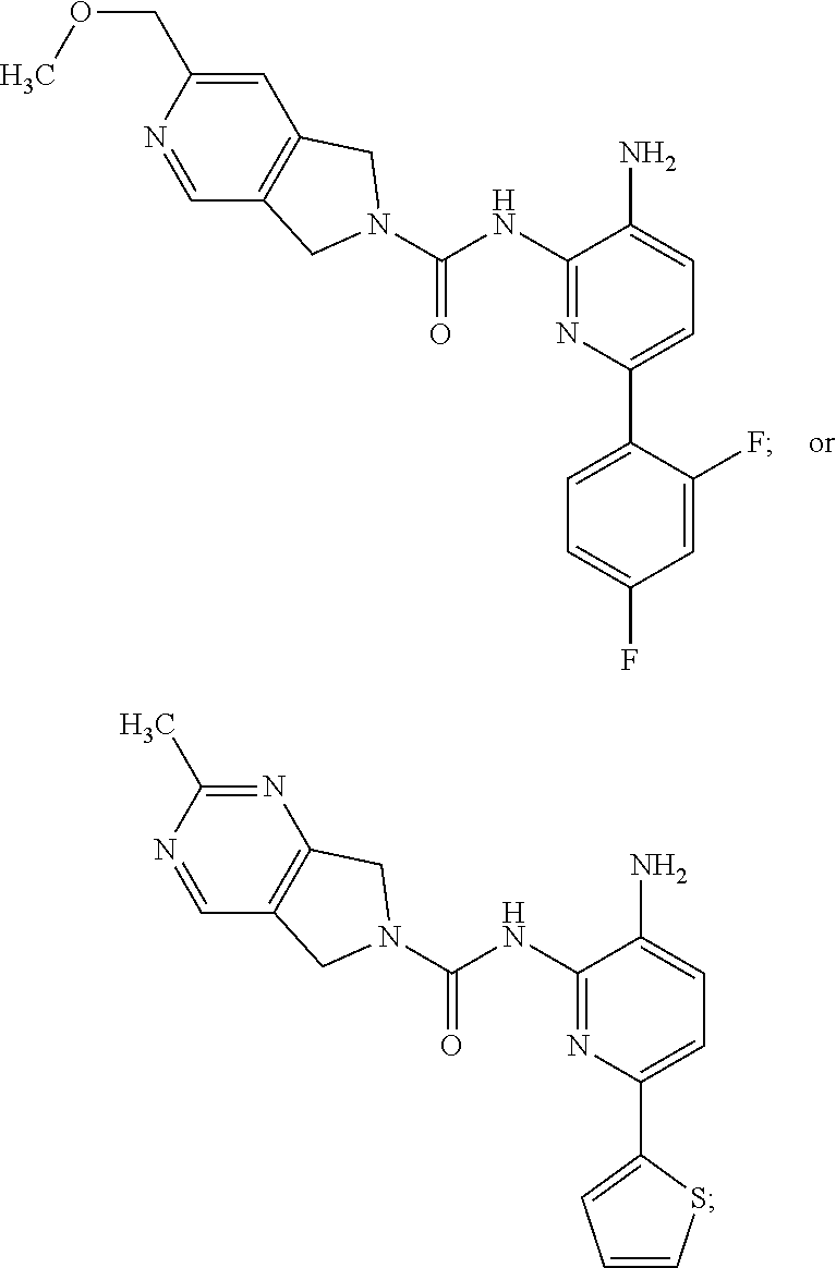
1. A compound of the formula: ##STR00027## or a pharmaceutically acceptable salt thereof.
2. A pharmaceutical composition comprising a compound of the formula: ##STR00028## or a pharmaceutically acceptable salt thereof; and a pharmaceutically acceptable carrier.
3. A method of inhibiting HDAC activity in a subject comprising the step of administering to the subject in need thereof an effective amount of a compound of claim 1, or a pharmaceutically acceptable salt thereof.
Description
BACKGROUND
Inhibitors of histone deacetylases (HDAC) have been shown to modulate transcription and to induce cell growth arrest, differentiation and apoptosis. HDAC inhibitors also enhance the cytotoxic effects of therapeutic agents used in cancer treatment, including radiation and chemotherapeutic drugs. Marks, P., Rifkind, R. A., Richon, V. M., Breslow, R., Miller, T., Kelly, W. K. Histone deacetylases and cancer: causes and therapies. Nat Rev Cancer, 1, 194-202, (2001); and Marks, P. A., Richon, V. M., Miller, T., Kelly, W. K. Histone deacetylase inhibitors. Adv Cancer Res, 91, 137-168, (2004). Moreover, recent evidence indicates that transcriptional dysregulation may contribute to the molecular pathogenesis of certain neurodegenerative disorders, such as Huntington's disease, spinal muscular atrophy, amyotropic lateral sclerosis, and ischemia. Langley, B., Gensert, J. M., Beal, M. F., Ratan, R. R. Remodeling chromatin and stress resistance in the central nervous system: histone deacetylase inhibitors as novel and broadly effective neuroprotective agents. Curr Drug Targets CNS Neurol Disord, 4, 41-50, (2005). A recent review has summarized the evidence that aberrant histone acetyltransferase (HAT) and histone deacetylases (HDAC) activity may represent a common underlying mechanism contributing to neurodegeneration. Moreover, using a mouse model of depression, Nestler has recently highlighted the therapeutic potential of histone deacetylation inhibitors (HDAC5) in depression. Tsankova, N. M., Berton, O., Renthal, W., Kumar, A., Neve, R. L., Nestler, E. J. Sustained hippocampal chromatin regulation in a mouse model of depression and antidepressant action. Nat Neurosci, 9, 519-525, (2006).
There are 18 known human histone deacetylases, grouped into four classes based on the structure of their accessory domains. Class I includes HDAC1, HDAC2, HDAC3, and HDAC8 and has homology to yeast Rpd3. HDAC4, HDAC5, HDAC7, and HDAC9 belong to class IIa and have homology to yeast Hda1. HDAC6 and HDAC10 contain two catalytic sites and are classified as class IIb. Class III (the sirtuins) includes SIRT1, SIRT2, SIRT3, SIRT4, SIRT5, SIRT6, and SIRT7. HDAC11 is another recently identified member of the HDAC family and has conserved residues in its catalytic center that are shared by both class I and class II deacetylases and is sometimes placed in class IV.
HDACs have been shown to be powerful negative regulators of long-term memory processes. Nonspecific HDAC inhibitors enhance synaptic plasticity as well as long-term memory (Levenson et al., 2004, J. Biol. Chem. 279:40545-40559; Lattal et al., 2007, Behav Neurosci 121:1125-1131; Vecsey et al., 2007, J. Neurosci 27:6128; Bredy, 2008, Learn Mem 15:460-467; Guan et al., 2009, Nature 459:55-60; Malvaez et al., 2010, Biol. Psychiatry 67:36-43; Roozendaal et al., 2010, J. Neurosci. 30:5037-5046). For example, HDAC inhibition can transform a learning event that does not lead to long-term memory into a learning event that does result in significant long-term memory (Stefanko et al., 2009, Proc. Natl. Acad. Sci. USA 106:9447-9452). Furthermore, HDAC inhibition can also generate a form of long-term memory that persists beyond the point at which normal memory fails. HDAC inhibitors have been shown to ameliorate cognitive deficits in genetic models of Alzheimer's disease (Fischer et al., 2007, Nature 447:178-182; Kilgore et al., 2010, Neuropsychopharmacology 35:870-880). These demonstrations suggest that modulating memory via HDAC inhibition have considerable therapeutic potential for many memory and cognitive disorders.
The role of individual HDACs in long-term memory has been explored in two recent studies. Kilgore et al. 2010, Neuropsychopharmacology 35:870-880 revealed that nonspecific HDAC inhibitors, such as sodium butyrate, inhibit class I HDACs (HDAC1, HDAC2, HDAC3, HDAC8) with little effect on the class IIa HDAC family members (HDAC4, HDAC5, HDAC7, HDAC9). This suggests that inhibition of class I HDACs may be critical for the enhancement of cognition observed in many studies. Indeed, forebrain and neuron specific overexpression of HDAC2, but not HDAC1, decreased dendritic spine density, synaptic density, synaptic plasticity and memory formation. (Guan et al., 2009, Nature, 459:55-60). In contrast, HDAC2 knockout mice exhibited increased synaptic density, increased synaptic plasticity and increased dendritic density in neurons. These HDAC2 deficient mice also exhibited enhanced learning and memory in a battery of learning behavioral paradigms. This work demonstrates that HDAC2 is a key regulator of synaptogenesis and synaptic plasticity. Additionally, Guan et al. showed that chronic treatment of mice with SAHA (an HDAC 1, 2, 3, 6, 8 inhibitor) reproduced the effects seen in the HDAC2 deficient mice and rescued the cognitive impairment in the HDAC2 overexpressing mice.
The inhibition of HDAC2 (selectively or in combination with inhibition of other class I HDACs; as the primary target, or as part of a complex with other proteins) is an attractive therapeutic target. Selective inhibition might be achieved by targeting specific HDAC isoforms such as HDAC2, in isolation, or as part of a functional multi-protein complex. Such inhibition has the potential for enhancing cognition and facilitating the learning process through increasing synaptic and dendritic density in neuronal cell populations. In addition, inhibition of specific HDACs, such as HDAC2, may also be therapeutically useful in treating a wide variety of other diseases and disorders.
SUMMARY
Disclosed are compounds and pharmaceutically acceptable salts thereof, and pharmaceutical compositions, which are useful in the treatment of conditions associated with the activity of HDAC (e.g., HDAC2). (See e.g., Tables 1 and 2).
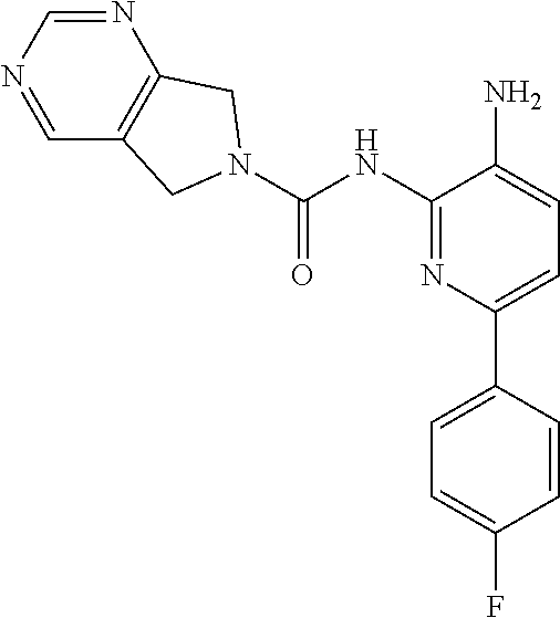
The disclosed compounds provide an advantage in hematological safety and overall balance of potency, ADME and PK profiles when compared to prior inhibitors. For example, the mere replacement of hydrogen for methyl between Comparator E and Compound 2 leads to a dramatic decrease in CYP2D6 inhibition. See e.g., Table 2. Also, this replacement provides distinct PK benefits over the unsubstituted pyrimidine analog, displaying a longer half-life, lower clearance, higher bioavailability, and a >5-fold higher brain exposure. See e.g., Table 3. Similarly, the addition of one additional ortho-fluorine atom realized a significant safety benefit in both the erythroid and myeloid progenitor cell lineages for Compound 1 relative to Comparator I. See e.g., Table 5.
The described compounds also produce changes in dendritic spine morphology in the CA1 region of the dorsal hippocampus in wild type mice. See e.g., Table 7. Measures of dendritic spine morphology can identify pharmacological agents which are likely to promote or distort normal cognitive function and protect against or exacerbate cognitive impairments.
Conditions which are treatable by the disclosed compounds include, but are not limited to, neurological disorders, memory or cognitive function disorders or impairments, extinction learning disorders, fungal diseases or infections, inflammatory diseases, hematological diseases, neoplastic diseases, psychiatric disorders, and memory loss.
DETAILED DESCRIPTION
1. Compounds
Provided herein is a compound of the formula:
##STR00001## or a pharmaceutically acceptable salt thereof.
2. Definitions
As used herein the terms "subject" and "patient" may be used interchangeably, and means a mammal in need of treatment, e.g., companion animals (e.g., dogs, cats, and the like), farm animals (e.g., cows, pigs, horses, sheep, goats and the like) and laboratory animals (e.g., rats, mice, guinea pigs and the like). Typically, the subject is a human in need of treatment.
Pharmaceutically acceptable salts as well as the neutral forms of the compounds described herein are included. For use in medicines, the salts of the compounds refer to non-toxic "pharmaceutically acceptable salts." Pharmaceutically acceptable salt forms include pharmaceutically acceptable acidic/anionic or basic/cationic salts. Pharmaceutically acceptable basic/cationic salts include, the sodium, potassium, calcium, magnesium, diethanolamine, n-methyl-D-glucamine, L-lysine, L-arginine, ammonium, ethanolamine, piperazine and triethanolamine salts. Pharmaceutically acceptable acidic/anionic salts include, e.g., the acetate, benzenesulfonate, benzoate, bicarbonate, bitartrate, carbonate, citrate, dihydrochloride, gluconate, glutamate, glycollylarsanilate, hexylresorcinate, hydrobromide, hydrochloride, malate, maleate, malonate, mesylate, nitrate, salicylate, stearate, succinate, sulfate, tartrate, and tosylate.
The term "pharmaceutically acceptable carrier" refers to a non-toxic carrier, adjuvant, or vehicle that does not destroy the pharmacological activity of the compound with which it is formulated. Pharmaceutically acceptable carriers, adjuvants or vehicles that may be used in the compositions described herein include, but are not limited to, ion exchangers, alumina, aluminum stearate, lecithin, serum proteins, such as human serum albumin, buffer substances such as phosphates, glycine, sorbic acid, potassium sorbate, partial glyceride mixtures of saturated vegetable fatty acids, water, salts or electrolytes, such as protamine sulfate, disodium hydrogen phosphate, potassium hydrogen phosphate, sodium chloride, zinc salts, colloidal silica, magnesium trisilicate, polyvinyl pyrrolidone, cellulose-based substances, polyethylene glycol, sodium carboxymethylcellulose, polyacrylates, waxes, polyethylene-polyoxypropylene-block polymers, polyethylene glycol and wool fat.
The terms "treatment," "treat," and "treating" refer to reversing, alleviating, reducing the likelihood of developing, or inhibiting the progress of a disease or disorder, or one or more symptoms thereof, as described herein. In some embodiments, treatment may be administered after one or more symptoms have developed, i.e., therapeutic treatment. In other embodiments, treatment may be administered in the absence of symptoms. For example, treatment may be administered to a susceptible individual prior to the onset of symptoms (e.g., in light of a history of symptoms and/or in light of genetic or other susceptibility factors), i.e., prophylactic treatment. Treatment may also be continued after symptoms have resolved, for example to prevent or delay their recurrence.
The term "effective amount" or "therapeutically effective amount" includes an amount of a compound described herein that will elicit a biological or medical response of a subject.
3. Uses, Formulation and Administration
In some embodiments, compounds and compositions described herein are useful in treating conditions associated with the activity of HDAC. Such conditions include for example, those described below.
Recent reports have detailed the importance of histone acetylation in central nervous system ("CNS") functions such as neuronal differentiation, memory formation, drug addiction, and depression (Citrome, Psychopharmacol. Bull. 2003, 37, Suppl. 2, 74-88; Johannessen, CNS Drug Rev. 2003, 9, 199-216; Tsankova et al., 2006, Nat. Neurosci. 9, 519-525; Bousiges et al., 2013, PLoS ONE 8(3), e57816). Thus, in one aspect, the provided compounds and compositions may be useful in treating a neurological disorder. Examples of neurological disorders include: (i) chronic neurodegenerative diseases such as fronto-temporal lobar degeneration (frontotemporal dementia, FTD), FTD-GRN, familial and sporadic amyotrophic lateral sclerosis (FALS and ALS, respectively), familial and sporadic Parkinson's disease, Parkinson's disease dementia, Huntington's disease, familial and sporadic Alzheimer's disease, multiple sclerosis, muscular dystrophy, olivopontocerebellar atrophy, multiple system atrophy, Wilson's disease, progressive supranuclear palsy, diffuse Lewy body disease, corticodentatonigral degeneration, progressive familial myoclonic epilepsy, striatonigral degeneration, torsion dystonia, familial tremor, Down's Syndrome, Gilles de la Tourette syndrome, Hallervorden-Spatz disease, diabetic peripheral neuropathy, dementia pugilistica, AIDS Dementia, age related dementia, age associated memory impairment, and amyloidosis-related neurodegenerative diseases such as those caused by the prion protein (PrP) which is associated with transmissible spongiform encephalopathy (Creutzfeldt-Jakob disease, Gerstmann-Straussler-Scheinker syndrome, scrapie, and kuru), and those caused by excess cystatin C accumulation (hereditary cystatin C angiopathy); and (ii) acute neurodegenerative disorders such as traumatic brain injury (e.g., surgery-related brain injury), cerebral edema, peripheral nerve damage, spinal cord injury, Leigh's disease, Guillain-Barre syndrome, lysosomal storage disorders such as lipofuscinosis, Alper's disease, restless leg syndrome, vertigo as result of CNS degeneration; pathologies arising with chronic alcohol or drug abuse including, for example, the degeneration of neurons in locus coeruleus and cerebellum, drug-induced movement disorders; pathologies arising with aging including degeneration of cerebellar neurons and cortical neurons leading to cognitive and motor impairments; and pathologies arising with chronic amphetamine abuse to including degeneration of basal ganglia neurons leading to motor impairments; pathological changes resulting from focal trauma such as stroke, focal ischemia, vascular insufficiency, hypoxic-ischemic encephalopathy, hyperglycemia, hypoglycemia or direct trauma; pathologies arising as a negative side-effect of therapeutic drugs and treatments (e.g., degeneration of cingulate and entorhinal cortex neurons in response to anticonvulsant doses of antagonists of the NMDA class of glutamate receptor) and Wernicke-Korsakoff's related dementia. Neurological disorders affecting sensory neurons include Friedreich's ataxia, diabetes, peripheral neuropathy, and retinal neuronal degeneration. Other neurological disorders include nerve injury or trauma associated with spinal cord injury. Neurological disorders of limbic and cortical systems include cerebral amyloidosis, Pick's atrophy, and Rett syndrome. In another aspect, neurological disorders include disorders of mood, such as affective disorders and anxiety; disorders of social behavior, such as character defects and personality disorders; disorders of learning, memory, and intelligence, such as mental retardation and dementia. Thus, in one aspect the disclosed compounds and compositions may be useful in treating schizophrenia, delirium, attention deficit hyperactivity disorder (ADHD), schizoaffective disorder, Alzheimer's disease, vascular dementia, Rubinstein-Taybi syndrome, depression, mania, attention deficit disorders, drug addiction, dementia, dementia including BPSD manifestations, agitation, apathy, anxiety, psychoses, personality disorders, bipolar disorders, unipolar affective disorder, obsessive-compulsive disorders, eating disorders, post-traumatic stress disorders, irritability, adolescent conduct disorder and disinhibition. They may also be useful for spontaneous, toxic, neoplastic, post-traumatic and post-infectious tinnitus and smelling impairment.
Transcription is thought to be a key step for long-term memory formation (Alberini, 2009, Physiol. Rev. 89, 121-145). Transcription is promoted by specific chromatin modifications, such as histone acetylation, which modulate histone-DNA interactions (Kouzarides, 2007, Cell, 128:693-705), as well as transcription factor-DNA interactions. Modifying enzymes, such as histone acetyltransferases (HATs) and histone deacetylases (HDACs), regulate the state of acetylation on histone tails. In general, histone acetylation promotes gene expression, whereas histone deacetylation leads to gene silencing, although treatment with HDAC inhibitors can result in both upregulation and downregulation of the expression levels of specific genes. Numerous studies have shown that a potent HAT, cAMP response element-binding protein (CREB)-binding protein (CBP), is necessary for long-lasting forms of synaptic plasticity and long term memory (for review, see Barrett, 2008, Learn Mem 15:460-467). Thus, in one aspect, the provided compounds and compositions may be useful for promoting cognitive function and enhancing learning and memory formation.
The compounds and compositions described herein may also be used for treating fungal diseases or infections.
In another aspect, the compounds and compositions described herein may be used for treating inflammatory diseases such as stroke, rheumatoid arthritis, lupus erythematosus, ulcerative colitis and traumatic brain injuries (Leoni et al., PNAS, 99(5); 2995-3000 (2002); Suuronen et al. J. Neurochem. 87; 407-416 (2003) and Drug Discovery Today, 10:197-204 (2005).
In yet another aspect, the compounds and compositions described herein may be used for treating a cancer caused by the proliferation of neoplastic cells. Such cancers include e.g., solid tumors, neoplasms, carcinomas, sarcomas, leukemias, lymphomas and the like. In one aspect, cancers that may be treated by the compounds and compositions described herein include, but are not limited to: cardiac cancer, lung cancer, gastrointestinal cancer, genitourinary tract cancer, liver cancer, nervous system cancer, gynecological cancer, hematologic cancer, skin cancer, and adrenal gland cancer. In one aspect, the compounds and compositions described herein are useful in treating cardiac cancers selected from sarcoma (angiosarcoma, fibrosarcoma, rhabdomyosarcoma, liposarcoma), myxoma, rhabdomyoma, fibroma, lipoma and teratoma. In another aspect, the compounds and compositions described herein are useful in treating a lung cancer selected from bronchogenic carcinoma (squamous cell, undifferentiated small cell, undifferentiated large cell, adenocarcinoma), alveolar (bronchiolar) carcinoma, bronchial adenoma, sarcoma, lymphoma, chondromatous hamartoma, and mesothelioma. In one aspect, the compounds and compositions described herein are useful in treating a gastrointestinal cancer selected from esophagus (squamous cell carcinoma, adenocarcinoma, leiomyosarcoma, lymphoma), stomach (carcinoma, lymphoma, leiomyosarcoma), pancreas (ductal adenocarcinoma, insulinoma, glucagonoma, gastrinoma, carcinoid tumors, vipoma), small bowel (adenocarcinoma, lymphoma, carcinoid tumors, Kaposi's sarcoma, leiomyoma, hemangioma, lipoma, neurofibroma, fibroma), and large bowel (adenocarcinoma, tubular adenoma, villous adenoma, hamartoma, leiomyoma). In one aspect, the compounds and compositions described herein are useful in treating a genitourinary tract cancer selected from kidney (adenocarcinoma, Wilm's tumor [nephroblastoma], lymphoma, leukemia), bladder and urethra (squamous cell carcinoma, transitional cell carcinoma, adenocarcinoma), prostate (adenocarcinoma, sarcoma), and testis (seminoma, teratoma, embryonal carcinoma, teratocarcinoma, choriocarcinoma, sarcoma, interstitial cell carcinoma, fibroma, fibroadenoma, adenomatoid tumors, lipoma). In one aspect, the compounds and compositions described herein are useful in treating a liver cancer selected from hepatoma (hepatocellular carcinoma), cholangiocarcinoma, hepatoblastoma, angiosarcoma, hepatocellular adenoma, and hemangioma.
In some embodiments, the compounds described herein relate to treating, a bone cancer selected from osteogenic sarcoma (osteosarcoma), fibrosarcoma, malignant fibrous histiocytoma, chondrosarcoma, Ewing's sarcoma, malignant lymphoma (reticulum cell sarcoma), multiple myeloma, malignant giant cell tumor chordoma, osteochondroma (osteocartilaginous exostoses), benign chondroma, chondroblastoma, chondromyxofibroma, osteoid osteoma and giant cell tumors.
In one aspect, the compounds and compositions described herein are useful in treating a nervous system cancer selected from skull (osteoma, hemangioma, granuloma, xanthoma, osteitis deformans), meninges (meningioma, meningiosarcoma, gliomatosis), brain (astrocytoma, medulloblastoma, glioma, ependymoma, germinoma [pinealoma], glioblastoma multiform, oligodendroglioma, schwannoma, retinoblastoma, congenital tumors), and spinal cord (neurofibroma, meningioma, glioma, sarcoma).
In one aspect, the compounds and compositions described herein are useful in treating a gynecological cancer selected from uterus (endometrial carcinoma), cervix (cervical carcinoma, pre-tumor cervical dysplasia), ovaries (ovarian carcinoma [serous cystadenocarcinoma, mucinous cystadenocarcinoma, unclassified carcinoma], granulosa-thecal cell tumors, Sertoli-Leydig cell tumors, dysgerminoma, malignant teratoma), vulva (squamous cell carcinoma, intraepithelial carcinoma, adenocarcinoma, fibrosarcoma, melanoma), vagina (clear cell carcinoma, squamous cell carcinoma, botryoid sarcoma (embryonal rhabdomyosarcoma), and fallopian tubes (carcinoma).
In one aspect, the compounds and compositions described herein are useful in treating a skin cancer selected from malignant melanoma, basal cell carcinoma, squamous cell carcinoma, Karposi's sarcoma, moles dysplastic nevi, lipoma, angioma, dermatofibroma, keloids, and psoriasis.
In one aspect, the compounds and compositions described herein are useful in treating an adrenal gland cancer selected from neuroblastoma.
In one aspect, the compounds and compositions described herein are useful in treating cancers that include, but are not limited to: leukemias including acute leukemias and chronic leukemias such as acute lymphocytic leukemia (ALL), Acute myeloid leukemia (AML), chronic lymphocytic leukemia (CLL), chronic myelogenous leukemia (CML) and Hairy Cell Leukemia; lymphomas such as cutaneous T-cell lymphomas (CTCL), noncutaneous peripheral T-cell lymphomas, lymphomas associated with human T-cell lymphotrophic virus (HTLV) such as adult T-cell leukemia/lymphoma (ATLL), Hodgkin's disease and non-Hodgkin's lymphomas, large-cell lymphomas, diffuse large B-cell lymphoma (DLBCL); Burkitt's lymphoma; mesothelioma, primary central nervous system (CNS) lymphoma; multiple myeloma; childhood solid tumors such as brain tumors, neuroblastoma, retinoblastoma, Wilm's tumor, bone tumors, and soft-tissue sarcomas, common solid tumors of adults such as head and neck cancers (e.g., oral, laryngeal and esophageal), genito urinary cancers (e.g., prostate, bladder, renal, uterine, ovarian, testicular, rectal and colon), lung cancer, breast cancer, pancreatic cancer, melanoma and other skin cancers, stomach cancer, brain tumors, liver cancer and thyroid cancer.
In one aspect, the compounds and compositions described herein are useful in treating a condition in a subject selected from a neurological disorder, memory or cognitive function disorder or impairment, extinction learning disorder, fungal disease or infection, inflammatory disease, hematological disease, psychiatric disorders, and neoplastic disease. In another aspect, the compounds and compositions described herein are useful in treating a condition selected from a) a cognitive function disorder or impairment associated with Alzheimer's disease, Huntington's disease, seizure induced memory loss, schizophrenia, Rubinstein Taybi syndrome, Rett Syndrome, Fragile X, Lewy body dementia, vascular dementia, fronto-temporal lobar degeneration (frontotemporal dementia, FTD), FTD-GRN, ADHD, dyslexia, bipolar disorder and social, cognitive and learning disorders associated with autism, traumatic head injury, attention deficit disorder, anxiety disorder, conditioned fear response, panic disorder, obsessive compulsive disorder, posttraumatic stress disorder (PTSD), phobia, social anxiety disorder, substance dependence recovery, Age Associated Memory Impairment (AAMI), Age Related Cognitive Decline (ARCD), ataxia, or Parkinson's disease; b) a hematological disease selected from acute myeloid leukemia, acute promyelocytic leukemia, acute lymphoblastic leukemia, chronic myelogenous leukemia, myelodysplastic syndromes, and sickle cell anemia; c) a neoplastic disease; and d) an extinction learning disorder selected from fear extinction and post-traumatic stress disorder. In one aspect, the condition treated by the compounds and compositions described herein is Alzheimer's disease, Huntington's disease, frontotemporal dementia, Freidreich's ataxia, post-traumatic stress disorder (PTSD), Parkinson's disease, depression, or substance dependence recovery.
In one aspect, the present disclosure provides a method of treating a condition described herein comprising administering to a subject an effective amount of a compound, or pharmaceutically acceptable salt described herein, or a composition thereof.
Also provided is the use of one or more of the compounds, or pharmaceutically acceptable salts thereof described herein, or a provided composition, for treating a condition described herein.
Also provided is the use of one or more of the compounds, or pharmaceutically acceptable salts thereof described herein for the manufacture of a medicament for treating a condition described herein.
Subjects may also be selected to be suffering from one or more of the described conditions before treatment with one or more of the described compounds, or pharmaceutically acceptable salts or compositions commences.
The present disclosure also provides pharmaceutically acceptable compositions comprising a compound described herein, or a pharmaceutically acceptable salt thereof; and a pharmaceutically acceptable carrier. These compositions can be used to treat one or more of the conditions described above.
Compositions described herein may be administered orally, parenterally, by inhalation spray, topically, rectally, nasally, buccally, vaginally or via an implanted reservoir. The term "parenteral" as used herein includes subcutaneous, intravenous, intramuscular, intra-articular, intra-synovial, intrasternal, intrathecal, intrahepatic, intralesional and intracranial injection or infusion techniques. Liquid dosage forms, injectable preparations, solid dispersion forms, and dosage forms for topical or transdermal administration of a compound are included herein.
The amount of provided compounds that may be combined with carrier materials to produce a composition in a single dosage form will vary depending upon the patient to be treated and the particular mode of administration. In some embodiments, provided compositions may be formulated so that a dosage of between 0.01-100 mg/kg body weight/day of the provided compound, such as e.g., 0.1-100 mg/kg body weight/day, can be administered to a patient receiving these compositions.
It should also be understood that a specific dosage and treatment regimen for any particular patient will depend upon a variety of factors, including age, body weight, general health, sex, diet, time of administration, rate of excretion, drug combination, the judgment of the treating physician, and the severity of the particular disease being treated. The amount of a provided compound in the composition will also depend upon the particular compound in the composition.
EXEMPLIFICATION
Spots were visualized by UV light (254 and 365 nm). Purification by column and flash chromatography was carried out using silica gel (200-300 mesh). Solvent systems are reported as the ratio of solvents.
NMR spectra were recorded on a Bruker 400 (400 MHz) spectrometer. .sup.1H chemical shifts are reported in 6 values in ppm with tetramethylsilane (TMS, =0.00 ppm) as the internal standard. See, e.g., the data provided in Table 1.
LCMS spectra were obtained on an Agilent 1200 series 6110 or 6120 mass spectrometer with ESI (+) ionization mode. See, e.g., the data provided in Table 1.
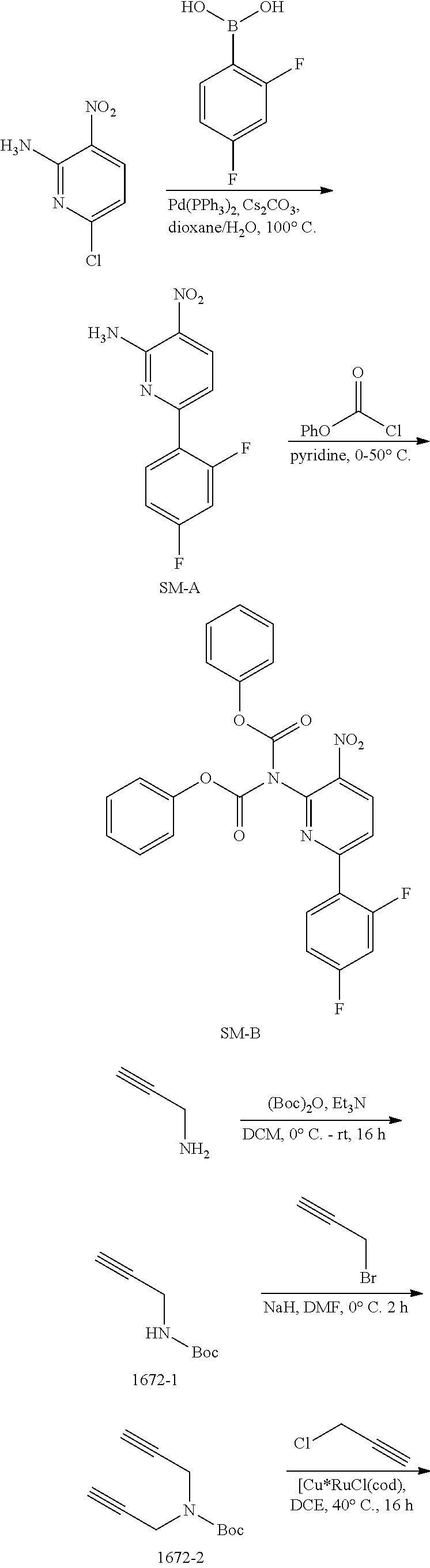
Example 1
##STR00002## ##STR00003##
Synthesis of SM-A.
A mixture of 6-chloro-3-nitropyridin-2-amine (4.58 g, 26.4 mmol), 2,4-difluorophenylboronic acid (5.00 g, 31.7 mmol) and Cs.sub.2CO.sub.3 (25.73 g, 79.2 mmol) in dioxane/H.sub.2O (100 mL/10 mL) was added Pd(PPh.sub.3).sub.4 (1.10 g, 0.95 mmol) under N.sub.2 atmosphere. The mixture was stirred at 100.degree. C. for 2 h and then concentrated in vacuo. The residue was dissolved with EtOAc (200 mL) and the solution was washed with brine (100 mL.times.3). The organic layer was dried over anhydrous Na.sub.2SO.sub.4 and then concentrated in vacuo. The residue was purified by column chromatography on silica gel (PE:EtOAc=7:1.about.5:1) to give SM-A (4.0 g, 61%) as a yellow solid. MS 252.1 [M+H].sup.+.
Synthesis of SM-B.
To a stirring solution of SM-A (4.0 g, 15.94 mmol) in pyridine (60 mL) was added phenyl carbonochloridate (7.50 g, 47.81 mmol) dropwise at 0.degree. C. After the addition was completed, the mixture was stirred at 50.degree. C. for 4 h. The mixture was concentrated in vacuo. The residue was purified by column chromatography on silica gel (PE:DCM=3:2.about.1:1) to give SM-B (7.1 g, 91%) as a yellow solid. MS 492.1 [M+H].sup.+.
Synthesis of 1672-1.
To a solution of prop-2-yn-1-amine (5.0 g, 90.9 mmol) and Et.sub.3N (18.4 g, 181.8 mmol) in DCM (100 mL) was added (Boc).sub.2O (23.8 g, 109.1 mmol) dropwise while cooling the reaction mixture with an ice bath. The resulting mixture was removed from the ice bath once the addition was completed, and was then stirred at room temperature for 16 h. When the reaction was complete, the mixture was diluted with DCM (200 mL), washed with brine (100 mL.times.3), and the organic layer was then dried over Na.sub.2SO.sub.4 and then concentrated in vacuo. The residue was purified by column chromatography on silica gel (PE:EtOAc=100:1.about.10:1) to give 1672-1 (10 g, 71%) as a colorless oil. MS 178.3 [M+23].sup.+, 100.3 [M-56].sup.+.
Synthesis of 1672-2.
To a solution of 1672-1 (10 g, 64.5 mmol) in DMF (200 mL) was added NaH (60% in mineral oil) (2.84 g, 71 mmol) slowly under ice bath. The resulting mixture was stirred at room temperature for 1 h, whereupon 3-bromoprop-1-yne (9.2 g, 77.4 mmol) was added into above mixture, and the reaction mixture was then stirred at room temperature for 2 h. The mixture was quenched with water (500 mL) and then extracted with t-BuOMe (250 mL.times.3). The combined organic layers were washed with brine (200 mL.times.3), dried over anhydrous Na.sub.2SO.sub.4 and then concentrated in vacuo. The residue was purified by column chromatography on silica gel (PE:EtOAc=100:1.about.10:1) to give 1672-2 (12 g, 96%) as a yellow oil. MS 138.1 [M-56].sup.+.
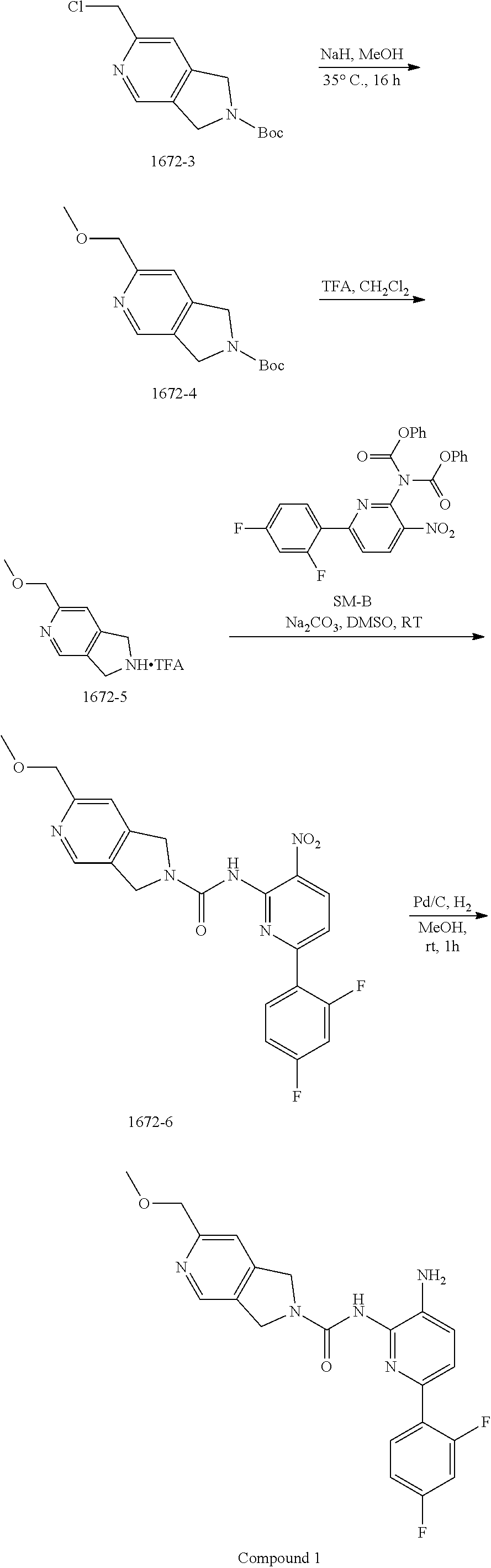
Synthesis of 1672-3.
To a solution of 2-chloroacetonitrile (3.13 g, 41.4 mmol) and [Cp*RuCl(cod)] (394 mg, 1.0 mmol) in DCE (40 mL) was added a solution of 1672-2 (4.0 g, 20.7 mmol) in DCE (80 mL) dropwise over 30 min under an N.sub.2 atmosphere. The resulting mixture was stirred at 40.degree. C. for 16 h. The solvent was removed in vacuo, and the residue was purified by column chromatography on silica gel (PE:EtOAc=10:1.about.2:1) to give 1672-3 (2.1 g, 22%) as a khaki solid. MS 269.3 [M+H].sup.+.
Synthesis of 1672-4.
To a solution of MeOH (30 mL) was added NaH (60% in mineral oil) (940 mg, 23.5 mmol) at ice bath and stirred for 30 min. Then 1672-C (2.1 g, 7.8 mmol) was added into above mixture and stirred at 35.degree. C. for 16 h. The mixture was quenched with water (30 mL), extracted with DCM (10 mL.times.3). The combined organic layers were washed with brine (10 mL.times.3), dried over anhydrous Na.sub.2SO.sub.4 and then concentrated in vacuo. The residue was purified by column chromatography on silica gel (PE:EtOAc=100:1.about.10:1) to give 1672-4 (1.8 g, 94%) as a tan colored solid. MS 265.1 [M+H].sup.+.
Synthesis of 1672-5.
To a solution of 1672-4 (120 mg, 0.45 mmol) in DCM (6 mL) cooled in an ice bath was added TFA (2 mL) dropwise. The resulting reaction mixture was stirred at room temperature for 1 h, whereupon the solvent was removed in vacuo to give 1672-5 as a crude product which was taken on to the next step without further purification. MS 165.1 [M+H].sup.+.
Synthesis of 1672-6.
To a mixture of 1672-5 (0.45 mmol, crude product from last step) and SM-B (150 mg, 0.30 mmol) in DMSO (10 mL) was added Na.sub.2CO.sub.3 (259 mg, 3.44 mmol), and the resulting reaction mixture was stirred at 25.degree. C. for 2 h. The mixture was then diluted with water (30 mL) and extracted with EtOAc (20 mL.times.3). The combined organic layers were washed with brine (10 mL.times.3), dried over anhydrous Na.sub.2SO.sub.4 and then concentrated in vacuo. The residue was purified by column chromatography on silica gel (DCM:MeOH=100:1.about.30:1) to give 1672-6 (120 mg, 89%) as a yellow solid. MS 442.1 [M+H].sup.+.
Synthesis of Compound 1.
A mixture of 1672-6 (120 mg, 0.27 mmol) and Pd/C (120 mg) in MeOH (10 mL) was stirred at room temperature for 1 h under a H.sub.2 atmosphere. Pd/C was then removed by filtration through the celite. The filtrate was concentrated and the residue was purified by Prep-TLC (DCM:MeOH=15:1) to give Compound 1 (70 mg, 70%) as a yellow solid. MS 412.1 [M+H].sup.+, 434.1 [M+23].sup.+.
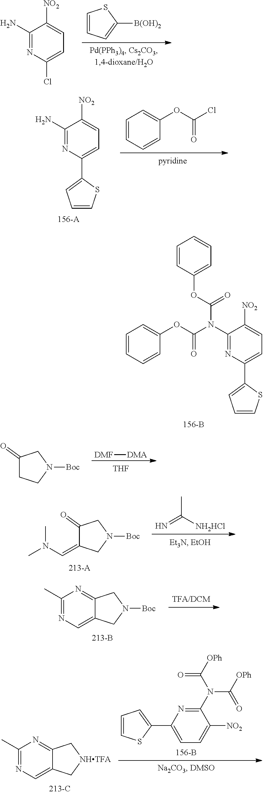
Example 2
##STR00004## ##STR00005##
Synthesis of 156-A.
A mixture of 6-chloro-3-nitropyridin-2-amine (10.00 g, 57.6 mmol), thiophen-2-ylboronic acid (8.12 g, 63.4 mmol) and Cs.sub.2CO.sub.3 (37.56 g, 115.2 mmol) in dioxane/H.sub.2O (200 mL/20 mL) was added Pd(PPh.sub.3).sub.4 (2.44 g, 2.88 mmol) under an N.sub.2 atmosphere. The mixture was stirred at 95.degree. C. for 2 h and then concentrated in vacuo. The residue was dissolved with EtOAc (200 mL) and the solution was washed with brine (100 mL.times.3). The organic layer was dried over anhydrous Na.sub.2SO.sub.4 and then concentrated in vacuo. The residue was purified by column chromatography on silica gel (PE:EtOAc=5:1.about.3:1) to give 156-A (10.0 g, 79%) as a yellow solid
Synthesis of 156-B.
To a stirred solution of 156-A (1.30 g, 5.88 mmol) in pyridine (20 mL) was added phenyl carbonochloridate (2.29 g, 14.7 mmol) in dropwise fashion. After the addition was completed, the mixture was heated to 50.degree. C. and stirred for 4 h. The mixture was then concentrated in vacuo, and the residue was purified by column chromatography on silica gel (PE:EtOAc=8:1.about.3:1) to give 156-B (2.4 g, 89%) as a yellow solid.
Synthesis of 213-A.
A solution of tert-butyl 3-oxopyrrolidine-1-carboxylate (150.0 g, 809.8 mmol) and DMF-DMA (289.5 g, 2.4 mol) in THF (1500 mL) was stirred at 70.degree. C. for 16 h. The solution was concentrated in vacuo to give 213-A as a crude product, which was used directly in the next step without further purification.
Synthesis of 213-B.
To a solution of 213-A (809.8 mmol, crude product from last step) in EtOH (1000 mL) was added Et.sub.3N (409.7 g, 4.0 mol) and acetimidamide hydrochloride (306.2 g, 3.2 mol). The resulting solution was stirred at 80.degree. C. for 24 h. After the mixture was cooled to room temperature, the mixture was diluted with water (500 mL) and extracted with DCM (500 mL.times.3). The combined organic layers were washed with brine (500 mL.times.3), dried over anhydrous Na.sub.2SO.sub.4 and then concentrated in vacuo. The residue was purified by column chromatography on silica gel (PE:DCM=10:1.about.1:2) to give 213-B (105.0 g, 55%) as a brown solid.
Synthesis of 213-C.
To a solution of 213-B (105.0 g, 446.3 mmol) in DCM (1000 mL) was added TFA (333 mL) dropwise. The reaction mixture was stirred at room temperature for 1 h, whereupon the solution was concentrated in vacuo to give 213-C as a crude product which was used directly in the next step.
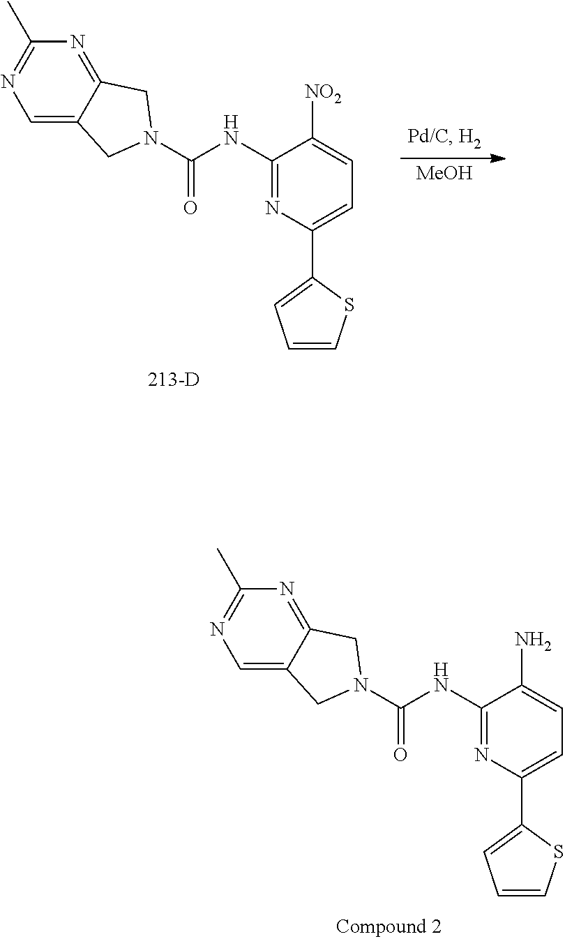
Synthesis of 213-D.
A mixture of 213-C (325.1 mmol, crude product from last step) and 156-B (75.0 g, 162.5 mmol) in DMSO (750 mL) was stirred at room temperature for 10 min, then Na.sub.2CO.sub.3 (137.8 g, 1.3 mol) was added, and the reaction mixture was stirred at room temperature for 2 h. The mixture was then diluted with water (1000 mL) and extracted with EtOAc (500 mL.times.3). The combined organic layers were washed with brine (500 mL.times.3), dried over anhydrous Na.sub.2SO.sub.4 and then concentrated in vacuo. The residue was purified by column chromatography on silica gel (PE:EA=10:1.about.1:2) to give 213-D (44.0 g, 71%) as a yellow solid.
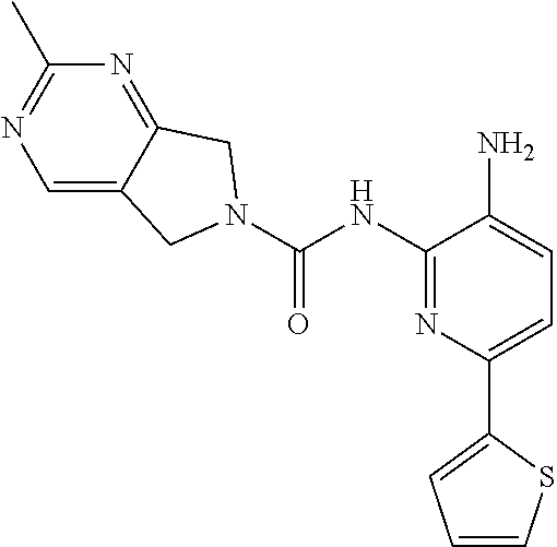
Synthesis of Compound 2.
A mixture of 213-D (44.0 g, 115.1 mmol) and Pd/C (22.0 g) in MeOH (250 mL) and DCM (250 mL) was stirred at room temperature for 1 h under a H.sub.2 atmosphere. Pd/C was removed by filtration through Celite. The filtrate was concentrated in vacuo and the residue was purified by column chromatography on silica gel (DCM:MeOH=50:1.about.15:1) to give Compound 2 (26.0 g, 64%) as a light yellow solid.
TABLE-US-00001 TABLE 1 Spectrometric Data for Compounds MS MS .sup.1HNMR Data (400 MHz, No. Structure Calc found DMSO-d.sub.6) 1 ##STR00006## 411 412 .delta. 8.58 (s, 1H), 8.53 (s, 1H), 7.97- 7.91 (m, 1H), 7.44-7.40 (m, 2H), 7.32-7.26 (m, 1H), 7.18- 7.13 (m, 2H), 5.28 (s, 2H), 4.82 (s, 4H), 4.52 (s, 2H), 3.38 (s, 3H). 2 ##STR00007## 352 353 .delta. 8.70 (s, 1H), 8.60 (s, 1H), 7.53- 7.47 (m, 2H), 7.42-7.40 (q, J = 4.0 Hz, 1H), 7.13-7.07 (t, J = 8.0 Hz, 1H), 7.06-7.05 (t, J = 1.2 Hz, 1H), 5.18 (s, 2H), 4.78- 4.75 (d, J = 10.4 Hz, 4H), 2.64 (s, 3H).
General Assay Methods
HDAC2 and HDAC1 Enzymatic Assay
The following describes an assay protocol for measuring the deacetylation of a peptide substrate by the enzymes HDAC2 or HDAC1.
All recombinant human HDACs were purchased from BPS Bioscience. The substrate, FAM-TSRHK(AC)KL-CONH, was synthesized at NanoSyn. Final assay reactions contained 100 mM HEPES (pH 7.5), 50 mM KCl, 0.1% BSA, 0.01% Triton X-100, 1% DMSO, 1 uM substrate and 5 nM HDAC enzyme. Enzyme and compounds were pre-incubated at 25.degree. C. for 5 hours and reactions were initiated by addition of substrate. 10 uL reactions were incubated for 17 hours at 25.degree. C. and terminated by the addition of 40 uL of buffer containing 100 mM HEPES (pH 7.5), 0.1% BSA, 0.01% Triton X-100 and 0.05% SDS. Substrate and product peptides present in each sample were separated electrophoretically using the LabChip 3000 capillary electrophoresis instrument. Change in the relative fluorescence intensity of the substrate and product peaks reflects enzyme activity. Reaction progress was determined as the product to sum ratio (PSR): P/(S+P), where P is the peak height of the product peptide and S is the peak height of the substrate peptide. Reactions were performed in duplicate at 12 concentrations, (3.times.serial dilutions starting at 30 uM). IC.sub.50 values were calculated using a 4 Parameter Logistic Model.
HDAC2 Enzymatic Inhibition Assay in SY5Y Cell Lysate with HDAC-Glo2 Substrate
Cell Culture and Inhibitor Treatments
SH-SY5Y cells (Sigma) were cultured in Eagle's Modified Essential Medium supplemented with 10% fetal bovine serum and pen/strep. Twenty-four hours prior to compound dosing 20 uL of cells were plated in white 384 well plates at a density of 1,500 cells/well. Compounds were serially diluted in neat DMSO and then diluted 1:100 v/v into media without FBS and mixed. Media was removed from the plated cells and the diluted compounds in serum free media (1% v/v final DMSO) were added and incubated at 37.0 for five hours. Ten uL of HDAC-Glo 2 reagent with 0.1% Triton X-100 was then added, the plate was mixed and allowed to develop at room temperature for 100 minutes. Plates were then read with a Spectramax LMax luminometer employing a 0.4 s integration time. Dose response curves were constructed with normalized data where CI-994 at 100 uM was defined as 100% inhibition and DMSO alone as 0% inhibition.
Erythroid and Myeloid CFU Assay
Compounds were tested to evaluate the potential effects on human erythroid and myeloid progenitors using colony forming cell assays. Clonogenic progenitors of human erythroid (CFU-E, BFU-E), granulocyte-monocyte (CFU-GM) and multipotential (CFU-GEMM) lineages were assessed in a semi-solid methylcellulose-based media formulation containing rhIL-3 (10 ng/mL), rhGM-SCF (10 ng/mL), rhSCF (50 ng/mL) and Epo (3 U/mL).
Cells
Normal human bone marrow light density cells derived from normal bone marrow (NorCal Biologics, California) and qualified at ReachBio, were stored in the gaseous phase of liquid nitrogen (-152.degree. C.) until required for the assay. On the day of the experiment, the cells were thawed rapidly, the contents of each vial was diluted in 10 mL of Iscove's modified Dulbecco's medium containing 10% fetal bovine serum (IMDM+10% FBS) and washed by centrifugation (approximately 1200 r.p.m. for 10 minutes, room temperature). The supernatant was discarded and the cell pellets resuspended in a known volume of IMDM+10% FBS. A cell count (3% glacial acetic acid) and viability assessment (trypan blue exclusion test) was performed for the bone marrow sample.
Compounds
On the day of the experiment, the compounds were dissolved in DMSO to a stock concentration of 10 mM. Serial dilutions were prepared from the stock concentration to achieve concentrations of 2 and 0.4 mM. When added to the methylcellulose-based media at 1:1000 (v/v), the final test concentrations of 10, 2 and 0.4 .mu.M were achieved. Additionally, 5-FU was evaluated at 1.0, 0.1 and 0.01 .mu.g/mL.
Method Summary
Clonogenic progenitors of the human erythroid (CFU-E and BFU-E) and myeloid (CFU-GM) lineages were set up in the methylcellulose-based media formulations described above. All compounds were added to the medium to give the final desired concentrations (10, 2 and 0.4 .mu.M). 5-Fluorouracil (Sigma Aldrich) was used as a positive control for progenitor proliferation (inhibition of colony growth) and was introduced to the human bone marrow cultures at 1.0, 0.1, and 0.01 .mu.g/mL. Solvent control cultures (containing no compound but 0.1% DMSO) as well as standard controls (containing no compound and no DMSO) were also initiated.
Human myeloid and erythroid progenitor assays were initiated at 2.0.times.10.sup.4 cells per culture. Following 14 days in culture, myeloid and erythroid colonies were assessed microscopically and scored by trained personnel. The colonies were divided into the following categories based on size and morphology: CFU-E, BFU-E, CFU-GM and CFU-GEMM.
Statistical Analyses of CFC Numbers
The mean.+-.one standard deviation of three replicate cultures was calculated for progenitors of each category (CFU-E, BFU-E, etc.). Two-tailed t-tests were performed to assess if there was a difference in the number of colonies generated between solvent control and treated cultures. Due to the potential subjectivity of colony enumeration, a p value of less than 0.01 is deemed significant. To calculate the concentration of 50% inhibition of colony growth (IC.sub.50) for each compound, a dose response curve was generated plotting the log of the compound concentration versus the percentage of control colony growth using XLfit software (IDBS). The concentration of 50% inhibition of colony growth (IC.sub.50) was calculated based on the sigmoid curve fit using Dose-Response, One-Site Model formula: y=A+[(B-A)/(1+((C/x){circumflex over ( )}D))], where A=the initial value (baseline response), B=maximum response, C=center (drug concentration that provokes a response halfway between A and B) and D=slope of the curve at midpoint. Further, plots and additional dose response curves were generated using GraphPad Prism 7.0.
Morphological Assessment of Colonies
Photographs were taken of representative hematopoietic progenitor-derived colonies from various lineages, illustrating colonies in the presence of the solvent control as well as colonies in the presence of the test compounds.
Erythroid (CFU-E and BFU-E), myeloid (CFU-GM) and multi-potential (CFU-GEMM) colony enumeration was performed by trained personnel. The distribution of colony types as well as general colony and cellular morphology was analyzed. For statistical analysis colony numbers in compound treated cultures were compared to the solvent control cultures. 5-FU was used as a positive control for toxicity in these assays and the inhibitory effects obtained for this compound were exactly as expected. The experiment was used to evaluate the potential effect of test compounds on human erythroid and myeloid progenitor proliferation in a methylcellulose-based medium. The IC.sub.50 values were calculated from XLfit. Dose response curves for erythroid and myeloid toxicity generated by XLfit. Finally, nonlinear regression curve fitting and IC.sub.50s.+-.95% CI, were calculated by Prism 7.0.-GEMM.
CYP Inhibition Assay
Compounds were tested to evaluate their inhibitory potential on CYP2D6 and CYP3A4 (midazolam) using human liver microsomes. Human liver microsomes were obtained from BD Gentest, and each compound was run in duplicate.
The test compounds and reference inhibitors (quinidine for 2D6, ketoconazole for 3A4) were plated in a 96-well plate by transferring 8 .mu.L of 10 mM stock solutions of compound in DMSO to 12 .mu.L of acetonitrile. Individual inhibitor spiking solutions were prepared for CYP2D6 and CYP3A4 (8 .mu.L of DMSO stock added to 12 .mu.L of acetonitrile). Next added 400 .mu.L of 0.2 mg/mL HLM to the assay wells and then added 2 .mu.L of 400.times.test compound into the designated wells on ice. Next, added 200 .mu.L of 0.2 mg/mL HLM to the assay wells and then added 1 .mu.L of reference inhibitor solutions into the designated wells. The following solutions were added (in duplicate) to a 96-well assay plate on ice: The test compounds and reference inhibitors (quinidine for 2D6, ketoconazole for 3A4) were tested using the following experimental procedure:
1. Prepare test compound and reference inhibitors (400.times.) in a 96-well plate:
1.1. Transfer 8 .mu.L of 10 mM test compounds to 12 .mu.L of ACN.
1.2. Prepare individual inhibitor spiking solution for CYP3A4, CYP2D6: 8 .mu.L of DMSO stock to 12 .mu.L of ACN.
2. Prepare 4.times.NADPH cofactor (66.7 mg NADPH in 10 mL 0.1 M K-buffer, pH7.4)
3. Prepare 4.times.substrate (2 mL for each isoform) as indicated in the table below (add HLM where required on ice).
4. Prepare 0.2 mg/mL HLM solution (10 .mu.L of 20 mg/mL to 990 .mu.L of 0.1 M K-buffer) on ice.
5. Add 400 .mu.L of 0.2 mg/mL HLM to the assay wells and then add 2 .mu.L of 400.times. test compound into the designated wells on ice.
6. Add 200 .mu.L of 0.2 mg/mL HLM to the assay wells and then add 1 .mu.L of reference inhibitor solution into the designated wells on ice.
7. Add following solutions (in duplicate) in a 96-well assay plate on ice:
7.1. Add 30 .mu.L of 2.times. test compound and reference compound in 0.2 mg/mL HLM solution;
7.2. Add 15 .mu.L of 4.times. substrate solution.
8. Pre-incubate the 96-well assay plate and NADPH solution at 37.degree. C. for 5 minutes.
9. Add 15 .mu.L of pre-warmed 8 mM NADPH solution to into the assay plates to initiate the reaction
10. Incubate the assay plate at 37.degree. C. 5 min for 3A4, 10 min for 2D6.
11. Stop the reaction by adding 120 .mu.L of ACN containing Internal Standard. For CYP3A4, the internal standard is 1'OH-midazolam-D.sub.4 (10 .mu.M solution diluted to a final concentration of 0.1 .mu.M by adding 100 .mu.L internal standard stock to 10 mL ACN). For CYP2D6, the internal standard is 1-OH-Bufuralol-maleate-[D.sub.9] (49 .mu.M solution diluted to a final concentration of 0.1 .mu.M by adding 20 .mu.L internal standard stock to 10 mL ACN).
12. After quenching, shake the plates at the vibrator (IKA, MTS 2/4) for 10 min (600 rpm/min) and then centrifuge at 5594 g for 15 min (Thermo Multifuge.times.3R).
13. Transfer 50 .mu.L of the supernatant from each well into a 96-well sample plate containing 50 .mu.L of ultra pure water (Millipore, ZMQS50F01) for LC/MS analysis.
The assessment on CYP isoform inhibition is as follows based on the assay results: if percentage of CYP inhibition is higher than 50%, indicates potent inhibition; if percentage of CYP inhibition is between 30-50%, indicates slight inhibition; if percentage of CYP inhibition is less than 30%, indicates slight or no inhibition. If the percentage of CYP inhibition is less than -30%, this indicates the compound may have some kind of activation of this isoform.
Aqueous Kinetic Solubility Measurement
Compounds were evaluated for their kinetic solubility in buffer or water. Aliquots of 8 .mu.L of reference and test compound stock solutions (10 mM in DMSO) were added into 792 .mu.L of 100 mM phosphate buffer (0.1 M NaPO.sub.4, pH 7.4). Final DMSO concentration is 1%. The sample tubes were shaken for 1 hour (1000 rpm) at room temperature. A calibration curve was prepared using 300 .mu.M spiking solution (SS) in MeOH/ACN (4:1) (SS=add 6 .mu.L 10 mM compound in 194 .mu.L MeOH/ACN (4:1)). Samples were centrifuged for 10 mM (12000 rpm) to precipitate undissolved particles, and the supernatants were transferred to a new tube or plate. Supernatants were diluted 10 times and 100 times with 100 mM buffer. Samples were then prepared for analysis by LC-MS/MS (Add 5 .mu.L of compound samples (not diluted, 10 times diluted and 100 times diluted) and standard curve samples to 95 .mu.L of ACN containing internal standard. Internal standards used are Propranolol, Ketoconazole, and Tamoxifen.
Assessment of Brain and Plasma Exposure for Compounds Following Intravenous (IV) and Oral (PO) Administration to Mice
Compounds were dosed in mice at either 10 mg/kg or 30 mg/kg PO, and were dosed at 1 mg/kg IV. Three animals for collection at each time point for plasma via bleeding at 0.25, 0.5, 1, 4, 12 and 24 h. Terminal bleeding for plasma and sampling for brain at 0.25, 0.5, 1, 4, 12 and 24 h (also three animals per brain exposure time point group). Total of six time points for plasma and six time points for brain.
Sample Collection:
Plasma: The animal was restrained manually at the designated time points, approximately 150 .mu.L blood/time point was collected into K.sub.2EDTA tube via retro orbital puncture or cardiac puncture under anesthesia with Isoflurane. The blood sample was centrifuged (2000 g, 4.degree. C., 5 min) to generate plasma within 30 min after bleeding.
Brain: At the designated time points, a mid-line incision was made in the animals scalp and the skin was retracted. Using small bone cutters and rongeurs, removed the skull overlying the brain. Removed the brain using a spatula and rinse with cold saline. Placed the brain in screw-top tubes, and then stored the tubes under -70.degree. C. until analysis.
Certain Advantages of Compounds 1 and 2
From a drug discovery standpoint, it is important that compounds have acceptable drug-like profiles across a range of parameters. It is typical to profile compounds not only for in vitro potency, but also in predictive Absorption, Distribution, Excretion and Metabolism (ADME) studies in vitro, and in pharmacokinetic (PK) experiments in vivo. In some cases, compounds are also profiled in predictive in vitro safety studies. Collecting in vitro ADME and safety data, along with PK data, help to identify benefits of certain structural features, and allows the optimization of the structure activity relationship (SAR) to design compounds with optimized drug-like profiles for profiling in vivo. The present compounds not only provide an advantage in hematological safety, but also provide an overall balance of potency, ADME and PK profiles.
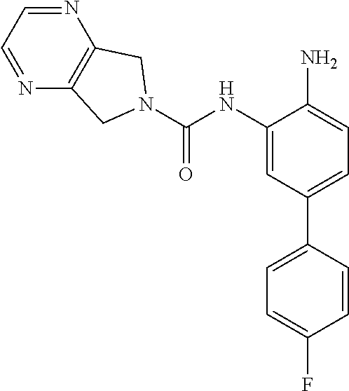
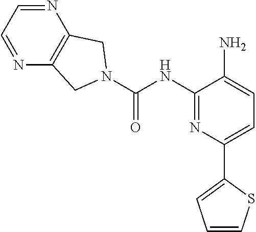
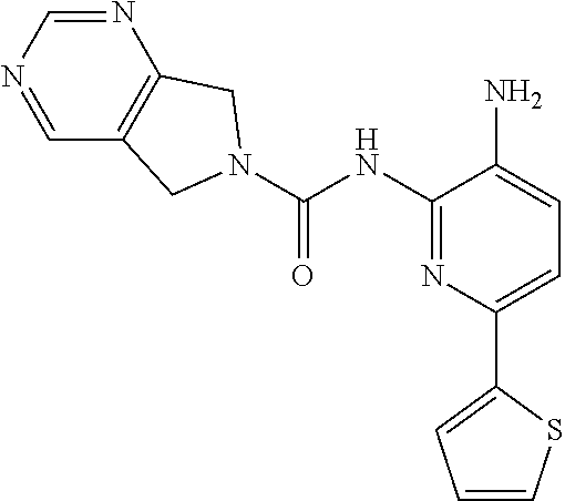
Aminoaniline urea compounds such as Comparator A and Comparator B in Table 2 were previously described in WO 2017/007755 and WO 2017/007756 (the contents of each of which are incorporated herein by reference). When screened in an in vitro colony forming unit (CFU) assay in human bone marrow cells, looking at erythroid and myeloid progenitor cells (predictive for neutropenia), and compared to the matched pair aminopyridine urea compound Comparator B, a single atom change in the diaminopyridine urea core of Comparator B leads to a significant improvement in predicted safety in both erythroid and myeloid lineages relative to the urea of the aminoaniline scaffold of Comparator A. See Table 2. Moving forward, when the 4-fluorophenyl group of Comparator B is replaced with a thiophene group (Comparator C), but the pyrrolopyrazine component of the urea is kept the same, a similar improvement in predicted safety profile is achieved relative to the aminoaniline urea Comparator A. This evidences that ureas of diaminopyridine compounds with identical pyrrolidine urea components are safer than the corresponding aminoaniline ureas.
While the pyrrolopyrazine urea compounds, Comparator B and Comparator C, showed good potency in in vitro potency assays, the values from the in vitro CFU assay still require improvement. A myeloid lineage IC.sub.50>5 .mu.M (roughly corresponding to >30% remaining at 10 .mu.M) predicts low likelihood of clinical neutropenia, so is the target threshold for acceptable safety (References: Pessina et al. Toxicological Sciences 2003, 75, 355-367; Clarke et al. Gen. Eng. & Biotech. News 2010, 14-15.). It was found that by changing the pyrrolopyrazine to a pyrrolopyrimidine, a significantly improved in vitro safety profile was achieved. Comparing the matched pair Comparator B (pyrrolopyrazine) with Comparator D (pyrrolopyrimidine), a significant improvement in both the CFU erythroid and myeloid progenitor cells is observed. This also holds for the aminopyridine urea matched pairs possessing a thiophene footpocket, Comparator C (pyrrolopyrazine) and Comparator E (pyrrolopyrimidine). Although the pyrimidine brought with it an improved safety profile, both unsubstituted pyrimidine compounds Comparator D and Comparator E showed significant inhibition of CYP2D6 at 10 .mu.M. However, substituting on the pyrimidine ring at the position between the nitrogen atoms, Compound 2 (methyl-substituted pyrrolopyrimidine) showed no significant inhibition of either CYP2D6 or CYP3A4 at 10 .mu.M.
These results evidence that slight chemical modifications such as the walking of a nitrogen atom one position on a ring in Comparators B and D and replacing hydrogen for methyl (Comparators E and Compound 2) produce dramatic increases in safety.
TABLE-US-00002 TABLE 2 Comparison of in vitro profile of Compound 2 to multiple comparators. HDAC2 SY5Y HDAC cell recombinant CFU % lysate enzymatic % CYP Control assay IC50 inhibition remaining IC50 (.mu.M) @ 10 .mu.M: @ 10 .mu.M Comp. Structure (.mu.M) HDAC2 HDAC1 2D6 3A4 Erythroid Myeloid A ##STR00008## 0.369 0.142 0.027 34 -22 0 0 B ##STR00009## 0.485 0.475 0.119 9 1.5 9 25 C ##STR00010## 0.279 0.301 0.095 0.2 1 20 20 D ##STR00011## 0.331 0.627 0.239 85 2 47 84 E ##STR00012## 0.278 0.511 0.142 50 -17 54 83 2 ##STR00013## 0.577 0.434 0.133 5 1 27 59
In addition to the substitution between pyrimidine nitrogen atoms improving the CYP inhibition profile of pyrrolopyrimidine compounds, the PK profile of Compound 2 is improved relative to unsubstituted Comparator E as well. See Table 3. The methyl-pyrimidine of Compound 2 provides distinct PK benefits over the unsubstituted pyrimidine analog, displaying a longer half-life, lower clearance, higher bioavailability, and a >5-fold higher brain exposure. These results evidence that slight chemical modifications also produce substantial benefits in PK.
TABLE-US-00003 TABLE 3 Comparison in PK profiles of unsubstituted pyrimidine and methyl- substituted pyrimidine matched pairs. Comparator E Compound 2 Structure ##STR00014## ##STR00015## Mouse IV (1 mpk): T1/2 (hr) 0.152 0.839 Mouse IV (1 mpk): Cl (L/hr/kg) 8.33 4.18 Mouse IV-PO (1/10 mpk): F (%) 49 100 Mouse PO (1/10 mpk): T1/2 (hr) 0.557 1.05 Mouse PO (10 mpk): Brain 100 583 Cmax (ng/g) (free Cmax, ng/g) Mouse PO (10 mpk): Free brain 35 173 Cmax (ng/g)
Similar advantages were gained by exploring the effects of regioisomers and substitution patterns on pyrrolopyridine ureas of diaminopyridines. For example, while pyrrolopyridine compound Comparator F showed good in vitro potency, it showed high levels of CYP2D6 inhibition and extremely low solubility. See Table 4. Moving the pyridine nitrogen of the pyrrolopyridine (Comparator G) was found to improve solubility. However, both CYP2D6 and CYP3A4 were inhibited at high levels. It was found that adding a methyl substituent adjacent to the pyridine nitrogen of the pyrrolopyridine improved the CYP inhibition profile toward 2D6 and 3A4 somewhat, while maintaining the potency and solubility (Comparator H). The electron withdrawing methoxymethyl substitution adjacent to the pyridine nitrogen (Comparator H) improved the CYP 2D6 and 3A4 inhibition profile even further, again maintaining the desirable potency and solubility profile. Similar results were found between the mono-fluoro and di-fluoro analogues of Comparator I and Compound 1, with Compound 1 displaying slightly reduced solubility. See Table 4.
However, Compound 1 vastly out performed Comparator I with respect to in vitro safety, despite the only one halogen atom difference. See Table 5. A significant benefit was realized in both the erythroid and myeloid progenitor cell lineages upon treatment with the 2,4-difluoro substituted Compound 1 relative to Comparator I.
TABLE-US-00004 TABLE 4 Comparison of in vitro profile of Compound 1 to multiple comparators. HDAC2 HDAC SY5Y recombinant % CYP cell lysate enzymatic IC50 inhibition @ assay (.mu.M) 10 .mu.M: Solubility Cmp. Structure IC50 (.mu.M) HDAC2 HDAC1 2D6 3A4 (.mu.M) F ##STR00016## 0.394 0.304 0.079 62 -4 1 G ##STR00017## 0.639 0.335 0.143 94 65 55 H ##STR00018## 0.621 0.214 0.090 57 28 78 I ##STR00019## 0.666 0.276 0.122 2 33 121 1 ##STR00020## 0.777 0.510 0.326 -1 27 27
TABLE-US-00005 TABLE 5 In vitro CFU assay data for Compound 1 and Comparator I Comparator I Compound 1 Structure ##STR00021## ##STR00022## CFU % Control remaining 23 57 @10 .mu.M: Erythroid CFU % Control remaining 59 86 @10 .mu.M: Myeloid
Table 6 below shows the results from the brain and plasma exposure following intravenous (IV) and Oral (PO) administration of compounds in mice.
TABLE-US-00006 TABLE 6 Projected Projected Brain Cmax free brain @ 10 mpk @ 10 mpk ng/g uM (*scaled (*scaled for for IV PK IV PK Cl Structure comparison) comparison) T 1/2 (hr) (L/hr/kg) Comparator D ##STR00023## 158* 0.111* Not available Not available 1 ##STR00024## 2460 1.04 2.13 PO = 1.97 0.681
Effects of 14-Days of Oral Treatment with Low Doses of Compounds on Dendritic Spine Morphology in Dorsal Hippocampus (CA1) of Wild Type Mice
Compounds were evaluated to determine whether sub-chronic treatment could produce changes in dendritic spine morphology in the CA1 region of the dorsal hippocampus in wild type (WT) mice. Compounds were administered orally to wild type mice, daily for 14 days. Doses were chosen based on pharmacokinetic data, brain exposure, and potency. Effects of compounds treatments on dendritic spine morphology in the CA1 region of the dorsal hippocampus were then evaluated.
Methods: In-Life
Male C57BL/6J mice (7-8-weeks old, n=7 per group) were dosed orally with Rodin compounds or vehicle (20% HP CD) daily for 14 days. Doses for compounds were chosen based on exposure data from pharmacokinetic experiments. Doses of some compounds were chosen to determine non-efficacious doses, as an extension of earlier studies showing increases in dendritic spine density after treatment with Compound 2 (at doses of 1, 3, 6 and 20 mg/kg/day), Compound 1 (10 mg/kg/day). Mice were sacrificed 24 hours after the last dose, and underwent transcardial perfusion for preparation of brain samples.
Perfusion and Brain Sampling Method
Mice were anesthetized with chloral hydrate (4% chloral hydrate in saline, 10 ml/kg) before undergoing transcardial perfusion with 4% PFA in 1.times.PBS (pH7.4, room temperature) at 20 mL/min for 54 seconds Immediately after the perfusion, mice were decapitated and their brain extracted. Brains were postfixed in scintillation vials containing 4% PFA (5-10 ml) for 4 min. Brains were sectioned using a tissue vibratome (Leica VT1000) to collect sections (300 .mu.m thick) from the anterior to posterior extremes of each brain.
Ballistic Dye Labeling and Microscopy
Superresolution laser-scanning confocal microscopy (Zeiss LSM880, Airyscan) was performed using a 63.times. objective (1.42 NA) to scan individually labeled neurons at high resolution (scan resolution=0.06 um .mu.m/pixel; axial resolution=0.06 um .mu.m/focal step). Target neurons were identified in the brain region of interest by epifluorescence navigation using anatomical location and cell morphology. Microscopy was performed blind to experimental conditions. A minimum of 7 mice were tested in each experimental condition. A minimum of 5 samples per mouse (range=5-6) were measured for each segment.
ESP Dendritic Spine Analysis and Assessment of Dendritic Membrane Integrity
Blind deconvolution (AutoQuant) was applied to raw three-dimensional digital images which were then analyzed for spine density and morphology by trained analysts. Individual spines were measured manually for (a) head diameter, (b) length, and (c) neck thickness from image Z-stacks using custom-built Afraxis ESP software. Each dendrite was analyzed by 3 (on average) independent analysts. Automated image assignment software distributed images to analysts in a randomized manner and ensured that each analyst performed measurements of near equal numbers of dendrites per group. Analysts were blinded to all experimental conditions (including treatment, brain region, and cell type). Statistical analysis of interanalyst variability for each dendrite was examined on-line and used to eliminate dendrites that did not meet interanalyst reliability criteria: For spine density and spine morphological classification, data across analysts were averaged to report data for each dendrite.
Dendritic Sampling Positions
dHIPP, CA1 Apical 2.degree. (0-50 .mu.m):
Brain region: Dorsal Hippocampus (dHIPP)
Cell type: CA1 pyramidal neuron (CA1)
Branch type: Apical
Branch order: Secondary (2.degree.)
Sample position: 0-50 .mu.m from branchpoint
Each identified dendritic spine was measured for (a) spine length, (b) spine head diameter, and (c) neck width. Population distributions of each measure were compiled for each dendritic sample and pooled by group. Raw dendritic spine morphometric values (spine length, head diameter, neck width) were assembled into a scheme used to describe classic spine phenotypes (e.g. mushroom, stubby, etc.). Total spine density was also reported as the sum of the density of all subclasses.
Results
General Observations.
The tissue processing demonstrated no observable pathological indications, including abnormal disruption of somatic membranes, dendritic blebbing, or abnormal modifications of dendrite diameters for the target cell type or other cell types within the brain regions tested. For adequate study to study comparisons, total spine density data were normalized to % vehicle levels
.times..times..times..times..times..times..times..times..times..times..ti- mes..times..times..times..times..times..times..times..times..times..times.- .times..times..times..times..times..times..times..times..times..times. ##EQU00001## Data were then reported as mean+/-SEM of total spine density (% increase from Vehicle). The results are reported in Table 7.
TABLE-US-00007 TABLE 7 Effects on spine morphology in WT mice after 14 days of dosing. Total Spines Thin Spines Dose (% increase (% increase Compound Structure (mg/kg) over vehicle) over vehicle) 1 ##STR00025## 0.1 0.3 1 3 10 12.2 21.3* 34.6* 26.7* 25.1* 13.8 23.2* 45.1* 25.3* 19.9* 2 ##STR00026## 0.1 1 3 6 20 8.1 21.4* 24.7* 15* -0.12 11.3 21.2 32.2* 35.4* 37.4* *significantly different from vehicle with p value <0.05 using a one-way ANOVA followed by a Dunnett's postHoc analysis
The contents of all references (including literature references, issued patents, published patent applications, and co-pending patent applications) cited throughout this application are hereby expressly incorporated herein in their entireties by reference. Unless otherwise defined, all technical and scientific terms used herein are accorded the meaning commonly known to one with ordinary skill in the art.
* * * * *
References
C00001
C00002
C00003
C00004
C00005
C00006
C00007
C00008
C00009
C00010
C00011
C00012
C00013
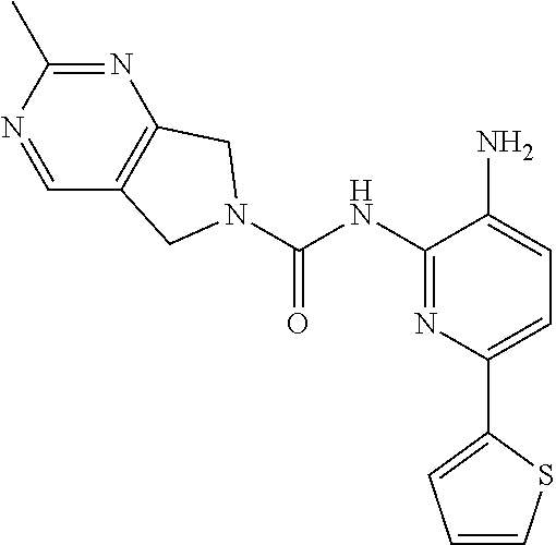
C00014

C00015
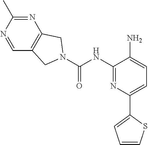
C00016
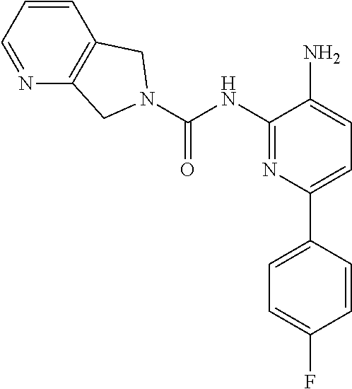
C00017
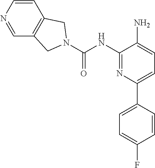
C00018
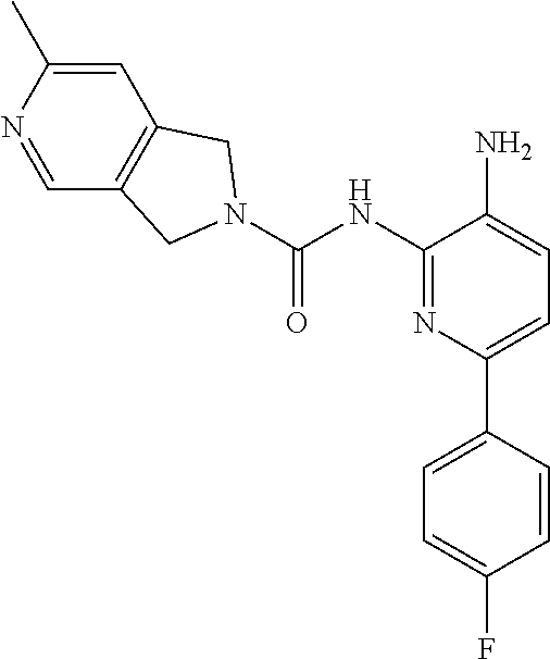
C00019

C00020
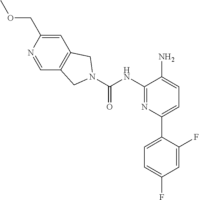
C00021
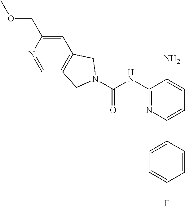
C00022

C00023

C00024

C00025

C00026

C00027

C00028
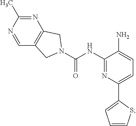
M00001

XML
uspto.report is an independent third-party trademark research tool that is not affiliated, endorsed, or sponsored by the United States Patent and Trademark Office (USPTO) or any other governmental organization. The information provided by uspto.report is based on publicly available data at the time of writing and is intended for informational purposes only.
While we strive to provide accurate and up-to-date information, we do not guarantee the accuracy, completeness, reliability, or suitability of the information displayed on this site. The use of this site is at your own risk. Any reliance you place on such information is therefore strictly at your own risk.
All official trademark data, including owner information, should be verified by visiting the official USPTO website at www.uspto.gov. This site is not intended to replace professional legal advice and should not be used as a substitute for consulting with a legal professional who is knowledgeable about trademark law.