T cell receptor-deficient T cell compositions
Sentman
U.S. patent number 10,689,618 [Application Number 15/948,303] was granted by the patent office on 2020-06-23 for t cell receptor-deficient t cell compositions. This patent grant is currently assigned to THE TRUSTEES OF DARTMOUTH COLLEGE. The grantee listed for this patent is THE TRUSTEES OF DARTMOUTH COLLEGE. Invention is credited to Charles L. Sentman.


| United States Patent | 10,689,618 |
| Sentman | June 23, 2020 |
T cell receptor-deficient T cell compositions
Abstract
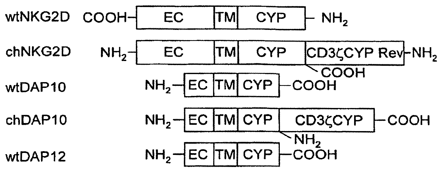
The invention is directed to modified T cells, methods of making and using isolated, modified T cells, and methods of using these isolated, modified T cells to address diseases and disorders. In one embodiment, this invention broadly relates to TCR-deficient T cells, isolated populations thereof, and compositions comprising the same. In another embodiment of the invention, these TCR-deficient T cells are designed to express a functional non-TCR receptor. The invention also pertains to methods of making said TCR-deficient T cells, and methods of reducing or ameliorating, or preventing or treating, diseases and disorders using said TCR-deficient T cells, populations thereof, or compositions comprising the same.
| Inventors: | Sentman; Charles L. (Grantham, NH) | ||||||||||
|---|---|---|---|---|---|---|---|---|---|---|---|
| Applicant: |
|
||||||||||
| Assignee: | THE TRUSTEES OF DARTMOUTH
COLLEGE (Hanover, NH) |
||||||||||
| Family ID: | 43992337 | ||||||||||
| Appl. No.: | 15/948,303 | ||||||||||
| Filed: | April 9, 2018 |
Prior Publication Data
| Document Identifier | Publication Date | |
|---|---|---|
| US 20180298336 A1 | Oct 18, 2018 | |
Related U.S. Patent Documents
| Application Number | Filing Date | Patent Number | Issue Date | ||
|---|---|---|---|---|---|
| 14676028 | Apr 1, 2015 | 9938497 | |||
| 13502978 | Nov 10, 2015 | 9181527 | |||
| PCT/US2010/054846 | Oct 29, 2010 | ||||
| 61255980 | Oct 29, 2009 | ||||
| Current U.S. Class: | 1/1 |
| Current CPC Class: | A61P 35/00 (20180101); A61K 35/17 (20130101); A61P 31/00 (20180101); A61P 37/00 (20180101); A61K 39/0011 (20130101); A61P 37/06 (20180101); C12N 5/0636 (20130101); A61K 2035/124 (20130101); A61K 2039/5156 (20130101); C12N 2501/515 (20130101); C12N 2511/00 (20130101); C12N 2510/02 (20130101); A61K 2039/585 (20130101) |
| Current International Class: | C12N 5/0783 (20100101); A61K 35/17 (20150101); A61K 39/00 (20060101); A61K 35/12 (20150101) |
References Cited [Referenced By]
U.S. Patent Documents
| 5415874 | May 1995 | Bender et al. |
| 5552300 | September 1996 | Makrides et al. |
| 5667967 | September 1997 | Steinman et al. |
| 5686281 | November 1997 | Roberts |
| 5830755 | November 1998 | Nishimura et al. |
| 5851828 | December 1998 | Seed et al. |
| 6103521 | August 2000 | Capon et al. |
| 6133433 | October 2000 | Hema et al. |
| 6190656 | February 2001 | Clifford et al. |
| 6242567 | June 2001 | Hema et al. |
| 6284240 | September 2001 | Seed et al. |
| 6407221 | June 2002 | Capon et al. |
| 6410319 | June 2002 | Raubitschek et al. |
| 6464978 | October 2002 | Brostoff et al. |
| 6753162 | June 2004 | Seed et al. |
| 6770749 | August 2004 | Ellenhorn et al. |
| 6984382 | January 2006 | Groner et al. |
| 7049136 | May 2006 | Seed et al. |
| 7052906 | May 2006 | Lawson et al. |
| 7070995 | July 2006 | Jensen |
| 7094599 | August 2006 | Seed et al. |
| 7435596 | October 2008 | Campana et al. |
| 7446179 | November 2008 | Jensen et al. |
| 7446190 | November 2008 | Sadelain et al. |
| 7456263 | November 2008 | Sherman et al. |
| 7514537 | April 2009 | Jensen |
| 7569357 | August 2009 | Kranz et al. |
| 7608410 | October 2009 | Dunn et al. |
| 7618817 | November 2009 | Campbell |
| 7655461 | February 2010 | Finn et al. |
| 7763243 | July 2010 | Lum et al. |
| 7820174 | October 2010 | Wang et al. |
| 7994298 | August 2011 | Zhang et al. |
| 8026097 | September 2011 | Campana et al. |
| 8252914 | August 2012 | Zhang et al. |
| 8283446 | October 2012 | Jakobsen et al. |
| 8399645 | March 2013 | Campana et al. |
| 8465743 | June 2013 | Rosenberg et al. |
| 8519100 | August 2013 | Jakobsen et al. |
| 8835617 | September 2014 | Luban et al. |
| 8945868 | February 2015 | Collingwood et al. |
| 8956828 | February 2015 | Bonini et al. |
| 9051391 | June 2015 | Mineno et al. |
| 9273283 | March 2016 | Sentman |
| 9938497 | April 2018 | Sentman |
| 2001/0007152 | July 2001 | Sherman et al. |
| 2002/0045241 | April 2002 | Schendel |
| 2002/0137697 | September 2002 | Eshhar et al. |
| 2003/0060444 | March 2003 | Finney et al. |
| 2003/0077249 | April 2003 | Bebbington et al. |
| 2003/0082719 | May 2003 | Schumacher et al. |
| 2003/0093818 | May 2003 | Belmont et al. |
| 2003/0219463 | November 2003 | Falkenburg et al. |
| 2004/0038886 | February 2004 | Finney et al. |
| 2004/0115198 | June 2004 | Spies et al. |
| 2004/0259196 | December 2004 | Zipori et al. |
| 2005/0048055 | March 2005 | Newell et al. |
| 2005/0113564 | May 2005 | Campana et al. |
| 2005/0129671 | June 2005 | Cooper et al. |
| 2005/0238626 | October 2005 | Yang et al. |
| 2006/0247420 | February 2006 | Coukos et al. |
| 2006/0093605 | May 2006 | Campana et al. |
| 2006/0166314 | July 2006 | Voss et al. |
| 2006/0263334 | November 2006 | Finn et al. |
| 2006/0269529 | November 2006 | Niederman et al. |
| 2007/0066802 | March 2007 | Geiger |
| 2007/0077241 | April 2007 | Spies et al. |
| 2007/0116690 | May 2007 | Yang et al. |
| 2008/0199424 | August 2008 | Yang et al. |
| 2008/0292549 | November 2008 | Jakobsen et al. |
| 2008/0292602 | November 2008 | Jakobsen et al. |
| 2008/0153029 | December 2008 | Mineno et al. |
| 2009/0053184 | February 2009 | Morgan et al. |
| 2009/0202501 | August 2009 | Zhang et al. |
| 2009/0226404 | September 2009 | Schuler et al. |
| 2009/0304657 | December 2009 | Morgan et al. |
| 2009/0324566 | December 2009 | Shiku et al. |
| 2010/0009863 | January 2010 | Himmler et al. |
| 2010/0015113 | January 2010 | Restifo et al. |
| 2010/0029749 | February 2010 | Zhang et al. |
| 2010/0055117 | March 2010 | Krackhardt et al. |
| 2010/0104556 | April 2010 | Blankenstein et al. |
| 2010/0105136 | April 2010 | Carter et al. |
| 2010/0135974 | June 2010 | Eshhar et al. |
| 2010/0143315 | June 2010 | Voss et al. |
| 2010/0178276 | July 2010 | Sadelain et al. |
| 2010/0189728 | July 2010 | Schendel et al. |
| 2010/0273213 | October 2010 | Mineno et al. |
| 2011/0158957 | June 2011 | Bonini et al. |
| 2011/0213288 | September 2011 | Choi et al. |
| 2012/0015434 | January 2012 | Campana et al. |
| 2012/0252742 | October 2012 | Kranz et al. |
| 2012/0294857 | November 2012 | Sentman et al. |
| 2012/0302466 | November 2012 | Sentman et al. |
| 2013/0011375 | January 2013 | Chen |
| 2013/0216509 | August 2013 | Campana et al. |
| 2013/0266551 | October 2013 | Campana et al. |
| 2013/0323214 | December 2013 | Gottschalk et al. |
| 2014/0004132 | January 2014 | Brenner et al. |
| 2014/0148354 | May 2014 | Campana et al. |
| 2014/0328812 | November 2014 | Campana et al. |
| 2015/0139943 | May 2015 | Campana et al. |
| 2016/0194375 | July 2016 | Kitchen et al. |
| 4408999 | Sep 1995 | DE | |||
| 19540515 | Jun 1997 | DE | |||
| 10259713 | Aug 2004 | DE | |||
| 0340793 | Aug 1995 | EP | |||
| 0499555 | May 2000 | EP | |||
| 0574512 | May 2003 | EP | |||
| 1226244 | Jul 2004 | EP | |||
| 0871495 | Jun 2005 | EP | |||
| 1075517 | Jul 2006 | EP | |||
| 1932537 | Jun 2008 | EP | |||
| 1765860 | Oct 2008 | EP | |||
| 2186825 | May 2010 | EP | |||
| 1791865 | Jul 2010 | EP | |||
| H05176760 | Jul 1993 | JP | |||
| 11-243955 | Sep 1999 | JP | |||
| 2008-523783 | Jul 2008 | JP | |||
| 2011-512786 | Apr 2011 | JP | |||
| WO 1991018019 | Nov 1991 | WO | |||
| WO 1992015322 | Sep 1992 | WO | |||
| WO 1994024282 | Oct 1994 | WO | |||
| WO 1996015238 | May 1996 | WO | |||
| WO 1996013584 | Sep 1996 | WO | |||
| WO 1998018809 | Jul 1998 | WO | |||
| WO 1998041613 | Sep 1998 | WO | |||
| WO 2000031239 | Feb 2000 | WO | |||
| WO 2000014257 | Mar 2000 | WO | |||
| WO 2001092291 | Jun 2001 | WO | |||
| WO 2004056845 | Aug 2004 | WO | |||
| 2005044996 | May 2005 | WO | |||
| 2006036445 | Apr 2006 | WO | |||
| WO 2006103429 | May 2006 | WO | |||
| WO 2006060878 | Jun 2006 | WO | |||
| 2008153029 | Dec 2008 | WO | |||
| WO 2009059804 | May 2009 | WO | |||
| WO 2009091826 | Jul 2009 | WO | |||
| WO 2010012829 | Apr 2010 | WO | |||
| WO 2010025177 | Apr 2010 | WO | |||
| WO 2010058023 | May 2010 | WO | |||
| WO 2010088160 | May 2010 | WO | |||
| WO 2010037395 | Aug 2010 | WO | |||
| WO 2010107400 | Sep 2010 | WO | |||
| WO 2011059836 | May 2011 | WO | |||
| 2011/070443 | Jun 2011 | WO | |||
| WO 2012050374 | Apr 2012 | WO | |||
| WO 2013166051 | Nov 2013 | WO | |||
Other References
|
Maher, J. and E.T. Davies. 2004. Br. J. Cancer 91:817-821 (Year: 2004). cited by examiner . Zhang, T. et al. 2005. Blood 106:1544-1551 (Year: 2005). cited by examiner . Declaration of Dr. Mark Daniels submitted Nov. 22, 2016 in conjunction with related re-examination U.S. Appl. No. 90/013,688, pp. 24-68. (Year: 2016). cited by examiner . Ardouin L, et al. "Crippling of CD3-zeta ITAMs does not impair T cell receptor signaling," Immunity. Apr. 1999;10(4):409-20. cited by applicant . Lipowska-Bhalla G, et al. "Targeted immunotherapy of cancer with CAR T cells: achievements and challenges," Cancer Immunol Immunother. Jul. 2012;61(7):953-62. cited by applicant . Shores EW, et al. "Role of TCR zeta chain in T cell development and selection," Science. Nov. 11, 1994;266(5187):1047-50. cited by applicant . Shores EW, et al. "Role of the multiple T cell receptor (TCR)-zeta chain signaling motifs in selection of the T cell repertoire," J Exp Med. Mar. 3, 1997;185(5):893-900. cited by applicant . Liu L, et al. "Adoptive T-cell therapy of B-cell malignancies: conventional and physiological chimeric antigen receptors," Cancer Lett. Mar. 2012;316(1):1-5. cited by applicant . Nagorsen D, et al. "Immunomodulatory therapy of cancer with T cell-engaging BiTE antibody blinatumomab," Exp Cell Res. May 15, 2011;317(9):1255-60. cited by applicant . Marin R, et al. "Analysis of HLA-E expression in human tumors," Immunogenetics. Feb. 2003;54(11):767-75. cited by applicant . Palmisano GL, et al. "HLA-E surface expression is independent of the availability of HLA class I signal sequence-derived peptides in human tumor cell lines," Hum Immunol. Jan. 2005;66(1):1-12. cited by applicant . Kudo K, Imai C, Lorenzini P, Kamiya T, Kono K, Davidoff AM, Chng WJ, Campana D. T lymphocytes expressing a CD16 signaling receptor exert antibody-dependent cancer cell killing. Cancer research. Jan. 1, 2014;74(1):93-103. cited by applicant . Kumagai MA, Coustan-Smith E, Murray DJ, Silvennoinen O, Murti KG, Evans WE, Malavasi F, Campana D. Ligation of CD38 suppresses human B lymphopoiesis. The Journal of experimental medicine. Mar. 1, 1995;181(3):1101-10. cited by applicant . Labrecque N et al. How Much TCR Does a T Cell Need? Immunity. Jul. 2001;15(1):71-82. cited by applicant . Lake DF, Salgaller ML, Van Der Bruggen P, Bernstein RM, Marchalonis JJ. Construction and binding analysis of recombinant single-chain TCR derived from tumor-infiltrating lymphocytes and a cytotoxic T lymphocyte clone directed against MAGE-1. Int Immunol. 1999;11(5):745-51. cited by applicant . Lamers CHJ, Willemsen RA, Luider BA, Debets R, Bolhuis RLH. Protocol for gene transduction and expansion of human T lymphocytes for clinical immunogene therapy of cancer. Cancer Gene Ther. 2002;9(7):613-23. cited by applicant . Lampson LA. Beyond inflammation: site-directed immunotherapy. Immunol Today. 1998;19(1):17-22. cited by applicant . Lapteva N, Durett AG, Sun J, Rollins LA, Huye LL, Fang J, Dandekar V, Mei Z, Jackson K, Vera J, Ando J. Large-scale ex vivo expansion and characterization of natural killer cells for clinical applications. Cytotherapy. Oct. 1, 2012;14(9)1131-43. cited by applicant . Lee DA, Verneris MR, Campana D. Acquisition, preparation, and functional assessment of human NL cells for adoptive immunotherapy. In: Immunotherapy of Cancer: Methods and Protocols. Humana Press; 2010. p. 61-77. cited by applicant . Leibman RS, Riley JL Engineering T cells to Functionally Cure HIV-1 Infection. Mol Ther. 2015;23(7):1149-59. cited by applicant . Leivas A, Perez-Martinez A, Blanchard MJ, Clavero EM, Campana D, Lahuerta JJ, Martinez-Lopez J. Autologous Activated and Expanded Natural Killer Cells Are Safe and Clinically Actives in Multiple Myeloma. Blood. Dec. 3, 2015;126(23):1856. cited by applicant . Leivas A, Perez-Martinez A, Blanchard MJ, Clavero EM, Campana D, Lahuerta JJ, Martinez-Lopez J. Multiple Infusions of Autologous Activated and Expanded Natural Killer Cells: A New Therapeutic Option for Multiple Myeloma. Clinical Lymphoma Myeloma and Leukemia. Sep. 1, 2015;15:e297-8. cited by applicant . Leivas A, Risueno RM, Perez-Martinez A, Campana D, Lahuerta JJ, Martinez-Lopez J. Autologous Activated and Expanded Natural Killer Cells Destroy Multiple Myeloma Clonogenic Tumor Cells through NKG2D and Its Ligands. Clinical Lymphoma Myeloma and Leukemia. Sep. 1, 2015;15:e245-6. cited by applicant . Leung W, Campana D, Yang J, Pei D, Coustan-Smith E, Gan K, Rubnitz JE, Sandlund JT, Ribeiro RC, Srinivasan A, Hartford C. High success rate of hematopoietic cell transplantation regardless of donor source in children with very high-risk leukemia. Blood. Jul. 14, 2011;118(2):223-30. cited by applicant . Li L, Wolfraim L, Allen C, Viley A, Fujisaki H, Campana D, Fratantoni JC, Peshwa MV. A Highly Efficient, Clinically Applicable Transfection Method to Redirect the Specificity of Immune Cells and Enhance Their Anti-Tumor Capacity. Blood. Nov. 16, 2008;112(11):3894. cited by applicant . Liao KW, Chou WC, Lo YC, Roller SR. Design of transgenes for efficient expression of active chimeric proteins on mammalian cells. Biotechnol Bioeng. 2001;73(4):313-23. cited by applicant . Lim KS, Kua LF, Mimura K, Shiraishi K, Chng WJ, Yong WP, Campana D, Kono K. Implication of highly cytotoxic natural killer cells for esophageal cancer treatment. Cancer Research. Aug. 1, 2015;75(15 Supplement):3148. cited by applicant . Liu C, Ma X, Liu B, Chen C, Zhang H. HIV-1 functional cure: will the dream come true? BMC Med. 2015;13(1):284. cited by applicant . Liu L, Patel B, Ghanem MH, Bundoc V, Zheng Z, Morgan RA, et al. Novel CD4-based bispecific chimeric antigen receptor designed for enhanced anti-HIV potency and absence of HIV entry receptor activity. J Virol. 2015;89 (13):6685-94. cited by applicant . Lund JA, Spach DH, Collier AC. Future Anti-HIV Therapy. In: Spach DH, Hooton TM, editors. The HIV Manual: A Guide to Diagnosis and Treatment. Oxford University Press; 1996. p. 89-104. cited by applicant . Lund O, Lund OS, Gram G, Nielsen SD, Schonning K, Nielsen JO, et al. Gene therapy of T helper cells in HIV infection: mathematical model of the criteria for clinical effect. Bull Math Biol. 1997;59(4):725-45. cited by applicant . Luszczek W, Morales-Tirado V, van der Merwe M, Kudo K, Campana D, Pillai A. Expanded human regulatory iNKT cells exhibit direct cytotoxicity against hematolymphoid tumor targets. Cancer Research. Apr. 15, 2012;72(8 Supplement):3512. cited by applicant . Ma Q, Gonzalo-Daganzo RM, Junghans RP. Genetically engineered T cells as adoptive immunotherapy of cancer. Cancer Chemother Biol Response Modif. 2002;20:315-41. cited by applicant . Ma Q, Safar M, Holmes E, Wang Y, Boynton AL, Junghans RP. Anti-prostate specific membrane antigen designer T cells for prostate cancer therapy. Prostate. 2004;61(1):12-25. cited by applicant . Marathe JG, Wooley DP. Is gene therapy a good therapeutic approach for HIV-positive patients? Genet Vaccines Ther. 2007;5:5. cited by applicant . Marin V, Kakuda H, Dander E, Imai C, Campana D, Biondi A, D'Amico G. Enhancement of the anti-leukemic activity of cytokine induced killer cells with an anti-CD19 chimeric receptor delivering a 4-1BB-.zeta. activating signal. Experimental hematology. Sep. 30, 2007;35(9)1388-97. cited by applicant . Martinius HO. Praklinische Untersuchungen zur Gentherapie der HIV-Infektion mit dem retroviralen Vektor M87o (Preclinical examinations on genetherapy of HIV infection with the retroviral vector M870). (Thesis.) Goethe University Frankfurt; 2007. cited by applicant . Masiero S, Del Vecchio C, Gavioli R, Mattiuzzo G, Cusi MG, Micheli L, et al. T-cell engineering by a chimeric T-cell receptor with antibody-type specificity for the HIV-1 gp120. Gene Ther. 2005;12:299-310. cited by applicant . McGuinness RP, Ge Y, Patel SD, Kashmiri SVS, Lee H-S, Hand PH, et al. Anti-tumor activity of human T cells expressing the CC49-zeta chimeric immune receptor. Hum Gene Ther. 1999;10(2):165-73. cited by applicant . Mihara K, Imai C, Coustan-Smith E, Dome JS, Dominici M, Vanin E, Campana D. Development and functional characterization of human bone marrow mesenchymal cells immortalized by enforced expression of telomerase. British Journal of haematology. Mar. 1, 2003;120(5):846-9. cited by applicant . Mihara K, Yanagihara K, Imai C, Kimura A, Campana D. Development of Effective Immunotherapy for B-Cell Non-Hodgkin's Lymphoma with CD19-Specific Cytotoxic T Cells. Blood. Nov. 16, 2004;104(11):3277. cited by applicant . Mihara K, Yanagihara K, Takigahira M, Imai C, Kitanaka A, Takihara Y, Kimura A. Activated T-cell-mediated Immunotherapy With a Chimeric Receptor Against CD38 in B-cell Non-Hodgkin Lymphoma. Journal of Immunotherapy. Sep. 1, 2009;32(7):737-43. cited by applicant . Mihara K, Yanagihara K, Takigahira M, Kitanaka A, Imai C, Bhattacharyya J, Kubo T, Takei Y, Yasunaga SI, Takihara Y, Kimura A. Synergistic and persistent effect of T-cell immunotherapy with anti-CD19 or anti-CD38 chimeric receptor in conjunction with rituximab on B-cell non-Hodgkin lymphoma. British journal of haematology. Oct. 1, 2010;151(1):37-46. cited by applicant . Milone MC, Fish JD, Carpenito C, Carroll RG, Binder GK, Teachey D, Samanta M, Lakhal M, Gloss B, Danet-Desnoyers G, Campana D. Chimeric receptors containing CD137 signal transduction domains mediate enhanced survival of T cells and increased antileukemic efficacy in vivo. Molecular Therapy. Aug. 1, 2009;17(8):1453-64. cited by applicant . Mimura K, Kamiya T, Shiraishi K, Kua LF, Shabbir A, So J, Yong WP, Suzuki Y, Yoshimoto Y, Nakano T, Fujii H. Therapeutic potential of highly cytotoxic natural killer cells for gastric cancer. International Journal of Cancer. Sep. 15, 2014;135(6):1390-8. cited by applicant . Mitra AK, Crews KR, Pounds S, Cao X, Feldberg T, Ghodke Y, Gandhi V, Plunkett W, Dolan ME, Hartford C, Raimondi S. Genetic variants in cytosolic 5'-nucleotidase are associated with its expression and cytarabine sensitivity in HapMap cell lines and in patients with acute myeloid leukemia. Journal of Pharmacology and Experimental Therapeutics. Oct. 1, 2011;339(1):9-23. cited by applicant . Mitsuyasu RT, Anton PA, Deeks SG, Scadden DT, Connick E, Downs MT, et al. Prolonged survival and tissue trafficking following adoptive transfer of CD4zeta gene-modified autologous CD4(+) and CD8(+) T cells in human immunodeficiency virus-infected subjects. Blood. 2000;96(3):785-93. cited by applicant . Muniappan A, Banapour B, Lebkowski J, Talib S. Ligand-mediated cytolysis of tumor cells: use of heregulin-zeta chimeras to redirect cytotoxic T lymphocytes. Cancer Gene Ther. 2000;7(1):128-34. cited by applicant . Ne{hacek over (s)}i D, Vukmanovie S. MHC class I is required for peripheral accumulation of CD8+ thymic emigrants. The Journal of Immunology. Apr. 15, 1998;160(8):3705-12. cited by applicant . Nguyen PT, Duthoit CT, Geiger TL. Induction of tolerance and immunity by redirected B cell-specific cytolytic T lymphocytes. Gene Ther. 2007;14(24):1739-49. cited by applicant . Ni Z, Knorr DA, Bendzick L, Allred J, Kaufman DS. Expression of chimeric receptor CD4.zeta. by natural killer cells derived from human pluripotent stem cells improves in vitro activity but does not enhance suppression of HIV infection in vivo. Stem Cells. 2014;32(4):1021-31. cited by applicant . Pal G, Li Pira G, Gennari F, Fenoglio D, Parolin C, Manca F. Genetically modified immunocompetent cells in HIV Infection. Gene Ther. 2001;8(21):1593-600. cited by applicant . Patel SD, Moskalenko M, Smith D, Maske B, Finer MH, McArthur JG. Impact of chimeric immune receptor extracellular protein domains on T cell function. Gene Ther. 1999;6(3):412-9. cited by applicant . Patel SD, Moskalenko M, Tian T, Smith D, McGuinness R, Chen L, et al. T-cell killing of heterogenous tumor or viral targets with bispecific chimeric immune receptors. Cancer Gene Ther. 2000;7(8):1127-34. cited by applicant . Petrausch U, Schirrmann T. Chimeric Antigen Receptors--"CARs." In: Dubel S, Reichert JM, editors. Handbook of Therapeutic Antibodies. Weinheim, Germany: Wiley-VCH Verlag GmbH & Co; 2014. p. 519-60. cited by applicant . Pinthus JH, Eshhar Z. The T-body approach: towards cancer immuno-gene therapy. In: Stuhler G, Walden P, editors. Cancer Immune Therapy: Current and Future Strategies. John Wiley & Sons; 2002. p. 287-98. cited by applicant . Pircher M, Schirrmann T, Petrausch U. T Cell Engineering. In: Immuno-Oncology. Basel: Karger; 2015. p. 110-35. cited by applicant . Poeschla EM, Wong-Staal F. Advances in Gene Therapy for HIV and Other Viral Infections. In: The Development of Human Gene Therapy. Cold Spring Harbor Laboratory Press; 1999. p. 573-606. cited by applicant . Prapa M, Caldrer S, Spano C, Bestagno M, Golinelli G, Grisendi G, Petrachi T, Conte P, Horwitz EM, Campana D, Paolucci P. A novel anti-GD2/4-1BB chimeric antigen receptor triggers neuroblastoma cell killing. Oncotarget. Sep. 22, 2015;6(28):24884. cited by applicant . Praga M, Cerioli D, Caldrer S, Spano C, Bestagno M, Golinelli G, Grisendi G, Sardi I, Da Ros M, Iorio A, Bambi F. Adoptive CAR T Cell Therapy Targeting GD2-Positive Cancers. Cytotherapy. Jun. 1, 2016;18(6):S101. cited by applicant . Protzer U, Abken H. Can engineered "designer" T cells outsmart chronic hepatitis B? Hepat Res Treat. 2010;2010:901216. cited by applicant . Alajez NM `MHC-Unrestricted MUC1-Specific T Cell Receptor for Cancer Immunotherapy/Gene Therapy` (2003) MHC-Unrestricted MUC1-Specific T Cell Receptor for Cancer Immunotherapy/Gene Therapy Doctoral Dissertation, University of Pittsburgh. cited by applicant . Alajez NM, et al. `Therapeutic potential of a tumor-specific, MHC-unrestricted T-cell receptor expressed on effector cells of theinnate and the adaptive immune system through bone marrow transduction and immune reconstitution.` Blood. Jun. 15,2005;105(12):4583-9. Epub Mar. 2005. cited by applicant . Alli R, et al. `Retrogenic Modeling of Experimental Allergic Encephalomyelitis Associates T Cell Frequency but Not TCR Functional Affinity with Pathogenicity` J Immunol. Jul. 1, 2008;181(1):136-45. cited by applicant . Almasbak H, et al. `Non-MHC-dependent redirected T cells against tumor cells.` Methods Mol Biol. 2010;629:453-93. doi: 10.1007/978-1-60761-657-3_28. cited by applicant . Beecham EJ, et al. `Dynamics of tumor cell killing by human T lymphocytes armed with an anti-carcinoembryonic antigen chimeric immunoglobulin T-cell receptor.` J Immunother. May-Jun. 2000;23(3):332-43. cited by applicant . Bell LM, et al. `Cytoplasmic tail deletion of T cell receptor (TCR) beta-chain results in its surface expression as glycosylphosphatidylinositol-anchored polypeptide on mature T cells in the absence of TCR-alpha` J Biol Chem. Sep. 9, 1994;269(36):22758-63. cited by applicant . Berry LJ, et al. `Adoptive immunotherapy for cancer: the next generation of gene-engineered immune cells.` Tissue Antigens. Oct. 2009;74(4):277-89. doi: 10.1111/j.1399-0039.2009.01336.x. cited by applicant . Bialer G, et al. `Selected murine residues endow human TCR with enhanced tumor recognition` J Immunol. Jun. 1, 2010;184(11):6232-41. doi: 10.4049/jimmuno1.0902047. Epub Apr. 28, 2010. cited by applicant . Billadeau DD, et al. `NKG2D-DAP10 triggers human NK cell-mediated killing via a Syk-independent regulatory pathway.` Nat Immunol. Jun. 2003;4(6):557-64. Epub May 11, 2003. cited by applicant . Bridgeman JS, et al. `The optimal antigen response of chimeric antigen receptors harboring the CD3zeta transmembrane domain is dependent upon incorporation of the receptor into the endogenous TCR/CD3 complex.` J Immunol. Jun. 15, 2010;184(12):6938-49. doi: 10.4049/jimmunol.0901766. Epub May 17, 2010. cited by applicant . Chmielewski M, et al. `CD28 cosignalling does not affect the activation threshold in a chimeric antigen receptor-redirected T-cell attack.` Gene Ther. Jan. 2011;18(1):62-72. doi: 10.1038/gt.2010.127. Epub. Oct. 14, 2010. cited by applicant . Cohen CJ, et al. `Enhanced Antitumor Activity of Murine-Human Hybrid T-Cell Receptor (TCR) in Human Lymphocytes Is Associated with Improved Pairing and TCR/CD3 Stability` Cancer Res. Sep. 1, 2006;66(17):8878-86. cited by applicant . Cooper LJ, et al. `Manufacturing of gene-modified cytotoxic T lymphocytes for autologous cellular therapy for lymphoma.` Cytotherapy. 2006;8(2):105-17. cited by applicant . Dall P, et al. `In vivo cervical cancer growth inhibition by genetically engineered cytotoxic T cell` Cancer Immunol Immunother. Jan. 2005;54(1):51-60. cited by applicant . Danielian S, et al. `Both T cell receptor (TcR)-CD3 complex and CD2 increase the tyrosine kinase activity of p561ck. CD2 can mediate TcR-CD3-independent and CD45-dependent activation of p56lck.` Eur J Immunol. Nov. 1992;22(11):2915-21. cited by applicant . Donnadieu et al., 'Reconstitution of CD3 zeta coupling to calcium mobilization via genetic.complementation.' J Biol. Chem. 269:32828-34 (1994). cited by applicant . Dennehy KM, et al. `Mitogenic CD28 Signals Require the Exchange Factor Vav1 to Enhance TCR Signaling at the SLP-76-Vav-Itk Signalosome` J Immunol. Feb. 1, 2007;178(3):1363-71. cited by applicant . D'Oro U, et al. `Regulation of constitutive TCR internalization by the zeta-chain.` J Immunol. Dec. 1, 2002;169(11):6269-78. cited by applicant . Duplay P, et al. `An activated epidermal growth factor receptor/Lck chimera restores early T cell receptor-mediated calcium response in a CD45-deficient T cell line.` J Biol Chem. Jul. 26, 1996;271(30):17896-902. cited by applicant . Eshhar Z, et al. `Specific activation and targeting of cytotoxic lymphocytes through chimeric single chains consisting of antibody-binding domains and the gamma or zeta subunits of the immunoglobulin and T-cell receptors.` Proc Natl Acad Sci U S A. Jan. 15, 1993;90(2):720-4. cited by applicant . Favier B, et al. `TCR dynamics on the surface of living T cells` Int Immunol. Dec. 2001;13(12):1525-32. cited by applicant . Finney HM, et al. `Chimeric receptors providing both primary and costimulatory signaling in T cells from a single gene product.` J Immunol. Sep. 15, 1998;161(6):2791-7. cited by applicant . Frankel TL, et al. `Both CD4 and CD8 T Cells Mediate Equally Effective in Vivo Tumor Treatment When Engineered with a Highly Avid TCR Targeting Tyrosinase` J Immunol. Jun. 1, 2010;184(11):5988-98. doi:10.4049/jimmunol.1000189. Epub Apr. 28, 2010. cited by applicant . Fujihashi K, et al. `gamma/delta T cell-deficient mice have impaired mucosal immunoglobulin a response` J Exp Med. Apr. 1, 1996;183(4):1929-35. cited by applicant . Garrity D, et al. `The activating NKG2D receptor assembles in the membrane with two signaling dimers into a hexameric structure.` Proc Natl Acad Sci U S A. May 24, 2005;102(21):7641-6. Epub May 2005. cited by applicant . Geiger TL, et al. `The TCR zeta-chain immunoreceptor tyrosine-based activation motifs are sufficient for the activation and differentiation of primary T lymphocytes.` J Immunol. May 15, 1999;162(10):5931-9. cited by applicant . Geiger TL, et al. `Integrated src kinase and costimulatory activity enhances signal transduction through single-chain chimeric receptors in T lymphocytes` Blood. Oct. 15, 2001;98(8):2364-71. cited by applicant . Gouaillard C, et al. `Evolution of T cell receptor (TCR) .alpha. .beta. heterodimer assembly with the CD3 complex`Eur J Immunol. Dec. 2001;31(12):3798-805. cited by applicant . Hawkins RE, et al. `Development of adoptive cell therapy for cancer: a clinical perspective.` Hum Gene. Ther. Jun. 2010;21(6):665-72. doi: 10.1089/hum.2010.086. cited by applicant . Haynes NM, et al. `Redirecting Mouse CTL Against Colon Carcinoma: Superior Signaling Efficacy of Single-Chain Variable Domain Chimeras Containing TCR-.zeta. vs Fc Rl-.gamma.` J Immunol. Jan. 1, 2001;166(1):182-7. cited by applicant . Horng T, et al. `NKG2D signaling is coupled to the interleukin 15 receptor signaling pathway.` Nat. Immunol. Dec. 2007;8(12):1345-52. Epub Oct. 21, 2007. cited by applicant . Imai C, et al. `Chimeric receptors with 4-1BB signaling capacity provoke potent cytotoxicity against acute lymphoblastic leukemia.` Leukemia. 2004;18(4):676-84. cited by applicant . Irles C, et al. `CD45 ectodomain controls interaction with GEMs and Lck activity for optimal TCR signaling.` Nat Immunol. Feb. 2003;4(2):189-97. Epub Dec. 23, 2002. cited by applicant . Itohara S, et al. `T cell receptor delta gene mutant mice: independent generation of alpha beta T cells and programmed rearrangements of gamma delta TCR genes.` Cell. Feb. 12, 1993;72(3):337-48. cited by applicant . Joyce DE,et al. `Functional interactions between the thrombin receptor and the T-cell antigen receptor in human T-cell lines` Blood. Sep. 1, 1997;90(5):1893-901. cited by applicant . Kieback E, et al. `Enhanced T cell receptor gene therapy for cancer.` Expert Opin Biol Ther. May 2010;10(5):749-62. doi: 10.1517/14712591003689998. cited by applicant . Kieback E, et al. `A safeguard eliminates T cell receptor gene-modified autoreactive T cells after adoptive transfer` Proc Natl Acad Sci U S A. Jan. 15, 2008;105(2):623-8. doi:.10.1073/pnas.0710198105. Epub Jan. 8, 2008. cited by applicant . Kreip.beta. et al., `Contrasting contributions of complementarity-determining region 2 and hypervariable region 4 of rat BV8S2+ (Vbeta8.2) TCR to the recognition of myelin basic protein and different types of bacterial superantigens.` Int Immunol. 16(5):655-663 (2004). cited by applicant . Koya RC, et al. `Kinetic phases of distribution and tumor targeting by T cell receptor engineered lymphocytes inducing robust antitumor responses.` Proc Natl Acad Sci U S A. Aug. 10, 2010;107(32):14286-91. doi: 10.1073/pnas.1008300107. Epub Jul. 12, 2010. cited by applicant . Leisegang M, et al. `T-Cell Receptor Gene-Modified T Cells with Shared Renal Cell Carcinoma Specificity for Adoptive T-Cell Therapy` Clin Cancer Res. Apr. 15, 2010;16(8):2333-43. doi: 10.1158/1078-0432.CCR-09-2897. Epub Apr. 6, 2010. cited by applicant . Liang X, et al. `A Single TCR.alpha.-Chain with Dominant Peptide Recognition in the Allorestricted HER2/neu-Specific T Cell Repertoire` J Immunol. Feb. 1, 2010;184(3):1617-29. doi: 10.4049/jimmunol.0902155. Epub Dec. 30, 2009. cited by applicant . Lin WY, et al. `Developmental dissociation of T cells from B, NK, and myeloid cells revealed by MHC class II-specific chimeric immune receptors bearing TCR-zeta or FcR-gamma chain signaling domains.` Blood. Oct. 15, 2002;100(8):3045-8. cited by applicant . Losch Fo, et al. `Activation of T cells via tumor antigen specific chimeric receptors: the role of the intracellular signaling domain.` Int J Cancer. Jan. 20, 2003;103(3):399-407. cited by applicant . Maher J, et al. `Human T-lymphocyte cytotoxicity and proliferation directed by a single chimeric TCRzeta /CD28 receptor.` Nat Biotechnol. Jan. 2002;20(1):70-5. cited by applicant . Mallevaey T, et al. `T Cell Receptor CDR2b and CDR3b Loops Collaborate Functionally to Shape the iNKT Cell Repertoire` Immunity. Jul. 17, 2009;31(1):60-71. doi: 10.1016/j.immuni.2009.05.010. cited by applicant . Marie-Cardine A, et al. `SHP2-interacting Transmembrane Adaptor Protein (SIT), A Novel Disulfide-linked Dimer Regulating Human T Cell Activation` J Exp Med. Apr. 19, 1999;189(8):1181-94. cited by applicant . McFarland HI, et al. `Signaling through MHC in transgenic mice generates a population of memory phenotype cytolytic cells that lack TCR.` Blood. Jun. 1, 2003;101(11):4520-8. Epub Feb. 13, 2003. cited by applicant . Mekala DJ, et al. `IL-10-dependent suppression of experimental allergic encephalomyelitis by Th2-differentiated, anti-TCRredirected T lymphocytes.` J Immunol. Mar. 15, 2005;174(6):3789-97. cited by applicant . Meresse B, et al. `Coordinated induction by IL15 of a TCR-independent NKG2D signaling pathway converts CTL into lymphokine-activated killer cells in celiac disease.` Immunity. Sep. 2004;21(3):357-66. cited by applicant . Milone MC, et al. `Chimeric receptors containing CD137 signal transduction domains mediate enhanced survival of T cells and increased antileukemic efficacy in vivo.` Mol Ther. Aug. 2009;17(8):1453-64. doi: 10.1038/mt.2009.83. Epub Apr. 21, 2009. cited by applicant . Mizoguchi A, et al. `Role of appendix in the development of inflammatory bowel disease in TCR-alpha mutant mice.` J Exp Med. Aug. 1, 1996;184(2):707-15. cited by applicant . Moeller M, et al. `A functional role for CD28 costimulation in tumor recognition by single-chain receptor-modified T cells.` Cancer Gene Ther. May 2004;11(5):371-9. cited by applicant . Moisini I, et al. `Redirecting Therapeutic T Cells against Myelin-Specific T Lymphocytes Using a Humanized Myelin Basic Protein-HLA-DR2-.zeta. Chimeric Receptor` J Immunol. Mar. 1, 2008;180(5):3601-11. cited by applicant . Mombaerts P, et al. `Mutations in T-cell antigen receptor genes alpha and beta block thymocyte development at different stages.` Nature. Nov. 19, 1992;360(6401):225-31. cited by applicant . Motmans K, et al. `Enhancing the tumor-specifity of human T cells by the expression of chimericimmunoglobulin/T cell receptor genes.` Immunotechnology, Nov. 1996;2(4): 303-304(2). cited by applicant . Nguyen P, et al. `Antigen-specific targeting of CD8+ T cells with receptor-modified T lymphocytes.` Gene Ther. Apr. 2003;10(7):594-604. cited by applicant . Nguyen P, et al. `Discrete TCR repertoires and CDR3 features distinguish effector and Foxp3+ regulatory T lymphocytes in myelin oligodendrocyte glycoprotein-induced experimental allergic encephalomyelitis.` J Immunol. Oct. 1, 2010;185(7):3895-904. doi: 10.4049/jimmunol.1001550. Epub Sep. 1, 2010. cited by applicant . Okamoto et al., `Improved expression and reactivity of transduced tumor-specific TCRs in human lymphocytes by specific silencing of endogenous TCR.` Cancer Res 69:9003-11 (2009). cited by applicant . Nguyen P, et al. `Identification of a murine CD28 dileucine motif that suppresses single-chain chimeric T-cell receptor expression and function.` Blood. Dec. 15, 2003;102(13):4320-5. Epub Aug. 28, 2003. cited by applicant . Polic B, et al. `How alpha beta T cells deal with induced TCR alpha ablation.` Proc Natl Acad Sci U S A. Jul. 17, 2001;98(15):8744-9. Epub Jul. 10, 2001. cited by applicant . Qian D, et al. `Dominant-negative zeta-associated protein 70 inhibits T cell antigen receptor signaling.` J Exp Med. Feb. 1, 1996;183(2):611-20. cited by applicant . Rivera A, et al. `Host stem cells can selectively reconstitute missing lymphoid lineages in irradiation bone marrow chimeras.` Blood. Jun. 1, 2003;101(11):4347-54. Epub Feb. 13, 2003. cited by applicant . Rossig C, et al. `Targeting of G(D2)-positive tumor cells by human T lymphocytes engineered to express chimeric T-cell receptor genes` Int J Cancer Oct. 15, 2001;94(2):228-36. cited by applicant . Sadelain M. `T-cell engineering for cancer immunotherapy.` Cancer J. Nov.-Dec. 2009;15(6):451-5. doi: 10.1097/PPO.0b013e3181c51f37. cited by applicant . Schirrmann T, et al. `Human natural killer cell line modified with a chimeric immunoglobulin T-cell receptor gene leads to tumor growth inhibition in vivo` Cancer Gene Ther. Apr. 2002;9(4):390-8. cited by applicant . Schmitt TM, et al. `T cell receptor gene therapy for cancer.` Hum Gene Ther. Nov. 2009;20(11):1240-8. doi: 10.1089/hum.2009.146. cited by applicant . Sommermeyer D, et al. `Designer T cells by T cell receptor replacement` Eur J Immunol. Nov. 2006;36(11):3052-9. cited by applicant . Spaapen R `Rebuilding human leukocyte antigen class II-restricted.` Novel strategies for identification and therapeutic application of minor histocompatibility antigens 13 (2009): 79. cited by applicant . Spaapen R, et al. `Rebuilding Human Leukocyte Antigen Class II-Restricted Minor Histocompatibility Antigen Specificity in Recall Antigen-Specific T Cells by Adoptive T Cell Receptor Transfer: Implications for Adoptive Immunotherapy` Clin Cancer Res. Jul. 1, 2007;13(13):4009-15. cited by applicant . Sturmhofel K, et al. `Antigen-independent, integrin-mediated T cell activation.` J Immunol. Mar. 1, 1995;154(5):2104-11. cited by applicant . Sugita M, et al. `Failure of Trafficking and Antigen Presentation by CD1 in AP-3-Deficient Cells` Immunity. May 2002;16(5):697-706. cited by applicant . Symes J, et al. `Genetic Modification of T Lymphocytes for Cancer Therapy ` Gene Therapy and Cancer Research Focus (2008): 163. cited by applicant . Udyavar A, et al. `Rebalancing immune specificity and function in cancer by T-cell receptor gene.therapy.` Arch Immunol Ther Exp (Warsz). Oct. 2010;58(5):335-46. doi: 10.1007/s00005-010-0090-1. Epub Aug, 1. 2010. cited by applicant . Udyavar A, et al. `Subtle affinity-enhancing mutations in a myelin oligodendrocyte glycoprotein-specific TCR alter specificity and generate new self-reactivity` J Immunol. Apr. 1, 2009;182(7):4439-47. doi: 10.4049/jimmunol.0804377. cited by applicant . Verneris MR, et al. `Role of NKG2D signaling in the cytotoxicity of activated and expanded CD8+ T cells.` Blood. Apr. 15, 2004;103(8):3065-72. Epub Nov. 20, 2003. cited by applicant . Voss RH, et al. `Molecular design of the Ca.beta. interface favors specific pairing of introduced TCR.alpha..beta. in human T cells` J Immunol. Jan. 1, 2008;180(1):391-401. cited by applicant . Wang J, et al. `Optimizing adoptive polyclonal T cell immunotherapy of lymphomas, using a chimeric T cell receptor possessing CD28 and CD137 costimulatory domains.` Hum Gene Ther. Aug. 2007;18(8):712-25. cited by applicant . Weiss A, et al. `Regulation of protein tyrosine kinase activation by the T-cell antigen receptor zeta chain.` Cold Spring Harb Symp Quant Biol. 1992;57:107-16. cited by applicant . Williams BL, et al. `Genetic evidence for differential coupling of Syk family kinases to the T-cell.receptor: reconstitution studies in a ZAP-70-deficient Jurkat T-cell line.` Mol Cell Biol. Mar. 1998;18(3):1388-99. cited by applicant . Wu J, et al. `An activating immunoreceptor complex formed by NKG2D and DAP10.` Science. Jul. 30, 1999;285(5428):730-2. cited by applicant . Xu H, et al. `A kinase-independent function of Lck in potentiating antigen-specific T cell activation.` Cell. Aug. 27, 1993;74(4):633-43. cited by applicant . Yachi PP, et al. `Altered Peptide Ligands Induce Delayed CD8-T Cell Receptor Interaction--a Role for CD8 in Distinguishing Antigen Quality` Immunity. Aug. 2006;25(2):203-11. Epub Jul. 27, 2006. cited by applicant . Zhang T, et al. `Generation of antitumor responses by genetic modification of primary human T cells with a chimeric NKG2D receptor.` Cancer Res. Jun. 1, 2006;66(11):5927-33. cited by applicant . Zhao Y, et al. `A Herceptin-Based Chimeric Antigen Receptor with Modified Signaling Domains Leads to Enhanced Survival of Transduced T Lymphocytes and Antitumor Activity` J Immunol. Nov. 1, 2009;183(9):5563-74. doi: 10.4049/jimmunol.0900447. cited by applicant . Yu C, et al. `Inhibitory signaling potential of a TCR-like molecule in lamprey.` Eur J Immunol. Feb. 2009;39(2):571-9. doi: 10.1002/eji.200838846. cited by applicant . Lustgarten J, et al. Specific elimination of IgE production using T cell lines expressing chimeric T cell receptor genes, Eur J Immunol. Oct. 1995;25(10):2985-91. cited by applicant . Surh CD, et al. "Homeostasis of memory T cells," Immunol Rev. Jun. 2006;211:154-63. cited by applicant . Schneider MA, et al. "CCR7 is required for the in vivo function of CD4+ CD25+ regulatory T cells," J Exp Med. Apr. 16, 2007;204(4):735-45. cited by applicant . Maloy KJ, et al. "Fueling regulation: IL-2 keeps CD4+ Treg cells fit," Nat Immunol. Nov. 2005;6(11):1071-2. cited by applicant . Call ME, et al. "Molecular mechanisms for the assembly of the T cell receptor-CD3 complex," Mol Immunol. 2004;40(18):1295-305. cited by applicant . Cooper TA. "Use of minigene systems to dissect alternative splicing elements," Methods. Dec. 2005 ;37(4):331-40. cited by applicant . Ehlers S, et al. "Alphabeta T cell receptor-positive cells and interferon-gamma, but not inducible nitric oxide synthase, are critical for granuloma necrosis in a mouse model of mycobacteria-induced pulmonary immunopathology," J Exp Med. Dec. 17, 2001;194(12):1847-59. cited by applicant . Madrenas, J. et al., "Thymus-independent Expression of Truncated T Cell Receptor-a mRNA in Murine Kidney," The Journal of Immunology, vol. 148, No. 2, pp. 612-619, Jan. 15, 1992. Abstract. cited by applicant . Roberts, S. et al., "T-Cell a.about.+ and yo+ Deficient Mice Display Abnormal but Distinct Phenotypes Toward a Natural, Widespread Infection of the Intestinal Epithelium," PNAS, Oct. 1996, vol. 93, pp. 11174-11779. cited by applicant . Stoss et al., Brian Research Protocols 4 (1999).383-394. cited by applicant . Szczepanik, M. et al., "Gamma.delta. T Cells from Tolerized a.about. T Cell Receptor (TCR)-deficient Mice Inhibit Contact Sensitivity-effector T Cells in Vivo, and Their Interferon-y Production in Vitro," The Journal of Experimental Medicine, Dec. 1, 1996, vol. 184, pp. 2129-2139. cited by applicant . Trickett et al., Journal of Immunological Methods 275 (2003) 251-255. cited by applicant . Wormley, F. et al., "Resistance of T-Cell Receptor o-Chain-Deficient Mice to Experimental Candida Albicans Vaginitis," Infection and Immunity, Nov. 2001, vol. 69, No. 11, pp. 7162-7164. cited by applicant . Wilson et al., Biochimie 91 (2009) 1342-1345. cited by applicant . Bohne F. Specific elimination of hepatitis B virus-infected hepatocytes by modified human T cells expressing a chimeric T cell receptor and establishing a cytotoxic immune response. (Thesis.) University of Cologne; 2006. cited by applicant . Bolhuis RLH, Hoogenboom HR, Gratama JW. Targeting of peripheral blood T lymphocytes. Springer Semin Immunopathol. 1996;18(2):211-26. cited by applicant . Bridges SH. Immune reconstitution for HIV disease. Antibiot Chemother. 1996;48:233-9. cited by applicant . Brocker T, Karjalainen K. Adoptive tumor immunity mediated by lymphocytes bearing modified antigen-specific receptors. Adv Immunol. 1998;68:257-69. cited by applicant . Bucala R, Metz CN. Immunosuppressive factors in cancer. In: Stuhler G, Walden P, editors. Cancer Immune Therapy. Weinheim, Germany: Wiley-VCH Verlag GmbH & Co; 2002. p. 119-54. cited by applicant . Bullain SS, Sahin A, Szentirmai O, Sanchez C, Lin N, Baratta E, et al. Genetically engineered T cells to target EGFRvIII expressing glioblastoma. J Neurooncol. 2009;94(3):373-82. cited by applicant . Buschle M, Campana D, Carding SR, Richard C, Hoffbrand AV, Brenner MK Interferon gamma Inhibits Apoptotic Cell Death in B Cell Chronic Lymphocytic Leukemia. Journal of Experimental Medicine. Jan. 1993;177:213-18. cited by applicant . Calogero A, de Leij LFMH, Mulder NH, Hospers GAP. Recombinant T-cell receptors: an immunologic link to cancer therapy. J Immunother. 2000;23(4):393-400. cited by applicant . Campana D, Coustan-Smith E, Janossy G. The immunologic detection of minimal residual disease in acute leukemia. Blood. Jul. 1990;76(1):163-71. cited by applicant . Campana D, Janossy G, Bofill M, Trejdosiewicz LK, Ma D, Hoffbrand AV, Mason DY, Lebacq AM, Forster HK. Human B cell development. I. Phenotypic differences of B lymphocytes in the bone marrow and peripheral lymphoid tissue. The Journal of Immunology. Mar. 1, 1985;134(3):1524-30. cited by applicant . Campana D, Janossy G. Leukemia diagnosis and testing of complement-fixing antibodies for bone marrow purging in acute lymphoid leukemia. Blood. Dec. 1, 1986;68(6):1264-71. cited by applicant . Campana D, Schwarz H, Imai C. 4-1BB chimeric antigen receptors. The Cancer Journal. Mar. 1, 2014;20(2):134-40. cited by applicant . Campana D. Chimeric antigen receptor technology: a breakthrough in immuno-oncology. Medicographia. 2015;37:280-6. cited by applicant . Campana D. Making Headway. Asia-Pacific Biotech News. Jun. 2008;12(8):20-3. cited by applicant . Campana DA, Janossy GE, Coustan-Smith EL, Amlot PL, Tian WT, Ip ST, Wong LE. The expression of T cell receptor-associated proteins during T cell ontogeny in man. The Journal of Immunology. Jan. 1, 1989;142(1):57-66. cited by applicant . Cartellieri M, Bachmann MP, Feldmann A, Bippes C, Stamova S, Wehner R, et al. Chimeric Antigen Receptor-Engineered T cells for Immunotherapy of cancer. J Biomed Biotechnol. 2010;2010:1-13. cited by applicant . Cartellieri M, Koristka S, Arndt C, Feldmann A, Stamova S, von Bonin M, et al. A novel Ex Vivo isolation and expansion procedure for chimeric antigen receptor engrafted human T cells. PLoS One. 2014;9(4):e93745. cited by applicant . Cebecauer M, Guillaume P, Mark S, Michielin O, Boucheron N, Bezard M, Meyer BH, Segura JM, Vogel H, Luescher IF. CD8+ cytotoxic T lymphocyte activation by soluble major histocompatibility complex-peptide dimers. Journal of Biological Chemistry. Jun. 24, 2005;280(25):23820-8. cited by applicant . Chamorro LMT. Adverse immune response in previously untreated patients infected with the human immunodeficiency virus initiating highly active antiretroviral therapy (HAART): prevalence factors, predictors, and clinical evolution. (Thesis.) Complutense University of Madrid School of Medicine; 2010. cited by applicant . Chang YH, Campana D. Increasing the antineoplastic potential of natural killer cells with a chimeric receptor activated by NKG2D ligands. OncoImmunology. Jul. 1, 2013;2(7):e24899. cited by applicant . Chang YH, Connolly J, Shimasaki N, Mimura K, Kono K, Campana D. A chimeric receptor with NKG2D specificity enhances natural killer cell activation and killing of tumor cells. Cancer research. Mar. 15, 2013;73(6):1777-86. cited by applicant . Chen Z, Kolokoltsov AA, Wang J, Adhikary S, Lorinczi M, Elferink LA, et al. GRB2 interaction with the ecotropic murine leukemia virus receptor, mCAT-1, controls virus entry and is stimulated by virus binding. J Virol. 2012;86(3):1421-32. cited by applicant . Cheng M, Zhang J, Jiang W, Chen Y, Tian Z. Natural killer cell lines in tumor immunotherapy. Front Med. 2012;6(1):56-66. cited by applicant . Cheok MH, Ding C, Yang W, Das S, Campana D, Cheng C, Cook EH, Pui CH, Relling MV, Evans WE. Genetic Polymorphisms in the Promoter Region of the beta-2 Adrenergic Receptor Are Associated with the Early Response of Acute Lymphoblastic Leukemia to Chemotherapy. Blood. Nov. 16, 2004;104(11):1959. cited by applicant . Cho D, Shook DR, Shimasaki N, Chang YH, Fujisaki H, Campana D. Cytotoxicity of activated natural killer cells against pediatric solid tumors. Clinical Cancer Research. Aug. 1, 2010;16(15):3901-9. cited by applicant . Conley ME, Larche M, Bonagura VR, Lawton 3rd AR, Buckley RH, Fu SM, Coustan-Smith E, Herrod HG, Campana D. Hyper IgM syndrome associated with defective CD40-mediated B cell activation. Journal of Clinical Investigation. Oct. 1994;94(4):1404. cited by applicant . Cooper LJN, Kalos M, Lewinsohn DA, Riddell SR, Greenberg PD. Transfer of specificity for human immunodeficiency virus type 1 into primary human T lymphocytes by introduction of T-cell receptor genes. J Virol. 2000;74(17):8207-12. cited by applicant . Costa GL, Benson JM, Seroogy CM, Achacoso P, Fathman CG, Nolan GP Targeting rare populations of murine antigen-specific T lymphocytes by retroviral transduction for potential application in gene therapy for autoimmune disease. J Immunol. 2000;164(7):3581-90. cited by applicant . Coustan-Smith E, Kitanaka A, Pui CH, McNinch L, Evans WE, Raimondi SC, Behm FG, Arico M, Campana D. Clinical relevance of BCL-2 overexpression in childhood acute lymphoblastic leukemia. Blood. Feb. 1, 1996;87(3):1140-6. cited by applicant . Coustan-Smith E, Sancho J, Hancock Ml, Razzouk BI, Ribeiro RC, Rivera GK, Rubnitz JE, Sandlund JT, Pui CH, Campana D. Use of peripheral blood instead of bone marrow to monitor residual disease in children with acute lymphoblastic leukemia. Blood. Oct. 1, 2002;100(7):2399-402. cited by applicant . Coustan-Smith E, Sandlund JT, Perkins SL, Chen H, Chang M, Abromowitch M, Campana D. Minimal disseminated disease in childhood T-cell lymphoblastic lymphoma: a report from the children's oncology group. Journal of Clinical Oncology. Jul. 20, 2009;27(21):3533-9. cited by applicant . Daly T, Royal RE, Kershaw MH, Treisman J, Wang G, Li W, et al. Recognition of human colon cancer by T cells transduced with a chimeric receptor gene. Cancer Gene Ther. 2000;7(2):284-91. cited by applicant . Darcy PK, Haynes NM, Snook MB, Trapani JA, Cerruti L, Jane SM, et al. Redirected perforin-dependent lysis of colon carcinoma by ex vivo genetically engineered CTL. J Immunol. 2000;164(7):3705-12. cited by applicant . Davies DM, Maher J. Adoptive T-cell immunotherapy of cancer using chimeric antigen receptor-grafted T Cells. Arch Immunol Ther Exp. 2010;58:165-78. cited by applicant . Deeks SG, Wagner B, Anton P a, Mitsuyasu RT, Scadden DT, Huang C, et al. A phase II randomized study of HIV-specific T-cell gene therapy in subjects with undetectable plasma viremia on combination antiretroviral therapy. Mol Ther. 2002;5(6):788-97. cited by applicant . Dey B, Berger EA. Towards an HIV cure based on targeted killing of infected cells. Curr Opin HIV AIDS. 2015;10 (3):207-13. cited by applicant . Didigu C, Doms R. Gene Therapy Targeting HIV Entry. Viruses. 2014;6(3):1395-409. cited by applicant . Didigu CA. Therapeutic applications and specificity of action of designer nucleases for precision genome engineering. (Thesis.) University of Pennsylvania; 2015. cited by applicant . Dorfman JR, Germain RN. MHC-dependent survival of naive T cells? A complicated answer to a simple question. Microbes and Infection. Apr. 30, 2002;4(5):547-54. cited by applicant . Dropulic B, Jun. CH. Gene-based immunotherapy for human immunodeficiency virus infection and acquired immunodeficiency syndrome. Hum Gene Ther. 2006;17(6):577-88. cited by applicant . Dunbar CE. Blood's 70th anniversary: CARs on the Blood highway. Blood. 2016;128(1):21-4. cited by applicant . Egerer L, von Laer D, Kimpel J. Gene therapy for HIV-1 infection. In: Tang Y-W, editor. Recent Translational Research in HIV/AIDS. InTech; 2011. p. 431-56. cited by applicant . Ernst B, Lee DS, Chang JM, Sprent J, Surh CD. The peptide ligands mediating positive selection in the thymus control T cell survival and homeostatic proliferation in the periphery. Immunity. Aug. 1, 1999;11(2):173-81. cited by applicant . Farson D, McGuinness RP, Dull TJ, Limoli K, Lazar R, Jalali S, et al. Large-scale manufacturing of safe and efficient retrovirus packaging lines for use in immunotherapy protocols. J Gene Med. 1999;1(3):195-209. cited by applicant . Farson D, Witt R, McGuinness RP, Dull TJ, Kelly M, Song J, et al. A New-Generation Stable Inducible Packaging Cell lIne for Lentiviral Vectors. Hum Gene Ther. 2001;12:981-97. cited by applicant . Finney HM, Akbar AN, Lawson ADG. Activation of Resting Human Primary T Cells with Chimeric Receptors: Costimulation from CD28, Inducible Costimulator, CD134, and CD137 in Series with Signals from the TCR Chain. J Immunol. 2004;172(1):104-13. cited by applicant . Fitzer-Attas CJ, Eshhar Z. Tyrosine kinase chimeras for antigen-selective T-body therapy. Adv Drug Deliv Rev. 1998;31(1-2):171-82. cited by applicant . Froelich CJ, Dixit VM, Yang X. Lymphocyte granule-mediated apoptosis: Matters of viral mimicry and deadly Proteases. Immunol Today. 1998;19(1):30-6. cited by applicant . Fujisaki H, Kakuda H, Imai C, Campana D. Sustained Expansion of Human Natural Killer Cells for Leukemia Therapy. Blood. Nov. 16, 2006;108(11):3719. cited by applicant . Fujisaki H, Kakuda H, Imai C, Mullighan CG, Campana D. Replicative potential of human natural killer cells. British journal of haematology. Jun. 1, 2009;145(5):606-13. cited by applicant . Imai C, Iwamoto S, Campana D. A Novel Method for Propagating Primary Natural Killer (NK) Cells Allows Highly Efficient Expression of Anti-CD19 Chimeric Receptors and Generation of Powerful Cytotoxicity Against NK-Resistant Acute Lymphoblastic Leukemia Cells. Blood. Nov. 16, 2004;104(11):306. cited by applicant . Imai C, Iwamoto S, Campana D. Genetic modification of primary natural killer cells overcomes inhibitory signals and induces specific killing of leukemic cells. Blood. Jul. 1, 2005;106(1):376-83. cited by applicant . Imai C, Mihara K, Andreansky M, Nicholson IC, Pui CH, Geiger TL, Campana D. Chimeric receptors with 4-1BB signaling capacity provoke potent cytotoxicity against acute lymphoblastic leukemia. Leukemia. Apr. 1, 2004;18 :4):676-84. cited by applicant . Cho D, Campana D. Expansion and activation of natural killer cells for cancer immunotherapy. The Korean journal of laboratory medicine. Apr. 1, 2009;29(2):89-96. cited by applicant . Altvater B, Landmeier S, Pscherer S, Temme J, Schweer K, Kailayangiri S, Campana D, Juergens H, Pule M, Rossig C. 2B4 (CD244) signaling by recombinant antigen-specific chimeric receptors costimulates natural killer cell activation to leukemia and neuroblastoma cells. Clinical Cancer Research. Aug. 1, 2009;15(15):4857-66. cited by applicant . J L, Liu LN, Feller S, Allen C, Shivakumar R, Fratantoni J, Wolfraim LA, Fujisaki H, Campana D, Chopas N, Dzekunov S. Expression of chimeric antigen receptors in natural killer cells with a regulatory-compliant non-viral method. Cancer gene therapy. Mar. 1, 2010;17(3):147-54. cited by applicant . Shook DR, Campana D. Natural killer cell engineering for cellular therapy of cancer. Tissue antigens. Dec. 1, 2011;78(6):409-15. cited by applicant . Shimasaki N, Fujisaki H, Cho D, Masselli M, Lockey T, Eldridge P, Leung W, Campana D. A clinically adaptable method to enhance the cytotoxicity of natural killer cells against B-cell malignancies. Cytotherapy. Aug. 1, 2012;14 (7):830-40. cited by applicant . Shimasaki N, Campana D. Natural killer cell reprogramming with chimeric immune receptors. Synthetic Messenger RNA and Cell Metabolism Modulation: Methods and Protocols. 2013:203-20. cited by applicant . Kamiya T, Chang YH, Campana D. Expanded and Activated Natural Killer Cells for Immunotherapy of Hepatocellular Carcinoma. Cancer immunology research. Jul. 2016;4(7):574-81. cited by applicant . Imai C, Kakuda H, Fujisaki H, Iwamoto S, Campana D. Genetic Modification of Natural Killer Cells for Leukemia Therapies. Antiinflamm Antiallergy Agents Med Chem. 2007;6(2):101-8. cited by applicant . Moisini I. Humanized Chimeric Receptors in the Therapy of Multiple Sclerosis. (Thesis.) University of Tennessee; 2007. cited by applicant . Wayne A, Kreitman R, Pastan I. Monoclonal antibodies and immunotoxins as new therapeutic agents for childhood acute lymphoblastic leukemia. Am Soc Clin Oncol. 2007;596-601. cited by applicant . Tassev DV. Generation and use of HLA-A2-restricted, peptide-specific monoclonal antibodies and chimeric antigen receptors. (Thesis.) Gernster Sloan Kettering Graduate School of Biomedical Sciences; 2011. cited by applicant . Xu J. Viral and Plasmid Transduction Systems: Methods to Modify Immune Cells for Cancer Immunotherapy. (Thesis.) Uppsala University; 2011. cited by applicant . Hombach AA, Holzinger A, Abken H. The weal and woe of costimulation in the adoptive therapy of cancer with chimeric antigen receptor (CAR)-redirected T cells. Curr Mol Med. 2013;13(7):1079-88. cited by applicant . Torikai H, Reik A, Liu PQ, Zhou Y, Zhang L, Maiti S, Huls H, Miller JC, Kebriaei P, Rabinovitch B, Lee DA. A foundation for universal T-cell based immunotherapy: T cells engineered to express a CD19-specific chimeric-antigen-receptor and eliminate expression of endogenous TCR. Blood. Jun. 14, 2012;119(24):5697-705. cited by applicant . Markiewicz MA, Girao C, Opferman JT, Sun J, Hu Q, Agulnik AA, Bishop CE, Thompson CB, Ashton-Rickardt PG. Long-term T cell memory requires the surface expression of self-peptide/major histocompatibility complex molecules. Proceedings of the National Academy of Sciences. Mar. 17, 1998;95(6):3065-70. cited by applicant . Viret C, Wong FS, Janeway CA. Designing and maintaining the mature TCR repertoire: the continuum of self-peptide: self-MHC complex recognition. Immunity. May 1, 1999;10(5):559-68. cited by applicant . Witherden D, van Oers N, Waltzinger C, Weiss A, Benoist C, Mathis D. Tetracycline-controllable selection of CD4+ T cells: half-life and survival signals in the absence of major histocompatibility complex class II molecules. The Journal of experimental medicine. Jan. 17, 2000;191(2):355-64. cited by applicant . Obst R, van Santen HM, Mathis D, Benoist C. Antigen persistence is required throughout the expansion phase of a CD4+ T cell response. The Journal of experimental medicine. May 16, 2005;201(10):1555-65. cited by applicant . Takada K and Jameson SC. Self-class I MHC molecules support survival of naive CD8 T cells, but depress their functional sensitivity through regulation of CD8 expression levels. J Exp Med. Sep. 28, 2009;206(10):2253-69. cited by applicant . De Riva A, Bourgeois C, Kassiotis G, Stockinger B. Noncognate interaction with MHC class II molecules is essential for maintenance of T cell metabolism to establish optimal memory CD4 T cell function. The Journal of Immunology. May 1, 2007;178(9):5488-95. cited by applicant . Kirberg J, Berns A, Von Boehmer H. Peripheral T cell survival requires continual ligation of the T cell receptor to major histocompatibility complex-encoded molecules. The Journal of experimental medicine. Oct. 20, 1997;186(8):1269-75. cited by applicant . Seddon B, Legname G, Tomlinson P, Zamoyska R. Long-term survival but impaired homeostatic proliferation of naive T cells in the absence of p56Ick. Science. Oct. 6, 2000;290(5489):127-31. cited by applicant . Polic B, Kunkel D, Scheffold A, Rajewsky K. How .alpha..beta. T cells deal with induced TCR.alpha. ablation. Proceedings of the National Academy of Sciences. Jul. 17, 2001;98(15):8744-9. cited by applicant . Seddon B, Zamoyska R. TCR signals mediated by Src family kinases are essential for the survival of naive T cells. The Journal of Immunology. Sep. 15, 2002;169(6):2997-3005. cited by applicant . Tanchot C, Lemonnier FA, Perarnau B, Freitas AA, Rocha B. Differential requirements for survival and proliferation of CD8 naive or memory T cells. Science. Jun. 27, 1997;276(5321):2057-62. cited by applicant . Markiewicz MA, Brown I, Gajewski TF. Death of peripheral CD8+ T cells in the absence of MHC class I is Fas-dependent and not blocked by Bcl-xL. European journal of immunology. Oct. 1, 2003;33(10):2917-26. cited by applicant . Boyman O, Cho JH, Tan JT, Surh CD, Sprent J. A major histocompatibility complex class I-dependent subset of memory phenotype CD8+ cells. The Journal of experimental medicine. Jul. 10, 2006;203(7):1817-25. cited by applicant . Takeda S, Rodewald HR, Arakawa H, Bluethmann H, Shimizu T. MHC class II molecules are not required for survival of newly generated CD4+ T cells, but affect their long-term life span. Immunity. Sep. 1, 1996;5(3):217-28. cited by applicant . Brocker T. Survival of mature CD4 T lymphocytes is dependent on major histocompatibility complex class II-expressing dendritic cells. The Journal of experimental medicine. Oct. 20, 1997;186(8):1223-32. cited by applicant . Rooke R, Waltzinger C, Benoist C, Mathis D. Targeted complementation of MHC class II deficiency by intrathymic lelivery of recombinant adenoviruses. Immunity. Jul. 1, 1997;7(1):123-34. cited by applicant . Kassiotis G, Garcia S, Simpson E, Stockinger B. Impairment of immunological memory in the absence of MHC despite survival of memory T cells. Nature immunology. Mar. 1, 2002;3(3):244-50. cited by applicant . Huppa JB, Gleimer M, Sumen C, Davis MM. Continuous T cell receptor signaling required for synapse maintenance and full effector potential. Nature immunology. Aug. 1, 2003;4(8):749-55. cited by applicant . Bhandoola A, Tai X, Eckhaus M, Auchincloss H, Mason K, Rubin SA, Carbone KM, Grossman Z, Rosenberg AS, Singer A. Peripheral expression of self-MHC-II influences the reactivity and self-tolerance of mature CD4+ T cells: evidence from a lymphopenic T cell model. Immunity. Oct. 31, 2002;17(4):425-36. cited by applicant . Fischer UB, Jacovetty EL, Medeiros RB, Goudy BD, Zell T, Swanson JB, Lorenz E, Shimizu Y, Miller MJ, Khoruts A, Ingulli E. MHC class II deprivation impairs CD4 T cell motility and responsiveness to antigen-bearing dendritic cells in vivo. Proceedings of the National Academy of Sciences. Apr. 24, 2007;104(17):7181-6. cited by applicant . Isaaz S, Baetz K, Olsen K, Podack E, Griffiths GM. Serial killing by cytotoxic T lymphocytes: T cell receptor triggers legranulation, re-filling of the lytic granules and secretion of lytic proteins via a non-granule pathway. European journal of immunology. Apr. 1, 1995;25(4):1071-9. cited by applicant . Berg NN, Puente LG, Dawicki W, Ostergaard HL. Sustained TCR signaling is required for mitogen-activated protein kinase activation and degranulation by cytotoxic T lymphocytes. The Journal of Immunology. Sep. 15, 1998;161(6):2919-24. cited by applicant . Hudrisier D, Riond J, Mazarguil H, Gairin JE, Joly E. Cutting edge: CTLs rapidly capture membrane fragments from target cells in a TCR signaling-dependent manner. The Journal of Immunology. Mar. 15, 2001;166(6):3645-9. cited by applicant . Doucey MA, Legler DF, Boucheron N, Cerottini JC, Bron C, Luescher IF. CTL activation is induced by cross-linking of TCR/MHC-peptide-CD8/p56lck adducts in rafts. European journal of immunology. May 1, 2001;31(5):1561-70. cited by applicant . Kassiotis G, Zamoyska R, Stockinger B. Involvement of avidity for major histocompatibility complex in homeostasis of naive and memory T cells. The Journal of experimental medicine. Apr. 21, 2003;197(8):1007-16. cited by applicant . Lee KH, Holdorf AD, Dustin ML, Chan AC, Allen PM, Shaw AS. T cell receptor signaling precedes immunological synapse formation. Science. Feb. 22, 2002;295(5559):1539-42. cited by applicant . Abken H, Hombach A, Heuser C. Immune response manipulation: recombinant immunoreceptors endow T-cells with predefined specificity. Curr Pharm Des. 2003;9(24):1992-2001. cited by applicant . Bahceci E, Rabinovich P, Budak-Alpdogan T, Komarovskaya M, Campana D, Weissman SM. Immunotherapy of B Cell Malignancies Using Transiently Redirected Cytotoxic T Cells. Blood. Nov. 16, 2007;110(11):2750. cited by applicant . Beecham EJ, Ma Q, Ripley R, Junghans RP. Coupling CD28 co-stimulation to immunoglobulin T-cell receptor molecules: the dynamics of T-cell proliferation and death. J Immunother. 2000;23(6):631-42. cited by applicant . Berger C, Berger M, Feng J, Riddell SR. Genetic modification of T cells for immunotherapy. Expert Opin Biol Ther. 2007;7(8):1167-82. cited by applicant . Bitton N, Gorochov G, Debre P, Eshhar Z. Gene therapy approaches to HIV-infection: immunological strategies: use of T bodies and universal receptors to redirect cytolytic T-cells. Front Biosci. 1999;4:D386-93. cited by applicant . Blankson JN, Persaud D, Siliciano RF. The challenge of viral reservoirs in HIV-1 infection. Annu Rev Med. 2002;53:557-93. cited by applicant . Kohne F, Protzer U. Adoptive T-cell therapy as a therapeutic option for chronic hepatitis B. Journal of Viral Hepatitis. 2007;14(Suppl 1):45-50. cited by applicant . Fujisaki H, Kakuda H, Lockey T, Eldridge PW, Leung W, Campana D. Expanded Natural Killer Cells for Cellular Therapy of Acute Myeloid Leukemia. Blood. Nov. 16, 2007;110(11):2743. cited by applicant . Fujisaki H, Kakuda H, Shimasaki N, Imai C, Ma J, Lockey T, Eldridge P, Leung WH, Campana D. Expansion of highly cytotoxic human natural killer cells for cancer cell therapy. Cancer research. May 1, 2009;69(9):4010-7. cited by applicant . Garg TK, Szmania S, Shi J, Stone K, Moreno-Bost A, Malbrough P, Campana D, Barlogie B, Afar D, van Rhee F. Ex vivo activated natural killer (NK) cells from myeloma patients kill autologous myeloma and killing is enhanced by elotuzumab. Blood. Nov. 16, 2008;112(11):3666. cited by applicant . Garg TK, Szmania SM, Khan JA, Hoering A, Malbrough PA, Moreno-Bost A, Greenway AD, Lingo JD, Li X, Yaccoby S, Suva LJ. Highly activated and expanded natural killer cells for multiple myeloma immunotherapy. Haematologica. Sep. 2012;97(9):1348-56. cited by applicant . Gilham De, O'Neil A, Hughes C, Guest RD, Kirillova N, Lehane M, et al. Primary polyclonal human T lymphocytes targeted to carcino-embryonic antigens and neural cell adhesion molecule tumor antigens by CD3zeta-based chimeric immune receptors. J Immunother. 2002;25(2):139-51. cited by applicant . Gobbi M, Caligaris-Cappio F, Campana D, Tazzari PL, Bergui L, Cavo M, Tura S. Functional behaviour and immunological phenotype of circulating B lymphocytes in multiple myeloma. Studies with pokeweed mitogen. Clin Exp Immunol. Dec. 1984;58(3):625. cited by applicant . Gray D. A role for antigen in the maintenance of immunological memory. Nat Rev Immunol. Jan. 1, 2002;2(1):60-5. cited by applicant . Grossman Z, Paul WE. Adaptive cellular interactions in the immune system: the tunable activation threshold and the significance of subthreshold responses. Proceedings of the National Academy of Sciences. Nov. 1, 1992;89(21):10365-9. cited by applicant . Grossman Z, Paul WE. Autoreactivity, dynamic tuning and selectivity. Current opinion in immunology. Dec. 1, 2001;13 :6):687-98. cited by applicant . Grossman Z, Paul WE. Self-tolerance: context dependent tuning of T cell antigen recognition. Seminars in immunology. Jun. 30, 2000;12(3):197-203. cited by applicant . Guest RD, Hawkins RE, Kirillova N, Cheadle EJ, Arnold J, O'Neill A, et al. The role of extracellular spacer regions in the optimal design of chimeric immune receptors: evaluation of four different scFvs and antigens. J Immunother. 2005;28(3):203-11. cited by applicant . Hamer DH. Can HIV be Cured? Mechanisms of HIV persistence and strategies to combat it. Curr HIV Res. 2004;2(2):99-111. cited by applicant . Hege KM, Cooke KS, Finer MH, Zsebo KM, Roberts MR. Systemic T cell-independent tumor immunity after transplantation of universal receptor-modified bone marrow into SCID mice. J Exp Med. 1996;184(6):2261-9. cited by applicant . Hege KM, Roberts MR. T-cell gene therapy. Curr Opin Biotechnol. 1996;7(6):629-34. cited by applicant . Ho WY, Blattman JN, Dossett ML, Yee C, Greenberg PD. Adoptive immunotherapy: Engineering T cell responses as biologic weapons for tumor mass destruction. Cancer Cell. 2003;3:431-7. cited by applicant . Hochberg J, Mar B, Ayello J, Day N, van de Ven C, Ricci A, Gurnani L, Cairo E, Campana D, Cairo MS. Genetic engineering and significant ex-vivo expansion of cord blood natural killer cells: implications for post-transplant adoptive cellular immunotherapy. Blood. Nov. 16, 2008;112(11):209-. cited by applicant . Hochberg J, Mar B, Ayello J, van de Ven C, Ricci A, Gurnani L, Campana D, Cairo MS. Genetically Reengineered K562 Cells (Antigen Presenting Cells, APC) Significantly Expand Cord Blood (CB) Natural Killer (NK) Cells for Use in Adoptive Cellular Immunotherapy. Pediatric Blood & Cancer. Apr. 24, 2009;52(6):698. cited by applicant . Hombach A, Heuser C, Sircar R, Tillmann T, Diehl V, Pohl C, et al. Characterization of a chimeric T-cell receptor with specificity for the Hodgkin's lymphoma-associated CD30 antigen. J Immunother. 1999;22(6):473-80. cited by applicant . Hombach A, Pohl C, Reinhold U, Abken H. Grafting T cells with tumor specificity: the chimeric receptor strategy for use in immunotherapy of malignant diseases. Hybridoma. 1999;18(1):57-61. cited by applicant . Hombach A, Sircar R, Heuser C, Tillmann T, Diehl V, Kruis W, et al. Chimeric anti-TAG72 receptors with immunoglobulin constant Fc domains and gamma or zeta signalling chains. Int J Mol Med. 1998;2(1):99-103. cited by applicant . Hua CK, Ackerman ME. Engineering broadly neutralizing antibodies for HIV prevention and therapy. Adv Drug Deliv Rev. 2016;103:157-73. cited by applicant . Imai C, Kakihara T, Watanabe A, Ikarashi Y, Hotta H, Tanaka A, Uchiyama M. Acute suppurative thyroiditis as a rare complication of aggressive chemotherapy in children with acute myelogeneous leukemia. Pediatric hematology and oncology. Jan. 1, 2002;19(4):247-53. cited by applicant . Imai C, Mihara K, Andreansky M, Geiger TL, Campana D. T-Cell immunotherapy for B-lineage acute lymphoblastic leukemia using chimeric antigen receptors that deliver 4-1BB-mediated costimulatory signals. Blood. Nov. 16, 2003;102(11):66A-67A. cited by applicant . Imai C, Ross ME, Reid G, Coustan-Smith E, Schultz KR, Pui CH, Downing JR, Campana D. Expression of the adaptor Protein BLNK/SLP-65 in childhood acute lymphoblastic leukemia. Leukemia. May 1, 2004;18(5):922-5. cited by applicant . Imai C, Takachi T, Iwabuchi H, Imamura M, Nemoto T, Campana D, Uchiyama M. Interleukin-2 Gene Transduction in Human Natural Killer Cells Augments Their Survival and Anti-Leukemic Capacity. Blood. Nov. 16, 2008;112(11):5437. cited by applicant . Imami N, Gotch F. Prospects for immune reconstitution in HIV-1 infection. Clinical and Experimental Immunology. 2002;127:402-11. cited by applicant . Imamura M, Imai C, Takachi T, Nemoto T, Tanaka A, Uchiyama M. Juvenile myelomonocytic leukemia with less aggressive clinical course and KRAS mutation. Pediatric blood & cancer. Oct. 1, 2008;51(4):569. cited by applicant . Imamura M, Kakihara T, Kobayashi T, Imai C, Tanaka A, Uchiyama M. Anticancer Drugs Overexpress Fas-associated Phosphatase-1 in Some Leukemic Cells. Acta Medica et Biologica. 2004;52(3):81-9. cited by applicant . Imamura M, Shook D, Kamiya T, Shimasaki N, Chai SM, Coustan-Smith E, Imai C, Campana D. Autonomous growth and increased cytotoxicity of natural killer cells expressing membrane-bound interleukin-15. Blood. Aug. 14, 2014;124(7):1081-8. cited by applicant . Irving BA, Weiss A. Surface chimeric receptors as tools in study of lymphocyte activation. Methods Enzymol. 2000;327:210-28. cited by applicant . Iwabuchi H, Kakihara T, Kobayashi T, Imai C, Tanaka A, Uchiyama M, Fukuda T. A gene homologous to human endogenous retrovirus overexpressed in childhood acute lymphoblastic leukemia. Leukemia & lymphoma. Nov. 2004;45(11):2303-6. cited by applicant . Jameson SC, Masopust D. Diversity in T cell memory: an embarrassment of riches. Immunity. Dec. 18, 2009;31(6):859-71. cited by applicant . Jameson SC. Maintaining the norm: T-cell homeostasis. Nature Reviews Immunology. Aug. 1, 2002;2(8):547-56. cited by applicant . Janossy G, Caligaris-Cappio F, Bofill M, Campana D, Janossa M. Development of B Cell Subpopulations in Humans and its Relevance to Malignancy. In Modern Trends in Human Leukemia VI New Results in Clinical and Biological Research Including Pediatric Oncology 1985 (pp. 461-470). Springer Berlin Heidelberg. cited by applicant . Janossy G, Campana D, Akbar A. Kinetics of T lymphocyte development. In Cell Kinetics of the Inflammatory Reaction 1989 (pp. 59-99). Springer Berlin Heidelberg. cited by applicant . Janossy G, Campana D, Amlot PL. Leukaemia and lymphoma treatment with autologous bone marrow transplantation: preclinical studies. Cancer detection and prevention. 1988;12(1-6):597-604. cited by applicant . Janossy G, Prentice HG, Grob JP, Ivory K, Tidman N, Grundy J, Favrot M, Brenner MK, Campana D, Blacklock HA, Gilmore MJ. T lymphocyte regeneration after transplantation of T cell depleted allogeneic bone marrow. Clinical and experimental immunology. Mar. 1986;63(3):577. cited by applicant . Jensen MC, Tan G, Forman SJ, Wu AM, Raubitschek A. CD20 is a molecular target for scFvFc:zeta receptor redirected T cells: implications for cellular immunotherapy of CD20+ malignancy. Biol Blood Marrow Transplant. 1998;4(2):75-83. cited by applicant . June CH. Protocol for pilot study of autlogous T cells engineered to contain anti-CD19 attached to TCR.zeta. and 4-1BB signaling domains in patients with chemotherapy resistant or refractory CD19+ leukemia and lymphoma. 2009. Retrieved from http://www.med.upenn.edu/junelab/docs/June_protocol_OLF.PDF on Oct. 13, 2016. cited by applicant . Junghans RP. Designer T Cells for Breast Cancer Therapy: Phase I Studies. Boston, Massachusetts; 2001 Retrieved from http://oai.dtic.mil/oai/oai?verb=getRecord&metadataPrefix=html&identifier- =ADA398295 on Oct. 11, 2016. cited by applicant . Kanwar VS, Witthuhn B, Campana D, Ihle JN. Lack of constitutive activation of Janus kinases and signal transduction and activation of transcription factors in Philadelphia chromosome-positive acute lymphoblastic leukemia. Blood. Jun. 1, 1996;87(11):4911-2. cited by applicant . Kershaw MH, Darcy PK, Hulett MD, Hogarth PM, Trapani JA, Smyth MJ. Redirected cytotoxic effector function: Requirements for expression of chimeric single chain high affinity immunoglobulin E receptors. J Biol Chem. 1996;271(35)21214-20. cited by applicant . Kitanaka A, Ito C, Coustan-Smith E, Campana D. CD38 ligation in human B cell progenitors triggers tyrosine Phosphorylation of CD19 and association of CD19 with lyn and phosphatidylinositol 3-kinase. The Journal of Immunology. Jul. 1, 1997;159(1):184-92. cited by applicant . Kitanaka A, Mano H, Conley ME, Campana D. Expression and activation of the nonreceptor tyrosine kinase Tec in human B cells. Blood. Feb. 1, 1998;91(3):940-8. cited by applicant . Kitanaka A, Suzuki T, Ito C, Nishigaki H, Coustan-Smith E, Tanaka T, Kubota Y, Campana D. CD38-Mediated Signaling Events in Murine Pro-B Cells Expressing Human CD38 With or Without Its Cytoplasmic Domain. J Immunol. Feb. 1999;162:1952-8. cited by applicant . Kitchen SG, Shimizu S, An DS. Stem cell-based anti-HIV gene therapy. Virology. 2011;411(2):260-72. cited by applicant . Kitchen SG, Zack J a. Stem cell-based approaches to treating HIV infection. Curr Opin HIV AIDS. 2011;6(1):68-73. cited by applicant . Koenig S. A lesson from the HIV patient: The immune response is still the bane (or promise) of gene therapy. Nat Med 1996;2(2):165-167. cited by applicant . Koh S, Shimasaki N, Suwanarusk R, Ho ZZ, Chia A, Banu N, Howland SW, Ong AS, Gehring AJ, Stauss H, Renia L. A practical approach to immunotherapy of hepatocellular carcinoma using T cells redirected against hepatitis B virus. Molecular Therapy--Nucleic Acids. Aug. 1, 2013;2(8):e114. cited by applicant . Krampera M, Perbellini O, Vincenzi C, Zampieri F, Pasini A, Scupoli MT, Guarini A, De Propris MS, Coustan-Smith E, Campana D, Foa R. Methodological approach to minimal residual disease detection by flow cytometry in adult B-lineage acute lymphoblastic leukemia. Haematologica. Jan. 1, 2006;91(8):1109-12. cited by applicant . Sigma-Aldrich in their Cook Book of Sep. 2010, vol. 12, Fundamental Techniques in Cell Culture Laboratory Handbook--2nd Edition, pp. 1-4. cited by applicant . Bisset et al., Eur J. Haematol 2004: 72: 203-212. cited by applicant . Groh et al., Nat Immunol Mar. 2001;2(3):255-60. cited by applicant . Kowolik et al., Cancer Res 2006;66(22):10995-1004. cited by applicant . Okamotoa et al., Cancer Res Nov. 10, 2009;69(23):9003-11. cited by applicant . Yang et al., International Immunology 2007;19(9):1083-1093. cited by applicant . Cooper et al., Blood Feb. 15, 2005;105(4):1622-31. cited by applicant . Pardoll, Nat Biotechnol Dec. 2002;20(12):1207-8. cited by applicant . Cooper et al., Cytotherapy 2006;8(2):105-117. cited by applicant . Merriam-Webster dictionary definition for "isolated," <http://www.merriam-webster.com/dictionary/isolated> downloaded Oct. 14, 2014, pp. 1-2. cited by applicant . Pamela Stanley lab wiki, "Transfection of Cells with DNA," <http://stanxterm.aecom.yu.edu/wiki/index.php? page=Transfection> Aug. 13, 2009, pp. 1-4. cited by applicant . Schwab et al., J. of Imm. Sep. 1985;135(3):1714-8. cited by applicant . Alegre et al., J. of Imm., Jun. 1992;148(11):3461-68. cited by applicant . Bridgeman et al., J Immunol 2010;184:6938-6949. cited by applicant . Barber et al., Experimental Hematology 2008;36:1318-1328. cited by applicant . Schumacher, Nat Rev Immunol. Jul. 2002;2(7):512-9. cited by applicant . Eagle et al., Curr Immunol Rev. Feb. 2009;5(1):22-34. cited by applicant . Basu et al., Clinical Immunology 2008;129:325-332. cited by applicant . Scheer et al., Methods Mol. Biol. Jul. 1, 2008;506:207-19. cited by applicant . Llano et al., Methods Mol Biol. Jan. 1, 2008;485:257-70. cited by applicant . Polic et al., PNAS Jul. 17, 2001;98(15):8744-9. cited by applicant . Gascoigne et al., J. Biol. Chem. 1990;265:9296-9301. cited by applicant . Rubin et al., Microscopy Research and Technique 2000;51:112-120. cited by applicant . Shan SM, et al. "Single-cell analysis of siRNA-mediated gene silencing using multiparameter flow cytometry," Cytometry A. Feb. 2006;69(2):59-65. cited by applicant . Imai et al. "Genetic modification of T cells for cancer therapy," J Biol Regul Homeost Agents. Jan.-Mar. 2004;18 :11:62-71. cited by applicant . Kambayashi T, et al. "IL-2 down-regulates the expression of TCR and TCR-associated surface molecules on CD8(+) T cells," Eur J Immunol. Nov. 2001;31(11):3248-54. cited by applicant . Okamoto S, et al. "Improved expression and reactivity of transduced tumor-specific TCRs in human lymphocytes by specific silencing of endogenous TCR," Cancer Res. Dec, 1. 2009;69(23):9003-11. cited by applicant . Irving BA, et al. "The cytoplasmic domain of the T cell receptor zeta chain is sufficient to couple to receptor-associated signal transduction pathways," Cell. Mar. 8, 1991;64(5):891-901. cited by applicant . Levin SD, et al. "A dominant-negative transgene defines a role for p56lck in thymopoiesis," Embo J. Apr. 1993;12(4):1671-80. cited by applicant . Qian D, et al. "Dominant-negative zeta-associated protein 70 inhibits T cell antigen receptor signaling," J Exp Met Feb. 1, 1996;183(2):611-20. cited by applicant . Wu J, et al. "A functional T-cell receptor signaling pathway is required for p95vav activity," Mol Cell Biol. Aug. 1995;15(8):4337-46. cited by applicant . Jui CH, Relling MV, Sandlund JT, Campana D, Evans WE. Education Session 1: Treatment of Acute Leukemia. Oct. 1999. Retrieved from: http://citeseerx.ist.psu.edu/viewdoc/download?doi=10.1.1.622.3615&rep=rep- 1&type=pdf on Sep. 15, 2016. cited by applicant . Pule M, Finney HM, Lawson ADG. Artificial T-cell receptors. Cytotherapy. 2003;5(3):211-26. cited by applicant . Quinn ER, Lum LG, Trevor KT. T cell activation modulates retrovirus-mediated gene expression. Hum Gene Ther. 1998;9(10):1457-67. cited by applicant . Rabinovich PM, Komarovskaya ME, Ye ZJ, Imai C, Campana D, Bahceci E, Weissman SM. Synthetic messenger RNA as a tool for gene therapy. Human gene therapy. Oct. 1, 2006;17(10):1027-35. cited by applicant . Regueiro JR, Martin-Fernandez JM, Melero I. Immunity and gene therapy: benefits and risks. Inmunologia. 2004;23(1):56-62. cited by applicant . Riley JL, June CH. Genetically Modified T Cells for Human Gene Therapy. In: Dropulic B, Carter B, editors. Concepts in Genetic Medicine. Hoboken, New Jersey: John Wiley & Sons, Inc; 2008. p. 193-205. cited by applicant . Roberts MR, Cooke KS, Tran A-C, Smith KA, Lin WY, Wang M, et al. Antigen-specific cytolysis by neutrophils and NK cells expressing chimeric immune receptors bearing zeta or gamma signaling domains. J Immunol. 1998;161(1):375-84. cited by applicant . Roberts MR, Qin L, Zhang DE, Smith DH, Tran AC, Dull TJ, Groopman JE, Capon DJ, Byrn RA, Finer MH. Targeting of human immunodeficiency virus-infected cells by CD8+ T lymphocytes armed with universal T-cell receptors. Blood. Nov. 1, 1994;84(9):2878-89. cited by applicant . Rondon IJ, Marasco WA. Gene Therapy for HIV-1 Using Intracellular Antibodies Against HIV-1 Gag Proteins. In: Marasco WA, editor. Intrabodies: Basic Research and Clinical Gene Therapy Applications. Springer Berlin Heidelberg; 1998. p. 163-81. cited by applicant . Rondon IJ, Marasco WA. Intracellular antibodies (intrabodies) for gene therapy of infectious diseases. Annu Rev Microbiol. 1997;51:257-83. cited by applicant . Rossi JJ, Jun. Ch, Kohn DB. Genetic therapies against HIV. Nat Biotechnol. 2007;25(12):1444-54. cited by applicant . Rossig C, Bollard CM, Nuchtem JG, Rooney CM, Brenner MK. Epstein-Barr virus-specific human T lymphocytes expressing antitumor chimeric T-cell receptors: potential for improved immunotherapy. Blood. 2002;99(6):2009-16. cited by applicant . Rossig C, Brenner MK. Chimeric T-cell receptors for the targeting of cancer cells. Acta Haematol. 2003;110(2-3):154-9. cited by applicant . Rossig C, Brenner MK. Genetic modification of T lymphocytes for adoptive immunotherapy. Mol Ther. 2004;10(1):5-18. cited by applicant . Roszkowski JJ, Nishimura MI. Retroviral-Mediated Gene Transfer for Engineering Tumor-Reactive T-Cells. In: Disis ML, editor. Immunotherapy of Cancer. Humana Press; 2006. p. 213-33. cited by applicant . Sahu GK, Sango K, Selliah N, Ma Q, Skowron G, Junghans RP. Anti-HIV designer T cells progressively eradicate a latently infected cell line by sequentially inducing HIV reactivation then killing the newly gp120-positive cells. Virology. 2013;446(1-2):268-75. cited by applicant . Schirrmann T, Pecher G. Emerging Therapeutic Concepts III: Chimeric Immunoglobulin T Cell Receptors, T-Bodies. In: Dubel S, editor. Handbook of Therapeutic Antibodies. Weinheim, Germany: Wiley-VCH Verlag GmbH; 2008. p. 533-71. cited by applicant . Scholler J, Brady TL, Binder-Scholl G, Hwang W-T, Plesa G, Hege KM, et al. Decade-long safety and function of retroviral-modified chimeric antigen receptor T cells. Sci Trans Med. 2012;4(132):132ra53. cited by applicant . Seow SV, Chai SMH, Tan PL, Yeoh AEJ, Campana D. Expansion and activation of allogeneic NK cells for adoptive mmunotherapy of advanced leukemia / lymphoma. In International Immunology Meeting Abstracts Aug. 1, 2010(vol. V, No. Suppl 1 Pt 2, pp. ii121-ii123). Oxford University Press. cited by applicant . Severino ME, Sarkis PTN, Walker BD, Yang OO. Chimeric immune receptor T cells bypass class I requirements and recognize multiple cell types relevant in HIV-1 infection. Virology. 2003;306(2):371-5. cited by applicant . Shi J, Szmania S, Tricot G, Garg TK, Malaviarachchi PA, Moreno-Bost A, Stone K, Zhan F, Campana D, Shaughnessy J, Barlogie B. Activation and Expansion of Natural Killer (NK) Cells with Potent Cytotoxicity for Multiple Myeloma. Blood. Nov. 16, 2008;112(11):2758. cited by applicant . Shibaguchi H, Luo NX, Kuroki M, Zhao J, Huang J, Hachimine K, et al. A fully human chimeric immune receptor for retargeting T-cells to CEA-expressing tumor cells. Anticancer Res. 2006;26(6 A):4067-72. cited by applicant . Shimasaki N, Coustan-Smith E, Kamiya T, Campana D. Expanded and armed natural killer cells for cancer treatment. Cytotherapy. Nov. 30, 2016;18(11):1422-34. cited by applicant . Sorg T, Methali M. Gene therapy for AIDS. Transfusion science. Jun. 30, 1997;18(2)277-89. cited by applicant . Sprent J, Surh CD. T cell memory. Annual review of immunology. Apr. 2002;20(1):551-79. cited by applicant . Starr TK, Jameson SC, Hogquist KA. Positive and negative selection of T cells. Annual review of immunology. Apr. 2003;21(1):139-76. cited by applicant . Surh CD, Sprent J. Homeostasis of naive and memory T cells. Immunity. Dec. 19, 2008;29(6):848-62. cited by applicant . Surh CD, Sprent J. Regulation of mature T cell homeostasis. In: Seminars in immunology Jun. 30, 2005(vol. 17, No. 3, pp. 183-191). Academic Press. cited by applicant . Szmania S, Garg TK, Lapteva N, Lingo JD, Greenway AD, Stone K, Woods E, Khan J, Stivers J, Nair B, Baxter-Lowe LA. Fresh ex vivo expanded natural killer cells demonstrate robust proliferation in vivo in high-risk relapsed multiple myeloma (MM) patients. Blood. Nov. 16, 2012;120(21):579. cited by applicant . Szmania S, Lapteva N, Garg T, Greenway A, Lingo J, Nair B, Stone K, Woods E, Khan J, Stivers J, Panozzo S. Ex Vivo Expanded Natural Killer Cells Demonstrate Robust Proliferation in Vivo in High-Risk Relapsed Multiple Myeloma Patients. Journal of immunotherapy. Jan. 2015;38(1):24. cited by applicant . Szmania S, Lapteva N, Garg TK, Lingo JD, Greenway AD, Bost A, Stone K, Khan J, Woods E, Nair B, Campana D. Expanded natural killer (NK) cells for immunotherapy: fresh and made to order. Blood. Nov. 16, 2012;120(21):1912. cited by applicant . Takachi T, Iwabuchi H, Imamura M, Imai C. Lymphoblastic lymphoma with mature b-cell immunophenotype and MLL-AF9 in a child. Pediatric blood & cancer. Dec. 15, 2011;57(7):1251-2. cited by applicant . Todisco E, Suzuki T, Srivannaboon K, Coustan-Smith E, Raimondi SC, Behm FG, Kitanaka A, Campana D. CD38 ligation inhibits normal and leukemic myelopoiesis. Blood. Jan. 15, 2000;95(2):535-42. cited by applicant . Tsui L V, Kelly M, Zayek N, Rojas V, Ho K, Ge Y, et al. Production of human clotting Factor IX without toxicity in mice after vascular delivery of a lentiviral vector. Nat Biotechnol. 2002;20(1):53-7. cited by applicant . Uherek C, Groner B, Wels W. Chimeric antigen receptors for the retargeting of cytotoxic effector cells. J Hematother Stem Cell Res. 2001;10(4):523-34. cited by applicant . von Boehmer H, Kisielow P. Self-nonself discrimination by T cells. Science. Jun. 15, 1990;248(4961):1369-73. cited by applicant . Von Laer D, Baum C, Protzer U. Antiviral gene therapy. In: Krausslich H-G, Bartenschlager R, editors. Handbook of Experimental Pharmacology. Springer Berlin Heidelberg; 2009. p. 265-97. cited by applicant . Voskens CJ, Watanabe R, Rollins S, Campana D, Hasumi K, Mann DL. Ex-vivo expanded human NK cells express activating receptors that mediate cytotoxicity of allogeneic and autologous cancer cell lines by direct recognition and antibody directed cellular cytotoxicity. Journal of Experimental & Clinical Cancer Research. Oct. 11, 2010;29(1):1. cited by applicant . Walker BD. Immunotherapy with immune reconstitution and HIV. 2001;1-40. Retrieved from aids-chushi.or.jp on Oct. 13, 2016. cited by applicant . Walker RE, Bechtel CM, Natarajan V, Baseler M, Hege KM, Metcalf JA, et al. Long-term in vivo survival of receptor-modified syngeneic T cells in patients with human immunodeficiency virus infection. Blood. 2000;96(2):467-74. cited by applicant . Wang G, Chopra RK, Royal RE, Yang JC, Rosenberg SA, Hwu P. A T cell-independent antitumor response in mice with bone marrow cells retrovirally transduced with an antibody/Fc-y chain chimeric receptor gene recognizing a human ovarian cancer antigen. Nat Med. 1998;4(2):168-72. cited by applicant . Wang W, Erbe Ak, Alderson KA, Phillips E, Gallenberger M, Gan J, Campana D, Hank JA, Sondel PM. Human NK cells maintain licensing status and are subject to killer immunoglobulin-like receptor (KIR) and KIR-ligand inhibition following ex vivo expansion. Cancer Immunology, Immunotherapy. Sep. 1, 2016;65(9)1047-59. cited by applicant . Weijtens MEM, Hart EH, Bolhuis RLH. Functional balance between T cell chimeric receptor density and tumor associated antigen density: CTL mediated cytolysis and lymphokine production. Gene Ther. 2000;7(1):35-42. cited by applicant . Weijtens MEM, Willemsen RA, Hart EH, Bolhuis RLH. A retroviral vector system "STITCH" in combination with an optimized single chain antibody chimeric receptor gene structure allows efficient gene transduction and expression in human T lymphocytes. Gene Ther. 1998;5:1195-203. cited by applicant . Weijtens MEM. Immune-gene therapy for renal cancer chimeric receptor-mediated lysis of tumor cells. (Thesis.) Erasmus University Rotterdam; 2001. cited by applicant . Wickremasinghe RG, Piga A, Campana D, Yaxley JC, Hoffbrand AV. Rapid down-regulation of protein kinase C and membrane association in phorbol ester-treated leukemia cells. FEBS letters. Oct. 7, 1985;190(1):50-4. cited by applicant . Willcox N, Schluept M, Sommer N, Campana D, Janossy G, Brown AN, Newsom-Davist J. Variable corticosteroid sensitivity of thymic cortex and medullary peripheral-type lymphoid tissue in myasthenia gravis patients: structural and functional effects. QJM. Nov. 1, 1989;73(2):1071-87. cited by applicant . Willemsen RA, Debets R, Chames P, Bolhuis RLH. Genetic engineering of T cell specificity for immunotherapy of cancer. Hum Immunol. 2003;64(1):56-68. cited by applicant . Willemsen RA, Debets R, Hart EH, Hoogenboom HR, Bolhuis RLH, Chames P. A phage display selected fab fragment with MHC class I-restricted specificity for MAGE-A1 allows for retargeting of primary human T lymphocytes. Gene Ther. 2001;8(21):1601-8. cited by applicant . Wong Jr. KK, Chatterjee S. Adeno-associated virus based vectors as antivirals. In: Berns KI, Giraud C, editors. Adeno-Associated Virus (AAV) Vectors in Gene Therapy. Springer-Verlag Berlin Heidelberg; 1996. p. 145-70. cited by applicant . Wu AM, Tan GJ, Sherman MA, Clarke P, Olafsen T, Forman SJ, et al. Multimerization of a chimeric anti-CD20 single-chain FV-FC fusion protein is mediated through variable domain exchange. Protein Eng. 2001;14(12):1025-33. cited by applicant . Wu C-Y, Roybal KT, Puchner EM, Onuffer J, Lim WA. Remote control of therapeutic T cells through a small molecule-chimeric receptor. Science. 2015;350(6258):aab4077. cited by applicant . Yang OO, Nguyen PT, Kalams SA, Dorfman T, Gottlinger HG, Stewart S, et al. Nef-Mediated Resistance of Human Immunodeficiency Virus Type 1 to Antiviral Cytotoxic T Lymphocytes. J Virol. 2002;76(4):1626-31. cited by applicant . Yang OO, Tran A-C, Kalams SA, Johnson RP, Roberts MR, Walker BD. Lysis of HIV-1-infected cells and inhibition of viral replication by universal receptor T cells. Proc Natl Acad Sci U S A. 1997;94(21):11478-83. cited by applicant . Yang OO, Walker BD. CD8-+ cells in human immunodeficiency virus type 1 pathogenesis: cytolytic and noncytolytic nhibition of viral replication. Adv Immunol. 1997;66:273-311. cited by applicant . Yel L, Minegishi Y, Coustan-Smith E, Buckley RH, Trubel H, Pachman LM, Kitchingman GR, Campana D, Rohrer J, Conley ME. Mutations in the mu heavy-chain gene in patients with agammaglobulinemia. New England Journal of Medicine. Nov. 14, 1996;335(20)1486-93. cited by applicant . Yun CO, Nolan KF, Beecham EJ, Reisfeld RA, Junghans P. Targeting of T Lymphocytes to Melanoma Cells Through Chimeric Anti-GD3 Immunoglobulin T-Cell Receptors. Neoplasia. 2000;2(5):449-59. cited by applicant . Zhu Y, Liu H, Wang Y, Wang B, Qian Q. Screening and Functional Research on Jurkat Cell Strain of Stable Chimeric Antigen Receptor Expression. Sci online. 2013. Retrieved from http://www.paper.edu.cn/download/downPaper/201302-359 on Oct. 13, 2016. cited by applicant . Zhu Y, Liu H, Wang Y, Wang B, Qian Q. Screening of Jurkat cells expressing FLT1-CAR and their chemotaxis to VEGF. Chinese J Cancer Biother. 2013;20(5):559-64. cited by applicant . "Viral Antigens." ProSpec Protein Specialists. Web. Sep. 10, 2010. < http://www.prospecbio.com/Viral_Antigens/>. Internet Archive. <https://web.archive.org/web/20100910230148/http://www.prospecbio.com:- 80/Viral_Antigens/>. cited by applicant . "Products & Ordering--Recombinant Proteins." Jena Bioscience. Web. Sep. 2, 2010. <http://www.jenabioscience.com/cms/en/1/browse/110_recombinant_p- roteins.html>. Internet Archive. <https://web.archive.org/web/20100902050651/http://www.jenabioscience.- com/cms/en/1/browse/110_recombinant_proteins.html>. cited by applicant. |
Primary Examiner: Skelding; Zachary S
Attorney, Agent or Firm: Teskin; Robin L. Baker, Donelson, Bearman, Caldwell & Berkowitz PC
Government Interests
This invention was made with government support under contract number CA 130911 awarded by the National Institutes of Health. The government has certain rights in the invention.
Parent Case Text
CROSS-REFERENCE TO RELATED APPLICATIONS
The application is a divisional of U.S. patent application Ser. No. 14/676,028, filed Apr. 1, 2015, which is a divisional of U.S. patent application Ser. No. 13/502,978, filed Aug. 7, 2012, now U.S. Pat. No. 9,181,527, which is a 371 national stage of international application no. PCT/US2010/54846, filed Oct. 29, 2010, which claims the benefit of priority to U.S. provisional patent application Ser. No. 61/255,980, filed Oct. 29, 2009, the disclosures of all of which are herein incorporated by reference in their entireties.
Claims
What is claimed is:
1. A method of producing one or more compositions comprising human T cells which express a heterologous ligand binding domain or a heterologous receptor other than a T cell receptor (TCR), wherein said human T cells are derived from primary human T cells and wherein said method comprises: (i) obtaining one or more compositions comprising isolated primary human T cells, each of said compositions being comprised of primary human T cells isolated from at least one human donor; (ii) modifying the primary human T cells comprised in said one or more compositions comprising primary human T cells in order to functionally impair or reduce expression of one or more components of the TCR complex endogenously expressed by said human primary T cells; and (iii) modifying the primary human T cells in the one or more compositions comprising human T cells by introducing a nucleic acid which encodes a desired exogenous ligand binding domain or a desired exogenous receptor, wherein said exogenous ligand binding domain and exogenous receptor do not comprise a TCR and further wherein said exogenous ligand binding domain or exogenous receptor firectly or indirectly mediates T cell signaling upon binding of the ligand binding domain or the exogenous receptor to a ligand expressed by a target cell; thereby producing one or more compositions comprising human T cells which express at least one desired ligand binding domain or desired receptor and further wherein one or more components of the TCR complex endogenously expressed by said human primary T cells is functionally impaired or the expression of one or more components of the TCR complex endogenously expressed by said human primary T cells is reduced.
2. The method of claim 1, wherein said desired ligand binding domain or desired receptor binds to tumor cells.
3. The method of claim 1, wherein said desired ligand binding domain or desired receptor binds to infected cells.
4. The method of claim 1, wherein said desired ligand binding domain or desired receptor binds to autoimmune cells.
5. The method of claim 1, wherein said nucleic acid further encodes a signaling domain.
6. The method of claim 1, wherein reduced expression of the targeted TCR component or the functional impairment or reduced expression of the endogenous TCR by the T cells in said one or more compositions obtained after effecting steps (i)-(iii) is confirmed by effecting one or more of the following procedures: (a) detecting the level of expression of said targeted TCR component in T cells comprised in said one or more compositions, (b) detecting the proliferation of T cells obtained in said one or more compositions in the presence of HLA-mismatched PMBCs, and (c) detecting the level of gamma interferon produced by T cells comprised in said one or more compositions in response to HLA-mismatched donor cells.
7. The method of claim 1, wherein said one or more compositions comprising human T cells are derived from different human donors.
8. The method of claim 1, wherein said one or more compositions comprising human T cells are derived from the same human donor.
9. The method of claim 1, wherein said one or more components of the TCR complex include TCR-.alpha., TCR-.beta., TCR-.gamma., TCR-.delta., CD3-.gamma., CD3-.delta., CD3-.zeta., and CD3-.epsilon..
10. The method of claim 1, wherein in step (i) the functional impairment or reduced expression of the one or more components of the TCR complex is effected by introducing into the primary human T cells at least one short hairpin RNA (shRNA) which targets a nucleic acid that encodes at least one component of a functional TCR expressed by said primary human T cells.
11. The method of claim 10, wherein said one or more components of the TCR complex include TCR-.alpha., TCR-.beta., TCR-.gamma., TCR-.delta., CD3-.gamma., CD3.delta., CD3-.zeta., and CD3-.epsilon..
12. The method of claim 1, wherein the compositions are in a form whereby they may be stored and can be used for future therapy.
13. The method of claim 11, wherein the compositions are in a form whereby they may be stored and can be used for future therapy.
14. The method of claim 1, wherein the primary human T cells express CD4 or CD8.
15. The method of claim 13, wherein the primary human T cells express CD4 or CD8.
16. The method of claim 1, wherein the primary human T cells of (i) comprise human peripheral blood mononuclear cells or cytotoxic T cells.
17. The method of claim 15, wherein the primary human T cells of step (i) of claim 1 comprise human peripheral blood mononuclear cells or cytotoxic T cells.
18. The method of claim 1, wherein the ligand binding domain is obtained from NKG2D, NKG2A, NKG2C, NKG2F, LLT1, AICL, CD26 or NKRP1.
19. The method of claim 1, wherein the ligand binding domain is obtained from an anti-tumor chimeric antigen receptor or anti-tumor antibody.
20. The method of claim 5, wherein the signaling domain is obtained from CD3 .zeta..
21. The method of claim 1, wherein the receptor binds to a ligand expressed on tumor or infected cells.
22. The method of claim 1, wherein the receptor binds to any one of MIC-A, MIC-B, an estrogen receptor, a progesterone receptor, RON or one or more members of the ULBP family.
23. The method of claim 22, wherein the one or more members of the ULBP family include Rae-1, H-60, HCMV UL18, Rae-1.beta., ULBP2, and ULBP3.
24. The method of claim 1, wherein the receptor comprises a ligand binding domain from NKG2D.
Description
SEQUENCE LISTING
The sequence listing in the file named "48307o0116" having a size of 32,012 bytes that was created Apr. 9, 2018, is hereby incorporated by reference in its entirety.
BACKGROUND OF THE INVENTION
Field of the Invention
The invention is directed to TCR-deficient T cells, methods of making and using TCR-deficient T cells, and methods of using these TCR-deficient T cells to address diseases and disorders. In one embodiment, the invention broadly relates to TCR-deficient T cells, isolated populations thereof, and compositions comprising the same. In another embodiment of the invention, said TCR-deficient T cells are further designed to express a functional non-TCR receptor. The invention also pertains to methods of making said TCR-deficient T cells, and methods of reducing or ameliorating, or preventing or treating, diseases and disorders using said TCR-deficient T cells, populations thereof, or compositions comprising the same.
Description of Related Art
The global burden of cancer doubled between 1975 and 2000, and cancer is expected to become the leading cause of death worldwide by 2010. According to the American Cancer Society, it is projected to double again by 2020 and to triple by 2030. Thus, there is a need for more effective therapies to treat various forms of cancer. Ideally, any cancer therapy should be effective (at killing cancerous cells), targeted (i.e. selective, to avoid killing healthy cells), permanent (to avoid relapse and metastasis), and affordable. Today's standards of care for most cancers fall short in some or all of these criteria.
Cellular immunotherapy has been shown to result in specific tumor elimination and has the potential to provide specific and effective cancer therapy (Ho, W. Y. et al. 2003. Cancer Cell 3:1318-1328; Morris, E. C. et al. 2003. Clin. Exp. Immunol. 131:1-7; Rosenberg, S. A. 2001. Nature 411:380-384; Boon, T. and P. van der Bruggen. 1996. J. Exp. Med. 183:725-729). T cells have often been the effector cells of choice for cancer immunotherapy due to their selective recognition and powerful effector mechanisms. T cells recognize specific peptides derived from internal cellular proteins in the context of self-major histocompatability complex (MHC) using their T cell receptors (TCR).
It is recognized in the art that the TCR complex associates in precise fashion by the formation of dimers and association of these dimers (TCR-alpha/beta, CD3-gamma/epsilon, CD3-delta/epsilon, and CD3-zeta dimer) into one TCR complex that can be exported to the cell surface. The inability of any of these complexes to form properly will inhibit TCR assembly and expression (Call, M. E. et al., (2007) Nature Rev. Immunol., 7:841-850; Call, M. E. et al., (2005) Annu. Rev. Immunol., 23:101-125).
Particular amino acid residues in the respective TCR chains have been identified as important for proper dimer formation and TCR assembly. In particular, for TCR-alpha, these key amino acids in the transmembrane portion are arginine (for association with CD3-zeta) and lysine (for association with the CD3-epsilon/delta dimer). For TCR-beta, the key amino acid in the transmembrane portion is a lysine (for association with CD3-epsilon/gamma dimer). For CD3-gamma, the key amino acid in the transmembrane portion is a glutamic acid. For CD3-delta, the key amino acid in the transmembrane portion is an aspartic acid. For CD3-epsilon, the key amino acid in the transmembrane portion is an aspartic acid. For CD3-zeta, the key amino acid in the transmembrane portion is an aspartic acid (Call, M. E. et al., (2007) Nature Rev. Immunol., 7:841-850; Call, M. E. et al., (2005) Annu. Rev. Immunol., 23:101-125).
Peptides derived from altered or mutated proteins in tumors can be recognized by specific TCRs. Several key studies have led to the identification of antigens associated with specific tumors that have been able to induce effective cytotoxic T lymphocyte (CTL) responses in patients (Ribas, A. et al. 2003. J. Clin. Oncol. 21:2415-2432). T cell effector mechanisms include the ability to kill tumor cells directly and the production of cytokines that activate other host immune cells and change the local tumor microenvironment. Theoretically, T cells could identify and destroy a tumor cell expressing a single mutated peptide. Adoptive immunotherapy with CTL clones specific for MART1 or gp100 with low dose IL-2 has been effective in reduction or stabilization of tumor burden in some patients (Yee, C. et al. 2002. Proc. Natl. Acad. Sci. USA 99:16168-16173). Other approaches use T cells with a defined anti-tumor receptor. These approaches include genetically modifying CTLs with new antigen-specific T cell receptors that recognize tumor peptides and MHC, chimeric antigen receptors (CARS) derived from single chain antibody fragments (scFv) coupled to an appropriate signaling element, or the use of chimeric NK cell receptors (Ho, W. Y. et al. 2003. Cancer Cell 3:431-437; Eshhar, Z. et al. 1993. Proc. Natl. Acad. Sci. USA 90:720-724; Maher, J. and E. T. Davies. 2004. Br. J. Cancer 91:817-821; Zhang, T. et al. 2005. Blood 106:1544-1551).
Cell-based therapies are used in patients who have failed conventional chemotherapy or radiation treatments, or have relapsed, often having attempted more than one type of therapy. The immune cells from patients with advanced cancer, who may have gone through rounds of chemotherapy, do not respond as robustly as healthy individuals. Moreover, cancer patients are often elderly and may suffer from other diseases that may limit the potential of their immune cells to become primed effector cells, even after in vitro activation and expansion. In addition, each cancer patient must provide a sufficient number of their own immune cells in order for them to be engineered to express a new immune receptor. Because each therapy must be custom made for the patient, this process requires weeks from the time the decision to undertake such therapy is made; meanwhile, the cancer continues to grow. U.S. patent application publication no. US 2002/0039576 discloses a method for modulating T cell activity, where the T cells used have a phenotype of CD3.sup.+-.alpha..beta.-TcR.sup.+CD4.sup.-CD8.sup.-CD28.sup.-NK1.1.sup.-. U.S. patent application publication no. US 2006/0166314 discloses use of mutated T cells to treat cancer where the T cells are ones with a T cell response-mediating MDM2 protein-specific .alpha..beta.-T cell receptor.
Cancer is not the only disease wherein T cell manipulation could be effective therapy. It is known that active T cell receptors on T cells are critical to the response of the body to stimulate immune system activity. For example, it has been shown that T cell receptor diversity plays a role in graft-versus-host-disease (GVHD), in particular chronic GVHD (Anderson et al. (2004) Blood 104:1565-1573). In fact, administration of T cell receptor antibodies has been shown to reduce the symptoms of acute GVHD (Maeda et al. (2005) Blood 106:749-755).
There remains a need for more effective T cell-based therapies for the treatment of certain diseases and disorders, and methods of treatment based on the design of new types of T cells.
BRIEF SUMMARY OF THE INVENTION
In one embodiment, this invention broadly relates to isolated, modified T cells that do not express a functional T cell receptor (TCR). In this embodiment, the T cells are TCR-deficient in the expression of a functional TCR. In another embodiment of the invention, TCR-deficient T cells are engineered to express a functional non-TCR receptor, such as for example a chimeric receptor. These cells also function as a platform to allow the expression of other targeting receptors, receptors that may be useful in specific diseases, while retaining the effector functions of T cells, albeit without a functioning TCR.
The invention contemplates populations of TCR-deficient T cells, and compositions comprising the same. The invention also contemplates methods of making said TCR-deficient T cells, and methods of reducing or ameliorating, or preventing or treating, diseases and disorders using said TCR-deficient T cells, populations thereof, or therapeutic compositions comprising the same. In one embodiment, this composition can be used to treat cancer, infection, one or more autoimmune disorders, radiation sickness, or to prevent or treat graft versus host disease (GVHD) or transplantation rejection in a subject undergoing transplant surgery.
BRIEF DESCRIPTION OF THE DRAWING
FIG. 1 illustrates chimeric NK receptors described herein. Extracellular (EC), transmembrane (TM), and cytoplasmic (Cyp) portions are indicated. Wild-type (WT) and chimeric (CH) forms of the receptors are indicated, wherein NH.sub.2 denotes the N-terminus and COOH denotes the C-terminus.
DETAILED DESCRIPTION OF PREFERRED EMBODIMENTS
Definitions
It is to be understood that this invention is not limited to the particular methodology, protocols, cell lines, animal species or genera, and reagents described, as such may vary. It is also to be understood that the terminology used herein is for the purpose of describing particular embodiments only, and is not intended to limit the scope of the present invention which will be limited only by the appended claims.
As used herein the singular forms "a", "and", and "the" include plural referents unless the context clearly dictates otherwise. Thus, for example, reference to "a cell" includes a plurality of such cells and reference to "the protein" includes reference to one or more proteins and equivalents thereof known to those skilled in the art, and so forth. All technical and scientific terms used herein have the same meaning as commonly understood to one of ordinary skill in the art to which this invention belongs unless clearly indicated otherwise.
In the context of the present invention, by a "TCR-deficient T cell", or a similar phrase is intended an isolated T cell(s) that lacks expression of a functional TCR, is internally capable of inhibiting its own TCR production, and further wherein progeny of said T cell(s) may also be reasonably expected to be internally capable of inhibiting their own TCR production. Internal capability is important in the context of therapy where TCR turnover timescales (.about.hours) are much faster than demonstrable efficacy timescales (days-months), i.e., internal capability is required to maintain the desired phenotype during the therapeutic period. This may e.g., be accomplished by different means as described infra, e.g., by engineering a T cell such that it does not express any functional TCR on its cell surface, or by engineering the T cell such that it does not express one or more of the subunits that comprise a functional TCR and therefore does not produce a functional TCR or by engineering a T cell such that it produces very little functional TCR on its surface, or which expresses a substantially impaired TCR, e.g by engineering the T cell to express mutated or truncated forms of one or more of the subunits that comprise the TCR, thereby rendering the T cell incapable of expressing a functional TCR or resulting in a cell that expresses a substantially impaired TCR. The different subunits that comprise a functional TCR are described infra. Whether a cell expresses a functional TCR may be determined using known assay methods such as are known in the art described herein. By a "substantially impaired TCR" applicants mean that this TCR will not substantially elicit an adverse immune reaction in a host, e.g., a GVHD reaction.
As described in detail infra, optionally these TCR-deficient cells may be engineered to comprise other mutations or transgenes that e.g., mutations or transgenes that affect T cell growth or proliferation, result in expression or absence of expression of a desired gene or gene construct, e.g., another receptor or a cytokine or other immunomodulatory or therapeutic polypeptide or a selectable marker such as a dominant selectable marker gene, e.g., DHFR or neomycin transferase.
"Allogeneic T cell" refers to a T cell from a donor having a tissue HLA type that matches the recipient. Typically, matching is performed on the basis of variability at three or more loci of the HLA gene, and a perfect match at these loci is preferred. In some instances allogeneic transplant donors may be related (usually a closely HLA matched sibling), syngeneic (a monozygotic `identical` twin of the patient) or unrelated (donor who is not related and found to have very close degree of HLA matching). The HLA genes fall in two categories (Type I and Type II). In general, mismatches of the Type-I genes (i.e. HLA-A, HLA-B, or HLA-C) increase the risk of graft rejection. A mismatch of an HLA Type II gene (i.e. HLA-DR, or HLA-DQB1) increases the risk of graft-versus-host disease.
In the context of the present invention, a "bank of tissue matched TCR-deficient T cells" refers to different compositions each containing T cells of a specific HLA allotype which are rendered TCR-deficient according to the invention. Ideally this bank will comprise compositions containing T cells of a wide range of different HLA types that are representative of the human population. Such a bank of engineered TCR-deficient T cells will be useful as it will facilitate the availability of T cells suitable for use in different recipients such as cancer patients.
In the context of the present invention, a "therapeutically effective amount" is identified by one of skill in the art as being an amount of TCR-deficient T cells that, when administered to a patient, alleviates the signs and or symptoms of the disease (e.g., cancer, infection or GVHD). The actual amount to be administered can be determined based on studies done either in vitro or in vivo where the functional TCR-deficient T cells exhibit pharmacological activity against disease. For example, the functional TCR-deficient T cells may inhibit tumor cell growth either in vitro or in vivo and the amount of functional TCR-deficient T cells that inhibits such growth is identified as a therapeutically effective amount.
A "pharmaceutical composition" refers to a chemical or biological composition suitable for administration to a mammal. Such compositions may be specifically formulated for administration via one or more of a number of routes, including but not limited to buccal, intraarterial, intracardial, intracerebroventricular, intradermal, intramuscular, intraocular, intraperitoneal, intraspinal, intrathecal, intravenous, oral, parenteral, rectally via an enema or suppository, subcutaneous, subdermal, sublingual, transdermal, and transmucosal. In addition, administration can occur by means of injection, liquid, gel, drops, or other means of administration.
As used herein, a nucleic acid construct or nucleic acid sequence is intended to mean a DNA molecule which can be transformed or introduced into a T cell and be transcribed and translated to produce a product (e.g., a chimeric receptor or a suicide protein).
Nucleic acids are "operably linked" when placed into a functional relationship with another nucleic acid sequence. For example, DNA for a signal sequence is operably linked to DNA for a polypeptide if it is expressed as a preprotein that participates in the secretion of the polypeptide; a promoter or enhancer is operably linked to a coding sequence if it affects the transcription of the sequence. Generally, "operably linked" means that the DNA sequences being linked are contiguous, and, in the case of a secretory leader, contiguous and in reading frame. However, enhancers do not have to be contiguous. Linking is accomplished by ligation at convenient restriction sites or alternatively via a PCR/recombination method familiar to those skilled in the art (Gateway.RTM. Technology; Invitrogen, Carlsbad Calif.). If such sites do not exist, the synthetic oligonucleotide adapters or linkers are used in accordance with conventional practice.
The invention contemplates compositions and methods for reducing or ameliorating, or preventing or treating, diseases or conditions such as cancer, infectious disease, GVHD, transplantation rejection, one or more autoimmune disorders, or radiation sickness. In a non-limiting embodiment, the compositions are based on the concept of providing an allogeneic source of isolated human T cells, namely TCR-deficient T cells, that can be manufactured in advance of patient need and inexpensively. The ability to create a single therapeutic product at a single site using processes that are well controlled is attractive in terms of both cost and quality considerations. The change from an autologous to an allogeneic source for T cells offers significant advantages. For example, it has been estimated that a single healthy donor could supply T cells sufficient to treat dozens of patients after transduction and expansion.
According to the present invention, modified allogeneic T cells are produced that do not express functional T cell receptors (TCRs). It is to be understood that some, or even all, of the TCR subunits/dimers may be expressed on the cell surface, but that the T cell does not express enough functional TCR to induce an undesirable reaction in the host. Without functional TCRs on their surface, the allogeneic T cells fail to mount an undesired immune response to host cells. As a result, these TCR-deficient T cells fail to cause GVHD, for example, as they cannot recognize the host MHC molecules. Additionally, these TCR-deficient T cells can be engineered to simultaneously express functional, non-TCR, disease-specific receptors.
As is well known to one of skill in the art, various methods are readily available for isolating allogeneic T cells from a subject. For example, using cell surface marker expression or using commercially available kits (e.g., ISOCELL.TM. from Pierce, Rockford, Ill.).
For cancer therapy, the approach encompasses producing an isolated pool of TCR-deficient T effector cells, e.g., of a desired tissue allotype that do not express a functional form of their endogenous TCR or which express substantially reduced levels of endogenous TCR compared to wild type T cells such that they do not elicit an immune response upon administration (such as GVHD), but instead express a functional, non-TCR receptor that recognizes tumor cells, or express another polypeptide that does not appreciably, or at all, attack non-disease associated cells, e.g., normal (non-tumorigenic) cells that do not express the antigen or ligand recognized by the tumor specific receptor or which express said antigen or ligand at reduced levels relative to tumor cells. It is understood by those skilled in the art that certain tumor-associated antigens are expressed in non-cancerous tissues, but they are viable therapeutic targets in a tumor-bearing host. With respect thereto it is generally understood by those skilled in the art that certain non-TCR, tumor-specific receptors are expressed in non-cancerous tissues, but are viable therapeutic targets in a tumor-bearing host as they may be expressed at significantly reduced levels in normal than tumor cells.
While not necessary for most therapeutic usages of the subject TCR-deficient T cells, in some instances it may be desirable to remove some or all of the donor T cells from the host shortly after they have mediated their anti-tumor effect. This may be facilitated by engineering the T cells to express additional receptors or markers that facilitate their removal and/or identification in the host such as GFP and the like. While the present invention should substantially eliminate any possibility of GVHD or other adverse immune reaction in the recipient this may be desired in some individuals. This should not compromise efficacy as it has already been shown that donor T cells do not need to remain long in the host for a long-term anti-tumor effect to be initiated (Zhang, T., et al. 2007. Cancer Res. 67:11029-11036; Barber, A. et al. 2008. J. Immunol. 180:72-78).
In one embodiment of the invention, nucleic acid constructs introduced into engineered T cells further contains a suicide gene such as thymidine kinase (TK) of the HSV virus (herpesvirus) type I (Bonini, et al. (1997) Science 276:1719-1724), a Fas-based "artificial suicide gene" (Thomis, et al. (2001) Blood 97:1249-1257), or E. coli cytosine deaminase gene which are activated by gancyclovir, AP1903, or 5-fluorocytosine, respectively. The suicide gene is advantageously included in the nucleic acid construct of the present invention to provide for the opportunity to ablate the transduced T cells in case of toxicity and to destroy the chimeric construct once a tumor has been reduced or eliminated. The use of suicide genes for eliminating transformed or transduced cells is well-known in the art. For example, Bonini, et al. ((1997) Science 276:1719-1724) teach that donor lymphocytes transduced with the HSV-TK suicide gene provide antitumor activity in patients for up to one year and elimination of the transduced cells is achieved using ganciclovir. Further, Gonzalez, et al. ((2004) J. Gene Med. 6:704-711) describe the targeting of neuroblastoma with cytotoxic T lymphocyte clones genetically modified to express a chimeric scFvFc:zeta immunoreceptor specific for an epitope on L1-CAM, wherein the construct further expresses the hygromycin thymidine kinase (HyTK) suicide gene to eliminate the transgenic clones.
It is contemplated that the suicide gene can be expressed from the same promoter as the shRNA, minigene, or non-TCR receptor, or from a different promoter. Generally, however, nucleic acid sequences encoding the suicide protein and shRNA, minigene, or non-TCR receptor reside on the same construct or vector. Expression of the suicide gene from the same promoter as the shRNA, minigene, or non-TCR receptor can be accomplished using any well-known internal ribosome entry site (IRES). Suitable IRES sequences which can be used in the nucleic acid construct of the present invention include, but are not limited to, IRES from EMCV, c-myc, FGF-2, poliovirus and HTLV-1. By way of illustration only, a nucleic acid construct for expressing a chimeric receptor can have the following structure: promoter->chimeric receptor->IRES->suicidal gene. Alternatively, the suicide gene can be expressed from a different promoter than that of the chimeric receptor (e.g., promoter 1->chimeric receptor->promoter 2->suicidal gene).
Because of the broad application of T cells for cell therapies, and the improved nature of the T cells of the invention, the present invention encompasses any method or composition wherein T cells are therapeutically desirable. Such compositions and methods include those for reducing or ameliorating, or preventing or treating cancer, GVHD, transplantation rejection, infection, one or more autoimmune disorders, radiation sickness, or other diseases or conditions that are based on the use of T cells derived from an allogeneic source that lack expression of functional TCR.
As indicated, further embodiments of the invention embrace recombinant expression of receptors in said TCR-deficient T cells, such as chimeric NKG2D, chimeric Fv domains, NKG2D, or any other receptor to initiate signals to T cells, thereby creating potent, specific effector T cells. One of skill in the art can select the appropriate receptor to be expressed by the TCR-deficient T cell based on the disease to be treated. For example, receptors that can be expressed by the TCR-deficient T cell for treatment of cancer would include any receptor to a ligand that has been identified on cancer cells. Such receptors include, but are not limited to, NKG2D, NKG2A, NKG2C, NKG2F, LLT1, AICL, CD26, NKRP1, NKp30, NKp44, NKp46, CD244 (2B4), DNAM-1, and NKp80.
In another embodiment of the invention, such receptors include, but not limited to, chimeric receptors comprising a ligand binding domain obtained from NKG2D, NKG2A, NKG2C, NKG2F, LLT1, AICL, CD26, NKRP1, NKp30, NKp44, NKp46, CD244 (2B4), DNAM-1, and NKp80, or an anti-tumor antibody such as anti-Her2neu or anti-EGFR, and a signaling domain obtained from CD3-zeta, Dap10, CD28, 41BB, and CD40L. In one embodiment of the invention, the chimeric receptor binds MIC-A, MIC-B, Her2neu, EGFR, mesothelin, CD38, CD20, CD19, PSA, MUC1, MUC2, MUC3A, MUC3B, MUC4, MUC5AC, MUC5B, MUC6, MUC7, MUC8, MUC12, MUC13, MUC15, MUC16, MUC17, MUC19, MUC20, estrogen receptor, progesterone receptor, RON, or one or more members of the ULBP/RAET1 family including ULBP1, ULBP2, ULBP3, ULBP4, ULBP5, and ULBP6.
In the methods of the present invention a patient suffering from cancer, GVHD, transplantation rejection, infection, one or more autoimmune disorders, or radiation sickness is administered a therapeutically effective amount of a composition comprising said TCR-deficient T cells. In another embodiment of the invention, a therapeutically effective amount of a composition comprising said TCR-deficient T cells is administered to prevent, treat, or reduce GVHD, transplantation rejection, or cancer.
Methods of Producing TCR-Deficient T-Cells
T cells stably lacking expression of a functional TCR according to the invention may be produced using a variety of approaches. T cells internalize, sort, and degrade the entire T cell receptor as a complex, with a half-life of about 10 hours in resting T cells and 3 hours in stimulated T cells (von Essen, M. et al. 2004. J. Immunol. 173:384-393). Proper functioning of the TCR complex requires the proper stoichiometric ratio of the proteins that compose the TCR complex. TCR function also requires two functioning TCR zeta proteins with ITAM motifs. The activation of the TCR upon engagement of its MHC-peptide ligand requires the engagement of several TCRs on the same T cell, which all must signal properly. Thus, if a TCR complex is destabilized with proteins that do not associate properly or cannot signal optimally, the T cell will not become activated sufficiently to begin a cellular response.
In one embodiment of the invention, TCR expression is eliminated using small-hairpin RNAs (shRNAs) that target nucleic acids encoding specific TCRs (e.g., TCR-.alpha. and TCR-.beta.) and/or CD3 chains in primary T cells. By blocking expression of one or more of these proteins, the T cell will no longer produce one or more of the key components of the TCR complex, thereby destabilizing the TCR complex and preventing cell surface expression of a functional TCR. Even though some TCR complexes can be recycled to the cell surface, the shRNA will prevent new production of TCR proteins resulting in degradation and removal of the entire TCR complex, resulting in the production of a T cell having a stable deficiency in functional TCR expression.
Expression of shRNAs in primary T cells can be achieved using any conventional expression system, e.g., a lentiviral expression system. Although lentiviruses are useful for targeting resting primary T cells, not all T cells will express the shRNAs. Some of these T cells may not express sufficient amounts of the shRNAs to allow enough inhibition of TCR expression to alter the functional activity of the T cell. Thus, T cells that retain moderate to high TCR expression after viral transduction can be removed, e.g., by cell sorting or separation techniques, so that the remaining T cells are deficient in cell surface TCR or CD3, enabling the expansion of an isolated population of T cells deficient in expression of functional TCR or CD3.
In a non-limiting embodiment of the invention, exemplary targeting shRNAs have been designed for key components of the TCR complex as set forth below (Table 1).
TABLE-US-00001 TABLE 1 Target SE ID Target base shRNA Sequence GC% O: TCR-.beta. 18.sup.a AGTGCGAGGAGATTCGGCAGCTTAT 52 1 21.sup.a GCGAGGAGATTCGGCAGCTTATTTC 52 2 48.sup.a CCACCATCCTCTATGAGATCTTGCT 48 3 54.sup.a TCCTCTATGAGATCTTGCTAGGGAA 44 4 TCR-.alpha. 3.sup.b TCTATGGCTTCAACTGGCTAGGGTG 52 5 76.sup.b CAGGTAGAGGCCTTGTCCACCTAAT 52 6 01.sup.b GCAGCAGACACTGCTTCTTACTTCT 48 7 07.sup.b GACACTGCTTCTTACTTCTGTGCTA 44 8 CD3- 89.sup.c CCTCTGCCTCTTATCAGTTGGCGTT 52 9 27.sup.c GAGCAAAGTGGTTATTATGTCTGCT 40 10 62.sup.c AAGCAAACCAGAAGATGCGAACTTT 40 11 45 GACCTGTATTCTGGCCTGAATCAGA 48 12 GGCCTCTGCCTCTTATCAGTT 52 13 GCCTCTGCCTCTTATCAGTTG 52 14 GCCTCTTATCAGTTGGCGTTT 48 15 AGGATCACCTGTCACTGAAGG 52 16 GGATCACCTGTCACTGAAGGA 52 17 GAATTGGAGCAAAGTGGTTAT 38 18 GGAGCAAAGTGGTTATTATGT 38 19 GCAAACCAGAAGATGCGAACT 48 20 ACCTGTATTCTGGCCTGAATC 48 21 GCCTGAATCAGAGACGCATCT 52 22 CTGAAATACTATGGCAACACAATGATAAA 31 23 AAACATAGGCAGTGATGAGGATCACCTGT 45 24 ATTGTCATAGTGGACATCTGCATCACTGG 45 25 CD3-.delta..sup.d CTGTATTCTGGCCTGAATCAGAGACGCAT 48 26 GATACCTATAGAGGAACTTGA 38 27 GACAGAGTGTTTGTGAATTGC 43 28 GAACACTGCTCTCAGACATTA 43 29 GGACCCACGAGGAATATATAG 48 30 GGTGTAATGGGACAGATATAT 38 31 GCAAGTTCATTATCGAATGTG 38 32 GGCTGGCATCATTGTCACTGA 52 33 GCTGGCATCATTGTCACTGAT 48 34 GCATCATTGTCACTGATGTCA 43 35 GCTTTGGGAGTCTTCTGCTTT 48 36 TGGAACATAGCACGTTTCTCTCTGGCCTG 52 37 CTGCTCTCAGACATTACAAGACTGGACCT 48 38 ACCGTGGCTGGCATCATTGTCACTGATGT 52 39 TGATGCTCAGTACAGCCACCTTGGAGGAA 52 40 GGCTATCATTCTTCTTCAAGG 43 41 GCCCAGTCAATCAAAGGAAAC 48 42 GGTTAAGGTGTATGACTATCA 38 43 GGTTCGGTACTTCTGACTTGT 48 44 CD3-.gamma..sup.e GAATGTGTCAGAACTGCATTG 43 45 GCAGCCACCATATCTGGCTTT 52 46 GGCTTTCTCTTTGCTGAAATC 43 47 GCTTTCTCTTTGCTGAAATCG 43 48 GCCACCTTCAAGGAAACCAGT 52 49 GAAACCAGTTGAGGAGGAATT 43 50 GGCTTTCTCTTTGCTGAAATCGTCAGCAT 45 51 AGGATGGAGTTCGCCAGTCGAGAGCTTCA 55 52 CCTCAAGGATCGAGAAGATGACCAGTACA 48 53 TACAGCCACCTTCAAGGAAACCAGTTGAG 48 54 .sup.aWith reference to Accession No. EU030678. .sup.bWith reference to Accession No. AY247834. .sup.cWith reference to Accession No. NM_000733. .sup.dWith reference to Accession No. NM_000732. .sup.eWith reference to Accession No. NM_000073.
TCR-alpha, TCR-beta, TCR-gamma, TCR-delta, CD3-gamma, CD3-delta, CD3-epsilon, or CD3-zeta mRNAs can be targeted separately or together using a variety of targeting shRNAs. The TCR-.beta. and TCR-.alpha. chains are composed of variable and constant portions. Several targeting shRNAs have been designed for the constant portions of these TCR/CD3 sequences. One or a combination of shRNAs can be used for each molecular target to identify the most efficient inhibitor of TCR expression. Using established protocols, each shRNA construct is cloned into, e.g., a pLko. 1 plasmid, with expression controlled by a promoter routinely used in the art, e.g., the U6p promoter. The resulting construct can be screened and confirmed for accuracy by sequencing. The shRNA expression plasmid can then be transfected into any suitable host cell (e.g., 293T), together with a packaging plasmid and an envelope plasmid for packaging. Primary human peripheral blood mononuclear cells (PBMCs) are isolated from healthy donors and activated with low dose soluble anti-CD3 and 25 U/ml rhulL-2 for 48 hours. Although it is not required to activate T cells for lentiviral transduction, transduction works more efficiently and allows the cells to continue to expand in IL-2. The activated cells are washed and transduced, e.g., using a 1 hour spin-fection at 30.degree. C., followed by a 7 hour resting period.
In another embodiment of the invention, over-expression of a dominant-negative inhibitor protein is capable of interrupting TCR expression or function. In this embodiment of the invention, a minigene that incorporates part, or all, of a polynucleotide encoding for one of the TCR components (e.g., TCR-alpha, TCR-beta, CD3-gamma, CD3-delta, CD3-epsilon, or CD3-zeta) is prepared, but is modified so that: (1) it lacks key signaling motifs (e.g. an ITAM) required for protein function; (2) is modified so it does not associate properly with its other natural TCR components; or (3) can associate properly but does not bind ligands (e.g. a truncated TCR beta minigene).
These minigenes may also encode a portion of a protein that serves as a means to identify the over-expressed minigene. For example, polynucleotides encoding a truncated CD19 protein, which contains the binding site for anti-CD19 mAbs, can be operably linked to the minigene so that the resulting cell that expresses the minigene will express the encoded protein and can be identified with anti-CD19 mAbs. This identification enables one to determine the extent of minigene expression and isolate cells expressing this protein (and thus lack a functional TCR).
In one embodiment of the invention, over-expression of a minigene lacking a signaling motif(s) lead to a TCR complex that cannot signal properly when the TCR is engaged by its MHC-peptide ligand on an opposing cell. In a non-limiting embodiment of the invention, high expression of this minigene (and the encoded polypeptide) outcompetes the natural complete protein when the TCR components associate, resulting in a TCR complex that cannot signal. In another embodiment of the invention, the minigene comprises, or alternatively consists of, a polynucleotide encoding full or partial CD3-zeta, CD3-gamma, CD3-delta, or CD3-epsilon polypeptides lacking the ITAM motifs required for signaling. The CD3-zeta protein contains three ITAM motifs in the cytoplasmic portion, and in one embodiment of the invention, removal of all of these through truncation inhibits proper TCR signaling in any complexes where this modified protein is incorporated. The construct may incorporate ITIM or other signaling motifs, which are known to alter cell signaling and promote inhibitory signals through the recruitment of phosphatases such as SHP1 and SHP2.
In another embodiment of the invention, over-expression of a minigene is modified so that the encoded polypeptide can associate with some, but not all, of its natural partners, creating competition with the normal protein for those associating proteins. In another non-limiting hypothesis of the invention, high level expression of the minigene (and the encoded polypeptide) outcompetes the natural partner proteins and prevents assembly of a functional TCR complex, which requires all components to associate in the proper ratios and protein-protein interactions. In another embodiment of the invention, minigenes comprise, or alternatively consist of, all or part of the polynucleotides encoding full-length proteins (e.g., TCR-alpha, TCR-beta, CD3-gamma, CD3-delta, CD3-epsilon, or CD3-zeta), but containing selected deletions in the sequence coding for amino acids in the transmembrane portion of the protein that are known to be required for assembly with other TCR/CD3 proteins.
In a preferred embodiment of the invention, selected deletions in the sequence coding for amino acids in the transmembrane portion of the protein known to be required for assembly with other TCR/CD3 proteins include, but are not limited to: the arginine residue at position 5 in the TCR-alpha transmembrane region; the lysine residue at position 10 in the TCR-alpha transmembrane region; the lysine residue at position 9 in the TCR-beta transmembrane region; the glutamic acid residue in the transmembrane region of CD3-gamma; the aspartic acid residue in the transmembrane region of CD3-delta-epsilon; the aspartic acid residue in the transmembrane region of CD3-epsilon; and the aspartic acid residue in the transmembrane region of CD3-zeta.
Over-expression of a truncated TCR-alpha, TCR-beta, TCR-gamma, or TCR-delta protein results in a TCR complex that cannot bind to MHC-peptide ligands, and thus will not function to activate the T cell. In another embodiment of the invention, minigenes comprise, or alternatively consist of, polynucleotides encoding the entire cytoplasmic and transmembrane portions of these proteins and portions of the extracellular region, but lacks polynucleotides encoding all or part of the first extracellular domain (i.e., the most outer domain containing the ligand binding site). In a preferred embodiment, said minigene polynucleotides do not encode Valpha and Vbeta polypeptides of the TCR-alpha and TCR-beta chains. In one embodiment, the minigene polynucleotides may be operably linked to polynucleotides encoding a protein epitope tag (e.g. CD19), thereby allowing mAb identification of cells expressing these genes.
In another embodiment, these minigenes can be expressed using a strong viral promoter, such as the 5'LTR of a retrovirus, or a CMV or SV40 promoter. Typically, this promoter is immediately upstream of the minigene and leads to a high expression of the minigene mRNA. In another embodiment, the construct encodes a second polynucleotide sequence under the same promoter (using for example an IRES DNA sequence between) or another promoter. This second polynucleotide sequence may encode for a functional non-TCR receptor providing specificity for the T cell. Examples of this polynucleotide include, but are not limited to, chimeric NKG2D, chimeric NKp30, chimeric NKp46, or chimeric anti-Her2neu. In a further embodiment, promoter-minigenes are constructed into a retroviral or other suitable expression plasmid and transfected or transduced directly into T cells using standard methods (Zhang, T. et al., (2006) Cancer Res., 66(11) 5927-5933; Barber, A. et al., (2007) Cancer Res., 67(10):5003-5008).
After viral transduction and expansion using any of the methods discussed previously, any T cells that still express TCR/CD3 are removed using anti-CD3 mAbs and magnetic beads using Miltenyi selection columns as previously described (Barber, A. et al., (2007) Cancer Res., 67(10):5003-5008). The T cells are subsequently washed and cultured in IL-2 (25 U/ml) for 3 to 7 days to allow expansion of the effector cells in a similar manner as for use of the cells in vivo.
The expression of TCR .alpha..beta. and CD3 can be evaluated by flow cytometry and quantitative real-time PCR (QRT-PCR). Expression of TCR-.alpha., TCR-.beta., CD3.epsilon., CD3-.zeta., and GAPDH (as a control) mRNA can be analyzed by QRT-PCR using an ABI7300 real-time PCR instrument and gene-specific TAQMAN.RTM. primers using methods similar to those used in Sentman, C. L. et al. ((2004) J. Immunol. 173:6760-6766). Changes in cell surface expression can be determined using antibodies specific for TCR-.alpha., TCR-.beta., CD3.epsilon., CD8, CD4, CD5, and CD45.
It is possible that a single shRNA species may not sufficiently inhibit TCR expression on the cell surface. In this case, multiple TCR shRNAs may be used simultaneously to target multiple components of the TCR complex. Each component is required for TCR complex assembly at the cell surface, so a loss of one of these proteins can result in loss of TCR expression at the cell surface. While some or even all TCR expression may remain, it is the receptor function which determines whether the receptor induces an immune response. The functional deficiency, rather than complete cell surface absence, is the critical measure. In general, the lower the TCR expression, the less likely sufficient TCR cross-linking can occur to lead to T cell activation via the TCR complex. While particular embodiments embrace the targeting of TCR-alpha, TCR-beta, and CD3-epsilon, other components of the TCR complex, such as CD3-gamma, CD3-delta, or CD3-zeta, can also be targeted.
The primary aim of removing the TCR from the cell surface is to prevent the activation of the T cell to incompatible MHC alleles. To determine whether the reduction in TCR expression with each shRNA or minigene construct is sufficient to alter T cell function, the T cells can be tested for: (1) cell survival in vitro; (2) proliferation in the presence of mitomycin C-treated allogeneic PBMCs; and (3) cytokine production in response to allogeneic PBMCs, anti-CD3 mAbs, or anti-TCR mAbs.
To test cell survival, transduced T cells are propagated in complete RPMI medium with rhulL-2 (25 U/ml). Cells are plated at similar densities at the start of culture and a sample may be removed for cell counting and viability daily for 7 or more days. To determine whether the T cells express sufficient TCR to induce a response against allogeneic cells, transduced or control T cells are cultured with mitomycin C-treated allogeneic or syngeneic PBMCs. The T cells are preloaded with CFSE, which is a cell permeable dye that divides equally between daughter cells after division. The extent of cell division can be readily determined by flow cytometry. Another hallmark of T cell activation is production of cytokines. To determine whether each shRNA construct inhibits T cell function, transduced T cells are cultured with anti-CD3 mAbs (25 ng/ml). After 24 hours, cell-free supernatants are collected and the amount of IL-2 and IFN-.gamma. produced is quantified by ELISA. PMA/ionomycin are used as a positive control to stimulate the T cells and T cells alone are used as a negative control.
It is possible that removal of TCR-alpha or TCR-beta components may allow the preferential expansion of TCR-gamma/delta T cells. These T cells are quite rare in the blood, however the presence of these cells can be determined with anti-TCR-gamma/delta antibodies. If there is an outgrowth of these cells, the targeting of CD3-epsilon, which is required for cell surface expression of both TCR-alpha/beta and TCR-gamma/delta at the cell surface, can be used. Both IL-2 and IFN-.gamma. are key effector cytokines that drive T cell expansion and macrophage activation. Therefore, lack of production of these cytokines is a sign of functional inactivation. It is also possible to measure changes in other cytokines, such as TNF-.alpha.. Any reduction in T cell survival upon elimination of TCR expression can be determined by culturing the TCR-deficient T cells with PBMCs, which better reflects the in vivo environment and provides support for T cell survival.
In another embodiment of the invention, The T cells stably deficient in functional TCR expression express a functional, non-TCR receptor. In this embodiment, the removal of TCR function (as described previously) is further combined with expression of one or more exogenous non-TCR targeting receptors (such as for example chimeric NKG2D (chNKG2D) or Fv molecules). This embodiment provides "universal" cell products, which can be stored for future therapy of any patient with any type of cancer, provided a suitable targeting receptor is employed.
Further embodiments of the invention embrace recombinant expression of receptors in said TCR-deficient T cells, such as chNKG2D, chimeric Fv domains, NKG2D, or any other receptor to initiate signals to T cells, thereby creating potent, specific effector T cells. One of skill in the art can select the appropriate receptor to be expressed by the TCR-deficient T cell based on the disease to be treated. For example, receptors that can be expressed by the TCR-deficient T cell for treatment of cancer would include any receptor to a ligand that has been identified on cancer cells. Such receptors include, but are not limited to, NKG2D (GENBANK accession number BC039836), NKG2A (GENBANK accession number AF461812), NKG2C (GENBANK accession number AJ001684), NKG2F, LLT1, AICL, CD26, NKRP1, NKp30 (e.g., GENBANK accession number AB055881), NKp44 (e.g., GENBANK accession number AJ225109), NKp46 (e.g., GENBANK accession number AJ001383), CD244 (2B4), DNAM-1, and NKp80.
In another embodiment of the invention, such receptors include, but not limited to, chimeric receptors comprising a ligand binding domain obtained from NKG2D, NKG2A, NKG2C, NKG2F, LLT1, AICL, CD26, NKRP1, NKp30, NKp44, NKp46, CD244 (2B4), DNAM-1, and NKp80, or an anti-tumor antibody, such as anti-Her2neu and anti-EGFR, and a signaling domain obtained from CD3-zeta (CD3) (e.g., GENBANK accession number human NM_198053), Dap10 (e.g., GENBANK accession number AF072845), CD28, 41BB, and/or CD40L.
In a further embodiment of the invention, the chimeric receptor binds MIC-A, MIC-B, Her2neu, EGFR, mesothelin, CD38, CD20, CD19, PSA, MUC1, MUC2, MUC3A, MUC3B, MUC4, MUC5AC, MUC5B, MUC6, MUC7, MUC8, MUC12, MUC13, MUC15, MUC16, MUC17, MUC19, MUC20, estrogen receptor, progesterone receptor, RON, or one or more members of the ULBP/RAET1 family including ULBP1, ULBP2, ULBP3, ULBP4, ULBP5, and ULBP6.
By way of illustration only, shRNAs or minigenes shown to eliminate cell surface expression of the TCR complex are co-expressed with the chNKG2D receptor via one or more viral vectors. To achieve co-expression in one vector, the shRNA can be driven by a U6 promoter and the chNKG2D receptor by a PGK promoter. In another embodiment, if an IRES sequence is used to separate the genetic elements then only one promoter is used.
A C-type lectin-like NK cell receptor protein particularly suitable for use in the chimeric receptor includes a receptor expressed on the surface of natural killer cells, wherein upon binding to its cognate ligand(s) it alters NK cell activation. The receptor can work alone or in concert with other molecules. Ligands for these receptors are generally expressed on the surface of one or more tumor cell types, e.g., tumors associated with cancers of the colon, lung, breast, kidney, ovary, cervix, and prostate; melanomas; myelomas; leukemias; and lymphomas (Wu, et al. (2004) J. Clin. Invest. 114:60-568; Groh, et al. (1999) Proc. Natl. Acad. Sci. USA 96:6879-6884; Pende, et al. (2001) Eur. J. Immunol. 31:1076-1086) and are not widely expressed on the surface of cells of normal tissues.
Examples of such ligands include, but are not limited to, MIC-A, MIC-B, heat shock proteins, ULBP binding proteins (e.g., ULPBs 1-4), and non-classical HLA molecules such as HLA-E and HLA-G, whereas classical MHC molecules such as HLA-A, HLA-B, or HLA-C and alleles thereof are not generally considered strong ligands of the C-type lectin-like NK cell receptor protein of the present invention. C-type lectin-like NK cell receptors which bind these ligands generally have a type II protein structure, wherein the N-terminal end of the protein is intracellular. In addition to any NK cell receptors previously listed above, further exemplary NK cell receptors of this type include, but are not limited to, Dectin-1 (GENBANK accession number AJ312373 or AJ312372), Mast cell function-associated antigen (GENBANK accession number AF097358), HNKR-P1A (GENBANK accession number U11276), LLT1 (GENBANK accession number AF133299), CD69 (GENBANK accession number NM_001781), CD69 homolog, CD72 (GENBANK accession number NM_001782), CD94 (GENBANK accession number NM_002262 or NM_007334), KLRF1 (GENBANK accession number NM_016523), Oxidised LDL receptor (GENBANK accession number NM_002543), CLEC-1, CLEC-2 (GENBANK accession number NM_016509), NKG2D (GENBANK accession number BC039836), NKG2C (GENBANK accession number AJ001684), NKG2A (GENBANK accession number AF461812), NKG2E (GENBANK accession number AF461157), WUGSC:H_DJ0701016.2, or Myeloid DAP12-associating lectin (MDL-1; GENBANK accession number AJ271684). In a preferred embodiment of the invention, the NK cell receptor is human NKG2D (SEQ ID NO:58) or human NKG2C (SEQ ID NO:59).
Similar type I receptors which would be useful in the chimeric receptor include NKp46 (GENBANK accession number AJ001383), NKp30 (GENBANK accession number AB055881), or NKp44 (GENBANK accession number AJ225109).
As an alternative to the C-type lectin-like NK cell receptor protein, a protein associated with a C-type lectin-like NK cell receptor protein can be used in the chimeric receptor protein. In general, proteins associated with C-type lectin-like NK cell receptor are defined as proteins that interact with the receptor and transduce signals therefrom. Suitable human proteins which function in this manner further include, but are not limited to, DAP10 (e.g., GENBANK accession number AF072845)(SEQ ID NO:60), DAP12 (e.g., GENBANK accession number AF019562)(SEQ ID NO:61) and FcR gamma.
To the N-terminus of the C-type lectin-like NK cell receptor is fused an immune signaling receptor having an immunoreceptor tyrosine-based activation motif (ITAM), (Asp/Glu)-Xaa-Xaa-Tyr*-Xaa-Xaa-(Ile/Leu)-Xaa.sub.6-8-Tyr*-Xaa-Xaa-(Ile/Le- u) (SEQ ID NOS: 55-57) which is involved in the activation of cellular responses via immune receptors. Similarly, when employing a protein associated with a C-type lectin-like NK cell receptor, an immune signaling receptor can be fused to the C-terminus of said protein (FIG. 1). Suitable immune signaling receptors for use in the chimeric receptor of the present invention include, but are not limited to, the zeta chain of the T-cell receptor, the eta chain which differs from the zeta chain only in its most C-terminal exon as a result of alternative splicing of the zeta mRNA, the delta, gamma and epsilon chains of the T-cell receptor (CD3 chains) and the gamma subunit of the FcR1 receptor. In particular embodiments, in addition to immune signaling receptors identified previously, the immune signaling receptor is CD3-zeta (CD3.zeta.) (e.g., GENBANK accession number human NM_198053)(SEQ ID NO:62), or human Fc epsilon receptor-gamma chain (e.g., GENBANK accession number M33195)(SEQ ID NO:63) or the cytoplasmic domain or a splicing variant thereof.
In particular embodiments, a chimeric receptor of the present invention is a fusion between NKG2D and CD3-zeta, or Dap10 and CD3-zeta.
In the nucleic acid construct of the present invention, the promoter is operably linked to the nucleic acid sequence encoding the chimeric receptor of the present invention, i.e., they are positioned so as to promote transcription of the messenger RNA from the DNA encoding the chimeric receptor. The promoter can be of genomic origin or synthetically generated. A variety of promoters for use in T cells are well-known in the art (e.g., the CD4 promoter disclosed by Marodon, et al. (2003) Blood 101(9):3416-23). The promoter can be constitutive or inducible, where induction is associated with the specific cell type or a specific level of maturation. Alternatively, a number of well-known viral promoters are also suitable. Promoters of interest include the .beta.-actin promoter, SV40 early and late promoters, immunoglobulin promoter, human cytomegalovirus promoter, retrovirus promoter, and the Friend spleen focus-forming virus promoter. The promoters may or may not be associated with enhancers, wherein the enhancers may be naturally associated with the particular promoter or associated with a different promoter.
The sequence of the open reading frame encoding the chimeric receptor can be obtained from a genomic DNA source, a eDNA source, or can be synthesized (e.g., via PCR), or combinations thereof. Depending upon the size of the genomic DNA and the number of introns, it may be desirable to use cDNA or a combination thereof as it is found that introns stabilize the mRNA or provide T cell-specific expression (Barthel and Goldfeld (2003) J. Immunol. 171(7):3612-9). Also, it may be further advantageous to use endogenous or exogenous non-coding regions to stabilize the mRNA.
For expression of a chimeric receptor of the present invention, the naturally occurring or endogenous transcriptional initiation region of the nucleic acid sequence encoding N-terminal component of the chimeric receptor can be used to generate the chimeric receptor in the target host. Alternatively, an exogenous transcriptional initiation region can be used which allows for constitutive or inducible expression, wherein expression can be controlled depending upon the target host, the level of expression desired, the nature of the target host, and the like.
Likewise, the signal sequence directing the chimeric receptor to the surface membrane can be the endogenous signal sequence of N-terminal component of the chimeric receptor. Optionally, in some instances, it may be desirable to exchange this sequence for a different signal sequence. However, the signal sequence selected should be compatible with the secretory pathway of T cells so that the chimeric receptor is presented on the surface of the T cell.
Similarly, a termination region can be provided by the naturally occurring or endogenous transcriptional termination region of the nucleic acid sequence encoding the C-terminal component of the chimeric receptor. Alternatively, the termination region can be derived from a different source. For the most part, the source of the termination region is generally not considered to be critical to the expression of a recombinant protein and a wide variety of termination regions can be employed without adversely affecting expression.
As will be appreciated by one of skill in the art, in some instances, a few amino acids at the ends of the C-type lectin-like natural killer cell receptor (or protein associated therewith) or immune signaling receptor can be deleted, usually not more than 10, more usually not more than 5 residues. Also, it may be desirable to introduce a small number of amino acids at the borders, usually not more than 10, more usually not more than 5 residues. The deletion or insertion of amino acids will usually be as a result of the needs of the construction, providing for convenient restriction sites, ease of manipulation, improvement in levels of expression, or the like. In addition, the substitute of one or more amino acids with a different amino acid can occur for similar reasons, usually not substituting more than about five amino acids in any one domain.
The chimeric construct, which encodes the chimeric receptor can be prepared in conventional ways. Since, for the most part, natural sequences are employed, the natural genes are isolated and manipulated, as appropriate (e.g., when employing a Type II receptor, the immune signaling receptor component may have to be inverted), so as to allow for the proper joining of the various components. Thus, the nucleic acid sequences encoding for the N-terminal and C-terminal proteins of the chimeric receptor can be isolated by employing the polymerase chain reaction (PCR), using appropriate primers which result in deletion of the undesired portions of the gene. Alternatively, restriction digests of cloned genes can be used to generate the chimeric construct. In either case, the sequences can be selected to provide for restriction sites which are blunt-ended, or have complementary overlaps.
The various manipulations for preparing the chimeric construct can be carried out in vitro and in particular embodiments the chimeric construct is introduced into vectors for cloning and expression in an appropriate host using standard transformation or transfection methods. Thus, after each manipulation, the resulting construct from joining of the DNA sequences is cloned, the vector isolated, and the sequence screened to insure that the sequence encodes the desired chimeric receptor. The sequence can be screened by restriction analysis, sequencing, or the like.
It is contemplated that the chimeric construct can be introduced into T cells as naked DNA or in a suitable vector. Methods of stably transfecting T cells by electroporation using naked DNA are known in the art. See, e.g., U.S. Pat. No. 6,410,319. Naked DNA generally refers to the DNA encoding a chimeric receptor of the present invention contained in a plasmid expression vector in proper orientation for expression. Advantageously, the use of naked DNA reduces the time required to produce T cells expressing the chimeric receptor of the present invention.
Alternatively, a viral vector (e.g., a retroviral vector, adenoviral vector, adeno-associated viral vector, or lentiviral vector) can be used to introduce the chimeric construct into T cells. Suitable vectors for use in accordance with the method of the present invention are non-replicating in the subject's T cells. A large number of vectors are known which are based on viruses, where the copy number of the virus maintained in the cell is low enough to maintain the viability of the cell. Illustrative vectors include the pFB-neo vectors (STRATAGENE.TM.) as well as vectors based on HIV, SV40, EBV, HSV or BPV. Once it is established that the transfected or transduced T cell is capable of expressing the chimeric receptor as a surface membrane protein with the desired regulation and at a desired level, it can be determined whether the chimeric receptor is functional in the host cell to provide for the desired signal induction (e.g., production of Rantes, Mip1-alpha, GM-CSF upon stimulation with the appropriate ligand).
As described above, primary human PBMCs are isolated from healthy donors and activated with low-dose soluble anti-CD3 and rhulL-2, anti-CD3/anti-CD28 beads and rhulL-2, or irradiated antigen presenting cells and rhulL-2. Although it is not required to activate T cells for lentiviral transduction, transduction is more efficient and the cells continue to expand in IL-2. The activated cells are washed and transduced as described herein, followed by a resting period. The cells are washed and cultured in IL-2 for 3 to 7 days to allow expansion of the effector cells in a similar manner as for use of the cells in vivo.
The expression of TCR.alpha..beta., CD3, and NKG2D can be evaluated by flow cytometry and quantitative QRT-PCR as discussed herein. The number of CD4+ and CD8+ T cells can also be determined. Overall cell numbers and the percentage of TCR complex-deficient, TCR-competent, and chNKG2D-expressing T cells can be determined by flow cytometry. These numbers can be compared to PBMCs that have been transduced with the shRNA or chNKG2D genes alone (as controls). Vector-only transduced cells can also be included as controls.
After viral transduction and expansion, the TCR+ and TCR- cells can be separated by mAbs with magnetic beads over Miltenyi columns and TCR-deficient T cells expressing the chNKG2D receptor are identified and isolated. For example, chNKG2D expression can be verified by QRT-PCR using specific primers for the chNKG2D receptor (Zhang, T. et al. (2007) Cancer Res. 67:11029-11036; Barber, A. et al. (2008) J. Immunol. 180:72-78). Function of these TCR-deficient chNKG2D+ cells can be determined by culturing the cells with allogeneic PBMCs or tumor cells that express NKG2D ligands. T cell proliferation and cytokine production (IFN-.gamma. and IL-2) can be determined by flow cytometry and ELISA, respectively. To determine whether the T cells that have lost TCR function and retained chNKG2D function, transduced or control T cells will be cultured with anti-CD3 (25 ng/ml) or mitomycin C-treated allogeneic PBMCs, or syngeneic PBMCs. The extent of cytokine production (IFN-.gamma. and IL-2) can be determined by ELISA. The T cells can be preloaded with CFSE, which is a cell permeable dye that divides equally between daughter cells after division. The extent of cell division can be readily determined by flow cytometry.
Another hallmark of T cell activation is production of cytokines. To determine whether TCR-deficient chNKG2D+ cells induce T cell activation, the T cells are cocultured with mitomycin C-treated allogeneic PBMCs, syngeneic PBMCs, or tumor cells: P815-MICA (a murine tumor expressing human MICA, a ligand for NKG2D), P815, A2008 (a human ovarian tumor cell, NKG2D ligand+), and U266 (a human myeloma cell line, NKG2D ligand+). After 24 hours, cell-free supernatants are collected and the amount of IL-2 and IFN-.gamma. produced is quantified by ELISA. T cells alone and culture with syngeneic PBMCs are used as a negative control.
Subsequently, the transduced T cells are reintroduced or administered to the subject to activate anti-tumor responses in said subject. To facilitate administration, the transduced T cells according to the invention can be made into a pharmaceutical composition or made implant appropriate for administration in vivo, with appropriate carriers or diluents, which further can be pharmaceutically acceptable. The means of making such a composition or an implant have been described in the art (see, for instance, Remington's Pharmaceutical Sciences, 16th Ed., Mack, ed. (1980)). Where appropriate, the transduced T cells can be formulated into a preparation in semisolid or liquid form, such as a capsule, solution, injection, inhalant, or aerosol, in the usual ways for their respective route of administration. Means known in the art can be utilized to prevent or minimize release and absorption of the composition until it reaches the target tissue or organ, or to ensure timed-release of the composition. Desirably, however, a pharmaceutically acceptable form is employed which does not ineffectuate the cells expressing the chimeric receptor. Thus, desirably the transduced T cells can be made into a pharmaceutical composition containing a balanced salt solution, preferably Hanks' balanced salt solution, or normal saline.
Methods of Ameliorating or Reducing Symptoms of or Treating, or Preventing, Diseases and Disorders Using TCR-Deficient T-Cells
The invention is also directed to methods of reducing or ameliorating, or preventing or treating, diseases and disorders using the TCR-deficient T cells described herein, isolated populations thereof, or therapeutic compositions comprising the same. In one embodiment, the TCR-deficient T cells described herein, isolated populations thereof, or therapeutic compositions comprising the same are used to reduce or ameliorate, or prevent or treat, cancer, infection, one or more autoimmune disorders, radiation sickness, or to prevent or treat graft versus host disease (GVHD) or transplantation rejection in a subject undergoing transplant surgery.
The TCR-deficient T cells described herein, isolated populations thereof, or therapeutic compositions comprising the same are useful in altering autoimmune or transplant rejection because these effector cells can be grown in TGF-.beta. during development and will differentiate to become induced T regulatory cells. In one embodiment, the functional non-TCR is used to give these induced T regulatory cells the functional specificity that is required for them to perform their inhibitory function at the tissue site of disease. Thus, a large number of antigen-specific regulatory T cells are grown for use in patients. The expression of FoxP3, which is essential for T regulatory cell differentiation, can be analyzed by flow cytometry, and functional inhibition of T cell proliferation by these T regulatory cells can be analyzed by examining decreases in T cell proliferation after anti-CD3 stimulation upon co-culture.
Another embodiment of the invention is directed to the use of TCR-deficient T cells described herein, isolated populations thereof, or therapeutic compositions comprising the same for the prevention or treatment of radiation sickness. One challenge after radiation treatment or exposure (e.g. dirty bomb exposure, radiation leak) or other condition that ablates bone marrow cells (certain drug therapies) is to reconstitute the hematopoietic system. In patients undergoing a bone marrow transplant, the absolute lymphocyte count on day 15 post-transplant is correlated with successful outcome. Those patients with a high lymphocyte count reconstitute well, so it is important to have a good lymphocyte reconstitution. The reason for this effect is unclear, but it may be due to lymphocyte protection from infection and/or production of growth factors that favors hematopoietic reconstitution.
In this embodiment, TCR-deficient T cells described herein, isolated populations thereof, or therapeutic compositions comprising the same result in the production of a large number of T cells that are unable to respond to allogeneic MHC antigens. Hence, these T cells may be used to reconstitute people and offer protection from infection, leading to faster self-reconstitution of people suffering from full or partial bone marrow ablation due to radiation exposure. In the event of a catastrophic or unexpected exposure to high doses of radiation, TCR-deficient T cells described herein having another functional receptor, isolated populations thereof, or therapeutic compositions comprising the same can be infused rapidly into patients to offer some reconstitution of their immune response and growth factor production for days to weeks until their own hematopoietic cells have reconstituted themselves, or until the person has been treated with an additional source of hematopoietic stem cells (e.g. a bone marrow transplant).
One of skill would understand how to treat cancer, infection, transplantation rejection, one or more autoimmune disorders, radiation sickness, or GVHD based on their experience with use of other types of T cells.
In addition to the illustrative TCR-deficient chNKG2D+ T cells described herein, it is contemplated that TCR-deficient T cells can be modified or developed to express other functional receptors useful in treatment of diseases such as cancer or infection as described previously. Briefly, the treatment methods of the invention contemplate the use of TCR- deficient T cells expressing functional non-TCR receptors, such as chNKG2D, chimeric Fv domains, NKG2D, or any other receptor to initiate signals to T cells, thereby creating potent, specific effector T cells. One of skill in the art can select the appropriate receptor to be expressed by the TCR-deficient T cell based on the disease to be treated. For example, receptors that can be expressed by the TCR-deficient T cell for treatment of cancer would include any receptor that binds to a ligand that has been identified on cancer cells. Such receptors include, but are not limited to, NKG2D, NKG2A, NKG2C, NKG2F, LLT1, AICL, CD26, NKRP1, NKp30, NKp44, NKp46, CD244 (2B4), DNAM-1, and NKp80.
In another embodiment of the invention, such receptors include, but not limited to, chimeric receptors comprising a ligand binding domain obtained from NKG2D, NKG2A, NKG2C, NKG2F, LLT1, AICL, CD26, NKRP1, NKp30, NKp44, NKp46, CD244 (2B4), DNAM-1, and NKp80, or an anti-tumor antibody such as anti-Her2neu and anti-EGFR, and a signaling domain obtained from CD3zeta, Dap10, CD28, 41BB, and CD40L.
In a further embodiment of the invention, the chimeric receptor binds MIC-A, MIC-B, Her2neu, EGFR, mesothelin, CD38, CD20, CD19, PSA, MUC1, MUC2, MUC3A, MUC3B, MUC4, MUC5AC, MUC5B, MUC6, MUC7, MUC8, MUC12, MUC13, MUC15, MUC16, MUC17, MUC19, MUC20, estrogen receptor, progesterone receptor, RON, or one or more members of the ULBP/RAET1 family including ULBP1, ULBP2, ULBP3, ULBP4, ULBP5, and ULBP6.
Also embraced by the present invention are TCR-deficient T cells that express a non-TCR pathogen-associated receptor and the use of the TCR-deficient T cells expressing the pathogen receptor to treat or prevent infectious disease. In this embodiment, the non-TCR receptor binds to virus antigen or viral-induced antigen found on the surface of an infected cell. The infection to be prevented or treated, for example may be caused by a virus, bacteria, protozoa, or parasite. Viruses which can be treated include, but are not limited to, HCMV, EBV, hepatitis type A, hepatitis type B (HBV), hepatitis type C (HCV), ebola virus, VSV, influenza, varicella, adenovirus, herpes simplex type I (HSV-1), herpes simplex type 2 (HSV-2), rinderpest, rhinovirus, echovirus, rotavirus, respiratory syncytial virus, papilloma virus, cytomegalovirus (CMV), echinovirus, arbovirus, hantavirus, coxsackie virus, mumps virus, measles virus, rubella virus, polio virus, and/or human immunodeficiency virus type 1 or type 2 (HIV-1, HIV-2). Non-viral infections which can be treated using the TCR-deficient T cells include, but are not limited to, infectious Staphylococcus sp., Enterococcus sp., Bacillus anthracis, Lactobacillus sp., Listeria sp., Corynebacterium diphtheriae, Nocardia sp., Streptococcus sp., Pseudomonas sp., Gardnerella sp., Streptomyces sp., Thermoactinomyces vulgaris, Treponema sp., Camplyobacter sp., Raeruginosa sp., Legionella sp., N. gonorrhoeae, N. meningitides, F. meningosepticum, F. odoraturn, Brucella sp., B. pertussis, B. bronchiseptica, E. coli, Klebsiella, Enterobacter, S. marcescens, S. liquefaciens, Edwardsiella, P. mirabilis, P. vulgaris, Streptobacillus, R. fickettsfi, C. psittaci, C. trachornatis, M. tuberculosis, M. intracellulare, M. folluiturn, M. laprae, M. avium, M. bovis, M. africanum, M. kansasii, M. lepraernurium, trypanosomes, Chlamydia, or rickettsia.
Efficacy of the compositions of the present invention can be demonstrated in the most appropriate in vivo model system depending on the type of drug product being developed. The medical literature provides detailed disclosure on the advantages and uses of a wide variety of such models. For example, there are many different types of cancer models that are used routinely to examine the pharmacological activity of drugs against cancer such as xenograft mouse models (e.g., Mattem, J. et al. 1988. Cancer Metastasis Rev. 7:263-284; Macor, P. et al. 2008. Curr. Pharm. Des. 14:2023-2039) or even the inhibition of tumor cell growth in vitro. In the case of GVHD, there are models in mice of both acute GVHD (e.g., He, S. et al. 2008. J. Immunol. 181:7581-7592) and chronic GVHD (e.g., Xiao, Z. Y. et al. 2007. Life Sci. 81:1403-1410).
Once the compositions of the present invention have been shown to be effective in vivo in animals, clinical studies may be designed based on the doses shown to be safe and effective in animals. One of skill in the art can design such clinical studies using standard protocols as described in textbooks such as Spilker (2000. Guide to Clinical Trials. Lippincott Williams & Wilkins: Philadelphia).
Administration
In one embodiment of the invention, the TCR-deficient T cells are administered to a recipient subject at an amount of between about 10.sup.6 to 10.sup.11 cells. In a preferred embodiment of the invention, the TCR-deficient T cells are administered to a recipient subject at an amount of between 10.sup.8 to 10.sup.9 cells. In a preferred embodiment of the invention, the TCR-deficient T cells are administered to a recipient subject with a frequency of once every twenty-six weeks or less, such as once every sixteen weeks or less, once every eight weeks or less, or once every four weeks or less.
These values provide general guidance of the range of transduced T cells to be utilized by the practitioner upon optimizing the method of the present invention for practice of the invention. The recitation herein of such ranges by no means precludes the use of a higher or lower amount of a component, as might be warranted in a particular application. For example, the actual dose and schedule can vary depending on whether the compositions are administered in combination with other pharmaceutical compositions, or depending on interindividual differences in pharmacokinetics, drug disposition, and metabolism. One skilled in the art readily can make any necessary adjustments in accordance with the exigencies of the particular situation.
A person of skill in the art would be able to determine an effective dosage and frequency of administration based on teachings in the art or through routine experimentation, for example guided by the disclosure herein and the teachings in Goodman, L. S., Gilman, A., Brunton, L. L., Lazo, J. S., & Parker, K. L. (2006). Goodman & Gilman's the pharmacological basis of therapeutics. New York: McGraw-Hill; Howland, R. D., Mycek, M. J., Harvey, R. A., Champe, P. C., & Mycek, M. J. (2006). Pharmacology. Lippincott's illustrated reviews. Philadelphia: Lippincott Williams & Wilkins; and Golan, D. E. (2008). Principles of pharmacology: the pathophysiologic basis of drug therapy. Philadelphia, Pa., [etc.]: Lippincott Williams & Wilkins. The dosing schedule can be based on well-established cell-based therapies (see, e.g., Topalian and Rosenberg (1987) Acta Haematol. 78 Suppl 1:75-6; U.S. Pat. No. 4,690,915) or an alternate continuous infusion strategy can be employed.
In another embodiment of the invention, the TCR-deficient T cells are administered to a subject in a pharmaceutical formulation.
In one embodiment of the invention, the TCR-deficient T cells may be optionally administered in combination with one or more active agents. Such active agents include analgesic, antipyretic, anti-inflammatory, antibiotic, antiviral, and anti-cytokine agents. Active agents include agonists, antagonists, and modulators of TNF-.alpha., IL-2, IL-4, IL-6, IL-10, IL-12, IL-13, IL-18, IFN-.alpha., IFN-.gamma., BAFF, CXCL13, IP-10, VEGF, EPO, EGF, HRG, Hepatocyte Growth Factor (HGF), Hepcidin, including antibodies reactive against any of the foregoing, and antibodies reactive against any of their receptors. Active agents also include 2-Arylpropionic acids, Aceclofenac, Acemetacin, Acetylsalicylic acid (Aspirin), Alclofenac, Alminoprofen, Amoxiprin, Ampyrone, Arylalkanoic acids, Azapropazone, Benorylate/Benorilate, Benoxaprofen, Bromfenac, Carprofen, Celecoxib, Choline magnesium salicylate, Clofezone, COX-2 inhibitors, Dexibuprofen, Dexketoprofen, Diclofenac, Diflunisal, Droxicam, Ethenzamide, Etodolac, Etoricoxib, Faislamine, fenamic acids, Fenbufen, Fenoprofen, Flufenamic acid, Flunoxaprofen, Flurbiprofen, Ibuprofen, Ibuproxam, Indometacin, Indoprofen, Kebuzone, Ketoprofen, Ketorolac, Lomoxicam, Loxoprofen, Lumiracoxib, Magnesium salicylate, Meclofenamic acid, Mefenamic acid, Meloxicam, Metamizole, Methyl salicylate, Mofebutazone, Nabumetone, Naproxen, N-Arylanthranilic acids, Nerve Growth Factor (NGF), Oxametacin, Oxaprozin, Oxicams, Oxyphenbutazone, Parecoxib, Phenazone, Phenylbutazone, Phenylbutazone, Piroxicam, Pirprofen, profens, Proglumetacin, Pyrazolidine derivatives, Rofecoxib, Salicyl salicylate, Salicylamide, Salicylates, Sulfinpyrazone, Sulindac, Suprofen, Tenoxicam, Tiaprofenic acid, Tolfenamic acid, Tolmetin, and Valdecoxib.
Antibiotics include Amikacin, Aminoglycosides, Amoxicillin, Ampicillin, Ansamycins, Arsphenamine, Azithromycin, Azlocillin, Aztreonam, Bacitracin, Carbacephem, Carbapenems, Carbenicillin, Cefaclor, Cefadroxil, Cefalexin, Cefalothin, Cefalotin, Cefamandole, Cefazolin, Cefdinir, Cefditoren, Cefepime, Cefixime, Cefoperazone, Cefotaxime, Cefoxitin, Cefpodoxime, Cefprozil, Ceftazidime, Ceftibuten, Ceftizoxime, Ceftobiprole, Ceftriaxone, Cefuroxime, Cephalosporins, Chloramphenicol, Cilastatin, Ciprofloxacin, Clarithromycin, Clindamycin, Cloxacillin, Colistin, Co-trimoxazole, Dalfopristin, Demeclocycline, Dicloxacillin, Dirithromycin, Doripenem, Doxycycline, Enoxacin, Ertapenem, Erythromycin, Ethambutol, Flucloxacillin, Fosfomycin, Furazolidone, Fusidic acid, Gatifloxacin, Geldanamycin, Gentamicin, Glycopeptides, Herbimycin, Imipenem, Isoniazid, Kanamycin, Levofloxacin, Lincomycin, Linezolid, Lomefloxacin, Loracarbef, Macrolides, Mafenide, Meropenem, Meticillin, Metronidazole, Mezlocillin, Minocycline, Monobactams, Moxifloxacin, Mupirocin, Nafcillin, Neomycin, Netilmicin, Nitrofurantoin, Norfloxacin, Ofloxacin, Oxacillin, Oxytetracycline, Paromomycin, Penicillin, Penicillins, Piperacillin, Platensimycin, Polymyxin B, Polypeptides, Prontosil, Pyrazinamide, Quinolones, Quinupristin, Rifampicin, Rifampin, Roxithrornycin, Spectinomycin, Streptomycin, Sulfacetamide, Sulfamethizole, Sulfanilimide, Sulfasalazine, Sulfisoxazole, Sulfonamides, Teicoplanin, Telithromycin, Tetracycline, Tetracyclines, Ticarcillin, Tinidazole, Tobramycin, Trimethoprim, Trimethoprim-Sulfamethoxazole, Troleandomycin, Trovafloxacin, and Vancomycin.
Active agents also include Aldosterone, Beclometasone, Betamethasone, Corticosteroids, Cortisol, Cortisone acetate, Deoxycorticosterone acetate, Dexamethasone, Fludrocortisone acetate, Glucocorticoids, Hydrocortisone, Methylprednisolone, Prednisolone, Prednisone, Steroids, and Triamcinolone. Any suitable combination of these active agents is also contemplated.
A "pharmaceutical excipient" or a "pharmaceutically acceptable excipient" is a carrier, usually a liquid, in which an active therapeutic agent is formulated. In one embodiment of the invention, the active therapeutic agent is a population of TCR-deficient T cells. In one embodiment of the invention, the active therapeutic agent is a population of TCR-deficient T cells expressing a functional, non-TCR receptor. The excipient generally does not provide any pharmacological activity to the formulation, though it may provide chemical and/or biological stability. Exemplary formulations can be found, for example, in Remington's Pharmaceutical Sciences, 19.sup.th Ed., Grennaro, A., Ed., 1995 which is incorporated by reference.
As used herein "pharmaceutically acceptable carrier" or "excipient" includes any and all solvents, dispersion media, coatings, antibacterial and antifungal agents, isotonic and absorption delaying agents that are physiologically compatible. In one embodiment, the carrier is suitable for parenteral administration. Alternatively, the carrier can be suitable for intravenous, intraperitoneal, intramuscular, or sublingual administration. Pharmaceutically acceptable carriers include sterile aqueous solutions or dispersions for the extemporaneous preparation of sterile injectable solutions or dispersions. The use of such media and agents for pharmaceutically active substances is well known in the art. Except insofar as any conventional media or agent is incompatible with the active compound, use thereof in the pharmaceutical compositions of the invention is contemplated. Supplementary active compounds can also be incorporated into the compositions.
In a particularly preferred embodiment of the invention, appropriate carriers include, but are not limited to, Hank's Balanced Salt Solution (HBSS), Phosphate Buffered Saline (PBS), or any freezing medium having for example 10% DMSO and 90% human serum.
Pharmaceutical compositions typically must be sterile and stable under the conditions of manufacture and storage. The invention contemplates that the pharmaceutical composition is present in lyophilized form. The composition can be formulated as a solution. The carrier can be a dispersion medium containing, for example, water.
For each of the recited embodiments, the compounds can be administered by a variety of dosage forms. Any biologically-acceptable dosage form known to persons of ordinary skill in the art, and combinations thereof, are contemplated. Examples of such dosage forms include, without limitation, liquids, solutions, suspensions, emulsions, injectables (including subcutaneous, intramuscular, intravenous, and intradermal), infusions, and combinations thereof.
The above description of various illustrated embodiments of the invention is not intended to be exhaustive or to limit the invention to the precise form disclosed. While specific embodiments of, and examples for, the invention are described herein for illustrative purposes, various equivalent modifications are possible within the scope of the invention, as those skilled in the relevant art will recognize. The teachings provided herein of the invention can be applied to other purposes, other than the examples described above.
These and other changes can be made to the invention in light of the above detailed description. In general, in the following claims, the terms used should not be construed to limit the invention to the specific embodiments disclosed in the specification and the claims. Accordingly, the invention is not limited by the disclosure, but instead the scope of the invention is to be determined entirely by the following claims.
The invention may be practiced in ways other than those particularly described in the foregoing description and examples. Numerous modifications and variations of the invention are possible in light of the above teachings and, therefore, are within the scope of the appended claims.
Certain teachings related to T-cell receptor deficient T-cell compositions and methods of use thereof were disclosed in U.S. Provisional patent application No. 61/255,980, filed Oct. 29, 2009, the disclosure of which is herein incorporated by reference in its entirety.
Certain teachings related to the production of T cells expressing chimeric receptors and methods of use thereof were disclosed in U.S. patent application publication no. US 2010/0029749, published Feb. 4, 2010, the disclosure of which is herein incorporated by reference in its entirety.
Certain polynucleotide sequences useful in the production of T-cell receptor deficient T-cells of the invention are disclosed in the sequence listing accompanying this patent application filing, and the disclosure of said sequence listing is herein incorporated by reference in its entirety.
The entire disclosure of each document cited (including patents, patent applications, journal articles, abstracts, manuals, books, or other disclosures) in the Background of the Invention, Detailed Description, and Examples is herein incorporated by reference in their entireties.
The following examples are put forth so as to provide those of ordinary skill in the art with a complete disclosure and description of how to make and use the subject invention, and are not intended to limit the scope of what is regarded as the invention. Efforts have been made to ensure accuracy with respect to the numbers used (e.g. amounts, temperature, concentrations, etc.) but some experimental errors and deviations should be allowed for. Unless otherwise indicated, parts are parts by weight, molecular weight is average molecular weight, temperature is in degrees centigrade; and pressure is at or near atmospheric.
EXAMPLES
Example 1: Production of T Cell Receptor (TCR)-Deficient T Cells
Minigenes are encoded on a retrovirus expression plasmid (e.g. pFB-neo or pSFG) containing 5' and 3' LTR sequences. The plasmids are packaged in a retroviral packaging cell line, such as PT67 or PG13, and viral particles are collected once the packaging cells have grown to confluence. T cells are then activated by PHA, anti-CD3, or anti-CD3/28 mAbs for 1 to 3 days in complete medium (or serum free medium) plus rIL-2 (25 U/ml), and T cells are transduced by spinoculation at 32.degree. C. in the presence of retronectin or polybrene. After resting for some 5 to 7 hours, the cells are washed and placed in fresh medium plus IL-2 for 2 to 7 days. Cells are counted periodically to avoid excessive cell concentration (i.e., >2.times.10.sup.6 cells/ml) and re-plated at 7.times.10.sup.5 cells/ml. Selection medium to remove non-transduced T cells is optionally used after 2 days for a period of 3 to 5 days. Live cells are harvested by Lymphoprep.TM. (Sentinel, Milan, Italy) gradient and further expanded for 1 to 3 days.
Following incubation, cells are analyzed for expression and function of the TCR. Functional non-TCR receptor expression may also be analyzed at this time, if appropriate. Flow cytometry is used to test for TCR/CD3 expression using fluorochrome-labeled antibodies. Live cells are stained with antibodies against CD5, CD8, and CD4, in combination with an antibody against CD3.epsilon., TCR.alpha., TCR.beta., TCR.gamma., or TCR.delta.. If the expression of either the CD3 or TCR genes is used, the expression of both TCR proteins and CD3 proteins should be severely reduced compared to control vector treated T cells. Isotype control antibodies are used to control for background fluorescence. To identify T cells, cells are gated on CD5, then expression of CD4, CD8, CD3, and TCR is determined. Multiple samples are used for each treatment and appropriate compensation of fluorochrome emission spectra is used. The expression of another receptor (e.g. chNKG2D) is determined using specific antibodies and flow cytometry, as previously described in the art (Zhang, T. et al., (2006) Cancer Res., 66(11) 5927-5933; Barber, A. et al., (2007) Cancer Res., 67(10):5003-5008).
To test for functional deficiency of the TCR, anti-CD3 stimulation of effector cells is used at the end of culture to measure interferon (IFN)-gamma production after 24 hours. T cells (2.times.10.sup.5) are cultured with soluble anti-CD3 (OKT3) mAbs in complete medium. After 24 hours, cell-free conditioned medium is collected and assayed by ELISA for IFN-gamma. Changes in TCR expression or function should be reflected in reduced IFN-gamma production.
To test for the function of the functional non-TCR, specific cytokine production by T cells incubated with tumor cells that do, or do not, express their specific ligand is used. For example, to test the function of chNKG2D, 10.sup.5 T cells are incubated with 10.sup.5 P815-MICA tumor cells (ligand+), 10.sup.5 P815 (ligand-) cells, 10.sup.5 RPMI8226 cells (ligand+) or T cells alone. After 24 hours, cell-free conditioned medium is collected and IFN-g measured by ELISA. Chimeric NKG2D T cells produce IFN-g after culture with ligand-expressing tumor cells (Zhang, T. et al., (2006) Cancer Res., 66(11) 5927-5933; Barber, A. et al., (2007) Cancer Res., 67(10):5003-5008). It is also possible to test cellular cytotoxicity against ligand+ tumor cells, as previously described in the art (Zhang, T. et al., (2006) Cancer Res., 66(11) 5927-5933). Specificity is shown using ligand-tumor cells or specific receptor blocking mAbs.
Example 2: Production of T Cell Receptor (TCR)-Deficient T Cells Expressing chNKG2D
In this example, simultaneous expression of a chNKG2D receptor and inhibition of endogenous TCR expression is performed. In this example, a murine chNKG2D receptor is used, composed of NKG2D in combination with a N-terminally attached CD3-zeta. The chNKG2D receptor is generated and expressed in murine T-cells. NKG2D is a type II protein, in which the N-terminus is located intracellularly (Raulet (2003) Nat. Rev. Immunol. 3:781-790), whereas the CD3-zeta chain is type I protein with the C-terminus in the cytoplasm (Weissman, et al. (1988) Proc. Natl. Acad. Sci. USA 85:9709-9713). To generate a chimeric NKG2D-CD3-zeta fusion protein, an initiation codon ATG is placed ahead of the coding sequence for the cytoplasmic region of the CD3-zeta chain (without a stop codon TAA) followed by a wild-type NKG2D gene. Upon expression, the orientation of the CD3-zeta portion is reversed inside the cells. The extracellular and transmembrane domains are derived from NKG2D. A second chimeric gene encoding the Dap10 gene followed by a fragment coding for the CD3-zeta cytoplasmic domain is also constructed. FIG. 1 presents the structures of the chimeric and wild-type receptors.
An shRNA is operably linked in a lentiviral vector with the chNKG2D receptor. To achieve expression of both genes, the shRNA is driven by a U6 promoter and the chNKG2D receptor by a PGK promoter. Primary human PBMCs are isolated from healthy donors and activated with low-dose soluble anti-CD3 and 25 U/ml rhulL-2 for 48 hours. Although it is not required to activate T cells for lentiviral transduction, the transduction will work more efficiently and allow the cells to continue to expand in IL-2. The activated cells are washed and transduced using a 1 h spin-fection at 30.degree. C., followed by a resting period for 7 h. The cells are washed and cultured in 25 U/ml IL-2 for 3 to 7 d to allow expansion of the effector cells in a similar manner as we do for use of the cells in vivo. The expression of TCR.alpha.3, CD3, and NKG2D is evaluated by flow cytometry and quantitative realtime-PCR (QRT-PCR). The number of CD4+ and CD8+ T cells are determined by flow cytometry. Overall cell numbers and the percentage of TCR complex deficient and expressing T cells are determined by flow cytometry. These are compared to PBMCs that are transduced with the shRNA or chNKG2D genes alone (as controls). Vector-only transduced cells are also included as controls.
It is anticipated that those cells with no or little TCR expression at the cell surface will express higher amounts of cell surface NKG2D because of co-expression of the chNKG2D receptor.
As an alternative, transduction may occur with two viruses at the same time, one with the shRNA construct and one with the chNKG2D receptor. A larger amount of the chNKG2D virus is used to ensure high expression of chNKG2D in those T cells that lack TCR expression. TCR+ T cells that may remain are removed to obtain TCR-, chNKG2D+ T cells.
After viral transduction and expansion, the TCR+ and TCR- cells are separated by mAbs with magnetic beads over Miltenyi columns. Verification of chNKG2D expression is performed by QRT-PCR using specific primers for the chNKG2D receptor.
To determine whether the T cells have lost TCR function and retained chNKG2D function, transduced or control T cells are cultured with mitomycin C-treated allogeneic PBMCs or syngeneic PBMCs. The T cells are preloaded with CFSE, which is a cell permeable dye that divides equally between daughter cells after division. The extent of cell division can be easily determined by flow cytometry.
To determine whether the shRNA construct can inhibit TCR function and allow chNKG2D receptor function, transduced T cells are cultured with mitomycin C-treated allogeneic PBMCs, syngeneic PBMCs, or tumor cells: P815-MICA (a murine tumor expressing human MICA, a ligand for NKG2D), P815, A2008 (a human ovarian tumor cell, NKG2D ligand+), and U266 (a human mycloma cell line, NKG2D ligand+). After 48 hours, cell-free supernatants are collected and the amount of IL-2 and IFN-.gamma. produced will be quantitated by ELISA. T cells alone are used as a negative control.
Example 3: In Vivo Administration of T Cell Receptor (TCR)-Deficient T Cells Expressing chNKG2D
In this example, the TCR-deficient T cells expressing a murine chNKG2D receptor as produced in Example 2 are administered to mice to evaluate the in vivo therapeutic potential of said T cells on certain cancers. The chimeric NKG2D-bearing T cells (106) are co-injected with RMA/Rae-1.beta. tumor cells (10.sup.5) subcutaneously to C57BL/6 mice. Chimeric NKG2D-bearing, TCR-deficient T cell-treated mice that are tumor-free or have tumor-inhibited growth of RMA/Rae-1.beta. tumors after 30 days reflects therapeutic anti-cancer activity in these mice.
In a second and more stringent model, transduced T cells (10.sup.7) are adoptively transferred i.v. into B6 mice one day before RMA/Rae-1.beta. s.c. tumor inoculation in the right flank. Suppression of the growth of the RMA/Rae-1.beta. tumors (s.c.) compared with control vector-modified T cells reflects therapeutic anti-cancer activity in these mice. As for toxicity of treatment with chimeric NKG2D-modified T cells, it is anticipated that the animals will not show any overt evidence of inflammatory damage (i.e., ruffled hair, hunchback or diarrhea, etc.) when treated with chimeric NKG2D-bearing T cells, which would be reflective of a lack of overt toxicity.
In a more stringent model of established ovarian tumors (ID8), transduced chNKG2D T cells (5.times.10.sup.6 T cells, i.p.) are injected into mice bearing tumors for 5 weeks. Mice are further injected with T cells at 7 and 9 weeks following tumor challenge. Under these conditions, mice treated with chNKG2D T cells will remain tumor-free for more than 250 days, whereas mice treated on a similar schedule with control T cells will die from tumor growth within 100 days. As for toxicity of treatment with chimeric NKG2D-modified T cells, it is anticipated that the animals will not show any overt evidence of inflammatory damage (i.e., ruffled hair, hunchback or diarrhea, etc.) when treated with chimeric NKG2D-bearing T cells, which would be reflective of a lack of overt toxicity.
In a model of multiple myeloma, mice bearing 5T33MM tumor cells are treated on day 12 post tumor cell infusion with chNKG2D T cells (5.times.10.sup.6 cells, i.v.). This treatment will result in an increased life-span of all mice and about half of these mice will be long-term, tumor-free survivors. Mice treated with control T cells will succumb to their tumors within 30 days. No overt evidence of toxicity will be observed due to treatment with the chNKG2D T cells.
Because the immune system can select for tumor variants, the most effective immunotherapies for cancer are likely going to be those that induce immunity against multiple tumor antigens. In a third experiment, it is tested whether treatment with chimeric NKG2D-bearing T cells will induce host immunity against wild-type tumor cells. Mice that are treated with chimeric NKG2D-bearing T cells and 5T33MM tumor cells, and are tumor-free after 80 days, are challenged with 5T33MM tumor cells. Tumor-free surviving mice are resistant to a subsequent challenge of 5T33MM cells (3.times.10.sup.5), compared to control naive mice which succumb to the tumor within an average of 27 days. However, tumor-free surviving mice are not resistant to a subsequent challenge of RMA-Rae1 tumor cells (3.times.10.sup.5), and succumb to the tumor in a similar time-span as naive mice (20 days). This indicates that adoptive transfer of chimeric NKG2D-bearing T cells will allow hosts to generate tumor-specific T cell memory.
SEQUENCE LISTINGS
1
SEQUENCE LISTING <160> NUMBER OF SEQ ID NOS: 63 <210> SEQ ID NO 1 <211> LENGTH: 25 <212> TYPE: DNA <213> ORGANISM: Artificial Sequence <220> FEATURE: <223> OTHER INFORMATION: TCR-beta shRNA sequence <400> SEQUENCE: 1 agtgcgagga gattcggcag cttat 25 <210> SEQ ID NO 2 <211> LENGTH: 25 <212> TYPE: DNA <213> ORGANISM: Artificial Sequence <220> FEATURE: <223> OTHER INFORMATION: TCR-beta shRNA sequence <400> SEQUENCE: 2 gcgaggagat tcggcagctt atttc 25 <210> SEQ ID NO 3 <211> LENGTH: 25 <212> TYPE: DNA <213> ORGANISM: Artificial Sequence <220> FEATURE: <223> OTHER INFORMATION: TCR-beta shRNA sequence <400> SEQUENCE: 3 ccaccatcct ctatgagatc ttgct 25 <210> SEQ ID NO 4 <211> LENGTH: 25 <212> TYPE: DNA <213> ORGANISM: Artificial Sequence <220> FEATURE: <223> OTHER INFORMATION: TCR-beta shRNA sequence <400> SEQUENCE: 4 tcctctatga gatcttgcta gggaa 25 <210> SEQ ID NO 5 <211> LENGTH: 25 <212> TYPE: DNA <213> ORGANISM: Artificial Sequence <220> FEATURE: <223> OTHER INFORMATION: TCR-alpha shRNA sequence <400> SEQUENCE: 5 tctatggctt caactggcta gggtg 25 <210> SEQ ID NO 6 <211> LENGTH: 25 <212> TYPE: DNA <213> ORGANISM: Artificial Sequence <220> FEATURE: <223> OTHER INFORMATION: TCR-alpha shRNA sequence <400> SEQUENCE: 6 caggtagagg ccttgtccac ctaat 25 <210> SEQ ID NO 7 <211> LENGTH: 25 <212> TYPE: DNA <213> ORGANISM: Artificial Sequence <220> FEATURE: <223> OTHER INFORMATION: TCR-alpha shRNA sequence <400> SEQUENCE: 7 gcagcagaca ctgcttctta cttct 25 <210> SEQ ID NO 8 <211> LENGTH: 25 <212> TYPE: DNA <213> ORGANISM: Artificial Sequence <220> FEATURE: <223> OTHER INFORMATION: TCR-alpha shRNA sequence <400> SEQUENCE: 8 gacactgctt cttacttctg tgcta 25 <210> SEQ ID NO 9 <211> LENGTH: 25 <212> TYPE: DNA <213> ORGANISM: Artificial Sequence <220> FEATURE: <223> OTHER INFORMATION: CD3-epsilon shRNA sequence <400> SEQUENCE: 9 cctctgcctc ttatcagttg gcgtt 25 <210> SEQ ID NO 10 <211> LENGTH: 25 <212> TYPE: DNA <213> ORGANISM: Artificial Sequence <220> FEATURE: <223> OTHER INFORMATION: CD3-epsilon shRNA sequence <400> SEQUENCE: 10 gagcaaagtg gttattatgt ctgct 25 <210> SEQ ID NO 11 <211> LENGTH: 25 <212> TYPE: DNA <213> ORGANISM: Artificial Sequence <220> FEATURE: <223> OTHER INFORMATION: CD3-epsilon shRNA sequence <400> SEQUENCE: 11 aagcaaacca gaagatgcga acttt 25 <210> SEQ ID NO 12 <211> LENGTH: 25 <212> TYPE: DNA <213> ORGANISM: Artificial Sequence <220> FEATURE: <223> OTHER INFORMATION: CD3-epsilon shRNA sequence <400> SEQUENCE: 12 gacctgtatt ctggcctgaa tcaga 25 <210> SEQ ID NO 13 <211> LENGTH: 21 <212> TYPE: DNA <213> ORGANISM: Artificial Sequence <220> FEATURE: <223> OTHER INFORMATION: CD3-epsilon shRNA sequence <400> SEQUENCE: 13 ggcctctgcc tcttatcagt t 21 <210> SEQ ID NO 14 <211> LENGTH: 21 <212> TYPE: DNA <213> ORGANISM: Artificial Sequence <220> FEATURE: <223> OTHER INFORMATION: CD3-epsilon shRNA sequence <400> SEQUENCE: 14 gcctctgcct cttatcagtt g 21 <210> SEQ ID NO 15 <211> LENGTH: 21 <212> TYPE: DNA <213> ORGANISM: Artificial Sequence <220> FEATURE: <223> OTHER INFORMATION: CD3-epsilon shRNA sequence <400> SEQUENCE: 15 gcctcttatc agttggcgtt t 21 <210> SEQ ID NO 16 <211> LENGTH: 21 <212> TYPE: DNA <213> ORGANISM: Artificial Sequence <220> FEATURE: <223> OTHER INFORMATION: CD3-epsilon shRNA sequence <400> SEQUENCE: 16 aggatcacct gtcactgaag g 21 <210> SEQ ID NO 17 <211> LENGTH: 21 <212> TYPE: DNA <213> ORGANISM: Artificial Sequence <220> FEATURE: <223> OTHER INFORMATION: CD3-epsilon shRNA sequence <400> SEQUENCE: 17 ggatcacctg tcactgaagg a 21 <210> SEQ ID NO 18 <211> LENGTH: 21 <212> TYPE: DNA <213> ORGANISM: Artificial Sequence <220> FEATURE: <223> OTHER INFORMATION: CD3-epsilon shRNA sequence <400> SEQUENCE: 18 gaattggagc aaagtggtta t 21 <210> SEQ ID NO 19 <211> LENGTH: 21 <212> TYPE: DNA <213> ORGANISM: Artificial Sequence <220> FEATURE: <223> OTHER INFORMATION: CD3-epsilon shRNA sequence <400> SEQUENCE: 19 ggagcaaagt ggttattatg t 21 <210> SEQ ID NO 20 <211> LENGTH: 21 <212> TYPE: DNA <213> ORGANISM: Artificial Sequence <220> FEATURE: <223> OTHER INFORMATION: CD3-epsilon shRNA sequence <400> SEQUENCE: 20 gcaaaccaga agatgcgaac t 21 <210> SEQ ID NO 21 <211> LENGTH: 21 <212> TYPE: DNA <213> ORGANISM: Artificial Sequence
<220> FEATURE: <223> OTHER INFORMATION: CD3-epsilon shRNA sequence <400> SEQUENCE: 21 acctgtattc tggcctgaat c 21 <210> SEQ ID NO 22 <211> LENGTH: 21 <212> TYPE: DNA <213> ORGANISM: Artificial Sequence <220> FEATURE: <223> OTHER INFORMATION: CD3-epsilon shRNA sequence <400> SEQUENCE: 22 gcctgaatca gagacgcatc t 21 <210> SEQ ID NO 23 <211> LENGTH: 29 <212> TYPE: DNA <213> ORGANISM: Artificial Sequence <220> FEATURE: <223> OTHER INFORMATION: CD3-epsilon shRNA sequence <400> SEQUENCE: 23 ctgaaatact atggcaacac aatgataaa 29 <210> SEQ ID NO 24 <211> LENGTH: 29 <212> TYPE: DNA <213> ORGANISM: Artificial Sequence <220> FEATURE: <223> OTHER INFORMATION: CD3-epsilon shRNA sequence <400> SEQUENCE: 24 aaacataggc agtgatgagg atcacctgt 29 <210> SEQ ID NO 25 <211> LENGTH: 29 <212> TYPE: DNA <213> ORGANISM: Artificial Sequence <220> FEATURE: <223> OTHER INFORMATION: CD3-epsilon shRNA sequence <400> SEQUENCE: 25 attgtcatag tggacatctg catcactgg 29 <210> SEQ ID NO 26 <211> LENGTH: 29 <212> TYPE: DNA <213> ORGANISM: Artificial Sequence <220> FEATURE: <223> OTHER INFORMATION: CD3-epsilon shRNA sequence <400> SEQUENCE: 26 ctgtattctg gcctgaatca gagacgcat 29 <210> SEQ ID NO 27 <211> LENGTH: 21 <212> TYPE: DNA <213> ORGANISM: Artificial Sequence <220> FEATURE: <223> OTHER INFORMATION: CD3-delta shRNA sequence <400> SEQUENCE: 27 gatacctata gaggaacttg a 21 <210> SEQ ID NO 28 <211> LENGTH: 21 <212> TYPE: DNA <213> ORGANISM: Artificial Sequence <220> FEATURE: <223> OTHER INFORMATION: CD3-delta shRNA sequence <400> SEQUENCE: 28 gacagagtgt ttgtgaattg c 21 <210> SEQ ID NO 29 <211> LENGTH: 21 <212> TYPE: DNA <213> ORGANISM: Artificial Sequence <220> FEATURE: <223> OTHER INFORMATION: CD3-delta shRNA sequence <400> SEQUENCE: 29 gaacactgct ctcagacatt a 21 <210> SEQ ID NO 30 <211> LENGTH: 21 <212> TYPE: DNA <213> ORGANISM: Artificial Sequence <220> FEATURE: <223> OTHER INFORMATION: CD3-delta shRNA sequence <400> SEQUENCE: 30 ggacccacga ggaatatata g 21 <210> SEQ ID NO 31 <211> LENGTH: 21 <212> TYPE: DNA <213> ORGANISM: Artificial Sequence <220> FEATURE: <223> OTHER INFORMATION: CD3-delta shRNA sequence <400> SEQUENCE: 31 ggtgtaatgg gacagatata t 21 <210> SEQ ID NO 32 <211> LENGTH: 21 <212> TYPE: DNA <213> ORGANISM: Artificial Sequence <220> FEATURE: <223> OTHER INFORMATION: CD3-delta shRNA sequence <400> SEQUENCE: 32 gcaagttcat tatcgaatgt g 21 <210> SEQ ID NO 33 <211> LENGTH: 21 <212> TYPE: DNA <213> ORGANISM: Artificial Sequence <220> FEATURE: <223> OTHER INFORMATION: CD3-delta shRNA sequence <400> SEQUENCE: 33 ggctggcatc attgtcactg a 21 <210> SEQ ID NO 34 <211> LENGTH: 21 <212> TYPE: DNA <213> ORGANISM: Artificial Sequence <220> FEATURE: <223> OTHER INFORMATION: CD3-delta shRNA sequence <400> SEQUENCE: 34 gctggcatca ttgtcactga t 21 <210> SEQ ID NO 35 <211> LENGTH: 21 <212> TYPE: DNA <213> ORGANISM: Artificial Sequence <220> FEATURE: <223> OTHER INFORMATION: CD3-delta shRNA sequence <400> SEQUENCE: 35 gcatcattgt cactgatgtc a 21 <210> SEQ ID NO 36 <211> LENGTH: 21 <212> TYPE: DNA <213> ORGANISM: Artificial Sequence <220> FEATURE: <223> OTHER INFORMATION: CD3-delta shRNA sequence <400> SEQUENCE: 36 gctttgggag tcttctgctt t 21 <210> SEQ ID NO 37 <211> LENGTH: 29 <212> TYPE: DNA <213> ORGANISM: Artificial Sequence <220> FEATURE: <223> OTHER INFORMATION: CD3-delta shRNA sequence <400> SEQUENCE: 37 tggaacatag cacgtttctc tctggcctg 29 <210> SEQ ID NO 38 <211> LENGTH: 29 <212> TYPE: DNA <213> ORGANISM: Artificial Sequence <220> FEATURE: <223> OTHER INFORMATION: CD3-delta shRNA sequence <400> SEQUENCE: 38 ctgctctcag acattacaag actggacct 29 <210> SEQ ID NO 39 <211> LENGTH: 29 <212> TYPE: DNA <213> ORGANISM: Artificial Sequence <220> FEATURE: <223> OTHER INFORMATION: CD3-delta shRNA sequence <400> SEQUENCE: 39 accgtggctg gcatcattgt cactgatgt 29 <210> SEQ ID NO 40 <211> LENGTH: 29 <212> TYPE: DNA <213> ORGANISM: Artificial Sequence <220> FEATURE: <223> OTHER INFORMATION: CD3-delta shRNA sequence <400> SEQUENCE: 40 tgatgctcag tacagccacc ttggaggaa 29 <210> SEQ ID NO 41 <211> LENGTH: 21 <212> TYPE: DNA <213> ORGANISM: Artificial Sequence <220> FEATURE: <223> OTHER INFORMATION: CD3-gamma shRNA sequence <400> SEQUENCE: 41 ggctatcatt cttcttcaag g 21 <210> SEQ ID NO 42 <211> LENGTH: 21 <212> TYPE: DNA
<213> ORGANISM: Artificial Sequence <220> FEATURE: <223> OTHER INFORMATION: CD3-gamma shRNA sequence <400> SEQUENCE: 42 gcccagtcaa tcaaaggaaa c 21 <210> SEQ ID NO 43 <211> LENGTH: 21 <212> TYPE: DNA <213> ORGANISM: Artificial Sequence <220> FEATURE: <223> OTHER INFORMATION: CD3-gamma shRNA sequence <400> SEQUENCE: 43 ggttaaggtg tatgactatc a 21 <210> SEQ ID NO 44 <211> LENGTH: 21 <212> TYPE: DNA <213> ORGANISM: Artificial Sequence <220> FEATURE: <223> OTHER INFORMATION: CD3-gamma shRNA sequence <400> SEQUENCE: 44 ggttcggtac ttctgacttg t 21 <210> SEQ ID NO 45 <211> LENGTH: 21 <212> TYPE: DNA <213> ORGANISM: Artificial Sequence <220> FEATURE: <223> OTHER INFORMATION: CD3-gamma shRNA sequence <400> SEQUENCE: 45 gaatgtgtca gaactgcatt g 21 <210> SEQ ID NO 46 <211> LENGTH: 21 <212> TYPE: DNA <213> ORGANISM: Artificial Sequence <220> FEATURE: <223> OTHER INFORMATION: CD3-gamma shRNA sequence <400> SEQUENCE: 46 gcagccacca tatctggctt t 21 <210> SEQ ID NO 47 <211> LENGTH: 21 <212> TYPE: DNA <213> ORGANISM: Artificial Sequence <220> FEATURE: <223> OTHER INFORMATION: CD3-gamma shRNA sequence <400> SEQUENCE: 47 ggctttctct ttgctgaaat c 21 <210> SEQ ID NO 48 <211> LENGTH: 21 <212> TYPE: DNA <213> ORGANISM: Artificial Sequence <220> FEATURE: <223> OTHER INFORMATION: CD3-gamma shRNA sequence <400> SEQUENCE: 48 gctttctctt tgctgaaatc g 21 <210> SEQ ID NO 49 <211> LENGTH: 21 <212> TYPE: DNA <213> ORGANISM: Artificial Sequence <220> FEATURE: <223> OTHER INFORMATION: CD3-gamma shRNA sequence <400> SEQUENCE: 49 gccaccttca aggaaaccag t 21 <210> SEQ ID NO 50 <211> LENGTH: 21 <212> TYPE: DNA <213> ORGANISM: Artificial Sequence <220> FEATURE: <223> OTHER INFORMATION: CD3-gamma shRNA sequence <400> SEQUENCE: 50 gaaaccagtt gaggaggaat t 21 <210> SEQ ID NO 51 <211> LENGTH: 29 <212> TYPE: DNA <213> ORGANISM: Artificial Sequence <220> FEATURE: <223> OTHER INFORMATION: CD3-gamma shRNA sequence <400> SEQUENCE: 51 ggctttctct ttgctgaaat cgtcagcat 29 <210> SEQ ID NO 52 <211> LENGTH: 29 <212> TYPE: DNA <213> ORGANISM: Artificial Sequence <220> FEATURE: <223> OTHER INFORMATION: CD3-gamma shRNA sequence <400> SEQUENCE: 52 aggatggagt tcgccagtcg agagcttca 29 <210> SEQ ID NO 53 <211> LENGTH: 29 <212> TYPE: DNA <213> ORGANISM: Artificial Sequence <220> FEATURE: <223> OTHER INFORMATION: CD3-gamma shRNA sequence <400> SEQUENCE: 53 cctcaaggat cgagaagatg accagtaca 29 <210> SEQ ID NO 54 <211> LENGTH: 29 <212> TYPE: DNA <213> ORGANISM: Artificial Sequence <220> FEATURE: <223> OTHER INFORMATION: CD3-gamma shRNA sequence <400> SEQUENCE: 54 tacagccacc ttcaaggaaa ccagttgag 29 <210> SEQ ID NO 55 <211> LENGTH: 17 <212> TYPE: PRT <213> ORGANISM: Artificial Sequence <220> FEATURE: <223> OTHER INFORMATION: Immunoreceptor tyrosine-based activation motif (ITAM) <220> FEATURE: <221> NAME/KEY: VARIANT <222> LOCATION: (1)..(1) <223> OTHER INFORMATION: Xaa may be Asp or Glu <220> FEATURE: <221> NAME/KEY: VARIANT <222> LOCATION: (2)..(3) <223> OTHER INFORMATION: Xaa may be any amino acid <220> FEATURE: <221> NAME/KEY: VARIANT <222> LOCATION: (5)..(6) <223> OTHER INFORMATION: Xaa may be any amino acid <220> FEATURE: <221> NAME/KEY: VARIANT <222> LOCATION: (7)..(7) <223> OTHER INFORMATION: Xaa may be Ile or Leu <220> FEATURE: <221> NAME/KEY: VARIANT <222> LOCATION: (8)..(13) <223> OTHER INFORMATION: Xaa may be any amino acid <220> FEATURE: <221> NAME/KEY: VARIANT <222> LOCATION: (15)..(16) <223> OTHER INFORMATION: Xaa may be any amino acid <220> FEATURE: <221> NAME/KEY: VARIANT <222> LOCATION: (17)..(17) <223> OTHER INFORMATION: Xaa may be Ile or Leu <400> SEQUENCE: 55 Xaa Xaa Xaa Tyr Xaa Xaa Xaa Xaa Xaa Xaa Xaa Xaa Xaa Tyr Xaa Xaa 1 5 10 15 Xaa <210> SEQ ID NO 56 <211> LENGTH: 18 <212> TYPE: PRT <213> ORGANISM: Artificial Sequence <220> FEATURE: <223> OTHER INFORMATION: Immunoreceptor tyrosine-based activation motif (ITAM) <220> FEATURE: <221> NAME/KEY: VARIANT <222> LOCATION: (1)..(1) <223> OTHER INFORMATION: Xaa may be Asp or Glu <220> FEATURE: <221> NAME/KEY: VARIANT <222> LOCATION: (2)..(3) <223> OTHER INFORMATION: Xaa may be any amino acid <220> FEATURE: <221> NAME/KEY: VARIANT <222> LOCATION: (5)..(6) <223> OTHER INFORMATION: Xaa may be any amino acid <220> FEATURE: <221> NAME/KEY: VARIANT <222> LOCATION: (7)..(7) <223> OTHER INFORMATION: Xaa may be Ile or Leu <220> FEATURE: <221> NAME/KEY: VARIANT <222> LOCATION: (8)..(14) <223> OTHER INFORMATION: Xaa may be any amino acid <220> FEATURE: <221> NAME/KEY: VARIANT <222> LOCATION: (16)..(17) <223> OTHER INFORMATION: Xaa may be any amino acid <220> FEATURE: <221> NAME/KEY: VARIANT <222> LOCATION: (18)..(18) <223> OTHER INFORMATION: Xaa may be Ile or Leu <400> SEQUENCE: 56 Xaa Xaa Xaa Tyr Xaa Xaa Xaa Xaa Xaa Xaa Xaa Xaa Xaa Xaa Tyr Xaa 1 5 10 15 Xaa Xaa <210> SEQ ID NO 57 <211> LENGTH: 19 <212> TYPE: PRT <213> ORGANISM: Artificial Sequence <220> FEATURE: <223> OTHER INFORMATION: Immunoreceptor tyrosine-based activation motify (ITAM)
<220> FEATURE: <221> NAME/KEY: VARIANT <222> LOCATION: (1)..(1) <223> OTHER INFORMATION: Xaa may be Asp or Glu <220> FEATURE: <221> NAME/KEY: VARIANT <222> LOCATION: (2)..(3) <223> OTHER INFORMATION: Xaa may be any amino acid <220> FEATURE: <221> NAME/KEY: VARIANT <222> LOCATION: (5)..(6) <223> OTHER INFORMATION: Xaa may be any amino acid <220> FEATURE: <221> NAME/KEY: VARIANT <222> LOCATION: (7)..(7) <223> OTHER INFORMATION: Xaa may be Ile or Leu <220> FEATURE: <221> NAME/KEY: VARIANT <222> LOCATION: (8)..(16) <223> OTHER INFORMATION: Xaa may be any amino acid <220> FEATURE: <221> NAME/KEY: VARIANT <222> LOCATION: (17)..(18) <223> OTHER INFORMATION: Xaa may be any amino acid <220> FEATURE: <221> NAME/KEY: VARIANT <222> LOCATION: (19)..(19) <223> OTHER INFORMATION: Xaa may be Ile or Leu <400> SEQUENCE: 57 Xaa Xaa Xaa Tyr Xaa Xaa Xaa Xaa Xaa Xaa Xaa Xaa Xaa Xaa Xaa Tyr 1 5 10 15 Xaa Xaa Xaa <210> SEQ ID NO 58 <211> LENGTH: 1575 <212> TYPE: DNA <213> ORGANISM: Homo sapien <400> SEQUENCE: 58 tgaggacata tctaaatttt ctagttttat agaaggcttt tatccacaag aatcaagatc 60 ttccctctct gagcaggaat cctttgtgca ttgaagactt tagattcctc tctgcggtag 120 acgtgcactt ataagtattt gatggggtgg attcgtggtc ggaggtctcg acacagctgg 180 gagatgagtg aatttcataa ttataacttg gatctgaaga agagtgattt ttcaacacga 240 tggcaaaagc aaagatgtcc agtagtcaaa agcaaatgta gagaaaatgc atctccattt 300 tttttctgct gcttcatcgc tgtagccatg ggaatccgtt tcattattat ggtagcaata 360 tggagtgctg tattcctaaa ctcattattc aaccaagaag ttcaaattcc cttgaccgaa 420 agttactgtg gcccatgtcc taaaaactgg atatgttaca aaaataactg ctaccaattt 480 tttgatgaga gtaaaaactg gtatgagagc caggcttctt gtatgtctca aaatgccagc 540 cttctgaaag tatacagcaa agaggaccag gatttactta aactggtgaa gtcatatcat 600 tggatgggac tagtacacat tccaacaaat ggatcttggc agtgggaaga tggctccatt 660 ctctcaccca acctactaac aataattgaa atgcagaagg gagactgtgc actctatgcc 720 tcgagcttta aaggctatat agaaaactgt tcaactccaa atacatacat ctgcatgcaa 780 aggactgtgt aaagatgatc aaccatctca ataaaagcca ggaacagaga agagattaca 840 ccagcggtaa cactgccaac cgagactaaa ggaaacaaac aaaaacagga caaaatgacc 900 aaagactgtc agatttctta gactccacag gaccaaacca tagaacaatt tcactgcaaa 960 catgcatgat tctccaagac aaaagaagag agatcctaaa ggcaattcag atatccccaa 1020 ggctgcctct cccaccacaa gcccagagtg gatgggctgg gggaggggtg ctgttttaat 1080 ttctaaaggt aggaccaaca cccaggggat cagtgaagga agagaaggcc agcagatcag 1140 tgagagtgca accccaccct ccacaggaaa ttgcctcatg ggcagggcca cagcagagag 1200 acacagcatg ggcagtgcct tccctgcctg tgggggtcat gctgccactt ttaatgggtc 1260 ctccacccaa cggggtcagg gaggtggtgc tgccctagtg ggccatgatt atcttaaagg 1320 cattattctc cagccttaag atcttaggac gtttcctttg ctatgatttg tacttgcttg 1380 agtcccatga ctgtttctct tcctctcttt cttccttttg gaatagtaat atccatccta 1440 tgtttgtccc actattgtat tttggaagca cataacttgt ttggtttcac aggttcacag 1500 ttaagaagga attttgcctc tgaataaata gaatcttgag tctcatgcaa aaaaaaaaaa 1560 aaaaaaaaaa aaaaa 1575 <210> SEQ ID NO 59 <211> LENGTH: 6098 <212> TYPE: DNA <213> ORGANISM: Homo sapien <400> SEQUENCE: 59 gcagttatca tagagcacag tccctcacat cacacagctg cagagatgag taaacaaaga 60 ggaaccttct cagaagtgag tctggcccag gacccaaagc ggcagcaaag gaaacctaaa 120 ggcaataaaa gctccatttc aggaaccgaa caggaaatat tccaagtaga attaaatctt 180 caaaatcctt ccctgaatca tcaagggatt gataaaatat atgactgcca aggtaaaaca 240 ttaaatatat cttcaatatt attgttctag gatgtgcagt tgaatgcaga agggtgagga 300 aagattaggg aatattttgc acttgtgaga atcggagttc ataattggga tctaaaattc 360 taatatgaaa tcagaagact aattttattc gggcattgtt caactgtaat ctgcggtcca 420 ctcatggaac attatattta ctgaaaatga aatggtatat tctgagagaa agattactag 480 agtagatgta gatttagagg ccagagttta tcattatgtt tccctgtgca tgtgggttct 540 ctagtatgta attctctagt atgtaatcct aatcaactct ctatctcccc tctctcagtg 600 cctctatttc tctccctgca ggtttactgc cacctccaga gaagctcact gccgaggtcc 660 taggaatcat ttgcattgtc ctgatggcca ctgtgttaaa aacaatagtt cttattcctt 720 gtaagcatat tcttgaaaga ttagaaggga acgttttact ttaatgcttg gaagtgcctc 780 aaaatatttc atactgttga agaatagaac tcttatttta ctgtttcttt caaagatcta 840 ttacttcatt tatttttata gaaaaagtta attttattaa agattgtccc cattttaaat 900 aacacacaaa gtttcaaagt aagaaactaa actcattatg gtttatctaa atattacttt 960 ttataaaaat cattttaatt tttctgttac agtcctggaa cagaacaatt cttccccaaa 1020 tacaagaacc cagaaaagta catttttatt ttcaaagttc tgatattagt acaatttgga 1080 accaaaagta atatggttat tctgaatttt tcacaacata aataacaaaa tcattgtaga 1140 gaacatgtgt ttattttttg tgtgtaatct atatatatgt atatacatac acacacaaag 1200 atattttctg atttcataat tcaaaggcat gctatagaag aaaagtattt agaaaaacaa 1260 attaattttt gaaagtggtt acatcaaata ctacaagaga tggtgaagtt tgtgctaaag 1320 tctttaaaaa tgtttatttc aaaggtctat tactttatat atttttatag aaaaagttaa 1380 ttttattaaa gattctcccc attttaaata acacacaaag tttcaaagta agaaactaaa 1440 ctcgttatgg ttcatctaga tatcagtttt tataaaaatc attttaattt ttctattaca 1500 gtcctggagc agaacaattc ttccccgaat acaagaacgc agaaaggtac atttttattt 1560 tcaatgttct gatattagta caatttatat tttgtgtctg ttttaaggca tgtaaaagaa 1620 tagtggcatt tttgcagaaa ataagccata aattcagcca taaatatttg taaagaaaga 1680 ttatgaggca gcatttcctt ttctccagtg agtagaaata ctcacttaaa atcattctac 1740 cctctttctc ccaattaaca gaggtttcct actgctgtga gatgatacca aataaataat 1800 tttactattc taaaaaagca gttgtgtatc agcgatgttc aacacatgtg tagagtgtat 1860 ttttgtttgt tcatttgctt tatatgggaa cacaattagg gaggagaggc taacccttgt 1920 ctgtgcatgt gtgtatgact gactcagtta ttaaaaatat acatttataa gcctgtaagg 1980 atgcgtaaat atgttaagca catatatgtt tatactgttg aaatatgtga actaattttc 2040 atttttaaaa attcatattg gtctaaatag taattcatat ctttattagc acgtcattgt 2100 ggccattgtc ctgaggagtg gattacatat tccaacagtt gttattacat tggtaaggaa 2160 agaagaactt gggaagagag tttgctggcc tgtacttcga agaactccag tctgctttct 2220 atagataatg aagaagaaat ggtaagatgt aaatgtttca aacattttat gaaaagcttc 2280 cttcagtgaa taatacattt gtagaaaaca tccatatgtg tgtacatata tttatctcat 2340 atattttcaa gtgtatgtaa tattcaattg attgacttaa taatgttttt aaagttatat 2400 actgctaatg tacatttatt ttcagttttt gtttttcaag gaaaaccatg cttctataag 2460 tgctttgaat ccacaataaa ttttgctatc taattttatc gggcatgata tcatctggtc 2520 atgcagattg atcacaaagt gaatgaatgc atgtgataca agtcagatca tgaaataaaa 2580 gtttccagct ctagcagttc cacccctgtg tatgccctca tcacttatcc tgactcctct 2640 ccaaaacgca gtcttgactt ttaatattat aaataatgat tgcctgttct tgaatttatt 2700 tatataaagg gaatcaaaca gtgtgaattt catgtctttt tcaatcctat ctgatatttg 2760 tgcaattcct ccatattatt gcagttatca gtagtatgtt actgttcact gctgtactat 2820 gtacaaagaa cagtaagaat ccattgagtc cttgtctctg gatggggaag tgggtctcat 2880 gccctcaggg acaaagagga ccctaggtgg tttacggtgc actgttagtc atggggtccc 2940 tttgctgatc ctcctcatcc acagccatcc tggtgtctct tggtatgaga aggaagcact 3000 ttctctagct ccatattggt agcaggtctc ctggtagatc atccttgcca gtggcaccag 3060 ccttgcctgg tattgtggag gggactctcc ttcgataccc tcctcctatt gccaggttgg 3120 gtgtagggaa acagcaggcc taggtcacct tcttctgtcg tgtggaggac ttaacatgct 3180 cacttggaca cttggttgat ccctgatgct agggtcccag acaatttcat ctttctcttt 3240 ccacctttca gagttctcca ttgcttttgt ctttcattaa tcccagagtt tatagttgtt 3300 tttagtaggg agtagcagag agagacgagt ctacaccacc tggccaggac ccctgttatt 3360 ccgcaaaaac cgaatcggat aaaaattgag ggcttatcta gttaaagaat ggtgtggtac 3420 ccagaaaacc caatctgtag cttccatgtc atctatttct gaatgacaac ccctcaattc 3480 ccttctaaat ctccaactct gagaaatata gcacaaaaat agattgattt agtcacagta 3540 tctggagaaa tgaatgcaca gtatcaggaa acttattaaa acccttcctg tgtttattct 3600 gttaattgga gtaactatta cattgcaaga attaaaatgt ctttattaac atgagaataa 3660 gaatgaaagt actaagtata aacgttgaag agttcattta aataaaaaat tcaaacattt 3720 atgaaagttt ttggcactgc aaatagtggt tttcaacttt aatatattgt ttttgtaatg 3780 ttttcataat tattatttaa gtgaaaatta tttcttttct tttagaaatt tctggccagc 3840 attttacctt cctcatggat tggtgtgttt cgtaacagca gtcatcatcc atgggtgaca 3900 ataaatggtt tggctttcaa acataagtaa gttcttttgt atggcgctat ataaaaaata 3960 tatataaagg ataaattcag aagaataata tgaataaatt tatgtggaat cattgacatg 4020 aagaaagatg tggaaagtta gtgaaatgtt gatataaata ttttacaata gaccatagta 4080 gtccatatat ttcaaccgct cattggtctg ctagtaacct tcttggttat cagatggacc 4140 aggggtgtcc catctttggc ttctgtgggc cacgttagaa gacgaatagt cttggcccac 4200 acatagaata cactaacact aacgatagct gacgagctaa aaaaaaaaaa aaatcacaga 4260 atgttttaag aaagtttacg tatttgtgtt gggccgcatt caaagctgtc ctgggtcacg 4320 tgcggcccat gggcagcgag ttggacaacc tcgagctgga ctatcaggga actgcagtgc 4380
ttgtttttat taaaaagcca cgcttacttt tttacttaag aatatcctca aagcacaata 4440 atagtgctgt tggcatattg ctataatttt tttattacta gttattgttg tcaatctctt 4500 attgtgccta atttataaat taaactttat cacagttatg aatgtgtaga gaaaacataa 4560 tctctctata ggttctgcac tatctgccat ttcaggcatc cactggggtc ttgaaacata 4620 tccctcgtgg atgaagaggg actactctgt tgagtgttca gaataatgac tcttactaat 4680 attatgaaaa atttaattac ccttttccat gaaattcttt tcttacagta catggaaaat 4740 gctttcgtct catgaatcat ttgcttaaaa tgtaacagaa tatggatttt tctccattac 4800 aggataaaag actcagataa tgctgaactt aactgtgcag tgctacaagt aaatcgactt 4860 aaatcagccc agtgtggatc ttcaatgata tatcattgta agcataagct ttagaagtaa 4920 agcatttgcg tttacagtgc atcagataca ttttatattt cttaaaatag aaatattatg 4980 attgcataaa tctgaaaatg aattatgtta tttgctctaa tacaaaaatt ctaaatcaat 5040 tattgaaata ggatgcacac aattactaaa gtacagacat cctagcattt gtgtcgggct 5100 cattttgctc aacatggtat ttgtggtttt cagcctttct aaaagttgca tgttatgtga 5160 gtcagcttat aggaagtacc aagaacagtc aaacccatgg agacagaaag tagaatagtg 5220 gttgccaatg tctcagggag gttgaaatag gagatgacca ctaattgata gaacgtttct 5280 ttgtgtcgtg atgaaaactt tctaaatttc agtaatggtg atggttgtaa ctttgcgaat 5340 atactaaaca tcattgattt ttaatcattt taagtgcatg aaatgtatgc tttgtacatg 5400 acacttcaat aaagctatcc agaaaaaaaa aagcctctga tgggattgtt tatgactgca 5460 tttatctcta aagtaatttt aaagattagc ttctttataa tattgacttt tctaatcagt 5520 ataaagtgtt tccttcaatg tactgtgtta tctttaattt ctctctcttg tattttgtat 5580 tttgggggat tgaagtcata cagaaatgta ggtattttac atttatgctt ttgtaaatgg 5640 catcctgatt ctaaaattcc ctttagtaat ttttgttgtt ataaatagaa atacaactga 5700 tgtctgcatt ttgattttat atctacttat tccactgatt ttatatattt aaatctatta 5760 tgtcaactat tgatttattt ctgggtgttc tatataacga gcaattttat ctgcaaatga 5820 tcacactttt atttttttta atccatgtgc tataacttag ttttattttc atttattttc 5880 actggctaag gttttatacc catagttgaa tagaaggcac aatcaaagtt ctttgtggat 5940 catatgcatc attttctggt tttggcaaaa aatacttcaa catgttatac atatttaaaa 6000 agcttggtgt tttttgcatc ctatctttct catatcgaag cagttttata atcctatttt 6060 ctaatagatt ttatcaattg taacaatttt tattaatt 6098 <210> SEQ ID NO 60 <211> LENGTH: 3000 <212> TYPE: DNA <213> ORGANISM: Homo sapien <300> PUBLICATION INFORMATION: <308> DATABASE ACCESSION NUMBER: AF072845 <309> DATABASE ENTRY DATE: 1998-06-22 <313> RELEVANT RESIDUES IN SEQ ID NO: (1)..(3000) <400> SEQUENCE: 60 aattcccagc cctggagctg gcattccagt gggaggccac tctcagtttc acttggtgac 60 ctttcacagc actgaccatg ttggccctat ttctcccctg cttgcttgct tttctatttt 120 attttattat tacattttta ttgttagaga gagggtctca ttctgtcgcc caggctggag 180 tgcagtggca aagtggtgag atctcggctc actgcaacct ccacttgcct cagtctccca 240 agtagctggg attacaggag cctgacacca tgcccgggta atttttgtat ttttgtagag 300 acggggtttc accatattgg cttgaactcc tgatctcagg tgatcccccc accttggcct 360 cccaaagtgc tgggattaca ggcatgagcc actgcggtgg cctctcccct gctttcaaga 420 tgccatgctc tcaggggtcc cctccctctt tctccatttc cctggcaaag ttcctcctct 480 tcccccattc agtgtgtgtt gtgatagggg cagaatcctg tctgcactca cttccttggt 540 gatctcaccc agtcttgtgg ctttaagtac catccataag ccatcaaccc ccaaatttac 600 atctccagac cagccttatc ccctgaactc ctaaatgcag tgaggttatt cagcatctcc 660 acagggagat tgtcaggcat ttccaaccct gtatgcccaa acctcgtcac tttccccgca 720 aacccacttc cctacctttc atctctgcca gcagacactc ccatcttctc agcgtttcat 780 gccagaaggc ttggctgtct aggatccctc tcaaacacac ccacattcat ttaatcagca 840 aattttcttg gccctacctc caaaatattt ccagatctcc ctagcctgca cacccttgcc 900 acctgtcatt cccacttgga ccaggccagc agcctccctg gtctctctga ccctccccct 960 gagttcgttc accaaaggca gtaacggaga caccccctca acacacacag gaagcagatg 1020 gccttgacac cagcagggtg acatccgcta ttgctacttc tctgctcccc cacagttcct 1080 ctggacttct ctggaccaca gtcctctgcc agacccctgc cagaccccag tccaccatga 1140 tccatctggg tcacatcctc ttcctgcttt tgctcccagg tgaagccagt ggttacaggg 1200 gatggtaggc agagcgtttg tgagatgggt gcttgggtga cgtctgcagg gacgggtgat 1260 gaaagtgggg ttcttctccc tgcacccctt cccttctggg agatccattc tgcttcaggg 1320 cctgggtcct tgggggcgga agggggtgag acagggagtt ctggaggggc tgcctgttag 1380 cgtccccttc tcatggctgg gtctctgctg ccacttccaa tttcttgtca ctctccatgt 1440 ctctgggagt ccccttccca tgtggtcctg ttccatctct ccagcctgga gattacttct 1500 caggacacta cctttccttc tctacaccct attttttggt ttgtttattt tgagatgggg 1560 tcttgctctg ttgtccaggc tggagtgcag tggcacaatc acggctcacg gcagccttga 1620 cttcctgggc tcaggtgatc ctcccagctc agcctcccga gtaactggga ttacaggtgt 1680 gaaccaacac ttccagctaa tttttgtatt tcttgtagag acgaggtctc actatgttgc 1740 ccaggctggt ctcgaactcc tgggctcaag cgatcttcct gcctcggcct cccaaagtgc 1800 tgggatgaca ggcgtgagcc acggtgccag gctgagcatt ctgttttgtg gaccttctct 1860 ccaccctcat ccaccttctt tctctttcca cagtggctgc agctcagacg actccaggag 1920 agagatcatc actccctgcc ttttaccctg gcacttcagg tatcacttcc accccagaag 1980 cttggccaga ggctcccaga acaccccagt ggttctccag gtcaccatcc cacctcccgt 2040 ccccaaatca gaggatccgt gtccttctcc gagtcccaga atcagcgacc cccagcctgt 2100 gttcaggagc accccgtgtg cccgccgcac agccccgagg gtcctgggac accccagcct 2160 ctctgcatct gtctcccgtt tcattcccca agcgcaactc caaggaacct gggacccgcc 2220 ccctcgcagg ggacttcctc tctgcctgtg gccaaagcac agccccagga cgcagagctt 2280 gagttgtctc cctgttccgg cccccactct ccaggctctt gttccggatg tgggtccctc 2340 tctctgccgc tcctggcagg cctcgtggct gctgatgcgg tggcatcgct gctcatcgtg 2400 ggggcggtgt tcctgtgcgc acgcccacgc cgcagccccg cccaaggtga gggcggagat 2460 gggcggggcc tggaaggtgt atagtgtccc tagggagggg gtcccaggga gggggccctt 2520 ggggaagccc tggaggaggt gctggggaaa ccctggggga ggtgcctggg ggaacccctg 2580 aggaaacccc tgaagcaggg ggtccccagg gaagtggaga tatgggtggt caagcttcat 2640 gctttctctc ccctatcccc agaagatggc aaagtctaca tcaacatgcc aggcaggggc 2700 tgaccctcct gcagcttgga cctttgactt ctgaccctct catcctggat ggtgtgtggt 2760 ggcacaggaa cccccgcccc aacttttgga ttgtaataaa acaattgaaa cacctgtagt 2820 cgtattcttt ctcaaagaac cccagagttc ccaaagcctc cctcccatga actgtttctg 2880 gatccaaggc cccctcagaa cccccacatg tccccatccc atcagcccaa ggatctggca 2940 taatgttttt gtgcttcatg tttattttag gagagtattg gggagcggtc tggtctctca 3000 <210> SEQ ID NO 61 <211> LENGTH: 604 <212> TYPE: DNA <213> ORGANISM: Homo sapien <300> PUBLICATION INFORMATION: <308> DATABASE ACCESSION NUMBER: AF019562 <309> DATABASE ENTRY DATE: 1997-08-14 <313> RELEVANT RESIDUES IN SEQ ID NO: (1)..(604) <400> SEQUENCE: 61 ccacgcgtcc gcgctgcgcc acatcccacc ggcccttaca ctgtggtgtc cagcagcatc 60 cggcttcatg gggggacttg aaccctgcag caggctcctg ctcctgcctc tcctgctggc 120 tgtaagtggt ctccgtcctg tccaggccca ggcccagagc gattgcagtt gctctacggt 180 gagcccgggc gtgctggcag ggatcgtgat gggagacctg gtgctgacag tgctcattgc 240 cctggccgtg tacttcctgg gccggctggt ccctcggggg cgaggggctg cggaggcagc 300 gacccggaaa cagcgtatca ctgagaccga gtcgccttat caggagctcc agggtcagag 360 gtcggatgtc tacagcgacc tcaacacaca gaggccgtat tacaaatgag cccgaatcat 420 gacagtcagc aacatgatac ctggatccag ccattcctga agcccaccct gcacctcatt 480 ccaactccta ccgcgataca gacccacaga gtgccatccc tgagagacca gaccgctccc 540 caatactctc ctaaaataaa catgaagcac aaaaaaaaaa aaaaaaaaaa aaaaaaaaaa 600 aaaa 604 <210> SEQ ID NO 62 <211> LENGTH: 1677 <212> TYPE: DNA <213> ORGANISM: Homo sapien <300> PUBLICATION INFORMATION: <308> DATABASE ACCESSION NUMBER: NM_198053 <309> DATABASE ENTRY DATE: 2010-08-01 <313> RELEVANT RESIDUES IN SEQ ID NO: (1)..(1677) <400> SEQUENCE: 62 gtcctccact tcctggggag gtagctgcag aataaaacca gcagagactc cttttctcct 60 aaccgtcccg gccaccgctg cctcagcctc tgcctcccag cctctttctg agggaaagga 120 caagatgaag tggaaggcgc ttttcaccgc ggccatcctg caggcacagt tgccgattac 180 agaggcacag agctttggcc tgctggatcc caaactctgc tacctgctgg atggaatcct 240 cttcatctat ggtgtcattc tcactgcctt gttcctgaga gtgaagttca gcaggagcgc 300 agacgccccc gcgtaccagc agggccagaa ccagctctat aacgagctca atctaggacg 360 aagagaggag tacgatgttt tggacaagag acgtggccgg gaccctgaga tggggggaaa 420 gccgcagaga aggaagaacc ctcaggaagg cctgtacaat gaactgcaga aagataagat 480 ggcggaggcc tacagtgaga ttgggatgaa aggcgagcgc cggaggggca aggggcacga 540 tggcctttac cagggtctca gtacagccac caaggacacc tacgacgccc ttcacatgca 600 ggccctgccc cctcgctaac agccagggga tttcaccact caaaggccag acctgcagac 660 gcccagatta tgagacacag gatgaagcat ttacaacccg gttcactctt ctcagccact 720 gaagtattcc cctttatgta caggatgctt tggttatatt tagctccaaa ccttcacaca 780 cagactgttg tccctgcact ctttaaggga gtgtactccc agggcttacg gccctggcct 840 tgggccctct ggtttgccgg tggtgcaggt agacctgtct cctggcggtt cctcgttctc 900 cctgggaggc gggcgcactg cctctcacag ctgagttgtt gagtctgttt tgtaaagtcc 960 ccagagaaag cgcagatgct agcacatgcc ctaatgtctg tatcactctg tgtctgagtg 1020
gcttcactcc tgctgtaaat ttggcttctg ttgtcacctt cacctccttt caaggtaact 1080 gtactgggcc atgttgtgcc tccctggtga gagggccggg cagaggggca gatggaaagg 1140 agcctaggcc aggtgcaacc agggagctgc aggggcatgg gaaggtgggc gggcagggga 1200 gggtcagcca gggcctgcga gggcagcggg agcctccctg cctcaggcct ctgtgccgca 1260 ccattgaact gtaccatgtg ctacaggggc cagaagatga acagactgac cttgatgagc 1320 tgtgcacaaa gtggcataaa aaacatgtgg ttacacagtg tgaataaagt gctgcggagc 1380 aagaggaggc cgttgattca cttcacgctt tcagcgaatg acaaaatcat ctttgtgaag 1440 gcctcgcagg aagacccaac acatgggacc tataactgcc cagcggacag tggcaggaca 1500 ggaaaaaccc gtcaatgtac taggatactg ctgcgtcatt acagggcaca ggccatggat 1560 ggaaaacgct ctctgctctg ctttttttct actgttttaa tttatactgg catgctaaag 1620 ccttcctatt ttgcataata aatgcttcag tgaaaaaaaa aaaaaaaaaa aaaaaaa 1677 <210> SEQ ID NO 63 <211> LENGTH: 591 <212> TYPE: DNA <213> ORGANISM: Homo sapien <300> PUBLICATION INFORMATION: <308> DATABASE ACCESSION NUMBER: M33195 <309> DATABASE ENTRY DATE: 1993-04-27 <313> RELEVANT RESIDUES IN SEQ ID NO: (1)..(591) <400> SEQUENCE: 63 cagaacggcc gatctccagc ccaagatgat tccagcagtg gtcttgctct tactcctttt 60 ggttgaacaa gcagcggccc tgggagagcc tcagctctgc tatatcctgg atgccatcct 120 gtttctgtat ggaattgtcc tcaccctcct ctactgtcga ctgaagatcc aagtgcgaaa 180 ggcagctata accagctatg agaaatcaga tggtgtttac acgggcctga gcaccaggaa 240 ccaggagact tacgagactc tgaagcatga gaaaccacca cagtagcttt agaatagatg 300 cggtcatatt cttctttggc ttctggttct tccagccctc atggttggca tcacatatgc 360 ctgcatgcca ttaacaccag ctggccctac ccctataatg atcctgtgtc ctaaattaat 420 atacaccagt ggttcctcct ccctgttaaa gactaatgct cagatgctgt ttacggatat 480 ttatattcta gtctcactct cttgtcccac ccttcttctc ttccccattc ccaactccag 540 ctaaaatatg ggaagggaga acccccaata aaactgccat ggactggact c 591
* * * * *
References
-
med.upenn.edu/junelab/docs/June_protocol_OLF.PDF
-
oai.dtic.mil/oai/oai?verb=getRecord&metadataPrefix=html&identifier=ADA398295
-
merriam-webster.com/dictionary/isolated
-
stanxterm.aecom.yu.edu/wiki/index.php?page=Transfection
-
citeseerx.ist.psu.edu/viewdoc/download?doi=10.1.1.622.3615&rep=rep1&type=pdf
-
paper.edu.cn/download/downPaper/201302-359
-
prospecbio.com/Viral_Antigens
-
-
jenabioscience.com/cms/en/1/browse/110_recombinant_proteins.html
D00001
P00001
P00002
S00001
XML
uspto.report is an independent third-party trademark research tool that is not affiliated, endorsed, or sponsored by the United States Patent and Trademark Office (USPTO) or any other governmental organization. The information provided by uspto.report is based on publicly available data at the time of writing and is intended for informational purposes only.
While we strive to provide accurate and up-to-date information, we do not guarantee the accuracy, completeness, reliability, or suitability of the information displayed on this site. The use of this site is at your own risk. Any reliance you place on such information is therefore strictly at your own risk.
All official trademark data, including owner information, should be verified by visiting the official USPTO website at www.uspto.gov. This site is not intended to replace professional legal advice and should not be used as a substitute for consulting with a legal professional who is knowledgeable about trademark law.