Method for treating a degenerative neurological disorders comprising administering ASM inhibitor
Bae , et al.
U.S. patent number 10,682,373 [Application Number 15/994,934] was granted by the patent office on 2020-06-16 for method for treating a degenerative neurological disorders comprising administering asm inhibitor. This patent grant is currently assigned to KYUNGPOOK NATIONAL UNIVERSITY INDUSTRY--ACADEMIC COOPERATION FOUNDATION. The grantee listed for this patent is KYUNGPOOK NATIONAL UNIVERSITY INDUSTRY--ACADEMIC COOPERATION FOUNDATION. Invention is credited to Jae Sung Bae, Hee Kyung Jin, Min Hee Park.


View All Diagrams
| United States Patent | 10,682,373 |
| Bae , et al. | June 16, 2020 |
Method for treating a degenerative neurological disorders comprising administering ASM inhibitor
Abstract
The present invention relates to a method for treating degenerative neurological disorders in a subject in need thereof, comprising administering to a subject in need thereof a therapeutically effective amount of a composition comprising an acid sphingomyelinase (ASM) activity inhibitor or expression inhibitor as an active ingredient.
| Inventors: | Bae; Jae Sung (Daegu, KR), Jin; Hee Kyung (Daegu, KR), Park; Min Hee (Gyeongsan-si, KR) | ||||||||||
|---|---|---|---|---|---|---|---|---|---|---|---|
| Applicant: |
|
||||||||||
| Assignee: | KYUNGPOOK NATIONAL UNIVERSITY
INDUSTRY--ACADEMIC COOPERATION FOUNDATION (Daegu,
KR) |
||||||||||
| Family ID: | 51867466 | ||||||||||
| Appl. No.: | 15/994,934 | ||||||||||
| Filed: | May 31, 2018 |
Prior Publication Data
| Document Identifier | Publication Date | |
|---|---|---|
| US 20180264027 A1 | Sep 20, 2018 | |
Related U.S. Patent Documents
| Application Number | Filing Date | Patent Number | Issue Date | ||
|---|---|---|---|---|---|
| 14889293 | 10098855 | ||||
| PCT/KR2014/004025 | May 7, 2014 | ||||
Foreign Application Priority Data
| May 7, 2013 [KR] | 10-2013-0051279 | |||
| Current U.S. Class: | 1/1 |
| Current CPC Class: | A61K 31/713 (20130101); A01K 67/0275 (20130101); A61K 31/133 (20130101); C12N 15/1137 (20130101); G01N 33/53 (20130101); A61K 31/7105 (20130101); A61K 38/005 (20130101); G01N 33/573 (20130101); A61K 39/3955 (20130101); G01N 33/561 (20130101); A61K 31/135 (20130101); A61P 25/28 (20180101); A61K 31/137 (20130101); A61K 45/06 (20130101); C12Q 1/6883 (20130101); C07K 16/18 (20130101); G01N 33/5023 (20130101); G01N 33/559 (20130101); A61K 31/135 (20130101); A61K 2300/00 (20130101); A01K 2217/052 (20130101); G01N 2800/50 (20130101); A01K 2267/0318 (20130101); A61K 48/005 (20130101); A01K 2217/15 (20130101); A61K 48/0066 (20130101); A01K 2267/0312 (20130101); A01K 2217/075 (20130101); C12Q 2600/158 (20130101); G01N 2800/28 (20130101); G01N 2800/52 (20130101); C12N 2320/30 (20130101); G01N 2333/916 (20130101); C12Q 2600/136 (20130101); C12Y 301/04012 (20130101); A01K 2227/105 (20130101); G01N 33/6896 (20130101) |
| Current International Class: | A61K 48/00 (20060101); A61K 31/133 (20060101); A61K 31/713 (20060101); A61K 38/00 (20060101); A61K 39/395 (20060101); A61K 45/06 (20060101); G01N 33/573 (20060101); G01N 33/50 (20060101); A61P 25/28 (20060101); C07K 16/18 (20060101); A01K 67/027 (20060101); G01N 33/561 (20060101); G01N 33/559 (20060101); G01N 33/53 (20060101); A61K 31/7105 (20060101); C12Q 1/6883 (20180101); C12N 15/113 (20100101); A61K 31/137 (20060101); A61K 31/135 (20060101); G01N 33/68 (20060101) |
| 10-2001-0045120 | Jun 2001 | KR | |||
| 10-1214198 | Dec 2012 | KR | |||
Other References
|
Stull and Szoka, Pharmaceutical research 12: 465-483. (Year: 1995). cited by examiner . Jackowski, British J. of Neurosurgery 9: 303-317 (Year: 1995). cited by examiner . Benkirane et al., Exploration of requirements for peptidomimetic immune recognition. Antigenic and immunogenic properties of reduced peptide bond pseudopeptide analogues of a histone hexapeptide, J. Biol. Chem., 271 (52):33218-24 (1996). cited by applicant . Ellington et al., In vitro selection of RNA molecules that bind specific ligands, Nature, 346(6287):818-22 (1990). cited by applicant . Famulok et al., Nucleic acid aptamers-from selection in vitro to applications in vivo, Acc. Chem. Res., 33(9):591-9 (2000). cited by applicant . International Search Report and Written Opinion, International Application No. PCT/KR2014/004025, dated Aug. 21, 2014. cited by applicant . Park et al., Rationally designed anti-HER2/neu peptide mimetic disables P185HER2/neu tyrosine kinases in vitro and in vivo, Nat. Biotechnol., 18(2):194-8 (2000). cited by applicant . Rothenberg et al., Oligodeoxynucleotides as anti-sense inhibitors of gene expression: therapeutic implications, J. Natl. Cancer Inst., 81 (20):1539-44 (1989). cited by applicant . Takasaki et al., Structure-based design and characterization of exocyclic peptidomimetics that inhibit INF alpha binding to its receptor, Nat. Biotechnol., 15 (12):1266-70 (1997). cited by applicant . Wilson et al., In vitro selection of functional nucleic acids, Annu. Rev. Biochem., 68:611-47 (1999). cited by applicant . Wrighton et al., Increased potency of an erythropoietin peptide mimetic through covalent dimerization, Nat. Biotechnol., 15(12):1261-5 (1997). cited by applicant. |
Primary Examiner: Hayes; Robert C
Attorney, Agent or Firm: Sughrue Mion, PLLC
Parent Case Text
CROSS-REFERENCE TO RELATED APPLICATIONS
The present application is a continuation-in-part of U.S. patent application Ser. No. 14/889,293 filed Nov. 5, 2015, which is a national phase application under 35 U.S.C. .sctn. 371, of PCT/KR2014/004025, filed May 7, 2014, which claims the benefit of priority to Korean Patent Application No. 10-2013-0051279, filed on May 7, 2013, the contents of which are incorporated by reference herein in their entirety.
Claims
What is claimed is:
1. A method for treating a degenerative neurological disorder in a subject in need thereof, comprising administering to a subject a therapeutically effective amount of a composition comprising an acid sphingomyelinase (ASM) expression inhibitor as an active ingredient, wherein the degenerative neurological disorder is Alzheimer's disease or Amyotrophic lateral sclerosis (ALS), and wherein the ASM expression inhibitor comprises a miRNA which complementarily binds to mRNA of ASM gene and comprises at least a nucleic acid sequence selected from the group consisting of SEQ ID NO: 1, SEQ ID NO: 3, and SEQ ID NO: 5.
Description
BACKGROUND
Field
Exemplary embodiments relate to a method for treating degenerative neurological disorders in a subject in need thereof, comprising administering to a subject in need thereof a therapeutically effective amount of a composition comprising an acid sphingomyelinase (ASM) activity inhibitor or expression inhibitor as an active ingredient.
Discussion of the Background
Dementia refers to a progressive decline in memory and cognitive functioning that interferes with daily life, and may be largely divided into vascular dementia and Alzheimer's disease. Vascular dementia mainly corresponds to the case where stroke or cerebral infarction, etc. occurs by thrombus formed in blood vessels, and is known to be the onset of symptoms such as memory loss, etc. caused by damage to neighboring brain cells. On the other hand, Alzheimer's disease (AD), which accounts for a significantly greater part of dementia than vascular dementia, is a progressive brain disorder that slowly weakens memory, changes personality, and destroys thinking skills. Most patients of Alzheimer's disease die of pneumonia, etc. within 8 to 10 years. Worldwide, 3.5 to 10% of the elderly people over the age of 65 suffer from this disease, and there are an estimated 4 million patients only in the US. Social costs incurred to treat this disease reach US$ 100 billion every year only in the US, making Alzheimer's disease a signature disease of old age.
The pathogenesis of Alzheimer's disease known until now is liberating .beta.-amyloid from amyloid precursor protein (APP), generating insoluble amyloid plaque by having the liberated .beta.-amyloid cohered, causing degeneration of neural cells by cohesion of .beta.-amyloid and generation of amyloid plaques, and inducing generation of secondary neurofibrillary tangle as a result. As such, it has been found out that the accumulation of .beta.-amyloid in brain tissue and neural toxicity accompanied therefrom activate as very important causes of Alzheimer's disease, and accordingly, research is focused on substances, like BACE-1 inhibitor, that have an effect of inhibiting the generation of .beta.-amyloid, inhibiting cohesion, or inhibiting toxicity, which having less side effects, over the world. .beta.-amyloid is a fragment of amyloid precursor protein generated when APP, an amyloid precursor protein, receives proteolytic enzymes such as gamma-secretase and beta-secretase. The beta-secretase enzyme which plays the most important role in generating .beta.-amyloid is generally referred to as BACE, and two types of BACE, i.e., BACE-1 and BACE-2, etc. are known. Among them, BACE-1 has most activity (about 90%) of beta-secretase, and thus is known to play a much more important role than BACE-2 in generating .beta.-amyloid.
Also, according to recent epidemiologic studies, it has been reported that risk factors for cerebrovascular diseases such as high blood pressure, diabetes, hyperlipidemia and cardiac disorders have increased the occurrence of Alzheimer's disease as well as vascular dementia. From the modern medical point of view on cognitive impairment caused by Alzheimer's disease (AD), extensive degeneration and loss of cholinergic neurons in the brain are considered as the leading cause of cognitive decline, and as a means to overcome this problem, most studies aim to develop drugs that can partly recover impaired cognitive functioning by increasing the activity of the cholinergic nervous system left undamaged.
Recently, four drugs (tacrine, rivastigmine, donepezil, and galantamine) have been approved by the U.S. Food and Drug Administration (FDA) for the treatment of Alzheimer's disease, and they are all so-called acetylcholinesterase inhibitors which intend to dramatically improve cognitive functioning by inhibiting the activity of acetylcholinesterase enzymes. Until now, acetylcholinesterase inhibitors are the only drugs approved as a therapeutic agent of Alzheimer disease. However, these drugs have disadvantages such that they only present a temporary relief of symptoms in some Alzheimer's patients (40-50%), and the efficacy does not last long. Also, although the drug has to be taken for a long period of time due to the characteristic of the disease, the acetylcholinesterase inhibitors developed until now had problems such that they accompanied a number of side effects including liver toxicity. That is, the therapeutic agents developed until now only temporarily relieved the symptoms, and thus development of drugs fundamentally treating the disease or inhibiting the progress of the disease is urgently required.
Meanwhile, sphingolipid metabolism controls signal transduction of normal cells, and ASM, an enzyme controlling sphingolipid metabolism, is a protein expressed in almost all cell types, and has an important role in sphingolipid metabolism and membrane turnover. The ASM is mainly located within the endosomal/lysosomal compartment, and when there is a cellular stress response, it is transported outside the cell membrane. ASM increases in various diseases such as Wilson's disease, diabetes, cystic fibrosis, emphysema, etc., and may have a significant correlation with the onset of the diseases. However, despite the above role of ASM, currently there is little progress in studies on the relationship between ASM and Alzheimer's disease.
In this regard, the present inventors found ASM as a pathogenesis of Alzheimer's disease and completed the present invention by confirming that when ASM is partially removed in an Alzheimer's disease model mouse, that is when ASM is inhibited therein, such as when an Alzheimer's disease model mouse with a partial removal of ASM is in a parabiotic union with an Alzheimer's disease model mouse, or when an Alzheimer's disease model mouse is injected with the serum of an mouse from which ASM gene has been removed, the deposition of .beta.-amyloid in the brain tissue is inhibited and the ability to learn and remember is improved.
SUMMARY
Exemplary embodiments provide a use of an ASM activity inhibitor or expression inhibitor for treating a degenerative neurological disorders.
Additional aspects will be set forth in the detailed description which follows, and, in part, will be apparent from the disclosure, or may be learned by practice of the inventive concept.
An exemplary embodiment discloses a composition for preventing or treating degenerative neurological disorders, including an ASM activity inhibitor or expression inhibitor as an active ingredient.
An exemplary embodiment also discloses a method for treating a degenerative neurological disorder in a subject in need thereof, comprising administering to a subject a therapeutically effective amount of a composition comprising an acid sphingomyelinase (ASM) activity inhibitor or expression inhibitor as an active ingredient.
An exemplary embodiment further discloses a method for screening a substance for preventing or treating degenerative neurological disorders, including treating a biological sample with a candidate substance, and measuring the change in expression amount of ASM.
According to the present invention, when ASM was inhibited in Alzheimer's disease model mice, for example, in the case of a Alzheimer's disease model mice in which ASM gene is partially deficient, in the case of a case in which a Alzheimer's disease model mice and a ASM gene-partially deficient mice were parabiotically unioned, and in the case of a Alzheimer's disease model mice injected with the sera from a ASM gene-deficient mouse, the deposition of .beta.-amyloid in the brain tissue is inhibited and the ability to learn and remember is improved, and the present invention confirms such superb effects. Accordingly, ASM inhibitor may be effectively used to prevent or treat degenerative neurological disorders including Alzheimer's disease.
BRIEF DESCRIPTION OF THE DRAWINGS
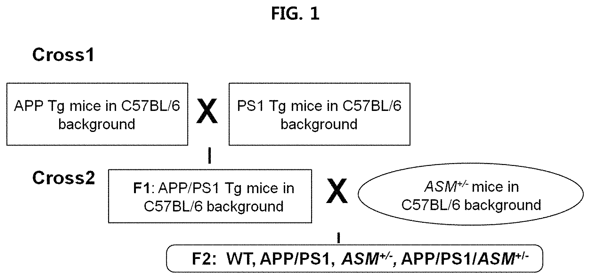
FIG. 1 is a view illustrating a process for manufacturing APP/PS1/ASM.sup.+/- mice;
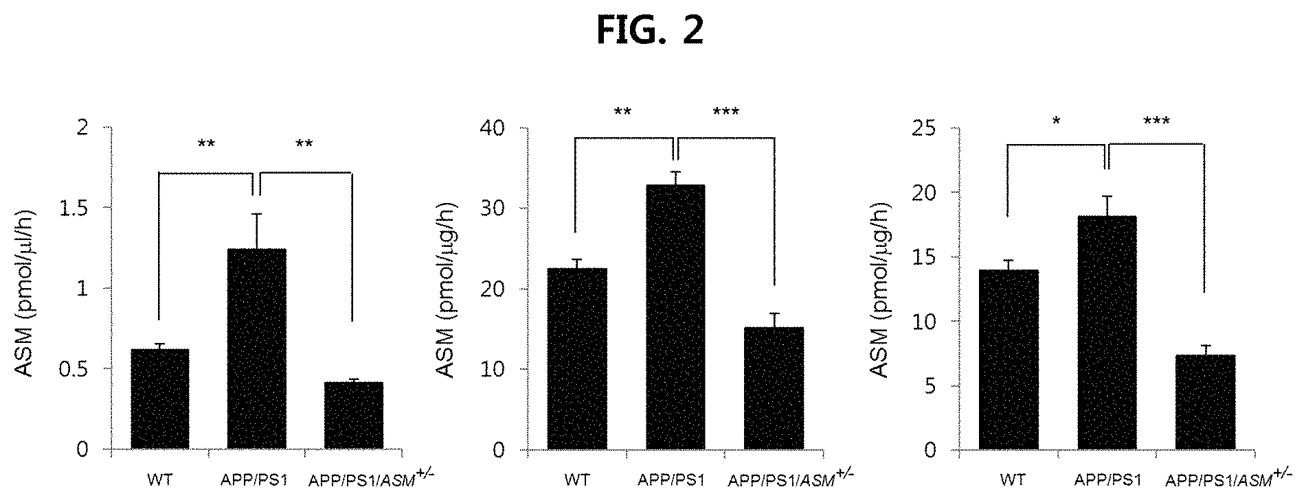
FIG. 2 is a view illustrating the ASM concentration levels in the plasma, brain tissue and fibroblast of Alzheimer's disease model mice (APP/PS1) and mice with partial removal of ASM in the Alzheimer's disease model mice (APP/PS1/ASM.sup.+/-) (left: plasma; middle: brain tissue; right: fibroblast);
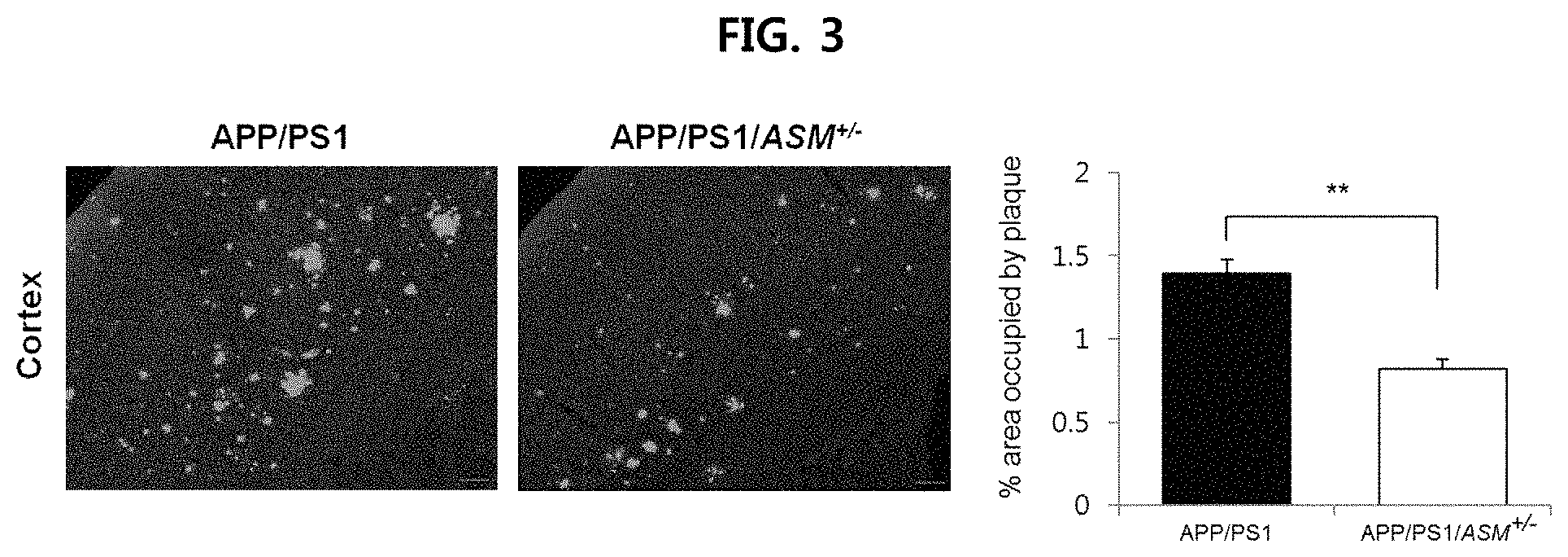
FIG. 3 is a view confirming the deposition of .beta.-amyloid in the cerebral cortex in the brain tissues of APP/PS1 mice and APP/PSVASM.sup.+/- mice using thioflavin S dye;
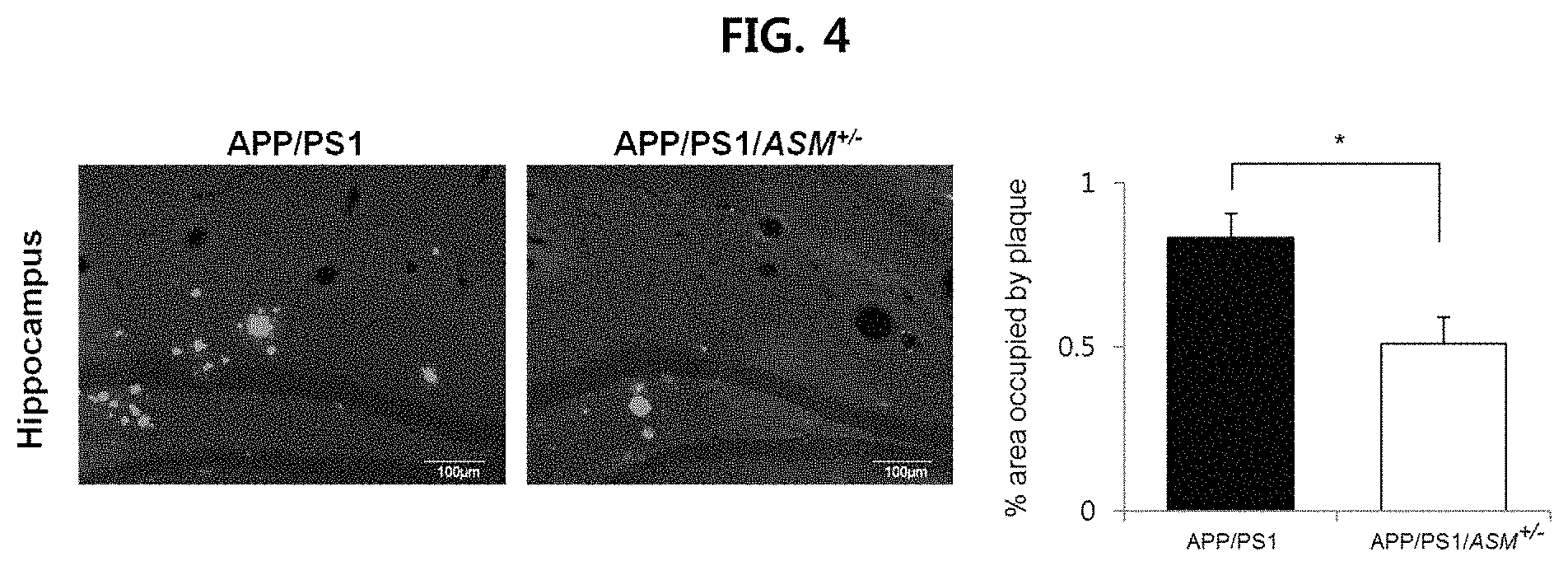
FIG. 4 is a view confirming the deposition of .beta.-amyloid in the hippocampus in the brain tissues of APP/PS1 mice and APP/PS1/ASM.sup.+/- mice using thioflavin S dye;
FIG. 5 is a view confirming the deposition of .beta.-amyloid in the brain tissues of APP/PS1 mice and APP/PS1/ASM.sup.+/- mice using immunofluorescence;
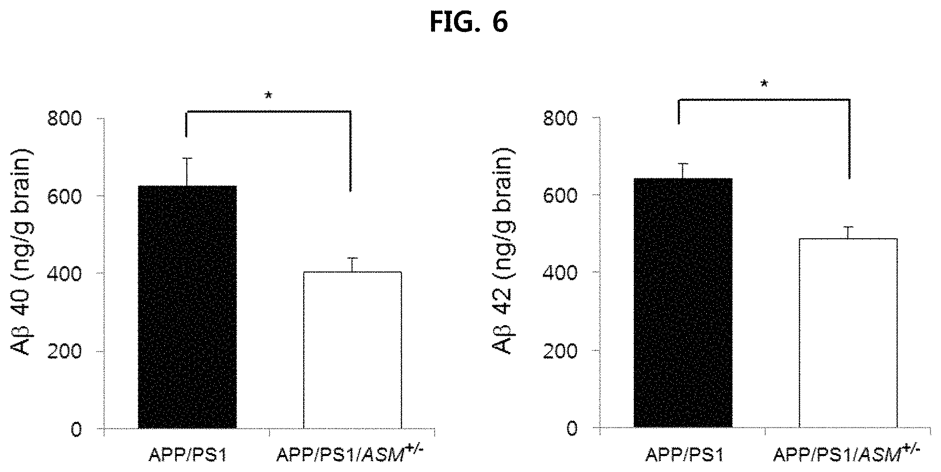
FIG. 6 is a view confirming the deposition of .beta.-amyloid in the brain tissues of APP/PS1 mice and APP/PS1/ASM.sup.+/- mice using ELISA;
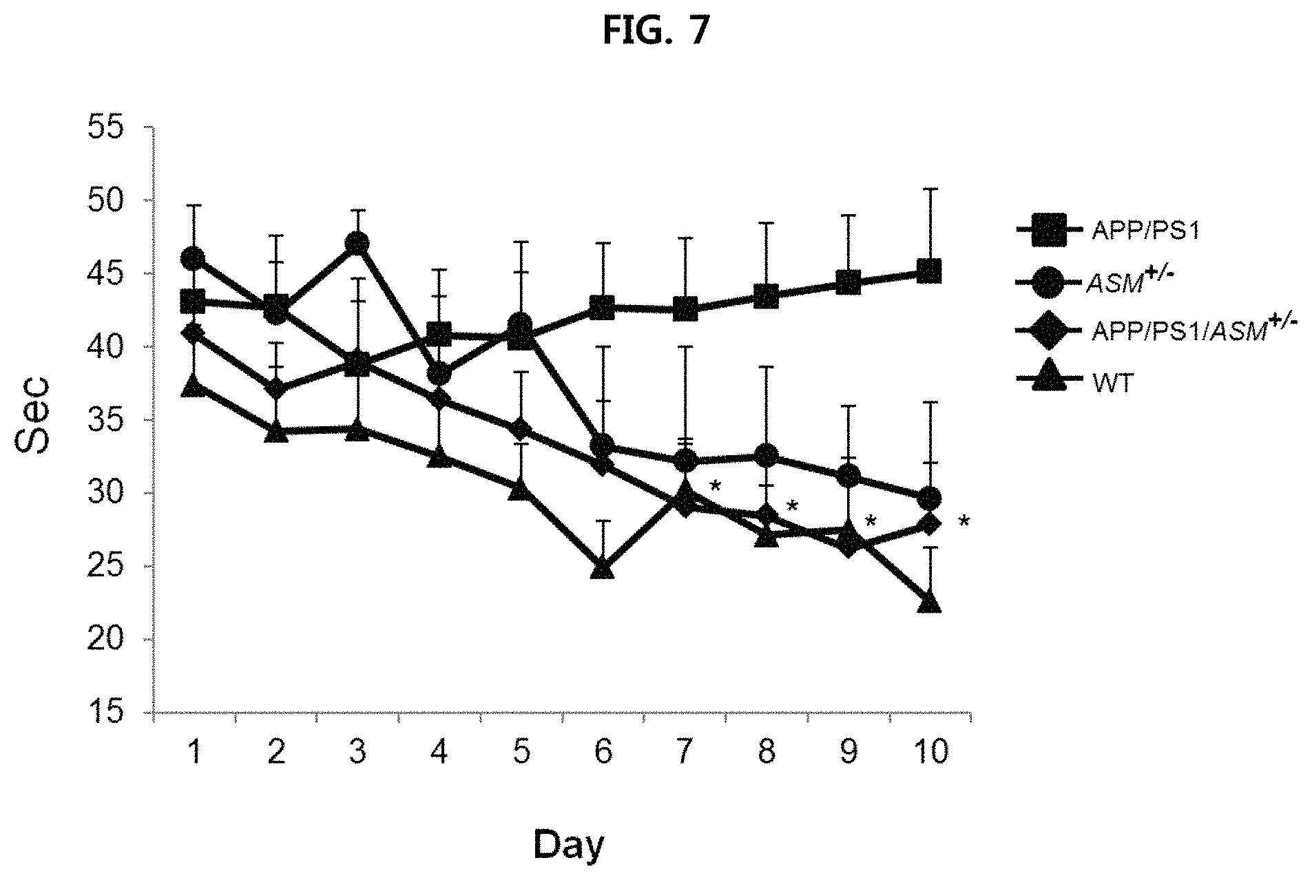
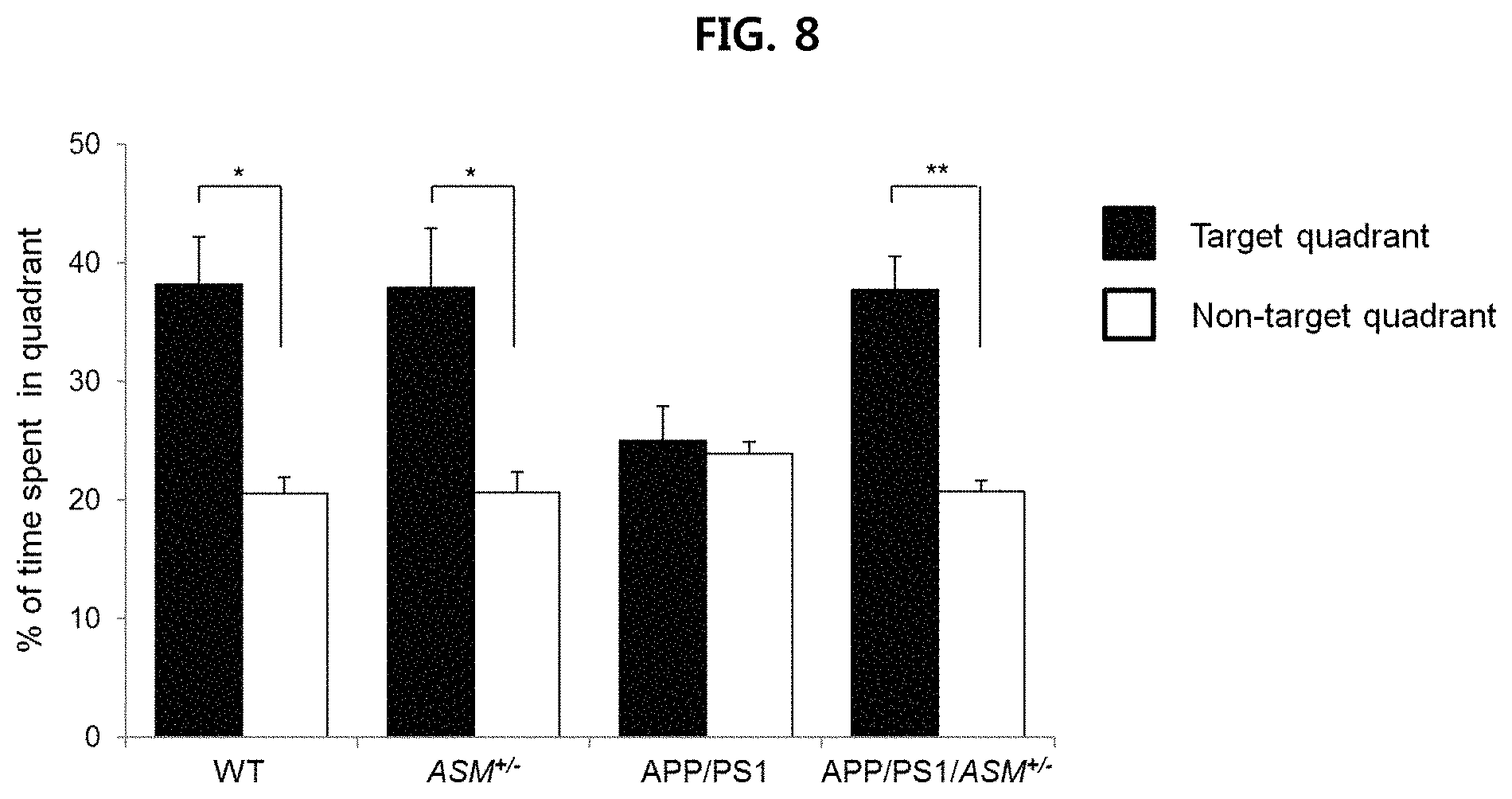
FIGS. 7 and 8 are views confirming the effect of improving the ability to learn and remember of APP/PSVASM.sup.+/- mice using Morris water maze (MWM) test;
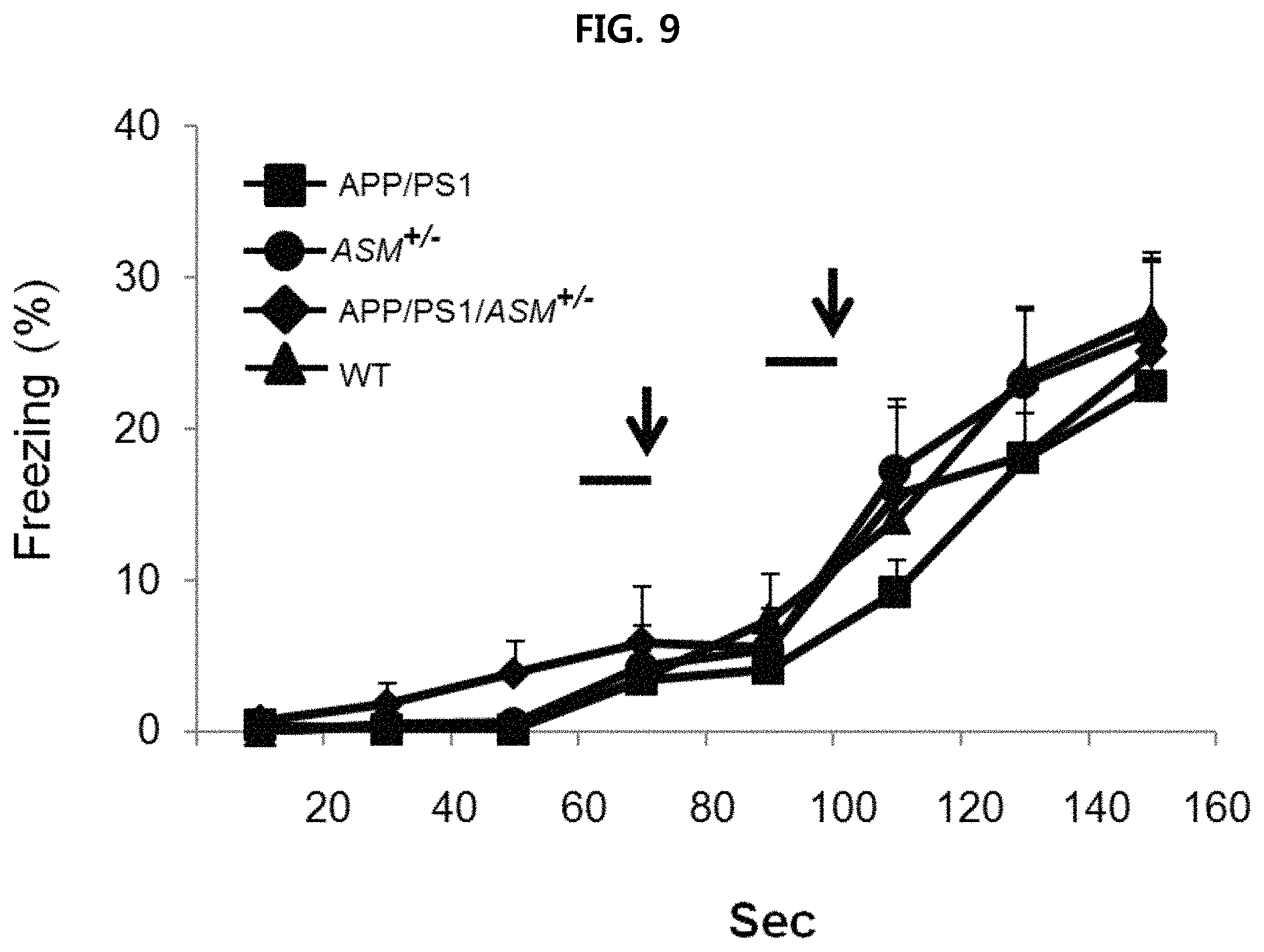
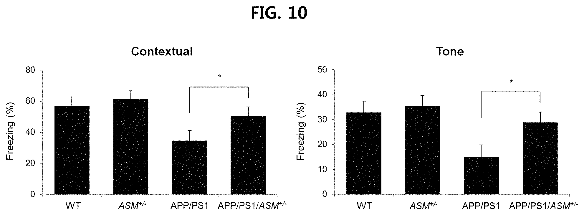
FIGS. 9 and 10 are views confirming the effect of improving the ability to remember of APP/PS1/ASM.sup.+/- mice using fear conditioning test;
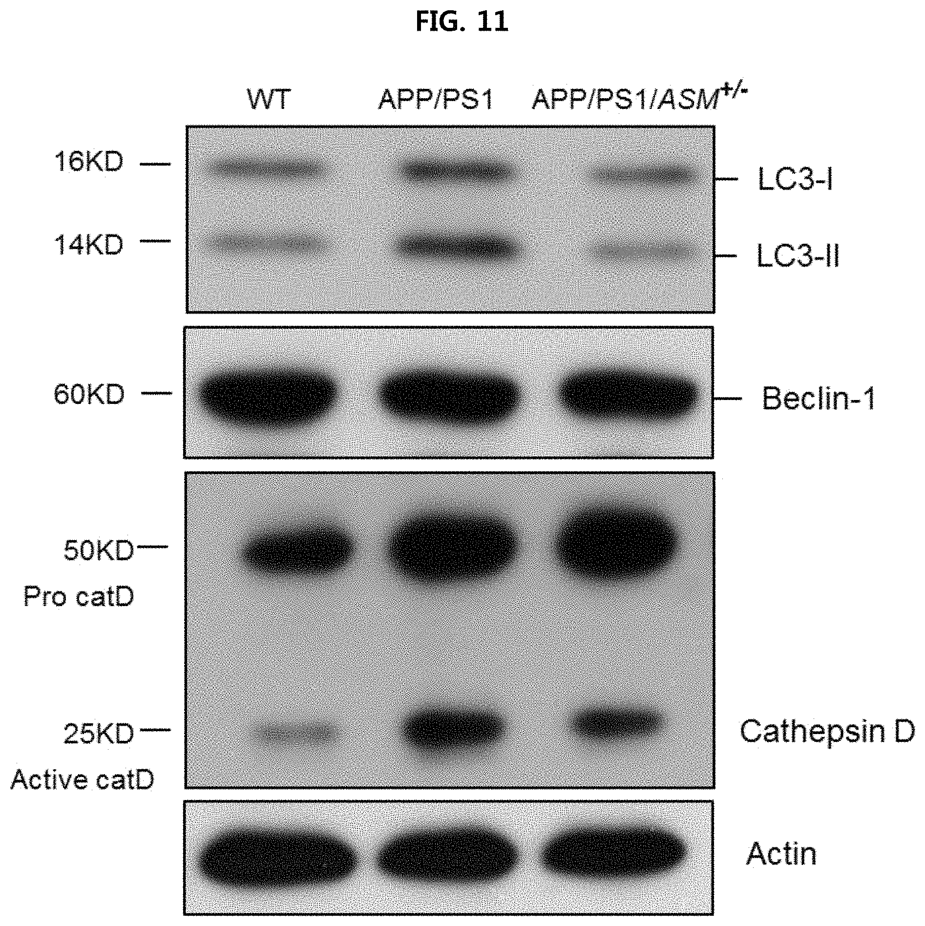
FIG. 11 is a view confirming the expression level of autophagy-related protein in the tail fibroblast from WT mice, APP/PS1 mice and APP/PS1/ASM.sup.+/- mice using Western blotting;
FIG. 12 is a view confirming the expression level of autophagy-related protein in the tail fibroblast from WT mice, APP/PS1 mice and APP/PS1/ASM.sup.+/- mice using densitometric quantification;
FIG. 13 is a view confirming the expression level of autophagy-related protein in the brain tissues of WT mice, APP/PS1 mice and APP/PS 1/ASM.sup.+/- mice using Western blotting;
FIG. 14 is a view confirming the expression level of autophagy-related protein in the brain tissues of WT mice, APP/PS1 mice and APP/PS 1/ASM.sup.+/- mice using densitometric quantification;
FIG. 15 is a view confirming the proteolytic activity in the tail fibroblast from WT mice, APP/PS1 mice and APP/PS1/ASM.sup.+/- mice;
FIG. 16 is a view observing the brain tissues of WT mice, APP/PS1 mice and APP/PS1/ASM.sup.+/- mice using transmission electron microscope (TEM);
FIG. 17 is a view confirming the expression level of autophagy-related protein when treating human fibroblast with ASM (1 .mu.M to 10 .mu.M) using Western blotting;
FIG. 18 is a view confirming the expression level of autophagy-related protein when treating human fibroblast with ASM (1 .mu.M to 10 .mu.M) using densitometric quantification;
FIG. 19 is a view confirming the expression level of autophagy-related protein when treating human fibroblast with M6P using Western blotting and densitometric quantification;
FIG. 20 is a view illustrating the conversion rate from LC3-I to LC3-II when treating human fibroblast with ASM in the presence or absence of NH.sub.4Cl using Western blotting;
FIG. 21 is a view illustrating the conversion rate from LC3-I to LC3-II when culturing human fibroblast in a serum-free medium or complete medium and treating it with NH.sub.4Cl;
FIG. 22 is a view illustrating the conversion rate from LC3-I to LC3-II when culturing human fibroblast in a serum-free medium or complete medium and treating it with ASM;
FIG. 23 is a view illustrating a test design for verifying the effect of treating Alzheimer's disease by administering AMI into APP/PS1 mice;
FIG. 24 is a view illustrating the ASM concentration in the serum and brain tissues of the mice when administering AMI into APP/PS1 mice;
FIG. 25 is a view confirming the deposition of .beta.-amyloid in the brain tissues (cerebral cortex and hippocampus) of mice when administering AMI into APP/PS1 mice;
FIG. 26 is a view confirming the effect of improving the ability to remember when administering AMI into APP/PS1 mice and performing Morris water maze (MWM) test;
FIG. 27 is a view illustrating a test design for verifying the effect of treating Alzheimer's disease using parabiotic union system [isochronic (APP/PS1-APP/PS1: parabiotic union between APP/PS1 mice, which are Alzheimer's disease model mice), heterochronic I (APP/PS1-ASM.sup.+/-: parabiotic union between APP/PS1 mice and ASM.sup.+/- mice), heterochronic II (APP/PS1-WT: parabiotic union between APP/PS1 mice and wild type mice)];
FIG. 28 is a view illustrating the ASM concentration in the serum and brain tissues of parabiotic union mice;
FIG. 29 is a view confirming the deposition of .beta.-amyloid in the brain tissues (cerebral cortex and hippocampus) of parabiotic union mice;
FIG. 30 is a view illustrating the expression level for each protein in the brain tissues of parabiotic union mice using Western blotting;
FIG. 31 is a view illustrating a test design for verifying the effect of treating Alzheimer's disease using serum injection;
FIG. 32 is a view illustrating the ASM concentration in serum and brain tissues of APP/PS1 mice provided with serum of each mouse;
FIG. 33 is a view confirming the deposition of .beta.-amyloid in the brain tissues(cerebral cortex and hippocampus) of APP/PS1 mice provided with serum of each mouse;
FIG. 34 is a view illustrating the expression level for each protein in the brain tissue of APP/PS1 mice provided with serum of each mouse using Western blotting;
FIG. 35 is a view confirming the effect of improving the ability to remember of APP/PS1 mice provided with serum of each mouse using Morris water maze (MWM) test;
FIG. 36 is a view confirming the effect of improving the ability to remember of APP/PS1 mice provided with serum of each mouse using fear conditioning test;
FIG. 37 is a schematic diagram of the role of ASM to a pathogenesis of Alzheimer's disease;
FIG. 38 illustrates changes in ASM activity after treatment of anti-ASM antibody (ASM-ab: SMPD1 antibody) on fibroblasts(PS1 fibroblast, control) of Alzheimer's patients with increased ASM activity (n=3/group);
FIG. 39A illustrates the results of Western blotting analysis of the expression of autophagy-related proteins after treating the ASM-inhibiting antibody (ASM-ab: SMPD1 antibody) on a fibroblast of Alzheimer's disease. (PS1 fibroblast);
FIG. 39B illustrates the quantification results of the Western blotting analysis of FIG. 39A (n=3/group);
FIG. 40 is a diagram illustrating the outline of an experiment performed to examine the effect of ASM inhibition on Alzheimer's disease through ASM-microRNA (Smpd1 miR RNAi) injection;
FIG. 41A illustrates the changes in ASM concentration in mouse serum after injection of Control miR RNAi or Smpd1 miR RNAi into an Alzheimer's animal model (n=3-4/group) (WT: wild type, AD: Alzheimer's animal model mouse(APP/PS1 mouse));
FIG. 41B illustrates the changes in ASM concentration in mouse brain tissues after injection of Control miR RNAi or Smpd1 miR RNAi into an Alzheimer's animal model (n=3-4/group) (WT: wild type, AD: Alzheimer's animal model (APP/PS1 mouse));
FIG. 42A illustrates the immunofluorescence staining images using thioflavin S(ThioS, fibrillary amyloid beta plaque detection) in the brain cortex of Alzheimer's animal model injected with Control miR RNAi or Smpd1 miR RNAi and the quantification results of the area occupied by a fibrillary amyloid beta plaque detected by Thioflavin S. (N=3/group) (WT: wild type, AD: Alzheimer's animal model (APP/PS1 mouse));
FIG. 42B illustrates the immunofluorescence staining images using thioflavin S(ThioS, fibrillary amyloid beta plaque detection) in the brain hippocampus of Alzheimer's animal model injected with Control miR RNAi or Smpd1 miR RNAi and the quantification results of the area occupied by a fibrillary amyloid beta plaque detected by Thioflavin S. (N=3/group) (WT: wild type, AD: Alzheimer's animal model (APP/PS1 mouse));
FIG. 43A illustrates the immunofluorescent staining image showing the degree of accumulation of A.beta.40 in the brain cortex and hippocampus of an Alzheimer's animal model injected with Control miR RNAi or Smpd1 miR RNAi and the result of quantifying the degree of accumulation (n=3/group) (WT: wild type, AD: Alzheimer's animal model (APP/PS1 mouse));
FIG. 43B illustrates the immunofluorescent staining image showing the degree of accumulation of A.beta.42 in the brain cortex and hippocampus of an Alzheimer's animal model injected with Control miR RNAi or Smpd1 miR RNAi and the result of quantifying the degree of accumulation. (n=3/group) (WT: wild type, AD: Alzheimer's animal model (APP/PS1 mouse));
FIG. 44A illustrates the change of the escape time during the MWM test period. The MWM test was performed to assess the ability of learning and memory on wild-type mice (n=6), Alzheimer's animal model injected with Control miR RNAi (n=6), Alzheimer's animal model injected with Smpd1 miR RNAi (n=6) (WT: wild type, AD: Alzheimer's animal model (APP/PS1 mouse));
FIG. 44B illustrates the time period spent during which the mice stayed on the target platform on the 11th day of the MWM test;
FIG. 44C illustrates the number of times the mouse entered the target area of the target platform on the 11th day of the MWM test;
FIG. 45A illustrates the results of quantifying the percentage of astrocytes(marker GFAP) in the brain cortex of the Alzheimer's animal model injected with Control miR RNAi or Smpd1 miR RNAi, and the wild-type mice (n=3/group, WT: wild type, AD: Alzheimer's animal model. (APP/PS1 mouse));
FIG. 45B illustrates the results of quantifying the percentage of microglial cells(marker Iba-1) in the brain cortex of the Alzheimer's animal model injected with Control miR RNAi or Smpd1 miR RNAi, and the wild-type mice.(n=3/group, WT: wild type, AD: Alzheimer's animal model.(APP/PS1 mouse));
FIG. 45C illustrates the results of evaluating mRNA expression levels of pro-inflammatory markers(TNF-alpha, IL-1beta, IL-6) in the brain cortex of the Alzheimer's animal model injected with Control miR RNAi or Smpd1 miR RNAi, and the wild-type mice (n=3/group, WT: wild type, AD: Alzheimer's animal model. (APP/PS1 mouse));
FIG. 46A illustrates the results of Western blotting analysis of the expression of autophagy-related proteins in the brain cortex of the Alzheimer's animal model injected with Control miR RNAi or Smpd1 miR RNAi, and the wild-type mice (WT: wild type, AD: Alzheimer's animal model (APP/PS1 mouse)); and
FIG. 46B illustrates the quantification results of the Western blotting analysis of FIG. 46A (n=3/group).
DETAILED DESCRIPTION OF THE ILLUSTRATED EMBODIMENTS
Hereinafter, the present invention will be described in detail.
The present invention provides a composition for preventing or treating degenerative neurological disorders, including an ASM (acid sphingomyelinase) activity inhibitor or expression inhibitor as an active ingredient.
The composition includes a pharmaceutical composition or a food composition.
According to the present invention, when ASM was inhibited in Alzheimer's disease model mice, for example, in the case of a Alzheimer's disease model mice in which ASM gene is partially deficient, in the case of a case in which a Alzheimer's disease model mice and a ASM gene-partially deficient mice were parabiotically unioned, and in the case of a Alzheimer's disease model mice injected with the sera from a ASM gene-deficient mouse, the deposition of .beta.-amyloid in the brain tissue is inhibited and the ability to learn and remember is improved, and the present invention confirms such superb effects. Accordingly, ASM inhibitor may be effectively used to prevent or treat degenerative neurological disorders including Alzheimer's disease.
In the present invention, ASM (acid sphingomyelinase, gene Smpd1) is not limited to a specific biological origin. The amino acid sequence of ASM is not particularly limited to a specific amino acid sequence as long as it has an amino acid sequence of a polypeptide known as ASM, while the nucleic acid sequence of a gene encoding ASM (including all transcripts from ASM gene, such as mRNA and the like) is also not particularly limited. As a preferable example, human ASM may comprise an amino acid sequence known as Genbank Accession No. NP_000534.3, NP_001007594.2, NP_001305016.1, NP_001305017.1, and the like, while its gene (or mRNA) sequence is known as Genbank Accession No. NP_000543.4, NP_001007593.2, NP_001318087.1, NP_001318088.1, and the like.
The ASM activity inhibitor according to the present invention may be at least one selected from a group consisting of a compound, a peptide, a peptide mimetic, a substrate analogue, an aptamer, and an antibody, specifically binding to ASM protein, but is not limited thereto.
The peptide mimetics inhibit the binding domain of ASM protein, thus inhibiting the activity of ASM protein. The peptide mimetics may be peptides or non-peptides and may include amino acids linked by non-peptide bonds such as psi bonds (Benkirane, N., et al. J. Biol. Chem., 271:33218-33224, 1996). Moreover, the peptide mimetics may be "conformationally constrained" peptides, cyclic mimetics, or cyclic mimetics including at least one exocyclic domain, a link moiety (linking amino acid) and an active region. The peptide mimetics are constructed to resemble secondary structural features of Ubiquitin-Associated Protein 2 (UBAP2) and may mimic inhibitory features of macro molecules such as antibody (Park, B. W. et al. Nat Biotechnol 18, 194-198, 2000) or water soluble receptors (Takasaki, W. et al. Nat Biotechnol 15, 1266-1270, 1997). These peptides represent novel small molecule that may act with potency equivalent to the natural antagonist (Wrighton, N. C. et al. Nat Biotechnol 15, 1261-1265, 1997).
The aptamer is a single-stranded DNA or RNA molecule and may be obtained by isolating oligomers that bind to specific chemical molecules or biological molecules with high affinity and specificity by an evolutionary method using an oligonucleotide library called systematic evolution of ligands by exponential enrichment (SELEX) (C. Tuerand L. Gold, Science 249, 505-510, 2005; A. D. Ellington and J. W. Szostak, Nature 346, 818-822, 1990; M. Famulok, et. al., Acc. Chem. Res. 33, 591-599, 2000; D. S. Wilson and Szostak, Annu. Rev. Biochem. 68, 611-647, 1999). The aptamer may specifically bind to a target to regulate its activity and may inhibit the function of the target by binding, for example.
The antibody specifically and directly binds to the ASM to effectively inhibit its activity. Preferably, a polyclonal antibody or monoclonal antibody may be used as the antibody that specifically binds to the ASM. The antibody that specifically binds to the ASM may be prepared by a method known to those skilled in the art, and a commercially available ASM antibody may be purchased and used. The antibody may be prepared by injecting the ASM protein as an immunogen into an external host according to a conventional method known to those skilled in the art. The external host may include mammals such as mice, rats, sheep, rabbits, etc. The immunogen may be injected intramuscularly, intraperitoneally, or subcutaneously, and generally may be injected with an adjuvant to enhance antigenicity. Blood samples may be taken from the external host at regular intervals and serum exhibiting titer and specificity to the antigen may be collected to separate an antibody therefrom.
The ASM expression inhibitor according to the present invention may be at least one selected from a group consisting of an antisense nucleotide, small hairpin RNA (shRNA), small interfering (siRNA), microRNA (miRNA) and ribozyme, complementarily binding to mRNA of an ASM gene or a gene promoting expression of ASM, but is not limited thereto. The antisense nucleotide, shRNA, siRNA or miRNA of the present invention is not particularly limited in its nucleotide sequence and its length as long as it has an activity of suppressing the expression of the ASM gene (i.e., Smpd1). As an example, the antisense nucleotide, shRNA, siRNA or miRNA of the present invention may comprise a nucleic acid sequence selected from the group consisting of SEQ ID NO: 1 to SEQ ID NO: 6, but is not limited thereto.
The siRNA is composed of a sense sequence of 15 to 30-mers selected from the mRNA sequence of a gene that encodes the ASM protein and an antisense sequence complementarily binding to the sense sequence. Here, preferably, the sense sequence may be composed of about 25 nucleotides, but is not particularly limited thereto.
As used herein, the miRNA (microRNA) refers to a short non-coding RNA derived from an endogenous gene, which acts as a post-transcriptional regulator of gene expression. miRNA acts as a post-transcriptional regulator of gene expression by forming a base pair with the mRNA of a target gene. The miRNA according to the present invention is not particularly limited in its nucleotide sequence and its length as long as it has an activity of suppressing the expression of the ASM gene (i.e., Smpd1). As an example, the miRNA according to the present invention may comprise a nucleic acid sequence selected from the group consisting of SEQ ID NO: 1 to SEQ ID NO: 6. As another example, the miRNA of the present invention may consist of or essentially consist of any one of the nucleotide sequences selected from the group consisting of SEQ ID NOS: 1 to 6.
As defined by Watson-Crick base pairs, the antisense nucleotide is hybridized with a complementary sequence of DNA, immature-mRNA or mature-mRNA to interrupt the transmission of genetic information as a protein in DNA. A target sequence specific antisense nucleotide is exceptionally multi-functional. The antisense nucleotide is a long chain of monomers, which favors hybridization to a target RNA sequence. Numbers of reports have recently been made to prove the utility of an antisense nucleotide as a biochemical tool in study of a target protein (Rothenberg et al., J. Natl. Cancer Inst., 81:1539-1544, 1999). Great progress has been made in the fields of oligonucleotide chemistry and nucleotide synthesis having improved cell adhesion of oligonucleotide, target binding affinity and resistance against nuclease, suggesting that an antisense nucleotide might be considered a new form of an inhibitor.
The degenerative neurological disorder according to the present invention includes Alzheimer's disease, Parkinson's disease, progressive supranuclear palsy, multiple system atrophy, olivopontocerebellar atrophy (OPCA), Shy-Drager syndrome, Striatonigral degeneration, Huntington's disease, Amyotrophic lateral sclerosis (ALS), essential tremor, cortico-basal ganglionic degeneration, diffuse Lewy body disease, Parkinson-ALS-dementia complex of Guam, Pick's disease, ischemia, and cerebral infarction, but is not limited thereto.
The composition of the present invention may include, together with the ASM activity inhibitor or expression inhibitor, at least one of a known active ingredient having an effect of inhibiting ASM expression or activity, or a known active ingredient having an effect of treating degenerative neurological disorders.
Also, the pharmaceutical composition of the present invention may be administered orally or parenterally (e.g., applied intravenously, subcutaneously, intraperitoneally or topically) according to the intended use, and the dosage may vary according to a patient's weight, age, gender, health condition, diet, administration time, administration method, administration period or interval, excretion rate, constitutional specificity, nature of formulation, etc. The dosage of the ASM expression inhibitor or activity inhibitor of the present invention is about 0.001 to 1000 mg/kg per day, preferably about 0.1 to 500 mg/kg per day, but this may vary depending on the clinical test result. Preferably, the pharmaceutical composition of the present invention may be administered once or several times a day.
The pharmaceutical composition of the present invention may be formulated in a variety of formulations for administration. The excipients that may be included in the present invention are non-toxic inert pharmaceutically suitable solid, semi-solid or liquid formulation auxiliaries of any type, for example, fillers, weighting agents, binders, wetting agents, disintegrating agents, dispersing agents, surfactants or diluents, etc.
The pharmaceutical composition of the present invention may be formulated in the form of tablets, coated tablets, capsules, pills, granules, suppositories, solutions, suspensions and emulsions, pastes, ointments, gels, creams, lotions, powders or sprays.
The composition of the present invention may be added to dietary supplements for the improvement of degenerative neurological disorders. When using the ASM expression inhibitor or activity inhibitor of the present invention as a food additive, the active ingredient may be added as it is or together with other food or food ingredients, and it may be suitably used according to a conventional manner. The amount of active ingredient added may be determined properly according to the purpose of use (preventive, health or therapeutic purposes). In general, when manufacturing food or beverage, the active ingredient of the present invention is added in an amount of 15% by weight or less to the raw material, preferably in an amount of 10% by weight or less. However, for health and hygiene purposes, or for long-term intake for the purpose of health control, the amount of active ingredient may be equal to or less than the above range, and since there is no problem in terms of safety, the active ingredient may be used in an amount greater than or equal to the above range.
There is no particular limitation in the type of food. Examples of the food to which this substance may be added include meat, sausages, bread, chocolate, candies, snacks, confectionery, pizza, ramen, other noodles, gum, dairy products including ice cream, soup, beverages, tea, drinks, alcohol drinks, and vitamin complexes, etc. That is, food may comprise all kinds of dietary supplements in the conventional sense.
The health beverage composition of the present invention may include additional ingredients such as various flavoring agents or natural carbohydrates, etc., like other beverages. The natural carbohydrates above may be monosaccharides such as glucose and fructose, disaccharides such as maltose and sucrose, polysaccharides such as dextrin and cyclodextrin, and sugar alcohols such as xylitol, sorbitol and erythritol, etc. Natural sweeteners such as thaumatin and stevia extract, and synthetic sweeteners such as saccharin and aspartame, etc. may be used as sweeteners. The ratio of natural carbohydrate is generally in the range of about 0.01 to 0.20 g per 100 g of the composition of the present invention, and preferably in the range of about 0.04 to 0.10 g.
In addition to the above, the composition of the present invention may include various nutrients, vitamins, electrolytes, flavoring agents, coloring agents, pectic acid and its salts, alginic acid and its salts, organic acid, protective colloidal thickeners, pH adjusting agents, stabilizers, preservatives, glycerin, alcohol, carbonating agents used in carbonated beverages, etc. Further, the composition of the present invention may include pulp for the production of natural fruit juice, fruit juice beverage and vegetable beverage. These ingredients may be used independently or in combination with other ingredients. The ratio of the additive is not so important but is generally selected from a range of 0.01 to 0.20 parts by weight with respect to 100 parts by weight of the composition of the present invention.
The term "comprising" of the present invention is used synonymously with "including" or "characterized" and does not exclude additional component elements or method steps, etc not mentioned in the composition or method. The term "consisting of" is intended to exclude additional elements, steps or components, etc not otherwise mentioned. The term "essentially consisting of" is intended to encompass component elements or steps, etc., which, in addition to the described component elements or steps, do not materially affect the active ingredient (for instance, ASM inhibitor in the present invention) underlying properties in the scope of the composition or method.
Also, the present invention provides a method for preventing or treating degenerative neurological disorders, including administering to a subject in need thereof a therapeutically effective amount of the composition.
Specifically, the present invention provides a method for treating a degenerative neurological disorders in a subject in need thereof, comprising administering to a subject a therapeutically effective amount of a composition comprising an ASM (acid sphingomyelinase) activity inhibitor or expression inhibitor as an active ingredient.
Also, the present invention provides a method for treating a degenerative neurological disorders in a subject in need thereof, comprising administering to a subject a therapeutically effective amount of a composition consisting of an ASM (acid sphingomyelinase) activity inhibitor or expression inhibitor.
Also, the present invention provides a method for treating a degenerative neurological disorders in a subject in need thereof, comprising administering to a subject a therapeutically effective amount of a composition essentially consisting of an ASM (acid sphingomyelinase) activity inhibitor or expression inhibitor.
The term "treatment or treating" as used herein is a concept involving inhibition, elimination, alleviation, relief, amelioration and/or prevention of a disease itself, or symptoms or conditions caused by the disease.
The "effective amount" as used herein refers to an amount that, when administered to an individual, represents an improvement, treatment, or prevention effect of degenerative neurological disorders. It is obvious to those skilled in the art that the therapeutically effective amount may be determined within the scope of sound medical judgment. Preferably, the specific therapeutically effective amount for a particular patient may vary depending on a variety of factors including the type and degree of a desired reaction, the specific composition including the use of any other agents according to the intended use, the patient's age, weight, general health condition, gender, diet, administration time, administrate route and excretion rate of the composition, duration of treatment, other drugs used in combination or coincidentally with the specific composition, and like factors well known in the medical arts. Therefore, preferably, the effective amount of the composition suitable for the purpose of the present invention is determined in consideration of the foregoing.
In addition, optionally, the composition of the present invention may be administered in combination with a known therapeutic agent for degenerative neurological disorders to increase the effect of treating degenerative neurological disorders.
The term "subject" refers to an animal, preferably a mammal which especially includes a human, while including animal-derived cells, tissues, organs and the like. The subject may be a patient in need of the above mentioned effect.
The present invention is applicable to any mammal, with a degenerative neurological disorder as described in the above-mentioned "subject". Here, the mammals include human, primates, and livestock animals such as cows, pigs, sheep, horses, dogs, cats, etc.
Also, the present invention provides a method for screening a substance for preventing or treating degenerative neurological disorders, including 1) treating a biological sample with a candidate substance, and 2) measuring the change in expression amount of mRNA or protein of ASM (acid sphingomyelinase) from the biological sample in step 1).
The biological sample in the step 1) may include blood, urine, saliva or tissue, etc. of animals with degenerative neurological disorders, but is not limited thereto.
The method for measuring the change in expression amount of mRNA in the step 2) includes reverse transcription polymerase chain reaction (RT-PCR), competitive RT-PCR, realtime RT-PCR, RNase protection assay (RPA), Northern blotting, and DNA chip, etc., but is not limited thereto.
The method for measuring the change in expression amount of protein in the step 2) includes Western blotting, enzyme linked immunosorbent assay (ELISA), radioimmunoassay (RIA), radioimmunodiffusion, Ouchterlony immunodiffusion, rocket immunoelectrophoresis, immunohistostaining, immunoprecipitation assay, complement fixation assay, fluorescence activated cell sorter (FACS) and protein chip, etc., but is not limited thereto.
Hereinafter, preferred Examples will be provided to facilitate understanding of the present invention. However, the following Examples are provided for better understanding of the present invention only, and the scope of the present invention is not limited by the following Examples.
Example 1
Confirmation on the Effect of ASM Inhibition on the Treatment of Alzheimer's Disease in ASM (Acid Sphingomyelinase) Mutant Mice
1-1. Preparation of ASM Mutant Mice
An experiment was conducted using APP/PS1 (APP/presenilin) double mutant mice and APP/PSVASM.sup.+/- triple mutant mice (with partial genetic removal of ASM), which are test animal models of Alzheimer's disease.
The animal test conducted was approved by the Kyungpook National University Institutional Animal Care and Use Committee (IACUC). Transgenic mouse lines overexpressing APPswe (hAPP695swe) or PS1 (presenilin-1M146V) mutations were used onto C57BL/6 mice (Charles River, UK) [hereinafter, APP mice: mice overexpressing APPswe, PS1 mice: overexpressing presenilin-1M146V; GlaxoSmithKline]. ASM.sup.+/- mice (only one of a pair of ASM genes is removed) were crossed with APP/PS1 mice to prepare APP/PS1/ASM.sup.+/- mice.
Detailed process is illustrated in FIG. 1.
1-2. Confirmation on ASM Concentration Level in ASM Mutant Mice
The ASM concentration levels were measured in the plasma, brain tissue and fibroblast of nine-month old wild type (WT) mice, APP/PS1 mice and APP/PS1/ASM.sup.+/- mice prepared in Example 1-1. More specifically, 3 .mu.l of plasma, brain tissue and fibroblast samples from each mouse were mixed with an ASM activity buffer, and stored at 37.degree. C. The hydrolysis reaction was completed by adding 114 .mu.l of ethanol to the mixed solution, and then the mixed solution was centrifuged. 30 .mu.l of the supernatant was transferred into a glass vial, and then 5 .mu.l was applied to the UPLC system. The ASM concentration level was quantified in comparison with a bodipy (aminoacetaldehyde) combined with sphingomyelin and ceramide. The sphingomyelin and ceramide levels were extracted and quantified according to a known method, by extracting lipid from the sample, resuspending the dried lipid extract in 25 .mu.l of 0.2% Igepal CA-630 (Sigma-Aldrich), and quantifying the concentration level of each lipid using the UPLC system. The results are illustrated in FIG. 2 (left: plasma; middle: brain tissue; right: fibroblast).
As illustrated in FIG. 2, it is confirmed that the ASM concentration levels in the plasma, brain tissue and fibroblast of APP/PS1/ASM.sup.+/- mice decreased remarkably, as compared to the ASM concentration levels in the plasma, brain tissue and fibroblast of APP/PS1 mice.
1-3. Confirmation on Inhibition of Deposition of .beta.-Amyloid in Brain Tissue of ASM Mutant Mice
As confirmed in Example 1-2, in order to confirm how the decrease in ASM concentration level in APP/PS 1/ASM.sup.+/- mice affects Alzheimer's disease in a pathological aspect, the deposition level of .beta.-amyloid in the brain tissue was analyzed.
First, the cerebral cortex and hippocampus tissue of each mouse prepared in Example 1-1 were isolated, and then a tissue fragment was obtained and this was dyed with thioflavin S according to a conventional known method. The results are illustrated in FIG. 3 (cerebral cortex) and FIG. 4 (hippocampus).
As illustrated in FIG. 3 and FIG. 4, it is confirmed that A.beta.40 and A.beta.42 deposited in the brain tissue of APP/PS 1/ASM.sup.+/- mice decreased remarkably, as compared to the brain tissue of APP/PS1 mice.
Also, immunofluorescence was performed according to a conventional known method using anti-20G10 (mouse, 1:1000) antibody against A.beta.42, anti-G30 (rabbit, 1:000) antibody against A.beta.40, anti-Iba-1 (rabbit, 1:500, Wako) antibody, anti-GFAP (rabbit, 1:500, DAKO) antibody and anti-active caspase3 (rabbit, 1:50, Chemicon) antibody. Tissue fragments were observed using a confocal laser scanning microscope equipped with Fluoview SV1000 imaging software (Olympus FV1000, Japan) or an Olympus BX51 microscope, and the percentage of the area of the dyed area with respect to the area of the entire tissue was quantified using Metamorph software (Molecular Devices).
Also, .beta.-amyloid deposition was confirmed using commercially available ELISA kits (Biosource). More specifically, hemispheres of the brain of each mouse were homogenized in a buffer containing 0.02 M of guanidine. Thereafter, ELISA was performed for A.beta.40 and A.beta.42 according to the manufacturer's instructions.
The results are illustrated in FIG. 5 (immunofluorescence) and FIG. 6 (ELISA).
As illustrated in FIG. 5 and FIG. 6, it is confirmed that A.beta.40 and A.beta.42 deposited in the brain tissue of APP/PS 1/ASM.sup.+/- mice decreased remarkably, as compared to the brain tissue of APP/PS1 mice.
1-4. Confirmation on Improvement of Ability to Learn and Remember in ASM Mutant Mice
In order to confirm the effect of improving the ability to learn and remember in APP/PSVASM.sup.+/- mice prepared in Example 1-1, a Morris water maze (MWM) test was performed according to a conventional known method.
More specifically, wild type mice, APP/PS1/ASM.sup.+/- mice, ASM.sup.+/- mice and APP/PS1 mice were used. The mice were given four trials per day for 10 days to learn the task, and on day 11, the mice were given a probe trial in which the platform was removed. The escape latency during the test period and the time spent in the target platform on day 11 were measured. The results are illustrated in FIG. 7 and FIG. 8.
As illustrated in FIG. 7 and FIG. 8, APP/PS1 mice did not show any change in escape latency during the test period, and did not show any difference in the time spent in the target platform and non-target platform, and thus it is confirmed that APP/PS1 mice showed disorder in forming spatial memory. In comparison, it is confirmed that APP/PSVASM.sup.+/- mice presented improved ability to learn and remember to a level similar to wild type mice.
In order to verify the MWM test results, a fear conditioning test was performed, which evaluates the ability to learn and remember by combining environmental context or conditioned stimulus with electric shock according to a conventional known method.
More specifically, wild type mice, APP/PS1/ASM.sup.+/- mice, ASM.sup.+/- mice and APP/PS1 mice were used. On day 1 of the test, each mouse was individually placed into a conditioned chamber, and then after a 60-second exploratory period, a tone (10 kHz, 70 dB) was delivered for 10 seconds. This served as the conditioned stimulus (CS). The CS was coterminated with the unconditioned stimulus (US), an electrical footshock (0.3 mA, 1 s). The CS-US pairing was delivered twice at a 20-second intertrial interval. On day 2, each mouse was placed in the fear-conditioning chamber containing the same exact context, but without administration of a CS or footshock. Freezing response was observed for 5 minutes. On day 3, each mouse was placed in a test chamber that was different from the conditioning chamber. After a 60-second exploratory period, the tone was presented for 60 seconds without the footshock. Freezing response was measured to measure fear memory. The results are illustrated in FIG. 9 and FIG. 10.
As illustrated in FIG. 9 and FIG. 10, it is confirmed that APP/PS1/ASM.sup.+/- mice has improved ability to remember than APP/PS1 mice.
Example 2
Confirmation on Effect of ASM Inhibition on Autophagy in ASM (Acid Sphingomyelinase) Mutant Mice
2-1. Confirmation on Autophagy-Related Gene Expression by ASM Inhibition
In order to confirm how genetic ASM inhibition works on autophagy-related pathways of Alzheimer's disease, the tail fibroblast and brain tissue samples from nine-month old wild type mice, APP/PS1 mice and APP/PSVASM.sup.+/- mice prepared in Example 1-1 were analyzed.
More specifically, Western blotting was performed according to a conventional known method using LC3 (rabbit, 1:1000, Cell Signaling Technologies, 4108S), Beclin-1 (rabbit, 1:1000, Cell Signaling Technologies, 3738S), p62 (rabbit, 1:1000, Cell Signaling Technologies, 5114S), cathepsin D (goat, 1:500, R&D Systems, BAF1029) and .beta.-actin (1:1000, Santa Cruz, SC-1615) antibodies, and densitometric quantification was performed using ImageJ software (US National Institutes of Health). The results are illustrated in FIGS. 11 to 14.
As illustrated in FIGS. 11 to 14, it is confirmed that the conversion from LC3-I to LC3-II increased in APP/PS1 mice as compared to WT mice, and that the expression of increased LC3-II decreased in APP/PS1/ASM.sup.+/- mice. Beclin-1 expression did not significantly vary between the groups. Also, the expression of cathepsin D (lysosomal hydrolase) and p62, which are indicators of autophagic turnover, increased in Alzheimer's patients, and thus this is pathologically related to Alzheimer's disease. It is confirmed that the expression of cathepsin D and p62 increased in APP/PS1 mice as compared to WT mice, whereas the increased expression of cathepsin D and p62 decreased in APP/PS1/ASM.sup.+/- mice.
2-2. Evaluation on Proteolytic Activity by ASM Inhibition
The proteolytic activity in the tail fibroblast from nine-month old wild type mice, APP/PS1 mice and APP/PS1/ASM.sup.+/- mice prepared in Example 1-1 were confirmed.
In order to label long-lived proteins, pulse-chase experiment was performed by giving a pulse with [.sup.3H]-leucine (2 .mu.Ci/ml) for 48 hours. Labeled cells were washed, and cultured in a complete medium of an environment inhibiting autophagy or a serum starvation medium inducing autophagy. Aliquots of the medium were collected at different time period and precipitated with 10% TCA, and then they were filtered with a film having holes of 0.22 .mu.m and the radioactivity was measured to analyze proteolytic activity. The results are illustrated in FIG. 15.
As illustrated in FIG. 15, when autophagy (culturing in serum starvation medium) is induced, it is confirmed that proteolytic activity increased in cells derived from APP/PSVASM.sup.+/- mice, as compared to cells derived from WT mice.
2-3. Analysis on Mouse Brain Tissue Using Transmission Electron Microscope (TEM)
The brain tissues of nine-month old wild type mice, APP/PS1 mice and APP/PSVASM.sup.+/- mice prepared in Example 1-1 were fixed in 3% glutaraldehyde containing phosphate buffer, 0.1M, pH 7.4, and postfixed in Sorensen's phosphate buffer containing osmium tetroxide. After dehydration with ethyl alcohol, the tissues were embedded in Epon (Electron Microscopy Sciences). They were cut serially and analyzed using Transmission Electron Microscope (Tecnai). Images were captured on a digital camera and Xplore3D tomography software. The results are illustrated in FIG. 16.
As illustrated in FIG. 16, it is confirmed that the size and number of autophagic vacuole (AV) increased in the brain tissue of APP/PS1 mice, and the size and number of vacuole in the brain tissue of APP/PS1/ASM.sup.+/- mice were observed to be slightly greater than those in WT mice, but smaller than those in APP/PS1 mice.
Example 3
Confirmation on Effect of ASM Inhibition on Autophagy in Human Cell
3-1. Confirmation on Change in Autophagy-Related Gene Expression by Recombinent ASM Protein in Human Fibroblast
Human fibroblast acquired from the Coriell Institute, and was cultured in DMEM medium containing 15% FBS at 37.degree. C. and 5% of CO.sub.2. The cell lines were treated with recombinant ASM (1 .mu.M to 10 .mu.M), and then Western blotting and densitometric quantification were performed in the same manner as Example 2-1. The results are illustrated in FIG. 17 and FIG. 18.
As illustrated in FIG. 17 and FIG. 18, it is confirmed that conversion from LC3-I to LC3-II takes place depending on the concentration of recombinant ASM treatment, and there is no significant change in expression of beclin-1.
3-2. Confirmation on Mechanism of ASM Using M6P (Mannose-6-Phosphate)
In order to confirm how ASM affects autophagy, a test was conducted as follows. Human fibroblast was treated with ASM alone, or treated with ASM in the presence of mannose-6-phosphate(M6P; 10 mM) relating to the action of placing lysosomal enzyme protein in lysosome, and then the accumulation of autophagosome was confirmed using Western blotting and densitometric quantification. The results are illustrated in FIG. 19.
As illustrated in FIG. 19, it is confirmed that when M6P is used to inhibit ASM absorption by lysosomes, the conversion to LC3-II decreased, and accordingly the accumulation of ASM-induced autophagosome decreased.
In order to confirm whether ASM increases the formation rate of autophagosome, or decreases the degradation rate of autophagosome, autophagic flux assay was performed. More specifically, the conversion rate from LC3-I to LC3-II was measured in cells in the presence or absence of NH.sub.4Cl, which inhibits degradation of autophagosome but does not affect autophagosome formation, using Western blotting. The results are illustrated in FIG. 20.
As illustrated in FIG. 20, it is confirmed that there is no significant change in the amount of LC3-II when treating cells with ASM and NH.sub.4Cl.
Also, when treating with NH.sub.4Cl or ASM after culturing fibroblast of human with Alzheimer's disease in a serum-free medium or complete medium, the conversion rate from LC3-I to LC3-II was measured using Western blotting. The results are illustrated in FIG. 21 and FIG. 22.
As illustrated in FIG. 21, it is confirmed that the LC3-II level increased significantly when adding NH.sub.4Cl after inducing autophagy by culturing cells in a serum-free medium. As illustrated in FIG. 22, it is confirmed that the accumulation of LC3-II increased significantly when treating with ASM after inducing autophagy by culturing cells in a serum-free medium.
Through the above test results, it is confirmed that ASM does not induce the formation of autophagosome in Alzheimer's disease, but inhibits proteolytic activity of autophagosome.
Example 4
Verification on Pathological Improvement Effect of ASM Inhibition in Alzheimer Disease Model Mice Confirmation of Therapeutic Effect Using ASM Inhibitory Compound, Parabiotic System and ASM Deficient Serum
4-1. Verification on Effect of Treating Alzheimer's Disease After Administering AMI Into APP/PS1 Mice
AMI (amitriptyline-hydrochloride), which is a known inhibitor of ASM that can cross the blood-brain barrier (BBB), was administered to APP/PS1 mice, which are Alzheimer's disease model mice, for four months, and then the water maze (WM) test was performed (FIG. 23). The degree of ASM expression was measured by obtaining serum and brain tissue when the mice became nine month old (in the same manner as Example 1-2), and the deposition of .beta.-amyloid in brain tissue was measured (in the same manner as Example 1-3). The test results are illustrated in FIGS. 24 to 26, respectively.
As illustrated in FIG. 24, it is confirmed that ASM decreased in the serum and brain tissue of mice where AMI was administered. As illustrated in FIG. 25, it is confirmed that the deposition of .beta.-amyloid was inhibited in the cerebral cortex and hippocampus of mice where AMI is administered. As illustrated in FIG. 26, it is confirmed that the ability to remember in mice where AMI is administered was recovered by decrease in escape latency, as compared to the control group.
4-2. Verification on Effect of Treating Alzheimer's Disease Using Parabiotic System
The effect of ASM inhibition is confirmed using parabiotic system of APP/PS1 mice. More specifically, isochronic (APP/PS1-APP/PS1: parabiotic union between APP/PS1 mice, which are Alzheimer's disease model mice), heterochronic I (APP/PS1-ASM.sup.+/-: parabiotic union between APP/PS1 mice and ASM.sup.+/- mice), heterochronic II (APP/PS1-WT: parabiotic union between APP/PS1 mice and wild mice) mice were prepared by sharing blood flow after connecting the skin and soft tissue by surgical methods and inducing new angiogenesis between two mice (FIG. 27). The expression degree of ASM was measured by obtaining serum and brain tissue in the same manner as Example 4-1, and the deposition of .beta.-amyloid and expression of protein in brain tissue were measured. The test results are illustrated in FIGS. 28 to 30, respectively.
As illustrated in FIG. 28, it is confirmed that the ASM concentration is low in the serum and brain tissue of heterochronic I (APP/PS1-ASM.sup.+/-) mice, as compared to isochronic (APP/PS1-APP/PS1) and heterochronic II (APP/PS1-WT) mice, and thus ASM.sup.+/- mice play the role of lowering ASM concentration.
Also, as illustrated in FIG. 29, it is confirmed that the deposition of .beta.-amyloid decreased remarkably in the cerebral cortex and hippocampus of heterochronic I (APP/PS1-ASM.sup.+/-) mice, as compared to isochronic (APP/PS1-APP/PS1) mice.
Also, as illustrated in FIG. 30, it is confirmed that the conversion to LC3-II decreased, the expression of p62 and cathepsin D decreased, and the expression of TFEB and Lamp1, which are proteins relating to ALP function, increased in the brain tissue of heterochronic I (APP/PS1-ASM.sup.+/-) mice, as compared to isochronic (APP/PS1-APP/PS1) mice.
4-3. Verification on Effect of Treating Alzheimer's Disease Using Serum Injection
The serum of APP/PS1 or ASM.sup.-/- mice was injected into APP/PS1 mice. More specifically, blood was obtained from the heart of APP/PS1 or ASM.sup.-/- mice, and then collected in a tube coated with EDTA. The collected blood was centrifuged to obtain serum, and 100 .mu.l of serum was intravenously injected into an eight-month old APP/PS1 mice eight times during 3 weeks (FIG. 31). After the test was completed, the degree of ASM expression was measured by obtaining the serum and brain tissue in the same manner as in Example 4-1, and the deposition of .beta.-amyloid and expression of protein in brain tissue were measured. Also, a behavior test was performed in the same manner as Example 1-4.
The test results are illustrated in FIGS. 32 to 36.
As illustrated in FIG. 32, it is confirmed that the ASM decreased in the serum and brain tissue of APP/PS1 mice provided with serum of ASM.sup.-/- mice, as compared to the APP/PS1 mice provided with serum of APP/PS1 mice.
As illustrated in FIG. 33 and FIG. 34, it is confirmed that the deposition of .beta.-amyloid decreased (FIG. 33) in the brain tissue of APP/PS1 mice provided with serum of ASM.sup.-/- mice, as compared to the APP/PS1 mice provided with serum of APP/PS1 mice. Thus, conversion to LC3-II decreased, and the expression of p62 and cathepsin D decreased (FIG. 34) in the APP/PS1 mice provided with serum of ASM.sup.-/- mice, as compared to the APP/PS1 mice provided with serum of APP/PS1 mice.
As illustrated in FIG. 35 and FIG. 36, as a result of MWM and fear conditioning test, it is confirmed that the ability to remember improved in APP/PS1 mice provided with serum of ASM.sup.-/- mice, as compared to APP/PS1 mice provided with serum of APP/PS1 mice.
Example 5
Verification on Pathological Improvement Effect of ASM Inhibition in Alzheimer Disease Model Mice Confirmation of Therapeutic Effect Using an Antibody and an Interfering RNA to ASM
Experimental Method
1) Cell Culture
Human fibroblast cell line(normal cell line and PS1-overexpressing cell line) were obtained from Coriell Institute and cultured in DMEM containing 15% FBS under the condition of 5% CO.sub.2 at 37.degree. C. Thereafter, the cell lines were treated with the ASM antibody (3 .mu.g/ml, R&D system) for 24 hours, and the ASM activity was measured.
2) Mice
The animal test conducted was approved by the Kyungpook National University Institutional Animal Care and Use Committee (IACUC). Transgenic mouse line overexpressing APPswe (hAPP695swe) and PS1 (presenilin-1M146V) based on C57BL/6 mice (Charles River, UK) were used as Alzheimer's animal models [hereinafter, APP mice: mice overexpressing APPswe, PS1 mice: overexpressing presenilin-1M146V; GlaxoSmithKline]. In order to confirm the therapeutic effect of ASM inhibition against Alzheimer's disease using Smpd1 miR RNAi, control miR RNAi or Smpd1 miR RNAi was injected twice a week (a total 8 weeks) into the above-mentioned mouse model (7 month-old) with a dose of 50 .mu.g/mice through tail vein. One month after the injection of Control miR RNAi or Smpd1 miR RNAi, behavioral analysis was performed, and mouse brain tissue was sampled after the behavioral analysis. This experimental outline is shown in FIG. 40.
3) microRNA Preparation for ASM (Smpd1 miR RNAi Construction)
ASM microRNA (Smpd1 miR RNAi), which can inhibit ASM, was constructed to test the therapeutic effect of inhibition of ASM activity in Alzheimer's animal model. Smpd1 miR RNAi was produced using the BLOCK-iT.TM. Pol II miR RNAi Expression Vector Kits (Invitrogen). First, miR RNA (Mmi520028_top_Smpd1, Mmi520028_bot_Smpd1, Mmi520030_top_Smpd1, Mmi520030_bot_Smpd1, Mmi520031_top_Smpd1 and Mmi520031_bot_Smpd1, see Table 1) for mouse Smpd1 was cloned into pcDNA6.2-GW/miR according to the manufacturer's protocol to generate pcDNA6.2-GW/mSmpd1 miR. After that, the CMV promoter was replaced with the human Endoglin promoter 49 to obtain phEndoglin mSmpd1 miR (Smpd1 miR RNAi). Control miR RNAi (see Table 1) was also obtained in the same manner. The prepared Smpd1 miR RNAi (50 .mu.g) was diluted in 50 .mu.l of 10% Glucose solution and RNAse free water was added to a final volume of 100 .mu.l. In another tube, 6.4 .mu.l of in vivo-jetPE.TM. (Polyplus-transfection) was diluted in 50 .mu.l of 10% Glucose solution and RNAse free water was added to a final volume of 100 .mu.l. Then 100 .mu.l of in vivo-jetPEI.TM. solution was mixed with Smpd1 miR RNAi solution. After the mixed solution was reacted at room temperature for 15 minutes, 200 .mu.l of the mixed solution was injected through the tail vein of the Alzheimer's animal model twice a week.
TABLE-US-00001 TABLE 1 Smpd1 miRNA sequence(5'.fwdarw.3') Mmi520028_top_ (SEQ ID NO. 1) Smpd1 TGCTGAAAGGAGTCCCACTCTGGGTGGTT TTGGCCACTGACTGACCACCCAGAGGGACTCCT TTCAGGACGACTTTCCTCAGGGTGAGACCCACC AAAACCGGAGACTGACTGGTGGGTCTCCCTGAG GAAACTCC Mmi520028_bot_ (SEQ ID NO. 2) Smpd1 GGAGTTTCCTCAGGGAGACCCACCAGTC AGTCTCCGGTTTTGGTGGGTCTCACCCTGAGGA AAGTCGTCCTGAAAGGAGTCCCTCTGGGTGGTC AGTCAGTGGCCAAAACCACCCAGAGTGGGACT CCTTTCAGCA Mmi520030_top_ (SEQ ID NO. 3) Smpd1 TGCTGATTGGTTTCCCTTTATGAAGGGTT TTGGCCACTGACTGACCCTTCATAGGGAAACCA ATCAGGACGACTAACCAAAGGGAAATACTTCCC AAAACCGGTGACTGACTGGGAAGTATCCCTTTG GTTAGTCC Mmi520030_bot_ (SEQ ID NO. 4) Smpd1 GGACTAACCAAAGGGATACTTCCCAGTC AGTCACCGGTTTTGGGAAGTATTTCCCTTTGGTT AGTCGTCCTGATTGGTTTCCCTATGAAGGGTCA GTCAGTGGCCAAAACCCTTCATAAAGGGAAACC AATCAGCA Mmi520031_top_ (SEQ ID NO. 5) Smpd1 TGCTGAACAGAGCCAGAACCAGCGCCGT TTTGGCCACTGACTGACGGCGCTGGCTGGCTCT GTTCAGGACGACTTGTCTCGGTCTTGGTCGCGG CAAAACCGGTGACTGACTGCCGCGACCGACCG AGACAAGTCC Mmi520031_bot_ (SEQ ID NO. 6) Smpd1 GGACTTGTCTCGGTCGGTCGCGGCAGTCA GTCACCGGTTTTGCCGCGACCAAGACCGAGACA AGTCGTCCTGAACAGAGCCAGCCAGCGCCGTCA GTCAGTGGCCAAAACGGCGCTGGTTCTGGCTCT GTTCAGCA Control miRNA sequence miR-neg control- (SEQ ID NO. 7) top TGCTGAAATGTACTGCGCGTGGAGACGTT TTGGCCACTGACTGACGTCTCCACGCAGTACAT TTCAGGACGACTTTACATGACGCGCACCTCTGC AAAACCGGTGACTGACTGCAGAGGTGCGTCATG TAAAGTCC miR-neg control- (SEQ ID NO. 8) bot GGACTTTACATGACGCACCTCTGCAGTCA GTCACCGGTTTTGCAGAGGTGCGCGTCATGTAA AGTCGTCCTGAAATGTACTGCGTGGAGACGTCA GTCAGTGGCCAAAACGTCTCCACGCGCAGTACA TTTCAGCA
4) Measurement of ASM Activity
The concentration level of ASM was measured as follows. Specifically, 3 .mu.l of mouse serum, brain tissue and fibroblast samples were mixed with ASM activity buffer and stored at 37.degree. C. 114 .mu.l of ethanol was added to terminate the hydrolysis reaction, followed by centrifugation. 30 .mu.l of supernatant was transferred to a glass vial, of which 5 .mu.l was applied to the UPLC system. The ASM concentration levels were quantified by comparison with sphingomyelin and Bodipy (aminoacetaldehyde) conjugated with ceramide. The extraction and quantification of the sphingomyelin and ceramide was carried out by extracting and drying the lipids from the sample according to a known conventional method, and resuspending the dried lipid extract to 25 .mu.l of 0.2% Igepal CA-630(Sigma-Aldrich), and the concentration levels of each lipid were quantitated using the UPLC system.
5) Immunofluorescence Staining Method
After fixing the cerebral and hippocampal tissues of the mice, 0.5% thioflavin S (Sigma-Aldrich), anti-20G10 antibodies(mouse, 1:1000) against A.beta.42, anti-G30 antibodies (rabbit, 1:000) against A.beta.40, anti-GFAP antibodies(rabbit, 1:500, DAKO), or anti-Iba-1 antibodies (rabbit, 1:500, Wako) were co-cultured. The tissues were analyzed using a laser scanning confocal microscope equipped with Fluoview SV1000 imaging software (Olympus FV1000, Japan) or an Olympus BX51 microscope. Percentage of stained area to total tissue area was quantified and analyzed using Metamorph software (Molecular Devices).
6) Western Blot
Western blotting method was used to analyze the expression of the following genes. Western blotting was performed by a conventional method using an antibody against LC3(LC3-I and LC3-II), Beclin-1, p62 [all of them were purchased from cell signaling Technologies], Cathepsin D (R&D systems), Lamp1 (abcam), TFEB (Invitrogen) and .beta.-actin (Santa Cruz). Densitometric quantification was performed using ImageJ software (US National Institutes of Health).
7) Real-Time Quantitative PCR
To quantify the expression levels of inflammatory cytokines(TNF-.alpha., IL-1.beta., and IL-6), real-time quantitative PCR was used. Total RNAs were extracted from brain tissue using RNeasy Plus mini kit (Qiagen, Korea, Ltd.), and cDNA was synthesized from 5 .mu.g of total RNAs using a kit of Clontech inc. (Mountain View, Calif.). Also, using Corbett research RG-6000 real-time PCR instrument, real-time quantitative PCR was performed with repeated 40 cycle(one cycle consisting of 95.degree. C., 10 minutes; 95.degree. C., 10 seconds; 58.degree. C., 15 seconds; 72.degree. C., 20 seconds). Table 2 shows the primers used for the real-time quantitative PCR.
TABLE-US-00002 TABLE 2 Sequence (5'.fwdarw.3') mTNF-.alpha. (SEQ ID NO. 9) (SEQ ID NO. 10) GAT TAT GGC TCA GGG TCC GCT CCA GTG AAT TCG GAA AA AG mIL-1.beta. (SEQ ID NO. 11) (SEQ ID NO. 12) CCC AAG CAA TAC CCA AAG GCT TGT GCT CTG CTT GTG AA AG mIL-6 (SEQ ID NO. 13) (SEQ ID NO. 14) CCG GAG AGG AGA CTT CAC TTG CCA TTG CAC AAC TCT AG TT mGAPDH (SEQ ID NO. 15) (SEQ ID NO. 16) TGA ATA CGG CTA CAG CAA AGG CCC CTC CTG TTA TTA CA TG
8) Behavioral Experiment
MWM(Morris water maze) experiments were performed in a conventional manner to identify potential effects on learning and memory. Briefly, for the MWM test, mouse were learned the task four times a day for 10 days, followed by the removal of the platform on the 11th day and subsequently a probe trial performed.
9) Statistical Analysis
For the comparison of the two groups, student's t-test was performed. For comparison of multiple groups, repeated analysis of Tukey's HSD test and distribution test were conducted according to the SAS statistical package (release 9.1; SAS Institute Inc., Cary, N.C.).* p<0.05, **p<0.01, ***p<0.001 were considered to be significant.
10) Acknowledgements
This research was supported by the Basic Science Research Program (2017R1A2A1A17069686, 40% contribution) of the National Research Foundation (NRF) of Korea funded by the Ministry of Science, ICT & Future Planning (2017R1A4A1015652, 40% contribution), Republic of Korea. This patent application was also supported by a grant of the Korea Health Technology R&D Project through the Korea Health Industry Development Institute (KHIDI), funded by the Ministry of Health & Welfare, Republic of Korea (HI16C2131, 20% contribution).
Experimental Results
5-1. Confirmation of ASM Activity Change After Treating ASM Antibody in Fibroblasts of Patients with Alzheimer's Disease
In order to confirm the effect of ASM inhibition on Alzheimer's disease in vitro, ASM activity was measured after fibroblasts(PS1 fibroblasts) derived from Alzheimer's patients were treated with anti-ASM antibody(3 m/ml, R&D system).
As shown in FIG. 38, ASM activity was significantly increased in the PS1 fibroblasts (derived from Alzheimer's disease patients) compared with normal human fibroblasts, but this was markedly decreased by ASM antibody treatment.
5-2. Confirmation of Effect on Autophagy-Related Protein After Treating ASM Antibody in Fibroblasts of Patients with Alzheimer's Disease
In order to identify how ASM inhibition by ASM antibody treatment affects autophagy-related pathways in Alzheimer's fibroblasts, the conversion of LC3-I into LC3-II and the expression levels of Beclin-1, cathepsin D, p62, Lamp1 and TFEB was confirmed by western blotting experiments after the ASM antibody treatment, respectively.
As shown in FIG. 39A and FIG. 39B, it was confirmed that the conversion of LC3-I to LC3-II was increased in Alzheimer's fibroblasts (PS1 fibroblasts) as compared with normal fibroblasts, while the conversion was reduced by ASM antibody treatment. The expression of Beclin-1 was not significantly different among the three groups. And, similarly, the expression level of p62 (an indicator of autophagy turnover), which is increased in Alzheimer's patients, is reduced by ASM antibody treatment. In addition, the expression of Lamp1 and TFEB, an indicator of lysosomes involved in autophagy, was decreased in Alzheimer's fibroblasts but increased by ASM antibody treatment. These results suggest that abnormal autophagy induced by increased ASM in Alzheimer's fibroblasts may be ameliorated by ASM inhibition.
5-3. Confirmation of ASM Activity Change in Alzheimer's Animal Model Injected with ASM MicroRNA
In order to verify the effect of alleviating Alzheimer's lesion by inhibition of ASM activity in vivo, ASM microRNA (Smpd1 miR RNAi) prepared by the present inventor was administered to Alzheimer's animal model(AD: APP/PS1 mouse) twice a week(for a total of 8 weeks) via tail vein (see FIG. 40)
First, in order to confirm whether ASM activity is inhibited or not, plasma and brain tissues of Alzheimer's animal model injected with Control miR RNAi or Smpd1 miR RNAi were collected, and ASM activity was measured. As a result, it was confirmed that the level of ASM concentration in the plasma (FIG. 41A) and the brain tissue (FIG. 41B) of the animal model of Alzheimer's injected with Smpd1 miR RNAi was remarkably low.
5-4. Confirmation of Amyloid-.beta. Deposition in Alzheimer's Animal Model Injected with ASM MicroRNA.
To confirm whether inhibition of ASM activity by the Smpd1 miR RNAi injection is therapeutically effective on Alzheimer's lesion or not, the cerebral cortex and hippocampus regions of the experimental group and the control group mouse of Example 5-3 were stained with thioflavin S (ThioS), and fibrillary amyloid-.beta. deposition was confirmed. Immunofluorescent staining of A.beta.40 and A.beta.42 was also performed to confirm amyloid-.beta. deposition.
Experimental results as shown in FIG. 42A and FIG. 42B, It was confirmed that fibrinous AP deposits were less in the cerebral cortex and hippocampal region of the Alzheimer's animal model injected with Smpd1 miR RNAi, compared to control miR RNAi injected Alzheimer's animal model.
FIG. 43A and FIG. 43B show the result of immunofluorescence staining of A.beta.40 and A.beta.42, respectively. It was confirmed that deposition of A.beta.40 (FIG. 43A) and .beta.42 (FIG. 43B) was significantly lower in the cerebral cortex and hippocampal region of the Alzheimer's animal model injected with Smpd1 miR RNAi than control miR RNAi injected Alzheimer's animal model.
5-5. Confirmation of Memory Improvement in Alzheimer's Animal Model Injected with ASM MicroRNA.
To confirm whether ASM inhibition by Smpd1 miR RNAi injection in Alzheimer's animal models have a potential effect on memory ability or not, MWM (Morris water maze) test was performed on the experimental group and the control group mice of Example 5-3.
FIG. 44A, FIG. 44B, and FIG. 44C show the results of MWM test. It was confirmed that the recovery degree of learning and cognitive function is significantly high in Alzheimer's animal models injected with Smpd1 miR RNAi. Specifically, an Alzheimer's animal model injected with Control miR RNAi showed severe impairment of spatial memory, cognition, and memory formation, but the Alzheimer's animal model that injected Smpd1 miR RNAi showed an improvement in such impairments.
5-6. Confirmation of Changes of Neuroinflammation in Alzheimer's Animal Model Injected with ASM MicroRNA.
To confirm the effect of inhibition of ASM by the injection of Smpd1 miR RNAi in Alzheimer's animal model on neuroinflammatory changes, the present inventors observed changes in astrocyte cells (using GFAP as a marker) and microglia (using Iba-1 as a marker) in the brain of the experimental group and the control group mouse of Example 5-3.
FIG. 45A, FIG. 45B and FIG. 45C show the results of confirming that increased neuroinflammation in the Alzheimer's animal model is reduced by Smpd1 miR RNAi injection, respectively. Specifically, it was confirmed that the activity of astrocytes and microglia was markedly lowered in the Alzheimer's animal model administered Smpd1 miR RNAi as compared with the Alzheimer's animal model injected with Control miR RNAi(see FIG. 45A and FIG. 45B). In the Alzheimer's animal model injected with Control miR RNAi, the gene expression of the inflammatory cytokines TNF-.alpha., IL-1.beta. and IL-6 was significantly increased compared with that of wild type mice. But, in Alzheimer's animal model administered with Smpd1 miR RNAi, the expression of the inflammatory cytokines was restored to their normal levels (FIG. 45C). These results suggest that inhibition of ASM activity by Smpd1 miR RNAi injection regulates neuroinflammation in Alzheimer's brain environment.
5-7. Confirmation of Effect on Improvement of Autophagy in Alzheimer's Animal Model Injected with ASM MicroRNA.
In order to identify how ASM inhibition by Smpd1 miR RNAi administration affects autophagy-related pathways, the conversion of LC3-I into LC3-II and the expression levels of Beclin-1, cathepsin D, p62, Lamp1 and TFEB was confirmed in brain tissue samples of the experimental group and control group mice of Example 5-3 by western blotting experiments.
As shown in FIG. 46A and FIG. 46B, it was confirmed that the conversion of LC3-I to LC3-II was increased in an Alzheimer's animal model(APP/PS1 mice) in which Control miR RNAi was administered, compared with wild-type (WT) mice. The conversion was reduced in APP/PS1 mice treated with Smpd1 miR RNAi. The expression of Beclin-1 was not significantly different among the three groups. And, the expression level of p62(an indicator of autophagy turnover), which is increased in Alzheimer's patients, is reduced by injection of Smpd1 miR RNAi. In addition, the expression of Lamp1 and TFEB, an indicator of lysosomes involved in autophagy, was decreased in Alzheimer's animal model administered with Control miR RNAi, but increased by injection of Smpd1 miR RNAi. These results demonstrate that abnormal autophagy induced by increased ASM in the brains of Alzheimer's animal models can be improved by ASM inhibition by Smpd1 miR RNAi injection
Taking all the above results into consideration, it was demonstrated that inhibition of ASM activity by ASM antibody treatment in Alzheimer's fibroblasts could regulate abnormally damaged autophagy. In addition, It was demonstrated that inhibition of ASM activity by injection of Smpd1 miR RNAi in Alzheimer's animal model could reduce A.beta. plaque deposition and inflammation, and restore damaged autophagy, and improve learning and memory ability. Therefore, it has been found that ASM activity and expression inhibitors such as ASM antibody and ASM microRNA can be used as a preventive or therapeutic agent for neurodegenerative diseases such as Alzheimer's disease.
Hereinafter, preparation examples of the pharmaceutical composition and food composition of the present invention are described for illustrative purposes only, and the present invention is not intended to be limited by the following preparation examples.
Preparation Example 1
Preparation of a Pharmaceutical Formulation
1-1. Preparation of Powders
ASM expression inhibitor or activity inhibitor 2 g lactose 1 g The above ingredients were mixed and filled into a sealed pouch to prepare a powder formulation. 1-2. Preparation of a Tablet ASM expression inhibitor or activity inhibitor 100 mg corn starch 100 mg lactose 100 mg stearic acid magnesium 2 mg The above ingredients were mixed, and then tabulated according to a conventional tablet preparation method to prepare a table formulation. 1-3. Preparation of a Capsule ASM expression inhibitor or activity inhibitor 100 mg Corn starch 100 mg lactose 100 mg stearic acid magnesium 2 mg The above ingredients were mixed, and then filled into a gelatin capsule according to a conventional capsule preparation method to provide a capsule formulation.
Preparation Example 2
Preparation of Food Formulation
2-1. Preparation of Health Care Food
TABLE-US-00003 ASM expression inhibitor or activity inhibitor 100 mg vitamin mixture proper quantity vitamin A acetate 70 g vitamin E 1.0 mg vitamin B1 0.13 mg vitamin B2 0.15 mg vitamin B6 0.5 mg vitamin B12 0.2 g vitamin C 10 mg biotin 10 g nicotinic acid amid 1.7 mg folic acid 50 g calcium pantothenate 0.5 mg inorganic mixture proper quantity ferrous sulfate 1.75 mg zinc oxide 0.82 mg magnesium carbonate 25.3 mg potassium phosphate monobasic 15 mg calcium phosphate dibasic 55 mg potassium citrate 90 mg calcium carbonate 100 mg magnesium chloride 24.8 mg
In the above composition ratio including vitamins and minerals, the ingredients are mixed in a ratio appropriate for a health care food, but the mixing ratio may be changed. A health care food composition may be prepared according to a conventional method of preparing a health care food, the method including the steps of mixing the above ingredients, preparing granules, and using the granules in the same manner as the conventional method.
SEQUENCE LISTINGS
1
161136DNAArtificial SequenceMmi520028_top_Smpd1 1tgctgaaagg agtcccactc tgggtggttt tggccactga ctgaccaccc agagggactc 60ctttcaggac gactttcctc agggtgagac ccaccaaaac cggagactga ctggtgggtc 120tccctgagga aactcc 1362136DNAArtificial SequenceMmi520028_bot_Smpd1 2ggagtttcct cagggagacc caccagtcag tctccggttt tggtgggtct caccctgagg 60aaagtcgtcc tgaaaggagt ccctctgggt ggtcagtcag tggccaaaac cacccagagt 120gggactcctt tcagca 1363136DNAArtificial SequenceMmi520030_top_Smpd1 3tgctgattgg tttcccttta tgaagggttt tggccactga ctgacccttc atagggaaac 60caatcaggac gactaaccaa agggaaatac ttcccaaaac cggtgactga ctgggaagta 120tccctttggt tagtcc 1364136DNAArtificial SequenceMmi520030_bot_Smpd1 4ggactaacca aagggatact tcccagtcag tcaccggttt tgggaagtat ttccctttgg 60ttagtcgtcc tgattggttt ccctatgaag ggtcagtcag tggccaaaac ccttcataaa 120gggaaaccaa tcagca 1365136DNAArtificial SequenceMmi520031_top_Smpd1 5tgctgaacag agccagaacc agcgccgttt tggccactga ctgacggcgc tggctggctc 60tgttcaggac gacttgtctc ggtcttggtc gcggcaaaac cggtgactga ctgccgcgac 120cgaccgagac aagtcc 1366136DNAArtificial SequenceMmi520031_bot_Smpd1 6ggacttgtct cggtcggtcg cggcagtcag tcaccggttt tgccgcgacc aagaccgaga 60caagtcgtcc tgaacagagc cagccagcgc cgtcagtcag tggccaaaac ggcgctggtt 120ctggctctgt tcagca 1367136DNAArtificial SequencemiR-neg control-top 7tgctgaaatg tactgcgcgt ggagacgttt tggccactga ctgacgtctc cacgcagtac 60atttcaggac gactttacat gacgcgcacc tctgcaaaac cggtgactga ctgcagaggt 120gcgtcatgta aagtcc 1368136DNAArtificial SequencemiR-neg control-bot 8ggactttaca tgacgcacct ctgcagtcag tcaccggttt tgcagaggtg cgcgtcatgt 60aaagtcgtcc tgaaatgtac tgcgtggaga cgtcagtcag tggccaaaac gtctccacgc 120gcagtacatt tcagca 136920DNAArtificial Sequenceforward primer for mTNF-a 9gattatggct cagggtccaa 201020DNAArtificial Sequencereverse primer for mTNF-a 10gctccagtga attcggaaag 201120DNAArtificial Sequenceforward primer for mIL-1b 11cccaagcaat acccaaagaa 201220DNAArtificial Sequencereverse primer for mIL-1b 12gcttgtgctc tgcttgtgag 201320DNAArtificial Sequenceforward primer for mIL-6 13ccggagagga gacttcacag 201420DNAArtificial Sequencereverse primer for mIL-6 14ttgccattgc acaactcttt 201520DNAArtificial Sequenceforward primer for mGAPDH 15tgaatacggc tacagcaaca 201620DNAArtificial Sequencereverse primer for mGAPDH 16aggcccctcc tgttattatg 20
* * * * *
D00001
D00002
D00003
D00004
D00005
D00006
D00007
D00008
D00009
D00010
D00011
D00012
D00013
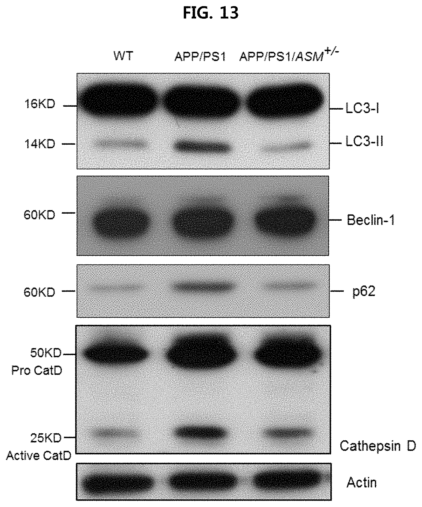
D00014
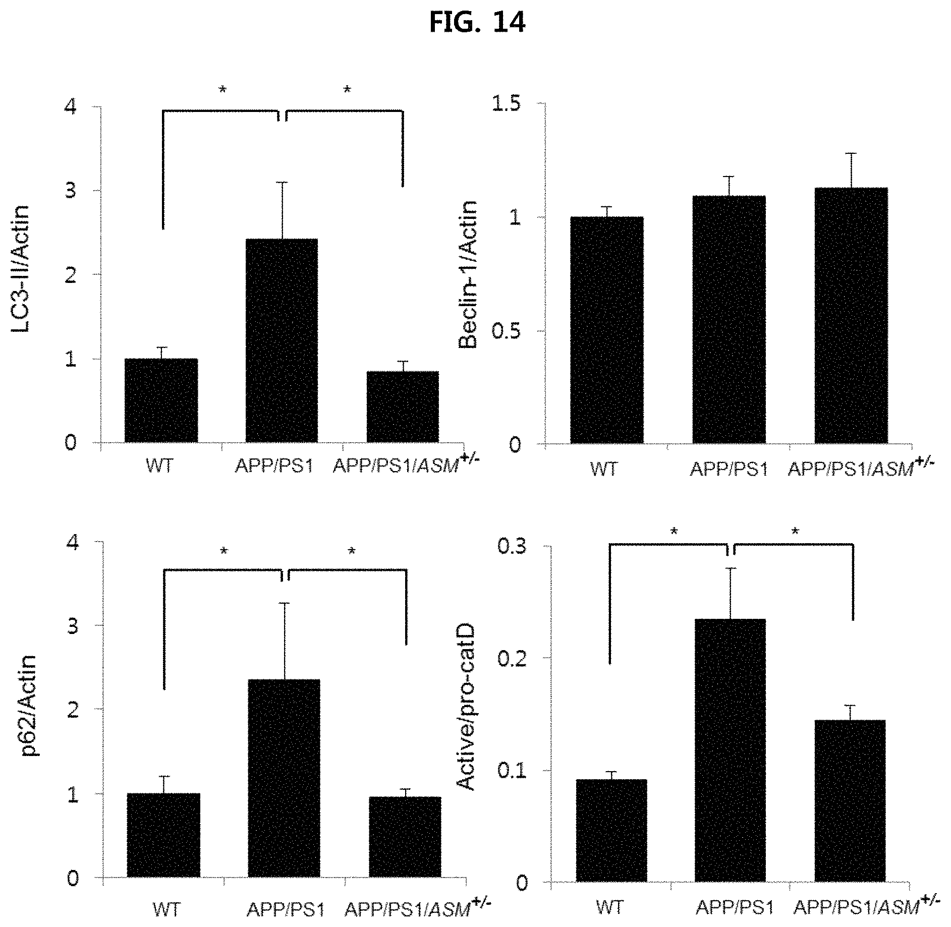
D00015
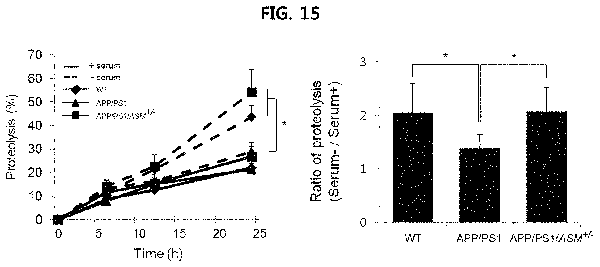
D00016
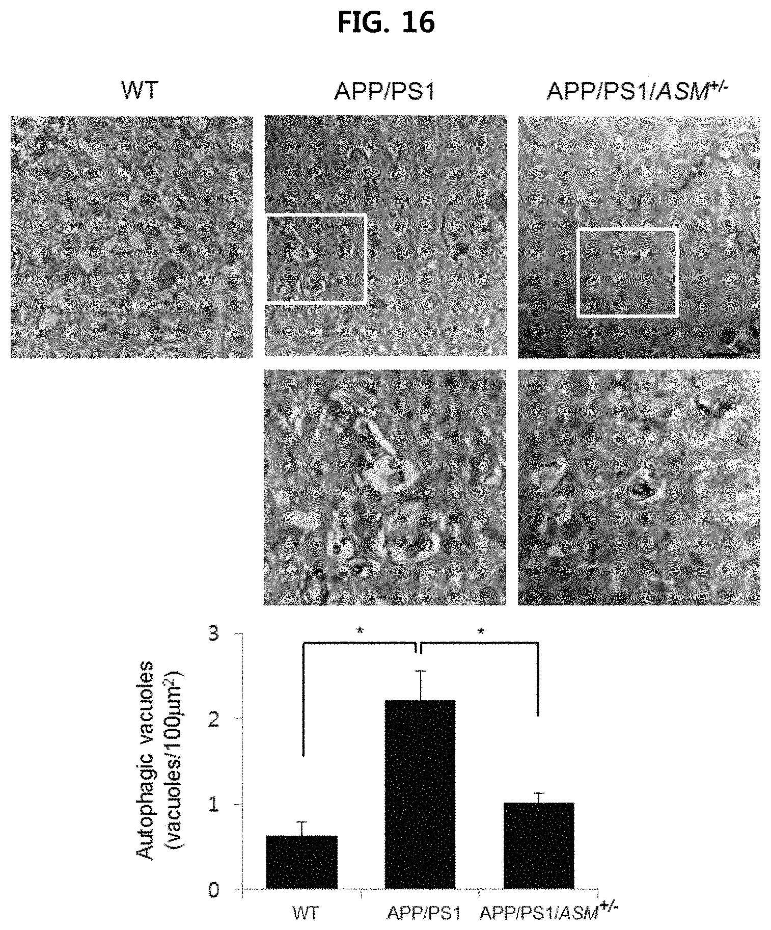
D00017
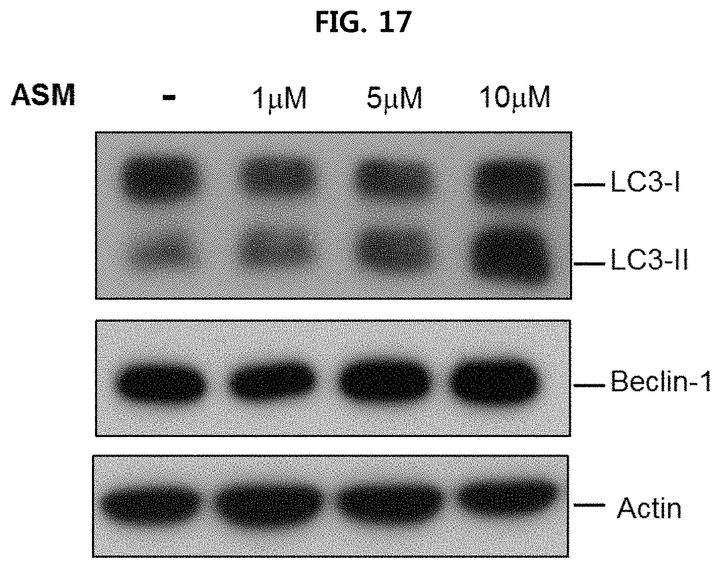
D00018
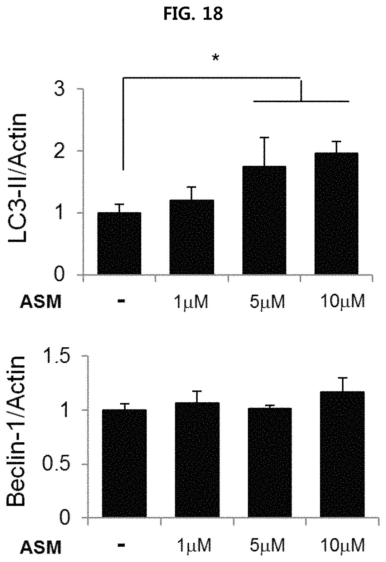
D00019
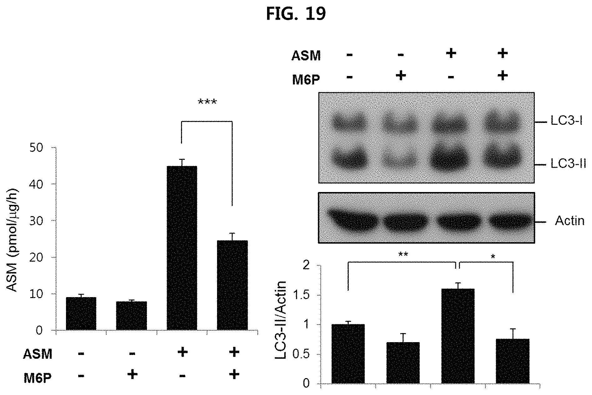
D00020
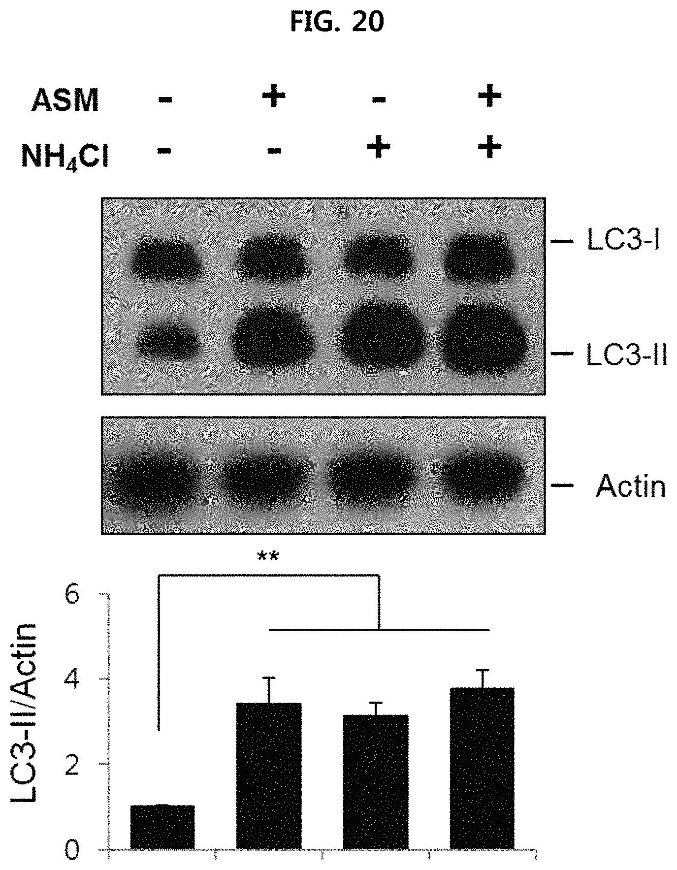
D00021
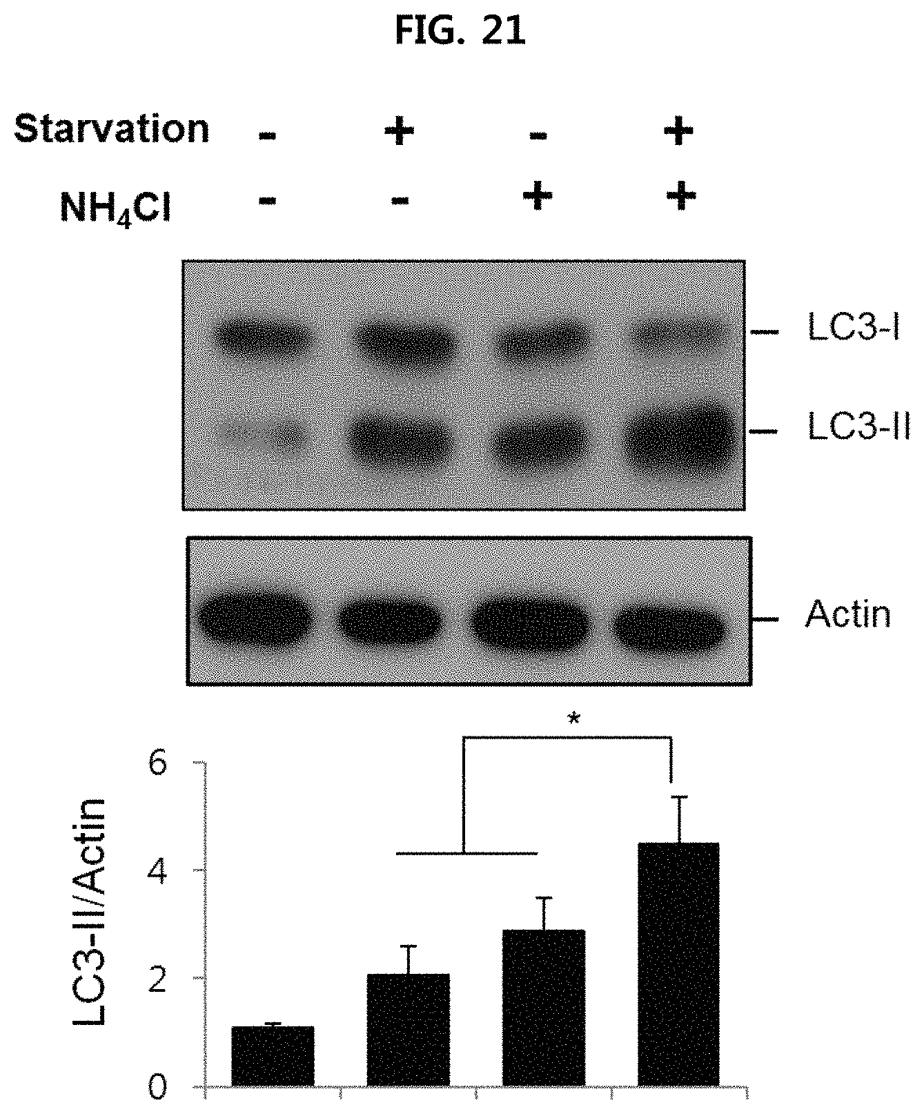
D00022
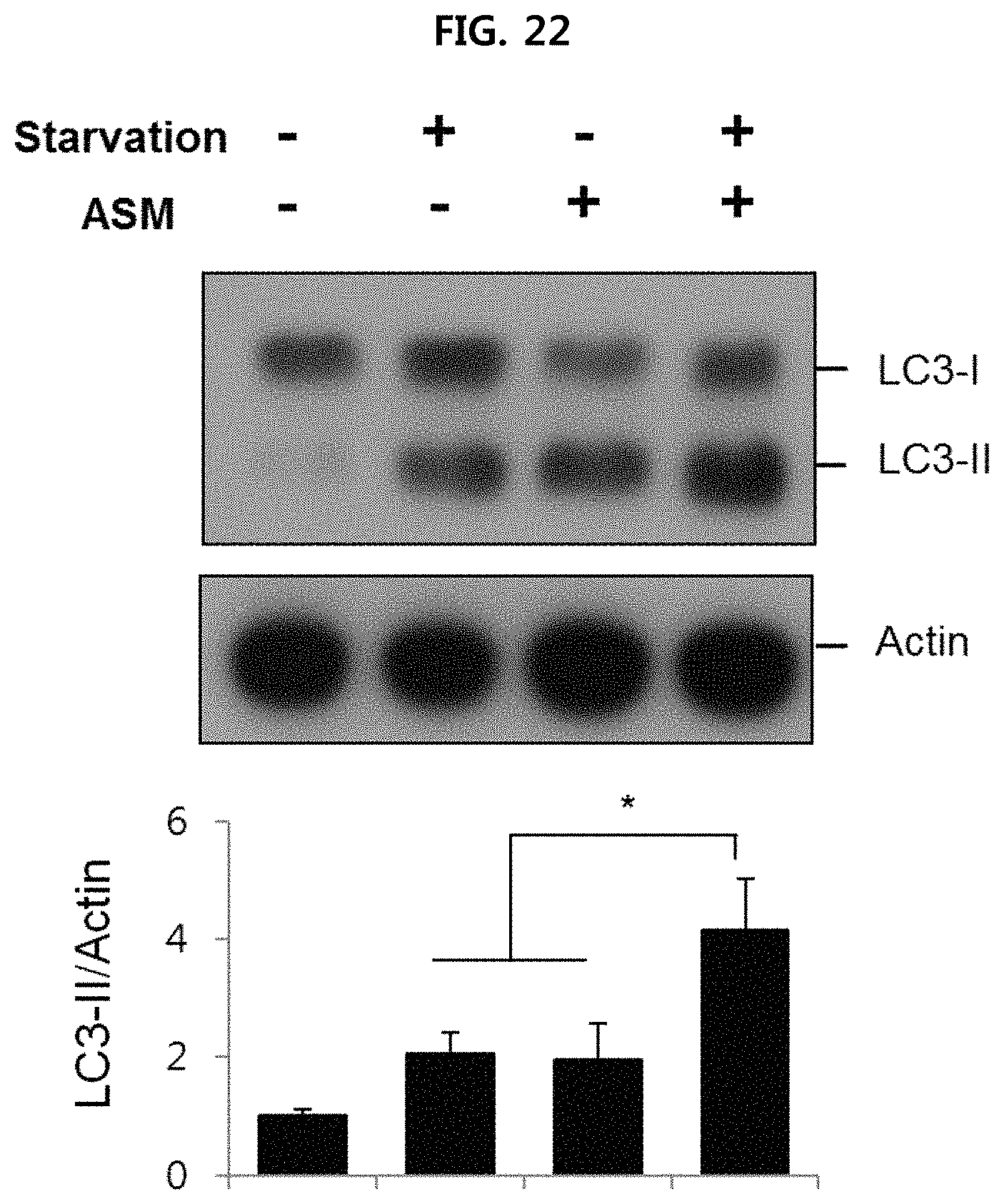
D00023
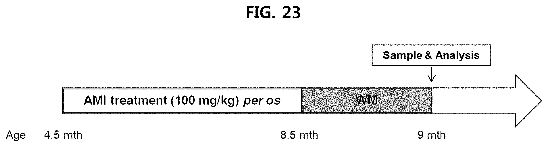
D00024
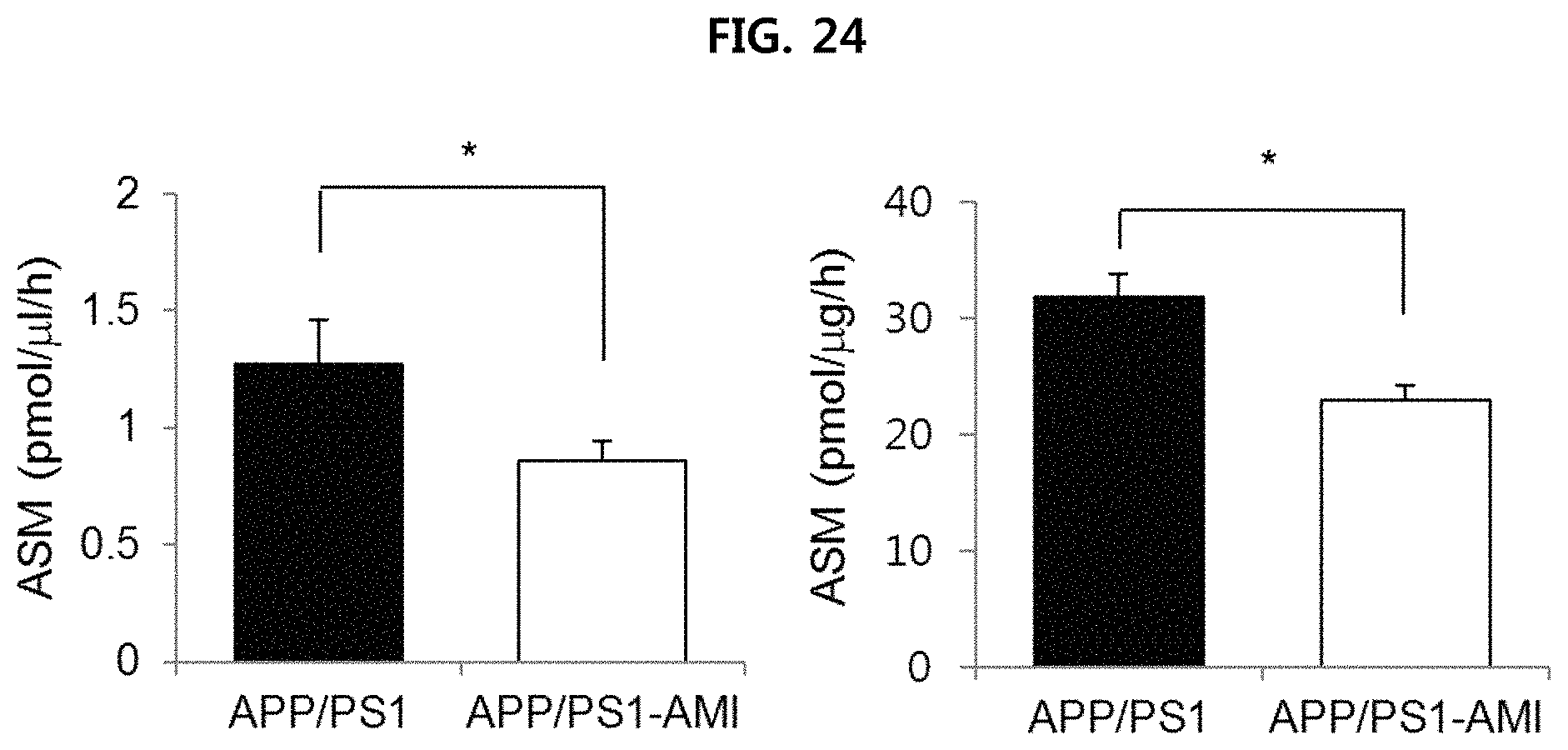
D00025
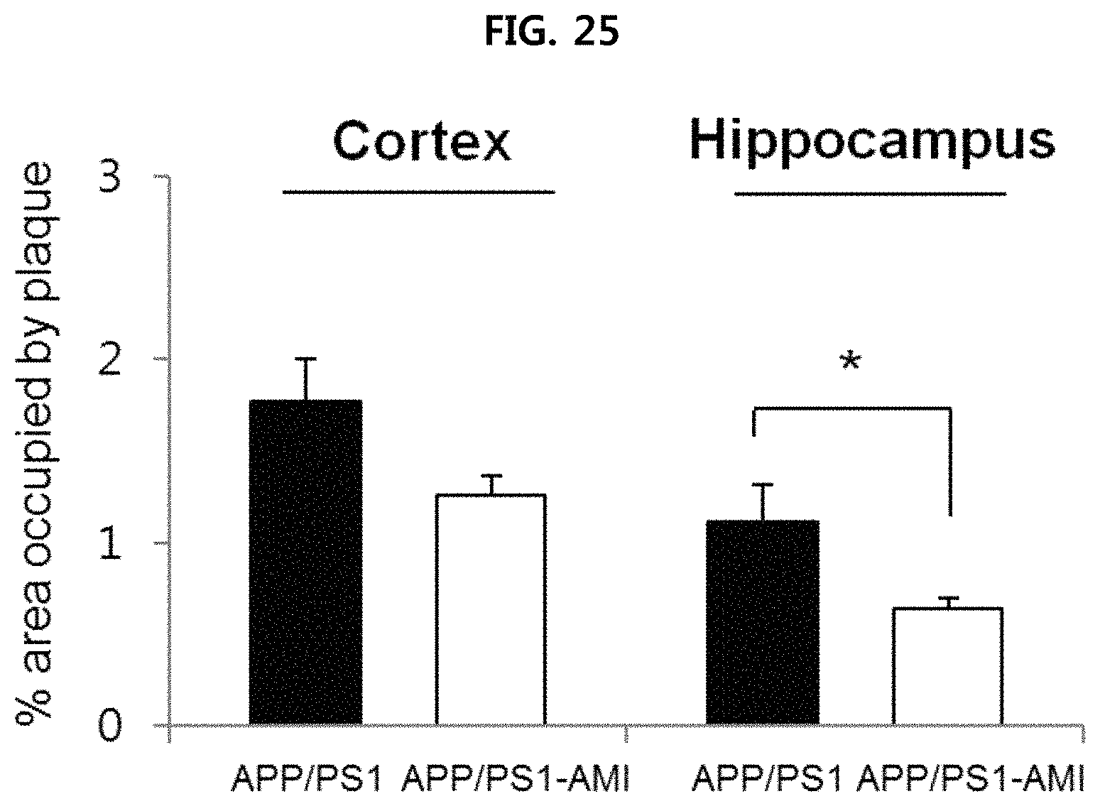
D00026
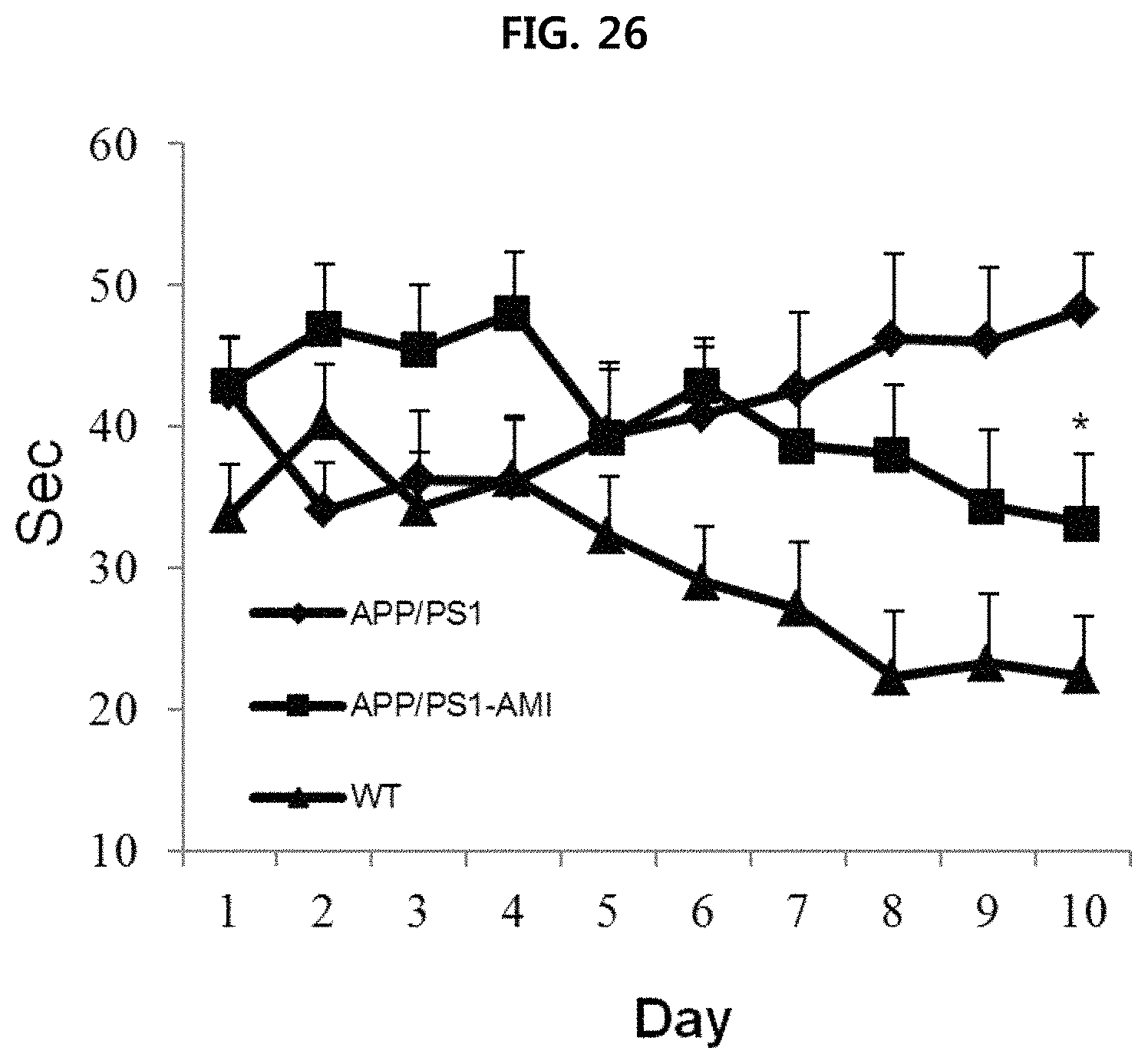
D00027
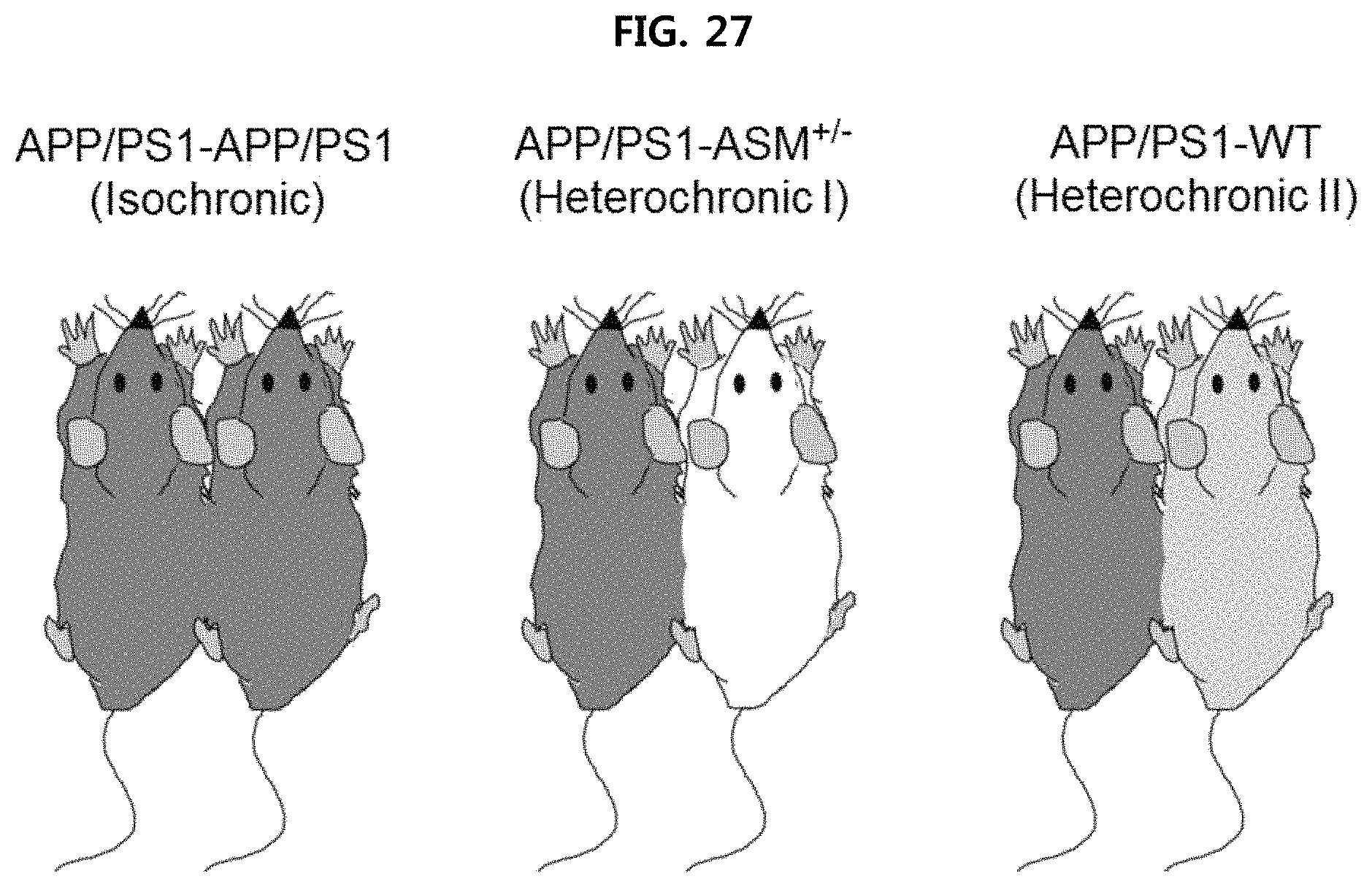
D00028
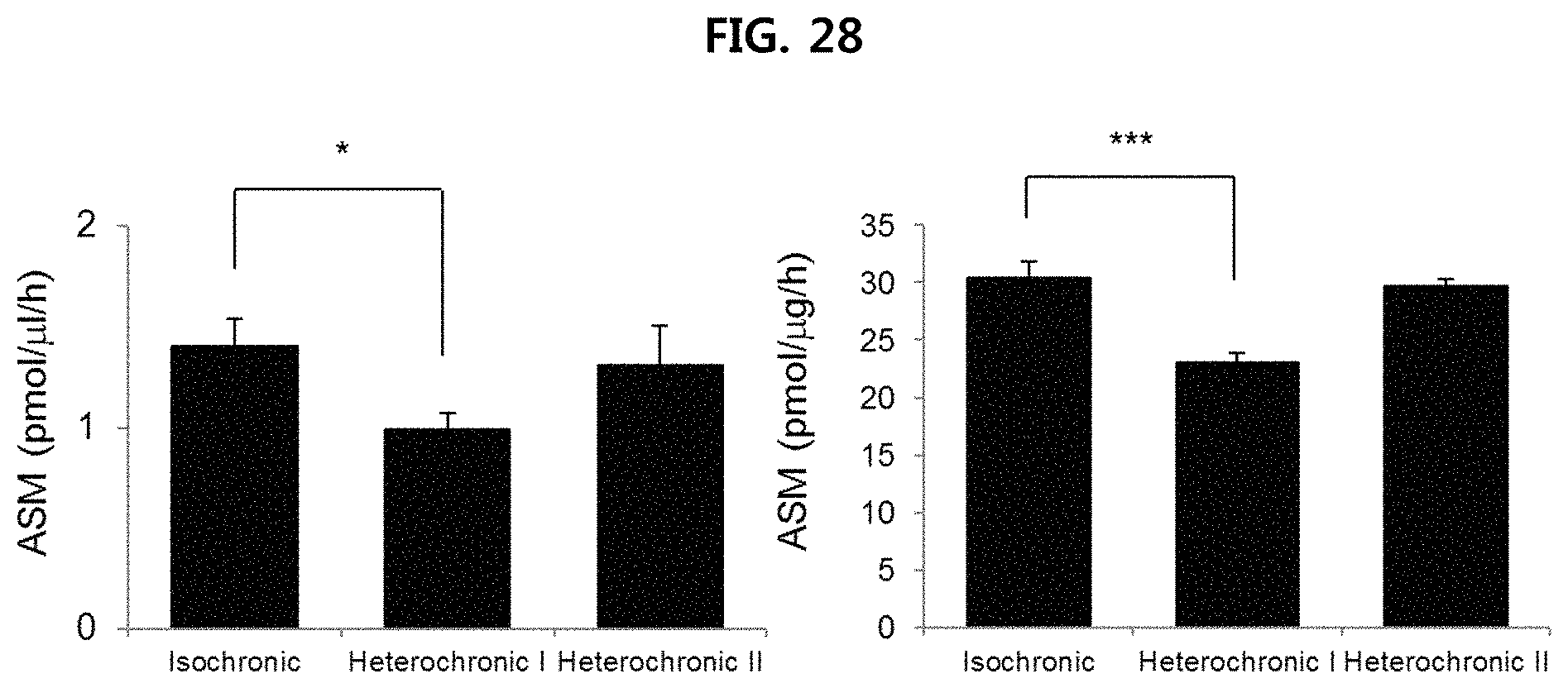
D00029

D00030
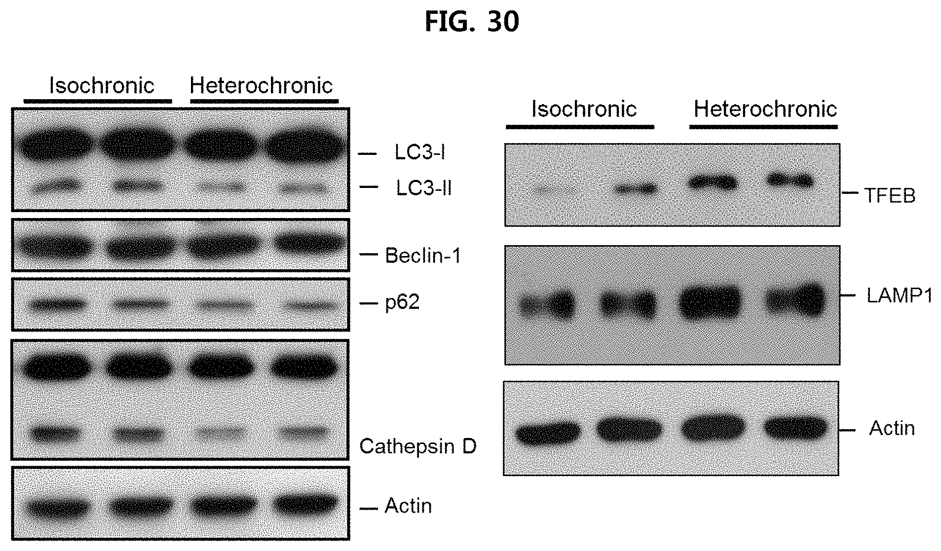
D00031
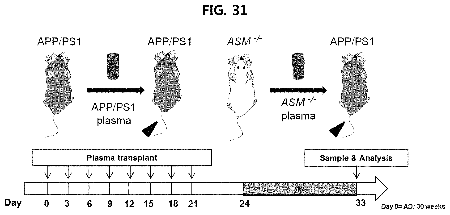
D00032
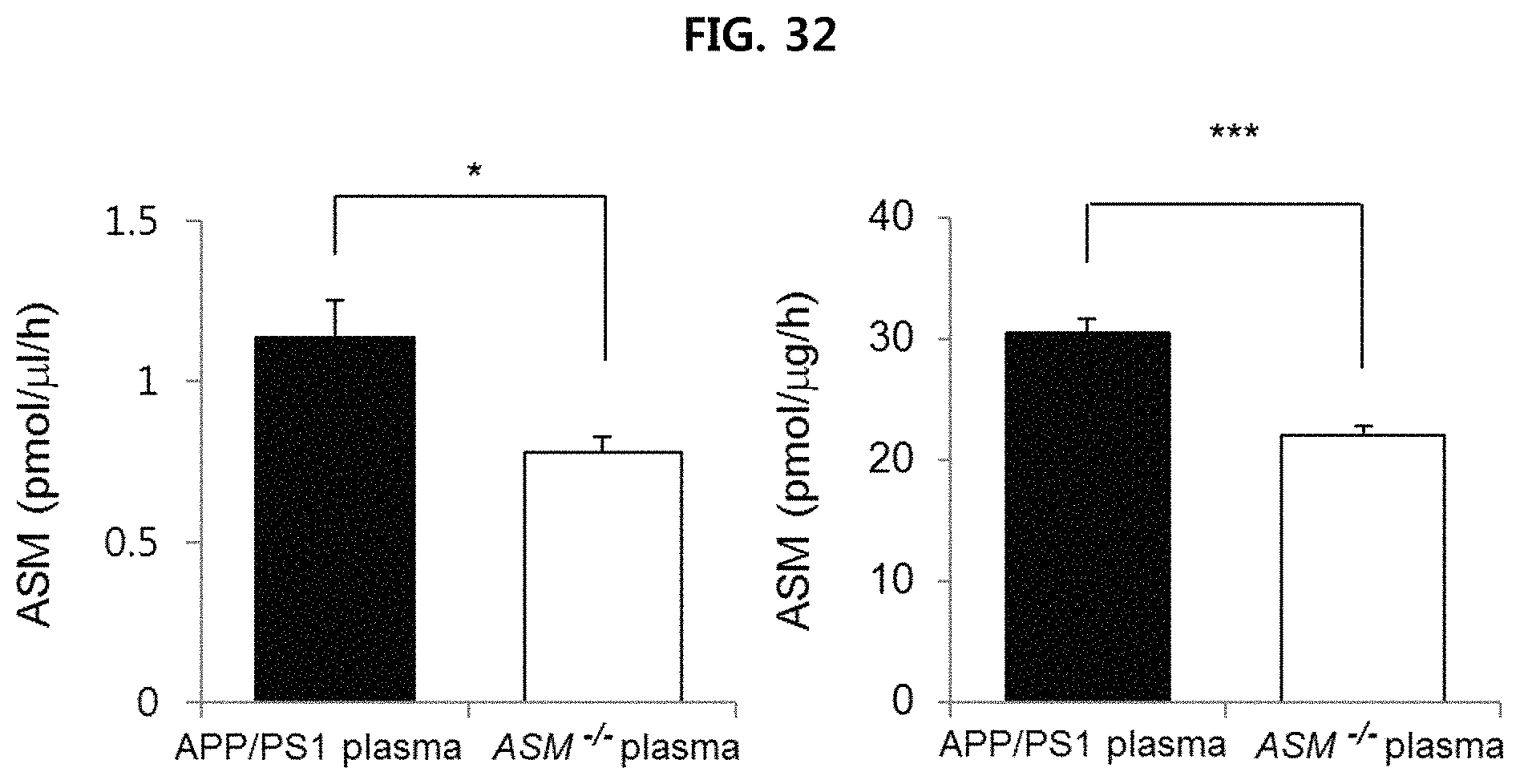
D00033
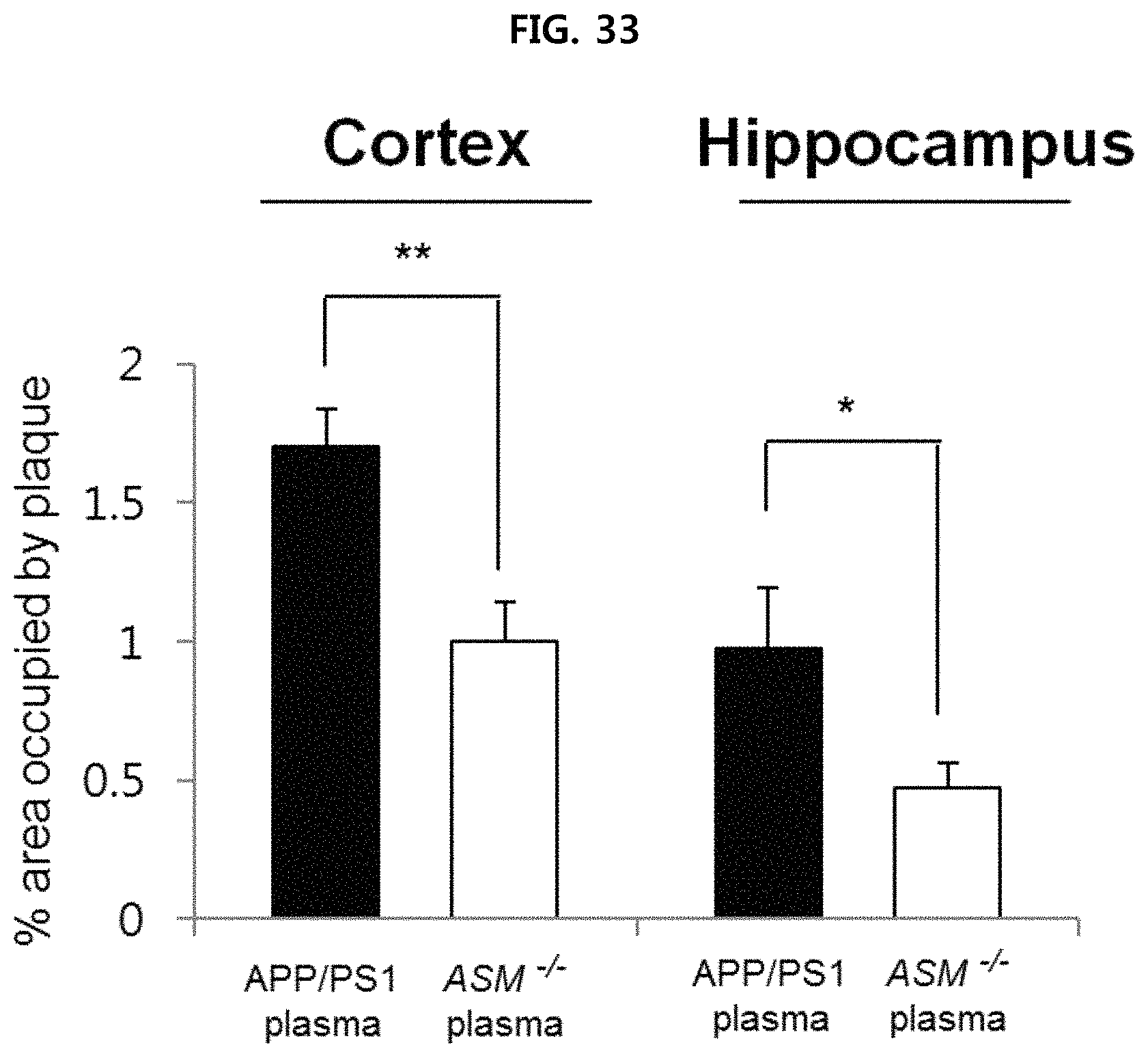
D00034
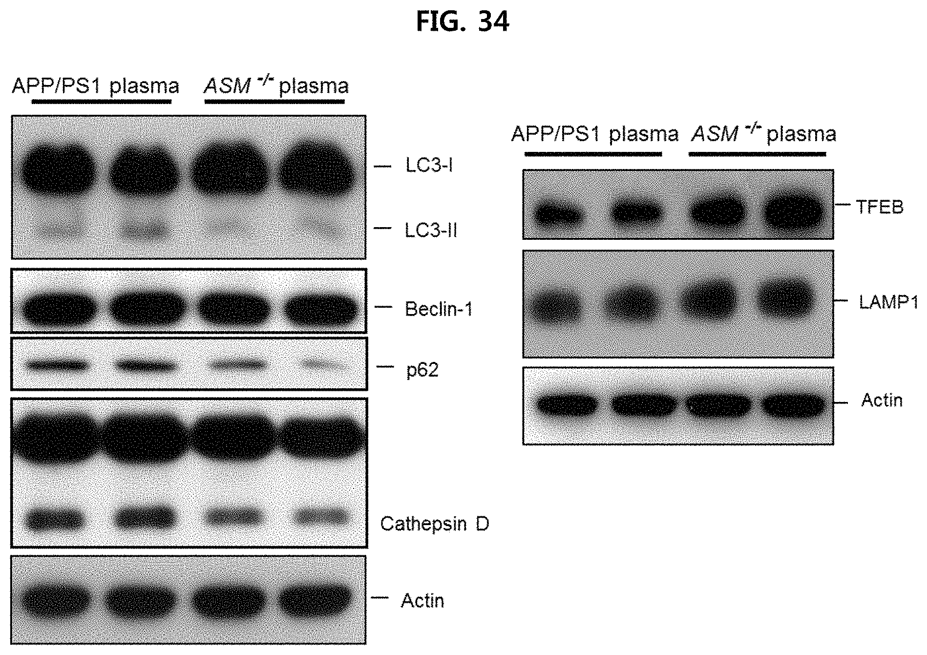
D00035
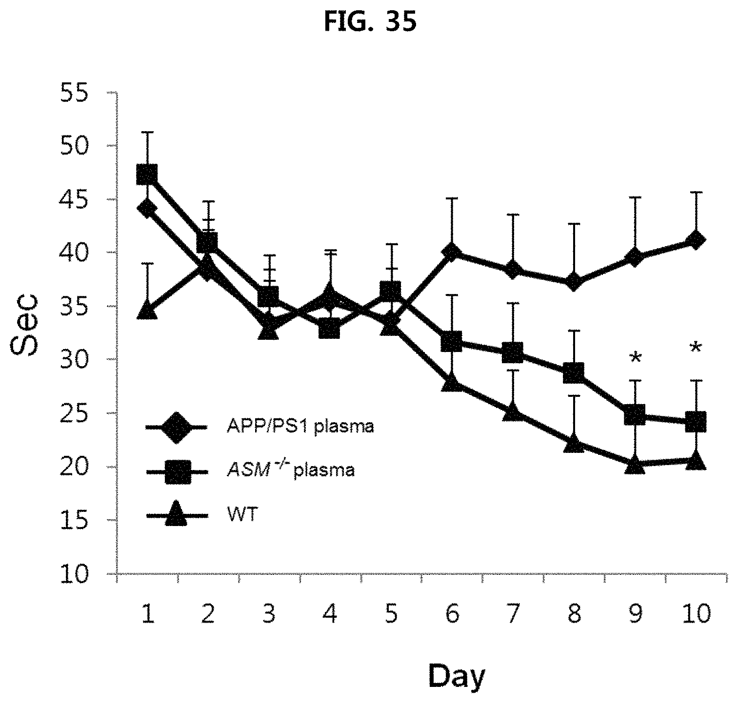
D00036

D00037
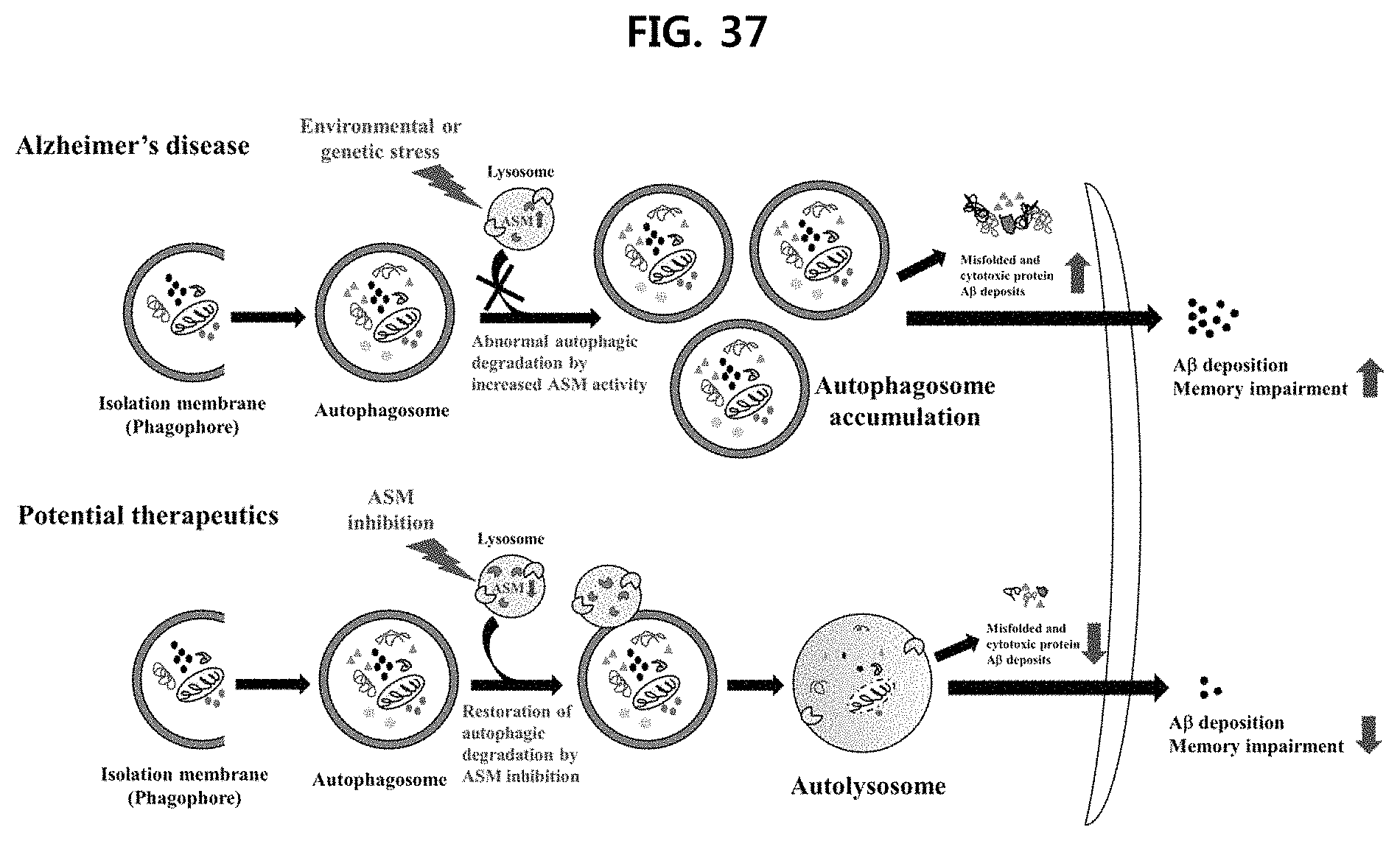
D00038
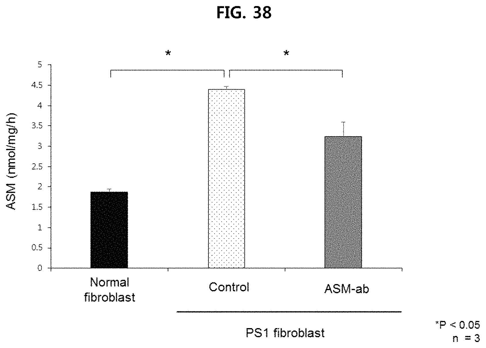
D00039
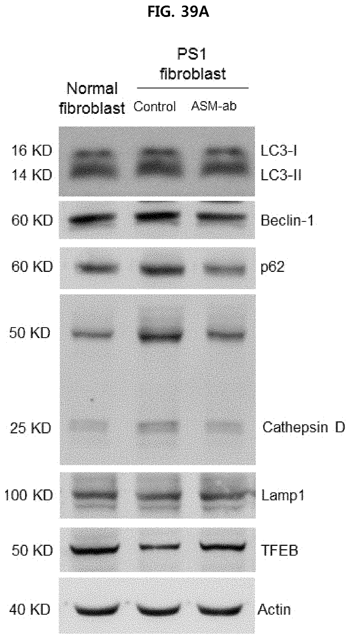
D00040
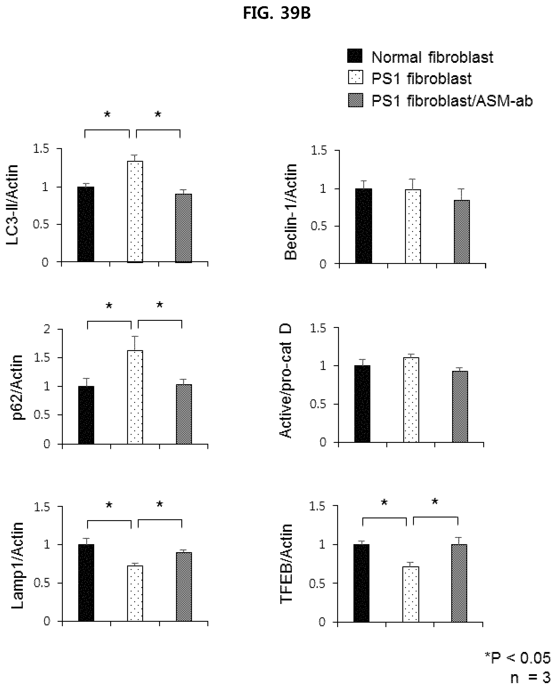
D00041
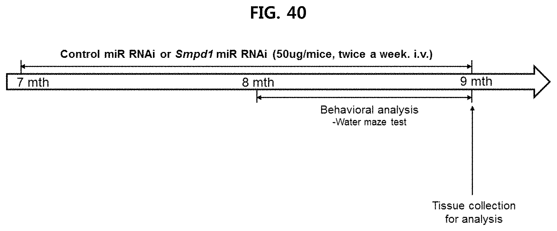
D00042
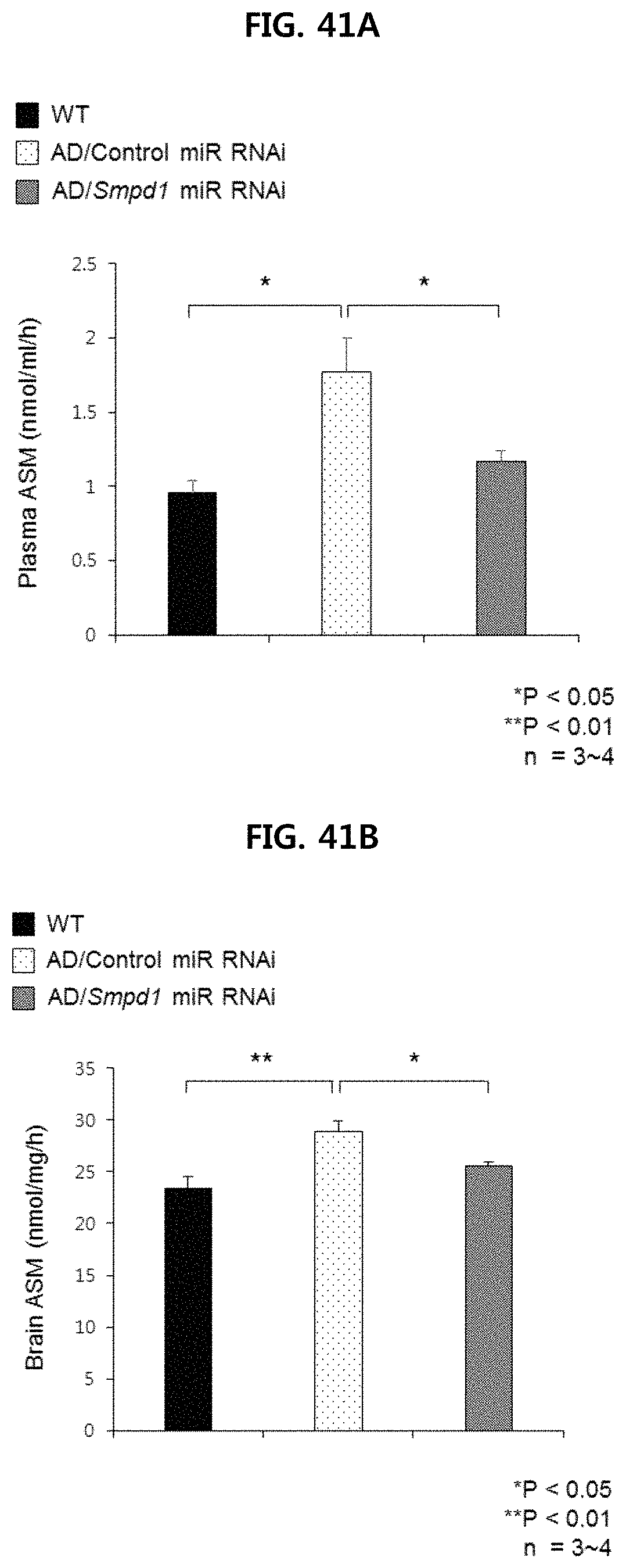
D00043
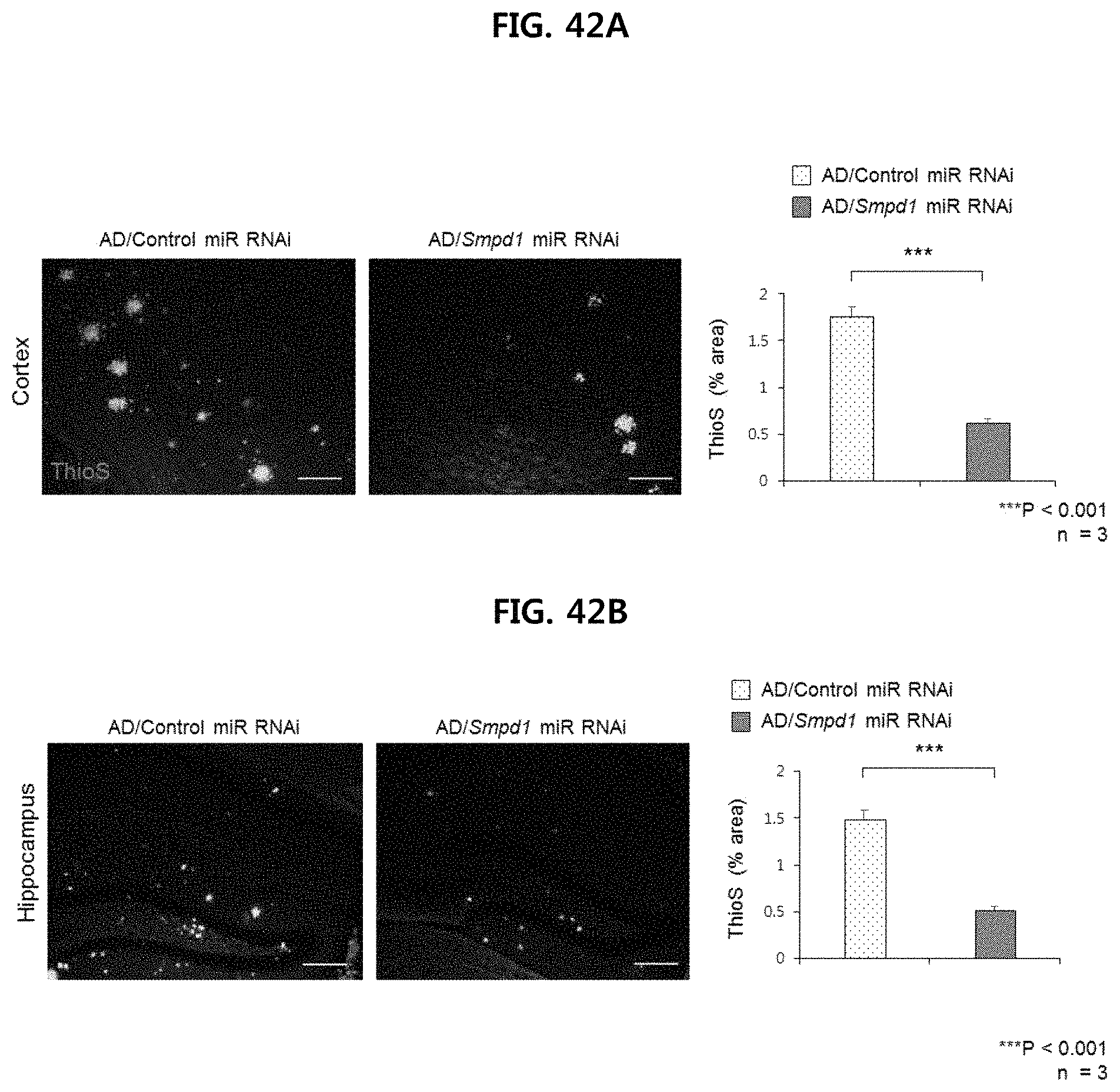
D00044
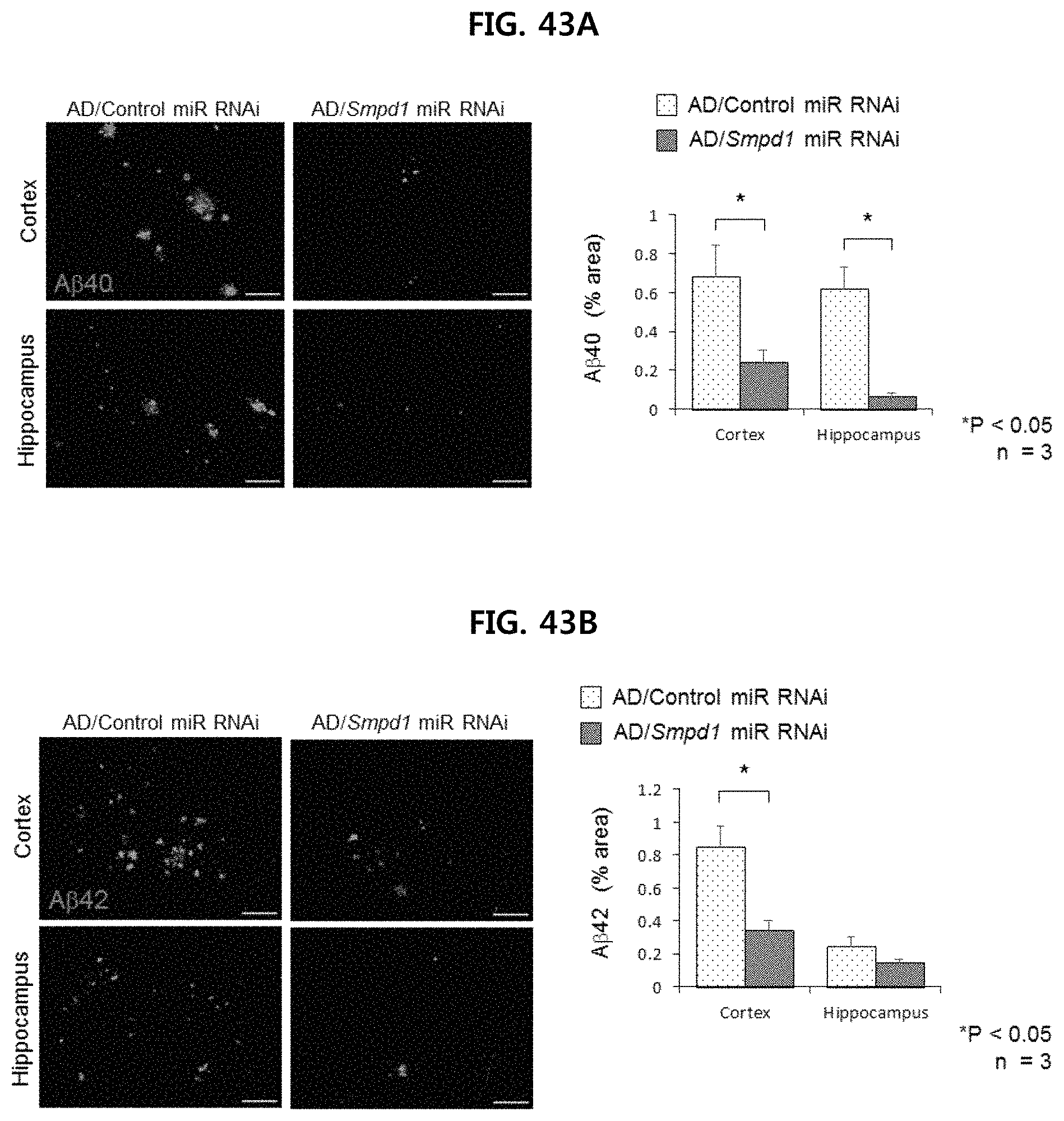
D00045

D00046
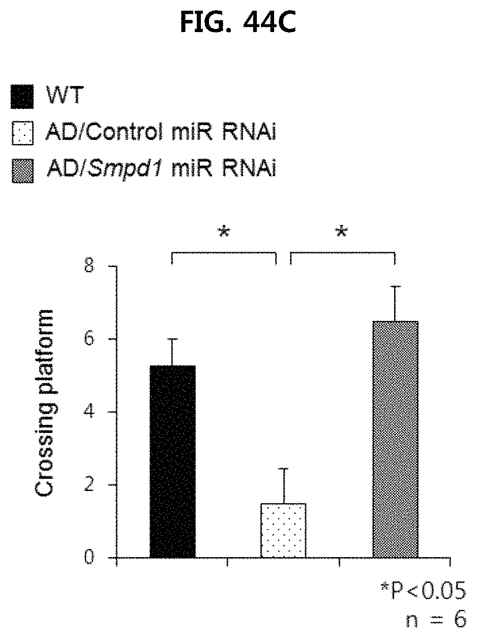
D00047
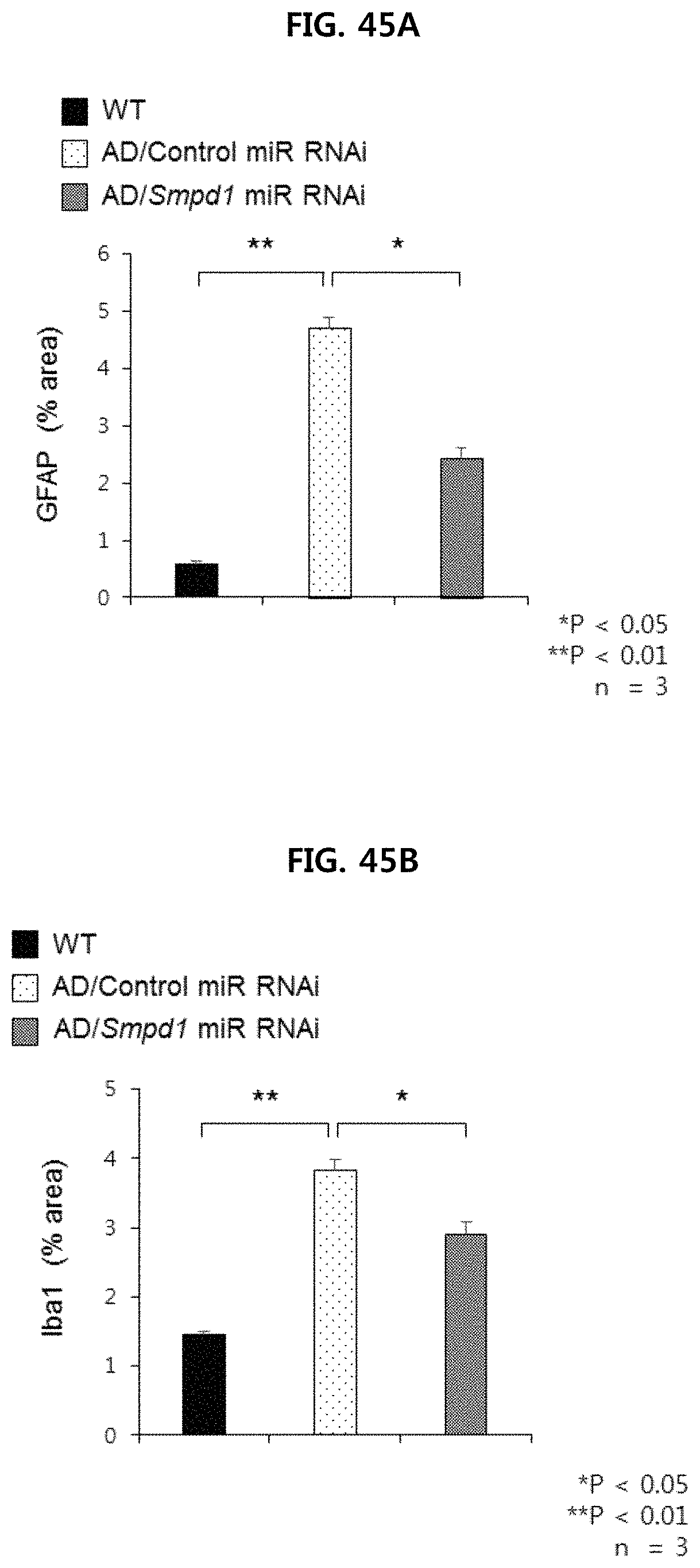
D00048
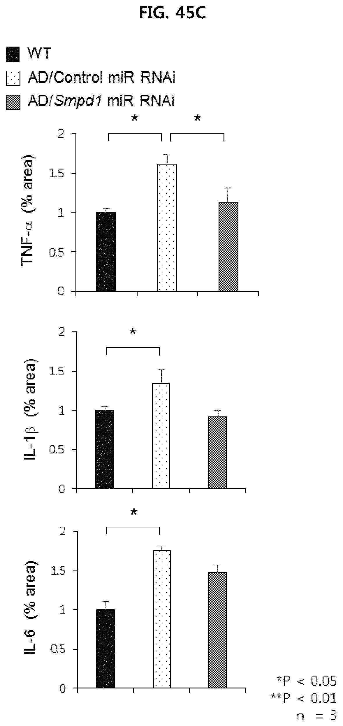
D00049
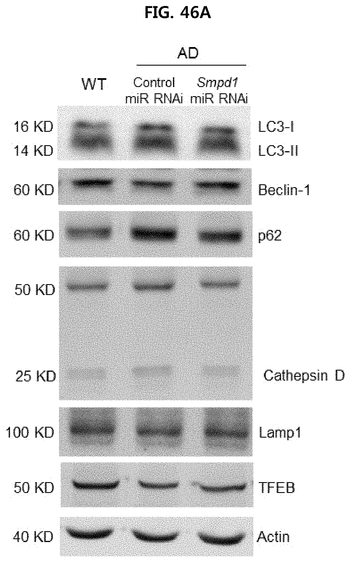
D00050
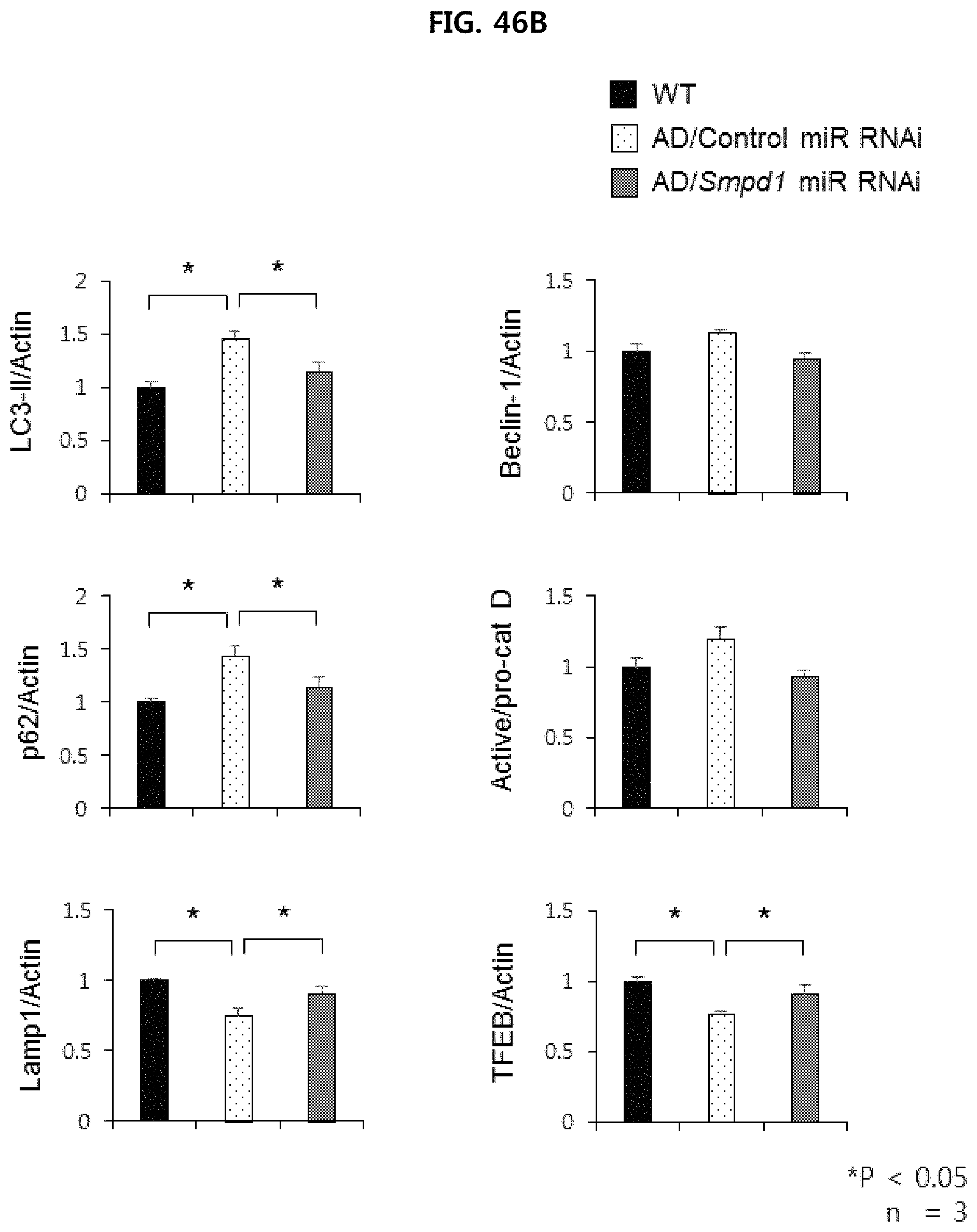
S00001
XML
uspto.report is an independent third-party trademark research tool that is not affiliated, endorsed, or sponsored by the United States Patent and Trademark Office (USPTO) or any other governmental organization. The information provided by uspto.report is based on publicly available data at the time of writing and is intended for informational purposes only.
While we strive to provide accurate and up-to-date information, we do not guarantee the accuracy, completeness, reliability, or suitability of the information displayed on this site. The use of this site is at your own risk. Any reliance you place on such information is therefore strictly at your own risk.
All official trademark data, including owner information, should be verified by visiting the official USPTO website at www.uspto.gov. This site is not intended to replace professional legal advice and should not be used as a substitute for consulting with a legal professional who is knowledgeable about trademark law.