Methods and devices of cardiac tissue monitoring and analysis
Rappaport , et al.
U.S. patent number 10,667,715 [Application Number 12/544,314] was granted by the patent office on 2020-06-02 for methods and devices of cardiac tissue monitoring and analysis. This patent grant is currently assigned to Sensible Medical Innovations Ltd.. The grantee listed for this patent is Shlomi Bergida, Ilan Kochba, Nadav Mizrahi, Dan Rappaport, Amir Saroka. Invention is credited to Shlomi Bergida, Ilan Kochba, Nadav Mizrahi, Dan Rappaport, Amir Saroka.


| United States Patent | 10,667,715 |
| Rappaport , et al. | June 2, 2020 |
Methods and devices of cardiac tissue monitoring and analysis
Abstract
A method for monitoring at least one cardiac tissue. The method comprises a) intercepting a plurality of reflections of an electromagnetic (EM) radiation reflected from at least one cardiac tissue of a patient in a plurality of EM radiation sessions, b) computing a mechanical tracing indicative of at least one mechanical property of said at least one cardiac tissue according to said plurality of reflections, c) analyzing said mechanical tracing so as to detect a presence or an absence of a physiological condition, and d) outputting said analysis.
| Inventors: | Rappaport; Dan (Tel-Aviv, IL), Saroka; Amir (Tel-Aviv, IL), Bergida; Shlomi (Tel-Aviv, IL), Kochba; Ilan (Modiln, IL), Mizrahi; Nadav (Tel-Aviv, IL) | ||||||||||
|---|---|---|---|---|---|---|---|---|---|---|---|
| Applicant: |
|
||||||||||
| Assignee: | Sensible Medical Innovations
Ltd. (Netanya, IL) |
||||||||||
| Family ID: | 41726427 | ||||||||||
| Appl. No.: | 12/544,314 | ||||||||||
| Filed: | August 20, 2009 |
Prior Publication Data
| Document Identifier | Publication Date | |
|---|---|---|
| US 20100056907 A1 | Mar 4, 2010 | |
Related U.S. Patent Documents
| Application Number | Filing Date | Patent Number | Issue Date | ||
|---|---|---|---|---|---|
| 61090356 | Aug 20, 2008 | ||||
| Current U.S. Class: | 1/1 |
| Current CPC Class: | A61B 5/05 (20130101); A61B 5/0507 (20130101); A61B 5/0472 (20130101); A61B 5/0408 (20130101) |
| Current International Class: | A61B 5/05 (20060101); A61B 5/0472 (20060101); A61B 5/0408 (20060101) |
| Field of Search: | ;600/382,513,479,515,523,481 |
References Cited [Referenced By]
U.S. Patent Documents
| 3534727 | October 1970 | Roman |
| 4016868 | April 1977 | Allison |
| 4240445 | December 1980 | Iskander et al. |
| 4279257 | July 1981 | Hochstcin |
| 4381510 | April 1983 | Wren |
| 4488559 | December 1984 | Iskander |
| 4572197 | February 1986 | Moore et al. |
| 4580572 | April 1986 | Granek et al. |
| 4647281 | March 1987 | Carr |
| 4676252 | June 1987 | Trautman et al. |
| 4690149 | September 1987 | Ko |
| 4877034 | October 1989 | Atkins et al. |
| 4920969 | May 1990 | Suzuki et al. |
| 4926868 | May 1990 | Larsen |
| 4958638 | September 1990 | Sharpe et al. |
| 4991585 | February 1991 | Mawhinney |
| 5002060 | March 1991 | Nedivi |
| 5078134 | January 1992 | Heilman et al. |
| 5132623 | July 1992 | Dc et al. |
| 5282840 | February 1994 | Hudrlik |
| 5334141 | August 1994 | Carr et al. |
| 5363050 | November 1994 | Guo et al. |
| 5394882 | March 1995 | Mawhinney |
| 5479120 | December 1995 | McEwan |
| 5517198 | May 1996 | McEwan |
| 5523760 | June 1996 | McEwan |
| 5563605 | October 1996 | McEwan |
| 5573012 | November 1996 | McEwan |
| 5576627 | November 1996 | McEwan |
| 5604531 | February 1997 | Iddan et al. |
| 5728143 | March 1998 | Gough et al. |
| 5738102 | April 1998 | Lemelson |
| 5749369 | May 1998 | Rabinovich et al. |
| 5766208 | June 1998 | McEwan |
| 5804921 | September 1998 | McEwan et al. |
| 5805110 | September 1998 | McEwan |
| 5807257 | September 1998 | Bridges |
| 5829437 | November 1998 | Bridges |
| 5833711 | November 1998 | Schneider, Sr. |
| 5861019 | January 1999 | Sun |
| 5876353 | March 1999 | Riff |
| 5883591 | March 1999 | McEwan |
| 5947910 | September 1999 | Zimmet |
| 5957861 | September 1999 | Combs et al. |
| 5964703 | October 1999 | Goodman et al. |
| 5995863 | November 1999 | Farace et al. |
| 6015386 | January 2000 | Kensey et al. |
| 6026173 | February 2000 | Svenson et al. |
| 6061589 | May 2000 | Bridges et al. |
| 6064903 | May 2000 | Riechers et al. |
| 6111415 | August 2000 | Moshe |
| 6169925 | January 2001 | Villaseca et al. |
| 6211663 | April 2001 | Moulthrop et al. |
| 6233479 | May 2001 | Haddad et al. |
| 6236889 | May 2001 | Soykan et al. |
| 6281843 | August 2001 | Evtioushkine et al. |
| 6330479 | December 2001 | Stauffer |
| 6332087 | December 2001 | Svenson et al. |
| 6332091 | December 2001 | Burns et al. |
| 6351246 | February 2002 | McCorkle |
| 6417797 | July 2002 | Cousins et al. |
| 6425878 | July 2002 | Shekalim |
| 6459931 | October 2002 | Hirschman |
| 6484047 | November 2002 | Vilsmeier |
| 6487428 | November 2002 | Culver et al. |
| 6488677 | December 2002 | Bowman et al. |
| 6494829 | December 2002 | New, Jr. et al. |
| 6496711 | December 2002 | Athan et al. |
| 6512949 | January 2003 | Combs et al. |
| 6551252 | April 2003 | Sackner et al. |
| 6574510 | June 2003 | Von Arx et al. |
| 6577709 | June 2003 | Tarr |
| 6590545 | July 2003 | McCorkle |
| 6675045 | January 2004 | Mass et al. |
| 6682480 | January 2004 | Habib et al. |
| 6687523 | February 2004 | Jayaramen et al. |
| 6746404 | June 2004 | Schwartz |
| 6766201 | July 2004 | Von Arx et al. |
| 6770070 | August 2004 | Balbierz |
| 6783499 | August 2004 | Schwartz |
| 6788262 | September 2004 | Adams et al. |
| 6802811 | October 2004 | Slepian |
| 6809701 | October 2004 | Amundson et al. |
| 6849046 | February 2005 | Eyal-Bickels et al. |
| 6909397 | June 2005 | Greneker, III et al. |
| 6917833 | July 2005 | Denker et al. |
| 6954673 | October 2005 | Von Arx et al. |
| 6972725 | December 2005 | Adams |
| 7006856 | February 2006 | Baker, Jr. et al. |
| 7024248 | April 2006 | Penner et al. |
| 7047058 | May 2006 | Dvorsky et al. |
| 7072718 | July 2006 | Von Arx et al. |
| 7077810 | July 2006 | Lange et al. |
| 7110823 | September 2006 | Whitehurst et al. |
| 7116276 | October 2006 | Lee |
| 7122012 | October 2006 | Bouton et al. |
| 7135871 | November 2006 | Pelletier |
| 7229415 | June 2007 | Schwartz |
| 7315170 | January 2008 | Sakayori |
| 7316658 | January 2008 | Gagne |
| 7330034 | February 2008 | Pelletier et al. |
| 7387610 | June 2008 | Stahmann et al. |
| 7445605 | November 2008 | Overall et al. |
| 7450077 | November 2008 | Waterhouse et al. |
| 7483752 | January 2009 | Von Arx et al. |
| 7561908 | July 2009 | Glukhovsky et al. |
| 7591792 | September 2009 | Bouton |
| 7613522 | November 2009 | Christman et al. |
| 7628757 | December 2009 | Koh |
| 7674244 | March 2010 | Kalafut et al. |
| 7686762 | March 2010 | Najafi et al. |
| 7725150 | May 2010 | Tupin, Jr. et al. |
| 7729776 | June 2010 | Von Arx et al. |
| 7736309 | June 2010 | Miller et al. |
| 7756587 | July 2010 | Penner et al. |
| 7825667 | November 2010 | Fang et al. |
| 7837629 | November 2010 | Bardy |
| 7844341 | November 2010 | Von Arx et al. |
| 7860574 | December 2010 | Von Arx et al. |
| 7872613 | January 2011 | Keilman et al. |
| 8032199 | October 2011 | Linti et al. |
| 8235949 | August 2012 | Hack et al. |
| 2003/0036674 | February 2003 | Bouton |
| 2003/0036713 | February 2003 | Bouton et al. |
| 2003/0128808 | July 2003 | Kindlein et al. |
| 2004/0006279 | January 2004 | Arad (Abboud) |
| 2004/0073093 | April 2004 | Hatlestad |
| 2004/0148021 | July 2004 | Cartledge et al. |
| 2004/0186395 | September 2004 | Vastano |
| 2004/0249257 | December 2004 | Tupin, Jr. et al. |
| 2004/0249258 | December 2004 | Tupin, Jr. et al. |
| 2004/0254457 | December 2004 | Van der Weide |
| 2005/0065567 | March 2005 | Lee et al. |
| 2005/0107719 | May 2005 | Arad (Abbound) |
| 2005/0124908 | June 2005 | Belalcazar et al. |
| 2005/0149139 | July 2005 | Plicchi et al. |
| 2005/0171396 | August 2005 | Pankratov et al. |
| 2005/0177061 | August 2005 | Alanen et al. |
| 2006/0058606 | March 2006 | Davis et al. |
| 2006/0235289 | October 2006 | Wesselink |
| 2006/0258952 | November 2006 | Stahmann et al. |
| 2006/0293609 | December 2006 | Stahmann et al. |
| 2007/0032749 | February 2007 | Overall et al. |
| 2007/0066904 | March 2007 | Wiesmann et al. |
| 2007/0088221 | April 2007 | Stalmiann |
| 2007/0123770 | May 2007 | Bouton et al. |
| 2007/0163584 | July 2007 | Bohm et al. |
| 2007/0197878 | August 2007 | Shklarski |
| 2007/0238914 | October 2007 | Royality et al. |
| 2008/0097530 | April 2008 | Muccio et al. |
| 2008/0103440 | May 2008 | Ferren et al. |
| 2008/0200802 | August 2008 | Bhavaraju et al. |
| 2008/0200803 | August 2008 | Kwon et al. |
| 2008/0224688 | September 2008 | Rubinsky et al. |
| 2008/0269589 | October 2008 | Thijs et al. |
| 2008/0283290 | November 2008 | Niino et al. |
| 2008/0288028 | November 2008 | Larson et al. |
| 2009/0043223 | February 2009 | Zhang et al. |
| 2009/0149918 | June 2009 | Krulevitch et al. |
| 2009/0227882 | September 2009 | Foo |
| 2009/0228001 | September 2009 | Pacey |
| 2009/0228075 | September 2009 | Dion |
| 2009/0241972 | October 2009 | Keilman et al. |
| 2009/0248129 | October 2009 | Keilman et al. |
| 2010/0056907 | March 2010 | Rappaport et al. |
| 2010/0256462 | October 2010 | Rappaport et al. |
| 2011/0025295 | February 2011 | Saroka et al. |
| 2011/0319746 | December 2011 | Kochba et al. |
| 2013/0281800 | October 2013 | Saroka et al. |
| 2017/0156626 | June 2017 | Kochba et al. |
| 2898342 | Jul 2014 | CA | |||
| 0694282 | Jan 1996 | EP | |||
| 1600892 | Nov 2005 | EP | |||
| 2004-528864 | Sep 2004 | JP | |||
| 2005-334298 | Dec 2005 | JP | |||
| 2007-509353 | Apr 2007 | JP | |||
| WO 99/39728 | Aug 1999 | WO | |||
| WO 00/71207 | Nov 2000 | WO | |||
| WO 02/053228 | Jul 2002 | WO | |||
| WO 03/009753 | Feb 2003 | WO | |||
| WO 2005/043100 | May 2005 | WO | |||
| WO 2005/074361 | Aug 2005 | WO | |||
| WO 2005/094369 | Oct 2005 | WO | |||
| WO 2007/010460 | Jan 2007 | WO | |||
| WO 2007/055491 | May 2007 | WO | |||
| WO 2008/002251 | Jan 2008 | WO | |||
| WO 2008/122056 | Oct 2008 | WO | |||
| WO 2009/031149 | Mar 2009 | WO | |||
| WO 2009/031150 | Mar 2009 | WO | |||
| WO 2011/141915 | Nov 2011 | WO | |||
Other References
|
International Preliminary Report on Patentability dated Mar. 18, 2010 From the International Bureau of WIPO RE.: Application No. PCT/IL2008/001198. cited by applicant . International Prelminary Report on Patentability dated Mar. 18, 2010 From teh International Bureau of WIPO Re.: Application No. PCT/IL2008/001199. cited by applicant . Hill-Rom "The Vest.RTM. Airway Clearance System. Information for Physicians", Hill-Rom, Retrieved From the Internet, 3 P., Nov. 24, 2011. cited by applicant . Jafari et al. "Ultrawideband Radar Tmagingn System for Biomedical Applications", Journal of Vacuum Science and Technology A: Vacuum, Surfaces, and Films, 24(3): 752-757, May/Jun. 2006. cited by applicant . Li et al. "An Overview of Ultra-Wideband Microwave Imaging via Space-Time Beamforming for Early-Stage Breast-Cancer Detection", IEEE Antennas and Propagation Magazine, 47(1): 19-34, Feb. 2005. cited by applicant . Meaney et al. "Near-Field Microwave Imaging of Biologically-Based Materials Using a Monopole Transceiver System", IEEE Transactions on Microwave Theory and Techniques, 46(1): 31-45, Jan. 1998. cited by applicant . Shea et al. "Contrast-Enhanced Microwave Imaging of Breast Tumors: A Computational Study Using 3D Realistic Numerical Phantoms", Inverse Problems, 26: 1-22, 2010. cited by applicant . Zhou et al. "On the Resolution of UWB Microwave Imaging of Tumors in Random Breast Tissue", IEEE International Symposium of the Antennas and Propagation Society, Jul. 3-8, 2005, 3A: 831-834, Jul. 2005. cited by applicant . International Search Report and the Written Opinion dated Jun. 15, 2010 From the International Searching Authority Re.: Application No. PCT/IL2010/000182. cited by applicant . Semenov et al. "Three-Dimensional Microwave Tomography: Initial Experimental Imaging of Animals", IEEE Transactions on Biomedical Engineering, XP011007196, 49(1):55-63, Jan. 2002. Abstract, p. 56, col. 1, Lines 6, 7. cited by applicant . Official Action dated Apr. 4, 2012 From the US Patent and Trademark Office Re. U.S. Appl. No. 12/676,385. cited by applicant . International Search Report dated Feb. 4, 2009 From the International Searching Authority Re.: Application No. PCT/IL2008/001198. cited by applicant . International Search Report dated Jan. 23, 2009 From the International Searching Authority Re.: Application No. PCT/IL2008/001199. cited by applicant . Written Opinion dated Feb. 4, 2009 From the International Searching Authority Re.: Application No. PCT/IL2008/001198. cited by applicant . Written Opinion dated Jan. 23, 2009 From the International Searching Authority Re.: Application No. PCT/IL2008/001199. cited by applicant . Azevedo et al "Micropower Impulse Radar", Science & Technologies Review, Feb. 17-29, 1996. cited by applicant . Billich "Bio-Medical Sensing Using Ultra Wideband Communications and Radar Technology", PhD Proposal, Department of Information and Telecommunication Technology--University of Trento, Italy--Jan. 2006 (10 pages). cited by applicant . Gentili et al "A Versatile Microwave Plethysmograph for the Monitoring of Physiological Parameters", IEEE Transactions on Biomedical Engineering 49(10) 1204-1210, Oct. 2002. cited by applicant . Juweid et al. "Positron-Emission Tomography and Assessment of Cancer Therapy", The New England Journal of Medicine, 354(5): 496-507, Feb. 2, 2006. cited by applicant . Kagawa et al. "Advanced Exercise Control Using Miniature ECG and 3D Acceleration Sensors", D&D Forum on Telemedicine Systems: Issues, design, Development and Standardization at Globecom 2008, New Orleans, Louisiana, USA, 23 P., Dec. 2, 2008. cited by applicant . Katzeff et al. "Exercise Stress Testing and an Electromechanical S Wace of the Electrocardiogram", South African Medical Journal, 49(27): 1088-1090, Jun. 28, 1975. cited by applicant . Kerckhoffs et al. "Homogeneity of Cardiac Contraction Despite Physiological Asynchrony of Depolarization: A Model Study", Annals of Biomedical Engineering, 31: 536-547, 2003. cited by applicant . Lee et al. "Noninvasive Tests in Patients With Stable Coronary Artery Disease", The New England Journal of Medicine, 344(24): 1840-1845, Jun. 14, 2001. cited by applicant . Park et al. "An Ultra-Wearable, Wireless, Low Power ECG Monitoring System", Proceedings of the IEEE Biomedical Circuits and Systems Conference, BioCAS 2006, London, UK, p. 241-244, Nov. 29-Dec. 1, 2006. cited by applicant . Pedersen et al "An Investigation of the Use of Microwave Radiation for Pulmonary Diagnostics", Communications--IEEE Transcations on Biomedical Engineering, p. 410-412, Sep. 1976. cited by applicant . Schiller "Noninvasive Monitoring of Tumors", The New England Journal of Medicine, 359(4): 418-420, Jul. 24, 2008. cited by applicant . Smiseth et al. "Regional Left Ventricular Electric and Mechanical Activation and Relaxation", JACC, Journal of the American College of Cardiology, 47(1): 173-174, Jan. 3, 2006. cited by applicant . Thornton "Optimization of Protocols for Computed Tomography Coronary Angiography", Supplement to Applied Radiology, p. 54-62, Jun. 2002. cited by applicant . Yamokoski et al "OptiVol.RTM. Fluid Status Monitoring With an Implantable Cardiac Device: A Heart Failure Managaement System.", 4:(6) 775-780 (doi:10.1586/17434440.4.6.775), Nov. 2007. cited by applicant . Zito et al "Wearable System-On-A-Chip Pulse Radar Sensors for the Health Care: System Overview", 21st Conference on Advanced Information Networking and Applications Workshop (AINAW'07), University of Pisa, Italy--2007, IEEE. cited by applicant . Zlochiver et al "A Portable Bio-Impedance System for Monitoring Lung Resistivity", Medical Engineering & Physics, 29:(1), 93-100. cited by applicant . International Preliminary Report on Patentability dated Sep. 15, 2011 From the International Bureau of WIPO Re. Application No. PCT/IL2010/000182. cited by applicant . Official Action dated Sep. 13, 2012 From the US Patent and Trademark Office Re. U.S. Appl. No. 12/676,381. cited by applicant . Official Action dated Oct. 15, 2012 From the US Patent and Trademark Office Re. U.S. Appl. No. 12/846,861. cited by applicant . Advisory Action Before the Filing of an Appeal Brief dated Jun. 6, 2013 From the US Patent and Trademark Office Re. U.S. Appl. No. 12/676,385. cited by applicant . Applicant-Initiated Interview Summary dated Jun. 13, 2013 From the US Patent and Trademark Office Re. U.S. Appl. No. 12/676,385. cited by applicant . Office Action dated Jun. 5, 2013 From the Israel Patent Office Re. U.S. Appl. No. 214973 and Its Translation Into English. cited by applicant . Official Action dated Jul. 5, 2013 From the US Patent and Trademark Office Re. U.S. Appl. No. 12/846,861. cited by applicant . Applicant-Initiated Interview Summary dated Jul. 2, 2013 From the US Patent and Trademark Office Re. U.S. Appl. No. 12/676,381. cited by applicant . Communication Pursuant to Rules 70(2) and 70a(2) EPC dated Mar. 5, 2013 From the European Patent Office Re. Application No. 08808013.0. cited by applicant . Official Action dated Mar. 25, 2013 From the US Patent and Trademark Office Re. U.S. Appl. No. 13/254,852. cited by applicant . Supplementary European Search Report and the European Search Opinion dated Feb. 13, 2013 From the European Patent Office Re. Application No. 08789867.2. cited by applicant . Jiang et al. "Ultrasound-Guided Microwave Imaging of Breast Cancer: Tissue Phantom and Pilot Clinical Experiments", Medical Physics, 32(8): 2528-2535, Aug. 2005. cited by applicant . Kramer et al. "Dielectric Measurement of Cerebral Water Content Using a Network Analyzer", Neurological Research, 14: 255-258, Jun. 1992. cited by applicant . Nopp et al. "Dielectric Properties of Lung Tissue as a Function of Air Content", Physics in Medicine and Biology, 38(6): 699-716, Jun. 1993. cited by applicant . Official Action dated Dec. 20, 2012 From the US Patent and Trademark Office Re. U.S. Appl. No. 12/676,385. cited by applicant . Official Action dated Feb. 8, 2013 From the US Patent and Trademark Office Re. U.S. Appl. No. 12/676,381. cited by applicant . Supplementary European Search Report and the European Search Opinion dated Feb. 14, 2013 From the European Patent Office Re. Application No. 08808013.0. cited by applicant . Communciation Pursuant to Article 94(3) EPC dated Sep. 27, 2013 From the European Patent Office Re. Application No. 08808013.0. cited by applicant . Communication Pursuant to Article 94(3) EPC dated Oct. 29, 2013 From the European Patent Office Re. Application No. 08789867.2. cited by applicant . Official Action dated Dec. 12, 2013 From the US Patent and Trademark Office Re. U.S. Appl. No. 13/254,852. cited by applicant . Official Action dated Oct. 22, 2013 From the US Patent and Trademark Office Re. U.S. Appl. No. 12/676,381. cited by applicant . Translation of Notice of Reason for Rejection dated Sep. 24, 2013 From the Japanese Patent Office Re. Application No. 2010-523644. cited by applicant . Meaney et al. "Microwave Imaging for Neoadjuvant Chemotherapy Monitoring", First European Conference on Antennas and Propagation, EuCAP 2006, Nice, France, Nov. 6-10, 2006, p. 1-4, Nov. 2006. cited by applicant . Panetta "A Mathematical Model of Periodically Pulsed Chemotherapy: Tumor Recurrence and Metastasis in a Competitive Environment", Bulletin of Methematical Biology, 58(3): 425-447, 1996. cited by applicant . Advisory Action Before the Filing of an Appeal Brief dated Apr. 22, 2014 From the US Patent and Trademark Office Re. U.S. Appl. No. 13/254,852. cited by applicant . Communication Pursuant to Article 94(3) EPC dated Mar. 19, 2014 From the European Patent Office Re. Application No. 08789867.2. cited by applicant . Official Action dated Jul. 1, 2014 From the US Patent and Trademark Office Re. U.S. Appl. No. 12/676,381. cited by applicant . Communication Pursuant to Article 94(3) EPC dated Aug. 4, 2014 From the European Patent Office Re. Application No. 08808013.0. cited by applicant . Official Action dated Sep. 4, 2014 From the US Patent and Trademark Office Re. U.S. Appl. No. 13/254,852. cited by applicant . Applicant-Initiated Interview Summary dated May 16, 2014 From the US Patent and Trademark Office Re. U.S. Appl. No. 13/254,852. cited by applicant . Communication Pursuant to Article 94(3) EPC dated May 22, 2014 From the European Patent Office Re. Application No. 10712583.3. cited by applicant . Office Action dated Apr. 28, 2014 From the Israel Patent Office Re. Application No. 214973 and Its Translation into English. cited by applicant . Official Action dated Jun. 10, 2015 From the US Patent and Trademark Office Re. U.S. Appl. No. 13/254,852. cited by applicant . Fear et al. "Microwaves for Breast Cancer Detection", IEEE Potentials, 22(1): 12-18, Feb. 25, 2003. cited by applicant . Applicant-Initiated Interview Summary dated Feb. 20, 2015 From the US Patent and Trademark Office Re. U.S. Appl. No. 13/254,852. cited by applicant . Communication Pursuant to Article 94(3) EPC dated Apr. 16, 2015 From the 10712583.3. European Patent Office Re. Application No. 10712583.3. cited by applicant . Communication Pursuant to Article 94(3) EPC dated Apr. 29, 2015 From the European Patent Office Re. Application No. 08789867.2. cited by applicant . Official Action dated May 7, 2015 From the US Patent and Trademark Office Re. U.S. Appl. No. 12/676,381. cited by applicant . Restriction Official Action dated Jul. 2, 2015 From the US Patent and Trademark Office Re. U.S. Appl. No. 13/922,299. cited by applicant . Official Action dated May 25, 2016 From the US Patent and Trademark Office Re. U.S. Appl. No. 13/922,299. cited by applicant . Gabriel "Compilation of the Dielectric Properties of Body Tissues at RF and Microwave Frequencies", Final Technical Report for the Period Sep. 15, 1993 to Dec. 14, 1994, p. 1-21, Jan. 1996. cited by applicant . Schantz "Introduction to Ultra-Wideband Antennas", IEEE Conference, on Ultra Wideband Systems and Technologies, in Brownsboro, AL, USA, on Nov. 16-19, 2003, p. 1-9, 2003. cited by applicant . Official Action dated Oct. 2, 2015 From the US Patent and Trademark Office Re. U.S. Appl. No. 13/922,299. cited by applicant . Official Action dated Oct. 7, 2015 From the US Patent and Trademark Office Re. U.S. Appl. No. 12/676,381. cited by applicant . Advisory Action Before the Filing of an Appeal Brief dated Oct. 21, 2015 From the US Patent and Trademark Office Re. U.S. Appl. No. 13/254,852. cited by applicant . Office Action dated Dec. 14, 2015 From the Israel Patent Office Re. Application No. 239240 and Its Translation Into English. cited by applicant . Notice of Reason for Rejection dated Oct. 23, 2015 From the Japanese Patent Office Re. Application No. 2015-000023 and Its Translation Into English. cited by applicant . Official Action dated Apr. 11, 2016 From the US Patent and Trademark Office Re. U.S. Appl. No. 12/676,381. cited by applicant . Advisory Action Before the Filing of an Appeal Brief dated Mar. 2, 2016 From the US Patent and Trademark Office Re. U.S. Appl. No. 12/676,381. cited by applicant . Communication Pursuant to Article 94(3) EPC dated Feb. 19, 2016 From the European Patent Office Re. Application No. 08789867.2. cited by applicant . Examiner-Initiated Interview Summary dated Feb. 4, 2016 From the US Patent and Trademark Office Re. U.S. Appl. No. 12/676,381. cited by applicant . Official Action dated Mar. 14, 2016 From the US Patent and Trademark Office Re. U.S. Appl. No. 13/254,852. cited by applicant . Summons to Attend Oral Proceedings Puruant to Rule 115(1) EPC dated Mar. 16, 2016 From the European Patent Office Re. Application No. 10712583.3. cited by applicant . Fear et al. "Enhancing Breast Tumor Detection With Near-Field Imaging", IEEE Microwave Magazine, pp. 48-56, Mar. 2002. cited by applicant . Winters et al. "Estimation of the Frequency-Dependent Average Dielectric Properties of Breast Tissue Using a Time-Domain Inverse Scattering Technique" IEEE Transactions on Antennas and Propagation, 54(11): 3517-3528, Nov. 2006. cited by applicant . Winters et al. "Three-Dimensional Microwave Breast Imaging: Dispersive Dielectric Properties Estimation Using Patient-Specific Basis Functions", IEEE Transactions on Medical Imaging, 28(7): 969-981, Jul. 2007. cited by applicant . European Search Report and the European Search Opinion dated Apr. 3, 2017 From the European Patent Office Re. Application No. 17153865.5. (7 Pages). cited by applicant . Official Action dated Apr. 7, 2017 From the US Patent and Trademark Office Re. U.S. Appl. No. 154/436,902. (41 Pages). cited by applicant . Iskander et al. A Microwave Method for Measuring Changes in Lung Water Content: Numerical Simulation, IEEE Transactions on Biomedical Engineering 28(12): 797-804, Dec. 1981. cited by applicant . Pierard et al. "Stress Testing in Valve Desease", Heart 93:766-772, 2007. cited by applicant . Official Action dated Mar. 9, 2017 From the US Patent and Trademark Office Re. U.S. Appl. No. 13/922,299. (40 pages). cited by applicant . Summons to Attend Oral Proceedings Pursuant to Rule 115(1) EPC Dated 23 Dec. 2016 From the European Patent Office Re. Application No. 08789867.2. (4 Pages). cited by applicant . Translation of Reason for Rejection dated Jul. 29, 2016 From the Japanese Patent Office Re. Application No. 2015-000023. cited by applicant . Official Action dated Dec. 27, 2016 From the US Patent and Trademark Office Re. U.S. Appl. No. 12/676,381. (37 pages). cited by applicant . Official Action dated Nov. 16, 2017 From the US Patent and Trademark Office Re. U.S. Appl. No. 15/436,902. (45 pages). cited by applicant . Wikipedia "Electronic Packaging", Retrieved from wikipedia.org, 4 Pages, Published Online on Dec. 2006. cited by applicant . Interview Summary dated Jun. 8, 2017 From the US Patent and Trademark Office Re. U.S. Appl. No. 13/922,299. (3 pages). cited by applicant . Official Action dated Aug. 10, 2017 From the US Patent and Trademark Office Re. U.S. Appl. No. 13/922,299. (26 pages). cited by applicant . Communication Pursuant to Article 94(3) EPC dated Dec. 17, 2018 From the European Patent Office Re. Application No. 17153865.5. (6 Pages). cited by applicant . Official Action dated Jan. 3, 2019 From the US Patent and Trademark Office Re. U.S. Appl. No. 12/676,381. (14 pages). cited by applicant . Semenov et al. "Dielectrical Spectroscopy of Canine Myocardium During Acute Ischemia and Hypoxia at Frequency Spectrum From 100 kHz to 6 GHz", IEEE Transactions on Medical Imaging, XP011076314, 21(6): 703-707, Jun. 2002. cited by applicant . Applicant-Initiated Interview Summary dated Feb. 5, 2019 From the US Patent and Trademark Office Re. U.S. Appl. No. 12/676,381. (4 pages). cited by applicant . Official Action dated Apr. 16, 2019 From the US Patent and Trademark Office Re. U.S. Appl. No. 15/436,902. (27 pages). cited by applicant . Official Action dated Jun. 29, 2018 From the US Patent and Trademark Office Re. U.S. Appl. No. 13/922,299. (27 pages). cited by applicant . McClelland et al. "A Continuous 40 Motion Model from Multiple Respiratory Cycles for Use in Lung Radiotherapy", Medical Physics, 33(9): 3348-3358, Sep. 2006. cited by applicant . Official Action dated Aug. 2, 2018 From the US Patent and Trademark Office Re. U.S. Appl. No. 12/676,381. (19 pages). cited by applicant . European Search Report and the European Search Opinion dated Sep. 20, 2018 From the European Patent Office Re. Application No. 17020594.2. (7 Pages). cited by applicant . Communication Pursuant to Article 94(3) EPC dated May 18, 2018 From the European Patent Office Re. Application No. 17153865.5. (4 Pages). cited by applicant . Official Action dated Jun. 4, 2018 From the US Patent and Trademark Office Re. U.S. Appl. No. 15/436,902. (44 pages). cited by applicant . Communication Pursuant to Article 94(3) EPC dated May 6, 2013 From the European Patent Office Re. Application No. 10712583.3. cited by applicant. |
Primary Examiner: Brutus; Joel F
Parent Case Text
RELATED APPLICATIONS
This application claims the benefit of priority from U.S. Provisional Patent Application No. 61/090,356 filed on Aug. 20, 2008, the contents of which are incorporated herein by reference.
This application also incorporates by reference the disclosures of International PCT Patent Applications Nos. PCT/IL2008/001198 and/or PCT/IL2008/001199, both filed on Sep. 4, 2008.
Claims
What is claimed is:
1. A computerized method for improving cardiac treatment by monitoring at least one cardiac tissue, comprising: transmitting electromagnetic (EM) radiation by at least one sensor located on the thorax of a patient; intercepting a plurality of reflections of said Electromagnetic (EM) radiation by said at least one sensor located on the thorax of said patient, said plurality of reflections are reflected from at least one cardiac tissue of a patient in a plurality of Electromagnetic (EM) radiation sessions; analyzing said plurality of reflections captured during one of said plurality of Electromagnetic (EM) radiation sessions and said plurality of reflections captured during another of said plurality of Electromagnetic (EM) radiation sessions to estimate a posture of the patient and body movements of said patient; using a computerized processor: to calculate an adjustment to an analysis of a tracing pattern of signals of said plurality of reflections, according to said posture, to adjust said analysis according to said adjustment and then to execute said analysis, and to detect a mechanical tracing indicative of at least one mechanical property of said at least one cardiac tissue according to an outcome of said adjusted analysis; analyzing said mechanical tracing; and presenting at real time, during cardiac therapy of said patient, an outcome of said analysis of said mechanical tracing, for optimizing said cardiac therapy according to said analysis of said mechanical tracing.
2. The method of claim 1, wherein said analyzing a signal tracing pattern of signals comprises computing a change in at least one dielectric related property, said change comprises a member of a group consisting of a change of motion velocity of said at least one cardiac tissue, motion acceleration of said at least one cardiac tissue, contractility of said at least one cardiac tissue, a length of a heart contraction and a pulsation of said at least one cardiac tissue.
3. The method of claim 1, wherein said analyzing of the mechanical tracing comprises matching said mechanical tracing to a temporal model.
4. The method of claim 1, wherein said analyzing a tracing pattern of signals comprises calculating a velocity of said at least one cardiac tissue according to a local deviation between signals generated according to EM reflections in first and second periods.
5. The method of claim 1, further comprising receiving a cardioelectric tracing captured during said intercepting and combining between said cardioelectric tracing and said mechanical tracing to compute at least one electromechanical property of said at least one cardiac tissue, and said analyzing of the mechanical tracing being performed according to said at least one electromechanical property.
6. The method of claim 1, wherein said analyzing comprises registering said mechanical tracing according to said posture and said body movements.
7. The method of claim 1, wherein said analyzing comprises adjusting said mechanical tracing according to a blood saturation level of said patient.
8. The method of claim 1, wherein the patient is an ambulatory patient.
9. The method of claim 1, wherein said intercepting is performed during a period of at least 24 hours.
10. The method of claim 1, further comprising presenting a diagnosed cardiac pathological condition to said patient, based on said analysis of said mechanical tracing.
11. The method of claim 1, further comprising forwarding a notification indicative of a diagnosed cardiac pathological condition to a remote medical center, based on said analysis of said mechanical tracing.
12. The method of claim 1, wherein said plurality of Electromagnetic (EM) radiations are intermittently transmitted during a period selected according to at least one of an activity of said patient and a vital sign of said patient.
13. The method of claim 1, further comprising performing said intercepting, computing and analyzing for at least one additional cardiac tissue so as to compute an additional mechanical tracing indicative of at least one mechanical property of said at least one additional cardiac tissue and matching between said mechanical tracing and said additional mechanical tracing to estimate a synchrony between a contractility of said at least one cardiac tissue and a contractility of said additional cardiac tissue.
14. The method of claim 1, wherein said cardiac tissue comprises a member from a group consisting of: a cardiac implant, a stent and a pacemaker lead.
15. The method of claim 1, wherein said at least one mechanical property is indicated by a change in at least one dielectric related property.
16. The method of claim 15, wherein a cardiac pathological condition is detected by identifying a deviation of said at least one dielectric related property from at least one predefined threshold or range.
17. The method of claim 1, wherein said at least one sensor comprises a plurality of sensors which are placed in a plurality of location on said thorax and a first sensor of said plurality of sensors performs said transmitting and a second sensor of said plurality of sensors performs said intercepting.
18. The method of claim 1, wherein said at least one sensor are a placed in a single location on said thorax.
19. The method of claim 1, wherein said optimized cardiac therapy includes at least one member of a group consisting of positioning a cardiac resynchronization therapy (CRT) leads, and localizing of pacemaker leads in cardiac resynchronization therapy (CRT).
20. A method for improving cardiac treatment by monitoring at least one cardiac tissue, comprising: transmitting electromagnetic (EM) radiation from at least one sensor located on the thorax of a patient toward at least one cardiac tissue a heart of a patient; calculating a posture and body movements of the patient on dielectric related properties of the cardiac tissue based on an analysis of a plurality of reflections of said Electromagnetic (EM) radiation during a period, said plurality of reflections of said Electromagnetic (EM) radiation being intercepted by said at least one sensor located on the thorax of a patient; calculating an adjustment to an analysis of a tracing pattern of signals of said plurality of reflections of said Electromagnetic (EM) radiation according to said posture, executing said adjusted analysis to detect according to outcomes of said adjusted analysis a mechanical tracing indicative of at least one mechanical property of said at least one cardiac tissue; receiving a cardioelectric tracing generated by measuring an electrical activity of said heart during said period; calculating at least one electromechanical property of said heart according to said mechanical tracing and said cardioelectric tracing; automatically analyzing said at least one electromechanical property; and presenting at real time, during cardiac therapy of said patient, an outcome of said analysis of said at least one electromechanical property, for optimizing said cardiac therapy according to said outcome of said analysis of said at least one electromechanical property.
21. The method of claim 20, wherein said at least one mechanical property comprises a length of a cardiac cycle of said heart and said cardioelectric tracing comprises a QT interval of a respective of said cardioelectric tracing, said at least one electromechanical property is based on said length and said QT interval.
22. The method of claim 20, wherein said period lasts at least 6 hours.
23. The method of claim 20, wherein said at least one mechanical property comprises a number of heart contractions per cardiac cycle.
24. The method of claim 20, wherein said at least one mechanical property comprises an electromechanical delay between an electrical activation of said at least one cardiac tissue and a respective mechanical activation of said at least one cardiac tissue during at least one cardiac cycle.
25. The method of claim 20, wherein said calculating at least one electromechanical property further comprises temporally correlating between said at least one mechanical tracing and said cardioelectric tracing.
26. The method of claim 20, wherein said at least one mechanical property comprises a delay between a mechanical onset and an electric onset during at least one cardiac cycle.
27. The method of claim 20, wherein said at least one sensor comprises a plurality of sensors which are placed in a plurality of location on said thorax and a first sensor of said plurality of sensors performs said transmitting and a second sensor of said plurality of sensors intercepts said plurality of reflections.
28. An apparatus for improving cardiac treatment by monitoring at least one cardiac tissue, comprising: at least one transducer adapted to be located on the thorax of a patient and configured for transmitting electromagnetic (EM) radiation toward at least one cardiac tissue a heart of a patient and intercepting at least one reflection of said Electromagnetic (EM) radiation from said at least one cardiac tissue during a monitoring period; a circuit which runs instructions: to estimate a posture and body movements of the patient based on an analysis of said at least one reflection of said Electromagnetic (EM) radiation during said monitoring period, said at least one reflection of said Electromagnetic (EM) radiation being captured by said at least one transducer located on the thorax of a patient; to calculate an adjustment to an analysis of a tracing pattern of signals of said at least one reflection according to said posture; and to adjust said analysis according to said adjustment and then to execute said analysis for detecting according to outcomes of said adjusted analysis a mechanical tracing of at least one mechanical property of said at least one cardiac tissue; at least one of a man-machine interface that presents at real time, during cardiac therapy of said patient, an outcome of said detection of said mechanical tracing, to allow an optimization of said cardiac therapy according to said outcome, and a communication interface that forwards data based on said adjusted analysis; and a housing for containing said at least one transducer, said circuit, and at least one said of said man-machine interface and said communication interface, said housing being configured for being disposed on the body of said patient.
29. The apparatus of claim 28, further comprising a cardioelectric sensor for measuring at least one electrical signal from heart, wherein said circuit computes an electrical tracing based on said at least on electrical signal, calculates at least one electromechanical property of said heart according to said mechanical tracing and said electrical tracing, and identifies said at least one cardiac pathological condition according to said at least one electromechanical property.
30. The apparatus of claim 29, wherein said housing contains said cardioelectric sensor.
31. The apparatus of claim 29, wherein said cardioelectric sensor having a plurality of leads; wherein at least one of said plurality of leads is configured for being separately disposed on the body.
32. The apparatus of claim 28, further comprising an additional sensor for measuring a vital sign of said patient; wherein at least one cardiac pathological condition is identified according to said vital sign.
33. The apparatus of claim 32, wherein said additional sensor is a member of a group consisting of: a temperature sensor, a blood pressure transducer, a fluid rate recording transducer, and an echo-cardiographic sensor.
34. The apparatus of claim 28 wherein said at least one transducer comprises a plurality of transducers each configured for intercepting said at least one reflection from a different position on the body of said patient, wherein said circuit generates a spatial motion vector of said heart by analyzing said at least one reflection from each said one of said plurality of transducers and identifies at least one cardiac pathological condition according to said spatial motion vector.
35. The apparatus of claim 28 further comprising an additional transducer mounted to intercept additional reflection of Electromagnetic (EM) radiation from an additional cardiac tissue; wherein said circuit generates an additional mechanical tracing of at least one mechanical property of said additional cardiac tissue and identifies a synchrony between said additional cardiac tissue and said cardiac tissue accordingly, wherein a cardiac pathological condition is identified according to said synchrony.
36. The apparatus of claim 28, wherein said at least one transducer comprises a plurality of sensors which are placed in a plurality of location on said thorax and a first sensor of said plurality of sensors performs said transmitting and a second sensor of said plurality of sensors performs said intercepting.
Description
FIELD AND BACKGROUND OF THE INVENTION
The present invention, in some embodiments thereof, relates to monitoring and, more particularly, but not exclusively, to using EM radiation for monitoring changes of a cardiac performance.
Medical instruments in which an echo of a pulse of EM radiation is used to detect and locate structures in the human body are known, see YOUNG, J. D et. al. Examination of video pulse radar systems as potential biological exploratory tools in LARSEN, L. E., and JACOBI, J. H. (Eds.): `Medical applications of microwave imaging` (IEEE Press, New York, 1986), pp. 82-105, which is incorporated herein by reference. Such medical instruments includes microwave imaging devices, which may be referred to as tissue sensing adaptive radar (TSAR) or imaging and other medical devices for detecting and possibly imaging internal biological tissues. The use of electromagnetic waves eliminates the need to expose the tissues to ionizing radiation, as performed during X-ray imaging, and to obtain relatively large tissue contrasts according to their water content.
During the last years, various methods and devices have been developed for diagnosing intrabody tissues of ambulatory patients using electromagnetic (EM) radiation. For example, International Patent Application Number IL2008/001198, filed on Sep. 4, 2008, which is incorporated herein by reference, describes a wearable monitoring device for monitoring at least one biological parameter of an internal tissue of an ambulatory user. The wearable monitoring device comprises at least one transducer configured for EM radiation to the internal tissue and intercepting reflections of the EM radiation therefrom in a plurality of continuous or intermittent EM radiation sessions during at least 24 hours, a processing unit configured for analyzing respective reflections and identifying a change in the at least one biological parameter accordingly, a reporting unit configured for generating a report according to the change, and a housing for containing the at least one transducer, the reporting unit, and the processing unit, the housing being configured for being disposed on the body of the ambulatory user.
Using EM radar for cardiac biomechanics assessment is mentioned in E. M. Staderini, "UWB radars in medicine," IEEE Aerospace and Electronic Systems Magazine, vol. 17, no. 1, pp. 13-18, 2002, which the content thereof is incorporated herein by reference.
The most widespread system for the monitoring of the cardiac activity is the electrocardiograph (ECG). The information provided by ECG is related to heart electrical activity. With time, ECG data has become a useful tool in monitoring the health of an ambulatory patient's heart. A prominent type of ECG monitoring is Holter monitoring in which ECG data is acquired continuously over a 24 hour period. Data acquired by Holter monitoring is useful in identifying patients who are at risk of ventricular tachycadia. To date, it has been difficult to properly identify late potentials in the data acquired by Holter monitoring. Late potentials are low level electrical signals that cause late activation of the heart within its cycle. Such late potentials can cause premature contraction and, eventually, severe fibrillation. Such late potentials have been difficult to detect because they are of too low a level (i.e., approximately 5 microvolts) and too high a frequency (i.e., approximately 250 hertz) for detection by conventional Holter monitoring systems.
SUMMARY OF THE INVENTION
According to some embodiments of the present invention there is provided a method for monitoring at least one cardiac tissue. The method comprises a) intercepting a plurality of reflections of an electromagnetic (EM) radiation reflected from at least one cardiac tissue of a patient in a plurality of EM radiation sessions, b) computing a mechanical tracing indicative of at least one mechanical property of the at least one cardiac tissue according to the plurality of reflections, c) analyzing the mechanical tracing so as to detect a presence or an absence of a physiological condition, and d) outputting the analysis.
Optionally, the intercepting is performed during a period of at least 6 hours.
Optionally, the mechanical property comprises a member of a group consisting of motion velocity, motion acceleration, contractility, a length of a heart contraction and a pulsation.
Optionally, the analyzing comprises matching the mechanical tracing to a temporal model.
Optionally, the computing comprises calculating a velocity of the at least one cardiac tissue according to a local deviation between signals generated according to EM reflections in first and second periods.
Optionally, the method further comprises receiving a cardioelectric tracing captured during the intercepting and combining between the cardioelectric tracing and the mechanical tracing to compute at least one electromechanical property of the at least one cardiac tissue, and the analyzing being performed according to the at least one electromechanical property.
Optionally, the computing comprises registering the mechanical tracing according to a movement of at least one of the patient and an organ of the patient.
Optionally, the computing comprises adjusting the mechanical tracing according to a blood saturation level of the patient.
Optionally, the computing comprises adjusting the mechanical tracing according to a motion of an additional body tissue of the patient.
Optionally, the computing comprises registering the mechanical tracing according to a posture of the patient.
Optionally, the patient is an ambulatory patient.
Optionally, the intercepting is performed during a period of at least 24 hours.
Optionally, the physiological condition is a cardiac pathological condition.
Optionally, the outputting comprises presenting the physiological condition to the patient.
Optionally, the outputting comprises forwarding a notification indicative of the physiological condition to a remote medical center.
Optionally, the plurality of EM radiations are intermittently transmitted during a period selected according to at least one of an activity of the patient and a vital sign of the patient.
Optionally, the method further comprises performing the a)-c) for an additional cardiac tissue so as to compute an additional mechanical tracing of at least one mechanical property of the at least one cardiac tissue and matching between the mechanical tracing and the additional mechanical tracing to estimate a synchrony between a contractility of the at least one cardiac tissue and a contractility of the additional cardiac tissue.
According to some embodiments of the present invention there is provided a method for monitoring at least one cardiac tissue. The method comprises a) computing a mechanical tracing of at least one mechanical property of at least one cardiac tissue of a heart of a patient according to electromagnetic (EM) radiation reflected therefrom during a period, b) receiving a cardioelectric tracing generated by measuring an electrical activity of the heart during the period, c) calculating at least one electromechanical property of the heart according to a relationship between the mechanical tracing and the cardioelectric tracing, d) automatically analyzing the at least one electromechanical property so as to detect a presence or an absence of a physiological condition during the period, and e) outputting the analysis.
Optionally, the mechanical property comprises a length of a cardiac cycle of the heart and the cardioelectric tracing comprises a QT interval of a respective of the cardioelectric tracing, the at least one electromechanical property being a relationship between the length and the QT interval.
Optionally, the period lasts at least 6 hours.
Optionally, the mechanical property comprises a number of heart contractions per cardiac cycle.
Optionally, the mechanical property comprises an electromechanical delay between an electrical activation of the at least one cardiac tissue and a respective mechanical activation of the at least one cardiac tissue during at least one cardiac cycle.
Optionally, the calculating comprises temporally correlating between the mechanical tracing and the cardioelectric tracing.
Optionally, the mechanical property comprises a relationship between a mechanical onset and an electric onset during at least one cardiac cycle.
Optionally, the mechanical property comprises an efficacy of a pacemaker.
According to some embodiments of the present invention there is provided a wearable monitoring apparatus for monitoring at least one cardiac tissue. The wearable monitoring apparatus comprises at least one transducer configured for intercepting at least one reflection of electromagnetic (EM) radiation from at least one cardiac tissue of a heart of an ambulatory patient, a processing unit configured for generating a mechanical tracing of at least one mechanical property of the at least one cardiac tissue by analyzing the at least one reflection so as to allow the identifying of at least one physiological condition, an output unit configured for outputting the analysis, and a housing for containing the at least one transducer, the processing unit, and the output unit, the housing being configured for being disposed on the body of the ambulatory patient.
Optionally, the wearable monitoring apparatus further comprises an cardioelectric sensor for measuring at least one electrical signal from heart, the processing unit configured for computing an electrical tracing based on the at least on electrical signal, calculating at least one electromechanical property of the heart according to a relationship between the mechanical tracing and the cardioelectric tracing, and identifying the at least one physiological condition according to the at least one electromechanical property.
Optionally, the wearable monitoring apparatus further comprises the housing contains the cardioelectric sensor.
Optionally, the cardioelectric sensor having a plurality of leads; wherein at least one of the plurality of leads is configured for being separately disposed on the body.
Optionally, the wearable monitoring apparatus further comprises an additional sensor for measuring a vital sign of the ambulatory patient the processing unit configured for performing the identification according to the vital sign.
More optionally, the additional sensor is a member of a group consisting of: a temperature sensor, a blood pressure transducer, a fluid rate recording transducer, and an echo-cardiographic sensor.
Optionally, at least one transducer comprises a plurality of transducers each configured for intercepting the at least one reflection from a different position on the body of the patient, the processing unit being configured for generating a spatial motion vector of the heart by analyzing the at least one reflection from each the transducer and identifying the at least one physiological condition according to the spatial motion vector.
Optionally, the wearable monitoring apparatus further comprises an additional transducer mounted to intercept the additional reflection of EM radiation from an additional cardiac tissue, the processing unit being configured for generating an additional mechanical tracing of at least one mechanical property of the additional cardiac tissue and identifying a synchrony between the additional cardiac tissue and the cardiac tissue accordingly, the physiological condition being identified according to the synchrony.
According to some embodiments of the present invention there is provided a method for positioning a cardiac resynchronization therapy (CRT) leads. The method comprises a) computing a mechanical tracing of at least one mechanical property of at least one cardiac tissue of a heart according to at least one reflection of electromagnetic (EM) radiation therefrom and a cardioelectric tracing generated by measuring an electrical activity of the heart, b) placing at least one cardiac resynchronization therapy (CRT) lead in a first location on the surface of the heart, c) calculating at least one electromechanical property of the heart according to a relationship between the mechanical tracing and the cardioelectric tracing, d) estimating the efficacy of the at least one CRT lead in the first location according to the at least one electromechanical property, and e) repositioning or fixating the at least one CRT lead according to the estimating.
Unless otherwise defined, all technical and/or scientific terms used herein have the same meaning as commonly understood by one of ordinary skill in the art to which the invention pertains. Although methods and materials similar or equivalent to those described herein can be used in the practice or testing of embodiments of the invention, exemplary methods and/or materials are described below. In case of conflict, the patent specification, including definitions, will control. In addition, the materials, methods, and examples are illustrative only and are not intended to be necessarily limiting.
Implementation of the method and/or system of embodiments of the invention can involve performing or completing selected tasks manually, automatically, or a combination thereof. Moreover, according to actual instrumentation and equipment of embodiments of the method and/or system of the invention, several selected tasks could be implemented by hardware, by software or by firmware or by a combination thereof using an operating system.
For example, hardware for performing selected tasks according to embodiments of the invention could be implemented as a chip or a circuit. As software, selected tasks according to embodiments of the invention could be implemented as a plurality of software instructions being executed by a computer using any suitable operating system. In an exemplary embodiment of the invention, one or more tasks according to exemplary embodiments of method and/or system as described herein are performed by a data processor, such as a computing platform for executing a plurality of instructions. Optionally, the data processor includes a volitile memory for storing instructions and/or data and/or a non-volatile storage, for example, a magnetic hard-disk and/or removable media, for storing instructions and/or data. Optionally, a network connection is provided as well. A display and/or a user input device such as a keyboard or mouse are optionally provided as well.
BRIEF DESCRIPTION OF THE DRAWINGS
Some embodiments of the invention are herein described, by way of example only, with reference to the accompanying drawings. With specific reference now to the drawings in detail, it is stressed that the particulars shown are by way of example and for purposes of illustrative discussion of embodiments of the invention. In this regard, the description taken with the drawings makes apparent to those skilled in the art how embodiments of the invention may be practiced.
In the drawings:
FIG. 1 is a flowchart of a method for monitoring one or more cardiac tissues of a patient during a monitoring period, according to some embodiments of the present invention;
FIG. 2 is a graph two signals each based on EM reflections intercepted from an exemplary cardiac tissue of a patient, according to some embodiments of the present invention;
FIG. 3 is a schematic illustration of an exemplary monitoring device, according to some embodiments of the present invention;
FIG. 4 is a schematic illustration of the thorax of an exemplary patient and sensor locations in which the RF transceivers and/or cardioelectric leads are placed, according to some embodiments of the present invention;
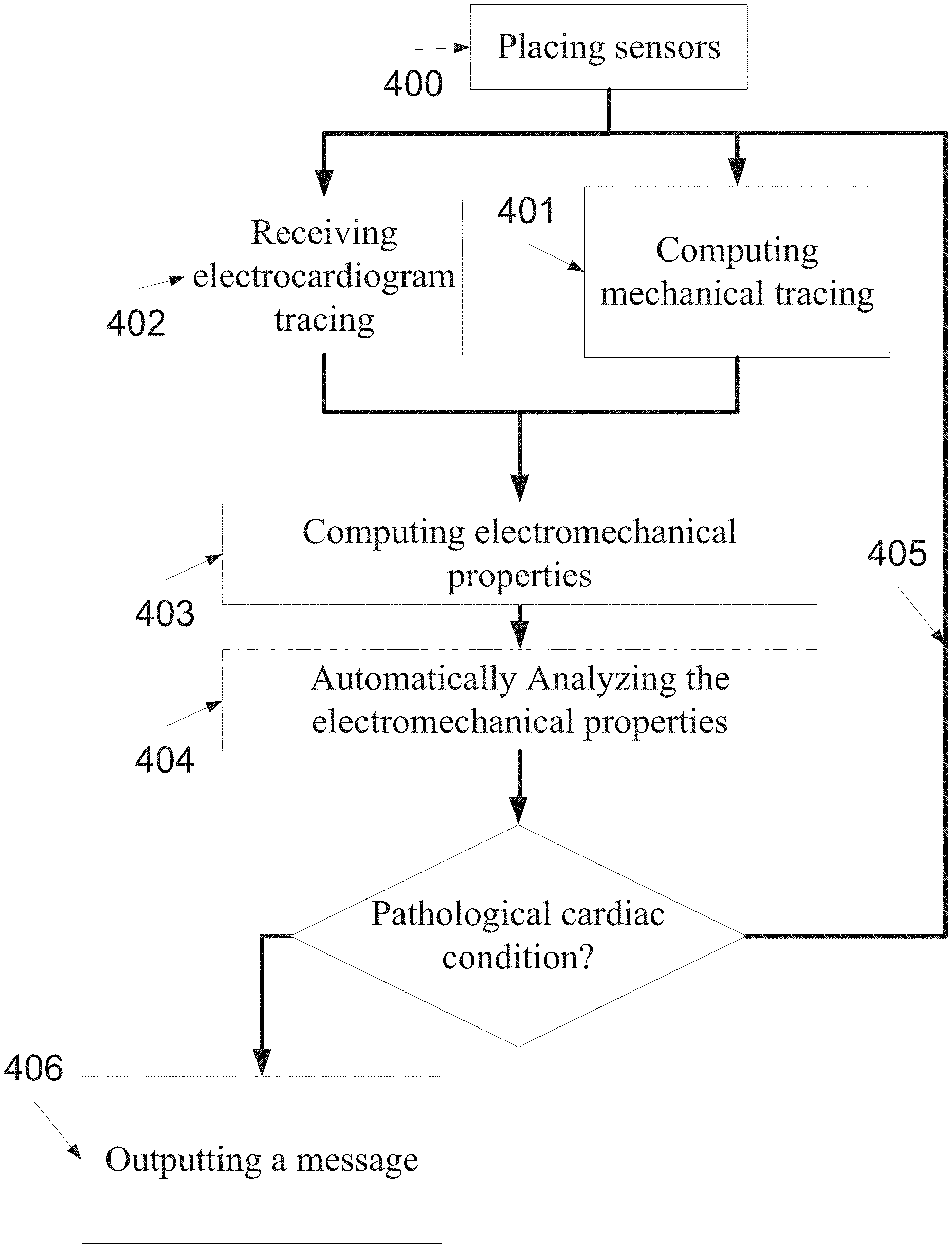
FIG. 5 is a flowchart of a method of monitoring one or more electromechanical properties, according to some embodiments of the present invention;
FIG. 6 is an illustration of two exemplary graphs, one depicting a signal which is based on EM reflections intercepted by a transducer and an electrical signal according to some embodiments of the present invention; and
FIG. 7 is an illustration a cardiac tissue velocity graph and a respective cardioelectric tracing which are temporally synchronized and compared, according to some embodiments of the present invention.
DESCRIPTION OF EMBODIMENTS OF THE INVENTION
The present invention, in some embodiments thereof, relates to monitoring and, more particularly, to using EM radiation for monitoring changes in cardiac performance.
According to some embodiments of the present invention there is provided a method and a device optionally wearable, for monitoring mechanical properties of one or more cardiac tissues by generating and analyzing mechanical tracings which are based on EM radiation reflected from the one or more cardiac tissues. The analysis allows detecting pathological conditions which may occur during the monitoring period, which is optionally of 6 hours or more, for example more than 24 hours. The device comprises one or more sensors or units that allow registering the mechanical tracings. In such a manner, the device may be used for monitoring ambulatory patients.
Optionally, the mechanical tracing documents the velocity of the motion of a certain cardiac motion during the monitoring period. Such a tracing may be used for estimating the acceleration of the cardiac tissue. Furthermore, the mechanical tracing may be used for detecting pathological conditions by detecting asynchronous contractions, multiple contractions per cardiac cycle, and asynchronous behavior in relation to other tissues. Optionally, the mechanical tracing documents the heart ejection fraction, and/or the contraction of the cardiac tissue during the monitoring period.
Optionally, the device includes a plurality of traducers that allows calculating a spatial motion vector of the one or more cardiac tissue. In such a manner, the mechanical tracing may be based on the spatial motion vector, providing data on the direction of contraction.
Optionally, the device includes a plurality of traducers that allows calculating a number of motion vectors, each of a different cardiac tissue. In such a manner, the synchrony of the motion of the different tissues may be evaluated.
According to some embodiments of the present invention there is provided a method and a device optionally wearable, for monitoring electromechanical properties of one or more cardiac tissues of a heart of a patient by generating and analyzing cardioelectric and mechanical tracings which are based on EM radiation reflected from the one or more cardiac tissues and an electric impulse from the heart. The analysis allows detecting pathological conditions which may occur during the monitoring period, which is optionally of 6 hours or more, for example more than 24 hours.
Optionally, the wearable device integrates one or more leads of an ECG sensor that allows generating the cardioelectric tracing. Optionally, one or more leads are external to the wearable device and communicate connected thereto via wires.
Such a method and device allows monitoring, in real time electromechanical properties such as a delay between an electric activation of the myocardium and the contraction thereof, a delay between electric and mechanical onsets and various electromechanical relationship between the cardioelectric tracing and the cardiac cycle, for example an electromechanical relationship between a QT interval of the cardioelectric tracing and the length of a respective cardiac cycle. Optionally, the electromechanical properties include the electromechanical relationship between amplitude changes and cardiac mechanical function changes.
Optionally, the method is used for monitoring patients in certain risk groups, such as CHF patients, pacemaker recipients and the like
According to some embodiments of the present invention there is provided method and device for a localization of pacemaker leads, such as cardiac resynchronization therapy (CRT) leads. The method and the device allow estimating the effect of the lead in different locations on the surface of the heart, allowing the surgeon to increase the efficacy of the CRT procedure.
For clarity, dielectric property of a material describes its interaction with EM fields; it is represented by a frequency dependent complex number describing the electrical permittivity and the conductivity of the material, as known in the art. Different human tissues are characterized by different dielectric coefficients. A dielectric coefficient of a cardiac tissue is affected by the dielectric coefficients of each of its components. As used herein a dielectric related property of a tissue means a property that is related to the dielectric coefficient thereof. Such a dielectric related property effects the reflection of electromagnetic radiation that is transmitted on the related tissue, such example changes the attenuation of the reflection, changes the delay that is caused by the tissue, changes the phase of the reflection, and changes the dispersion of the radiation in the tissue.
A dielectric related property may refer to a region of a body and be affected by the dielectric properties of tissues in that region. Normal and/or abnormal processes may change the regional dielectric related property, due to a change of the composition of the volume. A specific region may change its dielectric related property due to tissue movement and a consequent change in the configuration of tissues within that volume. The dielectric related property of a certain biological tissue may change in repetitive and/or predictable patterns according to various biological processes. For example, periodic changes may be measured along with breathing and heart cycles.
Before explaining at least one embodiment of the invention in detail, it is to be understood that the invention is not necessarily limited in its application to the details of construction and the arrangement of the components and/or methods set forth in the following description and/or illustrated in the drawings and/or the Examples. The invention is capable of other embodiments or of being practiced or carried out in various ways.
Reference is now made to FIG. 1, which is a flowchart of a method 100 for monitoring one or more cardiac tissues of a patient during a monitoring period of more than 6 hours by analyzing mechanical properties of the cardiac tissues, according to some embodiments of the present invention. The monitoring may be performed when the patient is in a certain risk group, for example because of a medical treatment and/or procedure and/or a pathological factor and/or during a physiological activity, such as running, cycling and the like. As used herein, a mechanical property of a cardiac tissue means a motion pattern, a cardiac cycle pattern, a pulsation pattern, a relationship with additional data, such as the cardioelectric tracing, an outcome of a mechanical tracing which is based on the movement of a certain cardiac tissue and the like. For brevity cardiac tissue also means implants, such as stents and pacemaker leads. These elements have a substantial different dielectric coefficient resulting in strong EM reflection profile and therefore may facilitate the EM analysis process.
As shown at 101 and 102, the method is based on transmitting EM radiation to a cardiac tissue of an ambulatory user and intercepting at least one reflection of the EM radiation therefrom in a plurality of transmission sessions, optionally during a period of at least 6 hours, for example 24 hours, few days, a week, few weeks, a mouth and/or any intermediate or longer period. The EM radiation may be a RF signal based on continuous emission and\or pulse emission. The signal may be wide band or narrow band, or any composition of multiple signals, constant or varying in time. Varying in time may include frequency sweeping, frequency hopping and\or any other change of the spectral density over time. Wide band signal may be a composition of multiple narrow band signals, such as multi tone.
As shown at 103, the transmission--reception sessions, for brevity referred to herein as transmission sessions, allow computing one or more mechanical tracings of the cardiac tissue. Computing may comprise mathematical and signal processing optionally including utilization of model based processing and/or analysis using present and historical measured or calculated information. As used herein a mechanical tracing is data indicative of a mechanical property of the cardiac tissue during a period, for example the velocity, the acceleration, the heart ejection fraction, the contraction period, the duty cycle of the contraction the speed of contraction, and/or contraction of the cardiac tissue during a period. As used herein a cardiac tissue means the myocardium, the aorta, the pericardium, the atrial tissue, and/or any combination thereof, for example left and right ventricles.
The mechanical tracing may be indicative of the pattern of the motion of the cardiac tissue, the contractility of the cardiac tissue, the remodeling of the cardiac tissue and/or the pulsation of the cardiac tissue.
Reference is now also made to FIG. 2, which is a graph two signals 510, 511 each based on EM reflections intercepted from an exemplary cardiac tissue of a patient, by one of a transducer of a monitoring device, for example as described below in relation to FIG. 3, according to some embodiments of the present invention. Each signal is recorded during respective correlated periods t.sub.1 and t.sub.2. The correlation between the signals 510, 511 is performed to allow matching between EM reflections from a monitored area in the thorax of the patient in period t.sub.1 and in period t.sub.2. The matching allows detecting a local deviation between the signals 510, 511. As the EM reflections pattern from which the signals are reconstructed are affected by the mechanical properties of the cardiac tissue, such a local deviation is indicative of a mechanical property--a motion of the cardiac tissue. For example, the local deviation .DELTA..tau..sub.x, which is depicted in FIG. 2, is indicative of the motion of the boundary between the heart and the lung during the period between t.sub.1 and t.sub.2. In FIG. 2, t.sub.1 is a systole and period t.sub.2 is a diastole and therefore the .DELTA..tau..sub.x reflects a motion occurring during the period between them, for example as an outcome of the heart contraction. The location of the local deviation along the signal is indicative of the location of the moving tissue. The location may be calculated according to the occurrence time (X axis) along the signal tracing. This time corresponds with the depth in the body of the patient, for example from the location of the sensor. In FIG. 2 a local deviation of 200 ps (.DELTA..tau.y) from approximately 0.6 to 0.8 nanoseconds from the first initial reflection which is equivalent to a deviation of 7.5 mm in depth of 25 mm, assuming the reflection from the heart is measured through the lung, whose real part of the dielectric coefficient is approximately 20. This local deviation is an outcome of the movement of the heart-lung boundary in 200 ms, corresponding approximately to the diastole and systole time.
Optionally, a mechanical property, such as the velocity of a cardiac tissue during a certain period, may be calculated by monitoring local deviations in a certain instant measured in relation to a reference instance, such as the instance of transmitting the EM radiation. The length of the local deviation corresponds with the velocity. By monitoring local deviations along the certain period, a mechanical tracing describing the velocity during that certain period may be calculated. For example, numeral 601 in FIG. 7 shows a graph that depicts the velocity of the boundary between the heart and the lung. The graph is based the local deviations which are monitored in a periodical instant which is indicative of reflections from the boundary between the heart and the lung.
Additionally or alternatively, the velocity of a certain cardiac tissue in a certain instance may be calculated by measuring the length of the local deviation and dividing it by the time period of this instance, which is indicative to the time it took the cardiac tissue to move so as to create the local deviation.
Additionally or alternatively, the velocity of a certain cardiac tissue is calculated by measuring the difference in the returned phase shift from transmission after adjusting to the wavelength. That could be done by transmitting and receiving a wave around one center frequency, or by using a wide pulse and computing a shift between the transmitted frequencies and intercepted frequencies. The adjusted measured shifts may be averaged to provide one corresponding robust estimation of the velocity, and\or any other mathematically operation, such as outlier exclusion by median operator and the like. Smoothing of the instant computed velocities may be conducted over time. As shown at 104, the mechanical tracing is analyzed. The analysis allows diagnosing and/or detecting physiological conditions, such as pathological cardiac conditions, for example by identifying a change or an indication of a change in relation to a normal motion pattern, a pulsation pattern, and/or any normal pattern of a mechanical behavior of a cardiac tissue, a number of contractions per cardiac cycle, a signal indicative of an irregular heart motion, an irregular cardiac cycle, an irregular heartbeat, an irregular relationship between a mechanical property during a cardiac cycle and additional data pertaining to the cardiac tissue, such as the cardioelectric tracing, during the same cardiac cycle. The change is analyzed so as to allow the identification of a change in one or more mechanical tracings of mechanical properties of the cardiac tissue, for example, its movement and/or contractility.
Optionally, the analysis is of a mechanical tracing that is indicative of the velocity of the motion and/or the pulsation of the heart or aorta, optionally by computing one or more spatial vectors in a monitoring period. In such a manner, a deviation from a normal motion and/or pulsation pattern and/or a pathological trend which lasts during few hours and/or days may be detected by extracting and classifying a motion pattern from the spatial vectors.
Optionally, a motion vector, which is calculated according to outputs of one or more transducers, may be calculated during the analysis so as to assess a cardiac performance.
As described above, the velocity of a certain cardiac tissue may be calculated. This allows calculating a clinical indication, such as the acceleration of the cardiac tissue, which is indicative of a ventricular pressure change over time (dp/dt). Optionally, the clinical indication is analyzed, in real time, for example by matching it with one or more expected normal values, ranges, and or patterns which are locally hosted by the monitoring device. In such embodiments the monitoring device may alarm a user and/or a medical center about abnormal clinical indications, optionally in real time, for example as described below.
Additionally or alternatively, the monitoring of the velocity of a certain cardiac tissue for a long period, such as hours and/or days, allows detecting pathological trends, for example a continuous slow down in the velocity and/or acceleration of the cardiac tissue due to ischemic processes, pericardial effusion, valvular deficiencies and the like.
Additionally or alternatively, the analysis of the mechanical tracing allows calculating a mechanical property such as the duration of a heart contraction. This may be done by calculating the time period between a first instance in which the first cardiac tissue contracts and a second instance in which the mechanical tracing indicates that one or more cardiac tissues, such as the ventricles, are relaxing. The analysis of such a mechanical property allows diagnosing and/or detecting a pathological cardiac condition, such as an elevated afterload, lack of intra and/or inter-ventricular synchronous contraction, either due to abnormal electrical activation pattern and/or due to an abnormal myocytes contraction. The analysis of such a mechanical property further allows diagnosing and/or detecting pathological conditions such as ischemic processes, presence and/or an absence of infracted regions in the myocardium or abnormal activation pathways due to left or right bundle branch blocks, Wolff-Parkinson-White (WPW) syndrome, a presence and/or an absence of ectopic foci, and/or decreased presence of critical substances such as calcium.
Additionally or alternatively, the analysis of the mechanical tracing allows estimating a mechanical property such as acceleration of a cardiac tissue along a cardiac cycle and optionally, the acceleration peak, which is estimated to be correlated with a heart ejection, and more particularly with a change of ventricular pressure during the cardiac cycle. The analysis of such a mechanical property allows diagnosing and/or detecting a pathological cardiac condition such as valvular deficiencies, for example mitral regurgitation.
If the analysis does not indicate a pathological cardiac condition, such as arrhythmia and hypertrophy, for example by identifying a change in the pattern of the motion and/or pulsation of the cardiac tissue and/or a deviation from a certain range of values, the process is repeated, as shown at 105. Else, as shown at 106 an alarm, a report and/or any other message that is indicative of the change and/or the deviation is outputted, for example presented and/or forwarded as described below. The system may report the current measurement and the analysis of the current clinical status.
Additionally or alternatively, the transmission sessions allow monitoring one or more values of one or more dielectric properties of the cardiac tissue and detecting a deviation of these values from predefined thresholds and/or ranges, for example similar to the described in International Patent Applications Numbers IL2008/001198 and/or IL2008/001199, filed on Sep. 4, 2008, which are incorporated herein by reference. In such a manner, pathological conditions such as pericardial effusion may be detected. In pericardial effusion, the space between the pericardium and myocardium is filled slowly or rapidly with blood, limiting the filling of the heart with blood, and risking for severe malfunction and death. Such a monitoring may provide an early detection for providing a more efficient treatment. According to some embodiments of the present invention, the transmission sessions allow diagnosing and/or detecting pathological cardiac conditions by matching mechanical tracings with a biomechanical model, optionally temporal. Optionally, the biomechanical model defines governing biomechanical equations and/or a parameterization of anatomical tissues based on parameter reconstruction. Optionally, the biomechanical model is based on anatomical/mechanical models using information that may be provided by one or more imaging modalities, such as echocardiograms, CT or MRI. Optionally, the biomechanical model defines an estimation of a number of physiological states of the heart during a period. Optionally, the biomechanical model of the tissue layers describes, both spatially and temporally, periodic effects of the respiration and the heart beat cycles and expected deviations according to various physiological phenomenon, for example various respiration patterns, such as cheyne stokes respiration pattern. In such a manner, a match to a physiological state may be indicative of a potential pathological condition.
Optionally, the monitoring and/or detecting is adjusted to take into account changes in the dielectric related properties of the monitored cardiac tissue, such as changes which occur as an outcome of a reaction to a medical treatment, a change of physiological state and body movements. Optionally, the transmission, receiving and optionally the analysis of the received reflections are performed by a wearable monitoring device, a probe and/or by a device having wearable probes, for example similar to the devices which are described in International Patent Applications Numbers IL2008/001198 and/or IL2008/001199, filed on Sep. 4, 2008, which are incorporated herein by reference. For brevity, each one of these devices may be referred to herein as a monitoring device or a probe.
As outlined above, one or more mechanical tracings of the cardiac tissue may be generated by intercepting and analyzing the EM reflections from one or more cardiac tissues during a period. Optionally, additional signs, which may provide meaningful clinical data as a complement to a clinical picture, are computed according to the EM properties. Such additional signs may include a respiratory rate and depth and/or an accumulation and/or dispersion of fluid within the cardiac and pulmonary tissues as well as of other chest fluids such as pleural effusion.
As outlined above, the method is optionally designed for long monitoring periods of 6 hours or more, for example as further described below. As such, the analysis may be adjusted to take into account changes in the dielectric related properties of the monitored cardiac tissue, such as changes, which occur as an outcome of thoracic movements, and other body movements.
Optionally, the analyzed data is presented to the patient and/or a medical caregiver and/or transferred to a central medical server, for example as described below. For example, mechanical tracings and/or alarms may be forwarded to an emergency center of a proximate hospital and to a central medical center.
According to some embodiments of the present invention, several types of sensors are used for monitoring the cardiac tissues. In such a manner, the reflections of the EM radiation may be analyzed together with signals from other modalities, such as an ultrasound modality that outputs a movement estimation of a cardiac tissue, which is optionally another cardiac tissue, and a pulse oximeter that outputs the blood saturation of the patient.
For example, ultrasound modality provides high tissue penetration ability when it comes to tissues rich with fluids while presenting less contrast between fat and muscles, for example dielectric coefficients are around 60 for a muscle tissue and 5 for a fat tissue, while the acoustic impedances differs only by few percent.
Optionally, the mechanical tracing is registered, calibrated, verified and/or otherwise adjusted according to the motion of another tissue, for example according to outputs of an additional sensor, such as an ultrasound sensor and/or an accelerometer which is placed on or in proximity to the tissue. Optionally, the mechanical tracing is registered according to outputs of a tiltmeter and/or accelerometer, which are indicative of the activity level of the patient. Optionally, the mechanical tracing is adjusted in a specific time instance according to the mechanical tracing of other sensors which is placed on or in proximity to the traced cardiac tissue, for example an ultrasound sensor and/or the aforementioned tiltmeter and/or accelerometer. Optionally, the mechanical tracing of a specific cardiac tissue for example the left ventricle is adjusted according to the mechanical tracing of another specific cardiac tissue, for example according to traced motion of the left ventricle.
Optionally, a plurality of RF transducers are used for assessing the movement of the heart from one or more angles and providing a number of spatial vectors that characterize the heart motion. Optionally, one or more of the RF transducers are placed to monitor the heart through the lungs. In such a manner, a relatively high dielectric contrast is provided at the muscle lung edge.
Optionally, the plurality of RF transducers allows monitoring mechanical properties of different cardiac tissues simultaneously. In such an embodiment, each RF transducer may intercept EM reflections from a different cardiac tissue, allowing the calculation of different mechanical tracings for different cardiac tissues. The mechanical tracings, which optionally document mechanical properties during a common period, may be correlated and matched to estimate a synchrony between different cardiac tissues, for example the contractions synchrony of two cardiac tissues, such as the left and the right ventricular chambers. This may be calculated by matching multiple peaks which are detected in a common cardiac cycle and/or directly measuring a mechanical onset of the contractions of each tissue.
Additionally or alternatively, the method described hereinabove may be executed in parallel to other monitoring methods such as monitoring of the congestion levels of the lungs.
Additionally or alternatively, the outputs of an accelerometer that is positioned on the chest may be used for registering the intercepted radiation, for example as described in International Patent Applications Numbers IL2008/001198 and/or IL2008/001199, filed on Sep. 4, 2008, which are incorporated herein by reference. Such a registration allows providing a finer accuracy in tracking heart motion overcoming chest motion and vibrations.
Reference is now also made to FIG. 3, which is a schematic illustration of an exemplary monitoring device 200, according to some embodiments of the present invention. Optionally, the exemplary monitoring device 200 is designed as a wearable and/or as a stationary monitoring device, for example as described in International Patent Applications Numbers IL2008/001198 and IL2008/001199, filed on Sep. 4, 2008, which are incorporated herein by reference. The monitoring device 200 may be used for implementing any of the aforementioned methods.
The exemplary device 200 comprises a central processing unit (CPU) and/or a digital signal processing (DSP) which may be referred to herein as a processing unit 201. Optionally, the processing unit 201 runs a real-time operating system (RTOS) that is responsible for coordinating all functions of the monitoring device 200. The processing unit 201 is optionally used for analyzing the outputs of the one or more front-end sensors 204 which are described below. Optionally, the one or more front-end sensors 204 are ECG electrodes. Optionally, the one or more front-end sensors 204 capture signals which are forwarded to the processing unit 201 that calculates a deviation from a motion pattern of the cardiac tissue. The deviation analyzed from the reflected signals may be used for detecting a pathological pattern. For example, the processing unit 201 may compare between the deviation and a set of one or more predefined values. The data that is calculated by the processing unit 201 is optionally used for generating one or more alerts and/or notifications. It should be noted that the term processing unit means a local processing unit, a distributed processing unit, and/or a remote processing unit which is used for performing the functioning of the processing unit that is described herein. In an embodiment in which the processing unit is remote, the data that is forwarded to the processing unit is transmitted for remote processing by the remote processing unit.
The monitoring device 200 further comprises a memory unit 202, such as a non volatile memory, that is designed for storing the operating system and parameters which are needed for the functioning of the monitoring device 200. Optionally, the memory unit 202 is used for recording mechanical and electromechanical tracings and/or calculations which are based thereupon, for example as further described above. Such a recording allows examining changes in the motion pattern, optionally during a period that lasts between few hours and days. The recording allows calculating a repetitive pattern and/or the identification of a deviation from a normal range that is optionally adjusted according to the specific user. Optionally, the memory unit 202 is used for recording readings of medical sensors which are connected to the monitoring device 200 and/or embedded therein. Optionally, the memory unit 202 is used for storing additional information, such as application executables codes, configuration files for the processing unit 201, preset parameters, long term state parameters and tables. The memory unit 202 may be used for storing additional user related data, such as the patient identification information, version information, user specific thresholds, authentication and/or security keys.
The monitoring device 200 further comprises a rapid access volatile memory unit 206, such as a dynamic random access memory (DRAM), a synchronous DRAM (SDRAM), and/or any other volatile memory for storing data that is needed to be accessed in a limited time for short terms. It may be interfaced by the processing unit 201, the below mentioned designated IC and/or any other component of the monitoring device 200.
Optionally, the monitoring device 200 comprises a designated processing unit 203, such as a designated integrated circuit (IC), for example an application-specific integrated circuit (ASIC) or a field-programmable gate array (FPGA) that contains logic blocks and programmable interconnects which are programmed to implement some of the functions required to process the data from the sensors front-ends. The designated processing unit 203 communicates with the processing unit 201, the memory unit 202, and/or with other components of the device for various tasks. Additionally or alternatively, the designated processing unit 203 may also implement any of the other blocks as an integrative solution. For example, the FPGA or ASIC may incorporate the processing unit 101 and/or another processing unit. Optionally, the logic blocks are programmed to implement monitoring methods as described above.
The hardware may be used for processing measurements obtained from other modalities such as oxygen saturation or ECG measurements.
As described above and depicted in FIG. 3, the monitoring 200 further comprises one or more probes, such as front-end sensors 204, for example EM transceivers, for transmitting a plurality of electromagnetic (EM) waves toward the thorax of the patient and for capturing reflections thereof from the cardiac tissues. In some embodiment, the beam is transmitted in a desired pulse and allows the capturing of a reflection thereof from various areas on the surface of the user's body. Optionally, the capturing is adjusted according to a selected operational mode, for example according to a selected swept frequency, a selected frequency hopping chirp, and a selected multiple carriers and the like. Other modes and/or gating patterns according to which the beam is transmitted and allows the capturing thereof are described in International Patent Applications Numbers IL2008/001198 and IL2008/001199, filed on Sep. 4, 2008, which are incorporated herein by reference.
Optionally, the monitoring device 200 is adjusted for long term usage, for example in terms of power consumption and heat dissipation, for example similar to devices which are described in International Patent Applications Numbers IL2008/001198 and/or IL2008/001199, filed on Sep. 4, 2008, which are incorporated herein by reference.
Optionally, the monitoring device 200 fits in a small form factor (SFF) housing which is adjusted for ambulatory patients, for example similar to devices which are described in International Patent Applications Numbers IL2008/001198 and/or IL2008/001199, filed on Sep. 4, 2008, which are incorporated herein by reference.
Optionally, the monitoring device 200 may be adjusted to facilitate easy replacement of disposable parts, such as batteries and padding, for example similar to devices which are described in International Patent Applications Numbers IL2008/001198 and/or IL2008/001199, filed on Sep. 4, 2008, which are incorporated herein by reference.
Optionally, the monitoring device 200 includes a placement unit 210 having a disengagement detector which examines the quality of measurement and optionally alert when improper positioning of the device 200 is detected, for example similar to placement units 210 which are described in International Patent Application Number IL2008/001199, filed on Sep. 4, 2008, which is incorporated herein by reference.
Optionally, front end sensors 204 comprise one or more medical sensors for capturing vital signs and/or other medical measures, for example an ECG sensor, a temperature sensor, a blood pressure transducer, a fluid rate recording transducer, and an echo-cardiographic sensor. The output of these sensors is forwarded to the processing unit 201 so as to allow a more accurate characterization of a dynamic heart function based on a plurality of measurements from a plurality of sensors. The medical sensors are either incorporated within the monitoring device and/or communicate therewith.
According to some embodiments of the present invention, a plurality of transducers are used. For clarity, reference is now also made to FIG. 4, which is a schematic illustration of the thorax of an exemplary patient 501 and sensor locations 502, 503 in which the RF transceivers and/or cardioelectric leads are placed, according to some embodiments of the present invention. For example, a first RF transducer 501 is placed on the front of the thorax, in front of the heart, capable of sensing motion and other mechanic properties in specific heart regions. A second RF transducer may be placed in another location on the thorax, for example on between the arm and the thorax. In such a manner, the EM radiation is received in two orthogonal or substantially orthogonal locations. The use of multiple sensors may be used to assess movement of different locations within the heart so as to allow assessing a related 2D or 3D vector of motion of the entire heart. Any number of additional sensors may be added to improve the accuracy and/or robustness of the spatial vector. As each sensor monitors motion of the heart from a different direction, a spatial motion vector may be computed by combining two or more motion vectors each calculated according to information from a different subset of the multiple sensors. Additionally or alternatively, the motion of two different cardiac tissues may be monitored, for example the left and right ventricles. In such a manner, the synchrony of the contractions and/or any other mechanical relation between the different tissues may be evaluated according to the intercepted reflections. One or more of the sensors 501, 502 may be used independently in order to evaluate the entire heart motion resulting mainly from the ejection of the ventricles, namely the jet effect.
Optionally, the leads of an ECG sensor are integrated into the body of the monitoring device 200. Additionally or alternatively, the leads of the ECG sensor are connected by wire to the body of the monitoring device 200. In such an embodiment the user may place the leads in different location, optionally according to predefined instructions.
According to some embodiments of the present invention, the monitoring device 200 incorporates an ECG sensor or receives outputs of such an ECG sensor, optionally wirelessly. Optionally, the ECG sensor comprises a number of leads. Optionally, one or more electrodes are integrated into the device 200 and one or more electrodes are separated from the monitoring device 200 and communicate therewith, optionally wirelessly. Leads could be positioned similar to the positioning of regular chest leads. Optionally the leads and the monitoring unit are incorporated in a wearable chest strap. The processing unit 201 receives and analyses the output of the ECG sensor and the aforementioned EM transceivers.
Optionally, the monitoring apparatus 200 is connected to a power supply element circuitry 205 that is designed for generating and distributing the power supply that is required for the components of the monitoring apparatus 200. The power supply element circuitry 205 comprises one or more batteries, optionally rechargeable.
Optionally, the monitoring device 200 comprises a man-machine interface (MMI) 207 for presenting data, such as an alert, a notification, statistical data, a current reading of the one or more front-end sensors 204 or external modality to the user, the user's caretaker, and/or others as desired. The MMI 207 may comprise a liquid crystal display (LCD), a touch screen, a speaker, a tactile generator, a set of light emitting diodes (LEDs), and/or any other indicator that may be used for presenting alerts and/or notifications which are based on a combination of the analysis of the mechanical tracings which are based on EM waves from an internal area in the body of the user, such as the cardiac tissues, and indicative of mechanical properties. Optionally, the MMI 207 is separate and remote from the monitoring device and connected to it optionally using wired, wireless, and/or using a computer or communication network.
Optionally, the configuration of the wearable monitoring apparatus 200 may allow the patient and/or a caretaker to define which alarm to present and to whom, for example whether the alarm is visual, audible, and/or tactile.
Optionally, the monitoring apparatus 200 comprises a posture detection unit 211 for using EM radiation for detecting a posture of a user, for example as described in International Patent Application Number IL2008/001199, filed on Sep. 4, 2008, which is incorporated herein by reference. The posture affects the thickness of layers of tissues in the path to the heart, for example when a person lies on his side his heart tends towards that side and less pulmonary tissue resides on the path from the chest surface to the heart. As the reflection is influenced by the layers between the chest surface and the heart and the propagation time is accordingly affected, the analysis of the intercepted data may be adjusted according to the outputs of the posture detection unit 211. In such a manner the effect of the posture is reduced and/or eliminated.
According to some embodiments of the present invention, the monitoring device 200 is used for computing an electromechanical delay in real time. In such an embodiment, the cardioelectric tracing and the mechanical tracing are combined in real time to provide a real time feedback for cardiac therapies, such as cardiac resynchronization therapy (CRT). In such an embodiment, a caretaker may receive a real time feedback on the efficacy of the therapy she provides and/or a real time indication about the responsiveness of a cardiac tissue to a therapeutic procedure. For example, the monitoring device 200 may be used for identifying the optimal location of leads in a CRT implantation procedure. In particular, a CRT device may have a number of leads, for example one in the right ventricle to stimulate the septum, and another inserted through the coronary sinus to pace the lateral wall of the left ventricle. Often, for patients in normal sinus rhythm, there is also a lead in the right atrium to facilitate synchrony with the atrial contraction. The leads are designed for timing between the atrial and ventricular contractions, as well as between the septal and lateral walls of the left ventricle. By monitoring the efficacy of the leads in different location on the heart, the monitoring device 200 may be used to identify the locations in which the leads achieve optimal cardiac function, see McWilliam J A (1889). "Electrical stimulation of the heart in man". Br Med J 1: 348-50. doi:10.136/bmj.1.1468.348. Partial quote in "Electrical Stimulation of the Heart in Man--1889", Heart Rhythm Society, Accessed May 11, 2007; Lidwell M C, "Cardiac Disease in Relation to Anaesthesia" in Transactions of the Third Session, Australasian Medical Congress, Sydney, Australia, Sep. 2-7 1929, p 160; and {circumflex over ( )} a b Mond H, Sloman J, Edwards R (1982). "The first pacemaker". Pacing and clinical electrophysiology: PACE 5 (2): 278-82. doi:10.1111/j.1540-8159.1982.tb02226.x. PMID 6176970 which are incorporated herein by reference.
Additionally or alternatively, the device may be used for monitoring the functionality and efficacy of a CRT and/or any other pacemaker, after the implantation of the leads. In such an embodiment, the monitoring device 200 may be provided after surgery to monitor the integration of the pacemaker and its functionality, optionally during different activities and/or situations of the patient.
Reference is now also made to FIG. 5, which is a flowchart of a method of monitoring one or more electromechanical properties, according to some embodiments of the present invention.
First, in 400, EM and ECG sensors, optionally similar to the described above, are attached to the thorax of the patient 501. For clarity, reference is now also made, once again, to FIG. 4. Optionally, leads of an ECG sensor are placed in one or more of the sensor locations 502, 503. Optionally, an electrical conductor 504 is used to reference the measured potentials between the different leads.
As shown in 401, one or more mechanical tracing are computed, for example as described above, according to EM radiation which is reflected from a monitored cardiac tissue of a patient. As described above, the EM reflections allow generating one or more mechanical tracings. As shown in 402, cardioelectric tracing of the patient are simultaneously, or substantially simultaneously, received from a cardioelectric sensor, such as an ECG sensor. As used herein, a cardioelectric tracing means a measurement of the electrical pulses given off by one or more cardiac tissues during a period, such as an electrocardiograph record, electrocardiogram tracing, and the like.
For example, reference is now made to FIG. 6, which includes a set of two exemplary graphs, one depicting a signal that is based on EM reflections intercepted by a transducer of the monitoring device depicted in FIG. 3 during a period t and a signal that is based on a cardioelectric value measured by a cardioelectric sensor, such as a ECG sensor during t, according to some embodiments of the present invention.
In the EM signal graph, the long vertical lines depict an instance which has been identified as an initiation of a respiration cycle during t, and the short vertical lines depicting heart beats. In the ECG signal graph, the solid lines are indicative of electrical onsets of an electrical activation (R). The EM signal graph depicts the energy of the reflected signal normalized to the transmitted energy and coordinated with the ECG signal on t.
Subsequently, as shown in 403, the outputs from 401 and 402 are used to compute one or more electromechanical properties of the monitored cardiac tissue. Optionally, 403 comprises temporally correlating between the cardioelectric tracing and the mechanical tracing. Optionally, the electromechanical properties include the electromechanical relationship between the cardioelectric tracing and the mechanical cardiac cycle, for example an electromechanical relationship between a QT interval of the cardioelectric tracing and the length of a respective mechanical cardiac cycle. Optionally, the electromechanical properties include the electromechanical relationship between amplitude changes and cardiac mechanical function changes.
Now, as shown at 404, the electromechanical properties are automatically analyzed so as to allow the detection of a physiological condition, such as a pathological cardiac condition, for example by the processing unit 201 of the monitoring device 200,
Optionally, the electromechanical properties include an electromechanical delay in one or more cardiac cycles. The electromechanical delay reflects the delay between the electrical stimulation of the myocytes and the development of a muscle tension. The delay is the period between the electric onset of a cardiac cycle, which may be detected by analyzing the received cardioelectric tracing and the mechanical onset of the cardiac cycle, which may be detected by analyzing the received EM reflections. The electromechanical delay may be determined by calculating the period between the electrical activation, which at the peak of the R wave in the QRS complex of the cardioelectric tracing and the respective mechanical activation, which may be determined by detecting peak acceleration. If the electromechanical relationship between the onsets is indicative of electrical activation pattern of the ventricle, a pathological condition, such as bundle branch block, left and\or right, and\or WPW syndrome may be identified.
Multiple electromechanical delays may be computed between any detected specific pattern in an ECG signal and in a motion vector. For example, an electromechanical delay may be detected by identifying the P and/or T wave in the ECG signal and associating it with a peak of acceleration and\or the velocity of a motion vector that appears in a heart cycle that is associated with a contraction of another cardiac tissue such as the atriums and the ventricles.
For example, FIG. 7 depicts an illustration of a cardiac tissue velocity graph and a respective cardioelectric tracing which are temporally correlated, according to some embodiments of the present invention. Such a correlation allows monitoring the period between the electrical activation and a respective mechanical activation that is triggered by it, for example as shown by numerals 603 and 604.
Optionally, the electromechanical properties include the number of contractions per cardiac cycle. The cardioelectric tracing, which is a transthoracic interpretation of the electrical activity of the heart, indicates the beginning and the end of a cardiac cycle. The calculation of the motion of the cardiac tissue allows estimating the number of contractions which are performed per cardiac cycle. In such a manner, multiple contractions per cycle may be detected, indicating an asynchronous mechanical contraction of the left and/or the right side of the heart.
Optionally, the electromechanical properties may indicate asynchronous contraction which may be a result of abnormal electrical activation sequence or abnormal regional contraction response. The electromechanical parameter, could be compared with an estimation of the electrical activation synchrony as assessed from the ECG and appeared generally by wider QRS complex. Thus, it is possible to ascertain a diagnosis of abnormal electrical activation pattern, or in case of normal QRS complex to diagnose an abnormal contraction.
By continually, periodically, and/or randomly, repeating 401-404, as shown at 405, the electromechanical properties of the patient may be monitored during a period of more than 6 hours, for example 24 hours, similarly to the aforementioned monitoring period.
As shown at 406, when the analysis performed in 404 indicates a physiological condition, for example a pathological cardiac condition, such as relating to the aforementioned electromechanical delay optionally a delay that is longer than a predefined threshold, an alarm, a notice, and/or any other message is outputted and optionally presented to the patient and/or forwarded to a caretaker and/or medical center. Such a delay may be indicative of a pathological viability of the myocardium and/or the pumping efficiency of the patient's heart. In such a manner, the alarm may assist in detecting and/or predicting a malfunction as well as monitoring therapeutic procedure effects. As shown at 405, if no pathological condition is detected, the process is repeated so as to allow a continuous, periodical, and/or random monitoring of the electrometrical properties of the patient.
Optionally, the assessment of mechanical and/or electromechanical properties may be used to improve sensitivity and/or specificity of other modalities, to detect different pathological patterns, and as a complement to a clinical status assessment, as well as to provide a combined index with a clinical value. For example, such a combined index may be used for providing feedback to therapies such as medication. Similarly, it may be used to provide feedback to therapeutic procedures such as cardiac resynchronization therapy (CRT) in real time. Furthermore, the combined index may be used for positioning the resynchronization electrodes.
Reference is now made, once again, to FIG. 3. Optionally, the monitoring device 200 includes a communication interface 208 that allows forwarding data pertaining to the monitored electromechanical and/or mechanical properties to an external computing unit in an autonomous manner, for example similar to devices which are described in International Patent Applications Numbers IL2008/001198 and/or IL2008/001199, filed on Sep. 4, 2008, which are incorporated herein by reference. Optionally, the data is an alert that is forwarded to a monitoring center and/or a caretaker when hazardous scenarios are detected. Optionally, the monitored data is forwarded as is to the external computing monitoring device. In such embodiments, the external monitoring device performs the analysis and output a report, an alarm, and/or any other indication based thereupon. The analysis may be separated such that different processing units perform different processing stages.
Optionally, the transmission sessions are intermittently performed so as to reduce the power consumption. For example, the number of transmission sessions may be reduced when the patient is asleep and increased when the patient performs an intense physical activity. In such an embodiment, the transmission sessions may be dynamically changed according to a vital sign either estimated based on the intercepted EM radiation and/or a signal which is based thereupon or an external sensor, such as an accelerometer that is designed to measure the activity level of the patient and/or according to changes in the motion pattern of the cardiac tissue and/or reception of other sensors which are connected to the monitoring device 200.
It is expected that during the life of a patent maturing from this application many relevant devices and methods will be developed and the scope of the term EM radiation, EM reflections, a power source, and a communication unit is intended to include all such new technologies a priori.
As used herein the term "about" refers to .+-.10%.
The terms "comprises", "comprising", "includes", "including", "having" and their conjugates mean "including but not limited to". This term encompasses the terms "consisting of" and "consisting essentially of".
The phrase "consisting essentially of" means that the composition or method may include additional ingredients and/or steps, but only if the additional ingredients and/or steps do not materially alter the basic and novel characteristics of the claimed composition or method.
As used herein, the singular form "a", "an" and "the" include plural references unless the context clearly dictates otherwise. For example, the term "a compound" or "at least one compound" may include a plurality of compounds, including mixtures thereof.
The word "exemplary" is used herein to mean "serving as an example, instance or illustration". Any embodiment described as "exemplary" is not necessarily to be construed as preferred or advantageous over other embodiments and/or to exclude the incorporation of features from other embodiments.
The word "optionally" is used herein to mean "is provided in some embodiments and not provided in other embodiments". Any particular embodiment of the invention may include a plurality of "optional" features unless such features conflict.
Throughout this application, various embodiments of this invention may be presented in a range format. It should be understood that the description in range format is merely for convenience and brevity and should not be construed as an inflexible limitation on the scope of the invention. Accordingly, the description of a range should be considered to have specifically disclosed all the possible subranges as well as individual numerical values within that range. For example, description of a range such as from 1 to 6 should be considered to have specifically disclosed subranges such as from 1 to 3, from 1 to 4, from 1 to 5, from 2 to 4, from 2 to 6, from 3 to 6 etc., as well as individual numbers within that range, for example, 1, 2, 3, 4, 5, and 6. This applies regardless of the breadth of the range.
Whenever a numerical range is indicated herein, it is meant to include any cited numeral (fractional or integral) within the indicated range. The phrases "ranging/ranges between" a first indicate number and a second indicate number and "ranging/ranges from" a first indicate number "to" a second indicate number are used herein interchangeably and are meant to include the first and second indicated numbers and all the fractional and integral numerals therebetween.
It is appreciated that certain features of the invention, which are, for clarity, described in the context of separate embodiments, may also be provided in combination in a single embodiment. Conversely, various features of the invention, which are, for brevity, described in the context of a single embodiment, may also be provided separately or in any suitable subcombination or as suitable in any other described embodiment of the invention. Certain features described in the context of various embodiments are not to be considered essential features of those embodiments, unless the embodiment is inoperative without those elements.
Although the invention has been described in conjunction with specific embodiments thereof, it is evident that many alternatives, modifications and variations will be apparent to those skilled in the art. Accordingly, it is intended to embrace all such alternatives, modifications and variations that fall within the spirit and broad scope of the appended claims.
All publications, patents and patent applications mentioned in this specification are herein incorporated in their entirety by reference into the specification, to the same extent as if each individual publication, patent or patent application was specifically and individually indicated to be incorporated herein by reference. In addition, citation or identification of any reference in this application shall not be construed as an admission that such reference is available as prior art to the present invention. To the extent that section headings are used, they should not be construed as necessarily limiting.
* * * * *
D00000
D00001
D00002
D00003
D00004
D00005
D00006
D00007
XML
uspto.report is an independent third-party trademark research tool that is not affiliated, endorsed, or sponsored by the United States Patent and Trademark Office (USPTO) or any other governmental organization. The information provided by uspto.report is based on publicly available data at the time of writing and is intended for informational purposes only.
While we strive to provide accurate and up-to-date information, we do not guarantee the accuracy, completeness, reliability, or suitability of the information displayed on this site. The use of this site is at your own risk. Any reliance you place on such information is therefore strictly at your own risk.
All official trademark data, including owner information, should be verified by visiting the official USPTO website at www.uspto.gov. This site is not intended to replace professional legal advice and should not be used as a substitute for consulting with a legal professional who is knowledgeable about trademark law.