Antibodies to amyloid beta
Groves , et al.
U.S. patent number 10,662,239 [Application Number 15/793,510] was granted by the patent office on 2020-05-26 for antibodies to amyloid beta. This patent grant is currently assigned to MedImmune Limited. The grantee listed for this patent is MedImmune Limited. Invention is credited to Per-Ola Freskgard, Maria Groves, Suzanne Gustavsson, Kina Hoglund, Chris Lloyd, David Lowne, Adrian Nickson, Camilla Niva, Sylvia Simon, Fraser Welsh.


View All Diagrams
| United States Patent | 10,662,239 |
| Groves , et al. | May 26, 2020 |
Antibodies to amyloid beta
Abstract
Antibody for human amyloid beta. Antibody selectively binds human amyloid beta 42 peptide over human amyloid beta 40 peptide. Antibodies specific for amyloid beta 42 as therapeutic agents for binding amyloid beta 42 peptide and treating conditions associated with amyloidosis, such as Alzheimer's disease.
| Inventors: | Groves; Maria (Cambridge, GB), Gustavsson; Suzanne (Huddinge, SE), Hoglund; Kina (Solna, SE), Lloyd; Chris (Cambridge, GB), Nickson; Adrian (Cambridge, GB), Niva; Camilla (Arsta, SE), Simon; Sylvia (Stockholm, SE), Lowne; David (Cambridge, GB), Welsh; Fraser (Cambridge, GB), Freskgard; Per-Ola (Sodertalje, SE) | ||||||||||
|---|---|---|---|---|---|---|---|---|---|---|---|
| Applicant: |
|
||||||||||
| Assignee: | MedImmune Limited (Cambridge,
GB) |
||||||||||
| Family ID: | 49765450 | ||||||||||
| Appl. No.: | 15/793,510 | ||||||||||
| Filed: | October 25, 2017 |
Prior Publication Data
| Document Identifier | Publication Date | |
|---|---|---|
| US 20180105585 A1 | Apr 19, 2018 | |
Related U.S. Patent Documents
| Application Number | Filing Date | Patent Number | Issue Date | ||
|---|---|---|---|---|---|
| 14435520 | 9834598 | ||||
| PCT/EP2013/071567 | Oct 15, 2013 | ||||
| 61713996 | Oct 15, 2012 | ||||
| Current U.S. Class: | 1/1 |
| Current CPC Class: | C07K 16/18 (20130101); A61P 25/28 (20180101); C07K 2317/622 (20130101); C07K 2317/515 (20130101); C07K 2317/33 (20130101); A61K 2039/505 (20130101); C07K 2317/565 (20130101); A61K 39/3955 (20130101); C07K 2317/55 (20130101); C07K 2317/21 (20130101); C07K 2317/92 (20130101); C07K 2317/51 (20130101); C07K 2317/567 (20130101); C07K 2317/60 (20130101) |
| Current International Class: | C07K 16/18 (20060101); A61K 39/00 (20060101); A61P 25/28 (20060101); A61K 39/395 (20060101) |
References Cited [Referenced By]
U.S. Patent Documents
| 9834598 | December 2017 | Groves |
| 101827862 | Sep 2010 | CN | |||
| 1160256 | Dec 2001 | EP | |||
| 1717250 | Nov 2006 | EP | |||
| 2009/104769 | Aug 2010 | RU | |||
| WO2003/015691 | Feb 2003 | WO | |||
| WO2006/072620 | Jul 2006 | WO | |||
| WO2008/011348 | Jan 2008 | WO | |||
| WO2017/158064 | Sep 2017 | WO | |||
| WO-2017/160622 | Sep 2017 | WO | |||
Other References
|
Bannister et al., "Parallel, high-throughput purification of recombinant antibodies for in vivo cell assays," Biotechnol Bioeng., 94(5): 931-937 (2006). cited by applicant . Bard et al., "Peripherally administered antibodies against amyloid beta-peptide enter the central nervous system and reduce pathology in a mouse model of Alzheimer disease," Nat. Med. 6(8): 916-919 (2000). cited by applicant . Borchelt et al., "Familial Alzheimer's disease-linked presenilin 1 variants elevate Abeta1-42/1-40 ratio in vitro and in vivo," Neuron., 17(5):1005-1013 (1996). cited by applicant . Calingasan et al., ".beta.-amyloid 42 accumulation in the lumbar spinal cord motor neurons of amyotrophic lateral sclerosis patients," Neurobiology Disease, vol. 19(1): 340-347 (2005). cited by applicant . Citron et al., "Additive effects of PS1 and APP mutations on secretion of the 42-residue amyloid beta-protein," Neurobiol Dis., 5(2):107-116 (1998). cited by applicant . De Strooper, "Loss-of-function presenilin mutations in Alzheimer disease. Talking Point on the role of presenilin mutations in Alzheimer disease," EMBO Rep., 8(2):141-146 (2007). cited by applicant . DeMattos et al., "Peripheral anti-A beta antibody alters CNS and plasma A beta clearance and decreases brain A beta burden in a mouse model of Alzheimer's disease," PNAC USA, 98(15):8850-8855 (2001). cited by applicant . Duff et al., "Increased amyloid-beta42(43) in brains of mice expressing mutant presenilin 1," Nature, 383(6602):710-713 (1996). cited by applicant . EMD Millipore Corp. "Anti-Amyloid Beta (ABeta) x-42, clone 12F4", certificate of analysis, copyright 2008. cited by applicant . Gilman et al., "Clinical effects of Abeta immunization (AN1792) in patients with AD in an interrupted trial," Neurology, 64(9):1553-1562 (2005). cited by applicant . Glabe, "Does Alzheimer disease tilt the scales of amyloid degradation versus accumulation?" Nat Med., 6(2):133-134 (2000). cited by applicant . Golde et al., "Quantitative and mechanistic studies of Abeta immunotherapy," CNS & Neuro. Dis. Drug Targets, 8(1):31-49 (2009). cited by applicant . Greeve et al., "Age-dependent neurodegeneration and Alzheimer-amyloid plaque formation in transgenic Drosophila," J. Neurosci., 24(16):3899-3906 (2004). cited by applicant . Groves et al., "Applications of ribosome display to antibody drug discovery," Expert Opin Biol Ther., 5(1):125-135 (2005). cited by applicant . Hanes et al., "In vitro selection and evolution of functional proteins by using ribosome display," PNAS USA, 94(10):4937-4942 (1997). cited by applicant . Hanes et al., "Selecting and evolving functional proteins in vitro by ribosome display," Methods Enzymol., 328:404-430 (2000). cited by applicant . Hartman T., et al., "Distinct sites of intracellular production for Alzheimer's disease A beta40/42 amyloid peptides," Nature Medicine 3(9) pp. 1016-1020 (1997). cited by applicant . Hoet et al., "Generation of high-affinity human antibodies by combining donor-derived and synthetic complementarity-determining-region diversity," Nat Biotechnol., 23(3):344-348 (2005). cited by applicant . Human Amyloid .beta. 1-42 (A.beta. 1-42) Kit, Technical Data Sheeet, Copyright 2009, PerkinElmer, Inc. [Online] Retrieved from internet Feb. 23, 2016. cited by applicant . Iijima et al., "Dissecting the pathological effects of human Abeta40 and Abeta42 in Drosophila: a potential model for Alzheimer's disease," PNAS USA, 101(7):6623-6628 (2004). cited by applicant . Kim et al., "A .beta.40 Inhibits Amyloid Deposition In Vivo," The Journal of Neuroscience, 27(3): 627-633 (2007). cited by applicant . Kuperstein et al., "Neurotoxicity of Alzheimer's disease A.beta. peptides is induced by small changes in the A.beta.42 to A.beta.40 ratio," EMBO J., 29(19):3408-3420 (2010). cited by applicant . Lambert et al., "Diffusible, nonfibrillar ligands derived from Abeta1-42 are potent central nervous system neurotoxins," PNAS USA 95(11):6448-6453 (1998). cited by applicant . Levites et al., "Anti-Abeta42- and anti-Abeta40-specific mAbs attenuate amyloid deposition in an Alzheimer disease mouse model," J Clin Invest., 116(1):193-201 (2005). cited by applicant . Matsuoka et al., "Novel therapeutic approach for the treatment of Alzheimer's disease by peripheral administration of agents with an affinity to beta-amyloid," J.Neurosci., 23(1):29-33 (2003). cited by applicant . McGowan et al., "Abeta42 is essential for parenchymal and vascular amyloid deposition in mice," Neuron., 47(2):191-199 (2005). cited by applicant . Mucke et al., "High-level neuronal expression of abeta 1-42 in wild-type human amyloid protein precursor transgenic mice: synaptotoxicity without plaque formation," J. Neurosci., 20(11):4050-4058 (2000). cited by applicant . Oganesyan et al., Structural characterization of a human Fc fragment engineered for lack of effector functions, Acta Crystallogr. D Bioi. Crystallogr., 64(Pt 6):700-704 (2008). cited by applicant . Orgogozo et al., "Subacute meningoencephalitis in a subset of patients with AD after Abeta42 immunization," neurology, 61(1):46-54 (2003). cited by applicant . Osbourn et al., "Generation of a panel of related human scFv antibodies with high affinities for human CEA," Immunotechnology, 2(3):181-196 (1996). cited by applicant . Persic et al., "An integrated vector system for the eukaryotic expression of antibodies or their fragments after selection from phage display libraries," Gene, 187(1):9-18 (1997). cited by applicant . Portelius et al., "Mass spectrometric characterization of brain amyloid beta isoform signatures in familial and sporadic Alzheimer's disease," Acta Neurppathol., 120(2):185-193 (2010). cited by applicant . Pride et al., "Progress in the active immunotherapeutic approach to Alzheimer's disease: clinical investigations into AN1792-associated meningoencephalitis," Neurodegener Dis., 5(3-4):194-196 (2008). cited by applicant . Schenk et al., "beta-peptide immunization: a possible new treatment for Alzheimer disease," Arch Neurol., 57(7):934-936 (2000). cited by applicant . Schenk et al., "Immunization with amyloid-beta attenuates Alzheimer-disease-like pathology in the PDAPP mouse," Nature, 400(6740):173-177 (1999). cited by applicant . Scheuner et al., "Secreted amyloid beta-protein similar to that in the senile plaques of Alzheimer's disease is increased in vivo by the presenilin 1 and 2 and APP mutations linked to familial Alzheimer's disease," Nat Med., 2(8):864-870 (1996). cited by applicant . Selkoe, "Translating cell biology into therapeutic advances in Alzheimer's disease," Nature, 399:A23-31 (1999). cited by applicant . Tomlinson et al., "The repertoire of human germline VH sequences reveals about fifty groups of VH segments with different hypervariable loops," J Mol Biol., 227(3):776-798 (1992). cited by applicant . Vassar et al., "Beta-secretase cleavage of Alzheimer's amyloid precursor protein by the transmembrane aspartic protease BACE," Science, 286:735-741 (1999). cited by applicant . Vaughan et al., "Human antibodies with sub-nanomolar affinities isolated from a large non-immunized phage display library," Nat Biotechnol., 14(3):309-314 (1996). cited by applicant . Walsh et al., "Certain inhibitors of synthetic amyloid beta-peptide (Abeta) fibrillogenesis block oligomerization of natural Abeta and thereby rescue long-term potentiation," J Neurosci., 25:2455-2462 (2005). cited by applicant . Walsh et al., "Naturally secreted oligomers of amyloid beta protein potently inhibit hippocampal long-term potentiation in vivo," Nature, 416:535-539 (2002). cited by applicant . Walsh et al., "The role of cell-derived oligomers of Abeta in Alzheimer's disease and avenues for therapeutic intervention," Biochem Soc Trans., 33:1087-1090 (2005). cited by applicant . Wang et al., "Soluble oligomers of beta amyloid (1-42) inhibit long-term potentiation but not long-term depression in rat dentate gyrus," Brain Res., 924:133-140 (2002). cited by applicant . Weller et al., "Cerebral amyloid angiopathy: pathogenesis and effects on the ageing and Alzheimer brain," Neurol Res., 25:611-616 (2003). cited by applicant . Wilcock et al., "Deglycosylated anti-amyloid-beta antibodies eliminate cognitive deficits and reduce parenchymal amyloid with minimal vascular consequences in aged amyloid precursor protein transgenic mice," J Neurosci., 26:5340-5346 (2006). cited by applicant . Wilcock et al., "Immunotherapy, vascular pathology, and microhemorrhages in transgenic mice," CNS Neurol Disord Drug Targets, 8:50-64 (2009). cited by applicant . Younkin, "Evidence that A beta 42 is the real culprit in Alzheimer's disease," Ann Neurol., 37:287-288 (1995). cited by applicant . Younkin, "The role of A beta 42 in Alzheimer's disease," J Physiol, 92:289-292 (1998). cited by applicant. |
Primary Examiner: Ballard; Kimberly
Assistant Examiner: MacFarlane; Stacey N
Attorney, Agent or Firm: White & Case LLP
Parent Case Text
CROSS-REFERENCE TO RELATED APPLICATIONS
This application is a continuation of U.S. application Ser. No. 14/435,520, filed on Apr. 14, 2015, which is a U.S. National Stage application of International Application No. PCT/EP2013/071567, filed on Oct. 15, 2013, said International Application No. PCT/EP2013/071567 claims benefit under 35 U.S.C. .sctn. 119(e) of the U.S. Provisional Application No. 61/713,996, filed on Oct. 15, 2012. Each of the above listed applications is incorporated by reference herein in its entirety for all purposes.
Claims
What is claimed is:
1. An isolated antibody molecule that is selective for binding human amyloid beta 1-42 peptide (A.beta.1-42) over human amyloid beta 1-40 peptide (A.beta.1-40); wherein the antibody molecule comprises: (i) a VH domain comprising the following HCDRs: HCDR1 SEQ ID NO: 525, HCDR2 SEQ ID NO: 526, and HCDR3 SEQ ID NO: 527, and (ii) a VL domain comprising the following LCDRs: LCDR1 SEQ ID NO: 534, LCDR2 SEQ ID NO: 535, and LCDR3 SEQ ID NO: 536; and wherein the VL domain has a methinonine at the amino acid position corresponding to position 80 of SEQ ID NO: 533.
2. The isolated antibody molecule of claim 1, wherein the VH domain has a lysine at the amino acid position corresponding to position 43 of SEQ ID NO: 524.
3. The isolated antibody molecule of claim 1, wherein the antibody molecule is a monoclonal antibody.
4. The isolated antibody molecule of claim 1, wherein the antibody molecule is an scFv.
5. The isolated antibody molecule of claim 1, wherein the antibody molecule is a Fab.
6. The isolated antibody molecule of claim 2, wherein the antibody molecule is a monoclonal antibody.
7. The isolated antibody molecule of claim 2, wherein the antibody molecule is an scFv.
8. The isolated antibody molecule of claim 2, wherein the antibody molecule is a Fab.
9. A method of treating a human or animal subject in need thereof, the method comprising administering to the subject an antibody of claim 1; wherein the treatment at least reduces amyloidosis; treats Alzheimer's disease; improves cognition or reduces cognitive decline in an Alzheimer's disease or Down's syndrome patient; or treats macular degeneration.
10. The method of claim 9, wherein the method is for reducing amyloidosis.
11. A method of treating a human or animal subject in need thereof, the method comprising administering to the subject an antibody of claim 2; wherein the treatment at least reduces amyloidosis; treats Alzheimer's disease; improves cognition or reduces cognitive decline in an Alzheimer's disease or Down's syndrome patient; or treats macular degeneration.
12. The method of claim 11, wherein the method is for reducing amyloidosis.
Description
REFERENCE TO THE SEQUENCE LISTING
This application incorporates by reference a Sequence Listing submitted with this application as text file entitled, 1848081_0002_094_302_Sequence_Listing, created on Oct. 25, 2017, and having a size of 206 kilobytes.
FIELD OF THE INVENTION
This invention relates to antibodies that bind to human amyloid beta 1-42 peptide and N-terminal truncates thereof, collectively referred to as A.beta.n-42 peptides, wherein n is 1 to 29. It relates to antibodies that are selective in binding to amyloid beta n-42 peptide over amyloid beta 1-40 peptide. The invention also relates to use of anti-A.beta.n-42 antibodies for treating conditions associated with amyloidosis, including Alzheimer's disease.
BACKGROUND
Alzheimer's disease (AD) is characterised by worsening cognitive impairment, affecting memory, that debilitates the patient's social and occupational functioning. The degenerative disease causes loss of nerve cells within the brain, which brings about cognitive difficulties with language and higher functioning, such as judgement, planning, organisation and reasoning, which can lead eventually to personality changes. The end stages of the disease are characterised by a complete loss of independent functioning.
Histologically, AD (sporadic and familial) is defined by the presence of intracellular neurofibrillary tangles (NFT's) and extracellular plaques. Plaques are aggregations of amyloid .beta. peptide (A.beta.) derived from the aberrant cleavage of the amyloid precursor protein (APP), a transmembrane protein found in neurons and astrocytes in the brain. A.beta. deposits are also found in the blood vessels of AD patients.
Cholinergic neurons are particularly vulnerable in AD, and the consequent neurotransmitter decline affects other neurotransmitter systems. Other symptoms of the disease include oxidative stress, inflammation and neuronal apoptosis (programmed cell death). In the AD patient, extensive neuronal cell death leads to cognitive decline and the eventual death of the patient. (Younkin, 1995; Borchelt et al., 1996; Selkoe, 1999).
Current treatments are symptomatic only and are seen as minimally effective with minor improvements in symptoms for a limited duration of time. However, overproduction or changes in A.beta. levels are believed to be key events in the pathogenesis of sporadic and early onset AD. For this reason, A.beta. has become a major target for the development of drugs designed to reduce its formation (Vassar et al., 1999), or to activate mechanisms that accelerate its clearance from brain.
The amyloid cascade hypothesis proposes that production of the A.beta. peptide adversely affects neuron function, thereby, leading neuron death and dementia in AD. A.beta. is produced from the amyloid precursor protein (APP) which is cleaved sequentially by secretases to generate species of different lengths. The main plaque component is the 42 amino acid isoform of A.beta.1-42 which is involved in the formation of neurotoxic oligomers and plaque formation in AD pathogenesis. A number of isoforms of A.beta. including A.beta.1-42, pGluA.beta.3-42, A.beta.3-42 and 4-42 predominate in the AD brain, of which A.beta.1-42 and A.beta.4-42 are the main forms in the hippocampus and cortex of familial and sporadic AD (Portelius et al., 2010).
A.beta. ending at residue 42 is a minor component of the A.beta. species produced by processing of APP. Other forms include A.beta.1-40 and N-terminal truncates A.beta.n-40. However, A.beta. ending at residue 42 is most prone to aggregate and drives the deposition into amyloid plaques. In addition to being more prone to aggregate, the A.beta.1-42 peptide forms soluble low-n polymers (or oligomers) that have been shown to be toxic to neurons in culture. Unlike the larger conspicuous fibril deposits, oligomers are not detected in typical pathology assays. Oligomers having similar properties have been isolated from AD brains and these are more closely associated to disease progression than the plaques (Younkin, 1998; Walsh et al., 2005a; Walsh et al., 2005b).
Experimentally generated oligomers applied to brain slices or injected in vivo cause failure of hippocampal long-term potentiation (LTP) which is a form of synaptic information storage well known as a paradigm for memory mechanisms (Lambert et al., 1998; Walsh et al., 2002; Wang et al., 2002). Soluble oligomers have been involved in the physical degeneration of synapses (Mucke et al., 2000). Reversal of memory failure by antibodies in mouse models has confirmed the emerging concept that oligomers have a major role to play in synaptic failure.
Genetic evidence suggests that increased amounts of A.beta.1-42 and N-terminal truncates thereof (A.beta.n-42) are produced in many, if not all, genetic conditions that cause familial AD (Borchelt et al., 1996; Duff et al., 1996; Scheuner et at, 1996; Citron et al., 1998), pointing to the possibility that amyloid formation may be caused either by increased generation of A.beta.n-42 or decreased degradation, or both (Glabe, 2000). In particular, familial AD causing genetic mutations in the APP gene and/or in the gene encoding the .gamma.-secretase complex component presenilin increased the production of A.beta.1-42 relative to A.beta.1-40. It has also been proposed that the absolute quantity of peptides produced within the brain might be less important than the ratio of A.beta. peptides (reflected in a changed A.beta.1-42 to A.beta.1-40 ratio) for the generation of toxic A.beta. species (De Strooper, 2007; Kuperstein et al., 2010). In addition, animal models of amyloid deposition, both mice and Drosophila, suggest that A.beta.1-42 is required for the formation of amyloid deposits (Greeve et al., 2004; Iijima et al., 2004; McGowan et al., 2005).
Results from a vaccination study in 2000 suggested possible new treatment strategies for AD. The PDAPP transgenic mouse, which overexpresses mutant human APP (in which the amino acid at position 717 is phenylalanine instead of the normal valine), progressively develops many of the neuropathological hallmarks of AD in an age- and brain region-dependent manner. Transgenic animals were immunised with A.beta.1-42 peptide either before the onset of AD-type neuropathologies (at 6 weeks of age) or at an older age (11 months), when A.beta. deposition and several of the subsequent neuropathological changes were well established. Immunisation of the young animals essentially prevented the development of plaque formation, neuritic dystrophy and astrogliosis. Treatment of the older animals also markedly reduced the extent and progression of these AD-like neuropathologies. It was shown that A.beta.1-42 immunisation resulted in the generation of anti-A.beta. antibodies and that A.beta.-immunoreactive monocytic/microglial cells appear in the region of remaining plaques (Schenk et al., 1999; Schenk et al., 2000). However, the active immunisation approach when applied to humans resulted in several cases of meningoencephalitis, most likely due to a T-cell response, and was discontinued although the initial results on efficacy were promising (Orgogozo et al., 2003; Gilman et al., 2005; Pride et al., 2008).
Following this, several passive vaccination strategies were investigated. The peripheral administration of antibodies against A.beta. was sufficient to reduce amyloid burden (Bard et al., 2000). Despite relatively modest antibody serum levels achieved in these experiments, the passively administered antibodies were able to cross the blood-brain barrier and enter the central nervous system, decorate plaques and induce clearance of pre-existing amyloid. In a comparison between an A.beta.1-40-specific antibody, an A.beta.1-42-specific antibody and an antibody directed against residues 1-16 of A.beta., all antibodies were shown to reduce A.beta. accumulation in mouse brain (Levites et al., 2006).
More recently, it has been suggested that CNS penetration is the most likely route to effective A.beta. clearance for passively administered antibodies (Golde et al., 2009). However, in addition to the antibodies being able to cross the blood-brain barrier, the sink hypothesis was proposed as a possible mechanism of action.
The sink hypothesis states that A.beta. can be removed from CNS indirectly by lowering the concentration of the peptide in the plasma. In the experiments describing this, an antibody that binds the A.beta. in the plasma and thereby sequesters A.beta. from the CNS was used. This was accomplished because the antibody prevents influx of A.beta. from the plasma to CNS and/or changes the equilibrium between the plasma and CNS due to a lowering of the free A.beta. concentration in plasma (DeMattos et al., 2001). Amyloid binding agents unrelated to antibodies have also been shown to be effective in removing A.beta. from CNS through binding in plasma. Two A.beta. binding agents, gelsolin and GM1, which sequester plasma A.beta. were shown to reduce or prevent brain amyloidosis (Matsuoka et al., 2003).
Regarding safety, one pathogenic feature in AD is cerebral amyloid angiopathy (CAA) where there is a replacement of vascular smooth muscle cells with A.beta., mainly A.beta.1-40, in the walls of cerebral arteries (Weller et al., 2003). Treating AD patients with pan-A.beta. antibodies has been shown to lead to microhemorrhages reflecting the removal of A.beta. from the vessel wall (Wilcock et al., 2009) which could be detrimental to patients. One way to circumvent this has been to generate de-glycosylated antibodies which may reduce the clearance mechanisms contributing to microhemorrhages and/or reduce the rate by which A.beta. is cleared from the vascular deposits, preventing saturation of efflux pathways (Wilcock et al., 2006).
Targeting the n-42.beta. peptide species with an A.beta.42 specific antibody would target the species which is the key peptide composite in the AD brain and the driver of plaque formation. An antibody with a primary specificity for n-42 monomer and low n oligomer species would not only deplete these species, but could also prevent the build-up of other oligomeric species shown to be toxic to neurons.
SUMMARY OF THE INVENTION
This invention relates to fully human antibodies that are specific for A.beta.1-42 and N-terminal truncates thereof and bind to an epitope between amino acids 29-42 of the A.beta.42 peptide. Antibodies according to this invention may be used for the preventative and/or therapeutic treatment of conditions associated with beta amyloid such as AD, including mild cognitive impairment (MCI) due to AD, and Down's syndrome.
The invention concerns the use of fully human antibodies to suppress isoforms of A.beta. peptide (n-42) in plasma, brain and cerebrospinal fluid (CSF) to prevent accumulation or reverse the deposition of A.beta. n-42 isoforms within the brain and cerebrovasculature and to improve cognition.
Described herein is the production of fully human antibodies to the A.beta. n-42 peptides, which recognise monomer and low n oligomeric forms (up to and including pentamer) of A.beta. n-42 and are epitope mapped to a region encompassing amino acids 17-42 on the A.beta.42 peptide, more specifically to a region encompassing amino acids 29 to 42 on the A.beta.42 peptide.
Antibodies in accordance with the invention are specific for A.beta. n-42 species (wherein n is an integer in the range of from 1 to 29) and thus can be expected to selectively reduce the key driver of AD progression. Antibodies in accordance with the invention are effective in binding A.beta.42 (not A.beta.40) in human plasma, brain and cerebrospinal fluid (CSF) leading to increased clearance of A.beta. n-42 isoforms from the brain. Antibodies in accordance with the invention are also effective in reducing the binding of A.beta.42 soluble aggregates to neurons and thus the portion of the antibody that enters the brain will have an effect on the health of the neurons.
Described herein are potent, high affinity antibodies, including an antibody with a KD of 320 pM for monomer. Such high affinity may enable effective suppression of A.beta. n-42 to levels enabling AD disease prevention and modification.
The levels of soluble A.beta.42 and A.beta.40 species can be detected in the brain, CSF and blood with standardised assays using antibodies directed against epitopes on the AB peptide. As shown in a rat PK:PD described herein, a dose-dependent suppression of free A.beta.42 was observed in the CSF of rats post peripheral administration of antibody. Also demonstrated is a dose-dependent increase in total A.beta.42 in the brain of rats with negligible effect on A1340 peptide.
Thus, described herein are antibodies that have the capacity to penetrate the brain (0.1% of total peripheral administration in the CSF) and specifically suppress the key toxic species A.beta.42 (not A.beta.40) in the CSF.
The specificity and mechanism of action of antibodies according to the invention may enable both the prophylactic and therapeutic treatment of a number of diseases linked to a build-up of amyloid which accumulates within organs in the body including different stages of the AD disease process: prodromal, mild and moderate AD, Down's syndrome as well as macular degeneration.
Antibodies according to the invention may have the capacity to reverse cognitive decline, treat cognitive decline and prevent cognitive decline in subjects diagnosed with prodromal, mild to moderate AD and Down's Syndrome.
Accordingly, a first aspect of the invention relates to binding members for human A.beta.1-42, especially antibody molecules.
Binding members, e.g. antibody molecules, according to the invention may have any or all of the following properties: Binding to soluble monomeric human A.beta.1-42 and/or oligomeric A.beta.1-42; Selectivity in binding A.beta.1-42 over A.beta.1-40. They may show no binding to A.beta.1-40, or binding may be negligible. For example, antibody molecules according to the invention may bind monomeric A.beta.1-42 with a dissociation constant (KD) of 500 pM or less. They may not bind A.beta.1-40, or may bind A.beta.1-40 with a KD greater than 1 mM; Binding to human A.beta.17-42. Accordingly, the antibody molecule may recognise an epitope between amino acids 17-42 of the A.beta.1-42 peptide, more specifically the antibody molecule may recognise an epitope between amino acids 29-42 of the A.beta.1-42 peptide; Binding to soluble monomeric human 3pyro-42 (pyroglutamate 3) and 11pyro-42 (pyroglutamate 11) Binding to human A.beta.1-43; and Cross-reactivity with murine A.beta.1-42.
A binding member may comprise a set of HCDRs and/or a set of LCDRs of an antibody molecule as described herein. Examples of antibody molecules according to the invention comprise a VH domain containing a set of HCDRs (HCDR1, HCDR2 and HCDR3) and a VL domain containing a set of LCDRs (LCDR1, LCDR2 and LCDR3), where the HCDRs and LCDRs are the HCDRs and LCDRs respectively of any of the antibodies Abet0380, Abet0007, Abet0144, Abet0319, Abet0321b, Abet0322b, Abet0323b, Abet0328, Abet0329, Abet0332, Abet0342, Abet0343, Abet0344, Abet0368, Abet0369, Abet0370, Abet0371, Abet0372, Abet0373, Abet0374, Abet0377, Abet0378, Abet0379, Abet0381, Abet0382 and Abet0383, or a GL version thereof, whose sequences are shown in the appended sequence listing. Correspondence between the antibody molecules and the sequence identifiers in the sequence listing is indicated in Table 16.
An antibody molecule for human A.beta.1-42 may comprise
(i) a VH domain comprising a set of HCDRs: HCDR1, HCDR2 and HCDR3, interspersed with framework regions, wherein the amino acid sequences of the set of HCDRs are as shown in Table 16 for any of antibodies Abet0380, Abet0007, Abet0144, Abet0319, Abet0321b, Abet0322b, Abet0323b, Abet0328, Abet0329, Abet0332, Abet0342, Abet0343, Abet0344, Abet0368, Abet0369, Abet0370, Abet0371, Abet0372, Abet0373, Abet0374, Abet0377, Abet0378, Abet0379, Abet0381, Abet0382 and Abet0383 or a GL version thereof, or may comprise that set of HCDRs with one or two amino acid mutations; and
(ii) a VL domain comprising a set of LCDRs: LCDR1, LCDR2 and LCDR3, interspersed with framework regions, wherein the amino acid sequences of the set of LCDRs are as shown in Table 16 for any of antibodies Abet0380, Abet0007, Abet0144, Abet0319, Abet0321b, Abet0322b, Abet0323b, Abet0328, Abet0329, Abet0332, Abet0342, Abet0343, Abet0344, Abet0368, Abet0369, Abet0370, Abet0371, Abet0372, Abet0373, Abet0374, Abet0377, Abet0378, Abet0379, Abet0381, Abet0382 and Abet0383, or a GL version thereof,
or may comprise that set of LCDRs with one or two amino acid mutations.
An antibody molecule according to the invention may comprise
(i) a VH domain comprising the Abet0380 or Abet0380 GL set of HCDRs, wherein the amino acid sequences of the Abet0380 HCDRs are
HCDR1 SEQ ID NO: 525,
HCDR2 SEQ ID NO: 526, and
HCDR3 SEQ ID NO: 527,
or may comprise the Abet0380 or Abet0380 GL set of HCDRs with one or two amino acid mutations, and
(ii) a VL domain comprising the Abet0380 or Abet0380 GL set of LCDRs, wherein the amino acid sequences of the Abet0380 LCDRs are
LCDR1 SEQ ID NO: 534
LCDR2 SEQ ID NO: 535, and
LCDR3 SEQ ID NO: 536,
or may comprise the Abet0380 or Abet0380 GL set of LCDRs with one or two amino acid mutations.
The antibody molecule may comprise
(i) a VH domain comprising a set of HCDRs: HCDR1, HCDR2 and HCDR3, interspersed with framework regions, wherein the amino acid sequences of the HCDRs are
HCDR1 SEQ ID NO: 525,
HCDR2 SEQ ID NO: 526, and
HCDR3 SEQ ID NO: 527,
or may comprise that set of HCDRs with one or more amino acid substitutions, wherein the one or more substitutions are selected from those shown in Table 12 or Table 14; and
(ii) a VL domain comprising a set of LCDRs: LCDR1, LCDR2 and LCDR3, interspersed with framework regions, wherein the amino acid sequences of the LCDRs are
LCDR1 SEQ ID NO: 534
LCDR2 SEQ ID NO: 535, and
LCDR3 SEQ ID NO: 536,
or may comprise that set of LCDRs with one or more amino acid substitutions, wherein the one or more substitutions are selected from those shown in Table 13 or Table 15.
The VH domain of the antibody molecule may comprise a FW1 region in which the amino acid residues at Kabat positions 26-30 are selected from those shown in Table 14.
The VH domain of the antibody molecule may comprise heavy chain framework regions FW1, FW2, FW3 and FW4, wherein the amino acid sequences of the heavy chain framework regions are
FW1 SEQ ID NO: 528
FW2 SEQ ID NO: 529
FW3 SEQ ID NO: 530, and
FW4 SEQ ID NO: 531
or wherein FW1 comprises SEQ ID NO: 528 with one or more amino acid substitutions, wherein the one or more substitutions in FW1 are selected from those shown in Table 12 or Table 14.
The VL domain of the antibody molecule may comprise light chain framework regions FW1, FW2, FW3 and FW4, wherein the amino acid sequences of the light chain framework regions are
FW1 SEQ ID NO: 537
FW2 SEQ ID NO: 538
FW3 SEQ ID NO: 539, and
FW4 SEQ ID NO: 540.
An antibody molecule according to the invention may comprise
(i) a VH domain amino acid sequence as shown in Table 16 for any of Abet0380, Abet0343, Abet0369, Abet0377 and Abet0382, or a GL version thereof,
or may comprise that amino acid sequence with one or two amino acid mutations; and
(ii) a VL domain amino acid sequence as shown in Table 16 for any of Abet0380, Abet0343, Abet0369, Abet0377 and Abet0382, or a GL version thereof,
or may comprise that amino acid sequence with one or two amino acid mutations.
An antibody molecule according to the invention may comprise a VH domain having an amino acid sequence at least 85% identical to SEQ ID NO: 524 and a VL domain having an amino acid sequence at least 85% identical to SEQ ID NO: 533, wherein in the VH domain:
amino acid 26 is M, G or S;
amino acid 27 is G, F or D;
amino acid 28 is N, T, D or H,
amino acid 29 is F
amino acid 30 is N, S, K, or P;
amino acid 31 is Y, V, R, E, or T;
amino acid 32 is Q, Y, D, S, or E;
amino acid 33 is T, P, I, or V;
amino acid 34 is M;
amino acid 35 is W;
amino acid 50 is V;
amino acid 51 is I;
amino acid 52 is G;
amino acid 52a is K, S, or A;
amino acid 53 is T, S, N, D, G, or Q;
amino acid 54 is N, G, T, or P;
amino acid 55 is E, G, N, K, or T;
amino acid 56 is N, T, R, or K;
amino acid 57 is I, T, K, or V;
amino acid 58 is A, V, or T;
amino acid 59 is Y;
amino acid 60 is A;
amino acid 61 is D;
amino acid 62 is S;
amino acid 63 is V;
amino acid 64 is K;
amino acid 65 is G;
amino acid 95 is E;
amino acid 96 is W;
amino acid 97 is M
amino acid 98 is D;
amino acid 99 is H;
amino acid 100 is S;
amino acid 100a is R;
amino acid 100b is P;
amino acid 100c is Y;
amino acid 100d is Y;
amino acid 100e is Y;
amino acid 100f is Y;
amino acid 100g is G;
amino acid 100h is M;
amino acid 101 is D;
amino acid 102 is V;
and wherein in the VL domain:
amino acid 24 is S;
amino acid 25 is G;
amino acid 26 is H;
amino acid 27 is N;
amino acid 28 is L, or I;
amino acid 29 is E, or G;
amino acid 30 is D;
amino acid 31 is K;
amino acid 32 is F, or W;
amino acid 33 is A, or V;
amino acid 34 is S;
amino acid 50 is R;
amino acid 51 is D;
amino acid 52 is D;
amino acid 53 is K;
amino acid 54 is R;
amino acid 55 is P;
amino acid 56 is S;
amino acid 89 is S, or Q;
amino acid 90 is S, or A;
amino acid 91 is Q;
amino acid 92 is D;
amino acid 93 is T, or S;
amino acid 94 is V, or T;
amino acid 95 is T;
amino acid 96 is R;
amino acid 97 is V.
An antibody molecule according to the invention may comprise a VH domain having an amino acid sequence at least 85% identical to SEQ ID NO: 524 and a VL domain having an amino acid sequence at least 85% identical to SEQ ID NO: 533, wherein in the VH domain:
amino acid 26 is M, G, S, V, A, N, T, or H;
amino acid 27 is G, F, S, Y, E, D, or P;
amino acid 28 is N, Q, H, V, E, T, A, S, D, M, or P;
amino acid 29 is F, I, Y, S, L, or W;
amino acid 30 is N, S, T, Q, K, H, R, G, P, E, K, A, or D;
amino acid 31 is Y, H, K, E, N, T, R, V, P, M, F, I, D, or W;
amino acid 32 is Q, Y, D, N, S, E, or T;
amino acid 33 is T, P, I, or V;
amino acid 34 is M, or L;
amino acid 35 is W;
amino acid 50 is V;
amino acid 51 is I;
amino acid 52 is G;
amino acid 52a is K, S, P, A, N, G, E, D, V, or T;
amino acid 53 is T, S, N, H, Q, D, G, or E;
amino acid 54 is N, G, P, T, Q, E, M, K, or A;
amino acid 55 is E, G, K, N, Q, T, H, D, or A;
amino acid 56 is N, T, A, R, or K;
amino acid 57 is I, T, N, S, K, F, Q, V, or L;
amino acid 58 is A, V, S, T, or N;
amino acid 59 is Y;
amino acid 60 is A;
amino acid 61 is D;
amino acid 62 is S, A, or T;
amino acid 63 is V;
amino acid 64 is K;
amino acid 65 is G;
amino acid 95 is E;
amino acid 96 is W;
amino acid 97 is M
amino acid 98 is D, or G;
amino acid 99 is H, or R;
amino acid 100 is S;
amino acid 100a is R;
amino acid 100b is P;
amino acid 100c is Y;
amino acid 100d is Y;
amino acid 100e is Y;
amino acid 100f is Y;
amino acid 100g is G;
amino acid 100h is M, or I;
amino acid 101 is D;
amino acid 102 is V, or A;
and wherein in the VL domain:
amino acid 24 is S, or T;
amino acid 25 is G, or T;
amino acid 26 is H, R, or P;
amino acid 27 is N, or H;
amino acid 28 is L, I, V, F, or T;
amino acid 29 is E, M, G, S, or N;
amino acid 30 is D, A, S, G, or H;
amino acid 31 is K, or S;
amino acid 32 is F, or W;
amino acid 33 is A, V, M, T, or I;
amino acid 34 is S, T, or A;
amino acid 50 is R;
amino acid 51 is D;
amino acid 52 is D;
amino acid 53 is K;
amino acid 54 is R;
amino acid 55 is P;
amino acid 56 is S;
amino acid 89 is S, Q, or A;
amino acid 90 is S, A, or T;
amino acid 91 is Q
amino acid 92 is D, or G;
amino acid 93 is T, Q, S, N, or K;
amino acid 94 is V, T, or F;
amino acid 95 is T;
amino acid 96 is R;
amino acid 97 is V, S, or A.
An antibody molecule according to the invention may comprise:
(i) a VH domain having an amino acid sequence at least 90% identical to a VH domain amino acid sequence shown in Table 16 for any of Abet0380, Abet0343, Abet0369, Abet0377 and Abet0382, or a GL version thereof; and
(ii) a VL domain having an amino acid sequence at least 90% identical to a VL domain amino acid sequence shown in Table 16 for any of Abet0380, Abet0343, Abet0369, Abet0377 and Abet0382, or a GL version thereof.
The antibody molecule may comprise a VH domain and a VL domain at least 90% identical with the VH domain and VL domain, respectively, of any of Abet0380, Abet0343, Abet0369, Abet0377 and Abet0382, or a GL version thereof.
The indicated percentage identity of the VH and/or VL domain may be at least 95%, at least 98% or at least 99%.
The antibody molecule may comprise the VH domain and VL domain of any of Abet0380, Abet0343, Abet0369, Abet0377 and Abet0382 or a GL version thereof.
For example, the antibody molecule may comprise the Abet0380-GL VH domain amino acid sequence SEQ ID NO: 524 and the Abet0380-GL VL domain amino acid sequence SEQ ID NO: 533.
An antibody molecule according to the invention may be one that competes for binding to A.beta.1-42 with: (i) an antibody molecule comprising a VH domain amino acid sequence SEQ ID NO: 524 and a VL domain amino acid sequence SEQ ID NO: 533, (ii) an antibody molecule encoded by nucleic acid deposited under accession number NCIMB 41890, 41891 or 41892.
An antibody molecule may comprise a VH domain and a VL domain encoded by: (i) the Abet0380-GL nucleic acid sequence deposited under accession number 41890; (ii) the Abet0144-GL nucleic acid sequence deposited under accession number 41891; or (ii) the Abet0377-GL nucleic acid sequence deposited under accession number 41892.
The antibody molecule may comprise a VH domain and a VL domain comprising the HCDRs and LCDRs, respectively, of a deposited antibody mentioned above. The antibody molecule may be the antibody encoded by the deposited nucleic acid mentioned above.
Also described herein are nucleic acid molecules encoding binding members according to the invention, host cells containing the nucleic acid, and methods of producing the binding members by expressing the nucleic acid and recovering the binding member.
Further aspects of the invention relate to compositions comprising an antibody molecule according to any of the preceding claims, and one or more additional components, such as a pharmaceutically acceptable excipient, and to such compositions for medical use. Compositions comprising binding members according to the present invention may be provided for use in a method of treatment of the human or animal body.
Binding members described herein may be used in methods of diagnosis or treatment in human or animal subjects, e.g. humans. Binding members of the invention may be used to decrease levels of A.beta.1-42 in an individual and/or to reduce amyloidosis. Binding members may be used to reduce amyloidosis and to treat, reduce or prevent conditions associated with amyloidosis. Conditions and diseases that may be treated include Alzheimer's disease, such as prodomal, mild or moderate AD. AD treated by the invention may be familial or sporadic AD.
The invention may be used to prevent, reduce or reverse mild cognitive impairment (MCI) associated with AD. Cognition may be improved, and/or cognitive decline may be lessened, in AD patients or Down's syndrome patients. The invention may also be used to treat or prevent macular degeneration, which is linked with amyloid beta (Ding et al. PNAS 108(28):E279-287 2011).
Accordingly, in a further aspect, the invention provides a method of reducing amyloidosis, treating Alzheimer's disease, improving cognition or reducing cognitive decline in Alzheimer's disease or Down's syndrome, and/or treating macular degeneration in an individual, comprising administering a binding member of the invention to the individual.
These and other aspects of the invention are described in more detail below.
BRIEF DESCRIPTION OF THE DRAWINGS
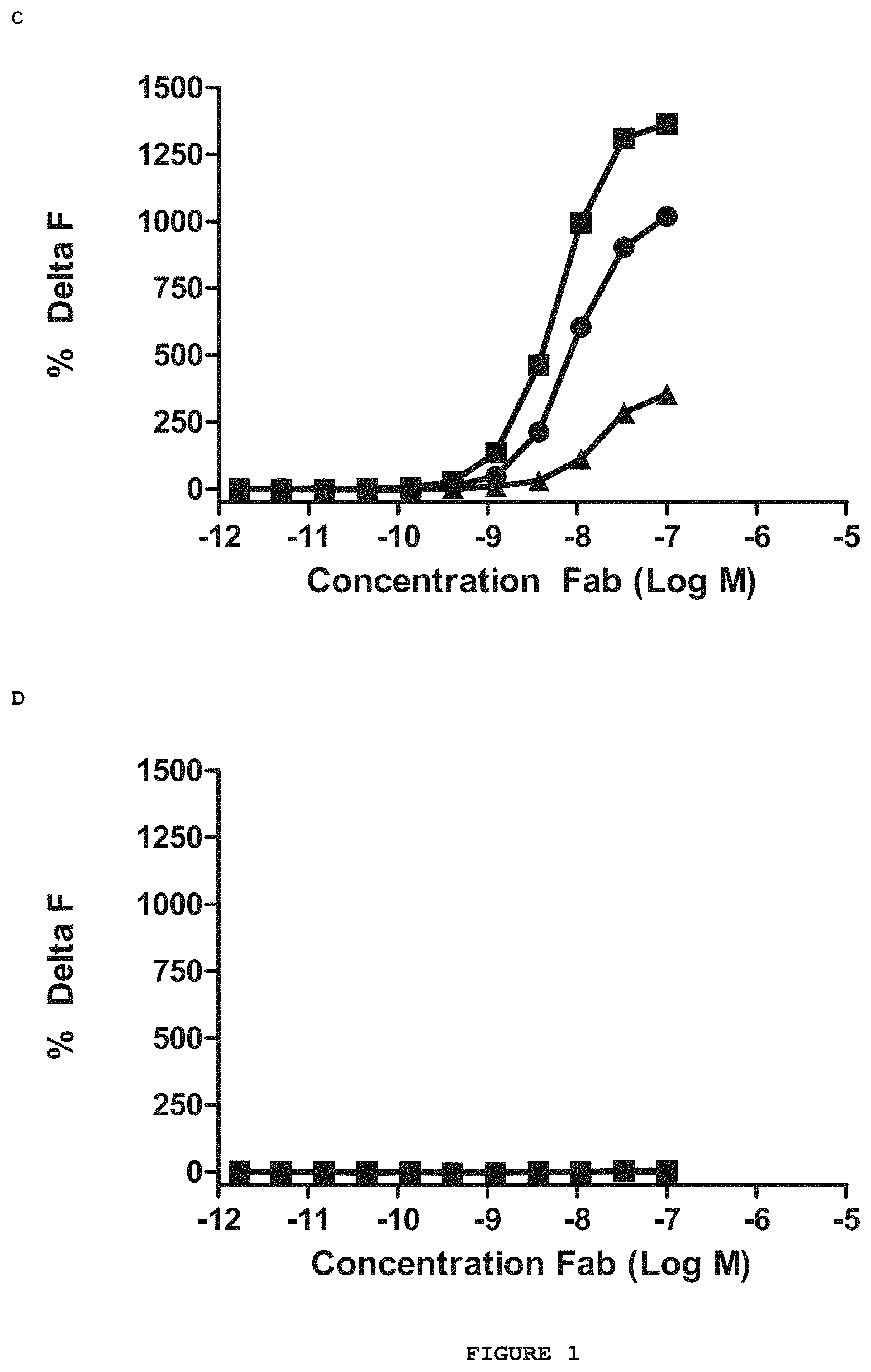
FIG. 1 shows the results of the direct binding HTRF.TM. assay between the purified Abet0007 Fab and a series of Amyloid beta peptides. The Abet0007 clone (.box-solid.) shows binding to the human Amyloid beta 1-42 peptide (FIG. 1A) and the murine Amyloid beta 1-42 peptide (FIG. 1C) but shows no binding to the human Amyloid beta 1-40 peptide (FIG. 1B) or the scrambled human Amyloid beta 1-42 peptide (FIG. 1D). The positive control antibody (.circle-solid.) and the negative control antibody (.DELTA.) were used to verify the integrity of the assay.
FIG. 2 shows the inhibition of the formation of the biotinylated human Amyloid beta 1-42 peptide and Abet0007 IgG2 complex by increasing concentrations of competitor peptides. Complex formation is inhibited by human Amyloid beta 1-42 (.circle-solid.), 11-42 (.DELTA.), 17-42 () and 1-43 (.diamond-solid.) peptides. It is not inhibited by human Amyloid beta 1-40 peptide (.box-solid.) or by the negative control peptide (.smallcircle.).
FIG. 3 shows the Surface Plasmon Resonance (BIAcore) traces for the human Amyloid beta 1-42 peptide binding to immobilised Abet0007 IgG2 at concentrations of 100 nM (top trace), 50 nM, 25 nM, 12.5 nM, 6.2 nM and 3.1 nM (bottom trace) peptide. Each trace is fitted to a 1:1 Langmuir model.
FIG. 4 shows sample images from the in vitro immunohistochemical staining of Abet0007 IgG2. (A) A positive control antibody shows strong plaque recognition (score=4) on human AD brain sections (ApoE genotype 3/3; Braak stage 6; 20 .mu.g/ml antibody). (B) The Abet0007 IgG2 lead clone shows no plaque recognition (score=0) on an adjacent brain section (20 .mu.g/ml). (C) The same positive control antibody shows strong plaque recognition (score=4) on Tg2576 mouse brain sections (18 month old mice; 20 .mu.g/ml antibody). (D) The Abet0007 IgG2 lead clone shows no plaque recognition (score=0) on an adjacent mouse brain section (20 .mu.g/ml).
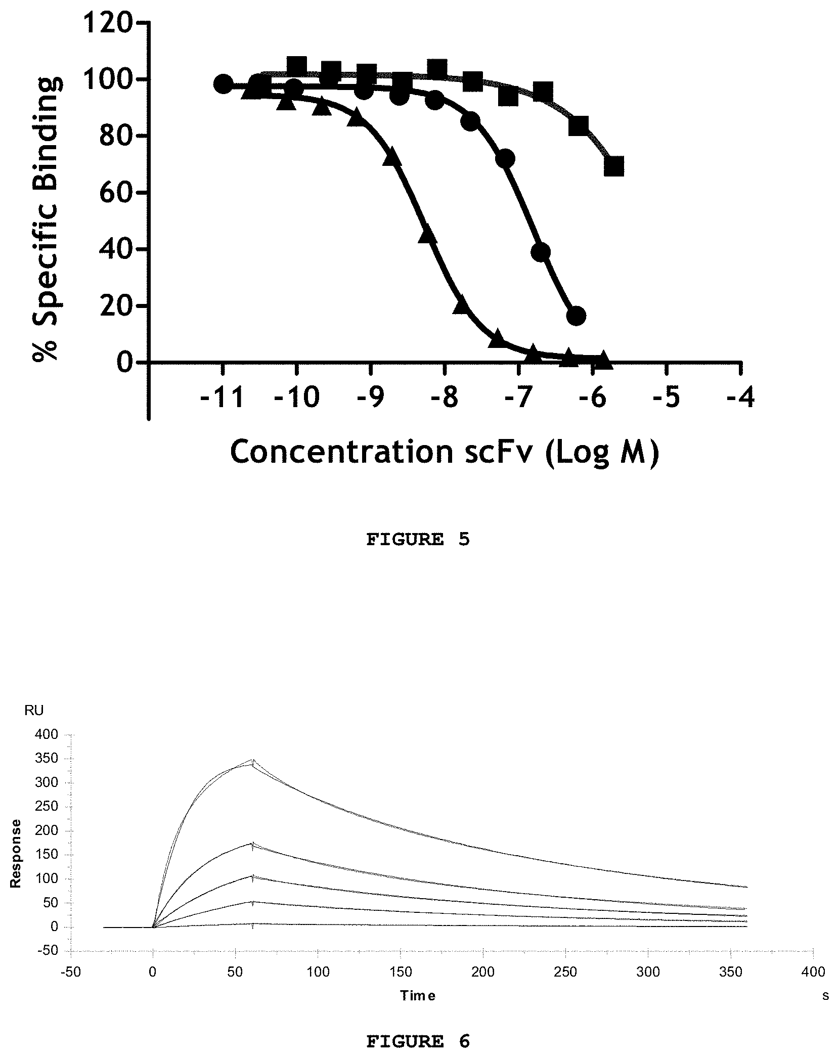
FIG. 5 shows the inhibition of the formation of the human Amyloid beta 1-42 and Abet0042 IgG complex by increasing concentrations of Abet0007 scFv (.circle-solid.) and Abet0144 scFv (.tangle-solidup.). The Abet0144 clone is significantly more potent than the Abet0007 parent clone in this assay. A negative control antibody (.box-solid.) is included for comparison.
FIG. 6 shows the Surface Plasmon Resonance (BIAcore) traces for the purified Abet0144 scFv binding to immobilised human Amyloid beta 1-42 peptide at concentrations of 400 nM (top trace), 200 nM, 100 nM, 50 nM and 12.5 nM (bottom trace) scFv. Each trace is fitted to a 1:1 Langmuir model.
FIG. 7 shows the Surface Plasmon Resonance (BIAcore) traces for human Amyloid beta 1-42 peptide binding to immobilised Abet0144-GL IgG1-TM antibody at concentrations of 50 nM (top trace), 25 nM, 12.5 nM, 6.25 nM, 3.13 nM and 1.56 nM (bottom trace) peptide. Each trace is fitted to a 1:1 Langmuir model.
FIG. 8 shows the Surface Plasmon Resonance (BIAcore) traces for a series of Amyloid beta peptides at 400 nM binding to immobilised Abet0144-GL IgG 1-TM antibody. There is clear binding to the biotinylated human Amyloid beta 1-42 peptide (top trace) and the unlabelled human Amyloid beta 1-42 peptide (second trace). There is no discernable binding to scrambled biotinylated human Amyloid beta 1-42 peptide, biotinylated human Amyloid beta 1-40 peptide, unlabelled human Amyloid beta 1-40 peptide or biotinylated-insulin (flat lines).
FIG. 9 shows specificity profiling of Abet0144-GL IgG1-TM using a biochemical epitope competition assay in which inhibition of the formation of a complex between biotinylated human Amyloid beta 1-42 peptide and Abet0144-GL IgG1-TM by increasing concentrations of competitor peptides is measured. Complex formation is inhibited by human Amyloid beta 1-42 (.circle-solid.), pyro 3-42 (.diamond-solid.) and pyro 11-42 (.smallcircle.) peptides. No significant inhibition is observed with human Amyloid beta 1-40 peptide (.box-solid.), 1-16 (.tangle-solidup.) and 12-28 () peptide truncates or with the negative control peptide (n).
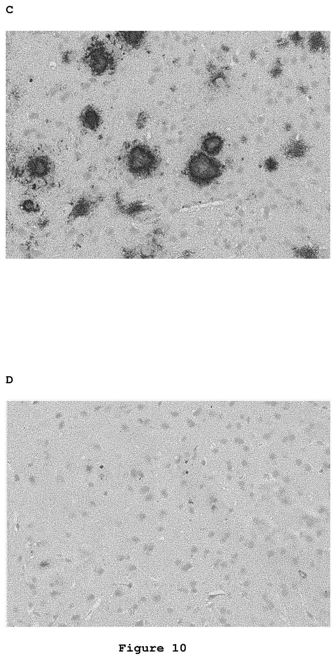
FIG. 10 shows sample images from the in vitro immunohistochemical staining of Abet0144-GL IgG1-TM. (A) A positive control antibody shows strong plaque recognition (score=4) on human AD brain sections (ApoE genotype 3/3; 20 .mu.g/ml antibody). (B) The Abet0144-GL IgG1-TM lead clone shows some plaque recognition (score=1.5) on an adjacent brain section (20 .mu.g/ml). (C) The same positive control antibody shows strong plaque recognition (score=4) on Tg2576 mouse brain sections (18 month old mice; 20 .mu.g/ml antibody). (D) The Abet0144-GL IgG1-TM lead clone shows some plaque recognition (score=1) on an adjacent mouse brain section (20 .mu.g/ml).
FIG. 11 shows the inhibition of the formation of the human Amyloid beta 1-42 peptide and Abet0144-GL IgG1-TM complex by increasing concentrations of purified competitor scFv (.circle-solid.). Four of the most potent scFv clones, Abet0369 (FIG. 11A), Abet0377 (FIG. 11B), Abet0380 (FIG. 11C) and Abet0382 (FIG. 11D) all show significant improvement in potency over the parent Abet0144-GL scFv sequence (.box-solid.).
FIG. 12 shows the Surface Plasmon Resonance (BIAcore) traces for human Amyloid beta 1-42 peptide binding to immobilised Abet0380-GL IgG 1-TM antibody at concentrations from 1024 nM (top trace) to 63 pM (bottom trace) peptide. Each trace is fitted to a 1:1 Langmuir model.
FIG. 13 shows the Surface Plasmon Resonance (BIAcore) traces for a series of Amyloid beta peptides binding to immobilised Abet0380-GL IgG1-TM antibody. There is clear binding to the biotinylated human Amyloid beta 1-42 peptide (top trace) and the unlabelled murine Amyloid beta 1-42 peptide (second trace). There is no discernable binding to biotinylated human Amyloid beta 1-40 peptide or unlabelled murine Amyloid beta 1-40 peptide (flat lines).
FIG. 14 shows sample images from the in vitro immunohistochemical staining of Abet0380-GL IgG1-TM. (A) A positive control antibody shows strong plaque recognition (score=4) on human AD brain sections (ApoE genotype 3/3, Braak stage 6; 5 .mu.g/ml antibody). (B) The Abet0380-GL IgG1-TM lead clone shows strong plaque recognition (score=3) on an adjacent brain section (10 .mu.g/ml). (C) The same positive control antibody shows strong plaque recognition (score=4) on Tg2576 mouse brain sections (22 month old mice; 20 .mu.g/ml antibody). (D) The Abet0380-GL IgG1-TM lead clone shows strong plaque recognition (score=4) on an adjacent mouse brain section (20 .mu.g/ml).
FIG. 15 shows Western Blot analysis of Abeta 42 aggregate preparation and detection using the Abet0380-GL IgG1TM. (A) Abet0380-GL IgG1TM detection of non-photo cross-linked (non PICUP) A.beta.42 aggregate. (B) Abet0380-GL IgG1TM detection of photo cross-linked A.beta.42 aggregate (PICUP). Here we demonstrate that Abet0380-GL IgG1TM specifically recognises A1-42 monomer and low n oligomer species up to and including pentamer.
FIG. 16 shows the dose-dependent reduction of the level of free Amyloid beta 1-42 peptide in the CSF (A), the increase of total Amyloid beta 1-42 peptide in brain tissue (B) and the unaffected levels of total Amyloid beta 1-40 peptide in brain tissue (C) by increasing doses of Abet0380-GL IgG1-TM antibody in Sprague-Dawley rats receiving repeated weekly doses over 14 days.
FIG. 17 shows sample images from the immunohistochemical analysis of binding of Abet0380-GL IgG1-TM to Amyloid beta plaques in vivo 168 hours after a peripheral dose to aged Tg2576 mice. A positive control antibody given at 30 mg/kg shows strong in vivo plaque recognition (A), whereas Abet0380-GL IgG1-TM given at 30(B) or 10(C) mg/kg does not show any in vivo plaque decoration.
FIG. 18 shows the specificity of Abet0380-GL IgG1-TM in competition binding experiments with a range of different concentrations (10 uM down to 0.17 nM) of a panel of full length, truncate and pyro human Abeta peptides (Abeta 1-42, Abeta 1-43, Abeta 1-16, Abeta 12-28, Abeta 17-42, Abeta pyro-3-42, or Abeta pyro-11-42). Key: Abeta 1-42 beta 1-43 Abeta 1-16 .tangle-solidup. Abeta 12-28 Abeta 17-42 Abeta Pyro-3-42 Abeta Pyro 11-42 .diamond. Vehicle 1 (DMSO) * Vehicle 2 (NH4OH) The x-axis shows the concentration of Abeta peptide in log M, the y-axis shows % specific binding. Inhibition of Abet0380-GL IgG1-TM: N-terminal Biotin Abeta 1-42 binding was observed with Abeta 1-42, Abeta 1-43, Abeta 17-42, Abeta Pyro-3-42 & Abeta Pyro-11-42 with IC.sub.50 values ranging from 10.sup.-8 to 10.sup.-9 molar for this group. No inhibition of Abet0380-GL IgG1-TM: N-terminal Biotin Abeta 1-42 binding was observed with Abeta 1-16 or Abeta 12-28.
FIG. 19 shows the ability of antibody Abet0144-GL to sequester amyloid beta 1-42 in a normal rat PK-PD study. The x-axis shows vehicle or concentration of Abet0144-GL (10 mg/kg, or 40 mg/kg), the y-axis shows the concentration of total amyloid beta 1-42 in CSF in .mu.g/ml. Free amyloid beta 1-42 in CSF was not significantly altered by either 10 or 40 mg/kg of Abet0144-GL (5 and 18% increase respectively when compared with vehicle). Total amyloid beta 1-42 in CSF was significantly increased by 38% at 10 mg/kg, and by 139% at 40 mg/kg. Total amyloid beta 1-42 in brain tissue was also significantly increased, by 16% and 50% at 10 and 40 mg/kg respectively. Data from this study in normal rats, demonstrate that Abet0144-GL had no significant effect on free amyloid beta 1-42 levels in CSF, whilst increasing total amyloid beta 1-42 levels in both CSF and brain.
DETAILED DESCRIPTION
By binding isoforms of A.beta. peptide 1-42 and N-terminal truncates thereof (n-42) in plasma, brain and cerebrospinal fluid (CSF), a binding member according to the present invention may prevent accumulation or reverse the deposition of A.beta. n-42 isoforms within the brain and cerebrovasculature. Binding members according to the present invention may bind and precipitate soluble A.beta.1-42 in blood plasma and/or in cerebrospinal fluid (CSF), thereby reducing the concentration of A.beta.1-42 in the serum and/or CSF, respectively. This represents a therapeutic approach for Alzheimer's disease and other conditions associated with amyloidosis.
Binding members are specific for the target epitope within A.beta.17-42, more specifically within A.beta.29-42, and bind this target epitope with high affinity relative to non-target epitopes, for example epitopes from A.beta.1-40, thereby targeting the main toxic species linked with amyloid plaque formation. For example, a binding member may display a binding affinity for A.beta.1-42 which is at least 10-fold, at least 100-fold, at least 1000-fold or at least 10,000-fold greater than for A.beta.1-40. Thus, the binding member is selective for binding A.beta.1-42 over A.beta.1-40. As noted above, the binding member may bind A.beta.1-42 with a dissociation constant (KD) of 500 pM or less. Preferably, it shows no significant binding to A.beta.1-40. Affinity and binding can be determined using surface plasmon resonance using monomeric A.beta. peptide, as described in the Examples.
Binding to A.beta. can also be measured in a homogenous time resolved fluorescence (HTRF.TM.) assay, to determine whether the antibody is able to compete for binding to A.beta. with a reference antibody molecule to the A.beta. peptide, as described in the Examples.
An HTRF.TM. assay is a homogeneous assay technology that utilises fluorescence resonance energy transfer between a donor and acceptor fluorophore that are in close proximity. Such assays can be used to measure macromolecular interactions by directly or indirectly coupling one of the molecules of interest to a donor fluorophore, europium (Eu3+) cryptate, and coupling the other molecule of interest to an acceptor fluorophore XL665, (a stable cross linked allophycocyanin). Excitation of the cryptate molecule (at 337 nm) results in fluorescence emission at 620 nm. The energy from this emission can be transferred to XL665 in close proximity to the cryptate, resulting in the emission of a specific long-lived fluorescence (at 665 nm) from the XL665. The specific signals of both the donor (at 620 nm) and the acceptor (at 665 nm) are measured, allowing the calculation of a 665/620 nm ratio that compensates for the presence of coloured compounds in the assay.
A binding member according to the invention may compete for binding to A.beta.1-42 and thus inhibit binding of the reference antibody in an HTFR.TM. competition assay with A.beta.1-42, but not with A.beta.1-40. A binding member may show at least 70%, at least 75%, at least 80%, at least 85% or at least 90% inhibition of Abet0144GL for binding to A.beta.1-42 in an HTRF.TM. assay.
Potency of inhibition of binding may be expressed as an IC.sub.50 value, in nM unless otherwise stated. In functional assays, IC.sub.50 is the concentration of an antibody molecule that reduces a biological response by 50% of its maximum. In ligand-binding studies, IC.sub.50 is the concentration that reduces receptor binding by 50% of maximal specific binding level. IC.sub.50 may be calculated by plotting % of maximal biological response as a function of the log of the binding member concentration, and using a software program, such as Prism (GraphPad) or Origin (Origin Labs) to fit a sigmoidal function to the data to generate IC.sub.50 values. Suitable assays for measuring or determining potency are well known in the art.
A binding member may have an IC.sub.50 of 5 nM or less, e.g. 2 nM or less, e.g. 1 nM or less, in HTRF.TM. epitope competition assay with Abet0144-GL and A.beta.1-42. Abet0144-GL is an antibody molecule having VH domain SEQ ID NO: 20 and VL domain SEQ ID NO: 29. It may be used in the assay in the same format as the antibody molecule to be tested, for example in scFv or IgG, e.g. IgG1 format. Thus, IgG antibody molecules according to the invention may compete with Abet0144-GL IgG for binding to human A.beta.1-42 in an HTRF epitope competition assay. Potency in such an assay may be less than 1 nM.
A binding member according to the invention may show specific binding for A.beta.1-42 over A.beta.1-40, as determined by an HTRF.TM. competition assay. In such an assay, A.beta.1-40 may show no significant inhibition of the binding member binding to the A.beta.1-42 peptide, e.g. it may show less than 20%, e.g. less than 10% or less than 5%, inhibition in such an assay, and preferably shows no significant inhibition in such an assay.
Binding members according to the invention recognise an epitope within human A.beta.17-42, more specifically within human A.beta.29-42 and may also recognise their target epitope in A.beta. from other species, e.g. mouse or rat. The potency of a binding member as calculated in an HTRF.TM. competition assay using A.beta.1-42 from a first species (e.g. human) may be compared with potency of the binding member in the same assay using A.beta.1-42 from a second species (e.g. mouse A.beta.1-42), in order to assess the extent of cross-reactivity of the binding member for A.beta.1-42 of the two species. Potency, as determined by IC.sub.50 measurements, may be within 10-fold or within 100-fold. As noted above, Abet0144GL may be used as reference antibody in the HTRF.TM. competition assay. Binding members described herein may have a greater potency in a human A.beta.1-42 assay than in a non-human A.beta.1-42 assay.
A binding member may comprise an antibody molecule having one or more CDRs, e.g. a set of CDRs, within an antibody framework (i.e. an antibody antigen-binding domain). For example, an antibody molecule may comprise an antibody VH and/or VL domain. VH and VL domains of antibody molecules are also provided as part of the invention. As is well-known, VH and VL domains comprise complementarity determining regions, ("CDRs"), and framework regions, ("FWs"). A VH domain comprises a set of HCDRs and a VL domain comprises a set of LCDRs. An antibody molecule may comprise an antibody VH domain comprising a VH CDR1, CDR2 and CDR3 and/or an antibody VL domain comprising a VL CDR1, CDR2 and CDR3. VH or VL domains may further comprise a framework. A VH or VL domain framework typically comprises four framework regions, FW1, FW2, FW3 and FW4, which are interspersed with CDRs in the following structure: FW1-CDR1-FW2-CDR2-FW3-CDR3-FW4.
Examples of antibody VH and VL domains, FWs and CDRs according to aspects of the invention are listed in Tables 5 and 6 and the appended sequence listing that forms part of the present disclosure. All VH and VL sequences, CDR sequences, sets of CDRs, sets of HCDRs and sets of LCDRs disclosed herein, as well as combinations of these elements, represent aspects of the invention. As described herein, a "set of CDRs" comprises CDR1, CDR2 and CDR3. Thus, a set of HCDRs refers to HCDR1, HCDR2 and HCDR3, and a set of LCDRs refers to LCDR1, LCDR2 and LCDR3. Unless otherwise stated, a "set of CDRs" includes HCDRs and LCDRs. Typically antibody molecules of the invention are monoclonal antibodies.
In other embodiments, a binding member may comprise an antigen-binding site within a non-antibody molecule, normally provided by one or more CDRs e.g. a set of CDRs in a non-antibody protein scaffold, as discussed further below.
The isolation of a parent antibody molecule designated Abet0007, followed by directed mutation of CDR3 and selection of an optimised antibody Abet0144, germlined to Abet0144-GL with a set of CDR sequences and framework sequences as shown in Tables 5, 6 and the sequence listing, is described herein. Through an extensive process of further optimisation and recombination of multiple libraries as described in the Examples, a panel of antibody clones was generated from Abet0144GL. These further optimised clones are designated Abet0380, Abet0319, Abet0321b, Abet0322b, Abet0323b, Abet0328, Abet0329, Abet0332, Abet0342, Abet0343, Abet0369, Abet0370, Abet0371, Abet0372, Abet0373, Abet0374, Abet0377, Abet0378, Abet0379, Abet0381, Abet0382 and Abet0383. Their CDR sequences and variable domain sequences are referenced in Tables 5 and 6 and set out in the sequence listing. Germlined VH and VL domain sequences Abet0380GL, Abet0377GL, Abet0343GL, Abet0369GL and Abet0382GL are shown in Table 8 and Table 9.
For example, Tables 5 and 6 show that Abet0380 has a set of CDRs, in which HCDR1 is SEQ ID NO: 525 (Kabat residues 31-35), HCDR2 is SEQ ID NO: 526 (Kabat residues 50-65), HCDR3 is SEQ ID NO: 527 (Kabat residues 95-102), LCDR1 is SEQ ID NO: 534 (Kabat residues 24-34), LCDR2 is SEQ ID NO: 535 (Kabat residues 50-56) and LCDR3 is SEQ ID NO: 536 (Kabat residues 89-97). The other optimised antibody clones are shown in Tables 5 and 6 in a similar way and are also provided as aspects of the invention.
A binding member for human A.beta.1-42 in accordance with the invention may comprise one or more CDRs as described herein, e.g. a set of CDRs. The CDR or set of CDRs may be an Abet0380, Abet0319, Abet0321b, Abet0322b, Abet0323b, Abet0328, Abet0329, Abet0332, Abet0342, Abet0343, Abet0369, Abet0370, Abet0371, Abet0372, Abet0373, Abet0374, Abet0377, Abet0378, Abet0379, Abet0381, Abet0382 and Abet0383 set of CDRs, or a germlined version thereof, or may be a variant thereof as described herein.
In some embodiments;
HCDR1 is may be 5 amino acids long, consisting of Kabat residues 31-35;
HCDR2 may be 17 amino acids long, consisting of Kabat residues 50-65;
HCDR3 may be 16 amino acids long, consisting of Kabat residues 95-102;
LCDR1 may be 11 amino acids long, consisting of Kabat residues 24-34;
LCDR2 may be 7 amino acids long, consisting of Kabat residues 50-56; and/or
LCDR3 may be 9 amino acids long, consisting of Kabat residues 89-97.
Binding members may comprise a HCDR1, HCDR2 and/or HCDR3 and/or an LCDR1, LCDR2 and/or LCDR3 of any of the antibodies listed in Tables 5 and 6, e.g., a set of CDRs of any of the antibodies listed in Table 5 or 6. The binding member may comprise a set of VH CDRs of any one of these antibodies. Optionally, it may also comprise a set of VL CDRs of one of these antibodies. The VL CDRs may be from the same or a different antibody as the VH CDRs. A VH domain comprising a set of HCDRs of any of the antibodies listed in Tables 5, and/or a VL domain comprising a set of LCDRs of any of the antibodies listed in Tables 6, are also provided herein.
A binding member may comprise a set of H and/or L CDRs of any of the antibodies listed in Tables 5 and 6 with one or more amino acid mutations, e.g. up to 5, 10 or 15 mutations, within the disclosed set of H and/or L CDRs. A mutation may be an amino acid substitution, deletion or insertion. For example, an antibody molecule of the invention may comprise the set of H and/or L CDRs from any one of Abet0380, Abet0319, Abet0321b, Abet0322b, Abet0323b, Abet0328, Abet0329, Abet0332, Abet0342, Abet0343, Abet0369, Abet0370, Abet0371, Abet0372, Abet0373, Abet0374, Abet0377, Abet0378, Abet0379, Abet0381, Abet0382 and Abet0383, or a germlined version thereof, with one or two amino acid mutations, e.g. substitutions.
For example, the binding member may comprise
a VH domain comprising the Abet0380 or Abet0380GL set of HCDRs, wherein the amino acid sequences of the Abet0380 or Abet0380GL HCDRs are
HCDR1 SEQ ID NO: 525,
HCDR2 SEQ ID NO: 526, and
HCDR3 SEQ ID NO: 527,
or comprising the Abet0380 set of HCDRs with one or two amino acid mutations, and
(ii) a VL domain comprising the Abet0380 or Abet0380GL set of LCDRs, wherein the amino acid sequences of the Abet0380 or Abet0380GL LCDRs are
LCDR1 SEQ ID NO: 534
LCDR2 SEQ ID NO: 535, and
LCDR3 SEQ ID NO: 536,
or comprising the Abet0380 or Abet0380GL set of LCDRs with one or two amino acid mutations.
Mutations may potentially be made at any residue within the set of CDRs. In some embodiments, substitutions may be made at the positions substituted in any of Abet0380, Abet0319, Abet0321b, Abet0322b, Abet0323b, Abet0328, Abet0329, Abet0332, Abet0342, Abet0343, Abet0369, Abet0370, Abet0371, Abet0372, Abet0373, Abet0374, Abet0377, Abet0378, Abet0379, Abet0381, Abet0382 and Abet0383 compared with Abet0144GL, or at the positions substituted in any of Abet0319, Abet0321b, Abet0322b, Abet0323b, Abet0328, Abet0329, Abet0332, Abet0342, Abet0343, Abet0369, Abet0370, Abet0371, Abet0372, Abet0373, Abet0374, Abet0377, Abet0378, Abet0379, Abet0381, Abet0382 and Abet0383 compared with Abet0380, or germlined versions thereof, as shown in Tables 5 and 6.
For example, the one or more substitutions may be at one or more of the following Kabat residues:
26, 27, 28, 29 or 30 in VH FW1;
31, 32, 33, 34 or 35 in VH CDR1;
52a, 53, 54, 55, 56, 57, 58 or 62 in VH CDR2;
98, 99, 100h or 102 in VH CDR3;
24, 25, 26, 27, 28, 29, 30, 31, 32, 33, 34 in VL CDR1;
89, 90, 92, 93, 94 or 97 in VL CDR3.
Examples of possible amino acid substitutions at particular Kabat residue positions are shown in Tables 12 and 14 for the VH domain and Tables 13 and 15 for the VL domain.
As described above, a binding member may comprise an antibody molecule having one or more CDRs, e.g. a set of CDRs, within an antibody framework. For example, one or more CDRs or a set of CDRs of an antibody may be grafted into a framework (e.g. human framework) to provide an antibody molecule. The framework regions may be of human germline gene segment sequences. Thus, the framework may be germlined, whereby one or more residues within the framework are changed to match the residues at the equivalent position in the most similar human germline framework. The skilled person can select a germline segment that is closest in sequence to the framework sequence of the antibody before germlining and test the affinity or activity of the antibodies to confirm that germlining does not significantly reduce antigen binding or potency in assays described herein. Human germline gene segment sequences are known to those skilled in the art and can be accessed for example from the VBASE compilation (VBASE, MRC Centre of Protein Engineering, UK, 1997, http//mrc-cpe.cam.ac.uk).
A binding member as described herein may be an isolated human antibody molecule having a VH domain comprising a set of HCDRs in a human germline framework, e.g. Vh3-23 DP-47. Thus, the VH domain framework regions FW1, FW2 and/or FW3 may comprise framework regions of human germline gene segment Vh3-23 DP-47 and/or may be germlined by mutating framework residues to match the framework residues of this human germline gene segment. FW4 may comprise a framework region of a human germline j segment.
The amino acid sequence of VH FW1 may be SEQ ID NO: 528. VH FW1 contains a series of residues at Kabat positions 26-30 that are believed to contribute to antigen binding and/or to be important for structural conformation of the CDR1 loop. Substitutions may be included in SEQ ID NO: 528, for example to synergise with the selected sequence of HCDR1. The one or more substitutions may optionally be selected from those shown in Table 12 or Table 14.
The amino acid sequence of VH FW2 may be SEQ ID NO: 529. The amino acid sequence of VH FW3 may be SEQ ID NO: 530. The amino acid sequence of VH FW4 may be SEQ ID NO: 531.
Normally the binding member also has a VL domain comprising a set of LCDRs, e.g. in a human germline framework, e.g. V lambda 23-3 DPL-23. Thus, the VL domain framework regions may comprise framework regions FW1, FW2 and/or FW3 of human germline gene segment V lambda 23-3 DPL-23 and/or may be germlined by mutating framework residues to match the framework residues of this human germline gene segment. FW4 may comprise a framework region of a human germline j segment. The amino acid sequence of VL FW1 may be SEQ ID NO: 537. The amino acid sequence of VL FW2 may be SEQ ID NO: 538. The amino acid sequence of VL FW3 may be SEQ ID NO: 539. The amino acid sequence of VL FW4 may be SEQ ID NO: 540.
A germlined VH or VL domain may or may not be germlined at one or more Vernier residues, but is normally not.
For example, an antibody molecule or a VH domain as described herein may comprise the following set of heavy chain framework regions:
FW1 SEQ ID NO: 528;
FW2 SEQ ID NO: 529;
FW3 SEQ ID NO: 530;
FW4 SEQ ID NO: 531;
or may comprise the said set of heavy chain framework regions with 1, 2, 3, 4, 5, 6 or 7 amino acid mutations, e.g. substitutions.
An antibody molecule or a VL domain as described herein may comprise the following set of light chain framework regions:
FW1 SEQ ID NO: 537;
FW2 SEQ ID NO: 538;
FW3 SEQ ID NO: 539;
FW4 SEQ ID NO: 540;
or may comprise the said set of light chain framework regions with 1, 2, 3, 4, 5, or 6 amino acid mutations, e.g. substitutions.
A non-germlined antibody molecule has the same CDRs, but different frameworks, compared to a germlined antibody molecule. Of the antibody sequences shown herein in the appended sequence listing, sequences of Abet0144-GL, Abet0380-GL, Abet0377-GL, Abet0343-GL, Abet0369-GL, and Abet0382-GL are germlined. Germlined antibodies of other antibody molecules whose sequences are disclosed herein may be produced by germlining framework regions of their VH and VL domain sequences, optionally to Vh3-23 DP-47 in the VH domain and V lambda 23-3 DPL-23 in the VL domain.
Typically, a VH domain is paired with a VL domain to provide an antibody antigen-binding site, although as discussed above a VH or VL domain alone may be used to bind antigen. For example, the Abet0380-GL VH domain (SEQ ID NO: 524) may be paired with the Abet0380-GL VL domain (SEQ ID NO:533), so that an antibody antigen-binding site is formed comprising both the Abet0380-GL VH and VL domains. Analogous embodiments are provided for the VH and VL domains of the other antibodies disclosed herein. In other embodiments, the Abet0380-GL VH is paired with a VL domain other than the Abet0380-GL VL. Light-chain promiscuity is well established in the art. Again, analogous embodiments are provided by the invention for the other VH and VL domains disclosed herein. Thus, a VH domain comprising the VH CDRs or the germlined VH domain sequence of any of Abet0319, Abet0321b, Abet0322b, Abet0323b, Abet0328, Abet0329, Abet0332, Abet0342, Abet0343, Abet0369, Abet0370, Abet0371, Abet0372, Abet0373, Abet0374, Abet0377, Abet0378, Abet0379, Abet0380, Abet0381, Abet0382 and Abet0383 may be paired with a VL domain comprising the VL CDRs or germlined VL domain from a different antibody e.g. the VH and VL domains may be from different antibodies selected from Abet0319, Abet0321b, Abet0322b, Abet0323b, Abet0328, Abet0329, Abet0332, Abet0342, Abet0343, Abet0369, Abet0370, Abet0371, Abet0372, Abet0373, Abet0374, Abet0377, Abet0378, Abet0379, Abet0380, Abet0381, Abet0382 and Abet0383.
A binding member may comprise
(i) a VH domain amino acid sequence as shown in Table 16 or in the appended sequence listing for any of Abet0380, Abet0343, Abet0369, Abet0377 and Abet0382, or a germlined version thereof,
or comprising that amino acid sequence with one or two amino acid mutations; and
(ii) a VL domain amino acid sequence as shown in Table 16 or in the appended sequence listing for any of Abet0380, Abet0343, Abet0369, Abet0377 and Abet0382, or a germlined version thereof,
or comprising that amino acid sequence with one or two amino acid mutations.
An antibody molecule may comprise:
(i) a VH domain having an amino acid sequence at least 90%, 95% or 98% identical to a VH domain amino acid sequence shown in Table 16 for any of Abet0380, Abet0343, Abet0369, Abet0377 and Abet0382, or a germlined version thereof; and
(ii) a VL domain having an amino acid sequence at least 90%, 95% or 98% identical to a VL domain amino acid sequence shown in Table 16 for any of Abet0380, Abet0343, Abet0369, Abet0377 and Abet0382, or a germlined version thereof.
It may comprise a VH domain and a VL domain at least 90%, 95% or 98% identical with the VH domain and VL domain, respectively, of any of Abet0380, Abet0343, Abet0369, Abet0377 and Abet0382, or a germlined version thereof.
A binding member may comprise a VH domain and a VL domain in which;
(i) the VH domain amino acid sequence is shown in SEQ ID NO: 524 and the VL domain amino acid sequence is shown in SEQ ID NO: 533.
(ii) the VH domain amino acid sequence has 1, 2, 3, 4, 5, 6, 7, 8, 9 or 10 amino acid substitutions as compared to SEQ ID NO: 524 and the VL domain amino acid sequence has 1, 2, 3, 4, 5, 6, 7, 8, 9 or 10 amino acid substitutions as compared to SEQ ID NO: 533; or
(iii) the VH domain amino acid sequence has at least 80%, at least 85%, at least 90% or at least 95% sequence identity with SEQ ID NO: 524 and the VL domain amino acid sequence has at least 80%, at least 85%, at least 90% or at least 95% sequence identity with SEQ ID NO: 533.
In some embodiments, an antibody molecule may lack antibody constant regions, for example an scFv.
In other embodiments, an antibody molecule may comprise an antibody constant region. An antibody molecule may be a whole antibody such as an IgG, i.e. an IgG1, IgG2, or IgG4, or may be an antibody fragment or derivative as described below. Antibody molecules can also have other formats, e.g. IgG1 with YTE (Dall'Acqua et al. (2002) J. Immunology, 169: 5171-5180; Dall'Acqua et al. (2006) J Biol. Chem. 281(33):23514-24) and/or TM mutations (Oganesyan et al. (2008) Acta Cryst D64:700-4) in the Fc region.
The invention provides a binding member of the present invention with a variant Fc region, wherein the variant comprises a phenylalanine (F) residue at position 234, a phenylalanine (F) residue or a glutamic acid (E) residue at position 235 and a serine (S) residue at position 331, as numbered by the EU index as set forth in Kabat. Such mutation combinations are hereinafter referred to as the triple mutant (TM).
A binding member as described herein may comprise a CDR, VH domain, VL domain, antibody-antigen binding site or antibody molecule which is encoded by the nucleic acid sequences and/or the vector of any of:
(i) deposit accession number NCIMB 41889 (Abet0007);
(ii) deposit accession number NCIMB 41890 (Abet0380-GL);
(iii) deposit accession number NCIMB 41891 (Abet0144-GL);
(iv) deposit accession number NCIMB 41892 (Abet0377-GL).
A binding member as described herein may be produced or producible from the nucleic acid, vector or cell line of deposit accession number NCIMB 41889, 41890, 41891 or 41892. For example, a binding member may be produced by expression of the nucleic acid or vector of the cell line of deposit accession number NCIMB 41890. The nucleic acid or vector may be expressed any convenient expression system. Alternatively, the binding member may be expressed by the cell line of deposit accession number NCIMB 41889, 41890, 41891 or 41892.
Aspects of the invention also provide nucleic acid encoding the VH and/or VL domains, which is contained in the cell line of accession number 41889, 41890, 41891 or 41892; a vector comprising said nucleic acid, which is contained in the cell line of accession number 41889, 41890, 41891 or 41892; and the cells or cell line of accession number 41889, 41890, 41891 or 41892.
A binding member according to the present invention may comprise an antibody antigen binding site or antibody molecule that competes for binding to human A.beta.1-42 with any antibody molecule encoded by nucleic acid deposited under accession number 41889, 41890, 41891 or 41892, or with an antibody molecule that comprises the VH domain and VL domain amino acid sequences of Abet007, Abet0380-GL, Abet0144-GL or Abet0377-GL as set out in the appended sequence listing.
Binding Member
The term binding member describes one member of a pair of molecules that bind one another. The members of a binding pair may be naturally derived or wholly or partially synthetically produced. One member of the pair of molecules has an area on its surface, or a cavity, which binds to and is therefore complementary to a particular spatial and polar organization of the other member of the pair of molecules. Examples of types of binding pairs are antigen-antibody, biotin-avidin, hormone-hormone receptor, receptor-ligand, enzyme-substrate. The present invention is concerned with antigen-antibody type reactions.
A binding member normally comprises a molecule having an antigen-binding site. For example, a binding member may be an antibody molecule or a non-antibody protein that comprises an antigen-binding site.
An antigen binding site may be provided by means of arrangement of CDRs on non-antibody protein scaffolds, such as fibronectin or cytochrome B etc. [Haan & Maggos (2004) BioCentury, 12(5): A1-A6; Koide et al. (1998) Journal of Molecular Biology, 284: 1141-1151; Nygren et al. (1997) Current Opinion in Structural Biology, 7: 463-469], or by randomising or mutating amino acid residues of a loop within a protein scaffold to confer binding specificity for a desired target. Scaffolds for engineering novel binding sites in proteins have been reviewed in detail by Nygren et al. [supra]. Protein scaffolds for antibody mimics are disclosed in WO00/34784, which is herein incorporated by reference in its entirety, in which the inventors describe proteins (antibody mimics) that include a fibronectin type III domain having at least one randomised loop. A suitable scaffold into which to graft one or more CDRs, e.g. a set of HCDRs or an HCDR3 and/or LCDR3, may be provided by any domain member of the immunoglobulin gene superfamily. The scaffold may be a human or non-human protein. An advantage of a non-antibody protein scaffold is that it may provide an antigen-binding site in a scaffold molecule that is smaller and/or easier to manufacture than at least some antibody molecules. Small size of a binding member may confer useful physiological properties, such as an ability to enter cells, penetrate deep into tissues or reach targets within other structures, or to bind within protein cavities of the target antigen. Use of antigen binding sites in non-antibody protein scaffolds is reviewed in Wess, 2004 [Wess, L. In: BioCentury, The Bernstein Report on BioBusiness, 12(42), A1-A7, 2004]. Typical are proteins having a stable backbone and one or more variable loops, in which the amino acid sequence of the loop or loops is specifically or randomly mutated to create an antigen-binding site that binds the target antigen. Such proteins include the IgG-binding domains of protein A from S. aureus, transferrin, tetranectin, fibronectin (e.g. 10th fibronectin type III domain), lipocalins as well as gamma-crystalline and other Affilin.TM. scaffolds (SciI Proteins). Examples of other approaches include synthetic "Microbodies" based on cyclotides--small proteins having intra-molecular disulphide bonds, Microproteins (Versabodies.TM., Amunix) and ankyrin repeat proteins (DARPins, Molecular Partners).
In addition to antibody sequences and/or an antigen-binding site, a binding member may comprise other amino acids, e.g. forming a peptide or polypeptide, such as a folded domain, or to impart to the molecule another functional characteristic in addition to ability to bind antigen. Binding members may carry a detectable label, or may be conjugated to a toxin or a targeting moiety or enzyme (e.g. via a peptidyl bond or linker). For example, a binding member may comprise a catalytic site (e.g. in an enzyme domain) as well as an antigen binding site, wherein the antigen binding site binds to the antigen and thus targets the catalytic site to the antigen. The catalytic site may inhibit biological function of the antigen, e.g. by cleavage.
Although, as noted, CDRs can be carried by non-antibody scaffolds, the structure for carrying a CDR, e.g. CDR3, or a set of CDRs of the invention will generally be an antibody heavy or light chain sequence or substantial portion thereof in which the CDR or set of CDRs is located at a location corresponding to the CDR or set of CDRs of naturally occurring VH and VL antibody variable domains encoded by rearranged immunoglobulin genes. The structures and locations of immunoglobulin variable domains may be determined by reference to Kabat, et al., 1987 [Kabat, E. A. et al, Sequences of Proteins of Immunological Interest. 4.sup.th Edition. US Department of Health and Human Services. 1987] and updates thereof. A number of academic and commercial on-line resources are available to query this database. For example, see Martin, A. C. R. Accessing the Kabat Antibody Sequence Database by Computer PROTEINS: Structure, Function and Genetics, 25 (1996), 130-133 and the associated on-line resource, currently at the web address of http://www.bioinf.org.uk/abs/simkab.html.
By CDR region or CDR, it is intended to indicate the hypervariable regions of the heavy and light chains of the immunoglobulin as defined by Kabat et al. 1991 [Kabat, E. A. et al. (1991) Sequences of Proteins of Immunological Interest, 5th Edition. US Department of Health and Human Services, Public Service, NIH, Washington], and later editions. An antibody typically contains 3 heavy chain CDRs and 3 light chain CDRs. The term CDR or CDRs is used here in order to indicate, according to the case, one of these regions or several, or even the whole, of these regions which contain the majority of the amino acid residues responsible for the binding by affinity of the antibody for the antigen or the epitope which it recognizes.
Among the six short CDR sequences, the third CDR of the heavy chain (HCDR3) has greater size variability (greater diversity essentially due to the mechanisms of arrangement of the genes which give rise to it). It may be as short as 2 amino acids although the longest size known is 26. CDR length may also vary according to the length that can be accommodated by the particular underlying framework. Functionally, HCDR3 plays a role in part in the determination of the specificity of the antibody (Segal et al., PNAS, 71:4298-4302, 1974; Amit et al., Science, 233:747-753, 1986; Chothia et al., J. Mol. Biol., 196:901-917, 1987; Chothia et al., Nature, 342:877-883, 1989; Caton et al., J. Immunol., 144:1965-1968, 199; Sharon et al., PNAS, 87:4814-4817, 1990; Sharon et al., J. Immunol., 144:4863-4869, 1990; and Kabat et al., J. Immunol., 147:1709-1719, 1991).
Antibody Molecule
This describes an immunoglobulin whether natural or partly or wholly synthetically produced. The term also covers any polypeptide or protein comprising an antibody antigen-binding site. It must be understood here that the invention does not relate to the antibodies in natural form, that is to say they are not in their natural environment but that they have been able to be isolated or obtained by purification from natural sources, or else obtained by genetic recombination, or by chemical synthesis, and that they can then contain unnatural amino acids as will be described later. Antibody fragments that comprise an antibody antigen-binding site include, but are not limited to, molecules such as Fab, Fab', Fab'-SH, scFv, Fv, dAb and Fd. Various other antibody molecules including one or more antibody antigen-binding sites have been engineered, including for example Fab.sub.2, Fab.sub.3, diabodies, triabodies, tetrabodies and minibodies. Antibody molecules and methods for their construction and use are described in Holliger & Hudson, Nature Biotechnology 23(9):1126-1136 2005.
It is possible to take monoclonal and other antibodies and use techniques of recombinant DNA technology to produce other antibodies or chimeric molecules that bind the target antigen. Such techniques may involve introducing DNA encoding the immunoglobulin variable region, or the CDRs, of an antibody to the constant regions, or constant regions plus framework regions, of a different immunoglobulin. See, for instance, EP-A-184187, GB 2188638A or EP-A-239400, and a large body of subsequent literature. A hybridoma or other cell producing an antibody may be subject to genetic mutation or other changes, which may or may not alter the binding specificity of antibodies produced.
As antibodies can be modified in a number of ways, the term "antibody molecule" should be construed as covering any binding member or substance having an antibody antigen-binding site with the required specificity and/or binding to antigen. Thus, this term covers antibody fragments and derivatives, including any polypeptide comprising an antibody antigen-binding site, whether natural or wholly or partially synthetic. Chimeric molecules comprising an antibody antigen-binding site, or equivalent, fused to another polypeptide (e.g. derived from another species or belonging to another antibody class or subclass) are therefore included. Cloning and expression of chimeric antibodies are described in EP-A-0120694 and EP-A-0125023, and a large body of subsequent literature.
Further techniques available in the art of antibody engineering have made it possible to isolate human and humanised antibodies. For example, human hybridomas can be made as described by Kontermann & Dubel [Kontermann, R & Dubel, S, Antibody Engineering, Springer-Verlag New York, LLC; 2001, ISBN: 3540413545]. Phage display, another established technique for generating binding members has been described in detail in many publications, such as Kontermann & Dubel [supra] and WO92/01047 (discussed further below), and U.S. Pat. Nos. 5,969,108, 5,565,332, 5,733,743, 5,858,657, 5,871,907, 5,872,215, 5,885,793, 5,962,255, 6,140,471, 6,172,197, 6,225,447, 6,291,650, 6,492,160, 6,521,404.
Transgenic mice in which the mouse antibody genes are inactivated and functionally replaced with human antibody genes while leaving intact other components of the mouse immune system, can be used for isolating human antibodies [Mendez, M. et al. (1997) Nature Genet, 15(2): 146-156]. Humanised antibodies can be produced using techniques known in the art such as those disclosed in for example WO91/09967, U.S. Pat. No. 5,585,089, EP592106, U.S. Pat. No. 565,332 and WO93/17105. Further, WO2004/006955 describes methods for humanising antibodies, based on selecting variable region framework sequences from human antibody genes by comparing canonical CDR structure types for CDR sequences of the variable region of a non-human antibody to canonical CDR structure types for corresponding CDRs from a library of human antibody sequences, e.g. germline antibody gene segments. Human antibody variable regions having similar canonical CDR structure types to the non-human CDRs form a subset of member human antibody sequences from which to select human framework sequences. The subset members may be further ranked by amino acid similarity between the human and the non-human CDR sequences. In the method of WO2004/006955, top ranking human sequences are selected to provide the framework sequences for constructing a chimeric antibody that functionally replaces human CDR sequences with the non-human CDR counterparts using the selected subset member human frameworks, thereby providing a humanized antibody of high affinity and low immunogenicity without need for comparing framework sequences between the non-human and human antibodies. Chimeric antibodies made according to the method are also disclosed.
Synthetic antibody molecules may be created by expression from genes generated by means of oligonucleotides synthesized and assembled within suitable expression vectors, for example as described by Knappik et al. [Knappik et al. J. Mol. Biol. (2000) 296, 57-86] or Krebs et al. [Krebs et al. Journal of Immunological Methods 254 2001 67-84].
It has been shown that fragments of a whole antibody can perform the function of binding antigens. Examples of binding fragments are (i) the Fab fragment consisting of VL, VH, CL and CH1 domains; (ii) the Fd fragment consisting of the VH and CH1 domains; (iii) the Fv fragment consisting of the VL and VH domains of a single antibody; (iv) the dAb fragment [Ward, E. S. et al., Nature 341, 544-546 (1989); McCafferty et al. (1990) Nature, 348, 552-554; Holt et al. (2003) Trends in Biotechnology 21, 484-490], which consists of a VH or a VL domain; (v) isolated CDR regions; (vi) F(ab')2 fragments, a bivalent fragment comprising two linked Fab fragments (vii) single chain Fv molecules (scFv), wherein a VH domain and a VL domain are linked by a peptide linker which allows the two domains to associate to form an antigen binding site [Bird et al., Science, 242, 423-426, 1988; Huston et al., PNAS USA, 85, 5879-5883, 1988]; (viii) bispecific single chain Fv dimers (PCT/US92/09965) and (ix) "diabodies", multivalent or multispecific fragments constructed by gene fusion (WO94/13804; Holliger, P. et al., Proc. Natl. Acad. Sci. USA 90 6444-6448, 1993). Fv, scFv or diabody molecules may be stabilized by the incorporation of disulphide bridges linking the VH and VL domains [Reiter, Y. et al., Nature Biotech, 14, 1239-1245, 1996]. Minibodies comprising a scFv joined to a CH3 domain may also be made [Hu, S. et al., Cancer Res., 56, 3055-3061, 1996]. Other examples of binding fragments are Fab', which differs from Fab fragments by the addition of a few residues at the carboxyl terminus of the heavy chain CH1 domain, including one or more cysteines from the antibody hinge region, and Fab'-SH, which is a Fab' fragment in which the cysteine residue(s) of the constant domains bear a free thiol group.
Qui et al. [Qui et al., Nat. Biotechnol. 25:921-929 2007] described antibody molecules containing just two CDRs linked by a framework region. CDR3 from the VH or VL domain was linked to the CDR1 or CDR2 loop of the other domain. Linkage was through the C terminus of the selected CDR1 or CDR2 to the N terminus of the CDR3, via a FR region. Qui et al. selected the FR region having the fewest hydrophobic patches. The best combination for the antibody tested was found to be VL CDR1 linked by VH FR2 to VH CDR3 (VHCDR1-VHFR2-VLCDR3). At a molecular weight of around 3 kDa, these antibody molecules offer advantages in terms of improved tissue penetration as compared with full immunoglobulins (approx. 150 kDa) or scFv (approx. 28 kDa).
Antibody fragments of the invention can be obtained starting from any of the antibodies listed herein, by methods such as digestion by enzymes e.g. pepsin or papain and/or by cleavage of the disulfide bridges by chemical reduction. In another manner, the antibody fragments comprised in the present invention can be obtained by techniques of genetic recombination likewise well known to the person skilled in the art or else by peptide synthesis by means of, for example, automatic peptide synthesizers, such as those supplied by the company Applied Biosystems, etc., or by nucleic acid synthesis and expression.
Functional antibody fragments according to the present invention include any functional fragment whose half-life is increased by a chemical modification, especially by PEGylation, or by incorporation in a liposome.
A dAb (domain antibody) is a small monomeric antigen-binding fragment of an antibody, namely the variable region of an antibody heavy or light chain. VH dAbs occur naturally in camelids (e.g., camel, llama) and may be produced by immunizing a camelid with a target antigen, isolating antigen-specific B cells and directly cloning dAb genes from individual B cells. dAbs are also producible in cell culture. Their small size, good solubility and temperature stability makes them particularly physiologically useful and suitable for selection and affinity maturation. Camelid VH dAbs are being developed for therapeutic use under the name "Nanobodies.TM.". A binding member of the present invention may be a dAb comprising a VH or VL domain substantially as set out herein, or a VH or VL domain comprising a set of CDRs substantially as set out herein.
Bispecific or bifunctional antibodies form a second generation of monoclonal antibodies in which two different variable regions are combined in the same molecule [Holliger and Bohlen (1999) Cancer and Metastasis Rev. 18: 411-419]. Their use has been demonstrated both in the diagnostic field and in the therapy field from their capacity to recruit new effector functions or to target several molecules on the surface of tumour cells. Where bispecific antibodies are to be used, these may be conventional bispecific antibodies, which can be manufactured in a variety of ways [Holliger, P. and Winter G. Current Opinion Biotechnol 4, 446-449. 1993], e.g., prepared chemically or from hybrid hybridomas, or may be any of the bispecific antibody fragments mentioned above. These antibodies can be obtained by chemical methods [Glennie M J et al., 1987 J. Immunol. 139, 2367-2375; Repp R. et al., 1995 J. Hemat. 377-382] or somatic methods [Staerz U. D. and Bevan M. J. 1986 PNAS 83; Suresh M. R. et al., 1986 Methods Enzymol. 121: 210-228] but likewise and preferentially by genetic engineering techniques which allow the heterodimerization to be forced and thus facilitate the process of purification of the antibody sought [Merchand et al., 1998 Nature Biotech. 16:677-681]. Examples of bispecific antibodies include those of the BiTE.TM. technology in which the binding domains of two antibodies with different specificity can be used and directly linked via short flexible peptides. This combines two antibodies on a short single polypeptide chain. Diabodies and scFv can be constructed without an Fc region, using only variable domains, potentially reducing the effects of anti-idiotypic reaction.
Bispecific antibodies can be constructed as entire IgG, as bispecific Fab'2, as Fab'PEG, as diabodies or else as bispecific scFv. Further, two bispecific antibodies can be linked using routine methods known in the art to form tetravalent antibodies.
Bispecific diabodies, as opposed to bispecific whole antibodies, may also be particularly useful because they can be readily constructed and expressed in E. coli. Diabodies (and many other polypeptides, such as antibody fragments) of appropriate binding specificities can be readily selected using phage display (WO94/13804) from libraries. If one arm of the diabody is to be kept constant, for instance, with a specificity directed against amyloid beta as described herein, then a library can be made where the other arm is varied and an antibody of appropriate specificity selected. Bispecific whole antibodies may be made by alternative engineering methods as described in Ridgeway et al., 1996 [Ridgeway, J. B. B. et al., Protein Eng., 9, 616-621, 1996].
Various methods are available in the art for obtaining antibodies. The antibodies may be monoclonal antibodies, especially of human, murine, chimeric or humanized origin, which can be obtained according to the standard methods well known to the person skilled in the art.
In general, for the preparation of monoclonal antibodies or their functional fragments, especially of murine origin, it is possible to refer to techniques which are described in particular in the manual "Antibodies" [Harlow and Lane, Antibodies: A Laboratory Manual, Cold Spring Harbor Laboratory, Cold Spring Harbor N.Y., pp. 726, 1988] or to the technique of preparation from hybridomas described by Kohler and Milstein [Kohler and Milstein, Nature, 256:495-497, 1975].
Monoclonal antibodies can be obtained, for example, from an animal cell immunized with human A.beta.1-42, or one of its fragments containing the epitope recognized by said monoclonal antibodies, e.g. A.beta.17-42. Suitable fragments and peptides or polypeptides comprising them are described herein, and may be used to immunise animals to generate antibodies against A.beta.1-42. Said antigen, or one of its fragments, can especially be produced according to the usual working methods, by genetic recombination starting with a nucleic acid sequence contained in the cDNA sequence coding for A.beta.1-42 or fragment thereof, by peptide synthesis starting from a sequence of amino acids comprised in the peptide sequence of the A.beta.1-42 and/or fragment thereof.
The monoclonal antibodies can, for example, be purified on an affinity column on which human A.beta.1-42 or one of its fragments containing the epitope recognized by said monoclonal antibodies, e.g. A.beta.17-42, has previously been immobilized. More particularly, the monoclonal antibodies can be purified by chromatography on protein A and/or G, followed or not followed by ion-exchange chromatography aimed at eliminating the residual protein contaminants as well as the DNA and the LPS, in itself, followed or not followed by exclusion chromatography on Sepharose gel in order to eliminate the potential aggregates due to the presence of dimers or of other multimers. In one embodiment, the whole of these techniques can be used simultaneously or successively.
Antigen-Binding Site
This describes the part of a molecule that binds to and is complementary to all or part of the target antigen. In an antibody molecule, it is referred to as the antibody antigen-binding site, and comprises the part of the antibody that binds to and is complementary to all or part of the target antigen. Where an antigen is large, an antibody may only bind to a particular part of the antigen, which part is termed an epitope. An antibody antigen-binding site may be provided by one or more antibody variable domains. An antibody antigen-binding site may comprise an antibody light chain variable region (VL) and an antibody heavy chain variable region (VH).
WO 2006/072620 describes engineering of antigen-binding sites in structural (non-CDR) loops extending between beta strands of immunoglobulin domains. An antigen-binding site may be engineered in a region of an antibody molecule separate from the natural location of the CDRs, e.g. in a framework region of a VH or VL domain, or in an antibody constant domain, e.g., CH1 and/or CH3. An antigen-binding site engineered in a structural region may be additional to, or instead of, an antigen-binding site formed by sets of CDRs of a VH and VL domain. Where multiple antigen-binding sites are present in an antibody molecule, they may bind the same antigen (target antigen), thereby increasing valency of the binding member. Alternatively, multiple antigen-binding sites may bind different antigens (the target antigen and one or more another antigen), and this may be used to add effector functions, prolong half-life or improve in vivo delivery of the antibody molecule.
Isolated
This refers to the state in which binding members of the invention, or nucleic acid encoding such binding members, will generally be in accordance with the present invention. Thus, binding members, VH and/or VL domains, and encoding nucleic acid molecules and vectors according to the present invention may be provided isolated and/or purified, e.g. from their natural environment, in substantially pure or homogeneous form, or, in the case of nucleic acid, free or substantially free of nucleic acid or genes of origin other than the sequence encoding a polypeptide with the required function. Isolated members and isolated nucleic acid will be free or substantially free of material with which they are naturally associated, such as other polypeptides or nucleic acids with which they are found in their natural environment, or the environment in which they are prepared (e.g. cell culture) when such preparation is by recombinant DNA technology practised in vitro or in vivo. Members and nucleic acid may be formulated with diluents or adjuvants and still for practical purposes be isolated--for example the members will normally be mixed with gelatin or other carriers if used to coat microtitre plates for use in immunoassays, or will be mixed with pharmaceutically acceptable carriers or diluents when used in diagnosis or therapy. Binding members may be glycosylated, either naturally or by systems of heterologous eukaryotic cells (e.g. CHO or NS0 (ECACC 85110503) cells, or they may be (for example if produced by expression in a prokaryotic cell) unglycosylated.
Heterogeneous preparations comprising antibody molecules also form part of the invention. For example, such preparations may be mixtures of antibodies with full-length heavy chains and heavy chains lacking the C-terminal lysine, with various degrees of glycosylation and/or with derivatized amino acids, such as cyclization of an N-terminal glutamic acid to form a pyroglutamic acid residue.
As used herein, the phrase "substantially as set out" refers to the characteristic(s) of the relevant CDRs of the VH or VL domain of binding members described herein will be either identical or highly similar to the specified regions of which the sequence is set out herein. As described herein, the phrase "highly similar" with respect to specified region(s) of one or more variable domains, it is contemplated that 1, 2, 3, 4, 5, 6, 7, 8, 9, 10, 11, 12, 13, 14 or 15 amino acid substitutions may be made in the CDR and/or VH or VL domain.
As noted above, a binding member in accordance with the present invention binds human A.beta.1-42. As described herein, binding members of the present invention may be optimised for affinity and/or for potency of inhibition in an HTRF.TM. competition assay. Generally, potency optimisation involves mutating the sequence of a selected binding member (normally the variable domain sequence of an antibody) to generate a library of binding members, which are then assayed for potency and the more potent binding members are selected. Thus selected "potency-optimised" binding members tend to have a higher potency than the binding member from which the library was generated. Nevertheless, high potency binding members may also be obtained without optimisation, for example a high potency binding member may be obtained directly from an initial screen. Assays and potencies are described in more detail elsewhere herein. The skilled person can thus generate binding members having high potency.
In a further aspect, the present invention provides a method of obtaining one or more binding members able to bind the antigen, the method including bringing into contact a library of binding members according to the invention and said antigen, and selecting one or more binding members of the library able to bind said antigen.
The library may be displayed on particles or molecular complexes, e.g. replicable genetic packages, such as yeast, bacterial or bacteriophage (e.g. T7) particles, viruses, cells or covalent, ribosomal or other in vitro display systems, each particle or molecular complex containing nucleic acid encoding the antibody VH variable domain displayed on it, and optionally also a displayed VL domain if present. Phage display is described in WO92/01047 and e.g. U.S. Pat. Nos. 5,969,108, 5,565,332, 5,733,743, 5,858,657, 5,871,907, 5,872,215, 5,885,793, 5,962,255, 6,140,471, 6,172,197, 6,225,447, 6,291,650, 6,492,160 and 6,521,404, each of which is herein incorporated by reference in their entirety. Ribosome display is described in Hanes J and Pluckthun A. (1997) Proc Natl Acad Sci USA. 1997 May 13; 94(10):4937-42; WO01/75097 and WO2006/072773, each of which is herein incorporated by reference in their entirety.
Following selection of binding members able to bind the antigen and displayed on bacteriophage or other library particles or molecular complexes, nucleic acid may be taken from a bacteriophage or other particle or molecular complex displaying a said selected binding member. Such nucleic acid may be used in subsequent production of a binding member or an antibody VH or VL variable domain by expression from nucleic acid with the sequence of nucleic acid taken from a bacteriophage or other particle or molecular complex displaying a said selected binding member.
An antibody VH variable domain with the amino acid sequence of an antibody VH variable domain of a said selected binding member may be provided in isolated form, as may a binding member comprising such a VH domain.
Ability to bind human A.beta.1-42 and A.beta.1-40 may be further tested, also ability to compete with, e.g., any of the antibodies as listed herein (e.g. in scFv format and/or IgG format, e.g. IgG2 or IgG1) for binding to human A.beta.1-42. Ability to neutralize A.beta.1-42 may be tested, as discussed further elsewhere herein.
A binding member may bind human A.beta.1-42 with the affinity of any of the antibodies listed in Tables 5 and 6, e.g. scFv, IgG2, IgG1TM or IgG1, or with an affinity that is better. Antibody binding affinities are shown in Table 7. Binding affinity and neutralization potency of different binding members can be compared under appropriate conditions.
Variants of the VH and VL domains and CDRs described herein, including those for which amino acid sequences are set out herein, and which can be employed in binding members for A.beta.1-42 can be obtained by means of methods of sequence alteration or mutation and screening for antigen binding members with desired characteristics. Examples of desired characteristics include but are not limited to: increased binding affinity for antigen relative to known antibodies which are specific for the antigen, increased neutralization of an antigen activity relative to known antibodies which are specific for the antigen if the activity is known specified competitive ability with a known antibody or ligand to the antigen at a specific molar ratio, ability to immunoprecipitate complex, ability to bind to a specified epitope: a linear epitope, e.g., peptide sequence identified using peptide-binding scan as described herein, e.g., using peptides screened in linear and/or constrained conformation, or a conformational epitope, formed by non-continuous residues; and ability to modulate a new biological activity of human A.beta.1-42. Such methods are also provided herein.
Variants of antibody molecules disclosed herein may be produced and used in the present invention. Following the lead of computational chemistry in applying multivariate data analysis techniques to the structure/property-activity relationships [see for example, Wold, et al. Multivariate data analysis in chemistry. Chemometrics-Mathematics and Statistics in Chemistry (Ed.: B. Kowalski); D. Reidel Publishing Company, Dordrecht, Holland, 1984 (ISBN 90-277-1846-6] quantitative activity-property relationships of antibodies can be derived using well-known mathematical techniques, such as statistical regression, pattern recognition and classification [see for example Norman et al. Applied Regression Analysis. Wiley-Interscience; 3.sup.rd edition (April 1998) ISBN: 0471170828; Kandel, Abraham et al. Computer-Assisted Reasoning in Cluster Analysis. Prentice Hall PTR, (May 11, 1995), ISBN: 0133418847; Krzanowski, Wojtek. Principles of Multivariate Analysis: A User's Perspective (Oxford Statistical Science Series, No 22 (Paper)). Oxford University Press; (December 2000), ISBN: 0198507089; Witten, Ian H. et al Data Mining: Practical Machine Learning Tools and Techniques with Java Implementations. Morgan Kaufmann; (Oct. 11, 1999), ISBN: 1558605525; Denison David G. T. (Editor) et al Bayesian Methods for Nonlinear Classification and Regression (Wiley Series in Probability and Statistics). John Wiley & Sons; (July 2002), ISBN: 0471490369; Ghose, Arup K. et al. Combinatorial Library Design and Evaluation Principles, Software, Tools, and Applications in Drug Discovery. ISBN: 0-8247-0487-8). The properties of antibodies can be derived from empirical and theoretical models (for example, analysis of likely contact residues or calculated physicochemical property) of antibody sequence, functional and three-dimensional structures and these properties can be considered individually and in combination.
An antibody antigen-binding site composed of a VH domain and a VL domain is typically formed by six loops of polypeptide: three from the light chain variable domain (VL) and three from the heavy chain variable domain (VH). Analysis of antibodies of known atomic structure has elucidated relationships between the sequence and three-dimensional structure of antibody combining sites [Chothia C. et al. Journal Molecular Biology (1992) 227, 799-817; Al-Lazikani, et al. Journal Molecular Biology (1997) 273(4), 927-948]. These relationships imply that, except for the third region (loop) in VH domains, binding site loops have one of a small number of main-chain conformations: canonical structures. The canonical structure formed in a particular loop has been shown to be determined by its size and the presence of certain residues at key sites in both the loop and in framework regions.
This study of sequence-structure relationship can be used for prediction of those residues in an antibody of known sequence, but of an unknown three-dimensional structure, which are important in maintaining the three-dimensional structure of its CDR loops and hence maintain binding specificity. These predictions can be backed up by comparison of the predictions to the output from lead optimization experiments. In a structural approach, a model can be created of the antibody molecule [Chothia, et al. Science, 223, 755-758 (1986)] using any freely available or commercial package, such as WAM [Whitelegg, N. R. u. and Rees, A. R (2000). Prot. Eng., 12, 815-824]. A protein visualisation and analysis software package, such as Insight II (Accelrys, Inc.) or Deep View [Guex, N. and Peitsch, M. C. Electrophoresis (1997) 18, 2714-2723] may then be used to evaluate possible substitutions at each position in the CDR. This information may then be used to make substitutions likely to have a minimal or beneficial effect on activity.
The techniques required to make substitutions within amino acid sequences of CDRs, antibody VH or VL domains and binding members generally are available in the art. Variant sequences may be made, with substitutions that may or may not be predicted to have a minimal or beneficial effect on activity, and tested for ability to bind A.beta.1-42 and/or for any other desired property.
Variable domain amino acid sequence variants of any of the VH and VL domains whose sequences are specifically disclosed herein may be employed in accordance with the present invention, as discussed.
As described above, aspects of the invention provide a binding member, such as an antibody molecule, comprising a VH domain that has at least 75%, at least 80%, at least 85%, at least 90%, at least 93%, at least 94%, at least 95%, at least 96%, at least 97%, at least 98% or at least 99% amino acid sequence identity with a VH domain of any of the antibodies listed herein, for which VH domain sequences are shown in the appended sequence listing below; and/or comprising a VL domain that has at least 75%, at least 80%, at least 85%, at least 90%, at least 93%, at least 94%, at least 95%, at least 96%, at least 97%, at least 98% or at least 99% amino acid sequence identity with a VL domain of any of the antibodies listed in Table 11, for which VL domain sequences are shown in the appended sequence listing.
Aspects of the invention provide a binding member, such as an antibody molecule, comprising a VH domain having a set of VH CDRs that have at least 75%, at least 80%, at least 85%, at least 90%, at least 93%, at least 94%, at least 95%, at least 96%, at least 97%, at least 98% or at least 99% amino acid sequence identity with the set of VH CDRs of any of the antibodies listed herein, for which VH CDR sequences are shown herein; and/or comprising a VL domain having a set of VL CDRs that have at that has at least 75%, at least 80%, at least 85%, at least 90%, at least 93%, at least 94%, at least 95%, at least 96%, at least 97%, at least 98% or at least 99% amino acid sequence identity with the set of VL CDRs of any of the antibodies listed herein, for which the VL CDR sequences are shown in herein.
Algorithms that can be used to calculate % identity of two amino acid sequences include e.g. BLAST [Altschul et al. (1990) J. Mol. Biol. 215: 405-410], FASTA [Pearson and Lipman (1988) PNAS USA 85: 2444-2448], or the Smith-Waterman algorithm [Smith and Waterman (1981) J. Mol Biol. 147: 195-197] e.g., employing default parameters.
Particular variable domains may include one or more amino acid sequence mutations (substitution, deletion, and/or insertion of an amino acid residue), and less than about 15 14, 13, 12, 11, 10, 9, 8, 7, 6, 5, 4, 3 or 2.
Mutations may be made in one or more framework regions and/or one or more CDRs. The mutations normally do not result in loss of function, so a binding member comprising a thus-altered amino acid sequence may retain an ability to bind human A.beta.1-42. It may retain the same quantitative binding and/or neutralizing ability as a binding member in which the alteration is not made, e.g., as measured in an assay described herein. The binding member comprising a thus-altered amino acid sequence may have an improved ability to bind human A.beta.1-42.
Mutation may comprise replacing one or more amino acid residues with a non-naturally occurring or non-standard amino acid, modifying one or more amino acid residue into a non-naturally occurring or non-standard form, or inserting one or more non-naturally occurring or non-standard amino acid into the sequence. Examples of numbers and locations of alterations in sequences of the invention are described elsewhere herein. Naturally occurring amino acids include the 20 "standard" L-amino acids identified as G, A, V, L, I, M, P, F, W, S, T, N, Q, Y, C, K, R, H, D, E by their standard single-letter codes. Non-standard amino acids include any other residue that may be incorporated into a polypeptide backbone or result from modification of an existing amino acid residue. Non-standard amino acids may be naturally occurring or non-naturally occurring. Several naturally occurring non-standard amino acids are known in the art, such as 4-hydroxyproline, 5-hydroxylysine, 3-methylhistidine, N-acetylserine, etc. [Voet & Voet, Biochemistry, 2nd Edition, (Wiley) 1995]. Those amino acid residues that are derivatised at their N-alpha position will only be located at the N-terminus of an amino-acid sequence. Normally in the present invention an amino acid is an L-amino acid, but it may be a D-amino acid. Alteration may therefore comprise modifying an L-amino acid into, or replacing it with, a D-amino acid. Methylated, acetylated and/or phosphorylated forms of amino acids are also known, and amino acids in the present invention may be subject to such modification.
Amino acid sequences in antibody domains and binding members of the invention may comprise non-natural or non-standard amino acids described above. Non-standard amino acids (e.g. D-amino acids) may be incorporated into an amino acid sequence during synthesis, or by modification or replacement of the "original" standard amino acids after synthesis of the amino acid sequence.
Use of non-standard and/or non-naturally occurring amino acids increases structural and functional diversity, and can thus increase the potential for achieving desired binding and neutralising properties in a binding member of the invention. Additionally, D-amino acids and analogues have been shown to have different pharmacokinetic profiles compared with standard L-amino acids, owing to in vivo degradation of polypeptides having L-amino acids after administration to an animal, e.g., a human, meaning that D-amino acids are advantageous for some in vivo applications.
Novel VH or VL regions carrying CDR-derived sequences of the invention may be generated using random mutagenesis of one or more selected VH and/or VL genes to generate mutations within the entire variable domain. Such a technique is described by Gram et al. [Gram et al., 1992, Proc. Natl. Acad. Sci., USA, 89:3576-3580], who used error-prone PCR. In some embodiments one or two amino acid substitutions are made within an entire variable domain or set of CDRs.
Another method that may be used is to direct mutagenesis to CDR regions of VH or VL genes. Such techniques are disclosed by Barbas et al. [Barbas et al., 1994, Proc. Natl. Acad. Sci., USA, 91:3809-3813] and Schier et al. [Schier et al., 1996, J. Mol. Biol. 263:551-567].
All the above-described techniques are known as such in the art and the skilled person will be able to use such techniques to provide binding members of the invention using routine methodology in the art.
A further aspect of the invention provides a method for obtaining an antibody antigen-binding site for human A.beta.1-42, the method comprising providing by way of substitution, deletion, or insertion of one or more amino acids in the amino acid sequence of a VH domain set out herein a VH domain which is an amino acid sequence variant of the VH domain, optionally combining the VH domain thus provided with one or more VL domains, and testing the VH domain or VH/VL combination or combinations to identify a binding member or an antibody antigen-binding site for A.beta.1-42 and optionally with one or more desired properties. Said VL domain may have an amino acid sequence which is substantially as set out herein. An analogous method may be employed in which one or more sequence variants of a VL domain disclosed herein are combined with one or more VH domains.
As noted above, a CDR amino acid sequence substantially as set out herein may be incorporated as a CDR in a human antibody variable domain or a substantial portion thereof. The HCDR3 sequences substantially as set out herein represent embodiments of the present invention and each of these may be incorporated as a HCDR3 in a human heavy chain variable domain or a substantial portion thereof.
Variable domains employed in the invention may be obtained or derived from any germline or rearranged human variable domain, or may be a synthetic variable domain based on consensus or actual sequences of known human variable domains. A variable domain can be derived from a non-human antibody. A CDR sequence of the invention (e.g. CDR3) may be introduced into a repertoire of variable domains lacking a CDR (e.g. CDR3), using recombinant DNA technology. For example, Marks et al. [Marks et al Bio/Technology, 1992, 10:779-783] describe methods of producing repertoires of antibody variable domains in which consensus primers directed at or adjacent to the 5' end of the variable domain area are used in conjunction with consensus primers to the third framework region of human VH genes to provide a repertoire of VH variable domains lacking a CDR3. Marks et al. further describe how this repertoire may be combined with a CDR3 of a particular antibody. Using analogous techniques, the CDR3-derived sequences of the present invention may be shuffled with repertoires of VH or VL domains lacking a CDR3, and the shuffled complete VH or VL domains combined with a cognate VL or VH domain to provide binding members of the invention. The repertoire may then be displayed in a suitable host system, such as the phage display system of WO92/01047, which is herein incorporated by reference in its entirety, or any of a subsequent large body of literature, including Kay, Winter & McCafferty [Kay, B. K., Winter, J., and McCafferty, J. (1996) Phage Display of Peptides and Proteins: A Laboratory Manual, San Diego: Academic Press], so that suitable binding members may be selected. A repertoire may consist of from anything from 10.sup.4 individual members upwards, for example at least 10.sup.5, at least 10.sup.6, at least 107, at least 10.sup.8, at least 10.sup.9 or at least 10.sup.10 members or more. Other suitable host systems include, but are not limited to yeast display, bacterial display, T7 display, viral display, cell display, ribosome display and covalent display.
A method of preparing a binding member for human A.beta.1-42 is provided, which method comprises:
(a) providing a starting repertoire of nucleic acids encoding a VH domain which either include a CDR3 to be replaced or lack a CDR3 encoding region;
(b) combining said repertoire with a donor nucleic acid encoding an amino acid sequence substantially as set out herein for a VH CDR3, for example a VH CDR3 shown in Table 11, such that said donor nucleic acid is inserted into the CDR3 region in the repertoire, so as to provide a product repertoire of nucleic acids encoding a VH domain;
(c) expressing the nucleic acids of said product repertoire;
(d) selecting a binding member for human A.beta.1-42; and
(e) recovering said binding member or nucleic acid encoding it.
Again, an analogous method may be employed in which a VL CDR3 of the invention is combined with a repertoire of nucleic acids encoding a VL domain that either include a CDR3 to be replaced or lack a CDR3 encoding region.
Similarly, one or more, or all three CDRs may be grafted into a repertoire of VH or VL domains that are then screened for a binding member or binding members for human A.beta.1-42.
For example, an HCDR1, HCDR2 and/or HCDR3, e.g., a set of HCDRs, from one or more of the antibodies listed in Table 5 or Table 6 may be employed, and/or an LCDR1, LCDR2 and/or LCDR3, e.g., set of LCDRs, from one or more of the antibodies listed herein may be employed.
Similarly, other VH and VL domains, sets of CDRs and sets of HCDRs and/or sets of LCDRs disclosed herein may be employed.
A substantial portion of an immunoglobulin variable domain may comprise at least the three CDR regions, together with their intervening framework regions. The portion may also include at least about 50% of either or both of the first and fourth framework regions, the 50% being the C-terminal 50% of the first framework region and the N-terminal 50% of the fourth framework region. Additional residues at the N-terminal or C-terminal end of the substantial part of the variable domain may be those not normally associated with naturally-occurring variable domain regions. For example, construction of binding members of the present invention made by recombinant DNA techniques may result in the introduction of N- or C-terminal residues encoded by linkers introduced to facilitate cloning or other manipulation steps. Other manipulation steps include the introduction of linkers to join variable domains of the invention to further protein sequences including antibody constant regions, other variable domains (for example in the production of diabodies) or detectable/functional labels as discussed in more detail elsewhere herein.
Although in some aspects of the invention, binding members comprise a pair of VH and VL domains, single binding domains based on either VH or VL domain sequences form further aspects of the invention. It is known that single immunoglobulin domains, especially VH domains, are capable of binding target antigens in a specific manner. For example, see the discussion of dAbs above.
In the case of either of the single binding domains, these domains may be used to screen for complementary domains capable of forming a two-domain binding member able to bind A.beta.1-42. This may be achieved by phage display screening methods using the so-called hierarchical dual combinatorial approach as disclosed in WO92/01047, herein incorporated by reference in its entirety, in which an individual colony containing either an H or L chain clone is used to infect a complete library of clones encoding the other chain (L or H) and the resulting two-chain binding member is selected in accordance with phage display techniques, such as those described in that reference. This technique is also disclosed in Marks et al., Bio/Technology, 1992, 10:779-783.
Binding members of the present invention may further comprise antibody constant regions or parts thereof, e.g., human antibody constant regions or parts thereof. For example, a VL domain may be attached at its C-terminal end to antibody light chain constant domains including human C.kappa. or C.lamda. chains. Similarly, a binding member based on a VH domain may be attached at its C-terminal end to all or part (e.g., a CH1 domain) of an immunoglobulin heavy chain derived from any antibody isotype, e.g. IgG, IgA, IgE and IgM and any of the isotype sub-classes, particularly IgG2, IgG1 and IgG4. IgG2 may be advantageous in some embodiments owing to its lack of effector functions. In other embodiments, IgG1 may be advantageous due to its effector function and ease of manufacture. Any synthetic or other constant region variant that has these properties and stabilizes variable regions may also be useful in the present invention.
Binding members may be labelled with a detectable or functional label. Thus, a binding member or antibody molecule can be present in the form of an immunoconjugate so as to obtain a detectable and/or quantifiable signal. An immunoconjugate may comprise an antibody molecule of the invention conjugated with detectable or functional label. A detectable label as referred to herein may be any label which produces or can be induced to produce a signal, including but not limited to fluorescers, chemiluminescers (e.g., horseradish peroxidase), coloured labels (e.g. latex [blue] or colloidal gold [red]), radiolabels, enzymes, photosensitisers and magnetic labels. The amount of label bound at a surface, e.g., a surface of a capillary bore, may therefore be detected and/or measured by detecting fluorescence or luminescence, colour, radioactivity, enzyme activity, light absorbance or changes in magnetic field. Detectable labels may be attached to binding members using conventional chemistry. Preferably, a detectable label is a label detectable by optical interrogation, e.g., with a digital camera or flatbed scanner. Labels that can be detected by optical interrogation include fluorescers, chemiluminescers and coloured labels. The mechanism by which a signal can be generated for optical detection includes (but is not necessarily limited to): light absorption, light scattering, light diffraction, light reflection, fluorescence or luminescence.
Suitable labels include, by way of illustration and not limitation,
enzymes, such as alkaline phosphatase, glucose-6-phosphate dehydrogenase ("G6PDH"), alpha-D-galactosidase, glucose oxydase, glucose amylase, carbonic anhydrase, acetylcholinesterase, lysozyme, malate dehydrogenase and peroxidase, e.g., horseradish peroxidase;
dyes;
fluorescent labels or fluorescers, such as fluorescein and its derivatives, fluorochrome, rhodamine compounds and derivatives, GFP (GFP for "Green Fluorescent Protein"), dansyl, umbelliferone, phycoerythrin, phycocyanin, allophycocyanin, o-phthaldehyde, and fluorescamine; fluorophores such as lanthanide cryptates and chelates, e.g., Europium etc (Perkin Elmer and Cis Biointernational),
chemoluminescent labels or chemiluminescers, such as isoluminol, luminol and the dioxetanes;
bio-luminescent labels, such as luciferase and luciferin;
sensitizers;
coenzymes;
enzyme substrates;
radiolabels, including but not limited to, bromine.sup.77, carbon.sup.14, cobalt.sup.57, fluorine.sup.8, gallium.sup.67, gallium.sup.88, hydrogen.sup.3 (tritium), indium.sup.111, indium.sup.113m, iodine.sup.123m, iodine.sup.125, iodine.sup.126, iodine.sup.131, iodine.sup.133, mercury.sup.107, mercury.sup.203, phosphorous.sup.32, rhenium.sup.99m, rhenium.sup.101, rhenium.sup.105, ruthenium.sup.95, ruthenium.sup.97, ruthenium.sup.103, ruthenium.sup.105, scandium.sup.47, selenium.sup.75, sulphur.sup.35, technetium.sup.99, technetium.sup.99m, tellurium.sup.121m, tellurium.sup.122m, tellurium.sup.125m, thulium.sup.165, thulium.sup.167, thulium.sup.168, yttrium.sup.199 and other radiolabels mentioned herein;
particles, such as latex or carbon particles; metal sol; crystallite; liposomes; cells, etc., which may be further labelled with a dye, catalyst or other detectable group;
molecules such as biotin, digoxygenin or 5-bromodeoxyuridine;
toxin moieties, such as for example a toxin moiety selected from a group of Pseudomonas exotoxin (PE or a cytotoxic fragment or mutant thereof), Diptheria toxin or a cytotoxic fragment or mutant thereof, a Botulinum toxin A, B, C, D, E or F, ricin or a cytotoxic fragment thereof e.g. ricin A, abrin or a cytotoxic fragment thereof, saporin or a cytotoxic fragment thereof, pokeweed antiviral toxin or a cytotoxic fragment thereof and bryodin 1 or a cytotoxic fragment thereof.
Examples of suitable enzymes and coenzymes are disclosed in U.S. Pat. Nos. 4,275,149, and 4,318,980. Suitable fluorescers and chemiluminescers are also disclosed in U.S. Pat. No. 4,275,149. Labels further include chemical moieties, such as biotin that may be detected via binding to a specific cognate detectable moiety, e.g., labelled avidin or streptavidin. Detectable labels may be attached to antibodies of the invention using conventional chemistry known in the art.
Immunoconjugates or their functional fragments can be prepared by methods known to the person skilled in the art. They can be coupled to enzymes or to fluorescent labels directly or by the intermediary of a spacer group or of a linking group, such as a polyaldehyde, like glutaraldehyde, ethylenediaminetetraacetic acid (EDTA), diethylene-triaminepentaacetic acid (DPTA), or in the presence of coupling agents, such as those mentioned above for the therapeutic conjugates. Conjugates containing labels of fluorescein type can be prepared by reaction with an isothiocyanate.
The methods known in the art for coupling the therapeutic radioisotopes to the antibodies either directly or via a chelating agent, such as EDTA, DTPA mentioned above may also be used for the radioelements which can be used in diagnosis. It is likewise possible to perform labelling with sodium.sup.125 by the chloramine T method [Hunter W. M. and Greenwood F. C. (1962) Nature 194:495] or else with technetium.sup.99m by the technique of U.S. Pat. No. 4,424,200 or attached via DTPA as described in U.S. Pat. No. 4,479,930.
There are numerous methods by which the label can produce a signal detectable by external means, for example, by visual examination, electromagnetic radiation, heat, and chemical reagents. The label can also be bound to another binding member that binds the antibody of the invention, or to a support.
The label can directly produce a signal, and therefore, additional components are not required to produce a signal. Numerous organic molecules, for example fluorescers, are able to absorb ultraviolet and visible light, where the light absorption transfers energy to these molecules and elevates them to an excited energy state. This absorbed energy is then dissipated by emission of light at a second wavelength. This second wavelength emission may also transfer energy to a labelled acceptor molecule, and the resultant energy dissipated from the acceptor molecule by emission of light for example fluorescence resonance energy transfer (FRET). Other labels that directly produce a signal include radioactive isotopes and dyes.
Alternatively, the label may need other components to produce a signal, and the signal producing system would then include all the components required to produce a measurable signal, which may include substrates, coenzymes, enhancers, additional enzymes, substances that react with enzymic products, catalysts, activators, cofactors, inhibitors, scavengers, metal ions, and a specific binding substance required for binding of signal generating substances. A detailed discussion of suitable signal producing systems can be found in U.S. Pat. No. 5,185,243.
An aspect of the invention provides a method comprising causing or allowing binding of a binding member as provided herein to human A.beta.1-42. As noted, such binding may take place in vivo, e.g. following administration of a binding member, or nucleic acid encoding a binding member, or it may take place in vitro, for example in ELISA, Western blotting, immunocytochemistry, immunoprecipitation, affinity chromatography, and biochemical or cell-based assays.
The present invention also provides for measuring levels of antigen directly, e.g., in plasma or CSF, by employing a binding member according to the invention for example in a biosensor system. For instance, a method of detecting and/or measuring binding to human A.beta.1-42 may comprise, (i) exposing said binding member to A.beta.1-42 and (ii) detecting binding of said binding member to A.beta.1-42, wherein binding is detected using any method or detectable label described herein. The A.beta.1-42 may be monomeric or oligomeric A.beta.1-42, preferably monomeric A.beta.1-42. This, and any other binding detection method described herein, may be interpreted directly by the person performing the method, for instance, by visually observing a detectable label. Alternatively, this method, or any other binding detection method described herein, may produce a report in the form of an autoradiograph, a photograph, a computer printout, a flow cytometry report, a graph, a chart, a test tube or container or well containing the result, or any other visual or physical representation of a result of the method.
The amount of binding of the binding member to A.beta.1-42 may be determined. Quantitation may be related to the amount of the antigen in a test sample, which may be of diagnostic interest. Screening for A.beta.1-42 binding and/or the quantitation thereof may be useful, for instance, in screening patients for diseases or disorders referred to herein and/or any other disease or disorder involving aberrant A.beta.1-42 levels and/or activity.
A diagnostic method may comprise (i) obtaining a tissue or fluid sample from a subject, e.g., a patient suspected or believed to have a condition or disease mentioned herein, (ii) exposing said tissue or fluid sample to one or more binding members of the present invention; and (iii) detecting bound A.beta.1-42 as compared with a control sample, wherein an increase in the amount of A.beta.1-42 binding as compared with the control may indicate an aberrant level of A.beta.1-42. Tissue or fluid samples to be tested include blood, serum, plasma, CSF, urine, biopsy material, tumours, or any tissue suspected of containing aberrant A.beta.1-42 levels. Subjects testing positive for aberrant A.beta.1-42 levels or activity may also benefit from the treatment methods disclosed later herein.
Those skilled in the art are able to choose a suitable mode of determining binding of the binding member to an antigen according to their preference and general knowledge, in light of the methods disclosed herein.
The reactivities of binding members in a sample may be determined by any appropriate means. Radioimmunoassay (RIA) is one possibility. Radioactive labelled antigen is mixed with unlabelled antigen (the test sample) and allowed to bind to the binding member. Bound antigen is physically separated from unbound antigen and the amount of radioactive antigen bound to the binding member determined. The more antigen there is in the test sample the less radioactive antigen will bind to the binding member. A competitive binding assay may also be used with non-radioactive antigen, using antigen or an analogue linked to a reporter molecule. The reporter molecule may be a fluorochrome, phosphor or laser dye with spectrally isolated absorption or emission characteristics. Suitable fluorochromes include fluorescein, rhodamine, phycoerythrin and Texas Red, and lanthanide chelates or cryptates. Suitable chromogenic dyes include diaminobenzidine.
Other reporters include macromolecular colloidal particles or particulate material, such as latex beads that are colored, magnetic or paramagnetic, and biologically or chemically active agents that can directly or indirectly cause detectable signals to be visually observed, electronically detected or otherwise recorded. These molecules may be enzymes, which catalyze reactions that develop, or change colours or cause changes in electrical properties, for example. They may be molecularly excitable, such that electronic transitions between energy states result in characteristic spectral absorptions or emissions. They may include chemical entities used in conjunction with biosensors. Biotin/avidin or biotin/streptavidin and alkaline phosphatase detection systems may be employed.
The signals generated by individual binding member-reporter conjugates may be used to derive quantifiable absolute or relative data of the relevant binding member binding in samples (normal and test).
A kit comprising a binding member as described herein is also provided as an aspect of the present invention. In the kit, the binding member may be labelled to allow its reactivity in a sample to be determined, e.g., as described further below. Further, the binding member may or may not be attached to a solid support. Components of a kit are generally sterile and in sealed vials or other containers. Kits may be employed in diagnostic analysis or other methods for which binding members are useful. A kit may be for use in a method described above. A kit may contain instructions for use of the components in a method, e.g., a method in accordance with the present invention. Ancillary materials to assist in or to enable performing such a method may be included within a kit of the invention. The ancillary materials include a second, different binding member which binds to the first binding member and is conjugated to a detectable label (e.g., a fluorescent label, radioactive isotope or enzyme). Antibody-based kits may also comprise beads for conducting an immunoprecipitation. Each component of the kits is generally in its own suitable container. Thus, these kits generally comprise distinct containers suitable for each binding member. Further, the kits may comprise instructions for performing the assay and methods for interpreting and analyzing the data resulting from the performance of the assay.
The present invention also provides the use of a binding member as above for measuring antigen levels in a competition assay, that is to say a method of measuring the level of antigen in a sample by employing a binding member as provided by the present invention in a competition assay. This may be where the physical separation of bound from unbound antigen is not required. Linking a reporter molecule to the binding member so that a physical or optical change occurs on binding is one possibility. The reporter molecule may directly or indirectly generate detectable signals, which may be quantifiable. The linkage of reporter molecules may be directly or indirectly, covalently, e.g., via a peptide bond or non-covalently. Linkage via a peptide bond may be as a result of recombinant expression of a gene fusion encoding antibody and reporter molecule.
Competition between binding members may be assayed easily in vitro, for example using ELISA and/or by a biochemical competition assay such as one tagging a specific reporter molecule to one binding member which can be detected in the presence of one or more other untagged binding members, to enable identification of binding members which bind the same epitope or an overlapping epitope. Such methods are readily known to one of ordinary skill in the art, and are described in more detail herein.
The present invention extends to a binding member that competes for binding to human A.beta.1-42 with any binding member defined herein, e.g., any of the antibodies listed in Tables 5 and 6, e.g., in IgG2, IgG1 or IgG1 triple mutation ("TM"; Oganesyan et al. (2008) Acta Crystallogr D Biol Crystallogr. 64(Pt 6):700-4) format. Competition between binding members may be assayed easily in vitro, for example by tagging a specific reporter molecule to one binding member which can be detected in the presence of other untagged binding member(s), to enable identification of binding members which bind the same epitope or an overlapping epitope. Competition may be determined for example using ELISA in which A.beta.1-42 is immobilized to a plate and a first tagged or labelled binding member along with one or more other untagged or unlabelled binding members is added to the plate. Presence of an untagged binding member that competes with the tagged binding member is observed by a decrease in the signal emitted by the tagged binding member.
Competition assays can also be used in epitope mapping. In one instance epitope mapping may be used to identify the epitope bound by a binding member which optionally may have optimized neutralizing and/or modulating characteristics. Such an epitope can be linear or conformational. A conformational epitope can comprise at least two different fragments of A.beta., wherein said fragments are positioned in proximity to each other when the A.beta. peptide is folded in its tertiary or quaternary structure to form a conformational epitope which is recognized by an inhibitor of A.beta., such as a A.beta.-binding member. In testing for competition a peptide fragment of the antigen may be employed, especially a peptide including or consisting essentially of an epitope of interest. A peptide having the epitope sequence plus one or more amino acids at either end may be used. Binding members according to the present invention may be such that their binding for antigen is inhibited by a peptide with or including the sequence given.
In further aspects, the invention provides an isolated nucleic acid which comprises a sequence encoding a binding member, VH domain and/or VL domain according to the present invention, and methods of preparing a binding member, a VH domain and/or a VL domain of the invention, which comprise expressing said nucleic acid under conditions to bring about production of said binding member, VH domain and/or VL domain, and recovering it. Examples of encoding nucleic acid sequences are set out in the Tables and the appended sequence listing. Nucleic acid sequences according to the present invention may comprise DNA or RNA and may be wholly or partially synthetic. Reference to a nucleotide sequence as set out herein encompasses a DNA molecule with the specified sequence, and encompasses a RNA molecule with the specified sequence in which U is substituted for T, unless context requires otherwise
The present invention also provides constructs in the form of plasmids, vectors, such as a plasmid or phage vector, transcription or expression cassettes which comprise at least one polynucleotide as above, for example operably linked to a regulatory element.
A further aspect provides a host cell containing or transformed with the nucleic acids and/or vectors of the invention. The present invention also provides a recombinant host cell line that comprises one or more constructs as above. A nucleic acid sequence encoding any CDR or set of CDRs or VH domain or VL domain or antibody antigen-binding site or antibody molecule, e.g. scFv or IgG (e.g. IgG2, IgG1 or IgG1TM) as provided, forms an aspect of the present invention, along with a method of production of the encoded product, which method comprises expression from encoding nucleic acid sequences thereof. Expression may conveniently be achieved by culturing recombinant host cells containing the nucleic acid under appropriate conditions. Following production by expression a VH or VL domain, or binding member may be isolated and/or purified using any suitable technique, then used as appropriate.
Accordingly, another aspect of the invention is a method of production of an antibody VH variable domain, the method including causing expression from encoding nucleic acid sequences. Such a method may comprise culturing host cells under conditions for production of said antibody VH variable domain.
Analogous methods for production of VL variable domains and binding members comprising a VH and/or VL domain are provided as further aspects of the present invention.
A method of production may comprise a step of isolation and/or purification of the product. A method of production may comprise formulating the product into a composition including at least one additional component, such as a pharmaceutically acceptable excipient.
Systems for cloning and expression of a polypeptide in a variety of different host cells are well known. Suitable host cells include bacteria, mammalian cells, plant cells, filamentous fungi, yeast and baculovirus systems and transgenic plants and animals. The expression of antibodies and antibody fragments in prokaryotic cells is well established in the art. For a review, see for example Pluckthun [Pluckthun, A. Bio/Technology 9: 545-551 (1991)]. A common bacterial host is E. coli.
Expression in eukaryotic cells in culture is also available to those skilled in the art as an option for production of a binding member [Chadd H E and Chamow S M (2001) Current Opinion in Biotechnology 12: 188-194; Andersen D C and Krummen L (2002) Current Opinion in Biotechnology 13: 117; Larrick J W and Thomas D W (2001) Current Opinion in Biotechnology 12:411-418].
Mammalian cell lines available in the art for expression of a heterologous polypeptide include Chinese hamster ovary (CHO) cells, HeLa cells, baby hamster kidney cells, NS0 mouse melanoma cells, YB2/0 rat myeloma cells, human embryonic kidney cells, human embryonic retina cells and many others.
Suitable vectors can be chosen or constructed, containing appropriate regulatory sequences, including promoter sequences, terminator sequences, polyadenylation sequences, enhancer sequences, marker genes and other sequences as appropriate. Vectors may be plasmids e.g. phagemid, or viral, e.g. `phage, as appropriate [Sambrook and Russell, Molecular Cloning: a Laboratory Manual: 3rd edition, 2001, Cold Spring Harbor Laboratory Press]. Many known techniques and protocols for manipulation of nucleic acid, for example in preparation of nucleic acid constructs, mutagenesis, sequencing, introduction of DNA into cells and gene expression, and analysis of proteins, are described in detail in Ausubel et al. [Ausubel et al. eds., Short Protocols in Molecular Biology: A Compendium of Methods from Current Protocols in Molecular Biology, John Wiley & Sons, 4.sup.th edition 1999].
A further aspect of the present invention provides a host cell containing nucleic acid as disclosed herein. Such a host cell may be in vitro and may be in culture. Such a host cell may be in vivo. In vivo presence of the host cell may allow intra-cellular expression of the binding members of the present invention as "intrabodies" or intra-cellular antibodies. Intrabodies may be used for gene therapy.
Another aspect provides a method comprising introducing nucleic acid of the invention into a host cell. The introduction may employ any available technique. For eukaryotic cells, suitable techniques may include calcium phosphate transfection, DEAE-Dextran, electroporation, liposome-mediated transfection and transduction using retrovirus or other virus, e.g., Vaccinia, or for insect cells, Baculovirus. Introducing nucleic acid in the host cell, in, particular a eukaryotic cell may use a viral or a plasmid based system. The plasmid system may be maintained episomally or may be incorporated into the host cell or into an artificial chromosome. Incorporation may be either by random or targeted integration of one or more copies at single or multiple loci. For bacterial cells, suitable techniques may include calcium chloride transformation, electroporation and transfection using bacteriophage.
The introduction may be followed by causing or allowing expression from the nucleic acid, e.g., by culturing host cells under conditions for expression of the gene. The purification of the expressed product may be achieved by methods known to one of skill in the art.
Nucleic acid of the invention may be integrated into the genome (e.g., chromosome) of the host cell. Integration may be promoted by inclusion of sequences that promote recombination with the genome, in accordance with standard techniques.
The present invention also provides a method that comprises using a construct as stated above in an expression system in order to express a binding member or polypeptide as above.
Binding members according to the invention may be used in a treatment (which may include prophylactic treatment) of a disease or disorder in the human or animal body (e.g., in a human patient), which comprises administering the binding member to the patient. Conditions treatable according to the invention are described elsewhere herein, including preventative treatment and reduction of severity of the condition or one or more of its symptoms, or delaying or reducing risk of onset.
Accordingly, the invention provides a method of treating or reducing the severity of at least one symptom of any of the conditions mentioned herein, comprising administering to a patient in need thereof an effective amount of one or more binding members of the present invention alone or in a combined therapeutic regimen with another appropriate medicament known in the art or described herein such that the severity of at least one symptom of any of the above disorders is reduced.
The term "effective dose" is defined as an amount sufficient to cure or at least partially arrest the disease and its complications in a patient already suffering from the disease.
The present invention is directed inter alia to treatment of Alzheimer's disease and other amyloidogenic diseases by administration of therapeutic antibody of the invention to a patient under conditions that generate a beneficial therapeutic response in a patient (e.g., a reduction of A.beta.1-42 in CSF, a reduction of plaque burden, inhibition of plaque formation, reduction of neuritic dystrophy, improvement in cognitive function, and/or reversal, reduction or prevention of cognitive decline) in the patient, for example, for the prevention or treatment of an amyloidogenic disease.
As used herein, "treatment" is defined as the application or administration of a therapeutic agent to a patient, who has a disease or condition associated with amyloidosis; or a symptom of, or a predisposition towards such disease or condition associated with amyloidosis, with the purpose to cure, heal, alleviate, relieve, alter, remedy, ameliorate, improve or affect the disease, condition, symptoms thereof or the predisposition thereto.
The invention provides methods of preventing or treating a disease associated with amyloid deposits of A.beta. in the brain of a patient. Such diseases include Alzheimer's disease, Down's syndrome, and cognitive impairment. Cognitive impairment can occur with or without other characteristics of an amyloidogenic disease. The invention provides methods of treatment of macular degeneration, a condition which is linked with A.beta.. Methods of the invention may involve administering an effective dose to a patient of an antibody that specifically binds to 1-42 A.beta. and N-terminal truncates thereof. Such methods are particularly useful for preventing or treating Alzheimer's disease in human patients.
Antibodies of the invention may be used in therapeutic regimes for preventing or ameliorating the neuropathology and, in some patients, the cognitive impairment associated with Alzheimer's disease.
Patients amenable to treatment include patients showing symptoms and also individuals at risk of disease but not showing symptoms. For Alzheimer's disease, potentially anyone is at risk if he or she lives for a sufficiently long time. Antibodies of the invention can be administered prophylactically to a subject without any assessment of the risk of the subject patient. Patients amenable to treatment include individuals who have a known genetic risk of Alzheimer's disease, for example individuals who have blood relatives with this disease and those whose risk is determined by analysis of genetic or biochemical markers. Genetic markers of predisposition towards Alzheimer's disease include mutations in the APP gene, particularly mutations at position 717 and positions 670 and 671 referred to as the Hardy and Swedish mutations respectively. Other markers of risk are mutations in the presenilin genes, PS1 and PS2, and ApoE4, a family history of AD, hypercholesterolemia or atherosclerosis. Individuals suffering from Alzheimer's disease can be diagnosed by the characteristic dementia associated with the disease, as well as by the presence of risk factors described above. A number of diagnostic tests are available to assist in identification Alzheimer's disease in an individual. These include measurement of CSF tau and A.beta.1-42 levels. Elevated tau and decreased A.beta.1-42 levels may signify the presence of AD. Individuals suffering from Alzheimer's disease can also be diagnosed by NINCDS-ADRDA or DSM-IV-TR criteria.
In asymptomatic patients, treatment can begin at any age (e.g., 10, 20, 30). Generally, treatment is commenced in later life, for example when a patient reaches his or her 40's, 50's, 60's or 70's. Treatment may involve multiple doses over a period of time, which may be for the duration of the remaining life of the patient. The need for administration of repeat doses can be monitored by measuring antibody levels over time.
For prophylaxis, pharmaceutical compositions or medicaments are administered to a patient susceptible to, or otherwise at risk of, Alzheimer's disease in an amount sufficient to eliminate or reduce the risk, lessen the severity, or delay the outset of the disease, including biochemical, histologic, cognitive impairment and/or behavioral symptoms of the disease, its complications and intermediate pathological phenotypes presenting during development of the disease. For therapeutic applications, compositions or medicaments are administered to a patient suspected of, or already suffering from such a disease in an amount sufficient to cure, or at least partially arrest, the symptoms of the disease (biochemical, histologic, cognitive impairment and/or behavioural), including its complications and intermediate pathological phenotypes in development of the disease.
The invention provides a method of treating an individual comprising administering to an individual in need thereof an effective amount of one or more binding members of the present invention alone or in a combined therapeutic regimen with another appropriate medicament known in the art or described herein. A method of treatment may comprise administering an effective amount of a binding member described herein to a patient in need thereof, wherein levels of A.beta.1-42 are decreased in blood plasma and/or CSF.
A method of treatment may comprise (i) identifying a patient having a condition associated with amyloidosis as mentioned herein, and (ii) administering an effective amount of a binding member described herein to the patient, wherein levels of A.beta.1-42 are decreased in blood plasma and/or CSF, and amyloidosis is reduced. An effective amount is an amount that decreases the level of A.beta.1-42 so as to decrease or lessen the severity of at least one symptom of the particular disease or disorder being treated, but not necessarily cure the disease or disorder.
The invention also provides a method of antagonising at least one effect of A.beta.1-42 comprising contacting with or administering an effective amount of one or more binding members of the present invention such that said at least one effect of A.beta.1-42 is antagonised.
Accordingly, further aspects of the invention provide methods of treatment comprising administration of a binding member as provided, pharmaceutical compositions comprising such a binding member, and use of such a binding member in the manufacture of a medicament for administration, for example in a method of making a medicament or pharmaceutical composition comprising formulating the binding member with a pharmaceutically acceptable excipient. A pharmaceutically acceptable excipient may be a compound or a combination of compounds entering into a pharmaceutical composition not provoking secondary reactions and which allows, for example, facilitation of the administration of the binding member, an increase in its lifespan and/or in its efficacy in the body, an increase in its solubility in solution or else an improvement in its conservation. These pharmaceutically acceptable vehicles are well known and will be adapted by the person skilled in the art as a function of the nature and of the mode of administration of the active compound(s) chosen.
Binding members as described herein will usually be administered in the form of a pharmaceutical composition, which may comprise at least one component in addition to the binding member. Thus pharmaceutical compositions according to the present invention, and for use in accordance with the present invention, may comprise, in addition to a binding member, a pharmaceutically acceptable excipient, carrier, buffer, stabilizer or other materials well known to those skilled in the art. Such materials should be non-toxic and should not interfere with the efficacy of the active ingredient. The precise nature of the carrier or other material will depend on the route of administration.
For injectable formulations, e.g., for intra-venous or subcutaneous injection, the active ingredient will be in the form of a parenterally acceptable aqueous solution which is pyrogen-free and has suitable pH, isotonicity and stability. Binding members as described herein may be formulated in liquid, semi-solid or solid forms depending on the physicochemical properties of the molecule and the route of delivery. Formulations may include excipients, or combinations of excipients, for example: sugars, amino acids and surfactants. Liquid formulations may include a wide range of antibody concentrations and pH. Solid formulations may be produced by lyophilisation, spray drying, or drying by supercritical fluid technology, for example. Treatment may be given by injection (for example, subcutaneously, or intra-venously. The treatment may be administered by pulse infusion, particularly with declining doses of the binding member. The route of administration can be determined by the physicochemical characteristics of the treatment, by special considerations for the disease or by the requirement to optimize efficacy or to minimize side-effects. One particular route of administration is intra-venous. Another route of administering pharmaceutical compositions of the present invention is subcutaneously. Subcutaneous injection using a needle-free device is also advantageous.
A composition may be administered alone or in combination with other treatments, either simultaneously or sequentially dependent upon the condition to be treated.
A binding member may be used as part of a combination therapy in conjunction with an additional medicinal component. Combination treatments may be used to provide significant synergistic effects, particularly the combination of a binding member with one or more other drugs. A binding member may be administered concurrently or sequentially or as a combined preparation with another therapeutic agent or agents, for the treatment of one or more of the conditions listed herein.
A binding member and one or more of the above additional medicinal components may be used in the manufacture of a medicament. The medicament may be for separate or combined administration to an individual, and accordingly may comprise the binding member and the additional component as a combined preparation or as separate preparations. Separate preparations may be used to facilitate separate and sequential or simultaneous administration, and allow administration of the components by different routes e.g. oral and parenteral administration.
Compositions provided may be administered to mammals. Administration is normally in a "therapeutically effective amount", this being sufficient to show benefit to a patient. Such benefit may be at least amelioration of at least one symptom. The actual amount administered, and rate and time-course of administration, will depend on the nature and severity of what is being treated, the particular mammal being treated, the clinical condition of the individual patient, the cause of the disorder, the site of delivery of the composition, the type of binding member, the method of administration, the scheduling of administration and other factors known to medical practitioners. Prescription of treatment, e.g. decisions on dosage etc, is within the responsibility of general practitioners and other medical doctors and may depend on the severity of the symptoms and/or progression of a disease being treated. A therapeutically effective amount or suitable dose of a binding member of the invention can be determined by comparing its in vitro activity and in vivo activity in an animal model. Methods for extrapolation of effective dosages in test animals to humans are known. The precise dose will depend upon a number of factors, including whether the antibody is for diagnosis, prevention or for treatment, the size and location of the area to be treated, the precise nature of the antibody (e.g., whole antibody, fragment or diabody) and the nature of any detectable label or other molecule attached to the antibody. A typical antibody dose will be in the range 100 .mu.g to 1 g for systemic applications. An initial higher loading dose, followed by one or more lower doses, may be administered. Typically, the antibody will be a whole antibody, e.g., the IgG1 or IgG1-TM isotype. Treatments may be repeated at daily, twice-weekly, weekly or monthly intervals, at the discretion of the physician. Treatments may be every two to four weeks for subcutaneous administration and every four to eight weeks for intra-venous administration. Treatment may be periodic, and the period between administrations is about two weeks or more, e.g., about three weeks or more, about four weeks or more, or about once a month.
Various further aspects and embodiments of the present invention will be apparent to those skilled in the art in view of the present disclosure.
All documents, including database references and accession numbers, patents, patent applications and publications, mentioned in this specification are incorporated herein by reference in their entirety for all purposes.
"and/or" where used herein is to be taken as specific disclosure of each of the two specified features or components with or without the other. For example "A and/or B" is to be taken as specific disclosure of each of (i) A, (ii) B and (iii) A and B, just as if each is set out individually herein.
Unless context dictates otherwise, the descriptions and definitions of the features set out above are not limited to any particular aspect or embodiment of the invention and apply equally to all aspects and embodiments which are described.
Certain aspects and embodiments of the invention will now be illustrated by way of example and with reference to the accompanying figures and tables.
EXAMPLES
The following sequences have been deposited with NCIMB, Ferguson Building, Craibstone Estate, Bucksburn, Aberdeen, AB21 9YA. Scotland, UK:
E. coli TOP10 cells Abet0007=NCIMB 41889
E. coli TOP10 cells Abet0380-GL=NCIMB 41890
E. coli TOP10 cells Abet0144-GL=NCIMB 41891
E. coli TOP10 cells Abet0377-GL=NCIMB 41892
Date of deposit=2 Nov. 2011
Example 1. Anti-Amyloid Beta 1-42 Specific Antibody Generation and Lead Selection
1.1 Formulation of Amyloid beta peptides
Biotinylated human Amyloid beta 1-42 peptide (rPeptide, USA; cat: A1117 or Bachem AG, Switzerland; cat: H-5642) was resuspended to 1 mg/ml in 1% ammonium hydroxide solution (v/v) and stored in aliquots at -80.degree. C. until required. An identical procedure was followed for unlabelled human Amyloid beta 1-42 peptide (Anaspec, USA; cat: 64129), unlabelled human Amyloid beta 1-40 peptide (rPeptide, USA; cat: A1155), biotinylated human Amyloid beta 1-40 peptide (rPeptide, USA; cat: A111 or Bachem AG, Switzerland; cat: H-5914), biotinylated murine Amyloid beta 1-42 peptide (Anaspec, USA; cat: 61718-01) and biotinylated murine Amyloid beta 1-40 peptide (Anaspec, USA; cat: 61717).
1.2 Selections
The Fab310-Lambda phage display library (Dyax, USA) cloned into a phagemid vector based on the filamentous phage M13 was used for selections (Hoet et al., 2005). Anti-Amyloid beta 1-42 specific Fab antibodies were isolated from the phage display libraries using a series of selection cycles on synthetic human biotinylated Amyloid beta 1-42 (rPeptide, USA) essentially as previously described (Hawkins et al., 1992; Vaughan et al., 1996). In brief, for the first round of solution-phase selections, biotinylated amyloid-beta 1-42 in Dulbecco's phosphate buffered saline (DPBS, pH 7.4) was added to purified phage particles that had been pre-incubated for 1 hour in Marvel-PBS (3% w/v) containing a 100-fold excess of unlabelled human Amyloid beta 1-40 peptide (Anaspec, USA). Phage particles that bound to the biotinylated Amyloid beta 1-42 peptide were captured using Streptavidin-coupled paramagnetic beads (Invitrogen Life Technologies, UK) and weakly-bound phage were removed by a series of wash cycles using PBS-Tween (0.1% v/v). Bound phage particles were eluted from the beads, infected into E. coli TG1 bacteria and rescued for the next round of selection (Vaughan et al., 1996). Two subsequent rounds of selection were carried out as previously described but with a reduced concentration of biotinylated Amyloid beta 1-42 antigen.
1.3 Identification of Amyloid Beta 1-42 Specific Clones Using a Direct-Binding Assay on Unpurified Fab Fragments
To produce soluble single chain Fab fragments (sFab) the geneIII tether was removed from the Fab310-Lambda display cassette using standard cleavage and ligation techniques. Briefly, the phagemid vectors were isolated from the round 3 output using standard DNA purification kits (QIAgen, UK) and the geneIII tether sequence was removed from the vector using a MluI restriction digest (Hoet et al., 2005). Religated vectors were transformed back into TG1 cells and individual colonies were picked for analysis.
Unpurified sFab from periplasmic preparations were screened in a homogeneous time-resolved fluorescence (HTRF.TM., CisBio International, France) binding assay using an EnVision plate reader (PerkinElmer, USA). In this assay, binding of unpurified sFab to human Amyloid beta 1-42 peptide was assessed by measuring the fluorescence resonance energy transfer (FRET) between the histidine tagged sFab and the biotinylated peptide using streptavidin cryptate and anti-6his-XL665 detection reagents (CisBio International, France; cat: 610SAKLB and 61HISXLB respectively). Selection outputs were screened as unpurified bacterial periplasmic extracts containing sFab, prepared in 50 mM MOPS buffer pH 7.4, 0.5 mM EDTA and 0.5 M sucrose. Ten microliters of unpurified sFab samples were added to a Costar.RTM. 384 well assay plate (Corning, USA; cat: 3676). This was followed by the addition of 5 .mu.l of 20 nM synthetic human Amyloid beta 1-42 and 5 .mu.l of a combined solution of 6 nM streptavidin cryptate and 20 nM anti-his-XL665. Non-specific binding wells (negative controls) were defined for each plate by using a negative control unpurified sFab in place of the test sFab sample. Cross-reactive sFab clones were identified using a concurrent assay with human Amyloid beta 1-40 peptide. All dilutions were performed in 50 mM MOPS pH 7.4 (Sigma, UK; cat: M9381) containing 0.4 M KF (BDH Chemicals, USA; cat: 103444T), 0.1% fatty acid free bovine serum albumin (Sigma, UK; cat: A6003) and 0.1% Tween 20 (v/v) (Sigma, UK; cat: P2287) (assay buffer). Assay plates were incubated for 4 hours at room temperature prior to reading time resolved fluorescence on an EnVision plate reader (PerkinElmer, USA) using an excitation wavelength of 320 nm and measuring the emission at 620 nm and 665 nm (100 flashes).
Data were analysed by calculating % Delta F values for each sample. % Delta F was determined according to equation 1.
.times..times..times..times..times..times..times..times..times..times..ti- mes..times..times..times..times..times..times..times..times..times..times.- .times..times..times..times..times..times..times..times..times..times..tim- es..times..times..times. ##EQU00001## 1.4 Direct Binding Assay of Purified sFab Fragments
Unpurified sFab periplasm extracts that showed specific binding to human Amyloid beta 1-42 peptide by HTRF.TM. assay were subjected to DNA sequencing (Osbourn et al., 1996; Vaughan et al., 1996). The sFab with unique protein sequences were expressed in E. coli and purified by affinity chromatography (essentially as described (Bannister et al., 2006)). The Amyloid beta binding profile of each purified sFab was determined by testing a dilution series of the purified sFab in the HTRF.TM. assay described in section 1.3, substituting the unpurified sFab periplasmic preparation with the purified sFab. The purified sFab were tested concurrently for binding to biotinylated human Amyloid beta 1-42 peptide, biotinylated murine Amyloid beta 1-42 peptide and biotinylated human Amyloid beta 1-40 peptide. In addition, sFab were tested for binding to scrambled human Amyloid beta 1-42 peptide (Anaspec, custom synthesis) in a separate HTRF.TM. experiment in order to control for any non-specific peptide binding. Data were analysed by calculating the % Delta F values as described in section 1.3.
Example results for purified Abet0007 sFab are shown in FIG. 1. These results demonstrate that Abet0007 specifically binds to human Amyloid beta 1-42 peptide over human Amyloid beta 1-40 peptide and scrambled human Amyloid beta 1-42 peptide. In addition, Abet0007 sFab is cross-reactive with murine Amyloid beta 1-42 peptide.
1.5 Reformatting of Amyloid Beta 1-42 Specific Antibody Fabs to IgG2 Format
Thirteen Amyloid beta 1-42 specific clones were converted from Fab to IgG2 by sub-cloning the variable heavy chain (V.sub.H) and variable light chain (V.sub.L) domains into vectors expressing whole human antibody heavy and light chains respectively. The V.sub.H domains were codon optimised in-house before sub-cloning since the Dyax FAB310 library V.sub.H domains are semi-synthetic and failed to express in sufficient quantities within the mammalian cells described herein. An in-house germline matched V.sub.H, which is known to express well as a full-length human IgG in mammalian cells, was used as a template to alter the DNA codon usage of the Dyax VH domains whilst retaining the original amino acid sequence. The codon optimised variable heavy chains were cloned into a mammalian expression vector (pEU 9.2) containing the human heavy chain constant domains and regulatory elements to express whole IgG2 heavy chain in mammalian cells. Similarly, the variable light chain domain was cloned into a mammalian expression vector (pEU4.4) for the expression of the human lambda light chain constant domains and regulatory elements to express whole IgG light chain in mammalian cells. Vectors for the expression of heavy chains and light chains were originally described in Persic et al. (Persic et al., 1997). To obtain clones as IgG2, the heavy and light chain IgG expression vectors were transiently transfected into HEK293-EBNA (Invitrogen, UK; cat: R620-07) or CEP6-CHO (produced in-house) mammalian cells where the antibody was expressed and secreted into the medium. Harvested media was filtered prior to purification. The IgGs were purified using Protein A chromatography (Biosepra.TM., Pall, USA or MabSelect SuRe, GE Healthcare, UK). Culture supernatants were loaded onto an appropriate Protein A column pre equilibrated in 50 mM Tris pH 8.0, 250 mM NaCl. Bound IgG was eluted from the column using 0.1 M Sodium citrate pH 3.0 and the eluate was neutralised by the addition of 1 M Tris buffer (pH 10.0). The IgGs were buffer exchanged into Dulbecco's PBS using NAP-10 buffer exchange columns (GE Healthcare, UK; cat: 17-0854-02). The purified IgGs were passed through a 0.2 micrometer filter and the concentration of IgG was determined by absorbance at 280 nm using an extinction coefficient based on the amino acid sequence of the IgG. The purified IgGs were analysed for aggregation or degradation using SEC-HPLC and SDS-PAGE techniques.
1.6 Specificity Determination of Lead Antibodies in an Amyloid Beta Peptide Competition HTRF.TM. Assay
As described above, the purified sFab fragments that bound specifically to the Amyloid beta 1-42 peptide were converted to recombinant IgG. To test the specificity of these IgGs for binding to other human Amyloid beta peptides, a competition assay was developed. In this assay unlabelled human Amyloid beta peptides 1-42 (rPeptide, USA; cat: A1165), 1-40 (rPeptide, USA; cat: A1155), 11-42 (rPeptide, USA; cat: A1063), 17-42 (rPeptide, USA; cat: A1058) and 1-43 (Anaspec, USA; cat: 25356) were incubated with the lead IgG and biotinylated human Amyloid beta peptide 1-42 (rPeptide, USA; cat: A1117). Briefly, a dilution series of each test peptide was combined with 0.3 nM test IgG and 5 nM biotinylated human Amyloid beta 1-42 peptide. The competition of each peptide was assessed by detecting the loss of binding of the lead IgG to the biotinylated 1-42 peptide by measuring the fluorescence resonance energy transfer (FRET) between the IgG and biotinylated 1-42 Amyloid beta using streptavidin cryptate (CisBio International, France; cat: 610SAKLB) and anti-human Fc IgG XL665 (CisBio International, France; cat: 61HFCXLB) detection reagents.
Streptavidin cryptate and anti-human Fc IgG XL665 were combined at 7 nM and 5 nM respectively in assay buffer containing 50 mM MOPS pH 7.4 (Sigma, UK; cat: M9381), 0.4 M KF (BDH Chemicals, USA; cat: 103444T), 0.1% fatty acid free bovine serum albumin (Sigma, UK; cat: A6003) and 0.1% Tween 20 (v/v) (Sigma, UK; cat: P2287). 5 .mu.l of this solution were added to the assay plate (Costar.RTM. 384 well black shallow well, Corning Life Sciences; cat: 3676). Amyloid beta peptides were serially diluted in assay buffer using a Greiner 96 well U bottom plate (Greiner BioOne, Germany; cat: 650201). 5 .mu.l of each peptide dilution were transferred in duplicate to the assay plate using a MiniTrak.TM. (PerkinElmer, USA) liquid handling robot. The test IgG was diluted to 1.2 nM in assay buffer and 5 .mu.l were added to the assay plate. A 20 nM solution of biotinylated human Amyloid beta 1-42 peptide was prepared in assay buffer and 5 .mu.l of this solution were added to the assay plates. Non-specific binding wells (negative controls) were defined for each plate by replacing the test IgG with 5 .mu.l of assay buffer. Assay plates were incubated for 4 hours at room temperature prior to reading time resolved fluorescence at 620 nm and 665 nm emission wavelengths using an EnVision plate reader (PerkinElmer, USA).
Data were analysed by calculating % Delta F values for each sample. % Delta F was determined according to equation 1. % Delta F values were subsequently used to calculate % specific binding as described in equation 2.
.times..times..times..times..times..times..times..times..times..times..ti- mes..times..times..times..times..times..times..times..times..times..times.- .times..times..times..times..times..times. ##EQU00002##
Example results for Abet0007 IgG are shown in FIG. 2. These results demonstrate Abet0007 IgG binds to human Amyloid beta 1-42 peptide, but not to human Amyloid beta 1-40 peptide. This antibody also binds to the truncated peptides 11-42 and 17-42 and to the human Amyloid beta 1-43 peptide.
1.7 Determination of Binding Affinities of Lead Antibodies to Human Amyloid Beta 1-42 Using Surface Plasmon Resonance
The BIAcore T-100 (GE Healthcare, UK) biosensor instrument was used to assess the kinetic parameters of the interaction between each lead antibody and synthetically produced human Amyloid beta 1-42 peptide. These experiments were performed essentially as described by Karlsson et al. (Karlsson et al., 1991).
The biosensor uses the optical effects of surface plasmon resonance (SPR) to study changes in surface concentration resulting from the interaction of an analyte molecule that is flowed over a ligand molecule that is immobilised on the dextran layer of a biosensor chip. Typically, a defined concentration of the analyte species is passed over the coupled ligand and any binding is detected as an increase in local SPR signal (association phase). This is followed by a period of buffer flow, during which dissociation of the analyte species from the surface immobilised ligand can be observed as a decrease in signal (dissociation phase). The remaining analyte can then be stripped from the chip-bound ligand and the procedure repeated at several different analyte concentrations. The experiment is designed such that neither the absolute binding capacity nor kinetic profile of the coupled ligand change significantly during the entire experiment and can be monitored using a series of controls employed throughout the experiment. A proprietary HEPES buffered saline containing EDTA (HBS-EP+, GE Healthcare, UK) is typically used as the diluent buffer for the analyte samples and as the flow buffer during the dissociation phase. The experimental data is recorded over time as `Resonance Units` (RUs), which are arbitrary units that directly correspond to the SPR signal. The RUs are directly proportional to changes in the refractive index on the chip surface, which in turn is an approximate measure of the mass of analyte bound. The proprietary BIAevaluation software package can then be used to process data and fit binding models to the data sets. Returned association (ka, M.sup.-1 s.sup.-1) and dissociation (kd, s.sup.-1) rate constants allow calculation of dissociation (KD, M) affinity constants.
The affinity of binding between each test IgG and human Amyloid beta 1-42 peptide was estimated using assays in which the antibody was covalently coupled by amine-linkage to a proprietary CM5 chip surface to a final surface density of approximately 2,000 RU. The chip surface was regenerated between cycles by a single 40 second injection of 10 mM Glycine pH 2.0 to remove ligand bound to the antibody. The regeneration did not result in a significant loss of peptide binding activity.
A series of dilutions of synthetic human Amyloid beta 1-42 peptide (1.6-100 nM) were sequentially passed over the antibody surface for a sufficient amount of time to observe sensorgrams that could be fitted to an appropriate binding model with confidence. Blank reference flow-cell data were subtracted from each IgG dataset and a zero-concentration buffer blank was double-reference subtracted from the main data set to reduce the impact of any buffer artefacts or non-specific binding effects. An appropriate binding model was then fitted simultaneously to the data from each analyte titration using the BIAevaluation software.
The validity of the data was assessed using the calculated Chi.sup.2 value, with an acceptable value being under 2 RU.sup.2. The overall success of the fit was estimated using the residuals, with a deviation of under 2 RUs being acceptable.
Example results for Abet0007 IgG2 are shown in FIG. 3. The mean association rate constant (ka), dissociation rate constant (kd) and dissociation constant (KD) are 1.6.times.10.sup.5 M.sup.-1 s.sup.-1, 7.4.times.10.sup.-2 s.sup.-1 and 473 nM respectively. These parameters were derived from a 1:1 Langmuir fit to the data.
1.8 Functional Characterisation of Lead Antibodies by Depletion of Amyloid Beta Peptides from Human Plasma
The lead antibodies were tested in a plasma depletion assay to investigate their ability to immunoprecipitate human Amyloid beta 1-42 peptide from human blood plasma. This assay was used to provide evidence of functional efficacy for each antibody. Briefly, human plasma samples were incubated with each test IgG for 3 hours after which the antibodies and any bound ligands were removed. The Amyloid beta 1-42 peptide content of the human plasma samples was assayed using standard techniques before and after immunoprecipitation, and these values were used to determine the efficacy of the antibody. The plasma samples were also assayed for any depletion in Amyloid beta 1-40 peptide to assess the specificity of the lead antibodies
Each test antibody was separately covalently linked to Dynabeads.RTM. M-270 Carboxylic Acid magnetic beads (Invitrogen Life Technologies, UK; cat: 143-05D) according to the manufacturer's instructions. For each test IgG 100 .mu.l of Dynabeads were washed twice with 100 .mu.l of 25 mM MES, pH 5 (Sigma, UK; cat: M5287) using a magnet to separate the beads from the suspension. Immediately before use, EDC (Pierce, Thermo Scientific, USA; cat: 22981) was dissolved in cold 25 mM MES, pH 5 to a final concentration of 50 mg/ml. A 50 mg/ml solution of NHS (Pierce, Thermo Scientific; cat: 24500) was similarly prepared in 25 mM MES, pH 5. 50 .mu.l of NHS solution and 50 .mu.l EDC solution were added to the washed Dynabeads, which were mixed well and incubated with slow-tilt rotation at room temperature for 30 minutes. A magnet was used to pellet the beads and the supernatant was removed. The beads were washed twice with 100 .mu.l of 25 mM MES, pH 5. Each test IgG was diluted to 0.6 mg/ml in a total volume of 100 .mu.l of 25 mM MES, pH 5. The washed beads were resuspended in the ligand solution and were incubated for at least 30 minutes at room temperature with slow tilt rotation. The beads were subsequently pelleted using a magnet and washed once with 100 .mu.l of 50 mM Tris buffer, pH 7.4 (Sigma, UK). The beads were then resuspended in 100 .mu.l of Marvel-PBS (3% w/v) and incubated overnight at 4.degree. C. The beads were washed twice with Dulbecco's PBS (100 .mu.l) and resuspended in PBS (100 .mu.l).
EDTA-plasma samples were isolated from anonymous donors at the AstraZeneca Health Clinic in Sodertalje, Sweden. Approximately 100 ml of blood were collected from each donor, and this was pooled into two 50 ml tubes. EDTA was added to a final concentration of 5 mM to prevent clotting, and the tubes were spun at 4,000.times.g for 10 minutes at 4.degree. C. The supernatant (plasma) was collected, aliquotted into 1 ml tubes, and stored at -80.degree. C. until required. The samples were assayed for both Amyloid beta 1-42 peptide and Amyloid beta 1-40 peptide content as described below.
Each set of antibody coated beads was separately incubated with a 1 ml aliquot of EDTA-plasma for 3 hours at 4.degree. C. with slow tilt rotation, and the beads were then pelleted using a magnet. The supernatant was carefully removed from the beads and was assayed for both Amyloid beta 1-42 peptide and Amyloid beta 1-40 peptide as described below.
Analysis of the Amyloid beta 1-40 peptide content of the plasma samples was performed using the A.beta. 40 Human ELISA Kit from Invitrogen (UK; cat: KHB3482) according to the manufacturer's instructions. Briefly, a human Amyloid beta 1-40 standard was used to create a dilution series from 1 ng/ml down to 7.81 .mu.g/ml. The dilutions were made using Standard Dilution Buffer containing 1 protease inhibitor tablet (Roche, UK; cat: 11697498001) per 50 ml diluent. 50 .mu.l of each dilution was then added to a different microtitre well pre-coated with a proprietary monoclonal antibody specific for the N-terminus of the Amyloid beta peptide. EDTA-plasma samples were centrifuged for 10 minutes at 4.degree. C. and 2,000.times.g and 50 .mu.l of each sample supernatant were added to a separate well in the same microtitre plate as the standards. 50 .mu.l of proprietary Hu A.beta. Detection Antibody, which specifically recognises the C-terminus of Amyloid beta 1-40 peptide, were then added to each microtitre well. The plate was covered with a plate seal and incubated for 3 hours at room temperature with shaking. The plate was washed four times with 400 .mu.l of wash buffer, allowing the plate to soak for 15-30 seconds during each wash. The plate was then inverted and tapped dry. 100 .mu.l of proprietary Anti-Rabbit IgG HRP conjugate were added to each well, the plate was covered with a plate seal, and the samples were incubated for 30 minutes at room temperature. Again, the plate was washed four times with 400 .mu.l of wash buffer, allowing the plate to soak for 15-30 seconds during each wash. The plate was then inverted and tapped dry. 100 .mu.l of proprietary Stabilised Chromogen were added to each well, and the plate was incubated in the dark for 20 minutes at room temperature. This was followed by the addition of 100 .mu.l of proprietary Stop Solution to each well. The absorbance of each well was read at 450 nm within 2 hours of adding the Stop Solution. A standard curve was produced from the human Amyloid beta 1-40 dilution series, and this was used to determine the concentration of human Amyloid beta 1-40 in the test EDTA-plasma samples.
Analysis of the Amyloid beta 1-42 peptide content of the plasma samples was performed using the INNOTEST.RTM. .beta.-Amyloid.sub.(1-42) kit from Innogenetics (Belgium; cat: 80177) essentially according to the manufacturer's instructions. Briefly, a human Amyloid beta 1-42 standard was used to create a dilution series from 1 ng/ml down to 7.81 .mu.g/ml. 100 .mu.l of each dilution was then added to a different microtitre well pre-coated with a proprietary monoclonal antibody specific for the C-terminus of the human Amyloid beta 1-42 peptide. EDTA-plasma samples were centrifuged for 10 minutes at 4.degree. C. and 2,000.times.g and 100 .mu.l of each sample supernatant were added to a separate well in the same microtitre plate as the standards. The plate was covered with a plate seal and incubated for 3 hours at room temperature with shaking. The plate was washed five times with 400 .mu.l of wash buffer and was then inverted and tapped dry. 100 .mu.l of proprietary conjugate 1 (C1HS), which recognises the N-terminus of the Amyloid beta peptide, were added to each well. The plate was covered with a plate seal, and the samples were incubated for 1 hour at room temperature. Again, the plate was washed five times with 400 .mu.l of wash buffer, and was then inverted and tapped dry. 100 .mu.l of conjugate 2 (C2), a streptavidin-HRP conjugate that binds to the biotinylated C1HS antibody, were then added to each well. The plate was covered with a plate seal and incubated for 30 minutes at room temperature. The plate was washed five times with 400 .mu.l of wash buffer, and was then inverted and tapped dry. 100 .mu.l of proprietary substrate solution were added to each well, the plate was incubated for 30 minutes at room temperature, and then 100 .mu.l of stop solution were added to each well. The absorbance of each well was read at 450 nm within 15 minutes of adding the stop solution. A standard curve was produced from the human Amyloid beta 1-42 dilution series, and this was used to determine the concentration of human Amyloid beta 1-42 in the test EDTA-plasma samples.
In one experiment, the Abet0007 IgG2 antibody reduced the levels of human Amyloid beta 1-42 peptide in plasma from 80.47 pg ml-1 to 60.56 pg ml-1 (a 25% reduction). In a second experiment, the levels were reduced from 197.43 pg ml-1 to 154.45 pg ml-1 (a 22% reduction).
1.9 Identification of Lead Clones with Low Affinity for Native Amyloid Beta Using In Vitro Immunohistochemistry
The lead antibodies were tested for their ability to bind to Amyloid beta, with the aim of identifying lead clones with low affinity for native forms of the Amyloid beta peptide. Briefly, the lead antibodies were screened on human Alzheimer's Disease brain sections and Tg2576 mouse brain sections to identify anti Amyloid beta 1-42 antibodies that bound to native Amyloid in vitro.
Tg2576 mice are transgenic mice that over-express the gene for human Amyloid Precursor Protein (APP). This gene carries two point mutations in the gamma-secretase cleavage site (Lys670Asn and Met671Leu) that leads to formation of Amyloid beta plaques in the cortex and hippocampus of the mice, starting at an age of approximately 9 months.
Human brain tissue was isolated from the frontal cortex (inferior frontal gyrus) of two individuals with severe Alzheimer's Disease (ApoE genotype 3/3, Braak stage 6 and ApoE genotype 4/3, Braak stage 5 respectively). As a control, equivalent tissue was isolated from one non-dementia individual (ApoE genotype 3/3, Braak stage 1). All three tissues were supplied by the Netherlands Brain Bank (NBB). The quality of the sections was verified by haematoxylin/eosin staining before use. Mouse brain tissue was isolated from Tg2576 mice at an age of 15 months (2 mice), 18 months (6 mice), and 22 months (2 mice).
Paraffin embedded brain sections, 4-6 .mu.m in width, were prepared for immunohistochemistry by first removing the paraffin support matrix. Sections were washed with Xylene (5 minutes.times.2), absolute ethanol (3 minutes.times.2), 95% ethanol (3 minutes.times.2), 70% ethanol (3 minutes.times.2), 90% formic acid (Sigma Aldrich, UK; cat: 06440; 10 minutes), tap water (20 minutes.times.3) and PBS (5 minutes.times.2). The sections were then boiled in Diva Decloaker solution (Biocare Medical, USA; cat: DV2004 G1) in a microwave oven for 20 minutes at 100.degree. C. The samples were subsequently cooled to 40.degree. C. in a waterbath and then washed with distilled water (5 minutes) and PBS (5 minutes.times.3).
The lead IgG2 antibodies were tested at concentrations of 2, 5 and 20 .mu.g ml.sup.-1. Binding of these IgGs was detected using a rabbit anti-human secondary antibody (Dako, Denmark; cat: A0482) at a 1 in 400 dilution, followed by the OmniMap anti-rabbit HRP conjugate antibody (Ventana Medical Systems, USA; cat: 760-4311). The signal was detected using the ChromoMap DAB kit (Ventana Medical Systems, USA; cat: 760-159). These staining steps were performed using a BenchMark automated slide preparation system (Ventana Medical Systems, USA) according to standard protocols.
Scoring of the staining was carried out in a blinded fashion by at least two different people under 20-40.times. optical magnification. In vitro plaque binding was designated using a scale from 0 (no staining of plaques) up to 4 (intense staining of plaques).
Abet0007 IgG2 showed no staining of plaques in either the human Alzheimer's Disease brains or the mouse Tg2576 brains (score=0). In contrast, a positive control antibody produced a score of 4 on adjacent sections under the same conditions. Representative images are presented in FIG. 4.
Example 2. Antibody Optimisation of Abet0007 Through Directed Mutation of the Complementarity Determining Regions 3 (CDR3)
2.1 Conversion of Abet0007 Parent Clone to scFv Format
The parent clone was converted from IgG2 format to single chain variable fragment (scFv) format in preparation for affinity optimisation. The codon-optimised variable heavy (V.sub.H) and variable light (V.sub.L) domains were amplified separately from their respective IgG vectors with the addition of specific cloning sites and a flexible linker region. Recombinatorial PCR was then performed to generate a complete scFv construct, which was cloned into the pCantab10.5 phagemid vector, essentially as described in Vaughan et al. (Vaughan et al., 1996). The pCantab10.5 vector is a modified version of the pCantab6 vector that contains additional restriction sites to facilitate the addition of tags other than the standard His and myc tags.
2.2 Optimisation of Abet0007 by Targeted Mutagenesis
The lead antibody (Abet0007) was optimised for improved affinity to human Amyloid beta 1-42 peptide using a targeted mutagenesis approach with affinity-based phage display selections. Large scFv-phage libraries derived from Abet0007 were created by oligonucleotide-directed mutagenesis of the variable heavy (V.sub.H) and variable light (V.sub.L) chain complementarity determining regions 3 (CDR3) using standard molecular biology techniques as described by Clackson and Lowman ((2004) A Practical Approach, Oxford University Press).
The libraries were subjected to affinity-based phage display selections in order to enrich for variants with higher affinity for human Amyloid beta 1-42 peptide. The selections were performed essentially as described previously (Hawkins et al., 1992; Schier et al., 1996; Thompson et al., 1996). In brief, the scFv phage particles were incubated with biotinylated human Amyloid beta 1-42 peptide (rPeptide, USA; cat: A1117) in solution. ScFv-phage that bound to the antigen were then captured on streptavidin-coated paramagnetic beads (Dynabeads.RTM. M280, Invitrogen Life Sciences, UK) following the manufacturer's recommendations. The selected scFv-phage particles were then rescued as described previously (Osbourn et al., 1996), and the selection process was repeated in the presence of decreasing concentrations of biotinylated human Amyloid beta 1-42 antigen (200 nM to 2 nM over 3 rounds).
2.3 Identification of Improved Clones Using a Direct Binding Assay
One thousand seven hundred and sixty scFv were randomly selected from selection rounds 2 and 3 from the targeted mutagenesis approach described in section 2.2. These clones were screened using a direct binding assay essentially as described in section 1.3. Briefly, unpurified scFv from periplasmic preparations were tested for increased binding to biotinylated human Amyloid beta 1-42 peptide and detected using HTRF.TM. technology with streptavidin cryptate and anti-6his-XL665 detection reagents.
Unpurified scFv from periplasmic preparations were prepared in 50 mM MOPS buffer pH 7.4 including 0.5 mM EDTA and 0.5 M sucrose and were subsequently diluted to 1% in assay buffer containing 50 mM MOPS pH 7.4 (Sigma, UK; cat: M9381), 0.4 M potassium fluoride (BDH Chemicals, USA; cat: 103444T), 0.1% fatty acid free bovine serum albumin (Sigma, UK; cat: A6003) and 0.1% Tween 20 (v/v) (Sigma, UK; cat: P2287) using a Greiner 384 well V bottom plate (Greiner BioOne, Germany; cat: 781280). 5 .mu.l of diluted scFv sample were transferred to the assay plate (Costar.RTM. 384 well black shallow well, Corning Life Sciences; cat: 3676) using a MiniTrak.TM. (PerkinElmer, USA) liquid handling robot. 5 .mu.l of assay buffer was then added to each well. A 20 nM solution of biotinylated human Amyloid beta 1-42 peptide (rPeptide, USA; cat: A1117) was prepared in assay buffer and 5 .mu.l this solution were added to the assay plates. Streptavidin cryptate (Cisbio International, France; cat: 610SAKLB) and anti-His6-XL665 (Cisbio International, France; cat: 61HISXLB) detection reagents were combined in assay buffer to give concentrations of 7 nM streptavidin cryptate and 60 nM anti-His6-XL665 and then 5 .mu.l of this detection cocktail were added to all wells of the assay plates. Non-specific binding wells (negative controls) were defined for each plate by replacing scFv sample with 5 .mu.l assay buffer. Assay plates were sealed and incubated for 2.5 hours at room temperature prior to reading time resolved fluorescence at 620 nm and 665 nm emission wavelengths using an EnVision plate reader (PerkinElmer, USA). Data analysis was performed as described previously (Section 1.3) and % Delta F values were used to compare assay signals in adjacent wells. A "hit" was defined as a scFv sample that generated a % Delta F greater than the signal observed for Abet0007 scFv.
2.4 Confirmation of Improved Clones Using an Epitope Competition Assay
Clones that displayed a higher binding to human Amyloid beta 1-42 peptide than the Abet0007 parent clone were subjected to DNA sequencing (Osbourn et al., 1996; Vaughan et al., 1996). The scFv with unique protein sequences were expressed in E. coli and purified by affinity chromatography followed by buffer exchange. The binding affinities of these scFv were then tested in an epitope competition assay against a benchmark antibody called Abet0042, which has a similar affinity for human Amyloid beta 1-42 peptide as Abet0007. In this competition assay, the binding of Abet0042 IgG to biotinylated human Amyloid beta 1-42 peptide is competed against the test scFv samples. The binding of Abet0042 IgG to biotinylated human Amyloid beta 1-42 peptide is detected using the HTRF.TM. technology as described previously (section 1.6). Briefly, a dilution series of purified scFv are added to a mixture of Abet0042 IgG, biotinylated Amyloid beta 1-42 peptide, streptavidin cryptate and anti-human Fc IgG XL665. The time resolved fluorescence was read after two hours incubation at room temperature.
Purified scFv were serially diluted in assay buffer containing 50 mM MOPS buffer pH 7.4 (Sigma, UK; cat: M9381), 0.4 M potassium fluoride (BDH Chemicals, USA; cat: 103444T), 0.1% fatty acid free bovine serum albumin (Sigma, UK; cat: A6003) and 0.1% Tween 20 (Sigma, UK; cat: P2287) using a Greiner 96 well U bottom plate (Greiner BioOne, Germany; cat: 650201). 5 .mu.l of each dilution of scFv was transferred in duplicate to the assay plate (Costar.RTM. 384 well black shallow well, Corning Life Sciences; cat: 3676) using a MiniTrak.TM. (PerkinElmer, USA) liquid handling robot. A 20 nM solution of biotinylated Amyloid beta 1-42 peptide (rPeptide, USA; cat: A1117) was prepared in assay buffer and 5 .mu.l of the biotinylated peptide solution was added to the assay plates, to give a final concentration of 5 nM peptide in the final assay volume of 20 .mu.l. The assay plates were sealed and incubated at room temperature for 1 hour. Streptavidin cryptate and anti-human Fc IgG XL665 were combined at 7 nM and 20 nM respectively in assay buffer and 5 .mu.l of this solution were added to the assay plate. Abet0042 IgG was diluted to 2.4 nM in assay buffer and 5 .mu.l were added to the assay plate to generate a final IgG concentration of 0.6 nM. Non-specific binding wells (negative controls) were defined for each plate by replacing Abet0042 IgG with 5 .mu.l assay buffer. Assay plates were sealed and incubated for 2 hours at room temperature prior to reading time resolved fluorescence at 620 nm and 665 nm emission wavelengths using an EnVision plate reader (PerkinElmer, USA).
Data were analysed by calculating the % Delta F value and the % specific binding for each sample. % Delta F was determined according to Equation 1 and % specific binding was calculated using Equation 2. IC.sub.50 values were determined using Prism (Graphpad Software, USA) by curve fitting using a four parameter logistic equation, as described in equation 3.
.times..times..times..times..times..times. ##EQU00003##
Where Y is specific binding and X is the logarithm of concentration.
Example results are shown in FIG. 5. The original lead, Abet0007, has an IC.sub.50 of 159 nM while the most improved clone, Abet0144, has an IC.sub.50 value of 5.5 nM.
2.5 Kinetic Profiling of Affinity Improved Clones in Purified scFv Format by Surface Plasmon Resonance
Surface Plasmon Resonance was used to analyse the purified scFv clones that had shown significant improvement in binding affinity for human Amyloid beta 1-42 peptide over the parent sequence, Abet0007, in the HTRF.TM. epitope competition assay (section 2.4). Briefly, the BIAcore T-100 (GE Healthcare, UK) biosensor instrument was used to assess the kinetic parameters of the interaction between each purified scFv and synthetically-produced human Amyloid beta 1-42 peptide. These experiments were performed essentially as described by Karlsson et al. (Karlsson et al., 1991). For further details see section 1.7.
The affinity of binding between each test scFv and human Amyloid beta 1-42 was estimated using assays in which biotinylated synthetic human Amyloid beta 1-42 peptide (rPeptide, USA; cat: A1117) was non-covalently bound via a biotin/streptavidin interaction to a proprietary SA sensor chip to a final surface density of approximately 700 RU. The chip surface was regenerated between cycles by a single 20 second injection of 10 mM Glycine pH 2.0 to remove scFv bound to the peptide. The regeneration did not result in a significant loss of peptide binding capacity.
A series of dilutions of each purified scFv (12.5-400 nM) were sequentially passed over the peptide surface for a sufficient amount of time to observe sensorgrams that could be fitted to an appropriate binding model with confidence. A zero-concentration buffer blank was subtracted from the main dataset to reduce the impact of any buffer artefacts or non-specific binding effects. An appropriate binding model was then fitted simultaneously to the data from each analyte titration using the BIAevaluation software.
The validity of the data was assessed using the calculated Chi.sup.2 value, with an acceptable value being under 4 RU.sup.2. The overall success of the fit was estimated using the residuals, with a deviation of under 20 RUs being acceptable.
Example results for Abet0144 scFv are shown in FIG. 6. The association rate constant (ka), dissociation rate constant (kd) and dissociation constant (KD) are 2.01.times.10.sup.5 M.sup.-1 s.sup.-1, 6.66.times.10.sup.-3 s.sup.-1 and 33.2 nM respectively. These parameters were derived from a 1:1 Langmuir fit to the data.
2.6 Reformatting of Affinity Improved scFv to Human IgG 1-TM
The IgG1-TM antibody format is a human IgG1 isotype containing three single amino acid substitutions (Triple Mutant: TM) within the lower hinge and CH2 constant domain (Oganesyan et al., 2008). When introduced into the lower hinge and C.sub.H2 domain of human IgG1 molecules, the triple mutation L234F/L235E/P331S (`TM`) causes a profound decrease in their binding to human CD64, CD32A, CD16 and C1q. These.TM. mutations are used to create a human isotype with very low effector function. ScFv were reformatted to IgG1-TM by subcloning the variable heavy chain (V.sub.H) and variable light chain (V.sub.L) domains into vectors expressing whole human antibody heavy and light chains respectively. The variable heavy chain was cloned into a mammalian expression vector (pEU 1.4) containing the human heavy chain constant domains and regulatory elements to express whole IgG1-TM heavy chain in mammalian cells. Similarly, the variable light chain domain was cloned into a mammalian expression vector (pEU 4.4) for the expression of the human lambda light chain constant domains and regulatory elements to express whole IgG light chain in mammalian cells. IgG antibodies were expressed and purified essentially as described in Section 1.5.
2.7 Germlining
The amino acid sequences of the V.sub.H and V.sub.L domains of the affinity optimised Amyloid beta 1-42 peptide specific antibodies were aligned to the known human germline sequences in the VBASE database (Tomlinson et al., 1992), and the closest germline was identified by sequence similarity. For the V.sub.H domains of the optimised antibody lineage this was Vh3-23 (DP-47) and for the V.sub.L domains it was V.lamda.3-3r (DPL-23).
The germlining process consisted of reverting framework residues in the V.sub.H and V.sub.L domains to the closest germline sequence to identically match human antibodies. For Abet0144, no residues required changing in the V.sub.H domain (Table 1) but a total of 5 changes were made in the framework of the V.sub.L domain. These changes occurred at Kabat positions 1, 2, 3, 40 and 81 (Table 2). The Vernier residues (Foote et al., 1992), were not germlined, apart from residue 2 in the light chain sequence which was germlined at the same time as the flanking residues 1 and 3. Germlining of these amino acid residues was carried out using standard site directed mutagenesis techniques with the appropriate mutagenic primers as described by Clackson and Lowman (Clackson et al., 2004).
TABLE-US-00001 TABLE 1 A sequence alignment of the Abet0144 and Abet0144-GL clones to the VH3-23 (DP47) germline. Residues that are different from germline are italicized. Vernier residues are indicated by circles (.circle-solid.). No changes were made in the V.sub.H domain to germline the Abet0144 clone. Kabat FW 1 CDR 1 Numbering V.sub.H 1 2 3 4 5 6 7 8 9 10 11 12 13 14 15 16 17 18 19 20 21 22- 23 24 25 26 27 28 29 30 31 32 33 34 35 Vernier .circle-solid. .circle-solid. .circle-sol- id. .circle-solid. .circle-solid. VH3-23 E V Q L L E S G G G L V Q P G G S L R L S C A A S G F T F S S Y A M- S Abet0144 E V Q L L E S G G G L V Q P G G S L R L S C A A S G F T F S V Y T- M W Abet0144-GL E V Q L L E S G G G L V Q P G G S L R L S C A A S G F T F S V - Y T M W Kabat FW 2 CDR 2 Numbering V.sub.H 36 37 38 39 40 41 42 43 44 45 46 47 48 49 50 51 52a 52 5- 3 54 55 56 57 58 59 60 61 62 63 64 65 Vernier .circle-solid. .circle-solid. .circle-solid. - VH3-23 W V R Q A P G K G L E W V S A I S G S G G S T Y Y A D S V K G Abet0144 W V R Q A P G K G L E W V S V I G S S G G T T V Y A D S V K G Abet0144-GL W V R Q A P G K G L E W V S V I G S S G G T T V Y A D S V K G Kabat FW 3 Numbering V.sub.H 66 67 68 69 70 71 72 73 74 75 76 77 78 79 80 81 82 82a 8- 2b 82c 83 84 85 86 87 88 89 90 91 92 93 94 Vernier .circle-solid. .circle-solid. .circle-solid. .circle-solid. - .circle-solid. .circle-solid. .circle-solid. VH3-23 R F T I S R D N S K N T L Y L Q M N S L R A E D T A V Y Y C A K Abet0144 R F T I S R D N S K N T L Y L Q M N S L R A E D T A V Y Y C A R Abet0144-GL R F T I S R D N S K N T L Y L Q M N S L R A E D T A V Y Y C A - R Kabat CDR 3 FW 4 Numbering V.sub.H 95 96 97 98 99 100 100a 100b 100c 100d 100e 100f 100g 1- 00h 101 102 103 104 105 106 107 108 109 110 111 112 113 Vernier .circle-solid. VH3-23 Abet0144 E W M D H S R P Y Y Y Y G M D V W G Q G T L V T V S S Abet0144-GL E W M D H S R P Y Y Y Y G M D V W G Q G T L V T V S S
TABLE-US-00002 TABLE 2 A sequence lignment of the Abet0144 and Abet0144-GL clones to the V.lamda.3-3R (DPL-23) germline. Residues that are different from germline are italicized. Vernier residues areindicated by circles (.circle-solid.). Five changes were made in the VL domain to germline the Abet0144 clone. The Vernier 2 residue was reverted to germline at the same time as residues 1 and 3. Reverting this residue did not impact on antibody potency. Kabat FW 1 Numbering V.sub.H 1 2 3 4 5 6 7 8 9 10 11 12 13 14 15 16 17 Vernier .circle-solid. .circle-solid. VH3-23 S Y E L T Q P P S V S V S P G Q Abet0144 Q S V L T Q P P S V S V S P G Q Abet0144-GL S Y E L T Q P P S V S V S P G Q Kabat FW 1 CDR 1 Numbering V.sub.H 18 19 20 21 22 23 24 25 26 27 28 29 30 31 32 33 34 Vernier VH3-23 T A S I T C S G D K L G D K Y A C Abet0144 T A S I T C S G H N L E D K F A S Abet0144-GL T A S I T C S G H N L E D K F A S Kabat FW 2 Numbering V.sub.H 35 36 37 38 39 40 41 42 43 44 45 46 47 48 49 Vernier .circle-solid. .circle-solid. .circle-solid. .circle-soli- d. .circle-solid. .circle-solid. VH3-23 W Y Q Q K P G Q S P V L V I Y Abet0144 W Y Q Q K S G Q S P V L V I Y Abet0144-GL W Y Q Q K P G Q S P V L V I Y Kabat CDR 2 FW 3 Numbering V.sub.H 50 51 52 53 54 55 56 57 58 59 60 61 62 63 64 65 Vernier .circle-solid. VH3-23 Q D S K R P S G I P E R F S G S Abet0144 R D D K R P S G I P E R F S A S Abet0144-GL R D D K R P S G I P E R F S A S Kabat FW 3 Numbering V.sub.H 66 67 68 69 70 71 72 73 74 75 76 77 78 79 80 81 Vernier .circle-solid. .circle-solid. .circle-solid. .circle-solid. - VH3-23 N S G N T A T L T I S G T Q A M Abet0144 N S G H T A T L T I S G T Q A T Abet0144-GL N S G H T A T L T I S G T Q A M Kabat FW 3 CRD 3 Numbering V.sub.H 82 83 84 85 86 87 88 89 90 91 92 93 94 95 96 97 Vernier VH3-23 D E A D Y Y C Q A W D Abet0144 D E A D Y Y C Q A Q D S T T R V Abet0144-GL D E A D Y Y C Q A Q D S T T R V Kabat FW 4 Numbering V.sub.H 98 99 100 101 102 103 104 105 106 107 Vernier .circle-solid. VH3-23 Abet0144 F G G G T K L T V L Abet0144-GL F G G G T K L T V L
2.8 Determination of the Binding Kinetics of Affinity-Optimised IgGs Using Surface Plasmon Resonance
Surface Plasmon Resonance was used to analyse the binding kinetics of the affinity-optimised IgGs (section 2.6) and their germlined counterparts (section 2.7). Briefly, the BIAcore T-100 (GE Healthcare, UK) biosensor instrument was used to assess the kinetic parameters of the interaction between each test IgG and synthetically produced human Amyloid beta 1-42 peptide. These experiments were performed essentially as described by Karlsson et al. (Karlsson et al., 1991). For further details see section 1.7.
The affinity of binding between each test IgG and human Amyloid beta 1-42 was estimated using assays in which the antibody was covalently coupled by amine linkage to a proprietary CM3 chip surface to a final surface density of approximately 2,000 RU. The chip surface was regenerated between cycles by a single 40 second injection of 10 mM Glycine pH 2.0 to remove ligand bound to the antibody. The regeneration did not result in a significant loss of peptide binding capacity.
A series of dilutions of synthetic human Amyloid beta 1-42 peptide (1.6-50 nM) were sequentially passed over the antibody surface for a sufficient amount of time to observe sensorgrams that could be fitted to an appropriate binding model with confidence. Blank reference flow-cell data were subtracted from each IgG dataset and a zero-concentration buffer blank was double-reference subtracted from the main dataset to reduce the impact of any buffer artefacts or non-specific binding effects. An appropriate binding model was then fitted simultaneously to the data from each analyte titration using the BIAevaluation software.
The validity of the data was assessed using the calculated Chi2 value, with an acceptable value being under 2 RU2. The overall success of the fit was estimated using the residuals, with a deviation of under 2 RUs being acceptable.
Example results for Abet0144-GL (germlined) IgG1-TM are shown in FIG. 7. The association rate constant (ka), dissociation rate constant (kd) and dissociation constant (KD) are 2.08.times.10.sup.5 M.sup.-1 s.sup.-1, 1.97.times.10.sup.-3 s.sup.-1 and 9.50 nM respectively. These parameters were derived from a 1:1 Langmuir fit to the data.
2.9 Specificity Profiling of Affinity Optimised IgGs Using Surface Plasmon Resonance
Surface Plasmon Resonance was used to verify the specificity of the affinity-optimised IgGs for the human Amyloid beta 1-42 peptide. Briefly, the BIAcore T-100 (GE Healthcare, UK) biosensor instrument was used to assess the kinetic parameters of the interaction between each test IgG and a range of small peptides including synthetically-produced human Amyloid beta 1-42 and human Amyloid beta 1-40. These experiments were performed essentially as described by Karlsson et al. (Karlsson et al., 1991). For further details see section 1.7.
The affinity of binding between each test IgG and each peptide was estimated using assays in which the antibody was covalently coupled by amine-linkage to a proprietary CM3 chip surface to a final surface density of approximately 2,000 RU. The chip surface was regenerated between cycles by a single 40 second injection of 10 mM Glycine pH 2.0 to remove ligand bound to the antibody. The regeneration did not result in a significant loss of peptide binding capacity.
Each test peptide at 400 nM was sequentially passed over the antibody surface for a sufficient amount of time to observe sensorgrams that either showed no binding or that could be fitted to an appropriate binding model with confidence. Blank reference flow-cell data were subtracted from each IgG dataset and a zero-concentration buffer blank was double-reference subtracted from the main dataset to reduce the impact of any buffer artefacts or non-specific binding effects.
Example results for Abet0144-GL (germlined) IgG1-TM are shown in FIG. 8. Two peptides (biotinylated human Amyloid beta 1-42, (rPeptide, USA; cat: A1117) and unlabelled human Amyloid beta 1-42 (rPeptide, USA; cat: A1165)) showed strong binding to the antibody, whilst three peptides (biotinylated scrambled human Amyloid beta 1-42 (Anaspec, USA; custom synthesis), biotinylated human Amyloid beta 1-40 (rPeptide, USA; cat: A1111) and unlabelled human Amyloid beta 1-40 (Anaspec, USA; cat: 24236)) showed no binding to the antibody.
2.10 Specificity Profiling of Abet0144-GL IgG1-TM in Biochemical Epitope Competition Assay Format
To test the specificity of Abet0144-GL IgG1-TM for binding to other human Amyloid beta peptides, a competition assay was developed. In this assay a fixed concentration (1.5 nM) of biotinylated human Amyloid beta 1-42 peptide (rPeptide, USA; cat: A1117) was incubated with a range of different concentrations of unlabelled human Amyloid beta peptides including 1-42, 1-40, 1-16, 12-28, pyro 3-42, pyro 11-42 and scrambled 1-42 (Anaspec cat: 20276, 24236, 24226, 24230, 29907, 29903 and 25383, respectively) in the presence of a fixed concentration (0.28 nM) of Abet0144-GL IgG 1-TM. Peptide competition was assessed by detecting the inhibition of binding of Abet0144-GL IgG1-TM to the biotinylated human Amyloid beta 1-42 peptide using time resolved fluorescence resonance energy transfer (TR-FRET). This involved the use of Europium cryptate labelled anti-human Fc IgG (CisBio International, France; cat: 61HFCKLB) and XL665 labelled streptavidin (CisBio International, France; cat: 611SAXLB) TR-FRET detection reagents.
Experiments were set up in Costar 384 well shallow bottom microtitre plates. 5 ul/well of biotinylated human Amyloid beta 1-42 (6 nM) was added to all wells of the assay plate (except for negative binding assay control wells) in order to give a final assay concentration of 1.5 nM. 5 ul of assay buffer only was added to the negative binding assay control wells. A duplicate 11 point 1:3 serial titration was prepared or each test peptide starting at a top concentration of 40 uM in order to give a top final assay peptide concentration of 10 uM. 5 ul per well of each test peptide serial dilution was then transferred onto the 384 well assay plate. 5 ul of assay buffer only was added to the total and negative binding assay control wells. Abet0144-GL IgG1-TM was diluted to give a working solution at 1.12 nM. 5 ul per well of this solution was added to all wells of the 384 well assay plate in order to result in a final assay Abet0144-GL-IgG1-TM concentration of 0.28 nM. Finally, a combined solution was prepared containing Europium cryptate labelled anti-human Fc (2.4 nM) and XL665 labelled streptavidin (60 nM). 5 ul per well of this solution was added to all wells of the 384 well assay plate such that the final concentrations of Europium cryptate labelled anti-human Fc and XL665 labelled streptavidin were 0.6 nM and 15 nM, respectively. Note that the final assay volume was 20 ul and that each individual assay component was added as a 5 ul addition at four times the required final assay concentration. All reagents were diluted in an assay buffer containing 50 mM MOPS pH 7.4 (Sigma, UK; cat: M9381), 0.4 M KF (BDH Chemicals, USA; cat: 103444T), 0.1% fatty acid free bovine serum albumin (Sigma, UK; cat: A6003) and 0.1% Tween 20 (v/v) (Sigma, UK; cat: P2287).
Assay plates were incubated for 2 hours at room temperature prior to reading time resolved fluorescence at 620 nm and 665 nm emission wavelengths using an EnVision plate reader (PerkinElmer, USA). Data was analysed by calculating % Delta F values for each sample. % Delta F was determined according to equation 1.
.times..times..times..times..times..times..times..times..times..times..ti- mes..times..times..times..times..times..times..times..times..times..times.- .times..times..times..times..times..times..times..times..times..times..tim- es..times..times..times. ##EQU00004## % Delta F values were used to calculate % specific binding as described in equation 2.
.times..times..times..times..times..times..times..times..times..times..ti- mes..times..times..times..times..times..times..times..times..times..times.- .times..times..times..times..times..times. ##EQU00005##
Example results for Abet0144-GL IgG1-TM are shown in FIG. 9. These results demonstrate that Abet0144-GL IgG1-TM binds to human Amyloid beta 1-42, pyro 3-42 and pyro 11-42 peptides but does not bind to human Amyloid beta 1-40 peptide or to the peptide truncates 1-16 and 12-28.
2.11 Functional Characterisation of Lead Antibodies by Depletion of Amyloid Beta Peptides from Human Plasma
The lead antibodies were tested in a plasma depletion assay to investigate their ability to immunoprecipitate human Amyloid beta 1-42 peptide from human blood plasma. This assay was used to provide evidence of functional efficacy for each antibody. The assays were performed exactly as described in Section 1.8.
In one experiment, the Abet0144-GL IgG1-TM antibody reduced the levels of human Amyloid beta 1-42 peptide in plasma from 13.54 pg ml-1 to 9.86 pg ml-1 (a 27% reduction). In a second experiment, the levels were reduced from 40.06 pg ml-1 to 34.65 pg ml-1 (a 14% reduction).
2.12 Identification of Improved Clones with Native Binding for Amyloid Beta Using In Vitro Immunohistochemistry
The affinity-optimised IgGs were tested for their ability to bind to Amyloid beta, with the aim of identifying lead clones which recognise native Amyloid beta peptide. Briefly, the lead antibodies were screened on human Alzheimer's Disease brain sections and Tg2576 mouse brain sections to identify anti-Amyloid beta 1-42 antibodies that bound to native Amyloid in vitro. The experiments were performed essentially as described in section 1.9.
In these experiments, human brain tissue was isolated from the frontal cortex and hippocampus of two individuals with severe Alzheimer's Disease (female, ApoE 4/3, 86 years; female, ApoE 3/3, 67 years). Mouse brain tissue was isolated from Tg2576 mice at an age of 18 months (6 mice). Antibodies were tested at concentrations of 2, 5 and 20 ug ml.sup.-1.
In one experiment, the Abet0144-GL IgG1-TM antibody did not stain diffuse plaques (DP) or cerebral amyloid angiopathy (CAA) plaques (score=0). It did, however, stain core plaques (CP) with a score of 1 on Tg2576 brain sections, and a score of 1.5 on human AD brain sections. In contrast, a positive control antibody produced a score of 3-4 on all plaques (CP, DP, CAA) on adjacent sections under the same conditions. Representative images are shown in FIG. 10.
Example 3. Antibody Optimisation of Abet0144-GL Through Mutation of all Six CDRs Including Flanking Vernier Residues
3.1 Conversion of Abet0144-GL Parent Clone to scFv Format Compatible with Ribosome Display
The parent clone was converted from IgG1-TM format to single chain variable fragment (scFv) format in preparation for affinity optimisation. The codon-optimised variable heavy (V.sub.H) and variable light (V.sub.L) domains were amplified separately from their respective IgG vectors with the addition of specific cloning sites and a flexible linker region. Recombinatorial PCR was then performed to generate a complete scFv construct, which was cloned into a modified pUC vector (pUC-RD) containing the structural features necessary for ribosome display. These features include a 5' and 3' stem loop to prevent degradation of the mRNA transcript by exonucleases, a Shine-Dalgarno sequence to promote ribosome binding to the mRNA transcript, and a geneIII spacer that allows the translated scFv molecule to fold while still remaining attached to the ribosome (Groves et al., 2005).
3.2 Optimisation of Abet0144-GL by Targeted Mutagenesis
The lead antibody (Abet0144-GL) was further optimised for improved affinity to human Amyloid beta 1-42 peptide using a targeted mutagenesis approach with affinity-based ribosome display selections. Large scFv-ribosome libraries derived from Abet0144-GL were created by oligonucleotide-directed mutagenesis of all six variable heavy (V.sub.H) and variable light (V.sub.L) chain complementarity determining regions (CDRs) using standard molecular biology techniques as described by Clackson and Lowman (Clackson et al., 2004). The mutated sequences from each CDR were affinity optimised as a separate library. The five Vernier residues preceding the V.sub.HCDR1 (Kabat residues 26-30) were also randomised using targeted mutagenesis and these sequences were combined and matured with the remaining V.sub.HCDR1 library. All libraries were subjected to affinity-based ribosome display selections in order to enrich for variants with higher affinity for human Amyloid beta 1-42 peptide. The selections were performed essentially as described previously (Hanes et al., 2000).
In brief, the six targeted mutagenesis libraries of the Abet0144-GL lead clone, one covering each CDR, were separately transcribed into mRNA. Using a process of stalled translation, mRNA-ribosome-scFv tertiary complexes were formed (Hanes et al., 1997). These complexes were then subjected to four rounds of selection incubated in the presence of decreasing concentrations of synthetic biotinylated human Amyloid beta 1-42 peptide (Bachem, Germany; cat: H-5642) (100 nM to 10 nM) to select for variants with higher affinity for human Amyloid beta 1-42 peptide. Those complexes that bound to the antigen were then captured on streptavidin-coated paramagnetic beads (Dynabeads.TM., Invitrogen, UK; cat: 112-05D) and non-specific ribosome complexes were washed away. mRNA was subsequently isolated from the bound ribosomal complexes, reverse transcribed to cDNA and them amplified by PCR. This DNA was used for the next round of selection.
After four rounds of affinity maturation, each selection output was cloned out for screening purposes. ScFv isolated by ribosome display were cloned into the phagemid vector pCANTAB6 by NotI/NcoI restriction endonuclease digestion of the ribosome display construct (New England BioLabs, USA; cat: R0189L, R0193L) followed by ligation into NotI/NcoI digested pCANTAB6 using T4 DNA ligase (New England BioLabs, USA; cat: M0202L) essentially as described by McCafferty et al. (McCafferty et al., 1994).
3.3 Identification of Improved Clones Using an Epitope Competition Assay
Two thousand and twenty four scFv chosen at random from selection rounds 3 and 4 of the targeted mutagenesis approach described in section 3.2 were expressed in bacteria to produce unpurified periplasmic scFv. Those scFv capable of binding synthetic human amyloid beta 1-42 peptide via the same epitope as Abet0144-GL IgG1-TM were elucidated in a competition format assay, using the HTRF.TM. platform. Specifically, fluorescence resonance energy transfer (FRET) was measured between streptavidin cryptate (associated with biotinylated amyloid beta 1-42 peptide) and anti-human Fc XL665 (associated with Abet0144-GL IgG1-TM) in the presence of a single concentration of each unpurified periplasmic test scFv. Successful occupation of the Abet0144-GL IgG1-TM epitope on the peptide by scFv resulted in a reduction in FRET, as measured on a fluorescence plate reader.
A `Total` binding signal was determined by analysing the binding of Abet0144-GL IgG1-TM to synthetic human Amyloid beta 1-42 peptide in the absence of competitor peptide. The `Sample` signals were derived from analysing the binding of Abet0144-GL IgG1-TM to synthetic human Amyloid beta 1-42 peptide in the presence of a test scFv sample. Finally, a `Cryptate Blank` signal was determined by analysing the fluorescence mediated by the detection reagent cocktail alone.
Unpurified periplasmic scFv were supplied in sample buffer consisting of 50 mM MOPS, pH 7.4, 0.5 mM EDTA, and 0.5 M sucrose. For profiling, scFv samples were diluted in a 384-well V-bottom plate to 50% of the original stock concentration in assay buffer, consisting of 50 mM MOPS, pH 7.4, 0.4 M potassium fluoride, 0.1% fatty-acid-free bovine serum albumin and 0.1% Tween 20 (v/v). 5 .mu.l of each newly-diluted scFv was transferred to the `Sample` wells of a black, shallow, solid bottom, non-binding 384-well assay plate using a liquid handling robot. The remaining reagents (prepared in assay buffer) were added to the assay plate by multichannel pipette in the following order: 5 .mu.l sample buffer (to `Total` and `Cryptate Blank` wells), 10 .mu.l assay buffer (to `Cryptate Blank` wells), 5 .mu.l 2 nM Abet0144-GL IgG1-TM (to `Sample` and `Total` wells), 5 .mu.l 5 nM biotinylated human Amyloid beta 1-42 peptide (to `Sample` and `Total` wells), and 5 .mu.l detection cocktail, consisting of 6 nM streptavidin cryptate and 60 nM anti-His6-XL665 (to all wells). Assay plates were sealed and then incubated for 3 hours at room temperature in the dark, prior to measuring time-resolved fluorescence at 620 and 665 nm emission wavelengths on a fluorescence plate reader.
Data were analysed by calculating % Delta F values for each sample. % Delta F was determined according to equation 4.
.times..times..times..times..times..times..times..times..times..times..ti- mes..times..times..times..times..times..times..times..times..times..times.- .times..times..times..times..times..times..times..times..times..times..tim- es..times..times..times. ##EQU00006##
Delta F values were subsequently used to calculate normalised binding values as described in equation 5.
.times..times..times..times..times..times..times..times..times..times..ti- mes..times..times..times..times..times..times..times..times..times..times.- .times..times..times..times..times..times..times..times. ##EQU00007##
Unpurified periplasmic scFv demonstrating significant inhibition of Abet0144-GL IgG1-TM binding to Amyloid beta 1-42 peptide were subjected to DNA sequencing (Osbourn et al., 1996; Vaughan et al., 1996). The scFv found to have unique protein sequences were expressed in E. coli and purified by affinity chromatography followed by buffer exchange.
The potency of each purified scFv was determined by testing a dilution series of the scFv (typically 4 pM-1200 nM) in the epitope competition assay described above. Data were again analysed by calculating the % Delta F and % Total binding values for each sample. In addition, a % Inhibition value for each concentration of purified scFv was also calculated as described in Equation 6: % Inhibition=100-% Total Binding Equation 6:
ScFv sample concentration was plotted against % Inhibition using scientific graphing software, and any concentration-dependant responses were fitted with non-linear regression curves. IC.sub.50 values were obtained from these analyses with Hill-slopes constrained to a value of -1. The most potent clone from this round of selections, Abet0286, had an IC.sub.50 of 1.8 nM and came from the V.sub.LCDR1 targeted mutagenesis library.
Reagent/Equipment sources: MOPS (Sigma, UK; cat: M9381), potassium fluoride (BDH chemicals, USA; cat: A6003), fatty-acid-free bovine serum albumin (Sigma, UK; cat: A6003), Tween 20 (Sigma, UK; cat: P2287), Abet0144-GL IgG1-TM (produced in-house), biotinylated human Amyloid beta 1-42 peptide (rpeptide, USA; cat: A1117), Streptavidin cryptate (Cisbio, France; cat: 610SAKLB), anti-His6-XL665 (Cisbio, France; cat: 61HISXLB), 384-well assay plates (Corning, Costar Life Sciences; cat: 3676), 384-well dilution plates (Greiner BioOne, Germany; cat: 781280), liquid handling robot (MiniTrak.TM., Perkin Elmer, USA), fluorescence plate reader (Envision.TM., Perkin Elmer, USA), HTRF technology (Cisbio International, France), graphing/statistical software (Prism, Graphpad USA).
3.4 Recombination of Successful Selection Outputs to Produce "Binary" Libraries, and their Subsequent Affinity Optimisation
The epitope competition assay described in Section 3.3 was used to judge whether a particular scFv-ribosome library had been affinity matured over the first four rounds of selection. Two of the libraries, the V.sub.HCDR3 and the V.sub.LCDR2 targeted mutagenesis libraries, had shown no improvement over the parent Abet0144-GL clone and were not progressed further.
The remaining four targeted mutagenesis libraries, (covering the V.sub.HCDR1, V.sub.HCDR2, V.sub.LCDR1 and V.sub.LCDR3), had shown affinity improvements and were recombined in a pair-wise fashion to produce six "binary" recombination libraries in which two of the six CDRs were mutated. For example, the affinity matured library covering the V.sub.HCDR1 was randomly recombined with the affinity matured V.sub.HCDR2 library to generate a V.sub.H1:V.sub.H2 library. The remaining libraries were produced as: V.sub.H1:V.sub.L1, V.sub.H1:V.sub.L3, V.sub.H2:V.sub.L1, V.sub.H2:V.sub.L3 and V.sub.L1:V.sub.L3. A subset of each recombination library was cloned out as previously described (Section 3.2) and was sent for sequencing to verify the integrity of each library.
Selections were then continued as previously described (section 3.2) in the presence of decreasing concentrations of biotinylated synthetic human Amyloid beta 1-42 peptide (5 nM and 2 nM for rounds 5 and 6 respectively). As before, each selection output was cloned out for screening purposes (section 3.2).
One thousand nine hundred and thirty-six scFv, randomly selected from selection rounds 5 and 6, were screened in an epitope competition assay as described in section 3.3. Due to the increase in potency of these clones, the unpurified scFv were first diluted to 25% before addition to the assay plates. As previously, clones that showed significant inhibitory properties were sent for DNA sequencing, and unique clones were produced and analysed as purified scFv (section 3.3). The most potent clone from these selections, Abet0303, had a potency of 0.84 nM and came from the V.sub.H1:V.sub.H2 recombination library.
3.5 Recombination of Binary Selection Outputs to Produce "Ternary" Libraries, and their Subsequent Affinity Optimisation
The epitope competition assay described in Section 3.3 was used to judge whether each binary library had been affinity matured over the previous two rounds of selection (5 and 6). All libraries had shown affinity improvements, and were therefore considered for further affinity maturation.
The six binary libraries (section 3.4) were recombined with the successful round 4 outputs (section 3.2) in a pair-wise fashion to form four "ternary" recombination libraries in which three of the six CDRs were mutated. For example, the V.sub.H2:V.sub.L3 binary library (round 6 output) was recombined with the V.sub.HCDR1 targeted mutagenesis library (round 4 output) to generate a V.sub.H1:V.sub.H2:V.sub.L3 library. Similar constructs were also created by combining the V.sub.H1:V.sub.H2 binary library (round 6 output) with the V.sub.LCDR3 targeted mutagenesis library (round 4 output). These two individual libraries were pooled to create the V.sub.H1:V.sub.H2:V.sub.L3 ternary library.
Care was taken not to destroy the synergy between CDRs that had been co-optimised. For example, the V.sub.H1:V.sub.L3 binary library was not recombined with the V.sub.HCDR2 targeted mutagenesis library since this manipulation would have destroyed the synergy between the co-optimised V.sub.HCDR1 and V.sub.LCDR3 sequences. A complete list of all ternary libraries and their derivations is given in Table 3. A subset of each recombination library was cloned out as previously described (Section 3.2) and was sent for sequencing to verify the integrity of each library.
TABLE-US-00003 TABLE 3 A description of the four ternary libraries that were matured during rounds 7 and 8 of the second Lead Optimisation campaign. Each library comprised two constituent libraries, generated from a random pairwise recombination of a round 6 output binary library and a round 4 output targeted mutagenesis library. Ternary Constituent Formed From Library Libraries Round 6 output Round 4 output V.sub.H1:V.sub.H2:V.sub.L1 V.sub.H1:V.sub.H2:V.sub.L1 a V.sub.H1:V.sub.H2 V.sub.LCDR1 V.sub.H1:V.sub.H2:V.sub.L1 b V.sub.H2:V.sub.L1 V.sub.HCDR1 V.sub.H1:V.sub.H2:V.sub.L3 V.sub.H1:V.sub.H2:V.sub.L3 a V.sub.H1:V.sub.H2 V.sub.LCDR3 V.sub.H1:V.sub.H2:V.sub.L3 b V.sub.H2:V.sub.L3 V.sub.HCDR1 V.sub.H1:V.sub.L1:V.sub.L3 V.sub.H1:V.sub.L1:V.sub.L3 a V.sub.H1:V.sub.L1 V.sub.LCDR3 V.sub.H1:V.sub.L1:V.sub.L3 b V.sub.L1:V.sub.L3 V.sub.HCDR1 V.sub.H2:V.sub.L1:V.sub.L3 V.sub.H2:V.sub.L1:V.sub.L3 a V.sub.H2:V.sub.L1 V.sub.LCDR3 V.sub.H2:V.sub.L1:V.sub.L3 b V.sub.L1:V.sub.L3 V.sub.HCDR2
Selections were then continued as previously described (section 3.2) in the presence of decreasing concentrations of biotinylated synthetic human Amyloid beta 1-42 peptide (500 pM and 200 pM for rounds 7 and 8 respectively). As before, each selection output was cloned out for screening purposes (section 3.2).
One thousand four hundred and eight scFv, randomly selected from selection rounds 7 and 8, were screened in an epitope competition assay as described in section 3.3. As with the "binary" screen, the unpurified scFv were first diluted to 25% before addition to the assay plates. As previously, clones that showed significant inhibitory properties were sent for DNA sequencing, and unique clones were produced and analysed as purified scFv (section 3.3). The most potent clone from these selections, Abet0343, had a potency of 0.48 nM and came from the V.sub.H1:V.sub.H2:V.sub.L3 recombination library.
3.6 Recombination of Ternary Selection Outputs to Produce "Quaternary" Libraries, and their Subsequent Affinity Optimisation
The epitope competition assay described in Section 3.3 was used to judge whether each ternary library had been affinity matured over the previous two rounds of selection (7 and 8). All libraries had shown affinity improvements, and were therefore considered for further affinity maturation.
The V.sub.H1:V.sub.H2:V.sub.L1 ternary library (round 8 output) was recombined with the V.sub.LCDR3 targeted mutagenesis library (round 4 output) and the V.sub.H2:V.sub.L1:V.sub.L3 ternary library (round 8 output) was recombined with the V.sub.HCDR1 targeted mutagenesis library (round 4 output). Separately, the V.sub.H1:V.sub.H2 binary library (round 6 output) was recombined with the V.sub.L1:V.sub.L3 binary library (round 6 output). These three individual libraries were then pooled to create a single "quaternary" library, V.sub.H1:V.sub.H2:V.sub.L1:V.sub.L3, in which four of the six CDRs were mutated.
Care was taken not to destroy the synergy between CDRs that had been co-optimised. For example, the V.sub.H1:V.sub.L2:V.sub.L3 ternary library was not recombined with the V.sub.LCDR1 targeted mutagenesis library since this manipulation would have destroyed the synergy between the co-optimised V.sub.HCDR1N.sub.HCDR2 and V.sub.LCDR3 sequences. A subset of each recombination library was cloned out as previously described (Section 3.2) and was sent for sequencing to verify the integrity of each library.
Selections were then continued as previously described (section 3.2) in the presence of decreasing concentrations of biotinylated synthetic human Amyloid beta 1-42 peptide (50 pM to 10 pM for rounds 9 to 11). As before, each selection output was cloned out for screening purposes (section 3.2).
One thousand six hundred and seventy two scFv, randomly selected from selection rounds 9 to 11, were screened in an epitope competition assay as described in section 3.3. Due to the increase in potency of these clones, the unpurified scFv were first diluted to 3.13% before addition to the assay plates. As previously, clones that showed significant inhibitory properties were sent for DNA sequencing, and unique clones were produced and analysed as purified scFv (section 3.3). The most potent clone from these selections, Abet0377, had a potency of 0.32 nM (n=2 data). Sample inhibition curves are shown in FIG. 11, and data for 24 of the highest potency clones are shown in Table 4. The corresponding protein sequences are listed in Tables 5 and 6.
TABLE-US-00004 TABLE 4 Example potency data for optimised scFv clones when evaluated in the Abet0144-GL HTRF .TM. epitope competition assay. Where the assay was performed more than once, the absolute range of IC.sub.50 values is provided. Selection Number of Clone round IC.sub.50 (nM) Range repeats Abet0144-GL -- 14 8.1-18 7 Abet0319 7 0.68 0.52-0.76 3 Abet0321b 7 0.73 0.69-0.76 2 Abet0322b 7 0.71 0.43-0.98 2 Abet0323b 8 0.67 0.57-0.76 2 Abet0328 8 0.55 1 Abet0329 8 0.63 1 Abet0332 8 0.91 1 Abet0342 8 0.59 1 Abet0343 8 0.48 1 Abet0344 7 0.77 1 Abet0368 11 0.55 1 Abet0369 10 0.36 0.30-0.41 3 Abet0370 10 0.76 1 Abet0371 11 0.50 0.46-0.53 2 Abet0372 10 0.38 0.26-0.49 2 Abet0373 10 0.84 1 Abet0374 10 0.42 0.41-0.43 2 Abet0377 10 0.32 0.29-0.35 2 Abet0378 9 0.97 1 Abet0379 9 0.69 1 Abet0380 10 0.43 0.38-0.47 2 Abet0381 10 0.47 1 Abet0382 10 0.66 1 Abet0383 11 0.75 1
Table 5 (see below): Sequence alignment of the VH domains of the optimised non-germlined clones described herein. Changes from the parent sequence (Abet0144-GL) are italicized. Residues are designated according to the Kabat numbering system. Table 6 (see below): Sequence alignment of the VL domains of the optimised non-germlined clones described herein. Changes from the parent sequence (Abet0144-GL) are italicized. Residues are designated according to the Kabat numbering system. Note that Abet0378 has an amber stop codon "B" present in the VL sequence at position 91, which was introduced as a change from glutamine during optimisation. The antibody was produced as an scFv fragment in the E. coli strain TG1 used for expression in which the amber stop codon is read as glutamine.
TABLE-US-00005 TABLE 5 Kabat FW 1 NumberingV.sub.H 1 2 3 4 5 6 7 8 9 10 11 12 13 Abet0144-GL E V Q L L E S G G G L V Q Abet0319 Abet0321b Abet0322b Abet0323b Abet0328 Abet0329 Abet0332 Abet0342 Abet0343 Abet0344 Abet0368 Abet0369 Abet0370 Abet0371 S Abet0372 Abet0373 Abet0374 Abet0377 Abet0378 Abet0379 Abet0380 Abet0381 Abet0382 Abet0383 Kabat FW 1 NumberingV.sub.H 14 15 16 17 18 19 20 21 22 23 24 25 26 Abet0144-GL P G G S L R L S C A A S G Abet0319 V Abet0321b A Abet0322b N Abet0323b T Abet0328 S Abet0329 T Abet0332 E S Abet0342 Abet0343 Abet0344 S Abet0368 Abet0369 S Abet0370 Abet0371 H Abet0372 S Abet0373 V Abet0374 Abet0377 Abet0378 Abet0379 Abet0380 M Abet0381 S Abet0382 Abet0383 K Kabat FW 1 CRD 1 FW2 NumberingV.sub.H 27 28 29 30 31 32 33 34 35 36 37 38 39 Abet0144-GL F T F S V Y T M W W V R Q Abet0319 S V Y N K D Abet0321b Y H S N H D P Abet0322b E E Q Y N P Abet0323b S Q E D Abet0328 D A K T D Abet0329 N L K R E Abet0332 D S W H T D I Abet0342 D R R S V Abet0343 N N H Q V Abet0344 Abet0368 D G P S P Abet0369 Q I K N R Abet0370 H P M S A Abet0371 D A P F D Abet0372 D M N I E Abet0373 D E R S V Abet0374 Q K D T P Abet0377 N N E Q L Abet0378 P E T D I Abet0379 D A E T P L Abet0380 G N N Y Q Abet0381 P S P R E Abet0382 H T N S I Abet0383 D W P Kabat FW 1 CDR 2 NumberingV.sub.H 40 41 42 43 44 45 46 47 48 49 50 51 52 Abet0144-GL A P G K G L E W V S V I G Abet0319 Abet0321b Abet0322b Abet0323b Abet0328 R Abet0329 Abet0332 R Abet0342 Abet0343 Abet0344 Abet0368 Abet0369 Abet0370 Abet0371 Abet0372 Abet0373 R Abet0374 Abet0377 Abet0378 Abet0379 E R Abet0380 R Abet0381 Abet0382 Abet0383 R I Kabat CDR 2 Numbering V.sub.H 52a 53 54 55 56 57 58 59 60 61 62 63 64 Abet0144-GL S S G G T T V Y A D S V K Abet0319 Abet0321b A Abet0322b A A Abet0323b P N P K N N A Abet0328 A H T N N S A Abet0329 H Q E R S Abet0332 N N K K I A Abet0342 A Q T Q N K A Abet0343 K T N E N I A Abet0344 G N E T R K A Abet0368 K D T Q N S T Abet0369 K D E T R F N Abet0370 E T P E R Q A Abet0371 Abet0372 K G M N N V S Abet0373 G K T N I T Abet0374 D Q N H K K A Abet0377 V G T K N I A T Abet0378 T N T D N V A Abet0379 N Q N K A Abet0380 K T N E N I A Abet0381 T Q P N R L T Abet0382 E A H R V T Abet0383 A D N A K I A Kabat CRD 2 FW 3 Numbering V.sub.H 65 66 67 68 69 70 71 72 73 74 75 76 77 Abet0144-GL G R F T I S R D N S K N T Abet0319 Abet0321b Abet0322b E Abet0323b Abet0328 Abet0329 Abet0332 Abet0342 Abet0343 Abet0344 R Abet0368 D Abet0369 Abet0370 S Abet0371 V Abet0372 Abet0373 Abet0374 Abet0377 Abet0378 Abet0379 D Abet0380 Abet0381 Abet0382 Abet0383 Kabat FW 3 Numbering V.sub.H 78 79 80 81 82 82a 82b 82c 83 84 85 86 87 Abet0144-GL L Y L Q M N S L R A E D T Abet0319 Abet0321b Abet0322b Abet0323b Abet0328 Abet0329 Abet0332 Abet0342 Abet0343 Abet0344 Abet0368 K Abet0369 Abet0370 Abet0371 Abet0372 Abet0373 Abet0374 Abet0377 Abet0378 Abet0379 Abet0380 Abet0381 Abet0382 Abet0383 Kabat FW 3 CRD 3 Numbering V.sub.H 88 89 90 91 82 93 94 95 96 97 98 99 Abet0144-GL A V Y Y C A R E W M D H Abet0319 Abet0321b Abet0322b Abet0323b Abet0328 R Abet0329 Abet0332 Abet0342 Abet0343 Abet0344 Abet0368 Abet0369 Abet0370 Abet0371 Abet0372 Abet0373 Abet0374 Abet0377 Abet0378 Abet0379 Abet0380 Abet0381 Abet0382 Abet0383 G Kabat CRD 3 Numbering V.sub.H 100 100a 100b 100c 100d 100e 100f 100g 100h 101 102 Abet0319 S R P Y Y Y Y G M D V Abet0321b Abet0322b Abet0323b Abet0328 Abet0329 Abet0332 Abet0342 Abet0343 Abet0344 Abet0368 Abet0369 Abet0370
Abet0371 Abet0372 Abet0373 Abet0374 A Abet0377 Abet0378 Abet0379 Abet0380 Abet0381 I Abet0382 Abet0383 Kabat FW 4 Numbering V.sub.H 103 104 105 106 107 108 109 110 111 112 113 Abet0319 W G Q G T L V T V S S Abet0321b Abet0322b Abet0323b Abet0328 Abet0329 Abet0332 Abet0342 Abet0343 Abet0344 Abet0368 Abet0369 Abet0370 Abet0371 Abet0372 Abet0373 Abet0374 Abet0377 Abet0378 Abet0379 Abet0380 Abet0381 P Abet0382 Abet0383 P
TABLE-US-00006 TABLE 6 FW 1 CDR 1 Kabat Numbering VL 1 2 3 4 5 6 7 8 9 10 11 12 13 14 15 16 17 18 19 20 21 22 23 24 25 26 2- 7 28 29 Abet0144-GL S Y E L T Q P P S V S V S P G Q T A S I T C S G H N L E Abet319 I M Abet321b Abet0322b G Abet0323b Abet0328 V R Abet0329 V S Abet0322 I G Abet0342 Abet0343 Q S V Abet0344 Q S V Abet0368 Abet0369 G I G Abet0370 T T P H F N Abet0371 I S Abet0372 Abet0373 Abet0374 T G Abet0377 T Abet0378 Abet0379 Q S V Abet0380 Abet0381 A Abet0382 I Abet0383 G CDR 1 FW 2 CDR 2 Kabat Numbering VL 30 31 32 33 34 35 36 37 38 39 40 41 42 43 44 45 46 47 48 49 50 51 52 5- 3 54 55 56 Abet0144-GL D K F A S W Y Q Q K P G Q S P V L V I Y R D D K R P S Abet319 W V R A Abet321b I Abet0322b Abet0323b Abet0328 W M T Abet0329 A W V Abet0322 I Abet0342 Abet0343 S Abet0344 T S Abet0368 S W V A S Abet0369 S Abet0370 S S W V Abet0371 Abet0372 Abet0373 G Abet0374 H W I Abet0377 Abet0378 Abet0379 S Abet0380 V Abet0381 Abet0382 Abet0383 FW 3 Kabat Numbering VL 57 58 59 60 61 62 63 64 65 66 67 68 69 70 71 72 73 74 75 76 77 78 79 8- 0 81 82 83 Abet0144-GL G I P E R F S A S N S G H T A T L T I S G T Q A M D E Abet319 T Abet321b T Abet0322b E T Abet0323b V T Abet0328 V T Abet0329 T Abet0322 T Abet0342 D Abet0343 T Abet0344 T Abet0368 A T Abet0369 T Abet0370 Abet0371 T Abet0372 T Abet0373 E T Abet0374 F T Abet0377 T T Abet0378 T Abet0379 T G Abet0380 T Abet0381 T Abet0382 T T Abet0383 T FW 3 CDR 3 FW 4 Kabat Numbering VL 84 85 86 87 88 89 90 91 92 93 94 95 96 97 98 99 100 101 102 103 104 10- 5 106 107 Abet0144-GL A D Y Y C Q A Q D S T T R V F G G G T K L T V L Abet319 S S T V Abet321b S S T V Abet0322b S S T V Abet0323b S S T V I Abet0328 S S T V Abet0329 S S T V Abet0322 G Q V S Abet0342 Abet0343 S S T V Abet0344 A T N F Abet0368 S S T V Abet0369 S S T V Abet0370 R Abet0371 S S T V Abet0372 S S Abet0373 S S Abet0374 S S Abet0377 S S Abet0378 S S B Abet0379 A T Abet0380 S S Abet0381 N S S A Abet0382 S S Abet0383 S S
3.7 Kinetic Profiling of Affinity Improved Clones in Purified scFv Format by Surface Plasmon Resonance
Surface Plasmon Resonance was used to analyse the purified scFv clones that had shown significant improvement in binding affinity for human Amyloid beta 1-42 peptide over the parent sequence, Abet0144-GL, in the HTRF.TM. epitope competition assay (sections 3.3-3.6). Briefly, the ProteOn Protein Interaction Array System (BioRad, USA) was used to assess the kinetic parameters of the interaction between each purified scFv and synthetically produced human Amyloid beta 1-42 peptide. These experiments were performed essentially as described by Karlsson et al. (Karlsson et al., 1991). For further details see section 1.7.
The affinity of binding between each test scFv and human Amyloid beta 1-42 was estimated using assays in which biotinylated synthetic human Amyloid beta 1-42 peptide (rPeptide, USA; cat: A1117) was non-covalently bound via a biotin/streptavidin interaction to a proprietary streptavidin chip (NTA 176-5021) at five different surface densities. The chip surface was regenerated between cycles by a single 60 second injection of 10 mM Glycine pH 2.0 to remove scFv bound to the peptide. The regeneration did not result in a significant loss of scFv binding capacity.
Each scFv at 100-200 nM was sequentially passed over the peptide surface for a sufficient amount of time to observe sensorgrams that could be fitted to an appropriate binding model with confidence. An irrelevant scFv blank was subtracted from the main dataset to reduce the impact of any buffer artefacts or non-specific binding effects. An appropriate binding model was then fitted to the data.
For Abet0380 scFv, the association rate constant (ka), dissociation rate constant (kd) and dissociation constant (KD) are 1.93.times.10.sup.5 M.sup.-1 s.sup.-1, 2.85.times.10.sup.-5 s.sup.-1 and 148 pM respectively. These parameters were derived from a 1:1 Langmuir fit to the data.
TABLE-US-00007 TABLE 7 Example kinetic data for optimised scFv clones binding to synthetic biotinylated human Amyloid beta 1-42 peptide, as determined by Surface Plasmon Resonance. Clone k.sub.a (M.sup.-1 s.sup.-1) k.sub.d (s.sup.-1) K.sub.D (M) Abet0144-GL 1.16E+05 6.60E-03 5.87E-08 Abet0319 3.29E+05 1.29E-04 3.91E-10 Abet0321b 1.50E+05 3.33E-05 2.22E-10 Abet0322b 2.03E+05 1.65E-04 8.12E-10 Abet0323b 2.10E+05 1.88E-04 8.94E-10 Abet0328 1.41E+05 1.03E-04 7.29E-10 Abet0329 1.97E+05 1.38E-04 7.01E-10 Abet0332 3.29E+05 1.29E-04 3.91E-10 Abet0342 1.36E+05 5.73E-05 4.21E-10 Abet0343 1.20E+05 2.25E-05 1.88E-10 Abet0344 7.75E+04 5.73E-05 7.39E-10 Abet0368 1.87E+05 9.00E-05 4.82E-10 Abet0369 3.27E+05 4.34E-05 1.33E-10 Abet0370 1.19E+05 7.76E-05 6.51E-10 Abet0371 3.57E+05 2.72E-04 7.62E-10 Abet0372 2.43E+05 1.76E-04 7.24E-10 Abet0373 1.85E+05 8.92E-05 4.83E-10 Abet0374 2.56E+05 6.04E-05 2.36E-10 Abet0377 1.96E+05 3.02E-05 1.54E-10 Abet0378 1.36E+05 6.41E-05 4.72E-10 Abet0379 1.34E+05 4.39E-05 3.27E-10 Abet0380 1.93E+05 2.85E-05 1.48E-10 Abet0381 2.13E+05 5.14E-05 2.41E-10 Abet0382 2.25E+05 7.97E-05 3.54E-10 Abet0383 1.81E+05 3.94E-05 2.17E-10
3.8 Reformatting of Affinity Improved scFv to Human IgG1-TM
The IgG1-TM antibody format is discussed in section 2.6. ScFv were reformatted to IgG1-TM by subcloning the variable heavy chain (V.sub.H) and variable light chain (V.sub.L) domains into vectors expressing whole human antibody heavy and light chains respectively. The variable heavy chain was cloned into a mammalian expression vector (pEU 1.4) containing the human heavy chain constant domains and regulatory elements to express whole IgG 1-TM heavy chain in mammalian cells. Similarly, the variable light chain domain was cloned into a mammalian expression vector (pEU 4.4) for the expression of the human lambda light chain constant domains and regulatory elements to express whole IgG light chain in mammalian cells.
To obtain antibodies as IgG, the heavy and light chain IgG expression vectors were transiently transfected into HEK293-EBNA mammalian cells (Invitrogen, UK; cat: R620-07) where the IgGs were expressed and secreted into the medium. Harvests were pooled and filtered prior to purification. The IgG was purified using Protein A chromatography. Culture supernatants were loaded onto an appropriate ceramic Protein A column (BioSepra--Pall, USA) and washed with 50 mM Tris-HCl pH 8.0, 250 mM NaCl. Bound IgG was eluted from the column using 0.1 M Sodium Citrate (pH 3.0) and neutralised by the addition of Tris-HCl (pH 9.0). The eluted material was buffer exchanged into PBS using NAP-10 buffer exchange columns (GE Healthcare, UK; cat: 17-0854-02) and the purified IgGs were passed through a 0.2 .mu.m filter. The concentration of IgG was determined spectrophotometrically using an extinction coefficient based on the amino acid sequence of the IgG. The purified IgGs were analysed for aggregation or degradation using SEC-HPLC and by SDS-PAGE.
3.9 Germlining
Five of the most potent IgGs were selected for germlining, based on an experimental characterisation of their corresponding scFv. Purified scFv of clones Abet0343, Abet0369, Abet0377, Abet0380 and Abet0382 all exhibited IC.sub.50 values of less than 750 pM, as determined by epitope competition assay (Table 4), and all had an experimental dissociation constant of less than 250 pM, as determined by Surface Plasmon Resonance, Table 7.
The germlining process consisted of reverting framework residues in the V.sub.H and V.sub.L domains to the closest germline sequence to identically match human antibodies. For the V.sub.H domains of the optimised antibody lineage this was Vh3-23 (DP-47) and for the V.sub.L domains it was V.lamda.3-3r (DPL-23). For Abet0380, 1 residue required changing in the V.sub.H domain at Kabat position 43 (Table 8) and 1 residue required changing in the V.sub.L domain at Kabat position 81 (Table 9). The remaining four sequences required between two and five changes (Tables 8 and 9). The Vernier residues (Foote et al., 1992), were not germlined, apart from residue 2 in the light chain sequence of Abet0343, which was germlined for at the same time as the flanking residues 1 and 3. Germlining of these amino acid residues was carried out using standard site-directed mutagenesis techniques with the appropriate mutagenic primers as described by Clackson and Lowman (Clackson et al., 2004).
TABLE-US-00008 TABLE 8 Sequence a;igm,emt of the V.sub.H domains of the five clones selected for germlining. The two residues that were reverted to germline are indicated by italicized text. The positions of the Vernier residues are indicated by circles (.circle-solid.). Kabat FW 1 CDR 1 Numbering V.sub.H 1 2 3 4 5 6 7 8 9 10 11 12 13 14 15 16 17 18 19 20 21 22- 23 24 25 26 27 28 29 30 31 32 Vernier .circle-solid. .circle-solid. .circle-sol- id. .circle-solid. .circle-solid. Abet0144-GL E V Q L L E S G G G L V Q P G G S L R L S C A A S G F T F S V - Y Abet0343 N N H Q Abet0369 S Q I K N Abet0377 N N E Q Abet0380 M N N Y Q Abet0382 H T N S Kabat CDR 1 FW 2 CDR 2 Numbering VH 33 34 35 36 37 38 39 40 41 42 43 44 45 46 47 48 49 50 51 52 52a 53 54 - 55 56 57 58 59 60 Vernier .circle-solid. .circle-solid. .circle-solid. - Abet0144-GL T M W W V R Q A P G K G L E W V S V I G S S G G T T V Y A Abet0343 V K T N E N I A Abet0369 R K D E T R F N Abet0377 L R V G T K N I A Abet0380 K T N E N I A Abet0382 I E A H R V T Kabat CDR 2 FW 3 Numbering VH 61 62 63 64 65 66 67 68 69 70 71 72 73 74 75 76 77 78 79 80 8- 1 82 82a 82b 82c 83 84 85 86 87 88 89 90 Vernier .circle-solid. .circle-solid. .circle-solid. .circle-soli- d. .circle-solid. Abet0144-GL D S V K G R F T I S R D N S K N T L Y L Q M N S L R A E D T A - V Y Abet0343 Abet0369 Abet0377 T Abet0380 Abet0382 Kabat FW 3 CDR 3 FW 4 Numbering VH 91 92 93 94 95 96 97 98 99 100 100a 100b 100c 100d 100e 100f 100g 100h- 101 102 103 104 105 106 407 108 109 110 111 112 113 Vernier .circle-solid. .circle-solid. .circle- -solid. Abet0144-GL Y C A R E W M D H S R P Y Y Y Y G M D V W G D S V K G R F T - I Abet0343 Abet0369 Abet0377 T Abet0380 Abet0382
TABLE-US-00009 TABLE 9 Sequence alignment of the VL domains of the five clones selected for germlining. The thirteen residues that were reverted to germline are indicated by italicized text. The positions of the Vernier residues are indicated by circles (.circle-solid.).The Vernier 2 residue in Abet0343 was reverted to germ-line at the same time as residues 1 and 3. Reverting this residue did not impact on antibody potency. Kabat FW 1 CDR 1 Numbering V.sub.L 1 1 2 3 4 5 6 7 8 9 10 11 12 13 14 15 16 17 18 19 20 21 - 22 23 24 25 26 27 28 29 30 31 32 33 Vernier .circle-solid. .circle-solid. Abet0144-GL S Y E L T Q P P S -- V S V S P G Q T A S I T C S G H N L E D K- F A S Abet0343 Q S V Abet0369 G R I G S W V A Abet0377 T H W I Abet0380 Abet0382 I Kabat FW 2 CDR 2 Numbering V.sub.L 34 35 36 37 38 39 40 41 42 43 44 45 46 47 48 49 50 51 52 53 54 55- Vernier .circle-solid. .circle-solid. .circle-solid. .circle- -solid. .circle-solid. .circle-solid. Abet0144-GL W Y Q Q K P G Q S P V L V I Y R D D K R P S Abet0343 S Abet0369 Abet0377 Abet0380 Abet0382 Kabat FW 3 Numbering V.sub.L 57 58 59 60 61 62 63 64 65 66 67 68 69 70 71 72 73 74 75- 76 77 78 79 80 81 82 83 84 85 86 87 88 Vernier .circle-solid. .circle-solid. .circle-solid. .circle-soli- d. .circle-solid. Abet0144-GL G I P E R F S A S N S G H T A T L T I S G T Q A M D E A D Y Y - C Abet0343 T Abet0369 T Abet0377 T T Abet0380 T Abet0382 T T Kabat CDR 3 FW 4 Numbering V.sub.L 89 89 91 91 92 93 94 95 96 97 98 99 100 101 102 103 104 105 106 1- 07 Vernier .circle-solid. Abet0144-GL C Q A Q D S T T R V F G G G T K L T V L Abet0343 S S T V Abet0369 S S T V Abet0377 S S T V Abet0380 S S T V Abet0382 S S K V
3.10 Determination of the Binding Kinetics of Affinity-Optimised IgGs Using Surface Plasmon Resonance
Surface Plasmon Resonance was used to analyse the binding kinetics of the affinity-optimised IgGs (section 3.8) and their germlined counterparts (section 3.9). Briefly, the BIAcore T-100 (GE Healthcare, UK) biosensor instrument was used to assess the kinetic parameters of the interaction between each test IgG and synthetically-produced human Amyloid beta 1-42 peptide. These experiments were performed essentially as described by Karlsson et al. (Karlsson et al., 1991). For further details see section 1.7.
The affinity of binding between each test IgG and human Amyloid beta 1-42 was estimated using assays in which each antibody was non-covalently captured by a protein G surface that was itself amine linked to a proprietary CM5 chip. The chip surface was regenerated between cycles by paired 40 second injections of 10 mM Glycine pH 2.0 to remove ligand and bound antibody. The test antibody was then reapplied for each peptide injection.
A series of dilutions of synthetic human Amyloid beta 1-42 peptide (0.063-1024 nM) were sequentially passed over the antibody surface for a sufficient amount of time to observe sensorgrams that could be fitted to an appropriate binding model with confidence. Blank reference flow-cell data were subtracted from each IgG dataset and a zero-concentration antibody-only buffer blank was double-reference subtracted from the main dataset. An appropriate binding model was then fitted simultaneously to the data from each analyte titration using the BIAevaluation software.
The validity of the data was assessed using the calculated Chi.sup.2 value, with an acceptable value being under 2 RU.sup.2. The overall success of the fit was estimated using the residuals, with a deviation of under 2 RUs being acceptable.
Example results for Abet0380-GL (germlined) IgG1-TM are shown in FIG. 12. The association rate constant (ka), dissociation rate constant (kd) and dissociation constant (KD) are 9.52.times.10 M.sup.-1 s.sup.-1, 3.07.times.10.sup.-4 s.sup.-1 and 322 pM respectively. These parameters were derived from a 1:1 Langmuir fit to the data.
3.11 Specificity Profiling of Affinity-Optimised IgGs Using Surface Plasmon Resonance
Surface Plasmon Resonance was used to verify the specificity of the affinity-optimised IgGs for the human Amyloid beta 1-42 peptide. Briefly, the BIAcore2000 (GE Healthcare, UK) biosensor instrument was used to assess the kinetic parameters of the interaction between each test IgG and a range of small peptides including synthetically-produced human Amyloid beta 1-42 and human Amyloid beta 1-40. These experiments were performed essentially as described by Karlsson et al. (Karlsson et al., 1991). For further details see section 1.7.
The interaction between each test IgG and each peptide was estimated using assays in which the antibody was non-covalently captured by a protein G surface that was itself amine linked to a proprietary CM5 chip. The interaction between antibody and peptide was observed using a 5 application single cycle approach. The chip surface was regenerated between cycles by paired 40 second injections of 10 mM Glycine pH 2.0 to remove ligand and bound antibody. The test antibody was then reapplied for each peptide injection cycle.
Each test peptide (between 64 and 1024 nM) was sequentially passed over the antibody surface for a sufficient amount of time to observe sensorgrams that either showed no binding or that could be fitted to an appropriate binding model with confidence. Blank reference flow-cell data were subtracted from each IgG dataset and a zero-concentration antibody-only buffer blank was double-reference subtracted from the main dataset.
Example results for Abet0380-GL (germlined) IgG1-TM are shown in FIG. 13. Two peptides (biotinylated human Amyloid beta 1-42, (rPeptide, USA; cat: A1117) and unlabelled murine Amyloid beta 1-42 (rPeptide, USA; cat: A1008) showed strong binding to the antibody, whilst two peptides biotinylated human Amyloid beta 1-40 (rPeptide, USA; cat: A1111) and unlabelled murine Amyloid beta 1-40 (rPeptide, USA; cat: A1007) showed no binding to the antibody.
3.12 Affinity of the Most Potent IgGs for Native Amyloid Beta Using In Vitro Immunohistochemistry
The most potent IgGs were tested for their ability to bind to Amyloid beta, with the aim of estimating the affinity of these clones for native forms of the Amyloid beta peptide. Briefly, the lead antibodies were screened on human Alzheimer's Disease brain sections and Tg2576 mouse brain sections to identify anti-Amyloid beta 1-42 antibodies that bound to Amyloid plaques in vitro. The experiments were performed essentially as described in section 1.9.
In these experiments, human brain tissue was isolated from the frontal cortex of two individuals with severe Alzheimer's Disease (ApoE genotype 3/3, Braak stage 6 and ApoE genotype 4/3, Braak stage 5). As a control, equivalent tissue was isolated from one non-dementia individual (ApoE genotype 3/3, Braak stage 1). Mouse brain tissue was isolated from Tg2576 mice at an age of 15 months (2 mice) and 22 months (2 mice). Antibodies were tested at concentrations of 2, 5, 10 and 20 ug ml.sup.-1.
In one experiment, the Abet0380-GL IgG1-TM antibody stained core plaques (CP) with a score of 4 on Tg2576 brain sections, and a score of 3 on human AD brain sections. It also stained diffuse plaques (DP) and cerebral amyloid angiopathy (CAA) plaques, but to a lesser extent. In contrast, a positive control antibody produced a score of 3-4 on all plaques (CP, DP, CAA) on adjacent sections under the same conditions. Representative images are shown in FIG. 14.
3.13 Demonstrating Abet0380-GL IgG1-TM Abeta42 Recognition Profile by Western Blot
To cross-link the A342 oligomers before SDS-PAGE, PICUP (photo-induced cross-linking of peptides) was carried out as follows. A 1 mM solution of Ru(Bpy) was created by adding 2 .mu.l of stock (at 10 mM) to 18 .mu.l of 1.times.PBS. In addition, a 20 mM solution of ammonium persulphate (APS) was created by adding 2 .mu.l of stock (at 200 mM) to 18 .mu.l of 1.times.PBS. Unused stock was immediately snap-frozen on dry ice and returned to the -80.degree. C. freezer. In the dark room, 5 .mu.l of Ru(Bpy) was added to 80 ul of aggregate (neat 10 uM sample), followed by 5 .mu.l of APS. Samples were irradiated with a lamp in the dark room for 10 secs. 30 uls of (4.times.) LDS Sample buffer was added immediately.
SDS-PAGE was then performed on cross-linked (PICUP) and non-cross-linked A.beta.1-42 aggregate. The solutions were incubated in a hot block at 70.degree. C. for 10 minutes. Meanwhile, a marker was created by combining 5 .mu.l of Magic Mark XP Western Protein Standard, 5 .mu.l of Novex Sharp Pre-stained Protein Standard. After the ten-minute incubation, the samples plus marker were loaded onto a NuPAGE Novex 4-12% Bis-Tris Gels (1.0 mm, 15 well, 15 .mu.l per well) with MES running buffer. The gels were run at 200 V for 35 minutes.
The gel was then blotted onto a PVDF membrane using an iBlot machine from Invitrogen, for 7 minutes at 20V (program P3).
Once blotting was complete, the gel stack was disassembled and the PVDF membrane was then blocked in 50 ml of 4% MPBST (4% Marvel in PBST) for one hour at room temperature with gentle rotation. The blots were then cut with a scalpel for probing with individual antibodies. This was a 1 hour incubation with the primary antibody solution (2 ug/ml in 10 ml of 3% MPBST).
Next, the membrane was washed 5.times. with PBST, 5 minutes each, and was then incubated in secondary antibody solution (1 .mu.l anti-human Fc specific--HRP conjugate in 10 ml of PBST) for 1 hour at room temperature. The membrane was washed 3.times. with PBST and 2.times. with PBS, 5 minutes each.
During the final washes, the chemi-luminescence SuperSignal West Dura substrate (Thermo Scientific; 34075) were allowed to warm to room temperature. 600 ul of each of the 2 solutions were combined. The PBS was decanted from the PVDF membrane, and then a pipette was used to cover the membrane with the mixed Dura reagents. The reaction was allowed to proceed for .about.5 minutes (during which time the VerscDoc Imaging System was set up) and then an image was taken with 30 sec exposure (with enhancement using the transform filter). A representative image is shown in FIG. 15.
Example 4. Studies Demonstrating a Specific Functional Response of Abet0380-GL IgG1-TM Antibody In Vivo
4.1 Functional Characterisation of Abet0380-GL IgG 1-TM by Reduction of Free Amyloid Beta 1-42 Peptide In Vivo
Eight-week old male albino Harlan Sprague-Dawley rats (n=8-12) received a single dose of Abet0380-GL IgG1-TM antibody by intravenous injection with a dosing vehicle of 25 mM Histidine, 7% Sucrose, 0.02% p80 surfactant, pH 6.0 at 5 ml/kg. Dosing solutions were made just before dosing. Animals were anaesthetised at the time indicated and cerebrospinal fluid (CSF) was aspirated from the cisterna magna. CSF samples were centrifuged for 10 minutes at approximately 3000.times.g at 4.degree. C. within 20 minutes of sampling to remove cells or debris. Samples were then frozen on dry ice and stored at -70.degree. C. for subsequent analysis.
Animals were sacrificed by decapitation, brain tissue was dissected and Amyloid beta peptides were extracted from brain tissue in diethylamine (DEA; Fluka, Sigma, UK; cat: 31729). Briefly, frozen brain tissue was homogenised in 0.2% DEA and 50 mM NaCl (Merck, USA; cat: 1.06404.1000). Brain homogenates were ultracentrifuged at 133,000.times.g, for 1 hour. Recovered supernatants were neutralised to pH 8.0 with 2 M Tris-HCl (TRIZMA.RTM.-hydrochloride; Sigma, UK; cat: 93363) and stored at -70.degree. C. until analysis. Animal experimentations were performed in accordance with relevant guidelines and regulations provided by the Swedish Board of Agriculture. The ethical permission was provided by an ethical board specialised in animal experimentations: the Stockholm Sodra Animal Research Ethical Board.
Measurement of free Amyloid beta 1-42 peptide in rat CSF was conducted using immunoprecipitation to remove Abet0380-GL bound Amyloid beta 1-42 peptide, followed by analysis by a commercial ELISA kit obtained from Invitrogen. Briefly, a solution of protein A beads (Dynabeads.RTM. Protein A; Invitrogen, UK; cat: 100-02D) was added to a 96 well non-skirted plate (polypropylene 0.2 ml; VWR International, UK; cat: 10732-4828) and washed twice with TBST (50 mM TBS; Sigma, UK; cat: T6664 plus 0.1% Tween20) using a magnet (DynaMag.TM. 96 side; Invitrogen, UK; cat: 123.31D) to separate the beads from the solution. Thawed rat CSF samples (40 .mu.l) were added to each well and incubated at 40.degree. C. with tilt rotation for 1 hour. The beads were then pelleted using the magnet and 30 .mu.l of immunoprecipitated CSF samples were transferred to a 96 well plate from the ELISA kit (mouse Amyloid beta (1-42) colorimetric ELISA kit; Invitrogen, UK; cat: KMB3441) with 70 .mu.l of the Standard Diluent Buffer already added (supplemented with protease inhibitor; Roche, UK; cat: 11836153001). Calibration standard samples were added to the plate in duplicate and the plate was incubated for 2 hours at room temperature with shaking. The plate was washed 4 times with 400 .mu.l of wash buffer, 100 .mu.l of the detection antibody solution was added to each well and the plate was incubated for 1 hour at room temperature with shaking. Again, the plate was washed 4 times with 400 .mu.l of wash buffer, 100 .mu.l of the secondary antibody working solution was added to each well and the plate was incubated for 30 minutes at room temperature with shaking. Finally, the plate was washed 4 times with 400 .mu.l of wash buffer, 100 .mu.l of stabilised Chromogen was added to each well and the plate was incubated for 30 minutes at room temperature in the dark. To stop the reaction, 100 .mu.l of Stop Solution was added to each well and the plate was read within 2 hours at an absorbance of 450 nm. Single CSF samples were analyzed and data analysis was performed using Prism 4 (GraphPad, USA) with one-way ANOVA on log transformed data without adjustment for multiple comparisons.
Measurement of total (free and Abet0380-GL bound) Amyloid beta 1-42 peptide in rat brain homogenates was performed using modifications of the mouse Amyloid beta (1-42) colorimetric ELISA kit (Invitrogen, UK; cat: KMB3441). The kit detection antibody was replaced by an excess of Abet0380-GL IgG1-TM antibody and the secondary antibody by an anti-human IgG HRP-conjugate antibody (Jackson ImmunoResearch, UK; cat: 109-035-098). Briefly, thawed brain homogenates of 50 .mu.l diluted 1:2 in Sample Diluent (supplemented with protease inhibitor; Roche, UK; cat: 11836153001) and standard samples were added in duplicate to the 96 well ELISA plate. An excess of Abet0380-GL IgG1-TM antibody (50 .mu.l, 4 .mu.g/ml) was added to each well and the plate was then incubated for 3 hours at room temperature. The plate was washed 4 times with 400 .mu.l of wash buffer, 100 .mu.l of the secondary antibody working solution was added to each well and the plate was incubated for 30 minutes at room temperature. Finally, the plate was washed 4 times with 400 .mu.l of wash buffer, 100 .mu.l of stabilised Chromogen was added to each well and the plate was incubated for 15 minutes at room temperature in the dark. To stop the reaction, 100 .mu.l of Stop Solution was added to each well and the plate was read within 2 hours at an absorbance of 450 nm. Data analysis was performed using Prism 4 (GraphPad, USA) with one-way ANOVA on log transformed data without adjustment for multiple comparisons.
Measurement of total Amyloid beta 1-40 peptide in rat brain homogenates was performed using the mouse Amyloid beta (1-40) colorimetric ELISA kit (Invitrogen, UK; cat: KMB3481). Briefly, thawed brain homogenates of 50 .mu.l and standard samples, diluted in Sample Diluent (supplemented with protease inhibitor; Roche, UK; cat: 11836153001), were added in duplicate to the 96 well ELISA plate. 50 .mu.l of the detection antibody solution were added to each well and the plate was incubated for 3 hours at room temperature. The plate was washed 4 times with 400 .mu.l of wash buffer, 100 .mu.l of the secondary antibody working solution was added to each well and the plate was incubated for 30 minutes at room temperature. Finally, the plate was washed 4 times with 400 .mu.l of wash buffer, 100 .mu.l of stabilised Chromogen was added to each well and the plate was incubated for 30 minutes at room temperature in the dark. To stop the reaction, 100 .mu.l of Stop Solution was added to each well and the plate was read within 2 hours at an absorbance of 450 nm. Data analysis was performed using Prism 4 (GraphPad, USA) with one-way ANOVA on log transformed data without adjustment for multiple comparisons.
4.2 Functional Characterisation of Abet0380-GL IgG1-TM by Reduction of Free Amyloid Beta 1-42 Peptide In Vivo
A single dose of the Abet0380-GL IgG1-TM antibody at 20 mg/kg reduced the CSF level of free Amyloid beta 1-42 peptide in rats to the limit of quantification at 72 or 168 hours after dose in the assay described in Section 4.1 (data not shown). To further investigate the effect of the Abet0380-GL IgG1-TM antibody in vivo, rats were administered weekly doses of 0.25, 0.5, 1, 5 or 10 mg/kg over 14 days. Animals were euthanized 168 hours after the second dose to measure levels of free Amyloid beta 1-42 peptide in CSF as well as total Amyloid beta 1-42 or 1-40 peptides in brain tissue.
A dose-dependent decrease of free Amyloid beta 1-42 was demonstrated in CSF (FIG. 16A). The two highest doses of 5 and 10 mg/kg reduced Amyloid beta 1-42 peptide to the limit of quantification in the assay used, whereas doses of 0.5 and 1 mg/kg significantly reduced Amyloid beta 1-42 peptide by 47% and 61% respectively when compared to the vehicle control. The lowest dose, 0.25 mg/kg, gave a 14% reduction of free Amyloid beta 1-42 peptide in CSF, but failed to reach statistical significance. Due to sequestration of Amyloid beta 1-42 peptide by Abet0380-GL IgG1-TM antibody, a dose-dependent increase of total Amyloid beta 1-42 peptide was demonstrated in brain tissue (FIG. 16B). However, the level of total Amyloid beta 1-40 peptide in brain tissue was unaffected (FIG. 16C), thus demonstrating the specificity of Abet0380-GL IgG1-TM for Amyloid beta 1-42 peptide. In summary, the above results from rat studies showed that the Abet0380-GL IgG1-TM antibody reduced the level of free Amyloid beta 1-42 peptide in CSF with an ED.sub.50 between 0.5 and 1 mg/kg.
4.3 Functional Characterisation of Abet0380-GL IgG 1TM--Demonstration of Non Plaque Binding In Vivo--No Binding of Abet0380-GL IgG 1-TM to Amyloid Beta Plaques In Vivo 168 Hours after a Peripheral Dose to Aged Tg2576 Mice
Abet0380-GL IgG1-TM was tested for its ability to bind to Amyloid beta plaques in aged Tg2576 mice after a single peripheral dose. Animal experimentations were performed in accordance with relevant guidelines and regulations provided by the Swedish Board of Agriculture. The ethical permission was provided by an ethical board specialised in animal experimentations: the Stockholm Sodra Animal Research Ethical Board.
Seventeen-month old female Tg2576 mice (n=5) received a single dose of vehicle, a positive control antibody at 30 mg/kg or the Abet0380-GL IgG1-TM antibody at 10 or 30 mg/kg by intravenous injection with a dosing vehicle of 25 mM Histidine, 7% Sucrose, 0.02% p80 surfactant, pH 6.0 at 5 mL/kg. At 168 hours after dose, animals were deeply anaesthetised and perfused with room temperature PBS followed by cold (4.degree. C.) phosphate buffered 4% paraformaldehyde (PFA). Animals were then sacrificed by decapitation and brains were dissected and immersions fix in PFA at 4.degree. C. for 72 hours. The fixative was exchanged to PBS containing 0.1% sodium azide and tissues were stored at 4.degree. C. until further processed.
Immunohistochemistry was performed on brain sections to evaluate the degree of binding of Abet0380-GL IgG1-TM to Amyloid beta plaques in vivo. Briefly, paraffin embedded brain sections were prepared for immunohistochemistry as described in section 1.9. Detection of Abet0380-GL IgG1-TM or the positive control antibody deposited within brain parenchyma was conducted using a rabbit-anti-mouse IgG1 and IgG2-specific secondary antibody from Epitomics. The staining was performed on the Ventana robot, using the OmniMap detection system (Ventana Medical Systems, USA). For spiking ex vivo, consecutive tissue sections were stained in vitro with the injected Abet0380-GL IgG1-TM or positive control antibody in excess. Secondary antibodies and chromogenes were the same as above.
Scoring of the staining was carried out in a blinded fashion under 1 Ox optical magnification. The distribution of decorated plaques was noted. The intensity of plaque labelling was scored according to a relative intensity scale from 0 (no staining of plaques) up to 4 (intense decoration of plaques).
Abet0380-GL IgG1-TM did not decorate Amyloid beta plaques or cerebral amyloid angiopathy (CAA) in vivo at 168 hours after a peripheral dose of 10 or 30 mg/kg. The positive control antibody demonstrated intense to low in vivo plaque decoration. A partial and focal distribution pattern was apparent, with core plaques, diffuse plaques and CAA in all animals. Representative images are shown in FIG. 17. Spiking ex vivo of brain tissue from the same animals with Abet0380-GL IgG1-TM and the positive control antibody confirmed the previously demonstrated ex vivo plaque binding capacity of the injected antibodies (not shown).
Example 5. Anti-A.beta.1-42 Sequences
Examples of sequences of antibody molecules are listed in the appended sequence listing, including example antibody VH domains, VL domains, individual CDR sequences, sets of HCDRs, sets of LCDRs, and framework regions.
Sequences of the 24 optimised clones listed in Table 7 were compared. Tables 10 and 11 show % sequence identity between the VH and VL domains respectively.
TABLE-US-00010 TABLE 10 Sequence identity across the entire V.sub.H sequence (Kabat residues 1 .fwdarw. 13) of the twenty four non- germlined and the five germlined antibodies described hereon. All sequences are within 86.4% of the Abet0380-GL lead clone. Homology of Percent Identity the V.sub.H domain 1 2 3 4 5 6 7 8 9 10 11 12 13 14 15 16 17 Abet0144-GL 1 94.4 92.8 92.0 90.4 88.0 91.2 87.2 90.4 90.4 92.8 88.8 89.6- 89.6 93.6 89.6 89.6 Abet0319 2 5.8 94.4 91.2 90.4 88.0 90.4 88.0 88.0 88.8 87.2 86.4 88.8 87.- 2 93.6 89.6 87.2 Abet0321b 3 7.6 5.8 91.2 89.6 88.0 89.6 88.8 88.8 90.4 87.2 87.2 87.2 88.- 8 92.0 88.98 87.2 Abet0322b 4 8.5 9.4 9.4 88.0 85.6 88.0 85.6 87.2 87.2 85.6 86.4 88.4 86.4- 90.4 87.2 86.4 Abet0323b 5 10.3 10.3 11.2 13.1 90.4 89.6 88.8 90.4 90.4 90.4 88.0 88.0 8- 9.6 88.8 90.4 88.0 Abet0328 6 13.1 13.1 13.1 16.0 10.3 88.8 88.8 90.4 88.8 87.2 88.0 87.2 87- .2 88.8 91.2 86.4 Abet0329 7 9.4 10.3 11.2 13.1 11.2 12.2 86.4 89.6 90.4 88.8 87.2 89.6 89.- 6 88.8 90.4 88.8 Abet0332 8 14.1 13.1 12.2 16.0 12.2 12.2 15.0 87.2 88.8 85.6 84.8 88.4 86- .4 87.2 88.0 87.2 Abet0342 9 10.3 13.1 12.2 14.1 10.3 10.3 11.2 14.1 92.8 90.4 92.8 88.0 91- .2 87.2 89.6 92.8 Abet0343 10 10.3 12.2 10.3 14.1 10.3 12.2 10.3 12.2 7.6 89.6 90.4 88.8 92- .0 87.2 91.2 91.2 Abet0344 11 7.6 14.1 14.1 16.0 10.3 14.1 12.2 16.0 10.3 11.2 87.2 90.4 89- .6 86.4 88.0 88.0 Abet0368 12 12.2 15.0 14.1 15.0 13.1 13.1 14.1 17.0 7.6 10.3 14.1 88.0 89- .6 85.6 88.8 90.4 Abet0369 13 11.2 12.2 14.1 15.0 13.1 14.1 11.2 15.0 13.1 12.2 10.3 13.1 8- 8.0 86.4 89.6 87.2 Abet0370 14 11.2 14.1 12.2 15.0 11.2 14.1 11.2 15.0 9.4 8.5 11.2 11.2 13.1- 87.2 88.0 88.8 Abet0371 15 6.7 6.7 8.5 10.3 12.2 12.2 12.2 14.1 14.1 14.1 15.0 16.0 15.0 - 14.1 88.8 86.4 Abet0372 16 11.2 11.2 12.2 14.1 10.3 9.4 10.3 13.1 11.2 9.4 13.1 12.2 11.2- 13.1 12.2 88.8 Abet0373 17 11.2 14.1 14.1 15.0 13.1 15.0 12.2 14.1 7.6 9.4 13.1 10.3 14.1- 12.2 15.0 12.2 Abet0374 18 11.2 14.1 12.2 14.1 12.2 13.1 12.2 13.1 8.5 9.4 11.2 12.2 13.1- 11.2 15.0 13.1 13.1 Abet0377 19 11.2 13.1 13.1 15.0 9.4 12.2 12.2 14.1 9.4 6.7 12.2 12.2 14.1 - 12.2 15.0 10.3 11.2 Abet0378 20 10.3 12.2 11.2 14.1 8.5 9.4 12.2 11.2 7.6 8.5 10.3 10.3 13.1 1- 0.3 13.1 10.3 10.3 Abet0379 21 12.2 15.0 13.1 15.0 13.1 16.0 13.1 15.0 12.2 13.1 14.1 12.2 17- .0 13.1 16.0 15.0 12.2 Abet0380 22 12.2 12.2 12.2 14.1 10.3 12.2 11.2 13.1 11.2 4.1 13.1 13.1 13.- 1 11.2 14.1 9.4 13.1 Abet0381 23 13.1 14.1 15.0 16.0 12.2 13.1 11.2 15.0 12.2 14.1 14.1 15.0 13- .1 12.2 14.1 10.3 14.1 Abet0382 24 9.4 12.2 11.2 13.1 12.2 14.1 10.3 14.1 9.4 10.3 11.2 10.3 12.2- 8.5 13.1 11.2 9.4 Abet0383 25 13.1 18.0 16.0 18.0 15.0 17.0 17.0 14.1 13.1 12.2 13.1 15.0 17- .0 15.0 19.1 17.0 14.1 Abet0343-GL 26 10.3 12.2 10.3 14.1 10.3 12.2 10.3 12.2 7.6 0.0 11.2 10.3 1- 2.2 8.5 14.1 9.4 9.4 Abet0369-GL 27 10.3 11.2 13.1 14.1 12.2 13.1 10.3 14.1 12.2 11.2 9.4 12.2 - 0.8 12.2 14.1 10.3 13.1 Abet0377-GL 28 11.2 13.1 13.1 15.0 9.4 12.2 12.2 14.1 9.4 6.7 12.2 12.2 14- .1 12.2 15.0 10.3 11.2 Abet0380-GL 29 11.2 11.2 11.2 13.1 9.4 11.2 10.3 12.2 10.3 3.3 12.2 12.2 1- 2.2 10.3 13.1 8.5 12.2 Abet0382-GL 30 9.4 12.2 11.2 13.1 12.2 14.1 10.3 14.1 9.4 10.3 11.2 10.3 1- 2.2 8.5 13.1 11.2 9.4 Percent Divergence Homology of Percent Identity the V.sub.H domain 18 19 20 21 22 23 24 25 26 27 28 29 30 Abet0144-GL 89.6 89.6 90.4 88.8 88.0 88.0 91.2 88.0 90.4 90.4 89.6 89.6- 91.2 Abet0319 97.2 88.0 86.8 86.4 88.6 87.2 88.8 84.0 88.8 89.6 88.0 89.6 88- .8 Abet0321b 88.8 88.0 89.6 88.0 88.8 86.4 89.6 85.6 90.4 88.0 88.0 89.6 8- 9.6 Abet0322b 87.2 88.4 87.2 86.4 87.2 85.6 88.0 84.0 87.2 87.2 86.4 88.0 8- 8.0 Abet0323b 88.8 91.2 92.0 88.0 90.4 88.8 88.8 86.4 90.4 88.8 91.2 91.2 8- 8.8 Abet0328 88.0 88.8 91.2 85.6 88.8 88.0 87.2 84.8 88.8 88.8 88.8 89.6 87- .2 Abet0329 88.8 88.8 88.8 88.0 89.6 89.6 90.4 84.8 90.4 90.4 88.8 90.4 90- .4 Abet0332 88.0 87.2 89.6 86.4 88.0 86.4 87.2 87.2 88.8 87.2 97.2 88.8 87- .2 Abet0342 92.0 91.2 92.8 88.8 89.6 88.8 91.2 88.0 92.8 88.8 91.2 90.4 91- .2 Abet0343 91.2 93.6 92.0 88.0 96.0 87.2 90.4 88.8 100.0 89.6 93.6 96.8 9- 0.4 Abet0344 89.6 88.8 90.4 87.2 88.0 87.2 89.6 88.0 89.6 91.2 88.8 88.8 89- .6 Abet0368 88.8 88.8 90.4 88.8 88.0 86.4 90.4 86.4 90.4 88.8 88.8 88.8 90- .4 Abet0369 88.0 87.2 88.0 84.8 88.0 88.0 88.8 84.8 88.8 99.2 87.2 88.8 88- .8 Abet0370 89.6 88.8 90.4 88.0 89.6 88.8 92.0 86.4 92.0 88.8 88.8 90.4 92- .0 Abet0371 86.4 86.4 88.0 85.6 87.2 87.2 88.0 83.2 87.2 87.2 86.4 88.0 88- .0 Abet0372 88.0 90.4 90.4 86.4 91.2 90.4 89.6 84.8 91.2 90.4 90.4 92.0 89- .6 Abet0373 88.0 89.6 90.4 88.8 88.0 87.2 91.2 87.2 91.2 88.0 89.6 88.8 91- .2 Abet0374 88.8 90.4 89.6 88.8 87.2 90.4 88.8 91.2 88.8 88.8 89.6 90.4 Abet0377 12.2 91.2 88.8 92.0 86.4 88.8 86.4 93.6 88.0 100.0 92.8 88.8 Abet0378 10.3 9.4 88.8 89.6 88.0 92.0 87.2 92.0 88.8 91.2 90.4 92.0 Abet0379 11.2 12.2 12.2 88.4 84.8 88.0 87.2 88.0 85.6 88.8 88.4 88.0 Abet0380 12.2 8.5 11.2 15.0 87.2 88.0 86.4 96.0 88.8 92.0 99.2 88.0 Abet0381 14.1 15.0 13.1 17.0 14.1 88.8 83.2 87.2 88.8 86.4 88.0 88.8 Abet0382 10.3 12.2 8.5 13.1 13.1 12.2 86.4 90.4 89.6 88.8 88.8 100.0 Abet0383 12.2 15.0 14.1 14.1 15.0 19.1 15.0 88.8 85.6 88.4 87.2 86.4 Abet0343-GL 9.4 6.7 8.5 13.1 4.1 14.1 10.3 12.2 89.6 93.6 96.8 90.4 Abet0369-GL 12.2 13.1 12.2 16.0 12.2 12.2 11.2 16.0 11.2 88.0 89.6 89.- 6 Abet0377-GL 12.2 0.0 9.4 12.2 8.5 15.0 12.2 15.0 6.7 13.1 92.8 88.8 Abet0380-GL 11.2 7.6 10.3 15.0 0.8 13.1 12.2 14.1 3.3 11.2 7.6 88.8 Abet0382-GL 10.3 12.2 8.5 13.1 13.1 12.2 0.0 15.0 10.3 11.2 12.2 12.2 Percent Divergence
TABLE-US-00011 TABLE 11 Sequence identity across the entire VL sequence (Kabat residues 1 .fwdarw. 107) of the twenty four non- germlined and the five germlined antibodies described hereon. All sequences are within 88.7% of the Abet0380-GL lead clone. Homology of Percent Identity the V.sub.L domain 1 2 3 4 5 6 7 8 9 10 11 12 13 14 15 16 17 Abet0144-GL 1 89.6 94.3 93.4 93.4 92.5 90.6 89.6 99.1 91.5 91.5 92.5 87.7- 92.5 89.6 95.3 94.3 Abet0319 2 11.2 93.4 93.4 92.5 91.5 94.3 91.5 88.7 90.6 86.8 92.5 93.4 84- .0 95.3 94.3 93.4 Abet0321b 3 5.9 6.9 97.2 97.2 96.2 94.3 88.7 93.4 95.3 91.5 96.2 91.5 88.- 8 93.4 99.1 98.1 Abet0322b 4 6.9 6.9 2.9 96.2 95.3 94.3 89.6 92.5 94.3 90.6 95.3 92.5 86.8- 9.34 98.1 99.1 Abet0323b 5 6.9 8.0 2.9 3.9 97.2 93.4 87.7 92.5 94.3 90.6 95.3 90.6 85.8 - 92.5 98.1 97.2 Abet0328 6 8.0 9.0 3.9 4.9 2.9 92.5 86.8 91.5 93.4 89.6 94.3 91.5 85.8 91- .5 97.2 96.2 Abet0329 7 10.1 5.9 5.9 5.9 6.9 8.0 89.6 89.6 91.5 87.7 93.4 93.4 84.9 95- .3 95.3 94.3 Abet0332 8 11.2 9.0 12.3 11.2 13.4 14.6 11.2 88.7 85.8 84.9 87.7 89.6 84.- 9 91.5 89.6 88.7 Abet0342 9 0.9 12.3 6.9 8.0 8.0 9.0 11.2 12.3 90.8 90.6 91.5 86.8 91.5 88- .7 94.3 93.4 Abet0343 10 9.0 10.1 4.9 5.9 5.9 6.9 9.0 15.7 10.1 96.2 95.3 89.6 84.0 90- .6 96.2 95.3 Abet0344 11 9.0 14.6 9.0 10.1 10.1 11.2 13.4 16.9 10.1 3.9 91.5 85.8 84.0- 86.8 92.5 91.5 Abet0368 12 8.0 8.0 3.9 4.9 4.9 5.9 6.9 13.4 9.0 4.9 9.0 90.6 84.9 92.5 9- 7.2 96.2 Abet0369 13 12.4 6.9 9.0 8.0 10.1 9.0 6.9 11.2 14.6 11.2 15.7 10.1 93.0 9- 5.3 92.5 91.5 Abet0370 14 8.0 18.1 14.6 14.6 15.7 15.7 16.9 16.9 9.0 18.1 18.1 16.9 19.3- 85.8 87.7 86.8 Abet0371 15 11.2 4.9 6.9 6.9 8.0 9.0 4.9 9.0 12.3 10.1 14.6 8.0 4.9 15.7 - 94.3 93.4 Abet0372 16 4.9 5.9 0.9 1.9 1.9 2.9 4.9 11.2 5.9 3.9 8.0 2.9 8.0 13.4 5.9 - 99.1 Abet0373 17 5.9 6.9 1.9 0.9 2.8 3.9 5.9 12.3 6.9 4.9 9.0 3.9 9.0 14.6 8.9 - 0.9 Abet0374 18 9.0 9.0 4.9 3.9 5.9 6.9 8.0 12.3 10.1 8.0 12.3 6.9 10.1 15.7 8- .0 3.9 4.9 Abet0377 19 10.1 6.9 5.9 6.9 6.9 8.0 5.9 11.2 11.2 9.0 13.4 8.9 9.0 16.9 5- .9 4.9 5.9 Abet0378 20 5.9 6.9 1.9 2.9 2.9 3.9 5.9 12.3 6.9 4.9 9.0 3.9 9.0 14.6 6.9 - 0.9 1.9 Abet0379 21 10.1 15.7 10.1 11.2 11.2 12.3 14.6 18.1 11.2 4.9 0.9 10.1 16.9- 19.3 15.7 9.0 10.1 Abet0380 22 4.9 5.9 0.9 1.9 1.9 2.9 4.9 11.2 5.9 3.9 8.0 2.9 8.0 13.4 5.9 - 0.0 0.9 Abet0381 23 9.0 8.0 4.9 5.9 5.9 6.9 8.0 12.3 10.1 8.0 12.3 5.9 10.1 18.1 8- .0 3.9 4.9 Abet0382 24 5.9 9.0 3.9 4.9 4.9 5.9 8.0 13.4 6.9 6.9 10.1 5.9 11.2 14.6 9.- 0 2.9 3.9 Abet0383 25 5.9 5.9 1.9 0.9 2.8 3.9 4.9 10.1 6.9 4.9 9.0 3.9 6.9 13.4 5.9 - 0.9 1.9 Abet0343-GL 26 3.9 8.9 1.9 2.9 2.9 3.9 5.9 12.3 4.9 4.9 9.0 3.9 9.0 12.3 6- .9 0.9 1.9 Abet0369-GL 27 11.2 6.9 9.0 8.0 10.1 9.0 6.9 11.2 12.3 12.3 16.9 10.1 1.9 - 16.9 4.9 8.0 9.0 Abet0377-GL 28 8.0 6.9 5.9 6.9 6.9 8.0 5.9 11.2 9.0 9.0 13.4 6.9 9.0 14.6 - 5.9 4.9 5.9 Abet0380-GL 29 3.9 6.9 1.9 2.9 2.9 3.9 5.9 12.3 4.9 4.9 9.0 3.9 9.0 12.3 8- .9 0.9 1.9 Abet0382-GL 30 3.9 6.9 1.9 2.9 2.9 3.9 5.9 12.3 4.9 4.9 9.0 3.9 9.0 12.3 8- .9 0.9 1.9 Percent Divergence Homology of Percent Identity the V.sub.L domain 18 19 20 21 22 23 24 25 26 27 28 29 30 Abet0144-GL 91.5 90.6 94.3 90.6 95.3 91.5 94.3 94.3 96.2 89.6 92.5 96.2- 96.2 Abet0319 91.5 93.4 93.4 85.8 94.3 92.5 91.5 94.3 93.4 93.4 93.4 93.4 93- .4 Abet0321b 95.3 94.3 98.1 90.6 99.1 95.3 96.2 98.1 98.1 91.5 94.3 98.1 9- 8.1 Abet0322b 96.2 93.4 97.2 89.6 98.1 94.3 95.3 99.1 97.2 92.5 93.4 97.2 9- 7.2 Abet0323b 94.3 93.4 97.2 89.6 98.1 94.3 95.3 97.2 97.2 90.6 93.4 97.2 9- 7.2 Abet0328 93.4 92.5 96.2 88.7 97.2 93.4 94.3 96.2 96.2 91.5 92.5 96.1 96- .2 Abet0329 92.5 94.3 94.3 86.8 95.3 92.5 92.5 95.3 94.3 93.4 94.3 94.3 94- .3 Abet0332 88.7 89.6 88.7 84.0 89.6 88.7 87.7 90.6 88.7 89.6 89.6 88.7 88- .7 Abet0342 90.6 89.6 93.4 89.6 94.3 90.6 93.4 93.4 95.3 88.7 91.5 95.3 95- .3 Abet0343 92.5 91.5 95.3 95.3 96.2 92.5 93.4 95.3 95.3 88.7 91.5 95.3 95- .3 Abet0344 88.7 87.7 91.5 99.1 92.5 88.7 90.6 91.5 91.5 84.9 87.7 91.5 91- .5 Abet0368 93.4 93.4 96.2 90.6 97.2 94.3 94.3 96.2 96.2 90.6 93.4 96.2 96- .2 Abet0369 90.6 91.5 91.5 84.9 92.5 90.6 89.6 93.4 91.5 98.1 91.5 91.5 91- .5 Abet0370 85.8 94.9 86.8 83.0 87.7 84.0 86.8 87.7 88.7 84.9 86.8 88.7 88- .7 Abet0371 92.5 94.3 93.4 85.8 94.3 92.5 91.5 94.3 93.4 95.3 94.3 93.4 93- .4 Abet0372 96.2 95.3 99.1 91.5 100.0 96.2 97.2 99.1 99.1 92.5 95.3 99.1 9- 9.1 Abet0373 95.3 94.3 98.1 90.6 99.1 95.3 96.2 98.1 98.1 91.5 94.3 98.1 98- .1 Abet0374 92.5 95.3 87.7 96.2 92.5 93.4 97.2 95.3 90.6 92.5 95.3 95.3 Abet0377 8.0 94.3 88.8 95.3 92.5 94.3 94.3 94.3 91.5 98.1 94.3 94.3 Abet0378 4.9 5.9 90.6 99.1 95.3 96.2 98.1 98.1 91.5 94.3 98.1 98.1 Abet0379 13.4 14.6 10.1 91.5 87.7 89.6 90.6 90.6 84.0 86.8 90.6 90.6 Abet0380 3.9 4.9 0.9 9.0 96.2 97.2 99.1 99.1 92.5 95.3 99.1 99.1 Abet0381 8.0 8.0 4.9 13.4 3.9 93.4 95.3 95.3 90.6 92.5 95.3 95.3 Abet0382 6.9 5.9 3.9 11.2 2.9 6.9 96.2 96.2 89.6 92.5 96.2 96.2 Abet0383 2.9 5.9 1.9 10.1 0.9 4.9 3.9 98.1 93.4 94.3 98.1 98.1 Abet0343-GL 4.9 5.9 1.9 10.1 0.9 4.9 3.9 1.9 93.4 96.2 100.0 100.0 Abet0369-GL 10.1 9.0 9.0 18.1 8.0 10.1 11.2 6.9 6.9 93.4 93.4 93.4 Abet0377-GL 8.0 1.9 5.9 14.6 4.9 8.0 8.0 5.9 3.9 6.9 96.2 96.2 Abet0380-GL 4.9 5.9 1.9 10.1 0.9 4.9 3.9 1.9 0.0 6.9 3.9 100.0 Abet0382-GL 4.9 5.9 1.9 10.1 0.9 4.9 3.9 1.9 0.0 6.9 3.9 0.0 Percent Divergence
TABLE-US-00012 TABLE 12 Examples of residues at each position within the VH CDRs and Vernier Residues. Kabat Abet0380- number GL Other example residues VH FW1 26 M G S 27 G F D 28 N T D H 29 F 30 N S K P VH CDR1 31 Y V R E T 32 Q Y D S E 33 T P I V 34 M 35 W VH CDR2 50 V 51 I 52 G 52a K S A 53 T S N D G Q 54 N G T P 55 E G N K T 56 N T R K 57 I T K V 58 A V T 59 Y 60 A 61 D 62 S 63 V 64 K 65 G VH CDR3 95 E 96 W 97 M 98 D 99 H 100 S 100a R 100b P 100c Y 100d Y 100e Y 100f Y 100g G 100h M 101 D 102 V
TABLE-US-00013 TABLE 13 Examples of residues at each position within the V.sub.L CDRs. Kabat Abet0380- number GL Other example residues VL CDR1 24 S 25 G 26 H 27 N 28 L I 29 E G 30 D 31 K 32 F W 33 A V 34 S VL CDR2 50 R 51 D 52 D 53 K 54 R 55 P 56 S VL CDR3 89 S Q 90 S A 91 Q 92 D 93 T S 94 V T 95 T 96 R 97 V
TABLE-US-00014 TABLE 14 Substitutions observed in VH CDRs and FW1 in 24 optimised clones Kabat number 0380-GL Substitutions in other optimised clones VH FW1 26 M G, S, V, A, N, T, H 27 G F, S, Y, E, D, P 28 N Q, H, V, E, T, A, S, D, M, P 29 F I, Y, S, L, W 30 N S, T, Q, K, H, R, G, P, E, K, A, D VH CDR1 31 Y H, K, E, N, T, R, V, P, M, F, I, D, W 32 Q Y, D, N, S, E, T 33 T P, I, V, A, I 34 M L 35 W VH CDR2 50 V 51 I 52 G 52a K S, P, A, N, G, E, D, V, T 53 T S, N, H, Q, D, G, E 54 N G, P, T, Q, E, M, K, A 55 E G, K, N, Q, T, H, D, A 56 N T, A, R, K 57 I T, N, S, K, F, Q, V, L 58 A V, S, T, N 59 Y 60 A 61 D 62 S A, T 63 V 64 K 65 G VH CDR3 95 E 96 W 97 M 98 D G 99 H R 100 S 100a R 100b P 100c Y 100d Y 100e Y 100f Y 100g G 100h M I 101 D 102 V A
TABLE-US-00015 TABLE 15 Substitutions observed in VL CDRs in 24 optimised clones Kabat number 0380-GL Substitutions in other optimised clones VL CDR1 24 S T 25 G T 26 H R, P 27 N H 28 L I, V, F, T 29 E M, G, S, N 30 D A, S, G, H 31 K S 32 F W 33 A V, M, T, I 34 S T, A VL CDR2 50 R 51 D 52 D 53 K 54 R 55 P 56 S VL CDR3 89 S Q, A 90 S A, T 91 Q 92 D G 93 T Q, S, N, K 94 V T, F 95 T 96 R 97 V S, A
TABLE-US-00016 TABLE 16 Correspondence between the antibody sequences mentioned herein and the sequences in the Sequence Listing at the end of this document. 1 Abet0007 VH DNA 2 Abet0007 VH PRT 3 Abet0007 CDR1 PRT 4 Abet0007 CDR2 PRT 5 Abet0007 CDR3 PRT 6 Abet0007 FW1 PRT 7 Abet0007 FW2 PRT 8 Abet0007 FW3 PRT 9 Abet0007 FW4 PRT 10 Abet0007 VL DNA 11 Abet0007 VL PRT 12 Abet0007 CDR1 PRT 13 Abet0007 CDR2 PRT 14 Abet0007 CDR3 PRT 15 Abet0007 FW1 PRT 16 Abet0007 FW2 PRT 17 Abet0007 FW3 PRT 18 Abet0007 FW4 PRT 19 Abet0144-GL VH DNA 20 Abet0144-GL VH PRT 21 Abet0144-GL CDR1 PRT 22 Abet0144-GL CDR2 PRT 23 Abet0144-GL CDR3 PRT 24 Abet0144-GL FW1 PRT 25 Abet0144-GL FW2 PRT 26 Abet0144-GL FW3 PRT 27 Abet0144-GL FW4 PRT 28 Abet0144-GL VL DNA 29 Abet0144-GL VL PRT 30 Abet0144-GL CDR1 PRT 31 Abet0144-GL CDR2 PRT 32 Abet0144-GL CDR3 PRT 33 Abet0144-GL FW1 PRT 34 Abet0144-GL FW2 PRT 35 Abet0144-GL FW3 PRT 36 Abet0144-GL FW4 PRT 37 Abet0319 VH DNA 38 Abet0319 VH PRT 39 Abet0319 CDR1 PRT 40 Abet0319 CDR2 PRT 41 Abet0319 CDR3 PRT 42 Abet0319 FW1 PRT 43 Abet0319 FW2 PRT 44 Abet0319 FW3 PRT 45 Abet0319 FW4 PRT 46 Abet0319 VL DNA 47 Abet0319 VL PRT 48 Abet0319 CDR1 PRT 49 Abet0319 CDR2 PRT 50 Abet0319 CDR3 PRT 51 Abet0319 FW1 PRT 52 Abet0319 FW2 PRT 53 Abet0319 FW3 PRT 54 Abet0319 FW4 PRT 55 Abet0321b VH DNA 56 Abet0321b VH PRT 57 Abet0321b CDR1 PRT 58 Abet0321b CDR2 PRT 59 Abet0321b CDR3 PRT 60 Abet0321b FW1 PRT 61 Abet0321b FW2 PRT 62 Abet0321b FW3 PRT 63 Abet0321b FW4 PRT 64 Abet0321b VL DNA 65 Abet0321b VL PRT 66 Abet0321b CDR1 PRT 67 Abet0321b CDR2 PRT 68 Abet0321b CDR3 PRT 69 Abet0321b FW1 PRT 70 Abet0321b FW2 PRT 71 Abet0321b FW3 PRT 72 Abet0321b FW4 PRT 73 Abet0322b VH DNA 74 Abet0322b VH PRT 75 Abet0322b CDR1 PRT 76 Abet0322b CDR2 PRT 77 Abet0322b CDR3 PRT 78 Abet0322b FW1 PRT 79 Abet0322b FW2 PRT 80 Abet0322b FW3 PRT 81 Abet0322b FW4 PRT 82 Abet0322b VL DNA 83 Abet0322b VL PRT 84 Abet0322b CDR1 PRT 85 Abet0322b CDR2 PRT 86 Abet0322b CDR3 PRT 87 Abet0322b FW1 PRT 68 Abet0322b FW2 PRT 89 Abet0322b FW3 PRT 90 Abet0322b FW4 PRT 91 Abet0323b VH DNA 92 Abet0323b VH PRT 93 Abet0323b CDR1 PRT 94 Abet0323b CDR2 PRT 95 Abet0323b CDR3 PRT 96 Abet0323b FW1 PRT 97 Abet0323b FW2 PRT 98 Abet0323b FW3 PRT 99 Abet0323b FW4 PRT 100 Abet0323b VL DNA 101 Abet0323b VL PRT 102 Abet0323b CDR1 PRT 103 Abet0323b CDR2 PRT 104 Abet0323b CDR3 PRT 105 Abet0323b FW1 PRT 106 Abet0323b FW2 PRT 107 Abet0323b FW3 PRT 108 Abet0323b FW4 PRT 109 Abet0328 VH DNA 110 Abet0328 VH PRT 111 Abet0328 CDR1 PRT 112 Abet0328 CDR2 PRT 113 Abet0328 CDR3 PRT 114 Abet0328 FW1 PRT 115 Abet0328 FW2 PRT 116 Abet0328 FW3 PRT 117 Abet0328 FW4 PRT 118 Abet0328 VL DNA 119 Abet0328 VL PRT 120 Abet0328 CDR1 PRT 121 Abet0328 CDR2 PRT 122 Abet0328 CDR3 PRT 123 Abet0328 FW1 PRT 124 Abet0328 FW2 PRT 125 Abet0328 FW3 PRT 126 Abet0328 FW4 PRT 127 Abet0329 VH DNA 128 Abet0329 VH PRT 129 Abet0329 CDR1 PRT 130 Abet0329 CDR2 PRT 131 Abet0329 CDR3 PRT 132 Abet0329 FW1 PRT 133 Abet0329 FW2 PRT 134 Abet0329 FW3 PRT 135 Abet0329 FW4 PRT 136 Abet0329 VL DNA 137 Abet0329 VL PRT 138 Abet0329 CDR1 PRT 139 Abet0329 CDR2 PRT 140 Abet0329 CDR3 PRT 141 Abet0329 FW1 PRT 142 Abet0329 FW2 PRT 143 Abet0329 FW3 PRT 144 Abet0329 FW4 PRT 145 Abet0332 VH DNA 146 Abet0332 VH PRT 147 Abet0332 CDR1 PRT 148 Abet0332 CDR2 PRT 149 Abet0332 CDR3 PRT 150 Abet0332 FW1 PRT 151 Abet0332 FW2 PRT 152 Abet0332 FW3 PRT 153 Abet0332 FW4 PRT 154 Abet0332 VL DNA 155 Abet0332 VL PRT 156 Abet0332 CDR1 PRT 157 Abet0332 CDR2 PRT 158 Abet0332 CDR3 PRT 159 Abet0332 FW1 PRT 160 Abet0332 FW2 PRT 161 Abet0332 FW3 PRT 162 Abet0332 FW4 PRT 163 Abet0342 VH DNA 164 Abet0342 VH PRT 165 Abet0342 CDR1 PRT 166 Abet0342 CDR2 PRT 167 Abet0342 CDR3 PRT 168 Abet0342 FW1 PRT 169 Abet0342 FW2 PRT 170 Abet0342 FW3 PRT 171 Abet0342 FW4 PRT 172 Abet0342 VL DNA 173 Abet0342 VL PRT 174 Abet0342 CDR1 PRT 175 Abet0342 CDR2 PRT 176 Abet0342 CDR3 PRT 177 Abet0342 FW1 PRT 178 Abet0342 FW2 PRT 179 Abet0342 FW3 PRT 180 Abet0342 FW4 PRT 181 Abet0343 VH DNA 182 Abet0343 VH PRT 183 Abet0343 CDR1 PRT 184 Abet0343 CDR2 PRT 185 Abet0343 CDR3 PRT 186 Abet0343 FW1 PRT 187 Abet0343 FW2 PRT 188 Abet0343 FW3 PRT 189 Abet0343 FW4 PRT 190 Abet0343 VL DNA 191 Abet0343 VL PRT 192 Abet0343 CDR1 PRT 193 Abet0343 CDR2 PRT 194 Abet0343 CDR3 PRT 195 Abet0343 FW1 PRT 196 Abet0343 FW2 PRT 197 Abet0343 FW3 PRT 198 Abet0343 FW4 PRT 199 Abet0344 VH DNA 200 Abet0344 VH PRT 201 Abet0344 CDR1 PRT 202 Abet0344 CDR2 PRT 203 Abet0344 CDR3 PRT 204 Abet0344 FW1 PRT 205 Abet0344 FW2 PRT 206 Abet0344 FW3 PRT 207 Abet0344 FW4 PRT 208 Abet0344 VL DNA 209 Abet0344 VL PRT 210 Abet0344 CDR1 PRT 211 Abet0344 CDR2 PRT 212 Abet0344 CDR3 PRT 213 Abet0344 FW1 PRT 214 Abet0344 FW2 PRT 215 Abet0344 FW3 PRT 216 Abet0344 FW4 PRT 217 Abet0368 VH DNA 218 Abet0368 VH PRT 219 Abet0368 CDR1 PRT 220 Abet0368 CDR2 PRT 221 Abet0368 CDR3 PRT 222 Abet0368 FW1 PRT 223 Abet0368 FW2 PRT 224 Abet0368 FW3 PRT 225 Abet0368 FW4 PRT 226 Abet0368 VL DNA 227 Abet0368 VL PRT 228 Abet0368 CDR1 PRT 229 Abet0368 CDR2 PRT 230 Abet0368 CDR3 PRT 231 Abet0368 FW1 PRT 232 Abet0368 FW2 PRT 233 Abet0368 FW3 PRT 234 Abet0368 FW4 PRT 235 Abet0369 VH DNA 236 Abet0369 VH PRT 237 Abet0369 CDR1 PRT 238 Abet0369 CDR2 PRT 239 Abet0369 CDR3 PRT 240 Abet0369 FW1 PRT 241 Abet0369 FW2 PRT 242 Abet0369 FW3 PRT 243 Abet0369 FW4 PRT
244 Abet0369 VL DNA 245 Abet0369 VL PRT 246 Abet0369 CDR1 PRT 247 Abet0369 CDR2 PRT 248 Abet0369 CDR3 PRT 249 Abet0369 FW1 PRT 250 Abet0369 FW2 PRT 251 Abet0369 FW3 PRT 252 Abet0369 FW4 PRT 253 Abet0370 VH DNA 254 Abet0370 VH PRT 255 Abet0370 CDR1 PRT 256 Abet0370 CDR2 PRT 257 Abet0370 CDR3 PRT 258 Abet0370 FW1 PRT 259 Abet0370 FW2 PRT 260 Abet0370 FW3 PRT 261 Abet0370 FW4 PRT 262 Abet0370 VL DNA 263 Abet0370 VL PRT 264 Abet0370 CDR1 PRT 265 Abet0370 CDR2 PRT 266 Abet0370 CDR3 PRT 267 Abet0370 FW1 PRT 268 Abet0370 FW2 PRT 269 Abet0370 FW3 PRT 270 Abet0370 FW4 PRT 271 Abet0371 VH DNA 272 Abet0371 VH PRT 273 Abet0371 CDR1 PRT 274 Abet0371 CDR2 PRT 275 Abet0371 CDR3 PRT 276 Abet0371 FW1 PRT 277 Abet0371 FW2 PRT 278 Abet0371 FW3 PRT 279 Abet0371 FW4 PRT 280 Abet0371 VL DNA 281 Abet0371 VL PRT 282 Abet0371 CDR1 PRT 283 Abet0371 CDR2 PRT 284 Abet0371 CDR3 PRT 285 Abet0371 FW1 PRT 286 Abet0371 FW2 PRT 287 Abet0371 FW3 PRT 288 Abet0371 FW4 PRT 289 Abet0372 VH DNA 290 Abet0372 VH PRT 291 Abet0372 CDR1 PRT 292 Abet0372 CDR2 PRT 293 Abet0372 CDR3 PRT 294 Abet0372 FW1 PRT 295 Abet0372 FW2 PRT 296 Abet0372 FW3 PRT 297 Abet0372 FW4 PRT 298 Abet0372 VL DNA 299 Abet0372 VL PRT 300 Abet0372 CDR1 PRT 301 Abet0372 CDR2 PRT 302 Abet0372 CDR3 PRT 303 Abet0372 FW1 PRT 304 Abet0372 FW2 PRT 305 Abet0372 FW3 PRT 306 Abet0372 FW4 PRT 307 Abet0373 VH DNA 308 Abet0373 VH PRT 309 Abet0373 CDR1 PRT 310 Abet0373 CDR2 PRT 311 Abet0373 CDR3 PRT 312 Abet0373 FW1 PRT 313 Abet0373 FW2 PRT 314 Abet0373 FW3 PRT 315 Abet0373 FW4 PRT 316 Abet0373 VL DNA 317 Abet0373 VL PRT 318 Abet0373 CDR1 PRT 319 Abet0373 CDR2 PRT 320 Abet0373 CDR3 PRT 321 Abet0373 FW1 PRT 322 Abet0373 FW2 PRT 323 Abet0373 FW3 PRT 324 Abet0373 FW4 PRT 325 Abet0374 VH DNA 326 Abet0374 VH PRT 327 Abet0374 CDR1 PRT 328 Abet0374 CDR2 PRT 329 Abet0374 CDR3 PRT 330 Abet0374 FW1 PRT 331 Abet0374 FW2 PRT 332 Abet0374 FW3 PRT 333 Abet0374 FW4 PRT 334 Abet0374 VL DNA 335 Abet0374 VL PRT 336 Abet0374 CDR1 PRT 337 Abet0374 CDR2 PRT 338 Abet0374 CDR3 PRT 339 Abet0374 FW1 PRT 340 Abet0374 FW2 PRT 341 Abet0374 FW3 PRT 342 Abet0374 FW4 PRT 343 Abet0377 VH DNA 344 Abet0377 VH PRT 345 Abet0377 CDR1 PRT 346 Abet0377 CDR2 PRT 347 Abet0377 CDR3 PRT 348 Abet0377 FW1 PRT 349 Abet0377 FW2 PRT 350 Abet0377 FW3 PRT 351 Abet0377 FW4 PRT 352 Abet0377 VL DNA 353 Abet0377 VL PRT 354 Abet0377 CDR1 PRT 355 Abet0377 CDR2 PRT 356 Abet0377 CDR3 PRT 357 Abet0377 FW1 PRT 358 Abet0377 FW2 PRT 359 Abet0377 FW3 PRT 360 Abet0377 FW4 PRT 361 Abet0378 VH DNA 362 Abet0378 VH PRT 363 Abet0378 CDR1 PRT 364 Abet0378 CDR2 PRT 365 Abet0378 CDR3 PRT 366 Abet0378 FW1 PRT 367 Abet0378 FW2 PRT 368 Abet0378 FW3 PRT 369 Abet0378 FW4 PRT 370 Abet0378 VL DNA 371 Abet0378 VL PRT 372 Abet0378 CDR1 PRT 373 Abet0378 CDR2 PRT 374 Abet0378 CDR3 PRT 375 Abet0378 FW1 PRT 376 Abet0378 FW2 PRT 377 Abet0378 FW3 PRT 378 Abet0378 FW4 PRT 379 Abet0379 VH DNA 380 Abet0379 VH PRT 381 Abet0379 CDR1 PRT 382 Abet0379 CDR2 PRT 383 Abet0379 CDR3 PRT 384 Abet0379 FW1 PRT 385 Abet0379 FW2 PRT 386 Abet0379 FW3 PRT 387 Abet0379 FW4 PRT 388 Abet0379 VL DNA 389 Abet0379 VL PRT 390 Abet0379 CDR1 PRT 391 Abet0379 CDR2 PRT 392 Abet0379 CDR3 PRT 393 Abet0379 FW1 PRT 394 Abet0379 FW2 PRT 395 Abet0379 FW3 PRT 396 Abet0379 FW4 PRT 397 Abet0380 VH DNA 398 Abet0380 VH PRT 399 Abet0380 CDR1 PRT 400 Abet0380 CDR2 PRT 401 Abet0380 CDR3 PRT 402 Abet0380 FW1 PRT 403 Abet0380 FW2 PRT 404 Abet0380 FW3 PRT 405 Abet0380 FW4 PRT 406 Abet0380 VL DNA 407 Abet0380 VL PRT 408 Abet0380 CDR1 PRT 409 Abet0380 CDR2 PRT 410 Abet0380 CDR3 PRT 411 Abet0380 FW1 PRT 412 Abet0380 FW2 PRT 413 Abet0380 FW3 PRT 414 Abet0380 FW4 PRT 415 Abet0381 VH DNA 416 Abet0381 VH PRT 417 Abet0381 CDR1 PRT 418 Abet0381 CDR2 PRT 419 Abet0381 CDR3 PRT 420 Abet0381 FW1 PRT 421 Abet0381 FW2 PRT 422 Abet0381 FW3 PRT 423 Abet0381 FW4 PRT 424 Abet0381 VL DNA 425 Abet0381 VL PRT 426 Abet0381 CDR1 PRT 427 Abet0381 CDR2 PRT 428 Abet0381 CDR3 PRT 429 Abet0381 FW1 PRT 430 Abet0381 FW2 PRT 431 Abet0381 FW3 PRT 432 Abet0381 FW4 PRT 433 Abet0382 VH DNA 434 Abet0382 VH PRT 435 Abet0382 CDR1 PRT 436 Abet0382 CDR2 PRT 437 Abet0382 CDR3 PRT 438 Abet0382 FW1 PRT 439 Abet0382 FW2 PRT 440 Abet0382 FW3 PRT 441 Abet0382 FW4 PRT 442 Abet0382 VL DNA 443 Abet0382 VL PRT 444 Abet0382 CDR1 PRT 445 Abet0382 CDR2 PRT 446 Abet0382 CDR3 PRT 447 Abet0382 FW1 PRT 448 Abet0382 FW2 PRT 449 Abet0382 FW3 PRT 450 Abet0382 FW4 PRT 451 Abet0383 VH DNA 452 Abet0383 VH PRT 453 Abet0383 CDR1 PRT 454 Abet0383 CDR2 PRT 455 Abet0383 CDR3 PRT 456 Abet0383 FW1 PRT 457 Abet0383 FW2 PRT 458 Abet0383 FW3 PRT 459 Abet0383 FW4 PRT 460 Abet0383 VL DNA 461 Abet0383 VL PRT 462 Abet0383 CDR1 PRT 463 Abet0383 CDR2 PRT 464 Abet0383 CDR3 PRT 465 Abet0383 FW1 PRT 466 Abet0383 FW2 PRT 467 Abet0383 FW3 PRT 468 Abet0383 FW4 PRT 469 Abet0343-GL VH DNA 470 Abet0343-GL VH PRT 471 Abet0343-GL CDR1 PRT 472 Abet0343-GL CDR2 PRT 473 Abet0343-GL CDR3 PRT 474 Abet0343-GL FW1 PRT 475 Abet0343-GL FW2 PRT 476 Abet0343-GL FW3 PRT 477 Abet0343-GL FW4 PRT 478 Abet0343-GL VL DNA 479 Abet0343-GL VL PRT 480 Abet0343-GL CDR1 PRT 481 Abet0343-GL CDR2 PRT 482 Abet0343-GL CDR3 PRT 483 Abet0343-GL FW1 PRT 484 Abet0343-GL FW2 PRT 485 Abet0343-GL FW3 PRT 486 Abet0343-GL FW4 PRT 487 Abet0369-GL VH DNA 488 Abet0369-GL VH PRT 489 Abet0369-GL CDR1 PRT 490 Abet0369-GL CDR2 PRT 491 Abet0369-GL CDR3 PRT 492 Abet0369-GL FW1 PRT 493 Abet0369-GL FW2 PRT 494 Abet0369-GL FW3 PRT
495 Abet0369-GL FW4 PRT 496 Abet0369-GL VL DNA 497 Abet0369-GL VL PRT 498 Abet0369-GL CDR1 PRT 499 Abet0369-GL CDR2 PRT 500 Abet0369-GL CDR3 PRT 501 Abet0369-GL FW1 PRT 502 Abet0369-GL FW2 PRT 503 Abet0369-GL FW3 PRT 504 Abet0369-GL FW4 PRT 505 Abet0377-GL VH DNA 506 Abet0377-GL VH PRT 507 Abet0377-GL CDR1 PRT 508 Abet0377-GL CDR2 PRT 509 Abet0377-GL CDR3 PRT 510 Abet0377-GL FW1 PRT 511 Abet0377-GL FW2 PRT 512 Abet0377-GL FW3 PRT 513 Abet0377-GL FW4 PRT 514 Abet0377-GL VL DNA 515 Abet0377-GL VL PRT 516 Abet0377-GL CDR1 PRT 517 Abet0377-GL CDR2 PRT 518 Abet0377-GL CDR3 PRT 519 Abet0377-GL FW1 PRT 520 Abet0377-GL FW2 PRT 521 Abet0377-GL FW3 PRT 522 Abet0377-GL FW4 PRT 523 Abet0380-GL VH DNA 524 Abet0380-GL VH PRT 525 Abet0380-GL CDR1 PRT 526 Abet0380-GL CDR2 PRT 527 Abet0380-GL CDR3 PRT 528 Abet0380-GL FW1 PRT 529 Abet0380-GL FW2 PRT 530 Abet0380-GL FW3 PRT 531 Abet0380-GL FW4 PRT 532 Abet0380-GL VL DNA 533 Abet0380-GL VL PRT 534 Abet0380-GL CDR1 PRT 535 Abet0380-GL CDR2 PRT 536 Abet0380-GL CDR3 PRT 537 Abet0380-GL FW1 PRT 538 Abet0380-GL FW2 PRT 539 Abet0380-GL FW3 PRT 540 Abet0380-GL FW4 PRT 541 Abet0382-GL VH DNA 542 Abet0382-GL VH PRT 543 Abet0382-GL CDR1 PRT 544 Abet0382-GL CDR2 PRT 545 Abet0382-GL CDR3 PRT 546 Abet0382-GL FW1 PRT 547 Abet0382-GL FW2 PRT 548 Abet0382-GL FW3 PRT 549 Abet0382-GL FW4 PRT 550 Abet0382-GL VL DNA 551 Abet0382-GL VL PRT 552 Abet0382-GL CDR1 PRT 553 Abet0382-GL CDR2 PRT 554 Abet0382-GL CDR3 PRT 555 Abet0382-GL FW1 PRT 556 Abet0382-GL FW2 PRT 557 Abet0382-GL FW3 PRT 558 Abet0382-GL FW4 PRT
Example 6: Specificity of Abet0380-GL IgG1-TM in Competition Binding Experiments
The specificity of Abet0380-GL IgG1-TM was examined in competition binding experiments. In brief Abet0380-GL IgG1-TM (0.5 nM) was incubated (1 hr at room temperature) with a range of different concentrations (10 uM down to 0.17 nM) of a panel of full length, truncate and pyro human Abeta peptides (Abeta 1-42, Abeta 1-43, Abeta 1-16, Abeta 12-28, Abeta 17-42, Abeta pyro-3-42, or Abeta pyro-11-42).
Following the incubation between Abet0380-GL IgG1-TM and the Abeta peptides N-terminal biotin Abeta 1-42 (1.5 nM) was added followed by a europium cryptate labelled anti-human Fc antibody (0.8 nM) (CisBio Cat. No. 61HFCKLB) and streptavidin-XL.sup.ent! (5 nM) (CisBio Cat. No. 611SAXLB). The assay was then incubated for a further 2 hrs at room temperature before reading on an Envision plate reader (PerkinElmer) using a standard homogeneous time resolved fluorescence (HTRF) read protocol. In the absence of competition, the interaction of N-terminal biotin Abeta 1-42 with Abet0380-GL IgG1-TM (in complex with streptavidin-XL.sup.ent! and and europium cryptate labelled anti-human Fc antibody, respectively) could then be measured via time resolved fluorescence resonance energy transfer (TR-FRET) due to the proximity of the europium cryptate donor and XL.sup.665 acceptor fluorophores. Competition of the Abet0380-GL IgG1-TM: N-terminal biotin Abeta 1-42 interaction by test peptides therefore resulted in a reduction in assay signal. Results were expressed as % specific binding where 100% specific binding was derived from wells containing streptavidin-XL.sup.ent! (5 nM), N-terminal biotin Abeta 1-42 (1.5 nM), Abet0380-GL IgG1-TM (0.5 nM) & europium crptate labelled anti-human Fc antibody (0.8 nM). 0% specific binding was derived from wells in which Abet0380-GL IgG1-TM had been omitted.
The final assay volume was 20 .mu.l and all reagents were prepared in an assay buffer comprising MOPS pH7.4 (50 mM), potassium fluoride (0.4M), tween 20 (0.1%) & fatty acid free BSA (0.1%). The assay was performed in low volume 384 well black assay plates (Costar 3676).
In summary, inhibition of Abet0380-GL IgG1-TM: N-terminal Biotin Abeta 1-42 binding was observed with Abeta 1-42, Abeta 1-43, Abeta 17-42, Abeta Pyro-3-42 & Abeta Pyro-11-42 with IC.sub.50 values ranging from 10.sup.-8 to 10.sup.-9 molar for this group. No inhibition of Abet0380-GL IgG1-TM: N-terminal Biotin Abeta 1-42 binding was observed with Abeta 1-16 or Abeta 12-28 (FIG. 18).
Example 7: Ability of Antibody Abet0144-GL to Sequester Amyloid Beta 1-42 in a Normal Rat PK-PD Study
The ability of antibody Abet0144-GL to sequester amyloid beta 1-42 was investigated in a PK-PD study in normal rats. Rats were intravenously administered Abet0144-GL (10 or 40 mg/kg) or vehicle weekly for 2 weeks (on days 0 and 7), and sacrificed a week after the 2nd dose. CSF was sampled for free and total amyloid beta 1-42, and brain was sampled for total amyloid beta 1-42 measurement. Free and total amyloid beta 1-42 levels were measured using assays described above.
As shown in FIG. 19, free amyloid beta 1-42 in CSF was not significantly altered by either 10 or 40 mg/kg of Abet0144-GL (5 and 18% increase, respectively when compared with vehicle; FIG. 19). Total amyloid beta 1-42 in CSF was significantly increased by 38% at 10 mg/kg, and by 139% at 40 mg/kg. Total amyloid beta 1-42 in brain tissue was also significantly increased, by 16% and 50% at 10 and 40 mg/kg, respectively. In summary, data from this study in normal rats, demonstrated that Abet0144-GL had no significant effect on free amyloid beta 1-42 levels in CSF, whilst increasing total amyloid beta 1-42 levels in both CSF and brain. This was the profile that would be expected from an antibody with an affinity for target in the tens of nM range.
REFERENCES
Bannister, D., Wilson, A., Prowse, L., Walsh, M., Holgate, R., Jermutus, L. and Wilkinson, T. (2006). Parallel, high-throughput purification of recombinant antibodies for in vivo cell assays. Biotechnol. Bioeng. 94, 931-937. Bard, F., Cannon, C., Barbour, R., Burke, R. L., Games, D., Grajeda, H., Guido, T., Hu, K., Huang, J., Johnson-Wood, K., Khan, K., Kholodenko, D., Lee, M., Lieberburg, I., Motter, R., Nguyen, M., Soriano, F., Vasquez, N., Weiss, K., Welch, B., Seubert, P., Schenk, D. and Yednock, T. (2000). Peripherally administered antibodies against amyloid beta-peptide enter the central nervous system and reduce pathology in a mouse model of Alzheimer disease. Nat. Med. 6, 916-919. Borchelt, D. R., Thinakaran, G., Eckman, C. B., Lee, M. K., Davenport, F., Ratovitsky, T., Prada, C. M., Kim, G., Seekins, S., Yager, D., Slunt, H. H., Wang, R., Seeger, M., Levey, A. I., Gandy, S. E., Copeland, N. G., Jenkins, N. A., Price, D. L., Younkin, S. G. and Sisodia, S. S. (1996). Familial Alzheimer's disease-linked presenilin 1 variants elevate Abeta1-42/1-40 ratio in vitro and in vivo. Neuron 17, 1005-1013. Citron, M., Eckman, C. B., Diehl, T. S., Corcoran, C., Ostaszewski, B. L., Xia, W., Levesque, G., St George Hyslop, P., Younkin, S. G. and Selkoe, D. J. (1998). Additive effects of PS1 and APP mutations on secretion of the 42-residue amyloid beta-protein. Neurobiol. Dis. 5, 107-116. Clackson, T. and Lowman, H. B. (2004). Phage display: a practical approach, Oxford University Press. De Strooper, B. (2007). Loss-of-function presenilin mutations in Alzheimer disease. Talking Point on the role of presenilin mutations in Alzheimer disease. EMBO Rep. 8, 141-146. DeMattos, R. B., Bales, K. R., Cummins, D. J., Dodart, J. C., Paul, S. M. and Holtzman, D. M. (2001). Peripheral anti-A beta antibody alters CNS and plasma A beta clearance and decreases brain A beta burden in a mouse model of Alzheimer's disease. Proc. Natl. Acad. Sci. USA 98, 8850-8855. Duff, K., Eckman, C., Zehr, C., Yu, X., Prada, C. M., Perez-tur, J., Hutton, M., Buee, L., Harigaya, Y., Yager, D., Morgan, D., Gordon, M. N., Holcomb, L., Refolo, L., Zenk, B., Hardy, J. and Younkin, S. (1996). Increased amyloid-beta42(43) in brains of mice expressing mutant presenilin 1. Nature 383, 710-713. Foote, J. and Winter, G. (1992). Antibody framework residues affecting the conformation of the hypervariable loops. J. Mol. Biol. 224, 487-499. Gilman, S., Koller, M., Black, R. S., Jenkins, L., Griffith, S. G., Fox, N. C., Eisner, L., Kirby, L., Rovira, M. B., Forette, F. and Orgogozo, J. M. (2005). Clinical effects of Abeta immunization (AN1792) in patients with AD in an interrupted trial. Neurology 64, 1553-1562. Glabe, C. (2000). Does Alzheimer disease tilt the scales of amyloid degradation versus accumulation? Nat. Med. 6, 133-134. Golde, T. E., Das, P. and Levites, Y. (2009). Quantitative and Mechanistic Studies of Ab Immunotherapy. CNS & Neuro. Dis.--Drug Targets 8, 31-49 Greeve, I., Kretzschmar, D., Tschape, J. A., Beyn, A., Brellinger, C., Schweizer, M., Nitsch, R. M. and Reifegerste, R. (2004). Age-dependent neurodegeneration and Alzheimer-amyloid plaque formation in transgenic Drosophila. J. Neurosci. 24, 3899-3906. Groves, M. A. and Osbourn, J. K. (2005). Applications of ribosome display to antibody drug discovery. Expert Opin. Biol. Ther. 5, 125-135. Hanes, J., Jermutus, L. and Pluckthun, A. (2000). Selecting and evolving functional proteins in vitro by ribosome display. Methods Enzymol. 328, 404-430. Hanes, J. and Pluckthun, A. (1997). In vitro selection and evolution of functional proteins by using ribosome display. Proc. Natl. Acad. Sci. USA 94, 4937-4942. Hawkins, R. E., Russell, S. J. and Winter, G. (1992). Selection of phage antibodies by binding affinity. Mimicking affinity maturation. J. Mol. Biol. 226, 889-896. Hoet, R. M., Cohen, E. H., Kent, R. B., Rookey, K., Schoonbroodt, S., Hogan, S., Rem, L., Frans, N., Daukandt, M., Pieters, H., van Hegelsom, R., Neer, N. C., Nastri, H. G., Rondon, I. J., Leeds, J. A., Hufton, S. E., Huang, L., Kashin, I., Devlin, M., Kuang, G., Steukers, M., Viswanathan, M., Nixon, A. E., Sexton, D. J., Hoogenboom, H. R. and Ladner, R. C. (2005). Generation of high-affinity human antibodies by combining donor-derived and synthetic complementarity-determining-region diversity. Nat. Biotechnol. 23, 344-348. Iijima, K., Liu, H. P., Chiang, A. S., Hearn, S. A., Konsolaki, M. and Zhong, Y. (2004). Dissecting the pathological effects of human Abeta40 and Abeta42 in Drosophila: a potential model for Alzheimer's disease. Proc. Natl. Acad. Sci. USA 101, 6623-6628. Karlsson, R., Michaelsson, A. and Mattsson, L. (1991). Kinetic analysis of monoclonal antibody-antigen interactions with a new biosensor based analytical system. J. Immunol Methods 145, 229-240. Kuperstein, I., Broersen, K., Benilova, I., Rozenski, J., Jonckheere, W., Debulpaep, M., Vandersteen, A., Segers-Nolten, I., Van Der Werf, K., Subramaniam, V., Braeken, D., Callewaert, G., Bartic, C., D'Hooge, R., Martins, I. C., Rousseau, F., Schymkowitz, J. and De Strooper, B. (2010). Neurotoxicity of Alzheimer's disease Abeta peptides is induced by small changes in the Abeta42 to Abeta40 ratio. EMBO J. 29, 3408-3420. Lambert, M. P., Barlow, A. K., Chromy, B. A., Edwards, C., Freed, R., Liosatos, M., Morgan, T. E., Rozovsky, I., Trommer, B., Viola, K. L., Wals, P., Zhang, C., Finch, C. E., Krafft, G. A. and Klein, W. L. (1998). Diffusible, nonfibrillar ligands derived from Abeta1-42 are potent central nervous system neurotoxins. Proc. Natl. Acad. Sci. USA 95, 6448-6453. Levites, Y., Das, P., Price, R. W., Rochette, M. J., Kostura, L. A., McGowan, E. M., Murphy, M. P. and Golde, T. E. (2006). Anti-Abeta42- and anti-Abeta40-specific mAbs attenuate amyloid deposition in an Alzheimer disease mouse model. J. Clin. Invest. 116, 193-201. Matsuoka, Y., Saito, M., LaFrancois, J., Saito, M., Gaynor, K., Olm, V., Wang, L., Casey, E., Lu, Y., Shiratori, C., Lemere, C. and Duff, K. (2003). Novel therapeutic approach for the treatment of Alzheimer's disease by peripheral administration of agents with an affinity to beta-amyloid. J. Neurosci. 23, 29-33. McCafferty, J., Fitzgerald, K. J., Earnshaw, J., Chiswell, D. J., Link, J., Smith, R. and Kenten, J. (1994). Selection and rapid purification of murine antibody fragments that bind a transition-state analog by phage display. Appl. Biochem. Biotechnol. 47, 157-171; discussion 171-153. McGowan, E., Pickford, F., Kim, J., Onstead, L., Eriksen, J., Yu, C., Skipper, L., Murphy, M. P., Beard, J., Das, P., Jansen, K., Delucia, M., Lin, W. L., Dolios, G., Wang, R., Eckman, C. B., Dickson, D. W., Hutton, M., Hardy, J. and Golde, T. (2005). Abeta42 is essential for parenchymal and vascular amyloid deposition in mice. Neuron 47, 191-199. Mucke, L., Masliah, E., Yu, G. Q., Mallory, M., Rockenstein, E. M., Tatsuno, G., Hu, K., Kholodenko, D., Johnson-Wood, K. and McConlogue, L. (2000). High-level neuronal expression of abeta 1-42 in wild-type human amyloid protein precursor transgenic mice: synaptotoxicity without plaque formation. J. Neurosci. 20, 4050-4058. Oganesyan, V., Gao, C., Shirinian, L., Wu, H. and Dall'Acqua, W. F. (2008). Structural characterization of a human Fc fragment engineered for lack of effector functions. Acta Crystallogr. D Biol. Crystallogr. 64, 700-704. Orgogozo, J. M., Gilman, S., Dartigues, J. F., Laurent, B., Puel, M., Kirby, L. C., Jouanny, P., Dubois, B., Eisner, L., Flitman, S., Michel, B. F., Boada, M., Frank, A. and Hock, C. (2003). Subacute meningoencephalitis in a subset of patients with AD after Abeta42 immunization. Neurology 61, 46-54. Osbourn, J. K., Field, A., Wilton, J., Derbyshire, E., Earnshaw, J. C., Jones, P. T., Allen, D. and McCafferty, J. (1996). Generation of a panel of related human scFv antibodies with high affinities for human CEA. Immunotechnology 2, 181-196. Persic, L., Roberts, A., Wilton, J., Cattaneo, A., Bradbury, A. and Hoogenboom, H. R. (1997). An integrated vector system for the eukaryotic expression of antibodies or their fragments after selection from phage display libraries. Gene 187, 9-18. Portelius, E., Bogdanovic, N., Gustavsson, M. K., Volkmann, I., Brinkmalm, G., Zetterberg, H., Winblad, B. and Blennow, K. (2010). Mass spectrometric characterization of brain amyloid beta isoform signatures in familial and sporadic Alzheimer's disease. Acta Neuropathol. 120, 185-193. Pride, M., Seubert, P., Grundman, M., Hagen, M., Eldridge, J. and Black, R. S. (2008). Progress in the active immunotherapeutic approach to Alzheimer's disease: clinical investigations into AN 1792-associated meningoencephalitis. Neurodegener. Dis. 5, 194-196. Schenk, D., Barbour, R., Dunn, W., Gordon, G., Grajeda, H., Guido, T., Hu, K., Huang, J., Johnson-Wood, K., Khan, K., Kholodenko, D., Lee, M., Liao, Z., Lieberburg, I., Motter, R., Mutter, L., Soriano, F., Shopp, G., Vasquez, N., Vandevert, C., Walker, S., Wogulis, M., Yednock, T., Games, D. and Seubert, P. (1999). Immunization with amyloid-beta attenuates Alzheimer-disease-like pathology in the PDAPP mouse. Nature 400, 173-177. Schenk, D. B., Seubert, P., Lieberburg, I. and Wallace, J. (2000). beta-peptide immunization: a possible new treatment for Alzheimer disease. Arch. Neurol. 57, 934-936. Scheuner, D., Eckman, C., Jensen, M., Song, X., Citron, M., Suzuki, N., Bird, T. D., Hardy, J., Hutton, M., Kukull, W., Larson, E., Levy-Lahad, E., Viitanen, M., Peskind, E., Poorkaj, P., Schellenberg, G., Tanzi, R., Wasco, W., Lannfelt, L., Selkoe, D. and Younkin, S. (1996). Secreted amyloid beta-protein similar to that in the senile plaques of Alzheimer's disease is increased in vivo by the presenilin 1 and 2 and APP mutations linked to familial Alzheimer's disease. Nat. Med. 2, 864-870. Schier, R., Bye, J., Apell, G., McCall, A., Adams, G. P., Malmqvist, M., Weiner, L. M. and Marks, J. D. (1996). Isolation of high-affinity monomeric human anti-c-erbB-2 single chain Fv using affinity-driven selection. J. Mol. Biol. 255, 28-43. Selkoe, D. J. (1999). Translating cell biology into therapeutic advances in Alzheimer's disease. Nature 399, A23-31. Thompson, J., Pope, T., Tung, J. S., Chan, C., Hollis, G., Mark, G. and Johnson, K. S. (1996). Affinity maturation of a high-affinity human monoclonal antibody against the third hypervariable loop of human immunodeficiency virus: use of phage display to improve affinity and broaden strain reactivity. J. Mol. Biol. 256, 77-88. Tomlinson, I. M., Walter, G., Marks, J. D., Llewelyn, M. B. and Winter, G. (1992). The repertoire of human germline VH sequences reveals about fifty groups of VH segments with different hypervariable loops. J. Mol. Biol. 227, 776-798. Vassar, R., Bennett, B. D., Babu-Khan, S., Kahn, S., Mendiaz, E. A., Denis, P., Teplow, D. B., Ross, S., Amarante, P., Loeloff, R., Luo, Y., Fisher, S., Fuller, J., Edenson, S., Lile, J., Jarosinski, M. A., Biere, A. L., Curran, E., Burgess, T., Louis, J. C., Collins, F., Treanor, J., Rogers, G. and Citron, M. (1999). Beta-secretase cleavage of Alzheimer's amyloid precursor protein by the transmembrane aspartic protease BACE. Science 286, 735-741. Vaughan, T. J., Williams, A. J., Pritchard, K., Osbourn, J. K., Pope, A. R., Eamshaw, J. C., McCafferty, J., Hodits, R. A., Wilton, J. and Johnson, K. S. (1996). Human antibodies with sub-nanomolar affinities isolated from a large non-immunized phage display library. Nat. Biotechnol. 14, 309-314. Walsh, D. M., Klyubin, I., Fadeeva, J. V., Cullen, W. K., Anwyl, R., Wolfe, M. S., Rowan, M. J. and Selkoe, D. J. (2002). Naturally secreted oligomers of amyloid beta protein potently inhibit hippocampal long-term potentiation in vivo. Nature 416, 535-539. Walsh, D. M., Klyubin, I., Shankar, G. M., Townsend, M., Fadeeva, J. V., Betts, V., Podlisny, M. B., Cleary, J. P., Ashe, K. H., Rowan, M. J. and Selkoe, D. J. (2005a). The role of cell-derived oligomers of Abeta in Alzheimer's disease and avenues for therapeutic intervention. Biochem. Soc. Trans. 33, 1087-1090. Walsh, D. M., Townsend, M., Podlisny, M. B., Shankar, G. M., Fadeeva, J. V., El Agnaf, O., Hartley, D. M. and Selkoe, D. J. (2005b). Certain inhibitors of synthetic amyloid beta-peptide (Abeta) fibrillogenesis block oligomerization of natural Abeta and thereby rescue long-term potentiation. J. Neurosci. 25, 2455-2462. Wang, H. W., Pasternak, J. F., Kuo, H., Ristic, H., Lambert, M. P., Chromy, B., Viola, K. L., Klein, W. L., Stine, W. B., Krafft, G. A. and Trommer, B. L. (2002). Soluble oligomers of beta amyloid (1-42) inhibit long-term potentiation but not long-term depression in rat dentate gyrus. Brain Res. 924, 133-140. Weller, R. O. and Nicoll, J. A. (2003). Cerebral amyloid angiopathy: pathogenesis and effects on the ageing and Alzheimer brain. Neurol. Res. 25, 611-616. Wilcock, D. M., Alamed, J., Gottschall, P. E., Grimm, J., Rosenthal, A., Pons, J., Ronan, V., Symmonds, K., Gordon, M. N. and Morgan, D. (2006). Deglycosylated anti-amyloid-beta antibodies eliminate cognitive deficits and reduce parenchymal amyloid with minimal vascular consequences in aged amyloid precursor protein transgenic mice. J. Neurosci. 26, 5340-5346. Wilcock, D. M. and Colton, C. A. (2009). Immunotherapy, vascular pathology, and microhemorrhages in transgenic mice. CNS Neurol. Disord. Drug. Targets. 8, 50-64. Younkin, S. G. (1995). Evidence that A beta 42 is the real culprit in Alzheimer's disease. Ann. Neurol. 37, 287-288. Younkin, S. G. (1998). The role of A beta 42 in Alzheimer's disease. J. Physiol. Paris 92, 289-292.
Other references are included in the text.
SEQUENCE LISTINGS
1
5781375DNAHomo sapiensAbet0007 1gaggtgcagc tgttggagtc tgggggaggc ttggtacagc ctggggggtc cctgagactc 60tcctgtgcag cctctggatt cacctttagc gtttatacta tgtggtgggt ccgccaggct 120ccagggaagg ggctggagtg ggtctcagtt attggttcta gtggtggtac gacagtttac 180gcagactccg tgaagggccg gttcaccatc tccagagaca attccaagaa cacgctgtat 240ctgcaaatga acagcctgag agccgaggac acggccgtgt attactgtgc gagagaaggg 300cagcagctgg tacgccccta ctactactac ggtatggacg tctgggggca ggggaccctg 360gtcaccgtct cctca 3752125PRTHomo sapiensAbet0007 2Glu Val Gln Leu Leu Glu Ser Gly Gly Gly Leu Val Gln Pro Gly Gly 5 10 15Ser Leu Arg Leu Ser Cys Ala Ala Ser Gly Phe Thr Phe Ser Val Tyr 20 25 30Thr Met Trp Trp Val Arg Gln Ala Pro Gly Lys Gly Leu Glu Trp Val 35 40 45Ser Val Ile Gly Ser Ser Gly Gly Thr Thr Val Tyr Ala Asp Ser Val 50 55 60Lys Gly Arg Phe Thr Ile Ser Arg Asp Asn Ser Lys Asn Thr Leu Tyr65 70 75 80Leu Gln Met Asn Ser Leu Arg Ala Glu Asp Thr Ala Val Tyr Tyr Cys 85 90 95Ala Arg Glu Gly Gln Gln Leu Val Arg Pro Tyr Tyr Tyr Tyr Gly Met 100 105 110Asp Val Trp Gly Gln Gly Thr Leu Val Thr Val Ser Ser 115 120 12535PRTHomo sapiensAbet0007 3Val Tyr Thr Met Trp 5417PRTHomo sapiensAbet0007 4Val Ile Gly Ser Ser Gly Gly Thr Thr Val Tyr Ala Asp Ser Val Lys 5 10 15Gly516PRTHomo sapiensAbet0007 5Glu Gly Gln Gln Leu Val Arg Pro Tyr Tyr Tyr Tyr Gly Met Asp Val 5 10 15630PRTHomo sapiensAbet0007 6Glu Val Gln Leu Leu Glu Ser Gly Gly Gly Leu Val Gln Pro Gly Gly 5 10 15Ser Leu Arg Leu Ser Cys Ala Ala Ser Gly Phe Thr Phe Ser 20 25 30714PRTHomo sapiensAbet0007 7Trp Val Arg Gln Ala Pro Gly Lys Gly Leu Glu Trp Val Ser 5 10832PRTHomo sapiensAbet0007 8Arg Phe Thr Ile Ser Arg Asp Asn Ser Lys Asn Thr Leu Tyr Leu Gln 5 10 15Met Asn Ser Leu Arg Ala Glu Asp Thr Ala Val Tyr Tyr Cys Ala Arg 20 25 30911PRTHomo sapiensAbet0007 9Trp Gly Gln Gly Thr Leu Val Thr Val Ser Ser 5 1010318DNAHomo sapiensAbet0007 10cagagcgtct tgactcagcc accctcagtg tccgtgtccc caggacagac ggccagcatc 60acctgctctg gacataactt ggaagataaa tttgcttcct ggtatcaaca gaagtcaggc 120cagtcccctg tcctggtcat ctatcgagat gacaagcggc cctcagggat ccctgagcga 180ttctctgcct ccaactctgg gcacactgcc actctgacca tcagcgggac ccaggctacg 240gatgaggctg actattactg tcaggcgcag gacagtacca ctcgagtgtt cggcggaggg 300accaagctga ccgtccta 31811106PRTHomo sapiensAbet0007 11Gln Ser Val Leu Thr Gln Pro Pro Ser Val Ser Val Ser Pro Gly Gln 5 10 15Thr Ala Ser Ile Thr Cys Ser Gly His Asn Leu Glu Asp Lys Phe Ala 20 25 30Ser Trp Tyr Gln Gln Lys Ser Gly Gln Ser Pro Val Leu Val Ile Tyr 35 40 45Arg Asp Asp Lys Arg Pro Ser Gly Ile Pro Glu Arg Phe Ser Ala Ser 50 55 60Asn Ser Gly His Thr Ala Thr Leu Thr Ile Ser Gly Thr Gln Ala Thr65 70 75 80Asp Glu Ala Asp Tyr Tyr Cys Gln Ala Gln Asp Ser Thr Thr Arg Val 85 90 95Phe Gly Gly Gly Thr Lys Leu Thr Val Leu 100 1051211PRTHomo sapiensAbet0007 12Ser Gly His Asn Leu Glu Asp Lys Phe Ala Ser 5 10137PRTHomo sapiensAbet0007 13Arg Asp Asp Lys Arg Pro Ser 5149PRTHomo sapiensAbet0007 14Gln Ala Gln Asp Ser Thr Thr Arg Val 51522PRTHomo sapiensAbet0007 15Gln Ser Val Leu Thr Gln Pro Pro Ser Val Ser Val Ser Pro Gly Gln 5 10 15Thr Ala Ser Ile Thr Cys 201615PRTHomo sapiensAbet0007 16Trp Tyr Gln Gln Lys Ser Gly Gln Ser Pro Val Leu Val Ile Tyr 5 10 151732PRTHomo sapiensAbet0007 17Gly Ile Pro Glu Arg Phe Ser Ala Ser Asn Ser Gly His Thr Ala Thr 5 10 15Leu Thr Ile Ser Gly Thr Gln Ala Thr Asp Glu Ala Asp Tyr Tyr Cys 20 25 301810PRTHomo sapiensAbet0007 18Phe Gly Gly Gly Thr Lys Leu Thr Val Leu 5 1019375DNAHomo sapiensAbet0144-GL 19gaggtgcagc tgttggagtc tgggggaggc ttggtacagc ctggggggtc cctgagactc 60tcctgtgcag cctctggatt cacctttagc gtttatacta tgtggtgggt ccgccaggct 120ccagggaagg ggctggagtg ggtctcagtt attggttcta gtggtggtac gacagtttac 180gcagactccg tgaagggccg gttcaccatc tccagagaca attccaagaa cacgctgtat 240ctgcaaatga acagcctgag agccgaggac acggccgtgt attactgtgc gagagagtgg 300atggaccact cccgccccta ctactactac ggtatggacg tctgggggca ggggaccctg 360gtcaccgtct cctca 37520125PRTHomo sapiensAbet0144-GL 20Glu Val Gln Leu Leu Glu Ser Gly Gly Gly Leu Val Gln Pro Gly Gly 5 10 15Ser Leu Arg Leu Ser Cys Ala Ala Ser Gly Phe Thr Phe Ser Val Tyr 20 25 30Thr Met Trp Trp Val Arg Gln Ala Pro Gly Lys Gly Leu Glu Trp Val 35 40 45Ser Val Ile Gly Ser Ser Gly Gly Thr Thr Val Tyr Ala Asp Ser Val 50 55 60Lys Gly Arg Phe Thr Ile Ser Arg Asp Asn Ser Lys Asn Thr Leu Tyr65 70 75 80Leu Gln Met Asn Ser Leu Arg Ala Glu Asp Thr Ala Val Tyr Tyr Cys 85 90 95Ala Arg Glu Trp Met Asp His Ser Arg Pro Tyr Tyr Tyr Tyr Gly Met 100 105 110Asp Val Trp Gly Gln Gly Thr Leu Val Thr Val Ser Ser 115 120 125215PRTHomo sapiensAbet0144-GL 21Val Tyr Thr Met Trp 52217PRTHomo sapiensAbet0144-GL 22Val Ile Gly Ser Ser Gly Gly Thr Thr Val Tyr Ala Asp Ser Val Lys 5 10 15Gly2316PRTHomo sapiensAbet0144-GL 23Glu Trp Met Asp His Ser Arg Pro Tyr Tyr Tyr Tyr Gly Met Asp Val 5 10 152430PRTHomo sapiensAbet0144-GL 24Glu Val Gln Leu Leu Glu Ser Gly Gly Gly Leu Val Gln Pro Gly Gly 5 10 15Ser Leu Arg Leu Ser Cys Ala Ala Ser Gly Phe Thr Phe Ser 20 25 302514PRTHomo sapiensAbet0144-GL 25Trp Val Arg Gln Ala Pro Gly Lys Gly Leu Glu Trp Val Ser 5 102632PRTHomo sapiensAbet0144-GL 26Arg Phe Thr Ile Ser Arg Asp Asn Ser Lys Asn Thr Leu Tyr Leu Gln 5 10 15Met Asn Ser Leu Arg Ala Glu Asp Thr Ala Val Tyr Tyr Cys Ala Arg 20 25 302711PRTHomo sapiensAbet0144-GL 27Trp Gly Gln Gly Thr Leu Val Thr Val Ser Ser 5 1028318DNAHomo sapiensAbet0144-GL 28tcgtacgagt tgactcagcc accctcagtg tccgtgtccc caggacagac ggccagcatc 60acctgctctg gacataactt ggaagataaa tttgcttcct ggtatcaaca gaagccaggc 120cagtcccctg tcctggtcat ctatcgagat gacaagcggc cctcagggat ccctgagcga 180ttctctgcct ccaactctgg gcacactgcc actctgacca tcagcgggac ccaggctatg 240gatgaggctg actattactg tcaggcgcag gacagtacca ctcgagtgtt cggcggaggg 300accaagctga ccgtccta 31829106PRTHomo sapiensAbet0144-GL 29Ser Tyr Glu Leu Thr Gln Pro Pro Ser Val Ser Val Ser Pro Gly Gln 5 10 15Thr Ala Ser Ile Thr Cys Ser Gly His Asn Leu Glu Asp Lys Phe Ala 20 25 30Ser Trp Tyr Gln Gln Lys Pro Gly Gln Ser Pro Val Leu Val Ile Tyr 35 40 45Arg Asp Asp Lys Arg Pro Ser Gly Ile Pro Glu Arg Phe Ser Ala Ser 50 55 60Asn Ser Gly His Thr Ala Thr Leu Thr Ile Ser Gly Thr Gln Ala Met65 70 75 80Asp Glu Ala Asp Tyr Tyr Cys Gln Ala Gln Asp Ser Thr Thr Arg Val 85 90 95Phe Gly Gly Gly Thr Lys Leu Thr Val Leu 100 1053011PRTHomo sapiensAbet0144-GL 30Ser Gly His Asn Leu Glu Asp Lys Phe Ala Ser 5 10317PRTHomo sapiensAbet0144-GL 31Arg Asp Asp Lys Arg Pro Ser 5329PRTHomo sapiensAbet0144-GL 32Gln Ala Gln Asp Ser Thr Thr Arg Val 53322PRTHomo sapiensAbet0144-GL 33Ser Tyr Glu Leu Thr Gln Pro Pro Ser Val Ser Val Ser Pro Gly Gln 5 10 15Thr Ala Ser Ile Thr Cys 203415PRTHomo sapiensAbet0144-GL 34Trp Tyr Gln Gln Lys Pro Gly Gln Ser Pro Val Leu Val Ile Tyr 5 10 153532PRTHomo sapiensAbet0144-GL 35Gly Ile Pro Glu Arg Phe Ser Ala Ser Asn Ser Gly His Thr Ala Thr 5 10 15Leu Thr Ile Ser Gly Thr Gln Ala Met Asp Glu Ala Asp Tyr Tyr Cys 20 25 303610PRTHomo sapiensAbet0144-GL 36Phe Gly Gly Gly Thr Lys Leu Thr Val Leu 5 1037375DNAHomo sapiensAbet0319 37gaggtgcagc tgttggagtc tgggggaggc ttggtacagc ctggggggtc cctgagactc 60tcctgtgcgg cctctgtctc cgtgtacaac aaggacacta tgtggtgggt ccgccaggct 120ccagggaagg ggctggagtg ggtctcagtt attggttcta gtggtggcac gacagtctac 180gcagactccg tgaagggccg gttcaccatc tccagagaca attccaagaa cacgctgtat 240ctgcaaatga acagcctgag agccgaggac acggccgtgt attactgtgc gagagagtgg 300atggaccact cccgccccta ctactactac ggtatggacg tctgggggca ggggaccctg 360gtcaccgtct cctca 37538125PRTHomo sapiensAbet0319 38Glu Val Gln Leu Leu Glu Ser Gly Gly Gly Leu Val Gln Pro Gly Gly 5 10 15Ser Leu Arg Leu Ser Cys Ala Ala Ser Val Ser Val Tyr Asn Lys Asp 20 25 30Thr Met Trp Trp Val Arg Gln Ala Pro Gly Lys Gly Leu Glu Trp Val 35 40 45Ser Val Ile Gly Ser Ser Gly Gly Thr Thr Val Tyr Ala Asp Ser Val 50 55 60Lys Gly Arg Phe Thr Ile Ser Arg Asp Asn Ser Lys Asn Thr Leu Tyr65 70 75 80Leu Gln Met Asn Ser Leu Arg Ala Glu Asp Thr Ala Val Tyr Tyr Cys 85 90 95Ala Arg Glu Trp Met Asp His Ser Arg Pro Tyr Tyr Tyr Tyr Gly Met 100 105 110Asp Val Trp Gly Gln Gly Thr Leu Val Thr Val Ser Ser 115 120 125395PRTHomo sapiensAbet0319 39Lys Asp Thr Met Trp 54017PRTHomo sapiensAbet0319 40Val Ile Gly Ser Ser Gly Gly Thr Thr Val Tyr Ala Asp Ser Val Lys 5 10 15Gly4116PRTHomo sapiensAbet0319 41Glu Trp Met Asp His Ser Arg Pro Tyr Tyr Tyr Tyr Gly Met Asp Val 5 10 154230PRTHomo sapiensAbet0319 42Glu Val Gln Leu Leu Glu Ser Gly Gly Gly Leu Val Gln Pro Gly Gly 5 10 15Ser Leu Arg Leu Ser Cys Ala Ala Ser Val Ser Val Tyr Asn 20 25 304314PRTHomo sapiensAbet0319 43Trp Val Arg Gln Ala Pro Gly Lys Gly Leu Glu Trp Val Ser 5 104432PRTHomo sapiensAbet0319 44Arg Phe Thr Ile Ser Arg Asp Asn Ser Lys Asn Thr Leu Tyr Leu Gln 5 10 15Met Asn Ser Leu Arg Ala Glu Asp Thr Ala Val Tyr Tyr Cys Ala Arg 20 25 304511PRTHomo sapiensAbet0319 45Trp Gly Gln Gly Thr Leu Val Thr Val Ser Ser 5 1046318DNAHomo sapiensAbet0319 46tcgtacgagt tgactcagcc accctcagtg tccgtgtccc caggacagac ggccagcatc 60acctgctctg gacataacat catggacaag tgggtctctt ggtatcaaca gaagccaggc 120cggtcccctg ccctggtaat ctatcgagat gacaagcggc cctcagggat ccctgagcga 180ttctctgcct ccaactctgg gcacactgcc actctgacca tcagcgggac ccaggctacg 240gatgaggctg actattactg ttcgtcccag gacacggtga ctcgagtgtt cggcggaggg 300accaagctga ccgtccta 31847106PRTHomo sapiensAbet0319 47Ser Tyr Glu Leu Thr Gln Pro Pro Ser Val Ser Val Ser Pro Gly Gln 5 10 15Thr Ala Ser Ile Thr Cys Ser Gly His Asn Ile Met Asp Lys Trp Val 20 25 30Ser Trp Tyr Gln Gln Lys Pro Gly Arg Ser Pro Ala Leu Val Ile Tyr 35 40 45Arg Asp Asp Lys Arg Pro Ser Gly Ile Pro Glu Arg Phe Ser Ala Ser 50 55 60Asn Ser Gly His Thr Ala Thr Leu Thr Ile Ser Gly Thr Gln Ala Thr65 70 75 80Asp Glu Ala Asp Tyr Tyr Cys Ser Ser Gln Asp Thr Val Thr Arg Val 85 90 95Phe Gly Gly Gly Thr Lys Leu Thr Val Leu 100 1054811PRTHomo sapiensAbet0319 48Ser Gly His Asn Ile Met Asp Lys Trp Val Ser 5 10497PRTHomo sapiensAbet0319 49Arg Asp Asp Lys Arg Pro Ser 5509PRTHomo sapiensAbet0319 50Ser Ser Gln Asp Thr Val Thr Arg Val 55122PRTHomo sapiensAbet0319 51Ser Tyr Glu Leu Thr Gln Pro Pro Ser Val Ser Val Ser Pro Gly Gln 5 10 15Thr Ala Ser Ile Thr Cys 205215PRTHomo sapiensAbet0319 52Trp Tyr Gln Gln Lys Pro Gly Arg Ser Pro Ala Leu Val Ile Tyr 5 10 155332PRTHomo sapiensAbet0319 53Gly Ile Pro Glu Arg Phe Ser Ala Ser Asn Ser Gly His Thr Ala Thr 5 10 15Leu Thr Ile Ser Gly Thr Gln Ala Thr Asp Glu Ala Asp Tyr Tyr Cys 20 25 305410PRTHomo sapiensAbet0319 54Phe Gly Gly Gly Thr Lys Leu Thr Val Leu 5 1055375DNAHomo sapiensAbet0321b 55gaggtgcagc tgttggagtc tgggggaggc ttggtacagc ctggggggtc cctgagactc 60tcctgtgcag cctctgcgta ccactcgaac cacgacccta tgtggtgggt ccgccaggct 120ccagggaagg ggctggagtg ggtctcagtt attggttcta gtggtggtac gacagcttac 180gcagactccg tgaagggccg gttcaccatc tccagagata attccaagaa cacgctgtat 240ctgcaaatga acagcctgag agccgaggac acggccgtgt attactgtgc gagagagtgg 300atggaccact cccgccccta ctactactac ggtatggacg tctgggggca ggggaccctg 360gtcaccgtct cctca 37556125PRTHomo sapiensAbet0321b 56Glu Val Gln Leu Leu Glu Ser Gly Gly Gly Leu Val Gln Pro Gly Gly 5 10 15Ser Leu Arg Leu Ser Cys Ala Ala Ser Ala Tyr His Ser Asn His Asp 20 25 30Pro Met Trp Trp Val Arg Gln Ala Pro Gly Lys Gly Leu Glu Trp Val 35 40 45Ser Val Ile Gly Ser Ser Gly Gly Thr Thr Ala Tyr Ala Asp Ser Val 50 55 60Lys Gly Arg Phe Thr Ile Ser Arg Asp Asn Ser Lys Asn Thr Leu Tyr65 70 75 80Leu Gln Met Asn Ser Leu Arg Ala Glu Asp Thr Ala Val Tyr Tyr Cys 85 90 95Ala Arg Glu Trp Met Asp His Ser Arg Pro Tyr Tyr Tyr Tyr Gly Met 100 105 110Asp Val Trp Gly Gln Gly Thr Leu Val Thr Val Ser Ser 115 120 125575PRTHomo sapiensAbet0321b 57His Asp Pro Met Trp 55817PRTHomo sapiensAbet0321b 58Val Ile Gly Ser Ser Gly Gly Thr Thr Ala Tyr Ala Asp Ser Val Lys 5 10 15Gly5916PRTHomo sapiensAbet0321b 59Glu Trp Met Asp His Ser Arg Pro Tyr Tyr Tyr Tyr Gly Met Asp Val 5 10 156030PRTHomo sapiensAbet0321b 60Glu Val Gln Leu Leu Glu Ser Gly Gly Gly Leu Val Gln Pro Gly Gly 5 10 15Ser Leu Arg Leu Ser Cys Ala Ala Ser Ala Tyr His Ser Asn 20 25 306114PRTHomo sapiensAbet0321b 61Trp Val Arg Gln Ala Pro Gly Lys Gly Leu Glu Trp Val Ser 5 106232PRTHomo sapiensAbet0321b 62Arg Phe Thr Ile Ser Arg Asp Asn Ser Lys Asn Thr Leu Tyr Leu Gln 5 10 15Met Asn Ser Leu Arg Ala Glu Asp Thr Ala Val Tyr Tyr Cys Ala Arg 20 25 306311PRTHomo sapiensAbet0321b 63Trp Gly Gln Gly Thr Leu Val Thr Val Ser Ser 5 1064318DNAHomo sapiensAbet0321b 64tcgtacgagt tgactcagcc accctcagtg tccgtgtccc caggacagac ggccagcatc 60acctgctctg gacataactt ggaagataaa tttgcttcct ggtatcaaca gaagccaggc 120cagtcccctg tcctgatcat ctatcgagat gacaagcggc cctcagggat ccctgagcga 180ttctctgcct ccaactctgg gcacactgcc actctgacca tcagcgggac ccaggctacg 240gatgaggctg actattactg ttcgtcccag gacacggtga ctcgagtgtt cggcggaggg 300accaagctga ccgtccta 31865106PRTHomo sapiensAbet0321b 65Ser Tyr Glu Leu Thr Gln Pro Pro Ser Val Ser Val Ser Pro Gly Gln 5 10 15Thr Ala Ser Ile Thr Cys Ser Gly His Asn Leu Glu Asp Lys Phe Ala 20 25 30Ser Trp Tyr Gln Gln Lys Pro Gly Gln Ser Pro Val Leu Ile
Ile Tyr 35 40 45Arg Asp Asp Lys Arg Pro Ser Gly Ile Pro Glu Arg Phe Ser Ala Ser 50 55 60Asn Ser Gly His Thr Ala Thr Leu Thr Ile Ser Gly Thr Gln Ala Thr65 70 75 80Asp Glu Ala Asp Tyr Tyr Cys Ser Ser Gln Asp Thr Val Thr Arg Val 85 90 95Phe Gly Gly Gly Thr Lys Leu Thr Val Leu 100 1056611PRTHomo sapiensAbet0321b 66Ser Gly His Asn Leu Glu Asp Lys Phe Ala Ser 5 10677PRTHomo sapiensAbet0321b 67Arg Asp Asp Lys Arg Pro Ser 5689PRTHomo sapiensAbet0321b 68Ser Ser Gln Asp Thr Val Thr Arg Val 56922PRTHomo sapiensAbet0321b 69Ser Tyr Glu Leu Thr Gln Pro Pro Ser Val Ser Val Ser Pro Gly Gln 5 10 15Thr Ala Ser Ile Thr Cys 207015PRTHomo sapiensAbet0321b 70Trp Tyr Gln Gln Lys Pro Gly Gln Ser Pro Val Leu Ile Ile Tyr 5 10 157132PRTHomo sapiensAbet0321b 71Gly Ile Pro Glu Arg Phe Ser Ala Ser Asn Ser Gly His Thr Ala Thr 5 10 15Leu Thr Ile Ser Gly Thr Gln Ala Thr Asp Glu Ala Asp Tyr Tyr Cys 20 25 307210PRTHomo sapiensAbet0321b 72Phe Gly Gly Gly Thr Lys Leu Thr Val Leu 5 1073375DNAHomo sapiensAbet0322b 73gaggtgcagc tgttggagtc tggaggaggc ctggtacagc ctggggggtc cctgagactc 60tcctgtgcag cctctaacga agagttccag tacaacccta tgtggtgggt ccgccaggct 120ccagggaagg ggctggagtg ggtctcagtt attggttcta gtggtggtgc gacagtttac 180gcagacgccg tgaagggccg gttcaccatc tccagagaca attccgagaa cacgctgtat 240ctgcaaatga acagcctaag agccgaggac acggccgtgt attactgtgc gagagagtgg 300atggaccact cccgccccta ctactactac ggtatggacg tctgggggca ggggaccctg 360gtcaccgtct cctca 37574125PRTHomo sapiensAbet0322b 74Glu Val Gln Leu Leu Glu Ser Gly Gly Gly Leu Val Gln Pro Gly Gly 5 10 15Ser Leu Arg Leu Ser Cys Ala Ala Ser Asn Glu Glu Phe Gln Tyr Asn 20 25 30Pro Met Trp Trp Val Arg Gln Ala Pro Gly Lys Gly Leu Glu Trp Val 35 40 45Ser Val Ile Gly Ser Ser Gly Gly Ala Thr Val Tyr Ala Asp Ala Val 50 55 60Lys Gly Arg Phe Thr Ile Ser Arg Asp Asn Ser Glu Asn Thr Leu Tyr65 70 75 80Leu Gln Met Asn Ser Leu Arg Ala Glu Asp Thr Ala Val Tyr Tyr Cys 85 90 95Ala Arg Glu Trp Met Asp His Ser Arg Pro Tyr Tyr Tyr Tyr Gly Met 100 105 110Asp Val Trp Gly Gln Gly Thr Leu Val Thr Val Ser Ser 115 120 125755PRTHomo sapiensAbet0322b 75Tyr Asn Pro Met Trp 57617PRTHomo sapiensAbet0322b 76Val Ile Gly Ser Ser Gly Gly Ala Thr Val Tyr Ala Asp Ala Val Lys 5 10 15Gly7716PRTHomo sapiensAbet0322b 77Glu Trp Met Asp His Ser Arg Pro Tyr Tyr Tyr Tyr Gly Met Asp Val 5 10 157830PRTHomo sapiensAbet0322b 78Glu Val Gln Leu Leu Glu Ser Gly Gly Gly Leu Val Gln Pro Gly Gly 5 10 15Ser Leu Arg Leu Ser Cys Ala Ala Ser Asn Glu Glu Phe Gln 20 25 307914PRTHomo sapiensAbet0322b 79Trp Val Arg Gln Ala Pro Gly Lys Gly Leu Glu Trp Val Ser 5 108032PRTHomo sapiensAbet0322b 80Arg Phe Thr Ile Ser Arg Asp Asn Ser Glu Asn Thr Leu Tyr Leu Gln 5 10 15Met Asn Ser Leu Arg Ala Glu Asp Thr Ala Val Tyr Tyr Cys Ala Arg 20 25 308111PRTHomo sapiensAbet0322b 81Trp Gly Gln Gly Thr Leu Val Thr Val Ser Ser 5 1082318DNAHomo sapiensAbet0322b 82tcgtacgagt tgactcagcc accctcagtg tccgtgtccc caggacagac ggccagcatc 60acctgctctg gacataactt gggagataaa tttgcttcct ggtatcaaca gaagccaggc 120cagtcccctg tcctggtcat ctatcgagat gacaagcggc cctcagagat ccctgagcga 180ttctctgcct ccaactctgg gcacactgcc actctgacca tcagcgggac ccaggctacg 240gatgaggctg actattactg ttcgtcccag gacacggtga ctcgagtgtt cggcggaggg 300accaagctga ccgtcctg 31883106PRTHomo sapiensAbet0322b 83Ser Tyr Glu Leu Thr Gln Pro Pro Ser Val Ser Val Ser Pro Gly Gln 5 10 15Thr Ala Ser Ile Thr Cys Ser Gly His Asn Leu Gly Asp Lys Phe Ala 20 25 30Ser Trp Tyr Gln Gln Lys Pro Gly Gln Ser Pro Val Leu Val Ile Tyr 35 40 45Arg Asp Asp Lys Arg Pro Ser Glu Ile Pro Glu Arg Phe Ser Ala Ser 50 55 60Asn Ser Gly His Thr Ala Thr Leu Thr Ile Ser Gly Thr Gln Ala Thr65 70 75 80Asp Glu Ala Asp Tyr Tyr Cys Ser Ser Gln Asp Thr Val Thr Arg Val 85 90 95Phe Gly Gly Gly Thr Lys Leu Thr Val Leu 100 1058411PRTHomo sapiensAbet0322b 84Ser Gly His Asn Leu Gly Asp Lys Phe Ala Ser 5 10857PRTHomo sapiensAbet0322b 85Arg Asp Asp Lys Arg Pro Ser 5869PRTHomo sapiensAbet0322b 86Ser Ser Gln Asp Thr Val Thr Arg Val 58722PRTHomo sapiensAbet0322b 87Ser Tyr Glu Leu Thr Gln Pro Pro Ser Val Ser Val Ser Pro Gly Gln 5 10 15Thr Ala Ser Ile Thr Cys 208815PRTHomo sapiensAbet0322b 88Trp Tyr Gln Gln Lys Pro Gly Gln Ser Pro Val Leu Val Ile Tyr 5 10 158932PRTHomo sapiensAbet0322b 89Glu Ile Pro Glu Arg Phe Ser Ala Ser Asn Ser Gly His Thr Ala Thr 5 10 15Leu Thr Ile Ser Gly Thr Gln Ala Thr Asp Glu Ala Asp Tyr Tyr Cys 20 25 309010PRTHomo sapiensAbet0322b 90Phe Gly Gly Gly Thr Lys Leu Thr Val Leu 5 1091375DNAHomo sapiensAbet0323b 91gaggtgcagc tgttggagtc tgggggaggc ttggtacagc ctggggggtc cctgagactc 60tcctgtgcag cctctaccag cacgttccag gaagacacta tgtggtgggt ccgccaggct 120ccagggaagg ggctggagtg ggtctcagtt attggtccca acccgaagaa caacgcctac 180gcagactccg tgaagggccg gttcaccatc tccagagaca attccaagaa cacgctgtat 240ctgcaaatga acagcctgag agccgaggac acggccgtgt attactgtgc gagagagtgg 300atggaccact cccgccccta ctactactac ggtatggacg tctgggggca ggggaccctg 360gtcaccgtct cctca 37592125PRTHomo sapiensAbet0323b 92Glu Val Gln Leu Leu Glu Ser Gly Gly Gly Leu Val Gln Pro Gly Gly 5 10 15Ser Leu Arg Leu Ser Cys Ala Ala Ser Thr Ser Thr Phe Gln Glu Asp 20 25 30Thr Met Trp Trp Val Arg Gln Ala Pro Gly Lys Gly Leu Glu Trp Val 35 40 45Ser Val Ile Gly Pro Asn Pro Lys Asn Asn Ala Tyr Ala Asp Ser Val 50 55 60Lys Gly Arg Phe Thr Ile Ser Arg Asp Asn Ser Lys Asn Thr Leu Tyr65 70 75 80Leu Gln Met Asn Ser Leu Arg Ala Glu Asp Thr Ala Val Tyr Tyr Cys 85 90 95Ala Arg Glu Trp Met Asp His Ser Arg Pro Tyr Tyr Tyr Tyr Gly Met 100 105 110Asp Val Trp Gly Gln Gly Thr Leu Val Thr Val Ser Ser 115 120 125935PRTHomo sapiensAbet0323b 93Glu Asp Thr Met Trp 59417PRTHomo sapiensAbet0323b 94Val Ile Gly Pro Asn Pro Lys Asn Asn Ala Tyr Ala Asp Ser Val Lys 5 10 15Gly9516PRTHomo sapiensAbet0323b 95Glu Trp Met Asp His Ser Arg Pro Tyr Tyr Tyr Tyr Gly Met Asp Val 5 10 159630PRTHomo sapiensAbet0323b 96Glu Val Gln Leu Leu Glu Ser Gly Gly Gly Leu Val Gln Pro Gly Gly 5 10 15Ser Leu Arg Leu Ser Cys Ala Ala Ser Thr Ser Thr Phe Gln 20 25 309714PRTHomo sapiensAbet0323b 97Trp Val Arg Gln Ala Pro Gly Lys Gly Leu Glu Trp Val Ser 5 109832PRTHomo sapiensAbet0323b 98Arg Phe Thr Ile Ser Arg Asp Asn Ser Lys Asn Thr Leu Tyr Leu Gln 5 10 15Met Asn Ser Leu Arg Ala Glu Asp Thr Ala Val Tyr Tyr Cys Ala Arg 20 25 309911PRTHomo sapiensAbet0323b 99Trp Gly Gln Gly Thr Leu Val Thr Val Ser Ser 5 10100318DNAHomo sapiensAbet0323b 100tcgtacgagt tgactcagcc accctcagta tccgtgtccc caggacagac ggccagcatc 60acctgctctg gacataactt ggaagataaa tttgcttcct ggtatcaaca gaagccaggc 120cagtcccctg tcctggtcat ctatcgagat gacaagcggc cctctggggt ccctgagcga 180ttctctgcct ccaactctgg gcacactgcc actctgacca tcagcgggac ccaggctacg 240gatgaggctg actattactg ttcgtcccag gacacggtga ctcgagtgtt cggcggaggg 300accaagctga tcgtccta 318101106PRTHomo sapiensAbet0323b 101Ser Tyr Glu Leu Thr Gln Pro Pro Ser Val Ser Val Ser Pro Gly Gln 5 10 15Thr Ala Ser Ile Thr Cys Ser Gly His Asn Leu Glu Asp Lys Phe Ala 20 25 30Ser Trp Tyr Gln Gln Lys Pro Gly Gln Ser Pro Val Leu Val Ile Tyr 35 40 45Arg Asp Asp Lys Arg Pro Ser Gly Val Pro Glu Arg Phe Ser Ala Ser 50 55 60Asn Ser Gly His Thr Ala Thr Leu Thr Ile Ser Gly Thr Gln Ala Thr65 70 75 80Asp Glu Ala Asp Tyr Tyr Cys Ser Ser Gln Asp Thr Val Thr Arg Val 85 90 95Phe Gly Gly Gly Thr Lys Leu Ile Val Leu 100 10510211PRTHomo sapiensAbet0323b 102Ser Gly His Asn Leu Glu Asp Lys Phe Ala Ser 5 101037PRTHomo sapiensAbet0323b 103Arg Asp Asp Lys Arg Pro Ser 51049PRTHomo sapiensAbet0323b 104Ser Ser Gln Asp Thr Val Thr Arg Val 510522PRTHomo sapiensAbet0323b 105Ser Tyr Glu Leu Thr Gln Pro Pro Ser Val Ser Val Ser Pro Gly Gln 5 10 15Thr Ala Ser Ile Thr Cys 2010615PRTHomo sapiensAbet0323b 106Trp Tyr Gln Gln Lys Pro Gly Gln Ser Pro Val Leu Val Ile Tyr 5 10 1510732PRTHomo sapiensAbet0323b 107Gly Val Pro Glu Arg Phe Ser Ala Ser Asn Ser Gly His Thr Ala Thr 5 10 15Leu Thr Ile Ser Gly Thr Gln Ala Thr Asp Glu Ala Asp Tyr Tyr Cys 20 25 3010810PRTHomo sapiensAbet0323b 108Phe Gly Gly Gly Thr Lys Leu Ile Val Leu 5 10109375DNAHomo sapiensAbet0328 109gaggtgcagc tgttggagtc tgggggaggc ttggtacagc ctggggggtc cctgagactc 60tcctgtgcag cctccagaga ccccttcaag gcggacacta tgtggtgggt ccgccaggct 120ccaaggaaga ggctggagtg ggtctcagtt attggtgccc acaccaccaa cagcgcgtac 180gcagactccg tgaagggccg gttcaccatc tccagagaca attccaagaa cacgctgtat 240ctgcaaatga acagcctgag agccgaggac acggccgtgt attactgtgc gagagagtgg 300atggaccgct cccgccccta ctactactac ggtatggacg tctgggggca ggggaccctg 360gtcaccgtct cctca 375110125PRTHomo sapiensAbet0328 110Glu Val Gln Leu Leu Glu Ser Gly Gly Gly Leu Val Gln Pro Gly Gly 5 10 15Ser Leu Arg Leu Ser Cys Ala Ala Ser Arg Asp Pro Phe Lys Ala Asp 20 25 30Thr Met Trp Trp Val Arg Gln Ala Pro Arg Lys Arg Leu Glu Trp Val 35 40 45Ser Val Ile Gly Ala His Thr Thr Asn Ser Ala Tyr Ala Asp Ser Val 50 55 60Lys Gly Arg Phe Thr Ile Ser Arg Asp Asn Ser Lys Asn Thr Leu Tyr65 70 75 80Leu Gln Met Asn Ser Leu Arg Ala Glu Asp Thr Ala Val Tyr Tyr Cys 85 90 95Ala Arg Glu Trp Met Asp Arg Ser Arg Pro Tyr Tyr Tyr Tyr Gly Met 100 105 110Asp Val Trp Gly Gln Gly Thr Leu Val Thr Val Ser Ser 115 120 1251115PRTHomo sapiensAbet0328 111Ala Asp Thr Met Trp 511217PRTHomo sapiensAbet0328 112Val Ile Gly Ala His Thr Thr Asn Ser Ala Tyr Ala Asp Ser Val Lys 5 10 15Gly11316PRTHomo sapiensAbet0328 113Glu Trp Met Asp Arg Ser Arg Pro Tyr Tyr Tyr Tyr Gly Met Asp Val 5 10 1511430PRTHomo sapiensAbet0328 114Glu Val Gln Leu Leu Glu Ser Gly Gly Gly Leu Val Gln Pro Gly Gly 5 10 15Ser Leu Arg Leu Ser Cys Ala Ala Ser Arg Asp Pro Phe Lys 20 25 3011514PRTHomo sapiensAbet0328 115Trp Val Arg Gln Ala Pro Arg Lys Arg Leu Glu Trp Val Ser 5 1011632PRTHomo sapiensAbet0328 116Arg Phe Thr Ile Ser Arg Asp Asn Ser Lys Asn Thr Leu Tyr Leu Gln 5 10 15Met Asn Ser Leu Arg Ala Glu Asp Thr Ala Val Tyr Tyr Cys Ala Arg 20 25 3011711PRTHomo sapiensAbet0328 117Trp Gly Gln Gly Thr Leu Val Thr Val Ser Ser 5 10118318DNAHomo sapiensAbet0328 118tcgtacgagt tgactcagcc accctcagtg tccgtgtccc caggacagac ggtcagcatc 60acctgctctg gacgtaactt ggaagataaa tttgcttcct ggtatcaaca gaagccaggc 120cagtcccctg tcctggtcat ctatcgagat gacaagcggc cctcaggggt ccctgagcga 180ttctctgcct ccaactccgg gcacactgcc actctgacca tcagcgggac ccaggctacg 240gatgaggctg actattactg ttcgtcccag gacacggtga ctcgagtgtt cggcggaggg 300accaagctga ccgtccta 318119106PRTHomo sapiensAbet0328 119Ser Tyr Glu Leu Thr Gln Pro Pro Ser Val Ser Val Ser Pro Gly Gln 5 10 15Thr Val Ser Ile Thr Cys Ser Gly Arg Asn Leu Glu Asp Lys Phe Ala 20 25 30Ser Trp Tyr Gln Gln Lys Pro Gly Gln Ser Pro Val Leu Val Ile Tyr 35 40 45Arg Asp Asp Lys Arg Pro Ser Gly Val Pro Glu Arg Phe Ser Ala Ser 50 55 60Asn Ser Gly His Thr Ala Thr Leu Thr Ile Ser Gly Thr Gln Ala Thr65 70 75 80Asp Glu Ala Asp Tyr Tyr Cys Ser Ser Gln Asp Thr Val Thr Arg Val 85 90 95Phe Gly Gly Gly Thr Lys Leu Thr Val Leu 100 10512011PRTHomo sapiensAbet0328 120Ser Gly Arg Asn Leu Glu Asp Lys Phe Ala Ser 5 101217PRTHomo sapiensAbet0328 121Arg Asp Asp Lys Arg Pro Ser 51229PRTHomo sapiensAbet0328 122Ser Ser Gln Asp Thr Val Thr Arg Val 512322PRTHomo sapiensAbet0328 123Ser Tyr Glu Leu Thr Gln Pro Pro Ser Val Ser Val Ser Pro Gly Gln 5 10 15Thr Val Ser Ile Thr Cys 2012415PRTHomo sapiensAbet0328 124Trp Tyr Gln Gln Lys Pro Gly Gln Ser Pro Val Leu Val Ile Tyr 5 10 1512532PRTHomo sapiensAbet0328 125Gly Val Pro Glu Arg Phe Ser Ala Ser Asn Ser Gly His Thr Ala Thr 5 10 15Leu Thr Ile Ser Gly Thr Gln Ala Thr Asp Glu Ala Asp Tyr Tyr Cys 20 25 3012610PRTHomo sapiensAbet0328 126Phe Gly Gly Gly Thr Lys Leu Thr Val Leu 5 10127375DNAHomo sapiensAbet0329 127gaggtgcagc tgttggagtc tgggggaggc ttggtacagc ctggggggtc cctgagactc 60tcctgtgcag cctctacgtt taacctcaag cgcgagacta tgtggtgggt ccgccaggct 120ccagggaagg ggctggagtg ggtctccgtt attggttccc accaggagcg cacgagctac 180gcagactccg tgaagggccg gttcaccatc tccagagaca attccaagaa cacgctgtat 240ctgcaaatga acagcctgag agccgaggac acggccgtgt attactgtgc gagagagtgg 300atggaccact cccgccccta ctactactac ggtatggacg tctgggggca ggggaccctg 360gtcaccgtct cctca 375128125PRTHomo sapiensAbet0329 128Glu Val Gln Leu Leu Glu Ser Gly Gly Gly Leu Val Gln Pro Gly Gly 5 10 15Ser Leu Arg Leu Ser Cys Ala Ala Ser Thr Phe Asn Leu Lys Arg Glu 20 25 30Thr Met Trp Trp Val Arg Gln Ala Pro Gly Lys Gly Leu Glu Trp Val 35 40 45Ser Val Ile Gly Ser His Gln Glu Arg Thr Ser Tyr Ala Asp Ser Val 50 55 60Lys Gly Arg Phe Thr Ile Ser Arg Asp Asn Ser Lys Asn Thr Leu Tyr65 70 75 80Leu Gln Met Asn Ser Leu Arg Ala Glu Asp Thr Ala Val Tyr Tyr Cys 85 90 95Ala Arg Glu Trp Met Asp His Ser Arg Pro Tyr Tyr Tyr Tyr Gly Met 100 105 110Asp Val Trp Gly Gln Gly Thr Leu Val Thr Val Ser Ser 115 120 1251295PRTHomo sapiensAbet0329 129Arg Glu Thr Met Trp 513017PRTHomo sapiensAbet0329 130Val Ile Gly Ser His Gln Glu Arg Thr Ser Tyr Ala Asp Ser Val Lys 5 10 15Gly13116PRTHomo sapiensAbet0329 131Glu Trp Met Asp His Ser Arg Pro Tyr Tyr Tyr Tyr Gly Met Asp Val 5 10
1513230PRTHomo sapiensAbet0329 132Glu Val Gln Leu Leu Glu Ser Gly Gly Gly Leu Val Gln Pro Gly Gly 5 10 15Ser Leu Arg Leu Ser Cys Ala Ala Ser Thr Phe Asn Leu Lys 20 25 3013314PRTHomo sapiensAbet0329 133Trp Val Arg Gln Ala Pro Gly Lys Gly Leu Glu Trp Val Ser 5 1013432PRTHomo sapiensAbet0329 134Arg Phe Thr Ile Ser Arg Asp Asn Ser Lys Asn Thr Leu Tyr Leu Gln 5 10 15Met Asn Ser Leu Arg Ala Glu Asp Thr Ala Val Tyr Tyr Cys Ala Arg 20 25 3013511PRTHomo sapiensAbet0329 135Trp Gly Gln Gly Thr Leu Val Thr Val Ser Ser 5 10136318DNAHomo sapiensAbet0329 136tcgtacgagt tgactcagcc accctcagtg tccgtgtccc caggacagac ggccagcatc 60acctgctctg gacataacgt gagcgacaag tggatgacgt ggtatcagca gaagccaggc 120cagtcccctg tcctggtcat ctatcgagat gacaagcggc cctcagggat ccctgagcga 180ttctctgcct ccaactctgg gcacactgcc actctgacca tcagcgggac ccaagctacg 240gatgaggctg actattactg ttcgtcccag gacacggtga ctcgagtgtt cggcggaggg 300accaagctga ccgtccta 318137106PRTHomo sapiensAbet0329 137Ser Tyr Glu Leu Thr Gln Pro Pro Ser Val Ser Val Ser Pro Gly Gln 5 10 15Thr Ala Ser Ile Thr Cys Ser Gly His Asn Val Ser Asp Lys Trp Met 20 25 30Thr Trp Tyr Gln Gln Lys Pro Gly Gln Ser Pro Val Leu Val Ile Tyr 35 40 45Arg Asp Asp Lys Arg Pro Ser Gly Ile Pro Glu Arg Phe Ser Ala Ser 50 55 60Asn Ser Gly His Thr Ala Thr Leu Thr Ile Ser Gly Thr Gln Ala Thr65 70 75 80Asp Glu Ala Asp Tyr Tyr Cys Ser Ser Gln Asp Thr Val Thr Arg Val 85 90 95Phe Gly Gly Gly Thr Lys Leu Thr Val Leu 100 10513811PRTHomo sapiensAbet0329 138Ser Gly His Asn Val Ser Asp Lys Trp Met Thr 5 101397PRTHomo sapiensAbet0329 139Arg Asp Asp Lys Arg Pro Ser 51409PRTHomo sapiensAbet0329 140Ser Ser Gln Asp Thr Val Thr Arg Val 514122PRTHomo sapiensAbet0329 141Ser Tyr Glu Leu Thr Gln Pro Pro Ser Val Ser Val Ser Pro Gly Gln 5 10 15Thr Ala Ser Ile Thr Cys 2014215PRTHomo sapiensAbet0329 142Trp Tyr Gln Gln Lys Pro Gly Gln Ser Pro Val Leu Val Ile Tyr 5 10 1514332PRTHomo sapiensAbet0329 143Gly Ile Pro Glu Arg Phe Ser Ala Ser Asn Ser Gly His Thr Ala Thr 5 10 15Leu Thr Ile Ser Gly Thr Gln Ala Thr Asp Glu Ala Asp Tyr Tyr Cys 20 25 3014410PRTHomo sapiensAbet0329 144Phe Gly Gly Gly Thr Lys Leu Thr Val Leu 5 10145375DNAHomo sapiensAbet0332 145gaggtgcagc tgttggagtc tgggggaggc ttggtacagc ctggggagtc cctgagactc 60tcctgtgcag cctcttccga ctcctggcac accgacatta tgtggtgggt ccgccaggct 120ccagggaaga ggctggagtg ggtctcagtt attggtaact cgaacaagaa gatcgcctac 180gcagactccg tgaagggccg gttcaccatc tccagagaca attccaagaa cacgctgtat 240ctgcaaatga acagcctgag agccgaggac acggccgtgt attactgtgc gagagagtgg 300atggaccact cccgccccta ctactactac ggtatggacg tctgggggca ggggaccctg 360gtcaccgtct catca 375146125PRTHomo sapiensAbet0332 146Glu Val Gln Leu Leu Glu Ser Gly Gly Gly Leu Val Gln Pro Gly Glu 5 10 15Ser Leu Arg Leu Ser Cys Ala Ala Ser Ser Asp Ser Trp His Thr Asp 20 25 30Ile Met Trp Trp Val Arg Gln Ala Pro Gly Lys Arg Leu Glu Trp Val 35 40 45Ser Val Ile Gly Asn Ser Asn Lys Lys Ile Ala Tyr Ala Asp Ser Val 50 55 60Lys Gly Arg Phe Thr Ile Ser Arg Asp Asn Ser Lys Asn Thr Leu Tyr65 70 75 80Leu Gln Met Asn Ser Leu Arg Ala Glu Asp Thr Ala Val Tyr Tyr Cys 85 90 95Ala Arg Glu Trp Met Asp His Ser Arg Pro Tyr Tyr Tyr Tyr Gly Met 100 105 110Asp Val Trp Gly Gln Gly Thr Leu Val Thr Val Ser Ser 115 120 1251475PRTHomo sapiensAbet0332 147Thr Asp Ile Met Trp 514817PRTHomo sapiensAbet0332 148Val Ile Gly Asn Ser Asn Lys Lys Ile Ala Tyr Ala Asp Ser Val Lys 5 10 15Gly14916PRTHomo sapiensAbet0332 149Glu Trp Met Asp His Ser Arg Pro Tyr Tyr Tyr Tyr Gly Met Asp Val 5 10 1515030PRTHomo sapiensAbet0332 150Glu Val Gln Leu Leu Glu Ser Gly Gly Gly Leu Val Gln Pro Gly Glu 5 10 15Ser Leu Arg Leu Ser Cys Ala Ala Ser Ser Asp Ser Trp His 20 25 3015114PRTHomo sapiensAbet0332 151Trp Val Arg Gln Ala Pro Gly Lys Arg Leu Glu Trp Val Ser 5 1015232PRTHomo sapiensAbet0332 152Arg Phe Thr Ile Ser Arg Asp Asn Ser Lys Asn Thr Leu Tyr Leu Gln 5 10 15Met Asn Ser Leu Arg Ala Glu Asp Thr Ala Val Tyr Tyr Cys Ala Arg 20 25 3015311PRTHomo sapiensAbet0332 153Trp Gly Gln Gly Thr Leu Val Thr Val Ser Ser 5 10154318DNAHomo sapiensAbet0332 154tcgtacgagt tgactcagcc accctcagtg tccgtgtccc cagggcagac ggccagcatc 60acctgctctg gacataacat cggcgcgaag tgggtgagct ggtatcaaca gaagccaggc 120cagtcaccta tcctggtcat ctatcgagat gacaagcggc cctcagggat ccctgagcga 180ttctctgcct ccaactctgg gcacactgcc actctgacca tcagcgggac ccaggctacg 240gatgaggctg actattactg tcaggcgcag ggccaggtga ccaggtcgtt cggcggaggg 300accaagctga ccgtccta 318155106PRTHomo sapiensAbet0332 155Ser Tyr Glu Leu Thr Gln Pro Pro Ser Val Ser Val Ser Pro Gly Gln 5 10 15Thr Ala Ser Ile Thr Cys Ser Gly His Asn Ile Gly Ala Lys Trp Val 20 25 30Ser Trp Tyr Gln Gln Lys Pro Gly Gln Ser Pro Ile Leu Val Ile Tyr 35 40 45Arg Asp Asp Lys Arg Pro Ser Gly Ile Pro Glu Arg Phe Ser Ala Ser 50 55 60Asn Ser Gly His Thr Ala Thr Leu Thr Ile Ser Gly Thr Gln Ala Thr65 70 75 80Asp Glu Ala Asp Tyr Tyr Cys Gln Ala Gln Gly Gln Val Thr Arg Ser 85 90 95Phe Gly Gly Gly Thr Lys Leu Thr Val Leu 100 10515611PRTHomo sapiensAbet0332 156Ser Gly His Asn Ile Gly Ala Lys Trp Val Ser 5 101577PRTHomo sapiensAbet0332 157Arg Asp Asp Lys Arg Pro Ser 51589PRTHomo sapiensAbet0332 158Gln Ala Gln Gly Gln Val Thr Arg Ser 515922PRTHomo sapiensAbet0332 159Ser Tyr Glu Leu Thr Gln Pro Pro Ser Val Ser Val Ser Pro Gly Gln 5 10 15Thr Ala Ser Ile Thr Cys 2016015PRTHomo sapiensAbet0332 160Trp Tyr Gln Gln Lys Pro Gly Gln Ser Pro Ile Leu Val Ile Tyr 5 10 1516132PRTHomo sapiensAbet0332 161Gly Ile Pro Glu Arg Phe Ser Ala Ser Asn Ser Gly His Thr Ala Thr 5 10 15Leu Thr Ile Ser Gly Thr Gln Ala Thr Asp Glu Ala Asp Tyr Tyr Cys 20 25 3016210PRTHomo sapiensAbet0332 162Phe Gly Gly Gly Thr Lys Leu Thr Val Leu 5 10163375DNAHomo sapiensAbet0342 163gaggtgcagc tgttggagtc tgggggaggc ttggtacagc ctggggggtc cctgagactc 60tcctgtgcag cctctggatt cgactttcgc aggtccgtca tgtggtgggt ccgccaggct 120ccagggaagg ggctggagtg ggtctcagtt attggtgccc agacccagaa caaggcgtac 180gcagactccg tgaagggccg gttcaccatc tccagagaca attccaagaa cacgctgtat 240ctgcaaatga acagcctgag agccgaggac acggccgtgt attactgtgc gagagagtgg 300atggaccact cccgccccta ctactactac ggtatggacg tctgggggca ggggaccctg 360gtcaccgtct cctca 375164125PRTHomo sapiensAbet0342 164Glu Val Gln Leu Leu Glu Ser Gly Gly Gly Leu Val Gln Pro Gly Gly 5 10 15Ser Leu Arg Leu Ser Cys Ala Ala Ser Gly Phe Asp Phe Arg Arg Ser 20 25 30Val Met Trp Trp Val Arg Gln Ala Pro Gly Lys Gly Leu Glu Trp Val 35 40 45Ser Val Ile Gly Ala Gln Thr Gln Asn Lys Ala Tyr Ala Asp Ser Val 50 55 60Lys Gly Arg Phe Thr Ile Ser Arg Asp Asn Ser Lys Asn Thr Leu Tyr65 70 75 80Leu Gln Met Asn Ser Leu Arg Ala Glu Asp Thr Ala Val Tyr Tyr Cys 85 90 95Ala Arg Glu Trp Met Asp His Ser Arg Pro Tyr Tyr Tyr Tyr Gly Met 100 105 110Asp Val Trp Gly Gln Gly Thr Leu Val Thr Val Ser Ser 115 120 1251655PRTHomo sapiensAbet0342 165Arg Ser Val Met Trp 516617PRTHomo sapiensAbet0342 166Val Ile Gly Ala Gln Thr Gln Asn Lys Ala Tyr Ala Asp Ser Val Lys 5 10 15Gly16716PRTHomo sapiensAbet0342 167Glu Trp Met Asp His Ser Arg Pro Tyr Tyr Tyr Tyr Gly Met Asp Val 5 10 1516830PRTHomo sapiensAbet0342 168Glu Val Gln Leu Leu Glu Ser Gly Gly Gly Leu Val Gln Pro Gly Gly 5 10 15Ser Leu Arg Leu Ser Cys Ala Ala Ser Gly Phe Asp Phe Arg 20 25 3016914PRTHomo sapiensAbet0342 169Trp Val Arg Gln Ala Pro Gly Lys Gly Leu Glu Trp Val Ser 5 1017032PRTHomo sapiensAbet0342 170Arg Phe Thr Ile Ser Arg Asp Asn Ser Lys Asn Thr Leu Tyr Leu Gln 5 10 15Met Asn Ser Leu Arg Ala Glu Asp Thr Ala Val Tyr Tyr Cys Ala Arg 20 25 3017111PRTHomo sapiensAbet0342 171Trp Gly Gln Gly Thr Leu Val Thr Val Ser Ser 5 10172318DNAHomo sapiensAbet0342 172tcgtacgagt tgactcagcc accctcagtg tccgtgtccc caggacagac ggccagcatc 60acctgctctg gacataactt ggaagataaa tttgcttcct ggtatcaaca gaagccaggc 120cagtcccccg tcctggtcat ctatcgggat gacaagcggc cctcagggat ccctgagcga 180ttctctgcct ccaactctgg ggacactgcc actctgacca tcagcgggac ccaggctatg 240gatgaggctg actattactg tcaggcgcag gacagtacca ctcgagtgtt cggcggaggg 300actaagctga ccgtccta 318173106PRTHomo sapiensAbet0342 173Ser Tyr Glu Leu Thr Gln Pro Pro Ser Val Ser Val Ser Pro Gly Gln 5 10 15Thr Ala Ser Ile Thr Cys Ser Gly His Asn Leu Glu Asp Lys Phe Ala 20 25 30Ser Trp Tyr Gln Gln Lys Pro Gly Gln Ser Pro Val Leu Val Ile Tyr 35 40 45Arg Asp Asp Lys Arg Pro Ser Gly Ile Pro Glu Arg Phe Ser Ala Ser 50 55 60Asn Ser Gly Asp Thr Ala Thr Leu Thr Ile Ser Gly Thr Gln Ala Met65 70 75 80Asp Glu Ala Asp Tyr Tyr Cys Gln Ala Gln Asp Ser Thr Thr Arg Val 85 90 95Phe Gly Gly Gly Thr Lys Leu Thr Val Leu 100 10517411PRTHomo sapiensAbet0342 174Ser Gly His Asn Leu Glu Asp Lys Phe Ala Ser 5 101757PRTHomo sapiensAbet0342 175Arg Asp Asp Lys Arg Pro Ser 51769PRTHomo sapiensAbet0342 176Gln Ala Gln Asp Ser Thr Thr Arg Val 517722PRTHomo sapiensAbet0342 177Ser Tyr Glu Leu Thr Gln Pro Pro Ser Val Ser Val Ser Pro Gly Gln 5 10 15Thr Ala Ser Ile Thr Cys 2017815PRTHomo sapiensAbet0342 178Trp Tyr Gln Gln Lys Pro Gly Gln Ser Pro Val Leu Val Ile Tyr 5 10 1517932PRTHomo sapiensAbet0342 179Gly Ile Pro Glu Arg Phe Ser Ala Ser Asn Ser Gly Asp Thr Ala Thr 5 10 15Leu Thr Ile Ser Gly Thr Gln Ala Met Asp Glu Ala Asp Tyr Tyr Cys 20 25 3018010PRTHomo sapiensAbet0342 180Phe Gly Gly Gly Thr Lys Leu Thr Val Leu 5 10181375DNAHomo sapiensAbet0343 181gaggtgcagc tgttggagtc tgggggaggc ttggtacagc ctggggggtc cctgagactc 60tcctgtgcag cctctggatt caactttaac caccaggtga tgtggtgggt ccgccaggct 120ccagggaagg ggctggagtg ggtctcagtt attggtaaga ccaacgagaa catcgcctac 180gcagactccg tgaagggccg gttcaccatc tccagagaca attccaagaa cacgctgtat 240ctgcaaatga acagcctgag agccgaggac acggccgtgt attactgtgc gagagagtgg 300atggaccact ctcgccccta ctactactac ggtatggacg tctgggggca ggggaccctg 360gtcaccgtct cctca 375182125PRTHomo sapiensAbet0343 182Glu Val Gln Leu Leu Glu Ser Gly Gly Gly Leu Val Gln Pro Gly Gly 5 10 15Ser Leu Arg Leu Ser Cys Ala Ala Ser Gly Phe Asn Phe Asn His Gln 20 25 30Val Met Trp Trp Val Arg Gln Ala Pro Gly Lys Gly Leu Glu Trp Val 35 40 45Ser Val Ile Gly Lys Thr Asn Glu Asn Ile Ala Tyr Ala Asp Ser Val 50 55 60Lys Gly Arg Phe Thr Ile Ser Arg Asp Asn Ser Lys Asn Thr Leu Tyr65 70 75 80Leu Gln Met Asn Ser Leu Arg Ala Glu Asp Thr Ala Val Tyr Tyr Cys 85 90 95Ala Arg Glu Trp Met Asp His Ser Arg Pro Tyr Tyr Tyr Tyr Gly Met 100 105 110Asp Val Trp Gly Gln Gly Thr Leu Val Thr Val Ser Ser 115 120 1251835PRTHomo sapiensAbet0343 183His Gln Val Met Trp 518417PRTHomo sapiensAbet0343 184Val Ile Gly Lys Thr Asn Glu Asn Ile Ala Tyr Ala Asp Ser Val Lys 5 10 15Gly18516PRTHomo sapiensAbet0343 185Glu Trp Met Asp His Ser Arg Pro Tyr Tyr Tyr Tyr Gly Met Asp Val 5 10 1518630PRTHomo sapiensAbet0343 186Glu Val Gln Leu Leu Glu Ser Gly Gly Gly Leu Val Gln Pro Gly Gly 5 10 15Ser Leu Arg Leu Ser Cys Ala Ala Ser Gly Phe Asn Phe Asn 20 25 3018714PRTHomo sapiensAbet0343 187Trp Val Arg Gln Ala Pro Gly Lys Gly Leu Glu Trp Val Ser 5 1018832PRTHomo sapiensAbet0343 188Arg Phe Thr Ile Ser Arg Asp Asn Ser Lys Asn Thr Leu Tyr Leu Gln 5 10 15Met Asn Ser Leu Arg Ala Glu Asp Thr Ala Val Tyr Tyr Cys Ala Arg 20 25 3018911PRTHomo sapiensAbet0343 189Trp Gly Gln Gly Thr Leu Val Thr Val Ser Ser 5 10190318DNAHomo sapiensAbet0343 190cagagcgtct tgactcagcc accctcagtg tccgtgtccc caggacagac ggccagcatc 60acctgctctg gacataactt ggaagataaa tttgcttcct ggtatcaaca gaagtcaggc 120cagtcccctg tcctggtcat ctatcgagat gacaagcggc cctcagggat ccctgagcga 180ttctctgcct ccaactctgg gcacactgcc actctgacca tcagcgggac ccaggctacg 240gatgaggctg actattactg ttcgtcccag gacacggtga ctcgagtgtt cggcggaggg 300accaagctga ccgtccta 318191106PRTHomo sapiensAbet0343 191Gln Ser Val Leu Thr Gln Pro Pro Ser Val Ser Val Ser Pro Gly Gln 5 10 15Thr Ala Ser Ile Thr Cys Ser Gly His Asn Leu Glu Asp Lys Phe Ala 20 25 30Ser Trp Tyr Gln Gln Lys Ser Gly Gln Ser Pro Val Leu Val Ile Tyr 35 40 45Arg Asp Asp Lys Arg Pro Ser Gly Ile Pro Glu Arg Phe Ser Ala Ser 50 55 60Asn Ser Gly His Thr Ala Thr Leu Thr Ile Ser Gly Thr Gln Ala Thr65 70 75 80Asp Glu Ala Asp Tyr Tyr Cys Ser Ser Gln Asp Thr Val Thr Arg Val 85 90 95Phe Gly Gly Gly Thr Lys Leu Thr Val Leu 100 10519211PRTHomo sapiensAbet0343 192Ser Gly His Asn Leu Glu Asp Lys Phe Ala Ser 5 101937PRTHomo sapiensAbet0343 193Arg Asp Asp Lys Arg Pro Ser 51949PRTHomo sapiensAbet0343 194Ser Ser Gln Asp Thr Val Thr Arg Val 519522PRTHomo sapiensAbet0343 195Gln Ser Val Leu Thr Gln Pro Pro Ser Val Ser Val Ser Pro Gly Gln 5 10 15Thr Ala Ser Ile Thr Cys 2019615PRTHomo sapiensAbet0343 196Trp Tyr Gln Gln Lys Ser Gly Gln Ser Pro Val Leu Val Ile Tyr 5 10 1519732PRTHomo sapiensAbet0343 197Gly Ile Pro Glu Arg Phe Ser Ala Ser Asn Ser Gly His Thr Ala Thr 5 10 15Leu Thr Ile Ser Gly Thr Gln Ala Thr Asp Glu Ala Asp Tyr Tyr Cys 20 25 3019810PRTHomo sapiensAbet0343 198Phe Gly Gly Gly Thr Lys Leu Thr Val Leu 5 10199375DNAHomo sapiensAbet0344 199gaggtgcagc tattggagtc tgggggaggc ttggtacagc ctggggggtc cctgagtctc 60tcctgtgcag cctctggatt cacctttagc gtttatacta tgtggtgggt ccgccaggct 120ccagggaagg ggctggagtg ggtctcagtt
attggtggga acgagacccg gaaggcctac 180gcagactccg tgaagggccg gttcaccatc tccagggaca attccaagaa caggctgtat 240ctgcaaatga acagcctgag agccgaggac acggccgtgt attactgtgc gagagagtgg 300atggaccact cccgccccta ctactactac ggtatggacg tctgggggca ggggaccctg 360gtcaccgtct cctca 375200125PRTHomo sapiensAbet0344 200Glu Val Gln Leu Leu Glu Ser Gly Gly Gly Leu Val Gln Pro Gly Gly 5 10 15Ser Leu Ser Leu Ser Cys Ala Ala Ser Gly Phe Thr Phe Ser Val Tyr 20 25 30Thr Met Trp Trp Val Arg Gln Ala Pro Gly Lys Gly Leu Glu Trp Val 35 40 45Ser Val Ile Gly Gly Asn Glu Thr Arg Lys Ala Tyr Ala Asp Ser Val 50 55 60Lys Gly Arg Phe Thr Ile Ser Arg Asp Asn Ser Lys Asn Arg Leu Tyr65 70 75 80Leu Gln Met Asn Ser Leu Arg Ala Glu Asp Thr Ala Val Tyr Tyr Cys 85 90 95Ala Arg Glu Trp Met Asp His Ser Arg Pro Tyr Tyr Tyr Tyr Gly Met 100 105 110Asp Val Trp Gly Gln Gly Thr Leu Val Thr Val Ser Ser 115 120 1252015PRTHomo sapiensAbet0344 201Val Tyr Thr Met Trp 520217PRTHomo sapiensAbet0344 202Val Ile Gly Gly Asn Glu Thr Arg Lys Ala Tyr Ala Asp Ser Val Lys 5 10 15Gly20316PRTHomo sapiensAbet0344 203Glu Trp Met Asp His Ser Arg Pro Tyr Tyr Tyr Tyr Gly Met Asp Val 5 10 1520430PRTHomo sapiensAbet0344 204Glu Val Gln Leu Leu Glu Ser Gly Gly Gly Leu Val Gln Pro Gly Gly 5 10 15Ser Leu Ser Leu Ser Cys Ala Ala Ser Gly Phe Thr Phe Ser 20 25 3020514PRTHomo sapiensAbet0344 205Trp Val Arg Gln Ala Pro Gly Lys Gly Leu Glu Trp Val Ser 5 1020632PRTHomo sapiensAbet0344 206Arg Phe Thr Ile Ser Arg Asp Asn Ser Lys Asn Arg Leu Tyr Leu Gln 5 10 15Met Asn Ser Leu Arg Ala Glu Asp Thr Ala Val Tyr Tyr Cys Ala Arg 20 25 3020711PRTHomo sapiensAbet0344 207Trp Gly Gln Gly Thr Leu Val Thr Val Ser Ser 5 10208318DNAHomo sapiensAbet0344 208cagagcgtct tgactcagcc accctcagtg tccgtgtccc caggacagac ggccagcatc 60acctgctctg gacataactt ggaagataaa tttgcttcct ggtatcaaca gaagtcaggc 120cagtcccctg tcctggtcat ctatcgagat gacaagcggc cctcagggat ccctgagcga 180ttctctgcct ccaactctgg gcacactgcc actctgacca tcagcgggac ccaggctacg 240gatgaggctg actattactg tgcgacccag gacaacttca ctcgagtgtt cggcggaggc 300accaagctga ccgtccta 318209106PRTHomo sapiensAbet0344 209Gln Ser Val Leu Thr Gln Pro Pro Ser Val Ser Val Ser Pro Gly Gln 5 10 15Thr Ala Ser Ile Thr Cys Ser Gly His Asn Leu Glu Asp Lys Phe Ala 20 25 30Ser Trp Tyr Gln Gln Lys Ser Gly Gln Ser Pro Val Leu Val Ile Tyr 35 40 45Arg Asp Asp Lys Arg Pro Ser Gly Ile Pro Glu Arg Phe Ser Ala Ser 50 55 60Asn Ser Gly His Thr Ala Thr Leu Thr Ile Ser Gly Thr Gln Ala Thr65 70 75 80Asp Glu Ala Asp Tyr Tyr Cys Ala Thr Gln Asp Asn Phe Thr Arg Val 85 90 95Phe Gly Gly Gly Thr Lys Leu Thr Val Leu 100 10521011PRTHomo sapiensAbet0344 210Ser Gly His Asn Leu Glu Asp Lys Phe Ala Ser 5 102117PRTHomo sapiensAbet0344 211Arg Asp Asp Lys Arg Pro Ser 52129PRTHomo sapiensAbet0344 212Ala Thr Gln Asp Asn Phe Thr Arg Val 521322PRTHomo sapiensAbet0344 213Gln Ser Val Leu Thr Gln Pro Pro Ser Val Ser Val Ser Pro Gly Gln 5 10 15Thr Ala Ser Ile Thr Cys 2021415PRTHomo sapiensAbet0344 214Trp Tyr Gln Gln Lys Ser Gly Gln Ser Pro Val Leu Val Ile Tyr 5 10 1521532PRTHomo sapiensAbet0344 215Gly Ile Pro Glu Arg Phe Ser Ala Ser Asn Ser Gly His Thr Ala Thr 5 10 15Leu Thr Ile Ser Gly Thr Gln Ala Thr Asp Glu Ala Asp Tyr Tyr Cys 20 25 3021610PRTHomo sapiensAbet0344 216Phe Gly Gly Gly Thr Lys Leu Thr Val Leu 5 10217375DNAHomo sapiensAbet0368 217gaggtgcagc tgttggagtc tgggggaggc ttagtacagc cgggggggtc cctgagactc 60tcctgtgcag cctctggatt cgactttggg ccgagcccta tgtggtgggt ccgccaggct 120ccagggaagg ggctggagtg ggtctcagtt attggtaagg acacccagaa cagcacgtac 180gcagactccg tgaagggccg gttcaccatc tccagagaca attccaagga cacgctgtat 240ctgcaaatga acagcctgaa agccgaggac acggccgtgt attactgtgc gagagagtgg 300atggaccact cccgccccta ctactactac ggtatggacg tctgggggca ggggaccctg 360gtcaccgtct cctca 375218125PRTHomo sapiensAbet0368 218Glu Val Gln Leu Leu Glu Ser Gly Gly Gly Leu Val Gln Pro Gly Gly 5 10 15Ser Leu Arg Leu Ser Cys Ala Ala Ser Gly Phe Asp Phe Gly Pro Ser 20 25 30Pro Met Trp Trp Val Arg Gln Ala Pro Gly Lys Gly Leu Glu Trp Val 35 40 45Ser Val Ile Gly Lys Asp Thr Gln Asn Ser Thr Tyr Ala Asp Ser Val 50 55 60Lys Gly Arg Phe Thr Ile Ser Arg Asp Asn Ser Lys Asp Thr Leu Tyr65 70 75 80Leu Gln Met Asn Ser Leu Lys Ala Glu Asp Thr Ala Val Tyr Tyr Cys 85 90 95Ala Arg Glu Trp Met Asp His Ser Arg Pro Tyr Tyr Tyr Tyr Gly Met 100 105 110Asp Val Trp Gly Gln Gly Thr Leu Val Thr Val Ser Ser 115 120 1252195PRTHomo sapiensAbet0368 219Pro Ser Pro Met Trp 522017PRTHomo sapiensAbet0368 220Val Ile Gly Lys Asp Thr Gln Asn Ser Thr Tyr Ala Asp Ser Val Lys 5 10 15Gly22116PRTHomo sapiensAbet0368 221Glu Trp Met Asp His Ser Arg Pro Tyr Tyr Tyr Tyr Gly Met Asp Val 5 10 1522230PRTHomo sapiensAbet0368 222Glu Val Gln Leu Leu Glu Ser Gly Gly Gly Leu Val Gln Pro Gly Gly 5 10 15Ser Leu Arg Leu Ser Cys Ala Ala Ser Gly Phe Asp Phe Gly 20 25 3022314PRTHomo sapiensAbet0368 223Trp Val Arg Gln Ala Pro Gly Lys Gly Leu Glu Trp Val Ser 5 1022432PRTHomo sapiensAbet0368 224Arg Phe Thr Ile Ser Arg Asp Asn Ser Lys Asp Thr Leu Tyr Leu Gln 5 10 15Met Asn Ser Leu Lys Ala Glu Asp Thr Ala Val Tyr Tyr Cys Ala Arg 20 25 3022511PRTHomo sapiensAbet0368 225Trp Gly Gln Gly Thr Leu Val Thr Val Ser Ser 5 10226318DNAHomo sapiensAbet0368 226tcgtacgagt tgactcagcc accctcagtg tccgtgtccc caggacagac ggccagcatc 60acctgctctg gacataactt ggaagataaa tttacttcct ggtatcaaca gaagtcaggc 120cagtcccctg tcctggtcat ctatcgagat gacaagcggc cctcagggat ccctgagcga 180ttctctgcct ccaactctgg gcacactgcc actctgacca tcagcggggc ccaggctacg 240gatgaggctg actattactg ttcgtcccag gacacggtga ctcgagtgtt cggcggaggg 300accaagctga ccgtccta 318227106PRTHomo sapiensAbet0368 227Ser Tyr Glu Leu Thr Gln Pro Pro Ser Val Ser Val Ser Pro Gly Gln 5 10 15Thr Ala Ser Ile Thr Cys Ser Gly His Asn Leu Glu Asp Lys Phe Thr 20 25 30Ser Trp Tyr Gln Gln Lys Ser Gly Gln Ser Pro Val Leu Val Ile Tyr 35 40 45Arg Asp Asp Lys Arg Pro Ser Gly Ile Pro Glu Arg Phe Ser Ala Ser 50 55 60Asn Ser Gly His Thr Ala Thr Leu Thr Ile Ser Gly Ala Gln Ala Thr65 70 75 80Asp Glu Ala Asp Tyr Tyr Cys Ser Ser Gln Asp Thr Val Thr Arg Val 85 90 95Phe Gly Gly Gly Thr Lys Leu Thr Val Leu 100 10522811PRTHomo sapiensAbet0368 228Ser Gly His Asn Leu Glu Asp Lys Phe Thr Ser 5 102297PRTHomo sapiensAbet0368 229Arg Asp Asp Lys Arg Pro Ser 52309PRTHomo sapiensAbet0368 230Ser Ser Gln Asp Thr Val Thr Arg Val 523122PRTHomo sapiensAbet0368 231Ser Tyr Glu Leu Thr Gln Pro Pro Ser Val Ser Val Ser Pro Gly Gln 5 10 15Thr Ala Ser Ile Thr Cys 2023215PRTHomo sapiensAbet0368 232Trp Tyr Gln Gln Lys Ser Gly Gln Ser Pro Val Leu Val Ile Tyr 5 10 1523332PRTHomo sapiensAbet0368 233Gly Ile Pro Glu Arg Phe Ser Ala Ser Asn Ser Gly His Thr Ala Thr 5 10 15Leu Thr Ile Ser Gly Ala Gln Ala Thr Asp Glu Ala Asp Tyr Tyr Cys 20 25 3023410PRTHomo sapiensAbet0368 234Phe Gly Gly Gly Thr Lys Leu Thr Val Leu 5 10235375DNAHomo sapiensAbet0369 235gaggtgcagc tgttggagtc tgggggaggc ctggtacagc ctggggggtc cctgagactc 60tcctgtgcag cctcttcgtt ccagatctcg aagaacacta tgtggtgggt ccgccgggct 120ccagggaagg ggctggagtg ggtctcagtt attggtaagg acgagacccg cttcaactac 180gcagactccg tgaagggccg gttcaccatc tccagagaca attccaagaa caccctgtat 240ctgcaaatga acagcctgag agccgaggac acggccgtgt attactgtgc gagagagtgg 300atggaccact cccgccccta ctactactac ggtatggacg tctgggggca ggggaccctg 360gtcaccgtct cctca 375236125PRTHomo sapiensAbet0369 236Glu Val Gln Leu Leu Glu Ser Gly Gly Gly Leu Val Gln Pro Gly Gly 5 10 15Ser Leu Arg Leu Ser Cys Ala Ala Ser Ser Phe Gln Ile Ser Lys Asn 20 25 30Thr Met Trp Trp Val Arg Arg Ala Pro Gly Lys Gly Leu Glu Trp Val 35 40 45Ser Val Ile Gly Lys Asp Glu Thr Arg Phe Asn Tyr Ala Asp Ser Val 50 55 60Lys Gly Arg Phe Thr Ile Ser Arg Asp Asn Ser Lys Asn Thr Leu Tyr65 70 75 80Leu Gln Met Asn Ser Leu Arg Ala Glu Asp Thr Ala Val Tyr Tyr Cys 85 90 95Ala Arg Glu Trp Met Asp His Ser Arg Pro Tyr Tyr Tyr Tyr Gly Met 100 105 110Asp Val Trp Gly Gln Gly Thr Leu Val Thr Val Ser Ser 115 120 1252375PRTHomo sapiensAbet0369 237Lys Asn Thr Met Trp 523817PRTHomo sapiensAbet0369 238Val Ile Gly Lys Asp Glu Thr Arg Phe Asn Tyr Ala Asp Ser Val Lys 5 10 15Gly23916PRTHomo sapiensAbet0369 239Glu Trp Met Asp His Ser Arg Pro Tyr Tyr Tyr Tyr Gly Met Asp Val 5 10 1524030PRTHomo sapiensAbet0369 240Glu Val Gln Leu Leu Glu Ser Gly Gly Gly Leu Val Gln Pro Gly Gly 5 10 15Ser Leu Arg Leu Ser Cys Ala Ala Ser Ser Phe Gln Ile Ser 20 25 3024114PRTHomo sapiensAbet0369 241Trp Val Arg Arg Ala Pro Gly Lys Gly Leu Glu Trp Val Ser 5 1024232PRTHomo sapiensAbet0369 242Arg Phe Thr Ile Ser Arg Asp Asn Ser Lys Asn Thr Leu Tyr Leu Gln 5 10 15Met Asn Ser Leu Arg Ala Glu Asp Thr Ala Val Tyr Tyr Cys Ala Arg 20 25 3024311PRTHomo sapiensAbet0369 243Trp Gly Gln Gly Thr Leu Val Thr Val Ser Ser 5 10244318DNAHomo sapiensAbet0369 244tcgtacgggt tgactcagcc accctcagtg tccgtgtccc caggacagac ggccagcatc 60acctgctctg gacgtaacat cggggacagc tgggtcgcgt ggtatcaaca gaagccaggc 120cagtcccctg tcctggtcat ctatcgagat gacaagcggc cctcagggat ccctgagcga 180ttctctgcct ccaactctgg gcacactgcc actctgacca tcagcgggac ccaggctacg 240gatgaggctg actattactg ttcgtcccag gacacggtga ctcgagtgtt cggcggaggg 300accaagctga ccgtccta 318245106PRTHomo sapiensAbet0369 245Ser Tyr Gly Leu Thr Gln Pro Pro Ser Val Ser Val Ser Pro Gly Gln 5 10 15Thr Ala Ser Ile Thr Cys Ser Gly Arg Asn Ile Gly Asp Ser Trp Val 20 25 30Ala Trp Tyr Gln Gln Lys Pro Gly Gln Ser Pro Val Leu Val Ile Tyr 35 40 45Arg Asp Asp Lys Arg Pro Ser Gly Ile Pro Glu Arg Phe Ser Ala Ser 50 55 60Asn Ser Gly His Thr Ala Thr Leu Thr Ile Ser Gly Thr Gln Ala Thr65 70 75 80Asp Glu Ala Asp Tyr Tyr Cys Ser Ser Gln Asp Thr Val Thr Arg Val 85 90 95Phe Gly Gly Gly Thr Lys Leu Thr Val Leu 100 10524611PRTHomo sapiensAbet0369 246Ser Gly Arg Asn Ile Gly Asp Ser Trp Val Ala 5 102477PRTHomo sapiensAbet0369 247Arg Asp Asp Lys Arg Pro Ser 52489PRTHomo sapiensAbet0369 248Ser Ser Gln Asp Thr Val Thr Arg Val 524922PRTHomo sapiensAbet0369 249Ser Tyr Gly Leu Thr Gln Pro Pro Ser Val Ser Val Ser Pro Gly Gln 5 10 15Thr Ala Ser Ile Thr Cys 2025015PRTHomo sapiensAbet0369 250Trp Tyr Gln Gln Lys Pro Gly Gln Ser Pro Val Leu Val Ile Tyr 5 10 1525132PRTHomo sapiensAbet0369 251Gly Ile Pro Glu Arg Phe Ser Ala Ser Asn Ser Gly His Thr Ala Thr 5 10 15Leu Thr Ile Ser Gly Thr Gln Ala Thr Asp Glu Ala Asp Tyr Tyr Cys 20 25 3025210PRTHomo sapiensAbet0369 252Phe Gly Gly Gly Thr Lys Leu Thr Val Leu 5 10253375DNAHomo sapiensAbet0370 253gaggtgcagc tgttggagtc tgggggaggc ttggtacagc ctggggggtc cctgagactc 60tcctgtgcag cctctggatt ccactttccc atgagcgcca tgtggtgggt ccgccaggct 120ccagggaagg ggctggagtg ggtctcagtc attggtgaga ccccggagag gcaggcctac 180gcagactccg tgaagggccg gttcaccatc tccagagaca attccaagag cacgctgtat 240ctgcaaatga acagcctgag agccgaggac acggccgtgt attactgtgc gagagagtgg 300atggaccact cccgccccta ctactactac ggtatggacg tctgggggca ggggaccctg 360gtcaccgtct cctca 375254125PRTHomo sapiensAbet0370 254Glu Val Gln Leu Leu Glu Ser Gly Gly Gly Leu Val Gln Pro Gly Gly 5 10 15Ser Leu Arg Leu Ser Cys Ala Ala Ser Gly Phe His Phe Pro Met Ser 20 25 30Ala Met Trp Trp Val Arg Gln Ala Pro Gly Lys Gly Leu Glu Trp Val 35 40 45Ser Val Ile Gly Glu Thr Pro Glu Arg Gln Ala Tyr Ala Asp Ser Val 50 55 60Lys Gly Arg Phe Thr Ile Ser Arg Asp Asn Ser Lys Ser Thr Leu Tyr65 70 75 80Leu Gln Met Asn Ser Leu Arg Ala Glu Asp Thr Ala Val Tyr Tyr Cys 85 90 95Ala Arg Glu Trp Met Asp His Ser Arg Pro Tyr Tyr Tyr Tyr Gly Met 100 105 110Asp Val Trp Gly Gln Gly Thr Leu Val Thr Val Ser Ser 115 120 1252555PRTHomo sapiensAbet0370 255Met Ser Ala Met Trp 525617PRTHomo sapiensAbet0370 256Val Ile Gly Glu Thr Pro Glu Arg Gln Ala Tyr Ala Asp Ser Val Lys 5 10 15Gly25716PRTHomo sapiensAbet0370 257Glu Trp Met Asp His Ser Arg Pro Tyr Tyr Tyr Tyr Gly Met Asp Val 5 10 1525830PRTHomo sapiensAbet0370 258Glu Val Gln Leu Leu Glu Ser Gly Gly Gly Leu Val Gln Pro Gly Gly 5 10 15Ser Leu Arg Leu Ser Cys Ala Ala Ser Gly Phe His Phe Pro 20 25 3025914PRTHomo sapiensAbet0370 259Trp Val Arg Gln Ala Pro Gly Lys Gly Leu Glu Trp Val Ser 5 1026032PRTHomo sapiensAbet0370 260Arg Phe Thr Ile Ser Arg Asp Asn Ser Lys Ser Thr Leu Tyr Leu Gln 5 10 15Met Asn Ser Leu Arg Ala Glu Asp Thr Ala Val Tyr Tyr Cys Ala Arg 20 25 3026111PRTHomo sapiensAbet0370 261Trp Gly Gln Gly Thr Leu Val Thr Val Ser Ser 5 10262318DNAHomo sapiensAbet0370 262tcgtacgagt tgactcagcc accctcagtg tccgtgtccc caggacagac ggccagcatc 60acctgcacga ccccgcactt caacagcaaa tttgcttcct ggtatcaaca gaagccgggc 120cagtcccctg tcctggtcat ctatcgagat gacaagcggc cctcagggat ccctgagcga 180ttctctgcct ccaactctgg gcacactgcc actctgacca tcagcgggac ccaggctatg 240gatgaggctg actattactg tcaggcgcag gatagtacca ctcgagtgtt cggcggaggg 300accaggctga ccgtccta 318263106PRTHomo sapiensAbet0370 263Ser Tyr Glu Leu Thr Gln Pro Pro Ser Val Ser Val Ser Pro Gly Gln 5 10 15Thr Ala Ser Ile Thr Cys Thr Thr Pro His Phe Asn Ser Lys Phe Ala 20 25 30Ser Trp Tyr Gln Gln Lys Pro Gly Gln Ser Pro Val Leu Val Ile Tyr 35 40
45Arg Asp Asp Lys Arg Pro Ser Gly Ile Pro Glu Arg Phe Ser Ala Ser 50 55 60Asn Ser Gly His Thr Ala Thr Leu Thr Ile Ser Gly Thr Gln Ala Met65 70 75 80Asp Glu Ala Asp Tyr Tyr Cys Gln Ala Gln Asp Ser Thr Thr Arg Val 85 90 95Phe Gly Gly Gly Thr Arg Leu Thr Val Leu 100 10526411PRTHomo sapiensAbet0370 264Thr Thr Pro His Phe Asn Ser Lys Phe Ala Ser 5 102657PRTHomo sapiensAbet0370 265Arg Asp Asp Lys Arg Pro Ser 52669PRTHomo sapiensAbet0370 266Gln Ala Gln Asp Ser Thr Thr Arg Val 526722PRTHomo sapiensAbet0370 267Ser Tyr Glu Leu Thr Gln Pro Pro Ser Val Ser Val Ser Pro Gly Gln 5 10 15Thr Ala Ser Ile Thr Cys 2026815PRTHomo sapiensAbet0370 268Trp Tyr Gln Gln Lys Pro Gly Gln Ser Pro Val Leu Val Ile Tyr 5 10 1526932PRTHomo sapiensAbet0370 269Gly Ile Pro Glu Arg Phe Ser Ala Ser Asn Ser Gly His Thr Ala Thr 5 10 15Leu Thr Ile Ser Gly Thr Gln Ala Met Asp Glu Ala Asp Tyr Tyr Cys 20 25 3027010PRTHomo sapiensAbet0370 270Phe Gly Gly Gly Thr Arg Leu Thr Val Leu 5 10271375DNAHomo sapiensAbet0371 271gaggtgcagc tgtcggagtc tgggggaggc ttggtacagc ctggggggtc cctgagactc 60tcctgtgcag cctctcacga cgccttcccc ttcgacacta tgtggtgggt ccgccaggct 120ccagggaagg ggctggagtg ggtctcagtt attggttcta gtggtggtac gacagtttac 180gcagactccg tgaagggccg gttcaccgtt tccagagaca attccaagaa cacgctgtat 240ctgcaaatga acagcctgag agccgaggac acggccgtgt attactgtgc gagagagtgg 300atggaccact cccgccccta ctactactac ggtatggacg tctgggggca ggggaccctg 360gtcaccgtct cctca 375272125PRTHomo sapiensAbet0371 272Glu Val Gln Leu Ser Glu Ser Gly Gly Gly Leu Val Gln Pro Gly Gly 5 10 15Ser Leu Arg Leu Ser Cys Ala Ala Ser His Asp Ala Phe Pro Phe Asp 20 25 30Thr Met Trp Trp Val Arg Gln Ala Pro Gly Lys Gly Leu Glu Trp Val 35 40 45Ser Val Ile Gly Ser Ser Gly Gly Thr Thr Val Tyr Ala Asp Ser Val 50 55 60Lys Gly Arg Phe Thr Val Ser Arg Asp Asn Ser Lys Asn Thr Leu Tyr65 70 75 80Leu Gln Met Asn Ser Leu Arg Ala Glu Asp Thr Ala Val Tyr Tyr Cys 85 90 95Ala Arg Glu Trp Met Asp His Ser Arg Pro Tyr Tyr Tyr Tyr Gly Met 100 105 110Asp Val Trp Gly Gln Gly Thr Leu Val Thr Val Ser Ser 115 120 1252735PRTHomo sapiensAbet0371 273Phe Asp Thr Met Trp 527417PRTHomo sapiensAbet0371 274Val Ile Gly Ser Ser Gly Gly Thr Thr Val Tyr Ala Asp Ser Val Lys 5 10 15Gly27516PRTHomo sapiensAbet0371 275Glu Trp Met Asp His Ser Arg Pro Tyr Tyr Tyr Tyr Gly Met Asp Val 5 10 1527630PRTHomo sapiensAbet0371 276Glu Val Gln Leu Ser Glu Ser Gly Gly Gly Leu Val Gln Pro Gly Gly 5 10 15Ser Leu Arg Leu Ser Cys Ala Ala Ser His Asp Ala Phe Pro 20 25 3027714PRTHomo sapiensAbet0371 277Trp Val Arg Gln Ala Pro Gly Lys Gly Leu Glu Trp Val Ser 5 1027832PRTHomo sapiensAbet0371 278Arg Phe Thr Val Ser Arg Asp Asn Ser Lys Asn Thr Leu Tyr Leu Gln 5 10 15Met Asn Ser Leu Arg Ala Glu Asp Thr Ala Val Tyr Tyr Cys Ala Arg 20 25 3027911PRTHomo sapiensAbet0371 279Trp Gly Gln Gly Thr Leu Val Thr Val Ser Ser 5 10280318DNAHomo sapiensAbet0371 280tcgtacgagt tgactcagcc accctcagtg tccgtgtccc caggacagac ggccagcatc 60acctgctccg gacataacat ctcgtcgagc tgggtctcct ggtatcaaca gaagccaggc 120cagtcccctg tcctggtcat ctatcgagat gacaagcggc cctcagggat ccctgagcga 180ttctctgcct ccaactctgg gcacactgcc actctgacca tcagcgggac ccaggctacg 240gatgaggctg actattactg ttcgtcccag gacacggtga ctcgagtctt cggcggaggg 300accaagctga ccgtccta 318281106PRTHomo sapiensAbet0371 281Ser Tyr Glu Leu Thr Gln Pro Pro Ser Val Ser Val Ser Pro Gly Gln 5 10 15Thr Ala Ser Ile Thr Cys Ser Gly His Asn Ile Ser Ser Ser Trp Val 20 25 30Ser Trp Tyr Gln Gln Lys Pro Gly Gln Ser Pro Val Leu Val Ile Tyr 35 40 45Arg Asp Asp Lys Arg Pro Ser Gly Ile Pro Glu Arg Phe Ser Ala Ser 50 55 60Asn Ser Gly His Thr Ala Thr Leu Thr Ile Ser Gly Thr Gln Ala Thr65 70 75 80Asp Glu Ala Asp Tyr Tyr Cys Ser Ser Gln Asp Thr Val Thr Arg Val 85 90 95Phe Gly Gly Gly Thr Lys Leu Thr Val Leu 100 10528211PRTHomo sapiensAbet0371 282Ser Gly His Asn Ile Ser Ser Ser Trp Val Ser 5 102837PRTHomo sapiensAbet0371 283Arg Asp Asp Lys Arg Pro Ser 52849PRTHomo sapiensAbet0371 284Ser Ser Gln Asp Thr Val Thr Arg Val 528522PRTHomo sapiensAbet0371 285Ser Tyr Glu Leu Thr Gln Pro Pro Ser Val Ser Val Ser Pro Gly Gln 5 10 15Thr Ala Ser Ile Thr Cys 2028615PRTHomo sapiensAbet0371 286Trp Tyr Gln Gln Lys Pro Gly Gln Ser Pro Val Leu Val Ile Tyr 5 10 1528732PRTHomo sapiensAbet0371 287Gly Ile Pro Glu Arg Phe Ser Ala Ser Asn Ser Gly His Thr Ala Thr 5 10 15Leu Thr Ile Ser Gly Thr Gln Ala Thr Asp Glu Ala Asp Tyr Tyr Cys 20 25 3028810PRTHomo sapiensAbet0371 288Phe Gly Gly Gly Thr Lys Leu Thr Val Leu 5 10289375DNAHomo sapiensAbet0372 289gaggtgcagc tgttggagtc tggggggggc ttggtacagc ctggggggtc cctgagactc 60tcctgtgcag cctctagcga catgttcaac atcgagacca tgtggtgggt ccgccaggct 120ccagggaagg ggctggagtg ggtctcagtt attggtaagg ggatgaacaa cgtctcgtac 180gcagactccg tgaagggccg gttcaccatc tccagagaca attccaagaa cacgctgtat 240ctgcaaatga acagcctgag agccgaggac acggccgtgt attactgtgc gagagagtgg 300atggaccact cccgccccta ctactactac ggtatggacg tctgggggca ggggaccctg 360gtcaccgtct cctca 375290125PRTHomo sapiensAbet0372 290Glu Val Gln Leu Leu Glu Ser Gly Gly Gly Leu Val Gln Pro Gly Gly 5 10 15Ser Leu Arg Leu Ser Cys Ala Ala Ser Ser Asp Met Phe Asn Ile Glu 20 25 30Thr Met Trp Trp Val Arg Gln Ala Pro Gly Lys Gly Leu Glu Trp Val 35 40 45Ser Val Ile Gly Lys Gly Met Asn Asn Val Ser Tyr Ala Asp Ser Val 50 55 60Lys Gly Arg Phe Thr Ile Ser Arg Asp Asn Ser Lys Asn Thr Leu Tyr65 70 75 80Leu Gln Met Asn Ser Leu Arg Ala Glu Asp Thr Ala Val Tyr Tyr Cys 85 90 95Ala Arg Glu Trp Met Asp His Ser Arg Pro Tyr Tyr Tyr Tyr Gly Met 100 105 110Asp Val Trp Gly Gln Gly Thr Leu Val Thr Val Ser Ser 115 120 1252915PRTHomo sapiensAbet0372 291Ile Glu Thr Met Trp 529217PRTHomo sapiensAbet0372 292Val Ile Gly Lys Gly Met Asn Asn Val Ser Tyr Ala Asp Ser Val Lys 5 10 15Gly29316PRTHomo sapiensAbet0372 293Glu Trp Met Asp His Ser Arg Pro Tyr Tyr Tyr Tyr Gly Met Asp Val 5 10 1529430PRTHomo sapiensAbet0372 294Glu Val Gln Leu Leu Glu Ser Gly Gly Gly Leu Val Gln Pro Gly Gly 5 10 15Ser Leu Arg Leu Ser Cys Ala Ala Ser Ser Asp Met Phe Asn 20 25 3029514PRTHomo sapiensAbet0372 295Trp Val Arg Gln Ala Pro Gly Lys Gly Leu Glu Trp Val Ser 5 1029632PRTHomo sapiensAbet0372 296Arg Phe Thr Ile Ser Arg Asp Asn Ser Lys Asn Thr Leu Tyr Leu Gln 5 10 15Met Asn Ser Leu Arg Ala Glu Asp Thr Ala Val Tyr Tyr Cys Ala Arg 20 25 3029711PRTHomo sapiensAbet0372 297Trp Gly Gln Gly Thr Leu Val Thr Val Ser Ser 5 10298318DNAHomo sapiensAbet0372 298tcgtacgagt tgactcagcc accctcagtg tccgtgtccc caggacagac ggccagcatc 60acctgctctg gacataactt ggaagataaa tttgcttcct ggtatcaaca gaagccaggc 120cagtcccctg tcctggtcat ctatcgagat gacaagcggc cctcagggat ccctgagcga 180ttctctgcct ccaactctgg gcacactgcc actctgacca ttagcgggac ccaggctacg 240gatgaggctg attattactg ttcgtcccag gacacggtga ctcgagtgtt cggcggaggg 300accaagctga ccgtccta 318299106PRTHomo sapiensAbet0372 299Ser Tyr Glu Leu Thr Gln Pro Pro Ser Val Ser Val Ser Pro Gly Gln 5 10 15Thr Ala Ser Ile Thr Cys Ser Gly His Asn Leu Glu Asp Lys Phe Ala 20 25 30Ser Trp Tyr Gln Gln Lys Pro Gly Gln Ser Pro Val Leu Val Ile Tyr 35 40 45Arg Asp Asp Lys Arg Pro Ser Gly Ile Pro Glu Arg Phe Ser Ala Ser 50 55 60Asn Ser Gly His Thr Ala Thr Leu Thr Ile Ser Gly Thr Gln Ala Thr65 70 75 80Asp Glu Ala Asp Tyr Tyr Cys Ser Ser Gln Asp Thr Val Thr Arg Val 85 90 95Phe Gly Gly Gly Thr Lys Leu Thr Val Leu 100 10530011PRTHomo sapiensAbet0372 300Ser Gly His Asn Leu Glu Asp Lys Phe Ala Ser 5 103017PRTHomo sapiensAbet0372 301Arg Asp Asp Lys Arg Pro Ser 53029PRTHomo sapiensAbet0372 302Ser Ser Gln Asp Thr Val Thr Arg Val 530322PRTHomo sapiensAbet0372 303Ser Tyr Glu Leu Thr Gln Pro Pro Ser Val Ser Val Ser Pro Gly Gln 5 10 15Thr Ala Ser Ile Thr Cys 2030415PRTHomo sapiensAbet0372 304Trp Tyr Gln Gln Lys Pro Gly Gln Ser Pro Val Leu Val Ile Tyr 5 10 1530532PRTHomo sapiensAbet0372 305Gly Ile Pro Glu Arg Phe Ser Ala Ser Asn Ser Gly His Thr Ala Thr 5 10 15Leu Thr Ile Ser Gly Thr Gln Ala Thr Asp Glu Ala Asp Tyr Tyr Cys 20 25 3030610PRTHomo sapiensAbet0372 306Phe Gly Gly Gly Thr Lys Leu Thr Val Leu 5 10307375DNAHomo sapiensAbet0373 307gaggtgcagc tgttggagtc tgggggaggc ttggtacagc ctggggggtc cctgagactc 60tcctgtgtag cctccggatt cgactttgag cggtccgtca tgtggtgggt ccgccaggct 120ccagggaaga ggctggagtg ggtctcagtt attggtagcg ggaagaccaa catcacctac 180gcagactccg tgaagggccg gtttaccatc tccagagaca attccaagaa cacgctgtat 240ctgcaaatga acagcctgag agccgaggac acggccgtgt attactgtgc gagagagtgg 300atggaccact cccgccccta ctactactac ggtatggacg tctgggggca ggggaccctg 360gtcaccgtct cctca 375308125PRTHomo sapiensAbet0373 308Glu Val Gln Leu Leu Glu Ser Gly Gly Gly Leu Val Gln Pro Gly Gly 5 10 15Ser Leu Arg Leu Ser Cys Val Ala Ser Gly Phe Asp Phe Glu Arg Ser 20 25 30Val Met Trp Trp Val Arg Gln Ala Pro Gly Lys Arg Leu Glu Trp Val 35 40 45Ser Val Ile Gly Ser Gly Lys Thr Asn Ile Thr Tyr Ala Asp Ser Val 50 55 60Lys Gly Arg Phe Thr Ile Ser Arg Asp Asn Ser Lys Asn Thr Leu Tyr65 70 75 80Leu Gln Met Asn Ser Leu Arg Ala Glu Asp Thr Ala Val Tyr Tyr Cys 85 90 95Ala Arg Glu Trp Met Asp His Ser Arg Pro Tyr Tyr Tyr Tyr Gly Met 100 105 110Asp Val Trp Gly Gln Gly Thr Leu Val Thr Val Ser Ser 115 120 1253095PRTHomo sapiensAbet0373 309Arg Ser Val Met Trp 531017PRTHomo sapiensAbet0373 310Val Ile Gly Ser Gly Lys Thr Asn Ile Thr Tyr Ala Asp Ser Val Lys 5 10 15Gly31116PRTHomo sapiensAbet0373 311Glu Trp Met Asp His Ser Arg Pro Tyr Tyr Tyr Tyr Gly Met Asp Val 5 10 1531230PRTHomo sapiensAbet0373 312Glu Val Gln Leu Leu Glu Ser Gly Gly Gly Leu Val Gln Pro Gly Gly 5 10 15Ser Leu Arg Leu Ser Cys Val Ala Ser Gly Phe Asp Phe Glu 20 25 3031314PRTHomo sapiensAbet0373 313Trp Val Arg Gln Ala Pro Gly Lys Arg Leu Glu Trp Val Ser 5 1031432PRTHomo sapiensAbet0373 314Arg Phe Thr Ile Ser Arg Asp Asn Ser Lys Asn Thr Leu Tyr Leu Gln 5 10 15Met Asn Ser Leu Arg Ala Glu Asp Thr Ala Val Tyr Tyr Cys Ala Arg 20 25 3031511PRTHomo sapiensAbet0373 315Trp Gly Gln Gly Thr Leu Val Thr Val Ser Ser 5 10316318DNAHomo sapiensAbet0373 316tcgtacgagt tgactcagcc accctcagtg tccgtgtccc cagggcagac ggccagcatc 60acctgctctg gtcataactt ggaggataaa tttgcttcct ggtatcaaca gaagccaggc 120cagtcccccg tcctggtcat ctatcgagat gacaagcggc cctcagagat ccctgagcga 180ttctctgcct ccaactctgg gcacaccgcc actctgacca tcagcgggac ccaggctacg 240gatgaggctg actattactg ttcgtcccag gacacggtga ctcgagtgtt cggcggaggg 300accaagctga ccgtccta 318317106PRTHomo sapiensAbet0373 317Ser Tyr Glu Leu Thr Gln Pro Pro Ser Val Ser Val Ser Pro Gly Gln 5 10 15Thr Ala Ser Ile Thr Cys Ser Gly His Asn Leu Glu Asp Lys Phe Ala 20 25 30Ser Trp Tyr Gln Gln Lys Pro Gly Gln Ser Pro Val Leu Val Ile Tyr 35 40 45Arg Asp Asp Lys Arg Pro Ser Glu Ile Pro Glu Arg Phe Ser Ala Ser 50 55 60Asn Ser Gly His Thr Ala Thr Leu Thr Ile Ser Gly Thr Gln Ala Thr65 70 75 80Asp Glu Ala Asp Tyr Tyr Cys Ser Ser Gln Asp Thr Val Thr Arg Val 85 90 95Phe Gly Gly Gly Thr Lys Leu Thr Val Leu 100 10531811PRTHomo sapiensAbet0373 318Ser Gly His Asn Leu Glu Asp Lys Phe Ala Ser 5 103197PRTHomo sapiensAbet0373 319Arg Asp Asp Lys Arg Pro Ser 53209PRTHomo sapiensAbet0373 320Ser Ser Gln Asp Thr Val Thr Arg Val 532122PRTHomo sapiensAbet0373 321Ser Tyr Glu Leu Thr Gln Pro Pro Ser Val Ser Val Ser Pro Gly Gln 5 10 15Thr Ala Ser Ile Thr Cys 2032215PRTHomo sapiensAbet0373 322Trp Tyr Gln Gln Lys Pro Gly Gln Ser Pro Val Leu Val Ile Tyr 5 10 1532332PRTHomo sapiensAbet0373 323Glu Ile Pro Glu Arg Phe Ser Ala Ser Asn Ser Gly His Thr Ala Thr 5 10 15Leu Thr Ile Ser Gly Thr Gln Ala Thr Asp Glu Ala Asp Tyr Tyr Cys 20 25 3032410PRTHomo sapiensAbet0373 324Phe Gly Gly Gly Thr Lys Leu Thr Val Leu 5 10325375DNAHomo sapiensAbet0374 325gaggtgcagc tgttggagtc tgggggaggc ttggtacagc ctggggggtc cctgagactc 60tcctgtgcag cctctggatt ccagtttaag gacacgccca tgtggtgggt ccgccaggct 120ccagggaagg ggctagagtg ggtctcagtt attggtgacc agaaccacaa gaaggcctac 180gcagactccg tgaagggccg gttcaccatc tccagagaca attccaagaa cacgctgtat 240ctgcaaatga acagcctgag agccgaggac acggccgtgt attactgtgc gagagagtgg 300atggaccact cccgccccta ctactactac ggtatggacg cctgggggca ggggaccctg 360gtcaccgtct cctca 375326125PRTHomo sapiensAbet0374 326Glu Val Gln Leu Leu Glu Ser Gly Gly Gly Leu Val Gln Pro Gly Gly 5 10 15Ser Leu Arg Leu Ser Cys Ala Ala Ser Gly Phe Gln Phe Lys Asp Thr 20 25 30Pro Met Trp Trp Val Arg Gln Ala Pro Gly Lys Gly Leu Glu Trp Val 35 40 45Ser Val Ile Gly Asp Gln Asn His Lys Lys Ala Tyr Ala Asp Ser Val 50 55 60Lys Gly Arg Phe Thr Ile Ser Arg Asp Asn Ser Lys Asn Thr Leu Tyr65 70 75 80Leu Gln Met Asn Ser Leu Arg Ala Glu Asp Thr Ala Val Tyr Tyr Cys 85 90 95Ala Arg Glu Trp Met Asp His Ser Arg Pro Tyr Tyr Tyr Tyr Gly Met 100 105 110Asp Ala Trp Gly Gln Gly Thr Leu Val Thr Val Ser Ser 115 120 1253275PRTHomo sapiensAbet0374 327Asp Thr Pro Met Trp 532817PRTHomo sapiensAbet0374 328Val Ile Gly Asp Gln Asn His Lys Lys Ala Tyr Ala Asp Ser Val Lys 5 10 15Gly32916PRTHomo sapiensAbet0374 329Glu Trp Met Asp His Ser Arg Pro Tyr Tyr Tyr Tyr Gly Met Asp Ala 5
10 1533030PRTHomo sapiensAbet0374 330Glu Val Gln Leu Leu Glu Ser Gly Gly Gly Leu Val Gln Pro Gly Gly 5 10 15Ser Leu Arg Leu Ser Cys Ala Ala Ser Gly Phe Gln Phe Lys 20 25 3033114PRTHomo sapiensAbet0374 331Trp Val Arg Gln Ala Pro Gly Lys Gly Leu Glu Trp Val Ser 5 1033232PRTHomo sapiensAbet0374 332Arg Phe Thr Ile Ser Arg Asp Asn Ser Lys Asn Thr Leu Tyr Leu Gln 5 10 15Met Asn Ser Leu Arg Ala Glu Asp Thr Ala Val Tyr Tyr Cys Ala Arg 20 25 3033311PRTHomo sapiensAbet0374 333Trp Gly Gln Gly Thr Leu Val Thr Val Ser Ser 5 10334318DNAHomo sapiensAbet0374 334tcgtacgagt tgactcagcc accctcagtg tccgtgaccc caggacagac ggccagcatc 60acctgctctg gacataactt gggaggtaaa tttgcttcct ggtatcaaca gaagccaggc 120cagtcccctg tcctggtcat ctatcgagat gacaagcggc cctcagggat ccctgagcga 180ttctctgcct ccaactttgg gcacactgcc actctgacca tcagcgggac ccaggctacg 240gatgaggctg actattactg ttcgtcccag gacacggtga ctcgagtgtt cggcggaggg 300accaagctga ccgtccta 318335106PRTHomo sapiensAbet0374 335Ser Tyr Glu Leu Thr Gln Pro Pro Ser Val Ser Val Thr Pro Gly Gln 5 10 15Thr Ala Ser Ile Thr Cys Ser Gly His Asn Leu Gly Gly Lys Phe Ala 20 25 30Ser Trp Tyr Gln Gln Lys Pro Gly Gln Ser Pro Val Leu Val Ile Tyr 35 40 45Arg Asp Asp Lys Arg Pro Ser Gly Ile Pro Glu Arg Phe Ser Ala Ser 50 55 60Asn Phe Gly His Thr Ala Thr Leu Thr Ile Ser Gly Thr Gln Ala Thr65 70 75 80Asp Glu Ala Asp Tyr Tyr Cys Ser Ser Gln Asp Thr Val Thr Arg Val 85 90 95Phe Gly Gly Gly Thr Lys Leu Thr Val Leu 100 10533611PRTHomo sapiensAbet0374 336Ser Gly His Asn Leu Gly Gly Lys Phe Ala Ser 5 103377PRTHomo sapiensAbet0374 337Arg Asp Asp Lys Arg Pro Ser 53389PRTHomo sapiensAbet0374 338Ser Ser Gln Asp Thr Val Thr Arg Val 533922PRTHomo sapiensAbet0374 339Ser Tyr Glu Leu Thr Gln Pro Pro Ser Val Ser Val Thr Pro Gly Gln 5 10 15Thr Ala Ser Ile Thr Cys 2034015PRTHomo sapiensAbet0374 340Trp Tyr Gln Gln Lys Pro Gly Gln Ser Pro Val Leu Val Ile Tyr 5 10 1534132PRTHomo sapiensAbet0374 341Gly Ile Pro Glu Arg Phe Ser Ala Ser Asn Phe Gly His Thr Ala Thr 5 10 15Leu Thr Ile Ser Gly Thr Gln Ala Thr Asp Glu Ala Asp Tyr Tyr Cys 20 25 3034210PRTHomo sapiensAbet0374 342Phe Gly Gly Gly Thr Lys Leu Thr Val Leu 5 10343375DNAHomo sapiensAbet0377 343gaggtgcagc tgttggagtc tgggggaggc ttggtacagc ctggggggtc cctgagactc 60tcctgtgcag cctctggatt caactttaac gagcagaccc tctggtgggt ccgccaagcc 120ccagggaaag ggctggagtg ggtctcagtt attggtgtgg ggaccaagaa catcgcctac 180gcagacaccg tgaagggccg gttcaccatc tccagagaca attccaagaa cacgctgtat 240ctgcaaatga acagcctgag agccgaggac acggccgtgt attactgtgc gagagagtgg 300atggaccact cccgccccta ctactactac ggtatggacg tctggggaca ggggaccctg 360gtcaccgtct cctca 375344125PRTHomo sapiensAbet0377 344Glu Val Gln Leu Leu Glu Ser Gly Gly Gly Leu Val Gln Pro Gly Gly 5 10 15Ser Leu Arg Leu Ser Cys Ala Ala Ser Gly Phe Asn Phe Asn Glu Gln 20 25 30Thr Leu Trp Trp Val Arg Gln Ala Pro Gly Lys Gly Leu Glu Trp Val 35 40 45Ser Val Ile Gly Val Gly Thr Lys Asn Ile Ala Tyr Ala Asp Thr Val 50 55 60Lys Gly Arg Phe Thr Ile Ser Arg Asp Asn Ser Lys Asn Thr Leu Tyr65 70 75 80Leu Gln Met Asn Ser Leu Arg Ala Glu Asp Thr Ala Val Tyr Tyr Cys 85 90 95Ala Arg Glu Trp Met Asp His Ser Arg Pro Tyr Tyr Tyr Tyr Gly Met 100 105 110Asp Val Trp Gly Gln Gly Thr Leu Val Thr Val Ser Ser 115 120 1253455PRTHomo sapiensAbet0377 345Glu Gln Thr Leu Trp 534617PRTHomo sapiensAbet0377 346Val Ile Gly Val Gly Thr Lys Asn Ile Ala Tyr Ala Asp Thr Val Lys 5 10 15Gly34716PRTHomo sapiensAbet0377 347Glu Trp Met Asp His Ser Arg Pro Tyr Tyr Tyr Tyr Gly Met Asp Val 5 10 1534830PRTHomo sapiensAbet0377 348Glu Val Gln Leu Leu Glu Ser Gly Gly Gly Leu Val Gln Pro Gly Gly 5 10 15Ser Leu Arg Leu Ser Cys Ala Ala Ser Gly Phe Asn Phe Asn 20 25 3034914PRTHomo sapiensAbet0377 349Trp Val Arg Gln Ala Pro Gly Lys Gly Leu Glu Trp Val Ser 5 1035032PRTHomo sapiensAbet0377 350Arg Phe Thr Ile Ser Arg Asp Asn Ser Lys Asn Thr Leu Tyr Leu Gln 5 10 15Met Asn Ser Leu Arg Ala Glu Asp Thr Ala Val Tyr Tyr Cys Ala Arg 20 25 3035111PRTHomo sapiensAbet0377 351Trp Gly Gln Gly Thr Leu Val Thr Val Ser Ser 5 10352318DNAHomo sapiensAbet0377 352tcgtacgagt tgactcagcc accctcagtg tccgtgtccc caggacagac ggccagcatc 60acctgctctg gacataacac cgagcacaag tggatctcgt ggtatcaaca gaagccaggc 120cagtcccctg tcctggtcat ctatcgagat gacaagcggc cctcagggat ccctgagcga 180ttctctgcca ccaactctgg gcacactgcc actctgacca tcagcgggac ccaggctacg 240gatgaggctg actattactg ttcgtcccag gacacggtga ctcgagtgtt cggcggaggg 300accaagctga ccgtccta 318353106PRTHomo sapiensAbet0377 353Ser Tyr Glu Leu Thr Gln Pro Pro Ser Val Ser Val Ser Pro Gly Gln 5 10 15Thr Ala Ser Ile Thr Cys Ser Gly His Asn Thr Glu His Lys Trp Ile 20 25 30Ser Trp Tyr Gln Gln Lys Pro Gly Gln Ser Pro Val Leu Val Ile Tyr 35 40 45Arg Asp Asp Lys Arg Pro Ser Gly Ile Pro Glu Arg Phe Ser Ala Thr 50 55 60Asn Ser Gly His Thr Ala Thr Leu Thr Ile Ser Gly Thr Gln Ala Thr65 70 75 80Asp Glu Ala Asp Tyr Tyr Cys Ser Ser Gln Asp Thr Val Thr Arg Val 85 90 95Phe Gly Gly Gly Thr Lys Leu Thr Val Leu 100 10535411PRTHomo sapiensAbet0377 354Ser Gly His Asn Thr Glu His Lys Trp Ile Ser 5 103557PRTHomo sapiensAbet0377 355Arg Asp Asp Lys Arg Pro Ser 53569PRTHomo sapiensAbet0377 356Ser Ser Gln Asp Thr Val Thr Arg Val 535722PRTHomo sapiensAbet0377 357Ser Tyr Glu Leu Thr Gln Pro Pro Ser Val Ser Val Ser Pro Gly Gln 5 10 15Thr Ala Ser Ile Thr Cys 2035815PRTHomo sapiensAbet0377 358Trp Tyr Gln Gln Lys Pro Gly Gln Ser Pro Val Leu Val Ile Tyr 5 10 1535932PRTHomo sapiensAbet0377 359Gly Ile Pro Glu Arg Phe Ser Ala Thr Asn Ser Gly His Thr Ala Thr 5 10 15Leu Thr Ile Ser Gly Thr Gln Ala Thr Asp Glu Ala Asp Tyr Tyr Cys 20 25 3036010PRTHomo sapiensAbet0377 360Phe Gly Gly Gly Thr Lys Leu Thr Val Leu 5 10361375DNAHomo sapiensAbet0378 361gaggtgcagc tgttggagtc tgggggaggc ttggtacagc ctggggggtc cctgagactc 60tcctgtgcag cctctggatt cccctttgag accgacatca tgtggtgggt ccgccaggct 120ccagggaagg ggctggagtg ggtctcagtt attggtacca acaccgacaa cgtcgcctac 180gcagactccg tgaagggccg gttcaccatc tccagagaca attccaagaa cacgctgtat 240ctgcaaatga acagcctgag agccgaggac acggccgtgt attactgtgc gagagagtgg 300atggaccact cccgccccta ctactactac ggtatggacg tctgggggca ggggaccctg 360gtcaccgtct cctca 375362125PRTHomo sapiensAbet0378 362Glu Val Gln Leu Leu Glu Ser Gly Gly Gly Leu Val Gln Pro Gly Gly 5 10 15Ser Leu Arg Leu Ser Cys Ala Ala Ser Gly Phe Pro Phe Glu Thr Asp 20 25 30Ile Met Trp Trp Val Arg Gln Ala Pro Gly Lys Gly Leu Glu Trp Val 35 40 45Ser Val Ile Gly Thr Asn Thr Asp Asn Val Ala Tyr Ala Asp Ser Val 50 55 60Lys Gly Arg Phe Thr Ile Ser Arg Asp Asn Ser Lys Asn Thr Leu Tyr65 70 75 80Leu Gln Met Asn Ser Leu Arg Ala Glu Asp Thr Ala Val Tyr Tyr Cys 85 90 95Ala Arg Glu Trp Met Asp His Ser Arg Pro Tyr Tyr Tyr Tyr Gly Met 100 105 110Asp Val Trp Gly Gln Gly Thr Leu Val Thr Val Ser Ser 115 120 1253635PRTHomo sapiensAbet0378 363Thr Asp Ile Met Trp 536417PRTHomo sapiensAbet0378 364Val Ile Gly Thr Asn Thr Asp Asn Val Ala Tyr Ala Asp Ser Val Lys 5 10 15Gly36516PRTHomo sapiensAbet0378 365Glu Trp Met Asp His Ser Arg Pro Tyr Tyr Tyr Tyr Gly Met Asp Val 5 10 1536630PRTHomo sapiensAbet0378 366Glu Val Gln Leu Leu Glu Ser Gly Gly Gly Leu Val Gln Pro Gly Gly 5 10 15Ser Leu Arg Leu Ser Cys Ala Ala Ser Gly Phe Pro Phe Glu 20 25 3036714PRTHomo sapiensAbet0378 367Trp Val Arg Gln Ala Pro Gly Lys Gly Leu Glu Trp Val Ser 5 1036832PRTHomo sapiensAbet0378 368Arg Phe Thr Ile Ser Arg Asp Asn Ser Lys Asn Thr Leu Tyr Leu Gln 5 10 15Met Asn Ser Leu Arg Ala Glu Asp Thr Ala Val Tyr Tyr Cys Ala Arg 20 25 3036911PRTHomo sapiensAbet0378 369Trp Gly Gln Gly Thr Leu Val Thr Val Ser Ser 5 10370318DNAHomo sapiensAbet0378 370tcgtacgagt tgacccagcc accctcagtg tccgtgtccc caggacagac ggccagcatc 60acctgctctg gacataactt ggaagataaa tttgcttcct ggtatcaaca gaagccaggc 120cagtcccctg tcctggtcat ctatcgagat gacaagcggc cctcagggat ccctgagcga 180ttctctgcct ccaactctgg gcacactgcc actctgacca tcagcgggac ccaggctacg 240gatgaggctg actattactg ctcgtcctag gacacggtga ctcgggtgtt cggcggaggg 300accaagctga ccgtccta 31837189PRTHomo sapiensAmino acids 1-89 of Abet0378 VL protein sequence 371Ser Tyr Glu Leu Thr Gln Pro Pro Ser Val Ser Val Ser Pro Gly Gln 5 10 15Thr Ala Ser Ile Thr Cys Ser Gly His Asn Leu Glu Asp Lys Phe Ala 20 25 30Ser Trp Tyr Gln Gln Lys Pro Gly Gln Ser Pro Val Leu Val Ile Tyr 35 40 45Arg Asp Asp Lys Arg Pro Ser Gly Ile Pro Glu Arg Phe Ser Ala Ser 50 55 60Asn Ser Gly His Thr Ala Thr Leu Thr Ile Ser Gly Thr Gln Ala Thr65 70 75 80Asp Glu Ala Asp Tyr Tyr Cys Ser Ser 8537211PRTHomo sapiensAbet0378 372Ser Gly His Asn Leu Glu Asp Lys Phe Ala Ser 5 103737PRTHomo sapiensAbet0378 373Arg Asp Asp Lys Arg Pro Ser 53746PRTHomo sapiensAmino acids 4-9 of Abet0378 light chain CDR3 protein sequence 374Asp Thr Val Thr Arg Val 537522PRTHomo sapiensAbet0378 375Ser Tyr Glu Leu Thr Gln Pro Pro Ser Val Ser Val Ser Pro Gly Gln 5 10 15Thr Ala Ser Ile Thr Cys 2037615PRTHomo sapiensAbet0378 376Trp Tyr Gln Gln Lys Pro Gly Gln Ser Pro Val Leu Val Ile Tyr 5 10 1537732PRTHomo sapiensAbet0378 377Gly Ile Pro Glu Arg Phe Ser Ala Ser Asn Ser Gly His Thr Ala Thr 5 10 15Leu Thr Ile Ser Gly Thr Gln Ala Thr Asp Glu Ala Asp Tyr Tyr Cys 20 25 3037810PRTHomo sapiensAbet0378 378Phe Gly Gly Gly Thr Lys Leu Thr Val Leu 5 10379375DNAHomo sapiensAbet0379 379gaggtgcagc tgttggagtc tgggggaggc ttggtacagc ctggggggtc cctgagactc 60tcctgtgcag cctctggatt cgactttgcc gagacgcctt tgtggtgggt ccgccaggct 120ccaggggaga ggctggagtg ggtctcagtt attggtagca accagaacaa gaccgcctac 180gcagactccg tgaagggccg gttcaccatc tccagagaca attccaagga cacgctgtat 240ctgcaaatga acagcctgag agccgaggac acggccgtgt attactgtgc gagagagtgg 300atggaccact cccgccccta ctactactac ggtatggacg tctgggggca ggggaccctg 360gtcaccgtct cctca 375380125PRTHomo sapiensAbet0379 380Glu Val Gln Leu Leu Glu Ser Gly Gly Gly Leu Val Gln Pro Gly Gly 5 10 15Ser Leu Arg Leu Ser Cys Ala Ala Ser Gly Phe Asp Phe Ala Glu Thr 20 25 30Pro Leu Trp Trp Val Arg Gln Ala Pro Gly Glu Arg Leu Glu Trp Val 35 40 45Ser Val Ile Gly Ser Asn Gln Asn Lys Thr Ala Tyr Ala Asp Ser Val 50 55 60Lys Gly Arg Phe Thr Ile Ser Arg Asp Asn Ser Lys Asp Thr Leu Tyr65 70 75 80Leu Gln Met Asn Ser Leu Arg Ala Glu Asp Thr Ala Val Tyr Tyr Cys 85 90 95Ala Arg Glu Trp Met Asp His Ser Arg Pro Tyr Tyr Tyr Tyr Gly Met 100 105 110Asp Val Trp Gly Gln Gly Thr Leu Val Thr Val Ser Ser 115 120 1253815PRTHomo sapiensAbet0379 381Glu Thr Pro Leu Trp 538217PRTHomo sapiensAbet0379 382Val Ile Gly Ser Asn Gln Asn Lys Thr Ala Tyr Ala Asp Ser Val Lys 5 10 15Gly38316PRTHomo sapiensAbet0379 383Glu Trp Met Asp His Ser Arg Pro Tyr Tyr Tyr Tyr Gly Met Asp Val 5 10 1538430PRTHomo sapiensAbet0379 384Glu Val Gln Leu Leu Glu Ser Gly Gly Gly Leu Val Gln Pro Gly Gly 5 10 15Ser Leu Arg Leu Ser Cys Ala Ala Ser Gly Phe Asp Phe Ala 20 25 3038514PRTHomo sapiensAbet0379 385Trp Val Arg Gln Ala Pro Gly Glu Arg Leu Glu Trp Val Ser 5 1038632PRTHomo sapiensAbet0379 386Arg Phe Thr Ile Ser Arg Asp Asn Ser Lys Asp Thr Leu Tyr Leu Gln 5 10 15Met Asn Ser Leu Arg Ala Glu Asp Thr Ala Val Tyr Tyr Cys Ala Arg 20 25 3038711PRTHomo sapiensAbet0379 387Trp Gly Gln Gly Thr Leu Val Thr Val Ser Ser 5 10388318DNAHomo sapiensAbet0379 388cagagcgtct tgactcagcc accctcagtg tccgtgtccc caggacagac ggccagcatc 60acctgctctg gacataactt ggaagataaa tttgcttcct ggtatcaaca gaagtcaggc 120cagtcccctg tcctggtcat ctatcgagat gacaagcggc cctcagggat ccctgagcga 180ttctctgcct ccaactccgg gcacactgcc actctgacca tcagcgggac ccaggctacg 240gatggggctg actattactg tgcgacccag gacaacttca ctcgagtgtt cggcggaggg 300accaagctga ccgtccta 318389106PRTHomo sapiensAbet0379 389Gln Ser Val Leu Thr Gln Pro Pro Ser Val Ser Val Ser Pro Gly Gln 5 10 15Thr Ala Ser Ile Thr Cys Ser Gly His Asn Leu Glu Asp Lys Phe Ala 20 25 30Ser Trp Tyr Gln Gln Lys Ser Gly Gln Ser Pro Val Leu Val Ile Tyr 35 40 45Arg Asp Asp Lys Arg Pro Ser Gly Ile Pro Glu Arg Phe Ser Ala Ser 50 55 60Asn Ser Gly His Thr Ala Thr Leu Thr Ile Ser Gly Thr Gln Ala Thr65 70 75 80Asp Gly Ala Asp Tyr Tyr Cys Ala Thr Gln Asp Asn Phe Thr Arg Val 85 90 95Phe Gly Gly Gly Thr Lys Leu Thr Val Leu 100 10539011PRTHomo sapiensAbet0379 390Ser Gly His Asn Leu Glu Asp Lys Phe Ala Ser 5 103917PRTHomo sapiensAbet0379 391Arg Asp Asp Lys Arg Pro Ser 53929PRTHomo sapiensAbet0379 392Ala Thr Gln Asp Asn Phe Thr Arg Val 539322PRTHomo sapiensAbet0379 393Gln Ser Val Leu Thr Gln Pro Pro Ser Val Ser Val Ser Pro Gly Gln 5 10 15Thr Ala Ser Ile Thr Cys 2039415PRTHomo sapiensAbet0379 394Trp Tyr Gln Gln Lys Ser Gly Gln Ser Pro Val Leu Val Ile Tyr 5 10 1539532PRTHomo sapiensAbet0379 395Gly Ile Pro Glu Arg Phe Ser Ala Ser Asn Ser Gly His Thr Ala Thr 5 10 15Leu Thr Ile Ser Gly Thr Gln Ala Thr Asp Gly Ala Asp Tyr Tyr Cys 20 25 3039610PRTHomo sapiensAbet0379 396Phe Gly Gly Gly Thr Lys Leu Thr Val Leu 5 10397375DNAHomo sapiensAbet0380 397gaggtgcagc tgttggagtc tgggggaggc ttggtacagc ctggggggtc cctgagactc 60tcctgtgcag cctctatggg caacttcaac taccagacta tgtggtgggt ccgccaggct 120ccagggaggg ggctggagtg ggtctcagtt attggtaaga ccaacgagaa catcgcctac 180gcagactccg
tgaagggccg gttcaccatc tccagagaca attccaagaa cacgctgtat 240ctgcaaatga acagcctgag agccgaggac acggccgtgt attactgtgc gagagagtgg 300atggaccact cccgccccta ctactactac ggtatggacg tctgggggca ggggaccctg 360gtcaccgtct cctca 375398125PRTHomo sapiensAbet0380 398Glu Val Gln Leu Leu Glu Ser Gly Gly Gly Leu Val Gln Pro Gly Gly 5 10 15Ser Leu Arg Leu Ser Cys Ala Ala Ser Met Gly Asn Phe Asn Tyr Gln 20 25 30Thr Met Trp Trp Val Arg Gln Ala Pro Gly Arg Gly Leu Glu Trp Val 35 40 45Ser Val Ile Gly Lys Thr Asn Glu Asn Ile Ala Tyr Ala Asp Ser Val 50 55 60Lys Gly Arg Phe Thr Ile Ser Arg Asp Asn Ser Lys Asn Thr Leu Tyr65 70 75 80Leu Gln Met Asn Ser Leu Arg Ala Glu Asp Thr Ala Val Tyr Tyr Cys 85 90 95Ala Arg Glu Trp Met Asp His Ser Arg Pro Tyr Tyr Tyr Tyr Gly Met 100 105 110Asp Val Trp Gly Gln Gly Thr Leu Val Thr Val Ser Ser 115 120 1253995PRTHomo sapiensAbet0380 399Tyr Gln Thr Met Trp 540017PRTHomo sapiensAbet0380 400Val Ile Gly Lys Thr Asn Glu Asn Ile Ala Tyr Ala Asp Ser Val Lys 5 10 15Gly40116PRTHomo sapiensAbet0380 401Glu Trp Met Asp His Ser Arg Pro Tyr Tyr Tyr Tyr Gly Met Asp Val 5 10 1540230PRTHomo sapiensAbet0380 402Glu Val Gln Leu Leu Glu Ser Gly Gly Gly Leu Val Gln Pro Gly Gly 5 10 15Ser Leu Arg Leu Ser Cys Ala Ala Ser Met Gly Asn Phe Asn 20 25 3040314PRTHomo sapiensAbet0380 403Trp Val Arg Gln Ala Pro Gly Arg Gly Leu Glu Trp Val Ser 5 1040432PRTHomo sapiensAbet0380 404Arg Phe Thr Ile Ser Arg Asp Asn Ser Lys Asn Thr Leu Tyr Leu Gln 5 10 15Met Asn Ser Leu Arg Ala Glu Asp Thr Ala Val Tyr Tyr Cys Ala Arg 20 25 3040511PRTHomo sapiensAbet0380 405Trp Gly Gln Gly Thr Leu Val Thr Val Ser Ser 5 10406318DNAHomo sapiensAbet0380 406tcgtacgagt tgactcagcc accctcagtg tccgtgtccc caggacagac ggccagcatc 60acctgctctg gacataactt ggaagataaa tttgcttcct ggtatcaaca gaagccaggc 120cagtcccctg tcctggtcat ctatcgagat gacaagcggc cctcagggat ccctgagcga 180ttctctgcct ccaactctgg gcacactgcc actctgacca tcagcgggac ccaggctacg 240gatgaggctg actattactg ttcgtcccag gacacggtga ctcgagtgtt cggcggaggg 300accaagctga ccgtccta 318407106PRTHomo sapiensAbet0380 407Ser Tyr Glu Leu Thr Gln Pro Pro Ser Val Ser Val Ser Pro Gly Gln 5 10 15Thr Ala Ser Ile Thr Cys Ser Gly His Asn Leu Glu Asp Lys Phe Ala 20 25 30Ser Trp Tyr Gln Gln Lys Pro Gly Gln Ser Pro Val Leu Val Ile Tyr 35 40 45Arg Asp Asp Lys Arg Pro Ser Gly Ile Pro Glu Arg Phe Ser Ala Ser 50 55 60Asn Ser Gly His Thr Ala Thr Leu Thr Ile Ser Gly Thr Gln Ala Thr65 70 75 80Asp Glu Ala Asp Tyr Tyr Cys Ser Ser Gln Asp Thr Val Thr Arg Val 85 90 95Phe Gly Gly Gly Thr Lys Leu Thr Val Leu 100 10540811PRTHomo sapiensAbet0380 408Ser Gly His Asn Leu Glu Asp Lys Phe Ala Ser 5 104097PRTHomo sapiensAbet0380 409Arg Asp Asp Lys Arg Pro Ser 54109PRTHomo sapiensAbet0380 410Ser Ser Gln Asp Thr Val Thr Arg Val 541122PRTHomo sapiensAbet0380 411Ser Tyr Glu Leu Thr Gln Pro Pro Ser Val Ser Val Ser Pro Gly Gln 5 10 15Thr Ala Ser Ile Thr Cys 2041215PRTHomo sapiensAbet0380 412Trp Tyr Gln Gln Lys Pro Gly Gln Ser Pro Val Leu Val Ile Tyr 5 10 1541332PRTHomo sapiensAbet0380 413Gly Ile Pro Glu Arg Phe Ser Ala Ser Asn Ser Gly His Thr Ala Thr 5 10 15Leu Thr Ile Ser Gly Thr Gln Ala Thr Asp Glu Ala Asp Tyr Tyr Cys 20 25 3041410PRTHomo sapiensAbet0380 414Phe Gly Gly Gly Thr Lys Leu Thr Val Leu 5 10415375DNAHomo sapiensAbet0381 415gaggtgcagc tgttggagtc tgggggaggc ttggtacagc ctggggggtc cttgagactc 60tcctgtgcag cctcttcccc gtcgttcccg cgggagacca tgtggtgggt ccgccaggct 120ccagggaagg ggcttgagtg ggtctcagtt attggtaccc agccgaaccg cttgacgtac 180gcagactccg tgaagggccg gttcaccatc tccagagaca attccaagaa cacgctgtat 240ctgcaaatga acagcctgag agccgaggac acggccgtgt attactgtgc gagagagtgg 300atggaccact cccgccccta ctactactac ggtatagacg tctgggggca ggggaccctg 360gtcaccgtct cccca 375416125PRTHomo sapiensAbet0381 416Glu Val Gln Leu Leu Glu Ser Gly Gly Gly Leu Val Gln Pro Gly Gly 5 10 15Ser Leu Arg Leu Ser Cys Ala Ala Ser Ser Pro Ser Phe Pro Arg Glu 20 25 30Thr Met Trp Trp Val Arg Gln Ala Pro Gly Lys Gly Leu Glu Trp Val 35 40 45Ser Val Ile Gly Thr Gln Pro Asn Arg Leu Thr Tyr Ala Asp Ser Val 50 55 60Lys Gly Arg Phe Thr Ile Ser Arg Asp Asn Ser Lys Asn Thr Leu Tyr65 70 75 80Leu Gln Met Asn Ser Leu Arg Ala Glu Asp Thr Ala Val Tyr Tyr Cys 85 90 95Ala Arg Glu Trp Met Asp His Ser Arg Pro Tyr Tyr Tyr Tyr Gly Ile 100 105 110Asp Val Trp Gly Gln Gly Thr Leu Val Thr Val Ser Pro 115 120 1254175PRTHomo sapiensAbet0381 417Arg Glu Thr Met Trp 541817PRTHomo sapiensAbet0381 418Val Ile Gly Thr Gln Pro Asn Arg Leu Thr Tyr Ala Asp Ser Val Lys 5 10 15Gly41916PRTHomo sapiensAbet0381 419Glu Trp Met Asp His Ser Arg Pro Tyr Tyr Tyr Tyr Gly Ile Asp Val 5 10 1542030PRTHomo sapiensAbet0381 420Glu Val Gln Leu Leu Glu Ser Gly Gly Gly Leu Val Gln Pro Gly Gly 5 10 15Ser Leu Arg Leu Ser Cys Ala Ala Ser Ser Pro Ser Phe Pro 20 25 3042114PRTHomo sapiensAbet0381 421Trp Val Arg Gln Ala Pro Gly Lys Gly Leu Glu Trp Val Ser 5 1042232PRTHomo sapiensAbet0381 422Arg Phe Thr Ile Ser Arg Asp Asn Ser Lys Asn Thr Leu Tyr Leu Gln 5 10 15Met Asn Ser Leu Arg Ala Glu Asp Thr Ala Val Tyr Tyr Cys Ala Arg 20 25 3042311PRTHomo sapiensAbet0381 423Trp Gly Gln Gly Thr Leu Val Thr Val Ser Pro 5 10424318DNAHomo sapiensAbet0381 424tcgtacgagt tgactcagcc accctcagtg tccgcgtccc caggacagac ggccagcatc 60acctgctctg gacataactt ggaagataaa tttgtttcct ggtatcaaca gaagccaggc 120cagtcccctg tcctggtcat ctatcgagat gacaagcgac cctcagggat ccctgagcga 180ttctctgcct ccaactctgg gcacactgcc actctgacca tcagcgggac ccaggctacg 240gatgaggcta actattactg ttcgtcccag gacacggtga ctcgagcgtt cggcggaggg 300accaagctga ccgtccta 318425106PRTHomo sapiensAbet0381 425Ser Tyr Glu Leu Thr Gln Pro Pro Ser Val Ser Ala Ser Pro Gly Gln 5 10 15Thr Ala Ser Ile Thr Cys Ser Gly His Asn Leu Glu Asp Lys Phe Val 20 25 30Ser Trp Tyr Gln Gln Lys Pro Gly Gln Ser Pro Val Leu Val Ile Tyr 35 40 45Arg Asp Asp Lys Arg Pro Ser Gly Ile Pro Glu Arg Phe Ser Ala Ser 50 55 60Asn Ser Gly His Thr Ala Thr Leu Thr Ile Ser Gly Thr Gln Ala Thr65 70 75 80Asp Glu Ala Asn Tyr Tyr Cys Ser Ser Gln Asp Thr Val Thr Arg Ala 85 90 95Phe Gly Gly Gly Thr Lys Leu Thr Val Leu 100 10542611PRTHomo sapiensAbet0381 426Ser Gly His Asn Leu Glu Asp Lys Phe Val Ser 5 104277PRTHomo sapiensAbet0381 427Arg Asp Asp Lys Arg Pro Ser 54289PRTHomo sapiensAbet0381 428Ser Ser Gln Asp Thr Val Thr Arg Ala 542922PRTHomo sapiensAbet0381 429Ser Tyr Glu Leu Thr Gln Pro Pro Ser Val Ser Ala Ser Pro Gly Gln 5 10 15Thr Ala Ser Ile Thr Cys 2043015PRTHomo sapiensAbet0381 430Trp Tyr Gln Gln Lys Pro Gly Gln Ser Pro Val Leu Val Ile Tyr 5 10 1543132PRTHomo sapiensAbet0381 431Gly Ile Pro Glu Arg Phe Ser Ala Ser Asn Ser Gly His Thr Ala Thr 5 10 15Leu Thr Ile Ser Gly Thr Gln Ala Thr Asp Glu Ala Asn Tyr Tyr Cys 20 25 3043210PRTHomo sapiensAbet0381 432Phe Gly Gly Gly Thr Lys Leu Thr Val Leu 5 10433375DNAHomo sapiensAbet0382 433gaggtgcagc tgttggagtc tgggggaggc ttggtacagc ctggggggtc cctgaggctc 60tcctgtgcag cctctggatt ccactttacc aactccatca tgtggtgggt ccgccaggct 120ccagggaagg ggctggagtg ggtctcagtt attggtagcg aggcgcaccg cgtcacgtac 180gcagactccg tgaagggccg gttcaccatc tccagagaca attccaagaa cacgctgtat 240ctgcaaatga acagcctgag agccgaggac acggccgtgt attactgtgc gagagagtgg 300atggaccact cccgccccta ctactactac ggtatggacg tctgggggca ggggaccctg 360gtcaccgtct cctca 375434125PRTHomo sapiensAbet0382 434Glu Val Gln Leu Leu Glu Ser Gly Gly Gly Leu Val Gln Pro Gly Gly 5 10 15Ser Leu Arg Leu Ser Cys Ala Ala Ser Gly Phe His Phe Thr Asn Ser 20 25 30Ile Met Trp Trp Val Arg Gln Ala Pro Gly Lys Gly Leu Glu Trp Val 35 40 45Ser Val Ile Gly Ser Glu Ala His Arg Val Thr Tyr Ala Asp Ser Val 50 55 60Lys Gly Arg Phe Thr Ile Ser Arg Asp Asn Ser Lys Asn Thr Leu Tyr65 70 75 80Leu Gln Met Asn Ser Leu Arg Ala Glu Asp Thr Ala Val Tyr Tyr Cys 85 90 95Ala Arg Glu Trp Met Asp His Ser Arg Pro Tyr Tyr Tyr Tyr Gly Met 100 105 110Asp Val Trp Gly Gln Gly Thr Leu Val Thr Val Ser Ser 115 120 1254355PRTHomo sapiensAbet0382 435Asn Ser Ile Met Trp 543617PRTHomo sapiensAbet0382 436Val Ile Gly Ser Glu Ala His Arg Val Thr Tyr Ala Asp Ser Val Lys 5 10 15Gly43716PRTHomo sapiensAbet0382 437Glu Trp Met Asp His Ser Arg Pro Tyr Tyr Tyr Tyr Gly Met Asp Val 5 10 1543830PRTHomo sapiensAbet0382 438Glu Val Gln Leu Leu Glu Ser Gly Gly Gly Leu Val Gln Pro Gly Gly 5 10 15Ser Leu Arg Leu Ser Cys Ala Ala Ser Gly Phe His Phe Thr 20 25 3043914PRTHomo sapiensAbet0382 439Trp Val Arg Gln Ala Pro Gly Lys Gly Leu Glu Trp Val Ser 5 1044032PRTHomo sapiensAbet0382 440Arg Phe Thr Ile Ser Arg Asp Asn Ser Lys Asn Thr Leu Tyr Leu Gln 5 10 15Met Asn Ser Leu Arg Ala Glu Asp Thr Ala Val Tyr Tyr Cys Ala Arg 20 25 3044111PRTHomo sapiensAbet0382 441Trp Gly Gln Gly Thr Leu Val Thr Val Ser Ser 5 10442318DNAHomo sapiensAbet0382 442tcgtacgagt tgattcagcc accctcagtg tccgtgtccc caggacagac agccagcatc 60acctgctctg gacataactt ggaagataaa tttgcttcct ggtatcaaca gaagccaggc 120cagtcccctg tcctggtcat ctatcgagat gacaagcggc cctcagggat ccctgagcga 180ttctctgcca ccaactctgg gcacactgcc actctgacca tcagcgggac ccaggctacg 240gatgaggctg actattactg ttcgtcccag gactcggtga ctcgagtgtt cggcggaggg 300accaagctga ccgtccta 318443106PRTHomo sapiensAbet0382 443Ser Tyr Glu Leu Ile Gln Pro Pro Ser Val Ser Val Ser Pro Gly Gln 5 10 15Thr Ala Ser Ile Thr Cys Ser Gly His Asn Leu Glu Asp Lys Phe Ala 20 25 30Ser Trp Tyr Gln Gln Lys Pro Gly Gln Ser Pro Val Leu Val Ile Tyr 35 40 45Arg Asp Asp Lys Arg Pro Ser Gly Ile Pro Glu Arg Phe Ser Ala Thr 50 55 60Asn Ser Gly His Thr Ala Thr Leu Thr Ile Ser Gly Thr Gln Ala Thr65 70 75 80Asp Glu Ala Asp Tyr Tyr Cys Ser Ser Gln Asp Ser Val Thr Arg Val 85 90 95Phe Gly Gly Gly Thr Lys Leu Thr Val Leu 100 10544411PRTHomo sapiensAbet0382 444Ser Gly His Asn Leu Glu Asp Lys Phe Ala Ser 5 104457PRTHomo sapiensAbet0382 445Arg Asp Asp Lys Arg Pro Ser 54469PRTHomo sapiensAbet0382 446Ser Ser Gln Asp Ser Val Thr Arg Val 544722PRTHomo sapiensAbet0382 447Ser Tyr Glu Leu Ile Gln Pro Pro Ser Val Ser Val Ser Pro Gly Gln 5 10 15Thr Ala Ser Ile Thr Cys 2044815PRTHomo sapiensAbet0382 448Trp Tyr Gln Gln Lys Pro Gly Gln Ser Pro Val Leu Val Ile Tyr 5 10 1544932PRTHomo sapiensAbet0382 449Gly Ile Pro Glu Arg Phe Ser Ala Thr Asn Ser Gly His Thr Ala Thr 5 10 15Leu Thr Ile Ser Gly Thr Gln Ala Thr Asp Glu Ala Asp Tyr Tyr Cys 20 25 3045010PRTHomo sapiensAbet0382 450Phe Gly Gly Gly Thr Lys Leu Thr Val Leu 5 10451375DNAHomo sapiensAbet0383 451gaggtgcagc tgttggagtc cgggggaggc ttggtacagc ctggggggtc cctgaaactc 60tcctgtgcag cctctggatt cacgtttgac tggtacccga tgtggtgggt ccgccaggct 120ccagggaaga ggctggagtg gatctcagtt attggtgcgg acaacgccaa gatcgcctac 180gcagactccg tgaagggccg gtttaccatc tccagagaca attccaagaa cacgctgtat 240ctgcaaatga acagcctgag agccgaggac acggccgtgt attactgtgc gagagagtgg 300atgggccact cccgccccta ctactactac ggtatggacg tctgggggca ggggaccccg 360gtcaccgtct cctca 375452125PRTHomo sapiensAbet0383 452Glu Val Gln Leu Leu Glu Ser Gly Gly Gly Leu Val Gln Pro Gly Gly 5 10 15Ser Leu Lys Leu Ser Cys Ala Ala Ser Gly Phe Thr Phe Asp Trp Tyr 20 25 30Pro Met Trp Trp Val Arg Gln Ala Pro Gly Lys Arg Leu Glu Trp Ile 35 40 45Ser Val Ile Gly Ala Asp Asn Ala Lys Ile Ala Tyr Ala Asp Ser Val 50 55 60Lys Gly Arg Phe Thr Ile Ser Arg Asp Asn Ser Lys Asn Thr Leu Tyr65 70 75 80Leu Gln Met Asn Ser Leu Arg Ala Glu Asp Thr Ala Val Tyr Tyr Cys 85 90 95Ala Arg Glu Trp Met Gly His Ser Arg Pro Tyr Tyr Tyr Tyr Gly Met 100 105 110Asp Val Trp Gly Gln Gly Thr Pro Val Thr Val Ser Ser 115 120 1254535PRTHomo sapiensAbet0383 453Trp Tyr Pro Met Trp 545417PRTHomo sapiensAbet0383 454Val Ile Gly Ala Asp Asn Ala Lys Ile Ala Tyr Ala Asp Ser Val Lys 5 10 15Gly45516PRTHomo sapiensAbet0383 455Glu Trp Met Gly His Ser Arg Pro Tyr Tyr Tyr Tyr Gly Met Asp Val 5 10 1545630PRTHomo sapiensAbet0383 456Glu Val Gln Leu Leu Glu Ser Gly Gly Gly Leu Val Gln Pro Gly Gly 5 10 15Ser Leu Lys Leu Ser Cys Ala Ala Ser Gly Phe Thr Phe Asp 20 25 3045714PRTHomo sapiensAbet0383 457Trp Val Arg Gln Ala Pro Gly Lys Arg Leu Glu Trp Ile Ser 5 1045832PRTHomo sapiensAbet0383 458Arg Phe Thr Ile Ser Arg Asp Asn Ser Lys Asn Thr Leu Tyr Leu Gln 5 10 15Met Asn Ser Leu Arg Ala Glu Asp Thr Ala Val Tyr Tyr Cys Ala Arg 20 25 3045911PRTHomo sapiensAbet0383 459Trp Gly Gln Gly Thr Pro Val Thr Val Ser Ser 5 10460318DNAHomo sapiensAbet0383 460tcgtacgagt tgactcagcc accctcagta tccgtgtccc caggacagac ggccagcatc 60acctgctctg gacataactt gggagataaa tttgcttcct ggtatcaaca gaagccaggc 120cagtcccctg tcctggtcat ctatcgagat gacaagcggc cctcagggat ccctgagcga 180ttctctgcct ccaactctgg gcacactgcc actctgacca ttagcgggac ccaggctacg 240gatgaggctg actattactg ttcgtcccag gacacggtga ctcgagtgtt cggcggaggg 300accaagctga ccgtcctg 318461106PRTHomo sapiensAbet0383 461Ser Tyr Glu Leu Thr Gln Pro Pro Ser Val Ser Val Ser Pro Gly Gln 5 10 15Thr Ala Ser Ile Thr Cys Ser Gly His Asn Leu Gly Asp Lys Phe Ala 20 25 30Ser Trp Tyr Gln Gln Lys Pro Gly Gln Ser Pro Val Leu Val Ile Tyr 35 40 45Arg Asp Asp Lys
Arg Pro Ser Gly Ile Pro Glu Arg Phe Ser Ala Ser 50 55 60Asn Ser Gly His Thr Ala Thr Leu Thr Ile Ser Gly Thr Gln Ala Thr65 70 75 80Asp Glu Ala Asp Tyr Tyr Cys Ser Ser Gln Asp Thr Val Thr Arg Val 85 90 95Phe Gly Gly Gly Thr Lys Leu Thr Val Leu 100 10546211PRTHomo sapiensAbet0383 462Ser Gly His Asn Leu Gly Asp Lys Phe Ala Ser 5 104637PRTHomo sapiensAbet0383 463Arg Asp Asp Lys Arg Pro Ser 54649PRTHomo sapiensAbet0383 464Ser Ser Gln Asp Thr Val Thr Arg Val 546522PRTHomo sapiensAbet0383 465Ser Tyr Glu Leu Thr Gln Pro Pro Ser Val Ser Val Ser Pro Gly Gln 5 10 15Thr Ala Ser Ile Thr Cys 2046615PRTHomo sapiensAbet0383 466Trp Tyr Gln Gln Lys Pro Gly Gln Ser Pro Val Leu Val Ile Tyr 5 10 1546732PRTHomo sapiensAbet0383 467Gly Ile Pro Glu Arg Phe Ser Ala Ser Asn Ser Gly His Thr Ala Thr 5 10 15Leu Thr Ile Ser Gly Thr Gln Ala Thr Asp Glu Ala Asp Tyr Tyr Cys 20 25 3046810PRTHomo sapiensAbet0383 468Phe Gly Gly Gly Thr Lys Leu Thr Val Leu 5 10469375DNAHomo sapiensAbet0343-GL 469gaggtgcagc tgttggagtc tgggggaggc ttggtacagc ctggggggtc cctgagactc 60tcctgtgcag cctctggatt caactttaac caccaggtga tgtggtgggt ccgccaggct 120ccagggaagg ggctggagtg ggtctcagtt attggtaaga ccaacgagaa catcgcctac 180gcagactccg tgaagggccg gttcaccatc tccagagaca attccaagaa cacgctgtat 240ctgcaaatga acagcctgag agccgaggac acggccgtgt attactgtgc gagagagtgg 300atggaccact ctcgccccta ctactactac ggtatggacg tctgggggca ggggaccctg 360gtcaccgtct cctca 375470125PRTHomo sapiensAbet0343-GL 470Glu Val Gln Leu Leu Glu Ser Gly Gly Gly Leu Val Gln Pro Gly Gly 5 10 15Ser Leu Arg Leu Ser Cys Ala Ala Ser Gly Phe Asn Phe Asn His Gln 20 25 30Val Met Trp Trp Val Arg Gln Ala Pro Gly Lys Gly Leu Glu Trp Val 35 40 45Ser Val Ile Gly Lys Thr Asn Glu Asn Ile Ala Tyr Ala Asp Ser Val 50 55 60Lys Gly Arg Phe Thr Ile Ser Arg Asp Asn Ser Lys Asn Thr Leu Tyr65 70 75 80Leu Gln Met Asn Ser Leu Arg Ala Glu Asp Thr Ala Val Tyr Tyr Cys 85 90 95Ala Arg Glu Trp Met Asp His Ser Arg Pro Tyr Tyr Tyr Tyr Gly Met 100 105 110Asp Val Trp Gly Gln Gly Thr Leu Val Thr Val Ser Ser 115 120 1254715PRTHomo sapiensAbet0343-GL 471His Gln Val Met Trp 547217PRTHomo sapiensAbet0343-GL 472Val Ile Gly Lys Thr Asn Glu Asn Ile Ala Tyr Ala Asp Ser Val Lys 5 10 15Gly47316PRTHomo sapiensAbet0343-GL 473Glu Trp Met Asp His Ser Arg Pro Tyr Tyr Tyr Tyr Gly Met Asp Val 5 10 1547430PRTHomo sapiensAbet0343-GL 474Glu Val Gln Leu Leu Glu Ser Gly Gly Gly Leu Val Gln Pro Gly Gly 5 10 15Ser Leu Arg Leu Ser Cys Ala Ala Ser Gly Phe Asn Phe Asn 20 25 3047514PRTHomo sapiensAbet0343-GL 475Trp Val Arg Gln Ala Pro Gly Lys Gly Leu Glu Trp Val Ser 5 1047632PRTHomo sapiensAbet0343-GL 476Arg Phe Thr Ile Ser Arg Asp Asn Ser Lys Asn Thr Leu Tyr Leu Gln 5 10 15Met Asn Ser Leu Arg Ala Glu Asp Thr Ala Val Tyr Tyr Cys Ala Arg 20 25 3047711PRTHomo sapiensAbet0343-GL 477Trp Gly Gln Gly Thr Leu Val Thr Val Ser Ser 5 10478318DNAHomo sapiensAbet0343-GL 478tcgtacgagt tgactcagcc accctcagtg tccgtgtccc caggacagac ggccagcatc 60acctgctctg gacataactt ggaagataaa tttgcttcct ggtatcaaca gaagccaggc 120cagtcccctg tcctggtcat ctatcgagat gacaagcggc cctcagggat ccctgagcga 180ttctctgcct ccaactctgg gcacactgcc actctgacca tcagcgggac ccaggctatg 240gatgaggctg actattactg ttcgtcccag gacacggtga ctcgagtgtt cggcggaggg 300accaagctga ccgtccta 318479106PRTHomo sapiensAbet0343-GL 479Ser Tyr Glu Leu Thr Gln Pro Pro Ser Val Ser Val Ser Pro Gly Gln 5 10 15Thr Ala Ser Ile Thr Cys Ser Gly His Asn Leu Glu Asp Lys Phe Ala 20 25 30Ser Trp Tyr Gln Gln Lys Pro Gly Gln Ser Pro Val Leu Val Ile Tyr 35 40 45Arg Asp Asp Lys Arg Pro Ser Gly Ile Pro Glu Arg Phe Ser Ala Ser 50 55 60Asn Ser Gly His Thr Ala Thr Leu Thr Ile Ser Gly Thr Gln Ala Met65 70 75 80Asp Glu Ala Asp Tyr Tyr Cys Ser Ser Gln Asp Thr Val Thr Arg Val 85 90 95Phe Gly Gly Gly Thr Lys Leu Thr Val Leu 100 10548011PRTHomo sapiensAbet0343-GL 480Ser Gly His Asn Leu Glu Asp Lys Phe Ala Ser 5 104817PRTHomo sapiensAbet0343-GL 481Arg Asp Asp Lys Arg Pro Ser 54829PRTHomo sapiensAbet0343-GL 482Ser Ser Gln Asp Thr Val Thr Arg Val 548322PRTHomo sapiensAbet0343-GL 483Ser Tyr Glu Leu Thr Gln Pro Pro Ser Val Ser Val Ser Pro Gly Gln 5 10 15Thr Ala Ser Ile Thr Cys 2048415PRTHomo sapiensAbet0343-GL 484Trp Tyr Gln Gln Lys Pro Gly Gln Ser Pro Val Leu Val Ile Tyr 5 10 1548532PRTHomo sapiensAbet0343-GL 485Gly Ile Pro Glu Arg Phe Ser Ala Ser Asn Ser Gly His Thr Ala Thr 5 10 15Leu Thr Ile Ser Gly Thr Gln Ala Met Asp Glu Ala Asp Tyr Tyr Cys 20 25 3048610PRTHomo sapiensAbet0343-GL 486Phe Gly Gly Gly Thr Lys Leu Thr Val Leu 5 10487375DNAHomo sapiensAbet0369-GL 487gaggtgcagc tgttggagtc tgggggaggc ttggtacagc ctggggggtc cctgagactc 60tcctgtgcag cctcttcgtt ccagatctcg aagaacacta tgtggtgggt ccgccaggct 120ccagggaagg ggctggagtg ggtctcagtt attggtaagg acgagacccg cttcaactac 180gcagactccg tgaagggccg gttcaccatc tccagagaca attccaagaa cacgctgtat 240ctgcaaatga acagcctgag agccgaggac acggccgtgt attactgtgc gagagagtgg 300atggaccact cccgccccta ctactactac ggtatggacg tctgggggca ggggaccctg 360gtcaccgtct cctca 375488125PRTHomo sapiensAbet0369-GL 488Glu Val Gln Leu Leu Glu Ser Gly Gly Gly Leu Val Gln Pro Gly Gly 5 10 15Ser Leu Arg Leu Ser Cys Ala Ala Ser Ser Phe Gln Ile Ser Lys Asn 20 25 30Thr Met Trp Trp Val Arg Gln Ala Pro Gly Lys Gly Leu Glu Trp Val 35 40 45Ser Val Ile Gly Lys Asp Glu Thr Arg Phe Asn Tyr Ala Asp Ser Val 50 55 60Lys Gly Arg Phe Thr Ile Ser Arg Asp Asn Ser Lys Asn Thr Leu Tyr65 70 75 80Leu Gln Met Asn Ser Leu Arg Ala Glu Asp Thr Ala Val Tyr Tyr Cys 85 90 95Ala Arg Glu Trp Met Asp His Ser Arg Pro Tyr Tyr Tyr Tyr Gly Met 100 105 110Asp Val Trp Gly Gln Gly Thr Leu Val Thr Val Ser Ser 115 120 1254895PRTHomo sapiensAbet0369-GL 489Lys Asn Thr Met Trp 549017PRTHomo sapiensAbet0369-GL 490Val Ile Gly Lys Asp Glu Thr Arg Phe Asn Tyr Ala Asp Ser Val Lys 5 10 15Gly49116PRTHomo sapiensAbet0369-GL 491Glu Trp Met Asp His Ser Arg Pro Tyr Tyr Tyr Tyr Gly Met Asp Val 5 10 1549230PRTHomo sapiensAbet0369-GL 492Glu Val Gln Leu Leu Glu Ser Gly Gly Gly Leu Val Gln Pro Gly Gly 5 10 15Ser Leu Arg Leu Ser Cys Ala Ala Ser Ser Phe Gln Ile Ser 20 25 3049314PRTHomo sapiensAbet0369-GL 493Trp Val Arg Gln Ala Pro Gly Lys Gly Leu Glu Trp Val Ser 5 1049432PRTHomo sapiensAbet0369-GL 494Arg Phe Thr Ile Ser Arg Asp Asn Ser Lys Asn Thr Leu Tyr Leu Gln 5 10 15Met Asn Ser Leu Arg Ala Glu Asp Thr Ala Val Tyr Tyr Cys Ala Arg 20 25 3049511PRTHomo sapiensAbet0369-GL 495Trp Gly Gln Gly Thr Leu Val Thr Val Ser Ser 5 10496318DNAHomo sapiensAbet0369-GL 496tcgtacgagt tgactcagcc accctcagtg tccgtgtccc caggacagac ggccagcatc 60acctgctctg gacgtaacat cggggacagc tgggtcgcgt ggtatcaaca gaagccaggc 120cagtcccctg tcctggtcat ctatcgagat gacaagcggc cctcagggat ccctgagcga 180ttctctgcct ccaactctgg gcacactgcc actctgacca tcagcgggac ccaggctatg 240gatgaggctg actattactg ttcgtcccag gacacggtga ctcgagtgtt cggcggaggg 300accaagctga ccgtccta 318497106PRTHomo sapiensAbet0369-GL 497Ser Tyr Glu Leu Thr Gln Pro Pro Ser Val Ser Val Ser Pro Gly Gln 5 10 15Thr Ala Ser Ile Thr Cys Ser Gly Arg Asn Ile Gly Asp Ser Trp Val 20 25 30Ala Trp Tyr Gln Gln Lys Pro Gly Gln Ser Pro Val Leu Val Ile Tyr 35 40 45Arg Asp Asp Lys Arg Pro Ser Gly Ile Pro Glu Arg Phe Ser Ala Ser 50 55 60Asn Ser Gly His Thr Ala Thr Leu Thr Ile Ser Gly Thr Gln Ala Met65 70 75 80Asp Glu Ala Asp Tyr Tyr Cys Ser Ser Gln Asp Thr Val Thr Arg Val 85 90 95Phe Gly Gly Gly Thr Lys Leu Thr Val Leu 100 10549811PRTHomo sapiensAbet0369-GL 498Ser Gly Arg Asn Ile Gly Asp Ser Trp Val Ala 5 104997PRTHomo sapiensAbet0369-GL 499Arg Asp Asp Lys Arg Pro Ser 55009PRTHomo sapiensAbet0369-GL 500Ser Ser Gln Asp Thr Val Thr Arg Val 550122PRTHomo sapiensAbet0369-GL 501Ser Tyr Glu Leu Thr Gln Pro Pro Ser Val Ser Val Ser Pro Gly Gln 5 10 15Thr Ala Ser Ile Thr Cys 2050215PRTHomo sapiensAbet0369-GL 502Trp Tyr Gln Gln Lys Pro Gly Gln Ser Pro Val Leu Val Ile Tyr 5 10 1550332PRTHomo sapiensAbet0369-GL 503Gly Ile Pro Glu Arg Phe Ser Ala Ser Asn Ser Gly His Thr Ala Thr 5 10 15Leu Thr Ile Ser Gly Thr Gln Ala Met Asp Glu Ala Asp Tyr Tyr Cys 20 25 3050410PRTHomo sapiensAbet0369-GL 504Phe Gly Gly Gly Thr Lys Leu Thr Val Leu 5 10505375DNAHomo sapiensAbet0377-GL 505gaggtgcagc tgttggagtc tgggggaggc ttggtacagc ctggggggtc cctgagactc 60tcctgtgcag cctctggatt caactttaac gagcagaccc tctggtgggt ccgccaagcc 120ccagggaaag ggctggagtg ggtctcagtt attggtgtgg ggaccaagaa catcgcctac 180gcagacaccg tgaagggccg gttcaccatc tccagagaca attccaagaa cacgctgtat 240ctgcaaatga acagcctgag agccgaggac acggccgtgt attactgtgc gagagagtgg 300atggaccact cccgccccta ctactactac ggtatggacg tctggggaca ggggaccctg 360gtcaccgtct cctca 375506125PRTHomo sapiensAbet0377-GL 506Glu Val Gln Leu Leu Glu Ser Gly Gly Gly Leu Val Gln Pro Gly Gly 5 10 15Ser Leu Arg Leu Ser Cys Ala Ala Ser Gly Phe Asn Phe Asn Glu Gln 20 25 30Thr Leu Trp Trp Val Arg Gln Ala Pro Gly Lys Gly Leu Glu Trp Val 35 40 45Ser Val Ile Gly Val Gly Thr Lys Asn Ile Ala Tyr Ala Asp Thr Val 50 55 60Lys Gly Arg Phe Thr Ile Ser Arg Asp Asn Ser Lys Asn Thr Leu Tyr65 70 75 80Leu Gln Met Asn Ser Leu Arg Ala Glu Asp Thr Ala Val Tyr Tyr Cys 85 90 95Ala Arg Glu Trp Met Asp His Ser Arg Pro Tyr Tyr Tyr Tyr Gly Met 100 105 110Asp Val Trp Gly Gln Gly Thr Leu Val Thr Val Ser Ser 115 120 1255075PRTHomo sapiensAbet0377-GL 507Glu Gln Thr Leu Trp 550817PRTHomo sapiensAbet0377-GL 508Val Ile Gly Val Gly Thr Lys Asn Ile Ala Tyr Ala Asp Thr Val Lys 5 10 15Gly50916PRTHomo sapiensAbet0377-GL 509Glu Trp Met Asp His Ser Arg Pro Tyr Tyr Tyr Tyr Gly Met Asp Val 5 10 1551030PRTHomo sapiensAbet0377-GL 510Glu Val Gln Leu Leu Glu Ser Gly Gly Gly Leu Val Gln Pro Gly Gly 5 10 15Ser Leu Arg Leu Ser Cys Ala Ala Ser Gly Phe Asn Phe Asn 20 25 3051114PRTHomo sapiensAbet0377-GL 511Trp Val Arg Gln Ala Pro Gly Lys Gly Leu Glu Trp Val Ser 5 1051232PRTHomo sapiensAbet0377-GL 512Arg Phe Thr Ile Ser Arg Asp Asn Ser Lys Asn Thr Leu Tyr Leu Gln 5 10 15Met Asn Ser Leu Arg Ala Glu Asp Thr Ala Val Tyr Tyr Cys Ala Arg 20 25 3051311PRTHomo sapiensAbet0377-GL 513Trp Gly Gln Gly Thr Leu Val Thr Val Ser Ser 5 10514318DNAHomo sapiensAbet0377-GL 514tcgtacgagt tgactcagcc accctcagtg tccgtgtccc caggacagac ggccagcatc 60acctgctctg gacataacac cgagcacaag tggatctcgt ggtatcaaca gaagccaggc 120cagtcccctg tcctggtcat ctatcgagat gacaagcggc cctcagggat ccctgagcga 180ttctctgcct ccaactctgg gcacactgcc actctgacca tcagcgggac ccaggctatg 240gatgaggctg actattactg ttcgtcccag gacacggtga ctcgagtgtt cggcggaggg 300accaagctga ccgtccta 318515106PRTHomo sapiensAbet0377-GL 515Ser Tyr Glu Leu Thr Gln Pro Pro Ser Val Ser Val Ser Pro Gly Gln 5 10 15Thr Ala Ser Ile Thr Cys Ser Gly His Asn Thr Glu His Lys Trp Ile 20 25 30Ser Trp Tyr Gln Gln Lys Pro Gly Gln Ser Pro Val Leu Val Ile Tyr 35 40 45Arg Asp Asp Lys Arg Pro Ser Gly Ile Pro Glu Arg Phe Ser Ala Ser 50 55 60Asn Ser Gly His Thr Ala Thr Leu Thr Ile Ser Gly Thr Gln Ala Met65 70 75 80Asp Glu Ala Asp Tyr Tyr Cys Ser Ser Gln Asp Thr Val Thr Arg Val 85 90 95Phe Gly Gly Gly Thr Lys Leu Thr Val Leu 100 10551611PRTHomo sapiensAbet0377-GL 516Ser Gly His Asn Thr Glu His Lys Trp Ile Ser 5 105177PRTHomo sapiensAbet0377-GL 517Arg Asp Asp Lys Arg Pro Ser 55189PRTHomo sapiensAbet0377-GL 518Ser Ser Gln Asp Thr Val Thr Arg Val 551922PRTHomo sapiensAbet0377-GL 519Ser Tyr Glu Leu Thr Gln Pro Pro Ser Val Ser Val Ser Pro Gly Gln 5 10 15Thr Ala Ser Ile Thr Cys 2052015PRTHomo sapiensAbet0377-GL 520Trp Tyr Gln Gln Lys Pro Gly Gln Ser Pro Val Leu Val Ile Tyr 5 10 1552132PRTHomo sapiensAbet0377-GL 521Gly Ile Pro Glu Arg Phe Ser Ala Ser Asn Ser Gly His Thr Ala Thr 5 10 15Leu Thr Ile Ser Gly Thr Gln Ala Met Asp Glu Ala Asp Tyr Tyr Cys 20 25 3052210PRTHomo sapiensAbet0377-GL 522Phe Gly Gly Gly Thr Lys Leu Thr Val Leu 5 10523375DNAHomo sapiensAbet0380-GL 523gaggtgcagc tgttggagtc tgggggaggc ttggtacagc ctggggggtc cctgagactc 60tcctgtgcag cctctatggg caacttcaac taccagacta tgtggtgggt ccgccaggct 120ccagggaagg ggctggagtg ggtctcagtt attggtaaga ccaacgagaa catcgcctac 180gcagactccg tgaagggccg gttcaccatc tccagagaca attccaagaa cacgctgtat 240ctgcaaatga acagcctgag agccgaggac acggccgtgt attactgtgc gagagagtgg 300atggaccact cccgccccta ctactactac ggtatggacg tctgggggca ggggaccctg 360gtcaccgtct cctca 375524125PRTHomo sapiensAbet0380-GL 524Glu Val Gln Leu Leu Glu Ser Gly Gly Gly Leu Val Gln Pro Gly Gly 5 10 15Ser Leu Arg Leu Ser Cys Ala Ala Ser Met Gly Asn Phe Asn Tyr Gln 20 25 30Thr Met Trp Trp Val Arg Gln Ala Pro Gly Lys Gly Leu Glu Trp Val 35 40 45Ser Val Ile Gly Lys Thr Asn Glu Asn Ile Ala Tyr Ala Asp Ser Val 50 55 60Lys Gly Arg Phe Thr Ile Ser Arg Asp Asn Ser Lys Asn Thr Leu Tyr65 70 75 80Leu Gln Met Asn Ser Leu Arg Ala Glu Asp Thr Ala Val Tyr Tyr Cys 85 90 95Ala Arg Glu Trp Met Asp His Ser Arg Pro Tyr Tyr Tyr Tyr Gly Met 100 105 110Asp Val Trp Gly Gln Gly Thr Leu Val Thr Val Ser Ser 115 120 1255255PRTHomo sapiensAbet0380-GL 525Tyr Gln Thr Met Trp 552617PRTHomo sapiensAbet0380-GL 526Val Ile Gly Lys Thr Asn Glu Asn Ile Ala Tyr Ala Asp Ser Val Lys 5 10 15Gly52716PRTHomo sapiensAbet0380-GL 527Glu
Trp Met Asp His Ser Arg Pro Tyr Tyr Tyr Tyr Gly Met Asp Val 5 10 1552830PRTHomo sapiensAbet0380-GL 528Glu Val Gln Leu Leu Glu Ser Gly Gly Gly Leu Val Gln Pro Gly Gly 5 10 15Ser Leu Arg Leu Ser Cys Ala Ala Ser Met Gly Asn Phe Asn 20 25 3052914PRTHomo sapiensAbet0380-GL 529Trp Val Arg Gln Ala Pro Gly Lys Gly Leu Glu Trp Val Ser 5 1053032PRTHomo sapiensAbet0380-GL 530Arg Phe Thr Ile Ser Arg Asp Asn Ser Lys Asn Thr Leu Tyr Leu Gln 5 10 15Met Asn Ser Leu Arg Ala Glu Asp Thr Ala Val Tyr Tyr Cys Ala Arg 20 25 3053111PRTHomo sapiensAbet0380-GL 531Trp Gly Gln Gly Thr Leu Val Thr Val Ser Ser 5 10532318DNAHomo sapiensAbet0380-GL 532tcgtacgagt tgactcagcc accctcagtg tccgtgtccc caggacagac ggccagcatc 60acctgctctg gacataactt ggaagataaa tttgcttcct ggtatcaaca gaagccaggc 120cagtcccctg tcctggtcat ctatcgagat gacaagcggc cctcagggat ccctgagcga 180ttctctgcct ccaactctgg gcacactgcc actctgacca tcagcgggac ccaggctatg 240gatgaggctg actattactg ttcgtcccag gacacggtga ctcgagtgtt cggcggaggg 300accaagctga ccgtccta 318533106PRTHomo sapiensAbet0380-GL 533Ser Tyr Glu Leu Thr Gln Pro Pro Ser Val Ser Val Ser Pro Gly Gln 5 10 15Thr Ala Ser Ile Thr Cys Ser Gly His Asn Leu Glu Asp Lys Phe Ala 20 25 30Ser Trp Tyr Gln Gln Lys Pro Gly Gln Ser Pro Val Leu Val Ile Tyr 35 40 45Arg Asp Asp Lys Arg Pro Ser Gly Ile Pro Glu Arg Phe Ser Ala Ser 50 55 60Asn Ser Gly His Thr Ala Thr Leu Thr Ile Ser Gly Thr Gln Ala Met65 70 75 80Asp Glu Ala Asp Tyr Tyr Cys Ser Ser Gln Asp Thr Val Thr Arg Val 85 90 95Phe Gly Gly Gly Thr Lys Leu Thr Val Leu 100 10553411PRTHomo sapiensAbet0380-GL 534Ser Gly His Asn Leu Glu Asp Lys Phe Ala Ser 5 105357PRTHomo sapiensAbet0380-GL 535Arg Asp Asp Lys Arg Pro Ser 55369PRTHomo sapiensAbet0380-GL 536Ser Ser Gln Asp Thr Val Thr Arg Val 553722PRTHomo sapiensAbet0380-GL 537Ser Tyr Glu Leu Thr Gln Pro Pro Ser Val Ser Val Ser Pro Gly Gln 5 10 15Thr Ala Ser Ile Thr Cys 2053815PRTHomo sapiensAbet0380-GL 538Trp Tyr Gln Gln Lys Pro Gly Gln Ser Pro Val Leu Val Ile Tyr 5 10 1553932PRTHomo sapiensAbet0380-GL 539Gly Ile Pro Glu Arg Phe Ser Ala Ser Asn Ser Gly His Thr Ala Thr 5 10 15Leu Thr Ile Ser Gly Thr Gln Ala Met Asp Glu Ala Asp Tyr Tyr Cys 20 25 3054010PRTHomo sapiensAbet0380-GL 540Phe Gly Gly Gly Thr Lys Leu Thr Val Leu 5 10541375DNAHomo sapiensAbet0382-GL 541gaggtgcagc tgttggagtc tgggggaggc ttggtacagc ctggggggtc cctgaggctc 60tcctgtgcag cctctggatt ccactttacc aactccatca tgtggtgggt ccgccaggct 120ccagggaagg ggctggagtg ggtctcagtt attggtagcg aggcgcaccg cgtcacgtac 180gcagactccg tgaagggccg gttcaccatc tccagagaca attccaagaa cacgctgtat 240ctgcaaatga acagcctgag agccgaggac acggccgtgt attactgtgc gagagagtgg 300atggaccact cccgccccta ctactactac ggtatggacg tctgggggca ggggaccctg 360gtcaccgtct cctca 375542125PRTHomo sapiensAbet0382-GL 542Glu Val Gln Leu Leu Glu Ser Gly Gly Gly Leu Val Gln Pro Gly Gly 5 10 15Ser Leu Arg Leu Ser Cys Ala Ala Ser Gly Phe His Phe Thr Asn Ser 20 25 30Ile Met Trp Trp Val Arg Gln Ala Pro Gly Lys Gly Leu Glu Trp Val 35 40 45Ser Val Ile Gly Ser Glu Ala His Arg Val Thr Tyr Ala Asp Ser Val 50 55 60Lys Gly Arg Phe Thr Ile Ser Arg Asp Asn Ser Lys Asn Thr Leu Tyr65 70 75 80Leu Gln Met Asn Ser Leu Arg Ala Glu Asp Thr Ala Val Tyr Tyr Cys 85 90 95Ala Arg Glu Trp Met Asp His Ser Arg Pro Tyr Tyr Tyr Tyr Gly Met 100 105 110Asp Val Trp Gly Gln Gly Thr Leu Val Thr Val Ser Ser 115 120 1255435PRTHomo sapiensAbet0382-GL 543Asn Ser Ile Met Trp 554417PRTHomo sapiensAbet0382-GL 544Val Ile Gly Ser Glu Ala His Arg Val Thr Tyr Ala Asp Ser Val Lys 5 10 15Gly54516PRTHomo sapiensAbet0382-GL 545Glu Trp Met Asp His Ser Arg Pro Tyr Tyr Tyr Tyr Gly Met Asp Val 5 10 1554630PRTHomo sapiensAbet0382-GL 546Glu Val Gln Leu Leu Glu Ser Gly Gly Gly Leu Val Gln Pro Gly Gly 5 10 15Ser Leu Arg Leu Ser Cys Ala Ala Ser Gly Phe His Phe Thr 20 25 3054714PRTHomo sapiensAbet0382-GL 547Trp Val Arg Gln Ala Pro Gly Lys Gly Leu Glu Trp Val Ser 5 1054832PRTHomo sapiensAbet0382-GL 548Arg Phe Thr Ile Ser Arg Asp Asn Ser Lys Asn Thr Leu Tyr Leu Gln 5 10 15Met Asn Ser Leu Arg Ala Glu Asp Thr Ala Val Tyr Tyr Cys Ala Arg 20 25 3054911PRTHomo sapiensAbet0382-GL 549Trp Gly Gln Gly Thr Leu Val Thr Val Ser Ser 5 10550318DNAHomo sapiensAbet0382-GL 550tcgtacgagt tgactcagcc accctcagtg tccgtgtccc caggacagac ggccagcatc 60acctgctctg gacataactt ggaagataaa tttgcttcct ggtatcaaca gaagccaggc 120cagtcccctg tcctggtcat ctatcgagat gacaagcggc cctcagggat ccctgagcga 180ttctctgcct ccaactctgg gcacactgcc actctgacca tcagcgggac ccaggctatg 240gatgaggctg actattactg ttcgtcccag gacacggtga ctcgagtgtt cggcggaggg 300accaagctga ccgtccta 318551106PRTHomo sapiensAbet0382-GL 551Ser Tyr Glu Leu Thr Gln Pro Pro Ser Val Ser Val Ser Pro Gly Gln 5 10 15Thr Ala Ser Ile Thr Cys Ser Gly His Asn Leu Glu Asp Lys Phe Ala 20 25 30Ser Trp Tyr Gln Gln Lys Pro Gly Gln Ser Pro Val Leu Val Ile Tyr 35 40 45Arg Asp Asp Lys Arg Pro Ser Gly Ile Pro Glu Arg Phe Ser Ala Ser 50 55 60Asn Ser Gly His Thr Ala Thr Leu Thr Ile Ser Gly Thr Gln Ala Met65 70 75 80Asp Glu Ala Asp Tyr Tyr Cys Ser Ser Gln Asp Thr Val Thr Arg Val 85 90 95Phe Gly Gly Gly Thr Lys Leu Thr Val Leu 100 10555211PRTHomo sapiensAbet0382-GL 552Ser Gly His Asn Leu Glu Asp Lys Phe Ala Ser 5 105537PRTHomo sapiensAbet0382-GL 553Arg Asp Asp Lys Arg Pro Ser 55549PRTHomo sapiensAbet0382-GL 554Ser Ser Gln Asp Thr Val Thr Arg Val 555522PRTHomo sapiensAbet0382-GL 555Ser Tyr Glu Leu Thr Gln Pro Pro Ser Val Ser Val Ser Pro Gly Gln 5 10 15Thr Ala Ser Ile Thr Cys 2055615PRTHomo sapiensAbet0382-GL 556Trp Tyr Gln Gln Lys Pro Gly Gln Ser Pro Val Leu Val Ile Tyr 5 10 1555732PRTHomo sapiensAbet0382-GL 557Gly Ile Pro Glu Arg Phe Ser Ala Ser Asn Ser Gly His Thr Ala Thr 5 10 15Leu Thr Ile Ser Gly Thr Gln Ala Met Asp Glu Ala Asp Tyr Tyr Cys 20 25 3055810PRTHomo sapiensAbet0382-GL 558Phe Gly Gly Gly Thr Lys Leu Thr Val Leu 5 1055942PRTHomo sapiens 559Asp Ala Glu Phe Arg His Asp Ser Gly Tyr Glu Val His His Gln Lys1 5 10 15Leu Val Phe Phe Ala Glu Asp Val Gly Ser Asn Lys Gly Ala Ile Ile 20 25 30Gly Leu Met Val Gly Gly Val Val Ile Ala 35 4056042PRTHomo sapiens 560Asp Ala Glu Phe Arg His Asp Ser Gly Tyr Glu Val His His Gln Lys1 5 10 15Leu Val Phe Phe Ala Glu Asp Val Gly Ser Asn Lys Gly Ala Ile Ile 20 25 30Gly Leu Met Val Gly Gly Val Val Ile Ala 35 4056140PRTHomo sapiens 561Asp Ala Glu Phe Arg His Asp Ser Gly Tyr Glu Val His His Gln Lys1 5 10 15Leu Val Phe Phe Ala Glu Asp Val Gly Ser Asn Lys Gly Ala Ile Ile 20 25 30Gly Leu Met Val Gly Gly Val Val 35 4056240PRTHomo sapiens 562Asp Ala Glu Phe Arg His Asp Ser Gly Tyr Glu Val His His Gln Lys1 5 10 15Leu Val Phe Phe Ala Glu Asp Val Gly Ser Asn Lys Gly Ala Ile Ile 20 25 30Gly Leu Met Val Gly Gly Val Val 35 4056342PRTMus musculus 563Asp Ala Glu Phe Gly His Asp Ser Gly Phe Glu Val Arg His Gln Lys1 5 10 15Leu Val Phe Phe Ala Glu Asp Val Gly Ser Asn Lys Gly Ala Ile Ile 20 25 30Gly Leu Met Val Gly Gly Val Val Ile Ala 35 4056442PRTMus musculus 564Asp Ala Glu Phe Gly His Asp Ser Gly Phe Glu Val Arg His Gln Lys1 5 10 15Leu Val Phe Phe Ala Glu Asp Val Gly Ser Asn Lys Gly Ala Ile Ile 20 25 30Gly Leu Met Val Gly Gly Val Val Ile Ala 35 4056542PRTMus musculus 565Asp Ala Glu Phe Gly His Asp Ser Gly Phe Glu Val Arg His Gln Lys1 5 10 15Leu Val Phe Phe Ala Glu Asp Val Gly Ser Asn Lys Gly Ala Ile Ile 20 25 30Gly Leu Met Val Gly Gly Val Val Ile Ala 35 4056640PRTMus musculus 566Asp Ala Glu Phe Gly His Asp Ser Gly Phe Glu Val Arg His Gln Lys1 5 10 15Leu Val Phe Phe Ala Glu Asp Val Gly Ser Asn Lys Gly Ala Ile Ile 20 25 30Gly Leu Met Val Gly Gly Val Val 35 4056740PRTMus musculus 567Asp Ala Glu Phe Gly His Asp Ser Gly Phe Glu Val Arg His Gln Lys1 5 10 15Leu Val Phe Phe Ala Glu Asp Val Gly Ser Asn Lys Gly Ala Ile Ile 20 25 30Gly Leu Met Val Gly Gly Val Val 35 4056842PRTArtificial sequenceBiotinylated Scrambled Amyloid Beta 1-42 peptide 568Ala Ile Ala Glu Gly Asp Ser His Val Leu Lys Glu Gly Ala Tyr Met1 5 10 15Glu Ile Phe Asp Val Gln Gly His Val Phe Gly Gly Leu Ile Phe Arg 20 25 30Val Val Asp Leu Gly Ser His Asn Val Ala 35 4056943PRTHomo sapiens 569Asp Ala Glu Phe Arg His Asp Ser Gly Tyr Glu Val His His Gln Lys1 5 10 15Leu Val Phe Phe Ala Glu Asp Val Gly Ser Asn Lys Gly Ala Ile Ile 20 25 30Gly Leu Met Val Gly Gly Val Val Ile Ala Thr 35 4057017PRTHomo sapiens 570Lys Lys Lys Gly Ala Ile Ile Gly Leu Met Val Gly Gly Val Val Ile1 5 10 15Ala57115PRTHomo sapiens 571Lys Lys Lys Gly Ala Ile Ile Gly Leu Met Val Gly Gly Val Val1 5 10 1557216PRTHomo sapiens 572Asp Ala Glu Phe Arg His Asp Ser Gly Tyr Glu Val His His Gln Lys1 5 10 1557312PRTHomo sapiens 573Glu Val Arg His Gln Lys Leu Val Phe Phe Ala Glu1 5 1057417PRTHomo sapiens 574Val Arg His Gln Lys Leu Val Phe Phe Ala Glu Asp Val Gly Ser Asn1 5 10 15Lys57526PRTHomo sapiens 575Leu Val Phe Phe Ala Glu Asp Val Gly Ser Asn Lys Gly Ala Ile Ile1 5 10 15Gly Leu Met Val Gly Gly Val Val Ile Ala 20 2557632PRTHomo sapiens 576Glu Val Arg His Gln Lys Leu Val Phe Phe Ala Glu Asp Val Gly Ser1 5 10 15Asn Lys Gly Ala Ile Ile Gly Leu Met Val Gly Gly Val Val Ile Ala 20 25 3057740PRTHomo sapiens 577Glu Phe Arg His Asp Ser Gly Tyr Glu Val His His Gln Lys Leu Val1 5 10 15Phe Phe Ala Glu Asp Val Gly Ser Asn Lys Gly Ala Ile Ile Gly Leu 20 25 30Met Val Gly Gly Val Val Ile Ala 35 4057816PRTHomo sapiensAmino acids 91-106 of Abet0378 VL protein sequence 578Asp Thr Val Thr Arg Val Phe Gly Gly Gly Thr Lys Leu Thr Val Leu1 5 10 15
* * * * *
References
D00001
D00002
D00003
D00004
D00005
D00006
D00007
D00008
D00009
D00010
D00011
D00012
D00013

D00014

D00015

D00016
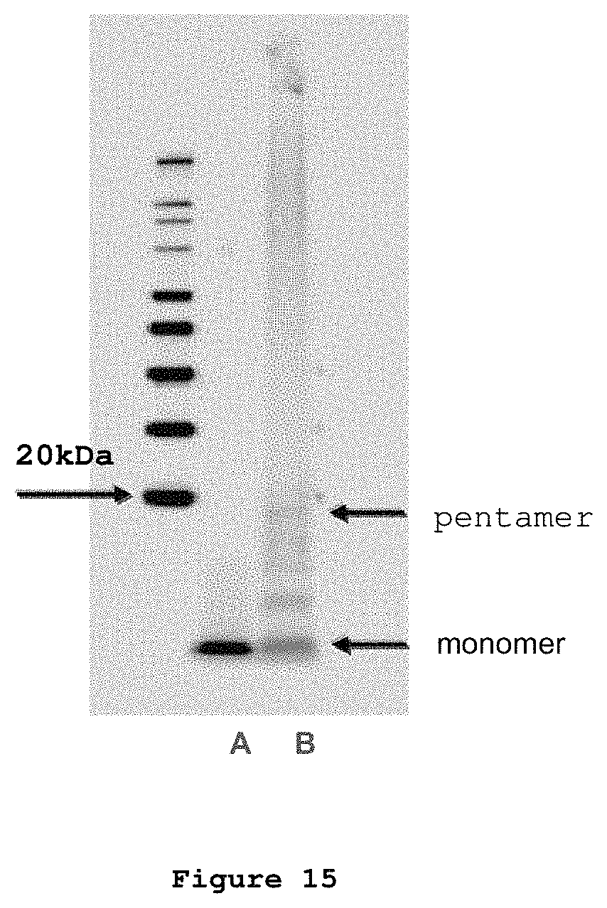
D00017

D00018
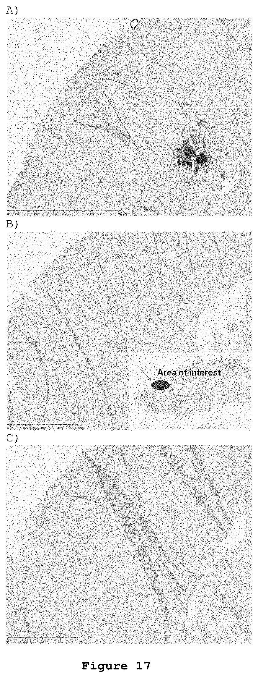
D00019
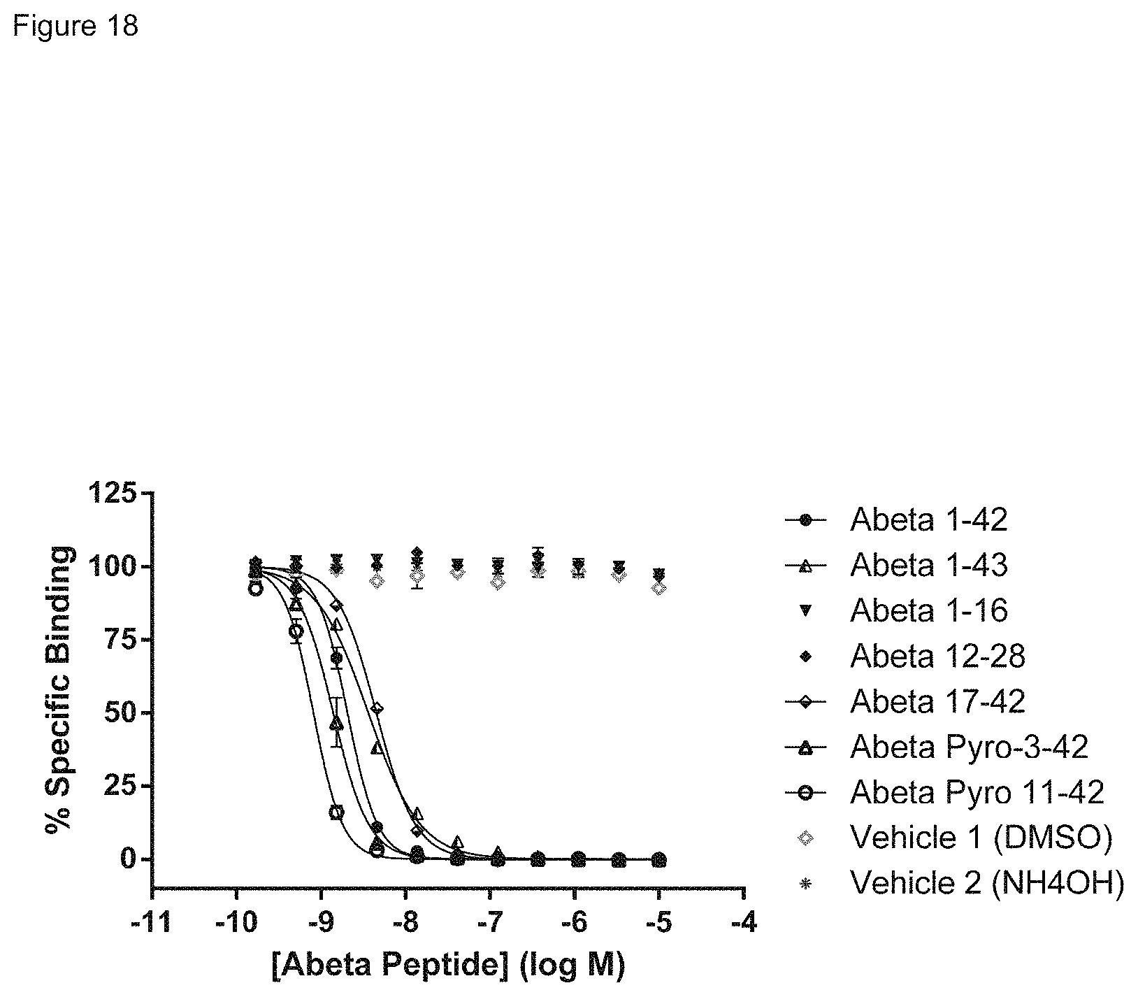
D00020

M00001

M00002

M00003

M00004

M00005

M00006

M00007

P00001

P00002

P00003

P00004

P00005

S00001
XML
uspto.report is an independent third-party trademark research tool that is not affiliated, endorsed, or sponsored by the United States Patent and Trademark Office (USPTO) or any other governmental organization. The information provided by uspto.report is based on publicly available data at the time of writing and is intended for informational purposes only.
While we strive to provide accurate and up-to-date information, we do not guarantee the accuracy, completeness, reliability, or suitability of the information displayed on this site. The use of this site is at your own risk. Any reliance you place on such information is therefore strictly at your own risk.
All official trademark data, including owner information, should be verified by visiting the official USPTO website at www.uspto.gov. This site is not intended to replace professional legal advice and should not be used as a substitute for consulting with a legal professional who is knowledgeable about trademark law.