Alkane oxidation by modified hydroxylases
Arnold , et al.
U.S. patent number 10,648,006 [Application Number 15/942,001] was granted by the patent office on 2020-05-12 for alkane oxidation by modified hydroxylases. This patent grant is currently assigned to California Institute of Technology. The grantee listed for this patent is The California Institute of Technology. Invention is credited to Frances Arnold, Mike M. Y. Chen, Rudi Fasan, Peter Meinhold, Matthew W. Peters.


View All Diagrams
| United States Patent | 10,648,006 |
| Arnold , et al. | May 12, 2020 |
Alkane oxidation by modified hydroxylases
Abstract
This invention relates to modified hydroxylases. The invention further relates to cells expressing such modified hydroxylases and methods of producing hydroxylated alkanes by contacting a suitable substrate with such cells.
| Inventors: | Arnold; Frances (Pasadena, CA), Meinhold; Peter (Pasadena, CA), Peters; Matthew W. (Pasadena, CA), Fasan; Rudi (Brea, CA), Chen; Mike M. Y. (Pasadena, CA) | ||||||||||
|---|---|---|---|---|---|---|---|---|---|---|---|
| Applicant: |
|
||||||||||
| Assignee: | California Institute of
Technology (Pasadena, CA) |
||||||||||
| Family ID: | 39682440 | ||||||||||
| Appl. No.: | 15/942,001 | ||||||||||
| Filed: | March 30, 2018 |
Prior Publication Data
| Document Identifier | Publication Date | |
|---|---|---|
| US 20180273983 A1 | Sep 27, 2018 | |
Related U.S. Patent Documents
| Application Number | Filing Date | Patent Number | Issue Date | ||
|---|---|---|---|---|---|
| 15224900 | Aug 1, 2016 | 9963720 | |||
| 14788365 | Aug 2, 2016 | 9404096 | |||
| 14270268 | Jul 7, 2015 | 9074178 | |||
| 11697404 | May 6, 2014 | 8715988 | |||
| PCT/US2006/011273 | Mar 28, 2006 | ||||
| 60900243 | Feb 8, 2007 | ||||
| 60700781 | Jul 20, 2005 | ||||
| 60698872 | Jul 13, 2005 | ||||
| 60665903 | Mar 28, 2005 | ||||
| Current U.S. Class: | 1/1 |
| Current CPC Class: | C07K 14/32 (20130101); C12P 7/04 (20130101); C12N 9/0071 (20130101); C12P 7/06 (20130101); C12Y 114/14001 (20130101); C12Y 106/02004 (20130101); C12N 9/0042 (20130101); C12N 9/0077 (20130101); Y02P 20/52 (20151101); C12Y 114/15 (20130101); C12Y 114/15003 (20130101); Y02E 50/17 (20130101); Y02E 50/10 (20130101) |
| Current International Class: | C12N 9/02 (20060101); C12P 7/06 (20060101); C07K 14/32 (20060101); C12P 7/04 (20060101); C12P 7/02 (20060101); C12N 15/53 (20060101) |
| Field of Search: | ;435/157,137,132,189,320.1 |
References Cited [Referenced By]
U.S. Patent Documents
| 4599342 | July 1986 | LaHann |
| 5605793 | February 1997 | Stemmer |
| 5741691 | April 1998 | Arnold et al. |
| 5785989 | July 1998 | Stanley et al. |
| 5811238 | September 1998 | Stemmer et al. |
| 5830721 | November 1998 | Stemmer et al. |
| 5837458 | November 1998 | Minshull et al. |
| 5965408 | October 1999 | Short |
| 6090604 | July 2000 | Golightly et al. |
| 6498026 | December 2002 | Delagrave et al. |
| 2005/0037411 | February 2005 | Arnold et al. |
| 2005/0202419 | September 2005 | Cirino et al. |
| 0 505 198 | Sep 1992 | EP | |||
| 0 752 008 | Feb 1997 | EP | |||
| 0 932 670 | Aug 2000 | EP | |||
| WO 89/03424 | Apr 1989 | WO | |||
| WO 95/22625 | Aug 1995 | WO | |||
| WO 97/16553 | May 1997 | WO | |||
| WO 97/20078 | Jun 1997 | WO | |||
| WO 97/35957 | Oct 1997 | WO | |||
| WO 97/35966 | Oct 1997 | WO | |||
| WO 98/27230 | Jun 1998 | WO | |||
| WO 98/31837 | Jul 1998 | WO | |||
| WO 98/41653 | Sep 1998 | WO | |||
| WO 98/42832 | Oct 1998 | WO | |||
| WO 99/60096 | Nov 1999 | WO | |||
| WO 00/00632 | Jan 2000 | WO | |||
| WO 00/04190 | Jan 2000 | WO | |||
| WO 00/09679 | Feb 2000 | WO | |||
| WO 0/067182 | Feb 2000 | WO | |||
| WO 00/18906 | Apr 2000 | WO | |||
| WO 00/31273 | Jun 2000 | WO | |||
| WO 01/62938 | Aug 2001 | WO | |||
| WO 03/008563 | Jan 2003 | WO | |||
Other References
|
Maradufu, A. et al., "Stereochemistry of Dehydrogenation by D-Galactose Oxidase," Canadian Journal of Chemistry, Oct. 1971, pp. 3429-3437, vol. 49, No. 19, NCR Research Press, Ottawa Canada. cited by applicant . March, J., Advanced Organic Chemistry: Reactions, Mechanisms, and Structure, 4th Edition, 1992, pp. 882-884, Wiley and Sons NY. cited by applicant . March, J., Advanced Organic Chemistry: Reactions, Mechanisms, and Structure,. 4th Edition, 1992, pp. 1072-1074, Wiley and Sons, NY. cited by applicant . Martin, B. et al., "Highly swelling hydrogels from ordered galactose-based polyacrylates," Biomaterials, 1998, pp. 69-76, 19(1-3), Elsevier. cited by applicant . Martin, I. et al., "Detection of honey adulteration with beet sugar using stable isotope methodology," Food Chemistry, 1998 pp. 281-286, vol. 61, No. 3, Elsevier Science Ltd. cited by applicant . Martineau, P. et al., "Expression of an Antibody Fragment at High Levels in the Bacterial Cytoplasm," J. Mol. Biol., 1998, pp. 117-127, vol. 280, No. 1, Academic Press. cited by applicant . Martinez, C. et al., "Cytochrome P450's: Potential Catalysts for Asymmetric Olefin Epoxidations," Current Organic chemistry, 2000, pp. 263-282, vol. 4, No. 3, Bentham Science Publishers B.V. cited by applicant . Matson, R. et al., "Characteristics of a Cytochrome P-450-Dependent Fatty Acid 107 -2 Hydroxylase From Bacillus Megaterium," Biochimica et Biophysica Acta, 1977, pp. 487-494, 487, Elsevier/North Holland Biomedical Press. cited by applicant . Mazur, A., "Chapter 8, Galactose Oxidase," ACS Symposium Series 466--Enzymes in Carbohydrate Synthesis, 1991, pp. 99-110. American Chemical Society, Washington, DC, USA. cited by applicant . Mazur, A., et al., "Chemoenzymic Approaches to the Preparation of 5-C-(Hydroxymethyl)hexoses," J. Org. Chem., 1997, pp. 4471-4475, vol. 62, No. 13 American Chemical Society, Washington, DC, USA. cited by applicant . McPherson, M. et al., "Galactose oxidase of Dactylium dendroides. Gene cloning and sequence analysis," Chemical Abstract Service, XP-002298547, Database accession No. M86819. cited by applicant . McPherson, M. et al., "Galactose Oxidase of Dactylium dendroides," Apr. 1992, pp. 8146-8152, The Journal of Biological Chemistry, vol. 267, No. 12, The American Society for Biochemistry and Molecular Biology, Inc. cited by applicant . McPherson, M. et al., "Galactose oxidase: Molecular analysis and mutagenesis studies," Biochemical Society Transactions, 646th Meeting Leeds, 1993, pp. 1992-1994, vol. 21, The Biochemical Society, Portland Press. cited by applicant . Meinhold, P. et al., "Direct Conversion of Ethane to Ethanol by Engineered Cytochrome P450 BM3," ChemBioChem, 2005 PD. 1-4, vol. 6, Wiley-VCH Verlaq GmbH & Co. Weinheim Germany. cited by applicant . Mendonca, M. et al., "Purification and Characterization of Intracellular Galactose Oxidase from Dactylium dendroides," Archives of Biochemistry and Biophysics, Feb. 1987, pp. 507-514, vol. 252, No. 2, Academic Press, Inc. cited by applicant . Mendonca, M. et al., "Role of Carbohydrate Content on the Properties of Galactose Oxidase from Dactylium dendroides," Archives of Biochemistry and Biophysics, Nov. 1988, pp. 427-434, vol. 266, No. 2, Academic Press, Inc. cited by applicant . Miele, R., et al., "Glycosylation of Asparagine-28 of Recombinant Staphylokinase with High-Mannose-type Oligosaccharides Results in a Protein with Highly Attenuated Plasminogen Activator Activity," Journal of Biological Chemistry, Mar. 1999, pp. 7769-7776, vol. 274, No. 12, The American Society for Biochemistry and Molecular Biology, Inc. cited by applicant . Miles, C. e al., "Protein engineering of cytochromes P-450," Biochimica et Biophysica Acta, 2000, pp. 383-407, 1543, Elsevier Science B.V. cited by applicant . Minshull, J. et al., "Protein evolution by molecular breeding," Chemical Biology, 1999, pp. 284-290, 3, Elsevier Science Ltd. cited by applicant . Mitraki, A. et al., "Amino acid substitutions influencing intracellular protein folding pathways," FEBS Letters, Jul. 1992, pp. 20-25, vol. 307, No.1, Elsevier Science Publishers B.V. cited by applicant . Miura, Y. et al., "107 -1, 107 -2 and 107 -3 Hydroxylation of Long-Chain Fatty Acids, Amides and Alcohols by a Soluble Enzyme System from Bacillus Megaterium," Biochimica et Biophysica Acta, 1975, pp. 305-317, 388, Elsevier Scientific Publishing Company, Amsterdam, The Netherlands. cited by applicant . Miyazaki, K. et al., "Directed Evolution Study of Temperature Adaptation in a Psychrophilic Enzyme," Journal Mol. Biol., 2000, pp. 1015-1026 297 Academic Press. cited by applicant . Modi, S. et al., "NMR Studies of Substrate Binding to Cytochrome P45OBM3: Comparisons to Cytochrome P450 cam." Biochemistry, 1995, pp. 8982-8988, vol. 34, No. 28, American Chemical Society. cited by applicant . Moore, J. et al., "Directed evolution of a para-nitrobenzyl esterase for aqueous-organic solvents," Nature Biotechnology, Apr. 1996 pp. 458-467, vol. 14. cited by applicant . Moore, J. et al., "Strategies for the in vitro Evolution of Protein Function: Enzyme Evolution by Random Recombination of Improved Sequences," J. Mol. Biol., 1997, pp. 336-347, 272, Academic Press Limited. cited by applicant . Moser, C. et al., "Biological Electron Transfer," Journal of Bioenergetics and Biomembranes, Jun. 1995, pp. 263-274, vol. 27 No. 3 Plenum Publishing Corporation. cited by applicant . Munro, A. et al., "Probing electronic transfer in flavocytochrome P-450 BM3 and its component domains," Eur. J. Biochem., 1996, pp. 403-409, 239, FEBS. cited by applicant . Munro, A. et al., "Alkane Metabnolism by Cytochrome P450 BM3," Biochemical Society Transactions, 1993, p. 412S, 21. cited by applicant . Murrell, J. et al., "Molecular biology and regulation of methane monooxygenase," Arch. Microbial., 2000, pp. 325-332, 173o. cited by applicant . Nagayama, Y. et al., "Role of Asparagine-linked Oligosaccharides in Protein Folding, Membrane Targeting, and Thyrotropin and Autoantibody Binding of the Human Thyrotropin Receptor," Journal of Biological Chemistry, Dec. 1998, pp. 33423-33428, vol. 273, No. 5, The American Society for Biochemistry and Molecular Biology. Inc. cited by applicant . Nakagawa, S. et al., "Construction of Catalase Deficient Escherichia coli Strains for the Production of Uricase," Biosci. Biotech. Biochem., 1996, pp. 415-420, 60 (3), Japanese Society for Bioscience, Biotechnology and Agrochemistry. cited by applicant . Nakajima, H. et al.,"Industrial Application of Adenosine 5'-Triphosphate Regeneration to Synthesis of Sugar Phosphates," ACS Symposium Series 466, Enzymes in Carbohydrate Synthesis, Chapter 9, pp. 110-120, American Chemical Society, Washington DC, 1991, Bednarski & Simon, Editors. cited by applicant . Narhi, L. et al., "Identification and Characterization of Two Functional Domains in Cytochrome P-450BM-3 , a Catalytically Self-sufficient Monooxygenase Induced by Barbiturates in Bacillus megaterium," The Journal of Biological Chemistry, May 1987, pp. 6683-6690 vol. 262, No. 14, The American Society of Biological Chemists, Inc. cited by applicant . Narhi, L. et al., "Characterization of a Catalytically Self-sufficient 199,000-Dalton Cytochrome P-450 Monooxygenase Induced by Barbiturates in Bacillus megaterium," The Journal of Biological Chemistry, Jun. 1986, pp. 7160-7169, vol. 261, No. 16, The American Society of Biological Chemists, Inc. cited by applicant . Nelson, D., "Appendix A--Cytochrome P450 Nomenclature and Alignment of Selected Sequences," Cytochrome P450: Structure, Mechanism, and Biochemistry, Second Ed., 1995, pp. 575-606, Plenum Press, NY. cited by applicant . Ness, J. et al., "DNA shuffling of subgenomic sequences of subtilisin," Nature Biotechnology, Sep. 1999, pp. 893-896, vol. 17, No. 9, Nature Publishing Group. cited by applicant . Noble, M. et al., "Roles of key active-site residues in flavocytochrome P450 BM3," Biochem. J., 1999, pp. 371-379, 339, Biochemical Society. cited by applicant . Oliphant, A. et al., "Cloning of random-sequence oligodeoxynucleotides," Gene, 1986, pp. 177-183, 44, Elsevier Science Publishers B.V. cited by applicant . Oliver, C. et al., "Engineering the substrate specificity of Bacillus megaterium cytochrome P-450 BM3: hydroxylation of alkyl trimethylammonium compounds," Biochem. J., 1997, pp. 537-544, 327, The Biochemical Society, London, England. cited by applicant . Omura, T. et al., "The Carbon Monoxide-binding Pigment of Liver Microsomes," The Journal of Biological Chemistry, Jul. 1964 pp. 2370-2378, vol. 239, No. 7, The American Society for Biochemistry and Molecular Biology. cited by applicant . Ortlepp, S. et al., "Expression and characterization of a protein specified by a synthetic horseradish peroxidase gene in Escherichia coli," Journal of Biotechnology, 1989, pp. 353-364, 11, Elsevier Science Publishers B.V. cited by applicant . Ost, T. et al., "Rational re-design of the substrate binding site of flavocytochrome P450 BM3," FEBS Letters, 2000, pp. 173-177,486, Elsevier Science B.V. cited by applicant . Ostermeier, M. et al., "Incremental Truncation as a Strategy in the Engineering of Novel Biocatalysts," Bioorganic & Medicinal Chemistry, 1999, pp. 2139-2144, 7, Elsevier Science Ltd. cited by applicant . Parekh, R. et al., "Multicopy Overexpression of Bovine Pancreatic Trypsin Inhibitor Saturates the Protein Folding and Secretory Capacity of Saccharomyces cerevisiae," Protein Expression and Purification, 1995, pp. 537-545, 6, Academic Press. cited by applicant . Patten, P. et al., "Applications of DNA shuffling to pharmaceuticals and vaccines," Biotechnology, 1997, pp. 724-733, vol. 8, Elsevier Science Ltd. cited by applicant . Paulsen, M. et al., "Dramatic Differences in the Motions of the Mouth of Open and Closed Cytochrome P450BM-3 b Molecular Dynamics Simulations," Proteins: Structure, Function and Genetics, 1995, pp. 237-243, Wiley-Liss, Inc. cited by applicant . Peterson, J. et al., "Chapter 5--Bacterial P450s," Cytochrome P450: Structure, Mechanism, and Biochemistry, Second Ed., 1995, pp. 151-180, Plenum Press, New York. cited by applicant . Rathore, D., et al., "Expression of Ribonucleolytic Toxin Restrictocin in Escherichia coli: Purification and Characterization," FEBS Letters, 1996, pp. 259-262, vol. 392, Federation of European Biochemical Societies. cited by applicant . Reynolds, M., et al., "Structure and Mechanism of Galactose Oxidase: Catalytic Role of Tyrosine 495," JBIC, 1997, pp. 327-335, vol. 2. cited by applicant . Amaral et al., "Galactose Oxidase of Polyporus circinatus1-4," Methods in Enzymology, Carbohydrate Metabolism, 1966, pp. 87-92, vol. 9, Academic Press Inc., New York NY, USA. cited by applicant . Adam et al., "Microbial Asymmetric Ch Oxidations of Simple Hydrocarbons: A Novel Monooxygenase Activity of the Topsoil Microorganism Bacillus megaterium," Eur. J. Org. Chem., 2000, pp. 2923-2926, Wiley-VCH Verlag GmbH, Weinheim, Germany. cited by applicant . Aisaka et al., "Production of Galactose Oxidase by Gibberella fujikuroi," Agric. Biol. Chem., 1981, pp. 2311-2316, 45 (10). cited by applicant . Anfinsen, "Principles that Govern the Folding of Protein Chains," Science, Jul. 20, 1973, pp. 223-230, vol. 181, No. 4096, American Asso for the Advancement of Science, Washington, DC, USA. cited by applicant . Appel et al., "A P450 BM-3 mutant hydroxylates alkanes, cycloalkanes, arenas and heteroarenes," Journal of Biotechnology, 2001, pp. 167-171, Elsevier Science B.V. cited by applicant . Arkin et al., "An algorithm for protein engine ring: Simulations of recursive ensemble mutagenesis," Proc. Natl. Acad. Sci.--USA, Aug. 1992, pp. 7811-7815, vol. 89, Applied Biological Sciences. cited by applicant . Arnold, "Design by Directed Evolution," Accounts of Chemical Research, 1998, pp. 125-131, vol. 31, No. 3, American Chemical Society. cited by applicant . Arnold, "Engineering proteins for nonnatural environments," The FASEB Journal, Jun. 1993, pp. 744-749, vol. 7, No. 6, FASEB, Bethesda, MD, USA. cited by applicant . Arnold et al., "Optimizing Industrial Enzymes by Directed Evolution," Advances in Biochemical Engineering/Biotechnology, 1997, pp. 1-14, vol. 58, Springer-Verlag, Berlin, Germany. cited by applicant . Arts et al., "Hydrogen Peroxide and Oxygen in Catalytic Oxidation of Carbohydrates and Related Compounds," Synthesis Journal of Synthetic Organic Chemistry, Jun. 1997, pp. 597-613. cited by applicant . Ashraf et al., "Bacterial oxidation of propane," FEMS Microbiology Letters, 1994, pp. 1-6, Federation of European MicrobioloQical Societies, Elsevier. cited by applicant . Avigad, "Oxidation Rates of Some Desialylated Glycoproteins by Galactose Oxidase," Archives of Biochemistry and Biophysics, Jun. 1985, pp. 531-537, vol. 239, No. 2, Academic Press, Inc. cited by applicant . Avigad, "An NADH Coupled Assay System for Galactose Oxidase," Analytical Biochemistry, 1978, pp. 470-476, 86, Academic Press Inc. cited by applicant . Avigad, :The D-Galactose Oxidase of Polyporus circinatus. Journal of Biological Chemistry, Sep. 1962, pp. 2736-2743, vol. 237, No. 9, American Society of Biological Chemists, Baltimore, MD, USA. cited by applicant . Barnes, "Maximizing Expression of Eukaryotic Cytochrome P450s in Escherichia coli," Methods in Enzymology, Cytochrome P450, Part B, 1996, pp. 3-14, vol. 272, Academic Press, Inc., San Diego, CA, USA. cited by applicant . Baron et al., "Structure and Mechanism of Galactose Oxidase," The Journal of Biological Chemistry, Sep. 23, 1994, pp. 25095-25105, vol. 269, No. 38, American Soc. for Biochemistry and Molecular Biology. cited by applicant . Benson et al., "Regulation of Membrane Peptides by the Pseudomonas Plasmid alk Regulon," Journal of Bacteriology, Dec. 1979 pp. 754-762, vol. 140, No. 3. cited by applicant . Better et al., "Escherichia coli Secretion of an Active Chimeric Antibody Fragment," Science, May 20, 1988, pp. 1041-1043, vol. 240, American Asso for the Advancement of Science, Washington, DC, USA. cited by applicant . Boddupalli et al., "Fatty Acid Monooxygenation by P450BM-3: Product Identification and Proposed Mechanisms for the Sequential Hydroxylation Reactions," Archives of Biochemistry and Biophysics, Jan. 1992, pp. 20-28, vol. 292, No. 1, Academic Press, Inc. cited by applicant . Boddupalli et al., "Fatty Acid Monooxygenation by Cytochrome P-450BM-3," The Journal of Biological Chemistry, 1990, pp. 4233-4239, The American Society for Biochemistry and Molecular Biology. cited by applicant . Borman et al., "Kinetic studies on the reactions of Fusarium galactose oxidase with five different substrates in the presence of dioxygen," Journal of Biological Inorganic Chemistry, 1997, pp. 480-487, Society of Biological Inorganic Chemistry. cited by applicant . Bradford, "A Rapid and Sensitive Method for the Quantitation of Microgram Quantities of Protein Utilizing the Principle of Protein-Dye Binding," Analytical Biochemistry, pp. 248-254. cited by applicant . Calderhead, D. et al., "Labeling of Glucose Transporters at the Cell Surface in 3T3-L 1 Adipocytes," The Journal of Biological Chemistry, Sep. 5, 1988, pp. 12171-12174, vol. 263, No. 25, The American Society for Biochemistry and Molecular Biology. cited by applicant . Calvin, N. et al., "High-Efficiency Transformation of Bacterial Cells by Electroporation," Journal of Bacteriology, Jun. 1988, pp. 2796-2801 , vol. 170 No. 6, American Society for Microbiology. cited by applicant . Cameron, A., "Two cradles for the heavy elements," Nature, Jan. 15, 1998, pp. 228-231, vol. 39. cited by applicant . Capdevila, J. et al., "The Highly Stereoselective Oxidation of Polyunsaturated Fatty Acids by Cytochrome P450BM-3," The Journal of Biological Chemistry, Sep. 13, 1996, pp. 22663-22671, vol. 271, No. 37, The American Society for Biochemistry and Molecular Biology, Inc. cited by applicant . Carmichael, A. et al., "Protein engineering of Bacillus megaterium CYP102," Eur. J. Biochem., 2001, pp. 3117-3125, vol. 268, FEBS. cited by applicant . Castelli, L. et al., "High-level secretion of correctly processed .beta.-lactamase from Saccharomyces cerevisiae using a highcopy-number secretion vector," Gene, 1994, EE- 113-117, vol. 142, Elsevier Science B.V. cited by applicant . Chang, C. et al., "Evolution of a cytokine using DNA family shuffling," Nature Biotechnology, Aug. 1999, pp. 793-797, vol. 17. cited by applicant . Chang, Y. et al.., "Homology Modeling, Molecular Dynamics Simulations, and Analysis of CYP119, a P450 Enzyme from Extreme Acidothermophilic Archaeon Sulfolobus soifataricus," Biochemistry, 2000, pp. 2484-2498, vol. 39, No. 10, American Chemical Society. cited by applicant . Chen, H. et al., "Thermal, Catalytic, Regiospecific Functionalization of Alkanes," Science, Mar 17, 2000, pp. 1995-1997, vol. 287. cited by applicant . Chen, K. et al., "Tuning the activity of an enzyme for unusual environments: Sequential random mutagenesis of subtilisin E for catalysis in dimethylformamide," Proc. Natl. Acad. Sci. USA, Jun. 15, 1993, pp. 5618-5622, vol. 90, No. 12. cited by applicant . Cherry, J. et al., "Directed evolution of a fungal peroxidase," Nature Biotechnology, Apr. 1999, pp. 379-384, vol. 17, Nature America Inc., New York, NY, USA. cited by applicant . Christians, F. et al., "Directed evolution of thymidine kinase for AZT phosphorylation using DNA family shuffling," Nature Biotechnology, Mar. 1999, pp. 259-264, vol. 17, Nature America Inc., New York, NY, USA. cited by applicant . Cleland, J. et al., "Cosolvent Assisted Protein Refolding," Biotechnology, Dec 1990, pp. 1274-1278, vol. 8. cited by applicant . Crameri, A. et al., "Molecular evolution of an arsenate detoxification pathway by DNA shuffling," Nature Biotechnology, May 1997 pp. 436-438, vol. 15, Nature America Inc., New York, NY, USA. cited by applicant . Crameri, A. et al., "Improved Green Fluorescent Protein by Molecular Evolution Using DNA Shuffling," Nature Biotechnology, Mar. 1996, pp. 315-319, vol. 14, Nature America Inc., New York, NY, USA. cited by applicant . Crameri, A. et al., "Construction and evolution of antibody-phage libraries by DNA shuffling," Nature Medicine, Jan. 1996, pp. 100-106 vol. 2, No. 1. cited by applicant . Dahlhoff, W. et al., "L-Giucose or D-gluco-Hexadialdose from D-Glucurono-6,3-lactone by Controlled Reductions," Angew. Chem. Int. Ed. Engl., 1980, pp. 546-547, 19 No. 7, Verlag Chemie, GmbH, Weinheim, Germany. cited by applicant . Danon, A., et al. "Enrichment of Rat Tissue Lipids with Fatty Acids that are Prostaglandin Precursors" Biochimica et Biophysica Acta, 1975, 388: 318-330. cited by applicant . De Bernardez-Clark, E. et al., "Inclusion Bodies and Recovery of Proteins from the Aggregated State," ACS Symposium Series Protein Refolding, 199th Natl Mtg American Chemical Society, Apr. 22-27, 1990, pp. 1-20, American Chemical Society, Washington, DC, USA. cited by applicant . Deacon, S. et al., "Enhanced Fructose Oxidase Activity in a Galactose Oxidase Variant," ChemBioChem: A European Journal of Chemical Biology, 2004, pp. 971-979, 5, Wiley-VCH Verlag GmbH & Co., Weinheim, Germany. cited by applicant . Delagrave, S. et al., "Recursive ensemble mutagenesis," Protein Engineering, Apr. 1993, pp. 327-331, vol. 6, No. 3, Oxford University Press. cited by applicant . Delagrave, S. et al., "Searching Sequence Space to Engineer Proteins: Exponential Ensemble Mutagenesis," Bio/Technology, Dec. 1993, pp. 1548-1552, vol. 11, American Society for Cell Biology, New Orleans, LA, USA. cited by applicant . Dordick, J. "Designing Enzymes for Use in Organic Solvents," Biotechnol. Prog., 1992, pp. 259-267, 8, American Chemical Society and American Institute of Chemical Engineers. cited by applicant . Dower, W. et al., "High efficiency transformation of E.-coli by high voltage electroporation," Nucleic Acids Research, 1988, pp. 6127-6145, vol. 16, No. 13, IRL Press Limited, Oxford, England. cited by applicant . Farinas, E., et al., "Directed Evolution of a Cytochrome P450 Monooxygenase for Alkane Oxidation," Adv. Synth. Catal., 2001, pp 601-606, vol. 343, No. 6-7. cited by applicant . Fiedler, K., et a!., The Role of N-Glycans in the Secretory Pathway, Cell, May 5, 1995, pp. 309-312, vol. 81, Cell Press. cited by applicant . Fisher, M., et al., "Positional Specificity of Rabbit CYP4B1 for .omega.-Hydroxylation of Short-Medium Chain Fatty Acids and Hydrocarbons," Biochemical and Biophysical Research Communications, 1998, pp. 352-355, vol. 248, No. RC988842. cited by applicant . Fox, B., et al., "Methane Monooxygenase from Methylosinus trichosporium OB3b," Methods in Enzymology, 1990, pp. 191-202, vol. 188, Academic Press, Inc. cited by applicant . Fox, B., et al., "Methane Monooxygenase from Methylosinus trichosporium OB3b Purification and Properties of a Three-Component System with High Specific Activity from a Type II Methanotroph," The Journal of Biological Chemistry, Jun. 15, 1989, pp. 10023-10033, vol. 264, No. 17, The American Society for Biochemistry and Molecular Biology, Inc. cited by applicant . Whittaker, M., et al., "Kinetic Isotope Effects as probes of the Mechanism of Galactose Oxidase," Biochemistry, 1998, pp. 8426-8436 vol. 37, American Chemical Society. cited by applicant . Wilkinson, D., et aL, "Structural and Kinetic Studies of a Series of Mutants of Galactose Oxidase Identified by Directed Evolution," Protein Engineering, Design & Selection, Jan. 12, 2004, pp. 141-148, vol. 17, No. 2, Oxford University Press. cited by applicant . Yang, G., et al., "Gal-GaINAc: A biomarker of Colon Carcinogenesis," Histology and Histopathology, 1996, pp. 801-806, vol. 11. cited by applicant . Yano, T., et al., "Directed Evolution of an Aspartate Aminotransferase with New Substrate Specificities," Proc. Natl. Acad. Sci. USA, May 1998, pp. 5511-5515, vol. 95. cited by applicant . Yeom, H., et al., "Oxygen Activation by Cytochrome P450BM-3: Effects of Mutating an Active Site Acidic Residue," Archieves of Biochemistry and Biophysics, Jan. 15, 1997, pp. 209-216, vol. 337, No. 2, Academic Press. cited by applicant . You, L., et al., "Directed Evolution of Subtilisin E in Bacillus subtilis to Enhance Total Activity in Aqueous Dimethylformamide," Protein Engineering, 1996, pp. 77-83, vol. 9, Oxford University Press. cited by applicant . Zhang, J., et al., "Directed Evolution of a Fucosidase from a Galactosidase by DNA Shuffling and Screening," Proc. Natl. Acad. Sci. USA, Apr. 1997, pp. 4504-4509, vol. 94. cited by applicant . Zhang, T., et al., "Circular Permutation of T4 Lysozyme," Biochemistry, 1993, pp. 12311-12318, vol. 32, No. 46, American Chemical Society. cited by applicant . Zhao, H., et al., "Directed Evolution Converts Subtilisin E into a Functional Equivalent of Thermitase," Protein Engineering,1999, pp. 47-53, vol. 12, No. 1, Oxford University Press. cited by applicant . Zhao, H., et al., "Methods for Optimizing Industrial Enzymes by Directed Evolution," Manual of Industrial Microbiology and Biotechnology (2d Ed.), 1999, pp. 597-604, ASM Press, Washington, D.C. cited by applicant . Zhao, H., et al., "Molecular Evolution by Staggered Extension Process (StEP) In Vitro Recombination," Nature Biotechnology, Mar. 1998, pp. 258-261, vol. 16. cited by applicant . Zhao, H., et al., "Optimization of DNA Shuffling for High Fidelity Recombination," Nucleic Acids Research, 1997, pp. 1307-1308, vol. 25, No. 6, Oxford University Press. cited by applicant . Zimmer, T., et al., "The CYP52 Multigene Family of Candida maltosa Encodes Functionally Diverse n-Alkane-Inducible Cytochromes P450," Biochemical and Biophysical Research Communications, 1996, pp. 784-789, vol. 224, No. 3, Academic Press, Inc. cited by applicant . XP-002298548, "Protein Sequence," Database accession No. 355884-87-G. cited by applicant . Rodriguez-Lopez, J., et al., "Role of Arginine 38 in Horseradish Peroxidase--A Critical Residue for Substrate Binding and Catalysis," The Journal of Biological Chemistry, Feb. 23, 1996, pp. 4023-4030, vol. 271, No. 8, The American Society for Biochemistry and Molecular Biology. cited by applicant . Romanos, M., et al., "Foreign Gene Expression in Yeast: a Review," Yeast, Jun. 1992, pp. 423-488, vol. 8, No. 6, John Wiley & Sons Ltd. cited by applicant . Root, R., et al., "Enzymatic Synthesis of Unusual Sugars: Galactose Oxidase Catalyzed Stereospecific Oxidation of Polyols," Journal of the American Chemical Society, 1985, pp. 2997-2999, vol. 107, No. 10, American Chemical Society. cited by applicant . Ruettinger, R., et al., "Coding Nucleotide, 5' Regulatory, and Deduced Amino Acid Sequences of P-4506M.3 , a Single Peptide Cytochrome P-450:NADPH-P-450 Reductase from Bacillus megaterium," The Journal of Biological Chemistry, Jul. 5, 1989, pp. 10987-10995, vol. 264, No. 19, The American Society for Biochemistry and Molecular Biology, Inc. cited by applicant . Ruettinger, R., et al., "Epoxidation of Unsaturated Fatty Acids by a Soluble Cytochrome P450-dependent System from Bacillus megaterium," The Journal of Biological Chemistry, Jun. 10, 1981, pp. 5728-5734, vol. 256, No. 11. cited by applicant . Said, I.T., et al., "Comparison of Different Techniques for Detection of Gai-GaINAc, an Early Marker of Colonic Neoplasia," Histology and Histopathology, Apr. 1999, pp. 351-357, vol. 14, No. 2, Jimenez Godoy, SA. cited by applicant . Savenkova, M., et al. "Improvement of Peroxygenase Activity by Relocation of a Catalytic Histidine within the Active Site of Horseradish Peroxidase," Biochemistry, 1998, pp. 10828-10836, vol. 37, American Chemical Society. cited by applicant . Saysell, C., et al., "Properties of the Trp290His Variant of Fusarium NRRL 2903 Galactose Oxidase: Interactions of the GOasesemi State with Different Buffers, Its Redox Activity and Ability to Bind Azide," JBIC, 1997, pp. 702-709, vol. 2. cited by applicant . Schatz, R, et al., "Genetic Analysis of Protein Export in Escherichia coli," Annual Review of Genetics, 1990, pp. 215-248, vol. 24, Annual Reviews, Inc., Palo Alto, CA. cited by applicant . Schein C., "Solubility as a Function of Protein Structure and Solvent Components," Bio/Technology, Apr. 1990, pp. 308-317, vol. 8, No. 4. cited by applicant . Scheller, U., et al., "Characterization of the n-Alkane and Fatty Acid Hydroxylating Cytochrome P450 Forms 52A3 and 52A4," Archives of Biochemistry and Biophysics, Apr. 15, 1996, pp. 245-254, vol. 328, No. 2, Academic Press, Inc. cited by applicant . Schlegel, R., et al.,"Substrate Specificity of D-Galactose Oxidase," Carbohydrate Research, Jun. 1968, pp. 193-199, vol. 7, No. 2, Elsevier Publishing Company, Amsterdam. cited by applicant . Schmid, A., et al., "Industrial Biocatalysis Today and Tomorrow," Nature, Jan. 11, 2001, pp. 258-268, vol. 409, Macmillian Magazines Ltd. cited by applicant . Schneider, S., et al., "Controlled Regioelectivity of Fatty Acid Oxidation by Whole Cells Producing Cytochrome P450sM-3 Monooxygenase Under Varied Dissolved Oxygen Concentrations," Biotechnology and Bioengineering, Aug. 5, 1999, pp. 333-341, vol. 64, No. 3, John Wiley & Sons, Inc. cited by applicant . Schwaneberg, U., et al., "A Continuous Spectrophotometric Assay for P450 BM-3, a Fatty Acid Hydroxylating Enzyme, and Its Mutant F87A," Analytical Biochemistry, 1999, pp. 359-366, vol. 269, Academic Press. cited by applicant . Schwaneberg, U., et al., "Cost-Effective Whole-Cell Assay for Laboratory Evolution of Hydroxylases in Escherichia coli," Journal of Biomolecular Screening, 2001, pp. 111-117, vol. 6, No. 2, The Society for Biomolecular Screening. cited by applicant . Schwaneberg, U., et al., "P450 Monooxygenase in Biotechnology--Single-Step, Large-Scale Purification Method for Cytochrome P450 BM-3 by Anion-Exchange Chromatography," Journal of Chromatography, 1999, pp. 149-159, vol. 848, Elsevier Science B.V. cited by applicant . Shafikhani, S., et al., "Generation of Large Libraries of Random Mutants in Bacillus subtilis by PCR-Based Plasmid Multimerization," BioTechniques, Aug. 1997, pp. 304-310, vol. 23, No. 2. cited by applicant . Shanklin, J., et al., "Mossbauer Studies of Alkane .omega.-Hydroxylase: Evidence for a Diiron Cluster in an Integral-Membrane Enzyme," Proc. Natl. Acad. Sci. USA, Apr. 1997, pp. 2981-2986, vol. 94. cited by applicant . Shao, Z., et al., "Random-priming In Vitro Recombination: An Effective Tool for Directed Evolution," Nucleic Acids Research, Jan. 15, 1998, pp. 681-683, vol. 26, No. 2, Oxford University Press. cited by applicant . Shilov, A., et al., "Activation of C-H Bonds by Metal Complexes," Chem. Rev., 1997, pp. 2879-2932, vol. 97, American Chemical Society. cited by applicant . Shindler, J., et al.,"Peroxidase from Human Cervical Mucus--The Isolation and Characterisation," European Journal of Biochemistry, Jun. 1976, pp. 325-331, vol. 65, No. 2. cited by applicant . Sirotkin, K., "Advantages to Mutagenesis Techniques Generating Populations Containing the Complete Spectrum of 1-single Codon Changes," J. Theor. Biol., 1986, pp. 261-279, vol. 123, Academic Press Inc. (London) Ltd. cited by applicant . Smith, A., et al., "Expression of a Synthetic Gene for Horseradish Peroxidase C in Escherichia coli and Folding and Activation of the Recombinant Enzyme with Ca2+ and Heme," The Journal of Biological Chemistry, Aug. 5, 1990, pp. 13335-13343, vol. 265, No. 22, The American Society for Biochemistry and Molecular Biology. cited by applicant . Smith, A., et al., "Substrate Binding and Catalysis in Heme Peroxidases," Current Opinion in Chemical Biology, (1998), pp. 269-278, vol. 2. cited by applicant . Spiro, T., et al., "Is the CO Adduct of Myoglobin Bent, and Does It Matter?," Accounts of Chemical Research, 2001, pp. 137-144, vol. 34, No. 2, American Chemical Society. cited by applicant . Staijen, I., et al., "Expression, Stability and Performance of the Three-Component Alkane Mono-oxygenase of Pseudomonas oleovorans in Escherichia coli," Eur. J. Biochem., 2000, pp. 1957-1965, vol. 267. cited by applicant . Stemmer, W., "DNA Shuffling by Random Fragmentation and Reassembly: In Vitro Recombination for Molecular Evolution," Proc. Natl. Acad. Sci. USA, Oct. 25, 1994, pp. 10747-10751, vol. 91, No. 22. cited by applicant . Stemmer, W., "Rapid Evolution of a Protein In Vitro by DNA Shuffling," Nature, Aug. 4, 1994, pp. 389-391, vol. 370, No. 6488. cited by applicant . Stemmer, W., et al., "Selection of an Active Single Chain Fv Antibody from a Protein Linker Library Prepared by Enzymatic Inverse PCR," BioTechniques, 1993, pp. 256-265, vol. 14, No. 2. cited by applicant . Stevenson, J., et al., "The Catalytic Oxidation of Linear and Branched Alkanes by Cytochrome P450cam," J. Am. Chem. Soc., 1996, pp. 12846-12847, vol. 118, No. 50, American Chemical Society. cited by applicant . Studier, F., et al., "Use of T7 RNA Polymerase to Direct Expression of Cloned Genes," Methods in Enzymology, 1990, pp. 60-89 vol. 185 Academic Press, Inc. cited by applicant . Sun, L., et al., "Expression and Stabilization of Galactose Oxidase in Escherichia coli by Directed Evolution," Protein Engineering, Sep. 2001, pp. 699-704, vol. 14, No. 9, Oxford University Press. cited by applicant . Sun, L., et al., "Modification of Galactose Oxidase to Introduce Glucose 6-0xidase Activity," ChemBioChem: A European Journal of Chemical Biology, Aug. 2, 2002, pp. 781-783, vol. 3, No. 8, Wiley-VCH-Vertag GmbH, Weinheim, Germany. cited by applicant . Szabo, E., et al., "Application of Biosensor for Monitoring Galactose Content," Biosensors & Bioelectronics, 1996, pp. 1051-1058, vol. 11, No. 10, Elsevier Science Limited. cited by applicant . Tams, J., et al., "Giycosylation and Thermodynamic Versus Kinetic Stability of Horseradish Peroxidase," FEBS Letters, 1998, pp. 234-236, vol. 421, Federation of European Biochemical Societies. cited by applicant . Thatcher, D., et al., "Protein Folding in Biotechnology," Mechanisms of Protein Folding, 1994, pp. 229-261, IRL Press, Oxford. cited by applicant . Tkac, J., et al., "Rapid and Sensitive Galactose Oxidase-Peroxidase Biosensor for Galactose Detection with Prolonged Stability," Biotechnology Techniques, 1999, pp. 931-936, Kluwer Academic Publishers. cited by applicant . Tonge, G., et al., "Purification and Properties of the Methane Mono-oxygenase nzyme System from Methylosinus trichosporium OB3b," Biochem. J., 1977, pp. 333-344, vol. 161. cited by applicant . Tressel, P., et al., "A Simplified Purification Procedure for Galactose Oxidase," Analytical Biochemistry, Jun. 1980, pp. 150-153, vol. 105, No. 1, Academic Press, Inc. cited by applicant . Tressel, P., et al., "Galactose Oxidase from Dactylium dendroides," Methods in Enzymology, 1982, pp. 163-171, vol. 89, Academic Press. cited by applicant . Truan, G., et al., "Thr268 in Substrate Binding and Catalysis in P450BM-3," Archives of iochemistry and Biophysics, Jan. 1, 1998, pp. 53-64, vol. 349, No. 1, Academic Press. cited by applicant . Vega, F., et al., "On-line Monitoring of Galactoside Conjugates and Glycerol by Flow Injection Analysis," Analytica Chimica Acta, 1998, pp. 57-62, vol. 373, Elsevier Science B.V. cited by applicant . Vrbova, E., et al., "Preparation and Utilization of a Biosensor Based on Galactose Oxidase," Collect. Czech. Chem. Commun., 1992, pp. 2287-2294, vol. 57. cited by applicant . Wachter, R., et al., "Molecular Modeling Studies on Oxidation of Hexopyranoses by Galactose Oxidase. An Active Site Topology Apparently Designed to Catalyze Radical Reactions, Either Concerted or Stepwise," Journal of the American Chemical Society, Mar. 9, 1996, pp. 2782-2789, vol. 118, No. 9. cited by applicant . Watkinson, R., et al., "Physiology of Aliphatic Hydrocarbon-Degrading Microorganisms," Biodegradation, 1990, pp. 19-92, vol. 1, Nos. 2/3 Kluwer Academic Publishers. cited by applicant . Welinder, K., "Amino Acid Sequence Studies of Horseradish Peroxidase," European Journal of Biochemistry, 1979, pp. 483-502. cited by applicant . Welinder, K., "Supplement to Amino Acid Sequence Studies of Horseradish Peroxidase," pp. 495-502. cited by applicant . Wetzel, R., et al., "Mutations in Human Interferon Gamma Affecting Inclusion Body Formation Identified by a General Immunochemical Screen," Bio/Technology, Aug. 1991, pp. 731-737, vol. 9. cited by applicant . Whittaker, M., et al., "The Active Site of Galactose Oxidase," The Journal of Biological Chemistry, 1988, pp. 6074-6080, vol. 263, No. 13, The American Society for Biochemistry and Molecular Biology, Inc. cited by applicant . Fruetel, J., et al., "Relationship of Active Site Topology to Substrate Specificity for Cytochrome P450terp (CYP108)," The Journal of Biological Chemistry, Nov. 18, 1994, pp. 28815-28821, vol. 269, No. 46, The American Society for Biochemistry and Molecular Biology, Inc. cited by applicant . Gahmberg C., et al., "Nonmetabolic Radiolabeling and Taggin of Glycoconjugates," Methods in Enzymology, 1994, pp. 32-44, vol. 230, Academic Press, Inc. cited by applicant . Gazaryan, I.G., "Heterologous Expression of Herne-Containing Peroxidases," Plant Peroxidase Newsletter, Sep. 1994, pp. 11-13, No. 4, LABPV Newsletters. cited by applicant . Gietz, R., et al., "Studies on the Transformation of Intact Yeast Cells by the LiAc/SS-DNA/PEG Procedure," Yeast, Apr. 15, 1995, pp. 355-360, vol. 11, No. 4, John Wiley & Sons Ltd. cited by applicant . Gillam, E., et al., "Expression of Cytochrome P450 2D6 in Escherichia coli, Purification, and Spectral and Catalytic Characterization," Archives of Biochemistry and Biophysics, Jun. 1, 1995, pp. 540-550, vol. 319, No. 2, Academic Press, Inc. cited by applicant . Giver, L., et al., "Combinatorial Protein Design by In Vitro Recombination," Current Opinion in Chemical Biology, 1998, pp. 335-338, vol. 2, Current Biology Ltd. cited by applicant . Giver, L., et al., "Directed Evolution of a Thermostable Esterase," Proc. Natl. Acad. Sci. USA, Oct. 1998, pp. 12809-12813, vol. 95. cited by applicant . Goldman, E., et al., "An Algorithmically Optimized Combinatorial Library Screened by Digital Imaging Spectroscopy," Biotechnology, Dec. 1992, pp. 1557-1561, vol. 10. cited by applicant . Graham-Lorence, S., et al., "An Active Site Substitution, F87V, Converts Cytochrome P450 BM-3 into a Regio- and Stereoselective (14S, 15R)-Arachidonic Acid Epoxygenase," The Journal of Biological Chemistry, Jan. 10, 1997, pp. 1127-1135, vol. 272, No. 2, The American Society for Biochemistry and Molecular Biology, Inc. cited by applicant . Gram, H. et al., "In Vitro Selection and Affinity Maturation of Antibodies from a Naive Combinatorial Immunoglobulin Library," Proc. Natl. Acad. Sci. USA, Apr. 1992, pp. 3576-3580, vol. 89. cited by applicant . Green, J., et al., "Substrate Specificity of Soluble Methane Monooxygenase Mechanistic Implications," The Journal of Biological Chemistry, Oct. 25, 1989, pp. 17698-17703, vol. 264, No. 30, The American Society for Biochemistry and Molecular Biology, Inc. cited by applicant . Griebenow, K., et al., Lyophilization-Induced Reversible Changes in the Secondary Structure of Proteins, Proc. Natl. Acad. Sci. USA, Nov. 1995, pp. 10969-10976, vol. 92. cited by applicant . Groves, J., et al., "Models and Mechanisms of Cytochrome P450 Action," Cytochrome P450: Structure, Mechanism, and Biochemistry (Second Edition), 1995, pp. 3-48, Plenum Press, New York. cited by applicant . Guengerich, F., et al., "Purification of Functional Recombinant P450s from Bacteria," Methods in Enzymology, 1996, pp. 35-44, vol. 272, Academic Press, Inc. cited by applicant . Gossow, D., et al., "Direct Clone Characterization from Plaques and Colonies by the Polymerase Chain Reaction," Nucleic Acids Research, 1989, p. 4000, vol. 17, No. 10, IRL Press. cited by applicant . Haines, D., et al., "Pivotal Role of Water in the Mechanism of P450BM-3," Biochemistry, 2001, pp. 13456-13465, vol. 40, No. 45, American Chemical Society. cited by applicant . Hamilton, G.A., et al., "Galactose Oxidase: The Complexities of a Simple Enzyme," Oxidases and Related Redox Systems, 1973, pp. 103-124, vol. 1, University Park Press. cited by applicant . Hamilton, G.A., et al., "Trivalent Copper, Superoxide, and Galactose Oxidase," Journal of the American Chemical Society, Mar. 15, 1978, pp. 1899-1912, vol. 100, No. 6, American Chemical Society. cited by applicant . Hartmann, M., et al., "Selective Oxidations of Linear Alkanes with Molecular Oxygen on Molecular Sieve Catalysts--A Breakthrough?," Agnew. Chem. Int. Ed. 2000, pp. 888-890, vol. 39, No. 5. cited by applicant . Haschke, R., et al., "Calcium-Related Properties of Horseradish Peroxidase," Biochemical and Biophysical Research Communications, Feb. 28, 1978, pp. 1039-1042, vol. 80, No. 4, Academic Press, Inc.. cited by applicant . Helenius, A., "How N-linked Oligosaccharides Affect Glycoprotein Folding in the Endoplasmic Reticulum," Molecular Biology of the Cell, Mar. 1994, pp. 253-265, vol. 5, No. 3, The American Society for Cell Biology. cited by applicant . Hermes, J., et al., "Searching Sequence Space by Definably Random Mutagenesis: Improving the Catalytic Potency of an Enzyme," Proc. Natl. Acad. Sci. USA, Jan. 1990, pp. 696-700, vol. 87. cited by applicant . Ito, N. et al., "X-Ray Crystallographic Studies of Cofactors in Galactose Oxidase," Methods in Enzymology, Redox-Active Amino Acids in Biology, 1995, pp. 235-262, vol. 258, Academic Press, Inc. cited by applicant . Ito, N. et al., "Crystal Structure of a Free Radical Enzyme, Galactose Oxidase," Journal of Molecular Biology, 1994, pp. 794-814, vol. 238, No. 5, Academic Press Limited. cited by applicant . Ito, N. et al., "Novel thioether bond revealed by a 1.7 .ANG. crystal structure of galactose oxidase," Nature, Mar 7, 1991, pp. 87-90. cited by applicant . Joo, H. et al., "Laboratory evolution of peroxide-mediated cytochrome P450 hydroxylation," Nature, Jun 17, 1999, pp. 371-673 vol. 399. cited by applicant . Joo, H. et al., "A high-throughput digital imaging screen for the discovery and directed evolution of oxygenases," Chemistry & Biology, Oct. 1999, pp. 699-706, vol. 6, No. 10. cited by applicant . Khoslat, C. et al., "Expression of Intracellular Hemoglobin Improves Protein Synthesis in Oxygen-Limited Escherichia coli," Bio/Technology, Sep. 1990, pp. 849-853, American Society for Cell Biology, New Orleans, LA, USA. cited by applicant . Kiba, N. et al., "A post-column co-immobilized galactose oxidase/peroxidase reactor for fluorometric detection of saccharides in a liquid chromatographic system," Journal of Chromatography, 1989, pp. 183-187, vol. 463, Elsevier Science Publishes B.V., Amsterdam the Netherlands. cited by applicant . Kim, J. et al., "Use of 4-(Nitrobenzyl)Pyridine (4-NBP) to Test Mutagenic Potential of Slow-Reacting Epoxides. Their Corresponding Olefins, and Other Alkylating Agents," Bull. Environ. Contam. Toxicol., 1992, pp. 879-885, vol. 49, Springer-Verlag New York Inc. cited by applicant . Klibanov, A. et al., "Stereospecific Oxidation of Aliphatic Alcohois Catalyzed by Galactose Oxidase," Biochemical and Biophysical Research Communications, 1982, pp. 804-808, vol. 108, No. 2, Academic Press, Inc. cited by applicant . Knappik, A. et al., "Engineered turns of a recombinant antibody improve its in vivo folding," Protein Engineering, Jan. 1995, pp. 81-89 vol. 8, No. 1, Oxford University Press. cited by applicant . Koroleva, O. et al., "Properties of Fusarium graminearum Galactose Oxidase," 1984, pp. 500-509, Plenum Publishing Corporation. cited by applicant . Kosman, D., "Chapter 1 Galactose Oxidase," in Lontie, R., Eds., Copper Proteins and Copper Enzymes vol. II, pp. 1-26 CRC Press, Inc., Boca Raton, FL, USA. cited by applicant . Koster, R. et al., "Organoboron Monosaccharides; XIII Quantitative Preparation of D-gluco-Hexodialdose from Sodium D-Giucuronate or D-Giucuronic acid," Synthesis, Aug. 1982, pp. 650-652, No. 8, Georq Thieme Verlaq. cited by applicant . Kuchner, O. et al., "Directed evolution of enzyme catalysts," Biotechnology, Dec. 1997. pp. 523-530, vol. 15, Elsevier Science Ltd. cited by applicant . Kuhn-Velten, W., "Effects of Compatible Solutes on Mammalian Cytochrome P450 Stability," 1997, pp. 132-135, Verlag der Zeitschrift fur Naturforschung. cited by applicant . Kvittingen, L. et al., "Use of Salt Hydrates to Buffer Optimal Water Level During Lipase Catalysed Synthesis in Organic Media: A Practical Procedure for Organic Chemists," Tetrahedron, 1992, pp. 2793-2802, vol. 48, No. 13, 3ergamon Press Ltd., Great Britain. cited by applicant . Lei, S. et al., "Characterization of the Erwinia carotovora peIB Gene and Its Product Pectate Lyase," Journal of Bacteriology, Sep. 1987, pp. 4379-4383, vol. 169, No. 9, American Society for Microbiology. cited by applicant . Leadbetter, E. R., et al. "Incorporation of Molecular Oxygen in Bacterial Cells Utilizing Hydrocarbons for Growth" Natures; Oct. 31 1959; vol. 184 pp. 1428-1429. cited by applicant . Leung, D. et al., "A Method for Random Mutagenesis of a Defined DNA Segment Using a Modified Polymerase Chair Reaction," Technique, A Journal of Methods in Cell and Molecular Biology, Aug. 1989, pp. 11-15, vol. 1, No. 1, Saunders Scientific Publications. cited by applicant . Lewis, D., "P450 Substrate Specificity and Metabolism," Cytochromes P450: Structure, Function and Mechanism, Aug. 2001 pp. 115-166, Taylor & Francis Publishers. cited by applicant . Li, H. et al., "The structure of the cytochrome p450BM-3 haem domain complexed with the fatty acid substrate, palmitoleic acid," Nature Structural Biology, Feb. 1997, pp. 140-146, vol. 4, No. 2. cited by applicant . Li, Q. et al., "Rational evolution of a medium chain-specific cytochrome P-450 BM-3 variant," Biochimica et Biophysica Acta, 2001, pp. 114-121, 1545, Elsevier Science B.V. cited by applicant . Lis, M. et al., "Galactose Oxidase-Giucan Binding Domain Fusion Proteins as Targeting Inhibitors of Dental Plaque Bacteria," Antimicrobial Agents & Chemotherapy, May 1997, pp. 999-1003, vol. 41, No. 5, American Society for Microbiology. cited by applicant . Liu, C. et al., "Sugar-containing Polyamines Prepared Using Galactose Oxidase Coupled with Chemical Reduction," J. Am. Chem. Soc., Jan. 20, 1999, pp. 466-467, vol. 121, No. 2, American Chemical Society. cited by applicant . Mannino, S. et al., "Simultaneous Determination of Glucose and Galactose in Dairy Products by Two Parallel Amperometric Biosensors," Italian Journal of Food Science, 1999, pp. 57-65, vol. 11, No. 1, Chiriotti Editori, s.p.a., Pinerolo, Italy. cited by applicant . Maradufu, A. et al., "A Non-Hydrogen-Bonding Role for the 4-Hydroxyl Group of D-Galactose in its Reaction with DGalactose Oxidase," Carbohydrate Research, 1974, pp. 93-99, 32, Elsevier Scientific Publishing Company, Amsterdam, The Netherlands. cited by applicant. |
Primary Examiner: Mondesi; Robert B
Assistant Examiner: Meah; Mohammad Y
Attorney, Agent or Firm: Gavrilovich, Dodd & Lindsey LLP
Government Interests
STATEMENTS REGARDING FEDERALLY SPONSORED RESEARCH
The invention was funded by Grant No. BES-0313567 awarded by National Science Foundation (NSF) and Grant No. DAAD19-03-0004 awarded by the U.S. Army. The government has certain rights in the invention.
Parent Case Text
CROSS REFERENCE TO RELATED APPLICATIONS
This application claims priority under 35 U.S.C. 120 as a continuation of U.S. Ser. No. 15/224,900, now U.S. Pat. No. 9,963,720, which claims priority under 35 U.S.C. 120 as a continuation of U.S. Ser. No. 14/788,365, filed Jun. 30, 2015 (now U.S. Pat. No. 9,404,096), which claims priority under 35 U.S.C. 120 as a continuation of U.S. Ser. No. 14/270,268, filed May 5, 2014 (now U.S. Pat. No. 9,074,178), which application claims priority under 35 U.S.C. 120 as a divisional application of U.S. Ser. No. 11/697,404, filed Apr. 6, 2007 (now U.S. Pat. No. 8,715,988), which application is a continuation-in-part of PCT/US2006/011273, filed Mar. 28, 2006, which application claims priority to U.S. Provisional Application Ser. No. 60/665,903 filed Mar. 28, 2005, and to U.S. Provisional Application Ser. No. 60/698,872, filed Jul. 13, 2005, and also to U.S. Provisional Application Ser. No. 60/700,781 filed Jul. 20, 2005. The application also claims priority to U.S. Provisional Application Ser. No. 60/900,243, filed Feb. 8, 2007, entitled, "Engineered Cytochromes P450 with Tailored Propane Specificity and Haloalkanes Dechalogenase Activity." All of the above disclosures are incorporated herein by reference.
Claims
What is claimed is:
1. An isolated or recombinant polypeptide comprising the amino acid sequence set forth in SEQ ID NO:1 with up to 50 conservative amino acid substitutions and wherein if residues 47, 78, 82, 94, 142, 175, 184, 205, 226, 236, 252, 255, 290, 328, and 353 and optionally further residues 464, and 710 are substituted these residues have selective substitutions as follows: (i) at positions selected from the group consisting of 47, 82, 142, 205, 236, 252, 255, and 464 and any combination thereof, an amino acid residue selected from glycine (G), asparagine (N), glutamine (Q), serine (S), threonine (T), tyrosine (Y), and cysteine (C); (ii) at positions selected from the group consisting of 94, 175, 184, 290 and 353, and any combination thereof, an amino acid residue selected from alanine (A), valine (V), leucine (L), isoleucine (I), proline (P), and methionine (M); (iii) at position 226, an amino acid selected from lysine (K) and arginine (R); (iv) at positions 78 and 328, an amino acid selected from alanine (A), tyrosine (Y), phenylalanine (F), tryptophan (W) and histidine (H); and (v) at position 710 a threonine (T), wherein the polypeptide catalyzes the conversion of a (C.sub.1-C.sub.12)alkane to an alcohol.
2. The isolated or recombinant polypeptide of claim 1, wherein the polypeptide has up to 25 conservative amino acid substitutions.
3. The isolated or recombinant polypeptide of claim 2, wherein the polypeptide has up to 10 conservative amino acid substitutions.
4. The isolated or recombinant polypeptide of claim 3, wherein the polypeptide has up to 5 conservative amino acid substitutions.
5. The isolated or recombinant polypeptide of claim 1, wherein the isolated or recombinant polypeptide comprises at position 52 a leucine (L), at position 74 an alanine (A), at position 188 a leucine (L), and at position 366 an isoleucine (I).
6. The isolated or recombinant polypeptide of claim 1, wherein the polypeptide comprises at position 47 a cysteine (C), at position 78 a phenylalanine (F), at position 82 a serine (S), at position 94 an isoleucine (I), at position 142 a serine (S), at position 175 an isoleucine (I), at position 184 a valine (V), at position 205 a cysteine (C), at position 226 an arginine (R), at position 236 a glutamine (Q), at position 252 a glycine (G), at position 255 a serine (S), at position 290 a valine (V), at position 328 a phenylalanine (F), and at position 353 a valine (V).
7. The isolated or recombinant polypeptide of claim 5, wherein the polypeptide comprises at position 47 a cysteine (C), at position 78 a phenylalanine (F), at position 82 a serine (S), at position 94 an isoleucine (I), at position 142 a serine (S), at position 175 an isoleucine (I), at position 184 a valine (V), at position 205 a cysteine (C), at position 226 an arginine (R), at position 236 a glutamine (Q), at position 252 a glycine (G), at position 255 a serine (S), at position 290 a valine (V), at position 328 a phenylalanine (F), and at position 353 a valine (V).
8. The isolated or recombinant polypeptide of claim 1, wherein the polypeptide comprises at position 464 a glycine (G), and at position 710 a threonine (T).
9. The isolated or recombinant polypeptide of claim 6, wherein the polypeptide comprises at position 464 a glycine (G), and at position 710 a threonine (T).
10. The isolated or recombinant polypeptide of claim 7, wherein the polypeptide comprises at position 464 a glycine (G), and at position 710 a threonine (T).
11. A method of converting a C1 to C12 alkane to an alcohol comprising contacting the C1 to C12 alkane with an isolated or recombinant polypeptide of claim 1 under conditions to produce the alcohol.
12. The method of claim 11, wherein the C1 to C12 alkane is selected from the group consisting of methane, propane, butane, pentane, hexane, heptane, octane, nonane, decane, undecane, and dodecane.
13. The method of claim 11, wherein the alcohol is selected from the group consisting of methanol, ethanol, propanol, butanol, pentanol, hexanol, heptanol, octanol, nonanol, decanol, undecanol, and dodecanol.
14. The method of claim 11, wherein the contacting is in a cell-free system.
15. A method to convert methane to methanol comprising contacting methane with an isolated or recombinant polypeptide of claim 7 under conditions to produce methanol.
16. A method to convert methane to methanol comprising contacting methane with an isolated or recombinant polypeptide of claim 10 under conditions to produce methanol.
17. A method to convert ethane to ethanol comprising contacting ethane with an isolated or recombinant polypeptide of claim 10 under conditions to produce ethanol.
18. A method to convert octane to 2-octanol comprising contacting octane with an isolated or recombinant polypeptide of claim 7 under conditions to produce 2-octanol.
19. A method to convert octane to 2-octanol comprising contacting octane with an isolated or recombinant polypeptide of claim 10 under conditions to produce 2-octanol.
Description
TECHNICAL FIELD
This invention relates to modified hydroxylases. The invention further relates to cells expressing such modified hydroxylases and methods of producing hydroxylated alkanes by contacting a suitable substrate with such modified hydroxylases and/or such cells. Also included may have modified hydroxylases comprising unique regioselectivities.
BACKGROUND
Hydroxylation of linear alkanes has the important practical implication of providing valuable intermediates for chemical synthesis. Nevertheless, selective oxyfunctionalization of hydrocarbons remains one of the great challenges for contemporary chemistry. Many chemical methods for hydroxylation require severe conditions of temperature or pressure, and the reactions are prone to over-oxidation, producing a range of products, many of which are not desired.
Enzymes are an attractive alternative to chemical catalysts. In particular, monooxygenases have the ability to catalyze the specific hydroxylation of non-activated C--H bonds. These cofactor-dependent oxidative enzymes have multiple domains and function via complex electron transfer mechanisms to transport a reduction equivalent to the catalytic center. Exemplary monooxygenases include the cytochrome P450 monooxygenases ("P450s"). The P450s are a group of widely-distributed heme-containing enzymes that insert one oxygen atom from diatomic oxygen into a diverse range of hydrophobic substrates, often with high regio- and stereoselectivity. Their ability to catalyze these reactions with high specificity and selectivity makes P450s attractive catalysts for chemical synthesis and other applications, including oxidation chemistry.
Despite the ability of these enzymes to selectively hydroxylate a wide range of compounds, including fatty acids, aromatic compounds, alkanes, alkenes, and natural products, only a few members of this large superfamily of proteins are capable of hydroxylating alkanes. Accordingly, there is a need for modified hydroxylases that have the ability to efficiently hydroxylate alkanes in vivo. In addition, there is a need for cells that can express such modified hydroxylases while producing recoverable quantities of alkane-derived alcohols.
SUMMARY
Provided herein are polypeptides that convert alkanes to alcohols. Also provided are nucleic acid molecules that encode such polypeptides, cells expressing such polypeptides, and methods of synthesizing alcohols from a suitable alkane substrate. Accordingly, in various embodiments, isolated or recombinant polypeptides comprising residues 1 to about 455 of the amino acid sequence set forth in SEQ ID NO:1, 2, 3, 4, 5 or 6, are provided. The polypeptides include up to 50, 25, 10, or 5 conservative amino acid substitutions excluding residues: (a) 47, 78, 82, 94, 142, 175, 184, 205, 226, 236, 252, 255, 290, 328, and 353 of SEQ ID NO:1; (b) 48, 79, 83, 95, 143, 176, 185, 206, 227, 238, 254, 257, 292, 330, and 355 of SEQ ID NO:2 or SEQ ID NO:3; (c) 49, 80, 84, 96, 144, 177, 186, 207, 228, 239, 255, 258, 293, 331, and 357 of SEQ ID NO:4; (d) 54, 85, 89, 101, 149, 182, 191, 212, 232, 243, 259, 262, 297, 336, and 361 of SEQ ID NO:5; and (e) 51, 82, 86, 98, 146, 179, 188, 208, 231, 242, 258, 262, 296, 337, and 363 of SEQ ID NO:6. The amino acid sequence includes the following residues:
(1) a Z1 amino acid residue at positions: (a) 47, 82, 142, 205, 236, 252, and 255 of SEQ ID NO:1; (b) 48, 83, 143, 206, 238, 254, and 257 of SEQ ID NO:2 or SEQ ID NO:3; (c) 49, 84, 144, 207, 239, 255, and 258 of SEQ ID NO:4; (d) 54, 89, 149, 212, 243, 259, and 262 of SEQ ID NO:5; and (e) 51, 86, 146, 208, 242, 258, and 262 of SEQ ID NO: 6;
(2) a Z2 amino acid residue at positions: (a) 94, 175, 184, 290, and 353 of SEQ ID NO:1; (b) 95, 176, 185, 292, and 355 of SEQ ID NO:2 or SEQ ID NO:3; (c) 96, 177, 186, 293, and 357 of SEQ ID NO:4; (d) 101, 182, 191, 297, and 361 of SEQ ID NO:5; and (e) 98, 179, 188, 296, and 363 of SEQ ID NO:6;
(3) a Z3 amino acid residue at position: (a) 226 of SEQ ID NO:1; (b) 227 of SEQ ID NO:2 or SEQ ID NO:3; (c) 228 of SEQ ID NO:4; (d) 232 of SEQ ID NO:5; and (e) 231 of SEQ ID NO:6; and
(4) a Z4 amino acid residue at positions: (a) 78 and 328 of SEQ ID NO:1; (b) 79 and 330 of SEQ ID NO:2 or SEQ ID NO:3; (c) 80 and 331 of SEQ ID NO:4; (d) 85 and 336 of SEQ ID NO:5; and (e) 82 and 337 of SEQ ID NO:6.
In general, a Z1 amino acid residue includes glycine (G), asparagine (N), glutamine (Q), serine (S), threonine (T), tyrosine (Y), or cysteine (C). A Z2 amino acid residue includes alanine (A), valine (V), leucine (L), isoleucine (I), proline (P), or methionine (M). A Z3 amino acid residue includes lysine (K), or arginine (R). A Z4 amino acid residue includes tyrosine (Y), phenylalanine (F), tryptophan (W), or histidine (H).
In other embodiments, the polypeptide further includes a Z3 amino acid residue at position: (a) 285 of SEQ ID NO:1; (b) 287 of SEQ ID NO:2 or 3; (c) 288 of SEQ ID NO:4; (d) 292 of SEQ ID NO:5; and (e) 291 of SEQ ID NO:6. A Z3 amino acid residue includes lysine (K), arginine (R), or histidine (H). In some aspects, the amino acid residue at this position is an arginine (R).
In yet another embodiment, the polypeptide comprises, in addition to one or more of the residue replacements above, an isoleucine at position 52, a glutamic acid at position 74, proline at position 188, a valine at position 366, an alanine at position 443, a glycine at position 698 of SEQ ID NO:1; isoleucine at position 53, a glutamic acid at position 75, proline at position 189, a valine at position 368 of SEQ ID NO:2; an isoleucine at position 53, a glutamic acid at position 75, proline at position 189, a valine at position 368 of SEQ ID NO:3; an isoleucine at position 55, a glutamic acid at position 77, proline at position 191, a valine at position 371 of SEQ ID NO:4; an isoleucine at position 59, a glutamic acid at position 81, proline at position 195, a valine at position 374 of SEQ ID NO:5; an isoleucine at position 56, a glutamic acid at position 78, proline at position 192, a valine at position 376 of SEQ ID NO:6; and an isoleucine at position 52, a glutamic acid at position 74, proline at position 188, a valine at position 366 of SEQ ID NO:7.
In general, polypeptides provided herein display hydroxylase activity that converts an alkane to an alcohol. In general, alkanes include methane (CH.sub.4), ethane (C.sub.2H.sub.6), propane (C.sub.3H.sub.8), butane (C.sub.4H.sub.10), pentane (C.sub.5H.sub.12), hexane (C.sub.6H.sub.14), heptane (C.sub.7H.sub.16), octane (C.sub.8H.sub.18), nonane (C.sub.9H.sub.20), decane (C.sub.10H.sub.22), undecane (C.sub.11H.sub.24), and dodecane (C.sub.12H.sub.26). Also in general, alcohols include methanol, ethanol, propanol, butanol, pentanol, hexanol, heptanol, octanol, nonanol, decanol, undecanol, and dodecanol.
In other embodiments, the amino acid sequence of the polypeptide includes residues at the following positions:
(1) a glycine (G), glutamine (Q), serine (S), threonine (T), or cysteine (C) amino acid residue at position: (a) 47, 82, 142, 205, 236, 252, and 255 of SEQ ID NO:1; (b) 48, 83, 143, 206, 238, 254, and 257 of SEQ ID NO:2 or SEQ ID NO:3; (c) 49, 84, 144, 207, 239, 255, and 258 of SEQ ID NO:4; (d) 54, 89, 149, 212, 243, 259, and 262 of SEQ ID NO:5; and (e) 51, 86, 146, 208, 242, 258, and 262 of SEQ ID NO: 6;
(2) a valine (V) or isoleucine (I) amino acid residue at position: (a) 94, 175, 184, 290, and 353 of SEQ ID NO:1; (b) 95, 176, 185, 292, and 355 of SEQ ID NO:2 or SEQ ID NO:3; (c) 96, 177, 186, 293, and 357 of SEQ ID NO:4; (d) 101, 182, 191, 297, and 361 of SEQ ID NO:5; and (e) 98, 179, 188, 296, and 363 of SEQ ID NO:6;
(3) an arginine amino acid residue at position: (a) 226 of SEQ ID NO:1; (b) 227 of SEQ ID NO:2 or SEQ ID NO:3; (c) 228 of SEQ ID NO:4; (d) 232 of SEQ ID NO:5; and (e) 231 of SEQ ID NO:6; and
(4) a phenylalanine (F) or histidine (H) amino acid residue at position: (a) 78 and 328 of SEQ ID NO:1; (b) 79 and 330 of SEQ ID NO:2 or SEQ ID NO:3; (c) 80 and 331 of SEQ ID NO:4; (d) 85 and 336 of SEQ ID NO:5; and (e) 82 and 337 of SEQ ID NO:6.
In other embodiments, the amino acid sequence of the polypeptide includes residues at the following positions:
(1) a serine (S) residue at position: (a) 82, 142, and 255 of SEQ ID NO:1; (b) 83, 143 and 257 of SEQ ID NO:2 or SEQ ID NO:3; (c) 84, 144, and 258 of SEQ ID NO:4; (d) 89, 149, and 262 of SEQ ID NO:5; (e) 86, 146 and 262 of SEQ ID NO:6;
(2) a cysteine (C) amino acid residue at position: (a) 47 and 205 of SEQ ID NO:1; (b) 48 and 206 of SEQ ID NO:2 or SEQ ID NO:3; (c) 49 and 207 of SEQ ID NO:4; (d) 54 and 212 of SEQ ID NO:5; (e) 51 and 208 of SEQ ID NO:6;
(3) a glutamine (Q) amino acid residue at position: (a) 236 of SEQ ID NO:1; (b) 238 of SEQ ID NO:2 or SEQ ID NO:3; (c) 239 of SEQ ID NO:4; (d) 243 of SEQ ID NO:5; (e) 242 of SEQ ID NO:6;
(4) a glycine (G) amino acid residue at position: (a) 252 of SEQ ID NO:1; (b) 254 of SEQ ID NO:2 or SEQ ID NO:3; (c) 255 of SEQ ID NO:4; (d) 259 of SEQ ID NO:5; (e) 258 of SEQ ID NO:6;
(5) a valine (V) amino acid residue at position: (a) 184, 290 and 353 of SEQ ID NO:1; (b) 185, 292, and 355 of SEQ ID NO:2 or SEQ ID NO:3; (c) 186, 293 and 357 of SEQ ID NO:4; (d) 191, 297, and 361 of SEQ ID NO:5; (e) 188, 296, and 363 of SEQ ID NO:6;
(6) an isoleucine (I) amino acid residue at position: (a) 94 and 175 of SEQ ID NO:1; (b) 95 and 176 of SEQ ID NO:2 or SEQ ID NO:3; (c) 96 and 177 of SEQ ID NO:4; (d) 101 and 182 of SEQ ID NO:5; (e) 98 and 179 of SEQ ID NO:6; and
(7) a phenylalanine (F) amino acid residue at position: (a) 78 and 328 of SEQ ID NO:1; (b) 79 and 330 of SEQ ID NO:2 or SEQ ID NO:3; (c) 80 and 331 of SEQ ID NO:4; (d) 85 and 336 of SEQ ID NO:5; (e) 82 and 337 of SEQ ID NO:6.
In another embodiment, an isolated or recombinant polypeptide that includes residues 1 to about 455 of the amino acid sequence set forth in SEQ ID NO:1, 2, 3, 4, 5 or 6 is provided. The polypeptide includes, at least, 80%, 85%, 90%, or 95% of the amino acid residues in the amino acid sequence at the following positions:
(1) a serine (S) residue at position: (a) 82, 142, and 255 of SEQ ID NO:1; (b) 83, 143 and 257 of SEQ ID NO:2 or SEQ ID NO:3; (c) 84, 144, and 258 of SEQ ID NO:4; (d) 89, 149, and 262 of SEQ ID NO:5; (e) 86, 146 and 262 of SEQ ID NO:6;
(2) a cysteine (C) amino acid residue at position: (a) 47 and 205 of SEQ ID NO:1; (b) 48 and 206 of SEQ ID NO:2 or SEQ ID NO:3; (c) 49 and 207 of SEQ ID NO:4; (d) 54 and 212 of SEQ ID NO:5; (e) 51 and 208 of SEQ ID NO:6;
(3) a glutamine (Q) amino acid residue at position: (a) 236 of SEQ ID NO:1; (b) 238 of SEQ ID NO:2 or SEQ ID NO:3; (c) 239 of SEQ ID NO:4; (d) 243 of SEQ ID NO:5; (e) 242 of SEQ ID NO:6;
(4) a glycine (G) amino acid residue at position: (a) 252 of SEQ ID NO:1; (b) 254 of SEQ ID NO:2 or SEQ ID NO:3; (c) 255 of SEQ ID NO:4; (d) 259 of SEQ ID NO:5; (e) 258 of SEQ ID NO:6;
(5) a valine (V) amino acid residue at position: (a) 184, 290 and 353 of SEQ ID NO:1; (b) 185, 292, and 355 of SEQ ID NO:2 or SEQ ID NO:3; (c) 186, 293 and 357 of SEQ ID NO:4; (d) 191, 297, and 361 of SEQ ID NO:5; (e) 188, 296, and 363 of SEQ ID NO:6;
(6) an isoleucine (I) amino acid residue at position: (a) 94 and 175 of SEQ ID NO:1; (b) 95 and 176 of SEQ ID NO:2 or SEQ ID NO:3; (c) 96 and 177 of SEQ ID NO:4; (d) 101 and 182 of SEQ ID NO:5; (e) 98 and 179 of SEQ ID NO:6; and
(7) a phenylalanine (F) amino acid residue at position: (a) 78 and 328 of SEQ ID NO:1; (b) 79 and 330 of SEQ ID NO:2 or SEQ ID NO:3; (c) 80 and 331 of SEQ ID NO:4; (d) 85 and 336 of SEQ ID NO:5; (e) 82 and 337 of SEQ ID NO:6; and
(8) an arginine (R) amino acid residue at position: (a) 226 of SEQ ID NO:1; b) 227 of SEQ ID NO:2 or SEQ ID NO:3; (c) 228 of SEQ ID NO:4; (d) 232 of SEQ ID NO:5; (e) 231 of SEQ ID NO:6.
In other embodiments, the polypeptide further includes a Z3 amino acid residue at position: (a) 285 of SEQ ID NO:1; (b) 287 of SEQ ID NO:2 or 3; (c) 288 of SEQ ID NO:4; (d) 292 of SEQ ID NO:5; and (e) 291 of SEQ ID NO:6. A Z3 amino acid residue includes lysine (K), arginine (R), or histidine (H). In some aspects, the amino acid residue at this position is an arginine (R).
In yet another embodiment, an isolated or recombinant polypeptide that includes residues 456-1048 of SEQ ID NO:1, 456-1059 of SEQ ID NO:2, 456-1053 of SEQ ID NO:3, 456-1064 of SEQ ID NO:4, 456-1063 of SEQ ID NO:5, or 456-1077 of SEQ ID NO:6, is provided. The polypeptide includes an amino acid sequence with up to 65, 40, 25, or 10 conservative amino acid substitutions excluding residues: (a) 464, 631, 645, 710 and 968 of SEQ ID NO:1; (b) 475, 641, 656, 721 and 980 of SEQ ID NO:2; (c) 467, 634, 648, 713 and 972 of SEQ ID NO:3; (d) 477, 644, 659, 724 and 983 of SEQ ID NO:4; (e) 472, 640, 656, 723 and 985 of SEQ ID NO:5; and (f) 480, 648, 664, 733 and 997 of SEQ ID NO:6. The amino acid sequence includes the following residues:
(1) a Z1, Z3, Z4, or Z5 amino acid residue at position: (a) 464 of SEQ ID NO:1; (b) 475 of SEQ ID NO:2; (c) 467 of SEQ ID NO:3; (d) 477 of SEQ ID NO:4; (e) 472 of SEQ ID NO:5; and (f) 480 of SEQ ID NO: 6;
(2) a Z1 amino acid residue at position: (a) 631 and 710 of SEQ ID NO1; (b) 641 and 721 of SEQ ID NO:2; (c) 634 and 713 of SEQ ID NO:3; (d) 644 and 724 of SEQ ID NO:4; (e) 640 and 723 of SEQ ID NO:5; and (f) 648 and 733 of SEQ ID NO:6;
(3) a Z3 amino acid residue at position: (a) 645 of SEQ ID NO:1; (b) 656 of SEQ ID NO:2; (c) 648 of SEQ ID NO:3; (d) 659 of SEQ ID NO:4; (e) 656 of SEQ ID NO:5; and (f) 664 of SEQ ID NO:6; and
(4) a Z2 amino acid residue at position: (a) 968 of SEQ ID NO:1; (b) 980 of SEQ ID NO:2; (c) 972 of SEQ ID NO:3; (d) 983 of SEQ ID NO:4; (e) 985 of SEQ ID NO:5; and (f) 997 of SEQ ID NO:6. Z1 is an amino acid residue selected from the group consisting of glycine (G), asparagine (N), glutamine (Q), serine (S), threonine (T), tyrosine (Y), and cysteine (C); Z2 is an amino acid residue selected from the group consisting of alanine (A), valine (V), leucine (L), isoleucine (I), proline (P), and methionine (M); Z3 is an amino acid residue selected from the group consisting of lysine (K), and arginine (R); Z4 is an amino acid residue selected from the group consisting of tyrosine (Y), phenylalanine (F), tryptophan (W), and histidine (H); and is an amino acid residue selected from the group consisting of threonine (T), valine (V), and isoleucine (I).
In other embodiments, the amino acid sequence of the polypeptide includes residues at the following positions:
(1) a glycine (G), arginine (R), tyrosine (Y), or threonine (T) amino acid residue at position: (a) 464 of SEQ ID NO:1; (b) 475 of SEQ ID NO:2; (c) 467 of SEQ ID NO:3; (d) 477 of SEQ ID NO:4; (e) 472 of SEQ ID NO:5; and (f) 480 of SEQ ID NO:6;
(2) an asparagine (N) amino acid residue at position: (a) 631 of SEQ ID NO:1; (b) 641 of SEQ ID NO:2; (c) 634 of SEQ ID NO:3; (d) 644 of SEQ ID NO:4; (e) 640 of SEQ ID NO:5; and (f) 648 of SEQ ID NO:6;
(3) an arginine (R) amino acid residue at position: (a) 645 of SEQ ID NO:1; (b) 656 of SEQ ID NO:2; (c) 648 of SEQ ID NO:3; (d) 659 of SEQ ID NO:4; (e) 656 of SEQ ID NO:5; and (f) 664 of SEQ ID NO:6;
(4) a threonine (T) amino acid residue at position: (a) 710 of SEQ ID NO:1; (b) 721 of SEQ ID NO:2; (c) 713 of SEQ ID NO:3; (d) 724 of SEQ ID NO:4; (e) 723 of SEQ ID NO:5; and (f) 733 of SEQ ID NO:6; and
(5) a lysine (L) amino acid residue at position: (a) 968 of SEQ ID NO:1; (b) 980 of SEQ ID NO:2; (c) 972 of SEQ ID NO:3; (d) 983 of SEQ ID NO:4; (e) 985 of SEQ ID NO:5; and (f) 997 of SEQ ID NO:6.
An isolated or recombinant polypeptide comprising residues 1 to about 455 of the amino acid sequence set forth in SEQ ID NO:1 with up to 50 conservative amino acid substitutions excluding residues 47, 52, 74, 78, 82, 94, 142, 175, 184, 188, 205, 226, 236, 252, 255, 290, 328, 353, 366 and 443 wherein the amino acid sequence comprises residues selected from the group consisting of: (a) at positions 47, 82, 142, 205, 236, 252, and 255, a Z1 amino acid residue; (b) at positions 52, 94, 175, 184, 188, 290, 353, 366 and 443, a Z2 amino acid residue; (c) at position 226, a Z3 amino acid residue; (d) at positions 78 and 328, a Z4 amino acid residue, (e) at position 74, an amino acid selected from the group consisting of alanine (A), serine (S), and glutamic acid (E); wherein Z1 is an amino acid residue selected from the group consisting of glycine (G), asparagine (N), glutamine (Q), serine (S), threonine (T), tyrosine (Y), and cysteine (C); wherein Z2 is an amino acid residue selected from the group consisting of alanine (A), valine (V), leucine (L), isoleucine (I), proline (P), and methionine (M); wherein Z3 is an amino acid residue selected from the group consisting of lysine (K), and arginine (R); and wherein Z4 is an amino acid residue selected from the group consisting of tyrosine (Y), phenylalanine (F), tryptophan (W), and histidine (H), and wherein the polypeptide catalyzes the conversion of an alkane to an alcohol.
In another embodiment, an isolated or recombinant polypeptide that includes the amino acid sequence set forth in SEQ ID NO:7 with up to 75, 50, 25, or 10 conservative amino acid substitutions excluding residues 47, 78, 82, 94, 142, 175, 184, 205, 226, 236, 252, 255, 290, 328, 353, 464, and 710, is provided. The polypeptide can display hydroxylase activity that converts an alkane to an alcohol.
In yet another embodiment, an isolated or recombinant polypeptide that includes the amino acid sequence set forth in SEQ ID NO:8 with up to 75, 50, 25, or 10 conservative amino acid substitutions excluding residues 47, 78, 82, 94, 142, 175, 184, 205, 226, 236, 252, 255, 290, 328, 353, 464, and 710, is provided.
In another embodiment, an isolated or recombinant polypeptide that includes the amino acid sequence set forth in SEQ ID NO:9 with up to 75, 50, 25, or 10 conservative amino acid substitutions excluding residues 47, 78, 82, 94, 142, 175, 184, 205, 226, 236, 252, 255, 290, 328, 353, 464, and 710, is provided.
In another embodiment, an isolated or recombinant polypeptide that includes the amino acid sequence set forth in SEQ ID NO:10 with up to 75, 50, 25, or 10 conservative amino acid substitutions excluding residues 47, 78, 82, 94, 142, 175, 184, 205, 226, 236, 252, 255, 290, 328, 353, 464, and 710, is provided.
The polypeptides of SEQ ID NO:7, 8, 9 or 10, further optionally include an arginine (R) at amino acid residue position 285.
As previously noted, polypeptides provided herein can display hydroxylase activity that converts an alkane to an alcohol. In general an alkane includes methane (CH.sub.4), ethane (C.sub.2H.sub.6), propane (C.sub.3H.sub.8), butane (C.sub.4H.sub.10), pentane (C.sub.5H.sub.12), hexane (C.sub.6H.sub.14), heptane (C.sub.7H.sub.16), octane (C.sub.8H.sub.18), nonane (C.sub.9H.sub.20), decane (C.sub.10H.sub.22), undecane (C.sub.11H.sub.24), and dodecane (C.sub.12H.sub.26). In general, an alcohol includes methanol, ethanol, propanol, butanol, pentanol, hexanol, heptanol, octanol, nonanol, decanol, undecanol, and dodecanol.
In another embodiment, an isolated or recombinant polypeptide that includes the amino acid sequence set forth in SEQ ID NO:11 with up to 75, 50, 25, or 10 conservative amino acid substitutions excluding residues 47, 78, 82, 94, 142, 175, 184, 205, 226, 236, 252, 255, 285, 290, 328, 353, 464, and 710, is provided.
In another embodiment, an isolated or recombinant polypeptide that includes the amino acid sequence set forth in SEQ ID NO:12 with up to 75, 50, 25, or 10 conservative amino acid substitutions excluding residues 47, 78, 82, 94, 142, 175, 184, 205, 226, 236, 252, 255, 290, 328, 353, 464, 645, and 710, is provided.
In another embodiment, an isolated or recombinant polypeptide that includes the amino acid sequence set forth in SEQ ID NO:13 with up to 75, 50, 25, or 10 conservative amino acid substitutions excluding residues 47, 78, 82, 94, 142, 175, 184, 205, 226, 236, 252, 255, 290, 328, 353, 464, 631, and 710, is provided.
In other embodiments, isolated or recombinant polypeptides of the invention include: (a) a polypeptide comprising the amino acid sequence set forth in SEQ ID NO:7; (b) a polypeptide consisting of the amino acid sequence set forth in SEQ ID NO:7; (c) a polypeptide comprising an amino acid sequence having at least 60%, 70%, 80%, 90%, or 98% sequence identity to the amino acid sequence set forth in SEQ ID NO:7 excluding amino acid residues 47, 78, 82, 94, 142, 175, 184, 205, 226, 236, 252, 255, 290, 328, 353, 464, and 710 of SEQ ID NO:7; or (d) a polypeptide comprising an amino acid sequence that can be optimally aligned with the sequence of SEQ ID NO:7 to generate a similarity score of at least 1830, using the BLOSUM62 matrix, a gap existence penalty of 11, and a gap extension penalty of 1, excluding amino acid residues 47, 78, 82, 94, 142, 175, 184, 205, 226, 236, 252, 255, 290, 328, 353, 464, and 710 of SEQ ID NO:7.
In other embodiments, isolated or recombinant polypeptides of the invention include: (a) a polypeptide comprising the amino acid sequence set forth in SEQ ID NO:8; (b) a polypeptide consisting of the amino acid sequence set forth in SEQ ID NO:8; or (c) a polypeptide comprising an amino acid sequence having at least 60%, 70%, 80%, 90%, or 98% sequence identity to the amino acid sequence set forth in SEQ ID NO:8 excluding amino acid residues 47, 78, 82, 94, 142, 175, 184, 205, 226, 236, 252, 255, 290, 328, 353, 464, and 710 of SEQ ID NO:8.
In other embodiments, isolated or recombinant polypeptides of the invention include: (a) a polypeptide comprising the amino acid sequence set forth in SEQ ID NO:9; (b) a polypeptide consisting of the amino acid sequence set forth in SEQ ID NO:9; or (c) a polypeptide comprising an amino acid sequence having at least 60%, 70%, 80%, 90%, or 98% sequence identity to the amino acid sequence set forth in SEQ ID NO:9 excluding amino acid residues 47, 78, 82, 94, 142, 175, 184, 205, 226, 236, 252, 255, 290, 328, 353, 464, and 710 of SEQ ID NO:9.
In other embodiments, isolated or recombinant polypeptides of the invention include: (a) a polypeptide comprising the amino acid sequence set forth in SEQ ID NO:10; (b) a polypeptide consisting of the amino acid sequence set forth in SEQ ID NO:10; or (c) a polypeptide comprising an amino acid sequence having at least 60%, 70%, 80%, 90%, or 98% sequence identity to the amino acid sequence set forth in SEQ ID NO:10 excluding amino acid residues 47, 78, 82, 94, 142, 175, 184, 205, 226, 236, 252, 255, 290, 328, 353, 464, and 710 of SEQ ID NO:10.
In other embodiments, isolated or recombinant polypeptides of the invention include: (a) a polypeptide comprising the amino acid sequence set forth in SEQ ID NO:11; (b) a polypeptide consisting of the amino acid sequence set forth in SEQ ID NO:11; or (c) a polypeptide comprising an amino acid sequence having at least 60%, 70%, 80%, 90%, or 98% sequence identity to the amino acid sequence set forth in SEQ ID NO:11 excluding amino acid residues 47, 78, 82, 94, 142, 175, 184, 205, 226, 236, 252, 255, 285, 290, 328, 353, 464, and 710 of SEQ ID NO:11.
In other embodiments, isolated or recombinant polypeptides of the invention include: (a) a polypeptide comprising the amino acid sequence set forth in SEQ ID NO:12; (b) a polypeptide consisting of the amino acid sequence set forth in SEQ ID NO:12; or (c) a polypeptide comprising an amino acid sequence having at least 60%, 70%, 80%, 90%, or 98% sequence identity to the amino acid sequence set forth in SEQ ID NO:12 excluding amino acid residues 47, 78, 82, 94, 142, 175, 184, 205, 226, 236, 252, 255, 290, 328, 353, 464, 645, and 710 of SEQ ID NO:12.
In other embodiments, isolated or recombinant polypeptides of the invention include: (a) a polypeptide comprising the amino acid sequence set forth in SEQ ID NO:13; (b) a polypeptide consisting of the amino acid sequence set forth in SEQ ID NO:13; or (c) a polypeptide comprising an amino acid sequence having at least 60%, 70%, 80%, 90%, or 98% sequence identity to the amino acid sequence set forth in SEQ ID NO:13 excluding amino acid residues 47, 78, 82, 94, 142, 175, 184, 205, 226, 236, 252, 255, 290, 328, 353, 464, 631, and 710 of SEQ ID NO:13.
In other embodiments, isolated nucleic acid molecules are provided. Such nucleic acid molecules include: (a) a nucleic acid molecule which encodes a polypeptide comprising the amino acid sequence set forth in SEQ ID NO:7, 8, 9, 10, 11, 12, 13 or 125; (b) a nucleic acid molecule which encodes a polypeptide consisting of the amino acid sequence set forth in SEQ ID NO:7, 8, 9, 10, 11, 12, 13 or 125; (c) a nucleic acid molecule which encodes a polypeptide comprising residues 1 to about 455 of the amino acid sequence set forth in SEQ ID NO:7 or 11; (d) a nucleic acid molecule which encodes a polypeptide comprising residues about 456 to about 1088 of the amino acid sequence set forth in SEQ ID NO:7, 8, 9, 10, 12, 13 or 125; (e) a nucleic acid molecule comprising the nucleotide sequence set forth in SEQ ID NO:15; (f) a nucleic acid molecule consisting of the nucleotide sequence set forth in SEQ ID NO:15; (g) a nucleic acid molecule comprising the nucleotide sequence set forth in SEQ ID NO:17; (h) a nucleic acid molecule consisting of the nucleotide sequence set forth in SEQ ID NO:17.
In other embodiments, nucleic acid molecules of the invention include: (a) a nucleic acid molecule which encodes a polypeptide comprising residues 1 to about 455 of the amino acid sequence set forth in SEQ ID NO:2 or 3 with the following amino acid residues: 48 and 206 are cysteine (C); 79 and 330 are phenylalanine (F); 83, 143, and 257 are serine (S); 95 and 176 are isoleucine (I); 185, 292, and 355 are valine (V); 227 is arginine (R); 238 is glutamine (Q); and 254 is glycine (G); (b) a nucleic acid molecule which encodes a polypeptide consisting of residues 1 to about 455 of the amino acid sequence set forth in SEQ ID NO:2 or 3 with the following amino acid residues: 48 and 206 are cysteine (C); 79 and 330 are phenylalanine (F); 83, 143, and 257 are serine (S); 95 and 176 are isoleucine (I); 185, 292, and 355 are valine (V); 227 is arginine (R); 238 is glutamine (Q); and 254 is glycine (G); (c) a nucleic acid molecule which encodes a polypeptide comprising residues 1 to about 455 of the amino acid sequence set forth in SEQ ID NO:4 with the following amino acid residues: 49 and 207 are cysteine (C); 80 and 331 are phenylalanine (F); 84, 144, and 258 are serine (S); 96 and 177 are isoleucine (I); 186, 293, and 357 are valine (V); 228 is arginine (R); 239 is glutamine (Q); and 255 is glycine (G); (d) a nucleic acid molecule which encodes a polypeptide consisting of residues 1 to about 455 of the amino acid sequence set forth in SEQ ID NO:4 with the following amino acid residues: 49 and 207 are cysteine (C); 80 and 331 are phenylalanine (F); 84, 144, and 258 are serine (S); 96 and 177 are isoleucine (I); 186, 293, and 357 are valine (V); 228 is arginine (R); 239 is glutamine (Q); and 255 is glycine (G); (e) a nucleic acid molecule which encodes a polypeptide comprising residues 1 to about 455 of the amino acid sequence set forth in SEQ ID NO:5 with the following amino acid residues: 54 and 212 are cysteine (C); 85 and 336 are phenylalanine (F); 89, 149, and 262 are serine (S); 101 and 182 are isoleucine (I); 191, 297, and 361 are valine (V); 232 is arginine (R); 243 is glutamine (Q); and 259 is glycine (G); (f) a nucleic acid molecule which encodes a polypeptide consisting of residues 1 to about 455 of the amino acid sequence set forth in SEQ ID NO:5 with the following amino acid residues: 54 and 212 are cysteine (C); 85 and 336 are phenylalanine (F); 89, 149, and 262 are serine (S); 101 and 182 are isoleucine (I); 191, 297, and 361 are valine (V); 232 is arginine (R); 243 is glutamine (Q); and 259 is glycine (G); (g) a nucleic acid molecule which encodes a polypeptide comprising residues 1 to about 455 of the amino acid sequence set forth in SEQ ID NO:6 with the following amino acid residues: 51 and 208 are cysteine (C); 82 and 337 are phenylalanine (F); 86, 146, and 262 are serine (S); 98 and 179 are isoleucine (I); 188, 296, and 363 are valine (V); 231 is arginine (R); 243 is glutamine (Q); and 258 is glycine (G); and (h) a nucleic acid molecule which encodes a polypeptide consisting of residues 1 to about 455 of the amino acid sequence set forth in SEQ ID NO:6 with the following amino acid residues: 51 and 208 are cysteine (C); 82 and 337 are phenylalanine (F); 86, 146, and 262 are serine (S); 98 and 179 are isoleucine (I); 188, 296, and 363 are valine (V); 231 is arginine (R); 243 is glutamine (Q); and 258 is glycine (G).
In other embodiments, nucleic acid molecules of the invention include: (a) a nucleic acid molecule which encodes a polypeptide comprising the amino acid sequence set forth in SEQ ID NO:2 with the following amino acid residues: 48 and 206 are cysteine (C); 79 and 330 are phenylalanine (F); 83, 143, and 257 are serine (S); 95 and 176 are isoleucine (I); 185, 292, and 355 are valine (V); 227, 287, and 656 are arginine (R); 238 is glutamine (Q); 254 is glycine (G); 475 is glycine (G), arginine (R), tyrosine (Y), or threonine (T); 641 is asparagine (N); 721 is threonine (T); and 980 is leucine; (b) a nucleic acid molecule which encodes a polypeptide consisting of the amino acid sequence set forth in SEQ ID NO:2 with the following amino acid residues: 48 and 206 are cysteine (C); 79 and 330 are phenylalanine (F); 83, 143, and 257 are serine (S); 95 and 176 are isoleucine (I); 185, 292, and 355 are valine (V); 227, 287, and 656 are arginine (R); 238 is glutamine (Q); 254 is glycine (G); 475 is glycine (G), arginine (R), tyrosine (Y), or threonine (T); 641 is asparagine (N); 721 is threonine (T); and 980 is leucine; (c) a nucleic acid molecule which encodes a polypeptide comprising the amino acid sequence set forth in SEQ ID NO:3 with the following amino acid residues: 48 and 206 are cysteine (C); 79 and 330 are phenylalanine (F); 83, 143, and 257 are serine (S); 95 and 176 are isoleucine (I); 185, 292, and 355 are valine (V); 227, 287, and 648 are arginine (R); 238 is glutamine (Q); 254 is glycine (G); 467 is glycine (G), arginine (R), tyrosine (Y), or threonine (T); 634 is asparagine (N); 713 is threonine (T); and 972 is leucine; (d) a nucleic acid molecule which encodes a polypeptide consisting of the amino acid sequence set forth in SEQ ID NO:3 with the following amino acid residues: 48 and 206 are cysteine (C); 79 and 330 are phenylalanine (F); 83, 143, and 257 are serine (S); 95 and 176 are isoleucine (I); 185, 292, and 355 are valine (V); 227, 287, and 648 are arginine (R); 238 is glutamine (Q); 254 is glycine (G); 467 is glycine (G), arginine (R), tyrosine (Y), or threonine (T); 634 is asparagine (N); 713 is threonine (T); and 972 is leucine; (e) a nucleic acid molecule which encodes a polypeptide comprising the amino acid sequence set forth in SEQ ID NO:4 with the following amino acid residues: 49 and 207 are cysteine (C); 80 and 331 are phenylalanine (F); 84, 144, and 258 are serine (S); 96 and 177 are isoleucine (I); 186, 293, and 357 are valine (V); 228, 288, and 659 are arginine (R); 239 is glutamine (Q); and 255 is glycine (G); 477 is glycine (G), arginine (R), tyrosine (Y), or threonine (T); 644 is asparagine (N); 724 is threonine (T); and 983 is leucine; (f) a nucleic acid molecule which encodes a polypeptide consisting of the amino acid sequence set forth in SEQ ID NO:4 with the following amino acid residues: 49 and 207 are cysteine (C); 80 and 331 are phenylalanine (F); 84, 144, and 258 are serine (S); 96 and 177 are isoleucine (I); 186, 293, and 357 are valine (V); 228, 288, and 659 are arginine (R); 239 is glutamine (Q); and 255 is glycine (G); 477 is glycine (G), arginine (R), tyrosine (Y), or threonine (T); 644 is asparagine (N); 724 is threonine (T); and 983 is leucine; (g) a nucleic acid molecule which encodes a polypeptide comprising the amino acid sequence set forth in SEQ ID NO:5 with the following amino acid residues: 54 and 212 are cysteine (C); 85 and 336 are phenylalanine (F); 89, 149, and 262 are serine (S); 101 and 182 are isoleucine (I); 191, 297, and 361 are valine (V); 232, 292, and 656 are arginine (R); 243 is glutamine (Q); and 259 is glycine (G); 472 is glycine (G), arginine (R), tyrosine (Y), or threonine (T); 640 is asparagine (N); 723 is threonine (T); and 985 is leucine; (h) a nucleic acid molecule which encodes a polypeptide consisting of the amino acid sequence set forth in SEQ ID NO:5 with the following amino acid residues: 54 and 212 are cysteine (C); 85 and 336 are phenylalanine (F); 89, 149, and 262 are serine (S); 101 and 182 are isoleucine (I); 191, 297, and 361 are valine (V); 232, 292, and 656 are arginine (R); 243 is glutamine (Q); and 259 is glycine (G); 472 is glycine (G), arginine (R), tyrosine (Y), or threonine (T); 640 is asparagine (N); 723 is threonine (T); and 985 is leucine; (i) a nucleic acid molecule which encodes a polypeptide comprising the amino acid sequence set forth in SEQ ID NO:6 with the following amino acid residues: 51 and 208 are cysteine (C); 82 and 337 are phenylalanine (F); 86, 146, and 262 are serine (S); 98 and 179 are isoleucine (I); 188, 296, and 363 are valine (V); 231, 291, and 664 are arginine (R); 243 is glutamine (Q); and 258 is glycine (G); 480 is glycine (G), arginine (R), tyrosine (Y), or threonine (T); 648 is asparagine (N); 733 is threonine (T); and 997 is leucine; and (j) a nucleic acid molecule which encodes a polypeptide consisting of the amino acid sequence set forth in SEQ ID NO:6 with the following amino acid residues: 51 and 208 are cysteine (C); 82 and 337 are phenylalanine (F); 86, 146, and 262 are serine (S); 98 and 179 are isoleucine (I); 188, 296, and 363 are valine (V); 231, 291, and 664 are arginine (R); 243 is glutamine (Q); and 258 is glycine (G); 480 is glycine (G), arginine (R), tyrosine (Y), or threonine (T); 648 is asparagine (N); 733 is threonine (T); and 997 is leucine.
In one embodiment, an isolated nucleic acid molecule that includes a nucleic acid molecule of the invention and a nucleotide sequence encoding a heterologous polypeptide, is provided.
In other embodiments, vectors that include a nucleic acid molecule of the invention are provided.
In other embodiments, host cells transfected with a nucleic acid molecule of the invention, or a vector that includes a nucleic acid molecule of the invention, are provided. Host cells include eucaryotic cells such as yeast cells, insect cells, or animal cells. Host cells also include procaryotic cells such as bacterial cells.
In other embodiments, methods for producing a cell that converts an alkane to alcohol, are provided. Such methods generally include: (a) transforming a cell with an isolated nucleic acid molecule encoding a polypeptide that includes an amino acid sequence set forth in SEQ ID NO: 7, 8, 9, 10, 11, 12, 13 or 125; (b) transforming a cell with an isolated nucleic acid molecule encoding a polypeptide of the invention; or (c) transforming a cell with an isolated nucleic acid molecule of the invention.
In other embodiments, methods for selecting a cell that converts an alkane to an alcohol, are provided. The methods generally include: (a) providing a cell containing a nucleic acid construct that includes a nucleotide sequence that encodes a modified cytochrome P450 polypeptide, the nucleotide sequence selected from: (i) a nucleic acid molecule encoding a polypeptide comprising an amino acid sequence set forth in SEQ ID NO: 7, 8, 9, 10, 11, 12, 13 or 125; (ii) a nucleic acid molecule encoding a polypeptide of the invention; or (iii) a nucleic acid molecule of the invention. The methods further include (b) culturing the cell in the presence of a suitable alkane and under conditions where the modified cytochrome P450 is expressed at a level sufficient to convert an alkane to an alcohol. Such conditions are met when an alcohol is produced at a level detectable by a method provided herein, or a method known to one skilled in the art of enzymology.
In other embodiments, methods for producing an alcohol, are provided. The methods include: (a) providing a cell containing a nucleic acid construct comprising a nucleotide sequence that encodes a modified cytochrome P450 polypeptide, the nucleotide sequence selected from: (i) a nucleic acid molecule encoding a polypeptide comprising an amino acid sequence set forth in SEQ ID NO: 7, 8, 9, 10, 11, 12, 13 or 125; (ii) a nucleic acid molecule encoding a polypeptide of the invention; or (iii) a nucleic acid molecule of the invention. The methods further include (b) culturing the cell in the presence of a suitable alkane and under conditions where the modified cytochrome P450 is expressed at an effective level; and (c) producing an alcohol by hydroxylation of the suitable alkane such as methane (CH.sub.4), ethane (C.sub.2H.sub.6), propane (C.sub.3H.sub.8), butane (C.sub.4H.sub.10), pentane (C.sub.5H.sub.12), hexane (C.sub.6H.sub.14), heptane (C.sub.7H.sub.16), octane (C.sub.8H.sub.18), nonane (C.sub.9H.sub.20), decane (C.sub.10H.sub.22), undecane (C.sub.11H.sub.24), and dodecane (C.sub.12H.sub.26). In general an alcohol produced by a method the invention can include methanol, ethanol, propanol, butanol, pentanol, hexanol, heptanol, octanol, nonanol, decanol, undecanol, and dodecanol.
In yet another embodiment, methods for producing a cytochrome P450 variant that hydroxylates an alkane are provided. Such methods include selecting a parent cytochrome P450 polypeptide and modifying at least one amino acid residue positioned in or near the active site of the heme domain of the parent cytochrome P450 polypeptide. In general the modification reduces the volume of the active site. The method further includes contacting the polypeptide comprising the modified amino acid with at least one alkane under conditions suitable for hydroxylation of the alkane and detecting a hydroxylated alkane. The modification may include a substitution of the parent amino acid for a different amino acid or it may include modifying the parent amino acid to include an additional group. If the modification is a substitution, such substitutions can include a phenylalanine, tyrosine, histidine, or serine for the parent amino acid residue positioned in or near the active site of the heme domain of the parent cytochrome P450 polypeptide.
The details of one or more embodiments of the disclosure are set forth in the accompanying drawings and the description below. Other features, objects, and advantages will be apparent from the description and drawings, and from the claims.
BRIEF DESCRIPTION OF DRAWINGS
FIG. 1 depicts the reduction of substrate size and increase in C--H bond dissociation energy achieved by directed evolution. Directed evolution was used to convert wild-type P450 BM-3 stepwise from a fatty acid hydroxylase into an enzyme capable of activating the higher-energy C--H bond of ethane. The figure shows the substrate range of wild-type P450 BM-3 and its mutant 53-5H and, for comparison, the range of substrates of other alkane monooxygenases.
FIG. 2 depicts a gas chromatogram of ethanol reaction mixtures using variant 53-5H as the catalyst. Prior to analysis, ethanol was derivatized to form ethyl nitrite for detection via an electron capture detector (ECD).
FIG. 3 depicts the conversion of ethane to ethanol using 200 nM protein corresponds to 50 and 250 turnovers per enzyme active site of 53-5H and 35-E11, respectively. Halving or doubling the enzyme concentration yielded approximately correspondingly lower or higher product concentrations, respectively. Control reactions, either without ethane or with inactivated protein, did not contain ethanol concentrations above the background level of 1 .mu.M. Error bars are the standard deviation of three experiments.
FIG. 4 depicts .sup.13C-NMR spectrum of singly .sup.13C-labeled ethanol incubated with 53-5H and NADPH regeneration system. No .sup.13C-labeled carbonyl peaks due to overoxidation of the ethanol were detectable.
FIG. 5 depicts an absorption spectrum of 53-5H. The ferric heme iron is in low-spin form. A shift to high-spin ferric iron upon addition of ethane could not be detected. Addition of octane did not induce an increase at 390 nm. The spectrum of 35-E11 is substantially similar.
FIGS. 6A-6F depict an amino acid sequence alignment of 6 CYP102 homologs and BM-3 mutant 35-E11. Corresponding mutations in homologs are provided in Table 4.
FIG. 7 depicts the spectra of methane reactions of BM-3 variant 35-E11 and the variant 35-E11-E464R after treatment with alcohol oxidase and Purpald. The peak at 550 nm corresponds to methanol produced. Solutions containing all reactants except methane do not absorb in this region when similarly treated.
FIG. 8 depicts SEQ ID NO: 1 (CYP102A1 from Bacillus megaterium).
FIG. 9 depicts SEQ ID NO: 2 (CYP102A2 from Bacillus subtilis, 59% identity to CYP102A1).
FIG. 10 depicts SEQ ID NO: 3 (CYP102A3 from Bacillus subtilis, 58% identity to CYP102A1).
FIG. 11 depicts SEQ ID NO: 4 (CYP102A5 from Bacillus cereus, 60% identity to CYP102A1).
FIG. 12 depicts SEQ ID NO: 5 (CYP102E1 from Ralstonia metallidurans, 38% identity to CYP102A1).
FIG. 13 depicts SEQ ID NO: 6 (CYP102A6 from Bradyrhizobium japonicum, 46% identity to CYP102A1).
FIG. 14 depicts SEQ ID NO: 7 (35-E11).
FIG. 15 depicts SEQ ID NO: 8 (35-E11-E464R).
FIG. 16 depicts SEQ ID NO: 9 (35-E11-E464Y).
FIG. 17 depicts SEQ ID NO: 10 (35-E11-E464T).
FIG. 18 depicts SEQ ID NO: 11 (20-D3).
FIG. 19 depicts SEQ ID NO: 12 (23-1D).
FIG. 20A-B depicts SEQ ID NO: 13 (21-4G) and 125 (P450.sub.PMO).
FIG. 21 depicts SEQ ID NO: 15 (53-5H).
FIGS. 22A and 22B depict SEQ ID NO: 16 (expression plasmid pCWORI-53-H containing the heme and reductase domain of variant 53-5H).
FIG. 23 depicts SEQ ID NO: 17 (nucleic acid sequence encoding variant 35-E11).
FIGS. 24A and 24B depict SEQ ID NO: 18 (expression plasmid pCWORI-35-E11 containing heme and reductase domain of variant 35-E11).
FIG. 25A-C depicts alkyl methyl ether assays. Panel a) shows hydroxylation of the propane surrogate dimethyl ether produces formaldehyde which can easily be detected using the dye Purpald. Panel b) shows hexyl methyl ether can be used as an octane surrogate. Panel c) shows a typical screening plate with P450-containing cell lysate, which, upon hydroxylation of the methoxy group of DME or HME and addition of Purpald, forms a purple color. Each well represents a different BM-3 variant.
FIG. 26 depicts standard curves for formaldehyde (.box-solid.) and hexanal (.diamond-solid.) using Purpald. The assay was performed in a 96-well microtiter plate and the final volume was 250 .mu.L per well.
FIG. 27A-B depicts a colorimetric reaction after addition of Purpald to reaction mixtures containing BM-3 mutants, chloromethane and cofactor regeneration system. Panel A and Panel B refer to independent experiments.
FIG. 28 depicts total turnover numbers for the reaction of CM dehalogenation catalyzed by BM-3 mutants.
FIG. 29 depicts a colorimetric response for the reaction BM-3 variants substituted at position A328.
FIG. 30 depicts the chemical structures of products from the hydroxylation/dehalogenation reaction catalyzed by P450 mutants on different kinds of halogenated alkanes.
FIGS. 31A-31C depict an amino acid sequence alignment of the heme domain (residues 1-455) of 6 CYP102 homologs and BM-3 mutant 35-E11. Corresponding mutations in homologs are provided in Table 4.
FIG. 32A-B depicts protein similarity scores for polypeptides provided herein. Panel A shows exemplary similarity scores for full length polypeptides (e.g., SEQ ID NOs: 1-7). Panel B shows exemplary similarity scores for residues 1-455 of SEQ ID NOs: 1-7 corresponding to the heme domain of P450 polypeptides.
FIG. 33A-B depicts protein percent (%) identities for polypeptides provided herein. Panel A shows exemplary percent (%) identities for full length polypeptides (e.g., SEQ ID NOs: 1-7). Panel B shows exemplary percent (%) identities for residues 1-455 of of SEQ ID NOs: 1-7 corresponding to the heme domain of P450 polypeptides.
FIG. 34A-B shows P450.sub.BM3 and naturally-occurring non-heme gaseous alkane monooxygenases. (A) P450.sub.BM3 (from Bacillus megaterium) catalyzes sub-terminal hydroxylation of long-chain fatty acids and propane monooxygenase from Gordonia sp. TY-5 catalyzes hydroxylation of propane to 2-propanol. P450.sub.BM3 has no measurable activity on propane, while TY-5 PMO specificity is restricted to propane. (B) Structures of P450.sub.BM3 and soluble gaseous alkane monooxygenase (represented by the soluble methane monoxygenase, sMMO) redox systems. Single-component P450.sub.BM3 (119 kDa) consists of three functional sub-domains, heme-, FMN-, and FAD-binding domain (modeled on rat CPR structure). sMMO (and presumably other bacterial multi-component monooxygenases including PMO and BMO) comprises a 250 kDa .alpha..sub.2.beta..sub.2.gamma..sub.2 hydroxylase, a 15 kDa cofactorless effector protein, and a 40 kDa [2Fe-2S]- and FAD-containing reductase.
FIG. 35A-C depicts directed evolution of P450.sub.PMO from wild-type P450.sub.BM3. (A) Protein engineering steps and selected variants from each round. (B) Map of mutated residues (sphere models) on P450.sub.BM3 heme domain crystal structure (PDB entry 1FAG). L188 (mutated to proline in ETS8.fwdarw.19A12 transition) is shown. Heme (sphere models) is shown while bound palmitate (stick model) is shown. (C) shows domain-based engineering strategy. The heme-, FMN- and FAD-binding domains of P450.sub.BM3 variant 35E11 were independently evolved preparing separate mutagenesis libraries of each region. The heme domain was optimized for propane activity over several rounds of directed evolution. Upon screening of 35E11 reductase domain libraries, sites important for activity were identified and further optimized using the heme domain of 11-3. P450.sub.PMO was obtained by recombination of the most beneficial reductase domain mutations obtained from these libraries using the heme domain of 7-7.
FIG. 36 shows the thermostability of P450.sub.BM3 variants. Curves corresponding to 35E11 (.smallcircle.), ETS8 (.diamond.), and wild-type P450.sub.BM3 ( ) are indicated. T.sub.50 value correspond to midpoint denaturation temperature.
FIG. 37A-F shows the functional and stability properties of variants on the lineage from P450.sub.BM3 to P450.sub.PMO. (A) Total turnover numbers for propane hydroxylation (mol propanol/mol P450). (B) Coupling efficiency as given by propanol formation rate/NADPH oxidation rate. (C) K.sub.m values for propanol formation. (D) Catalytic rate constants (k.sub.cat) for propanol formation. (E) Enzyme thermostability, reported as T.sub.50 values from heat inactivation of heme domain. (F) Stability penalty for each round of directed evolution, normalized by the number of mutations accumulated in each round.
FIG. 38 depicts re-specialization of P450.sub.BM3 for activity on propane. Activities of the engineered variants on C.sub.2 to C.sub.10 alkanes, reported relative to activity on the substrate preferred by each variant (=100%). The dashed line indicates the profile of the preceding variant to highlight the change in substrate specificity in each directed evolution step.
FIG. 39 shows active site engineering. Map of active site residues targeted for saturation mutagenesis on the palmitate-bound structure of P450.sub.BM3 heme domain (PDB entry 1FAG).
FIG. 40A-B (A) Comparison between catalytic rate of P450.sub.PMO on propane and those of naturally-occurring alkane monooxygenases (pMMO, sMMO, BMO, AlkB, cyp153(A6)) on their preferred substrates. (B) Reported substrate specificity profiles of pMMO (based on in vitro activity), PMO from Gordonia sp. TY-5 (based on growth rate), BMO from Nocardioides sp. CF8 (based on growth rate), and cyp153(A6) from Mycobacterium sp. HXN-1500 (based on in vitro activity).
FIG. 41A-B shows C--H bond dissociation energy (A) and molecular size (B) of C.sub.1-C.sub.3 alkanes and halogenated methane derivatives. Experimental C--H BDE values are derived from Cherkasov et al.
FIG. 42 is a GC chromatogram of PFHA-derivatized formaldehyde from reaction with P450.sub.PMO and halogenated methane derivatives, CH.sub.3Cl, CH.sub.3Br and CH.sub.3I.
FIG. 43A-C shows convergent evolution in propane hydroxylation and emergence of new functions. (A) k.sub.cat values for alkanes as measured from Michaelis-Menten plots. (B) Overlap of substrate range of the evolving P450.sub.BM3 with those of naturally-occurring non-heme oxygenases (AlkB: integral membrane alkane hydroxylase). P450.sub.PMO has re-specialized for propane hydroxylation with complete loss of the original activity on fatty acids (AA: arachidonic acid, PA: palmitic acid, MPA: 13-methyl-PA). Overall, 34 mutational events separate P450.sub.PMO and P450.sub.BM3 leading to 23 amino acid differences. (C) Halomethane dehalogenase activity of P450.sub.PMO and its precursors, as measured by the rate of formaldehyde formation.
Like reference symbols in the various drawings indicate like elements.
DETAILED DESCRIPTION
Before describing the invention in detail, it is to be understood that this invention is not limited to particular compositions or biological systems, which can, of course, vary. It is also to be understood that the terminology used herein is for the purpose of describing particular embodiments only, and is not intended to be limiting. As used in this specification and the appended claims, the singular forms "a," "an," and "the" include plural referents unless the content clearly dictates otherwise. Unless defined otherwise, all technical and scientific terms used herein have the same meaning as commonly understood by one of ordinary skill in the art to which the invention pertains. Although any methods and materials similar or equivalent to those described herein can be used in the practice for testing of the invention(s), specific examples of appropriate materials and methods are described herein.
World reserves of methane in natural gas are more abundant than oil. Methane in methane hydrates increases this by orders of magnitude. Methane is also emitted from coal beds and renewably produced by the anaerobic decomposition of organic matter in landfills and human and animal waste streams. Most of these sources, deemed "stranded" by the energy industries, are not used because they are either too remote to transport the gas to a power generating facility or too small to economically convert with existing technology into the more easily transported methanol. A practical catalyst for the conversion of methane to methanol could allow these sources of methane gas to be converted into easily transported liquid fuel. Accessing these reserves with a new technology will have a dramatic effect on both the energy and chemical commodity industries.
The wide scale production of methanol from these sources would also have a favorable impact on the concentration of greenhouse gases in the atmosphere. Methane has a 20 fold higher global warming potential than carbon dioxide. Human activities release about 300 MMT/year of methane into the atmosphere, accounting for about a quarter of the measured increase in global temperatures. Conversion of this wasted methane into methanol could have a dramatic effect on the concentrations of global warming gases in the atmosphere, since in addition to removing methane from the atmosphere by this process, the methanol when burned as a fuel releases less (.about.1/2) carbon dioxide than is released by burning an equivalent amount of gasoline. Converting methane released into the environment from human activities into a usable alternative fuel is therefore one of the most effective means of curbing global warming.
The engineered cytochromes P450 mutants described throughout the present disclosure were acquired by accumulating point mutations in directed evolution experiments. An alternative method for making libraries for directed evolution to obtain P450s with new or altered properties is recombination, or chimeragenesis, in which portions of homologous P450s are swapped to form functional chimeras. Recombining equivalent segments of homologous proteins generates variants in which every amino acid substitution has already proven to be successful in one of the parents. Therefore, the amino acid mutations made in this way are less disruptive, on average, than random mutations. A structure-based algorithm, such as SCHEMA, identifies fragments of proteins that can be recombined to minimize disruptive interactions that would prevent the protein from folding into its active form. SCHEMA has been used to design chimeras of P450 BM-3 and its homolog CYP102A2, sharing 63% amino acid sequence identity. Fourteen of the seventeen constructed hybrid proteins were able to fold correctly and incorporate the heme cofactor, as determined by CO difference spectra. Half of the chimeras had altered substrate specificities, while three mutants acquired activities towards a new substrate not hydroxylated by either parent. A large library of chimeras made by SCHEMA-guided recombination of BM-3 with its homologs CYP102A2 and CYP102A3 contains more than 3000 new properly folded variants of BM-3. In addition to creating new biocatalysts, these chimeragenesis studies demonstrate that the homologous P450 enzymes CYP102A2 and CYP102A3 are similar enough to BM-3 to recombine in this fashion and still retain a high probability of folding.
Provided herein are variants of cytochrome P450 BM-3 that catalyze the direct conversion of methane to methanol. The reaction uses dioxygen from the air and the reduced form of the biological cofactor nicotinamide adenine dinucleotide phosphate (NADPH). The stoichiometry of the reactions catalyzed by the BM-3 variant is CH.sub.4+O.sub.2+NADPH+H.sup.+.fwdarw.CH.sub.3OH+H.sub.2O+NADP.sup.+.
The term "total turnover number" (TTN) is the total number of substrate molecules converted to product (or turned over) by a single enzyme over its lifetime or during a specified time period. TTN is an important figure of merit for a catalyst because it allows for the calculation of the total amount of product that can be made from a given quantity of catalyst. The term "coupling" means the ratio, in percent, of product formed to cofactor (NAD(P)H) consumed during the enzyme-catalyzed reaction. If 2 moles of cofactor are consumed for every mole of product made, then the coupling for the reaction is 50%. Coupling is also an important figure of merit because it allows one to assess how much cofactor is required to produce a given amount of product.
The BM-3-based catalyst functions at ambient temperature and pressure. Unlike methane monooxygenase enzymes, this BM-3 based biocatalyst can be expressed in host organisms that do not consume the methanol as it is produced. Thus it may be incorporated into a whole-cell biocatalysis system that can accumulate methanol in the presence of methane and air.
Cytochrome P450 enzymes (P450s) are exceptional oxidizing catalysts, effecting highly selective transformations that are sometimes impossible to achieve by chemical methods under similarly mild conditions. These versatile enzymes have enormous potential for applications in drug discovery, chemical synthesis, bioremediation, and biotechnology. However, tailoring P450s to accept nonnatural substrates, as required by many applications, is exceptionally difficult in this complex catalytic system, which involves multiple cofactors, substrates and protein domains. Compared to their natural counterparts, engineered P450s exhibit poor catalytic and coupling efficiencies; obtaining native-like proficiencies is a mandatory first step towards utilizing the catalytic power of these versatile oxygenases in chemical synthesis.
The P450 catalytic cycle is initiated by a substrate binding event that is accompanied by large conformational changes and a shift in the heme redox potential. This induces electron transfer from the NAD(P)H cofactor to the heme, resulting in the formation of the highly reactive iron-oxo species that activates the substrate. Mechanisms controlling efficient catalysis are disrupted when P450s bind nonnative substrates or when amino acid substitutions are made, both of which result in uncoupling of cofactor utilization and product formation and rapid enzyme inactivation due to the formation of reactive oxygen species.
P450s insert oxygen into a broad range of compounds. No P450, however, is known to have specialized in reactions on small alkanes. Gaseous alkanes (methane, propane, butane) and halomethanes (CH.sub.3Cl, CH.sub.3Br) serve as sole carbon and energy sources for numerous aerobic microorganisms, where the first oxidative step is catalyzed by non-heme methane, propane and butane monooxygenases (MMO, PMO, BMO).
Accordingly, the term "hydroxylase" or "monooxygenase" should be considered to include any enzyme that can insert one oxygen atom from diatomic oxygen into a substrate. Exemplary enzymes include the cytochrome P450 monooxygenases. "Cytochrome P450 monooxygenase" or "P450 enzyme" means an enzyme in the superfamily of P450 heme-thiolate proteins, which are widely distributed in bacteria, fungi, plants and animals. The unique feature which defines whether an enzyme is a cytochrome P450 enzyme is traditionally considered to be the characteristic absorption maximum ("Soret band") near 450 nm observed upon binding of carbon monoxide (CO) to the reduced form of the heme iron of the enzyme. Reactions catalyzed by cytochrome P450 enzymes include epoxidation, N-dealkylation, 0-dealkylation, S-oxidation and hydroxylation. The most common reaction catalyzed by P450 enzymes is the monooxygenase reaction, i.e., insertion of one atom of oxygen into a substrate while the other oxygen atom is reduced to water.
The term "substrate" or "suitable substrate" means any substance or compound that is converted or meant to be converted into another compound by the action of an enzyme catalyst. The term includes alkanes, and includes not only a single compound, but also combinations of compounds, such as solutions, mixtures and other materials which contain at least one substrate. Substrates for hydroxylation using the cytochrome P450 enzymes of the invention include para-nitrophenoxycarboxylic acids ("pNCAs") such as 12-pNCA, as well as decanoic acid, myristic acid, lauric acid, and other fatty acids and fatty acid-derivatives. For alkane/alkene-substrates, propane, propene, ethane, ethene, butane, butene, pentane, pentene, hexane, hexene, cyclohexane, octane, octene, styrene, p-nitrophenoxyoctane (8-propane), and various derivatives thereof, can be used. The term "derivative" refers to the addition of one or more functional groups to a substrate, including, but not limited, alcohols, amines, halogens, thiols, amides, carboxylates, etc.
As will be described in more detail below, the invention is based, at least in part, on the generation and expression of novel enzymes that catalyze the hydroxylation of alkanes to alcohols. In one embodiment, novel polypeptides that have been engineered to convert an alkane to an alcohol are provided. Such polypeptides include P450 variants that have been altered to include amino acid substitutions at specified residues.
While these variants will be described in more detail below, it is understood that polypeptides of the invention may contain one or more modified amino acids. The presence of modified amino acids may be advantageous in, for example, (a) increasing polypeptide in vivo half-life, (b) reducing or increasing polypeptide antigenicity, and (c) increasing polypeptide storage stability. Amino acid(s) are modified, for example, co-translationally or post-translationally during recombinant production (e.g., N-linked glycosylation at N-X-S/T motifs during expression in mammalian cells) or modified by synthetic means. Accordingly, A "mutant", "variant" or "modified" protein, enzyme, polynucleotide, gene, or cell, means a protein, enzyme, polynucleotide, gene, or cell, that has been altered or derived, or is in some way different or changed, from a parent protein, enzyme, polynucleotide, gene, or cell. A mutant or modified protein or enzyme is usually, although not necessarily, expressed from a mutant polynucleotide or gene.
A "parent" protein, enzyme, polynucleotide, gene, or cell, is any protein, enzyme, polynucleotide, gene, or cell, from which any other protein, enzyme, polynucleotide, gene, or cell, is derived or made, using any methods, tools or techniques, and whether or not the parent is itself native or mutant. A parent polynucleotide or gene encodes for a parent protein or enzyme.
The disclosure demonstrates, for example, that directed evolution converted a long-chain fatty acid P450 hydroxylase into a proficient propane hydroxylase. To achieve high activity and catalytic efficiency on this small alkane, the active site was progressively optimized in the evolving P450, as judged by narrowing of the substrate range and decreasing K.sub.m. This is consistent with the sequence of mutational events leading to P450.sub.PMO, which indicate a continuous re-arrangement of active site residues in the search of a configuration optimal for the new substrate. For example, the evolution of P450.sub.BM3 into P450.sub.PMO shows that a specialized member of the P450 enzyme family can evolve by point mutations and selection for function on progressively different substrates to a new, re-specialized function. Particularly noteworthy is that the native enzymatic function eventually disappeared solely under the pressure of positive selection for the new function; no negative selection was needed. The disclosure provides an engineered P450, P450.sub.PMO, comprising a catalytic rate and specificity comparable to natural alkane monooxygenases (FIG. 40).
A "mutation" means any process or mechanism resulting in a mutant protein, enzyme, polynucleotide, gene, or cell. This includes any mutation in which a protein, enzyme, polynucleotide, or gene sequence is altered, and any detectable change in a cell arising from such a mutation. Typically, a mutation occurs in a polynucleotide or gene sequence, by point mutations, deletions, or insertions of single or multiple nucleotide residues. A mutation includes polynucleotide alterations arising within a protein-encoding region of a gene as well as alterations in regions outside of a protein-encoding sequence, such as, but not limited to, regulatory or promoter sequences. A mutation in a gene can be "silent", i.e., not reflected in an amino acid alteration upon expression, leading to a "sequence-conservative" variant of the gene. This generally arises when one amino acid corresponds to more than one codon.
Non-limiting examples of a modified amino acid include a glycosylated amino acid, a sulfated amino acid, a prenylated (e.g., farnesylated, geranylgeranylated) amino acid, an acetylated amino acid, an acylated amino acid, a pegylated amino acid, a biotinylated amino acid, a carboxylated amino acid, a phosphorylated amino acid, and the like. References adequate to guide one of skill in the modification of amino acids are replete throughout the literature. Example protocols are found in Walker (1998) Protein Protocols on CD-ROM (Humana Press, Towata, N.J.).
Recombinant methods for producing and isolating modified P450 polypeptides of the invention are described herein. In addition to recombinant production, the polypeptides may be produced by direct peptide synthesis using solid-phase techniques (e.g., Stewart et al. (1969) Solid-Phase Peptide Synthesis (WH Freeman Co, San Francisco); and Merrifield (1963) J. Am. Chem. Soc. 85: 2149-2154; each of which is incorporated by reference). Peptide synthesis may be performed using manual techniques or by automation. Automated synthesis may be achieved, for example, using Applied Biosystems 431A Peptide Synthesizer (Perkin Elmer, Foster City, Calif.) in accordance with the instructions provided by the manufacturer.
"Cytochrome P450 monooxygenase" or "P450 enzyme" means an enzyme in the superfamily of P450 haem-thiolate proteins, which are widely distributed in bacteria, fungi, plants and animals. The enzymes are involved in metabolism of a plethora of both exogenous and endogenous compounds. Usually, P450 enzymes act as terminal oxidases in multicomponent electron transfer chains, i.e., P450-containing monooxygenase systems. The unique feature which defines whether an enzyme is a cytochrome P450 enzyme is traditionally considered to be the characteristic absorption maximum ("Soret band") near 450 nm observed upon binding of carbon monoxide (CO) to the reduced form of the heme iron of the enzyme. Reactions catalyzed by cytochrome P450 enzymes include epoxidation, N-dealkylation, O-dealkylation, S-oxidation and hydroxylation. The most common reaction catalyzed by P450 enzymes is the monooxygenase reaction, i.e., insertion of one atom of oxygen into a substrate while the other oxygen atom is reduced to water.
"Heme domain" refers to an amino acid sequence within an oxygen carrier protein, which sequence is capable of binding an iron-complexing structure such as a porphyrin. Compounds of iron are typically complexed in a porphyrin (tetrapyrrole) ring that may differ in side chain composition. Heme groups can be the prosthetic groups of cytochromes and are found in most oxygen carrier proteins. Exemplary heme domains include that of P450 BM-3 (P450.sub.BM-P), SEQ ID NO:3, as well as truncated or mutated versions that retain the capability to bind the iron-complexing structure. The skilled artisan can readily identify the heme domain of a specific protein using methods known in the art.
As noted above, polypeptides provided herein generally include a "heme domain" which includes amino acid residues 1 to about 455 of the various P450 sequences provided in, for example, SEQ ID NO:1 through SEQ ID NO:13 and SEQ ID NO:125. As used herein, the term "heme domain" refers to a catalytically functional region of the polypeptide that exhibits monooxygenase/hydroxylase activity. A heme domain is a redox domain capable of binding an iron-complexing structure such as a porphyrin. Compounds of iron are typically complexed in a porphyrin (tetrapyrrole) ring that may differ in side chain composition. Heme groups can be the prosthetic groups of cytochromes and are found in most oxygen carrier proteins. Exemplary heme domains include that of P450 BM-3 (e.g., amino acid residues 1 to about 455 of SEQ ID NO:1), as well as truncated or mutated versions of these that retain the capability to bind the iron-complexing structure. The skilled artisan can readily identify the heme domain of a specific protein using methods known in the art. Accordingly, the skilled artisan will recognize that amino acid residues 1 to "about" or "approximately" 430 means within an acceptable error range for the particular value as determined by one of ordinary skill in the art, which will depend in part on how the value is measured or determined, i.e., the limitations of the measurement system. For example, "about" can mean a range of up to 20%, up to 10%, up to 5%, up to 1% of a given value, such as the number of amino acid residues in a heme domain or a reductase domain.
A "protein" or "polypeptide", which terms are used interchangeably herein, comprises one or more chains of chemical building blocks called amino acids that are linked together by chemical bonds called peptide bonds. An "enzyme" means any substance, composed wholly or largely of protein, that catalyzes or promotes, more or less specifically, one or more chemical or biochemical reactions. The term "enzyme" can also refer to a catalytic polynucleotide (e.g., RNA or DNA). A "native" or "wild-type" protein, enzyme, polynucleotide, gene, or cell, means a protein, enzyme, polynucleotide, gene, or cell that occurs in nature.
Referring to the sequence comparison of various cytochromes P450 in FIG. 6A-6F, SEQ ID NO:1 includes the amino acid sequence of cytochrome P450 BM-3 isolated from Bacillus megaterium. This wild-type P450 BM-3 is also designated CYP102A1. SEQ ID NO:2 provides the amino acid sequence of wild-type cytochrome P450 from Bacillus subtilis strain 1A1.15. This wild-type P450 is designated CYP102A2 and shares 59% amino acid sequence identity to CYP102A1 (SEQ ID NO:1). SEQ ID NO:3 includes the amino acid sequence of wild-type cytochrome P450 from Bacillus subtilis strain 1A1.15). This wild-type P450 is designated CYP102A3 and shares 58% amino acid sequence identity to CYP102A1 (SEQ ID NO:1). SEQ ID NO:4 includes the amino acid sequence of wild-type cytochrome P450 from Bacillus cereus. This wild-type P450 is also designated CYP102A5 and shares 60% amino acid sequence identity to CYP102A1 (SEQ ID NO:1). SEQ ID NO:5 includes the amino acid sequence of wild-type cytochrome P450 from Ralstonia metallidurans. This wild-type P450 is designated CYP102E1 and shares 38% amino acid sequence identity to CYP102A1 (SEQ ID NO:1). SEQ ID NO:6 includes the amino acid sequence of wild-type cytochrome P450 from Bradyrhizobium japonicum. This wild-type P450 is also designated CYP102A6 and shares 38% amino acid sequence identity to CYP102A1 (SEQ ID NO:1).
The cytochromes P450 set forth in SEQ ID NOs:1-6 are closely related to one another and show a high degree of sequence identity. The sequences can be aligned based on the sequence homology. The alignment provided in FIG. 6A-6F identifies "equivalent positions" in the sequences. An equivalent position denotes a position which, on the basis of the alignment of the sequence of the parent cytochrome P450 in question with the "reference" cytochrome P450 amino acid sequence in question (e.g. SEQ ID NO: 1) so as to achieve juxtapositioning of amino acid residues which are common to both, corresponds most closely to a particular position in the reference sequence in question. This process can cause gaps or insertions to appear in the sequences. In the alignment of FIGS. 6A-6F or FIGS. 31A-31C, equivalent positions are shown lined up vertically with one another. For example, position 87 in SEQ ID NO: 1 is equivalent to position 88 in SEQ ID NO: 2 and 3 and position 89 in SEQ ID NO: 4. As shown in FIGS. 6A-6F and more particularly in FIGS. 31A-31C, the identity in the heme domain is often higher than reported because of large differences in the linker region (.about.residue 470) between the heme and reductase domains (see Gonindaraj & Poulos, J. Biol. Chem. 272:7915).
Provided herein are novel modified cytochrome P450 enzymes capable of hydroxylating various alkanes to alcohols (e.g., methane to produce methanol). Because the heme domain is capable of this reaction, protein engineering of cytochrome P450s from other sources can be expected to lead to a similar result. It is well known in the art that amino acid substitutions having a particular effect (e.g. that confer activity towards a new substrate) can have the same effect in closely related proteins. For example, the alignment of these four homologs and 35-E11 shown in FIGS. 6A-6F illustrate the high degree of sequence similarity among the four cytochromes P450, particularly in the heme domain. It has been shown on multiple occasions that amino acid substitutions at equivalent positions in these enzymes have equivalent effects on function. For example, the substitution of F87 by A in CYP102A1 increases the peroxygenase activity of the enzyme. The same substitution of the equivalent position in CYP102A2 and CYP102A3, which is F88A, has the same effect: it increases the peroxygenase.31 Appel et al. showed that the substitutions A74G, F87A and L188Q in CYP102A1 together broaden the enzyme's substrate range, so that it hydroxylates octane and naphthalene. Lentz et al. demonstrated that substituting the equivalent positions in CYP102A3 (no substitution was required at A74G because there was a G at this position already), F88 to V and S188 to Q, had a similar effect of conferring the ability to hydroxylate octane and naphthalene. Thus on the basis of this experience, it is reasonable to assume that substitutions equivalent to those described here in CYP102A2 as conferring methane hydroxylation activity will have the same effect in CYP102A2, CYP102A3 as well as other cytochromes P450 that show high identity (55% or greater) to CYP102A such as the one from B. cereus. Additionally, these P450 enzymes can be subjected to rounds of directed evolution using the techniques and screens described herein to obtain and/or increase methane hydroxylation activity. Less closely-related cytochromes P450 with similar domain architecture to BM-3 have also been reported in the literature, including P450s from the unrelated .beta.-proteobacterium Ralstonia metallidurans (CYP102E1) and the gram negative bacterium Bradyrhizobium japonicum (CYP102A6), and could potentially be similarly engineered for methane oxidation (see FIGS. 6A-6F and Table 4 for corresponding mutations in these two P450s).
Accordingly, in various embodiments, isolated or recombinant polypeptides comprising residues 1 to about 455 of the amino acid sequence set forth in SEQ ID NO:1, 2, 3, 4, 5 or 6, are provided. The polypeptides include up to 50, 25, 10, or 5 conservative amino acid substitutions excluding residues: (a) 47, 78, 82, 94, 142, 175, 184, 205, 226, 236, 252, 255, 290, 328, and 353 of SEQ ID NO:1; (b) 48, 79, 83, 95, 143, 176, 185, 206, 227, 238, 254, 257, 292, 330, and 355 of SEQ ID NO:2 or SEQ ID NO:3; (c) 49, 80, 84, 96, 144, 177, 186, 207, 228, 239, 255, 258, 293, 331, and 357 of SEQ ID NO:4; (d) 54, 85, 89, 101, 149, 182, 191, 212, 232, 243, 259, 262, 297, 336, and 361 of SEQ ID NO:5; and (e) 51, 82, 86, 98, 146, 179, 188, 208, 231, 242, 258, 262, 296, 337, and 363 of SEQ ID NO:6. The amino acid sequence includes the following residues at the following positions:
(1) a Z1 amino acid residue at positions: (a) 47, 82, 142, 205, 236, 252, and 255 of SEQ ID NO:1; (b) 48, 83, 143, 206, 238, 254, and 257 of SEQ ID NO:2 or SEQ ID NO:3; (c) 49, 84, 144, 207, 239, 255, and 258 of SEQ ID NO:4; (d) 54, 89, 149, 212, 243, 259, and 262 of SEQ ID NO:5; and (e) 51, 86, 146, 208, 242, 258, and 262 of SEQ ID NO: 6;
(2) a Z2 amino acid residue at positions: (a) 94, 175, 184, 290, and 353 of SEQ ID NO:1; (b) 95, 176, 185, 292, and 355 of SEQ ID NO:2 or SEQ ID NO:3; (c) 96, 177, 186, 293, and 357 of SEQ ID NO:4; (d) 101, 182, 191, 297, and 361 of SEQ ID NO:5; and (e) 98, 179, 188, 296, and 363 of SEQ ID NO:6;
(3) a Z3 amino acid residue at position: (a) 226 of SEQ ID NO:1; (b) 227 of SEQ ID NO:2 or SEQ ID NO:3; (c) 228 of SEQ ID NO:4; (d) 232 of SEQ ID NO:5; and (e) 231 of SEQ ID NO:6; and
(4) a Z4 amino acid residue at positions: (a) 78 and 328 of SEQ ID NO:1; (b) 79 and 330 of SEQ ID NO:2 or SEQ ID NO:3; (c) 80 and 331 of SEQ ID NO:4; (d) 85 and 336 of SEQ ID NO:5; and (e) 82 and 337 of SEQ ID NO:6.
In other embodiments, the polypeptide further includes a Z3 amino acid residue at position: (a) 285 of SEQ ID NO:1; (b) 287 of SEQ ID NO:2 or 3; (c) 288 of SEQ ID NO:4; (d) 292 of SEQ ID NO:5; and (e) 291 of SEQ ID NO:6. A Z3 amino acid residue includes lysine (K), arginine (R), or histidine (H). In some aspects, the amino acid residue at this position is an arginine (R).
In some embodiments, the polypeptide can further comprise, in addition to one or more of the residue replacements above, an isoleucine at position 52, a glutamic acid at position 74, proline at position 188, valine at position 366, an alanine at position 443, a glycine at position 698 of SEQ ID NO:1; isoleucine at position 53, a glutamic acid at position 75, proline at position 189, a valine at position 368, an alanine at position 445, a glycine at position 709 of SEQ ID NO:2; an isoleucine at position 53, a glutamic acid at position 75, proline at position 189, a valine at position 368, an alanine at position 445, a glycine at position 701 of SEQ ID NO:3; an isoleucine at position 55, a glutamic acid at position 77, proline at position 191, a valine at position 371, an alanine at position 448, a glycine at position 813 of SEQ ID NO:4; an isoleucine at position 59, a glutamic acid at position 81, proline at position 195, a valine at position 374, an alanine at position 452, a glycine at position 811 of SEQ ID NO:5; an isoleucine at position 56, a glutamic acid at position 78, proline at position 192, a valine at position 376, an alanine at position 455, a glycine at position 821 of SEQ ID NO:6; and an isoleucine at position 52, a glutamic acid at position 74, proline at position 188, a valine at position 366, an alanine at position 443, a glycine at position 698 of SEQ ID NO:7.
"Conservative amino acid substitution" or, simply, "conservative variations" of a particular sequence refers to the replacement of one amino acid, or series of amino acids, with essentially identical amino acid sequences. One of skill will recognize that individual substitutions, deletions or additions which alter, add or delete a single amino acid or a percentage of amino acids in an encoded sequence result in "conservative variations" where the alterations result in the deletion of an amino acid, addition of an amino acid, or substitution of an amino acid with a chemically similar amino acid.
Conservative substitution tables providing functionally similar amino acids are well known in the art. For example, one conservative substitution group includes Alanine (A), Serine (S), and Threonine (T). Another conservative substitution group includes Aspartic acid (D) and Glutamic acid (E). Another conservative substitution group includes Asparagine (N) and Glutamine (Q). Yet another conservative substitution group includes Arginine (R) and Lysine (K). Another conservative substitution group includes Isoleucine, (I) Leucine (L), Methionine (M), and Valine (V). Another conservative substitution group includes Phenylalanine (F), Tyrosine (Y), and Tryptophan (W).
Thus, "conservative amino acid substitutions" of a listed polypeptide sequence (e.g., SEQ ID NOs: 1-6) include substitutions of a percentage, typically less than 10%, of the amino acids of the polypeptide sequence, with a conservatively selected amino acid of the same conservative substitution group. Accordingly, a conservatively substituted variation of a polypeptide of the invention can contain 100, 75, 50, 25, or 10 substitutions with a conservatively substituted variation of the same conservative substitution group.
It is understood that the addition of sequences which do not alter the encoded activity of a nucleic acid molecule, such as the addition of a non-functional or non-coding sequence, is a conservative variation of the basic nucleic acid. The "activity" of an enzyme is a measure of its ability to catalyze a reaction, i.e., to "function", and may be expressed as the rate at which the product of the reaction is produced. For example, enzyme activity can be represented as the amount of product produced per unit of time or per unit of enzyme (e.g., concentration or weight), or in terms of affinity or dissociation constants. As used interchangeably herein a "cytochrome P450 activity", "biological activity of cytochrome P450" or "functional activity of cytochrome P450", refers to an activity exerted by a cytochrome P450 protein, polypeptide or nucleic acid molecule on a cytochrome P450 polypeptide substrate, as determined in vivo, or in vitro, according to standard techniques. The biological activity of cytochrome P450 is described herein.
One of skill in the art will appreciate that many conservative variations of the nucleic acid constructs which are disclosed yield a functionally identical construct. For example, owing to the degeneracy of the genetic code, "silent substitutions" (i.e., substitutions in a nucleic acid sequence which do not result in an alteration in an encoded polypeptide) are an implied feature of every nucleic acid sequence which encodes an amino acid. Similarly, "conservative amino acid substitutions," in one or a few amino acids in an amino acid sequence are substituted with different amino acids with highly similar properties, are also readily identified as being highly similar to a disclosed construct. Such conservative variations of each disclosed sequence are a feature of the polypeptides provided herein.
It will be appreciated by those skilled in the art that due to the degeneracy of the genetic code, a multitude of nucleotide sequences encoding modified P450 polypeptides of the invention may be produced, some of which bear substantial identity to the nucleic acid sequences explicitly disclosed herein. For instance, codons AGA, AGG, CGA, CGC, CGG, and CGU all encode the amino acid arginine. Thus, at every position in the nucleic acids of the invention where an arginine is specified by a codon, the codon can be altered to any of the corresponding codons described above without altering the encoded polypeptide. It is understood that U in an RNA sequence corresponds to T in a DNA sequence.
"Conservative variants" are proteins or enzymes in which a given amino acid residue has been changed without altering overall conformation and function of the protein or enzyme, including, but not limited to, replacement of an amino acid with one having similar properties, including polar or non-polar character, size, shape and charge. Amino acids other than those indicated as conserved may differ in a protein or enzyme so that the percent protein or amino acid sequence similarity between any two proteins of similar function may vary and can be, for example, at least 30%, at least 50%, at least 70%, at least 80%, at least 90%, at least 95%, at least 98% or at least 99%, as determined according to an alignment scheme. As referred to herein, "sequence similarity" means the extent to which nucleotide or protein sequences are related. The extent of similarity between two sequences can be based on percent sequence identity and/or conservation. "Sequence identity" herein means the extent to which two nucleotide or amino acid sequences are invariant. "Sequence alignment" means the process of lining up two or more sequences to achieve maximal levels of identity (and, in the case of amino acid sequences, conservation) for the purpose of assessing the degree of similarity. Numerous methods for aligning sequences and assessing similarity/identity are known in the art such as, for example, the Cluster Method, wherein similarity is based on the MEGALIGN algorithm, as well as BLASTN, BLASTP, and FASTA (Lipman and Pearson, 1985; Pearson and Lipman, 1988). When using all of these programs, the preferred settings are those that results in the highest sequence similarity.
Non-conservative modifications of a particular polypeptide are those which substitute any amino acid not characterized as a conservative substitution. For example, any substitution which crosses the bounds of the six groups set forth above. These include substitutions of basic or acidic amino acids for neutral amino acids, (e.g., Asp, Glu, Asn, or Gln for Val, Ile, Leu or Met), aromatic amino acid for basic or acidic amino acids (e.g., Phe, Tyr or Trp for Asp, Asn, Glu or Gln) or any other substitution not replacing an amino acid with a like amino acid. Basic side chains include lysine (K), arginine (R), histidine (H); acidic side chains include aspartic acid (D), glutamic acid (E); uncharged polar side chains include glycine (G), asparagine (N), glutamine (Q), serine (S), threonine (T), tyrosine (Y), cysteine (C); nonpolar side chains include alanine (A), valine (V), leucine (L), isoleucine (I), proline (P), phenylalanine (F), methionine (M), tryptophan (W); beta-branched side chains include threonine (T), valine (V), isoleucine (I); aromatic side chains include tyrosine (Y), phenylalanine (F), tryptophan (W), histidine (H).
Accordingly, some amino acid residues at specific positions in a polypeptide are "excluded" from conservative amino acid substitutions. Instead, these restricted amino acids are generally chosen from a "Z" group. "Z" groups are amino acid groups designated Z1, Z2, Z3, Z4, Z5, and Z6. These amino acid residues can be substituted at a designated position to obtain a modified or variant polypeptide. While some overlap may occur, the members of each "Z" group are not "conservative amino acid substitutions" as defined above. The "Z" group members are established based upon the substitutions set forth in Table 4. In general, these mutations represent non-conservative substitutions at the indicated position in the designated sequence. For example, as shown in the first column at the first row, an arginine (R) to cysteine (C) substitution is made in SEQ ID NO:1 at position 47. This substitution is generally not considered a "conservative" substitution. Similar substitutions are made throughout the various sequences at the indicated positions in order to modify the activity of the polypeptide. Such modifications are made in order to generate polypeptides that are active on substrates such as alkanes. Referring again to Table 4, a parent polypeptide (e.g., SEQ ID NO:1, 2, 3, 4, 5 or 6) can be modified to include an amino acid substitution at a particular residue by modifying the nucleic acid sequence that encodes the parent polypeptide (see below for additional information regarding modifying nucleic acid sequences). The region of P450 modified is identified on the left side of Table 4. The specific substitutions are identified throughout Table 4. Accordingly, "Z" groups are identified as follows: a Z1 amino acid residue includes glycine (G), asparagine (N), glutamine (Q), serine (S), threonine (T), tyrosine (Y), or cysteine (C); a Z2 amino acid residue includes alanine (A), valine (V), leucine (L), isoleucine (I), proline (P), or methionine (M); a Z3 amino acid residue includes lysine (K), or arginine (R); a Z4 amino acid residue includes tyrosine (Y), phenylalanine (F), tryptophan (W), or histidine (H); a Z5 amino acid residue includes threonine (T), valine (V), and isoleucine (I); and a Z6 amino acid residue includes aspartic acid (D) and glutamic acid (E).
Accordingly, a polypeptide provided herein can include amino acids that are "restricted" to particular amino acid substitutions. For example, residues 47, 78, 82, 94, 142, 175, 184, 205, 226, 236, 252, 255, 290, 328, and 353 can be restricted to substitutions set forth in a "Z" group as defined below and throughout the specification. It is understood that not all of the identified restricted residues need be altered in the same polypeptide. In some embodiments, the invention encompasses polypeptides where only about 80%, 85%, 90% or 95% of the restricted amino acid residues are altered in a given polypeptide.
Member residues of each "Z" group can be included at the appropriate position in a designated polypeptide. Accordingly, in other embodiments, the amino acid sequence of the polypeptide includes residues at the following positions:
(1) a glycine (G), glutamine (Q), serine (S), threonine (T), or cysteine (C) amino acid residue at position: (a) 47, 82, 142, 205, 236, 252, and 255 of SEQ ID NO:1; (b) 48, 83, 143, 206, 238, 254, and 257 of SEQ ID NO:2 or SEQ ID NO:3; (c) 49, 84, 144, 207, 239, 255, and 258 of SEQ ID NO:4; (d) 54, 89, 149, 212, 243, 259, and 262 of SEQ ID NO:5; and (e) 51, 86, 146, 208, 242, 258, and 262 of SEQ ID NO: 6;
(2) a valine (V) or isoleucine (I) amino acid residue at position: (a) 94, 175, 184, 290, and 353 of SEQ ID NO:1; (b) 95, 176, 185, 292, and 355 of SEQ ID NO:2 or SEQ ID NO:3; (c) 96, 177, 186, 293, and 357 of SEQ ID NO:4; (d) 101, 182, 191, 297, and 361 of SEQ ID NO:5; and (e) 98, 179, 188, 296, and 363 of SEQ ID NO:6;
(3) an arginine amino acid residue at position: (a) 226 of SEQ ID NO:1; (b) 227 of SEQ ID NO:2 or SEQ ID NO:3; (c) 228 of SEQ ID NO:4; (d) 232 of SEQ ID NO:5; and (e) 231 of SEQ ID NO:6; and
(4) a phenylalanine (F) or histidine (H) amino acid residue at position: (a) 78 and 328 of SEQ ID NO:1; (b) 79 and 330 of SEQ ID NO:2 or SEQ ID NO:3; (c) 80 and 331 of SEQ ID NO:4; (d) 85 and 336 of SEQ ID NO:5; and (e) 82 and 337 of SEQ ID NO:6.
In other embodiments, the amino acid sequence of the polypeptide includes residues at the following positions:
(1) a serine (S) residue at position: (a) 82, 142, and 255 of SEQ ID NO:1; (b) 83, 143 and 257 of SEQ ID NO:2 or SEQ ID NO:3; (c) 84, 144, and 258 of SEQ ID NO:4; (d) 89, 149, and 262 of SEQ ID NO:5; (e) 86, 146 and 262 of SEQ ID NO:6;
(2) a cysteine (C) amino acid residue at position: (a) 47 and 205 of SEQ ID NO:1; (b) 48 and 206 of SEQ ID NO:2 or SEQ ID NO:3; (c) 49 and 207 of SEQ ID NO:4; (d) 54 and 212 of SEQ ID NO:5; (e) 51 and 208 of SEQ ID NO:6;
(3) a glutamine (Q) amino acid residue at position: (a) 236 of SEQ ID NO:1; (b) 238 of SEQ ID NO:2 or SEQ ID NO:3; (c) 239 of SEQ ID NO:4; (d) 243 of SEQ ID NO:5; (e) 242 of SEQ ID NO:6;
(4) a glycine (G) amino acid residue at position: (a) 252 of SEQ ID NO:1; (b) 254 of SEQ ID NO:2 or SEQ ID NO:3; (c) 255 of SEQ ID NO:4; (d) 259 of SEQ ID NO:5; (e) 258 of SEQ ID NO:6;
(5) a valine (V) amino acid residue at position: (a) 184, 290 and 353 of SEQ ID NO:1; (b) 185, 292, and 355 of SEQ ID NO:2 or SEQ ID NO:3; (c) 186, 293 and 357 of SEQ ID NO:4; (d) 191, 297, and 361 of SEQ ID NO:5; (e) 188, 296, and 363 of SEQ ID NO:6;
(6) an isoleucine (I) amino acid residue at position: (a) 94 and 175 of SEQ ID NO:1; (b) 95 and 176 of SEQ ID NO:2 or SEQ ID NO:3; (c) 96 and 177 of SEQ ID NO:4; (d) 101 and 182 of SEQ ID NO:5; (e) 98 and 179 of SEQ ID NO:6; and
(7) a phenylalanine (F) amino acid residue at position: (a) 78 and 328 of SEQ ID NO:1; (b) 79 and 330 of SEQ ID NO:2 or SEQ ID NO:3; (c) 80 and 331 of SEQ ID NO:4; (d) 85 and 336 of SEQ ID NO:5; (e) 82 and 337 of SEQ ID NO:6.
In some embodiments, the polypeptide can further comprise, in addition to one or more of the residue replacements above, an isoleucine at position 52, a glutamic acid at position 74, proline at position 188, valine at position 366, an alanine at position 443, a glycine at position 698 of SEQ ID NO:1; isoleucine at position 53, a glutamic acid at position 75, proline at position 189, a valine at position 368 of SEQ ID NO:2; an isoleucine at position 53, a glutamic acid at position 75, proline at position 189, a valine at position 368 of SEQ ID NO:3; an isoleucine at position 55, a glutamic acid at position 77, proline at position 191, a valine at position 371 of SEQ ID NO:4; an isoleucine at position 59, a glutamic acid at position 81, proline at position 195, a valine at position 374 of SEQ ID NO:5; an isoleucine at position 56, a glutamic acid at position 78, proline at position 192, a valine at position 376 of SEQ ID NO:6; and an isoleucine at position 52, a glutamic acid at position 74, proline at position 188, a valine at position 366 of SEQ ID NO:7.
A polynucleotide, polypeptide, or other component is "isolated" when it is partially or completely separated from components with which it is normally associated (other proteins, nucleic acids, cells, synthetic reagents, etc.). A nucleic acid or polypeptide is "recombinant" when it is artificial or engineered, or derived from an artificial or engineered protein or nucleic acid. For example, a polynucleotide that is inserted into a vector or any other heterologous location, e.g., in a genome of a recombinant organism, such that it is not associated with nucleotide sequences that normally flank the polynucleotide as it is found in nature is a recombinant polynucleotide. A protein expressed in vitro or in vivo from a recombinant polynucleotide is an example of a recombinant polypeptide. Likewise, a polynucleotide sequence that does not appear in nature, for example a variant of a naturally occurring gene, is recombinant. For example, an "isolated" nucleic acid molecule is one which is separated from other nucleic acid molecules which are present in the natural source of the nucleic acid. For example, with regards to genomic DNA, the term "isolated" includes nucleic acid molecules which are separated from the chromosome with which the genomic DNA is naturally associated. Typically, an "isolated" nucleic acid is free of sequences which naturally flank the nucleic acid (i.e., sequences located at the 5' and 3' ends of the nucleic acid) in the genomic DNA of the organism from which the nucleic acid is derived. For example, in various embodiments, the isolated nucleic acid molecule can contain less than about 5 kb, 4 kb, 3 kb, 2 kb, 1 kb, 0.5 kb or 0.1 kb of nucleotide sequences which naturally flank the nucleic acid molecule in genomic DNA of the cell from which the nucleic acid is derived. Moreover, an "isolated" nucleic acid molecule, such as a cDNA molecule, can be substantially free of other cellular material, or culture medium when produced by recombinant techniques, or substantially free of chemical precursors or other chemicals when chemically synthesized.
It is envisioned that modifying the regions of a P450 polypeptide associated with the "linker" region and "reductase" domain. The "linker" region generally includes those residues from positions about 450 to about 480 of, for example, SEQ ID NO:1-6. In addition, the "reductase" domain generally includes those residues from positions about 470 to about 1050 of SEQ ID NOs:1-6. As shown in Table 4, polypeptides that include amino acid substitutions at these residues are provided. These substitutions can be made in conjunction with substitutions in the "heme" domain or they can be made in isolation from those in the heme domain.
Accordingly, in yet another embodiment, an isolated or recombinant polypeptide that includes residues 456-1048 of SEQ ID NO:1, 456-1059 of SEQ ID NO:2, 456-1053 of SEQ ID NO:3, 456-1064 of SEQ ID NO:4, 456-1063 of SEQ ID NO:5, or 456-1077 of SEQ ID NO:6, is provided. The polypeptide includes an amino acid sequence with up to 65, 40, 25, or 10 conservative amino acid substitutions excluding residues: (a) 464, 631, 645, 710 and 968 of SEQ ID NO:1; (b) 475, 641, 656, 721 and 980 of SEQ ID NO:2; (c) 467, 634, 648, 713 and 972 of SEQ ID NO:3; (d) 477, 644, 659, 724 and 983 of SEQ ID NO:4; (e) 472, 640, 656, 723 and 985 of SEQ ID NO:5; and (f) 480, 648, 664, 733 and 997 of SEQ ID NO:6. The amino acid sequence includes the following residues:
(1) a Z1, Z3, Z4, or Z5 amino acid residue at position: (a) 464 of SEQ ID NO:1; (b) 475 of SEQ ID NO:2; (c) 467 of SEQ ID NO:3; (d) 477 of SEQ ID NO:4; (e) 472 of SEQ ID NO:5; and (f) 480 of SEQ ID NO: 6;
(2) a Z1 amino acid residue at position: (a) 631 and 710 of SEQ ID NO1; (b) 641 and 721 of SEQ ID NO:2; (c) 634 and 713 of SEQ ID NO:3; (d) 644 and 724 of SEQ ID NO:4; (e) 640 and 723 of SEQ ID NO:5; and (f) 648 and 733 of SEQ ID NO:6;
(3) a Z3 amino acid residue at position: (a) 645 of SEQ ID NO:1; (b) 656 of SEQ ID NO:2; (c) 648 of SEQ ID NO:3; (d) 659 of SEQ ID NO:4; (e) 656 of SEQ ID NO:5; and (f) 664 of SEQ ID NO:6; and
(4) a Z2 amino acid residue at position: (a) 968 of SEQ ID NO:1; (b) 980 of SEQ ID NO:2; (c) 972 of SEQ ID NO:3; (d) 983 of SEQ ID NO:4; (e) 985 of SEQ ID NO:5; and (f) 997 of SEQ ID NO:6.
Z1 is an amino acid residue selected from the group consisting of glycine (G), asparagine (N), glutamine (Q), serine (S), threonine (T), tyrosine (Y), and cysteine (C); Z2 is an amino acid residue selected from the group consisting of alanine (A), valine (V), leucine (L), isoleucine (I), proline (P), and methionine (M); Z3 is an amino acid residue selected from the group consisting of lysine (K), and arginine (R); Z4 is an amino acid residue selected from the group consisting of tyrosine (Y), phenylalanine (F), tryptophan (W), and histidine (H); and is an amino acid residue selected from the group consisting of threonine (T), valine (V), and isoleucine (I).
In other embodiments, the amino acid sequence of the polypeptide includes residues at the following positions:
(1) a glycine (G), arginine (R), tyrosine (Y), or threonine (T) amino acid residue at position: (a) 464 of SEQ ID NO:1; (b) 475 of SEQ ID NO:2; (c) 467 of SEQ ID NO:3; (d) 477 of SEQ ID NO:4; (e) 472 of SEQ ID NO:5; and (f) 480 of SEQ ID NO:6;
(2) an asparagine (N) amino acid residue at position: (a) 631 of SEQ ID NO:1; (b) 641 of SEQ ID NO:2; (c) 634 of SEQ ID NO:3; (d) 644 of SEQ ID NO:4; (e) 640 of SEQ ID NO:5; and (f) 648 of SEQ ID NO:6;
(3) an arginine (R) amino acid residue at position: (a) 645 of SEQ ID NO:1; (b) 656 of SEQ ID NO:2; (c) 648 of SEQ ID NO:3; (d) 659 of SEQ ID NO:4; (e) 656 of SEQ ID NO:5; and (f) 664 of SEQ ID NO:6;
(4) a threonine (T) amino acid residue at position: (a) 710 of SEQ ID NO:1; (b) 721 of SEQ ID NO:2; (c) 713 of SEQ ID NO:3; (d) 724 of SEQ ID NO:4; (e) 723 of SEQ ID NO:5; and (f) 733 of SEQ ID NO:6; and
(5) a lysine (L) amino acid residue at position: (a) 968 of SEQ ID NO:1; (b) 980 of SEQ ID NO:2; (c) 972 of SEQ ID NO:3; (d) 983 of SEQ ID NO:4; (e) 985 of SEQ ID NO:5; and (f) 997 of SEQ ID NO:6.
The invention envisions the synthesis of chimeric polypeptides that include a heme catalytic domain derived from a first source and an electron transfer domain (e.g., a reductase domain) derived from a second source. The first source and the second source may differ by the genus, or the species from which they are derived, or they may be derived from the same species as one another but from different organelles or compartments in the same species. Generally the electron transfer domain is a heme reductase domain.
Accordingly, in addition to providing variants of full-length P450 polypeptides, chimeric polypeptides that comprise: 1) a variant heme domain isolated from a first organism and modified to include a new activity; and 2) a variant reductase domain isolated from a second organism and modified to include a new activity or an activity that a complements the heme domain, are provided. Methods for engineering a chimeric polypeptide of the invention are disclosed in U.S. Patent Application Publication Number 20050124025, the contents of which are incorporated herein by reference.
In another embodiment, an isolated or recombinant polypeptide that includes residues 1 to about 455 of the amino acid sequence set forth in SEQ ID NO:1, 2, 3, 4, 5 or 6 is provided. The polypeptide comprises amino acid residues in the amino acid sequence that correspond to the following positions, at least 80%, 85%, 90%, or 95% of the amino acid residues in the amino acid sequence at the positions specified below are present:
(1) a serine (S) residue at position: (a) 82, 142, and 255 of SEQ ID NO:1; (b) 83, 143 and 257 of SEQ ID NO:2 or SEQ ID NO:3; (c) 84, 144, and 258 of SEQ ID NO:4; (d) 89, 149, and 262 of SEQ ID NO:5; (e) 86, 146 and 262 of SEQ ID NO:6;
(2) a cysteine (C) amino acid residue at position: (a) 47 and 205 of SEQ ID NO:1; (b) 48 and 206 of SEQ ID NO:2 or SEQ ID NO:3; (c) 49 and 207 of SEQ ID NO:4; (d) 54 and 212 of SEQ ID NO:5; (e) 51 and 208 of SEQ ID NO:6;
(3) a glutamine (Q) amino acid residue at position: (a) 236 of SEQ ID NO:1; (b) 238 of SEQ ID NO:2 or SEQ ID NO:3; (c) 239 of SEQ ID NO:4; (d) 243 of SEQ ID NO:5; (e) 242 of SEQ ID NO:6;
(4) a glycine (G) amino acid residue at position: (a) 252 of SEQ ID NO:1; (b) 254 of SEQ ID NO:2 or SEQ ID NO:3; (c) 255 of SEQ ID NO:4; (d) 259 of SEQ ID NO:5; (e) 258 of SEQ ID NO:6;
(5) a valine (V) amino acid residue at position: (a) 184, 290 and 353 of SEQ ID NO:1; (b) 185, 292, and 355 of SEQ ID NO:2 or SEQ ID NO:3; (c) 186, 293 and 357 of SEQ ID NO:4; (d) 191, 297, and 361 of SEQ ID NO:5; (e) 188, 296, and 363 of SEQ ID NO:6;
(6) an isoleucine (I) amino acid residue at position: (a) 94 and 175 of SEQ ID NO:1; (b) 95 and 176 of SEQ ID NO:2 or SEQ ID NO:3; (c) 96 and 177 of SEQ ID NO:4; (d) 101 and 182 of SEQ ID NO:5; (e) 98 and 179 of SEQ ID NO:6; and
(7) a phenylalanine (F) amino acid residue at position: (a) 78 and 328 of SEQ ID NO:1; (b) 79 and 330 of SEQ ID NO:2 or SEQ ID NO:3; (c) 80 and 331 of SEQ ID NO:4; (d) 85 and 336 of SEQ ID NO:5; (e) 82 and 337 of SEQ ID NO:6; and
(8) an arginine (R) amino acid residue at position: (a) 226 of SEQ ID NO:1; b) 227 of SEQ ID NO:2 or SEQ ID NO:3; (c) 228 of SEQ ID NO:4; (d) 232 of SEQ ID NO:5; (e) 231 of SEQ ID NO:6.
In other embodiments, the polypeptide further includes a Z3 amino acid residue at position: (a) 285 of SEQ ID NO:1; (b) 287 of SEQ ID NO:2 or 3; (c) 288 of SEQ ID NO:4; (d) 292 of SEQ ID NO:5; and (e) 291 of SEQ ID NO:6. A Z3 amino acid residue includes lysine (K), arginine (R), or histidine (H). In some aspects, the amino acid residue at this position is an arginine (R).
In some embodiments, the polypeptide can further comprise, in addition to one or more of the residue replacements above, an isoleucine at position 52, a glutamic acid at position 74, proline at position 188, valine at position 366, an alanine at position 443, a glycine at position 698 of SEQ ID NO:1; isoleucine at position 53, a glutamic acid at position 75, proline at position 189, a valine at position 368 of SEQ ID NO:2; an isoleucine at position 53, a glutamic acid at position 75, proline at position 189, a valine at position 368 of SEQ ID NO:3; an isoleucine at position 55, a glutamic acid at position 77, proline at position 191, a valine at position 371 of SEQ ID NO:4; an isoleucine at position 59, a glutamic acid at position 81, proline at position 195, a valine at position 374 of SEQ ID NO:5; an isoleucine at position 56, a glutamic acid at position 78, proline at position 192, a valine at position 376 of SEQ ID NO:6; and an isoleucine at position 52, a glutamic acid at position 74, proline at position 188, a valine at position 366 of SEQ ID NO:7.
Accordingly, in some embodiments, a polypeptide provided herein includes amino acid residue substitutions that correspond to positions in a particular sequence at least 80%, 85%, 90%, or 95% of the time. In other words, the invention encompasses polypeptides that contain the recited amino acid substitutions at 80%, 85%, 90%, or 95% of the recited positions in a given sequence. The skilled artisan will recognize that not every substitution from a group of substitutions is necessary to obtain a modified polypeptide that is active on an alkane substrate.
In another embodiment, an isolated or recombinant polypeptide that includes the amino acid sequence set forth in SEQ ID NO:7, is provided. SEQ ID NO:7 (see FIG. 14) provides an amino acid sequence of a P450 BM-3 variant. This variant is also designated 35-E11. The polypeptide may contain up to 75, 50, 25, or 10 conservative amino acid substitutions excluding residues 47, 78, 82, 94, 142, 175, 184, 205, 226, 236, 252, 255, 290, 328, 353, 464, and 710.
In yet another embodiment, an isolated or recombinant polypeptide that includes the amino acid sequence set forth in SEQ ID NO:8, is provided. SEQ ID NO:8 (see FIG. 15) provides an amino acid sequence of a P450 BM-3 variant. This variant is also designated 35-E11-E464R. The polypeptide may contain up to 75, 50, 25, or 10 conservative amino acid substitutions excluding residues 47, 78, 82, 94, 142, 175, 184, 205, 226, 236, 252, 255, 290, 328, 353, 464, and 710.
In another embodiment, an isolated or recombinant polypeptide that includes the amino acid sequence set forth in SEQ ID NO:9, is provided. SEQ ID NO:9 (see FIG. 16) provides an amino acid sequence of a P450 BM-3 variant. This variant is also designated 35-E11-E464Y. The polypeptide may contain up to 75, 50, 25, or 10 conservative amino acid substitutions excluding residues 47, 78, 82, 94, 142, 175, 184, 205, 226, 236, 252, 255, 290, 328, 353, 464, and 710.
In another embodiment, an isolated or recombinant polypeptide that includes the amino acid sequence set forth in SEQ ID NO:10, is provided. SEQ ID NO:10 (see FIG. 17) provides an amino acid sequence of a P450 BM-3 variant. This variant is also designated 35-E11-E464T. The polypeptide may contain up to 75, 50, 25, or 10 conservative amino acid substitutions excluding residues 47, 78, 82, 94, 142, 175, 184, 205, 226, 236, 252, 255, 290, 328, 353, 464, and 710.
In yet another embodiment, the polypeptides of SEQ ID NO:7, 8, 9 or 10, further optionally include an arginine (R) at amino acid residue position 285.
In another embodiment, an isolated or recombinant polypeptide that includes the amino acid sequence set forth in SEQ ID NO:11, is provided. SEQ ID NO:11 (see FIG. 18) provides an amino acid sequence of a P450 BM-3 variant. This variant is also designated 20-D3. The polypeptide may contain up to 75, 50, 25, or 10 conservative amino acid substitutions excluding residues 47, 78, 82, 94, 142, 175, 184, 205, 226, 236, 252, 255, 285, 290, 328, 353, 464, and 710.
In another embodiment, an isolated or recombinant polypeptide that includes the amino acid sequence set forth in SEQ ID NO:12, is provided. SEQ ID NO:12 (see FIG. 19) provides an amino acid sequence of a P450 BM-3 variant. This variant is also designated 23-1D. The polypeptide may contain up to 75, 50, 25, or 10 conservative amino acid substitutions excluding residues 47, 78, 82, 94, 142, 175, 184, 205, 226, 236, 252, 255, 290, 328, 353, 464, 645, and 710.
In another embodiment, an isolated or recombinant polypeptide that includes the amino acid sequence set forth in SEQ ID NO:13, is provided. SEQ ID NO:13 (see FIG. 20) provides an amino acid sequence of a P450 BM-3 variant. This variant is also designated 21-4G. The polypeptide may contain up to 75, 50, 25, or 10 conservative amino acid substitutions excluding residues 47, 78, 82, 94, 142, 175, 184, 205, 226, 236, 252, 255, 290, 328, 353, 464, 631, and 710.
In another embodiment, an isolated or recombinant polypeptide that includes the amino acid sequence set forth in SEQ ID NO:125, is provided. SEQ ID NO:125 (see FIG. 21B) provides an amino acid sequence of a P450 BM-3 variant. This variant is also designated P450.sub.PMO. The polypeptide may contain up to 75, 50, 25, or 10 conservative amino acid substitutions excluding residues 47, 52, 74, 78, 82, 94, 142, 175, 184, 188, 205, 226, 236, 252, 255, 290, 328, 353, 366, 443, 464, 698 and 710.
In other embodiments, isolated or recombinant polypeptides of the invention include: (a) a polypeptide comprising the amino acid sequence set forth in SEQ ID NO:7; (b) a polypeptide consisting of the amino acid sequence set forth in SEQ ID NO:7; (c) a polypeptide comprising an amino acid sequence having at least 60%, 70%, 80%, 90%, or 98% sequence identity to the amino acid sequence set forth in SEQ ID NO:7 excluding amino acid residues 47, 78, 82, 94, 142, 175, 184, 205, 226, 236, 252, 255, 290, 328, 353, 464, and 710 of SEQ ID NO:7; or (d) a polypeptide comprising an amino acid sequence that can be optimally aligned with the sequence of SEQ ID NO:7 to generate a similarity score of at least 1830, using the BLOSUM62 matrix, a gap existence penalty of 11, and a gap extension penalty of 1, excluding amino acid residues 47, 78, 82, 94, 142, 175, 184, 205, 226, 236, 252, 255, 290, 328, 353, 464, and 710 of SEQ ID NO:7.
In other embodiments, isolated or recombinant polypeptides of the invention include: (a) a polypeptide comprising the amino acid sequence set forth in SEQ ID NO:8; (b) a polypeptide consisting of the amino acid sequence set forth in SEQ ID NO:8; or (c) a polypeptide comprising an amino acid sequence having at least 60%, 70%, 80%, 90%, or 98% sequence identity to the amino acid sequence set forth in SEQ ID NO:8 excluding amino acid residues 47, 78, 82, 94, 142, 175, 184, 205, 226, 236, 252, 255, 290, 328, 353, 464, and 710 of SEQ ID NO:8.
In other embodiments, isolated or recombinant polypeptides of the invention include: (a) a polypeptide comprising the amino acid sequence set forth in SEQ ID NO:9; (b) a polypeptide consisting of the amino acid sequence set forth in SEQ ID NO:9; or (c) a polypeptide comprising an amino acid sequence having at least 60%, 70%, 80%, 90%, or 98% sequence identity to the amino acid sequence set forth in SEQ ID NO:9 excluding amino acid residues 47, 78, 82, 94, 142, 175, 184, 205, 226, 236, 252, 255, 290, 328, 353, 464, and 710 of SEQ ID NO:9.
In other embodiments, isolated or recombinant polypeptides of the invention include: (a) a polypeptide comprising the amino acid sequence set forth in SEQ ID NO:10; (b) a polypeptide consisting of the amino acid sequence set forth in SEQ ID NO:10; or (c) a polypeptide comprising an amino acid sequence having at least 60%, 70%, 80%, 90%, or 98% sequence identity to the amino acid sequence set forth in SEQ ID NO:10 excluding amino acid residues 47, 78, 82, 94, 142, 175, 184, 205, 226, 236, 252, 255, 290, 328, 353, 464, and 710 of SEQ ID NO:10.
In other embodiments, isolated or recombinant polypeptides of the invention include: (a) a polypeptide comprising the amino acid sequence set forth in SEQ ID NO:11; (b) a polypeptide consisting of the amino acid sequence set forth in SEQ ID NO:11; or (c) a polypeptide comprising an amino acid sequence having at least 60%, 70%, 80%, 90%, or 98% sequence identity to the amino acid sequence set forth in SEQ ID NO:11 excluding amino acid residues 47, 78, 82, 94, 142, 175, 184, 205, 226, 236, 252, 255, 285, 290, 328, 353, 464, and 710 of SEQ ID NO:11.
In other embodiments, isolated or recombinant polypeptides of the invention include: (a) a polypeptide comprising the amino acid sequence set forth in SEQ ID NO:12; (b) a polypeptide consisting of the amino acid sequence set forth in SEQ ID NO:12; or (c) a polypeptide comprising an amino acid sequence having at least 60%, 70%, 80%, 90%, or 98% sequence identity to the amino acid sequence set forth in SEQ ID NO:12 excluding amino acid residues 47, 78, 82, 94, 142, 175, 184, 205, 226, 236, 252, 255, 290, 328, 353, 464, 645, and 710 of SEQ ID NO:12.
In other embodiments, isolated or recombinant polypeptides of the invention include: (a) a polypeptide comprising the amino acid sequence set forth in SEQ ID NO:13; (b) a polypeptide consisting of the amino acid sequence set forth in SEQ ID NO:13; or (c) a polypeptide comprising an amino acid sequence having at least 60%, 70%, 80%, 90%, or 98% sequence identity to the amino acid sequence set forth in SEQ ID NO:13 excluding amino acid residues 47, 78, 82, 94, 142, 175, 184, 205, 226, 236, 252, 255, 290, 328, 353, 464, 631, and 710 of SEQ ID NO:13.
In other embodiments, isolated or recombinant polypeptides of the invention include: (a) a polypeptide comprising the amino acid sequence set forth in SEQ ID NO:125; (b) a polypeptide consisting of the amino acid sequence set forth in SEQ ID NO:125; or (c) a polypeptide comprising an amino acid sequence having at least 60%, 70%, 80%, 90%, or 98% sequence identity to the amino acid sequence set forth in SEQ ID NO:125 excluding amino acid residues 47, 52, 74, 78, 82, 94, 142, 175, 184, 188, 205, 226, 236, 252, 255, 290, 328, 353, 366, 443, 464, 698 and 710.
"Sequence identity" herein means the extent to which two nucleotide or amino acid sequences are invariant. "Sequence alignment" means the process of lining up two or more sequences to achieve maximal levels of identity (and, in the case of amino acid sequences, conservation) for the purpose of assessing the degree of similarity. Numerous methods for aligning sequences and assessing similarity/identity are known in the art such as, for example, the Cluster Method, wherein similarity is based on the MEGALIGN algorithm, as well as BLASTN, BLASTP, and FASTA (Lipman and Pearson, 1985; Pearson and Lipman, 1988). When using all of these programs, the preferred settings are those that results in the highest sequence similarity. For example, the "identity" or "percent identity" with respect to a particular pair of aligned amino acid sequences can refer to the percent amino acid sequence identity that is obtained by ClustalW analysis (version W 1.8 available from European Bioinformatics Institute, Cambridge, UK), counting the number of identical matches in the alignment and dividing such number of identical matches by the greater of (i) the length of the aligned sequences, and (ii) 96, and using the following default ClustalW parameters to achieve slow/accurate pairwise alignments--Gap Open Penalty: 10; Gap Extension Penalty: 0.10; Protein weight matrix: Gonnet series; DNA weight matrix: IUB; Toggle Slow/Fast pairwise alignments=SLOW or FULL Alignment.
Two sequences are "optimally aligned" when they are aligned for similarity scoring using a defined amino acid substitution matrix (e.g., BLOSUM62), gap existence penalty and gap extension penalty so as to arrive at the highest score possible for that pair of sequences. Amino acid substitution matrices and their use in quantifying the similarity between two sequences are well-known in the art and described, e.g., in Dayhoff et al. (1978) "A model of evolutionary change in proteins" in "Atlas of Protein Sequence and Structure," Vol. 5, Suppl. 3 (ed. M. O. Dayhoff), pp. 345-352. Natl. Biomed. Res. Found., Washington, D.C. and Henikoff et al. (1992) Proc. Nat'l. Acad. Sci. USA 89: 10915-10919 (each of which is incorporated by reference). The BLOSUM62 matrix (FIG. 10) is often used as a default scoring substitution matrix in sequence alignment protocols such as Gapped BLAST 2.0. The gap existence penalty is imposed for the introduction of a single amino acid gap in one of the aligned sequences, and the gap extension penalty is imposed for each additional empty amino acid position inserted into an already opened gap. The alignment is defined by the amino acids positions of each sequence at which the alignment begins and ends, and optionally by the insertion of a gap or multiple gaps in one or both sequences so as to arrive at the highest possible score. While optimal alignment and scoring can be accomplished manually, the process is facilitated by the use of a computer-implemented alignment algorithm, e.g., gapped BLAST 2.0, described in Altschul et al. (1997) Nucl. Acids Res. 25: 3389-3402 (incorporated by reference herein), and made available to the public at the National Center for Biotechnology Information (NCBI) Website (www.ncbi.nlm.nih.gov). Optimal alignments, including multiple alignments, can be prepared using, e.g., PSI-BLAST, available through the NCB1 website and described by Altschul et al. (1997) Nucl. Acids Res. 25:3389-3402 (incorporated by reference herein).
With respect to an amino acid sequence that is optimally aligned with a reference sequence, an amino acid residue "corresponds to" the position in the reference sequence with which the residue is paired in the alignment. The "position" is denoted by a number that sequentially identifies each amino acid in the reference sequence based on its position relative to the N-terminus. For example, in SEQ ID NO:1, position 1 is T, position 2 is I, position 3 is K, etc. When a test sequence is optimally aligned with SEQ ID NO:1, a residue in the test sequence that aligns with the K at position 3 is said to "correspond to position 3" of SEQ ID NO:1. Owing to deletions, insertion, truncations, fusions, etc., that must be taken into account when determining an optimal alignment, in general the amino acid residue number in a test sequence as determined by simply counting from the N-terminal will not necessarily be the same as the number of its corresponding position in the reference sequence. For example, in a case where there is a deletion in an aligned test sequence, there will be no amino acid that corresponds to a position in the reference sequence at the site of deletion. Where there is an insertion in an aligned reference sequence, that insertion will not correspond to any amino acid position in the reference sequence. In the case of truncations or fusions there can be stretches of amino acids in either the reference or aligned sequence that do not correspond to any amino acid in the corresponding sequence.
In other embodiments, isolated nucleic acid molecules are provided. In one aspect, the invention provides a novel family of isolated or recombinant polynucleotides referred to herein as "P450 polynucleotides" or "P450 nucleic acid molecules." P450 polynucleotide sequences are characterized by the ability to encode a P450 polypeptide. In general, the invention includes any nucleotide sequence that encodes any of the novel P450 polypeptides described herein. In some aspects of the invention, a P450 polynucleotide that encodes a P450 variant polypeptide with activity on alkanes is provided. The terms "polynucleotide," "nucleotide sequence," and "nucleic acid molecule" are used to refer to a polymer of nucleotides (A, C, T, U, G, etc. or naturally occurring or artificial nucleotide analogues), e.g., DNA or RNA, or a representation thereof, e.g., a character string, etc., depending on the relevant context. A given polynucleotide or complementary polynucleotide can be determined from any specified nucleotide sequence.
In one aspect, the P450 polynucleotides comprise recombinant or isolated forms of naturally occurring nucleic acids isolated from an organism, e.g., a bacterial strain. Exemplary P450 polynucleotides include those that encode the wild-type polypeptides set forth in SEQ ID NO: 1, 2, 3, 4, 5, or 6. In another aspect of the invention, P450 polynucleotides are produced by diversifying, e.g., recombining and/or mutating one or more naturally occurring, isolated, or recombinant P450 polynucleotides. As described in more detail elsewhere herein, it is often possible to generate diversified P450 polynucleotides encoding P450 polypeptides with superior functional attributes, e.g., increased catalytic function, increased stability, or higher expression level, than a P450 polynucleotide used as a substrate or parent in the diversification process. Exemplary polynucleotides include those that encode the P450 variant polypeptides set forth in SEQ ID NO: 7, 8, 9, 10, 11, 12, 13 or 125.
The polynucleotides of the invention have a variety of uses in, for example recombinant production (i.e., expression) of the P450 polypeptides of the invention and as substrates for further diversity generation, e.g., recombination reactions or mutation reactions to produce new and/or improved P450 homologues, and the like.
It is important to note that certain specific, substantial and credible utilities of P450 polynucleotides do not require that the polynucleotide encode a polypeptide with substantial P450 activity or even variant P450 activity. For example, P450 polynucleotides that do not encode active enzymes can be valuable sources of parental polynucleotides for use in diversification procedures to arrive at P450 polynucleotide variants, or non-P450 polynucleotides, with desirable functional properties (e.g., high k.sub.cat or k.sub.cat/K.sub.m, low K.sub.m, high stability towards heat or other environmental factors, high transcription or translation rates, resistance to proteolytic cleavage, etc.).
P450 polynucleotides, including nucleotide sequences that encode P450 polypeptides and variants thereof, fragments of P450 polypeptides, related fusion proteins, or functional equivalents thereof, are used in recombinant DNA molecules that direct the expression of the P450 polypeptides in appropriate host cells, such as bacterial cells. Due to the inherent degeneracy of the genetic code, other nucleic acid sequences which encode substantially the same or a functionally equivalent amino acid sequence can also be used to clone and express the P450 polynucleotides. The term "host cell", as used herein, includes any cell type which is susceptible to transformation with a nucleic acid construct. The term "transformation" means the introduction of a foreign (i.e., extrinsic or extracellular) gene, DNA or RNA sequence to a host cell, so that the host cell will express the introduced gene or sequence to produce a desired substance, typically a protein or enzyme coded by the introduced gene or sequence. The introduced gene or sequence may include regulatory or control sequences, such as start, stop, promoter, signal, secretion, or other sequences used by the genetic machinery of the cell. A host cell that receives and expresses introduced DNA or RNA has been "transformed" and is a "transformant" or a "clone." The DNA or RNA introduced to a host cell can come from any source, including cells of the same genus or species as the host cell, or cells of a different genus or species.
As will be understood by those of skill in the art, it can be advantageous to modify a coding sequence to enhance its expression in a particular host. The genetic code is redundant with 64 possible codons, but most organisms preferentially use a subset of these codons. The codons that are utilized most often in a species are called optimal codons, and those not utilized very often are classified as rare or low-usage codons (see, e.g., Zhang et al. (1991) Gene 105:61-72; incorporated by reference herein). Codons can be substituted to reflect the preferred codon usage of the host, a process sometimes called "codon optimization" or "controlling for species codon bias."
Optimized coding sequences containing codons preferred by a particular prokaryotic or eukaryotic host (see also, Murray et al. (1989) Nucl. Acids Res. 17:477-508; incorporated by reference herein) can be prepared, for example, to increase the rate of translation or to produce recombinant RNA transcripts having desirable properties, such as a longer half-life, as compared with transcripts produced from a non-optimized sequence. Translation stop codons can also be modified to reflect host preference. For example, preferred stop codons for S. cerevisiae and mammals are UAA and UGA, respectively. The preferred stop codon for monocotyledonous plants is UGA, whereas insects and E. coli prefer to use UAA as the stop codon (Dalphin et al. (1996) Nucl. Acids Res. 24: 216-218; incorporated by reference herein). Methodology for optimizing a nucleotide sequence for expression in a plant is provided, for example, in U.S. Pat. No. 6,015,891, and the references cited therein (incorporated herein by reference).
Accordingly, in some embodiments, nucleic acid molecules of the invention include: (a) a nucleic acid molecule which encodes a polypeptide comprising the amino acid sequence set forth in SEQ ID NO:7, 8, 9, 10, 11, 12, 13 or 125; (b) a nucleic acid molecule which encodes a polypeptide consisting of the amino acid sequence set forth in SEQ ID NO:7, 8, 9, 10, 11, 12, 13 or 125; (c) a nucleic acid molecule which encodes a polypeptide comprising residues 1 to about 455 of the amino acid sequence set forth in SEQ ID NO:7 or 11; (d) a nucleic acid molecule which encodes a polypeptide comprising residues about 456 to about 1088 of the amino acid sequence set forth in SEQ ID NO:7, 8, 9, 10, 12, 13 or 125; (e) a nucleic acid molecule comprising the nucleotide sequence set forth in SEQ ID NO:15; (f) a nucleic acid molecule consisting of the nucleotide sequence set forth in SEQ ID NO:15; (g) a nucleic acid molecule comprising the nucleotide sequence set forth in SEQ ID NO:17; (h) a nucleic acid molecule consisting of the nucleotide sequence set forth in SEQ ID NO:17.
In other embodiments, nucleic acid molecules of the invention include: (a) a nucleic acid molecule which encodes a polypeptide comprising residues 1 to about 455 of the amino acid sequence set forth in SEQ ID NO:2 or 3 with the following amino acid residues: 48 and 206 are cysteine (C); 79 and 330 are phenylalanine (F); 83, 143, and 257 are serine (S); 95 and 176 are isoleucine (I); 185, 292, and 355 are valine (V); 227 is arginine (R); 238 is glutamine (Q); and 254 is glycine (G); (b) a nucleic acid molecule which encodes a polypeptide consisting of residues 1 to about 455 of the amino acid sequence set forth in SEQ ID NO:2 or 3 with the following amino acid residues: 48 and 206 are cysteine (C); 79 and 330 are phenylalanine (F); 83, 143, and 257 are serine (S); 95 and 176 are isoleucine (I); 185, 292, and 355 are valine (V); 227 is arginine (R); 238 is glutamine (Q); and 254 is glycine (G); (c) a nucleic acid molecule which encodes a polypeptide comprising residues 1 to about 455 of the amino acid sequence set forth in SEQ ID NO:4 with the following amino acid residues: 49 and 207 are cysteine (C); 80 and 331 are phenylalanine (F); 84, 144, and 258 are serine (S); 96 and 177 are isoleucine (I); 186, 293, and 357 are valine (V); 228 is arginine (R); 239 is glutamine (Q); and 255 is glycine (G); (d) a nucleic acid molecule which encodes a polypeptide consisting of residues 1 to about 455 of the amino acid sequence set forth in SEQ ID NO:4 with the following amino acid residues: 49 and 207 are cysteine (C); 80 and 331 are phenylalanine (F); 84, 144, and 258 are serine (S); 96 and 177 are isoleucine (I); 186, 293, and 357 are valine (V); 228 is arginine (R); 239 is glutamine (Q); and 255 is glycine (G); (e) a nucleic acid molecule which encodes a polypeptide comprising residues 1 to about 455 of the amino acid sequence set forth in SEQ ID NO:5 with the following amino acid residues: 54 and 212 are cysteine (C); 85 and 336 are phenylalanine (F); 89, 149, and 262 are serine (S); 101 and 182 are isoleucine (I); 191, 297, and 361 are valine (V); 232 is arginine (R); 243 is glutamine (Q); and 259 is glycine (G); (f) a nucleic acid molecule which encodes a polypeptide consisting of residues 1 to about 455 of the amino acid sequence set forth in SEQ ID NO:5 with the following amino acid residues: 54 and 212 are cysteine (C); 85 and 336 are phenylalanine (F); 89, 149, and 262 are serine (S); 101 and 182 are isoleucine (I); 191, 297, and 361 are valine (V); 232 is arginine (R); 243 is glutamine (Q); and 259 is glycine (G); (g) a nucleic acid molecule which encodes a polypeptide comprising residues 1 to about 455 of the amino acid sequence set forth in SEQ ID NO:6 with the following amino acid residues: 51 and 208 are cysteine (C); 82 and 337 are phenylalanine (F); 86, 146, and 262 are serine (S); 98 and 179 are isoleucine (I); 188, 296, and 363 are valine (V); 231 is arginine (R); 243 is glutamine (Q); and 258 is glycine (G); and (h) a nucleic acid molecule which encodes a polypeptide consisting of residues 1 to about 455 of the amino acid sequence set forth in SEQ ID NO:6 with the following amino acid residues: 51 and 208 are cysteine (C); 82 and 337 are phenylalanine (F); 86, 146, and 262 are serine (S); 98 and 179 are isoleucine (I); 188, 296, and 363 are valine (V); 231 is arginine (R); 243 is glutamine (Q); and 258 is glycine (G).
In other embodiments, nucleic acid molecules of the invention include: (a) a nucleic acid molecule which encodes a polypeptide comprising the amino acid sequence set forth in SEQ ID NO:2 with the following amino acid residues: 48 and 206 are cysteine (C); 79 and 330 are phenylalanine (F); 83, 143, and 257 are serine (S); 95 and 176 are isoleucine (I); 185, 292, and 355 are valine (V); 227, 287, and 656 are arginine (R); 238 is glutamine (Q); 254 is glycine (G); 475 is glycine (G), arginine (R), tyrosine (Y), or threonine (T); 641 is asparagine (N); 721 is threonine (T); and 980 is leucine; (b) a nucleic acid molecule which encodes a polypeptide consisting of the amino acid sequence set forth in SEQ ID NO:2 with the following amino acid residues: 48 and 206 are cysteine (C); 79 and 330 are phenylalanine (F); 83, 143, and 257 are serine (S); 95 and 176 are isoleucine (I); 185, 292, and 355 are valine (V); 227, 287, and 656 are arginine (R); 238 is glutamine (Q); 254 is glycine (G); 475 is glycine (G), arginine (R), tyrosine (Y), or threonine (T); 641 is asparagine (N); 721 is threonine (T); and 980 is leucine; (c) a nucleic acid molecule which encodes a polypeptide comprising the amino acid sequence set forth in SEQ ID NO:3 with the following amino acid residues: 48 and 206 are cysteine (C); 79 and 330 are phenylalanine (F); 83, 143, and 257 are serine (S); 95 and 176 are isoleucine (I); 185, 292, and 355 are valine (V); 227, 287, and 648 are arginine (R); 238 is glutamine (Q); 254 is glycine (G); 467 is glycine (G), arginine (R), tyrosine (Y), or threonine (T); 634 is asparagine (N); 713 is threonine (T); and 972 is leucine; (d) a nucleic acid molecule which encodes a polypeptide consisting of the amino acid sequence set forth in SEQ ID NO:3 with the following amino acid residues: 48 and 206 are cysteine (C); 79 and 330 are phenylalanine (F); 83, 143, and 257 are serine (S); 95 and 176 are isoleucine (I); 185, 292, and 355 are valine (V); 227, 287, and 648 are arginine (R); 238 is glutamine (Q); 254 is glycine (G); 467 is glycine (G), arginine (R), tyrosine (Y), or threonine (T); 634 is asparagine (N); 713 is threonine (T); and 972 is leucine; (e) a nucleic acid molecule which encodes a polypeptide comprising the amino acid sequence set forth in SEQ ID NO:4 with the following amino acid residues: 49 and 207 are cysteine (C); 80 and 331 are phenylalanine (F); 84, 144, and 258 are serine (S); 96 and 177 are isoleucine (I); 186, 293, and 357 are valine (V); 228, 288, and 659 are arginine (R); 239 is glutamine (Q); and 255 is glycine (G); 477 is glycine (G), arginine (R), tyrosine (Y), or threonine (T); 644 is asparagine (N); 724 is threonine (T); and 983 is leucine; (f) a nucleic acid molecule which encodes a polypeptide consisting of the amino acid sequence set forth in SEQ ID NO:4 with the following amino acid residues: 49 and 207 are cysteine (C); 80 and 331 are phenylalanine (F); 84, 144, and 258 are serine (S); 96 and 177 are isoleucine (I); 186, 293, and 357 are valine (V); 228, 288, and 659 are arginine (R); 239 is glutamine (Q); and 255 is glycine (G); 477 is glycine (G), arginine (R), tyrosine (Y), or threonine (T); 644 is asparagine (N); 724 is threonine (T); and 983 is leucine; (g) a nucleic acid molecule which encodes a polypeptide comprising the amino acid sequence set forth in SEQ ID NO:5 with the following amino acid residues: 54 and 212 are cysteine (C); 85 and 336 are phenylalanine (F); 89, 149, and 262 are serine (S); 101 and 182 are isoleucine (I); 191, 297, and 361 are valine (V); 232, 292, and 656 are arginine (R); 243 is glutamine (Q); and 259 is glycine (G); 472 is glycine (G), arginine (R), tyrosine (Y), or threonine (T); 640 is asparagine (N); 723 is threonine (T); and 985 is leucine; (h) a nucleic acid molecule which encodes a polypeptide consisting of the amino acid sequence set forth in SEQ ID NO:5 with the following amino acid residues: 54 and 212 are cysteine (C); 85 and 336 are phenylalanine (F); 89, 149, and 262 are serine (S); 101 and 182 are isoleucine (I); 191, 297, and 361 are valine (V); 232, 292, and 656 are arginine (R); 243 is glutamine (Q); and 259 is glycine (G); 472 is glycine (G), arginine (R), tyrosine (Y), or threonine (T); 640 is asparagine (N); 723 is threonine (T); and 985 is leucine; (i) a nucleic acid molecule which encodes a polypeptide comprising the amino acid sequence set forth in SEQ ID NO:6 with the following amino acid residues: 51 and 208 are cysteine (C); 82 and 337 are phenylalanine (F); 86, 146, and 262 are serine (S); 98 and 179 are isoleucine (I); 188, 296, and 363 are valine (V); 231, 291, and 664 are arginine (R); 243 is glutamine (Q); and 258 is glycine (G); 480 is glycine (G), arginine (R), tyrosine (Y), or threonine (T); 648 is asparagine (N); 733 is threonine (T); and 997 is leucine; and (j) a nucleic acid molecule which encodes a polypeptide consisting of the amino acid sequence set forth in SEQ ID NO:6 with the following amino acid residues: 51 and 208 are cysteine (C); 82 and 337 are phenylalanine (F); 86, 146, and 262 are serine (S); 98 and 179 are isoleucine (I); 188, 296, and 363 are valine (V); 231, 291, and 664 are arginine (R); 243 is glutamine (Q); and 258 is glycine (G); 480 is glycine (G), arginine (R), tyrosine (Y), or threonine (T); 648 is asparagine (N); 733 is threonine (T); and 997 is leucine.
In one embodiment, an isolated nucleic acid molecule that includes a nucleic acid molecule of the invention and a nucleotide sequence encoding a heterologous polypeptide, is provided.
"Silent variations" are one species of "conservatively modified variations." One of skill will recognize that each codon in a nucleic acid (except AUG, which is ordinarily the only codon for methionine) can be modified by standard techniques to encode a functionally identical polypeptide. Accordingly, each silent variation of a nucleic acid which encodes a polypeptide is implicit in any described sequence. The invention provides each and every possible variation of nucleic acid sequence encoding a polypeptide of the invention that could be made by selecting combinations based on possible codon choices. These combinations are made in accordance with the standard triplet genetic code as applied to the nucleic acid sequence encoding a P450 homologue polypeptide of the invention. All such variations of every nucleic acid herein are specifically provided and described by consideration of the sequence in combination with the genetic code. Any variant can be produced as noted herein.
In general, the invention includes any polypeptide encoded by a modified P450 polynucleotide derived by mutation, recursive sequence recombination, and/or diversification of the polynucleotide sequences described herein. In some aspects of the invention, a P450 polypeptide, such as a heme domain and/or a reductase domain, is modified by single or multiple amino acid substitutions, a deletion, an insertion, or a combination of one or more of these types of modifications. Substitutions can be conservative or non-conservative, can alter function or not, and can add new function. Insertions and deletions can be substantial, such as the case of a truncation of a substantial fragment of the sequence, or in the fusion of additional sequence, either internally or at N or C terminal.
One aspect of the invention pertains to isolated nucleic acid molecules that encode modified P450 polypeptides or biologically active portions thereof. As used herein, the term "nucleic acid molecule" is intended to include DNA molecules (e.g., cDNA or genomic DNA) and RNA molecules (e.g., mRNA) and analogs of the DNA or RNA generated using nucleotide analogs. The nucleic acid molecule can be single-stranded or double-stranded, but preferably is double-stranded DNA.
A nucleic acid molecule of the present invention, e.g., a nucleic acid molecule that encodes a polypeptide set forth in any of SEQ NOs:1-13 and 125, or having the nucleotide sequence of set forth in any of SEQ ID NOs:15-18, or a portion thereof, can be isolated using standard molecular biology techniques and the sequence information provided herein.
A nucleic acid of the invention can be amplified using cDNA, mRNA or alternatively, genomic DNA, as a template and appropriate oligonucleotide primers according to standard PCR amplification techniques. The nucleic acid so amplified can be cloned into an appropriate vector and characterized by DNA sequence analysis. Furthermore, oligonucleotides corresponding to nucleotide sequences can be prepared by standard synthetic techniques, e.g., using an automated DNA synthesizer. In some embodiments, an isolated nucleic acid molecule of the invention comprises a nucleic acid molecule which is a complement of a nucleotide sequence encoding a polypeptide set forth in any of SEQ NOs:1-13 and 125, or having the nucleotide sequence of set forth in any of SEQ ID NOs:15-18. In still another embodiment, an isolated nucleic acid molecule of the invention comprises a nucleotide sequence which is at least about 50%, 54%, 55%, 60%, 62%, 65%, 70%, 75%, 78%, 80%, 85%, 86%, 90%, 95%, 97%, 98% or more identical to the nucleotide sequence encoding a polypeptide set forth in any of SEQ NOs:1-13 and 125, or having the nucleotide sequence set forth in any of SEQ ID NOs:15-18, or a portion of any of these nucleotide sequences.
In addition to the nucleotide sequences encoding a polypeptide set forth in any of SEQ NOs:1-13, and 125, or having the nucleotide sequence set forth in any of SEQ ID NOs:15-18, it will be appreciated by those skilled in the art that DNA sequence polymorphisms that lead to changes in the amino acid sequences of the proteins may exist within a population. Such genetic polymorphisms may exist among individuals within a population due to natural allelic variation. Such natural allelic variations include both functional and non-functional proteins and can typically result in 1-5% variance in the nucleotide sequence of a gene. Any and all such nucleotide variations and resulting amino acid polymorphisms in genes that are the result of natural allelic variation and that do not alter the functional activity of a protein are intended to be within the scope of the invention.
Accordingly, in another embodiment, an isolated nucleic acid molecule of the invention hybridizes under stringent conditions to a nucleic acid molecule comprising the nucleotide sequence encoding a polypeptide set forth in any of SEQ NOs:1-13 and 125, or having the nucleotide sequence set forth in any of SEQ ID NOs:15-18. In other embodiments, the nucleic acid is at least 30, 50, 100, 150, 200, 250, 300, 350, 400, 450, 500, 550, or 600 nucleotides in length. Nucleic acid molecules are "hybridizable" to each other when at least one strand of one polynucleotide can anneal to another polynucleotide under defined stringency conditions. Stringency of hybridization is determined, e.g., by (a) the temperature at which hybridization and/or washing is performed, and (b) the ionic strength and polarity (e.g., formamide) of the hybridization and washing solutions, as well as other parameters. Hybridization requires that the two polynucleotides contain substantially complementary sequences; depending on the stringency of hybridization, however, mismatches may be tolerated. Typically, hybridization of two sequences at high stringency (such as, for example, in an aqueous solution of 0.5.times.SSC at 65.degree. C.) requires that the sequences exhibit some high degree of complementarity over their entire sequence. Conditions of intermediate stringency (such as, for example, an aqueous solution of 2.times.SSC at 65.degree. C.) and low stringency (such as, for example, an aqueous solution of 2.times.SSC at 55.degree. C.), require correspondingly less overall complementarity between the hybridizing sequences (1.times.SSC is 0.15 M NaCl, 0.015 M Na citrate). Nucleic acid molecules that hybridize include those which anneal under suitable stringency conditions and which encode polypeptides or enzymes having the same function, such as the ability to catalyze the conversion of an alkane (e.g., methane) to an alcohol (e.g., methanol), of the invention. Further, the term "hybridizes under stringent conditions" is intended to describe conditions for hybridization and washing under which nucleotide sequences at least 30%, 40%, 50%, or 60% homologous to each other typically remain hybridized to each other. Preferably, the conditions are such that sequences at least about 70%, more preferably at least about 80%, even more preferably at least about 85% or 90% homologous to each other typically remain hybridized to each other. In some cases, an isolated nucleic acid molecule of the invention that hybridizes under stringent conditions to a nucleic acid sequence encoding a polypeptide set forth in any of SEQ NOs:1-13, and 125, or having the nucleotide sequence set forth in any of SEQ ID NOs:15-18, corresponds to a naturally-occurring nucleic acid molecule. As used herein, a "naturally-occurring" nucleic acid molecule refers to an RNA or DNA molecule having a nucleotide sequence that occurs in nature (e.g., encodes a natural protein).
The skilled artisan will appreciate that changes can be introduced by mutation into the nucleotide sequences of any nucleic acid sequence encoding a polypeptide set forth in any of SEQ NOs:1-13, and 125, or having the nucleotide sequence set forth in any of SEQ ID NOs:15-18, thereby leading to changes in the amino acid sequence of the encoded proteins. In some cases the alteration will lead to altered function of the polypeptide. In other cases the change will not alter the functional ability of the encoded polypeptide. In general, substitutions that do not alter the function of a polypeptide include nucleotide substitutions leading to amino acid substitutions at "non-essential" amino acid residues. Generally these substitutions can be made in, for example, in the sequence encoding a polypeptide set forth in any of SEQ NOs:7-13, and 125, or having the nucleotide sequence set forth in any of SEQ ID NOs:15-18, without altering the ability of the enzyme to catalyze the oxidation of an alkane to an alcohol. A "non-essential" amino acid residue is a residue that can be altered from the parent sequence without altering the biological activity of the resulting polypeptide, e.g., catalyzing the conversion of methane to methanol.
Also contemplated are those situations where it is desirable to alter the activity of a parent polypeptide such that the polypeptide has new or increased activity on a particular substrate. It is understood that these amino acid substitutions will generally not constitute "conservative" substitutions. Instead, these substitutions constitute non-conservative substitutions introduced in to a sequence in order to obtain a new or improved activity. For example, the polypeptides set forth in Table 4 (SEQ ID NOs: 1-6) describe specific amino acid substitutions that contribute to the alteration of the activity of a parent polypeptide. SEQ ID NOs: 1-6 are P450 wild-type polypeptides. These polypeptides may also be referred to as "parent" amino acid sequences because substitutions made to their amino acid sequence give rise to modified P450 polypeptides that can have altered or modified activity as compared to the parent sequence (see Table 4). For example, SEQ ID NO:1 provided the parent sequence for the modified P450 polypeptide set forth in SEQ ID NO:7. SEQ ID NO:7 includes amino acid substitutions that impart activities to the SEQ ID NO:7 polypeptide not measurable in the parent polypeptide. Accordingly, the nucleic acid molecule encoding the amino acid sequence of SEQ ID NO:1 provides a "parent" nucleic acid molecule from which mutations can be made to obtain a nucleic acid molecule that encodes a modified polypeptide that includes amino acid substitutions. In general, these mutations provide non-conservative amino acid substitutions at indicated position(s) in a designated sequence.
It is also understood that a modified polypeptide can constitute a "parent" polypeptide from which additional substitutions can be made. Accordingly, a parent polypeptide, and a nucleic acid molecule that encodes a parent polypeptide, includes modified polypeptides and not just "wild-type" sequences. For example, the polypeptide of SEQ ID NO:7 is a modified polypeptide with respect to SEQ ID NO:1 (i.e., the "parent" polypeptide). Similarly, the polypeptide of SEQ ID NO:8 is a modified polypeptide with respect to SEQ ID NO:7. Accordingly, SEQ ID NO:7 is the parent sequence of SEQ ID NO:8. Further, the nucleic acid sequence encoding the polypeptide of SEQ ID NO:7 provides the parent sequence from which mutations can be made to obtain a nucleic acid sequence that encodes the polypeptide of SEQ ID NO:8.
It is also understood that an isolated nucleic acid molecule encoding a polypeptide homologous to the polypeptides if of SEQ ID NOs:1-13, and 125 can be created by introducing one or more nucleotide substitutions, additions or deletions into the nucleotide sequence encoding the particular polypeptide, such that one or more amino acid substitutions, additions or deletions are introduced into the encoded protein. Mutations can be introduced into the nucleic acid sequence by standard techniques, such as site-directed mutagenesis and PCR-mediated mutagenesis. In contrast to those positions where it may be desirable to make a non-conservative amino acid substitutions (see above), in some positions it is preferable to make conservative amino acid substitutions. A "conservative amino acid substitution" is one in which the amino acid residue is replaced with an amino acid residue having a similar side chain. Families of amino acid residues having similar side chains have been defined in the art. These families include amino acids with basic side chains (e.g., lysine, arginine, histidine), acidic side chains (e.g., aspartic acid, glutamic acid), uncharged polar side chains (e.g., glycine, asparagine, glutamine, serine, threonine, tyrosine, cysteine), nonpolar side chains (e.g., alanine, valine, leucine, isoleucine, proline, phenylalanine, methionine, tryptophan), beta-branched side chains (e.g., threonine, valine, isoleucine) and aromatic side chains (e.g., tyrosine, phenylalanine, tryptophan, histidine).
Mutational methods of generating diversity include, for example, site-directed mutagenesis (Ling et al. (1997) "Approaches to DNA mutagenesis: an overview" Anal Biochem. 254(2): 157-178; Dale et al. (1996) "Oligonucleotide-directed random mutagenesis using the phosphorothioate method" Methods Mol. Biol. 57:369-374; Smith (1985) "In vitro mutagenesis" Ann. Rev. Genet. 19:423-462; Botstein & Shortle (1985) "Strategies and applications of in vitro mutagenesis" Science 229:1193-1201; Carter (1986) "Site-directed mutagenesis" Biochem. J. 237:1-7; and Kunkel (1987) "The efficiency of oligonucleotide directed mutagenesis" in Nucleic Acids & Molecular Biology (Eckstein, F. and Lilley, D. M. J. eds., Springer Verlag, Berlin)); mutagenesis using uracil containing templates (Kunkel (1985) "Rapid and efficient site-specific mutagenesis without phenotypic selection" Proc. Natl. Acad. Sci. USA 82:488-492; Kunkel et al. (1987) "Rapid and efficient site-specific mutagenesis without phenotypic selection" Methods in Enzymol. 154, 367-382; and Bass et al. (1988) "Mutant Trp repressors with new DNA-binding specificities" Science 242:240-245); oligonucleotide-directed mutagenesis (Methods in Enzymol. 100: 468-500 (1983); Methods in Enzymol. 154: 329-350 (1987); Zoller & Smith (1982) "Oligonucleotide-directed mutagenesis using M13-derived vectors: an efficient and general procedure for the production of point mutations in any DNA fragment" Nucleic Acids Res. 10:6487-6500; Zoller & Smith (1983) "Oligonucleotide-directed mutagenesis of DNA fragments cloned into M13 vectors" Methods in Enzymol. 100:468-500; and Zoller & Smith (1987) "Oligonucleotide-directed mutagenesis: a simple method using two oligonucleotide primers and a single-stranded DNA template" Methods in Enzymol. 154:329-350); phosphorothioate-modified DNA mutagenesis (Taylor et al. (1985) "The use of phosphorothioate-modified DNA in restriction enzyme reactions to prepare nicked DNA" Nucl. Acids Res. 13: 8749-8764; Taylor et al. (1985) "The rapid generation of oligonucleotide-directed mutations at high frequency using phosphorothioate-modified DNA" Nucl. Acids Res. 13: 8765-8787; Nakamaye & Eckstein (1986) "Inhibition of restriction endonuclease Nci I cleavage by phosphorothioate groups and its application to oligonucleotide-directed mutagenesis" Nucl. Acids Res. 14: 9679-9698; Sayers et al. (1988) "Y-T Exonucleases in phosphorothioate-based oligonucleotide-directed mutagenesis" Nucl. Acids Res. 16:791-802; and Sayers et al. (1988) "Strand specific cleavage of phosphorothioate-containing DNA by reaction with restriction endonucleases in the presence of ethidium bromide" Nucl. Acids Res. 16: 803-814); mutagenesis using gapped duplex DNA (Kramer et al. (1984) "The gapped duplex DNA approach to oligonucleotide-directed mutation construction" Nucl. Acids Res. 12: 9441-9456; Kramer & Fritz (1987) Methods in Enzymol. "Oligonucleotide-directed construction of mutations via gapped duplex DNA" 154:350-367; Kramer et al. (1988) "Improved enzymatic in vitro reactions in the gapped duplex DNA approach to oligonucleotide-directed construction of mutations" Nucl. Acids Res. 16: 7207; and Fritz et al. (1988) "Oligonucleotide-directed construction of mutations: a gapped duplex DNA procedure without enzymatic reactions in vitro" Nucl. Acids Res. 16: 6987-6999) (each of which is incorporated by reference).
Additional suitable methods include point mismatch repair (Kramer et al. (1984) "Point Mismatch Repair" Cell 38:879-887), mutagenesis using repair-deficient host strains (Carter et al. (1985) "Improved oligonucleotide site-directed mutagenesis using M13 vectors" Nucl. Acids Res. 13: 4431-4443; and Carter (1987) "Improved oligonucleotide-directed mutagenesis using M13 vectors" Methods in Enzymol. 154: 382-403), deletion mutagenesis (Eghtedarzadeh & Henikoff (1986) "Use of oligonucleotides to generate large deletions" Nucl. Acids Res. 14: 5115), restriction-selection and restriction-purification (Wells et al. (1986) "Importance of hydrogen-bond formation in stabilizing the transition state of subtilisin" Phil. Trans. R. Soc. Lond. A 317: 415-423), mutagenesis by total gene synthesis (Nambiar et al. (1984) "Total synthesis and cloning of a gene coding for the ribonuclease S protein" Science 223: 1299-1301; Sakamar and Khorana (1988) "Total synthesis and expression of a gene for the a-subunit of bovine rod outer segment guanine nucleotide-binding protein (transducin)" Nucl. Acids Res. 14: 6361-6372; Wells et al. (1985) "Cassette mutagenesis: an efficient method for generation of multiple mutations at defined sites" Gene 34:315-323; and Grundstrom et al. (1985) "Oligonucleotide-directed mutagenesis by microscale `shot-gun` gene synthesis" Nucl. Acids Res. 13: 3305-3316); double-strand break repair (Mandecki (1986); Arnold (1993) "Protein engineering for unusual environments" Current Opinion in Biotechnology 4:450-455; and "Oligonucleotide-directed double-strand break repair in plasmids of Escherichia coli: a method for site-specific mutagenesis" Proc. Natl. Acad. Sci. USA, 83:7177-7181) (each of which is incorporated by reference). Additional details on many of the above methods can be found in Methods in Enzymology Volume 154, which also describes useful controls for trouble-shooting problems with various mutagenesis methods.
Additional details regarding various diversity generating methods can be found in the following U.S. patents, PCT publications, and EPO publications: U.S. Pat. No. 5,605,793 to Stemmer (Feb. 25, 1997), "Methods for In vitro Recombination;" U.S. Pat. No. 5,811,238 to Stemmer et al. (Sep. 22, 1998) "Methods for Generating Polynucleotides having Desired Characteristics by Iterative Selection and Recombination;" U.S. Pat. No. 5,830,721 to Stemmer et al. (Nov. 3, 1998), "DNA Mutagenesis by Random Fragmentation and Reassembly;" U.S. Pat. No. 5,834,252 to Stemmer, et al. (Nov. 10, 1998) "End-Complementary Polymerase Reaction;" U.S. Pat. No. 5,837,458 to Minshull, et al. (Nov. 17, 1998), "Methods and Compositions for Cellular and Metabolic Engineering;" WO 95/22625, Stemmer and Crameri, "Mutagenesis by Random Fragmentation and Reassembly;" WO 96/33207 by Stemmer and Lipschutz "End Complementary Polymerase Chain Reaction;" WO 97/20078 by Stemmer and Crameri "Methods for Generating Polynucleotides having Desired Characteristics by Iterative Selection and Recombination;" WO 97/35966 by Minshull and Stemmer, "Methods and Compositions for Cellular and Metabolic Engineering;" WO 99/41402 by Punnonen et al. "Targeting of Genetic Vaccine Vectors;" WO 99/41383 by Punnonen et al. "Antigen Library Immunization;" WO 99/41369 by Punnonen et al. "Genetic Vaccine Vector Engineering;" WO 99/41368 by Punnonen et al. "Optimization of Immunomodulatory Properties of Genetic Vaccines;" EP 752008 by Stemmer and Crameri, "DNA Mutagenesis by Random Fragmentation and Reassembly;" EP 0932670 by Stemmer "Evolving Cellular DNA Uptake by Recursive Sequence Recombination;" WO 99/23107 by Stemmer et al., "Modification of Virus Tropism and Host Range by Viral Genome Shuffling;" WO 99/21979 by Apt et al., "Human Papillomavirus Vectors;" WO 98/31837 by del Cardayre et al. "Evolution of Whole Cells and Organisms by Recursive Sequence Recombination;" WO 98/27230 by Patten and Stemmer, "Methods and Compositions for Polypeptide Engineering;" WO 98/13487 by Stemmer et al., "Methods for Optimization of Gene Therapy by Recursive Sequence Shuffling and Selection;" WO 00/00632, "Methods for Generating Highly Diverse Libraries;" WO 00/09679, "Methods for Obtaining in vitro Recombined Polynucleotide Sequence Banks and Resulting Sequences;" WO 98/42832 by Arnold et al., "Recombination of Polynucleotide Sequences Using Random or Defined Primers;" WO 99/29902 by Arnold et al., "Method for Creating Polynucleotide and Polypeptide Sequences;" WO 98/41653 by Vind, "An in vitro Method for Construction of a DNA Library;" WO 98/41622 by Borchert et al., "Method for Constructing a Library Using DNA Shuffling;" WO 98/42727 by Pati and Zarling, "Sequence Alterations using Homologous Recombination;" WO 00/18906 by Patten et al., "Shuffling of Codon-Altered Genes;" WO 00/04190 by del Cardayre et al. "Evolution of Whole Cells and Organisms by Recursive Recombination;" WO 00/42561 by Crameri et al., "Oligonucleotide Mediated Nucleic Acid Recombination;" WO 00/42559 by Selifonov and Stemmer "Methods of Populating Data Structures for Use in Evolutionary Simulations;" WO 00/42560 by Selifonov et al., "Methods for Making Character Strings, Polynucleotides & Polypeptides Having Desired Characteristics;" WO 01/23401 by Welch et al., "Use of Codon-Varied Oligonucleotide Synthesis for Synthetic Shuffling;" and WO 01/64864 "Single-Stranded Nucleic Acid Template-Mediated Recombination and Nucleic Acid Fragment Isolation" by Affholter (each of which is incorporated by reference).
Also provided are recombinant constructs comprising one or more of the nucleic acid sequences as broadly described above. The constructs comprise a vector, such as, a plasmid, a cosmid, a phage, a virus, a bacterial artificial chromosome (BAC), a yeast artificial chromosome (YAC), or the like, into which a nucleic acid sequence of the invention has been inserted, in a forward or reverse orientation. In a preferred aspect of this embodiment, the construct further comprises regulatory sequences including, for example, a promoter operably linked to the sequence. Large numbers of suitable vectors and promoters are known to those of skill in the art, and are commercially available.
Accordingly, in other embodiments, vectors that include a nucleic acid molecule of the invention are provided. In other embodiments, host cells transfected with a nucleic acid molecule of the invention, or a vector that includes a nucleic acid molecule of the invention, are provided. Host cells include eucaryotic cells such as yeast cells, insect cells, or animal cells. Host cells also include procaryotic cells such as bacterial cells.
The terms "vector", "vector construct" and "expression vector" mean the vehicle by which a DNA or RNA sequence (e.g. a foreign gene) can be introduced into a host cell, so as to transform the host and promote expression (e.g. transcription and translation) of the introduced sequence. Vectors typically comprise the DNA of a transmissible agent, into which foreign DNA encoding a protein is inserted by restriction enzyme technology. A common type of vector is a "plasmid", which generally is a self-contained molecule of double-stranded DNA that can readily accept additional (foreign) DNA and which can readily introduced into a suitable host cell. A large number of vectors, including plasmid and fungal vectors, have been described for replication and/or expression in a variety of eukaryotic and prokaryotic hosts. Non-limiting examples include pKK plasmids (Clonetech), pUC plasmids, pET plasmids (Novagen, Inc., Madison, Wis.), pRSET or pREP plasmids (Invitrogen, San Diego, Calif.), or pMAL plasmids (New England Biolabs, Beverly, Mass.), and many appropriate host cells, using methods disclosed or cited herein or otherwise known to those skilled in the relevant art. Recombinant cloning vectors will often include one or more replication systems for cloning or expression, one or more markers for selection in the host, e.g., antibiotic resistance, and one or more expression cassettes.
The terms "express" and "expression" mean allowing or causing the information in a gene or DNA sequence to become manifest, for example producing a protein by activating the cellular functions involved in transcription and translation of a corresponding gene or DNA sequence. A DNA sequence is expressed in or by a cell to form an "expression product" such as a protein. The expression product itself, e.g. the resulting protein, may also be said to be "expressed" by the cell. A polynucleotide or polypeptide is expressed recombinantly, for example, when it is expressed or produced in a foreign host cell under the control of a foreign or native promoter, or in a native host cell under the control of a foreign promoter.
Polynucleotides provided herein can be incorporated into any one of a variety of expression vectors suitable for expressing a polypeptide. Suitable vectors include chromosomal, nonchromosomal and synthetic DNA sequences, e.g., derivatives of SV40; bacterial plasmids; phage DNA; baculovirus; yeast plasmids; vectors derived from combinations of plasmids and phage DNA, viral DNA such as vaccinia, adenovirus, fowl pox virus, pseudorabies, adenovirus, adeno-associated viruses, retroviruses and many others. Any vector that transduces genetic material into a cell, and, if replication is desired, which is replicable and viable in the relevant host can be used.
Vectors can be employed to transform an appropriate host to permit the host to express an inventive protein or polypeptide. Examples of appropriate expression hosts include: bacterial cells, such as E. coli, B. subtilis, Streptomyces, and Salmonella typhimurium; fungal cells, such as Saccharomyces cerevisiae, Pichia pastoris, and Neurospora crassa; insect cells such as Drosophila and Spodoptera frugiperda; mammalian cells such as CHO, COS, BHK, HEK 293 br Bowes melanoma; or plant cells or explants, etc.
In bacterial systems, a number of expression vectors may be selected depending upon the use intended for the P450 polypeptide. For example, when large quantities of P450 polypeptide or fragments thereof are needed for commercial production or for induction of antibodies, vectors which direct high level expression of fusion proteins that are readily purified can be desirable. Such vectors include, but are not limited to, multifunctional E. coli cloning and expression vectors such as BLUESCRIPT (Stratagene), in which the P450 polypeptide coding sequence may be ligated into the vector in-frame with sequences for the amino-terminal Met and the subsequent 7 residues of beta-galactosidase so that a hybrid protein is produced; pIN vectors (Van Heeke & Schuster (1989) J. Biol. Chem. 264: 5503-5509); pET vectors (Novagen, Madison Wis.); and the like.
Similarly, in the yeast Saccharomyces cerevisiae a number of vectors containing constitutive or inducible promoters such as alpha factor, alcohol oxidase and PGH may be used for production of the P450 polypeptides of the invention. For reviews, see Ausubel (supra) and Grant et al. (1987) Methods in Enzymology 153:516-544 (incorporated herein by reference).
Also provided are engineered host cells that are transduced (transformed or transfected) with a vector provided herein (e.g., a cloning vector or an expression vector), as well as the production of polypeptides of the invention by recombinant techniques. The vector may be, for example, a plasmid, a viral particle, a phage, etc. The engineered host cells can be cultured in conventional nutrient media modified as appropriate for activating promoters, selecting transformants, or amplifying the P450 homologue gene. Culture conditions, such as temperature, pH and the like, are those previously used with the host cell selected for expression, and will be apparent to those skilled in the art and in the references cited herein, including, e.g., Sambrook, Ausubel and Berger, as well as e.g., Freshney (1994) Culture of Animal Cells: A Manual of Basic Technique, 3rd ed. (Wiley-Liss, New York) and the references cited therein.
In other embodiments, methods for producing a cell that converts an alkane to alcohol, are provided. Such methods generally include: (a) transforming a cell with an isolated nucleic acid molecule encoding a polypeptide comprising an amino acid sequence set forth in SEQ ID NO: 7, 8, 9, 10, 11, 12, 13 or 125; (b) transforming a cell with an isolated nucleic acid molecule encoding a polypeptide of the invention; or (c) transforming a cell with an isolated nucleic acid molecule of the invention.
In other embodiments, methods for selecting a cell that converts an alkane to an alcohol, are provided. The methods generally include: (a) providing a cell containing a nucleic acid construct that includes a nucleotide sequence that encodes a modified cytochrome P450 polypeptide, the nucleotide sequence selected from: (i) a nucleic acid molecule encoding a polypeptide comprising an amino acid sequence set forth in SEQ ID NO: 7, 8, 9, 10, 11, 12, 13 or 125; (ii) a nucleic acid molecule encoding a polypeptide of the invention; or (iii) a nucleic acid molecule of the invention. The methods further include (b) culturing the cell in the presence of a suitable alkane and under conditions where the modified cytochrome P450 is expressed at an effective level; and (c) detecting the production of an alcohol.
In other embodiments, methods for producing an alcohol, are provided. The methods include: (a) providing a cell containing a nucleic acid construct comprising a nucleotide sequence that encodes a modified cytochrome P450 polypeptide, the nucleotide sequence selected from: (i) a nucleic acid molecule encoding a polypeptide comprising an amino acid sequence set forth in SEQ ID NO: 7, 8, 9, 10, 11, 12, 13 or 125; (ii) a nucleic acid molecule encoding a polypeptide of the invention; or (iii) a nucleic acid molecule of the invention. The methods further include (b) culturing the cell in the presence of a suitable alkane and under conditions where the modified cytochrome P450 is expressed at an effective level; and (c) producing an alcohol by hydroxylation of the suitable alkane such as methane (CH.sub.4), ethane (C.sub.2H.sub.6), propane (C.sub.3H.sub.8), butane (C.sub.4H.sub.10), pentane (C.sub.5H.sub.12), hexane (C.sub.6H.sub.14), heptane (C.sub.7H.sub.16), octane (C.sub.8H.sub.18), nonane (C.sub.9H.sub.20), decane (C.sub.10H.sub.22), undecane (C.sub.11H.sub.24), and dodecane (C.sub.12H.sub.26). In general an alcohol produced by a method the invention can include methanol, ethanol, propanol, butanol, pentanol, hexanol, heptanol, octanol, nonanol, decanol, undecanol, and dodecanol.
The cytochrome P450 methane hydroxylase is expressed in its functional form in Escherichia coli and the whole cell conversion of methane to methanol using optimized versions of these enzymes in E. coli will be possible. As demonstrated in the examples provided herein, Escherichia coli over expressing BM-3 enzymes will act as whole cell biocatalysts with relative activities equivalent to the activities of the individual BM-3 catalyst. Substrates used for these whole cell experiments include alkanes, alkenes, and heterocyclic compounds. In the case of methane hydroxylation, the maintenance of the gaseous substrate levels in cultures of whole cells will complicate the process and constitute a large portion of future work with this system. Further, the metabolism of host organisms can be engineered to further increase the levels of NADPH (or NADH for BM-3 variants that accept NADH) available to biocatalysts provided herein.
Examples of whole cell systems that could be used to convert methane to methanol will now be described. The alkane content of natural gas generally comprises about 80% methane and 20% other small alkanes, primarily ethane and propane. Naturally occurring strains of gram negative (e.g. Pseudomonas) and gram positive (e.g. Rhodococcus), and even engineered laboratory strains of E. coli, can grow using small alcohols as their sole carbon source. Expression of optimized BM-3 based biocatalysts in one of these bacterial strains will allow the bacteria to use the alkane impurities in natural gas as their sole source of carbon and energy (and cofactor regeneration) while converting the methane into methanol. Expression of our BM-3 enzymes in any of these bacteria can be done by standard in the art techniques using multi-host plasmids and shuttle vectors.
Methane generated by the decomposition of biomass in landfills and agricultural waste does not contain other alkanes. To utilize these methane sources with a whole cell system containing our BM-3 biocatalysts, an external carbon and energy source such as glucose must be provided to the host cells to regenerate NADPH. Glucose or similar compounds (with greater NADPH-producing potentials) can be added directly to these whole cell reactions or generated from biomass present in the methane-producing waste. The amount of cofactor a host organism generates from these carbon sources can also be increased by streamlining the cell strain for methane production. Expression of optimized BM-3 enzymes in these bacteria can be done by standard in the art techniques using strain specific plasmids. Modification of E. coli strains to optimize NADPH availability can also be performed using standard in the art metabolic engineering techniques such as chromosomal insertion of phage-delivered DNA and recombination of homologous gene fragments from plasmids.
Alternatively, methylotrophs, i.e. methanol-metabolizing yeast (e.g. strains of Pichia) and bacterial cell strains (e.g. strains of Methylobacterium), can serve as hosts for BM-3 biocatalysts. In this scenario, the engineered BM-3 enzymes supply all of the methanol the hosts need to grow and live while generating excess methanol for production. These cell strains could also be modified genetically to optimize the amount of methanol they can produce. Additionally, since the growth of these methylotrophic strains are dependent on methanol, these strains can be used to identify new enzyme variants with improved activity towards methane. Expression of BM-3 in Pichia is possible with standard in the art Pichia-specific gene expression plasmids. Expression in methylotrophs is possible with standard in the art techniques using multi-host shuttle vectors.
The methods of the invention can utilize liquid cultures of specific microorganisms for the whole cell bioconversion of methane to methanol, but other host organisms including but not limited to other bacteria, other yeast, and even plants, algae, and fungi could also be used. Plants, algae, and even photosynthesizing bacteria such as cyanobacteria that can convert carbon dioxide and light into sugars (and then NAD(P)H) would be especially valuable as hosts for a BM-3 methane oxidation biocatalyst since these organisms would not require any carbon source other than carbon dioxide. Because methane streams derived from biomass contain about 50% carbon dioxide, these hosts are ideal for methane to methanol processes using landfill and waste generated methane. Expression of BM-3 in plant and fungi is possible with standard in the art DNA transfer techniques specific to these types of cells, such as the use of viral vectors for plants.
As previously discussed, general texts which describe molecular biological techniques useful herein, including the use of vectors, promoters and many other relevant topics, include Berger and Kimmel, Guide to Molecular Cloning Techniques, Methods in Enzymology Volume 152, (Academic Press, Inc., San Diego, Calif.) ("Berger"); Sambrook et al., Molecular Cloning--A Laboratory Manual, 2d ed., Vol. 1-3, Cold Spring Harbor Laboratory, Cold Spring Harbor, N.Y., 1989 ("Sambrook") and Current Protocols in Molecular Biology, F. M. Ausubel et al., eds., Current Protocols, a joint venture between Greene Publishing Associates, Inc. and John Wiley & Sons, Inc., (supplemented through 1999) ("Ausubel") (each of which is incorporated by reference). Examples of protocols sufficient to direct persons of skill through in vitro amplification methods, including the polymerase chain reaction (PCR), the ligase chain reaction (LCR), Q.beta.-replicase amplification and other RNA polymerase mediated techniques (e.g., NASBA), e.g., for the production of the homologous nucleic acids of the invention are found in Berger, Sambrook, and Ausubel, as well as in Mullis et al. (1987) U.S. Pat. No. 4,683,202; Innis et al., eds. (1990) PCR Protocols: A Guide to Methods and Applications (Academic Press Inc. San Diego, Calif.) ("Innis"); Arnheim & Levinson (Oct. 1, 1990) C&EN 36-47; The Journal Of NIH Research (1991) 3: 81-94; Kwoh et al. (1989) Proc. Natl. Acad. Sci. USA 86: 1173; Guatelli et al. (1990) Proc. Nat'l. Acad. Sci. USA 87: 1874; Lomell et al. (1989) J. Clin. Chem 35: 1826; Landegren et al. (1988) Science 241: 1077-1080; Van Brunt (1990) Biotechnology 8: 291-294; Wu and Wallace (1989) Gene 4:560; Barringer et al. (1990) Gene 89:117; and Sooknanan and Malek (1995) Biotechnology 13: 563-564 (each of which is incorporated by reference). Improved methods for cloning in vitro amplified nucleic acids are described in Wallace et al., U.S. Pat. No. 5,426,039. Improved methods for amplifying large nucleic acids by PCR are summarized in Cheng et al. (1994) Nature 369: 684-685 and the references cited therein (incorporated by reference herein), in which PCR amplicons of up to 40 kb are generated. One of skill will appreciate that essentially any RNA can be converted into a double stranded DNA suitable for restriction digestion, PCR expansion and sequencing using reverse transcriptase and a polymerase. See, e.g., Ausubel, Sambrook and Berger, all supra.
The invention is illustrated in the following examples, which are provided by way of illustration and are not intended to be limiting.
EXAMPLES
Example 1: Ethane Conversion
A catalyst for the selective oxidation under mild conditions of gaseous alkanes into easily transported alcohols allows for the economical exploitation of both local, small natural gas resources and vast, remote reserves. Prior to the present studies, the selective conversion of ethane and methane mainly to their corresponding alcohols was not demonstrated. Provided herein are enzyme-based catalysts that convert ethane to ethanol and methane to methanol with high selectivity and productivity. The enzymes are function at ambient temperature and pressure, use dioxygen from the air as the oxidant, and produce little or no hazardous wastes.
Biological systems have evolved efficient and productive metalloenzymes that convert alkanes into alcohols using dioxygen from the air. Methane monooxygenases (MMO) catalyze the conversion of methane to methanol and thereby enable methanotrophic bacteria to use methane as a source of carbon and energy. Microorganisms that grow on larger gaseous alkanes (ethane, propane, butane) have also been reported and putatively use similar enzymes for the first, alkane oxidation step. The relatively well-studied MMOs have long been a source of inspiration for designers of chemical catalysts. Unfortunately, these structurally complex enzymes have never been functionally expressed in a heterologous organism suitable for bioconversion and process optimization and therefore have proven to be of little practical use themselves for production of alcohols.
The present studies use directed evolution techniques to convert heme-containing medium-chain fatty acid hydroxylases in to hydroxylating catalysts for small alkanes. An exemplary hydroxylase includes cytochrome P450 BM-3 (CYP102A1) which is the third P450 identified in the bacterium Bacillus megaterium. P450 BM-3 is a soluble, catalytically self-sufficient fatty acid monoxygenase isolated from Bacillus megaterium. It has a multi-domain structure, composed of three domains: one FAD, one FMN and one heme domain, fused on the same 119 kDa polypeptide chain of 1048 residues. Furthermore, despite its bacterial origin, P450 BM-3 has been classified as a class II P450 enzyme, typical of microsomal eukaryotic P450s: it shares 30% sequence identity with microsomal fatty acid .omega.-hydroxylase, 35% sequence identity with microsomal NADPH:P450 reductase, and only 20% homology with other bacterial P450s. Homologues of P450 BM-3 (CYP102A1) have been recognized in other bacteria, including two characterized systems in Bacillus subtilis: CYP102A2 and CYP102A3.
Communication between the heme and reductase domains of BM-3 is substrate-dependent and fine-tuned to transfer a pair of electrons from an NADPH cofactor through two reductase-bound flavins and then shuttle the electrons, one at a time, into the hydroxylase domain during catalysis. Engineering the protein for activity on a new substrate such as ethane and/or methane must conserve this regulated communication, and poses significant technical challenges. The engineered enzyme must bind ethane and/or methane, a substrate considerably smaller than a fatty acid, directly above the heme in the active site of the hydroxylase domain. Substrate binding must initiate electron transfer from the reductase domain to the heme domain during catalysis. Once electron transfer occurs, the active iron-oxo species must be capable of breaking the high energy C--H bond in ethane (101.1 kcal mol-1 vs. 95-99 kcal mol-1 for the secondary C--H bonds of the fatty acid substrates of wild type BM-3). Finally, the singly-hydroxylated product must be released from the active site before further oxidation can occur.
Crystal structures of BM-3 heme domain with and without substrate are available while a structure of the reductase domain is not. Furthermore, large conformational changes in both domains during catalysis make it difficult to identify amino acid substitutions which can address all these issues. In the present studies, ethane and/or methane hydroxylases were engineered using an `evolutionary` strategy in which mutations are accumulated in multiple generations of random mutagenesis and screening. The approach has been to use directed evolution methods to adapt the enzyme to exhibit higher turnover on smaller and smaller alkanes (see e.g., FIG. 1). Through successive rounds of mutagenesis of the heme domain (residues 1-429 of SEQ ID NO:1) the activity of BM-3 towards octane was increased. Screening was initially based on the hydroxylation of the octane surrogate substrate p-nitrophenyl octyl ether. The resulting mutant, 139-3, contained 11 amino acid substitutions distributed throughout the hydroxylase domain and exhibited good activity on octane. It had also acquired a small but appreciable activity on propane, producing 2-propanol and a very small (less than 3%) amount of 1-propanol. Subsequently, the propane-hydroxylating activity of this mutant was increased by randomly mutating its heme domain and screening the resulting libraries for activity towards the (propane surrogate) substrate dimethyl ether. Mutant 9-10A obtained in this way was much more active towards propane than 139-3 and produced significantly more 1-propanol (8%). The increase in 1-propanol was particularly interesting because the propane terminal C--H bond energy (100.4 kcal/mol) is similar to the C--H bond energy of ethane (101.1 kcal/mol).
To obtain an ethane-hydroxylating P450, the directed evolution of 9-10A was initiated. The mutations were targeted to the active site rather than the entire heme domain. Most of these targeted substitutions were not accessible by random point mutagenesis due to the conservative nature of the genetic code. The BM-3 active site consists of a hydrophobic channel located directly above the heme. Guided by the high-resolution crystal structure of P450 BM-3 with bound palmitoylglycine substrate eleven amino acid residues in this channel that lie within five .ANG. of the terminal eight carbons of the bound substrate were chosen for mutagenesis. These residues were targeted because they are likely to contact small alkanes in the active site during catalysis. Saturation mutagenesis libraries constructed for each of these residues were screened for mutants showing improved activity towards dimethyl ether. Single mutants selected from these libraries were then recombined to form a large library containing all possible combinations of the beneficial active-site mutations, and this library was screened for activity towards dimethyl ether. The mutant with the highest activity on propane, 53-5H, was isolated and purified and shown to catalyse thousands of turnovers of propane to propanol at a rate of 370 min.sup.-1 (see Table 1).
Notably, P450 BM-3 mutant 53-5H also hydroxylates ethane to generate ethanol (see Table 1). 53-5H contains three active-site mutations, A78F, A82S, and A328F, all of which replace alanine with a larger side chain which reduce the volume of the active site and position small alkanes above the heme during catalysis. In addition to its activity towards ethane, 53-5H exhibits the highest regioselectivity (89% 2-octanol) and enantioselectivity (65% S-2-octanol) towards octane yet encountered in any BM-3 variant (see Table 1), further evidence of tighter substrate binding in the engineered active site.
Ethane hydroxylation by 53-5H was measured after 12-hour incubations of the enzyme in ethane-saturated buffer containing an ethanol-free NADPH regeneration system (commercial NADPH contains 2-3% ethanol). While the reaction is uncoupled to NADPH oxidation in the presence of ethane (660 min', see Table 2), 53-5H nevertheless consistently produces at least 50 equivalents of ethanol independent of the starting concentration of enzyme (see FIGS. 2 and 3). Overoxidation was tested by supplying ethanol as a substrate and monitoring ethanol depletion by gas chromatography and the production of acetaldehyde and acetic acid by .sup.13C NMR using .sup.13C-labeled ethanol. No (i.e. less than 10%, the detection limit of these experiments) overoxidation products were detected in these experiments (see FIG. 4). In general, the hydrophobic active sites in P450s assist in the release of singly-hydroxylated products. Alkane-hydroxylating mutants overoxidize longer-chain alkanes such as decane to a higher extent (<7%) than shorter ones such as hexane (<1%).18 Mutant 53-5H also catalyses the rapid (660 min-1) and efficient (80% coupled) conversion of octane to primarily 2-octanol in the presence of 1% ethanol, demonstrating that ethanol does not inhibit the catalyst. These results demonstrate that the 101.1 kcal/mol C--H bond dissociation energy of ethane does not pose a fundamental barrier to a cytochrome P450 and that a P450-based biocatalyst can cleanly convert ethane into ethanol without undesired side reactions.
A further round of directed evolution was performed to increase the ethane activity of 53-5H, this time targeting mutations to the reductase domain, including the polypeptide linker that connects it to the heme domain. The amino acid substitution E464G in the linker region increases total turnover in selected BM-3 mutants. Alone this mutation does not enhance the production of ethanol by 53-5H, but further improvement was found upon screening a library of 53-5H containing this mutation and random mutations in the reductase domain for high activity towards dimethyl ether and accompanied by reduced NADPH consumption rates in the absence of substrate. The resulting mutant 35-E11 contains all 15 of the hydroxylase domain substitutions found in 53-5H, the linker mutation E464G, and a new mutation, leading to the substitution I710T, located in the reductase domain. Compared to 53-5H, mutant 35-E11 exhibits a five-fold increase in total turnover of ethane to ethanol (total turnover number (ttn)=250). In a typical reaction with ethane, 200 nM of mutant 35-E11 produces over 50 .mu.M of ethanol (FIG. 3). The rate of product formation by 35-E11 equals that of 53-5H, while the NADPH consumption rate in the presence of ethane has dropped to .about.520 min-1 (Table 2). The increased productivity likely reflects a prolonged catalyst lifetime, achieved by reducing non-productive cofactor oxidation, which inactivates the protein by forming various reactive species. Amino acid residue I710 by comparison to the crystal structure of the homologous rat P450 reductase, is located near the FAD cofactor.
Accordingly, provided herein are variant P450s that have been adapted to hydroxylate ever smaller substrates. For example, the wildtype cytochrome P450 BM-3 enzyme is inactive on propane. Evolving it to become an octane hydroxylase, however, generated a small amount of activity towards propane. Similarly, increasing the newly acquired propane activity produced measurable activity towards ethane. Directed evolution has generated a biocatalyst with activity towards ethane. Further, as provided below, additional rounds of directed evolution has generated a BM-3 variant with the ability to convert methane to methanol. This enzyme provides the foundation for a novel biocatalytic route to methanol production from methane.
Reagents:
Unless noted otherwise, chemicals were purchased from Sigma-Aldrich (St. Louis, Mo.). Ethane (99.95%), propane and dimethyl ether (DME) were purchased from Special Gas Services, Inc. (Warren, N.J.) or Advanced Gas Technologies. Ethanol was purchased from AAPER (Shelbyville, Ky.). NADP+ and NADPH were purchased from Roche (Indianapolis, Ind.) or Biocatalytics, Inc. (Pasadena, Calif.). Hexanes (99.94%) were purchased from EMD (Gribbstown, N.J.). Restriction enzymes were purchased from Roche (Indianapolis, Ind.) or New England Biolabs (Beverly, Mass.), Taq DNA polymerase from Roche (Indianapolis, Ind.), Pfu turbo DNA polymerase from Stratagene (La Jolla, Calif.), and T4 DNA ligase from Invitrogen (Carlsbad, Calif.).
Expression and Purification of P450 BM-3:
The genes encoding P450 BM-3 mutants 53-5H (with amino acid substitutions R47C, V78F, A82S, K94I, P142S, T175I, A184V, F205C, S226R, H236Q, E252G, R255S, A290V, A328F, L353V) and 35-E11 (with additional amino acid substitutions E464G and I710T) were expressed and purified as described (Peters et al., J. Am. Chem. Soc. 125:13442 (2003); incorporated by reference herein). Enzyme concentrations were measured in triplicate from the CO-difference spectra.
Construction of Saturation Mutagenesis Libraries:
Mutations were introduced into mutant 9-10A (with amino acid substitutions R47C, V78A, K94I, P142S, T175I, A184V, F205C, S226R, H236Q, E252G, R255S, A290V, L353V) by PCR overlap extension mutagenesis using Pfu turbo DNA polymerase (Stratagene). The primers for each saturation library contained all possible combinations of bases, NNN (N=A, T, G, or C), at the codon for a particular residue. The recitation of "for" indicates a "forward" primer. Accordingly, the primers in the forward direction for each library were:
TABLE-US-00001 (SEQ ID NO: 19) 74NNN-for (5'-GTCAANNNCTTAAATTTGCACG-3') (SEQ ID NO: 20) 75NNN-for (5'-GTCAAGCGNNNAAATTTGCACG-3') (SEQ ID NO: 21) 78NNN-for (5'-GCTTAAATTTNNNCGTGATTTTGCAGG-3') (SEQ ID NO: 22) 81NNN-for (5'-CGTGATNNNGCAGGAGAC-3') (SEQ ID NO: 23) 82NNN-for (5'-CGTGATTTTNNNGGAGAC-3') (SEQ ID NO: 24) 87NNN-for (5'-GAGACGGGTTANNNACAAGCTGGAC-3') (SEQ ID NO: 25) 88NNN-for (5'-GGAGACGGGTTATTTNNNAGCTGGACG-3') (SEQ ID NO: 26) 260NNN-for (5'-CAAATTATTNNNTTCTTAATTGCGGGAC-3') (SEQ ID NO: 27) 263NNN-for (5'-ACATTCTTANNNGCGGGACACGAAAC-3') (SEQ ID NO: 28) 264NNN-for (5'-ACATTCTTAATTNNNGGACACGAAAC-3') (SEQ ID NO: 29) 328NNN-for (5'-CCAACTNNNCCTGCGTTTTCC-3')
The reverse primers for each of these libraries complement their corresponding forward primers. For each mutation, two separate PCRs were performed, each using a perfectly complementary primer, BamHI-forw (5'-GGAAACAGGATCCATCGATGC-3') (SEQ ID NO:30) and SacI-rev (5'-GTGAAGGAATACCGCCAAGC-3') (SEQ ID NO:31), at the end of the sequence and a mutagenic primer. The resulting two overlapping fragments that contain the mutations were then annealed in a second PCR to amplify the complete mutated gene. The full gene was then cut with BamHI and SacI restriction enzymes and ligated with T4 ligase (Invitrogen) into pBM-3-WT18-6, previously cut with BamHI and SacI to remove the wild-type gene. The ligation mixtures were then transformed into E. coli DH5alpha electrocompetent cells and plated onto Luria-Bertani (LB) agar plates to form single colonies for picking.
Construction of the Active Site Recombination Library:
Improved mutations that were picked up from the saturation mutagenesis libraries were recombined in a combinatorial fashion. Saturation libraries of amino acid positions 74, 81, 263 and 264 did not yield improved mutants so only beneficial mutations at the remaining seven sites were recombined. Mutations were added sequentially using overlap extension PCR with degenerate primers (see above) or a mixture of degenerate primers (mutation sites in the primers are in bold letters). Starting with mutant 9-10A, mutations at amino acid position 78 and 82 were introduced using primers:
TABLE-US-00002 78A/T 82A/T/S/F/I/C/G/L/V-for (SEQ ID NO: 32) (5'GCTTAAATTCRCGCGTGATTTTDBCGGAGACG), and 78F 82A/T/S/F/I/C/G/L/V-for (SEQ ID NO: 33) 5'CTTAAATTCTTTCGTGATTTTDBCGGAGACG).
Next, mutations at position 260 were introduced using primers:
TABLE-US-00003 (SEQ ID NO: 34) 260T-for (5'CAAATTATTACATTCTTAATTGCGGGAC), (SEQ ID NO: 35) 260N-for (5'CAAATTATTAACTTCTTAATTGCGGGAC) and (SEQ ID NO: 36) 260L-for (5'CAAATTATTCTTTTCTTAATTGCGGGAC).
Mutations at position 87 and 88 were introduced using primers:
TABLE-US-00004 (SEQ ID NO: 37) 87F/I/V/L 88T for (5'GAGACGGGTTANTYACAAGCTGGAC) and (SEQ ID NO: 38) 87F/I/V/L 88C for (5'GAGACGGGTTANTYTGTAGCTGGAC).
Mutations at position 328 were introduced using primers:
TABLE-US-00005 (SEQ ID NO: 39) 328A-for (5'CCAACTGCTCCTGCGTTTTCC), (SEQ ID NO: 40) 328L/F/V-for (5'CCAACTBTTCCTGCGTTTTCC), and (SEQ ID NO: 41) 328M-for (5'CCAACTATGCCTGCGTTTTCC)
Finally, mutations at position 75 were introduced using primers:
TABLE-US-00006 (SEQ ID NO: 42) 75L/I-for (5'CTTAAGTCAAGCGMTTAAATTC), and (SEQ ID NO: 43) 75W-for (5'CTTAAGTCAAGCGTGGAAATTC).
Sequences of Resulting Ethane Hydroxylating Mutant 53-5H:
Nucleic acid mutations compared to wild-type P450 BM-3 include C142T, T234C, G235T, A237T, G247A, C248G, A249C, A284T, C324T, C427T, C527T, C554T, T617G, C681G, T711G, A758G, C766A, C872T, G985T, C986T, C1060G, and A1119G. Amino acid mutations compared to wild-type P450 BM-3 include R47C, V78F, A82S, K94I, P142S, T175I, A184V, F205C, S226R, H236Q, E252G, R255S, A290V, A328F, and L353V.
Construction of the Reductase Library:
The mutation E465G was found by directed evolution of a different BM-3 variant not reported in this work. To make the reductase library of 53-5H containing this mutation, a random library of the reductase portion of the E465G BM-3 gene (1285 bp to 3147 bp) was PCR amplified using the primers Sac1- for (5'-GACTTTGAAGATCATACAAACTACGAGCTCG-3') (SEQ ID NO:44) and EcoRI-rev (5'-AGATCTGCTCATGTTTGACAGCTTATCATCGATGAATTC-3') (SEQ ID NO:45), with various concentrations of MnCl.sub.2 (0, 50, 100, and 150 .mu.M) added. The amplified DNA was restriction digested using Sac1 and EcoRI and then ligated with T4 ligase into the pCWori plasmid containing the gene for 53-5H with its reductase portion removed with Sac1 and EcoRI. A small library (.about.100 mutants) from each PCR condition was tested for mutation rate. The 100 .mu.M MnCl2 condition produced the optimal mutation rate of 1-2 mutations per gene, and a larger library containing 2610 members from this condition was picked and screened.
Nucleic acid sequences of resulting ethane hydroxylating mutant 35-E11 (positions given with regard to the coding sequence of wild-type P450 BM-3 nucleic acid sequence) include C142T, T234C, G235T, A237T, G247A, C248G, A249C, A284T, C324T, C427T, C527T, C554T, T617G, C681G, T711G, A758G, C766A, C872T, G985T, C986T, C1060G, A1119G, A1394G, I2132C, and A2313G. Amino acid sequence mutations compared to wild-type P450 BM-3 include R47C, V78F, A82S, K94I, P142S, T175I, A184V, F205C, S226R, H236Q, E252G, R255S, A290V, A328F, L353V, E464G, I710T.
Cell lysates for high throughput screening were prepared as described previously (Peters et al., J. Am. Chem. Soc. 125:13442 (2003); incorporated by reference herein). High-throughput dimethyl ether (DME) screens were performed in 96 well plates as described previously. Improved mutants were selected based on an increased signal compared to 9-10A (for the saturation mutagenesis libraries) and 1-12G (for the recombination library). Reactions on octane and propane were analyzed by gas chromatography as described.
Reactions on ethane were carried out similarly to those described for propane (Peters et al., J. Am. Chem. Soc. 125:13442 (2003); incorporated by reference herein). Because commercially-available NADPH contains 2-3% ethanol, an NADPH regeneration system was used. Thereby, the background ethanol concentration was reduced to approximately 1 .mu.M. Ethane hydroxylation reactions were performed in 20 mL glass vials. A 5.0 mL reaction mixture contained 100, 200 or 500 nM purified protein in 0.1 M potassium phosphate buffer (pH 8.0) saturated with ethane and dioxygen. 300 .mu.L of the regeneration system (1.66 mM NADP+, 167 U/mL isocitrate dehydrogenase, 416 mM isocitrate) was added. The vial was immediately topped with a septum, pressurized with 20 psi of ethane and stirred at 25.degree. C. For determination of total turnover, the reaction was allowed to proceed for four hours. For rate determination the reaction was stopped at 0, 15 and 30 min. 50 .mu.L of 5 mM 1-butanol was added to the reaction mixture as an internal standard. Ethanol and 1-butanol were subsequently derivatized to their corresponding alkyl nitrites. Control reactions were performed by repeating these steps without substrate and with inactivated protein, to correct for background levels of ethanol. Reaction buffer saturated with ethane and oxygen did not contain ethanol concentrations above this background. Reactions with ethane and corresponding control reactions without ethane or with inactivated protein were carried out in triplicate. Taking into account the background ethanol concentration of around 1 .mu.M (1.2.+-.0.1 .mu.M for the control reaction without ethane or 0.8.+-.0.1 uM for the control reaction with inactivated P450), the reactions yielded final ethanol concentrations corresponding to total turnover numbers of 50 and 250 for 53-5H and 35-E11, respectively (see e.g., FIGS. 2 and 3).
Gas chromatographic analysis of the ethanol was performed using purchased standards and a five-point calibration curve with 1-butanol as an internal standard. The samples were injected at a volume of 1 .mu.L and analyses were performed at least in triplicate. Ethanol analysis was performed on a Hewlett-Packard 5890 Series II Plus gas chromatograph using an HP-1 column (Agilent Technologies, 30m length, 0.53 mm ID, 2.65 uM film thickness) connected to an electron capture detector (ECD). The temperature program was: 60.degree. C. injector, 150.degree. C. detector, 35.degree. oven for 3 min, 10.degree. C./min gradient to 60.degree. C., 60.degree. C. gradient to 200.degree. C., and then 200.degree. C. for 5 min.
In addition, overoxidation of ethanol by 53-5H was tested. First, ethanol (20 .mu.M), instead of ethane was used as the substrate under the reaction conditions described above, and no decrease in ethanol concentration was observed within experimental error (.+-.3 uM) over the course of the reaction. Second, NMR (Varian Mercury 300 MHz) spectroscopy was used to monitor reactions of 53-5H with singly .sup.13C-labeled ethanol (HO--.sup.13CH.sub.2CH.sub.3, 57 ppm). Concentrations of .sup.13C-labeled ethanol as low as 50 .mu.M in the presence of 10 .mu.M P450 (in 0.1M potassium phosphate buffer, pH=8, with 10% deuterium oxide added) could be detected using the 13C channel of the NMR. The longer relaxation rates of the carbonyl carbon atoms in acetaldehyde (206 ppm) and acetic acid (176 ppm) decrease their carbon NMR signal heights by 25-40% in the experiments, making their detection limit approximately 100 .mu.M. Reactions using 10 .mu.M 53-5H, 1 mM .sup.13C-ethanol, 100 .mu.M NADP+, 50 mM isocitrate, and 10 U/mL isocitrate dehydrogenase were stirred 12 hours at room temperature. Deuterium oxide (10% final concentration) was then added to each reaction, and their NMR spectra (averaging .about.20,000 scans each) were recorded. Control reactions using 50 nM of 53-5H from the same batch, 4 mM of octane, 100 .mu.M NADP+, 50 mM isocitrate, and 10 U/mL isocitrate dehydrogenase were also performed and the products characterized to verify that the P450 was fully active. No overoxidation products were detected in these experiments, indicating that, in the presence of 1 mM ethanol, 53-5H catalyzes less than 10 total turnovers of ethanol (see FIG. 4).
The absorption spectra for 53-5H and 35-E11 were taken using a 3 uM solution of purified P450 in a quartz cuvette. Upon incubation with ethane, no shift in equilibrium from low- (A.sub.max at 418 nm) to high-spin (A.sub.max at 390 nm) configuration of the ferric P450 was observed. Octane, a good substrate, also did not induce a shift. Mutant 35-E11 behaved identically (see FIG. 5). Previously presented mutants, e.g. 9-10A exhibit a significant spin shift upon incubation with good substrates, such as octane.
NADPH oxidation rates were measured over 20 s at 25.degree. C. using a BioSpec-1601 UV-Vis spectrophotometer (Shimadzu, Columbia, Md.) and 1 cm pathlength cuvettes. A 1 mL reaction mixture contained 100 nM P450, 166 .mu.M NADPH and either methane-, ethane- or propane-saturated potassium phosphate buffer (0.1 M, pH 8.0) or 4 mM octane and 1% ethanol in potassium phosphate buffer (0.1M, pH 8.0). The reaction was initiated by the addition of NADPH and the decrease in absorption at 340 nm was monitored. NADPH consumption rates were also measured without substrate (see Table 2).
Octane reactions were performed in presence of 1% ethanol and the results shown in Table 1. Rates of ethanol formation were measured over 30 min using gas chromatography (GC) coupled to an electron capture detector (ECD) and are reported as nmol ethanol/min/nmol enzyme. Errors are at most 10%. Total turnover number (ttn) was measured using GC after completion of the reaction and is reported as nmol product/nmol protein. Errors are at most 10%. Initial rates of propanol and octanol formation were measured over 1 min by GC and are reported as nmol product/min/nmol protein. Errors are at most 15%. The "ee" of 2-octanol (main product) was measured by GC; the favored enantiomer is listed in parenthesis. Wild-type P450 BM-3 primarily produces 3- and 4-octanol. The yields were not sufficient for the determination of ee for wt BM-3. Mutant 9-10A contains amino acid substitutions R47C, V78A K94I, P142S, T175I, A184V, F205C, S226R, H236Q, E252G, R255S, A290V, and L353V.
TABLE-US-00007 TABLE 1 Number of Active site Octane amino acid amino acid Ethane Propane % 2- % Enzyme substitutions substitutions Rate ttn Rate ttn Rate ttn octanol ee Wild- -- -- -- -- -- -- 30 150 17 n.d type BM-3 9-10A 13 V78A -- -- 23 1100 540 3000 82 50 (S) 53-5H 15 V78F, A82S, 0.4 50 370 5000 660 8000 89 65 A328F (S) 35-E11 17 V78F, A82S, 0.4 250 210 6000 420 8000 89 65 A328F (S)
Table 2 provides NADPH oxidation rates of 53-5H and 35-E11 in presence and absence of substrate. The coupling efficiency is estimated from product formation rate/NADPH oxidation rate. In general, NADPH oxidation rates cannot be measured on the same time scale as ethanol formation rates.
TABLE-US-00008 TABLE 2 53-5H 35-E11 NADPH oxidation Coupling NADPH Coupling rate efficiency oxidation efficiency [min.sup.-1] [%] rate [min.sup.-1] [%] no substrate 540 390 ethane 660 0.06 520 0.08 propane 930 40 800 26 octane 820 80 660 64
Example 2: Methane Conversion
Methane hydroxylase variants were generated by an `evolutionary` strategy in which mutations were accumulated in multiple generations of random mutagenesis and screening, in essence adapting the enzyme to exhibit higher turnover on smaller and smaller alkanes. In the first generations, the activity of BM-3 towards octane was increased and a small but appreciable activity on propane was acquired. Further evolution led to a variant which is much more active towards propane and, significantly, produced more 1-propanol. The increase in the terminal hydroxylation product was particularly interesting because the propane terminal C--H bond energy (100.4 kcal/mol) is similar to the C--H bond energy of ethane (101.1 kcal/mol).
Continued directed evolution then led to the first ethane-hydroxylating P450 BM-3 as described above. The ethane-hydroxylating P450 BM-3 variant 35-E11 was obtained by improving the electron transfer in the triple active site variant 53-5H as described. Briefly, the active site mutations in 53-5H (V78F, A82S, and A328F) were obtained by screening a library containing all possible combinations of single-site mutations of a BM-3 variant previously evolved for high activity towards propane. The 15 mutations in 53-5H (12 are not in the active site) are located in the heme domain: R47C, V78F, A82S, K94I, P142S, T175I, A184V, F205C, S226R, H236Q, E252G, R255S, A290V, A328F, L353V. While not bound to a particular mechanism of action, it is believed that the active site mutations serve to decrease the size of the active site so that ethane is bound and hydroxylated. Mutant 53-5H has only limited activity towards ethane, producing only about 50 total turnovers before it is inactivated. Variant 35-E11 was obtained by modifying the reductase domain of 53-5H to more efficiently transfer electrons to the active site during catalysis. This variant contains two mutations, one in the linker region connecting the two domains (E464G) and one in the reductase domain (I710T), that improve coupling and increase the total turnovers on ethane to more than 250.
The 35-E11 variant was tested for the ability to convert methane to methanol using the method of Anthon and Barrett (Agric. Food Chem., 52:3749 (2004)). The method was optimized so that it could detect methanol concentrations in water as low as 10 .mu.M. Using this assay the 35-E11 variant was identified as producing methanol from methane.
Libraries of mutants of 35-E11 were screened for phenotypes indicative of either increased product formation on small gaseous substrates slightly larger than methane. Additionally, they were screened for increased coupling of the consumption of NADPH reducing equivalents to product formation. Product formation was determined by measuring total amount of dimethyl ether hydroxylation while increased coupling was determined primarily by decreased background NADPH consumption rates (measured spectrophotometrically at 340 nm in the absence of substrate).
Four libraries of mutants of 35-E11 were constructed. Two saturation mutagenesis libraries were prepared in 35-E11, in which all possible amino acid substitutions were introduced at the positions of two non-heme domain mutations, E464G and I710T. Additionally, two random mutagenesis libraries of 35-E11 were prepared by error-prone PCR such that each library contained an average of 1-2 amino acid mutations. One of these random mutagenesis libraries contained only mutations in the heme domain (residues 1 to about 455) while the other library contained mutations only in the linker between the two domains and the reductase domain (residues about 470 to 1048). These four libraries were screened as described above, and several improved variants (described below) were isolated, sequenced, and purified for further analysis.
The mutant BM-3 enzymes described below were purified using published procedures and used as catalysts in small reactions with methane. In glass head space vials containing magnetic stirring bars, BM-3 catalyst was added to 0.1 M potassium phosphate buffer (pH=8) saturated with methane. The vials were then crimped shut and charged to 20 psi with methane. To start the biotransformation, an NADPH regeneration system consisting of NADP+, isocitrate, and isocitric dehydrogenase was added through a syringe. The reaction was allowed to stir 12-15 hours. Methanol production was measured using a coupled enzyme assay. Aliquots of the reaction were transferred to 96-well microtitre plates and reacted with the enzyme alcohol oxidase for 5 minutes at room temperature. Alcohol oxidase converts the methanol produced by the BM-3 variants into formaldehyde which can be detected with the dye Purpald. To detect formaldehyde and quench the alcohol oxidase reaction, a solution of Purpald in 2 M NaOH was added and allowed to react for one hour. The purple color that Purpald forms in the presence of formaldehyde was quantified one hour later by spectrophotometrically reading the well at 550 nm and comparing the absorbance against similarly-treated solutions of BM-3, NADPH regeneration system, and methane-free buffer spiked with known concentrations of methanol. The results of these biotransformations are reported in Table 3 and sample spectra from the colorimetric assay are shown in FIG. 7.
As expected from their relative increase in activity towards the screening substrates, several of the mutants isolated from these libraries have increased activity towards methane. Three of these mutants were isolated from the saturation library of the linker position 464: E464R, E464Y, and E464T. None of these mutants exhibited higher activity towards the screening substrate DME, but all of them had a 30-40% lower NADPH background consumption rate in the screen, indicating that the roughly equivalent production of oxidized DME they produced compared to the parent 35-E11 required less NADPH. This increase in coupling on DME translated to an increase in TTN of methane (from 8 to 11-13). From the heme domain library, mutant 20-D3 was identified for its increased activity towards DME. Even though the background consumption rate of NADPH was increased by an equivalent amount in the screen, more of these reducing equivalents are transferred to the heme domain of this mutant in the presence of substrate, leading to an increase in methanol formation (from 8 to 10 TTN). This mutation, H285R, is located on the surface of the heme domain, near the putative interface between the heme and reductase domains. From the reductase domain library, two mutants, 23-1D and 21-4G, were identified for their decreased background NADPH consumption rates, leading to an increase in methanol formation. The mutations in these variants, Q645R/P968L and D631N, respectively, are located outside of the electron transfer pathway. Their presence, however, gives 35-E11 increased TTN on methane (from 8 to 10-11).
The methane hydroxylation activity reported here is not sufficiently high for a practical process for converting methane to methanol. TTN must be increased, and coupling of methanol production to consumption of the cofactor must be increased, or the enzyme will use too must However, the methods of making mutations and screening the libraries described here can be used to further improve the activity, TTN and coupling.
Whole Cell Catalysis.
Cell mass is generated using a carbon source such as glucose and nitrogen and other nutrients supplied to the cells. At an optimum cell density, expression of the P450 biocatalyst is induced using strategies well known in the art. After optimum biocatalyst expression levels are reached, nitrogen is removed from the medium to prevent further growth of the cell, ensuring that any additional carbon source supplied to the non-growing cells is converted into reducing equivalents for methanol production. Methane and oxygen are then supplied to the culture at saturating levels assisted by mixing with either propeller-type mixing or air-lift mixing. As methanol is accumulated in the culture, portions of the culture are separated from the main reactor, centrifuged to remove cells (which are added back to the main reactor), and the supernatant treated to recover the methanol. The process can be constructed as either a batch or continuous process.
Specifically, the first demonstration of whole cell methanol production using a catalyst based on BM-3 or one of its homologs expressed in a laboratory strain of E. coli will occur in a small fermentor where oxygen and glucose levels can be easily monitored and adjusted. Oxygen levels are particularly important, because in the absence of oxygen, the facultative aerobe E. coli will begin to degrade glucose into ethanol and carbon dioxide. This causes the cell to up-regulate the production of alcohol dehydrogenases that will consume the trace levels of methanol produced by our catalyst. Additionally, the ethanol that accumulates from anaerobic metabolism interferes with our methanol detection assays. To perform the whole cell reaction in the fermentor, a 1 L culture of cells will be grown to high density (OD.sub.600=1-2) in Terrific Broth containing 1 .mu.g/mL ampicillin (for retention of the PCWori plasmid containing the biocatalyst gene). Catalyst expression will be induced by the addition of 1 mM IPTG and 0.5 mM .delta.-amino levulinic acid. After 12-24 hours, the culture will be centrifuged to collect the cells containing biocatalyst. After removal of the supernatant, the cells will be resuspended and washed in a nitrogen free minimal medium to remove traces of the terrific broth. The cells in nitrogen free minimal medium will then be added to a fermentor and fed 0.5% glucose. During the reaction, additional glucose will be added to maintain this level. In the fermentor, oxygen will be added to the culture at saturating levels and will be monitored and added during the reaction as needed to maintain these levels. To initiate the reaction, methane will be bubbled through the culture. The lack of nitrogen in the culture forces the cells into a non-growing state, allowing most of the glucose aerobically consumed to be converted into NADPH for the biocatalyst.39 An E. coli cell functioning as an aerobe also has a limited ability to metabolize alcohols, including the methanol product.40 Methanol will be measured in this culture as described for the in vitro reactions in this work.
The present data demonstrates that methane hydroxylation activity can be achieved through modification of various cytochrome P450 enzymes by directed evolution. Since this enzyme can be expressed in heterologous hosts that do not metabolize methanol as it is produced, the invention of this biocatalyst also makes the production of methanol using a whole cell process possible.
Reactions for enzyme-catalyzed conversion of methane to methanol were carried out in 10 mL glass headspace vials. A 470 .mu.L reaction mixture containing 5 .mu.M purified BM-3 variant in 0.1 M potassium phosphate buffer (pH 8.0) was first added to the vial. The vial was immediately topped with a septum, pressurized with 30 psi of methane and shaken at 4.degree. C. for 1 hour. Then 30 .mu.L of an NADPH regeneration system (1.66 mM NADP+, 167 U/mL isocitric dehydrogenase, 416 mM isocitrate) was added to initiate the reaction. For determination of total turnover, the reaction was allowed to proceed for 20 hours at room temperature.
The amount of methanol product formed during the 20 hr methane reaction was quantified colorimetrically with the use of alcohol oxidase and Purpald. 190 .mu.L of the reaction mixture is transferred onto a single cell of a 96 well plate. 10 .mu.L of 0.05 U/.mu.L solution of alcohol oxidase (from Pichia pastoris, purchased from Sigma-Aldrich) was added to cell and allowed to incubate at room temperature for 5 minutes. During this time, the alcohol oxidase converts the methanol into formaldehyde. Purpald (168 mM in 2 M NAOH) was then added to form a purple product with the formaldehyde in solution. The purple color was read approximately 1 hour later at 550 nm using a Spectramax Plus microtiter plate reader. The concentration of methanol was determined based on the measure absorbance in comparison to a methanol calibration curve. The methanol calibration curve was generated with several standards. Each standard contained 100 .mu.L of a methanol solution (10, 20, 40, 60, 100 .mu.M) and 90 .mu.L of the methane reaction mixture described above, without the methane pressurization.
Table 3 provides amino acid substitution, screening and activity data for second-generation 35-E11 variants exhibiting methane hydroxylation activity. The relative dimethyl ether (DME) activity was measured, 1 hour after treatment with Purpald, as the average net and absorbance at 550 nm from 8 microtitre plate DME reactions of the mutant compared to 8 equivalent reactions of 35-E11. All values were normalized by measuring the P450 concentration in each well with CO binding. "Asteriks" indicate criteria used to select mutants from libraries for further characterization. The oxidation rate is measured as absorbance change at 340 nm over 1 minute upon the addition of NADPH to 8 microtitre plate wells containing P450-containing cell lysate and compared to 8 equivalent reactions of 35-E11. All values were normalized by measuring the P450 concentration in each well with CO binding. The total turnover number (TTN) was measured as the absorbance at 550 nm of methane reactions with each mutant after treatment with alcohol oxidase and Purpald.
TABLE-US-00009 TABLE 3 Relative Relative background AA DME NADPH substitutions activity in oxidation Methane Mutant to 35-E11 screen rate TTN 35-E11 -- 1 1 8 35-E11-E464R E464R 0.9 0.6* 12 35-E11-E464Y E464Y 0.8 0.7* 13 35-E11-E464T E464T 0.8 0.7* 11 20-D3 H285R 1.7* 1.6 10 23-1D Q645R, P968L 0.7 0.8* 11 21-4G D631N 1.1 0.7* 10
Table 4 provides an extensive list of amino acid substitutions in various parental P450 sequences (e.g., SEQ ID NO: 1, 2, 3, 4, 5, and 6).
TABLE-US-00010 TABLE 4 Seq. ID Seq. ID Seq. ID Seq. ID Seq. ID Seq. ID Mutation No. 1 No. 2 No. 3 No. 4 No. 5 No. 6 Heme Domain R47C G48C V48C D49C K54C S51C Mutation in 35-E11 Active Site V78F V79F V79F V80F L85F V82F Mutation in 35-E11 Active Site A82S A83S S83S G84S A89S G86S Mutation in 35-E11 Heme Domain K94I P95I P95I P96I P101I P98I Mutation in 35-E11 Heme Domain P142S P143S A143S P144S A149S V146S Mutation in 35-E11 Heme Domain T175I N176I T176I T177I M182I E179I Mutation in 35-E11 Heme Domain A184V A185V A185V A186V A191V I188V Mutation in 35-E11 Heme Domain F205C F206C F206C F207C A212C L208C Mutation in 35-E11 Heme Domain S226R N227R N227R S228R V232R E231R Mutation in 35-E11 Heme Domain H236Q R238Q L238Q R239Q L243Q A242Q Mutation in 35-E11 Heme Domain E252G E254G E254G E255G A259G V258G Mutation in 35-E11 Heme Domain R255S R257S R257S R258S R262S R262S Mutation in 35-E11 Heme Domain H285R D287R E287R D288R G292R D291R Mutation in 20-D3 Heme Domain A290V Y292V Q292V Y293V Y297V Y296V Mutation in 35-E11 Active Site A328F A330F A330F A331F A336F A337F Mutation in 35-E11 Heme Domain L353V I355V V355V I357V I361V I363V Mutation in 35-E11 Linker Mutation E464G/R/Y/T E475G/R/Y/T D467G/R/Y/T I477G/R/Y/T S472G/R/Y/- T S480G/R/Y/T in 35-E11 and Derivatives Reductase D631N E641N N634N E644N T640N D648N Domain Mutation in 21-4G Reductase Q645R Q656R T648R Q659R R656R E664R Domain Mutation in 23-1D Reductase I710T N721T L713T N724T L723T L733T Domain Mutation in 35-E11 Reductase P968L E980L E972L E983L P985L D997L Domain Mutation in 23-1D
Example 3: Regiospecific Activity
Biocatalysis provides a useful alternative to classic chemical synthesis due to the inherent specificity of enzymes. Enantioselective catalysts that accept only a single enantiomer or regioisomer of a substrate have been created using directed evolution. The enzymatic, regio- and enantioselective functionalization of achiral compounds, however, is a greater challenge. In particular, the regio- and enantioselective hydroxylation of saturated hydrocarbons remains a problem in chemical synthesis.
As previously noted, cytochrome P450 BM-3 is a soluble protein that contains an hydroxylase domain and a reductase domain on a single polypeptide. Also as previously noted, cytochrome P450 BM-3 variants are provided that exhibit efficient alkane hydroxylase activity. One of these variants, 9-10A, was further modified at eleven active site residues that are in close contact to the substrate and screened for activity. Beneficial mutations were recombined, generating variants that catalyze the hydroxylation of linear alkanes with varying regioselectivities.
Mutant 9-10A exhibits high activity towards alkanes as small as propane. The alkane hydroxylation properties of this mutant are detailed in Tables 5 and 6.
In Table 5 the product distribution is determined as ratio of a specific alcohol product to the total amount of all alcohol products (given in %). The product distribution for ketones was similar to alcohol product distribution. The numbers reported here are the (%) total of all ketones relative to total products (alcohols+ketones). The favored enantiomer is listed in parentheses.
TABLE-US-00011 TABLE 5 Mutant Product Propane Hexane % ee heptane % ee Octane % ee Nonane % ee Decane % ee 9-10A 1-alcohol 8 0 1 1 0 1 2-alcohol 92 6 14(s) 26 7(S) 53 50(S) 39 60(S) 16 3-alcohol 95 41(s) 41 8(S) 20 59 32 4-alcohol 33 26 3 51 Ketones <1 3 5 5 6
For the data shown in Table 6 the initial NADPH consumption rates were measured over 15 seconds at 340 nm as nmol NADPH/min/nmol protein. Octane reactions contained 100 nM P450, 166 uM NADPH, and 4 mM octane in 1% ethanol and potassium phosphate buffer. Propane reactions contained 200 nM P450, 166 uM NADPH, and propane-saturated potassium phosphate buffer. Errors are at most 10%. Initial rates were measured by GC over 1 minute as nmol total products/min/nmol protein. Octane reactions contained 100 nM P450, 500 uM NADPH, and 4 mM octane in 1% ethanol and potassium phosphate buffer. Propane reactions contained 1 uM P450, 500 uM NADPH, and propane-saturated potassium phosphate buffer. Coupling was determined by ratio of product formation rate to NADPH consumption rate. Total turnover numbers were determined as nmol product/nmol enzyme. Octane reactions contained 10-25 nM P450, 500 uM NADPH, and 4 mM octane in 1% ethanol and potassium phosphate buffer. Propane reactions contained 10-25 nM protein, potassium phosphate buffer saturated with propane, and an NADPH regeneration system containing 100 uM NADP+, 2 U/mL isocitrate dehydrogenase, and 10 mM isocitrate.
TABLE-US-00012 TABLE 6 Rate of Rate of Coupling NADPH Product to Consumption Formation NADPH Total Mutant Substrate (min.sup.-1).sup.[a] (min.sup.-1).sup.[b] (%).sup.[c] Tur- novers.sup.[d] 9-10A octane 2600 540 21 3000 propane 420 23 5 1100
As shown in Table 5 mutant 9-10A hydroxylates octane into a mixture of octanols and ketones. The distribution of these products is different from the distribution produced by wild type BM-3. 9-10A contains 14 mutations in the heme domain, only one of which, V78A, is located in the active site. This data indicates that active site changes, especially multiple changes, may alter product distributions and increase activity. For example, two other active-site mutations, A82L and A328V, shifted the regioselectivity of alkane hydroxylation predominantly to a single, subterminal (.omega.-1) position. Based on these results, active site variants were generated in order to obtain enzymes with altered regioselectivity of hydroxylation and altered substrate specificities.
Screening libraries by spectroscopically monitoring NADPH consumption rates in the presence of the alkane often selects for mutants with NADPH oxidation rates highly uncoupled to product formation. This can be overcome through the use of surrogate substrates that mimic desired substrates, yet yield species when hydroxylated that are easily detected either directly or with the addition of a dye. For example, the screen for the high-throughput identification of mutant BM-3 enzymes with activity towards propane as described previously uses dimethyl ether (DME) as a propane surrogate substrate. Upon hydroxylation, it decomposes into methanol and dye-sensitive formaldehyde. In addition, hexyl methyl ether (HME) may be used as a surrogate for octane. It shares most of the physical properties of octane, including solubility in buffer, size, shape, and strength of its C--H bonds. When HME is hydroxylated at its methoxy carbon, it also produces an equivalent of formaldehyde which is visually detected at concentrations as low as 10 .mu.M with the dye Purpald (see FIG. 25). Hexanal, the aldehyde product formed when the .alpha.-carbon of the hexyl group is hydroxylated, is practically unreactive towards Purpald at some concentrations used in the present screens. Formaldehyde is approximately 42 times more sensitive to Purpald than hexanal (see FIG. 26). This disproportionate response makes HME, in contrast to the symmetric ether DME, a regio-selective screen for terminal alkane hydroxylation.
Small libraries of changes at each individual active site position in 9-10A were made to identify changes that either increase its activity towards alkanes or alter its regioselectivity. The 11 key active site residues were chosen for their proximity, i.e. within 5 Angstroms of the 10 carbon atoms closest to the heme cofactor, to the bound fatty acid substrates co-crystallized with BM-3 heme domain in published crystal structures. These active site residues are A74, L75, V78, F81, A82, F87, T88, T260, I263, A264, and A328. For example, crystal structures of wildtype P450 BM-3 with and without substrate reveal large conformational changes upon substrate binding at the active site. The substrate free structure displays an open access channel with 17 to 21 ordered water molecules. Substrate recognition serves as a conformational trigger to close the channel, which dehydrates the active site, increases the redox potential, and allows dioxygen to bind to the heme. In the present studies saturation mutagenesis libraries were made at each of these 11 positions. The libraries were screened for activity towards dimethyl ether (DME) and hexylmethyl ether (HME).
Twenty-one single active site mutants with increased activity towards either of these substrates were isolated. Mutations that improved the activity were found at amino acid positions L75 (I, W), A78 (T, F, S), A82 (T, S, F, I, C, G, L), F87 (I, V, L), T88 (C), T260 (N, L, S) and A328 (L, F, M). The single active site mutants of 9-10A were characterized by their altered regioselectivity towards octane. Each of these mutations produces a characteristic octanol product distribution as a result of their presence altering the active site geometry above the heme cofactor. These 21 different mutants and their regioselectivity towards octane are listed in Table 7. Transformations were carried out using cell lysates. The mutations were generated in variant 9-10A (with amino acid mutations R47C, V78A, K94I, P142S, T175I, A184V, F205C, S226R, H236Q, E252G, R255S, A290V, L353V). "Others" indicates overoxidation products (ketones and aldehydes) with a similar product distribution as the alcohols. "Rate" was measured as nmol octanol/nmol P450/minute. "TTN" was measured as nmol octanol/nmol P450 produced in 4 hour reaction.
TABLE-US-00013 TABLE 7 1-ol 2-ol 3-ol 4-ol mutation .sup.[b] [%] [%] [%] [%] Others .sup.[c] rate .sup.[d] ttn .sup.[e] 0 51 21 27 1 540 3000 L75I 3 42 18 17 20 210 4500 L75W 3 6 18 24 9 200 4000 A78T 4 22 27 45 2 500 4000 A78F 4 37 17 31 11 380 3000 A78S 5 48 18 20 9 260 2500 A82T 5 23 25 44 3 340 2700 A82S 6 40 19 26 9 300 2600 A82F 4 26 20 48 2 140 3000 A82I 4 11 21 59 5 600 3500 A82C 5 26 26 42 1 200 3000 A82G 7 52 17 23 1 360 2000 F87I 8 70 6 3 13 60 2600 F87V 5 52 13 4 26 240 3600 F87L 7 55 22 12 4 60 2300 T88C 4 51 21 22 2 460 4000 T260L 7 67 11 10 5 260 2500 T260S 6 39 26 29 0 160 3000 T260N 6 29 23 42 0 300 2500 A328F 6 88 4 0 2 40 2800 A328M 24 71 0 0 5 10 700 A328L 13 87 0 0 0 20 1000
The original amino acids and the beneficial mutations described above were recombined to form a library of mutants containing all possible combinations of these mutations. Mutations A82L and A328V were included in this library as they had proven to be important determinants of regioselectivity in previous experiments. Many of the active site mutations are too close to each other on the encoding gene such that standard methods such as DNA shuffling or StEP could not be used to recombine them. Therefore, the recombination library was assembled stepwise using designed oligonucleotide primers containing the mutations. Mutant 1-12G2 was used as a control in the screening procedure. A total of 16 mutants with improved activity towards DME or HME relative to 1-12G were selected for purification and further characterization. Octane was used as a model substrate for alkane hydroxylation and rates of product formation, total turnover number and regioselectivity were determined (Table 8). Mutations were found at amino acid positions 75, 78, 82, 87 and 328. While building the library ten improved members of a partial library were screened and sequenced. All possible amino acids at position 75 and 260 were identified, and four out of eight amino acids at position 78.
Table 8 provides a listing of mutations, product distribution, rates and total turnover numbers of octane hydroxylation reactions catalyzed by BM-3 active site mutants.
Mutations are relative to variant 9-10A (described above). Wild-type has a valine at position 78. The product distribution determined as ratio of a specific alcohol product to the total amount of alcohol products (given in %). The formation of ketones was also observed but is less than 5% of the total amount of product. The initial rates of product formation were measured by GC over 60 s as nmol total products/min/nmol protein. The reactions contained P450 (100 nM), NADPH (500 .mu.M), and octane (4 mM) in ethanol (1%) and potassium phosphate buffer (0.1 M, pH=8.0). The total turnover numbers are determined as nmol product/nmol protein. The reactions contained P450 (25 nM), NADPH (500 .mu.M), and octane (4 mM) in ethanol (1%) and potassium phosphate buffer (0.1 M, pH=8.0).
TABLE-US-00014 TABLE 8 9-10A A75 A78.sup.[a] A82 F87 A328 4-ol [%] 3-ol [%] 2-ol [%] 1-ol [%] ttn.sup.[c] rate.sup.[d] 9-10A 27 21 51 0 3000 540 77-9H T G L 0 3 45 52 1300 160 1-7D I F G L 1 6 55 38 1300 220 68-8F F G L 2 8 58 31 1400 180 49-9B G V L 0 2 76 20 3800 310 35-7F F S L 1 6 74 18 2000 200 1-5G I F S L 1 7 74 17 2200 380 13-7C T L 1 8 74 17 2600 310 49-1A T G V L 0 2 81 15 3700 350 2-4B I T L 1 8 76 14 3900 320 12-10C G V V 1 6 77 12 3100 240 11-8E V L 0 2 87 8 5700 550 41-5B F G V 5 15 68 7 1800 370 29-3E F G F 3 10 79 6 3200 160 7-11D F V 2 13 78 6 2200 150 29-10E F F 1 8 84 5 3200 140 53-5H F S F 1 5 87 3 7400 660
The HME screening procedure was used to identify BM-3 variants with a regioselectivity shifted towards the terminal methyl carbon of the alkane chain. In general, mutants with high total turnover of HME in the screen produced more 1-octanol than compared to 1-12G. The total amount of 1-octanol formed (percent of 1-octanol multiplied by ttn) correlates well with the amount of HME converted. For example, mutant 77-9H (see Table 8 above) is not improved relative to 1-12G when considering rates and total turnovers on octane, but this mutant converts octane to 52% 1-octanol, making it highly active towards HME in the screen. The proportion of 1-octanol in the product mixture varies from three to 51%. Single active site mutants that showed significantly different regioselectivities were recombined. Upon recombination and screening for shifts towards terminal hydroxylation all the identified variants are selective for the terminal methyl or penultimate subterminal methylene group. Screening the recombination library e.g. for hydroxylation of the third or fourth carbon of a linear alkane chain may identify variants that exhibit this specific regioselectivity.
The recombination library contains approximately 9,000 different mutants of P450 BM-3, each of which contains a characteristic active site. More than 70% of these were active towards DME or HME. Data provided herein indicate that mutations in the active site can yield variants that hydroxylate complex substrates such as 2-cyclopentyl-benzoxazole with high regio- and enantioselectivity, yielding potentially valuable intermediates for chemical synthesis (see example below).
Application of Active Site Mutations to Homologous Proteins:
Mutant 9-10A described above was acquired by accumulating point mutations in directed evolution experiments. An alternative method for making libraries for directed evolution to obtain P450s with new or altered properties is recombination, or chimeragenesis, in which portions of homologous P450s are swapped to form functional chimeras. Recombining equivalent segments of homologous proteins generates variants in which every amino acid substitution has already proven to be successful in one of the parents. Therefore, the amino acid mutations made in this way are less disruptive, on average, than random mutations. Recently, the structure-based algorithm "SCHEMA" was developed. The algorithm identifies fragments of proteins that can be recombined to minimize disruptive interactions that would prevent the protein from folding into its active form. P450 BM-3 and homologs thereof are soluble fusion proteins consisting of a catalytic heme-domain and an FAD- and FMN-containing NADPH reductase. They require dioxygen and an NADPH cofactor to catalyze substrate hydroxylation. However, the heme domain can utilize hydrogen peroxide via the peroxide "shunt" pathway for catalysis. While this `peroxygenase` activity is low in CYP102A1, it is enhanced by the amino acid substitution F87A. A similar effect has been shown for the equivalent F88A mutation in CYP102A2.
SCHEMA was used to design chimeras of P450 BM-3 and its homolog CYP102A2 sharing 63% amino acid sequence identity. Fourteen of the seventeen constructed hybrid proteins were able to fold correctly and incorporate the heme cofactor, as determined by CO difference spectra. The folded chimeras exhibited peroxygenase activity. A large library of chimeras made by SCHEMA-guided recombination of BM-3 with its homologs CYP102A2 and CYP102A3 contains more than 3000 new properly folded variants of BM-3. In addition to creating new biocatalysts, these chimeragenesis studies demonstrate that the homologous P450 enzymes CYP102A2 and CYP102A3 are similar enough to BM-3 to recombine in this fashion and still retain a high probability of folding.
Regioselectivity of Variant 77-9H for the Hydroxylation of n-Alkanes:
Mutant 77-9H with the highest selectivity for terminal hydroxylation was characterized in reactions with alkanes of chain length C6 to C10. The product distributions summarized in Table 9 show that mutant 77-9H hydroxylates only octane primarily at the terminal position. Alkanes shorter or longer than octane are hydroxylated mainly at the 2-position. Wildtype BM-3 hydroxylates lauric and palmitic acid at subterminal positions. Lauric acid is hydroxylated mainly (48%) at the .omega.-1 position, while palmitic acid is hydroxylated at the .omega.-2 position (48%). On fatty acids, mutant 77-9H shows a similar trend, with selectivity for specific subterminal positions (see Table 10). Residues that contact the terminal eight carbons of the substrate were altered based on the crystal structure with bound palmitoylglycine. Amino acid residues outside this region have not been modified, particularly those interacting with the carboxyl moiety.
TABLE-US-00015 TABLE 9 Substrate 1-ol [%] 2-ol [%] 3-ol [%] 4-ol [%] 5-ol [%] hexane 11.4 83.8 4.9 heptane 21.3 74.3 4.2 0.2 octane 51.8 45.1 2.8 0.3 nonane 9.7 88.1 1.5 0.7 0.0 decane 5.5 91.8 2.0 0.7 0.0
For Table 9 shows product distribution determined as ratio of a specific alcohol product to the total amount of alcohol products (given in %). The formation of ketones was also observed but is less than 10% of the total amount of product.
TABLE-US-00016 TABLE 10 Substrate .omega. [%] .omega.-1 [%] .omega.-2 [%] .omega.-3 [%] lauric acid 7.5 52.1 2.8 37.5 palmitic acid 8.9 5.2 83.0 3.0
Table 10 shows product distributions for hydroxylation of fatty acids catalyzed by cytochrome P450 BM-3 77-9H. Product distribution was determined as ratio of a specific hydroxylation product to the total amount of hydroxylation products (given in %).
P450 BM-3 mutant F87A has been reported to support .omega.-hydroxylation of lauric acid. However, data provided herein shows that the F87A mutation broadens regioselectivity and shifts hydroxylation away from the terminal position. In the present studies, the F87A substitution was engineered into wild-type P450 BM-3 and 9-10A. The distribution of alkane and fatty acid hydroxylation product regioisomers were analyzed. Neither variant supports terminal hydroxylation activity on alkanes (Table 11) or fatty acids (Table 12). Table 11 shows product distributions for hydroxylation of alkanes catalyzed by cytochrome P450 BM-3 variants wt F87A and 9-10A F87A.
TABLE-US-00017 TABLE 11 Mutant Substrate 1-ol [%] 2-ol [%] 3-ol [%] 4-ol [%] 5-ol [%] wt F87A hexane 0 54 46 heptane 0 36 61 3 octane 0 14 43 44 nonane 0 7 20 2 72 decane 0 10 9 0 81 9-10A F87A hexane 0 42 57 heptane 2 62 34 3 octane 1 55 37 7 nonane 0 24 46 5 25 decane 0 14 23 1 62
Table 12 shows product distributions for fatty acid hydroxylation catalyzed by cytochrome P450 BM-3 variants wt F87A and 9-10A F87A.
TABLE-US-00018 TABLE 12 .omega. .omega. -1 .omega.-2 .omega.-3 .omega.-4 .omega.-5 .omega.-6 .omega.-7 Mutant Substrate [%] [%] [%] [%] [%] [%] [%] [%] wt-F87A lauric acid 0 5 9 39 20 27 0 0 palmitic acid 0 11 36 18 22 13 0 0 9-10A- lauric acid 0 5 6 28 14 47 0 0 F87A palmitic acid 0 0 1 1 2 8 20 68
Total turnover numbers (ttn) and rates of product formation were measured for hydroxylation of the different alkanes by 77-9H (see Table 13). The rates of product formation range from 112 min.sup.-1 (for heptane) to 742 min.sup.-1 (for nonane). The highest turnover rates of cytochromes P450 that carry out terminal hydroxylation reactions, however, are an order of magnitude lower than those of 77-9H. Wildtype BM-3 hydroxylates alkanes at rates that are an order of magnitude lower (e.g., 30 min-1 for octane). To determine TTN, the enzyme was diluted to a concentration at which the P450 is neither oxygen-limited nor inactivated due to dialysis of the flavins. The total turnover number was found to be substrate-dependent and generally increases with the alkane chain length. Rates of substrate hydroxylation appear to follow the same trend. It is well known that unproductive dissociation of reduced oxygen species can lead to the formation of superoxide or peroxide, which inactivate the enzyme. Consistent with this is the fact that the enzyme supports the most turnovers on nonane, the substrate for which the coupling efficiency is the highest (66%, Table 13).
TABLE-US-00019 TABLE 13 rate of product rate of NADPH coupling formation oxidation efficiency Substrate ttn.sup.[a] [min.sup.-1].sup.[b] [min.sup.-1].sup.[c] [%].sup.[d- ] hexane 1800 .+-. 130 251 .+-. 29 1560 .+-. 60 16.1 .+-. 2.0 heptane 1730 .+-. 110 112 .+-. 9 1630 .+-. 20 6.9 .+-. 0.6 octane 3040 .+-. 150 236 .+-. 24 1630 .+-. 30 14.5 .+-. 1.7 nonane 4240 .+-. 680 742 .+-. 30 1150 .+-. 100 65.6 .+-. 9.3 decane 2831 .+-. 225 318 .+-. 47 940 .+-. 50 33.4 .+-. 7.2
For Table 13, total turnover numbers were determined as nmol product/nmol protein. The reactions contained P450 (25 nM), an NADPH regeneration system (166 .mu.M NADP+, 6.6 U/mL isocitrate dehydrogenase, 41.6 mM isocitrate) and octane (4 mM) in ethanol (2%) and potassium phosphate buffer (0.1 M, pH 8.0). The rates of product formation were measured by GC over 20 s as nmol total products/min/nmol protein. The reactions contained P450 (200 nM), NADPH (500 .mu.M) and octane (2 mM) in ethanol (2%) and potassium phosphate buffer (0.1 M, pH 8.0). The rates of NADPH oxidation were measured over 20 s at 340 nm as nmol NADPH/min/nmol protein. The reactions contained P450 (200 nM), NADPH (160 .mu.M), and octane (2 mM) in ethanol (2%) and potassium phosphate buffer (0.1 M, pH 8.0). The background NADPH oxidation rate (without substrate) rate was 220/min. The coupling efficiency is the ratio of product formation rate to NADPH oxidation rate.
Wildtype P450 BM-3 tightly regulates electron transfer from the cofactor (NADPH) to the heme. In the absence of substrate, a weakly-bound water molecule acts as the sixth, axial ligand of the heme iron. Substrate replaces this water molecule, perturbing the spin-state equilibrium of the heme iron in favor of the high-spin form and also increases the heme iron reduction potential by approximately 130 mV. These events trigger electron transfer to the substrate bound heme and start the catalytic cycle. However, with mutant BM-3 enzymes and nonnatural substrates, NADPH consumption is not necessarily coupled to the formation of product. Instead, electrons from NADPH reduce heme-bound dioxygen to water or the reactive oxygen species peroxide and superoxide. To determine the coupling efficiency (the fraction of NADPH used for product formation) of mutant 77-9H, rates of NADPH oxidation upon addition of the alkane substrates were measured and compared to product formation rates (Table 13). The rate of product formation is correlated to coupling efficiency, i.e. the more the reaction is coupled to NADPH oxidation, the higher the rate of product formation. The highest coupling efficiency (66%) was observed for nonane.
Linear alkanes are challenging to hydroxylate selectively because they contain no functional groups that help fix their orientation for reaction with the active heme iron-oxo species of cytochrome P450. Model studies with metalloporphyrins show that, absent substrate binding effects, the regioselectivity of n-alkane hydroxylation is determined by the relative bond dissociation energies. Engineering a P450 for terminal hydroxylase activity must therefore override the inherent specificity of the catalytic species for the methylene groups so that only the terminal methyl carbon is activated.
Wildtype cytochrome P450 BM-3 hydroxylates its preferred substrates, medium-chain fatty acids, at subterminal positions. The terminal carbon is not hydroxylated. By screening saturation mutagenesis libraries at 11 active site positions and recombining beneficial mutations a set of P450 BM-3 mutants that exhibit a range of regioselectivities for fatty acid and alkane hydroxylation are provided herein. One mutant, 77-9H, hydroxylates octane at the terminal position with 51% selectivity, but exhibits this selectivity only for octane. The active site of 77-9H is therefore not restricted near the activated oxygen to prevent subterminal hydroxylation. The specificity for the terminal methyl group of octane probably reflects specific interactions between active site residues and octane methylene groups and/or the methyl group at the unreacted end. The hexyl methyl ether substrate used for screening terminal hydroxylation activity has the same chain length as octane. The mutations that enable terminal hydroxylation of HME likely act in a similar fashion to promote terminal hydroxylation of octane.
Recombining Active Site Mutations for Linear, Terminal Alkene Epoxidation:
The target reaction was the selective epoxidation of linear, terminal alkenes. The same saturation mutagenesis libraries were screened to identify single active site mutants that exhibit this activity. Upon recombination of beneficial active site mutations, two mutants were identified that epoxidize linear terminal alkenes with high regioselectivity and opposite enantioselectivity.
Potential Uses of Active Site Recombination Variants for the Regioselective Hydroxylation of Larger Compounds:
The target compound 2-cyclopentlybenzoxazole was hydroxylated by active site mutant 1-12G with high regio- and enantioselectivity (see `Regio- and Enantio-Selective Alkane Hydroxylation with Engineered Cytochromes P450`--PCT/US04/18832, incorporated herein by reference). The work presented herein demonstrates that active site mutants are well suited for the preparation of chiral intermediates for chemical synthesis. In this case, the hydroxylated product can be used to as an intermediate in the synthesis of, for example, Carbovir, a carboxylic nucleoside potentially active against human HIV.
The active site mutants 53-5H and 77-9H, as provided herein, were tested for the hydroxylation of 2-cyclopentylbenzoxazole. The regioselectivity for the formation of the 3-(benzoxazol-2-yl)cyclopentanol was even higher than reported for 1-12G. The observed selectivities of 53-5H and 77-9H were 99.4% and 99.9%, respectively. The enantioselectivities were not as high with measured ee values of 55.8% for 53-5H and 1.5% for 77-9H. However, mutants were selected for high regioselectivity and not enantioselectivity.
Direct Conversion of Gaseous Alkanes, Such as Ethane or Propane to their Corresponding Alcohols:
The mutants provided herein indicate that single active site mutants, and combinations thereof, may be used to generate active sites in P450 BM-3 capable of binding a diverse array of substrates, including gaseous alkanes.
The mutant with the highest activity on propane, 53-5H, was shown to catalyze thousands of turnovers of propane to propanol at a rate of 370 min.sup.-1. This mutant also hydroxylates ethane to generate ethanol 53-5H contains three active-site mutations, A78F, A82S, and A328F, all of which replace alanine with a larger side chain and presumably reduce the volume of the active site and position small alkanes above the heme during catalysis. In addition to its activity towards ethane, 53-5H exhibits the highest regioselectivity (89% 2-octanol) and enantioselectivity (65% S-2-octanol) towards octane of any BM-3 variant identified herein. This information further indicates that tighter substrate binding in the engineered active site occurs in the modified enzymes.
Dehalogenation of Halogenated Alkanes by P450 BM-3 Mutants:
The hydroxylation activity of BM-3 active site mutants on short- and medium-chain alkanes could be potentially useful to carry out the dehalogenation of halogenated alkanes. Accordingly, wild-type P450 BM-3 was used as the starting point for generating active site variants. Alanine at amino acid position 328 was substituted with asparagine, leucine, isoleucine, proline, and valine using standard DNA manipulation techniques. The amino acid sequence of the wild-type enzyme is provided in SEQ ID NO: 1 (see FIG. 8).
The dehalogenation reaction is based on the hydroxylation of the carbon atom carrying the halogen by a spontaneous (base catalyzed) alpha-elimination reaction which leads to the formation of an aldehyde or ketone depending on the starting haloalkane. The expected reaction mechanism for the dehalogenation of a general alpha-haloalkane is described by the scheme:
##STR00001##
To test the feasibility of this approach, chloromethane was chosen as substrate for the dehalogenation reaction. Compared to other haloalkanes, chloromethane (CM) has the advantage to be moderately soluble in water (0.5 g/100 ml) and to lead to formaldehyde as end product of the hydroxylation/alpha-elimination reaction. Formaldehyde can be then readily detected by colorimetric reaction with Purpald as described previously. To test the reaction, two BM-3 mutants from later rounds of random mutagenesis, 35-E1141 and 20-D3, were incubated in the presence of CM and a cofactor regeneration system based on NADP.sup.+, glucose-6-phosphate and glucose-6-phosphate dehydrogenase. Reactions with wild-type P450 BM-3 and without enzyme were included as controls. A specific colorimetric response (see FIG. 27A) was observed only in the presence of the BM-3 mutants.
The reaction was repeated in the presence of additional BM-3 mutants, including 1-8H, 1-12G2 and the wild-type P450 BM-3 in which A328 was substituted with valine (BM-3-A328V) (FIG. 27B). The total turnover number of BM-3-A328V on CM is considerably higher than that of the other mutants considered in the study, while the wild-type enzyme exhibits no detectable activity (FIG. 28). Wild-type BM-3 was substituted with other amino acid residues at position V328. These mutants, however, are much less active than BM-3-A328V on CM (FIG. 29).
The activity of BM-3-A328V on CM and other substrates such as HME, DME, and propane was further investigated by measuring product formation rate, cofactor consumption and coupling efficiency (Table 14). This mutant was also found to be capable to hydroxylate ethane to ethanol, although only for a total of about 5-10 turnovers. In Table 14 "consumption" rates are expressed as nmol NADPH/nmol enzyme/min and "product formation" rates are expressed as nmol product/nmol enzyme/min.
TABLE-US-00020 TABLE 14 substrates no rates substrate HME DME Propane CH.sub.3Cl ethane NADPH 19.9 36.3 12.5 n.d. 16.2 n.d. consumption.sup.[a] product -- 4.3 3.0 7.0 1.1 n.d. formation.sup.[b] coupling (%) -- 11.8 24.2 8.0 6.8 n.d. TTN -- n.d. n.d. 2300 130 7
It is envisioned that the type of dehalogenation reaction described for chloromethane is extendable to other halomethanes such as bromo- and iodomethane as well as longer haloalkanes (e.g., haloethane, 1- and 2-halopropane). The resulting haloalcohols are reactive intermediates which can form the corresponding carbonyl compounds (aldehyde, ketones) or epoxides depending on the regioselectivity of the hydroxylation reaction and the nature of starting compounds (FIG. 30). These reactions are useful for bioremediation as well as for synthetic purposes.
Example 4: Propanol Mutants
Plasmids and Oligonucleotides:
All plasmids are derived from vector pCWori. The encoding sequence (3040 bp) is comprised between BamH I and EcoR I restriction sites under the control of a tac promoter. P450.sub.BM3 variants 139-3, J, and 35E11 were previously described. Plasmid pCWori_35E11 was used as template for construction of the first library. The sequences of the oligonucleotides that were used throughout this work are indicated in Table 15.
Table 15: Oligonucleotides used for library construction.
TABLE-US-00021 Primer name # (SEQ ID NO:) Sequence 1 L52I_for (46) 5'-CGC GCT ACA TAT CAA GTC AGC-3' 2 L52I_rev (47) 5'-GCT GAC TTG ATA TGT AGC GCG-3' 3 M145A_for (48) 5'-GTA TCG GAA GAC GCG ACA CGT TTA ACG-3' 4 M145A_rev (49) 5'-GTA TCG GAA GAC GCG ACA CGT TTA ACG-3' 5 V340M_for (50) 5'-GAA GAT ACG ATG CTT GGA GGA G-3' 6 V340M_rev (51) 5'-CTC CTC CAA GCA TCG TAT CTT C-3' 7 I366V_for (52) 5'-CGT GAT AAA ACA GTT TGG GGA GAC G-3' 8 I366V_rev (53) 5'-CGT CTC CCC AAA CTG TTT TAT CAC G-3' 9 E442K_for (54) 5'-CGT TAA AAC CTA AAG GCT TTG TGG-3' 10 E442K_rev (55) 5'-CCA CAA AGC CTT TAG GTT TTA ACG-3' 11 L324I_for (56) 5'-CGA AGC GCT GCG CAT CTG GCC AAC TT-3' 12 L324I_rev (57) 5'-AAG TTG GCC AGA TGC GCA GCG CTT CG-3' 13 S106R_for (58) 5'-CTTACTTCCAAGGTTCAGTCAGCAGG-3' 14 S106R_rev (59) 5'-CCT GCT GAC TGA ACC TTG GAA GTA AG-3' 15 BamHI_fwd (60) 5'-CAC AGG AAA CAG GAT CCA TCG ATG CTT AGG-3' 16 SacI_rev (61) 5'-CTA GGT GAA GGA ATA CCG CCA AGC GGA-3' 17 L437NNK_for (62) 5'-CGA TAT TAA AGA AAC TNN KAC GTT AAA ACC-3' 18 L437NNK_rev (63) 5'-GGT TTT AAC GTM NNA GTT TCT TTA ATA TCG-3' 19 T438NNK_for (64) 5'-CGA TAT TAA AGA AAC TTT ANN KTT AAA ACC-3' 20 T438NNK_rev (65) 5'-GGT TTT AAM NNT AAA GTT TCT TTA ATA TCG-3' 21 EcoRI_Rev (66) 5'-CCG GGC TCA GAT CTG CTC ATG TTT GAC AGC-3' 22 L181NNK_for (67) 5'-GGT CCG TGC ANN KGA TGA AGT AAT G-3' 23 L181NNK_rev (68) 5'-CAT TAC TTC ATC MNN TGC ACG GAC C-3' 24 A82NNK_for (69) 5'-CGT GAT TTT NNK GGA GAC GGG TTA-3' 25 A82NNK_rev (70) 5'-TAA CCC GTC TCC MNN AAA ATC ACG-3' 26 A74NNK_for (71) 5'-AAC TTA AGT CAA NNK CTT AAA TTC-3' 27 A74NNK_rev (72) 5'-GAA TTT AAG MNN TTG ACT TAA GTT-3' 28 L75NNK_for (73) 5'-GTC AAG CGN NK AAA TTC TTT CGT G-3' 29 L75NNK_rev (74) 5'-CAC GAA AGA ATT TMN NCG CTT GAC-3' 30 V78NNK_for (75) 5'-GTC AAG CGC TTA AAT TCN NKC GTG ATT TT-3' 31 V78NNK_rev (76) 5'-AAA ATC ACG MNN GAA TTT AAG CGC TTG AC-3' 32 A328NNK_for (77) 5'-GGC CAA CTN NKC CTG CGT TTT CC-3' 33 A328NNK_rev (78) 5'-GGA AAA CGC AGG MNN AGT TGG CC-3' 34 A184NNK_for (79) 5'-GCA CTG GAT GAA NNK ATG AAC AAG-3' 35 A184NNK_rev (80) 5'-CTT GTT CAT MNN TTC ATC CAG TGC-3' 36 L188NNK_for (81) 5'-GAA CAA GNN KCA GCG AGC AAA TCC-3' 37 L188NNK_rev (82) 5'-GGA TTT GCT CGC TGM NNC TTG TTC-3' 38 I401NNKfwd (83) 5'-GCG TGC GTG TNN KGG TCA GCA G-3' 39 I401NNKrev (84) 5'-CTG CTG ACC MNN ACA CGC ACG C-3' 40 T268NNKfwd (85) 5'-GCG GGA CAC GAA NNK ACA AGT GGT C-3' 41 T268NNKrev (86) 5'-GAC CAC TTG TMN NTT CGT GTC CCG C-3' 42 G265NNKfwd (87) 5'-CAT TCT TAA TTG CGN NKC ACG AAA CAA CAA GTG-3' 43 G265NNKrev (88) 5'-CAC TTG TTG TTT CGT GMN NCG CAA TTA AGA ATG-3' 44 A264NNKfwd (89) 5'-CA TTC TTA ATT NNK GGA CAC GAA ACA ACA AGT G-3' 45 A264NNKrev (90) 5'-CAC TTG TTG TTT CGT GTC CMN NAA TTA AGA ATG-3' 46 T260NNKfwd (91) 5'-CAA ATT ATT NNK TTC TTA ATT GCG GGA C-3' 47 T260NNKrev (92) 5'-GTC CCG CAA T TA AGA AMN NAA TAA TTT G-3' 48 L75NNKfwd (93) 5'-GTC AAG CGN NKA AAT TTG TAC G-3' 49 L75NNKrev (94) 5'-GTC CCG CAA TTA AGA AMN NAA TAA TTT G-3' 50 T88NNKfwd (95) 5'-GAC GGG TTA TTT NNK AGC TGG ACG CAT G-3' 51 T88NNKrev (96) 5'-GTC CCG CAA TTA AGA AMN NAA TAA TTT G-3' 52 F87NNKfwd (97) 5'-GAC GGG TTA NNK ACA AGC TGG-3' 53 F87NNKrev (98) 5'-CCA GCT TGT MNN TAA CCC GTC-3' 54 A82G_for (99) 5'-CGT GAT TTT GGT GGA GAC GGG TTA-3' 55 A82G_rev (100) 5'-TAA CCC GTC TCC ACC AAA ATC ACG-3' 56 FMN_for (101) 5'-GCT GGT ACT TGG TAT GAT GCT-3' 57 FMN_rev (102) 5'-CCA GAC GGA TTT GCT GTG AT-3' 58 FAD_for (103) 5'-CGT GTA ACA GCA AGG TTC GG-3' 59 FAD_rev (104) 5'-CTG CTC ATG TTT GAC AGC TTA TC-3' 60 G443NNK_for (105) 5'-CGT TAA AAC CTG AAN NKT TTG TGG-3' 61 G443NNK_rev (106) 5'-CCA CAA AMN NTT CAG GTT TTA ACG-3' 62 V445NNK_for (107) 5'-CCT GAA GGC TTT NNK GTA AAA GCA -3' 63 V445NNK_rev (108) 5'-TGC TTT TAC MNN AAA GCC TTC AGG-3' 64 T480NNK_for (109) 5'-CGC TCA TAA TNN K CCG CTG CTT GTG-3' 65 T480NNK_rev (110) 5'-CAC AAG CAG CGG MNN ATT ATG AGC G-3' 66 T515NNK_for (111) 5'-CCG CAG GTC GCA NNK CTT GAT TCA C-3' 67 T515NNK_rev (112) 5'-GTG AAT CAA GMN NTG CGA CCT GCG G-3' 68 P654NNK_for (113) 5'-GCG GAT ATG NNK CTT GCG AAA ATG-3' 69 P654NNK_rev (114) 5'-CAT TTT CGC AAG MNN CAT ATC CGC-3' 70 T664NNK_for (115) 5'-GGT GCG TTT TCA NNK AAC GTC GTA GCA-3' 71 T664NNK_rev (116) 5'-TGC TAC GAC GTT MNN TGA AAA CGC ACC-3' 72 D698NNK_for (117) 5'-CAA GAA GGA NNK CAT TTA GGT G-3' 73 D698NNK_rev (118) 5'-CAC CTA AAT GMN NTC CTT CTT G-3' 74 E1037NNK_for (119) 5'-CAG CAG CTA GAA NNK AAA GGC CG -3' 75 E1037NNK_rev (120) 5'-CGG CCT TTM NNT TCT AGC TGC TG-3' 76 BMfor_1504 (121) 5'-GCA GAT ATT GCA ATG AGC AAA GG-3' 77 BMrev1504 (122) 5'-CCT TTG CTC ATT GCA ATA TCT GC-3' 78 BMfor2315 (123) 5'-CGG TCT GCC CGC CGC ATA AAG-3' 79 BMrev2315 (124) 5'-CTT TAT GCG GCG GGC AGA CCG-3'
Construction of Thermostability Library (HL1):
L52I, S106R, M145A, L324I, V340M, I366V, E442K substitutions were introduced in 35E11 sequence by PCR overlap extension mutagenesis. Multiple mutants were prepared by gene SOEing using PCR products from single mutants. pCWori_35E11 was used as template, BamHI_fwd and SacI rev as megaprimers, and #1 to #14 as mutagenizing primers. The amplified region (1.5 Kbp) was cloned in pCWori_35E11 using BamH I and Sac I restriction enzymes.
Construction of Random Mutagenesis Library (HL2):
Random mutagenesis library of ETS8 heme domain (BamH I-Sac I region) was created by error-prone PCR using Taq polymerase (Roche, Indianapolis, Ind.), pCWori_ETS8 as template, BamHI_fwd and SacI_rev primers, and different concentrations of MnCl.sub.2 (50, 100, 150, 200, 300 .mu.M). Mutation rate was estimated by sequencing of 8 randomly chosen colonies.
Construction of Active Site Libraries (HL3):
Site-saturation (NNK) libraries of positions 437 and 438 were prepared by PCR overlap extension mutagenesis using pCWori_19A12 as template, BamHI_fwd, and EcoRI_rev megaprimers, and #17 to #20 primers. Amplified region (.about.3.2 Kbp) was cloned in p19A12 using BamH I and EcoR I restriction enzymes. Site-saturation (NNK) libraries of positions 74, 75, 78, 82, 87, 88, 181, 184, 188, 260, 264, 265, 268, 328, and 401 were prepared using BamHI_fwd and SacI_rev megaprimers, and #22 to #53 primers. The amplified region (1.5 Kbp) was cloned in pCWori_35E11 using BamH I and Sac I restriction enzymes.
Construction of Heme Recombination Library (HL4):
Recombination library was prepared recombining single mutants (e.g., A74S, A74Q, V184S, V184T, V184A) from HL3 libraries by SOEing. Sequences of single point mutants from RF-g3 generation served as templates. BamHI_fwd and SacI_rev were used as megaprimers, The amplified region (1.5 Kbp) was cloned in pCWori_35E11 using BamH I and Sac I restriction enzymes.
Construction of Heme Saturation-Recombination Libraries (HL5):
These libraries were prepared by SOEing using gene fragments containing S/NNK in position 74, S/G in position 82, S/T/NNK in position 184 in combinatorial manner. To introduce the 82G mutation primers #54 and #55 were used. To randomize positions 74 and 184, primers #26 and #27 and primers #34 and #35, respectively, were used. To introduce the remaining mutations, plasmids of the corresponding variants were used as templates. For multiple mutants, a second PCR was carried out using purified product from first PCR as template. BamHI_fwd and SacI_rev served as megaprimers. The amplified region (1.5 Kbp) was cloned in pCWori_35E11 using BamH I and Sac I restriction enzymes.
Construction of Reductase Domain Random Mutagenesis Library (RL1-2).
Random mutagenesis libraries of 35E11 FMN-binding domain (432-720) and FAD-binding domain regions (724-1048) were created by error-prone PCR using Taq polymerase (Roche, Indianapolis, Ind.), pCWori_35E11 as template, FMN_for/FMN_rev and FAD_for/FAD_rev primer pairs, and different concentrations of MnCl.sub.2 (100, 200, 300 .mu.M). The amplified region (1.9 Kbp) was cloned in pCWori_35E11 using Sac I and EcoR I restriction enzymes. Mutation rate was estimated by sequencing of 8 randomly chosen colonies.
Construction of Reductase Domain Saturation Mutagenesis Library (RL3-4).
Site-saturation (NNK) libraries of positions 443, 445, 515, 580, 654, 664, 698, 1037 were prepared by PCR overlap extension mutagenesis using pCWori_11-3 as template, FMN_for, and EcoRI_rev megaprimers, and #60 to #75 primers. Amplified region (1.9 Kbp) was cloned in p11-3 using Sac I and EcoR I restriction enzymes.
Construction of Domain Recombination Library (L9).
A FMN/FAD recombination library was prepared by SOEing recombining G443A, P654K, T664G, D698G, and E1037G mutations from RDL3 library, primers #76 to #79, and FMN_for and EcoRI_rev as megaprimers. The reductase domain library was combined to 7-7 heme domain by cloning the amplified region (1.9 Kbp) in pCWori_7-7 using Sac I and EcoR I restriction enzymes.
High-Throughput Screening.
Single colonies were picked and inoculated by a Qpix robot (Genetix, Beaverton, Oreg.) into 1-mL, deep-well plates containing LB (Amp.sup.+) medium (400 .mu.L). The plates were incubated at 30.degree. C., 250 rpm, and 80% relative humidity. After 24 h, 50 .mu.L from this pre-culture were used to inoculate a 2-mL, deep-well plates containing 900 .mu.L TB (Amp.sup.+) medium per well. Cultures were grown at 37.degree. C., 220 rpm, and 80% relative humidity. As OD at 600 nm reached 1.5, cells were added with 50 .mu.L TB medium containing 10 mM IPTG and 10 mM .delta.-aminolevulinic acid hydrochloride followed by growth at 30.degree. C. After 24 h, plates were centrifuged at 5000 rpm and stored at -20.degree. C. Cells were resuspended in 500 .mu.L potassium phosphate (KPi) buffer (0.1 M, pH 8.0), containing 0.5 mg/mL lysozyme, 2 units/mL DNaseI, and 10 mM MgCl.sub.2. After incubation at 37.degree. C. for 60 min, lysates were centrifuged at 1500 g for 10 minutes. Screening of dimethyl ether (DME) activity was carried out by transferring 20 .mu.l supernatant to 96-well microtiter plates, adding 130 .mu.l DME-saturated KPi buffer and starting the reaction with 50 .mu.L NADPH (1.0 mM). Formaldehyde produced in the reaction was determined by addition of Purpald (168 mM in 2 M NaOH) and by monitoring absorbance at 550 nM using a Spectramax Plus microtiter plate reader. Mutants with improved DME activity were re-screed in 96-well plates using 8 replicates for each positive clone.
Protein Expression and Purification.
All media and agar were supplemented with 100 .mu.g/ml ampicillin. E. coli DH5.alpha. cells were transformed with plasmids encoding wild-type P450.sub.BM3 gene and mutants thereof and grown in TB medium (Amp.sup.+). At OD.sub.600=1.5, cultures were induced with .delta.-aminolevulinic acid hydrochloride (ALA; 0.5 mM) and IPTG 0.5 mM and grown at 30.degree. C. Cells were harvested after 24 hours by centrifugation at 4000 g. Cell pellets were resuspended in 25 mM Tris pH 8.0 and lysed by sonication. After centrifugation, the supernatant was applied to an ion-exchange column (Toyopearl Super Q-650 resin). After the column was washed with 3 column volumes 25 mM Tris pH 8.0 and 3 column volumes 25 mM Tris 150 mM NaCl pH 8.0, bound protein was eluted with 25 mM Tris 340 mM NaCl pH 8.0. P450-containing fractions were collected and concentrated using a Centriprep 30 (Millipore). The concentrated protein was desalted using a PD-10 desalting column (Pharmacia) and 100 mM KPi pH 8.0 as running buffer. P450 concentration in the purified sample was measured in triplicate from the CO-difference spectra. Protein samples were aliquoted and stored at -80.degree. C.
T.sub.50 Determination.
A solution of purified enzyme (1-3 .mu.M) in 100 mM KPi pH 8.0 buffer was transferred on a PCR 96-well plate and incubated for 10 minutes at different temperatures (from 37.degree. C. to 60.degree. C.) in a PCR thermocycler. The plate was then cooled on ice, centrifuged at 1500 g for 10 min. 160 .mu.L of the protein solution was then transferred on a 96-well microtiter plate with 40 .mu.L of sodium hydrosulfite 0.1 M in 1 M KPi pH 8.0. A protein sample incubated at room temperature and one incubated on ice were included in the series. Concentration of folded protein was then determined by measuring CO-binding difference spectra using absorption at 450 and 490 nm and extinction coefficient .epsilon..sub.450-490=91,000 M.sup.-1 cm.sup.-1. CO-binding signals were normalized to protein concentration of the sample incubated at 4.degree. C. T.sub.50 values were calculated as midpoint inactivation temperatures from nonlinear fitting to the heat-inactivation curves. Experiments were carried out in triplicate.
Determination of NADPH Oxidation Rates.
Initial rates of NADPH oxidation were measured using propane-saturated 100 mM KPi pH 8.0 buffer, purified enzyme at 50-200 nM, and a saturating concentration of NADPH (200 .mu.M). The absorbance decrease at 340 nm was monitored at 25.degree. C. in a BioSpec-1601 UV/VIS spectrophotometer (Shimadzu, Columbia, Md.) for 1 min. Rates were calculated over the first 20 seconds using NADPH extinction coefficient .epsilon..sub.340=6210 M.sup.-1 cm.sup.-1. Rates were determined in triplicate. Coupling values were calculated from the ratio of propanol formation rate to NADPH oxidation rate in the presence of propane.
Reactions on Alkanes.
Reactions with alkanes from pentane to decane were carried out at 3 mL-scale using 100 mM KPi pH 8.0 buffer containing 2% ethanol (to allow alkane solubilization) and purified protein. 2.5 mL KPi buffer were added with 60 .mu.L of 90 mM alkane solution in ethanol (final conc.=1.8 mM), 150 .mu.L enzyme solution at 1 .mu.M (final conc.=50 nM), and 300 .mu.L NADPH regeneration solution (100 units/mL isocitrate dehydrohenase, 70 mg/mL isocitrate, 1.5 mg/ml NADP.sup.+). Vials were sealed and stirred for 24 hours at 4.degree. C. 1 mL-aliquot of the reaction was removed, added with 10 .mu.l internal standard (25 mM 1-bromohexane in ethanol), and extracted with 200 .mu.l chloroform (30 sec vortexing, 1 min centrifugation at 10000 g). The organic phase was removed and analyzed by gas chromatography. Reactions with ethane, propane, and butane were carried out as described for longer alkanes with the only difference that alkane-saturated KPi buffer was used. Propane reactions with variants from HL5 and L9 libraries were performed on 1 mL-scale reaction using 20 nM enzyme to reduce oxygen limitation. Propane and butane reactions were analyzed by direct injection of reaction solution on the GC using 1-pentanol as internal standard. Ethane reaction products were functionalized (see below) and analyzed by GC using 1-butanol as internal standard. All measurements were performed in triplicate.
Reactions on Laurate.
P450.sub.PMO activity on laurate was tested mixing the substrate (2 mM) with purified protein (up to 0.5 .mu.M) in 100 mM KPi pH 8.0 buffer, and adding NADPH to final concentration of 1 mM. Reaction mixtures were incubated for 5 hours at room temperature under gentle stirring. Analysis of the hydroxylated products was carried out by derivatization with N-methyl-N-trimethylsilylheptafluorobutyramide (Aldrich) followed by GC analysis according to a published procedure. A control experiment was carried out using wild-type P450.sub.BM3. From the reaction with wild-type P450.sub.BM3, multiple hydroxylation products were detected, whereas no hydroxylation product could be detected from P450.sub.PMO reactions.
Rate Measurements.
Initial rates of product formation were determined at 25.degree. C. in 100 mM KPi pH 8.0 buffer using purified protein (100 nM) and a substrate concentration of 1.6 mM. In a 5 mL-vial, 20 .mu.L of alkane solution (in ethanol) were dissolved in 400 .mu.L KPi buffer, to which 500 .mu.L enzyme solution at 100-200 nM was added. Reaction was initiated by adding 100 .mu.L of 5 mM NADPH solution, followed by stirring at 300 rpm for 1 minute, and quenched by adding 200 .mu.L chloroform and stirring at 800 rpm for 10 sec. Solutions were transferred to an Eppendorf tube, to which internal standard (10 .mu.L 25 mM 1-bromohexane in ethanol) was added. Tubes were vortexed for 10 sec and centrifuged for 30 sec at 10,000 g. Organic phase was removed and analyzed by GC. For propane, reactions were carried out in a 5 mL-scale using 100-400 nM purified enzyme, propane-saturated buffer (propane concentration 1.6 mM) and a final NADPH concentration of 500 .mu.M. Reactions were stopped by addition of 200 .mu.L H.sub.2SO.sub.4 conc. and analyzed by derivatization to alkyl nitrites followed by GC-ECD analysis as described below.
Kinetic Measurements.
A propane concentration range from 1.6 mM to 50 .mu.M (6 data points) was prepared by dilution of a propane-saturated buffer solution (propane solubility in water=1.6 mM). Reactions were carried out in a 5 mL-scale using 100-400 nM purified enzyme and a final NADPH concentration of 1 mM. After 30 seconds, reactions were stopped by addition of 200 .mu.L H.sub.2SO.sub.4 conc. and analyzed by GC as described below. Propane leakage during the experiment was neglected in reason of short measuring time. All measurements were performed in triplicate. Kinetic parameters were estimated from least-squares nonlinear regression to the Michaelis-Menten equation, v=V.sub.maxS/(K.sub.m+S) where v is the initial velocity, S is the substrate concentration, V.sub.max is the maximum velocity, and K.sub.m is the apparent Michaelis constant for the substrate.
Products from alkane reactions were identified and quantified by gas chromatography using authentic standards and calibration curves with internal standards. For the reactions with alkanes from pentane to decane, GC analyses were carried out using a Shimadzu GC-17A gas chromatograph, Agilent HP5 column (30m.times.0.32 mm.times.0.1 .mu.m film), 1 .mu.L injection, FID detector, and the following separation program: 250.degree. C. inlet, 250.degree. C. detector, 50.degree. C. oven for 3 min, 5.degree. C./min gradient to 100.degree. C., 25.degree. C./min gradient to 230.degree. C., and 230.degree. C. for 3 min. For butane and propane (TTN) reactions, GC analyses were carried out using a Hewlett-Packard 5890 Series II Plus gas chromatograph, Supelco SPB-1 column (60m.times.0.52 mm.times.0.5 .mu.m film), 0.5 .mu.L injection, FID detector, and the following separation program: 250.degree. C. inlet, 275.degree. C. detector, 80.degree. C. oven for 2 min, 10.degree. C./min gradient to 110.degree. C., 25.degree. C./min gradient to 275.degree. C., and 275.degree. C. for 2.5 min. For ethane and propane (rate) reactions, analysis of reaction mixtures was carried out by derivatization to alkyl nitrites followed by GC-ECD analysis. For laurate, reaction product analysis was carried out by derivatization with N-methyl-N-trimethylsilylheptafluorobutyramide followed by extraction with hexane. Organic solutions were analyzed by FID-GC.
Halomethane Reactions:
Reactions with chloromethane, bromomethane, and iodomethane were carried out in 1 mL-scale using halomethane-saturated buffer (100 mM phosphate pH 8.0) and 50-500 nM purified protein. 900 .mu.L buffer were added with protein, incubated for 15 seconds, and then added with 100 .mu.L NADPH regeneration solution (100 units/mL IDH, 70 mg/mL isocitrate, 1.5 mg/ml NADP+). Solution were sealed and stirred for 30 minutes at 25.degree. C. To reduce background levels of formaldehyde to a minimum, buffers were prepared using HPLC-grade water and autoclaved twice prior to use. Formaldehyde concentration in the reaction solutions was determined through derivatization with o-(2,3,4,5,6-pentafluorobenzyl)hydroxylamine hydrochloride (PFBHA) and gas chromatography. Reaction solutions (1 mL) were transferred in eppendorf tubes and added with 50 .mu.L of PFBHA (Fluka) in formaldehyde-free water (17 mg/mL). Tubes were incubated for 15 minutes at 25.degree. C. (1000 rpm) in a thermoblock. Solutions were added with 10 .mu.L internal standard (25 mM 1-bromohexane), mixed, added with 500 .mu.L hexane, vortexed for 10 sec, and centrifuged at 13,000 rpm for 30 sec. Organic phase was removed, diluted 1/100 v/v, and analyzed on a Shimadzu GC-17A gas chromatograph. Analytical conditions were: Agilent HP1 column (30m.times.0.53 mm.times.2.65 .mu.m film), 1 .mu.L injection, ECD detector. Separation program injection was: 70.degree. C. inlet, 250.degree. C. detector, 45.degree. C. oven for 1 min, 15.degree. C./min gradient to 200.degree. C., 50.degree. C./min gradient to 220.degree. C., and 220.degree. C. for 3 min. Under these conditions, derivatized HCHO (PFBHA carbonyl oxime) elutes at 6.35 min, unreacted PFBHA at 6.88 min. Quantification was carried out using a calibration curve and solutions at known concentration of formaldehyde. Rates were estimated from 30 minute-reactions. Measurements were performed in triplicate.
In multi-component redox systems such as soluble methane monooxygenase and class I cytochrome P450s, the rate of product formation and the extent of coupling efficiency in product oxidation is profoundly influenced by the relative ratio of the individual components in the in vitro reconstituted systems. With sMMO from Methylosinus trichosporium OB3b, product yield in propene oxidation varies from 10% to .about.100% as the relative concentration of MMOB and MMOR (with respect to MMOH) is changed. With sMMO from Methylococcus capsulatus (Bath), coupling in methane oxidation varies according to hydroxylase:reductase ratio with significant uncoupling occurring at stoichiometric concentrations. This relationship was also found to change with the nature of the substrate. Similar observations were made with P450.sub.cam, a prototypical member of class I P450 systems. When putidaredoxin is added in large stoichiometric excess as compared to the other redox partners, up to 10-fold enhancements of P450.sub.cam catalytic rates can be achieved. The k.sub.cat/K.sub.m for oxygen consumption in P450.sub.cam was also found to be hyperbolically dependent on putidaredoxin concentration. Belonging to prokaryotic class II P450 systems, P450.sub.BM3 incorporates the hydroxylase domain and the reductase partners in a single polypeptide. The catalytic function of P450.sub.BM3 is finely regulated through conformational re-arrangements in the heme and reductase domains, possibly involving hinged domain motions. This system enables an easier and unbiased analysis of the evolution of the engineered function without the confounding effects of arbitrarily chosen parameters (e.g. redox system composition), whose role in the natural evolution of enzymes remains unclear.
P450.sub.BM3 fold comprises a heme-, an FMN-, and a FAD-binding domain. Beside the heme domain, mutations in the reductase domains and linker regions are also known to affect the catalytic properties of the enzyme. In order to identify beneficial mutations in each of these enzyme regions, a domain-based engineering strategy was employed (FIG. 35C). While the heme domain was being optimized for propane activity, beneficial mutations in the FMN- and FAD-binding domains were first identified (using 35E11 as background) and subsequently optimized (using heme domain variant 11-3 as background). In the last step, activity-enhancing mutations in the reductase domains were recombined and joined to the heme domain of the most active heme domain variant (7-7). The overall directed evolution process that led to the development of P450.sub.PMO from wild-type P450.sub.BM3 is summarized in FIG. 35A.
The engineering strategy that led to the development of P450.sub.PMO is summarized in FIG. 35. As a first step, we aimed at increasing the thermostability of 35E11 heme domain in order to obtain a more `evolvable` parent. This choice was motivated by the repeated failure of random and focused mutagenesis libraries to yield improved variants starting from this enzyme and by the analysis of the stability trend from wild-type to 35E11 demonstrated a considerable reduction in stability as a consequence of the accumulated mutations (FIG. 37E). To regain stability and allow further evolution, stabilizing mutations, derived from a thermostable peroxygenase, was tested in single and in combination (Table 16). The effect on enzyme's thermostability of each single mutation or combination thereof on thermostability was evaluated by monitoring temperature-dependent inactivation of the enzyme's CO-binding (FIG. 36C). Among 35E11 derivatives, ETS8 was selected as the best candidate for further evolution experiments, in that its higher thermostability (.DELTA.T.sub.50=+5.1.degree. C.) was accompanied by only a 20% decrease in propane activity on propane. Thus, almost half of the stability loss from wild-type to 35E11 (.DELTA.T.sub.50=-11.6.degree. C.) could be recovered in a single step and only two mutations (L52I, I366V). ETS8 was then used as parent for generating a set of heme domain random mutagenesis libraries with mutation rates varying from 2.0-3.5 bp mutations/gene. Screening of 3000 members led to two improved variants, 19A12 and 12G7, each carrying a single amino acid mutation (L188P and D182Y, respectively), both located in helix F of P450.sub.BM3 heme domain (FIG. 35B). 19A12 served as parental sequence for the preparation of a pool of active site libraries, where 17 positions within and in proximity of the enzyme's active site were targeted for saturation mutagenesis (FIG. 39). These positions include residues 74, 75, 78, and 82 in helix B1', 87 and 88 in the helix B1'-helix C linker, 181 184 and 188 in helix F, 260, 264, 265, and 268 in helix I, 328 in helix K-.beta.-2 linker, 401 in helix L, 437 and 438 in the .beta.-4 hairpin. Eleven of these positions were already saturated in a previous round (J.fwdarw.35E11). From the active site libraries, further improvement of propane-hydroxylating activity was achieved in multiple variants, exemplified by variant 11-3. By recombination of the beneficial mutations in these variants, a remarkably more active variant was obtained, i.e. 1-3. Analysis of the mutational pattern in the selected variants from this and the previously screened active site libraries revealed identical positions exploring alternative or novel solutions, suggesting that fine tuning of active site after each mutational step be required for best function on propane. To account for this, a recombination/site-saturation strategy was employed using 1-3 as parent and targeting positions 74, 82, and 184. From these libraries, 7-7 emerged as the most active P450 variant, with three different solutions for each of the targeted positions as compared to 1-3. 7-7 is capable of catalyzing .about.20,500 turnovers on propane, showing a k.sub.cat of 185 min.sup.-1 and coupling efficiency of about 90% for propane hydroxylation. A list of all the amino acid mutations present in P450.sub.PMO and its intermediates is reported in Table 17.
TABLE-US-00022 TABLE 16 Thermostabilization of 35E11. S106R and M145A mutations were introduced in ETS2 and ETS7, respectively, resulting in unfolded enzymes. These mutations were therefore not considered for recombination. Variant L52I L324I V340M I366V E442K T.sub.50 (.degree. C.) .DELTA.T.sub.50 (.degree. C.) 35E11 43.4 .+-. 0.6 0 ETS1 X 44.5 .+-. 0.3 1.1 ETS3 X 43.2 .+-. 0.1 -0.3 ETS4 X 46.0 .+-. 0.1 2.6 ETS5 X 47.1 .+-. 0.1 3.7 ETS6 X 45.0 .+-. 1.4 1.6 ETS8 X X 48.5 .+-. 0.2 5.1 ETS9 X X 46.8 .+-. 0.6 3.4 ETS10 X X 44.2 .+-. 0.1 0.8 ETS11 X X 46.6 .+-. 0.2 3.2 ETS12 X X X misfolded -- ETS13 X X misfolded -- ETS14 X X X 46.1 .+-. 0.6 2.7 ETS15 X X X X 47.8 .+-. 1.3 4.4 ETS16 X X X 45.9 .+-. 0.1 2.5 ETS17 X X X X 47.2 .+-. 0.2 3.8 ETS18 X X X 45.5 .+-. 0.2 2.1 ETS19 X X X X 45.6 .+-. 0.3 2.2 ETS20 X X X 44.6 .+-. 0.2 1.2 ETS21 X X X X 45.8 .+-. 0.1 2.4 ETS22 X X X X X 47.0 .+-. 0.3 3.6
TABLE-US-00023 TABLE 17 Amino acid mutations in the P450.sub.BM3 variants. P450.sub.BM3 variants 139-3 J 35E11 ETS8 19A12 11-3 1-3 7-7 P450.sub.PMO R47 C C C C C C C L52 I I I I I I A74 S S E E V78 A A F F F F F F F A82 S S S S S G G K94 I I I I I I I H138 Y P142 S S S S S S S T175 I I I I I I I I I V178 I A184 V V V V V V A V V L188 P P P P P F205 C C C C C C C C S226 R R R R R R R R H236 Q Q Q Q Q Q Q Q Q E252 G G G G G G G G G R255 S S S S S S S S S A290 V V V V V V V V V A295 T A328 F F F F F F F L353 V V V V V V V V V I366 V V V V V V G443 A E464 G G G G G G G D698 G I710 T T T T T T T
Two random mutagenesis libraries were constructed targeting the FMN- and FAD-binding domain regions of 35E11 (RL1 and RL2, respectively; FIG. 35C). Mutation rates ranged from 1.8 to 3.0 bp mutations/gene. Screening of about 5,000 members from each library led to the identification of eight beneficial mutations (G443D, V445M, T480M, T515M, P654Q, T664M, D698G, and E1037G). Using 11-3 as parent, these positions were subjected to saturation mutagenesis. Variants with improved activity on propane (as compared to 11-3) were identified in all the libraries except those saturating positions 480 and 515. The amino acid substitutions in the selected variants were G443A, V445R, P654K, T664G, D698G, and E1037G. TTN with propane in these mutants ranged from 20,000 to 25,000 with little or no change in propane oxidation rate. As compared to the parent, most mutants, and in particular 11-3(D698G), show enhanced coupling efficiency for propane hydroxylation. As the last directed evolution step, a recombination library of the mutations in the FMN- and FAD-binding domains was prepared by SOEing and fused to the heme domain of the most active heme domain variant (7-7). The most active variant (P450.sub.PMO) isolated from this library (L9) contained G443A and D698G mutations in addition to those already present in 7-7. A map of P450.sub.PMO mutated sites in the heme domain is given in FIG. 35B.
The structures of P450.sub.BM3 heme- and FMN-binding domains were obtained using pdb entry 1BU7. P450.sub.BM3 FAD-binding domain was modeled using rat cytochrome P450 reductase structure, pdb 1AMO. Structural similarity between the two structures is supported by the alignment of the solved structure of P450.sub.BM3 FAD-binding domain and rat CPR structure. The structures of sMMO components (hydroxylase, 2Fe-2S N-terminal domain of MMO-Reductase, C-terminal FAD-binding domain of MMO-reductase, and MMO regulatory protein) were obtained using pdb entries 1FZH, 1JQ4, 1TVC, and 2MOB, respectively.
Directed evolution was performed on a variant of cytochrome P450.sub.BM3 (139-3) a self-sufficient hydroxylase from B. megaterium that catalyzes the sub-terminal hydroxylation of long-chain (C.sub.12-C.sub.20) fatty acids. As described herein the 139-3 P450 was modified to used propane and ethane (35E11). Reflecting the dissimilarity in physico-chemical properties between these small alkanes and the substrates of P450.sub.BM3 (FIG. 34), coupling in propane and ethane oxidation in the engineered variants was useful.
Because 35E11 was only marginally stable, the engineering strategy described herein (see below) involved an initial stabilization step followed by iterative rounds of mutagenesis and screening. Random, saturation and site-directed mutagenesis methods were used to create libraries of mutants (FIG. 35), which were screened for activity on a propane surrogate, dimethyl ether. Positives were confirmed in a re-screen and purified for further analysis. Reactions with purified enzymes were carried out in sealed vials in the presence of propane-saturated buffer and a cofactor regeneration system. Improvement in propane total turnover number--moles of propanol produced per mole of enzyme--was a selection criterion for taking a mutant to the next round.
Five rounds of directed evolution generated P450.sub.PMO, a cytochrome P450 that is capable of supporting more than 20,000 turnovers on propane (FIG. 37A) and more than 2,500 turnovers on ethane (Table 18). P450.sub.PMO differs from 35E11 at 7 amino acid positions and from wild-type at a total of 23 (.about.4% of its hydroxylase sequence). To evaluate the overall process of activity refinement from wild-type P450.sub.BM3 to P450.sub.PMO, total turnover numbers (TTN), catalytic efficiency (k.sub.cat/K.sub.m) and coupling efficiencies were measured for P450.sub.PMO and all its evolutionary precursors (FIG. 37). Compared to 139-3, where activity on propane emerged (wild-type P450.sub.BM3 has no detectable propane activity), total turnovers supported by P450.sub.PMO increased 100-fold. Kinetic constants (K.sub.m, k.sub.cat) were determined from Michaelis-Menten plots of initial rates of activity on propane at different substrate concentrations. Throughout the variant series, the increase in turnovers on propane accompanied a large decrease in K.sub.m values that went from >30 mM in 139-3 to the .about.100-.mu.M range (1-3: 250 .mu.M, P450.sub.PMO: 170 .mu.M) (FIG. 37C). Despite the much reduced size and polar nature of propane, the P450.sub.PMO K.sub.m for propane is similar to that of wild-type enzyme for laurate (K.sub.m=210 .mu.M). Catalytic constant k.sub.cat increased by a factor of 45 from 139-3 (10 min.sup.-1) to 19A12 (450 min.sup.-1) and varied between 400 and 200 min.sup.-1 in subsequent generations (FIG. 37D). P450.sub.PMO has a catalytic efficiency (k.sub.cat/K.sub.m) of 1.8.times.10.sup.4 M.sup.-1 sec.sup.-1, or about three orders of magnitude higher than that of the propane oxidizer 139-3 and at least four orders of magnitude better than wild-type BM3.
TTNs for each variant-substrate pair were determined using standardized reaction conditions (identical enzyme and substrate concentration, incubation time, cofactor regeneration system) (Table 18). TTN values were used to assess performance because TTN is a cumulative measure of both catalytic and coupling efficiency and because mutants were selected based on improvement in this property. To facilitate comparison among the variants, TTNs were normalized to the highest value in the series (MTN=Maximal Turnover Number) and plotted for the increasing-sized substrates (FIG. 38). A gradual shift of the enzyme's substrate preference during laboratory evolution is evident from these profiles. The marginal activity of wild-type BM3 on these substrates (MTN=150) is restricted to the long-chain alkanes, where it is a promiscuous function of BM3's high C.sub.12-C.sub.20 fatty acid hydroxylase activity. In the first evolutionary intermediates, substrate preference centers on medium-chain alkanes (C.sub.8-C.sub.7), reflecting their selection for activity on octane. In 35E11, selected for activity on propane, preference still centers on C.sub.6-C.sub.8 alkanes but now extends towards the shorter ones, including propane. Propane activity becomes prevalent with the transition from variant ETS8 to 19A12. This `turning point` also coincides with the highest per-mutation stability penalty (FIG. 37F). For example, a single mutation (L188P) separates ETS8 from 19A12. Leucine 188 is located along helix F (FIG. 35B), which together with helix G forms a lid covering the active site, in one of the regions (SRS-2) associated with substrate binding in P450s. Due to its particular conformational requirements and lack of H-bonding amide, proline acts as a `helix-breaker`. Although not wishing to be bound by any particular theory of operation, the L188P mutation may be associated with a perturbation of the surrounding helical region, which destabilizes the protein fold but at the same time induces a marked change of enzyme's substrate recognition properties. The variants isolated after 19A12 are characterized by higher TTNs on propane and a gradual decrease in relative activity towards the longer alkanes. In P450.sub.PMO, specificity along the alkane series is increased around propane, showing a 90% drop in activity when even a single methylene unit is added to the substrate.
TABLE-US-00024 TABLE 18 Total turnovers with P450.sub.BM3 variants with alkanes from ethane to decane. Alkane/C WT 139-3 J 35E11 atoms TTN % rel. act. TTN % rel. act. TTN % rel. act. TTN % rel. act. 2 0 0.0 0 0.0 20 .+-. 5 0.6 220 .+-. 30 3.5 3 0 0.0 200 .+-. 50 9.4 750 .+-. 110 21.6 4550 .+-. 350 71.7 4 0 0.0 680 .+-. 190 32.1 2790 .+-. 900 80.2 5150 .+-. 104 81.1 5 0 0.0 1020 .+-. 140 48.1 3480 .+-. 412 100.0 4800 .+-. 174 75.6 6 10 .+-. 4 6.3 1940 .+-. 73 91.5 3290 .+-. 642 94.5 6350 .+-. 384 100.0 7 20 .+-. 5 12.5 2120 .+-. 70 100.0 2350 .+-. 220 67.5 5170 .+-. 148 81.4 8 30 .+-. 42 18.8 2060 .+-. 33 97.2 2400 .+-. 119 69.0 5950 .+-. 323 93.7 9 110 .+-. 10 68.8 1110 .+-. 34 52.4 1300 .+-. 320 37.4 3350 .+-. 133 52.8 10 160 .+-. 95 100.0 730 .+-. 49 34.4 780 .+-. 65 22.4 860 .+-. 223 13.5 Alkane/C 19A12 11-3 1-3 P450.sub.PMO atoms TTN % rel. act. TTN % rel. act. TTN % rel. act. TTN % rel. act. 2 750 .+-. 80 7.1 1350 .+-. 80 10.2 1750 .+-. 190 10.5 2550 .+-. 280 12.4 3 10500 .+-. 1100 100.0 13200 .+-. 1200 100.0 16600 .+-. 1500 100.0 20500 .+-. 1900 100.0 4 8850 .+-. 850 84.3 10500 .+-. 1500 79.5 8200 .+-. 900 49.4 6200 .+-. 400 30.2 5 8590 .+-. 517 81.8 9370 .+-. 268 71.0 6090 .+-. 300 36.7 1000 .+-. 110 4.9 6 7470 .+-. 631 71.1 6630 .+-. 80 50.2 6350 .+-. 480 38.3 2600 .+-. 205 12.7 7 5360 .+-. 1027 51.0 5150 .+-. 263 39.0 5080 .+-. 115 30.6 2990 .+-. 165 14.6 8 2710 .+-. 108 25.8 2290 .+-. 154 17.3 2020 .+-. 290 12.2 1880 .+-. 60 9.2 9 2470 .+-. 273 23.5 3330 .+-. 598 25.2 2870 .+-. 510 17.3 2390 .+-. 30 11.7 10 1170 .+-. 518 16.9 515 .+-. 91 3.9 1050 .+-. 100 6.3 1080 .+-. 30 5.3
In the presence of ideal substrates, coupling of product formation to NADPH oxidation approaches 100%, e.g., no side reactions were detected in the P450 active site. Coupling is therefore often used to define how well the substrate fits into the active site. Along the variant series, coupling for propane oxidation increases from 5-15% in the earliest rounds to 90% in P450.sub.PMO (FIG. 37B). This is similar to the efficiencies with which wild-type P450.sub.BM3 oxidizes myristate and palmitate (88% and 93%, respectively).
T.sub.50 values determined from heat-inactivation curves of heme-dependent CO-binding spectral shift show how mutations accumulated during laboratory evolution affected enzyme stability (FIG. 37E) and enable an estimation per-mutation stability penalties for each directed evolution round (FIG. 37F). A gradual but continuous decrease in stability both before and after the stabilization step is evident, reflecting the often-observed trade-off between mutational effects on activity and stability. The average impact of each mutational event on enzyme stability varies considerably, however, with the highest stability cost observed for the transition from ETS8 to 19A12. These analyses highlight the importance of the stabilization step after 35E11 and support the proposed relationship between stability and `evolvability`.
To further investigate the evolution of substrate specificity, kinetic constants for the C5 to C8 alkanes were determined from Michaelis-Menten plots (Table 19). The preference of 35E11 and its evolutionary predecessors for medium-chain alkanes is indicated by their high C.sub.8/C.sub.5 specificities ((k.sub.cat/K.sub.m).sub.8/(k.sub.cat/K.sub.m).sub.5.about.100), where K.sub.m appears to be the major contributing factor. C.sub.8/C.sub.5 specificity decreases to .about.5 in the immediate precursors of P450.sub.PMO and then switches altogether to the smaller alkane, becoming 0.4 in P450.sub.PMO. In P450.sub.PMO, catalytic efficiency is highest on propane and decreases along the alkane series, driven by decreasing k.sub.cat.
TABLE-US-00025 TABLE 19 Kinetic constants for n-alkanes. WT 139-3 J 35E11 19A12 11-3 1-3 P450.sub.PMO Propane K.sub.m (.mu.M) n.a. >30000 1450 .+-. 180 820 .+-. 95 475 .+-. 82 340 .+-. 41 250 .+-. 33 170 .+-. 35 k.sub.cat (min.sup.-1) n.a. <10 45 .+-. 8 245 .+-. 25 450 .+-. 54 455 .+-. 75 320 .+-. 46 185 .+-. 35 k.sub.cat/K.sub.m (M.sup.-1 s.sup.-1) -- .sup. <1E+01 5.17E+02 4.98E+03 1.58E+04 2.23E+04 2.13E+04 1.81E+04 Pentane K.sub.m (.mu.M) n.a. 1410 .+-. 50 1290 .+-. 105 1030 .+-. 57 675 .+-. 74 740 .+-. 69 385 .+-. 43 835 .+-. 117 k.sub.cat (min.sup.-1) n.a. 98 .+-. 11 215 .+-. 39 760 .+-. 52 1090 .+-. 125 845 .+-. 95 460 .+-. 85 150 .+-. 14 k.sub.cat/K.sub.m (M.sup.-1 s.sup.-1) -- 1.12E+03 2.78E+03 1.23E+04 2.69E+04 1.90E+04 1.99E+04 2.99E+- 03 Hexane K.sub.m (.mu.M) n.a. 435 .+-. 30 295 .+-. 20 370 .+-. 33 665 .+-. 118 395 .+-. 81 150 .+-. 24 430 .+-. 91 k.sub.cat (min.sup.-1) n.a. 210 .+-. 11 410 .+-. 57 710 .+-. 69 1310 .+-. 222 910 .+-. 117 315 .+-. 17 85 .+-. 17 k.sub.cat/K.sub.m (M.sup.-1 s.sup.-1) -- 8.05E+03 2.32E+04 3.20E+04 3.28E+04 3.84E+04 3.50E+04 3.29E+- 03 Heptane K.sub.m (.mu.M) n.m. 60 .+-. 8 79 .+-. 19 200 .+-. 46 175 .+-. 17 105 .+-. 29 72 .+-. 23 190 .+-. 40 k.sub.cat (min.sup.-1) n.m. 145 .+-. 12 275 .+-. 57 635 .+-. 83 580 .+-. 125 420 .+-. 76 160 .+-. 35 32 .+-. 5 k.sub.cat/K.sub.m (M.sup.-1 s.sup.-1) -- 4.03E+04 5.73E+04 5.29E+04 5.52E+04 6.67E+04 3.81E+04 2.75E+- 03 Octane K.sub.m (.mu.M) >5000 19 .+-. 6 130 .+-. 27 85 .+-. 25 74 .+-. 21 81 .+-. 16 22 .+-. 6 185 .+-. 40 k.sub.cat (min.sup.-1) <5 190 .+-. 28 520 .+-. 53 390 .+-. 95 205 .+-. 12 235 .+-. 21 100 .+-. 23 13 .+-. 4 k.sub.cat/K.sub.m (M.sup.-1 s.sup.-1) <2E+01 1.67E+05 6.67E+04 7.65E+04 4.62E+04 4.84E+04 7.58E+04- 1.17E+03 N.a. = not active. N.m = not measurable.
To evaluate the fate of the enzyme's original function, P450.sub.PMO activity was tested on the long-chain fatty acids laurate and palmitate. Despite high catalytic efficiency of wild-type P450.sub.BM3 with these substrates (1.9.times.10.sup.5 M.sup.-1 sec.sup.-1 and 6.0.times.10.sup.7 M.sup.-1 sec.sup.-1, respectively), no detectable amounts of hydroxylated fatty acids were observed in the presence of P450.sub.PMO, demonstrating a loss of these original functions upon re-specialization for propane hydroxylation.
Along with activity on propane, P450.sub.PMO also hydroxylates ethane (TTN.about.2,500), despite ethane's higher C--H bond energy (101.0 kcal mol.sup.-1 versus 98.6 kcal mol.sup.-1 for propane). P450.sub.PMO activity was examined on halogenated derivatives of methane, CH.sub.3C1, CH.sub.3Br and CH.sub.3I. Halomethanes have molecular sizes and C--H bond energies intermediate between those of methane and propane (FIG. 41), but different reactivity and polarity. All the halomethanes studied except iodomethane are substrates for methane monooxygenase (MMO), which converts them to formaldehyde and HX. Reactions with purified P450 variants and the halomethanes were analyzed for formaldehyde formation (FIG. 42). P450.sub.PMO and four of its evolutionary predecessors oxidized bromomethane to formaldehyde. with bromomethane dehalogenase activity that amounts to .about.1-5% of the propane activity. Activity on chloromethane was detected at lower levels (0.5-1% of propane activity) and in fewer P450 variants. Iodomethane dehalogenase activity could be detected only in P450.sub.PMO and its immediate precursor 1-3 (FIG. 43B). Despite the low turnover rates (0.5-5 min.sup.-1), the emergence of these activities in P450.sub.PMO has important implications. Particularly noteworthy is the iodomethane dehalogenase activity, which is not supported by natural P450s or methane monooxygenase. This compound is only known to be degraded by a small subset of methylotrophic species using enzymes such as corrinoid-dependent methyltransferases that are unrelated to P450s and MMO. With P450.sub.PMO, an alternative enzymatic strategy for the degradation of this compound has thus emerged, expanding even further the range of known P450 substrates.
The examples set forth above are provided to give those of ordinary skill in the art a complete disclosure and description of how to make and use the embodiments of the devices, systems and methods of the invention, and are not intended to limit the scope of what the inventors regard as their invention. Modifications of the above-described modes for carrying out the invention that are obvious to persons of skill in the art are intended to be within the scope of the following claims. All patents and publications mentioned in the specification are indicative of the levels of skill of those skilled in the art to which the invention pertains. All references cited in this disclosure are incorporated by reference to the same extent as if each reference had been incorporated by reference in its entirety individually.
A number of embodiments of the invention have been described. Nevertheless, it will be understood that various modifications may be made without departing from the spirit and scope of the invention. Accordingly, other embodiments are within the scope of the following claim.
SEQUENCE LISTINGS
1
12511048PRTBacillus megaterium 1Thr Ile Lys Glu Met Pro Gln Pro Lys Thr Phe Gly Glu Leu Lys Asn1 5 10 15Leu Pro Leu Leu Asn Thr Asp Lys Pro Val Gln Ala Leu Met Lys Ile 20 25 30Ala Asp Glu Leu Gly Glu Ile Phe Lys Phe Glu Ala Pro Gly Arg Val 35 40 45Thr Arg Tyr Leu Ser Ser Gln Arg Leu Ile Lys Glu Ala Cys Asp Glu 50 55 60Ser Arg Phe Asp Lys Asn Leu Ser Gln Ala Leu Lys Phe Val Arg Asp65 70 75 80Phe Ala Gly Asp Gly Leu Phe Thr Ser Trp Thr His Glu Lys Asn Trp 85 90 95Lys Lys Ala His Asn Ile Leu Leu Pro Ser Phe Ser Gln Gln Ala Met 100 105 110Lys Gly Tyr His Ala Met Met Val Asp Ile Ala Val Gln Leu Val Gln 115 120 125Lys Trp Glu Arg Leu Asn Ala Asp Glu His Ile Glu Val Pro Glu Asp 130 135 140Met Thr Arg Leu Thr Leu Asp Thr Ile Gly Leu Cys Gly Phe Asn Tyr145 150 155 160Arg Phe Asn Ser Phe Tyr Arg Asp Gln Pro His Pro Phe Ile Thr Ser 165 170 175Met Val Arg Ala Leu Asp Glu Ala Met Asn Lys Leu Gln Arg Ala Asn 180 185 190Pro Asp Asp Pro Ala Tyr Asp Glu Asn Lys Arg Gln Phe Gln Glu Asp 195 200 205Ile Lys Val Met Asn Asp Leu Val Asp Lys Ile Ile Ala Asp Arg Lys 210 215 220Ala Ser Gly Glu Gln Ser Asp Asp Leu Leu Thr His Met Leu Asn Gly225 230 235 240Lys Asp Pro Glu Thr Gly Glu Pro Leu Asp Asp Glu Asn Ile Arg Tyr 245 250 255Gln Ile Ile Thr Phe Leu Ile Ala Gly His Glu Thr Thr Ser Gly Leu 260 265 270Leu Ser Phe Ala Leu Tyr Phe Leu Val Lys Asn Pro His Val Leu Gln 275 280 285Lys Ala Ala Glu Glu Ala Ala Arg Val Leu Val Asp Pro Val Pro Ser 290 295 300Tyr Lys Gln Val Lys Gln Leu Lys Tyr Val Gly Met Val Leu Asn Glu305 310 315 320Ala Leu Arg Leu Trp Pro Thr Ala Pro Ala Phe Ser Leu Tyr Ala Lys 325 330 335Glu Asp Thr Val Leu Gly Gly Glu Tyr Pro Leu Glu Lys Gly Asp Glu 340 345 350Leu Met Val Leu Ile Pro Gln Leu His Arg Asp Lys Thr Ile Trp Gly 355 360 365Asp Asp Val Glu Glu Phe Arg Pro Glu Arg Phe Glu Asn Pro Ser Ala 370 375 380Ile Pro Gln His Ala Phe Lys Pro Phe Gly Asn Gly Gln Arg Ala Cys385 390 395 400Ile Gly Gln Gln Phe Ala Leu His Glu Ala Thr Leu Val Leu Gly Met 405 410 415Met Leu Lys His Phe Asp Phe Glu Asp His Thr Asn Tyr Glu Leu Asp 420 425 430Ile Lys Glu Thr Leu Thr Leu Lys Pro Glu Gly Phe Val Val Lys Ala 435 440 445Lys Ser Lys Lys Ile Pro Leu Gly Gly Ile Pro Ser Pro Ser Thr Glu 450 455 460Gln Ser Ala Lys Lys Val Arg Lys Lys Ala Glu Asn Ala His Asn Thr465 470 475 480Pro Leu Leu Val Leu Tyr Gly Ser Asn Met Gly Thr Ala Glu Gly Thr 485 490 495Ala Arg Asp Leu Ala Asp Ile Ala Met Ser Lys Gly Phe Ala Pro Gln 500 505 510Val Ala Thr Leu Asp Ser His Ala Gly Asn Leu Pro Arg Glu Gly Ala 515 520 525Val Leu Ile Val Thr Ala Ser Tyr Asn Gly His Pro Pro Asp Asn Ala 530 535 540Lys Gln Phe Val Asp Trp Leu Asp Gln Ala Ser Ala Asp Glu Val Lys545 550 555 560Gly Val Arg Tyr Ser Val Phe Gly Cys Gly Asp Lys Asn Trp Ala Thr 565 570 575Thr Tyr Gln Lys Val Pro Ala Phe Ile Asp Glu Thr Leu Ala Ala Lys 580 585 590Gly Ala Glu Asn Ile Ala Asp Arg Gly Glu Ala Asp Ala Ser Asp Asp 595 600 605Phe Glu Gly Thr Tyr Glu Glu Trp Arg Glu His Met Trp Ser Asp Val 610 615 620Ala Ala Tyr Phe Asn Leu Asp Ile Glu Asn Ser Glu Asp Asn Lys Ser625 630 635 640Thr Leu Ser Leu Gln Phe Val Asp Ser Ala Ala Asp Met Pro Leu Ala 645 650 655Lys Met His Gly Ala Phe Ser Thr Asn Val Val Ala Ser Lys Glu Leu 660 665 670Gln Gln Pro Gly Ser Ala Arg Ser Thr Arg His Leu Glu Ile Glu Leu 675 680 685Pro Lys Glu Ala Ser Tyr Gln Glu Gly Asp His Leu Gly Val Ile Pro 690 695 700Arg Asn Tyr Glu Gly Ile Val Asn Arg Val Thr Ala Arg Phe Gly Leu705 710 715 720Asp Ala Ser Gln Gln Ile Arg Leu Glu Ala Glu Glu Glu Lys Leu Ala 725 730 735His Leu Pro Leu Ala Lys Thr Val Ser Val Glu Glu Leu Leu Gln Tyr 740 745 750Val Glu Leu Gln Asp Pro Val Thr Arg Thr Gln Leu Arg Ala Met Ala 755 760 765Ala Lys Thr Val Cys Pro Pro His Lys Val Glu Leu Glu Ala Leu Leu 770 775 780Glu Lys Gln Ala Tyr Lys Glu Gln Val Leu Ala Lys Arg Leu Thr Met785 790 795 800Leu Glu Leu Leu Glu Lys Tyr Pro Ala Cys Glu Met Lys Phe Ser Glu 805 810 815Phe Ile Ala Leu Leu Pro Ser Ile Arg Pro Arg Tyr Tyr Ser Ile Ser 820 825 830Ser Ser Pro Arg Val Asp Glu Lys Gln Ala Ser Ile Thr Val Ser Val 835 840 845Val Ser Gly Glu Ala Trp Ser Gly Tyr Gly Glu Tyr Lys Gly Ile Ala 850 855 860Ser Asn Tyr Leu Ala Glu Leu Gln Glu Gly Asp Thr Ile Thr Cys Phe865 870 875 880Ile Ser Thr Pro Gln Ser Glu Phe Thr Leu Pro Lys Asp Pro Glu Thr 885 890 895Pro Leu Ile Met Val Gly Pro Gly Thr Gly Val Ala Pro Phe Arg Gly 900 905 910Phe Val Gln Ala Arg Lys Gln Leu Lys Glu Gln Gly Gln Ser Leu Gly 915 920 925Glu Ala His Leu Tyr Phe Gly Cys Arg Ser Pro His Glu Asp Tyr Leu 930 935 940Tyr Gln Glu Glu Leu Glu Asn Ala Gln Ser Glu Gly Ile Ile Thr Leu945 950 955 960His Thr Ala Phe Ser Arg Met Pro Asn Gln Pro Lys Thr Tyr Val Gln 965 970 975His Val Met Glu Gln Asp Gly Lys Lys Leu Ile Glu Leu Leu Asp Gln 980 985 990Gly Ala His Phe Tyr Ile Cys Gly Asp Gly Ser Gln Met Ala Pro Ala 995 1000 1005Val Glu Ala Thr Leu Met Lys Ser Tyr Ala Asp Val His Gln Val 1010 1015 1020Ser Glu Ala Asp Ala Arg Leu Trp Leu Gln Gln Leu Glu Glu Lys 1025 1030 1035Gly Arg Tyr Ala Lys Asp Val Trp Ala Gly 1040 104521060PRTBacillus subtilis 2Lys Glu Thr Ser Pro Ile Pro Gln Pro Lys Thr Phe Gly Pro Leu Gly1 5 10 15Asn Leu Pro Leu Ile Asp Lys Asp Lys Pro Thr Leu Ser Leu Ile Lys 20 25 30Leu Ala Glu Glu Gln Gly Pro Ile Phe Gln Ile His Thr Pro Ala Gly 35 40 45Thr Thr Ile Val Val Ser Gly His Glu Leu Val Lys Glu Val Cys Asp 50 55 60Glu Glu Arg Phe Asp Lys Ser Ile Glu Gly Ala Leu Glu Lys Val Arg65 70 75 80Ala Phe Ser Gly Asp Gly Leu Phe Thr Ser Trp Thr His Glu Pro Asn 85 90 95Trp Arg Lys Ala His Asn Ile Leu Met Pro Thr Phe Ser Gln Arg Ala 100 105 110Met Lys Asp Tyr His Glu Lys Met Val Asp Ile Ala Val Gln Leu Ile 115 120 125Gln Lys Trp Ala Arg Leu Asn Pro Asn Glu Ala Val Asp Val Pro Gly 130 135 140Asp Met Thr Arg Leu Thr Leu Asp Thr Ile Gly Leu Cys Gly Phe Asn145 150 155 160Tyr Arg Phe Asn Ser Tyr Tyr Arg Glu Thr Pro His Pro Phe Ile Asn 165 170 175Ser Met Val Arg Ala Leu Asp Glu Ala Met His Gln Met Gln Arg Leu 180 185 190Asp Val Gln Asp Lys Leu Met Val Arg Thr Lys Arg Gln Phe Arg Tyr 195 200 205Asp Ile Gln Thr Met Phe Ser Leu Val Asp Ser Ile Ile Ala Glu Arg 210 215 220Arg Ala Asn Gly Asp Gln Asp Glu Lys Asp Leu Leu Ala Arg Met Leu225 230 235 240Asn Val Glu Asp Pro Glu Thr Gly Glu Lys Leu Asp Asp Glu Asn Ile 245 250 255Arg Phe Gln Ile Ile Thr Phe Leu Ile Ala Gly His Glu Thr Thr Ser 260 265 270Gly Leu Leu Ser Phe Ala Thr Tyr Phe Leu Leu Lys His Pro Asp Lys 275 280 285Leu Lys Lys Ala Tyr Glu Glu Val Asp Arg Val Leu Thr Asp Ala Ala 290 295 300Pro Thr Tyr Lys Gln Val Leu Glu Leu Thr Tyr Ile Arg Met Ile Leu305 310 315 320Asn Glu Ser Leu Arg Leu Trp Pro Thr Ala Pro Ala Phe Ser Leu Tyr 325 330 335Pro Lys Glu Asp Thr Val Ile Gly Gly Lys Phe Pro Ile Thr Thr Asn 340 345 350Asp Arg Ile Ser Val Leu Ile Pro Gln Leu His Arg Asp Arg Asp Ala 355 360 365Trp Gly Lys Asp Ala Glu Glu Phe Arg Pro Glu Arg Phe Glu His Gln 370 375 380Asp Gln Val Pro His His Ala Tyr Lys Pro Phe Gly Asn Gly Gln Arg385 390 395 400Ala Cys Ile Gly Met Gln Phe Ala Leu His Glu Ala Thr Leu Val Leu 405 410 415Gly Met Ile Leu Lys Tyr Phe Thr Leu Ile Asp His Glu Asn Tyr Glu 420 425 430Leu Asp Ile Lys Gln Thr Leu Thr Leu Lys Pro Gly Asp Phe His Ile 435 440 445Ser Val Gln Ser Arg His Gln Glu Ala Ile His Ala Asp Val Gln Ala 450 455 460Ala Glu Lys Ala Ala Pro Asp Glu Gln Lys Glu Lys Thr Glu Ala Lys465 470 475 480Gly Ala Ser Val Ile Gly Leu Asn Asn Arg Pro Leu Leu Val Leu Tyr 485 490 495Gly Ser Asp Thr Gly Thr Ala Glu Gly Val Ala Arg Glu Leu Ala Asp 500 505 510Thr Ala Ser Leu His Gly Val Arg Thr Lys Thr Ala Pro Leu Asn Asp 515 520 525Arg Ile Gly Lys Leu Pro Lys Glu Gly Ala Val Val Ile Val Thr Ser 530 535 540Ser Tyr Asn Gly Lys Pro Pro Ser Asn Ala Gly Gln Phe Val Gln Trp545 550 555 560Leu Gln Glu Ile Lys Pro Gly Glu Leu Glu Gly Val His Tyr Ala Val 565 570 575Phe Gly Cys Gly Asp His Asn Trp Ala Ser Thr Tyr Gln Tyr Val Pro 580 585 590Arg Phe Ile Asp Glu Gln Leu Ala Glu Lys Gly Ala Thr Arg Phe Ser 595 600 605Ala Arg Gly Glu Gly Asp Val Ser Gly Asp Phe Glu Gly Gln Leu Asp 610 615 620Glu Trp Lys Lys Ser Met Trp Ala Asp Ala Ile Lys Ala Phe Gly Leu625 630 635 640Glu Leu Asn Glu Asn Ala Asp Lys Glu Arg Ser Thr Leu Ser Leu Gln 645 650 655Phe Val Arg Gly Leu Gly Glu Ser Pro Leu Ala Arg Ser Tyr Glu Ala 660 665 670Ser His Ala Ser Ile Ala Glu Asn Arg Glu Leu Gln Ser Ala Asp Ser 675 680 685Asp Arg Ser Thr Arg His Ile Glu Ile Ala Leu Pro Pro Asp Val Glu 690 695 700Tyr Gln Glu Gly Asp His Leu Gly Val Leu Pro Lys Asn Ser Gln Thr705 710 715 720Asn Val Ser Arg Ile Leu His Arg Phe Gly Leu Lys Gly Thr Asp Gln 725 730 735Val Thr Leu Ser Ala Ser Gly Arg Ser Ala Gly His Leu Pro Leu Gly 740 745 750Arg Pro Val Ser Leu His Asp Leu Leu Ser Tyr Ser Val Glu Val Gln 755 760 765Glu Ala Ala Thr Arg Ala Gln Ile Arg Glu Leu Ala Ser Phe Thr Val 770 775 780Cys Pro Pro His Arg Arg Glu Leu Glu Glu Leu Ser Ala Glu Gly Val785 790 795 800Tyr Gln Glu Gln Ile Leu Lys Lys Arg Ile Ser Met Leu Asp Leu Leu 805 810 815Glu Lys Tyr Glu Ala Cys Asp Met Pro Phe Glu Arg Phe Leu Glu Leu 820 825 830Leu Arg Pro Leu Lys Pro Arg Tyr Tyr Ser Ile Ser Ser Ser Pro Arg 835 840 845Val Asn Pro Arg Gln Ala Ser Ile Thr Val Gly Val Val Arg Gly Pro 850 855 860Ala Trp Ser Gly Arg Gly Glu Tyr Arg Gly Val Ala Ser Asn Asp Leu865 870 875 880Ala Glu Arg Gln Ala Gly Asp Asp Val Val Met Phe Ile Arg Thr Pro 885 890 895Glu Ser Arg Phe Gln Leu Pro Lys Asp Pro Glu Thr Pro Ile Ile Met 900 905 910Val Gly Pro Gly Thr Gly Val Ala Pro Phe Arg Gly Phe Leu Gln Ala 915 920 925Arg Asp Val Leu Lys Arg Glu Gly Lys Thr Leu Gly Glu Ala His Leu 930 935 940Tyr Phe Gly Cys Arg Asn Asp Arg Asp Phe Ile Tyr Arg Asp Glu Leu945 950 955 960Glu Arg Phe Glu Lys Asp Gly Ile Val Thr Val His Thr Ala Phe Ser 965 970 975Arg Lys Glu Gly Met Pro Lys Thr Tyr Val Gln His Leu Met Ala Asp 980 985 990Gln Ala Asp Thr Leu Ile Ser Ile Leu Asp Arg Gly Gly Arg Leu Tyr 995 1000 1005Val Cys Gly Asp Gly Ser Lys Met Ala Pro Asp Val Glu Ala Ala 1010 1015 1020Leu Gln Lys Ala Tyr Gln Ala Val His Gly Thr Gly Glu Gln Glu 1025 1030 1035Ala Gln Asn Trp Leu Arg His Leu Gln Asp Thr Gly Met Tyr Ala 1040 1045 1050Lys Asp Val Trp Ala Gly Ile 1055 106031053PRTBacillus subtilis 3Lys Gln Ala Ser Ala Ile Pro Gln Pro Lys Thr Tyr Gly Pro Leu Lys1 5 10 15Asn Leu Pro His Leu Glu Lys Glu Gln Leu Ser Gln Ser Leu Trp Arg 20 25 30Ile Ala Asp Glu Leu Gly Pro Ile Phe Arg Phe Asp Phe Pro Gly Val 35 40 45Ser Ser Val Phe Val Ser Gly His Asn Leu Val Ala Glu Val Cys Asp 50 55 60Glu Lys Arg Phe Asp Lys Asn Leu Gly Lys Gly Leu Gln Lys Val Arg65 70 75 80Glu Phe Gly Gly Asp Gly Leu Phe Thr Ser Trp Thr His Glu Pro Asn 85 90 95Trp Gln Lys Ala His Arg Ile Leu Leu Pro Ser Phe Ser Gln Lys Ala 100 105 110Met Lys Gly Tyr His Ser Met Met Leu Asp Ile Ala Thr Gln Leu Ile 115 120 125Gln Lys Trp Ser Arg Leu Asn Pro Asn Glu Glu Ile Asp Val Ala Asp 130 135 140Asp Met Thr Arg Leu Thr Leu Asp Thr Ile Gly Leu Cys Gly Phe Asn145 150 155 160Tyr Arg Phe Asn Ser Phe Tyr Arg Asp Ser Gln His Pro Phe Ile Thr 165 170 175Ser Met Leu Arg Ala Leu Lys Glu Ala Met Asn Gln Ser Lys Arg Leu 180 185 190Gly Leu Gln Asp Lys Met Met Val Lys Thr Lys Leu Gln Phe Gln Lys 195 200 205Asp Ile Glu Val Met Asn Ser Leu Val Asp Arg Met Ile Ala Glu Arg 210 215 220Lys Ala Asn Pro Asp Glu Asn Ile Lys Asp Leu Leu Ser Leu Met Leu225 230 235 240Tyr Ala Lys Asp Pro Val Thr Gly Glu Thr Leu Asp Asp Glu Asn Ile 245 250 255Arg Tyr Gln Ile Ile Thr Phe Leu Ile Ala Gly His Glu Thr Thr Ser 260 265 270Gly Leu Leu Ser Phe Ala Ile Tyr Cys Leu Leu Thr His Pro Glu Lys 275 280 285Leu Lys Lys Ala Gln Glu Glu Ala Asp Arg Val Leu Thr Asp Asp Thr 290 295 300Pro Glu Tyr Lys Gln Ile Gln Gln Leu Lys Tyr Ile Arg Met Val Leu305 310 315 320Asn Glu Thr Leu Arg Leu Tyr Pro Thr Ala Pro Ala Phe Ser Leu Tyr 325 330 335Ala Lys Glu Asp Thr Val Leu Gly Gly Glu Tyr Pro Ile Ser Lys Gly 340 345
350Gln Pro Val Thr Val Leu Ile Pro Lys Leu His Arg Asp Gln Asn Ala 355 360 365Trp Gly Pro Asp Ala Glu Asp Phe Arg Pro Glu Arg Phe Glu Asp Pro 370 375 380Ser Ser Ile Pro His His Ala Tyr Lys Pro Phe Gly Asn Gly Gln Arg385 390 395 400Ala Cys Ile Gly Met Gln Phe Ala Leu Gln Glu Ala Thr Met Val Leu 405 410 415Gly Leu Val Leu Lys His Phe Glu Leu Ile Asn His Thr Gly Tyr Glu 420 425 430Leu Lys Ile Lys Glu Ala Leu Thr Ile Lys Pro Asp Asp Phe Lys Ile 435 440 445Thr Val Lys Pro Arg Lys Thr Ala Ala Ile Asn Val Gln Arg Lys Glu 450 455 460Gln Ala Asp Ile Lys Ala Glu Thr Lys Pro Lys Glu Thr Lys Pro Lys465 470 475 480His Gly Thr Pro Leu Leu Val Leu Phe Gly Ser Asn Leu Gly Thr Ala 485 490 495Glu Gly Ile Ala Gly Glu Leu Ala Ala Gln Gly Arg Gln Met Gly Phe 500 505 510Thr Ala Glu Thr Ala Pro Leu Asp Asp Tyr Ile Gly Lys Leu Pro Glu 515 520 525Glu Gly Ala Val Val Ile Val Thr Ala Ser Tyr Asn Gly Ala Pro Pro 530 535 540Asp Asn Ala Ala Gly Phe Val Glu Trp Leu Lys Glu Leu Glu Glu Gly545 550 555 560Gln Leu Lys Gly Val Ser Tyr Ala Val Phe Gly Cys Gly Asn Arg Ser 565 570 575Trp Ala Ser Thr Tyr Gln Arg Ile Pro Arg Leu Ile Asp Asp Met Met 580 585 590Lys Ala Lys Gly Ala Ser Arg Leu Thr Ala Ile Gly Glu Gly Asp Ala 595 600 605Ala Asp Asp Phe Glu Ser His Arg Glu Ser Trp Glu Asn Arg Phe Trp 610 615 620Lys Glu Thr Met Asp Ala Phe Asp Ile Asn Glu Ile Ala Gln Lys Glu625 630 635 640Asp Arg Pro Ser Leu Ser Ile Thr Phe Leu Ser Glu Ala Thr Glu Thr 645 650 655Pro Val Ala Lys Ala Tyr Gly Ala Phe Glu Gly Ile Val Leu Glu Asn 660 665 670Arg Glu Leu Gln Thr Ala Ala Ser Thr Arg Ser Thr Arg His Ile Glu 675 680 685Leu Glu Ile Pro Ala Gly Lys Thr Tyr Lys Glu Gly Asp His Ile Gly 690 695 700Ile Leu Pro Lys Asn Ser Arg Glu Leu Val Gln Arg Val Leu Ser Arg705 710 715 720Phe Gly Leu Gln Ser Asn His Val Ile Lys Val Ser Gly Ser Ala His 725 730 735Met Ala His Leu Pro Met Asp Arg Pro Ile Lys Val Val Asp Leu Leu 740 745 750Ser Ser Tyr Val Glu Leu Gln Glu Pro Ala Ser Arg Leu Gln Leu Arg 755 760 765Glu Leu Ala Ser Tyr Thr Val Cys Pro Pro His Gln Lys Glu Leu Glu 770 775 780Gln Leu Val Ser Asp Asp Gly Ile Tyr Lys Glu Gln Val Leu Ala Lys785 790 795 800Arg Leu Thr Met Leu Asp Phe Leu Glu Asp Tyr Pro Ala Cys Glu Met 805 810 815Pro Phe Glu Arg Phe Leu Ala Leu Leu Pro Ser Leu Lys Pro Arg Tyr 820 825 830Tyr Ser Ile Ser Ser Ser Pro Lys Val His Ala Asn Ile Val Ser Met 835 840 845Thr Val Gly Val Val Lys Ala Ser Ala Trp Ser Gly Arg Gly Glu Tyr 850 855 860Arg Gly Val Ala Ser Asn Tyr Leu Ala Glu Leu Asn Thr Gly Asp Ala865 870 875 880Ala Ala Cys Phe Ile Arg Thr Pro Gln Ser Gly Phe Gln Met Pro Asn 885 890 895Asp Pro Glu Thr Pro Met Ile Met Val Gly Pro Gly Thr Gly Ile Ala 900 905 910Pro Phe Arg Gly Phe Ile Gln Ala Arg Ser Val Leu Lys Lys Glu Gly 915 920 925Ser Thr Leu Gly Glu Ala Leu Leu Tyr Phe Gly Cys Arg Arg Pro Asp 930 935 940His Asp Asp Leu Tyr Arg Glu Glu Leu Asp Gln Ala Glu Gln Asp Gly945 950 955 960Leu Val Thr Ile Arg Arg Cys Tyr Ser Arg Val Glu Asn Glu Pro Lys 965 970 975Gly Tyr Val Gln His Leu Leu Lys Gln Asp Thr Gln Lys Leu Met Thr 980 985 990Leu Ile Glu Lys Gly Ala His Ile Tyr Val Cys Gly Asp Gly Ser Gln 995 1000 1005Met Ala Pro Asp Val Glu Arg Thr Leu Arg Leu Ala Tyr Glu Ala 1010 1015 1020Glu Lys Ala Ala Ser Gln Glu Glu Ser Ala Val Trp Leu Gln Lys 1025 1030 1035Leu Gln Asp Gln Arg Arg Tyr Val Lys Asp Val Trp Thr Gly Met 1040 1045 105041065PRTBacillus cereus 4Met Asp Lys Lys Val Ser Ala Ile Pro Gln Pro Lys Thr Tyr Gly Pro1 5 10 15Leu Gly Asn Leu Pro Leu Ile Asp Lys Asp Lys Pro Thr Leu Ser Phe 20 25 30Ile Lys Ile Ala Glu Glu Tyr Gly Pro Ile Phe Gln Ile Gln Thr Leu 35 40 45Ser Asp Thr Ile Ile Val Ile Ser Gly His Glu Leu Val Ala Glu Val 50 55 60Cys Asp Glu Thr Arg Phe Asp Lys Ser Ile Glu Gly Ala Leu Ala Lys65 70 75 80Val Arg Ala Phe Ala Gly Asp Gly Leu Phe Thr Ser Glu Thr Gln Glu 85 90 95Pro Asn Trp Lys Lys Ala His Asn Ile Leu Met Pro Thr Phe Ser Gln 100 105 110Arg Ala Met Lys Asp Tyr His Ala Met Met Val Asp Ile Ala Val Gln 115 120 125Leu Val Gln Lys Trp Ala Arg Leu Asn Pro Asn Glu Asn Val Asp Val 130 135 140Pro Glu Asp Met Thr Arg Leu Thr Leu Asp Thr Ile Gly Leu Cys Gly145 150 155 160Phe Asn Tyr Arg Phe Asn Ser Phe Tyr Arg Glu Thr Pro His Pro Phe 165 170 175Ile Thr Ser Met Thr Arg Ala Leu Asp Glu Ala Met His Gln Leu Gln 180 185 190Arg Leu Asp Ile Glu Asp Lys Leu Met Trp Arg Thr Lys Arg Gln Phe 195 200 205Gln His Asp Ile Gln Ser Met Phe Ser Leu Val Asp Asn Ile Ile Ala 210 215 220Glu Arg Lys Ser Ser Gly Asn Gln Glu Glu Asn Asp Leu Leu Ser Arg225 230 235 240Met Leu His Val Gln Asp Pro Glu Thr Gly Glu Lys Leu Asp Asp Glu 245 250 255Asn Ile Arg Phe Gln Ile Ile Thr Phe Leu Ile Ala Gly His Glu Thr 260 265 270Thr Ser Gly Leu Leu Ser Phe Ala Ile Tyr Phe Leu Leu Lys Asn Pro 275 280 285Asp Lys Leu Lys Lys Ala Tyr Glu Glu Val Asp Arg Val Leu Thr Asp 290 295 300Pro Thr Pro Thr Tyr Gln Gln Val Met Lys Leu Lys Tyr Ile Arg Met305 310 315 320Ile Leu Asn Glu Ser Leu Arg Leu Trp Pro Thr Ala Pro Ala Phe Ser 325 330 335Leu Tyr Ala Lys Glu Asp Thr Val Ile Gly Gly Lys Tyr Pro Ile Lys 340 345 350Lys Gly Glu Asp Arg Ile Ser Val Leu Ile Pro Gln Leu His Arg Asp 355 360 365Lys Asp Ala Trp Gly Asp Asn Val Glu Glu Phe Gln Pro Glu Arg Phe 370 375 380Glu Asp Leu Asp Lys Val Pro His His Ala Tyr Lys Pro Phe Gly Asn385 390 395 400Gly Gln Arg Ala Cys Ile Gly Met Gln Phe Ala Leu His Glu Ala Thr 405 410 415Leu Val Met Gly Met Leu Leu Gln His Phe Glu Phe Ile Asp Tyr Glu 420 425 430Asp Tyr Gln Leu Asp Val Lys Gln Thr Leu Thr Leu Lys Pro Gly Asp 435 440 445Phe Lys Ile Arg Ile Val Pro Arg Asn Gln Asn Ile Ser His Thr Thr 450 455 460Val Leu Ala Pro Thr Glu Glu Lys Leu Lys Asn His Glu Ile Lys Gln465 470 475 480Gln Val Gln Lys Thr Pro Ser Ile Ile Gly Ala Asp Asn Leu Ser Leu 485 490 495Leu Val Leu Tyr Gly Ser Asp Thr Gly Val Ala Glu Gly Ile Ala Arg 500 505 510Glu Leu Ala Asp Thr Ala Ser Leu Glu Gly Val Gln Thr Glu Val Ala 515 520 525Ala Leu Asn Asp Arg Ile Gly Ser Leu Pro Lys Glu Gly Ala Val Leu 530 535 540Ile Val Thr Ser Ser Tyr Asn Gly Lys Pro Pro Ser Asn Ala Gly Gln545 550 555 560Phe Val Gln Trp Leu Glu Glu Leu Lys Pro Asp Glu Leu Lys Gly Val 565 570 575Gln Tyr Ala Val Phe Gly Cys Gly Asp His Asn Trp Ala Ser Thr Tyr 580 585 590Gln Arg Ile Pro Arg Tyr Ile Asp Glu Gln Met Ala Gln Lys Gly Ala 595 600 605Thr Arg Phe Ser Thr Arg Gly Glu Ala Asp Ala Ser Gly Asp Phe Glu 610 615 620Glu Gln Leu Glu Gln Trp Lys Glu Ser Met Trp Ser Asp Ala Met Lys625 630 635 640Ala Phe Gly Leu Glu Leu Asn Lys Asn Met Glu Lys Glu Arg Ser Thr 645 650 655Leu Ser Leu Gln Phe Val Ser Arg Leu Gly Gly Ser Pro Leu Ala Arg 660 665 670Thr Tyr Glu Ala Val Tyr Ala Ser Ile Leu Glu Asn Arg Glu Leu Gln 675 680 685Ser Ser Ser Ser Glu Arg Ser Thr Arg His Ile Glu Ile Ser Leu Pro 690 695 700Glu Gly Ala Thr Tyr Lys Glu Gly Asp His Leu Gly Val Leu Pro Ile705 710 715 720Asn Ser Glu Lys Asn Val Asn Arg Ile Leu Lys Arg Phe Gly Leu Asn 725 730 735Gly Lys Asp Gln Val Ile Leu Ser Ala Ser Gly Arg Ser Val Asn His 740 745 750Ile Pro Leu Asp Ser Pro Val Arg Leu Tyr Asp Leu Leu Ser Tyr Ser 755 760 765Val Glu Val Gln Glu Ala Ala Thr Arg Ala Gln Ile Arg Glu Met Val 770 775 780Thr Phe Thr Ala Cys Pro Pro His Lys Lys Glu Leu Glu Ser Leu Leu785 790 795 800Glu Asp Gly Val Tyr His Glu Gln Ile Leu Lys Lys Arg Ile Ser Met 805 810 815Leu Asp Leu Leu Glu Lys Tyr Glu Ala Cys Glu Ile Arg Phe Glu Arg 820 825 830Phe Leu Glu Leu Leu Pro Ala Leu Lys Pro Arg Tyr Tyr Ser Ile Ser 835 840 845Ser Ser Pro Leu Ile Ala Gln Asp Arg Leu Ser Ile Thr Val Gly Val 850 855 860Val Asn Ala Pro Ala Trp Ser Gly Glu Gly Thr Tyr Glu Gly Val Ala865 870 875 880Ser Asn Tyr Leu Ala Gln Arg His Asn Lys Asp Glu Ile Ile Cys Phe 885 890 895Ile Arg Thr Pro Gln Ser Asn Phe Gln Leu Pro Glu Asn Pro Glu Thr 900 905 910Pro Ile Ile Met Val Gly Pro Gly Thr Gly Ile Ala Pro Phe Arg Gly 915 920 925Phe Leu Gln Ala Arg Arg Val Gln Lys Gln Lys Gly Met Asn Leu Gly 930 935 940Glu Ala His Leu Tyr Phe Gly Cys Arg His Pro Glu Lys Asp Tyr Leu945 950 955 960Tyr Arg Thr Glu Leu Glu Asn Asp Glu Arg Asp Gly Leu Ile Ser Leu 965 970 975His Thr Ala Phe Ser Arg Leu Glu Gly His Pro Lys Thr Tyr Val Gln 980 985 990His Val Ile Lys Glu Asp Arg Met Asn Leu Ile Ser Leu Leu Asp Asn 995 1000 1005Gly Ala His Leu Tyr Ile Cys Gly Asp Gly Ser Lys Met Ala Pro 1010 1015 1020Asp Val Glu Asp Thr Leu Cys Gln Ala Tyr Gln Glu Ile His Glu 1025 1030 1035Val Ser Glu Gln Glu Ala Arg Asn Trp Leu Asp Arg Leu Gln Asp 1040 1045 1050Glu Gly Arg Tyr Gly Lys Asp Val Trp Ala Gly Ile 1055 1060 106551063PRTRalstonia sp. 5Ser Thr Ala Thr Pro Ala Ala Ala Leu Glu Pro Ile Pro Arg Asp Pro1 5 10 15Gly Trp Pro Ile Phe Gly Asn Leu Phe Gln Ile Thr Pro Gly Glu Val 20 25 30Gly Gln His Leu Leu Ala Arg Ser Arg His His Asp Gly Ile Phe Glu 35 40 45Leu Asp Phe Ala Gly Lys Arg Val Pro Phe Val Ser Ser Val Ala Leu 50 55 60Ala Ser Glu Leu Cys Asp Ala Thr Arg Phe Arg Lys Ile Ile Gly Pro65 70 75 80Pro Leu Ser Tyr Leu Arg Asp Met Ala Gly Asp Gly Leu Phe Thr Ala 85 90 95His Ser Asp Glu Pro Asn Trp Gly Cys Ala His Arg Ile Leu Met Pro 100 105 110Ala Phe Ser Gln Arg Ala Met Lys Ala Tyr Phe Asp Val Met Leu Arg 115 120 125Val Ala Asn Arg Leu Val Asp Lys Trp Asp Arg Gln Gly Pro Asp Ala 130 135 140Asp Ile Ala Val Ala Asp Asp Met Thr Arg Leu Thr Leu Asp Thr Ile145 150 155 160Ala Leu Ala Gly Phe Gly Tyr Asp Phe Ala Ser Phe Ala Ser Asp Glu 165 170 175Leu Asp Pro Phe Val Met Ala Met Val Gly Ala Leu Gly Glu Ala Met 180 185 190Gln Lys Leu Thr Arg Leu Pro Ile Gln Asp Arg Phe Met Gly Arg Ala 195 200 205His Arg Gln Ala Ala Glu Asp Ile Ala Tyr Met Arg Asn Leu Val Asp 210 215 220Asp Val Ile Arg Gln Arg Arg Val Ser Pro Thr Ser Gly Met Asp Leu225 230 235 240Leu Asn Leu Met Leu Glu Ala Arg Asp Pro Glu Thr Asp Arg Arg Leu 245 250 255Asp Asp Ala Asn Ile Arg Asn Gln Val Ile Thr Phe Leu Ile Ala Gly 260 265 270His Glu Thr Thr Ser Gly Leu Leu Thr Phe Ala Leu Tyr Glu Leu Leu 275 280 285Arg Asn Pro Gly Val Leu Ala Gln Ala Tyr Ala Glu Val Asp Thr Val 290 295 300Leu Pro Gly Asp Ala Leu Pro Val Tyr Ala Asp Leu Ala Arg Met Pro305 310 315 320Val Leu Asp Arg Val Leu Lys Glu Thr Leu Arg Leu Trp Pro Thr Ala 325 330 335Pro Ala Phe Ala Val Ala Pro Phe Asp Asp Val Val Leu Gly Gly Arg 340 345 350Tyr Arg Leu Arg Lys Asp Arg Arg Ile Ser Val Val Leu Thr Ala Leu 355 360 365His Arg Asp Pro Lys Val Trp Ala Asn Pro Glu Arg Phe Asp Ile Asp 370 375 380Arg Phe Leu Pro Glu Asn Glu Ala Lys Leu Pro Ala His Ala Tyr Met385 390 395 400Pro Phe Gly Gln Gly Glu Arg Ala Cys Ile Gly Arg Gln Phe Ala Leu 405 410 415Thr Glu Ala Lys Leu Ala Leu Ala Leu Met Leu Arg Asn Phe Ala Phe 420 425 430Gln Asp Pro His Asp Tyr Gln Phe Arg Leu Lys Glu Thr Leu Thr Ile 435 440 445Lys Pro Asp Gln Phe Val Leu Arg Val Arg Arg Arg Arg Pro His Glu 450 455 460Arg Phe Val Thr Arg Gln Ala Ser Gln Ala Val Ala Asp Ala Ala Gln465 470 475 480Thr Asp Val Arg Gly His Gly Gln Ala Met Thr Val Leu Cys Ala Ser 485 490 495Ser Leu Gly Thr Ala Arg Glu Leu Ala Glu Gln Ile His Ala Gly Ala 500 505 510Ile Ala Ala Gly Phe Asp Ala Lys Leu Ala Asp Leu Asp Asp Ala Val 515 520 525Gly Val Leu Pro Thr Ser Gly Leu Val Val Val Val Ala Ala Thr Tyr 530 535 540Asn Gly Arg Ala Pro Asp Ser Ala Arg Lys Phe Glu Ala Met Leu Asp545 550 555 560Ala Asp Asp Ala Ser Gly Tyr Arg Ala Asn Gly Met Arg Leu Ala Leu 565 570 575Leu Gly Cys Gly Asn Ser Gln Trp Ala Thr Tyr Gln Ala Phe Pro Arg 580 585 590Arg Val Phe Asp Phe Phe Ile Thr Ala Gly Ala Val Pro Leu Leu Pro 595 600 605Arg Gly Glu Ala Asp Gly Asn Gly Asp Phe Asp Gln Ala Ala Glu Arg 610 615 620Trp Leu Ala Gln Leu Trp Gln Ala Leu Gln Ala Asp Gly Ala Gly Thr625 630 635 640Gly Gly Leu Gly Val Asp Val Gln Val Arg Ser Met Ala Ala Ile Arg 645 650 655Ala Glu Thr Leu Pro Ala Gly Thr Gln Ala Phe Thr Val Leu Ser Asn 660 665 670Asp Glu Leu Val Gly Asp Pro Ser Gly Leu Trp Asp Phe Ser Ile Glu 675 680 685Ala Pro Arg Thr Ser Thr Arg Asp Ile Arg Leu Gln Leu Pro Pro Gly 690
695 700Ile Thr Tyr Arg Thr Gly Asp His Ile Ala Val Trp Pro Gln Asn Asp705 710 715 720Ala Gln Leu Val Ser Glu Leu Cys Glu Arg Leu Asp Leu Asp Pro Asp 725 730 735Ala Gln Ala Thr Ile Ser Ala Pro His Gly Met Gly Arg Gly Leu Pro 740 745 750Ile Asp Gln Ala Leu Pro Val Arg Gln Leu Leu Thr His Phe Ile Glu 755 760 765Leu Gln Asp Val Val Ser Arg Gln Thr Leu Arg Ala Leu Ala Gln Ala 770 775 780Thr Arg Cys Pro Phe Thr Lys Gln Ser Ile Glu Gln Leu Ala Ser Asp785 790 795 800Asp Ala Glu His Gly Tyr Ala Thr Lys Val Val Ala Arg Arg Leu Gly 805 810 815Ile Leu Asp Val Leu Val Glu His Pro Ala Ile Ala Leu Thr Leu Gln 820 825 830Glu Leu Leu Ala Cys Thr Val Pro Met Arg Pro Arg Leu Tyr Ser Ile 835 840 845Ala Ser Ser Pro Leu Val Ser Pro Asp Val Ala Thr Leu Leu Val Gly 850 855 860Thr Val Cys Ala Pro Ala Leu Ser Gly Arg Gly Gln Phe Arg Gly Val865 870 875 880Ala Ser Thr Trp Leu Gln His Leu Pro Pro Gly Ala Arg Val Ser Ala 885 890 895Ser Ile Arg Thr Pro Asn Pro Pro Phe Ala Pro Asp Pro Asp Pro Ala 900 905 910Ala Pro Met Leu Leu Ile Gly Pro Gly Thr Gly Ile Ala Pro Phe Arg 915 920 925Gly Phe Leu Glu Glu Arg Ala Leu Arg Lys Met Ala Gly Asn Ala Val 930 935 940Thr Pro Ala Gln Leu Tyr Phe Gly Cys Arg His Pro Gln His Asp Trp945 950 955 960Leu Tyr Arg Glu Asp Ile Glu Arg Trp Ala Gly Gln Gly Val Val Glu 965 970 975Val His Pro Ala Tyr Ser Val Val Pro Asp Ala Pro Arg Tyr Val Gln 980 985 990Asp Leu Leu Trp Gln Arg Arg Glu Gln Val Trp Ala Gln Val Arg Asp 995 1000 1005Gly Ala Thr Ile Tyr Val Cys Gly Asp Gly Arg Arg Met Ala Pro 1010 1015 1020Ala Val Arg Gln Thr Leu Ile Glu Ile Gly Met Ala Gln Gly Gly 1025 1030 1035Met Thr Asp Lys Ala Ala Ser Asp Trp Phe Gly Gly Leu Val Ala 1040 1045 1050Gln Gly Arg Tyr Arg Gln Asp Val Phe Asn 1055 106061077PRTBradyrhizobium japonicum 6Ser Ser Lys Asn Arg Leu Asp Pro Ile Pro Gln Pro Pro Thr Lys Pro1 5 10 15Val Val Gly Asn Met Leu Ser Leu Asp Ser Ala Ala Pro Val Gln His 20 25 30Leu Thr Arg Leu Ala Lys Glu Leu Gly Pro Ile Phe Trp Leu Asp Met 35 40 45Met Gly Ser Pro Ile Val Val Val Ser Gly His Asp Leu Val Asp Glu 50 55 60Leu Ser Asp Glu Lys Arg Phe Asp Lys Thr Val Arg Gly Ala Leu Arg65 70 75 80Arg Val Arg Ala Val Gly Gly Asp Gly Leu Phe Thr Ala Asp Thr Arg 85 90 95Glu Pro Asn Trp Ser Lys Ala His Asn Ile Leu Leu Gln Pro Phe Gly 100 105 110Asn Arg Ala Met Gln Ser Tyr His Pro Ser Met Val Asp Ile Ala Glu 115 120 125Gln Leu Val Gln Lys Trp Glu Arg Leu Asn Ala Asp Asp Glu Ile Asp 130 135 140Val Val His Asp Met Thr Ala Leu Thr Leu Asp Thr Ile Gly Leu Cys145 150 155 160Gly Phe Asp Tyr Arg Phe Asn Ser Phe Tyr Arg Arg Asp Tyr His Pro 165 170 175Phe Val Glu Ser Leu Val Arg Ser Leu Glu Thr Ile Met Met Thr Arg 180 185 190Gly Leu Pro Phe Glu Gln Ile Trp Met Gln Lys Arg Arg Lys Thr Leu 195 200 205Ala Glu Asp Val Ala Phe Met Asn Lys Met Val Asp Glu Ile Ile Ala 210 215 220Glu Arg Arg Lys Ser Ala Glu Gly Ile Asp Asp Lys Lys Asp Met Leu225 230 235 240Ala Ala Met Met Thr Gly Val Asp Arg Ser Thr Gly Glu Gln Leu Asp 245 250 255Asp Val Asn Ile Arg Tyr Gln Ile Asn Thr Phe Leu Ile Ala Gly His 260 265 270Glu Thr Thr Ser Gly Leu Leu Ser Tyr Thr Leu Tyr Ala Leu Leu Lys 275 280 285His Pro Asp Ile Leu Lys Lys Ala Tyr Asp Glu Val Asp Arg Val Phe 290 295 300Gly Pro Asp Val Asn Ala Lys Pro Thr Tyr Gln Gln Val Thr Gln Leu305 310 315 320Thr Tyr Ile Thr Gln Ile Leu Lys Glu Ala Leu Arg Leu Trp Pro Pro 325 330 335Ala Pro Ala Tyr Gly Ile Ser Pro Leu Ala Asp Glu Thr Ile Gly Gly 340 345 350Gly Lys Tyr Lys Leu Arg Lys Gly Thr Phe Ile Thr Ile Leu Val Thr 355 360 365Ala Leu His Arg Asp Pro Ser Val Trp Gly Pro Asn Pro Asp Ala Phe 370 375 380Asp Pro Glu Asn Phe Ser Arg Glu Ala Glu Ala Lys Arg Pro Ile Asn385 390 395 400Ala Trp Lys Pro Phe Gly Asn Gly Gln Arg Ala Cys Ile Gly Arg Gly 405 410 415Phe Ala Met His Glu Ala Ala Leu Ala Leu Gly Met Ile Leu Gln Arg 420 425 430Phe Lys Leu Ile Asp His Gln Arg Tyr Gln Met His Leu Lys Glu Thr 435 440 445Leu Thr Met Lys Pro Glu Gly Phe Lys Ile Lys Val Arg Pro Arg Ala 450 455 460Asp Arg Glu Arg Gly Ala Tyr Gly Gly Pro Val Ala Ala Val Ser Ser465 470 475 480Ala Pro Arg Ala Pro Arg Gln Pro Thr Ala Arg Pro Gly His Asn Thr 485 490 495Pro Met Leu Val Leu Tyr Gly Ser Asn Leu Gly Thr Ala Glu Glu Leu 500 505 510Ala Thr Arg Met Ala Asp Leu Ala Glu Ile Asn Gly Phe Ala Val His 515 520 525Leu Gly Ala Leu Asp Glu Tyr Val Gly Lys Leu Pro Gln Glu Gly Gly 530 535 540Val Leu Ile Ile Cys Ala Ser Tyr Asn Gly Ala Pro Pro Asp Asn Ala545 550 555 560Thr Gln Phe Val Lys Trp Leu Gly Ser Asp Leu Pro Lys Asp Ala Phe 565 570 575Ala Asn Val Arg Tyr Ala Val Phe Gly Cys Gly Asn Ser Asp Trp Ala 580 585 590Ala Thr Tyr Gln Ser Val Pro Arg Phe Ile Asp Glu Gln Leu Ser Gly 595 600 605His Gly Ala Arg Ala Val Tyr Pro Arg Gly Glu Gly Asp Ala Arg Ser 610 615 620Asp Leu Asp Gly Gln Phe Gln Lys Trp Phe Pro Ala Ala Ala Gln Val625 630 635 640Ala Thr Lys Glu Phe Gly Ile Asp Trp Asn Phe Thr Arg Thr Ala Glu 645 650 655Asp Asp Pro Leu Tyr Ala Ile Glu Pro Val Ala Val Thr Ala Val Asn 660 665 670Thr Ile Val Ala Gln Gly Gly Ala Val Ala Met Lys Val Leu Val Asn 675 680 685Asp Glu Leu Gln Asn Lys Ser Gly Ser Asn Pro Ser Glu Arg Ser Thr 690 695 700Arg His Ile Glu Val Gln Leu Pro Ser Asn Ile Thr Tyr Arg Val Gly705 710 715 720Asp His Leu Ser Val Val Pro Arg Asn Asp Pro Thr Leu Val Asp Ser 725 730 735Val Ala Arg Arg Phe Gly Phe Leu Pro Ala Asp Gln Ile Arg Leu Gln 740 745 750Val Ala Glu Gly Arg Arg Ala Gln Leu Pro Val Gly Glu Ala Val Ser 755 760 765Val Gly Arg Leu Leu Ser Glu Phe Val Glu Leu Gln Gln Val Ala Thr 770 775 780Arg Lys Gln Ile Gln Ile Met Ala Glu His Thr Arg Cys Pro Val Thr785 790 795 800Lys Pro Lys Leu Leu Ala Phe Val Gly Glu Glu Ala Glu Pro Ala Glu 805 810 815Arg Tyr Arg Thr Glu Ile Leu Ala Met Arg Lys Ser Val Tyr Asp Leu 820 825 830Leu Leu Glu Tyr Pro Ala Cys Glu Leu Pro Phe His Val Tyr Leu Glu 835 840 845Met Leu Ser Leu Leu Ala Pro Arg Tyr Tyr Ser Ile Ser Ser Ser Pro 850 855 860Ser Val Asp Pro Ala Arg Cys Ser Ile Thr Val Gly Val Val Glu Gly865 870 875 880Pro Ala Ala Ser Gly Arg Gly Val Tyr Lys Gly Ile Cys Ser Asn Tyr 885 890 895Leu Ala Asn Arg Arg Ala Ser Asp Ala Ile Tyr Ala Thr Val Arg Glu 900 905 910Thr Lys Ala Gly Phe Arg Leu Pro Asp Asp Ser Ser Val Pro Ile Ile 915 920 925Met Ile Gly Pro Gly Thr Gly Leu Ala Pro Phe Arg Gly Phe Leu Gln 930 935 940Glu Arg Ala Ala Arg Lys Ala Lys Gly Ala Ser Leu Gly Pro Ala Met945 950 955 960Leu Phe Phe Gly Cys Arg His Pro Asp Gln Asp Phe Leu Tyr Ala Asp 965 970 975Glu Leu Lys Ala Leu Ala Ala Ser Gly Val Thr Glu Leu Phe Thr Ala 980 985 990Phe Ser Arg Ala Asp Gly Pro Lys Thr Tyr Val Gln His Val Leu Ala 995 1000 1005Ala Gln Lys Asp Lys Val Trp Pro Leu Ile Glu Gln Gly Ala Ile 1010 1015 1020Ile Tyr Val Cys Gly Asp Gly Gly Gln Met Glu Pro Asp Val Lys 1025 1030 1035Ala Ala Leu Val Ala Ile Arg His Glu Lys Ser Gly Ser Asp Thr 1040 1045 1050Ala Thr Ala Ala Arg Trp Ile Glu Glu Met Gly Ala Thr Asn Arg 1055 1060 1065Tyr Val Leu Asp Val Trp Ala Gly Gly 1070 107571048PRTArtificial Sequencemutant P450 35-E11 protein sequence 7Thr Ile Lys Glu Met Pro Gln Pro Lys Thr Phe Gly Glu Leu Lys Asn1 5 10 15Leu Pro Leu Leu Asn Thr Asp Lys Pro Val Gln Ala Leu Met Lys Ile 20 25 30Ala Asp Glu Leu Gly Glu Ile Phe Lys Phe Glu Ala Pro Gly Cys Val 35 40 45Thr Arg Tyr Leu Ser Ser Gln Arg Leu Ile Lys Glu Ala Cys Asp Glu 50 55 60Ser Arg Phe Asp Lys Asn Leu Ser Gln Ala Leu Lys Phe Phe Arg Asp65 70 75 80Phe Ser Gly Asp Gly Leu Phe Thr Ser Trp Thr His Glu Ile Asn Trp 85 90 95Lys Lys Ala His Asn Ile Leu Leu Pro Ser Phe Ser Gln Gln Ala Met 100 105 110Lys Gly Tyr His Ala Met Met Val Asp Ile Ala Val Gln Leu Val Gln 115 120 125Lys Trp Glu Arg Leu Asn Ala Asp Glu His Ile Glu Val Ser Glu Asp 130 135 140Met Thr Arg Leu Thr Leu Asp Thr Ile Gly Leu Cys Gly Phe Asn Tyr145 150 155 160Arg Phe Asn Ser Phe Tyr Arg Asp Gln Pro His Pro Phe Ile Ile Ser 165 170 175Met Val Arg Ala Leu Asp Glu Val Met Asn Lys Leu Gln Arg Ala Asn 180 185 190Pro Asp Asp Pro Ala Tyr Asp Glu Asn Lys Arg Gln Cys Gln Glu Asp 195 200 205Ile Lys Val Met Asn Asp Leu Val Asp Lys Ile Ile Ala Asp Arg Lys 210 215 220Ala Arg Gly Glu Gln Ser Asp Asp Leu Leu Thr Gln Met Leu Asn Gly225 230 235 240Lys Asp Pro Glu Thr Gly Glu Pro Leu Asp Asp Gly Asn Ile Ser Tyr 245 250 255Gln Ile Ile Thr Phe Leu Ile Ala Gly His Glu Thr Thr Ser Gly Leu 260 265 270Leu Ser Phe Ala Leu Tyr Phe Leu Val Lys Asn Pro His Val Leu Gln 275 280 285Lys Val Val Glu Glu Ala Ala Arg Val Leu Val Asp Pro Val Pro Ser 290 295 300Tyr Lys Gln Val Lys Gln Leu Lys Tyr Val Gly Met Val Leu Asn Glu305 310 315 320Ala Leu Arg Leu Trp Pro Thr Phe Pro Ala Phe Ser Leu Tyr Ala Lys 325 330 335Glu Asp Thr Val Leu Gly Gly Glu Tyr Pro Leu Glu Lys Gly Asp Glu 340 345 350Val Met Val Leu Ile Pro Gln Leu His Arg Asp Lys Thr Ile Trp Gly 355 360 365Asp Asp Val Glu Glu Phe Arg Pro Glu Arg Phe Glu Asn Pro Ser Ala 370 375 380Ile Pro Gln His Ala Phe Lys Pro Phe Gly Asn Gly Gln Arg Ala Cys385 390 395 400Ile Gly Gln Gln Phe Ala Leu His Glu Ala Thr Leu Val Leu Gly Met 405 410 415Met Leu Lys His Phe Asp Phe Glu Asp His Thr Asn Tyr Glu Leu Asp 420 425 430Ile Lys Glu Thr Leu Thr Leu Lys Pro Glu Gly Phe Val Val Lys Ala 435 440 445Lys Ser Lys Lys Ile Pro Leu Gly Gly Ile Pro Ser Pro Ser Thr Gly 450 455 460Gln Ser Ala Lys Lys Val Arg Lys Lys Ala Glu Asn Ala His Asn Thr465 470 475 480Pro Leu Leu Val Leu Tyr Gly Ser Asn Met Gly Thr Ala Glu Gly Thr 485 490 495Ala Arg Asp Leu Ala Asp Ile Ala Met Ser Lys Gly Phe Ala Pro Gln 500 505 510Val Ala Thr Leu Asp Ser His Ala Gly Asn Leu Pro Arg Glu Gly Ala 515 520 525Val Leu Ile Val Thr Ala Ser Tyr Asn Gly His Pro Pro Asp Asn Ala 530 535 540Lys Gln Phe Val Asp Trp Leu Asp Gln Ala Ser Ala Asp Glu Val Lys545 550 555 560Gly Val Arg Tyr Ser Val Phe Gly Cys Gly Asp Lys Asn Trp Ala Thr 565 570 575Thr Tyr Gln Lys Val Pro Ala Phe Ile Asp Glu Thr Leu Ala Ala Lys 580 585 590Gly Ala Glu Asn Ile Ala Asp Arg Gly Glu Ala Asp Ala Ser Asp Asp 595 600 605Phe Glu Gly Thr Tyr Glu Glu Trp Arg Glu His Met Trp Ser Asp Val 610 615 620Ala Ala Tyr Phe Asn Leu Asp Ile Glu Asn Ser Glu Asp Asn Lys Ser625 630 635 640Thr Leu Ser Leu Gln Phe Val Asp Ser Ala Ala Asp Met Pro Leu Ala 645 650 655Lys Met His Gly Ala Phe Ser Thr Asn Val Val Ala Ser Lys Glu Leu 660 665 670Gln Gln Pro Gly Ser Ala Arg Ser Thr Arg His Leu Glu Ile Glu Leu 675 680 685Pro Lys Glu Ala Ser Tyr Gln Glu Gly Asp His Leu Gly Val Ile Pro 690 695 700Arg Asn Tyr Glu Gly Thr Val Asn Arg Val Thr Ala Arg Phe Gly Leu705 710 715 720Asp Ala Ser Gln Gln Ile Arg Leu Glu Ala Glu Glu Glu Lys Leu Ala 725 730 735His Leu Pro Leu Ala Lys Thr Val Ser Val Glu Glu Leu Leu Gln Tyr 740 745 750Val Glu Leu Gln Asp Pro Val Thr Arg Thr Gln Leu Arg Ala Met Ala 755 760 765Ala Lys Thr Val Cys Pro Pro His Lys Val Glu Leu Glu Ala Leu Leu 770 775 780Glu Lys Gln Ala Tyr Lys Glu Gln Val Leu Ala Lys Arg Leu Thr Met785 790 795 800Leu Glu Leu Leu Glu Lys Tyr Pro Ala Cys Glu Met Lys Phe Ser Glu 805 810 815Phe Ile Ala Leu Leu Pro Ser Ile Arg Pro Arg Tyr Tyr Ser Ile Ser 820 825 830Ser Ser Pro Arg Val Asp Glu Lys Gln Ala Ser Ile Thr Val Ser Val 835 840 845Val Ser Gly Glu Ala Trp Ser Gly Tyr Gly Glu Tyr Lys Gly Ile Ala 850 855 860Ser Asn Tyr Leu Ala Glu Leu Gln Glu Gly Asp Thr Ile Thr Cys Phe865 870 875 880Ile Ser Thr Pro Gln Ser Glu Phe Thr Leu Pro Lys Asp Pro Glu Thr 885 890 895Pro Leu Ile Met Val Gly Pro Gly Thr Gly Val Ala Pro Phe Arg Gly 900 905 910Phe Val Gln Ala Arg Lys Gln Leu Lys Glu Gln Gly Gln Ser Leu Gly 915 920 925Glu Ala His Leu Tyr Phe Gly Cys Arg Ser Pro His Glu Asp Tyr Leu 930 935 940Tyr Gln Glu Glu Leu Glu Asn Ala Gln Ser Glu Gly Ile Ile Thr Leu945 950 955 960His Thr Ala Phe Ser Arg Met Pro Asn Gln Pro Lys Thr Tyr Val Gln 965 970 975His Val Met Glu Gln Asp Gly Lys Lys Leu Ile Glu Leu Leu Asp Gln 980 985 990Gly Ala His Phe Tyr Ile Cys Gly Asp Gly Ser Gln Met Ala Pro Ala 995 1000 1005Val Glu Ala Thr Leu Met Lys Ser Tyr Ala Asp Val
His Gln Val 1010 1015 1020Ser Glu Ala Asp Ala Arg Leu Trp Leu Gln Gln Leu Glu Glu Lys 1025 1030 1035Gly Arg Tyr Ala Lys Asp Val Trp Ala Gly 1040 104581048PRTArtificial sequencemutant P450 35-E11-E464R protein sequence 8Thr Ile Lys Glu Met Pro Gln Pro Lys Thr Phe Gly Glu Leu Lys Asn1 5 10 15Leu Pro Leu Leu Asn Thr Asp Lys Pro Val Gln Ala Leu Met Lys Ile 20 25 30Ala Asp Glu Leu Gly Glu Ile Phe Lys Phe Glu Ala Pro Gly Cys Val 35 40 45Thr Arg Tyr Leu Ser Ser Gln Arg Leu Ile Lys Glu Ala Cys Asp Glu 50 55 60Ser Arg Phe Asp Lys Asn Leu Ser Gln Ala Leu Lys Phe Phe Arg Asp65 70 75 80Phe Ser Gly Asp Gly Leu Phe Thr Ser Trp Thr His Glu Ile Asn Trp 85 90 95Lys Lys Ala His Asn Ile Leu Leu Pro Ser Phe Ser Gln Gln Ala Met 100 105 110Lys Gly Tyr His Ala Met Met Val Asp Ile Ala Val Gln Leu Val Gln 115 120 125Lys Trp Glu Arg Leu Asn Ala Asp Glu His Ile Glu Val Ser Glu Asp 130 135 140Met Thr Arg Leu Thr Leu Asp Thr Ile Gly Leu Cys Gly Phe Asn Tyr145 150 155 160Arg Phe Asn Ser Phe Tyr Arg Asp Gln Pro His Pro Phe Ile Ile Ser 165 170 175Met Val Arg Ala Leu Asp Glu Val Met Asn Lys Leu Gln Arg Ala Asn 180 185 190Pro Asp Asp Pro Ala Tyr Asp Glu Asn Lys Arg Gln Cys Gln Glu Asp 195 200 205Ile Lys Val Met Asn Asp Leu Val Asp Lys Ile Ile Ala Asp Arg Lys 210 215 220Ala Arg Gly Glu Gln Ser Asp Asp Leu Leu Thr Gln Met Leu Asn Gly225 230 235 240Lys Asp Pro Glu Thr Gly Glu Pro Leu Asp Asp Gly Asn Ile Ser Tyr 245 250 255Gln Ile Ile Thr Phe Leu Ile Ala Gly His Glu Thr Thr Ser Gly Leu 260 265 270Leu Ser Phe Ala Leu Tyr Phe Leu Val Lys Asn Pro His Val Leu Gln 275 280 285Lys Val Val Glu Glu Ala Ala Arg Val Leu Val Asp Pro Val Pro Ser 290 295 300Tyr Lys Gln Val Lys Gln Leu Lys Tyr Val Gly Met Val Leu Asn Glu305 310 315 320Ala Leu Arg Leu Trp Pro Thr Phe Pro Ala Phe Ser Leu Tyr Ala Lys 325 330 335Glu Asp Thr Val Leu Gly Gly Glu Tyr Pro Leu Glu Lys Gly Asp Glu 340 345 350Val Met Val Leu Ile Pro Gln Leu His Arg Asp Lys Thr Ile Trp Gly 355 360 365Asp Asp Val Glu Glu Phe Arg Pro Glu Arg Phe Glu Asn Pro Ser Ala 370 375 380Ile Pro Gln His Ala Phe Lys Pro Phe Gly Asn Gly Gln Arg Ala Cys385 390 395 400Ile Gly Gln Gln Phe Ala Leu His Glu Ala Thr Leu Val Leu Gly Met 405 410 415Met Leu Lys His Phe Asp Phe Glu Asp His Thr Asn Tyr Glu Leu Asp 420 425 430Ile Lys Glu Thr Leu Thr Leu Lys Pro Glu Gly Phe Val Val Lys Ala 435 440 445Lys Ser Lys Lys Ile Pro Leu Gly Gly Ile Pro Ser Pro Ser Thr Arg 450 455 460Gln Ser Ala Lys Lys Val Arg Lys Lys Ala Glu Asn Ala His Asn Thr465 470 475 480Pro Leu Leu Val Leu Tyr Gly Ser Asn Met Gly Thr Ala Glu Gly Thr 485 490 495Ala Arg Asp Leu Ala Asp Ile Ala Met Ser Lys Gly Phe Ala Pro Gln 500 505 510Val Ala Thr Leu Asp Ser His Ala Gly Asn Leu Pro Arg Glu Gly Ala 515 520 525Val Leu Ile Val Thr Ala Ser Tyr Asn Gly His Pro Pro Asp Asn Ala 530 535 540Lys Gln Phe Val Asp Trp Leu Asp Gln Ala Ser Ala Asp Glu Val Lys545 550 555 560Gly Val Arg Tyr Ser Val Phe Gly Cys Gly Asp Lys Asn Trp Ala Thr 565 570 575Thr Tyr Gln Lys Val Pro Ala Phe Ile Asp Glu Thr Leu Ala Ala Lys 580 585 590Gly Ala Glu Asn Ile Ala Asp Arg Gly Glu Ala Asp Ala Ser Asp Asp 595 600 605Phe Glu Gly Thr Tyr Glu Glu Trp Arg Glu His Met Trp Ser Asp Val 610 615 620Ala Ala Tyr Phe Asn Leu Asp Ile Glu Asn Ser Glu Asp Asn Lys Ser625 630 635 640Thr Leu Ser Leu Gln Phe Val Asp Ser Ala Ala Asp Met Pro Leu Ala 645 650 655Lys Met His Gly Ala Phe Ser Thr Asn Val Val Ala Ser Lys Glu Leu 660 665 670Gln Gln Pro Gly Ser Ala Arg Ser Thr Arg His Leu Glu Ile Glu Leu 675 680 685Pro Lys Glu Ala Ser Tyr Gln Glu Gly Asp His Leu Gly Val Ile Pro 690 695 700Arg Asn Tyr Glu Gly Thr Val Asn Arg Val Thr Ala Arg Phe Gly Leu705 710 715 720Asp Ala Ser Gln Gln Ile Arg Leu Glu Ala Glu Glu Glu Lys Leu Ala 725 730 735His Leu Pro Leu Ala Lys Thr Val Ser Val Glu Glu Leu Leu Gln Tyr 740 745 750Val Glu Leu Gln Asp Pro Val Thr Arg Thr Gln Leu Arg Ala Met Ala 755 760 765Ala Lys Thr Val Cys Pro Pro His Lys Val Glu Leu Glu Ala Leu Leu 770 775 780Glu Lys Gln Ala Tyr Lys Glu Gln Val Leu Ala Lys Arg Leu Thr Met785 790 795 800Leu Glu Leu Leu Glu Lys Tyr Pro Ala Cys Glu Met Lys Phe Ser Glu 805 810 815Phe Ile Ala Leu Leu Pro Ser Ile Arg Pro Arg Tyr Tyr Ser Ile Ser 820 825 830Ser Ser Pro Arg Val Asp Glu Lys Gln Ala Ser Ile Thr Val Ser Val 835 840 845Val Ser Gly Glu Ala Trp Ser Gly Tyr Gly Glu Tyr Lys Gly Ile Ala 850 855 860Ser Asn Tyr Leu Ala Glu Leu Gln Glu Gly Asp Thr Ile Thr Cys Phe865 870 875 880Ile Ser Thr Pro Gln Ser Glu Phe Thr Leu Pro Lys Asp Pro Glu Thr 885 890 895Pro Leu Ile Met Val Gly Pro Gly Thr Gly Val Ala Pro Phe Arg Gly 900 905 910Phe Val Gln Ala Arg Lys Gln Leu Lys Glu Gln Gly Gln Ser Leu Gly 915 920 925Glu Ala His Leu Tyr Phe Gly Cys Arg Ser Pro His Glu Asp Tyr Leu 930 935 940Tyr Gln Glu Glu Leu Glu Asn Ala Gln Ser Glu Gly Ile Ile Thr Leu945 950 955 960His Thr Ala Phe Ser Arg Met Pro Asn Gln Pro Lys Thr Tyr Val Gln 965 970 975His Val Met Glu Gln Asp Gly Lys Lys Leu Ile Glu Leu Leu Asp Gln 980 985 990Gly Ala His Phe Tyr Ile Cys Gly Asp Gly Ser Gln Met Ala Pro Ala 995 1000 1005Val Glu Ala Thr Leu Met Lys Ser Tyr Ala Asp Val His Gln Val 1010 1015 1020Ser Glu Ala Asp Ala Arg Leu Trp Leu Gln Gln Leu Glu Glu Lys 1025 1030 1035Gly Arg Tyr Ala Lys Asp Val Trp Ala Gly 1040 104591048PRTArtificial sequencemutant P450 35-E11-E464Y protein sequence 9Thr Ile Lys Glu Met Pro Gln Pro Lys Thr Phe Gly Glu Leu Lys Asn1 5 10 15Leu Pro Leu Leu Asn Thr Asp Lys Pro Val Gln Ala Leu Met Lys Ile 20 25 30Ala Asp Glu Leu Gly Glu Ile Phe Lys Phe Glu Ala Pro Gly Cys Val 35 40 45Thr Arg Tyr Leu Ser Ser Gln Arg Leu Ile Lys Glu Ala Cys Asp Glu 50 55 60Ser Arg Phe Asp Lys Asn Leu Ser Gln Ala Leu Lys Phe Phe Arg Asp65 70 75 80Phe Ser Gly Asp Gly Leu Phe Thr Ser Trp Thr His Glu Ile Asn Trp 85 90 95Lys Lys Ala His Asn Ile Leu Leu Pro Ser Phe Ser Gln Gln Ala Met 100 105 110Lys Gly Tyr His Ala Met Met Val Asp Ile Ala Val Gln Leu Val Gln 115 120 125Lys Trp Glu Arg Leu Asn Ala Asp Glu His Ile Glu Val Ser Glu Asp 130 135 140Met Thr Arg Leu Thr Leu Asp Thr Ile Gly Leu Cys Gly Phe Asn Tyr145 150 155 160Arg Phe Asn Ser Phe Tyr Arg Asp Gln Pro His Pro Phe Ile Ile Ser 165 170 175Met Val Arg Ala Leu Asp Glu Val Met Asn Lys Leu Gln Arg Ala Asn 180 185 190Pro Asp Asp Pro Ala Tyr Asp Glu Asn Lys Arg Gln Cys Gln Glu Asp 195 200 205Ile Lys Val Met Asn Asp Leu Val Asp Lys Ile Ile Ala Asp Arg Lys 210 215 220Ala Arg Gly Glu Gln Ser Asp Asp Leu Leu Thr Gln Met Leu Asn Gly225 230 235 240Lys Asp Pro Glu Thr Gly Glu Pro Leu Asp Asp Gly Asn Ile Ser Tyr 245 250 255Gln Ile Ile Thr Phe Leu Ile Ala Gly His Glu Thr Thr Ser Gly Leu 260 265 270Leu Ser Phe Ala Leu Tyr Phe Leu Val Lys Asn Pro His Val Leu Gln 275 280 285Lys Val Val Glu Glu Ala Ala Arg Val Leu Val Asp Pro Val Pro Ser 290 295 300Tyr Lys Gln Val Lys Gln Leu Lys Tyr Val Gly Met Val Leu Asn Glu305 310 315 320Ala Leu Arg Leu Trp Pro Thr Phe Pro Ala Phe Ser Leu Tyr Ala Lys 325 330 335Glu Asp Thr Val Leu Gly Gly Glu Tyr Pro Leu Glu Lys Gly Asp Glu 340 345 350Val Met Val Leu Ile Pro Gln Leu His Arg Asp Lys Thr Ile Trp Gly 355 360 365Asp Asp Val Glu Glu Phe Arg Pro Glu Arg Phe Glu Asn Pro Ser Ala 370 375 380Ile Pro Gln His Ala Phe Lys Pro Phe Gly Asn Gly Gln Arg Ala Cys385 390 395 400Ile Gly Gln Gln Phe Ala Leu His Glu Ala Thr Leu Val Leu Gly Met 405 410 415Met Leu Lys His Phe Asp Phe Glu Asp His Thr Asn Tyr Glu Leu Asp 420 425 430Ile Lys Glu Thr Leu Thr Leu Lys Pro Glu Gly Phe Val Val Lys Ala 435 440 445Lys Ser Lys Lys Ile Pro Leu Gly Gly Ile Pro Ser Pro Ser Thr Tyr 450 455 460Gln Ser Ala Lys Lys Val Arg Lys Lys Ala Glu Asn Ala His Asn Thr465 470 475 480Pro Leu Leu Val Leu Tyr Gly Ser Asn Met Gly Thr Ala Glu Gly Thr 485 490 495Ala Arg Asp Leu Ala Asp Ile Ala Met Ser Lys Gly Phe Ala Pro Gln 500 505 510Val Ala Thr Leu Asp Ser His Ala Gly Asn Leu Pro Arg Glu Gly Ala 515 520 525Val Leu Ile Val Thr Ala Ser Tyr Asn Gly His Pro Pro Asp Asn Ala 530 535 540Lys Gln Phe Val Asp Trp Leu Asp Gln Ala Ser Ala Asp Glu Val Lys545 550 555 560Gly Val Arg Tyr Ser Val Phe Gly Cys Gly Asp Lys Asn Trp Ala Thr 565 570 575Thr Tyr Gln Lys Val Pro Ala Phe Ile Asp Glu Thr Leu Ala Ala Lys 580 585 590Gly Ala Glu Asn Ile Ala Asp Arg Gly Glu Ala Asp Ala Ser Asp Asp 595 600 605Phe Glu Gly Thr Tyr Glu Glu Trp Arg Glu His Met Trp Ser Asp Val 610 615 620Ala Ala Tyr Phe Asn Leu Asp Ile Glu Asn Ser Glu Asp Asn Lys Ser625 630 635 640Thr Leu Ser Leu Gln Phe Val Asp Ser Ala Ala Asp Met Pro Leu Ala 645 650 655Lys Met His Gly Ala Phe Ser Thr Asn Val Val Ala Ser Lys Glu Leu 660 665 670Gln Gln Pro Gly Ser Ala Arg Ser Thr Arg His Leu Glu Ile Glu Leu 675 680 685Pro Lys Glu Ala Ser Tyr Gln Glu Gly Asp His Leu Gly Val Ile Pro 690 695 700Arg Asn Tyr Glu Gly Thr Val Asn Arg Val Thr Ala Arg Phe Gly Leu705 710 715 720Asp Ala Ser Gln Gln Ile Arg Leu Glu Ala Glu Glu Glu Lys Leu Ala 725 730 735His Leu Pro Leu Ala Lys Thr Val Ser Val Glu Glu Leu Leu Gln Tyr 740 745 750Val Glu Leu Gln Asp Pro Val Thr Arg Thr Gln Leu Arg Ala Met Ala 755 760 765Ala Lys Thr Val Cys Pro Pro His Lys Val Glu Leu Glu Ala Leu Leu 770 775 780Glu Lys Gln Ala Tyr Lys Glu Gln Val Leu Ala Lys Arg Leu Thr Met785 790 795 800Leu Glu Leu Leu Glu Lys Tyr Pro Ala Cys Glu Met Lys Phe Ser Glu 805 810 815Phe Ile Ala Leu Leu Pro Ser Ile Arg Pro Arg Tyr Tyr Ser Ile Ser 820 825 830Ser Ser Pro Arg Val Asp Glu Lys Gln Ala Ser Ile Thr Val Ser Val 835 840 845Val Ser Gly Glu Ala Trp Ser Gly Tyr Gly Glu Tyr Lys Gly Ile Ala 850 855 860Ser Asn Tyr Leu Ala Glu Leu Gln Glu Gly Asp Thr Ile Thr Cys Phe865 870 875 880Ile Ser Thr Pro Gln Ser Glu Phe Thr Leu Pro Lys Asp Pro Glu Thr 885 890 895Pro Leu Ile Met Val Gly Pro Gly Thr Gly Val Ala Pro Phe Arg Gly 900 905 910Phe Val Gln Ala Arg Lys Gln Leu Lys Glu Gln Gly Gln Ser Leu Gly 915 920 925Glu Ala His Leu Tyr Phe Gly Cys Arg Ser Pro His Glu Asp Tyr Leu 930 935 940Tyr Gln Glu Glu Leu Glu Asn Ala Gln Ser Glu Gly Ile Ile Thr Leu945 950 955 960His Thr Ala Phe Ser Arg Met Pro Asn Gln Pro Lys Thr Tyr Val Gln 965 970 975His Val Met Glu Gln Asp Gly Lys Lys Leu Ile Glu Leu Leu Asp Gln 980 985 990Gly Ala His Phe Tyr Ile Cys Gly Asp Gly Ser Gln Met Ala Pro Ala 995 1000 1005Val Glu Ala Thr Leu Met Lys Ser Tyr Ala Asp Val His Gln Val 1010 1015 1020Ser Glu Ala Asp Ala Arg Leu Trp Leu Gln Gln Leu Glu Glu Lys 1025 1030 1035Gly Arg Tyr Ala Lys Asp Val Trp Ala Gly 1040 1045101048PRTArtificial sequencemutant P450 35-E11-E464T protein sequence 10Thr Ile Lys Glu Met Pro Gln Pro Lys Thr Phe Gly Glu Leu Lys Asn1 5 10 15Leu Pro Leu Leu Asn Thr Asp Lys Pro Val Gln Ala Leu Met Lys Ile 20 25 30Ala Asp Glu Leu Gly Glu Ile Phe Lys Phe Glu Ala Pro Gly Cys Val 35 40 45Thr Arg Tyr Leu Ser Ser Gln Arg Leu Ile Lys Glu Ala Cys Asp Glu 50 55 60Ser Arg Phe Asp Lys Asn Leu Ser Gln Ala Leu Lys Phe Phe Arg Asp65 70 75 80Phe Ser Gly Asp Gly Leu Phe Thr Ser Trp Thr His Glu Ile Asn Trp 85 90 95Lys Lys Ala His Asn Ile Leu Leu Pro Ser Phe Ser Gln Gln Ala Met 100 105 110Lys Gly Tyr His Ala Met Met Val Asp Ile Ala Val Gln Leu Val Gln 115 120 125Lys Trp Glu Arg Leu Asn Ala Asp Glu His Ile Glu Val Ser Glu Asp 130 135 140Met Thr Arg Leu Thr Leu Asp Thr Ile Gly Leu Cys Gly Phe Asn Tyr145 150 155 160Arg Phe Asn Ser Phe Tyr Arg Asp Gln Pro His Pro Phe Ile Ile Ser 165 170 175Met Val Arg Ala Leu Asp Glu Val Met Asn Lys Leu Gln Arg Ala Asn 180 185 190Pro Asp Asp Pro Ala Tyr Asp Glu Asn Lys Arg Gln Cys Gln Glu Asp 195 200 205Ile Lys Val Met Asn Asp Leu Val Asp Lys Ile Ile Ala Asp Arg Lys 210 215 220Ala Arg Gly Glu Gln Ser Asp Asp Leu Leu Thr Gln Met Leu Asn Gly225 230 235 240Lys Asp Pro Glu Thr Gly Glu Pro Leu Asp Asp Gly Asn Ile Ser Tyr 245 250 255Gln Ile Ile Thr Phe Leu Ile Ala Gly His Glu Thr Thr Ser Gly Leu 260 265 270Leu Ser Phe Ala Leu Tyr Phe Leu Val Lys Asn Pro His Val Leu Gln 275 280 285Lys Val Val Glu Glu Ala Ala Arg Val Leu Val Asp Pro Val Pro Ser 290 295 300Tyr Lys Gln Val Lys Gln Leu Lys Tyr Val Gly Met Val Leu Asn
Glu305 310 315 320Ala Leu Arg Leu Trp Pro Thr Phe Pro Ala Phe Ser Leu Tyr Ala Lys 325 330 335Glu Asp Thr Val Leu Gly Gly Glu Tyr Pro Leu Glu Lys Gly Asp Glu 340 345 350Val Met Val Leu Ile Pro Gln Leu His Arg Asp Lys Thr Ile Trp Gly 355 360 365Asp Asp Val Glu Glu Phe Arg Pro Glu Arg Phe Glu Asn Pro Ser Ala 370 375 380Ile Pro Gln His Ala Phe Lys Pro Phe Gly Asn Gly Gln Arg Ala Cys385 390 395 400Ile Gly Gln Gln Phe Ala Leu His Glu Ala Thr Leu Val Leu Gly Met 405 410 415Met Leu Lys His Phe Asp Phe Glu Asp His Thr Asn Tyr Glu Leu Asp 420 425 430Ile Lys Glu Thr Leu Thr Leu Lys Pro Glu Gly Phe Val Val Lys Ala 435 440 445Lys Ser Lys Lys Ile Pro Leu Gly Gly Ile Pro Ser Pro Ser Thr Thr 450 455 460Gln Ser Ala Lys Lys Val Arg Lys Lys Ala Glu Asn Ala His Asn Thr465 470 475 480Pro Leu Leu Val Leu Tyr Gly Ser Asn Met Gly Thr Ala Glu Gly Thr 485 490 495Ala Arg Asp Leu Ala Asp Ile Ala Met Ser Lys Gly Phe Ala Pro Gln 500 505 510Val Ala Thr Leu Asp Ser His Ala Gly Asn Leu Pro Arg Glu Gly Ala 515 520 525Val Leu Ile Val Thr Ala Ser Tyr Asn Gly His Pro Pro Asp Asn Ala 530 535 540Lys Gln Phe Val Asp Trp Leu Asp Gln Ala Ser Ala Asp Glu Val Lys545 550 555 560Gly Val Arg Tyr Ser Val Phe Gly Cys Gly Asp Lys Asn Trp Ala Thr 565 570 575Thr Tyr Gln Lys Val Pro Ala Phe Ile Asp Glu Thr Leu Ala Ala Lys 580 585 590Gly Ala Glu Asn Ile Ala Asp Arg Gly Glu Ala Asp Ala Ser Asp Asp 595 600 605Phe Glu Gly Thr Tyr Glu Glu Trp Arg Glu His Met Trp Ser Asp Val 610 615 620Ala Ala Tyr Phe Asn Leu Asp Ile Glu Asn Ser Glu Asp Asn Lys Ser625 630 635 640Thr Leu Ser Leu Gln Phe Val Asp Ser Ala Ala Asp Met Pro Leu Ala 645 650 655Lys Met His Gly Ala Phe Ser Thr Asn Val Val Ala Ser Lys Glu Leu 660 665 670Gln Gln Pro Gly Ser Ala Arg Ser Thr Arg His Leu Glu Ile Glu Leu 675 680 685Pro Lys Glu Ala Ser Tyr Gln Glu Gly Asp His Leu Gly Val Ile Pro 690 695 700Arg Asn Tyr Glu Gly Thr Val Asn Arg Val Thr Ala Arg Phe Gly Leu705 710 715 720Asp Ala Ser Gln Gln Ile Arg Leu Glu Ala Glu Glu Glu Lys Leu Ala 725 730 735His Leu Pro Leu Ala Lys Thr Val Ser Val Glu Glu Leu Leu Gln Tyr 740 745 750Val Glu Leu Gln Asp Pro Val Thr Arg Thr Gln Leu Arg Ala Met Ala 755 760 765Ala Lys Thr Val Cys Pro Pro His Lys Val Glu Leu Glu Ala Leu Leu 770 775 780Glu Lys Gln Ala Tyr Lys Glu Gln Val Leu Ala Lys Arg Leu Thr Met785 790 795 800Leu Glu Leu Leu Glu Lys Tyr Pro Ala Cys Glu Met Lys Phe Ser Glu 805 810 815Phe Ile Ala Leu Leu Pro Ser Ile Arg Pro Arg Tyr Tyr Ser Ile Ser 820 825 830Ser Ser Pro Arg Val Asp Glu Lys Gln Ala Ser Ile Thr Val Ser Val 835 840 845Val Ser Gly Glu Ala Trp Ser Gly Tyr Gly Glu Tyr Lys Gly Ile Ala 850 855 860Ser Asn Tyr Leu Ala Glu Leu Gln Glu Gly Asp Thr Ile Thr Cys Phe865 870 875 880Ile Ser Thr Pro Gln Ser Glu Phe Thr Leu Pro Lys Asp Pro Glu Thr 885 890 895Pro Leu Ile Met Val Gly Pro Gly Thr Gly Val Ala Pro Phe Arg Gly 900 905 910Phe Val Gln Ala Arg Lys Gln Leu Lys Glu Gln Gly Gln Ser Leu Gly 915 920 925Glu Ala His Leu Tyr Phe Gly Cys Arg Ser Pro His Glu Asp Tyr Leu 930 935 940Tyr Gln Glu Glu Leu Glu Asn Ala Gln Ser Glu Gly Ile Ile Thr Leu945 950 955 960His Thr Ala Phe Ser Arg Met Pro Asn Gln Pro Lys Thr Tyr Val Gln 965 970 975His Val Met Glu Gln Asp Gly Lys Lys Leu Ile Glu Leu Leu Asp Gln 980 985 990Gly Ala His Phe Tyr Ile Cys Gly Asp Gly Ser Gln Met Ala Pro Ala 995 1000 1005Val Glu Ala Thr Leu Met Lys Ser Tyr Ala Asp Val His Gln Val 1010 1015 1020Ser Glu Ala Asp Ala Arg Leu Trp Leu Gln Gln Leu Glu Glu Lys 1025 1030 1035Gly Arg Tyr Ala Lys Asp Val Trp Ala Gly 1040 1045111048PRTArtificial sequencemutant P450 20-D3 protein sequence 11Thr Ile Lys Glu Met Pro Gln Pro Lys Thr Phe Gly Glu Leu Lys Asn1 5 10 15Leu Pro Leu Leu Asn Thr Asp Lys Pro Val Gln Ala Leu Met Lys Ile 20 25 30Ala Asp Glu Leu Gly Glu Ile Phe Lys Phe Glu Ala Pro Gly Cys Val 35 40 45Thr Arg Tyr Leu Ser Ser Gln Arg Leu Ile Lys Glu Ala Cys Asp Glu 50 55 60Ser Arg Phe Asp Lys Asn Leu Ser Gln Ala Leu Lys Phe Phe Arg Asp65 70 75 80Phe Ser Gly Asp Gly Leu Phe Thr Ser Trp Thr His Glu Ile Asn Trp 85 90 95Lys Lys Ala His Asn Ile Leu Leu Pro Ser Phe Ser Gln Gln Ala Met 100 105 110Lys Gly Tyr His Ala Met Met Val Asp Ile Ala Val Gln Leu Val Gln 115 120 125Lys Trp Glu Arg Leu Asn Ala Asp Glu His Ile Glu Val Ser Glu Asp 130 135 140Met Thr Arg Leu Thr Leu Asp Thr Ile Gly Leu Cys Gly Phe Asn Tyr145 150 155 160Arg Phe Asn Ser Phe Tyr Arg Asp Gln Pro His Pro Phe Ile Ile Ser 165 170 175Met Val Arg Ala Leu Asp Glu Val Met Asn Lys Leu Gln Arg Ala Asn 180 185 190Pro Asp Asp Pro Ala Tyr Asp Glu Asn Lys Arg Gln Cys Gln Glu Asp 195 200 205Ile Lys Val Met Asn Asp Leu Val Asp Lys Ile Ile Ala Asp Arg Lys 210 215 220Ala Arg Gly Glu Gln Ser Asp Asp Leu Leu Thr Gln Met Leu Asn Gly225 230 235 240Lys Asp Pro Glu Thr Gly Glu Pro Leu Asp Asp Gly Asn Ile Ser Tyr 245 250 255Gln Ile Ile Thr Phe Leu Ile Ala Gly His Glu Thr Thr Ser Gly Leu 260 265 270Leu Ser Phe Ala Leu Tyr Phe Leu Val Lys Asn Pro Arg Val Leu Gln 275 280 285Lys Val Val Glu Glu Ala Ala Arg Val Leu Val Asp Pro Val Pro Ser 290 295 300Tyr Lys Gln Val Lys Gln Leu Lys Tyr Val Gly Met Val Leu Asn Glu305 310 315 320Ala Leu Arg Leu Trp Pro Thr Phe Pro Ala Phe Ser Leu Tyr Ala Lys 325 330 335Glu Asp Thr Val Leu Gly Gly Glu Tyr Pro Leu Glu Lys Gly Asp Glu 340 345 350Val Met Val Leu Ile Pro Gln Leu His Arg Asp Lys Thr Ile Trp Gly 355 360 365Asp Asp Val Glu Glu Phe Arg Pro Glu Arg Phe Glu Asn Pro Ser Ala 370 375 380Ile Pro Gln His Ala Phe Lys Pro Phe Gly Asn Gly Gln Arg Ala Cys385 390 395 400Ile Gly Gln Gln Phe Ala Leu His Glu Ala Thr Leu Val Leu Gly Met 405 410 415Met Leu Lys His Phe Asp Phe Glu Asp His Thr Asn Tyr Glu Leu Asp 420 425 430Ile Lys Glu Thr Leu Thr Leu Lys Pro Glu Gly Phe Val Val Lys Ala 435 440 445Lys Ser Lys Lys Ile Pro Leu Gly Gly Ile Pro Ser Pro Ser Thr Gly 450 455 460Gln Ser Ala Lys Lys Val Arg Lys Lys Ala Glu Asn Ala His Asn Thr465 470 475 480Pro Leu Leu Val Leu Tyr Gly Ser Asn Met Gly Thr Ala Glu Gly Thr 485 490 495Ala Arg Asp Leu Ala Asp Ile Ala Met Ser Lys Gly Phe Ala Pro Gln 500 505 510Val Ala Thr Leu Asp Ser His Ala Gly Asn Leu Pro Arg Glu Gly Ala 515 520 525Val Leu Ile Val Thr Ala Ser Tyr Asn Gly His Pro Pro Asp Asn Ala 530 535 540Lys Gln Phe Val Asp Trp Leu Asp Gln Ala Ser Ala Asp Glu Val Lys545 550 555 560Gly Val Arg Tyr Ser Val Phe Gly Cys Gly Asp Lys Asn Trp Ala Thr 565 570 575Thr Tyr Gln Lys Val Pro Ala Phe Ile Asp Glu Thr Leu Ala Ala Lys 580 585 590Gly Ala Glu Asn Ile Ala Asp Arg Gly Glu Ala Asp Ala Ser Asp Asp 595 600 605Phe Glu Gly Thr Tyr Glu Glu Trp Arg Glu His Met Trp Ser Asp Val 610 615 620Ala Ala Tyr Phe Asn Leu Asp Ile Glu Asn Ser Glu Asp Asn Lys Ser625 630 635 640Thr Leu Ser Leu Gln Phe Val Asp Ser Ala Ala Asp Met Pro Leu Ala 645 650 655Lys Met His Gly Ala Phe Ser Thr Asn Val Val Ala Ser Lys Glu Leu 660 665 670Gln Gln Pro Gly Ser Ala Arg Ser Thr Arg His Leu Glu Ile Glu Leu 675 680 685Pro Lys Glu Ala Ser Tyr Gln Glu Gly Asp His Leu Gly Val Ile Pro 690 695 700Arg Asn Tyr Glu Gly Thr Val Asn Arg Val Thr Ala Arg Phe Gly Leu705 710 715 720Asp Ala Ser Gln Gln Ile Arg Leu Glu Ala Glu Glu Glu Lys Leu Ala 725 730 735His Leu Pro Leu Ala Lys Thr Val Ser Val Glu Glu Leu Leu Gln Tyr 740 745 750Val Glu Leu Gln Asp Pro Val Thr Arg Thr Gln Leu Arg Ala Met Ala 755 760 765Ala Lys Thr Val Cys Pro Pro His Lys Val Glu Leu Glu Ala Leu Leu 770 775 780Glu Lys Gln Ala Tyr Lys Glu Gln Val Leu Ala Lys Arg Leu Thr Met785 790 795 800Leu Glu Leu Leu Glu Lys Tyr Pro Ala Cys Glu Met Lys Phe Ser Glu 805 810 815Phe Ile Ala Leu Leu Pro Ser Ile Arg Pro Arg Tyr Tyr Ser Ile Ser 820 825 830Ser Ser Pro Arg Val Asp Glu Lys Gln Ala Ser Ile Thr Val Ser Val 835 840 845Val Ser Gly Glu Ala Trp Ser Gly Tyr Gly Glu Tyr Lys Gly Ile Ala 850 855 860Ser Asn Tyr Leu Ala Glu Leu Gln Glu Gly Asp Thr Ile Thr Cys Phe865 870 875 880Ile Ser Thr Pro Gln Ser Glu Phe Thr Leu Pro Lys Asp Pro Glu Thr 885 890 895Pro Leu Ile Met Val Gly Pro Gly Thr Gly Val Ala Pro Phe Arg Gly 900 905 910Phe Val Gln Ala Arg Lys Gln Leu Lys Glu Gln Gly Gln Ser Leu Gly 915 920 925Glu Ala His Leu Tyr Phe Gly Cys Arg Ser Pro His Glu Asp Tyr Leu 930 935 940Tyr Gln Glu Glu Leu Glu Asn Ala Gln Ser Glu Gly Ile Ile Thr Leu945 950 955 960His Thr Ala Phe Ser Arg Met Pro Asn Gln Pro Lys Thr Tyr Val Gln 965 970 975His Val Met Glu Gln Asp Gly Lys Lys Leu Ile Glu Leu Leu Asp Gln 980 985 990Gly Ala His Phe Tyr Ile Cys Gly Asp Gly Ser Gln Met Ala Pro Ala 995 1000 1005Val Glu Ala Thr Leu Met Lys Ser Tyr Ala Asp Val His Gln Val 1010 1015 1020Ser Glu Ala Asp Ala Arg Leu Trp Leu Gln Gln Leu Glu Glu Lys 1025 1030 1035Gly Arg Tyr Ala Lys Asp Val Trp Ala Gly 1040 1045121048PRTArtificial sequenceMutant P450 23-1D protein sequence 12Thr Ile Lys Glu Met Pro Gln Pro Lys Thr Phe Gly Glu Leu Lys Asn1 5 10 15Leu Pro Leu Leu Asn Thr Asp Lys Pro Val Gln Ala Leu Met Lys Ile 20 25 30Ala Asp Glu Leu Gly Glu Ile Phe Lys Phe Glu Ala Pro Gly Cys Val 35 40 45Thr Arg Tyr Leu Ser Ser Gln Arg Leu Ile Lys Glu Ala Cys Asp Glu 50 55 60Ser Arg Phe Asp Lys Asn Leu Ser Gln Ala Leu Lys Phe Phe Arg Asp65 70 75 80Phe Ser Gly Asp Gly Leu Phe Thr Ser Trp Thr His Glu Ile Asn Trp 85 90 95Lys Lys Ala His Asn Ile Leu Leu Pro Ser Phe Ser Gln Gln Ala Met 100 105 110Lys Gly Tyr His Ala Met Met Val Asp Ile Ala Val Gln Leu Val Gln 115 120 125Lys Trp Glu Arg Leu Asn Ala Asp Glu His Ile Glu Val Ser Glu Asp 130 135 140Met Thr Arg Leu Thr Leu Asp Thr Ile Gly Leu Cys Gly Phe Asn Tyr145 150 155 160Arg Phe Asn Ser Phe Tyr Arg Asp Gln Pro His Pro Phe Ile Ile Ser 165 170 175Met Val Arg Ala Leu Asp Glu Val Met Asn Lys Leu Gln Arg Ala Asn 180 185 190Pro Asp Asp Pro Ala Tyr Asp Glu Asn Lys Arg Gln Cys Gln Glu Asp 195 200 205Ile Lys Val Met Asn Asp Leu Val Asp Lys Ile Ile Ala Asp Arg Lys 210 215 220Ala Arg Gly Glu Gln Ser Asp Asp Leu Leu Thr Gln Met Leu Asn Gly225 230 235 240Lys Asp Pro Glu Thr Gly Glu Pro Leu Asp Asp Gly Asn Ile Ser Tyr 245 250 255Gln Ile Ile Thr Phe Leu Ile Ala Gly His Glu Thr Thr Ser Gly Leu 260 265 270Leu Ser Phe Ala Leu Tyr Phe Leu Val Lys Asn Pro His Val Leu Gln 275 280 285Lys Val Val Glu Glu Ala Ala Arg Val Leu Val Asp Pro Val Pro Ser 290 295 300Tyr Lys Gln Val Lys Gln Leu Lys Tyr Val Gly Met Val Leu Asn Glu305 310 315 320Ala Leu Arg Leu Trp Pro Thr Phe Pro Ala Phe Ser Leu Tyr Ala Lys 325 330 335Glu Asp Thr Val Leu Gly Gly Glu Tyr Pro Leu Glu Lys Gly Asp Glu 340 345 350Val Met Val Leu Ile Pro Gln Leu His Arg Asp Lys Thr Ile Trp Gly 355 360 365Asp Asp Val Glu Glu Phe Arg Pro Glu Arg Phe Glu Asn Pro Ser Ala 370 375 380Ile Pro Gln His Ala Phe Lys Pro Phe Gly Asn Gly Gln Arg Ala Cys385 390 395 400Ile Gly Gln Gln Phe Ala Leu His Glu Ala Thr Leu Val Leu Gly Met 405 410 415Met Leu Lys His Phe Asp Phe Glu Asp His Thr Asn Tyr Glu Leu Asp 420 425 430Ile Lys Glu Thr Leu Thr Leu Lys Pro Glu Gly Phe Val Val Lys Ala 435 440 445Lys Ser Lys Lys Ile Pro Leu Gly Gly Ile Pro Ser Pro Ser Thr Gly 450 455 460Gln Ser Ala Lys Lys Val Arg Lys Lys Ala Glu Asn Ala His Asn Thr465 470 475 480Pro Leu Leu Val Leu Tyr Gly Ser Asn Met Gly Thr Ala Glu Gly Thr 485 490 495Ala Arg Asp Leu Ala Asp Ile Ala Met Ser Lys Gly Phe Ala Pro Gln 500 505 510Val Ala Thr Leu Asp Ser His Ala Gly Asn Leu Pro Arg Glu Gly Ala 515 520 525Val Leu Ile Val Thr Ala Ser Tyr Asn Gly His Pro Pro Asp Asn Ala 530 535 540Lys Gln Phe Val Asp Trp Leu Asp Gln Ala Ser Ala Asp Glu Val Lys545 550 555 560Gly Val Arg Tyr Ser Val Phe Gly Cys Gly Asp Lys Asn Trp Ala Thr 565 570 575Thr Tyr Gln Lys Val Pro Ala Phe Ile Asp Glu Thr Leu Ala Ala Lys 580 585 590Gly Ala Glu Asn Ile Ala Asp Arg Gly Glu Ala Asp Ala Ser Asp Asp 595 600 605Phe Glu Gly Thr Tyr Glu Glu Trp Arg Glu His Met Trp Ser Asp Val 610 615 620Ala Ala Tyr Phe Asn Leu Asp Ile Glu Asn Ser Glu Asp Asn Lys Ser625 630 635 640Thr Leu Ser Leu Arg Phe Val Asp Ser Ala Ala Asp Met Pro Leu Ala 645 650 655Lys Met His Gly Ala Phe Ser Thr Asn Val Val Ala Ser Lys Glu Leu 660
665 670Gln Gln Pro Gly Ser Ala Arg Ser Thr Arg His Leu Glu Ile Glu Leu 675 680 685Pro Lys Glu Ala Ser Tyr Gln Glu Gly Asp His Leu Gly Val Ile Pro 690 695 700Arg Asn Tyr Glu Gly Thr Val Asn Arg Val Thr Ala Arg Phe Gly Leu705 710 715 720Asp Ala Ser Gln Gln Ile Arg Leu Glu Ala Glu Glu Glu Lys Leu Ala 725 730 735His Leu Pro Leu Ala Lys Thr Val Ser Val Glu Glu Leu Leu Gln Tyr 740 745 750Val Glu Leu Gln Asp Pro Val Thr Arg Thr Gln Leu Arg Ala Met Ala 755 760 765Ala Lys Thr Val Cys Pro Pro His Lys Val Glu Leu Glu Ala Leu Leu 770 775 780Glu Lys Gln Ala Tyr Lys Glu Gln Val Leu Ala Lys Arg Leu Thr Met785 790 795 800Leu Glu Leu Leu Glu Lys Tyr Pro Ala Cys Glu Met Lys Phe Ser Glu 805 810 815Phe Ile Ala Leu Leu Pro Ser Ile Arg Pro Arg Tyr Tyr Ser Ile Ser 820 825 830Ser Ser Pro Arg Val Asp Glu Lys Gln Ala Ser Ile Thr Val Ser Val 835 840 845Val Ser Gly Glu Ala Trp Ser Gly Tyr Gly Glu Tyr Lys Gly Ile Ala 850 855 860Ser Asn Tyr Leu Ala Glu Leu Gln Glu Gly Asp Thr Ile Thr Cys Phe865 870 875 880Ile Ser Thr Pro Gln Ser Glu Phe Thr Leu Pro Lys Asp Pro Glu Thr 885 890 895Pro Leu Ile Met Val Gly Pro Gly Thr Gly Val Ala Pro Phe Arg Gly 900 905 910Phe Val Gln Ala Arg Lys Gln Leu Lys Glu Gln Gly Gln Ser Leu Gly 915 920 925Glu Ala His Leu Tyr Phe Gly Cys Arg Ser Pro His Glu Asp Tyr Leu 930 935 940Tyr Gln Glu Glu Leu Glu Asn Ala Gln Ser Glu Gly Ile Ile Thr Leu945 950 955 960His Thr Ala Phe Ser Arg Met Leu Asn Gln Pro Lys Thr Tyr Val Gln 965 970 975His Val Met Glu Gln Asp Gly Lys Lys Leu Ile Glu Leu Leu Asp Gln 980 985 990Gly Ala His Phe Tyr Ile Cys Gly Asp Gly Ser Gln Met Ala Pro Ala 995 1000 1005Val Glu Ala Thr Leu Met Lys Ser Tyr Ala Asp Val His Gln Val 1010 1015 1020Ser Glu Ala Asp Ala Arg Leu Trp Leu Gln Gln Leu Glu Glu Lys 1025 1030 1035Gly Arg Tyr Ala Lys Asp Val Trp Ala Gly 1040 1045131048PRTArtificial sequenceMutant P450 21-4G protein sequence 13Thr Ile Lys Glu Met Pro Gln Pro Lys Thr Phe Gly Glu Leu Lys Asn1 5 10 15Leu Pro Leu Leu Asn Thr Asp Lys Pro Val Gln Ala Leu Met Lys Ile 20 25 30Ala Asp Glu Leu Gly Glu Ile Phe Lys Phe Glu Ala Pro Gly Cys Val 35 40 45Thr Arg Tyr Leu Ser Ser Gln Arg Leu Ile Lys Glu Ala Cys Asp Glu 50 55 60Ser Arg Phe Asp Lys Asn Leu Ser Gln Ala Leu Lys Phe Phe Arg Asp65 70 75 80Phe Ser Gly Asp Gly Leu Phe Thr Ser Trp Thr His Glu Ile Asn Trp 85 90 95Lys Lys Ala His Asn Ile Leu Leu Pro Ser Phe Ser Gln Gln Ala Met 100 105 110Lys Gly Tyr His Ala Met Met Val Asp Ile Ala Val Gln Leu Val Gln 115 120 125Lys Trp Glu Arg Leu Asn Ala Asp Glu His Ile Glu Val Ser Glu Asp 130 135 140Met Thr Arg Leu Thr Leu Asp Thr Ile Gly Leu Cys Gly Phe Asn Tyr145 150 155 160Arg Phe Asn Ser Phe Tyr Arg Asp Gln Pro His Pro Phe Ile Ile Ser 165 170 175Met Val Arg Ala Leu Asp Glu Val Met Asn Lys Leu Gln Arg Ala Asn 180 185 190Pro Asp Asp Pro Ala Tyr Asp Glu Asn Lys Arg Gln Cys Gln Glu Asp 195 200 205Ile Lys Val Met Asn Asp Leu Val Asp Lys Ile Ile Ala Asp Arg Lys 210 215 220Ala Arg Gly Glu Gln Ser Asp Asp Leu Leu Thr Gln Met Leu Asn Gly225 230 235 240Lys Asp Pro Glu Thr Gly Glu Pro Leu Asp Asp Gly Asn Ile Ser Tyr 245 250 255Gln Ile Ile Thr Phe Leu Ile Ala Gly His Glu Thr Thr Ser Gly Leu 260 265 270Leu Ser Phe Ala Leu Tyr Phe Leu Val Lys Asn Pro His Val Leu Gln 275 280 285Lys Val Val Glu Glu Ala Ala Arg Val Leu Val Asp Pro Val Pro Ser 290 295 300Tyr Lys Gln Val Lys Gln Leu Lys Tyr Val Gly Met Val Leu Asn Glu305 310 315 320Ala Leu Arg Leu Trp Pro Thr Phe Pro Ala Phe Ser Leu Tyr Ala Lys 325 330 335Glu Asp Thr Val Leu Gly Gly Glu Tyr Pro Leu Glu Lys Gly Asp Glu 340 345 350Val Met Val Leu Ile Pro Gln Leu His Arg Asp Lys Thr Ile Trp Gly 355 360 365Asp Asp Val Glu Glu Phe Arg Pro Glu Arg Phe Glu Asn Pro Ser Ala 370 375 380Ile Pro Gln His Ala Phe Lys Pro Phe Gly Asn Gly Gln Arg Ala Cys385 390 395 400Ile Gly Gln Gln Phe Ala Leu His Glu Ala Thr Leu Val Leu Gly Met 405 410 415Met Leu Lys His Phe Asp Phe Glu Asp His Thr Asn Tyr Glu Leu Asp 420 425 430Ile Lys Glu Thr Leu Thr Leu Lys Pro Glu Gly Phe Val Val Lys Ala 435 440 445Lys Ser Lys Lys Ile Pro Leu Gly Gly Ile Pro Ser Pro Ser Thr Gly 450 455 460Gln Ser Ala Lys Lys Val Arg Lys Lys Ala Glu Asn Ala His Asn Thr465 470 475 480Pro Leu Leu Val Leu Tyr Gly Ser Asn Met Gly Thr Ala Glu Gly Thr 485 490 495Ala Arg Asp Leu Ala Asp Ile Ala Met Ser Lys Gly Phe Ala Pro Gln 500 505 510Val Ala Thr Leu Asp Ser His Ala Gly Asn Leu Pro Arg Glu Gly Ala 515 520 525Val Leu Ile Val Thr Ala Ser Tyr Asn Gly His Pro Pro Asp Asn Ala 530 535 540Lys Gln Phe Val Asp Trp Leu Asp Gln Ala Ser Ala Asp Glu Val Lys545 550 555 560Gly Val Arg Tyr Ser Val Phe Gly Cys Gly Asp Lys Asn Trp Ala Thr 565 570 575Thr Tyr Gln Lys Val Pro Ala Phe Ile Asp Glu Thr Leu Ala Ala Lys 580 585 590Gly Ala Glu Asn Ile Ala Asp Arg Gly Glu Ala Asp Ala Ser Asp Asp 595 600 605Phe Glu Gly Thr Tyr Glu Glu Trp Arg Glu His Met Trp Ser Asp Val 610 615 620Ala Ala Tyr Phe Asn Leu Asn Ile Glu Asn Ser Glu Asp Asn Lys Ser625 630 635 640Thr Leu Ser Leu Gln Phe Val Asp Ser Ala Ala Asp Met Pro Leu Ala 645 650 655Lys Met His Gly Ala Phe Ser Thr Asn Val Val Ala Ser Lys Glu Leu 660 665 670Gln Gln Pro Gly Ser Ala Arg Ser Thr Arg His Leu Glu Ile Glu Leu 675 680 685Pro Lys Glu Ala Ser Tyr Gln Glu Gly Asp His Leu Gly Val Ile Pro 690 695 700Arg Asn Tyr Glu Gly Thr Val Asn Arg Val Thr Ala Arg Phe Gly Leu705 710 715 720Asp Ala Ser Gln Gln Ile Arg Leu Glu Ala Glu Glu Glu Lys Leu Ala 725 730 735His Leu Pro Leu Ala Lys Thr Val Ser Val Glu Glu Leu Leu Gln Tyr 740 745 750Val Glu Leu Gln Asp Pro Val Thr Arg Thr Gln Leu Arg Ala Met Ala 755 760 765Ala Lys Thr Val Cys Pro Pro His Lys Val Glu Leu Glu Ala Leu Leu 770 775 780Glu Lys Gln Ala Tyr Lys Glu Gln Val Leu Ala Lys Arg Leu Thr Met785 790 795 800Leu Glu Leu Leu Glu Lys Tyr Pro Ala Cys Glu Met Lys Phe Ser Glu 805 810 815Phe Ile Ala Leu Leu Pro Ser Ile Arg Pro Arg Tyr Tyr Ser Ile Ser 820 825 830Ser Ser Pro Arg Val Asp Glu Lys Gln Ala Ser Ile Thr Val Ser Val 835 840 845Val Ser Gly Glu Ala Trp Ser Gly Tyr Gly Glu Tyr Lys Gly Ile Ala 850 855 860Ser Asn Tyr Leu Ala Glu Leu Gln Glu Gly Asp Thr Ile Thr Cys Phe865 870 875 880Ile Ser Thr Pro Gln Ser Glu Phe Thr Leu Pro Lys Asp Pro Glu Thr 885 890 895Pro Leu Ile Met Val Gly Pro Gly Thr Gly Val Ala Pro Phe Arg Gly 900 905 910Phe Val Gln Ala Arg Lys Gln Leu Lys Glu Gln Gly Gln Ser Leu Gly 915 920 925Glu Ala His Leu Tyr Phe Gly Cys Arg Ser Pro His Glu Asp Tyr Leu 930 935 940Tyr Gln Glu Glu Leu Glu Asn Ala Gln Ser Glu Gly Ile Ile Thr Leu945 950 955 960His Thr Ala Phe Ser Arg Met Pro Asn Gln Pro Lys Thr Tyr Val Gln 965 970 975His Val Met Glu Gln Asp Gly Lys Lys Leu Ile Glu Leu Leu Asp Gln 980 985 990Gly Ala His Phe Tyr Ile Cys Gly Asp Gly Ser Gln Met Ala Pro Ala 995 1000 1005Val Glu Ala Thr Leu Met Lys Ser Tyr Ala Asp Val His Gln Val 1010 1015 1020Ser Glu Ala Asp Ala Arg Leu Trp Leu Gln Gln Leu Glu Glu Lys 1025 1030 1035Gly Arg Tyr Ala Lys Asp Val Trp Ala Gly 1040 104514862PRTArtificial sequenceP450 reductase consensus sequence 14Ile Ile Pro Gln Pro Lys Thr Phe Gly Leu Gly Asn Leu Pro Leu Leu1 5 10 15Asp Asp Lys Pro Gln Ser Leu Ile Lys Ile Ala Asp Glu Leu Gly Pro 20 25 30Ile Phe Ile Asp Pro Gly Thr Ile Phe Val Ser Gly His Leu Val Glu 35 40 45Val Cys Asp Glu Ser Arg Phe Asp Lys Ile Ala Leu Lys Val Arg Asp 50 55 60Phe Ala Gly Asp Gly Leu Phe Thr Ser Trp Thr His Glu Pro Asn Trp65 70 75 80Lys Lys Ala His Asn Ile Leu Leu Pro Ser Phe Ser Gln Arg Ala Met 85 90 95Lys Gly Tyr His Ala Met Met Val Asp Ile Ala Val Gln Leu Val Gln 100 105 110Lys Trp Glu Arg Leu Asn Pro Asp Glu Ile Asp Val Glu Asp Met Thr 115 120 125Arg Leu Thr Leu Asp Thr Ile Gly Leu Cys Gly Phe Asn Tyr Arg Phe 130 135 140Asn Ser Phe Tyr Arg Asp Pro His Pro Phe Ile Ser Met Val Arg Ala145 150 155 160Leu Asp Glu Ala Met Asn Leu Gln Arg Leu Ile Asp Asp Lys Met Arg 165 170 175Lys Arg Gln Phe Gln Glu Asp Ile Met Asn Leu Val Asp Ile Ile Ala 180 185 190Glu Arg Lys Ala Gly Asp Gln Asp Leu Leu Ser Met Leu Gly Lys Asp 195 200 205Pro Glu Thr Gly Glu Leu Asp Asp Glu Asn Ile Arg Tyr Gln Ile Ile 210 215 220Thr Phe Leu Ile Ala Gly His Glu Thr Thr Ser Gly Leu Leu Ser Phe225 230 235 240Ala Leu Tyr Phe Leu Leu Lys Asn Pro Asp Val Leu Lys Lys Ala Tyr 245 250 255Glu Glu Val Asp Arg Val Leu Asp Pro Thr Tyr Lys Gln Val Gln Leu 260 265 270Lys Tyr Ile Met Val Leu Asn Glu Ala Leu Arg Leu Trp Pro Thr Ala 275 280 285Pro Ala Phe Ser Leu Tyr Ala Lys Glu Asp Thr Val Leu Gly Gly Tyr 290 295 300Pro Ile Lys Gly Asp Ile Ser Val Leu Ile Pro Gln Leu His Arg Asp305 310 315 320Lys Ile Trp Gly Asp Glu Glu Phe Arg Pro Glu Arg Phe Glu Ser Ile 325 330 335Pro His Ala Tyr Lys Pro Phe Gly Asn Gly Gln Arg Ala Cys Ile Gly 340 345 350Gln Phe Ala Leu His Glu Ala Thr Leu Val Leu Gly Met Met Leu Lys 355 360 365His Phe Asp Phe Ile Asp His Tyr Glu Leu Asp Ile Lys Glu Thr Leu 370 375 380Thr Leu Lys Pro Glu Phe Ile Lys Val Lys Arg Lys Ala Pro Gly Lys385 390 395 400Ser Ala His Asn Thr Pro Leu Leu Val Leu Tyr Gly Ser Asn Leu Gly 405 410 415Thr Ala Glu Gly Ile Ala Arg Glu Leu Ala Asp Ala Gly Phe Val Ala 420 425 430Leu Asp Asp His Ile Gly Leu Pro Lys Glu Gly Ala Val Leu Ile Val 435 440 445Thr Ala Ser Tyr Asn Gly Pro Pro Asp Asn Ala Gln Phe Val Trp Leu 450 455 460Asp Asp Glu Leu Lys Gly Val Arg Tyr Ala Val Phe Gly Cys Gly Asp465 470 475 480Asn Trp Ala Ser Thr Tyr Gln Lys Val Pro Arg Phe Ile Asp Glu Leu 485 490 495Ala Ala Lys Gly Ala Ile Ala Arg Gly Glu Ala Asp Ala Ser Asp Phe 500 505 510Glu Gly Glu Trp Lys Met Trp Ser Asp Phe Gly Leu Asp Ile Asp Asp 515 520 525Arg Ser Thr Leu Ser Leu Gln Phe Val Ala Asp Ser Pro Leu Ala Lys 530 535 540Tyr Gly Ala Val Leu Asn Arg Glu Leu Gln Ala Ser Arg Ser Thr Arg545 550 555 560His Ile Glu Ile Leu Pro Thr Tyr Lys Glu Gly Asp His Leu Gly Val 565 570 575Leu Pro Arg Asn Leu Val Asn Arg Val Arg Phe Gly Leu Ala Gln Ile 580 585 590Arg Leu Ser Ala Leu Ala His Leu Pro Leu Ala Lys Val Ser Val Asp 595 600 605Leu Leu Ser Tyr Val Glu Leu Gln Asp Ala Thr Arg Gln Leu Arg Met 610 615 620Ala Ala Thr Val Cys Pro Pro His Lys Glu Leu Glu Ala Leu Leu Asp625 630 635 640Tyr Lys Glu Gln Val Leu Ala Lys Arg Leu Ser Met Leu Asp Leu Leu 645 650 655Glu Lys Tyr Pro Ala Cys Glu Met Phe Phe Leu Ala Leu Leu Pro Ser 660 665 670Leu Lys Pro Arg Tyr Tyr Ser Ile Ser Ser Ser Pro Arg Val Ala Ser 675 680 685Ile Thr Val Gly Val Val Gly Pro Ala Trp Ser Gly Arg Gly Glu Tyr 690 695 700Lys Gly Val Ala Ser Asn Tyr Leu Ala Glu Leu Gln Gly Asp Ile Cys705 710 715 720Phe Ile Arg Thr Pro Gln Ser Phe Leu Pro Asp Pro Glu Thr Pro Ile 725 730 735Ile Met Val Gly Pro Gly Thr Gly Ile Ala Pro Phe Arg Gly Phe Leu 740 745 750Gln Ala Arg Val Leu Lys Gly Ser Leu Gly Glu Ala His Leu Tyr Phe 755 760 765Gly Cys Arg Pro Asp Asp Tyr Leu Tyr Arg Glu Glu Leu Glu Asn Asp 770 775 780Gly Ile Ile Thr Leu His Thr Ala Phe Ser Arg Met Glu Pro Lys Thr785 790 795 800Tyr Val Gln His Val Leu Gln Asp Lys Leu Ile Leu Leu Asp Gln Gly 805 810 815Ala His Ile Tyr Val Cys Gly Asp Gly Ser Gln Met Ala Pro Asp Val 820 825 830Glu Ala Thr Leu Met Ala Tyr Asp Val His Gly Val Ser Glu Glu Ala 835 840 845Trp Leu Leu Asp Gly Arg Tyr Ala Lys Asp Val Trp Ala Gly 850 855 860153147DNAArtificial sequenceMutant P450 53-5H DNA sequence 15atgacaatta aagaaatgcc tcagccaaaa acgtttggag agcttaaaaa tttaccgtta 60ttaaacacag ataaaccggt tcaagctttg atgaaaattg cggatgaatt aggagaaatc 120tttaaattcg aggcgcctgg ttgtgtaacg cgctacttat caagtcagcg tctaattaaa 180gaagcatgcg atgaatcacg ctttgataaa aacttaagtc aagcgcttaa attctttcgt 240gattttagcg gagacgggtt atttacaagc tggacgcatg aaataaattg gaaaaaagcg 300cataatatct tacttccaag ctttagtcag caggcaatga aaggctatca tgcgatgatg 360gtcgatatcg ccgtgcagct tgttcaaaag tgggagcgtc taaatgcaga tgagcatatt 420gaagtatcgg aagacatgac acgtttaacg cttgatacaa ttggtctttg cggctttaac 480tatcgcttta acagctttta ccgagatcag cctcatccat ttattataag tatggtccgt 540gcactggatg aagtaatgaa caagctgcag cgagcaaatc cagacgaccc agcttatgat 600gaaaacaagc gccagtgtca agaagatatc aaggtgatga acgacctagt agataaaatt 660attgcagatc gcaaagcaag gggtgaacaa agcgatgatt tattaacgca gatgctaaac 720ggaaaagatc cagaaacggg tgagccgctt gatgacggga acattagcta tcaaattatt 780acattcttaa ttgcgggaca cgaaacaaca agtggtcttt tatcatttgc gctgtatttc 840ttagtgaaaa atccacatgt attacaaaaa gtagcagaag aagcagcacg agttctagta 900gatcctgttc caagctacaa acaagtcaaa cagcttaaat atgtcggcat
ggtcttaaac 960gaagcgctgc gcttatggcc aacttttcct gcgttttccc tatatgcaaa agaagatacg 1020gtgcttggag gagaatatcc tttagaaaaa ggcgacgaag taatggttct gattcctcag 1080cttcaccgtg ataaaacaat ttggggagac gatgtggagg agttccgtcc agagcgtttt 1140gaaaatccaa gtgcgattcc gcagcatgcg tttaaaccgt ttggaaacgg tcagcgtgcg 1200tgtatcggtc agcagttcgc tcttcatgaa gcaacgctgg tacttggtat gatgctaaaa 1260cactttgact ttgaagatca tacaaactac gagctcgata ttaaagaaac tttaacgtta 1320aaacctgaag gctttgtggt aaaagcaaaa tcgaaaaaaa ttccgcttgg cggtattcct 1380tcacctagca ctgaacagtc tgctaaaaaa gtacgcaaaa aggcagaaaa cgctcataat 1440acgccgctgc ttgtgctata cggttcaaat atgggaacag ctgaaggaac ggcgcgtgat 1500ttagcagata ttgcaatgag caaaggattt gcaccgcagg tcgcaacgct tgattcacac 1560gccggaaatc ttccgcgcga aggagctgta ttaattgtaa cggcgtctta taacggtcat 1620ccgcctgata acgcaaagca atttgtcgac tggttagacc aagcgtctgc tgatgaagta 1680aaaggcgttc gctactccgt atttggatgc ggcgataaaa actgggctac tacgtatcaa 1740aaagtgcctg cttttatcga tgaaacgctt gccgctaaag gggcagaaaa catcgctgac 1800cgcggtgaag cagatgcaag cgacgacttt gaaggcacat atgaagaatg gcgtgaacat 1860atgtggagtg acgtagcagc ctactttaac ctcgacattg aaaacagtga agataataaa 1920tctactcttt cacttcaatt tgtcgacagc gccgcggata tgccgcttgc gaaaatgcac 1980ggtgcgtttt caacgaacgt cgtagcaagc aaagaacttc aacagccagg cagtgcacga 2040agcacgcgac atcttgaaat tgaacttcca aaagaagctt cttatcaaga aggagatcat 2100ttaggtgtta ttcctcgcaa ctatgaagga atagtaaacc gtgtaacagc aaggttcggc 2160ctagatgcat cacagcaaat ccgtctggaa gcagaagaag aaaaattagc tcatttgcca 2220ctcgctaaaa cagtatccgt agaagagctt ctgcaatacg tggagcttca agatcctgtt 2280acgcgcacgc agcttcgcgc aatggctgct aaaacggtct gcccgccgca taaagtagag 2340cttgaagcct tgcttgaaaa gcaagcctac aaagaacaag tgctggcaaa acgtttaaca 2400atgcttgaac tgcttgaaaa atacccggcg tgtgaaatga aattcagcga atttatcgcc 2460cttctgccaa gcatacgccc gcgctattac tcgatttctt catcacctcg tgtcgatgaa 2520aaacaagcaa gcatcacggt cagcgttgtc tcaggagaag cgtggagcgg atatggagaa 2580tataaaggaa ttgcgtcgaa ctatcttgcc gagctgcaag aaggagatac gattacgtgc 2640tttatttcca caccgcagtc agaatttacg ctgccaaaag accctgaaac gccgcttatc 2700atggtcggac cgggaacagg cgtcgcgccg tttagaggct ttgtgcaggc gcgcaaacag 2760ctaaaagaac aaggacagtc acttggagaa gcacatttat acttcggctg ccgttcacct 2820catgaagact atctgtatca agaagagctt gaaaacgccc aaagcgaagg catcattacg 2880cttcataccg ctttttctcg catgccaaat cagccgaaaa catacgttca gcacgtaatg 2940gaacaagacg gcaagaaatt gattgaactt cttgatcaag gagcgcactt ctatatttgc 3000ggagacggaa gccaaatggc acctgccgtt gaagcaacgc ttatgaaaag ctatgctgac 3060gttcaccaag tgagtgaagc agacgctcgc ttatggctgc agcagctaga agaaaaaggc 3120cgatacgcaa aagacgtgtg ggctggg 3147164500DNAArtificial sequencepCWORI-53-H DNA sequence 16atgacaatta aagaaatgcc tcagccaaaa acgtttggag agcttaaaaa tttaccgtta 60ttaaacacag ataaaccggt tcaagctttg atgaaaattg cggatgaatt aggagaaatc 120tttaaattcg aggcgcctgg ttgtgtaacg cgctacttat caagtcagcg tctaattaaa 180gaagcatgcg atgaatcacg ctttgataaa aacttaagtc aagcgcttaa attctttcgt 240gattttagcg gagacgggtt atttacaagc tggacgcatg aaataaattg gaaaaaagcg 300cataatatct tacttccaag ctttagtcag caggcaatga aaggctatca tgcgatgatg 360gtcgatatcg ccgtgcagct tgttcaaaag tgggagcgtc taaatgcaga tgagcatatt 420gaagtatcgg aagacatgac acgtttaacg cttgatacaa ttggtctttg cggctttaac 480tatcgcttta acagctttta ccgagatcag cctcatccat ttattataag tatggtccgt 540gcactggatg aagtaatgaa caagctgcag cgagcaaatc cagacgaccc agcttatgat 600gaaaacaagc gccagtgtca agaagatatc aaggtgatga acgacctagt agataaaatt 660attgcagatc gcaaagcaag gggtgaacaa agcgatgatt tattaacgca gatgctaaac 720ggaaaagatc cagaaacggg tgagccgctt gatgacggga acattagcta tcaaattatt 780acattcttaa ttgcgggaca cgaaacaaca agtggtcttt tatcatttgc gctgtatttc 840ttagtgaaaa atccacatgt attacaaaaa gtagcagaag aagcagcacg agttctagta 900gatcctgttc caagctacaa acaagtcaaa cagcttaaat atgtcggcat ggtcttaaac 960gaagcgctgc gcttatggcc aacttttcct gcgttttccc tatatgcaaa agaagatacg 1020gtgcttggag gagaatatcc tttagaaaaa ggcgacgaag taatggttct gattcctcag 1080cttcaccgtg ataaaacaat ttggggagac gatgtggagg agttccgtcc agagcgtttt 1140gaaaatccaa gtgcgattcc gcagcatgcg tttaaaccgt ttggaaacgg tcagcgtgcg 1200tgtatcggtc agcagttcgc tcttcatgaa gcaacgctgg tacttggtat gatgctaaaa 1260cactttgact ttgaagatca tacaaactac gagctcgata ttaaagaaac tttaacgtta 1320aaacctgaag gctttgtggt aaaagcaaaa tcgaaaaaaa ttccgcttgg cggtattcct 1380tcacctagca ctgaacagtc tgctaaaaaa gtacgcaaaa aggcagaaaa cgctcataat 1440acgccgctgc ttgtgctata cggttcaaat atgggaacag ctgaaggaac ggcgcgtgat 1500ttagcagata ttgcaatgag caaaggattt gcaccgcagg tcgcaacgct tgattcacac 1560gccggaaatc ttccgcgcga aggagctgta ttaattgtaa cggcgtctta taacggtcat 1620ccgcctgata acgcaaagca atttgtcgac tggttagacc aagcgtctgc tgatgaagta 1680aaaggcgttc gctactccgt atttggatgc ggcgataaaa actgggctac tacgtatcaa 1740aaagtgcctg cttttatcga tgaaacgctt gccgctaaag gggcagaaaa catcgctgac 1800cgcggtgaag cagatgcaag cgacgacttt gaaggcacat atgaagaatg gcgtgaacat 1860atgtggagtg acgtagcagc ctactttaac ctcgacattg aaaacagtga agataataaa 1920tctactcttt cacttcaatt tgtcgacagc gccgcggata tgccgcttgc gaaaatgcac 1980ggtgcgtttt caacgaacgt cgtagcaagc aaagaacttc aacagccagg cagtgcacga 2040agcacgcgac atcttgaaat tgaacttcca aaagaagctt cttatcaaga aggagatcat 2100ttaggtgtta ttcctcgcaa ctatgaagga atagtaaacc gtgtaacagc aaggttcggc 2160ctagatgcat cacagcaaat ccgtctggaa gcagaagaag aaaaattagc tcatttgcca 2220ctcgctaaaa cagtatccgt agaagagctt ctgcaatacg tggagcttca agatcctgtt 2280acgcgcacgc agcttcgcgc aatggctgct aaaacggtct gcccgccgca taaagtagag 2340cttgaagcct tgcttgaaaa gcaagcctac aaagaacaag tgctggcaaa acgtttaaca 2400atgcttgaac tgcttgaaaa atacccggcg tgtgaaatga aattcagcga atttatcgcc 2460cttctgccaa gcatacgccc gcgctattac tcgatttctt catcacctcg tgtcgatgaa 2520aaacaagcaa gcatcacggt cagcgttgtc tcaggagaag cgtggagcgg atatggagaa 2580tataaaggaa ttgcgtcgaa ctatcttgcc gagctgcaag aaggagatac gattacgtgc 2640tttatttcca caccgcagtc agaatttacg ctgccaaaag accctgaaac gccgcttatc 2700atggtcggac cgggaacagg cgtcgcgccg tttagaggct ttgtgcaggc gcgcaaacag 2760ctaaaagaac aaggacagtc acttggagaa gcacatttat acttcggctg ccgttcacct 2820catgaagact atctgtatca agaagagctt gaaaacgccc aaagcgaagg catcattacg 2880cttcataccg ctttttctcg catgccaaat cagccgaaaa catacgttca gcacgtaatg 2940gaacaagacg gcaagaaatt gattgaactt cttgatcaag gagcgcactt ctatatttgc 3000ggagacggaa gccaaatggc acctgccgtt gaagcaacgc ttatgaaaag ctatgctgac 3060gttcaccaag tgagtgaagc agacgctcgc ttatggctgc agcagctaga agaaaaaggc 3120cgatacgcaa aagacgtgtg ggctgggtaa gaattcatcg atgataagct gtcaaacatg 3180agcagatctg agcccgccta atgagcgggc ttttttttca gatctgcttg aagacgaaag 3240ggcctcgtga tacgcctatt tttataggtt aatgtcatga taataatggt ttcttagcgt 3300caaagcaacc atagtacgcg ccctgtagcg gcgcattaag cgcggcgggt gtggtggtta 3360cgcgcagcgt gaccgctaca cttgccagcg ccctagcgcc cgctcctttc gctttcttcc 3420cttcctttct cgccacgttc gccggctttc cccgtcaagc tctaaatcgg gggctccctt 3480tagggttccg atttagtgct ttacggcacc tcgaccccaa aaaacttgat ttgggtgatg 3540gttcacgtag tgggccatcg ccctgataga cggtttttcg ccctttgacg ttggagtcca 3600cgttctttaa tagtggactc ttgttccaaa ctggaacaac actcaaccct atctcgggct 3660attcttttga tttataaggg attttgccga tttcggccta ttggttaaaa aatgagctga 3720tttaacaaaa atttaacgcg aattttaaca aaatattaac gtttacaatt tcaggtggca 3780cttttcgggg aaatgtgcgc ggaaccccta tttgtttatt tttctaaata cattcaaata 3840tgtatccgct catgagacaa taaccctgat aaatgcttca ataatattga aaaaggaaga 3900gtatgagtat tcaacatttc cgtgtcgccc ttattccctt ttttgcggca ttttgccttc 3960ctgtttttgc tcacccagaa acgctggtga aagtaaaaga tgctgaagat cagttgggtg 4020cacgagtggg ttacatcgaa ctggatctca acagcggtaa gatccttgag agttttcgcc 4080ccgaagaacg ttttccaatg atgagcactt ttaaagttct gctatgtggc gcggtattat 4140cccgtgttga cgccgggcaa gagcaactcg gtcgccgcat acactattct cagaatgact 4200tggttgagta ctcaccagtc acagaaaagc atcttacgga tggcatgaca gtaagagaat 4260tatgcagtgc tgccataacc atgagtgata acactgcggc caacttactt ctgacaacga 4320tcggaggacc gaaggagcta accgcttttt tggttgagta ctcaccagtc acagaaaagc 4380atcttacgga tggcatgaca gtaagagaat tatgcagtgc tgccataacc atgagtgata 4440acactgcggc caacttactt ctgacaacga tcggaggacc gaaggagcta accgcttttt 4500173147DNAArtificial sequenceMutant P450 35-E11 DNA sequence 17atgacaatta aagaaatgcc tcagccaaaa acgtttggag agcttaaaaa tttaccgtta 60ttaaacacag ataaaccggt tcaagctttg atgaaaattg cggatgaatt aggagaaatc 120tttaaattcg aggcgcctgg ttgtgtaacg cgctacttat caagtcagcg tctaattaaa 180gaagcatgcg atgaatcacg ctttgataaa aacttaagtc aagcgcttaa attctttcgt 240gattttagcg gagacgggtt atttacaagc tggacgcatg aaataaattg gaaaaaagcg 300cataatatct tacttccaag ctttagtcag caggcaatga aaggctatca tgcgatgatg 360gtcgatatcg ccgtgcagct tgttcaaaag tgggagcgtc taaatgcaga tgagcatatt 420gaagtatcgg aagacatgac acgtttaacg cttgatacaa ttggtctttg cggctttaac 480tatcgcttta acagctttta ccgagatcag cctcatccat ttattataag tatggtccgt 540gcactggatg aagtaatgaa caagctgcag cgagcaaatc cagacgaccc agcttatgat 600gaaaacaagc gccagtgtca agaagatatc aaggtgatga acgacctagt agataaaatt 660attgcagatc gcaaagcaag gggtgaacaa agcgatgatt tattaacgca gatgctaaac 720ggaaaagatc cagaaacggg tgagccgctt gatgacggga acattagcta tcaaattatt 780acattcttaa ttgcgggaca cgaaacaaca agtggtcttt tatcatttgc gctgtatttc 840ttagtgaaaa atccacatgt attacaaaaa gtagcagaag aagcagcacg agttctagta 900gatcctgttc caagctacaa acaagtcaaa cagcttaaat atgtcggcat ggtcttaaac 960gaagcgctgc gcttatggcc aacttttcct gcgttttccc tatatgcaaa agaagatacg 1020gtgcttggag gagaatatcc tttagaaaaa ggcgacgaag taatggttct gattcctcag 1080cttcaccgtg ataaaacaat ttggggagac gatgtggagg agttccgtcc agagcgtttt 1140gaaaatccaa gtgcgattcc gcagcatgcg tttaaaccgt ttggaaacgg tcagcgtgcg 1200tgtatcggtc agcagttcgc tcttcatgaa gcaacgctgg tacttggtat gatgctaaaa 1260cactttgact ttgaagatca tacaaactac gagctcgata ttaaagaaac tttaacgtta 1320aaacctgaag gctttgtggt aaaagcaaaa tcgaaaaaaa ttccgcttgg cggtattcct 1380tcacctagca ctggacagtc tgctaaaaaa gtacgcaaaa aggcagaaaa cgctcataat 1440acgccgctgc ttgtgctata cggttcaaat atgggaacag ctgaaggaac ggcgcgtgat 1500ttagcagata ttgcaatgag caaaggattt gcaccgcagg tcgcaacgct tgattcacac 1560gccggaaatc ttccgcgcga aggagctgta ttaattgtaa cggcgtctta taacggtcat 1620ccgcctgata acgcaaagca atttgtcgac tggttagacc aagcgtctgc tgatgaagta 1680aaaggcgttc gctactccgt atttggatgc ggcgataaaa actgggctac tacgtatcaa 1740aaagtgcctg cttttatcga tgaaacgctt gccgctaaag gggcagaaaa catcgctgac 1800cgcggtgaag cagatgcaag cgacgacttt gaaggcacat atgaagaatg gcgtgaacat 1860atgtggagtg acgtagcagc ctactttaac ctcgacattg aaaacagtga agataataaa 1920tctactcttt cacttcaatt tgtcgacagc gccgcggata tgccgcttgc gaaaatgcac 1980ggtgcgtttt caacgaacgt cgtagcaagc aaagaacttc aacagccagg cagtgcacga 2040agcacgcgac atcttgaaat tgaacttcca aaagaagctt cttatcaaga aggagatcat 2100ttaggtgtta ttcctcgcaa ctatgaagga acagtaaacc gtgtaacagc aaggttcggc 2160ctagatgcat cacagcaaat ccgtctggaa gcagaagaag aaaaattagc tcatttgcca 2220ctcgctaaaa cagtatccgt agaagagctt ctgcaatacg tggagcttca agatcctgtt 2280acgcgcacgc agcttcgcgc aatggctgct aagacggtct gcccgccgca taaagtagag 2340cttgaagcct tgcttgaaaa gcaagcctac aaagaacaag tgctggcaaa acgtttaaca 2400atgcttgaac tgcttgaaaa atacccggcg tgtgaaatga aattcagcga atttatcgcc 2460cttctgccaa gcatacgccc gcgctattac tcgatttctt catcacctcg tgtcgatgaa 2520aaacaagcaa gcatcacggt cagcgttgtc tcaggagaag cgtggagcgg atatggagaa 2580tataaaggaa ttgcgtcgaa ctatcttgcc gagctgcaag aaggagatac gattacgtgc 2640tttatttcca caccgcagtc agaatttacg ctgccaaaag accctgaaac gccgcttatc 2700atggtcggac cgggaacagg cgtcgcgccg tttagaggct ttgtgcaggc gcgcaaacag 2760ctaaaagaac aaggacagtc acttggagaa gcacatttat acttcggctg ccgttcacct 2820catgaagact atctgtatca agaagagctt gaaaacgccc aaagcgaagg catcattacg 2880cttcataccg ctttttctcg catgccaaat cagccgaaaa catacgttca gcacgtaatg 2940gaacaagacg gcaagaaatt gattgaactt cttgatcaag gagcgcactt ctatatttgc 3000ggagacggaa gccaaatggc acctgccgtt gaagcaacgc ttatgaaaag ctatgctgac 3060gttcaccaag tgagtgaagc agacgctcgc ttatggctgc agcagctaga agaaaaaggc 3120cgatacgcaa aagacgtgtg ggctggg 3147188060DNAArtificial sequencepCWORI-35-E11 DNA sequence 18atgacaatta aagaaatgcc tcagccaaaa acgtttggag agcttaaaaa tttaccgtta 60ttaaacacag ataaaccggt tcaagctttg atgaaaattg cggatgaatt aggagaaatc 120tttaaattcg aggcgcctgg ttgtgtaacg cgctacttat caagtcagcg tctaattaaa 180gaagcatgcg atgaatcacg ctttgataaa aacttaagtc aagcgcttaa attctttcgt 240gattttagcg gagacgggtt atttacaagc tggacgcatg aaataaattg gaaaaaagcg 300cataatatct tacttccaag ctttagtcag caggcaatga aaggctatca tgcgatgatg 360gtcgatatcg ccgtgcagct tgttcaaaag tgggagcgtc taaatgcaga tgagcatatt 420gaagtatcgg aagacatgac acgtttaacg cttgatacaa ttggtctttg cggctttaac 480tatcgcttta acagctttta ccgagatcag cctcatccat ttattataag tatggtccgt 540gcactggatg aagtaatgaa caagctgcag cgagcaaatc cagacgaccc agcttatgat 600gaaaacaagc gccagtgtca agaagatatc aaggtgatga acgacctagt agataaaatt 660attgcagatc gcaaagcaag gggtgaacaa agcgatgatt tattaacgca gatgctaaac 720ggaaaagatc cagaaacggg tgagccgctt gatgacggga acattagcta tcaaattatt 780acattcttaa ttgcgggaca cgaaacaaca agtggtcttt tatcatttgc gctgtatttc 840ttagtgaaaa atccacatgt attacaaaaa gtagcagaag aagcagcacg agttctagta 900gatcctgttc caagctacaa acaagtcaaa cagcttaaat atgtcggcat ggtcttaaac 960gaagcgctgc gcttatggcc aacttttcct gcgttttccc tatatgcaaa agaagatacg 1020gtgcttggag gagaatatcc tttagaaaaa ggcgacgaag taatggttct gattcctcag 1080cttcaccgtg ataaaacaat ttggggagac gatgtggagg agttccgtcc agagcgtttt 1140gaaaatccaa gtgcgattcc gcagcatgcg tttaaaccgt ttggaaacgg tcagcgtgcg 1200tgtatcggtc agcagttcgc tcttcatgaa gcaacgctgg tacttggtat gatgctaaaa 1260cactttgact ttgaagatca tacaaactac gagctcgata ttaaagaaac tttaacgtta 1320aaacctgaag gctttgtggt aaaagcaaaa tcgaaaaaaa ttccgcttgg cggtattcct 1380tcacctagca ctggacagtc tgctaaaaaa gtacgcaaaa aggcagaaaa cgctcataat 1440acgccgctgc ttgtgctata cggttcaaat atgggaacag ctgaaggaac ggcgcgtgat 1500ttagcagata ttgcaatgag caaaggattt gcaccgcagg tcgcaacgct tgattcacac 1560gccggaaatc ttccgcgcga aggagctgta ttaattgtaa cggcgtctta taacggtcat 1620ccgcctgata acgcaaagca atttgtcgac tggttagacc aagcgtctgc tgatgaagta 1680aaaggcgttc gctactccgt atttggatgc ggcgataaaa actgggctac tacgtatcaa 1740aaagtgcctg cttttatcga tgaaacgctt gccgctaaag gggcagaaaa catcgctgac 1800cgcggtgaag cagatgcaag cgacgacttt gaaggcacat atgaagaatg gcgtgaacat 1860atgtggagtg acgtagcagc ctactttaac ctcgacattg aaaacagtga agataataaa 1920tctactcttt cacttcaatt tgtcgacagc gccgcggata tgccgcttgc gaaaatgcac 1980ggtgcgtttt caacgaacgt cgtagcaagc aaagaacttc aacagccagg cagtgcacga 2040agcacgcgac atcttgaaat tgaacttcca aaagaagctt cttatcaaga aggagatcat 2100ttaggtgtta ttcctcgcaa ctatgaagga acagtaaacc gtgtaacagc aaggttcggc 2160ctagatgcat cacagcaaat ccgtctggaa gcagaagaag aaaaattagc tcatttgcca 2220ctcgctaaaa cagtatccgt agaagagctt ctgcaatacg tggagcttca agatcctgtt 2280acgcgcacgc agcttcgcgc aatggctgct aagacggtct gcccgccgca taaagtagag 2340cttgaagcct tgcttgaaaa gcaagcctac aaagaacaag tgctggcaaa acgtttaaca 2400atgcttgaac tgcttgaaaa atacccggcg tgtgaaatga aattcagcga atttatcgcc 2460cttctgccaa gcatacgccc gcgctattac tcgatttctt catcacctcg tgtcgatgaa 2520aaacaagcaa gcatcacggt cagcgttgtc tcaggagaag cgtggagcgg atatggagaa 2580tataaaggaa ttgcgtcgaa ctatcttgcc gagctgcaag aaggagatac gattacgtgc 2640tttatttcca caccgcagtc agaatttacg ctgccaaaag accctgaaac gccgcttatc 2700atggtcggac cgggaacagg cgtcgcgccg tttagaggct ttgtgcaggc gcgcaaacag 2760ctaaaagaac aaggacagtc acttggagaa gcacatttat acttcggctg ccgttcacct 2820catgaagact atctgtatca agaagagctt gaaaacgccc aaagcgaagg catcattacg 2880cttcataccg ctttttctcg catgccaaat cagccgaaaa catacgttca gcacgtaatg 2940gaacaagacg gcaagaaatt gattgaactt cttgatcaag gagcgcactt ctatatttgc 3000ggagacggaa gccaaatggc acctgccgtt gaagcaacgc ttatgaaaag ctatgctgac 3060gttcaccaag tgagtgaagc agacgctcgc ttatggctgc agcagctaga agaaaaaggc 3120cgatacgcaa aagacgtgtg ggctgggtaa gaattcatcg atgataagct gtcaaacatg 3180agcagatctg agcccgccta atgagcgggc ttttttttca gatctgcttg aagacgaaag 3240ggcctcgtga tacgcctatt tttataggtt aatgtcatga taataatggt ttcttagcgt 3300caaagcaacc atagtacgcg ccctgtagcg gcgcattaag cgcggcgggt gtggtggtta 3360cgcgcagcgt gaccgctaca cttgccagcg ccctagcgcc cgctcctttc gctttcttcc 3420cttcctttct cgccacgttc gccggctttc cccgtcaagc tctaaatcgg gggctccctt 3480tagggttccg atttagtgct ttacggcacc tcgaccccaa aaaacttgat ttgggtgatg 3540gttcacgtag tgggccatcg ccctgataga cggtttttcg ccctttgacg ttggagtcca 3600cgttctttaa tagtggactc ttgttccaaa ctggaacaac actcaaccct atctcgggct 3660attcttttga tttataaggg attttgccga tttcggccta ttggttaaaa aatgagctga 3720tttaacaaaa atttaacgcg aattttaaca aaatattaac gtttacaatt tcaggtggca 3780cttttcgggg aaatgtgcgc ggaaccccta tttgtttatt tttctaaata cattcaaata 3840tgtatccgct catgagacaa taaccctgat aaatgcttca ataatattga aaaaggaaga 3900gtatgagtat tcaacatttc cgtgtcgccc ttattccctt ttttgcggca ttttgccttc 3960ctgtttttgc tcacccagaa acgctggtga aagtaaaaga tgctgaagat cagttgggtg 4020cacgagtggg ttacatcgaa ctggatctca acagcggtaa gatccttgag agttttcgcc 4080ccgaagaacg ttttccaatg atgagcactt ttaaagttct gctatgtggc gcggtattat 4140cccgtgttga cgccgggcaa gagcaactcg gtcgccgcat acactattct cagaatgact 4200tggttgagta ctcaccagtc acagaaaagc atcttacgga tggcatgaca gtaagagaat 4260tatgcagtgc tgccataacc atgagtgata acactgcggc caacttactt ctgacaacga 4320tcggaggacc gaaggagcta accgcttttt tgcacaacat gggggatcat gtaactcgcc 4380ttgatcgttg ggaaccggag ctgaatgaag ccataccaaa cgacgagcgt gacaccacga 4440tgcctgcagc aatggcaaca acgttgcgca aactattaac tggcgaacta cttactctag 4500cttcccggca acaattaata gactggatgg aggcggataa agttgcagga ccacttctgc 4560gctcggccct tccggctggc tggtttattg ctgataaatc tggagccggt gagcgtgggt 4620ctcgcggtat cattgcagca ctggggccag atggtaagcc ctcccgtatc gtagttatct 4680acacgacggg gagtcaggca actatggatg aacgaaatag acagatcgct gagataggtg 4740cctcactgat taagcattgg taactgtcag accaagttta ctcatatata ctttagattg 4800atttaaaact tcatttttaa tttaaaagga tctaggtgaa gatccttttt gataatctca 4860tgaccaaaat cccttaacgt gagttttcgt tccactgagc gtcagacccc gtagaaaaga 4920tcaaaggatc ttcttgagat cctttttttc tgcgcgtaat ctgctgcttg caaacaaaaa 4980aaccaccgct
accagcggtg gtttgtttgc cggatcaaga gctaccaact ctttttccga 5040aggtaactgg cttcagcaga gcgcagatac caaatactgt ccttctagtg tagccgtagt 5100taggccacca cttcaagaac tctgtagcac cgcctacata cctcgctctg ctaatcctgt 5160taccagtggc tgctgccagt ggcgataagt cgtgtcttac cgggttggac tcaagacgat 5220agttaccgga taaggcgcag cggtcgggct gaacgggggg ttcgtgcaca cagcccagct 5280tggagcgaac gacctacacc gaactgagat acctacagcg tgagctatga gaaagcgcca 5340cgcttcccga agggagaaag gcggacaggt atccggtaag cggcagggtc ggaacaggag 5400agcgcacgag ggagcttcca gggggaaacg cctggtatct ttatagtcct gtcgggtttc 5460gccacctctg acttgagcgt cgatttttgt gatgctcgtc aggggggcgg agcctatgga 5520aaaacgccag caacgcggcc tttttacggt tcctggcctt ttgctggcct tttgctcaca 5580tgttctttcc tgcgttatcc cctgattctg tggataaccg tattaccgcc tttgagtgag 5640ctgataccgc tcgccgcagc cgaacgaccg agcgcagcga gtcagtgagc gaggaagcgg 5700aagagcgcct gatgcggtat tttctcctta cgcatctgtg cggtatttca caccgcatat 5760atggtgcact ctcagtacaa tctgctctga tgccgcatag ttaagccagt atacactccg 5820ctatcgctac gtgactgggt catggctgcg ccccgacacc cgccaacacc cgctgacgcg 5880ccctgacggg cttgtctgct cccggcatcc gcttacagac aagctgtgac cgtctccggg 5940agctgcatgt gtcagaggtt ttcaccgtca tcaccgaaac gcgcgaggca gaacgccatc 6000aaaaataatt cgcgtctggc cttcctgtag ccagctttca tcaacattaa atgtgagcga 6060gtaacaaccc gtcggattct ccgtgggaac aaacggcgga ttgaccgtaa tgggataggt 6120tacgttggtg tagatgggcg catcgtaacc gtgcatctgc cagtttgagg ggacgacgac 6180agtatcggcc tcaggaagat cgcactccag ccagctttcc ggcaccgctt ctggtgccgg 6240aaaccaggca aagcgccatt cgccattcag gctgcgcaac tgttgggaag ggcgatcggt 6300gcgggcctct tcgctattac gccagctggc gaaaggggga tgtgctgcaa ggcgattaag 6360ttgggtaacg ccagggtttt cccagtcacg acgttgtaaa acgacggcca gtgaatccgt 6420aatcatggtc atagctgttt cctgtgtgaa attgttatcc gctcacaatt ccacacaaca 6480tacgagccgg aagcataaag tgtaaagcct ggggtgccta atgagtgagc taactcacat 6540taattgcgtt gcgctcactg cccgctttcc agtcgggaaa cctgtcgtgc cagctgcatt 6600aatgaatcgg ccaacgcgcg gggagaggcg gtttgcgtat tgggcgccag ggtggttttt 6660cttttcacca gtgagacggg caacagctga ttgcccttca ccgcctggcc ctgagagagt 6720tgcagcaagc ggtccacgct ggtttgcccc agcaggcgaa aatcctgttt gatggtggtt 6780aacggcggga tataacatga gctgtcttcg gtatcgtcgt atcccactac cgagatatcc 6840gcaccaacgc gcagcccgga ctcggtaatg gcgcgcattg cgcccagcgc catctgatcg 6900ttggcaacca gcatcgcagt gggaacgatg ccctcattca gcatttgcat ggtttgttga 6960aaaccggaca tggcactcca gtcgccttcc cgttccgcta tcggctgaat ttgattgcga 7020gtgagatatt tatgccagcc agccagacgc agacgcgccg agacagaact taatgggccc 7080gctaacagcg cgatttgctg gtgacccaat gcgaccagat gctccacgcc cagtcgcgta 7140ccgtcttcat gggagaaaat aatactgttg atgggtgtct ggtcagagac atcaagaaat 7200aacgccggaa cattagtgca ggcagcttcc acagcaatgg catcctggtc atccagcgga 7260tagttaatga tcagcccact gacgcgttgc gcgagaagat tgtgcaccgc cgctttacag 7320gcttcgacgc cgcttcgttc taccatcgac accaccacgc tggcacccag ttgatcggcg 7380cgagatttaa tcgccgcgac aatttgcgac ggcgcgtgca gggccagact ggaggtggca 7440acgccaatca gcaacgactg tttgcccgcc agttgttgtg ccacgcggtt gggaatgtaa 7500ttcagctccg ccatcgccgc ttccactttt tcccgcgttt tcgcagaaac gtggctggcc 7560tggttcacca cgcgggaaac ggtctgataa gagacaccgg catactctgc gacatcgtat 7620aacgttactg gtttcacatt caccaccctg aattgactct cttccgggcg ctatcatgcc 7680ataccgcgaa aggttttgcg ccattcgatg gtgtcctggc acgacaggtt tcccgactgg 7740aaagcgggca gtgagcgcaa cgcaattaat gtgagttagc tcactcatta ggcaccccag 7800gctttacact ttatgcttcc ggctcgtata atgtgtggaa ttgtgagcgg ataacaattt 7860cacacaggaa acaggatcga tccatcgatg agcttactcc ccatccccct gttgacaatt 7920aatcatcggc tcgtataatg tgtggaattg tgagcggata acaatttcac acaggaaaca 7980ggatcagctt actccccatc cccctgttga caattaatca tcggctcgta taatgtgtgg 8040aattgtgagc ggataacaat 80601922DNAArtificial sequenceForward primermisc_feature(6)..(8)n is a, c, g, or t 19gtcaannnct taaatttgca cg 222022DNAArtificial sequenceForward primermisc_feature(9)..(11)n is a, c, g, or t 20gtcaagcgnn naaatttgca cg 222127DNAArtificial sequenceForward primermisc_feature(11)..(13)n is a, c, g, or t 21gcttaaattt nnncgtgatt ttgcagg 272218DNAArtificial sequenceForward primermisc_feature(7)..(9)n is a, c, g, or t 22cgtgatnnng caggagac 182318DNAArtificial sequenceForward primermisc_feature(10)..(12)n is a, c, g, or t 23cgtgattttn nnggagac 182425DNAArtificial sequenceForward primermisc_feature(12)..(14)n is a, c, g, or t 24gagacgggtt annnacaagc tggac 252527DNAArtificial sequenceForward primermisc_feature(16)..(18)n is a, c, g, or t 25ggagacgggt tatttnnnag ctggacg 272628DNAArtificial sequenceForward primermisc_feature(10)..(12)n is a, c, g, or t 26caaattattn nnttcttaat tgcgggac 282726DNAArtificial sequenceForward primermisc_feature(10)..(12)n is a, c, g, or t 27acattcttan nngcgggaca cgaaac 262826DNAArtificial sequenceForward primermisc_feature(13)..(15)n is a, c, g, or t 28acattcttaa ttnnnggaca cgaaac 262921DNAArtificial sequenceForward primermisc_feature(7)..(9)n is a, c, g, or t 29ccaactnnnc ctgcgttttc c 213021DNAArtificial sequenceForward primer 30ggaaacagga tccatcgatg c 213120DNAArtificial sequenceForward primer 31gtgaaggaat accgccaagc 203232DNAArtificial sequenceForward primer 32gcttaaattc rcgcgtgatt ttdbcggaga cg 323331DNAArtificial sequenceForward primer 33cttaaattct ttcgtgattt tdbcggagac g 313428DNAArtificial sequenceForward primer 34caaattatta cattcttaat tgcgggac 283528DNAArtificial sequenceForward primer 35caaattatta acttcttaat tgcgggac 283628DNAArtificial sequenceForward primer 36caaattattc ttttcttaat tgcgggac 283725DNAArtificial sequenceForward primermisc_feature(12)..(12)n is a, c, g, or t 37gagacgggtt antyacaagc tggac 253825DNAArtificial sequenceForward primermisc_feature(12)..(12)n is a, c, g, or t 38gagacgggtt antytgtagc tggac 253921DNAArtificial sequenceForward primer 39ccaactgctc ctgcgttttc c 214021DNAArtificial sequenceForward primer 40ccaactbttc ctgcgttttc c 214121DNAArtificial sequenceForward primer 41ccaactatgc ctgcgttttc c 214222DNAArtificial sequenceForward primer 42cttaagtcaa gcgmttaaat tc 224322DNAArtificial sequenceForward primer 43cttaagtcaa gcgtggaaat tc 224431DNAArtificial sequenceForward primer 44gactttgaag atcatacaaa ctacgagctc g 314539DNAArtificial sequenceReverse primer 45agatctgctc atgtttgaca gcttatcatc gatgaattc 394621DNAArtificial sequenceForward primer 46cgcgctacat atcaagtcag c 214721DNAArtificial sequenceReverse primer 47gctgacttga tatgtagcgc g 214827DNAArtificial sequenceForward primer 48gtatcggaag acgcgacacg tttaacg 274927DNAArtificial sequenceReverse primer 49gtatcggaag acgcgacacg tttaacg 275022DNAArtificial sequenceForward primer 50gaagatacga tgcttggagg ag 225122DNAArtificial sequenceReverse primer 51ctcctccaag catcgtatct tc 225225DNAArtificial sequenceForward primer 52cgtgataaaa cagtttgggg agacg 255325DNAArtificial sequenceReverse primer 53cgtctcccca aactgtttta tcacg 255424DNAArtificial sequenceForward primer 54cgttaaaacc taaaggcttt gtgg 245524DNAArtificial sequenceReverse primer 55ccacaaagcc tttaggtttt aacg 245626DNAArtificial sequenceForward primer 56cgaagcgctg cgcatctggc caactt 265726DNAArtificial sequenceReverse primer 57aagttggcca gatgcgcagc gcttcg 265826DNAArtificial sequenceForward primer 58cttacttcca aggttcagtc agcagg 265926DNAArtificial sequenceReversse primer 59cctgctgact gaaccttgga agtaag 266030DNAArtificial sequenceForward primer 60cacaggaaac aggatccatc gatgcttagg 306127DNAArtificial sequenceReverse primer 61ctaggtgaag gaataccgcc aagcgga 276230DNAArtificial sequenceForward primermisc_feature(17)..(18)n is a, c, g, or t 62cgatattaaa gaaactnnka cgttaaaacc 306330DNAArtificial sequenceReverse primermisc_feature(13)..(14)n is a, c, g, or t 63ggttttaacg tmnnagtttc tttaatatcg 306430DNAArtificial sequenceForward primermisc_feature(20)..(21)n is a, c, g, or t 64cgatattaaa gaaactttan nkttaaaacc 306530DNAArtificial sequenceReverse primermisc_feature(10)..(11)n is a, c, g, or t 65ggttttaamn ntaaagtttc tttaatatcg 306630DNAArtificial sequenceReverse primer 66ccgggctcag atctgctcat gtttgacagc 306725DNAArtificial sequenceForward primermisc_feature(11)..(12)n is a, c, g, or t 67ggtccgtgca nnkgatgaag taatg 256825DNAArtificial sequenceReverse primermisc_feature(14)..(15)n is a, c, g, or t 68cattacttca tcmnntgcac ggacc 256924DNAArtificial sequenceForward primermisc_feature(10)..(11)n is a, c, g, or t 69cgtgattttn nkggagacgg gtta 247024DNAArtificial sequenceReverse primermisc_feature(14)..(15)n is a, c, g, or t 70taacccgtct ccmnnaaaat cacg 247124DNAArtificial sequenceForward primermisc_feature(13)..(14)n is a, c, g, or t 71aacttaagtc aannkcttaa attc 247224DNAArtificial sequenceReverse primermisc_feature(11)..(12)n is a, c, g, or t 72gaatttaagm nnttgactta agtt 247324DNAArtificial sequenceForward primermisc_feature(9)..(10)n is a, c, g, or t 73gtcaagcgnn kaaattcttt cgtg 247424DNAArtificial sequenceReverse primermisc_feature(15)..(16)n is a, c, g, or t 74cacgaaagaa tttmnncgct tgac 247529DNAArtificial sequenceForward primermisc_feature(18)..(19)n is a, c, g, or t 75gtcaagcgct taaattcnnk cgtgatttt 297629DNAArtificial sequenceReverse primermisc_feature(11)..(12)n is a, c, g, or t 76aaaatcacgm nngaatttaa gcgcttgac 297723DNAArtificial sequenceForward primermisc_feature(9)..(10)n is a, c, g, or t 77ggccaactnn kcctgcgttt tcc 237823DNAArtificial sequenceReverse primermisc_feature(14)..(15)n is a, c, g, or t 78ggaaaacgca ggmnnagttg gcc 237924DNAArtificial sequenceForward primermisc_feature(13)..(14)n is a, c, g, or t 79gcactggatg aannkatgaa caag 248024DNAArtificial sequenceReverse primermisc_feature(11)..(12)n is a, c, g, or t 80cttgttcatm nnttcatcca gtgc 248124DNAArtificial sequenceForward primermisc_feature(8)..(9)n is a, c, g, or t 81gaacaagnnk cagcgagcaa atcc 248224DNAArtificial sequenceReverse primermisc_feature(16)..(17)n is a, c, g, or t 82ggatttgctc gctgmnnctt gttc 248322DNAArtificial sequenceForward primermisc_feature(11)..(12)n is a, c, g, or t 83gcgtgcgtgt nnkggtcagc ag 228422DNAArtificial sequenceReverse primermisc_feature(11)..(12)n is a, c, g, or t 84ctgctgaccm nnacacgcac gc 228525DNAArtificial sequenceForward primermisc_feature(13)..(14)n is a, c, g, or t 85gcgggacacg aannkacaag tggtc 258625DNAArtificial sequenceReverse primermisc_feature(12)..(13)n is a, c, g, or t 86gaccacttgt mnnttcgtgt cccgc 258733DNAArtificial sequenceForward primermisc_feature(15)..(16)n is a, c, g, or t 87cattcttaat tgcgnnkcac gaaacaacaa gtg 338833DNAArtificial sequenceReverse primermisc_feature(18)..(19)n is a, c, g, or t 88cacttgttgt ttcgtgmnnc gcaattaaga atg 338933DNAArtificial sequenceForward primermisc_feature(12)..(13)n is a, c, g, or t 89cattcttaat tnnkggacac gaaacaacaa gtg 339033DNAArtificial sequenceReverse primermisc_feature(21)..(22)n is a, c, g, or t 90cacttgttgt ttcgtgtccm nnaattaaga atg 339128DNAArtificial sequenceForward primermisc_feature(10)..(11)n is a, c, g, or t 91caaattattn nkttcttaat tgcgggac 289228DNAArtificial sequenceReverse primermisc_feature(18)..(19)n is a, c, g, or t 92gtcccgcaat taagaamnna ataatttg 289322DNAArtificial sequenceForward primermisc_feature(9)..(10)n is a, c, g, or t 93gtcaagcgnn kaaatttgta cg 229428DNAArtificial sequenceReverse primermisc_feature(18)..(19)n is a, c, g, or t 94gtcccgcaat taagaamnna ataatttg 289528DNAArtificial sequenceForward primermisc_feature(13)..(14)n is a, c, g, or t 95gacgggttat ttnnkagctg gacgcatg 289628DNAArtificial sequenceReverse primermisc_feature(18)..(19)n is a, c, g, or t 96gtcccgcaat taagaamnna ataatttg 289721DNAArtificial sequenceForward primermisc_feature(10)..(11)n is a, c, g, or t 97gacgggttan nkacaagctg g 219821DNAArtificial sequenceReverse primermisc_feature(11)..(12)n is a, c, g, or t 98ccagcttgtm nntaacccgt c 219924DNAArtificial sequenceForward primer 99cgtgattttg gtggagacgg gtta 2410024DNAArtificial sequenceReverse primer 100taacccgtct ccaccaaaat cacg 2410121DNAArtificial sequenceForward primer 101gctggtactt ggtatgatgc t 2110220DNAArtificial sequenceReverse primer 102ccagacggat ttgctgtgat 2010320DNAArtificial sequenceForward primer 103cgtgtaacag caaggttcgg 2010423DNAArtificial sequenceReverse primer 104ctgctcatgt ttgacagctt atc 2310524DNAArtificial sequenceForward primermisc_feature(15)..(16)n is a, c, g, or t 105cgttaaaacc tgaannkttt gtgg 2410624DNAArtificial sequenceReverse primermisc_feature(9)..(10)n is a, c, g, or t 106ccacaaamnn ttcaggtttt aacg 2410724DNAArtificial sequenceForward primermisc_feature(13)..(14)n is a, c, g, or t 107cctgaaggct ttnnkgtaaa agca 2410824DNAArtificial sequenceReverse primermisc_feature(11)..(12)n is a, c, g, or t 108tgcttttacm nnaaagcctt cagg 2410925DNAArtificial sequenceForward primermisc_feature(11)..(12)n is a, c, g, or t 109cgctcataat nnkccgctgc ttgtg 2511025DNAArtificial sequenceReverse primermisc_feature(14)..(15)n is a, c, g, or t 110cacaagcagc ggmnnattat gagcg 2511125DNAArtificial sequenceForward primeremisc_feature(13)..(14)n is a, c, g, or t 111ccgcaggtcg cannkcttga ttcac 2511225DNAArtificial sequenceReverse primermisc_feature(12)..(13)n is a, c, g, or t 112gtgaatcaag mnntgcgacc tgcgg 2511324DNAArtificial sequenceForward primermisc_feature(10)..(11)n is a, c, g, or t 113gcggatatgn nkcttgcgaa aatg 2411424DNAArtificial sequenceReverse primermisc_feature(14)..(15)n is a, c, g, or t 114cattttcgca agmnncatat ccgc 2411527DNAArtificial sequenceforward primermisc_feature(13)..(14)n is a,
c, g, or t 115ggtgcgtttt cannkaacgt cgtagca 2711627DNAArtificial sequenceReverse primermisc_feature(14)..(15)n is a, c, g, or t 116tgctacgacg ttmnntgaaa acgcacc 2711722DNAArtificial sequenceForward primermisc_feature(10)..(11)n is a, c, g, or t 117caagaaggan nkcatttagg tg 2211822DNAArtificial sequenceReverse primermisc_feature(12)..(13)n is a, c, g, or t 118cacctaaatg mnntccttct tg 2211923DNAArtificial sequenceForward primermisc_feature(13)..(14)n is a, c, g, or t 119cagcagctag aannkaaagg ccg 2312023DNAArtificial sequenceReverse primermisc_feature(10)..(11)n is a, c, g, or t 120cggcctttmn nttctagctg ctg 2312123DNAArtificial sequenceForward primer 121gcagatattg caatgagcaa agg 2312223DNAArtificial sequenceReverse primer 122cctttgctca ttgcaatatc tgc 2312321DNAArtificial sequenceForward primer 123cggtctgccc gccgcataaa g 2112421DNAArtificial sequenceReverse primer 124ctttatgcgg cgggcagacc g 211251048PRTArtificial sequenceMutant P450PMO protein sequence 125Thr Ile Lys Glu Met Pro Gln Pro Lys Thr Phe Gly Glu Leu Lys Asn1 5 10 15Leu Pro Leu Leu Asn Thr Asp Lys Pro Val Gln Ala Leu Met Lys Ile 20 25 30Ala Asp Glu Leu Gly Glu Ile Phe Lys Phe Glu Ala Pro Gly Cys Val 35 40 45Thr Arg Tyr Ile Ser Ser Gln Arg Leu Ile Lys Glu Ala Cys Asp Glu 50 55 60Ser Arg Phe Asp Lys Asn Leu Ser Gln Glu Leu Lys Phe Phe Arg Asp65 70 75 80Phe Gly Gly Asp Gly Leu Phe Thr Ser Trp Thr His Glu Ile Asn Trp 85 90 95Lys Lys Ala His Asn Ile Leu Leu Pro Ser Phe Ser Gln Gln Ala Met 100 105 110Lys Gly Tyr His Ala Met Met Val Asp Ile Ala Val Gln Leu Val Gln 115 120 125Lys Trp Glu Arg Leu Asn Ala Asp Glu His Ile Glu Val Ser Glu Asp 130 135 140Met Thr Arg Leu Thr Leu Asp Thr Ile Gly Leu Cys Gly Phe Asn Tyr145 150 155 160Arg Phe Asn Ser Phe Tyr Arg Asp Gln Pro His Pro Phe Ile Ile Ser 165 170 175Met Val Arg Ala Leu Asp Glu Val Met Asn Lys Pro Gln Arg Ala Asn 180 185 190Pro Asp Asp Pro Ala Tyr Asp Glu Asn Lys Arg Gln Cys Gln Glu Asp 195 200 205Ile Lys Val Met Asn Asp Leu Val Asp Lys Ile Ile Ala Asp Arg Lys 210 215 220Ala Arg Gly Glu Gln Ser Asp Asp Leu Leu Thr Gln Met Leu Asn Gly225 230 235 240Lys Asp Pro Glu Thr Gly Glu Pro Leu Asp Asp Gly Asn Ile Ser Tyr 245 250 255Gln Ile Ile Thr Phe Leu Ile Ala Gly His Glu Thr Thr Ser Gly Leu 260 265 270Leu Ser Phe Ala Leu Tyr Phe Leu Val Lys Asn Pro His Val Leu Gln 275 280 285Lys Val Val Glu Glu Ala Ala Arg Val Leu Val Asp Pro Val Pro Ser 290 295 300Tyr Lys Gln Val Lys Gln Leu Lys Tyr Val Gly Met Val Leu Asn Glu305 310 315 320Ala Leu Arg Leu Trp Pro Thr Phe Pro Ala Phe Ser Leu Tyr Ala Lys 325 330 335Glu Asp Thr Val Leu Gly Gly Glu Tyr Pro Leu Glu Lys Gly Asp Glu 340 345 350Val Met Val Leu Ile Pro Gln Leu His Arg Asp Lys Thr Val Trp Gly 355 360 365Asp Asp Val Glu Glu Phe Arg Pro Glu Arg Phe Glu Asn Pro Ser Ala 370 375 380Ile Pro Gln His Ala Phe Lys Pro Phe Gly Asn Gly Gln Arg Ala Cys385 390 395 400Ile Gly Gln Gln Phe Ala Leu His Glu Ala Thr Leu Val Leu Gly Met 405 410 415Met Leu Lys His Phe Asp Phe Glu Asp His Thr Asn Tyr Glu Leu Asp 420 425 430Ile Lys Glu Thr Leu Thr Leu Lys Pro Glu Ala Phe Val Val Lys Ala 435 440 445Lys Ser Lys Lys Ile Pro Leu Gly Gly Ile Pro Ser Pro Ser Thr Gly 450 455 460Gln Ser Ala Lys Lys Val Arg Lys Lys Ala Glu Asn Ala His Asn Thr465 470 475 480Pro Leu Leu Val Leu Tyr Gly Ser Asn Met Gly Thr Ala Glu Gly Thr 485 490 495Ala Arg Asp Leu Ala Asp Ile Ala Met Ser Lys Gly Phe Ala Pro Gln 500 505 510Val Ala Thr Leu Asp Ser His Ala Gly Asn Leu Pro Arg Glu Gly Ala 515 520 525Val Leu Ile Val Thr Ala Ser Tyr Asn Gly His Pro Pro Asp Asn Ala 530 535 540Lys Gln Phe Val Asp Trp Leu Asp Gln Ala Ser Ala Asp Glu Val Lys545 550 555 560Gly Val Arg Tyr Ser Val Phe Gly Cys Gly Asp Lys Asn Trp Ala Thr 565 570 575Thr Tyr Gln Lys Val Pro Ala Phe Ile Asp Glu Thr Leu Ala Ala Lys 580 585 590Gly Ala Glu Asn Ile Ala Asp Arg Gly Glu Ala Asp Ala Ser Asp Asp 595 600 605Phe Glu Gly Thr Tyr Glu Glu Trp Arg Glu His Met Trp Ser Asp Val 610 615 620Ala Ala Tyr Phe Asn Leu Asp Ile Glu Asn Ser Glu Asp Asn Lys Ser625 630 635 640Thr Leu Ser Leu Gln Phe Val Asp Ser Ala Ala Asp Met Pro Leu Ala 645 650 655Lys Met His Gly Ala Phe Ser Thr Asn Val Val Ala Ser Lys Glu Leu 660 665 670Gln Gln Pro Gly Ser Ala Arg Ser Thr Arg His Leu Glu Ile Glu Leu 675 680 685Pro Lys Glu Ala Ser Tyr Gln Glu Gly Gly His Leu Gly Val Ile Pro 690 695 700Arg Asn Tyr Glu Gly Thr Val Asn Arg Val Thr Ala Arg Phe Gly Leu705 710 715 720Asp Ala Ser Gln Gln Ile Arg Leu Glu Ala Glu Glu Glu Lys Leu Ala 725 730 735His Leu Pro Leu Ala Lys Thr Val Ser Val Glu Glu Leu Leu Gln Tyr 740 745 750Val Glu Leu Gln Asp Pro Val Thr Arg Thr Gln Leu Arg Ala Met Ala 755 760 765Ala Lys Thr Val Cys Pro Pro His Lys Val Glu Leu Glu Ala Leu Leu 770 775 780Glu Lys Gln Ala Tyr Lys Glu Gln Val Leu Ala Lys Arg Leu Thr Met785 790 795 800Leu Glu Leu Leu Glu Lys Tyr Pro Ala Cys Glu Met Lys Phe Ser Glu 805 810 815Phe Ile Ala Leu Leu Pro Ser Ile Arg Pro Arg Tyr Tyr Ser Ile Ser 820 825 830Ser Ser Pro Arg Val Asp Glu Lys Gln Ala Ser Ile Thr Val Ser Val 835 840 845Val Ser Gly Glu Ala Trp Ser Gly Tyr Gly Glu Tyr Lys Gly Ile Ala 850 855 860Ser Asn Tyr Leu Ala Glu Leu Gln Glu Gly Asp Thr Ile Thr Cys Phe865 870 875 880Ile Ser Thr Pro Gln Ser Glu Phe Thr Leu Pro Lys Asp Pro Glu Thr 885 890 895Pro Leu Ile Met Val Gly Pro Gly Thr Gly Val Ala Pro Phe Arg Gly 900 905 910Phe Val Gln Ala Arg Lys Gln Leu Lys Glu Gln Gly Gln Ser Leu Gly 915 920 925Glu Ala His Leu Tyr Phe Gly Cys Arg Ser Pro His Glu Asp Tyr Leu 930 935 940Tyr Gln Glu Glu Leu Glu Asn Ala Gln Ser Glu Gly Ile Ile Thr Leu945 950 955 960His Thr Ala Phe Ser Arg Met Pro Asn Gln Pro Lys Thr Tyr Val Gln 965 970 975His Val Met Glu Gln Asp Gly Lys Lys Leu Ile Glu Leu Leu Asp Gln 980 985 990Gly Ala His Phe Tyr Ile Cys Gly Asp Gly Ser Gln Met Ala Pro Ala 995 1000 1005Val Glu Ala Thr Leu Met Lys Ser Tyr Ala Asp Val His Gln Val 1010 1015 1020Ser Glu Ala Asp Ala Arg Leu Trp Leu Gln Gln Leu Glu Glu Lys 1025 1030 1035Gly Arg Tyr Ala Lys Asp Val Trp Ala Gly 1040 1045
* * * * *
C00001
D00001
D00002
D00003
D00004
D00005
D00006
D00007
D00008
D00009
D00010
D00011
D00012

D00013

D00014

D00015

D00016

D00017

D00018

D00019

D00020

D00021
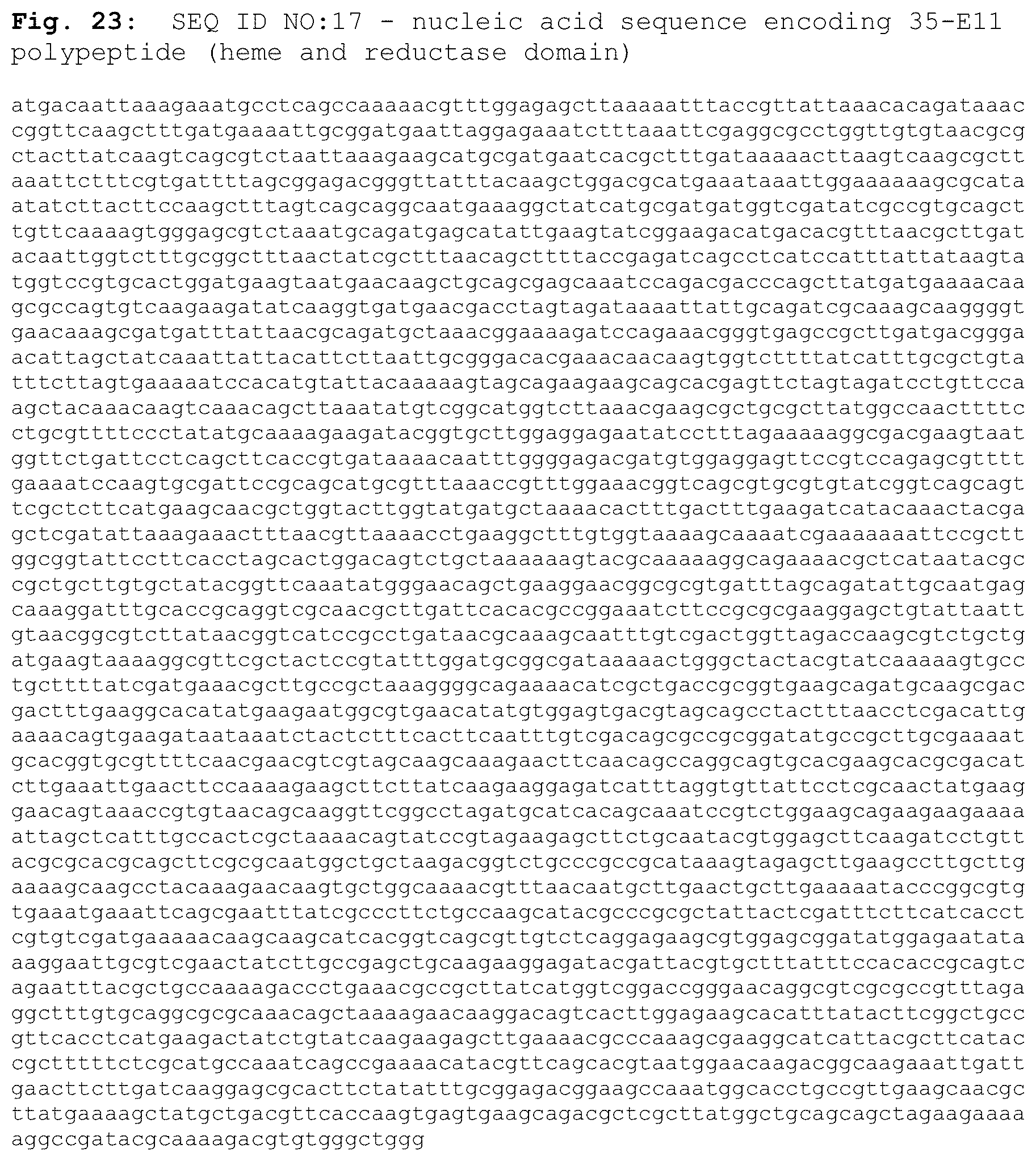
D00022

D00023

D00024

D00025

D00026
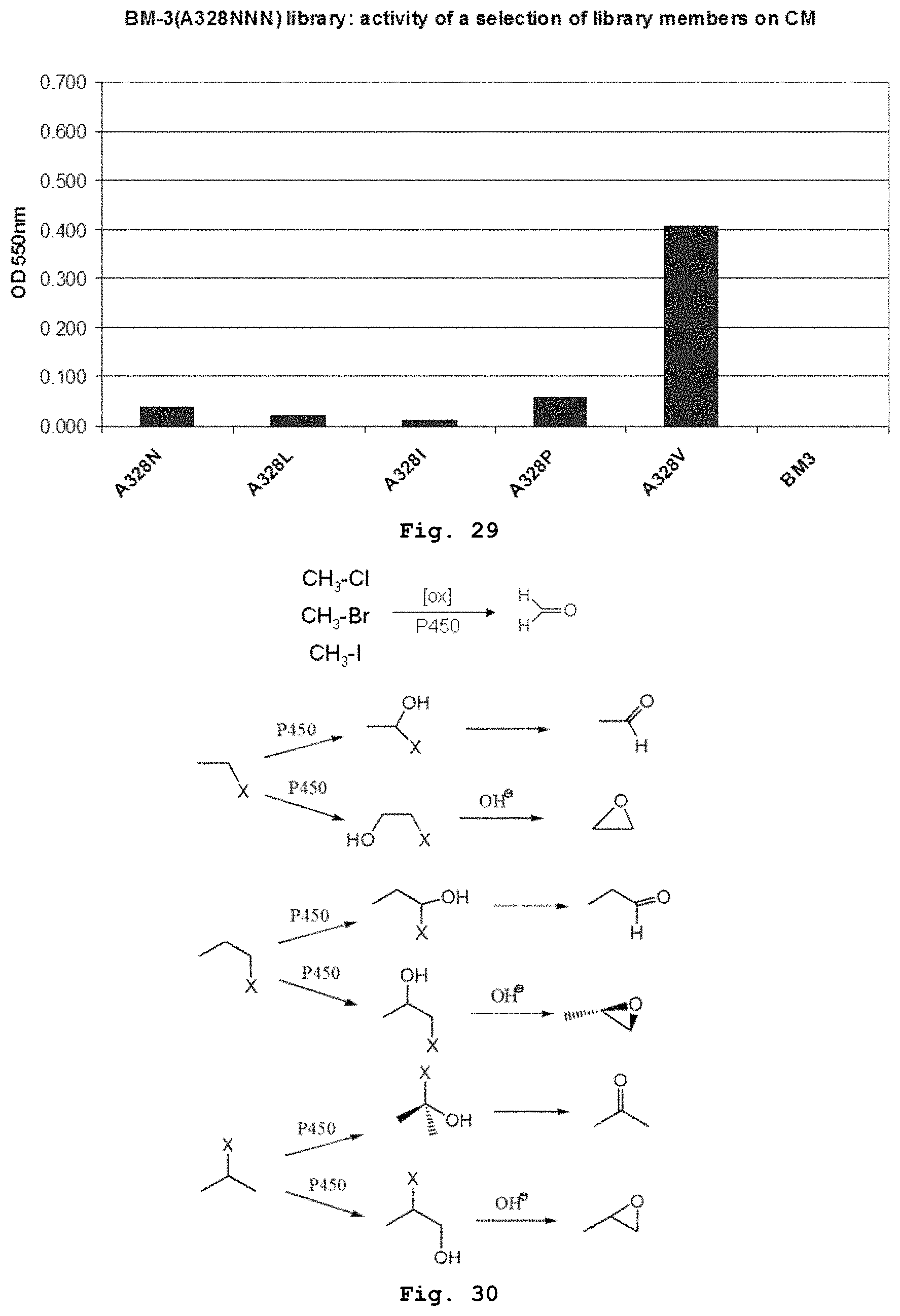
D00027

D00028

D00029

D00030
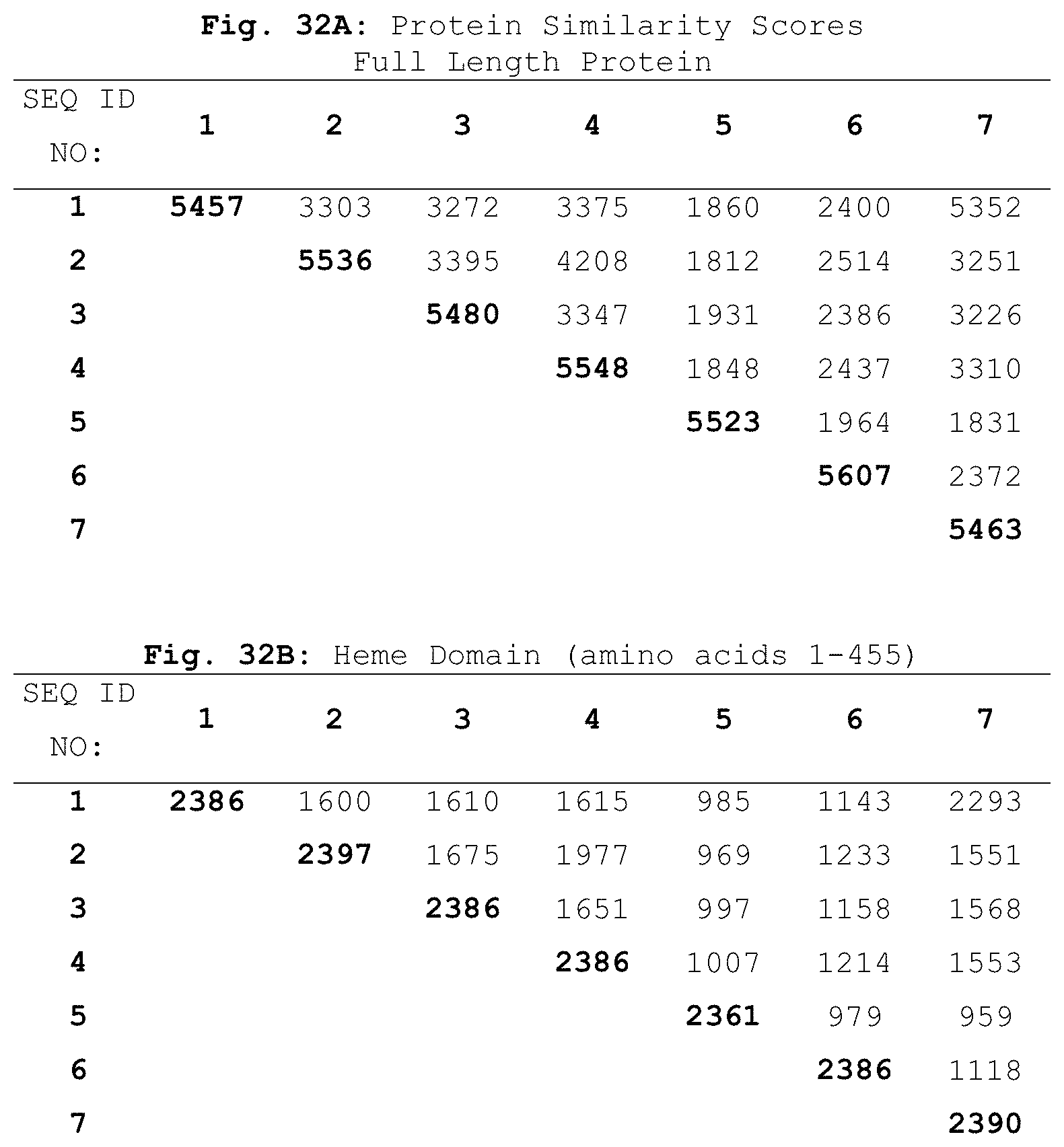
D00031

D00032

D00033

D00034

D00035

D00036

D00037

D00038

D00039

D00040

S00001
XML
uspto.report is an independent third-party trademark research tool that is not affiliated, endorsed, or sponsored by the United States Patent and Trademark Office (USPTO) or any other governmental organization. The information provided by uspto.report is based on publicly available data at the time of writing and is intended for informational purposes only.
While we strive to provide accurate and up-to-date information, we do not guarantee the accuracy, completeness, reliability, or suitability of the information displayed on this site. The use of this site is at your own risk. Any reliance you place on such information is therefore strictly at your own risk.
All official trademark data, including owner information, should be verified by visiting the official USPTO website at www.uspto.gov. This site is not intended to replace professional legal advice and should not be used as a substitute for consulting with a legal professional who is knowledgeable about trademark law.