Lactobacillus strain, composition and use thereof for treating syndromes and related complications of autoimmune diseases
Huang , et al.
U.S. patent number 10,646,522 [Application Number 15/954,583] was granted by the patent office on 2020-05-12 for lactobacillus strain, composition and use thereof for treating syndromes and related complications of autoimmune diseases. This patent grant is currently assigned to GenMont Biotech Incorporation. The grantee listed for this patent is GenMont Biotech Incorporation. Invention is credited to Yi-Hsing Chen, Chih-Yang Huang, Wei-Wen Kuo, Bor-Show Tzang.


| United States Patent | 10,646,522 |
| Huang , et al. | May 12, 2020 |
Lactobacillus strain, composition and use thereof for treating syndromes and related complications of autoimmune diseases
Abstract
The present invention is related to a Lactobacillus strain, composition and use thereof for treating autoimmune disease and related complications. The composition comprises at least one isolate of Lactobacillus paracasei strain GMNL-32, L. reuteri strain GMNL-89 or L. reuteri strain GMNL-263, and a pharmaceutical carrier, for treating syndromes and related complications of the autoimmune diseases.
| Inventors: | Huang; Chih-Yang (Taichung, TW), Kuo; Wei-Wen (Taichung, TW), Tzang; Bor-Show (Taichung, TW), Chen; Yi-Hsing (Tainan, TW) | ||||||||||
|---|---|---|---|---|---|---|---|---|---|---|---|
| Applicant: |
|
||||||||||
| Assignee: | GenMont Biotech Incorporation
(Tainan, TW) |
||||||||||
| Family ID: | 52821915 | ||||||||||
| Appl. No.: | 15/954,583 | ||||||||||
| Filed: | April 16, 2018 |
Prior Publication Data
| Document Identifier | Publication Date | |
|---|---|---|
| US 20180228851 A1 | Aug 16, 2018 | |
Related U.S. Patent Documents
| Application Number | Filing Date | Patent Number | Issue Date | ||
|---|---|---|---|---|---|
| 14623527 | Feb 17, 2015 | 9980990 | |||
Foreign Application Priority Data
| Feb 21, 2014 [TW] | 103105864 A | |||
| Current U.S. Class: | 1/1 |
| Current CPC Class: | A61P 37/02 (20180101); A61P 37/00 (20180101); C12N 1/20 (20130101); A61K 35/747 (20130101); C12R 1/225 (20130101) |
| Current International Class: | A61K 35/747 (20150101); C12N 1/20 (20060101) |
References Cited [Referenced By]
U.S. Patent Documents
| 9301984 | April 2016 | Chen et al. |
| 2009/0196921 | August 2009 | Ebel et al. |
| 2009/0274672 | November 2009 | Yu |
| 2011/0300117 | December 2011 | Leu et al. |
| 2012/0263695 | October 2012 | Schrezenmeir et al. |
| 1696281 | Nov 2005 | CN | |||
| 101575582 | Nov 2009 | CN | |||
| 102100704 | Jun 2011 | CN | |||
| 102115721 | Jul 2011 | CN | |||
| 102935092 | Feb 2013 | CN | |||
| 1481681 | Dec 2004 | EP | |||
| 2392340 | Dec 2011 | EP | |||
| 2009057346 | Mar 2009 | JP | |||
| 2012-149026 | Aug 2012 | JP | |||
| I284149 | Jul 2007 | TW | |||
| I340021 | Apr 2011 | TW | |||
| I346554 | Aug 2011 | TW | |||
| I355939 | Jan 2012 | TW | |||
| 201210603 | Mar 2012 | TW | |||
| I406665 | Sep 2013 | TW | |||
| 01/11077 | Feb 2001 | WO | |||
| 0111334 | Feb 2001 | WO | |||
| 2006/102536 | Sep 2006 | WO | |||
| 2007/040446 | Apr 2007 | WO | |||
| 2009027753 | Mar 2009 | WO | |||
Other References
|
"How dose lupus affect the heart and cirsulation," Lupus Foundation of America, Aug. 11, 2013. cited by applicant . "How does lupus affect the gastrointestinal system" Lupus Foundation of America, Jul. 12, 2013. cited by applicant . M. Salmon et al., "The role of apoptosis in systemic lups erythematosus," Rheumatology 1999;38:1177-1183. cited by applicant . Tom Van Belle et al. "Type 1 Diabetes: Etiology, Immunology, and Therapeutic Strategies", Physiological Reviews, vol. 91, No. 1, 2011, pp. 79-118. cited by applicant . Harman et al. "Prevalence and natural history of histologically proven chronic liver disease in a longitudinal cohort of patients with type 1 diabetes", Hepatology, vol. 56, No. 4, Oct. 2012, p. 881A-882A, #1467. cited by applicant . Manirarora, Jean Nepomuscene "Manipulating antigen-presenting cell function to restore immune regulation in autoimmune-prone mice", ProQuest Dissertations Publishing, 2008, pp. 111, 113-115. cited by applicant . Pascale Alard et al., Abstract of "Probiotics control lupus progression via induction of regulatory cells and IL-10 production", http://www.jimmunol.org/content/182/1_Supplement/50.30, Apr. 2009. cited by applicant . Hermelijn H. Smits, PhD et al., "Selective probiotic bacteria induce IL-10-producing regulatory T cells in vitro by modulating dendritic cell function through dendritic cell-specific intercellular adhesion molecule 3-grabbing nonintegrin", J Allergy Clin Immunol, vol. 115, No. 6, Jun. 2005, pp. 1260-1267. cited by applicant . Matteo Bellone, "Autoimmune Disease: Pathogenesis", Encyclopedia of Life Sciences, published online Sep. 2015. cited by applicant . JP2012149026A, Machine Translation, Aug. 2012. cited by applicant. |
Primary Examiner: Berke-Schlessel; David W
Attorney, Agent or Firm: CKC & Partners Co., LLC
Parent Case Text
RELATED APPLICATIONS
This application is a divisional application of U.S. application Ser. No. 14/623,527, filed Feb. 17, 2015, which is herein incorporated by reference, which was based on, and claims priority to Taiwan Application Serial Number 103105864, filed Feb. 21, 2014, which is herein incorporated by reference.
Claims
What is claimed is:
1. A method of treating a cardiac or hepatic disease caused by systemic lupus erythematosus in an individual, comprising orally administering a lactic acid bacteria (LAB)-containing composition to the individual, wherein the LAB-containing composition comprises an effective amount of a LAB strain and a pharmaceutically acceptable carrier, the effective amount is 1.times.10.sup.9 CFU/g per individual for at least 12 weeks, the individual is a mouse, and the LAB strain is Lactobacillus reuteri strain GMNL-89, CCTCC M 207154, for reducing gap junctions between cardiac cells or inhibiting activities of matrix metallopeptidase 9 (MMP-9), C-reactive protein (CRP), interleukin-1.beta.(IL-1.beta.), IL-6, inducible NO synthase (iNOS) and caspase-3 of a hepatocyte.
2. The method of claim 1, wherein the LAB strain reduces gap junctions between cardiac cells.
Description
BACKGROUND
Field of Invention
The disclosure relates to a Lactobacillus strain and an application thereof. More particularly, the disclosure relates to a Lactobacillus strain for treating syndromes and related complications of autoimmune diseases and a composition thereof.
Description of Related Art
An autoimmune disease is a disease that a human body's immune system attacks normal cells of his body. Such patient has weaker normal immunity but stronger aberrant immunity. The "aberrant immunity" refers to the immune system that recognizes the normal cells of its own body as foreign virus or bacteria and targets the normal cells for attacking and clearing them out of the body. The normal immune system of a human body provides a physically protective mechanism to target foreign antigens and abnormal cells, such as tumor cells, for attacking and clearing them out of the human body. However, sometimes the immune system may produce some antibodies against the normal cells or normal cellular components. Therefore, some aberrant excessive inflammation or tissue damage may cause diseases and adversely effect health.
Common autoimmune diseases include coeliac disease, type 1 diabetes mellitus, systemic lupus erythematosus (SLE), Sjogren's syndrome, multiple sclerosis (MS), Hashimoto's thyroiditis, Graves' disease, idiopathic thrombocytopenic purpura, rheumatoid arthritis (RA), aplastic anemia, polymyositis and dermatomyositis, as well as scleroderma.
Among those, systemic lupus erythematosus (SLE) is a chronic systemic autoimmune disease, in which the body's immune system turns against all organs or tissues of the body itself, including skin, joints, heart, blood vessels, liver, kidneys, brain and nervous system. In general, the course of SLE is alternatively aggravated and released, and the onset is usually unpredictable. This disease, which may occur in people of any age, most commonly occurs in young women, and 90% of SLE patients are women. The signs and symptoms of SLE vary greatly in different individuals. When onset of SLE, some patients have severe symptoms with the important organs such as the central nervous system, kidney, heart and so on, whereas some other patient only have mildly joint symptoms during life. As for the incidence, above 90% of the SLE patients have joint pain or arthritis, 70% to 80% of the SLE patients have skin erythema or discoid erythema, 40% to 50% of the SLE patients have pleurisy or pericarditis, 40% to 50% of the SLE patients have lupus nephritis, 20% to 60% of the SLE patients have central nervous system violations, and 20% of the SLE patients have thrombocytopenia.
At present, the cause of the SLE is still unclear, but it is believed that SLE is related to genes, immunodeficiency, viral infections, UV, and drugs. Recent studies have found that, SLE patients have much higher incidence of liver abnormalities (such as hepatomegaly, splenomegaly, jaundice, abnormal liver function, abnormal liver enzymes and so on) and the cardiovascular disease (CVD, such as myocardial infarction (MI)) than normal people, and the atherosclerosis is also highly associated with SLE. Some studies with animal models reveal that significant inflammation and apoptosis of liver and heart occur in SLE mice.
SLE is currently incurable, but its symptoms can be treated. Medicines are important to the treatment of SLE. Physicians prescribe mediation to treat a SLE patient depending on symptoms, the different organ injuries and different severities. Common medications used to treat SLE manifestations include the followings.
(1) Non-Steroidal Anti-Inflammatory Drugs (NSAIDs):
NSAIDs can be used to treat arthritis, pleurisy, and other lighter inflammatory symptoms. Gastrointestinal side effects are the major problem of NSAIDs. Fortunately, a new generation of selective inhibitor of type II cyclooxygenase (COX-2) has been commercially marketed, and it causes much less gastrointestinal side effect than the traditional NSAIDs.
(2) Antimalarial Drugs:
Most commonly antimalarial drug is hydroxychloroquine (or the brand name of Plaquenil). The clinical uses show that the antimalarial drugs have a fairly good efficacy on the treatment of skin symptoms, arthritis, and other mild systemic symptoms of SLE patients. The antimalarial drugs also can decrease the recurrence of SLE.
(3) Cortisol:
Cortisol is generally known as the "American panacea" or "steroids." Cortisol is the most important drugs for treating SLE. Higher dose of steroids should be used in severe conditions of internal organ damages such as lupus nephritis, nervous system lupus, lupus pneumonitis, hemolytic anemia, or thrombocytopenia and so on. The physician also considers supplying an intravenous pulse steroid therapy with high dose of steroids, so that the condition is controlled as soon as possible, and the dose of the steroids is then gradually decreased to a minimal level when clinically well controlled.
(4) Immunosuppressing Drugs:
Immunosuppressing drugs are mainly used to inhibit the activity of the immune system, so as to achieve good control and efficacy of the autoimmune diseases. Immunosuppressing drugs are usually used with steroids, so as to decrease the doses of the steroids and to effectively control the stubborn disease. Common immunosuppressing drugs include azathioprine (Imuran.RTM.), cyclophosphamide (Endoxan.RTM.), cyclosporine, methotrexate (MTX) and so on.
Probiotics or probiotic bacteria generally refer to live microorganisms originating from human bodies and beneficial to intestinal tract health, or some microorganisms supplied from outside sources and beneficial to human health, for example, lactic acid bacteria (LAB) and some yeasts. 70% of human immune system is located in the digestive tract. As early as the late nineteenth century, Russia scientists have observed that probiotics have health effect to human body. Previous studies have shown that more than 1/3 SLE patients have gastrointestinal symptoms, and 10% SLE patients have gastrointestinal pathological changes at the first onset. It is currently known that probiotics are capable of restoring intestinal flora balance, stimulating mucosal immunity, improving gastrointestinal discomfort and improving urogenital flora.
Recently, some studies attempt to treat SLE with Lactobacillus. The obtained results show that the single Lactobacillus strain can promote the production of anti-inflammatory cytokines such as interleukin-10 (IL-10) and tumor necrosis factor-.alpha. (TNF-.alpha.), to treat the autoimmune diseases such as SLE.
However, the aforementioned studies seldom investigated whether LABs is used to treat the symptoms of the autoimmune diseases (such as SLE) and their complications (such as cardiac and hepatic disease), and did not propose any regulatory mechanism that LABs may be involved in SLE treatments.
Side effects often occur in drugs for the treatment of autoimmune diseases (such as SLE), and patients with such chronic diseases must be treated and controlled for a long time. Therefore, there is a need to use native Lactobacillus strains to develop products that are capable of treating symptoms and complications of autoimmune diseases, so as to achieve the improvement, control, remedy, or prevention of the patients with the symptoms and complications of autoimmune diseases, as well as the development of other applications of Lactobacillus strains.
SUMMARY
Accordingly, an aspect of the invention provides an isolated strain of Lactobacillus species for treating a syndrome and a complication of an autoimmune disease.
Another aspect of the invention provides a lactic acid bacteria (LAB) strain used for preparation of a medical composition for treating a syndrome of an autoimmune disease and its complication.
Yet another aspect of the present invention provides a LAB-containing composition containing the aforementioned LAB strain, for treat a syndrome of an autoimmune disease and its related complication via oral administration.
In light of foregoing, an isolated strain of Lactobacillus for treating a syndrome and a complication of an autoimmune disease is provided. The isolated strain includes but is not limited to at least one of Lactobacillus paracasei strain GMNL-32 (the accession numbers of BCRC 910220 and CCTCC M 204012), L. reuteri strain GMNL-89 (the accession numbers of BCRC 910340 and CCTCC M 207154), L. reuteri strain GMNL-263 (the accession numbers of BCRC 910452 and CCTCC M 209263), or any combinations thereof. The aforementioned Lactobacillus strains are deposited in Food Industry Research and Development Institute (FIRDI), Hsin-Chu, Taiwan, as well as China Center for Type Culture Collection (CCTCC), Wuhan University, Wuhan 430072, People's Republic of China.
According to another aspect, a lactic acid bacteria (LAB) strain used for preparation of a medical composition for treating a syndrome and a complication of an autoimmune disease is provided. The medical composition comprises an effective amount of the LAB strain and a pharmaceutically acceptable carrier, and the LAB strain can include but be not limited to at least one isolated strain of the aforementioned Lactobacillus species.
According to yet another aspect, a LAB-containing composition is provided. The LAB-containing composition comprises an effective amount of a LAB strain and an edible material. The LAB strain is at least one of the isolated strains of the aforementioned Lactobacillus species.
According to some embodiments, when the LAB strain is used for preparation of the medical composition for treating the syndrome and the complication of the autoimmune disease, the LAB strain is live and/or inactivated. Moreover, the aforementioned medical composition further comprises a pharmaceutically acceptable carrier, which can use any conventional carrier rather than being recited in detail. In addition, the aforementioned LAB strain can be applied to any form of easy to use in daily life, for example, healthy food, additives, medical compositions, dietary supplement, food and so on. In other examples, the aforementioned LAB strain can be produced in the freeze-dried form, and it can further include other ingredients such as glucose, maltodextrin, baby milk, fructo-oligosaccharides, magnesium stearate, yogurt spices, other uncertain remains unseparated therefrom or any combinations thereof. Furthermore, the aforementioned LAB strain may be used to specifically inhibit activities of matrix metallopeptidase 9 (MMP-9), C-reactive protein (CRP), interleukin-1.beta. (IL-1.beta.), tumor necrosis factor-.alpha. (TNF-.alpha.), IL-6 and inducible NO synthase (iNOS).
According to an embodiment, the aforementioned autoimmune disease can be systemic lupus erythematosus.
In light of the foregoing, the isolated strain of the Lactobacillus species of the present invention, the medical composition containing the same, and the LAB-containing composition, can be administrated orally to specifically inhibit activities of MMP-9, CRP, IL-1.beta., TNF-.alpha., IL-6, iNOS to a subject having an autoimmune disease, for treating the syndrome and the complication (such as cardiac and hepatic disease) of the autoimmune disease, thereby developing other application fields of the LAB strains.
These and other features, aspects, and advantages of the present invention will become better understood with reference to the following description and appended claims.
BRIEF DESCRIPTION OF THE DRAWING
The invention can be more fully understood by reading the following detailed description of the embodiment, with reference made to the accompanying drawings as follows:
FIG. 1 depicts a histogram showing the liver-to-body weight ratios (L/B) of SLE mice according to several embodiments of the present invention.
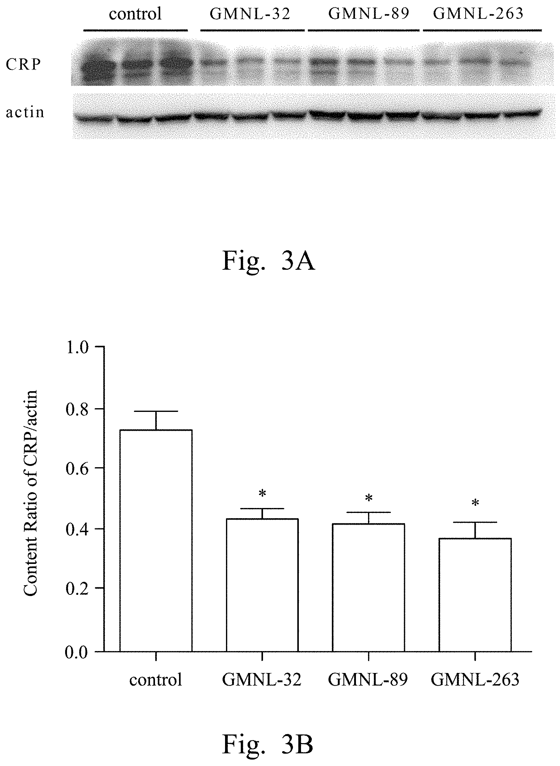
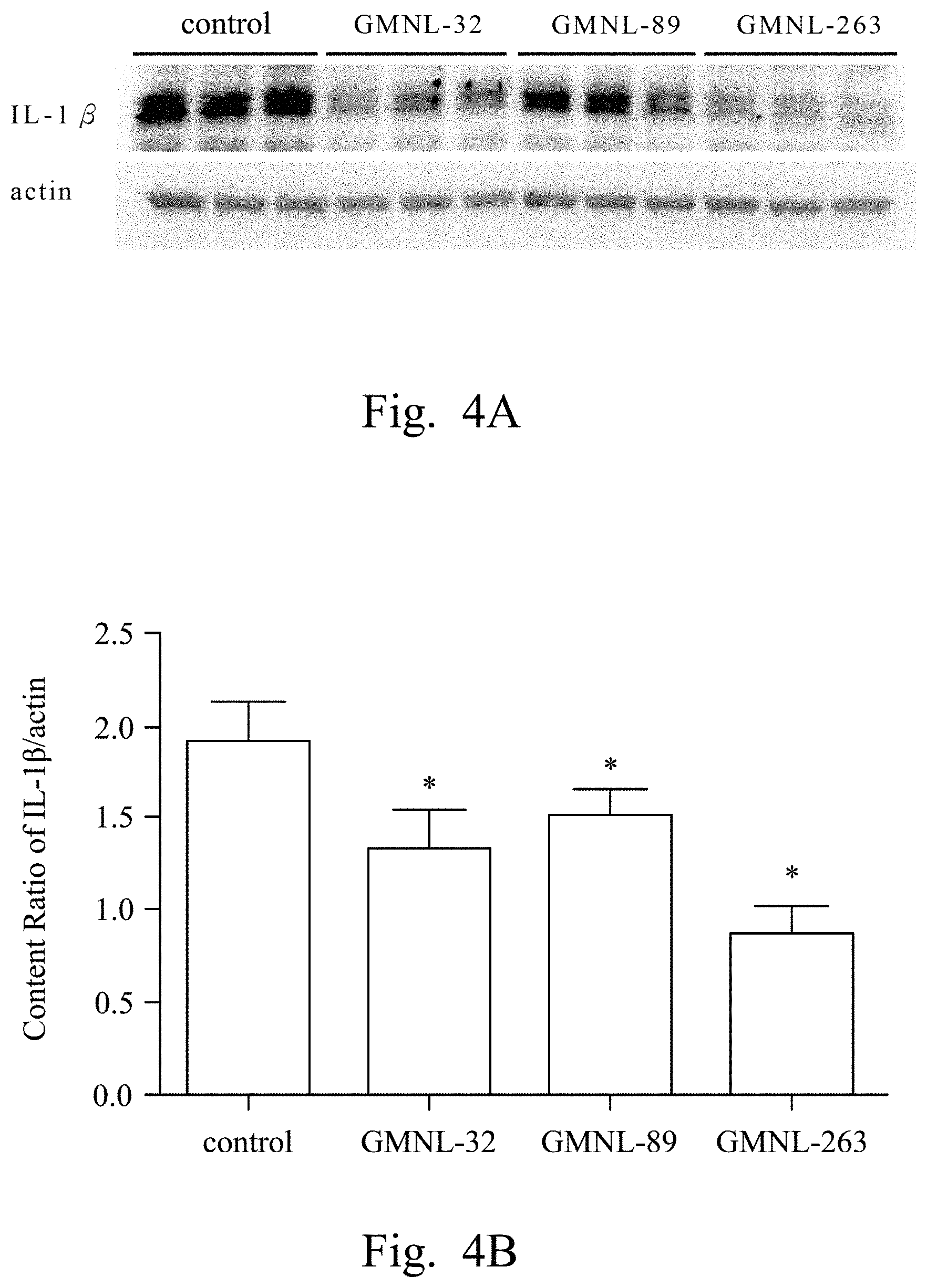
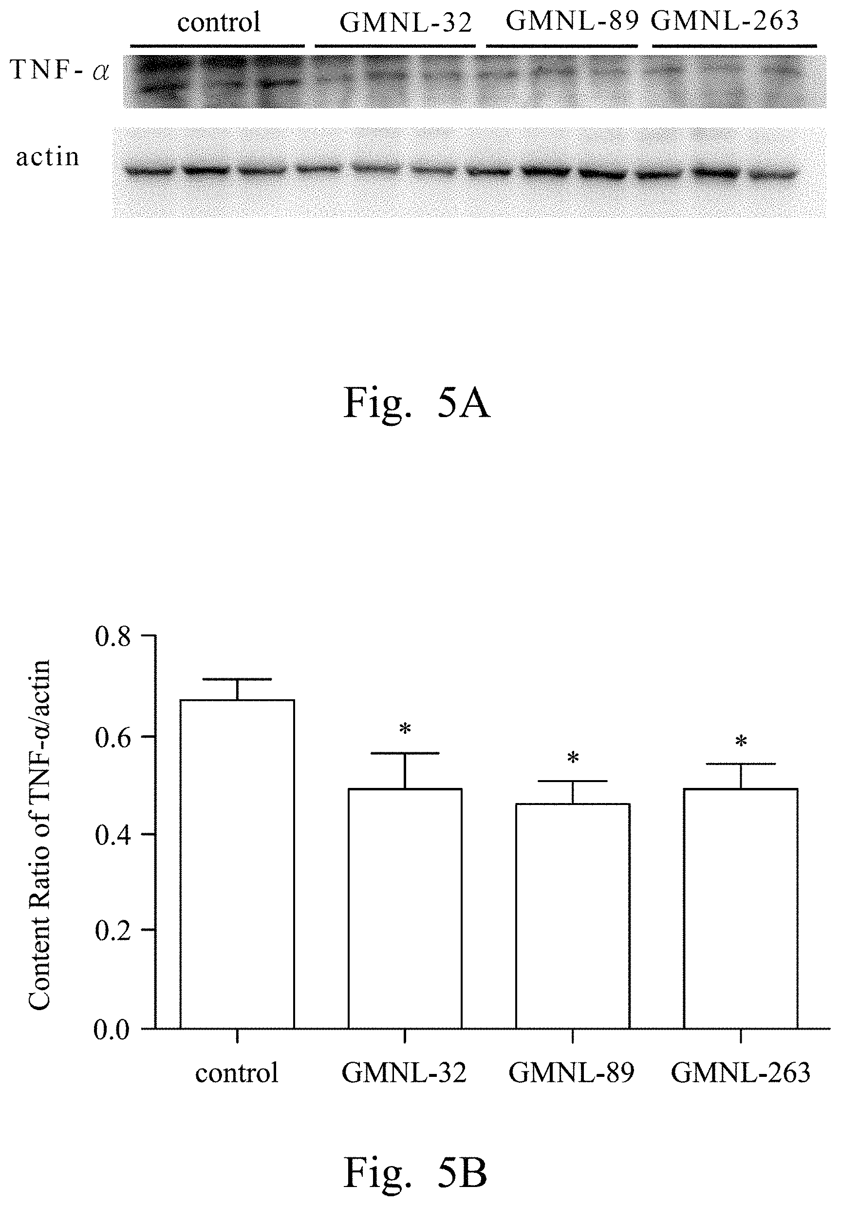
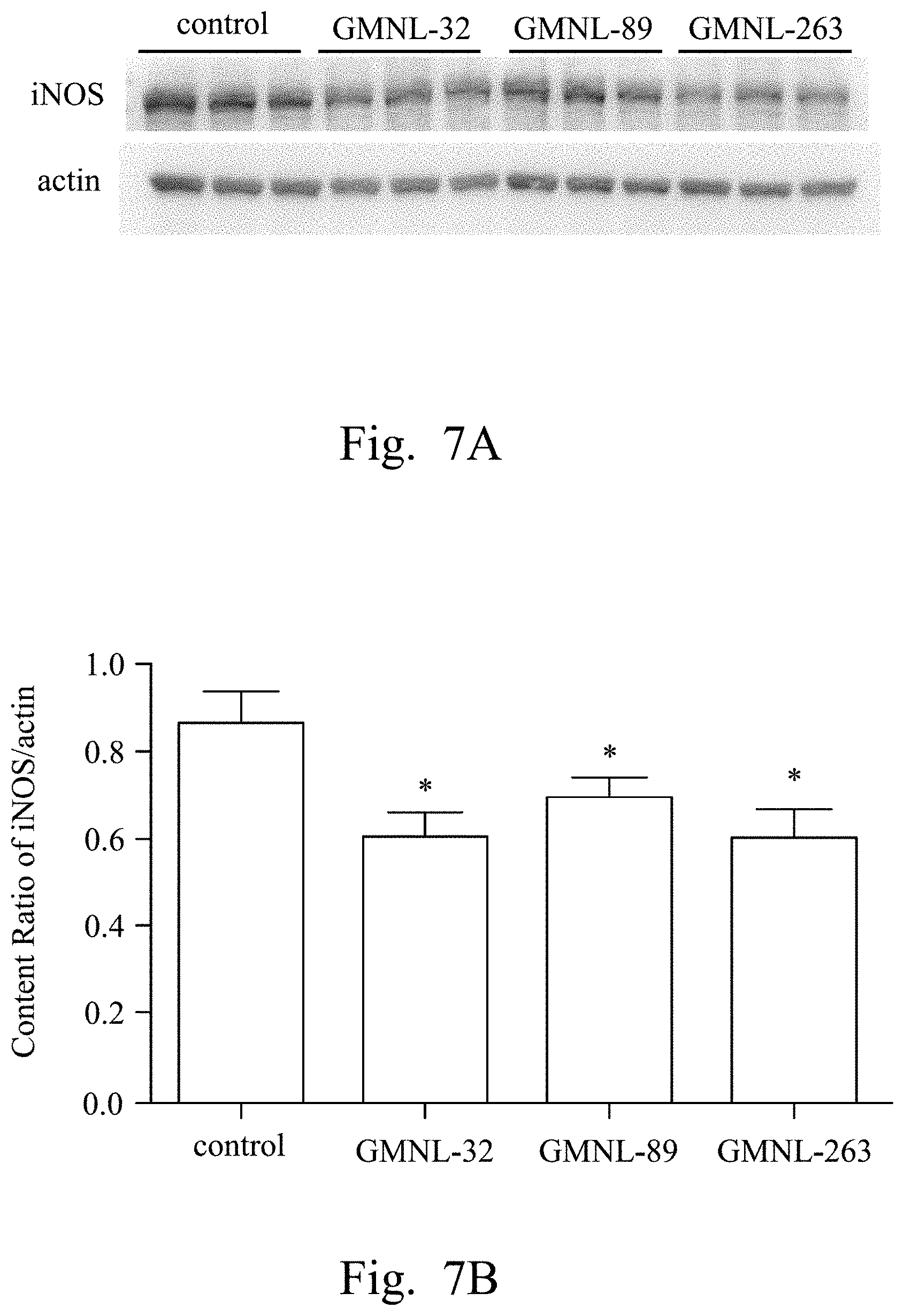
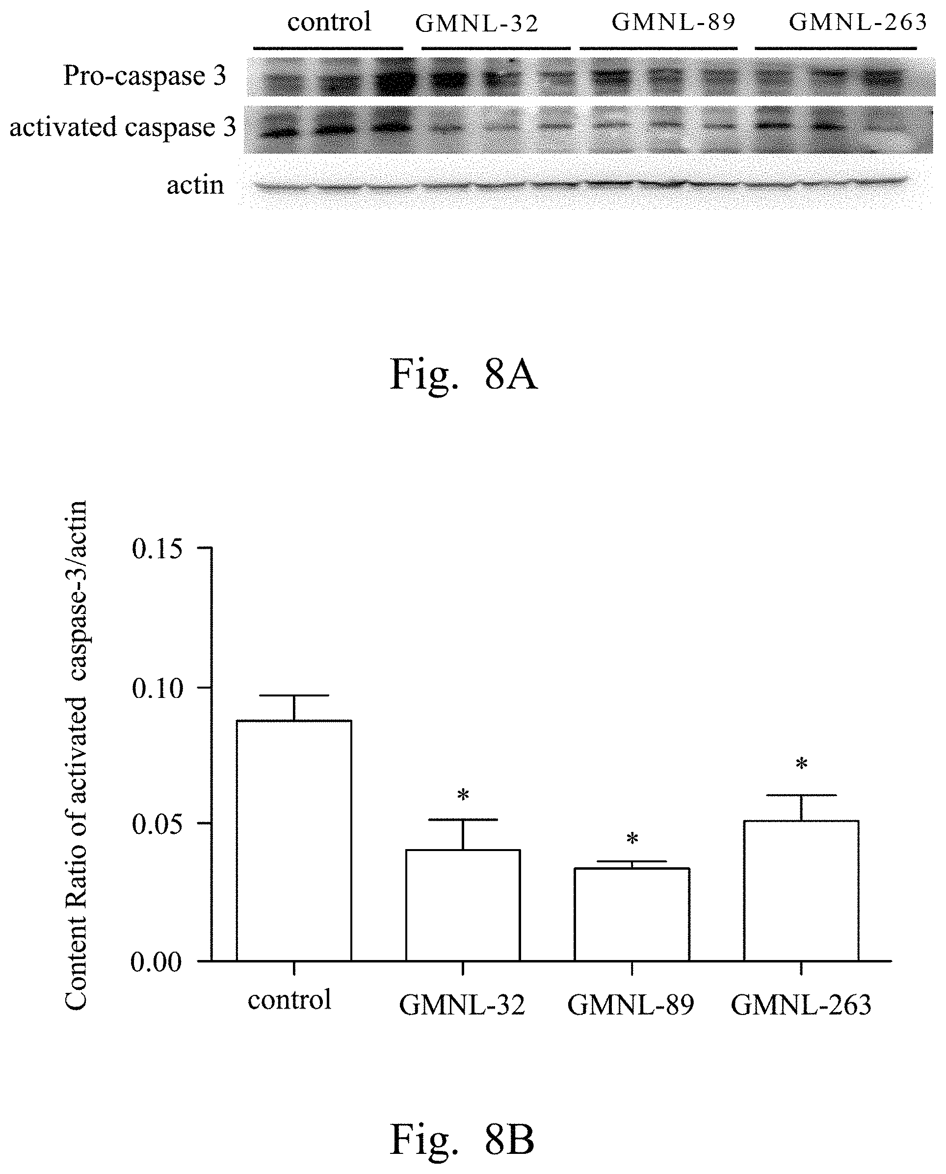
FIGS. 2A, 3A, 4A, 5A, 6A, 7A, and 8A are images respectively showing the results of gelatin-SDS-PAGE gel electrophoresis of MMP-9 (FIG. 2A), and the western blotting analyses of CRP (FIG. 3A), IL-1.beta. (FIG. 4A), TNF-.alpha. (FIG. 5A), IL-6 (FIG. 6A), iNOS (FIG. 7A) and caspase-3 (FIG. 8A) of liver tissue of SLE mice according to several embodiments of the present invention.
FIGS. 2B, 3B, 4B, 5B, 6B, 7B, and 8B are histograms respectively showing the content ratios of MMP-9/MMP-2 (FIG. 2B), CRP/actin (FIG. 3B), IL-1.beta./actin (FIG. 4B), TNF-.alpha./actin (FIG. 5B), IL-6/actin (FIG. 6B), iNOS/actin (FIG. 7B) and caspase-3/actin (FIG. 8B).
FIG. 9 shows the result of flow cytometry analysis of CD4.sup.+/CD25.sup.+ splenic Treg cells of SLE mice according to several embodiments of the present invention.
FIGS. 10A to 10D are images of cardiac tissue sections of SLE mice analyzed by Masson's trichrome staining according to several embodiments of the present invention.
DETAILED DESCRIPTION
In light of forgoing, this invention provides an isolated strain of Lactobacillus species for treating autoimmune disease and complications thereof, a medical composition comprising lactic acid bacteria (LAB) and a LAB-containing composition for treating autoimmune disease and complications thereof. The isolated strain of the Lactobacillus species comprises at least one Lactobacillus strain, and can be administered orally to treat the syndrome and complication (such as cardiac and hepatic disease) of the autoimmune disease.
The "isolated strain of the Lactobacillus species" recited herein includes but is not limited to Lactobacillus paracasei strain GMNL-32, L. reuteri strain GMNL-89, L. reuteri strain GMNL-263 or any combinations thereof. The L. paracasei strain GMNL-32 has been deposited in Bioresource Collection and Research Center (BCRC) of Food Industry Research and Development Institute (FIRDI), Hsin-Chu, Taiwan on Mar. 19, 2003 under the accession number of BCRC 910220, and also deposited in China Center for Type Culture Collection (CCTCC), Wuhan University, Wuhan 430072, People's Republic of China on Feb. 19, 2004 under the accession number of CCTCC M 204012. The L. reuteri strain GMNL-89 has been deposited in BCRC of FIRDI on Nov. 14, 2006 under the accession number of BCRC 910340, and also deposited in CCTCC on Nov. 19, 2007 under the accession number of CCTCC M 207154. The L. reuteri strain GMNL-263 has been deposited in BCRC of FIRDI on Nov. 6, 2009 under the accession number of BCRC 910452, and also deposited in CCTCC on Nov. 13, 2009 under the accession number of CCTCC M 209263.
The aforementioned L. paracasei strain GMNL-32 (also called as GM-080) is a disclosed strain (the accession numbers of BCRC 910220 and CCTCC M 204012). The related strain characteristics have been disclosed in TW I284149 titled as "novel microorganism strain GMNL-32 of Lactobacillus paracasei for allergy diseases treatment", and TW 201210603 titled as "composition and use of probiotic strain GM-080 in treating cardiac inflammation and apoptosis", both of which are herein incorporated by reference.
The L. reuteri strain GMNL-89 is also a disclosed strain (the accession numbers of BCRC 910340 and CCTCC M 207154). The related strain characteristics have been disclosed in TW I340021 titled as "novel Lactobacillus strain, composition and use thereof for improving the syndrome of diabetes and complication thereof", and TW I346554 titled as "Lactobacillus isolates having anti-inflammatory activities and uses of the same", both of which are herein incorporated by reference.
The L. reuteri strain GMNL-263 is also a disclosed strain (the accession numbers of BCRC 910452 and CCTCC M 209263). The related strain characteristics have been disclosed in TW I340021 titled as "novel Lactobacillus strain, composition and use thereof for improving the syndrome of diabetes and complication thereof" and TW I355939 titled as "composition and use of probiotic strain GM-263 (ADR-1) in treating renal fibrosis in diabetes", both of which are herein incorporated by reference.
In general, the aforementioned LAB strains can be subjected to various available culturing methods to obtain the isolated strain. The "isolated strain" recited herein refers to a strain obtained from a pure culture of a single colony of the aforementioned LAB strain, which substantially does not contain other strains.
In application, when the LAB strain is used for preparation of the medical composition for treating the syndrome and the complication of the autoimmune disease, the LAB strain is live and/or inactivated. Moreover, the aforementioned medical composition further comprises a pharmaceutically acceptable carrier, which can use any conventional carrier rather than being recited in detail. In addition, the aforementioned LAB strain can be applied to any form of easy to use in daily life, for example, healthy food, additives, medical compositions, dietary supplement, food and so on. In other examples, the aforementioned LAB strain can be produced in the freeze-dried form, and it can further include an edible material such as glucose, maltodextrin, baby milk, fructo-oligosaccharides, magnesium stearate, yogurt spices, other uncertain remains unseparated therefrom or any combinations thereof. Furthermore, one or more pure cultures of LAB strains are mixed well with a pharmaceutically acceptable carrier, so as to prepare a medical composition containing the aforementioned LAB strains. A formulation of the aforementioned medical composition can include but be not limited to a solution, a suspension solution, an emulsion, powder, a tablet, a pill, a syrup, a lozenge, a troche, a chewing gum, a thick slurry, a capsule or any combination thereof. The aforementioned medical composition is then analyzed by animal model, such as SLE mice. The results prove that the aforementioned medical composition can treat, alleviate, control, improve, and/or prevent the cardiac and hepatic disease and related complications caused by autoimmune diseases.
In detail, "the treatment and improvement of cardiac and hepatic disease and related complication of the autoimmune disease" recited herein refers to the aforementioned medical composition containing the above strains of Lactobacillus for continuously oral administration to a subject in a daily dosage of about 1.times.10.sup.6 colony-forming unit per gram (CFU/g) to about 1.times.10.sup.11 CFU/g for at least 12 weeks. Such medical composition is easy-to-use in the daily life of patients, and can treat, alleviate, control, improve, and/or prevent the hepatic disease-related protein expression caused by the autoimmune disease (such as SLE) in the animal body. For example, the aforementioned medical composition can specifically inhibit activities of MMP-9, CRP, IL-1.beta., TNF-.alpha., IL-6 and iNOS. Also, the aforementioned medical composition can treat, alleviate, control, improve, and/or prevent cardiac disease-related syndromes caused by the autoimmune disease, so as to restore heart weight and reduce gap junctions between cardiac cells, for example.
Thereinafter, various applications of the LAB strains of the present invention will be described in more details referring to several exemplary embodiments below, while not intended to be limiting. Thus, one skilled in the art can easily ascertain the essential characteristics of the present invention and, without departing from the spirit and scope thereof, can make various changes and modifications of the invention to adapt it to various usages and conditions.
EXAMPLE
Embodiment 1
Establishment and Evaluation of Animal Model
1. Preparation of Lactobacillus Strains
L. paracasei strain GMNL-32 (the accession number of BCRC 910220), L. reuteri strain GMNL-89 (the accession number of BCRC 910340) and L. reuteri strain GMNL-263 (the accession number of BCRC 910452) were used to perform SLE animal experiments, for evaluating the effect of treating the cardiac and hepatic disease caused by SLE via oral administration.
The total dosages per day of the aforementioned three Lactobacillus strains were 1.times.10.sup.9 CFU/g, respectively. The aforementioned three Lactobacillus strains were produced in the freeze-dried form, and included other ingredients such as glucose, maltodextrin, baby milk, fructo-oligosaccharides, magnesium stearate, yogurt spices, other uncertain remains unseparated therefrom or any combinations thereof.
2. Establishment of SLE Animal Testing Model
New Zealand Black/White F1 hybrid (NZB/W F1) female mice (Jackson Lab, U.S.A.) were exemplified to establish SLE animal testing model. First, SLE mice were randomly assigned to three experimental groups and one control group. Each group had 8 SLE mice of 14 weeks old. The SLE mice of the experimental groups were fed a diet with 1.times.10.sup.9 CFU/g per mouse of various LAB strains including Lactobacillus paracasei strain GMNL-32, L. reuteri strain GMNL-89 or L. reuteri strain GMNL-263 respectively. The SLE mice of the control group were fed a basic diet replaced by deionized water having the same weight as the LAB strains.
The SLE mice were raised at ambient temperature controlled at 25.+-.1.degree. C., relative humidity at 65.+-.5%. In addition, the SLE mice were maintained on a reverse 12 hours light-dark cycle. The SLE mice were provided with standard laboratory chow and water ad libitum. All experimental procedures were approved according to related guidelines for the management of the laboratory animals announced by National Health Research Institutes (NHRI), Taiwan.
All SLE mice were weighted after 12 weeks, and then sacrificed. The liver, heart and coronary artery were dissected and then washed by distilled water. The left and right atriums and ventricles were divided and separately weighted for subsequent analyses.
3. Extraction of Liver Tissue
The livers of the aforementioned SLE mice were respectively immersed in lysis buffer, and then 100 mg of liver tissue in 1 mL of the lysis buffer were homogenized for 1 minute, so as to dissolve hepatocytes and release the proteins in the hepatocytes. The aforementioned lysis buffer included 20 mM of tris(hydroxymethyl) aminomethane (Tris) solution, 2 mM of tris(hydroxymethyl) aminomethane (EDTA), 50 mM of 2-mercaptoethanol, 10% of glycerol, proteinase inhibitor (Roche) and phosphatase inhibitor cocktail (Sigma), and the pH value of the lysis buffer was 7.4. Next, the resultant homogenate was placed on ice for 10 minutes, and then centrifuged twice at 12000 g for 40 minutes. The supernatant was taken and stored at -80.degree. C. for following assessments.
4. Western Blot
Hereinafter, electrophoresis and western blot were used to evaluate the effects of the three Lactobacillus strains on the treatment of the liver diseases caused by SLE.
The aforementioned supernatant were loaded into 10% sodium dodecyl sulfate-polyacrylamide gel electrophoresis (SDS-PAGE), and electrophoresed at a voltage of 85 volts for 3.5 hours. Next, the gel was equilibrated in 25 mM of Tris-HCl buffer (pH 8.3) containing 192 mM of glycine and 20% (v/v) of methanol for 15 minutes. The preparation of SDS-PAGE and related equipments were familiar with the artisan in this art of the present invention rather than being recited in detail herein.
The SDS-PAGE gel was then subjected to western blotting assay. In this Example, the kit of western blotting assay (such as Bio-Rad Scientific Instruments Transfer Unit) was used to transfer electrophoresed proteins to a transfer membrane, such as polyvinylidene difluoride (PVDF) membrane (0.45 m of pore size, Millipore, Bedford, Mass., U.S.A.) at a voltage of 85 volts for 2.5 hours. Next, the transfer membrane was incubated in a blocking solution containing 5% skim milk powder dissolved in TBS buffer (Tris-Base, NaCl, Tween-20, pH 7.4) at room temperature for 1 hour. Subsequently, the transfer membrane was incubated in a primary antibody diluted 500-fold in an antibody-binding buffer overnight at 4.degree. C. Later, the transfer membrane was washed three times for 10 minutes each with TBS buffer. Afterwards, the transfer membrane was incubated in a secondary antibody diluted 500-fold in TBS buffer solution at 37.degree. C. for 1 hour. Later, the transfer membrane was washed three times for 10 minutes each with TBS buffer. Finally, the immunoblotted proteins were visualized by a luminescence coloring agent such as an enhanced chemiluminescence (ECL) western blotting luminol reagent (Pierce Biotechnology Inc., Rockford, Ill., U.S.A.) and analyzed by a luminescence and fluorescence analyzing system (ImageQuant LAS-4000, GE Healthcare Life Sciences Inc., U.S.A.).
The aforementioned primary antibody included anti-CRP monoclonal antibody (Santa Cruz Biotechnology, Inc., Tex., U.S.A.), anti-IL-1.beta. monoclonal antibody (Santa Cruz Biotechnology, Inc., Tex., U.S.A.), anti-TNF-.alpha. monoclonal antibody (Santa Cruz Biotechnology, Inc., Tex., U.S.A.), anti-IL-6 monoclonal antibody (Santa Cruz Biotechnology, Inc., Tex., U.S.A.), or anti-iNOS monoclonal antibody (Santa Cruz Biotechnology, Inc., Tex., U.S.A.). The aforementioned secondary antibody included goat anti-mouse IgG-HRP, goat anti-rabbit IgG-HRP, or donkey anti-goat IgG-HRP, all of which were purchased from Santa Cruz Biotechnology (Santa Cruz, Calif., U.S.A.) or Santa Cruz Biotechnology, Inc. (Texas, U.S.A.).
5. Gelatin Zymography Protease Assay
The aforementioned supernatant from liver was subjected to an MMP activity test. The aforementioned supernatant were loaded into 8% SDS-PAGE gel containing 0.1% gelatin, and electrophoresed at a voltage of 100-120 volts for 3-4 hours. Then, the gel was washed in 2.5% of Triton X-100 solution once or twice with shaking for 30 minutes to recover the function of the proteases. The preparation of the gelatin-SDS-PAGE gel and the related equipments were familiar with the artisan in this art of the present invention rather than being recited in detail herein.
Afterwards, the gelatin-SDS-PAGE gel was placed in a solution containing 40 mM of Tris-HCl (pH 8.0), 10 mM of CaCl.sub.2, and 0.01% (w/v) of NaN.sub.3, and then incubated at 37.degree. C. for 16 hours. Next, the electrophoretic gel was stained by 0.25% (w/v) of Coomassie Brilliant Blue R-250 (Sigma-Aldrich Inc., U.S.A.) for 30 minutes, and then decolored by a decoloring solution containing 875 mL of deionized water (dH.sub.2O), 50 mL of methanol, and 75 mL of acetic acid. The band intensities of serum MMP-9 (about 92 kDa) and serum MMP-2 (about 62 kDa) on the gelatin-SDS-PAGE gel were qualified by a commercial image analysis software/device such as a densitometer (Appraise, Beckman-Coulter, Brea, Calif., U.S.A.), so as to calculate the content ratio of MMP-9 to MMP-2 (MMP-9/MMP-2).
The data were all shown in mean.+-.standard error of mean, and analyzed by SAS 9.1 software. Tukey-type multiple comparison test based on the studentized residuals was used to examine the statistical differences among individual groups. The symbol * in figures referred to significant difference (p<0.05) between the experimental groups and the control group.
6. Immunohistochemistry (IHC)
The tissue was embedded in an optimal cutting temperature (OCT) embedding agent and then cryosectioned to histological sections having a thickness of 5 .mu.m by a commercial cryostat microtome. The histological sections were picked up on a glass that can be stored at -20.degree. C. Next, histological sections were stained by Masson's trichrome stain (Sigma-Aldrich Inc., U.S.A.) or indirectly immunostained by anti-a-smooth muscle actin (.alpha.-SMA) antibody, for facilitating observation of changes in the cardiac tissue, cell morphology and cellular structure. The OCT-embedded blocks, histological sections, and Masson's trichrome staining were familiar with the artisan in this art of the present invention rather than being recited in detail herein.
EXAMPLE 2
Evaluation of Efficacy of the LAB Strains to the Treatment of Liver Disease
1. Evaluation of the Influence of the LAB Strains on the Liver Weight of SLE Animal
SLE mice of EXAMPLE 1 were respectively fed deionized water (the control group), the L. paracasei strain GMNL-32, the L. reuteri strain GMNL-89 and the L. reuteri strain GMNL-263. After 12 weeks, the liver weight (L) and body weight (B) were measured and listed in TABLE 1 as follows. Moreover, the liver-to-body weight ratios (L/B) were listed in TABLE 1 and FIG. 1.
TABLE-US-00001 TABLE 1 Group L. paracasei strain L. reuteri strain L. reuteri strain Control GMNL-32 GMNL-89 GMNL-263 Liver weight (L) (mg) 1549.283 .+-. 41.994 1881.587 .+-. 93.034 1543.271 .+-. 61.597 1649.314 .+-. 123.633 Body weight (B) (g) 39.169 .+-. 0.704 45.26314 .+-. 1.878 41.17843 .+-. 2.061 43.25257 .+-. 0.834 Weight ratio of L/B 39.503 .+-. 0.466 41.74273 .+-. 3.035 38.18335 .+-. 1.174 36.03054 .+-. 1.236
Reference was made to TABLE 1 and FIG. 1. Table 1 shows the liver weight (g), the body weight (g), and the liver-to-body weight ratios (L/B) of the SLE mice. FIG. 1 is a histogram showing the liver-to-body weight ratios (L/B) of the SLE mice according to an embodiment of the present invention. In Table 1 and FIG. 1, p is smaller than 0.05 (p<0.05).
As shown in the results of Table 1 and FIG. 1, the liver-to-body weight ratios (L/B) of the SLE mice in the experimental groups (fed L. paracasei strain GMNL-32, L. reuteri strain GMNL-89, and L. reuteri strain GMNL-263) did not have significant differences compared to the SLE mice of the control group (fed with deionized water). Therefore, this result indicated that the SLE mice fed various Lactobacillus strains did not have significant influence on the liver weight and the body weight.
2. Evaluation of the Influence of the LAB Strains on the Expression of MMP-9, CRP, IL-1.beta., TNF-.alpha., IL-6, iNOS and Caspase-3 in Liver Tissue of SLE Animal
Reference was made to FIGS. 2A, 3A, 4A, 5A, 6A, 7A, and 8A, which were images respectively showing the results of gelatin-SDS-PAGE gel electrophoresis of MMP-9 (FIG. 2A), and the western blotting analyses of CRP (FIG. 3A), IL-1.beta. (FIG. 4A), TNF-.alpha. (FIG. 5A), IL-6 (FIG. 6A), iNOS (FIG. 7A) and caspase-3 (FIG. 8A) of liver tissue of SLE mice according to several embodiments of the present invention.
In FIG. 2A, the first lane referred to the commercial protein marker, the second to the fourth lanes referred to the control group, the fifth to the seventh lanes referred to the experimental group fed with L. paracasei strain GMNL-32, the eighth to the tenth lanes referred to the experimental group fed with L. reuteri strain GMNL-89, and the eleventh to the thirteenth lanes referred to the experimental group fed with L. reuteri strain GMNL-263. The expression of MMP-2 was used as the internal control.
In FIGS. 3A to 8A, the first lane referred to the commercial protein marker, the second to the fourth lanes referred to the control group, the fifth to the seventh lanes referred to the experimental group fed with the L. paracasei strain GMNL-32, the eighth to the tenth lanes referred to the experimental group fed with L. reuteri strain GMNL-89, and the eleventh to the thirteenth lanes referred to the experimental group fed with L. reuteri strain GMNL-263. The expression of actin was used as the internal control.
In addition, reference was made to FIGS. 2B, 3B, 4B, 5B, 6B, 7B, and 8B, which were histograms respectively showing the content ratios of MMP-9/MMP-2 (FIG. 2B), CRP/actin (FIG. 3B), IL-1.beta./actin (FIG. 4B), TNF-.alpha./actin (FIG. 5B), IL-6/actin (FIG. 6B), iNOS/actin (FIG. 7B) and caspase-3/actin (FIG. 8B).
As shown in the results of FIG. 2A to FIG. 8B, it was found that, compared to the SLE mice feeding deionized water, the indicators of liver inflammation (for example, serum MMP-9, CRP, IL-1.beta., TNF-.alpha. and IL-6) and apoptosis (for example, caspase-3) can be effectively reduced in SLE mice of the experimental groups feeding L. paracasei strain GMNL-32, L. reuteri strain GMNL-89 and L. reuteri strain GMNL-263.
EXAMPLE 3
Evaluation of the Efficacy of the LAB Strains to CD4.sup.+/CD25.sup.+ Splenic Regulatory T (Treg) Cell
The spleens of the SLE mice in the EXAMPLE 1 were cut into small pieces, immersed in HBSS solution (1 g/10 mL) and deoxyribonuclease I (Life Technologies, Inc., U.S.A.) and incubated at 37.degree. C. for 15 minutes. The HBSS solution included 0.05 mg/mL of collagenase type I, 0.05 mg/ml of collagenase type IV, 0.025 mg/ml of hyauronidase and 1 mg/ml of soybean trypsin inhibitor (Life Technologies, Inc., U.S.A.). Next, cells were collected by centrifugation, resuspended in HBSS digestion solution and incubated at 37.degree. C. for 15 minutes. Undigested tissue pieces were filtered and removed, remaining splenocytes were washed by RPMI 1640 medium. Afterwards, splenocytes were separated by Ficoll-Paque gradient and dead cells were removed.
The resultant splenocytes were analyzed. The splenocytes were labeled by direct conjugated antibodies, including fluorescein isothiocyanate (FITC)-conjugated anti-CD.sup.+ monoclonal antibody, and FITC-conjugated anti-CD25.sup.+ monoclonal antibody (BioLegen Inc., CA, U.S.A.). After the surface labeling, the splenocytes were fixed according to the manufacturer's instructions, the fluorescence intensities of the splenocytes were analyzed by flow cytometer (Becton Dickinson, Mountain View, Calif., U.S.A.), and the result was shown in FIG. 9.
Reference was made to FIG. 9, which showed the result of flow cytometry analysis of CD4.sup.+/CD25.sup.+ splenic Treg cells of SLE mice according to several embodiments of the present invention. In FIG. 9, the left upper square showed the result of the control group, the left lower square showed the result of the experiment fed the L. paracasei strain GMNL-32, the right upper square showed the result of the experiment fed the L. reuteri strain GMNL-89, and the right lower square showed the result of the experiment fed the L. reuteri strain GM N L-263.
As shown in the result of FIG. 9, it was found that the SLE mice fed the L. reuteri strain GMNL-263 effectively increased the ratio of CD4.sup.+/CD25.sup.+ splenic Treg cells.
EXAMPLE 4
Evaluation of the Influence of the LAB Strains to the Heart Weight of SLE Animal
1. Evaluating the Influence of the LAB Strains on the Heart Weight of SLE Animal
SLE mice of EXAMPLE 1 were respectively fed deionized water (the control group), the L. paracasei strain GMNL-32, the L. reuteri strain GMNL-89 and the L. reuteri strain GMNL-263. After 12 weeks, the heart weight, left ventricular weight, body weight, and tibial length were listed in TABLE 2 as follows.
TABLE-US-00002 TABLE 2 Group L. paracasei L. reuteri strain L. reuteri strain Control strain GMNL-32 GMNL-89 GMNL-263 Body weight (g) 38.84 .+-. 1.24 42.02 .+-. 3.53 42.11 .+-. 1.60 42.47 .+-. 0.65* Heart weight (g) 0.137 .+-. 0.006 0.176 .+-. 0.020* 0.151 .+-. 0.010 0.143 .+-. 0.003 Left ventricular weight (g) 0.096 .+-. 0.0007 0.129 .+-. 0.016* 0.109 .+-. 0.009 0.105 .+-. 0.003 Heart weight/body weight 35.42 .+-. 0.0002 42.51 .+-. 0.0009 35.95 .+-. 0.0003 33.74 .+-. 0.0001 (.times.10.sup.4) Left ventricular weight/ 24.81 .+-. 0.0002 31.06 .+-. 0.0007 25.92 .+-. 0.0003 24.78 .+-. 0.0003 body weight (.times.10.sup.4) Left ventricular 0.700 .+-. 0.020 0.729 .+-. 0.016 0.720 .+-. 0.021 0.735 .+-. 0.037 weight/heart weight Heart weight/tibial length 71.56 .+-. 0.0003 89.80 .+-. 0.0011* 78.62 .+-. 0.0005 75.04 .+-. 0.0003 (g/mm, .times.10.sup.4) Left ventricular weight/ 50.11 .+-. 0.0003 65.55 .+-. 0.0009* 56.65 .+-. 0.0005 55.10 .+-. 0.0001 tibial length (g/mm, .times.10.sup.4)
As shown in the results of the Table 2, it was found that, compared to the SLE mice of the control group, the heart weight, left ventricular weight, and body weight, and the tibial length of SLE mice fed the L. paracasei strain GMNL-32, the L. reuteri strain GMNL-89 and the L. reuteri strain GMNL-263 were increased.
2. Evaluating the Influence of the LAB Strains on the Cardiac Tissue, Cell Morphology and Cellular Structure of SLE Animal
Reference was made to FIGS. 10A to 10D, which showed images of cardiac tissue sections of SLE mice analyzed by Masson's trichrome stain according to several embodiments of the present invention, which showed. FIG. 10A showed the image of the cardiac tissue section of SLE mice of the control group (FIG. 10A), the images of the cardiac tissue section of SLE mice of the experiment fed the L. paracasei strain GMNL-32 (FIG. 10B), the L. reuteri strain GMNL-89 (FIG. 10C) and the L. reuteri strain GMNL-263 (FIG. 10D).
As shown in the results of FIGS. 10A to 10D, it was found that, compared to the SLE mice fed with deionized water, the gap junctions between cardiac cells of the SLE mice fed L. paracasei strain GMNL-32, L. reuteri strain GMNL-89, and L. reuteri strain GMNL-263 were decreased.
It is necessarily supplemented that, specific strains, specific analysis methods, or specific equipments are employed as exemplary embodiments for clarifying the LAB strains used for preparation of a medical composition for treating cardiac and hepatic disease of the autoimmune disease of the present invention. However, as is understood by a person skilled in the art, other analysis methods or other equipments can be also employed in the LAB strains used for preparation of a medical composition for treating cardiac and hepatic disease of the autoimmune disease of the present invention, rather than limiting to thereto.
According to the embodiments of the present invention, the aforementioned isolated strain of Lactobacillus species, the medical composition containing the same, and the LAB-containing composition for treating the cardiac and hepatic disease of the autoimmune disease, the LAB strains themselves advantageously have multiple beneficial functions without side effects. Such medical composition is easy-to-use in the daily life of patients via oral administration, and can treat, alleviate, control, improve, and/or prevent the related complications (cardiac and hepatic disease) caused by the autoimmune disease (such as SLE), thereby developing other application fields of the LAB strains.
Although the present invention has been described in considerable detail with reference to certain embodiments thereof, other embodiments are possible. Therefore, the spirit and scope of the appended claims should not be limited to the description of the embodiments contained herein.
* * * * *
References
D00001
D00002
D00003
D00004
D00005
D00006
D00007
D00008
D00009
D00010
D00011
XML
uspto.report is an independent third-party trademark research tool that is not affiliated, endorsed, or sponsored by the United States Patent and Trademark Office (USPTO) or any other governmental organization. The information provided by uspto.report is based on publicly available data at the time of writing and is intended for informational purposes only.
While we strive to provide accurate and up-to-date information, we do not guarantee the accuracy, completeness, reliability, or suitability of the information displayed on this site. The use of this site is at your own risk. Any reliance you place on such information is therefore strictly at your own risk.
All official trademark data, including owner information, should be verified by visiting the official USPTO website at www.uspto.gov. This site is not intended to replace professional legal advice and should not be used as a substitute for consulting with a legal professional who is knowledgeable about trademark law.