Devices and methods for treating spinal cord tissue
Argenta , et al.
U.S. patent number 10,632,235 [Application Number 15/700,667] was granted by the patent office on 2020-04-28 for devices and methods for treating spinal cord tissue. This patent grant is currently assigned to WAKE FOREST UNIVERSITY HEALTH SCIENCES. The grantee listed for this patent is Wake Forest University Health Sciences. Invention is credited to Louis C Argenta, David L Carroll, Nicole H Levi, Jie Liu, Michael J Morykwas, Stephen Tatter, William D Wagner.


| United States Patent | 10,632,235 |
| Argenta , et al. | April 28, 2020 |
Devices and methods for treating spinal cord tissue
Abstract
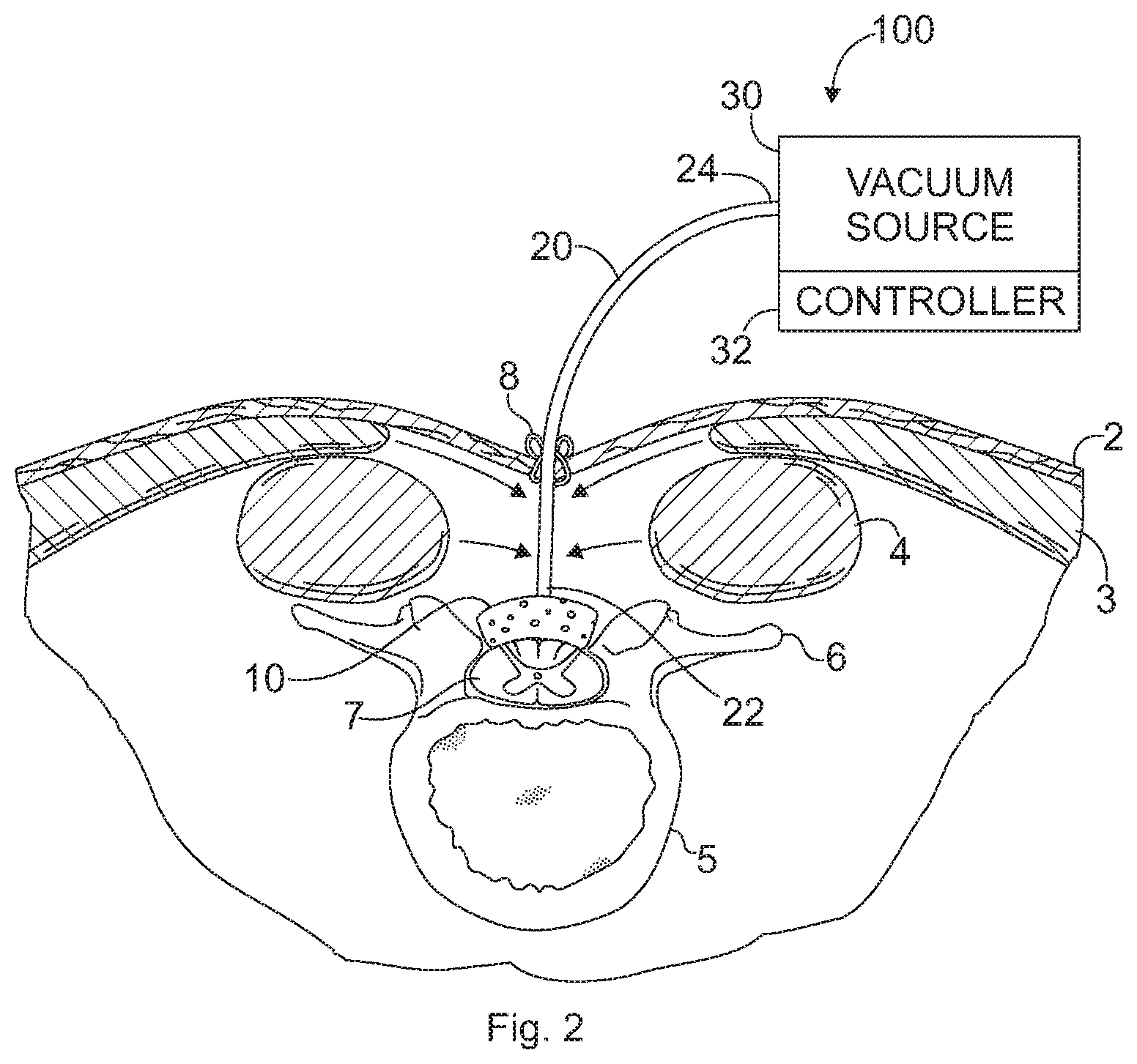
The present invention provides devices and methods that treat damaged spinal cord tissue, such as spinal tissue damaged by disease, infection, or trauma, which may lead to the presence of swelling, compression, and compromised blood flow secondary to interstitial edema.
| Inventors: | Argenta; Louis C (Winston-Salem, NC), Carroll; David L (Winston-Salem, NC), Levi; Nicole H (Winston-Salem, NC), Liu; Jie (Woodbury, MN), Morykwas; Michael J (Winston-Salem, NC), Tatter; Stephen (Winston-Salem, NC), Wagner; William D (Clemmons, NC) | ||||||||||
|---|---|---|---|---|---|---|---|---|---|---|---|
| Applicant: |
|
||||||||||
| Assignee: | WAKE FOREST UNIVERSITY HEALTH
SCIENCES (Winston-Salem, NC) |
||||||||||
| Family ID: | 40549559 | ||||||||||
| Appl. No.: | 15/700,667 | ||||||||||
| Filed: | September 11, 2017 |
Prior Publication Data
| Document Identifier | Publication Date | |
|---|---|---|
| US 20170368242 A1 | Dec 28, 2017 | |
Related U.S. Patent Documents
| Application Number | Filing Date | Patent Number | Issue Date | ||
|---|---|---|---|---|---|
| 14458790 | Aug 13, 2014 | ||||
| 12248346 | Sep 16, 2014 | 8834520 | |||
| 61088558 | Aug 13, 2008 | ||||
| 61081997 | Jul 18, 2008 | ||||
| 60978884 | Oct 10, 2007 | ||||
| Current U.S. Class: | 1/1 |
| Current CPC Class: | A61M 1/0023 (20130101); A61M 1/0088 (20130101); A61F 13/00021 (20130101); A61M 2210/1003 (20130101) |
| Current International Class: | A61M 1/00 (20060101); A61F 13/00 (20060101) |
References Cited [Referenced By]
U.S. Patent Documents
| 4187852 | February 1980 | Urry |
| 4221215 | September 1980 | Mandelbaum |
| 4661530 | April 1987 | Gogolewski |
| 4841962 | June 1989 | Berg |
| 4906233 | March 1990 | Moriuchi |
| 5024841 | June 1991 | Chu |
| 5490962 | February 1996 | Cima |
| 5516396 | May 1996 | Maurer |
| 5607590 | March 1997 | Shimizu |
| 5662625 | September 1997 | Westwood |
| 5736372 | April 1998 | Vacanti |
| 5766618 | June 1998 | Laurencin |
| 6095148 | August 2000 | Shastri |
| 6106913 | August 2000 | Scardino |
| 6695823 | February 2004 | Lina |
| 7216651 | May 2007 | Argenta |
| 7722894 | May 2010 | Wang |
| 8267960 | September 2012 | Argenta |
| 8632523 | January 2014 | Eriksson |
| 8764794 | July 2014 | Argenta |
| 8834520 | September 2014 | Argenta |
| 8932620 | January 2015 | Lelkes |
| 9289193 | March 2016 | Argenta |
| 1007631 | September 2018 | Argenta |
| 2002/0004556 | January 2002 | Foulger |
| 2003/0027332 | February 2003 | Lafrance |
| 2003/0108587 | June 2003 | Orgill |
| 2003/0109855 | June 2003 | Solem |
| 2003/0118692 | June 2003 | Wang |
| 2003/0208149 | November 2003 | Coffey |
| 2004/0039391 | February 2004 | Argenta |
| 2004/0210009 | October 2004 | Kobayashi |
| 2005/0063939 | March 2005 | Ameer |
| 2006/0079852 | April 2006 | Bubb |
| 2006/0263417 | November 2006 | Lelkes |
| 2006/0293169 | December 2006 | Srinivasan |
| 2007/0071790 | March 2007 | Ameer |
| 2007/0155010 | July 2007 | Farnsworth |
| 2007/0185426 | August 2007 | Ambrosio |
| 2007/0208420 | September 2007 | Ameer |
| 2008/0009830 | January 2008 | Fujimoto |
| 2008/0031934 | February 2008 | MacPhee |
| 2008/0102054 | May 2008 | Faustman |
| 2008/0112998 | May 2008 | Wang |
| 2008/0147156 | June 2008 | Imran |
| 2008/0153796 | June 2008 | Occleston |
| 2009/0011486 | January 2009 | Bettinger |
| 2009/0093565 | April 2009 | Yang |
| 2009/0148945 | June 2009 | Ameer |
| 2009/0187259 | July 2009 | Argenta |
| 2009/0254120 | October 2009 | Argenta |
| 2009/0295644 | December 2009 | Curran |
| 2009/0325859 | December 2009 | Ameer |
| 2010/0196478 | August 2010 | Masters |
| 2010/0221304 | September 2010 | Tan |
| 2011/0052646 | March 2011 | Kaigler |
| 2011/0129436 | June 2011 | Pryor |
| 2011/0262489 | October 2011 | Zhao |
| 2012/0016325 | January 2012 | Pinto |
| 2012/0265297 | October 2012 | Altman |
| 2014/0079759 | March 2014 | Patel |
| 4439240 | May 1996 | DE | |||
| 712939 | Aug 1954 | GB | |||
| 1004629 | Jan 1989 | JP | |||
| 2008099565 | May 2008 | JP | |||
| 199000060 | Jan 1990 | WO | |||
| 9918892 | Apr 1999 | WO | |||
| 2000061206 | Oct 2000 | WO | |||
| 03026489 | Apr 2003 | WO | |||
| 2007060433 | May 2007 | WO | |||
| 2012004627 | Jan 2012 | WO | |||
| 2012078472 | Jun 2012 | WO | |||
Other References
|
Dong, et al.; Performance of an in situ formed bioactive hydrogel dressing from a PEG-based hyperbranched multifunctional copolymer; Acta Biomaterialia; 10 (2014) 2076 2085. cited by applicant . Moues et al,; The role of topical negative pressure in wound repair: Expression of biochemical markers in wound fluid during wound healing; Wound Repair and Regeneration; pp. 448-494. cited by applicant . Seol et al.; Biocompatibility and preclinical feasibility tests of a temperature-sensitive hydrogel for the purpose of surgical wound pain control and cartilage; repair Journal of Biomedical Materials Research B: Applied Biomaterials | Nov. 2013 vol. 101B, Issue 8; pp. 1508-1515. cited by applicant . Casper el al. "Coating Electrospun Collagen and Gelatin Fibers with Perlecan Domain I for Increased Growth Factor Binding" Biomacromolecules, vol. 8 Issue 4 (Feb. 28, 2007): pp. 1116-1123. cited by applicant . Zhang et al. "In vitro evaluation of electrospun silk fibroin scaffolds for vascular cell growth" Biomalerials, vol. 29 Issue 14 (Feb. 14, 2008): pp. 2217-2227. cited by applicant . Lee et al. "Nanofiber alignment and direction of mechanical strain affect the ECM production of human ACL fibroblast" Biomaterials, vol. 26 Issue 11 (Jun. 11, 2004): pp. 1261-1270. cited by applicant . Yeo et al. "Collagen-Based Biomimetic Nanofibrous Scaffolds: Preparation and Characterization of Collagen/Silk Fibroin Bicomponent Nanofibrous Structures" Biomacromolecules, vol. 9 Issue 4 (Mar. 8, 2008): pp. 1106-1116. cited by applicant . Zhang et al.; "Electrospun Biomimic Nanofibrous Scaffolds of Silk Fibroin/Hyuronic Acid for Tissue Engineering"; Journal of Biomaterials Science 23(2012), 1185-1198. cited by applicant . Yan et al.; "Silk fibroin/chondroitin sulfate/hyaluronic acid Ternary scaffolds for dermal tissue receonstruction"; Acta Biomaterialia; Feb. 16, 2013. cited by applicant . Extended European Search Report from European Patent Application No. 13775894.2, dated Nov. 27, 2015. cited by applicant . Abidian MR, Martin DC. Experimental and theoretical characterization of implantable neural microelectrodes modified with conducting polymer nanotubes. Biomaterials Mar. 2008;29(9)1273-1283. cited by applicant . Abrams GA, Goodman SL, Nealey PF, Franco M, Murphy CJ. Nanoscale topography of the basement membrane underlying the corneal epithelium of the rhesus macaque. Cell Tissue Res Jan. 2000;299(1):39-46. cited by applicant . Ahmed I, Liu HY, Mamiya PC, Ponery AS, Babu AN, Weik T, et al. Three-dimensional nanofibrillar surfaces covalently modified with tenascin-C-derived peptides enhance neuronal growth in vitro. J Biomed Mater Res A Mar. 15, 2006;76(4):851-860. cited by applicant . Al-Majed AA, Neumann CM, Brushart TM, Gordon T. Brief electrical stimulation promotes the speed and accuracy of motor axonal regeneration. J Neurosci Apr. 1, 2000;20(7):2602-2608. cited by applicant . Al-Majed AA, Tam SL, Gordon T. Electrical stimulation accelerates and enhances expression of regeneration-associated genes in regenerating rat femoral motoneurons. Cell Mol Neurobiol Jun. 2004;24(3):379-402. cited by applicant . Batchelor PE, Porritt MJ, Martinello P, Parish CL, Liberatore GT, Donnan GA, et al. Macrophages and Microglia Produce Local Trophic Gradients That Stimulate Axonal Sprouting Toward but Not beyond the Wound Edge. Mol Cell Neurosci Nov. 2002;21(3):436-453. cited by applicant . Barrett, DG and Yousaf, MN. Design and Applications of Biodegradable Polyester Tissue Scaffolds Based on Endogenous Monomers Found in Human Metabolism. Molecules, Oct. 12, 2009. cited by applicant . Barras FM, Pasche P, Bouche N, Aebischer P, Zurn AD. Glial cell line-derived neurotrophic factor released by synthetic guidance channels promotes facial nerve regeneration in the rat. J Neurosci Res Dec. 15, 2002;70(6):746-755. cited by applicant . AshaRani PV, Hande MP, Valiyaveettil S. Anti-proliferative activity of silver nanoparticles. BMC Cell Biol Sep. 2009;10:14. cited by applicant . Alkilany AM, Nagaria PK, Hexel CR, Shaw TJ, Murphy CJ, Wyatt MD. Cellular Uptake and Cytotoxicity of Gold Nanorods: Molecular Origin of Cytotoxicity and Surface Effects. Small Mar. 2009;5(6):701-708. cited by applicant . Apte, S. S. (2011). "Current developments in the tissue engineering of autologous heart valves: moving towards clinical use." Future cardiology 7(1): 77-97. cited by applicant . Barakat, N.A.M., et al., "Polymeric nanofibers containing solid nanoparticles prepared by electrospinning and their applications," Chemical Engineering Journal 156:487-495 (2010). cited by applicant . Baron-Van Evercooren A, Kleinman HK, Ohno S, Marangos P, Schwartz JP, Dubois-Dalcq ME. Nerve growth factor laminin and fibronective promote neurite growth in human fetal sensory ganglia cultures. Journal of Neuroscience Research 1982;8(2-3):179-194. cited by applicant . Bondar, B., S. Fuchs, et al. (2008). "Functionality of endothelial cells on silk fibroin nets: Comparative study of micro- and nanometric fibre size." Biomaterials 29(5): 561-572. cited by applicant . Billiar, K. L. and M. S. Sacks (2000). "Biaxial Mechanical Properties of the Native and Glutaraldehyde-Treated Aortic Valve Cusp: Part II--A Structural Constitutive Model." Journal of Biomechanical Engineering 122(4): 327-335. cited by applicant . Carlson C, Hussain SM, Schrand AM, Braydich-Stolle LK, Hess KL, Jones RL, et al. Unique Cellular Interaction of Silver Nanoparticles: Size-Dependent Generation of Reactive Oxygen Species. J Phys Chem B 2008;112(43):13608-13619. cited by applicant . Butcher, J. T., G. J. Mahler, et al. (2011). "Aortic valve disease and treatment: The need for naturally engineered solutions." Advanced Drug Delivery Reviews 63(4-5): 242-268. cited by applicant . Brushart, T.M., Jari, R., Verge, V., Rohde, C. & Gordon, T. Electrical stimulation restores the specificity of sensory axon regeneration. Experimental Neurology 194, 221-229 (2005). cited by applicant . Boland, E.D., et al., "Electrospinning polydioxanone for biomedical applications," Acta Biomaterialia 1:115-123 (2005). cited by applicant . Bouquet C, Ravaille-Veron M, Propst F, Nothias F. MAP1B coordinates microtubule and actin filament remodeling in adult mouse Schwann cell tips and DRG neuron growth cones. Mol Cell Neurosci Oct. 2007;36(2):235-247. cited by applicant . Chen, W.-Q., H. Priewalder, et al. (2010). "Silk cocoon of Bombyx mori: Proteins and posttranslational modifications--heavy phosphorylation and evidence for lysine-mediated cross links." Proteomics 10(3): 369-379. cited by applicant . Chang CJ, Hsu SH, Yen HJ, Chang H, Hsu SK. Effects of unidirectional permeability in asymmetric poly(DL-lactic acid-co-glycolic acid) conduits on peripheral nerve regeneration: An in vitro and in vivo study. J Biomed Mater Res Part B Oct. 2007;83B(1):206-215. cited by applicant . Chen, Y., et al., "Increased osteoblast functions in the presence of BMP-7 short peptides for nanostructured biomaterial applications," J. Biomed. Mater. Res. A 91:296-304 (2009; published online Nov. 3, 2008). cited by applicant . Breuer, C. K. (2004). "Application of tissue-engineering principles toward the development of a semilunar heart valve substitute." Tissue engineering 10(11-12): 1725-1736. cited by applicant . Bruder JM, Lee AP, Hoffman-Kim D. Biomimetic materials replicating Schwann cell topography enhance neuronal adhesion and neurite alignment in vitro. J Biomater Sci Polym Ed 2007;18(8):967-982. cited by applicant . Bryan DJ, Holway AH, Wang KK, Silva AE, Trantolo DJ, Wise D, et al. Influence of glial growth factor and Schwann cells in a bioresorbable guidance channel on peripheral nerve regeneration. Tissue Eng Apr. 2000;6(2):129-138. cited by applicant . Chobanian, A. V., G. L. Bakris, et al. (2003). "Seventh Report of the Joint National Committee on Prevention, Detection, Evaluation, and Treatment of High Blood Pressure." Hypertension 42(6): 1206-1252. cited by applicant . Cho Y, Shi R, Ivanisevic A, Ben Borgens R. A mesoporous silica nanosphere-based drug delivery system using an electrically conducting polymer. Nanotechnology Jul. 8, 2009;20(27):275102. cited by applicant . Borschel GH, Kia KF, Kuzon WM, Jr., Dennis RG. Mechanical properties of acellular peripheral nerve. J Surg Res Oct. 2003;114(2):133-139. cited by applicant . Boyd JG, Gordon T. A dose-dependent facilitation and inhibition of peripheral nerve regeneration by brain-derived neurotrophic factor. Eur J Neurosci Feb. 2002;15(4):613-626. cited by applicant . Causa, F., et al., "A multi-functional scaffold for tissue regeneration: The need to engineer a tissue analogue," Biomaterials, 28(34):5093-5099 (Dec. 2007; available online Aug. 6, 2007). cited by applicant . Cebotari, S. (2011). "Use of fresh decellularized allografts for pulmonary valve replacement may reduce the reoperation rate in children and young adults: early report." Circulation (New York, N.Y.) 124(11 suppl): S115-123. cited by applicant . Chamberlain LJ, Yannas IV, Hsu HP, Strichartz GR, Sepctor M. Near-terminus axonal structure and function following rat sciatic nerve regeneration through a collagen-GAG matrix in a ten-millimeter gap. J Neurosci Res Jun. 1, 2000;610(5):666-677. cited by applicant . Chen, D., et al., "Application of electrostatic spinning technology in nano-structured polymer scaffold," Zhongguo Xiu Fu Chong Jian Wai Ke Za Zhi, 21(4):411-415 (Apr. 2007), 1 sheet abstract. cited by applicant . Chen, R., et al., "Preparation and characterization of coaxial electrospun thermoplastic polyurethane/collagen compound nanofibers for tissue engineering applications," Colloids and Surfaces B: Biointerfaces 79(2):315-325 (Sep. 1, 2010) (available online Apr. 3, 2010). cited by applicant . Chen YS, Hsieh CL, Tsai CC, Chen TH, Cheng WC, Hu CL, et al. Peripheral nerve regeneration using silicone rubber chambers filled with collagen, laminin and fibronectin. Biomaterials Aug. 2000;21(15):1541-1547. cited by applicant . Cheng C, Webber CA, Wang J, Xu Y, Martinez JA, Liu WQ, et al. Activated RHOA and peripheral axon regeneration. Exp Neurol Aug. 2008;212(2):358-369. cited by applicant . De S, Higgins TM, Lyons PE, Doherty EM, Nirmalraj PN, Blau WJ, et al. Silver Nanowire Networks as Flexible, Transparent, Conducting Films: Extremely High DC to Optical Conductivity Ratios. ACS Nano Jul. 2009;3(7):1767-1774. cited by applicant . Evans, G.R.D. et al. In vivo evaluation of poly(-lactic acid) porous conduits for peripheral nerve regeneration. Biomaterials 20, 1109-1115 (1999). cited by applicant . Elias, K.L., Price, R.L. & Webster, T.J. Enhanced functions of osteoblasts on nanometer diameter carbon fibers. Biomaterials 23, 3279-3287 (2002). cited by applicant . Dumitriu, Severian and Popa, Valentin. Polymeric Biomaterials: Structure and Function, vol. 1. Boca Raton, FL: CRC Press, Jan. 17, 2013. cited by applicant . Ciardelli G, Rechichi A, Cerrai P, Tricoli M, Barbani N, Giusti P. Segmented polyurethanes for medical applications: Synthesis, characterization and in vitro enzymatic degradation studies. Macromolecular Symposia 2004;218:261-271. cited by applicant . Deister C, Schmidt CE. Optimizing neurotrophic factor combinations for neurite outgrowth. J Neural Eng Jun. 2006;3(2):172-179. cited by applicant . Ekaputra, A.K., et al., "Composite electrospun scaffolds for engineering tubular bone grafts," Tissue Eng. Part A 15(12):3779-3788 (Dec. 8, 2009) (published online Jul. 20, 2009; online ahead of print: Jul. 24, 2009; online ahead of editing: Jun. 15, 2009). cited by applicant . Corey JM, Lin DY, Mycek KB, Chen Q, Samuel S, Feldman EL, et al. Aligned electrospun nanofibers specify the direction of dorsal root ganglia neurite growth. J Biomed Mater Res A Dec. 1, 2007;83(3):636-645. cited by applicant . De Cock, L. J. (2010). "Layer-by-layer incorporation of growth factors in decellularized aortic heart valve leaflets." Biomacromolecules 11(4): 1002-1008. cited by applicant . Cullen DK, A RP, Doorish JF, Smith DH, Pfister BJ. Developing a tissue-engineered neural-electrical relay using encapsulated neuronal constructs on conducting polymer fibers. J Neural Eng Dec. 2008;5(40):374-384. cited by applicant . Cui X, Lee VA, Raphael Y, Wiler JA, Hetke JF, Anderson DJ, et al. Surface modification of neural recording electrodes with conducting polymer/biomolecule blends. J Biomed Mater Res Aug. 2001;56(2):261-272. cited by applicant . Evans PJ, Bain JR, Mackinnon SE, Makino AP, Hunter DA. Selective reinnervation: a comparison of recovery following microsuture and conduit nerve repair. Brain Res Sep. 20, 1991;559(2):315-321. cited by applicant . Evans AJ, Thompson BC, Wallace GG, Millard R, O'Leary SJ, Clark GM, et al. Promoting neurite outgrowth from spiral ganglion neuron explants using polypyrrole/BDNF-coated electrodes. J Biomed Mater Res A Oct. 2009;91(1):241-250. cited by applicant . Edwards SL, Church JS, Werkmeister JA, Ramshaw JAM. Tubular micro-scale multiwalled carbon nanotube-based scaffolds for tissue engineering. Biomaterials Mar. 2009;30(9)1725-1731. cited by applicant . Evans GRD, Brandt K, Katz S, Chauvin P, Otto L, Bogle M, et al. Bioactive poly(L-lactic acid) conduits seeded with Schwann cells for peripheral nerve regeneration. Biomaterials Feb. 2002;23(3):841-848. cited by applicant . Evans GRD. Peripheral nerve injury: A review and approach to tissue engineered constructs. Anatomical Record 2001; 263(4):396-404. cited by applicant . Deng, M., et al., "Miscibility and in vitro osteocompatibility of biodegradable blends of poly[(ethyl alanato) (p-phenyl phenoxy) phosphazene] and poly(lacitic acid-glycolic acid)," Biomaterials 29:337-349 (2008; available online Oct. 17, 2007). cited by applicant . Bauhofer W, Kovacs JZ. A review and analysis of electrical percolation in carbon nanotube polymer composites. Composites Science and Technology 2009;69(10):1486-1498. cited by applicant . Bain JR, Mackinnon SE, Hunter DA. Functional evaluation of complete sciatic, peroneal, and posterior tibial nerve lesions in the rat. Plast Reconstr Surg Jan. 1989;83(1):129-138. cited by applicant . Beachley, V., et al., "Polymer nanofibrous structures: Fabrication, biofunctionalization, and cell interactions," Progress in Polymer Science, 35(7):868-892 (Jul. 2010) (available online Mar. 17, 2010). cited by applicant . Beun, L. H., X. J. Beaudoux, et al. (2011). "Self-Assembly of Silk-Collagen-like Triblock Copolymers Resembles a Supramolecular Living Polymerization." ACS Nano 6(1):133-140. cited by applicant . Boissard, C.I.R., et al., "Nanohydroxyapatite/poly(ester urethane) scaffold for bone tissue engineering," Acta Biomaterialia 5:3316-3327 (Nov. 2009; available online May 12, 2009). cited by applicant . Borgens RB, Vanable JW, Jr. Jaffe LF. Bioelectricity and regeneration: large currents leave the stumps of regenerating newt limbs. Proc Natl Acad Sci U S A Oct. 1977;74(10):4528-4532. cited by applicant . Caswell KK, Bender CM, Murphy CJ. Seedless, surfactantless wet chemical synthesis of silver nanowires. Nano Lett May 2003;3(5):667-669. cited by applicant . Chamberlain LJ, Yannas IV, Hsu HP, Strichartz GR, Spector M. Near-terminus axonal structure and function following rat sciatic nerve regeneration through a collagen-GAG matrix in a ten-millimeter gap. J Neurosci Res Jun. 1, 2000;60(5):666-677. cited by applicant . Chen X, Schluesener HJ. Nanosilver: A nanoproduct in medical application. Toxicol Lett Jan. 2008;176(1):1-12. cited by applicant . Chronakis, I.S., "Novel nanocomposites and nanoceramics based on polymer nanofibers using electrospinning process--A review," Journal of Materials Processing Technology 167:283-293 (2005). cited by applicant . Al Majed, A.A., Tam, S.L. & Gordon, T. Electrical stimulation accelerates and enhances expression of regeneration-associated genes in regenerating rat femoral motoneurons. Cellular and Molecular Neurobiology 24, 379-402 (2004). cited by applicant . Bashar SA. Study of Indium Tin Oxide (ITO) for Novel Optoelectronic Devices. King's College London: University of London; 1998. cited by applicant . Batchelor PE, Wills TE, Hewa AP, Porritt MJ, Howells DW. Stimulation of axonal sprouting by trophic factors immobilized within the wound core. Brain Res May 13, 2008;1209:49-56. cited by applicant . Cheng H, Hoffer B, Stromberg I, Russell D, Olson L. The effect of glial-cell line-derived neurotrophic factor in fibrin glue on developing dopamine neurons. Exp Brain Res May 1995;104(2):199-206. cited by applicant . Dijkman, P. E., A. Driessen-Mol, et al. (2012). "Decellularized homologous tissue-engineered heart valves as off-the-shelf alternatives to xeno- and homografts." Biomaterials 18:4545-54. cited by applicant . Dertinger SK, Jiang X, Li Z , Murphy VN, Whitesides GM. Gradients of substrate-bound laminin orient axonal specification of neurons. Proc Natl Acad Sci U S A Oct. 1, 2002;99(20):12542-12547. cited by applicant . Chiang M, Robinson KR, Vanable JW, Jr. Electrical fields in the vicinity of epithelial wounds in the isolated bovine eye. Exp Eye Res Jun. 1992;54(6):999-1003. cited by applicant . De Cupere, V. M., J. Van Wetter, et al. (2003). "Nanoscale Organization of Collagen and Mixed Collagen--Pluronic Adsorbed Layers." Langmuir 19(17): 6957-6967. cited by applicant . Deng, C. (2011). "Application of decellularized scaffold combined with loaded nanoparticles for heart valve tissue engineering in vitro." Journal of Huazhong University of Science and Technology. Medical sciences 31(1): 88-93. cited by applicant . FitzGerald MJT. Neuroanatomy: Basic and Clinical. Philadelphia: W.B. Saunders Company, Ltd., 1996. cited by applicant . Fujihara, K., et al., "Guided bone regeneration membrane made of polycaprolactone/calcium carbonate composite nano-fibers," Biomaterials 26:4139-4147 (2005; available online Dec. 24, 2004). cited by applicant . Deka, H., et al., "Biocompatible hyperbranched polyurethane/multi-walled carbon nanotube composites as shape memory materials," Carbon 48:2013-2022 (2010; available online Feb. 11, 2010). cited by applicant . Franzen R, Tanner SL, Dashiell SM, Rottkamp CA, Hammer JA, Quarles RH. Microtubule-associated protein 1B: a neuronal binding partner for myelin-associated glycoprotein. J Cell Biol Dec. 10, 2001;155(6):893-898. cited by applicant . Gerecht S, Townsend SA, Pressler H, Zhu H, Nijst CLE, Bruggeman JP, et al. A porous photocurable elastomer for cell encapsulation and culture. Biomaterials Nov. 2007;28(32):4826-4835. cited by applicant . Gallo G. RhoA-kinase coordinates F-actin organization and myosin II activity during semaphoring-3A-induced axon retraction. J Cell Sci Aug. 15, 2006;119(Pt 16):3413-3423. cited by applicant . Goold RG, Gordon-Weeks PR. Glycogen synthase kinase 3beta and the regulation of axon growth. Biochem Soc Trans Nov. 2004;32(Pt 5):809-811. cited by applicant . Goldner JS, Bruder JM, Li G, Gazzola D, Hoffman-Kim D. Neurite bridging across micropatterned grooves. Biomaterials Jan. 2006;27(3):460-472. cited by applicant . Dohmen, P. M., A. Lembcke, et al. (2011). "Ten Years of Clinical Results With a Tissue-Engineered Pulmonary Valve." The Annals of Thoracic Surgery 92(4): 1308-1314. cited by applicant . Goold RG, Gordon-Weeks PR. NGF activates the phosphorylation of MAP1B by GSK3beta through the TrkA receptor and not the p75(NTR) receptor. J Neurochem Nov. 2003;87(4):935-946. cited by applicant . Gordon T. The role of neurotrophic factors in nerve regeneration. Neurosurg Focus 2009;26(2):E3. cited by applicant . Gole A, Murphy CJ. Seed-mediated synthesis of gold nanorods: Role of the size and nature of the seed. Chem Mat Sep. 2004;16(19):3633-3640. cited by applicant . Gonzalez-Billault C, Jimenez-Mateos EM, Caceres A, Diaz-Nido J, Wandosell F, Avila J. Microtube-associated protein 1B function during normal development, regeneration, and pathological conditions in the nervous system. J Neurobiol Jan. 2004;58(1):48-59. cited by applicant . Dong, B., et al., "Electrospinning of collagen nanofiber scaffolds from benign solvents," Macromolecular Rapid Communications 30(7):539-542 (Feb. 5, 2009). cited by applicant . Faria, M.L.E., et al., "Recombinant human bone morphogenetic protein-2 in absorbable collagen sponge enhances bone healing of tibial osteotomies in dogs," Veterinary Surgery 36(2):122-131 (Feb. 2007; first published online Mar. 2, 2007). cited by applicant . Garcia-Perez J, Avila J, Diaz-Nido J. Implication of cyclin-dependent kinases and glycogen synthase kinase 2 in the phosphorylation of microtubule-associated protein 1B in developing neuronal cells. J Neurosci Res May 15, 1998;52(4):445-452. cited by applicant . George PM, Lyckman AW, LaVan DA, Hegde A, Leung Y, Avasare R, et al. Fabrication and biocompatibility of polypyrrole implants suitable for neural prosthetics. Biomaterials Jun. 2005;26(17):3511-3519. cited by applicant . Goold RG, Gordon-Weeks PR. The MAP kinase pathway is upstream of the activation of GSK3beta that enables it to phosphorylate MAP1B and contributes to the stimulation of axon growth. Mol Cell Neurosci Mar. 2005;28(3):524-534. cited by applicant . Guan J, Stankus JJ, Wagner WR. Biodegradable elastomeric scaffolds with basic fibroblast growth factor release. Journal of Controlled Release 2007;120(1-2):70-78. cited by applicant . Gu, X. And K. S. Masters (2010). "Regulation of valvular interstitial cell calcification by adhesive peptide sequences." Journal of Biomedical Materials Research Part A 93A(4):1620-1630. cited by applicant . Hayashi, T. and S. Mukamel (2007). "Vibrational-Exciton Couplings for the Amide I, II, III, and A Modes of Peptides." The Journal of Physical Chemistry B 111(37):11032-11046. cited by applicant . Hayashi A, Moradzadeh A, Tong A, Wei C, Tuffaha SH, Hunter DA, et al. Treatment modality affects allograft-derived Schwann cell phenotype and myelinating capacity. Exp Neurol Aug. 2008;212(2):324-336. cited by applicant . Hopkins, R. A. (2005). "Tissue engineering of heart valves: decellularized valve scaffolds." Circulation (New York, N.Y.) 111(21): 2712-2714. cited by applicant . Guimard NK, Gomez N, Schmidt CE. Conducting polymers in biomedical engineering. Progress in Polymer Science 2007;32:876-921. cited by applicant . Hinton, R. B. and K. E. Yutzey (2011). "Heart Valve Structure and Function in Development and Disease." Annual Review of Physiology 73(1): 29-46. cited by applicant . Hill, C.A., et al., "Superior sternal cleft repair using autologous rib grafts in an infant with complex congenital heart disease," Ann. Thorac. Surg., 84:673-4, (2007). cited by applicant . Horan, R. L., K. Antle, et al. (2005). "In vitro degradation of silk fibroin." Biomaterials 26(17): 3385-3393. cited by applicant . Hoshi, R.A., "Nanoporous biodegradable elastomers," Adv. Mater. 21:188-192 (2009). cited by applicant . Ifkovits JL, Devlin JJ, Eng G, Martens TP, Vunjak-Novakovic G, Burdick JA. Biodegradable Fibrous Scaffolds with Tunable Properties Formed from Photo-Cross-Linkable Poly(glycerol sebacate). ACS Appl Mater Interfaces Sep. 2009;1(9):1878-1886. cited by applicant . Hu, H. et al. Polyethyleneimine functionalized single-walled carbon nanotubes as a substrate for neuronal growth. Journal of Physical Chemistry B 109, 4285-4289 (2005). cited by applicant . International Preliminary Report on Patentability from International Application No. PCT/US2013/032520 dated Oct. 14, 2014. cited by applicant . Integra. NeuraWrap Nerve Protector. 2009; Available from: http://www.integra-ls.com/products/?product=198. cited by applicant . Integra. NeuraGen Nerve Guide. 2009; Available from: http://www.integra-ls.com/products/?product=88. cited by applicant . International Preliminary Report on Patentability from International Application No. PCT/US2013/066747, dated Apr. 28, 2015. cited by applicant . Hong, Y., et al., "Tailoring the degradation kinetics of poly(ester carbonate urethane)urea thermoplastic elastomers for tissue engineering scaffolds," Biomaterials 31:4249-4258 (2010; available online Feb. 25, 2010). cited by applicant . Jain, A., Kim, Y.T., Mckeon, R.J. & Bellamkonda, R.V. In situ gelling hydrogels for conformal repair of spinal cord defects, and local delivery of BDNF after spinal cord injury. Biomaterials 27, 497-504 (2006). cited by applicant . Hu, X., D. Kaplan, et al. (2006). "Determining Beta-Sheet Crystallinity in Fibrous Proteins by Thermal Analysis and Infrared Spectroscopy." Macromolecules 39(18): 6161-6170. cited by applicant . Hu H, Ni YC, Montana V, Haddon RC, Parpura V. Chemically functionalized carbon nanotubes as substrates for neuronal growth. Nano Lett 2004;4(3):507-511. cited by applicant . Huang J, Hu X, Lu L, Ye Z, Zhang Q, Luo Z. Electrical regulation of Schwann cells using conductive polypyrrole/chitosan polymers. J Biomed Mater Res A Jun. 17, 2009. cited by applicant . Hudson TW, Evans GR, Schmidt CE. Engineering strategies for peripheral nerve repair. Orthop Clin North Am Jul. 2000;31(3):485-498. cited by applicant . Medscape: Diseases of the Peripheral Nervous System. Web MD Inc., ACP Medicine, 2004. cited by applicant . Song YX, Muramatsu K, Kurokawa Y, Kuriyama R, Sakamoto S, Kaneko K, et al. Functional recovery of rat hind-limb allografts. J Reconstr Microsurg Oct. 2005;21(7):471-476. cited by applicant . Luis AL, Rodrigues JM, Lobato JV, Lopes MA, Amado S, Veloso AP, et al. Evaluation of two biodegradable nerve guides for the reconstruction of the rat sciatic nerve. Biomed Mater Eng 2007;17(1):39-52. cited by applicant . Pigino G, Paglini G, Ulloa L, Avila J, Caceres A. Analysis of the expression, distribution and function of cyclin dependent kinase 5 (cdk5) in developing cerebellar macroneurons. J Cell Sci Jan. 1997;110 (Pt 2):257-270. cited by applicant . Riederer BM, Moya F, Calvert R. Phosphorylated MAP1b, alias MAP5 and MAP1x, is involved in axonal growth and neuronal mitosis. Neuroreport Jun. 1993;4(6):771-774. cited by applicant . Stoll G, Muller HW. Nerve injury, axonal degeneration and neural regeneration: basic insights. Brain Pathol Apr. 1999;9(2):313-325. cited by applicant . Loudon RP, Silver LD, Yee HF, Jr., Gallo G. RhoA-kinase and myosin II are required for the maintenance of growth cone polarity and guidance by nerve growth factor. J Neurobiol Jul. 2006;66(8):847-867. cited by applicant . Melendez-Vasquez CV, Einheber S, Salzer JL. Rho kinase regulates Schwann cell myelination and formation of associated axonal domains. J Neurosci Apr. 21, 2004;24(16):3953-3963. cited by applicant . Sherman DL, Brophy PJ. Mechanisms of axon ensheathment and myelin growth. Nat Rev Neurosci Sep. 2005;6(9):683-690. cited by applicant . Sharma N, Marzo SJ, Jones KJ, Foecking EM. Electrical stimulation and testosterone differentially enhance expression of regeneration-associated genes. Exp Neurol May 7, 2009. cited by applicant . Tsujino H, Kondo E, Fukuoka T, Dai Y, Tokunaga A, Miki K, et al. Activating transcription factor 3 (ATF3) induction by axotomy in sensory and motoneurons: A novel neuronal marker of nerve injury. Mol Cell Neurosci Feb. 2000;15(2):170-182. cited by applicant . Geremia NM, Gordon T, Brushart TM, Al-Majed AA, Verge VM. Electrical stimulation promotes sensory neuron regeneration and growth-associated gene expression. Exp Neurol Jun. 2007;205(2):347-359. cited by applicant . McIntyre CC, Richardson AG, Grill WM. Modeling the excitability of mammalian nerve fibers: influence of afterpotentials on the recovery cycle. J Neurophysiol Feb. 2002;87(2):995-1006. cited by applicant . Shanthaveerappa TR, Bourne H. Histological and histochemical studies of the choroid of the eye and its relations to the pia-arachnoid mater of the central nervous system and perineural epithelium of the peripheral nervous system. Acta Anat (Basel) 1965;61(3):379-398. cited by applicant . Jana NR, Gearheart L, Murphy CJ. Wet chemical synthesis of high aspect ratio cylindrical gold nanorods. J Phys Chem B May 2001;105(19):4065-4067. cited by applicant . Johnson EO, Soucacos PN. Nerve repair: experimental and clinical evaluation of biodegradable artificial nerve guides. Injury Sep. 2008;39 Suppl 3:S30-36. cited by applicant . Wang S, Yaszemski MJ, Knight AM, Gruetzmacher JA, Currier BL, Yaszemski MJ. Synthesis and characterizations of biodegradable and crosslinkable poly(epsilon-caprolactone fumarate), poly(ethylene glycol fumarate), and their amphiphilic copolymer. Biomaterials Feb. 2006;27(6):832-841. cited by applicant . Wang S, Yaszemski MJ, Knight AM, Gruetzmacher JA, Windebank AJ, Lu L. Photo-crosslinked poly(epsilon-caprolactone fumarate) networks for guided peripheral nerve regeneration: material properties and preliminary biological evaluations. Acta Biomater Jun. 2009:5(5):1 531-1542. cited by applicant . Hadlock T, Sundback C, Hunter D, Cheney M, Vacanti JP. A polymer foam conduit seeded with Schwann cells promotes guided peripheral nerve regeneration. Tissue Engineering 2000;6(20):119-127. cited by applicant . Hollowell JP, Villadiego A, Rich KM. Sciatic nerve regeneration across gaps within silicone chambers: long-term effects of NGF and consideration of axonal branching. Exp Neurol Oct. 1990;110(1):45-51. cited by applicant . Wood MD, Moore AM, Hunter DA, Tuffaha S, Borschel GH, Mackinnon SE, et al. Affinity-based release of glial-derived neurotrophic factor from fibrin matrices enhances sciatic nerve regeneration. Acta Biomater May 2009;5(4):959-968. cited by applicant . Lietz M, Dreesmann L, Hoss M, Oberhoffner S, Schlosshauer B. Neuro tissue engineering of glial nerve guides and the impact of different cell types. Biomaterials Mar. 2006;27(8):1425-1436. cited by applicant . Song J, Cheng Q, Kopta S, Stevens RC. Modulating artificial membrane morphology: pH-induced chromatic transition and nanostructural transformation of a bolaamphiphilic conjugated polymer from blue helical ribbons to red nanofibers. J Am Chem Soc Apr. 11, 2001;123(14):3205-3213. cited by applicant . Kehoe S, Zhang XF, Boyd D. FDA approved guidance conduits and wraps for peripheral nerve injury: A review of materials and efficacy. Injury-Int J Care Inj May;43(5):553-572. cited by applicant . Pomerantseva I, Krebs N, Hart A, Neville CM, Huang AY, Sundback CA. Degradation behavior of poly(glycerol sebacate). J Biomed Mater Res Part A Dec. 2009;91A(4):1038-1047. cited by applicant . Wang YD, Ameer GA, Sheppard BJ, Langer R. A tough biodegradable elastomer. Nature Biotechnology Jun. 2002;20(6):602-606. cited by applicant . Nijst CLE, Bruggeman JP, Karp JM, Ferreira L, Zumbuehl A, Bettinger CJ, et al. Synthesis and characterization of photocurable elastomers from poly(glycerol-co-sebacate). Biomacromolecules Oct. 2007;8(10):3067-3073. cited by applicant . Svennersten K, Bolin MH, Jager EWH, Berggren M, Richter-Dahlfors A. Electrochemical modulation of epithelia formation using conducting polymers. Biomaterials Nov. 2009;30(31):6257-6264. cited by applicant . Luo XL, Weaver CL, Zhou DD, Greenberg R, Cui XYT. Highly stable carbon nanotube doped poly(3,4-ethylenedioxythiophene) for chronic neural stimulation. Biomaterials Aug;32(24):5551-5557. cited by applicant . Shi GX, Rouabhia M, Wang ZX, Dao LH, Zhang Z. A novel electrically conductive and biodegradable composite made of polypyrrole nanoparticles and polylactide. Biomaterials Jun. 2004; 25(13):2477-2488. cited by applicant . Liu QY, Tian M, Ding T, Shi R, Feng YX, Zhang LQ, et al. Preparation and characterization of a thermoplastic poly(glycerol sebacate) elastomer by two-step method. J Appl Polym Sci Feb. 2007;103(3):1412-1419. cited by applicant . Evans GR. Challenges to nerve regeneration. Semin Surg Oncol Oct.-Nov. 2000;19(3):312-318. cited by applicant . Zhang DH, Kandadai MA, Cech J, Roth S, Curran SA. Poly(L-lactide) (PLLA)/multiwalled carbon nanotube (MWCNT) composite: Characterization and biocompatibility evaluation. J Phys Chem B 2006;110(26):12910-12915. cited by applicant . Schense JC, Bloch J, Aebischer P, Hubbell JA. Enzymatic incorporation of bioactive peptides into fibrin matrices enhances neurite extension. Nature Biotechnology Apr. 2000;18(4):415-419. cited by applicant . Lee AC, Yu VM, Lowe JB, 3rd, Brenner MJ, Hunter DA, Mackinnon SE, et al. Controlled release of nerve growth factor enhances sciatic nerve regeneration. Exp Neurol Nov. 2003;184(1):295-303. cited by applicant . Miller C, Jeftinija S, Mallapragada S. Micropatterned Schwann cell-seeded biodegradable polymer substrates significantly enhance neurite alignment and outgrowth. Tissue Engineering Dec. 2001;7(6):705-715. cited by applicant . Zhao M, AgiusFernandez A, Forrester JV, McCaig CD. Orientation and directed migration of cultured corneal epithelial cells in small electric fields are serum dependent. Journal of Cell Science Jun. 1996;109:1405-1414. cited by applicant . Zhao M, McCaig Cd, AgiusFernandez A, Forrester JV, ArakiSasaki K. Human corneal epithelial cells reorient and migrate cathodally in a small applied electric field. Current Eye Research 1997;16(10):973-984. cited by applicant . Edelman ER, Mathiowitz E, Langer R, Klagsbrun M. Controlled and modulated release of basic fibroblast growth factor. Biomaterials Sep. 1991;12(7):619-626. cited by applicant . McDonald, D.S. & Zochodne, D.W. An injectable nerve regeneration chamber for studies of unstable soluble growth factors. Journal of Neuroscience Methods 122, 171-178 (2003). cited by applicant . Phillips,J.B., Bunting, S.C.J., Hal1 ,S.M. & Brown, R.A. Neural tissue engineering: A self-organizing collagen guidance conduit. Tissue Engineering 11, 1611-1617 (2005). cited by applicant . Jubran,M. & Widenfalk, J. Repair of peripheral nerve transections with fibrin sealant containing neurotrophic factors. Experimental Neurology 181, 204-212 (2003). cited by applicant . Foley,J.D., Grunwald, E.W., Nealey, P.F. & Murphy, C.J. Cooperative modulation of neuritogenesis by PC12 cells by topography and nerve growth factor. Biomaterials 26, 3639-3644 (2005). cited by applicant . Yusuka Katayama et al. Coil-Reinforced hydrogel tubes promote nerve regeneration equivalent to that of nerve autografts. Biomaterials 27, 503-518 (2006). cited by applicant . Yuan, Y., Zhang, P., Yang, Y., Wang, X. & Gu, X. The interaction of Schwann cells with chitosan membranes and fibers in vitro. Biomaterials 25, 4273-4278 (2004). cited by applicant . Lietz, M. et al. Physical and biological performance of a novel block copolymer nerve guide. Biotechnology and Bioengineering 93, 99-109 (2006). cited by applicant . Keilhoff,G., Stang, F., Wolf, G. & Fansa, H. Bio-compatibility of type I/III collagen matrix for peripheral nerve reconstruction. Biomaterials 24, 2779-2787 (2003). cited by applicant . Li, M., Guo, Y., Wei, Y., MacDiarmid, A.G. & Lelkes, P.I. Electrospinning polyaniline-contained gelatin nanofibers for tissue engineering applications. Biomaterials 27, 2705-2715 (2006). cited by applicant . Novikova,L.N. et al. Alginate hydrogel and matrigel as potential cell carriers for neurotransplantation. Journal of Biomedical Materials Research Part A 77A, 242-252 (2006). cited by applicant . Ming, G.I., Henley, J., Tessier-Lavigne, M., Song, H.J. & Poo, M.m. Electrical Activity Modulates Growth Cone Guidance by Diffusible Factors. Neuron 29, 441-452 (2001). cited by applicant . Terell Rivers, Terry Hudson & Christine Schmidt. Synthesis of a Novel, Biodegradable Electrically Conducting Polymer for Biomedical Applications. Advanced Functional Materials 12, 33-37 (2002). cited by applicant . Kotwal, A. & Schmidt, C.E. Electrical stimulation alters protein adsorption and nerve cell interactions with electrically conducting biomaterials. Biomaterials 22, 1055-1064 (2001). cited by applicant . Yang, L., Feng, J.K. & Ren, A.M. Structural, electronic and optical properties of a series of oligofluorene-thiophene oligomers and polymers. Journal of Molecular Structure-Theochem 758, 29-39 (2006). cited by applicant . Suzuki, M., Fukuyama, M., Hori, Y. & Hotta, S. Electroluminescent features of oligothiophenes dispersed as a dopant in host matrices. Journal of Applied Physics 91, 5706-571 1 (2002). cited by applicant . Xu, H. et al. High-performance field-effect transistors based on Langmuir-Blodgett films of cyclo[8]pyrrole. Langmuir 21, 5391-5395 (2005). cited by applicant . Zhang L, Ma Z, Smith GM, Wen X, Pressman Y, Wood PM, et al. GDNF-enhanced axonal regeneration and myelination following spinal cord injury is mediated by primary effects on neurons. Glia Jan. 23, 2009. cited by applicant . Jin, Z., Pramoda, K.P., Xu, G. & Goh, S.H. Dynamic mechanical behavior of meltprocessed multi-walled carbon nanotubelpoly(methyl methacrylate) composites. Chemical Physics Letters 337, 43-47 (2001). cited by applicant . Webster, T.J., Waid, M.C., McKenzie, J.L., Price, R.L. & Ejiofor, J.U. Nanobiotechnology: carbon nanofibres as improved neural and orthopaedic implants. Nanotechnology 15, 48-54 (2004). cited by applicant . Wei, G. et al. One-step synthesis of silver nanoparticles, nanorods, and nanowires on the surface of DNA network. Journal of Physical Chemistry B 109, 8738-8743 (2005). cited by applicant . Murphy, C.J., Gole, A.M., Hunyadi, S.E. & Orendorff, C.J. One-dimensional colloidal gold and silver nanostructures. Inorganic Chemistry 45,7544-7554 (2006). cited by applicant . Orendorff, C.J., Gearheart, L., Jana, N.R. & Murphy, C.J. Aspect ratio dependence on surface enhanced Raman scattering using silver and gold nanorod substrates. Physical Chemistry Chemical Physics 8, 165-170 (2006). cited by applicant . Min, B.M., You, Y., Kim, J.M., Lee, S.J. & Park, W.H. Formation of nanostructured poly(lactic-co-glycolic acid)/chitin matrix and its cellular response to normal human keratinocytes and fibroblasts. Carbohydrate Polymers 57, 285-292 (2004). cited by applicant . Mattson, M.P., Haddon, R.C. & Rao, A.M. Molecular functionalization of carbon nanotubes and use as substrates for neuronal growth. Journal of Molecular Neuroscience 14, 175-182 (2000). cited by applicant . Ni, Y.C. et al. Chemically functionalized water soluble single-walled carbon nanotubes modulate neurite outgrowth. Journal of Nanoscience and Nanotechnology 5, 1707-171 2 (2005). cited by applicant . Zhang X, MacDiarmid AG, Manohar SK. Chemical synthesis of PEDOT nanofibers. Chem Commun (Camb) Nov. 14, 2005(42):5328-5330. cited by applicant . Lansdown, A.B.G. Critical observations on the neurotoxicity of silver. Critical Reviews in Toxicology 37, 237-250 (2007). cited by applicant . Schmidt, C.E. Ann Rev Biomed Engr 5:293-347 (2003). cited by applicant . Rosenbalm, T., Levi-Polyachenko, N., and Wagner, W.D. Development of repeated biphasic conducting materials for peripheral nerve repair. Gordon Conference on Biochemistry, The Complex Membrane of the Electric Field, University of New England, Biddeford, ME (Jul. 11-16, 2010). cited by applicant . Oh, et al. High molecular weight soluble polypyrrole. Synthetic Metals 125: 267-272 (2002). cited by applicant . Taunk, M. et al. Hopping and tunneling transport over a wide temperature range in chemically synthesized doped and undoped polypyrrole. Solid State Communications 150: 1766-1769 (2010). cited by applicant . Yen, S-J et al. Preparation and characterization of polypyrrole/magnetite nanocomposites synthesized by in situ chemical oxidative polymerization. Journal of Polymer Sciences B: Polymer Physics 46: 1291-1300 (2008). cited by applicant . Lu, X. et al. Preparation and characterization of conducting polycaprolactone/chitosan/polypyrrole composites. Composites: Part A. 41: 1516-1523 (2010). cited by applicant . Oh, S.H. et al. Peripheral nerve regeneration within an asymmetrically porous PLGA/Pluronic F127 nerve guide conduit. Biomaterials 29: 1601-1609 (2008). cited by applicant . Oh, E.J. et al. Synthesis and characterization of high molecular weight, highly soluble polypyrrole in organic solvents. Synthetic Metals 119: 109-110 (2001). cited by applicant . Shi, G. et al. A novel electrically conductive and biodegradable composite made of polypyrrole nanoparticles and polylactide. Biomaterials 25: 2477-2488 (2004). cited by applicant . Meek MF, Coert JH. US Food and Drug Administration/Conformit Europe-approved absorbable nerve conduits for clinical repair of peripheral and cranial nerves. Ann Plast Surg Jan. 2008;60(1):110-116. cited by applicant . Kim YT, Haftel VK, Kumar S. Bellamkonda RV. The role of aligned polymer fiber-based constructs in the bridging of long peripheral nerve gaps. Biomaterials Jul. 2008;29(21):3117-3127. cited by applicant . Thomas, C.A. et al. Poly(3,4-alkylenedioxypyrrole)s as highly stable aqueous-compatible conducting polymers with biomedical implications. Adv. Mater 12: 222 (2000). cited by applicant . Wan, Y. et al. Porous-conductive chitosan scaffolds for tissue engineering II. In vitro and in vivo degradation. Journal of Materials Science: Materials in Medicine 16: 1017-1028 (2005). cited by applicant . Wan, Y. et al. Porous-Conductive Scaffolds for Tissue Engineering, 1: Preparation and Characterization. Macromol. Biosci. 4: 882-890 (2004). cited by applicant . Wang, Z. et al. In vivo evaluation of a novel electrically conductive polypyrrole/poly(D,L-lactide) composite and polypyrrole-coated poly(D,L-lactide-co-glycolide) membranes. J. Biomed Mater Res 70A: 28-38 (2004). cited by applicant . Yan, F. et al. Preparation of electrically conducting polypyrrole in oil/water microemulsion. J Appl Polym Sci 77: 135-140 (2000). cited by applicant . International Search Report from International Application No. PCT/US2013/032520 dated Aug. 2, 2013. cited by applicant . Written Opinion from International Application No. PCT/US2013/032520 dated Aug. 2, 2013. cited by applicant . Stryker. Stryker Neuromatrix. 2009; Available from: http://www.stryker.com/en-us/products/Trauma/PeripheralNerveRepair/ NeuroMatrix/index.htm. cited by applicant . Kenar et al. (2010). Design of 3D aligned mycoardial tissue construct from biodegradable polyesters. J. Mater. Med 21:989-997. cited by applicant . Stryker. Stryker Neuroflex. 2009; Available from: http://www.stryker.com/en-us/products/Trauma/PeripheralNeryeRepair/Neurof- lex/index.htm. cited by applicant . Polyganics. Neurolac. 2009; Available from: http://www.polyganics.nl/index.php?id=19. cited by applicant . Synovis Micro Companies Alliance I. GEM Neurotube. 2009; Available from: http://www.synovismicro.com/gem_neurotube.php. cited by applicant . SaluMedica. SaluBridge Physician Information. 2009; Available from: http://www.salumedica.com/salubridgeinfodoc.htm. cited by applicant . Motlagh et al. (2006) "Hemocompatibility Evaluation of Poly(glycerol-sebacate) in vitro for vascular tissue engineering", Biomaterials 27(24): 4315-4324. cited by applicant . Wang et al. (2012) "Novel Nanofiber-based Graft for Heart Valve Replacement," Thesis for Master of Science, Biomedical Engineering, Wake Forest University Dec. 12, 2012. cited by applicant . Lang et al. (2014) "A blood-resistant surgical glue for minimally invasive repair of vessels and heart defects," Science Translational Medicine, 6(218): 1-6. cited by applicant . Wen X, Tresco PA. Effect of filament diameter and extracellular matrix molecule precoating on neurite outgrowth and Schwann cell behavior on multifilament entubulation bridging device in vitro. J Biomed Mater Res A Mar. 1, 2006;76(3):626-637. cited by applicant . Cilurzo, F., C. G. M. Gennari, et al. (2011). "An investigation into silk fibroin conformation in composite materials intended for drug delivery." International Journal of Pharmaceutics 414(1-2): 218-224. cited by applicant . Edwards, M. B., E. R. Draper, et al. (2005). "Mechanical testing of human cardiac tissue: some implications for MRI safety." J Cardiovasc Magn Reson 7(5): 835-840. cited by applicant . Fan YW, Cui FZ, Chen LN, Zhai Y, Xu QY, Lee IS. Adhesion of neural cell son silicon wafer with nano-topographic surface. Applied Surface Science 2002;187(3-4):313-318. cited by applicant . Jiang, C., X. Wang, et al. (2007). "Mechanical Properties of Robust Ultrathin Silk Fibroin Films." Advanced Functional Materials 17(13): 2229-2237. cited by applicant . Jordan, J. E., J. K. Williams, et al. (2012). "Bioengineered self-seeding heart valves." The Journal of thoracic and cardiovascular surgery 143(1): 201-208. cited by applicant . Kidane, A. G., G. Burriesci, et al. (2009). "A novel nanocomposite polymer for development of synthetic heart valve leaflets." Acta Biomaterialia 5(7): 2409-2417. cited by applicant . Kim, K., M. Yu, et al. (2003). "Control of degradation rate and hydrophilicity in electrospun non-woven poly(d,l-lactide) nanofiber scaffolds for biomedical applications." Biomaterials 24(27): 4977-4985. cited by applicant . Lee, K.-W., D. B. Stolz, et al. (2011). "Substantial expression of mature elastin in arterial constructs." Proceedings of the National Academy of Sciences 108(7): 2705-2710. cited by applicant . Liu, T., W. K. Teng, et al. (2010). "Photochemical crosslinked electrospun collagen nanofibers: Synthesis, characterization and neural stem cell interactions." Journal of Biomedical Materials Research Part A 95A(1): 276-282. cited by applicant . Lombardi, S. J. and D. L. Kaplan (1990). "The Amino Acid Composition of Major Ampullate Gland Silk (Dragline) of Nephila Clavipes (Araneae, Tetragnathidae)." Journal of Arachnology 18(3): 297-306. cited by applicant . Malafaya, P. B., G. A. Silva, et al. (2007). "Natural-origin polymers as carriers and scaffolds for biomolecules and cell delivery in tissue engineering applications." Advanced Drug Delivery Reviews 59(4-5): 207-233. cited by applicant . Mendelboum Raviv, S., K. Szekeres-Csiki, et al. (2011). "Coating conditions matter to collagen matrix formation regarding von Willebrand factor and platelet binding." Thrombosis Research 129(4):e29-35. cited by applicant . Schmalenberg KE, Uhrich KE. Micropatterned polymer substrates control alignment of proliferating Schwann cells to direct neuronal regeneration. Biomaterials Apr. 2005;26(12):1423-1430. cited by applicant . Minoura, N., M. Tsukada, et al. (1990). "Fine structure and oxygen permeability of silk fibroin membrane treated with methanol." Polymer 31(2): 265-269. cited by applicant . Mirensky, T. L. and C. K. Breuer (2008). "The Development of Tissue-Engineered Grafts for Reconstructive Cardiothoracic Surgical Applications." Pediatr Res 63(5): 559-568. cited by applicant . Mol, A. (2004). "Review article: Tissue engineering of semilunar heart valves: current status and future developments." The Journal of heart valve disease 13(2): 272-280. cited by applicant . Newton, D., R. Mahajan, et al. (2009). "Regulation of material properties in electrospun scaffolds: Role of cross-linking and fiber tertiary structure." Acta Biomaterialia 5(1): 518-529. cited by applicant . Nkomo, V. T., J. M. Gardin, et al. (2006). "Burden of valvular heart diseases: a population-based study." The Lancet 368(9540): 1005-1011. cited by applicant . Okhawilai, M. (2010). "Preparation of Thai silk fibroin/gelatin electrospun fiber mats for controlled release applications." International journal of biological macromolecules 46(5): 544-550. cited by applicant . Pomerantseva, I., N. Krebs, et al. (2009). "Degradation behavior of poly(glycerol sebacate)." Journal of Biomedical Materials Research Part A 91A(4): 1038-1047. cited by applicant . Rockwood, D. N., R. C. Preda, et al. (2011). "Materials fabrication from Bombyx mori silk fibroin." Nat. Protocols 6(10): 1612-1631. cited by applicant . Ruzmetov, M., J. J. Shah, et al. (2012). "Decellularized versus standard cryopreserved valve allografts for right ventricular outflow tract reconstruction: A single-institution comparison." The Journal of thoracic and cardiovascular surgery 143(3): 543-549. cited by applicant . Sacks, M. S., F. J. Schoen, et al. (2009). "Bioengineering Challenges for Heart Valve Tissue Engineering." Annual Review of Biomedical Engineering 11(1): 289-313. cited by applicant . Sant, S., C. M. Hwang, et al. (2011). "Hybrid PGS--PCL microfibrous scaffolds with improved mechanical and biological properties." Journal of Tissue Engineering and Regenerative Medicine 5(4): 283-291. cited by applicant . Sant, S. and A. Khademhosseini (2010). Fabrication and characterization of tough elastomeric fibrous scaffolds for tissue engineering applications. Engineering in Medicine and Biology Society (EMBC), 2010 Annual International Conference of the IEEE. cited by applicant . Schopka, S. (2009). "Recellularization of biological heart valves with human vascular cells: in vitro hemocompatibility assessment." Journal of biomedical materials research. Part B, Applied biomaterials 88(1): 130-138. cited by applicant . Schroeder, W. A., L. M. Kay, et al. (1955). "The Amino Acid Composition of Bombyx mori Silk Fibroin and of Tussah Silk Fibroin." Journal of the American Chemical Society 77(14): 3908-3913. cited by applicant . Sell, S. A., M. J. McClure, et al. (2009). "Electrospinning of collagen/biopolymers for regenerative medicine and cardiovascular tissue engineering." Advanced Drug Delivery Reviews 61(12): 1007-1019. cited by applicant . Sell, S. A., P. S. Wolfe, et al. (2010). "The Use of Natural Polymers in Tissue Engineering: A Focus on Electrospun Extracellular Matrix Analogues." Polymers 2(4): 522-553. cited by applicant . Shekaran, A. and A. J. Garcia (2011). "Nanoscale engineering of extracellular matrix-mimetic bioadhesive surfaces and implants for tissue engineering." Biochimica et Biophysica Acta (BBA)--General Subjects 1810(3): 350-360. cited by applicant . Simon, P., M. T. Kasimir, et al. (2003). "Early failure of the tissue engineered porcine heart valve SYNERGRAFT ?.RTM. in pediatric patients." European Journal of Cardio-Thoracic Surgery 23(6): 1002-1006. cited by applicant . Simone, E. A., T. D. Dziubla, et al. (2009). "Filamentous Polymer Nanocarriers of Tunable Stiffness that Encapsulate the Therapeutic Enzyme Catalase." Biomacromolecules 10(6): 1324-1330. cited by applicant . Soliman, S., S. Sant, et al. (2011). "Controlling the porosity of fibrous scaffolds by modulating the fiber diameter and packing density." Journal of Biomedical Materials Research Part A 96A(3): 566-574. cited by applicant . Sung, H.-W., C.-N. Chen, et al. (2000). "In vitro surface characterization of a biological patch fixed with a naturally occurring crosslinking agent." Biomaterials 21(13): 1353-1362. cited by applicant . Tedder, M. E. (2009). "Stabilized collagen scaffolds for heart valve tissue engineering." Tissue engineering. Part A 15(6): 1257-1268. cited by applicant . Trowbridge, E. A., P. V. Lawford, et al. (1989). "Pericardial heterografts: a comparative study of suture pull-out and tissue strength." Journal of Biomedical Engineering 11(4): 311-314. cited by applicant . Um, I. C., H. Kweon, et al. (2001). "Structural characteristics and properties of the regenerated silk fibroin prepared from formic acid." International journal of biological macromolecules 29(2): 91-97. cited by applicant . Vesely, I. and R. Noseworthy (1992). "Micromechanics of the fibrosa and the ventricularis in aortic valve leaflets." Journal of Biomechanics 25(1): 101-113. cited by applicant . Wan, L.-S. and Z.-K. Xu (2009). "Polymer surfaces structured with random or aligned electrospun nanofibers to promote the adhesion of blood platelets." Journal of Biomedical Materials Research Part A 89A(1): 168-175. cited by applicant . Wang, Y., G. A. Ameer, et al. (2002). "A tough biodegradable elastomer." Nat Biotech 20(6): 602-606. cited by applicant . Yacoub, M. H. and L. H. Cohn (2004). "Novel Approaches to Cardiac Valve Repair." Circulation 109(9): 1064-1072. cited by applicant . Yacoub, M. H. and J. J. M. Takkenberg (2005). "Will heart valve tissue engineering change the world?" Nat Clin Pract Cardiovasc Med 2(2): 60-61. cited by applicant . Yamada, K. M., D. W. Kennedy, et al. (1980). "Characterization of fibronectin interactions with glycosaminoglycans and identification of active proteolytic fragments." Journal of Biological Chemistry 255(13): 6055-6063. cited by applicant . Rydevik BL, Kwan MK, Myers RR, Brown RA, Triggs KJ, Woo SLY, et al. An Invitro Mechanical and Histological Study of Acute Stretching on Rabbit Tibial Nerve. Journal of Orthopaedic Research 1990;8(5):694-701. cited by applicant . Yi, F. and D. A. LaVan (2008). "Poly(glycerol sebacate) Nanofiber Scaffolds by Core/Shell Electrospinning." Macromolecular Bioscience 8(9): 803-806. cited by applicant . Yoganathan, A. P., Z. He, et al. (2004). "Fluid Mechanics of Heart Valves." Annual Review of Biomedical Engineering 6(1): 331-362. cited by applicant . Zhou, C.-Z., et al. (2001) "Silk Fibroin: Structural implications of a remarkable amino acid sequence." Proteins: Structure, Function, and Bioinformatics 44(2): p. 119-122. cited by applicant . Zhou, J., C. Cao, et al. (2010). "In vitro and in vivo degradation behavior of aqueous-derived electrospun silk fibroin scaffolds." Polymer Degradation and Stability 95(9): 1679-1685. cited by applicant . Zhu, J., A. Negri, et al. (2010). "Closed headpiece of integrin alphallbbeta3 and its complex with an alphallbbeta3-specific antagonist that does not induce opening." Blood(Aug. 2, 2010): Dec. 2010 2012;2116(2023):5050-2019. cited by applicant . Zoccola, M., A. Aluigi, et al. (2008). "Study on Cast Membranes and Electrospun Nanofibers Made from Keratin/Fibroin Blends." Biomacromolecules 9(10): 2819-2825. cited by applicant . Zong, X., S. Ran, et al. (2003). "Structure and Morphology Changes during in Vitro Degradation of Electrospun Poly(glycolide-co-lactide) Nanofiber Membrane." Biomacromolecules 4(2): 416-423. cited by applicant . Zou, L., S. Cao, et al. (2012). "Fibronectin induces endothelial cell migration through beta1-integrin and Src dependent phosphorylation of fibroblast growth factor receptor-1 at tyrosines 653/654 and 766." Journal of Biological Chemistry. cited by applicant . Nair et al. (2004). "Development of novel tissue engineering scaffolds via electrospinning." Exper. Opin. Biol. Ther. 4:659-668. cited by applicant . Van Susante, J.L.C., et al., "Linkage of chondroitin-sulfate to type I collagen scaffolds stimulates the bioactivity of seeded chondrocytes in vitro," Biomaterials, 22:2359-2369 (2001). cited by applicant . Sasaki, N., et al., "Stress-strain curve and Young's Modulus of a collagen molecule as determined by the x-ray diffraction technique," J. Biomechanics, 29(5):655-658 (1996). cited by applicant . Nagata, M., et al., "Synthesis, characterization, and enzymatic degradation of network aliphatic copolyesters," Journal of Polymer Science: Part A: Polymer Chemistry, 37:2005-2011 (1999). cited by applicant . Webb, A.R., et al., "Biodegradable polyester elastomers in tissue engineering," Expert Opin. Biol. Ther. 4(6):801-812 (2004). cited by applicant . Zhong, S.P., et al., "Development of a novel collagen-GAG nanofibrous scaffold via electrospinning," Materials Science and Engineering: C, 27(2):262-266 (Mar. 2007) (available online Jun. 8, 2006). cited by applicant . Li, C., et al., "Electrospun silk-BMP-2 scaffolds for bone tissue engineering," Biomaterials, 27(16):3115-3124 (Jun. 2006) (available online Feb. 3, 2006). cited by applicant . Martins, A., et al., "Biodegradable nanofibers-reinforced microfibrous composite scaffolds for bone tissue engineering," Tissue Engineering: Part A, 16(12):3599-3609 (2010) (published online Sep. 21, 2010). cited by applicant . Yi, F., et al., "Poly(glycerol sebacate) nanofiber scaffolds by core/shell electrospinning," Macromol. Biosci. 8:803-806 (2008). cited by applicant . Sondell M, Lundborg G, Kanje M. Regeneration of the rat sciatic nerve into allografts made acellular through chemical extraction. Brain Res Jun. 8, 1998;795(1-2):44-54. cited by applicant . Guan YQ, Tao HM, Li YC, Wang WW, Li ZB, Peng CL. Preparation and activity of a nanometer anti-microbial polyurethane. Journal of Wuhan University of Technology--Materials Science Edition 2009;24(4):540-545. cited by applicant . Wang, Y., et al., "In vivo degradation characteristics of poly(glycerol sebacate)," J. Biomed Mater Res A, 66(1):192-197 (Jul. 1 2003) (published online Jun. 10, 2003). cited by applicant . Sant, S., et al., "Fabrication and characterization of tough elastomeric fibrous scaffold applications," Conf. Proc. IEEE Eng. Med. Biol. Soc. 2010:3546-3548, and 32nd Annual International Conference of the IEEE EMBS Buenos Aires, Argentina, (Aug. 31-Sep. 4, 2010). cited by applicant . Ifkovits, J.L., et al., "Biodegradable and radically polymerized elastomers with enhanced processing capabilities," Biomed Mater. 3(3):034104 (Sep. 2008) (published Aug. 8, 2008). cited by applicant . Ifkovits, J.L., et al., "Biodegradable fibrous scaffolds with tunable properties formed from photo-cross-linkable poly(glycerol sebacate)," ACS Appl. Mater. Interfaces 1(9):1878-1886 (Sep. 2009; published online Sep. 11, 2009). cited by applicant . Schofer, M.D., et al., "Characterization of a PLLA-collagen I blend nanofiber scaffold with respect to growth and osteogenic differentiation of human mesenchymal stem cells," ScientificWorldJournal 9:118-129 (Feb. 15, 2009). cited by applicant . Venugopal, J.R., et al., "Nanobioengineered electrospun composite nanofibers and osteoblasts for bone regeneration," Artif. Organs 32(5):388-397 (2008). cited by applicant . Yang, X., et al., "Acceleration of osteogenic differentiation of preosteoblastic cells by chitosan containing nanofibrous scaffolds," Biomacromolecules 10(10):2772-2778 (Sep. 10, 2009). cited by applicant . Heydarkhan-Hagvall, S., et al., "Three-dimensional electrospun ECM-based hybrid scaffolds for cardiovascular tissue engineering," Biomaterials 29(19):2907-2914 (Jul. 2008; available online Apr. 9, 2008). cited by applicant . Wang S, Wan AC, Xu X, Gao S, Mao HQ, Leong KW, et al. A new nerve guide conduit material composed of a biodegradable poly(phosphoester). Biomaterials May 2001;22(10):1157-1169. cited by applicant . Jeong, C.G., et al., "Mechanical, permeability, and degradation properties of 3D designed poly(1,8 octanediol-co-citrate) scaffolds for soft tissue engineering," J. Biomed. Mater. Res. Part B: Appl. Biomater. 93(1):141-149 (Apr. 2010; published online Jan. 20, 2010). cited by applicant . Wang, J., et al., "Spiral-structured, nanofibrous, 3D scaffolds for bone tissue engineering," J. Biomed. Mater. Res. A 93:753-762 (2010; published online Jul. 29, 2009). cited by applicant . Qiu, H., et al., "A citric acid-based hydroxyapatite composite for orthopedic implants," Biomaterials 27:5845-5854 (2006) (available online Aug. 21, 2006). cited by applicant . Nair, L.S., et al., "Nanofibers and nanoparticles for orthopaedic surgery applications," J. Bone Joint Surg. Am. 90(Supp. 1):128-131 (2008). cited by applicant . Abdel-Fattah, W.I., et al., "Synthesis, characterization of chitosans and fabrication of sintered chitosan microsphere matrices for bone tissue engineering," Acta Biomaterialia 3:503-514 (2007). cited by applicant . Li, M., et al., "Electrospun blends of natural and synthetic polymers as scaffolds for tissue engineering," Conf. Proc. IEEE Eng. Med. Biol. Soc. 6:5858-5861 (2005), 1 sheet abstract. cited by applicant . Yang, X., et al., "Multifunctional nanofibrous scaffold for tissue engineering," Journal of Experimental Nanoscience 3(4):329-345 (2008). cited by applicant . Deng, M., et al., "Biomimetic, bioactive etheric polyphosphazene-poly(lactide-co-glycolide) blends for bone tissue engineering," J. Biomed Mater Res A 92:114-125 (2010; published online Jan. 22, 2009). cited by applicant . Krogman, N.R., et al., "Hydrogen bonding in blends of polyesters with dipeptide-containing polyphosphazenes," J. Appl. Polym. Sci. 115:431-437 (2010; published online Sep. 1, 2009). cited by applicant . Wang W, Itoh S, Matsuda A, Ichinose S, Shinomiya K, Hata Y, et al. Influences of mechanical properties and permeability on chitosan nano/microfiber mesh tubes as a scaffold for nerve regeneration. J Biomed Mater Res A Feb. 2008;84(2):557-566. cited by applicant . Yang, J., et al., "Synthesis and evaluation of poly(diol citrate) biodegradable elastomers," Biomaterials 27:1889-1898 (2006; available online Nov. 15, 2005). cited by applicant . Yoshimoto, H., et al., "A biodegradable nanofiber scaffold by electrospinning and its potential for bone tissue engineering," Biomaterials 24(12):2077-2082 (May 2003). cited by applicant . Ndreu, A., et al., "Electrospun biodegradable nanofibrous mats for tissue engineering," Nanomedicine (Lond.) 3(1):45-60 (Feb. 2008), 1 sheet abstract. cited by applicant . Kim, S.S., et al., "Accelerated bonelike apatite growth on porous polymer/ceramic composite scaffolds in vitro," Tissue Eng. 12(10):2997-3006 (Oct. 2006). cited by applicant . Li, M., et al., "Electrospun protein fibers as matrices for tissue engineering," Biomaterials 26(30):5999-6008 (Oct. 2005) (available online May 13, 2005). cited by applicant . Kidoaki, S., et al., "Mesoscopic spatial designs of nano- and microfiber meshes for tissue-engineering matrix and scaffold based on newly devised multilayering and mixing electrospinning techniques," Biomaterials 26(1):37-46 (Jan. 2005) (available online Mar. 2, 2004). cited by applicant . Ma, Z., et al., "Potential of nanofiber matrix as tissue-engineering scaffolds," Tissue Engineering 11(1/2):101-109 (2005). cited by applicant . Li, W.J., et al., "Fabrication and characterization of six electrospun poly(alpha-hydroxy ester)-based fibrous scaffolds for tissue engineering applications," Acta Biomater. 2(4):377-385 (Jul. 2006; published online May 6, 2006). cited by applicant . Smith, L.A., et al., "Nano-fibrous scaffolds for tissue engineering," Colloids and Surfaces B: Biointerfaces 39(3):125-131 (Dec. 10, 2004; available online Feb. 4, 2004). cited by applicant . Smith, I.O., et al., "Nanostructured polymer scaffolds for tissue engineering and regenerative medicine," Interdisciplinary Reviews: WIREs Nanomed. Nanobiotechnol. 1(2):226-236 (Mar./Apr. 2009) (Jan. 12, 2009). cited by applicant . Wang, W., et al., "Biodegradable polyurethane based on random copolymer of L-lactide and -caprolactone and its shape-memory property," J. Appl. Polym. Sci. 104:4182-4187 (2007). cited by applicant . Guan, J., et al., "Preparation and characterization of highly porous, biodegradable polyurethane scaffolds for soft tissue applications," Biomaterials 26:3961-3971 (2005; available online Dec. 8, 2004). cited by applicant . Soletti, L., et al., "A bilayered elastomeric scaffold for tissue engineering of small diameter vascular grafts," Acta Biomaterialia 6:110-122 (2010; available online Jun. 18, 2009). cited by applicant . Ranganathan, S.I., et al., "Shaping the micromechanical behavior of multi-phase composites for bone tissue engineering," Acta Biomaterial 6:3448-3456 (2010; available online Mar. 24, 2010). cited by applicant . Lahiri, D., et al., "Boron nitride nanotube reinforced polylactide-polycaprolactone copolymer composite: Mechanical properties and cytocompatibility with osteoblasts and macrophages in vitro," Acta Biomaterialia 6:3524-3533 (2010; available online Mar. 10, 2010). cited by applicant . Misra, S.K., et al., "Characterization of carbon nanotube (MWCNT) containing P(3HB)/bioactive glass composites for tissue engineering applications," Acta Biomaterialia 6:735-742 (2010; available online Oct. 1, 2009). cited by applicant . Kim, H.W., et al., "Bioactive glass nanofiber-collagen nanocomposite as a novel bone regeneration matrix," J. Biomed. Mater. Res. A 79:698-705 (2006; published online Jul. 18, 2006). cited by applicant . Nair, L.S., et al., "Biodegradable polymers as biomaterials," Prog. Polym. Sci. 32:762-798 (2007; available online Jun. 11, 2007). cited by applicant . Zhang, Y., et al., "Electrospun biomimetic nanocomposite nanofibers of hydroxyapatite/chitosan for bone tissue engineering," Biomaterials 29:4314-4322 (2008; available online Aug. 20, 2008). cited by applicant . Venugopal, J., et al., "Biomimetic hydroxyapatite-containing composite nanofibrous substrates for bone tissue engineering," Phil. Trans. R. Soc. A 368:2065-2081 (2010). cited by applicant . Wan, Y., et al., "Biphasic scaffold for annulus fibrosus tissue regeneration," Biomaterials 29:643-652 (2008; available online Nov. 13, 2007). cited by applicant . Wang, C., et al., "Correlation between processing parameters and microstructure of electrospun poly(D,L-lactic acid) nanofibers," Polymer 50:6100-6110 (Nov. 2009; available online Oct. 30, 2009). cited by applicant . Um, I.C., et al., "Electro-spinning and electro-blowing of hyaluronic acid," Biomacromolecules 5:1428-1436 (2004; published online May 7, 2004). cited by applicant . Liu, Y., et al., "Electrospinning of poly(ethylene-co-vinyl acetate)/clay nanocomposite fibers," J. Polym. Sci.: Part B: Polym. Phys. 47:2501-2508 (Dec. 2009; first published online Nov. 10, 2009). cited by applicant . Ji, Y., et al., "Electrospun three-dimensional hyaluronic acid nanofibrous scaffolds," Biomaterials 27:3782-3792 (2006; available online Mar. 23, 2006). cited by applicant . Liu, Y., et al., "Engineering of bio-hybrid materials by electrospinning polymer-microbe fibers," P. Natl. Acad. Sci. USA 106(34):14201-14206 (Aug. 25, 2009). cited by applicant . Krogman, N.R., et al., "The influence of side group modification in polyphosphazenes on hydrolysis and cell adhesion of blends with PLGA," Biomaterials 30:3035-3041 (2009; available online Apr. 5, 2009). cited by applicant . Sethuraman, S., et al., "Novel low temperature setting nanocrystalline calcium phosphate cements for bone repair: Osteoblast cellular response and gene expression studies," J. Biomed. Mater. Res. A 82:884-891 (2007; published online Mar. 2, 2007). cited by applicant . Ren, L., et al., "Fabrication of gelatin-siloxane fibrous mats via sol-gel and electrospinning procedure and its application for bone tissue engineering," Materials Science and Engineering C 30:437-444 (2010; available online Jan. 11, 2010). cited by applicant . Douglas, T., et al., "Novel ceramic bone replacement material CeraBallseeded with human mesenchymal stem cells," Clin. Oral Impl. Res. 21:262-267 (2010). cited by applicant . Shah, P.N., et al., "Electrospinning of L-tyrosine polyurethanes for potential biomedical applications," Polymer 50:2281-2289 (May 2009; available online Mar. 19, 2009). cited by applicant . Hong, Y, et al., "Preparation, bioactivity, and drug release of hierarchical nanoporous bioactive glass ultrathin fibers," Adv. Mater. 22:754-758 (2010). cited by applicant . Lu, X.L., et al., "Shape memory property of poly(L-lactide-co-?-caprolactone) copolymers," Materials Science and Engineering A 438-440:857-861 (2006). cited by applicant . Leonelli, C., et al., "Synthesis and characterization of cerium-doped glasses and in vitro evaluation of bioactivity," Journal of Non-Crystalline Solids 316:198-216 (2003). cited by applicant . Hersh, R.E., et al., "A technique for the treatment of sternal infections using the vacuum assisted closure.TM. device", Heart Surg. Forum, 4(3):211-15 (2001). cited by applicant . Sjogren, J., et al., "Vacuum-assisted closure therapy in mediastinitis after heart transplantation", J. Heart Lung Transplant., 23(4):506-7 (Apr. 2004). cited by applicant . Ostergaard L, Kristiansen SB, Angleys H, et al. The role of capillary transit time heterogeneity in myocardial oxygenation and ischemic heart disease. Basic Res Cardiol May 2014;109(3):409. cited by applicant . Thompson BC, Richardson RT, Moulton SE, Evans AJ, O'Leary S, Clark GM, et al. Conducting polymers, dual neurotrophins and pulsed electrical stimulation--Dramatic effects on neurite outgrowth. J Control Release Sep. 27, 2009. cited by applicant . Yang et al., "Electrospinning of nano/micro scale poly(L-lactic acid) aligned fibers and their potential in neural tissue engineering," Biomaterials 260:2603-2610 (2005). cited by applicant . International Search Report from International Application No. PCT/US2013/066747, dated Dec. 20, 2013. cited by applicant . Written Opinion from International Application No. PCT/US2013/066747, dated Dec. 20, 2013. cited by applicant . Supplementary European Search Report for EP13849526 dated May 25, 2016. cited by applicant . International Search Report and Written Opinion from International Application No. PCT/US15/54484 dated Jan. 6, 2016. cited by applicant . Wang, R et al. "Evaluation of Repeated Biphasic Conducting Materials for Peripheral Nerve Repair" and "Injectable and Self-Assembling Sponge As A Protective Layer At Device-Tissue Interfaces in Wound Repair." BMES, 12th Annual Graduate Student Research Symposium. May 16, 2013. cited by applicant . Merle M, Dellon AL, Campbell JN, Chang PS. Complications from silicon-polymer intubulation of nerves. Mircrosurgery 1989; 10(2):130-133. cited by applicant . Murphy CJ, Jana NR. Controlling the aspect ratio of inorganic nanorods and nanowires. Adv Mater Jan. 2002;14(1):80-82. cited by applicant . Li J, Ma PC, Chow WS, To CK, Tang BZ, Kim JK. Correlations between percolation threshold, dispersion state, and aspect ratio of carbon nanotubes. Advanced Functional Materials 2007;17(16):3207-3215. cited by applicant . Hernandez JJ, Garcia-Gutierrez MC, Nogales A, Rueda DR, Kwiatkowska M, Szymczyk A, et al. Influence of preparation procedure on the conductivity and transparency of SWCNT-polymer nanocomposites. Composites Science and Technology 2009;69(11-12):1867-1872. cited by applicant . Kostarelos K. The long and short of carbon nanotube toxicity. Nature Biotechnology 2008;26(7):774-776. cited by applicant . Lam CW, James JT, McCluskey R, Arepalli S, Hunter RL. A review of carbon nanotube toxicity and assessment of potential occupational and environmental health risks. Critical Reviews in Toxicology 2006;36(3):189-217. cited by applicant . Zhang XP, Sun BQ, Friend RH, Guo HC, Nau D, Giessen H. Metallic photonic crystals based on solution-processible gold nanoparticles. Nano Lett Apr. 2006;6(4):651-655. cited by applicant . Meek MF, Coert JH. Clinical use of nerve conduits in peripheral-nerve repair: review of the literature. J Reconstr Microsurg Feb. 2002;18(2):97-109. cited by applicant . Kalbacova M, Kalbac M, Dunsch L, Kataura H, Hempel U. The study of the interaction of human mesenchymal stem cells and monocytes/macrophages with single-walled carbon nanotube films. Phys Status Solidi B-Basic Solid State Phys Nov. 2006;243(13):3514-3518. cited by applicant . Kalbacova M, Kalbac M, Dunsch L, Kromka A, Vanecek M, Rezek B, et al. The effect of SWCNT and nano-diamond films on human osteoblast cells. Phys Status Solidi B-Basic Solid State Phys 2007;244(11):4356-4359. cited by applicant . Mitsumoto H, Tsuzaka K. Neurotrophic factors and neuromuscular disease: I. General comments, the neurotrophin family, and neuropoietic cytokines. Muscle Nerve Aug. 1999;22(8):983-999. cited by applicant . Jones DM, Tucker BA, Rahimtula M, Mearow KM. The synergistic effects of NGF and IGF-1 on neurite growth in adult sensory neurons: convergence on the PI 3-kinase signaling pathway. J Neurochem Sep. 2003;86(5):1116-1128. cited by applicant . Sakiyama-Elbert SE, Hubbell JA. Controlled release of nerve growth factor from a heparin-containing fibrin-based cell ingrowth matrix. J Control Release Oct. 3, 2000;69(1):149-158. cited by applicant . Webber CA, Xu Y, Vanneste KJ, Martinez JA, Verge VM, Zochodne DW. Guiding adult Mammalian sensory axons during regeneration. J Neuropathol Exp Neurol Mar. 2008;67(3):212-222. cited by applicant . Xu X, Yee WC, Hwang PY, Yu H, Wan AC, Gao, et al. Peripheral nerve regeneration with sustained release of poly(phosphoester) microencapsulated nerve growth factor within nerve guide conduits. Biomaterials Jun. 2003;24(13):2405-2412. cited by applicant . Ohta M, Suzuki Y, Chou H, Ishikawa N, Suzuki S, Tanihara M, et al. Novel heparin/alginate gel combines with basic fibroblast growth factor promotes nerve regeneration in rat sciatic nerve. J Biomed Mater Res A Dec. 15, 2004;71(4):661-668. cited by applicant . Winseck AK, Caldero J, Ciutat D, Prevette D, Scott SA, Wang G, et al. In vivo analysis of Schwann cell programmed cell death in the embryonic chick: regulation by axons and glial growth factor. J Neurosci Jun. 1, 2002;22(11):4509-4521. cited by applicant . Zurn AD, Winkel L, Menoud A, Djabali K, Aebischer P. Combined effects of GDNF, BDNF, and CNTF on motoneuron differentiation in vitro. J Neurosci Res Apr. 15, 1996;44(2):133-141. cited by applicant . Bailey SB, Eicheler ME, Villadiego A, Rich KM. The influence of fibronectin and laminin during Schwann cell migration and peripheral nerve regeneration through silicon chambers. J Neurocytol Mar. 1993;22(3):176-184. cited by applicant . Woolley AL, Hollowell JP, Rich KM. First place--Resident Basic Science Award 1990. Fibronectin-laminin combination enhances peripheral nerve regeneration across long haps. Otolaryngol Head Neck Surg Oct. 1990;103(4):509-518. cited by applicant . Li GN, Liu J, Hoffman-Kim D. Multi-molecular gradients of permissive and inhibitory cues direct neurite outgrowth. Ann Biomed Eng Jun. 2008;36(6):889-904. cited by applicant . Sta Iglesia DD, Cragoe EJ, Jr., Vanable JW, Jr. Electric field strength and epithelization in the newt (Notophthalmus viridescens). J Exp Zool Jan. 1, 1996;274(1):56-62. cited by applicant . Sta Iglesia DD, Vanalbe JW, Jr. Endogenous lateral electric fields around bovine corneal lesions are necessary for and can enhance normal rates of wound healing. Wound Repair Regen Nov-Dec. 1998;6(6):531-542. cited by applicant . Song B, Zhao M, Forrester J, McCaig C. Nerve regeneration and wound healing are stimulated and directed by an endogenous electrical field in vivo. Journal of Cell Science 2004;117(20):4681-4690. cited by applicant . Young PA, Young PH. Basic Clinical Neuroanatomy. Philedelphia: Lippincott Williams, and Wilkins, 1997. cited by applicant . Supronowicz PR, Ajayan PM, Ullmann KR, Arulanandam BP, Metzger DW, Bizios R. Novel current-conducting composite substrates for exposing osteoblasts to alternating current stimulation. J Biomed Mater Res Mar. 5, 2002;59(30):499-506. cited by applicant . Marsh G, Beams HW. In Vitro Contorl of Growing Chick Nerve Fibers by Applied Electric Currents. Journal of Cellular and Comparative Physiology 1946;27:139-157. cited by applicant . Macias MY, Battocletti JH, Sutton CH, Pintar FA, Maiman DJ. Directed and enhanced neurite growth with pulsed magnetic field stimulation. Bioelectromagnetics May 2000;21(4):272-286. cited by applicant . Zhao M, Dick A, Forrester JV, McCaig CD. Electric field-directed cell motility involves up-regulated expression and asymmetric redistribution of the epidermal growth factor receptors and is enhanced by fibronectin and laminin. Molecular Biology of the Cell 1999;10(4)1 259-1276. cited by applicant . Rajnicek AM, Robinson KR, McCaig CD. The direction of neurite growth in a weak DC electric field depends on the substratum: Contributions of adhesivity and net surface charge. Dev Biol Nov. 1998;203(2):412-423. cited by applicant . McCaig CD, Sangster L, Stewart R. Neurotrophins enhance electric field-directed growth cone guidance and directed nerve branching. Dev Dyn Mar. 2000;217(3):299-308. cited by applicant . MacKinnon SE, Dellon AL. Surgery of the Peripheral Nerve. Thieme Medical Publishers New York, 1988. cited by applicant . Wang S, Cai Q, Hou J, Bei J, Zhang T, Yang J, et al. Acceleration effect of basic fibroblast growth factor on the regeneration of peripheral nerve through a 15-mm gap. J Biomed Mater Res A Sep. 1, 2003;66(3):522-531. cited by applicant . Vleggeert-Lankamp CL. The role of evaluation methods in the assessment of peripheral nerve regeneration through synthetic conduits: a systematic review. Laboratory investigation. J Neurosurg Dec. 2007; 107(6):1168-1189. cited by applicant . Fine EG, Decosterd I, Papaloizos M, Zurn AD, Aebischer P. GDNF and NGF released by synthetic guidance channels support sciatic nerve regeneration across a long gap. Eur J Neurosci Feb. 2002;15(4):589-601. cited by applicant . Udina E, Furey M, Busch S, Silver J, Gordon T, Fouad K. Electrical stimulation of intact peripheral sensory axons in rats promotes outgrowth of their central projections. Exp Neurol Mar. 2008;210(1):238-247. cited by applicant . Al-Majed AA, Brushart TM, Gordon T. Electrical accelerates and increases expression of BDNF and trkB Mrna in regenerating rat femoral motoneurons. Eur J Neurosci Dec. 2000;12(12):4381-4390. cited by applicant . Zhang J, Lineaweaver WC, Oswald T, Chen Z, Zhang F. Ciliary neurotrophic factor for acceleration of peripheral nerve regeneration: an experimental study. J Reconstr Microsurg May 2004;20(4):323-327. cited by applicant . Varejao AS, Melo-Pinto P, Meek MF, Filipe VM, Bulas-Cruz J. Methods for the experimental functional assessment of rat sciatic nerve regeneration. Neurol Res Mar. 2004;26(2):186-194. cited by applicant . Varejao AS, Cabrita AM, Geuna S, Melo-Pinto P, Filipe VM, Gramsbergen A, et al. Toe out angle: a functional index for the evaluation of sciatic nerve recovery in the rat model. Exp Neurol Oct. 2003;183(20):695-699. cited by applicant . Subramanian et al, Development of biomaterial scaffold for nerve tissue engineering: Biomaterial mediated neural regeneration, Journal of Biomedical Science, 2009, 16, pp. 1-11. cited by applicant . Yu et al, Promoting neuron adhesion and growth, Materials today, 2008, 11, pp. 36-43. cited by applicant . Widmer et al, Manufacture of porous biodegradable polymer conduits by an extrusion process for guided tissue regeneration, Biomaterials, 1998, 19, pp. 1945-1955. cited by applicant . Chen et al, Development of biodegradable porous scaffolds for tissue engineering, Materials Science and Engineering C, 2001, 17, pp. 63-69. cited by applicant . Lee et al, In vivo conjunctiva! reconstruction using modified PLGA grafts for decreased scar formation and contraction, Biomaterials, 2003, 24, pp. 5049-5059. cited by applicant . Cytometrics, from http://www.nanomedicine.com/NMI/8.5.1.htm, pp. 1-2, accessed Jul. 5, 2016. cited by applicant . May, The Effects of Biological Wound Dressings on the Healing Process,National Tissue Services, American Red Cross, 1991;8(3-4):243-9. cited by applicant . Salisbury et al., Biological Dressings and Evaporative Water Loss from Burn Wounds, Annals of Plastic Surgery vol. S No. 4 Oct. 1980, pp. 270-272. cited by applicant . Calvin et al., Microstructure and Mechanics of the Chorioamnion Membrane with an Emphasis on Fracture Properties, vol. 1101, Reproductive Biomechanics pp. 166-185, Apr. 2007. cited by applicant . Tao Xua, et al., Viability and electrophysiology of neural cell structures generated by the inkjet printing method, Biomaterials 27 (2006) 3580-3588, Jan. 2006. cited by applicant . Scott J.Hollister, Porous scaffold design for tissue engineering, Nature Materials vol. 4 Jul. 2005. cited by applicant . Banani Kundu et al., Thromboelastometric and platelet responses to silk biomaterials, Scientific Reports, pp. 1-9, May 2014. cited by applicant . R. W. Farndale et al., The role of collagen in thrombosis and hemostasis, Journal of Thrombosis and Haemostasis, 2: 561-573. cited by applicant . Wei Wu, Ph.D. et al., Artificial Niche Combining Elastomeric Substrate and Platelets Guides Vascular Differentiation of Bone Marrow Mononuclear Cells, Tissue Engineering: Part A vol. 17, No. 15 and 16, 2011. cited by applicant . Journal of pressure ulcer, 2012, vol. 14, No. 1, p. 43-48. cited by applicant. |
Primary Examiner: Vu; Quynh-Nhu H.
Attorney, Agent or Firm: Haun; Niels Dann, Dorfman, Herrell and Skillman, P.C.
Parent Case Text
RELATED APPLICATIONS
This application is a continuation of U.S. application Ser. No. 14/458,790, filed Aug. 13, 2014, which is a divisional of U.S. application Ser. No. 12/248,346, filed Oct. 9, 2008, which issued as U.S. Pat. No. 8,834,520, which claims the benefit of priority of U.S. Provisional application Ser. No. 60/978,884, filed on Oct. 10, 2007, U.S. Provisional application Ser. No. 61/081,997, filed on Jul. 18, 2008, and U.S. Provisional application Ser. No. 61/088,558, filed on Aug. 13, 2008, the entire contents of which applications are incorporated herein by reference.
Claims
What is claimed is:
1. An apparatus for treating damaged spinal cord tissue, comprising: a bio-incorporable material having a structure configured to deliver and distribute sub-atmospheric pressure to the spinal cord tissue to be treated, the bio-incorporable material having a selected surface for placement proximate the damaged spinal cord tissue and configured to prevent the growth of tissue therein; a vacuum source for producing a sub-atmospheric pressure disposed in gaseous communication with the bio-incorporable material for distributing the sub-atmospheric pressure through the bio-incorporable material to the spinal cord tissue to be treated; a cover configured to cover the damaged spinal cord tissue to the maintain sub-atmospheric pressure under the cover at the damaged spinal cord tissue; and an in situ suture operably connected to the vacuum source to further maintain the sub-atmospheric pressure under the cover at the damaged spinal cord.
2. An apparatus according to claim 1, wherein the bio-incorporable material itself comprises at least one surface that is sealed to prevent the transmission of sub-atmospheric pressure therethrough out of contact with spinal cord to be treated.
3. An apparatus according to claim 1, comprising an airtight dressing configured to cover the suture to maintain sub-atmospheric pressure at the damaged spinal cord tissue.
4. An apparatus according to claim 1, wherein the cover comprises a self-adhesive sheet.
5. An apparatus according to claim 1, wherein the vacuum source comprises a vacuum pump.
6. An apparatus according to claim 1, wherein the bio-incorporable material has a multi-layer structure that includes a non-ingrowth layer at the selected surface and includes an additional layer of a material different from the non-ingrowth layer.
7. An apparatus according to claim 6, wherein the additional layer is structured to promote a formation of granulation tissue.
8. An apparatus according to claim 1, wherein the bio-incorporable material is structured promote a formation of granulation tissue at a location away from the selected surface.
9. An apparatus according to claim 1, wherein the bio-incorporable material comprises a combination of polycaprolactone, polyglycolic acid, and polylactic acid.
10. The apparatus according to claim 1, wherein the vacuum source includes a tube in gaseous communication with the bio-incorporable material.
Description
FIELD OF THE INVENTION
The present invention relates generally to devices and methods for treating damaged or compromised spinal cord tissue using sub-atmospheric pressure and more particularly, but not exclusively, to devices and methods for treating spinal cord tissue that have experienced a recoverable or non-recoverable injury.
BACKGROUND OF THE INVENTION
The anatomy, physiology, and pathologic processes that involve the spinal cord pose special concerns for the treatment of damaged or compromised spinal cord tissue. The preservation of both the three-dimensional structural anatomy and the microanatomical relationships of neurons (whose function depends on specific spatial relationships with other neurons and other supporting cells), as well as the maintenance of properly oxygenated blood flow and the homogeneous ground substance matrix in which the neurons survive, are vital to the survival and function of spinal cord tissues. Moreover, the inability of spinal cord cells to regenerate emphasizes the need to maximize survival of every possible neuron. For reasons such as these, treatment of both open and closed space pathology in the spinal cord poses special concerns.
Among the clinical problems that threaten survival of spinal cord tissue, the control of spinal cord edema, infection, and blood supply are central. The spinal cord responds to trauma and injury by collecting a significant amount of interstitial edema. Because the spinal cord is enclosed in a closed space (dura and the spinal canal), edema results in compression and compromise of the blood flow and nutritional performance of the spinal cord, which greatly impairs physiological recovery of the spinal cord and often of itself results in progression of compromise and death of the spinal cord. Currently available treatments for reducing edema include pharmacologic agents, such as glucocorticoids (Dexamethasone, Prednisone, Methyl Prednisolone), diuretics, and extensive surgical decompression. However, disadvantages to these treatments include irregular and unpredictable results, complications of the drugs, infection, and surgical complications.
The need for rapid and effective treatment is also vital due to the disastrous consequences and high likelihood of rapid propagation of infection and edema in the spinal cord. At present there are few successful methods available to treat pathologies affecting the intraspinal space, spinal cord parenchyma, and the surrounding structures. Where tissues elsewhere in the body can be treated with dressing changes, the spinal cord is not amenable to this type of treatment because of its precarious structure, propensity for infection, and potential for progression of injury. There is evidence that inflammation and immunological response to spinal cord trauma and other pathology are of equal or greater long term consequences than the initial trauma or insult. The response of the spinal cord to decreased blood flow secondary to edema results in hypoxia and ischemia/reperfusion-mediated injury. These injuries contribute to the neuropathological sequella, which greatly contribute to the adverse outcome of spinal injury.
In addition, the spinal cord requires a continuous supply of oxygenated blood to function and survive. Within a few minutes of complete interruption of blood flow to the spinal cord, irreversible spinal cord damage results. The spinal cord can, however, remain viable and recover from reduced blood flow for more prolonged periods. There is evidence that focal areas of the spinal cord can remain ischemic and relatively functionless for days and still recover. This finding has led to the concept of an ischemic zone, termed the penumbra or halo zone, that surrounds an area of irreversible injury. A secondary phenomena in the ischemic zone is the release of excitotoxins that are released locally by injured neurons, alterations in focal blood flow, and edema.
Vascular pathology of the spine may be a result of: inadequate blood flow to the spinal cord cells from decreased perfusion pressure, rupture of a blood vessel resulting in direct injury to the local spinal cord area, or by compression of adjacent tissue; intrinsic disease of the spinal cord blood vessels such as atherosclerosis, aneurysm, inflammation, etc.; or a remote thrombus that lodges in the spinal cord blood vessels from elsewhere such as the heart.
In cases of intraspinal hemorrhage, the hemorrhage usually begins as a small mass that grows in volume by pressure dissection and results in displacement and compression of adjacent spinal cord tissue. Edema in the adjacent compressed tissue around the hemorrhage may lead to a mass effect and a worsening of the clinical condition by compromising a larger area of spinal cord tissue. Edema in the adjacent spinal cord may cause progressive deterioration usually seen over 12 to 72 hours. The occurrence of edema in the week following the intraspinal hemorrhage often worsens the prognosis, particularly in the elderly. The tissue surrounding the hematoma is displaced and compressed but is not necessarily fatally compromised. Improvement can result as the hematoma is resorbed, adjacent edema decreased, and the involved tissue regains function.
Treatment of these conditions has been disappointing. Surgical decompression of the spinal cord can be helpful in some cases to prevent irreversible compression. Agents such as mannitol and some other osmotic agents can reduce intraspinal pressure caused by edema. Steroids are of uncertain value in these cases, and recently hyperbaric oxygen has been proposed.
Thus, though the application of negative (or sub-atmospheric) pressure therapy to wounded cutaneous and subcutaneous tissue demonstrates an increased rate of healing compared to traditional methods (as set forth in U.S. Pat. Nos. 5,645,081, 5,636,643, 7,198,046, and 7,216,651, as well as US Published Application Nos. 2003/0225347, 2004/0039391, and 2004/0122434, the contents of which are incorporated herein by reference), there remains a need for devices and methods specifically suited for use with the specialized tissues of the spinal cord.
SUMMARY OF THE INVENTION
The present invention provides devices and methods that use sub-atmospheric (or negative) pressure to treat damaged spinal cord tissue, such as spinal tissue damaged by disease, infection, or trauma, for example, which may lead to the presence of swelling, compression, and compromised blood flow secondary to interstitial edema. For instance, the spinal cord may be damaged by blunt trauma resulting in a recoverable or non-recoverable injury.
In one of its aspects the present invention provides a method for treating damaged spinal cord tissue using sub-atmospheric pressure. The method comprises locating a porous material proximate the damaged spinal cord tissue to provide gaseous communication between one or more pores of the porous material and the damaged spinal cord tissue. The porous material may be sealed in situ proximate the damaged spinal cord tissue to provide a region about the damaged spinal cord tissue for maintaining sub-atmospheric pressure at the damaged spinal cord tissue. The porous material may be operably connected with a vacuum system for producing sub-atmospheric pressure at the damaged spinal cord tissue, and the vacuum system activated to provide sub-atmospheric pressure at the damaged spinal cord tissue. The sub-atmospheric pressure may be maintained at the damaged spinal cord tissue for a time sufficient to decrease edema at the spinal cord. For example, the sub-atmospheric pressure may be maintained at about 25 mm Hg below atmospheric pressure. The method may also include locating a cover over damaged spinal cord tissue and sealing the cover to tissue proximate the damaged spinal cord tissue for maintaining sub-atmospheric pressure at the damaged spinal cord tissue. The cover may be provided in the form of a self-adhesive sheet which may be located over the damaged spinal cord tissue. In such a case, the step of sealing the cover may include adhesively sealing and adhering the self-adhesive sheet to tissue surrounding the damaged spinal cord tissue to form a seal between the sheet and tissue surrounding the damaged spinal cord tissue.
In another of its aspects the present invention provides an apparatus for treating damaged spinal cord tissue. The apparatus may include a porous bio-incorporable material, such as an open-cell collagen, having pore structure configured to permit gaseous communication between one or more pores of the porous material and the spinal cord tissue to be treated. The bio-incorporable nature of the porous material can obviate the need for a second procedure to remove the porous material. (As used herein the term "bio-incorporable" is defined to describe a material that may be left in the patient indefinitely and is capable of being remodeled, resorbed, dissolved, and/or otherwise assimilated or modified.) The apparatus also includes a vacuum source for producing sub-atmospheric pressure; the vacuum source may be disposed in gaseous communication with the porous material for distributing the sub-atmospheric pressure to the spinal cord tissue. The porous material may have, at least at a selected surface of the porous material, pores sufficiently small to prevent the growth of tissue therein. In addition, the porous material may have, at least at a selected surface of the porous material, a pore size smaller than the size of fibroblasts and spinal cord cells, and may have a pore size at a location other than the selected surface that is larger than that of fibroblasts and spinal cord cells. The pore size of the porous material may be large enough to allow movement of proteins the size of albumin therethrough. Also, the porous bio-incorporable material may include at least one surface that is sealed to prevent the transmission of sub-atmospheric pressure therethrough. The apparatus may also include a cover configured to cover the damaged spinal cord tissue to maintain sub-atmospheric pressure under the cover at the damaged spinal cord tissue.
Thus, the present invention provides devices and methods for minimizing the progression of pathologic processes, minimizing the disruption of physiological spinal cord integrity, and minimizing the interference with spinal cord blood flow and nutrition. By decreasing spinal cord edema and intraspinal pressure the risk of spinal cord herniation and compromise may be minimized. In addition, the present invention facilitates the removal of mediators, degradation products, and toxins that enhance the inflammatory and neuropathological response of tissues in the spinal cord.
BRIEF DESCRIPTION OF THE DRAWINGS
The foregoing summary and the following detailed description of the preferred embodiments of the present invention will be best understood when read in conjunction with the appended drawings, in which:
FIG. 1 schematically illustrates a partial cross-sectional view of an exemplary configuration of an apparatus of the present invention in situ prior to the application of sub-atmospheric pressure;
FIG. 2 schematically illustrates the partial cross-sectional view of FIG. 1 as a sub-atmospheric pressure is being applied;
FIG. 3 schematically illustrates the partial cross-sectional view of FIGS. 1 and 2 showing the effect of the applied sub-atmospheric pressure on the tissues surrounding the spinal cord;
FIG. 4 schematically illustrates a partial cross-sectional view of a second exemplary configuration of the present invention in situ comprising a rigid or semi-rigid cover disposed subcutaneously over the spinal cord;
FIG. 5 schematically illustrates a partial cross-sectional view of a third exemplary configuration of the present invention in situ comprising a flexible cover disposed subcutaneously over the spinal cord;
FIG. 6 illustrates the BBB score as a function of time for control animals exposed to recoverable blunt trauma of the spinal cord;
FIG. 7 illustrates the BBB score as a function of time for animals exposed to recoverable blunt trauma of the spinal cord and treated with sub-atmospheric pressure;
FIG. 8 illustrates the cross-sectional area of the spinal cord as a function of time for control animals exposed to non-recoverable blunt trauma of the spinal cord;
FIG. 9 illustrates the cross-sectional area of the spinal cord as a function of time for animals exposed to non-recoverable blunt trauma of the spinal cord and treated with sub-atmospheric pressure; and
FIG. 10 schematically illustrates a porous material having a multi-layer structure for use in a sub-atmospheric pressure apparatus of the present invention.
DETAILED DESCRIPTION OF THE INVENTION
Referring now to the figures, wherein like elements are numbered alike throughout, the present invention relates to devices and methods that use sub-atmospheric (or negative) pressure for treating damaged spinal cord tissue, where "damaged" tissue is defined to include tissue that is injured, compromised, or in any other way impaired, such as damage due to trauma, disease, infection, surgical complication, or other pathologic process, for example. More specifically, the devices and methods of the present invention can effect treatment of edema of the spinal cord parenchyma secondary to any cause, such as the aforementioned causes; treatment of any of the spaces surrounding the spinal cord, including the subdural/epidural spaces; and, treatment of elevated intraspinal pressure due to any cause, such as the aforementioned causes.
An exemplary configuration of a sub-atmospheric spinal cord treatment device 100 of the present invention may include a vacuum source 30 for supplying sub-atmospheric pressure via a tube 20 to a porous material 10 disposed proximate the spinal cord 7, FIGS. 1-3. In this regard, the porous material 10 may be structured to deliver and distribute sub-atmospheric pressure to the spinal cord 7. The spinal cord treatment device 100 may be applied to a patient by locating a porous material 10 proximate the damaged spinal cord tissue 7 to provide gaseous communication between one or more pores of the porous material 10 and the damaged spinal cord tissue 7. A tube 20 may be connected to the porous material 10 at a distal end 22 of the tube 20, and the porous material 10 may be sealed in situ by sutures 8 in the skin and subcutaneous tissues 2 to provide a region about the damaged spinal cord tissue 7 for maintaining sub-atmospheric pressure. The proximal end 24 of the tube 20 may be attached to a vacuum source 30 to operably connect the porous material 10 to the vacuum system 30 for producing sub-atmospheric pressure at the damaged spinal cord tissue 7 upon activation of the vacuum system 30.
Turning to FIG. 1 in greater detail, an exemplary configuration of a sub-atmospheric spinal cord treatment device 100 of the present invention is illustrated in situ in a patient with surrounding tissues shown in partial cross-section. The tissues illustrated include the skin and subcutaneous tissue 2, muscle tissue, such as the trapezius 3 and erector spinae 4, vertebrae 5, transverse process 6, and the spinal cord 7. To provide access to the spinal cord 7, a portion of the vertebrae 5 may be missing. For instance, the spinous process may be absent due to surgical dissection, disease, or injury. A porous material 10, such as an open-cell collagen material, may be placed in the subcutaneous space proximate the spinal cord tissue 7 to be treated with sub-atmospheric pressure to decrease edema in the parenchymal tissues and improve physiologic function, for example. In addition to an open-cell collagen material, the porous material 10 may also include a polyglycolic and/or polylactic acid material, a synthetic polymer, a flexible sheet-like mesh, an open-cell polymer foam, a foam section, a porous sheet, a polyvinyl alcohol foam, a polyethylene and/or polyester material, elastin, hyaluronic acid, alginates, polydiolcitrates, polyhyrdoxybutyrate, polyhyrdoxyfumarate, polytrimethylenecarbonate, polyglycerolsebecate, aliphatic/aromatic polyanhydride, or other suitable materials, and combinations of the foregoing any of which may be fabricated by electrospinning, casting, or printing, for example. Such materials include a solution of chitosan (1.33% weight/volume in 2% acetic acid, 20 ml total volume) which may be poured into an appropriately sized mold. The solution is then frozen for 2 hours at -70.degree. C., and then transferred to the lyophylizer with a vacuum applied for 24 hours. The material may be cross-linked by 2.5%-5% glutaraldehyde vapor for 12-24 hours (or by ultraviolet radiation for 8 hours) to provide a cast porous material 10.
Additionally, the porous material 10 may be made by casting polycaprolactone (PCL). Polycaprolactone may be mixed with sodium chloride (1 part caprolactone to 10 parts sodium chloride) and placed in a sufficient volume of chloroform to dissolve the components. For example, 8 ml of the solution may be poured into an appropriately sized and shaped contained and allowed to dry for twelve hours. The sodium chloride may then be leached out in water for 24 hours.
It is also possible to use electrospun materials for the porous material 10. One exemplary of a formulation and method for making an electrospun porous material 10 was made using a combination of collagen Type I:chondroitin-6-sulfate (CS): poly 1,8-octanediol citrate (POC) in a ratio of 76%:4%:20%: by weight. Two solvents were utilized for the collagen/CS/POC. The CS was dissolved in water and the collagen and POC were dissolved in 2,2,2-trifluoroethanol (TFE). A 20% water/80% TFE solution (volume/volume) solution was then used. For electrospinning, the solution containing the collagen:CS:POC mixture was placed in a 3 ml syringe fitted to an 18 Ga needle. A syringe pump (New Era Pump Systems, Wantaugh, N.Y.) was used to feed the solution into the needle tip at a rate of 2.0 ml/hr. A voltage of 10-20 kV was provided by a high voltage power supply (HV Power Supply, Gamma High Voltage Research, Ormond Beach Fla.) and was applied between the needle (anode) and the grounded collector (cathode) with a distance of 15-25 cm. The material was then cross-linked with glutaraldehyde (Grade II, 25% solution) and heat polymerized (80.degree. C.) for 48 hours. It is also possible to electrospin collagen Type I porous materials 10 starting with an initial concentration of 80 mg/ml of collagen in 1,1,1,3,3,3-hexafluoro-2-propanol (HFP), then use the same electrospinning conditions as the collagen:CS:POC combination.
An additional method for creating porous materials 10 is to use thermal inkjet printing technologies. Bio-incorporable materials such as collagen, elastic, hyaluronic acid, alginates, and polylactic/polyglycolic acid co-polymers may be printed. As examples, Type I collagen (Elastin Products Co., Owensville, Mo.) dissolved in 0.05% acetic acid, then diluted to 1 mg/ml in water can be printed, as can sodium alginate (Dharma. Trading Co., San Raphael, Calif.) 1 mg/ml in water. A mixture of Type I collagen (2.86 mg/ml in 0.05% acetic acid) and polylactic/polyglycolic acid (PURAC America, Blair, Nebr.) (14.29 mg/ml in tetraglycol (Sigma Aldrich, St. Louis Mo.)) can also be printed. Hardware from a Hewlett Packard 660c printer, including the stepper motors and carriage for the cartridges, can be mounted to a platform. The height of the hardware above the platform can then be adjusted for printing in layers.
The porous material 10 may comprise pores sufficiently small at the interface between the porous material 10 and the spinal cord 7 to prevent the growth of tissue therein, e.g., a pore size smaller than the size of fibroblasts and spinal cord cells; otherwise the porous material 10 may stick to the spinal cord 7 and cause bleeding or trauma when the porous material 10 is removed. In addition, the pore size at the interface between the porous material 10 and the spinal cord 7 may be sufficiently small so as to avoid the excessive production of granulation or scar tissue at the spinal cord 7 which may interfere with the physiologic function of the spinal cord 7. At the same time, the pore size of the porous material 10 may be large enough to allow movement of proteins the size of albumin therethrough to permit undesirable compounds to be removed, such as mediators, degradation products, and toxins.
The porous material 10 may, however, have a larger pore size (e.g., larger than that of fibroblasts and spinal cord cells) interior to the porous material 10 or at any other location of the porous material 10 that is not in contact with spinal cord tissue 7. For example, the porous material 110 may comprise a multi-layer structure with a non-ingrowth layer 112 having a sufficiently small pore size to prevent the growth of tissue therein for placement at the spinal cord, and may have an additional layer 114 of a different material that has a relatively larger pore size in contact with the non-ingrowth layer 112.
Alternatively, the porous material 10 may be homogeneous in composition and/or morphology. At a location away from the interface with the spinal cord 7, the porous material 10 may have a pore size sufficiently large o promote the formation of granulation tissue at other tissues in the spaces surrounding the spinal cord 7, such as promotion of granulation tissue in areas where spinal cord disruption has occurred. In addition, the porous material 10 may have a configuration in which one or more sides or surfaces of the porous material 10 are sealed to prevent the transmission of sub-atmospheric pressure through such a sealed surface, while at the same time having at least one surface through which sub-atmospheric pressure may be transmitted. Such a configuration of the porous material 10 can present preferential treatment of tissue on one side of the porous material 10 while not treating the other side. For instance, the parenchyma of the spinal cord 7 could be treated with the non-sealed interface on one side of the porous material 10.
The porous material 10 may be comprised of a material that needs to be removed after sub-atmospheric therapy is given, which could require a second surgery. Alternatively, the porous material 10 may be comprised of a material that is bioabsorbable or degrades harmlessly over time to avoid a second surgery, such as collagen. In addition, the porous material 10 may comprise a non-metallic material so that an MRI can be performed while the porous material 10 is in situ. The porous material 10 may also comprise a material that is sufficiently compliant so that if it presses against the spinal cord 7 the porous material 10 does not interfere with spinal cord function. At the same time, the porous material 10 may comprise a material that is sufficiently firm so that the porous material 10 does not collapsed so much as to create a pull on, or distortion of, the "normal spinal cord" that might interfere with spinal cord function.
To deliver sub-atmospheric pressure to the porous material 10 for distribution to the spinal cord 7, a tube 20 may be connected directly or indirectly in gaseous communication with the porous material 10 at the distal end 22 of the tube 20. For example, the distal end 22 of the tube 20 may be embedded in the porous material 10 or may be placed over the porous material 10. The distal end 22 of the tube 20 may also include one or more fenestrations to assist in delivering the sub-atmospheric pressure to the porous material 10 and the spinal cord 7. The tube 20 may extend through an opening in the skin and subcutaneous tissue 2 Which may be secured about the tube 20 with a suture 8 to assist in providing a seal about the tube 20. The proximal end 24 of the tube 20 may be operably connected to a vacuum source 30, such as a vacuum pump, to provide sub-atmospheric pressure that is transmitted via the tube 20 to the porous material 10 and the spinal cord 7.
The vacuum source 30 may include a controller 32 to regulate the production of sub-atmospheric pressure. For instance, the vacuum source 30 may be configured to produce sub-atmospheric pressure continuously or intermittently; e.g. the vacuum source 30 may cycle on and off to provide alternating periods of production and non-production of sub-atmospheric pressure. The duty cycle between production and non-production may be between 1 to 10 (on/off) and 10 to 1 (on/off). In addition, intermittent sub-atmospheric pressure may be applied by a periodic or cyclical waveform, such as a sine wave. The vacuum source 30 may be cycled after initial treatment to mimic a more physiologic state, such as several times per minute. The sub-atmospheric pressure may be cycled on-off as-needed as determined by monitoring of the pressure in the spinal cord 7. In general, the vacuum source 30 may be configured to deliver sub-atmospheric pressure between atmospheric pressure and 75 mm Hg below atmospheric pressure to minimize the chance that the sub-atmospheric pressure may result in bleeding into the spinal cord 7 or otherwise be deleterious to the spinal cord 7. The application of such a sub-atmospheric pressure can operate to remove edema from the spinal cord 7, thus preserving neurologic function to increase the probability of recovery and survival in a more physiologically preserved state.
To assist in maintaining the sub-atmospheric pressure at the spinal cord 7, a flexible cover/sheet 50 or rigid (or semi-rigid) cover 40 may be provided proximate the spinal cord 7 to provide a region about the spinal cord 7 where sub-atmospheric pressure may be maintained, FIGS. 4, 5. Specifically, with reference to FIGS. 4 and 5, a cover 40, 50 may be provided over the spinal cord 7 and porous material 10 by adhering the cover 40, 50 to tissues proximate the spinal cord 7 to define an enclosed region 48, 58 about the spinal cord 7 and porous material 10. For instance, the cover 40, 50 may be glued to the vertebrae 5, muscle tissue 4, and/or other appropriate tissues using an adhesive 42, such as a fibrin glue. The adhesive 42 may comprise an auto-polymerizing glue and/or may desirably include a filler to provide the adhesive 42 with sufficient bulk to permit the adhesive 42 to conform to the shapes of the potentially irregular surfaces which the adhesive 42 contacts. The adhesive 42 may be provided as a separate component or as a portion of the cover 40, 50 to provide a self-adhesive cover 40, 50. For instance, the cover 50 may comprise a flexible self-adhesive sheet which includes a suitable adhesive on one or more of its surfaces.
For the flexible cover 50, an outside edge or border of the flexible cover 50 may be rolled under (or toward) the spinal cord 7. Alternatively, the flexible cover 50 may be curled out away from the spinal cord 7 so that the underside of the cover 50 (that side facing with the porous material 10) may then contact with the vertebrae 5 and surrounding muscles and soft tissue, FIG. 5. If the flexible cover 50 is rolled under the spinal cord 7, an adhesive 52 may then be applied to the outside of the cover 50 between the cover 50 and the vertebrae 5, surrounding muscle and soft tissues to help promote an airtight seal. If the flexible cover 50 is curled away from the spinal cord 7, an adhesive may be applied to the underside of the cover 50, between the cover 50 and the vertebrae 5 and surrounding muscle and soft tissue to create an airtight seal.
Sub-atmospheric pressure may be delivered under the cover 40, 50 by cooperation between the cover 40, 50 and the tube 20. Specifically, the cover 40 (or flexible cover 50) may include a vacuum port 43 to which the distal end 22 of the tube 20 connects to provide gaseous communication between the tube 20 and the space 48 under the cover 40 over the spinal cord 7, FIG. 4. Alternatively, the cover 50 (or cover 40) may include a pass-through 52 through which the tube 20 passes so that the distal end 22 of the tube 20 is disposed interior to, and in gaseous communication with, the space 58 under the cover 50 over the spinal cord 7. FIG. 5.
The cover 40, 50 may serve to further confine the subcutaneous region about the spinal cord 7 at Which sub-atmospheric pressure is maintained. That is, as illustrated in FIGS. 4 and 5, the cover 40, 50 provides an enclosed space/region 48, 58 about spinal cord 7 under the cover 40, 50, which can serve to isolate the tissues exterior to the cover 40, 50 from exposure to the sub-atmospheric pressure applied to the spinal cord 7. In contrast, as illustrated in FIGS. 2 and 3, in the absence of a cover, sub-atmospheric pressure delivered to the porous material 10 and spinal cord 7 may draw the surrounding tissues, such as muscles 3, 4, inward towards the tube 20 and porous material 10 along the directions of the arrows shown in FIG. 2 resulting in the configuration of tissues illustrated in FIG. 3. In this regard the stretched and/or moved tissues, such as muscles 3, 4, can help to confine the applied sub-atmospheric pressure to a region between the muscles 4 and the spinal cord 7. In addition the covers 40, 50 may further protect the spinal cord 7 from exogenous infection and contamination beyond the protection already afforded by the porous material 10 and sutured skin 2. Likewise, the covers 40, 50 may further protect surrounding tissues from the spread of infection from the spinal cord 7 such as spinal cord abscesses, meningitis, and spinal tissue infection.
In another of its aspects, the present invention also provides a method for treating damaged spinal cord tissue using sub-atmospheric pressure with, by way of example, the devices illustrated in FIGS. 1-5. In particular, the method may comprise locating a porous material 10 proximate the damaged spinal cord tissue 7 to provide gaseous communication between one or more pores of the porous material 10 and the damaged spinal cord tissue 7. The porous material 10 may be sealed in situ proximate the damaged spinal cord tissue 7 to provide a region about the damaged spinal cord tissue 7 for maintaining sub-atmospheric pressure at the damaged spinal cord tissue 7 In this regard, the muscles 3,4 and subcutaneous tissues may be loosely re-approximated over top of the porous material 10 with the tube 20 exiting through the skin 2 and the skin 2 sutured closed. A further airtight dressing may optionally be placed over the suture site to promote an airtight seal. The porous material 10 may be operably connected with a vacuum system 30 for producing sub-atmospheric pressure at the damaged spinal cord tissue 7, and the vacuum system 30 activated to provide sub-atmospheric pressure at the damaged spinal cord tissue 7. For example, the sub-atmospheric pressure may be maintained at about 25 to 75 mm Hg below atmospheric pressure. The sub-atmospheric pressure may be maintained at the damaged spinal cord tissue 7 for a time sufficient to decrease edema at the spinal cord 7 or to control spinal fluid leaks. In addition, the sub-atmospheric pressure may be maintained at the damaged spinal cord tissue 7 for a time sufficient to prepare the spinal cord tissue 7 to achieve a stage of healing and diminution of bacterial counts such that acceptance of secondary treatments (e.g., flaps, skin grafts) can be successful. The method may be used for at least 4 hours, or can be used for many days. At the end of the vacuum treatment, the sutures 8 may be removed and the skin 2 re-opened. The porous material 10 may then be removed and the skin 2 re-sutured closed.
The method may also include locating a cover 40, 50 over the damaged spinal cord tissue 7 and sealing the cover 40, 50 to tissue proximate the damaged spinal cord tissue 7 for maintaining sub-atmospheric pressure at the damaged spinal cord tissue 7. The step of sealing the cover 40, 50 to tissue surrounding the damaged spinal cord tissue 7 may comprise adhesively sealing and adhering the cover 40, 50 to tissue surrounding the damaged spinal cord tissue 7. The cover 50 may be provided in the form of a self-adhesive sheet 50 which may be located over the damaged spinal cord tissue 7. In such a case, the step of sealing the cover 50 may include adhesively sealing and adhering the self-adhesive sheet 50 to tissue surrounding the damaged spinal cord tissue 7 to form a seal between the sheet 50 and tissue surrounding the damaged spinal cord tissue 7. In addition, the step of operably connecting a vacuum system 30 in gaseous communication with the porous material 10 may comprise connecting the vacuum system 30 with the vacuum port 42 of the cover 40.
EXAMPLES
Rat Spinal Cord Injuries and Sub-atmospheric Pressure Exposure
Experiment 1
A series of experiments were conducted to determine the effects of sub-atmospheric pressure on the spinal cord in rats post contusion injury. In a first animal protocol, 250-300 gram Sprague Dawley rats were obtained and the model of spinal contusion developed and verified. The procedure for creating the injury and assessing recovery was based upon the description of spinal cord contusion injury in Wrathall, et al., Spinal Cord Contusion in the Rat: Production of Graded, Reproducible, Injury Groups, Experimental Neurology 88, 108-122 (1985). The surgical technique was developed for exposing the spinal cord in the anesthetized rats and consistent production of a contusion injury by dropping a cylindrical 10 gram weight through a glass tube from a height of 5 cm. Half of the rats were untreated controls while the other half had the area of contusion exposed to 4 hours of sub-atmospheric pressure (25 mm Hg below atmospheric). However, the degree of injury did not produce a significant injury in the control animals (they recovered quickly), and thus it was not possible to compare the treated animals to the control animals.
Experiment 2
A second protocol was developed in which a more severe injury was inflicted on the spinal cord (a 10 gram weight was dropped from a higher height--7.5 cm). Twenty-eight large (300 gram) Sprague Dawley rats were procured over rime and allowed to acclimate to housing conditions. On the day of surgery, the animals were sedated and the back shaved and scrubbed for surgery. A midline incision made over the spine was made extending through the skin and subcutaneous tissue and the cutaneous maximus muscle and fascia exposing the deeper back muscles. The paired muscles that meet at the midline (trapezius and potentially latisimus dorsi) were separated at the midline and retracted laterally. The deep `postural` muscles such as the spinotrapezius and/or the sacrospinal muscles that are attached to the bony structures of the spine itself were also divided on the midline and retracted laterally. This exposed the spinous process and potentially some of the transverse processes. At the level of T7-T9, the spinous processes and the small transversospinal muscles that extend between two consecutive vertebra were removed, exposing the surface (dura) of the spinal cord. A laminectomy was performed at T-8. The spine was stabilized at T-7 and T-9 and a 10 gram weight was dropped from a height of 7.5 cm to produce a moderate degree of spinal cord injury based on the procedure of Wrathall, et al. Five animals died on their respective day of initial surgery (three in the control group and two in the vacuum treated group), and early in the experiment one animal in the control group died two days into the experiment, leaving 22 animals. By the end of the experiment, eleven animals had been assigned randomly to each of the control group and the 25 mm Hg vacuum group.
For the control rats, no treatment was provided, and the injury was sutured closed. For the vacuum treated rats, a polyvinyl alcohol vacuum dressing (Vacuseal Plus, Polymnedics, Belgium) was placed on the cord and the skin sutured closed, with the vacuum tube extending through the incision. After 1 hour delay, a vacuum (sub-atmospheric pressure) of 25 mm Hg below atmospheric pressure was applied for 4 hours to each animal in the vacuum treatment group. At the end of this time, the animals were re-sedated, the vacuum dressings removed, and the skin incision re-sutured with monofilament suture.
The incision sites were inspected daily. The animals were examined for signs of ability to self void their bladders. Any animal unable to void received manual assistance three times per day at 8 hour intervals. The animals were examined daily for signs of auto-cannibalism, pressure sores, and for degree of hydration (pinch test). The animals were housed in soft shavings to minimize potential for pressure sore development. Food was placed on the bottom of cages to facilitate eating. Animals were examined daily for recovery of motor function of hind limbs using a modified Tarlov scoring system for each hind limb. (0=no movement, no weight bearing; 1=slight movement, no weight bearing; 2=frequent movement, no weight bearing; 3=weight bearing, 1-2 steps; 4=walking with deficit; 5=walking with no deficit.) The animals were tested daily on an inclined plane (angle at which they can no longer hold on and slide off the plane), and for hind limb grip strength. The animals were euthanized 14 days post surgery, and the spines removed and examined histologically.
The results of the experiment are provided in Tables 1 and 2, with day "0" being the day of surgery. Several animals exhibited minimal injury/deficit and may not have had an adequate injury during weight drop. (Control animals 1, 2, 11 and treated animals 3, 9, 10. See Tables 1 and 2.) Two animals exhibited a severe/total injury and did not recover. (Control animal 5 and treated animal 2. See Tables 1 and 2.) This left a total of seven control and seven treated animals believed to have an adequate injury but not a severe/total injury.
For purposes of analysis, an animal was considered "recovered" as of the day on which it achieved a score of at least "4/4." Of the seven control animals, three had not recovered to at least a score of 4/4 (right leg/left leg--walking with deficit) by day eight post surgery. (Animals 3, 6, 7. Table 1.) Of the remaining four control animals (animal
References
-
integra-ls.com/products/?product=198
-
-
stryker.com/en-us/products/Trauma/PeripheralNerveRepair/NeuroMatrix/index.htm
-
-
polyganics.nl/index.php?id=19
-
synovismicro.com/gem_neurotube.php
-
salumedica.com/salubridgeinfodoc.htm
-
nanomedicine.com/NMI/8.5.1.htm
D00000
D00001
D00002
D00003
D00004
D00005
D00006
D00007
D00008
P00001
P00002
XML
uspto.report is an independent third-party trademark research tool that is not affiliated, endorsed, or sponsored by the United States Patent and Trademark Office (USPTO) or any other governmental organization. The information provided by uspto.report is based on publicly available data at the time of writing and is intended for informational purposes only.
While we strive to provide accurate and up-to-date information, we do not guarantee the accuracy, completeness, reliability, or suitability of the information displayed on this site. The use of this site is at your own risk. Any reliance you place on such information is therefore strictly at your own risk.
All official trademark data, including owner information, should be verified by visiting the official USPTO website at www.uspto.gov. This site is not intended to replace professional legal advice and should not be used as a substitute for consulting with a legal professional who is knowledgeable about trademark law.