Pain management based on respiration-mediated heart rates
Annoni , et al.
U.S. patent number 10,631,776 [Application Number 15/867,756] was granted by the patent office on 2020-04-28 for pain management based on respiration-mediated heart rates. This patent grant is currently assigned to Boston Scientific Neuromodulation Corporation. The grantee listed for this patent is Boston Scientific Neuromodulation Corporation. Invention is credited to Elizabeth Mary Annoni, Bryan Allen Clark, Kyle Harish Srivastava, Pramodsingh Hirasingh Thakur.


| United States Patent | 10,631,776 |
| Annoni , et al. | April 28, 2020 |
Pain management based on respiration-mediated heart rates
Abstract
This document discusses, among other things, systems and methods for managing pain of a subject. A system may include a sensor circuit configured to sense a respiration signal and a heart rate signal. A pain analyzer circuit may determine respiratory cycles and respiratory phases in a respiratory cycle, and generate one or more signal metrics indicative of respiration-mediated heart rate variation. The pain analyzer may generate a pain score using the signal metrics indicative of respiration-mediated heart rate variation. The pain score may be output to a user or a process. The system may include an electrostimulator to generate and deliver closed-loop pain therapy according to the pain score.
| Inventors: | Annoni; Elizabeth Mary (White Bear Lake, MN), Clark; Bryan Allen (Forest Lake, MN), Thakur; Pramodsingh Hirasingh (Woodbury, MN), Srivastava; Kyle Harish (Saint Paul, MN) | ||||||||||
|---|---|---|---|---|---|---|---|---|---|---|---|
| Applicant: |
|
||||||||||
| Assignee: | Boston Scientific Neuromodulation
Corporation (Valencia, CA) |
||||||||||
| Family ID: | 62782413 | ||||||||||
| Appl. No.: | 15/867,756 | ||||||||||
| Filed: | January 11, 2018 |
Prior Publication Data
| Document Identifier | Publication Date | |
|---|---|---|
| US 20180192941 A1 | Jul 12, 2018 | |
Related U.S. Patent Documents
| Application Number | Filing Date | Patent Number | Issue Date | ||
|---|---|---|---|---|---|
| 62445069 | Jan 11, 2017 | ||||
| Current U.S. Class: | 1/1 |
| Current CPC Class: | A61B 5/0205 (20130101); A61N 1/36139 (20130101); A61B 5/4836 (20130101); A61B 5/4824 (20130101); A61N 1/36071 (20130101); A61N 1/37247 (20130101); A61N 1/37264 (20130101); A61B 5/686 (20130101); A61N 1/36062 (20170801); A61B 5/02405 (20130101); A61N 1/0534 (20130101); A61B 5/021 (20130101); A61B 5/0816 (20130101); A61B 5/026 (20130101); A61B 5/0402 (20130101); A61B 5/053 (20130101) |
| Current International Class: | A61B 5/00 (20060101); A61B 5/0205 (20060101); A61N 1/36 (20060101); A61N 1/372 (20060101); A61B 5/026 (20060101); A61B 5/021 (20060101); A61B 5/024 (20060101); A61B 5/08 (20060101); A61B 5/053 (20060101); A61N 1/05 (20060101); A61B 5/0402 (20060101) |
| Field of Search: | ;607/46 |
References Cited [Referenced By]
U.S. Patent Documents
| 4297685 | October 1981 | Brainard, II |
| 5187675 | February 1993 | Dent et al. |
| 5774591 | June 1998 | Black et al. |
| 6016103 | January 2000 | Leavitt |
| 6076011 | June 2000 | Hoover |
| 6088040 | July 2000 | Oda et al. |
| 6173260 | January 2001 | Slaney |
| 6480734 | November 2002 | Zhang et al. |
| 6497658 | December 2002 | Roizen et al. |
| 6654632 | November 2003 | Lange et al. |
| 6659968 | December 2003 | McClure |
| 6731984 | May 2004 | Cho et al. |
| 6871099 | March 2005 | Whitehurst et al. |
| 7001337 | February 2006 | Dekker |
| 7004907 | February 2006 | Banet et al. |
| 7177686 | February 2007 | Turcott |
| 7189204 | March 2007 | Ni et al. |
| 7222075 | May 2007 | Petrushin |
| 7299086 | November 2007 | McCabe et al. |
| 7376457 | May 2008 | Ross |
| 7407485 | August 2008 | Huiku |
| 7463927 | December 2008 | Chaouat |
| 7566308 | July 2009 | Stahmann |
| 7627475 | December 2009 | Petrushin |
| 7636602 | December 2009 | Baru Fassio et al. |
| 7650184 | January 2010 | Walter |
| 7678061 | March 2010 | Lee et al. |
| 7775993 | August 2010 | Heruth et al. |
| 7957809 | June 2011 | Bourget et al. |
| 7986991 | July 2011 | Prichep |
| 8019439 | September 2011 | Kuzma et al. |
| 8055348 | November 2011 | Heruth et al. |
| 8083682 | December 2011 | Dalal et al. |
| 8192376 | June 2012 | Lovett et al. |
| 8209182 | June 2012 | Narayanan |
| 8290596 | October 2012 | Wei et al. |
| 8332038 | December 2012 | Heruth et al. |
| 8398556 | March 2013 | Sethi et al. |
| 8447401 | May 2013 | Miesel et al. |
| 8475370 | July 2013 | McCombie et al. |
| 8529459 | September 2013 | Malker et al. |
| 8606356 | December 2013 | Lee et al. |
| 8688221 | April 2014 | Miesel |
| 8744587 | June 2014 | Miesel et al. |
| 8805518 | August 2014 | King et al. |
| 9066659 | June 2015 | Thakur et al. |
| 9072870 | July 2015 | Wu et al. |
| 9119965 | September 2015 | Xi et al. |
| 9314168 | April 2016 | Watson et al. |
| 9395792 | July 2016 | Kahn et al. |
| 2004/0015091 | January 2004 | Greenwald et al. |
| 2007/0167859 | July 2007 | Finneran et al. |
| 2007/0213783 | September 2007 | Pless |
| 2007/0260285 | November 2007 | Libbus et al. |
| 2008/0177191 | July 2008 | Patangay et al. |
| 2008/0249430 | October 2008 | John et al. |
| 2009/0124863 | May 2009 | Liu et al. |
| 2009/0192556 | July 2009 | Wu et al. |
| 2009/0312663 | December 2009 | John et al. |
| 2009/0318986 | December 2009 | Alo et al. |
| 2010/0016913 | January 2010 | Arcot-Krishnamurthy et al. |
| 2010/0286549 | November 2010 | John et al. |
| 2011/0015702 | January 2011 | Ternes et al. |
| 2011/0021928 | January 2011 | Giovangrandi et al. |
| 2011/0112420 | May 2011 | Nagata et al. |
| 2011/0124979 | May 2011 | Heneghan et al. |
| 2011/0137134 | June 2011 | Hemmerling et al. |
| 2011/0172562 | July 2011 | Sahasrabudhe et al. |
| 2011/0224749 | September 2011 | Ben-David et al. |
| 2011/0306846 | December 2011 | Osorio |
| 2013/0165994 | June 2013 | Ternes et al. |
| 2013/0268016 | October 2013 | Xi et al. |
| 2014/0276188 | September 2014 | Jardin |
| 2014/0276549 | September 2014 | Osorio |
| 2015/0005842 | January 2015 | Lee et al. |
| 2015/0025335 | January 2015 | Jain et al. |
| 2015/0289803 | October 2015 | Wu et al. |
| 2016/0022203 | January 2016 | Arnold et al. |
| 2016/0082265 | March 2016 | Moffitt et al. |
| 2016/0129272 | May 2016 | Hou et al. |
| 2016/0144194 | May 2016 | Roothans et al. |
| 2016/0243359 | August 2016 | Sharma |
| 2016/0302720 | October 2016 | John et al. |
| 2016/0374567 | December 2016 | Breslow et al. |
| 2018/0078768 | March 2018 | Thakur et al. |
| 2018/0085055 | March 2018 | Annoni et al. |
| 2018/0085584 | March 2018 | Thakur et al. |
| 2018/0110464 | April 2018 | Annoni et al. |
| 2018/0192942 | July 2018 | Clark et al. |
| 2018/0192943 | July 2018 | Annoni et al. |
| 2018/0193644 | July 2018 | Annoni et al. |
| 2018/0193650 | July 2018 | Srivastava et al. |
| 2018/0193651 | July 2018 | Annoni et al. |
| 2018/0193652 | July 2018 | Srivastava et al. |
| 2018/0229040 | August 2018 | Srivastava et al. |
| 1059064 | Dec 2000 | EP | |||
| 1897586 | Mar 2008 | EP | |||
| 2559783 | Aug 2015 | RU | |||
| WO-2007007058 | Jan 2007 | WO | |||
| WO-2009055127 | Apr 2009 | WO | |||
| WO-2010051406 | May 2010 | WO | |||
| WO-2011008747 | Jan 2011 | WO | |||
| WO-2011053607 | May 2011 | WO | |||
| WO-2013134479 | Sep 2013 | WO | |||
| WO-2014151860 | Sep 2014 | WO | |||
| WO-2015060888 | Apr 2015 | WO | |||
| WO-2015128567 | Sep 2015 | WO | |||
| WO-2016025989 | Feb 2016 | WO | |||
| WO-2016077786 | May 2016 | WO | |||
| WO-2018052695 | Mar 2018 | WO | |||
| WO-2018063637 | Apr 2018 | WO | |||
| WO-2018063912 | Apr 2018 | WO | |||
| WO-2018080887 | May 2018 | WO | |||
Other References
|
"2015 Sleep in America.RTM. Poll Sleep and Pain--Summary of Findings", National Sleep Foundation, (2015), 1-54. cited by applicant . "International Application Serial No. PCT/US2017/048867, International Search Report dated Nov. 13, 2017", 5 pgs. cited by applicant . "International Application Serial No. PCT/US2017/048867, Written Opinion dated Nov. 13, 2017", 6 pgs. cited by applicant . "International Application Serial No. PCT/US2017/048896, International Search Report dated Nov. 27, 2017", 5 pgs. cited by applicant . "International Application Serial No. PCT/US2017/048896, Written Opinion dated Nov. 27, 2017", 6 pgs. cited by applicant . "International Application Serial No. PCT/US2017/052685, International Search Report dated Jan. 4, 2018", 5 pgs. cited by applicant . "International Application Serial No. PCT/US2017/052685, Written Opinion dated Jan. 4, 2018", 6 pgs. cited by applicant . Ahern, David K., et al., "Comparison of lumbar paravertebral EMG patterns in chronic low back pain patients and non-patient controls", Pain, 34, (1988), 153-160. cited by applicant . Allum, John H.J., et al., "A speedy solution for balance and gait analysis: angular velocity measured at the centre of body mass", Current Opinion in Neurology 18, (2005), 15-21. cited by applicant . Alo, Kenneth M., et al., "Effect of Spinal Cord Stimulation on Sensory Nerve Conduction Threshold Functional Measures", Neuromodulation, vol. 3, No. 3, (2000), 145-154. cited by applicant . Ambady, Nalini, et al., "Thin Slices of Expressive Behavior as Predictors of Interpersonal Consequences: A Meta-Analysis", Psychological Bulletin, 1992, vol. 111, No. 2, 256-274. cited by applicant . Annoni, Elizabeth M., et al., "Method and Apparatus for Pain Management Using Objective Pain Measure", U.S. Appl. No. 62/400,336, filed Sep. 27, 2016. cited by applicant . Annoni, Elizabeth M., et al., "Pain Management Based on Brain Activity Monitoring", U.S. Appl. No. 62/445,061, filed Jan. 11, 2017. cited by applicant . Annoni, Elizabeth M., et al., "Pain Management Based on Muscle Tension Measurements", U.S. Appl. No. 62/445,092, filed Jan. 11, 2017. cited by applicant . Annoni, Elizabeth M., et al., "Pain Management Based on Respiration-Mediated Heart Rates", U.S. Appl. No. 62/445,069, filed Jan. 11, 2017. cited by applicant . Annoni, Elizabeth M., et al., "Patient-Specific Calibration of Pain Quantification", U.S. Appl. No. 62/445,095, filed Jan. 11, 2017. cited by applicant . Arsenault, Marianne, et al., "Pain Modulation Induced by Respiration: Phase and Frequency Effects", Neuroscience 252, (2013), 501-511. cited by applicant . Artner, Juraj, et al., "Prevalence of sleep deprivation in patients with chronic neck and back pain: a retrospective evaluation of 1016 patients", Journal of Pain Research: 6, (2013), 1-6. cited by applicant . Bakker, Jorn, et al., "What's your current stress level? Detection of stress patterns from GSR sensor data", Eindhoven University of Technology--The Netherlands, (2011), 1-8. cited by applicant . Baliki, Marwan N., et al., "Beyond Feeling: Chronic Pain hurts the Brain, Disrupting the Default-Mode Network Dynamics", The Journal of Neuroscience, 28 (6), (Feb. 6, 2008), 1398-1403. cited by applicant . Banos, Oresti, et al., "PhysioDroid: Combining Wearable Health Sensors and Mobile Devices for a Ubiquitous, Continuous, and Personal Monitoring", The Scientific World Journal, vol. 2014 Article ID 190824, (2014), 11 pgs. cited by applicant . Bansevicius, Dalius, et al., "Mental stress of long duration: EMG activity, perceived tension, fatigue, and pain development in pain-free subjects", Headache: The Journal of Head and Face Pain; 37.8, (1997), 499-510. cited by applicant . Barad, Meredith J., et al., "Complex Regional Pain Syndrome is Associated With Structural Abnormalities in Pain-Related Regions of the Human Brain", The Journal of Pain, vol. 15, No. 2, (Feb. 2914), 197-203. cited by applicant . Barkley, Jacob E., et al., "The effect of spinal cord stimulation unit revision on perceived pain, anxiety, mobility and physical activity in individuals with low back/lower extremity pain", Kent State University--The Spine and Pain Institute, Presented at Annual Meeting of the North American Neuromodulation Society (NANS) on Dec. 11-14, 2014, 1 pg. cited by applicant . Bartlett, Marian Stewart, et al., "Automatic Decoding of Facial Movements Reveals Deceptive Pain Expressions", Current Biology 24, 738-743, Mar. 31, 2014. cited by applicant . Beneck, George J., et al., "Spectral analysis of EMG using intramuscular electrodes reveals non-linear fatigability characteristics in persons with chronic low back pain", Journal of Electromyography and Kinesiology 23, (2013), 70-77. cited by applicant . Ben-Israel, Nir, et al., "Monitoring the nociception level: a multi-parameter approach", J Clin Monit Comput, (Jul. 2012), 10 pgs. cited by applicant . Ben-Israel, Nir, et al., "Monitoring the nociception level: a multi-parameter approach", J Clin Monit Comput 27, (2013), 659-668. cited by applicant . Boselli, E., et al., "Prediction of immediate postoperative pain using the analgesia/nociception index: a prospective observational study", British Journal of Anaesthesia 112 (4):, (2014), 715-721. cited by applicant . Boselli, E., et al., "Prospective observational study of the non-invasive assessment of immediate postoperative pain using the analgesia/nociception index (ANI)", British Journal of Anaesthesia 111, (2013), 453-459. cited by applicant . Broucqsault-Dedrie, Celine, et al., "Measurement of Heart Rate Variability to Assess Pain in Sedated Critically III Patients: A Prospective Observational Study", PLOS One, (Jan. 25, 2016), 1-11. cited by applicant . Chan, C. W.Y., et al., "Subjective pain sensation is linearly correlated with the Flexion reflex in man", Brain Research, 479, (1989), 145-150. cited by applicant . Chapman, C. Richard, et al., "Phasic pupil dilation response to noxious stimulation in normal volunteers: relationship to brain evoked potentials and pain report", (1999), 44-52. cited by applicant . Chen, Shuzhen, et al., "The role of the autonomic nervous system in hypertension: a bond graph model study", Physiological measurement 29.4 (2008): 473, (2008), 473-495. cited by applicant . Cheng, Qian, et al., "GaitTrack: Health Monitoring of Body Motion from Spatio-Temporal Parameters of Simple Smart Phones", The ACM Conference on Bioinformatics, Computational Biology, Biomed Biomedical Informatics (BCB) Health Information Symposium (HIS), Sep. 25, 2013 (2013), 1-10. cited by applicant . Chuang, Chiung-Cheng, et al., "Photoplethysmography variability as an alternative approach to obtain heart rate variability information in chronic pain patient", J Clin Monit Comput--Published online, (Feb. 24, 2015), 1-6. cited by applicant . Chung, Ok Y., "Baroreflex sensitivity associated hypoalgesia in healthy states is altered by chronic pain", Pain 138, (2008), 87-97. cited by applicant . Ciampi De Andrade, Daniel, et al., "Neurophysiological assessment of spinal cord stimulation in failed back surgery syndrome", Pain 150, (2010), 485-491. cited by applicant . Cinaz, Burcu, et al., "Monitoring of mental workload levels during an everyday life office-work scenario", Pers Ubiquit Comput 17, (2013), 229-239. cited by applicant . Clark, Bryan Allen, et al., "Pain Management Based on Functional Measurements", U.S. Appl. No. 62/445,075, filed Jan. 11, 2017. cited by applicant . Culic, Ognjen, et al., "Serum activities of adenosine deaminase, dipeptidyl peptidase IV and prolyl endopeptidase in patients with fibromyalgia:diagnostic implications", Clin Rheumatol 35, (2016), 2565-2571. cited by applicant . Dansie, Elizabeth J., et al., "Activity in Adults with Chronic Widespread Pain", The Journal of Pain--Accepted Manuscript, (2014), 33 pgs. cited by applicant . Davydov, Dmitry M., et al., "Cardiovascular activity and chronic pain severity", Physiology & Behavior 152, 203-216 (2015). cited by applicant . De-La-Herran, Alvaro M., et al., "Gait Analysis Methods: An Overview of Wearable and Non-Wearable Systems, Highlighting Clinical Applications", Sensors 14, (2014), 3362-3394. cited by applicant . Denk, Franziska, et al., "Chronic Pain: Emerging Evidence for the Involvement of Epigenetics", Neuron 73 (3), (2012), 435-444. cited by applicant . Duschek, S., "Relationship between baroreceptor cardiac reflex sensitivity and pain experience in normotensive individuals", International Journal of Psychophysiology 65, (2007), 193-200. cited by applicant . Eisenberg, Elon, et al., "Quantitative Sensory Testing for Spinal Cord Stimulation in Patients With Chronic Neuropathic Pain", (2006), 161-165. cited by applicant . Elgendi, Mohamed, "On the analysis of fingertip photoplethysmogram signals", Current cardiology reviews 8.1, (2012), 14-25. cited by applicant . Evans, Subhadr, et al., "Heart rate variability as a biomarker for autonomic nervous system response differences between children with chronic pain and healthy control children", Journal of Pain Research 3.6, (2013), 449-457. cited by applicant . Fagius, J., et al., "The cold pressor test: effects on sympathetic nerve activity in human muscle and skin nerve fascicles", Acta physiologica Scandinavica 137.3, (1989), 325-334. cited by applicant . Fazalbhoy, Azharuddin, et al., "Individual differences in the cardiovascular responses to tonic muscle pain: parallel increases or decreases in muscle sympathetic nerve activity, blood pressure and heart rate", Exp Physiol 97.10, (2012), 1084-1092. cited by applicant . Frederiks, Joost, et al., "Within-subject electrocardiographic differences at equal heart rates: role of the autonomic nervous system", Pflugers Archiv 441.5, (2001), 717-724. cited by applicant . Geisser, Michael E., et al., "Pain-Related Fear, Lumbar Flexion, and Dynamic EMG Among Persons With Chronic Musculoskeletal Low Back Pain", Clin J Pain, vol. 20, No. 2, (Apr. 2004). cited by applicant . Generaal, Ellen, et al., "Reduced hypothalamic-pituitary-adrenal axis activity in chronic multi-site musculoskeletal pain: partly masked by depressive and anxiety disorders", BMC Musculoskeletal Disorders, 15:227, (2014), 1-11. cited by applicant . Gesche, Heiko, et al., "Continuous blood pressure measurement by using the pulse transit time: comparison to a cuff-based method", European journal of applied physiology 112.1, (2012), 309-315. cited by applicant . Godfrey, A., et al., "Direct measurement of human movement by accelerometry", Medical Engineering & Physics 30 (2008) 1364-1386. cited by applicant . Godfrey, A., et al., "Instrumenting gait with an accelerometer: a system and algorithm examination", Medical Engineering & Physics, Mar. 2015, doi:10.1016/j.medengphy.2015.02.003, 24 pgs. cited by applicant . Gouveia, S., et al., "Assessing Baroreflex Sensitivity in the Sequences Technique: Local versus Global Approach", Computers in Cardiology, 32, (2005), 279-282. cited by applicant . Granovsky, Yelena, et al., "Objective Correlate of Subjective Pain Perception by Contact Heat-Evoked Potentials", The Journal of Pain, vol. 9, No. 1, (Jan. 2008), 53-63. cited by applicant . Green, Alexande L., "Measurement of muscle sympathetic nerve activity reveals true sympathetic changes in chronic pain", Exp Physiol 97.10, (2012), 1083. cited by applicant . Hallman, David, et al., "Autonomic regulation, physical activity and perceived stress in subjects with musculoskeletal pain: 24-hour ambulatory monitoring", International Journal of Psychophysiology 86, (2012), 276-282. cited by applicant . Hallman, David M., et al., "Changes in physical activity and heart rate variability in chronic neck-shoulder pain: monitoring during work and leisure time", Int Arch Occup Environ Health 87, (2014), 735-744. cited by applicant . Hallman, David M., et al., "Long-Term Monitoring of Physical Behavior Reveals Different Cardiac Responses to Physical Activity among Subjects with and without Chronic Neck Pain", BioMed Research International, vol. 2015, Article ID 907482, 11 pages, http://dx.doi.org/10.1155/2015/907482, 11 pages. cited by applicant . Hartwich, Doreen, et al., "Effect of muscle metaboreflex activation on spontaneous cardiac baroreflex sensitivity during exercise in humans", J Physiol 589.24, (2011), 6157-6171. cited by applicant . Jensen, MP, et al., "Brain EEG activity correlates of chronic pain in persons with spinal cord injury: clinical implications", Nature; Spinal Cord; 51, (Jul. 17, 2012), 55-58. cited by applicant . Jess, Gunnar, et al., "Monitoring heart rate variability to assess experimentally induced pain using the analgesia nociception index--A randomised volunteer study", Eur J Anaesthesiol 32, (2015), 1-8. cited by applicant . Kang, Jon-Eun, et al., "Pulse transit time shows vascular changes caused by propofol in children", J Clin Monit Comput 29, (2015), 533-537. cited by applicant . Keefe, Francis J,, et al., "An Objective Approach to Quantifing Pain Behavior and Gait Patterns in Low Back Pain Patients", Pain, 21, (1985), 153-161. cited by applicant . Kemler, Marius A., et al., "Impact of Spinal Cord Stimulation on Sensory Characteristics in Complex Regional Pain Syndrome Type 1--A Randomized Trial", Anesthesiology, 95, (2001), 72-80. cited by applicant . Keshari, Kayvan R., et al., "Lactic Acid and Proteoglycans as Metabolic Markers dor Discogenic Back Pain", Spine, vol. 13, No. 3, (2008), 312-317. cited by applicant . Kim, Young Uk, et al., "Pulse Transit Time as a Predictor of the Efficacy of a Celiac Plexus Block in Patients With Chronic Intractable Abdominal Pain", Clin J Pain, vol. 32, No. 6, (Jun. 2015), 522-526. cited by applicant . Kodituwakku, Sandun, et al., "Point Process Respiratory Sinus Arrhythmia Analysis during Deep Tissue Pain Stimulation", Computing in Cardiology 38, (2011), 193-196. cited by applicant . Koenig, J., et al., "Heart rate variability and experimentally induced pain in healthy adults: A systematic review", European Journal of Pain 18, (2014), 301-314. cited by applicant . Koenig, Julian, et al., "Chronic Pain and Heart Rate Variability in a Cross-Sectional Occupational Sample Evidence for Impaired Vagal Control", The Clinical Journal of Pain, Publish Ahead of Print, (2015), 31 pgs. cited by applicant . La Rovere, Maria Teresa, et al., "Baroreflex Sensitivity: Measurement and Clinical Implications", Ann Noninvasive Electrodardiol, 13(2):191-207, 2008. cited by applicant . Lamoth, Claudine J.C., et al., "How do persons with chronic low back pain speed up and slow down? Trunk-pelvis coordination and erector spinae activity during gait", Gait & Posture 23, (2006), 230-239. cited by applicant . Lamoth, Claudine J.C., et al., "Pelvis-Thorax Coordination in the Transverse Plane During Walking in Persons With Nonspecific Low Back Pain", Spine, vol. 27, No. 4, (2002), E92-E99. cited by applicant . Lane, James D., et al., "Respiratory Sinus Arrhythmia and Cardiovascular Responses to Stress", Psychophysiology, vol. 29, No. 4, (1992), 461-470. cited by applicant . Latremoliere, Alban, et al., "Reduction of Neuropathic and Inflammatory Pain through Inhibition of the Tetrahydrobiopterin Pathway", Neuron, 86 (6), (2015), 1393-1406. cited by applicant . Ledowski, Thomas, et al., "The influence of age and sex on the relationship between heart rate variability, haemodynamic variables and subjective measures of acute post-operative pain", European Journal of Anaesthesiology, vol. 28, No. 6, (2011), 433-437. cited by applicant . Lee, Jihyoung, et al., "Validation of normalized pulse volume in the outer ear as a simple measure of sympathetic activity using warm and cold pressor tests: towards applications in ambulatory monitoring", Physiol. Meas. 34, (2013), 359-375. cited by applicant . Lidberg, Lars, et al., "Sympathetic Skin Nerve Dischai gcs in Relation lo Anipliliule ol Skin Resistance Responses", Psychophysiology, vol. 18, No. 3, (May 1981), 268-270. cited by applicant . Littlewort, Gwen C., et al., "Automatic Coding of Facial Expressions Displayed During Posed and Genuine Pain", Image and Vision Computing, 27(12) p. 1741-1844. cited by applicant . Logier, R., et al., "PhysioDoloris: a monitoring device for Analgesia / Nociception balance evaluation using Heart Rate Variability analysis", 32nd Annual International Conference of the IEEE EMBS, (2010), 1194-1197. cited by applicant . Looney, David, et al., "The In-the-Ear Recording Concept", IEEE Pulse Nov./Dec. 2012, 32-42. cited by applicant . Marchi, Antonio, et al., "Pain Biomarkers", Clin Drug Invest, 29 Suppl 1, (2009), 41-46. cited by applicant . Martini, Chris H., et al., "Ability of the Nociception Level, a Multiparameter Composite of Autonomic Signals, to Detect Noxious Stimuli during Propofol--Remifentanil Anesthesia", Anesthesiology, vol. 123, No. 3, (2015), 524-534. cited by applicant . Mauer, C,, et al., "Quantitative sensory testing in the German Research Network on Neuropathic Pain (DFNS): Somatosensory abnormalities in 1236 patients with different neuropathic pain syndromes", Pain 150, (2010), 439-450. cited by applicant . McBeth, John, et al., "Hypothalamic-pituitary-adrenal stress axis function and the relationship with chronic widespread pain and its antecedents", [Online]. Retrieved from the Internet: <URL: http://arthritis-research.com/content/7/5/R992, (2005), R992-R1000. cited by applicant . McCarthy, K. F., et al., "Cerebrospinal fluid levels of glial cell-derived neurotrophic factor correlate with spinal cord stimulation frequency in patients with neuropathic pain: a preliminary report", Spinal Cord 52, (2014), S8-S10. cited by applicant . McCracken, Lance M., et al., "Disrupted sleep patterns and daily functioning in patients with chronic pain", Pain Res Manage vol. 7 No. 2 Summer 2002 75-79. cited by applicant . Mikkelsen, Kaare B., et al., "EEGRecordedfromtheEar:CharacterizingtheEar-EEGMethod", FrontiersinNeuroscience|www.frontiersin.org, Nov. 2015|vol. 9|Article438, 8 pgs. cited by applicant . Mironer, Y. Eugene, et al., "Pain Tolerance Threshold: A Pilot Study of an Objective Measurement of Spinal Cord Stimulator Trial Results", Pain Medicine, vol. 1, No. 2, (2000), 110-115. cited by applicant . Moseley, G. Lorimer, et al., "Tactile Discrimination, but not tactile stimulation alone, reduces chronic limg pain", Pain 137, (2008), 600-608. cited by applicant . Moxham, I.M., "Understanding Arterial Pressure Waveforms", Southern African Journal of Anaesthesia and Analgesia 9.1, (2003), 40-42. cited by applicant . Mukkamala, R., et al., "Toward ubiquitous blood pressure monitoring via pulse transit time: theory and practice", IEEE Transactions on Biomedical Engineering 62.8, (2015), 1879-1901. cited by applicant . Mylius, Vett, et al., "Sex differences in nociceptive withdrawal reflex and pain perception", Somatosensory and Motor Research 22 (3), (Sep. 2005), 207-211. cited by applicant . Neblett, Randy, et al., "What Is the Best Surface EMG Measure of Lumbar Flexion-Relation for Distinguishing Chronic Low Back Pain Patients From Pain-Free Controls?", Clin J Pain 29 (4)--NIH Public Access, (Apr. 2013), 334-340. cited by applicant . Ng, Joseph, et al., "EMG activity of trunk muscles and torque output during isometric axial rotation exertion: a comparison between back pain patients and matched controls", Journal of Orthopaedic Research; 20, (2002), 112-121. cited by applicant . Palermo, Tonya M., et al., "Subjective Sleep Disturbances in Adolescents With Chronic Pain: Relationship to Daily Functioning and Quality of Life", The Journal of Pain, vol. 6, No. 3, (Mar. 2995), 201-207. cited by applicant . Panjabi, Manohar, "Clinical spinal instability and low back pain", Journal of Electromyography and Kinesiology 13, (2003), 371-379. cited by applicant . Patti, Gary J., et al., "Metabolomics implicates altered sphingolipids in chronic pain of neuropathic origin", nature chemical biology, vol. 8, (Mar. 2012), 232-234. cited by applicant . Perruchoud, Christophe, et al., "Assessment of Physical Activity of Patients with Chronic Pain", Neuromodulation: Technology at the Neural Interface; 17, (2012), 42-47. cited by applicant . Pinheiro, Eulalia Silva Dos Santos, et al., "Electroencephalographic Patterns in Chronic Pain: A Systematic Review of the Literature", PLOS ONE | DOI:10.1371/journal.pone.0149085 Feb. 25, 2016, 27 pgs. cited by applicant . Plaza-Manzano, Gustavo, et al., "Changes in Biochemical Markers of Pain Perception and Stress Response After Spinal Manipulation", Journal of Orthopaedic & Sports Physical Therapy, vol. 44, No. 4, (Apr. 2014), 231-239. cited by applicant . Pleger, Burkhard, et al., "Patterns of cortical reorginization parallel impaired tactile discrimination and pain intensity in complex regional pain syndrome", NeuroImage 32, (2006), 503-510. cited by applicant . Pluijms, Wouter , et al., "Increased Contact Heat Evoked Potential Stimulation Latencies in Responders to Spinal Cord Stimulation for Painful Diabetic Cord Stimulation for Painful Diabetic Cord Stimulation for Painful Diabetic Polyneuropathy", Neuromodulation 18, (2015), 126-132. cited by applicant . Poon, C.C.Y., "Cuff-less and noninvasive measurements of arterial blood pressure by pulse transit time", 2005 IEEE Engineering in Medicine and Biology 27th Annual Conference. IEEE, 2006., (2006), 5877-5880. cited by applicant . Prichep, Leslie S., et al., "Evaluation of the Pain Matrix Using EEG Source Localization: A Feasibility Study", Pain Medicine 12, (2011), 1241-1248. cited by applicant . Prkachin, Kenneth, "The consistency of facial expressions of pain: a comparison across modalities", Pain, 51, (1992), 279-306. cited by applicant . Raminen, Tina, et al., "The Impact of Spinal Cord Stimulation on Sleep Patterns", Neuromodulation 19, (2016), 477-481. cited by applicant . Rasche, Dirk, et al., "Quantitative Sensory Testing in Patients With Chronic Unilateral Radicular Neuropathic Pain and Active Spinal Cord Stimulation", Neuromodulation, vol. 9, No. 3, (2006), 239-247. cited by applicant . Rhudy, Jamie L., et al., "Defining the nociceptive flexion reflex (NFR) threshold in human participants: A comparison of different scoring criteria", Pain 128, (2007), 244-253. cited by applicant . Roy, Sourav Dey, et al., "An Approach for Automatic Pain Detection through Facial Expression", Procedia Computer Science 84 (2016) 99-106. cited by applicant . Sacco, Marcella, et al., "The Relationship Between Blood Pressure and Pain", The Journal of Clinical Hypertension vol. 15, No. 8, (Aug. 2013), 600-605. cited by applicant . Sarnthein, Johannes, et al., "Increased EEG power and slowed dominant frequncy in patients with neurogenic pain", Brain 129, (2005), 55-64. cited by applicant . Sato, Karina L/, et al., "Spinal Cord Stimulation (SCS) Improves Decreased Physical Activity Induced by Nerve Injury", Behavioral Neuroscience, vol. 128, No. 5, (2914), 625-632. cited by applicant . Sawada, Yukihiro, et al., "Normalized pulse volume (NPV) derived photo-plethysmography as a more valid measure of the finger vascular tone", International Journal of Psychophysiology 41, (2001), 1-10. cited by applicant . Sayar, Kemal, et al., "Sleep Quality in Chronic Pain Patients", Can J. Psychiatry, vol. 47, No. 9, (Nov. 2002), 844-848. cited by applicant . Schulman, Joshua J., et al., "Thalamocortical dysrhythmia syndrome: MEG imaging of neuropathic pain", (Jul. 25, 2014), 33-39. cited by applicant . Schulz, Enrico, et al., "Prefrontal Gamma Oscillations Encode Tonic Pain in Humans", Cerebral Cortex 2015, (Mar. 8, 2015), 1-8. cited by applicant . Sesay, Musa, et al., "Responses of Heart Rate Variability to Acute Pain After Minor Spinal Surgery: Optimal Thresholds and Correlation With the Numeric Rating Scale", J Neurosurg Anesthesiol, vol. 00, No. 00, (2014), 1-7. cited by applicant . Shouldice, R., "PR and PP ECG intervals as indicators of autonomic nervous innervation of the cardiac sinoatrial and atrioventricular nodes", Neural Engineering, 2003. Conference Proceedings. First International IEEE EMBS Conference on. IEEE, (Mar. 2003), 261-264. cited by applicant . Siddall, Phillip J., et al., "Magnetic Resonance Spectroscopy Detects Biochemical Changes in the Brain Associated with Chronic Low Back Pain: A Preliminary Report", Anesth Analg 102, (2006), 1164-1168. cited by applicant . Sihvonen, T., et al., "Electric behavior of low back muscles during lumbar pelvic rhythm in low back pain patients and healthy controls", Archives of physical medicine and rehabilitation; 72.13, (1991), 1080-1087. cited by applicant . Simoes, Mario A., "Feasibility of Wearable Sensors to Determine Gait Parameters", University of South Florida Scholar Commons, (2011), 1-98. cited by applicant . Skljarevski, V., et al., "The nociceptive flexion reflex in humans--review article", Pain, 96, (2002), 3-8. cited by applicant . Smallwood, Rachel F., et al., "Structural Brain Anomalies and Chronic Pain: A Quantitative Meta-Analysis of Gray Matter Volume", The Journal of Pain, vol. 14, No. 7, (Jul. 2013), 663-675. cited by applicant . Srivastava, Kyle Harish, et al., "Pain Management Based on Cardiovascular Parameters", U.S. Appl. No. 62/445,053, filed Jan. 11, 2017. cited by applicant . Srivastava, Kyle Harish, et al., "Pain Management Based on Emotional Expression Measurements", U.S. Appl. No. 62/445,082, filed Jan. 11, 2017. cited by applicant . Staud, Roland, "Heart rate variability as a biomarker of fibromyalgia syndrome", Fut Rheumatol 3 (5)--NIH Public Access, (Oct. 1, 2008), 475-483. cited by applicant . Storm, H., et al., "Skin conductance correlates with perioperative stress", Acta Anaesthesiol Scand 46, (2002), 887-895. cited by applicant . Sturgeon, John A., et al., "Respiratory Sinus Arrhythmia: a Marker of Resilience to Pain Induction", Int.J. Behay. Med. 21, (2014), 961-965. cited by applicant . Swenne, C. A., "Baroreflex sensitivity: mechanisms and measurement", Neth Heart J 21, (2013), 58-60. cited by applicant . Symons, Frank J., et al., "Can Biomarkers Differentiate Pain and no Pain Subgroups of Nonverbal Children with Cerebral Palsy? A Preliminary Investigation Based on Noninvasive Saliva Sampling", Pain Med 16 (2), (2015), 249-256. cited by applicant . Tagliazucchi, Enzo, et al., "Brain resting state is disrupted in chronic back pain patients", Neurosci Lett. 485 (1)--NIH Public Access, (Nov. 12, 2010), 26-31. cited by applicant . Tao, Weijun, et al., "Gait Analysis Using Wearable Sensors", Sensors 12, (2012), 2255-2283. cited by applicant . Tauda, Makoto, et al., "P2X4receptorsandneuropathicpain", Frontiers in Cellular Neuroscience, vol. 7, Article 191, (Oct. 28, 2013), 1-6. cited by applicant . Terkelsen, Astrid J., et al., "Heart Rate Variability in Complex Regional Pain Syndrome during Rest and Mental and Orthostatic Stress", Anesthesiology, vol. 116, No. 1, (Jan. 2012), 133-146. cited by applicant . Thakur, Pramodsingh Hirasingh, et al., "Method and Apparatus for Pain Control Using Baroreflex Sensitivity During Posture Change", U.S. Appl. No. 62/412,587, filed Oct. 25, 2016. cited by applicant . Thakur, Pramodsingh Hirasingh, et al., "Systems and Methods for Closed-Loop Pain Management", U.S. Appl. No. 62/400,313, filed Sep. 27, 2016. cited by applicant . Thankur, Pramodsingh Hirasingh, et al., "Method and Apparatus for Pain Management Using Heart Sounds", U.S. Appl. No. 62/395,641, filed Sep. 16, 2016. cited by applicant . Theuvenel, Peter J., et al., "Responses to Median and Tbial Nerve Stimulation in Patients with Chronic Neuropathic Pain", Brain Topography, vol. 11, No. 4, (1999), 306-313. cited by applicant . Uceyler, Nuncan, et al., "Differential expression of cytokines in painful and painless neuropathies", (2007). cited by applicant . Uzar, E., et al., "Serum cytokine and pro-brain natriuretic peptide (BNP) levels in patients with migraine", European Review for Medical and Pharmacological Sciences; 15, (2011), 1111-1116. cited by applicant . Van Velzen, Marit H.N., et al., "Effect of heat-induced pain stimuli on pulse transit time and pulse wave amplitude in healthy volunteers", Physiological Measurement 37, (2016), 52-66. cited by applicant . Villarejo, Viqueira Maria, et al., "A Stress Sensor Based on Galvanic Skin Response (GSR) Controlled by ZigBee", Sensors 12, (2012), 6075-6101. cited by applicant . Walton, K. D., et al., "Abnormal thalamocortical activity in patients with Complex Regional Pain Syndrome (CRPS) Type 1", Pain 150, (2010), 41-51. cited by applicant . Willer, Jean Claude, "Comparative Study of Perceived Pain and Nociceptive Flexion Reflex in Man", Pain, 3, (1977), 69-80. cited by applicant . Williams, Dewayne P., et al., "Effects of Chronic Pelvic Pain on Heart Rate Variability in Women", The Journal of Urology, vol. 194 (Nov. 2015), 1-6. cited by applicant . Wong, Arnold Y.L., et al., "Does experimental low back pain change posteroanterior lumbar spinal stiffness and trunk muscle activity? A randomized crossover study", Clinical Biomechanics 34, (2016), 45-52. cited by applicant . Wong, Jih-Sen, et al., "A comparative study of pulse rate variability and heart rate variability in healthy subjects", J Clin Monit Comput 26, (2012), 107-114. cited by applicant . Wu, Hao-Yu, et al., "Eulerian Video Magnification for Revealing Subtle Changes in the World", ACM Transactions on Graphics 31, No. 4, (Jul. 1, 2012), 1-8. cited by applicant . Zamuner, Antonio R., et al., "Respiratory Sinus Arrhythmia and its Association with Pain in Women with Fibromyalgia Syndrome", Pain Practice, vol. 16, Issue 6, (2016), 704-711. cited by applicant . Zamuner, A. R., et al., "Relationship between sympathetic activity and pain intensity in fibromyalgia", Clin Exp Rheumatol 33--Abstract, [Online]. Retrieved from the Internet: <URL: http://www.ncbi.nlm.nih.gov.ezp3.lib.umn.edu/pubmed/25786044, (Feb. 2015), 1-2. cited by applicant . Zeng, Zhihong, et al., "A Survey of Affect Recognition Methods: Audio, Visual and Spontaneous Expressions", ICMI'07, Nov. 12-15, 2007, 126-133. cited by applicant . Zhang, John, "Effect of Chiropractic Care on Heart Rate Variability and Pain in a Multisite Clinical Study", Jimmal of Manipulative and Physiological Therapeutics, vol. 29, No. 4, (2006), 267-274. cited by applicant . Zhou, Jing, et al., "Recurrent Convolutional Neural Network Regression for Continuous Pain Intensity Estimation in Video", arXiv preprint arXiv:1605.00894 (2016) 84-92. cited by applicant . Zhou, Jing, et al., "Recurrent Convolutional Neural Network Regression for Continuous Pain Intensity Estimation in Video", Technical Report, (May 3, 2016), 1-11. cited by applicant . U.S. Appl. No. 15/687,925, filed Aug. 28, 2017, Method and Apparatus for Pain Management Using Heart Sounds. cited by applicant . U.S. Appl. No. 15/711,578, filed Sep. 21, 2017, Systems and Methods for Closed-Loop Pain Management. cited by applicant . U.S. Appl. No. 15/688,676, filed Aug. 28, 2017, Method and Apparatus for Pain Management Using Objective Pain Measure. cited by applicant . U.S. Appl. No. 15/788,403, filed Oct. 19, 2017, Method and Apparatus for Pain Control Using Baroreflex Sensitivity During Posture Change. cited by applicant . U.S. Appl. No. 15/867,789, filed Jan. 11, 2018, Pain Management Based on Cardiovascular Parameters. cited by applicant . U.S. Appl. No. 15/867,801, filed Jan. 11, 2018, Pain Management Based on Brain Activity Monitoring. cited by applicant . U.S. Appl. No. 15/867,760, filed Jan. 11, 2018, Pain Management Based on Functional Measurements. cited by applicant . U.S. Appl. No. 15/867,873, filed Jan. 11, 2018, Pain Management Based on Emotional Expression Measurements. cited by applicant . U.S. Appl. No. 15/867,767, filed Jan. 11, 2018, Pain Management Based on Muscle Tension Measurements. cited by applicant . "U.S. Appl. No. 15/687,925, Final Office Action dated Feb. 14, 2019", 10 pgs. cited by applicant . "U.S. Appl. No. 15/687,925, Non Final Office Action dated Jun. 11, 2019", 11 pgs. cited by applicant . "U.S. Appl. No. 15/687,925, Non Final Office Action dated Oct. 9, 2018", 9 pgs. cited by applicant . "U.S. Appl. No. 15/687,925, Response filed Jan. 9, 2019 to Non Final Office Action dated Oct. 9, 2018", 9 pgs. cited by applicant . "U.S. Appl. No. 15/687,925, Response filed May 13, 2019 to Final Office Action dated Feb. 14, 2019", 11 pgs. cited by applicant . "U.S. Appl. No. 15/688,676, Final Office Action dated Jul. 29, 2019", 7 pgs. cited by applicant . "U.S. Appl. No. 15/688,676, Non Final Office Action dated Jan. 11, 2019", 7 pgs. cited by applicant . "U.S. Appl. No. 15/688,676, Response filed Apr. 9, 2019 to Non Final Office Action dated Jan. 11, 2019", 12 pgs. cited by applicant . "U.S. Appl. No. 15/711,578, Non Final Office Action dated May 23, 2019", 6 pgs. cited by applicant . "U.S. Appl. No. 15/711,578, Repsonse filed Aug. 23, 2019 to Non Final Office Action dated May 23, 2019", 11 pgs. cited by applicant . "U.S. Appl. No. 15/788,403, Non Final Office Action dated Jul. 23, 2019", 9 pgs. cited by applicant . "U.S. Appl. No. 15/867,760, Non Final Office Action dated Jul. 1, 2019", 8 pgs. cited by applicant . "Australian Application Serial No. 2017325823, First Examination Report dated Jun. 19, 2019", 3 pgs. cited by applicant . "Australian Application Serial No. 2017334841, First Examination Report dated Jun. 24, 2019", 3 pgs. cited by applicant . "Australian Application Serial No. 2017335497, First Examination Report dated Jun. 26, 2019", 3 pgs. cited by applicant . "International Application Serial No. PCT/US2017/048867, International Preliminary Report on Patentability dated Mar. 28, 2019", 8 pgs. cited by applicant . "International Application Serial No. PCT/US2017/048896, International Preliminary Report on Patentability dated Apr. 11, 2019", 8 pgs. cited by applicant . "International Application Serial No. PCT/US2017/052685, International Preliminary Report on Patentability dated Apr. 11, 2019", 6 pgs. cited by applicant . "International Application Serial No. PCT/US2017/057367, International Preliminary Report on Patentability dated May 9, 2019", 6 pgs. cited by applicant . "International Application Serial No. PCT/US2017/057367, International Search Report dated Jan. 19, 2018", 4 pgs. cited by applicant . "International Application Serial No. PCT/US2017/057367, Written Opinion dated Jan. 19, 2018", 4 pgs. cited by applicant . "International Application Serial No. PCT/US2018/013251, International Preliminary Report on Patentability dated Jul. 25, 2019", 7 pgs. cited by applicant . "International Application Serial No. PCT/US2018/013251, International Search Report dated Apr. 12, 2018", 4 pgs. cited by applicant . "International Application Serial No. PCT/US2018/013251, Written Opinion dated Apr. 12, 2018", 5 pgs. cited by applicant . "International Application Serial No. PCT/US2018/013257, International Preliminary Report on Patentability dated Jul. 25, 2019", 8 pgs. cited by applicant . "International Application Serial No. PCT/US2018/013257, International Search Report dated Apr. 19, 2018", 4 pgs. cited by applicant . "International Application Serial No. PCT/US2018/013257, Written Opinion dated Apr. 19, 2018", 6 pgs. cited by applicant . "International Application Serial No. PCT/US2018/013268, International Preliminary Report on Patentability dated Jul. 25, 2019", 13 pgs. cited by applicant . "International Application Serial No. PCT/US2018/013268, International Search Report dated Apr. 30, 2018", 5 pgs. cited by applicant . "International Application Serial No. PCT/US2018/013268, Written Opinion dated Apr. 30, 2018", 11 pgs. cited by applicant . Ashraf, A B, et al., "The painful face--Pain expression recognition using active appearance models", Image and Vision Computing Elsevier Guildford, GB, vol. 27, No. 12, (Nov. 1, 2009), 1788-1796. cited by applicant . Berthomier, Christian, et al., "Automatic analysis of single-channel sleep EEG: validation in healthy individuals", Sleep-New York Then Westchester--30.11, (2007), 1587-1595. cited by applicant . Bunde, Armin, et al., "Correlated and uncorrelated regions in heart-rate fluctuations during sleep", Physical Review Letters 85.17, (2000), 3736-3739. cited by applicant . Foo, H., et al., "Brainstem modulation of pain during sleep and waking", Sleep medicine reviews 7.2, (2003), 145-154. cited by applicant . Sano, Akane, et al., "Quantitative analysis of wrist electrodermal activity during sleep", Int J Psychophysiol. Dec. 2014 ; 94(3), (2014), 382-389. cited by applicant . Sotocinal, S G, et al., "The Rat Grimace Scale partially automated method for quantifying pain in the laboratory rat via facial expressions", Molecular Pain Biomed Central, London, GB, vol. 7 No. 1, (Jul. 29, 2011), 1744-8069. cited by applicant. |
Primary Examiner: Mahmood; Nadia A
Attorney, Agent or Firm: Schwegman Lundberg & Woessner, P.A.
Parent Case Text
CLAIM OF PRIORITY
This application claims the benefit of priority under 35 U.S.C. .sctn. 119(e) of U.S. Provisional Patent Application Ser. No. 62/445,069, filed on Jan. 11, 2017, which is herein incorporated by reference in its entirety.
CROSS REFERENCE TO RELATED APPLICATIONS
This application is related to commonly assigned U.S. Provisional Patent Application Ser. No. 62/445,053 entitled "PAIN MANAGEMENT USING CARDIOVASCULAR PARAMETERS", filed on January 11, U.S. Provisional Patent Application Ser. No. 62/445,061, entitled "PAIN MANAGEMENT BASED ON BRAIN ACTIVITY MONITORING", filed on Jan. 11, 2017, U.S. Provisional Patent Application Ser. No. 62/445,075, entitled "PAIN MANAGEMENT BASED ON FUNCTIONAL MEASUREMENTS", filed on Jan. 11, 2017, U.S. Provisional Patent Application Ser. No. 62/445,082, entitled "PAIN MANAGEMENT BASED ON EMOTIONAL EXPRESSION MEASUREMENTS", filed on Jan. 11, 2017, U.S. Provisional Patent Application Ser. No. 62/445,092, entitled "PAIN MANAGEMENT BASED ON MUSCLE TENSION MEASUREMENTS", filed on Jan. 11, 2017, U.S. Provisional Patent Application Ser. No. 62/445,095, entitled "PATIENT-SPECIFIC CALIBRATION OF PAIN QUANTIFICATION", filed on Jan. 11, 2017, U.S. Provisional Patent Application Ser. No. 62/395,641, entitled "METHOD AND APPARATUS FOR PAIN MANAGEMENT USING HEART SOUNDS", filed on Sep. 16, 2016, U.S. Provisional Patent Application Ser. No. 62/400,313, entitled "SYSTEMS AND METHODS FOR CLOSED-LOOP PAIN MANAGEMENT", filed on Sep. 27, 2016, U.S. Provisional Patent Application Ser. No. 62/400,336, entitled "METHOD AND APPARATUS FOR PAIN MANAGEMENT USING OBJECTIVE PAIN MEASURE", filed on Sep. 27, 2016, U.S. Provisional Patent Application Ser. No. 62/412,587, entitled "METHOD AND APPARATUS FOR PAIN CONTROL USING BAROREFLEX SENSITIVITY DURING POSTURE CHANGE", filed on Oct. 25, 2016, which are incorporated by reference in their entirety.
Claims
What is claimed is:
1. A system for managing pain of a patient, the system comprising: a sensor circuit coupled to one or more physiological sensors and configured to sense a respiration signal and a heart rate signal from the patient; a pain analyzer circuit coupled to the sensor circuit, the pain analyzer circuit configured to: generate one or more signal metrics indicative of respiration-mediated heart rate variation using the sensed respiration signal and the heart rate signal; and generate a pain score using a combination of the generated one or more signal metrics each weighted by a respective weight factor; and an output unit configured to output the pain score to a user or a process.
2. The system of claim 1, further comprising: an electrostimulator configured to generate electrostimulation energy to treat pain; and a controller circuit coupled to the pain analyzer circuit and the electrostimulator, the controller circuit configured to control the electrostimulator to deliver a pain therapy and to control the electrostimulation energy generated by the electrostimulator according to the pain score.
3. The system of claim 2, wherein the electrostimulator is further configured to deliver at least one of: a spinal cord stimulation; a brain stimulation; or a peripheral nerve stimulation.
4. The system of claim 2, wherein: the controller circuit is further configured to deliver first electrostimulation to the patient in response to the pain score exceeding a threshold value, and to deliver second electrostimulation to the patient in response to the pain score falling below the threshold value; and the first electrostimulation differs from the second electrostimulation with respect to at least one of an electrostimulation energy, an electrostimulation pulse shape, or an electrostimulation pattern.
5. The system of claim 1, further comprising a respiratory phase detector configured to detect a plurality of respiratory cycles and at least one of an inspiration phase or an expiration phase in each of the plurality of respiratory cycles, wherein the pain analyzer circuit is further configured to synchronize heart rates from the heart rate signal to the plurality of respiratory cycles, and to determine the respiration-mediated heart rate variation using the synchronized heart rates.
6. The system of claim 5, wherein the respiration-mediated heart rate variation indicates a difference between heart rates during the inspiration phase and heart rates during the expiration phase.
7. The system of claim 5, wherein the respiration-mediated heart rate variation indicates a variability of the synchronized heart rates within one or more of the plurality of respiratory cycles.
8. The system of claim 5, wherein the respiration-mediated heart rate variation includes at least one of: a heart rate variability during the inspiration phase; a heart rate variability during the expiration phase; or a difference between the heart rate variability during the inspiration phase and the heart rate variability during the expiration phase.
9. The system of claim 1, wherein the sensor circuit is further configured to: sense a physiological signal using at least one of the one or more physiological sensors; and filter the sensed physiological signal, via a first filter circuit, to generate the respiration signal, or filter the sensed physiological signal, via a second filter circuit, to generate the heart rate signal; wherein the second filter circuit uses a center frequency higher than a center frequency used by the first filter circuit.
10. The system of claim 9, wherein the sensor circuit is further configured to sense the physiological signal including at least one of: a cardiac electrical signal; a heart sound signal; a blood pressure signal; a blood flow signal; a tissue strain signal; or a tissue impedance signal.
11. The system of claim 2, further comprising an implantable neuromodulator device (IND) that includes one or more of the sensor circuit, the pain analyzer circuit, or the electrostimulator.
12. A method for managing pain of a patient using an implantable neuromodulator device (IND), the method comprising: sensing a respiration signal and a heart rate signal from the patient via a sensor circuit; generating one or more signal metrics indicative of respiration-mediated heart rate variation using the sensed respiration signal and the heart rate signal; generating a pain score using a combination of the generated one or more signal metrics each weighted by a respective weight factor; and outputting the pain score to a user or a process.
13. The method of claim 12, further comprising delivering a pain therapy via the IND, the pain therapy including electrostimulation energy determined according to the pain score.
14. The method of claim 12, further comprising: detecting a plurality of respiratory cycles and at least one of an inspiration phase or an expiration phase in each of the plurality of respiratory cycles; synchronizing heart rates from the heart rate signal to the plurality of respiratory cycles; and determining the respiration-mediated heart rate variation using the synchronized heart rates.
15. The method of claim 14, wherein the respiration-mediated heart rate variation indicates a difference between heart rates during the inspiration phase and heart rates during the expiration phase.
16. The method of claim 14, wherein the respiration-mediated heart rate variation indicates a variability of the synchronized heart rates within one or more of the plurality of respiratory cycles.
17. The method of claim 14, wherein the respiration-mediated heart rate variation includes at least one of: a heart rate variability during the inspiration phase; a heart rate variability during the expiration phase; or a difference between the heart rate variability during the inspiration phase and the heart rate variability during the expiration phase.
18. The method of claim 12, wherein sensing the respiration signal and the heart rate signal includes: sensing a physiological signal using a physiological sensor; and filtering the sensed physiological signal, via a first filter circuit, to generate the respiration signal, or filtering the sensed physiological signal, via a second filter circuit, to generate the heart rate signal, wherein the second filter circuit uses a center frequency higher than a center frequency used by the first filter circuit.
19. At least one non-transitory machine-readable medium including instructions that, when executed by a machine, cause the machine to: receive a respiration signal and a heart rate signal sensed from a patient; generate a plurality of signal metrics from the received two or more signals; generate one or more signal metrics indicative of respiration-mediated heart rate variation using the sensed respiration signal and the heart rate signal; generate a pain score using a combination of the generated one or more signal metrics each weighted by a respective weight factor; and output the pain score to a user or a process.
20. The at least one non-transitory machine-readable medium of claim 19, wherein the instructions, when performed by the machine, cause the machine to: detect a plurality of respiratory cycles and at least one of an inspiration phase or an expiration phase in each of the plurality of respiratory cycles; synchronize heart rates from the heart rate signal to the plurality of respiratory cycles; and determine the respiration-mediated heart rate variation using the synchronized heart rates.
Description
TECHNICAL FIELD
This document relates generally to medical systems and more particularly to systems, devices, and methods for pain management.
BACKGROUND
Pain is one of the most common and among the most personally compelling reasons for seeking medical attention, and consumes considerable healthcare resources each year. The relation between etiology, underlying mechanisms and the specific symptoms and signs related to painful disorders is complex. Pain in an individual patient may be produced by more than one mechanism.
Chronic pain, such as pain present most of the time for a period of six months or longer during the prior year, is a highly pervasive complaint and consistently associated with psychological illness. Chronic pain may originate with a trauma, injury or infection, or there may be an ongoing cause of pain. Chronic pain may also present in the absence of any past injury or evidence of body damage. Common chronic pain can include headache, low back pain, cancer pain, arthritis pain, neurogenic pain (pain resulting from damage to the peripheral nerves or to the central nervous system), or psychogenic pain (pain not due to past disease or injury or any visible sign of damage inside or outside the nervous system).
Chronic pain may be treated or alleviated using medications, acupuncture, surgery, and neuromodulation therapy such as local electrical stimulation or brain stimulation, among others. Examples of neuromodulation include Spinal Cord Stimulation (SCS), Deep Brain Stimulation (DBS), Peripheral Nerve Stimulation (PNS), and Functional Electrical Stimulation (FES). Implantable neuromodulation systems have been applied to deliver such a therapy. An implantable neuromodulation system may include an implantable neurostimulator, also referred to as an implantable pulse generator (IPG), which can electrically stimulate tissue or nerve centers to treat nervous or muscular disorders. In an example, an IPG can deliver electrical pulses to a specific region in a spinal cord, such as particular spinal nerve roots or nerve bundles, to create an analgesic effect that masks pain sensation.
SUMMARY
By way of example, chronic pain management may involve determining appropriate treatment regimens such as SCS and evaluating therapy efficacy. Accurate pain assessment and characterization are desirable for managing patients with chronic pain. Currently, pain assessment generally relies on patient subjective report of pain symptoms, including severity, pattern, or duration of pain. Based on the patient reported pain sensation, a clinician may prescribe a pain therapy, such as to manually program an electrostimulator for delivering a neuromodulation therapy. However, the subjective description of pain sensation may be constrained by patient cognitive abilities. The subjective pain description may also be subject to intra-patient variation, such as due to a progression of a chronic disease, or a change in general health status or medication. Having a patient to report and describe each pain episode he or she has experienced is not efficient and may delay appropriate pain therapy. Additionally, for patients in an ambulatory setting who lack immediate access to medical assistance, manual adjustment of pain therapy by a clinician may not be feasible especially if immediate therapy titration is required. The present inventors have recognized that there remains a demand for improving pain management, such as systems and methods for objective pain assessment and automated closed-loop pain therapy based on objective pain assessment.
This document discusses, among other things, systems, devices, and methods for assessing pain of a subject. The system includes one or more physiological sensors configured to sense a respiration signal and a heart rate signal from the patient. The system may generate one or more signal metrics indicative of respiration-mediated heart rate variation (RM-HRV) using the respiration signal and the heart rate signal. The RM-HRV may indicate autonomic imbalance which is associated with pain. The system may generate a pain score using the one or more signal metrics. The pain score can be output to a patient or used for closed-loop control of a pain therapy.
Example 1 is a system for managing pain of a patient. The system comprises a sensor circuit, a pain analyzer circuit, and an output unit. The sensor circuit may be coupled to one or more physiological sensors and configured to sense a respiration signal and a heart rate signal from the patient. The pain analyzer circuit may be coupled to the sensor circuit and configured to generate one or more signal metrics indicative of respiration-mediated heart rate variation using the sensed respiration signal and the heart rate signal, and generate a pain score using the generated one or more signal metrics indicative of respiration-mediated heart rate variation. The output unit may be configured to output the pain score to a user or a process.
In Example 2, the subject matter of Example 1 optionally includes an electrostimulator that may be configured to generate electrostimulation energy to treat pain, and a controller circuit coupled to the pain analyzer circuit and the electrostimulator. The controller circuit may be configured to control the electrostimulator to deliver a pain therapy and to control the electrostimulation energy generated by the electrostimulator according to the pain score.
In Example 3, the subject matter of Example 2 optionally includes the electrostimulator that may be further configured to deliver at least one of: a spinal cord stimulation; a brain stimulation; or a peripheral nerve stimulation.
In Example 4, the subject matter of any one or more of Examples 2-3 optionally includes the controller circuit that may be further configured to deliver first electrostimulation to the patient in response to the pain score exceeding a threshold value, and to deliver second electrostimulation to the patient in response to the pain score falling below the threshold value. The first electrostimulation may differ from the second electrostimulation with respect to at least one of an electrostimulation energy, an electrostimulation pulse shape, or an electrostimulation pattern.
In Example 5, the subject matter of any one or more of Examples 1-4 optionally includes a respiratory phase detector that may be configured to detect a plurality of respiratory cycles and at least one of an inspiration phase or an expiration phase in each of the plurality of respiratory cycles. The pain analyzer circuit may be configured to synchronize heart rates from the heart rate signal to the plurality of respiratory cycles, and determine the respiration-mediated heart rate variation using the synchronized heart rates.
In Example 6, the subject matter of Example 5 optionally includes the respiration-mediated heart rate variation that may indicate a difference between heart rates during the inspiration phase and heart rates during the expiration phase.
In Example 7, the subject matter of Example 5 optionally includes the respiration-mediated heart rate variation that may indicate a variability of the synchronized heart rates within one or more of the plurality of respiratory cycles.
In Example 8, the subject matter of Example 5 optionally includes the respiration-mediated heart rate variation that may include at least one of: a heart rate variability during the inspiration phase; a heart rate variability during the expiration phase; or a difference between the heart rate variability during the inspiration phase and the heart rate variability during the expiration phase.
In Example 9, the subject matter of any one or more of Examples 1-8 optionally includes the one or more physiological sensors that may include a first sensor and a second sensor different from the first sensor. The sensor circuit may be further configured to sense the respiration signal via the first sensor, and to sense the heart rate signal via the second sensor.
In Example 10, the subject matter of any one or more of Examples 1-9 optionally includes the sensor circuit that may be further configured to: sense a physiological signal via at least one of the one or more physiological sensors; and filter the sensed physiological signal, via a first filter circuit, to generate the respiration signal, or filter the sensed physiological signal, via a second filter circuit, to generate the heart rate signal. The second filter circuit may have a higher center frequency than the first filter circuit.
In Example 11, the subject matter of Example 10 optionally includes the sensor circuit that may be configured to sense the physiological signal including at least one of: a cardiac electrical signal; a heart sound signal; a blood pressure signal; a blood flow signal; a tissue strain signal; or a tissue impedance signal.
In Example 12, the subject matter of any one or more of Examples 1-11 optionally includes the pain analyzer circuit that may be further configured to generate the pain score using a plurality of the one or more signal metrics each weighted by their respective weight factor.
In Example 13, the subject matter of any one or more of Examples 1-12 optionally includes the pain analyzer circuit that may be further configured to generate the pain score using a combination of comparisons between a plurality of the one or more signal metrics and their respective threshold value.
In Example 14, the subject matter of any one or more of Examples 1-13 optionally includes the output unit that may be further configured to produce an alert based on the generated pain score.
In Example 15, the subject matter of Example 2 optionally includes an implantable neuromodulator device (IND) that includes one or more of the sensor circuit, the pain analyzer circuit, or the electrostimulator.
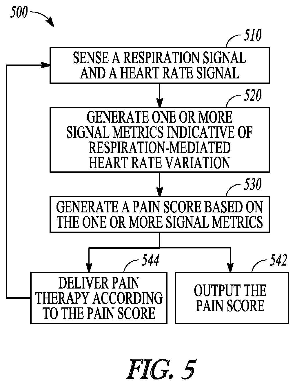
Example 16 is a method for managing pain of a patient using an implantable neuromodulator device (IND). The method comprises steps of: sensing a respiration signal and a heart rate signal from the patient via a sensor circuit; generating one or more signal metrics indicative of respiration-mediated heart rate variation using the sensed respiration signal and the heart rate signal; generating a pain score based on the generated one or more signal metrics indicative of respiration-mediated heart rate variation; and outputting the pain score to a user or a process.
In Example 17, the subject matter of Example 16 optionally includes delivering a pain therapy via the IND. The pain therapy may include electrostimulation energy determined according to the pain score.
In Example 18, the subject matter of claim 16 optionally includes detecting a plurality of respiratory cycles and at least one of an inspiration phase or an expiration phase in each of the plurality of respiratory cycles, synchronizing heart rates from the heart rate signal to the plurality of respiratory cycles, and determining the respiration-mediated heart rate variation using the synchronized heart rates.
In Example 19, the subject matter of Example 18 optionally includes the respiration-mediated heart rate variation that may indicate a difference between heart rates during the inspiration phase and heart rates during the expiration phase.
In Example 20, the subject matter of Example 18 optionally includes the respiration-mediated heart rate variation that may indicate a variability of the synchronized heart rates within one or more of the plurality of respiratory cycles.
In Example 21, the subject matter of Example 18 optionally includes the respiration-mediated heart rate variation that may include at least one of: a heart rate variability during the inspiration phase; a heart rate variability during the expiration phase; or a difference between the heart rate variability during the inspiration phase and the heart rate variability during the expiration phase.
In Example 22, the subject matter of Example 16 optionally includes sensing the respiration signal and the heart rate signal that may include steps of: sensing a physiological signal using a physiological sensor; and filtering the sensed physiological signal, via a first filter circuit, to generate the respiration signal, or filtering the sensed physiological signal, via a second filter circuit, to generate the heart rate signal. The second filter circuit may have a higher center frequency than the first filter circuit.
In Example 23, the subject matter of Example 16 optionally includes generating the pain score that may include using a plurality of the one or more signal metrics each weighted by their respective weight factor.
Sensor-based pain scores using the respiration-mediated heart rates as discussed in this document may improve automated assessment of patient pain, as well as individualized therapies to alleviate pain or to reduce side effects. The systems, devices, and methods discussed in this document may also enhance the performance and functionality of a pain management system or device. A device or a system programmed with the sensor-based pain assessment methods can have improved automaticity in medical diagnostics. More efficient device memory or communication bandwidth usage may be achieved by storing or transmitting medical information more relevant to clinical decisions. Additionally, through improved pain therapy efficacy based on patient individual need, battery longevity of an implantable device may be enhanced, or pain medication volume may be saved.
This summary is intended to provide an overview of subject matter of the present patent application. It is not intended to provide an exclusive or exhaustive explanation of the disclosure. The detailed description is included to provide further information about the present patent application. Other aspects of the disclosure will be apparent to persons skilled in the art upon reading and understanding the following detailed description and viewing the drawings that form a part thereof, each of which are not to be taken in a limiting sense.
BRIEF DESCRIPTION OF THE DRAWINGS
Various embodiments are illustrated by way of example in the figures of the accompanying drawings. Such embodiments are demonstrative and not intended to be exhaustive or exclusive embodiments of the present subject matter.
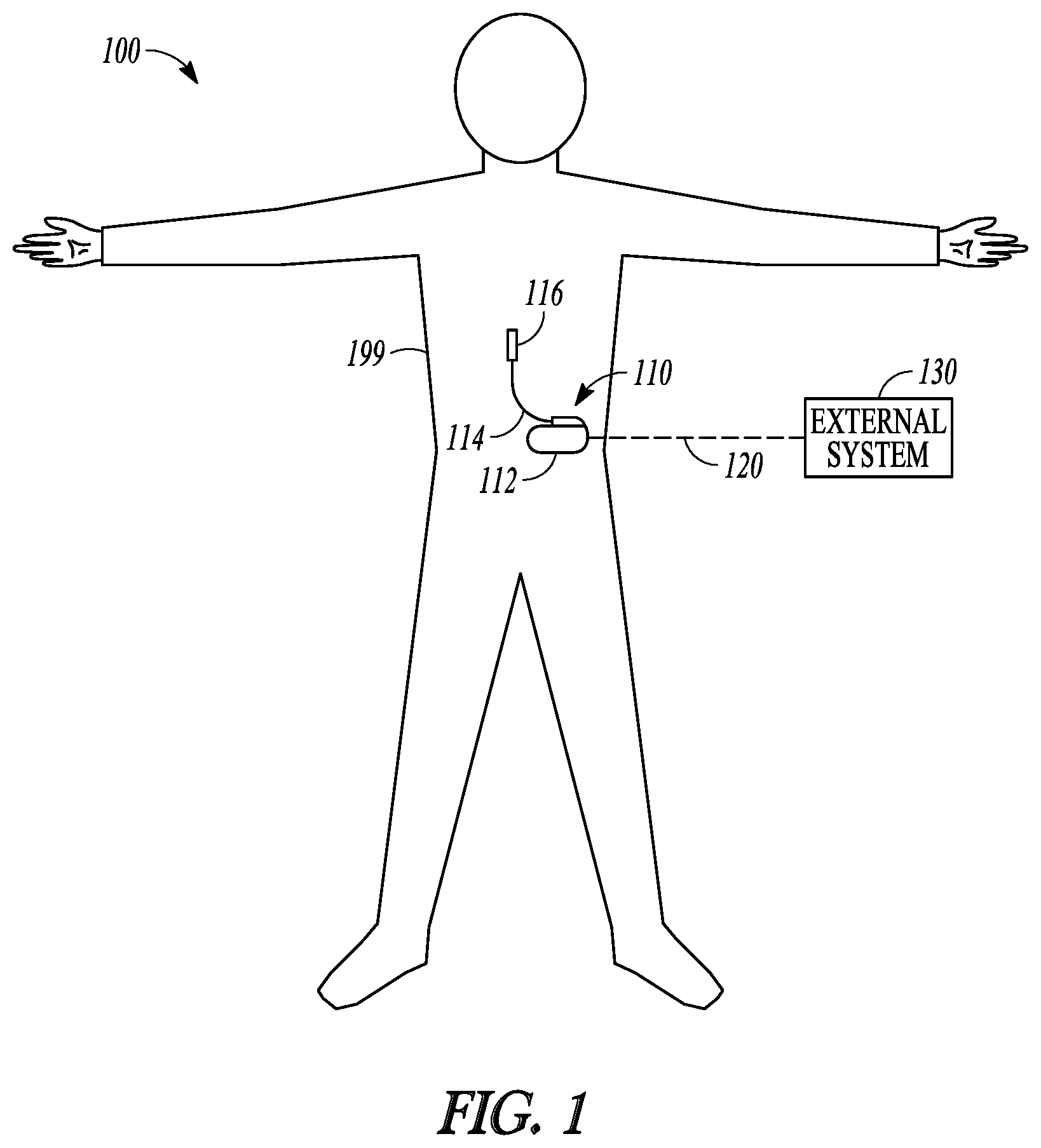
FIG. 1 illustrates, by way of example and not limitation, a neuromodulation system and portions of an environment in which the neuromodulation system may operate.
FIG. 2 illustrates, by way of example and not limitation, a block diagram of a pain management system.
FIG. 3 illustrates, by way of example and not limitation, a block diagram of a pain management system comprising an implantable neuromodulator.
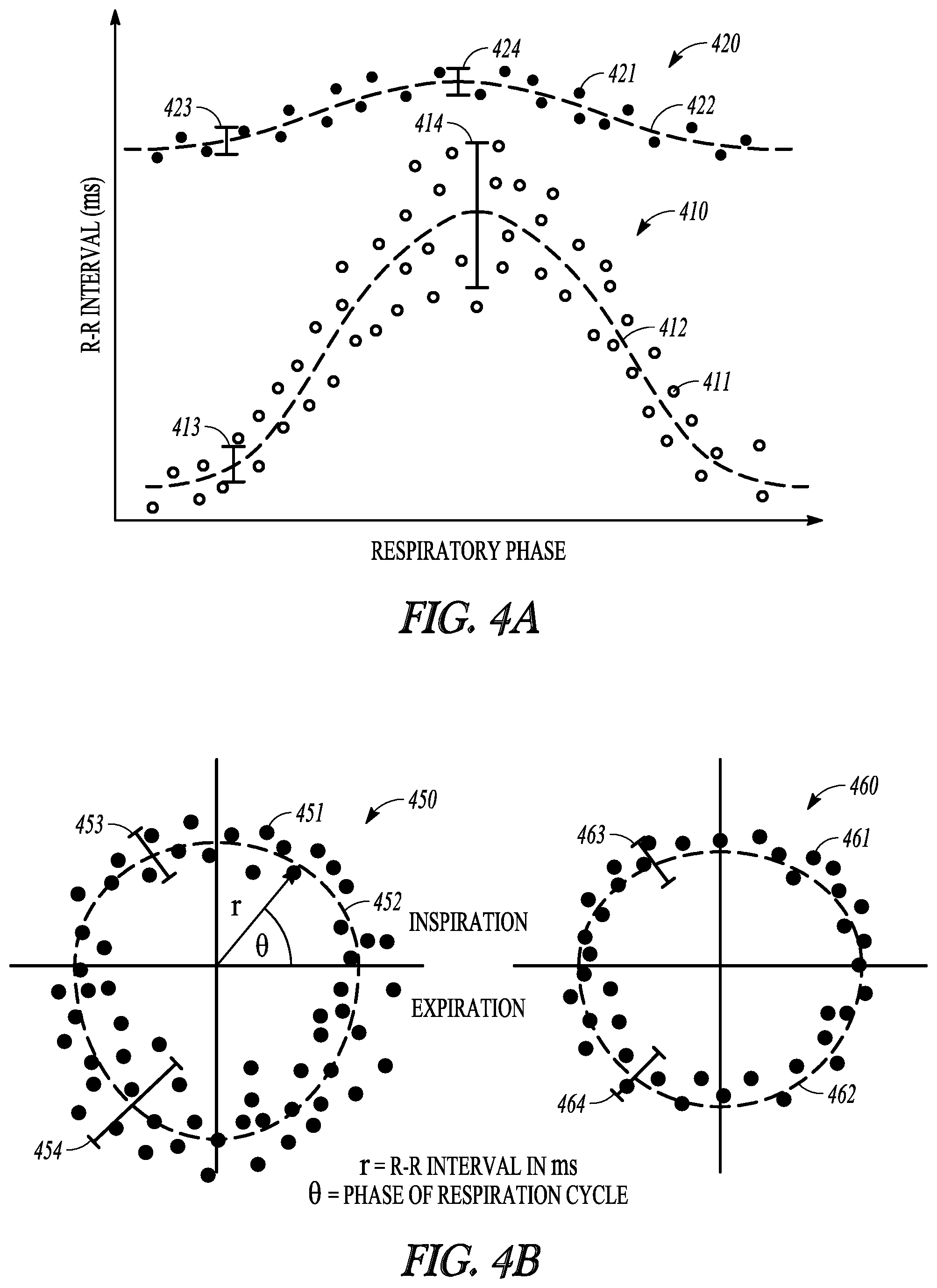
FIGS. 4A-B illustrate, by way of example and not limitation, diagrams illustrating a comparison of respiration-mediated heart rates in a pain state and respiration-mediated heart rates in a pain-free state.
FIG. 5 illustrates, by way of example and not limitation, a flow chart of a method for managing pain in a patient.
FIG. 6 illustrates, by way of example of not limitation, a block diagram of an example machine upon which any one or more of the techniques discussed herein may perform.
DETAILED DESCRIPTION
In the following detailed description, reference is made to the accompanying drawings which form a part hereof, and in which is shown by way of illustration specific embodiments in which the invention may be practiced. These embodiments are described in sufficient detail to enable those skilled in the art to practice the invention, and it is to be understood that the embodiments may be combined, or that other embodiments may be utilized and that structural, logical and electrical changes may be made without departing from the spirit and scope of the present invention. References to "an", "one", or "various" embodiments in this disclosure are not necessarily to the same embodiment, and such references contemplate more than one embodiment. The following detailed description provides examples, and the scope of the present invention is defined by the appended claims and their legal equivalents.
Clinically, chronic pain may be associated with changes in patient autonomic balance. Painful states may impair neuro-cardiac integrity. In the presence of pain, elevated sympathetic activity and/or withdrawal of parasympathetic activity may cause an increase in heart rate (HR) and decrease in heart rate variability (HRV). Conversely, when pain subsides such as via an effective pain therapy, parasympathetic activation may increase to restore autonomic balance, resulting in a decrease in HR and an increase in HRV. A short-term measure of HRV is respiratory sinus arrhythmia (RSA) that represents a cardiorespiratory coupling, which may also be affected by autonomic balance. The RSA may be related to baroreceptor activation due to changes in arterial pressure as a result of inspiratory increase in venous return to the heart. Parasympathetic activity is withdrawn during inspiration, resulting in an increase in HR and decrease in HRV. During expiration, parasympathetic activity increases, which causes HR to decrease and HRV to increase. Therefore, monitoring of patient respiration-mediated heart rates may provide an objective assessment of pain, and may be used to improve pain therapy efficacy.
Disclosed herein are systems, devices, and methods for or assessing pain of a subject, and optionally programming pain therapy based on the pain assessment. In various embodiments, the present system may include sensors configured to sense a respiration signal and a heart rate signal. A pain analyzer circuit may generate a pain score using signal metrics indicative of respiration-mediated heart rate variation. The system may include a neurostimulator that can deliver a pain therapy according to the pain score.
The present system may be implemented using a combination of hardware and software designed to provide a closed-loop pain management regimen to increase therapeutic efficacy, increase patient satisfaction for neurostimulation therapies, reduce side effects, and/or increase device longevity. The present system may be applied in any neurostimulation (neuromodulation) therapies, including but not limited to SCS, DBS, PNS, FES, motor cortex stimulation, sacral nerve stimulation, and vagus nerve stimulation (VNS) therapies. In various examples, instead of providing closed-loop pain therapies, the systems, devices, and methods described herein may be used to monitor the patient and assess pain that either occurs intrinsically or is induced by nerve block procedures or radiofrequency ablation therapies, or side effects like paresthesia caused by the stimulation therapy, among others. The patient monitoring may include generating recommendations to the patient or a clinician regarding pain treatment.
FIG. 1 illustrates, by way of example and not limitation, a neuromodulation system 100 for managing pain of a subject such as a patient with chronic pain, and portions of an environment in which the neuromodulation system 100 may operate. The neuromodulation system 100 may include an implantable system 110 that may be associated with a body 199 of the subject, and an external system 130 in communication with the implantable system 110 via a communication link 120.
The implantable system 110 may include an ambulatory medical device (AMD), such as an implantable neuromodulator device (IND) 112, a lead system 114, and one or more electrodes 116. The IND 112 may be configured for subcutaneous implant in a patient's chest, abdomen, upper gluteal surface, or other parts of the body 199. The IND 112 may be configured as a monitoring and diagnostic device. The IND 112 may include a hermetically sealed can that houses sensing circuitry to sense physiological signals from the patient via sensing electrodes or ambulatory sensors associated with the patient and in communication with the IND 112, such as the one or more electrodes 116. In some examples, the sensing electrodes or the ambulatory sensors may be included within the IND 112. The physiological signals, when measured during a pain episode, may be correlative to severity of the pain. In an example, the IND 112 may sense, via sensors or electrodes, a respiration signal and a heart rate signal, and generate one or more signal metrics indicative of respiration-mediated heart rate variation (RM-HRV) that represents variability of heart rate (or cardiac cycle length) modulated by respiration, such as heart rate variability at different phases of respiration. The RM-HRV indicates autonomic imbalance which is associated with pain. The IND 112 may characterize and quantify the pain based on the sensed physiological signals, such as to determine onset, intensity, severity, duration, or patterns of the pain experienced by the subject. The IND 112 may generate an alert to indicate occurrence of a pain episode, pain exacerbation, or efficacy of pain therapy, and present the alert to a clinician.
The IND 112 may alternatively be configured as a therapeutic device for treating or alleviating the pain. In addition to the pain monitoring circuitry, the IND 112 may further include a therapy unit that can generate and deliver energy or modulation agents to a target tissue. The energy may include electrical, magnetic, thermal, or other types of energy. In some examples, the IND 112 may include a drug delivery system such as a drug infusion pump that can deliver pain medication to the patient, such as morphine sulfate or ziconotide, among others.
The IND 112 may include electrostimulation circuitry that generates electrostimulation pulses to stimulate a neural target via the electrodes 116 operably connected to the IND 112. In an example, the electrodes 116 may be positioned on or near a spinal cord, and the electrostimulation circuitry may be configured to deliver SCS to treat pain. In another example, the electrodes 116 may be surgically placed at other neural targets such as a brain or a peripheral neutral tissue, and the electrostimulation circuitry may be configured to deliver brain or peripheral stimulations. Examples of electrostimulation may include deep brain stimulation (DBS), trigeminal nerve stimulation, occipital nerve stimulation, vagus nerve stimulation (VNS), sacral nerve stimulation, sphenopalatine ganglion stimulation, sympathetic nerve modulation, adrenal gland modulation, baroreceptor stimulation, or transcranial magnetic stimulation, spinal cord stimulation (SCS), dorsal root ganglia (DRG) stimulation, motor cortex stimulation (MCS), transcranial direct current stimulation (tDCS), transcutaneous spinal direct current stimulation (tsDCS), pudendal nerve stimulation, multifidus muscle stimulation, transcutaneous electrical nerve stimulation (TENS), tibial nerve stimulation, among other peripheral nerve or organ stimulation. The IND 112 may additionally or alternatively provide therapies such as radiofrequency ablation (RFA), pulsed radiofrequency ablation, ultrasound therapy, high-intensity focused ultrasound (HIFU), optical stimulation, optogenetic therapy, magnetic stimulation, other peripheral tissue stimulation therapies, other peripheral tissue denervation therapies, or nerve blocks or injections.
In various examples, the electrodes 116 may be distributed in one or more leads of the lead system 114 electrically coupled to the IND 112. In an example, the lead system 114 may include a directional lead that includes at least some segmented electrodes circumferentially disposed about the directional lead. Two or more segmented electrodes may be distributed along a circumference of the lead. The actual number and shape of leads and electrodes may vary according to the intended application. Detailed description of construction and method of manufacturing percutaneous stimulation leads are disclosed in U.S. Pat. No. 8,019,439, entitled "Lead Assembly and Method of Making Same," and U.S. Pat. No. 7,650,184, entitled "Cylindrical Multi-Contact Electrode Lead for Neural Stimulation and Method of Making Same," the disclosures of which are incorporated herein by reference. The electrodes 116 may provide an electrically conductive contact providing for an electrical interface between the IND 112 and tissue of the patient. The neurostimulation pulses are each delivered from the IND 112 through a set of electrodes selected from the electrodes 116. In various examples, the neurostimulation pulses may include one or more individually defined pulses, and the set of electrodes may be individually definable by the user for each of the individually defined pulses.
Although the discussion herein with regard to the neuromodulation system 100 focuses on an implantable device such as the IND 112, this is meant only by way of example and not limitation. It is within the contemplation of the present inventors and within the scope of this document, that the systems, devices, and methods discussed herein may also be used for pain management via subcutaneous medical devices, wearable medical devices (e.g., wrist watches, patches, garment- or shoe-mounted devices, headgear, eye glasses, or earplugs), or other external medical devices, or a combination of implantable, wearable, or other external devices. The therapy, such as electrostimulation or medical therapies, may be used to treat various neurological disorders other than pain, which by way of example and not limitation may include epilepsy, migraine, Tourette's syndrome, obsessive compulsive disorder, tremor, Parkinson's disease, or dystonia, among other movement and affective disorders.
The external system 130 may be communicated with the IND 112 via a communication link 120. The external system 130 may include a dedicated hardware/software system such as a programmer, a remote server-based patient management system, or alternatively a system defined predominantly by software running on a standard personal computer. In some examples, at least a portion of the external system 130 may be ambulatory such as configured to be worn or carried by a subject. The external system 130 may be configured to control the operation of the IND 112, such as to program the IND 112 for delivering neuromodulation therapy. The external system 130 may additionally receive via the communication link 120 information acquired by IND 112, such as one or more physiological signals. In an example, the external system 130 may determine a pain score based on the physiological signals received from the IND 112, and program the IND 112 to deliver pain therapy in a closed-loop fashion. Examples of the external system and neurostimulation based on pain score are discussed below, such as with reference to FIGS. 2-3.
The communication link 120 may include one or more communication channels and intermediate devices between the external system and the IND, such as a wired link, a telecommunication link such as an internet connection, or a wireless link such as one or more of an inductive telemetry link, a radio-frequency telemetry link. The communication link 120 may provide for data transmission between the IND 112 and the external system 130. The transmitted data may include, for example, real-time physiological signals acquired by and stored in the IND 112, therapy history data, data indicating device operational status of the IND 112, one or more programming instructions to the IND 112 which may include configurations for sensing physiological signal or stimulation commands and stimulation parameters, or device self-diagnostic test, among others. In some examples, the IND 112 may be coupled to the external system 130 further via an intermediate control device, such as a handheld external remote control device to remotely instruct the IND 112 to generate electrical stimulation pulses in accordance with selected stimulation parameters produced by the external system 130, or to store the collected data into the external system 130.
Portions of the IND 112 or the external system 130 may be implemented using hardware, software, firmware, or combinations thereof. Portions of the IND 112 or the external system 130 may be implemented using an application-specific circuit that may be constructed or configured to perform one or more particular functions, or may be implemented using a general-purpose circuit that may be programmed or otherwise configured to perform one or more particular functions. Such a general-purpose circuit may include a microprocessor or a portion thereof, a microcontroller or a portion thereof, or a programmable logic circuit, or a portion thereof. For example, a "comparator" may include, among other things, an electronic circuit comparator that may be constructed to perform the specific function of a comparison between two signals or the comparator may be implemented as a portion of a general-purpose circuit that may be driven by a code instructing a portion of the general-purpose circuit to perform a comparison between the two signals.
FIG. 2 illustrates, by way of example and not limitation, a block diagram of a pain management system 200, which may be an embodiment of the neuromodulation system 100. The pain management system 200 may assess pain of a subject using at least one physiological signal, and program a pain therapy based on the pain assessment. As illustrated in FIG. 2, the pain management system 200 may include a sensor circuit 210, a pain analyzer circuit 220, a memory 230, a user interface 240, and a therapy unit 250.
The sensor circuit 210 may be coupled to one or more physiological sensors to sense from the patient at least one physiological signal. The sensor circuit 210 may include sense amplifier circuit that may pre-process the sensed physiological signals, including, for example, amplification, digitization, filtering, or other signal conditioning operations. Various physiological signals, such as cardiac, pulmonary, neural, or biochemical signals may demonstrate characteristic signal properties in response to an onset, intensity, severity, duration, or patterns of pain. In an example, the sensor circuit 210 may be coupled to implantable or wearable sensors to sense cardiac signals such as electrocardiograph (ECG), intracardiac electrogram, gyrocardiography, magnetocardiography, heart rate signal, heart rate variability signal, cardiovascular pressure signal, or heart sounds signal, among others. In another example, the sensor circuit 210 may sense pulmonary signals such as a respiratory signal, a thoracic impedance signal, or a respiratory sounds signal. In yet another example, the sensor circuit 210 may sense biochemical signals such as blood chemistry measurements or expression levels of one or more biomarkers, which may include, by way of example and not limitation, B-type natriuretic peptide (BNP) or N-terminal pro b-type natriuretic peptide (NT-proBNP), serum cytokine profiles, P2X4 receptor expression levels, gamma-aminobutyric acid (GABA) levels, TNF.alpha. and other inflammatory markers, cortisol, adenosine, Glial cell-derived neurotrophic factor (GDNF), Nav 1.3, Nav 1.7, or Tetrahydrobiopterin (BH4) levels, among other biomarkers.
In an example, the sensor circuit 210 may sense one or more physiological signals including a respiration signal and a heart rate signal. The physiological sensors may be an ambulatory sensor, such as an implantable or wearable sensor associated with the patient. In an example, the sensor circuit 210 may be coupled to a respiration sensor to sense the respiration signal, and coupled to a heart rate sensor to sense a heart rate signal to detect heart rates. The heart rate sensor may include one or more implantable, wearable, or otherwise ambulatory cardiac activity sensor configured to sense cardiac electrical or mechanical activity. In an example, the heart rate sensor is configured to sense an electrocardiogram (ECG) using surface electrodes or subcutaneous electrodes, or sense intracardiac electrograms (EGM) from inside the heart chamber or heart tissue using intracardiac electrodes. The heart rate sensor may alternatively or additionally include an accelerometer or a microphone configured to sense heart sounds (HS) signal in a patient, an impedance sensor configured to sense variations of intracardiac impedance as a result of cyclic cardiac contractions, or a pressure sensor configured to sense arterial pulses, among others.
The respiration sensor may be coupled to electrodes attached to or implanted in the patient to sense the respiration signal from the patient. The respiration signal includes a respiration waveform that represents the change of air flow or lung volume during a respiratory cycle. The respiration sensor may include a flowmeter configured to sense directly the air flow in the respiratory system or volume change in the lungs. Alternatively, the respiration sensor may sense a physiological signal modulated by respiration, such as a thoracic impedance signal. The thoracic impedance may be measured using electrodes on an implantable lead coupled to an implantable medical device. In an example, the thoracic impedance may be sensed between an electrode on a right ventricular and the can housing of and implantable device implanted at the left or right pectoral region. In another example, the thoracic impedance may be sensed between an electrode on a left ventricle and the can housing of the implantable device, or between a right atrium electrode and the can housing of the implantable device. The thoracic impedance may alternatively be measured using non-invasive surface electrodes removably attached to a patient chest. In some examples, the respiration sensors may sense other respiration-modulated physiological signals including, for example, chest muscle strain sensor to measure cyclic changes in muscle tension corresponding to respiration cycles, accelerometers to measure acceleration associated with displacement or movement of chest walls corresponding to respiration, or acoustic or vibrational signals modulated by respiration. The respiration sensors may alternatively include patient-external respiratory bands, respiration flowmeter, implantable or patient-external breath sound detector, blood oxygen level detector, and other sensors configured to sense a respiration-modulated physiological signal, which can be found in Lee et al., U.S. Pat. No. 7,678,061 entitled "System and method for characterizing patient respiration", filed on Apr. 15, 2004, which is incorporated herein by reference in its entirety.
In another example, the sensor circuit 210 may be coupled to a physiological sensor to sense a physiological signal that is modulated by both cardiac rhythm and respiratory rhythm. The sensor circuit 210 may include at least first and second filter circuits. The first filter may filter the sensed physiological signal to generate a respiration signal, and the second filter may filter the sensed physiological signal generate the heart rate signal. The second filter circuit may have a higher center frequency than the first filter circuit. In an example, the heart rates and the respiration signal may be generated from a cardiac electrical signal, such as an electrocardiograph (ECG). During inspiration, the diaphragm shift downwards away from the apex of the heart. The increased filling of the lungs further stretch the apex of the heart towards the abdomen. During expiration, the lung volume reduces, and the diaphragm elevates upwards toward the heart which compresses the apex of the heart towards the breast. As a result, the angle of the electric cardiac vector that gives rise to the ECG signal changes during inspiratory and respiratory phases, which leads to cyclic variation in R-wave amplitude on the ECG signal. The respiration signal can be obtained from the R-wave amplitude signal using demodulation method. Other physiological signals, such as a heart sound signal, a blood pressure signal, a blood flow signal, a tissue strain signal, or a tissue impedance signal may each be modulated by cardiac rhythm and respiratory rhythm, and can be filtered using respective filters to extract the heart rate signal and the respiration signal.
The pain analyzer circuit 220 may generate a pain score using at least the physiological signals received from the sensor circuit 210. The pain analyzer circuit 220 may be implemented as a part of a microprocessor circuit, which may be a dedicated processor such as a digital signal processor, application specific integrated circuit (ASIC), microprocessor, or other type of processor for processing information including physical activity information. Alternatively, the microprocessor circuit may be a general purpose processor that may receive and execute a set of instructions of performing the functions, methods, or techniques described herein.
The pain analyzer circuit 220 may include circuit sets comprising one or more other circuits or sub-circuits that may, alone or in combination, perform the functions, methods or techniques described herein. In an example, hardware of the circuit set may be immutably designed to carry out a specific operation (e.g., hardwired). In an example, the hardware of the circuit set may include variably connected physical components (e.g., execution units, transistors, simple circuits, etc.) including a computer readable medium physically modified (e.g., magnetically, electrically, moveable placement of invariant massed particles, etc.) to encode instructions of the specific operation. In connecting the physical components, the underlying electrical properties of a hardware constituent are changed, for example, from an insulator to a conductor or vice versa. The instructions enable embedded hardware (e.g., the execution units or a loading mechanism) to create members of the circuit set in hardware via the variable connections to carry out portions of the specific operation when in operation. Accordingly, the computer readable medium is communicatively coupled to the other components of the circuit set member when the device is operating. In an example, any of the physical components may be used in more than one member of more than one circuit set. For example, under operation, execution units may be used in a first circuit of a first circuit set at one point in time and reused by a second circuit in the first circuit set, or by a third circuit in a second circuit set at a different time.
As illustrated in FIG. 2, the pain analyzer circuit 220 may include a respiration signal analyzer 221, signal metrics generator 222, and a pain score generator 225. The respiration signal analyzer 221 may be configured to detect from the sensed respiration signal a plurality of respiratory cycles, and determine for each respiratory cycle a respiratory cycle period. The respiratory cycle period may be determined as the duration between two time instants that represent the same state of respiration. For example, the respiratory cycle period may be computed as the duration between the end-of-expiration states. The respiration signal analyzer 221 may additionally be configured to detect, within each respiratory cycle, an inspiration phase and an expiration phase. The inspiration phase is a period between an end-of-expiration state and the next end-of-inspiration state. The expiration phase is a period between an end-of-inspiration state and the next end-of-expiration state. In an example where the respiration sensor directly or indirectly measures the lung volume, the end-of-expiration state may correspond to the minimal lung volume (or a metric indicative of the lung volume) within a specified detection window; and the end-of-inspiration state may correspond to the maximal lung volume (or a metric indicative of the lung volume) with a specified detection window. In another example where the respiration sensor senses thoracic impedance, the thoracic impedance increases when the air volume in the lungs increases (e.g., during inspiration). The end-of-expiration state may correspond to the minimal transthoracic impedance within a specified detection window; and the end-of-inspiration state may correspond to the maximal transthoracic impedance within a specified detection window.
The signal metrics generator 222 may generate one or more respiration-mediated heart rate metrics 223 using the heart rates detected from the heart rate signal and the respiration cycle or respiratory phases from the respiration signal. The detected heart rates, or cardiac cycles, may be synchronized to the respiratory cycles, inspiration phase, or expiration phase. The HR-respiration synchronization process can compensate both system lag and physiological lag between the respiration signal and the heart rate signal. The system lag includes the lag between the heart rate sensor and the respiration signal sensor in sensing and processing the respective signals. In an example, a system synchronization signal such as an electrical pulse may be issued and sensed by both the heart rate sensor and the respiration sensor, and the system lag may be determined as the time between the pulse signature on the heart rate signal and the pulse signature on the respiration signal. The physiological phase lag between HR and respiration signal may be due to the physiological response delay between heart rate and respiration. For example, because respiratory sinus arrhythmia (RSA) is associated with baroreflex, the HR change may lag behind the respiratory phase change. For example, during inspiration, thoracic pressure drops and the venous return increases, and the arterial baroreceptors are unloaded. Through the baroreflex mechanism, the vagus nerve activity is suppressed, which leads to an increase in sinus node firing and thus an increase in HR. The HR-respiration synchronization may compensate the physiological lag between HR and respiration signal.
The signal metrics generator 222 may generate one or more respiration-mediated heart rate metrics 223 using the respiration-synchronized heart rates. In an example, the respiration-mediated heart rate metrics 223 may indicate a variability of the heart rates (or cardiac cycles such as R-R intervals) within one or more respiratory cycles, hereinafter referred to as respiration-mediated heart rate variation (RM-HRV). The RM-HRV represents variability of heart rate (or cardiac cycle length) modulated by respiration, such as heart rate variability at different phases of respiration. A large RM-HRV (such as exceeding a threshold value) may be a result of persistence or restoration of automatic balance, which indicates no pain or an effective pain-relief effect. Conversely, a small RM-HRV (such as falling below a threshold value) may be caused by parasympathetic withdrawal, which indicates persistence of pain or ineffective pain therapy. Examples of the variability may include heart rate range within a respiratory cycle (e.g., a relative difference between the maximal heart rate and the minimal heart rate within the respiratory cycle), a statistical measure out of a histogram of the heart rates within a respiratory cycle, or a standard deviation, variance, or other spreaders measures of the heart rates within a respiratory cycle. In another example, the RM-HRV may be calculated as a relative difference (.DELTA.HR.sub.Insp-Exp) between a representative heart rates during the inspiration phase (HR.sub.Insp) and a representative heart rate during the expiration phase (HR.sub.Exp). The representative heart rates may be computed as a maximal value, a mean, a median, or a mode of the heart rates within the respective respiratory phases. A large difference .DELTA.HR.sub.Insp-Exp, such as when exceeding a threshold value, may be a result of persistence or restoration of automatic balance, and indicate no pain or effective pain-relief therapy. Conversely, a small difference .DELTA.HR.sub.Insp-Exp, such as when falling below a threshold value, may be caused by parasympathetic withdrawal, which indicates persistence of pain or ineffective pain therapy.
In some examples, the RM-HRV may be calculated as a heart rate variability during the inspiration phase (HRV.sub.Insp) and/or a heart rate variability during the expiration phase (HRV.sub.Exp). In an example, the HRV.sub.Exp may be compared to a threshold such as a baseline HRV.sub.Exp measured when the patient is known to be free of pain. If the HRV.sub.Exp falls within a specified margin of the baseline HRV.sub.Exp, it may indicate adequate maintenance or substantial restoration of automatic balance, and no pain or effective pain-relief therapy. Conversely, if the HRV.sub.Exp is less than the baseline HRV.sub.Exp by a specified margin, that is, substantial reduction of HRV during expiration, it may indicate parasympathetic withdrawal and persistence of pain or ineffective pain therapy. In another example, the RM-HRV may be calculated as a relative difference (.DELTA.HRV.sub.Insp-Exp) between the HRV.sub.Insp and HRV.sub.Exp. A large difference .DELTA.HRV.sub.Insp-Exp, such as when exceeding a threshold value, may be a result of persistence or restoration of automatic balance, which indicates no pain or effective pain-relief therapy. Conversely, a small difference .DELTA.HRV.sub.Insp-Exp, such as when falling below a threshold value, may indicate parasympathetic withdrawal and persistence of pain or ineffective pain-relief therapy.
The pain score generator 225 may generate a pain score using the measurements of the signal metrics generated by the signal metrics generator 222. The pain score may be represented as a numerical or categorical value that quantifies a patient overall pain symptom. In an example, a composite signal metric may be generated using a plurality of the signal metrics respectively weighted by weight factors. The combination may be linear or nonlinear. The pain score generator 225 may compare the composite signal metric to one or more threshold values or range values, and assign a corresponding pain score (such as numerical values from 0 to 10) based on the comparison.
In another example, the pain score generator 225 may compare the signal metrics to their respective threshold values or range values, assign corresponding signal metric-specific pain scores based on the comparison, and compute a composite pain score using a linear or nonlinear fusion of the signal metric-specific pain scores each weighted by their respective weight factor. In an example, the threshold may be inversely proportional to signal metric's sensitivity to pain. A signal metric that is more sensitive to pain may have a corresponding lower threshold and a larger metric-specific pain score, thus plays a more dominant role in the composite pain score than another signal metric that is less sensitive to pain. Examples of the fusion algorithm may include weighted averages, voting, decision trees, or neural networks, among others. The pain score generated by the pain score generator 225 may be output to a system user or a process.
In various examples, in addition to the physiological signals such as the respiration and heart rate signals, the sensor circuit 210 may sense one or more functional signals from the patient. Examples of the functional signals may include, but not limited to, patient posture, gait, balance, or physical activity signals, among others. The sensor circuit 210 may sense the functional signals via one or more implantable or wearable motion sensors, including an accelerometer, a gyroscope (which may be a one-, two-, or three-axis gyroscope), a magnetometer (e.g., a compass), an inclinometer, a goniometer, a electromagnetic tracking system (ETS), or a global positioning system (GPS) sensor, among others. Detailed description of functional signals for use in pain characterization are disclosed in commonly assigned U.S. Provisional Patent Application Ser. No. 62/445,075, entitled "PAIN MANAGEMENT BASED ON FUNCTIONAL MEASUREMENTS", the disclosures of which are incorporated herein by reference. The signal metrics generator 221 may generate functional signal metrics from the functional signals, and the pain score generator 225 may determine the pain score using a linear or nonlinear combination of the muscle tension signal metrics and the functional signal metrics. Commonly assigned U.S. Provisional Patent Application Ser. No. 62/445,053, entitled "PAIN MANAGEMENT BASED ON CARDIOVASCULAR PARAMETERS" describes cardiovascular parameters such as arterial pulsatile activity and electrocardiography for use in pain analysis, the disclosure of which is incorporated herein by reference in its entirety. Commonly assigned U.S. Provisional Patent Application Ser. No. 62/445,061, entitled "PAIN MANAGEMENT BASED ON BRAIN ACTIVITY MONITORING" describes information of brain activity for use in pain analysis, the disclosure of which is incorporated herein by reference in its entirety. Commonly assigned U.S. Provisional Patent Application Ser. No. 62/445,061, entitled "PAIN MANAGEMENT BASED ON BRAIN ACTIVITY MONITORING" describes information of brain activity for use in pain analysis, the disclosure of which is incorporated herein by reference in its entirety. Commonly assigned U.S. Provisional Patent Application Ser. No. 62/445,082, entitled "PAIN MANAGEMENT BASED ON EMOTIONAL EXPRESSION MEASUREMENTS" describes measurements of patient emotional expressions for use in pain analysis, the disclosure of which is incorporated herein by reference in its entirety. Commonly assigned U.S. Provisional Patent Application Ser. No. 62/445,092, entitled "PAIN MANAGEMENT BASED ON MUSCLE TENSION MEASUREMENTS" describes measurements of patient muscle tension including electromyography for use in pain analysis, the disclosure of which is incorporated herein by reference in its entirety. One or more of these additional signals or measurements may be used by the pain analyzer circuit 220 to generate a pain score.
The memory 230 may be configured to store sensor signals or signal metrics such as generated by the sensor circuit 210 and the signal metrics generator 222, and the pain scores such as generated by the pain score generator 225. Data storage at the memory 230 may be continuous, periodic, or triggered by a user command or a specific event. In an example, as illustrated in FIG. 2, the memory 230 may store weight factors, which may be used by the pain score generator 225 to generate the composite pain score. The weight factors may be provided by a system user, or alternatively be automatically determined or adjusted such as based on the corresponding signal metrics' reliability in representing an intensity of the pain. Examples of the automatic weight factor generation are discussed below, such as with reference to FIG. 3.
The user interface 240 may include an input circuit 241 and an output unit 242. In an example, at least a portion of the user interface 240 may be implemented in the external system 130. The input circuit 241 may enable a system user to program the parameters used for sensing the physiological signals, generating signal metrics, or generating the pain score. The input circuit 241 may be coupled to one or more input devices such as a keyboard, on-screen keyboard, mouse, trackball, touchpad, touch-screen, or other pointing or navigating devices. In some example, the input device may be incorporated in a mobile device such as a smart phone or other portable electronic device configured to execute a mobile application ("App"). The mobile App may enable a patient to provide pain description or quantified pain scales during the pain episodes. In an example, the input circuit 241 may enable a user to confirm, reject, or edit the programming of the therapy unit 250, such as parameters for electrostimulation, as to be discussed in the following.
The output unit 242 may include a display to present to a system user such as a clinician the pain score. The output unit 242 may also display information including the physiological and functional signals, trends of the signal metric, or any intermediary results for pain score calculation such as the signal metric-specific pain scores. The information may be presented in a table, a chart, a diagram, or any other types of textual, tabular, or graphical presentation formats, for displaying to a system user. The presentation of the output information may include audio or other human-perceptible media format. In an example, the output unit 242 may generate alerts, alarms, emergency calls, or other forms of warnings to signal the system user about the pain score.
The therapy circuit 250 may be configured to deliver a therapy to the patient based on the pain score generated by the pain score generator 225. The therapy circuit 250 may include an electrostimulator configured to generate electrostimulation energy to treat pain. In an example, the electrostimulator may deliver spinal cord stimulation (SCS) via electrodes electrically coupled to the electrostimulator. The electrodes may be surgically placed at a region at or near a spinal cord tissue, which may include, by way of example and not limitation, dorsal column, dorsal horn, spinal nerve roots such as the dorsal nerve root, dorsal root entry zone, spinothalamic tract, and dorsal root ganglia. The SCS may be in a form of stimulation pulses that are characterized by pulse amplitude, pulse width, stimulation frequency, duration, on-off cycle, pulse shape or waveform, temporal pattern of the stimulation, among other stimulation parameters. Examples of the stimulation pattern may include burst stimulation with substantially identical inter-pulse intervals, or ramp stimulation with incremental inter-pulse intervals or with decremental inter-pulse intervals. In some examples, the frequency or the pulse width may change from pulse to pulse. The electrostimulator may additionally or alternatively deliver electrostimulation to other target tissues such as peripheral nerves tissues. In an example, the electrostimulator may deliver transcutaneous electrical nerve stimulation (TENS) via detachable electrodes that are affixed to the skin.
The therapy circuit 250 may additionally or alternatively include a drug delivery system, such as an intrathecal drug delivery pump that may be surgically placed under the skin, which may be programmed to inject medication or biologics through a catheter to the area around the spinal cord. Other examples of drug delivery system may include a computerized patient-controlled analgesia pump that may deliver the prescribed pain medication to the patient such as via an intravenous line. In some examples, the therapy circuit 250 may be delivered according to the pain score received from the pain score generator 225.
FIG. 3 illustrates, by way of example and not limitation, a block diagram of another example of a pain management system 300, which may be an embodiment of the neuromodulation system 100 or the pain management system 200. The pain management system 300 may include an implantable neuromodulator 310 and an external system 320, which may be, respectively, embodiments of the IND 112 and the external system 130 as illustrated in FIG. 1. The external system 320 may be communicatively coupled to the implantable neuromodulator 310 via the communication link 120.
The implantable neuromodulator 310 may include several components of the pain management system 200 as illustrated in FIG. 2, including the sensor circuit 210, the pain analyzer circuit 220, the memory 230, and the therapy unit 250. As discussed with reference to FIG. 2, the pain analyzer circuit 220 includes the pain score generator 225 that determine a pain score using weight factors stored in the memory 230 and the signal metrics from the signal metrics generator 222 which may also be included in the pain analyzer circuit 220. The implantable neuromodulator 310 may include a controller 312, coupled to the therapy unit 250, that controls the generation and delivery of pain therapy, such as neurostimulation energy. The controller 312 may control the generation of electrostimulation pulses according to specific stimulation parameters. The stimulation parameters may be provided by a system user. Alternatively, the stimulation parameters may be automatically determined based on the intensity, severity, duration, or pattern of pain, which may be subjectively described by the patient or automatically quantified based on the physiological signals sensed by the sensor circuit 210. For example, when a patient-described or sensor-indicated quantification exceeds a respective threshold value or falls within a specific range indicating elevated pain, the electrostimulation energy may be increased to provide stronger pain relief. Increased electrostimulation energy may be achieved by programming a higher pulse intensity, a higher frequency, or a longer stimulation duration or "on" cycle, among others. Conversely, when a patient-described or sensor-indicated pain quantification falls below a respective threshold value or falls within a specific range indicating no pain or mild pain, the electrostimulation energy may be decreased. The controller circuit 312 may also adjust stimulation parameters to alleviate side effects introduced by the electrostimulation of the target tissue.
Additionally or alternatively, the controller circuit 312 may control the therapy unit 250 to deliver electrostimulation pulses via specific electrodes. In an example of pain management via SCS, a plurality of segmented electrodes, such as the electrodes 116, may be distributed in one or more leads. The controller circuit 312 may configure the therapy unit 250 to deliver electrostimulation pulses via a set of electrodes selected from the plurality of electrodes. The electrodes may be manually selected by a system user or automatically selected based on the pain score.
The implantable neuromodulator 310 may receive the information about electrostimulation parameters and the electrode configuration from the external system 320 via the communication link 120. Additional parameters associated with operation of the therapy unit 250, such as battery status, lead impedance and integrity, or device diagnostic of the implantable neuromodulator 310, may be transmitted to the external system 320. The controller circuit 312 may control the generation and delivery of electrostimulation using the information about electrostimulation parameters and the electrode configuration from the external system 320. Examples of the electrostimulation parameters and electrode configuration may include: temporal modulation parameters such as pulse amplitude, pulse width, pulse rate, or burst intensity; morphological modulation parameters respectively defining one or more portions of stimulation waveform morphology such as amplitude of different phases or pulses included in a stimulation burst; or spatial modulation parameters such as selection of active electrodes, electrode combinations which define the electrodes that are activated as anodes (positive), cathodes (negative), and turned off (zero), and stimulation energy fractionalization which defines amount of current, voltage, or energy assigned to each active electrode and thereby determines spatial distribution of the modulation field.
In an example, the controller circuit 312 may control the generation and delivery of electrostimulation in a closed-loop fashion by adaptively adjusting one or more stimulation parameters or stimulation electrode configuration based on the pain score. For example, if the score exceeds the pain threshold (or falls within a specific range indicating an elevated pain), then the first electrostimulation may be delivered. Conversely, if the composite pain score falls below a respective threshold value (or falls within a specific range indicating no pain or mild pain), then a second pain therapy, such as second electrostimulation may be delivered. The first electrostimulation may differ from the second electrostimulation with respect to at least one of the stimulation energy, pulse amplitude, pulse width, stimulation frequency, duration, on-off cycle, pulse shape or waveform, electrostimulation pattern such as electrode configuration or energy fractionalization among active electrodes, among other stimulation parameters. In an example, the first electrostimulation may have higher energy than the second electrostimulation, such as to provide stronger effect of pain relief. Examples of increased electrostimulation energy may include a higher pulse intensity, a higher frequency, or a longer stimulation duration or "on" cycle, among others.
The parameter adjustment or stimulation electrode configuration may be executed continuously, periodically at specific time, duration, or frequency, or in a commanded mode upon receiving from a system user a command or confirmation of parameter adjustment. In some examples, the closed-loop control of the electrostimulation may be further based on the type of the pain, such as chronic or acute pain. In an example, the pain analyzer circuit 220 may trend the signal metric over time to compute an indication of abruptness of change of the signal metrics, such as a rate of change over a specific time period. The pain episode may be characterized as acute pain if the signal metric changes abruptly (e.g., the rate of change of the signal metric exceeding a threshold), or as chronic pain if the signal metric changes gradually (e.g., the rate of change of the signal metric falling below a threshold). The controller circuit 312 may control the therapy unit 250 to deliver, withhold, or otherwise modify the pain therapy in accordance with the pain type. For example, incidents such as toe stubbing or bodily injuries may cause abrupt changes in certain signal metrics, but no adjustment of the closed-loop pain therapy is deemed necessary. On the contrary, if the pain analyzer circuit 220 detects chronic pain characterized by gradual signal metric change, then the closed-loop pain therapy may be delivered accordingly.
The external system 320 may include the user interface 240, a weight generator 322, and a programmer circuit 324. The weight generator 322 may generate weight factors used by the pain score generator 225 to generate the pain score. The weight factors may indicate the signal metrics' reliability in representing an intensity of the pain. A sensor metric that is more reliable, or more sensitive or specific to the pain, would be assigned a larger weight than another sensor metric that is less reliable, or less sensitive or specific to the pain. In an example, the weight factors may be proportional to correlations between a plurality of quantified pain scales (such as reported by a patient) and measurements of the signal metrics corresponding to the plurality of quantified pain scales. A signal metric that correlates with the pain scales is deemed a more reliable signal metric for pain quantification, and is assigned a larger weight factor than another signal metric less correlated with the quantified pain scales. In another example, the weight generator 322 may determine weight factors using the signal sensitivity to pain. The signal metrics may be trended over time, such as over approximately six months. The signal sensitivity to pain may be represented by a rate of change of the signal metrics over time during a pain episode. The signal sensitivity to pain may be evaluated under a controlled condition such as when the patient posture or activity is at a specific level or during specific time of the day. The weight generator 322 may determine weight factors to be proportional to signal metric's sensitivity to pain.
The programmer circuit 324 may produce parameter values for operating the implantable neuromodulator 310, including parameters for sensing physiological signals and generating signal metrics, and parameters or electrode configurations for electrostimulation. In an example, the programmer circuit 324 may generate the stimulation parameters or electrode configurations for SCS based on the pain score produced by the pain score generator 225. Through the communication link 120, the programmer circuit 324 may continuously or periodically provide adjusted stimulation parameters or electrode configuration to the implantable neuromodulator 210. By way of non-limiting example and as illustrated in FIG. 3, the programmer circuit 324 may be coupled to the user interface 234 to allow a user to confirm, reject, or edit the stimulation parameters, sensing parameters, or other parameters controlling the operation of the implantable neuromodulator 210. The programmer circuit 324 may also adjust the stimulation parameter or electrode configuration in a commanded mode upon receiving from a system user a command or confirmation of parameter adjustment.
The programmer circuit 324, which may be coupled to the weight generator 322, may initiate a transmission of the weight factors generated by the weight generator 322 to the implantable neuromodulator 310, and store the weight factors in the memory 230. In an example, the weight factors received from the external system 320 may be compared to previously stored weight factors in the memory 230. The controller circuit 312 may update the weight factors stored in the memory 230 if the received weight factors are different than the stored weights. The pain analyzer circuit 220 may use the updated weight factors to generate a pain score. In an example, the update of the stored weight factors may be performed continuously, periodically, or in a commanded mode upon receiving a command from a user. In various examples, weight factors may be updated using a fusion model. Commonly assigned U.S. Provisional Patent Application Ser. No. 62/445,095, entitled "PATIENT-SPECIFIC CALIBRATION OF PAIN QUANTIFICATION" describes systems and methods for calibrating a fusion model, such as adjusting weights for signal metrics, using a reference pain quantification, the disclosure of which is incorporated herein by reference in its entirety.
In some examples, the pain score may be used by a therapy unit (such as an electrostimulator) separated from the pain management system 300. In various examples, the pain management system 300 may be configured as a monitoring system for pain characterization and quantification without delivering closed-loop electrostimulation or other modalities of pain therapy. The pain characterization and quantification may be provided to a system user such as the patient or a clinician, or to a process including, for example, an instance of a computer program executable in a microprocessor. In an example, the process includes computer-implemented generation of recommendations or an alert to the system user regarding pain medication (e.g., medication dosage and time for taking a dose), electrostimulation therapy, or other pain management regimens. The therapy recommendations or alert may be based on the pain score, and may be presented to the patient or the clinician in various settings including in-office assessments (e.g. spinal cord stimulation programming optimization), in-hospital monitoring (e.g. opioid dosing during surgery), or ambulatory monitoring (e.g. pharmaceutical dosing recommendations).
In an example, in response to the pain score exceeding a threshold which indicates elevated pain symptom, an alert may be generated and presented at the user interface 240 to remind the patient to take pain medication. In another example, therapy recommendations or alerts may be based on information about wearing-off effect of pain medication, which may be stored in the memory 230 or received from the user interface 240. When the drug effect has worn off, an alert may be generated to remind the patient to take another dose or to request a clinician review of the pain prescription. In yet another example, before a pain therapy such as neurostimulation therapy is adjusted (such as based on the pain score) and delivered to the patient, an alert may be generated to forewarn the patient or the clinician of any impending adverse events. This may be useful as some pain medication may have fatal or debilitating side effects. In some examples, the pain management system 300 may identify effect of pain medication addiction such as based on physiological signals. An alert may be generated to warn the patient about effects of medication addiction and thus allow medical intervention.
In some examples, the pain analyzer circuit 220 may be alternatively included in the external system 320. The pain analyzer circuit 220, or a portion of the pain analyzer circuit 220 such as one or more the respiration signal analyzer 221, signal metrics generator 222, or the pain score generator 225, may be included in a wearable device configured to be worn or carried by a subject. At least a portion of the sensor circuit 210 may also be included in the external system 320. A clinician may use the external system 320 to program the implantable neuromodulator 310 with appropriate pain therapy based on the pain score generated at the external system 320, such as during a clinical trial or patient follow-up visit at the clinic.
FIGS. 4A-B illustrates, by way of example and not limitation, diagrams of a comparison of respiration-mediated heart rates in a pain state (420 and 460) and respiration-mediated heart rates in a pain-free state (410 and 450). FIG. 4A illustrates, in a Cartesian coordinate system, a scatter plot of R wave-to-R wave intervals (RR intervals) within a respiratory cycle. The RR intervals may be computed from an ECG signal synchronized to a respiration signal. In a pain-free state or an effective pain therapy state, as illustrated in 410, the RR intervals 411 demonstrate a prominent pattern of cyclic variation with respiratory phases within a respiratory cycle. The average RR interval, represented by the trend line 412, is shorter (i.e., faster heart rate) during inspiration than the average RR interval during expiration (i.e., slower heart rate). The RR intervals during inspiration is less variable (as represented by a relatively smaller variability 413) than the RR intervals during expiration (as represented by a relatively larger variability 414). In a pain state or an ineffective pain therapy state, as illustrated in 420, the within--respiratory cycle cyclic variation pattern of the RR intervals 421 at different respiratory phases becomes less prominent. The average RR interval (represented by the trend line 422) is not substantially shorter during inspiration than during expiration. The RR interval variability during inspiration (as represented by a small variability 423) is not substantially smaller than the RR interval variability during expiration (as represented by a large variability 424). Additionally, the RR interval variability during expiration in a pain state (424) is substantially smaller than the RR interval variability during expiration in a pain-free state (414). The different patterns of average HR or HRV at various respiratory phases may be used to characterize and quantify the patient pain.
FIG. 4B illustrates a scatter plot of RR intervals 421 within a respiratory cycle in a polar coordinate system. Each respiration-synchronized RR interval is represented by a point in the polar coordinate system, as defined by a radius r and an angle .theta.. The radius r represents the RR interval (e.g., in milliseconds), and the angle .theta. represents the respiratory phase angle between 0 and 360 degrees. The plot 450 corresponds to the pain-free state or effective pain therapy state and the plot 460 corresponds to the pain state or ineffective pain therapy state. The upper quadrants of 450 and 460 (corresponding to 0<.theta.<180.degree.) represents the inspiration phase. The lower quadrants of 450 and 460 (corresponding to 180.degree.<.theta.<360.degree.) represents the expiration phase. The RR intervals 451 in the polar coordinate system correspond to the RR intervals 411 in the Cartesian coordinate system, and the RR intervals 461 in the polar coordinate system correspond to the RR intervals 421 in the Cartesian coordinate system. As illustrated in 450, in the pain-free or effective pain therapy state, the average RR interval (represented by 452) during inspiration (the upper quadrants) is smaller than the average RR interval during expiration (the lower quadrants). This results in an asymmetrical average HR curve 452 about the horizontal axis that separates the inspiration phase from the expiration phase. In contrast, in the pain state or ineffective pain therapy state as illustrated in 460, the average RR interval (represented by 462) during inspiration (the upper quadrants) is close to the average RR interval during expiration (the lower quadrants). This results in a more symmetrical average HR curve 462 about the horizontal axis. Additionally, in pain-free or effective pain therapy state, the RR interval variability 453 during inspiration is substantially smaller than the RR interval variability 454 during expiration. However, such a respiratory phase-dependent distinction in RR interval variability becomes less prominent in pain state or ineffective pain therapy state, as indicated by the RR interval variability 463 during inspiration and the RR interval variability 464 during expiration. The graphical features, such as the degree of symmetry of the average RR interval trend 452 or 462, may be extracted from the plots 450 or 460 and used for generating the pain score.
FIG. 5 illustrates, by way of example and not limitation, a method 500 for managing pain of a patient. The method 500 may be implemented in a medical system, such as the pain management system 200 or 300. In an example, at least a portion of the method 500 may be executed by a neuromodulator device (IND) such as the implantable neuromodulator 310. In an example, at least a portion of the method 500 may be executed by an external programmer or remote server-based patient management system, such as the external system 320 that are communicatively coupled to the IND. The method 500 may be used to provide neuromodulation therapy to treat chronic pain or other disorders.
The method 500 begins at step 510, where a respiration signal and a heart rate signal may be sensed from the patient. The respiration may be sensed using a sensor circuit, such as the sensor circuit 210, that is coupled to one or more respiration sensors to sense an airflow signal in the respiratory system, chest volume signal, thoracic impedance signal, chest muscle tension, acceleration, or displacement signal, breath sound signal, or blood oxygen level signal, among other respiration-modulated physiological signals. The heart rate signal may be sensed using a sensor circuit, such as the sensor circuit 210, that is coupled to one or more electrodes or sensors to sense cardiac electrical or mechanical signals, such as an electrocardiogram, a intracardiac electrogram, a heart sounds signal, a pressure pulse signal, or an intracardiac impedance signal, among others.
In some examples, a physiological signal sensed at 510 may be modulated by both heart rhythm and respiratory rhythm. The sensed physiological signal may be filtered into components at different frequency bands using at least first and second filters. The first filter may filter the sensed physiological signal to generate a respiration signal, and the second filter may filter the sensed physiological signal generate the heart rate signal. The second filter circuit may have a higher center frequency than the first filter circuit. For example, an electrocardiograph (ECG) may be used to extract both the heart rates and the respiration signal. The angle of the electric cardiac vector that gives rise to the ECG signal changes during inspiration and respiratory phases. This may result in cyclic variation in R-wave amplitude on the ECG signal. The respiration signal can be obtained from the R-wave amplitude signal through a low-pass or pass-pass filter, or other demodulation method. In other examples, a heart sounds (HS) signal, a blood pressure signal, a blood flow signal, a tissue strain signal, a tissue impedance signal, or a nerve electrical signal may each be modulated by cardiac rhythm and respiratory rhythm, and can be filtered using respective filters to extract a heart rate signal and a respiration signal.
At 520, one or more signal metrics indicative of respiration-mediated heart rate variation using the sensed respiration signal and the heart rate signal. The respiration-medicate heart rate variation represents changes or pattern of changes of heart rates at different phases of a respiratory cycle. The sensed respiration signal may be processed to determine a respiratory cycle period, such as measured as a duration between two time instants that represent the same state of respiration. Within each respiratory cycle, an inspiration phase and an expiration phase may be detected.
Heart rates, or cardiac cycles, from the heart rate signal may be synchronized to the respiratory cycles, inspiration phase, or expiration phase. The HR-respiration synchronization process can compensate both system lag and physiological lag between the respiration signal and the heart rate signal. One or more respiration-mediated heart rate metrics may then be generated using the respiration-synchronized heart rates. In an example, the respiration-mediated heart rate metrics include a variance or a spreadness measure of the heart rates (or cardiac cycles such as R-R intervals) within one or more respiratory cycles, hereinafter referred to as respiration-mediated heart rate variation (RM-HRV). A large RM-HRV may correspond to maintained or restored automatic balance, which indicates no pain or effective therapy in relieving pain. A small RM-HRV may correspond to parasympathetic withdrawal, which indicates persistence of pain or ineffective pain therapy. In another example, the RM-HRV may be calculated as a relative difference between the heart rates during the inspiration phase and the heart rates during the expiration phase. A large difference in HR during respective respiration phases corresponds to persistence or restoration of automatic balance, which indicates no pain or effective therapy in relieving pain, and a small HR difference corresponds to parasympathetic withdrawal, which indicates persistence of pain or ineffective pain therapy. The RM-HRV may alternatively be calculated as a heart rate variability HRV.sub.Insp during the inspiration phase, a heart rate variability HRV.sub.Exp during the expiration phase, or a relative difference .DELTA.HRV.sub.Insp-Exp between the HRV.sub.Insp and HRV.sub.Exp. One or more of the HRV.sub.Insp, the HRV.sub.Exp, or the .DELTA.HRV.sub.Insp-Exp may be compared to their respective threshold values to determine the pain state or a pain-relief effect of a therapy.
At 530, a pain score may be generated using the measurements of the signal metrics such as one or more signal metrics indicative of respiration-mediated heart rate variation. The pain score may be represented as a numerical or categorical value that quantifies overall pain quality in the subject. In an example, a composite signal metric may be generated using a combination of the signal metrics each weighted by their respective weight factor. The composite signal metric may be categorized as one of a number of degrees of pain by comparing the composite signal metric to one or more threshold values or range values, and a corresponding pain score may be assigned based on the comparison. In another example, the signal metrics may be compared to their respective threshold values or range values and a corresponding signal metric-specific pain score may be determined. A composite pain score may be generated using a linear or nonlinear fusion of the signal metric-specific pain scores each weighted by their respective weight factor. In some examples, the pain score may be computed using a subset of the signal metrics selected based on their temporal profile of pain response. Signal metrics with quick pain response (or a shorter transient state of response) may be selected to compute the pain score during a pain episode. Signal metrics with slow or delayed pain response (or a longer transient state of response before reaching a steady state) may be used to compute the pain score after an extended period following the onset of pain such as to allow the signal metrics to reach steady state of response.
At 542, the pain score may be output to a user or to a process, such as via the output unit 242 as illustrated in FIG. 2. The pain score, including the composite pain score and optionally together with metric-specific pain scores, may be displayed on a display screen. Other information such as the respiration signal and the heart rate signal, and the signal metrics indicative of respiration-mediated heart rate variation, may also be output for display or for further processing. In some examples, alerts, alarms, emergency calls, or other forms of warnings may be generated to signal the system user about occurrence of a pain episode or aggravation of pain as indicated by the pain score.
The method 500 may include, at 544, an additional step of delivering a pain therapy to the patient according to the pain score. The pain therapy may include electrostimulation therapy, such as spinal cord stimulation (SCS) via electrodes electrically coupled to the electrostimulator. The SCS may be in a form of stimulation pulses that are characterized by pulse amplitude, pulse width, stimulation frequency, duration, on-off cycle, waveform, among other stimulation parameters. Other electrostimulation therapy, such as one or a combination of DBS, FES, VNS, TNS, or PNS at various locations, may be delivered for pain management. The pain therapy may additionally or alternatively include a drug therapy such as delivered by using an intrathecal drug delivery pump.
In various examples, the pain therapy (such as in the form of electrostimulation or drug therapy) may be delivered in a closed-loop fashion. Therapy parameters, such as stimulation waveform parameters, stimulation electrode combination and fractionalization, drug dosage, may be adaptively adjusted based at least on the pain score. The pain-relief effect of the delivered pain therapy may be assessed based on the signal metrics such as the cardiovascular parameters, and the therapy may be adjusted to achieve desirable pain r
References
D00000
D00001
D00002
D00003
D00004
D00005
XML
uspto.report is an independent third-party trademark research tool that is not affiliated, endorsed, or sponsored by the United States Patent and Trademark Office (USPTO) or any other governmental organization. The information provided by uspto.report is based on publicly available data at the time of writing and is intended for informational purposes only.
While we strive to provide accurate and up-to-date information, we do not guarantee the accuracy, completeness, reliability, or suitability of the information displayed on this site. The use of this site is at your own risk. Any reliance you place on such information is therefore strictly at your own risk.
All official trademark data, including owner information, should be verified by visiting the official USPTO website at www.uspto.gov. This site is not intended to replace professional legal advice and should not be used as a substitute for consulting with a legal professional who is knowledgeable about trademark law.