Quantification of absolute blood flow in tissue using fluorescence-mediated photoplethysmography
Flower , et al.
U.S. patent number 10,631,746 [Application Number 15/517,895] was granted by the patent office on 2020-04-28 for quantification of absolute blood flow in tissue using fluorescence-mediated photoplethysmography. This patent grant is currently assigned to Novadaq Technologies ULC. The grantee listed for this patent is Novadaq Technologies ULC. Invention is credited to Arthur E. Bailey, Robert W. Flower, Robert Anthony Stead.


| United States Patent | 10,631,746 |
| Flower , et al. | April 28, 2020 |
Quantification of absolute blood flow in tissue using fluorescence-mediated photoplethysmography
Abstract
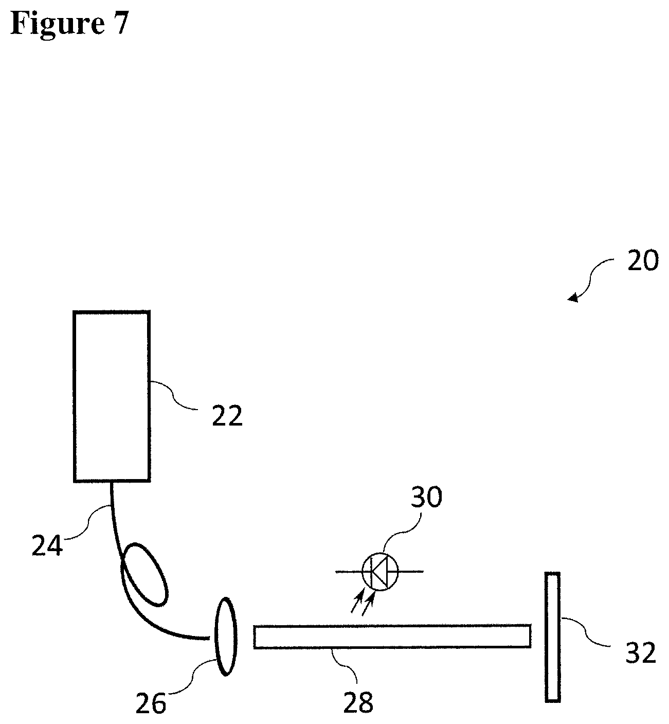
A method, an apparatus, and a kit including the apparatus and a fluorescence agent are provided for measuring a time-varying change in an amount of blood in a tissue volume, and include exciting a fluorescence agent in the blood, acquiring a time-varying light intensity signal during a pulsatile flow of the blood through the tissue volume, the pulsatile flow having a systolic and a diastolic phase resembling a conventional photoplethysmogram, and processing the acquired signal by applying a modified Beer-Lambert law to obtain a measurement of the time-varying change in the amount of blood in the tissue volume. The instantaneous molar concentration of the fluorescence agent is determined by utilizing a concentration-mediated change in a fluorescence emission spectrum of the fluorescence agent. There is further provided a fluorescence agent for use in the method.
| Inventors: | Flower; Robert W. (Hunt Valley, MD), Stead; Robert Anthony (Vancouver, CA), Bailey; Arthur E. (North Vancouver, CA) | ||||||||||
|---|---|---|---|---|---|---|---|---|---|---|---|
| Applicant: |
|
||||||||||
| Assignee: | Novadaq Technologies ULC
(Burnaby, CA) |
||||||||||
| Family ID: | 55652639 | ||||||||||
| Appl. No.: | 15/517,895 | ||||||||||
| Filed: | October 9, 2014 | ||||||||||
| PCT Filed: | October 09, 2014 | ||||||||||
| PCT No.: | PCT/IB2014/065189 | ||||||||||
| 371(c)(1),(2),(4) Date: | April 07, 2017 | ||||||||||
| PCT Pub. No.: | WO2016/055837 | ||||||||||
| PCT Pub. Date: | April 14, 2016 |
Prior Publication Data
| Document Identifier | Publication Date | |
|---|---|---|
| US 20170303800 A1 | Oct 26, 2017 | |
| Current U.S. Class: | 1/1 |
| Current CPC Class: | A61B 5/0071 (20130101); A61B 5/0261 (20130101); A61B 5/0295 (20130101); A61B 5/0275 (20130101); A61B 5/00 (20130101); A61B 5/6826 (20130101) |
| Current International Class: | A61B 5/026 (20060101); A61B 5/00 (20060101); A61B 5/0275 (20060101); A61B 5/0295 (20060101) |
References Cited [Referenced By]
U.S. Patent Documents
| 4109647 | August 1978 | Stern et al. |
| 4162405 | July 1979 | Chance et al. |
| 4200801 | April 1980 | Schuresko |
| 4263916 | April 1981 | Brooks et al. |
| 4394199 | July 1983 | Barnhard, IV et al. |
| 4473841 | September 1984 | Murakoshi et al. |
| 4532918 | August 1985 | Wheeler |
| 4541438 | September 1985 | Parker et al. |
| 4556057 | December 1985 | Hiruma et al. |
| 4619249 | October 1986 | Landry |
| 4718417 | January 1988 | Kittrell et al. |
| 4719508 | January 1988 | Sasaki et al. |
| 4768513 | September 1988 | Suzuki |
| 4773097 | September 1988 | Suzaki et al. |
| 4774568 | September 1988 | Matsuo |
| 4786813 | November 1988 | Svanberg et al. |
| 4805597 | February 1989 | Iwakoshi |
| 4815848 | March 1989 | Hadeishi |
| 4821117 | April 1989 | Sekiguchi |
| 4827908 | May 1989 | Matsuo |
| 4852579 | August 1989 | Gilstad et al. |
| 4858001 | August 1989 | Milbank et al. |
| 4860731 | August 1989 | Matsuura |
| 4867137 | September 1989 | Takahashi |
| 4868647 | September 1989 | Uehara et al. |
| 4900934 | February 1990 | Peeters et al. |
| 4930516 | June 1990 | Alfano et al. |
| 4938205 | June 1990 | Nudelman |
| 4957114 | September 1990 | Zeng et al. |
| 4993404 | February 1991 | Lane |
| 4995396 | February 1991 | Inaba et al. |
| 4995398 | February 1991 | Turnidge |
| 4998972 | March 1991 | Chin et al. |
| 5003977 | April 1991 | Suzuki et al. |
| 5042494 | August 1991 | Alfano |
| 5071417 | December 1991 | Sinofsky |
| 5078150 | January 1992 | Hara et al. |
| 5090400 | February 1992 | Saito |
| 5091652 | February 1992 | Mathies et al. |
| 5115137 | May 1992 | Andersson-Engels et al. |
| 5117466 | May 1992 | Buican et al. |
| 5125404 | June 1992 | Kittrell et al. |
| 5131398 | July 1992 | Alfano et al. |
| 5134662 | July 1992 | Bacus et al. |
| 5150292 | September 1992 | Hoffmann et al. |
| 5165079 | November 1992 | Schulz-Hennig |
| 5178616 | January 1993 | Uemiya et al. |
| 5196928 | March 1993 | Karasawa et al. |
| 5214503 | May 1993 | Chiu et al. |
| 5225883 | July 1993 | Carter et al. |
| 5255087 | October 1993 | Nakamura et al. |
| 5279298 | January 1994 | Flower |
| 5318023 | June 1994 | Vari et al. |
| 5318024 | June 1994 | Kittrell et al. |
| 5318869 | June 1994 | Hashimoto et al. |
| 5340592 | August 1994 | Goodrich, Jr. et al. |
| 5361769 | November 1994 | Nilsson |
| 5365057 | November 1994 | Morley et al. |
| 5371355 | December 1994 | Wodecki |
| 5375603 | December 1994 | Feiler |
| 5377676 | January 1995 | Vari et al. |
| 5377686 | January 1995 | O'Rourke et al. |
| 5394199 | February 1995 | Flower |
| 5419323 | May 1995 | Kittrell et al. |
| 5420628 | May 1995 | Poulsen et al. |
| 5421337 | June 1995 | Richards-Kortum et al. |
| 5421339 | June 1995 | Ramanujam et al. |
| 5424841 | June 1995 | Van Gelder et al. |
| 5430476 | July 1995 | Hafele et al. |
| 5437274 | August 1995 | Khoobehi et al. |
| 5438989 | August 1995 | Hochman et al. |
| 5453448 | September 1995 | Narciso, Jr. |
| 5465718 | November 1995 | Hochman et al. |
| 5491343 | February 1996 | Brooker |
| 5496369 | March 1996 | Howard, III |
| 5507287 | April 1996 | Palcic et al. |
| 5514127 | May 1996 | Shanks |
| 5519534 | May 1996 | Smith et al. |
| 5576013 | November 1996 | Williams et al. |
| 5590660 | January 1997 | MacAulay et al. |
| 5623930 | April 1997 | Wright et al. |
| 5627907 | May 1997 | Gur et al. |
| 5647368 | July 1997 | Zeng et al. |
| 5656498 | August 1997 | Iijima et al. |
| 5662644 | September 1997 | Swor |
| 5664574 | September 1997 | Chance |
| 5673701 | October 1997 | Chance |
| 5689241 | November 1997 | Clarke, Sr. et al. |
| 5699798 | December 1997 | Hochman et al. |
| 5707986 | January 1998 | Miller et al. |
| 5732707 | March 1998 | Widder et al. |
| 5741648 | April 1998 | Hemstreet, III et al. |
| 5743266 | April 1998 | Levene et al. |
| 5756541 | May 1998 | Strong et al. |
| 5785965 | July 1998 | Pratt et al. |
| 5803914 | September 1998 | Ryals et al. |
| 5827190 | October 1998 | Palcic et al. |
| 5845639 | December 1998 | Hochman et al. |
| 5851181 | December 1998 | Talmor |
| 5865754 | February 1999 | Sevick-Muraca et al. |
| 5910510 | June 1999 | Strong et al. |
| 5919616 | July 1999 | Aurelian et al. |
| 5927284 | July 1999 | Borst et al. |
| 5935942 | August 1999 | Zeimer |
| 5951980 | September 1999 | Cohen |
| 5956435 | September 1999 | Buzug et al. |
| 5965356 | October 1999 | Aurelian et al. |
| 5999841 | December 1999 | Aoyagi et al. |
| 6008889 | December 1999 | Zeng et al. |
| 6013265 | January 2000 | Aurelian |
| 6021344 | February 2000 | Lui et al. |
| 6032070 | February 2000 | Flock et al. |
| 6054131 | April 2000 | Aurelian |
| 6069689 | May 2000 | Zeng et al. |
| 6074627 | June 2000 | Dean et al. |
| 6081612 | June 2000 | Gutkowicz-Krusin et al. |
| 6093149 | July 2000 | Guracar et al. |
| 6122042 | September 2000 | Wunderman et al. |
| 6140314 | October 2000 | Zeimer |
| 6148227 | November 2000 | Wagnieres et al. |
| 6149671 | November 2000 | Nordquist et al. |
| 6162242 | December 2000 | Peyman |
| 6178340 | January 2001 | Svetliza |
| 6179421 | January 2001 | Pang |
| 6186628 | February 2001 | Van de Velde |
| 6196226 | March 2001 | Hochman et al. |
| 6207168 | March 2001 | Aurelian |
| 6211953 | April 2001 | Niino et al. |
| 6217848 | April 2001 | Achilefu et al. |
| 6223069 | April 2001 | Pfeiffer et al. |
| 6233480 | May 2001 | Hochman et al. |
| 6241672 | June 2001 | Hochman et al. |
| 6246901 | June 2001 | Benaron |
| 6248727 | June 2001 | Zeimer |
| 6263227 | July 2001 | Boggett et al. |
| 6272374 | August 2001 | Flock et al. |
| 6280386 | August 2001 | Alfano et al. |
| 6293911 | September 2001 | Imasizumi et al. |
| 6319273 | November 2001 | Cheen et al. |
| 6331703 | December 2001 | Yarnall et al. |
| 6335429 | January 2002 | Cai et al. |
| 6351663 | February 2002 | Flower et al. |
| 6351667 | February 2002 | Godie |
| 6353750 | March 2002 | Kimura et al. |
| 6399354 | June 2002 | Knipe et al. |
| 6440950 | August 2002 | Zeimer |
| 6443976 | September 2002 | Flower et al. |
| 6447443 | September 2002 | Keogh et al. |
| 6485413 | November 2002 | Boppart et al. |
| 6498945 | December 2002 | Alfheim et al. |
| 6544183 | April 2003 | Thorn Leeson et al. |
| 6566641 | May 2003 | Suda |
| 6577884 | June 2003 | Boas |
| 6603552 | August 2003 | Cline et al. |
| 6621917 | September 2003 | Vilser |
| 6631286 | October 2003 | Pfeiffer et al. |
| 6671540 | December 2003 | Hochman |
| 6757554 | June 2004 | Rubinstein et al. |
| 6804549 | October 2004 | Hayashi |
| 6821946 | November 2004 | Goldspink et al. |
| 6840933 | January 2005 | Pang et al. |
| 6853857 | February 2005 | Pfeiffer et al. |
| 6882366 | April 2005 | Kijima et al. |
| 6899675 | May 2005 | Cline et al. |
| 6915154 | July 2005 | Docherty et al. |
| 6936043 | August 2005 | Peyman |
| 6944493 | September 2005 | Alam et al. |
| 7113817 | September 2006 | Winchester, Jr. et al. |
| 7236815 | June 2007 | Richards-Kortum et al. |
| 7317554 | January 2008 | Ueda et al. |
| 7364574 | April 2008 | Flower |
| 7381400 | June 2008 | Woltering |
| 7400753 | July 2008 | Seino et al. |
| 7400755 | July 2008 | West et al. |
| 7474906 | January 2009 | Rubinstein |
| 7482318 | January 2009 | Aurelian et al. |
| 7581191 | August 2009 | Rice et al. |
| 7729750 | June 2010 | Tromberg et al. |
| 7774048 | August 2010 | Nakaoka et al. |
| 7881777 | February 2011 | Docherty et al. |
| 7885438 | February 2011 | Uppaluri et al. |
| 8036437 | October 2011 | Arditi et al. |
| 8073224 | December 2011 | Strobel et al. |
| 8144958 | March 2012 | Nahm et al. |
| 8185176 | May 2012 | Mangat et al. |
| 8194981 | June 2012 | Suzuki |
| 8285353 | October 2012 | Choi |
| 8361775 | January 2013 | Flower |
| 8406860 | March 2013 | Dvorsky et al. |
| 8480579 | July 2013 | Serov et al. |
| 8521260 | August 2013 | Grinvald et al. |
| 8538107 | September 2013 | Rottger |
| 8647605 | February 2014 | Mangat et al. |
| 8718747 | May 2014 | Bjornerud |
| 8725225 | May 2014 | Golijanin et al. |
| 8892190 | November 2014 | Docherty et al. |
| 8929974 | January 2015 | Hauger et al. |
| 8965488 | February 2015 | Dvorsky et al. |
| 9089601 | July 2015 | Golijanin et al. |
| 9129366 | September 2015 | Nahm et al. |
| 9241636 | January 2016 | Koizumi et al. |
| RE45916 | March 2016 | Golijanin et al. |
| 9351644 | May 2016 | Nahm et al. |
| 9357931 | June 2016 | Nahm et al. |
| 9421280 | August 2016 | Mangat et al. |
| 9451903 | September 2016 | Feinberg |
| 9554738 | January 2017 | Gulati |
| 9610021 | April 2017 | Dvorsky et al. |
| 9642532 | May 2017 | Fengler et al. |
| 9816930 | November 2017 | Moriyama et al. |
| 9936887 | April 2018 | Dvorsky et al. |
| 10041042 | August 2018 | Flower |
| 10219742 | March 2019 | Dvorsky et al. |
| 10231624 | March 2019 | Mangat et al. |
| 10265419 | April 2019 | Golijanin |
| 10278585 | May 2019 | Ferguson, Jr. et al. |
| 10285603 | May 2019 | Flower |
| 2002/0007123 | January 2002 | Balas |
| 2002/0025541 | February 2002 | Nelson et al. |
| 2002/0038120 | March 2002 | Duhaylongsod et al. |
| 2002/0049389 | April 2002 | Abreu |
| 2002/0099279 | July 2002 | Pfeiffer et al. |
| 2002/0099295 | July 2002 | Gil et al. |
| 2002/0146369 | October 2002 | Goldenberg |
| 2002/0181752 | December 2002 | Wallo et al. |
| 2002/0183621 | December 2002 | Pfeiffer et al. |
| 2003/0032885 | February 2003 | Rubinstein et al. |
| 2003/0050543 | March 2003 | Hartmann |
| 2003/0060718 | March 2003 | Alam et al. |
| 2003/0060722 | March 2003 | Pfeiffer et al. |
| 2003/0064025 | April 2003 | Yang et al. |
| 2003/0093064 | May 2003 | Peyman |
| 2003/0093065 | May 2003 | Peyman |
| 2003/0127609 | July 2003 | El-Hage |
| 2003/0139667 | July 2003 | Hewko |
| 2003/0156252 | August 2003 | Morris et al. |
| 2003/0187349 | October 2003 | Kaneko et al. |
| 2003/0232016 | December 2003 | Heinrich |
| 2003/0236458 | December 2003 | Hochman |
| 2004/0002660 | January 2004 | Mielekamp |
| 2004/0066961 | April 2004 | Spreeuwers et al. |
| 2004/0077952 | April 2004 | Rafter et al. |
| 2004/0109231 | June 2004 | Haisch et al. |
| 2004/0156782 | August 2004 | Alam et al. |
| 2004/0162489 | August 2004 | Richards-Kortum et al. |
| 2004/0171827 | September 2004 | Peng et al. |
| 2004/0174495 | September 2004 | Levine |
| 2005/0019744 | January 2005 | Bertuglia |
| 2005/0020891 | January 2005 | Rubinstein et al. |
| 2005/0033145 | February 2005 | Graham et al. |
| 2005/0065432 | March 2005 | Kimura |
| 2005/0069525 | March 2005 | Mikael |
| 2005/0089866 | April 2005 | Hinuma et al. |
| 2005/0107380 | May 2005 | Nimmo et al. |
| 2005/0142556 | June 2005 | Hoon et al. |
| 2005/0182321 | August 2005 | Frangioni |
| 2005/0182327 | August 2005 | Petty et al. |
| 2005/0182431 | August 2005 | Hausen et al. |
| 2005/0182434 | August 2005 | Docherty et al. |
| 2005/0187477 | August 2005 | Serov et al. |
| 2005/0197583 | September 2005 | Chance |
| 2005/0254008 | November 2005 | Ferguson et al. |
| 2006/0011853 | January 2006 | Spartiotis |
| 2006/0013768 | January 2006 | Woltering |
| 2006/0079750 | April 2006 | Fauci et al. |
| 2006/0108509 | May 2006 | Frangioni et al. |
| 2006/0118742 | June 2006 | Levenson et al. |
| 2006/0147897 | July 2006 | Grinvald et al. |
| 2006/0239921 | October 2006 | Mangat et al. |
| 2006/0241499 | October 2006 | Irion et al. |
| 2007/0121099 | May 2007 | Matsumoto et al. |
| 2007/0122344 | May 2007 | Golijanin |
| 2007/0122345 | May 2007 | Golijanin |
| 2007/0203413 | August 2007 | Frangioni |
| 2007/0254276 | November 2007 | Deutsch et al. |
| 2008/0007733 | January 2008 | Marks et al. |
| 2008/0015446 | January 2008 | Mahmood et al. |
| 2008/0025918 | January 2008 | Frangioni et al. |
| 2008/0044073 | February 2008 | Bernhardt et al. |
| 2008/0081990 | April 2008 | Berenfeld et al. |
| 2008/0161744 | July 2008 | Golijanin et al. |
| 2008/0188728 | August 2008 | Neumann |
| 2008/0221421 | September 2008 | Choi |
| 2008/0221648 | September 2008 | Flower |
| 2008/0239070 | October 2008 | Westwick et al. |
| 2008/0319309 | December 2008 | Bredno et al. |
| 2009/0005693 | January 2009 | Brauner et al. |
| 2009/0042179 | February 2009 | Peltie et al. |
| 2009/0048516 | February 2009 | Yoshikawa et al. |
| 2009/0054788 | February 2009 | Hauger et al. |
| 2009/0112097 | April 2009 | Kato |
| 2009/0118623 | May 2009 | Serov et al. |
| 2009/0137902 | May 2009 | Frangioni et al. |
| 2009/0252682 | October 2009 | Hillman |
| 2009/0297004 | December 2009 | Baumgart |
| 2010/0016669 | January 2010 | Takaoka et al. |
| 2010/0022898 | January 2010 | Rubinstein et al. |
| 2010/0036217 | February 2010 | Choi et al. |
| 2010/0041999 | February 2010 | Schuhrke et al. |
| 2010/0061604 | March 2010 | Nahm et al. |
| 2010/0069759 | March 2010 | Schuhrke et al. |
| 2010/0080757 | April 2010 | Haaga |
| 2010/0099961 | April 2010 | Hubner et al. |
| 2010/0222673 | September 2010 | Mangat et al. |
| 2010/0286529 | November 2010 | Carroll et al. |
| 2011/0001061 | January 2011 | Ishihara |
| 2011/0013002 | January 2011 | Thompson et al. |
| 2011/0063427 | March 2011 | Fengler et al. |
| 2011/0071403 | March 2011 | Sevick-Muraca et al. |
| 2011/0098685 | April 2011 | Flower |
| 2011/0306877 | December 2011 | Dvorsky et al. |
| 2012/0026325 | February 2012 | Bunker et al. |
| 2012/0078093 | March 2012 | Flower |
| 2012/0252699 | April 2012 | Jaffrey et al. |
| 2012/0165627 | June 2012 | Yamamoto |
| 2012/0165662 | June 2012 | Nahm et al. |
| 2012/0271176 | October 2012 | Moghaddam et al. |
| 2012/0323118 | December 2012 | Menon Gopalakrishna |
| 2013/0035569 | February 2013 | Heanue |
| 2013/0203082 | August 2013 | Gonda et al. |
| 2013/0203083 | August 2013 | Connors |
| 2013/0230866 | September 2013 | Miyashita et al. |
| 2013/0245456 | September 2013 | Ferguson, Jr. et al. |
| 2013/0286176 | October 2013 | Westwick et al. |
| 2013/0296715 | November 2013 | Lasser et al. |
| 2013/0342674 | December 2013 | Dixon |
| 2013/0345560 | December 2013 | Ferguson, Jr. et al. |
| 2014/0099007 | April 2014 | Sarkar et al. |
| 2014/0254909 | September 2014 | Carmi |
| 2014/0308656 | October 2014 | Flower |
| 2014/0316262 | October 2014 | Havens |
| 2014/0371583 | December 2014 | Flower |
| 2015/0112192 | April 2015 | Docherty |
| 2015/0112193 | April 2015 | Docherty et al. |
| 2015/0164396 | June 2015 | Acharya |
| 2015/0182137 | July 2015 | Flower |
| 2015/0196208 | July 2015 | Dvorsky et al. |
| 2015/0230710 | August 2015 | Nahm et al. |
| 2015/0230715 | August 2015 | Nahm et al. |
| 2015/0248758 | September 2015 | Pautot |
| 2015/0381909 | December 2015 | Butte et al. |
| 2016/0038027 | February 2016 | Brzozowski et al. |
| 2016/0041098 | February 2016 | Hirawake et al. |
| 2016/0097716 | April 2016 | Gulati |
| 2016/0199515 | July 2016 | Flower |
| 2016/0253800 | September 2016 | Gurevich |
| 2016/0371834 | December 2016 | Watanabe et al. |
| 2017/0039710 | February 2017 | Minai et al. |
| 2017/0084024 | March 2017 | Gurevich |
| 2017/0245766 | August 2017 | Flower |
| 2017/0303800 | October 2017 | Flower |
| 2018/0020933 | January 2018 | Dvorsky et al. |
| 2018/0104362 | April 2018 | Golijanin et al. |
| 2018/0120230 | May 2018 | Moriyama et al. |
| 2018/0220907 | August 2018 | Dvorsky et al. |
| 2018/0234603 | August 2018 | Moore et al. |
| 2018/0369426 | December 2018 | Flower et al. |
| 409451 | Aug 2002 | AT | |||
| 2212257 | Aug 1996 | CA | |||
| 2413033 | Mar 2000 | CA | |||
| 2711560 | Jul 2009 | CA | |||
| 2913692 | Jan 2015 | CA | |||
| 1049781 | Mar 1991 | CN | |||
| 1200174 | Nov 1998 | CN | |||
| 1399528 | Feb 2003 | CN | |||
| 101264014 | Sep 2008 | CN | |||
| 101451953 | Jun 2009 | CN | |||
| 102288589 | Dec 2011 | CN | |||
| 102405212 | Apr 2012 | CN | |||
| 102436648 | May 2012 | CN | |||
| 103608662 | Feb 2014 | CN | |||
| 3906860 | Sep 1989 | DE | |||
| 19608027 | Sep 1996 | DE | |||
| 10028233 | Jan 2002 | DE | |||
| 10120980 | Nov 2002 | DE | |||
| 69727220 | Dec 2004 | DE | |||
| 102005044531 | Mar 2007 | DE | |||
| 0091805 | Oct 1983 | EP | |||
| 0215772 | Mar 1987 | EP | |||
| 0512965 | Nov 1992 | EP | |||
| 0792618 | Sep 1997 | EP | |||
| 0807402 | Nov 1997 | EP | |||
| 0826335 | Mar 1998 | EP | |||
| 1677097 | Jul 2006 | EP | |||
| 1761171 | Mar 2007 | EP | |||
| 1874181 | Jan 2008 | EP | |||
| 2944104 | Oct 2010 | FR | |||
| 2203831 | Oct 1988 | GB | |||
| S52-34584 | Mar 1977 | JP | |||
| S58-222331 | Dec 1983 | JP | |||
| S59-069721 | Apr 1984 | JP | |||
| S59-070903 | Apr 1984 | JP | |||
| H01-236879 | Sep 1989 | JP | |||
| 02-200237 | Aug 1990 | JP | |||
| H03-115958 | May 1991 | JP | |||
| 04-297236 | Oct 1992 | JP | |||
| H05-64232 | Oct 1993 | JP | |||
| H06-007353 | Jan 1994 | JP | |||
| 06-335451 | Dec 1994 | JP | |||
| H07-043303 | Feb 1995 | JP | |||
| 07-065154 | Mar 1995 | JP | |||
| 07-079955 | Mar 1995 | JP | |||
| H07-155285 | Jun 1995 | JP | |||
| H07-155286 | Jun 1995 | JP | |||
| H07-155290 | Jun 1995 | JP | |||
| H07-155291 | Jun 1995 | JP | |||
| H07-155292 | Jun 1995 | JP | |||
| 07-222712 | Aug 1995 | JP | |||
| H07-204156 | Aug 1995 | JP | |||
| H07-222723 | Aug 1995 | JP | |||
| H07-250804 | Oct 1995 | JP | |||
| H07-250812 | Oct 1995 | JP | |||
| 08-024227 | Jan 1996 | JP | |||
| H08-224208 | Sep 1996 | JP | |||
| H08-224209 | Sep 1996 | JP | |||
| H08-224240 | Sep 1996 | JP | |||
| H09-120033 | May 1997 | JP | |||
| H09-305845 | Nov 1997 | JP | |||
| H09-308609 | Dec 1997 | JP | |||
| H09-309845 | Dec 1997 | JP | |||
| H10-500479 | Jan 1998 | JP | |||
| H10-503480 | Mar 1998 | JP | |||
| H10-085222 | Apr 1998 | JP | |||
| H10-104070 | Apr 1998 | JP | |||
| H10-151104 | Jun 1998 | JP | |||
| H10-506440 | Jun 1998 | JP | |||
| H10-506550 | Jun 1998 | JP | |||
| H10-201700 | Aug 1998 | JP | |||
| H10-201707 | Aug 1998 | JP | |||
| H11-137517 | May 1999 | JP | |||
| H11-155812 | Jun 1999 | JP | |||
| H11-509748 | Aug 1999 | JP | |||
| 2001-198079 | Jul 2001 | JP | |||
| 2002-219129 | Aug 2002 | JP | |||
| 2003-510121 | Mar 2003 | JP | |||
| 2003-144401 | May 2003 | JP | |||
| 2003-187226 | Jul 2003 | JP | |||
| 2003-329589 | Nov 2003 | JP | |||
| 2004-528917 | Sep 2004 | JP | |||
| 2004-325200 | Nov 2004 | JP | |||
| 2006-503620 | Feb 2006 | JP | |||
| 2006-192280 | Jul 2006 | JP | |||
| 2007-021006 | Feb 2007 | JP | |||
| 3896176 | Mar 2007 | JP | |||
| 2008-023113 | Feb 2008 | JP | |||
| 2008-525126 | Jul 2008 | JP | |||
| 2008-220926 | Sep 2008 | JP | |||
| 2008-535600 | Sep 2008 | JP | |||
| 2008-231113 | Oct 2008 | JP | |||
| 2009-095683 | May 2009 | JP | |||
| 2009-519082 | May 2009 | JP | |||
| 2009-291554 | Dec 2009 | JP | |||
| 2010-505582 | Feb 2010 | JP | |||
| 2010-521267 | Jun 2010 | JP | |||
| 2011-509768 | Mar 2011 | JP | |||
| 2011-519589 | Jul 2011 | JP | |||
| 2011-528918 | Dec 2011 | JP | |||
| 5918532 | May 2016 | JP | |||
| 2016-521612 | Jul 2016 | JP | |||
| 90-0005434 | Jul 1990 | KR | |||
| 2002-0064287 | Aug 2002 | KR | |||
| 2288633 | Dec 2006 | RU | |||
| WO-1986/02730 | May 1986 | WO | |||
| WO-1990/10219 | Sep 1990 | WO | |||
| WO-1990/12536 | Nov 1990 | WO | |||
| WO-1993/25141 | Dec 1993 | WO | |||
| WO-1994/12092 | Jun 1994 | WO | |||
| WO-1995/00171 | Jan 1995 | WO | |||
| WO-1995/26673 | Oct 1995 | WO | |||
| WO-1996/09435 | Mar 1996 | WO | |||
| WO-1996/09792 | Apr 1996 | WO | |||
| WO-1996/18415 | Jun 1996 | WO | |||
| WO-1996/23524 | Aug 1996 | WO | |||
| WO-1996/39925 | Dec 1996 | WO | |||
| WO-1997/08538 | Mar 1997 | WO | |||
| WO-1998/24360 | Jun 1998 | WO | |||
| WO-1998/30144 | Jul 1998 | WO | |||
| WO-1998/46122 | Oct 1998 | WO | |||
| WO-1999/00053 | Jan 1999 | WO | |||
| WO-1999/47940 | Sep 1999 | WO | |||
| WO-1999/53832 | Oct 1999 | WO | |||
| WO-2000/42910 | Jul 2000 | WO | |||
| WO-2000/47107 | Aug 2000 | WO | |||
| WO-2001/08552 | Feb 2001 | WO | |||
| WO-2001/17561 | Mar 2001 | WO | |||
| WO-2001/22870 | Apr 2001 | WO | |||
| WO-2001/39764 | Jun 2001 | WO | |||
| WO-2001/69244 | Sep 2001 | WO | |||
| WO-2001/80734 | Nov 2001 | WO | |||
| WO-2001/82786 | Nov 2001 | WO | |||
| WO-2002/061390 | Aug 2002 | WO | |||
| WO-2003/006658 | Jan 2003 | WO | |||
| WO-2004/006963 | Jan 2004 | WO | |||
| WO-2004/052195 | Jun 2004 | WO | |||
| WO-2005/026319 | Mar 2005 | WO | |||
| WO-2005/034747 | Apr 2005 | WO | |||
| WO-2005/036143 | Apr 2005 | WO | |||
| WO-2005/079238 | Sep 2005 | WO | |||
| WO-2006/111836 | Oct 2006 | WO | |||
| WO-2006/111909 | Oct 2006 | WO | |||
| WO-2006/116634 | Nov 2006 | WO | |||
| WO-2006/119349 | Nov 2006 | WO | |||
| WO-2006/121631 | Nov 2006 | WO | |||
| WO-2006/121631 | Nov 2006 | WO | |||
| WO-2006/123742 | Nov 2006 | WO | |||
| WO-2007/028032 | Mar 2007 | WO | |||
| WO-2008/039968 | Apr 2008 | WO | |||
| WO-2008/044822 | Apr 2008 | WO | |||
| WO-2008/070269 | Jun 2008 | WO | |||
| WO-2008/070269 | Jun 2008 | WO | |||
| WO-2008/087869 | Jul 2008 | WO | |||
| WO-2009/046985 | Apr 2009 | WO | |||
| WO-2009/046985 | Apr 2009 | WO | |||
| WO-2009/048660 | Apr 2009 | WO | |||
| WO-2009/092162 | Jul 2009 | WO | |||
| WO-2009/127972 | Oct 2009 | WO | |||
| WO-2012/038824 | Mar 2012 | WO | |||
| WO-2012/096878 | Jul 2012 | WO | |||
| WO-2013/190391 | Dec 2013 | WO | |||
| WO-2015/001427 | Jan 2015 | WO | |||
| WO-2013/002350 | Feb 2015 | WO | |||
Other References
|
Akintunde, A. et al. (Oct.-Nov. 1992). "Quadruple Labeling of Brain-Stem Neurons: A Multiple Retrograde Fluorescent Tracer Study of Axonal Collateralization," Journal of Neuroscience Methods 45(1-2):15-22. cited by applicant . Alander, J.T. et al. (Jan. 1, 2012). "A Review of Indocyanine Green Fluorescent Imaging in Surgery," International Journal of Biomedical Imaging 2012:1-26, article ID 940585. Jarmo T. Alander et al.; A Review of Indocyanine Green Fluorescent Imaging in Surgery; International Journal of Biomedical Imaging, vol. 2, No. 7, Jan. 1, 2012, pp. 2068-2026. cited by applicant . Alfano et al. (Oct. 1987). "Fluorescence Spectra from Cancerous and Normal Human Breast and Lung Tissues," IEEE Journal of Quantum Electronics QE-23(10):1806-1811. cited by applicant . Alm, A. et al. (Jan. 1, 1973). "Ocular and Optic Nerve Blood Flow at Normal and Increased Intraocular Pressures in Monkeys (Macaca irus): A Study with Radioactively Labelled Microspheres Including Flow Determinations in Brain and Some Other Tissues," Experimental Eye Research 15(1):15-29. cited by applicant . Alonso-Burgos, A. et al.(2006). "Preoperative planning of deep inferior epigastric artery perforator flap reconstruction with multi-slice-CT angiography: imaging findings and initial experience," Journal of Plastic, Reconstructive & Aesthetic Surgery 59:585-593. cited by applicant . Alvarez, F. J. et al. (Apr. 1996). "Behaviour of Isolated Rat and Human Red Blood Cells Upon Hypotonic-Dialysis Encapsulation of Carbonic Anhydrase and Dextran," Biotechnology and Applied Biochemistry 23(2):173-179. cited by applicant . Ancalmo, N. et al. (1997). "Minimally invasive coronary artery bypass surgery: really minimal?" Ann. Thorac. Surg. 64:928-929. cited by applicant . Andersson-Engels, S. et al. (1991). "Fluorescence Characteristics of Atherosclorotic Plaque and Malignant Tumors," in Optical Methods for Tumor Treatment and Early Diagnosis: Mechanisms and Techniques, T. J. Dougherty (Ed.), The Society of Photo-optical Instrumentation Engineers (SPIE) 1426:31-43, fourteen pages. cited by applicant . Andersson-Engels, S. et al. (Mar. 1989). "Tissue Diagnostics Using Laser-Induced Fluorescence," Berichte der Bunsengesellschaft fur physikalische Chemie 93(3):335-342. cited by applicant . Angelov, D.N. et al. (Apr. 1999). "Contralateral Trigeminal Nerve Lesion Reduces Polyneuronal Muscle Innervation after Facial Nerve Repair in Rats," European Journal of Neuroscience 11(4):1369-1378. cited by applicant . Annese, V. et al. (2005). "Erthrocytes-Mediated Delivery of Dexamethasone in Steroid-Dependent IBD Patients--a Pilot Uncontrolled Study," American Journal of Gastroenterology 100:1370-1375. cited by applicant . Argus-50/CA, Inter cellular CA2+ (calcium ion) Image Analysis system (Feb. 1992). "Observation and 2-dimensional analysis of Ca2+ concentration distribution. Fura-2 and Indo-1 compatible. Ca2+ concentrations are calculated from the fluorescence ratio," pp. 1-10. cited by applicant . Australian Notice of Allowance dated Sep. 17, 2018 for Australian Patent Application No. 2015327665, filed on Mar. 23, 2017, three pages. cited by applicant . Australian Notice of Allowance dated Jul. 3, 2019 for Australian Patent Application No. 2014408488, filed on Mar. 31, 2017, three pages. cited by applicant . Australian Office Action dated Jun. 26, 2018 for Australian Patent Application No. 2014408488, filed on Mar. 31, 2017, nine pages. cited by applicant . Author Unknown. (Jun. 4, 2008)."Invitrogen," Material Safety Data Sheet, p. 1-4. cited by applicant . Awano, T. et al. (Jun. 2010). "Intraoperative EC-IC Bypass Blood Flow Assessment with Indocyanine Green Angiography in Moyamoya and Non-moyamoya Ischemic Stroke," World Neurosurg. 73(6):668-674. cited by applicant . Azuma, R. et al. (2008, presented in part Jun. 2007). "Detection of Skin Perforators by Indocyanine Green Fluorescence Nearly Infrared Angiography," PRS Journal 122(4):1062-1067. cited by applicant . Balacumarswami, L. et al. (Aug. 2004). "Does Off-Pump Total Arterial Grafting Increase the Incidence of Intraoperative Graft Failure?," The Journal of Thoracic and Cardiovascular Surgery 128(2):238-244. cited by applicant . Barton, J.K. et al. (1999) "Simultaneous irradiation and imaging of blood vessels during pulsed laser delivery," Lasers in Surgery and Medicine 24(3):236-243. cited by applicant . Bassingthwaighte, J.B. et al. (Apr. 1974). "Organ Blood Flow, Wash-in, Washout, and Clearance of Nutrients and Metabolites," Mayo Clin. Proc. 49(4):248-255. cited by applicant . Batliwala, H. et al. (Apr. 15, 1995). "Methane-Induced Haemolysis of Human Erythrocytes," Biochemical J. 307(2):433-438. cited by applicant . Baumgartner, R. et al. (1987). "Section V--In vivo Localization and Photodynamic Therapy: A Fluorescence Imaging Device for Endoscopic Detection of Early Stage Cancer--Instrumental and Experimental Studies," Photochemistry and Photobiology 46(5):759-763. cited by applicant . Baumgartner, R. et al. (Jan. 1, 1990). "A Fluorescence Imaging Device for Endoscopic Detection of Early Stage Cancer Instrumental and Experimental Studies," Section Five in SPIE Press, ed. Abraham Katzir, pp. 513-517, eight pages. cited by applicant . Bek, T. (1999). "Diabetic Maculopathy Caused by Disturbances in Retinal Vasomotion: A New Hypothesis," Acta Ophthalmologica Scandinavica 77:376-380. cited by applicant . Benson, R.C. et al. (1978). "Fluorescence Properties of Indocyanine Green as Related to Angiography," Phys. Med. Biol. 23(1):159-163. cited by applicant . Black's Medical Dictionary, "Perfusion," 42nd Edition (2009), two pages. cited by applicant . Boer, F.et al. (1994). "Effect of Ventilation on First-Pass Pulmonary Retention of Alfentaril and Sufentanil in Patients Undergoing Coronary Artery Surgery," British Journal Anesthesia 73:458-463. cited by applicant . Boldt, .J. et al. (Feb. 1990). "Lung management during cardiopulmonary bypass: influence on extravascular lung water," Journal of Cardiothoracic Anesthesia 4(1):73-79. cited by applicant . Boldt, J. et al. (1991). "Does the technique of cardiopulmonary bypass affect lung water content?" European Journal of Cardio-Thoracic Surgery 5:22-26. cited by applicant . Brazilian Office Action dated May 14, 2019 for Brazilian Application No. PI 0907272-1, filed on Oct. 14, 2010, five pages. cited by applicant . Butter, A. et al. (May 2005). "Melanoma in Children and the Use of Sentinel Lymph Node Biopsy," Journal of Pediatric Surgery 40(5):797-800. cited by applicant . C2741, Compact High Performance video camera for industrial applications with Built-in contrast enhancement circuit, Jun. 1998, six pages. cited by applicant . Canada Health. (1997). "Coronary Bypass Surgery and Angioplasty, 1982-1995, Heart Disease and Stroke in Canada," Canada Health, located at <http:/www.hc-sc.gc.ca/hpb>, eighty two pages. cited by applicant . Canadian Notice of Allowance dated Jan. 4, 2018 for Canadian Application No. 2,750,760, filed on Jul. 25, 2008, one page. cited by applicant . Canadian Notice of Allowance dated Sep. 27, 2017 for Canadian Application No. 2,811,847, filed on Mar. 20, 2013, one page. cited by applicant . Canadian Office Action dated Dec. 12, 2018 for CA Application No. 2,963,987 filed on Mar. 27, 2017, four pages. cited by applicant . Canadian Office Action dated Dec. 28, 2018 for CA Application No. 2,963,450 filed on Apr. 3, 2017, four pages. cited by applicant . Canadian Office Action dated Feb. 13, 2018 for CA Application No. 2,963,450 filed on Apr. 3, 2017, three pages. cited by applicant . Canadian Office Action dated Feb. 27, 2017 for Canadian Application No. 2,750,760, filed on Jul. 25, 2011, three pages. cited by applicant . Canadian Office Action dated Feb. 28, 2018 for CA Application No. 2,963,987 filed on Mar. 27, 2017, five pages. cited by applicant . Canadian Office Action dated Jan. 19, 2017 for Canadian Application No. 2,914,778 filed on Dec. 8, 2015, four pages. cited by applicant . Canadian Office Action dated Mar. 16, 2016 for CA Application No. 2,750,760 filed on Jan. 23, 2009, five pages. cited by applicant . Canadian Office Action dated May 28, 2019 for Canadian Application No. 3,011,310, filed on Jul. 11, 2018, four pages. cited by applicant . Canadian Office Action dated Nov. 28, 2017 for Canadian Application No. 2,914,778 filed on Dec. 8, 2015, six pages. cited by applicant . Canadian Office Action dated Nov. 28, 2018 for CA Application No. 2,750,760 filed on Jan. 23, 2009, three pages. cited by applicant . Canadian Office Action dated Oct. 25, 2016 for Canadian Patent Application No. 2,811,847, filed on Sep. 20, 2011, three pages. cited by applicant . Canadian Office Action dated Sep. 30, 2015 for CA Application No. 2,811,847, filed on Sep. 20, 2011, four pages. cited by applicant . Chinese Office Action dated Jul. 3, 2012, issued in counterpart Chinese Application No. 200980123414.0, eight pages. cited by applicant . Chinese Office Action dated Apr. 6, 2017 for Chinese Application No. 201510214021.8, filed on May 14, 2009, fifteen pages. cited by applicant . Chinese Office Action dated Apr. 17, 2019 for Chinese Application No. 201510214021.8, filed on May 14, 2009, sixteen pages. cited by applicant . Chinese Office Action dated Apr. 26, 2019 for CN Application No. 201580064648.8 filed on May 26, 2017, twenty six pages. cited by applicant . Chinese Office Action dated Aug. 8, 2016 for Chinese Application No. 201180057244.8 filed on Sep. 20, 2011, eighteen pages. cited by applicant . Chinese Office Action dated Dec. 19, 2017 for Chinese Application No. 201180057244.8 filed on Sep. 20, 2011, eleven pages. cited by applicant . Chinese Office Action dated Feb. 8, 2018 for Chinese Application No. 201510214021.8, filed on May 14, 2009, seventeen pages. cited by applicant . Chinese Office Action dated Mar. 13, 2017 for Chinese Application No. 201180057244.8 filed on Sep. 20, 2011, twenty pages. cited by applicant . Chinese Office Action dated May 23, 2013, issued in counterpart Chinese Application No. 200980123414.0, nineteen pages. cited by applicant . Chinese Office Action dated Nov. 12, 2015 for Chinese Patent Application No. 201180057244.8, filed on Sep. 20, 2010, five pages, (English Translation). cited by applicant . Chinese Office Action dated Sep. 27, 2018 for Chinese Application No. 201510214021.8, filed on May 14, 2009, seventeen pages. cited by applicant . Coffey, J.H. et al. (1984). "Evaluation of Visual Acuity During Laser Photoradiation Therapy of Cancer," Lasers in Surgery and Medicine 4(1):65-71. cited by applicant . Conley, M.P. et al. (Oct. 2004). "Anterograde Transport of Peptide-Conjugated Fluorescent Beads in the Squid Giant Axom Identifies a Zip-Code for Synapse," Biological Bulletin 207(2):164, one page. cited by applicant . Costa, R.A. et al. (Oct. 2001). "Photodynamic Therapy with Indocyanine Green for Occult Subfoveal Choroidal Neovascularization Caused by Age-Related Macular Degeneration," Curr. Eye Res. 23(4):274-275. cited by applicant . Cothren, R.M. et al. (Mar. 1990). "Gastrointestinal Tissue Diagnosis by Laser-Induced Fluorescence Spectroscopy at Endoscopy," Gastrointestinal Endoscopy 36(2):105-111. cited by applicant . Dail, W.G. et al. (Oct. 1999). "Multiple Vasodilator Pathways from the Pelvic Plexus to the Penis of the Rat," International Journal of Impotence Research 11(5):277-285. cited by applicant . Dan, A.G. et al. (Nov. 2004). "1% Lymphazurin vs 10% Fluorescein for Sentinel Node Mapping in Colorectal Tumors," Arch Surg. 139(11):1180-1184. cited by applicant . Daniels, G. et al. (Apr. 2007). "Towards Universal Red Blood Cell," Nature Biotechnology 25(4):427-428. cited by applicant . De Flora, A. (Sep. 1986). "Encapsulation of Adriamycin in human erythrocytes," Proc. Natl. Acad. Sci., USA 83(18):7029-7033. cited by applicant . Declaration of Brian Wilson dated Aug. 22, 2017 for Inter Partes Review No. IPR2017-01426, twelve pages. cited by applicant . Definition of "Expose," Excerpt of Merriam Webster's Medical Desk Dictionary (1993), four pages. cited by applicant . Definition of "Graft," Excerpt of Stedman's Medical Dictionary for the Health Professions and Nursing; 6th Ed. (2008), three pages. cited by applicant . De-Grand, A.M. et al. (Dec. 2003). "An Operational Near Infrared Fluorescence Imaging System Prototype for Large Animal Surgery," Technology in Cancer Research & Treatment 2(6):1-10. cited by applicant . Deloach, J.R. (ed.) et al. (1985). Red Blood Cells as Carriers for Drugs. A Method for Disseminating Chemotherapeutics, Hormones, Enzymes and Other Therapeutic Agents via the Circulatory System, Karger, Basel, CH, pp. v-vii, (Table of Contents), seven pages. cited by applicant . Deloach, J.R. (Jun. 1983). "Encapsulation of Exogenous Agents in Erythrocytes and the Circulating Survival of Carrier Erythrocytes," Journal of Applied Biochemistry 5(3):149-157. cited by applicant . Demos (May/Jun. 2004). "Near-Infrared Autofluorescence Imaging for Detection of Cancer," Journal of Biomedical Optics 9(3):587-592. cited by applicant . Desai, N.D. et al. (Oct. 18, 2005, e-published on Sep. 28, 2005) "Improving the Quality of Coronary Bypass Surgery with Intraoperative Angiography," Journal of the American College of Cardiology 46(8):1521-1525. cited by applicant . Detter, C. et al. (Aug. 28, 2007). "Fluorescent Cardiac Imaging: A Novel Intraoperative Method for Quantitative Assessment of Myocardial Perfusion During Graded Coronary Artery Stenosis," Circulation 116(9):1007-1014. cited by applicant . Detter, C. et al. (Jun. 2002). "Near-Infrared Fluorescence Coronary Angiography: A New Noninvasive Technology for Intraoperative Graft Patency Control." The Heart Surgery Forum #2001-6973 5(4):364-369. cited by applicant . Dietz, F.B. et al. (Feb. 2003). "Indocyanine Green: Evidence of Neurotoxicity in Spinal Root Axons," Anesthesiology 98(2):516-520. cited by applicant . Digital CCD Microscopy (date unknown). Chapter 14, pp. 259-282. cited by applicant . Dougherty, T.J. et al. (1990). "Cutaneous Phototoxic Occurrences in Patients Receiving Photofrin," Lasers in Surgery and Medicine 10(5):485-488. cited by applicant . Draijer, M.J. et al. (Jun. 17-19, 2007). "Laser Doppler Perfusion Imaging with a High-Speed CMOS-Camera," in Novel Optical Instrumentation for Biomedical Applications III, C. Depeursinge, ed., Proceedings of SPIE-OSA Biomedical Optics (Optical Society of America, 2007), SPIE-OSA, 6631:0N1-0N7, eight pages. cited by applicant . Dunne, A. et al. (Nov. 2001)."Value of Sentinel lymphonodectomy in Head and Neck Cancer Patients without Evidence of Lymphogenic Metastatic Disease," Auris Nasus Larynx 28(4):339-344. cited by applicant . Ekstrand, M.I. et al. (Feb. 14, 2008). "The Alpha-Herpesviruses: Molecular Pathfinders in Nervous System Circuits," Trends in Molecular Medicine, Elsevier Current Trends 14(3):134-140. cited by applicant . Emery R.W. et al. (Aug. 1996). "Revascularization Using Angioplasty and Minimally Invasive Techniques Documented by Thermal Imaging," The Annals of Thoracic Surgery 62(2):591-593. cited by applicant . Enquist, L.W. et al. (2002). "Directional Spread of an .alpha.-Herpesvirus in the Nervous System," Veterinary Microbiology 86:5-16. cited by applicant . Eren, S. et al. (Dec. 1995). "Assessment of Microcirculation of an Axial Skin Flap Using Indocyanine Green Fluorescence Angiography," Plast. Reconstr. Surg. 96(7):1636-1649. cited by applicant . European Decision in Opposition Proceeding Revoking (Jun. 10, 2010). European Patent No. 1 143 852, thirty pages. cited by applicant . European Decision of European Patent Office Technical Board of Appeal Revoking Counterpart Patent No. 1143852, dated Oct. 23, 2013. cited by applicant . European Notice of Allowance dated Jul. 16, 2019 for EP Application No. 18166591.0, filed on May 1, 2009, eight pages. cited by applicant . European Notice of Allowance dated Apr. 21, 2017 for EP Application No. 09732993.2, filed on Nov. 8, 2010, two pages. cited by applicant . European Notice of Allowance dated Dec. 1, 2017 for European patent Application No. 09739980.2, filed on Nov. 30, 2010, seven pages. cited by applicant . European Notice of Allowance dated Mar. 15, 2018 for EP Application No. 09739980.2 filed on Nov. 30, 2010, two pages. cited by applicant . European Notice of Allowance dated Nov. 21, 2017 for European Patent Application No. 15160177.0, filed on Aug. 11, 2000, seven pages. cited by applicant . European Notice of Allowance dated Oct. 21, 2015 for EP Application No. 11 826 475.3, filed on Sep. 20, 2010, eight pages. cited by applicant . European Notice of Allowance dated Oct. 29, 2015 for EP Application No. 09 704 642.9, filed on Jan. 25, 2008, two pages. cited by applicant . European Office Action dated Apr. 6, 2018 for EP Application No. 15188378.2 filed on Oct. 5, 2015, four pages. cited by applicant . European Office Action dated Aug. 31, 2017 for EP Application No. 09739980.2 filed on Nov. 30, 2010, four pages. cited by applicant . European Office Action dated Mar. 9, 2016 for European Patent Application No. 09739980.2 filed on May 1, 2009, five pages. cited by applicant . European Office Action dated Mar. 27, 2015 for EP Application No. 09 732 993.2, filed on Apr. 14, 2008, six pages. cited by applicant . European Office Action dated May 15, 2014 in EP Application No. 09732993.2 , one page. cited by applicant . European Office Action dated May 27, 2016 for EP Application No. 15160177.0 filed on Aug. 11, 2000, five pages. cited by applicant . European Office Action dated May 28, 2018 for EP Application No. 16183434.6 filed on Aug. 9, 2016, four pages. cited by applicant . European Office Action dated Nov. 14, 2016 in EP Application No. 16163909.1, two pages. cited by applicant . European Office Action dated Sep. 21, 2017 for European Application No. 16163909.1 filed on Apr. 5, 2016, three pages. cited by applicant . European Opposition of European Patent No. EP1143852 lodged by Hamamatsu Photonics, Inc., Jul. 30, 2008. cited by applicant . European Notice of Allowance dated Sep. 26, 2019, for Patent Application No. 16163909.1, filed Jan. 25, 2008. cited by applicant . European Search Report dated Apr. 28, 2014 for EP Application No. 09 732 993.2, filed on Apr. 14, 2008, eight pages. cited by applicant . European Search Report dated Dec. 16, 2010 for European Application No. 10186218.3 filed on Aug. 11, 2000, seven pages. cited by applicant . European Search Report dated Feb. 22, 2012 for EP Application No. 09 704 642.9, filed on Jan. 25, 2008, fifteen pages. cited by applicant . European Search Report dated Jan. 10, 2018 for EP Application No. 17171383.7 filed on May 16, 2017, eleven pages. cited by applicant . European Search Report dated Jan. 28, 2014 for EP Application No. 11 826 475.3, filed on Sep. 20, 2010, six pages. cited by applicant . European Search Report dated Jul. 6, 2004 for EP Application No. 00955472.6 filed on Aug. 11, 2000, five pages. cited by applicant . European Search Report dated Jul. 16, 2018 for EP Application No. 15846111.1, filed on Apr. 25, 2017, thirteen pages. cited by applicant . European Search Report dated Jun. 6, 2018 for EP Application No. 18166591.0 filed on Apr. 10, 2018, six pages. cited by applicant . European Search Report dated Jun. 11, 2014 for European Application No. 13178642.8, filed on May 1, 2009, five pages. cited by applicant . European Search Report dated Jun. 28, 2016 for European Application No. 16163909.1 filed on Apr. 5, 2016, six pages. cited by applicant . European Search Report dated May 23, 2018 for EP Application No. 14903635.2 filed on May 2, 2017, nine pages. cited by applicant . European Search Report dated Oct. 14, 2015 for EP Application No. 13806313.6 filed on Jun. 20, 2013, nine pages. cited by applicant . European Search Report dated Sep. 16, 2016 for EP Application No. 16183434.6 filed on Aug. 9, 2016, ten pages. cited by applicant . European Summons to attend Oral Proceedings pursuant to Rule 115(1) EPC dated Apr. 25, 2016 for European patent application No. 09732993.2, filed on Apr. 14, 2009, 5 pages. cited by applicant . European Summons to attend Oral Proceedings pursuant to Rule 115(1) EPC dated Dec. 16, 2016 for European patent application No. 15160177.0, filed on Aug. 11, 2000, seven pages. cited by applicant . European Summons to Attend the Oral Proceedings dated May 31, 2019 for European Application No. 16163909.1 filed on Apr. 5, 2016, two pages. cited by applicant . European Summons to Attend the Oral Proceedings dated Oct. 24, 2018 for European Application No. 16163909.1 filed on Apr. 5, 2016, four pages. cited by applicant . Falk, T. et al. (Apr. 15, 2001). "A Herpes Simplex Viral Vector Expressing Green Fluorescent Protein can be Used to Visualize Morphological Changes in High-density Neuronal Culture," Electronic Journal of Biotechnology 4(1):34-45. cited by applicant . Flower, R. et al. (Apr.-Jun. 1999). "Effects of free and liposome-encapsulated hemoglobin on choroidal vascular plexus blood flow, using the rabbit eye as a model system," European Journal of Ophthalmology 9(2):103-114. cited by applicant . Flower, R.W. (1992)."Choroidal Angiography Today and Tomorrow," Retina 12(3):189-190. cited by applicant . Flower, R.W. (Apr. 2000). "Experimental Studies of Indocyanine Green Dye-Enhanced Photocoagulation of Choroidal Neovascularization Feeder Vessels," American Journal of Ophthalmology 129(4):501-512. cited by applicant . Flower, R.W. (Aug. 2002). "Optimizing Treatment of Choroidal Neovascularization Feeder Vessels Associated with Age-Related Macular Degeneration," American Journal of Ophthalmology 134(2):228-239. cited by applicant . Flower, R.W. (Dec. 1973). "Injection Technique for Indocyanine Green and Sodium Fluorescein Dye Angiography of the Eye," Investigative Opthamology 12(12):881-895. cited by applicant . Flower, R.W. (Sep. 1, 1994). "Does Preinjection Binding of Indocyanine Green to Serum Actually Improve Angiograms?," Arch Ophthalmol. 112(9):1137-1139. cited by applicant . Flower, R.W. et al. (Aug. 1977). "Quantification of Indicator Dye Concentration in Ocular Blood Vessels," Exp. Eye Res. 25(2):103-111. cited by applicant . Flower, R.W. et al. (Dec. 1, 2008, e-published Aug. 15, 2008). "Observation of Erythrocyte Dynamics in the Retinal Capillaries and Choriocapillaris Using ICG-Loaded Erythrocyte Ghost Cells," Investigative Ophthalmology, & Visual Science 49(12):5510-5516. cited by applicant . Flower, R.W. et al. (Mar. 26, 2008-Mar. 29, 2008). "Observation of Erythrocyte Dynamics in the Retinal Capillaries and Choriocapillaris Using ICG-Loaded Erythrocyte Ghost Cells," Annual Meeting of the Macula Society, Abstract No. XP002535355, Palm Beach, FL, USA, fourteen pages, (Schedule of the Meeting only). cited by applicant . Forrester et al. (Nov. 1, 2002). "Comparison of Laser Speckle and Laser Doppler Perfusion Imaging: Measurement in Human Skin and Rabbit Articular Tissue," Medical and Biological Engineering and Computing 40(6):687-697. cited by applicant . Frangioni, J.V. (Oct. 2003). "In Vivo Near-Infrared Fluorescence Imaging," Current Opinion in Chemical Biology 7(5):626-634. cited by applicant . Frenzel H. et al. (Apr. 18, 2008). "In Vivo Perfusion Analysis of Normal and Dysplastic Ears and its Implication on Total Auricular Reconstruction," Journal of Plastic, Reconstructive and Aesthetic Surgery 61(Supplement1):S21-S28. cited by applicant . Fritzsch, B. et al. (Aug. 1991)."Sequential Double Labeling With Different Fluorescent Dyes Coupled to Dextran Amines as a Tool to Estimate the Accuracy of Tracer Application and of Regeneration," Journal of Neuroscience Methods 39(1):9-17. cited by applicant . Gagnon, A.R. et al. (2006). "Deep and Superficial Inferior Epigastric Artery Perforator Flaps," Cirugia Plastica Ibero-Latinoamericana 32(4):7-13. cited by applicant . Gardner, T.J. (1993). "Coronary Artery Disease and Ventricular Aneurysms," in Surgery, Scientific Principles and Practice, Greenfield, L.J. (ed.) et al., J.B. Lippincott Co., Philadelphia, PA, pp. 1391-1411, twenty three pages. cited by applicant . Garrett, W.T. et al. (Jul. 8, 1991). "Fluoro-Gold's Toxicity makes it Inferior to True Blue for Long-Term Studies of Dorsal Root Ganglion Neurons and Motoneurons," Neuroscience Letters 128(1):137-139. cited by applicant . Geddes, C. D. et al. (2003, e-published on Mar. 20, 2003). "Metal-Enhanced Fluorescence (MEF) Due to Silver Colloids on a Planar Surface: Potential Applications of Indocyanine Green to in Vivo Imaging," Journal of Physical Chemistry A 107(18):3443-3449. cited by applicant . Gipponi, M. et al. (Mar. 1, 2004). "New Fields of Application of the Sentinel Lymph Node Biopsy in the Pathologic Staging of Solid Neoplasms: Review of Literature and Surgical Perspectives," Journal of Surgical Oncology 85(3):171-179. cited by applicant . Giunta, R.E. et al. (Jul. 2005). "Prediction of Flap Necrosis with Laser Induced Indocyanine Green Fluorescence in a Rat Model," British Journal of Plastic Surgery 58(5):695-701. cited by applicant . Giunta, R.E. et al. (Jun. 2000). "The Value of Preoperative Doppler Sonography for Planning Free Perforator Flaps," Plastic and Reconstructive Surgery 105(7):2381-2386. cited by applicant . Glossary, Nature, downloaded from the internet <http://www.nature.com/nrg/journal/v4/nl0/glossary/nrgl 183_glossary.html>> HTML on Jun. 30, 2014, three pages. cited by applicant . Glover, J.C. et al. (Nov. 1986). "Fluorescent Dextran-Amines Used as Axonal Tracers in the Nervous System of the Chicken Embryo," Journal of Neuroscience Methods 18(3):243-254. cited by applicant . Goldstein, J.A. et al. (Dec. 1998). "Intraoperative Angiography to Assess Graft Patency After Minimally Invasive Coronary Bypass," Ann. Thorac. Surg. 66(6):1978-1982. [Exhibit 1007]. cited by applicant . Gothoskar A.V. (Mar. 2004). "Resealed Erythrocytes: A Review," Pharmaceutical Technology pp. 140, 142, 144, 146, 148, 150, 152 and 154-158, twelve pages. cited by applicant . Granzow, J.W. et al. (Jul. 2007)."Breast Reconstruction with Perforator Flaps" Plastic and Reconstructive Surgery 120(1):1-12. cited by applicant . Green, H.A. et al. (Jan. 1992). "Burn Depth Estimation Using Indocyanine Green Fluorescence," Arch Dermatol 128(1):43-49. cited by applicant . Haglund, M. et al. (Feb. 1996). "Enhanced Optical Imaging of Human Gliomas and Tumor Margins," Neurosurgery 38(2):308-317. cited by applicant . Haglund, M.M. et al. (Nov. 1994). "Enhanced Optical Imaging of Rat Gliomas and Tumor Margins," Neurosurgery 35(5):930-941. cited by applicant . Hallock, G.G. (Jul. 2003). "Doppler Sonography and Color Duplex Imaging for Planning a Perforator Flap," Clinics in Plastic Surgery 30(3):347-357. (Per J. Liebes cite with a later OA e-mail dated Mar. 24, 2016). cited by applicant . Hamamatsu Brochure. (May 1997). Specifications for Real-time Microscope Image Processing System: ARGUS-20 with C2400-75i, four pages [Exhibit 1006]. cited by applicant . Hamamatsu. (Date unknown). Microscope Video Camera, For Fluorescent Observation, Easy Fluorescent Image Analysis C2400-731,-75l Series a CCD Camera, seven pages. cited by applicant . Hayashi, J. et al. (Nov. 1993). "Transadventitial Localization of Atheromatous Plaques by Fluorescence Emission Spectrum Analysis of Mono-L Aspartyl-Chlorin e6," Cardiovascular Research 27(11):1943-1947. cited by applicant . Hayata, Y. et al. (Jul. 1982). "Fiberoptic Bronchoscopic Laser Photoradiation for Tumor Localization in Lung Cancer," Chest 82(1):10-14. cited by applicant . He, Z. (Feb. 2009). "Fluorogold Induces Persistent Neurological Deficits and Circling Behavior in Mice Over-Expressing Human Mutant Tau," Current Neurovascular Research 6(1):54-61. cited by applicant . Herts, B.R. (May 2003). "Imaging for Renal Tumors," Current Opin . . . Urol. 13(3):181-186. cited by applicant . Hirano, T. et al. (1989). "Photodynamic Cancer Diagnosis and Treatment System Consisting of Pulse Lasers and an Endoscopic Spectro-Image Analyzer," Laser in Life Sciences 3(2):99-116. cited by applicant . Holm, C. et al. (2002). "Monitoring Free Flaps Using Laser-Induced Fluorescence of Indocyanine Green: A Preliminary Experience," Microsurgery 22(7):278-287. cited by applicant . Holm, C. et al. (Apr. 2003, e-published on Feb. 25, 2003). "Laser-Induced Fluorescence of Indocyanine Green: Plastic Surgical Applications," European Journal of Plastic Surgery 26(1):19-25. cited by applicant . Holm, C. et al. (Dec. 1, 2002). "Intraoperative Evaluation of Skin-Flap Viability Using Laser-Induced Fluorescence of Indocyanine Green," British Journal of Plastic Surgery 55(8):635-644. cited by applicant . Humblet, V. et al. (Oct. 2005). "High-Affinity Near-Infrared Fluorescent Small-Molecule Contrast Agents for In Vivo Imaging of Prostate-Specific Membrane Antigen," Molecular Imaging 4(4):448-462. cited by applicant . Hung, J. et al. (1991). "Autofluorescence of Normal and Malignant Bronchial Tissue," Lasers in Surgery and Medicine 11(2):99-105. cited by applicant . Hyvarinen, L. et al. (1980). "Indocyanine Green Fluorescence Angiography." Acta ophthalmologica 58(4):528-538. [Exhibit 1014]. cited by applicant . Ikeda, S. (Jul. 1989). "Bronichial Telivision Endoscopy," Chest 96(1):41S-42S. cited by applicant . Indian Office Action dated Jan. 16, 2018 for Indian Application No. 2993/DELNP/2011, filed on Apr. 25, 2011, eleven pages. cited by applicant . Indian Office Action dated Jul. 28, 2017 for Indian Application No. 1983/MUMNP/2007, filed on Nov. 27, 2007, seven pages. cited by applicant . Indian Office Action dated Sep. 22, 2016 for Indian Application No. 7566/DELNP/2010, filed on Oct. 27, 2010, nine pages. cited by applicant . International Preliminary Report on Patentability dated Aug. 22, 2019, for PCT/CA2017/050564, filed on May 10, 2017, nine pages. cited by applicant . International Preliminary Examination Report completed on Jul. 1, 2001 for PCT/US00/22088, filed on Aug. 11, 2000, three pages. cited by applicant . International Preliminary Report on Patentability dated Apr. 4, 2017 for PCT Application No. PCT/CA2015/050973 filed on Sep. 28, 2015, six pages. cited by applicant . International Search Report and the Written Opinion of the International Searching Authority, or the Declaration dated Apr. 3, 2008 for PCT/US07/77892, filed on Sep. 7, 2007, ten pages. cited by applicant . International Search Report and Written Opinion dated Jul. 29, 2009 for PCT/US2009/043975 filed on May 14, 2009, eleven pages. cited by applicant . International Search Report and Written Opinion dated Oct. 24, 2017 for PCT Application No. PCT/CA2017/050564 filed on May 10, 2017, fourteen pages. cited by applicant . International Search Report dated Dec. 3, 2009 for PCT Patent Application No. PCT/IB2009/005700, filed on Apr. 14, 2009, three pages. cited by applicant . International Search Report dated Dec. 3, 2015 for PCT Application No. PCT/CA2015/050973 filed on Sep. 28, 2015, three pages. cited by applicant . International Search Report dated Feb. 1, 2012 for PCT Patent Application No. PCT/IB2011/002381, filed on Sep. 20, 2011, five pages. cited by applicant . International Search Report dated Jan. 22, 2014 for PCT Application No. PCT/IB2013/001934, filed on Jun. 20, 2013, four pages. cited by applicant . International Search Report dated Jul. 4, 2008 for PCT Patent Application No. PCT/US2007/080847, filed on Oct. 9, 2007, three pages. cited by applicant . International Search Report dated Jun. 2, 2009 for PCT Application No. PCT/EP2008/008547, filed on Oct. 9, 2008, five pages. cited by applicant . International Search Report dated Jun. 8, 2009 for PCT Patent Application No. PCT/CA2009/000073, filed on Jan. 23, 2009, three pages. cited by applicant . International Search Report dated Oct. 18, 2000 for PCT Application No. PCT/US2000/22088, filed on Aug. 11, 2000, one page. cited by applicant . International Search Report dated Sep. 11, 2009 for Application No. PCT/US2009/042606 filed on May 1, 2009, five pages. cited by applicant . Invitation to Pay Additional Fees and, Where Applicable, Protest Fee dated Jul. 4, 2017 for PCT/CA2017/050564, filed on May 10, 2017, two pages. cited by applicant . Jaber, S.F. et al. (Sep. 1998). "Role of Graft Flow Measurement Technique in Anastomotic Quality Assessment in Minimally Invasive CABG," Ann. Thorac. Surg. 66(3):1087-1092. cited by applicant . Jagoe, J.R. et al. (1989). "Quantification of retinal damage during cardiopulmonary bypass," Third International Conference on Image Processing and its Applications (Conf. Publ. No. 307), IEE, pp. 319-323. cited by applicant . Jamis-Dow, C.A. et al. (Mar. 1996). "Small (< or=3-cm) Renal Masses: Detection with CT versus US and Pathologic Correlation," Radiology 198(3):785-788. cited by applicant . Japanese Final Office Action dated Feb. 5, 2018 for Japanese Patent Application No. 2016-014503, filed on Jan. 28, 2016, six pages. cited by applicant . Japanese Final Office Action dated Jan. 28, 2019 for Japanese Patent Application No. 2016-014503, filed on Jan. 28, 2016, 3 pages. cited by applicant . Japanese Final Office Action dated Sep. 25, 2018 for Japanese Application No. 2017-518785 filed on Apr. 7, 2017, six pages. cited by applicant . Japanese Notice of Allowance dated Jan. 25, 2019 for Japanese Application No. 2017-518785 filed on Apr. 7, 2017, six pages. cited by applicant . Japanese Notice of Allowance dated Jun. 7, 2019 for Japanese Patent Application No. 2017-516925 filed on Mar. 28, 2017, six pages. cited by applicant . Japanese Notice of Allowance dated Jun. 8, 2018 for Japanese Patent Application No. 2016-203798 filed on Oct. 17, 2016, six pages. cited by applicant . Japanese Notice of Allowance dated Jun. 21, 2019 for Japanese Patent Application No. 2018-129970, filed on Jul. 9, 2018, six pages. cited by applicant . Japanese Notice of Allowance dated May 10, 2019 for Japanese Patent Application No. 2017-205499, filed on Nov. 24, 2017, six pages. cited by applicant . Japanese Notice of Allowance dated Sep. 16, 2016 for Japanese Patient Application No. 2015-517876 filed on Jun. 20, 2013, six pages. cited by applicant . Japanese Notice of Allowance dated Sep. 25, 2017 for Japanese Patent Application No. 2013-529729, filed on Mar. 21, 2013, six pages. cited by applicant . Japanese Office Action dated Sep. 13, 2019 for Japanese Patent Application No. 2018-153572, filed on Oct. 9, 2014, 3 pages. cited by applicant . Japanese Office Action dated Apr. 1, 2016 for Japanese Patent Application No. 2013-529729, filed on Mar. 21, 2013, seven pages. cited by applicant . Japanese Office Action dated Aug. 20, 2018 for Japanese Patent Application No. 2017-205499, filed on Nov. 24, 2017, six pages. cited by applicant . Japanese Office Action dated Feb. 1, 2016 for Japanese Patent Application No. 2015-517876 filed on Jun. 20, 2013, eight pages. cited by applicant . Japanese Office Action dated Jul. 28, 2017 for Japanese Patent Application No. 2016- 203798 filed on Oct. 17, 2016, four pages. cited by applicant . Japanese Office Action dated Jul. 30, 2013, issued in counterpart Japanese Application No. 2011-504574, filed on Apr. 14, 2009, six pages. cited by applicant . Japanese Office Action dated Mar. 3, 2017 for Japanese Patent Application No. 2016-014503, filed on Jan. 28, 2016, ten pages. cited by applicant . Japanese Office Action dated Mar. 19, 2018 for Japanese Application No. 2017-518785 filed Apr. 7, 2017, eight pages. cited by applicant . Japanese Office Action dated Mar. 31, 2017 for Japanese Patent Application No. 2013-529729, filed on Mar. 21, 2013, eleven pages. cited by applicant . Japanese Office Action dated May 7, 2018 for Japanese Patent Application No. 2017-516925 filed on Mar. 28, 2017, four pages. cited by applicant . Japanese Office Action dated Nov. 19, 2018 for Japanese Patent Application No. 2017-516925 filed on Mar. 28, 2017, four pages. cited by applicant . Japanese Office Action dated Sep. 14, 2015 for Japanese Patent Application No. 2011-504574, filed on Apr. 14, 2009, three pages. cited by applicant . Jolion, J. et al. (Aug. 1991). "Robust Clustering with Applications in Computer Vision," IEEE Transactions on Pattern Analysis and Machine Intelligence 13(8):791-802. cited by applicant . Kamolz, L.-P. et al. (Dec. 2003). "Indocyanine Green Video Angiographies Help to Identify Burns Requiring Operation," Burns 29(8):785-791. cited by applicant . Kapadia, C.R. et al. (Jul. 1990). "Laser-Induced Fluorescence Spectroscopy of Human Colonic Mucosa. Detection of Adenomatous Transformation," Gastroenterology 99(1):150-157. cited by applicant . Kato, H. et al. (Jun. 1985). "Early Detection of Lung Cancer by Means of Hematoporphyrin Derivative Fluorescence and Laser Photoradiation," Clinics in Chest Medicine 6(2):237-253. cited by applicant . Kato, H. et al. (Jun. 1990). "Photodynamic Diagnosis in Respiratory Tract Malignancy Using an Excimer Dye Laser System," Journal of Photochemistry and Photobiology, B. Biology 6(1-2):189-196. cited by applicant . Keon, W.J. et al. (Dec. 1979). "Coronary Endarterectomy: An Adjunct to Coronary Artery Bypass Grafting," Surgery 86(6):859-867. cited by applicant . Kim, S. et al. (Jan. 2004, e-published Dec. 7, 2003). "Near-Infrared Fluorescent Type II Quantum Dots for Sentinel Lymph Node Mapping," Nature Biotechnology 22(1):93-97. cited by applicant . Kiryu, J. et al. (Sep. 1994). "Noninvasive Visualization of the Choriocapillaris and its Dynamic Filling," Investigative Ophthalmology & Visual Science 35(10):3724-3731. cited by applicant . Kitai, T. et al. (Jul. 2005). "Fluorescence Navigation with Indocyanine Green for Detecting Sentinel Lymph Nodes in Breast Cancer," Breast Cancer 12(3):211-215. cited by applicant . Kleszcy ska, H. et al. (Mar. 2005). "Hemolysis of Erythrocytes and Erythrocyte Membrane Fluidity Changes by New Lysosomotropic Compounds," Journal of Fluorescence 15(2):137-141. cited by applicant . Kobbert, C. et al. (Nov. 2000). "Current Concepts in Neuroanatomical Tracing," Progress in Neurobiology 62(4):327-351. cited by applicant . Kokaji, K. et al. (Date Unknown). "Intraoperative Quality Assessment by Using Fluorescent Imaging in Off-pump Coronary Artery Bypass Grafting," The Department of Cardiovascular Surgery, University of Keio, Tokyo, Japan, one page (Abstract only). cited by applicant . Komurcu, F. et al. (Feb. 2005). "Management Strategies for Peripheral Iatrogenic Nerve Lesions," Annals of Plastic Surgery 54(2):135-139. cited by applicant . Korean Office Action dated Sep. 25, 2019, for Patent Application No. 10-2017-7011565, filed Sep. 28, 2015, 7 pages, including the English translation. cited by applicant . Korean Notice of Allowance dated Apr. 27, 2017 for Korean Patent Application No. 10-2016-7007994, filed on Mar. 25, 2016, three pages. cited by applicant . Korean Notice of Allowance dated Apr. 29, 2016 for Korean Patent Application No. 10-2010-7024977, filed on Apr. 14, 2009, three pages. cited by applicant . Korean Notice of Allowance dated May 20, 2019 for Korean Patent Application No. 2019-7005800, filed on Feb. 26, 2019, three pages. cited by applicant . Korean Notice of Allowance dated Nov. 30, 2018 for Korean Patent Application No. 10-2017-7012463, filed on May 8, 2017, three pages. cited by applicant . Korean Office Action dated Apr. 17, 2018 for Korean Patent Application No. 10-2017-7012463, filed on May 8, 2017, six pages. cited by applicant . Korean Office Action dated Dec. 4, 2018 for Korean Patent Application No. 2017-7011565, filed on Apr. 4, 2017, nine pages. cited by applicant . Korean Office Action dated Jun. 25, 2014 in Korean Patent Application No. 10-2013-7035027, filed on May 14, 2009, fifteen pages. cited by applicant . Korean Office Action dated Nov. 30, 2015 for Korean Patent Application No. 10-2010-7024977, filed on Apr. 14, 2009, two pages. cited by applicant . Krishnan, K. G. et al. (Apr. 1, 2005). "The Role of Near-Infrared Angiography in the Assessment of Post-Operative Venous Congestion in Random Pattern, Pedicled Island and Free Flaps", British Journal of Plastic Surgery 58(3):330-338. cited by applicant . Kuipers, J.A. et al. (1999). "Recirculatory and Compartmental Pharmacokinetic Modeling of Alfentanil Pigs, the Influence of Cardiac Output," Anesthesiology 90(4):1146-1157. cited by applicant . Kupriyanov, V.V. et al. (Nov. 2004; , e-publication Sep. 28, 2004). "Mapping Regional Oxygenation and Flow in Pig Hearts In Vivo Using Near-infrared Spectroscopic Imaging," Journal of Molecular and Cellular Cardiology 37(5):947-957. cited by applicant . Kurihara, K. et al. (Jun. 1984). "Nerve Staining with Leucomethylene Blue: An Experimental Study," Plastic and Reconstruction Surgery 73(6):960-964. cited by applicant . Kyo, S. (Date Unknown). "Use of Ultrasound Cardiology during Coronary Artery Bypass Surgery," Heart and Blood Vessel Imaging II, three pages. cited by applicant . Lam, S. et al. (1991). "Mechanism of Detection of Early Lung Cancer by Ratio Fluorometry," Lasers in Life Sciences 4(2):67-73. cited by applicant . Lam, S. et al. (Feb. 1990). "Detection of Early Lung Cancer Using Low Dose Photofrin II," Chest 97(2):333-337. cited by applicant . Lam, S. et al. (Jul. 1, 1990). "Detection of Lung Cancer by Ratio Fluorometry With and Without Photofrin II," Proc. SPIE--Optical Fibers in Medicine V 11201:561-568. cited by applicant . Lam, S. et al. (Nov. 1-4, 1990). "Fluorescence Imaging of Early Lung Cancer," Annual International Conference of the IEEE Engineering in Medicine and Biology Society 12(3):1142-1143. cited by applicant . Lam, S.C. et al. (1993). "Fluorescence Detection," Chapter 20 in Lung Cancer, Roth, J.A. (ed.), et al., Blackwell Scientific Publications Inc., 238 Main Street, Cambridge, Massachusetts, 02142, pp. 325-338, sixteen pages. cited by applicant . Lanciego, J.L. et al. (Jun. 1998). "Multiple Neuroanatomical Tracing in Primates," Brain Research Protocols 2(4):323-332. cited by applicant . Lanciego, J.L. et al. (Oct. 1998). "Multiple Axonal Tracing: Simultaneous Detection of Three Tracers in the Same Section," Histochemistry and Cell Biology 110(5):509-515. cited by applicant . Laub, G.W. et al. (Nov./Dec. 1989). "Experimental Use of Fluorescein for Visualization of Coronary Arteries," Vascular and Endovascular Surgery 23(6):454-457. cited by applicant . Lee, E.T. et al. (Mar. 1997). "A New Method for Assessment of Changes in Retinal Blood Flow," Medical Engineering & Physics 19(2):125-130. cited by applicant . Leissner, J. et al. (Jan. 2004). "Extended Radical Lymphadenectomy in Patients with Urothelial Bladder Cancer: Results of a Prospective Multicenter Study," The Journal of Urology 171(1):139-144. cited by applicant . Leithner, C. (Jul. 14, 2003). "Untersuchung der Sauerstoffkonzentrationsveranderungen in der Mikrozirkulation des Hirnkortex von Ratten bei funktioneller Stimulation mittels Phosphorescence Quenching," [dissertation], Jul. 14, 2003; retrieved from the Internet: <http://edoc.hu-berlin.de/d issertationen/leith ner-ch ristoph-2003-07-14/>, two hundred and eight pages [English Abstract and Machine Translation]. cited by applicant . Li, X. et al. (May 12, 2004). "Method for Retinal Vessel Detection and Diameter Measurement," Presented at Medical Imaging 2004:Image Processing, San Diego, CA, Proceedings of SPIE 5370:1746-1754. cited by applicant . Liedberg, F. et al. (2003). "Sentinel-Node-Diagnostik Beim Invasiven (Bladder Cancer and the Sentinel Node Concept)," Aktuel Urol. 34:115-118 (English Abstract Only). cited by applicant . Liedberg, F. et al. (Jan. 2006). "Intraoperative Sentinel Node Detection Improves Nodal Staging in Invasive Bladder Cancer," The Journal of Urology 175(1):84-89. cited by applicant . Lippincott's New Medical Dictionary. "Perfusion," p. 707 (1897), three pages. cited by applicant . Liptay, M.J. (Mar. 2004). "Sentinel Node Mapping in Lung Cancer," Annals of Surgical Oncology 11(Supplement 3):271S-274S. cited by applicant . Little, J.R. et al. (May 1979). "Superficial Temporal Artery to Middle Cerebral Artery Anastomosis: Intraoperative Evaluation by Fluorescein Angiography and Xenon--133 Clearance," Journal of Neurosurgery 50(5):560-569. [Exhibit 1002]. cited by applicant . Liu, Q.P. et al. (Apr. 2007). "Bacterial Glycosidases for the Production of Universal Red Blood Cells" Nature Biotechnology 25(7):454-464. cited by applicant . Lund, F. et al. (Nov. 1997). "Video Fluorescein Imaging of the Skin: Description of an Overviewing Technique for Functional Evaluation of Regional Cutaneous Blood Evaluation of Regional Cutaneous Perfusion in Occlusive Arterial Disease of the Limbs," Clinical Physiology 17(6):619-633. cited by applicant . Mack, M.J. et al. (Sep. 1998). "Arterial Graft Patency in Coronary Artery Bypass Grafting: What Do We Really Know?," Ann. Thorac. Surg. 66(3):1055-1059. cited by applicant . Magnani, M. et al. (1998). "Erythrocyte Engineering for Drug Delivery and Targeting," Biotechnol. Appl. Biochem. 28:1-6. cited by applicant . Magnani, M. et al. (Jul. 15, 1992). "Targeting Antiretroviral Nucleoside Analogues in Phosphorylated Form to Macrophasges: In Vitro and In Vivo Studies," Proc. Natl. Acad. Sci. USA 89(14):6477-6481. cited by applicant . Malmstrom et al. (Nov. 2002). "Early Metastatic Progression of Bladder Carcinoma: Molecular Profile of Primary Tumor and Sentinel Lymph Node," The Journal of Urology 168(5):2240-2244. cited by applicant . Malmstrom, P.U. et al. (Jul. 2004). "Re: Extended Radical Lymphadenectomy in Patients With Urothelial Bladder Cancer: Results of a Prospective Multicenter Study," J. of Urol. 172(1):386, one page. cited by applicant . Marangos, N. et al. (Dec. 2001). "In Vivo Visualization of the Cochlear Nerve and Nuclei with Fluorescent Axonal Tracers," Hearing Research 162(1-2):48-52. cited by applicant . Martinez-Perez, M. et al. (Sep. 19, 1996). "Unsupervised Segmentation Based on Robust Estimation and Cooccurrence Data," Proceedings of the International Conference on Miage Processing (ICIP) Lausanne 3:943-945. cited by applicant . Martinez-Perez, M.E. et al. (Aug. 2002). "Retinal Vascular Tree Morphology: A Semi-Automatic Quantification," IEEE Transactions of Biomedical Engineering 49(8):912-917. cited by applicant . May, S. (May/Jun. 1995). "Photonic Approaches to Burn Diagnostics," Biophotonics International pp. 44-50. cited by applicant . Mckee, T.D. et al. (Mar. 1, 2006). "Degradation of Fibrillar Collagen in a Human Melanoma Xenograft Improves the Efficacy of an Oncolytic Herpes Simplex Virus Vector," Cancer Research 66(5):2509-2513. cited by applicant . Merriam Webster Medline Plus Medical Dictionary. "Perfusion," located at http://www.merriam-webster.com/medlineplus/perfusion, last visited on Apr. 15, 2015, one page. cited by applicant . Mexican Office Action dated May 30, 2013, issued in counterpart Mexican Application No. Mx/a/2010/011249. no translation. cited by applicant . Minciacchi, D. et al. (Jul. 1991). "A Procedure for the Simultaneous Visualization of Two Anterograde and Different Retrograde Fluorescent Tracers-Application to the Study of the Afferent-Efferent Organization of Thalamic Anterior Intralaminar Nuclei" Journal of Neuroscience Methods 38(2-3):183-191. cited by applicant . Mitaka USA, Inc. (2015). "PDE Breast Free Flap Evaluation," located at <http://mitakausa.com/category/pde_education/flaps/>, last visited on Oct. 7, 2016, four pages. cited by applicant . Mitaka USA, Inc. (2015). "PDE-Neo" located at <http://mitakausa.com/pde-neo/>, last visited on Oct. 7, 2016, two pages. cited by applicant . Mohr, F.W. et al. (May 1997). "Thermal Coronary Angiography: A Method for Assessing Graft Patency and Coronary Anatomy in Coronary Bypass Surgery," Ann Thorac. Surgery 63(5):1506-1507. cited by applicant . Montan, S. et al. (Feb. 1, 1985). "Multicolor Imaging and Contrast Enhancement in Cancer-Tumor Localization Using Laser-Induced Fluorescence in Hematoporphyrin-Derivative-Bearing Tissue," Optics Letters 10(2):56-58. cited by applicant . Mothes, H. et al. (Nov. 2004). "Indocyanine-Green Fluorescence Video Angiography Used Clinically to Evaluate Tissue Perfusion in Microsurgery," The Journal of Trauma Injury, Infection, and Critical Care 57(5):1018-1024. cited by applicant . Motomura, K. et al. (1999). "Sentinel Node Biopsy Guided by Indocyanine Green Dye in Breast Cancer Patients," Japan J. Clin. Oncol. 29(12):604-607. cited by applicant . Mullooly, V.M. et al. (1990). "Dihematoporphyrin Ether-Induced Photosensitivity in Laryngeal Papilloma Patients," Lasers in Surgery and Medicine 10(4):349-356. cited by applicant . Murphy (2001). "Digital CCD Microscopy," Chapter 14 in Fundamentals of Light Microscopy and Electronic Imaging, John Wiley and Sons, pp. i-xi and 259-281. cited by applicant . Nahlieli, O. et al. (Mar. 2001). "Intravital Staining with Methylene Blue as an Aid to Facial Nerve Identification in Parotid Gland Surgery" J. Oral Maxillofac. Surgery 59(3):355-356. cited by applicant . Nakamura, T. et al. (1964). "Use of Novel Dyes, Coomassie Blue and Indocyanine Green in Dye Dilution Method," Tohoka University, Nakamura Internal Department, The Tuberculosis Prevention Society, Tuberculosis Research Laboratory, 17(2):1361-1366, seventeen pages. cited by applicant . Nakayama, A. et al. (Oct. 2002). "Functional Near-Infrared Fluorescence Imaging for Cardiac Surgery and Targeted Gene Therapy," Molecular Imaging 1(4):365-377. cited by applicant . Naumann, T. et al. (Nov. 15, 2000). "Retrograde Tracing with Fluoro-Gold: Different Methods of Tracer Detection at the Ultrastructural Level and Neurodegenerative Changes of Back-Filled Neurons in Long-Term Studies," Journal of Neuroscience Methods 103(1):11-21. cited by applicant . Newman et al. (Oct. 31, 2008). "Update on the Application of Laser-Assisted Indocyanine Green Fluorescent Dye Angiography in Microsurgical Breast Reconstruction," American Society of Plastic Surgeons, Plastic Surgery 2008, 2 pages. cited by applicant . Nimura, H. et al. (May 2004, e-published on Mar. 22, 2004). "Infrared Ray Electronic Endoscopy Combined with Indocyanine Green Injection for Detection of Sentinel Nodes of Patients with Gastric Cancer," British Journal of Surgery 91(5):575-579. cited by applicant . Novadaq Technologies Inc. (Jan. 29, 2007). "Novadaq Imaging System Receives FDA Clearance for use During Plastic Reconstructive Surgery," PR Newswire three pages. cited by applicant . Novadaq Technologies Inc. (Jan. 19, 2005). 510(k) Summary--Showing X-Ray Fluoroscopy as Predicate Device, Fluorescent Angiographic System, six pages, [Exhibit 1012]. cited by applicant . Novadaq Technologies Inc.'s Preliminary Response filed on Aug. 23, 2017 to Petition for Inter Partes Review of U.S. Pat. No. 8,892,190, sixty one pages. cited by applicant . Oddi, A. et al. (Jun. 1996). "Intraoperative Biliary Tree Imaging with Cholyl-Lysyl-Fluorescein: An Experimental Study in the Rabbit" Surgical Laparoscopy & Endoscopy 6(3):198-200. cited by applicant . Ogata, F. et al. (Jun. 2007). "Novel Lymphography Using Indocyanine Green Dye for Near-Infrared Fluorescence Labeling," Annals of Plastic Surgery 58(6):652-655. cited by applicant . Ohnishi, S. et al. (Jul.-Sep. 2005). "Organic Alternatives to Quantum Dots for Intraoperative Near-Infrared Fluorescent Lymph Node Mapping" Molecular Imaging 4(3):172-181. cited by applicant . Ooyama, M. (Oct. 12-15, 1994). The 8th Congress of International YAG Laser Symposium, The 15th Annual Meeting of Japan Society for Laser Medicine, Sun Royal Hotel, Japan, eight pages. cited by applicant . Ott, P. (1998). "Hepatic Elimination of Indocyanine Green with Special Reference to Distribution Kinetics and the Influence of Plasma Protein Binding," Pharmacology & Toxicology 83(Supp. II):5-48. cited by applicant . Oxford Concise Medical Dictionary. "Perfusion," p. 571 (1980), three pages. cited by applicant . Pagni, S. et al. (Jun. 1997). "Anastomotic Complications in Minimally Invasive Coronary Bypass Grafting," Ann. Thorac. Surg. 63(6 Suppl):S64-S67. cited by applicant . Palcic et al. (1991). "Lung Imaging Fluorescence Endoscope: A Device for Detection of Occult Lung Cancer," Medical Design and Material, thirteen pages. cited by applicant . Palcic, B. et al. (1990). "Development of a Lung Imaging Fluorescence Endoscope," Annual International Conference of the IEEE Engineering in Medicine and Biology Society 12(1):0196-0197. cited by applicant . Palcic, B. et al. (Aug. 1, 1990). "The Importance of Image Quality for Computing Texture Features in Biomedical Specimens," Proc. SPIE 1205:155-162. cited by applicant . Palcic, B. et al. (Jun. 1, 1991). "Lung Imaging Fluorescence Endoscope: Development and Experimental Prototype," Proc. SPIE 1448:113-117. cited by applicant . Palcic, B. et al. (Mar. 1991). "Detection and Localization of Early Lung Cancer by Imaging Techniques," Chest 99(3):742-743. cited by applicant . Pandharlpande, P.V. et al. (Mar. 2005). "Perfusion Imaging of the Liver: Current Challenges and Future Goals," Radiology 234(3):661-673. cited by applicant . Paques, M. et al. (Mar. 2003). "Axon-Tracing Properties of Indocyanine Green," Arch Ophthalmol. 121(3):367-370. cited by applicant . Parungo, C.P. et al. (Apr. 2005). "Intraoperative Identification of Esophageal Sentinel Lymph Nodes with Near-Infrared Fluorescence Imaging," The Journal of Thoracic and Cardiovascular Surgery 129(4):844-850. cited by applicant . Parungo, C.P. et al. (Dec. 2004, e-published on Nov. 15, 2004). "In Vivo Optical Imaging of Pleural Space Drainage to Lymph Nodes of Prognostic Significance," Annals of Surgical Oncology 11(12):1085-1092. cited by applicant . Peak, M.J. et al. (1986). "DNA-to-Protein Crosslinks and Backbone Breaks Caused by FAR-and NEAR-Ultraviolet and Visible Light Radiations in Mammalian Cells," in Mechanism of DNA Damage and Repair, Implications for Carcinogenesis and Risk Assessment, SIMIC, M.G. (ed.) et al., Plenum Press, 233 Spring Street, New York, N.Y. 10013, pp. 193-202. cited by applicant . Peiretti et al. (2005). "Human erythrocyte-ghost-mediated choroidal angiography and photocoagulation." Database Biosis [online] Biosciences Information Service, Philadelphia, PA, US, XP002725023, Database accession No. Prev200600056121 (abstract), three pages. cited by applicant . Peiretti, E. et al. (May 2005). "Human Erythrocyte-Ghost-Mediated Choroidal Angiography and Photocoagulation," Investigative Ophthalmology & Visual Science, ARVO Annual Meeting Abstract 46(13):4282, located at <http://iovs.arvojournals.org/article.aspx?articleid=2403707>, last visited on Oct. 7, 2016, two pages. cited by applicant . Perez, M.T. et al. (Sep. 2002). "In Vivo Studies on Mouse Erythrocytes Linked to Transferrin," IUBMB Life 54(3):115-121. cited by applicant . Petition for Inter Partes Review of U.S. Pat. No. 8,892,190 (May 11, 2017), filed on by Visionsense Corp., fifty four pages. cited by applicant . Pfister, A.J. et al. (Dec. 1992). "Coronary Artery Bypass Without Cardiopulmonary Bypass," Ann. Thorac. Surg. 54(6):1085-1092, (Discussion by S.R. Gundry). cited by applicant . Phillips, R.P. et al. (1991). "Quantification of Diabetic Maculopathy by Digital Imaging of the Fundus," Eye 5(1):130-137. cited by applicant . Piermarocchi, S. et al. (Apr. 2002). "Photodynamic Therapy Increases the Eligibility for Feeder Vessel Treatment of Choroidal Neovascularization Caused by Age-Related Macular Degeneration," American Journal of Ophthalmology 133(4):572-575. cited by applicant . Profio, A.E. et al. (Jul.-Aug. 1984). "Fluorometer for Endoscopic Diagnosis of Tumors," Medical Physics 11(4):516-520. cited by applicant . Profio, A.E. et al. (Jun. 1, 1991). "Endoscopic Fluorescence Detection of Early Lung Cancer," Proc. SPIE 1426:44-46. cited by applicant . Profio, A.E. et al. (Nov./Dec. 1979). "Laser Fluorescence Bronchoscope for Localization of Occult Lung Tumors," Medical Physics 6:523-525. cited by applicant . Profio, A.E. et al. (Sep.-Oct. 1986). "Digital Background Subtraction for Fluorescence Imaging," Medical Physics 13(5):717-721. cited by applicant . Puigdellivol-Sanchez, A. et al. (Apr. 15, 2002). "On the Use of Fast Blue, Fluoro-Gold and Diamidino Yellow for Retrograde Tracing After Peripheral Nerve Injury: Uptake, Fading, Dye Interactions, and Toxicity," Journal of Neuroscience Methods 115(2):115-127. cited by applicant . Pyner, S. et al. (Nov. 2001). "Tracing Functionally Identified Neurones in a Multisynaptic Pathway in the Hamster and Rat Using Herpes Simplex Virus Expressing Green Fluorescent Protein," Experimental Physiology 86(6):695-702. cited by applicant . Raabe et al. (2009, e-published on Nov. 12, 2008). "Laser Doppler Imaging for lntraoperative Human Brain Mapping", NeuroImage 44:1284-1289. cited by applicant . Raabe, A. et al. (Jan. 2003). "Near-Infrared Indocyanine Green Video Angiography: A New Method for Intraoperative Assessment of Vascular Flow," Neurosurgery 52(1):132-139. cited by applicant . Rava, R.P. et al. (Jun. 1, 1991). "Early Detection of Dysplasia in Colon and Bladder Tissue Using Laser-Induced Fluorescence," Proc. SPIE 1426:68-78. cited by applicant . Razum, N. et al. (Nov. 1987). "Skin Photosensitivity: Duration and Intensity Following Intravenous Hematoporphyrin Derivatives, HpD and DHE," Photochemistry and Photobiology 46(5):925-928. cited by applicant . Report on Observation by C2400-75i and ARGUS20 Under Low illumination conditions, Jan. 17, 2008, six pages. cited by applicant . Request for Invalidation dated Jun. 29, 2007 for Japanese Patent No. JP-3881550, filed by Hamamatsu Photonics, Inc., eighty five pages (with English Translation). cited by applicant . Reuthebuch, O et al. (Feb. 2004). "Novadaq SPY: Intraoperative Quality Assessment in Off Pump Coronary Artery Bypass Grafting," Chest 125(2):418-424. cited by applicant . Reuthebuch, O.T. et al. (May 2003). "Graft Occlusion After Deployment of the Symmetry Bypass System," Ann. Thorac. Surg. 75(5):1626-1629. cited by applicant . Richards-Kortum, R. et al. (Jun. 1991). "Spectroscopic Diagnosis of Colonic Dysplasia: Spectroscopic Analysis," Biochemistry and Photobiology 53(6):777-786. cited by applicant . Roberts, W.W. et al. (Dec. 1997). "Laparoscopic Infrared Imaging," Surg. Endoscopy 11(12):1221-1223. cited by applicant . Rodnenkov, O.V. et al. (May 2005). "Erythrocyte Membrane Fluidity and Haemoglobin Haemoporphyrin Conformation: Features Revealed in Patients with Heart Failure," Pathophysiology 11(4):209-213. cited by applicant . Ropars, C. (ed.) et al. (1987). Red Blood Cells as Carriers for Drugs. Potential therapeutic Applications, Pergamon Press, Oxford, New York, pp. v-vii, (Table of Contents only), four pages. cited by applicant . Ross, G.L. et al. (Dec. 2002). "The Ability of Lymphoscintigraphy to Direct Sentinel Node Biopsy in the Clinically N0 Neck for Patients with Head and Neck Squamous Cell Carcinoma," The British Journal of Radiology 75(900):950-958. cited by applicant . Ross, G.L. et al. (Jul. 2004, e-published on Jun. 14, 2000). "Sentinel Node Biopsy in Head and Neck Cancer: Preliminary Results of a Multicenter Trial," Annals of Surgical Oncology 11(7):690-696. cited by applicant . Rossi, L. et al. (2001). "Erthrocyte-Mediated Delivery of Dexamethasone in Patients with Chronic Obstructive Pulmonary Disease," Biotechnol. Appl. Biochem. 33:85-89. cited by applicant . Rossi, L. et al., "Heterodimer-Loaded RBCs as Bioreactors for Slow Delivery of The Antiviral Drug Azidothymidine and the Antimycobacterial Drug Ethambutol." AIDS Res Hum Retrovir 1999; 15:345-353. cited by applicant . Rossi, L. et al. (2004). "Low Doses of Dexamethasone Constantly Delivered by Autologous Erythrocytes Slow the Progression of Lung Disease in Cystic Fibrosis Patients," Blood Cells, Molecules, and Diseases 33:57-63. cited by applicant . Rozen, W.M. et al. (Jan. 2008). "Preoperative Imaging for DIEA Perforator Flaps: A Comparative Study of Computed Tomographic Angiography and Doppler Ultrasound," Plastic and Reconstructive Surgery 121(1):9-16. (Per J. Liebes cite with a later OA e-mail dated Mar. 24, 2016). cited by applicant . Rubben, A. et al. (Mar. 1994). "Infrared Videoangiofluorography of the Skin with Indocyanine Green--Rat Random Cutaneous Flap Model and Results in Man," Microvascular Research 47(2):240-251. cited by applicant . Rubens, F.D. et al. (2002). "A New and Simplified Method for Coronary and Graft Imaging During CABG," The Heart Surgery Forum 5(2):141-144. cited by applicant . Russian Notice of Allowance dated Jul. 29, 2013, issued in counterpart Russian Application No. 2011111078.14, five pages. cited by applicant . Russian Office Action dated Mar. 29, 2013, issued in counterpart Russian Application No. 2011111078.14, three pages. cited by applicant . Sakatani, K. et al. (Nov. 1997). "Noninvasive Optical Imaging of the Subarchnoid Space and Cerebrospinal Fluid Pathways Based on Near Infrared Fluorescence," J. Neurosurg. 87(5):738-745. cited by applicant . Salmon, E.D. et al. (Oct. 1994). "High Resolution Multimode Digital Imaging System for Mitosis Studies In Vivo and In Vitro," Biol. Bull 187(2):231-232. cited by applicant . Sato, M. et al., (1991). "Development of a Visualization Method for the Microcirculation of Deep Viscera Using an Infrared Intravital Microscope System," Research on ME Devices and ME Technology (with English Translation), five pages. cited by applicant . Satpathy G.R. et al. (Oct. 2004; , e-publication Aug. 7, 2004). "Loading Red Blood Cells with Trehalose: A Step Towards Biostabilization," Cryobiology 49(2):123-136. cited by applicant . Schaff, H.V. et al. (Oct. 15, 1996). "Minimal Thoracotomy for Coronary Artery Bypass: Value of Immediate Postprocedure Graft Angiography," Supplement to Circulation 94(8):I-51, (Abstract No. 0289), two pages. cited by applicant . Schellingerhout, D. et al. (Oct. 2000). "Quantitation of HSV Mass Distribution in a Rodent Brain Tumor Model," Gene Therapy 7(19):1648-1655. cited by applicant . Schmued, L. et al. (Aug. 27, 1990). "In Vivo Anterograde and Retrograde Axonal Transport of the Fluorescent Rhodamine-Dextran-Amine, Fluoro-Ruby, Within the CNS," Brain Research 526(1):127-134. cited by applicant . Schmued, L.C. et al. (Oct. 29, 1993). "Intracranial Injection of Fluoro-Gold Results in the Degeneration of Local but not Retrogradely Labeled Neurons," Brain Research 626(1-2):71-77. cited by applicant . Schneider Jr., H.C. et al. (Jan. 1975). "Fluorescence of Testicle, An Indication of Viability of Spermatic Cord After Torsion," Urology V(1):133-136. cited by applicant . Seeman, P. (Jan. 1, 1967). "Transient Holes in the Erythrocyte Membrane During Hypotonic Hemolysis and Stable Holes in the Membrane After Lysis by Saponin and Lysolecithin," Journal of Cell Biology 32(1):55-70. cited by applicant . Sekijima, M. et al. (Sep. 2004). "An Intraoperative Fluorescent Imaging System in Organ Transplantation," Transplantation Proceedings 36(7):2188-2190. cited by applicant . Serov, A. et al. (Mar. 1, 2002). "Laser Doppler Perfusion Imaging with a Complimentary Metal Oxide Semiconductor Image Sensor," Optics Letters 27(5):300-302. cited by applicant . Serov, A.N. et al. (Sep. 23, 2003). "Quasi-Parallel Laser Doppler Perfusion Imaging Using a CMOS Image Sensor," Proc. SPIE 5067:73-84. cited by applicant . Sezgin, M. et al. (Jan. 2004). "Survey Over Image Thresholding Techniques and Quantitative Performance Evaluation," Journal of Electronic Imaging 13(1):146-165. cited by applicant . Sherif, A. et al. (Sep. 2001). "Lymphatic Mapping and Detection of Sentinel Nodes in Patients with Bladder Cancer," The Journal of Urology 166(3):812-815. cited by applicant . Sheth, S.A. et al. (Apr. 22, 2004). "Linear and Nonlinear Relationships between Neuronal Activity, Oxygen Metabolism, and Hemodynamic Responses," Neuron 42(2):347-355. cited by applicant . Shoaib, T. et al. (Jun. 1, 2001). "The Accuracy of Head and Neck Carcinoma Sentinel Lymph Node Biopsy in the Clinically No Neck," Cancer 91(11):2077-2083. cited by applicant . Siemers, B.M. et al. (Nov. 2001). "The Acoustic Advantage of Hunting at Low Heights Above Water: Behaviorual Experiments on the European `Trawling` Bats Myotis Capaccinii, M Dasycneme and M. Daubentonii," J. Experimental Biol. 204(Pt. 22):3843-3854. cited by applicant . Skalidis, E.I. et al. (Nov. 16, 2004). "Regional Coronary Flow and Contractile Reserve in Patients with Idiopathic Dilated Cardiomyopathy," Journal of the American College of Cardiology 44(10):2027-2032. cited by applicant . Slakter, J.S. et al. (Jun. 1995). "Indocyanine-Green Angiography," Current Opinion in Ophthalmology 6(III):25-32. cited by applicant . Smith, G.A. et al. (Mar. 13, 2001). "Herpesviruses Use Bidirectional Fast-Axonal Transport to Spread in Sensory Neurons," Proceedings of the National Academy of Sciences of the United States of America 98(6):3466-3470. cited by applicant . Soltesz, E.G. et al. (Jan. 2005). "Intraoperative Sentinel Lymph Node Mapping of the Lung Using Near-Infrared Fluorescent Quantum Dots," Ann. Thorac. Surg. 79(1):269-277. cited by applicant . Sony Corporation. The Sony U-Matic Videocassette Recorder, VO-9800, ten pages. cited by applicant . Staurenghi, G. et al. (Dec. 2001)."Combining Photodynamic Therapy and Feeder Vessel Photocoagulation: A Pilot Study," Seminars in Ophthalmology 16(4):233-236. cited by applicant . Stern, M.D. (Mar. 6, 1975). "In Vivo Evaluation of Microcirculation by Coherent Light Scattering," Nature 254(5495):56-58. cited by applicant . Still, J. et al. (Mar. 1999). "Evaluation of the Circulation of Reconstructive Flaps Using Laser-Induced Fluorescence of Indocyanine Green," Ann. Plast. Surg. 42(3):266-274. cited by applicant . Still, J.M. et al. (Jun. 2001). "Diagnosis of Burn Depth Using Laser-Induced Indocyanine Green Fluorescence: A Preliminary Clinical Trial," Burns 27(4):364-371. cited by applicant . Stoeckli, S.J. et al. (Sep. 2001). "Sentinel Lymph Node Evaluation in Squamous Cell Carcinoma of the Head and Neck," Otolaryngol Head Neck Surg. 125(3):221-226. cited by applicant . Subramanian, V.A. et al. (Oct. 15, 1995). "Minimally Invasive Coronary Bypass Surgery: A Multi-Center Report of Preliminary Clinical Experience," Supplement to Circulation 92(8):I-645, (Abstract No. 3093), two pages. cited by applicant . Sugi, K. et al. (Jan. 2003). "Comparison of Three Tracers for Detecting Sentinel Lymph Nodes in Patients with Clinical N0 Lung Cancer," Lung Cancer 39(1):37-40. cited by applicant . Sugimoto, K. et al. (Jun. 2008, e-published on Mar. 19, 2008). "Simultaneous Tracking of Capsid, Tegument, and Envelope Protein Localization in Living Cells Infected With Triply Fluorescent Herpes Simplex Virus 1," Journal of Virology 82(11):5198-5211. cited by applicant . Suma, H. et al. (2000). "Coronary Artery Bypass Grafting Without Cardiopulmonary Bypass in 200 Patients," J. Cardiol. 36(2):85-90, (English Abstract only). cited by applicant . Summary of Invention Submitted to EPO, "Development of Novadaq SPY.TM. Cardiac Imaging Invention," five pages. cited by applicant . Taggart, D.P. et al. (Mar. 2003). "Preliminary Experiences with a Novel Intraoperative Fluorescence Imaging Technique to Evaluate the Patency of Bypass Grafts in Total Arterial Revascularization," Ann Thorac Surg. 75(3):870-873. cited by applicant . Taichman, G.C. et al. (Jun. 1987). "The Use of Cardio-Green for lntraoperative Visualization of the Coronary Circulation: Evaluation of Myocardial Toxicity," Texas Heart Institute Journal 14(2):133-138. cited by applicant . Takahashi, M. et al. (Sep. 2004). "SPY.TM.: An Innovative Intra-Operative Imaging System to Evaluate Graft Patency During Off-Pump Coronary Artery Bypass Grafting," Interactive Cardio Vascular and Thoracic Surgery 3(3):479-483. cited by applicant . Takayama, T. et al. (Apr. 1992). "Intraoperative Coronary Angiography Using Fluorescein Basic Studies and Clinical Application," Vascular and Endovascular Surgery 26(3):193-199. cited by applicant . Takayama, T. et al. (Jan. 1991). "Intraoperative Coronary Angiography Using Fluorescein" The Annals of Thoracic Surgery 51(1):140-143. cited by applicant . Tanaka, E. et al. (Jul. 2009). "Real-time Assessment of Cardiac Perfusion, Coronary Angiography, and Acute Intravascular Thrombi Using Dual-channel Near-infrared Fluorescence Imaging," The Journal of Thoracic and Cardiovascular Surgery 138(1):133-140. cited by applicant . Tang, G.C. et al. (1989). "Spectroscopic Differences between Human Cancer and Normal Lung and Breast Tissues," Lasers in Surgery and Medicine 9(3):290-295. cited by applicant . Taylor, K.M. (Apr. 1998). "Brain Damage During Cardiopulmonary Bypass," Annals of Thoracic Surgery 65(4):S20-S26. cited by applicant . The American Heritage Medical Dictionary. "Perfuse." p. 401 (2008), three pages. cited by applicant . Thelwall, P.E. et al. (Oct. 2002). "Human Erythrocyte Ghosts: Exploring the Origins of Multiexponential Water Diffusion in a Model Biological Tissue with Magnetic Resonance," Magnetic Resonance in Medicine 48(4):649-657. cited by applicant . Torok, B. et al. (May 1996). "Simultaneous Digital Indocyanine Green and Fluorescein Angiography," Klinische Monatsblatter Fur Augenheilkunde 208(5):333-336, (Abstract only), two pages. cited by applicant . Translation of Decision of Japanese Patent Office Trial Board revoking Counterpart U.S. Pat. No. 3,881,550, twenty six pages. cited by applicant . Tsutsumi, D. et al. "Moisture Detection of road surface using infrared camera," Reports of the Hokkaido Industrial Research Institute (No. 297), Issued on Nov. 30, 1998, two pages. cited by applicant . Tubbs, R.S. et al. (Apr. 2005). "Anatomic Landmarks for Nerves of the Neck: A Vade Mecum for Neurosurgeons," Neurosurgery 56(2 Suppl.):ONS256-ONS260. cited by applicant . U.S. Final Office Action dated Apr. 2, 2013 for U.S. Appl. No. 13/419,368, filed Mar. 13, 2012, five pages. cited by applicant . U.S. Final Office Action dated Apr. 4, 2017 for U.S. Appl. No. 12/933,477, filed Sep. 20, 2010, twelve pages. cited by applicant . U.S. Final Office Action dated Apr. 10, 2008 for U.S. Appl. No. 11/106,154, filed Apr. 14, 2005, six pages. cited by applicant . U.S. Final Office Action dated Apr. 12, 2017 for U.S. Appl. No. 14/543,429, filed Nov. 17, 2014, nine pages. cited by applicant . U.S. Final Office Action dated Apr. 20, 2016 for U.S. Appl. No. 14/543,429, filed Nov. 17, 2014, seven pages. cited by applicant . U.S. Final Office Action dated Apr. 27, 2011 for U.S. Appl. No. 11/515,419, filed Sep. 1, 2006, nine pages. cited by applicant . U.S. Final Office Action dated Aug. 10, 2012 for U.S. Appl. No. 11/912,877, filed Aug. 13, 2008, ten pages. cited by applicant . U.S. Final Office Action dated Dec. 4, 2014 for U.S. Appl. No. 12/776,835, filed May 10, 2010, thirteen pages. cited by applicant . U.S. Final Office Action dated Dec. 31, 2015 for U.S. Appl. No. 14/177,050, filed Feb. 10, 2014, eighteen pages. cited by applicant . U.S. Final Office Action dated Feb. 1, 2013 for U.S. Appl. No. 12/776,835, filed May 10, 2010, thirteen pages. cited by applicant . U.S. Final Office Action dated Feb. 4, 2015 for U.S. Appl. No. 13/314,418, filed Dec. 8, 2011, six pages. cited by applicant . U.S. Final Office Action dated Feb. 13, 2015 for U.S. Appl. No. 14/543,429, filed Nov. 17, 2014, six pages. cited by applicant . U.S. Final Office Action dated Feb. 18, 2010 for U.S. Appl. No. 11/106,154, filed Apr. 14, 2005, six pages. cited by applicant . U.S. Final Office Action dated Jul. 9, 2015 for U.S. Appl. No. 14/543,356, filed Nov. 17, 2014, eight pages. cited by applicant . U.S. Final Office Action dated Jul. 21, 2016 for U.S. Appl. No. 14/543,356, filed Nov. 17, 2014, seven pages. cited by applicant . U.S. Final Office Action dated Jun. 1, 2015 for U.S. Appl. No. 14/543,429, filed Nov. 17, 2014, nine pages. cited by applicant . U.S. Final Office Action dated Jun. 13, 2014 for U.S. Appl. No. 12/776,835, filed May 10, 2010, thirteen pages. cited by applicant . U.S. Final Office Action dated Jun. 25, 2014 for U.S. Appl. No. 12/933,477, filed Sep. 20, 2010, fifteen pages. cited by applicant . U.S. Final Office Action dated Mar. 20, 2017 for U.S. Appl. No. 14/177,050, filed Feb. 10, 2014, twenty three pages. cited by applicant . U.S. Final Office Action dated Mar. 28, 2013 for U.S. Appl. No. 12/063,349, filed May 12, 2010, twenty pages. cited by applicant . U.S. Final Office Action dated May 29, 2013 for U.S. Appl. No. 12/933,477, filed Sep. 20, 2010, twelve pages. cited by applicant . U.S. Final Office Action dated Nov. 6, 2013 for U.S. Appl. No. 13/419,368, filed Mar. 13, 2012, five pages. cited by applicant . U.S. Final Office Action dated Oct. 7, 2011 for U.S. Appl. No. 11/851,312, filed Sep. 6, 2007, ten pages. cited by applicant . U.S. Final Office Action dated Sep. 13, 2011 for U.S. Appl. No. 11/106,154, filed Apr. 14, 2005, five pages. cited by applicant . U.S. Final Office Action dated Sep. 17, 2014 for U.S. Appl. No. 11/515,419, filed Sep. 1, 2006, eleven pages. cited by applicant . U.S. Final Office Action dated Sep. 17, 2015 for U.S. Appl. No. 12/933,477, filed Sep. 20, 2010, six pages. cited by applicant . U.S. Final Office Action dated Sep. 23, 2004 for U.S. Appl. No. 09/744,034, filed Apr. 27, 2001, seven pages. cited by applicant . U.S. Final Office Action dated Sep. 29, 2016 for U.S. Appl. No. 13/922,996, filed Jun. 20, 2013, fourteen pages. cited by applicant . U.S. Non-Final Office Action dated Aug. 6, 2019 for U.S. Appl. No. 15/591,909, filed May 10, 2017, nine pages. cited by applicant . U.S. Non-Final Office Action dated Jul. 23, 2019, for U.S. Appl. No. 15/517,895, thirteen pages. cited by applicant . U.S. Non-Final Office Action dated Apr. 1, 2015 for U.S. Appl. No. 12/933,477, filed Sep. 20, 2010, fourteen pages. cited by applicant . U.S. Non-Final Office Action dated Apr. 26, 2012 for U.S. Appl. No. 12/776,835, filed May 10, 2010, nine pages. cited by applicant . U.S. Non-Final Office Action dated Apr. 28, 2010 for U.S. Appl. No. 11/946,672, filed Nov. 28, 2007, nine pages. cited by applicant . U.S. Non-Final Office Action dated Aug. 10, 2016 for U.S. Appl. No. 14/177,050, filed Feb. 10, 2014, twenty pages. cited by applicant . U.S. Non-Final Office Action dated Aug. 29, 2014 for U.S. Appl. No. 12/063,349, filed May 12, 2010, nineteen pages. cited by applicant . U.S. Non-Final Office Action dated Dec. 16, 2016 for U.S. Appl. No. 14/868,369, filed Sep. 28, 2015, seven pages. cited by applicant . U.S. Non-Final Office Action dated Dec. 20, 2013 for U.S. Appl. No. 12/933,477, filed Sep. 20, 2010, thirteen pages. cited by applicant . U.S. Non-Final Office Action dated Dec. 30, 2010 for U.S. Appl. No. 11/106,154, filed Apr. 14, 2005, six pages. cited by applicant . U.S. Non-Final Office Action dated Feb. 1, 2011 for U.S. Appl. No. 11/851,312, filed Sep. 6, 2007, seven pages. cited by applicant . U.S. Non-Final Office Action dated Feb. 5, 2016 for U.S. Appl. No. 14/543,356, filed Nov. 17, 2014, seven pages. cited by applicant . U.S. Non-Final Office Action dated Jan. 8, 2018 for U.S. Appl. No. 15/077,677, filed Mar. 22, 2016, nine pages. cited by applicant . U.S. Non-Final Office Action dated Jan. 9, 2009 for U.S. Appl. No. 11/106,154, filed Apr. 14, 2005, six pages. cited by applicant . U.S. Non-Final Office Action dated Jan. 22, 2014 for U.S. Appl. No. 11/851,312, filed Sep. 6, 2007, ten pages. cited by applicant . U.S. Non-Final Office Action dated Jan. 27, 2012 for U.S. Appl. No. 11/912,877, filed Aug. 13, 2008, eleven pages. cited by applicant . U.S. Non-Final Office Action dated Jan. 31, 2018 for U.S. Appl. No. 15/799,727 filed Oct. 31, 2017, seven pages. cited by applicant . U.S. Non-Final Office Action dated Jul. 2, 2015 for U.S. Appl. No. 14/177,050, filed Feb. 10, 2014, nineteen pages. cited by applicant . U.S. Non-Final Office Action dated Jul. 8, 2014 for U.S. Appl. No. 13/314,418, filed Dec. 8, 2011, seven pages. cited by applicant . U.S. Non-Final Office Action dated Jul. 22, 2015 for U.S. Appl. No. 13/314,418, filed Dec. 8, 2011, six pages. cited by applicant . U.S. Non-Final Office Action dated Jun. 11, 2013 for U.S. Appl. No. 11/515,419, filed Sep. 1, 2006, eleven pages. cited by applicant . U.S. Non-Final Office Action dated Jun. 28, 2012 for U.S. Appl. No. 12/063,349, filed May 12, 2010, seventeen pages. cited by applicant . U.S. Non-Final Office Action dated Mar. 6, 2007 for U.S. Appl. No. 11/106,154, filed Apr. 14, 2005, eight pages. cited by applicant . U.S. Non-Final Office Action dated Mar. 10, 2004 for U.S. Appl. No. 09/744,034, filed Apr. 27, 2001, seven pages. cited by applicant . U.S. Non-Final Office Action dated Mar. 13, 2015 for U.S. Appl. No. 14/543,356, filed Nov. 17, 2014, eight pages. cited by applicant . U.S. Non-Final Office Action dated Mar. 22, 2018 for U.S. Appl. No. 15/610,102, filed on May 31, 2017, eleven pages. cited by applicant . U.S. Non-Final Office Action dated Mar. 22, 2019 for U.S. Appl. No. 15/799,727 filed Oct. 31, 2017, eight pages. cited by applicant . U.S. Non-Final Office Action dated May 6, 2015 for U.S. Appl. No. 12/063,349, filed May 12, 2010, seventeen pages. cited by applicant . U.S. Non-Final Office Action dated May 21, 2015 for U.S. Appl. No. 13/922,996, filed Jun. 20, 2013, fourteen pages. cited by applicant . U.S. Non-Final Office Action dated Nov. 9, 2015 for U.S. Appl. No. 14/177,045, filed Feb. 10, 2014, seven pages. cited by applicant . U.S. Non-Final Office Action dated Nov. 18, 2016 for U.S. Appl. No. 12/933,477, filed Sep. 20, 2010, six pages. cited by applicant . U.S. Non-Final Office Action dated Nov. 27, 2015 for U.S. Appl. No. 14/543,429, filed Nov. 17, 2014, six pages. cited by applicant . U.S. Non-Final Office Action dated Oct. 3, 2013 for U.S. Appl. No. 12/776,835, filed May 10, 2010, twelve pages. cited by applicant . U.S. Non-Final Office Action dated Oct. 12, 2016 for U.S. Appl. No. 14/543,429, filed Nov. 17, 2014, nine pages. cited by applicant . U.S. Non-Final Office Action dated Oct. 13, 2017 for U.S. Appl. No. 13/922,996, filed Jun. 20, 2013, seventeen pages. cited by applicant . U.S. Non-Final Office Action dated Oct. 26, 2017 for U.S. Appl. No. 14/543,429, filed Nov. 17, 2014, nine pages. cited by applicant . U.S. Non-Final Office Action dated Oct. 28, 2016 for U.S. Appl. No. 14/543,356, filed Nov. 17, 2014, eight pages. cited by applicant . U.S. Non-Final Office Action dated Sep. 5, 2012 for U.S. Appl. No. 12/933,477, filed Sep. 20, 2010, seven pages. cited by applicant . U.S. Non-Final Office Action dated Sep. 15, 2010 for U.S. Appl. No. 11/106,154, filed Apr. 14, 2005, six pages. cited by applicant . U.S. Non-Final Office Action dated Sep. 27, 2017 for U.S. Appl. No. 14/177,050, filed Feb. 10, 2014, twenty two pages. cited by applicant . U.S. Non-Final Office Action dated Sep. 30, 2010 for U.S. Appl. No. 11/515,419, filed Sep. 1, 2006, eleven pages. cited by applicant . U.S. Notice of Allowance dated Jul. 16, 2019, for U.S. Appl. No. 12/776,835, filed May 10, 2010, seven pages. cited by applicant . U.S. Notice of Allowance dated Apr. 17, 2014 for U.S. Appl. No. 13/419,368, filed Mar. 13, 2012, five pages. cited by applicant . U.S. Notice of Allowance dated Aug. 7, 2014 for U.S. Appl. No. 13/850,063, filed Mar. 25, 2013, nine pages. cited by applicant . U.S. Notice of Allowance dated Dec. 2, 2016 for U.S. Appl. No. 14/598,832, filed Jan. 16, 2015, seven pages. cited by applicant . U.S. Notice of Allowance dated Dec. 4, 2018 for U.S. Appl. No. 11/515,419, filed Sep. 1, 2006, seven pages. cited by applicant . U.S. Notice of Allowance dated Dec. 6, 2017 for U.S. Appl. No. 15/476,290, filed Mar. 31, 2017, nine pages. cited by applicant . U.S. Notice of Allowance dated Dec. 18, 2018 for U.S. Appl. No. 13/922,996, filed Jun. 20, 2013, six pages. cited by applicant . U.S. Notice of Allowance dated Jan. 10, 2019 for U.S. Appl. No. 15/610,102, filed May 31, 2017, five pages. cited by applicant . U.S. Notice of Allowance dated Jul. 12, 2017 for U.S. Appl. No. 14/868,369, filed Sep. 28, 2015, nine pages. cited by applicant . U.S. Notice of Allowance dated Jul. 13, 2016 for U.S. Appl. No. 14/598,832, filed Jan. 16, 2015, seven pages. cited by applicant . U.S. Notice of Allowance dated Jun. 15, 2018 for U.S. Appl. No. 11/515,419, filed Sep. 1, 2006, five pages. cited by applicant . U.S. Notice of Allowance dated Mar. 7, 2005 for U.S. Appl. No. 09/744,034, filed Apr. 27, 2001, five pages. cited by applicant . U.S. Notice of Allowance dated Mar. 15, 2016 for U.S. Appl. No. 14/598,832, filed Jan. 16, 2015, seven pages. cited by applicant . U.S. Notice of Allowance dated Mar. 29, 2018 for U.S. Appl. No. 14/177,050, filed Feb. 10, 2014, ten pages. cited by applicant . U.S. Notice of Allowance dated May 15, 2019 for U.S. Appl. No. 15/610,102, filed May 31, 2017, eight pages. cited by applicant . U.S. Notice of Allowance dated May 26, 2016 for U.S. Appl. No. 14/177,045, filed Feb. 10, 2014, eight pages. cited by applicant . U.S. Notice of Allowance dated Nov. 25, 2015 for U.S. Appl. No. 14/598,832, filed Jan. 16, 2015, seven pages. cited by applicant . U.S. Notice of Allowance dated Nov. 30, 2010 for U.S. Appl. No. 11/946,672, filed Nov. 28, 2007, six pages. cited by applicant . U.S. Notice of Allowance dated Oct. 4, 2013 for U.S. Appl. No. 11/912,877, filed Aug. 13, 2008, nine pages. cited by applicant . U.S. Notice of Allowance dated Oct. 6, 2014 for U.S. Appl. No. 13/419,368, filed Mar. 13, 2012, five pages. cited by applicant . U.S. Notice of Allowance dated Oct. 16, 2014 for U.S. Appl. No. 13/850,063, filed Mar. 25, 2013, eight pages. cited by applicant . U.S. Notice of Allowance dated Oct. 17, 2018 for U.S. Appl. No. 12/933,477, filed Sep. 20, 2010, seven pages. cited by applicant . U.S. Notice of Allowance dated Oct. 18, 2012 for U.S. Appl. No. 12/841,659, filed Jul. 22, 2010, seven pages. cited by applicant . U.S. Notice of Allowance dated Oct. 29, 2018 for U.S. Appl. No. 12/063,349, filed May 12, 2010, eight pages. cited by applicant . U.S. Notice of Allowance dated Sep. 6, 2018 for U.S. Appl. No. 12/776,835, filed May 10, 2010, five pages. cited by applicant . U.S. Notice of Allowance dated Sep. 6, 2018 for U.S. Appl. No. 13/922,996, filed Jun. 20, 2013, nine pages. cited by applicant . U.S. Notice of Allowance dated Sep. 11, 2018 for U.S. Appl. No. 15/799,727, filed Oct. 31, 2017, eight pages. cited by applicant . U.S. Notice of Allowance dated Sep. 26, 2018 for U.S. Appl. No. 15/610,102, filed May 31, 2017, eight pages. cited by applicant . U.S. Notice of Allowance dated Jul. 10, 2019 for U.S. Appl. No. 15/799,727, filed Oct. 31, 2017, seven pages. cited by applicant . U.S. Appl. No. 16/291,930, filed Mar. 4, 2019. (Copy not submitted herewith pursuant to the waiver of 37 C.F.R. .sctn.1.98(a)(2)(iii) issued by the Office dated Sep. 21, 2004). cited by applicant . U.S. Appl. No. 16/356,766, filed Mar. 18, 2019. (Copy not submitted herewith pursuant to the waiver of 37 C.F.R. .sctn.1.98(a)(2)(iii) issued by the Office dated Sep. 21, 2004). cited by applicant . U.S. Restriction Requirement dated Jan. 17, 2019 for U.S. Appl. No. 15/591,909, filed May 10, 2017, seven pages. cited by applicant . U.S. Restriction Requirement dated Jun. 26, 2017 for U.S. Appl. No. 15/077,677, filed Mar. 22, 2016, seven pages. cited by applicant . Unno, N. et al. (Feb. 2008, e-published on Oct. 26, 2007). "Indocyanine Green Fluorescence Angiography for intraoperative assessment of Blood flow: A Feasibility Study," Eur J Vasc Endovasc Surg. 35(2):205-207. cited by applicant . Uren, R.F. (Jan. 2004). "Cancer Surgery Joins the Dots," Nature Biotechnology 22(1):38-39. cited by applicant . Valero-Cabre, A. et al. (Jan. 15, 2001). "Superior Muscle Reinnervation after Autologous Nerve Graft or Poly-L-Lactide-.di-elect cons.-Caprolactone (PLC) Tube Implantation in Comparison to Silicone Tube Repair," Journal of Neuroscience Research 63(2):214-223. cited by applicant . Van Son, J.A.M. et al. (Nov. 1997). "Thermal Coronary Angiography for Intraoperative Testing of Coronary Patency in Congenital Heart Defects," Ann Thorac Surg. 64(5):1499-1500. cited by applicant . Verbeek, X. et al. (2001). "High-Resolution Functional Imaging With Ultrasound Contrast Agents Based on RF Processing in an In Vivo Kidney Experiment", Ultrasound in Med. & Biol. 27(2):223-233. cited by applicant . Wachi, A. et al. (Apr. 1995). "Characteristics of Cerebrospinal Fluid Circulation in Infants as Detected With MR Velocity Imaging," Child's Nery Syst 11(4):227-230. cited by applicant . Wagnieres, G.A. et al. (Jul. 1, 1990). "Photodetection of Early Cancer by Laser Induced Fluorescence of a Tumor-Selective Dye: Apparatus Design and Realization," Proc. SPIE 1203:43-52. cited by applicant . Weinbeer, M. et al. (Nov. 25, 2013). "Behavioral Flexibility of the Trawling Long-Legged Bat, Macrophyllum Macrophyllum (Phyllostomidae)," Frontiers in Physiology 4(Article 342):1-11. cited by applicant . What is Perfusion? A Summary of Different Typed of Perfusion. (Sep. 1, 2004). Located at, <http://www.perfusion.com/cgi-bin/absolutenm/templates/articledisplay.- asp?articleid=1548#.Vo8Hv02FPGj>, last visited on Jan. 7, 2016, two pages. cited by applicant . Wise, R.G. et al. (Nov. 2005). "Simultaneous Measurement of Blood and Myocardial Velocity in the Rat Heart by Phase Contrast MRI Using Sparse q-Space Sampling" Journal of Magnetic Resonance Imaging 22(5):614-627. cited by applicant . Woitzik, J. et al. (Apr. 2005). "Intraoperative Control of Extracranial-Intracranial Bypass Patency by Near-Infrared Indocyanine Green Videoangiography," J. Neurosurg. 102(4):692-698. cited by applicant . Wollert, H.G. et al. (Dec. 1989). "Intraoperative Visualization of Coronary Artery Fistula Using Medical Dye," The Thoracic and Cardiovascular Surg. 46(6):382-383. cited by applicant . Written Opinion of the International Searching Authority dated Dec. 3, 2009 for PCT Patent Application No. PCT/IB2009/005700, filed on Apr. 14, 2009, six pages. cited by applicant . Written Opinion of the International Searching Authority dated Dec. 3, 2015 for PCT Patent Application No. PCT/CA2015/050973 filed on Sep. 28, 2015, five pages. cited by applicant . Written Opinion of the International Searching Authority dated Feb. 1, 2012 for PCT Patent Application No. PCT/IB2011/002381, filed on Sep. 20, 2011, four pages. cited by applicant . Written Opinion of the International Searching Authority dated Jan. 22, 2014 for PCT Patent Application No. PCT/162013/001934, filed on Jun. 20, 2013, six pages. cited by applicant . Written Opinion of the International Searching Authority dated Jul. 4, 2008 for PCT Patent Application No. PCT/US2007/080847, filed on Oct. 9, 2007, six pages. cited by applicant . Written Opinion of the International Searching Authority dated Jun. 2, 2009 forPCT Patent Application No. PCT/EP2008/008547, filed on Oct. 9, 2008, eleven pages. cited by applicant . Written Opinion of the International Searching Authority dated Jun. 8, 2009 for PCT Patent Application No. PCT/CA2009/000073, filed on Jan. 23, 2009, four pages. cited by applicant . Wu, C. et al. (Apr. 15, 2005). "cGMP (Guanosine 3',5'--Cyclic Monophosphate) Transport Across Human Erythrocyte Membranes," Biochemical Pharmacology 69(8):1257-1262. cited by applicant . Yada, T. et al. (May 1993). "In Vivo Observation of Subendocardial Microvessels of the Beating Porcine Heart Using a Needle-Probe Videomicroscope with a CCD Camera," Circulation Research 72(5):939-946. cited by applicant . Yamaguchi, S. et al. (Apr. 2005). "Evaluation of Skin Perfusion After Nipple-Sparing Mastectomy by Indocyanine Green Dye" Journal of Saitama Medical University, Japan, 32(2):45-50, (With English Abstract). cited by applicant . Yoneya, S. et al. (Jun. 1998). "Binding Properties of Indocyanine Green in Human Blood," IOVS 39(7):1286-1290. cited by applicant . Yoneya, S. et al. (Sep. 1993). "Improved Visualization of the Choroidal Circulation with Indocyanine Green Angiography," Arch Opthalmol. 111(9):1165-1166. cited by applicant . Young. I.T. et al. (1993). "Depth of Focus in Microscopy," SCIA '93, Proc. of the 8th Scandinavian Conference on Image Analysis, Tromso, Norway, pp. 493-498, six pages. cited by applicant. |
Primary Examiner: Dougherty; Sean P
Attorney, Agent or Firm: Morrison & Foerster LLP
Claims
The invention claimed is:
1. An apparatus for measuring a time-varying change in an amount of blood in a tissue volume, the apparatus comprising: an excitation light source configured to excite a fluorescence agent in the blood; a light intensity sensor configured to acquire a time-varying light intensity signal during a pulsatile flow of the blood through the tissue volume, the pulsatile flow having a diastolic and a systolic phase; and one or more processors configured to process the acquired time-varying light intensity signal to obtain a measurement of the time-varying change in the amount of blood in the tissue volume, wherein a modified Beer-Lambert law is applied at the diastolic and systolic phases to obtain: .DELTA.L=ln[(I.sub.e.PHI.-I.sub.m/I.sub.e.PHI.-I.sub.p)](.epsilon.C).sup.- -1 where: .DELTA.L is a change in aggregate blood layer thickness within a given tissue volume, I.sub.e is an intensity of an excitation light exciting the fluorescence agent in the blood, .PHI. is a quantum efficiency of the fluorescence agent, I.sub.m is an intensity of the time-varying light intensity signal during the diastolic phase minimum of the pulsatile flow of the blood through the tissue volume, I.sub.p is an intensity of the time-varying light intensity signal during the systolic phase maximum of the pulsatile flow of the blood through the tissue volume, .epsilon. is a molar absorption coefficient for the fluorescence agent, C is an instantaneous molar concentration of the fluorescence agent in the blood.
2. The apparatus of claim 1 wherein the means for exciting comprises an illumination module comprising a fluorescence excitation source, the fluorescence excitation source operatively configured to generate an excitation light having a suitable intensity and a suitable wavelength for exciting the fluorescence agent.
3. The apparatus of claim 2 wherein the fluorescence excitation source comprises a first excitation source and a second excitation source.
4. The apparatus of claim 2 wherein the illumination module further comprises an optical element operatively configured to shape and guide the excitation light exiting the illumination module to provide a uniform field of the excitation light across an area of interest comprising the tissue volume of the subject.
5. The apparatus of claim 4 wherein the optical element comprises a lens, a light guide, a diffractive element, or a combination thereof.
6. The apparatus of claim 1 wherein the means for acquiring comprises a fluorescence emission acquisition module comprising an image sensor.
7. The apparatus of claim 6 wherein the fluorescence emission acquisition module further comprises an optical element disposed in front of the image sensor operatively configured to capture, filter, and direct the time-varying light intensity signal produced by the fluorescence agent to the image sensor.
8. The apparatus of claim 1 wherein the means for processing comprises a processor module, the processor module being operatively configured to control an operation of the means for causing the fluorescence agent to produce the time-varying light intensity signal, to control an operation of the means for acquiring the time-varying light intensity signal, or a combination thereof.
9. A kit for measuring a time-varying change in an amount of blood in a tissue volume, the kit including the apparatus of claim 1 and a fluorescence agent.
10. A method for measuring a time-varying change in an amount of blood in a tissue volume, the method performed at an apparatus comprising an excitation light source, a light intensity sensor, and one or more processors, the method comprising: exciting, by the excitation light source, a fluorescence agent in the blood; acquiring, by the light intensity sensor, a time-varying light intensity signal during a pulsatile flow of the blood through the tissue volume, the pulsatile flow having a diastolic and a systolic phase; and processing, by the one or more processors, the acquired time-varying light intensity signal to obtain a measurement of the time-varying change in the amount of blood in the tissue volume, wherein a modified Beer-Lambert law is applied at the diastolic and systolic phases to obtain: .DELTA.L=ln[(I.sub.e.PHI.-I.sub.m/I.sub.e.PHI.-I.sub.p)](.epsilon.C).sup.- -1 where: .DELTA.L is a change in aggregate blood layer thickness within a given tissue volume, I.sub.e is an intensity of an excitation light exciting the fluorescence agent in the blood, .PHI. is a quantum efficiency of the fluorescence agent, I.sub.m is an intensity of the time-varying light intensity signal during the diastolic phase minimum of the pulsatile flow of the blood through the tissue volume, I.sub.p is an intensity of the time-varying light intensity signal during the systolic phase maximum of the pulsatile flow of the blood through the tissue volume, .epsilon. is a molar absorption coefficient for the fluorescence agent, C is an instantaneous molar concentration of the fluorescence agent in the blood.
11. The method of claim 10 wherein C is determined by utilizing a concentration-mediated change in a fluorescence emission spectrum of the fluorescence agent.
12. The method of claim 11 wherein the concentration-mediated change includes a spectral shift in the fluorescence emission spectrum of the fluorescence agent.
13. The method of claim 11 wherein the utilizing comprises: selecting first and second spectral bands of fluorescence emission spectrum of the fluorescence agent; acquiring first and second intensities of fluorescence emission integrated over wavelengths in the first and second spectral bands respectively; calculating a ratio of the first and second intensities; and deriving a value for C from the ratio.
14. The method of claim 13 wherein the selection of the first and second spectral bands is such that: one of the first and second intensities varies monotonically with C, and one of the first and second intensities is unchanged with C; (ii) the first and second intensities increase monotonically with C but at different rates; or (iii) the first intensity increases monotonically with C, and the second intensity decreases monotonically with C.
15. The method of claim 13 wherein the first spectral band comprises wavelengths ranging from about 780 nm to about 835 nm, or a subset thereof, and the second spectral band comprises wavelengths ranging from about 835 nm to about 1000 nm, or a subset thereof.
16. The method of claim 10 wherein the fluorescence agent is indocyanine green (ICG).
17. The method of claim 16 wherein C ranges from about 2 .mu.M to about 10 mM.
Description
REFERENCE TO RELATED APPLICATIONS
This application is a national stage application under 35 USC 371 of International Application No. PCT/IB2014/065189, filed Oct. 9, 2014, the entire contents of which are incorporated herein by reference.
TECHNICAL FIELD OF THE INVENTION
The present invention relates generally to the field of optical assessment of blood flow in tissue using photoplethysmography (PPG), and in particular to the quantitative assessment of blood flow in tissue, including microvascular blood flow in tissue.
BACKGROUND OF THE INVENTION
Perfusion refers to the flow of blood into and out of the tissue capillary bed. Quantification of tissue perfusion is of interest to clinicians across many surgical and non-surgical specialties. Although simple binary assessment (flow versus no-flow) may be adequate for some clinical applications, quantification of perfusion in standard measures is desirable in many other clinical applications. To date, quantitative assessment of tissue perfusion has remained elusive.
Photoplethysmography (PPG) is an optical technique that can be used to estimate changes in microvascular blood volume, and PPG-based technology has been deployed in commercially available medical devices for assessing pulse rate, oxygen saturation, blood pressure, and cardiac output. A typical output of such devices is the PPG waveform that corresponds to the heartbeat of the subject. Despite the relatively wide application of PPG technology to such medical devices, PPG has not been utilized to provide measurements in standardized units when assessing blood flow. A PPG technology with such capabilities would enable routine measurements of blood flow in tissue, including perfusion measurements, to be made in standardized units of volume/unit time/tissue area. This would be of significant value to clinicians, as such measurements would allow direct inter-site and inter-subject comparisons.
SUMMARY OF THE INVENTION
In accordance with a first aspect of the invention, there is provided a method for measuring a time-varying change in an amount of blood in a tissue volume. The method includes exciting a fluorescence agent in the blood, such as for example indocyanine green (ICG), acquiring a time-varying light intensity signal during a pulsatile flow of the blood through the tissue volume, wherein the pulsatile flow has a diastolic and a systolic phase resembling a conventional photoplethysmogram, and processing the acquired time-varying light intensity signal to obtain a measurement of the time-varying change in the amount of blood in the tissue volume.
In the first aspect, the method may exclude any step of administering the fluorescence agent to a subject.
Furthermore, in the first aspect, the method may exclude correlating the measurement of the time-varying change in the amount of blood in the tissue volume to a physiological parameter, a diagnostic parameter, or a pathological parameter.
In accordance with a second aspect, there is provided an apparatus for measuring a time-varying change in an amount of blood in a tissue volume. The apparatus includes means for exciting a fluorescence agent in the blood, such as for example ICG, means for acquiring a time-varying light intensity signal during a pulsatile flow of the blood through the tissue volume, wherein the pulsatile flow has a diastolic and a systolic phase resembling a conventional photoplethysmogram, and means for processing the acquired time-varying light intensity signal to obtain a measurement of the time-varying change in the amount of blood in the tissue volume.
In a third aspect, there is provided a kit for measuring a time-varying change in an amount of blood in a tissue volume, the kit including the apparatus of the second aspect and a fluorescence agent such as, for example, ICG.
In a fourth aspect, there is provided a fluorescence agent for use in a method for measuring a time-varying change in an amount of blood in a tissue volume of a subject, the method being according to the first aspect.
In the various aspects of the present invention, a modified Beer Lambert law is applied at the diastolic and systolic phases of the pulsatile flow of blood through tissue volume such that: .DELTA.L=ln[(I.sub.e.PHI.-I.sub.m/I.sub.e.PHI.-I.sub.p)](.epsilon.C).sup.- -1 where .DELTA.L is a change in aggregate blood layer thickness within a given tissue volume, I.sub.e is an intensity of an excitation light exciting the fluorescence agent in the blood, .PHI. is a quantum efficiency of the fluorescence agent, I.sub.m is an intensity of the time-varying light intensity signal during the diastolic phase minimum of the pulsatile flow of the blood through the tissue volume, I.sub.p is an intensity of the time-varying light intensity signal during the systolic phase maximum of the pulsatile flow of the blood through the tissue volume, .epsilon. is a molar absorption coefficient for the fluorescence agent, and C is an instantaneous molar concentration of the fluorescence agent in the blood.
In the various aspects of the present invention, the instantaneous molar concentration of the fluorescence agent in the blood is preferably determined by utilizing a concentration-mediated change in a fluorescence emission spectrum of the fluorescence agent. The concentration-mediated change in fluorescence emission spectrum of the fluorescence agent includes a monotonic spectral shift.
In various aspects of the present invention, utilizing the concentration-mediated change in fluorescence emission spectrum of the fluorescence agent preferably includes selecting first and second spectral bands of fluorescence emission spectrum of the fluorescence agent, acquiring first and second intensities of fluorescence emission integrated over wavelengths in the first and second spectral bands respectively, calculating a ratio of the first and second intensities, and deriving a value for C from the ratio. In various embodiments, the first spectral band includes wavelengths ranging from about 780 nm to about 835 nm, or a subset thereof, and the second spectral band includes wavelengths ranging from about 835 nm to about 1000 nm, or a subset thereof.
According to an embodiment, the first and second spectral bands are selected such that one of the first and second intensities varies monotonically with C, and one of the first and second intensities is unchanged with C. In another embodiment, the first and second spectral bands are selected such that the first and second intensities increase monotonically with C, but at different rates. In yet further embodiment, the first and second spectral bands are selected such that the first intensity increases monotonically with C, and the second intensity decreases monotonically with C. The instantaneous molar concentration of the fluorescence agent in blood ranges in various embodiments from about 2 .mu.M to about 10 mM.
The optional features of the invention set out above may be applied, in any combination, with any aspect of the invention, unless the context demands otherwise.
Further optional features of the invention are set out below.
BRIEF DESCRIPTION OF THE DRAWINGS
In accompanying drawings which illustrate embodiments of the invention,
FIG. 1 schematically illustrates the use of conventional photoplethysmography (PPG) in which a fingertip sensor is used to measure pulse rate, blood oxygen saturation or both;
FIG. 2 shows fluorescence emission spectra of indocyanine green (ICG) dye shifting to longer wavelengths with increasing molar concentration of the dye in blood according to an embodiment;
FIG. 3 illustrates an embodiment in which an instantaneous molar concentration of the fluorescence agent in the blood is determined by utilizing a spectral shift in the fluorescence emission spectrum of the fluorescence agent where first and second spectral bands are selected such that one of the first and second intensities varies monotonically with concentration, and one of the first and second intensities is unchanged with concentration;
FIG. 4 illustrates an embodiment in which an instantaneous molar concentration of the fluorescence agent in the blood is determined by utilizing a spectral shift in the fluorescence emission spectrum of the fluorescence agent where first and second spectral bands are selected such that the first and second intensities increase monotonically with concentration, but at different rates;
FIG. 5 illustrates an embodiment in which an instantaneous molar concentration of the fluorescence agent in the blood is determined by utilizing a spectral shift in the fluorescence emission spectrum of the fluorescence agent where first and second spectral bands are selected such that the first intensity increases monotonically with concentration, and the second intensity decreases monotonically with concentration;
FIG. 6 illustrates an example apparatus for measuring a time-varying change in an amount of blood in a tissue volume according to an embodiment;
FIG. 7 illustrates an example illumination module according to an embodiment;
FIG. 8 illustrates an example fluorescence emission acquisition module according to an embodiment;
FIG. 9 illustrates an example relationship between a ratio of ICG fluorescence intensities from a first spectral band ranging from about 820 to about 840 nm (where "SWL" denotes a short wavelength) and from the second spectral band ranging from about 840 nm to about 900 nm (where "LWL" denotes a long wavelength) and the instantaneous molar concentration of ICG; and
FIG. 10 illustrates an alternative embodiment of the means for exciting for fluorescence excitation of the fluorescence agent of the apparatus in FIG. 6.
DETAILED DESCRIPTION OF THE INVENTION
Reference will now be made in detail to implementations and embodiments of various aspects and variations of the invention, examples of which are illustrated in the accompanying drawings.
Conventional photoplethysmography (PPG) can estimate changes in tissue blood volume by detecting changes in the amount of red or near-infrared light transmitted through the tissue. As the blood volume within tissue expands and contracts during a cardiovascular pressure pulse corresponding to the heartbeat of the subject, the amount of light absorbed by the blood volume increases and decreases, respectively. As shown in FIG. 1, for example, the aggregate blood volume in the fingertip blood vessels is smallest during cardiovascular pressure pulse diastole and the volume is greatest during systole. Although it may be used for measuring pulse rate and blood oxygenation, this application of PPG technology is not configured to provide volumetric flow measurements in standardized units.
To be able to provide volumetric microvascular blood flow measurements in standardized units, the metrics of the PPG waveform must be related in a known and repeatable fashion to the blood volume changes in the tissue. It is possible to establish this type of deterministic relationship with the application of a modified Beer-Lambert law (also known as Beer's law, or the Beer-Lambert-Bouguer law). The Beer-Lambert law relates the attenuation of a light beam passing through a medium to the path length through the medium and its absorptivity and this relationship utilized in conventional PPG. Conventional PPG is performed by passing a beam of near-IR wavelengths of light through tissue (e.g., a fingertip), but the need for trans-illumination of tissue significantly limits application of this method to the more general case of volumetric blood flow measurements in tissue. According to an embodiment, the present invention utilizes a modified Beer-Lambert law to enable such blood flow measurements using fluorescent light wavelengths emitted by a fluorescence agent such as a fluorescence dye. Such a dye may, for example, be bound preferentially to blood plasma, thereby making it possible to position both the light beam source and fluorescent light detector on the surface of the tissue. The fluorescent light emitted from, for example, the dye-tagged plasma component of blood will conform to the modified Beer-Lambert law and, by solving the equation for the optical path length and quantifying the respective parameters, fluorescence-mediated PPG is capable of providing volumetric blood flow measurements, including microvascular blood flow measurements without trans-illumination.
Thus, in contrast to the conventional PPG technology, the present invention provides fluorescence-mediated photoplethysmography (FM-PPG) for measuring time-varying changes in the amount of blood in a tissue volume, and presenting these changes as a blood flow, including microvascular blood flow, in standardized units (e.g., volume/unit time). With FM-PPG, according to the various embodiments, the detected fluorescence intensity is proportional to the instantaneous concentration of a fluorescence agent in the blood (e.g., a fluorescence agent in the blood plasma), and can thus be utilized to determine blood flow in tissue, including microvascular blood flow or perfusion. Blood flow in tissue is generally understood as an increase in the total amount of blood flowing into an anatomic structure or region; blood flow encompasses tissue perfusion or microvascular blood flow, which is the amount of blood flowing through the capillaries of the vascular bed of the anatomic structure or region. In various embodiments, the method and apparatus of the present invention are used for measuring blood flow in tissue, and more particularly, for measuring perfusion or microvascular blood flow in tissue. In various embodiments, the use of the method and apparatus of the present invention includes the ability to discriminate between the blood flow and the microvascular blood flow.
In accordance with one aspect of the invention, there is provided a method for measuring a time-varying change in an amount of blood in a tissue volume. The method comprises exciting a fluorescence agent in the blood, acquiring a time-varying light intensity signal, which includes a time-varying fluorescence intensity signal, during a pulsatile flow of the blood through the tissue volume, the pulsatile flow having a diastolic phase and a systolic phase resembling a conventional photoplethysmogram. The method further comprises processing the acquired time-varying light intensity signal to obtain a measurement of the time-varying change in the amount of blood in the tissue volume by applying a modified Beer-Lambert law at the diastolic and systolic phases.
In various embodiments, a suitable fluorescence agent is an agent which can circulate with the blood (e.g., an agent which can circulate with, for example a component of the blood such as plasma in the blood) and which fluoresces when exposed to appropriate excitation light energy. Furthermore, the fluorescence agent exhibits a concentration-mediated change in its fluorescence emission spectrum. In various embodiments, the concentration-mediated change includes a monotonic spectral shift in the fluorescence emission spectrum of the fluorescence agent. An example of the fluorescence agent is a fluorescence dye, which includes any non-toxic fluorescence dye exhibiting a monotonic spectral shift with concentration. In certain embodiments, the fluorescence dye is a dye that emits light in the near-infrared spectrum. In certain embodiments, the fluorescence dye is a tricarbocyanine dye such as, for example, indocyanine green (ICG). In other embodiments the fluorescence dye may further be or comprise fluorescein isothiocyanate, rhodamine, phycoerythrin, phycocyanin, allophycocyanin, o-phthaldehyde, fluorescamine, rose Bengal, trypan blue, fluoro-gold, or a combination thereof, using excitation light wavelengths appropriate to each dye. In some embodiments, an analogue or a derivative of the fluorescence dye may be used. For example, a fluorescence dye analog or a derivative includes a fluorescence dye that has been chemically modified, but still retains its ability to fluoresce when exposed to light energy of an appropriate wavelength.
One aspect of the method of measuring the time-varying change in the amount of blood in the tissue volume of the subject comprises administering the fluorescence agent to the subject such that the fluorescence agent circulates with the blood in the tissue volume as the blood flows through the tissue volume. In various embodiments, the fluorescence agent may be administered to the subject intravenously, e.g., as a bolus injection, in a suitable concentration for imaging. In various embodiments, the fluorescence agent may be injected into a vein, artery, microvasculature (e.g., a capillary bed) or a combination thereof of the subject such that it circulates in the microvasculature. In embodiments in which multiple fluorescence agents are used, such agents may be administered simultaneously, e.g. in a single bolus, or sequentially, e.g. in separate boluses. In some embodiments, the fluorescence agent may be administered by a catheter. In certain embodiments, the fluorescence agent may be administered to the subject less than an hour in advance of performing the measurement according to the various embodiments. For example, the fluorescence agent may be administered to the subject less than 30 minutes in advance of the measurement. In yet other embodiments, the fluorescence agent may be administered at least 30 seconds in advance of performing the measurement. In still other embodiments, the fluorescence agent may be administered contemporaneously with performing the measurement as described in connection with the various embodiments.
In another aspect, the method may exclude any step of administering the fluorescence agent to the subject.
The fluorescence agent may be provided as a lyophilized powder, solid, or liquid. In certain embodiments, the fluorescence agent may be provided in a vial (e.g., a sterile vial), which may permit reconstitution to a suitable concentration by administering a sterile fluid with a sterile syringe. Reconstitution may be performed using any appropriate carrier or diluent. For example, the fluorescence agent may be reconstituted with water immediately before administration. In various embodiments, any diluent or carrier which will maintain the fluorescence agent in solution may be used. As an example, in certain embodiments where the fluorescence agent is ICG, it may be reconstituted with water. In some embodiments, once the fluorescence agent is reconstituted, it may be mixed with additional diluents and carriers. In some embodiments, the fluorescence agent may be conjugated to another molecule, e.g., a protein, a peptide, an amino acid, a synthetic polymer, or a sugar e.g., to enhance solubility, stability, imaging properties or a combination thereof. Additional buffering agents may optionally be added including Tris, HCl, NaOH, phosphate buffer, HEPES.
In various embodiments, the fluorescence agent may be administered in various concentrations to achieve a desired circulating concentration in the blood. For example, when the fluorescence agent is ICG, it may be administered at a concentration of about 2.5 mg/mL to achieve a circulating concentration of about 5 .mu.M to about 10 .mu.M in blood. In various embodiments, the upper concentration limit for the administration of the fluorescence agent is the concentration at which the fluorescence agent becomes clinically toxic in circulating blood, and the lower concentration limit is the instrumental detection limit for detecting the fluorescence agent in circulating blood. In various other embodiments, the upper concentration limit for the administration of the fluorescence agent is the concentration at which the fluorescence agent becomes self-quenching. In further embodiments, a lower concentration limit for the administration of the fluorescence agent is the concentration at which the fluorescence agent becomes too difficult for conventional imaging technology to detect. For example, when the fluorescence agent is ICG, the circulating concentration of the fluorescence agent may range from 2 .mu.M to about 10 mM.
The method for measuring the time-varying change in the amount of blood in the tissue volume further comprises acquiring the time-varying light intensity signal during the pulsatile flow of the blood through the tissue volume. In various embodiments, the pulsatile flow arises from a cardiovascular pressure pulse, which may be generated by a heartbeat or simulated heartbeat (e.g., by using a blood pump). The pulsatile flow comprises a diastolic phase and a systolic phase. Furthermore, the diastolic and systolic phases resemble a conventional photoplethysmogram.
The method yet further comprises processing the acquired time-varying light intensity signal (e.g., a time-varying fluorescent light intensity signal) to provide a measurement of the time-varying change in the amount of blood in the tissue volume wherein a modified Beer-Lambert law is applied at the diastolic and systolic phases. The modified Beer-Lambert law for emitted fluorescenct light may be written as: .DELTA.L=ln[(I.sub.e.PHI.-I.sub.m/I.sub.e.PHI.-I.sub.p)](.epsilon.C).sup.- -1 wherein .DELTA.L is a change in aggregate blood layer thickness within a given tissue volume, I.sub.e is an intensity of an excitation light exciting the fluorescence agent, .PHI. is a quantum efficiency of the fluorescence agent, I.sub.m is an intensity of the time-varying light intensity signal during the diastolic phase minimum of the pulsatile flow of the blood through the tissue volume, I.sub.p is an intensity of the time-varying light intensity signal during the systolic phase maximum of the pulsatile flow of the blood through the tissue volume, is a molar absorption coefficient for the fluorescence agent, and C is an instantaneous molar concentration of the fluorescence agent in the blood.
As demonstrated in FIG. 2, the emission spectrum for ICG dye in whole blood is different for each different molar concentration of the dye. In various embodiments, the instantaneous molar concentration of the fluorescence agent is determined by utilizing a concentration-mediated change in a fluorescence emission spectrum of the fluorescence agent. The concentration-mediated change includes a monotonic spectral shift in the fluorescence emission spectrum of the fluorescence agent.
In various embodiments, utilizing the concentration-mediated change in the fluorescence emission spectrum of the fluorescence agent comprises selecting first and second spectral bands of a fluorescence emission spectrum of the fluorescence agent (e.g., as is shown in FIG. 3), acquiring first and second intensities of fluorescence emission integrated over wavelengths in the first and second spectral bands respectively, calculating a ratio of the first and second intensities, and deriving a value for C in the modified Beer-Lambert law from the calculated ratio.
In various embodiments, the first and second spectral bands may be selected in a number of ways. According to an embodiment, the first and second spectral bands are selected such that one of the first and second intensities varies (increases or decreases) monotonically with C, and one of the first and second intensities is unchanged with C. For example, as is illustrated in FIG. 3, the intensity of fluorescence emission integrated over wavelengths for any bands selected in range B will remain nominally unchanged with increasing concentration of the fluorescence agent. Furthermore, the intensity of fluorescence emission integrated over wavelengths for any bands selected in range A will decrease with C. Consequently, the ratio of intensities of bands from A/B will decrease with C.
According to another embodiment, the first and second spectral bands are selected such that the first and second intensities decrease monotonically with C, but at different rates. For example, as is illustrated in FIG. 4, the intensity of fluorescence emission integrated over wavelengths for any bands selected in range B will decrease with C, but the intensity of fluorescence emission integrated over wavelengths for any bands selected in range A will decrease more slowly with C. Consequently, the ratio of intensities of bands from A/B will decrease with C.
According to yet another embodiment, the first and second spectral bands are selected such that the first intensity increases monotonically with C, and the second intensity decreases monotonically with C. For example, as is illustrated in FIG. 5, the intensity of fluorescence emission integrated over wavelengths for any bands selected in range B will increase with C, but the intensity of fluorescence emission integrated over wavelengths for any bands selected in range A will decrease with C. Consequently, the ratio of intensities of bands from A/B will decrease with C, but will do so at a greater rate than in the previous embodiments.
In various embodiments, the first spectral band comprises wavelengths ranging from about 780 nm to about 835 nm, or a subset thereof, and the second spectral band comprises wavelengths ranging from about 835 nm to about 1000 nm, or a subset thereof when, for example, the fluorescence agent is ICG.
By selecting the first and second spectral bands as described in connection with the various embodiments, a clinically discernible variation in the ratio is achieved over the range of clinically anticipated concentrations of the fluorescence agent in the circulating blood, and thus the instantaneous molar concentration, C, of the fluorescence agent can be determined.
In various embodiments, the method may further comprise correlating the measurement of the time-varying change in the amount of blood in the tissue volume to a biological parameter, a physiological parameter, a diagnostic parameter, a pathological parameter or a combination thereof. In an alternative embodiment, the method may comprise deriving a measurement of a change in a biological parameter, a physiological parameter, a diagnostic parameter, a pathological parameter or a combination thereof from the measurement of the time-varying change in the amount of blood in the tissue volume. In various embodiments, examples of the biological parameter, the physiological parameter, the diagnostic parameter, the pathological parameter or a combination thereof include those which are indicative or a certain condition of the tissue, a condition of the subject or a combination thereof (e.g., atherosclerosis, oxygenation, cardiac output).
In various other embodiments, the method may exclude correlating the measurement of the time-varying change in the amount of blood in the tissue volume to a physiological parameter, a diagnostic parameter, or a pathological parameter.
In accordance with another aspect of the invention, there is provided an apparatus for measuring the time-varying change in an amount of blood in the tissue volume. The apparatus comprises means for exciting the fluorescence agent in the blood, means for acquiring the time-varying light intensity signal during the pulsatile flow of the blood through the tissue volume (where the pulsatile flow may be caused, for example, by a heartbeat or by means simulating the heartbeat such as, for example, a blood pump), the pulsatile flow having a diastolic and a systolic phase resembling a conventional photoplethysmogram, and means for processing the acquired time-varying light intensity signal to obtain a measurement of the time-varying change in the amount of blood in the tissue volume. A modified Beer-Lambert law is applied at the diastolic and systolic phases to obtain: .DELTA.L=ln[(I.sub.e.PHI.-I.sub.m/I.sub.e.PHI.-I.sub.p)](.epsilon.C).sup.- -1 as was described in connection with the method embodiments.
In various embodiments of the apparatus, the instantaneous molar concentration of the fluorescence agent, C, is determined by a utilization of a concentration-mediated change, including a monotonic spectral shift, in a fluorescence emission spectrum of the fluorescence agent. In various embodiments, the utilization comprises a selection of first and second spectral bands of fluorescence emission spectrum of the fluorescence agent, an acquisition of first and second intensities of fluorescence emission integrated over wavelengths in the first and second spectral bands respectively, a calculation of a ratio of the first and second intensities, and a derivation of a value for C from the ratio.
According to an embodiment, the selection of the first and second spectral bands is such that one of the first and second intensities varies monotonically with C, and one of the first and second intensities is unchanged with C. According to another embodiment, the first and second intensities increase monotonically at different rates with C. According to yet another embodiment, the first intensity increases monotonically with C, and the second intensity decreases monotonically with C. Examples relating to these embodiments are illustrated in FIGS. 3 to 5. In various embodiments, the first spectral band comprises wavelengths ranging from about 780 nm to about 835 nm, or a subset thereof, and the second spectral band comprises wavelengths ranging from about 835 nm to about 1000 nm, or a subset thereof.
Referring to FIG. 6, there is shown schematically an example embodiment of an apparatus 10 for measuring the time-varying change in the amount of blood in the tissue volume. The apparatus 10 comprises means for exciting 12 the fluorescence agent 14 in the blood in the tissue volume, means for acquiring 16 the time-varying light intensity signal during the pulsatile flow of the blood through the tissue volume, and means for processing 18 the acquired time-varying light intensity signal to provide the measurement of the time-varying change in the amount of blood in the tissue volume.
In various embodiments, the means for exciting 12 comprises, for example, an illumination module comprising a fluorescence excitation source operatively configured to generate an excitation light having a suitable intensity and a suitable wavelength for exciting the fluorescence agent 14. FIG. 7 shows an example illumination module 20 according to an embodiment. The illumination module 20 comprises a laser diode 22 (e.g., which may comprise, for example, one or more fiber-coupled diode lasers) for providing an excitation light for exciting the fluorescence agent 14 (not shown). Examples of other sources of the excitation light which may be used in various embodiments include one or more LEDs, arc lamps, or other illuminant technologies of sufficient intensity and appropriate wavelength to excite the fluorescence agent 14 in blood. For example, excitation of the fluorescence agent 14 in blood, wherein the fluorescence agent 14 is a fluorescence dye with near infra-red excitation and emission characteristics, may be performed using one or more 793 nm, conduction-cooled, single bar, fiber-coupled laser diode modules from DILAS Diode Laser Co, Germany.
In various embodiments, the light output from the source of the excitation light may be projected through an optical element (i.e., one or more optical elements) to shape and guide the output being used to illuminate the tissue area of interest. The shaping optics may consist of one or more lenses, light guides, and/or diffractive elements so as to ensure a flat field over substantially the entire field of view of the fluorescence emission acquisition module. In particular embodiments, the fluorescence excitation source is selected to emit at a wavelength close to the absorption maximum of the fluorescence agent 14 (e.g., a fluorescence dye such as ICG). For example, referring to the embodiment of the illumination module 20 in FIG. 7, the output 24 from the laser diode 22 is passed through one or more focusing lenses 26, and then through a homogenizing light pipe 28 such as, for example, light pipes commonly available from Newport Corporation, USA. Finally, the light is passed through an optical diffractive element 32 (i.e., one or more optical diffusers) such as, for example, ground glass diffractive elements also available from Newport Corporation, USA. Power to the laser diode 22 itself is provided by, for example, a high-current laser driver such as those available from Lumina Power Inc. USA. The laser may be operated in a pulsed mode during the image acquisition process. In this embodiment, an optical sensor such as a solid state photodiode 30 is incorporated into the illumination module 20 and samples the illumination intensity produced by the illumination module 20 via scattered or diffuse reflections from the various optical elements. In various embodiments, additional illumination sources may be used to provide guidance when aligning and positioning the module over the area of interest.
Referring back to FIG. 6, in various embodiments, the means for acquiring 16 comprises, for example, a fluorescence emission acquisition module for acquiring a fluorescence signal (e.g., the time-varying light intensity signal) from the fluorescence agent 14, the fluorescence emission acquisition module comprising an image sensor. Referring to FIG. 8, there is shown an exemplary embodiment of a fluorescence emission acquisition module 40 for acquiring the fluorescence signal such as the time-varying light intensity signal from the fluorescence agent 14 (not shown). As is shown in FIG. 8, the fluorescence emission 42 from the fluorescence agent 14 in blood (not shown) is collected and focused onto a 2D solid state image sensor (e.g. an image sensor 44 and an image sensor 46) using a system of imaging optics 48a, 48b and 48c. The solid state image sensor may be a charge coupled device (CCD), a CMOS sensor, a CID or similar 2D sensor technology. An optical filter 50 (which may comprise a plurality of optical filters in various arrangements) is used to remove residual and reflected excitation light and to ensure that only the fluorescence emission is recorded at the image sensors 44 and 46. In this embodiment, a dichroic optical filter 52 is used to divide the fluorescence emission spectrum of the fluorescence agent 14 into two spectral channels (e.g., first and second spectral bands). In this embodiment, the dichroic optical filter 52 is designed such that the total fluorescence emission is divided generally equally between the two spectral channels, and such that the shorter wavelength channel collects light of wavelengths equal to or shorter than the fluorescence emission maximum, and the longer wavelength channel collects light equal to or longer than the fluorescence emission maximum. The charge that results from the optical signal transduced by the image sensors 44 and 46 is converted to an electrical video signal, which includes both digital and analog video signals, by the appropriate read-out and amplification electronics in the fluorescence emission acquisition module 40.
Although only two image sensors 44 and 46 are utilized in the embodiment shown in FIG. 8, the preferred selection of the two spectral bands--where the fluorescence emission over wavelengths in one band monotonically increases with the fluorescence agent concentration and where the fluorescence emission integrated over wavelengths in another band monotonically decreases with the fluorescence agent concentration as shown in FIG. 5--enables the possibility of utilizing the signals transduced by the two sensors in two beneficial ways. Firstly the signal from the two image sensors 44 and 46 may be combined to obtain the total fluorescence image signal intensity. This will enable the highest quality (lowest noise) fluorescent image to be generated. Secondly, the image signal from these two spectral bands can be ratioed on a pixel by pixel basis to determine the instantaneous molar concentration of fluorescence agent 14 in the blood. The molar concentration is an essential parameter in determining the time-varying change in the amount of blood in the tissue volume. The images from the two image sensors 44 and 46 show identical fields of view on a pixel by pixel basis. Furthermore, the range of variation of the ratio as shown in FIG. 9, is increased and the determination of the instantaneous concentration of the fluorescence agent 14 is consequently more accurate by utilizing the selection of the spectral bands as is described in connection with the various embodiments.
Referring back to FIG. 6, in various embodiments, the means for processing 18 comprises, for example, a processor module (not shown) for analyzing time-varying light intensity signals, performing calculations for the plethesmographic computation of the time-varying change in the amount of blood in the tissue volume, outputting the calculated information to an appropriate display and/or recording device, or a combination thereof. In various embodiments, the processor module comprises any computer or computing means such as, for example, a tablet, laptop, desktop, networked computer, or dedicated standalone microprocessor. Inputs are taken, for example, from the image sensors 44, 46 of the emission acquisition module 40 shown in FIG. 8, from the solid state photodiode 30 in the illumination module 20 in FIG. 7, and from any external control hardware such as a footswitch or remote-control. Output is provided to the laser diode driver, and optical alignment aids. In various embodiments, the processor module may have the capability to save image sequences to internal memory, such as a hard disk or flash memory, so as to enable post-processing of acquired data. In various embodiments, the processor module may have an internal clock to enable control of the various elements and ensure correct timing of illumination and sensor shutters. In various other embodiments, the processor module may also provide user input and graphical display of outputs.
In various other embodiments, the apparatus 10 illustrated in FIG. 6, may alternatively comprise the means for exciting 12A for fluorescence excitation of the fluorescence agent 14 (not shown) as illustrated in FIG. 10. The means for exciting 12A comprises an illumination module comprising a first excitation source 90 and a second excitation source 92 for providing an excitation light for exciting the fluorescence agent 14 (not shown). The output from each excitation source is passed through beam shaping and smoothing optics as described in connection with the previous embodiments. In this embodiment, the means for acquiring (not shown) comprises a fluorescence emission acquisition module consisting of fluorescence collecting and imaging optics similar to those described in connection with the previous embodiments, as well as an optical filter for rejection of residual and reflected excitation light (not shown). This system of optics preferably focuses the collected fluorescence onto a single solid-state image sensor, which is read out by the processing module at each frame.
In operation, and with continuing reference to the embodiments in FIGS. 6 to 8, the subject is positioned such that an area of interest is located beneath both the means for exciting 12 comprising the illumination module 20 and the means for acquiring 16 comprising the fluorescence emission acquisition module 40 of the apparatus 10, and such that the illumination module 20 produces a substantially uniform field of illumination across substantially the entire area of interest. In various embodiments, prior to the administration of the fluorescence agent 14 to the subject, an image may be acquired of the area of interest for the purposes of background deduction. For example, in order to do this, the operator may initiate the image acquisition procedure by depressing a remote switch or foot-control, or via a keyboard on the processing module (not shown) of the processing means 18 of the apparatus 10 in FIG. 6. As a result, the excitation source (e.g., the laser diode 22 of the illumination module 20 in FIG. 7) is turned on and begins the shutter sequence for the image sensors (e.g., image sensors 44, 46 of the fluorescence emission acquisition module 40 in FIG. 8). When operating in the pulsed mode of the embodiment, each of the image sensors is read out simultaneously with the laser pulses. In this way, maximum fluorescence emission intensity is recorded, and signal-to-noise ratio is optimized. In this embodiment, the fluorescence agent 14 is administered to the subject and delivered to the area of interest via arterial flow. Image acquisition is initiated, for example, shortly after administration of the fluorescence agent 14, and images of the fluorescence returned from substantially the entire area of interest are acquired throughout the ingress of the fluorescence agent 14. The fluorescence emission from the area of interest is collected by the front imaging optics of the fluorescence emission acquisition module 40. Residual and reflected excitation light is attenuated by the optical filters (e.g., optical filter 50 in FIG. 8).
In the embodiment in FIG. 8, the dichroic optical filter 52 is used to divide the total fluorescence acquired into two selected spectral channels, as is described in connection with the various embodiments. In a single exposure, the images recorded by each sensor 44 and 46 are read out and sent to the processor module (not shown) of the processing means 18 of the apparatus 10 shown in FIG. 6. In various embodiments, the processor module may perform averaging over adjacent pixels in each frame, as well as over multiple successive frames prior to performing any calculations of perfusion. The images recorded in each of the two spectral channels are compared, and the ratio of fluorescence intensity in each channel is calculated over a kernel of the field of view. The kernel may be a single pixel or an array of pixels in the field of view. Based on the calculated ratio, and on a previous calibration of the apparatus, the concentration of ICG within the kernel is calculated. The combined signal from both image sensors 44 and 46 is then used, together with a measurement of the optical illumination intensity as measured by the sampling solid state photodiode 32 within the illumination module 20 in FIG. 7 to calculate the total fluorescence intensity, and determine the volume of blood in the kernel via an application of the modified Beer-Lambert law as is described in connection with the various embodiments. This processing is repeated over substantially the entire field of view, and the resulting measurement of perfusion (blood flow) is displayed to the user on demand as, for example, a grayscale or false color image, or stored for later analysis.
In yet further aspects, there is provided a kit for measuring a time-varying change in an amount of blood in a tissue volume, the kit comprising the apparatus as was described above in connection with the various embodiments, and a fluorescence agent such as, for example, ICG as was described above in connection with the various embodiments.
In yet further aspects, there is provided a fluorescence agent for use in a method for measuring a time-varying change in an amount of blood in a tissue volume of a subject, the various aspects of the method being described above.
While the present invention has been illustrated and described in connection with various embodiments shown and described in detail, it is not intended to be limited to the details shown, since various modifications and structural changes may be made without departing in any way from the scope of the present invention. Various modifications of form, arrangement of components, steps, details and order of operations of the embodiments illustrated, as well as other embodiments of the invention may be made without departing in any way from the scope of the present invention, and will be apparent to a person of skill in the art upon reference to this description. It is therefore contemplated that the appended claims will cover such modifications and embodiments as they fall within the true scope of the invention. For the terms "for example" and "such as," and grammatical equivalences thereof, the phrase "and without limitation" is understood to follow unless explicitly stated otherwise. As used herein, the singular forms "a", "an", and "the" include plural referents unless the context clearly dictates otherwise.
* * * * *
References
-
hc-sc.gc.ca/hpb
-
nature.com/nrg/journal/v4/nl0/glossary/nrgl183_glossary.html
-
edoc.hu-berlin.de/dissertationen/leithner-christoph-2003-07-14
-
merriam-webster.com/medlineplus/perfusion
-
mitakausa.com/category/pde_education/flaps
-
-
iovs.arvojournals.org/article.aspx?articleid=2403707
-
perfusion.com/cgi-bin/absolutenm/templates/articledisplay.asp?articleid=1548#.Vo8Hv02FPGj
D00000
D00001
D00002
D00003
D00004
D00005
D00006
D00007
D00008
D00009
D00010
XML
uspto.report is an independent third-party trademark research tool that is not affiliated, endorsed, or sponsored by the United States Patent and Trademark Office (USPTO) or any other governmental organization. The information provided by uspto.report is based on publicly available data at the time of writing and is intended for informational purposes only.
While we strive to provide accurate and up-to-date information, we do not guarantee the accuracy, completeness, reliability, or suitability of the information displayed on this site. The use of this site is at your own risk. Any reliance you place on such information is therefore strictly at your own risk.
All official trademark data, including owner information, should be verified by visiting the official USPTO website at www.uspto.gov. This site is not intended to replace professional legal advice and should not be used as a substitute for consulting with a legal professional who is knowledgeable about trademark law.