Synthetic bacteriophages and bacteriophage compositions
Lu , et al.
U.S. patent number 10,626,394 [Application Number 15/795,510] was granted by the patent office on 2020-04-21 for synthetic bacteriophages and bacteriophage compositions. This patent grant is currently assigned to Massachusetts Institute of Technology. The grantee listed for this patent is Massachusetts Institute of Technology. Invention is credited to Sebastien Lemire, Timothy Kuan-Ta Lu, Andrew C. Yang, Kevin M. Yehl.


View All Diagrams
| United States Patent | 10,626,394 |
| Lu , et al. | April 21, 2020 |
Synthetic bacteriophages and bacteriophage compositions
Abstract
Disclosed herein are novel synthetic bacteriophages and bacteriophage compositions, methods of production thereof, and therapeutic uses thereof.
| Inventors: | Lu; Timothy Kuan-Ta (Cambridge, MA), Lemire; Sebastien (Belmont, MA), Yang; Andrew C. (Fullerton, CA), Yehl; Kevin M. (Quincy, MA) | ||||||||||
|---|---|---|---|---|---|---|---|---|---|---|---|
| Applicant: |
|
||||||||||
| Assignee: | Massachusetts Institute of
Technology (Cambridge, MA) |
||||||||||
| Family ID: | 60629778 | ||||||||||
| Appl. No.: | 15/795,510 | ||||||||||
| Filed: | October 27, 2017 |
Prior Publication Data
| Document Identifier | Publication Date | |
|---|---|---|
| US 20180155721 A1 | Jun 7, 2018 | |
Related U.S. Patent Documents
| Application Number | Filing Date | Patent Number | Issue Date | ||
|---|---|---|---|---|---|
| 62414558 | Oct 28, 2016 | ||||
| Current U.S. Class: | 1/1 |
| Current CPC Class: | G01N 33/56916 (20130101); A61P 31/04 (20180101); C07K 14/005 (20130101); C12N 15/113 (20130101); C12N 2320/32 (20130101); C12N 2795/14132 (20130101); C12N 2795/00032 (20130101); C12N 2795/00021 (20130101); C12N 2330/30 (20130101); C12N 2795/14122 (20130101) |
| Current International Class: | A61K 38/00 (20060101); A61P 31/04 (20060101); G01N 33/569 (20060101); C12N 15/113 (20100101); C07K 14/005 (20060101); C07H 21/04 (20060101); C12P 21/04 (20060101); C07K 14/00 (20060101) |
References Cited [Referenced By]
U.S. Patent Documents
| 7163818 | January 2007 | Merril |
| 9217145 | December 2015 | Sokoloff |
| WO 2004/013307 | Feb 2004 | WO | |||
| WO 2013/045863 | Apr 2013 | WO | |||
| WO 2015/035168 | Mar 2015 | WO | |||
Other References
|
Alexander, Why microbial predators and parasites do not eliminate their prey and hosts. Annu Rev. Microbiol. 1981;35:113-33. Doi: 10.1146/annurev.mi.35.100181.000553. cited by applicant . Ando et al., Engineering Modular Viral Scaffolds for Targeted Bacterial Population Editing. Cell Syst. Sep. 23, 2015;1(3):187-96. Author provided manuscript, Sep. 23, 2016. 22 pages. cited by applicant . Bikard et al., Exploiting CRISPR-Cas nucleases to produce sequence-specific antimicrobials. Nat Biotechnol. Nov. 2014;32(11):1146-50. doi: 10.1038/nbt.3043. Epub Oct. 5, 2014. Author provided manuscript, May 1, 2015. 16 pages. cited by applicant . Bull et al., Phenotypic Resistance and the Dynamics of Bacterial Escape from Phage Control. PLoS Cell. Apr. 17, 2014;9(4):e94690, 10 pages. cited by applicant . Chan et al., Phage cocktails and the future of phage therapy. Future Microbiol. Jun. 2013;8(6):769-83. doi: 10.2217/fmb.13.47. cited by applicant . Chen et al., Inducible Prophage Mutant of Escherichia coli Can Lyse New Host and the Key Sites of Receptor Recognition Identification. Front Microbiol. 2017;8:147, 13 pages. Doi: 10.3389/fmicb.2017.00147. Epub Feb. 1, 2017. cited by applicant . Chen et al., Incorporation of therapeutically modified bacteria into gut microbiota inhibits obesity. J Clin Invest. Aug. 2014;124(8):3391-406. doi: 10.1172/JCI72517. Epub Jun. 24, 2014. cited by applicant . Citorik et al., Sequence-specific antimicrobials using efficiently delivered RNA-guided nucleases. Nat Biotechnol. Nov. 2014;32(11):1141-5. doi: 10.1038/nbt.3011. Epub Sep. 21, 2014. Author provided manuscript, May 1, 2015. 18 pages. cited by applicant . Cooper et al., Adapting Drug Approval Pathways for Bacteriophage-Based Therapeutics. Front Microbiol. Aug. 3, 2016;7:1209, 15 pages. doi: 10.3389/fmicb.2016.01209. eCollection 2016. cited by applicant . Devlin et al., Modulation of a Circulating Uremic Solute via Rational Genetic Manipulation of the Gut Microbiota. Cell Host Microbe. Dec. 14, 2016;20(6):709-715. doi: 10.1016/j.chom.2016.10.021. Epub Dec. 1, 2016. Author provided manuscript, Dec. 14, 2017. 13 pages. cited by applicant . Galtier et al., Bacteriophages to reduce gut carriage of antibiotic resistant uropathogens with low impact on microbiota composition. Environ Microbiol. Jul. 2016;18(7):2237-45. doi: 10.1111/1462-2920.13284. Epub Apr. 28, 2016. cited by applicant . Garcia-Doval et al., Structure of the receptor-binding carboxy-terminal domain of bacteriophage T7 tail fibers. PNAS. Jun. 12, 2012;109(24):9390-5. Doi: 10.1073/pnas.1119719109. cited by applicant . Gebhart et al., Bacteriophage SP6 encodes a second tailspike protein that recognizes Salmonella enterica serogroups C2 and C3. Virology. Jul. 2017;507:263-266. doi: 10.1016/j.virol.2017.02.025. Epub Mar. 10, 2017. Author manuscript provided, Jul. 1, 2018. 12 pages. cited by applicant . Guo et al., Diversity-Generating Retroelements in Phage and Bacterial Genomes. Microbiol Spectr. Dec. 2014;2(6):22 pages. Doi: 10.1128/microbiolspec.MDNA3-0029-2014. Author provided manuscript, Jun. 10, 2015. cited by applicant . Hawkins et al., Genome sequence of the Bacteroides fragilis phage ATCC 51477-B1. Virol J. 2008;5:97, 5 pages. Doi: 10.1186/1743-422X-5-97. Epub Aug. 18, 2008. cited by applicant . Heilpern e t al., pIIICTX, a Predicted CTX.phi. Minor Coat Protein, Can Expand the Host Range of Coliphage fd To Include Vibrio cholera. J Bacteriol. Feb. 2003;185(3):1037-44. cited by applicant . Klein et al., Escherichia coli K-12 Suppressor-free Mutants Lacking Early Glycosyltransferases and Late Acyltransferases: minimal lipopolysaccharide structure and induction of envelope stress response. J Biol Chem. Jun. 5, 2009;284(23):15369-89. doi: 10.1074/jbc.M900490200. Epub Apr. 3, 2009. cited by applicant . Kutateladze et al., Bacteriophages as potential new therapeutics to replace or supplement antibiotics. Trends Biotechnol. Dec. 2010;28(12):591-5. doi: 10.1016/j.tibtech.2010.08.001. Epub Aug. 31, 2010. cited by applicant . Kutter et al., Phage therapy in clinical practice: treatment of human infections. Curr Pharm Biotechnol. Jan. 2010;11(1):69-86. cited by applicant . Kutter et al., Re-establishing a place for phage therapy in western medicine. Future Microbiol. 2015;10(5):685-8. doi: 10.2217/fmb.15.28. cited by applicant . Labrie et al., Bacteriophage resistance mechanisms. Nat Rev Microbiol. May 2010;8(5):317-27. doi: 10.1038/nrmicro2315. Epub Mar. 29, 2010. cited by applicant . Levin et al., Population and evolutionary dynamics of phage therapy. Nat Rev Microbiol. Mar. 2004;2:166-73. Doi: 10.1038/nrmicro822. cited by applicant . Lin et al., A T3 and T7 Recombinant Phage Acquires Efficient Adsorption and a Broader Host Range. PLoS One. 2012;7(2):e30954, 10 pages. cited by applicant . Lu et al., Dispersing biofilms with engineered enzymatic bacteriophage. Proc Natl Acad Sci USA. Jul. 3, 2007;104(27):11197-202. Epub Jun. 25, 2007. cited by applicant . Lu et al., Engineered bacteriophage targeting gene networks as adjuvants for antibiotic therapy. Proc Natl Acad Sci USA.Mar. 24, 2009;106(12):4629-34. cited by applicant . Maynard et al., A Forward-Genetic Screen and Dynamic Analysis of Lambda Phage Host-Dependencies Reveals an Extensive Interaction Network and a New Anti-Viral Strategy. PLoS Genet. Jul. 8, 2010;6(7):e1001017, 15 pages. cited by applicant . McMahon et al., The C-type lectin fold as an evolutionary solution for massive sequence variation. Nat Struct Mol Biol. Oct. 2005;12(10):886-92. Epub Sep. 18, 2005. cited by applicant . Meyer et al., Repeatability and contingency in the evolution of a key innovation in phage lambda. Science. Jan. 27, 2012;335(6067):428-32. doi: 10.1126/science.1214449. Author provided manuscript, Mar. 18, 2012. cited by applicant . Miedzybrodski et al., Clinical aspects of phage therapy. Adv Virus Res. 2012;83:73-121. doi: 10.1016/B978-0-12-394438-2.00003-7. cited by applicant . Montag et al., Receptor-recognizing proteins of T-even type bacteriophages. Constant and hypervariable regions and an unusual case of evolution. J Mol Biol. Jul. 5, 1987;196(1):165-74. cited by applicant . Nguyen et al., Multiple genetic pathways to similar fitness limits during viral adaptation to a new host. Evolution. Feb. 2012;66(2):363-74. Doi: 10.1111/j.1558-5646.2011.01433.x. Epub Sep. 20, 2011. Author provided manuscript, Feb. 1, 2013. 21 pages. cited by applicant . Overstreet et al., Self-made phage libraries with heterologous inserts in the Mtd of Bordetella bronchiseptica. Protein Eng Des Sel. Apr. 2012;25(4):145-51. doi: 10.1093/protein/gzr068. Epub Jan. 27, 2012. cited by applicant . Perry et al., The Molecular and Genetic Basis of Repeatable Coevolution between Escherichia coli and Bacteriophage T3 in a Laboratory Microcosm. PLoS One. Jun. 26, 2015;10(6):e0130639, 12 pages. Doi: 10.1371/journal.pone.0130639. cited by applicant . Pouillot et al., Genetically Engineered Virulent Phage Banks in the Detection and Control of Emergent Pathogenic Bacteria. Biosecur Bioterror. Jun. 2010;8(2):155-69. Doi: 10.1089/bsp.2009.0057. cited by applicant . Qimron et al., Genomewide screens for Escherichia coli genes affecting growth of T7 bacteriophage. Proc Natl Acad Sci USA. Dec. 12, 2006;103(50):19039-44. cited by applicant . Ross et al., More Is Better: Selecting for Broad Host Range Bacteriophages. Front Microbiol. Sep. 8, 2016;7:1352, 6 pages. doi: 10.3389/fmicb.2016.01352. eCollection 2016. cited by applicant . Scholl et al., An engineered R-type pyocin is a highly specific and sensitive bactericidal agent for the food-borne pathogen Escherichia coli O157:H7. Antimicrob Agents Chemother. Jul. 2009;53(7):3074-80. Doi: 10.1128/aac.01660-08. cited by applicant . Shen et al., Engineering the gut microbiota to treat hyperammonemia. J Clin Invest. Jul. 1, 2015;125(7):2841-50. doi: 10.1172/JCI79214. Epub Jun. 22, 2015. cited by applicant . Silva et al., Host receptors for bacteriophage adsorption. FEMS Microbiol Lett. Feb. 2016;363(4):fnw002, 11 pages. doi: 10.1093/femsle/fnw002. Epub Jan. 10, 2016. cited by applicant . Springman et al., Evolution at a high imposed mutation rate: adaptation obscures the load in phage T7. Genetics. Jan. 2010;184:221-32. Doi: 10.1534/genetics.109.108803. cited by applicant . Studier et al., Understanding the differences between genome sequences of Escherichia coli B strains REL606 and BL21(DE3) and comparison of the E. coli B and K-12 genomes. J Mol Biol. Dec. 11, 2009;394(4):653-80. doi: 10.1016/j.jmb.2009.09.021. Epub Sep. 15, 2009. cited by applicant . Tetart et al., Bacteriophage T4 host range is expanded by duplications of a small domain of the tail fiber adhesin. J Mol Biol. May 24, 1996;258(5):726-31. doi: 10.1016/j.jbiotec.2004.08.00. cited by applicant . Trojet et al., The gp38 Adhesins of the T4 Superfamily: A Complex Modular Determinant of the Phage's Host Specificity. Genome Biol Evol. 2011;3:674-686. doi: 10.1093/gbe/evr059. Epub Jul. 11, 2011. cited by applicant . Yehl et al., Engineering Phage Host-Range and Suppressing Bacterial Resistance Through Phage Tail Fiber Mutagenesis. bioRxiv. Jul. 11, 2019:1-52. doi: http://dx.doi.org/10.1101/699090. cited by applicant . Yoichi et al., Alteration of tail fiber protein gp38 enables T2 phage to infect Escherichia coli O157:H7. J Biotechnol. Feb. 2005;115(1):101-7. doi: 10.1016/j.jbiotec.2004.08.003. cited by applicant . Yosef et al., Extending the Host Range of Bacteriophage Particles for DNA Transduction. Mol Cell. Jun. 1, 2017;66(5):721-728.e3. doi: 10.1016/j.molcel.2017.04.025. Epub May 25, 2017. cited by applicant . Yu et al., Isolation of Polyvalent Bacteriophages by Sequential Multiple-Host Approaches. Appl Environ Microbiol. Feb. 1, 2016;82(3):808-15. doi: 10.1128/AEM.02382-15. Epub Jan. 22, 2016. cited by applicant. |
Primary Examiner: Chestnut; Barry A
Attorney, Agent or Firm: Wolf, Greenfield & Sacks, P.C.
Government Interests
GOVERNMENT SUPPORT
This invention was made with Government support under Grant No. R21 AI121669 awarded by the National Institutes of Health, Grant No. HDTRA1-14-1-0007 awarded by the Defense Threat Reduction Agency, and Contract No. W911NF-13-D-0001 awarded by Army Research Office. The Government has certain rights in the invention.
Parent Case Text
RELATED APPLICATION
This application claims the benefit under 35 U.S.C. .sctn. 119(e) of U.S. Provisional Application Ser. No. 62/414,558, filed Oct. 28, 2016, the entire disclosure of which is incorporated by reference herein.
Claims
What is claimed is:
1. A synthetic bacteriophage comprising mutations in a tail fiber tip protein, wherein the mutations: (i) are engineered in one or more binding loops of the tail fiber tip protein, wherein the binding loops face the basal or apex side of the tail fiber; and (ii) cause the bacteriophage to have a different host range than the corresponding bacteriophage lacking the mutations in the tail fiber tip protein.
2. The synthetic bacteriophage of claim 1, wherein the mutations are in one or more of loops BC, DE, FG and/or HI.
3. The synthetic bacteriophage of claim 1, wherein the bacteriophage is a T3 bacteriophage, a T7 bacteriophage, or a bacteriophage having a tail fiber tip protein that has 90% or greater amino acid identity to a T3 bacteriophage gp17 tail fiber tip protein or a T7 bacteriophage gp17 tail fiber tip protein.
4. The synthetic bacteriophage of claim 1, wherein the tail fiber tip protein is gp17.
5. A pharmaceutical composition comprising the synthetic bacteriophage as claimed in claim 1, and a pharmaceutically-acceptable carrier.
6. A pharmaceutical composition comprising two or more types of synthetic bacteriophages as claimed in claim 1, wherein each of the two or more types of bacteriophages has different mutations in the tail fiber tip protein, and a pharmaceutically acceptable carrier.
7. The composition of claim 6, wherein the two or more types of bacteriophages have different host ranges.
8. A method for treating a bacterial infection comprising administering to a subject having a bacterial infection in need of treatment the pharmaceutical composition as claimed in claim 5.
9. A method of producing one or more synthetic bacteriophages comprising mutating a tail fiber protein of a bacteriophage wherein the mutations: (i) are engineered in one or more binding loops of the tail fiber tip protein, wherein the binding loops face the basal or apex side of the tail fiber; and (ii) cause the bacteriophage to have a different host range than the corresponding bacteriophage lacking the mutations in the tail fiber tip protein.
10. The method of claim 9, wherein the bacteriophage is a T3 bacteriophage, a T7 bacteriophage, or a bacteriophage having a tail fiber tip protein that has 90% or greater amino acid identity to a T3 bacteriophage gp17 tail fiber tip protein or a T7 bacteriophage gp17 tail fiber tip protein.
11. The method of claim 9, wherein the tail fiber tip protein is gp17.
12. The method of claim 9 comprising replacing one or more codons within at least one binding loop of a tail fiber protein with the degenerate codon NNK to introduce amino acid variability in the at least one loop.
13. The method of claim 12, wherein the step of replacing one or more codons comprises amplifying a sequence comprising a bacteriophage gp17 gene region to produce an amplification product.
14. The method of claim 13, further comprising: circularizing the amplification product; introducing the circularized amplification product into bacteriophage susceptible bacterial cells; infecting the bacterial cells with wild-type bacteriophages, wherein said infection facilitates the recombination of the wild-type bacteriophage with the amplification product; and isolating recombinant synthetic bacteriophages comprising the amplification product.
15. The method of claim 11, wherein the amino acid variability is introduced in any one or more of the BC, DE, FG, and/or HI loops of the gp17 tail fiber protein.
16. The method of claim 9, wherein the mutation in the one or more binding loops comprises one or more amino acid substitutions.
17. The method of claim 9, wherein the mutation in the one or more binding loops shortens or lengthens the one or more binding loops by at least one amino acid.
18. The synthetic bacteriophage of claim 1, wherein the bacteriophage is a bacteriophage having a tail fiber tip protein that has 90% or greater amino acid identity to the C-terminal 104 amino acids of T3 bacteriophage gp17 tail fiber tip protein or the C-terminal 99 amino acids of T7 gp17 tail fiber tip protein.
Description
FIELD
Disclosed herein are novel synthetic bacteriophages and bacteriophage compositions, methods of production thereof, and therapeutic uses thereof.
BACKGROUND
The rapid escalation of drug-resistant bacterial infections and decreased investment in antibiotic research make it imperative to develop alternative therapies. Engineering synthetic bacteriophages (or phages) with expanded host ranges is one approach which has, to this point, remained underdeveloped. Previous attempts to engineer phage host range utilized genome reconstruction in the yeast Saccharomyces cerevisiae (Ando et al. Cell Syst. 1, 187-196 (2015)). This method is limited in that it requires prior knowledge of the host range of the phages used for reconstruction. Alternatively, some studies have relied on traditional phage mutant selection procedures which utilizes natural evolution (Perry et al. PLoS One 10, e0130639 (2015); Qimron et al. Proc. Natl. Acad. Sci. U.S.A. 103, 19039-44(2006)). This process proceeds through single mutations at a time, and some of these mutation may be deleterious initially though required towards the evolutionary goal set. In this way, natural evolution procedures often result in bottlenecks where too many concomitant mutations are necessary to both obtain the selected phenotype and have a viable organism. Additional approaches that overcome these limitations will prove much more powerful.
SUMMARY
Studies of bacterial resistance to T3 bacteriophages and T7 bacteriophages has revealed that phages routinely adapt to resistance through mutations within genes 11, 12, and/or 17 for T7 and within 17 exclusively for T3 (Perry et al. PLoS One 10, e0130639 (2015); Qimron et al. Proc. Natl. Acad. Sci. U.S.A. 103, 19039-44 (2006)). Both T3 and T7 rely on binding to the outer core LPS for absorption; however, they bind to different LPS moieties which leads to slightly different host ranges (FIG. 1). T7 LPS recognition is carried out by its six trimeric tail fibers encoded by gene 17 and more specifically, by the carboxy terminal domain or the tail fiber tip of gp17 (371-553 aa fragment, or even more specifically, residues 455-553). The T3 gp17 protein is 86% identical and the corresponding host recognizing tip occupies residues 455-558 (FIG. 2A). The extent to which bacteriophage tail fibers delineate bacteriophage host range, and the application of synthetic biology to manipulate bacteriophage tail fiber tips in hopes of modifying bacteriophage host range has, up until now, remained largely unexplored.
Bacteriophage therapy has a rich history and potential to treat the rapid emergence of antibiotic resistant infections. However, limited host range, poorly characterized phage cocktails, and the inadequate evolution of phages to overcome bacterial resistance severely restrict the broad use and application of phage technology.
Described herein are novel approaches to engineering synthetic bacteriophages with altered host ranges. These methods facilitate the rapid evolution of bacteriophages to generate combinatorial phage libraries, where only a small region of the tail fiber is mutagenized (4-9 a.a.). This site-directed approach, which is superior to traditional phage evolution strategies, yields a vast amount of diversity (10.sup.7 mutants/mL), while minimally perturbing the overall phage structure and mechanism of infection. This degree of diversity surpasses the capacity of natural phage evolution because it eliminates possible functional bottlenecks that may arise from base-pair mutations and enables the assembly of compositions of phages all derived from the same scaffold to evade bacterial resistance.
In some aspects, synthetic bacteriophages are provided that are characterized by mutations in a tail fiber tip protein, wherein the mutations are engineered in one or more binding loops of the tail fiber tip protein. In another aspect, compositions of synthetic bacteriophages, or compositions comprising two or more types of synthetic bacteriophages, are provided in which the two or more types of synthetic bacteriophages have different mutations in the tail fiber tip protein. In other aspects, collections of synthetic bacteriophages are provided that include a plurality of synthetic bacteriophages having different mutations engineered in two or more loops of a tail fiber tip protein.
In some aspects, methods for treating a bacterial infection are provided that include administering to a subject having a bacterial infection and in need of treatment, the synthetic bacteriophages. In some embodiments, the subject is a mammal. In some embodiments, the subject is a human.
In some aspects, methods of producing one or more synthetic bacteriophages through mutation of one or more binding loops in a tail fiber protein of a bacteriophage are provided.
In some aspects, the tail fiber protein is gp17.
In another aspect, methods of screening a combinatorial bacteriophage library are provided that include: exposing bacterial cells that are normally poorly susceptible or not susceptible at all to bacteriophage infection to the synthetic bacteriophages and identifying synthetic bacteriophages that are capable of sustaining infection of the bacterial cells to an extent that exceeds that of the bacteriophages that contain unmutated binding loops. In this aspect, bacterial cells may be contained in, derived or obtained from patient samples.
In some aspects, methods of generating synthetic bacteriophage compositions that target a bacterial strain and the bacteriophage-resistant variants thereof are provided. The methods include: (a) exposing bacterial cells to synthetic bacteriophages, synthetic bacteriophage compositions, or a collection of synthetic bacteriophages; (b) identifying synthetic bacteriophages that are capable of sustaining infection of the bacterial cells of (a); (c) exposing the bacterial cells of (a) to the synthetic bacteriophages identified in (b) until such time that bacteriophage-resistant variants arise; (d) exposing the bacteriophage-resistant variants of (c) to synthetic bacteriophages, synthetic bacteriophage compositions, or a collection of synthetic bacteriophages; (e) identifying the synthetic bacteriophages that are capable of infecting the bacteriophage-resistant variants; and optionally, iteratively repeating the steps to identify additional synthetic bacteriophages that are capable of infecting additional bacteriophage-resistant variants.
In other aspects, methods of delaying the evolution of a bacterial strain are provided that include exposing the bacterial strain to a synthetic bacteriophage composition that targets the bacterial strain and its common bacteriophage-resistant variants.
In other aspects, methods for suppressing resistance of bacteria to bacteriophage infection are provided. The methods include contacting a population of bacteria with a cocktail of synthetic bacteriophages comprising two or more different host ranges. In some embodiments, the cocktail of synthetic bacteriophages comprises two or more variants or types of synthetic bacteriophages that have different mutations in the tail fiber tip protein. In some embodiments, the step of contacting a population of bacteria with a cocktail of synthetic bacteriophages comprises administering the cocktail of synthetic bacteriophages to a subject. In some embodiments, the subject is a mammal. In some embodiments, the subject is a human. In some embodiments, the step of contacting a population of bacteria with a cocktail of synthetic bacteriophages comprises contacting an isolated population bacteria (such as bacteria derived or obtained from patient samples) with the cocktail of synthetic bacteriophages. In some embodiments, the cocktail comprises synthetic bacteriophages, a composition, or a collection of synthetic bacteriophages as disclosed herein, or synthetic bacteriophages generated as disclosed herein.
In other aspects, methods for preparing a cocktail of synthetic bacteriophages are provided. The methods include obtaining one or more samples from a patient, contacting the bacteria in the one or more samples with a library or bank of synthetic bacteriophages, and identifying synthetic bacteriophages that infect the bacteria in the one or more samples. In some embodiments, the more than one sample is obtained from a patient at different times. In some embodiments, the methods also include combining synthetic bacteriophages that infect the bacteria in the one or more samples in to a cocktail. In some embodiments, the library or bank of synthetic bacteriophages comprises synthetic bacteriophages, a composition, or a collection of synthetic bacteriophages as disclosed herein, or synthetic bacteriophages generated as disclosed herein. In some embodiments, the patient is a mammal. In some embodiments, the patient is a human.
In other aspects, methods for detecting bacteria, identifying bacteria or diagnosing bacterial infections are provided. The methods include contacting a sample containing bacteria with the synthetic bacteriophages, a composition, or a collection of synthetic bacteriophages as disclosed herein, or synthetic bacteriophages generated as disclosed herein, incubating the sample containing bacteria with the synthetic bacteriophages for a time sufficient for the synthetic bacteriophages to infect the bacteria, and detecting the synthetic bacteriophages to detect the presence of one or more bacteria in the sample, to identifying the bacteria or to diagnose bacterial infection. In some embodiments, the sample is obtained from a patient. In some embodiments, the patient is a mammal. In some embodiments, the patient is a human.
These and other aspects of the invention are further described below.
BRIEF DESCRIPTION OF THE DRAWINGS
The following drawings form part of the present specification and are included to further demonstrate certain aspects of the present disclosure, which can be better understood by reference to one or more of these drawings in combination with the detailed description of specific embodiments presented herein. It is to be understood that the data illustrated in the drawings in no way limit the scope of the disclosure.
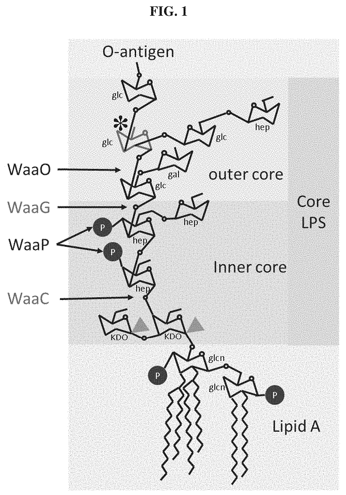
FIG. 1. Schematic representation of the E. coli BL21 LPS and relevant synthesis enzymes. The E. coli BL21 structure was assembled from biocyc database information. The glucose moiety (marked with an asterisk) is that which is used as a receptor by T3. Glcn: .alpha.-D-glucosamine, Glc: .alpha.-D-glucose, Gal: .alpha.-D-galactose, Hep: glycero-.beta.-D-manno-heptose, KDO: 3-deoxy-D-manno-octulosonate
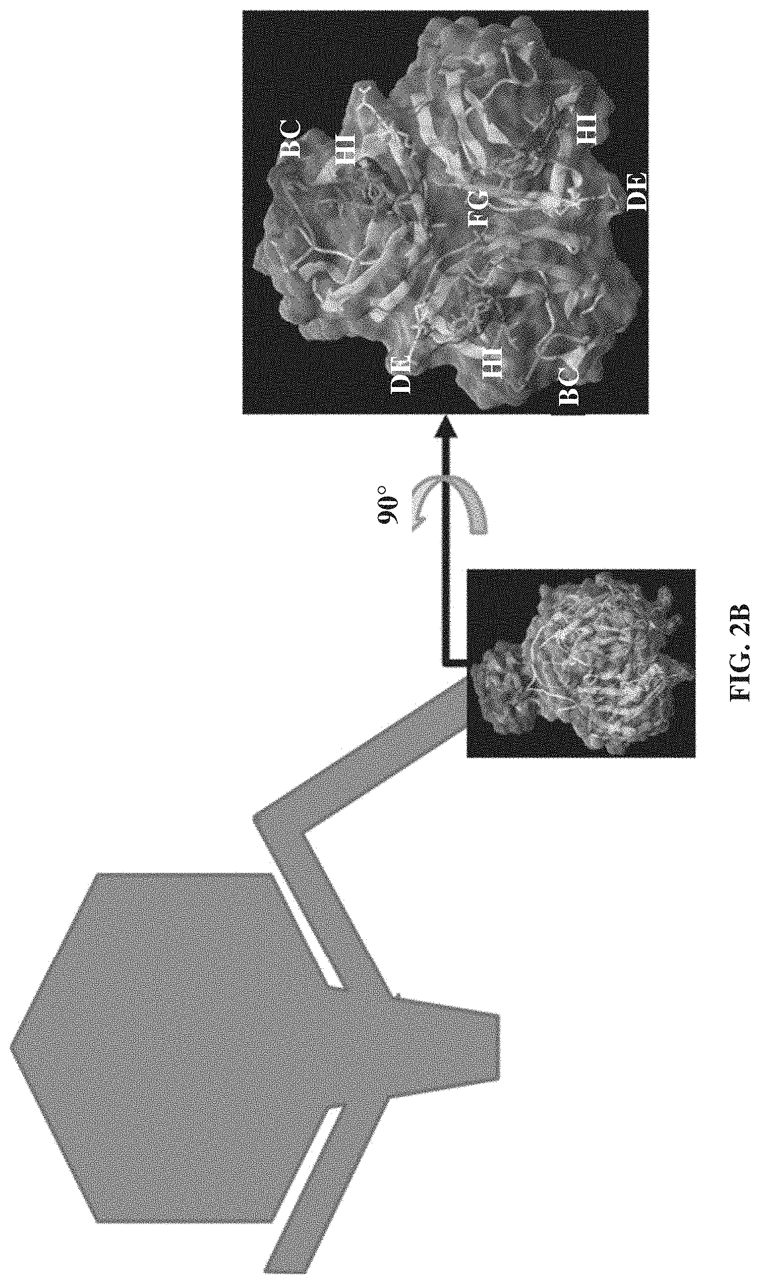
FIGS. 2A-2B. Bacteriophage tail fiber primary and tertiary structure. FIG. 2A. Alignment of the T7 (SEQ ID NO: 152) and T3 (SEQ ID NO: 153) tail fiber products. Identical residues are displayed as dots. The largest rectangular box (corresponding to amino acids 372-553) specifies the fraction of T7 gp17 that has been crystallized. Rectangular boxes B, C, D, E, F, G, H, I, R, S, T, U, V, W, X, Y, and Z represent beta-strands, the rectangular box A represents the alpha-helix that links the pyramidal domain. Rectangular boxes CD, EF, and GH represent random coils that point towards the pyramidal domain whereas the downward facing loops BC, DE, FG, and HI are represented with rectangular boxes BC, DE, FG, and HI, respectively. FIG. 2B. 3D structure of the T7 gp17 last 99 amino acids from helix A to the very end of the protein as seen from the side or axially. Important contribution of loops BC, DE, FG and HI to the basal surface of the tip domain and the complete absence of the other inter strand loops. Side chains are shown for the BC, DE, FG and HI loops only.
FIG. 3. T3 acquires LPS-mutant infectivity after extended incubation in batch cultures with E. coli BL21. Four independent late exponential cultures of E. coli BL21 were infected with T3 WT at an MOI.about.0.01 and the phage titers on WT E. coli BL21 and the two LPS mutants .DELTA.waaC and .DELTA.waaG followed along time of incubation. During the first day, samples from each culture were gathered at 3 hrs, 6 hrs and 24 hrs. The lysate were diluted 100-fold into fresh medium every 24 hrs. BDL: Below Detection Limit. For each time point, the set of bars, from left to right, represents: T3 stock, replicate 1, replicate 2, replicate 3, replicate 4.
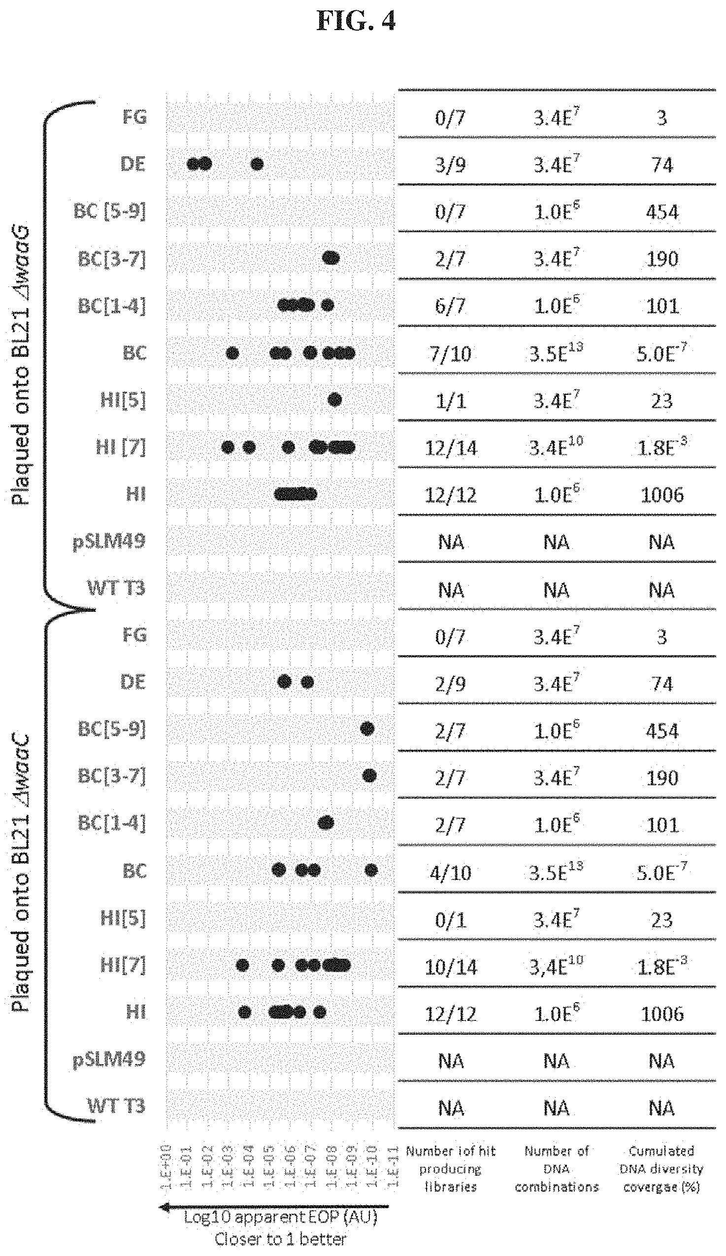
FIG. 4. Efficiency of Plating (EOP) of T3 lysates on two E. coli BL21 mutants, .DELTA.waaC and .DELTA.waaG. Lysates were grown on NEB5.alpha. carrying Gp17 plasmid libraries where independent loops were randomized, NEB5.alpha. with a nonmutated plasmid (pSLM49 or the normal T3 host, E. coli BL21 (T3 WT). Below the chart is the number of independent libraries that produced hits on either E. coli BL21 mutant and the theoretical cumulative percentage of the possible sequence space sampled for every type of loop modification. The theoretical coverage is calculated as the percentage of the possible diversity (4.times.4.times.2)n where n is the number of randomized codons in the remodeled loop with the assumption that libraries are completely independent and not redundant. NA: not applicable.
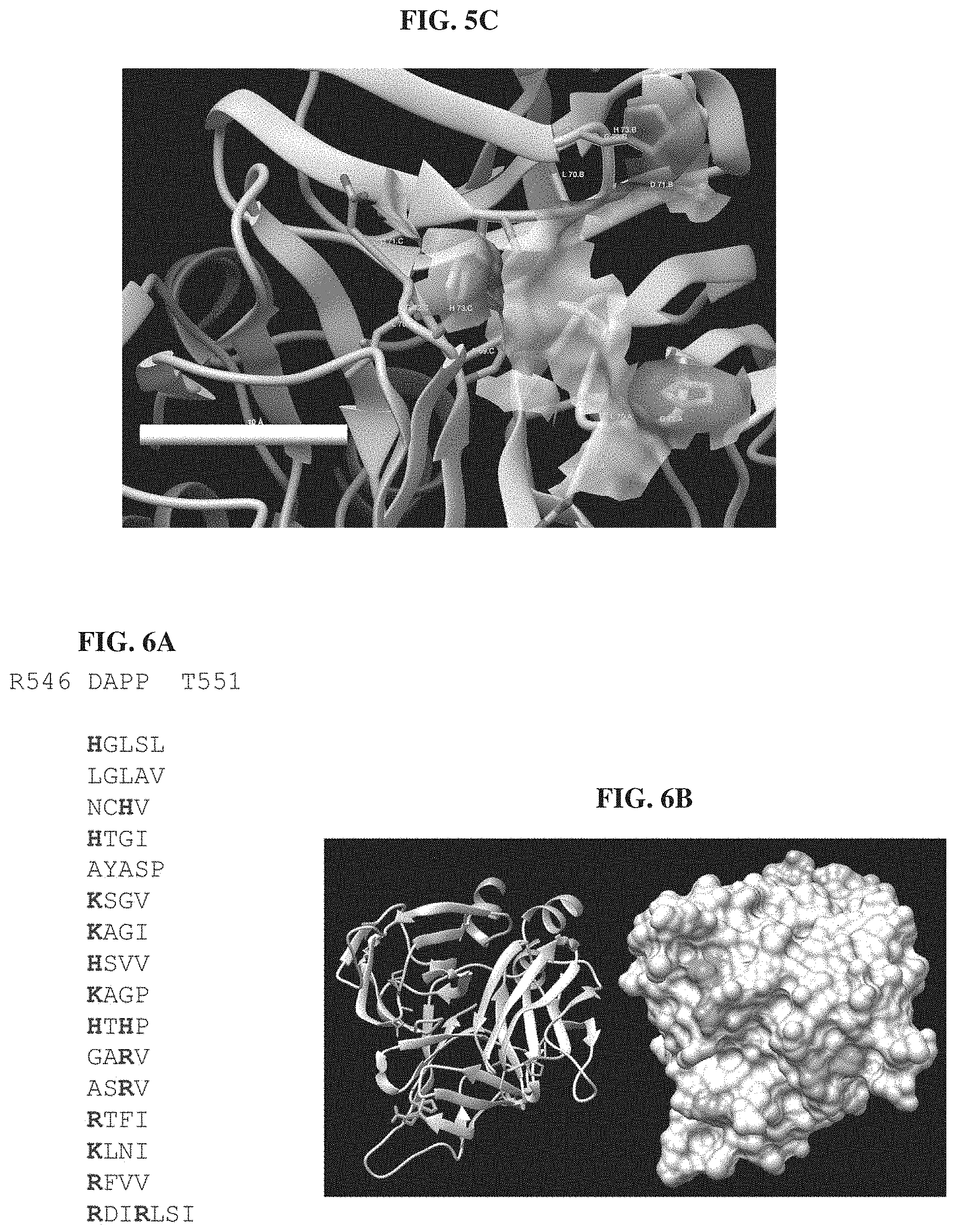
FIGS. 5A-5C. Comparison of the modelled structure of T3 and T3(FG:PLDGH) FIG. 5A. The computed surface area of WT T3 gp17 tip is overlaid atop the modelled structure of the T3(FG:PLDGH) gp17 tip. Only loop amino acid side chains are displayed and loops are labeled. The main differences are in the BC loop which is flexible and therefore can accept several configurations and the FG loop were H527 side chains clearly stands out of the T3 gp17 surface area model. FIG. 5B. Side by side axial view comparison of the surface electrostatic potentials of the two tail fiber tips. Polar residues are shaded. H strand residue R546 and G527/H527 are indicated. R546 and H527 in T3(FG:PLDGH) create a very positively charged area. FIG. 5C. Surface area from FG residues only in T3(FG:PLDGH) illustrating that H527 is the major contributor.
FIGS. 6A-6B. FIG. 6A. The sequence of the HI loop of the 16 phagebodies that were isolated on either .DELTA.waaC or .DELTA.waaG are aligned to the wild-type sequence (top to bottom: SEQ ID NOs: 154-169). Residues with positively charged side chains are highlighted. FIG. 6B. Model of the T3 gp17 tip structure with each HI loop residue highlighted: D547, A548, P549, and P550. On the left side is a ribbon representation while the molecule surface is displayed on the right. Only D547 and P549 are visible on the latter because neither A548 nor P550 are surface accessible.
FIG. 7. Detail of the HI loops from 6 aa codon long HI phagebodies and T3(HI:RDIRLSI). The modelled structures of 7 phagebody gp17 tips with HI loop mutations were superimposed. Position 547 and 549 of the 6 phagebody with 4 codon long HI loops (ASRV, KLNI, HTHP, NCHV, RFFV and RTFI) are identified. T3(HI:RDIRLSI) R547 and R550 are also highlighted to show how similarly placed the side chains are.
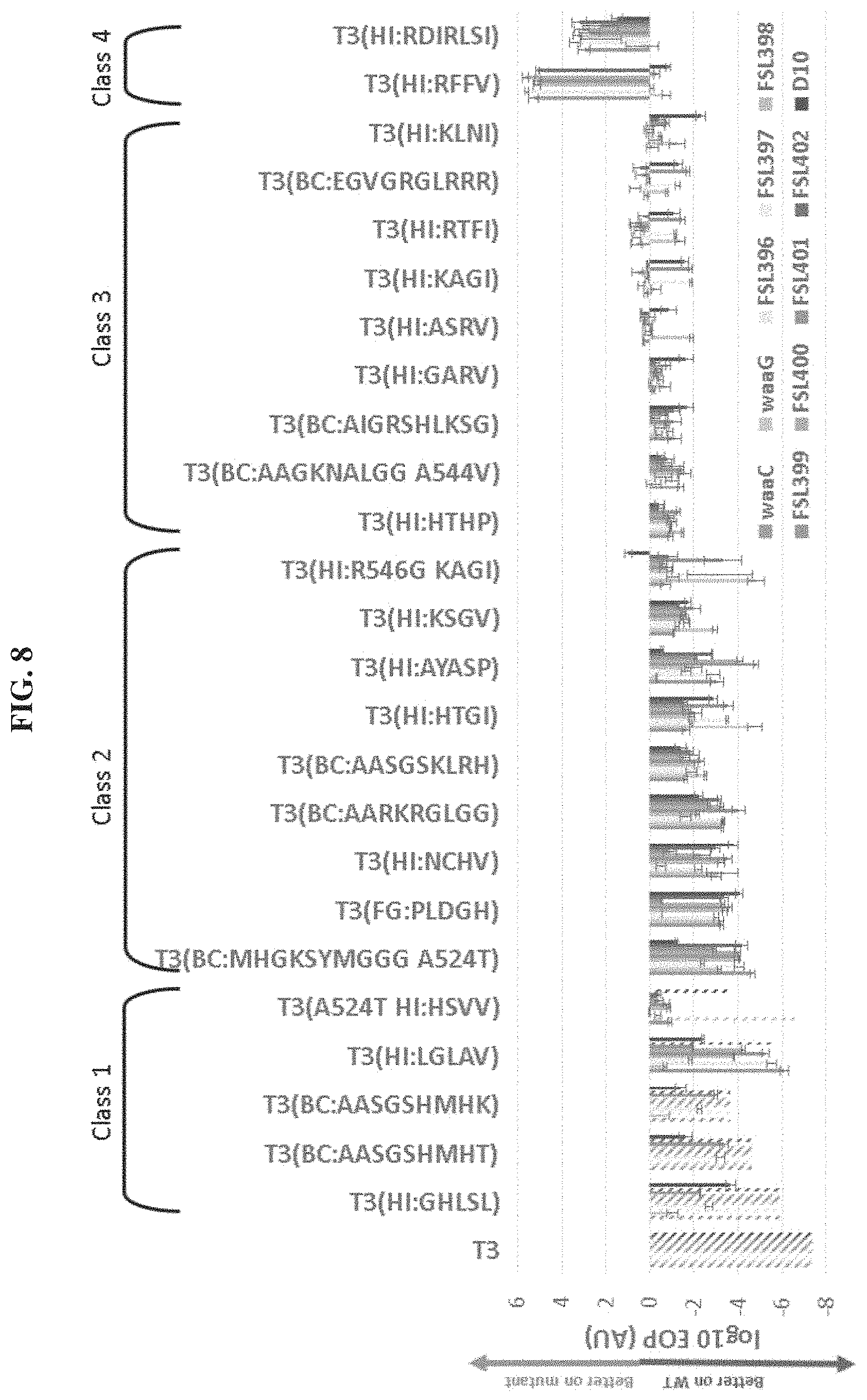
FIG. 8. Infectivity of bank isolates towards T3 resistant mutants of E. coli BL21. The infectivity of 27 independent selected phages isolated from various gp17 banks was on 8 naturally occurring T3 resistant mutants of E. coli BL21 as well as on the two constructed LPS mutants .DELTA.waaC and .DELTA.waaG was evaluated through EOP measurement which is plotted as its log 10 value. Stripe patterned data points are beyond detection limit (no plaques detected) which is calculated as the inverse of the WT E. coli BL21 number of pfus in the assessed volume. In each set of bars, the samples are (left to right): waaC, waaG, FSL396, FSL397, FSL398, FSL399, FSL400, FSL401, FSL402, D10.
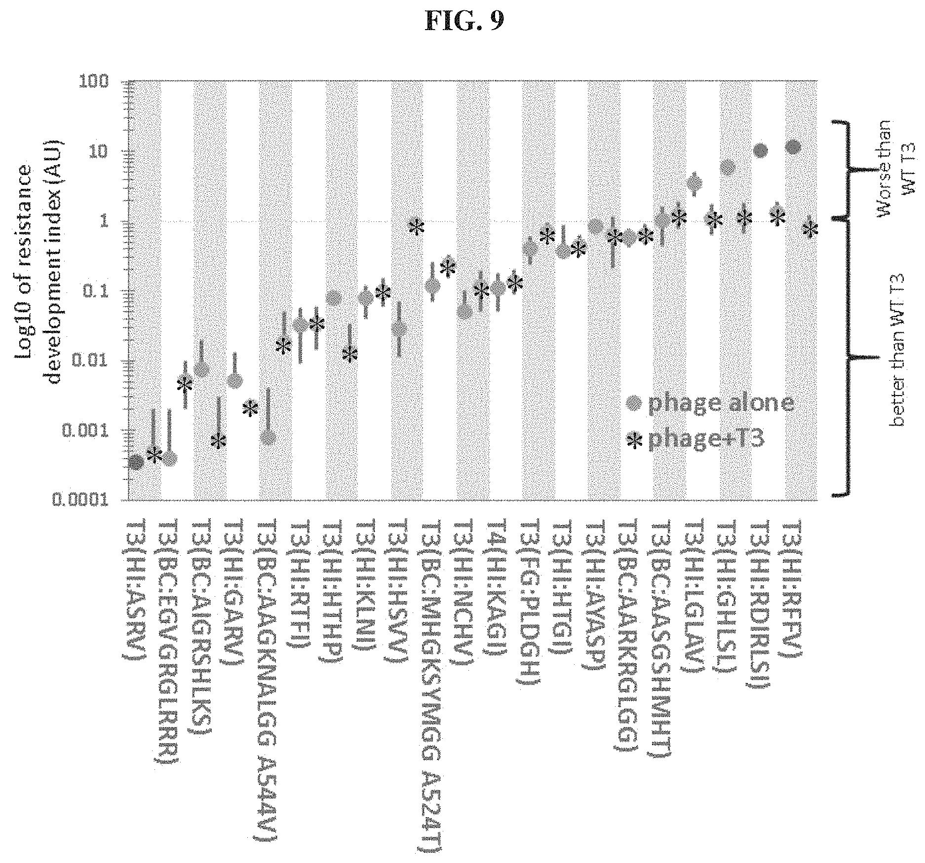
FIG. 9. Bacterial resistance development index. The capacity of a given T3 variant to eliminate resistance development in WT BL21 cultures was evaluated on plates seeded with about 10.sup.5 phages and 10.sup.8 bacteria. The resistance index for each variant was calculated by determining the ratio of the number of resistant colonies observed after 24 hrs to that obtained with WT T3. All experiments were done in triplicates. Expressed on a log 10 scale, a value below 1 indicates that the phage or cocktail prevents resistance better than WT T3 and conversely, a value above 1 indicates the phage or cocktail is worse. The first dot on the left represents a value below detection limits.
FIG. 10. Cocktail of chosen T3 variants prevents resistance development in large evolving populations of E. coli BL21. Infections were run in quadruplicate in 10 ml LB batch cultures subcultured every 24 hrs into fresh medium through 100-fold dilution with either WT T3 or a cocktail of 12 variants isolated from host range altered libraries. Bacterial counts in each of these microcosms was measured at each subculture steps and compared to that of 2 replicate cultures not infected with phage. For each time point, the set of bars, from left to right, represents: T3wt_A, T3wt_B, T3wt_C, T3wt_D, Cocktail_A, Cocktail_B, Cocktail_C, Cocktail_D, No Phage_A, No Phage_B.
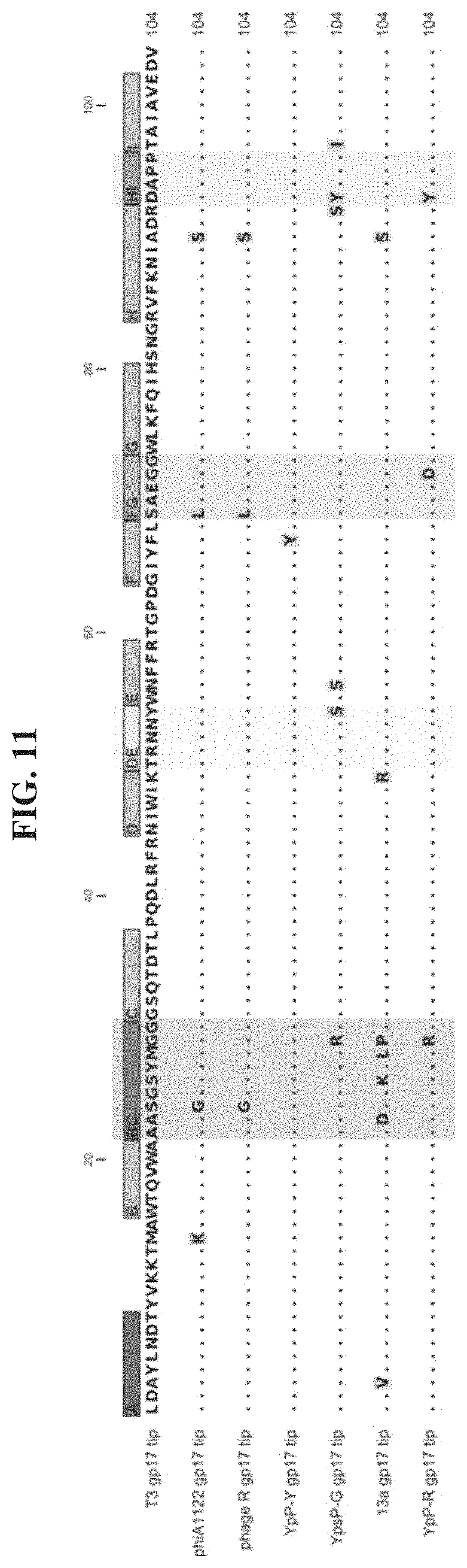
FIG. 11. Alignment of the closest homologs to the T3 gp17 tip (top to bottom: SEQ ID NOs: 170-176). Identical residues are displayed as dots. The location of loops BC, DE, FG and HI are highlighted.
FIG. 12. Schematic of scheme for replacement of each codon within any given loop with the degenerate codon NNK GGCAGGGTATTTAAGAACATAGCGGATAGANNKNNKNNKNNKACAGCAATAGC CGTAGAGGACGTGTAA (SEQ ID NO: 177); GGCAGGGTATTTAAGAACATAGCGGATAGAGATGCGCCTCCACAGCAATAGCCG TAGAGGACGTGTAA (SEQ ID NO: 178) (and reverse complement); CCGTCCCATAAATTCTTGTATCGCCTATCT (SEQ ID NO: 179); GRVFKNIADRDAPPTAIAVEDV (SEQ ID NO: 180).
FIGS. 13A-13B. DE loop residues Y508 and T504 environments. Residues and features of importance to the reading of these modelled structures from the T3 gp17 tail fiber have been shaded as described in the legend. FIG. 13A. Zoomed in view of the residues surrounding DE loop's Y508. Y508 from monomer A is wedged between the side chain of E525 from that same monomer and the side chains of I519 and F521 from the neighboring monomer. FIG. 13B. T504 is not solvent accessible being located underneath P549.
FIG. 14. Gp17 tip models from BC loop mutated phagebodies. Surface shaded according to electrostatic potential. White: neutral; Shaded: charged.
FIGS. 15A-15F. Capacity of phagebodies to control bacterial population over three consecutive passages using a high-throughput 96-well plate system with a starting bacterial population of .about.10.sup.7 cfu and an MOI of .about.10.sup.-4. FIG. 15A, phage T3. FIG. 15B, phagebody T3(BC:AIGRSHLKS). FIG. 15C, phagebody T3(BC:MHGKSYMGG). FIG. 15D, phagebody T3(HI:ASRV). FIG. 15E, phagebody T3(BC:AAGKNALGG). FIG. 15F, phagebody T3(HI:RDIRLSI).
FIGS. 16A-16B. FIG. 16A. Schematic showing the phage panning procedure to amplify out functional phages. FIG. 16B. Efficiency of plating plots summarizing the amplification of functional mutant phages and dilution of T3 WT per round of passaging on mutant strain. Rows are organized by the strain the bank was passaged on: top: truncated outer core LPS (i.e., minimal LPS); middle: LPS outer core void (i.e., LPS void); and bottom: mutant bacteria isolated from growth curve of T3 WT (i.e., T3 mutant). Columns are organized by the bank that was being passaged. BL21 cultures were originally infected with the phage bank for round 0 and every subsequent round was infected with supernatant from the prior infection.
FIG. 17. Schematic illustrating the restriction-ligation method to synthesize phage libraries.
FIG. 18. Alignment of the tip sequences of phages PhAY01 and T7 (top to bottom: SEQ ID NOs: 181-182).
FIG. 19. Schematic illustration of the first gp17 randomized bank assembly method. Highlighted in between each of the 3 pieces that are ligated together are the overhangs generated by type IIs restriction enzymes.
FIG. 20. Sequence of the PST510-511-512 PCR product before BsaI restriction (top to bottom: SEQ ID NOs: 183-184).
FIGS. 21A-21B. Schematic illustrating the similarities between antibodies and phage tail fibers and how phagebody libraries can be used to reduce bacterial resistance to phages. FIG. 21A. Schematic illustrating the similarities between antibody engineering and the phagebody strategy. In an antibody, antigen recognition is primarily encoded by six hypervariable complement-determining regions (CDRs), three on the heavy chain and three on the light chain. The inset presents the three-dimensional structure of the variable domain of an antibody (PDB ID HGT). In phage T7, host range is largely determined by the C-terminus of its tail fiber protein, gp17. The insets show the crystallographic structure of the C-terminal 182 amino acids of T7 gp17 (PDB ID 4A0T). Outward loops are expected to participate in receptor recognition while tolerating mutations. Phagebodies are designed to carry mutations in these loops while leaving other structures of the tail fiber intact. FIG. 21B. Schematic illustrating how resistance appears in bacterial cultures and how phagebody cocktails or individual phagebodies may suppress resistance.
FIGS. 22A-22B. Structure and sequence of the T3 gp17 tip. FIG. 22A. Three-dimensional structure of the tail fiber tip domain of phage T3 as modeled by SWISS-MODEL. The molecular surface encompassed by the residues belonging to the BC, DE, FG and HI loops are highlighted to illustrate their possible contribution to host binding. FIG. 22B. Sequence of the T3 tail fiber tip (gp17 a.a. 455-558 fragment) modeled in FIG. 22A (SEQ ID NO: 185).
FIGS. 23A-23J. Phagebody libraries exceed 10.sup.7 unique phages. FIG. 23A. Schematic showing the strategy to synthesize phagebody libraries. FIG. 23B. Table summarizing the theoretical diversity for each library synthesized. FIGS. 23C-23I. Illumina HiSeq characterization of phagebody libraries quantifying library diversity. Each plot shows a rarefaction curve characterizing library diversity at each stage of synthesis. FIG. 23J. Legend for FIGS. 23C-23I. Solid lines are guides showing 100%, 10%, and 1% coverage of theoretical maximum sequence space. Circles correspond to synthesized plasmid libraries, squares indicate plasmid libraries recovered post transformation, and triangles specify phage libraries post recombination.
FIGS. 24A-24D. Phagebodies display broadened host range towards BL21 mutants that are resistant to wild-type T3. FIG. 24A. LPS structures for wild-type BL21 and the wild-typeT3-resistant BL21 mutants constructed for phagebody isolation. Highlighted with an arrow and an asterisk are the sugar residues that act as receptors for T3 and T7, respectively. FIG. 24B. Phage titer for 4 independent phagebody libraries designed to randomize the indicated loops. Titer was measured on wild-type BL21 (top row), .DELTA.waaG (middle row), and .DELTA.waaC (bottom row) in triplicate for each library and the data is plotted as mean+/-standard deviation. This data illustrates the reproducibility of library construction and the repeated failure of some libraries to produce host-range-altered phagebodies. FIG. 24C. Representative image of plaque assays from one of the HI loop phagebody libraries highlighting individual plaques. FIG. 24D. Heat map summarizing the efficiency of plating (EOP; ratio of wild-type T3 or phagebody PFU on the tested bacterial mutant versus wild-type T3 or phagebody PFU on wild-type BL21) for randomly isolated and plaque-purified phagebodies on a panel of experimentally isolated wild-type-T3-resistant bacterial mutants (D10; FSL396-402) and the two constructed isolation hosts, .DELTA.waaC and .DELTA.waaG. The phagebodies are sorted based on performance. Class 1 phagebodies are marginally better than wild-type T3, failing to infect all tested mutants and doing so at low EOP (log.sub.10 EOP on mutants <-2); Class 2 phagebodies infect all test strains but poorly (log.sub.10 EOP<-2 for at least one test strain); Class 3 phagebodies infect all test strains as efficiently as wild-type BL21 (log.sub.10 EOP.about.0); Class 4 phagebodies have lost the capacity to infect wild-type BL21 but infect LPS mutants efficiently (log.sub.10 EOP>2).
FIGS. 25A-25H. Phagebody libraries can prevent the onset of resistance. FIGS. 25A-25H. Kinetic plots showing growth curves of wild-type BL21 bacterial cultures that were infected with phagebody libraries. As a control, wild-type T3 was grown on E. coli NEB5.alpha. carrying a wild-type T3 gene 17 plasmid (WT gene 17) in order to mimic the phagebody library construction conditions but without mutagenizing the plasmid-borne gene 17. Bacterial growth was followed through optical density at 600 nm. Each plot consists of 10 replicates from three independent experiments. Error bars show the SEM. Cultures were infected at a MOI of 0.01.
FIG. 26. Cocktail of 12 individual phagebodies inhibits the development of phage resistance in populations of E. coli BL21. Replicates of four 50 ml cultures were inoculated with wild-type T3 (circles) or a cocktail of 12 phagebodies (squares) obtained from the enrichment experiment presented in FIG. 30. Each culture was serially passaged every day with a 2-fold dilution into 2.times.-concentrated LB media for 6 consecutive days and the bacterial titer was measured at each time (see methods and materials below for details). The day 0 titer corresponds to that of the starter culture before phage addition. All data points are represented with the median as a black horizontal bar. Only the day 6 data showed statistically significant differences between the bacterial titers for cultures treated with the cocktail versus wild-type T3 (t-testp=0.016). The black bar represents the median value for each day. The limit of detection is .about.300 CFU/ml, which is below the lowest data point on the graph.
FIGS. 27A-27B. Isolated phagebodies inhibit the development of phage resistance in populations of E. coli BL21. FIG. 27A. Resistance index for each randomly isolated phagebody (lower is better). The resistance index is based on counting the number of resistant colonies growing on plates inoculated with .about.10.sup.9 CFU of wild-type BL21 and .about.10.sup.5 PFU of wild-type T3 phage (as reference) or a phagebody, in top agar after 24 hours of incubation. The resistance index is the log.sub.10 of the ratio between these two counts. The vertical patterned lines indicate arbitrarily chosen thresholds of improved resistance prevention compared to T3 (same as (|), 10.times. better (+) or 250-fold better (.times.)) from right to left) and are presented to help visualize best and worse phagebodies. 3 data points are estimated because they had too many colonies to count (.largecircle.) or no resistant colonies at all (A). The shading is the same as in FIG. 24D and is meant to help correlate the two datasets. The data is represented as mean+/-95% confidence interval. FIG. 27B. Select phagebodies were co-cultured with wild-type BL21 in four independent microcosms for 6 days with daily reseeding in fresh medium and bacterial titer was recorded before each subculture. The same protocol was applied to four independent wild-type BL21 microcosms infected with wild-type T3 as a control. Phagebodies presented on the top line were best at controlling bacterial growth, as all 4 replicate microcosms maintained bacterial levels that were several orders of magnitude below the starting titer of the culture (.about.10.sup.9 CFU/ml). All phagebodies performed better than wild-type T3.
FIG. 28. Related to FIG. 22: Alignment of gp17 tip sequences (top to bottom: SEQ ID NOs: 170, 175, 186, 173, 176, 174, 172, 171). The T3 gp17 tip was aligned to the corresponding regions of tail fibers from other related wild-type phages, illustrating the enrichment of mutations in outward loops between related phages targeting different hosts. Identical residues are displayed as dots. The location of loops BC, DE, FG and HI are highlighted as in FIG. 22. The partial BC loop regions targeted for library designs (BC[1-4], BC[3-7] and BC[6-9] are also shown. The highlighted gp17 proteins (on the left) indicate those originating from phages isolated on Yersinia species. Protein sequences from phages isolated on E. coli are not highlighted. Note that some of these phages may grow on both E. coli and Yersinia (T7 for example) while others may be specific to either strain (such as T3 that only grows on E. coli). The shaded boxes with bolded residues inside indicate amino acids that differ from the T3 gp17 sequence.
FIG. 29. Related to FIG. 24: Co-evolution of wild-type T3 with wild-type BL21 selects for phage mutants that can infect T3-resistant mutants .DELTA.waaG and .DELTA.waaC but that are unable to improve the control of phage-resistant bacterial mutants. Three replicate wild-type BL21 cultures were infected with wild-type T3 and reseeded every 24 hrs into fresh LB medium. At 3, 6, 24, 48 and 72 hours, phage titers of the evolved T3 lysates on the parental host (dots with asterisk) as well as on .DELTA.waaG (dots with rectangle) and .DELTA.waaC (dots with triangle) were measured. At times 0, 24, 48 and 72 hours, .about.10.sup.5 phage forming units (PFUs) of the corresponding evolved T3 lysates were also used to infect a lawn of .about.10.sup.9 wild-type BL21 colony forming units (CFUs) and obtain the number of T3 phage-resistant colonies (PRC) that arose after 24 hrs (bars bottom plot; results presented as mean+/-standard deviation). DL: detection limit of the assay. BDL: below detection limit. N/A: not available. While T3 mutants that can infect .DELTA.waaG and .DELTA.waaC do appear during co-evolution with wild-type BL21, these mutants do not appear capable of preventing resistant colonies from appearing in the plate resistance assay.
FIGS. 30A-30B. Related to FIG. 24 and FIG. 25: Panning phagebody lysates on selective hosts unveils rare phagebodies. FIG. 30A. Schematic showing the phage panning procedure to amplify functional phagebodies out of libraries. FIG. 30B. Efficiency of plaquing plots summarizing the amplification of functional mutant phages and dilution of wild-type T3 per round of passaging on mutant strain. Rows are organized by the strain the library was passaged on: top--.DELTA.waaG; middle--.DELTA.waaC; and bottom--T3-resistant mutant D10 isolated from a wild-type BL21 culture infected with wild-type T3. Columns are organized by the phagebody library that was being passaged. Wild-type BL21 cultures were originally infected with the phage library for round 0 and every subsequent round was infected with supernatant from the prior infection.
FIG. 31. Related to FIG. 27B: Phage titer of parallel cultures infected with wild-type T3 or select phagebodies in long-term resistance suppression assays. Four parallel phage/wild-type BL21 10 ml co-cultures for each phage (listed at the top) were set up and incubated for 6 days with daily reseeding at 100-fold dilution into fresh media. Before each reseed, phage titers on wild-type BL21, .DELTA.waaC, and .DELTA.waaG were measured.
DETAILED DESCRIPTION
The rapid escalation of drug resistant bacterial infections and decreased investment in antibiotic research make it imperative to develop alternative therapies. A resurging approach gaining significant interest is phage therapy (PT) whereby bacteria targeting viruses (bacteriophages or phages for short) are used as antimicrobials or for delivery of genetic circuits with antimicrobial or physiological activities (Chen et al., J. Clin. Invest. 124, 3391-406 (2014); Devlin et al., Cell Host Microbe. 20, 709-15v (2016); Shen et al., J. Clin. Invest. 125, 2841-50 (2015); Kutter et al. Curr. Pharm. Biotechnol. 11, 69-86 (2010); Kutateladze and Adamia, Trends Biotechnol. 28, 591-95 (2010); Kutter et al. Future Microbiol. 10, 685-88 (2015); Citorik et al. Nat. Biotechnol. 32, 1141-45 (2014); Bikard et al. Nat. Biotechnol. 32, 1146-50 (2014); Maynard et al. PLoS Genet. 6 (2010); Lu and Collins, Proc. Natl. Acad. Sci. U.S.A. 104, 11197-202 (2007); Lu and Collins, Proc. Natl. Acad. Sci. U.S.A. 106, 4629-34 (2009)). Phages are exquisitely selective of their host, which makes phage therapy less destructive of the normal and beneficial microflora of the patient compared to conventional chemical antibiotics (Galtier et al. Environ. Microbiol. 18, 2237-45 (2016)). Bacteriophages are also functionally orthogonal to antibiotics, which means they are generally unaffected by acquisition of antibiotic resistance making them particularly adapted to the treatment of Anti-Microbial Resistant (AMR) infections (Miedzybrodski et al. Adv. Virus Res. 83, 73-121 (2012)). A further advantage of phages is their self-dosing capacity in that they can replicate to the extent of the infection. However, this also makes traditional pharmacodynamics methods inadequate for PT.
Although independent of antibiotic resistance mechanisms, bacteria have evolved various resistance solutions against phage predation. Bacteriophage initiate infection through the specific recognition of a surface exposed receptor molecule, protein, lipopolysaccharide (LPS) or capsule component, which if mutated or masked deprives the virus of its entry port (Labrie, et al. Nat. Rev. Microbiol. 8, 317-27 (2010)). Resistance to phages may also arise from acquisition of dedicated phage defense mechanisms such as CRISPR or abortive infection systems (Labrie, et al. Nat. Rev. Microbiol. 8, 317-27 (2010)). Finally, the need for phages to recognize a specific receptor translate into relatively narrow host ranges for most naturally occurring phages. This in turn, means that no single phage may be active against all (or a medically relevant fraction of) bacteria involved in any given disease.
These issues are traditionally alleviated by empirically assembling and regularly updating cocktails of un-related phages that are collectively able to eliminate the affliction. However, this leads to often poorly defined mixtures that are largely incompatible with modern medical standards for safety testing and regulatory approval. Because these cocktails are composed of phages with completely distinct properties, they may require individual protocols for production, storage and manufacturing, which further complicates establishment of good manufacturing practices, an essential part of drug approval processes. As a result and despite its enormous potential, phage-based therapies have struggled to gain momentum (Cooper et al. Front. Microbiol. 7, 1209 (2016)).
Researchers have long observed that in the predator and prey relationship between phage and bacteria, the prey almost systematically outcompete the predator (Alexander, Annu. Rev. Microbiol. 35, 113-33 (1981)). Various models have been proposed. The most simplistic one explains that because phage genomes are small and densely packed, the likelihood of deleterious mutations is higher than in their host so that bacteria can tolerate more mutations. This eventually leads to bacterial resistance before collapsing, thus giving bacteria an edge in the arms race with phages. Such a phenomenon, is one of the reason that sustains distrust in the use of phages as therapeutics.
Various approaches have been undertaken to expand the host range of phages to combat resistance (Ando et al. Cell Syst. 1, 187-196 (2015); Chen et al., Front. Microbiol. 8, 147 (2017); Gebhart et al., Virology 505, 263-66 (2017); Hawkins et al., Virol. J. 5, 97 (2008); Heilpern and Waldor, J. Bacteriol. 185, 1037-44 (2003); Lin et al., PLoS One 7, e30954 (2012); Nguyen et al., Evolution 66, 363-74 (2012); Scholl et al., Antimocrob. Agents Chemother. 53, 3074-80 (2009); Yoichi et al., J. Biotechnol. 115, 101-7 (2005); Yosef et al., J. Biotechnol. 115, 721-28 (2017)). (Ando et al. Cell Syst. 1, 187-196 (2015)). However, these approaches rely on hybridization between already characterized bacteriophages with known and desired host ranges, which is very limited and often results in long and unpredictable trial and error periods. It is, therefore, not well suited to the isolation of mutant phages that may target bacteria that have evolved receptor mutations as a result of phage predation. Alternatively, some studies have relied on traditional phage mutant selection procedures which utilize natural evolution ((Nguyen et al., Evolution 66, 363-74 (2012); Springman et al., Genetics 184, 221-32 (2010); Perry et al. PLoS One 10, e0130639 (2015); Qimron et al. Proc. Natl. Acad. Sci. U.S.A. 103, 19039-44(2006)). This process proceeds through single mutations at a time and some of these mutation may be deleterious initially though required towards the evolutionary goal set (Alexander, Annu. Rev. Microbiol. 35, 113-33 (1981); Bull et al., PLos One 9, e94690 (2014); Levin and Bull, Nat. Rev. Microbiol. 2, 166-73 (2004); Meyer et al., Science 335 428-32 (2012); Nguyen et al., Evolution 66, 363-74 (2012); Perry et al. PLoS One 10, e0130639 (2015); Qimron et al. Proc. Natl. Acad. Sci. U.S.A. 103, 19039-44 (2006); Studier et al., J. Mol. Biol. 258, 726-31 (2009); Tetart et al., J. Mol. Biol. 258, 726-31 (1996)). Thus, the natural evolution procedure often reaches bottlenecks where too many concomitant mutations are necessary to both obtain the selected phenotype and have a viable organism.
Previous studies have demonstrated that the T7-family of phages is particularly amenable to phage host range engineering (Ando et al. Cell Syst. 1, 187-196 (2015)). T7-family phages have an extremely host independent life cycle so that DNA entry into the host range is the most significant barrier to generating progeny. The experiments described here have focused on phage T3 because it has a slightly more limited host range than its close relative T7 which therefore affords more room for phenotypic improvement. The two phages are extremely similar and share an extremely similar developmental cycle (Calendar, The Bacteriophages 2nd Edition).
Studies of bacterial resistance to T3 bacteriophages and T7 bacteriophages have revealed that phages routinely adapt to resistance through mutations within genes 11, 12, and/or 17 for T7 and within 17 exclusively for T3 (Perry et al. PLoS One 10, e0130639 (2015); Qimron et al. Proc. Natl. Acad. Sci. U.S.A. 103, 19039-44 (2006)). Both T3 and T7 rely on binding to the outer core LPS for absorption; however, they bind to different LPS moieties which leads to slightly different host ranges (FIG. 1).
Recently, the T7 gp17 tip was crystallized and its structure resolved (Garcia-Doval and Van Raaij. Proc. Natl. Acad. Sci. U.S.A. 109 (2012)). It is 75% identical to the corresponding region of T3 gp17 (FIG. 2A), and the structure of the T3 tail fiber tip can therefore be modelled with high accuracy using homology modelling tools such as Swiss-model (FIG. 2B) (Arnold, Bioinformatics. 22, 195-201 (2006)). The distal 106 aa of gp17 form an intertwined globular domain shaped by an eight stranded beta barrel (labelled B to I) connected by random coils. Four of those coils, BC, DE, FG and HI, are pointed towards the exterior side of the tail fiber and are therefore uniquely positioned to contact the host and recognize the receptor moiety (FIG. 21). The extent to which bacteriophage tail fibers delineate bacteriophage host range, and the application of synthetic biology to manipulate bacteriophage tail fiber tips in hopes of expanding bacteriophage host range has, up until now, remained unexplored.
Disclosed herein are strategies and methods for engineering synthetic bacteriophages with expanded host ranges. In contrast to previous approaches, the methods described herein focus on producing vial phages with subtle host range alterations to target resistant mutants. These methods are rapid and simple enough that they can be used to scan for the most important regions involved in host recognition. Importantly, because these methods are directed towards short loop regions, they can be used to discover phage mutants that would require too many point mutations to ever be produced by evolution, naturally or in vitro (Nguyen et al., Evolution 66, 363-74 (2012); Springman et al., Genetics 184, 221-32 (2010)). Finally, these methods are versatile because they are compatible with other forms of phage engineering (synthetic genome reconstruction or Gibson assembly of full phage genomes), selection of other phenotypes (e.g., selection of faster or slower replication rates or altered immunogenicity of the phage), and are also compatible with random mutagenesis to enrich mutations outside of the immediately targeted region. However, these methods are also simple and fast enough that iterative cycles can be performed to generate phagebodies mutated in several loops concurrently.
Moreover, methods are described that facilitate the rapid evolution of bacteriophages to generate combinatorial phage libraries. This combinatorial-based approach, which is superior to previous phage cocktails, yields a vast amount of diversity (10.sup.7 mutants/mL), while minimally perturbing the overall phage structure and mechanism of infection. Finally, this amount of diversity surpasses that of natural phage evolution and enabled the assembly of a cocktail of phages all derived from the same scaffold to evade bacterial resistance.
Disclosed herein are synthetic bacteriophages having mutations in a tail fiber tip protein (also referred to herein as "phagebodies"). The mutations are engineered in one or more binding loops of the tail fiber tip protein. More specifically, in some embodiments the engineered mutations are identified using the methods disclosed herein, and can be generated by non-natural methods such as synthesis of sequences of one or more binding loops to introduce mutations relative to the wild-type sequence. The binding loops of the tail fiber tip protein are engaged in binding molecules on the surface of bacteria, and face the basal or apex side of the tail fiber protein.
In some embodiments, the mutations are in one or more of coils BC, DE, FG and/or HI of the tail fiber protein. As demonstrated herein, these coils can be mutagenized to alter the ability of a synthetic bacteriophage to infect particular bacteria, i.e., the mutagenized synthetic bacteriophages have a different host range than a bacteriophage with unmutated binding loops of the tail fiber tip protein. Coils BC and HI are particularly suited for mutagenesis to produce host range altered synthetic bacteriophages (phagebodies).
The mutations introduced to produce the synthetic bacteriophages can be substitution mutations, deletions, or insertions/additions. As is shown below, the coils in the binding loops of the tail fiber protein can have one or more amino acids substituted for the wild-type amino acid(s). It also is possible to add amino acids or delete amino acids, for example at one or both ends of a coil, to provide longer or shorter coil sequences. The types of mutations can be mixed such that, for example, one coil contains a substitution mutation of one or more amino acids, and another coil contains an addition and/or deletion mutation. The types of mutations also can be mixed such that, for example, one coil contains both a substitution mutation of one or more amino acids, and an addition and/or deletion mutation.
In some embodiments, the engineered mutations in the one or more binding loops of the tail fiber tip protein of the synthetic bacteriophage are the only mutations in the synthetic bacteriophage. However, the synthetic bacteriophages are not limited in this aspect, and may contain other mutations in other proteins, such as for providing the synthetic bacteriophage with one or more additional functional features.
As shown herein, the synthetic bacteriophage can be a T3 bacteriophage. Other similar bacteriophage can likewise be generated to have mutations in a tail fiber tip protein, such as a T7 bacteriophage or a bacteriophage having about 75%, 80%, 85%, 90%, 91%, 92%, 93%, 94%, 95%, 96%, 97%, 98%, or 99% amino acid identity to a T3 bacteriophage tail fiber tip protein.
In some embodiments, the tail fiber tip protein mutated in the synthetic bacteriophage is gp17. Other tail fiber tip proteins are known to those of skill in the art.
Compositions of the synthetic bacteriophage also are provided. Such compositions can include a pharmaceutically-acceptable carrier. Generally, for pharmaceutical use, the synthetic bacteriophages may be formulated as a pharmaceutical preparation or compositions comprising at least one synthetic bacteriophage and at least one pharmaceutically acceptable carrier, diluent or excipient, and optionally one or more further pharmaceutically active compounds. Such a formulation may be in a form suitable for oral administration, for parenteral administration (such as by intravenous, intramuscular or subcutaneous injection or intravenous infusion), for topical administration, for administration by inhalation, by a skin patch, by an implant, by a suppository, etc. Such administration forms may be solid, semi-solid or liquid, depending on the manner and route of administration. For example, formulations for oral administration may be provided with an enteric coating that will allow the synthetic bacteriophages in the formulation to resist the gastric environment and pass into the intestines. More generally, synthetic bacteriophage formulations for oral administration may be suitably formulated for delivery into any desired part of the gastrointestinal tract. In addition, suitable suppositories may be used for delivery into the gastrointestinal tract. Various pharmaceutically acceptable carriers, diluents and excipients useful in synthetic bacteriophage compositions are known to the skilled person.
The synthetic bacteriophage compositions have, in some embodiments, a single type of synthetic bacteriophage. More typically, however, the synthetic bacteriophage compositions include two or more variants or types of synthetic bacteriophages that have different mutations in the tail fiber tip protein, i.e., a "cocktail" of synthetic bacteriophages. In some embodiments, the two or more types of synthetic bacteriophages advantageously have different host ranges, which provides for enhanced resistance to bacterial mutations in the exterior components that are bound by the tail fiber tip protein of the synthetic bacteriophages.
Also provided are collections (also referred to as "libraries" or "banks") of synthetic bacteriophages, which include a plurality of synthetic bacteriophages having different mutations engineered in one or more loops of a tail fiber protein. As noted above, such mutations may be substitutions, additions, or deletions.
Also provided are methods for treating a bacterial infection using the synthetic bacteriophages disclosed herein. The methods include administering the synthetic bacteriophages or compositions disclosed herein to a subject having a bacterial infection in need of treatment. In some embodiments, the subject is a mammal. In some embodiments, the subject is a human.
Methods of producing one or more synthetic bacteriophages also are provided. In such methods, one or more binding loops in a tail fiber tip protein of a bacteriophage is mutated to produce a synthetic bacteriophage. As disclosed in the examples below, such mutations can be introduced by synthesizing portions of the tail fiber tip protein using degenerate primers that vary the nucleotide sequence, and thereby introduce substitutions of amino acids (or additions or deletions) in one or more coils of the binding loops of the tail fiber tip protein.
In some embodiments, one or more codons within at least one binding loop of a tail fiber tip protein can be replaced with the degenerate codon NNK to introduce amino acid variability in the at least one loop.
For example, replacing one or more codons can be achieved by amplifying a sequence comprising a bacteriophage gp17 gene region to produce an amplification product. The amplification can be carried out using PCR primers encoding the nucleic acid sequence NNK for at least one of the amino acid codons. Such methods also can include circularization of the amplification product; introduction of the circularized amplification product into bacteriophage susceptible bacterial cells; infection of the bacterial cells with wild-type bacteriophages, wherein said infection facilitates the recombination of the wild-type bacteriophage with the amplification product; and isolation of recombinant bacteriophages comprising the amplification product.
Also provided are methods of screening one or more synthetic bacteriophages for ability to infect bacteria. The synthetic bacteriophages (or compositions containing such compositions, or collection or library of synthetic bacteriophages), which can be produced as disclosed herein, are exposed to bacterial cells and synthetic bacteriophages are identified that are capable of sustaining infection of the bacterial cells to an extent that exceeds that of the bacteriophages that contain unmutated binding loops. In some embodiments, the bacterial cells are E. coli cells. Examples of such bacterial cells include .DELTA.waaG mutants or .DELTA.waaC mutants, as are shown in the working examples. Other types of bacteria that are susceptible to synthetic bacteriophages will be known to a skilled person, and can be selected based on the host range of the wild type bacteriophages used in engineering the synthetic bacteriophages.
Also provided are methods of generating synthetic bacteriophage compositions that target a bacterial strain and the bacteriophage-resistant variants thereof. Such methods include repeated exposure of synthetic bacteriophages to bacterial cells such that bacteriophage-resistant variants arise, and further culturing with synthetic bacteriophages such that synthetic bacteriophages capable of infecting of the bacteriophage-resistant variants are obtained. In some embodiments, the methods include exposing bacterial cells to synthetic bacteriophages that are described herein and which may be produced using the methods described herein; identifying synthetic bacteriophages that are capable of sustaining infection of the bacterial cells; exposing the same bacterial cells to the synthetic bacteriophages identified until such time that bacteriophage-resistant variants arise; exposing the bacteriophage-resistant variants to synthetic bacteriophages; and identifying the synthetic bacteriophages that are capable of infecting of the bacteriophage-resistant variants. The methods also can include iteratively repeating the steps to identify additional synthetic bacteriophages that are capable of infecting additional bacteriophage-resistant variants. The methods also can include combining the synthetic bacteriophages identified to produce a composition, which optionally can include carriers, diluents and/or excipients.
Also provided herein are methods of delaying the evolution of a bacterial strain. Such methods include exposing the bacterial strain to a synthetic bacteriophage composition that targets the bacterial strain and its common bacteriophage-resistant variants, such as is produced by the method described above.
Also provided herein are methods for suppressing resistance of bacteria to bacteriophage infection. The methods include contacting a population of bacteria with a cocktail of synthetic bacteriophages comprising two or more different host ranges. In some embodiments, the cocktail of synthetic bacteriophages comprises two or more variants or types of synthetic bacteriophages that have different mutations in the tail fiber tip protein. In some embodiments, the step of contacting a population of bacteria with a cocktail of synthetic bacteriophages comprises administering the cocktail of synthetic bacteriophages to a subject. In some embodiments, the subject is a mammal. In some embodiments, the subject is a human. In some embodiments, the step of contacting a population of bacteria with a cocktail of synthetic bacteriophages comprises contacting an isolated population bacteria (such as bacteria derived or obtained from patient samples) with the cocktail of synthetic bacteriophages. In some embodiments, the cocktail comprises synthetic bacteriophages, a composition, or a collection of synthetic bacteriophages as disclosed herein, or synthetic bacteriophages generated as disclosed herein.
Also provided herein are methods for preparing a cocktail of synthetic bacteriophages. The methods include obtaining one or more samples from a patient, contacting the bacteria in the one or more samples with a library or bank of synthetic bacteriophages, and identifying synthetic bacteriophages that infect the bacteria in the one or more samples. In some embodiments, the more than one sample is obtained from a patient at different times. In some embodiments, the methods also include combining synthetic bacteriophages that infect the bacteria in the one or more samples in to a cocktail. In some embodiments, the library or bank of synthetic bacteriophages comprises synthetic bacteriophages, a composition, or a collection of synthetic bacteriophages as disclosed herein, or synthetic bacteriophages generated as disclosed herein. In some embodiments, the patient is a mammal. In some embodiments, the patient is a human.
Also provided herein are methods for detecting bacteria, identifying bacteria or diagnosing bacterial infections. The methods include contacting a sample containing bacteria with the synthetic bacteriophages, a composition, or a collection of synthetic bacteriophages as disclosed herein, or synthetic bacteriophages generated as disclosed herein, incubating the sample containing bacteria with the synthetic bacteriophages for a time sufficient for the synthetic bacteriophages to infect the bacteria, and detecting the synthetic bacteriophages to detect the presence of one or more bacteria in the sample, to identifying the bacteria or to diagnose bacterial infection. In some embodiments, the sample is obtained from a patient. In some embodiments, the patient is a mammal. In some embodiments, the patient is a human. If samples are obtained from a patient at different times, the progress of bacterial infection can be monitored and tracked, as can the efficacy of anti-bacterial therapies.
EXAMPLES
Methods and Materials
Strains and Culture Conditions
Bacteriophage T3 was obtained from Ian Molineux (University of Texas, Austin) and maintained on E. coli BL21. Cloning was performed in E. coli NEB5a. Bacteria were grown in Lysogeny Broth (LB medium; LabExpress) at 37.degree. C. with agitation at 250 rpm from isolated colonies grown on LB plates from frozen stocks and stored at -80.degree. C. in 45% glycerol. As needed, the medium was supplemented with kanamycin (kan; 50 .mu.g/ml final concentration), carbenicillin (carb; 50 .mu.g/ml final concentration), apramycin (50 .mu.g/ml final concentration), and/or glucose (glc; 0.2% w/v final concentration). LB plates contained agar (LabExpress) at a final concentration of 1.5%. Top agar was LB agar 0.6%. T3 resistant strains FSL397-402 and D10 were picked from T3 infected lawns of wild-type E. coli BL21 incubated at 37.degree. C. until resistant colonies grew. They were picked, streaked to isolation twice, and tested for T3 resistance.
DNA Manipulation and Sequencing
Polymerase Chain Reaction was performed using either KAPA Biosystems Hifi or KAPA2G Robust DNA polymerases. Standard PCR conditions for these two polymerase are presented in TABLE 1A. DNA fragments were purified using the DNA clean up and concentration kit from Zymo Research. Plasmids were extracted using the plasmid mini- or midiprep kits from the same source depending on the scale of the plasmid preparation. Restriction enzymes were purchased from New England Biolabs.
All these reagents and kits were used following the manufacturer's recommendations.
TABLE-US-00001 TABLE 1A PCR programs used in this study. KAPA HiFi PCR Conditions 1X 95.degree. C. 3 min 25X 98.degree. C. 20 s 65.degree. C. 15 s 72.degree. C. 30 s/kb 1X 72.degree. C. 10 min KAPA2G Robust PCR Conditions 1X 95.degree. C. 3 min 25X 95.degree. C. 20 s 60.degree. C. 20 s 72.degree. C. 15 s/kb 1X 72.degree. C. 10 min
Plasmid Construction
Plasmids constructed and used in this project are listed in TABLE 3. pSLM49 was constructed by assembling the PCR amplified replication origin and resistance marker from pFF753 (primers PST480 and PST481) (Farzadfard et al. Science 346, 1256272 (2014)) with a PCR amplified fragment from phage T3 containing gene 17 (PST575 and PST576) using the BamHI and XmaI sites added to the primer sequences.
pSLM193-197 and pSLM225-233 are derivatives of pSLM49 built by cloning the gene 17 tip sequence from select phagebodies in lieu of the wild-type tip sequence. The gene 17 tips were amplified through primers PST691/692 and the rest of the plasmid with PST693/694. The two PCR fragments were then assembled by Gibson.RTM. reaction. pSLD18 is a derivative of pSIM9 (Datta et al., Gene 379: 109-15 (2006)) where the chloramphenicol marker was replaced with the erythromycin marker of pCP1 (Le Bourgeois et al., Gene 111, 109-14 (1992)). pSLM111alpha was obtained by ligating the apramycin resistance marker of plasmid pSET152 (Bierman et al., Gene 116, 43-9 (1992)) amplified with primers PST816 and PST817 and the backbone of pKD3 (Datsenko and Wanner, Proc. Natl. Acad. Sci. U.S.A. 97, 6640-45 (2000)) amplified with primers PST818 and PST819 after restriction of both fragments with PspoMI.
pSLM173 was constructed from pNR63, which is a pSC101 based plasmid with an ampicillin resistance marker and an AHL regulated promoter in front of the BxbI integrase gene. The replication origin, selection marker and AHL controlled promoter were PCR amplified with primers PST1089 and 1090 while the E. coli BL21 waaG gene was amplified with primers PST1091BL and PST1092BL (TABLE 2). The resulting amplicons were assembled using the Gibson reaction.
LPS Mutant Construction
E. coli BL21 was transformed with the recombineering plasmid pSLD18--which is pSIM9 (Datta et al. Gene 379, 109-15 (2006) with its chloramphenicol marker replaced with the erythromycin resistance marker from pRC1--and cells made recombineering proficient. The cells were electroporated with a PCR product designed to replace waaC or waaG with an apramycin resistance marker amplified from pSLM111alpha with primers PST853/PST854 and PST857/PST858 respectively (TABLE 2). Proper deletion was then verified by PCR.
Tail Fiber Library Creation
Diversity was introduced at the DNA level in pSLM49. Two different methods were used: (1) Direct transformation of PCR products with terminal redundancy and (2) a restriction-ligation based method.
Directed Transformation of PCR Products:
In the first method, the entire pSLM49 plasmid was PCR amplified with a pair of diverging primers annealing on each site of the target loop. In one of the oligonucleotides, the target loop sequence was replaced by a series of NNK codons. The NNK stretch is preceded in 5' by the complementary sequence to the reverse primer so that the final PCR product has a 20-30 bp identical sequence at each end. The amplicons were then DpnI digested to eliminate template DNA and about 100-500 ng of that DNA was transformation into chemically competent NEB5.alpha. cells following the manufacturer's instructions. The termini of the PCR products were redundant such that the PCR product circularized reconstituting gene 17 present in pSLM49 but with a random sequence in place of the targeted loop. The bacteria were recovered for 1 hour at 37.degree. C. in SOCS medium (1 mL). After this step, the transformation yield was determined by plating serial dilutions of culture on LB-kanamycin agar plates. The 1 mL bacterial cultures were then diluted with 9 mL of LB-kanamycin and grown overnight at 37.degree. C. and 250 rpm of shaking. The next day, fresh cultures were started by diluting 1 mL of overnight culture into 9 mL of LB, while the remaining culture was pelleted and stored at -20.degree. C. for plasmid DNA extraction/sequencing. Phage lysates were made by infecting bacterial cultures at exponential growth phase (OD.sub.600=0.7) with 10.sup.7 plaque forming units (PFU) of T3 (100 .mu.L). The cultures were grown for another 2-3 hours until the cultures cleared. Phage lysates were chloroform treated with 500 .mu.L of chloroform for 30 minutes to kill any remaining bacteria, spun down to remove debris and filtered through a 0.22 .mu.m filter. Phage lysates were spun down at 7,000 G for 5 minutes and stored at 8.degree. C. for long-term storage.
Restriction-Ligation-Based Method:
For each loop library, ten 25 uL PCR reactions were carried out where 10 ng of template plasmid encoding the T3 gp17 gene was PCR amplified using 8 pmoles of each primer and 0.5 units of HiFi polymerase following the heating protocol summarized in TABLE 1B. Primers were designed to encode a (1) mutagenized region corresponding to the desired gp17 loop and (2) BsaI cleavage sites for restriction digestion and subsequent circular ligation by T4 ligase to yield scarless circular plasmids (FIG. 17 and FIG. 23A). The mutagenized region was encoded by NNK codons to minimize premature incorporation of stop codons.
TABLE-US-00002 TABLE 1B Step Temp. Time 1 95.degree. C. 3 min 2 98.degree. C. 15 s 3 62.degree. C. 15 s 4 72.degree. C. 2 min Repeat 2-4 25 times 5 72.degree. C. 10 min
Each PCR reaction yielded approximately 500 ng of linear PCR product, which was pooled together and DpnI (100 units) treated for 4 hours at 37.degree. C. to eliminate template plasmid. Following DpnI digestion, the PCR products were purified using Zymo DNA clean and Concentrator.TM.-5 spin columns. Next, .about.5 .mu.g of linearized gp17 gene products were diluted in New England Biolabs CutSmart.RTM. buffer (500 .mu.L) and restriction digested using 125 units of BsaI at 37.degree. C. for four hours, after which the enzyme was heat inactivated at 65.degree. C. for 20 minutes. Digested DNA was purified using Zymo DNA clean and Concentrator.TM.-5 spin columns and eluted in Nanopure water (18.2 Me).
The digested DNA was circularized using T4 ligase, where 2 .mu.g of DNA was diluted to 500 .mu.L in T4 ligase buffer, which 10 .mu.L of T4 ligase (4,000 units) was added and the reaction was incubated overnight at room temperature. The next day, DNA was purified using the Zymo DNA clean and Concentrator.TM.-5 spin columns and eluted with 7 .mu.L of Nanopure water to yield a plasmid stock of .about.100 ng/uL. Next, bacterial libraries were made by transforming 100 ng of plasmid into New England Biolabs 5-alpha electrocompetent cells via electroporation (1 mm cuvette, 1.7 kV, 200 52, and 20 .mu.F). The bacteria recovered for 1 hour at 37.degree. C. in SOC media (1 mL), after which the transformation yield was determined by plating serial dilutions of culture on LB-kanamycin agar plates. The 1 mL bacterial cultures were then diluted with 9 mL of LB-kanamycin and grown overnight at 37.degree. C. and 250 rpm. The next day, new cultures were started by diluting 1 mL of overnight culture into 9 mL of LB, which the remaining culture was pelleted and stored at -20.degree. C. for sequencing. Phage lysates were made by infecting bacterial cultures at exponential growth phase (OD.sub.600: 0.7) with 10.sup.7 plaque forming units (PFU) of T3 (100 .mu.L). The cultures were grown for another 2-3 hours until the cultures cleared. Phage lysates were chloroform treated with 500 mL of chloroform for 30 minutes to extract the cellular debris and kill any remaining bacteria. Phage lysates were spun down at 7,000 G for 5 minutes and stored at 8.degree. C. for long-term storage.
Tail Fiber Structure Modelling and Display
The structure of T3 gp17 was modelled using Swiss-model at its default settings (Arnold et al., Bioinformatics 22, 195-201 (2006)), and the results were analyzed and visualizations were created using the chimera software.
Measuring Efficiency of Plating (EOP) of Phage Lysates
Lysate's host range was characterize through EOP measurements. Phagebody libraries were serially diluted in triplicates and 3 .mu.l of each dilution were spotted onto the surface of 10.times.10 cm LB agar plates covered with a top agar lawn of the desired test strain. The EOP was calculated as the ratio between the phage titer on the mutant strain and the reference strain, which was E. coli BL21. The confidence interval of the calculated EOP was computed using the method described in (Fieller, Suppl. To J. R. Stat. Soc., 1-64 (1940)) using the calculator located at www.graphpad.com/quickcalcs/ErrorProp1.cfm.
Phage Panning
For each bacterial mutant, overnight cultures were grown from a single colony. The next day, 50 .mu.L of the overnight culture was diluted into 5.0 mL of LB and grown to exponential phase (OD.sub.600: 0.7), which point 100 uL of phage lysate from each loop library was added. The bacterial cultures were grown for another 3 hours, except for .DELTA.waaC mutants, where the culture was grown for 4 hours. After phage propagation, phage lysates were chloroform treated (250 .mu.L), spun down at 7,000 G for 5 minutes, and stored at 8.degree. C. for subsequent panning. This procedure was repeated for additional rounds (FIG. 16A and FIG. 30A), except infecting with 1 uL of phage lysate from the previous round of infection rather than 100 .mu.L, except for .DELTA.waaC where 100 .mu.L was added. This enabled amplification of functional phages, while diluting away phages incapable of infecting bacterial mutants.
Resistance Index Determination
Triplicate samples of .about.10.sup.5 PFU of each phagebody isolate was mixed with .about.10.sup.9 CFU of wild-type E. coli BL21 in 3 ml of top agar and immediately poured over an LB plate. After the top agar had hardened, plates were incubated for 24 hrs at 37.degree. C. CFU were subsequently counted for each plate. Because it is unlikely that two independent BL21 cultures contain the exact same assortment of naturally occurring mutation, results were systematically normalized to the number of CFU counted on T3-infected lawns. A pseudocount of 1 was added to the entire dataset prior to any calculation. The confidence interval on the resulting resistance indices was calculated according to (Fieller, Suppl. To J. R. Stat. Soc., 1-64 (1940)) using the calculator located at www.graphpad.com/quickcalcs/ErrorProp1.cfm.
Liquid Culture Assay for Resistance Suppression by Phagebody Libraries
From an overnight culture of wild-type BL21, a fresh culture was grown to exponential phase (OD.sub.600=0.7). After which, 250 .mu.L aliquots of the culture were added to a 96 well plate along with 2.5 uL of phagebody lysates per well. This equates to an MOI of 0.001. Growth curves were obtained by taking OD.sub.600 measurements using a BioTek Synergy H1 microplate reader at 2 min. intervals, 37.degree. C., and constant shaking.
Liquid Culture Assay for Resistance Suppression by Phagebody Cocktail
Overnight cultures of BL21 were grown from a single colony. The next day, 500 .mu.L of the overnight cultures were diluted into 50 mL of LB in 250 Erlenmeyer flasks and grown to exponential phase (OD.sub.600: 0.7). After which, .about.10.sup.7 PFU's (which equates to a multiplicity of infection of 10.sup.-3) of phage lysate from wild type T3 (100 .mu.L) or a phage cocktail (10 .mu.L) were added. The cultures were grown overnight. The next day, 1 mL aliquots were taken from each culture and washed 4 times in PBS and were serial diluted and plated on LB-agar plates to quantify the amount of colony-forming units (CFU). Every 24 hours, 25 mL of the culture was discarded and diluted with 25 mL of 2.times.-concentrated LB to ensure bacterial nutrients were still available.
Liquid Culture Assay for Resistance Suppression by Isolated Phagebodies
T3 and the phagebodies T3(HI:ASRV), T3(HI:GARV), T3(BC:AAGKNALGG), T3(HI:HSQP), T3(HI:HSVV), T3(HI:NCHV) and T3(HI:RTFI) were each seeded at an MOI of .about.10.sup.-4 into 4 replicate 10 ml LB late log phase E. coli BL21 cultures. The cultures were then incubated at 37.degree. C. with shaking at 250 rpm for 22 hrs. Each culture was subcultured into 10 ml LB medium with a 100-fold dilution and the process was repeated over 6 days. Prior to starting the experiment and before every reseed, the bacterial and phage titers were measured. Phage titer was measured not only on the original host E. coli BL21 but also on the two LPS mutants .DELTA.waaC and .DELTA.waaG. In order to mitigate the effect of phages on colony viability during plating and counting, 1 ml samples from each condition were washed 3-times in PBS before serial dilution and plating. PFUs were measured from the chloroform-treated supernatant of the first wash.
Additional Cloning Approaches
Variability was generated in the tip of T3 gp17 in several ways: a) primer based randomization of multiple loops; b) primer based randomization of single or multiple loops; and c) random mutagenesis of the whole tip module using base analogs. In all cases, all or fragments of the T3 17 gene were cloned into a plasmid which is introduced into phage susceptible strains (E. coli DH5a or DH10B derivatives). This bank was then infected with the wild-type phage and the resulting population screened for viruses infecting other types of bacteria. Deletion of xonA (sbcB) and recJ (strain FSL71) stimulates recombination of plasmids with T7 or T3 by a factor of 2 to 3-fold. The strain harboring these deletion has a very low transformability and the recombination benefit was therefore shadowed by the low transformation efficiency and such a mutant was not systematically used in experiments. It also was found that introduction of plasmids containing a CRISPR-cas9 system targeting the T3 17 tip sequence (pACY22) could improve recombination efficiency but the benefits of counter selecting against WT 17 was not reproducible enough to warrant the lower transformation efficiency incurred by the presence of the extra plasmid carrying the CRISPR-cas9 system.
TABLE-US-00003 TABLE 2 Primers used for construct preparation. SEQ ID NO: pSLM49 construction PST480 GTACGAATTCAGCTGGATCCAGACCTAGGGGATATATTCCGCTTCCTCGCTC 1 A PST481 GCATCCCGGGTGCAAAGCTTGACGTCGGAATTGCCAGCTGGGGCGCCCTC 2 PST575 TAGCGGATCCTGAAGGAACGTGACCCAAACAAACCGTACA 3 PST576 TCGACCCGGGATCTTATCGACTACCTTGGCACCAATCTGA 4 Subcloning phagebody tips into pSLM49 PS1691 GTACTAAGTGGGGAGGTAAGTGGCTT 5 PS1692 GTGTGATAGTCCATCCGTGGACTTAAAGTA 6 PST693 AAGCCACTTACCTCCCCACTTAGTAC 7 PST694 TACTTTAAGTCCACGGATGGACTATCACAC 8 pSLM173 construction PST1089 GGTACCTTTCTCCTCTITAATAGCTAAATC 9 PST1090 GTGCACGGATCCCATGGTACGCGTGCTAG 10 PST1091 GGAGAAAGGTACCATGTCATTTTGTTGGAATGAAATTAATTCTGG 11 BL PST1092 GGGATCCGTGCACTTATTTATCTAATAAACATTGGTCTGATTGTGC 12 BL pSLM111alpha construction PST816 CCTGTGGGGCCCATGCCCTAGGTCATGAGATTATCAAAAAGGATCTTCACC 13 PST817 GGTGCAGGGCCCTCGACAATTGTCAGCCAATCGACTGGCGAGCGGCATCGC 14 PST818 TGCGAAGGGCCCGGATTCGAATTCGTGATCTTCCGTCACAGGTAGGCGC 15 PST819 GTGGCAGGGCCCGCGTAAGCTAGCGGCGCGCCATTTAAATGAAGTTCCTAT 16 TCC waaC deletion PST853 CGGATGCGGGTTTTGATCGTTAAAACATCGTCGATGGGCGGTGTAGGCTGG 17 AGCTGCTTC PST854 ACCATCTGATTCITCCCATACCCACCAATTAATCCCGGATATGGGAATTAGC 18 CATGGTCC waaG deletion PST857 CGGTTTGCAGCGCGATTTTATGCGTATTGCTCAGACAGTCGTGTAGGCTGGA 19 GCTGCTTC PST858 CCAGACCACCCGTTATGATATCCGCCGCTTTCTCTGGCAGATGGGAATTAGC 20 CATGGTCC loop direct transformation loop library construction modification PST695 CCTGTGGGAGAGTATCAGTCTGAGAACCMNNMNNMNNMNNMNNMNNMN BC FW 21 NMNNMNNAGCCCATACTTGAGTCCAGGCC PST696 GGTTCTCAGACTGATACTCTCCCACAGG BC RV 22 PST699 GGCAGGGTATTTAAGAACATAGCGGATAGANNKNNKNNKNNKACAGCAAT HI FW 23 AGCCGTAGAGGACGTG PST700 TCTATCCGCTATGTTCTTAAATACCCTGCC HI RV 24 PST701 AACTGGTCCTGACGGTATCTACTTCGTNNKNNKNNKNNKNNKTGGC FG FW 25 ATTCCAGATACACTCTAATCTGC PST702 AAGGAAGTAGATACCGTCAGGACCAGTT FG RV 26 PST794 CTTAATCCATATGTTGCGGAATCGC DE FW 27 PST795 GCGATTCCGCAACATATGGATTAAGNNKNNKNNKNNKNNKTGGAACTTCTT DE RV 28 CCGAACTGGTCCTGACG PST800 GACAATGGCCTGGACTCAAGTATGGGCTNNKNNKNNKNNKNNKNNKNNKN BC[10] FW 29 NKNNKNNKGGTTCTCAGACTGATACTCTCCCAC PST802 AGCCCATACTTGAGTCCAGGCCATTGTC BC[10] RV 30 PST803 GGGTATTTAAGAACATAGCGGATAGANNKNNKNNKNNKNNKNNKNNKACA HI[+3] FW 31 GCAATAGCCGTAGAGGACGTG PST805 TCTATCCGCTATGTTCTTAAATACCC HI[+3] RV 32 PST696 GGTTCTCAGACTGATACTCTCCCACAGG partial BC 33 FW PST1252 CCTGTGGGAGAGTATCAGTCTGAGAACCMNNMNNMNNMNNACTACCACTA BC[6-9] RV 34 GCAGCAGCCCATACTTGAGTCCAGGCC PST1253 CCTGTGGGAGAGTATCAGTCTGAGAACCTCCTCCCATGTAACTMNNMNNMN BC[3-7] RV 35 NMNNAGCCCATACTTGAGTCCAGGCC PST1254 CCTGTGGGAGAGTATCAGTCTGAGAACCTCCTCCMNNMNNMNNMNNMNN BC[1-4] RV 36 AGCAGCAGCCCATACTTGAGTCCAGGCC loop BsaI/religation loop library construction modification PST1255 CTGACTGGTCTCtAGCCMNNMNNMNNMNNACTACCACTAGCAGCAGCCCAT BC[6-9] RV 37 ACTTGAGTCCAGGCCATTGTC PST1256 CTGACTtGGTCTCTAGCCTCCTCCCATGTAACTMNNMNNMNNMNNAGCCCA BC[3-7] RV 38 TACTTGAGTCCAGGCCATTGTC PST1257 CTGACTGGTCTCAGCCTCCTCCMNNMNNMNNMNNMNNAGCAGCAGCCCAT BC[1-4] RV 39 ACTTGAGTCCAGGCCATTGTC PST957 AGTCAGGGTCTCTGGTTCTCAGACTGATACTCTCCCACAGG BC FW 40 PST958 CTGACTGGTCTCTATTCCAMNNMNNMNNMNNMNNCTTAATCCATATGTTG DE RV 41 CGGAATCGC PST961 AGTCAGGGTCTCTGAATTTCTTCCGAACTGGTCCTGACGGTATC DE FW 42 PST962 CTGACTGGTCTCTCAACCAMNNMNNMNNMNNMNNAAGGAAGTAGATACC FG RV 43 GTCAGGACCAG PST965 AGTCAGGGTCTCtTGGCTAAAATTCCAGATACACTCTAATGG FG FW 44 PST966 CTGACTGGTCTCTCGGTMNNMNNMNNMNNTCTATCCGCTATGTTCTTAAAT HI RV 45 ACCCTGC PST1258 CTGACTGGTCTCTCGGTMNNMNNMNNNMNNMNNTCTATCCGCTATGTTCTTA HI[+1] RV 46 AATACCCTGC PST969 AGTCAGGGTCTCTACCGCAATAGCCGTAGAGGACGTG HI FW 47
TABLE-US-00004 TABLE 3 Plasmids constructed and used for this project TABLE 2. List of oligonucleotides. FW: forward primer; RV: reverse primer. Plasmid Selection Name Features marker Source pSLM49 wild-type T3 gene 17 Kanamycin This study pSLM193 pSLM49 with 17 (HI:RDIRLSI) Kanamycin This study pSLM194 pSLM49 with 17 (HI:ASRV) Kanamycin This study pSLM195 pSLM49 with 17 (HI:GARV) Kanamycin This study pSLM197 pSLM49 with 17 (HI:KLNI) Kanamycin This study pSLM225 pSLM49 with 17 (BC:AAGKNLAGG) Kanamycin This study pSLM226 pSLM49 with 17 (BC:MHGKSYMGGA524T) Kanamycin This study pSLM227 pSLM49 with 17 (BC:AIGRSHLKS) Kanamycin This study pSLM228 pSLM49 with 17 (HI:RFFV) Kanamycin This study pSLM229 pSLM49 with 17 (HI:HTHP) Kanamycin This study pSLM230 pSLM49 with 17 (HI:NCHV) Kanamycin This study pSLM233 pSLM49 with 17 (A524G HI:HTHP) Kanamycin This study pSLD18 heat sensitive and heat inducible Erythromycin This study recombineering plasmid pSLM111 suicide plasmid (pir dependent replication Carbenicillin, This study origin) with an FRT site surrounded apramycin apramycin marker
Example 1: Host Range-Changing Mutations Cluster within Outward Facing Loops
Based on the T3 gp17 tip structure model and sequence alignments between T3 and T7 tail fibers, the location of its beta-strands and loops was defined (FIG. 2A). Alignment of the T3 gp17 tip sequences with its closest homologues shows that mutations are particularly enriched within the loop sequences (FIG. 11). The three point mutations found in phage R's gp17 are responsible for its capacity to infect Yersinia pseudotuberculosis (Ando et al. Cell Syst. 1, 187-196 (2015)) and suggests that the loop mutations observed in the other Yersinia infecting phages are equally responsible for the host range shift, furthering the hypothesis that loops are particularly important to host range determination.
T3 is naturally incapable of infecting E. coli MG1655 but evolves easily to do so. This characteristic was used as a way to select for naturally occurring mutants of T3 with altered host range in order to study the sequence variation within gene 17. Out of 66 such mutants individually picked and sequenced, 25 had mutations in BC and 25 in HI, 2 had a single mutation in the H or I beta-sheet while the 14 others had multiple mutations but all located within the tip region (TABLE 4).
A similar smaller experiment using BL21 .DELTA.waaC as a selective host yielded 3 independent phage variants capable of reproducing on it. The gp17 tip of these mutants was sequenced. Two were identical but the three impacted the same HI loop residue, D547 (TABLE 4).
TABLE-US-00005 TABLE 4 Mutations identified in the T3 gp17 tip region that are associated with the ability to infect the indicated host. number of New T3 host Structure affected mutation occurrences E. coli MG1655 BC loop S480R 24 Y481H E. coli MG1655 HI loop D547Y 12 E. coli MG1655 HI loop D547G 10 E. coli MG1655 HI loop D547A 2 E. coli MG1655 I .beta.-sheet Frameshift 4 from T550 E. coli MG1655 HI loop and I .beta.-sheet Frameshift 1 from R546 E. coli MG1655 BC loop G484K 1 E. coli MG1655 H .beta.-sheet V539G 1 E. coli MG1655 BC loop S480I 1 MG1655 multiple mutations: 10 BC loop, H .beta.-sheet, HI loop and/or I .beta.-sheet BL21 LPS mutant HI loop D547G 1 BL21 LPS mutant HI loop D547N 2
Example 2: Mutational Loop Library Design
T7 and T3 recombine very efficiently with plasmids that harbor at least 30-40 bp homology to their genomes (Bull et al. J. Mol. Evol. 53, 47-54 (2001); Bull and Molineux, Heredity (Edinb) 100, 453-63 (2008); Springman et al. G3 (Bethesda). 2, 825-30 (2012)), mutations introduced into plasmids can be transferred to the phage relatively easily, as long as they are selectable.
In order to generate as much diversity as possible within the loops, each codon was replaced within any given loop with the degenerate codon NNK (see material and methods for details; FIG. 12). This allows covering all possible amino acids while removing two of the three possible stop codons. The sequence space generated by such a randomization scheme is determined by the size of the targeted loop. Given the size of each loop, HI is four codons long, DE and FG five are codons long, and BC is nine codons long; only loops HI, DE and FG could be expected to be exhaustively queried (TABLE 5). As a result, the BC loop also was split into smaller sub regions (aa 1-4, 3-7 and 6-9) for which banks were made. Finally, libraries were created where the BC or HI loops were elongated by one or three codons to assess whether changing the length of a loop can impact host range.
TABLE-US-00006 TABLE 5 Theoretical genetic and protein diversity generated by NNK codons as a function of the number of codon randomized. theoretical DNA diversity Theoretical protein loop length (aa) with NNK codons diversity 1 32 20 2 1024 400 3 32768 8000 4 1.0E+06 1.6E+05 5 3.4E+07 3.2E+06 6 1.1E+09 6.4E+07 7 3.4E+10 1.3E+09 8 1.1E+12 2.6E+10 9 3.5E+13 5.1E+11 10 1.1E+15 1.0E+13
TABLE-US-00007 TABLE 6 Cumulative characteristics of phagebody libraries made during this work. The theoretical diversity. The theoretical diversity expresses the total number of possible DNA combinations based on the number of NNK codons randomized. The cumulative coverage is the sum of the library transformation yields for all the libraries ever built for that loop. Calculated cumulative coverage is the percentage of the theoretical diversity created as measured by the total number of plasmid clones obtained for all repeats of each type of library. Hits are defined as obtaining at least one PFU on a lawn of the corresponding selective BL21 mutants, .DELTA.waaC, or .DELTA.waaG. Theoretical diversity Cumulative Number of Number of (number of different Number coverage phagebody phagebody combinations from of (% of libraries libraries Targeted NNK codon libraries theoretical producing hits on producing loop randomization) built diversity) .DELTA.waaC hits on .DELTA.waaG WT gp17 NA 21 NA 0 0 BC 3.5E+13 10 ~0.0000005 4 7 BC[1-4] 1.0E+06 10 ~60 4 6 BC[3-7] 3.4E+07 10 ~90 2 5 BC[6-9] 1.0E+06 8 >100 2 4 DE 3.4E+07 10 ~50 2 4 FG 3.4E+07 10 ~0.3 0 0 HI 1.0E+06 15 >100 15 15 HI[+1] 3.4E+10 4 ~0.00002 3 4 HI[+3] 3.4E+07 14 ~20 10 10
Example 3: Testing the Capability of Loop Diversification
Given LPS mutants are the most likely pathway of resistance development against T3 infection, the phage banks were screened against two constructed LPS deletion mutants of E. coli BL21, .DELTA.waaG and .DELTA.waaC, to avoid the potential pleiotropic effects of naturally occurring T3 resistant mutants. A panel was assembled of 8 BL21 spontaneous T3 resistant mutants isolated from independent T3 infections and representative of the diversity of the evolutionary paths set in play during bacteriophage challenge.
The function of waaG was presented above but waaC was chosen as a second target because its mutant is stripped of its entire core LPS short of its two essential ketodeoxyoctulosonic acid moieties (KDO) (FIG. 1) and represents the absolute minimal LPS that bacteria can survive with under conditions relevant to natural systems (Klein et al. J. Biol. Chem. 284, 15369-89 (2009)). Both the .DELTA.waaG and .DELTA.waaC mutants of E. coli BL21 proved fully T3 resistant and naturally occurring mutations conferring infectivity to T3 were very rare (less than 1 in 10.sup.11 bacteriophages) only occurring after extensive co-culturing (>than 6 hours) of the phage and E. coli BL21 (FIG. 3). The 3' end of gene 17 of a few such mutants were sequenced and HI mutations were found similar to those described by previously: D547G, D547H or D547N (Perry et al. PLoS One 10, e0130639 (2015); Qimron et al. Proc. Natl. Acad. Sci. U.S.A. 103, 19039-44 (2006)).
In order to sample as much diversity for each bank as possible, between 2 and 12 independent phage banks targeted at each of the BC, DE, FG and HI loops and their derivatives were generated and tested for their capacity to generate phages that plaque onto .DELTA.waaG and .DELTA.waaC. The result is expressed as the apparent efficiency of plating (EOP) of said bank which is the ratio of the phage titer on a test strain relative to the titer on the normal host, wild-type E. coli BL21. The results are plotted in FIG. 4. An apparent EOP close to 1 indicates that most viable phages in the lysate are equally capable at infecting WT BL21 as its LPS mutant(s). Each dot represents the apparent EOP of one plaque generating library. Given the fact some of the banks assessed had a very low diversity (.about.1000-10.sup.7 different clones for a theoretical bank diversity), as can be expected from the low sequence coverage for some of the banks that were created, the results display stochasticity with some libraries targeting a given loop containing successful phages variants that can infect either or both test bacterial mutant while others do not with the exception of the HI targeting libraries that almost systematically contain successful mutants adsorbing to either LPS mutant (FIG. 5).
Example 4: Mutagenizing the DE or FG Loop does not Produce Many Host Range Altered Mutants
Not all loops proved equal in participating in host range determination. As presented in FIG. 4, none of the libraries designed to target the FG loop generated detectable amounts of phages capable of targeting either test strain in the EOP assay. However, by co-culturing an FG bank with the .DELTA.waaG mutant repeatedly, one variant phage was obtained that was capable of reproducibly plaquing on it. The DE libraries also proved to have a poor yield of functional phage mutants, besides, none of the plaques picked from DE banks produced phages which grew robustly and they were eventually lost before they could be investigated any further. This suggested that the FG and DE loop might be more important to the structure of gp17 than initially envisaged.
Based on the modelled structure of T3 gp17, the DE loop residue side chains appear mostly surface accessible and display minimal contacts with other parts of the protein which would suggest that modifying it is unlikely to generate massive structural damage. A notable exception is T504 which is largely tucked underneath HI loops P548 and P549 and Y508 which stacks against I519 from the neighboring monomer while being pushed back by FG loop's E525. It is conceivable that by replacing one or both of these residues, the banks created a large fraction of inactive tail fiber mutants which contributes to the very low hit rate on either LPS mutants and general instability of the phages that did plaque on them (FIG. 13). As discussed above, E525 seems involved in an interaction with the DE loop tyrosine and while part of its side chain is surface exposed, its carboxyl group isn't hidden beneath DE loops N506 and N507 on the one hand and the neighboring gp17 monomer G beta strand's K530 side chain. E525 and K530 from the neighboring monomer are likely to be involved in a salt bridge which seems to be conserved in T7 gp17 structure although it is then operated by D520 and R525. This suggest an important structural role for those residues. A524's side chain is entirely buried inside the beta-sandwich structure and not accessible to the exterior solvent thus unlikely to have a great contribution to host range leaving only 5523, G526 and G527 as positions that may be mutated with limited risk.
In the only phagebody obtained from the FG libraries, T3(FG:PLDGH), the C-alpha trace of the FG loop is virtually indistinguishable from that of the WT gp17 protein despite a completely different primary structure highlighting the structural constraints exercised by the rest of the protein onto that sequence. To illustrate this, the average distance between the Ca residues of loop residues from aligned modelled T3 and T3(FG:PLDGH) tip domains was plotted (FIG. 5A). This shows that the BC loop undergoes tremendous remodeling in the phagebody compared to T3 but this loop is also intrinsically flexible due to its outward location whereas all residues of the FG loop are positioned less than half an Angstrom away between the two structures. The DE and HI loops are similarly conserved. FIG. 5B illustrates how the imidazole group of T3(FG:PLDGH)'s sticks out into the central crevice of the trimer by overlapping the computed surface of T3 gp17 trimer with the ribbon and side chain display of T3(FG:PLDGH)'s tail fiber model. This imidazole group makes a significant contribution to the electrostatic surface potential of T3(FG:PLDGH) compared to the wild-type protein (FIG. 5C) and may help the phage in making contact with the highly negatively charged KDO and/or phosphate residues residues at the very bottom of the LPS (FIG. 1). It is also interesting to see that an acidic residue was maintained at position 525 while an aliphatic one was selected at position 524 in light of the structural features described in the previous paragraph.
Example 5: The HI and BC Loops are Hot Spots for Host Range Determination
The two other loops, BC and particularly HI, proved extremely successful at generating host range altered phagebodies. At 9 aa, the BC loop is the longest of the 4 variable regions studied here. The amount of sequence diversity generated by NNK codons at each position largely exceeds library synthesis capability (.about.4.times.10.sup.13 combinations at the DNA level, .about.5.times.10.sup.10 at the protein level) but successful hits were easily recorded despite a very minimal sequence coverage. Indeed, 4 out of 10 libraries generated .DELTA.waaC infecting variants and 7 out of 10 displayed .DELTA.waaG infectivity. Very surprisingly however, despite a 441% sequence space coverage, libraries targeted at the last 4 codons of the BC loop generated very few successful hits and only on .DELTA.waaC whereas the libraries aimed at randomizing the 4 first codons of that same loop generated hits on both target strains despite a much lower sequence space coverage. This is all the more puzzling that codons 6-9 of the BC loop are those most exposed to the basal side of the tail fiber tip and would therefore be expected to be heavily involved in host contact. The BC loop is also expected to be the most flexible part of the gp17 tip as it sits at the periphery of the structure and loop side chains appear largely free of interactions with the rest of the protein allowing them a great level of mobility.
Experiments with the BC loop are a very good illustration of the usefulness of the approach which combines guided designs with empirical engineering strategies. Analyzing the sequence and structure of 7 phagebodies isolated from the various banks that were created did not reveal any clear structural rationale as to why they may have altered host recognition (TABLE 7). All mutants have positively charged amino acid within their mutations (R, K and/or H) but some of these residues appear not to change the electrostatic potential (FIG. 14) of the basal surface of the tail fiber as exemplified by T3(BC:AAGKNALGG) and T3(BC:AASGSHMHT), so it is unclear whether they are responsible for the host range change.
The HI loop at only 4 codons long is the one that was most exhaustively covered and because it was short to begin with, length was experimented with as well while maintaining libraries of sizes compatible with reasonably exhaustive screening (TABLE 6). HI targeting bank produced more hits more consistently than any other library. As for other LPS mutant infecting phagebodies, most mutants contain positively charged amino acids. Aligning all the mutants that were isolated and that have a normal length HI loop (4 codons) reveals that they sport positively charged amino acids at position 547 or 549 (FIG. 6A). Close inspection of the gp17 structures reveal that indeed, the entire surface contribution of the HI loop lies in residues 547 and 549 side chains (FIG. 6B) while 548 and 550 have their side chains buried inside the protein with no access to solvent. Once again it was hypothesized that the host range change stems from the positively charged amino acids helping to recognize negative charges (carboxylic acid on KDO or phosphate groups) in the lower LPS inner core.
T3(HI:RDIRLSR) is one of two phagebodies that have switched host range as opposed to expanded it and as a result a particularly interesting phage. Interestingly, the HI loop of this mutant took a much more helical conformation allowing the positioning of the two arginines in ways almost identical to the way positively charged side chains in other HI mutated phagebodies (FIG. 7) so it may be that the host range change emanates from the two arginines exacerbating the affinity towards negatively charged lower LPS.
TABLE-US-00008 TABLE 7 Phage strains constructed along this work with their known characteristics. Verified Phagebody Targeted Isolation through Part of ID loop Loop DNA sequence Loop protein sequence method recloning? coctail? T3 BC GCTGCTAGTGGTAGTTACATGGGAGGAGGT (SEQ ID NO: 48) AASGSYMGGG (SEQ ID NO: 76) T3 (BC:AAS BC GCTGCTAGTGGTAGTCATATGCATACTGGC (SEQ ID NO: 49) AASGSHMHTG (SEQ ID NO: 77) panning on Yes GSHMHT) D10 T3 (BC:AA BC + H GCTGCTGGTAAGAATGCGCTTGGAGGAGGT//C1631T AAGKNALGGG//A544V direct Yes GKNALGG) (SEQ ID NO: 50) (SEQ ID NO: 78) plaque picking T3 (BC:AAR BC GCTGCTAGGAAGCGGGGTCTGGGAGGAGGT (SEQ ID NO: 51) AARKRGLGGG (SEQ ID NO: 79) panning on Yes KRGLGG) D10 T3 (BC:MH BC + FG ATGCATGGTAAGAGTTACATGGGAGGAGGT//G1570A MHGKSYMGGG//A524T (SEQ ID direct Yes GKSYMGG) (SEQ ID NO: 52) NO: 80) plaque picking T3 (BC:AIG BC GCGATTGGTAGGTCTCATTTGAAGAGTGGT (SEQ ID NO: 53) AIGRSHLKSG (SEQ ID NO: 81) direct RSHLKS) plaque picking T3 (HI:AAS BC GCTGCTAGTGGTAGTAAGCTGAGGCATGGC (SEQ ID NO: 54) AASGSKLRHG (SEQ ID NO: 82) panning on Yes GSKLRH) .DELTA.waaC T3 (BC:AAS BC GCTGCTAGTGGTAGTCATATGCATACTGGC (SEQ ID NO: 55) AASGSHMHKG (SEQ ID NO: 83) panning on Yes GSHMHK) .DELTA.waaG T3 FG TCAGCCGAGGGCGGT (SEQ ID NO: 56) SAEGG (SEQ ID NO: 84) T3 (FG:PLD FG CCGTTGGATGGTCAT (SEQ ID NO: 57) PLDGH (SEQ ID NO: 85) panning on yES GH) .DELTA.waaG and D10 T3 HI AGAGATGCGCCTCCA---------ACA (SEQ ID NO: 580 RDAPP---T (SEQ ID NO: 86) T3 (HI:GHL HI AGACATGGGTTGTCTTG------ACC (SEQ ID NO: 59) RLGLAV--T (SEQ ID NO: 87) panning on Yes SL) .DELTA.waaG T3 (HI:LGL HI AGACTGGGTCTTGCTGTT------ACC (SEQ ID NO: 60) RLGLAV--T (SEQ ID NO: 88) panning on Yes AV) D10 T3 (HI:HSV FG + HI G1570A//AGACATTCGGTGGTT---------ACA A524T//RHSVV---T direct V) (SEQ ID NO: 61) (SEQ ID NO: 89) plaque picking T3 (HI:NCH HI AGAAATTGTCATGTG---------ACC (SEQ ID NO: 620 RNCHV---T (SEQ ID NO: 90) panning on Yes Yes V) D10 T3 (HI:HTG HI AGACATACGGGTATT---------ACC (SEQ ID NO: 63) RHTGI---T (SEQ ID NO: 91) panning on Yes I) .DELTA.waaG T3 (HI:AYA HI AGAGCTTATGCGTCTCCA------ACA (SEQ ID NO: 64) RAYASP--T (SEQ ID NO: 92) direct SP) plaque picking T3 (HI:KSG HI AGAAAGAGTGGGGTG---------ACA (SEQ ID NO: 65) RKSGV---T (SEQ ID NO: 93) direct V) plaque picking T3 (HI:R546 HI GGAAAGGCGGGGATT---------ACA (SEQ ID NO: 66) GKAGI---T (SEQ ID NO: 94) direct G KAGI) plaque picking T3 (HI:HTH HI AGACATACTCATCCT---------ACC (SEQ ID NO: 67) RHTHP---T (SEQ ID NO: 95) panning on Yes Yes P) .DELTA.waaG T3 (HI:HSQ HI AGACATTCTCAGCCG---------ACC (SEQ ID NO: 68) RHSQP---T (SEQ ID NO: 96) panning on Yes P) D10 T3 (HI:KLN HI AGAAAGCTGAATATT---------ACA (SEQ ID NO: 69) RKLNI---T (SEQ ID NO: 97) direct Yes I) plaque picking T3 (HI:GAR HI AGAGGGGCGAGGGTG---------ACA (SEQ ID NO: 70) RGARV---T (SEQ ID NO: 98) direct Yes V) plaque picking T3 (HI:ASR HI AGAGCGAGTAGGGTG---------ACA (SEQ ID NO: 71) RASRV---T (SEQ ID NO: 99) direct Yes V) plaque picking T3 (HI:KAG HI AGAAAGGCGGGGATT---------ACA (SEQ ID NO: 72) RKAGI---T (SEQ ID NO: 100) direct I) plaque picking T3 (HI:RTF HI AGACGTACTTTTATT---------ACA (SEQ ID NO: 73) RRTFI---T (SEQ ID NO: 101) direct I) plaque picking T3 (HI:RDIR HI AGACGGGATATTAGGCTTAGTATTACA (SEQ ID NO: 74) RRDIRLSIT (SEQ ID NO: 102) direct Yes LSI) plaque picking T3 (HI:RFF HI AGACGTTTTTTTGTT---------ACC (SEQ ID NO: 75) RRFFV---T (SEQ ID NO: 103) panning on Yes Yes V) .DELTA.waaC
Example 6: Host Range of Isolated Phagebodies
Randomly selected plaques were picked, plaque purified and amplified from each banks using either .DELTA.waaC, .DELTA.waaG or a naturally occurring T3 resistant mutant named D10 (see material and methods) and those that grew robustly in liquid culture were studied. To evaluate the phenotypic diversity our library approach can provide, the EOP of all the above mentioned isolated phage samples was measured on the two isolation LPS mutants .DELTA.waaC and .DELTA.waaG along with 8 T3 resistant clones that were independently isolated across various experiments where WT T3 was plated onto WT BL21. Transformation of each of these isolates with a plasmid expressing waaG (pSLM173) restored T3 infectivity in 3 of the 8 strains (FSL 397, FSL401 and D10) suggesting they are waaG mutants (TABLE 8). This data provides a glimpse of the capacity of each phagebody to target resistance evolution during phage challenge and provide data about the phenotypic diversity that the different gp17 sequences create.
TABLE-US-00009 TABLE 8 Complementation of T3 resistant mutants of E. coli BL21 by a plasmid expressing waaG. pwaaG (pSLM173) - + BL21 1 1 waaC 0 0 waaG 0 1 FSL396 0 0 FSL397 0 1 FSL398 0 0 FSL399 0 0 FSL400 0 0 FSL401 0 1 FSL402 0 0 D10 0 1
In FIG. 8 the EOP of each phagebody is plotted as its log 10 value such that negative values indicate a phage that is poorer than wild-type at infecting the test strain while positive values indicate a phage which is better than wild-type and a log 10 EOP of 0 denotes a phage that plaques just as well on the mutant as on the wild-type E. coli BL21. Each test host is coded by a different shade for the respective bars shown in FIG. 8, wherein the samples are (left to right): waaC, waaG, FSL396, FSL397, FSL398, FSL399, FSL400, FSL401, FSL402, D10.
The different phagebody isolates could be roughly classified into 4 classes. Class 1 phages are phages which were only marginally better than T3, infecting only a small subset of bacterial mutants at very low EOPs. Class 2 are phages which infected the majority or all mutants but at low EOPs. Class 3 are phages that infected all T3 resistant mutant at an EOP>=0.01 (at least 1/100 pfu is infective towards mutant bacteria) and class 4 are phagebodies that actually switched host range instead of broadening it. They have become immensely better at infecting mutant bacteria than wild-type BL21. The large variety of phenotype within this small subset of randomly selected phages is a testament to the power of the method.
Example 7: Resistance Prevention from Phagebodies but not T3
A simple test was devised consisting of mixing a phagebody and wild-type BL21 at an MOI of about 10.sup.-3 in top agar on a plate and counting the number of surviving colonies after 24 hrs. A resistance index was calculated for each phagebody by dividing the number of colonies from T3 infected plate by that of phagebody infected plates. Finally, it was also tested whether adding T3 to each phagebody infection in order to assess whether the cocktail could improve performance. Plotted in FIG. 9, plain dots represent the resistance index of a given phagebody assayed alone while dots with asterisks represent the resistance index of the corresponding minimalistic cocktail.
T3(HI:ASRV) showed the highest potency at preventing resistance appearance. Indeed, alone, no colonies even appeared. It represents an about 800-fold lower resistance level compared to wild-type T3. Surprisingly, adding T3 had a negative impact on T3(HI:ASRV) performance. All 9 class 3 phagebodies performed well in the resistance development assay and fell within the 12 phages that decreased resistance at least 10-fold (log 10 resistance index below 0.1). The 3 others were class 2 phagebodies T3(HI:HSVV), T3(HI:NCHV) and T3(BC:MHGKSYMGG A524T). Strangely enough, addition of T3 had no or limited effect on the outcome of the assay except for the two class 4 phagebodies T3(HI:RFFV) and T3(HI:RDIRLSI) but even then, the results were disappointing as the cocktail did not perform better than T3 alone despite those two phagebodies targeting LPS mutants very efficiently in EOP assays. This points out to the difficulty of transferring host range data from one assay to another in a predictive manner.
The plate system used in the initial resistance impeachment experiments does not register the effects of evolution over long period of times in the presence of the phage selective pressure. Therefore the capacity of those phagebodies to control bacterial population over 3 consecutive passages were examined using a high-throughput 96-well plate system with a starting bacterial population of .about.10.sup.7 cfu and a MOI of .about.10.sup.-4. Under these conditions, which were chosen to mimic conditions during of an actual phage therapy situation where few phages may reach a comparatively large bacterial population at the site of infection, it was observed that 3 out of 8 replicates infected with T3 developed resistance against phage infection (FIG. 15A). T3(HI:ASRV), as in the previous assay, decreased resistance development as only 1 of the 8 replicates managed to escape predation (FIG. 15D), however none of the other phagebodies performed significantly better than T3 (FIG. 15B-15C, 15E, 15F). More surprisingly, T3(HI:RDIRLSI) (FIG. 15F), one of the class 4 phagebody that infected wild-type BL21 very poorly in the EOP and resistance index assays, performed about on par with T3 in this assay suggesting that growth conditions on plates and liquid are sufficiently different that a receptor usable by T3(BC: RDIRLSI) is present under the latter but not the former conditions.
Whether a cocktail of phagebodies could prevent resistance appearance better and suppress the growth and evolution of a large bacterial population was next determined. Some of the phagebodies described above originated from a phage enrichment protocol aimed at selecting those phage variants that are most adept at targeting bacterial mutants and bacteria growing in liquid cultures. Phagebody banks were repeatedly panned onto either .DELTA.waaG, .DELTA.waaC or D10 after 3 enrichment steps, individual plaques were picked, the gp17 gene sequenced and the phagebodies phenotypically characterized. A cocktail composed of 10 randomly selected phagebodies from this experiment was formulated and used to infect replicate liquid microcosms of 50 ml. Based on the fidelity of E. coli replication and data from the resistance index experiments, it was estimated that each such culture contained between 1000 and 10000 T3 resistant mutants at the time of phage addition and continued replication and evolution could of course generate new ones during the challenge. The ideal MOI was first assessed through titration. The effectiveness of the T3 phage application decreases with decreasing MOI whereas phage concentration seemed to have no impact on the phagebody cocktail capacity to kill bacteria (FIG. 8) even at an MOI of 10.sup.-7. To quantify the long-term effects of the phagebody cocktail at minimizing resistance, a kill assay was performed in which CFUs were measured over time (FIG. 10) at an MOI of 10.sup.-3. Impressively, the phagebody cocktail was able to prevent resistance for long term (greater than 6 days), whereas mutants quickly arose for a T3 wild type infection in less than 24 hours. Lastly, the phagebody cocktail was able to kill the resistant mutants at approximately three orders of magnitude better than T3.
As demonstrated herein, the host range of phage T3 can be altered efficiently by generating genetic diversity within host contacting regions chosen based on available structural data. This is a process reminiscent of the Diversity Generating Elements found in Bordetella phage BPP-1 (Guo, et al. Mircrobiol. Spectr. 2 (2014)). The distal fragment of BPP-1 tail fiber protein Mtd was also crystallized and although it adopts a completely different fold as the T3 gp17 tip, the general shape of the C-terminal fragment of both protein is not dissimilar and they both are trimeric and there are surprising resemblances between the location of the host range altering regions mutagenized as described herein and those which are targeted by BPP-1 DGR (McMahon et al. Nat. Struct. Mol. Biol. 12, 886-92 (2005)). In addition, BPP-1 DGR, just like the system described herein, generates diversity that can be used to counter-act resistance evolution in the host. These striking functional convergences between the naturally sourced DGRs and the synthetic approach described herein highlight the generalizable principle underlying the approach described herein. As the repertoire of phage host binding protein for which structural data is available increases, the value of this host range alteration method will increase and it will be a transformative step in how phages and possibly any virus are isolated for therapeutic and biotechnological applications.
Example 8: Primer Based Randomization of Gp17 Loops
pSLM22 is a plasmid that carries the last 294 last base pairs of T7 gp17 surrounded by 50 bp on each side of homology to the corresponding region of phage T3. This plasmid recombines with a frequency of about 1-5% into T3 to create a T3 phage with the host range of T7. This plasmid was used as the basis to introduce variability in 17 loops via PCR with primers carrying randomization (NNK codons) in the regions corresponding to the loops.
All primers were purchased from IDT. All randomization primers were PAGE purified. Other primers were simply desalted. Randomization of loop coding regions was achieved using NNK for each of the codons of those areas. NNK was chosen because it limits introduction of stop codons while still generating all possible amino acid changes.
PCR amplification of pSLM22 with ppAY15 and ppAY16 results in introduction of random amino acids at all positions of loops DE and FG but not in loops BC and HI. PCR amplification of pSLM22 with ppAY17 and ppAY18 followed by a second amplification with ppAYC15 and ppAY16 allows randomization of loops AB, BC, DE, FG and HI simultaneously. PCR amplification of pSLM22 with ppAY37 and ppAY18 followed by a second amplification with ppAYC15 and ppAY16 allows randomization of loops BC, DE, FG and HI simultaneously. Sequence of primers used are provided TABLE 2.
The PCR products were circularized using an ApaI restriction site generated on each end of the PCR product via a conservative point mutation in the sequence of the T7 gp17 sequence. The circularized PCR products were then transformed into highly competent DH10B cells by electroporation. Plasmids were recovered by miniprep and transformed into electrocompetent FSL71/pACY22. After 2 hrs of recovery in SOCS, the resulting transformants were diluted into 30 mL of LB kanamycin/carbenicillin and grown overnight to eliminate non transformed cells. This is referred to as the bank. The bank may contain up to .about.10.sup.9 different clones (gp17 fragments with non-natural sequences). A fraction of the bank is then diluted into fresh medium, infected at a multiplicity of infection (MOI) of 3 with wild-type T3 and the growth pursued for about 2 hrs at which point almost all cells are lysed. The lysate is sterilized with chloroform, spun down to remove the solvent and debris and saved at 4.degree. C. until testing. This is referred to as the bank lysate.
Testing was done in either of two ways: 1) direct plaque formation assay of the bank lysate onto confluent lawns of a strain normally poorly or not susceptible at all to T3 infection looking for plaques indicative of a phage now capable of sustaining infection of that strain; or 2) bank lysate amplification on the target host followed by plaquing of this amplification step onto the same target cells. Amplification is often necessary to detect rare mutants or those that have poor adsorption properties and therefore do not grow very robustly.
To help identify positive bank lysate amplification cultures where phages have been growing, a bacterial lysis indicator was used. Chlorophenol red .beta.-D-galactopyranoside (CPRG) is a yellow compound that releases a dark red/pink pigment upon hydrolysis by lacZ, the beta-galactosidase. LacZ is an intracellular activity that only gets to the medium if cells lyse and CPRG cannot diffuse through bacterial membrane so that contact between the indicator CPRG and LacZ requires phage mediated lysis of the cells. CPRG was used to test the presence of lacZ in the supernatants of bank lysate amplifications on various E. coli strains (which are all lac.sup.+) as an initial screening. A final CPRG concentration of 0.35 mM CPRG was used into cell free culture supernatants where lacZ expression was induced before bank lysate addition with 0.4 mM IPTG. A threshold for detection was set at 30 minutes as the compound will naturally hydrolyze over time and baseline levels of lacZ are present in any culture. This method allowed detection of as few as 10-100 phage particles in a 1.5 mL culture and may be made more sensitive with careful optimization.
The first banks were screened against E. coli MG1655, various mutants in the lipopolysaccharide biosynthesis pathway of E. coli K-12, part of the ECOR collection (www.shigatox.net/new/reference-strains/ecor.html), a few different Salmonellae and Shigellae, Yersinia pseudotuberculosis YPIII and IP2666, a few different Pseudomonas aeruginosa and putida and Klebsiella sp.390. This randomization proved useful in generating a mutant of T3, PhAY01, that infects strains MG1655 efficiently and has unnatural sequences in loops DE and FG as illustrated in FIG. 18. Alignment is made to the T7 tail fiber protein sequence because this is what T3 recombines with when infecting pSLM22 containing strains.
Example 9: Randomization of Single or Multiple Loops
To circumvent some of the shortcomings of the strategy described above in Example 8, a new PCR based strategy was designed that modifies a single loop at a time but can be used iteratively to generate pools of mutants with modification in several loops.
As a template plasmid pSLM49 was used which has the entire T3 gp17 gene cloned inside of it and some fragments of genes upstream and downstream of it. This plasmid greatly increases the amount of homology available for recombination with the phage on each side of the modified loops. To introduce variability, the entire plasmid is PCR amplified with a pair of primers that initiate polymerization on each side of the loop sequence to modify. One of the primers is long and contains the randomized loop sequence along with 20-30 nucleotide homology to the other end of the full plasmid amplicon. The overlapping primers allow for circularization of the final plasmid upon transformation into bacteria. The amplification therefore results in a product that spans the full length of the plasmid, with random sequences within the targeted loop sequence and about 20-30 bp of redundancy at the ends. The product may be transformed into cells after DpnI digestion and silica column clean up and concentration (Zymo research clean-and-concentrator-5). This method is analogous to QUICK-CHANGE.RTM.. For a slightly higher transformation efficiency, circularization by Gibson assembly followed by clean up and concentration on silica columns (Zymo research clean-and-concentrator-5) can be used.
Iterative loop mutagenesis can be performed either from Gibson circularized plasmids or from plasmids extracted after transformation into cells. Each iteration can be done with a different primer thus generating diversity in several loops or with the same primer set thus increasing diversity in that particular loop.
For controls, banks were generated in that same manner in each beta-sheet and in each inward facing loops. Following analysis, the general trend is that inward loops have no implication in host range, beta-sheets occasionally impact host range and the AB loop, that forming the hinge between the pyramidal stem of the Gp17 tip and the globular domain is very involved in host range.
Certain primers used are listed in TABLE 9 along with comments on which primers target which tip structure.
CRISPR-cas9 system targeting the wild-type phage were designed but provided little added benefit and are not further described here although CRISPR counter-selection of the wild-type virus could increase the efficiency of the method.
Phages that infect E. coli MG1655, EMG2 and BW25113, Yersinia pseudotuberculosis also were obtained. Phages active against LPS mutants of E. coli K-12 BW25113 (.DELTA.waaC::kan; deep rough strain) that are normally completely resistant to both T3 and T7 and for which it was difficult to obtain naturally occurring phage infective mutants also were obtained, as were phages infect ECOR63 and ECOR4, strains which are normally infected very poorly by T3.
There are other phages with similar tail fibers such as, but not limited to, 285P, FE44, BA14, for which the technique is applicable although the exact precise location of the loops and beta-sheets is not known with absolute certainty as the 3-dimensional structure of the tail tip has not been determined.
Example 10: Random Mutagenesis of the Tip Encoding Fragment of T3 gp17
In this method, variability was introduced via random mutagenesis using the Jena biosciences dNTP mutagenesis kit.
The tip portion of T3 gp17 was amplified with primers PST691/PST692 or PST703/PST704 from pSLM49 in the presence of the mutagenic base analogs dPTP and 8-oxo-dGTP as advised by the manufacturer with non-proofreading polymerases. Mutagenesis was performed for 10, 20 or 30 amplification rounds and the resulting amplicon was diluted 50-fold into a mutagen free PCR reaction with the same primer pairs in order to settle the changes. After DpnI digestion to eliminate background, the resulting amplicons were cloned into a PCR product corresponding to the rest of pSLM49 by Gibson assembly. For the PST691/692 amplicon, the complement product was generated with primers PST693/PST694. For the PST703/704 amplicon, the complement PCR product was generated with primers PST705/PST706. The resulting DNA was cleaned up and concentrated before transformation into DH10B. Such banks have generated mutants of T3 active towards MG1655, EMG2, BW25113 and its LPS mutants, and Yersinia pseudotuberculosis.
Example 11: Other Methods and their Limitations: Golden Gate Approach
pSLM22 is a plasmid that harbors the 294 bp long C-terminal tip of T7 gp17 surrounded by 50 bp of homology on each side to the corresponding gp17 tip region of T3. Recombination between pSLM22 and T3 yields to functional T3/T7 hybrids with the host range of T7. Starting from this plasmid, various approaches were tried to create diversity in the AB, BC, DE, FG and HI loops of the T7 Gp17 tip.
One attempt at creating a diversified bank of Gp17 used a GOLDEN GATE.RTM. assembly system. The backbone and T3 homology regions of pSLM22 were amplified with primers PST494 and PST495. These long oligonucleotides also introduced NNN in place of each codon of loops AB and HI respectively. Finally PST494 and 495 also introduced a restriction site for the type IIs enzyme BbsI and BsaI. These sites were introduced in such a way that restriction digestion would cleave off the restriction recognition site and allow scarless cloning of fragments in place of the gp17 tip sequence. The remainder of the gp17 tip sequence was provided by two template less PCR product obtained by annealing and filling in of primers PST496/PST497 and PST498/PST499. These primers introduce NNN codons in loops BC and DE for the first one and FG for the second one. PST496/497 had a BbsI site on one side and BsmBI site on the other while PST498/499 had a BsmBI and a BsaI site. Restriction sites were chosen in such a way that they would generate overhangs that allow the PST496/497 product to anneal and ligate with the BbsI restricted pSLM22 PST494/495 product on one hand and the BsmBI restricted PST498/499 on the other hand. Similarly the BsaI site of PST498/499 was chosen to allow annealing and ligation with the BsaI restricted pSLM22 PST494/495. This assembly procedure is depicted in FIG. 19.
This bank assembly method proved disappointing. First of all, transformation efficiency proved low, generally generating only in the range of 10.sup.3 to 10.sup.4 clones. On top of that, out of about 50 clones picked from ligation plates and analyzed by sequencing, hardly any actually had the correctly assembled tail fiber module. In most cases, stop codons appeared in frame with the gp17 coding sequence and in some cases the randomization created unexpected BsaI, BbsI or BsmBI sites that would lead to truncated gp17 sequences. It was also clear that BbsI and/or BsmBI did not cut completely and it was realized there was no need to go through the complication of three type IIs restriction enzymes as the overhang depend on the location of the enzyme recognition site and can therefore be chosen at will.
Example 12: Other Methods and their Limitations: Nested Template
Nested template less PCR was also used to generate the whole randomized gp17 insert as one product instead of two on order to increase ligation efficiency. In order to limit stop codon appearance, randomization no longer was the result of NNN codons but of NNK codons which cannot encode TAA or TGA stop codons. In this method, pSLM22 was amplified with PST494bis and PST495bis which are identical to the original PST494/495 except for the NNK codons and the BbsI site of PST494 replaced by BsaI. To generate the randomized insert, PST510, PST511 and PST512 were mixed in equimolar amounts and PCRed together. The resulting product was expected to be a 199 bp long fragment with BsaI sites on each end placed in such a way as to ensure faithful reconstruction of a Gp17 tip gene fragment after cloning into BsaI digested pSLM22 PST494bis/PST495bis. The 199 bp band was gel purified after BsaI restriction and cloned into the appropriate vector. Ligation efficiency increased 10-100 fold compared to the previous method but was not high enough yet. Stop codons plagued only about half of the clones (vs close to 100% previously) and there were less abnormal ligation products but still only about 10% of the clones had a useful gp17 tip, that is full length without any stop codon. Most of the abnormal ligation product was eliminated by running the template less PCR in two steps and by careful optimization of annealing temperature. First PST510 and PST511 were annealed and amplified together. The product was gel purified and submitted to a second PCR using only PST511 and PST512 resulting in a full length 199 bp fragment. Mispriming between PST511 and PST512 may have occurred at the annealing temperature required to get a product when all three primers are mixed together. The expected product of the PST510-PST511-PST512 template less PCR is depicted in FIG. 20.
TABLE-US-00010 TABLE 9 Primer sequences ppAY15 CCATCGGGGCCCGTACGGAAGAAGTT randomize DE and FG SEQ ID CCAMNNMNNMNNMNNMNNCTTANTCC NO: 104 AGATATTGCGGAAGCGGAGATCCTGT GAAACAGTCACACTTACCCC ppAY16 CCGTACOGGCCCCGATGGAATCTACT randomize DE and FG SEQ ID TCATANNKNNKNNKNNKNNKTGGTTA NO: 105 CGATTCCAAATACACTCCAACGGCCT CGGATTC ppAY17 CTGTGAAACAGTCACACTTACCCCMN randomize AB, BC SEQ ID NMNNMNNMNNMNNAGACCACACCTGA and HI NO: 106 GTCCAMNNMNNMNNMNNMNNMNNMNN GCTGTCCGTAGGMAGCATCA ppAY18 CTCCAACGGCCTCGGATTCAAGAATA randomize AB, BC SEQ ID TTGCAGACAGTNNKNNKNNKNNKAAT and HI NO: 107 GCAATCTGGTCGAGAACGAG PST494 agacGAAGACtgCACACCTGAGTCCA create a T7 tip SEQ ID NNNNNNNNNNNNNNNNNNNNNCCTGT with randomization NO: 108 CACGTAGGTAAGCATCCAGC in each loop PST494bis TGCAGGTCTCTCACACCTGAGTCCAM create a T7 tip SEQ ID NNMNNMNNMNNMNNMNNMNNGCTGTC with randomization NO: 109 ACGTAGGTAAGCATCAAGC in each loop PST495 tgcaGGTCTCtAACGGcCTCGGATTC create a T7 tip SEQ ID AAGAATATTGCAGACAGTNNNNNNNN with randomization NO: 110 NNNNAATGCAATCATGGTGGAGAACG in each loop ACTA PST495bis tgcaGGTCTCtAACGGcCTCGGATTC create a T7 tip SEQ ID AAGAATATTGCAGACAGTNNKNNKNN with randomization NO: 111 KNNKAATGCAATCATGGTGGAGAACG in each loop AGTA PST496 gtctGAAGACtgTGTGGTCTNNNNNN create a T7 tip SEQ ID NNNNNNNNNGGGGTAAGTGTGACTGT with randomization NO: 112 TTCACAGGATCTCCGCTTCCGCAATA in each loop TCTGGATTAAG PST497 GAGACGCCGTCTCaCGGGGCCAGTAC create a T7 tip SEQ ID GGAAGAAGTTCCANNNNNNNNNNNNN with randomization NO: 113 NNCTTAATCCAGATATTGCGGAAGCG in each loop G PST498 CGGCGTCTCaCCCGATGGAATCTACT create a T7 tip SEQ ID TCATANNNNNNNNNNNNNNNTGGTTA with randomization NO: 114 CGATTCCAAATACACTCCAACGaGAG in each loop ACCtgca PST499 tgcaGGTCTCtCGTTGGAGTGTAT create a T7 tip SEQ ID with randomization NO: 115 in each loop PST510 CCGCTTCCGCAATATCTGGATTAAGN create a T7 tip SEQ ID NKNNKNNKNNKNNKTGGAACTTCTTC with randomization NO: 116 CGTACTGGCCCCG in each loop PST511 tgcaGGTCTCtCGTTGGAGTGTATTT create a T7 tip SEQ ID GGAATCGTAACCAMNNMNNMNNMNNM with and NO: 117 NNTATCAAGTAGATTCCATCGCGGCC in each loop AGTACCGAAGAAGTTCCA PST512 tgcaGGTCTCtTGTGGTCTNNKNNKN create a T7 tip SEQ ID NKNNKNNKGGGGTAAGTGTGACTGTT with randomization NO: 118 TCACAGGATCTCCGCTTCCGCAATAT in each loop CTGGATTAAG PST69 GTACTAAGTGGGGAGGTAAGTGGCTT amplify pSLM49 SEQ ID constant region NO: 5 PST692 GTGTGATAGTCCATCCGTGGACTTAA amplify pSLM49 SEQ ID AGTA constant region NO: 6 (mutagenize BC-HI) PST693 AAGCCACTTACCTCCCCACTTAGTAC amplify T3 17 tip SEQ ID (BC to HI) for NO: 7 error prone PCR PST694 TACTTTAAGTCCACGGATGGACTATC amplify T3 17 tip SEQ ID ACAC for error prone PCR NO: 8 PST695 CCIGIGGGAGAGTATCAGTCTGAGAA randomize BC SEQ ID CCMNNMNNMNNMNNMNNMNNMNNMNN NO: 21 MNNAGCCCATACTTGAGTCCAGGCC PST696 GGTTCTCAGACTGATACTCTCCCACA randomize BC SEQ ID GG NO: 22 PST699 GGCAGGGTATTTAAGAACATAGCGGA randomize PG SEQ ID TAGANNKNNKNNKNNKACAGCAATAG NO: 23 CCGTAGAGGACGTG PT700 TCTATCCGCTATGTTCTTAAATACCC randomize PG SEQ ID TGCC NO: 24 PST701 AACTGGTCCTGACCGTATCTACTTCC randomize HI SEQ ID TTNNKNNKNNKNNKNNKTGGCTAAAA NO: 25 TTCCAGATACACTCTAATGGC PST702 AAGGAAGTAGATACCGTCAGGACCAG randomize HI SEQ ID TT NO: 26 PST703 GGCCTCGACTCAAGTATGGGCT amplify pSLM49 SEQ ID constant region NO: 119 PST704 CACGTCCTCTACCGCTATTGCTGT amplify T3 17 tip SEQ ID (AB to HI) for NO: 120 error prone PCR PST705 AGCCCATACTIGAGTCCAGGCC amplify T3 17 tip SEQ ID (AB to HI) for NO: 121 error prone PCR PST706 ACAGCAATAGCCCTAGAGGACGTG amplify pSLM49 SEQ ID constant region NO: 122 (mutagenize AS to HI) PST767 GTACTAAGTGGGGAGGTAAGTGGCTT randomize A in SEQ ID NNKNNKNNKNNKNNKNNKNNKTACGT pSLM49 NO: 123 TAAGAAGACAATGGCCTGGACTCAA PST768 GGCCATTGTCTTCTTAACGTAAGTAT randomize B SEQ ID CGTT NO: 124 PST769 AACGATACTTACGTTAAGAAGACAAT randomize B SEQ ID GGCCNNKNNKNNKNNKNNKNNKGCTG NO: 125 CTAGTGGTAGTTACATGGCAGG PST770 TCCTCCCATGTAACTACCACTAGCAG randomize C SEQ ID C NO: 126 PST771 GCTCCTAGTGGTAGTTACATCGGAGG randomize C SEQ ID ANNKNNKNNKNNKNNKNNKNNKCCAC NO: 127 AGGACTTGCGATTCCGCAACATATGG PST772 GCGGAATCGCAAGTCCTGTCCGAGAG randomize D SEQ ID NO: 128 PST773 CTCTCCCACAGGACTTGCGATTCCGC randomize D SEQ ID NNKNNKNNKNNKNNKACCAGAAACAA NO: 129 CTATTGGAACTCTTCCGA PST774 ATAGTTGTTTCTGGTCTTAATCCATA randomize E SEQ ID TGTT NO: 130 PST775 AACATATGGATTAAGACCAGAAACAA randomize E SEQ ID CTATNNKNNKNNKNNKNNKNNKGGTC NO: 131 CTGACGGTATCTACTTCCTTCAG PST776 GTCAGGACCAGTTCGGAAGAAGTTCC randomize F SEQ ID NO: 132 PST777 GGAACTTCTTCCGAACTGGTCCTGAC randomize F SEQ ID NNKNNKNNKNNKNNKTCACCCGAGGG NO: 133 CGGTTGGCTAAAATTCCAG PST778 ACCGCCCTCGGCTGAAAGGAAGTAGA randomize G SEQ ID TACC NO: 134 PST779 GGTATCTACTTCCTTTCAGCCGAGGG randomize G SEQ ID CGGTNNKNNKNNKNNKNNKNNKNNKT NO: 135 CTAATGGCAGGGTATTTAAGAACATA GCG PST780 GCCATTAGAGTGTATCTGGAATTTTA randomize H SEQ ID GCCAACC NO: 136 PST781 GGCTAAAATTCCAGATACACTCTAAT randomize H SEQ ID GGCNNKNNKNNKNNKNNKNNKNNKNN NO: 137 KNNKGATGCGCCTCCAACAGCAATAG C PST782 TGGAGGCGCATCTCTATCCGCTATGT randomize I SEQ ID TC NO: 138 PST783 AAGAACATAGCGGATAGAGATGCGCC randomize I SEQ ID TCCANNKNNKNNKNNKNNKNNKCACC NO: 139 TGTAATAAGCATCAAAGGAACTACTT PST784 GAGAGTATCAGTCTGAGAACCTCCTC randomize CD SEQ ID CC NO: 140 PST785 GGGAGGAGGTTCTCAGACTGATACTC randomize CD SEQ ID TCNNKNNKNNKNNKNNKNNKNNKAAC NO: 141 ATATGGATTAAGACCAGAAACAACTA TTGG PST766 AGTTCGGAAGAAGTTCCAATAGTTGT randomize HE SEQ ID TTCTGG NO: 142 PST787 CCAGAAACAACTATTGGAACTTCTTC randomize FE SEQ ID CGAACTNNKNNKNNKGGTATCTACTT NO: 143 CCTTTCAGCCCGAGG PST788 GTGTATCTGGAATTTTAGCCAACCG randomize GH SEQ ID NO: 144 PST789 CGGTTGGCTAAAATTCCAGATACACN randomize GH SEQ ID NKNNKNNKAGGGTATTTAAGAACATA NO: 145 GCGGATAGAC PST792 AGTATCGTTTAGGTAAGCATCAAGCC randomize AB SEQ ID NO: 146 PST793 GGCTTGATGCTTACCTAAACGATACT randomize AB SEQ ID NNKNNKNNKNNKNNKNNKNNKTGGAC NO: 147 TCAAGTATGGGCTGCTGCTAG PST794 CTTAATCCATATGTTGCGGAATCGC randomize DE SEQ ID NO: 27 PST795 GCGATTCCGCAACATATGGATTAAGN randomize DE SEQ ID NKNNKNNKNNKNNKTGGAACTTCTTC NO: 28 CGAACTTGGTCCTGACG PST800 GACAATGGCCTGGACTCAAGTATGGG random BC + 3 SEQ ID CTNNKNNKNNKNNKNNKNNKNNKNNK NO: 29 NNKNNKGGTTCTCAGACTGATACTCT CCCAC PST801 GACAATGGCCTGGACTCAAGTATGGG random BC - 3 SEQ ID CTNNKNNKNNKNNKGGTTCTCAGACT NO: 148 GATACTCTCCCAC PST802 AGCCCATACTTGAGTCCAGGCCATTG rev for random BC SEQ ID TC NO: PST803 GGGTATTTAAGAACATAGCGGATAGA random HI + 3 SEQ ID NNKNNKNNKNNKNNKNNKNNKACAGC NO: 31 AATAGCCGTACAGGCGTC PST804 GGGTATTTAAGAACATAGCGGATAGA random HI - 3 SEQ ID NNKNNKNNKNNKNNKNNKNNKACAGC NO: 149 AATAGCCGTACAGGCGTC TPS805 TCTATCCGCTATGTTCTTAAATACCC reverse for random SEQ ID HI NO: 32
Example 13: Phagebody Scaffold Choice and Structure Informed Tail Fiber Library Engineering Design Principles
Perry and co-workers showed that evolution of phage T3 co-cultured with its host E. coli BL21 (BL21) proceeds through a limited number of pathways and is therefore predictable (Perry et al. PLoS One 10, e0130639 (2015)). The host initially develops resistance through mutations in the lipopolysaccharide (LPS) synthesis gene waaG, resulting in a truncated LPS. The phage responds by acquiring either one of two mutations in its tail fiber gene (gene 17)--D547G or D547N--which enables recognition of the truncated LPS. Subsequently, these evolved phages apply additional selection pressure for modifications to the host's LPS, resulting in mutations in the waaO and waaP genes, or in the trxA gene, which is essential for phage replication and gene expression. Consequently, T3 is unable to adapt naturally to maintain infectivity of these secondary mutations.
The tail fiber of T3 is responsible for initial host recognition through binding to the LPS. Each tail fiber is composed of a homotrimer of the gene 17 product, gp17. Previous work identified the carboxy-terminal .about.450-553 amino acid globular domain, or the "tip", as a determinant for host specificity (Ando et al. Cell Syst. 1, 187-196 (2015)). Homology modeling (Swiss-model (Arnold et al., Bioinformatics 22, 195-201 (2006)) was used to generate the structure of the tip of T3 gp17 (residues 454-558) (FIG. 22A). The distal 104 a.a. portion of gp17 forms an intertwined globular domain shaped by an eight-stranded beta barrel (strands labeled B to I), where the strands are connected by random coils. Three of these coils, CD, EF and GH, are oriented towards the tail fiber shaft (inward loops). The four other coils, BC, DE, FG and HI, are displayed on the opposite side of the tip and point outwards away from the tail fiber (outward loops, highlighted in FIGS. 22A-22B). These structures suggested that these regions are likely to make contact with the host's surface and are important for receptor recognition.
An alignment of a limited set of the gp17 tip region of T3-like phages indicated an enrichment of mutations within the loop sequences (FIG. 28). Wild-type T3 (T3 phage maintained on wild-type BL21) does not infect E. coli K-12 MG1655, but approximately 1 in 10.sup.6 to 10.sup.7 T3 phage acquires a mutation that allows it to efficiently plaque on MG1655. These plaques were picked, purified and grown on MG1655 to analyze the mutations that led to this host range change. In total, 66 individual mutants were isolated and studied to confirm the hypothesis that loops are important for host range determination. Sequencing of gene 17 revealed that 25 of the T3 mutants acquired mutations in the BC loop, 25 contained mutations in the HI loop, 2 had point mutations in the H or I n-sheet, and 14 had a combination of mutations throughout the tip region, including the outward facing loops. However, none of the isolated phages had mutations in the .about.200 bp upstream or downstream of the tip or in the inward loops (TABLE 4), thus supporting the association between outward loops and host range determination. Collectively, these results suggested that targeting the outward loops for mutagenesis should modify T3's host range.
Because each of these loops are relatively small (4-9 a.a. long), experiments were performed to generate large diversity by replacing each codon within each targeted loop with NNK codons. This was accomplished by cloning the tail fiber gene 17 into a plasmid, which was then entirely PCR amplified with degenerate oligonucleotides designed to replace a single loop with a random sequence of a predetermined number of NNK codons (see methods and materials). The resulting plasmid libraries containing mutations in gene 17 were transformed into E. coli NEB5.alpha., and the mutated regions were recombined into the T3 genome (1-5% of progeny phage acquired the plasmid-borne sequence) (Bull and Molineux, Heredity 100, 453-63 (2008); Bull et al., J. Mol. Evol. 53, 47-54 (2001); Springman et al., G3 (Bethesda) 2, 205-10 (2012)). Each loop size dictates the total theoretical sequence space of possible mutations. For the smaller HI loop, there are .about.10.sup.6 unique DNA sequences, while for the longest loop, BC, there are 10.sup.13 unique sequences. However, the theoretical protein sequence space is approximately 10-100 fold lower due to redundancy of the genetic code (TABLE 5). Since it is not feasible to exhaustively sample the entire sequence space of the full length BC loop, partially randomized BC loops were designed where only the first 4 codons (BC[1-4]), the central 5 codons (BC[3-7]), or the last 4 codons (BC[6-9]) were randomized, where the bracketed numbering indicates codon positioning within the loop (FIG. 22B). In addition, phagebody libraries were generated that contained elongated HI loops compared to wild-type T3 with either one extra codon (HI[+1]) or three extra codons (HI[+3]). FIG. 23A illustrates this pipeline.
Example 14: Phagebody Pipeline Validation
In order to quantify library diversity and to identify potential sequence biases, HiSeq was performed at each step of library synthesis. Rarefaction curves were plotted for each sequenced library (FIGS. 23C-23I). These results show that libraries targeting 4 codons saturated the theoretical sequence space. Although libraries targeting 5 codons were not fully saturating at the DNA sequence level, they covered a large majority of the potential protein sequence space when accounting for codon redundancy in the genetic code. Libraries designed against loops longer than 5 codons were not saturating at either the DNA or protein level. Comparing the diversity differences between each stage in library construction ("Plasmid" compared to "Transformed plasmid" and "Transformed Plasmid" compared to "Phage" in FIG. 23) suggested that the limiting step for library diversity is the transformation yield, while minimum loss in diversity can be attributed to the recombination efficiency. Thus, transformation yield was used as a measure to gauge library diversity.
To validate our hypothesis that loop randomization creates functional diversity, the libraries were screened for phagebodies that infect LPS mutants of BL21 and assayed whether they performed better than wild-type T3 at suppressing resistance evolution. Because LPS mutations are the main evolutionary pathway for bacterial resistance against wild-type T3 (Perry et al. PLoS One 10, e0130639 (2015)), two LPS synthesis genes were replaced independently with an apramycin resistance cassette to create BL21 .DELTA.waaG::apra (referred to as .DELTA.waaG) and BL21 .DELTA.waaC::apra (referred to as .DELTA.waaC). BL21.DELTA.waaG mutants lack the outer core of its LPS, including the glucose moiety that wild-type T3 uses as a receptor (FIG. 24A) (Heinrichs et al., Mol. Microbiol. 30, 221-32 (1998)). BL21.DELTA.waaC mutants are rougher LPS mutants that are almost completely devoid of LPS (FIG. 24A) (Heinrichs et al., Mol. Microbiol. 30, 221-32 (1998)). As expected, wild-type T3 displayed an efficiency of plating (EOP) below 10.sup.-12 on both strains. The EOP is the ratio between the phage titer on a test bacterium (in this case, the LPS mutants) and the titer on a reference bacterium routinely used to maintain a particular phage (in this case, wild-type BL21). It serves as a measure of a phage's capacity to productively infect the alternative test bacterium as a host.
Though very rare, T3 mutants capable of infecting either .DELTA.waaG, .DELTA.waaC, or both arise if wild-type BL21 is co-cultured with wild-type T3 over extended periods of time. This is because wild-type T3 eliminates most wild-type bacteria and selects for resistant mutants, among which are LPS mutants. This in turn selects for phage mutants that can infect them (Perry et al. PLoS One 10, e0130639 (2015)). Phage mutants that infect either .DELTA.waaG or .DELTA.waaC arise in wild-type T3/wild-type BL21 co-cultures were verified but were only observed after at least 24 hrs of co-incubation (FIG. 29). Whether these naturally occurring mutants of T3 might be sufficient at decreasing resistance in BL21 cultures compared to wild-type T3 was also investigated. To test this, a simple co-plating selection assay was carried out where 10.sup.9 colony forming units (CFU) of wild-type BL21 were challenged with wild-type T3 or the evolved T3 lineages described above, and the number of mutant colonies surviving after 24 hrs were counted (hereafter referred to as phage-resistant colonies, or PRC). Surprisingly, the evolved T3 lysates (lysates of T3 co-cultured with BL21 for 24, 48 or 72 hrs) did not perform any better than wild-type T3 (FIG. 29; bottom plot) despite significant numbers of T3 mutants capable of plaquing on .DELTA.waaG or .DELTA.waaC (FIG. 29; top plot). This suggests that the mutant T3 phages obtained from this natural evolution experiment either fail to infect many of the different T3-resistant BL21 mutants that arose during the co-culture, adsorb to all but very slowly, suffer from a decreased fitness as a result of the mutation(s) they acquired, or a combination of all the above. Importantly, this also corroborates previous work (Perry et al. PLoS One 10, e0130639 (2015)) that indicates that T3 is limited in its potential to overcome resistance through natural evolution. Based on these results, all stock lysates of isolated phages or phagebody libraries were generated by limiting infection to 3 hours to prevent natural phage evolution.
Each phagebody library was serially diluted and arrayed on both .DELTA.waaG and .DELTA.waaC to quantify the number of PFU and gauge the success of each library (FIGS. 24B-24C). To ascertain the reproducibility of bank construction, library synthesis was repeated up to 21 times. A summary of all libraries constructed is available in TABLE 6.
Every library mutagenizing the HI loop yielded phagebodies active against both .DELTA.waaG and .DELTA.waaC. Even when elongated to 5 (HI[+1]) or 7 amino acids (HI[+3]), HI loop libraries were productive (FIG. 24B, HI[+1] data set on .DELTA.waaG and .DELTA.waaC; TABLE 6, [HI+1] and HI[+3] lines, last three columns). This is despite the fact that some of these libraries were far from saturating (TABLE 6, HI[+3] line) and loop elongation may have unpredictable consequences on tail fiber structure. Contrarily, DE and FG phagebodies rarely demonstrated infectivity against .DELTA.waaG or .DELTA.waaC LPS mutants (FIG. 24B, DE and FG datasets on .DELTA.waaG and .DELTA.waaC; TABLE 6, DE and FG lines last three columns), while about half of the libraries aimed at all or parts of the BC loop produced hits on .DELTA.waaG or .DELTA.waaC (FIG. 24B, BC[1-4], BC[3-7] and BC[6-9] datasets on .DELTA.waaG and .DELTA.waaC; TABLE 6, BC, BC[1-4], BC[3-7] and BC[6-9] lines last three columns). Therefore, not all loops are equal when it comes to receptor recognition. The HI and BC loops appear particularly critical for host binding and are able to tolerate mutagenesis, while the DE and FG loops seem to have a negligible role in receptor recognition or are particularly constrained in sequence by the tail-fiber structure, or a combination of the two.
Example 15: Phagebody with Broadened Host Range and Long-Term Suppression of Resistance
From the direct plating of phagebody libraries described above in Example 14, 14 different phagebodies were isolated on .DELTA.waaG or .DELTA.waaC. However, some libraries failed to produce functional phagebodies (TABLE 6), so it was unclear if a serial panning amplification would uncover rare or poorly growing phagebodies. To implement this, three different host strains were used, .DELTA.waaG, .DELTA.waaC, and a natural T3-resistant BL21 mutant that was experimentally isolated and referred to as D10. The panning experiment consisted of infecting a fresh culture of the desired T3-resistant bacterial mutant with a particular phagebody library at a high multiplicity of infection (MOI=0.4), then recovering the progeny phages and repeating the cycle 3 times.
All of the libraries passed through the panning regimen yielded phagebodies capable of infecting at least one of the above mentioned T3-resistant bacteria, with the exception of the BC[3-7] and DE libraries (FIG. 30B). The FG library also performed poorly, requiring panning at a MOI of 40 instead of 0.4 to identify successful phagebodies that could plaque on .DELTA.waaG (FIG. 30B). Interestingly, FG libraries were also the most difficult to build, as they consistently yielded lower transformation (TABLE 6). The remaining phagebody libraries easily produced large titers of phagebodies that could infect T3-resistant strains with limited to no enrichment needed (FIG. 30B).
In total, 26 distinct phagebodies were isolated and characterized, 14 from direct isolation on LPS mutants and 12 from the panning experiments (TABLE 7). The gp17 tip of all isolated phagebodies was sequenced which identified mutations within the expected loop region, although these were sometimes accompanied by spontaneous point mutations elsewhere in the tip sequence. Some libraries were also designed to harbor silent mutations to help track acquisition of plasmid material by the phage via sequencing. Some of these mutant gp17 tip sequences were recloned into a wild-type gene 17 plasmid and recombined into T3 to confirm that they were responsible for the expanded host range (TABLE 7). The resulting lysates were spotted onto BL21 LPS mutants and 4 to 8 plaques were purified, isolated, and Sanger sequenced. 100% of the isolated phagebodies able to infect LPS mutants carried the gene 17 mutation introduced via our plasmids. Moreover, control phagebody libraries grown on a strain containing the unmutated gene 17 plasmid did not plaque on LPS mutants, thus strengthening the conclusion that no other mutations present in the phage were responsible for the observed phenotypes (FIG. 24B, WT gene 17 dataset on .DELTA.waaG and .DELTA.waaC). Isolated and sequenced phagebodies were named using the following nomenclature: "name of the seed phage (loop mutated:new protein sequence for that particular loop)".
To characterize the relevance of these phagebodies to the development of T3 resistance by E. coli BL21, seven additional T3-resistant bacterial mutants (FSL397-402) were randomly isolated and the EOP of each isolated phagebody was measured on these BL21 mutants along with .DELTA.waaG, .DELTA.waaC, and D10 mutants. Based on the EOP and the number of bacterial mutants that the phagebodies were able to infect, the individual phagebodies were assigned to a particular class (1 to 4). As can be seen in FIG. 24D, some of the phagebodies failed to infect some or even the majority of the T3 resistant mutants and often did so at a very low EOP. These phagebodies were grouped into class 1. Class 2 contained phagebodies that infected all test strains but at relatively poor EOP (log.sub.10 EOP.ltoreq.-2). Class 3 phagebodies infected all bacterial variants at similar efficiency to wild-type BL21 (-2.ltoreq.EOP.ltoreq.2) and therefore had vastly broadened host ranges that included a large variety of naturally occurring T3 resistant mutants. Two phages, T3(HI:RFFV) and T3(HI:RDIRLSI), constituted class 4 phagebodies. Both had almost completely lost the capacity to infect wild-type BL21 (hence their very high EOP) but grew robustly on T3-resistant strains. While class 1, 2 and 3 phagebodies expanded on the starting host range of T3, class 4 phagebodies changed their preferred host, which is remarkable considering an extremely small percentage of phagebodies that were synthesized were sampled. Interestingly, there was no obvious correlation between the class that a phagebody belonged to and the loop that contained the mutations.
Example 15: Loop Diversification in Phagebodies can Delay or Prevent Bacterial Resistance
When a wild-type BL21 culture is infected with T3, it follows a relatively predictable growth pattern. Within one to two hours, the culture clears and remains visibly clear for 6-12 hours. However, if incubated further, T3-resistant bacterial mutants will grow and eventually colonize the medium. A 10.sup.9 CFU BL21 population is expected to contain .about.10.sup.3 T3-resistant mutants (FIG. 29).
It was previously shown that phagebodies can plaque on and infect bacterial LPS mutants known to evolve from prolonged T3 infection (Perry et al. PLoS One 10, e0130639 (2015)). Thus, it was expected that these phage libraries would be able to curb resistance compared to wild-type T3. As an initial screen, bacterial growth kinetics were measured upon phage infection at a starting MOI of .about.0.01. Only the HI loop libraries prevented bacterial resistance at 24 hours post infection (FIGS. 25G-25H compared to FIG. 25A). Surprisingly, the BC loop libraries did not curb resistance (FIGS. 25B-25D) despite the demonstrated presence of host-range-altered phagebodies (FIG. 24B; TABLE 6).
Phagebody libraries contain a large diversity of different phagebodies (.about.10.sup.5-10.sup.7 unique phage mutants) such that each individual phagebody is at a low concentration (.about.100-1000 PFU/ml). Additionally, some phagebodies may have low EOP against bacterial mutants and may not be very useful. To alleviate this potential problem, experiments were performed to formulate a defined cocktail composed of a limited number of phagebodies, each at high concentrations. As an initial cocktail formulation, 12 isolated phagebodies were mixed together in equal amounts (TABLE 7). Replicate cultures of wild-type BL21 were infected with wild-type T3 or the cocktail at an MOI of 10.sup.-3 (total phage concentration). Every 24 hrs, each culture was diluted two-fold into fresh 2.times. concentrated LB medium. The low dilution rate was used to avoid dilution of cocktail members that may not be necessary early in the evolutionary path of the bacterial population but may become important later on.
At day 1 and 2, there was no statistically relevant difference between the T3-treated and cocktail-treated cultures, although there was a trend for T3-treated cultures to have higher bacterial titers compared to cocktail-treated cultures (FIG. 26, circles versus squares). Beyond day 2, the bacterial titer of the cocktail-infected populations (FIG. 26, squares) tended to decrease, while in the wild-type-T3 infected control, they increased. At day 6, the phagebody cocktail decreased bacterial titers by approximately 5 orders of magnitude more than wild-type T3 (FIG. 26, circles versus squares). Most importantly, the phagebody cocktail was able to prevent regrowth of the bacterial culture for approximately a week, whereas wild-type T3 incurred visible resistance from the very first day and therefore only afforded transient control of the bacterial population. Assuming a 60-minute doubling time, which is slow for E. coli but reasonable considering that phage predation keeps populations low and fresh medium is brought in regularly, this equates to suppression of phage resistance for .about.150 bacterial generations by the phagebody cocktail.
Example 16: Individual Phagebodies can Prevent Resistance and Eradicate Bacterial Populations
Since a small cocktail was able to suppress bacterial resistance very well, experiments were performed to determine whether this property could be recapitulated by individual phagebodies. To provide a quantitative measurement for the anti-resistance potential for each phagebody, a simple co-plating assay similar to that described earlier was conducted to determine the number of PRC per .about.10.sup.9 CFU of BL21 after 24 hrs of plating. The resistance index was defined as the log.sub.10 of the ratio between the phagebody-resistant PRC to the number of wild-type-T3-resistant PRC. A resistance index above 1 means the tested phage is worse than wild-type T3, while a score below 1 indicates the phage is better than wild-type T3.
The two worst phagebodies, T3(HI:RDIRLSI) and T3(HI:RFFV) (FIG. 27A), were not surprising based on their host range measurements. Indeed, neither phagebody infected wild-type BL21 efficiently (FIG. 24D), which led to poor clearing of BL21 cultures and high numbers of PRC when plated.
There was a strong correlation between a phagebody's performance in the resistance index test and the class to which it belonged (see boxes around a phagebody's name in FIG. 27A). Class 1 and 2 phagebodies (FIG. 27A) generally performed about the same or worse than wild-type T3 (resistance index between 0.5 and 5) with the notable exception of T3(HI:HSVV), which was among the top performing phagebodies (FIG. 27A). Class 1 and 2 phagebodies are phages that either failed to infect all T3-resistant mutants of our panel or did so at a low efficiency. T3(HI:HSVV) is a class 1 phagebody because it failed to produce plaques on .DELTA.waaG and D10 but it displayed excellent infectivity towards all other strains tested (FIG. 24D).
On the other end of the spectrum, T3(HI:ASRV) showed the highest potency at preventing resistance, as it did not allow a single colony to grow on any of the three replicate plates. This corresponds to .about.750-times less bacterial resistance than observed when treating bacteria with wild-type T3. Class 3 phagebodies exhibited excellent suppression of bacterial resistance, which is expected given that this class efficiently infected all T3-resistant bacterial mutants tested and wild-type BL21 (FIG. 24D). It is particularly remarkable that class 3 phagebodies were obtained from all three isolation hosts (.DELTA.waaC, .DELTA.waaG and D10 (TABLE 7)). It is also noteworthy that no phagebody library or our 12-phagebody cocktail managed to contain bacterial resistance as completely as T3(HI:GARV) did. Importantly, these results show that a single phagebody can perform as well or even better than a cocktail.
Seven of the best performing phagebodies were selected and assessed for their efficiencies at preventing bacterial resistance in a larger population and over a longer period. Four replicate wild-type BL21 cultures were inoculated at an MOI of 10.sup.4 with each phagebody (.about.10.sup.6 phages and .about.10.sup.10 bacteria). Every 24 hours, bacterial CFU and phage PFU were determined by plaquing on BL21, .DELTA.waaG, and .DELTA.waaC and the cultures were diluted 100-fold into fresh medium to allow for ample time for bacterial and phage growth and co-evolution.
In all four wild-type T3 control samples, bacterial levels remained low throughout day 1 but then rose to saturating levels (10.sup.9-10.sup.10 CFU/ml) composed of T3-resistant bacterial mutants, and remained high until the end of the experiment (FIG. 27B, top left panel). As wild-type T3 reproduced on the starting bacterial pool, the titer rose from a starting concentration of .about.10.sup.5 PFU/ml to .about.10.sup.9 PFU/ml within the first day. Although the phage titer showed a slight tendency to decrease over the rest of the experiment, it remained at or above 10.sup.7 PFU/ml thereafter (FIG. 31, first line, first column), suggesting that the phage reproduced on available bacteria. Despite the appearance of a substantial titer of phage variants capable of plaquing on .DELTA.waaC and .DELTA.waaG (up to 10.sup.8 PFU/ml in one culture; FIG. 31, first column, second and third lines), wild-type T3 failed to control resistance development and bacterial growth in a strikingly repeatable fashion. This observation corroborates data presented in FIG. 25A and literature (Perry et al. PLoS One 10, e0130639 (2015); Qimron et al. Proc. Natl. Acad. Sci. U.S.A. 103, 19039-44(2006)).
All of the selected phagebodies performed better than wild-type T3 in this long-term resistance prevention assay. Four of the seven phagebodies kept the bacterial titer below 10.sup.6 CFU/ml in most, but not all, replicate cultures (FIG. 27B, bottom line). The other three phagebodies (T3:HI:GARV, T3(HI:HSVV) and T3(HI:NCHV)) maintained bacterial titers several orders of magnitude lower than the starting titer (.about.10.sup.9 CFU/ml) for all 4 microcosms over the entire length of the experiment (FIG. 27B, first line, last three columns).
T3(HI:GARV) was particularly remarkable because it was capable of reducing bacterial CFU below the detection limit for the last time point in all four cultures tested (.about.300 CFU in the entire 10 ml microcosm; FIG. 27B dotted line). In at least two of the four cultures, not only did the CFU drop below detectability, but the T3(HI:GARV) PFU also decreased about 100-fold every day. This decrease corresponds to the dilution factor applied upon reseeding cultures (FIG. 31, fourth column). This suggests that T3(HI:GARV) did not reproduce after the first day in those two microcosms and was passively diluted away with subsequent passages. This observation was unlikely to be due to any remaining bacteria being resistant to T3(HI:ASRV) since no bacterial growth was observed; this suggests that the bacteria may have been completely wiped out. A similar situation was observed in some but not all of the cultures infected with T3(HI:HSVV), T3(HI:NCHV), and T3(BC:AAGKNALGG) (FIG. 27B and FIG. 31).
On the other hand, PFU of all four T3(HI:RTFI) infected cultures hovered around 10.sup.8 PFU/ml throughout the experiment (FIG. 31), despite the bacterial titer dropping to .about.10.sup.4 CFU/ml in three of the four cultures (FIG. 27B). Thus, it seemed that the T3(HI:RTFI) phagebody entered a steady-state equilibrium with its host bacterium instead of driving it to extinction. Together, these experiments demonstrate that engineered phagebodies perform better than wild-type T3 at controlling the long-term evolution of bacterial resistance in response to phage predation but that phagebodies differ in their individual behavior. This feature may enable phage-based therapies with the ability to achieve long-term control over target bacterial populations without resorting to cocktails.
Plasmids
TABLE-US-00011 pSLM22 SEQ ID NO: 150 1 TCAGAAGAAC TCGTCAAGAA GGCGATAGAA GGCGATGCGC GAGCGGCGAT TGCGAATCGG 61 ACCGTAAAGC ACGAGGAAGC GGTCAGCCCA TTCGCCGCCA ACCTCTTCAG CAATATCACG 121 GGTAGCCAAC GCTATGTCCT GATAGCGGTC CGCCACACCC AGCCGGCCAC AGTCGATGAA 181 TCCAGAAAAG CGGCCATTTT CCACCATGAT ATTCGGCAAG CAGGCATCGC CATGGGTCAC 241 GACGAGATCC TCGCCGTCGG GCATGCGCGC CTTGAGCCTG GCGAACAGTT CGGCTGGCGC 301 GAGCCCCTGA TGCTCTTCGT CCAGATCATC CTGATCGACA AGACCGGCTT CCATCCGAGT 361 ACGTGCTCGC TCGATGCGAT GTTTCGCTTG GTGGTCGAAT GGGCAGGTAG CCGGATCAAG 421 CGTATGCAGC CGCCGCATTG CATCAGCCAT GATGGATACT TTCTCGGCAG GAGCAAGGTG 481 AGATGACAGG AGATCCTGCC CCGGCACTTC GCCCAATAGC AGCCAGTCCC TTCCCGCTTC 541 AGTGACAACG TCGAGCACAG CTGCGCAAGG AACGCCCGTC GTGGCCAGCC ACGATAGCCG 601 CGCTGCCTCG TCCTGCAGTT CATTCAGGGC ACCGGACAGG TCGGTCTTGA CAAAAAGAAC 661 CGGGCGCCCC TGCGCTGACA GCCGGAACAC GGCGGCATCA GAGCAGCCGA TTGTCTGTTG 721 TGCCCAGTCA TAGCCGAATA GCCTCTCCAC CCAAGCGGCC GGAGAACCTG CGTGCAATCC 781 ATCTTGTTCA ATCATGCGAA ACGATCCTCA TCCTGTCTCT TGATCAGATC TTGATCCCCT 841 GCGCCATCAG ATCCTTGGCG GCAAGAAAGC CATCCAGTTT ACTTTGCAGG GCTTCCGAAC 901 CTTACCAGAG GGCGCCCCAG CTGGCTATTC CGACGTCTAA GAAACCATTA TTATCATGAC 961 ATTAACCTAT AAAAATAGGC GTATCACGAG GCCCTTTCGT CTTCACCTCG AGTCCCTATC 1021 AGTGATAGAG ATTGACATCC CTATCAGTGA TAGAGATACT GAGCACATCA GCAGGACGCA 1081 CTGACCTTAA TTAAATGCGC ACCCTTAGCG AGAGGTTTAT CATTAAGGTC AACCTCTGGA 1141 TGTTGTTTCG GCATCCTGCA TTGAATCTGA GTTACTGTCT GTTTTCCTGA ATTCTAGCCA 1201 CTGATGGTAA TATTCAAGGT ACTAAGTGGG GAGGTAAGTG GCTTGATGCT TACCTACGTG 1261 ACAGCTTCGT TGCGAAGTCC AAGGCGTGGA CTCAGGTGTG GTCTGGTAGT GCTGGCGGTG 1321 GGGTAAGTGT GACTGTTTCA CAGGATCTCC GCTTCCGCAA TATCTGGATT AAGTGTGCCA 1381 ACAACTCTTG GAACTTCTTC CGTACTGGCC CCGATGGAAT CTACTTCATA GCCTCTGATG 1441 GTGGATGGTT ACGATTCCAA ATACACTCCA ACGGTCTCGG ATTAAAGAAT ATTGCAGACA 1501 GTCGTTCAGT ACCTAATGCA ATCATGGTGG AGAACGAGTA ATAAGCATCA AAGGAACTAC 1561 TTTAAGTCCA CGGATGGACT ATCACACTGA ATTCAGGAAA CCCGTTTTTT CTGACGTAAG 1621 GGTGCGCAAC TTTCATGAAA TCCGCTGAAT ATTTGAACAC TTTTAGATTG AGAAATCTCG 1681 GCCTACCTGT CATGAACAAT TTGCATGACA TGTCTAAGGC GACTCGCATA TCTGTTGAAA 1741 CACTTCGGTT GTTAATCTAT ACAGCTGATT TTCGCTATAG GATCTACACT GTAGAAAAGA 1801 AAGGCCCAGA GAAGAGAATG AGAACCATTT ACCAACCTTC TCGAGAACTT AAAGCCTTAC 1861 AAGGATGGGT TCTACGTAAC ATTTTAGATA AACTGTCGTC ATCTCCTTTT TCTATTGGAT 1921 TTGAAAAGCA CCAATCTATT TTGAATAATG CTACCCCGCA TATTGGGGCA AACTTTATAC 1981 TGAATATTGA TTTGGAGGAT TTTTTCCCAA GTTTAACTGC TAACAAAGTT TTTGGAGTGT 2041 TCCATTCTCT TGGTTATAAT CGACTAATAT CTTCAGTTTT GACAAAAATA TGTTGTTATA 2101 AAAATCTGCT ACCACAAGGT GCTCCATCAT CACCTAAATT AGCTAATCTA ATATGTTCTA 2161 AACTTGATTA TCGTATTCAG GGTTATGCAG GTAGTCGGGG CTTGATATAT ACGAGATATG 2221 CCGATGATCT CACCTTATCT GCACAGTCTA TGAAAAAGGT TGTTAAAGCA CGTGATTTTT 2281 TATTTTCTAT AATCCCAAGT GAAGGATTGG TTATTAACTC AAAAAAAACT TGTATTAGTG 2341 GGCCTCGTAG TCAGAGGAAA GTTACAGGTT TAGTTATTTC ACAAGAGAAA GTTGGGATAG 2401 GTAGAGAAAA ATATAAAGAA ATTAGAGCAA AGATACATCA TATATTTTGC GGTAAGTCTT 2461 CTGAGATAGA AAACGTTAGG GGATGGTTGT CATTTATTTT AAGTGTGGAT TCAAAAAGCC 2521 ATAGGAGATT AATAACTTAT ATTAGCAAAT TAGAAAAAAA ATATGGAPAG AACCCTTTAA 2581 ATAAAGCGAA GACCTAAGGA TCCGGTTGAT ATTATTCAGA GGTATAAAAC GAATGAGTAC 2641 TGCACTCGCA ACGCTGGCTG GGAAGCTGGC TGAACGTGTC GGCATGGATT CTGTCGACCC 2701 ACAGGAACTG ATCACCACTC TTCGCCAGAC GGCATTTAAA GGTGATGCCA GCGATGCGCA 2761 GTTCATCGCA TTACTGATCG TTGCCAACCA GTACGGCCTT AATCCGTGGA CGAAAGAAAT 2821 TTACGCCTTT CCTGATAAGC AGAATGGCAT CGTTCCGGTG GTGGGCGTTG ATGGCTGGTC 2881 CCGCATCATC AATGAAAACC AGCAGTTTGA TGGCATGGAC TTTGAGCAGG ACAATGAATC 2941 CTGTACATGC CGGATTTACC GCAAGGACCG TAATCATCCG ATCTGCGTTA CCGAATGGAT 3001 GGATGAATGC CGCCGCGAAC CATTCAAAAC TCGCGAAGGC AGAGAAATCA CGGGGCCGTG 3061 GCAGTCGCAT CCCAAACGGA TGTTACGTCA TAAAGCCATG ATTCAGTGTG CCCGTCTGGC 3121 CTTCGGATTT GCTGGTATCT ATGACAAGGA TGAAGCCGAG CGCATTGTCG AAAATACTGC 3181 ATACACTGCA GAACGTCAGC CGGAACGCGA CATCACTCCG GTTAACGATG AAACCATGCA 3241 GGAGATTAAC ACTCTGCTGA TCGCCCTGGA TAAAACATGG GATGACGACT TATTGCCGCT 3301 CTGTTCCCAG ATATTTCGCC GCGACATTCG TGCATCGTCA GAACTGACAC AGGCCGAAGG 3361 AGTAAAAGCT CTTGGATTCC TGAAACAGAA AGCCGCAGAG CAGAAGGTGG CAGCATGAAC 3421 GCGTGCTAGA GGCATCAAAT AAAACGAAAG GCTCAGTCGA AAGACTGGGC CTTTCGTTTT 3481 ATCTGTTGTT TGTCGGTGAA CGCTCTCCTG AGTAGGACAA ATCCGCCGCC CTAGACCTAG 3541 GGGATATATT CCGCTTCCTC GCTCACTGAC TCCTAACGCT CGGTCGTTCG ACTGCGGCGA 3601 GCGGAAATGG CTTACGTACG GGGCGGAGAT TTCCTGGAAG ATGCCAGGAA GATACTTAAC 3661 AGGGAAGTGA GAGGGCCGCG GCAAAGCCGT TTTTCCATAG GCTCCGCCCC CCTGACAAGC 3721 ATCACGAAAT CTGACGCTCA AATCAGTGGT GGCGAAACCC GACAGGACTA TAAAGATACC 3781 AGGCGTTTGC CCCTGGCGGC TCCCTCGTGC GCTCTCCTGT TCCTGCCTTT CGGTTTACCG 3841 GTGTCATTTC GCTGTTATGG CCGCGTTTGT CTTATTCTAC GCCTGACACT CAGTTCCGGG 3901 TAGGAAGTTC GCTCCAAGCT GGACTGTATG CACGAACCCC CCGTTCAGTC CGACCGCTGC 3961 GCCTTATCCG GTAACTATCG TCTTGAGTCC AACCCGGAAA GACATGCAAA AGCACCACTG 4021 GCAGCAGCCA CTGGTAATTG ATTTAGAGGA GTTAGTCTTG AAGTCATGCG CCGGTTAAGG 4081 CTAAACTGAA AGGACAAGTT TTGGTGACTG CGCTCCTCCA AGCCAGTTAC CTCGGTTCAA 4141 AGAGTTGGTA GCTCAGAGAA CCTTCGAAAA ACCGCCCTGC AAGGCGGTTT TTTCGTTTTC 4201 AGAGCAAGAG ATTACGCGCA GACCAAAACG ATCTCAAGAA GATCATCTTA TTAATCAGAT 4261 AAAATATTTC TAGATTTCAG TGCAATTTAT CTCTTCAAAT GTAGCACCTG AAGTCAGCCC 4321 CATACGATAT AAGTTGTTAC TAGTGCTTGG ATTCTCACCA ATAAAAAACG CCCGGCGGCA 4381 ACCGAGCGTT CTGAACAAAT CCAGATGGAG TTCTGAGGTC ATTACTGGAT CTATCAACAG 4441 GAGTCCAAGC GAGCTCTCGA ACCCCAGAGT CCCGC pSLM49 SEQ ID NO: 151 1 ATGATTGAAC AAGATGGATT GCACGCAGGT TCTCCGGCCG CTTGGGTGGA GAGGCTATTC 61 GGCTATGACT GGGCACAACA GACAATCGGC TGCTCTGATG CCGCCGTGTT CCGGCTGTCA 121 GCGCAGGGGC GCCCGGTTCT TTTTGTCAAG ACCGACCTGT CCGGTGCCCT GAATGAACTG 181 CAGGACGAGG CAGCGCGGTT ATTGTGGCTG GCTACGATGG GCGTTCCTTG CGCAGCTGTG 241 CTCGACGTTG TCACTGAAGC GGGAAGGGAC TGGCTGCTAT TGGGGGAAGT GCCGGGGCAG 301 GATCTCCTGT CATCTCACCT TGCTCCTGCC GAGAAAGTAT CCATCATGGC TGATGCAATG 361 CGGCGGCTGC ATACGCTTGA TCCGGCTACC TGCCCATTCG ACCACCAAGC GAAACATCGC 421 ATCGAGCGAG CACGTACTCG GATGGAAGCC GGTCTTGTCG ATCAGGATGA TCTGGACGAA 481 GAGCATCAGG GGCTCGCGCC AGCCGAACTG TTCGCCAGGC TCAAGGCGCG CATGCCCGAC 541 GGCGAGGATC TCGTCGTGAC CCATGGCGAT GCCTGCTTGC CGAATATCAT GGTGGAAAAT 601 GGCCGCTTTT CTGGATTCAT CGACTGTGGC CGGCTGGGTG TGGCGGACCG CTATCAGGAC 661 ATAGCGTTGG CTACCCGTGA TATTGCTGAA GAGCTTGGCG GCGAATGGGC TGACCGCTTC 721 CTCGTGCTTT ACGGTATCGC CGCTCCCGAT TCGCAGCGCA TCGCCTTCTA TGGCCTTCTT 781 GACGAGTTCT TCTGAGCGGG ACTCTGGGGT TCGAGAGCTC GCTTGGACTC CTGTTGATAG 841 ATCCAGTAAT GACCTCAGAA CTCCATCTGG ATTTGTTCAG AACGCTCGGT TGCCGCCGGG 901 CGTTTTTTAT TGGTGAGAAT CCAAGCACTA GTAACAACTT ATATCGTATG GGGCTGACTT 961 CAGGTGCTAC ATTTGAAGAG ATAAATTGCA CTGAAATCTA GAAATATTTT ATCTGATTAA 1021 TAAGATGATC TTCTTGAGAT CGTTTTGGTC TGCGCGTAAT CTCTTGCTCT GAAAACGAAA 1081 AAACCGCCTT GCAGGGCGGT TTTTCGAAGG TTCTCTGAGC TACCAACTCT TTGAACCGAG 1141 GTAACTGGCT TGGAGGAGCG CAGTCACCAA AACTTGTCCT TTCAGTTTAG CCTTAACCGG 1201 CGCATGACTT CAAGACTAAC TCCTCTAAAT CAATTACCAG TGGCTGCTGC CAGTGGTGCT 1261 TTTGCATGTC TTTCCGGGTT GGACTCAAGA CGATAGTTAC CGGATAAGGC GCAGCGGTCG 1321 GACTGAACGG GGGGTTCGTG CATACAGTCC AGCTTGGAGC GAACTGCCTA CCCGGAACTG 1381 AGTGTCAGGC GTGGAATGAG ACAAACGCGG CCATAACAGC GGAATGACAC CGGTAAACCG 1441 AAAGGCAGGA ACAGGAGAGC GCACGAGGGA GCCGCCAGGG GGAAACGCGT GGTATCTTTA 1501 TAGTCCTGTC GGGTTTCGCC ACCACTGATT TGAGCGTGAG ATTTCGTGAT GCTTGTCAGG 1561 GGGGCGGAGC CTATGGAAPA ACGGCTTTGC CGCGGCCCTC TCACTTCCCT GTTAAGTATC 1621 TTCCTGGCAT CTTCCAGGAA ATCTCCGCCC CGTTCGTAAG CCATTTCCGC TCGCCGCAGT 1681 CGAACGACCG AGCGTAGCGA GTGAGTGAGC GAGGAAGGGG AATATATCCC CTAGGTCTGG 1741 ATCCTGAAGG AACGTGACCC AAACAAACCG TACACCTCTA GAGAGGTAAT GGGAGCTATG 1801 GGTTCGAACC TTCTGGAGCA GATGCCTTCC GCTGGCTTTG TGGCTAACGT AGGGGCTACC 1861 TTAATGAATG CTGCTGGTGT GGTTAACTCA CCTAACAAAG CAACCCAGCA GGACTTCATG 1921 ACTGGATTGA TGAACTCTAC CAAAGAGTTA GTGCCTAACG ACCCTCTTAC TCAACAGCTT 1981 GTGGTTAAGA TTTATGAGGC GAACGGTGTT AACCTGAGGG AGCGTAAGAA ATAATACGAC 2041 TCACTATAGG GAGAGGCGAA ATAATCTTCT CCCTGTAGTC TCTTAGATTT ACTTTAAGGA 2101 GGTCAAATGG CTAACGTAAT TAAAACCGTT TTGACTTACC AGTTAGATGG CTCCAATCGT 2161 GATTTTAATA TCCCGTTTGA GTATCTAGCC CGTAAGTTCG TAGTAGTAAC CCTTATTGGC 2221 GTAGACCGCA AGGTCCTTAC GATTAATGCA GACTACCGTT TTGCTACGCG TACTACCATC 2281 TCACTTACCA AGGCTTGGGG TCCAGCGGAT GGATACACTA CCATCGAGTT ACGCCGAGTA 2341 ACCTCCACAA CCGACCGATT GGTTGACTTT ACGGATGGTT CAATCCTCCG TGCGTATGAC 2401 CTTAACGTCG CTCAGATTCA AACGATGCAC GTAGCGGAAG AGGCCCGTGA CCTCACTGCT 2461 GATACCATAG GTGTCAATAA TGATGGTCAT TTGGATGCTC GTGGTCGTCG AATTGTTAAC 2521 CTAGCGAACG CTGTGGATGA CCGCGACGCT GTTCCGTTTG GTCAACTTAA GACCATGAAC 2581 CAGAACTCGT GGCAGGCGCG TAATGAGGCA CTACAGTTCC GTAATGAGGC TGAGACTTTC 2641 AGAAATCAAA CGGAGGTTTT TAAGAATGAG TCCGGTACTA ACGCTACGAA CACAAAGCAG 2701 TGGCGAGATG AGGCTAATGG GTCCCGAGAT GAAGCCGAGC AGTTCAAGAA TACGGCTGGT 2761 CAATACGCTA CATCTGCTGG GAACTCTGCT ACTGCTGCGC ATCAATCTGA GGTAAACGCT 2821 GAGAACTCCG CTACAGCAGC AGCGAACTCT GCGAATTTGG CAGAACAACA CGCAGACCGT
2881 GCGGAACGTG AAGCAGACAA GCTGGGGAAT TTTAATGGAC TGGCTGGTGC AATTGACAGG 2941 GTGGATGGAA CCAATGTGTA CTGGAAAGGA GGTATCCATG CGAACGGACG CCTTTACCTT 3001 ACCTCAGATG GTTTCGACTG TGGTCAGTAT CAACAGTTCT TTGGTGGTTC TGCTGGTCGT 3061 TACTCTGTCA TGGAGTGGGG TGATGAGAAC GGATGGCTGA TGCATGTTCA ACGTAGAGAG 3121 TGGACAACAG CGATAGGTGA TAACATCCAG CTAGTAGTAA ACGGACATAT CATCGCCCAA 3181 GGTGGAGACA TGACTGGTCC GCTGAAATTG CAGAATGGAC ATGCCCTTTA CTTAGAGTCC 3241 GCATCCGACA AGGCGCAATA TATTCTATCT AAAGATGGTA ACAGAAACAA CTGGTACATT 3301 GGTAGAGGAT CAGATAACAA CAATGACTGT ACCTTCCACT CCTATGTGTA TGGTACGAAC 3361 TTAACACTCA AGCCGGACTA TGCAGTAGTT AACAAACGCT TCCACGTAGG TCAGGCAGTT 3421 GTAGCCACTG ATGGTAATAT TCAAGGTACT AAGTGGGGAG GTAAGTGGCT TGATGCTTAC 3481 CTAAACGATA CTTACGTTAA GAAGACAATG GCCTGGACTC AAGTATGGGC TGCTGCTAGT 3541 GGTAGTTACA TGGGAGGAGG TTCTCAGACT GATACTCTCC CACAGGACTT GCGATTCCGC 3601 AACATATGGA TTAAGACCAG AAACAACTAT TGGAACTTCT TCCGAACTGG TCCTGACGGT 3661 ATCTACTTCC TTTCAGCCGA GGGCGGTTGG CTAAAATTCC AGATACACTC TAATGGCAGG 3721 GTATTTAAGA ACATAGCGGA TAGAGATGCG CCTCCAACAG CAATAGCCGT AGAGGACGTG 3781 TAATAAGCAT CAAAGGAACT ACTTTAAGTC CACGGATGGA CTATCACACT AAGGAGGACA 3841 CATGTTGTCA TTGGATTTTA ACAACGAACT AATTAAAGCG GCACCGATTG TAGGTACAGG 3901 TGTTGCAGAT GTTAGTGCAA GACTGTTCTT CGGGCTAAGC CTGAATGAAT GGTTCTATGT 3961 GGCTGCTATC GCCTACACAG TGGTTCAGAT TGGTGCCAAG GTAGTCGATA AGATCCCGGG 4021 tgcaAACCTT GACGTCGGAA TTGCCAGCTG GGGCGCCCTC TGGTAAGGTT GGGAAGCCCT 4081 GCAAAGTAAA CTGGATGGCT TTCTTGCCGC CAAGGATCTG ATGGCGCAGG GGATCAAGAT 4141 CTGATCAAGA GACAGGATGA GGATCGTTTC GC
REFERENCES
1. Alexander M., Why Microbial Predators and Parasites do not Eliminate their Prey and Hosts. Annu. Rev. Microbiol., 1981. 35: p. 113-33. 2. Ando H., Lemire S., Pires D. P., Lu T. K., Engineering Modular Viral Scaffolds for Targeted Bacterial Population Editing. Cell Syst., 2015. 1(3): p. 187-196. 3. Arnold K., Bordoli L., Kopp J., Schwede T., The SWISS-MODEL workspace: a web-based environment for protein structure homology modelling. Bioinformatics., 2006. 22(2): p. 195-201. 4. Beck A., Wurch T., Bailly C., and Corvaia N., Strategies and challenges for the next generation of therapeutic antibodies. Nat. Rev. Immunol., 2010. 10(5): p. 345-52. 5. Bierman M., Logan R., O'Brien K., Seno E. T., Rao R. N., and Schoner B. E., Plasmid cloning vectors for the conjugal transfer of DNA from Escherichia coli to Streptomyces spp., Gene. 1992. 116(1): p. 43-9. 6. Bikard D., Euler C. W., Jiang W., Nussenzweig P. M., Goldberg G. W., Duportet X., Fischetti V. A., Marraffini L. A., Exploiting CRISPR-Cas nucleases to produce sequence-specific antimicrobials. Nat. Biotechnol., 2014. 32(11): p. 1146-50. 7. Le Bourgeois P., Lautier M., Mata M., and Ritzenthaler P., New tools for the physical and genetic mapping of Lactococcus strains. Gene, 1992. 111(1): 109-14. 8. Bull J. J., and Molineux I. J., Predicting evolution from genomics: experimental evolution of bacteriophage T7. Heredity (Edinb)., 2008. 100(5): p. 453-63. 9. Bull J. J., Badgett M. R., Molineux I. J., A General Mechanism for Viral Resistance to Suicide Gene Expression, J. Mol. Evol., 2001. 53(1): p. 47-54. 10. Bull J. J., Vegge C. S., Schmerer M., Chaudhry W. N., and Levin B. R., Phenotypic resistance and the dynamics of bacterial escape from phage control. PLoS One., 2014. 9(4): e94690. 11. Calendar R., The Bacteriophages, Oxford Univ. Press 2005. 2nd edition. 12. Chen M., Zhang L., Xin S., Yao H. Lu C., and Zhang W., Inducible Prophage Mutant of Escherichia coli Can Lyse New Host and the Key Sites of Receptor Recognition Identification, Front. Microbiol., 2017. 8: p. 147. 13. Chen Z., Guo L., Zhang Y., Walzem R. L., Pendergast J. S., Printz R. L., Morris L. C., Matafonova E., Stien X., Kang L., Coulon D., McGuinness O.P., Niswender K. D., and Davies S. S., Incorporation of therapeutically modified bacteria into gut microbiota inhibits obesity, J. Clin. Invest., 2014. 124(8): p. 3391-406. 14. Citorik R. J., Mimee M., Lu T. K., Sequence-specific antimicrobials using efficiently delivered RNA-guided nucleases. Nat. Biotechnol., 2014. 32(11): p. 1141-45. 15. Cooper C. J., Khan Mirzaei M., Nilsson A. S., Adapting Drug Approval Pathways for Bacteriophage-Based Therapeutics. Front. Microbiol., 2016. 3(7): p. 1209. 16. Datsenko K. Z. and Wanner B. L., One-step inactivation of chromosomal genes in Escherichia coli K-12 using PCR products, Proc. Natl. Acad. Sci. U.S.A., 2000. 97(12): p. 6640-45. 17. Datta S., Costantino N., and Court D. L. A set of recombineering plasmids for gram-negative bacteria, Gene. 2006. 379: p. 109-15. 18. Devlin AS., Marcobal A., Dodd D., Nayfach S., Plummer N., Meyer T., Pollard K. S., Sonnenburg J. L., and Fischbach M. A., Modulation of a Circulating Uremic Solute via Rational Genetic Manipulation of the Gut Microbiota. Cell Host Microbe., 2016. 20(6): p. 709-15. 19. Ducancel F. and Muller B. H. Molecular engineering of antibodies for therapeutic and diagnostic purposes, MAbs., 2012. 4: p. 445-57. 20. Farzadfard F. and Lu T. K., Genomically encoded analog memory with precise in vivo DNA writing in living cell populations, Science, 2014. 346(6211): p. 1256272. 21. Fieller E., The biological standardization of Insulin, Suppl. to J. R. Stat. Soc., 1940. p. 1-64. 22. Foltz I. N., Karow M., and Wasserman S. M., Evolution and Emergence of Therapeutic Monoclonal Antibodies: What Cardiologists Need to Know, Circulation, 2013. 127(22): p. 2222-30. 23. Galtier M., De Sordi L., Maura D., Arachchi H., Volant S., Dillies M. A., Debarbieux L., Bacteriophages to reduce gut carriage of antibiotic resistant uropathogens with low impact on microbiota composition. Environ. Microbiol., 2016. 18(7): p. 2237-45. 24. Garcia-Doval C., Van Raaij M. J., Structure of the receptor-binding carboxy-terminal domain of bacteriophage T7 tail fibers. Proc. Natl. Acad. Sci. U.S.A., 109(24): p. 2390-95. 25. Gebhart D., Williams S. R., and Scholl D., Bacteriophage SP6 encodes a second tailspike protein that recognizes Salmonella enterica serogroups C2 and C3, Virology, 2017. 507: p. 263-66. 26. Gladstone E., Molineux I., and Bull J., Evolutionary principles and synthetic biology: avoiding a molecular tragedy of the commons with an engineered phage, J. Biol. Eng., 2012. 6(1): p. 13. 27. Guo H., Arambula D., Ghosh P., Miller J. F., Diversity-generating Retroelements in Phage and Bacterial Genomes. Microbiol. Spectr., 2014. 2(6): doi:10.1128/microbiolspec.MDNA3-0029-2014. 28. Gupte G., Woodward C., and Stout V., Isolation and characterization of rcsB mutations that affect colanic acid capsule synthesis in Escherichia coli K-12, J. Bacteriol., 1997. 179(13): p. 4328-35. 29. Hawkins S. A., Layton A. C., Ripp S., Williams D., and Sayler G. S., Genome sequence of the Bacteroides fragilis phage ATCC 51477-B1, Virol. J., 2008. 5: p. 97. 30. Heilpern A. J., and Waldor M. K., pIII CTX, a Predicted CTX .phi. Minor Coat Protein, Can Expand the Host Range of Coliphage fd To Include Vibrio cholerae pIII CTX, a Predicted CTX Minor Coat Protein, Can Expand the Host Range of Coliphage fd To Include Vibrio cholerae, J. Bacteriol., 2003. 185(3): p. 1037-44. 31. Heinrichs D. E., Yethon J. A., and Whitfield C., Molecular basis for structural diversity in the core regions of the lipopolysaccharides of Escherichia coli and Salmonella enterica, Mol. Microbiol., 1998. 30(2): p. 221-32. 32. Hsu C. R., Lin T. L., Pan Y. J., Hsieh P. F., and Wang J. T., Isolation of a Bacteriophage Specific for a New Capsular Type of Klebsiella pneumoniae and Characterization of Its Polysaccharide Depolymerase, PLoS One, 2013. 8(8): e70092. 33. Igawa T., Tsunoda H., Kuramochi T., Sampei Z., Ishii S., and Hattori K., Engineering the variable region of therapeutic IgG antibodies, MAbs, 2011. 3(3): p. 243-52. 34. Kim M. S., Kim Y. D., Hong S. S., Park K., Ko K. S., and Myung H., Phage Encoded Colanic Acid-Degrading Enzyme Permits Lytic Phage Infection of a Capsule-Forming Resistant Mutant Escherichia coli Strain, Appl. Environ. Microbiol., 2014. 81(3): p. 900-09. 35. Klein G., Lindner B., Brabetz W., Brade H., Raina S., Escherichia coli K-12 Suppressor-free Mutants Lacking Early Glycosyltransferases and Late Acyltransferases: minimal lipopolysaccharide structure and induction of envelope stress response. J. Biol. Chem., 2009. 284(23): p. 15369-89. 36. Kutateladze M., Adamia R., Bacteriophages as potential new therapeutics to replace or supplement antibiotics. Trends Biotechnol., 2010. 28(12): p. 591-95. 37. Kutter E., De Vos D., Gvasalia G., Alavidze Z., Gogokhia L., Kuhl S., Abedon S. T., Phage therapy in clinical practice: treatment of human infections. Curr. Pharm. Biotechnol., 2010. 11(1): p. 69-86. 38. Kutter E. M., Kuhl S. J., Abedon S. T., Re-establishing a place for phage therapy in western medicine. Future Microbiol., 2015. 10(5): p. 685-8. 39. Labrie S. J., Samson J. E., Moineau S., Bacteriophage resistance mechanisms. Nat. Rev. Microbiol., 2010. 8(5): p. 317-27. 40. Leiman P. G., Battisti A. J., Bowman V. D., Stummeyer K., Malenhoff M., Gerardy-Schahn R., Scholl D., and Molineux I. J., The structures of bacteriophages K1E and K1-5 explain processive degradation of polysaccharide capsules and evolution of new host specificities, J. Mol. Biol., 2007. 371(3): p. 836-49. 41. Levin B. R. and Bull J. J., Population and evolutionary dynamics of phage therapy, Nat. Rev. Microbiol., 2004. 2(2): p. 166-73. 42. Lin T. L., Hsieh P. F., Huang Y. T., Lee W. C., Tsai Y. T., Su P. A., Pan Y. J., Hsu C. R., Wu M. C., and Wang J. T., Isolation of a bacteriophage and its depolymerase specific for K1 capsule of Klebsiella pneumoniae: implication in typing and treatment, J. Infect. Dis., 2014. 210(11): p. 1734-44. 43. Lin T. Y., Lo Y. H., Tseng P. W., Chang S. F., Lin Y. T., and Chen T. S., A T3 and T7 recombinant phage acquires efficient adsorption and a broader host range, PLoS One, 2012. 7(2): e30954. 44. Lu T. K., Collins J. J., Dispersing biofilms with engineered enzymatic bacteriophage. Proc. Natl. Acad. Sci. U.S.A., 2007. 104(27): p. 11197-202. 45. Lu T. K., Collins J. J., Engineered bacteriophage targeting gene networks as adjuvants for antibiotic therapy. Proc. Natl. Acad. Sci. U.S.A., 2009. 106(12): p. 4629-34. 46. May T. B. and Chakrabarty A. M. (1994). Pseudomonas aeruginosa: genes and enzymes of alginate synthesis. Trends Microbiol. 2, 151-157. 47. Maynard N. D., Birch E. W., Sanghvi J. C., Chen L., Gutschow M. V., Covert M. W., A forward-genetic screen and dynamic analysis of lambda phage host-dependencies reveals an extensive interaction network and a new anti-viral strategy. PLoS Genet., 2010. 6(7): e1001017. 48. McMahon S. A., Miller J. L., Lawton J. A., Kerkow D. E., Hodes A., Marti-Renom M. A., Doulatov S., Narayanan E., Sali A., Miller J. F., Ghosh P., The C-type lectin fold as an evolutionary solution for massive sequence variation. Nat. Struct. Mol. Biol., 2005. 12(10): p. 886-92. 49. Meyer J. R., Dobias D. T., Weitz J. S., Barrick J. E., Quick R. T., and Lenski R. E., Repeatability and Contingency in the Evolution of a Key Innovation in Phage Lambda, Science, 2012. 335(6067): p. 428-32. 50. Mi dzybrodzki R., Borysowski J., Weber-D browska B., Fortuna W., Letkiewicz S., Szufnarowski K., Pawelczyk Z., Rogo P., Klak M., Wojtasik E., Gorski A., Clinical aspects of phage therapy. Adv. Virus Res., 2012. 83: p. 73-121. 51. Montag D., Riede I., Eschbach M. L., Degen M., and Henning U., Receptor-recognizing proteins of T-even type bacteriophages. Constant and hypervariable regions and an unusual case of evolution, J. Mol. Biol., 1987. 196(1): p. 165-74. 52. Nguyen A. H., Molineux I. J., Springman R., and Bull J. J., Multiple genetic pathways to similar fitness limits during viral adaptation to a new host, Evolution, 2012. 66(2): p. 363-74. 53. Pawluk, A., Staals, R. H. J., Taylor, C., Watson, B. N. J., Saha, S., Fineran, P. C., Maxwell, K. L., Davidson, A. R., Inactivation of CRISPR-Cas systems by anti-CRISPR proteins in diverse bacterial species, Nat. Microbiol., 2016. 1(8): 16085. 54. Perry E. B., Barrick J. E., Bohannan B. J., The Molecular and Genetic Basis of Repeatable Coevolution between Escherichia coli and Bacteriophage T3 in a Laboratory Microcosm., PLoS One. 2009. 10(6): e0130639. 55. Pickard D., Thomson N. R., Baker S., Wain J., Pardo M., Goulding D., Hamlin N., Choudhary J., Threfall J., and Dougan G., Molecular characterization of the Salmonella enterica serovar Typhi Vi-typing bacteriophage E1, J. Bacteriol., 2008. 190(7): p. 2580-87. 56. Pouillot F., Blois H., and Iris F., Genetically Engineered Virulent Phage Banks in the Detection and Control of Emergent Pathogenic Bacteria, Biosecur. Bioterror., 2010. 8(2): p. 155-69. 57. Qimron U., Marintcheva B., Tabor S., Richardson C. C., Genomewide screens for Escherichia coli genes affecting growth of T7 bacteriophage. Proc. Natl. Acad. Sci. U.S.A., 2006. 103(50): p. 19039-44. 58. Ross A., Ward S., and Hyman P., More Is Better: Selecting for Broad Host Range Bacteriophages, Front. Microbiol., 2016. 7: p. 1352. 59. Scholl D., Adhya S., and Merril C., Escherichia coli K1's Capsule Is a Barrier to Escherichia coli K1's Capsule Is a Barrier to Bacteriophage T7, Appl. Eviron. Microbiol., 2005. 71(8): p. 4872-74. 60. Scholl D., Cooley M., Williams S. R., Gebhart D., Martin D., Bates A., and Mandrell R., An engineered R-type pyocin is a highly specific and sensitive bactericidal agent for the food-borne pathogen Escherichia coli O157:H7, Antimicrob. Agents Chemother., 2009. 53(7): p. 3074-80. 61. Shen T. C., Albenberg L., Bittinger K., Chehoud C., Chen Y. Y., Judge C. A., Chau L., Ni J., Sheng M., Lin, A., Wilkins B. J., Buza F. L., Lewis J. D., Daikhin Y., Nissim I., Yudokoff M., Bushman F. D., and Wu G. D., Engineering the gut microbiota to treat hyperammonemia, J. Clin. Invest., 2015. 125(7): p. 2841-50. 62. Silva J. B., Storms Z., and Sauvageau D., Host receptors for bacteriophage adsorption, FEMS Microbiol. Lett., 2016, 363(4): pii: fnw002. 63. Springman R., Keller T., Molineux I. J., and Bull J. J., Evolution at a high imposed mutation rate: adaptation obscures the load in phage T7, Genetics, 2010. 184(1): p. 221-32. 64. Springman R., Kapadia-Desai D. S., Molineux I. J., Bull J. J., Evolutionary recovery of a recombinant viral genome. G3 (Bethesda)., 2012. 2(7): p. 825-30. 65. Strom A. R. and Kaasen I., Trehalose metabolism in Escherichia coli: stress protection and stress regulation of gene expression, Mol. Microbiol., 1993. 8(2): p. 205-10. 66. Studier F. W., Daegelen P., Lenski R. E., Maslov S., and Kim J. F., Understanding the differences between genome sequences of Escherichia coli B strains REL606 and BL21(DE3) and comparison of the E. coli B and K-12 genomes, J. Mol. Biol., 2009. 394(4): p. 653-80. 67. Tetart F., Repoila F., Monod C., and Krisch H. M., Bacteriophage T4 host range is expanded by duplications of a small domain of the tail fiber adhesin, J. Mol. Biol., 1996. 258(5): p. 726-31. 68. Trojet S. N., Caumont-Sarcos A., Perrody E., Comeau A. M., and Krisch H. M., The gp38 adhesins of the T4 superfamily: a complex modular determinant of the phage's host specificity, Genome Biol. Evol., 2011. 3: p. 674-86. 69. Willis L. M. and Whitfield C., Structure, biosynthesis, and function of bacterial capsular polysaccharides synthesized by ABC transporter-dependent pathways, Carbohydr. Res., 2013. 378: p. 35-44. 70. Yoichi M., Abe M., Miyanaga K., Unno H., and Tanji Y., Alteration of tail fiber protein gp38 enables T2 phage to infect Escherichia coli O157:H7, J. Biotechnol., 2005. 115(1) p. 101-7. 71. Yosef I., Goren M. G., Globus R., Molshanski-Mor S., and Qimron U., Extending the Host Range of Bacteriophage Particles for DNA Transduction, Mol. Cell, 2017. 66(5): p. 721-28. 72. Yu P., Mathieu J., Li M., Dai Z., and Alvarez P. J. J., Isolation of Polyvalent Bacteriophages by Sequential Multiple-Host Approaches, Appl. Environ. Microbiol., 2015. 82(3): p. 808-15.
Other Embodiments
All of the features disclosed in this specification may be combined in any combination. Each feature disclosed in this specification may be replaced by an alternative feature serving the same, equivalent, or similar purpose. Thus, unless expressly stated otherwise, each feature disclosed is only an example of a generic series of equivalent or similar features.
From the above description, one skilled in the art can easily ascertain the essential characteristics of the present disclosure, and without departing from the spirit and scope thereof, can make various changes and modifications of the disclosure to adapt it to various usages and conditions. Thus, other embodiments are also within the claims.
EQUIVALENTS
While several inventive embodiments have been described and illustrated herein, those of ordinary skill in the art will readily envision a variety of other means and/or structures for performing the function and/or obtaining the results and/or one or more of the advantages described herein, and each of such variations and/or modifications is deemed to be within the scope of the inventive embodiments described herein. More generally, those skilled in the art will readily appreciate that all parameters, dimensions, materials, and configurations described herein are meant to be exemplary and that the actual parameters, dimensions, materials, and/or configurations will depend upon the specific application or applications for which the inventive teachings is/are used. Those skilled in the art will recognize, or be able to ascertain using no more than routine experimentation, many equivalents to the specific inventive embodiments described herein. It is, therefore, to be understood that the foregoing embodiments are presented by way of example only and that, within the scope of the appended claims and equivalents thereto, inventive embodiments may be practiced otherwise than as specifically described and claimed. Inventive embodiments of the present disclosure are directed to each individual feature, system, article, material, kit, and/or method described herein. In addition, any combination of two or more such features, systems, articles, materials, kits, and/or methods, if such features, systems, articles, materials, kits, and/or methods are not mutually inconsistent, is included within the inventive scope of the present disclosure.
All definitions, as defined and used herein, should be understood to control over dictionary definitions, definitions in documents incorporated by reference, and/or ordinary meanings of the defined terms.
All references, patents and patent applications disclosed herein are incorporated by reference with respect to the subject matter for which each is cited, which in some cases may encompass the entirety of the document.
The indefinite articles "a" and "an," as used herein in the specification and in the claims, unless clearly indicated to the contrary, should be understood to mean "at least one."
The phrase "and/or," as used herein in the specification and in the claims, should be understood to mean "either or both" of the elements so conjoined, i.e., elements that are conjunctively present in some cases and disjunctively present in other cases. Multiple elements listed with "and/or" should be construed in the same fashion, i.e., "one or more" of the elements so conjoined. Other elements may optionally be present other than the elements specifically identified by the "and/or" clause, whether related or unrelated to those elements specifically identified. Thus, as a non-limiting example, a reference to "A and/or B", when used in conjunction with open-ended language such as "comprising" can refer, in one embodiment, to A only (optionally including elements other than B); in another embodiment, to B only (optionally including elements other than A); in yet another embodiment, to both A and B (optionally including other elements); etc.
As used herein in the specification and in the claims, "or" should be understood to have the same meaning as "and/or" as defined above. For example, when separating items in a list, "or" or "and/or" shall be interpreted as being inclusive, i.e., the inclusion of at least one, but also including more than one, of a number or list of elements, and, optionally, additional unlisted items. Only terms clearly indicated to the contrary, such as "only one of" or "exactly one of," or, when used in the claims, "consisting of" will refer to the inclusion of exactly one element of a number or list of elements. In general, the term "or" as used herein shall only be interpreted as indicating exclusive alternatives (i.e. "one or the other but not both") when preceded by terms of exclusivity, such as "either," "one of," "only one of," or "exactly one of" "Consisting essentially of," when used in the claims, shall have its ordinary meaning as used in the field of patent law.
As used herein in the specification and in the claims, the phrase "at least one," in reference to a list of one or more elements, should be understood to mean at least one element selected from any one or more of the elements in the list of elements, but not necessarily including at least one of each and every element specifically listed within the list of elements and not excluding any combinations of elements in the list of elements. This definition also allows that elements may optionally be present other than the elements specifically identified within the list of elements to which the phrase "at least one" refers, whether related or unrelated to those elements specifically identified. Thus, as a non-limiting example, "at least one of A and B" (or, equivalently, "at least one of A or B," or, equivalently "at least one of A and/or B") can refer, in one embodiment, to at least one, optionally including more than one, A, with no B present (and optionally including elements other than B); in another embodiment, to at least one, optionally including more than one, B, with no A present (and optionally including elements other than A); in yet another embodiment, to at least one, optionally including more than one, A, and at least one, optionally including more than one, B (and optionally including other elements); etc.
It should also be understood that, unless clearly indicated to the contrary, in any methods claimed herein that include more than one step or act, the order of the steps or acts of the method is not necessarily limited to the order in which the steps or acts of the method are recited.
In the claims, as well as in the specification above, all transitional phrases such as "comprising," "including," "carrying," "having," "containing," "involving," "holding," "composed of," and the like are to be understood to be open-ended, i.e., to mean including but not limited to. Only the transitional phrases "consisting of" and "consisting essentially of" shall be closed or semi-closed transitional phrases, respectively, as set forth in the United States Patent Office Manual of Patent Examining Procedures, Section 2111.03. It should be appreciated that embodiments described in this document using an open-ended transitional phrase (e.g., "comprising") are also contemplated, in alternative embodiments, as "consisting of" and "consisting essentially of" the feature described by the open-ended transitional phrase. For example, if the disclosure describes "a composition comprising A and B", the disclosure also contemplates the alternative embodiments "a composition consisting of A and B" and "a composition consisting essentially of A and B".
SEQUENCE LISTINGS
1
186153DNAArtificial SequencePST480 primer 1gtacgaattc agctggatcc agacctaggg gatatattcc gcttcctcgc tca 53250DNAArtificial SequencePST481 primer 2gcatcccggg tgcaaagctt gacgtcggaa ttgccagctg gggcgccctc 50340DNAArtificial SequencePST575 primer 3tagcggatcc tgaaggaacg tgacccaaac aaaccgtaca 40440DNAArtificial SequencePST576 primer 4tcgacccggg atcttatcga ctaccttggc accaatctga 40526DNAArtificial SequencePST691 primer 5gtactaagtg gggaggtaag tggctt 26630DNAArtificial SequencePST692 primer 6gtgtgatagt ccatccgtgg acttaaagta 30726DNAArtificial SequencePST693 primer 7aagccactta cctccccact tagtac 26830DNAArtificial SequencePST694 primer 8tactttaagt ccacggatgg actatcacac 30930DNAArtificial SequencePST1089 primer 9ggtacctttc tcctctttaa tagctaaatc 301029DNAArtificial SequencePST1090 primer 10gtgcacggat cccatggtac gcgtgctag 291145DNAArtificial SequencePST1091BL primer 11ggagaaaggt accatgtcat tttgttggaa tgaaattaat tctgg 451246DNAArtificial SequencePST1092BL primer 12gggatccgtg cacttattta tctaataaac attggtctga ttgtgc 461351DNAArtificial SequencePST816 primer 13cctgtggggc ccatgcccta ggtcatgaga ttatcaaaaa ggatcttcac c 511451DNAArtificial SequencePST817 primer 14ggtgcagggc cctcgacaat tgtcagccaa tcgactggcg agcggcatcg c 511549DNAArtificial SequencePST818 primer 15tgcgaagggc ccggattcga attcgtgatc ttccgtcaca ggtaggcgc 491654DNAArtificial SequencePST819 primer 16gtggcagggc ccgcgtaagc tagcggcgcg ccatttaaat gaagttccta ttcc 541760DNAArtificial SequencePST853 primer 17cggatgcggg ttttgatcgt taaaacatcg tcgatgggcg gtgtaggctg gagctgcttc 601860DNAArtificial SequencePST854 primer 18accatctgat tcttcccata cccaccaatt aatcccggat atgggaatta gccatggtcc 601960DNAArtificial SequencePST857 primer 19cggtttgcag cgcgatttta tgcgtattgc tcagacagtc gtgtaggctg gagctgcttc 602060DNAArtificial SequencePST858 primer 20ccagaccacc cgttatgata tccgccgctt tctctggcag atgggaatta gccatggtcc 602177DNAArtificial SequencePST695 primermisc_feature(30)..(31)n is a, c, g, or tmisc_feature(33)..(34)n is a, c, g, or tmisc_feature(36)..(37)n is a, c, g, or tmisc_feature(39)..(40)n is a, c, g, or tmisc_feature(42)..(43)n is a, c, g, or tmisc_feature(45)..(46)n is a, c, g, or tmisc_feature(48)..(49)n is a, c, g, or tmisc_feature(51)..(52)n is a, c, g, or tmisc_feature(54)..(55)n is a, c, g, or t 21cctgtgggag agtatcagtc tgagaaccmn nmnnmnnmnn mnnmnnmnnm nnmnnagccc 60atacttgagt ccaggcc 772228DNAArtificial SequencePST696 primer 22ggttctcaga ctgatactct cccacagg 282366DNAArtificial SequencePST699 primermisc_feature(31)..(32)n is a, c, g, or tmisc_feature(34)..(35)n is a, c, g, or tmisc_feature(37)..(38)n is a, c, g, or tmisc_feature(40)..(41)n is a, c, g, or t 23ggcagggtat ttaagaacat agcggataga nnknnknnkn nkacagcaat agccgtagag 60gacgtg 662430DNAArtificial SequencePST700 primer 24tctatccgct atgttcttaa ataccctgcc 302573DNAArtificial SequencePST701 primermisc_feature(29)..(30)n is a, c, g, or tmisc_feature(32)..(33)n is a, c, g, or tmisc_feature(35)..(36)n is a, c, g, or tmisc_feature(38)..(39)n is a, c, g, or tmisc_feature(41)..(42)n is a, c, g, or t 25aactggtcct gacggtatct acttccttnn knnknnknnk nnktggctaa aattccagat 60acactctaat ggc 732628DNAArtificial SequencePST702 primer 26aaggaagtag ataccgtcag gaccagtt 282725DNAArtificial SequencePST794 primer 27cttaatccat atgttgcgga atcgc 252868DNAArtificial SequencePST795 primermisc_feature(26)..(27)n is a, c, g, or tmisc_feature(29)..(30)n is a, c, g, or tmisc_feature(32)..(33)n is a, c, g, or tmisc_feature(35)..(36)n is a, c, g, or tmisc_feature(38)..(39)n is a, c, g, or t 28gcgattccgc aacatatgga ttaagnnknn knnknnknnk tggaacttct tccgaactgg 60tcctgacg 682983DNAArtificial SequencePST800 primermisc_feature(29)..(30)n is a, c, g, or tmisc_feature(32)..(33)n is a, c, g, or tmisc_feature(35)..(36)n is a, c, g, or tmisc_feature(38)..(39)n is a, c, g, or tmisc_feature(41)..(42)n is a, c, g, or tmisc_feature(44)..(45)n is a, c, g, or tmisc_feature(47)..(48)n is a, c, g, or tmisc_feature(50)..(51)n is a, c, g, or tmisc_feature(53)..(54)n is a, c, g, or tmisc_feature(56)..(57)n is a, c, g, or t 29gacaatggcc tggactcaag tatgggctnn knnknnknnk nnknnknnkn nknnknnkgg 60ttctcagact gatactctcc cac 833028DNAArtificial SequencePST802 primer 30agcccatact tgagtccagg ccattgtc 283171DNAArtificial SequencePST803 primermisc_feature(27)..(28)n is a, c, g, or tmisc_feature(30)..(31)n is a, c, g, or tmisc_feature(33)..(34)n is a, c, g, or tmisc_feature(36)..(37)n is a, c, g, or tmisc_feature(39)..(40)n is a, c, g, or tmisc_feature(42)..(43)n is a, c, g, or tmisc_feature(45)..(46)n is a, c, g, or t 31gggtatttaa gaacatagcg gatagannkn nknnknnknn knnknnkaca gcaatagccg 60tagaggacgt g 713226DNAArtificial SequencePST805 primer 32tctatccgct atgttcttaa ataccc 263328DNAArtificial SequencePST696. primer 33ggttctcaga ctgatactct cccacagg 283477DNAArtificial SequencePST1252 primermisc_feature(30)..(31)n is a, c, g, or tmisc_feature(33)..(34)n is a, c, g, or tmisc_feature(36)..(37)n is a, c, g, or tmisc_feature(39)..(40)n is a, c, g, or t 34cctgtgggag agtatcagtc tgagaaccmn nmnnmnnmnn actaccacta gcagcagccc 60atacttgagt ccaggcc 773577DNAArtificial SequencePST1253 primermisc_feature(45)..(46)n is a, c, g, or tmisc_feature(48)..(49)n is a, c, g, or tmisc_feature(51)..(52)n is a, c, g, or tmisc_feature(54)..(55)n is a, c, g, or t 35cctgtgggag agtatcagtc tgagaacctc ctcccatgta actmnnmnnm nnmnnagccc 60atacttgagt ccaggcc 773677DNAArtificial SequencePST1254 primermisc_feature(36)..(37)n is a, c, g, or tmisc_feature(39)..(40)n is a, c, g, or tmisc_feature(42)..(43)n is a, c, g, or tmisc_feature(45)..(46)n is a, c, g, or tmisc_feature(48)..(49)n is a, c, g, or t 36cctgtgggag agtatcagtc tgagaacctc ctccmnnmnn mnnmnnmnna gcagcagccc 60atacttgagt ccaggcc 773772DNAArtificial SequencePST1255 primermisc_feature(19)..(20)n is a, c, g, or tmisc_feature(22)..(23)n is a, c, g, or tmisc_feature(25)..(26)n is a, c, g, or tmisc_feature(28)..(29)n is a, c, g, or t 37ctgactggtc tctagccmnn mnnmnnmnna ctaccactag cagcagccca tacttgagtc 60caggccattg tc 723873DNAArtificial SequencePST1256 primermisc_feature(35)..(36)n is a, c, g, or tmisc_feature(38)..(39)n is a, c, g, or tmisc_feature(41)..(42)n is a, c, g, or tmisc_feature(44)..(45)n is a, c, g, or t 38ctgacttggt ctctagcctc ctcccatgta actmnnmnnm nnmnnagccc atacttgagt 60ccaggccatt gtc 733971DNAArtificial SequencePST1257 primermisc_feature(24)..(25)n is a, c, g, or tmisc_feature(27)..(28)n is a, c, g, or tmisc_feature(30)..(31)n is a, c, g, or tmisc_feature(33)..(34)n is a, c, g, or tmisc_feature(36)..(37)n is a, c, g, or t 39ctgactggtc tcagcctcct ccmnnmnnmn nmnnmnnagc agcagcccat acttgagtcc 60aggccattgt c 714041DNAArtificial SequencePST957 primer 40agtcagggtc tctggttctc agactgatac tctcccacag g 414159DNAArtificial SequencePST958 primermisc_feature(21)..(22)n is a, c, g, or tmisc_feature(24)..(25)n is a, c, g, or tmisc_feature(27)..(28)n is a, c, g, or tmisc_feature(30)..(31)n is a, c, g, or tmisc_feature(33)..(34)n is a, c, g, or t 41ctgactggtc tctattccam nnmnnmnnmn nmnncttaat ccatatgttg cggaatcgc 594244DNAArtificial SequencePST961 primer 42agtcagggtc tctgaatttc ttccgaactg gtcctgacgg tatc 444360DNAArtificial SequencePST962 primermisc_feature(21)..(22)n is a, c, g, or tmisc_feature(24)..(25)n is a, c, g, or tmisc_feature(27)..(28)n is a, c, g, or tmisc_feature(30)..(31)n is a, c, g, or tmisc_feature(33)..(34)n is a, c, g, or t 43ctgactggtc tctcaaccam nnmnnmnnmn nmnnaaggaa gtagataccg tcaggaccag 604442DNAArtificial SequencePST965 primer 44agtcagggtc tcttggctaa aattccagat acactctaat gg 424558DNAArtificial SequencePST966 primermisc_feature(19)..(20)n is a, c, g, or tmisc_feature(22)..(23)n is a, c, g, or tmisc_feature(25)..(26)n is a, c, g, or tmisc_feature(28)..(29)n is a, c, g, or t 45ctgactggtc tctcggtmnn mnnmnnmnnt ctatccgcta tgttcttaaa taccctgc 584661DNAArtificial SequencePST1258 primermisc_feature(19)..(20)n is a, c, g, or tmisc_feature(22)..(23)n is a, c, g, or tmisc_feature(25)..(26)n is a, c, g, or tmisc_feature(28)..(29)n is a, c, g, or tmisc_feature(31)..(32)n is a, c, g, or t 46ctgactggtc tctcggtmnn mnnmnnmnnm nntctatccg ctatgttctt aaataccctg 60c 614737DNAArtificial SequencePST969 primer 47agtcagggtc tctaccgcaa tagccgtaga ggacgtg 374830DNAArtificial SequenceT3(BC) 48gctgctagtg gtagttacat gggaggaggt 304930DNAArtificial SequenceT3(BCAASGSHMHT) 49gctgctagtg gtagtcatat gcatactggc 305030DNAArtificial SequenceT3(BCAAGKNALGG) 50gctgctggta agaatgcgct tggaggaggt 305130DNAArtificial SequenceT3(BCAARKNALGG) 51gctgctagga agcggggtct gggaggaggt 305230DNAArtificial SequenceT3(BCMHGKSYMGG) 52atgcatggta agagttacat gggaggaggt 305330DNAArtificial SequenceT3(BCAIGRSHLKS) 53gcgattggta ggtctcattt gaagagtggt 305430DNAArtificial SequenceT3(HIAASGSKLRH) 54gctgctagtg gtagtaagct gaggcatggc 305530DNAArtificial SequenceT3(BCAASGSHMHK) 55gctgctagtg gtagtcatat gcatactggc 305615DNAArtificial SequenceT3(FG) 56tcagccgagg gcggt 155715DNAArtificial SequenceT3(FGPLDGH) 57ccgttggatg gtcat 155818DNAArtificial SequenceT3(HI) 58agagatgcgc ctccaaca 185921DNAArtificial SequenceT3(HIGHLSL) 59agacatgggt tgtctttgac c 216021DNAArtificial SequenceT3(HILGLAV) 60agactgggtc ttgctgttac c 216118DNAArtificial SequenceT3(HIHSVV) 61agacattcgg tggttaca 186218DNAArtificial SequenceT3(HINCHV) 62agaaattgtc atgtgacc 186318DNAArtificial SequenceT3(HIHTGI) 63agacatacgg gtattacc 186421DNAArtificial SequenceT3(HIAYASP) 64agagcttatg cgtctccaac a 216518DNAArtificial SequenceT3(HIKSGV) 65agaaagagtg gggtgaca 186618DNAArtificial SequenceT3(HIR546G KAGI) 66ggaaaggcgg ggattaca 186718DNAArtificial SequenceT3(HIHTHP) 67agacatactc atcctacc 186818DNAArtificial SequenceT3(HIHSQP) 68agacattctc agccgacc 186918DNAArtificial SequenceT3(HIKLNI) 69agaaagctga atattaca 187018DNAArtificial SequenceT3(HIGARV) 70agaggggcga gggtgaca 187118DNAArtificial SequenceT3(HIASRV) 71agagcgagta gggtgaca 187218DNAArtificial SequenceT3(HIKAGI) 72agaaaggcgg ggattaca 187318DNAArtificial SequenceT3(HIRTFI) 73agacgtactt ttattaca 187427DNAArtificial SequenceT3(HIRDIRLSI) 74agacgggata ttaggcttag tattaca 277518DNAArtificial SequenceT3(HIRFFV) 75agacgttttt ttgttacc 187610PRTArtificial SequenceT3(BC) aa 76Ala Ala Ser Gly Ser Tyr Met Gly Gly Gly1 5 107710PRTArtificial SequenceT3(BCAASGSHMHT) aa 77Ala Ala Ser Gly Ser His Met His Thr Gly1 5 107810PRTArtificial SequenceT3(BCAAGKNALGG) aa 78Ala Ala Gly Lys Asn Ala Leu Gly Gly Gly1 5 107910PRTArtificial SequenceT3(BCAARKARGLGG) aa 79Ala Ala Arg Lys Arg Gly Leu Gly Gly Gly1 5 108010PRTArtificial SequenceT3(BCMHGKSYMGG) aa 80Met His Gly Lys Ser Tyr Met Gly Gly Gly1 5 108110PRTArtificial SequenceT3(BCAIGRSHLKS) aa 81Ala Ile Gly Arg Ser His Leu Lys Ser Gly1 5 108210PRTArtificial SequenceT3(HIAASGSKLRH) aa 82Ala Ala Ser Gly Ser Lys Leu Arg His Gly1 5 108310PRTArtificial SequenceT3(BCAASGSHMHK) aa 83Ala Ala Ser Gly Ser His Met His Lys Gly1 5 10845PRTArtificial SequenceT3(FG) aa 84Ser Ala Glu Gly Gly1 5855PRTArtificial SequenceT3(FGPLDGH) aa 85Pro Leu Asp Gly His1 5866PRTArtificial SequenceT3(HI) aa 86Arg Asp Ala Pro Pro Thr1 5877PRTArtificial SequenceT3(HIGHLSL) aa 87Arg Gly His Leu Ser Leu Thr1 5887PRTArtificial SequenceT3(HILGLAV) aa 88Arg Leu Gly Leu Ala Val Thr1 5896PRTArtificial SequenceT3(HIHSVV) aa 89Arg His Ser Val Val Thr1 5906PRTArtificial SequenceT3(HINCHV) aa 90Arg Asn Cys His Val Thr1 5916PRTArtificial SequenceT3(HIHTGI) aa 91Arg His Thr Gly Ile Thr1 5927PRTArtificial SequenceT3(HIAYASP) aa 92Arg Ala Tyr Ala Ser Pro Thr1 5936PRTArtificial SequenceT3(HIKSGV) aa 93Arg Lys Ser Gly Val Thr1 5946PRTArtificial SequenceT3(HIR546G KAGI) aa 94Gly Lys Ala Gly Ile Thr1 5956PRTArtificial SequenceT3(HIHTHP) aa 95Arg His Thr His Pro Thr1 5966PRTArtificial SequenceT3(HIHSQP) aa 96Arg His Ser Gln Pro Thr1 5976PRTArtificial SequenceT3(HIKLNI) aa 97Arg Lys Leu Asn Ile Thr1 5986PRTArtificial SequenceT3(HIGARV) aa 98Arg Gly Ala Arg Val Thr1 5996PRTArtificial SequenceT3(HIASRV) aa 99Arg Ala Ser Arg Val Thr1 51006PRTArtificial SequenceT3(HIKAGI) aa 100Arg Lys Ala Gly Ile Thr1 51016PRTArtificial SequenceT3(HIRTFI) aa 101Arg Arg Thr Phe Ile Thr1 51029PRTArtificial SequenceT3(HIRDIRLSI) aa 102Arg Arg Asp Ile Arg Leu Ser Ile Thr1 51036PRTArtificial SequenceT3(HIRFFV) aa 103Arg Arg Phe Phe Val Thr1 510498DNAArtificial SequenceppAY15misc_feature(31)..(32)n is a, c, g, or tmisc_feature(34)..(35)n is a, c, g, or tmisc_feature(37)..(38)n is a, c, g, or tmisc_feature(40)..(41)n is a, c, g, or tmisc_feature(43)..(44)n is a, c, g, or t 104ccatcggggc ccgtacggaa gaagttccam nnmnnmnnmn nmnncttaat ccagatattg 60cggaagcgga gatcctgtga aacagtcaca cttacccc 9810585DNAArtificial SequenceppAY16misc_feature(32)..(33)n is a, c, g, or tmisc_feature(35)..(36)n is a, c, g, or tmisc_feature(38)..(39)n is a, c, g, or tmisc_feature(41)..(42)n is a, c, g, or tmisc_feature(44)..(45)n is a, c, g, or t 105ccgtacgggc cccgatggaa tctacttcat annknnknnk nnknnktggt tacgattcca 60aatacactcc aacggcctcg gattc 85106100DNAArtificial SequenceppAY17misc_feature(26)..(27)n is a, c, g, or tmisc_feature(29)..(30)n is a, c, g, or tmisc_feature(32)..(33)n is a, c, g, or tmisc_feature(35)..(36)n is a, c, g, or tmisc_feature(38)..(39)n is a, c, g, or tmisc_feature(59)..(60)n is a, c, g, or tmisc_feature(62)..(63)n is a, c, g, or tmisc_feature(65)..(66)n is a, c, g, or tmisc_feature(68)..(69)n is a, c, g, or tmisc_feature(71)..(72)n is a, c, g, or tmisc_feature(74)..(75)n is a, c, g, or tmisc_feature(77)..(78)n is a, c, g, or t 106ctgtgaaaca gtcacactta ccccmnnmnn mnnmnnmnna gaccacacct gagtccamnn 60mnnmnnmnnm nnmnnmnngc tgtcacgtag gtaagcatca 10010773DNAArtificial SequenceppAY18misc_feature(38)..(39)n is a, c, g, or tmisc_feature(41)..(42)n is a, c, g, or tmisc_feature(44)..(45)n is a, c, g, or tmisc_feature(47)..(48)n is a, c, g, or t 107ctccaacggc ctcggattca
agaatattgc agacagtnnk nnknnknnka atgcaatcat 60ggtggagaac gag 7310872DNAArtificial SequencePST494 primermisc_feature(27)..(47)n is a, c, g, or t 108agacgaagac tgcacacctg agtccannnn nnnnnnnnnn nnnnnnngct gtcacgtagg 60taagcatcca gc 7210971DNAArtificial SequencePST494bis primermisc_feature(27)..(28)n is a, c, g, or tmisc_feature(30)..(31)n is a, c, g, or tmisc_feature(33)..(34)n is a, c, g, or tmisc_feature(36)..(37)n is a, c, g, or tmisc_feature(39)..(40)n is a, c, g, or tmisc_feature(42)..(43)n is a, c, g, or tmisc_feature(45)..(46)n is a, c, g, or t 109tgcaggtctc tcacacctga gtccamnnmn nmnnmnnmnn mnnmnngctg tcacgtaggt 60aagcatcaag c 7111082DNAArtificial SequencePST495 primermisc_feature(45)..(56)n is a, c, g, or t 110tgcaggtctc taacggcctc ggattcaaga atattgcaga cagtnnnnnn nnnnnnaatg 60caatcatggt ggagaacgag ta 8211182DNAArtificial SequencePST495bis primermisc_feature(45)..(46)n is a, c, g, or tmisc_feature(48)..(49)n is a, c, g, or tmisc_feature(51)..(52)n is a, c, g, or tmisc_feature(54)..(55)n is a, c, g, or t 111tgcaggtctc taacggcctc ggattcaaga atattgcaga cagtnnknnk nnknnkaatg 60caatcatggt ggagaacgag ta 8211289DNAArtificial SequencePST496 primermisc_feature(21)..(35)n is a, c, g, or t 112gtctgaagac tgtgtggtct nnnnnnnnnn nnnnnggggt aagtgtgact gtttcacagg 60atctccgctt ccgcaatatc tggattaag 8911379DNAArtificial SequencePST497 primermisc_feature(40)..(54)n is a, c, g, or t 113gagacgccgt ctcacggggc cagtacggaa gaagttccan nnnnnnnnnn nnnncttaat 60ccagatattg cggaagcgg 7911485DNAArtificial SequencePST498 primermisc_feature(32)..(46)n is a, c, g, or t 114cggcgtctca cccgatggaa tctacttcat annnnnnnnn nnnnnntggt tacgattcca 60aatacactcc aacgagagac ctgca 8511524DNAArtificial SequencePST499 primer 115tgcaggtctc tcgttggagt gtat 2411665DNAArtificial SequencePST510 primermisc_feature(26)..(27)n is a, c, g, or tmisc_feature(29)..(30)n is a, c, g, or tmisc_feature(32)..(33)n is a, c, g, or tmisc_feature(35)..(36)n is a, c, g, or tmisc_feature(38)..(39)n is a, c, g, or t 116ccgcttccgc aatatctgga ttaagnnknn knnknnknnk tggaacttct tccgtactgg 60ccccg 6511796DNAArtificial SequencePST511 primermisc_feature(41)..(42)n is a, c, g, or tmisc_feature(44)..(45)n is a, c, g, or tmisc_feature(47)..(48)n is a, c, g, or tmisc_feature(50)..(51)n is a, c, g, or tmisc_feature(53)..(54)n is a, c, g, or t 117tgcaggtctc tcgttggagt gtatttggaa tcgtaaccam nnmnnmnnmn nmnntatgaa 60gtagattcca tcggggccag tacggaagaa gttcca 9611888DNAArtificial SequencePST512 primermisc_feature(20)..(21)n is a, c, g, or tmisc_feature(23)..(24)n is a, c, g, or tmisc_feature(26)..(27)n is a, c, g, or tmisc_feature(29)..(30)n is a, c, g, or tmisc_feature(32)..(33)n is a, c, g, or t 118tgcaggtctc ttgtggtctn nknnknnknn knnkggggta agtgtgactg tttcacagga 60tctccgcttc cgcaatatct ggattaag 8811922DNAArtificial SequencePST703 primer 119ggcctggact caagtatggg ct 2212024DNAArtificial SequencePST704 primer 120cacgtcctct acggctattg ctgt 2412122DNAArtificial SequencePST705 primer 121agcccatact tgagtccagg cc 2212224DNAArtificial SequencePST706 primer 122acagcaatag ccgtagagga cgtg 2412377DNAArtificial SequencePST767 primermisc_feature(27)..(28)n is a, c, g, or tmisc_feature(30)..(31)n is a, c, g, or tmisc_feature(33)..(34)n is a, c, g, or tmisc_feature(36)..(37)n is a, c, g, or tmisc_feature(39)..(40)n is a, c, g, or tmisc_feature(42)..(43)n is a, c, g, or tmisc_feature(45)..(46)n is a, c, g, or t 123gtactaagtg gggaggtaag tggcttnnkn nknnknnknn knnknnktac gttaagaaga 60caatggcctg gactcaa 7712430DNAArtificial SequencePST768 primer 124ggccattgtc ttcttaacgt aagtatcgtt 3012574DNAArtificial SequencePST769 primermisc_feature(31)..(32)n is a, c, g, or tmisc_feature(34)..(35)n is a, c, g, or tmisc_feature(37)..(38)n is a, c, g, or tmisc_feature(40)..(41)n is a, c, g, or tmisc_feature(43)..(44)n is a, c, g, or tmisc_feature(46)..(47)n is a, c, g, or t 125aacgatactt acgttaagaa gacaatggcc nnknnknnkn nknnknnkgc tgctagtggt 60agttacatgg gagg 7412627DNAArtificial SequencePST770 primer 126tcctcccatg taactaccac tagcagc 2712778DNAArtificial SequencePST771 primermisc_feature(28)..(29)n is a, c, g, or tmisc_feature(31)..(32)n is a, c, g, or tmisc_feature(34)..(35)n is a, c, g, or tmisc_feature(37)..(38)n is a, c, g, or tmisc_feature(40)..(41)n is a, c, g, or tmisc_feature(43)..(44)n is a, c, g, or tmisc_feature(46)..(47)n is a, c, g, or t 127gctgctagtg gtagttacat gggaggannk nnknnknnkn nknnknnkcc acaggacttg 60cgattccgca acatatgg 7812826DNAArtificial SequencePST772 primer 128gcggaatcgc aagtcctgtg ggagag 2612971DNAArtificial SequencePST773 primermisc_feature(27)..(28)n is a, c, g, or tmisc_feature(30)..(31)n is a, c, g, or tmisc_feature(33)..(34)n is a, c, g, or tmisc_feature(36)..(37)n is a, c, g, or tmisc_feature(39)..(40)n is a, c, g, or t 129ctctcccaca ggacttgcga ttccgcnnkn nknnknnknn kaccagaaac aactattgga 60acttcttccg a 7113030DNAArtificial SequencePST774 primer 130atagttgttt ctggtcttaa tccatatgtt 3013176DNAArtificial SequencePST775 primermisc_feature(31)..(32)n is a, c, g, or tmisc_feature(34)..(35)n is a, c, g, or tmisc_feature(37)..(38)n is a, c, g, or tmisc_feature(40)..(41)n is a, c, g, or tmisc_feature(43)..(44)n is a, c, g, or tmisc_feature(46)..(47)n is a, c, g, or t 131aacatatgga ttaagaccag aaacaactat nnknnknnkn nknnknnkgg tcctgacggt 60atctacttcc tttcag 7613226DNAArtificial SequencePST776 primer 132gtcaggacca gttcggaaga agttcc 2613371DNAArtificial SequencePST777 primermisc_feature(27)..(28)n is a, c, g, or tmisc_feature(30)..(31)n is a, c, g, or tmisc_feature(33)..(34)n is a, c, g, or tmisc_feature(36)..(37)n is a, c, g, or tmisc_feature(39)..(40)n is a, c, g, or t 133ggaacttctt ccgaactggt cctgacnnkn nknnknnknn ktcagccgag ggcggttggc 60taaaattcca g 7113430DNAArtificial SequencePST778 primer 134accgccctcg gctgaaagga agtagatacc 3013581DNAArtificial SequencePST779 primermisc_feature(31)..(32)n is a, c, g, or tmisc_feature(34)..(35)n is a, c, g, or tmisc_feature(37)..(38)n is a, c, g, or tmisc_feature(40)..(41)n is a, c, g, or tmisc_feature(43)..(44)n is a, c, g, or tmisc_feature(46)..(47)n is a, c, g, or tmisc_feature(49)..(50)n is a, c, g, or t 135ggtatctact tcctttcagc cgagggcggt nnknnknnkn nknnknnknn ktctaatggc 60agggtattta agaacatagc g 8113633DNAArtificial SequencePST780 primer 136gccattagag tgtatctgga attttagcca acc 3313779DNAArtificial SequencePST781 primermisc_feature(30)..(31)n is a, c, g, or tmisc_feature(33)..(34)n is a, c, g, or tmisc_feature(36)..(37)n is a, c, g, or tmisc_feature(39)..(40)n is a, c, g, or tmisc_feature(42)..(43)n is a, c, g, or tmisc_feature(45)..(46)n is a, c, g, or tmisc_feature(48)..(49)n is a, c, g, or tmisc_feature(51)..(52)n is a, c, g, or tmisc_feature(54)..(55)n is a, c, g, or t 137ggctaaaatt ccagatacac tctaatggcn nknnknnknn knnknnknnk nnknnkgatg 60cgcctccaac agcaatagc 7913828DNAArtificial SequencePST782 primer 138tggaggcgca tctctatccg ctatgttc 2813978DNAArtificial SequencePST783 primermisc_feature(31)..(32)n is a, c, g, or tmisc_feature(34)..(35)n is a, c, g, or tmisc_feature(37)..(38)n is a, c, g, or tmisc_feature(40)..(41)n is a, c, g, or tmisc_feature(43)..(44)n is a, c, g, or tmisc_feature(46)..(47)n is a, c, g, or t 139aagaacatag cggatagaga tgcgcctcca nnknnknnkn nknnknnkga cgtgtaataa 60gcatcaaagg aactactt 7814028DNAArtificial SequencePST784 primer 140gagagtatca gtctgagaac ctcctccc 2814182DNAArtificial SequencePST785 primermisc_feature(29)..(30)n is a, c, g, or tmisc_feature(32)..(33)n is a, c, g, or tmisc_feature(35)..(36)n is a, c, g, or tmisc_feature(38)..(39)n is a, c, g, or tmisc_feature(41)..(42)n is a, c, g, or tmisc_feature(44)..(45)n is a, c, g, or tmisc_feature(47)..(48)n is a, c, g, or t 141gggaggaggt tctcagactg atactctcnn knnknnknnk nnknnknnka acatatggat 60taagaccaga aacaactatt gg 8214232DNAArtificial SequencePST786 primer 142agttcggaag aagttccaat agttgtttct gg 3214366DNAArtificial SequencePST787 primermisc_feature(33)..(34)n is a, c, g, or tmisc_feature(36)..(37)n is a, c, g, or tmisc_feature(39)..(40)n is a, c, g, or t 143ccagaaacaa ctattggaac ttcttccgaa ctnnknnknn kggtatctac ttcctttcag 60ccgagg 6614425DNAArtificial SequencePST788 primer 144gtgtatctgg aattttagcc aaccg 2514562DNAArtificial SequencePST789 primermisc_feature(26)..(27)n is a, c, g, or tmisc_feature(29)..(30)n is a, c, g, or tmisc_feature(32)..(33)n is a, c, g, or t 145cggttggcta aaattccaga tacacnnknn knnkagggta tttaagaaca tagcggatag 60ag 6214626DNAArtificial SequencePST792 primer 146agtatcgttt aggtaagcat caagcc 2614773DNAArtificial SequencePST793 primermisc_feature(27)..(28)n is a, c, g, or tmisc_feature(30)..(31)n is a, c, g, or tmisc_feature(33)..(34)n is a, c, g, or tmisc_feature(36)..(37)n is a, c, g, or tmisc_feature(39)..(40)n is a, c, g, or tmisc_feature(42)..(43)n is a, c, g, or tmisc_feature(45)..(46)n is a, c, g, or t 147ggcttgatgc ttacctaaac gatactnnkn nknnknnknn knnknnktgg actcaagtat 60gggctgctgc tag 7314865DNAArtificial SequencePST801 primermisc_feature(29)..(30)n is a, c, g, or tmisc_feature(32)..(33)n is a, c, g, or tmisc_feature(35)..(36)n is a, c, g, or tmisc_feature(38)..(39)n is a, c, g, or t 148gacaatggcc tggactcaag tatgggctnn knnknnknnk ggttctcaga ctgatactct 60cccac 6514971DNAArtificial SequencePST804 primermisc_feature(27)..(28)n is a, c, g, or tmisc_feature(30)..(31)n is a, c, g, or tmisc_feature(33)..(34)n is a, c, g, or tmisc_feature(36)..(37)n is a, c, g, or tmisc_feature(39)..(40)n is a, c, g, or tmisc_feature(42)..(43)n is a, c, g, or tmisc_feature(45)..(46)n is a, c, g, or t 149gggtatttaa gaacatagcg gatagannkn nknnknnknn knnknnkaca gcaatagccg 60tagaggacgt g 711504475DNAArtificial SequencepSLM22 150tcagaagaac tcgtcaagaa ggcgatagaa ggcgatgcgc tgcgaatcgg gagcggcgat 60accgtaaagc acgaggaagc ggtcagccca ttcgccgcca agctcttcag caatatcacg 120ggtagccaac gctatgtcct gatagcggtc cgccacaccc agccggccac agtcgatgaa 180tccagaaaag cggccatttt ccaccatgat attcggcaag caggcatcgc catgggtcac 240gacgagatcc tcgccgtcgg gcatgcgcgc cttgagcctg gcgaacagtt cggctggcgc 300gagcccctga tgctcttcgt ccagatcatc ctgatcgaca agaccggctt ccatccgagt 360acgtgctcgc tcgatgcgat gtttcgcttg gtggtcgaat gggcaggtag ccggatcaag 420cgtatgcagc cgccgcattg catcagccat gatggatact ttctcggcag gagcaaggtg 480agatgacagg agatcctgcc ccggcacttc gcccaatagc agccagtccc ttcccgcttc 540agtgacaacg tcgagcacag ctgcgcaagg aacgcccgtc gtggccagcc acgatagccg 600cgctgcctcg tcctgcagtt cattcagggc accggacagg tcggtcttga caaaaagaac 660cgggcgcccc tgcgctgaca gccggaacac ggcggcatca gagcagccga ttgtctgttg 720tgcccagtca tagccgaata gcctctccac ccaagcggcc ggagaacctg cgtgcaatcc 780atcttgttca atcatgcgaa acgatcctca tcctgtctct tgatcagatc ttgatcccct 840gcgccatcag atccttggcg gcaagaaagc catccagttt actttgcagg gcttcccaac 900cttaccagag ggcgccccag ctggcaattc cgacgtctaa gaaaccatta ttatcatgac 960attaacctat aaaaataggc gtatcacgag gccctttcgt cttcacctcg agtccctatc 1020agtgatagag attgacatcc ctatcagtga tagagatact gagcacatca gcaggacgca 1080ctgaccttaa ttaaatgcgc acccttagcg agaggtttat cattaaggtc aacctctgga 1140tgttgtttcg gcatcctgca ttgaatctga gttactgtct gttttcctga attctagcca 1200ctgatggtaa tattcaaggt actaagtggg gaggtaagtg gcttgatgct tacctacgtg 1260acagcttcgt tgcgaagtcc aaggcgtgga ctcaggtgtg gtctggtagt gctggcggtg 1320gggtaagtgt gactgtttca caggatctcc gcttccgcaa tatctggatt aagtgtgcca 1380acaactcttg gaacttcttc cgtactggcc ccgatggaat ctacttcata gcctctgatg 1440gtggatggtt acgattccaa atacactcca acggtctcgg attcaagaat attgcagaca 1500gtcgttcagt acctaatgca atcatggtgg agaacgagta ataagcatca aaggaactac 1560tttaagtcca cggatggact atcacactga attcaggaaa cccgtttttt ctgacgtaag 1620ggtgcgcaac tttcatgaaa tccgctgaat atttgaacac ttttagattg agaaatctcg 1680gcctacctgt catgaacaat ttgcatgaca tgtctaaggc gactcgcata tctgttgaaa 1740cacttcggtt gttaatctat acagctgatt ttcgctatag gatctacact gtagaaaaga 1800aaggcccaga gaagagaatg agaaccattt accaaccttc tcgagaactt aaagccttac 1860aaggatgggt tctacgtaac attttagata aactgtcgtc atctcctttt tctattggat 1920ttgaaaagca ccaatctatt ttgaataatg ctaccccgca tattggggca aactttatac 1980tgaatattga tttggaggat tttttcccaa gtttaactgc taacaaagtt tttggagtgt 2040tccattctct tggttataat cgactaatat cttcagtttt gacaaaaata tgttgttata 2100aaaatctgct accacaaggt gctccatcat cacctaaatt agctaatcta atatgttcta 2160aacttgatta tcgtattcag ggttatgcag gtagtcgggg cttgatatat acgagatatg 2220ccgatgatct caccttatct gcacagtcta tgaaaaaggt tgttaaagca cgtgattttt 2280tattttctat aatcccaagt gaaggattgg ttattaactc aaaaaaaact tgtattagtg 2340ggcctcgtag tcagaggaaa gttacaggtt tagttatttc acaagagaaa gttgggatag 2400gtagagaaaa atataaagaa attagagcaa agatacatca tatattttgc ggtaagtctt 2460ctgagataga acacgttagg ggatggttgt catttatttt aagtgtggat tcaaaaagcc 2520ataggagatt aataacttat attagcaaat tagaaaaaaa atatggaaag aaccctttaa 2580ataaagcgaa gacctaagga tccggttgat attattcaga ggtataaaac gaatgagtac 2640tgcactcgca acgctggctg ggaagctggc tgaacgtgtc ggcatggatt ctgtcgaccc 2700acaggaactg atcaccactc ttcgccagac ggcatttaaa ggtgatgcca gcgatgcgca 2760gttcatcgca ttactgatcg ttgccaacca gtacggcctt aatccgtgga cgaaagaaat 2820ttacgccttt cctgataagc agaatggcat cgttccggtg gtgggcgttg atggctggtc 2880ccgcatcatc aatgaaaacc agcagtttga tggcatggac tttgagcagg acaatgaatc 2940ctgtacatgc cggatttacc gcaaggaccg taatcatccg atctgcgtta ccgaatggat 3000ggatgaatgc cgccgcgaac cattcaaaac tcgcgaaggc agagaaatca cggggccgtg 3060gcagtcgcat cccaaacgga tgttacgtca taaagccatg attcagtgtg cccgtctggc 3120cttcggattt gctggtatct atgacaagga tgaagccgag cgcattgtcg aaaatactgc 3180atacactgca gaacgtcagc cggaacgcga catcactccg gttaacgatg aaaccatgca 3240ggagattaac actctgctga tcgccctgga taaaacatgg gatgacgact tattgccgct 3300ctgttcccag atatttcgcc gcgacattcg tgcatcgtca gaactgacac aggccgaagc 3360agtaaaagct cttggattcc tgaaacagaa agccgcagag cagaaggtgg cagcatgaac 3420gcgtgctaga ggcatcaaat aaaacgaaag gctcagtcga aagactgggc ctttcgtttt 3480atctgttgtt tgtcggtgaa cgctctcctg agtaggacaa atccgccgcc ctagacctag 3540gggatatatt ccgcttcctc gctcactgac tcgctacgct cggtcgttcg actgcggcga 3600gcggaaatgg cttacgaacg gggcggagat ttcctggaag atgccaggaa gatacttaac 3660agggaagtga gagggccgcg gcaaagccgt ttttccatag gctccgcccc cctgacaagc 3720atcacgaaat ctgacgctca aatcagtggt ggcgaaaccc gacaggacta taaagatacc 3780aggcgtttcc ccctggcggc tccctcgtgc gctctcctgt tcctgccttt cggtttaccg 3840gtgtcattcc gctgttatgg ccgcgtttgt ctcattccac gcctgacact cagttccggg 3900taggcagttc gctccaagct ggactgtatg cacgaacccc ccgttcagtc cgaccgctgc 3960gccttatccg gtaactatcg tcttgagtcc aacccggaaa gacatgcaaa agcaccactg 4020gcagcagcca ctggtaattg atttagagga gttagtcttg aagtcatgcg ccggttaagg 4080ctaaactgaa aggacaagtt ttggtgactg cgctcctcca agccagttac ctcggttcaa 4140agagttggta gctcagagaa ccttcgaaaa accgccctgc aaggcggttt tttcgttttc 4200agagcaagag attacgcgca gaccaaaacg atctcaagaa gatcatctta ttaatcagat 4260aaaatatttc tagatttcag tgcaatttat ctcttcaaat gtagcacctg aagtcagccc 4320catacgatat aagttgttac tagtgcttgg attctcacca ataaaaaacg cccggcggca 4380accgagcgtt ctgaacaaat ccagatggag ttctgaggtc attactggat ctatcaacag 4440gagtccaagc gagctctcga accccagagt cccgc 44751514172DNAArtificial SequencepSLM49 151atgattgaac aagatggatt gcacgcaggt tctccggccg cttgggtgga gaggctattc 60ggctatgact gggcacaaca gacaatcggc tgctctgatg ccgccgtgtt ccggctgtca 120gcgcaggggc gcccggttct ttttgtcaag accgacctgt ccggtgccct gaatgaactg 180caggacgagg cagcgcggct atcgtggctg gccacgacgg gcgttccttg cgcagctgtg 240ctcgacgttg
tcactgaagc gggaagggac tggctgctat tgggcgaagt gccggggcag 300gatctcctgt catctcacct tgctcctgcc gagaaagtat ccatcatggc tgatgcaatg 360cggcggctgc atacgcttga tccggctacc tgcccattcg accaccaagc gaaacatcgc 420atcgagcgag cacgtactcg gatggaagcc ggtcttgtcg atcaggatga tctggacgaa 480gagcatcagg ggctcgcgcc agccgaactg ttcgccaggc tcaaggcgcg catgcccgac 540ggcgaggatc tcgtcgtgac ccatggcgat gcctgcttgc cgaatatcat ggtggaaaat 600ggccgctttt ctggattcat cgactgtggc cggctgggtg tggcggaccg ctatcaggac 660atagcgttgg ctacccgtga tattgctgaa gagcttggcg gcgaatgggc tgaccgcttc 720ctcgtgcttt acggtatcgc cgctcccgat tcgcagcgca tcgccttcta tcgccttctt 780gacgagttct tctgagcggg actctggggt tcgagagctc gcttggactc ctgttgatag 840atccagtaat gacctcagaa ctccatctgg atttgttcag aacgctcggt tgccgccggg 900cgttttttat tggtgagaat ccaagcacta gtaacaactt atatcgtatg gggctgactt 960caggtgctac atttgaagag ataaattgca ctgaaatcta gaaatatttt atctgattaa 1020taagatgatc ttcttgagat cgttttggtc tgcgcgtaat ctcttgctct gaaaacgaaa 1080aaaccgcctt gcagggcggt ttttcgaagg ttctctgagc taccaactct ttgaaccgag 1140gtaactggct tggaggagcg cagtcaccaa aacttgtcct ttcagtttag ccttaaccgg 1200cgcatgactt caagactaac tcctctaaat caattaccag tggctgctgc cagtggtgct 1260tttgcatgtc tttccgggtt ggactcaaga cgatagttac cggataaggc gcagcggtcg 1320gactgaacgg ggggttcgtg catacagtcc agcttggagc gaactgccta cccggaactg 1380agtgtcaggc gtggaatgag acaaacgcgg ccataacagc ggaatgacac cggtaaaccg 1440aaaggcagga acaggagagc gcacgaggga gccgccaggg ggaaacgcct ggtatcttta 1500tagtcctgtc gggtttcgcc accactgatt tgagcgtcag atttcgtgat gcttgtcagg 1560ggggcggagc ctatggaaaa acggctttgc cgcggccctc tcacttccct gttaagtatc 1620ttcctggcat cttccaggaa atctccgccc cgttcgtaag ccatttccgc tcgccgcagt 1680cgaacgaccg agcgtagcga gtcagtgagc gaggaagcgg aatatatccc ctaggtctgg 1740atcctgaagg aacgtgaccc aaacaaaccg tacacctcta gagaggtaat gggagctatg 1800ggttcgaacc ttctggagca gatgccttcc gctggctttg tggctaacgt aggggctacc 1860ttaatgaatg ctgctggtgt ggttaactca cctaacaaag caaccgagca ggacttcatg 1920actggattga tgaactctac caaagagtta gtgcctaacg accctcttac tcaacagctt 1980gtggttaaga tttatgaggc gaacggtgtt aacctgaggg agcgtaagaa ataatacgac 2040tcactatagg gagaggcgaa ataatcttct ccctgtagtc tcttagattt actttaagga 2100ggtcaaatgg ctaacgtaat taaaaccgtt ttgacttacc agttagatgg ctccaatcgt 2160gattttaata tcccgtttga gtatctagcc cgtaagttcg tagtagtaac ccttattggc 2220gtagaccgca aggtccttac gattaatgca gactaccgtt ttgctacgcg tactaccatc 2280tcacttacca aggcttgggg tccagcggat ggatacacta ccatcgagtt acgccgagta 2340acctccacaa ccgaccgatt ggttgacttt acggatggtt caatcctccg tgcgtatgac 2400cttaacgtcg ctcagattca aacgatgcac gtagcggaag aggcccgtga cctcactgct 2460gataccatag gtgtcaataa tgatggtcat ttggatgctc gtggtcgtcg aattgttaac 2520ctagcgaacg ctgtggatga ccgcgacgct gttccgtttg gtcaacttaa gaccatgaac 2580cagaactcgt ggcaggcgcg taatgaggca ctacagttcc gtaatgaggc tgagactttc 2640agaaatcaaa cggaggtttt taagaatgag tccggtacta acgctacgaa cacaaagcag 2700tggcgagatg aggctaatgg gtcccgagat gaagccgagc agttcaagaa tacggctggt 2760caatacgcta catctgctgg gaactctgct actgctgcgc atcaatctga ggtaaacgct 2820gagaactccg ctacagcagc agcgaactct gcgaatttgg cagaacaaca cgcagaccgt 2880gcggaacgtg aagcagacaa gctggggaat tttaatggac tggctggtgc aattgacagg 2940gtggatggaa ccaatgtgta ctggaaagga ggtatccatg cgaacggacg cctttacctt 3000acctcagatg gtttcgactg tggtcagtat caacagttct ttggtggttc tgctggtcgt 3060tactctgtca tggagtgggg tgatgagaac ggatggctga tgcatgttca acgtagagag 3120tggacaacag cgataggtga taacatccag ctagtagtaa acggacatat catcgcccaa 3180ggtggagaca tgactggtcc gctgaaattg cagaatggac atgcccttta cttagagtcc 3240gcatccgaca aggcgcaata tattctatct aaagatggta acagaaacaa ctggtacatt 3300ggtagaggat cagataacaa caatgactgt accttccact cctatgtgta tggtacgaac 3360ttaacactca agccggacta tgcagtagtt aacaaacgct tccacgtagg tcaggcagtt 3420gtagccactg atggtaatat tcaaggtact aagtggggag gtaagtggct tgatgcttac 3480ctaaacgata cttacgttaa gaagacaatg gcctggactc aagtatgggc tgctgctagt 3540ggtagttaca tgggaggagg ttctcagact gatactctcc cacaggactt gcgattccgc 3600aacatatgga ttaagaccag aaacaactat tggaacttct tccgaactgg tcctgacggt 3660atctacttcc tttcagccga gggcggttgg ctaaaattcc agatacactc taatggcagg 3720gtatttaaga acatagcgga tagagatgcg cctccaacag caatagccgt agaggacgtg 3780taataagcat caaaggaact actttaagtc cacggatgga ctatcacact aaggaggaca 3840catgttgtca ttggatttta acaacgaact aattaaagcg gcaccgattg taggtacagg 3900tgttgcagat gttagtgcaa gactgttctt cgggctaagc ctgaatgaat ggttctatgt 3960ggctgctatc gcctacacag tggttcagat tggtgccaag gtagtcgata agatcccggg 4020tgcaaagctt gacgtcggaa ttgccagctg gggcgccctc tggtaaggtt gggaagccct 4080gcaaagtaaa ctggatggct ttcttgccgc caaggatctg atggcgcagg ggatcaagat 4140ctgatcaaga gacaggatga ggatcgtttc gc 4172152553PRTArtificial SequenceSynthetic Polypeptide 152Met Ala Asn Val Ile Lys Thr Val Leu Thr Tyr Gln Leu Asp Gly Ser1 5 10 15Asn Arg Asp Phe Asn Ile Pro Phe Glu Tyr Leu Ala Arg Lys Phe Val 20 25 30Val Val Thr Leu Ile Gly Val Asp Arg Lys Val Leu Thr Ile Asn Thr 35 40 45Asp Tyr Arg Phe Ala Thr Arg Thr Thr Ile Ser Leu Thr Lys Ala Trp 50 55 60Gly Pro Ala Asp Gly Tyr Thr Thr Ile Glu Leu Arg Arg Val Thr Ser65 70 75 80Thr Thr Asp Arg Leu Val Asp Phe Thr Asp Gly Ser Ile Leu Arg Ala 85 90 95Tyr Asp Leu Asn Val Ala Gln Ile Gln Thr Met His Val Ala Glu Glu 100 105 110Ala Arg Asp Leu Thr Thr Asp Thr Ile Gly Val Asn Asn Asp Gly His 115 120 125Leu Asp Ala Arg Gly Arg Arg Ile Val Asn Leu Ala Asn Ala Val Asp 130 135 140Asp Arg Asp Ala Val Pro Phe Gly Gln Leu Lys Thr Met Asn Gln Asn145 150 155 160Ser Trp Gln Ala Arg Asn Glu Ala Leu Gln Phe Arg Asn Glu Ala Glu 165 170 175Thr Phe Arg Asn Gln Ala Glu Gly Phe Lys Asn Glu Ser Ser Thr Asn 180 185 190Ala Thr Asn Thr Lys Gln Trp Arg Asp Glu Thr Lys Gly Phe Arg Asp 195 200 205Glu Ala Lys Arg Phe Lys Asn Thr Ala Gly Gln Tyr Ala Thr Ser Ala 210 215 220Gly Asn Ser Ala Ser Ala Ala His Gln Ser Glu Val Asn Ala Glu Asn225 230 235 240Ser Ala Thr Ala Ser Ala Asn Ser Ala His Leu Ala Glu Gln Gln Ala 245 250 255Asp Arg Ala Glu Arg Glu Ala Asp Lys Leu Glu Asn Tyr Asn Gly Leu 260 265 270Ala Gly Ala Ile Asp Lys Val Asp Gly Thr Asn Val Tyr Trp Lys Gly 275 280 285Asn Ile His Ala Asn Gly Arg Leu Tyr Met Thr Thr Asn Gly Phe Asp 290 295 300Cys Gly Gln Tyr Gln Gln Phe Phe Gly Gly Val Thr Asn Arg Tyr Ser305 310 315 320Val Met Glu Trp Gly Asp Glu Asn Gly Trp Leu Met Tyr Val Gln Arg 325 330 335Arg Glu Gln Thr Thr Ala Ile Gly Gly Asn Ile Gln Leu Val Val Asn 340 345 350Gly Gln Ile Ile Thr Gln Gly Gly Ala Met Thr Gly Gln Leu Lys Leu 355 360 365Gln Asn Gly His Val Leu Gln Leu Glu Ser Ala Ser Asp Lys Ala His 370 375 380Tyr Ile Leu Ser Lys Asp Gly Asn Arg Asn Asn Trp Tyr Ile Gly Arg385 390 395 400Gly Ser Asp Asn Asn Asn Asp Cys Thr Phe His Ser Tyr Val His Gly 405 410 415Thr Thr Leu Thr Leu Lys Gln Asp Tyr Ala Val Val Asn Lys His Phe 420 425 430His Val Gly Gln Ala Val Val Ala Thr Asp Gly Asn Ile Gln Gly Thr 435 440 445Lys Trp Gly Gly Lys Trp Leu Asp Ala Tyr Leu Arg Asp Ser Phe Val 450 455 460Ala Lys Ser Lys Ala Trp Thr Gln Val Trp Ser Gly Ser Ala Gly Gly465 470 475 480Gly Val Ser Val Thr Val Ser Gln Asp Leu Arg Phe Arg Asn Ile Trp 485 490 495Ile Lys Cys Ala Asn Asn Ser Trp Asn Phe Phe Arg Thr Gly Pro Asp 500 505 510Gly Ile Tyr Phe Ile Ala Ser Asp Gly Gly Trp Leu Arg Phe Gln Ile 515 520 525His Ser Asn Gly Leu Gly Phe Lys Asn Ile Ala Asp Ser Arg Ser Val 530 535 540Pro Asn Ala Ile Met Val Glu Asn Glu545 550153558PRTArtificial SequenceSynthetic Polypeptide 153Met Ala Asn Val Ile Lys Thr Val Leu Thr Tyr Gln Leu Asp Gly Ser1 5 10 15Asn Arg Asp Phe Asn Ile Pro Phe Glu Tyr Leu Ala Arg Lys Phe Val 20 25 30Val Val Thr Leu Ile Gly Val Asp Arg Lys Val Leu Thr Ile Asn Ala 35 40 45Asp Tyr Arg Phe Ala Thr Arg Thr Thr Ile Ser Leu Thr Lys Ala Trp 50 55 60Gly Pro Ala Asp Gly Tyr Thr Thr Ile Glu Leu Arg Arg Val Thr Ser65 70 75 80Thr Thr Asp Arg Leu Val Asp Phe Thr Asp Gly Ser Ile Leu Arg Ala 85 90 95Tyr Asp Leu Asn Val Ala Gln Ile Gln Thr Met His Val Ala Glu Glu 100 105 110Ala Arg Asp Leu Thr Ala Asp Thr Ile Gly Val Asn Asn Asp Gly His 115 120 125Leu Asp Ala Arg Gly Arg Arg Ile Val Asn Leu Ala Asn Ala Val Asp 130 135 140Asp Arg Asp Ala Val Pro Phe Gly Gln Leu Lys Thr Met Asn Gln Asn145 150 155 160Ser Trp Gln Ala Arg Asn Glu Ala Leu Gln Phe Arg Asn Glu Ala Glu 165 170 175Thr Phe Arg Asn Gln Thr Glu Val Phe Lys Asn Glu Ser Gly Thr Asn 180 185 190Ala Thr Asn Thr Lys Gln Trp Arg Asp Glu Ala Asn Gly Ser Arg Asp 195 200 205Glu Ala Glu Gln Phe Lys Asn Thr Ala Gly Gln Tyr Ala Thr Ser Ala 210 215 220Gly Asn Ser Ala Thr Ala Ala His Gln Ser Glu Val Asn Ala Glu Asn225 230 235 240Ser Ala Thr Ala Ala Ala Asn Ser Ala Asn Leu Ala Glu Gln His Ala 245 250 255Asp Arg Ala Glu Arg Glu Ala Asp Lys Leu Gly Asn Phe Asn Gly Leu 260 265 270Ala Gly Ala Ile Asp Arg Val Asp Gly Thr Asn Val Tyr Trp Lys Gly 275 280 285Gly Ile His Ala Asn Gly Arg Leu Tyr Leu Thr Ser Asp Gly Phe Asp 290 295 300Cys Gly Gln Tyr Gln Gln Phe Phe Gly Gly Ser Ala Gly Arg Tyr Ser305 310 315 320Val Met Glu Trp Gly Asp Glu Asn Gly Trp Leu Met His Val Gln Arg 325 330 335Arg Glu Gln Thr Thr Ala Ile Gly Asp Asn Ile Gln Leu Val Val Asn 340 345 350Gly His Ile Ile Ala Gln Gly Gly Asp Met Thr Gly Pro Leu Lys Leu 355 360 365Gln Asn Gly His Ala Leu Tyr Leu Glu Ser Ala Ser Asp Lys Ala Gln 370 375 380Tyr Ile Leu Ser Lys Asp Gly Asn Arg Asn Asn Trp Tyr Ile Gly Arg385 390 395 400Gly Ser Asp Asn Asn Asn Asp Cys Thr Phe His Ser Tyr Val Tyr Gly 405 410 415Thr Asn Leu Thr Leu Lys Pro Asp Tyr Ala Val Val Asn Lys Arg Phe 420 425 430His Val Gly Gln Ala Val Val Ala Thr Asp Gly Asn Ile Gln Gly Thr 435 440 445Lys Trp Gly Gly Lys Trp Leu Asp Ala Tyr Leu Asn Asp Thr Tyr Val 450 455 460Lys Lys Thr Met Ala Trp Thr Gln Val Trp Ala Ala Ala Ser Gly Ser465 470 475 480Tyr Met Gly Gly Gly Ser Gln Thr Asp Thr Leu Pro Gln Asp Leu Arg 485 490 495Phe Arg Asn Ile Trp Ile Lys Thr Arg Asn Asn Tyr Trp Asn Phe Phe 500 505 510Arg Thr Gly Pro Asp Gly Ile Tyr Phe Leu Ser Ala Glu Gly Gly Trp 515 520 525Leu Lys Phe Gln Ile His Ser Asn Gly Arg Val Phe Lys Asn Ile Ala 530 535 540Asp Arg Asp Ala Pro Pro Thr Ala Ile Ala Val Glu Asp Val545 550 5551545PRTArtificial SequenceSynthetic Polypeptide 154His Gly Leu Ser Leu1 51555PRTArtificial SequenceSynthetic Polypeptide 155Leu Gly Leu Ala Val1 51564PRTArtificial SequenceSynthetic Polypeptide 156Asn Cys His Val11574PRTArtificial SequenceSynthetic Polypeptide 157His Thr Gly Ile11585PRTArtificial SequenceSynthetic Polypeptide 158Ala Tyr Ala Ser Pro1 51594PRTArtificial SequenceSynthetic Polypeptide 159Lys Ser Gly Val11604PRTArtificial SequenceSynthetic Polypeptide 160Lys Ala Gly Ile11614PRTArtificial SequenceSynthetic Polypeptide 161His Ser Val Val11624PRTArtificial SequenceSynthetic Polypeptide 162Lys Ala Gly Pro11634PRTArtificial SequenceSynthetic Polypeptide 163His Thr His Pro11644PRTArtificial SequenceSynthetic Polypeptide 164Gly Ala Arg Val11654PRTArtificial SequenceSynthetic Polypeptide 165Ala Ser Arg Val11664PRTArtificial SequenceSynthetic Polypeptide 166Arg Thr Phe Ile11674PRTArtificial SequenceSynthetic Polypeptide 167Lys Leu Asn Ile11684PRTArtificial SequenceSynthetic Polypeptide 168Arg Phe Val Val11697PRTArtificial SequenceSynthetic Polypeptide 169Arg Asp Ile Arg Leu Ser Ile1 5170104PRTArtificial SequenceSynthetic Polypeptide 170Leu Asp Ala Tyr Leu Asn Asp Thr Tyr Val Lys Lys Thr Met Ala Trp1 5 10 15Thr Gln Val Trp Ala Ala Ala Ser Gly Ser Tyr Met Gly Gly Gly Ser 20 25 30Gln Thr Asp Thr Leu Pro Gln Asp Leu Arg Phe Arg Asn Ile Trp Ile 35 40 45Lys Thr Arg Asn Asn Tyr Trp Asn Phe Phe Arg Thr Gly Pro Asp Gly 50 55 60Ile Tyr Phe Leu Ser Ala Glu Gly Gly Trp Leu Lys Phe Gln Ile His65 70 75 80Ser Asn Gly Arg Val Phe Lys Asn Ile Ala Asp Arg Asp Ala Pro Pro 85 90 95Thr Ala Ile Ala Val Glu Asp Val 100171104PRTArtificial SequenceSynthetic Polypeptide 171Leu Asp Ala Tyr Leu Asn Asp Thr Tyr Val Lys Lys Thr Lys Ala Trp1 5 10 15Thr Gln Val Trp Ala Ala Ala Gly Gly Ser Tyr Met Gly Gly Gly Ser 20 25 30Gln Thr Asp Thr Leu Pro Gln Asp Leu Arg Phe Arg Asn Ile Trp Ile 35 40 45Lys Thr Arg Asn Asn Tyr Trp Asn Phe Phe Arg Thr Gly Pro Asp Gly 50 55 60Ile Tyr Phe Leu Leu Ala Glu Gly Gly Trp Leu Lys Phe Gln Ile His65 70 75 80Ser Asn Gly Arg Val Phe Lys Asn Ile Ser Asp Arg Asp Ala Pro Pro 85 90 95Thr Ala Ile Ala Val Glu Asp Val 100172104PRTArtificial SequenceSynthetic Polypeptide 172Leu Asp Ala Tyr Leu Asn Asp Thr Tyr Val Lys Lys Thr Met Ala Trp1 5 10 15Thr Gln Val Trp Ala Ala Ala Gly Gly Ser Tyr Met Gly Gly Gly Ser 20 25 30Gln Thr Asp Thr Leu Pro Gln Asp Leu Arg Phe Arg Asn Ile Trp Ile 35 40 45Lys Thr Arg Asn Asn Tyr Trp Asn Phe Phe Arg Thr Gly Pro Asp Gly 50 55 60Ile Tyr Phe Leu Leu Ala Glu Gly Gly Trp Leu Lys Phe Gln Ile His65 70 75 80Ser Asn Gly Arg Val Phe Lys Asn Ile Ser Asp Arg Asp Ala Pro Pro 85 90 95Thr Ala Ile Ala Val Glu Asp Val 100173104PRTArtificial SequenceSynthetic Polypeptide 173Leu Asp Ala Tyr Leu Asn Asp Thr Tyr Val Lys Lys Thr Met Ala Trp1 5 10 15Thr Gln Val Trp Ala Ala Ala Ser Gly Ser Tyr Met Gly Gly Gly Ser 20 25 30Gln Thr Asp Thr Leu Pro Gln Asp Leu Arg Phe Arg Asn Ile Trp Ile 35 40 45Lys Thr Arg Asn Asn Tyr Trp Asn Phe Phe Arg Thr Gly Pro Asp Gly 50 55 60Ile Tyr Tyr Leu Ser Ala Glu Gly Gly Trp Leu Lys Phe Gln Ile His65 70 75 80Ser Asn Gly Arg Val Phe Lys Asn Ile Ala Asp Arg Asp Ala Pro Pro 85 90 95Thr Ala Ile Ala Val Glu Asp Val 100174104PRTArtificial SequenceSynthetic Polypeptide 174Leu Asp Ala Tyr Leu Asn Asp Thr Tyr Val Lys Lys Thr Met Ala Trp1 5 10 15Thr Gln Val Trp Ala Ala Ala Ser Gly Ser Tyr Met Arg Gly Gly Ser 20 25 30Gln Thr Asp Thr Leu Pro Gln Asp Leu Arg Phe Arg Asn Ile Trp Ile 35 40 45Lys Thr Arg Asn Asn Ser Trp Ser Phe Phe Arg Thr Gly Pro Asp Gly 50 55 60Ile Tyr Phe Leu Ser Ala Glu Gly Gly Trp Leu Lys Phe Gln Ile His65 70 75 80Ser Asn Gly Arg Val Phe Lys Asn Ile Ala
Asp Ser Tyr Ala Pro Pro 85 90 95Ile Ala Ile Ala Val Glu Asp Val 100175104PRTArtificial SequenceSynthetic Polypeptide 175Leu Asp Val Tyr Leu Asn Asp Thr Tyr Val Lys Lys Thr Met Ala Trp1 5 10 15Thr Gln Val Trp Ala Ala Asp Ser Gly Lys Tyr Leu Pro Gly Gly Ser 20 25 30Gln Thr Asp Thr Leu Pro Gln Asp Leu Arg Phe Arg Asn Ile Trp Ile 35 40 45Arg Thr Arg Asn Asn Tyr Trp Asn Phe Phe Arg Thr Gly Pro Asp Gly 50 55 60Ile Tyr Phe Leu Ser Ala Glu Gly Gly Trp Leu Lys Phe Gln Ile His65 70 75 80Ser Asn Gly Arg Val Phe Lys Asn Ile Ser Asp Arg Asp Ala Pro Pro 85 90 95Thr Ala Ile Ala Val Glu Asp Val 100176104PRTArtificial SequenceSynthetic Polypeptide 176Leu Asp Ala Tyr Leu Asn Asp Thr Tyr Val Lys Lys Thr Met Ala Trp1 5 10 15Thr Gln Val Trp Ala Ala Ala Ser Gly Ser Tyr Met Arg Gly Gly Ser 20 25 30Gln Thr Asp Thr Leu Pro Gln Asp Leu Arg Phe Arg Asn Ile Trp Ile 35 40 45Lys Thr Arg Asn Asn Tyr Trp Asn Phe Phe Arg Thr Gly Pro Asp Gly 50 55 60Ile Tyr Phe Leu Ser Ala Glu Asp Gly Trp Leu Lys Phe Gln Ile His65 70 75 80Ser Asn Gly Arg Val Phe Lys Asn Ile Ala Asp Arg Tyr Ala Pro Pro 85 90 95Thr Ala Ile Ala Val Glu Asp Val 10017769DNAArtificial SequenceSynthetic Polynucleotidemisc_feature(31)..(32)n is a, c, g, or tmisc_feature(34)..(35)n is a, c, g, or tmisc_feature(37)..(38)n is a, c, g, or tmisc_feature(40)..(41)n is a, c, g, or t 177ggcagggtat ttaagaacat agcggataga nnknnknnkn nkacagcaat agccgtagag 60gacgtgtaa 6917868DNAArtificial SequenceSynthetic Polynucleotide 178ggcagggtat ttaagaacat agcggataga gatgcgcctc cacagcaata gccgtagagg 60acgtgtaa 6817930DNAArtificial SequenceSynthetic Polynucleotide 179ccgtcccata aattcttgta tcgcctatct 3018022PRTArtificial SequenceSynthetic Polypeptide 180Gly Arg Val Phe Lys Asn Ile Ala Asp Arg Asp Ala Pro Pro Thr Ala1 5 10 15Ile Ala Val Glu Asp Val 2018184PRTArtificial SequenceSynthetic Polypeptide 181Trp Thr Gln Val Trp Ser Gly Ser Ala Gly Gly Gly Val Ser Val Thr1 5 10 15Val Ser Gln Asp Leu Arg Phe Arg Asn Ile Trp Ile Lys Phe Asp Asp 20 25 30Asn Val Trp Asn Phe Phe Arg Thr Gly Pro Asp Gly Ile Tyr Phe Ile 35 40 45Arg Val Ser Lys Gly Trp Leu Arg Phe Gln Ile His Ser Asn Gly Leu 50 55 60Gly Phe Lys Asn Ile Ala Asp Ser Arg Ser Val Pro Asn Ala Ile Met65 70 75 80Val Glu Asn Glu18284PRTArtificial SequenceSynthetic Polypeptide 182Trp Thr Gln Val Trp Ser Gly Ser Ala Gly Gly Gly Val Ser Val Thr1 5 10 15Val Ser Gln Asp Leu Arg Phe Arg Asn Ile Trp Ile Lys Cys Ala Asn 20 25 30Asn Ser Trp Asn Phe Phe Arg Thr Gly Pro Asp Gly Ile Tyr Phe Ile 35 40 45Ala Ser Asp Gly Gly Trp Leu Arg Phe Gln Ile His Ser Asn Gly Leu 50 55 60Gly Phe Lys Asn Ile Ala Asp Ser Arg Ser Val Pro Asn Ala Ile Met65 70 75 80Val Glu Asn Glu183199DNAArtificial SequenceSynthetic Polynucleotidemisc_feature(20)..(21)n is a, c, g, or tmisc_feature(23)..(24)n is a, c, g, or tmisc_feature(26)..(27)n is a, c, g, or tmisc_feature(29)..(30)n is a, c, g, or tmisc_feature(32)..(33)n is a, c, g, or tmisc_feature(89)..(90)n is a, c, g, or tmisc_feature(92)..(93)n is a, c, g, or tmisc_feature(95)..(96)n is a, c, g, or tmisc_feature(98)..(99)n is a, c, g, or tmisc_feature(101)..(102)n is a, c, g, or tmisc_feature(146)..(147)n is a, c, g, or tmisc_feature(149)..(150)n is a, c, g, or tmisc_feature(152)..(153)n is a, c, g, or tmisc_feature(155)..(156)n is a, c, g, or tmisc_feature(158)..(159)n is a, c, g, or t 183tgcaggtctc ttgtggtctn nknnknnknn knnkggggta agtgtgactg tttcacagga 60tctccgcttc cgcaatatct ggattaagnn knnknnknnk nnktggaact tcttccgtac 120tggccccgat ggaatctact tcatannknn knnknnknnk tggttacgat tccaaataca 180ctccaacgag agacctgca 199184199DNAArtificial SequenceSynthetic Polynucleotidemisc_feature(41)..(42)n is a, c, g, or tmisc_feature(44)..(45)n is a, c, g, or tmisc_feature(47)..(48)n is a, c, g, or tmisc_feature(50)..(51)n is a, c, g, or tmisc_feature(53)..(54)n is a, c, g, or tmisc_feature(98)..(99)n is a, c, g, or tmisc_feature(101)..(102)n is a, c, g, or tmisc_feature(104)..(105)n is a, c, g, or tmisc_feature(107)..(108)n is a, c, g, or tmisc_feature(110)..(111)n is a, c, g, or tmisc_feature(167)..(168)n is a, c, g, or tmisc_feature(170)..(171)n is a, c, g, or tmisc_feature(173)..(174)n is a, c, g, or tmisc_feature(176)..(177)n is a, c, g, or tmisc_feature(179)..(180)n is a, c, g, or t 184tgcaggtctc tcgttggagt gtatttggaa tcgtaaccam nnmnnmnnmn nmnntatgaa 60gtagattcca tcggggccag tacggaagaa gttccamnnm nnmnnmnnmn ncttaatcca 120gatattgcgg aagcggagat cctgtgaaac agtcacactt accccmnnmn nmnnmnnmnn 180agaccacaag agacctgca 199185104PRTArtificial SequenceSynthetic Polypeptide 185Leu Asp Ala Tyr Leu Asn Asp Thr Tyr Val Lys Lys Thr Met Ala Trp1 5 10 15Thr Gln Val Trp Ala Ala Ala Ser Gly Ser Tyr Met Gly Gly Gly Ser 20 25 30Gln Thr Asp Thr Leu Pro Gln Asp Leu Arg Phe Arg Asn Ile Trp Ile 35 40 45Lys Thr Arg Asn Asn Tyr Trp Asn Phe Phe Arg Thr Gly Pro Asp Gly 50 55 60Ile Tyr Phe Leu Ser Ala Glu Gly Gly Trp Leu Lys Phe Gln Ile His65 70 75 80Ser Asn Gly Arg Val Phe Lys Asn Ile Ala Asp Arg Asp Ala Pro Pro 85 90 95Thr Ala Ile Ala Val Glu Asp Val 10018699PRTArtificial SequenceSynthetic Polypeptide 186Leu Asp Ala Tyr Leu Arg Asp Ser Phe Val Ala Lys Ser Lys Ala Trp1 5 10 15Thr Gln Val Trp Ser Gly Ser Ala Gly Gly Gly Val Ser Val Thr Val 20 25 30Ser Gln Asp Leu Arg Phe Arg Asn Ile Trp Ile Lys Cys Ala Asn Asn 35 40 45Ser Trp Asn Phe Phe Arg Thr Gly Pro Asp Gly Ile Tyr Phe Ile Ala 50 55 60Ser Asp Gly Gly Trp Leu Arg Phe Gln Ile His Ser Asn Gly Leu Gly65 70 75 80Phe Lys Asn Ile Ala Asp Ser Arg Ser Val Pro Asn Ala Ile Met Val 85 90 95Glu Asn Glu
* * * * *
References
D00001
D00002
D00003
D00004
D00005
D00006
D00007
D00008
D00009
D00010
D00011
D00012
D00013

D00014
D00015

D00016
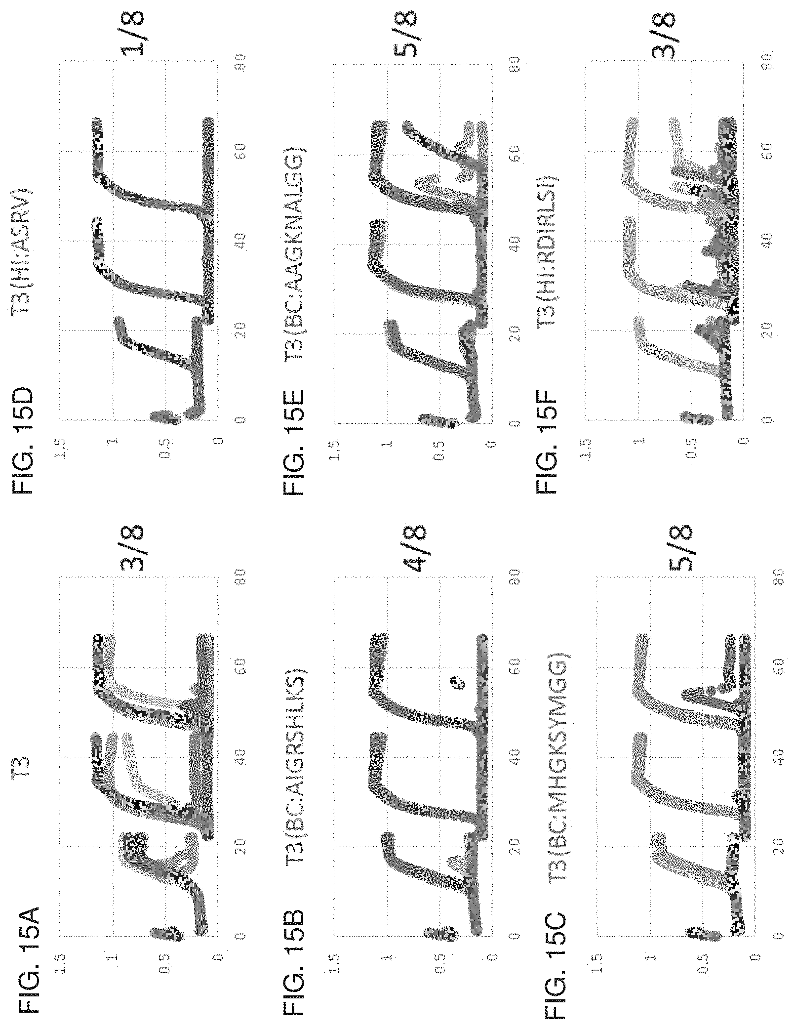
D00017

D00018
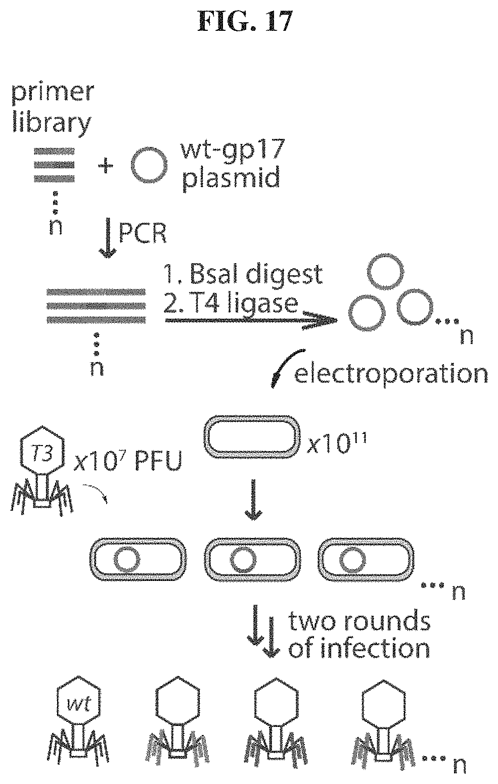
D00019
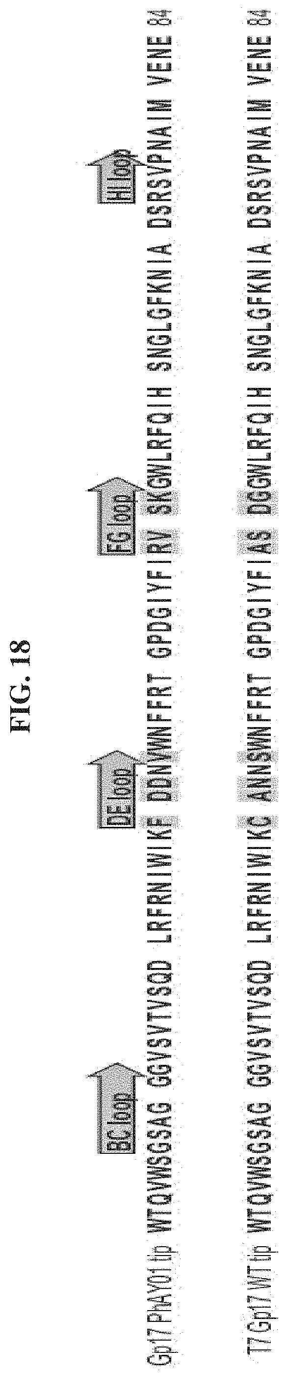
D00020
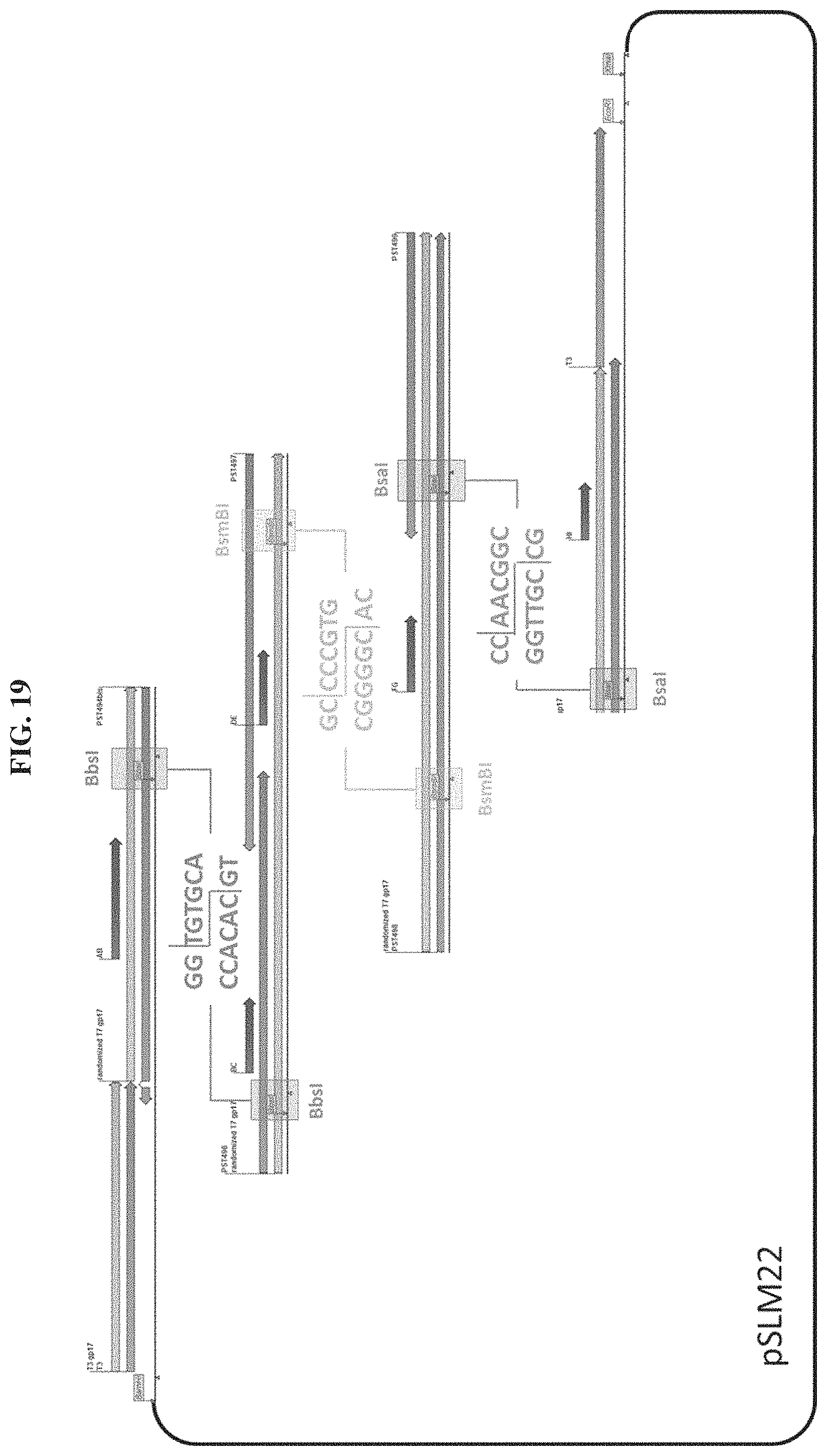
D00021

D00022
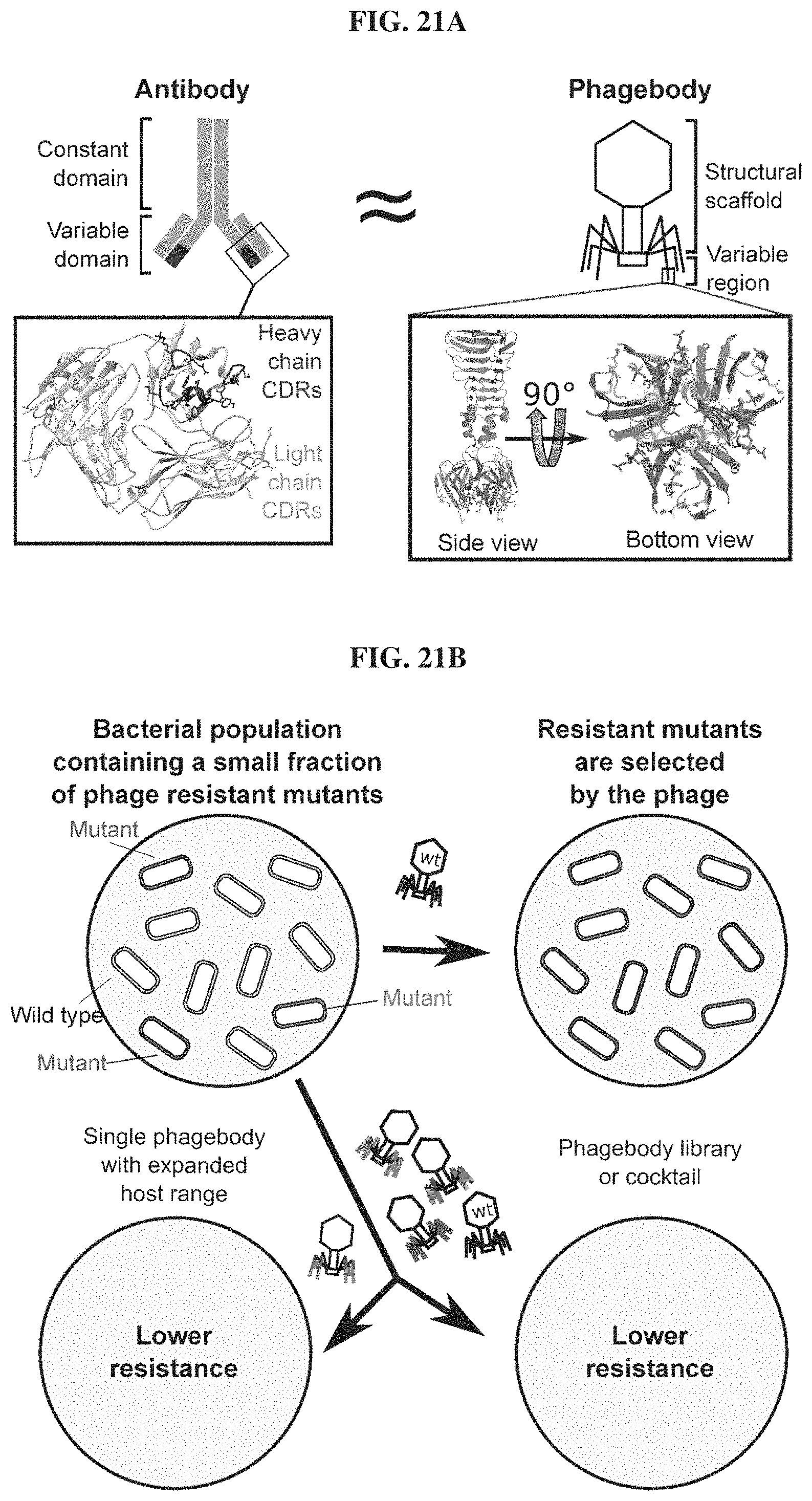
D00023
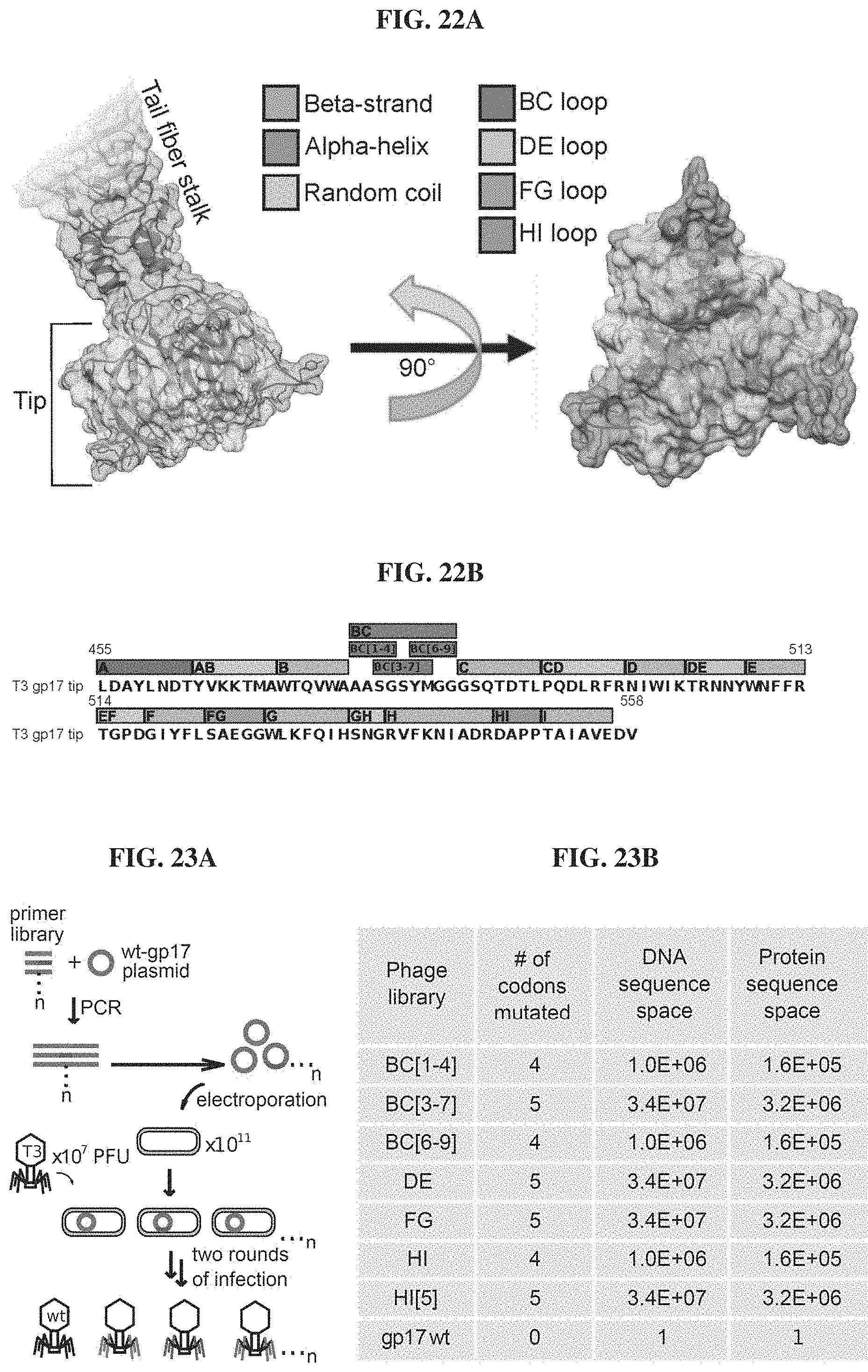
D00024
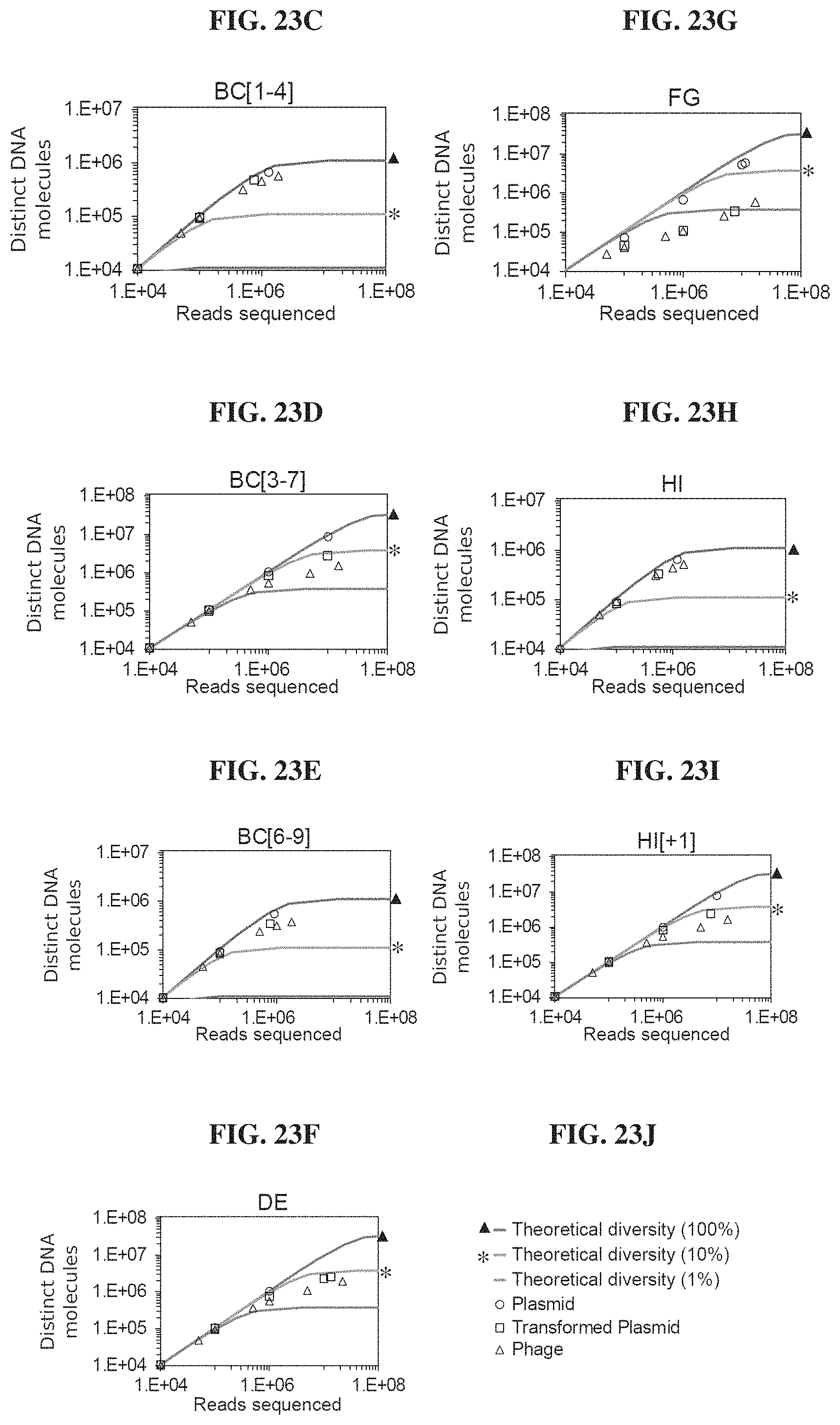
D00025
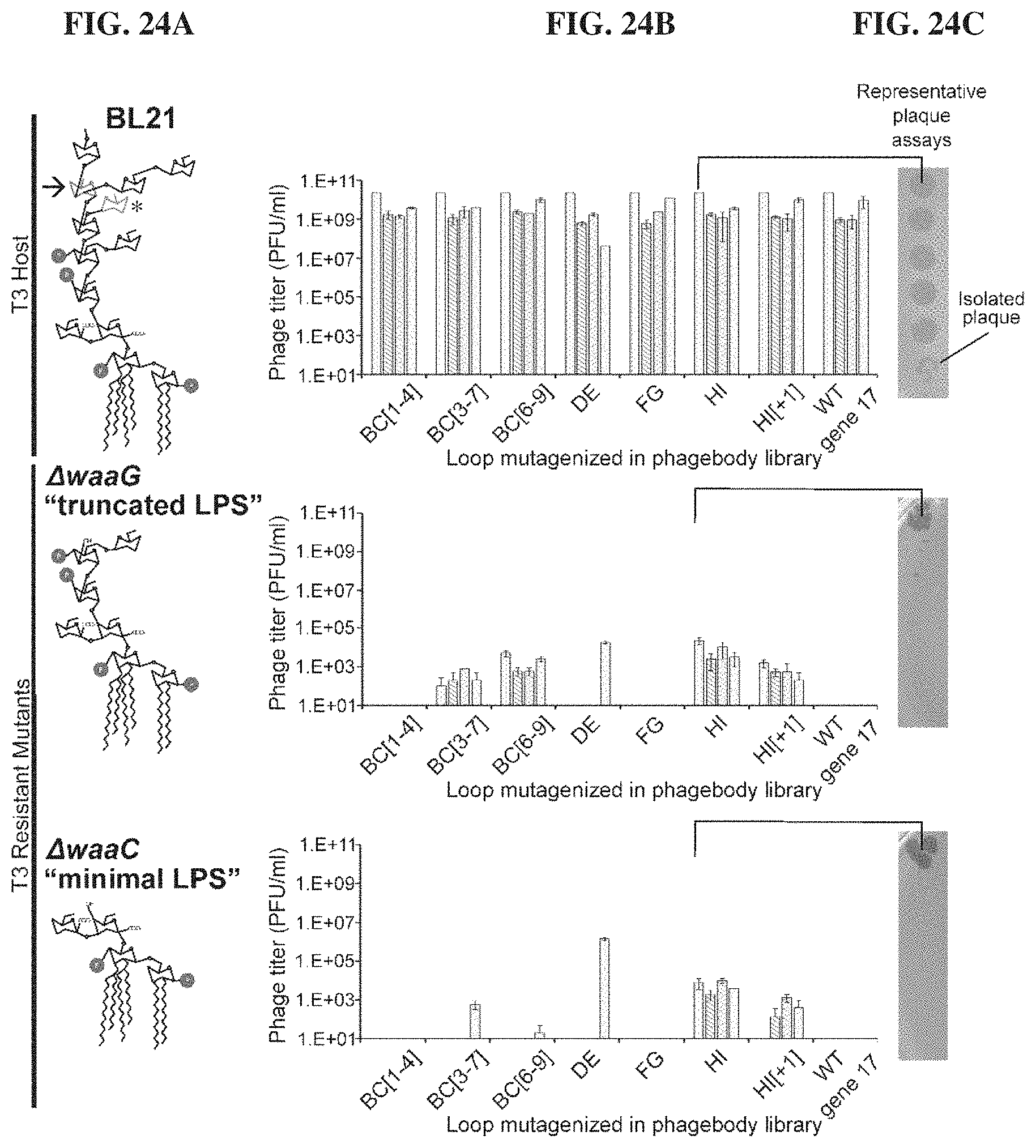
D00026
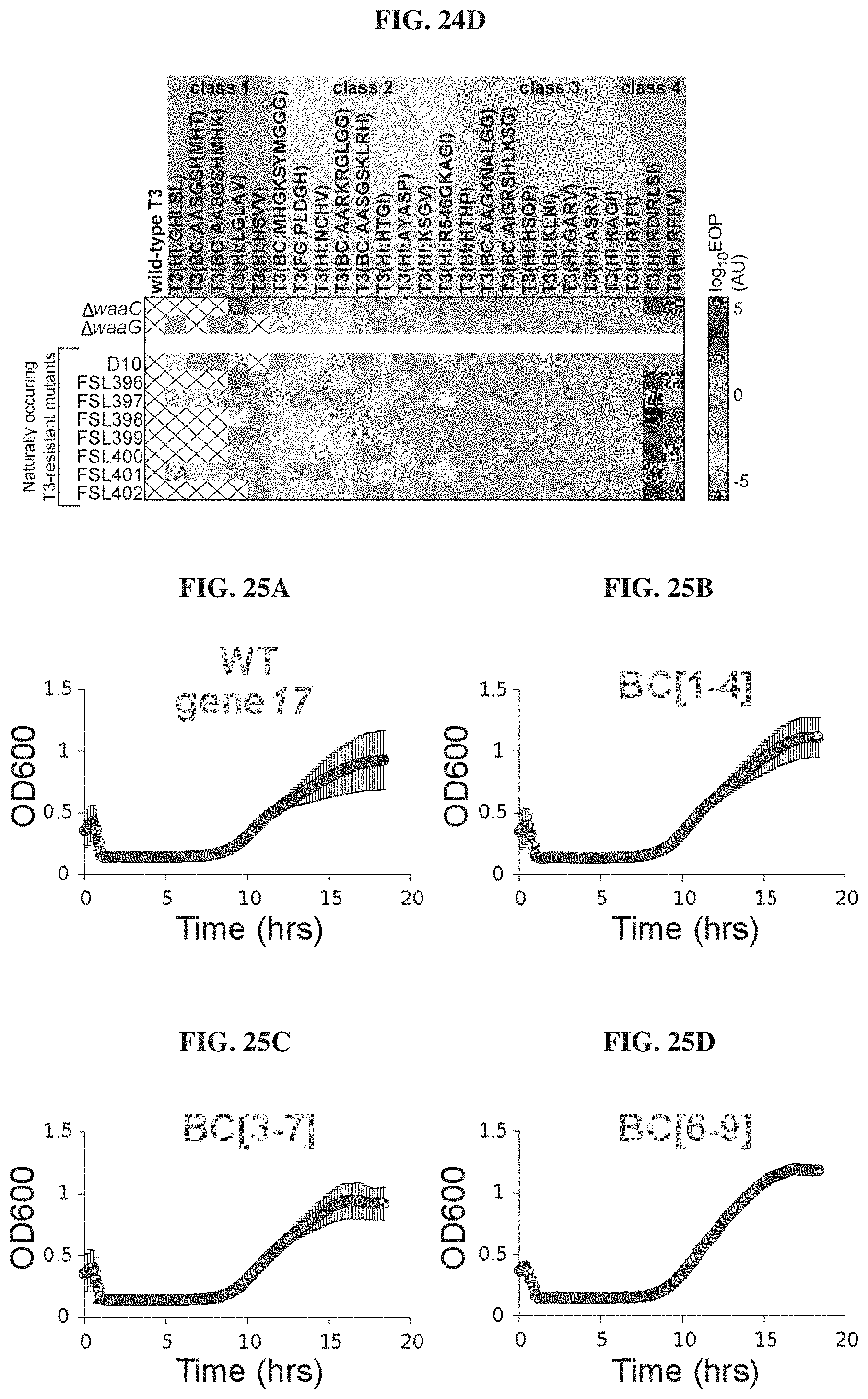
D00027
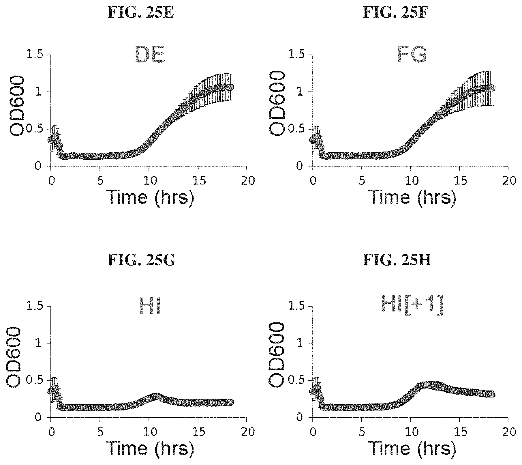
D00028
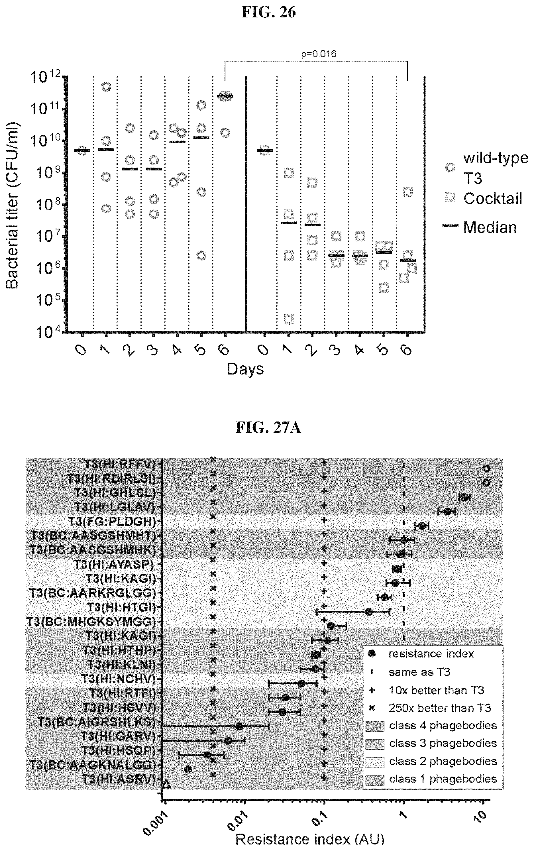
D00029

D00030

D00031
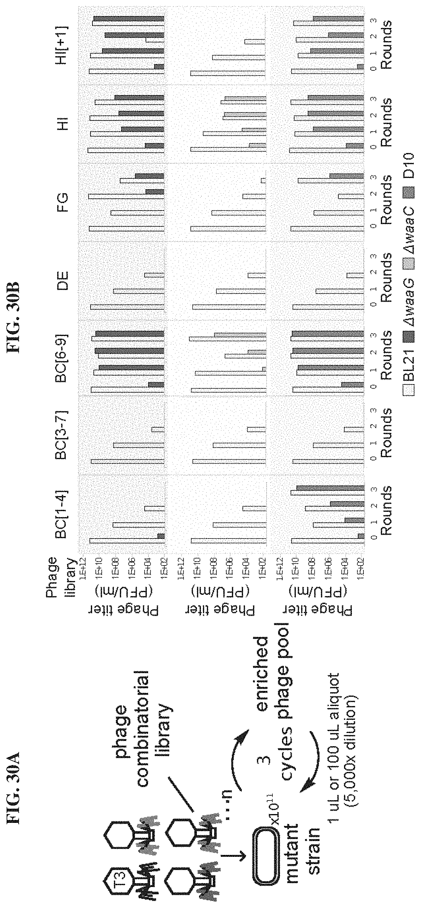
D00032

S00001
XML
uspto.report is an independent third-party trademark research tool that is not affiliated, endorsed, or sponsored by the United States Patent and Trademark Office (USPTO) or any other governmental organization. The information provided by uspto.report is based on publicly available data at the time of writing and is intended for informational purposes only.
While we strive to provide accurate and up-to-date information, we do not guarantee the accuracy, completeness, reliability, or suitability of the information displayed on this site. The use of this site is at your own risk. Any reliance you place on such information is therefore strictly at your own risk.
All official trademark data, including owner information, should be verified by visiting the official USPTO website at www.uspto.gov. This site is not intended to replace professional legal advice and should not be used as a substitute for consulting with a legal professional who is knowledgeable about trademark law.