Human airway stem cells in lung epithelial engineering
Ott , et al.
U.S. patent number 10,624,992 [Application Number 15/595,464] was granted by the patent office on 2020-04-21 for human airway stem cells in lung epithelial engineering. This patent grant is currently assigned to The General Hospital Corporation. The grantee listed for this patent is The General Hospital Corporation. Invention is credited to Sarah E. Gilpin, Harald C. Ott.


View All Diagrams
| United States Patent | 10,624,992 |
| Ott , et al. | April 21, 2020 |
Human airway stem cells in lung epithelial engineering
Abstract
Methods of using human airway stem cells in lung epithelial engineering, optionally wherein the cells are contacted with a gamma secretase inhibitor, bioartificial airway organs produced thereby, and the use thereof, e.g., for transplantation. Also methods of treating a bio-artificial matrix with Tenascin-C and/or fibrillin 2.
| Inventors: | Ott; Harald C. (Wenham, MA), Gilpin; Sarah E. (Somerville, MA) | ||||||||||
|---|---|---|---|---|---|---|---|---|---|---|---|
| Applicant: |
|
||||||||||
| Assignee: | The General Hospital
Corporation (Boston, MA) |
||||||||||
| Family ID: | 60297304 | ||||||||||
| Appl. No.: | 15/595,464 | ||||||||||
| Filed: | May 15, 2017 |
Prior Publication Data
| Document Identifier | Publication Date | |
|---|---|---|
| US 20170326273 A1 | Nov 16, 2017 | |
Related U.S. Patent Documents
| Application Number | Filing Date | Patent Number | Issue Date | ||
|---|---|---|---|---|---|
| PCT/US2017/031076 | May 4, 2017 | ||||
| 62483760 | Apr 10, 2017 | ||||
| 62426146 | Nov 23, 2016 | ||||
| 62337041 | May 16, 2016 | ||||
| Current U.S. Class: | 1/1 |
| Current CPC Class: | A61F 2/04 (20130101); A61L 27/3895 (20130101); A61L 27/3683 (20130101); C12N 5/0697 (20130101); C12N 5/0688 (20130101); A61L 27/3891 (20130101); A61L 27/3808 (20130101); A61L 27/3604 (20130101); A61L 27/3882 (20130101); A61L 27/3834 (20130101); A61L 27/3886 (20130101); C12N 2502/27 (20130101); C12N 2513/00 (20130101); C12N 2501/999 (20130101); C12N 2501/50 (20130101); C12N 2533/90 (20130101); C12N 2501/998 (20130101); C12N 2501/42 (20130101); C12N 2501/734 (20130101); A61L 2430/22 (20130101); C12N 2533/54 (20130101); C12N 2502/28 (20130101); C12N 2501/165 (20130101) |
| Current International Class: | A61F 2/04 (20130101); A61L 27/38 (20060101); C12N 5/071 (20100101); A61L 27/36 (20060101) |
References Cited [Referenced By]
U.S. Patent Documents
| 4446229 | May 1984 | Indech |
| 4598706 | July 1986 | Darowski |
| 5540225 | July 1996 | Schutt |
| 5750329 | May 1998 | Quinn et al. |
| 6087552 | July 2000 | Gregory |
| 6121042 | September 2000 | Peterson |
| 6416995 | July 2002 | Wolfinbarger |
| 6479064 | November 2002 | Atala |
| 7662409 | February 2010 | Masters |
| 9005885 | April 2015 | Ott |
| 2002/0172705 | November 2002 | Murphy et al. |
| 2002/0182241 | December 2002 | Borenstein et al. |
| 2002/0182261 | December 2002 | Dai et al. |
| 2003/0087428 | May 2003 | Wolfinbarger et al. |
| 2003/0129751 | July 2003 | Grikscheit et al. |
| 2003/0166274 | September 2003 | Hewitt et al. |
| 2003/0180268 | September 2003 | Atala |
| 2005/0107868 | May 2005 | Yasuhide et al. |
| 2005/0196423 | September 2005 | Batich et al. |
| 2005/0256588 | November 2005 | Sawa et al. |
| 2007/0059293 | March 2007 | Atala |
| 2007/0244568 | October 2007 | Matsuda et al. |
| 2008/0017194 | January 2008 | Hassanein et al. |
| 2008/0131473 | June 2008 | Brown et al. |
| 2008/0292595 | November 2008 | Arbetman et al. |
| 2008/0292677 | November 2008 | Cortiella et al. |
| 2009/0035855 | February 2009 | Ying et al. |
| 2009/0060961 | March 2009 | Naruse et al. |
| 2009/0075282 | March 2009 | Mahmood et al. |
| 2009/0142836 | June 2009 | Wang et al. |
| 2009/0202977 | August 2009 | Ott et al. |
| 2010/0034791 | February 2010 | Lelkes et al. |
| 2010/0092433 | April 2010 | Levenberg et al. |
| 2012/0064537 | March 2012 | Ross |
| 2012/0141439 | June 2012 | Ott |
| 2012/0302499 | November 2012 | Matheny |
| 2013/0084266 | April 2013 | Ott et al. |
| 2014/0273220 | September 2014 | Gerecht |
| 2015/0182560 | July 2015 | Calle et al. |
| 2015/0289501 | October 2015 | Raredon et al. |
| 2751133 | Feb 2009 | CA | |||
| 1911438 | Feb 2007 | CN | |||
| 1555031 | Jul 2005 | EP | |||
| 2484754 | Aug 2012 | EP | |||
| WO 2002/053193 | Jul 2002 | WO | |||
| WO 2007/025233 | Mar 2007 | WO | |||
| WO 2007/095192 | Aug 2007 | WO | |||
| WO 2010/091188 | Aug 2010 | WO | |||
| WO 2010/141803 | Dec 2010 | WO | |||
| WO 2011/059808 | May 2011 | WO | |||
| WO 2013/071096 | May 2013 | WO | |||
| WO-2013066802 | May 2013 | WO | |||
| WO 2014/110135 | Jul 2014 | WO | |||
| WO 2014/168264 | Oct 2014 | WO | |||
| WO 2014/200340 | Dec 2014 | WO | |||
| WO 2015/108893 | Jul 2015 | WO | |||
| WO 2015/119642 | Aug 2015 | WO | |||
| WO 2015/138999 | Sep 2015 | WO | |||
Other References
|
Tomoshi et al, "Ventilation-Based Decellularization System of the Lung" Bioresearch Open Access, (2016) 5(1); 118-126. Published May 1, 2016 (Year: 2016). cited by examiner . Tsao et al "Y-Secretase Activation of Notch Signaling Regulates the Balance of Proximal and Distal Fates in Progenitor Cells of the Developing Lung" The Journal of Biological Chemistry. (2008) vol. 283, No. 43, pp. 29532-29544 (Year: 2008). cited by examiner . Albelda et al., "Effects of increased ventilation on lung lymph flow in unanesthetized sheep," J Appl Physiol, Jun. 1986, 60(6):2063-70. cited by applicant . Andrade et al., "Cell-based tissue engineering for lung regeneration," Am J Physiol Lung Cell Mol Physiol, Feb. 2007, 292(2):L510-8. cited by applicant . Au et al., Bone marrow-derived mesenchymal stem cells facilitate engineering of long-lasting functional vasculature, Blood, May 2008, 111: 4551-4558. cited by applicant . Badylak et al., "Whole-organ tissue engineering: decellularization and recellularization of three-dimensional matrix scaffolds," Annual Review of Biomedical Engineering, 2011, 13: 27-53. cited by applicant . Balestrini and Niklason, "Extracellular matrix as a driver for lung regeneration," Annals of Biomedical Engineering, Mar. 2015, 43: 568-576. cited by applicant . Baptista et al., "Whole Organ Decellularization--A Tool for Bioscaffold Fabrication and Organ," Bioengineering, 31st Annual International Conference of the IEEE EMBS, Sep. 2-6, 2009. cited by applicant . Barkauskas et al., "Type 2 alveolar cells are stem cells in adult lung," The Journal of Clinical Investigation, Jul. 2013, 123: 3025-3036. cited by applicant . Bhattacharya et al., "Lung expansion and the perialveolar interstitial pressure gradient," J Appl Physiol, Jun. 1989, 66: 2600-5. cited by applicant . Boasquevisque et al., "Surgical Techniques: Lung Transplant and Lung vol. Reduction," Proceedings of the American Thoracic Society, Jan. 2009, 6:66-78. cited by applicant . Bonvillain et al., "Nonhuman primate lung decellularization and recellularization using a specialized large-organ bioreactor," J Vis Exp, Dec. 2013, (82):e50825. cited by applicant . Booth et al., "Acellular Normal and Fibrotic Human Lung Matrices as a Culture System for In Vitro Investigation," American Journal of Respiratory and Critical Care Medicine, 2012, 186(9):866-76. cited by applicant . Brew et al., "Mechanical Ventilation Injury and Repair in Extremely and Very Preterm Lungs," PLOS One, 2013, 8(5):e63905. cited by applicant . Bribriesco et al., "Experimental models of lung Transplantation," Front Biosci (Elite Ed), Jan. 2013, 5:266-72. cited by applicant . Brudno et al. "Enhancing microvascular formation and vessel maturation through temporal control over multiple pro-angiogenic and pro-maturation factors," Biomaterials, Dec. 2013, 34: 9201-9209. cited by applicant . Camargo et al., "Surgical maneuvers for the management of bronchial complications in lung transplantation," Eur J Cardiothorac Surg,, 2008, 34:1206-1209. cited by applicant . Canadian Office Action in Application No. 2,762,590, dated Sep. 11, 2017. cited by applicant . Canadian Office Action in Canadian Application No. 2,762,590, dated Apr. 15, 2016, 13 pages. cited by applicant . Charest et al., "Design and validation of a clinical-scale bioreactor for long-term isolated lung culture," Biomaterials, Jun. 2015, 52: 79-87. cited by applicant . Chen et al., "Formation of lung alveolar-like structures in collagen-glycosaminoglycan scaffolds in vitro," Tissue Eng., Sep.-Oct. 2005, 11(9-10):1436-48. cited by applicant . Conconi et al., "Tracheal matrices, obtained by a detergent-enzymatic method, support in vitro the adhesion of chondrocytes and tracheal epithelial cells," Transplant International, 2005, 18:727-734. cited by applicant . Cotiella et al., "Tissue-Engineered Lung: An In Vivo and In Vitro Comparison of Polyglycolic Acid and Pluronic F-127 Hydrogel/Somatic Lung Progenitor Cell Constructs to Support Tissue Growth," Tissue Engineering, 2006, 12:1213-1225. cited by applicant . Crosby and Waters, "Epithelial repair mechanisms in the lung," American Journal of Physiology Lung Cellular and Molecular Physiology, 2010, 298: L715-731. cited by applicant . Daley et al., "Extracellular matrix dynamics in development and regenerative medicine," Journal of Cell Science, 2008, 121: 255-264. cited by applicant . Davidson et al., "Murine epithelial cells: isolation and culture," Journal of Cystic Fibrosis, Aug. 2004, 3 Suppl 2: 59-62. cited by applicant . Declaration of Harald C. Ott, M.D. Under 37 CFR 1.131. and Ott, Curriculum Vitae, Apr. 11, 2014, 17 pages. cited by applicant . Desai and Cardoso, "Growth factors in lung development and disease: friends or foe?," Respire. Res., 2002, 3:2. cited by applicant . Desai et al., "Alveolar progenitor and stem cells in lung development, renewal and cancer," Nature, Mar. 2014, 507: 190-194. cited by applicant . Ding et al., "Design of compliance chamber and after-load in apparatus for cultured endothelial cells subjected to stresses," Cell Biology International, 2006, 30:439-444. cited by applicant . Dupuit et al., "Differentiated and functional human airway epithelium regeneration in tracheal xenografts," American Journal of Physiology Lung Cellular and Molecular Physiology, 2000, 278: L165-176. cited by applicant . Erasmus et al., "Normothermic ex vivo lung perfusion of non-heart-beating donor lungs in pigs: from pretransplant function analysis towards a 6-h machine preservation," Transpl Int , Jul. 2006, 19: 589-593. cited by applicant . Evans et al., "Cellular and molecular characteristics of basal cells in airway epithelium," Experimental Lung Research, 2001, 27: 401-415. cited by applicant . Evans et al., "Renewal of alveolar epithelium in the rat following exposure to NO2," The American Journal of Pathology, 1973; 70: 175-198. cited by applicant . Evans et al., "Role of nonciliated cells in renewal of the bronchial epithelium of rats exposed to NO2," The American Journal of Pathology, 1986, 123: 126-133. cited by applicant . First Chinese Office Action in Chinese Application No. 201080032724.4, dated Dec. 4, 2012, 7 pages, (with English translation). cited by applicant . Fulcher and Randell, "Human nasal and tracheo-bronchial respiratory epithelial cell culture," Methods in Molecular Biology, 2013, 945: 109-121. cited by applicant . Gaissert and Patterson, "Surgical Techniques of Single and Bilateral Lung Transplantation," The Transplantation and Replacement of Thoracic Organs, 1996, 457-463. cited by applicant . Gilbert et al., "Decellularization of tissues and organs," Biomaterials, 2006, 27(9) :3675-83. cited by applicant . Gilpin and Ott, "Using Nature's Platform to Engineer Bio-Artificial Lungs," Annals of the American Thoracic Society, 2015, 12 Suppl 1: S45-49. cited by applicant . Gilpin et al., "Enhanced lung epithelial specification of human induced pluripotent stem cells on decellularized lung matrix," Ann Thorac Surg, Nov. 2014, 98(5):1721-9. cited by applicant . Gilpin et al., "Perfusion decellularization of human and porcine lungs: Bringing the matrix to clinical scale," Journal of Heart and Lung Transplantation, Mar. 2014, 33: 298-308. cited by applicant . Godin et al., "Decreased Laminin Expression by Human Lung Epithelial Cells and Fibroblasts Cultured in Acellular Lung Scaffolds from Aged Mice," PloS One, 2016, 11(3):e0150966. cited by applicant . Gomi et al., "Activation of NOTCH1 or NOTCH3 signaling skews human airway basal cell differentiation toward a secretory pathway," PloS one, Feb. 2015, 10: e0116507. cited by applicant . Gordon et al., "Mechanical Allostery: Evidence for a Force Requirement in the Proteolytic Activation of Notch," Developmental Cell, 2015, 33: 729-736. cited by applicant . Granger et al., "Dynamics and control of transmicrovascular fluid exchange," Edema, 1984, 8:189-228. cited by applicant . Gro et al., "Improved generation of patient-specific induced pluripotent stem cells using a chemically-defined and matrigel-based approach," Curr Mol Med., Jun. 2013,13:765-76. cited by applicant . Guseh et al., "Notch signaling promotes airway mucous metaplasia and inhibits alveolar development," Development, May 2009, 136: 1751-1759. cited by applicant . Guyette et al, "Perfusion decellularization of whole organs," Nat Protoc, 2014, 9: 1451-1468. cited by applicant . Hackett et al., "The human airway epithelial basal cell transcriptome," PloS one, May 2011, 6: e18378. cited by applicant . Hoganson et al., "Tissue Engineering and Organ Structure: A Vascularized Approach to Liver and Lung," Pediatric Research, May 2008, 63(5):520-526. cited by applicant . Hong et al., "Basal cells are a multipotent progenitor capable of renewing the bronchial epithelium," The American Journal of Pathology 2004, 164: 577-588. cited by applicant . Hong et al., "In vivo differentiation potential of tracheal basal cells: evidence for multipotent and unipotent subpopulations," American Journal of Physiology Lung Cellular and Molecular Physiology, 2004, 286: L643-649. cited by applicant . Hou et al., "Pluripotent Stem Cells Induced from Mouse Somatic Cells by Small-Molecule Compounds," Science, Jul. 2013, 341:651-654. cited by applicant . Huang et al., The in vitro generation of lung and airway progenitor cells from human pluripotent stem cells, Nature Protocols, Mar. 2015, 10: 413-425. cited by applicant . Ichii et al., Current status of islet cell transplantation, J. Hepatobiliary Pancreat. Surg., 2009, 16:101-112. cited by applicant . Inayama et al., "The differentiation potential of tracheal basal cells," Laboratory Investigation; a Journal of Technical Methods and Pathology, 1988, 58: 706-717. cited by applicant . Ingenito et al., "Design and testing of biological scaffolds for delivering reparative cells to target sites in the lung," J Tissue Eng Regen Med., 2010, 4: 259-272. cited by applicant . International Preliminary Report on Patentability and Written Opinion in International Application No. PCT/US2010/037379, dated Dec. 6, 2011, 8 pages. cited by applicant . International Preliminary Report on Patentability and Written Opinion in International Application No. PCT/US2010/054689, dated May 1, 2012, 9 pages. cited by applicant . International Preliminary Report on Patentability in International Application No. PCT/US2015/020605, dated Sep. 14, 2016. cited by applicant . International Search Report and Written Opinion in International Application No. PCT/US2010/037379, dated Mar. 1, 2011, 9 pages. cited by applicant . International Search Report and Written Opinion in International Application No. PCT/US2010/054689, dated Jul. 11, 2011, 13 pages. cited by applicant . International Search Report and Written Opinion in International Application No. PCT/US2016/051049, dated Dec. 12, 2016, 13 pages. cited by applicant . International Search Report and Written Opinion dated May 26, 2015 in International Application No. PCT/US2015/020605, 13 pgs. cited by applicant . International Search Report in International Application No. PCT/US2010/23213, dated Aug. 12, 2010, 4 pages. cited by applicant . Jain et al., "Plasticity of Hopx(+) type I alveolar cells to regenerate type II cells in the lung," Nature Communications, 2015, 6: 6727. cited by applicant . James et al., "Expansion and maintenance of human embryonic stem cell-derived endothelial cells by TGFbeta inhibition is Id 1 dependent," Nat Biotechnol, Feb. 2010, 28: 161-166. cited by applicant . Japanese Office Action in Japanese Application No. 2012-514170, dated Sep. 16, 2014, 14 pages. cited by applicant . Karp et al., "An in vitro model of differentiated human airway epithelia. Methods for establishing primary cultures," Methods in Molecular Biology, 2002, 188: 115-137. cited by applicant . Kim et al., "Identification of bronchioalveolar stem cells in normal lung and lung cancer," Cell, Jun. 2005, 121: 823-835. cited by applicant . Korean Office Action in Korean Application No. 2012-7000278, dated Jul. 14, 2016, 12 pages (with English translation). cited by applicant . Kotton and Morrisey, "Lung regeneration: mechanisms, applications and emerging stem cell populations," Nature Medicine, Aug. 2014, 20: 822-832. cited by applicant . Li et al., "A Single Use, Scalable Perfusion Bioreactor System," BioProcess International, Supplement, 2009. cited by applicant . Li et al., "Generation of iPSCs from mouse fibroblasts with a single gene, Oct4, and small molecules," Cell Res., Jan. 2011, 21:196-204. cited by applicant . Liao et al, "Effects of Decellularization on the Mechanical and Structural Properties of the Porcine Aortic Valve Leaflet," Biomaterials, Mar. 2008, 29(8): 1065-74. cited by applicant . Lin and Ying, "Mechanism and method for generating tumor-free iPS cells using intronic microRNA miR-302 induction," Methods Mol Biol., 2013, 936:295-312. cited by applicant . Lin et al., "Biocompatibility of Poly-D2009L-lactic acid (PDLLA) for Lung Tissue Engineering," Journal of Biomaterials Applications, 2006, 21:109-118. cited by applicant . Lin et al., "Homo- and heterotypic fibrillin-1 and -2 interactions constitute the basis for the assembly of microfibrils," J. Biol. Chem, Dec. 2002, 277: 50795-50804. cited by applicant . Liu et al., "ROCK inhibitor and feeder cells induce the conditional reprogramming of epithelial cells," The American Journal of Pathology, Feb. 2012, 180: 599-607. cited by applicant . Longmire et al., "Efficient derivation of purified lung and thyroid progenitors from embryonic stem cells," Cell Stem Cell, 2012, 10: 398-411. cited by applicant . Macchiarini et al., "Clinical transplantation of a tissue-engineered airway," Lancet, 2008, 372:2023-2030. cited by applicant . Maghsoudlou al., "Preservation of micro-architecture and angiogenic potential in a pulmonary acellular matrix obtained using intermittent intra-tracheal flow of detergent enzymatic treatment," Biomaterials, Sep. 2013, 34(28):6638-48. cited by applicant . Malik and Rao, "A Review of the Methods for Human iPSC Derivation," Methods Mol Biol., 2013, 997:23-33. cited by applicant . McBride et al., "Lung growth and airway function after lobectomy in infancy for congenital lobar emphysema," The Journal of Clinical Investigation, Nov. 1980, 66(5):962-70. cited by applicant . Meiners et al., "Hallmarks of the ageing lung," The European Respiratory Journal, Mar. 2015, 45(3):807-27. cited by applicant . Melero-Martin et al., "In vivo vasculogenic potential of human blood-derived endothelial progenitor cells," Blood, Jun. 2007, 109: 4761-4768. cited by applicant . Mercer et al., "Cell number and distribution in human and rat airways," American Journal of Respiratory Cell and Molecular Biology, Jun. 1994, 10: 613-624. cited by applicant . Midwood and Orend, "The role of tenascin-C in tissue injury and tumorigenesis," Journal of Cell Communication and Signaling, Oct. 2009, 3:287-310. cited by applicant . Mondrinos et al., "Engineering Three-Dimensional Pulmonary Tissue Constructs," Tissue Engineering, 2006, 12(4):717-728. cited by applicant . Mou et al., "Generation of multipotent lung and airway progenitors from mouse ESCs and patient-specific cystic fibrosis iPSCs," Cell Stem Cell, 2012, 10: 385-397. cited by applicant . Musah et al., "Repair of tracheal epithelium by basal cells after chlorine-induced injury," Respiratory Research, 2012, 13: 107. cited by applicant . Nichols et al., "Giving new life to old lungs: methods to produce and assess whole human paediatric bioengineered lungs," J Tissue Eng Regen Med, Jan. 2016, 11: 2136-2152. cited by applicant . Nichols et al., "Engineering of a Complex Organ Progress Toward Development of a Tissue-engineered Lung," Proceedings of the American Thoracic Society, 2008, 5:723-730. cited by applicant . Nichols et al., "Production and assessment of decellularized pig and human lung scaffolds," Tissue Eng Part A, Sep. 2013, 19(17-18):2045-62. cited by applicant . Okano et al., "Steps toward safe cell therapy using induced pluripotent stem cells," Circ Res., Feb. 2013, 112(3):523-33. cited by applicant . O'Koren et al., "Loss of basal cells precedes bronchiolitis obliterans-like pathological changes in a murine model of chlorine gas inhalation," American Journal of Respiratory Cell and Molecular Biology, 2013, 49: 788-797. cited by applicant . O'Neill et al., "Decellularization of human and porcine lung tissues for pulmonary tissue engineering," Ann Thorac Surg., Sep. 2013, 96(3): 1046-55. cited by applicant . Ott et al., "Perfusion-decellularized matrix: using nature's platform to engineer a bioartificial heart," Nature Medicine, 2008, 14(2):213-221. cited by applicant . Ott et al., "Regeneration and orthotopic transplantation of a bioartificial lung," Nat Med, Aug. 2010,16(8):927-33. cited by applicant . Park et al., "Reprogramming of human somatic cells to pluripotency with defined factors," Nature, Jan. 2008, 451 :141-146. cited by applicant . Pasque et al., "Standardizing thoracic organ procurement for transplantation," J Thorac Cardiovasc Surg., Jan. 2010, 139(1):13-7. cited by applicant . Petersen et al., "Tissue-Engineered Lungs for in Vivo Implantation," Science, Jul. 2010, 329: 538-541. cited by applicant . Petersen et al., "Bioreactor for the long-term culture of lung tissue," Cell Transplant, 2011, 20(7):1117-26. cited by applicant . Petersen, "In Vitro Development of Engineered Lung Tissue," Duke University Doctoral Dissertation, 2009, 283 pages. cited by applicant . Pezzulo et al., "The air-liquid interface and use of primary cell cultures are important to recapitulate the transcriptional profile of in vivo airway epithelia," American Journal of Physiology Lung Cellular and Molecular Physiology, 2011, 300: L25-31. cited by applicant . Polacheck et al., "Interstitial flow influences direction of tumor cell migration through competing mechanisms," PNAS, 2011, 108: 11115-11120. cited by applicant . Price et al., "Development of a decellularized lung bioreactor system for bioengineering the lung: the matrix reloaded," Tissue Eng Part A, Aug. 2010, 16(8):2581-91. cited by applicant . Rawlins and Hogan, "Epithelial stem cells of the lung: privileged few or opportunities for many?," Development, 2006, 133: 2455-2465. cited by applicant . Reed et al., "Stem cell-derived endothelial cells for cardiovascular disease: a therapeutic perspective," Br J Clin Pharrnacol, Apr. 2013, 75(4):897-906. cited by applicant . Ren et al., "Engineering pulmonary vasculature in decellularized rat and human lungs," Nature Biotechnology, Oct. 2015, 33: 1097-1102. cited by applicant . Rey-Santano et al., "Effect of Surfactant and Partial Liquid Ventilation Treatment on Gas Exchange and Lung Mechanics in Immature Lambs: Influence of Gestational Age," PLOS One, 2013, 8:e56127. cited by applicant . Rimensberger, "Neonatal respiratory failure," Current Opinion in Pediatrics, 2002, 14:315-321. cited by applicant . Rock et al., "Airway basal stem cells: a perspective on their roles in epithelial homeostasis and remodeling," Disease Models & Mechanisms, 2010; 3: 545-556. cited by applicant . Rock et al., "Basal cells as stem cells of the mouse trachea and human airway epithelium," PNAS, 2009, 106: 12771-12775. cited by applicant . Rock et al., "Notch-dependent differentiation of adult airway basal stem cells," Cell Stem Cell, Jun. 2011, 8: 639-648. cited by applicant . Rojanarat et al., "Isoniazid Proliposome Powders for Inhalation--Preparation, Characterization and Cell Culture Studies," International Journal of Molecular Sciences, 2011, 12:4414-4434. cited by applicant . Rosen et al., "Preconditioning allows engraftment of mouse and human embryonic lung cells, enabling lung repair in mice," Nature Medicine, 2015, 21: 869-879. cited by applicant . Sato et al., "Replacement of the left main bronchus with a tissue-engineered prosthesis in a canine model," Ann. Thorac. Surg., 2008, 86:422-428. cited by applicant . Second Chinese Office Action in Chinese Application No. 201080032724.4, dated Sep. 24, 2013, 9 pages (with English translation). cited by applicant . Sharpless and Sherr, "Forging a signature of in vivo senescence," Nature Reviews Cancer, Jul. 2015, 15: 397-408. cited by applicant . Sokocevic et al., "The effect of age and emphysematous and fibrotic injury on the re-cellularization of de-cellularized lungs," Biomaterials, 2013, 34(13):3256-69. cited by applicant . Song and Ott, "Bioartificial lung engineering," Am J Transplant, Feb. 2012,12(2):283-8. cited by applicant . Song et al., "Enhanced in vivo function of bioartificial lungs in rats," Ann Thorac Surg., 2011, 92(3):998-1005. cited by applicant . Song et al., "Organ engineering based on decellularized matrix scaffolds," Trends in Molecular Medicine, 2011, 17(8):424-432. cited by applicant . Staudt et al., "Airway Basal stem/progenitor cells have diminished capacity to regenerate airway epithelium in chronic obstructive pulmonary disease," American Journal of Respiratory and Critical Care Medicine, 2014, 190: 955-958. cited by applicant . Stripp and Reynolds, "Maintenance and repair of the bronchiolar epithelium," Proceedings of the American Thoracic Society, 2008, 5: 328-333. cited by applicant . Supplementary Search Report in European Application No. 10784139, dated Aug. 29, 2013, 8 pages. cited by applicant . Takahashi et al., "Induction of Pluripotent Stem Cells from Adult Human Fibroblasts by Defined Factors," Cell, Nov. 2007,131 :861-72. cited by applicant . Tapias and Ott, "Decellularized scaffolds as a platform for bioengineered organs," Current Opinion in Organ Transplantation, 2014, 19(2):145-52. cited by applicant . Teebken et al., "Tissue Engineering of Vascular Grafts. Human Cell Seeding of Decellularised Porcine Matrix," Eur. J. Vase. Endovasc. Surg., Apr. 2000, 19:381-86. cited by applicant . Teixeira et al., "Stochastic homeostasis in human airway epithelium is achieved by neutral competition of basal cell progenitors," eLife, 2013, 2: e00966. cited by applicant . Thannickal et al., "Blue Journal Conference. Aging and Susceptibility to Lung Disease," American Journal of Respiratory and Critical Care Medicine, 2015, 191(3):261-9. cited by applicant . The Decision of Final Rejection of the Application in Chinese Application No. 201080032724.4, dated Jan. 27, 2015, 7 pages (English translation). cited by applicant . Third Chinese Office Action in Chinese Application No. 201080032724.4, dated May 9, 2014, 8 pages (English translation). cited by applicant . Toni et al., "The bioartificial thyroid: a biotechnological perspective in endocrine organ engineering for transplantation replacement," Acta Bio Medica, 2007, 78(Sunn 1):129-155. cited by applicant . Tsao et al., ".gamma.-Secretase Activation of Notch Signaling Regulates the Balance of Proximal and Distal Fates in Progenitor Cells of the Developing Lung," Journal of Biological Chemistry, Oct. 2008, 283: 29532-29544. cited by applicant . U.S. Final Office Action in U.S. Appl. No. 13/375,260, dated Oct. 25, 2013, 15 pages. cited by applicant . U.S. Final Office Action in U.S. Appl. No. 13/504,358, dated Jul. 3, 2014, 10 pages. cited by applicant . U.S. Final Office Action in U.S. Appl. No. 14/559,401, dated Feb. 8, 2017, 11 pages. cited by applicant . U.S. Non-Final Office Action in U.S. Appl. No. 13/375,260, dated Feb. 1, 2013, 11 pages. cited by applicant . U.S. Non-Final Office Action in U.S. Appl. No. 13/504,358, dated Dec. 9, 2013, 13 pages. cited by applicant . U.S. Notice of Allowance in U.S. Appl. No. 13/375,260, dated Nov. 26, 2014, 10 pages. cited by applicant . Vandenbroucke et al., "Regulation of Endothelial Junctional Permeability," Ann. N.Y. Acad. Sci., 2008, 1123:134-145. cited by applicant . Vaughan et al., "Lineage-negative progenitors mobilize to regenerate lung epithelium after major injury", Nature, 2015, 517: 621-625. cited by applicant . Venkateswaran et al., "Measurement of extravascular lung water following human brain death: implications for lung donor assessment and transplantation," Eur J Cardiothorac Surg., Jun. 2013, 43(6):1227-32. cited by applicant . Venkateswaran et al., "The proinflammatory environment in potential heart and lung donors: prevalence and impact of donor management and hormonal therapy," Transplantation, Aug. 2009, 88(4):582-8. cited by applicant . Venuta et al., "Evolving Techniques and Perspectives in Lung Transplantation," Transplantation Proceedings, Jul.-Aug. 2005, 37(6):2682-2683. cited by applicant . Wagner et al., "Comparative decellularization and recellularization of normal versus emphysematous human lungs," Biomaterials, Mar. 2014, 35: 3281-3297. cited by applicant . Wansleeben et al., "Stem cells of the adult lung: their development and role in homeostasis, regeneration, and disease," Wiley Interdisciplinary Reviews Developmental Biology, 2013, 2: 131-148. cited by applicant . Watson et al., "Clonal Dynamics Reveal Two Distinct Populations of Basal Cells in Slow-Turnover Airway Epithelium," Cell Reports, 2015, 12: 90-101. cited by applicant . Whitsett et al., "Human surfactant protein B: structure, function, regulation, and genetic disease," Physiological Reviews, Oct. 1995, 75(4):749-57. cited by applicant . Written Opinion of the International Search Authority in International Application No. PCT/US2010/23213, dated May 24, 2010, 6 pages. cited by applicant . Xu et al., "Notch signaling in lung development and disease," Advances in Experimental Medicine and Biology, 2012, 727: 89-98. cited by applicant . Yang and Conte, "Finer techniques in lung transplantation," Transplantation Proceedings, Nov. 2000, 32(7): 1521-1522. cited by applicant . Yang et al., "Expression of mutant BMPR-II in pulmonary endothelial cells promotes apoptosis and a release of factors that stimulate proliferation of pulmonary arterial smooth muscle cells," Pulmonary Circulation, 2011, 1(1):103-110. cited by applicant . Yates et al., "Skin wound healing and scarring: fetal wounds and regenerative restitution," Birth Defects Research Part C, Embryo Today, Dec. 2012, 96(4):325-33. cited by applicant . Yoshida et al., "Surgical Technique of Experimental Lung Transplantation in Rabbits," Ann Thorac Cardiovasc Surg., Jan. 2005, 11(1):7-11. cited by applicant . Yu et al., "Induced pluripotent stem cell lines derived from human somatic cells," Science, Nov. 2007, 318: 1917-20. cited by applicant . Zahm et al., "Cell migration and proliferation during the in vitro wound repair of the respiratory epithelium," Cell Motility and the Cytoskeleton, 1997, 37: 33-43. cited by applicant . Zhu et al., "Partial liquid ventilation decreases tissue and serum tumor necrosis factor-a concentrations in acute lung injury model of immature piglet induced by oleic acid," Chinese Medical Journal, 2012, 125(1):123-128. cited by applicant . Zhu et al., "Reprogramming of Human Primary Somatic Cells by OCT4 and Chemical Compounds," Cell Stem Cell., Dec. 2010, 7:651-5. cited by applicant . Zuo et al., "p63(+)Krt5(+) distal airway stem cells are essential for lung regeneration," Nature, 2015, 517: 616-620. cited by applicant . Gilpin et al. "Regenerative potential of human airway stem cells in lung epithelial engineering," Biomaterials, Sep. 2016, 108: 111-119. cited by applicant . International Search Report and Written Opinion in International Application No. PCT/US2017/031076, dated Dec. 29, 2017, 11 pages. cited by applicant . EP Extended European Search Report in European Appln. No. 17799867.1, dated Jan. 7, 2020, 12 pages. cited by applicant . Karp et al., "An in Vitro Model of Differentiated Human Airway Epithelia: Methods for Establishing Primary Cultures" Epithelial Cell Culture Protocols, 2002, 115-137. cited by applicant . Lemon et al., "The development of the bio artificial lung," British Medical Bulletin, Jun. 2014, 110(1):35-45. cited by applicant . Mendez et al., "Epithelial Cell Differentiation of Human Mesenchymal Stromal Cells in Decellularized Lung Scaffolds," Tissue Engineering Part A., Jun. 2014, 20(11-12):1735-46. cited by applicant . Schilders et al., "Regeneration of the lung: Lung stem cells and the development of lung mimicking devices", Respiratory Research, Dec. 2016, 17(1):44, 16 pages. cited by applicant . Shojaie et al., "Acellular Lung Scaffolds Direct Differentiation of Endoderm to Functional Airway Epithelial Cells: Requirement of Matrix-Bound HS Proteoglycans", Stem Cell Reports, Mar. 2015, 4(3):419-430. cited by applicant. |
Primary Examiner: Dukert; Brian A
Assistant Examiner: Zimmerman; Rebecca Lynee
Attorney, Agent or Firm: Fish & Richardson P.C.
Government Interests
FEDERALLY SPONSORED RESEARCH OR DEVELOPMENT
This invention was made with Government support under Grant Nos. OD008749 and HL108678 awarded by the National Institutes of Health. The Government has certain rights in the invention.
Parent Case Text
CLAIM OF PRIORITY
This application is a continuation of PCT/US2017/031076, filed May 4, 2017, which claims the benefit of U.S. Provisional Patent Application Ser. Nos. 62/337,041, filed on May 16, 2016; 62/426,146, filed on Nov. 23, 2016; and 62/483,760, filed on Apr. 10, 2017. The entire contents of the foregoing are hereby incorporated by reference.
Claims
What is claimed is:
1. A method of providing a bioartificial lung organ, the method comprising: providing a population of proliferative basal stem cells from a human donor wherein the cells are Krt5.sup.+p63.sup.+ cells; maintaining and expanding the cells in culture for up to five passages, and wherein the cells are passaged at 60-100%, confluency; providing a cell-free lung tissue matrix including an airway and vasculature, wherein the lung tissue matrix comprises one or both of exogenously added tenascin-c or exogenously added fibrillin-2; seeding the lung tissue matrix with the stem cells through the airway, and with endothelial cells through the vasculature; and maintaining the matrix in an organ chamber under conditions comprising providing the lung tissue matrix with wet ventilation using a liquid media comprising a notch inhibitor, for a time sufficient for a pre-selected degree of organ maturation to occur.
2. The method of claim 1, in which the organ chamber comprises a chamber pressure sensor and a bi-directional drainage chamber pump each controlled by a control module that controls the bi-directional drainage pump in response to data transmitted by the chamber pressure sensor.
3. The method of claim 1, further comprising preventing a transpulmonary pressure gradient by equilibrating a pressure level in a venous line that is connected to a pulmonary vein of the lung tissue matrix with a pressure level in a media reservoir.
4. The method of claim 1, in which the organ chamber further comprises a pneumatic pressure control module connected to the organ chamber, wherein the pneumatic pressure control module: generates negative pressure in the organ chamber during an inspiration phase; maintains the organ chamber pressure for a plateau phase; and generates positive pressure in the organ chamber during an expiration phase.
5. The method of claim 1, in which wet ventilation comprises: connecting a tracheal line that is also connected to the airway of the lung tissue matrix to a media reservoir, in which the tracheal line includes a bi-directional tracheal pump connected to the controller; inflating the lung tissue matrix with media using the bi-directional tracheal pump; and deflating the lung tissue matrix using the bi-directional tracheal pump to withdraw media from the lung tissue matrix, wherein the media is continuously refreshed during wet ventilation.
6. The method of claim 1, in which the wet ventilation comprises: connecting a tracheal line that is also connected to the airway of the lung tissue matrix to a media reservoir, in which the tracheal line includes a first pump and a second pump each connected to the controller; inflating the lung tissue matrix with media using the first pump; and deflating the lung tissue matrix using the second pump to withdraw media from the lung tissue matrix, wherein the media is continuously refreshed during wet ventilation.
7. The method of claim 6, in which the controller controls the bi-directional tracheal pump in response to data transmitted by a tracheal pressure sensor connected to the tracheal line.
8. The method of claim 1, comprising providing wet ventilation using a liquid media comprising a notch inhibitor for at least 2, 5, 7, or 10 days, optionally followed by additional wet ventilation using a liquid media not comprising a notch inhibitor.
9. The method of claim 1, comprising contacting the lung tissue matrix with one or both of tenascin-c or fibrillin-2 prior to seeding.
10. A functional lung produced by the method of claim 1.
11. The functional lung of claim 10, wherein the organ is a full lung or a vascularized portion thereof.
12. A method of treating a subject having impaired or reduced lung capacity, the method comprising transplanting the lung of claim 10 into the subject.
13. The method of claim 1, wherein the Krt5+p63+ cells are obtained from the airway of the donor.
14. The method of claim 1, wherein the cells are passaged at 80% confluency.
15. The method of claim 1, wherein the Notch inhibitor is a gamma secretase inhibitor.
16. The method of claim 1, wherein the cells are passaged in culture in the absence of a ROCK inhibitor prior to seeding the lung tissue matrix.
Description
TECHNICAL FIELD
Provided herein are methods of using human airway stem cells in lung epithelial engineering, optionally wherein the cells are contacted with a gamma secretase inhibitor, bioartificial airway organs produced thereby, and the use thereof, e.g., for transplantation.
BACKGROUND
Lung transplants represent a final hope for many patients experiencing conditions typified by lung failure, e.g., chronic obstructive pulmonary disease (COPD), cystic fibrosis, pulmonary hypertension, lung cancers, and congenital lung diseases. Typical wait time for a lung transplant can be two years or more, resulting in a 30% mortality rate for those on the waiting list. The development of techniques to engineer organs for transplantation may ultimately provide a solution for end-stage organ failure without the risk of rejection.
SUMMARY
Building upon the process of whole organ perfusion decellularization, the present inventors aimed to utilize primary human donor lung tissue-derived cells to repopulate and regenerate native lung scaffolds.
As shown herein, a proliferative basal epithelial stem cell population was isolated and expanded in culture, and robust recellularization of both rodent and human lung extracellular matrix (ECM) was achieved. Differentiation toward a ciliated airway epithelial phenotype was demonstrated both in vitro and in ex vivo whole rodent lung recellularization and biomimetic culture. Induction of a distal epithelial phenotype was achieved by inhibition of the Notch pathways through .gamma.-secretase. Increased surfactant protein-B and C expression was demonstrated by mRNA analysis in vitro, in human ECM slice culture, and in whole rodent lung culture. Recellularization of isolated human lung lobes, coupled with extended ex vivo biomimetic culture, further confirmed the regenerative capacity of this cell population. Functional analysis of the recellularized lung constructs verified cell viability and metabolic activity throughout culture, as well as dynamic organ compliance and gas exchange. On final tissue analysis, extensive re-epithelialization with physiologic tissue architecture and morphology was observed.
These results demonstrate the regenerative potential and bi-lineage capacity of human airway stem cells, which are useful in whole lung tissue bioengineering and ex vivo organ culture.
In addition, the behavior of basal epithelial stem cells (BESCs) isolated from adult human lung tissue cultured on acellular ECM derived from neonatal (aged<1 week) or adult lung donors (n=3 donors per group) was evaluated. A significant difference in cell proliferation and survival was found. In-depth proteomic analysis of the lung scaffolds was performed to quantify proteins significantly enriched in the neonatal ECM, and identified the glycoproteins Fibrillin-2 (FBN-2) and Tenascin-C (TN-C) as potential mediators of the observed effect. BESCs cultured on Collagen Type IV coated plates, supplemented with FBN-2 and/or TN-C demonstrated significantly increased proliferation and decreased cellular senescence; (note that this difference was also found when compared to untreated plates (no Collagen IV coating). No significant increase in epithelial-to-mesenchymal transition was observed. In vitro wound closure was also increased on FBN-2 and/or TN-C. Decellularized lung scaffolds pre-treated with FBN-2 and/or TN-C prior to re-epithelialization supported greater epithelial proliferation and tissue remodeling. BESC distribution, matrix alignment, and overall tissue morphology was improved on treated lung scaffolds, after 3 and 7 days of ex vivo lung culture. These results demonstrate that scaffold re-epithelialization is enhanced on neonatal lung ECM, and that supplementation of FBN-2 and TN-C to the native scaffold is a valuable tool in lung tissue regeneration.
Thus, provided herein are methods for providing a bioartificial lung organ. The methods include providing a population of proliferative basal stem cells from a human donor wherein the cells are Krt5.sup.+p63.sup.+ cells, preferably obtained from the airway of the donor; optionally maintaining and expanding the cells in culture for up to five passages (e.g., wherein cells were passaged at 60-100%, e.g., 80%, confluency), optionally in the absence of a ROCK inhibitor; providing a (cell-free) lung tissue matrix including an airway and substantial vasculature; seeding the lung tissue matrix with the stem cells through the airway, and with endothelial cells through the vasculature; and maintaining the matrix under conditions sufficient for the formation of a functional epithelium in the airways and functional vasculature, wherein maintaining the matrix comprises providing the lung tissue matrix with wet ventilation using a liquid media comprising a notch inhibitor, e.g., a gamma secretase inhibitor, for a time sufficient for a first desired degree of organ maturation to occur to produce a wet-matured organ; and optionally maintaining a substantially constant fluid level in the organ chamber during wet ventilation.
In some embodiments, the organ chamber comprises a chamber pressure sensor and a bi-directional drainage chamber pump each controlled by a control module that controls the bi-directional drainage pump in response to data transmitted by the chamber pressure sensor.
In some embodiments, the methods include preventing a transpulmonary pressure gradient by equilibrating a pressure level in the venous line with a pressure level in a media reservoir.
In some embodiments, the organ chamber further comprises a pneumatic pressure control module connected to the organ chamber, wherein the pneumatic pressure control module: generates negative pressure in the organ chamber during an inspiration phase; maintains the organ chamber pressure for a plateau phase; and generates positive pressure in the organ chamber during an expiration phase.
In some embodiments, wet ventilation comprises connecting the tracheal line to a media reservoir, in which the tracheal line includes a bi-directional tracheal pump connected to the controller; inflating the lung tissue matrix with media using the bi-directional tracheal pump; and deflating the lung tissue matrix using the bi-directional tracheal pump to withdraw media from the lung tissue matrix, wherein the media is continuously refreshed during wet ventilation.
In some embodiments, the wet ventilation comprises connecting the tracheal line to a media reservoir, in which the tracheal line includes a first pump and a second pump each connected to the controller; inflating the lung tissue matrix with media using the first pump; and deflating the lung tissue matrix using the second pump to withdraw media from the lung tissue matrix, wherein the media is continuously refreshed during wet ventilation. In some embodiments, the controller controls the bi-directional tracheal pump in response to data transmitted by a tracheal pressure sensor connected to the tracheal line.
In some embodiments, the methods include providing wet ventilation using a liquid media comprising a notch inhibitor for at least 2, 5, 7, or 10 days, optionally followed by additional wet ventilation using a liquid media not comprising a notch inhibitor.
In some embodiments, the lung tissue matrix comprises tenascin-c (TN-C), e.g., supplemental tenascin-c in addition to any tenascin-c that may be naturally present in a matrix derived from a decellularized organ scaffold; the methods can include contacting the lung tissue matrix with tenascin-c prior to cell seeding, e.g., delivery of a solution comprising Tenascin-c, e.g., about 0.5-10 ug/ml Tenascin-C, to the lung tissue matrix scaffold airway.
In some embodiments, the lung tissue matrix comprises Fibrillin-2 (FBN-2), e.g., supplemental FBN-2 in addition to any FBN-2 that may be naturally present in a matrix derived from a decellularized organ scaffold; the methods can include contacting the lung tissue matrix with FBN-2 prior to cell seeding, e.g., delivery of a solution comprising FBN-2, e.g., about 0.1 to 100 ug/ml FBN-2, e.g., 0.5-50, 1-20, 5-15, 5-20, 10-20 ug/ml FBN-2, to the lung tissue matrix scaffold airway.
In some embodiments, the lung tissue matrix comprises both TN-C and FBN-2.
Also provided herein are functional lungs produced by a method described herein. In some embodiments, the organ is a full lung or a vascularized portion thereof.
Also provided herein are methods for treating a subject having impaired or reduced lung capacity that include transplanting a functional lung produced by a method described herein into the subject.
Unless otherwise defined, all technical and scientific terms used herein have the same meaning as commonly understood by one of ordinary skill in the art to which this invention belongs. Methods and materials are described herein for use in the present invention; other, suitable methods and materials known in the art can also be used. The materials, methods, and examples are illustrative only and not intended to be limiting. All publications, patent applications, patents, sequences, database entries, and other references mentioned herein are incorporated by reference in their entirety. In case of conflict, the present specification, including definitions, will control.
Other features and advantages of the invention will be apparent from the following detailed description and figures, and from the claims.
DESCRIPTION OF DRAWINGS
The patent or application file contains at least one drawing executed in color. Copies of this patent or patent application publication with color drawing(s) will be provided by the Office upon request and payment of the necessary fee.
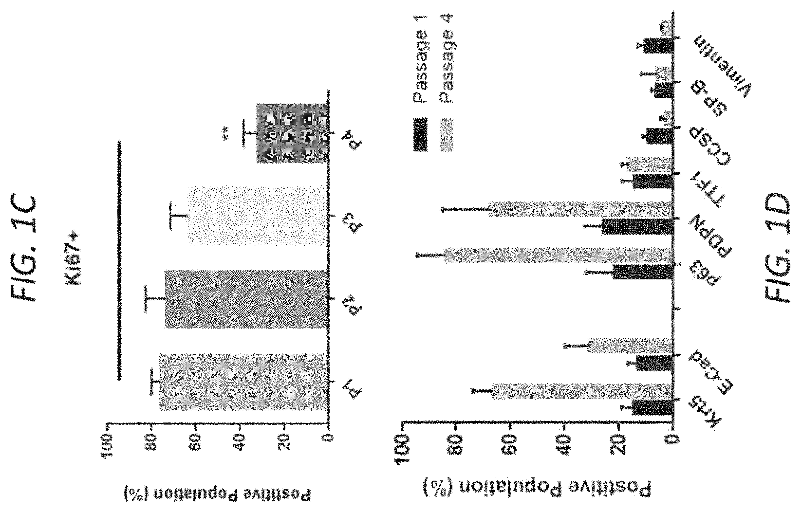
FIGS. 1A-E. Characterization of primary human lung epithelial stem cell expansion in vitro. Bright-field and immunofluorescent images of (A) Passage 1 (P1) and (B) Passage 4 (P4) primary human epithelial cells in vitro, illustrating colony outgrowth with an enrichment of Krt5+p63+ basal stem cells. Scale bar=100 .mu.m (C) Quantification of Ki67 positive cells in vitro by passage. n=3 images quantified per passage (P1-P4). 1-Way ANOVA compared to Passage 1 (P1), with Dunnet post-test. All error bars represent standard deviation. (D) Flow cytometric quantification of epithelial cell markers at P1 and P4. 2.times.104 events collected. Populations gated to exclude doublets and auto-fluorescent cells. n=3 cell lines per passage (E) Quantitative PCR analysis of gene expression by cell passage. Box plots represent median, plus the first and third quartile. Whiskers represent the 2.5-97.5% data range. n=3 cell lines per passage. Normalized to .beta.-actin expression and relative to normal cadaveric lung tissue control.
FIGS. 2A-F. Differentiation toward ciliated airway epithelium by air-liquid interface culture. (A) Primary human lung epithelial stems cells at Air-Liquid Interface (ALI), Day 21. Induction of ciliogenesis is observed by Haematoxyalin and Eosin staining. Scale bar=25 .mu.m. (B) Immunofluorescent images demonstrate preservation of the basal stem cell population (Krt5/p63+), functional ciliogenesis (FOXJ1 and Acetylated .alpha. Tubulin) and tight junction formation (E-Cadherin) at ALI. Scale bar=25 .mu.m. (C) Air-liquid interface on decellularized lung matrix by continuous positive airway pressure (CPAP) model (20 mmHg airway pressure for 7 days following 7 days of vascular perfusion-only culture). (D) Immunofluorescent images demonstrate maintenance of basal stem cell population (Krt5), induction of FOXJ1 expression, enhanced tight junction formation (E-Cadherin), and decrease in proliferation (Ki67) compared to lung prior to CPAP (Day 7 vs Day 14). Scale bar=50 .mu.m. (E) Western blot analysis of E-Cadherin protein levels at day 14 in lung tissue +CPAP or -CPAP (vascular perfusion-only). (F) Quantitative PCR analysis of gene expression in recellularized lungs following recellularization and vascular perfusion only for 7 days, compared to lungs at Day 14 (additional 7 days of CPAP or perfusion only). Data from 3 independent recellularized lungs is shown. n=3 independent tissue samples analyzed per lung, per time point, in experimental triplicates. Normalized to .beta.-actin expression and relative to normal cadaveric lung tissue control. All error bars represent standard deviation. Analyzed by 1-way ANOVA with Tukey's multiple comparisons post-test.
FIGS. 3A-H. Induction of a distal Type II pneumocyte phenotype by Notch inhibition. (A) Immunofluorescent images of primary epithelial basal cells in vitro (passage 3) treated with Notch inhibitors 3-isobutyl-1-methylxanthine (IBMX), a phosphodiesterase inhibitor, and N--[N-(3,5-Difluorophenacetyl-L-alanyl)]-(S)-phenylglycine t-butyl ester (DAPT, also known as GSI IX, a gamma secretase inhibitor) for 5 days, demonstrating the increase in surfactant protein-B (SP-B) positive cells. Scale bars=100 .mu.m. (B) Quantitative PCR analysis of gene expression of cells treated with IBMX (100 DAPT (50 .mu.M), or combination IBMX+DAPT for 5 days in vitro, demonstrating an increase in SP-B and SP-C expression. n=3 experimental replicates are shown. 1-way ANOVA with Dunnet post-test compared to No Treatment (NT). Although in this experiment DAPT and IBMX were ineffective alone, in other replicates they showed some activity. 3-dimensional culture assay demonstrating sphere formation. (C) Immunofluorescent images of spheres demonstrating a predominance of a Krt5+p63+ phenotype in 3D sphere culture. Scale bar=50 .mu.m. (D) Haematoxylin and Eosin stained spheres demonstrating luminal development by day 7. (E) Bright field images of spheres in both untreated and Notch inhibited (IBMX+DAPT) cells on Day 7. Scale bar=100 .mu.m. (F) Quantification of sphere number as a percentage of total cell number initially seeded, demonstrating a decrease in sphere formation in IBMX+DAPT treated cells. n=3 independent cultures quantified in experimental triplicate. Analyzed by T-test. (G) Quantitative PCR analysis of gene expression of sphere cultures on day 7, with or without Notch inhibition (IBMX+DAPT). Data from 3 independent cultures is shown, analyzed in experimental duplicate. Normalized to .beta.-actin expression and relative to normal cadaveric lung tissue control. (H) SP-C protein could also be measured by ELISA in the conditioned media of BESCs (at passage 4, p4) following treatment with DAPT+IBMX, in vitro for 5 days. All error bars represent standard deviation, analyzed by t-test.
FIGS. 4A-E. Induction of primary basal airway stem cells to distal type II pneumocyte phenotype in decellularized lung scaffold culture. (A) Co-culture of basal epithelial stem cells with human decellularized lung slices for 5 days demonstrating cell attachment to lung matrix via integrin .alpha.2.beta.1 and .alpha.3.beta.1, the formation of tight junctions along areas of matrix attachment (E-Cadherin), and continued proliferation (Ki67+). Scale bar=100 .mu.m. (B) Quantitative PCR analysis of gene expression in cell-matrix culture with or without Notch inhibition (IBMX+DAPT) for 5 days, demonstrating the induction of SP-B and SP-C, and loss of Club Cell Secretory Protein (CCSP expression after treatment. Data from 2 separate experiments shown, with cells seeded to matrix derived from two different lung donors (n=3 to HL30 and n=3 to HL38), analyzed in experimental duplicate. Normalized to .beta.-actin expression and relative to normal cadaveric lung tissue control. Analyzed by T-test (IBMX+DAPT vs. NT). Error bars represent standard deviation. (C) Biomimetic culture of lungs recellularized with primary basal stem cells (20.times.10.sup.6) plus IBMX+DAPT treatment. (D) Quantitative PCR analysis of gene expression in recellularized lungs treated with IBMX+DAPT for 5 days in constant perfusion culture compared to No Treatment lung (Day 5). n=3 independent tissue samples analyzed per lung, in experimental triplicates. Normalized to .beta.-actin expression and relative to normal cadaveric lung tissue control. Analyzed by T-test (IBMX+DAPT vs. NT). Error bars represent standard deviation. (E) Immunofluorescent images of recellularized lungs at Day 5 (No Treatment vs IBMX+DAPT Notch inhibition), confirming the maintenance of Krt5/p63+ basal cell population, an increase tight junction intensity (E-Cadherin), an increase in SP-C positive cells, and a loss of Aquaporin-5 positive cells. Scale bars=50 .mu.m
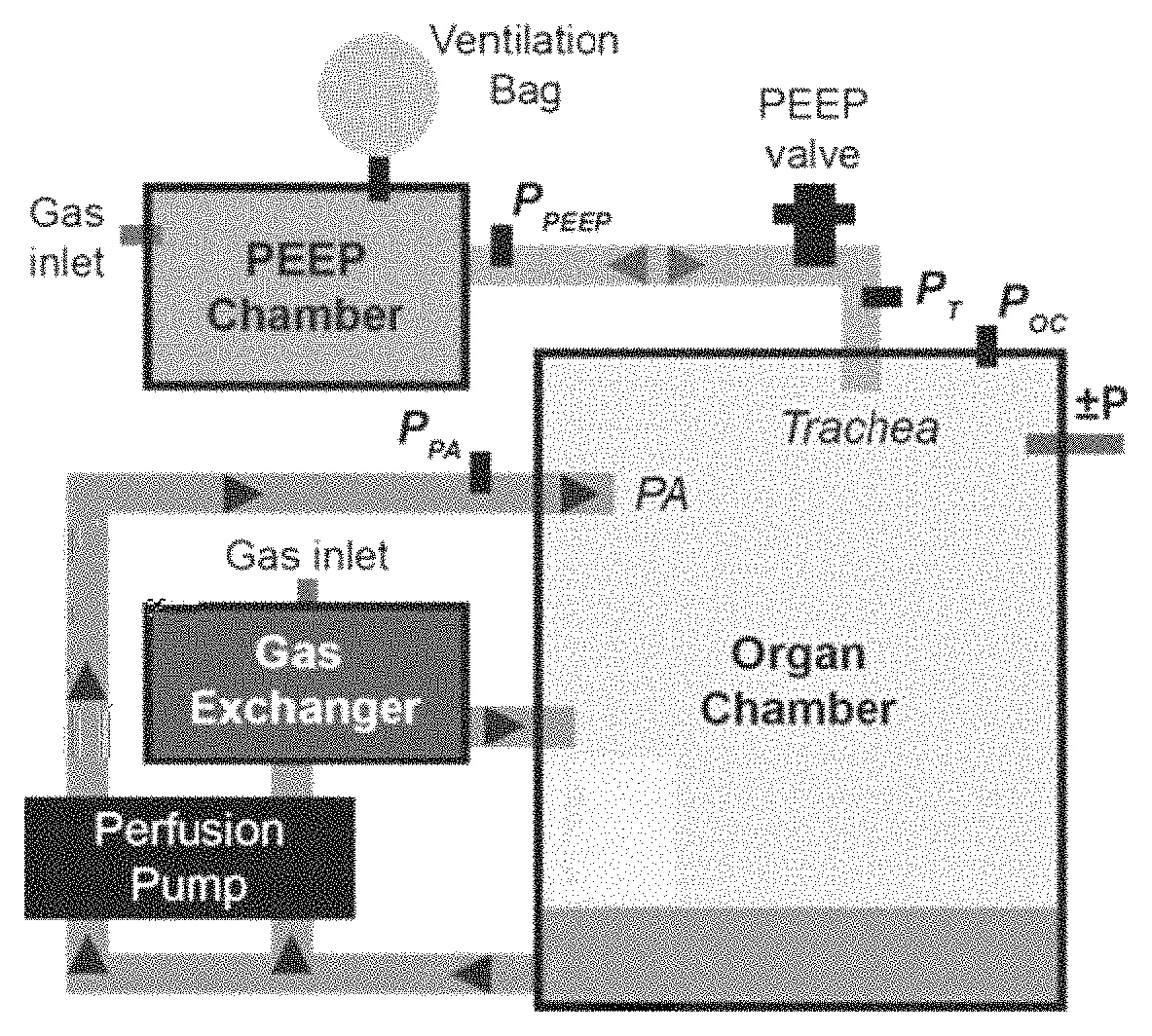
FIGS. 5A-K. Recellularization and culture of whole decellularized human lung scaffolds with primary human lung basal stem cells. (A) Schematic of exemplary lung bioreactor capable of constant organ perfusion and negative pressure ventilation. (B) Single human decellularized lung lobe in bioreactor with access ports for pulmonary vein, airway, and pulmonary artery highlighted. Lobes were seeded with primary PAECs (pulmonary artery endothelial cells, 160-240.times.106) and BESCs (basal epithelial stem cells, 220-280.times.106, n=3) and maintained under constant media perfusion plus periodic negative pressure ventilation. (C) Pulmonary artery pressure over 8 days of recellularized lung perfusion culture, n=3 recellularized lungs. Data represents mean pressure+/-SD. (D-E) Change in (D) Glucose measurements in media sampled from the lung culture media. Media was changed every 48-hours, and values represent the change in glucose concentration (mg/dL) and (E) lactate concentration (mmol/L) after 48-hours of organ perfusion, compared to fresh media. Data shown represent n=2 independent lung cultures per time point. Error bars represent standard error. (F) Representative pressure traces during negative pressure ventilation (breath rate of 6/min). Pressures are simultaneously recorded in the organ chamber, pulmonary artery, PEEP chamber, airway, and pulmonary vein. (G) Peak transmural pressure (mmHg) during ventilation. (H) Calculated tidal volume (mL) during ventilation. Box plots represent median, plus the first and third quartile. Whiskers represent the range of data. Outliers (points greater than 1.5.times.IQR of the box plot) are represented by a plus sign (+). (I) Representative pressure-volume loop generated during negative pressure ventilation in the bioreactor. Traced loop t=0 is represented as Blue and t=final is represented as Red (J) Endpoint positive pressure ventilation challenge of a single lower lobe. (K) Representative measurement of pH, pO.sub.2, pCO.sub.2, and HCO.sub.3 of perfusate during positive pressure ventilation challenge. Lobe was recellularized and cultured for 7 days prior to testing and functional challenge was with 21% and 100% FiO2.
FIGS. 6A-H. Analysis of lung tissue recellularization following biomimetic culture. (A) Perfusion of Resazurin containing media to assay cell viability on day 7 of biomimetic culture. Viable cell metabolism of the blue dye is visualized by transition to a pink color, demonstrating extensive cell retention, distribution, and viability after culture. (B) Representative scan of H&E section of recellularized lung tissue (i) scale bar=5 mm and (ii) scale bar=100 .mu.m. (C) Immunofluorescent image of continued E-Cadherin+ epithelial cell proliferation (Ki67+) on Day 7 of culture. Scale bar=50 .mu.m. (D) Quantification of cell proliferation in recellularized lung tissue by Ki67+ staining. Three representative areas were analyzed per lung, with 4 images quantified per area. Error bars represent the standard deviation. Analyzed by 1-way ANOVA, with no significance identified. (E) Immunofluorescent images of recellularized lungs at Day 7 of culture confirming the maintenance of Krt5+p63+ basal cell population and the rare observance of non-adhered proSP-B+ cells. (F) Cell retention and repopulation of large airways following culture, demonstrated by Krt5+, p63+ and E-cadherin+ epithelial cells in recellularized (i-ii) rat and (iii) human lungs. (G) Localization of and organization of CD31+ cells in the vascular capillary compartment of the lung scaffold. Scale bars=50 .mu.m. (H) Quantitative PCR analysis of gene expression of lung tissue on Day 7 or 10 (final day) of culture. Data from 3 independent recellularized lungs cultures is shown, with n=4 unique tissue samples analyzed per lung, in experimental duplicate. Expression normalized to .beta.-actin and relative to normal cadaveric lung tissue control. Gene expression level for cells maintained in vitro is shown for reference. All error bars represent standard deviation. Analyzed by 1-way ANOVA, with no significance identified.
FIGS. 7A-B: Primary Endothelial Cell isolation and Culture. (A) Primary lung endothelial cells were isolated from large vessels and cultured for 5 days in EGM2 media prior to sorting for CD31+ population. Gating strategy demonstrates the exclusion of doublets and dead deals (Pacific Blue+), and the isolation of CD31+ population. Representative example presented. (B) Immunofluorescent staining of the sorted population in culture on gelatin-coated flasks, demonstrating endothelial purity. Scale bar=100 .mu.m
FIGS. 8A-C: Effect of ROCK Inhibitor on Cell Phenotype and Senescence. (A) Gene expression analysis of n=3 individual cell lines at passage 1 and passage 4, with or without the addition of ROCK inhibitor Y27632 (10 Error bars represent standard deviation. No significant differences between untreated and treated (+ROCK) were found by t-test at each passage. (B) Representative image of cells at passage 1 and passage 4, with or without ROCK inhibitor, with Trypan Blue added to culture media. (C) Representative image of cells at passage 1 and passage 4, with or without ROCK inhibitor, stained for senescence-associated .beta.-galactosidase activity at pH 6. Scale bars=50 .mu.m.
FIGS. 9A-B: Effect of Endothelium Cell Co-Culture on Basal Cell Population. (A) qPCR gene expression analysis of primary epithelial basal cells (passage 4) in co-culture with primary lung endothelial cells. Data represents the basal cell population (Epi+) plus the culture on endothelial cells on transwell inserts (Endo+), with or without the addition of VEGF in the culture media (VEGF+, 40/ml). (B) Expression of VEGF receptor by treated and untreated epithelium (undetected) and endothelium. Expression level is normalized to .beta.-Actin and expressed relative to normal lung tissue. n=3 replicates analyzed in duplicate. Error bar=standard deviation. Analyzed by 1-way ANOVA with Dunnet post-test to Epi+ untreated group.
FIGS. 10A-B. BESCs were pre-treated with DAPT (50 nM) in vitro for 5 days, then delivered to rat lung scaffolds (20.times.10.sup.6), and maintained a distal type2-like fate, without continued inhibitor treatment. (A) Quantitative PCR analysis of gene expression at the end of in vitro treatment (Day 5), and following delivery to rat lung scaffold and ex vivo culture for 5 additional days without inhibitors. Data from 3 independent well (in vitro) or 3 independent tissue pieces (ex vivo) is shown. Expression normalized to .beta.-actin and relative to normal cadaveric lung tissue control. (B) Immunofluorescent staining for surfactant protein C and p63, in recellularized lung tissue on day 5 of ex vivo culture. 50 um scale bar.
FIG. 11. Direct Inhibition of the Notch signal pathway using dual small molecules (in this figure, LY411575 and GSI-X) targeting gamma-secretase efficiently directed tissue-derived BESCs toward a distal pneumocyte fate in vitro. Quantitative PCR analysis of gene expression at the end of in vitro treatment (Day 5). Data from n=3 independent wells, analyzed in experimental replicate is shown. Expression normalized to .beta.-Actin expression and fold-change calculated compared to untreated cells (SAGM). With this treatment, Type1 pneumocyte marker AQP5 was also increased, which was again not found with DAPT/IBMX treatment (as in FIG. 3).
FIGS. 12A-C. Epithelial Culture on Isolated Human ECM. (A) Method for preparation of matrix coating for in vitro culture. (B) Quantitative gene expression analysis of BESCs grown on neonatal (N1-N3) and adult (A1-A3) matrix coating. Expression normalized to B-Actin, and expressed relative to normal adult lung tissue. (C) Cytotoxicity assay measuring total live and dead cell fluorescence on Day 7.
FIGS. 13A-B. Neonatal and Adult Lung Composition by Proteomic Analysis. (A) Heat map of detected proteins in each sample. (B) Summary matrisome composition in neonatal vs adult matrix (n=3/group).
FIGS. 14A-B. Quantitative comparison of the matrix proteins in adult and neonatal lung scaffolds. (A) Volcano plot of detected matrix proteins. (B) Details of proteins highlighted in (A).
FIGS. 15A-E. In vitro analysis of BESC response to FBN-2 and TN-C. (A) Gene expression analysis, normalized to .beta.-Actin, and expressed relative to normal adult lung tissue. (B) Immunofluorescent staining (C) Ki67+ quantification (n=3 tissues/group). Scale bar=50 .mu.m (D) In vitro migration assay, representative image and quantification of change in cell-free area over 180 min. Scale bar=100 .mu.m. (E) Expression of Focal Adhesion Kinase (FAK) by BESCs on each coating. Gene expression analysis, normalized to .beta.-Actin, and expressed relative to normal adult lung tissue.
FIGS. 16A-E. Ex vivo lung epithelial regeneration on pre-treated matrices. (A) Quantitative gene expression of re-epithelialized lung scaffolds (B) Hematoxylin and eosin assessment of lung tissue. Scale bar=50 .mu.m. (C) Immunofluorescent staining of lung tissue on Day 3 and 7 of regeneration. Scale bar=50 .mu.m. (D) Quantification of Ki67 positive cells on Day 3 and 7 of lung epithelial regeneration. (E) Quantification of tissue morphology by septal thickness.
FIGS. 17A-C. Decellularization of Neonatal Human Lung. (A) Donor left and right Lung. (B) Cannulation of donor lungs. Scale bar=5 cm. (C) Decellularization of neonatal donor lung by perfusion decellularization of 0.5% Sodium Dodecyl Sulfate (SDS) solution.
FIG. 18. Representative Measurement of Septal Thickness. Red lines indicate measured areas (n=5/image). Scale bar (white)=50 .mu.m.
FIG. 19. A schematic diagram of an exemplary lung bioreactor.
DETAILED DESCRIPTION
Solid organ bioengineering based on native extracellular matrix scaffolds has fueled recent enthusiasm for regenerative medicine approaches to end organ failure (1). The main approach involves combining regenerative cell populations with corresponding biological matrices to form living, functional grafts. To this end, native solid organ extracellular matrix (ECM) scaffolds can be readily generated by perfusion decellularization with specific detergents, rendering a biocompatible framework as a foundation for regeneration (2-7).
Clinically relevant organ recellularization presents significant challenges, both in terms of identifying a cell source and in the establishment of functional biomimetic organ culture conditions to support organ maturation prior to transplantation (8).
An optimal cell source would be easily obtained and expanded in vitro, and would ideally possess the capacity for multi-lineage differentiation. While directed differentiation of induced pluripotent stem cells through key developmental stages presents a promising option for obtaining lung-specified cell populations (9-11), the length of in vitro culture and limited cell numbers restricts their current utility for large-scale organ engineering. While largely quiescent, adult lung tissue has a remarkable capacity for regeneration, owing to a number of facultative stem/progenitor cell populations that become activated in response to tissue damage (12). Airway basal cells, identified by the transcription factor p63 and expression of cytokeratin 5 (Krt5), function as multipotent stem cells of the airway epithelium, and are critical for maintaining airway homeostasis during physiological cell turnover and regeneration (13, 14). This essential cell population comprises 30% of the cells in human airway epithelium (15), and early studies of airway regeneration demonstrated the ability for isolated basal cells to recapitulate a fully differentiated airway epithelium when seeded onto denuded mouse tracheas (16). In response to injury, basal epithelial stem cells can rapidly proliferate and give rise to both ciliated and club cell progeny, confirming their important function in tissue homeostasis and injury repair (17). Lung basal cells can be readily isolated from lung tissue (18, 19) and propagated in culture (20), which makes them a useful target population for tissue engineering applications. The in vitro cultivation of this primary stem cell population also provides an important tool for studying basic biology and tissue regeneration (13), particularly given their capacity for multi-lineage differentiation (21, 22).
Described herein is the isolation of a highly proliferative basal stem cell population from an easily accessible tissue source and demonstrated over 100-fold expansion in vitro. This cell population, identified by Krt5.sup.+p63.sup.+ expression, has been studied in animal models of lung repair (23-25) and in human disease (26).
Within the Krt5.sup.+p63.sup.+ population, additional distinct subpopulations of basal stem cells may exist, each with a unique role in tissue homeostasis and repair. This includes the recently reported lineage-negative epithelial progenitor (LNEP) cells within normal distal lung, which can specifically proliferate following injury (27). It is unclear whether these rare cellular subsets can act in isolation, or if they require combined signaling and action of other cells in the injured tissue milieu. Mathematical models support a heterogeneous basal stem cell population, proposing approximately equal numbers of multipotent stem cells and committed precursors (28). The role of injury, including source, intensity, and duration, is also an important determinant of cell activation and fate. The origin of the cell population studied, considering age and species as two examples, can also contribute to the regenerative capacity and cell fate. Embryonic lungs at the canalicular stage of development have been recently shown to possess distinct niches enriched with epithelial progenitors surrounded by mesenchymal cells and blood cells, and these cells can be transplanted to injured lungs and differentiation to multiple lineages (29).
Following lung injury, the re-establishment of an intact epithelium is critical to restore lung homeostasis (30). In the present model of lung repair, the decellularized lung scaffold serves as the provisional matrix for epithelial cell migration, recapitulating (at least in part) the processes activated in vivo to cover and repair denuded airway and gas exchange surfaces (31). The physiologic role of basal cells, to help anchor epithelial cells to the matrix and protect the underlying stroma, is aided by their expression of abundant cytoskeletal, junctional and adhesive proteins, which supports their demonstrated utility in re-epithelialization of native lung ECM (32).
Rodent models of airway injury have established a timeline of epithelial repair. After epithelial injury, cell spreading and migration occurs as the primary repair mechanisms in the first 12-24 hours, with proliferation beginning after 24 hours and continuing for several weeks (33). This timeline aligns with the present model of lung repair in ex vivo culture and regeneration. The next step in the repair mechanism would be reestablishment of a pseudostratified epithelium, which can take several weeks to establish (34). The present model of air-liquid interface culture on whole rat lung scaffolds demonstrated the initial upregulation of FOXJ1 and increased tight junction formation at 7 days. Extended bioreactor culture may be required to fully mature the reconstituted epithelium, as in vitro ALI models require 3-4 weeks to recapitulate the mature airway biology (35). Following recellularization and ex vivo culture, no significant pneumocyte lineages were identified within the present reconstituted epithelium. Longer regeneration time, combined with modulation of signaling pathways is likely required to induce committed pneumocyte differentiation from the delivered stem cell population, with animal models demonstrating distal lung regeneration required 50-90 days (27, 36).
Notch signaling plays an essential and complex role in lung epithelial development and homeostasis, and Notch ligands are expressed at very high levels in the lung (37). Lung development requires the precise patterning of multiple cell lineages, of which many fate choices are controlled by direct cell-to-cell communication. During embryonic alveolar development, constitutive over-expression of Notch inhibits the development of distal epithelium, instead promoting cyst formation mainly lacking alveolar markers (38). The requirement for Notch signaling in early lung proximal-distal cell fate decision was also shown following Notch inhibition by DAPT, resulting in an accumulation of Nk.times.2.1.sup.+ distal lung progenitor cell population (38). As also shown herein, pharmacological inhibition of the Notch pathway through .gamma.-secretase can induce global transitional toward a type II pneumocyte phenotype in vitro and in ex vivo lung scaffold culture. This confirms a report of 3-D sphere culture of mouse basal stem cells (27). There was also a loss of CCSP-expression observed following Notch inhibition, further highlighting the essential role for Notch activation in basal cell differentiation towards the secretory lineage (39, 40). For organ engineering, precise control of these signals may require pharmaceutical activation or inhibition of the Notch pathway to achieve optimal cell patterning. Further development of advanced bioengineering procedures will be required to specifically deliver these biochemical signals to the specific proximal or distal lung compartment in a dose and time-controlled manner. Mechanical forces also contribute to the activation of Notch signaling exposing the metalloprotease cleavage site and facilitating the subsequent change from the auto-inhibited conformation (41). These mechanical considerations may be of additional significance to cell-cell signaling in 3-dimensional whole organ biomimetic culture vs. traditional 2-D culture. Shear fluid forces resulting from biomimetic organ perfusion may further direct cell organization along the scaffold during culture. Epithelial cells have been shown to migrate along fluid flow streamlines in vitro, which may be directed by paracrine chemokine fields in the local microenvironment (42).
The present model of lung scaffold recellularization and ex vivo regeneration provides a unique and easily accessible tool to further investigate epithelial repair in a systematic manner.
Organ regeneration based on decellularized scaffolds is perhaps the ultimate model of injury and test of cellular repair potential. Given the isolated environment, coupled with the biomimetic stimulus provided by the ex vivo culture of the regenerating organ, it is possible to directly assess the ability of specific cell populations to regenerate native tissue. In the present study, by employing systematic building-blocks approach, a critical step forward is made by demonstrating that a primary isolated airway stem cell population can accomplish extensive tissue regeneration on an acellular lung scaffold, and can be directed toward both proximal and distal epithelial lineages.
This document relates to methods and materials involved in airway organ generation and preservation. Described are methods, devices, cells, and compositions configured to generate functional lung tissue that can be used to provide a more realistic environment for growth of functional airway organs ready for transplantation into humans and other animals. The lung tissue is generated over a given matrix, e.g., an artificial or decellularized lung tissue matrix. The present invention is further based on the use of this realistic environment for the preservation, repair, and modification of donor organs over prolonged periods of time in order to provide more, improved, and individualized grafts for transplantation.
As used herein, a "functional" lung tissue performs most or all of the functions of a normal healthy lung, e.g., allows for transportation of oxygen from the air into the bloodstream, and the release of carbon dioxide from the bloodstream into the air. It can humidify inhaled air, produce surfactant to decrease surface tension in the alveoli, and/or produce and transport mucus to remove inhaled particulate matter from the distal to the proximal airway.
As used herein, the terms "decellularized" and "acellular" are used or defined as the complete or near complete absence of detectable intracellular matter, endothelial cells, epithelial cells, and nuclei in histologic sections using standard histological staining procedures. Preferably, but not necessarily, residual cell debris also has been removed from the decellularized organ or tissue.
Decellularized Tissue/Organ Matrices
In some embodiments of the present methods, lung tissue is generated over a decellularized matrix. Methods and materials for a preparing a decellularized lung tissue matrix are known in the art, as discussed below. Any appropriate materials can be used to prepare such a matrix. In a preferred embodiment, a tissue matrix can be an acellular tissue scaffold developed from decellularized lung tissue. For example, tissue such as a human lung, e.g., one or a pair of human lungs or portions thereof, e.g., human, porcine, bovine, primate, or ovine cadaveric lungs or portions thereof, can be decellularized by an appropriate method to remove native cells from the tissue while maintaining morphological integrity and vasculature of the tissue or tissue portion and preserving extracellular matrix (ECM) proteins (see Tapias L F, and Ott H C. Decellularized scaffolds as a platform for bioengineered organs. Current opinion in organ transplantation. 2014; 19(2):145-52). Methods for decellularizing mammalian lung tissues are described, e.g., in O'Neill J D et al., Decellularization of human and porcine lung tissues for pulmonary tissue engineering. Ann Thorac Surg. 2013 September; 96(3):1046-55; Nichols J E et al., Production and assessment of decellularized pig and human lung scaffolds, Tissue Eng Part A. 2013 September; 19 (17-18):2045-62; Gilpin S E et al., Perfusion decellularization of human and porcine lungs: Bringing the matrix to clinical scale. Journal of Heart and Lung Transplantation. 2014; 33: 298-308; Song J J et al., Bioartificial lung engineering. Am J Transplant. 2012 February; 12(2):283-8; and Ott H C et al., Regeneration and orthotopic transplantation of a bioartificial lung. Nat Med. 2010 August; 16(8):927-33. Exemplary decellularization methods can include subjecting tissue (e.g., lung tissue) to repeated freeze-thaw cycles, for example using liquid nitrogen. In other cases, a tissue can be subjected to an anionic or ionic cellular disruption medium such as sodium dodecyl sulfate (SDS), polyethylene glycol (PEG), or TritonX. The tissue can also be treated with a nuclease solution (e.g., ribonuclease, deoxyribonuclease) and washed in sterile phosphate buffered saline with mild agitation. Exemplary methods are known in the art e.g., O'Neill J D et al., Decellularization of human and porcine lung tissues for pulmonary tissue engineering. Ann Thorac Surg. 2013 September; 96(3):1046-55. In some cases, decellularization can be performed by flushing the vessels, ducts, and/or cavities of the organ or tissue using methods and materials known in the art. For example, as described in Maghsoudlou et al., Preservation of micro-architecture and angiogenic potential in a pulmonary acellular matrix obtained using intermittent intra-tracheal flow of detergent enzymatic treatment. Biomaterials. 2013 September; 34(28):6638-48. Following the flushing step, the organ or tissue can be perfused via the line with a cellular disruption medium as described above for example 1% SDS in deionized water. Perfusion through the tissue can be anterograde or retrograde, and directionality can be alternated to improve perfusion efficiency. Depending upon the size and weight of an organ or tissue and the particular anionic or ionic detergent(s) and concentration of anionic or ionic detergent(s) in the cellular disruption medium, a tissue generally is perfused from about 2 to about 12 hours per gram of tissue with cellular disruption medium. Including washes, an organ may be perfused for up to about 12 to about 72 hours per gram of tissue. Perfusion generally is adjusted to physiologic conditions including flow rate and pressure, e.g., pressure between 5-100 mmHg, and flow rate between 0.1-10 times the physiologic cardiac output of the source organism or individual.
In another exemplary method, a decellularization method includes perfusing a detergent, e.g., (1) 0.1% SDS (2) 2%, sodium deoxycholate (SDC), or (3) 8 mmol/liter (3)3-[(3-cholamidopropyl)dimethylammonio]-1-propanesulfonate (CHAPS) (pH12) detergent, through the pulmonary artery at a constant pressure of 30 cm H.sub.2O. The protocol for all 3 detergents includes:
1. a 10-minute initial antegrade wash with phosphate-buffered saline (PBS),
2. detergent perfusion for the time required to visualize an opaque translucent matrix (indicative of decellularization) plus an additional 20% of that initial time (e.g., 70 minutes+14 minutes),
3. 15-minute deionized H.sub.2O wash, and
4. an additional 172-hour PBS wash with added antibiotics and antimycotics.
This decellularization method, e.g., can include an additional wash of 1% Triton-X following the deionized H.sub.2O. The SDC protocol can include a 0.1% Triton-X perfusion before SDC and a 1 mol/liter NaCl wash after SDC.
Similarly, porcine and human lung decellularization methods can include perfusion of a detergent or other decellularization agent though the pulmonary artery at constant pressure, followed by sequential washing with H.sub.2O, 1% Triton-X solution, and PBS. Similar to rat lungs, decellularization can be deemed complete upon visual inspection and the appearance of an opaque translucent matrix. Variability in the starting organ, mainly due to extensiveness of pre-flushing during harvest and any resulting clots can contribute to the required length of perfusion. In general, the time of decellularization perfusion can vary e.g., from 4 to 7 days.
Decellularized tissue can consist essentially (e.g., at least: 85% pure, 90% pure, 92% pure, 95% pure, 96% pure, 97% pure, 98% pure, and 99% pure by weight) of the extracellular matrix (ECM) component of all or most regions of the tissue, including ECM components of the vascular tree. ECM components can include any or all of the following: fibronectin, fibrillin, laminin, elastin, members of the collagen family (e.g., collagen I, III, and IV), glycosaminoglycans, ground substance, reticular fibers and thrombospondin, which can remain organized as defined structures such as the basal lamina. In a preferred embodiment, decellularized lung tissue matrix retains an intact vasculature. Preserving a substantially intact vasculature enables connection of the tissue matrix to a subject's vascular system upon transplantation. In addition, a decellularized tissue matrix can be further treated with, for example, irradiation (e.g., UV, gamma) to reduce or eliminate the presence of any type of microorganism remaining on or in a decellularized tissue matrix.
Methods for obtaining decellularized tissue matrices using physical, chemical, and enzymatic means are known in the art, see, e.g., Liao et al, Biomaterials 29(8):1065-74 (2008); Gilbert et al., Biomaterials 27(9):3675-83 (2006); Teebken et al., Eur. J. Vasc. Endovasc. Surg. 19:381-86 (2000). See also U.S. Pat. Publication Nos. 2009/0142836; 2005/0256588; 2007/0244568; and 2003/0087428.
Artificial Organ Matrices
In some embodiments of the present methods, lung tissue is generated over an artificial organ matrix. Methods and materials for a preparing an artificial organ matrix are known in the art. Any appropriate materials can be used to prepare such a matrix. In a preferred embodiment, an artificial organ matrix can be a scaffold developed from porous materials such as, for example, polyglycolic acid, Pluronic F-127 (PF-127), Gelfoam sponge, collagen-glycosaminoglycan (GAG), fibrinogen-fibronectin-vitronectin hydrogel (FFVH), and elastin. See, e.g., Ingenito et al., J Tissue Eng Regen Med. 2009 Dec. 17; Hoganson et al., Pediatric Research, May 2008, 63(5):520-526; Chen et al., Tissue Eng. 2005 September-October; 11(9-10):1436-48. In some cases, an artificial organ matrix can have porous structures similar to alveolar units. See Andrade et al., Am J Physiol Lung Cell Mol Physiol. 2007 February; 292(2):L510-8. In some cases, an implanted artificial organ matrix can express organ-specific markers (e.g., lung-specific markers for Clara cells, pneumocytes, and respiratory epithelium). In some cases, an implanted artificial organ matrix can organize into identifiable structures (e.g., structures similar to alveoli and terminal bronchi in an artificial lung matrix). For example, an implanted artificial lung maxtrix made using FFVH can promote cell attachment, spreading and extracellular matrix expression in vitro and apparent engraftment in vivo, with evidence of trophic effects on the surrounding tissue. See Ingenito et al., supra. See also U.S. Pat. Nos. 7,662,409 and 6,087,552; United States Patent Publication Nos. 2010/0034791; 2009/0075282; 2009/0035855; 2008/0292677; 2008/0131473; 2007/0059293; 2005/0196423; 2003/0166274; 2003/0129751; 2002/0182261; 2002/0182241; and 2002/0172705.
Treatment with Tenascin-C and/or Fibrillin-2
The optimal scaffold for lung organ engineering would not only provide the necessary structure, but would additionally guide the organization and function of new lung tissue. The ECM is a complex entity that participates in many biological processes, including tissue development and repair (Balestrini and Niklason, Annals of biomedical engineering. 2015; 43(3):568-76). When considering the ECM in whole organ regeneration, the source of native lung tissue used to prepare the scaffold can have a direct impact on subsequent regeneration. Several studies have shown that underlying lung pathologies can cause changes in the ECM that are retained following decellularization, and can perpetuate during tissue repair (Burgess et al., The Journal of pathology. 2016; 240(4):397-409). This has been demonstrated for both pulmonary fibrosis and emphysema (Booth et al. American journal of respiratory and critical care medicine. 2012; 186(9):866-76; Sokocevic et al. Biomaterials. 2013; 34(13):3256-69). Age of the lung can also contribute important differences to the decellularized scaffold. It has been shown that growth on aged ECM can lead to significantly lower cellular expression of laminin .alpha.3 and .alpha.4 chains, which recapitulates the laminin deficiency that is observed in aged lung ECM. These data further highlight the deep biological information that is contained in the lung scaffold, and the feedback loops that can exist between reparative cell populations and the underlying protein matrix (Godin et al., PloS one. 2016; 11(3):e0150966).
Lung development actively continues following birth, and ECM remodeling is an essential component in the post-natal process of alveolarization. This mechanism functions to dramatically increase the gas exchange surface area, as the lung further refines the immature alveolar structure and undertakes secondary septation to generate a greater number of smaller sized alveoli (Whitsett et al., Physiological reviews. 1995; 75(4):749-57). The consequences of this process and the specific differences in ECM composition have not been well studied in the context of ex vivo tissue regeneration. Fetal wounds repair at a faster rate than adults, with little or no scarring (Yates et al., Birth defects research Part C, Embryo today: reviews. 2012; 96(4):325-33). Regrowth of lung is possible after lobectomy in infancy, with restoration of airway function and total recovery of lung volume (McBride et al., The Journal of clinical investigation. 1980; 66(5):962-70). Conversely, dysregulation of the ECM is an important driving factor for ageing, and age-related alterations in the ECM can be directly communicated to the surrounding cells, contributing to the development of chronic lung diseases such as emphysema and pulmonary fibrosis (Meiners et al., The European respiratory journal. 2015; 45(3):807-27). Another consequence of aging is the phenomenon of stem cell dysfunction and exhaustion, where the multipotent pool of progenitors progressively declines and becomes increasing senescent (Thannickal et al., American journal of respiratory and critical care medicine. 2015; 191(3):261-9). These interactions between the stem cell and the niche, including ECM, can contribute to this decrease in regenerative capacity.
The present study investigated the differences in ECM from neonatal lungs actively undergoing alveolarization, compared to adult lung donors, and evaluated the consequences of these differences on ex vivo lung epithelial repair. There was an increase in developmentally associated proteins Fibrillin-3 (FBN-3), Fibrillin-2 (FBN-2), and Tenascin-C (TN-C) in the neonatal human lung ECM, and report that supplementation of these two proteins both in vitro and in ex vivo lung regeneration on acellular lung scaffolds can enhance epithelial proliferation, decrease senescence, aid cell attachment and migration, and ultimately improve regenerated tissue morphology and structure.
In some embodiments, the lung tissue matrix, e.g., decellularized lung tissue matrix or artificial lung matrix, is pre-treated with a solution comprising Tenascin-C (e.g., 0.5-10 ug/ml, e.g., about 1-3 ug/ml) and/or Fibrillin-2 (e.g., whole or N and C fragments) (e.g., 0.5-10 ug/ml, e.g., about 1-3 ug/ml), e.g., in a 15 ml total volume for a rat lung matrix, prior to cell seeding. In these methods, the Tenascin-C and/or Fibrillin-2 are exogenous, i.e., are added to a solution in which the matrix is incubated (either before or during contact with the matrix), and are in addition to any Tenascin-C and/or Fibrillin-2 already present in the matrix (or already present in any serum present in the solution). In some embodiments, the methods include delivery of a solution comprising Tenascin-c and/or Fibrillin-2, e.g., about 0.5-10 ug/ml Tenascin-C and/or about 0.5-10 ug and/or Fibrillin-2, to the scaffold airways (e.g., by gravity pressure), e.g., and incubating the matrix in the presence of the Tenascin-C and/or Fibrillin-2, e.g., at 37.degree. C. for about 1 hour. In some embodiments, the cells are seeded in a solution comprising Tenascin-C and/or Fibrillin-2. As an alternative to or in addition to Fibrillin-2, Fibrillin-3 can be used.
Exemplary sequences for human Tenascin-C precursor are in GenBank at NM_002160.3 (nucleic acid) and NP_002151.2 (protein). An exemplary protein sequence is as follows:
TABLE-US-00001 (SEQ ID NO: 1) 1 mgamtqllag vflaflalat eggvlkkvir hkrqsgvnat lpeenqpvvf nhvyniklpv 61 gsqcsvdles asgekdlapp sepsesfqeh tvdgenqivf thriniprra cgcaaapdvk 121 ellsrleele nlvsslreqc tagagcclqp atgrldtrpf csgrgnfste gcgcvcepgw 181 kgpncsepec pgnchlrgrc idgqcicddg ftgedcsqla cpsdcndqgk cvngvcicfe 241 gyagadcsre icpvpcseeh gtcvdglcvc hdgfagddcn kplclnncyn rgrcvenecv 301 cdegftgedc selicpndcf drgrcingtc yceegftged cgkptcphac htqgrceegq 361 cvcdegfagv dcsekrcpad chnrgrcvdg rcecddgftg adcgelkcpn gcsghgrcvn 421 gqcvcdegyt gedcsqlrcp ndchsrgrcv egkcvceggf kgydcsdmsc pndchqhgrc 481 vngmcvcddg ytgedcrdrq cprdcsnrgl cvdgqcvced gftgpdcael scpndchgqg 541 rcvngqcvch egfmgkdcke grcpsdchgq grcvdgqcic hegftgldcg qhscpsdcnn 601 lgqcvsgrci cnegysgedc sevsppkdlv vtevteetvn lawdnemrvt eylvvytpth 661 egglemqfry pgdqtstiiq elepgveyfi rvfailenkk sipvsarvat ylpapeglkf 721 ksiketsvev ewdpldiafe tweiifrnmn kedegeitks lrrpetsyrq tglapgqeye 781 islhivknnt rgpglkrvtt trldapsqie vkdvtdttal itwfkplaei dgieltygik 841 dvpgdrttid ltedenqysi gnlkpdteye vslisrrgdm ssnpaketft tgldaprnlr 901 rvsqtdnsit lewrngkaai dsyrikyapi sggdhaevdv pksqqattkt tltglrpgte 961 ygigvsavke dkesnpatin aateldtpkd lqvsetaets ltllwktpla kfdryrinys 1021 lptgqwvgvq lprnttsyvl rglepgqeyn vlltaekgrh kskparvkas tegapelenl 1081 tvtevgwdgl rinwtaadqa yehfiiqvqe ankveaarnl tvpgslravd ipglkaatpy 1141 tvsiygviqg yrtpvlsaea stgetpnlge vvvaevgwda lklnwtapeg ayeyffiqvq 1201 eadtveaaqn ltvpgglrst dlpglkaath ytitirgvtq dfsttplsve vlteevpdmg 1261 nitvtevswd alrinwttpd gtydgftiqv qeadqveeah nitvpgslrs meipglragt 1321 pytvtlhgev rghstrplav evvtedlpql gdlaysevgw dglrinwtaa dnayehfviq 1381 vqevnkveaa qnitlpgslr avdipgleaa tpyrvsiygv irgyrtpvls aeastakepe 1441 ignlnvsdit pesfnlswma tdgifetfti eiidsnrlle tveynisgae rtahisglpp 1501 stdfivylsg lapsirtkti satattealp llenitisdi npygftvswm asenafdsfl 1561 vtvvdsgkll dpqeftlsgt qrklelrgli tgigyevmvs gftqghqtkp lraeivteae 1621 pevdnllvsd atpdgfrlsw tadegvfdnf vlkirdtkkg sepleitlla pertrditgl 1681 reateyeiel ygiskgrrsq tvsaiattam gspkevifsd itensatvsw raptaqvesf 1741 rityvpitgg tpsmvtvdgt ktqtrlvkli pgveylvsii amkgfeesep vsgsfttald 1801 gpsglvtani tdsealarwq paiatvdsyv isytgekvpe itrtvsgntv eyaltdlepa 1861 teytlrifae kgpqksstit akfttdldsp rdltatevqs etalltwrpp rasvtgyllv 1921 yesvdgtvke vivgpdttsy sladlspsth ytakigalng plrsnmigti fttigllypf 1981 pkdcsqamln gdttsglyti ylngdkaeal evfcdmtsdg ggwivflrrk ngrenfyqnw 2041 kayaagfgdr reefwlgldn lnkitaggqy elrvdlrdhg etafavydkf svgdaktryk 2101 lkvegysgta gdsmayhngr sfstfdkdtd saitncalsy kgafwyrnch rvnlmgrygd 2161 nnhsqgvnwf hwkghehsiq faemklrpsn frnlegrrkr a
As amino acids 1-22 appear to be a signal sequence, in some embodiments, the mature Tenascin-C protein can be used, e.g., amino acids 23-2201 of SEQ ID NO:1. Alternatively, a fragment comprising amino acids 23 to 625 can be used.
Exemplary sequences for the human fibrillin-2 precursor are in GenBank at NM_001999.3 (nucleic acid) and NP_001990.2 (protein). An exemplary protein sequence for human fibrillin-2 precursor is as follows:
TABLE-US-00002 (SEQ ID NO: 2) 1 mgrrrrlclq lyflwlgcvv lwaggtaggp qppppkpprp qpppqqvrsa tagseggfla 61 peyreegaav asrvrrrgqq dvlrgpnvcg srfhsyccpg wktlpggnqc ivpicrnscg 121 dgfcsrpnmc tcssggisst cgsksiqqcs vrcmnggtca ddhcqcqkgy igtycgqpvc 181 engcqnggrc igpnrcacvy gftgpqcerd yrtgpcftqv nnqmcqgqlt givctkticc 241 atigrawghp cemcpaqpqp crrgfipnir tgacqdvdec gaipgicqgg ncintvgsfe 301 crcpaghkgs ettqkcedid ecsiipgice tgecsntvgs yfcvcprgyv tstdgsrcid 361 grtgmcfsgl vngrcagelp grmtkmgccc epgrcwgigt ipeacpvrgs eeyrrlcmdg 421 lpmggipgsa gsrpggtggn gfapsgngng ygpggtgfip ipggngfspg vggagvgagg 481 qgpiitglti lnqtidickh hanlclngrc iptvssyrce cnmgykqdan gdcidvdect 541 snpctngdcv ntpgsyyckc hagfqrtptk qacidideci qngvlckngr cvntdgsfqc 601 icnagfeltt dgkncvdhde ctttnmclng mcinedgsfk cickpgfvla pngryctdvd 661 ecqtpgicmn ghcinsegsf rcdcppglav gmdgrvcvdt hmrstcyggi kkgvcvrpfp 721 gavtkseccc anpdygfgep cqpcpaknsa efhglcssgv gitvdgrdin ecaldpdica 781 ngicenlrgs yrcncnsgye pdasgrncid ideclvnrll cdnglcrntp gsysctcppg 841 yvfrtetetc edinecesnp cvngacrnnl gsfncecspg sklsstglic idslkgtcwl 901 niqdsrcevn ingatlksec catlgaawgs pcerceldta cprglarikg vtcedvnece 961 vfpgvcpngr cvnskgsfhc ecpegltldg tgrvcldirm eqcylkwded ecihpvpgkf 1021 rmdacccavg aawgteceec pkpgtkeyet lcprgagfan rgdvltgrpf ykdineckaf 1081 pgmctygkcr ntigsfkcrc nsgfaldmee rnctdidecr ispdlcgsgi cvntpgsfec 1141 ecfegyesgf mmmkncmdid ecernpllcr ggtcvntegs fqcdcplghe lspsredcvd 1201 inecslsdnl crngkcvnmi gtyqcscnpg yqatpdrqgc tdidecmimn ggcdtqctns 1261 egsyecscse gyalmpdgrs cadidecenn pdicdggqct nipgeyrcic ydgfmasmdm 1321 ktcidvnecd lnsnicmfge centkgsfic hcglgysvkk gttgctdvde ceigahncdm 1381 hasclnipgs fkcscregwi gngikcidld ecsngthqcs inaqcvntpg syrcacsegf 1441 tgdgftcsdv decaeninlc enggclnvpg ayrcecemgf tpasdsrscq didecsfqni 1501 cvfgtcnnlp gmfhcicddg yeldrtggnc tdidecadpi ncvnglcvnt pgryecncpp 1561 dfqlnptgvg cvdnrvgncy lkfgprgdgs lscnteigvg vsrsscccsl gkawgnpcet 1621 cppvnsteyy ticpggegfr pnpitiiled idecgelpgl cqggncintf gsfqcecpqg 1681 yylsedtric edidecfahp gvcgpgtcyn tlgnytcicp peymqvnggh ncmdmrksfc 1741 yrsyngttce nelpfnvtkr mccctynvgk awnkpcepcp tpgtadfkti cgnipgftfd 1801 ihtgkavdid eckeipgica ngvcingigs frcecptgfs yndlllvced idecsngdnl 1861 cqrnadcins pgsyrcecaa gfklspngac vdrnecleip nvcshglcvd lqgsyqcich 1921 ngfkasqdqt mcmdvdecer hpcgngtckn tvgsyncicy pgfelthnnd cldidecssf 1981 fgqvcrngrc fneigsfkcl cnegyeltpd gkncidtnec valpgscspg tcqnlegsfr 2041 cicppgyevk sencidinec dedpniclfg sctntpggfq cicppgfvls dngrrcfdtr 2101 gsfcftnfen gkcsvpkafn ttkakcccsk mpgegwgdpc elcpkddeva fqdlcpyghg 2161 tvpslhdtre dvneclespg icsngqcint dgsfrcecpm gynldytgvr cvdtdecsig 2221 npcgngtctn vigsfecncn egfepgpmmn cedinecaqn pllcafrcmn tfgsyectcp 2281 igyalredqk mckdldecae glhdcesrgm mcknligtfm cicppgmarr pdgegcvden 2341 ecrtkpgice ngrcvniigs yrcecnegfq ssssgtecld nrqglcfaev lgticgmass 2401 srnlvtksec ccdggrgwgh qcelcplpgt aqykkicphg pgyttdgrdi deckvmpnlc 2461 tngqcintmg sfrcfckvgy ttdisgtsci dldecsqspk pcnyicknte gsyqcscprg 2521 yvlqedgktc kdldecqtkq hncqflcvnt lggftckcpp gftqhhtaci dnnecgsgps 2581 lcgakgicqn tpgsfscecq rgfsldatgl ncedvdecdg nhrcqhgcqn ilggyrcgcp 2641 ggyighyqwn qcvdenecsn pnacgsascy ntlgsykcac psgfsfdqfs sachdvnecs 2701 ssknpcnygc snteggylcg cppgyyrvgq ghcvsgmgfn kgqylsldte vdeenalspe 2761 acyeckingy skkdsrqkrs ihepdptave qislesvdmd spvnmkfnls hlgskehile 2821 lrpaigpinn hiryvisqgn ddsvfrihqr nglsylhtak kklmpgtytl eitsiplykk 2881 kelkkleesn eddyllgelg ealrmrlqiq ly
As amino acids 1-28 appear to be a signal sequence, in some embodiments, the mature Fibrillin-2 protein can be used, e.g., amino acids 29-2912 of SEQ ID NO:2. In some embodiments, the Fibrillin-2 is made as two separate peptides, e.g., N and C fragments, e.g., an N-terminal half rFBN2-N (amino acids 1-1732) and a C-terminal half rFBN2-C (amino acids 1531-2771), e.g., produced in human 293 cells. See, e.g., Lin et al., J. Biol. Chem. 277: 50795-50804 (2002).
Exemplary sequences for the human fibrillin-3 precursor are in GenBank at NM_001321431.1 (nucleic acid) and NP_001308360.1 (protein), or NM_032447.4 (nucleic acid) and NP_115823.3 (amino acid).
Preferably the sequence of the protein used is at least 80% identical (e.g., at least 90%, 95%, or 99% identical) to the mature reference sequence provided above, and has an activity described herein. To determine the percent identity of two amino acid sequences, or of two nucleic acid sequences, the sequences are aligned for optimal comparison purposes (e.g., gaps can be introduced in one or both of a first and a second amino acid or nucleic acid sequence for optimal alignment and non-homologous sequences can be disregarded for comparison purposes). In a preferred embodiment, the length of a reference sequence aligned for comparison purposes is at least 80% of the length of the reference sequence, and in some embodiments is at least 90% or 100%. The amino acid residues or nucleotides at corresponding amino acid positions or nucleotide positions are then compared. When a position in the first sequence is occupied by the same amino acid residue or nucleotide as the corresponding position in the second sequence, then the molecules are identical at that position (as used herein amino acid or nucleic acid "identity" is equivalent to amino acid or nucleic acid "homology"). The percent identity between the two sequences is a function of the number of identical positions shared by the sequences, taking into account the number of gaps, and the length of each gap, which need to be introduced for optimal alignment of the two sequences.
The comparison of sequences and determination of percent identity between two sequences can be accomplished using a mathematical algorithm. For example, the percent identity between two amino acid sequences can determined using the Needleman and Wunsch ((1970) J. Mol. Biol. 48:444-453) algorithm that has been incorporated into the GAP program in the GCG software package (available on the world wide web at gcg.com), using the default parameters, e.g., a Blossum 62 scoring matrix with a gap penalty of 12, a gap extend penalty of 4, and a frameshift gap penalty of 5.
Any form of TNC or FBN-2 can be used, e.g., protein produced recombinantly (e.g., expressed and isolated from cells, e.g., prokaryotic or eukaryotic, preferably mammalian (more preferably human) cells, or from a transgenic animal producing the protein, or transcribe and translated in a cell free system in vitro) or protein isolated from natural sources. Although human proteins are preferred, other mammalian species can also be used, e.g., bovine, caprine, porcine, equine, or ovine.
Although the present disclosure exemplifies the use of TNC and/or FBN-2 with some cell types, others can also be used.
Cell Seeding
In some of the methods described herein, a lung tissue matrix, e.g., decellularized lung tissue matrix or artificial lung matrix, is seeded with cells, e.g., differentiated or regenerative cells.
Any appropriate regenerative cell type, such as naive or undifferentiated cell types, can be used to seed the lung tissue matrix. The cells may be seeded at a variety of stages including, but not limited to, stem cell stage (e.g., after induction), progenitor cell stage, hemangioblast stage, or differentiated stage (e.g., CD 31+, vWF+). As used herein, regenerative cells can include, without limitation, progenitor cells, precursor cells, and "adult"-derived stem cells including umbilical cord cells (e.g., human umbilical vein endothelial cells) and fetal stem cells. Regenerative cells also can include differentiated or committed cell types. Stem cells appropriate for the methods and materials provided herein can include human induced pluripotent stem cells (iPSC) (e.g., undifferentiated, differentiated endoderm, anteriolized endoderm, TTF-1 positive lung progenitors), human mesenchymal stem cells, human umbilical vein endothelial cells, multipotent adult progenitor cells (MAPC), iPS derived mesenchymal cells, or embryonic stem cells. In some cases, regenerative cells derived from other tissues also can be used. For example, regenerative cells derived from skin, bone, muscle, bone marrow, synovium, or adipose tissue can be used to develop stem cell-seeded tissue matrices.
In some cases, a lung tissue matrix provided herein can be alternatively or further seeded with differentiated cell types such as (preferably human) epithelial cells and endothelial cells. For example, a lung matrix can be seeded with endothelial cells via the vasculature (e.g. through the arterial line or the venous line), and seeded with the proliferative basal stem cells from a human donor wherein the cells are Krt5+p63+ cells, via the airway (e.g., through the tracheal line). The lung matrix can also be seeded with one or more cell types (e.g., one or more of types of epithelial and mesenchymal cells, adult peripheral blood derived epithelial cells, cord blood-derived epithelial cells, iPS derived epithelial cells, progenitor stage cells (e.g., smooth muscle), adult lung derived cell mixture (e.g., rat human), commercially available small airway epithelial cells or alveolar epithelial cells, Embryonic Stem (ES) cell-derived epithelial cells, and/or human umbilical vein endothelial cells (HUVEC).
Any type of appropriate commercially available media and/or media kits may be used for the seeding and culture of cells. For example, SAGM media may be used for small airway cells (e.g., SAGM BulletKit by Lonza) and EGM-2 kits may be used for endothelial cells (e.g., EGM-2 BulletKit by Lonza). Media customized to the seeded endothelial cell type may be used (e.g., by increasing or decreasing growth factors such as VEGF) as described in, for example, Brudno Y et al. Biomaterials 34 (2013) 9201-9209. In the case of endothelial cells, a sequence of different media compositions may be used to induce different phases of seeding, expansion, engraftment, and maturation of cells. For example, in a first phase, a cell seeded constructs may be perfused with an `angiogenic media` for 2-30 days to increase endothelial cell expansion, migration, and metabolism. This media is characterized by high concentration of cytokines, e.g., VEGF at 5-100 ng/ml and bFGF at 5-100 ng/ml, and the presence of phorbol myristate acetate (PMA), e.g., 5-100 ng/ml PMA, which activates the angiogenic pathway through activation of protein kinase C, and Ang-1, which stimulates endothelial cell sprouting. In a second phase, a cell seeded construct can then be perfused with `tightening media` that supports endothelial maturation and the formation of tight junctions. Tightening media has lower levels of cytokines, with the same basic composition as the angiogenic media but with decreased levels of VEGF, bFGF and PMA (0.1-5 ng/ml VEGF, FGF, and PMA). Hydrocortisone, which promotes tight junction formation and has been shown to reduce pulmonary edema, can be further added to the tightening media to promote vascular maturation. Further promaturation factors such as PDGF and Ang-2 may be added to the tightening media to enhance vessel formation. Concentrations of these factors may be titrated to support different vessel sizes. Media changes can be performed gradually to avoid detrimental effects of sudden cytokine changes. Similar to endothelial cell supporting media, sequential media changes can be used to guide epithelial cell fate. Initial media may contain, for example, Activin A at 10-200 ng/ml and Pi3K inhibitors such as ZSTK 474 at 0.01-1 uM to induce definite endoderm, subsequently TGF-beta inhibitors such as A-8301 at 01-10 uM and BMP4 antagonists such as DMH-1 at 0.05-1 uM to induce anteriorized endoderm, and finally BMP4 at 1-100 ug/ml, FGF2 at 10-500 ng/ml, GSK-3beta inhibitor such as CHIR 99021 at 10-500 nM, a PI3K inhibitor such as PIK-75 at 1-100 nM and methotrexate at 1-100 nM to induce the generation of lung progenitor cells.
Any appropriate method for isolating and collecting cells for seeding can be used. For example, induced pluripotent stem cells generally can be obtained from somatic cells "reprogrammed" to a pluripotent state by the ectopic expression of transcription factors such as Oct4, Sox2, Klf4, c-MYC, Nanog, and Lin28. See Takahashi et al., Cell 131:861-72 (2007); Park et al., Nature 451:141-146 (2008); Yu et al., Science 318:1917-20 (2007); Zhu et al., Cell Stem Cell. 7:651-5 2010; and Li et al., Cell Res. 21:196-204 (2011); Malik and Rao, Methods Mol Biol. 2013; 997:23-33; Okano et al., Circ Res. 2013 Feb. 1; 112(3):523-33; Lin and Ying, Methods Mol Biol. 2013; 936:295-312. Peripheral blood-derived mononuclear cells can be isolated from patient blood samples and used to generate induced pluripotent stem cells. In other examples, induced pluripotent stem cells can be obtained by reprogramming with constructs optimized for high co-expression of Oct4, Sox2, Klf4, c-MYC in conjunction with small molecule such as transforming growth factor .beta. (SB431542), MEK/ERK (PD0325901) and Rho-kinase signaling (Thiazovivin). See Gro.beta. et al., Curr Mol Med. 13:765-76 (2013) and Hou et al., Science 341:651:654 (2013). Methods for generating endothelial cells from stem cells are reviewed in Reed et al., Br J Clin Pharmacol. 2013 April; 75(4):897-906. Cord blood stem cells can be isolated from fresh or frozen umbilical cord blood. Mesenchymal stem cells can be isolated from, for example, raw unpurified bone marrow or ficoll-purified bone marrow. Epithelial and endothelial cells can be isolated and collected from living or cadaveric donors, e.g., from the subject who will be receiving the bioartificial lung, according to methods known in the art. For example, epithelial cells can be obtained from a skin tissue sample (e.g., a punch biopsy), and endothelial cells can be obtained from a vascular tissue sample.
In some embodiments, proteolytic enzymes are perfused into the tissue sample through a catheter placed in the vasculature. Portions of the enzymatically treated tissue can be subjected to further enzymatic and mechanical disruption. The mixture of cells obtained in this manner can be separated to purify epithelial and endothelial cells. In some cases, flow cytometry-based methods (e.g., fluorescence-activated cell sorting) can be used to sort cells based on the presence or absence of specific cell surface markers. Furthermore, lung cells (epithelial, mesenchymal, and endothelial) can be obtained from lung biopsies, which can be obtained via transbronchial and endobronchial biopsies or via surgical biopsies of lung tissue. In cases where non-autologous cells are used, the selection of immune type-matched cells should be considered, so that the organ or tissue will not be rejected when implanted into a subject.
Isolated cells can be rinsed in a buffered solution (e.g., phosphate buffered saline at pH 7.4) and resuspended in a cell culture medium. Standard cell culture methods can be used to culture and expand the population of cells. Once obtained, the cells can be used to seed the tissue matrix, e.g., introduced into the matrix via the arterial or venous lines (endothelial cells) or through the airway (tracheal) line (epithelial cells). For example, a tissue matrix can be seeded with at least one cell type in vitro at any appropriate cell density. For example, cell densities for seeding a matrix can be at least 1.times.10.sup.3 cells/gram matrix. Cell densities can range between about 1.times.10.sup.5 to about 1.times.10.sup.10 cells/gram matrix (e.g., at least 100,000, 1,000,000, 10,000,000, 100,000,000, 1,000,000,000, or 10,000,000,000 cells/gram matrix) can be used.
In some cases, a decellularized or artificial lung tissue matrix, as provided herein, can be seeded with the cell types and cell densities described above, e.g., by gravity flow or perfusion seeding. For example, a flow perfusion system can be used to seed the decellularized lung tissue matrix via the vascular system preserved in the tissue matrix (e.g., through the arterial line). In some cases, automated flow perfusion systems can be used under the appropriate conditions. Such perfusion seeding methods can improve seeding efficiencies and provide more uniform distribution of cells throughout the composition. Quantitative biochemical and image analysis techniques can be used to assess the distribution of seeded cells following either static or perfusion seeding methods.
In some cases, a tissue matrix can be impregnated or perfused with one or more growth factors to stimulate differentiation of the seeded regenerative cells. For example, a tissue matrix can be impregnated or perfused with growth factors appropriate for the methods and materials provided herein, for example, vascular endothelial growth factor (VEGF), TGF-.beta. growth factors, bone morphogenetic proteins (e.g., BMP-1, BMP-4), platelet-derived growth factor (PDGF), basic fibroblast growth factor (b-FGF), e.g., FGF-10, insulin-like growth factor (IGF), epidermal growth factor (EGF), or growth differentiation factor-5 (GDF-5). See, e.g., Desai and Cardoso, Respire. Res. 3:2 (2002). These growth factors can be encapsulated to control temporal release. Different parts of the scaffold can be enhanced with different growth factors to add spatial control of growth factor stimulation. In the present methods, the cells seeding the airway can be perfused with a notch inhibitor, e.g., a gamma secretase inhibitor.
Seeded tissue matrices can be incubated for a period of time (e.g., from several hours to about 14 days or more) post-seeding to improve adhesion and penetration of the cells in the tissue matrix. The seeded tissue matrix can be maintained under conditions in which at least some of the regenerative cells can multiply and/or differentiate within and on the acellular tissue matrix. Such conditions can include, without limitation, the appropriate temperature (35-38 degree centigrade) and/or pressure (e.g., atmospheric), electrical and/or mechanical activity (e.g., ventilation via positive or negative pressure with positive end expiratory pressure from 1-20 cmH2O, mean airway pressure from 5-50 cmH2O, and peak inspiratory pressure from 5-65 cmH2O), the appropriate gases, e.g., 02 (1-100% FiO2) and/or CO.sub.2 (0-10% FiCO2), an appropriate amount of humidity (10-100%), and sterile or near-sterile conditions. Such conditions can also include wet ventilation, wet to dry ventilation and dry ventilation. In some cases, nutritional supplements (e.g., nutrients and/or a carbon source such as glucose), exogenous hormones, or growth factors can be added to the seeded tissue matrix. In preferred embodiments, a notch inhibitor, e.g., a gamma secretase inhibitor, is added to the cells seeded through the airway. Histology and cell staining can be performed to assay for seeded cell retention and propagation. Any appropriate method can be performed to assay for seeded cell differentiation. In general, the methods described herein will be performed in an airway organ bioreactor apparatus, e.g., as described herein.
Thus, the methods described herein can be used to generate a transplantable bioartificial lung tissue, e.g., for transplanting into a human subject. As described herein, a transplantable tissue will preferably retain a sufficiently intact vasculature that can be connected to the patient's vascular system.
The bioartificial lung tissues described herein can be combined with packaging material to generate articles of manufacture or kits. Components and methods for producing articles of manufacture are well known. In addition to the bioartificial tissues, an article of manufacture or kit can further can include, for example, one or more anti-adhesives, sterile water, pharmaceutical carriers, buffers, and/or other reagents for promoting the development of functional lung tissue in vitro and/or following transplantation. In addition, printed instructions describing how the composition contained therein can be used can be included in such articles of manufacture. The components in an article of manufacture or kit can be packaged in a variety of suitable containers.
Notch/Gamma-Secretase Inhibitors
Gamma secretase inhibitors useful in the present methods include, e.g., RO4929097; DAPT (N-[(3,5-Difluorophenyl)acetyl]-L-alanyl-2-phenyl]glycine-1,1-dimethyleth- yl ester); L-685458 ((5S)-(t-Butoxycarbonylamino)-6-phenyl-(4R)hydroxy-(2R)benzylhexanoyl)-L-- leu-L-phe-amide); BMS-708163 (Avagacestat); BMS-299897 (2-[(1R)-1-[[(4-Chlorophenyl)sulfonyl](2,5-difluorophenyl)amino]ethyl-5-f- luorobenzenebutanoic acid); MK-0752; YO-01027; MDL28170 (Sigma); LY411575 (N-2((2S)-2-(3,5-difluorophenyl)-2-hydroxyethanoyl)-N1-((7S)-5-methyl-6-o- xo-6,7-dihydro-5H-dibenzo[b,d]azepin-7-yl)-1-alaninamide, see U.S. Pat. No. 6,541,466); ELN-46719 (2-hydroxy-valeric acid amide analog of LY411575 (where LY411575 is the 3,5-difluoro-mandelic acid amide) (U.S. Pat. No. 6,541,466)); PF-03084014 ((S)-2-((S)-5,7-difluoro-1,2,3,4-tetrahydronaphthalen-3-ylamino)-N-(1-(2-- methyl-1-(neopentylamino)propan-2-yl)-1H-imidazol-4-yl)pentanamide, Samon et al., Mol Cancer Ther 2012; 11:1565-1575); and Compound E ((2S)-2-{[(3,5-Diflurophenyl)acetyl]amino}-N-[(3S)-1-methyl-2-oxo-5-pheny- l-2,3-dihydro-1H-1,4-benzodiazepin-3-yl]propanamide; see WO 98/28268 and Samon et al., Mol Cancer Ther 2012; 11:1565-1575; available from Alexis Biochemicals)), or pharmaceutically acceptable salts thereof.
In some embodiments, suitable gamma secretase inhibitors include: semagacestat (also known as LY450139, (2S)-2-hydroxy-3-methyl-N-[(1S)-1-methyl-2-oxo-2-[[(1S)-2,3,4,5-tetrahydr- o-3-methyl-2-oxo-1H-3-benzazepin-1-yl]amino]ethyl]butanamide, available from Eli Lilly; WO 02/47671 and U.S. Pat. No. 7,468,365); LY411575 (N-2((2S)-2-(3,5-difluorophenyl)-2-hydroxyethanoyl)-N1-((7S)-5-methyl-6-o- xo-6,7-dihydro-5H-dibenzo[b,d]azepin-7-yl)-L-alaninamide, available from Eli Lilly, Fauq et al., Bioorg Med Chem Lett 17: 6392-5, 2007); begacestat (also known as GSI-953, U.S. Pat. No. 7,300,951); arylsulfonamides (A S, Fuwa et al., Bioorg Med Chem Lett. 16(16):4184-4189, 2006); N--[N-(3,5-difluorophenacetyl)-L-alanyl]-(S)-phenylglycine t-butyl ester (DAPT, Shih and Wang, Cancer Res. 67: 1879-1882, 2007); N--[N-3,5-Difluorophenacetyl]-L-alanyl-S-phenylglycine Methyl Ester (also known as DAPM, gamma-Secretase Inhibitor XVI, available from EMD Millipore); Compound W (3,5-bis(4-Nitrophenoxy)benzoic acid, available from Tocris Bioscience); L-685,458 ((5S)-(tert-Butoxycarbonylamino)-6-phenyl-(4R)-hydroxy-(2R)-benzylhexanoy- l)-L-leucy-L-phenylalaninamide, available from Sigma-Aldrich, Shearmen et al., Biochemistry 39, 8698-8704, 2000); BMS-289948 (4-chloro-N-(2,5-difluorophenyl)-N-((1R)-{4-fluoro-2-[3-(1H-imidazol-1-yl- )propyl]phenyl}ethyl)benzenesulfonamide hydrochloride, available from Bristol Myers Squibb); BMS-299897 (4-[2-((1R)-1-{[(4-chlorophenyl)sulfonyl]-2,5-difluoroanilino}ethyl)-5-fl- uorophenyl]butanoic acid, available from Bristol Myers Squibb, see Zheng et al., Xenobiotica 39(7):544-55, 2009); avagacestat (also known as BMS-708163, (R)-2-(4-chloro-N-(2-fluoro-4-(1,2,4-oxadiazol-3-yl)benzyl)phenylsulfonam- ido)-5,5,5-trifluoropentanamide, available from Bristol Myers Squibb, Albright et al., J Pharmacol. Exp. Ther. 344(3):686-695, 2013); MK-0752 (3-(4-((4-chlorophenyl)sulfonyl)-4-(2,5-difluorophenyl)cyclohexyl)propano- ic acid, available from Merck); MRK-003 ((3'R,6R,9R)-5'-(2,2,2-trifluoroethyl)-2-((E)-3-(4-(trifluoromethyl)piper- idin-1-yl)prop-1-en-1-yl)-5,6,7,8,9,10-hexahydrospiro[6,9-methanobenzo[8]a- nnulene-11,3'-[1,2,5]thiadiazolidine] 1',1'-dioxide, available from Merck, Mizuma et al., Mol Cancer Ther. 11(9):1999-2009, 2012); MRK-560 (N-[cis-4-[(4-Chlorophenyl)sulfonyl]-4-(2,5-difluorophenyl)cyclohexyl]-1,- 1,1-trifluoro-methanesulfonamide, Best et. al., J Pharmacol Exp Ther. 317(2):786-90, 2006); RO-4929097 (also known as R4733, (S)-2,2-dimethyl-N1-(6-oxo-6,7-dihydro-5H-dibenzo[b,d]azepin-7-yl)-N3-(2,- 2,3,3,3-pentafluoropropyl)malonamide, available from Hoffman-La Roche Inc., Tolcher et al., J Clin. Oncol. 30(19):2348-2353, 2012); JLK6 (also known as 7-Amino-4-chloro-3-methoxyisocoumarin, available from Santa Cruz Biotechnology, Inc., Petit et al., Nat. Cell. Biol. 3: 507-511, 2001); Tarenflurbil (also known as (R)-Flurbiprofen, (2R)-2-(3-fluoro-4-phenylphenyl)propanoic acid); ALX-260-127 (also known as Compound 11, described by Wolfe et al., J. Med. Chem. 41: 6, 1998); Sulindac sulfide (SSide, Takahashi et al., J Biol Chem. 278(20): 18664-70, 2003); 1,1,1-trifluoro-N-(4-[5-fluoro-2-(trifluoromethyl)phenyl]-4-{[4 (trifluoromethyl)phenyl]sulfonyl}cyclohexyl)methanesulfonamide (described in US20110275719); N-[trans-3-[(4-chlorophenyl)sulfonyl]-3-(2,5-difluorophenyl)cyclobutyl]-1- ,1,1-trifluoromethanesulfonamide (described in US20110263580); N-[cis-3-[(4-chlorophenyl)sulfonyl]-3-(2,5-difluorophenyl)cyclobutyl]-1,1- ,1-trifluoromethanesulfonamide (described in US20110263580); N-[cis-3-[(4-chlorophenyl)sulfonyl]-3-(2-cyano-5-fluorophenyl)cyclobutyl]- -1,1,1-trifluoromethanesulfonamide (described in US20110263580); N-[cis-3-[(4-chlorophenyl)sulfonyl]-3-(2,5-dichlorophenyl)cyclobutyl]-1,1- ,1-trifluoromethanesulfonamide (described in US20110263580); N-(cis-3-(2,5-difluorophenyl)-3-{[4-(trifluoromethyl)phenyl]sulfonyl}cycl- obutyl)-1,1,1-trifluoromethanesulfonarnide (described in US20110263580); N-{cis-3-(5-chloro-2-fluorophenyl)-3-[(4-chlorophenyl)sulfonyl]cyclobutyl- }-1,1,1-trifluoromethanesulfonamide (described in US20110263580); N-{cis-3-(2, 5-difluorophenyl)-3-[(4-fluorophenyl)sulfonyl]cyclobutyl}-1,1,1-trifluoro- methanesulfonamide (described in US20110263580); N-{cis-3-(2,5-difluorophenyl)-3-[(3,4-difluorophenyl)sulfonyl]cyclobutyl}- -1,1,1-trifluoromethanesulfonamide (described in US20110263580); N-[cis-3-[(4-cyanophenyl)sulfonyl]-3-(2,5-difluorophenyl)cyclobutyl]-1,1,- 1-trifluoromethanesulfonamide (described in US20110263580); 4-{[cis-3-[(4-chlorophenyl)sulfonyl]-3-(2,5-difluorophenyl)cyclobutyl][tr- ifluoromethyl) sulfonyl]amino}butanoic acid (described in US20110263580); N-[cis-3-[(4-chlorophenyl)sulfonyl]-3-(2,5-difluorophenyl)cyclobutyl]-1,1- ,1-trifluoro-N-[2-(tetrahydro-2-pyran-2-yloxy)ethyl]methanesulfonamide (described in US20110263580); Methyl{[cis-3-[(4-chlorophenyl)sulfonyl]-3-(2,5-difluorophenyl)cyclobutyl- ][(trifluoromethyl)sulfonyl]amino}acetate (described in US20110263580); N-[3-[(4-chlorophenyl)sulfonyl]-3-(2,5-difluorophenyl)cyclobutyl]-1,1,1-t- rifluoro-N-methylmethanesulfonamide (described in US20110263580); N-[3-[(4-chlorophenyl)sulfonyl]-3-(2,5-difluorophenyl)cyclobutyl]-1,1,1-t- rifluoro-N-methylmethanesulfonamide (described in US20110263580); Methyl 4-{[cis-3-[(4-chlorophenyl)sulfonyl]-3-(2,5-difluorophenyl)cyclobutyl] [(trifluoro-methyl)sulfonyl]amino}butanoate (described in US20110263580); N-[cis-3-[(4-chlorophenyl)sulfonyl]-3-(2,5-difluorophenyl)cyclobutyl]-N-[- (trifluoromethyl)sulfonyl]glycine (described in US20110263580); N-[cis-3-[(4-chlorophenyl)sulfonyl]-3-(2,5-difluorophenyl)-1-methylcyclob- utyl]-1,1,1-trifluoromethanesulfonamide (described in US20110263580); N-(cis-3-(2,5-difluorophenyl)-1-methyl-3-{[4-(trifluoromethyl)phenyl]sulf- onyl}cyclobutyl)-1,1,1-trifluoromethanesulfonamide (described in US20110263580); N-[cis-3-[(4-chlorophenyl)sulfonyl]-3-(2,5-difluorophenyl)cyclobutyl]-1,1- ,1-trifluoro-N-[(trifluoromethyl)sulfonyl]methanesulfonamide (described in US20110263580); Sodium[cis-3-[(4-chlorophenyl)sulfonyl]-3-(2,5-difluorophenyl)cyclobutyl] [(trifluoromethyl)sulfonyl]azanide (described in US20110263580); Potassium[cis-3-[(4-chlorophenyl)sulfonyl]-3-(2,5-difluorophenyl)cyclo butyl] [(trifluoromethyl)sulfonyl]azanide (described in US20110263580); N-[cis-3-[(4-trifluoromethoxyphenyl)sulfonyl]-3-(2,5-difluorophenyl)cyclo- butyl]-1,1,1-trifluoromethanesulfonamide (described in US20110263580); 1,1,1-trifluoro-N-(4-[5-fluoro-2-(trifluoromethyl)phenyl]-4-{[4-(trifluor- omethyl)phenyl]sulfonyl}cyclohexyl)methanesulfonamide (described in US20110263580); gamma-Secretase Inhibitor I (also known as Z-Leu-Leu-Nle-CHO, benzyloxycarbonyl-leucyl-leucyl-norleucinal, available from Calbiochem); gamma-secretase inhibitor II:
##STR00001## (MOL)(CDX) (available from Calbiochem); gamma secretase inhibitor III, (N-Benzyloxycarbonyl-Leu-leucinal, available from Calbiochem); gamma secretase inhibitor IV, (N-(2-Naphthoyl)-Val-phenylalaninal, available from Calbiochem); gamma-secretase inhibitor V (also known as Z-LF-CHO, N-Benzyloxycarbonyl-Leu-phenylalaninal, available from EMD Millipore); gamma-secretase inhibitor VI (1-(S)-endo-N-(1,3,3)-Trimethylbicyclo[2.2.1]hept-2-yl)-4-fluorophenyl Sulfonamide, available from EMD Millipore); gamma secretase inhibitor VII, (also known as Compound A, MOC-LL-CHO, Menthyloxycarbonyl-LL-CHO, available from Calbiochem); gamma secretase inhibitor X, ({1S-Benzyl-4R-[1-(1S-carbamoyl-2-phenethylcarbamoyl)-1S-3-methylbutylcar- bamoyl]-2R-hydroxy-5-phenylpentyl}carbamic acid tert-butyl ester, available from Calbiochem); gamma secretase inhibitor XI, (7-Amino-4-chloro-3-methoxyisocoumarin, available from Calbiochem); gamma secretase inhibitor XII, (also known as Z-Ile-Leu-CHO, Shih and Wang, Cancer Res. 67: 1879-1882, 2007); gamma secretase inhibitor XIII, (Z-Tyr-Ile-Leu-CHO, available from Calbiochem); gamma secretase inhibitor XIV, (Z-Cys(t-Bu)-Ile-Leu-CHO, available from Calbiochem); gamma secretase inhibitor XVII, (also known as WPE-III-31C),
##STR00002## (MOL)(CDX) (available from Calbiochem); gamma secretase inhibitor XIX, (also known as benzodiazepine, (2S,3R)-3-(3,4-Difluorophenyl)-2-(4-fluorophenyl)-4-hydroxy-N-((3S)-2-oxo- -5-phenyl-2,3-dihydro-1H-benzo[e][1,4]diazepin-3-yl)-butyramide, Churcher et al., J Med Chem. 46(12):2275-8, 2003); gamma secretase inhibitor XX, (also known as dibenzazepine, (S,S)-2-[2-(3,5-Difluorophenyl)acetylamino]-N-(5-methyl-6-oxo-6,7-dihydro- -5H-dibenzo[b,d]azepin-7-yl)propionamide,
##STR00003## (MOL)(CDX) (Weihofen et al., Science 296: 2215-2218, 2002, available from Calbiochem); gamma secretase inhibitor XXI, ((S,S)-2-[2-(3,5-Difluorophenyl)-acetylamino]-N-(1-methyl-2-oxo-5-phenyl-- 2,3-dihydro-1H-benzo[e][1,4]diazepin-3-yl)-propionamide, available from Calbiochem); 5-methyl-2-propan-2-ylcyclohexyl)N-[4-methyl-1-[(4-methyl-1-oxopentan-2-y- l)amino]-1-oxopentan-2-yl]carbamate (available from HDH Pharma Inc.); N-trans-3,5-Dimethoxycinnamoyl-Ile-leucinal (available from Calbiochem); N-tert-Butyloxycarbonyl-Gly-Val-Valinal; isovaleryl-V V-Sta-A-Sta-OCH3 (available from Calbiochem); diethyl-(5-phenyl-3H-azepin-2-yl)-amine (described in U.S. Pat. No. 8,188,069); diethyl-(5-isopropyl-3H-azepin-2-yl)-amine (described in U.S. Pat. No. 8,188,069); diethyl-(4-phenyl-3H-azepin-2-yl)-amine (described in U.S. Pat. No. 8,188,069); diethyl-(6-phenyl-3H-azepin-2-yl)-amine (described in U.S. Pat. No. 8,188,069); 5-phenyl-1,3-dihydro-azepin-2-one (described in U.S. Pat. No. 8,188,069); 5-Isopropyl-1,3-dihydro-azepin-2-one (described in U.S. Pat. No. 8,188,069); 4-phenyl-1,3-dihydro-azepin-2-one (described in U.S. Pat. No. 8,188,069); 6-phenyl-1,3-dihydro-azepin-2-one (described in U.S. Pat. No. 8,188,069); 2-butoxy-5-phenyl-3H-azepine (described in U.S. Pat. No. 8,188,069); 1-methyl-5-phenyl-1,3-dihydro-azepin-2-one (described in U.S. Pat. No. 8,188,069); 5-isopropyl-1-methyl-1,3-dihydro-azepin-2-one (described in U.S. Pat. No. 8,188,069); 1-methyl-4-phenyl-1,3-dihydro-azepin-2-one (described in U.S. Pat. No. 8,188,069); 1-methyl-6-phenyl-1,3-dihydro-azepin-2-one (described in U.S. Pat. No. 8,188,069); 1-methyl-5-phenyl-1H-azepine-2,3-dione-3-oxime (described in U.S. Pat. No. 8,188,069); 5-isopropyl-1-methyl-1H-azepine-2,3-dione-3-oxime (described in U.S. Pat. No. 8,188,069); 1-methyl-6-phenyl-1H-azepine-2,3-dione-3-oxime (described in U.S. Pat. No. 8,188,069); 1-methyl-4-phenyl-1H-azepine-2,3-dione-3-oxime (described in U.S. Pat. No. 8,188,069); 3-amino-1-methyl-5-phenyl-1,3-dihydro-azepin-2-one (described in U.S. Pat. No. 8,188,069); 3-amino-5-isopropyl-1-methyl-1,3-dihydro-azepin-2-one (described in U.S. Pat. No. 8,188,069); 3-amino-1-methyl-4-phenyl-1,3-dihydro-azepin-2-one (described in U.S. Pat. No. 8,188,069); 3-amino-1-methyl-6-phenyl-1,3-dihydro-azepin-2-one (described in U.S. Pat. No. 8,188,069); (S)-[1-(1-methyl-2-oxo-5-phenyl-2,3-dihydro-1H-azepin-3-ylcarbamoyl)-ethy- l]-carbamic acid tertbutyl ester (described in U.S. Pat. No. 8,188,069); [(S)-1-(5-isopropyl-1-methyl-2-oxo-2,3-dihydro-1H-azepin-3-ylcarbamoyl)-e- thyl]carbamic acid tert-butyl ester (described in U.S. Pat. No. 8,188,069); [(S)-1-(1-methyl-2-oxo-4-phenyl-2,3-dihydro-1H-azepin-3-ylcarbamoyl)-ethy- l]carbamic acid tert-butyl ester (described in U.S. Pat. No. 8,188,069); [(S)-1-(1-methyl-2-oxo-6-phenyl-2,3-dihydro-1H-azepin-3-ylcarbamoyl)-ethy- l]-carbamic acid tert-butyl ester (described in U.S. Pat. No. 8,188,069); (S)-2-amino-N-(1-methyl-2-oxo-5-phenyl-2,3-dihydro-1H-azepin-3-yl)-propio- namide (described in U.S. Pat. No. 8,188,069); (S)-2-amino-N-(5-isopropyl-1-methyl-2-oxo-2,3-dihydro-1H-azepin-3-yl)prop- ionarnide (described in U.S. Pat. No. 8,188,069); (S)-2-Amino-N-(I-methyl-2-oxo-6-phenyl-2,3-dihydro-1H-azepin-3-yl)propion- amide hydrochloride (described in U.S. Pat. No. 8,188,069); (S)-2-Amino-N-(I-methyl-2-oxo-4-phenyl-2,3-dihydro-1H-azepin-3-yl)propion- amide hydrochloride (described in U.S. Pat. No. 8,188,069); (S)-2-fluoro-3-methyl-butyric acid (described in U.S. Pat. No. 8,188,069); (S)-2-hydroxy-3-methyl-N--[(S)-1-((S)-1-methyl-2-oxo-5-phenyl-2,3-dihydro- -1H-azepin-3-ylcarbamoyl)-ethyl]-butyramide (described in U.S. Pat. No. 8,188,069); (S)-2-fluoro-3-methyl-N--[(S)-1-(1-methyl-2-oxo-5-phenyl-2,3-dihydro-1H-a- zepin-3-ylcarbamoyl)-ethyl]-butyramide (described in U.S. Pat. No. 8,188,069); (S)-2-hydroxy-N--[(S)-1-(5-isopropyl-1-methyl-2-oxo-2,3-dihydro-1H-azepin- -3-ylcarbamoyl)ethyl]-3-methyl-butyramide (described in U.S. Pat. No. 8,188,069); (S)-2-hydroxy-3-methyl-N--[(S)-1-(1-methyl-2-oxo-4-phenyl-2,3-dihydro-1H-- azepin-3-ylcarbamoyl)-ethyl]-butyramide (described in U.S. Pat. No. 8,188,069); (S)-2-hydroxy-3-methyl-N--[(S)-1-(1-methyl-2-oxo-6-phenyl-2,3-dihydro-1H-- azepin-3-ylcarbamoyl)-ethyl]-butyramide (described in U.S. Pat. No. 8,188,069); and(S)-2-fluoro-3-methyl-N--[(S)-1-(1-methyl-2-oxo-6-phenyl-2,3-dihydro-1- H-azepin-3-ylcarbamoyl)-ethyl]-butyramide (described in U.S. Pat. No. 8,188,069), or pharmaceutically acceptable salts thereof.
Additional examples of gamma-secretase inhibitors are disclosed in U.S. Patent Application Publication Nos. 2004/0029862, 2004/0049038, 2004/0186147, 2005/0215602, 2005/0182111, 2005/0182109, 2005/0143369, 2005/0119293, 2007/0190046, 2008/008316, 2010/0197660 and 2011/0020232; U.S. Pat. Nos. 6,756,511; 6,890,956; 6,984,626; 7,049,296; 7,101,895; 7,138,400; 7,144,910; 7,183,303; 8,188,069; and International Publication Nos. WO 1998/28268; WO 2001/70677, WO 2002/049038, WO 2004/186147, WO 2003/093253, WO 2003/093251, WO 2003/093252, WO 2003/093264, WO 2005/030731, WO 2005/014553, WO 2004/039800, WO 2004/039370, WO 2009/023453, EP 1720909, EP 2178844, EP 2244713.
The entire disclosures of all of the foregoing are hereby incorporated by reference herein.
Methods for Using Bioartificial Lungs
This document also provides methods and materials for using bioartificial lung tissues and, in some cases, promoting lung function. In some embodiments, the methods provided herein can be used to restore some lung function in patients having diseases that impair or reduce lung capacity (e.g., cystic fibrosis, COPD, emphysema, lung cancer, asthma, pulmonary hypertension, lung trauma, or other genetic or congenital lung abnormalities, e.g., bronchogenic cyst, pulmonary agenesis and hypoplasia, polyalveolar lobe, alveolocapillary dysplasia, sequestration including arteriovenous malformation (AVM) and scimitar syndrome, pulmonary lymphangiectasis, congenital lobar emphysema (CLE), and cystic adenomatoid malformation (CAM) and other lung cysts). The methods provided herein also include those wherein the subject is identified as in need of a particular stated treatment, e.g., increased lung function, or increased or improved lung capacity.
Bioartificial lung tissues (e.g., whole organs or portions thereof) can be generated according to the methods provided herein. In some embodiments, the methods comprise transplanting a bioartificial lung tissue as provided herein to a subject (e.g., a human patient) in need thereof. In some embodiments, a bioartificial lung tissue is transplanted to the site of diseased or damaged tissue. For example, bioartificial lung tissues can be transplanted into the chest cavity of a subject in place of (or in conjunction with) a non-functioning or poorly-functioning lung; methods for performing lung transplantation are known in the art, see, e.g., Boasquevisque et al., Proceedings of the American Thoracic Society 6:66-78 (2009); Camargo et al., Eur J Cardiothorac Surg 2008; 34:1206-1209 (2008); Yoshida et al., Ann Thorac Cardiovasc Surg. 11(1):7-11 (2005); Venuta et al., Transplantation Proceedings 37(6):2682-2683 (2005); Yang and Conte, Transplantation Proceedings 32(7):1521-1522 (2000); Gaissert and Patterson, "Surgical Techniques of Single and Bilateral Lung Transplantation," in The Transplantation and Replacement of Thoracic Organs, 2d ed. Springer Netherlands (1996).
The methods can include transplanting a bioartificial lung or portion thereof as provided herein during a surgical procedure to partially or completely remove a subject's lung and/or during a lung resection. The methods can also include harvesting a lung or a portion thereof from a live donor or cadaver and preserving or regenerating the lung in a bioreactor described herein. In some cases, the methods provided herein can be used to replace or supplement lung tissue and function in a subject, e.g., a human or animal subject.
Any appropriate method(s) can be performed to assay for lung function before or after transplantation. For example, methods can be performed to assess tissue healing, to assess functionality, and to assess cellular in-growth. In some cases, tissue portions can be collected and treated with a fixative such as, for example, neutral buffered formalin. Such tissue portions can be dehydrated, embedded in paraffin, and sectioned with a microtome for histological analysis. Sections can be stained with hematoxylin and eosin (H&E) and then mounted on glass slides for microscopic evaluation of morphology and cellularity. For example, histology and cell staining can be performed to detect seeded cell propagation. Assays can include functional evaluation of the transplanted tissue matrix or imaging techniques (e.g., computed tomography (CT), ultrasound, or magnetic resonance imaging (e.g., contrast-enhanced MRI)). Assays can further include functional tests under rest and physiologic stress (e.g., body plethysmography, lung function testing). Functionality of the matrix seeded with cells can be assayed using methods known in the art, e.g., histology, electron microscopy, and mechanical testing (e.g., of volume and compliance). Gas exchange can be measured as another functionality assay. To assay for cell proliferation, thymidine kinase activity can be measured by, for example, detecting thymidine incorporation. In some cases, blood tests can be performed to evaluate the function of the lungs based on levels of oxygen in the blood.
To facilitate functionality assays during culture, any line of the bioreactor apparatus' described herein may include sampling ports to allow for single or real-time measurements of functionality parameters (e.g., pH, glucose, lactate, Na, K, Ca, Cl, bicarb, O.sub.2, CO.sub.2, sat). Metabolites may also be used to monitor cell number and viability using colorimetric assays, and biochemical assays may be used to monitor cell maturation (e.g., measuring surfactant protein, etc.) For example, an increased concentration of surfactant can indicate that the culture lung possesses sufficient epithelial cells to withstand dry ventilation. In some cases, endothelial barrier function may be used as a marker of vascular maturity. Lungs can be perfused with different sizes of molecules (such as dextrans of defined sizes and albumin), and microbeads (increasing sizes from 0.2 to 5 um), as well as isolated red blood cells. Bronchoalveolar lavage fluid can then be sampled to assess leakage of these markers into the alveolar space. For example, 500-kDa dextran can be used in combination with a Bronchoalvelar lavage assay to determine the percentage of dextran retained within the vascular compartment. An increase in the percentage of dextran retained indicates an improvement in the barrier function because barrier function to dextran is dependent on viable and functional endothelium, while dextran will diffuse across a denuded vascular basement membrane (e.g., in an acellular lung) over time during constant perfusion. For example, a cadaveric lung may retain substantially all of the dextran within the vascular compartment while acellular lungs may retain a small percentage of the dextran (e.g., 10.0%.+-.8.0%). Leakage of these markers into the alveolar space greater than a tolerated minimum (for example >10% of 4 um microbeads, or greater than 20% of 0.2 um microbeads) can be used to indicate that the lung is not sufficiently mature to withstand dry ventilation.
In some cases, molecular biology techniques such as RT-PCR can be used to quantify the expression of metabolic (e.g. surfactant protein, mucin-1) and differentiation markers (e.g. TTF-1, p63, surfactant protein C). Any appropriate RT-PCR protocol can be used. Briefly, total RNA can be collected by homogenizing a biological sample (e.g., tendon sample), performing a chloroform extraction, and extracting total RNA using a spin column (e.g., RNeasy.RTM. Mini spin column (QIAGEN, Valencia, Calif.)) or other nucleic acid-binding substrate. In other cases, markers associated with lung cells types and different stages of differentiation for such cell types can be detected using antibodies and standard immunoassays.
Airway Organ Bioreactor Apparatus
An exemplary airway organ bioreactor and methods of use thereof are described in WO 2015/138999, which is incorporated herein by reference in its entirety. Other exemplary bioreactors are described in Charest et al., Biomaterials. 2015 June; 52:79-87; Gilpin et al., Ann Thorac Surg. 2014 November; 98(5):1721-9; discussion 1729; Price et al., Tissue Eng Part A 2010; 16(8):2581-91; Petersen et al., Cell Transplant 2011; 20(7):1117-26; Bonvillain et al., J Vis Exp 2013; (82):e50825; Nichols et al., J Tissue Eng Regen Med. 2016 Jan. 12. doi: 10.1002/term.2113.
For example, referring to FIG. 19, components of a bioreactor 500 as described in WO 2015/138999, include a lung chamber 501, an incubator chamber 502, a media reservoir 503, an arterial perfusion pump 504, a drainage pump 505, a wet ventilation pump 506, an arterial pressure sensor 507, a chamber pressure sensor 508, a tracheal pressure sensor 509, a venous pressure sensor 510, an arterial line 511, a venous line 512, a tracheal line 513, a sterile filter 514, an oxygenator 515, a control module 516, and a venous valve 517. Lung chamber 501 holds a lung matrix (not shown). Similar to the bioreactors 100, 200, and 400 as described in WO 2015/138999, the pulmonary artery of lung is connected to the arterial line 511, the pulmonary vein of the lung is connected to venous line 512, and the trachea of the lung is connected to the tracheal line 513. In addition to the features of the bioreactor 200 as described in WO 2015/138999, the bioreactor 500 further includes the wet ventilation pump 506 connected to the tracheal line 513. The wet ventilation pump 506 enables positive pressure liquid ventilation. Wet ventilation pump 506 draws fresh media from the media reservoir 503 and pumps the media through the tracheal line 513 thereby inflating the lung with liquid (e.g., media). The wet ventilation pump 506 is bi-directional and aspirates liquid from the tracheal line thereby deflating the lung.
Because the wet ventilation pump draws directly from the media reservoir 503, the lung matrix is continuously inflated with fresh media. The control module 516 controls the operation (e.g., duration, direction, and speed) of the wet ventilation pump 506 based on the pressure readings transmitted by the tracheal pressure sensor 509. For example, a positive inspiratory pressure of 5 to 45 cm H.sub.2O is applied during inspiration, while an expiratory pressure of 5 to -15 cm H.sub.2O is applied during expiration. During wet ventilation and dry ventilation modes, ventilation can be pressure controlled (PC) or volume controlled (VC). In a pressure-controlled mode, the pump provides a defined inspiratory pressure and a defined expiratory pressure for a defined period (inspiratory time, expiratory time) with the possibility of positive, neutral, and negative pressure plateaus, at a defined rate. In a volume controlled ventilation mode, the pump generates a defined inspiratory pressure until a certain volume has been inspired, then holds a defined plateau, then generates an expiratory pressure until a certain defined volume is exhaled, or until a certain defined target pressure has been reached, then the pump may hold at a neutral pressure or a defined exhaled plateau pressure. Volume movements may be measured by a variety of flow meters (e.g., heat based, differential pressure-based or ultrasonic). These flow meters have to be attached to the tracheal line near the lung chamber to provide most accurate flow measurements.
As described with respect to the bioreactor 200 as described in WO 2015/138999, any fluid shift from the lung tissue to graph chamber 501 is drained automatically back to the media reservoir 503 using the bi-directional drainage pump 505. The control module 516 activates drainage pump 505 based on data gathered from the chamber pressure sensor 508.
EXAMPLES
The invention is further described in the following examples, which do not limit the scope of the invention described in the claims.
Example 1. Regenerative Potential of Human Airway Stem Cells in Lung Epithelial Engineering
Conventionally, a restrictive lineage concept in lung epithelial maintenance and repair has been held. Basal epithelial cells are relatively undifferentiated and characteristically express the transcription factor Trp-63 (p63) and cytokeratins 5 and 14 (Krt5/14), and function as stems cells for the lung airway during repair (32). This was demonstrated in a model of denuded airway repair in vivo (43). Basal lung epithelial cells have been classified as multipotent adult tissue stem cells, which have the ability to generate basal (self-renewal), ciliated and Clara (club) cells following injury (21, 44). The traditional model of lineage hierarchy in the distal alveolar epithelium defines Type 2 pneumocytes as the progenitor cell for the terminally differentiated Type 1 pneumocyte (45, 46). Examples of new paradigms in lung epithelial lineage capacity include bronchioalveolar stem cells (BASCs), a proposed progenitor cell population for both bronchiolar club cells and alveolar cells (47). A bi-potent alveolar progenitor cell has been reported in developing lungs that can transition to a mature type II pneumocyte progenitor after birth (48). The classical alveolar type II/type I differentiation hierarchy has also been challenged and a novel, bi-directional potential reported (49). Following influenza injury, delivery of Krt5.sup.+ airways stem cells revealed distal lung incorporation and contribution to both type 1 and type II pneumocyte lineages (36). These studies highlight the evolving understanding of traditional cellular hierarchy and identity. Notch signaling is also fundamental in in epithelial fate decisions following injury (27). Low level Notch signaling is present in steady-state lung epithelium and increases following airway injury, driving differentiation of basal stem cells to a secretory lineage (39). A more nuanced understanding of both the magnitude and timing of Notch signaling in epithelial repair and distal pneumocyte cell differentiation is developing.
The present experiments aimed to exploit the capacity of this easily accessible and expandable basal stem cell population to respond to injury, and re-establish epithelial integrity and functional organisation (13, 44), in the context of whole organ engineering. The architectural and biological niches retained within the native extracellular matrix provide a valid template to guide cell engraftment and investigate mechanisms of lung tissue repair (50, 51), and in combination with extended biomimetic culture provide an important platform for the regeneration of human lung constructs.
Methods
The following materials and methods were used in the Examples below.
Study Approval
Donor lungs otherwise unsuitable for transplantation were obtained from the New England Organ Bank, following informed consent. Experiments were approved by the MGH IRB and Animal Utilization Protocol.
TABLE-US-00003 TABLE 1 Lung Donor Information Donor ID Age (yrs) Gender BMI DCD/DBD HL55 34 M 36 DBD HL54 49 M 17 DBD HL52 51 M 22 DCD HL51 64 F 48 DBD HL49 30 F 27 DBD HL46 1 F 12 DBD HL45 35 F 32 DBD HL44 49 M 25 DCD HL43 49 F 28 DCD HL42 51 M 30 DCD HL39 47 M 21 DBD HL35 48 F 27 DBD HL33 64 F 23 DBD HL31 23 M 22 DBD HL30 58 F 25 DCD HL29 37 M 27 DBD 43.5 .+-. 15.6 8 Male: 26.4 .+-. 7.8 5 DCD: 8 Female 11 DBD
Data represents the age (years), gender (male (M) or female (F)), Body Mass Index (BMI), and donor status (Donation after Cardiac Death (DCD) or Donation after Brain Death (DBD)). Age and BMI are presented with the summarized mean.+-.standard deviation.
Cell Isolation and Expansion
Donor lung peripheral tissue (1-inch cubed) was washed in .alpha.MEM and then chopped into .about.1/4 inch pieces with scissors and digested in 0.1 mg/ml DNAse (Sigma) and 1.4 mg/ml Pronase (Roche, 11459643001) for 24 hours/4.degree. C. (52). Digested tissue was plated onto uncoated culture flasks in SAGM for 30 minutes/37.degree. C., then non-adherent cells transferred and adhered to human Collagen-IV (Sigma-Aldrich C7521)-coated flasks. Epithelial cells were maintained in SAGM (Lonza, CC-3118) and passaged at 80% confluent (approximately 3-5 days/passage). For cells treated with .gamma.-secretase inhibitors (1) 3-Isobutyl-1-methylxanthine (100 .mu.g/ml IBMX, Sigma-Aldrich, 15879) and (2) N-[(3,5-Difluorophenyl)acetyl]-L-alanyl-2-phenyl]glycine-1,1-dimethylethy- l ester (50 .mu.g/ml DAPT, Selleck Chemicals, S2215), media was changed daily. Primary endothelial cells were isolated from the large vessels of donor lungs using the same digestion protocol. The endothelial population was sorted for CD31.sup.+ purity by flow cytometry, and maintained and expanded on Gelatin coated flasks in EGM2 (Lonza, CC-3162) until utilized for human lung recellularization (See FIGS. 7A-B).
Air-Liquid Interface Culture
Primary epithelial cells at passage 3 were plated onto 0.4 .mu.M Transwell inserts coated with collagen IV and maintained in submerged culture with SAGM for 5 days. Media was replaced with PneumaCult.TM.-ALI medium (Stemcell Technologies, 05001) in the basal chamber only, and maintained for 21 days at Air-Liquid interface, with alternate day media changes.
3-Dimensional Sphere Assay
Primary epithelial cells at passage 3 were filtered through a 40 .mu.m mesh to remove any cell clumps then transfer onto a 0.4 .mu.M Transwell insert at a density of 5000 cells/90 uL of a 50:50 matrigel-to-SAGM substrate, following a previously published protocol (46). Single cell suspension was confirmed by light microscopy (40.times.). Cultures were maintained with SAGM in the basal chamber only for 7 days.
Lung Decellularization
Rat and human donor lungs were decellularized as previously described (2, 6).
Matrix Slice Culture Assay
Human lung matrix slices were prepared as previously describe (6). Primary epithelial cells (passage 3) were seeded to the matrix at 50,000 cells/slice and maintained with SAGM for 5 days.
Rat Lung Recellularization and Culture
Primary lung epithelial cells (passage 3) were harvested from 2-D culture, counted, and re-introduced to the scaffold airways in solution (20 ml) by gravity. Continuous media perfusion through the pulmonary artery was maintained at 4 ml/min by peristaltic pump and changed daily.
For continuous positive airway pressure (CPAP) rat lung culture, following epithelial recellularization and 7 days of constant perfusion culture with SAGM, media was changed to PneumaCult.TM.-ALI (Stemcell Technologies, 05001) and CPAP was initiated at 20cmH.sub.2O. Positive airway pressure by connecting the trachea to a secondary reservoir chamber, which was continuously pressurized to 20cmH.sub.2O.
Human Lung Recellularization and Biomimetic Culture
Single lobes were surgically isolated from intact decellularized human lungs and cannulae were placed in the pulmonary artery, pulmonary vein, and bronchus. Lobes were sterilized by soaking in 0.1% peracetic acid in 4% Ethanol for 60 minutes/25.degree. C., followed by 3.times.PBS washes, and then exposed to 10,000 Rad of .gamma.-irradiation over 2 hrs. Isolated lobes were aseptically mounted in the bioreactor and tissue primed by SAGM perfusion overnight.
A total of 160-240.times.10.sup.6 primary donor-derived CD31.sup.+ endothelial cells were first delivered to the vasculature via the pulmonary artery by pump at a constant pressure of 50 mmHg. After 90 minutes, a total of 220-280.times.10.sup.6 epithelial cells (passage 4) were delivered to the main airway in solution (500 ml total media) by pump at 50 ml/min. A total of n=4 independent lobes were recellularized, in separate experiments.
Constant perfusion of SAGM with 40 ng/ml VEGF was maintained for 7 (n=3) or 10 (n=1) days at 20-40 ml/min. Perfusion pressure was continuously monitored and maintained within physiologic range (mean=21.39.+-.4.53 mmHg, see FIG. 5C). Negative pressure ventilation was generated via chamber pressure oscillations to achieve a breath rate of 6 breath cycles/minute. Ventilation was initiated on day 3 of culture and maintained for 2-hours/day. Media samples from the pulmonary artery, pulmonary vein, and chamber were tested by iSTAT cartridge (CG4+/CG8+, Abbott) daily.
Positive pressure ventilation challenge was performed on the final day of culture. Volume-controlled ventilation was applied using a Drager Evita 4 ventilator, with a tidal volume of 150-200 mL, a PEEP of 5 mmHg, and a respiratory rate of 12 breaths/min. Samples were analyzed after 10 minutes of ventilation with a FiO.sub.2 of 21% and again after 10 minutes with and FiO.sub.2 of 100%, using a GC3+ iSTAT cartridge.
On the final day of culture, a 0.05 mM Resazurin solution was circulated for 90 minutes at a constant flow of 30 ml/min, then tissue was inspected visually for metabolic conversion of dye colour. Tissue samples were then fixed in 5% formalin, or saved in RNAlater (Qiagen) for subsequent analysis.
Quantitative PCR
RNA was isolated using the RNEasy Plus (Qiagen). cDNA was transcribed using the Superscript III kit (Invitrogen). Quantitative gene expression was analyzed using Taqman probes and the OneStep Plus system (Applied Biosystems). Expression was normalized to .beta.-Actin expression and relative to cadaveric lung tissue control samples.
Immunostaining
Primary Antibodies (1:100): p63 (Santa Cruz, sc-25268), Krt5 (Abcam, ab24647), E-cadherin (BD, 610181), Surfactant Protein-B (Millipore, AB3430), pro-Surfactant Protein-C (Abcam, ab3786), Aquaporin-5 (Abcam, ab92320), Acetylated .alpha.-Tubulin (Abcam, ab24610), .alpha..sub.2.beta..sub.1 integrin (Abcam, ab24697), .alpha..sub.3.beta..sub.1 integrin (Abcam, ab24696), and CD31 (Daki, M082301-2). Secondary antibodies (1:400): Alexafluor 488 and 594 (Life Technologies).
Results
First a highly proliferative cell population was isolated from human cadaveric peripheral lung tissue. A robust expansion of the Krt5.sup.+p63.sup.+ basal stem cell population was reproducibly observed over serial passages in culture (FIGS. 1A-B). The proliferative capacity of the isolated cell population was maintained through 3 passages (Ki67.sup.+ cells by staining, 63.4.+-.8.08%), which began to decline by passage 4 (FIG. 1C). Phenotypic stability was further examined by flow cytometric analysis of passage 1 and passage 4 cells, confirming the expansion of the Krt5.sup.+p63.sup.+ basal stem cell lineage (FIG. 1D). Gene expression was longitudinally profiled (FIG. 1E), additionally confirming the enrichment of the airway stem cell population, with a parallel loss of type 1 and type 2 pneumocytes, and CCSP.sup.+ secretory cells. No increase in expression of mesenchymal genes vimentin or smooth muscle actin (SMA) was measured during cell expansion, and no increase in the expression of epithelial-to-mesenchymal transition associated transcription factors ZEB1 and SNAIL. Maximal cell proliferation at passage 3 was also confirmed by gene expression of Ki67. In the present experiments, use of ROCK inhibitor Y-27632 (53) during cell isolation and passage did not enhance the basal cell population, or alter proliferation (Ki67, PCNA) or senescence (senescence-associated cyclin-dependent kinase inhibitor 2A, CDKN2A) (54) (FIGS. 8A-C) and was not used for in vitro expansion.
Successful formation of a functional epithelium on acellular lung scaffolds would require re-establishment of complex tissue containing multiple cell lineages. Therefore, the regenerative potential of the isolated and expanded cells was tested by confirming their capacity for ciliogenesis when cultured at air-liquid interface (ALI) in vitro (FIG. 2A). A pseudostratified epithelium was observed including an acetylated .alpha.-tubulin.sup.+ ciliated upper layer, a basal Krt5.sup.+p63.sup.+ cell layer, the nuclear expression of Forkhead box protein-J1 (FOXJ1), and cell-to-cell tight junction formation (E-cadherin), together indicating the preservation of phenotypical diversity, differentiation potential, and capacity for physiologic self-organization (FIG. 2B). To test this potential in whole organ recellularization, re-epithelialized rat lungs were maintained for 7-days under constant media perfusion, then transitioned to a continuous positive airway pressure (CPAP) model for an additional 7 days to recapitulate ALI on the lung scaffold, or maintained with vascular perfusion only, for a total of 14 days ex vivo culture (FIG. 2C). Extensive basal cell (Krt5.sup.+) repopulation was maintained following CPAP culture, lining the airway and alveolar architecture. Induction of FOXJ1 expression, increase in E-Cadherin intensity, and a decrease in proliferation (Ki67) was observed (FIG. 2D-E). This early induction toward a ciliated epithelial phenotype was confirmed by gene expression quantification, revealing a significant increase in FOXJ1 and E-Cadherin expression in CPAP lungs versus vascular perfusion-only lungs at day 7 and 14 of culture (FIG. 2F).
Next cell plasticity was examined by inhibiting Notch signaling through .gamma.-secretase activity. Although the results with single agents were somewhat variable depending on the donor cells, treatment with a combination of Notch inhibitors IBMX and DAPT to passage 3 cells in vitro induced both nuclear Nk.times.2.1 and cytoplasmic proSP-C expression (FIG. 3A). This was further confirmed by gene expression analysis demonstrating a significant increase in the type II pneumocyte markers surfactant protein-B (SP-B) and SP-C (22.06.+-.0.29-fold increase following Notch inhibition, while preserving the basal stem cell population (p63) (FIG. 3B). A loss of type 1 pneumocyte markers Aquaporin 5 (AQP5) and HOPX1, and a loss of secretory cell marker expression (CCSP) was also quantified following Notch signal inhibition. Surfactant Protein-C production was also increased when measured in the conditioned media by ELISA (0.33.+-.1.13 pg/ml untreated vs 1.13.+-.0.09 pg/ml following Notch inhibition treatment). Treatment with GSI X and LY411575 also increased AQP5 expression (which is a marker of Type 1 cells not Type 2 cells, but Type 2 cells differentiate to Type 1 cells), but the IBMX+DAPT treatment did not support AQP5 expression (See FIG. 3 and FIG. 11).
Single epithelial cells cultured in matrix support (Matrigel) could form 3D spheres over 7 days in vitro, with lumen development (FIG. 3D) and evidence of epithelial polarity (FIG. 3C). Cultures treated with dual Notch inhibition (IBMX+DAPT) formed significantly fewer spheres (FIG. 3E-F). Gene expression analysis further confirmed that inhibiting the Notch signaling pathway in 3D culture can promote a transition towards a type II pneumocyte population (SP-B, SP-C), a loss of type I pneumocytes (AQP5, HOPX1), loss of club cells (CCSP), and no significant change in basal stem cell marker expression (p63) (FIG. 3G).
Cells seeded to human decellularized lung slices in vitro demonstrated specific cell attachment to the matrix via integrin .alpha.2.beta.1 and .alpha.3.beta.1, the formation of tight junctions along areas of matrix attachment (E-Cadherin), and continued global proliferation (Ki67.sup.+) (FIG. 4A). Gene expression following Notch inhibition was analyzed in cells seeded onto lung matrix from a neonatal donor (HL38, aged 3 days) and from a healthy adult lung (HL30) were analyzed. Induction of a type II pneumocyte population was found in both cultures treated with Notch inhibitors (FIG. 4B). Scaling-up to whole rodent lung re-epithelialization and culture (FIG. 4C), the transition toward a type II pneumocyte population was also demonstrated following 5 days of continuous media perfusion with Notch inhibitors vs lungs perfused with media alone (FIG. 4D-E).
To enable large-scale whole organ culture, the present isolated lung bioreactor system was adapted for the recellularization of intact human lung scaffolds (55) (FIG. 5A). The expanded basal stem cell population was delivered to the airways of the human lung scaffold, and in addition, primary human lung-derived endothelial cells (CD31.sup.+) were delivered to the vascular compartment (FIG. 5B). The bioreactor maintained a physiologic perfusion range (mean=21.39.+-.4.53 mmHg) of the ex vivo regenerating organ (FIG. 5C), while cell survival and metabolic activity were monitored in a non-invasive manner for 7-10 days. Increasing glucose consumption and lactic acid production in the perfusate was measured every 48 hours (FIG. 5D-E). Negative pressure ventilation of the lung construct was achieved at 6 breaths/minute by oscillating between set chamber pressure targets (FIG. 5F), resulting in a median peak trans-mural pressure of 15.88 mmHg (14.42-21.73 mmHg, n=2447 breaths), and a median tidal volume of 138.08 ml/breath (78.08-183.32 ml, n=2447 breaths) (FIG. 5G-I). Positive pressure ventilation was performed as an end-point test of potential organ function (FIG. 5J) and oxygenation transfer to the perfusate media was measured following ventilation at FiO.sub.2 of 21% and 100% for 10 minutes. A resulting pO.sub.2 of 72 mmHg (PaO.sub.2/FiO.sub.2=343 mmHg) was measured, which increased to 412 mmHg (PaO.sub.2/FiO.sub.2=412 mmHg), with a corresponding pCO.sub.2 of 17.5 mmHg and 24.9 mmHg, respectively (FIG. 5K). This suggests that the regenerated human lung construct can support minimal organ function and gas transfer following recellularization and culture.
Tissue recellularization and cell viability was visually assessed following metabolism of Resazurin-containing perfusate, noting the metabolism to pink colour by viable cells (FIG. 6B). Extent of coverage and cell morphology was investigated by histologic staining across multiple areas of each lung (FIG. 6B). Broad cell distribution throughout the repopulated scaffold, from the upper airways to the distal lung region with cell alignment in accordance with the preserved matrix architecture was found. The ability of reintroduced cells to continue expansion within the matrix was confirmed. Up to 75% of cells were proliferating (61.7%.+-.10.4) at the end of organ culture (FIG. 6C-D). Co-culture of the basal stem cell population with endothelial cells was found to increase epithelial proliferation in vitro (FIGS. 9A-B), which supports the effect in whole lung culture. A robust Krt5.sup.+p63.sup.+ basal stem cell phenotype was observed throughout the regenerated lung tissue (FIG. 6Ei), with a very minor contribution of non-adherent proSP-B.sup.+ cells identified (FIG. 6Eii). Epithelial cell attachment to large airways was also observed in both rat and lung whole lung culture (FIG. 6F). Heterogeneous endothelial cell coverage was observed throughout the vascular compartment, which corresponded with the expected distribution based on the initial cell number seeded. Rudimentary gas exchange units could be identified, represented by single layer endothelial and epithelial cells lining the alveolar-capillary interface (FIG. 6Gi) and repopulated vascular conduits were found (FIG. 6Gii). The epithelial cell population retained within the cultured lung was further analyzed for gene expression, confirming the maintenance of the basal stem cell population (p63 expression greater than 25-fold higher than normal cadaveric lung tissue), and very low expression levels of other mature lung epithelial lineages (CCSP, FOXJ1, Nk.times.2.1, and SP-B), relative to normal lung (FIG. 6H).
In addition, induction of a distal Type2 pneumocyte fate was confirmed following delivery of BESCs to the airways of acellular rat lung scaffolds and ex vivo biomimetic culture, with delivery of the inhibitors DAPT+IBMX through the vascular perfusate for 5 days (13.08.+-.1. 15-fold increase in SP-C expression), see FIGS. 4A-B and E. BESCs could also be pre-differentiated in vitro prior to recellularization, and then shown to maintain a sustained distal fate after lung scaffold regeneration and inhibitor withdrawal, see FIG. 10A. Analysis of the regenerated lung tissue confirmed extensive alveolar recellularization with organized tissue architecture and morphology, see FIG. 10B.
Example 2. Enhanced Epithelial Regeneration on Native Human Scaffolds by Tenascin-C and Fibrillin-2
Typically, organ engineering based on native matrix scaffolds involves combining regenerative cell populations with corresponding biological matrices to form functional grafts on-demand. The extracellular matrix (ECM) that is retained following lung decellularization provides essential structure and biophysical cues for whole organ regeneration after recellularization. The unique ECM composition in the early post-natal lung, during active alveolargenesis, may possess distinct signals that can aid in driving cell adhesion, survival, and proliferation.
Methods
The following materials and methods were used in Example 2.
Study Approval.
Human donor lungs otherwise unsuitable for transplantation were obtained from the New England Organ Bank (see Table 1), following informed consent. Experiments were approved by the Massachusetts General Hospital Internal Review Board and Animal Utilization Protocol. Donor demographics are listed in Table 1.
TABLE-US-00004 TABLE 1 Donor demographics. Age in Day (D, neonatal) and Years (adult). Gestation listed in weeks (neonatal, not applicable (N/A) to adult donors). Gender listed as Male (M) or Female (F). Body Mass Index (BMI) listed for both group. Neonatal (n = 3) Adult (n = 3) Age 7 D 2 D 6 D 48 64 47 Gender M F M M F M BMI 11 9 14 24 48 21
Cell Isolation and Expansion.
Epithelial cells were isolated from adult donor lung peripheral tissue as described above, and maintained in vitro on human Collagen IV (Sigma-Aldrich, C7521)-coated flasks in Small Airway Growth Media (SAGM, Lonza, CC-3118) until used for experiments at passage 3.
Lung Decellularization.
Rat and human donor lungs were decellularized as previously described (Gilpin et al., The Journal of heart and lung transplantation: the official publication of the International Society for Heart Transplantation. 2014; 33(3):298-308; Guyette et al., Nature protocols. 2014; 9(6):1451-68). Briefly, cadaveric rat lungs were explanted from male Sprague-Dawley rats (250-300 g, >8 weeks of age, Charles River Laboratories) and decellularized by perfusion of 0.1% SDS solution through the pulmonary artery at 40 mmHg, followed by washing. Human lung decellularization was performed by perfusion of 0.5% SDS solution through the pulmonary artery at a constant pressure between 30 mmHg and 60 mmHg.
Lung ECM Digestion for In Vitro Coating and Culture.
Tissue samples from decellularized lungs (neonatal, n=3 and adult, n=3) were lyophilized and mechanically homogenized in pepsin buffer at (1 mg of pepsin per mL of 0.1 M sterile HCl) at 10 mg/mL for 24 h at room temperature. Before coating, pepsin digested tissue was diluted 1:100 in 0.1M acetic acid to a final conc. of 0.1 mg/mL. The coating was added to tissue culture plates and centrifuged at 300.times.g for 5 min. A total of 1.times.10.sup.6 BESCs (identified by p63 and Krt5 expression) were added to each well of a 24-well plate, and cultured for 7 d in SAGM.
Cytotoxicity assay was performed in a 96-well plate, coated with ECM as described above, with a total of 1.times.10.sup.5 BESCs added to each well. After 5 days of culture, MultiTox-Fluor Multiplex Cytotoxicity Assay (Promega) was performed per manufacturer's instructions, and live-cell fluorescence read at 400Ex/505Em; dead-cell fluorescence measured at 485Ex/520Em.
Proteomic Sample Preparation.
Decellularized neonatal and adult lung tissues were prepared for proteomic analyses as previously described (Li et al., Biomaterials. 2016; 75:37-46; Li et al. Biomaterials. 2016; 81:104-13). Approximately 90 mg of each tissue was minced on ice, and ground with disposable pellet pestles for 1 min in 1.5-mL tubes, followed by addition of 300 .mu.L SDT solution--4% SDS, 0.1 M Tris-HCl (pH 7.6) and 0.1 M dithiothreitol (DTT) (all reagents from Sigma-Aldrich, St. Louis, Mo.). Samples were then heated at 95.degree. C. for 7 min and sonicated on ice with a probe sonicator (Misonix XL2015, Misonix microtip PN/418, Farmingdale, N.Y.)--alternating 20 seconds on and 20 seconds off for 6 min, followed by centrifugation at 22.degree. C. for 5 min at 16,100.times.g. Aliquots (2.times.30 .mu.L) of the sample supernatant were mixed with 2.times.200 .mu.L of 8M urea/0.1 M Tris buffer (pH 8.0) in a 30K MW Vivacon 500 filter (Sartorius, Bohemia, N.Y.). The sample was washed, alkylated with iodoacetamide, washed further, then digested with trypsin (Promega, Madison, Wis.; protein:enzyme ratio of 50:1 (w/w)) overnight at 37.degree. C., and the digested peptides were collected by centrifugation. Digestion was then quenched with 10% trifluoroacetic acid (TFA) to a final concentration of 0.5% TFA.
The quenched digests were subjected to high pH fractionation on an HPLC system (Shimadzu, Columbia, Md.) using a Kinetex.RTM. C18 column (5 .mu.m, 100 .ANG., 250.times.4.6 mm, Phenomenex, Torrance, Calif.). Mobile phase A was aqueous 20 mM ammonium formate and mobile phase B was 20 mM ammonium formate in 70% acetonitrile (ACN); the gradient of 0-100% mobile phase B occurred over 20 min. The HPLC flow rate was 1 mL/min and the eluent was collected and combined into 6 fractions, each of which was evaporated to dryness in the SpeedVac and reconstituted in 5% ACN, 2% formic acid (FA).
Proteomic Analysis with Liquid Chromatography-Tandem Mass Spectrometry (LC-MS/MS).
Reconstituted peptide solution was injected into a Waters nanoAcquity HPLC coupled to an ESI ion-trap/Orbitrap mass spectrometer (LTQ Orbitrap Velos, Thermo Scientific, Waltham, Mass.). Peptides were separated on a 100 .mu.m inner diameter column packed with 20 cm of 1.7 .mu.m BEH C18 particles (Waters, Milford, Mass.), and eluted at 0.3 .mu.L/min in 0.1% FA with a gradient of increasing ACN over 2.5 h. A heater cartridge was used to keep the capillary column at 60.degree. C. A full-mass scan (300-1500 m/z) was performed in the Orbitrap at a resolution of 60,000. The ten most intense peaks were selected for fragmentation by higher-energy collisional dissociation (HCD) at 42% collision energy, then analyzed with a resolution of 7,500 and an isolation width of 2.5 m/z. Dynamic exclusion was enabled with a repeat count of 1 over 30 s and an exclusion duration of 120 s.
Proteomic Data Analysis.
The acquired raw files were analyzed by MaxQuant version 1.5.2.8 (Cox et al., Nat Biotech. 2008; 26(12):1367-72). The UniProt database used contained 20,278 reviewed sequences from Homo sapiens downloaded on Dec. 5, 2013, supplemented with 262 common contaminants. Precursor and fragment ion mass tolerances were set to 4.5 ppm and 20 ppm, respectively. Static cysteine carbamidomethylation (+57.0215 Da) and up to 7 variable methionine and proline oxidations (+15.9949 Da) were specified. A false discovery rate of 1% at both the peptide and the protein level was allowed. Up to two missed cleavages were allowed and a minimum of two unique peptides per protein was required. Protein groups containing matches to proteins from the reversed database or contaminants were discarded. Only unique and razor peptides were used for quantification and a minimum count of two was required. Relative abundances of proteins within each sample were measured by intensity-based absolute quantification (iBAQ), and the label-free quantification (LFQ) algorithm embedded in the MaxQuant software package was employed for comparing the abundances of proteins between different samples. Perseus software (version 1.5.0.15) was used for downstream data processing. Proteins were filtered by requiring at least two valid values in at least one sample group (neonatal or adult). The corrected intensities were log 2 transformed and missing values were replaced using data imputation by employing a width of 0.3 and a downshift of 0.9. Two-sample t-tests with Benjamini-Hochberg correction were performed to statistically compare the LFQ values of individual proteins in the neonatal and adult tissues.
In Vitro Culture and Migration Assay.
24-well plates were pre-coated with human Collagen IV (10 .mu.g/ml) Sigma-Aldrich C7521) for 2 h at 37.degree. C. After removal of the collagen solution, TN-C (10 .mu.g/ml, R&D 3358-TC-050) or the recombinant N-terminal half (FBN-2-N) or C-terminal half (FBN-2-C) of human FBN-2 (10 .mu.g/ml) (Lin et al., The Journal of biological chemistry. 2002; 277(52):50795-804) were then added to select wells, and incubated for 2 h at 37.degree. C. A total of 1.times.10.sup.5 BESC were subsequently plated to each well and cultured for 7 d in SAGM.
For migration assay, after coating as above, a small inset (MIDI) was added to the wells prior to cell seeding. A total of 1.times.10.sup.4 cells were seeded within the insert and incubated for 12 h, before the insert was removed. Bright-field images were taken every 30 min for 180 min to track cell migration. Images were analyzed with ImageJ software (Schneider et al., Nature methods. 2012; 9(7):671-5) to quantify the change in cell-free area.
Ex Vivo Rat Lung Recellularization and Culture.
Decellularized lung scaffolds were pre-coated with (A) PBS control, (B) TN-C, 10 .mu.g/ml, (C) FBN-2, (10 .mu.g/ml each of N- and C-terminal fragment of FBN-2), or (D) TN-C+FBN-2 (10 .mu.g/ml each of TN-C and of N- and C-terminal fragment of FBN-2), by delivery through the trachea. Solution was recycled to the trachea for 90 min at 37.degree. C. A total of 20.times.10.sup.6 primary lung epithelial cells (passage 3) were then delivered to the scaffold airways in 20 ml of SAGM by gravity. Constant media perfusion of SAGM through the pulmonary artery was maintained at 4 ml/min (pressure 15-20 mmHg) and changed daily. Recellularized lungs were maintained in culture for 7 days, with the right lung removed on Day 3 for time point analysis.
Quantitative PCR.
mRNA was isolated (Qiagen RNeasy Plus Kit) and transcribed to cDNA (Invitrogen SuperScript III). Gene expression was analyzed using Taqman probes and the OneStep Plus system (Applied Biosystems). Each biological sample was analyzed in experimental replicate (n=2 repeated wells of the qPCR reaction) and the Ct value of each replicate was averaged and handed as n=1 unique biologic sample. Expression for each sample was normalized to .beta.-Actin (ACTA1) gene expression (.DELTA.Ct) and relative to cadaveric peripheral lung tissue control samples (.DELTA..DELTA.Ct), with fold change calculated by 2-.DELTA..DELTA.Ct (Livak and Schmittgen, Methods. 2001; 25(4):402-8). A total of n=3 unique biological samples were analyzed for each reported experiment.
Immunostaining.
After de-paraffinization and rehydration, 5 .mu.m tissue section were permeabilized with 0.1% Triton X-100 for intracellular antigens, when appropriate. Cells in culture were fixed with ice-cold methanol prior to staining. All samples were blocked with 1% donkey serum for 1 hour. Primary antibodies all 1:100 diluted: p63 (Biocare Medica, CM163A), Krt5 (Abcam, ab24647), E-cadherin (BD Biosciences, 610181), Ki67 (Abcam, ab16667). Secondary antibodies all 1:400 diluted: Donkey anti-Mouse, Rabbit, or Goat, conjugated to Alexa Fluor 488 or 594 (Life Technologies). Samples were stained with 4',6-diamidino-2-phenylindole (DAPI) to visualize the nucleus and imaged using a Nikon Ti-Eclipse microscope.
Image analysis was performed using ImageJ software (NIH), and septal thickness was measured on n=3 unique sections, with n=5 areas measured per section (see FIG. 18).
Statistical Analysis.
For all experiments, the n value stated represent an independent biological sample. Data were analyzed by 1-way or 2-way ANOVA, as appropriate, using GraphPad Software. All statistical significance is reported accordingly. *=p<0.05, **=p<0.01, ***=p<0.001.
Results
Donated human lungs deemed otherwise unsuitable for clinical transplantation were first decellularized by constant-pressure vascular perfusion of 0.5% sodium dodecyl sulfate (SDS) solution (see FIGS. 17A-B), followed by extensive washing to remove residual detergent and cellular components, to generate an extracellular matrix protein scaffold (previously described in Gilpin et al., The Journal of heart and lung transplantation: the official publication of the International Society for Heart Transplantation. 2014; 33(3):298-308; Guyette et al., Nature protocols. 2014; 9(6):1451-68). A total of n=3 neonatal (less than 1 week of life) lung scaffolds and n=3 adult lung scaffolds were prepared in this manner for subsequent analyses (See Table 1).
We first aimed to evaluate the response of primary donor tissue-derived basal epithelial stem cells (BESCs) when cultured on ECM derived from neonatal versus adult lungs. To this end, acellular lung ECM from each neonatal and adult donor was prepared as a coating for in vitro epithelial cell culture (FIG. 12A). After culture of BESCs on each substrate for 7 d, it was found that cells on neonatal ECM were significantly more proliferative (Ki67 and PCNA expression), and less senescent (CDKN2A expression) compared to cells grown on adult lung ECM (FIG. 12B). No significant differences in epithelial phenotype were found (E-Cadherin, p63 expression), and no increase in expression of the mesenchymal marker smooth muscle actin (SMA) was observed. By total cell assessment, significantly more live cells engrafted on neonatal ECM coating than on adult lung ECM, by 7 d of culture, while no difference in the number of dead cells was found (FIG. 12C).
To then investigate the difference in protein composition that may be mediating this effect, we evaluated acellular lung scaffolds from neonatal versus adult donor lungs by proteomic analysis with liquid chromatography-tandem mass spectrometry (LC-MS/MS). The heat map in FIG. 13A shows the change in abundance of each protein, from each biological sample. In both groups, many low-abundance proteins were measured (green), in addition to a smaller number of high-abundance proteins (red). Further analysis of the subcategories of the matrisome (FIG. 13B) showed that neonatal lung scaffolds contained a larger number of collagens, while the other subcategories (glycoproteins, proteoglycans, ECM regulators, etc.) are more abundant in the adult scaffolds.
The measured abundance of each individual matrix protein was then compared in the neonatal versus adult scaffolds. A volcano plot was generated, showing fold change in protein abundance (adult versus neonatal) plotted against statistical p-values. Selected proteins that are enriched in neonatal or adult scaffolds are highlighted in FIG. 14A and listed with details in FIG. 14B.
Fibrillin 2 and 3 were found to be enriched in the neonatal scaffold, relative to the adult samples (Fibrillin-2=202.74-fold change, p=2.8.times.10.sup.-2). Fibrillins are glycoproteins that are essential for the deposition of elastin and the formation of elastic fibers, which supports alveolar development and structure (Peirce et al., Ciba Foundation symposium. 1995; 192(199-212; discussion -4). Specifically, the expression pattern of FBN-2 is largely restricted to developing fetal tissues (Zhang et al., The Journal of cell biology. 1994; 124(5):855-6). In addition, FBN-2 has been shown to interact with TN-C, both in development and in tissue repair (Brinckmann et al., Laboratory investigation; a journal of technical methods and pathology. 2010; 90(5):739-52). TN-C is also found in the post-natal lung ECM and has been shown to aid the process of branching morphogenesis (Young et al., Developmental biology. 1994; 161(2):615-25). Enrichment of these two proteins in the neonatal lung scaffolds prompted us to further analyze their role as potential mediators of the enhanced epithelial repair response found on neonatal lung ECM coating.
We tested if these individual proteins could recapitulate the beneficial effects of neonatal ECM on BESC in vitro. BESCs were cultured on plates first coated with Collagen IV, and then supplemented with TN-C and/or FBN-2 recombinant N- and C-terminal halves, and compared this to culture on uncoated wells (FIG. 15A). As observed when BESC were cultured on isolated neonatal ECM coating, we found significantly greater proliferation and less senescence by BESCs grown on FBN-2 and TN-C coated plates, with the most significant response measured on TN-C+FBN-2-C-terminal half coating. No differences in epithelial phenotype was found, when compared to uncoated or Collagen IV coated wells. No significant changes in TN-C, FBN-2, or Vimentin expression was detected in response to the different protein coatings (FIG. 15A). Also, no evidence of epithelial-to-mesenchymal transition (EMT) was identified on the different coatings, as assessed by smooth muscle actin (SMA) expression and the transcription factors SNAIL and ZEB (FIG. 15A). Immunofluorescent staining of BESCs grown the different ECM coatings confirmed the findings of the gene expression analysis, and when Ki67 expression was quantified, a significant difference was found on FBN-2 and TN-C coated plates (FIGS. 15B-C). BESC migration was also investigated on the various protein coatings, by quantifying the cell migration assay. A mixture of the N- and C-terminal halves of FBN-2 was used for the migration assay. Significantly higher rates of BESC migration were identified on FBN-2 and TN-C coated plates over 3 h, when compared to Collagen IV coating alone (p<0.001, FIG. 15D). In addition, gene expression of Focal Adhesion Kinase (FAK), an additional indicator of cell migration (Mitra et al., Nature reviews Molecular cell biology. 2005; 6(1):56-6), was measured on the various coatings, with a significantly higher expression level in BESC grown on TN-C+FBN-2 C-terminal fragment coated plates (FIG. 15E).
To ultimately assess these findings in the context of whole lung epithelial tissue regeneration, we evaluated the effect of FBN-2 (mixed N and C-terminal fragment) and TN-C pre-treatment of the acellular lung scaffold prior to epithelial recellularization. After pre-coating and BESC re-epithelialization, the lungs were maintained in ex vivo biomimetic culture for 7 d, with the right lung removed on Day 3 for time-point analysis.
Tissue analysis again identified significantly more epithelial proliferation, on both Day 3 and 7 of regeneration, with scaffold pre-treatment (FIG. 16A). The increase in cellular senescence on Day 7 of culture was significantly reduced by FBN-2+TN-C scaffold coating. An increase in E-Cadherin expression was measured on Day 3 following FBN-2+TN-C treatment, but otherwise epithelial fate was unchanged by scaffold coating. Neither FBN-2 nor TN-C were upregulated by the treatment. No increase in mesenchymal phenotype or EMT-associated transcription factor expression was noted. Gross morphologic analysis of the re-epithelialized lung tissue by hematoxylin and eosin staining revealed improved tissue structure, cell alignment to the matrix, and less cell hypertrophy in the coated lungs, when compared to untreated (FIG. 16B). These observations were most apparent at Day 7 of culture.
Immunofluorescent staining was performed to assess epithelial fate and proliferation. Quantification of Ki67 expression of Krt5+ BESCs confirmed a significant increase in proliferation when lung scaffolds were pre-coated with FBN-2 and TN-C, with the greatest cellular response found when both proteins were combined for scaffold coating (FIGS. 16C-D).
Quantification of tissue morphology by measurement of septal thickness, also confirmed the observation that pre-coating of the scaffolds resulted in more cell alignment and less septal thickening in the regenerated lung tissue (FIG. 16E and FIG. 18). This resulted in an alveolar structure with an appearance more similar to native lung tissue, in both size and structure.
Together, these results demonstrate that the treatment of acellular lung matrices with FBN-2 and TN-C proteins can enhance basal epithelial stem cell migration, proliferation, and aid lung tissue repair.
REFERENCES
1. Badylak S F, Taylor D, Uygun K. Whole-organ tissue engineering: decellularization and recellularization of three-dimensional matrix scaffolds. Annual review of biomedical engineering 2011; 13: 27-53. 2. Guyette J P, Gilpin S E, Charest J M, Tapias L F, Ren X, Ott H C. Perfusion decellularization of whole organs. Nature protocols 2014; 9: 1451-1468. 3. Ott H C, Matthiesen T S, Goh S K, Black L D, Kren S M, Netoff T I, Taylor D A. Perfusion-decellularized matrix: using nature's platform to engineer a bioartificial heart. Nature medicine 2008; 14: 213-221. 4. Ott H C, Clippinger B, Conrad C, Schuetz C, Pomerantseva I, Ikonomou L, Kotton D, Vacanti J P. Regeneration and orthotopic transplantation of a bioartificial lung. Nature medicine 2010; 16: 927-933. 5. Petersen T H, Calle E A, Zhao L, Lee E J, Gui L, Raredon M B, Gavrilov K, Yi T, Zhuang Z W, Breuer C, Herzog E, Niklason L E. Tissue-engineered lungs for in vivo implantation. Science 2010; 329: 538-541. 6. Gilpin S E, Guyette J P, Gonzalez G, Ren X, Asara J M, Mathisen D J, Vacanti J P, Ott H C. Perfusion decellularization of human and porcine lungs: bringing the matrix to clinical scale. J Heart Lung Transplant 2014; 33: 298-308. 7. Wagner D E, Bonenfant N R, Parsons C S, Sokocevic D, Brooks E M, Borg Z D, Lathrop M J, Wallis J D, Daly A B, Lam Y W, Deng B, DeSarno M J, Ashikaga T, Loi R, Weiss D J. Comparative decellularization and recellularization of normal versus emphysematous human lungs. Biomaterials 2014; 35: 3281-3297. 8. Gilpin S E, Ott H C. Using Nature's Platform to Engineer Bio-Artificial Lungs. Annals of the American Thoracic Society 2015; 12 Suppl 1: S45-49. 9. Longmire T A, Ikonomou L, Hawkins F, Christodoulou C, Cao Y, Jean J C, Kwok L W, Mou H, Rajagopal J, Shen S S, Dowton A A, Serra M, Weiss D J, Green M D, Snoeck H W, Ramirez M I, Kotton D N. Efficient derivation of purified lung and thyroid progenitors from embryonic stem cells. Cell stem cell 2012; 10: 398-411. 10. Mou H, Zhao R, Sherwood R, Ahfeldt T, Lapey A, Wain J, Sicilian L, Izvolsky K, Musunuru K, Cowan C, Rajagopal J. Generation of multipotent lung and airway progenitors from mouse ESCs and patient-specific cystic fibrosis iPSCs. Cell stem cell 2012; 10: 385-397. 11. Huang S X, Green M D, de Carvalho A T, Mumau M, Chen Y W, D'Souza S L, Snoeck H W. The in vitro generation of lung and airway progenitor cells from human pluripotent stem cells. Nature protocols 2015; 10: 413-425. 12. Kotton D N, Morrisey E E. Lung regeneration: mechanisms, applications and emerging stem cell populations. Nature medicine 2014; 20: 822-832. 13. Rock J R, Randell S H, Hogan B L. Airway basal stem cells: a perspective on their roles in epithelial homeostasis and remodeling. Disease models & mechanisms 2010; 3: 545-556. 14. Rawlins E L, Hogan B L. Epithelial stem cells of the lung: privileged few or opportunities for many? Development 2006; 133: 2455-2465. 15. Mercer R R, Russell M L, Roggli V L, Crapo J D. Cell number and distribution in human and rat airways. American journal of respiratory cell and molecular biology 1994; 10: 613-624. 16. Inayama Y, Hook G E, Brody A R, Cameron G S, Jetten A M, Gilmore L B, Gray T, Nettesheim P. The differentiation potential of tracheal basal cells. Laboratory investigation; a journal of technical methods and pathology 1988; 58: 706-717. 17. Rock J R, Onaitis M W, Rawlins E L, Lu Y, Clark C P, Xue Y, Randell S H, Hogan B L. Basal cells as stem cells of the mouse trachea and human airway epithelium. Proceedings of the National Academy of Sciences of the United States of America 2009; 106: 12771-12775. 18. Hackett N R, Shaykhiev R, Walters M S, Wang R, Zwick R K, Ferris B, Witover B, Salit J, Crystal R G. The human airway epithelial basal cell transcriptome. PloS one 2011; 6: e18378. 19. Davidson D J, Gray M A, Kilanowski F M, Tarran R, Randell S H, Sheppard D N, Argent B E, Dorin J R. Murine epithelial cells: isolation and culture. Journal of cystic fibrosis: official journal of the European Cystic Fibrosis Society 2004; 3 Suppl 2: 59-62. 20. Fulcher M L, Randell S H. Human nasal and tracheo-bronchial respiratory epithelial cell culture. Methods in molecular biology 2013; 945: 109-121. 21. Hong K U, Reynolds S D, Watkins S, Fuchs E, Stripp B R. In vivo differentiation potential of tracheal basal cells: evidence for multipotent and unipotent subpopulations. American journal of physiology Lung cellular and molecular physiology 2004; 286: L643-649. 22. Evans M J, Shami S G, Cabral-Anderson L J, Dekker N P. Role of nonciliated cells in renewal of the bronchial epithelium of rats exposed to NO2. The American journal of pathology 1986; 123: 126-133. 23. Teixeira V H, Nadarajan P, Graham T A, Pipinikas C P, Brown J M, Falzon M, Nye E, Poulsom R, Lawrence D, Wright N A, McDonald S, Giangreco A, Simons B D, Janes S M. Stochastic homeostasis in human airway epithelium is achieved by neutral competition of basal cell progenitors. eLife 2013; 2: e00966. 24. Wansleeben C, Barkauskas C E, Rock J R, Hogan B L. Stem cells of the adult lung: their development and role in homeostasis, regeneration, and disease. Wiley interdisciplinary reviews Developmental biology 2013; 2: 131-148. 25. O'Koren E G, Hogan B L, Gunn M D. Loss of basal cells precedes bronchiolitis obliterans-like pathological changes in a murine model of chlorine gas inhalation. American journal of respiratory cell and molecular biology 2013; 49: 788-797. 26. Staudt M R, Buro-Auriemma L J, Walters M S, Salit J, Vincent T, Shaykhiev R, Mezey J G, Tilley A E, Kaner R J, Ho M W, Crystal R G. Airway Basal stem/progenitor cells have diminished capacity to regenerate airway epithelium in chronic obstructive pulmonary disease. American journal of respiratory and critical care medicine 2014; 190: 955-958. 27. Vaughan A E, Brumwell A N, Xi Y, Gotts J E, Brownfield D G, Treutlein B, Tan K, Tan V, Liu F C, Looney M R, Matthay M A, Rock J R, Chapman H A. Lineage-negative progenitors mobilize to regenerate lung epithelium after major injury. Nature 2015; 517: 621-625. 28. Watson J K, Rulands S, Wilkinson A C, Wuidart A, Ousset M, Van Keymeulen A, Gottgens B, Blanpain C, Simons B D, Rawlins E L. Clonal Dynamics Reveal Two Distinct Populations of Basal Cells in Slow-Turnover Airway Epithelium. Cell reports 2015; 12: 90-101. 29. Rosen C, Shezen E, Aronovich A, Klionsky Y Z, Yaakov Y, Assayag M, Biton I E, Tal O, Shakhar G, Ben-Hur H, Shneider D, Vaknin Z, Sadan O, Evron S, Freud E, Shoseyov D, Wilschanski M, Berkman N, Fibbe W E, Hagin D, Hillel-Karniel C, Krentsis I M, Bachar-Lustig E, Reisner Y. Preconditioning allows engraftment of mouse and human embryonic lung cells, enabling lung repair in mice. Nature medicine 2015; 21: 869-879. 30. Stripp B R, Reynolds S D. Maintenance and repair of the bronchiolar epithelium. Proceedings of the American Thoracic Society 2008; 5: 328-333. 31. Zahm J M, Kaplan H, Herard A L, Doriot F, Pierrot D, Somelette P, Puchelle E. Cell migration and proliferation during the in vitro wound repair of the respiratory epithelium. Cell motility and the cytoskeleton 1997; 37: 33-43. 32. Evans M J, Van Winkle L S, Fanucchi M V, Plopper C G. Cellular and molecular characteristics of basal cells in airway epithelium. Experimental lung research 2001; 27: 401-415. 33. Crosby L M, Waters C M. Epithelial repair mechanisms in the lung. American journal of physiology Lung cellular and molecular physiology 2010; 298: L715-731. 34. Dupuit F, Gaillard D, Hinnrasky J, Mongodin E, de Bentzmann S, Copreni E, Puchelle E. Differentiated and functional human airway epithelium regeneration in tracheal xenografts. American journal of physiology Lung cellular and molecular physiology 2000; 278: L165-176. 35. Pezzulo A A, Starner T D, Scheetz T E, Traver G L, Tilley A E, Harvey B G, Crystal R G, McCray P B, Jr., Zabner J. The air-liquid interface and use of primary cell cultures are important to recapitulate the transcriptional profile of in vivo airway epithelia. American journal of physiology Lung cellular and molecular physiology 2011; 300: L25-31. 36. Zuo W, Zhang T, Wu D Z, Guan S P, Liew A A, Yamamoto Y, Wang X, Lim S J, Vincent M, Lessard M, Crum C P, Xian W, McKeon F. p63(+)Krt5(+) distal airway stem cells are essential for lung regeneration. Nature 2015; 517: 616-620. 37. Xu K, Moghal N, Egan S E. Notch signaling in lung development and disease. Advances in experimental medicine and biology 2012; 727: 89-98. 38. Guseh J S, Bores S A, Stanger B Z, Zhou Q, Anderson W J, Melton D A, Rajagopal J. Notch signaling promotes airway mucous metaplasia and inhibits alveolar development. Development 2009; 136: 1751-1759. 39. Rock J R, Gao X, Xue Y, Randell S H, Kong Y Y, Hogan B L. Notch-dependent differentiation of adult airway basal stem cells. Cell stem cell 2011; 8: 639-648. 40. Gomi K, Arbelaez V, Crystal R G, Walters Miss. Activation of NOTCH1 or NOTCH3 signaling skews human airway basal cell differentiation toward a secretory pathway. PloS one 2015; 10: e0116507. 41. Gordon W R, Zimmerman B, He L, Miles L J, Huang J, Tiyanont K, McArthur D G, Aster J C, Perrimon N, Loparo J J, Blacklow S C. Mechanical Allostery: Evidence for a Force Requirement in the Proteolytic Activation of Notch. Developmental cell 2015; 33: 729-736. 42. Polacheck W J, Charest J L, Kamm R D. Interstitial flow influences direction of tumor cell migration through competing mechanisms. Proceedings of the National Academy of Sciences of the United States of America 2011; 108: 11115-11120. 43. Musah S, Chen J, Hoyle G W. Repair of tracheal epithelium by basal cells after chlorine-induced injury. Respiratory research 2012; 13: 107. 44. Hong K U, Reynolds S D, Watkins S, Fuchs E, Stripp B R. Basal cells are a multipotent progenitor capable of renewing the bronchial epithelium. The American journal of pathology 2004; 164: 577-588. 45. Evans M J, Cabral L J, Stephens R J, Freeman G. Renewal of alveolar epithelium in the rat following exposure to NO2. The American journal of pathology 1973; 70: 175-198. 46. Barkauskas C E, Cronce M J, Rackley C R, Bowie E J, Keene D R, Stripp B R, Randell S H, Noble P W, Hogan B L. Type 2 alveolar cells are stem cells in adult lung. The Journal of clinical investigation 2013; 123: 3025-3036. 47. Kim C F, Jackson E L, Woolfenden A E, Lawrence S, Babar I, Vogel S, Crowley D, Bronson R T, Jacks T. Identification of bronchioalveolar stem cells in normal lung and lung cancer. Cell 2005; 121: 823-835. 48. Desai T J, Brownfield D G, Krasnow M A. Alveolar progenitor and stem cells in lung development, renewal and cancer. Nature 2014; 507: 190-194. 49. Jain R, Barkauskas C E, Takeda N, Bowie E J, Aghajanian H, Wang Q, Padmanabhan A, Manderfield L J, Gupta M, Li D, Li L, Trivedi C M, Hogan B L, Epstein J A. Plasticity of Hopx(+) type I alveolar cells to regenerate type II cells in the lung. Nature communications 2015; 6: 6727. 50. Balestrini J L, Niklason L E. Extracellular matrix as a driver for lung regeneration. Annals of biomedical engineering 2015; 43: 568-576. 51. Daley W P, Peters S B, Larsen M. Extracellular matrix dynamics in development and regenerative medicine. Journal of cell science 2008; 121: 255-264. 52. Karp P H, Moninger T O, Weber S P, Nesselhauf T S, Launspach J L, Zabner J, Welsh M J. An in vitro model of differentiated human airway epithelia. Methods for establishing primary cultures. Methods in molecular biology 2002; 188: 115-137. 53. Liu X, Ory V, Chapman S, Yuan H, Albanese C, Kallakury B, Timofeeva O A, Nealon C, Dakic A, Simic V, Haddad B R, Rhim J S, Dritschilo A, Riegel A, McBride A, Schlegel R. ROCK inhibitor and feeder cells induce the conditional reprogramming of epithelial cells. The American journal of pathology 2012; 180: 599-607. 54. Sharpless N E, Sherr C J. Forging a signature of in vivo senescence. Nature reviews Cancer 2015; 15: 397-408. 55. Charest J M, Okamoto T, Kitano K, Yasuda A, Gilpin S E, Mathisen D J, Ott H C. Design and validation of a clinical-scale bioreactor for long-term isolated lung culture. Biomaterials 2015; 52: 79-87. 56. Gilpin S E, Guyette J P, Gonzalez G, et al. Perfusion decellularization of human and porcine lungs: bringing the matrix to clinical scale. The Journal of heart and lung transplantation: the official publication of the International Society for Heart Transplantation 2014; 33:298-308. 57. Midwood K S, Orend G. The role of tenascin-C in tissue injury and tumorigenesis. Journal of cell communication and signaling 2009; 3:287-310.
Other Embodiments
It is to be understood that while the invention has been described in conjunction with the detailed description thereof, the foregoing description is intended to illustrate and not limit the scope of the invention, which is defined by the scope of the appended claims. Other aspects, advantages, and modifications are within the scope of the following claims.
SEQUENCE LISTINGS
1
212201PRTHomo sapiens 1Met Gly Ala Met Thr Gln Leu Leu Ala Gly Val Phe Leu Ala Phe Leu1 5 10 15Ala Leu Ala Thr Glu Gly Gly Val Leu Lys Lys Val Ile Arg His Lys 20 25 30Arg Gln Ser Gly Val Asn Ala Thr Leu Pro Glu Glu Asn Gln Pro Val 35 40 45Val Phe Asn His Val Tyr Asn Ile Lys Leu Pro Val Gly Ser Gln Cys 50 55 60Ser Val Asp Leu Glu Ser Ala Ser Gly Glu Lys Asp Leu Ala Pro Pro65 70 75 80Ser Glu Pro Ser Glu Ser Phe Gln Glu His Thr Val Asp Gly Glu Asn 85 90 95Gln Ile Val Phe Thr His Arg Ile Asn Ile Pro Arg Arg Ala Cys Gly 100 105 110Cys Ala Ala Ala Pro Asp Val Lys Glu Leu Leu Ser Arg Leu Glu Glu 115 120 125Leu Glu Asn Leu Val Ser Ser Leu Arg Glu Gln Cys Thr Ala Gly Ala 130 135 140Gly Cys Cys Leu Gln Pro Ala Thr Gly Arg Leu Asp Thr Arg Pro Phe145 150 155 160Cys Ser Gly Arg Gly Asn Phe Ser Thr Glu Gly Cys Gly Cys Val Cys 165 170 175Glu Pro Gly Trp Lys Gly Pro Asn Cys Ser Glu Pro Glu Cys Pro Gly 180 185 190Asn Cys His Leu Arg Gly Arg Cys Ile Asp Gly Gln Cys Ile Cys Asp 195 200 205Asp Gly Phe Thr Gly Glu Asp Cys Ser Gln Leu Ala Cys Pro Ser Asp 210 215 220Cys Asn Asp Gln Gly Lys Cys Val Asn Gly Val Cys Ile Cys Phe Glu225 230 235 240Gly Tyr Ala Gly Ala Asp Cys Ser Arg Glu Ile Cys Pro Val Pro Cys 245 250 255Ser Glu Glu His Gly Thr Cys Val Asp Gly Leu Cys Val Cys His Asp 260 265 270Gly Phe Ala Gly Asp Asp Cys Asn Lys Pro Leu Cys Leu Asn Asn Cys 275 280 285Tyr Asn Arg Gly Arg Cys Val Glu Asn Glu Cys Val Cys Asp Glu Gly 290 295 300Phe Thr Gly Glu Asp Cys Ser Glu Leu Ile Cys Pro Asn Asp Cys Phe305 310 315 320Asp Arg Gly Arg Cys Ile Asn Gly Thr Cys Tyr Cys Glu Glu Gly Phe 325 330 335Thr Gly Glu Asp Cys Gly Lys Pro Thr Cys Pro His Ala Cys His Thr 340 345 350Gln Gly Arg Cys Glu Glu Gly Gln Cys Val Cys Asp Glu Gly Ph
C00001
C00002
C00003
D00000
D00001
D00002
D00003
D00004
D00005
D00006
D00007
D00008
D00009

D00010

D00011
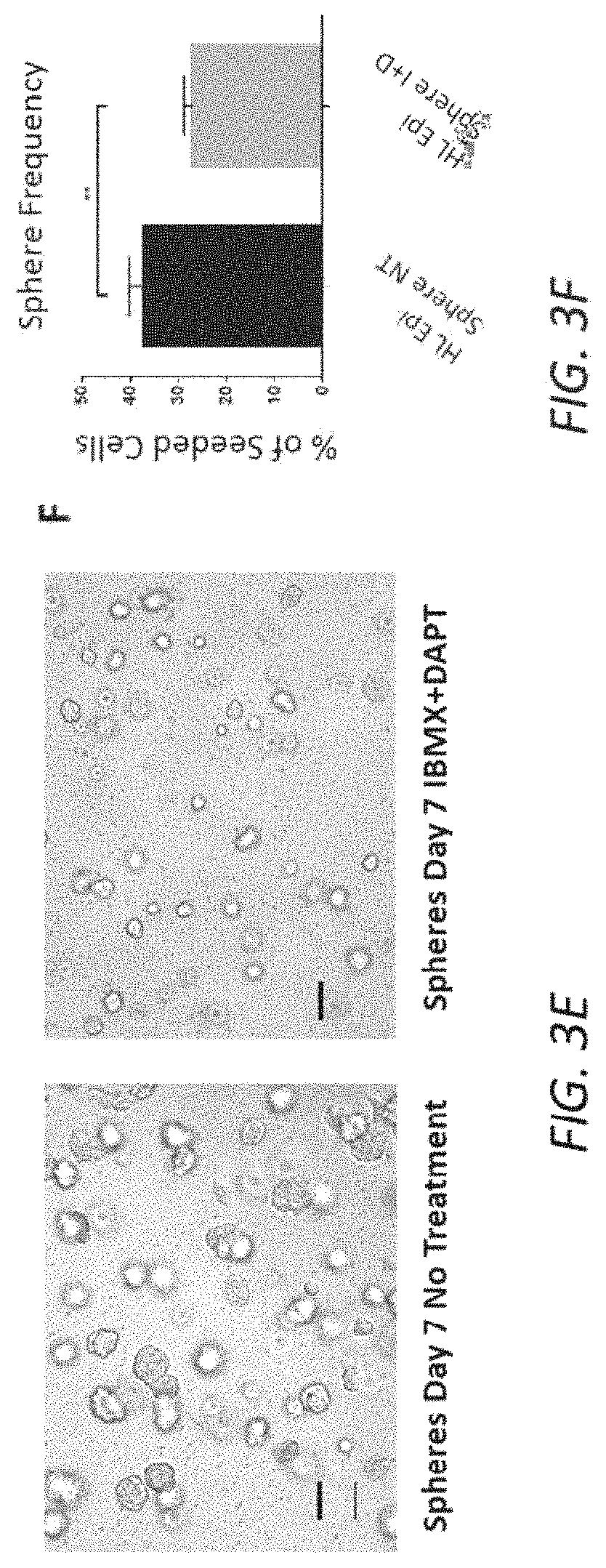
D00012

D00013

D00014

D00015

D00016

D00017

D00018

D00019

D00020

D00021

D00022

D00023

D00024

D00025

D00026

D00027

D00028

D00029

D00030

D00031
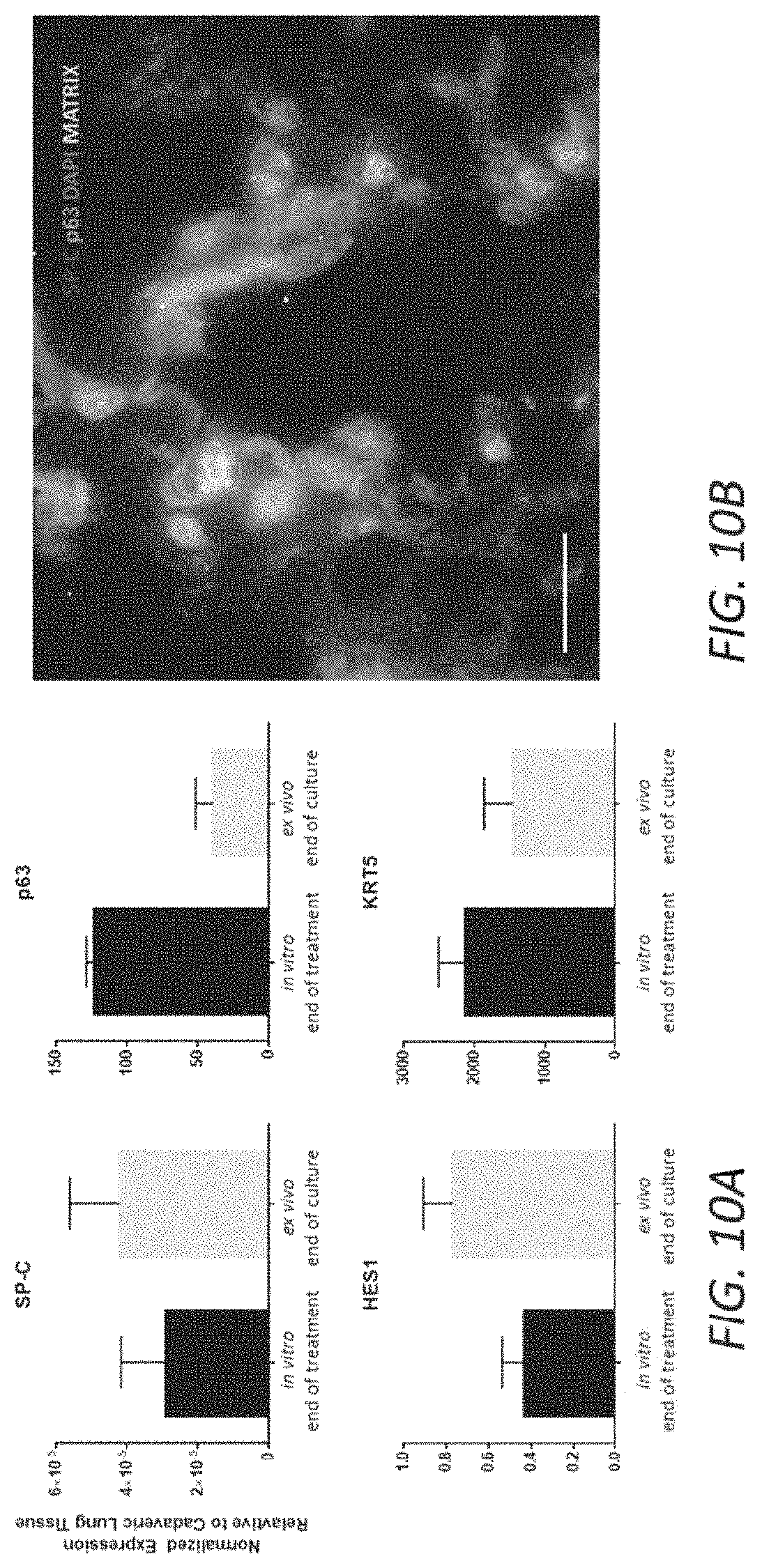
D00032

D00033

D00034

D00035

D00036

D00037

D00038

D00039

D00040

D00041

D00042

D00043

D00044

D00045

D00046

D00047

D00048
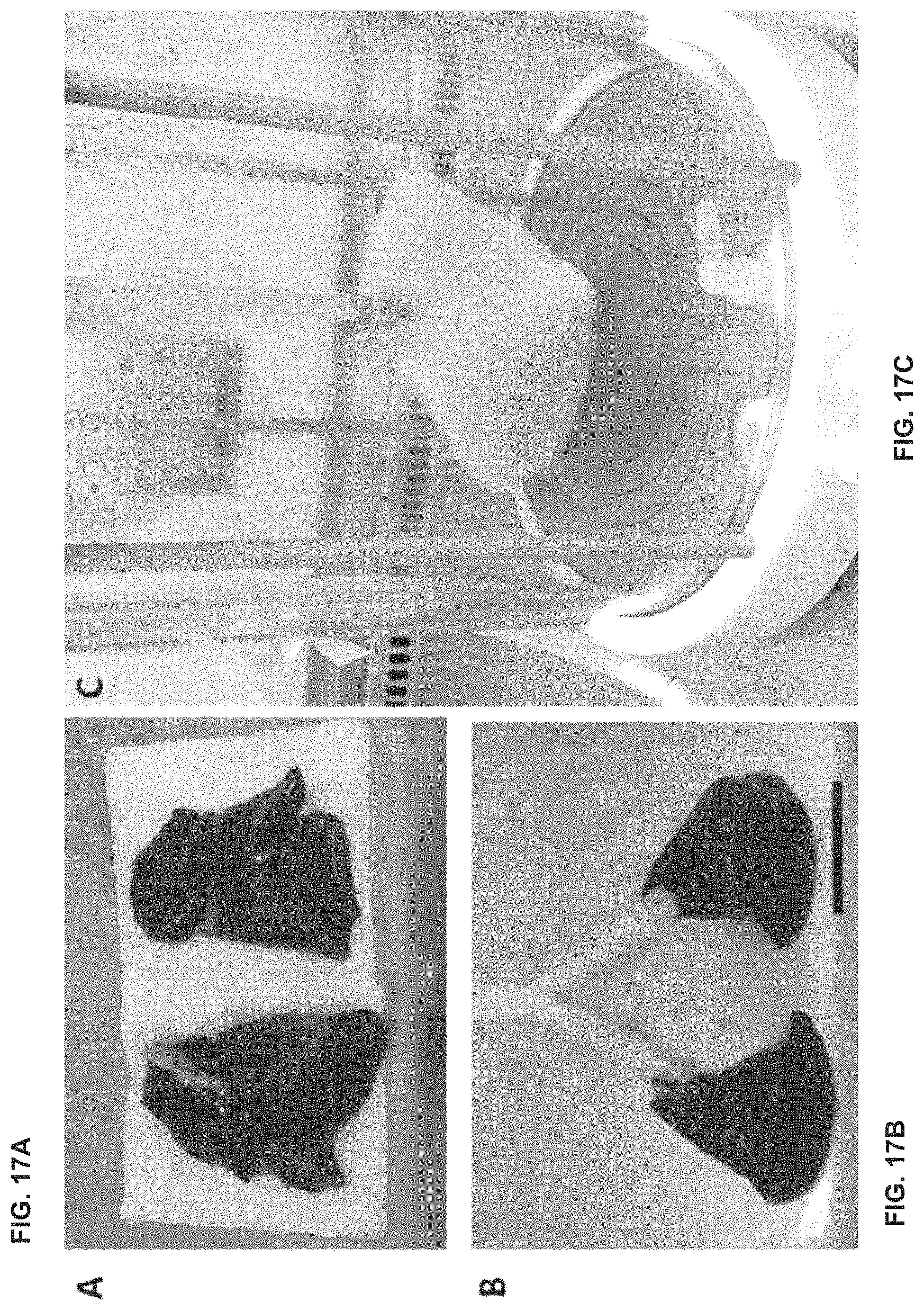
D00049

D00050

S00001
XML
uspto.report is an independent third-party trademark research tool that is not affiliated, endorsed, or sponsored by the United States Patent and Trademark Office (USPTO) or any other governmental organization. The information provided by uspto.report is based on publicly available data at the time of writing and is intended for informational purposes only.
While we strive to provide accurate and up-to-date information, we do not guarantee the accuracy, completeness, reliability, or suitability of the information displayed on this site. The use of this site is at your own risk. Any reliance you place on such information is therefore strictly at your own risk.
All official trademark data, including owner information, should be verified by visiting the official USPTO website at www.uspto.gov. This site is not intended to replace professional legal advice and should not be used as a substitute for consulting with a legal professional who is knowledgeable about trademark law.