C. novyi for the treatment of solid tumors in humans
Saha , et al.
U.S. patent number 10,617,723 [Application Number 14/781,273] was granted by the patent office on 2020-04-14 for c. novyi for the treatment of solid tumors in humans. This patent grant is currently assigned to BIOMED VALLEY DISCOVERIES, INC., THE JOHNS HOPKINS UNIVERSITY. The grantee listed for this patent is BIOMED VALLEY DISCOVERIES, INC., THE JOHNS HOPKINS UNIVERSITY. Invention is credited to Kenneth W. Kinzler, Saurabh Saha, Bert Vogelstein, Shibin Zhou.


View All Diagrams
| United States Patent | 10,617,723 |
| Saha , et al. | April 14, 2020 |
C. novyi for the treatment of solid tumors in humans
Abstract
The present invention provides, inter alia, methods for treating or ameliorating an effect of a solid tumor present in a human. These methods include administering intratumorally to the human a unit dose of C. novyi, preferably C. novyi NT, colony forming units (CFUs), which contains about 1.times.10.sup.3-1.times.10.sup.7 CFUs suspended in a pharmaceutically acceptable carrier or solution. Methods for debulking a solid tumor present in a human, methods for ablating a solid tumor present in a human, a method for microscopically precise excision of tumor cells in a human, methods for treating or ameliorating an effect of a solid tumor that has metastasized to one or more sites in a human, unit doses of C. novyi, preferably C. novyi NT, CFUs, and kits for treating or ameliorating an effect of a solid tumor present in a human are also provided.
| Inventors: | Saha; Saurabh (Wellesley Hills, MA), Zhou; Shibin (Owings Mills, MD), Vogelstein; Bert (Baltimore, MD), Kinzler; Kenneth W. (Bel Air, MD) | ||||||||||
|---|---|---|---|---|---|---|---|---|---|---|---|
| Applicant: |
|
||||||||||
| Assignee: | BIOMED VALLEY DISCOVERIES, INC.
(Kansas City, MO) THE JOHNS HOPKINS UNIVERSITY (Baltimore, MD) |
||||||||||
| Family ID: | 51625539 | ||||||||||
| Appl. No.: | 14/781,273 | ||||||||||
| Filed: | March 28, 2014 | ||||||||||
| PCT Filed: | March 28, 2014 | ||||||||||
| PCT No.: | PCT/US2014/032196 | ||||||||||
| 371(c)(1),(2),(4) Date: | September 29, 2015 | ||||||||||
| PCT Pub. No.: | WO2014/160950 | ||||||||||
| PCT Pub. Date: | October 02, 2014 |
Prior Publication Data
| Document Identifier | Publication Date | |
|---|---|---|
| US 20160051597 A1 | Feb 25, 2016 | |
Related U.S. Patent Documents
| Application Number | Filing Date | Patent Number | Issue Date | ||
|---|---|---|---|---|---|
| 61806497 | Mar 29, 2013 | ||||
| Current U.S. Class: | 1/1 |
| Current CPC Class: | A61P 1/18 (20180101); A61P 15/08 (20180101); A61N 5/10 (20130101); A61P 1/16 (20180101); A61K 35/742 (20130101); A61K 45/06 (20130101); A61P 21/00 (20180101); A61P 37/04 (20180101); A61K 35/74 (20130101); A61P 29/00 (20180101); A61P 37/02 (20180101); A61P 43/00 (20180101); A61P 17/00 (20180101); A61P 35/00 (20180101); A61K 31/65 (20130101); A61P 35/04 (20180101); A61K 2035/11 (20130101) |
| Current International Class: | A61K 35/74 (20150101); A61K 35/00 (20060101); A61N 5/10 (20060101); A61K 45/06 (20060101); A61K 31/65 (20060101); A61K 35/742 (20150101) |
| Field of Search: | ;424/93.45 |
References Cited [Referenced By]
U.S. Patent Documents
| 4599331 | July 1986 | Schreiber et al. |
| 4771042 | September 1988 | Braughler et al. |
| 6905480 | June 2005 | McGuckin, Jr. et al. |
| 7331947 | February 2008 | McGuckin, Jr. et al. |
| 7344710 | March 2008 | Dang et al. |
| 2005/0079157 | April 2005 | Dang et al. |
| 2010/0034814 | February 2010 | Sabbadini |
| 20021236471 | Sep 2002 | EP | |||
| 1675465 | Mar 2010 | EP | |||
| 1987002672 | May 1987 | WO | |||
| 1990015816 | Dec 1990 | WO | |||
| 2003/045153 | Jun 2003 | WO | |||
| 2005/018332 | Mar 2005 | WO | |||
| 2005018332 | Mar 2005 | WO | |||
| 2008/073148 | Jun 2006 | WO | |||
Other References
|
Kim et al., Atypical radiological features of a leiomyosarcoma that arose from the ovarian vein and mimicked a vascular tumor, The British Journal of Radiology, 83 (2010), e95-e97. cited by examiner . Dunn et al., Disseminated Osteomyelitis Caused by Clostridium novyi in a Cat, Case Report, Can Vet J, 24 (1983) 312-315. cited by examiner . OB-GYN 101 Pharmacy, Antibiotics of Choice, Available Online at: www.brooksidepress.org/ Products/OBGYN_101/MyDocuments4/Pharmacy/AntibioticsofChoice.htm, at least as early as Dec. 18, 2005 per Internet Archive Wayback Machine. cited by examiner . Korman et al., Checkpoint Blockade in Cancer Immunotherapy, Adv Immunol, 90 (2006), 297-339. cited by examiner . Mose, J.R., Clostridium Strain M55 and its effectson Malignant Tumors, in Bacteries anaerobies 1st edn (ed. Fredette, V.) 229-247, Montreal Institute e Microbiologie et l'Hygiene de Universite de Montreal, 1967. cited by applicant . Mose, J.R., Onkolyse durch Clostridien in 3rd International Congress of Chemotherapy (ed.) Thieme, G., 1972, Stuttgurt, Germany. cited by applicant . Komeda, S., et al., A Third Mode of DNA Binding: Phosphate Clamps by a Polynuclear Platinum Complex, J. Am. Chem. Soc., 2006, 128 (50), pp. 16092-16103. cited by applicant . Senderowitz, A.M., et al., Information needed to conduct first-in human oncology trials in the U.S.: a view from a former FDA medical reviewer, Clin. Cancer. Res. 2010, 16: 1719-25. cited by applicant . Qu, Y., et al., Synthesis and DNA conformational changes of non-covalent polynuclear platinum complexes, J. Inorg. Biochem. Oct. 2004;98(10):1591-98. cited by applicant . Kleinman, M. E., et al., Sequence- and target-independent angiogenesis suppression by siRNA via TLR3, Nature. Apr. 3, 2008; 452(7187): 591-597. cited by applicant . Makrides, S.C., 1998. Strategies for optimizing heterologous protein expression in Escherichia coli. Trends Biotechnol. 16: 54-60. cited by applicant . Maurer, T., Small-molecule ligands bind to a distinct pocket in Ras and inhibit SOS-mediated nucleotide excange activity, PNAS 109(14): 5299-304 (2012). cited by applicant . Shima, F., et al., In silico discovery of small-molecule Ras inhibitors that display antitumor activity by blocking the Ras-effector interaction, Proc Natl Arad Sci U S A, 110(20):8182-7 (2013). cited by applicant . Patgiri, A., et al., An Orthosteric inhibitor of the Ras-Sos interaction, Na. Chem. Biol. 7:585-587 (2011). cited by applicant . Dennis, M.M., et al., Prognostic Factors for Cutaneous and Subcutaneous Soft Tissue Sarcomas in Dogs, The American College of Veterinary Pathologists, 2011, 48(1), pp. 73-84. cited by applicant . Van Mellaert, L., et al., (2006) Clostridium spores as anti-tumour agents. Trends Microbial 14: 190-196. cited by applicant . Harris, A., et al., Synthesis, Characterization, and Cytotoxicity of a Novel Highly Charged Trinuclear Platinum Compound. Enhancement of Cellular Uptake with Charge, Inorg. Chem., 2005, 44 (26), pp. 9598-9600. cited by applicant . International Search Report dated Aug. 25, 2014. cited by applicant . Agrawal, N. et al. Bacteriolytic therapy can generate a potent immune response against experimental tumors. Proc Natl Acad Sci U S A 101, 15172-7 (2004). cited by applicant . Bai, R.Y., et al. V. Antiparasitic mebendazole shows survival benefit in 2 preclinical models of glioblastoma multiforme. Neuro-oncology 13, 974-982 (2011). cited by applicant . Barretina, J., et al. Subtype-specific genomic alterations define new targets for soft-tissue sarcoma therapy. Nature genetics 42,715-721 (2010). cited by applicant . Bettegowda, C., et al. The genome and transcriptomes of the anti-tumor agent Clostridium novyi-NT. Nature biotechnology 24, 1573-1580 (2006). cited by applicant . Bettegowda, C., & Saha, S. Clostridium novyi-NT Cancer Therapeutic. Chordoma Foundation. Mar. 22, 2013. cited by applicant . Bettegowda, C., et al. Overcoming the hypoxic barrier to radiation therapy with anaerobic bacteria. Proc Natl Acad Sci U S A. Dec. 9, 2003; 100(25): 15083-15088. cited by applicant . Breed, Robert S.; Dotterrer, W. D. "The Number of Colonies Allowable on Satisfactory Agar Plates". Journal of Bacteriology 1 (3): 321-331 (1916). cited by applicant . Brook, I. Anaerobic infections in children. Microbes Infect. Oct. 2002;4(12):1271-80. cited by applicant . Carey, R.W., Holland, J.F., Whang, H.Y., Neter, E. & Bryant, B. Clostridial oncolysis in man. Eur. J. Cancer 3, 37-46 (1967). cited by applicant . Chmielecki, J., et al. Whole-exome sequencing identifies a recurrent NAB2-STAT6 fusion in solitary fibrous tumors. Nature genetics 45, 131-132 (2013). cited by applicant . Dang, L.H. et al. Targeting Vascular and Avascular Compartments of Tumors with C. novyi-NT and Anti-Microtubule Agents. Cancer Biol Ther 3, 326-37 (2004). cited by applicant . Dang, L.H., et al., "Combination bacteriolytic therapy for the treatment of experimental tumors." PNAS. vol. 98, pp. 15155-15160 (2001). cited by applicant . Diaz, L.A., Jr. et al. Pharmacologic and toxicologic evaluation of C. novyi-NT spores. Toxicol Sci 88, 562-75 (2005). cited by applicant . European Medicines Agency. Combined VeDDRA list of clinical terms for reporting suspected adverse reactions in animals and humans to veterinary medicinal products (2012). cited by applicant . Gavhane, Y.N. et al., "Solid Tumors: Facts, Challenges and Solutions." International J. of Pharma Science and Research, vol. 2, pp. 1-12 (2011). cited by applicant . International Search Report for PCT/U52014/032196, dated Aug. 25, 2014. cited by applicant . Jain, R.K. & Forbes, N.S. Can engineered bacteria help control cancer? Proc Natl Acad Sci U S A 98, 14748-50 (2001). cited by applicant . Jones, S., et al. Frequent mutations of chromatin remodeling gene ARID1A in ovarian clear cell carcinoma. Science 330, 228-231 (2010). cited by applicant . Joseph, C., et al. Exomic Analysis of myxoid liposarcomas, synovial sarcomas and osteosarcomas. Genes Chromosomes Cancer. Jan. 2014;53(1):15-24. cited by applicant . Lee, R.S., et al. A remarkably simple genome underlies highly malignant pediatric rhabdoid cancers. J Clin Invest. Aug. 2012;122(8):2983-8. cited by applicant . Leu, KM, et al. Laboratory and clinical evidence of synergistic cytotoxicity of sequential treatment with gemcitabine followed by docetaxel in the treatment of sarcoma. J Clin Oncol. May 1, 2004;22(9):1706-12. cited by applicant . Nemunaitis, J, et al. Pilot trial of genetically modified, attenuated Salmonella expressing the E. coli cytosine deaminase gene in refractory cancer patients. Cancer Gene Ther. Oct. 2003;10(10):737-44. cited by applicant . Nicolson, GL & Nicolson, NL. Gulf War illnesses: complex medical, scientific and political paradox. Med Confl Surviv. Apr.-Jun. 1998;14(2):156-65. cited by applicant . Paoloni, M., et al. Translation of new cancer treatments from pet dogs to humans. Nature Reviews Cancer 8, 147-156 (2008). cited by applicant . Parker, R.C., Plummer, H.C., Siebenmann, C.O. & Chapman, M.G. Effect of histolyticus infection and toxin on transplantable mouse tumors. Proc. Soc. Exp. Biol. Med. 66, 461 (1947). cited by applicant . Patnaik, A.K., et al. Canine cutaneous mast cell tumor: morphologic grading and survival time in 83 dogs. Veterinary pathology 21, 469-474 (1984). cited by applicant . Roberts, N.J., et al. Intratumoral injection of Clostridium novyi-NT spores induces antitumor responses. Sci Transl Med. Aug. 13, 2014;6(249):249ra111. cited by applicant . Sabattini, S., et al. Histologic Grading of Canine Mast Cell Tumor: Is 2 Better Than 3? Vet Pathol. Jan. 2015;52(1):70-3. cited by applicant . Schlom, J. Recent advances in therapeutic cancer vaccines. Cancer Biother Radiopharm. Feb. 2012;27(1):2-5. cited by applicant . Smedley, R.C., et al. Prognostic markers for canine melanocytic neoplasms: a comparative review of the literature and goals for future investigation. Veterinary pathology 48, 54-72 (2011). cited by applicant . Vail, D.M., et al. Spontaneously occurring tumors of companion animals as models for human cancer. Cancer investigation 18, 781-792 (2000). cited by applicant . Van Mellaert, L, et al. Clostridium spores as anti-tumour agents. Trends Microbiol. Apr. 2006;14(4):190-6. cited by applicant . Veterinary Co-Operative Oncology Group. Veterinary Co-operative Oncology Group--Common Terminology Criteria for Adverse Events (VCOG-CTCAE) following chemotherapy or biological antineoplastic therapy in dogs and cats v1.0. Veterinary and comparative oncology 2, 195-213 (2004). cited by applicant . Vogelstein, B., et al. Cancer genome landscapes. Science 339, 1546-1558 (2013). cited by applicant . Walther, W., et al. Novel jet-injection technology for nonviral intratumoral gene transfer in patients with melanoma and breast cancer. Clin Cancer Res. Nov. 15, 2008;14(22):7545-53. cited by applicant . Written Opinion of the International Searching Authority for PCT/US2014/032196, dated Aug. 25, 2014. cited by applicant . Krick E.L. et al. Evaluation of Clostridium novyi-NT spores in dogs with naturally occurring tumors. Am J Vet Res. Jan. 2012;73(1):112-8. cited by applicant . Pawelek et al. "Bacteria as tumour-targeting vectors". The lancet oncology. Sep. 30, 2003;4(9):548-56. cited by applicant . EP Appln. No. 14774988.1 Office Action dated Aug. 31, 2018. cited by applicant . Tourneau, et al. "Dose Escalation Methods in Phase I Cancer Clinial Trials," J Natl Cancer Inst. 10:708:720. cited by applicant. |
Primary Examiner: Tichy; Jennifer M. H.
Attorney, Agent or Firm: Bryan Cave Leighton Paisner LLP
Claims
What is claimed is:
1. A method for debulking or ablating a solid tumor present in a human comprising administering intratumorally to the human a unit dose of C. novyi colony forming units (CFUs) comprising 1.times.10.sup.3-1.times.10.sup.5 CFUs suspended in a pharmaceutically acceptable carrier or solution, wherein the C. novyi is effective to debulk or ablate the solid tumor without administration of additional anti-cancer agents.
2. The method according to claim 1, wherein the solid tumor is selected from the group consisting of soft tissue sarcoma, hepatocellular carcinoma, breast cancer, pancreatic cancer, and melanoma.
3. The method according to claim 1, wherein the solid tumor is leiomyosarcoma.
4. The method according to claim 3, wherein the solid tumor is retroperitoneal leiomyosarcoma.
5. The method according to claim 1, wherein the unit dose comprises about 1.times.10.sup.3-1.times.10.sup.4 C. novyi CFUs.
6. The method according to claim 1, wherein the C. novyi CFUs are selected from the group consisting of vegetative and spore forms.
7. The method according to claim 1, wherein the C. novyi is C. novyi NT.
8. The method according to claim 7, wherein the unit dose comprises 1.times.10.sup.3-1.times.10.sup.5 C. novyi NT spores.
9. The method according to claim 7, wherein the unit dose comprises about 1.times.10.sup.3-1.times.10.sup.4 C. novyi NT spores.
10. The method according to claim 1, wherein the administering step comprises injecting the unit dose at a single location into the tumor.
11. The method according to claim 1, wherein the administering step comprises injecting the unit dose at multiple unique locations into the tumor.
12. The method according to claim 1, wherein the administering step comprises injecting the unit dose at 1-5 unique locations into the tumor.
13. The method according to claim 1, wherein the administering step comprises injecting the unit dose at 5 or more unique locations into the tumor.
14. The method according to claim 1 further comprising administering a plurality of treatment cycles to the human, each treatment cycle comprising injecting one unit dose of the C. novyi CFUs into the solid tumor.
15. The method according to claim 14, wherein 2-10 treatment cycles are administered.
16. The method according to claim 14, wherein 2-4 treatment cycles are administered.
17. The method according to claim 14, wherein an interval between each treatment cycle is about 5-100 days.
18. The method according to claim 14, wherein an interval between each treatment cycle is about 7 days.
19. The method according to claim 8 further comprising administering IV fluids to the human before, during, and/or after each administration of the C. novyi NT spores.
20. The method according to claim 8 further comprising administering a plurality of treatment cycles to the human, each treatment cycle comprising injecting one unit dose of the C. novyi NT spores into the solid tumor.
21. The method according to claim 20, wherein 2-4 treatment cycles are administered.
22. The method according to claim 1 further comprising administering IV fluids to the human before, during, and/or after each administration of the C. novyi.
23. The method according to claim 1 further comprising providing the human with a first course of antibiotics for a period of time and at a dosage that is effective to treat or alleviate an adverse side effect selected from the group consisting of infections, vomiting, hematochezia, fever, and combinations thereof caused by the C. novyi.
24. The method according to claim 23, wherein the antibiotics are administered for two weeks post C. novyi administration.
25. The method according to claim 23, wherein the antibiotics are selected from the group consisting of amoxicillin, clavulanate, metronidazole, and combinations thereof.
26. The method according to claim 23 further comprising providing the human with a second course of antibiotics for a period of time and at a dosage that is effective to treat or alleviate an adverse side effect selected from the group consisting of infections, vomiting, hematochezia, fever, and combinations thereof caused by the C. novyi.
27. The method according to claim 26, wherein the second course of antibiotics is initiated after completion of the first course of antibiotics and is carried out for 1-6 months.
28. The method according to claim 26, wherein the second course of antibiotics is initiated after completion of the first course of antibiotics and is carried out for 3 months.
29. The method according to claim 26, wherein the antibiotic used in the second course is doxycycline.
30. The method according to claim 1, further comprising administering to the human an anti-cancer agent selected from the group consisting of chemotherapy, radiation therapy, immunotherapy, and combinations thereof, after the C. novyi have acted to debulk or ablate the solid tumor.
31. The method according to claim 30, wherein the immunotherapy comprises administering to the human an immune checkpoint inhibitor.
32. The method according to claim 1, wherein the solid tumor is resistant to a therapy selected from the group consisting of chemotherapy, radiation therapy, immunotherapy, and combinations thereof.
33. The method according to claim 30, wherein the chemotherapy comprises administering to the human an agent selected from the group consisting of an anti-metabolite, a microtubule inhibitor, a DNA damaging agent, an antibiotic, an anti-angiogenesis agent, a vascular disrupting agent, a molecularly targeted agent, and combinations thereof.
34. The method according to claim 30, wherein the chemotherapy comprises administering to the human an agent selected from the group consisting of gemcitabine, taxol, adriamycin, ifosfamide, trabectedin, pazopanib, abraxane, avastin, everolimus, and combinations thereof.
35. The method according to claim 1, wherein the solid tumor is refractory to standard therapy or the solid tumor is without an available standard therapy.
36. The method according to claim 1, wherein the unit dose of C. novyi induces a potent localized inflammatory response and an adaptive immune response in the human.
37. A method for microscopically precise excision of tumor cells in a human comprising administering intratumorally to the human a unit dose of C. novyi NT colony forming units (CFUs) comprising 1.times.10.sup.3-1.times.10.sup.5 CFUs suspended in a pharmaceutically acceptable carrier or solution, wherein the C. novyi is effective for microscopically precise excision of the tumor cells without administration of additional anti-cancer agents.
38. A method for debulking or ablating a solid tumor that has metastasized to one or more sites in a human comprising administering intratumorally to the human a unit dose of C. novyi NT colony forming units (CFUs) comprising 1.times.10.sup.3-1.times.10.sup.5 CFUs suspended in a pharmaceutically acceptable carrier or solution, wherein the C. novyi is effective to debulk or ablate the solid tumor without administration of additional anti-cancer agents.
39. The method according to claim 38, wherein at least one site is distal to the original solid tumor.
40. A method for debulking a solid tumor present in a human comprising administering intratumorally to the human a unit dose of C. novyi CFUs comprising 1.times.10.sup.3-1.times.10.sup.5 CFUs suspended in a pharmaceutically acceptable carrier or solution, wherein the C. novyi is effective to debulk the solid tumor without administration of additional anti-cancer agents.
41. The method according to claim 40, wherein the solid tumor is selected from the group consisting of soft tissue sarcoma, hepatocellular carcinoma, breast cancer, pancreatic cancer, and melanoma.
42. A method for debulking a solid tumor present in a human comprising administering intratumorally to the human one to four cycles of a unit dose of C. novyi NT spores comprising 1.times.10.sup.3-1.times.10.sup.5 spores per cycle, each unit dose of C. novyi NT being suspended in a pharmaceutically acceptable carrier or solution, wherein the C. novyi is effective to debulk the solid tumor without administration of additional anti-cancer agents.
43. A method for debulking or ablating a solid tumor present in a human comprising administering intratumorally to the human one to four cycles of a unit dose of C. novyi NT spores comprising 1.times.10.sup.3-1.times.10.sup.5 spores per cycle, each unit dose of C. novyi NT spores being suspended in a pharmaceutically acceptable carrier or solution, wherein the C. novyi is effective to debulk or ablate the solid tumor without administration of additional anti-cancer agents.
44. A method for ablating a solid tumor present in a human comprising administering intratumorally to the human a unit dose of C. novyi CFUs comprising 1.times.10.sup.3-1.times.10.sup.4 CFUs suspended in a pharmaceutically acceptable carrier or solution, wherein the C. novyi is effective to ablate the solid tumor without administration of additional anti-cancer agents leaving a margin of normal tissue.
45. The method according to claim 44, wherein the tumor is a sarcoma.
Description
FIELD OF INVENTION
The present invention provides, inter alia, methods for treating or ameliorating an effect of a solid tumor present in a human, for debulking a solid tumor present in a human, for microscopically precise excising of tumor cells in a human, and for ablating a solid tumor present in a human. Unit doses of C. novyi CFUs and kits are also provided.
CROSS-REFERENCE TO RELATED APPLICATIONS
The present invention claims benefit to U.S. provisional application Ser. No. 61/806,497 filed Mar. 29, 2013, the entire contents of which are incorporated by reference.
BACKGROUND OF THE INVENTION
Strategies that successfully target and destroy human cancers recognize differences between normal and malignant tissues (Dang et al., 2001). Such differences can be found at the molecular level, as is the case with genetic aberrations, or more holistically, as with the physiological aberrations in a tumor.
It is known that malignant solid tumors are usually composed of a necrotic core and a viable rim. Therapeutic interventions to date have focused on the well-vascularized outer shell of the tumor, but few have targeted the inner hypoxic core (Jain et al., 2001). The inner core of a tumor has unique characteristics that differentiate it from normal tissues. The core has a poor vascular supply and is therefore deficient in nutrients and oxygen. As a site of active cellular necrosis, the lack of a functional vascular supply limits the clearance of noxious cell breakdown and results in a low pH. Such an environment is not suitable for growth of most human cells but is a rich environment for the growth of certain anaerobic bacteria. More than sixty-years ago, this concept led investigators to inject spores of Clostridium histolyticus into tumor-bearing animals (Parker et al., 1947). Remarkably, the bacteria germinated only in the necrotic core of the tumor and liquefied the tumors. In the 1950s and 1960s, spores from Clostridium butyricum were injected into patients with a variety of very advanced solid tumor malignancies (Mose, 1967; Mose, 1972). Many patients had significant germination and destruction of large portions of their tumors, but the very poor health and advanced stage of these patients made their clinical management difficult and the absence of complete clinical responses subdued further pursuit of this approach.
Successful treatment of solid tumors remains an unfulfilled medical goal. Accordingly, there is a need to find treatments for solid tumors. The present invention is directed to meeting this and other needs.
SUMMARY OF THE INVENTION
One embodiment of the present invention is a method for treating or ameliorating an effect of a solid tumor present in a human. This method comprises administering intratumorally to the human a unit dose of C. novyi colony forming units (CFUs) comprising about 1.times.10.sup.3-1.times.10.sup.7 CFUs suspended in a pharmaceutically acceptable carrier or solution.
Another embodiment of the present invention is a method for debulking a solid tumor present in a human. This method comprises administering intratumorally to the human a unit dose of C. novyi CFUs comprising about 1.times.10.sup.3-1.times.10.sup.7 CFUs suspended in a pharmaceutically acceptable carrier or solution.
An additional embodiment of the present invention is a method for debulking a solid tumor present in a human. This method comprises administering intratumorally to the human one to four cycles of a unit dose of C. novyi NT spores comprising about 1.times.10.sup.4 spores per cycle, each unit dose of C. novyi NT being suspended in a pharmaceutically acceptable carrier or solution.
A further embodiment of the present invention is a method for treating or ameliorating an effect of a solid tumor present in a human. This method comprises administering intratumorally to the human one to four cycles of a unit dose of C. novyi NT spores comprising about 1.times.10.sup.4 spores per cycle, each unit dose of C. novyi NT spores being suspended in a pharmaceutically acceptable carrier or solution.
Another embodiment of the present invention is method for ablating a solid tumor present in a human. This method comprises administering intratumorally to the human a unit dose of C. novyi CFUs comprising about 1.times.10.sup.3-1.times.10.sup.7 CFUs suspended in a pharmaceutically acceptable carrier or solution, wherein the tumor is ablated leaving a margin of normal tissue.
A further embodiment of the present invention is a unit dose of C. novyi CFUs. This unit dose comprises about 1.times.10.sup.3-1.times.10.sup.7 CFUs in a pharmaceutically acceptable carrier or solution, which is effective for treating or ameliorating an effect of a solid tumor present in a human.
An additional embodiment of the present invention is a kit for treating or ameliorating an effect of a solid tumor present in a human. This kit comprises a unit dose of C. novyi CFUs comprising about 1.times.10.sup.3-1.times.10.sup.7 CFUs in a pharmaceutically acceptable carrier or solution and instructions for use of the kit.
Another embodiment of the present invention is a method for microscopically precise excision of tumor cells in a human. This method comprises administering intratumorally to the human a unit dose of C. novyi NT colony forming units (CFUs) comprising about 1.times.10.sup.3-1.times.10.sup.7 CFUs suspended in a pharmaceutically acceptable carrier or solution.
A further embodiment of the present invention is a method for treating or ameliorating an effect of a solid tumor that has metastasized to one or more sites in a human. This method comprises administering intratumorally to the human a unit dose of C. novyi NT colony forming units (CFUs) comprising at least about 1.times.10.sup.3-1.times.10.sup.7 CFUs suspended in a pharmaceutically acceptable carrier or solution.
BRIEF DESCRIPTION OF THE DRAWINGS
FIGS. 1A-B show various images of canine osteosarcomas on the right distal radius/ulna of test subjects "Sasha" (FIG. 1A) and "Sampson" (FIG. 1B) after radiation treatment and intravenous (IV) C. novyi NT injection.
FIG. 2A shows Kaplan-Meier curves showing survival of F433 Fisher rats after orthotopic implantation of a syngeneic glioma cell line (F98). Outer line C. novyi-NT spores injected into tumor 12-15 days after tumor implantation. Inner line control. FIG. 2B shows bioluminescence (Xenogen imaging system) in three representative F433 Fisher rats after orthotopic implantation of F98 glioma cell line. Images acquired on day 0 (pretreatment day of C. novyi--NT spore injection), day 1 after IT injection of C. novyi-NT spores, and day 2 after IT injection of C. novyi-NT spores. FIG. 2C shows luciferase activity (count in millions) on day 0 (pretreatment), day 1 after IT injection of C. novyi-NT spores, and day 2 after IT injection of C. novyi-NT spores.
FIGS. 3A-B and 4A-B show germinated C. novyi-NT bacteria within microscopic brain tumor lesions. In these Figures, gram stain showed vegetative C. novyi-NT bacteria (white or black arrowheads) localized in tumor (T) and stellate micro-invasion (S), but not in normal brain tissue (Br). FIG. 3A is a 100.times. magnification showing the interface of tumor and normal brain. FIG. 3B is a 400.times. magnification showing the interface of tumor and normal brain. FIG. 4A is a 100.times. magnification showing the interface of normal brain, tumor, and stellate micro-invasion of neoplastic tissue. FIG. 4B is a 400.times. magnification showing C. novyi-NT germination in a stellate micro-invasive lesion.
FIG. 5 is a table of summary data for samples sequenced.
FIG. 6 is a table of copy number alterations in canine sarcomas.
FIGS. 7A-F are photographic and CT images from dog 11-R01 showing a partial response to C. novyi-NT therapy. Images span pre-treatment to day 70 after first IT dose of C. novyi-NT spores. FIG. 7A shows a pre-treatment image of the peripheral nerve sheath tumor. FIG. 7B shows abscess formation on day 3 of the study, with extent confined to tumor. FIG. 7C shows medical debridement following spontaneous abscess rupture and discharge of necrotic and purulent material, which allowed healing by second intention. FIG. 7D shows that the wound has healed completely by day 70 of the study and 77.6% reduction in tumor longest diameter was noted. FIG. 7E is a pre-treatment CT image, taken 4 days before first treatment, which shows extent of tumor (circle) at the intersection of pinna and cranium. FIG. 7F is a post-treatment CT image on day 10 of the study showing almost complete de-bulking of tumor.
FIGS. 8A-D are photographic and CT images from dog 04-R03 showing a complete response to C. novyi-NT therapy. Images span pre-treatment to day 60 after first IT dose of C. novyi-NT spores. FIG. 8A shows a pre-treatment image of the soft tissue sarcoma. FIG. 8B shows a tumor localized abscess formed on day 15 of the study, 1 day after a third dose of C. novyi-NT spores. FIG. 8C shows that tumor de-bulking was complete by day 27 of the study and healthy granulation tissue had formed. FIG. 8D shows that the wound had healed completely by day 60 of the study, and no residual tumor was noted (complete response). FIG. 8E is a pre-treatment CT image, taken 5 days before first treatment, showing extent of tumor (circle) on antebrachium. FIG. 8F is a post-treatment CT image on day 62 of the study showing complete loss of tumor mass.
FIG. 9 shows the size of dog 11-R01's tumor from initial IT dosing of C. novyi NT spores to completion of the clinical course.
FIG. 10A shows photographic (upper panels) and CT images (lower panels) of a canine soft tissue sarcoma on test subject "Drake" (04-R01) after IT dosing of C. novyi NT spores. Circled regions of the CT images indicate tumor location. FIG. 10B shows the size of Drake's tumor from initial IT dosing of C. novyi NT, through three subsequent doses, to completion of the clinical course.
FIG. 11 shows the size of dog 04-R03's tumor from initial IT dosing of C. novyi NT spores, through two subsequent cycles, to completion of the clinical course.
FIG. 12A shows tumor size in eight test subjects (11-R02, 04-R02, 26-R01, 16-R02, 04-R05, 16-R03, 11-R04, and 04-R06) over the clinical course in which four cycles of IT C. novyi NT spores were administered. FIG. 12B shows tumor size in three test subjects (04-R08, 01-R02, and 10-R02) for which data from a complete clinical course was not available due to necessary amputation or data cutoff.
FIG. 13 shows an injection scheme for tumors treated in the IT study disclosed in Examples 6 and 7.
FIGS. 14A-D show CT and MRI images from a human patient. FIG. 14A shows a post-treatment CT with contrast on day 3 demonstrating evidence of intra- and extra-medullary air collection. FIG. 14B shows a pre-treatment MRI (T1 with gadolinium contrast) of the right upper humerus showing a contrast enhancing mass involving the soft tissue and possibly adjacent bone. FIG. 14C shows a post-treatment MRI on day 4 demonstrating diminished contrast enhancement in the tumor mass compared to baseline. FIG. 14D shows a post-treatment MRI on day 29 showing homogenous non-enhancing mass consistent with ongoing necrosis. Tumor is highlighted with arrowheads.
FIGS. 15A-D show extensive tumor necrosis in the human patient treated with C. novyi-NT spores. FIGS. 15A and 15B show a pre-treatment tumor biopsy showing viable tumor (leiomyosarcoma) cells, 40.times. (A) and 100.times. (B) magnification, respectively. FIGS. 15C and 15D show a post-treatment tumor biopsy, 4 days after IT injection of C. novyi-NT spores, showing extensive necrosis of tumor cells, 40.times. (A) and 100.times. (B) magnification, respectively.
FIGS. 16A-D show various aspects of the IT injection procedure using a three-tined needle. FIG. 16A shows a photograph of the three-tined needle. FIGS. 16B and 16C show computed tomography (CT) images of the target injection area before and after insertion of the needle. FIG. 16D shows a magnified image of the three tines of the needle. FIG. 16E shows a CT image with overlaying measurements for determining insertion points of the needle.
DETAILED DESCRIPTION OF THE INVENTION
One embodiment of the present invention is a method for treating or ameliorating an effect of a solid tumor present in a human. This method comprises administering intratumorally to the human a unit dose of C. novyi colony forming units (CFUs) comprising about 1.times.10.sup.3-1.times.10.sup.7 CFUs suspended in a pharmaceutically acceptable carrier or solution.
As used herein, the terms "treat," "treating," "treatment" and grammatical variations thereof mean subjecting an individual subject (e.g., a human patient) to a protocol, regimen, process or remedy, in which it is desired to obtain a physiologic response or outcome in that subject, e.g., a patient. In particular, the methods and compositions of the present invention may be used to slow the development of disease symptoms or delay the onset of the disease or condition, or halt the progression of disease development. However, because every treated subject may not respond to a particular treatment protocol, regimen, process or remedy, treating does not require that the desired physiologic response or outcome be achieved in each and every subject or subject, e.g., patient, population. Accordingly, a given subject or subject, e.g., patient, population may fail to respond or respond inadequately to treatment.
As used herein, the terms "ameliorate", "ameliorating" and grammatical variations thereof mean to decrease the severity of the symptoms of a disease in a subject.
As used herein, a "solid tumor" means an abnormal mass of cell growth. Solid tumors may occur anywhere in the body. Solid tumors may be cancerous (malignant) or noncancerous (benign). Examples of solid tumors according to the present invention include adrenocortical carcinoma, anal tumor/cancer, bladder tumor/cancer, bone tumor/cancer (such as osteosarcoma), brain tumor, breast tumor/cancer, carcinoid tumor, carcinoma, cervical tumor/cancer, colon tumor/cancer, endometrial tumor/cancer, esophageal tumor/cancer, extrahepatic bile duct tumor/cancer, Ewing family of tumors, extracranial germ cell tumor, eye tumor/cancer, gallbladder tumor/cancer, gastric tumor/cancer, germ cell tumor, gestational trophoblastic tumor, head and neck tumor/cancer, hypopharyngeal tumor/cancer, islet cell carcinoma, kidney tumor/cancer, laryngeal tumor/cancer, leiomyosarcoma, leukemia, lip and oral cavity tumor/cancer, liver tumor/cancer (such as hepatocellular carcinoma), lung tumor/cancer, lymphoma, malignant mesothelioma, Merkel cell carcinoma, mycosis fungoides, myelodysplastic syndrome, myeloproliferative disorders, nasopharyngeal tumor/cancer, neuroblastoma, oral tumor/cancer, oropharyngeal tumor/cancer, osteosarcoma, ovarian epithelial tumor/cancer, ovarian germ cell tumor, pancreatic tumor/cancer, paranasal sinus and nasal cavity tumor/cancer, parathyroid tumor/cancer, penile tumor/cancer, pituitary tumor/cancer, plasma cell neoplasm, prostate tumor/cancer, rhabdomyosarcoma, rectal tumor/cancer, renal cell tumor/cancer, transitional cell tumor/cancer of the renal pelvis and ureter, salivary gland tumor/cancer, Sezary syndrome, skin tumors (such as cutaneous t-cell lymphoma, Kaposi's sarcoma, mast cell tumor, and melanoma), small intestine tumor/cancer, soft tissue sarcoma, stomach tumor/cancer, testicular tumor/cancer, thymoma, thyroid tumor/cancer, urethral tumor/cancer, uterine tumor/cancer, vaginal tumor/cancer, vulvar tumor/cancer, and Wilms' tumor. Preferably, the solid tumor is selected from the group consisting of soft tissue sarcoma, hepatocellular carcinoma, breast cancer, pancreatic cancer, and melanoma. More preferably, the solid tumor is a leiomyosarcoma, such as a retroperitoneal leiomyosarcoma.
As used herein, a "unit dose" means the amount of a medication administered to a subject, e.g., a human, in a single dose.
As used herein, "C. novyi" means a bacteria belonging to species of Clostridium novyi or a bacteria derived therefrom. Clostridium novyi, which may be obtained commercially from, e.g., the ATCC (#19402), is a gram-positive anaerobic bacterium. A bacteria derived from Clostridium novyi may be made by, e.g., screening native Clostridium novyi for clones that possess specific characteristics. Preferred C. novyi bacteria are those which are non-toxic or minimally toxic to a subject such as a mammal, e.g., a human. For example, a preferred C. novyi, C. novyi NT, is a bacteria derived from native Clostridium novyi that has lost its single systemic toxin (.alpha.-toxin) gene by, e.g., a genetic engineering process or through a selection procedure. C. novyi NT may be made, for example, using the procedure disclosed in Dang et al., 2001 and U.S. Pat. No. 7,344,710. Thus, the present invention includes C. novyi as well as C. novyi NT bacteria.
Pharmacokinetic studies indicate that C. novyi NT spores, if injected intravenously, are rapidly cleared from the circulation (greater than 99% spores are cleared within 1 hour) and sequestered within the reticulo-endothelial system. Long-term distribution studies reveal that these spores are eventually eliminated from all tissues within one year. Delivered in spore form (dormant stage), C. novyi NT germinates (transitions from the spore to the vegetative state) when exposed to the hypoxic regions of tumors. Thus, the toxicities of C. novyi NT are expected to be greater in tumor-bearing than in healthy patients.
Healthy mice and rabbits showed no apparent clinical signs (morbidity, mortality, or clinical appearance) of toxicity regardless of treatment dose when injected with C. novyi NT intravenously. However, examination of tissues at necropsy revealed both gross and microscopic inflammatory changes that appeared to be treatment-dose dependent. These findings, primarily in the liver, spleen and adrenals, were noted at doses of 5.times.10.sup.8 spores/kg or greater. Healthy animals receiving lower doses showed no gross or microscopic abnormalities at necropsy. In animals that received high doses, resolution of inflammation was already evident on day 28 and all signs of inflammation were absent in all animals by one year following administration. To determine if C. novyi NT spores would germinate in non-tumor hypoxic tissue, studies in elderly mice with atherosclerotic plaques and experimental myocardial infarctions were treated with C. novyi NT. There was no evidence of spore localization or germination within these vascular lesions. At the conclusion of the study, no clinical or pathologic abnormalities (other than the pre-existing cardiovascular lesions) were noted in these mice. These studies demonstrated that C. novyi NT caused no apparent clinical and minimal pathological toxicity in healthy animals.
Intravenous (IV) injection of spores into immune-competent tumor-bearing mice leads to lysis of the tumor and an intense inflammatory response. In mice, one of three outcomes is typically observed: One subset (25-35%) of mice are cured (no tumor recurrence after one year of observation) and develop long-term immunity to the original tumor (Agrawal et al., 2004). Another subset (65-75%) experience complete clinical responses, but undergo a recurrence with re-growth of the original tumor. Finally, the remaining subset (0 to 20%, depending on the experiment) undergoes tumor destruction, but develop significant clinical toxicity 2-5 days after the initiation of therapy. Relatively simple measures, such as hydration, are adequate to reduce this toxicity, often entirely eliminating these signs. Studies in larger animals (rabbits) show the same cure and recurrence rates with C. novyi NT therapy, but do not show the life-threatening clinical toxicity observed in a subset of mice. Treatment-related death was observed in tumor-bearing mice, but not in rabbits, treated with C. novyi NT spores (Diaz et al., 2005). In these studies toxicity was related to both spore dose and tumor size. In moribund mice, no specific clinical laboratory or pathologic end-organ damage was noted and the only significant finding was hepatosplenomegaly. Cured mice had rare remnant inflammatory changes in the liver and spleen, but were otherwise no different than untreated animals. These studies show that toxicity in tumor-bearing animals can be pronounced (death) in mice with large tumors, but was minimal in larger animals (rabbits), and was manageable in mice with hydration or antibiotics.
Previous work using C. novyi NT spores injected intravenously (1.times.10.sup.9 spores/m.sup.2) as a single agent in tumor bearing dogs produced no life threatening toxicities. The dogs were maintained on fluid therapy (2-4 ml/kg/hr) for several days post treatment which may have decreased the toxicity. Unfortunately, there were no measurable tumor responses to the treatment.
As used herein, "colony forming units" ("CFUs") mean viable forms of the bacteria which will give rise to an aggregate of cells (or colonies). Such viable forms include vegetative and spore forms, and the present invention includes both forms used separately and in combination. Colony forming unit assays are known in the art. See, e.g., Breed et al., 1916. Media for supporting the growth of C. novyi are commercially available, such as Reinforced Clostridial Medium (RCM) from Difco (BD, Franklin Lakes, N.J.). As set forth above, the unit dose comprises from about 1.times.10.sup.3-1.times.10.sup.7, such as about 1.times.10.sup.3-1.times.10.sup.4, about 1.times.10.sup.4-1.times.10.sup.5, about 1.times.10.sup.5-1.times.10.sup.6, or about 1.times.10.sup.6-1.times.10.sup.7, C. novyi CFUs.
In one aspect of this embodiment, the unit dose comprises from about 1.times.10.sup.6-1.times.10.sup.7 C. novyi CFUs. In another aspect of this embodiment, the unit dose comprises about 1.times.10.sup.4 C. novyi CFUs. Surprisingly, the doses disclosed herein for human treatment are unexpectedly lower than would be expected from simply extrapolating from our non-rodent models using 1/6 of the non-rodent highest non-severely toxic does (HNSTD), as is typical for a starting dose therapeutic for oncology indications. See, e.g., Senderowicz, A.M., "Information needed to conduct first-in human oncology trials in the United States: a view from a former FDA medical reviewer." Clin. Canc. Res., 2010, 16:1719-25.
Preferably, in the present invention the C. novyi is C. novyi NT.
In another aspect of this embodiment, the unit dose comprises about 1.times.10.sup.6-1.times.10.sup.7 C. novyi NT spores. In a further aspect of this embodiment, the unit dose comprises about 1.times.10.sup.4 C. novyi NT spores.
In an additional aspect of this embodiment, the administering step comprises injecting the unit dose at a single location into the tumor. In another aspect of this embodiment, the administering step comprises injecting the unit dose at multiple unique locations, such as 2, 3, 4, 5, 6, 7, 8, 9, 10 or more than 10 unique locations, into the tumor. Preferably, the administering step comprises injecting the unit dose at 1-5 unique locations into the tumor, such as in the configurations shown in FIG. 13. In another preferred embodiment, the administering step comprises injecting the unit dose at 5 or more unique locations into the tumor. Multi-site injections may be carried out as disclosed herein, preferably with a multi-tined needle such as Quadra-Fuse.RTM. (Rex-Medical, Conshohocken, Pa.). In the present invention, the administering step, as noted above, includes injections directly into the tumor, but other methods for administering an active agent, such as C. novyi or C. novyi NT, to a tumor are also contemplated. Such methods include implantation, transdermal delivery, and transmucosal delivery.
In another aspect of this embodiment, the method further comprises administering a plurality of treatment cycles, such as 1, 2, 3, 4, 5, 6, 7, 8, 9, 10, 11, 12, 13, 14, 15, 20, 25, 30, or more than 30 cycles, to the human, each treatment cycle comprising injecting one unit dose of the C. novyi CFUs, such as one unit dose of the C. novyi NT spores, into the solid tumor. Preferably, 1-10 treatment cycles are administered. More preferably, 2-4 treatment cycles are administered. The interval between each treatment cycle may be variable. In one preferred embodiment, the interval between each treatment cycle is about 5-100 days. In another preferred embodiment, the interval between each treatment cycle is about 7 days.
In an additional aspect of this embodiment, the method further comprises administering intravenous (IV) fluids to the human before, during, and/or after each administration of the C. novyi CFUs, such as the C. novyi NT spores. IV fluids for hydrating the patients are disclosed herein and are well known in the art. Such fluids may be fluids that are isotonic with blood, such as, e.g., a 0.9% sodium chloride solution, or Lactated Ringer's solution.
In another aspect of this embodiment, the method further comprises providing the human with a first course of antibiotics for a period of time and at a dosage that is effective to treat or alleviate an adverse side effect caused by the C. novyi CFUs, such as the C. novyi NT spores. In the present invention an adverse side effect (or adverse event, which is used interchangeably with adverse side effect) may include but is not limited to infections (such as those caused by open wounds), vomiting, hematochezia, and fever.
In one preferred embodiment, the antibiotics are administered for two weeks post C. novyi administration. Non-limiting examples of such antibiotics include amoxicillin, clavulanate, metronidazole, and combinations thereof.
In another preferred embodiment, the method further comprises providing the human with a second course of antibiotics for a period of time and at a dosage that is effective to treat or alleviate an adverse side effect caused by the C. novyi. The second course of antibiotics may be initiated after completion of the first course of antibiotics and is carried out for 1-6 months, such as 3 months. Preferably, the antibiotic used in the second course is doxycycline, but any antibiotic approved by a medical professional may be used.
In a further aspect of this embodiment, the method further comprises, using a co-treatment protocol by, e.g., administering to the human a therapy selected from the group consisting of chemotherapy, radiation therapy, immunotherapy, and combinations thereof.
The C. novyi, e.g., the C. novyi NT spores, and the anti-cancer agent(s) used in the co-treatment therapy may be administered to the human, either simultaneously or at different times, as deemed most appropriate by a physician. If the C. novyi, e.g., the C. novyi NT spores, and the other anti-cancer agent(s) are administered at different times, for example, by serial administration, then the C. novyi, e.g., the C. novyi NT spores, may be administered to the human before the other anti-cancer agent. Alternatively, the other anti-cancer agent(s) may be administered to the human before the C. novyi, e.g., the C. novyi NT spores.
As used herein, "chemotherapy" means any therapeutic regimen that is compatible with the C. novyi, e.g., C. novyi NT, treatment of the present invention and that uses cytotoxic and/or cytostatic agents against cancer cells or cells that are associated with or support cancer cells. In a preferred embodiment, the chemotherapy comprises administering to the human an agent selected from the group consisting of an anti-metabolite, a microtubule inhibitor, a DNA damaging agent, an antibiotic, an anti-angiogenesis agent, a vascular disrupting agent, a molecularly targeted agent, and combinations thereof.
As used herein, an "anti-metabolite" is a substance that reduces or inhibits a cell's use of a chemical that is part of normal metabolism. Non-limiting examples of anti-metabolite agents or analogs thereof according to the present invention include antifolates, purine inhibitors, pyrimidine inhibitors, and combinations thereof.
As used herein, an "antifolate" is a substance that alters, reduces, or inhibits the use of folic acid (vitamin B.sub.9) by cells. Non-limiting examples of antifolates include methotrexate (DuraMed Pharmaceuticals, Inc.), pemetrexed (Eli Lilly), pralatrexate (Spectrum Pharmaceuticals), aminopterin (Sigma Aldrich), pharmaceutically acceptable salts thereof, and combinations thereof.
As used herein, a "purine" is a compound that contains a fused six-membered and a five-membered nitrogen-containing ring. Non-limiting examples of purines that are important for cellular metabolism include adenine, guanine, hypoxanthine, and xanthine. A "purine inhibitor" is a substance that alters, reduces or suppresses the production of a purine or the use of a purine by a cell. Non-limiting examples of purine inhibitors include methotrexate (DuraMed Pharmaceuticals, Inc.), pemetrexed (Eli Lilly), hydroxyurea (Bristol-Myers Squibb), 2-mercaptopurine (Sigma-Aldrich), 6-mercaptopurine (Sigma-Aldrich), fludarabine (Ben Venue Laboratories), clofarabine (Genzyme Corp.), nelarabine (GlaxoSmithKline), pralatrexate (Spectrum Pharmaceuticals), 6-thioguanine (Gate Pharmaceuticals), forodesine (BioCryst Pharmaceuticals), pentostatin (Bedford Laboratories), sapacitabine (Cyclacel Pharmaceuticals, Inc.), aminopterin (Sigma Aldrich), azathioprine (GlaxoSmithKline), pharmaceutically acceptable salts thereof, and combinations thereof.
As used herein, a "pyrimidine" is a compound that contains a six-membered nitrogen-containing ring. Non-limiting examples of pyrimidines that are important for cellular metabolism include uracil, thymine, cytosine, and orotic acid. A "pyrimidine inhibitor" is a substance that alters, reduces, or suppresses the production of a pyrimidine or the use of a pyrimidine by the a cell. Non-limiting examples of pyrimidine inhibitors include 5-fluorouracil (Tocris Bioscience), tegafur (LGM Pharma), capecitabine (Xeloda) (Roche), cladribine (LGM Pharma), gemcitabine (Eli Lilly), cytarabine (Bedford Laboratories), decitabine (Eisai Inc.), floxuridine (Bedford Laboratories), 5-azacytidine (Pharmion Pharmaceuticals), doxifluridine (Cayman Chemicals), thiarabine (Access Pharmaceuticals), troxacitabine (SGX Pharmaceuticals), raltitrexed (AstraZeneca), carmofur (Santa Cruz Biotechnology, Inc.), 6-azauracil (MP Biomedicals, LLC), pharmaceutically acceptable salts thereof, and combinations thereof.
In a preferred aspect of the present invention, the anti-metabolite agent is selected from the group consisting of 5-fluorouracil (Tocris Bioscience), tegafur (LGM Pharma), capecitabine (Xeloda) (Roche), cladribine (LGM Pharma), methotrexate (DuraMed Pharmaceuticals, Inc.), pemetrexed (Eli Lilly), hydroxyurea (Bristol-Myers Squibb), 2-mercaptopurine (Sigma-Aldrich), 6-mercaptopurine (Sigma-Aldrich), fludarabine (Ben Venue Laboratories), gemcitabine (Eli Lilly), clofarabine (Genzyme Corp.), cytarabine (Bedford Laboratories), decitabine (Eisai Inc.), floxuridine (Bedford Laboratories), nelarabine (GlaxoSmithKline), pralatrexate (Spectrum Pharmaceuticals), 6-thioguanine (Gate Pharmaceuticals), 5-azacytidine (Pharmion Pharmaceuticals), doxifluridine (Cayman Chemicals), forodesine (BioCryst Pharmaceuticals), pentostatin (Bedford Laboratories), sapacitabine (Cyclacel Pharmaceuticals, Inc.), thiarabine (Access Pharmaceuticals), troxacitabine (SGX Pharmaceuticals), raltitrexed (AstraZeneca), aminopterin (Sigma Aldrich), carmofur (Santa Cruz Biotechnology, Inc.), azathioprine (GlaxoSmithKline), 6-azauracil (MP Biomedicals, LLC), pharmaceutically acceptable salts thereof, and combinations thereof.
As used herein, a "microtubule inhibitor" is a substance that disrupts the functioning of a microtubule, such as the polymerization or the depolymerization of individual microtubule units. In one aspect of the present invention, the microtubule inhibitor may be selected from the group consisting of a microtubule-destabilizing agent, a microtubule-stabilizing agent, and combinations thereof. A microtubule inhibitor of the present invention may also be selected from the group consisting of a taxane, a vinca alkaloid, an epothilone, and combinations thereof. Non-limiting examples of microtubule inhibitors according to the present invention include BT-062 (Biotest), HMN-214 (D. Western Therapeutics), eribulin mesylate (Eisai), vindesine (Eli Lilly), EC-1069 (Endocyte), EC-1456 (Endocyte), EC-531 (Endocyte), vintafolide (Endocyte), 2-methoxyestradiol (EntreMed), GTx-230 (GTx), trastuzumab emtansine (Hoffmann-La Roche), crolibulin (Immune Pharmaceuticals), D1302A-maytansinoid conjugates (ImmunoGen), IMGN-529 (ImmunoGen), lorvotuzumab mertansine (ImmunoGen), SAR-3419 (ImmunoGen), SAR-566658 (ImmunoGen), IMP-03138 (Impact Therapeutics), topotecan/vincristine combinations (LipoCure), BPH-8 (Molecular Discovery Systems), fosbretabulin tromethamine (OXiGENE), estramustine phosphate sodium (Pfizer), vincristine (Pierre Fabre), vinflunine (Pierre Fabre), vinorelbine (Pierre Fabre), RX-21101 (Rexahn), cabazitaxel (Sanofi), STA-9584 (Synta Pharmaceuticals), vinblastine, epothilone A, patupilone (Novartis), ixabepilone (Bristol-Myers Squibb), Epothilone D (Kosan Biosciences), paclitaxel (Bristol-Myers Squibb), docetaxel (Sanofi-Aventis), HAI abraxane, DJ-927 (Daiichi Sankyo), discodermolide (CAS No: 127943-53-7), eleutherobin (CAS No.: 174545-76-7), pharmaceutically acceptable salts thereof, and combinations thereof.
DNA damaging agents of the present invention include, but are not limited to, alkylating agents, platinum-based agents, intercalating agents, and inhibitors of DNA replication.
As used herein, an "alkylating agent" is a substance that adds one or more alkyl groups (C.sub.nH.sub.m, where n and m are integers) to a nucleic acid. In the present invention, an alkylating agent is selected from the group consisting of nitrogen mustards, nitrosoureas, alkyl sulfonates, triazines, ethylenimines, and combinations thereof. Non-limiting examples of nitrogen mustards include mechlorethamine (Lundbeck), chlorambucil (GlaxoSmithKline), cyclophosphamide (Mead Johnson Co.), bendamustine (Astellas), ifosfamide (Baxter International), melphalan (Ligand), melphalan flufenamide (Oncopeptides), and pharmaceutically acceptable salts thereof. Non-limiting examples of nitrosoureas include streptozocin (Teva), carmustine (Eisai), lomustine (Sanofi), and pharmaceutically acceptable salts thereof. Non-limiting examples of alkyl sulfonates include busulfan (Jazz Pharmaceuticals) and pharmaceutically acceptable salts thereof. Non-limiting examples of triazines include dacarbazine (Bayer), temozolomide (Cancer Research Technology), and pharmaceutically acceptable salts thereof. Non-limiting examples of ethylenimines include thiotepa (Bedford Laboratories), altretamine (MGI Pharma), and pharmaceutically acceptable salts thereof. Other alkylating agents include ProLindac (Access), Ac-225 BC-8 (Actinium Pharmaceuticals), ALF-2111 (Alfact Innovation), trofosfamide (Baxter International), MDX-1203 (Bristol-Myers Squibb), thioureidobutyronitrile (CellCeutix), mitobronitol (Chinoin), mitolactol (Chinoin), nimustine (Daiichi Sankyo), glufosfamide (Eleison Pharmaceuticals), HuMax-TAC and PBD ADC combinations (Genmab), BP-C1 (Meabco), treosulfan (Medac), nifurtimox (Metronomx), improsulfan tosilate (Mitsubishi tanabe Pharma), ranimustine (Mitsubishi tanabe Pharma), ND-01 (NanoCarrier), HH-1 (Nordic Nanovector), 22P1G cells and ifosfamide combinations (Nuvilex), estramustine phosphate (Pfizer), prednimustine (Pfizer), lurbinectedin (PharmaMar), trabectedin (PharmaMar), altreatamine (Sanofi), SGN-CD33A (Seattle Genetics), fotemustine (Servier), nedaplatin (Shionogi), heptaplatin (Sk Holdings), apaziquone (Spectrum Pharmaceuticals), SG-2000 (Spirogen), TLK-58747 (Telik), laromustine (Vion Pharmaceuticals), procarbazine (Alkem Laboratories Ltd.), and pharmaceutically acceptable salts thereof.
As used herein, a "platinum-based agent" is an anti-cancer substance that contains the metal platinum and analogs of such substances. The platinum may be in any oxidation state. Platinum-based agents of the present invention include, but are not limited to, 1,2-diaminocyclohexane (DACH) derivatives, phenanthroimidazole Pt(II) complexes, platiunum IV compounds, bi- and tri-nuclear platinum compounds, demethylcantharidin-integrated platinum complexes, platinum-conjugated compounds, cisplatin nanoparticles and polymer micelles, sterically hindered platinum complexes, oxaliplatin (Debiopharm), satraplatin (Johnson Matthey), BBR3464 (Novuspharma S.p.A.), ZD0473 (Astra Zeneca), cisplatin (Nippon Kayaku), JM-11 (Johnson Matthey), PAD (cis-dichlorobiscyclopentylamine platinum (II)), MBA ((trans-1,2-diaminocyclohexane)bisbromoacetato platinum (II)), PHM ((1,2-Cyclohexanediamine) malonato platinum (II)), SHP ((1,2-Cyclohexanediamine) sulphato platinum (II)), neo-PHM ((trans-R,R-1,2-Cyclohexanediamine) malonato platinum (II)), neo-SHP ((trans-R,R-1,2-Cyclohexanediamine)sulphato platinum (II)), JM-82(Johnson Matthey), PYP ((1,2-Cyclohexanediamine)bispyruvato platinum (II)), PHIC ((1,2-Cyclohexanediamine) isocitrato platinum (II)), TRK-710 ((trans-R,R-1,2-cyclohexanediamine) [3-Acetyl-5-methyl-2,4(3H,5H)-furandionato] platinum (II)), BOP ((1,2-Cyclooctanediamine)bisbromoacetato platinum (II)), JM-40 (Johnson Matthey), enloplatin (UnionPharma), zeniplatin (LGM Pharma), CI-973 (Parke-Davis), lobaplatin (Zentaris AG/Hainan Tianwang International Pharmaceutical), cycloplatam (LGM Pharma), WA2114R (miboplatin/lobaplatin) (Chembest Research Laboratories, Ltd.), heptaplatin (SKI2053R) (SK Chemicals), TNO-6 (spiroplatin) (Haihang Industry Co., Ltd.), ormaplatin (tetraplatin) (LGM Pharma), JM-9 (iproplatin) (Johnson Matthey), BBR3610 (Novuspharma S.p.A.), BBR3005 (Novuspharma S.p.A.), BBR3571 (Novuspharma S.p.A.), BBR3537 (Novuspharma S.p.A.), aroplatin (L-NDDP) (BOC Sciences), Pt-ACRAMTU ({[Pt(en) CI(ACRAMTU-S)](NO.sub.3).sub.2 (en=ethane-1,2-diamine, ACRAMTU=1-[2-(acridin-9-ylamino)ethyl]-1,3-dimethylthiourea)}), cisplatin-loaded liposomes (LiPlasomes), SPI-077 (Alza), lipoplatin (Regulon), lipoxal (Regulon), carboplatin (Johnson Matthey), nedaplatin (Shionogi Seiyaku), miriplatin hydrate (Dainippon Sumitomo Pharma), ormaplatin (LGM Pharma), enloplatin (Lederle Laboratories), 01973 (Parke-Davis), PEGylated cisplatin, PEGylated carboplatin, PEGylated oxaliplatin, transplatin (trans-diamminedichloroplatinum(II); mixedZ:trans-[PtCl.sub.2{Z--HN.dbd.C(OMe)Me}(NH.sub.3)]), CD-37 (estradiol-platinum(II) hybrid molecule), picoplatin (Poniard Pharmaceuticals),
##STR00001## AH44 (Komeda et al., 2006; Harris et al., 2005; Qu et al., 2004), triplatinNC (Harris et al., 2005; Qu et al., 2004), ProLindac (Access), pharmaceutically acceptable salts thereof, and combinations thereof.
As used herein, an "intercalating agent" includes, but is not limited to, doxorubicin (Adriamycin), daunorubicin, idarubicin, mitoxantrone, pharmaceutically acceptable salts thereof, prodrugs, and combinations thereof.
Non-limiting examples of inhibitors of DNA replication include, but are not limited to topoisomerase inhibitors. As used herein, a "topoisomerase inhibitor" is a substance that decreases the expression or the activity of a topoisomerase. The topoisomerase inhibitors according to the present invention may inhibit topoisomerase I, topoisomerase II, or both topoisomerase I and topoisomerase II. Non-limiting examples of topoisomerase I inhibitors according to the present invention include irinotecan (Alchemia), APH-0804 (Aphios), camptothecin (Aphios), cositecan (BioNumerik), topotecan (GlaxoSmithKline), belotecan hydrochloride (Ghon Kun Dang), firtecan pegol (Enzon), HN-30181A (Hanmi), hRS7-SN-38 (Immunomedics), labetuzumab-SN-38 (Immunomedics), etirinotecan pegol (Nektar Therapeutics), NK-012 (Nippon Kayaku), SER-203 (Serina Therapeutics), simmitecan hydrochloride prodrug (Shanghai HaiHe Pharmaceuticals), gimatecan (Sigma-Tau), namitecan (Sigma-Tau), SN-38 (Supratek Pharma), TLC-388 hydrochloride (Taiwan Liposome Company), lamellarin D (PharmaMar), pharmaceutically acceptable salts thereof, and combinations thereof. Non-limiting examples of inhibitors of topoisomerase type II according to the present invention include Adva-27a (Advanomics), zoptarelin doxorubicin (Aeterna Zentaris), valrubicin (Anthra Pharmaceuticals), razoxane (AstraZeneca), doxorubicin (Avena Therapeutics), amsacrine (Bristol-Myers Squibb), etoposide phosphate (Bristol-Myers Squibb), etoposide (Novartis), dexrazoxane (Cancer Research Technology), cytarabine/daunorubicin combination (Celator Pharmaceuticals), CAP7.1 (CellAct Pharma), aldoxorubicin (CytRx), amrubicin hydrochloride (Dainippon Sumitomo Pharma), vosaroxin (Dainippon Sumitomo Pharma), daunorubicin (Gilead Sciences), milatuzumab/doxorubicin combination (Immunomedics), aclarubicin (Kyowa Hakko Kirin), mitoxantrone (Meda), pirarubicin (Meiji), epirubicin (Pfizer), teniposide (Novartis), F-14512 (Pierre Fabre), elliptinium acetate (Sanofi), zorubicin (Sanofi), dexrazoxane (TopoTarget), sobuzoxane (Zenyaku Kogyo), idarubicin (Pfizer), HU-331 (Cayman Chemical), aurintricarboxylic acid (Sigma Aldrich), pharmaceutically acceptable salts thereof, and combinations thereof.
Chemotherapeutic antibiotics according to the present invention include, but are not limited to, actinomycin, anthracyclines, valrubicin, epirubicin, bleomycin, plicamycin, mitomycin, pharmaceutically acceptable salts thereof, prodrugs, and combinations thereof.
As used herein, the term "anti-angiogenesis agent" means any compound that prevents or delays nascent blood vessel formation from existing vessels. In the present invention, examples of anti-angiogenesis agents include, but are not limited to, pegaptanib, ranibizumab, bevacizumab (avastin), carboxyamidotriazole, TNP-470, CM101, IFN-.alpha., IL-12, platelet factor 4, suramin, SU5416, thrombospondin, VEGFR antagonists, angiostatic steroids and heparin, cartilage-derived angiogenesis inhibitory factor, matrix metalloproteinase inhibitors, angiostatin, endostatin, 2-methoxyestradiol, tecogalan, prolactin, .alpha..sub.v.beta..sub.3 inhibitors, linomide, VEGF-Trap, aminosterols, cortisone, tyrosine kinase inhibitors, anti-angiogenic siRNA, inhibitors of the complement system, vascular disrupting agents, and combinations thereof. Preferably, the anti-angiogenesis agent is bevacizumab.
VEGFR antagonists of the present invention include, but are not limited to, pazopanib, regorafenib, lenvatinib, sorafenib, sunitinib, axitinib, vandetanib, cabozantinib, vatalanib, semaxanib, ZD6474, SU6668, AG-013736, AZD2171, AEE788, MF1/MC-18F1, DC101/IMC-1C11, ramucirumab, and motesanib. VEGFR antagonists may also include, VEGF inhibitors such as bevacizumab, aflibercept, 2C3, r84, VEGF-Trap, and ranibizumab.
Angiostatic steroids of the present invention include any steroid that inhibits, attenuates, prevents angiogenesis or neovascularization, or causes regression of pathological vascularization. Angiostatic steroids of the present invention include those disclosed in European Patent Application Serial No. EP1236471 A2, as well as those 20-substituted steroids disclosed in U.S. Pat. No. 4,599,331, those 21-hydroxy steroids disclosed in U.S. Pat. No. 4,771,042, those C.sub.11-functionalized steroids disclosed in International Application Serial No. WO 1987/02672, 6.alpha.-fluoro17.alpha.,21-dihydroxy-16.alpha.-methylpregna-4,9(11)-dien- e-3,20-dione 21-acetate, 6.alpha.-fluoro-17.alpha.,21-dihydroxy-16.beta.-methylpregna-4,9(11)-dien- e-3,20-dione, 6.alpha.-fluoro-17.alpha.,21-dihydroxy-16.beta.-methylpregna-4,9(11)-dien- e-3,20-dione 21-phosphonooxy and pharmaceutically acceptable salts thereof, hydrocortisone, tetrahydrocortisol, 17.alpha.-hydroxy-progesterone, 11.alpha.-epihydrocortisone, cortexolone, corticosterone, desoxycorticosterone, dexamethasone, cortisone 21-acetate, hydrocortisone 21-phosphate, 17.alpha.-hydroxy-6.alpha.-methylpregn-4-ene-3,20-dione 17-acetate, 6.alpha.-fluoro-17.alpha.,21-dihydroxy-16.alpha.-methylpregna-4,9(11)-die- ne-3,20-dione, and .DELTA.9(11)-etianic esters, all disclosed in International Application Serial No. WO 1990/015816 A1.
Cartilage-derived angiogenesis inhibitor factors include, but are not limited to, peptide troponin and chondromodulin I.
Matrix metalloproteinase inhibitors of the present invention include, but are not limited to, succinyl hydroxamates such as marimastat and SC903, sulphonamide hydroxamates such as CGS27023A, phosphinamide hydroxamates, carboxylate inhibitors such as BAY12-9566, thiol inhibitors such as Compound B, aminomethyl benzimidazole analogues, peptides such as regasepin, and tetracyclines such as minocycline.
.alpha..sub.v.beta..sub.3 inhibitors include, but are not limited to, IS20I, P11 peptide, EMD 85189, and 66203, RGD peptide, RGD mimetics such as S 36578-2, echistatin, antibodies or antibody fragments against .alpha..sub.v.beta..sub.3 integrin such as Vitaxin, which targets the extracellular domain of the dimer, cilengitide, and peptidomimetics such as S247.
Anti-angiogenic siRNAs include, but are not limited to, siRNAs targeting mRNAs that are upregulated during angiogenesis, optionally PEGylated siRNAs targeting VEGF or VEGFR mRNAs, and siRNAs targeting UPR (unfolded protein response)-IRE1.alpha., XBP-1, and ATF6 mRNAs. Additionally, it has been shown that siRNAs that are, at minimum, 21 nucleotides in length, regardless of targeting sequence, suppress neovascularization (Kleinman, et al., 2008) and may be included in the anti-angiogenic siRNAs of the present invention.
Inhibitors of the complement system include, but are not limited to, modified native complement components such as soluble complement receptor type 1, soluble complement receptor type 1 lacking long homologous repeat-A, soluble Complement Receptor Type 1-Sialyl Lewis.sup.x, complement receptor type 2, soluble decay accelerating factor, soluble membrane cofactor protein, soluble CD59, decay accelerating factor-CD59 hybrid, membrane cofactor protein-decay accelerating factor hybrid, C1 inhibitor, and C1q receptor, complement-inhibitory antibodies such as anti-C5 monoclonal antibody and anti-C5 single chain Fv, synthetic inhibitors of complement activation such as antagonistic peptides and analogs targeting C5a receptor, and naturally occurring compounds that block complement activation such as heparin and related glycosaminoglycan compounds. Additional inhibitors of the complement system are disclosed by Makrides (Makrides, 1998).
As used herein, the term "vascular disrupting agent" means any compound that targets existing vasculature, e.g. tumor vasculature, damages or destroys said vasculature, and/or causes central tumor necrosis. In the present invention, examples of vascular disrupting agents include, but are not limited to, ABT-751 (Abbott), AVE8062 (Aventis), BCN105 (Bionomics), BMXAA (Antisoma), CA-4-P (OxiGene), CA-1-P (OxiGene), CYT997 (Cytopia), MPC-6827 (Myriad Pharmaceuticals), MN-029 (MediciNova), NPI-2358 (Nereus), Oxi4503 (Oxigene), TZT-1027 (Daichi Pharmaceuticals), ZD6126 (AstraZeneca and Angiogene), pharmaceutically acceptable salts thereof, and combinations thereof.
As used herein, a "molecularly targeted agent" is a substance that interferes with the function of a single molecule or group of molecules, preferably those that are involved in tumor growth and progression, when administered to a subject. Non-limiting examples of molecularly targeted agents of the present invention include signal transduction inhibitors, modulators of gene expression and other cellular functions, immune system modulators, antibody-drug conjugates (ADCs), and combinations thereof.
As used herein, a "signal transduction inhibitor" is a substance that disrupts communication between cells, such as when an extracellular signaling molecule activates a cell surface receptor. Non-limiting examples of signal transduction inhibitors of the present invention include anaplastic lymphoma kinase (ALK) inhibitors, B-Raf inhibitors, epidermal growth factor inhibitors (EGFRi), ERK inhibitors, Janus kinase inhibitors, MEK inhibitors, mammalian target of rapamycin (mTor) inhibitors, phosphoinositide 3-kinase inhibitors (PI3Ki), and Ras inhibitors.
As used herein, an "anaplastic lymphoma kinase (ALK) inhibitor" is a substance that (i) directly interacts with ALK, e.g., by binding to ALK and (ii) decreases the expression or the activity of ALK. Non-limiting examples of anaplastic lymphoma kinase (ALK) inhibitors of the present invention include crizotinib (Pfizer, New York, N.Y.), CH5424802 (Chugai Pharmaceutical Co., Tokyo, Japan), GSK1838705 (GlaxoSmithKline, United Kingdom), Chugai 13d (Chugai Pharmaceutical Co., Tokyo, Japan), CEP28122 (Teva Pharmaceutical Industries, Ltd., Israel), AP26113 (Ariad Pharmaceuticals, Cambridge, Mass.), Cephalon 30 (Teva Pharmaceutical Industries, Ltd., Israel), X-396 (Xcovery, Inc., West Palm Beach, Fla.), Amgen 36 (Amgen Pharmaceuticals, Thousand Oaks, Calif.), ASP3026 (Astellas Pharma US, Inc., Northbrook, Ill.), and Amgen 49 (Amgen Pharmaceuticals, Thousand Oaks, Calif.), pharmaceutically acceptable salts thereof, and combinations thereof.
As used herein, a "B-Raf inhibitor" of the present invention is a substance that (i) directly interacts with B-Raf, e.g., by binding to B-Raf and (ii) decreases the expression or the activity of B-Raf. B-Raf inhibitors may be classified into two types by their respective binding modes. As used herein, "Type 1" B-Raf inhibitors are those inhibitors that target the ATP binding sites of the kinase in its active conformation. "Type 2" B-Raf inhibitors are those inhibitors that preferentially bind to an inactive conformation of the kinase. Non-limiting examples of Type 1 B-Raf inhibitors of the present invention include:
##STR00002## ##STR00003## dabrafenib (GlaxoSmithKline), GDC-0879 (Genentech), L-779450 B-Raf (Merck), PLX3202 (Plexxikon), PLX4720 (Plexxikon), SB-590885 (GlaxoSmithKline), SB-699393 (GlaxoSmithKline), vemurafenib (Plexxikon), pharmaceutically acceptable salts thereof, and combinations thereof. Preferably, the type 1 RAF inhibitor is dabrafenib or a pharmaceutically acceptable salt thereof.
Non-limiting examples of Type 2 B-Raf inhibitors of the present invention include:
##STR00004## ##STR00005## ##STR00006## ##STR00007## ##STR00008## Sorafenib (Onyx Pharmaceuticals), ZM-336372 (AstraZeneca), pharmaceutically acceptable salts thereof, and combinations thereof
Other B-Raf inhibitors include, without limitation, AAL881 (Novartis); AB-024 (Ambit Biosciences), ARQ-736 (ArQule), ARQ-761 (ArQule), AZ628 (Axon Medchem BV), BeiGene-283 (BeiGene), BUB-024 (MLN 2480) (Sunesis & Takeda), b raf inhibitor (Sareum), BRAF kinase inhibitor (Selexagen Therapeutics), BRAF siRNA 313 (tacaccagcaagctagatgca) and 253 (cctatcgttagagtcttcctg) (Liu et al., 2007), CTT239065 (Institute of Cancer Research), DP-4978 (Deciphera Pharmaceuticals), HM-95573 (Hanmi), GW 5074 (Sigma Aldrich), ISIS 5132 (Novartis), LErafAON (NeoPharm, Inc.), LBT613 (Novartis), LGX 818 (Novartis), pazopanib (GlaxoSmithKline), PLX5568 (Plexxikon), RAF-265 (Novartis), RAF-365 (Novartis), regorafenib (Bayer Healthcare Pharmaceuticals, Inc.), RO 5126766 (Hoffmann-La Roche), TAK 632 (Takeda), TL-241 (Teligene), XL-281 (Exelixis), pharmaceutically acceptable salts thereof, and combinations thereof.
As used herein, an "EGFR inhibitor" is a substance that (i) directly interacts with EGFR, e.g. by binding to EGFR and (ii) decreases the expression or the activity of EGFR. Non-limiting examples of EGFR inhibitors according to the present invention include (+)-Aeroplysinin-1 (CAS #28656-91-9), 3-(4-Isopropylbenzylidenyl)-indolin-2-one, ABT-806 (Life Science Pharmaceuticals), AC-480 (Bristol-Myers Squibb), afatinib (Boehringer Ingelheim), AG 1478 (CAS #153436-53-4), AG 494 (CAS #133550-35-3), AG 555 (CAS #133550-34-2), AG 556 (CAS #133550-41-1), AG 825 (CAS #149092-50-2), AG-490 (CAS #134036-52-5), antroquinonol (Golden Biotechnology), AP-26113 (Ariad), ARRY334543 (CAS #845272-21-1), AST 1306 (CAS #897383-62-9), AVL-301 (Celgene), AZD8931 (CAS #848942-61-0), BIBU 1361 (CAS #793726-84-8), BIBX 1382 (CAS #196612-93-8), BMS-690514 (Bristol-Myers Squibb), BPIQ-I (CAS #174709-30-9), Canertinib (Pfizer), cetuximab (Actavis), cipatinib (Jiangsu Hengrui Medicine), CL-387,785 (Santa Cruz Biotech), compound 56 (CAS #171745-13-4), CTX-023 (CytomX Therapeutics), CUDC-101 (Curis), dacomitinib (Pfizer), DAPH (CAS #145915-58-8), daphnetin (Santa Cruz Biotech), dovitinib lactate (Novartis), EGFR Inhibitor (CAS #879127-07-8), epitinib (Hutchison China MediTech), erbstatin Analog (CAS #63177-57-1), erlotinib (Astellas), gefitinib (AstraZeneca), GT-MAB 5.2-GEX (Glycotope), GW 583340 (CAS #388082-81-3), GW2974 (CAS #202272-68-2), HDS 029 (CAS #881001-19-0), Hypericin (Santa Cruz Biotech), icotinib hydrochloride (Betapharma), JNJ-26483327 (Johnson & Johnson), JNJ-28871063 (Johnson & Johnson), KD-020 (Kadmon Pharmaceuticals), lapatinib ditosylate (GlaxoSmithKline), Lavendustin A (Sigma), Lavendustin C (Sigma), LY-3016859 (Eli Lilly), MEHD-7945A (Hoffmann-La Roche), MM-151 (Merrimack), MT-062 (Medisyn Technologies), necitumumab (Eli Lilly), neratinib (Pfizer), nimotuzumab (Center of Molecular Immunology), NT-004 (NewGen Therapeutics), pantiumumab (Amgen), PD 153035 (CAS #153436-54-5), PD 161570 (CAS #192705-80-9), PD 168393, PD 174265 (CAS #216163-53-0), pirotinib (Sihuan Pharmaceutical), poziotinib (Hanmi), PP 3 (CAS #5334-30-5), PR-610 (Proacta), pyrotinib (Jiangsu Hengrui Medicine), RG-13022 (CAS #136831-48-6), rindopepimut (Celldex Therapeutics), RPI-1 (CAS #269730-03-2), S-222611 (Shionogi), TAK 285 (CAS #871026-44-7), TAS-2913 (Taiho), theliatinib (Hutchison China MediTech), Tyrphostin 47 (RG-50864, AG-213) (CAS #118409-60-2), Tyrphostin 51 (CAS #122520-90-5), Tyrphostin AG 1478 (CAS #175178-82-2), Tyrphostin AG 183 (CAS #126433-07-6), Tyrphostin AG 528 (CAS #133550-49-9), Tyrphostin AG 99 (CAS #118409-59-9), Tyrphostin B42 (Santa Cruz Biotech), Tyrphostin B44 (Santa Cruz Biotech), Tyrphostin RG 14620 (CAS #136831-49-7), vandetanib (AstraZeneca), varlitinib (Array BioPharma), vatalanib (Novartis), WZ 3146 (CAS #1214265-56-1), WZ 4002 (CAS #1213269-23-8), WZ8040 (CAS #1214265-57-2), XL-647 (Exelixis), Z-650 (HEC Pharm), ZM 323881 (CAS #324077-30-7), pharmaceutically acceptable salts thereof, and combinations thereof. Preferably, the EGFR inhibitor is selected from the group consisting of panitumumab, erlotinib, pharmaceutically acceptable salts thereof, and combinations thereof.
As used herein, an "ERK inhibitor" is a substance that (i) directly interacts with ERK, including ERK1 and ERK2, e.g., by binding to ERK and (ii) decreases the expression or the activity of an ERK protein kinase. Therefore, inhibitors that act upstream of ERK, such as MEK inhibitors and RAF inhibitors, are not ERK inhibitors according to the present invention. Non-limiting examples of ERK inhibitors of the present invention include AEZS-131 (Aeterna Zentaris), AEZS-136 (Aeterna Zentaris), SCH-722984 (Merck & Co.), SCH-772984 (Merck & Co.), SCH-900353 (MK-8353) (Merck & Co.), pharmaceutically acceptable salts thereof, and combinations thereof.
As used herein, a "Janus kinase inhibitor" is a substance that (i) directly interacts with a Janus kinase, e.g., by binding to a Janus kinase and (ii) decreases the expression or the activity of a Janus kinase. Janus kinases of the present invention include Tyk2, Jak1, Jak2, and Jak3. Non-limiting examples of Janus kinase inhibitors of the present invention include ruxolitinib (Incyte Corporation, Wilmington, Del.), baricitinib (Incyte Corporation, Wilmington, Del.), tofacitinib (Pfizer, New York, N.Y.), VX-509 (Vertex Pharmaceuticals, Inc., Boston, Mass.), GLPG0634 (Galapagos NV, Belgium), CEP-33779 (Teva Pharmaceuticals, Israel), pharmaceutically acceptable salts thereof, and combinations thereof
As used herein, a "MEK inhibitor" is a substance that (i) directly interacts with MEK, e.g., by binding to MEK and (ii) decreases the expression or the activity of MEK. Therefore, inhibitors that act upstream of MEK, such as RAS inhibitors and RAF inhibitors, are not MEK inhibitors according to the present invention. MEK inhibitors may be classified into two types depending on whether the inhibitor competes with ATP. As used herein, a "Type 1" MEK inhibitor is an inhibitor that competes with ATP for binding to MEK. A "Type 2" MEK inhibitor is an inhibitor that does not compete with ATP for binding to MEK. Non-limiting examples of type 1 MEK inhibitors according to the present invention include bentamapimod (Merck KGaA), L783277 (Merck), RO092210 (Roche), pharmaceutically acceptable salts thereof, and combinations thereof. Preferably, the type 1 MEK inhibitor is RO092210 (Roche) or a pharmaceutically acceptable salt thereof. Non-limiting examples of type 2 MEK inhibitors according to the present invention include anthrax toxin, lethal factor portion of anthrax toxin, ARRY-142886 (6-(4-bromo-2-chloro-phenylamino)-7-fluoro-3-methyl-3H-benzoimidazole-5-c- arboxylic acid (2-hydroxy-ethoxy)-amide) (Array BioPharma), ARRY-438162 (Array BioPharma), AS-1940477 (Astellas), MEK162 (Array BioPharma), PD 098059 (2-(2'-amino-3'-methoxyphenyl)-oxanaphthalen-4-one), PD 184352 (CI-1040), PD-0325901 (Pfizer), pimasertib (Santhera Pharmaceuticals), refametinib (AstraZeneca), selumetinib (AZD6244) (AstraZeneca), TAK-733 (Takeda), trametinib (Japan Tobacco), U0126 (1,4-diamino-2,3-dicyano-1,4-bis(2-aminophenylthio)butadiene) (Sigma), RDEA119 (Ardea Biosciences/Bayer), pharmaceutically acceptable salts thereof, and combinations thereof. Preferably, the type 2 MEK inhibitor is trametinib or a pharmaceutically acceptable salt thereof. Other MEK inhibitors include, without limitation, antroquinonol (Golden Biotechnology), AS-1940477 (Astellas), AS-703988 (Merck KGaA), BI-847325 (Boehringer Ingelheim), E-6201 (Eisai), GDC-0623 (Hoffmann-La Roche), GDC-0973, RG422, RO4987655, RO5126766, SL327, WX-554 (Wilex), YopJ polypeptide, pharmaceutically acceptable salts thereof, and combinations thereof.
As used herein, an "mTOR inhibitor" is a substance that (i) directly interacts with mTOR, e.g. by binding to mTOR and (ii) decreases the expression or the activity of mTOR. Non-limiting examples of mTOR inhibitors according to the present invention include zotarolimus (AbbVie), umirolimus (Biosensors), temsirolimus (Pfizer), sirolimus (Pfizer), sirolimus NanoCrystal (Elan Pharmaceutical Technologies), sirolimus TransDerm (TransDerm), sirolimus-PNP (Samyang), everolimus (Novartis), biolimus A9 (Biosensors), ridaforolimus (Ariad), rapamycin, TCD-10023 (Terumo), DE-109 (MacuSight), MS-R001 (MacuSight), MS-R002 (MacuSight), MS-R003 (MacuSight), Perceiva (MacuSight), XL-765 (Exelixis), quinacrine (Cleveland BioLabs), PKI-587 (Pfizer), PF-04691502 (Pfizer), GDC-0980 (Genentech and Piramed), dactolisib (Novartis), CC-223 (Celgene), PWT-33597 (Pathway Therapeutics), P-7170 (Piramal Life Sciences), LY-3023414 (Eli Lilly), INK-128 (Takeda), GDC-0084 (Genentech), DS-7423 (Daiichi Sankyo), DS-3078 (Daiichi Sankyo), CC-115 (Celgene), CBLC-137 (Cleveland BioLabs), AZD-2014 (AstraZeneca), X-480 (Xcovery), X-414 (Xcovery), EC-0371 (Endocyte), VS-5584 (Verastem), PQR-401 (Piqur), PQR-316 (Piqur), PQR-311 (Piqur), PQR-309 (Piqur), PF-06465603 (Pfizer), NV-128 (Novogen), nPT-MTOR (Biotica Technology), BC-210 (Biotica Technology), WAY-600 (Biotica Technology), WYE-354 (Biotica Technology), WYE-687 (Biotica Technology), LOR-220 (Lorus Therapeutics), HMPL-518 (Hutchison China MediTech), GNE-317 (Genentech), EC-0565 (Endocyte), CC-214 (Celgene), and ABTL-0812 (Ability Pharmaceuticals).
As used herein, a "PI3K inhibitor" is a substance that decreases the expression or the activity of phosphatidylinositol-3 kinases (PI3Ks) or downstream proteins, such as Akt. PI3Ks, when activated, phosphorylate the inositol ring 3'-OH group in inositol phospholipids to generate the second messenger phosphatidylinositol-3,4,5-trisphosphate (PI-3,4,5-P(3)). Akt interacts with a phospholipid, causing it to translocate to the inner membrane, where it is phosphorylated and activated. Activated Akt modulates the function of numerous substrates involved in the regulation of cell survival, cell cycle progression and cellular growth.
Non-limiting examples of PI3K inhibitors according to the present invention include A-674563 (CAS #552325-73-2), AGL 2263, AMG-319 (Amgen, Thousand Oaks, Calif.), AS-041164 (5-benzo[1,3]dioxol-5-ylmethylene-thiazolidine-2,4-dione), AS-604850 (5-(2,2-Difluoro-benzo[1,3]dioxol-5-ylmethylene)-thiazolidine-2,4-dione), AS-605240 (5-quinoxilin-6-methylene-1,3-thiazolidine-2,4-dione), AT7867 (CAS #857531-00-1), benzimidazole series, Genentech (Roche Holdings Inc., South San Francisco, Calif.), BML-257 (CAS #32387-96-5), CAL-120 (Gilead Sciences, Foster City, Calif.), CAL-129 (Gilead Sciences), CAL-130 (Gilead Sciences), CAL-253 (Gilead Sciences), CAL-263 (Gilead Sciences), CAS #612847-09-3, CAS #681281-88-9, CAS #75747-14-7, CAS #925681-41-0, CAS #98510-80-6, CCT128930 (CAS #885499-61-6), CH5132799 (CAS #1007207-67-1), CHR-4432 (Chroma Therapeutics, Ltd., Abingdon, UK), FPA 124 (CAS #902779-59-3), GS-1101 (CAL-101) (Gilead Sciences), GSK 690693 (CAS #937174-76-0), H-89 (CAS #127243-85-0), Honokiol, IC87114 (Gilead Science), IPI-145 (Intellikine Inc.), KAR-4139 (Karus Therapeutics, Chilworth, UK), KAR-4141 (Karus Therapeutics), KIN-1 (Karus Therapeutics), KT 5720 (CAS #108068-98-0), Miltefosine, MK-2206 dihydrochloride (CAS #1032350-13-2), ML-9 (CAS #105637-50-1), Naltrindole Hydrochloride, OXY-111A (NormOxys Inc., Brighton, Mass.), perifosine, PHT-427 (CAS #1191951-57-1), PI3 kinase delta inhibitor, Merck KGaA (Merck & Co., Whitehouse Station, N.J.), PI3 kinase delta inhibitors, Genentech (Roche Holdings Inc.), PI3 kinase delta inhibitors, Incozen (Incozen Therapeutics, Pvt. Ltd., Hydrabad, India), PI3 kinase delta inhibitors-2, Incozen (Incozen Therapeutics), PI3 kinase inhibitor, Roche-4 (Roche Holdings Inc.), PI3 kinase inhibitors, Roche (Roche Holdings Inc.), PI3 kinase inhibitors, Roche-5 (Roche Holdings Inc.), PI3-alpha/delta inhibitors, Pathway Therapeutics (Pathway Therapeutics Ltd., South San Francisco, Calif.), PI3-delta inhibitors, Cellzome (Cellzome AG, Heidelberg, Germany), PI3-delta inhibitors, Intellikine (Intellikine Inc., La Jolla, Calif.), PI3-delta inhibitors, Pathway Therapeutics-1 (Pathway Therapeutics Ltd.), PI3-delta inhibitors, Pathway Therapeutics-2 (Pathway Therapeutics Ltd.), PI3-delta/gamma inhibitors, Cellzome (Cellzome AG), PI3-delta/gamma inhibitors, Cellzome (Cellzome AG), PI3-delta/gamma inhibitors, Intellikine (Intellikine Inc.), PI3-delta/gamma inhibitors, Intellikine (Intellikine Inc.), PI3-delta/gamma inhibitors, Pathway Therapeutics (Pathway Therapeutics Ltd.), PI3-delta/gamma inhibitors, Pathway Therapeutics (Pathway Therapeutics Ltd.), PI3-gamma inhibitor Evotec (Evotec), PI3-gamma inhibitor, Cellzome (Cellzome AG), PI3-gamma inhibitors, Pathway Therapeutics (Pathway Therapeutics Ltd.), PI3K delta/gamma inhibitors, Intellikine-1 (Intellikine Inc.), PI3K delta/gamma inhibitors, Intellikine-1 (Intellikine Inc.), pictilisib (GDC-0941) (Roche Holdings Inc.), PIK-90 (CAS #677338-12-4), SC-103980 (Pfizer, New York, N.Y.), SF-1126 (Semafore Pharmaceuticals, Indianapolis, Ind.), SH-5, SH-6, Tetrahydro Curcumin, TG100-115 (Targegen Inc., San Diego, Calif.), Triciribine, X-339 (Xcovery, West Palm Beach, Fla.), XL-499 (Evotech, Hamburg, Germany), pharmaceutically acceptable salts thereof, and combinations thereof. Preferably, the inhibitor of the PI3K/Akt pathway is pictilisib (GDC-0941) or a pharmaceutically acceptable salt thereof.
As used herein, a "RAS inhibitor" is a substance that (i) directly interacts with RAS, e.g., by binding to RAS and (ii) decreases the expression or the activity of RAS. Non-limiting examples of RAS inhibitors according to the present invention include farnesyl transferase inhibitors (such as, e.g., tipifarnib and lonafarnib), farnesyl group-containing small molecules (such as, e.g., salirasib and TLN-4601), DCAI, as described by Maurer (Maurer, et al., 2012), Kobe0065 and Kobe2602, as described by Shima (Shima, et al., 2013), and HBS 3 (Patgiri, et al., 2011), and AIK-4 (Allinky), pharmaceutically acceptable salts thereof, and combinations thereof.
As used herein, "gene expression" is a process by which the information from DNA is used in the formation of a polypeptide. A "modulator of gene expression and other cellular functions" is a substance that affects gene expression and other works of a cell. Non-limiting examples of such modulators include hormones, histone deacetylase inhibitors (HDACi), and cyclin-dependent kinase inhibitors (CDKi), and poly ADP ribose polymerase (PARP) inhibitors.
In the present invention, a "hormone" is a substance released by cells in one part of a body that affects cells in another part of the body. Non-limiting examples of hormones according to the present invention include prostaglandins, leukotrienes, prostacyclin, thromboxane, amylin, antimullerian hormone, adiponectin, adrenocorticotropic hormone, angiotensinogen, angiotensin, vasopressin, atriopeptin, brain natriuretic peptide, calcitonin, cholecystokinin, corticotropin-releasing hormone, encephalin, endothelin, erythropoietin, follicle-stimulating hormone, galanin, gastrin, ghrelin, glucagon, gonadotropin-releasing hormone, growth hormone-releasing hormone, human chorionic gonadotropin, human placental lactogen, growth hormone, inhibin, insulin, somatomedin, leptin, liptropin, luteinizing hormone, melanocyte stimulating hormone, motilin, orexin, oxytocin, pancreatic polypeptide, parathyroid hormone, prolactin, prolactin releasing hormone, relaxin, renin, secretin, somatostain, thrombopoietin, thyroid-stimulating hormone, testosterone, dehydroepiandrosterone, androstenedione, dihydrotestosterone, aldosterone, estradiol, estrone, estriol, cortisol, progesterone, calcitriol, and calcidiol.
Some compounds interfere with the activity of certain hormones or stop the production of certain hormones. Non-limiting examples of hormone-interfering compounds according to the present invention include tamoxifen (Nolvadex.RTM.), anastrozole (Arimidex.RTM.), letrozole (Femara.RTM.), and fulvestrant (Faslodex.RTM.). Such compounds are also within the meaning of hormone in the present invention.
As used herein, an "HDAC inhibitor" is a substance that (i) directly interacts with HDAC, e.g., by binding to HDAC and (ii) decreases the expression or the activity of HDAC. Non-limiting examples of HDAC inhibitors according to the present invention include 4SC-201 (4SC AG), 4SC-202 (Takeda), abexinostat (Celera), AN-1 (Titan Pharmaceuticals, Inc.), Apicidine (Merck & Co., Inc.), AR-42 (Arno Therapeutics), ARQ-700RP (ArQule), Avugane (TopoTarget AS), azelaic-1-hydroxamate-9-anilide (AAHA), belinostat (TopoTarget), butyrate (Enzo Life Sciences, Inc.), CG-1255 (Errant Gene Therapeutics, LLC), CG-1521 (Errant Gene Therapeutics, LLC), CG-200745 (CrystalGenomics, Inc.), chidamide (Shenzhen Chipscreen Biosciences), CHR-3996 (Chroma Therapeutics), CRA-024781 (Pharmacyclics), CS-3158 (Shenzhen Chipscreen Biosciences), CU-903 (Curis), DAC-60 (Genextra), entinostat (Bayer), hyaluronic acid butyric acid ester (HA-But), IKH-02 (IkerChem), IKH-35 (IkerChem), ITF-2357 (Italfarmaco), ITF-A (Italfarmaco), JNJ-16241199 (Johnson & Johnson), KA-001 (Karus Therapeutics), KAR-3000 (Karus Therapeutics), KD-5150 (Kalypsys), KD-5170 (Kalypsys), KLYP-278 (Kalypsys), KLYP-298 (Kalypsys), KLYP-319 (Kalypsys), KLYP-722 (Kalypsys), m-carboxycinnamic acid bis-hydroxamide (CBHA), MG-2856 (MethylGene), MG-3290 (MethylGene), MG-4230 (MethylGene), MG-4915 (MethylGene), MG-5026 (MethylGene), MGCD-0103 (MethylGene Inc.), mocetinostat (MethylGene), MS-27-275 (Schering AG), NBM-HD-1 (NatureWise), NVP-LAQ824 (Novartis), OCID-4681-S-01 (Orchid Pharmaceuticals), oxamflatin ((2E)-5-[3-[(phenylsufonyl)aminol phenyl]-pent-2-en-4-ynohydroxamic acid), panobinostat (Novartis), PCI-34051 (Pharmacyclics), phenylbutyrate (Enzo Life Sciences, Inc.), pivaloyloxymethyl butyrate (AN-9, Titan Pharmaceuticals, Inc.), pivanex (Titan Pharmaceuticals, Inc.), pracinostat (SBIO), PX-117794 (TopoTarget AS), PXD-118490 (LEO-80140) (TopoTarget AS), pyroxamide (suberoyl-3-aminopyridineamide hydroxamic acid), resminostat (Takeda), RG-2833 (RepliGen), ricolinostat (Acetylon), romidepsin (Astellas), SB-1304 (S*BIO), SB-1354 (S*BIO), SB-623 (Merrion Research I Limited), SB-624 (Merrion Research I Limited), SB-639 (Merrion Research I Limited), SB-939 (S*BIO), Scriptaid (N-Hydroxy-1,3-dioxo-1H-benz[de]isoquinoline-2(3H)-hexan amide), SK-7041 (In2Gen/SK Chemical Co.), SK-7068 (In2Gen/SK Chemical Co.), suberoylanilide hydroxamic acid (SAHA), sulfonamide hydroxamic acid, tributyrin (Sigma Aldrich), trichostatin A (TSA) (Sigma Aldrich), valporic acid (VPA) (Sigma Aldrich), vorinostat (Zolinza), WF-27082B (Fujisawa Pharmaceutical Company, Ltd.), pharmaceutically acceptable salts thereof, and combinations thereof. Preferably, the HDAC inhibitor is romidepsin, pharmaceutically acceptable salts thereof, and combinations thereof.
As used herein, "CDK" is a family of protein kinases that regulate the cell cycle. Known CDKs include cdk1, cdk2, ckd3, ckd4, cdk5, cdk6, cdk7, cdk8, cdk9, cdk10, and cdk11. A "CDK inhibitor" is a substance that (i) directly interacts with CDK, e.g. by binding to CDK and (ii) decreases the expression or the activity of CDK. Non-limiting examples of CDK inhibitors according to the present invention include 2-Hydroxybohemine, 3-ATA, 5-Iodo-Indirubin-3'-monoxime, 9-Cyanopaullone, Aloisine A, Alsterpaullone 2-Cyanoethyl, alvocidib (Sanofi), AM-5992 (Amgen), Aminopurvalanol A, Arcyriaflavin A, AT-7519 (Astex Pharmaceuticals), AZD 5438 (CAS #602306-29-6), BMS-265246 (CAS #582315-72-8), BS-181 (CAS #1092443-52-1), Butyrolactone I (CAS #87414-49-1), Cdk/Crk Inhibitor (CAS #784211-09-2), Cdk1/5 Inhibitor (CAS #40254-90-8), Cdk2 Inhibitor II (CAS #222035-13-4), Cdk2 Inhibitor IV, NU6140 (CAS #444723-13-1), Cdk4 Inhibitor (CAS #546102-60-7), Cdk4 Inhibitor III (CAS #265312-55-8), Cdk4/6 Inhibitor IV (CAS #359886-84-3), Cdk9 Inhibitor II (CAS #140651-18-9), CGP 74514A, CR8, CYC-065 (Cyclacel), dinaciclib (Ligand), (R)-DRF053 dihydrochloride (CAS #1056016-06-8), Fascaplysin, Flavopiridol, Hygrolidin, Indirubin, LEE-011 (Astex Pharmaceuticals), LY-2835219 (Eli Lilly), milciclib maleate (Nerviano Medical Sciences), MM-D37K (Maxwell Biotech), N9-Isopropyl-olomoucine, NSC 625987 (CAS #141992-47-4), NU2058 (CAS #161058-83-9), NU6102 (CAS #444722-95-6), Olomoucine, ON-108600 (Onconova), ON-123300 (Onconova), Oxindole I, P-1446-05 (Piramal), P-276-00 (Piramal), palbociclib (Pfizer), PHA-767491 (CAS #845714-00-3), PHA-793887 (CAS #718630-59-2), PHA-848125 (CAS #802539-81-7), Purvalanol A, Purvalanol B, R547 (CAS #741713-40-6), RO-3306 (CAS #872573-93-8), Roscovitine, SB-1317 (SBIO), SCH 900776 (CAS #891494-63-6), SEL-120 (Selvita), seliciclib (Cyclacel), SNS-032 (CAS #345627-80-7), SU9516 (CAS #377090-84-1), WHI-P180 (CAS #211555-08-7), pharmaceutically acceptable salts thereof, and combinations thereof. Preferably, the CDK inhibitor is selected from the group consisting of dinaciclib, palbociclib, pharmaceutically acceptable salts thereof, and combinations thereof.
As used herein, a "poly ADP ribose polymerase (PARP) inhibitor" is a substance that decreases the expression or activity of poly ADP ribose polymerases (PARPs) or downstream proteins. Non-limiting examples of poly ADP ribose polymerase (PARP) inhibitors of the present invention include PF01367338 (Pfizer, New York, N.Y.), olaparib (AstraZeneca, United Kingdom), iniparib (Sanofi-Aventis, Paris, France), veliparib (Abbott Laboratories, Abbott Park, Ill.), MK 4827 (Merck, White House Station, N.J.), CEP 9722 (Teva Pharmaceuticals, Israel), LT-673 (Biomarin, San Rafael, Calif.), and BSI 401 (Sanofi-Aventis, Paris, France), pharmaceutically acceptable salts thereof, and combinations thereof.
In a preferred embodiment, the chemotherapy comprises administering to the human an agent selected from the group consisting of gemcitabine, taxol, adriamycin, ifosfamide, trabectedin, pazopanib, abraxane, avastin, everolimus, and combinations thereof.
As used herein, "radiotherapy" means any therapeutic regimen, that is compatible with the C. novyi, e.g., C. novyi NT, treatment of the present invention and in which radiation is delivered to a subject, e.g., a human, for the treatment of cancer. Radiotherapy can be delivered to, e.g., a human subject, by, for example, a machine outside the body (external-beam radiation therapy) or a radioactive material inside the body (brachytherapy, systemic radiation therapy).
External-beam radiation therapy includes, but is not limited to, 3-dimensional conformal radiation therapy, intensity-modulated radiation therapy, image-guided radiation therapy, tomotherapy, stereotactic radiosurgery, stereotactic body radiation therapy, proton therapy, and other charged particle beam therapies, such as electron beam therapy. External-beam radiation therapies are widely used in cancer treatment and are well known to those of skill in the art.
Brachytherapy means radiotherapy delivered by being implanted in, or placed on, a subject's body. Brachytherapy includes, but is not limited to, interstitial brachytherapy, intracavitary brachytherapy, and episcleral brachytherapy. Brachytherapy techniques are also widely used in cancer treatment and are well known to those of skill in the art.
Systemic radiation therapy means radiotherapy delivered by injection to or ingestion by a subject. One example of systemic radiation therapy is radioiodine therapy. Radioiodine is a radiolabeled iodine molecule that is safe and effective for use in a subject, such as, e.g., a human. Non-limiting examples of radioiodine according to the present invention may be selected from the group consisting of .sup.123I, .sup.124I, .sup.125I, .sup.131I, and combinations thereof. Preferably, the radioiodine is .sup.131I.
As used herein, "immunotherapy" means any anti-cancer therapeutic regimen that is compatible with the C. novyi, e.g., C. novyi NT, treatment of the present invention and that uses a substance that alters the immune response by augmenting or reducing the ability of the immune system to produce antibodies or sensitized cells that recognize and react with the antigen that initiated their production. Immunotherapies may be recombinant, synthetic, or natural preparations and include cytokines, corticosteroids, cytotoxic agents, thymosin, and immunoglobulins. Some immunotherapies are naturally present in the body, and certain of these are available in pharmacologic preparations. Examples of immunotherapies include, but are not limited to, granulocyte colony-stimulating factor (G-CSF), interferons, imiquimod and cellular membrane fractions from bacteria, IL-2, IL-7, IL-12, CCL3, CCL26, CXCL7, and synthetic cytosine phosphate-guanosine (CpG).
In one preferred embodiment, the immunotherapy comprises administering to the human an immune checkpoint inhibitor. As used herein, an "immune checkpoint inhibitor" means a substance that blocks the activity of molecules involved in attenuating the immune response. Such molecules include, for example, cytotoxic T-lymphocyte-associated antigen 4 (CTLA-4) and programmed cell death protein 1 (PD-1). Immune checkpoint inhibitors of the present invention include, but are not limited to, ipilimumab (Bristol-Myers Squibb), tremelimumab (Pfizer), MDX-1106 (Medarex, Inc.), MK3475 (Merck), CT-011 (CureTech, Ltd.), AMP-224 (AmpImmune), MDX-1105 (Medarex, Inc.), IMP321 (Immutep S.A.), and MGA271 (Macrogenics).
In an additional aspect of this embodiment, the C. novyi, e.g., C. novyi NT, therapy of the present invention is effective against, e.g., solid tumors that are resistant to a therapy selected from the group consisting of chemotherapy, radiation therapy, immunotherapy, and combinations thereof.
In another aspect of this embodiment, the solid tumor is refractory to standard therapy or the solid tumor is without an available standard therapy, yet the C. novyi, e.g., C. novyi NT, therapy of the present invention is effective against such a tumor.
As used herein, "resistant" and "refractory" are used interchangeably. Being "refractory" to a therapy means that the prior therapy or therapies has/have reduced efficacy in, e.g., treating cancer or killing cancer cells, compared to the same subject prior to becoming resistant to the therapy.
As used herein, the term "standard therapy" means those therapies generally accepted by medical professionals as appropriate for treatment of a particular cancer, preferably a particular solid tumor. Standard therapies may be the same or different for different tumor types. Standard therapies are typically approved by various regulatory agencies, such as, for example, the U.S. Food and Drug Administration.
In a further aspect of this embodiment, the method induces a potent localized inflammatory response and an adaptive immune response in the human.
As used herein, an "inflammatory response" is a local response to cellular damage, pathogens, or irritants that may include, but is not limited to, capillary dilation, leukocytic infiltration, swelling, redness, heat, itching, pain, loss of function, and combinations thereof.
As used herein, an "adaptive immune response" involves B and T cells of a subject's immune system. Upon exposure to a pathogenic substance, for example, a cancer cell, B cells may produce antibodies against pathogenic antigens on the pathogenic substance, and T cells may become able to target pathogens for eventual destruction. Certain populations of B and T cells, specific for a given antigen, are retained by the immune system and are called upon in the event of subsequent exposure to the pathogenic antigen. An adaptive immune response is thus durable, and provides a host subject's immune system with the continual ability to recognize and destroy a given pathogenic antigen-presenting pathogen.
Another embodiment of the present invention is a method for debulking a solid tumor present in a human. This method comprises administering intratumorally to the human a unit dose of C. novyi, preferably C. novyi NT, CFUs comprising about 1.times.10.sup.3-1.times.10.sup.7 CFUs suspended in a pharmaceutically acceptable carrier or solution.
As used herein, "debulking" a solid tumor means to reduce the size of or the number of cancer in a solid tumor. Such a procedure is palliative and may be used to enhance the effectiveness of the treatments, including radiation therapy, chemotherapy, or amputation. In this embodiment, solid tumors are as set forth above. Preferably, the solid tumor is selected from the group consisting of soft tissue sarcoma, hepatocellular carcinoma, breast cancer, pancreatic cancer, and melanoma. More preferably, the solid tumor is a leiomyosarcoma, such as a retroperitoneal leiomyosarcoma.
An additional embodiment of the present invention is a method for debulking a solid tumor present in a human. This method comprises administering intratumorally to the human one to four cycles of a unit dose of C. novyi NT spores comprising about 1.times.10.sup.4 spores per cycle, each unit dose of C. novyi NT being suspended in a pharmaceutically acceptable carrier or solution. In this embodiment, the types of solid tumors are as set forth above. Preferably, the solid tumor is selected from the group consisting of soft tissue sarcoma, hepatocellular carcinoma, breast cancer, pancreatic cancer, and melanoma.
A further embodiment of the present invention is a method for treating or ameliorating an effect of a solid tumor present in a human. This method comprises administering intratumorally to the human one to four cycles of a unit dose of C. novyi NT spores comprising about 1.times.10.sup.4 spores per cycle, each unit dose of C. novyi NT spores being suspended in a pharmaceutically acceptable carrier or solution. Various types of solid tumors are as set forth above. Preferably, the solid tumor is selected from the group consisting of soft tissue sarcoma, hepatocellular carcinoma, breast cancer, pancreatic cancer, and melanoma.
Another embodiment of the present invention is method for ablating a solid tumor present in a human. This method comprises administering intratumorally to the human a unit dose of C. novyi, preferably C. novyi NT, CFUs comprising about 1.times.10.sup.3-1.times.10.sup.7 CFUs suspended in a pharmaceutically acceptable carrier or solution, wherein the tumor is ablated leaving a margin of normal tissue.
As used herein, "ablating" a solid tumor means that the process removes all of the solid tumor. In this process, after carrying out the treatment, a margin of normal tissue is left surrounding the area where the tumor once resided. In this embodiment, the types of solid tumors are as set forth above. Preferably, the solid tumor is a sarcoma. More preferably, the solid tumor is a leiomyosarcoma, such as a retroperitoneal leiomyosarcoma.
A further embodiment of the present invention is a unit dose of C. novyi CFUs. This unit dose comprises about 1.times.10.sup.3-1.times.10.sup.7 CFUs in a pharmaceutically acceptable carrier or solution, which is effective for treating or ameliorating an effect of a solid tumor present in a human. As set forth above, the C. novyi CFUs may be in vegetative and spore forms.
In one aspect of this embodiment, the C. novyi is C. novyi NT. Preferably, the unit dose comprises about 1.times.10.sup.4-1.times.10.sup.7 C. novyi NT spores, such as about 1.times.10.sup.6-1.times.10.sup.7 C. novyi NT spores, in a pharmaceutically acceptable carrier or solution. Preferably, the unit dose comprises about 1.times.10.sup.4 C. novyi NT spores in a pharmaceutically acceptable carrier or solution.
An additional embodiment of the present invention is a kit for treating or ameliorating an effect of a solid tumor present in a human. This kit comprises a unit dose of C. novyi CFUs comprising about 1.times.10.sup.3-1.times.10.sup.7 CFUs in a pharmaceutically acceptable carrier or solution and instructions for use of the kit. The kit may be divided into one or more compartments and may have one or more containers for the various reagents. The kit may be further adapted to support storage and shipment of each component.
In one aspect of this embodiment, the kit further comprises one or more antibiotics, which are effective to treat or alleviate an adverse side effect caused by the C. novyi CFUs. The CFUs may be in vegetative or spore forms. Suitable antibiotics are as set forth above. Preferably, the kit further comprises 1-4 unit doses of the C. novyi for carrying out 1-4 treatment cycles.
In another aspect of this embodiment, the C. novyi is C. novyi NT. Preferably, the unit dose comprises about 1.times.10.sup.4-1.times.10.sup.7 C. novyi NT spores, such as about 1.times.10.sup.6-1.times.10.sup.7 C. novyi NT spores, or about 1.times.10.sup.4 C. novyi NT spores, in a pharmaceutically acceptable carrier or solution. Also preferably, the kit further comprises 1-4 unit doses of the C. novyi NT spores for carrying out 1-4 treatment cycles.
Another embodiment of the present invention is a method for microscopically precise excision of tumor cells in a human. This method comprises administering intratumorally to the human a unit dose of C. novyi NT colony forming units (CFUs) comprising about 1.times.10.sup.3-1.times.10.sup.7 CFUs suspended in a pharmaceutically acceptable carrier or solution.
As used herein, "microscopically precise excision" means elimination of a target tissue in a subject, for example, a pathogenic tissue, said elimination being essentially specific, at the cellular level, for the pathogenic tissue while causing minimal or no harm to nearby "healthy" tissue. Elimination of a target tissue may be, but is not limited to, apoptosis, necrosis, and cell lysis. This embodiment may be accomplished by precision delivery of, e.g., the C. novyi NT spores of the invention via CT-guided intratumoral injection using, e.g., a multi-pronged delivery device, such as a multi-pronged needle.
In the present invention, the C. novyi spores, such as the C. novyi NT spores, are delivered to the subject, e.g., human patient, intratumorally in any medically appropriate manner. For example, C. novyi NT spores may be delivered via a single needle used at one or more sites on a tumor. Alternatively, a multi-tined delivery vehicle, such as a multi-tined needle, may be used to deliver, e.g., C. novyi NT spores, to a tumor. Delivery of, e.g., the spores may be to the same or multiple depths at one or more sites of the tumor. The selected delivery vehicles may be operated manually or controlled electronically. The delivery vehicles may be positioned and/or repositioned on or within a tumor manually or via a remote controlled device and visualization of the injection site may be augmented using various imaging techniques known in the art, such as CT imaging. Multi-tined delivery vehicles that may be used in the present invention include those disclosed in, e.g., McGuckin, Jr. et al., U.S. Pat. Nos. 6,905,480 and 7,331,947, which are incorporated herein by reference.
A further embodiment of the present invention is a method for treating or ameliorating an effect of a solid tumor that has metastasized to one or more sites in a human. This method comprises administering intratumorally to the human a unit dose of C. novyi NT colony forming units (CFUs) comprising at least about 1.times.10.sup.3-1.times.10.sup.7 CFUs suspended in a pharmaceutically acceptable carrier or solution. Preferably, at least one site of metastasis is distal to the original solid tumor.
As used herein, "metastasis" and grammatical variations thereof mean the spread of pathogenic cells, i.e. tumor cells, from an original, primary region of the body, to a secondary region of the body. Metastasis may be regional or distal, depending on the distance from the original primary tumor site. Whether a metastasis is regional or distal may be determined by a physician. For example, a breast cancer that has spread to the brain is distal, whereas the spread of breast cancer cells to under arm lymph nodes is regional.
In the present invention, an "effective amount" or a "therapeutically effective amount" of a compound or composition disclosed herein is an amount of such compound or composition that is sufficient to effect beneficial or desired results as described herein when administered to a subject. Effective dosage forms, modes of administration, and dosage amounts are as disclosed herein or as modified by a medical professional. It is understood by those skilled in the art that the dosage amount will vary with the route of administration, the rate of excretion, the duration of the treatment, the identity of any other drugs being administered, the age and size of the patient, and like factors well known in the arts of medicine. In general, a suitable dose of a composition according to the invention will be that amount of the composition, which is the lowest dose effective to produce the desired effect. The effective dose of a composition of the present invention is described above. Further, a composition of the present invention may be administered in conjunction with other treatments.
The compositions of the invention comprise one or more active ingredients in admixture with one or more pharmaceutically-acceptable carriers and, optionally, one or more other compounds, drugs, ingredients and/or materials. Regardless of the route of administration selected, the agents/compounds of the present invention are formulated into pharmaceutically-acceptable unit dosage forms by conventional methods known to those of skill in the art. See, e.g., Remington, The Science and Practice of Pharmacy (21.sup.st Edition, Lippincott Williams and Wilkins, Philadelphia, Pa.).
Pharmaceutically acceptable carriers or solutions are well known in the art (see, e.g., Remington, The Science and Practice of Pharmacy (21.sup.st Edition, Lippincott Williams and Wilkins, Philadelphia, Pa.) and The National Formulary (American Pharmaceutical Association, Washington, D.C.)) and include sugars (e.g., lactose, sucrose, mannitol, and sorbitol), starches, cellulose preparations, calcium phosphates (e.g., dicalcium phosphate, tricalcium phosphate and calcium hydrogen phosphate), sodium citrate, water, aqueous solutions (e.g., saline, sodium chloride injection, Ringer's injection, dextrose injection, dextrose and sodium chloride injection, lactated Ringer's injection), alcohols (e.g., ethyl alcohol, propyl alcohol, and benzyl alcohol), polyols (e.g., glycerol, propylene glycol, and polyethylene glycol), organic esters (e.g., ethyl oleate and tryglycerides), biodegradable polymers (e.g., polylactide-polyglycolide, poly(orthoesters), and poly(anhydrides)), elastomeric matrices, liposomes, microspheres, oils (e.g., corn, germ, olive, castor, sesame, cottonseed, and groundnut), cocoa butter, waxes (e.g., suppository waxes), paraffins, silicones, talc, silicylate, etc. Each pharmaceutically acceptable carrier or solution used in a unit dose according to the present invention must be "acceptable" in the sense of being compatible with the other ingredients of the formulation and not injurious to the subject. Carriers or solutions suitable for a selected dosage form and intended route of administration, e.g., IT, are well known in the art, and acceptable carriers or solutions for a chosen dosage form and method of administration can be determined using ordinary skill in the art.
The unit doses of the invention may, optionally, contain additional ingredients and/or materials commonly used in pharmaceutical compositions. These ingredients and materials are well known in the art and include (1) fillers or extenders, such as starches, lactose, sucrose, glucose, mannitol, and silicic acid; (2) binders, such as carboxymethylcellulose, alginates, gelatin, polyvinyl pyrrolidone, hydroxypropylmethyl cellulose, sucrose and acacia; (3) humectants, such as glycerol; (4) disintegrating agents, such as agar-agar, calcium carbonate, potato or tapioca starch, alginic acid, certain silicates, sodium starch glycolate, cross-linked sodium carboxymethyl cellulose and sodium carbonate; (5) solution retarding agents, such as paraffin; (6) absorption accelerators, such as quaternary ammonium compounds; (7) wetting agents, such as cetyl alcohol and glycerol monostearate; (8) absorbents, such as kaolin and bentonite clay; (9) lubricants, such as talc, calcium stearate, magnesium stearate, solid polyethylene glycols, and sodium lauryl sulfate; (10) suspending agents, such as ethoxylated isostearyl alcohols, polyoxyethylene sorbitol and sorbitan esters, microcrystalline cellulose, aluminum metahydroxide, bentonite, agar-agar and tragacanth; (11) buffering agents; (12) excipients, such as lactose, milk sugars, polyethylene glycols, animal and vegetable fats, oils, waxes, paraffins, cocoa butter, starches, tragacanth, cellulose derivatives, polyethylene glycol, silicones, bentonites, silicic acid, talc, salicylate, zinc oxide, aluminum hydroxide, calcium silicates, and polyamide powder; (13) inert diluents, such as water or other solvents; (14) preservatives; (15) surface-active agents; (16) dispersing agents; (17) control-release or absorption-delaying agents, such as hydroxypropylmethyl cellulose, other polymer matrices, biodegradable polymers, liposomes, microspheres, aluminum monostearate, gelatin, and waxes; (18) opacifying agents; (19) adjuvants; (20) wetting agents; (21) emulsifying and suspending agents; (22), solubilizing agents and emulsifiers, such as ethyl alcohol, isopropyl alcohol, ethyl carbonate, ethyl acetate, benzyl alcohol, benzyl benzoate, propylene glycol, 1,3-butylene glycol, oils (in particular, cottonseed, groundnut, corn, germ, olive, castor and sesame oils), glycerol, tetrahydrofuryl alcohol, polyethylene glycols and fatty acid esters of sorbitan; (23) propellants, such as chlorofluorohydrocarbons and volatile unsubstituted hydrocarbons, such as butane and propane; (24) antioxidants; (25) agents which render the formulation isotonic with the blood of the intended recipient, such as sugars and sodium chloride; (26) thickening agents; (27) coating materials, such as lecithin; and (28) sweetening, flavoring, coloring, perfuming and preservative agents. Each such ingredient or material must be "acceptable" in the sense of being compatible with the other ingredients of the formulation and not injurious to the subject. Ingredients and materials suitable for a selected dosage form and intended route of administration are well known in the art, and acceptable ingredients and materials for a chosen dosage form and method of administration may be determined using ordinary skill in the art.
Liquid dosage forms include pharmaceutically-acceptable emulsions, microemulsions, liquids, and suspensions. The liquid dosage forms may contain suitable inert diluents commonly used in the art. Besides inert diluents, the oral compositions may also include adjuvants, such as wetting agents, emulsifying and suspending agents, coloring, and preservative agents. Suspensions may contain suspending agents.
Dosage forms for the intratumoral administration include solutions, dispersions, suspensions or emulsions, or sterile powders. The active agent(s)/compound(s) may be mixed under sterile conditions with a suitable pharmaceutically-acceptable carrier.
Unit doses of the present invention may alternatively comprise one or more active agents, e.g., C. novyi CFUs or C. novyi NT spores in combination with sterile powders which may be reconstituted into sterile injectable solutions or dispersions just prior to use, which may contain suitable antioxidants, buffers, solutes which render the formulation isotonic with the blood of the intended recipient, or suspending or thickening agents. Proper fluidity can be maintained, for example, by the use of coating materials, by the maintenance of the required particle size in the case of dispersions, and by the use of surfactants. These compositions may also contain suitable adjuvants, such as wetting agents, emulsifying agents and dispersing agents. It may also be desirable to include isotonic agents. In addition, prolonged absorption of the injectable pharmaceutical form may be brought about by the inclusion of agents which delay absorption.
Intratumorally injectable depot forms may be made by forming microencapsulated matrices of the active ingredient in biodegradable polymers. Depending on the ratio of the active ingredient to polymer, and the nature of the particular polymer employed, the rate of active ingredient release can be controlled. Depot injectable formulations are also prepared by entrapping the active agent in liposomes or microemulsions which are compatible with body tissue.
As noted above, the formulations may be presented in unit-dose or multi-dose sealed containers, for example, ampules and vials, and may be stored in a lyophilized condition requiring only the addition of the sterile liquid carrier, for example water for injection, immediately prior to use. Extemporaneous injection solutions and suspensions may be prepared from sterile powders, granules and tablets of the type described above.
The following examples are provided to further illustrate the methods of the present invention. These examples are illustrative only and are not intended to limit the scope of the invention in any way.
EXAMPLES
Example 1
Combined Intravenous (IV) Dosing of C. novyi NT with Radiation
A study of a single IV dose of C. novyi NT spores in dogs with spontaneous tumors following treatment with external beam radiation was performed.
The manufacturing and final formulation of C. novyi NT spores was performed by the Johns Hopkins Development laboratory according to the following process. C. novyi NT spores generated according to Dang et al., 2001. were inoculated into a rich sporulation medium and incubated in an anaerobic chamber for 17-19 days at 37.degree. C. Spores were purified by sequential continuous Percoll gradient centrifugation followed by extensive phosphate buffered saline washing. Spores were stored at 2-8.degree. C. Spores were prepared prior to shipment, suspended in sterile phosphate buffered saline and diluted in 50 ml of 0.9% sodium chloride.
C. novyi NT spores were reconstituted in a 50 ml saline bag and delivered overnight to the test site. The radiation dose was approximately 54 gy delivered over 20 fractions: 11 before C. novyi NT IV injection and 9 after injection. C. novyi NT spores were administered as a single injection at a dose of 1.times.10.sup.9 spores/m.sup.2, based on body surface area. The transfer of the spores to a syringe occurred on an absorbent pad with an impervious backing. A 22 gauge needle with a 3-way stopcock attached was inserted into the bag. A male portion of a closed chemotherapy system (ONGUARD.TM., TEVA Medical Ltd.) was attached to a port on the stopcock. The complete contents were withdrawn from the bag into a 60 cubic centimeter (cc) syringe to which was attached a female portion of the closed system. The spores were injected into each subject over 15 minutes through an IV catheter to which was attached the male end of the chemotherapy closed system. The infusion was followed by a 10 cc saline flush. The subject was monitored closely for 6 hours post-infusion as follows: vital signs, blood pressure, and oxygen saturation monitoring every 15 minutes for the first 60 minutes, followed by monitoring every 30 minutes for the next 60 minutes, then every 60 minutes for the next 120 minutes. Subsequent checks were performed every 60 minutes for a total of 6 hours.
Test subjects were hospitalized for the initial 3 weeks of treatment: 2 weeks for radiation treatments and 1 week following C. novyi NT IV treatment. Subsequent follow-up visits occurred up to 6 months post-treatment at month 1, 2, 3, and 6. See Tables 1 and 2 for sample treatment schedules.
TABLE-US-00001 TABLE 1 Schedule of spore events Screen Day 1 (Prior to In-Patient starting Monitoring Day Day Month Month Month Month radiation for 6 Hours Day Day Day Day 8 .+-. 2 15 .+-. 2 1 .+-. 3 2 .+-. 3 3 .+-. 14 6 .+-. 14 therapy) Post Infusion 2 3 4 5 days days days days days days Informed Consent X Medical History X Physical Exam X X X X X X X X X X X X Vital Signs X X X X X X X X X X X X Chest x-Ray X .sup. X .sup.1 .sup. X .sup.1 .sup. X .sup.1 .sup. X .sup.1 .sup. X .sup.1 .sup. X .sup.1 Tumor fine needle X X X X X aspiration (FNA) for culture Abdominal X .sup. X .sup.1 .sup. X .sup.1 .sup. X .sup.1 .sup. X .sup.1 .sup. X .sup.1 .sup. X .sup.1 Ultrasound Extremity x-Ray X .sup. X .sup.1 .sup. X .sup.1 .sup. X .sup.1 .sup. X .sup.1 .sup. X .sup.1 .sup. X .sup.1 (if indicated) Complete blood X X X X X X X X X X count (CBC), Prothrombin time/Partial thromboplastin time (PT/PTT), Chem, Urinalysis Research X X X X X X X X X X X X bloodwork.sup.2 Tumor X X X X X X X X X measurements and photographs Infuse C. novyi X NT spores Response X X X X X X Adverse Events X X X X X X X X X (AEs) Con Meds X X X X X X X X X X .sup.1 Chest x-ray and additional imaging as clinically indicated .sup.2Research lab work includes plasma, serum, whole blood pellet, and peripheral blood mononuclear cell collection (cells from plasma collection)
TABLE-US-00002 TABLE 2 Calendar of treatments for combined radiation and C. novyi NT (Days) Monday Tuesday Wednesday Thursday Friday Saturday Sunday Radiation Rad Day 2 Rad Day 3 Rad Day 4 Rad Day 5 Day 1 Rad Day 6 Rad Day 7 Rad Day 8 Rad Day 9 Rad Day 10 Spore day 1X Rad Day 11 Rad Day 12 Rad Day 13 Rad Day 14 Spore Day Spore Day Infusion Spore Day 2 Spore Day 3 Spore Day 4 Spore Day 5 6 7 X AY X Y X Rad Day 15 Rad Day 16 Rad Day 18 Rad Day 19 Rad Day 20 Spore Day Spore Day Spore Day 8 Spore Day 9 B B B 13 14 X, Y Spore Day 10 Spore Day 11 Spore Day 12 Spore Day 15 Spore Day 30 Via CT tumor Spore Day X, Y X, Y Re-evaluation 90 X, Y 60 days post rad Spore Day 180 X, Z A Radiation may be interrupted more than one day but will be radiation Day 11 when re-started B Radiation will be completed one of these days. X = CBC, Chem Profile, AST, PT/PTT, Research Blood Samples, Adverse Events (AEs), Concomitant Medications, Tumor Measurements, Photos Y = Research blood samples Z = Thoracic metastasis Check and additional Imaging as Indicated Including Abdominal Ultrasound
As of Sep. 10, 2012, five dogs were treated in this manner. Of the five, 2 developed an abscess, 1 maintained stable disease, and 2 died or were euthanized. The two test subjects that developed an abscess were photographed throughout treatment as shown in FIGS. 1A and 1B.
FIG. 1A depicts a canine osteosarcoma located on the right distal radius/ulna over the course of treatment. The test subject, Sasha, exhibited fever and swelling on day 3 and a burst abscess on day 6. Antibiotics were started on day 8 due to the open wound and later, necrotic bone and tissue were removed. Sasha completed 12 of the 19 radiation treatments and, as of Sep. 10, 2012, was healing with stable disease.
FIG. 1B also depicts a canine osteosarcoma located on the right distal radius/ulna over the course of treatment. The test subject, Sampson, exhibited fever and swelling on day 5. On day 6, the abscess was lanced and antibiotics were started. Sampson completed 14 of the 20 radiation treatments and, as of Sep. 10, 2012, was healing with stable disease.
The other subjects, Chipper, Bailey, and Ruskin, exhibited varying results. Chipper presented with a squamous cell carcinoma of the left mandible. Over the course of treatment, Chipper had swelling at the tumor site and received 20 of 20 radiation treatments. As of Sep. 10, 2012, Chipper had stable disease.
Another subject, Bailey, presented with a soft tissue sarcoma of the left axillary region. During treatment, Bailey died, having experienced sepsis, acute renal failure, potential disseminated intravascular coagulation, and cardiac arrest. However, necropsy showed all dead tissue inside the tumor, with no tumor cells.
The remaining subject, Ruskin, presented with an osteosarcoma of the right proximal humerus. During treatment, Ruskin had swelling of the tumor site and completed 20/20 radiation treatments. However, on day 30, the tumor site was producing large amounts of purulent material and Ruskin was experiencing renal failure. The owner decided to euthanize when renal status did not improve. As of Sep. 10, 2012, necropsy results were still pending.
Example 2
IT-Injected C. novyi-NT Spores Specifically Target Tumor Tissue and Prolong Survival in Rats Methods
Cell Lines and Tissue Culture
A rat F98 glioma cell line transfected with a luciferase construct via lentivirus was maintained in Dulbecco's Modified Eagle Medium (DMEM) supplemented with 10% fetal bovine serum (FBS) and 1% penicillin and streptomycin.
Rat Experiments
6 week old female F344 Fisher rats (weight 100-150 grams) were purchased from the National Cancer Institute. For the implantation procedure, female F344 Fisher rats were anesthetized via intraperitoneal (IP) injection of ketamine hydrochloride (75 mg/kg; 100 mg/mL ketamine HCl; Abbot Laboratories), xylazine (7.5 mg/kg; 100 mg/mL Xyla-ject; Phoenix Pharmaceutical, Burlingame, Calif.), and ethanol (14.25%) in a sterile NaCl (0.9%) solution. F98 glioma cells (2.times.10.sup.4) were stereotactically implanted through a burr hole into the right frontal lobe located 3 mm lateral and 2 mm anterior to the bregma, as described before (Bai, et al., 2011). Tumor size was assessed via a Xenogen instrument with IP injection of 8 mg/rat D-luciferin potassium salt at day 12 after implantation of the tumor cells. Subsequently, 3 million C. novyi-NT spores, produced as previously described (Dang, et al., 2001, Bettegowda, et al., 2006), were stereotactically injected into the intracranial tumor using the same coordinates as described above and the rats were treated with 10 mg/kg/day of IP dexamethasone for the first 2 days. Animals were observed daily for any signs of deterioration, lethargy, neurotoxicity, or pain in accordance with the Johns Hopkins Animal Care and Use Guidelines. If symptoms of distress were present, supportive therapy with hydration and doxycycline (loading dose of 15 mg/kg IP followed by 10 mg/kg every 12 hours as maintenance) was initiated and continued for a 7 day period. If symptoms persisted and/or resulted in debilitation, moribund animals were euthanized. The effectiveness of IT injected C. novyi-NT spores was evaluated by Kaplan-Meyer survival curves, as well as remaining tumor burden on brain sections. For the latter, brains were collected postmortem, placed in formaldehyde, and embedded in paraffin for additional pathological studies. Gram-stained slides, counter-stained with safranin, and H&E-slides were obtained according to standard procedure guidelines.
Statistical Analyses
Kaplan-Meier survival curves were created and analyzed with a Mantel-Cox test using GraphPad Prism v.5.00 (GraphPad Software, San Diego, Calif.).
Example 3
IT-Injected C. novyi-NT Spores Specifically Target Tumor Tissue and Prolong Survival in Rats Results
Complete surgical excision of advanced gliomas is nearly always impossible and these tumors inexorably recur. Though this tumor type generally does not metastasize, there are no highly effective medical therapies available to treat it. Gliomas therefore seemed to represent a tumor type for which local injection of C. novyi-NT spores could be therapeutically useful. To evaluate this possibility, F98 rat glioma cells were orthotopically implanted into 6-week old F433 Fisher rats, resulting in locally invasive tumors that were rapidly fatal (FIG. 2A). IT injection of C. novyi-NT spores into the tumors of these rats resulted in their germination within 24 hours and a rapid fall in luciferase activity, an indicator of tumor burden, over 24-48 hours (FIGS. 2B and 2C). C. novyi-NT germination was evidenced by the appearance of vegetative forms of the bacteria. Strikingly, C. novyi-NT precisely localized to the tumor, sparing adjacent normal cells only a few microns away (FIGS. 3A and 3B). Moreover, these vegetative bacteria could be seen to specifically grow within and concomitantly destroy islands of micro-invasive tumor cells buried within the normal brain parenchyma (FIGS. 4A and 4B). This bacterial biosurgery led to a significant survival advantage in this extremely aggressive murine model (FIG. 2A, P-value <0.0001).
Example 4
Canine Soft Tissue Sarcomas Resemble Human Tumors Methods
Genomic DNA Isolation for Sequencing
Genomic DNA from dogs participating in the comparative study of IT C. novyi-NT spores was extracted from peripheral blood lymphocytes (PBLs) and formalin-fixed, paraffin-embedded tumor tissue using the QIAamp DNA mini kit (QIAGEN, Valencia, Calif.) according to the manufacturer's protocol.
Sequencing and Bioinformatic Analysis
Genomic purification, library construction, exome capture, next generation sequencing, and bioinformatics analyses of tumor and normal samples were performed at Personal Genome Diagnostics (PGDx, Baltimore, Md.). In brief, genomic DNA from tumor and normal samples were fragmented and used for Illumina TruSeq library construction (Illumina, San Diego, Calif.). The exonic regions were captured in solution using the Agilent Canine All Exon kit according to the manufacturer's instructions (Agilent, Santa Clara, Calif.). Paired-end sequencing, resulting in 100 bases from each end of the fragments, was performed using a HiSeq 2000 Genome Analyzer (Illumina, San Diego, Calif.). The tags were aligned to the canine reference sequence (CanFam2.0) using the Eland algorithm of CASAVA 1.7 software (Illumina, San Diego, Calif.). The chastity filter of the BaseCall software of Illumina was used to select sequence reads for subsequent analysis. The ELAND algorithm of CASAVA 1.7 software (Illumina, San Diego, Calif.) was then applied to identify point mutations and small insertions and deletions. Known polymorphisms recorded in dbSNP131 (CanFam2.0) were removed from the analysis. Potential somatic mutations were filtered and visually inspected as described previously (Jones, et al., 2010).
Example 5
Canine Soft Tissue Sarcomas Resemble Human Tumors Results
Preclinical animal studies of anticancer agents often do not recapitulate the observed effects in people. In dogs, however, clinically used therapeutic agents induce similar toxicities and effects to people (Paoloni, et al., 2008). Studies of investigational therapies in dogs can represent a crucial bridge between preclinical animal studies and human clinical studies. In particular, canine soft tissue sarcomas are an excellent model as they are common in many breeds of dogs and have clinical and histopathologic features remarkably close to those of human soft tissue sarcomas (Paoloni, et al., 2008, Vail, et al., 2000). However, while recent advances in genomics have significantly expanded our knowledge of cancer genetics in people, comparatively little is known about the genetic landscape of canine cancers. Therefore, to determine whether canine tumors were genetically similar to those of humans, the exome of tumor and matched normal DNA from 11 dogs participating in the comparative study was sequenced (FIG. 5). This analysis involved the interrogation of 30,194 nominal genes comprising 32.9 megabases (Mb) of DNA. Ten of the dogs had soft tissue sarcomas (six peripheral nerve sheath tumors) and one had a chondroblastic osteosarcoma. On average, 15.7 gigabases (Gb) (range: 8.1-23.3 Gb) of generated sequence were mapped to the genome, and 92.1% of bases in the targeted regions were covered by at least 10 unique reads in the tumor DNA. Similarly, an average of 16.3 Gb (range: 14.6-19.7 Gb) of sequence were mapped to the genome in normal DNA, with 93.6% of targeted bases covered by at least ten unique reads. Average coverage for each targeted base in the tumor was 153-fold (range: 73-227-fold) and was 152-fold in the matched normal samples (range: 130-178-fold).
Using stringent analysis criteria, 156 somatic mutations and 28 somatic copy number alterations among the 10 soft tissue sarcomas were identified (Table 3 and FIG. 6). The range of somatic mutations was 0 to 95 with a mean of 14 per tumor. Mutation prevalence in the soft tissue sarcomas was low, averaging 0.47 per Mb (range: 0.00-2.89 per Mb). Excluding one sample outlier, with 95 somatic alterations, there was a mean prevalence of 0.21 mutations per Mb (range: 0.00-0.61 per Mb) (FIG. 5), similar to estimates of the mutation rate in human pediatric rhabdoid tumors (Lee, et al., 2012) and other soft tissue sarcomas (Joseph, et al., 2013). The most common type of somatic alteration was a missense mutation, with a preponderance of C to T (45.5%) and G to A transitions (34.0%; Tables 4a and 4b).
TABLE-US-00003 TABLE 3 Somatic Alterations in Canine Sarcomas Nucleotide Amino Acid Sequence Context (genomic) Position (protein) Position (Position of Mutation % Case ID Tumor Type Gene Symbol Gene Description Transcript Accession of Mutation of Mutation Mutation Type Consequence Indicated by "N") Mutant Reads 04-R03 STS CCDC61 coiled-coil ENSCAFT00000006986 chr1_112524782- NA Substi- tution Splice site donor CCCTANCTGGG 0.41 domain 112524782_C_T containing 61 FAM83B family with ENSCAFT00000003643 chr12_25277449- 68V>F Substitution Nonsynonymo- us AAAACNTCCAG 0.39 sequence 25277449_G_T coding similarity 83, member B Novel Gene uncharacterized ENSCAFT00000006899 chr23_3005035- 32N>I Su- bstitution Nonsynonymous GGTCANTATTA 0.34 protein 3005035_T_A coding Novel Gene uncharacterized ENSCAFT00000028936 chr20_55267898- 323R>X - Substitution Nonsense AGGAGNGACGC 0.17 protein 55267898_C_T NUP210 nucleoporin ENSCAFT00000007053 chr20_6644043- 1627P>T Substitu- tion Nonsynonymous GCCCGNGATGG 0.38 210 kDa 6644043_G_T coding PLMN Plasminogen ENSCAFT00000001179 chr1_52549843- 598G>E Substitutio- n Nonsynonymous CGCACNCACCT 0.28 Plasmin heavy 52549843_C_T coding chain A Plasmin light chain B UFSP2 UFM1-specific ENSCAFT00000012105 chr16_48180970- 271L>R Substit- ution Nonsynonymous TTACCNCAATC 0.61 peptidase 2 48180970_T_G coding ZNFX1 zinc finger, ENSCAFT00000018115 chr24_38909185- 1195I>L Substitution Nonsyn- onymous AACAANGTCAT 0.34 NFX1-type 38909185_T_G coding containing 1 16-R03 STS ANKRD11 ankyrin repeat ENSCAFT00000031567 chr5_67220009- NA Substitution Splice site donor CCGTGNTGAGT 0.19 domain 11 67220009_G_A TMEM132B transmembrane ENSCAFT00000011029 chr26_7467030- 198G>D Subst- itution Nonsynonymous ACAAGNCGGCC 0.18 protein 132B 7467030_C_T coding 16-R02 STS CAPN6 calpain 6 ENSCAFT00000028872 chrX_87423838- 433R>H Substitution Nonsynonymous - ATCTGCGGTTC 0.45 87423838_C_T coding CNGB3 cyclic ENSCAFT00000014134 chr29_35801978- 451R>X Substitution N- onsense GATTCGGAAGT 0.22 nucleotide- 35801978_G_A gated cation channel beta-3 Novel gene uncharacterized ENSCAFT00000035928 chr4_69847894- 352Y>X S- ubstitution Nonsense ACCTACTTTGA 0.11 protein 69847894_C_G PLAC8L1 PLAC8-like 1 ENSCAFT00000010364 chr2_43368179- 99C>Y Substitution Nonsynonymous T- GTCACACTCA 0.2 43368179_C_T coding 11-R04 STS AIDA axin interactor, ENSCAFT00000021486 chr38_19939874- 258F>S Substitution Non- synonymous AAGCANAGCAC 0.25 dorsalization 19939874_A_G coding associated BRWD3 bromodomain ENSCAFT00000027493 chrX_65189965- 275S>A Substituti- on Nonsynonymous AGTTGNTGGAC 0.7 and WD repeat 65189965_A_C coding domain containing 3 Novel gene uncharacterized ENSCAFT00000027037 chrX_58551749- 104K>R S- ubstitution Nonsynonymous CCTGANGAATT 0.17 protein 58551749_A_G coding 11-R02 STS-PNST AFAP1L1 actin filament ENSCAFT00000029078 chr4_62838379- 425S>F Substitution Nonsyno- nymous TCTTGNAGAAG 0.25 associated 62838379_G_A coding protein 1-like 1 ATP7B copper- ENSCAFT00000006859 chr22_3134952- 288K>Q Substitution N- onsynonymous ACCCANAGATG 0.2 transporting 3134952_A_C coding ATPase 2 C11orf63 chromosome 11 ENSCAFT00000018556 chr5_14445155- 55S>P Substitution Nonsynonymous - CTGGGNCTTAC 0.18 open reading 14445155_A_G coding frame 63 FIP1L1 FIP1 like 1 (S. ENSCAFT00000003220 chr13_48967897- NA Deletion Frameshift AGGTANAGCAG- 0.4 cerevisiae) 48967897_C KRT23 keratin 23 ENSCAFT00000025377 chr9_25094298- 389K>M Substitution Nonsynonymous- ATCGANGTCAA 0.25 (histone 25094298_A_T coding deacetylase inducible) MLL3 myeloid/lymphoid ENSCAFT00000007959 chr16_18937990- 3177QQ>Q Del- etion In-frame deletion GCTGTNGCTGC 0.11 or mixed- 18937992_TGC.sub.-- lineage leukemia 3 MUC5AC mucin 5B, ENSCAFT00000015796 chr18_48561759- 3305G>S Substitution Nonsynonym- ous AGACANGCCCC 0.12 oligomeric 48561759_G_A coding mucus/gel- forming Novel gene uncharacterized ENSCAFT00000036128 chr14_61936959- NA Inserti- on Frameshift CGGTCNCCCAG 0.16 protein 61936959_T OR52N1 olfactory ENSCAFT00000010210 chr21_32133356- 239A>T Substituti- on Nonsynonymous GAAGGNCTTCT 0.28 receptor, family 32133356_C_T coding 52, subfamily N, member 1 PREX1 phosphatidylinositol- ENSCAFT00000017540 chr24_38467733- 96R>H - Substitution Nonsynonymous AGGCGNGCACA 0.29 3,4,5-trisphosphate- 38467733_C_T coding dependent Rac exchange factor 1 PRPF39 PRP39 pre- ENSCAFT00000022300 chr8_25550886- NA Deletion Frameshift GAAGANTTTGG- 0.24 mRNA 25550886_T.sub.-- processing factor 39 homolog Q6W6S1 uncharacterized ENSCAFT00000030697 chr9_50634661- 310S>T Subst- itution Nonsynonymous TTTGGNTTTAT 0.27 protein 50634661_A_T coding TENM2 teneurin ENSCAFT00000027184 chr4_46714792- 364R>H Substitution - Nonsynonymous TTCGGNGGCGG 0.21 transmembrane 46714792_C_T coding protein 2 ZNF641 zinc finger ENSCAFT00000014313 chr27_9390690- 363P>S Substitution Nonsynony- mous CCCCCNCAGTG 0.26 protein 641 9390690_C_T coding 11-R01 STS-PNST ACTN2 actinin, alpha 2 ENSCAFT00000017321 chr4_6385028- 90G>E Substitution Nonsynonymous TT- TTTNCTCGG 0.24 6385028_C_T coding GPR139 G protein- ENSCAFT00000028634 chr6_28316728- 132P>L Substituti- on Nonsynonymous CCACCNGCTCA 0.27 coupled 28316728_C_T coding receptor 139 KCNJ16 potassium ENSCAFT00000017085 chr9_19566120- 5G>C Substitution - Nonsynonymous ATTACNGCAGC 0.26 inwardly- 19566120_G_T coding rectifying channel, subfamily J, member 16 KCNJ5 potassium ENSCAFT00000016271 chr5_8746471- 116G>R Substitution - Nonsynonymous ATCACNCCGGA 0.32 inwardly- 8746471_C_G coding rectifying channel, subfamily J, member 5 04-R08 STS-PNST A1ILJ0 serpin peptidase ENSCAFT00000036554 chr8_66432888- 194D>N Substitution Nonsyn- onymous GACATNCTCTA 0.42 inhibitor, clade 66432888_C_T coding A (alpha-1 antiproteinase, antitrypsin), member 1 precursor AASS aminoadipate- ENSCAFT00000005673 chr14_62956632- 66G>S Substitut- ion Nonsynonymous AATGCNACCAG 0.62 semialdehyde 62956632_C_T coding synthase ABCB10 ATP-binding ENSCAFT00000019279 chr4_12734254- 495R>C Substitut- ion Nonsynonymous CAGCTNGCCCA 0.47 cassette, sub- 12734254_C_T coding family B (MDR/TAP), member 10 ACTL9 actin-like 9 ENSCAFT00000029470 chr20_56179685- 363P>S Substitution Nonsynonymous- GGGGGNCAGGC 0.37 56179685_G_A coding ADAM7 ADAM ENSCAFT00000014408 chr25_35952270- 473E>K Substitution Non- synonymous CACTTNAGGAA 0.31 metallopeptidase 35952270_C_T coding domain 7 ADCYAP1R1 adenylate ENSCAFT00000005018 chr14_46708954- 448S>F Substit- ution Nonsynonymous GGGCTNCTTCC 0.63 cyclase 46708954_C_T coding activating polypeptide 1 (pituitary) receptor type I ALDH7A1 aldehyde ENSCAFT00000000904 chr11_18836811- 523T>I Substituti- on Nonsynonymous TGATANTACTA 0.3 dehydrogenase 18836811_G_A coding 7 family, member A1 ANKLE1 ankyrin repeat ENSCAFT00000024464 chr20_48444251- 74Q>X Substitution Nonsense - CTCCTNGTCTC 0.27 and LEM 48444251_G_A domain containing 1 ARMC9 armadillo repeat ENSCAFT00000017508 chr25_46161506- 296T>I Substitution Nonsynon- ymous TTCAANCATGT 0.29 containing 9 46161506_C_T coding ASPM Abnormal ENSCAFT00000018114 chr7_8578487- 1156L>F Substitution N- onsynonymous CATTTNTTTGC 0.2 spindle-like 8578487_C_T coding microcephaly- associated protein homolog ATP13A1 ATPase type ENSCAFT00000022481 chr20_46627633- 633S>F Substitution Nonsynonym- ous AATGTNCGTGC 0.2 13A1 46627633_C_T coding ATP2B3 ATPase, Ca++ ENSCAFT00000030531 chrX_124404772- 22P>L Substitution Nonsynonymo- us GGCCCNCCATG 0.19 transporting, 124404772_C_T coding plasma membrane 3 B6EY10 tryptophan 5- ENSCAFT00000014485 chr21_43753174- 98R>Q Substitution Nonsynonymous- ATTTTNGGGAC 0.47 hydroxylase 1 43753174_C_T coding BCAR1 breast cancer ENSCAFT00000031962 chr5_78491554- 150P>S Substitution Nonsynony- mous AGATGNCCCAT 0.28 anti-estrogen 78491554_C_T coding resistance 1 BOD1L1 biorientation of ENSCAFT00000024431 chr3_69317598- 2128P>S Substitution Nonsynonymou- s AACTCNCTGCG 0.29 chromosomes in 69317598_C_T coding cell division 1- like 1 BRDT bromodomain, ENSCAFT00000032118 chr6_59977191- 874E>K Substituti- on Nonsynonymous ATTTTNTTGAA 0.5 testis-specific 59977191_C_T coding BRE brain and ENSCAFT00000008397 chr17_25386278- 372Q>H Substitution Nonsynonymo-
us AACCANCCTTC 0.36 reproductive 25386278_G_T coding organ- expressed (TNFRSF1A modulator) C11orf80 chromosome 11 ENSCAFT00000019460 chr18_53566794- 206P>L Substitution Nonsynonymou- s TCAGANGCAGA 0.45 open reading 53566794_G_A coding frame 80 C1orf168 chromosome 1 ENSCAFT00000030112 chr5_55715053- 219T>I Substitution Nonsynonymous - AGAAANCCCTC 0.26 open reading 55715053_C_T coding frame 168 C6orf211 chromosome 6 ENSCAFT00000000674 chr1_44848305- 38R>X Substitution Nonsense TGCATN- GACAT 0.32 open reading 44848305_C_T frame 211 CABP2 calcium binding ENSCAFT00000018054 chr18_52987478- 67G>E Substitution Nonsynon- ymous AGTGGNGCCGG 0.35 protein 2 52987478_G_A coding CEP250 centrosomal ENSCAFT00000012850 chr24_27405113- 550L>F Substitu- tion Nonsynonymous TCATTNTTCGG 0.6 protein 250 kDa 27405113_C_T coding CSMD1 CUB and Sushi ENSCAFT00000013885 chr16_58244318- 1551S>F Substitution Nonsynon- ymous TCTGGNAATGG 0.48 multiple 58244318_G_A coding domains 1 CSMD2 CUB and Sushi ENSCAFT00000005882 chr15_11028241- 728S>L Substitution Nonsynony- mous GACTTNGCCCA 0.18 multiple 11028241_C_T coding domains 2 DCDC2 doublecortin ENSCAFT00000016283 chr35_25388917- 192G>E Substitu- tion Nonsynonymous GTTTTNCTTCT 0.54 domain 25388917_C_T coding containing 2 DNMT3B DNA (cytosine- ENSCAFT00000011678 chr24_25068698- 61S>F Substitution Nonsy- nonymous ATTGTNCAAGA 0.26 5-)- 25068698_C_T coding methyltransferase 3 beta EMR2 EGF-like ENSCAFT00000025982 chr20_50969425- 75S>N Substitution N- onsynonymous GGCTGNTGAAG 0.43 module- 50969425_C_T coding containing mucin-like hormone receptor-like 2 precursor EXOC3L1 exocyst ENSCAFT00000032455 chr5_85189666- 539R>K Substitution- Nonsynonymous GGTGANAGTCC 0.46 complex 85189666_G_A coding component 3- like 1 FCRLB Fc receptor-like ENSCAFT00000020702 chr38_23962108- 21A>S Subst- itution Nonsynonymous GGCTGNCCAGA 0.14 A 23962108_C_A coding FLRT1 fibronectin ENSCAFT00000023385 chr18_55953743- 616G>D Substitut- ion Nonsynonymous CGGGGNCCCGG 0.31 leucine rich 55953743_C_T coding transmembrane protein 1 FMR1 fragile X mental ENSCAFT00000030311 chrX_119344462- 331E>K Substitution Nonsynon- ymous CCAAGNAAATT 0.24 retardation 1 119344462_G_A coding FMR1 fragile X mental ENSCAFT00000030311 chrX_119344481- 337S>F Substitution Nonsynon- ymous AAATTNCCTAC 0.2 retardation 1 119344481_C_T coding FSCN3 fascin homolog ENSCAFT00000002697 chr14_11685668- 310R>C Substitution Nonsyno- nymous TGCACNAAGCT 0.48 3, actin-bundling 11685668_G_A coding protein, testicular (Strongylocentrotus purpuratus) FUT9 Alpha-(1,3)- ENSCAFT00000005507 chr12_57775088- 331E>K Substitut- ion Nonsynonymous TTTGGNAATCA 0.28 fucosyltransferase 57775088_G_A coding FXYD3 FXYD domain ENSCAFT00000011413 chr1_120363321- NA Substitution Splice site donor TCTCANCATAG 0.88 containing ion 120363321_C_T transport regulator 3 GPR126 G protein- ENSCAFT00000000457 chr1_37098753- 415S>F Substituti- on Nonsynonymous AATTTNCATAG 0.24 coupled 37098753_C_T coding receptor 126 GPR128 G protein- ENSCAFT00000014844 chr33_10191962- 34R>W Substituti- on Nonsynonymous AAGGANGGAGG 0.33 coupled 10191962_C_T coding receptor 128 GPR82 G protein- ENSCAFT00000022877 chrX_36056596- 213S>L Substitutio- n Nonsynonymous ATTTTNATTTT 0.32 coupled 36056596_C_T coding receptor 82 GRM6 glutamate ENSCAFT00000000509 chr11_5596380- 523P>L Substitution - Nonsynonymous CCTCCNCTGTG 0.53 receptor, 5596380_C_T coding metabotropic 6 GSX1 GS homeobox 1 ENSCAFT00000010870 chr25_14841844- NA Substitution Splice site GCTGTNTGGAG 0.36 14841844_C_T acceptor GTF2I general ENSCAFT00000038018 chr6_8807549- 145Q>X Substitution No- nsense AGACTNATCTC 0.43 transcription 8807549_G_A factor IIi HDAC8 histone ENSCAFT00000027174 chrX_59408793- 359S>F Substitution N- onsynonymous GGGAANAGAAG 0.71 deacetylase 8 59408793_G_A coding HECTD4 HECT domain ENSCAFT00000014076 chr26_12845851- 541R>Q Substitution Nonsynon- ymous CTTCCNGCTTG 0.38 containing E3 12845851_C_T coding ubiquitin protein ligase 4 K1C10 keratin, type I ENSCAFT00000025391 chr9_25194405- 316E>K Substitution Nonsynonymous - AATACNAACAA 0.3 cytoskeletal 10 25194405_G_A coding KCNG3 potassium ENSCAFT00000035514 chr17_37144629- 366S>F Substitutio- n Nonsynonymous TGTTGNATGTT 0.43 voltage-gated 37144629_G_A coding channel, subfamily G, member 3 KIF25 kinesin family ENSCAFT00000001345 chr1_58634208- 509E>K Substitution Nonsynony- mous TGTCGNAGCGC 0.33 member 25 58634208_G_A coding LAMB2 laminin, beta 2 ENSCAFT00000018765 chr20_43058275- 1054P>L Substitution Nonsynonymou- s GTGCCNGTCCA 0.38 (laminin S) 43058275_C_T coding LIMK1 LIM domain ENSCAFT00000019799 chr6_9274167- 222R>W Substitution Nonsynonym- ous GATCCNGTCTC 0.6 kinase 1 9274167_G_A coding LY9 lymphocyte ENSCAFT00000020056 chr38_24536297- 263E>K Substitution- Nonsynonymous CGACTNCCCCA 0.58 antigen 9 24536297_C_T coding MBD5 methyl-CpG ENSCAFT00000008917 chr19_53239621- 1189P>L Substituti- on Nonsynonymous TGGTCNAGCTA 0.32 binding domain 53239621_C_T coding protein 5 MLF1 myeloid ENSCAFT00000014162 chr23_54989572- 164A>V Substitution N- onsynonymous CCGAGNTCATG 0.33 leukemia factor 1 54989572_C_T coding NELL1 NEL-like 1 ENSCAFT00000015919 chr21_46027895- 105E>K Substitution Nonsynonymous- CTGTCNAATGT 0.24 (chicken) 46027895_G_A coding NF1 neurofibromin 1 ENSCAFT00000029545 chr9_44834512- 1933P>S Substitution Nonsynonymous- CCACGNAGTCA 0.48 44834512_G_A coding Novel gene Uncharacterized ENSCAFT00000021819 chr27_39478508- 1291E>K- Substitution Nonsynonymous GTTCTNAACTA 0.36 protein 39478508_G_A coding Novel gene Uncharacterized ENSCAFT00000004310 chr1_106460436- 314E>K - Substitution Nonsynonymous GGGAGNAGAAA 0.47 protein 106460436_G_A coding Novel gene Uncharacterized ENSCAFT00000028222 chr6_27157711- 319M>I S- ubstitution Nonsynonymous AAAATNATGCA 0.39 protein 27157711_C_T coding Novel gene Uncharacterized ENSCAFT00000027418 chr8_56643270- 395R>C S- ubstitution Nonsynonymous TAAACNATCAG 0.38 protein 56643270_G_A coding Novel gene Uncharacterized ENSCAFT00000012946 chr25_30547894- 397D>N - Substitution Nonsynonymous GGCATNATGGC 0.31 protein 30547894_G_A coding Novel gene Uncharacterized ENSCAFT00000030235 chrX_115997637- 6E>K Su- bstitution Nonsynonymous CAATTNGCCAG 0.41 protein 115997637_C_T coding Novel gene Uncharacterized ENSCAFT00000024549 chr6_14378075- 734S>F S- ubstitution Nonsynonymous TTTTGNAAATT 0.36 protein 14378075_G_A coding Novel gene Uncharacterized ENSCAFT00000009040 chr1_116977163- 56E>X S- ubstitution Nonsense CACTTNGGAGC 0.17 protein 116977163_C_A NTN5 netrin 5 ENSCAFT00000006331 chr1_110537423- 259W>X Substitution Nonsense CTTC- TNGAGGG 0.17 110537423_G_A NUP210L nucleoporin ENSCAFT00000027524 chr7_46057921- 287P>S Substitu- tion Nonsynonymous GATTTNCTCTG 0.25 210 kDa-like 46057921_C_T coding NVL nuclear VCP- ENSCAFT00000025949 chr7_43088033- 783S>L Substitution Nonsynonymo- us CTACTNGTGAG 0.16 like 43088033_C_T coding OLFM4 olfactomedin 4 ENSCAFT00000038323 chr22_13020301- 245Q>H Substitution Nonsynonymous- GTTCANCTCAA 0.26 13020301_G_C coding OR11H4 olfactory ENSCAFT00000008634 chr15_20603710- 352M>I Substituti- on Nonsynonymous GACATNAAATT 0.33 receptor, family 20603710_G_A coding 11, subfamily H, member 4 OR11L1 olfactory ENSCAFT00000039246 chr14_4576143- 164S>F Substitutio- n Nonsynonymous GATTTNCAAGT 0.25 receptor, family 4576143_C_T coding 11, subfamily L, member 1 PEPB pepsin B ENSCAFT00000031388 chr6_43778633- 367D>N Substitution Nonsynonymous - TGGGANATGTC 0.14 precursor 43778633_G_A coding PHKA2 phosphorylase ENSCAFT00000020564 chrX_14879295- NA Substitution Sp- lice site donor ACTTANTTTAT 0.46 kinase, alpha 2 14879295_C_T (liver) PKHD1 polycystic ENSCAFT00000003416 chr12_22675987- 1323S>L Substitut- ion Nonsynonymous TCACTNAGTTG 0.38 kidney and 22675987_G_A coding hepatic disease 1 (autosomal recessive) PRDM2 PR domain ENSCAFT00000025940 chr2_86311966- 1366P>S Substitutio- n Nonsynonymous GGACGNCAGCG 0.31 containing 2, 86311966_G_A coding with ZNF domain PTPRO protein tyrosine ENSCAFT00000020369 chr27_34189070- 309E>K Substitution Nonsyn- onymous TTTTTNCGTCT 0.57 phosphatase, 34189070_C_T coding receptor type, O PTPRZ1 protein tyrosine ENSCAFT00000005646 chr14_62891929- 1733L>P Substitution Nonsy- nonymous TAAACNTGCAC 0.11 phosphatase, 62891929_T_C coding receptor-type, Z polypeptide 1 Q28302 Uncharacterized ENSCAFT00000035111 chr20_54398781- 202L>F Subs- titution Nonsynonymous AACTCNTCAAC 0.34 protein 54398781_C_T coding Q38IV3 Multidrug ENSCAFT00000027259 chr9_29903253- 761R>Q Substitutio- n Nonsynonymous CCAGCNACAGC 0.47
resistance 29903253_G_A coding protein 3 Q8HYR2 Uncharacterized ENSCAFT00000019633 chr27_29388021- 166I>F Subs- titution Nonsynonymous GAAATNTTATA 0.59 protein 29388021_A_T coding RCC2 regulator of ENSCAFT00000024961 chr2_83776440- 309P>L Substitution Nonsynonymous- GGTCCNCCGGC 0.46 chromosome 83776440_C_T coding condensation 2 RP1 oxygen- ENSCAFT00000011204 chr29_9140829- 1861E>K Substitution No- nsynonymous AATCANAAAGA 0.3 regulated 9140829_G_A coding protein 1 RTKN2 rhotekin 2 ENSCAFT00000020670 chr4_17382177- 602S>L Substitution Nonsynonymous - GCCATNATCTG 0.29 17382177_G_A coding SAMD7 sterile alpha ENSCAFT00000023423 chr34_37539386- 369R>Q Substitution Nonsynony- mous TCTTCNAAGCA 0.29 motif domain 37539386_G_A coding containing 7 SLAF1 Signaling ENSCAFT00000019982 chr38_24663637- 233S>L Substitutio- n Nonsynonymous GTCTTNGGGTG 0.53 lymphocytic 24663637_C_T coding activation molecule SLC47A2 solute carrier ENSCAFT00000036298 chr5_43495248- 83S>F Substitution Nonsynony- mous AGTTTNCATAG 0.38 family 47, 43495248_C_T coding member 2 SULT4A1 sulfotransferase ENSCAFT00000035674 chr10_24862764- 72M>I Sub- stitution Nonsynonymous TTGATNAACAT 0.26 family 4A, 24862764_G_A coding member 1 TAF7L TAF7-like RNA ENSCAFT00000027954 chrX_78291782- 366E>K Substitution Nonsynonymou- s CTTTTNATAAT 0.41 polymerase II, 78291782_C_T coding TATA box binding protein (TBP)- associated factor, 50 kDa TBC1D15 TBC1 domain ENSCAFT00000000735 chr10_16382190- 176S>F Substitution Nonsynon- ymous TGACTNTCTTG 0.3 family, member 16382190_C_T coding 15 TLR1 toll-like receptor ENSCAFT00000037196 chr3_76368607- 234W>X Substitution Nonsens- e GGATGNTCTTA 0.3 1 precursor 76368607_G_A TMEM74 transmembrane ENSCAFT00000001114 chr13_12451185- 61R>C Substit- ution Nonsynonymous AGGGCNAAGTT 0.34 protein 74 12451185_G_A coding TOM1 target of myb1 ENSCAFT00000002700 chr10_31874137- 50V>G Substitution Nonsynonymo- us GCATCNCCTCA 0.36 (chicken) 31874137_A_C coding TRIM58 tripartite motif ENSCAFT00000001915 chr14_4533386- 455T>R Substitution Nonsynonym- ous CGTTTNTTACA 0.23 containing 58 4533386_G_C coding TRIM66 tripartite motif ENSCAFT00000011106 chr21_35253035- 662L>F Substitution Nonsynony- mous TGGGANAGGCG 0.43 containing 66 35253035_G_A coding TTN titin ENSCAFT00000022319 chr36_25212813- 25277E>K Substitution No- nsynonymous ACTTTNTTTAA 0.31 25212813_C_T coding TTN titin ENSCAFT00000022319 chr36_25208898- 26582P>S Substitution No- nsynonymous GACCGNTTCGC 0.36 25208898_G_A coding TTN titin ENSCAFT00000022319 chr36_25207752- 26964E>K Substitution No- nsynonymous GTTTTNTGCAT 0.32 25207752_C_T coding TTN titin ENSCAFT00000022319 chr36_25363681- 6209E>K Substitution Non- synonymous GTTCTNGTGAC 0.32 25363681_C_T coding USP45 ubiquitin specific ENSCAFT00000005638 chr12_60682412- 232P>S Substitution Nonsyn- onymous GGGAGNAAAAA 0.43 peptidase 45 60682412_G_A coding 04-R04 OSA.sub.c ASTN1 astrotactin 1 ENSCAFT00000022524 chr7_25651338- 762A>V Substitution Nonsynonymous - TGTGGNCTTGT 0.26 25651338_C_T coding ASXL3 additional sex ENSCAFT00000028551 chr7_59080331- 1100P>L Substitution Nonsynonymo- us CGGCCNGAGGC 0.33 combs like 3 59080331_G_A coding (Drosophila) FRMPD4 FERM and PDZ ENSCAFT00000018460 chrX_9178376- 1180A>T Substitution Nonsynonymou- s TGGACNCGGGC 0.17 domain 9178376_G_A coding containing 4 MC4R melanocortin ENSCAFT00000000145 chr1_19140979- 47V>I Substitutio- n Nonsynonymous TCTTCNTCTCC 0.33 receptor 4 19140979_G_A coding MGAM maltase- ENSCAFT00000006194 chr16_10143723- NA Deletion Frameshift - GGGTGNTTTTT 0.24 glucoamylase 10143723_T.sub.-- (alpha- glucosidase) NFATC1 nuclear factor of ENSCAFT00000000013 chr1_4124943- 8V>A Substitution Nonsynonymous AA- AGGNCTGGA 0.4 activated T- 4124943_A_G coding cells, cytoplasmic, calcineurin- dependent 1 NFE2L3 nuclear factor ENSCAFT00000038557 chr14_42452261- NA Deletion Frameshift AAGATNAT- GTA 0.3 (erythroid- 42452264_GATG.sub.-- derived 2)-like 3 TP53 cellular tumor ENSCAFT00000026465 chr5_35558664- 260F>S Substitution Nonsynonym- ous CCTCANAGCTG 0.54 antigen p53 35558664_A_G coding PLEKHB1 pleckstrin ENSCAFT00000009009 chr21_27601782- 142R>H Substitu- tion Nonsynonymous CTCGGNGGCTC 0.43 homology 27601782_C_T coding domain containing, family B (evectins) member 1 PTPN14 protein tyrosine ENSCAFT00000019934 chr7_15317710- 911G>R Substitution Nonsyno- nymous CATTCNCTCTT 0.12 phosphatase, 15317710_C_T coding non-receptor type 14 RBBP6 retinoblastoma ENSCAFT00000027846 chr6_24499626- 1730K>R Substi- tution Nonsynonymous TCTTTNTGCTG 0.3 binding protein 24499626_T_C coding 6 TDRD6 tudor domain ENSCAFT00000003223 chr12_17857549- 1517W>X Substitution Nonsens- e AACTGNTATAA 0.49 containing 6 17857549_G_A TEX15 testis expressed ENSCAFT00000010405 chr16_36456696- 1265V>F Substitution Nons- ynonymous TTTCANTTTTG 0.58 15 36456696_G_T coding TRAP1 TNF receptor- ENSCAFT00000030584 chr6_40616562- 42A>D Substitution Nonsyno- nymous TCCAGNCCAGT 0.3 associated 40616562_C_A coding protein 1 04-R02 STS-PNST KIAA1217 uncharacterized ENSCAFT00000006799 chr2_11859851-- 356A>V Substitution Nonsynonymous GAGAGNCGGGG 0.45 protein 11859851_G_A coding MFSD2B major facilitator ENSCAFT00000006341 chr17_21486565- 494R>C Substitution Non- synonymous GTGCANGTGGG 0.42 superfamily 21486565_C_T coding domain containing 2B Novel gene uncharacterized ENSCAFT00000030447 chrX_123930541- 327R>P - Substitution Nonsynonymous AGGGCNCCCCG 0.14 protein 123930541_G_C coding SLC16A2 solute carrier ENSCAFT00000027229 chrX_60903455- 72A>T Substitution Nonsynony- mous CCTTCNCCTTT 0.4 family 16, 60903455_G_A coding member 2 (thyroid hormone transporter) TEP1 telomerase- ENSCAFT00000008693 chr15_20729329- 1900L>F Substitut- ion Nonsynonymous CAGGANGCCCC 0.42 associated 20729329_G_A coding protein 1 XPNPEP2 X-prolyl ENSCAFT00000029688 chrX_104033303- 502R>X Substituti- on Nonsense CAGGGNGAATG 0.25 aminopeptidase 104033303_C_T (aminopeptidase P) 2, membrane- bound 01-R02 STS-PNST ACD adrenocortical ENSCAFT00000032411 chr5_84799806- 388P&- gt;H Substitution Nonsynonymous TGGCCNCCTGC 0.13 dysplasia 84799806_C_A coding homolog (mouse) ADAMTS5 ADAM ENSCAFT00000013627 chr31_25306205- 226H>Y Substitution N- onsynonymous CTGATNCTGCC 0.13 metallopeptidase with 25306205_G_A coding thrombospondin type 1 motif, 5 ADRB2 beta-2 ENSCAFT00000029135 chr4_63253706- 76C>Y Substitution Non- synonymous CAGCANAGGCC 0.12 adrenergic 63253706_C_T coding receptor ATP7B copper- ENSCAFT00000006859 chr22_3160667- 1119V>M Substitution - Nonsynonymous TGGGCNTGGCC 0.2 transporting 3160667_G_A coding ATPase 2 CDK14 cyclin- ENSCAFT00000003009 chr14_19522937- 102R>W Substitution - Nonsynonymous TCAGGNGGCAC 0.2 dependent 19522937_C_T coding kinase 14 IER5L immediate early ENSCAFT00000031805 chr9_57855189- 20S>N Substitution Nonsynonymo- us CCACANCTCCC 0.16 response 5-like 57855189_G_A coding IRS1 insulin receptor ENSCAFT00000016522 chr25_42687032- 139S>N Substitution Nonsyn- onymous CCGAGNTGCCG 0.11 substrate 1 42687032_C_T coding JAG1 jagged 1 ENSCAFT00000009074 chr24_14655994- 93S>N Substitution Nonsynonymous - CTGTANCTTCG 0.11 14655994_G_A coding JUNB jun B proto- ENSCAFT00000027182 chr20_52362490- 77S>L Substitution Nonsynony- mous GCTCCNATGAG 0.14 oncogene 52362490_G_A coding LMNA lamin A/C ENSCAFT00000026695 chr7_44690367- 64T>I Substitution Nonsynonymous- ACTCGNTGATG 0.15 44690367_G_A coding MADCAM1 mucosal ENSCAFT00000031356 chr20_61126306- NA Deletion Frameshif- t AAAGTNGGGGG 0.27 addressin cell 61126306_G.sub.-- adhesion molecule 1 precursor MEFV Mediterranean ENSCAFT00000037775 chr6_41024970- 673N>K Substitut- ion Nonsynonymous GGAAANAAGAC 0.26 fever 41024970_C_A coding Novel Gene Aldehyde ENSCAFT00000017771 chr18_52833141- 250V>A Substit- ution Nonsynonymous ACAGGNCGTAG 0.11 dehydrogenase 52833141_A_G coding NRM nurim (nuclear ENSCAFT00000000694 chr12_3488483- 524S>N Substitution Nonsyno- nymous GGCAGNTGCGG 0.11 envelope 3488483_C_T coding membrane protein) PIM1 proto-oncogene ENSCAFT00000002258 chr12_9213964- 73G>D Substitut- ion Nonsynonymous CCCCGNCTCCT 0.22 serine/threonine- 9213964_G_A coding protein kinase pim-1
PIM1 proto-oncogene ENSCAFT00000002258 chr12_9214807- 250H>Y Substitu- tion Nonsynonymous ACTGCNACAAC 0.22 serine/threonine- 9214807_C_T coding protein kinase pim-1 PIM1 proto-oncogene ENSCAFT00000002258 chr12_9214750- 231Q>X Substitu- tion Nonsense CCCTGNAGGAG 0.2 serine/threonine- 9214750_C_T protein kinase pim-1 PTCH1 Patched-like ENSCAFT00000001978 chr1_74305255- 73A>T Substituti- on Nonsynonymous GGAAANCTACT 0.16 protein 1 74305255_G_A coding TRPS1 trichorhinophalangeal ENSCAFT00000001274 chr13_18226051- 530S>N- Substitution Nonsynonymous CATGANTGTCC 0.13 syndrome I 18226051_C_T coding ZFP36L1 zinc finger ENSCAFT00000026141 chr8_45703888- 14S>N Substitution Nonsynonym- ous CTTCGNTCAAG 0.13 protein 36, C3H 45703888_C_T coding type-like 1 STS--soft tissue sarcoma; STS-PNST--soft tissue sarcoma, peripheral nerve sheath tumor; OSA.sub.c--chondroblastic osteosarcoma.
TABLE-US-00004 TABLE 4a Types of somatic changes observed across canine soft tissue sarcomas Number of Percentage of Type Subtype alterations alterations (%) Substitutions Nonsense 11 6 Missense (non- 135 73 synonymous) Splice site acceptor 1 1 Splice site donor 4 2 Subtotal 151 82 INDELs Deletion 4 2 Insertion 1 1 Subtotal 5 3 CNAs Deletion 0 0 Amplification 28 15 Subtotal 28 15 Total 184 100 INDELs--insertions and deletions; CNAs--copy number alterations
TABLE-US-00005 TABLE 4b Type of somatic mutations across canine soft tissue sarcomas Type of somatic alteration Number Percentage 1 bp deletion 3 1.9 3 bp deletion 1 0.6 1 bp deletion 1 0.6 A:T > C:G 3 1.9 A:T > G:C 4 2.6 A:T > T:A 3 1.9 C:G > A:T 4 2.6 C:G > G:C 2 1.3 C:G > T:A 71 45.5 G:C > A:T 53 34.0 G:C > C:G 3 1.9 G:C > T:A 4 2.6 T:A > A:T 1 0.6 T:A > C:G 1 0.6 T:A > G:C 2 1.3 Total 156 100
Amplifications and deletions were less common, with an average of three per tumor (range: of 0-17) (FIG. 5). Seven of the 10 soft tissue sarcomas harbored no amplifications or deletions. The chondroblastic osteosarcoma exome was similar to those of the soft tissue sarcomas, with 14 somatic mutations and four amplifications (Table 3 and FIG. 6).
Single base substitutions were identified in four tumor suppressor genes that are frequently mutated in human tumors (NF1, MLL3, TP53, and PTCH1). Additionally, MDM4, an oncogene that has been shown to be amplified but not point-mutated in human cancers was found to be amplified (but not point-mutated) in one canine tumor (Lee, et al., 2012, Barretina, et al., 2010, Chmielecki, et al., 2013, Vogelstein, et al., 2013). The only genes mutated in more than one tumor were ATP7B (missense mutations in two tumors) and AIG1 (amplified in two tumors). Interestingly, mutations in ATP7B were also found in a human liposarcomas (Joseph, et al., 2013). Twenty-two of the 184 somatic mutations in canine tumors occurred in genes previously shown to be mutated in human soft tissue sarcomas (Table 5).
TABLE-US-00006 TABLE 5 Genes mutated in both human and canine cancers Number Human driver of Number gene or mutated somatic Type of of in human soft Gene alterations alteration samples tissue sarcoma ANKRD11 1 SBS (splice site) 1 Joseph et al., 2013 ATP7B 2 SBS (missense) 2 Joseph et al., 2013 BRDT 1 SBS (missense) 1 Chemielecki et al., 2013 BRWD3 1 SBS (missense) 1 Joseph et al., 2013 CSMD2 1 SBS (missense) 1 Joseph et al., 2013 FCRLB 1 SBS (missense) 1 Lee et al., 2012 IRS1 1 SBS (missense) 1 Barretina et al., 2010 LIMK1 1 SBS (missense) 1 Lee et al., 2012 MBD5 1 SBS (missense) 1 Lee et al., 2012 MLL3 1 Deletion 1 Vogelstein et al., 2013 NF1 1 SBS (missense) 1 Barretina et al., 2010 PKHD1 1 SBS (missense) 1 Lee et al., 2012 PTCH1 1 SBS (missense) 1 Vogelstein et al., 2013 PTPRZ1 1 SBS (missense) 1 Chemielecki et al., 2013 RP1 1 SBS (missense) 1 Chemielecki et al., 2013 TTN 4 SBS (missense) 1 Chemielecki et al., 2013 MDM4 1 Amplification 1 Vogelstein et al., 2013 CNTN2 1 Amplification 1 Chemielecki et al., 2013
Larger studies of soft tissue sarcomas in both species will be required to determine whether these represent driver mutations that signify important, conserved tumorigenic pathways. Regardless, the genetic landscapes of canine tumors were similar to those of humans in terms of the numbers of genetic alterations and spectrum of mutations. Specifically, they exclude the possibility that the canine tumors have a very large number of mutations which might make them more likely to mount an immune response than analogous tumor types in humans.
Example 6
Intratumoral (IT) Administration of C. novyi NT--Study 1 Methods
To investigate the safety and efficacy of the method of the present invention, a comparative study in 16 dogs with spontaneously occurring solid tumors was performed (Table 6).
TABLE-US-00007 TABLE 6 Patient Characteristics Body Longest # of IT Age Weight Tumor diameter.sup.d Previous C. novyi-NT Case ID Sex.sup.a Breed (years) (kg) type.sup.b Grade.sup.c Location (mm) - treatment treatments 01-R02 FN Border 14.3 21.7 STS- II Left flank 43 None 4 collie PNST 04-R01 MN Golden 7.9 34.0 STS- II Right maxilla 15 Surgical 4 retriever PNST 04-R02 MI Golden 12.0 38.8 STS- I Right lateral 46 Surgical 4 retriever PNST metacarpus 04-R03 MN Boxer 9.6 29.4 STS I Left medial 56 None 3.sup.TR antebrachium 04-R04 FN St. 11.7 31.0 OSA.sub.c III Right proximal ND Surgical 1.sup.AE Bernard humerus 04-R05 MN Shetland 14.0 13.4 STS III Right cranial 45 Surgical & 4 sheepdog antebrachium C. novyi-NT spores IV 04-R06 FN Labrador 11.6 24.3 MCT III Right hindlimb 23 None 4 retriever digit III 04-R08 FN Shepherd 7.2 28.9 STS- I Right medial 65 Surgical 3.sup.PD PNST hindlimb paw 10-R01 MN Golden 13.7 33.6 OMM III Left mandible 27 Surgical 2.sup.AE retriever 10-R02 MN Pit bull 10.0 43.6 STS I Right flank 53 Surgical 4 terrier 11-R01 MN Maltese 11.1 8.1 STS- II Left pinna 28 Surgical 1.sup.TR PNST 11-R02 FN Labrador 12.2 30.3 STS- II Left stifle 43 None 3.sup.IV retriever PNST 11-R04 MN Husky 10.3 44.3 STS I Right forelimb 29 None 4 paw 16-R02 MN Labrador 9.8 36.8 STS I Left lateral 91 Surgical 4 retriever thigh 16-R03 FN Shepherd 10.8 20.8 STS I Left forelimb 53 Surgical 4 paw 26-R01 MN Labrador 7.9 30.8 STS II Right forelimb 24 None 4 retriever paw .sup.aFN--female neutered; MN--male neutered; MI--male intact. .sup.bSTS--soft tissue sarcoma; STS - PNST--peripheral nerve sheath tumor; OSA.sub.c--chondroblastic osteosarcoma; MCT--mast cell tumor; OMM--oral malignant melanoma. .sup.cGrading based on published criteria (Dennis et al., 2011, Patnaik et al., 1984, Smedley et al., 2011, Sabattini et al., 2014): I--low grade; II--intermediate grade; III--high grade; NA--not assessed. .sup.dLongest diameter at time of first C. novyi -NT administration (day 0). ND--unmeasurable due to location. .sup.e04-R05 - previous C. novyi -NT therapy with a single IV injection of 1 .times. 10.sup.7 spores/m.sup.2 437 days prior to the first IT administration of C. novyi-NT spores. .sup.fReason for number of treatments less than 4 given in superscript: .sup.TRtumor response; .sup.AEadverse event; .sup.PDprogressive disease; .sup.IV4th dose given intravenously.
Dogs were enrolled at multiple sites participating in the Animal Clinical Investigation oncology network (ACI, Washington, D.C.) and written informed consent was obtained from owner(s) prior to enrollment. Treatment, management, and study evaluations were overseen by board-certified veterinary oncologists. Enrollment was offered to client-owned dogs with spontaneous solid tumors, with a preference for soft-tissue sarcomas that had failed standard therapy or whose owner(s) had declined such therapy. Participation was restricted to tumor bearing dogs with a target lesion having a longest diameter between 1 and 7 centimeters. Dogs with tumors located in areas where abscess development would be catastrophic (e.g., nasal tumors that extended into the brain or significant pulmonary metastatic disease) were excluded from the study.
Dogs with evidence of an active bacterial infection requiring systemic antibiotic therapy within seven days or cancer therapy (chemotherapy, radiation therapy, and immunotherapy) within 21 days of C. novyi-NT spore treatment were ineligible. Dogs were required to have a performance score of 0 or 1 (Table 7) and to be available for the full duration of the study for enrollment. Concurrent use of anticancer agents and participation in other clinical trials were prohibited. Dogs that were pregnant or likely to become pregnant were not included in the study. Also, dogs that may have been unavailable for the entire study duration, and dogs that were considered unsuitable for study enrollment by the Investigator or Medical Director were not included in the study.
TABLE-US-00008 TABLE 7 Performance status evaluations Score Description 0 Normal activity 1 Restricted activity: decreased activity from pre-disease status 2 Compromised: ambulatory only for vital activities, able to consistently defecate and urinate in acceptable areas 3 Disabled: must be force fed and/or unable to confine urination and defecation to acceptable areas 4 Death
During a screening visit, each dog was assigned a unique study dog identification number consisting of a 5-digit numeric code (which may not have been sequentially in order of the screening dog number). The first 2 digits indicated the study site (01 to 99), the middle digit indicated the study `R`, and the last 2 digits described the study dog number within a study site (01 to 99). For example the 11th dog enrolled at Site 9 was assigned study dog number 09-R11. Study dog numbers were assigned chronologically in the order that dogs were enrolled at a given study site. A dog was considered enrolled in the study when it satisfied the inclusion and exclusion criteria.
Gross pathology and histopathology was performed in accordance with Food and Drug Administration's CVM Guidance for Industry 185. At necropsy, the following tissues (Table 8) were assessed for gross pathology and for histopathology and described in the necropsy report. Samples of brain, heart, lung, liver, spleen, kidney, muscle, bone, small intestine, large intestine and any tissue with gross abnormality were collected for microbiology.
TABLE-US-00009 TABLE 8 List of tissues to be examined by gross pathology and histopathology Pituitary gland Brain Bone and marrow Thyroid gland Spinal cord Marrow smear Parathyroid gland Eyes Spleen Adrenal gland Lung Stomach Pancreas Muscle Duodenum Ovaries Mammary gland Jejunum Uterus Liver Ileum Testes Gall bladder Colon Prostate Kidneys Cecum Epididymis Urinary bladder Thymus Heart Lymph nodes Injection site Ventricles Skin Any abnormal tissues
All dogs were hospitalized from day 0 (D0) to day 4 (D4), and then optionally (at the Investigator's discretion) for 24 to 48 hours after each subsequent treatment for clinical observation. Fluids were administered to all study dogs during hospitalization following C. novyi NT treatment. On dosing days all dogs were administered intravenous (IV) crystalloids at 4 ml/kg/h for 2 hours. Dogs were closely monitored for six hours after each IT injection of C. novyi-NT spores. At the next visit (4 days later) all dogs were administered subcutaneous (SQ) crystalloids at 20 ml/kg. If a dog was hospitalized and receiving IV crystalloids on the day that SQ crystalloids were to be administered, it was not necessary to give the SQ dose.
Study visits and events are summarized in Table 9 as an example of a 4-dose treatment regimen. The dosing interval was suggested to be on a weekly basis, if the dog was to be treated with repeated dosing. Treatment delays for repeated dosing occurred during the course of the study due to adverse events or the decision of the investigator.
TABLE-US-00010 TABLE 9 Summary of study evaluations Pretreatment Day Day Day Day Day Day Day Day Day Day Screening.sup.a 0.sup.b 4 7.sup.b 11 14.sup.b 18 21.sup.b 25 60 90 Informed Consent X Medical History & X Demographics Physical Exam X X X X X X X X X X X Weight & Vital Signs X X X X X X X X X X X Performance Score X Inclusion & X Exclusion Criteria Laboratory Values.sup.c X X X X (X) (X) (X) (X) (X) (X) (X) Imaging.sup.d X (X) (X) (X) (X) (X) (X) (X) (X) (X) (X) Biopsy X Research Bloodwork X Tumor Measurements X X X X X X X & Photographs Assign study X dog number Enrollment X IT C. novyi-NT X X X X X IV Fluid Therapy.sup.e X X X X X SQ Fluid Therapy.sup.f X X X X Study completion.sup.g X .sup.aScreening evaluations undertaken 1-14 days prior to treatment. .sup.bPatient monitored 6 hours post-treatment. Evaluation made every 15 minutes for 1st hour post-treatment, every 30 minutes for 2nd hour post treatment and every 60 minutes for 3rd-6th hour post-treatment. .sup.cLaboratory values include: complete blood count, serum biochemistry panel, prothrombin time, thromboplastin time and urinalysis. (X) - at discretion of the investigator. .sup.dDiagnostic imaging including: radiographs, ultrasound examination, or computed tomography. .sup.eCrystalloid at 4 mL/kg/hr for two hours. .sup.fCrystalloid at 20 mL/kg. .sup.gFollowing study completion and if systemic antibiotics were required to manage adverse events, it was recommended to administer doxycycline 5-10 mg/kg orally twice a day (PO BID) to dogs for 3 months.
Sixteen dogs, 9 neutered males, 1 entire (intact) male, and 6 neutered females, were enrolled in the study. (Table 6). Their demographics and tumor characteristics are given in Table 6. Enrolled cases exhibited diverse breeds, weights and ages. Cases were previously diagnosed with naturally occurring cancers representing a variety of histological origins: 13 dogs had a diagnosis of soft tissue sarcoma (81.3%), 1 osteosarcoma (6.3%), 1 melanoma (6.3%) and 1 mast cell tumor (6.3%). Of the 13 soft tissue sarcomas, histologic subtype was available for 11 and included: 4 hemangiopericytomas (30.8%), 3 peripheral nerve sheath tumors (23.1%), 1 synovial cell sarcoma (7.7%), 1 myxosarcoma (7.7%), 1 rhabdosarcoma (7.7%) and 1 fibrosarcoma (7.7%). The mean weight of dogs in the trial was 29.4 kg (range 8.1-44.3 kg) and their mean age was 10.9 years (range: 7.2-14.3 years). Thirteen dogs had a diagnosis of soft tissue sarcoma, and one each had a diagnosis of osteosarcoma, malignant melanoma, and mast cell tumor. Of the 13 soft tissue sarcomas, six were available for immunohistochemistry (IHC). All six were positive for S100 and negative for smooth muscle actin, suggesting the diagnosis of a sarcoma subtype called peripheral nerve sheath tumors. Seven of the tumors were grade I, five were grade II, and four were grade III. Eight dogs had previous surgical therapy for their cancers.
Preparation and IT Injection of C. novyi-NT Spores in Spontaneous Canine Tumors
C. novyi-NT spores for use in the comparative canine study were produced as previously described (Dang, et al., 2001, Bettegowda, et al., 2006). In brief, bacteria were cultured in sporulation medium for at least two weeks to ensure maximum yield of mature spores. Mature spores were purified through two consecutive, continuous Percoll gradients followed by four washes and re-suspensions in PBS. Sterility testing of the final product was performed by culturing product in Soybean-Casein Digest Medium and Thioglycollate Medium in accordance with FDA 21CFR610.12 guidelines (Nelson Laboratories, Salt Lake City, Utah). Germination efficiency assays were performed under anaerobic conditions on Brucella agar with 5% horse blood to ensure the spores meet preset viability criteria. Spores were packaged in sterile 1.8 mL cryovials with O-ring sealed screw caps (Simport, Beloeil, Canada) at a volume of 1000 .mu.L and a concentration of 1.times.10.sup.9 spores/mL. C. novyi-NT cryovials were stored at 2-8.degree. C. For dosing, a 0.4 mL aliquot of the stock spore solution was packaged into 0.5 mL cryovials. After dosing, the cryovials and unused C. novyi-NT spores were discarded according to applicable regulations for disposal of Biosafety Level 2 material. Prior to IT injection, spores were re-suspended with a vortex, mixing at maximum speed for 10 seconds for a total of three times before being withdrawn into a 1 mL syringe. The injection site was aseptically prepared. If available, ultrasound or computed tomography (CT) was used to identify a necrotic region of the tumor. If a necrotic region was not identified, the injection was directed to the center of the tumor. The needle was inserted once into the pre-defined region and 100 .mu.L of spore suspension (1.times.10.sup.8 C. novyi-NT spores) were dispensed with even pressure. The injection needle was removed slowly and the injection site sterilized. All dogs received at least 1 cycle of an IT dose of 1.times.10.sup.8 sporesin 100 .mu.L saline (biosurgery): 3 dogs received a single treatment cycle, 13 dogs received more than 1 and up to 4 treatment cycles. Dogs could receive up to 4 cycles of biosurgery with a one-week interval between cycles. Treated dogs were followed for at least 90 days after the first IT injection. Extended follow-up for disease progression and survival were warranted when available. Early withdrawal from the study was allowed for toxicity or progressive disease.
Study evaluations were undertaken as described in Table 9. Pre-screening evaluations were conducted 1 to 14 days before the first cycle of biosurgery. Dogs were monitored periodically on both an inpatient and outpatient basis during the study. Laboratory samples were taken as defined in Table 9 and included a complete blood count, serum biochemistry, prothrombin time, partial thromboplastin time, and urinalysis. Imaging was performed at screening and included regional CT, thoracic radiography, and abdominal ultrasonography. Additional imaging may be conducted during the study at the investigator's discretion.
Adverse events were evaluated, where possible, using the Veterinary Co-operative Oncology Group--Common Terminology Criteria for Adverse Events (VCOG-CTCAE) v1.0 (Veterinary Co-operative Oncology Group, 2004), with terminology from the Veterinary Dictionary for Drug Related Affairs (VeDDRA) rev.4 (European Medicines Agency, 2012). Terminologies for adverse events related to C. novyi-NT germination (target lesion reactions) are defined in Table 10. Clinical observations without appropriate VeDDRA or target lesion reaction terminology were classified separately as uncoded signs (Table 11). Relationship to C. novyi-NT therapy was determined by the reporting investigator.
TABLE-US-00011 TABLE 10 Coded terms to describe tumor adverse events associated with C. novyi-NT activity System Organ Class (SOC) High Level Term Preferred Term Low Level Term Term (HLT) (PT) (LLT) Target lesion Tumor Tumor abscess Tumor abscess reaction inflammation Target lesion Tumor Tumor abscess Tumor closed reaction inflammation wound Target lesion Tumor Tumor abscess Tumor reaction inflammation malodorous Target lesion Tumor Tumor abscess Tumor necrosis reaction inflammation Target lesion Tumor Tumor abscess Tumor open reaction inflammation wound Target lesion Tumor Tumor abscess Tumor tissue loss reaction inflammation Target lesion Tumor Tumor abscess Tumor tissue reaction inflammation sloughing Target lesion Tumor Tumor abscess Tumor ulceration reaction inflammation Target lesion Tumor Tumor Tumor reaction inflammation consistency consistency change change Target lesion Tumor Tumor Tumor firmer reaction inflammation consistency change Target lesion Tumor Tumor Tumor softer reaction inflammation consistency change Target lesion Tumor Tumor discharge Tumor bleeding reaction inflammation Target lesion Tumor Tumor discharge Tumor bloody reaction inflammation discharge Target lesion Tumor Tumor discharge Tumor discharge reaction inflammation Target lesion Tumor Tumor discharge Tumor purulent reaction inflammation discharge Target lesion Tumor Tumor discharge Tumor serous reaction inflammation discharge Target lesion Tumor Tumor Increased tumor reaction inflammation inflammation heat Target lesion Tumor Tumor Increased tumor reaction inflammation inflammation warmth Target lesion Tumor Tumor Tumor reaction inflammation inflammation edematous Target lesion Tumor Tumor Tumor reaction inflammation inflammation inflammation Target lesion Tumor Tumor Tumor reaction inflammation inflammation inflammatory reaction Target lesion Tumor Tumor Tumor pruritis reaction inflammation inflammation Target lesion Tumor Tumor Tumor swollen reaction inflammation inflammation Target lesion Tumor Tumor pain Tumor pain reaction inflammation Target lesion Tumor Tumor skin Tumor bruising reaction inflammation disorder Target lesion Tumor Tumor skin Tumor reaction inflammation disorder discoloration Target lesion Tumor Tumor skin Tumor erythema reaction inflammation disorder Target lesion Tumor Tumor skin Tumor reaction inflammation disorder petichiation Target lesion Tumor Other tumor Other tumor reaction inflammation disorder disorder Target lesion Tumor Tumor pain Tumor discomfort reaction inflammation
TABLE-US-00012 TABLE 11 Signs not attributable in VeDDRA to underlying clinical entity or C. novyi-NT related target lesion reaction # of dogs (with at least Adverse Event 1 occurrence (Preferred Term) G-I G-II G-III G-IV of AE) Total Uncoded sign 15 2 1.sup.a 5 18 .sup.aGrade IV decrease in blood eosinophils reported by investigator.
Longest diameter tumor measurements of the target (injected) lesion were made on day 0, day 7, day 14, day 21, day 60 and day 90 post-treatment (Table 9). Non-target and new lesions were recorded but not measured. The best overall target response was evaluated on or after the day 21 study visit: complete response (CR) was defined as the complete disappearance of the target lesion; partial response (PR) was defined as at least a 30% decrease in the longest diameter of the target lesion; and progressive target disease (PD) was defined as at least a 20% increase in the longest diameter of the target lesion or the appearance of new nontarget lesions. Stable disease (SD) was defined as insufficient decrease or increase in the longest diameter of the target lesion to qualify as CR, PR, or PD. In the case of C. novyi-NT related abscesses, medical, or surgical debridement of necrotic tissue was at the discretion of the investigator.
Evaluation of surgical samples and necropsies were conducted by board certified veterinary pathologists. Tissue specimens were fixed in 10% neutral buffered formalin and embedded in paraffin. Slides stained with H&E and or gram stained slides were prepared for evaluation according to standard procedure guidelines. For immunohistochemistry (IHC), formalin-fixed, paraffin-embedded tumor tissue was sectioned at 5 .mu.m, deparaffinized in xylene, and rehydrated through graded alcohols. Antigen retrieval was done using unmasking solution (Vector Laboratories, Burlingame, Calif.). Primary antibodies S100 (DAKO, Carpinteria, Calif.) and anti-smooth muscle actin (DAKO, Carpinteria, Calif.) were used at 1:100. Secondary antibodies (Vector Laboratories, Burlingame, Calif.) labeled with DAB were used at 1:500. Sections were incubated with ABC reagent (Vector Laboratories, Burlingame, Calif.) and counterstained with hematoxylin. Tumor grades were assigned to each based on published criteria (Dennis, et al., 2011, Patnaik, et al., 1984, Smedley, et al., 2011, Sabattini, et al., 2014).
Example 7
Intratumoral (IT) Administration of C. novyi-NT--Study 1 Results
All dogs received at least one cycle of biosurgery, with 53 cycles given of a maximum of 64 planned. The majority of dogs, 10 of 16, received the intended four cycles. Cycles of biosurgery were typically one week apart. No placebo control or masking was used.
For dogs showing early tumor responses, toxicity, or progressive disease after the first cycle, subsequent cycles were stopped. The most common adverse events were consistent with local infection at the C. novyi-NT spore injection site, including: fever (17 incidents), tumor inflammation (12 incidents), tumor abscess (10 incidents), anorexia (nine incidents), and lethargy (six incidents) (Table 12). Clinical signs of an inflammatory response at the injected target lesion site was observed in 14 of 16 dogs (87.5%), including: tumor inflammation (12/14), tumor abscess (7/14), tumor pain (5/14), and tumor discharge (4/14) (Table 13).
TABLE-US-00013 TABLE 12 Summary of adverse events observed throughout study # of dogs (with at least Adverse Event 1 occurrence (Preferred Term) G-I G-II G-III G-IV of AE) Total Hyperthermia 14 3 10 17 Tumor inflammation 7 4 1 12 12 Tumor abscess 6 3 1 8 10 Anorexia 7 2 8 9 Lethargy 3 2 1 6 6 Lameness 5 1 6 6 Oedema 5 1 5 6 Hypertension 6 4 6 Neutrophilia 6 6 6 Tumor discharge 6 4 6 Anaemia 4 1 5 5 Diarrhoea 3 1 2 4 Tumor pain 3 1 4 4 Leucocytosis 4 3 4 Lymphadenitis 4 4 4 Tumor consistency 3 3 3 change Leucopenia 1 1 1 2 Thrombocytopenia 1 1 2 2 Localized pain 1 1 2 2 Lymphopenia 1 1 2 2 Change in blood protein 1 1 2 2 Emesis 1 1 2 2 Fluid in abdomen 1 1 1 2 General pain 1 1 2 2 Electrolyte disorder 2 2 2 Impaired consciousness 2 2 2 Tumor skin disorder 2 2 2 Neutropenia 1 1 1 Malaise 1 1 1 Muscle weakness 1 1 1 Recumbency 1 1 1 Steatitis 1 1 1 Digestive tract 1 1 1 haemorrhage Skin and tissue infection 1 1 1 Arrhythmia 1 1 1 Bone and joint disorder 1 1 1 Cardiac enlargement 1 1 1 Digestive tract disorder 1 1 1 Eosinophilia 1 1 1 Erythema 1 1 1 Hepatomegaly 1 1 1 Hepatopathy 1 1 1 Injection site pruritus 1 1 1 Lymphocytosis 1 1 1 Murmur 1 1 1 Nausea 1 1 1 Palpable mass 1 1 1 Pulmonary disorder 1 1 1 Skin haemorrhage 1 1 1 Urine abnormalities 1 1 1 Total 153
TABLE-US-00014 TABLE 13 Summary of clinical evidence of germination and response from C. novyi-NT therapy Clinical Case ID Clinical evidence of germination.sup.a response.sup.b 01-R02 Tumor inflammation, skin disorder and PD discharge 04-R01 Tumor inflammation and pain CR 04-R02 Tumor inflammation and abscess PR 04-R03 Tumor inflammation, consistency change, CR discharge and tumor pain 04-R04 Tumor inflammation and pain NE 04-R05 Tumor inflammation, consistency change, PR skin disorder and pain 04-R06 Tumor inflammation, abscess and discharge CR 04-R08 Tumor abscess and discharge NE 10-R01 -- PD 10-R02 Tumor inflammation, abscess and pain SD 11-R01 Tumor inflammation and abscess PR 11-R02 Tumor inflammation SD 11-R04 Tumor abscess and consistency change SD 16-R02 Tumor inflammation PD 16-R03 Tumor inflammation and abscess SD 26-R01 -- SD .sup.aClinical evidence of C. novyi-NT germination on or after day 0 of the study and includes target lesion reactions (FIG. 5). .sup.bBest response of the target lesion, as defined by the study protocol, after day 21 of the study: CR--complete response; PR--partial response; SD--stable disease; PD--progressive disease; NE--not evaluable for response after on or after day 21 of the study.
Early-Onset Adverse Events
Early-onset adverse events refer to the events occurring within the first 7 days following the first treatment cycle (13 dogs) or a single treatment cycle (3 dogs). A variety of adverse (AE) event findings were noted across multiple cases. The early-onset adverse events that occurred within 7 days either after the 1.sup.st treatment cycle (13 dogs that have received multiple cycles) or after the single treatment cycle (3 dogs that have received only one cycle) are summarized in Table 14.
TABLE-US-00015 TABLE 14 Summary of early onset.sup.a adverse events of any grade during the first treatment cycle Number of dogs.sup.b Incidence Adverse Event Type (N = 16) (%) Tumor inflammation Target Lesion reaction 9 56.3% Anorexia General signs or symptoms 4 25.0% Edema General signs or symptoms 4 25.0% Fever General signs or symptoms 4 25.0% WBC increased Blood and lymphatic system 2 12.5% Hypertension Circulatory disorders 2 12.5% Lethargy General signs or symptoms 2 12.5% Pain General signs or symptoms 2 12.5% Tumor abscess Target Lesion reaction 2 12.5% Hb decreased Blood and lymphatic system 1 6.3% MCV decreased Blood and lymphatic system 1 6.3% Neutrophils increased Blood and lymphatic system 1 6.3% RBC decreased Blood and lymphatic system 1 6.3% WBC decreased Blood and lymphatic system 1 6.3% Blood in feces Digestive tract disorders 1 6.3% Diarrhea Digestive tract disorders 1 6.3% Nausea Digestive tract disorders 1 6.3% Regurgitation Digestive tract disorders 1 6.3% Vomiting Digestive tract disorders 1 6.3% Injection site pruritus Injection site reactions 1 6.3% Tumor bleeding Target Lesion reaction 1 6.3% Tumor erythema Target Lesion reaction 1 6.3% .sup.aUp to and less than 7 days after first treatment. .sup.bNumber of dogs with at least one adverse event of any grade
Common early onset adverse event findings included: target tumor lesion reactions, alterations in general signs and symptoms, and blood and lymphatic system abnormalities. The majority of early onset adverse events were mild to moderate (Grade I-II), with tumor inflammation, anorexia, tumor edema, and fever being the most commonly observed events. Grade III tumor abscess and Grade III tumor inflammation were noted in two cases (10-R02 and 16-R03). Early onset adverse event findings appear consistent with the anticipated tumor inflammatory reactions resulting from the mechanism of action of the C. novyi-NT therapeutic.
Late-Onset Adverse Events
A subset of 3 dogs received only a single treatment cycle (as of Dec. 2, 2012). Late-onset adverse events refer to the events occurring after 7 days following the single treatment cycle and are summarized in Table 15 for the 3 dogs (04-R04, 10-R02, and 11-R01). The majority of late-onset adverse events were mild to moderate (Grade I-11) and 11 of the 12 later onset findings were noted in a single subject 04-R04. This dog presented with chondroblastic osteosarcoma of the right forelimb with a LD measurement of 94.5 mm at baseline (CT measurement not available). Amputation was pursued 20 days after C. novyi-NT spore injection due to progressive disease. The other two subjects have well tolerated the single treatment cycle. Their late-onset AE was exclusively limited to a mild fever (Grade I).
TABLE-US-00016 TABLE 15 Summary of later onset.sup.a adverse events of any grade after first treatment cycle Number of Days dogs.sup.b Incidence to Adverse Event Type ( N = 3) (%) Finding.sup.c Fever General signs or 1 33.3% 9 symptoms Pain General signs or 1 33.3% 20 symptoms Surgical site Systemic disorders 1 33.3% 24 disorder NOS Neutrophils Blood and lymphatic 1 33.3% 34 increased system RBC decreased Blood and lymphatic 1 33.3% 34 system Eosinophils Blood and lymphatic 1 33.3% 61 increased system WBC increased Blood and lymphatic 1 33.3% 61 system Tumor new mass Neoplasia 1 33.3% 82 Lymphadenopathy Lymph node disorders 1 33.3% 82 Thrombocytes Blood and lymphatic 1 33.3% 93 decreased system .sup.aAfter 7 days following a single treatment only. .sup.bNumber of dogs with at least one adverse event of any grade. .sup.cFrom day of first treatment.
In summary, the safety profile observed following one treatment cycle of C. novyi-NT IT administration of 1.times.10.sup.8 spores suggested suitable tolerability. The early-onset and late-onset adverse events were consistent with the anticipated tumor inflammatory reactions resulting from the mechanism of action of C. novyi-NT. The adverse events have been monitored and managed effectively as disclosed herein.
The adverse events noted when dogs were given multiple treatment cycles of C. novyi-NT by IT administration are summarized in Table 9 for adverse events (AEs) of any Grades and in Table 10 for AEs of Grade III and above.
The variety and incidence of adverse event findings following multiple cycles of treatment was broadly similar to that observed following a single treatment cycle. Likewise, the onset of events appeared to be largely consistent with what was observed following a single treatment cycle: of 169 findings across all cases, only 30 were noted more than seven days following a prior dose. Similarly, tumor inflammation, anorexia, and fever were the most commonly observed events. Adverse events that occurred in more than one case included: target lesion reactions, alterations in general signs and symptoms, blood and lymphatic system abnormalities, lameness, hypertension, lymphadenopathy, diarrhea, and new masses. The majority (about 95%) of findings were mild to moderate in intensity (Grade I to II).
Severe Adverse Events
Severe adverse events (Grade III and greater) were noted in 5 cases (Table 16). Subject 04-R05 experienced a Grade III increase in neutrophil count. Subject 10-R01 experienced Grade III anemia, lethargy, muscle weakness, myositis, pain and recumbency. Extensive metastatic disease, while not observed at baseline, was diagnosed following necropsy of case 10-R01 at Day 60; progressive disease may have influenced adverse event findings in this case. Subject 10-R02 experienced a Grade III tumor abscess. Subject 11-R01 experienced a Grade IV decreased thrombocyte count 93 days after first treatment cycle which resolved without intervention. Symptoms resolved 21 days after the Day 93 visit without any medical treatment. Notably, this subject also exhibited Grade I and Grade III symptoms of thrombocytopenia at screening and baseline, respectively. Subject 16-R03 experienced Grade III diarrhea, lameness and tumor inflammation that resolved within one week.
TABLE-US-00017 TABLE 16 Summary of adverse events greater than or equal to Grade III for all treatment cycles Number of dogs.sup.a Incidence Adverse Event Type (N = 16) (%) Lameness Musculoskeletal disorders 3 18.8% Pain General signs or symptoms 2 12.5% Anemia Blood and lymphatic system 1 6.3% Neutrophils decreased Blood and lymphatic system 1 6.3% Thombocytes decreased Blood and lymphatic system 1 6.3% Diarrhea Digestive tract disorders 1 6.3% Lethargy General signs or symptoms 1 6.3% Steatitis General signs or symptoms 1 6.3% Myositis Musculoskeletal disorders 1 6.3% Tumor abscess Target Lesion reaction 1 6.3% Tumor inflammation Target Lesion reaction 1 6.3% .sup.aNumber of dogs with at least one adverse event of any grade.
Two dogs had documented new masses during the study. A rectal mass was identified in subject 04-R04 on Day 82 and a lytic vertebral lesion of T1 in subject 10-R01 on Day 9. These findings may represent a metastasis or a second distinct pathology. In both cases, the relationship to C. novyi-NT therapy was unclear.
Response from C. novyi-NT Therapy
In summary, C. novyi-NT IT treatment in companion dogs at a dose of 1.times.10.sup.8 spores per cycle of therapy for up to 4 cycles is well tolerated. Most adverse events possibly or probably related to drug that were greater than Grade III resolved within one week. Expected adverse events have been largely associated with local inflammatory changes following intratumoral therapy and generally resolved within one week. The adverse events and serious adverse events have been monitored and managed effectively as disclosed herein.
Given that C. novyi-NT IT administration was accompanied by broad evidence of biological activity, a preliminary assessment of primary tumor response using RECIST 1.1 was made and is summarized in Table 17 below.
TABLE-US-00018 TABLE 17 Summary of clinical evidence of germination and response from C. novyi-NT therapy Clinical evidence of Case ID germination.sup.a Clinical Response.sup.b 01-R02 Tumor inflammation, PD skin disorder and disorder 04-R01 Tumor inflammation and CR pain 04-R02 Tumor inflammation and PR abscess 04-R03 Tumor inflammation, CR consistency change, discharge and tumor pain 04-R04 Tumor inflammation and NE pain 04-R05 Tumor inflammation, PR consistency change, skin disorder and pain 04-R06 Tumor inflammation, CR abscess and discharge 04-R08 Tumor abscess and NE discharge 10-R01 -- PD 10-R02 Tumor inflammation, SD abscess and pain 11-R01 Tumor inflammation and PR abscess 11-R02 Tumor inflammation SD 11-R04 Tumor abscess and SD consistency change 16-R02 Tumor inflammation PD 16-R03 Tumor inflammation and SD abscess 26-R01 -- SD
Dogs were evaluated for best response on or after day 21 of the study. Three had a complete response (CR) to therapy, three had partial responses (PR), five had stable disease (SD), three had progressive disease (PD), and two dogs (04-R04 and 04-R08) were not evaluable for response because the injected tumor was surgically resected before day 21. The objective response rate for biosurgery was 37.5% (6 of 16 dogs; 95 percent confidence interval: 15.2-64.6%). Tumor abscesses and responses occurred after one to four cycles of biosurgery. Dog 11-R01 experienced a PR after a single cycle, 04-R03 had a CR after three cycles, dogs 04-R02 and 04-R05 had PRs after four cycles, while 04-R01 and 04-R06 had CRs after four cycles. FIGS. 7A-F and FIGS. 8A-F show representative changes in dogs with partial (11-R01) and complete responses (04-R03), respectively. Resolution of abscesses occurred with debridement and wound healing was complete after 2 to 4 weeks. However, overt abscess formation was not always observed before an objective response. Dogs 04-R01 and 04-R06 received 4 cycles of biosurgery, with tumor inflammation, but not abscessation, observed up to the day 21 study visit. Even so, complete responses were noted on the day 42 (unscheduled visit) and day 60 study visits in these two dogs, respectively.
Individual subjects are discussed in more detail below:
Andy (11-R01, FIGS. 7A-F), a 10 year-old, neutered male, Maltese, presented with a grade II soft tissue sarcoma on the left pinna. His treatment history included surgery prior to enrollment. He received a single dose of C. novyi-NT spores on Jun. 18, 2012. Andy experienced Grade I tumor swelling on Day 1 (Jun. 19, 2012). Abscess formation led to ulceration of the tumor and discharge of purulent, necrotic material. The resulting wound healed without complication. During the extended follow-up period, a Grade IV thrombocytopenia was observed on Day 93 (Sep. 19, 2012) that resolved at a routine follow-up visit a few weeks later. A thickened cutaneous area of approximately 8 mm remained after wound healing (see FIG. 9 for a time course of tumor measurements over the course of the study). This may have represented scar tissue or residual tumor.
Molly (11-R02), a 12 year-old, neutered female, Labrador Retriever, presented with a grade II soft tissue sarcoma on the left stifle. She had no treatment history prior to enrollment. She received 3 cycles of IT C. novyi-NT spores, followed by 1 IV dose of 1.times.10.sup.8 C. novyi-NT spores, 7 days after the 3rd IT dose. Her 1st, 2nd and 3rd IT doses on Jul. 11, 2012, Jul. 18, 2012, and Jul. 25, 2012, respectively. The single IV dose of C. novyi-NT spores was given on Aug. 1, 2012 due to lack of biological activity seen with the prior IT doses. The only adverse event noted was Grade I hypertension after the 3rd IT dose. Hypertension was transient and self-limiting, resolving within 1 hour. Molly's tumor was surgically removed on Day 30 (Aug. 10, 2012) for histologic analysis. The mass was confirmed to be a soft tissue sarcoma with areas of necrosis and inflammation. Bacteria were not present on gram stains, supporting lack of biological activity in this case.
Ricky (10-R01), a 13 year-old, male neutered, Golden retriever, presented with oral melanoma. His treatment history included surgery prior to enrollment. He received 2 cycles of IT C. novyi-NT spores. C. novyi-NT IT treatments were administered on Aug. 2, 2012 and Aug. 9, 2012. On Day 9 (Aug. 11, 2012), Ricky developed sudden onset of cervical pain and rear leg neurological deficits 2 days after the 2nd treatment cycle. Grade III anemia was also noted. An MRI was performed and revealed probable cervical steatitis and cervical spinal cord compression. Corticosteroids and gastrointestinal protectants were administered and Ricky recovered after 3 days. No changes in the oral melanoma were noted and no additional C. novyi-NT treatments were administered. On Day 21 (Aug. 23, 2012), an MRI was performed and showed improvement in the previously described steatitis; however, metastatic pulmonary nodules were noted on CT of the thorax. Excision of the oral melanoma was performed. A human tyrosinase melanoma vaccine was started on Aug. 30, 2012. On Day 42 (Sep. 13, 2012), Ricky presented with recurrent cervical pain and forelimb pain (2 weeks after discontinuation of corticosteroids) and 2 weeks after receiving the melanoma vaccine. Medical management with pain medication did not result in improvement after 4 days so corticosteroids were restarted. On Day 46, Grade III anemia and elevated BUN were noted. A presumptive gastrointestinal bleed was treated with gastrointestinal protectants. On Day 60, Ricky collapsed and developed hematemesis. Humane euthanasia was performed. A necropsy revealed disseminated metastatic melanoma including submandibular lymph node, mediastinal lymph node, mesenteric lymph node, kidney, and perispinal fat in the region of the cervical spine. No evidence of gastric or intestinal ulceration was found. The presumed cause for the two episodes of spinal pain is metastatic melanoma. The relationship to C. novyi-NT is uncertain.
Finnegan (04-R02), an 11 year-old, entire male, Golden Retriever, presented with a soft tissue sarcoma (hemangiopericytoma) on the right lateral metacarpus. His treatment history included surgery prior to enrollment. He received 4 cycles of IT C. novyi-NT spores. Adverse events were mild and well tolerated. Complete ablation of the tumor occurred after 4 cycles of treatment, leaving a margin of normal tissue about the site of the tumor. Finnegan received his 1st, 2nd, 3rd and 4th treatment cycles on Aug. 3, 2012, Aug. 10, 2012, Aug. 17, 2012 and Aug. 24, 2012, respectively. Administration of C. novyi-NT was associated with only Grade I adverse events reported after the 1st, 2nd and 3rd cycles. Grade I and II adverse events were noted 48 hours after the 4th dose. Tumor infection was noted and consisted of fever, leukocytosis, neutrophilia and tumor-associated pain and abscess formation. Infection progressed to abscess formation and ablation of the entire tumor with minimal debridement occurring 96 hours after the 4th dose. Tumor measurements at this visit were recorded in the morning prior to complete ablation of gross tumor later that day. Amputation of the limb was pursued instead of open-wound management on Day 25 (Aug. 28, 2012) and antibiotics were given. Finnegan recovered uneventfully from surgery and remains grossly tumor free 94 days (Nov. 5, 2012) after his first treatment.
Drake (04-R01, FIG. 10A), a 7 year-old, neutered male, Golden Retriever, presented with a soft tissue sarcoma (fibrosarcoma) in the right mid maxillary region. He had no treatment history prior to enrollment. He received 4 cycles of IT C. novyi-NT spores. Adverse events were mild and well tolerated. Complete ablation of the tumor occurred after 4 cycles, leaving a margin of normal tissue about the site of the tumor. Drake received his 1st, 2nd, 3rd, and 4th treatments on Aug. 13, 2012, Aug. 20, 2012, Aug. 27, 2012, and Sep. 4, 2012, respectively. The intervals between 1st, 2nd, and 3rd doses were 7 days; while the interval between 3rd and 4th doses was 8 days in observance of a national holiday. Administration of C. novyi-NT was associated with mild adverse events, including Grade I lethargy and inappetence and Grade II vomiting and hematochezia reported 24-48 hours after the 1st cycle. These AEs were treated successfully with an anti-emetic and antibiotic. AEs were noted within 24 hours of the 4.sup.th dose, including Grade I tumor pain and swelling. Further evidence of tumor infection and abscess formation was not observed. Ablation of the tumor was evident on day 60 (Oct. 12, 2012) and the tumor was not measurable (see FIG. 10B for a time course of tumor measurements over the course of the study). The region was firm and remained slightly swollen and a CT scan was performed. Drake remains free of tumor on day 86 (Nov. 7, 2012) after 1st dose.
Baxter (04-R03, FIGS. 8A-F), a 9 year-old, neutered male, Boxer, presented with a grade II soft tissue sarcoma on the left medial antebrachium. He had no treatment history prior to enrollment. He received three cycles of IT C. novyi-NT spores. Adverse events were mild and well tolerated. Complete ablation of the tumor occurred after three injections, leaving a margin of normal tissue about the site of the tumor. Baxter received his 1st, 2nd and 3rd doses of C. novyi-NT spores on Aug. 17, 2012, Aug. 24, 2012, and Aug. 31, 2012, respectively. Administration of C. novyi-NT was well tolerated, with no study agent related toxicity reported after the 1st or 2nd dose. Study-related adverse events were noted 24 hours after the 3rd dose. These adverse events were associated with tumor infection and consisted of fever, anorexia, lethargy and tumor-associated pain, swelling and bleeding. Adverse events were mild (Grade II or lower) and were managed with supportive care and analgesics. C. novyi-NT related tumor infection progressed to involve the entire tumor and abscess formation. Surgical debridement of the tumor on Sep. 2, 2012 resulted in rapid resolution of AEs. Wound healing was without complication and complete by Oct. 16, 2012. Baxter remains grossly tumor free at 94 days (Nov. 19, 2012) after his first treatment (see FIG. 11 for a time course of tumor measurements over the course of the study).
Harley (26-R01), a 7 year-old, neutered male, Labrador Retriever, presented with a grade II soft tissue sarcoma (hemangiopericytoma) on the right paw. He had no treatment history prior to enrollment. He received 4 cycles of IT C. novyi-NT spores. The 1st, 2nd, 3rd and 4th doses were given on Aug. 20, 2012, Aug. 27, 2012, Sep. 4, 2012 and Sep. 10, 2012. The interval between doses was 6-8 days. A baseline elevation of temperature was noted at the time of the 1st and 2nd doses. IT treatment of C. novyi-NT spores was well tolerated with no adverse events reported. There was no response to therapy.
Ursula (04-R-04), an 11 year old, female spayed, Saint Bernard mix, presented with chondroblastic osteosarcoma of the right forelimb. Her treatment history included surgery prior to enrollment. She received a single IT dose of C. novyi-NT spores. No metastatic disease was present at enrollment. Following the first treatment on Aug. 31, 2012, tumor abscess formation and peritumoral inflammation was evident within the first 24 hours and medically managed with pain medication, warm compresses and intravenous crystalloids. After no improvement, the tumor/abscess was lanced on Day 2 (Sep. 2, 2012). Moderate serosanguineous fluid was present. An anaerobic culture isolated C. novyi. Antibiotics were administered starting on Day 4 (Sep. 4, 2012). The incision was managed as an open wound until Day 20 (Sep. 20, 2012) when amputation was pursued for progressive disease. Histopathology revealed severe necrosis and hemorrhage along with persisting chondroblastic osteosarcoma. Following amputation, an incision site infection was noted. Cultures did not reveal C. novyi. No adjuvant therapy was pursued following amputation. On Day 81 (Nov. 21, 2012), Ursula presented for rectal prolapse and was found to have rectal polyps. Thoracic radiographs performed at the time of this evaluation revealed pulmonary metastasis.
Gabriel (16-R02), a 9 year-old, neutered male, Labrador Retriever, presented with a grade I soft tissue sarcoma on the left lateral thigh. His treatment history included surgery prior to enrollment. He received 4 cycles of IT C. novyi-NT spores. IT administration of C. novyi-NT was generally well tolerated with a 1 week delay between the 1st and 2nd doses due to Grade II diarrhea that responded to medical management. Gabriel received his 1st, 2nd, 3rd and 4th doses on Sep. 12, 2012, Sep. 26, 2012, Oct. 3, 2012 and Oct. 10, 2012 respectively. Toxicity was mild and consisted mainly of diarrhea and constitutive symptoms. Grade II diarrhea was noted after each dose and responded well to medical management. After the 1st dose, a 1-week dose delay was implemented resulting in a 14 day interval between the 1st and 2nd doses. Dose delays were not implemented for further doses for Grade II diarrhea. Additionally, Grade II tumor swelling was observed on Day 4 (Sep. 16, 2012). Tumor size remained stable from D0 (Sep. 12, 2012) to D63 (Nov. 14, 2012), the most recent study visit.
Buddy (04-R05), a 13 year-old, neutered male, Shetland sheepdog, presented with soft tissue sarcoma (rhabdomyosarcoma) on the right antebrachium. His treatment history included surgery, chemotherapy, and a previous C. novyi-NT clinical trial prior to enrollment. No metastatic disease was noted at the time of study entry. He received 4 cycles of IT C. novyi-NT spores. Clinically significant adverse events contemporaneous with C. novyi-NT were isolated to a Grade III neutropenia and fever following the 3rd cycle of therapy. This event resolved within 48 hours of medical management with intravenous antibiotics and fluid therapy. Buddy received his 1st, 2nd, 3rd and 4th treatment cycles on Sep. 20, 2012, Sep. 27, 2012, Oct. 5, 2012, and Oct. 12, 2012. Mild tumor inflammation (erythema, warmth, swelling) was noted associated with 2 of the 4 cycles. A transient decrease in tumor size was noted at Day 4 (Sep. 24, 2012). A new non-target lesion was noted near the primary tumor site on Day 21 (Oct. 12, 2012). The primary target tumor was stable at Day 61.
Amber (16-R03), a 10 year-old, neutered female, Shepherd, presented with a grade I soft tissue sarcoma on the left paw, palmar and dorsal surfaces. Her treatment history included surgery prior to enrollment. She received 4 cycles of IT C. novyi-NT spores. The 1st, 2nd, 3rd and 4th doses were given on Sep. 26, 2012, Oct. 3, 2012, Oct. 15, 2012, and Oct. 24, 2012. The interval between doses was 7-12 days. Amber experienced Grade II tumor swelling and pain after her 1st and 2nd doses. Grade I inappetence was noted on Day 2 (Sep. 28, 2012). On Day 8 (Oct. 4, 2012, 1 day after 2nd dose), a Grade I fever, Grade II tumor warmth and Grade III lameness was noted. Her tumor was lanced and analgesics were given. A Grade III diarrhea was noted on Day 11 (Oct. 7, 2012) and managed medically. Due to the tumor associated adverse events and diarrhea, the 3rd dose was delayed until Day 19 (Oct. 15, 2012). Grade II tumor swelling was again observed on Day 19, after the 3rd dose of C. novyi-NT and this was managed with analgesics. No adverse events were noted after the 4th dose.
Six (11-R04), a 9 year-old, neutered male, Husky, presented with a grade I soft tissue sarcoma on the right paw. She had no treatment history prior to enrollment. She received 4 cycles of IT C. novyi-NT spores. Six received the 1st, 2nd, 3rd and 4th doses on Oct. 1, 2012, Oct. 8, 2012, Oct. 15, 2012, and Oct. 22, 2012, respectively. Administration of C. novyi-NT spores was well tolerated with only mild adverse events observed. After the 1st dose, Grade I hypertension and fever were noted. Fever and hypertension were self-limiting and resolved within 1 and 2 hours of dosing respectively. On Day 4 (Oct. 5, 2012), the tumor was subjectively softer and a small area of ulceration (Grade I) was observed at the site of a previous biopsy. Ulceration continued to Day 31 (Nov. 1, 2012), the most current study visit. This ulceration may be associated with either the study agent or a complication of the biopsy required for study enrollment.
Belle (04-R06), an 11 year-old, female spayed, Labrador retriever, presented with a mast cell tumor (originally aspirated as a soft tissue sarcoma) on the right rear digit 3 with metastasis to the popliteal lymph node. She had no treatment history prior to enrollment. She received 4 cycles of IT C. novyi-NT spores. Adverse events were mild and limited to Grade I fever and Grade I tumor inflammation. Belle received the 1st, 2nd, 3rd and 4th treatment cycles on Oct. 19, 2012, Oct. 26, 2012, Nov. 2, 2012, and Nov. 9, 2012. Grade I fever contemporaneous with C. novyi-NT treatment and tumor inflammation. Fever and inflammation were self-resolving without the need for medical management other than protocol required subcutaneous fluids administered on scheduled study visits. Ulceration of the tumor was noted on Day 21 (Nov. 9, 2012). Photographs of the tumor sent to the investigator by the dog owner showed resolution of the ulceration and marked regression in the mass. An unscheduled visit was performed on Day 46 (Dec. 4, 2012) to capture tumor response assessment. Complete regression of the tumor was noted.
Frida (11-R01), a 7 year-old, female spayed, German shepherd mix, presented with a soft tissue sarcoma (hemangiopericytoma) on the right rear paw with possible lymph node metastasis (based on CT). Her treatment history included surgery prior to enrollment. She traveled with her owner from Mexico to participate in this clinical trial. She received 3 cycles of IT C. novyi-NT spores. Adverse events were limited to a waxing and waning fever for 48 hours, which resolved with intravenous fluids and NSAIDs. Frida received the 1st, 2nd, and 3rd cycles of therapy on Nov. 6, 2012, Nov. 14, 2012, and Nov. 21, 2012. The only significant adverse events included Grade I fever requiring hospitalization and fluids starting on Day 4 (Nov. 10, 2012) and progressing to Grade II fever on Day 5 (Nov. 11, 2012). The fever resolved after 48 hours. A Grade I fever was also noted after the 3rd cycle of therapy on Day 18 (Nov. 24, 2012). Tumor progression prompted amputation on Day 21 (November 27, 12).
Mhija (01-R02), a 7 year-old, neutered male, Border Collie, presented with soft tissue sarcoma (peripheral nerve sheath tumor) on the left thoracic flank. She had no treatment
References
C00001
C00002
C00003
C00004
C00005
C00006
C00007
C00008
D00001
D00002
D00003
D00004
D00005

D00006

D00007

D00008

D00009

D00010

D00011

D00012

D00013

D00014

D00015

D00016

D00017

D00018

D00019

D00020

D00021

D00022

D00023

D00024

D00025

D00026

D00027

D00028
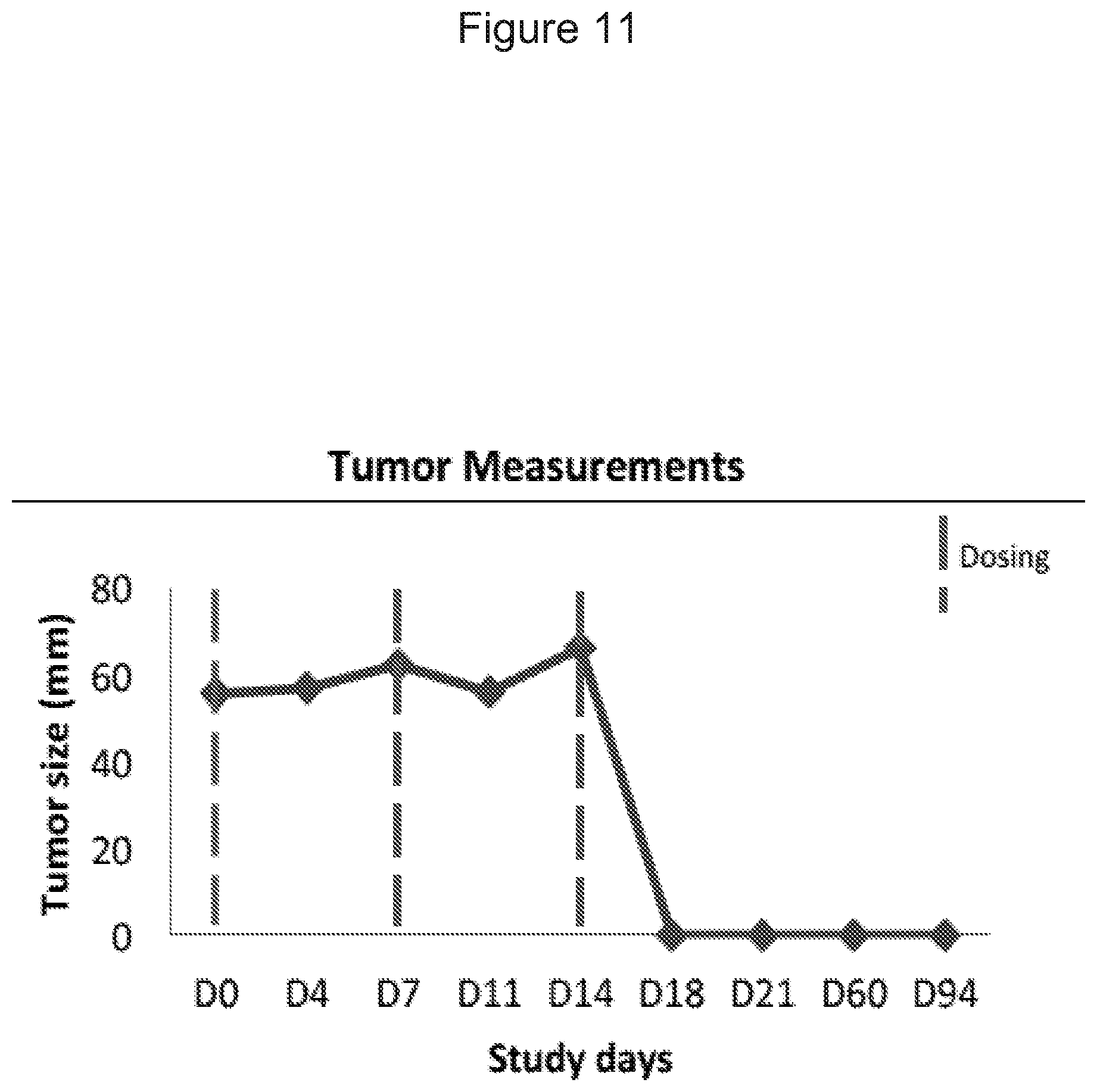
D00029

D00030

D00031

D00032

D00033

D00034

D00035

D00036

D00037

D00038

D00039

D00040

D00041

D00042
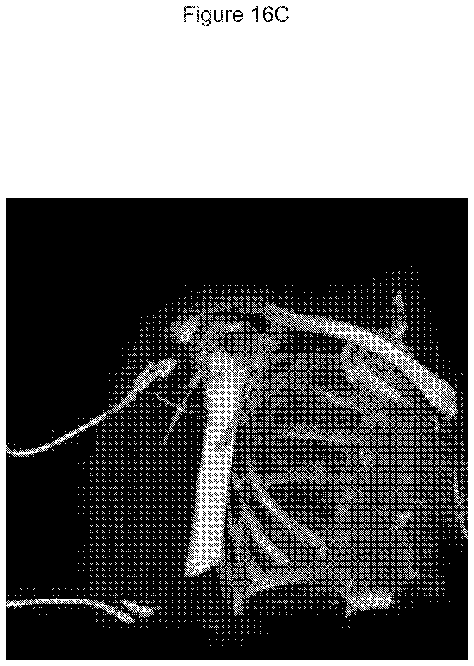
D00043

D00044

XML
uspto.report is an independent third-party trademark research tool that is not affiliated, endorsed, or sponsored by the United States Patent and Trademark Office (USPTO) or any other governmental organization. The information provided by uspto.report is based on publicly available data at the time of writing and is intended for informational purposes only.
While we strive to provide accurate and up-to-date information, we do not guarantee the accuracy, completeness, reliability, or suitability of the information displayed on this site. The use of this site is at your own risk. Any reliance you place on such information is therefore strictly at your own risk.
All official trademark data, including owner information, should be verified by visiting the official USPTO website at www.uspto.gov. This site is not intended to replace professional legal advice and should not be used as a substitute for consulting with a legal professional who is knowledgeable about trademark law.