Methods for monitoring changes in the core of lipoprotein particles in metabolism and disease
Cistola , et al.
U.S. patent number 10,613,169 [Application Number 15/411,403] was granted by the patent office on 2020-04-07 for methods for monitoring changes in the core of lipoprotein particles in metabolism and disease. This patent grant is currently assigned to East Carolina University. The grantee listed for this patent is East Carolina University. Invention is credited to David P. Cistola, Michelle Robinson.


| United States Patent | 10,613,169 |
| Cistola , et al. | April 7, 2020 |
Methods for monitoring changes in the core of lipoprotein particles in metabolism and disease
Abstract
A method is disclosed for measuring the properties of protein and lipoprotein elements in a sample. The method includes the of placing a small volume of a sample into a NMR instrument tuned to measure a particular nucleus;, applying a series of radio frequency pulses with intermittent delays in order to measure spin-spin and/or spin-lattice relaxation time constants from the time-domain decay of the signal, without the use of chemical shifts and without converting data into the frequency domain by Fourier transform or other means, at least partially suppressing the water signal prior to the beginning of a sequence used to record relaxation time constants in the time domain, optionally utilizing relaxation contrast agents or other chemical additives to perturb the solvent water or other elements of the sample, analyzing the exponentially decaying NMR signal in the time domain using multi-exponential analysis, and comparing differences in the relaxation time constants for lipoprotein- or protein-specific elements within a single human subject, or between subjects, to assess normal and abnormal metabolism reflective of increased disease risk or active disease.
| Inventors: | Cistola; David P. (Fort Worth, TX), Robinson; Michelle (Greenville, NC) | ||||||||||
|---|---|---|---|---|---|---|---|---|---|---|---|
| Applicant: |
|
||||||||||
| Assignee: | East Carolina University
(Greenville, NC) |
||||||||||
| Family ID: | 51528857 | ||||||||||
| Appl. No.: | 15/411,403 | ||||||||||
| Filed: | January 20, 2017 |
Prior Publication Data
| Document Identifier | Publication Date | |
|---|---|---|
| US 20170192069 A1 | Jul 6, 2017 | |
Related U.S. Patent Documents
| Application Number | Filing Date | Patent Number | Issue Date | ||
|---|---|---|---|---|---|
| 13839420 | Jan 24, 2017 | 9551768 | |||
| Current U.S. Class: | 1/1 |
| Current CPC Class: | A61B 5/150022 (20130101); G16H 50/30 (20180101); G01N 33/49 (20130101); G01R 33/448 (20130101); A61B 5/7275 (20130101); G01N 24/08 (20130101); A61B 5/14546 (20130101); A61B 5/055 (20130101) |
| Current International Class: | G01R 33/44 (20060101); G01N 24/08 (20060101); A61B 5/145 (20060101); G01N 33/49 (20060101); A61B 5/15 (20060101); A61B 5/00 (20060101); A61B 5/055 (20060101) |
References Cited [Referenced By]
U.S. Patent Documents
| 4933844 | June 1990 | Otvos |
| 5192264 | March 1993 | Fossel |
| 5343389 | August 1994 | Otvos |
| 5366440 | November 1994 | Fossel |
| 6426058 | July 2002 | Pines |
| 6518069 | February 2003 | Otvos |
| 6574495 | June 2003 | Golman |
| 6576471 | June 2003 | Otvos |
| 6617167 | September 2003 | Otvos |
| 6653140 | November 2003 | Otvos |
| 6683455 | January 2004 | Ebbels |
| 6818202 | November 2004 | Pines |
| 7191069 | March 2007 | Wishart |
| 7243030 | July 2007 | Reeve |
| 7306562 | December 2007 | Baykal |
| 7397241 | July 2008 | Gauthausen |
| 7474095 | January 2009 | Levitt |
| 7550971 | June 2009 | Carpenter |
| 7564243 | July 2009 | Desvaux |
| 7647234 | January 2010 | Ruderman |
| 7713744 | May 2010 | Benner |
| 7750633 | July 2010 | Pines |
| 7790465 | September 2010 | Otvos |
| 2002/0087276 | July 2002 | Otvos |
| 2003/0054599 | March 2003 | Huizing |
| 2003/0119194 | June 2003 | Otvos |
| 2004/0098208 | May 2004 | Reeve |
| 2004/0142496 | July 2004 | Nicholson |
| 2005/0222504 | October 2005 | Otvos |
| 2006/0104906 | May 2006 | Ardenkjaer-Larsen |
| 2006/0183234 | August 2006 | Otvos |
| 2007/0063700 | March 2007 | Levitt |
| 2007/0178598 | August 2007 | Jeyarajah |
| 2007/0264677 | November 2007 | Otvos |
| 2008/0038829 | February 2008 | Kremer |
| 2008/0088308 | April 2008 | Carpenter |
| 2008/0204014 | August 2008 | Desvaux |
| 2009/0219022 | September 2009 | Carpenter |
| 2010/0039109 | February 2010 | Cheng |
| 2010/0100334 | April 2010 | Otvos |
| 2010/0219826 | September 2010 | Duckett |
| 2010/0233089 | September 2010 | Ross |
| 1327993 | Mar 1994 | CA | |||
| 0361214 | Jul 1994 | EP | |||
| 9104744 | Apr 1991 | WO | |||
| 9110128 | Jul 1991 | WO | |||
| 0017766 | Nov 2000 | WO | |||
| 0065366 | Nov 2000 | WO | |||
| 03012416 | Feb 2003 | WO | |||
| 2006076631 | Jul 2006 | WO | |||
| 2009129265 | Oct 2009 | WO | |||
Other References
|
Bell et al., "Effects of n-3 fatty acids on the NMR profile of plasma lipoproteins," J. Lipid Research, 1996, v. 37, pp. 1664-1674. cited by applicant . Tang et al. "Use of relaxation-edited one-dimensional and two dimensional nuclear magnetic resonance spectroscopy to improve detection of small metabolites in blood plasma," Anal. Biochem., 2004, v. 325, pp. 260-272. cited by applicant . Mallol et al. "Human serum/plasma lipoprotein analysis by NMR: Application to the study of diabetic dyslipidemia." Prog. Nucl. Magn. Reson., 2013, (published on-line Sep. 11, 2012), v. 70, pp. 1-24. cited by applicant . Aursand et al. "Low Field NMR Studies of Atlantic Salmon (Salmo salar)," Modern Magnetic Resonance, ed. Webb, 2008, pp. 905-913. cited by applicant . Mo and Raftery, "Pre-SAT180, a Simple and Effective Method for Residual Water Suppression," J. Magn. Reson., 2008, v. 190, No. 1, pp. 1-6. cited by applicant . Bubici et al. "Inversion of Multi-component FFC-NMR relaxation decays," STELAR, Sep. 2012, pp. 1-3. cited by applicant . MickLander et al. "Multivariate Analysis of Time Domain NMR Signals in Relation to Food Quality," in Magnetic Resonance in Food Science, 2003, ed. Belton et al., pp. 239-254. cited by applicant . Miller et al., Seven Direct Methods for Measuring HDL and LDL Cholesterol Compared with Ultracentrifugation Reference Measurement Procedures, Clinical Chemistry 56:6, 2010, pp. 977-986. cited by applicant . Choi et al., N.m.r. Lipid Profiles of Cells, Tissues and Body Fluids-Neutral non-acidic and acidic phospholipid analysis of Bond Elut chromatographic fractions; Biochem Journal, 1993, pp. 712-721. cited by applicant . Hayes et al., Effect of a high saturated fat and no-starch diet on serum lipid subtractions in patients with documented atherosclerotic cardiovascular disease; Mayo Clinic Procedures, vol. 78, Issue 11; Nov. 1, 2003; pp. 1-6. cited by applicant . Spin-lattice relaxation time; from Wikipedia; accessed Jan. 21, 2011 at http:/fen.wikipedia.org/wiki/Spin-lattice_relaxation_time; pp. 2. cited by applicant . Bruker Optics; Principles of NMR; accessed Jan. 21, 2011 at http://www.brukeroptics.com/nmr_principles.htmll?&L=O&print=1. cited by applicant . Total Cholesterol Certification Protocol for Manufacturers-Revised; CRMLN Revised Total Cholesterol Certification Protocol; Oct. 2004; pp. 1-17. cited by applicant . Cistola; Benchtop Time-doman NMR Methods and Tools for Assessing Serum Lipoprotein Particle Properties and Cardiovascular Disease Risk; Sep. 30, 2010; 16 pages. cited by applicant . Robinson et al, Lipoprotein Remodeling in Human Serum as Monitored by Benchtop Time-Doman NMR; 2012. cited by applicant . Eaton et al., Spin Lattice Relaxation in Solution and Summary of Relaxation Mechanisms; University of Denver Department of Chemistry and Biochemistry; Presented at Modem EPR Spectroscopy Euro-Summer School in Retie, Belgium, Dec. 1-7, 2002. cited by applicant . Barkemeyer et al., Heteronuclear Polarization Transfer Using Selective Pulses during Hydrogenation with Parahydrogen; Journal of Magnetic Resonance, Series A 120, (1996) pp. 129-132. cited by applicant. |
Primary Examiner: Xu; Xiaoyun R
Attorney, Agent or Firm: Stanek, Lemon, Crouse & Meeks, PA
Parent Case Text
RELATED APPLICATIONS
This is a divisional of Ser. No. 13/839,420 filed Mar. 15, 2013.
Claims
The invention claimed is:
1. A method for measuring the dynamical, mobility, fluidity and/or physical properties of protein and lipoprotein elements in a sample, including but not limited to lipoprotein particle core and surface mobility, and relative core and surface composition, the method comprising: placing a sample into an NMR instrument tuned to measure a nucleus selected from the group consisting of hydrogen-1, carbon-13, nitrogen-15, fluorine-19, and phosphorous-31; applying a series of radiofrequency pulses with intermittent delays in order to measure spin-spin and/or spin-lattice relaxation time constants from the time-domain decay of the signal, without the use of chemical shifts and without converting data into the frequency domain by Fourier transform or other means; analyzing the exponentially decaying NMR signal in the time domain using a multi-exponential analysis selected from the group consisting of an inverse Laplacian transform and multi-exponential algorithm; and comparing differences in the relaxation time constants for lipoprotein-or protein-specific elements within a single human subject, or between subjects, to assess normal and abnormal metabolism reflective of increased disease risk or active disease, selected from the group consisting of early insulin resistance syndrome, prediabetes, metabolic syndrome, type 1 and 2 diabetes and other metabolic disorders.
2. The method of claim 1 comprising partially suppressing the water signal by introducing a 180-degree inversion pulse followed by a delay to partially relax the water signal prior to the beginning of a sequence used to record relaxation time constants in the time domain.
3. The method of claim 1 comprising utilizing relaxation contrast agents to perturb the solvent water or other elements of the sample.
4. The method of claim 1 comprising measuring the properties of a body fluid.
5. The method of claim 1 comprising measuring the properties of blood serum.
6. The method of claim 1 comprising measuring the properties of blood plasma.
7. The method of claim 1 further comprising preparing a sample prior to the step of placing the sample into an NMR instrument.
8. The method according to claim 7 wherein the step of preparing the partially fractionated body fluid selected from the group consisting of selective precipitation and column fractionation of human blood serum.
9. A method according to claim 1 further comprising the steps of: taking a specimen of whole blood from a patient; thereafter extracting a blood plasma sample from the specimen; prior to the step of placing the sample into an NMR instrument.
10. A method according to claim 1 further comprising the steps of: taking a specimen of whole blood from a patient; thereafter extracting a blood serum sample from the specimen; prior to the step of placing the sample into an NMR instrument.
11. The method of claim 1 comprising placing between about 0.2 and 0.6 mL of a sample into an NMR instrument.
12. The method of claim 1 comprising placing between about 0.01 and 0.6 mL of a sample into an NMR instrument.
13. A method for measuring the dynamical, mobility, fluidity and/or physical properties of protein and lipoprotein elements in a sample, including but not limited to lipoprotein particle core and surface mobility, and relative core and surface composition, the method comprising: taking a specimen of whole blood from a patient; extracting a blood plasma or serum sample from the specimen; placing between about 0.2 and 0.6 mL of the sample into an NMR instrument tuned to measure a nucleus selected from the group consisting of hydrogen-1, carbon-13, nitrogen-15, fluorine-19, and phosphorous-31; applying a series of radiofrequency pulses with intermittent delays in order to measure spin-spin and/or spin-lattice relaxation time constants from the time-domain decay of the signal, without the use of chemical shifts and without converting data into the frequency domain by Fourier transform or other means; analyzing the exponentially decaying NMR signal in the time domain using a multi-exponential analysis selected from the group consisting of an inverse Laplacian transform and multi-exponential algorithm; and comparing differences in the relaxation time constants for lipoprotein-or protein-specific elements within a single human subject, or between subjects, to assess normal and abnormal metabolism reflective of increased disease risk or active disease, selected from the group consisting of early insulin resistance syndrome, prediabetes, metabolic syndrome, type 1 and 2 diabetes and other metabolic disorders.
Description
BACKGROUND
The present invention relates to the analysis of blood to identify and measure properties that correlate with cardiovascular disease.
Cardiovascular disease--primarily in the form of heart attack or stroke--is the leading cause of death in the United States and other developed countries. Cardiovascular disease is likewise becoming an increasing cause of death in developing countries as the risk of death from infectious diseases decreases in such countries.
Some of the main risk factors associated with cardiovascular disease are generally well understood. They include an elevated amount of low density lipoprotein (LDL), high blood pressure, cigarette smoking, diabetes mellitus ("diabetes"), family history, and a less physically active, more sedentary lifestyle.
Serum LDL cholesterol levels are positively correlated with cardiovascular disease risk. However, approximately half of patients who suffer from symptomatic coronary artery disease have normal LDL-cholesterol concentrations. Therefore, there appears to be a hidden risk not detected by conventional clinical laboratory measurements of cholesterol.
As currently best understood, cholesterol deposited in arteries represents a main factor in cardiovascular disease. Cholesterol is effectively insoluble in water and blood and thus the body carries cholesterol using particles called lipoproteins. The body uses several lipid transporting particles present in blood and these lipoprotein particles are typically referred to as chylomicrons, very low density lipoproteins (VLDL), intermediate density lipoproteins (IDL) and high density lipoproteins (HDL). Density increases when less cholesterol is present and density decreases when more cholesterol is present. Thus, the layman often refers to LDL cholesterol as "bad" cholesterol and HDL cholesterol as "good" cholesterol.
Low density lipoprotein particles tend to deposit in artery walls to form atherosclerotic plaques in the artery. In turn, the deposition of LDL to form the atherosclerotic plaques is promoted by an increased LDL concentration (or remnants that can form LDL particles) and a decreased LDL particle size.
In order to help predict and potentially moderate or avoid cardiovascular disease in individuals, conventional clinical tests are carried out to measure certain of the known risk factors. Currently, the most common test is the basic lipid panel which measures total cholesterol, HDL cholesterol ("HDL-C"), and triglycerides. The LDL cholesterol ("LDL-C") is calculated as the difference between total cholesterol and HDL cholesterol.
Currently, approximately 250,000,000 such tests are carried out in the United States every year, and on a worldwide basis 540 million tests are carried out each year. Current costs are between about $26 and $56 per test.
As an additional factor, LDL can be present in different LDL particle sizes. In turn, smaller LDL particle sizes are associated with an increased risk of cardiovascular disease. Because of the size relationship, information about the size of the LDL particles is valuable in combination with information about the concentration of LDL particles.
Currently, the common tests for measuring LDL particle size include vertical autoprofile (VAPO), gradient gel electrophoresis, and NMR lipoprofiles.
VAP is also referred to as a vertical spin density gradient ultracentrifugation and an exemplary version ("The VAP Cholesterol Test".RTM.) is provided by Atherotech, Inc. of Birmingham, Ala. (USA).
Gradient gel electrophoresis distinguishes particle size in a otherwise conventional electrophoresis (i.e. chromatography) process with an exemplary test offered by Berkeley HeartLab Inc. of (South San Francisco California (USA).
In one commercial embodiment, NMR lipoprofile testing is based upon the chemical shift of the resonant frequencies. LabCorp Inc. (North Carolina, USA) is an exemplary provider of such tests, a number of which are based on U.S. Pat. No. 5,343,389 (and others) to James D. Otvos ("the Otvos patents"). The Otvos patents employ frequency-domain FT-NMR to study lipoprotein particle properties, such as particle size and particle number, in order to perform clinical diagnostic testing and disease risk assessment. In order to provide accurate data, however, chemical shift NMR is typically carried out in large (e.g., 400 megahertz or higher) high resolution Fourier-transform NMR instruments. Many such instruments incorporate a superconducting magnet cooled by a surrounding environment of liquid helium which in turn is surrounded by liquid nitrogen. As a result, the device is large and expensive and the testing is carried out in a small number of central laboratories at a cost of between about 100 and $200 per test.
Such tests also require a frequency-domain analysis, typically performed by a Fourier transform of the data. The key measurable is the chemical shift, a measure of relative frequency and atomic environment. Differences in chemical shifts are used to distinguish and resolve different lipoprotein classes and permit the detection of particle size and number.
In evaluating an individual's lipid profile, core mobility or fluidity of the lipids is a reflection of the relative ratio of different cholesteryl ester and triglyceride molecules in the particle core, which in turn, is a reflection of normal or abnormal lipid metabolism.
Lipoproteins are the body's nanoparticle delivery systems that carry water-insoluble cholesterol and triglyceride molecules through the blood and target them to particular tissues for metabolism. Lipoprotein particles can be distinguished by their density, size, chemical composition and charge. They can also be distinguished by the relative lipid content of the particle's oily core compartment. For example, the cores of LDL and HDL are relatively rich in cholesteryl ester (a highly water-insoluble form of cholesterol), whereas VLDL and chylomicrons are relatively rich in triglycerides. Triglyceride molecules are more flexible than cholesteryl esters, so oil phases rich in triglycerides will appear more fluid and mobile, less viscous. Also, the ratio of these components and thus, the core mobility, changes with metabolism and disease.
On a broad basis, the use of NMR techniques for medical purposes is not new, and the term "NMR" typically can refer to a variety of diagnostic methods. There are many types of NMR methods and instruments and thousands of distinct NMR experiments. A vivid example of this is magnetic resonance imaging ("MRI"), which was originally called NMR Imaging. MRI is a variation of NMR that yields anatomical images rather than chemical signatures. Although MRI is based on the same fundamental physics, it involves different instrumentation, methods and derived measurable from other NMR techniques. Thus, different kinds of NMR are used in somewhat related but distinct areas of medical diagnosis, imaging, and treatment.
U.S. Pat. No. 7,550,971 B2) to Carpenter and Benson describe a method of determining analyte concentrations in body fluids such as blood plasma or serum. Examples given in the claims are the concentrations of glucose, cholesterol, triglycerides, albumin, blood urea nitrogen, alkaline phosphatase and creatinine. The method is restricted to the use of low-field, bench-top TD-NMR instruments, but the measurements and derived quantities are analyte concentration rather than lipoprotein core mobility. These contrasting measurables provide completely different types of diagnostic information.
Arguably, the Carpenter and Benson methods are thinly justified and lack any preliminary data that demonstrates the feasibility of their method for measuring analyte concentrations, and bench-top TD-NMR may not be as suitable for measuring the analyte concentrations as Carpenter and Benson imply. Serum is a complex mixture, and U.S. Pat. No. 7,550,971 lacks any explanation as to how the different analytes in serum can be resolved from one another. Instead, much of the content in U.S. Pat. No. 7,550,971 reflects the known operation of the TD-NMR instrument rather than a technique for resolving analytes from TD-NMR data.
As a result of these various factors, vertical autoprofile, gradient gel electrophoresis, and NMR lipoprofiles can be impractical for routine clinical use; i.e., they are too expensive and too cumbersome to be carried out on-site in a practitioner's office or a hospital laboratory.
As another factor, various lipid tests (e.g., for HDL-C and LDL-C) can be inaccurate in certain circumstances. For example, calculated LDL-C values from a conventional lipid panel are not accurate when determined from non-fasting blood samples or in patients who have elevated triglyceride levels, as is common in diabetes. Likewise, some advanced lipid tests like the NMR LipoProfile require fasting blood samples and thus, cannot monitor changes in lipoprotein particles during metabolism following a meal.
Furthermore, evidence is beginning to emerge that characteristics of lipid-carrying particles other than size and density will correlate with an increased risk of cardiovascular disease.
As yet another factor, when any particular test is difficult to carry out, or must be carried out off-site, or will take significant time to complete, or any combination of these factors, the use of that test will tend to be less frequent than the use of tests that can be carried out quickly and easily at a location--a physician's office, small clinic, or hospital--where patients are typically located and their blood samples taken.
Thus, tests that identify cardiovascular risk and that can be carried out more quickly, more easily, less expensively, and on site would tend to be used more frequently and thus provide greater benefits to individual patients and to the relevant patient population.
Therefore, a need exists for faster, simpler and localized techniques that will identify and measure relevant characteristics that correlate to an expected degree of risk of cardiovascular disease.
SUMMARY
This invention describes non-perturbing methods for monitoring changes in the core mobility and core composition of lipoprotein particles in intact, unfractionated body fluids such as blood serum or plasma.
In one aspect, the method includes the of placing a small volume of a sample into a NMR instrument tuned to measure a particular nucleus, applying a series of radiofrequency pulses with intermittent delays in order to measure spin-spin ("T2") and/or spin-lattice ("Ti") relaxation time constants from the time-domain decay of the signal, without the use of chemical shifts and without converting data into the frequency domain by Fourier transform or other means, at least partially suppressing the water signal prior to the beginning of a sequence used to record relaxation time constants in the time domain, optionally utilizing relaxation contrast agents or other chemical additives to perturb the solvent water or other elements of the sample, analyzing the exponentially decaying NMR signal in the time domain using multi-exponential analysis, and comparing differences in the relaxation time constants for lipoprotein- or protein-specific elements within a single human subject, or between subjects, to assess normal and abnormal metabolism reflective of increased disease risk or active disease.
In another aspect, the method comprises measuring the pulsed time domain NMR spin-spin relaxation time for a plurality of LDL samples, normalizing the viscosity of the same LDL samples, plotting the product of spin-spin relaxation time and viscosity for the samples against an axis defined by spin-spin relaxation time to thereby develop a database of T2 or T2V statistics (or Ti or TiV statistics or some combination of T2, T2V, T1, or TiV) for the original LDL samples, measuring the classic lipoprotein profile for the same LDL samples, and correlating known risks of cardiovascular disease based upon the classic lipoprotein testing with the results as determined by the T2 or T2V statistics to thereby correlate the T2 or T2V statistics with the known risks of cardiovascular disease in a statistically acceptable manner. As used herein, the term "normalizing" the viscosity of the sample comprises either using samples at the same viscosity by physically manipulating the samples to obtain the viscosity; or mathematically modeling the resulting data so that the results are comparable across viscosities. Persons skilled in the art are familiar with both techniques and they are not otherwise described in detail herein.
Without being bound by theory, the possibility exists that the relevant interpretation of the NMR data can be obtained regardless of normalization; i.e., the viscosity normalization may be option or unnecessary. Thus, where appropriate the phrase "T2 or T2V" reflects this.
In another aspect, the invention is a diagnostic kit that includes a pulse time domain NMR instrument, a sample selected from the group consisting of serum and plasma, and a database of T2 or T2V data that correlates with known cardiovascular risk statistics.
In another aspect, the invention is a combinatorial library that includes a plurality of patient samples selected from the group consisting of whole or partially fractionated serum or plasma, and a T2 or T2V measurement for each sample.
In another aspect, the invention is a method of determining cardiac, metabolic or diabetic risk factors based upon blood samples. The method includes the steps of measuring the pulse time domain NMR spin-spin or spin-lattice relaxation time of a sample selected from the group consisting of serum and plasma, measuring the lipid profile of the same sample, optionally combining the spin-spin relaxation time and viscosity of the sample to produce a T2 or T2V value for the blood sample, and comparing the T2 or T2V value to the lipid profile of the sample to identify the cardiac or metabolic risk measured by the T2 or T2V value based upon the cardiac risk measured by the lipid profile.
BRIEF DESCRIPTION OF THE DRAWINGS
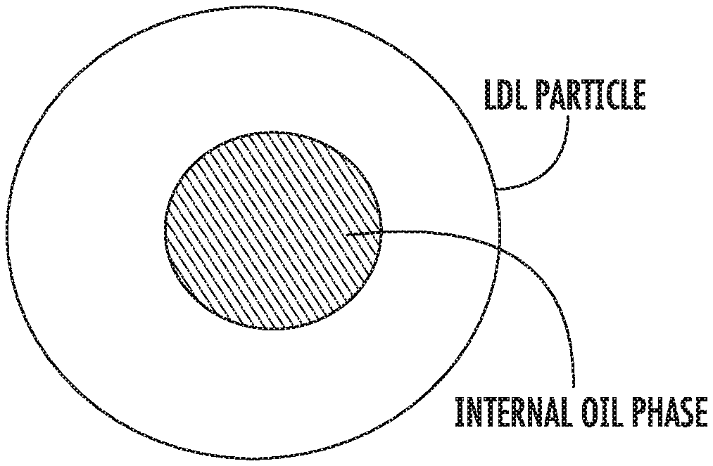
FIG. 1 is a very general schematic diagram of an LDL particle and its internal oil phase.
FIG. 2 is a TD-NMR T2 profile for purified human low-density lipoprotein at 25.degree. C.
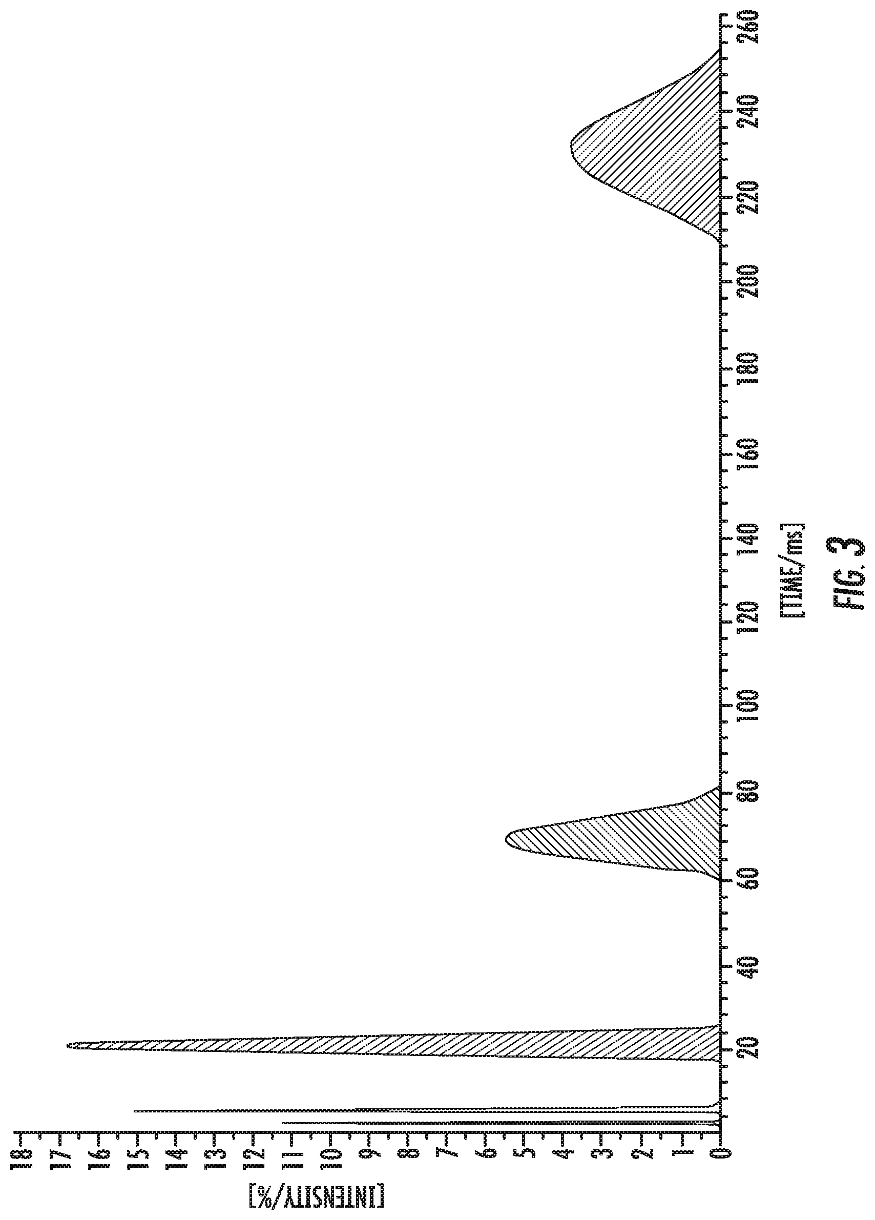
FIG. 3 is a plot of metabolic remodeling of LDL core lipids during metabolism following a meal.
FIGS. 4-6 are CONTIN profiles for pure triolein and for two lipid cores.
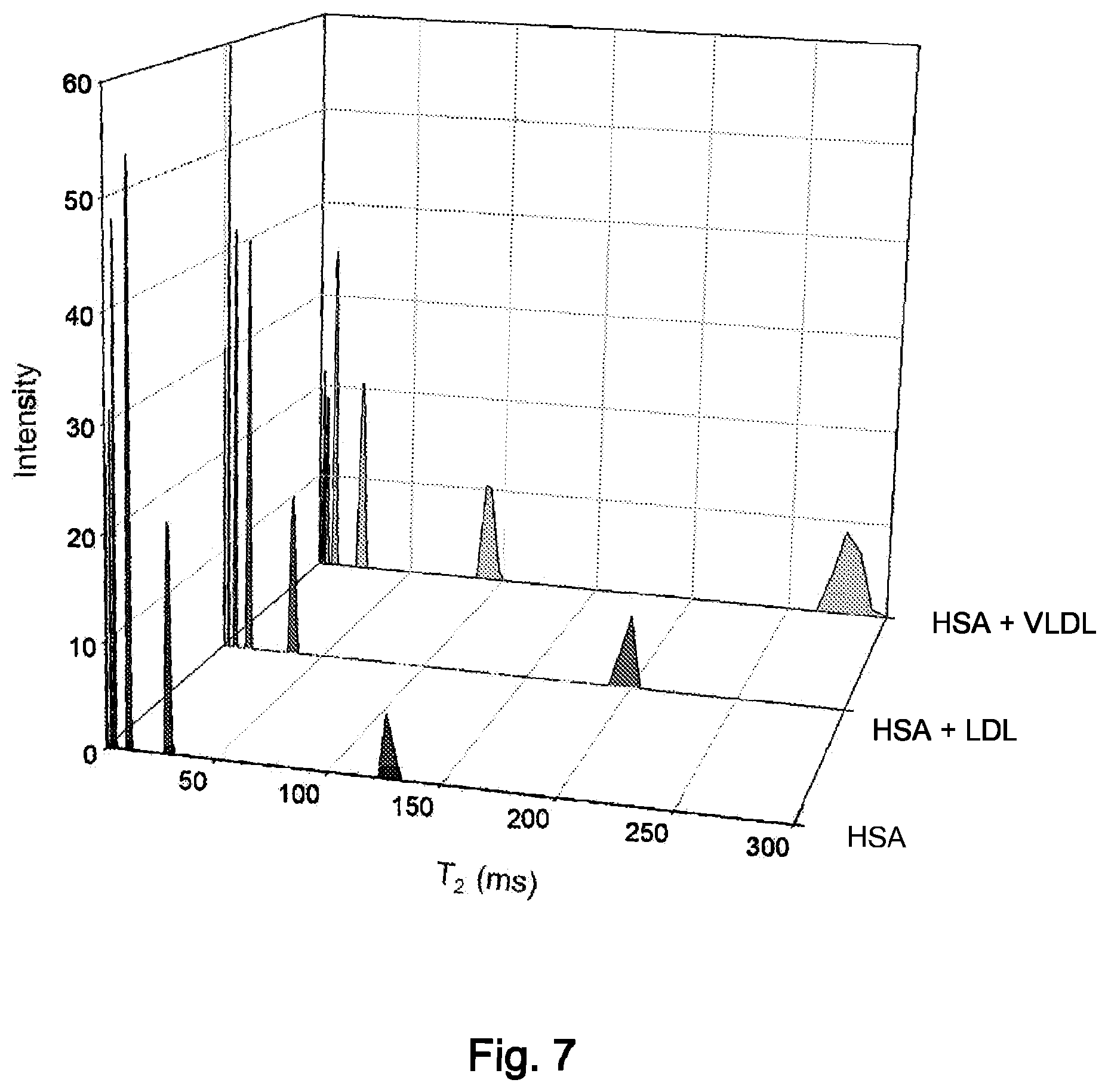
FIG. 7 is a plot of T2V values for albumin, LDL plus albumin, and VLDL plus albumin.
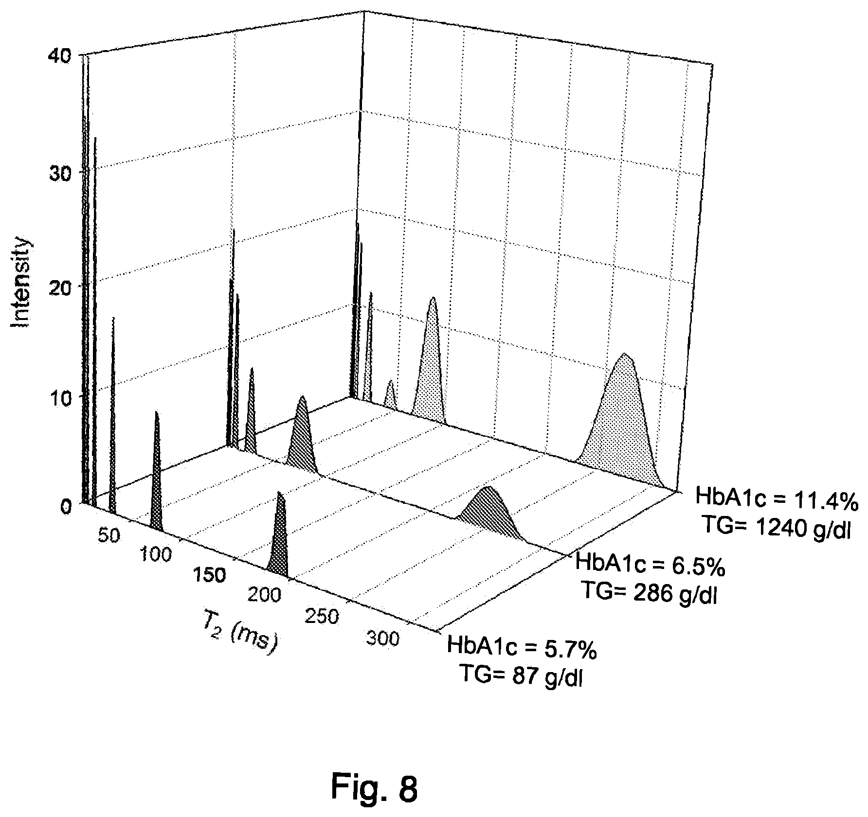
FIG. 8 is a plot of T2V values for three samples of whole human serum.
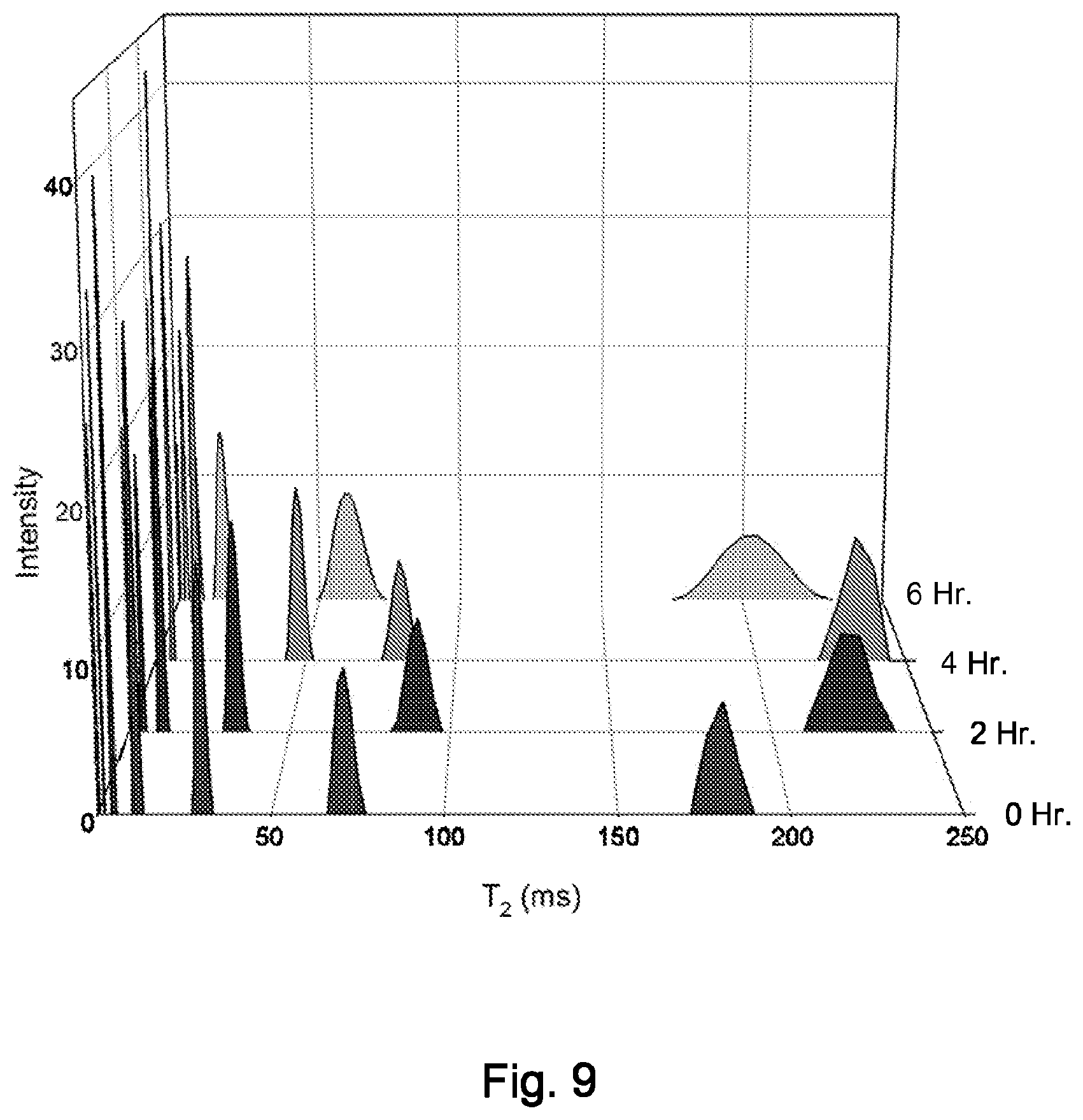
FIG. 9 is a plot of T2V values for lipoprotein particles at various times after a meal.
DETAILED DESCRIPTION
The methods of the invention resolve lipoprotein mobility domains and detect differences in core lipid mobility, which is influenced by the relative amount of cholesteryl ester to triglyceride molecules within each particle's core. Variability in core mobility and core composition within a particle class, such as LDL, can result from patient-to-patient differences, or from particle remodeling within an individual subject as occurs during metabolism following a meal. Changes in lipoprotein particle core mobility and core composition are monitored using a time-domain nuclear magnetic resonance (TD-NMR) analysis. A hallmark of this approach is that the analysis is performed without Fourier transformation and without the use of frequency-domain information such as chemical shifts. Unlike frequency-domain Fourier transform NMR, this time-domain NMR analysis can be performed at low magnetic fields (<60 MHz for hydrogen) in a low-cost, bench-top instrument configuration, although it can also be performed in conventional high-field NMR spectrometers.
The general principles of time domain pulse NMR are generally well understood and familiar to persons of ordinary skill in the art and need not be discussed in detail. In brief, however, a sample is positioned in an external magnetic field provided by a permanent magnet. This aligns the magnetic moments of the hydrogen atoms with (or against) the permanent magnetic field. Then, a radio frequency pulse is applied in a direction that provides a secondary (temporary) magnetic field perpendicular to the permanent magnetic field. This moves the magnetic moments of the hydrogen atoms away from their equilibrium state. The time duration of the pulse determines how far the magnetic moments move. The combined movement of many spins (many hydrogen atoms) generates a small but detectable oscillating magnetic field that in turn induces an alternating voltage that is measured as the NMR signal by a detection coil.
At the end of the pulse, the protons in the sample give up excess energy to their surroundings and relax back to the equilibrium state with respect to the permanent magnetic field. This relaxation takes a certain amount of time, so that the NMR signal remains detectable for a period of time that can range from several milliseconds to several seconds.
Furthermore, the relaxing component of the NMR signal will be characteristic of individual mobility domains, which in turn, help identify the molecules involved in the motions. For example, cholesterol molecules are more internally rigid than triglyceride molecules and will tend to give lower T2 and Ti values.
Additionally, the data resolution of the pulse time domain NMR technique of the invention is on the order of molecular complexes, molecules, or domains of molecules. In comparison, Fourier transform NMR resolves data on an atomic scale. As a result, the time domain technique makes fewer technical demands (so to speak) on the instrument and can provide useful data at the available resolution.
According to the invention, it is been determined that time decay constants are sensitive to both particle size and particle mobility.
The method is also tolerant of multiple phases or mixed phases; i.e., solids and liquids in many circumstances.
As part of the correlation discoveries of the invention, it is now been determined that LDL particles with a higher triglyceride/cholesterol molecular ratio in the core have a longer spin-spin relaxation time (T2) and particles with a lower triglyceride/cholesterol ratio have a shorter T2.
Although the inventors do not wish to be bound by a particular theory, it appears that this may result from the characteristics of an LDL particle as not being solid in the same sense as a solid homogeneous composition would be. Instead, the LDL particle has an internal oil phase (FIG. 1). In turn, the oil phase moves (tumbles) differently--and typically faster--than the remainder of the particle (with the remainder often being referred to as the "surface" of the particle to distinguish from the "core"). This faster internal tumbling increases the spin-spin relaxation time.
In one embodiment, the hydrogen spin-spin relaxation rate constants (or time constants) are measured using a low-field bench-top time-domain NMR analyzer, and the relaxation rate constants for lipoprotein mobility domains are resolved through a multi-exponential deconvolution algorithm. Another key feature of this analysis is that measurements can be made directly on intact body fluids (e.g., serum. plasma or blood) without the need for separation or fractionation of individual lipoprotein classes by ultracentrifugation, electrophoresis, chromatography or other time-consuming, sample-perturbing methods. Because of the relative simplicity and low cost, this method has potential application to clinical testing for the detection of unique dyslipidemias and for the early detection and risk assessment of cardiovascular disease, diabetes and inflammation.
The measurements can, of course, be made in conventional high-field NMR spectrometers, but as set forth herein, the use of Benchtop instruments offers a number of clinical advantages.
In the invention, time-domain NMR resolves individual lipoprotein classes by measuring mobility differences in the oil phases within the core compartment of lipoprotein particles. The invention is also based on the discovery that TD-NMR is sensitive to changes in the particle core within a lipoprotein class. For example, the LDL particles in diabetic subjects tend to be richer in triglyceride, which makes the particle core more mobile.
The mobility differences are monitored by measuring relaxation rate constants (or time constants) without chemical shifts. Chemical shifts are the centerpiece of conventional high-field, frequency-domain NMR. By contrast, time-domain NMR does not require chemical shifts for frequency domain resolution and does not require high magnetic field strength or field homogeneity. This approach is fundamentally different from conventional NMR spectroscopy in both methodology and instrumentation requirements.
In one embodiment, the invention is a process for measuring the spin-spin or transverse relaxation time constants (T2) for the lipid core compartments in unfractionated human serum. The human serum is obtained in a conventional manner from a low-speed centrifugation of human blood after clotting. Approximately 0.6 mL of unmodified serum is pipetted into a 10 mm NMR tube, and the tube is placed into the bore of the magnet of a bench-top TD-NMR analyzer, typically operating at 10, 20, 40 or 60 MHz resonance frequency for hydrogen. In the examples described here, 20 MHz and 40 MHz data were collected using Bruker benchtop mq20 and mq40 TD-NMR instruments (Bruker BioSpin Corporation, Billerica, Mass., USA).
A Carr-Purcell-Meiboom-Gill (CPMG) pulse sequence is used to measure the exponential time-decay curve. This pulse sequence effectively eliminates chemical shifts and magnetic field inhomogeneity, permitting the measurement of T2 values. Although T2 measurements can be linked with chemical shifts and measured in the frequency domain, the present TD-NMR method measures T2 in the time domain without chemical shifts. This provides a distinct advantage with respect to instrument simplicity and cost.
The resulting T2 decay curve for human serum is multi-exponential, so the individual exponential terms are deconvoluted and resolved with the use of an inverse Laplacian transform. An implementation of this mathematical calculation is provided in the public domain software CONTIN, authored by Steven Provencher (http://s-provencher.com/pages/contin.shtml; accessed Mar. 11, 2013). Under the proper experimental conditions with excellent signal-to-noise, the CONTIN calculation can resolve up to 4 different exponential terms in TD-NMR T2 profiles of human serum. Because human serum has abundant quantities of lipoprotein core lipids and soluble proteins, and because these assemblies are relatively large, the protein and lipoprotein components dominate the T2 profile.
One experimental issue involves solvent suppression, because an intense water signal can overshadow the contributions of lipoprotein components and lead to artifacts such as radiation damping. The solvent water can be partially suppressed using a number of NMR schemes. In this embodiment, a 180-degree pulse and delay is inserted prior to the CPMG sequence. This achieves partial relaxation (and partial suppression) of the water with full recovery of the lipoprotein components by the start of the CPMG pulse scheme. Although there are many sophisticated NMR methods for suppressing water, the goal of this invention was to develop the simplest, most inexpensive method for measuring lipoprotein core properties in unmodified human serum.
This embodiment is further illustrated using the figures and tables. FIG. 2 shows a time-domain T2 NMR profile of normal fasting human serum, and FIG. 3 is the TD-NMR T2 profile for purified human low-density lipoprotein at 25.degree. C.
The profile of FIG. 2 (not to be confused with a conventional NMR spectrum) was obtained by performing an inverse Laplacian transform of the T2 decay curve. In turn, the T2 curve was measured using a modified CPMG experiment on a Bruker 40 MHz TD-NMR analyzer. The profile resolves 7 distinct T2 components, here represented as peaks in the profile. Higher T2 values represent more mobile elements. The solvent water peak in serum, not shown in this plot, is observed at T2 values of approximately 600 ms. The peaks at approximately 200 and 70 ms represent two distinct mobility domains in LDL, as assigned from control samples containing only LDL.
These two peaks are not observed in the other control samples containing fractionated serum proteins or lipoproteins and appear to provide a unique signature for the core lipid mobility of LDL. The peaks at lower T2 values have contributions from both serum proteins and lipoproteins.
FIG. 9 illustrates the variation in T2 measurements in response to metabolic changes following a meal. Shown are T2 profiles for whole human serum at fasting, 2 hours, 4 hours and 6 hours post-prandial. The T2 values for the LDL-specific peaks increase and peak at 4 hours, reflecting the remodeling of the LDL core as the content of triglyceride increases relative to cholesteryl ester.
These preliminary results demonstrate the feasibility of obtaining domain-specific measurements of the core mobility of LDL in whole human serum. The data also demonstrate that T2 measurements obtained from TD-NMR are sensitive to metabolic remodeling and patient-to-patient variability.
Furthermore, the invention requires neither high magnetic field instrumentation nor a frequency-domain analysis. Instead, it uses a time-domain analysis. Unlike (for example) the Otvos approach, the methods of the invention can be performed on inexpensive low-field bench-top instruments, because high field strength and field homogeneity is not required. The key measurables are relaxation rate constants rather than chemical shifts. Differences in relaxation rate constants are used to resolve lipoprotein classes (not chemical shifts, as in Otvos and Kremer). Also, the derived parameter in our invention is lipoprotein particle core mobility or fluidity, rather than particle number or particle size. In summary, the instrumentation, data processing, measurables and derived parameters of our invention are different from those of Otvos.
In contrast to (for example) U.S. Pat. No. 7,550,971, the present invention invention does not measure analyte concentrations. Rather it measures lipoprotein particle properties, specifically the mobility or fluidity ("squishiness") of the oily lipid core found within lipoprotein particles. Also, the invention is not restricted to low-field, bench-top NMR instruments, but can also be performed on conventional high-resolution NMR instruments as well.
Overall, the method of the invention is much simpler and can be performed on inexpensive low-field benchtop NMR analyzers, and the particle core mobility provides diagnostic information different from particle size and concentration distribution.
Additional Examples
In the following Examples, all aqueous samples are prepared in a 9:1 D20/H20 saline buffer, concentrated to a viscosity of 1.20 cP at 37.degree. C. The raw data are in the form of a multi exponential decay curve. The individual relaxation time constants are deconvoluted using an inverse Laplacian transform calculation as implemented in the public domain program CONTIN.
Lipoprotein Lipid Core Mixtures
A CONTIN profile of triolein, the most abundant TG in lipoproteins, is shown in FIG. 4. Each T2 (or T2V) corresponds to a mobility domain of the triglyceride molecule. A VLDL like lipid core with 80% TG and 20% CE shows a similar profile (FIG. 5) but is shifted slightly to the left, indicating a less mobile, more vicious environment. The LDL like lipid core (FIG. 6) composed of 80% CE and 20% TG exhibited lower T2 or T2V values indicating reduced mobility. This trend resembles that observed with physiological lipoprotein particles with differences in TG/CE ratios. T2V values for lipid mixtures are summarized in Table 1.
TABLE-US-00001 TABLE 1 (T2V times in ms) Fast Medium Slow Other 100% TO 347 155 79 8 80% TO to 20% CL (VLDL Core) 303 141 70 25, 5 20% TO to 80% CL (LDL Core) 183 92 45 19, 9, 4
Fractionated Lipoproteins and Serum Proteins
FIG. 7 shows the T2V profiles for albumin ("HSA"), LDL plus albumin and VLDL plus albumin. Although all of the profiles display fast decaying components in the range of 2-50 ms, there are T2V values above 50 ms that are unique to individual lipoprotein classes.
Whole Human Serum
De-identified samples of whole human serum, representing various metabolic and disease states were obtained from Pitt County Memorial Hospital (Greenville, N.C., USA). As seen in FIG. 8 variability was observed in lipoprotein T2V values. Patient samples with a higher HbAlc, indicative of poorly managed Type 2 Diabetes Mellitus, have increased T2 or T2V values suggesting that the lipoprotein particles have increasingly mobile, TG-rich lipid cores versus non-diabetic patients.
Results: FIG. 9 shows the remodeling healthy non-diabetic subject ingested a liquid meal that contained 50 grams of lipid following a 16 hour fast, after which blood was drawn every hour for 8 hours. The NMR data are shown in FIG. 4b at 0 hour (fasting) and other time points after the meal. As lipoprotein remodeling occurs and the particles become TG rich, the T2V peak shifts to the right indicating an increase in lipid core mobility. Table 2 shows standard lipid analysis for these samples.
TABLE-US-00002 TABLE 2 0 2 3 4 5 6 7 8 Triglyceri 87 184 181 194 197 231 188 169 Cholester 197 204 200 199 193 187 184 187 HDL-C 54 55 52 52 49 47 47 48 Non- 148 149 148 147 144 140 137 139 LDL-C 125 112 112 108 105 94 99 105
Benchtop TD-NMR appears to provide unique information about LDL and VLDL particle properties reflective of different states of normal and abnormal metabolism. This approach holds promise for translation from the research lab into the clinical setting as the measurements are performed on whole human serum and are relatively simple, inexpensive and non-invasive.
In the drawings and specification there has been set forth a preferred embodiment of the invention, and although specific terms have been employed, they are used in a generic and descriptive sense only and not for purposes of limitation, the scope of the invention being defined in the claims.
* * * * *
References
D00000
D00001
D00002
D00003
D00004
D00005
D00006
XML
uspto.report is an independent third-party trademark research tool that is not affiliated, endorsed, or sponsored by the United States Patent and Trademark Office (USPTO) or any other governmental organization. The information provided by uspto.report is based on publicly available data at the time of writing and is intended for informational purposes only.
While we strive to provide accurate and up-to-date information, we do not guarantee the accuracy, completeness, reliability, or suitability of the information displayed on this site. The use of this site is at your own risk. Any reliance you place on such information is therefore strictly at your own risk.
All official trademark data, including owner information, should be verified by visiting the official USPTO website at www.uspto.gov. This site is not intended to replace professional legal advice and should not be used as a substitute for consulting with a legal professional who is knowledgeable about trademark law.