Human antibody against ED-B domain of fibronectin and uses thereof
Ji , et al.
U.S. patent number 10,611,830 [Application Number 14/895,678] was granted by the patent office on 2020-04-07 for human antibody against ed-b domain of fibronectin and uses thereof. This patent grant is currently assigned to HEFEI LIFEON PHARMACEUTICAL CO. LTD.. The grantee listed for this patent is HEFEI LIFEON PHARMACEUTICAL CO. LTD.. Invention is credited to Jun Chen, Meihua Gao, Junqiu Ji, Mei Zhang.


| United States Patent | 10,611,830 |
| Ji , et al. | April 7, 2020 |
Human antibody against ED-B domain of fibronectin and uses thereof
Abstract
The present invention provides an antibody or antibody fragment for specifically recognizing and binding to an ED-B domain of fibronectin. The antibody or antibody fragment can be widely used for in vitro detection and in vivo location of the ED-B protein domain, as well as targeted therapy of tumors.
| Inventors: | Ji; Junqiu (Anhui, CN), Zhang; Mei (Anhui, CN), Gao; Meihua (Anhui, CN), Chen; Jun (Anhui, CN) | ||||||||||
|---|---|---|---|---|---|---|---|---|---|---|---|
| Applicant: |
|
||||||||||
| Assignee: | HEFEI LIFEON PHARMACEUTICAL CO.
LTD. (Hefei, Anhui, CN) |
||||||||||
| Family ID: | 52007549 | ||||||||||
| Appl. No.: | 14/895,678 | ||||||||||
| Filed: | May 28, 2014 | ||||||||||
| PCT Filed: | May 28, 2014 | ||||||||||
| PCT No.: | PCT/CN2014/078600 | ||||||||||
| 371(c)(1),(2),(4) Date: | March 06, 2016 | ||||||||||
| PCT Pub. No.: | WO2014/194784 | ||||||||||
| PCT Pub. Date: | December 11, 2014 |
Prior Publication Data
| Document Identifier | Publication Date | |
|---|---|---|
| US 20190185550 A1 | Jun 20, 2019 | |
Foreign Application Priority Data
| Jun 6, 2013 [CN] | 2013 1 0221256 | |||
| Current U.S. Class: | 1/1 |
| Current CPC Class: | G01N 33/57415 (20130101); C07K 16/18 (20130101); G01N 33/57446 (20130101); G01N 33/6887 (20130101); A61P 35/00 (20180101); C07K 14/7155 (20130101); A61K 2039/505 (20130101); C07K 2317/94 (20130101); C07K 2317/622 (20130101); C07K 2317/92 (20130101); G01N 2333/78 (20130101); C07K 2319/00 (20130101) |
| Current International Class: | C07K 16/18 (20060101); A61P 35/00 (20060101); A61K 39/00 (20060101); C07K 14/715 (20060101); G01N 33/68 (20060101); G01N 33/574 (20060101) |
References Cited [Referenced By]
U.S. Patent Documents
| 2004/0013640 | January 2004 | Zardi |
| 2009/0214423 | August 2009 | Borsi |
Other References
|
Pini et al. Design and Use of a Phage Display Library: Human Antibodies With Subnanomolar Affinity Against a Marker of Angiogenesis Eluted From a Two-Dimensional Gel. The Journal of Biological Chemistry. vol. 273, No. 34, Issue of Aug. 21, pp. 21769-21776, 1998 (Year: 1998). cited by examiner. |
Primary Examiner: Haddad; Maher M
Claims
What is claimed is:
1. An antibody or antibody fragment for specifically recognizing and binding to an ED-B domain of human fibronectin (FN), comprising a VH having an amino acid sequence as shown in SEQ ID NO: 1 and a VL having an amino acid sequence as shown in SEQ TD NO: 3.
2. The antibody or antibody fragment, as recited in claim 1, wherein the antibody or antibody fragment is a monoclonal antibody with an equilibrium binding/dissociation constant (KD) of 1.times.10.sup.-8 M or less, 1.times.10.sup.-9 M or less, or 1.times.10.sup.-10 M or less.
3. The antibody or antibody fragment, as recited in claim 1, wherein the antibody or antibody fragment is a mammal-derived antibody sequence.
4. The antibody or antibody fragment, as recited in claim 1, wherein the antibody or antibody fragment directly binds to the ED-B protein domain.
5. The antibody or antibody fragment, as recited in claim 4, wherein the ED-B protein domain is an independent recombinant ED-B protein, or a recombinant protein formed through fusion of the ED-B protein with other proteins, or a natural fibronectin comprising the ED-B domain.
6. The antibody or antibody fragment, as recited in claim 4, wherein the ED-B protein domain is derived from humans, mice, rats, or chickens.
7. The antibody or antibody fragment, as recited in claim 1, wherein the antibody is a human immunoglobulin IgG.
8. A pharmaceutical composition for diagnosing or treating tumor disease comprising the antibody or antibody fragment as recited in claim 1.
9. The pharmaceutical composition, as recited in claim 8, further comprising a fusion protein, a radioactive isotope, a chemical drug and a nanoparticle.
10. The pharmaceutical composition, as recited in claim 9, wherein the cancer disease is a cancer in which the FN(B+) comprising the ED-B domain is expressed.
11. The pharmaceutical composition, as recited in claim 10, wherein the cancer disease is nasopharyngeal cancer, larynx cancer, oesophagus cancer, cardiac cancer, gastric cancer, colorectal cancer, lung cancer, liver cancer, pancreatic cancer, melanoma, fibrosarcoma or breast cancer.
Description
CROSS REFERENCE OF RELATED APPLICATION
This is a U.S. National Stage under 35 U.S.C 371 of the International Application PCT/CN2014/078600, filed May 28, 2014, which claims priority under 35 U.S.C. 119(a-d) to CN 201310221256.0, filed Jun. 6, 2013.
BACKGROUND OF THE PRESENT INVENTION
Field of Invention
The present invention relates to a monoclonal antibody technology, a phage display technology and a gene recombination technology, and particularly relates to an antibody or antibody fragment for binding to an ED-B domain of fibronectin (FN). Additionally, the present invention also relates to a method for preparing the monoclonal antibody and a pharmaceutical composition including the monoclonal antibody.
Description of Related Arts
Commonly-used tumor therapy methods include chemotherapy and radiotherapy methods, which affect physiological functions of normal organs and reduce the immune ability and life quality of a patient while killing a cancer cell due to their low specificity and significant toxic or side effect on normal tissues. A method for improving the specificity of a tumor therapeutic drug is targeted killing of a tumor by targeting the drug to a tumor cell via an antibody or antibody fragment which can recognize a tumor cell marker. Achievement of this method firstly requires finding a tumor marker capable of being expressed on an external surface of a cell membrane, i.e., a protein which is only expressed on the surface or in the matrix of a tumor cell, but is expressed with a small amount or even not expressed in a normal cell or tissue.
Fibronectin is a multifunctional glycoprotein existing widely in extracellular matrix, blood plasma and other body fluids, which is expressed by epithelial cells, endothelial cells, fibroblast cells, liver cells, decidual cells, extravillous trophoblast cells and the like. FN participates in adhesion, cytomorphosis and distribution of cells, and formation of blood vessels.
The FN gene is about 75 kb long, includes about 50 exons, and essentially consists of types I, II, and III homologous repeating units. ED-B is included in the type III repetitive sequence of the FN, and is a complete repeat composed of 91 amino acids and coded by a single exon. During expression of the FN gene, two cases exist, one is FN(B+) including an ED-B domain and the other is FN(B-) including no ED-B domain. These two cases are considered as playing an important role in an individual development process. The FN(B+) is expressed with a small amount in a normal tissue of an adult, but is highly expressed during an injury process, a disease process or a wound healing process, and particularly during a tumor growth process, and thus is also referred to as a carcinoembryonic gene. The FN(B+) including the ED-B domain is highly expressed in for example cells of gastric cancer, colorectal cancer, lung cancer, breast cancer and the like. As shown by in vitro immunohistochemical detection and in vivo targeted localization, the FN(B+) is highly expressed in blood vessels in varied types of tumor tissues, but is not expressed in a normal tissue of an adult, and thus is a marker protein of a tumor tissue.
Therefore, a targeted antibody is developed based on the ED-B domain, which can be used for targeting a candidate drug such as cytotoxin and the like to a tumor location so as to achieve a therapeutic aim by effectively inhibiting or killing tumor cells and meanwhile reduce the damage to normal tissues and toxic or side effects.
The screening or drug development of an antibody against the ED-B has a history of more than 20 years. For example a murine-derived monoclonal antibody BC-1 obtained through a hybridoma technique (Carnemolla, Leprini et al. 1992) can specifically recognize the FN (B+) of new human blood vessels and does not cross-react with the FN(B-).
Additionally, a Fab-type murine-derived antibody MOR03255 obtained through mouse immunity and affinity maturation conducted by means of a phage display technology has a maximum affinity up to 30 pM. MOR03255 has the characteristic of neutralizing an antibody, which binds to the ED-B to inhibit the adhesion between human dermal microvascular endothelial (HDMVE) cells and extracellular matrix and thus the formation of new blood vessels, such that the tumor tissues are lack of nutrition due to the absence of blood vessels. Therefore, MOR03255 shows a significant inhibition effect on tumor growth in vivo experiments.
However, as demonstrated by a research, when a murine-derived antibody drug is administrated in the human body, a serious human anti-murine antibody (HAMA) reaction is caused, and other side effects may also be caused, such as an allergic response, a blood disease and damage to kidney. Furthermore, such an antibody has a short half life, which affects the therapeutic effect. Therefore, the murine-derived antibodies BC-1 and MOR03255 have some limitations. Now the murine-derived antibody is gradually replaced by a humanized antibody and a fully humanized antibody.
Since the amino acid sequence of human ED-B is 100% homologous with that of mice's ED-B, the immune system of mice recognizes the human ED-B as its own protein and thus seldomly generates an immune response to the ED-B domain of human. Therefore, the development of the murine-derived antibody against ED-B is often counter-productive. Now with the aid of genetic engineering techniques, an antibody with a specific function is obtained by directly expressing a synthesized antibody gene via the use of the phage display technique and then screening, without using any immune system of a higher animal, and thus a technique for developing recombinant humanized antibodies is available. Currently reported humanized antibodies against ED-B include CGS-1, CGS-2, L19 and the like (Carnemolla, Neri et al. 1996, Pini, Viti et al. 1998), which all have high specificity and respectively have affinities of 5.4.times.10.sup.-8 M, 1.1.times.10.sup.-9M and 8.7.times.10.sup.-10 M as detected by BIAcore. Particularly the clinical research of the antibody L19 sufficiently demonstrates that a specific antibody drug developed against ED-B has a significant effect of inhibiting or killing tumors.
We have obtained a specific single-chain antibody by screening with the aid of synthesis of antibody genes and the phage display technique. The antibody can directly recognize a single ED-B domain and FN(B+), as well as fusion forms of ED-B with other proteins, e.g., GST-ED-B and the like. The antibody obtained by screening does not need affinity maturation, and thus is superior to other disclosed humanized single-chain antibodies against ED-B. Additionally, the binding between the obtained antibody and an antigen is hardly ever dissociated, as detected by BIAcore. These features demonstrate that the obtained antibody can bind to ED-B, and the very high affinity of the antibody enables the binding between the antibody and an antigen to be hardly ever dissociated, such that the antibody can be targeted to and highly enriched at a tissue site which expresses FN(B+) and is hardly ever dissociated again and diffused into normal tissues.
The obtained antibody can be used for development of anti-tumor drugs, e.g., radioisotopic labeling of the antibody, fusion of the antibody with cytotoxin, coupling of the antibody with chemical drugs and the like. With the aid of the obtained antibody, a drug is highly enriched in tumor tissues which highly express FN(B+), so as to achieve targeted killing of tumor cells and improve the drug efficacy while having small damage to normal tissues. Since fully humanized antibodies are human homologous proteins and have low immunogenicity in the human body, in general the HAMA reaction less likely occurs, so that the fully humanized antibodies have lower toxic or side effects than murine-derived antibodies. The structure form of the single-chain antibody enables miniaturization of antibody drugs and improves the permeability of the antibody into the human body, such that the antibody can get into the tumor tissues through tumor blood vessels; and the single-chain antibody may be flexibly varied into antibodies of various forms, e.g. a natural antibody structure, a minibody, a double-antibody, a fusion form of multiple antibodies and the like. By changing the structure form of the antibody, the stability, permeability, use and the like of the antibody can be varied. Furthermore, the obtained antibody can be developed to serve as a tumor diagnostic reagent for in vivo and in vitro detection of tumor tissues, or used for development of various other types of immune-related reagents.
SUMMARY OF THE PRESENT INVENTION
Through a lot of creative works, the inventor obtains a recombinant monoclonal antibody or antibody fragment against a human ED-B protein domain, wherein said antibody fragment is preferably an antigen-binding fragment; and the antibody or antibody fragment can effectively bind to fibronectin (FN) including an ED-B domain (abbreviated as FN(B+)), and can be used for detection and diagnosis of FN(B+), as well as targeted therapy of tumors which highly express FN(B+).
Therefore, the present invention provides an antibody or antibody fragment for specifically recognizing and binding to an ED-B domain of human fibronectin (FN).
In a specific implementation, the antibody or antibody fragment includes an amino acid sequence of the CDR3 region of a heavy-chain variable region (VH), wherein the CDR3 region includes the amino acid sequence of HTAPLFDY (SEQ ID NO: 10).
In another specific implementation, the antibody or antibody fragment includes an amino acid sequence of the CDR3 region of a light-chain variable region (VL), wherein the CDR3 region includes the amino acid sequence of QQGRHTP (SEQ ID NO: 13).
In another specific implementation, the antibody or antibody fragment includes a VH CDR3 having the amino acid sequence of HTAPLFDY (SEQ ID NO: 10) and a VL CDR3 having the amino acid sequence of HTAPLFDY (SEQ ID NO: 10).
In another specific implementation, the antibody or antibody fragment includes three VH CDRs. The amino acid sequences of the CDRs are respectively SYAMS (SEQ ID NO: 8), RISPSGSSTYYADSVKG (SEQ ID NO: 9) and HTAPLFDY (SEQ ID NO: 10).
In another specific implementation, the antibody or antibody fragment includes three VL CDRs. The amino acid sequences of the CDRs are respectively RASQSVSSSYLA (SEQ ID NO: 11), KASNLAT (SEQ ID NO: 12) and QQGRHTP (SEQ ID NO: 13).
In another specific implementation, the antibody or antibody fragment includes three VH CDRs and three VL CDRs, wherein the amino acid sequences of the three VH CDRs are respectively SYAMS (SEQ ID NO: 8), RISPSGSSTYYADSVKG (SEQ ID NO: 9) and HTAPLFDY (SEQ ID NO: 10); and the amino acid sequences of the three VL CDRs are respectively RASQSVSSSYLA (SEQ ID NO: 11), KASNLAT (SEQ ID NO: 12) and QQGRHTP (SEQ ID NO: 13).
In another specific implementation, the antibody or antibody fragment includes a VH having the amino acid sequence as shown in SEQ ID NO: 1.
In another specific implementation, the antibody or antibody fragment includes a VL having the amino acid sequence as shown in SEQ ID NO: 3.
In another specific implementation, the antibody or antibody fragment includes a VH having the amino acid sequence as shown in SEQ ID NO: 1, and a VL having the amino acid sequence as shown in SEQ ID NO: 3.
In another specific implementation, the antibody or antibody fragment is a monoclonal antibody with an equilibrium binding/dissociation constant (KD) of 1.times.10.sup.-8 M or less, 1.times.10.sup.-9 M or less, or 1.times.10.sup.-10 M or less.
In another specific implementation, the antibody or antibody fragment is a monomer or polymer.
In another specific implementation, the antibody or antibody fragment is a mammal-derived antibody sequence, and particularly a human-derived antibody sequence.
In another specific implementation, the antibody or antibody fragment directly binds to the ED-B protein domain. Particularly, the ED-B protein domain may be an independent recombinant ED-B protein, or a recombinant protein formed through fusion of the ED-B protein with other proteins, or a natural fibronectin including the ED-B domain. More particularly, the ED-B protein domain may be derived from humans, mice, rats, chickens or other species. More particularly, the ED-B protein domain may be a glycosylated protein or a non-glycosylated protein.
In a specific implementation, the antibody fragment may be a monovalent and micromolecular antibody, such as a single-chain antibody, a single-domain antibody, a hypervariable region polypeptide or the like, and Fab; or a multivalent and micromolecular antibody, such as a double-chain antibody, a three-chain antibody, and a minibody.
In a specific implementation, the antibody is a human immunoglobulin IgG.
On the other hand, the present invention provides a pharmaceutical composition including the aforementioned antibody or antibody fragment.
In an implementation, the pharmaceutical composition further includes but not limited to a fusion protein, a radioactive isotope, a chemical drug, a nanoparticle and the like. In a specific implementation, the pharmaceutical composition is used for diagnosis or therapy of tumor-related diseases.
On the other hand, the present invention provides use of the aforementioned antibody or antibody fragment, or the pharmaceutical composition thereof in prevention, diagnosis and therapy of cancer-related diseases. On the other hand, the present invention provides use of the aforementioned antibody or antibody fragment, or the pharmaceutical composition thereof in preparation of a drug for prevention, diagnosis and therapy of cancer-related diseases. Particularly, the cancer-related disease is cancer in which the FN(B+) including the ED-B domain is expressed. The cancer may be nasopharyngeal cancer, larynx cancer, oesophagus cancer, cardiac cancer, gastric cancer, colorectal cancer, lung cancer, liver cancer, pancreatic cancer, melanoma, fibrosarcoma or breast cancer; and more particularly, the cancer-related disease is gastric cancer, colorectal cancer, lung cancer or breast cancer.
On the other hand, the present invention provides a diagnostic kit including the aforementioned antibody or antibody fragment, which can be used for diagnosis of in vivo distribution of tumor tissues, histopathological sections and the like; or for analysis and identification of cells, proteins and the like; or for affinity purification of cells or protein molecules including the ED-B protein domain. The present invention further provides use of the aforementioned kit in diagnosis of in vivo distribution of tumor tissues and histopathological sections; analysis and identification of cells and proteins; or affinity purification of cells or protein molecules including the ED-B protein domain. The present invention further provides use of the aforementioned kit in preparation of a kit used for diagnosis of in vivo distribution of tumor tissues and histopathological sections; analysis and identification of cells and proteins; or affinity purification of cells or protein molecules including the ED-B protein domain.
On the other hand, the present invention provides a polynucleotide molecule encoding the aforementioned antibody or antibody fragment.
In a specific implementation, the polynucleotide molecule includes a nucleotide sequence as shown in SEQ ID NO: 2, which encodes the amino acid sequence of antibody VH; and a nucleotide sequence as shown in SEQ ID NO: 4, which encodes the amino acid sequence of antibody VL.
Specifically, the present invention provides a monoclonal antibody against the human ED-B protein domain (abbreviated as B5), which is a fully humanized antibody, includes the VH and VL of human, and provides an linker fragment for connecting the VH and the VL. Particularly, the antibody B5 herein may be a single-chain antibody B5 (single-chain antibody fragment, scFv) which includes a VH, a VL and a linker, preferably constructed in the arrangement manner of antibody VH-linker-antibody VL. The VH has the amino acid sequence as shown in SEQ ID NO: 1; and the VL includes the amino acid sequence as shown in SEQ ID NO: 3. Preferably, the linker fragment includes the amino acid sequence as shown in SEQ ID NO: 5. In a specific implementation, the antibody B5 has the amino acid sequence as shown in SEQ ID NO: 15.
On the other hand, the present invention also provides a DNA molecule which encodes the aforementioned monoclonal antibody.
In an embodiment, the DNA molecule includes a nucleotide sequence as shown in SEQ ID NO: 2, which encodes the VH of the monoclonal antibody; a nucleotide sequence as shown in SEQ ID NO: 4, which encodes the VL of the monoclonal antibody; and optionally a nucleotide sequence as shown in SEQ ID NO: 6, which can be used for encoding the linker fragment for connecting the VH and the VL of the monoclonal antibody.
Furthermore, the present invention also provides a drug fusion protein used for therapy of tumors. This drug fusion protein is preferably in the form of B5-IL2, and the drug fusion protein includes the aforementioned monoclonal antibody B5 and a human interleukin 2 (abbreviated as IL2) with cytotoxicity. The monoclonal antibody B5 and the human IL2 protein are connected through a linker fragment. Preferably, the linker fragment includes the amino acid sequence as shown in SEQ ID NO: 7. Preferably the human IL2 is a recombinant human IL2 protein, which is a mature IL2 in which the signal peptide is removed. In a specific implementation, the fusion protein has the amino acid sequence as shown in SEQ ID NO: 16.
The term "monoclonal antibody" as described herein is also abbreviated as "McAb" and refers to a kind of specific antibody with high homogeneity, meaning that except few possibly existing naturally occurring mutants, the amino acid sequences and structures of respective antibodies are identical. The monoclonal antibody recognizes only one kind of epitope (antigenic determinant) and thus has high specificity. "Monoclonal" only represents that the source or composition of the antibodies is consistent, which is a description of the antibody characteristic, rather than representing a specific preparing method or technique.
The terms "antibody", "single-chain antibody", "antibody fragment" or "immunoglobulin" as used herein refer to a monomer or polymer composed of a VH and a VL of an antibody, wherein the heavy chain and the light chain may be connected through a covalent disulfide bond or a synthesized polypeptide. Each variable region may be connected with a constant region, or may be fused with other proteins.
The term "variable region" as described herein refers to that certain specific sequences of an antibody are varied significantly between different types of antibodies, wherein the variation of the variable region forms various antibodies for specifically recognizing specific antigens or epitopes. The variable region is concentrated at the N terminals of the heavy and light chains, which is a region varied significantly in the amino acid sequence and has a molecular weight of about 25,000 Daltons. The variable region includes three complementary determining regions (CDRs) (or also referred to as hypervariable region), and a relatively conservative region between the different CDR regions is called a framework region (FR). The CDR region is an antibody region used for recognizing and binding to an antigen, which directly determines the specificity of an antibody.
Herein, the constant region of antibody includes a heavy-chain constant region (CH) and a light-chain constant region (CL). As classified according to differences of amino acid sequences, the CH may be divided into five types respectively of IgA, IgD, IgE, IgG and IgM, wherein some of the types may be further subdivided; and the CL may be divided into two types respectively of .kappa. and .lamda..
The "fusion protein" as described herein refers to a protein formed by serially connecting two or more natural proteins or artificially modified proteins through a gene engineering technique, wherein proproteins may be connected through artificially-designed polypeptide fragments or directly connected through peptide bonds. Generally, the fusion protein as described herein specifically refers to a fusion form of an antibody with other proteins, such as a bispecific antibody or multispecific antibody formed by the fusion of an antibody with other antibodies, or immunotoxin formed by the fusion of an antibody with cytotoxin. The adopted technical means are well known to those skilled in the art.
The term "polymer" as described herein refers to a polymer of protein monomers formed in a covalent form or non-covalent form. For example, the human antibody IgG is generally formed as a tetramer through covalent binding with disulfide bonds.
The term "radioactive isotope" as described herein refers to a radioactive nuclide, and commonly-used isotopes include Iodine-131, Iodine-125, phosphorus-32 and the like. A radioactive immune drug is prepared by labeling an antibody having high specificity and affinity against an antigen with a radioactive isotope, which can reach a targeted organ after being injected into the body and thus produces a biological effect of radiation.
The "pharmaceutical composition" as described herein refers to a new drug formed by cross-linking the antibody or antibody fragment with other chemical drugs or radioactive isotopes through chemical bonds; also refers to a fusion protein obtained by fusing the antibody or antibody fragment with other proteins (e.g., cytotoxin) and expressing the fusion form in a cell; also refers to a new targeted formulation formed by connecting the antibody or antibody fragment onto the surface of a nanoparticle; and also refers to a composition of the aforementioned antibody, antibody fragment, new drug, fusion protein or formulation with a pharmaceutically acceptable carrier.
The "kit" as described herein essentially consists of the antibody or antibody fusion protein described herein, or a new antibody composition formed through labeling the antibody or antibody fusion protein with a fluorescein, a radioactive isotope, a peroxidase, an alkaline phosphatase and the like. The kit optionally includes a buffer, an antibody not described herein, a substrate of enzymatic reaction (e.g., diaminobenzidine (DAB) and the like), and corresponding supporters such as an ELISA plate, magnetic beads and the like. The kit can be used for diagnosis of in vivo distribution of tumor tissues, histopathological sections and the like; or for analysis and identification of cells, proteins and the like; or for affinity purification of cells or protein molecules including the ED-B protein domain.
The "diagnosis" method as described herein refers to qualitative and quantitative detections of substances reflecting health conditions of the human body, such as body fluids, blood, tissues and the like, wherein the commonly-used experiment techniques include an immunohistochemical method, an immunocytochemical method, an enzyme linked immunosorbent assay and the like.
The "therapy" method as described herein refers to a process of administrating the antibody or antibody fragment, or a pharmaceutical combination formed therefrom into the human body through intravenous injection, local injection at the diseased region or the like, so as to generate the drug efficacy.
The present invention provides an antibody for specifically recognizing the ED-B protein, which includes the amino acid sequence of the heavy chain as shown in SEQ ID NO: 1 and the amino acid sequence of the light chain as shown in SEQ ID NO: 3. The antibody can produce its biological effect in various manners, but substantially the antibody fragment includes the sequence of the CDR3 region in the heavy chain or the sequence of the CDR3 region in the light chain.
The present invention also provides a DNA sequence of the antibody. In an embodiment, the adopted DNA includes the nucleotide sequence as shown in SEQ ID NO: 2, which can encodes the VH amino acid sequence of an antibody; and the nucleotide sequence as shown in SEQ ID NO: 4 which can encodes the VL amino acid sequence of an antibody.
Furthermore, the present invention also provides a method for preparing the aforementioned monoclonal antibody.
After the amino acid sequence or nucleotide sequence encoding the antibody of the present invention is obtained, those skilled in the art can prepare the fully humanized antibody of the present invention through conventional methods of the art. For example, the antibody can be prepared by using hybridoma techniques and gene engineering techniques well-known to those skilled in the art; or obtained by screening from hybridoma cell strains, or separating through a phage antibody library display technique.
The anti-ED-B monoclonal antibody of the present invention can be obtained by either cloning the single-chain antibody provided by the present invention into a protein expression vector, or cloning the nucleotide sequences of the heavy and light chains of the antibody respectively into different expression vectors or the same vector.
The protein expression vector as described herein includes but not limited to protein expression vectors of prokaryotic cells, yeast cells, insect cells, plant cells and mammal cells, wherein the vector includes acting elements necessary for expressing proteins in respective host cells, such as a promoter, a terminator, a resistance screening fragment and the like.
The DNA sequence encoding the monoclonal antibody of the present invention can be obtained through means well-known to those skilled in the art. For example, the DNA sequence is deduced according to the amino acid sequence, or obtained through extraction and reverse transcription of the mRNA, or obtained directly through artificial synthesis. Then these DNA sequences are inserted into expression vectors through technical means such as enzyme-cut and link up and the like, wherein the DNA sequence of the antibody is located in an appropriate reading frame and has necessary initiation codons and termination codons. The expression vector adopted in the present invention is a commercially-available expression vector well-known to those skilled in the art.
The constructed expression vector is transformed or transfected into a matched host cell, and then the antibody protein is expressed after cell culture under appropriate conditions.
The "host cell" includes a prokaryotic cell, a eukaryotic cell and the like. In the present invention, a eukaryotic host cell is preferred, and a mammal cell is more preferred. These cells can be obtained commercially or from cooperation organizations, wherein the cells include but not limited to Chinese hamster ovary (CHO) cells, human embryonic kidney cells (HEK293), African green monkey kidney cells (Vero cells), baby hamster kidney (BHK) cells, African green monkey kidney cells (COS), and various other immortalized cell lines. In the present invention, the CHO cell and the HEK293 cell are generally used as the host cell. These cell strains are well known in the art as being capable of providing modifications such as correct translation and protein folding, formation of disulfide bonds, glycosylation and the like to protein molecules, and are close to the natural state of a humanized protein. However, as well known by those skilled in the art, the aforementioned cell lines and subsets thereof all can express the antibody or fusion protein described in the present invention.
The methods for transformation and transfection of expression vectors into host cells include an electroporation technique, a liposome mediated method, a calcium phosphate precipitation method, a PEI mediated method and other methods, and those skilled in the art can select an appropriate transfection method based on different host cells and aims. For example, a vector carrying the nucleotide sequence of the antibody is transfected into a HEK293 cell through the PEI mediated method, and the lipidosome purchased from Invitrogen is transfected into a CHO cell to construct a cell strain which can express the antibody stably.
The purification method of the antibody in the present invention depends on the characteristics of the protein and the adopted protein tag. For example, when the antibody includes a fragment of the constant region of the antibody, a Protein A affinity chromatography method may be adopted; if the antibody includes six histidine tags, then a Ni-chelating affinity chromatography method may be used, or methods such as ion-exchange column chromatography, hydrophobic chromatography, molecular sieve chromatography, dialysis, gel electrophoresis and the like may be used. As such, the antibody of the present invention or a fusion protein including the antibody can be obtained by those skilled in the art only through conventional separation and purification methods.
The antibody of the present invention can be identified through various methods, such as the enzyme linked immunosorbent assay (ELISA) and the Western Blot assay, and the affinity detection of the antibody can be implemented with a BIAcore technique or Scatchard assay (Beatty, Beatty et al. 1987).
The antibody of the present invention can be fused with various proteins, or coupled with chemical drugs, nanoparticles and the like, so as to produce a targeting effect and form a targeted drug. The drug efficacy of the antibody or antibody fragment of the present invention and the corresponding targeted drugs can be proved from the cell level or the living organism level, and the proving of the drug efficacy can be achieved by those skilled in the art through conventional drug experiment methods.
Although the present invention is described in details above, it is apparent to those skilled in the art that various modifications and changes can be made to the present invention without departing from the design thought and protection scope of the present invention, and these modifications and changes within the scope of the present invention fall into the scope of the appended claims.
The present invention is further described in connection with embodiments hereafter. However, it should be understood that these listed embodiments are only used for illustration, rather than limiting the present invention.
BRIEF DESCRIPTION OF THE DRAWINGS
FIG. 1 illustrates the amino acid sequence of antibody B5;
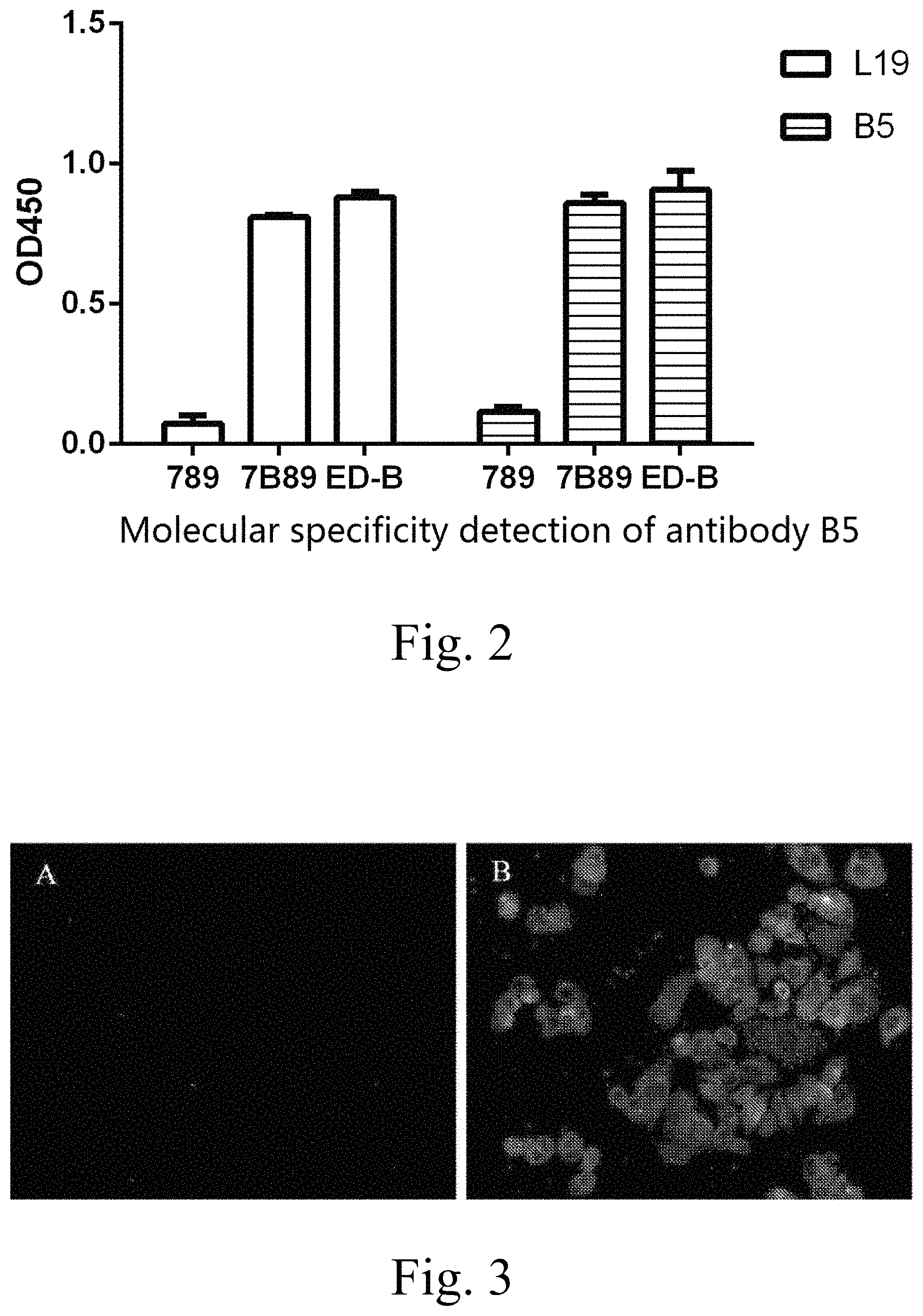
FIG. 2 illustrates an ELISA experiment adopted for confirming the ability of the antibody for specifically recognizing the ED-B domain of an antigen;
FIG. 3 illustrates a cellular immunofluorescence test used for confirming the ability of the antibody B5 for specifically recognizing an FN(B+) cell, wherein A is a negative control with no primary antibody and a secondary antibody being a FITC-labeled rabbit-anti-human IgG antibody; and B is the specific binding of the antibody B5 with the FN(B+) cell with a primary antibody being the antibody B5 and a secondary antibody being a FITC-labeled rabbit-anti-human IgG antibody;
FIG. 4 illustrates the distribution condition of the Cy5.5 NHS ester-labeled antibody B5 at the time 24 hours after the administration thereof through tail intravenous injection to a Balb/c nu nude mouse; and
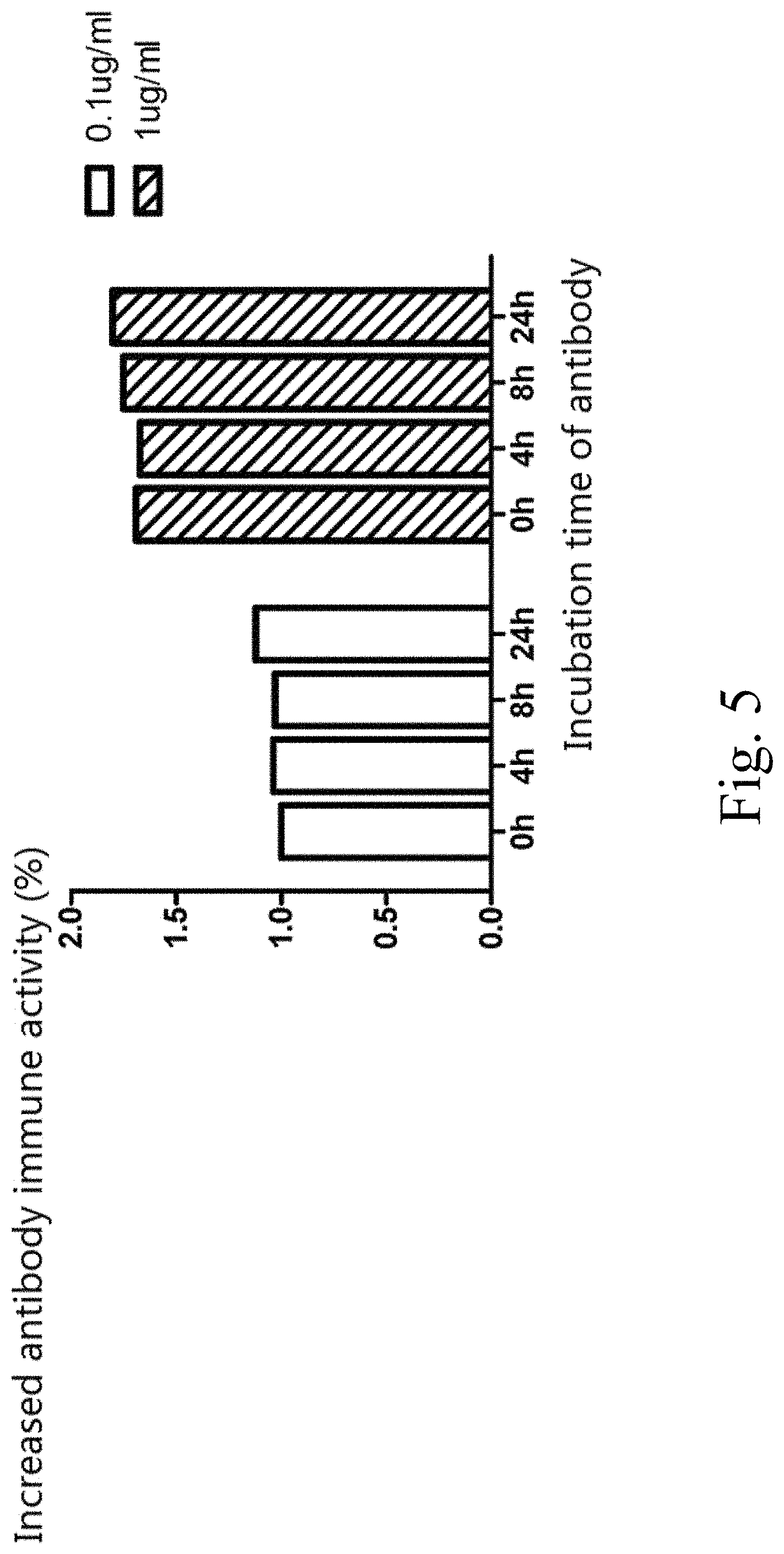
FIG. 5 illustrates a thermal stability test of the antibody B5.
DETAILED DESCRIPTION OF THE PREFERRED EMBODIMENT
Embodiment 1: Screening of Humanized Single-Chain Antibody Specific to the ED-B Domain in Human FN
1) Antigen Expression
FN(B+), ED-B, FN(789) and FN(7B89) are cloned from the cDNA of the HEK293 cell, and then are each fused with six histidine tags at the carboxyl terminal. The proteins are expressed in E. coli, and the obtained recombinant proteins are purified through Ni-chelating affinity chromatography. For FN(7B89), the letter "B" represents that the expressed protein includes the ED-B domain; the "7, 8, 9" represents three domains adjacent to EDB in the FN; and FN(789) does not include the ED-B domain.
2) Construction of Antibody Library
A fully synthetic antibody library is designed for screening a single-chain antibody against the ED-B antigen. By using a most common human antibody molecule DP47 as the template of heavy chain and using DPK22 as the template of light chain, a template of the single-chain antibody library is formed by connecting the two templates sequentially through commonly-used (G.sub.4S).sub.3 peptide bonds, wherein the CDR regions of the light and heavy chains are formed by addition according to experience. The DNA template of the antibody library is obtained through chemical synthesis. During the synthesis, Nco I and EcoR I restriction endonuclease cleavage sites are introduced to the 5'-terminal, and a Not I site is introduced to the 3'-terminal. The DNA is cloned onto a pCANTAB5E vector through the Nco I and Not I sites, so as to construct and generate a random antibody library and phage display, wherein the EcoR I site is used for subsequent molecular clone. For the random antibody library, the randomization of the CDR3 region is achieved through addition of restriction cleavage sites at two ends of the CDR3 regions of the light and heavy chains and the insertion of the restriction cleavage sites into a random fragment of the DNA (Marks, Hoogenboom et al. 1991, Hoogenboom and Winter 1992, Nissim, Hoogenboom et al. 1994, Pini, Viti et al. 1998, Silacci, Brack et al. 2005).
3) Screening
50 .mu.l E. coli TG1 bacteria solution of the antibody library is added into 50 ml fresh 2.times.YT culture media, subjected to shake culture at 37.degree. C. until the OD600 value reaches 0.4-0.5, infected with a helper phage at 37.degree. C. for 30 min, precipitated with PEG, then centrifuged at 10,000 g for 30 min, and resuspended in PBS for later use, to make the prepared phage-fused antibody library have a titer over 10.sup.12 CFU/ml.
The EDB antigen is enveloped in a 5 ml immune tube and blocked with milk, then 2 ml solution of the aforementioned phage antibody library in PBS is added. The culture is incubated at 37.degree. C. for 2 hours, the liquid in the immune tube is poured out, and the culture is washed with 0.1% PBST for 20 times, and 4 ml log-phase TG1 bacteria is added and then incubated while standing at 37.degree. C. for 1 hr. The bacteria solution is coated onto a SOB plate with ampicillin and glucose and cultured overnight, and then the E. coli TG1 bacteria solution infected by the phage is collected to complete the first round of panning. The obtained TG1 bacteria solution is used for preparation of phage-fused antibody library again, so as to start the second round of panning.
Three rounds of panning are conducted in total by using the ED-B as the antigen, wherein the obtained E. coli TG1 is diluted at an appropriate ratio, and then coated onto an agarose plate to obtain monoclonal colonies used for respectively expressing soluble antibodies. An ELISA detection is conducted to select phage clones showing the brightest staining under the condition of the same enveloped antigen for further analysis.
4) Cloning of Antibody from Prokaryotic Vector to Eukaryotic Vector
The DNA fragment of the single-chain antibody obtained by panning is cleaved by EcoR I and Not I, then connected to a pCI-neo vector including the DNA sequence of the Fc fragment in the constant region of human IgG1 antibody, and the DNA sequence of the antibody is in the same open reading frame with the DNA sequence of IgG1Fc, so that the two DNA sequences can be expressed as a fusion protein, wherein IgG1Fc can be used for purifying the single-chain antibody and improving the stability of the antibody protein.
5) Protein Expression
After a large amount of obtained plasmids including the antibody fusion proteins are extracted, the DNA of the plasmids is infected into CHO-K 1 cells by using a liposome reagent Lipofectamine.RTM. 2000 from Invitrogen. The cells are selectively cultured in a selective culture medium including G418 for a continuous time of 4-8 weeks by means of the neo gene of the pCI-neo plasmid vector and then are clonally cultured through a limited dilution method, and the obtained monoclonal cells are further clonally cultured to obtain stably-expressed cell lines.
The obtained cell lines are habituated by using Hyclone SFM4CHO-Utility, and then expressed in suspension. As confirmed by ELISA and SDS-PAGE experiments, a target antibody protein is obtained with the yield of 100 mg/L.
6) Purification of Monoclonal Antibody
The antibody containing the 6.times.His tags is purified through Ni-chelating affinity chromatography from Qiagen, and the antibody containing Fc of the IgG1 constant region is purified through Protein A affinity chromatography from Genscript. The respective procedures are conducted according to the instructions.
Through this method, the antibody B5 of the present invention is obtained and the sequence thereof is shown in FIG. 1.
Embodiment 2: Specificity Detection of Antibody
1) Specificity Detection at Molecular Level
A truncated plasmid vector of fibronectin (FN) is constructed, which can respectively express proteins including the domain FN(789), proteins including the domain FN(7B89), and proteins including the independent ED-B domain, and has 6.times.His tags. The plasmid is expressed in E. coli and then purified through a Ni-chelating column. The aforementioned three proteins are used as antigens and are blocked with milk. Then an ELISA detection test is conducted by using the antibody B5 as a primary antibody and using the horseradish peroxidase-labeled rabbit-anti-human IgG antibody as the secondary antibody, with the test results being shown in FIG. 2. The results demonstrate that both the antibodies B5 and L19 show abilities of specifically recognizing the ED-B domain of the antigen. The staining value of the FN(789) domain is substantially consistent with that of a blank control in which the antigen is not enveloped, and the antibodies bind to both the independent ED-B domain and the FN(7B89) containing the ED-B domain.
2) Specificity Detection at Cellular Level
Human rectal adenocarcinoma Caco-2 cells which highly express FN(B+) are added into and cultured in a glass bottom type laser confocal cell culture dish until the confluence reaches 30%, the culture medium is removed, and the culture is washed in PBS. Then the antibody B5 and a positive control are added, and PBS is used as a negative control. The culture is incubated at 37.degree. C. for 2 hr, washed in PBS for three times, incubated for 1 hr with the FITC-labeled rabbit-anti-human IgG added as the secondary antibody, washed in PBS for three times, and then counter-stained by adding DAPI. As observed in a laser confocal manner, the background of the negative control does not emit a green fluorescent light, while both the cell membranes of B5 and the positive control emit the green fluorescent light (as shown in FIG. 3), demonstrating that the antibody B5 can be specifically targeted to a Caco-2 cell expressing ED-B.
3) Specificity Research of Distribution in Animal Tissues
Construction of transplantation tumor mouse model: healthy Bal b/cnu nude mice are raised in a SPF barrier system until the tumor reaches 20 g per mouse, a transplantation tumor tissue mass from a human pharyngeal squamous carcinoma cell FaDu with the volume of about 1 mm.sup.3 is subcutaneously implanted on the back of each mouse, and then the mice are raised continually.
Fluorescence labeling of antibody drug: the antibody B5 is labeled with the Cy5.5 NHS ester fluorescent dye, and the labeling ratio of the antibody protein to the fluorescent dye molecule is 1:1.
Administration method: when the tumor is grown to have a diameter of about 5 mm, the antibody labeled with the Cy5.5 is introduced according to the standard of 5 mg per kg weight of the tumor. The in vivo distribution condition of the antibody is observed by using a small animal in vivo imaging system from Xenogen, and the observation is conducted once per day after administration.
The experiment results are shown in FIG. 4. The antibody drug is significantly enriched in tumor tissues 24 hours after the administration, the distribution of the Cy5.5 fluorescent dye is not detected in other tissues, and the fluorescent light can be maintained in the tumor tissues after administration for over 7 days.
Embodiment 3: Determination of Antibody Affinity
1) Determination of Absolute Affinity of Antibody Against ED-B Antigen through ELISA Method
The Scatchard assay method (Beatty, Beatty et al. 1987) is used for the affinity determination. 0.1 .mu.g/.mu.l ED-B antigen is diluted according to four concentration gradients of 1:1, 1:2, 1:4 and 1:8, enveloped in the ELISA plate, stored overnight at 4.degree. C., and then blocked with 4% skimmed milk. The single-chain antibody diluted according to the concentration gradient of 1:3 is added with the original concentration of the antibody being 10 .mu.g/ml. The antibody reacts under a room temperature for 1 hr, then reacts for 1 hr under the addition of a horseradish peroxidase-labeled rabbit-anti-human IgG antibody, and is stained by adding tetramethyl benzidine (TMB). When an appropriate color depth is reached, the staining is terminated by using 2M H.sub.2SO.sub.4, and the OD450 optical absorption value is determined.
As calculated, the result shows that the equilibrium dissociation constant KD of the antibody B5 is 1.8 nM, while the equilibrium dissociation constant KD of the antibody L19 is 6.0 nM, so that the affinity of the antibody B5 is higher than that of the antibody L19.
2) Determination of Absolute Affinity of Antibody Against ED-B Antigen through Biocore3000
A monolayer carboxyl chip (coupled with the amino group of a protein) is used to envelope the antigen, wherein the antigen EDB (with the concentration of 10 .mu.g/ml) is diluted into a fixed phase by using an acetic acid buffer solution with the pH of 4. The chip is blocked by using a 1M ethanolamine-HCl buffer solution with the pH of 8.5.
An appropriate concentration gradient should be selected for determination of kinetic constants. The antibodies of the aforementioned four concentration gradients are further diluted according to a gradient of 1:2, such that totally eight concentrations are used as flowing phases, with each concentration being sampled in parallel for three times. The dynamic binding and distribution states of the fixed and flowing phases are detected through the BIAcore3000.
As detected, the antibody B5 can specifically bind onto the ED-B antigen, and the binding between the B5 and the EDB on the surface of the chip is hardly ever dissociated, i.e. the dissertation degree exceeds the detection limit of instrument, which indicates that the antibody B5 can stably bind to the ED-B antigen. The equilibrium dissociation constant of the antibody L19 is 0.4 nM, as detected via the BIAcore3000.
Embodiment 4: Stability Test of Antibody in Blood Plasma
The B5 is diluted respectively to 1 ng/.mu.l and 0.1 ng/.mu.l with the blood plasma of Bal b/c mice, transferred into EP tubes, each tube including 100 .mu.l of the solution. The tubes are incubated in a water bath of 37.degree. C. respectively for 0, 4, 8 and 24 hr. The tubes are withdrawn from the water bath at the same time to conduct an ELISA detection thereof.
The procedure of ELISA detection is as follows: the FN(7B89) fusion protein is enveloped in the ELISA plate at 4.degree. C. overnight; the protein reacts under room temperature for 2 hr, with B5-IgG1Fc being used as the primary antibody and the horseradish peroxidase-labeled rabbit-anti-human IgG being used as the secondary antibody; the protein is stained under room temperature by using the tetramethyl benzidine (TMB) as a substrate, and then detection is conducted at a wavelength of 450 nm via a microplate reader. The result shows that after the antibodies of two different concentrations are incubated in the mice's blood plasma for different time periods, the immune activity changes of the antibodies are statistically meaningless.
By using 0.1 .mu.g/ml B5 antibody incubated in the blood plasma for 0 hr as a standard, changes of the activities of antibodies with different concentrations and under different incubation conditions relative to the standard activity are observed. Therefore, it can be seen that after the antibody is diluted to 1 .mu.g/ml or 0.1 .mu.g/ml with the blood plasma and incubated at 37.degree. C. for 4, 8 and 24 hr, the activity of the antibody is not reduced obviously as compared to the standard 0 hr (as shown in FIG. 5), demonstrating that the antibody B5 can keep stable in the blood plasma of Bal b/c mice.
Embodiment 5: Research of Physiological Activities of Monoclonal Antibody
Construction of transplantation tumor mouse model: healthy Bal b/c mice are raised until the tumor reaches 20 g per mouse, and mice's embryonic cancer cells F9 are subcutaneously injected under the foreleg armpit with the amount of 3.times.10.sup.6 per mouse, so as to construct a model.
Administration method: when the solid tumor can be identified clearly, an antibody drug B5-IL2 (the fusion protein has the amino acid sequence as shown in SEQ ID NO: 16) of 30 .mu.g is administrated to each mouse through tail intravenous injection, and the drug administration is repeated once three days later. The positive control is B5m-IL2, wherein B5m is a mutant antibody which cannot bind specifically to the ED-B, which is formed by the lactamine mutation in the CDR3 region of the B5 heavy chain. The mutation method is the process that the amino acid HTAPLFDY in the CDR3 region of B5 heavy chain is mutated into lactamine AAAAAAAA (SEQ ID NO: 14) while other sequences are kept unchanged, and the resultant mutant of antibody B5 is known as B5m.
Determination of tumor growth rate: the size of the tumor is measured every day until the mice's tumor is increased to 1000 mm.sup.3 or after 14 days, then the mice are sacrificed. Calculation method of tumor volume: the maximum diameter (Length) and the minimum diameter (Width) of the tumor are measured by using a vernier caliper, wherein the tumor volume=width.sup.2.times.length.times.0.5.
The experiment results are shown in table 1 below, wherein no significant difference in tumor size exists between the positive control group and the negative control group, the mean tumor volume of the experiment group is about three times more than that of the negative control group, and the differences in tumor volume between the experiment group and the two control groups have statistical meaning (p<0.05).
TABLE-US-00001 TABLE 1 Therapeutic Effect of B5-IL2 through tail intravenous injection to a Balb/c mouse (x .+-. s, n = 6, *p < 0.05) Tumor Volume Reduction Folds of Tumor Volume Group (mm.sup.3) Compared with the Control Group Negative Control 0.53 .+-. 0.13 -- Group Positive Control 0.51 .+-. 0.13 -- Group Experiment Group 0.17 .+-. 0.07* 3 folds
SEQUENCE LISTINGS
1
161117PRTArtificial SequenceSynthesis 1Glu Val Gln Leu Leu Glu Ser Gly Gly Gly Leu Val Gln Pro Gly Gly1 5 10 15Ser Leu Arg Leu Ser Cys Ala Ala Ser Gly Phe Thr Phe Ser Ser Tyr 20 25 30Ala Met Ser Trp Val Arg Gln Ala Pro Gly Lys Gly Leu Glu Trp Val 35 40 45Ser Arg Ile Ser Pro Ser Gly Ser Ser Thr Tyr Tyr Ala Asp Ser Val 50 55 60Lys Gly Arg Phe Thr Ile Ser Arg Asp Asn Ser Lys Asn Thr Leu Tyr65 70 75 80Leu Gln Met Asn Ser Leu Arg Ala Glu Asp Thr Ala Val Tyr Tyr Cys 85 90 95Ala Arg His Thr Ala Pro Leu Phe Asp Tyr Trp Gly Gln Gly Thr Leu 100 105 110Val Thr Val Ser Ser 1152351DNAArtificial SequenceSythesis 2gaagtgcagc tgttggaatc tgggggtggc ttggtacagc ctggggggtc cctgcgtctc 60tcctgtgcag ccagcggctt cacctttagt agctatgcca tgagctgggt ccgccaggct 120ccagggaaag gcctggaatg ggtcagtcgt attagtccga gtggcagcag tacatactac 180gcagactccg tgaaaggtcg cttcaccatc tcccgtgaca attccaaaaa cacgctgtat 240ctgcaaatga acagcctgcg tgccgaagac accgccgtat attactgtgc gcgtcatact 300gcgcctctgt tcgattactg gggccagggc accctggtca ccgtctccag t 3513108PRTArtificial SequenceSynthesis 3Glu Ile Val Leu Thr Gln Ser Pro Gly Thr Leu Ser Leu Ser Pro Gly1 5 10 15Glu Arg Ala Thr Leu Ser Cys Arg Ala Ser Gln Ser Val Ser Ser Ser 20 25 30Tyr Leu Ala Trp Tyr Gln Gln Lys Pro Gly Gln Ala Pro Arg Leu Leu 35 40 45Ile Tyr Lys Ala Ser Asn Leu Ala Thr Gly Ile Pro Asp Arg Phe Ser 50 55 60Gly Ser Gly Ser Gly Thr Asp Phe Thr Leu Thr Ile Ser Arg Leu Glu65 70 75 80Pro Glu Asp Phe Ala Val Tyr Tyr Cys Gln Gln Gly Arg His Thr Pro 85 90 95Pro Thr Phe Gly Gln Gly Thr Lys Val Glu Ile Lys 100 1054324DNAArtificial SequenceSynthesis 4gaaattgtgt tgacgcagtc tccaggcacc ctgtctttgt ctccaggtga aagagccacc 60ctctcttgcc gtgccagtca gagtgttagc agcagctact tagcctggta ccagcagaaa 120cctggccagg ctccacgtct gctgatctat aaagcttcta acctggccac cggcatccca 180gaccgtttca gtggcagtgg ctctggtaca gacttcactc tcaccatcag ccgcctggag 240cctgaagatt ttgcagtgta ttactgtcag cagggtcgtc atacgccgcc taccttcggc 300caaggcacca aggtggaaat caaa 324516PRTArtificial SequenceSynthesis 5Gly Gly Gly Gly Ser Gly Gly Gly Gly Ser Gly Gly Gly Gly Ser Ser1 5 10 15648DNAArtificial SequenceSynthesis 6ggtggtggcg gttcaggcgg tggtggctct ggtggcggtg ggagctct 48717PRTArtificial SequenceSynthesis 7Ala Ala Ala Ser Ser Ser Gly Ser Ser Ser Ser Gly Ser Ser Ser Ser1 5 10 15Gly85PRTArtificial SequenceSynthesis 8Ser Tyr Ala Met Ser1 5917PRTArtificial SequenceSynthesis 9Arg Ile Ser Pro Ser Gly Ser Ser Thr Tyr Tyr Ala Asp Ser Val Lys1 5 10 15Gly108PRTArtificial SequenceSynthesis 10His Thr Ala Pro Leu Phe Asp Tyr1 51112PRTArtificial SequenceSynthesis 11Arg Ala Ser Gln Ser Val Ser Ser Ser Tyr Leu Ala1 5 10127PRTArtificial SequenceSynthesis 12Lys Ala Ser Asn Leu Ala Thr1 5137PRTArtificial SequenceSynthesis 13Gln Gln Gly Arg His Thr Pro1 5148PRTArtificial SequenceSynthesis 14Ala Ala Ala Ala Ala Ala Ala Ala1 515241PRTArtificial SequenceSynthesis 15Glu Val Gln Leu Leu Glu Ser Gly Gly Gly Leu Val Gln Pro Gly Gly1 5 10 15Ser Leu Arg Leu Ser Cys Ala Ala Ser Gly Phe Thr Phe Ser Ser Tyr 20 25 30Ala Met Ser Trp Val Arg Gln Ala Pro Gly Lys Gly Leu Glu Trp Val 35 40 45Ser Arg Ile Ser Pro Ser Gly Ser Ser Thr Tyr Tyr Ala Asp Ser Val 50 55 60Lys Gly Arg Phe Thr Ile Ser Arg Asp Asn Ser Lys Asn Thr Leu Tyr65 70 75 80Leu Gln Met Asn Ser Leu Arg Ala Glu Asp Thr Ala Val Tyr Tyr Cys 85 90 95Ala Arg His Thr Ala Pro Leu Phe Asp Tyr Trp Gly Gln Gly Thr Leu 100 105 110Val Thr Val Ser Ser Gly Gly Gly Gly Ser Gly Gly Gly Gly Ser Gly 115 120 125Gly Gly Gly Ser Ser Glu Ile Val Leu Thr Gln Ser Pro Gly Thr Leu 130 135 140Ser Leu Ser Pro Gly Glu Arg Ala Thr Leu Ser Cys Arg Ala Ser Gln145 150 155 160Ser Val Ser Ser Ser Tyr Leu Ala Trp Tyr Gln Gln Lys Pro Gly Gln 165 170 175Ala Pro Arg Leu Leu Ile Tyr Lys Ala Ser Asn Leu Ala Thr Gly Ile 180 185 190Pro Asp Arg Phe Ser Gly Ser Gly Ser Gly Thr Asp Phe Thr Leu Thr 195 200 205Ile Ser Arg Leu Glu Pro Glu Asp Phe Ala Val Tyr Tyr Cys Gln Gln 210 215 220Gly Arg His Thr Pro Pro Thr Phe Gly Gln Gly Thr Lys Val Glu Ile225 230 235 240Lys16392PRTArtificial SequenceSynthesis 16Glu Val Gln Leu Leu Glu Ser Gly Gly Gly Leu Val Gln Pro Gly Gly1 5 10 15Ser Leu Arg Leu Ser Cys Ala Ala Ser Gly Phe Thr Phe Ser Ser Tyr 20 25 30Ala Met Ser Trp Val Arg Gln Ala Pro Gly Lys Gly Leu Glu Trp Val 35 40 45Ser Arg Ile Ser Pro Ser Gly Ser Ser Thr Tyr Tyr Ala Asp Ser Val 50 55 60Lys Gly Arg Phe Thr Ile Ser Arg Asp Asn Ser Lys Asn Thr Leu Tyr65 70 75 80Leu Gln Met Asn Ser Leu Arg Ala Glu Asp Thr Ala Val Tyr Tyr Cys 85 90 95Ala Arg His Thr Ala Pro Leu Phe Asp Tyr Trp Gly Gln Gly Thr Leu 100 105 110Val Thr Val Ser Ser Gly Gly Gly Gly Ser Gly Gly Gly Gly Ser Gly 115 120 125Gly Gly Gly Ser Ser Glu Ile Val Leu Thr Gln Ser Pro Gly Thr Leu 130 135 140Ser Leu Ser Pro Gly Glu Arg Ala Thr Leu Ser Cys Arg Ala Ser Gln145 150 155 160Ser Val Ser Ser Ser Tyr Leu Ala Trp Tyr Gln Gln Lys Pro Gly Gln 165 170 175Ala Pro Arg Leu Leu Ile Tyr Lys Ala Ser Asn Leu Ala Thr Gly Ile 180 185 190Pro Asp Arg Phe Ser Gly Ser Gly Ser Gly Thr Asp Phe Thr Leu Thr 195 200 205Ile Ser Arg Leu Glu Pro Glu Asp Phe Ala Val Tyr Tyr Cys Gln Gln 210 215 220Gly Arg His Thr Pro Pro Thr Phe Gly Gln Gly Thr Lys Val Glu Ile225 230 235 240Lys Ala Ala Ala Ser Ser Ser Gly Ser Ser Ser Ser Gly Ser Ser Ser 245 250 255Ser Gly Ala Pro Thr Ser Ser Ser Thr Lys Lys Thr Gln Leu Gln Leu 260 265 270Glu His Leu Leu Leu Asp Leu Gln Met Ile Leu Asn Gly Ile Asn Asn 275 280 285Tyr Lys Asn Pro Lys Leu Thr Arg Met Leu Thr Phe Lys Phe Tyr Met 290 295 300Pro Lys Lys Ala Thr Glu Leu Lys His Leu Gln Cys Leu Glu Glu Glu305 310 315 320Leu Lys Pro Leu Glu Glu Val Leu Asn Leu Ala Gln Ser Lys Asn Phe 325 330 335His Leu Arg Pro Arg Asp Leu Ile Ser Asn Ile Asn Val Ile Val Leu 340 345 350Glu Leu Lys Gly Ser Glu Thr Thr Phe Met Cys Glu Tyr Ala Asp Glu 355 360 365Thr Ala Thr Ile Val Glu Phe Leu Asn Arg Trp Ile Thr Phe Cys Gln 370 375 380Ser Ile Ile Ser Thr Leu Thr Ala385 390
* * * * *
D00001
D00002
D00003
D00004
S00001
XML
uspto.report is an independent third-party trademark research tool that is not affiliated, endorsed, or sponsored by the United States Patent and Trademark Office (USPTO) or any other governmental organization. The information provided by uspto.report is based on publicly available data at the time of writing and is intended for informational purposes only.
While we strive to provide accurate and up-to-date information, we do not guarantee the accuracy, completeness, reliability, or suitability of the information displayed on this site. The use of this site is at your own risk. Any reliance you place on such information is therefore strictly at your own risk.
All official trademark data, including owner information, should be verified by visiting the official USPTO website at www.uspto.gov. This site is not intended to replace professional legal advice and should not be used as a substitute for consulting with a legal professional who is knowledgeable about trademark law.