Biocompatible scaffolds with tissue fragments
Binette , et al.
U.S. patent number 10,603,408 [Application Number 15/338,895] was granted by the patent office on 2020-03-31 for biocompatible scaffolds with tissue fragments. This patent grant is currently assigned to DePuy Synthes Products, Inc.. The grantee listed for this patent is DePuy Mitek, LLC. Invention is credited to Francois Binette, Sridevi Dhanaraj, Anna Gosiewska, Julia Hwang.
| United States Patent | 10,603,408 |
| Binette , et al. | March 31, 2020 |
Biocompatible scaffolds with tissue fragments
Abstract
A biocompatible tissue repair implant or scaffold device is provided for use in repairing a variety of tissue injuries, particularly injuries to cartilage, ligaments, tendons, and nerves. The repair procedures may be conducted with implants that contain a biological component that assists in healing or tissue repair. The biocompatible tissue repair implants include a biocompatible scaffold and particles of living tissue, such that the tissue and the scaffold become associated. The particles of living tissue contain one or more viable cells that can migrate from the tissue and populate the scaffold.
| Inventors: | Binette; Francois (San Francisco, CA), Hwang; Julia (Wayland, MA), Dhanaraj; Sridevi (Raritan, NJ), Gosiewska; Anna (Skillman, NJ) | ||||||||||
|---|---|---|---|---|---|---|---|---|---|---|---|
| Applicant: |
|
||||||||||
| Assignee: | DePuy Synthes Products, Inc.
(Raynham, MA) |
||||||||||
| Family ID: | 32045864 | ||||||||||
| Appl. No.: | 15/338,895 | ||||||||||
| Filed: | October 31, 2016 |
Prior Publication Data
| Document Identifier | Publication Date | |
|---|---|---|
| US 20170049931 A1 | Feb 23, 2017 | |
Related U.S. Patent Documents
| Application Number | Filing Date | Patent Number | Issue Date | ||
|---|---|---|---|---|---|
| 12951205 | Nov 22, 2010 | 9511171 | |||
| 10374772 | Feb 25, 2003 | ||||
| 60419539 | Oct 18, 2002 | ||||
| 60420093 | Oct 18, 2002 | ||||
| Current U.S. Class: | 1/1 |
| Current CPC Class: | A61L 27/3616 (20130101); A61L 27/3895 (20130101); C12N 5/0655 (20130101); C12N 5/0068 (20130101); A61L 27/3817 (20130101); A61L 27/58 (20130101); A61L 27/3612 (20130101); A61L 27/36 (20130101); A61L 27/18 (20130101); A61F 2/08 (20130101); A61L 2430/10 (20130101); A61F 2/02 (20130101); A61L 2400/18 (20130101); A61L 2430/06 (20130101); A61F 2310/0097 (20130101); A61L 2430/34 (20130101) |
| Current International Class: | A61F 2/00 (20060101); C12N 5/00 (20060101); A61L 27/58 (20060101); C12N 5/077 (20100101); A61L 27/38 (20060101); A61L 27/36 (20060101); A61L 27/18 (20060101); A61F 2/08 (20060101); A61F 2/02 (20060101) |
References Cited [Referenced By]
U.S. Patent Documents
| 3272204 | September 1966 | Artandi et al. |
| 3562820 | February 1971 | Braun |
| 3739402 | June 1973 | Cooley et al. |
| 3812017 | May 1974 | Santangelo |
| 3857932 | December 1974 | Shepherd et al. |
| 4045418 | August 1977 | Sinclair |
| 4057537 | November 1977 | Sinclair |
| 4105034 | August 1978 | Shalaby et al. |
| 4130639 | December 1978 | Shalaby et al. |
| 4130689 | December 1978 | Costa, Jr. |
| 4140678 | February 1979 | Shalaby et al. |
| 4141087 | February 1979 | Shalaby et al. |
| 4205399 | June 1980 | Shalaby et al. |
| 4208511 | June 1980 | Shalaby et al. |
| 4344193 | August 1982 | Kenny |
| 4520821 | June 1985 | Schmidt et al. |
| 4553272 | November 1985 | Mears |
| 4585458 | April 1986 | Kurland |
| 4597766 | July 1986 | Hilal et al. |
| 4609551 | September 1986 | Caplan et al. |
| 4728329 | March 1988 | Mansat |
| 4801299 | January 1989 | Brendel et al. |
| 4837285 | June 1989 | Berg et al. |
| 4902508 | February 1990 | Badylak et al. |
| 4917700 | April 1990 | Aikins |
| 4946377 | August 1990 | Kovach |
| 5007934 | April 1991 | Stone |
| 5041138 | August 1991 | Vacanti et al. |
| 5053050 | October 1991 | Itay |
| 5061281 | October 1991 | Mares et al. |
| 5078744 | January 1992 | Chvapil |
| 5108807 | April 1992 | Tucker |
| 5108989 | April 1992 | Amento et al. |
| 5147400 | September 1992 | Kaplan et al. |
| 5176708 | January 1993 | Frey et al. |
| 5206023 | April 1993 | Hunziker |
| 5258028 | November 1993 | Ersek et al. |
| 5263984 | November 1993 | Li et al. |
| 5290494 | March 1994 | Coombes et al. |
| 5306311 | April 1994 | Stone et al. |
| 5320624 | June 1994 | Kaplan et al. |
| 5320646 | June 1994 | Patton et al. |
| 5326357 | July 1994 | Kandel |
| 5366756 | November 1994 | Chesterfield et al. |
| 5393594 | February 1995 | Koyfman et al. |
| 5425766 | June 1995 | Bowald |
| 5443950 | August 1995 | Naughton et al. |
| 5445833 | August 1995 | Badylak et al. |
| 5455041 | October 1995 | Genco et al. |
| 5464929 | November 1995 | Bezwada et al. |
| 5468253 | November 1995 | Bezwada et al. |
| 5480827 | January 1996 | Guillemin et al. |
| 5487897 | January 1996 | Polson et al. |
| 5514181 | May 1996 | Light et al. |
| 5514378 | May 1996 | Mikos et al. |
| 5571189 | November 1996 | Kuslich |
| 5577517 | November 1996 | Bonutti |
| 5589176 | December 1996 | Seare, Jr. |
| 5595751 | January 1997 | Bezwada et al. |
| 5597579 | January 1997 | Bezwada et al. |
| 5607687 | March 1997 | Bezwada et al. |
| 5612028 | March 1997 | Sackier et al. |
| 5618552 | April 1997 | Bezwada et al. |
| 5620698 | April 1997 | Bezwada et al. |
| 5624463 | April 1997 | Stone et al. |
| 5626611 | May 1997 | Liu et al. |
| 5632745 | May 1997 | Schwartz |
| 5645850 | July 1997 | Bezwada et al. |
| 5648088 | July 1997 | Bezwada et al. |
| 5654135 | August 1997 | Tinois et al. |
| 5656492 | August 1997 | Glowacki et al. |
| 5677355 | October 1997 | Shalaby et al. |
| 5681353 | October 1997 | Li et al. |
| 5697976 | December 1997 | Chesterfield et al. |
| 5698213 | December 1997 | Jamiolkowski et al. |
| 5700583 | December 1997 | Jamiolkowski et al. |
| 5705181 | January 1998 | Cooper et al. |
| 5709854 | January 1998 | Griffith-Cima et al. |
| 5711960 | January 1998 | Shikinami |
| 5713920 | February 1998 | Bezwada et al. |
| 5720969 | February 1998 | Gentile et al. |
| 5723331 | March 1998 | Tubo et al. |
| 5735903 | April 1998 | Li et al. |
| 5736372 | April 1998 | Vacanti et al. |
| 5755791 | May 1998 | Whitson et al. |
| 5759190 | June 1998 | Vibe-Hansen et al. |
| 5766631 | June 1998 | Arnold |
| 5769897 | June 1998 | Harle |
| 5769899 | June 1998 | Schwartz et al. |
| 5782914 | July 1998 | Schankereli |
| 5786217 | July 1998 | Tubo et al. |
| 5800537 | September 1998 | Bell |
| 5800543 | September 1998 | McLeod et al. |
| 5830493 | November 1998 | Yokota et al. |
| 5837235 | November 1998 | Mueller et al. |
| 5837278 | November 1998 | Geistlich et al. |
| 5842477 | December 1998 | Naughton et al. |
| 5855608 | January 1999 | Brekke et al. |
| 5859150 | January 1999 | Jamiolkowski et al. |
| 5891558 | April 1999 | Bell et al. |
| 5902741 | May 1999 | Purchio et al. |
| 5904716 | May 1999 | Gendler |
| 5904717 | May 1999 | Brekke et al. |
| 5914121 | June 1999 | Robey et al. |
| 5922025 | July 1999 | Hubbard |
| 5964805 | October 1999 | Stone |
| 5968096 | October 1999 | Whitson et al. |
| 5980889 | November 1999 | Butler et al. |
| 5989269 | November 1999 | Vibe-Hansen et al. |
| 5990194 | November 1999 | Dunn et al. |
| 5990378 | November 1999 | Ellis |
| 6001352 | December 1999 | Boyan et al. |
| 6001394 | December 1999 | Daculsi et al. |
| 6005161 | December 1999 | Brekke et al. |
| 6027742 | February 2000 | Lee et al. |
| 6042534 | March 2000 | Gellman et al. |
| 6042610 | March 2000 | Li et al. |
| 6054122 | April 2000 | MacPhee et al. |
| 6077989 | June 2000 | Kandel et al. |
| 6080579 | June 2000 | Hanley, Jr. et al. |
| 6096532 | August 2000 | Armstrong et al. |
| 6103255 | August 2000 | Levene et al. |
| 6110209 | August 2000 | Stone |
| 6110212 | August 2000 | Gregory |
| 6113640 | September 2000 | Tormala et al. |
| 6117166 | September 2000 | Winston et al. |
| 6120514 | September 2000 | Vibe-Hansen et al. |
| 6121042 | September 2000 | Peterson et al. |
| 6123727 | September 2000 | Vacanti et al. |
| 6132463 | October 2000 | Lee et al. |
| 6132468 | October 2000 | Mansmann |
| 6139578 | October 2000 | Lee et al. |
| 6140039 | October 2000 | Naughton et al. |
| 6143293 | November 2000 | Weiss et al. |
| 6147135 | November 2000 | Yuan et al. |
| 6153292 | November 2000 | Bell et al. |
| 6156068 | December 2000 | Walter et al. |
| 6165217 | December 2000 | Hayes |
| 6171338 | January 2001 | Talja et al. |
| 6176880 | January 2001 | Plouhar et al. |
| 6179840 | January 2001 | Bowman |
| 6179872 | January 2001 | Bell et al. |
| 6180007 | January 2001 | Gentile et al. |
| 6183737 | February 2001 | Zaleske et al. |
| 6187053 | February 2001 | Minuth |
| 6187329 | February 2001 | Agrawal et al. |
| 6197061 | March 2001 | Masuda et al. |
| 6197325 | March 2001 | MacPhee et al. |
| 6200606 | March 2001 | Peterson et al. |
| 6214045 | April 2001 | Corbitt, Jr. et al. |
| 6214055 | April 2001 | Simionescu et al. |
| 6242247 | June 2001 | Rieser et al. |
| 6251673 | June 2001 | Winkler |
| 6277151 | August 2001 | Lee et al. |
| 6283980 | September 2001 | Vibe-Hansen et al. |
| 6287316 | September 2001 | Agarwal et al. |
| 6287340 | September 2001 | Altman et al. |
| 6291240 | September 2001 | Mansbridge et al. |
| 6306177 | October 2001 | Felt et al. |
| 6306424 | October 2001 | Vyakarnam et al. |
| 6316692 | November 2001 | Readhead et al. |
| 6319712 | November 2001 | Meenen et al. |
| 6328765 | December 2001 | Hardwick et al. |
| 6331312 | December 2001 | Lee et al. |
| 6333029 | December 2001 | Vyakarnam et al. |
| 6365149 | April 2002 | Vyakarnam et al. |
| 6365405 | April 2002 | Salzmann et al. |
| 6378527 | April 2002 | Hungerford et al. |
| 6378572 | April 2002 | Neubauer et al. |
| 6379367 | April 2002 | Vibe-Hansen et al. |
| 6464729 | October 2002 | Kandel |
| 6485723 | November 2002 | Badylak et al. |
| 6489165 | December 2002 | Bhatnagar et al. |
| 6503278 | January 2003 | Pohjonen et al. |
| 6511511 | January 2003 | Slivka et al. |
| 6511958 | January 2003 | Atkinson et al. |
| 6521430 | February 2003 | Orwar et al. |
| 6530956 | March 2003 | Mansmann |
| 6534084 | March 2003 | Vyakarnam et al. |
| 6541024 | April 2003 | Kadiyala et al. |
| 6551355 | April 2003 | Lewandrowski et al. |
| 6569172 | May 2003 | Asculai et al. |
| 6592588 | July 2003 | Bobic et al. |
| 6599323 | July 2003 | Melican et al. |
| 6605294 | August 2003 | Sawhney |
| 6626950 | September 2003 | Brown et al. |
| 6652450 | November 2003 | Neisz et al. |
| 6652585 | November 2003 | Lange |
| 6727224 | April 2004 | Zhang et al. |
| 6773458 | August 2004 | Brauker et al. |
| 6783712 | August 2004 | Slivka et al. |
| 6840962 | January 2005 | Vacanti et al. |
| 6852330 | February 2005 | Bowman et al. |
| 6866681 | March 2005 | Laboureau et al. |
| 6884428 | April 2005 | Binette et al. |
| 6886568 | May 2005 | Frondoza et al. |
| 6886569 | May 2005 | Chervitz et al. |
| 6902932 | June 2005 | Altman et al. |
| 7109034 | September 2006 | Orwar et al. |
| 7208177 | April 2007 | Geistlich et al. |
| 7262020 | August 2007 | Hellerstein |
| 7316822 | January 2008 | Binette et al. |
| 7368124 | May 2008 | Chun et al. |
| 7456012 | November 2008 | Ryttsen et al. |
| 7799089 | September 2010 | Plouhar et al. |
| 7824701 | November 2010 | Binette et al. |
| 7875296 | January 2011 | Binette et al. |
| 7901461 | March 2011 | Harmon et al. |
| 8137686 | March 2012 | Kladakis et al. |
| 8137702 | March 2012 | Binette et al. |
| 8197837 | June 2012 | Jamiolkowski et al. |
| 8221780 | July 2012 | Dhanaraj et al. |
| 8226715 | July 2012 | Hwang et al. |
| 8496970 | July 2013 | Binette et al. |
| 8637066 | January 2014 | Binnette et al. |
| 8641775 | February 2014 | Harmon et al. |
| 8691259 | April 2014 | Bowman et al. |
| 2001/0014475 | August 2001 | Frondoza et al. |
| 2001/0016353 | August 2001 | Janas et al. |
| 2001/0016772 | August 2001 | Lee et al. |
| 2001/0023373 | September 2001 | Plouhar et al. |
| 2001/0033857 | October 2001 | Vyakarnam et al. |
| 2001/0038848 | November 2001 | Donda et al. |
| 2001/0039453 | November 2001 | Gresser et al. |
| 2001/0051834 | December 2001 | Frondoza et al. |
| 2001/0053353 | December 2001 | Griffith et al. |
| 2001/0053839 | December 2001 | Noishiki et al. |
| 2002/0006428 | January 2002 | Mahmood et al. |
| 2002/0009477 | January 2002 | Mahmood et al. |
| 2002/0009805 | January 2002 | Nevo et al. |
| 2002/0009806 | January 2002 | Hicks |
| 2002/0013627 | January 2002 | Geistlich et al. |
| 2002/0015719 | February 2002 | Kellner et al. |
| 2002/0022883 | February 2002 | Burg |
| 2002/0022884 | February 2002 | Mansmann |
| 2002/0028192 | March 2002 | Dimitrijevich et al. |
| 2002/0029055 | March 2002 | Bonutti |
| 2002/0062151 | May 2002 | Altman et al. |
| 2002/0082631 | June 2002 | Bonutti |
| 2002/0083479 | June 2002 | Winston et al. |
| 2002/0091403 | July 2002 | Bonutti |
| 2002/0091406 | July 2002 | Bonutti |
| 2002/0099401 | July 2002 | Bonutti |
| 2002/0099448 | July 2002 | Hiles et al. |
| 2002/0107570 | August 2002 | Sybert et al. |
| 2002/0119177 | August 2002 | Bowman et al. |
| 2002/0120348 | August 2002 | Melican et al. |
| 2002/0123750 | September 2002 | Eisermann et al. |
| 2002/0127265 | September 2002 | Bowman et al. |
| 2002/0133229 | September 2002 | Laurencin et al. |
| 2002/0133235 | September 2002 | Hungerford et al. |
| 2002/0150604 | October 2002 | Yi et al. |
| 2002/0151975 | October 2002 | Farr et al. |
| 2002/0173558 | November 2002 | Williams et al. |
| 2002/0176893 | November 2002 | Wironen et al. |
| 2002/0177224 | November 2002 | Madry et al. |
| 2003/0003153 | January 2003 | Asculai et al. |
| 2003/0004578 | January 2003 | Brown et al. |
| 2003/0012805 | January 2003 | Chen et al. |
| 2003/0023316 | January 2003 | Brown et al. |
| 2003/0026787 | February 2003 | Fearnot et al. |
| 2003/0027332 | February 2003 | Lafrance et al. |
| 2003/0033021 | February 2003 | Plouhar et al. |
| 2003/0033022 | February 2003 | Plouhar et al. |
| 2003/0036797 | February 2003 | Malaviya et al. |
| 2003/0036801 | February 2003 | Schwartz et al. |
| 2003/0045937 | March 2003 | Ginn |
| 2003/0050709 | March 2003 | Noth et al. |
| 2003/0064917 | April 2003 | Crawford et al. |
| 2003/0075822 | April 2003 | Slivka et al. |
| 2003/0077311 | April 2003 | Vyakarnam et al. |
| 2003/0078617 | April 2003 | Schwartz et al. |
| 2003/0147935 | August 2003 | Binette et al. |
| 2003/0193104 | October 2003 | Melican et al. |
| 2004/0024457 | February 2004 | Boyce et al. |
| 2004/0033212 | February 2004 | Thomson et al. |
| 2004/0059416 | March 2004 | Murray et al. |
| 2004/0078077 | April 2004 | Binette et al. |
| 2004/0078090 | April 2004 | Binette et al. |
| 2004/0175408 | September 2004 | Chun et al. |
| 2004/0219182 | November 2004 | Gomes et al. |
| 2004/0236424 | November 2004 | Berez et al. |
| 2004/0249457 | December 2004 | Smith et al. |
| 2004/0267088 | December 2004 | Kammerer |
| 2004/0267362 | December 2004 | Hwang et al. |
| 2005/0002915 | January 2005 | Atala et al. |
| 2005/0038520 | February 2005 | Binette et al. |
| 2005/0048651 | March 2005 | Ryttsen et al. |
| 2005/0113937 | May 2005 | Binette et al. |
| 2005/0113938 | May 2005 | Jamiolkowski et al. |
| 2005/0125077 | June 2005 | Harmon et al. |
| 2005/0147645 | July 2005 | Budny |
| 2005/0177249 | August 2005 | Kladakis et al. |
| 2005/0232967 | October 2005 | Kladakis et al. |
| 2005/0234549 | October 2005 | Kladakis et al. |
| 2006/0067967 | March 2006 | Bowman et al. |
| 2006/0084930 | April 2006 | Dhanaraj et al. |
| 2006/0204439 | September 2006 | Hellerstein |
| 2006/0223177 | October 2006 | Harris et al. |
| 2006/0280768 | December 2006 | Hwang et al. |
| 2006/0293760 | December 2006 | DeDeyne |
| 2007/0031470 | February 2007 | Kladakis et al. |
| 2007/0036767 | February 2007 | Mistry et al. |
| 2007/0250177 | October 2007 | Bilbo |
| 2008/0039955 | February 2008 | Hunziker |
| 2008/0071385 | March 2008 | Binette et al. |
| 2008/0226870 | September 2008 | Sypeck et al. |
| 2008/0241213 | October 2008 | Chun et al. |
| 2011/0009963 | January 2011 | Binnette et al. |
| 2011/0091517 | April 2011 | Binette et al. |
| 2011/0097381 | April 2011 | Binette et al. |
| 2011/0110958 | May 2011 | Qiu et al. |
| 2011/0177134 | July 2011 | Harmon et al. |
| 2012/0156265 | June 2012 | Binette et al. |
| 2012/0165939 | June 2012 | Kladakis et al. |
| 2012/0253464 | October 2012 | Hwang et al. |
| 2013/0123937 | May 2013 | Jamiolkowski et al. |
| 717552 | Mar 2000 | AU | |||
| 2247158 | Aug 1997 | CA | |||
| 198 12 195 | Sep 1999 | DE | |||
| 0 145 492 | Jun 1985 | EP | |||
| 0 274 898 | Jul 1988 | EP | |||
| 0 277 678 | Aug 1988 | EP | |||
| 0 411 545 | Feb 1991 | EP | |||
| 0 464 163 | Jan 1992 | EP | |||
| 0 466 105 | Jan 1992 | EP | |||
| 0 485 215 | May 1992 | EP | |||
| 0 562 864 | Sep 1993 | EP | |||
| 0 955 024 | Nov 1999 | EP | |||
| 1 027 897 | Aug 2000 | EP | |||
| 1 064 958 | Jan 2001 | EP | |||
| 1 074 270 | Feb 2001 | EP | |||
| 1 167 517 | Jan 2002 | EP | |||
| 1 177 800 | Feb 2002 | EP | |||
| 1 216 717 | Jun 2002 | EP | |||
| 1 216 718 | Jun 2002 | EP | |||
| 1 348 451 | Oct 2003 | EP | |||
| 1 405 649 | Apr 2004 | EP | |||
| 1 410 811 | Apr 2004 | EP | |||
| 1 506 790 | Feb 2005 | EP | |||
| 1 537 839 | Jun 2005 | EP | |||
| 1 604 622 | Dec 2005 | EP | |||
| 2688690 | Sep 1993 | FR | |||
| 1008193 | Oct 1965 | GB | |||
| 63-203154 | Aug 1988 | JP | |||
| 02-052648 | Feb 1990 | JP | |||
| 02-143945 | Dec 1990 | JP | |||
| 02-227442 | Apr 1992 | JP | |||
| 02-256824 | May 1992 | JP | |||
| 10-234844 | Sep 1998 | JP | |||
| 10-129048 | Nov 1999 | JP | |||
| 11-319068 | Nov 1999 | JP | |||
| 11-512626 | Nov 1999 | JP | |||
| 10-319783 | May 2000 | JP | |||
| 03-139361 | Feb 2001 | JP | |||
| 2001-079079 | Mar 2001 | JP | |||
| 2001-129073 | May 2001 | JP | |||
| 03-261753 | Mar 2002 | JP | |||
| 2002-165345 | Jun 2002 | JP | |||
| 2002-527402 | Aug 2002 | JP | |||
| 2002-535378 | Oct 2002 | JP | |||
| 2003-320008 | Nov 2003 | JP | |||
| 2004-008437 | Jan 2004 | JP | |||
| 2004-195103 | Jul 2004 | JP | |||
| 2005-237476 | Sep 2005 | JP | |||
| 04-094329 | Jun 2008 | JP | |||
| 04-300557 | Jul 2009 | JP | |||
| 04-502715 | Jul 2010 | JP | |||
| 2187261 | Aug 2002 | RU | |||
| 1535542 | Jan 1990 | SU | |||
| WO-86/00533 | Jan 1986 | WO | |||
| WO-92/06179 | Apr 1992 | WO | |||
| WO-93/02718 | Feb 1993 | WO | |||
| WO-93/11805 | Jun 1993 | WO | |||
| WO-95/33821 | Dec 1995 | WO | |||
| WO-96/08277 | Mar 1996 | WO | |||
| WO-97/30662 | Aug 1997 | WO | |||
| WO-97/46665 | Dec 1997 | WO | |||
| WO-98/48860 | Nov 1998 | WO | |||
| WO-98/53768 | Dec 1998 | WO | |||
| WO-99/05992 | Feb 1999 | WO | |||
| WO-99/16381 | Apr 1999 | WO | |||
| WO-99/39724 | Aug 1999 | WO | |||
| WO-99/47097 | Sep 1999 | WO | |||
| WO-99/59647 | Nov 1999 | WO | |||
| WO-00/15248 | Mar 2000 | WO | |||
| WO-00/16381 | Mar 2000 | WO | |||
| WO-00/69355 | Nov 2000 | WO | |||
| WO-00/72782 | Dec 2000 | WO | |||
| WO-00/74741 | Dec 2000 | WO | |||
| WO-01/15753 | Mar 2001 | WO | |||
| WO-01/34065 | May 2001 | WO | |||
| WO-01/85226 | Nov 2001 | WO | |||
| WO-02/00272 | Jan 2002 | WO | |||
| WO-02/05750 | Jan 2002 | WO | |||
| WO-02/30324 | Apr 2002 | WO | |||
| WO-02/062357 | Aug 2002 | WO | |||
| WO-02/074356 | Sep 2002 | WO | |||
| WO-02/096268 | Dec 2002 | WO | |||
| WO-03/007789 | Jan 2003 | WO | |||
| WO-03/017826 | Mar 2003 | WO | |||
| WO-03/043674 | May 2003 | WO | |||
| WO-2004/012782 | Feb 2004 | WO | |||
Other References
|
O'Driscoll S.W. et al., "Viability of Periosteal Tissue Obtained Postmortem", Cell Transplantation, 1999, vol. 8, pp. 611-616. (Year: 1999). cited by examiner . Bruns J. et al., "The in vitro influence of different culture conditions on the potential of sheep rib perichondrium to form hyaline-like cartilage: Evaluation of glueing materials used for in vivo graft fixation", Virchows Archiv., 1994, vol. 424, pp. 169-175 (Year: 1994). cited by examiner . [No Author Listed] New study shows cloning from dried cells now possible. Sep. 8, 2008. Retrieved from <www.bio-medicine.org/medicine-technology-1/New-Study-Shows-Cloning-Fr- om-Dried-Cells-Now-Possible-2988-1/>, 2 pgs, printed Jan. 11, 2010. cited by applicant . [No author listed] Warm Glass Disclosure "The Basic Fusing and Slumping Process." 1999. Retrieved from the internet Nov. 22, 2005, 5 pages. cited by applicant . Albrecht et al., "Closure of Osteochondral Lesions Using Chondral Fragments and Fibrin Adhesive," Arch. Orthop. Trauma Surg. 101:213-217 (1983). cited by applicant . Albrecht. F.H., "The Closure of Joint Cartilage Defects by Means of Cartilage Fragments and Fibrin Adhesive," Fortschr. Med. 101(37):1650-52 (1983). cited by applicant . Allcock, H.R., The Encyclopedia of Polymer Science, vol. 13, pp. 31-41, Wiley Intersciences, John Wiley & Sons, 1988. cited by applicant . Andreasen et al., Evaluation of different types of autotransplanted connective tissues as potential periodontal ligamant substitues: An experimental replantation study in monkeys, International Journal of Oral Surgery, Jun. 1981, vol. 10, Issue 3, pp. 189-201.(Full text). cited by applicant . Australian Search Report for AU application No. 2006200194, dated Feb. 4, 2008. cited by applicant . Boland et. al., J. Macromol. Sci.-Pure Appl. Chem., 2001, A38(12), p. 1231-1243). cited by applicant . Bonisch, M., et al. "Septumredonstrucktion mit PDS-Folie" HNO 47: 1999 pp. 546-550. German language reference. English abstract only. cited by applicant . Burden, D.W., Issues and temperature variation in the cryopreservation of animal cells and tissues. BT & C/ OPS Diagnostics. Retrieved from <www.btc-bti.com/applications/cryogenicstorage.htm>, 6 pgs, printed Jan. 11, 2010. cited by applicant . Buschmann et al., J. Orthop. Res. 1992; 10:745-752. cited by applicant . Caterson EJ., et al. "Three-Dimensional Cartilage Formation by Bone Marrow-Derived Cells Seeded in Polylactide/Alginate Amalgam," J Biomed Mater Res. 57(3):394-403 (2001) *(Abstract Only). cited by applicant . Chen G., Ushida T. and Tateishi T. "A hybrid network of synthetic polymer mesh and collagen sponge," Chem. Commun., 2000, 1505-1506. cited by applicant . Cohn et al., Journal of Biomaterials Research, vol. 22, pp. 993-1009, 1988. cited by applicant . Cohn, Polymer Preprints (ACS Division of Polymer Chemistry), vol. 30(1), p. 498, 1989. cited by applicant . De Groot, J.H. et al., "Meniscal tissue regeneration in porous 50/50 copoly(l-lactide/epsilon-caprolactone) implants" Biomaterials, vol. 18, No. 8, 1997, pp. 613-622. cited by applicant . De Groot, J.H. et al., "Use of porous polyurethanes for meniscal reconstruction and meniscal prostheses" Biomaterials, vol. 17, No. 2, 1996, pp. 163-173. cited by applicant . Defrere et al., "Teflon/polyurethane arthroplasty of the knee: the first 2 years preliminary clinical experience in a new concept of artificial resurfacing of full thickness cartilage legions of the knee," Acta Chir. Belg., 1992, vol. 92, No. 5, pp. 217-227. cited by applicant . Deuel, T. et al., "Growth Factors in Principles of Tissue Engineering," Second Edition, Academic Press pp. 129-141 (2000). cited by applicant . Dialog English language abstract for DE 19812195, published Sep. 30, 1999. cited by applicant . Eckersberger, M.D., Franz, "Circumferential tracheal replacement with costal cartilage", The Journal of Thoracic and Cardiovascular Surgery, 1987;94: pp. 175-180. cited by applicant . European Search Report for Application No. 04251265.7 dated Jul. 9, 2004. cited by applicant . European Search Report for Application No. 05256123, dated Feb. 1, 2006. cited by applicant . European Search Report for EP 08075114.2, dated May 12, 2010. cited by applicant . European Search Report for EP 10075307 dated Oct. 6, 2010. cited by applicant . European Search Report, for EP 03 25 6522, dated Feb. 24, 2004. cited by applicant . European Search Report, for EP Application No. 07252617.1, dated Nov. 2, 2007. cited by applicant . Examination file history of EP 01310810, priority date of Dec. 21, 2000, 444 pages. cited by applicant . Frenkel, S, Ph.D. and Paul E. Di Cesare, M.D., "Degradation and Repair of Articular Cartilage," Frontiers in Bioscience, 4th ed., pp. 671-685, 32 pages (Oct. 15, 1999). cited by applicant . Gooch, K. et al., "Mechanical Forces and Growth Factors Utilized in Tissue Engineering Frontiers in Tissue Engineering," Pergamon Chapter II.3, pp. 61-82 (1998). cited by applicant . Grigolo, B., et al. "Transplantation of Chondrocytes Seeded on a Hyaluronan Derivative (hyaff-11) into Cartilage Defects in Rabbits," Biomaterials 22(17):2417-2424 (2001) *(Abstract Only). cited by applicant . Guy Fortier, Development of Biosensors Based on Immobilization of Enzymes in Eastman AQ Polymer Coated with a Layer of Nation, Analytical Letters, vol. 23 No. 9, Sep. 1990. Abstract. cited by applicant . Heller, J., Poly(ortho esters). Handbook of Biodegradable Polymers, 1997, Hardwood Academic Press pp. 99-118. cited by applicant . Hutmacher DW., "Scaffold Design and Fabrication Technologies for Engineering Tissues--State of the Art and Future Prospectives", J Biomater Sci Polym Ed, 12(1):107-124 (2001) *(Abstract Only). cited by applicant . Hutmacher DW., "Scaffolds in Tissue Engineering Bone and Cartilage", Biomaterials, 21(24):2529-2543 (2000) *(Abstract Only). cited by applicant . Ibarra, C. M.D. et al. "Tissue-Engineered Meniscus--Cells and Matrix", Tissue Engineering in Orthopedic Surgery 31(3):411-418 (Jul. 2000). cited by applicant . Ikada, Yoshito, Handbook of Fiber Science and Technology, Edited by Menachem Lewin, Jack Preston, vol. III, Part B, Chapter 8, pp. 253, 289-295, Published by M. Dekker, 1983. cited by applicant . International Patent Classification A61L (7th Edition, 1999). cited by applicant . International Patent Classification D04B (7th Edition, 1999), 4 pages. cited by applicant . Japanese Office Action dated Apr. 24, 2012 for Application No. 2007-171032 (6 Pages). cited by applicant . Japanese Office Action dated Aug. 28, 2012 for Application No. 2004-233655 (6 Pages). cited by applicant . Japanese Office Action dated Dec. 6, 2011 for Application No. 2004-233655 (8 Pages). cited by applicant . Japanese Office Action dated Feb. 26, 2013 for Application No. 2007-171032 (4 Pages). cited by applicant . Japanese Office Action, from JP 2004-191861, dated Mar. 1, 2011. cited by applicant . Kemnitzer and Kohn, in the Handbook of Biodegradable Polymers, edited by Domb, et. al., Hardwood Academic Press, pp. 251-272 (1997). cited by applicant . Koski, J. M.D. et al., "Meniscal Injury and Repair", Orthopedic Clinics of North American, 31(3):419-435 (Jul. 2000). cited by applicant . Koski, J. M.D. et al., "Tissue-Engineered Ligament--Cells, Matrix, and Growth Factors" Tissue Engineering in Orthopedic Surgery, 31(3):437-452 (Jul. 2000). cited by applicant . Kurashina, K. et al. "Osteogenesis in muscle with composite graft of hydroxyapatite and autogenous calvarial periosteum: a preliminary report" Biomaterials (1995) vol. 16, No. 2, pp. 119-23. cited by applicant . Lobler et al., Biomaterial implants induce the inflammation marker CPR at the site of implantation, Journal of Biomedical Materials Research, 2002, vol. 61, No. 1, pp. 165-167. cited by applicant . Matsuo, M.D., Kiyoshi et al., "Semiquantitative Correction of Posttraumatic Enophthalmos with Sliced Cartilage Grafts" Plastic and Reconstructive Surgery, vol. 83, No. 3, Postraumatic Enophthalmos, pp. 429-437 (1989). cited by applicant . Megumi, M.D., Yoshikazu, "Augmentation Rhinoplasty with Soft Tissue and Cartilage" Aesthetic Plastic Surgery, 1988, pp. 89-93. cited by applicant . Murray, M., et al. "The Migration of Cells from the Ruptured Human Anterior Cruciate Ligament into Collagen-Glycosaminoglycan Regeneration Templates in Vitro," Biomaterials 22:2393-2402 (2001). cited by applicant . Noishiki Y., "A new trend in hybrid artificial organs" J. Artificial Organs, 1999, vol. 2: pp. 93-96. cited by applicant . Papadopulos, M.D., Angel, "Compound Implant to Project the Nasal Tip" Aesthetic Plastic Surgery, 1987, pp. 181-185. cited by applicant . Partial European Search Report, for EP 04257515, dated May 9, 2005. cited by applicant . Powers, Dennis L. et al., "A cartilagenous graft as an adjunct to finger joint implant arthroplasty" Journal of Biomedical Materials Research, vol. 19, 1985 pp. 509-518. cited by applicant . Radice, M. "Hyaluronan-Based Biopolymers as delivery vehicles for Bone-Marrow-Derived Mesenchymal Progenitors", J Biomed Mater Res. 50(2):101-9 (2000) *(Abstract Only). cited by applicant . Rohrbach, Jens Martin et al., "Biological Corneal Replacement--Alternative to Keratoplasty and Keratoprosthesis? A Pilot Study with Heterologous Hyaline Cartilage in the Rabbit Model", Klin Monatsbl Augenheilkd 207, 1995; pp. 191-196. German-language reference. English abstract only. cited by applicant . Rossi, et al., "Embryonic Purkinje Cells Grafted on the Surface of the Cerebellar Cortex Integrate in the Adult Unlesioned Cerebellum," EP J. Neuroscience 4:589-93 (1992). cited by applicant . Sampath, T. K., et al. "In Vitro Transformation of Mesenchymal Cells Derived From Embryonic Muscle Into Cartilage In Response to Extracellular Matrix Components of Bone," Proceedings of the National Academy of Science of the USA, 81(1): 3419-3423 (Jun. 1984). cited by applicant . Schreiber RE., et al. "A Method for Tissue Engineering of cartilage by Cell Seeding on Bioresorbable Scaffolds," Ann NY Acad Sci. 875:394-404 (1999) *(Abstract Only). cited by applicant . Solov'ev et al., "Functional Activity of Hepatocytes in Liver Fragments In Vitro as a Function if Fragment Size and Duration of Culturing" Bull Exp Biol Med. Jun. 2000;129(6):595-7. cited by applicant . Spaans et al. "Solvent-free fabrication of micro-porous polyurethane amide and polyurethane-urea scaffolds for repair and replacement of the knee joint meniscus" Journal of Biomaterials, vol. 21, No. 23, 2000, pp. 2453-2460. cited by applicant . Stone, K. et al. "Meniscal Regeneration with Copolymeric Collagen Scaffolds," American Journal of Sports Medicine 20(2):104-111 (1992). cited by applicant . Takeuchi et al., The present situation and vision of joint transplantation. Journal of Clinical and Experimental Medicine. 1995;164(10):748-9. Translation. cited by applicant . Tienen T. G. et al., "A porous polymer scaffold for meniscal lesion repair--A study in dogs" Biomaterials, vol. 24, No. 14, 2003, pp. 2541-2548. cited by applicant . Tozum et al., J Canadian Dental Assoc. Nov. 2003 69(10):664-664h. cited by applicant . Trenite, M.D., G.J. Nolst et al.., "Reimplantation of autologous septal cartilage in the growing nasal septum", Rhinology, 25, 1987, pp. 225-236. cited by applicant . Van Susante JLC, et al. "Linkage of Chondroitin-Sulfate to Type I Collagen Scaffolds Stimulates the Bioactivity of Seeded Chondrocytes in Vitro", Biomaterials 22(17):2359-2369 (2001) *(Abstract Only). cited by applicant . Vandorpe, et al in the Handbook of Biodegradable Polymers, edited by Domb, et al., Hardwood Academic Press, pp. 161-182 (1997). cited by applicant . Young, A.T., Microcellular Foams via Phase Separation, J. Vac. Sci. Technolol., vol. 4(3), May/Jun. 1986. cited by applicant. |
Primary Examiner: Singh; Satyendra K
Parent Case Text
CROSS REFERENCE TO RELATED APPLICATIONS
This application is a continuation of U.S. patent application Ser. No. 12/951,205 filed on Nov. 22, 2010 entitled "Biocompatible Scaffold With Tissue Fragments," which is a continuation of U.S. patent application Ser. No. 10/374,772 filed on Feb. 25, 2003 entitled "Biocompatible Scaffold With Tissue Fragments" (now abandoned), which claims priority to U.S. Provisional Patent Application No. 60/420,093 filed on Oct. 18, 2002 and entitled "Biocompatible Scaffold With Tissue Fragments," and to U.S. Provisional Patent Application No. 60/419,539 filed on Oct. 18, 2002 and entitled "Biocompatible Scaffold for Ligament or Tendon Repair," which are incorporated by reference in their entireties.
Claims
What is claimed is:
1. A method of treating a living tissue of a subject in need thereof, comprising: forming a suspension including finely divided tissue fragments and a physiological buffering solution, the tissue fragments being free of bone tissue and comprising tissue selected from the group consisting of cartilage tissue, meniscal tissue, ligament tissue, and tendon tissue; after forming the suspension, depositing the finely divided tissue fragments upon a biocompatible scaffold to form a tissue implant; and implanting the tissue implant in a desired position relative to a cartilage tissue to be treated, wherein the finely divided tissue fragments have a size in the range of about 0.1 mm.sup.3 to about 3 mm.sup.3.
2. The method of claim 1, wherein the biocompatible scaffold comprises a synthetic polymer, a natural polymer, an injectable gel, a ceramic material, autogeneic tissue, allogeneic tissue, xenogeneic tissue, or combinations thereof.
3. The method of claim 1, further comprising affixing the tissue implant in the desired position relative to the tissue to be treated by applying at least one fastener to the tissue implant.
4. The method of claim 1, further comprising, after the depositing and prior to implanting the tissue implant in the desired position relative to the tissue to be treated, incubating the tissue implant for a duration and under conditions effective to allow cells within the sample of tissue to populate the biocompatible scaffold.
5. The method of claim 1, wherein the finely divided tissue fragments include viable cells that can migrate out of the finely divided tissue fragments and populate the biocompatible scaffold.
6. The method of claim 1, further comprising, after the depositing and prior to implanting the tissue implant in the desired position relative to the tissue to be treated, placing an additional biocompatible scaffold over the deposited finely divided tissue fragments such that at least a portion of the finely divided tissue fragments is disposed between the biocompatible scaffold and the additional biocompatible scaffold.
7. The method of claim 1, further comprising, prior to the depositing, filtering and concentrating the suspension to separate the tissue fragments from the physiological buffering solution.
8. The method of claim 1, wherein the depositing includes applying the suspension, including the tissue fragments and the physiological buffering solution, upon the scaffold.
9. A method of treating a living tissue of a subject in need thereof, comprising: forming a suspension including finely divided tissue fragments and a physiological buffering solution, the tissue fragments comprising tissue selected from the group consisting of meniscal tissue, cartilage tissue, skin, synovial tissue, periosteal tissue, pericardial tissue, fat tissue, bone marrow, tendon tissue, ligament tissue, and combinations thereof; after forming the suspension, depositing the finely divided tissue fragments upon a biocompatible scaffold to form a tissue implant; and after the depositing, implanting the tissue implant in a desired position relative to a meniscal tissue to be treated, wherein the finely divided tissue fragments have a size in the range of about 0.1 mm.sup.3 to about 3 mm.sup.3.
10. The method of claim 9, wherein the biocompatible scaffold comprises a synthetic polymer, a natural polymer, an injectable gel, a ceramic material, autogeneic tissue, allogeneic tissue, xenogeneic tissue, or combinations thereof.
11. The method of claim 9, further comprising, prior to implanting the tissue implant in the desired position relative to the tissue to be treated, incubating the tissue implant for a duration and under conditions effective to allow cells within the sample of tissue to populate the biocompatible scaffold.
12. The method of claim 9, wherein the finely divided tissue fragments include viable cells that can migrate out of the finely divided tissue fragments and populate the biocompatible scaffold.
13. The method of claim 9, further comprising, prior to the depositing, filtering and concentrating the suspension to separate the tissue fragments from the physiological buffering solution.
14. The method of claim 9, wherein the depositing includes applying the suspension, including the tissue fragments and the physiological buffering solution, upon the scaffold.
15. A method of treating a living tissue of a subject in need thereof, comprising: forming a suspension including finely divided tissue fragments and a physiological buffering solution, the tissue fragments comprising tissue selected from the group consisting of tendon tissue, ligament tissue, synovial tissue, periosteal tissue, fascia, skin, and combinations thereof; after forming the suspension, depositing the finely divided tissue fragments upon a biocompatible scaffold to form a tissue implant; and after the depositing, implanting the tissue implant in a desired position relative to a ligament tissue to be treated, wherein the finely divided tissue fragments have a size in the range of about 0.1 mm.sup.3 to about 3 mm.sup.3.
16. The method of claim 15, wherein the biocompatible scaffold comprises a synthetic polymer, a natural polymer, an injectable gel, a ceramic material, autogeneic tissue, allogeneic tissue, xenogeneic tissue, or combinations thereof.
17. The method of claim 15, further comprising, prior to implanting the tissue implant in the desired position relative to the tissue to be treated, incubating the tissue implant for a duration and under conditions effective to allow cells within the sample of tissue to populate the biocompatible scaffold.
18. The method of claim 15, wherein the finely divided tissue fragments include viable cells that can migrate out of the finely divided tissue fragments and populate the biocompatible scaffold.
19. The method of claim 15, further comprising, prior to the depositing, filtering and concentrating the suspension to separate the tissue fragments from the physiological buffering solution.
20. The method of claim 15, wherein the depositing includes applying the suspension, including the tissue fragments and the physiological buffering solution, upon the scaffold.
Description
FIELD OF THE INVENTION
The present invention relates to biocompatible tissue implant devices for use in the repair of tissue injuries, as well as methods for making and using such biocompatible tissue implant devices.
BACKGROUND OF THE INVENTION
Injuries to soft tissue, such as cartilage, skin, muscle, bone, tendon and ligament, where the tissue has been injured or traumatized frequently require surgical intervention to repair the damage and facilitate healing. Such surgical repairs can include suturing or otherwise repairing the damaged tissue with known medical devices, augmenting the damaged tissue with other tissue, using an implant, a graft or any combination of these techniques.
One common tissue injury involves damage to cartilage, which is a non-vascular, resilient, flexible connective tissue. Cartilage typically acts as a "shock-absorber" at articulating joints, but some types of cartilage provide support to tubular structures, such as for example, the larynx, air passages, and the ears. In general, cartilage tissue is comprised of cartilage cells, known as chondrocytes, located in an extracellular matrix, which contains collagen, a structural scaffold, and aggrecan, a space-filling proteoglycan. Several types of cartilage can be found in the body, including hyaline cartilage, fibrocartilage and elastic cartilage. Hyaline cartilage can appear in the body as distinct pieces, or alternatively, this type of cartilage can be found fused to the articular ends of bones. Hyaline cartilage is generally found in the body as articular cartilage, costal cartilage, and temporary cartilage (i.e., cartilage that is ultimately converted to bone through the process of ossification). Fibrocartilage is a transitional tissue that is typically located between tendon and bone, bone and bone, and/or hyaline cartilage and hyaline cartilage. Elastic cartilage, which contains elastic fibers distributed throughout the extracellular matrix, is typically found in the epliglottis, the ears and the nose.
One common example of hyaline cartilage injury is a traumatic focal articular cartilage defect to the knee. A strong impact to the joint can result in the complete or partial removal of a cartilage fragment of various size and shape. Damaged articular cartilage can severely restrict joint function, cause debilitating pain and may result in long term chronic diseases such as osteoarthritis, which gradually destroys the cartilage and underlying bone of the joint. Injuries to the articular cartilage tissue will not heal spontaneously and require surgical intervention if symptomatic. The current modality of treatment consists of lavage, removal of partially or completely unattached tissue fragments. In addition, the surgeon will often use a variety of methods such as abrasion, drilling or microfractures, to induce bleeding into the cartilage defect and formation of a clot. It is believed that the cells coming from the marrow will form a scar-like tissue called fibrocartilage that can provide temporary relief to some symptoms. Unfortunately, the fibrocartilage tissue does not have the same mechanical properties as hyaline cartilage and degrades faster over time as a consequence of wear. Patients typically have to undergo repeated surgical procedures which can lead to the complete deterioration of the cartilage surface. More recently, experimental approaches involving the implantation of autologous chondrocytes have been used with increasing frequency. The process involves the harvest of a small biopsy of articular cartilage in a first surgical procedure, which is then transported to a laboratory specialized in cell culture for amplification. The tissue biopsy is treated with enzymes that will release the chondrocyte cells from the matrix, and the isolated cells will be grown for a period of 3 to 4 weeks using standard tissue culture techniques. Once the cell population has reached a target number, the cells are sent back to the surgeon for implantation during a second surgical procedure. This manual labor-intense process is extremely costly and time consuming. Although, the clinical data suggest long term benefit for the patient, the prohibitive cost of the procedure combined with the traumatic impact of two surgical procedures to the knee, has hampered adoption of this technique.
One common example of cartilage injury is damage to the menisci of a knee joint. There are two menisci of the knee joint, a medial and a lateral meniscus. Each meniscus is a biconcave, fibrocartilage tissue that is interposed between the femur and tibia of the leg. In addition to the menisci of the knee joint, meniscal cartilage can also be found in the acromioclavicular joint, i.e., the joint between the clavicle and the acromion of the scapula, in the sternoclavicular joint, i.e., the joint between the clavicle and the sternum, and in the temporomandibular joint, i.e., the joint of the lower jaw. The primary functions of meniscal cartilage are to bear loads, to absorb shock and to stabilize a joint. If not treated properly, an injury to the meniscus, such as a "bucket-handle tear" in the knee joint, may lead to the development of osteoarthritis. Current conventional treatment modalities for damaged meniscal cartilage include the removal and/or surgical repair of the damaged cartilage.
Another common form of tissue injury involves damage to the ligaments and/or tendons. Ligaments and tendons are cords or bands of fibrous tissue that contains soft collagenous tissue. Ligaments connect bone to bone, while tendons connect muscle to bone. Tendons are fibrous cords or bands of variable length that have considerable strength but are virtually devoid of elasticity. Ligaments, in contrast, are generally pliant and flexible, to allow the ligament tissue to have freedom of movement, and simultaneously strong and inextensible, to prevent the ligament tissue from readily yielding under applied force. Ligaments and tendons are comprised of fascicles, which contain the basic fibril of the ligament or tendon, as well as the cells that produce the ligament or tendon, known as fibroblasts. The fascicles of the tendon are generally comprised of very densely arranged collagenous fibers, parallel rows of elongated fibroblasts, and a proteoglycan matrix. The fascicles of ligaments also contain a proteoglycan matrix, fibroblasts and collagen fibrils, but the fibrils found in ligament tissue are generally less dense and less structured than the fibrils found in tendon tissue.
One example of a common ligament injury is a torn anterior cruciate ligament (ACL), which is one of four major ligaments of the knee. The primary function of the ACL is to constrain anterior translation, rotary laxity and hyperextension. The lack of an ACL causes instability of the knee joint and leads to degenerative changes in the knee such as osteoarthritis. The most common repair technique is to remove and discard the ruptured ACL and reconstruct a new ACL using autologous bone-patellar, tendon-bone or hamstring tendons. Although this technique has shown long-term clinical efficacy, there is morbidity associated with the harvest site of the tissue graft. Synthetic prosthetic devices have been clinically evaluated in the past with little long-term success. The advantages of a synthetic implant are that the patient does not suffer from the donor site morbidity that is associated with autograft procedures, and that patients having a synthetic implant are able to undergo faster rehabilitation of the knee. These synthetic devices were composed of non-resorbable materials and were designed to be permanent prosthetic implants. A number of problems were found during the clinical trials of these implants, such as for example, synovitis, bone tunnel enlargement, wear debris, and elongation and rupture of the devices. For this reason, autograft reconstruction is still the widely accepted solution for repairing a ruptured ACL.
A common tendon injury is a damaged or torn rotator cuff, which is the portion of the shoulder joint that facilitates circular motion of the humerus bone relative to the scapula. The most common injury associated with the rotator cuff is a strain or tear to the supraspinatus tendon. This tear can occur at the insertion site of the supraspinatus tendon, where the tendon attaches to the humerus, thereby partially or fully releasing the tendon (depending upon the severity of the injury) from the bone. Additionally, the strain or tear can occur within the tendon itself. Treatment for a strained tendon usually involves rest and reduced use of the tendon. However, depending upon the severity of the injury, a torn tendon may require surgical intervention, such as for example, in the case of a full tear of the supraspinatus tendon from the humerus. In the case of severe tendon damage, surgical intervention can involve the repair and/or reattachment of tom tissue, which typically requires a healing and recovery period.
There is a continuing need in this art for novel surgical techniques for the surgical treatment of damaged tissue (e.g., cartilage, meniscal cartilage, ligaments, tendons and skin) that can effect a more reliable tissue repair and can facilitate the healing of injured tissue. Various surgical implants are known and have been used in surgical procedures to help achieve these benefits. For example, it is known to use various devices and techniques for creating implants having isolated cells loaded onto a delivery vehicle. Such cell-seeded implants are used in an in vitro method of making and/or repairing cartilage by growing cartilaginous structures that consist of chondrocytes seeded onto biodegradable, biocompatible fibrous polymeric matrices. Such methods require the initial isolation of chondrocytes from cartilaginous tissue prior to the chondrocytes being seeded onto the polymeric matrices. Other techniques for repairing damaged tissue employ implants having stem or progenitor cells that are used to produce the desired tissue. For example, it is known to use stem or progenitor cells, such as the cells within fatty tissue, muscle, or bone marrow, to regenerate bone and/or cartilage in a patient. The stem cells are removed from the patient and placed in an environment favorable to cartilage formation, thereby inducing the fatty tissue cells to proliferate and to create a different type of cell, such as for example, cartilage cells.
There continues to exist a need in this art for novel devices and methods for making and/or repairing damaged tissue and for hastening the healing of the damaged tissue.
SUMMARY OF THE INVENTION
This invention relates to biocompatible tissue implants for use in treating tissue, and the methods for making and using these devices. For example, the tissue implants can be used for the repair and/or regeneration of diseased or damaged tissue. Further, the tissue implants can be used for tissue bulking, cosmetic treatments, therapeutic treatments, tissue augmentation, and tissue repair. The implants include a biocompatible scaffold that is associated with a suspension containing at least one minced tissue fragment. The biocompatible tissue implants can also include an additional biological agent and/or an optional retaining element placed over the suspension of minced tissue.
The invention also relates to a method of preparing such biocompatible tissue implants. The implants are made by providing at least one biocompatible scaffold and a sample of minced tissue, processing the tissue sample to create a suspension of viable tissue having at least one minced tissue fragment, and depositing the tissue sample upon the biocompatible scaffold. In one embodiment, the method of producing these implants can include the further step of incubating the tissue-laden scaffold in a suitable environment for a duration and under conditions that are sufficient to effectively allow cells within the tissue sample to populate the scaffold.
The invention is also directed to a kit to assist in the preparation of the tissue implants of the present invention. The kits of the present invention include a sterile container which houses at least one biocompatible scaffold, a harvesting tool for collecting a tissue sample from a subject, and one or more reagents for sustaining the viability of the tissue sample. The kit can also include a processing tool for mincing the tissue into tissue particles, or alternatively, the harvesting tool can be adapted to collect the tissue sample and to process the sample into finely divided tissue particles. The kit can, optionally, also include a delivery device for transferring the scaffold from the sterile container to a subject for implantation.
The invention also relates to methods of treating tissue using the biocompatible tissue implants of the present invention. Tissue treatment according to these methods can be performed by providing a biocompatible scaffold and a sample of minced tissue, depositing the tissue sample upon the biocompatible scaffold, and placing the tissue-laden scaffold in a desired position relative to the tissue to be treated. In one embodiment, tissue repair can be achieved by providing a biocompatible scaffold and a sample of minced tissue, depositing the tissue sample in a desired position relative to the tissue injury, and placing the biocompatible scaffold over the tissue. In another embodiment, the method of producing these implants can include the further step of incubating the tissue-laden scaffold in a suitable environment for a duration and under conditions that are effective to allow cells within the tissue sample to populate the scaffold. In yet another embodiment, the methods of treating tissue can also include the additional step of affixing the scaffold in a desired position relative to the tissue to be treated, such as, for example, by fastening the tissue-laden scaffold in place.
The present invention is also directed to methods for measuring the effect(s) of a substance on living tissue. According to this aspect of the invention, the bioimplantable tissue implants of the present invention can be used to create tissue constructs that can be contacted with a test substance so that the effects of the substance on living tissue can be observed and measured. Thus, the bioimplantable tissue constructs of the present invention can be used as a biological screening assay to measure the effects of a test substance on living tissue by examining the effect on various biological responses, such as for example, the effect on cell migration, cell proliferation and differentiation and maintenance of cell phenotype.
In embodiments in which the implant is used for tissue repair, the tissue repair implant can be used to treat a variety of injuries, such as for example, injuries occurring within the musculoskeletal system, such as rotator cuff injuries, ACL ruptures, or meniscal tears, as well as injuries occurring in other connective tissues, such as skin and cartilage. Furthermore, such implants can be used in other orthopaedic surgical procedures, such as hand and foot surgery, to repair tissues such as ligaments, nerves, and tendons.
BRIEF DESCRIPTION OF THE DRAWINGS
The invention will be more fully understood by reference to the following detailed description when considered in conjunction with the accompanying drawings, in which:
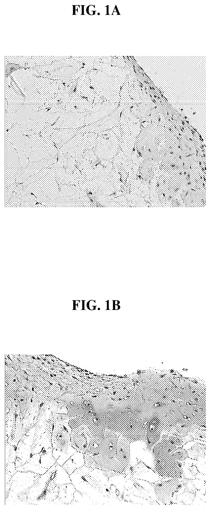
FIG. 1A is photomicrograph that demonstrates that cells in a cartilage tissue sample migrate extensively into a polymer scaffold;
FIG. 1B is a photomicrograph that demonstrates that the migrating cells of FIG. 1A retain their phenotype and the migrating cells produce cellular matrix that stains positive for sulfated glycosaminoglycan using the Safranin O stain;
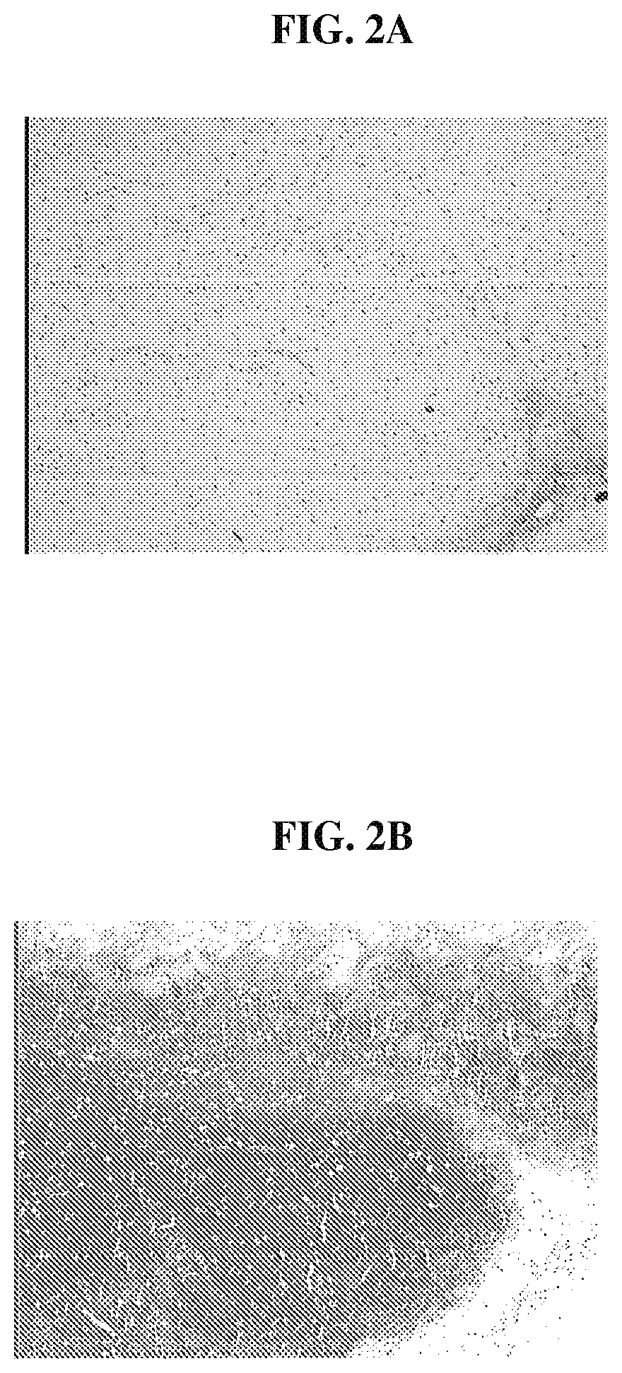
FIG. 2A is a photomicrograph that demonstrates that cells within the minced tissue loaded on the biocompatible scaffolds, following implantation into SCID mice, have proliferated and filled the entire scaffold;
FIG. 2B is a photomicrograph that demonstrates that cells within the minced tissue, following implantation into SCID mice, are chondrocyte-like and are surrounded by an abundant matrix that stains positive for Safranin O;
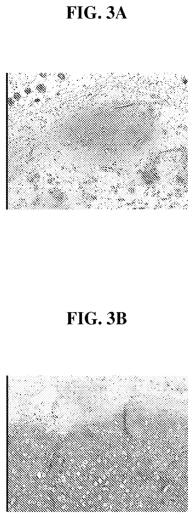
FIG. 3A is a photomicrograph that illustrates a scaffold loaded with minced tissue;
FIG. 3B is a photomicrograph that illustrates a scaffold loaded with minced tissue and platelet rich plasma (PRP) and demonstrates that growth factors in the PRP arc beneficial in promoting the migration of chondrocyte cells from the minced tissue and in promoting maintenance of differentiated phenotype of the chondrocyte cells within the scaffolds;
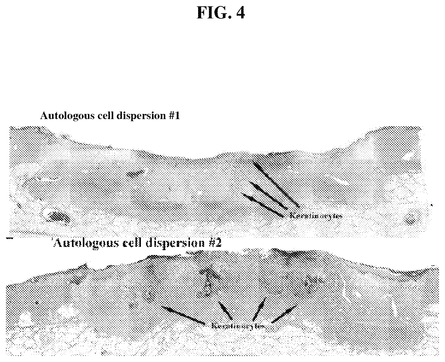
FIG. 4 is a photomicrograph that demonstrates that autologous cell dispersion (derived from skin) is present histologically as keratinocyte islands;
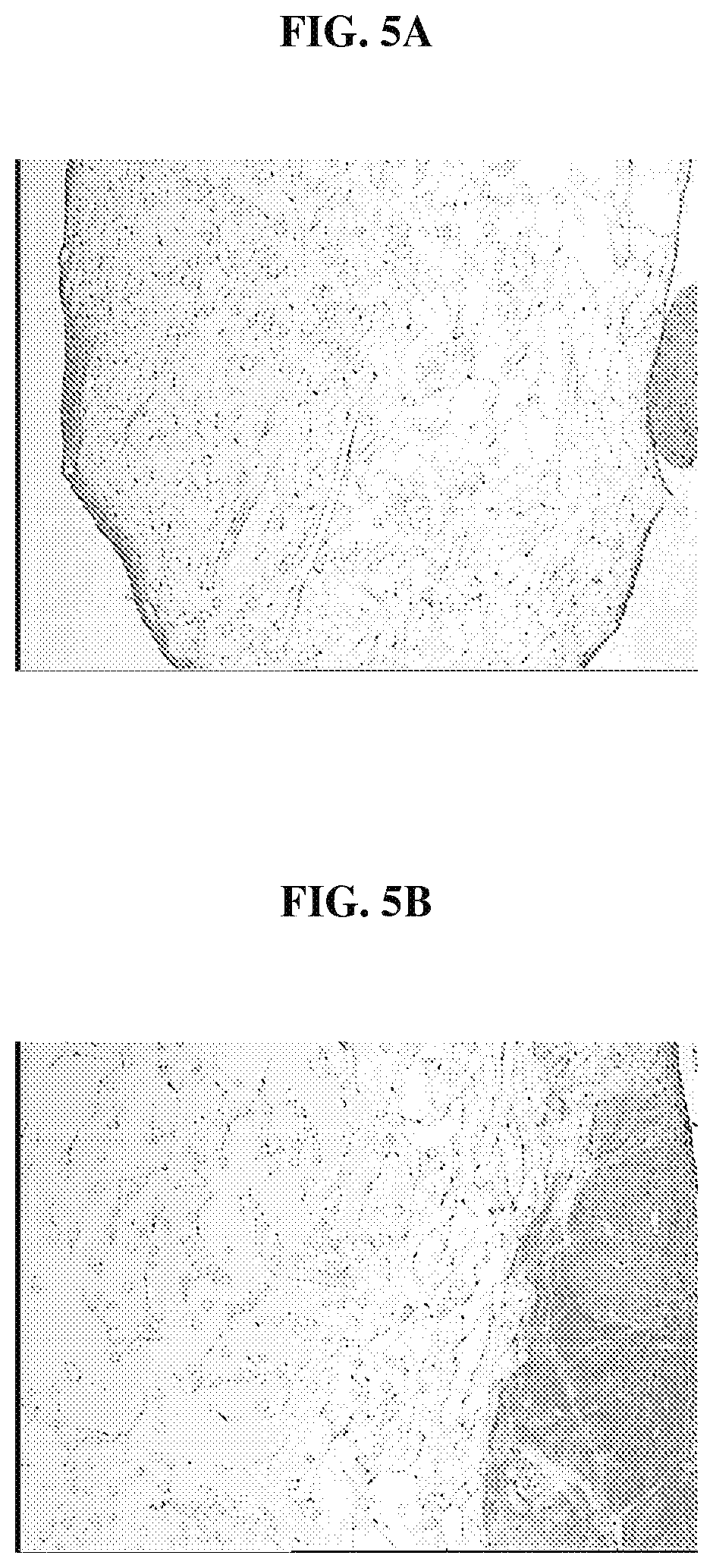
FIG. 5A is a photomicrograph that demonstrates the extensive migration of cells into the polymer scaffolds after incubating for 6 weeks in culture the biocompatible scaffolds having minced anterior cruciate tissue fragments that have been treated with collagenase;
FIG. 5B is a photomicrograph that demonstrates the extensive migration of cells into the polymer scaffolds after incubating for 6 weeks in culture the biocompatible scaffolds having minced anterior cruciate tissue fragments treated without collagenase;
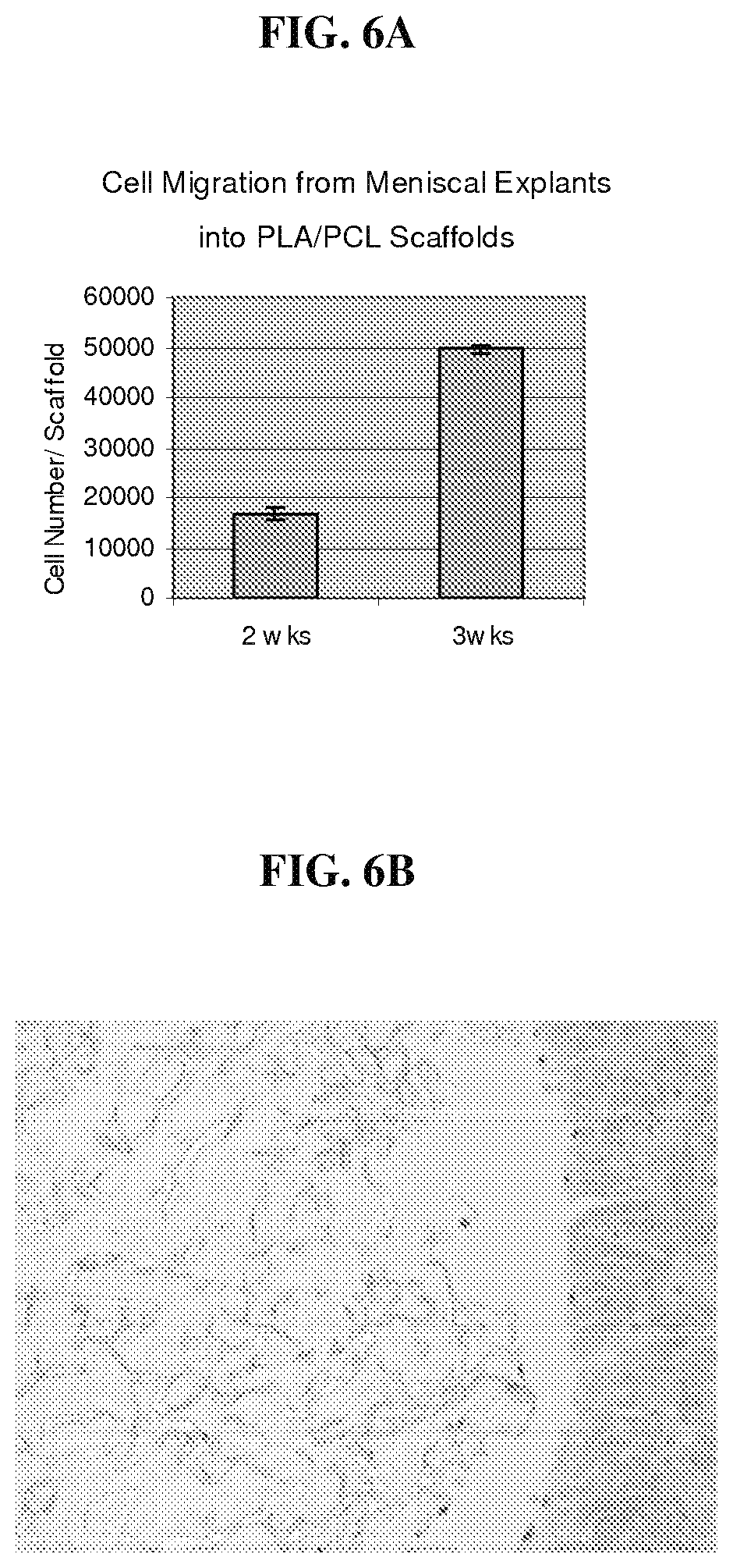
FIG. 6A is a graph that demonstrates that cells in a meniscal explant sample migrate extensively into a polymer scaffold;
FIG. 6B is a photomicrograph that illustrates the histology of cross sections of the associated meniscal explant and biocompatible scaffolds, which demonstrates that cells in the meniscal explant sample migrate into the polymer scaffold.
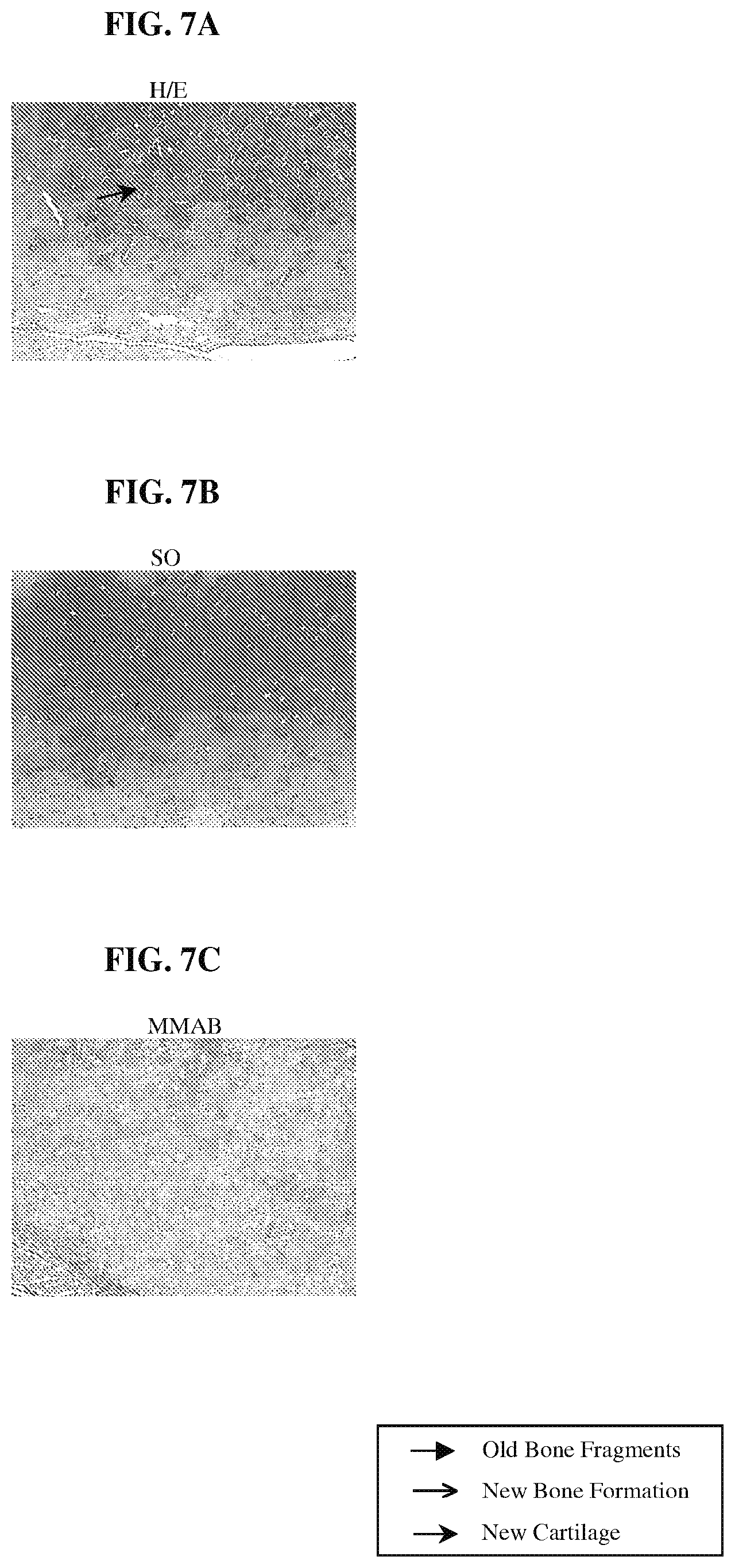
FIGS. 7A-7C are photomicrographs of histological sections of explant samples obtained following the procedure of Example 7, demonstrating the distribution and nature of tissue formed within a scaffold and grown from minced cartilage tissue fragments.
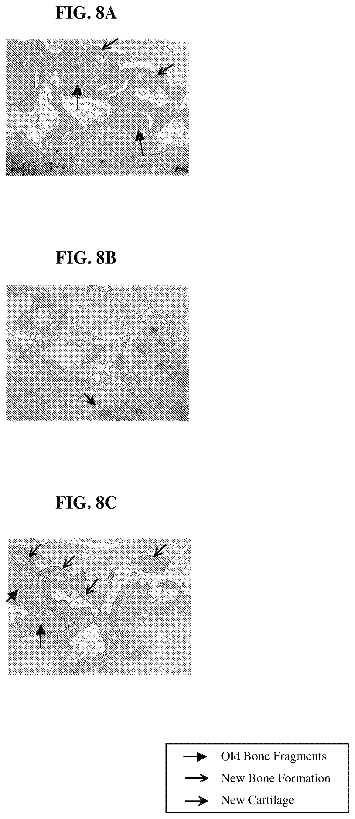
FIGS. 8A-8C are photomicrographs of histological sections of explant samples obtained following the procedure of Example 7, demonstrating the distribution and nature of tissue formed within a scaffold and grown from bone cartilage paste.
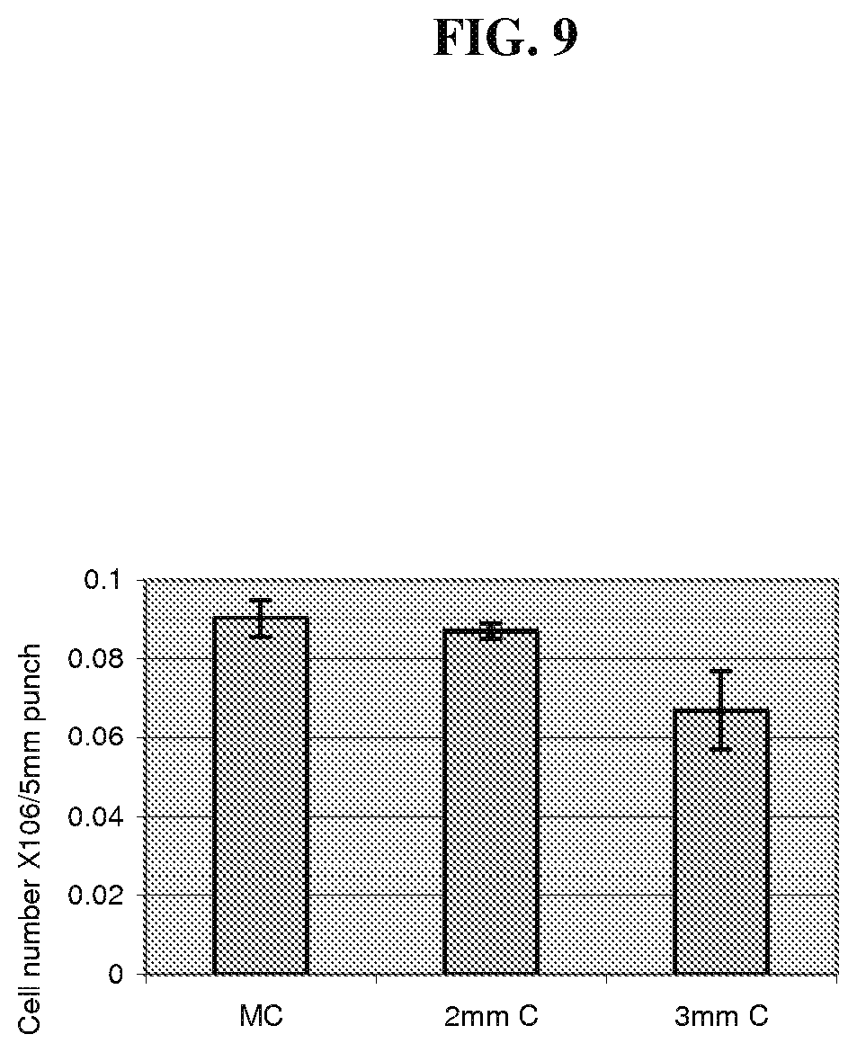
FIG. 9 is a graph comparing the numbers of cells obtained for different sizes of minced cartilage tissue fragments.
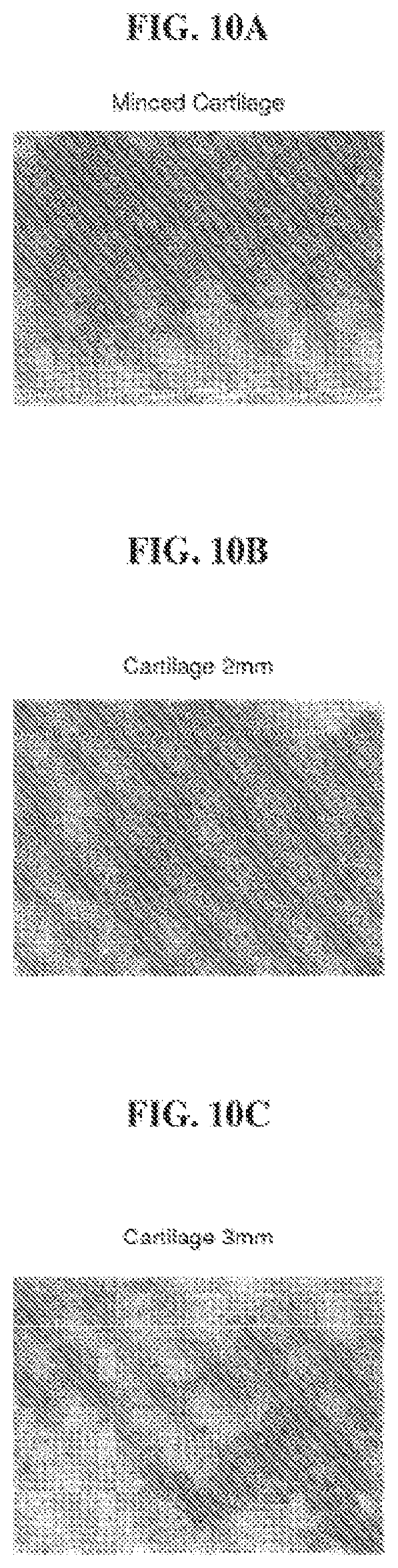
FIGS. 10A-10C are photomicrographs of histological sections of explant samples obtained following the procedure of Example 8, demonstrating the uniformity of the cartilage-like tissue obtained with minced cartilage tissue fragments of different sizes.
DETAILED DESCRIPTION OF THE INVENTION
The biocompatible tissue implants of the present invention are used in the treatment of various types of tissue for various purposes. For example, the implants can be used for the repair and/or regeneration of diseased or damaged tissue, or they can be used for tissue bulking, tissue augmentation, cosmetic treatments, therapeutic treatments, and for tissue sealing. The tissue implants include a biocompatible scaffold and a suspension of minced tissue having at least one minced tissue fragment, wherein the minced tissue suspension is associated with the scaffold. The minced tissue in the suspension of the present invention includes at least one viable cell that can migrate from the tissue fragment and onto the scaffold.
Although the implants are sometimes referred to herein as "tissue repair implants" and the methods of using the implants are sometimes characterized as tissue repair techniques, it is understood that the implants can be used for a variety of tissue treatments, including but not limited to tissue repair, tissue bulking, cosmetic treatments, therapeutic treatments, tissue augmentation, and tissue sealing.
The biocompatible tissue implant of the present invention includes a biocompatible scaffold having at least a portion in contact with the minced tissue suspension. The minced tissue suspension can be disposed on the outer surface of the scaffold, on an inner region of the scaffold, and any combination thereof, or alternatively, the entire scaffold can be in contact with the minced tissue suspension. The scaffold can be formed using virtually any material or delivery vehicle that is biocompatible, bioimplantable, easily sterilized and that has sufficient structural integrity and physical and/or mechanical properties to effectively provide for ease of handling in an operating room environment and to permit it to accept and retain sutures or other fasteners without substantially tearing. Alternatively, the scaffold could be in the form of an injectable gel that would set in place at the defect site. Sufficient strength and physical properties are developed in the scaffold through the selection of materials used to form the scaffold, and the manufacturing process. Preferably, the scaffold is also pliable so as to allow the scaffold to adjust to the dimensions of the target site of implantation. In some embodiments, the scaffold can be a bioresorbable or bioabsorbable material.
In one embodiment of the present invention, the scaffold can be formed from a biocompatible polymer. A variety of biocompatible polymers can be used to make the biocompatible tissue implants or scaffold devices according to the present invention. The biocompatible polymers can be synthetic polymers, natural polymers or combinations thereof. As used herein the term "synthetic polymer" refers to polymers that are not found in nature, even if the polymers are made from naturally occurring biomaterials. The term "natural polymer" refers to polymers that are naturally occurring. In embodiments where the scaffold includes at least one synthetic polymer, suitable biocompatible synthetic polymers can include polymers selected from the group consisting of aliphatic polyesters, poly(amino acids), poly(propylene fumarate), copoly(ether-esters), polyalkylenes oxalates, polyamides, tyrosine derived polycarbonates, poly(iminocarbonates), polyorthoesters, polyoxaesters, polyamidoesters, polyoxaesters containing amine groups, poly(anhydrides), polyphosphazenes, and blends thereof. Suitable synthetic polymers for use in the present invention can also include biosynthetic polymers based on sequences found in collagen, elastin, thrombin, fibronectin, starches, poly(amino acid), gelatin, alginate, pectin, fibrin, oxidized cellulose, chitin, chitosan, tropoelastin, hyaluronic acid, ribonucleic acids, deoxyribonucleic acids, polypeptides, proteins, polysaccharides, polynucleotides and combinations thereof.
For the purpose of this invention aliphatic polyesters include, but are not limited to, homopolymers and copolymers of lactide (which includes lactic acid, D-,L- and meso lactide); glycolide (including glycolic acid); .epsilon.-caprolactone; p-dioxanone (1,4-dioxan-2-one); trimethylene carbonate (1,3-dioxan-2-one); alkyl derivatives of trimethylene carbonate; .delta.-valerolactone; .beta.-butyrolactone; .gamma.-butyrolactone; .epsilon.-decalactone; hydroxybutyrate; hydroxyvalerate; 1,4-dioxepan-2-one (including its dimer 1,5,8,12-tetraoxacyclotetradecane-7,14-dione); 1,5-dioxepan-2-one; 6,6-dimethyl-1,4-dioxan-2-one; 2,5-diketomorpholine; pivalolactone; .alpha., .alpha. diethylpropiolactone; ethylene carbonate; ethylene oxalate; 3-methyl-1,4-dioxane-2,5-dione; 3,3-diethyl-1,4-dioxan-2,5-dione; 6,6-dimethyl-dioxepan-2-one; 6,8-dioxabicycloctane-7-one and polymer blends thereof. Aliphatic polyesters used in the present invention can be homopolymers or copolymers (random, block, segmented, tapered blocks, graft, triblock, etc.) having a linear, branched or star structure. Poly(iminocarbonates), for the purpose of this invention, are understood to include those polymers as described by Kemnitzer and Kohn, in the Handbook of Biodegradable Polymers, edited by Domb, et. al., Hardwood Academic Press, pp. 251-272 (1997). Copoly(ether-esters), for the purpose of this invention, are understood to include those copolyester-ethers as described in the Journal of Biomaterials Research, Vol. 22, pages 993-1009, 1988 by Cohn and Younes, and in Polymer Preprints (ACS Division of Polymer Chemistry), Vol. 30(1), page 498, 1989 by Cohn (e.g., PEO/PLA). Polyalkylene oxalates, for the purpose of this invention, include those described in U.S. Pat. Nos. 4,208,511; 4,141,087; 4,130,639; 4,140,678; 4,105,034; and 4,205,399. Polyphosphazencs, co-, ter- and higher order mixed monomer based polymers made from L-lactide, D,L-lactide, lactic acid, glycolide, glycolic acid, para-dioxanone, trimethylene carbonate and .epsilon.-caprolactone such as are described by Allcock in The Encyclopedia of Polymer Science, Vol. 13, pages 31-41, Wiley Intersciences, John Wiley & Sons, 1988 and by Vandorpe, et al in the Handbook of Biodegradable Polymers, edited by Domb, et al., Hardwood Academic Press, pp. 161-182 (1997). Polyanhydrides include those derived from diacids of the form HOOC--C.sub.6H.sub.4--O--(CH.sub.2).sub.m--O--C.sub.6H.sub.4--COOH, where "m" is an integer in the range of from 2 to 8, and copolymers thereof with aliphatic alpha-omega diacids of up to 12 carbons. Polyoxaesters, polyoxaamides and polyoxaesters containing amines and/or amido groups are described in one or more of the following U.S. Pat. Nos. 5,464,929; 5,595,751; 5,597,579; 5,607,687; 5,618,552; 5,620,698; 5,645,850; 5,648,088; 5,698,213; 5,700,583; and 5,859,150. Polyorthoesters such as those described by Heller in Handbook of Biodegradable Polymers, edited by Domb, et al., Hardwood Academic Press, pp. 99-118 (1997).
As used herein, the term "glycolide" is understood to include polyglycolic acid. Further, the term "lactide" is understood to include L-lactide, D-lactide, blends thereof, and lactic acid polymers and copolymers.
Elastomeric copolymers are also particularly useful in the present invention. Suitable elastomeric polymers include those with an inherent viscosity in the range of about 1.2 dL/g to 4 dL/g, more preferably about 1.2 dL/g to 2 dL/g and most preferably about 1.4 dL/g to 2 dL/g as determined at 25.degree. C. in a 0.1 gram per deciliter (g/dL) solution of polymer in hexafluoroisopropanol (HFIP). Further, suitable elastomers exhibit a high percent elongation and a low modulus, while possessing good tensile strength and good recovery characteristics. In the preferred embodiments of this invention, the elastomer exhibits a percent elongation greater than about 200 percent and preferably greater than about 500 percent. In addition to these elongation and modulus properties, suitable elastomers should also have a tensile strength greater than about 500 psi, preferably greater than about 1,000 psi, and a tear strength of greater than about 50 lbs/inch, preferably greater than about 80 lbs/inch.
Exemplary biocompatible elastomers that can be used in the present invention include, but are not limited to, elastomeric copolymers of c-caprolactone and glycolide (including polyglycolic acid) with a mole ratio of c-caprolactone to glycolide of from about 35:65 to about 65:35, more preferably from 45:55 to 35:65; elastomeric copolymers of .epsilon.-caprolactone and lactide (including L-lactide, D-lactide, blends thereof, and lactic acid polymers and copolymers) where the mole ratio of .epsilon.-caprolactone to lactide is from about 35:65 to about 65:35 and more preferably from 45:55 to 30:70 or from about 95:5 to about 85:15; elastomeric copolymers of p-dioxanone (1,4-dioxan-2-one) and lactide (including L-lactide, D-lactide, blends thereof, and lactic acid polymers and copolymers) where the mole ratio of .epsilon.-dioxanone to lactide is from about 40:60 to about 60:40; elastomeric copolymers of c-caprolactone and p-dioxanone where the mole ratio of c-caprolactone to p-dioxanone is from about from 30:70 to about 70:30; elastomeric copolymers of p-dioxanonc and trimethylene carbonate where the mole ratio of p-dioxanone to trimethylene carbonate is from about 30:70 to about 70:30; elastomeric copolymers of trimethylene carbonate and glycolide (including polyglycolic acid) where the mole ratio of trimethylene carbonate to glycolide is from about 30:70 to about 70:30; elastomeric copolymers of trimethylene carbonate and lactide (including L-lactide, D-lactide, blends thereof, and lactic acid polymers and copolymers) where the mole ratio of trimethylene carbonate to lactide is from about 30:70 to about 70:30; and blends thereof. Examples of suitable biocompatible elastomers are described in U.S. Pat. Nos. 4,045,418; 4,057,537 and 5,468,253.
In one embodiment, the elastomer is a copolymer of 3565 .epsilon.-caprolactone and glycolide, formed in a dioxane solvent and including a polydioxanone mesh. In another embodiment, the elastomer is a copolymer of 40:60 .epsilon.-caprolactone and lactide with a polydioxanone mesh. In yet another embodiment, the elastomer is a 50:50 blend of a 35:65 copolymer of .epsilon.-caprolactone and glycolide and 40:60 copolymer of .epsilon.-caprolactone and lactide. The polydioxanone mesh may be in the form of a one layer thick two-dimensional mesh or a multi-layer thick three-dimensional mesh.
The scaffold of the present invention can, optionally, be formed from a bioresorbable or bioabsorbable material that has the ability to resorb in a timely fashion in the body environment. The differences in the absorption time under in vivo conditions can also be the basis for combining two different copolymers when forming the scaffolds of the present invention. For example, a copolymer of 35:65 .epsilon.-caprolactone and glycolide (a relatively fast absorbing polymer) can be blended with 40:60 .epsilon.-caprolactone and L-lactide copolymer (a relatively slow absorbing polymer) to form a biocompatible scaffold. Depending upon the processing technique used, the two constituents can be either randomly inter-connected bicontinuous phases, or the constituents could have a gradient-like architecture in the form of a laminate type composite with a well integrated interface between the two constituent layers. The microstructure of these scaffolds can be optimized to regenerate or repair the desired anatomical features of the tissue that is being regrown.
In one embodiment, it is desirable to use polymer blends to form scaffolds which transition from one composition to another composition in a gradient-like architecture. Scaffolds having this gradient-like architecture are particularly advantageous in tissue engineering applications to repair or regenerate the structure of naturally occurring tissue such as cartilage (articular, meniscal, septal, tracheal, auricular, costal, etc.), tendon, ligament, nerve, esophagus, skin, bone, and vascular tissue. For example, by blending an elastomer of e-caprolactone-co-glycolide with c-caprolactone-co-lactide (e.g., with a mole ratio of about 5:95) a scaffold may be formed that transitions from a softer spongy material to a stiffer more rigid material, for example, in a manner similar to the transition from cartilage to bone. Clearly, one of ordinary skill in the art will appreciate that other polymer blends may be used for similar gradient effects, or to provide different gradients (e.g., different absorption profiles, stress response profiles, or different degrees of elasticity). For example, such design features can establish a concentration gradient for the suspension of minced tissue associated with the scaffolds of the present invention, such that a higher concentration of the tissue fragments is present in one region of the implant (e.g., an interior portion) than in another region (e.g., outer portions).
The biocompatible scaffold of the tissue repair implant of the present invention can also include a reinforcing material comprised of any absorbable or non-absorbable textile having, for example, woven, knitted, warped knitted (i.e., lace-like), non-woven, and braided structures. In one embodiment, the reinforcing material has a mesh-like structure. In any of the above structures, mechanical properties of the material can be altered by changing the density or texture of the material, the type of knit or weave of the material, the thickness of the material, or by embedding particles in the material. The mechanical properties of the material may also be altered by creating sites within the mesh where the fibers are physically bonded with each other or physically bonded with another agent, such as, for example, an adhesive or a polymer. The fibers used to make the reinforcing component can be monofilaments, yarns, threads, braids, or bundles of fibers. These fibers can be made of any biocompatible material including bioabsorbable materials such as polylactic acid (PLA), polyglycolic acid (PGA), polycaprolactone (PCL), polydioxanone (PDO), trimethylene carbonate (TMC), copolymers or blends thereof. These fibers can also be made from any biocompatible materials based on natural polymers including silk and collagen-based materials. These fibers can also be made of any biocompatible fiber that is nonresorbable, such as, for example, polyethylene, polyethylene terephthalate, poly(tetrafluoroethylene), polycarbonate, polypropylene and poly(vinyl alcohol). In one embodiment, the fibers are formed from 95:5 copolymer of lactide and glycolide.
In another embodiment, the fibers that form the reinforcing material can be made of a bioabsorbable glass. Bioglass, a silicate containing calcium phosphate glass, or calcium phosphate glass with varying amounts of solid particles added to control resorption time are examples of materials that could be spun into glass fibers and used for the reinforcing material. Suitable solid particles that may be added include iron, magnesium, sodium, potassium, and combinations thereof.
The biocompatible scaffolds as well as the reinforcing material may also be formed from a thin, perforation-containing elastomeric sheet with pores or perforations to allow tissue ingrowth. Such a sheet could be made of blends or copolymers of polylactic acid (PLA), polyglycolic acid (PGA), polycaprolactone (PCL), and polydioxanone (PDO).
In one embodiment, filaments that form the biocompatible scaffolds or the reinforcing material may be co-extruded to produce a filament with a sheath/core construction. Such filaments are comprised of a sheath of biodegradable polymer that surrounds one or more cores comprised of another biodegradable polymer. Filaments with a fast-absorbing sheath surrounding a slower-absorbing core may be desirable in instances where extended support is necessary for tissue ingrowth.
One of ordinary skill in the art will appreciate that one or more layers of the reinforcing material may be used to reinforce the tissue implant of the invention. In addition, biodegradable textile scaffolds, such as, for example, meshes, of the same structure and chemistry or different structures and chemistries can be overlaid on top of one another to fabricate biocompatible tissue implants with superior mechanical strength.
In embodiments where the scaffold includes at least one natural polymer, suitable examples of natural polymers include, but are not limited to, fibrin-based materials, collagen-based materials, hyaluronic acid-based materials, glycoprotein-based materials, cellulose-based materials, silks and combinations thereof. By way of nonlimiting example, the biocompatible scaffold can be constructed from a collagen-based small intestine submucosa.
In another embodiment of the present invention, the biocompatible scaffold can be formed from a biocompatible ceramic material. Suitable biocompatible ceramic materials include, for example, hydroxyapatite, .alpha.-tricalcium phosphate, .beta.-tricalcium phosphate, bioactive glass, calcium phosphate, calcium sulfate, calcium carbonate, xenogeneic and allogeneic bone material and combinations thereof. Suitable bioactive glass materials for use in the present invention include silicates containing calcium phosphate glass, or calcium phosphate glass with varying amounts of solid particles added to control resorption time. Suitable compounds that may be incorporated into the calcium phosphate bioactive glass include, but are not limited to, magnesium oxide, sodium oxide, potassium oxide, and combinations thereof.
In yet another embodiment of the tissue implants of the present invention, the scaffold can be formed using tissue grafts, such as may be obtained from autogeneic tissue, allogeneic tissue and xenogeneic tissue. By way of non-limiting example, tissues such as skin, cartilage, ligament, tendon, periosteum, perichondrium, synovium, fascia, mesenter and sinew can be used as tissue grafts to form the biocompatible scaffold. In some embodiments where an allogeneic tissue is used, tissue from a fetus or newborns can be used to avoid the immunogenicity associated with some adult tissues.
In another embodiment, the scaffold could be in the form of an injectable gel that would set in place at the defect site. The gel can be a biological or synthetic hydrogel, including alginate, cross-linked alginate, hyaluronic acid, collagen gel, fibrin glue, fibrin clot, poly(N-isopropylacrylamide), agarose, chitin, chitosan, cellulose, polysaccharides, poly(oxyalkylene), a copolymer of poly(ethylene oxide)-poly(propylene oxide), poly(vinyl alcohol), polyacrylate, platelet rich plasma (PRP) clot, platelet poor plasma (PPP) clot, Matrigel, or blends thereof.
In still yet another embodiment of the tissue implants, the scaffold can be formed from a polymeric foam component having pores with an open cell pore structure. The pore size can vary, but preferably, the pores are sized to allow tissue ingrowth. More preferably, the pore size is in the range of about 50 to 1000 microns, and even more preferably, in the range of about 50 to 500 microns. The polymeric foam component can, optionally, contain a reinforcing component, such as for example, the textiles disclosed above. In some embodiments where the polymeric foam component contains a reinforcing component, the foam component can be integrated with the reinforcing component such that the pores of the foam component penetrate the mesh of the reinforcing component and interlock with the reinforcing component.
The foam component of the tissue implant may be formed as a foam by a variety of techniques well known to those having ordinary skill in the art. For example, the polymeric starting materials may be foamed by lyophilization, supercritical solvent foaming (i.e., as described in EP 464,163), gas injection extrusion, gas injection molding or casting with an extractable material (e.g., salts, sugar or similar suitable materials).
In one embodiment, the foam component of the engineered tissue repair implant devices of the present invention may be made by a polymer-solvent phase separation technique, such as lyophilization. Generally, however, a polymer solution can be separated into two phases by any one of the four techniques: (a) thermally induced gelation/crystallization; (b) non-solvent induced separation of solvent and polymer phases; (c) chemically induced phase separation, and (d) thermally induced spinodal decomposition. The polymer solution is separated in a controlled manner into either two distinct phases or two bicontinuous phases. Subsequent removal of the solvent phase usually leaves a porous structure with a density less than the bulk polymer and pores in the micrometer ranges. See Microcellular Foams Via Phase Separation, J. Vac. Sci. Technol., A. T. Young, Vol. 4(3), May/June 1986.
The steps involved in the preparation of these foams include choosing the right solvents for the polymers to be lyophilized and preparing a homogeneous solution. Next, the polymer solution is subjected to a freezing and vacuum drying cycle. The freezing step phase separates the polymer solution and vacuum drying step removes the solvent by sublimation and/or drying, leaving a porous polymer structure or an interconnected open cell porous foam.
Suitable solvents that may be used in the preparation of the foam component include, but are not limited to, formic acid, ethyl formate, acetic acid, hexafluoroisopropanol (HFIP), cyclic ethers (e.g., tetrahydrofuran (THF), dimethylene fluoride (DMF), and polydioxanone (PDO)), acetone, acetates of C2 to C5 alcohols (e.g., ethyl acetate and t-butylacetate), glyme (e.g., monoglyme, ethyl glyme, diglyme, ethyl diglyme, triglyme, butyl diglyme and tetraglyme), methylethyl ketone, dipropyleneglycol methyl ether, lactones (e.g., .gamma.-valerolactone, .delta.-valerolactone, .beta.-butyrolactone, .gamma.-butyrolactone), 1,4-dioxane, 1,3-dioxolane, 1,3-dioxolane-2-one (ethylene carbonate), dimethlycarbonate, benzene, toluene, benzyl alcohol, p-xylene, naphthalene, tetrahydrofuran, N-methyl pyrrolidone, dimethylformamide, chloroform, 1,2-dichloromethane, morpholine, dimethylsulfoxide, hexafluoroacetone sesquihydrate (HFAS), anisole and mixtures thereof. Among these solvents, a preferred solvent is 1,4-dioxane. A homogeneous solution of the polymer in the solvent is prepared using standard techniques.
The applicable polymer concentration or amount of solvent that may be utilized will vary with each system. Generally, the amount of polymer in the solution can vary from about 0.5% to about 90% and, preferably, will vary from about 0.5% to about 30% by weight, depending on factors such as the solubility of the polymer in a given solvent and the final properties desired in the foam.
In one embodiment, solids may be added to the polymer-solvent system to modify the composition of the resulting foam surfaces. As the added particles settle out of solution to the bottom surface, regions will be created that will have the composition of the added solids, not the foamed polymeric material. Alternatively, the added solids may be more concentrated in desired regions (i.e., near the top, sides, or bottom) of the resulting tissue implant, thus causing compositional changes in all such regions. For example, concentration of solids in selected locations can be accomplished by adding metallic solids to a solution placed in a mold made of a magnetic material (or vice versa).
A variety of types of solids can be added to the polymer-solvent system. Preferably, the solids are of a type that will not react with the polymer or the solvent. Generally, the added solids have an average diameter of less than about 1.0 mm and preferably will have an average diameter of about 50 to about 500 microns. Preferably, the solids are present in an amount such that they will constitute from about 1 to about 50 volume percent of the total volume of the particle and polymer-solvent mixture (wherein the total volume percent equals 100 volume percent).
Exemplary solids include, but are not limited to, particles of demineralized bone, calcium phosphate particles, bioglass particles, calcium sulfate, or calcium carbonate particles for bone repair, leachable solids for pore creation and particles of bioabsorbablc polymers not soluble in the solvent system that are effective as reinforcing materials or to create pores as they are absorbed, and non-bioabsorbable materials.
Suitable leachable solids include nontoxic leachable materials such as salts (e.g., sodium chloride, potassium chloride, calcium chloride, sodium tartrate, sodium citrate, and the like), biocompatible mono and disaccharides (e.g., glucose, fructose, dextrose, maltose, lactose and sucrose), polysaccharides (e.g., starch, alginate, chitosan), water soluble proteins (e.g., gelatin and agarose). The leachable materials can be removed by immersing the foam with the leachable material in a solvent in which the particle is soluble for a sufficient amount of time to allow leaching of substantially all of the particles, but which does not dissolve or detrimentally alter the foam. The preferred extraction solvent is water, most preferably distilled-deionized water. Such a process is described in U.S. Pat. No. 5,514,378. Preferably the foam will be dried after the leaching process is complete at low temperature and/or vacuum to minimize hydrolysis of the foam unless accelerated absorption of the foam is desired.
Suitable non-bioabsorbable materials include biocompatible metals such as stainless steel, cobalt chrome, titanium and titanium alloys, and bioinert ceramic particles (e.g., alumina, zirconia, and calcium sulfate particles). Further, the non-bioabsorbable materials may include polymers such as polyethylene, polyvinylacetate, polymethylmethacrylate, polypropylene, poly(ethylene terephthalate), silicone, polyethylene oxide, polyethylene glycol, polyurethanes, polyvinyl alcohol, natural polymers (e.g., cellulose particles, chitin, and keratin), and fluorinated polymers and copolymers (e.g., polyvinylidene fluoride, polytetrafluoroethylene, and hexafluoropropylene).
It is also possible to add solids (e.g., barium sulfate) that will render the tissue implants radio opaque. The solids that may be added also include those that will promote tissue regeneration or regrowth, as well as those that act as buffers, reinforcing materials or porosity modifiers.
As noted above, porous, reinforced tissue repair implant devices of the present invention are made by injecting, pouring, or otherwise placing, the appropriate polymer solution into a mold set-up comprised of a mold and the reinforcing elements of the present invention. The mold set-up is cooled in an appropriate bath or on a refrigerated shelf and then lyophilized, thereby providing a reinforced scaffold. A biological component can be added either before or after the lyophilization step. In the course of forming the foam component, it is believed to be important to control the rate of freezing of the polymer-solvent system. The type of pore morphology that is developed during the freezing step is a function of factors such as the solution thermodynamics, freezing rate, temperature to which it is cooled, concentration of the solution, and whether homogeneous or heterogenous nucleation occurs. One of ordinary skill in the art can readily optimize the parameters without undue experimentation.
The required general processing steps include the selection of the appropriate materials from which the polymeric foam and the reinforcing components are made. If a mesh reinforcing material is used, the proper mesh density must be selected. Further, the reinforcing material must be properly aligned in the mold, the polymer solution must be added at an appropriate rate and, preferably, into a mold that is tilted at an appropriate angle to avoid the formation of air bubbles, and the polymer solution must be lyophilized.
In embodiments that utilize a mesh reinforcing material, the reinforcing mesh has to be of a certain density. That is, the openings in the mesh material must be sufficiently small to render the construct sutureable or otherwise fastenable, but not so small as to impede proper bonding between the foam and the reinforcing mesh as the foam material and the open cells and cell walls thereof penetrate the mesh openings. Without proper bonding the integrity of the layered structure is compromised leaving the construct fragile and difficult to handle. Because the density of the mesh determines the mechanical strength of the construct, the density of the mesh can vary according to the desired use for tissue repair. In addition, the type of weave used in the mesh can determine the directionality of the mechanical strength of the construct, as well as the mechanical properties of the reinforcing material, such as for example, the elasticity, stiffness, burst strength, suture retention strength and ultimate tensile strength of the construct. By way of non-limiting example, the mesh reinforcing material in a foam-based biocompatible scaffold of the present invention can be designed to be stiff in one direction, yet elastic in another, or alternatively, the mesh reinforcing material can be made isotropic.
During the lyophilization of the reinforced foam, several parameters and procedures are important to produce implants with the desired integrity and mechanical properties. Preferably, the reinforcement material is substantially flat when placed in the mold. To ensure the proper degree of flatness, the reinforcement (e.g., mesh) is pressed flat using a heated press prior to its placement within the mold. Further, in the event that reinforcing structures are not isotropic it is desirable to indicate this anisotropy by marking the construct to indicate directionality. This can be accomplished by embedding one or more indicators, such as dyed markings or dyed threads, within the woven reinforcements. The direction or orientation of the indicator will indicate to a surgeon the dimension of the implant in which physical properties are superior.
As noted above, the manner in which the polymer solution is added to the mold prior to lyophilization helps contribute to the creation of a tissue implant with adequate mechanical integrity. Assuming that a mesh reinforcing material will be used, and that it will be positioned between two thin (e.g., 0.75 mm) shims it should be positioned in a substantially flat orientation at a desired depth in the mold. The polymer solution is poured in a way that allows air bubbles to escape from between the layers of the foam component. Preferably, the mold is tilted at a desired angle and pouring is effected at a controlled rate to best prevent bubble formation. One of ordinary skill in the art will appreciate that a number of variables will control the tilt angle and pour rate. Generally, the mold should be tilted at an angle of greater than about 1 degree to avoid bubble formation. In addition, the rate of pouring should be slow enough to enable any air bubbles to escape from the mold, rather than to be trapped in the mold.
If a mesh material is used as the reinforcing c
D00001
D00002
D00003
D00004
D00005
D00006
D00007
D00008
D00009
D00010
XML
uspto.report is an independent third-party trademark research tool that is not affiliated, endorsed, or sponsored by the United States Patent and Trademark Office (USPTO) or any other governmental organization. The information provided by uspto.report is based on publicly available data at the time of writing and is intended for informational purposes only.
While we strive to provide accurate and up-to-date information, we do not guarantee the accuracy, completeness, reliability, or suitability of the information displayed on this site. The use of this site is at your own risk. Any reliance you place on such information is therefore strictly at your own risk.
All official trademark data, including owner information, should be verified by visiting the official USPTO website at www.uspto.gov. This site is not intended to replace professional legal advice and should not be used as a substitute for consulting with a legal professional who is knowledgeable about trademark law.