Methods of treating a subject with an alkaline phosphatase deficiency
Odrljin
U.S. patent number 10,603,361 [Application Number 15/544,063] was granted by the patent office on 2020-03-31 for methods of treating a subject with an alkaline phosphatase deficiency. This patent grant is currently assigned to Alexion Pharmaceuticals, Inc.. The grantee listed for this patent is Alexion Pharmaceuticals, Inc.. Invention is credited to Tatjana Odrljin.


| United States Patent | 10,603,361 |
| Odrljin | March 31, 2020 |
Methods of treating a subject with an alkaline phosphatase deficiency
Abstract
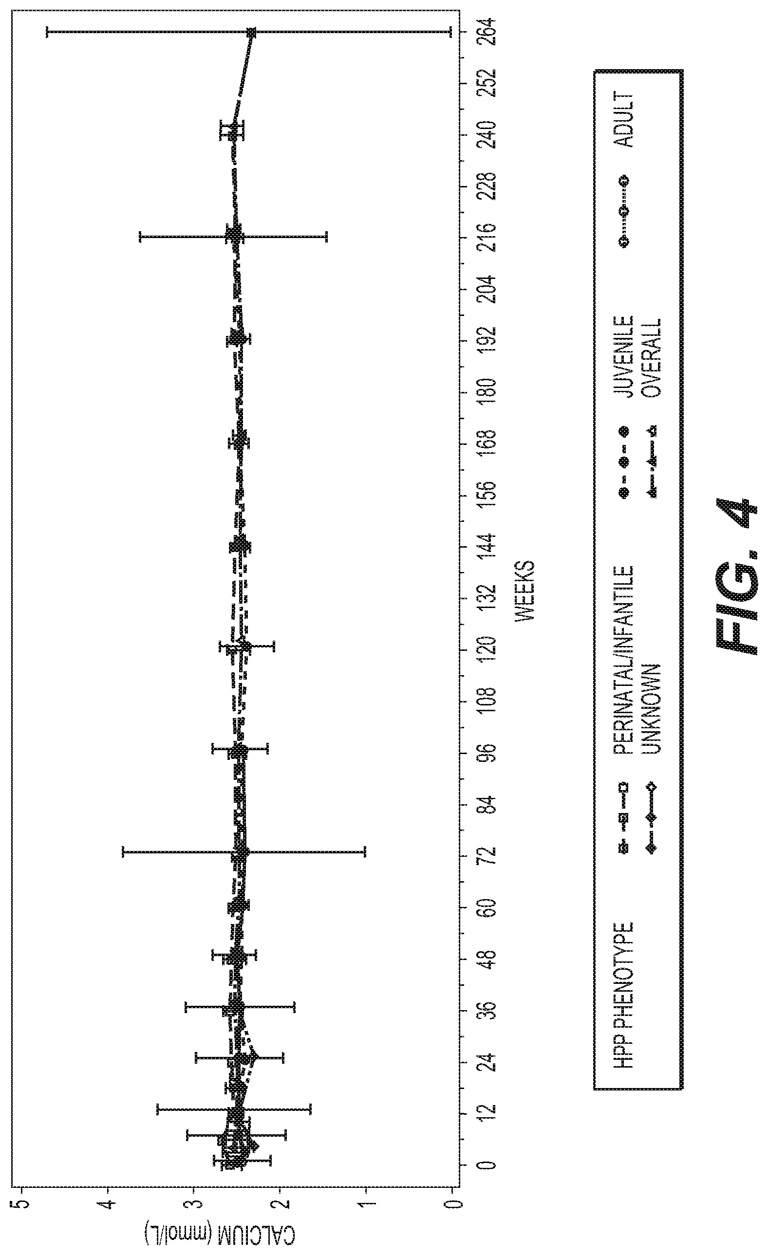
Disclosed herein are methods for treating a subject with an alkaline phosphatase deficiency, further comprising monitoring additional anaiytes, e.g., calcium, parathyroid hormone and/or vitamin D, with treatment modifications as indicated by the levels, e.g., serum levels, of the additional anaiytes.
| Inventors: | Odrljin; Tatjana (Brookline, MA) | ||||||||||
|---|---|---|---|---|---|---|---|---|---|---|---|
| Applicant: |
|
||||||||||
| Assignee: | Alexion Pharmaceuticals, Inc.
(Boston, MA) |
||||||||||
| Family ID: | 55451553 | ||||||||||
| Appl. No.: | 15/544,063 | ||||||||||
| Filed: | January 28, 2016 | ||||||||||
| PCT Filed: | January 28, 2016 | ||||||||||
| PCT No.: | PCT/US2016/015366 | ||||||||||
| 371(c)(1),(2),(4) Date: | July 17, 2017 | ||||||||||
| PCT Pub. No.: | WO2016/123342 | ||||||||||
| PCT Pub. Date: | August 04, 2016 |
Prior Publication Data
| Document Identifier | Publication Date | |
|---|---|---|
| US 20180333470 A1 | Nov 22, 2018 | |
Related U.S. Patent Documents
| Application Number | Filing Date | Patent Number | Issue Date | ||
|---|---|---|---|---|---|
| 62108669 | Jan 28, 2015 | ||||
| Current U.S. Class: | 1/1 |
| Current CPC Class: | A61K 38/46 (20130101); C12Y 301/03001 (20130101); A61K 31/137 (20130101); A61P 3/00 (20180101); A61K 38/465 (20130101); A61K 45/06 (20130101) |
| Current International Class: | A61K 38/46 (20060101); A61K 31/137 (20060101); A61P 3/00 (20060101); A61K 45/06 (20060101) |
References Cited [Referenced By]
U.S. Patent Documents
| 5336759 | August 1994 | Matsuo et al. |
| 5338830 | August 1994 | Matsuo et al. |
| 5340920 | August 1994 | Matsuo et al. |
| 5352770 | October 1994 | Matsuo |
| 5428130 | June 1995 | Capon et al. |
| 5434133 | July 1995 | Tanaka et al. |
| 5583108 | December 1996 | Wei et al. |
| 5665704 | September 1997 | Lowe et al. |
| 5714147 | February 1998 | Capon et al. |
| 5767239 | June 1998 | Immer et al. |
| 5846932 | December 1998 | Lowe et al. |
| 5948761 | September 1999 | Seilhamer et al. |
| 5973134 | October 1999 | Matsuo et al. |
| 6020168 | February 2000 | Matsuo et al. |
| 6028055 | February 2000 | Lowe et al. |
| 6034231 | March 2000 | Tanaka et al. |
| 6290952 | September 2001 | Poelstra et al. |
| 6406697 | June 2002 | Capon et al. |
| 6407211 | June 2002 | Burnett, Jr. et al. |
| 6420384 | July 2002 | Weigele et al. |
| 6436386 | August 2002 | Roberts et al. |
| 6455495 | September 2002 | Orgel et al. |
| 6458579 | October 2002 | Hopwood et al. |
| 6525022 | February 2003 | Lowe et al. |
| 6541610 | April 2003 | Smith |
| 6743425 | June 2004 | Nakao |
| 6790649 | September 2004 | Crine et al. |
| 6818619 | November 2004 | Burnett, Jr. et al. |
| 6830885 | December 2004 | Lanctot et al. |
| 6849714 | February 2005 | Bridon et al. |
| 6887470 | May 2005 | Bridon et al. |
| 6905689 | June 2005 | Schneidinger et al. |
| 6946484 | September 2005 | Adams et al. |
| 7026293 | April 2006 | Kitakaze |
| 7033997 | April 2006 | Forssmann et al. |
| 7070974 | July 2006 | Desgroseillers et al. |
| 7105539 | September 2006 | Gravel et al. |
| 7179903 | February 2007 | McArthur et al. |
| 7256253 | August 2007 | Bridon et al. |
| 7271149 | September 2007 | Glaesner et al. |
| 7276481 | October 2007 | Golembo et al. |
| 7341838 | March 2008 | Buechler et al. |
| 7365091 | April 2008 | Gravel et al. |
| 7384917 | June 2008 | Burnett, Jr. et al. |
| 7399466 | July 2008 | Boileau |
| 7414107 | August 2008 | Larsen |
| 7425531 | September 2008 | Lanctot et al. |
| 7427498 | September 2008 | Crine et al. |
| 7470668 | December 2008 | Lanctot et al. |
| 7488713 | February 2009 | Vesely |
| 7527939 | May 2009 | Davey et al. |
| 7563769 | July 2009 | Bogin et al. |
| 7625564 | December 2009 | Wang et al. |
| 7642243 | January 2010 | Nakao et al. |
| 7648962 | January 2010 | James et al. |
| 7662773 | February 2010 | James et al. |
| 7678391 | March 2010 | Graham et al. |
| 7732406 | June 2010 | Mitrovic et al. |
| 7736653 | June 2010 | Kim et al. |
| 7754852 | July 2010 | Burnett, Jr. et al. |
| 7763712 | July 2010 | Crine et al. |
| 7803769 | September 2010 | Sullivan et al. |
| 7803901 | September 2010 | Burnett, Jr. et al. |
| 7825092 | November 2010 | Vesely |
| 7846900 | December 2010 | Vesely |
| 7858560 | December 2010 | Koster et al. |
| 7919591 | April 2011 | Sheffer et al. |
| 7943126 | May 2011 | Tomatsu et al. |
| 7960529 | June 2011 | Crine et al. |
| 8058242 | November 2011 | Alewood et al. |
| 8691208 | April 2014 | Tomatsu et al. |
| 9266939 | February 2016 | Crine et al. |
| 9988620 | June 2018 | Crine et al. |
| 10000532 | June 2018 | Crine et al. |
| 2002/0183276 | December 2002 | Millan et al. |
| 2003/0158132 | August 2003 | Kovesdi |
| 2004/0023916 | February 2004 | Millan et al. |
| 2004/0077537 | April 2004 | Schreiner |
| 2004/0234518 | November 2004 | Crine et al. |
| 2005/0113286 | May 2005 | Schreiner et al. |
| 2005/0142217 | June 2005 | Adams et al. |
| 2005/0202442 | September 2005 | Morris et al. |
| 2005/0244904 | November 2005 | Ng |
| 2005/0276796 | December 2005 | Tomatsu et al. |
| 2006/0014687 | January 2006 | Crine et al. |
| 2006/0019890 | January 2006 | Kapoun et al. |
| 2006/0074009 | April 2006 | James et al. |
| 2006/0110359 | May 2006 | Sanchez-Ramos et al. |
| 2006/0172929 | August 2006 | Rappold-Hoerbrand et al. |
| 2006/0228710 | October 2006 | Morris et al. |
| 2007/0041972 | February 2007 | Rother et al. |
| 2007/0042957 | February 2007 | Burnett et al. |
| 2007/0081984 | April 2007 | Tomatsu et al. |
| 2007/0081986 | April 2007 | Tomatsu et al. |
| 2007/0197434 | August 2007 | Nakao et al. |
| 2007/0281887 | December 2007 | Pan |
| 2007/0292966 | December 2007 | Prickett et al. |
| 2007/0293418 | December 2007 | Larsen |
| 2008/0032933 | February 2008 | Burnett et al. |
| 2008/0081768 | April 2008 | Watt et al. |
| 2008/0085862 | April 2008 | Kim et al. |
| 2008/0113411 | May 2008 | Sheffer et al. |
| 2008/0113412 | May 2008 | Sheffer et al. |
| 2008/0125574 | May 2008 | Sheffer et al. |
| 2008/0153747 | June 2008 | Alewood et al. |
| 2008/0161243 | July 2008 | Rosen et al. |
| 2008/0181903 | July 2008 | Bhaskar et al. |
| 2008/0182299 | July 2008 | Colocaru et al. |
| 2008/0194481 | August 2008 | Rosen et al. |
| 2008/0194682 | August 2008 | Golembo et al. |
| 2008/0227713 | September 2008 | Protter |
| 2008/0293632 | November 2008 | Rappold-Hoerbrand et al. |
| 2008/0312142 | December 2008 | Nakao et al. |
| 2009/0011997 | January 2009 | Peri et al. |
| 2009/0023652 | January 2009 | Bell |
| 2009/0053192 | February 2009 | Millan |
| 2009/0069243 | March 2009 | Burnett, Jr. et al. |
| 2009/0092582 | April 2009 | Bogin et al. |
| 2009/0142347 | June 2009 | Milian |
| 2009/0170756 | July 2009 | Burnett, Jr. et al. |
| 2009/0221803 | September 2009 | Dall'Acqua et al. |
| 2009/0238814 | September 2009 | Tomatsu et al. |
| 2009/0240031 | September 2009 | Immer et al. |
| 2009/0247462 | October 2009 | Bogin et al. |
| 2009/0252729 | October 2009 | Farrington et al. |
| 2009/0258018 | October 2009 | Medich et al. |
| 2009/0275506 | November 2009 | Bakis et al. |
| 2009/0325195 | December 2009 | Davey et al. |
| 2010/0008979 | January 2010 | Tomatsu et al. |
| 2010/0055150 | March 2010 | Golembo et al. |
| 2010/0093678 | April 2010 | Della-Fera et al. |
| 2010/0160212 | June 2010 | Sheffer et al. |
| 2010/0168443 | July 2010 | Geysen |
| 2010/0184680 | July 2010 | Bevec |
| 2010/0197574 | August 2010 | Chen et al. |
| 2010/0204094 | August 2010 | Simari et al. |
| 2010/0204109 | August 2010 | Bevec |
| 2010/0204446 | August 2010 | Forssmann |
| 2010/0209958 | August 2010 | Nakao et al. |
| 2010/0216714 | August 2010 | James et al. |
| 2010/0221234 | September 2010 | Crine et al. |
| 2010/0240125 | September 2010 | Crine et al. |
| 2010/0249017 | September 2010 | Bevec et al. |
| 2010/0260706 | October 2010 | Bogin et al. |
| 2010/0261248 | October 2010 | Kim et al. |
| 2010/0297021 | November 2010 | Wendt et al. |
| 2010/0297119 | November 2010 | Crine et al. |
| 2010/0305031 | December 2010 | Wakabayashi et al. |
| 2010/0305051 | December 2010 | Burnett, Jr. et al. |
| 2010/0310561 | December 2010 | Canada et al. |
| 2010/0311660 | December 2010 | Simari et al. |
| 2010/0317600 | December 2010 | Immer et al. |
| 2010/0331256 | December 2010 | Wendt et al. |
| 2011/0152194 | June 2011 | Burnett, Jr. et al. |
| 2011/0250187 | October 2011 | Tomatsu et al. |
| 2011/0269684 | November 2011 | Burnett, Jr. et al. |
| 2011/0300143 | December 2011 | Sly et al. |
| 2012/0088771 | April 2012 | Millan |
| 2012/0164142 | June 2012 | Crine et al. |
| 2013/0108635 | May 2013 | Crine et al. |
| 2013/0323244 | December 2013 | Crine |
| 2014/0193388 | July 2014 | Velders et al. |
| 2014/0194484 | July 2014 | Coats et al. |
| 2015/0353633 | December 2015 | Kakkis et al. |
| 2016/0052968 | February 2016 | Crine et al. |
| 2017/0175094 | June 2017 | Hatch |
| 2017/0360899 | December 2017 | Marozsan et al. |
| 2018/0230445 | August 2018 | Jaluria et al. |
| 0478797 | Apr 1995 | EP | |||
| 0769554 | Apr 1997 | EP | |||
| 0771875 | May 1997 | EP | |||
| 0466174 | Jun 1997 | EP | |||
| 0475394 | Jun 1997 | EP | |||
| 0466175 | Jan 1998 | EP | |||
| 0477971 | Jan 1998 | EP | |||
| 0475290 | Dec 1998 | EP | |||
| 0475291 | Dec 1998 | EP | |||
| 0497368 | Jun 2002 | EP | |||
| 1492567 | Sep 2003 | EP | |||
| 1502604 | Feb 2005 | EP | |||
| 1623994 | Feb 2006 | EP | |||
| 1759001 | Mar 2007 | EP | |||
| 1759710 | Mar 2007 | EP | |||
| 2158319 | Mar 2010 | EP | |||
| 2158319 | Dec 2011 | EP | |||
| H0870875 | Mar 1996 | JP | |||
| 2000-327583 | Nov 2000 | JP | |||
| 2002-541776 | Dec 2002 | JP | |||
| 2007-511209 | May 2007 | JP | |||
| 2010-501026 | Jan 2010 | JP | |||
| 2010-526543 | Aug 2010 | JP | |||
| 2010-530222 | Sep 2010 | JP | |||
| 2011-504506 | Feb 2011 | JP | |||
| WO-92/20371 | Nov 1992 | WO | |||
| WO-94/20534 | Sep 1994 | WO | |||
| WO-95/05456 | Feb 1995 | WO | |||
| WO-95/13296 | May 1995 | WO | |||
| WO-95/33769 | Dec 1995 | WO | |||
| WO-98/17690 | Apr 1998 | WO | |||
| WO-98/35703 | Aug 1998 | WO | |||
| WO-99/46283 | Sep 1999 | WO | |||
| WO-00/18954 | Apr 2000 | WO | |||
| WO-00/50580 | Aug 2000 | WO | |||
| WO-00/53755 | Sep 2000 | WO | |||
| WO-00/64486 | Nov 2000 | WO | |||
| WO-00/69900 | Nov 2000 | WO | |||
| WO-01/36620 | May 2001 | WO | |||
| WO-01/44284 | Jun 2001 | WO | |||
| WO-01/80890 | Nov 2001 | WO | |||
| WO-02/15918 | Feb 2002 | WO | |||
| WO-02/47871 | Jun 2002 | WO | |||
| WO-02/067639 | Aug 2002 | WO | |||
| WO-02/068579 | Sep 2002 | WO | |||
| WO-02/074234 | Sep 2002 | WO | |||
| WO-03/074082 | Sep 2003 | WO | |||
| WO-03/079979 | Oct 2003 | WO | |||
| WO-03/092581 | Nov 2003 | WO | |||
| WO-03/094835 | Nov 2003 | WO | |||
| WO-2004/011498 | Feb 2004 | WO | |||
| WO-2004/022579 | Mar 2004 | WO | |||
| WO-2004/046194 | Jun 2004 | WO | |||
| WO-2004/047871 | Jun 2004 | WO | |||
| WO-2004/062555 | Jul 2004 | WO | |||
| WO-2004/074320 | Sep 2004 | WO | |||
| WO-2004/094460 | Nov 2004 | WO | |||
| WO-2005/000095 | Jan 2005 | WO | |||
| WO-2005/007809 | Jan 2005 | WO | |||
| WO-2005/042034 | May 2005 | WO | |||
| WO-2005/047337 | May 2005 | WO | |||
| WO-2005/070446 | Aug 2005 | WO | |||
| WO-2005/072055 | Aug 2005 | WO | |||
| WO-2005/094890 | Oct 2005 | WO | |||
| WO-2005/098490 | Oct 2005 | WO | |||
| WO-2005/103263 | Nov 2005 | WO | |||
| WO-2005110435 | Nov 2005 | WO | |||
| WO-2006/005140 | Jan 2006 | WO | |||
| WO-2006/026663 | Mar 2006 | WO | |||
| WO-2006/039480 | Apr 2006 | WO | |||
| WO-2006/060641 | Jun 2006 | WO | |||
| WO-2006/110743 | Oct 2006 | WO | |||
| WO-2006/116260 | Nov 2006 | WO | |||
| WO-2007/041645 | Apr 2007 | WO | |||
| WO-2007/071295 | Jun 2007 | WO | |||
| WO-2007/097923 | Aug 2007 | WO | |||
| WO-2007130113 | Nov 2007 | WO | |||
| WO-2008/021872 | Feb 2008 | WO | |||
| WO-2008/030558 | Mar 2008 | WO | |||
| WO-2008/031045 | Mar 2008 | WO | |||
| WO-2008/053362 | May 2008 | WO | |||
| WO-2008/058016 | May 2008 | WO | |||
| WO-2008/079995 | Jul 2008 | WO | |||
| WO-2008/088422 | Jul 2008 | WO | |||
| WO-2008/109903 | Sep 2008 | WO | |||
| WO-2008/136611 | Nov 2008 | WO | |||
| WO-2008/138131 | Nov 2008 | WO | |||
| WO-2008/154226 | Dec 2008 | WO | |||
| WO-2009/006520 | Jan 2009 | WO | |||
| WO-2009/006732 | Jan 2009 | WO | |||
| WO-2009/015011 | Jan 2009 | WO | |||
| WO-2009/023270 | Feb 2009 | WO | |||
| WO-2009/033680 | Mar 2009 | WO | |||
| WO-2009/033724 | Mar 2009 | WO | |||
| WO-2009/033796 | Mar 2009 | WO | |||
| WO-2009/033807 | Mar 2009 | WO | |||
| WO-2009/034134 | Mar 2009 | WO | |||
| WO-2009/036448 | Mar 2009 | WO | |||
| WO-2009/040030 | Apr 2009 | WO | |||
| WO-2009/040031 | Apr 2009 | WO | |||
| WO-2009/040083 | Apr 2009 | WO | |||
| WO-2009/046861 | Apr 2009 | WO | |||
| WO-2009/058322 | May 2009 | WO | |||
| WO-2009/067639 | May 2009 | WO | |||
| WO-2009/086126 | Jul 2009 | WO | |||
| WO-2009/090553 | Jul 2009 | WO | |||
| WO-2009/142307 | Nov 2009 | WO | |||
| WO-2009/149161 | Dec 2009 | WO | |||
| WO-2009/156481 | Dec 2009 | WO | |||
| WO-2009/158035 | Dec 2009 | WO | |||
| WO-2010/002583 | Jan 2010 | WO | |||
| WO-2010/011096 | Jan 2010 | WO | |||
| WO-2010/048308 | Apr 2010 | WO | |||
| WO-2010/078325 | Jul 2010 | WO | |||
| WO-2010/082804 | Jul 2010 | WO | |||
| WO-2010/117760 | Oct 2010 | WO | |||
| WO-2010/129655 | Nov 2010 | WO | |||
| WO-2010/135541 | Nov 2010 | WO | |||
| WO-2011/134084 | Nov 2011 | WO | |||
| WO-2012/088608 | Jul 2012 | WO | |||
| WO-2012/099851 | Jul 2012 | WO | |||
| WO-2013/058833 | Apr 2013 | WO | |||
| WO-2013/059491 | Apr 2013 | WO | |||
| WO-2013071262 | May 2013 | WO | |||
| WO-2015/112015 | Jul 2015 | WO | |||
| WO-2016/007873 | Jan 2016 | WO | |||
| WO-2016/090251 | Jun 2016 | WO | |||
| WO-2016/123342 | Aug 2016 | WO | |||
| WO-2017/031114 | Feb 2017 | WO | |||
| WO-2017/058822 | Apr 2017 | WO | |||
| WO-2017/074466 | May 2017 | WO | |||
| WO-2017/155569 | Sep 2017 | WO | |||
| WO-2017/171871 | Oct 2017 | WO | |||
| WO-2017/173395 | Oct 2017 | WO | |||
| WO-2017/173413 | Oct 2017 | WO | |||
| WO-2017/214130 | Dec 2017 | WO | |||
| WO-2018/004517 | Jan 2018 | WO | |||
| WO-2018/035420 | Feb 2018 | WO | |||
Other References
|
International Preliminary Report on Patentability for International Patent Application No. PCT/US2016/049983, dated Sep. 11, 2018 (9 pages). cited by applicant . Sequence 4, U.S. Appl. No. 12/599,679, retrieved Nov. 17, 2018 (2 pages). cited by applicant . Agochukwu et al., "Hearing loss in syndromic craniosynostoses: introduction and consideration of mechanisms," Am J Audiol. 23(2): 135-41 (2014). cited by applicant . Balasubramaniam et al., "Perinatal hypophosphatasia presenting as neonatal epileptic encephalopathy with abnormal neurotransmitter metabolism secondary to reduced co-factor pyridoxal-5'-phosphate availability," J Inherit Metab Dis. 33(Suppl 3):S25-33 (2010). cited by applicant . Barcia et al., "Infantile hypophosphatasia: treatment options to control hypercalcemia, hypercalciuria, and chronic bone demineralization," J Pediatr. 130(5):825-8 (1997). cited by applicant . Barvencik et al., "Skeletal mineralization defects in adult hypophosphatasia--a clinical and histological analysis," Osteoporosis Int. 22(10):2667-75 (2011). cited by applicant . Baumgartner-Sigl et al., "Pyridoxine-responsive seizures as the first symptom of infantile hypophosphatasia caused by two novel missense mutations (c.677T>C, p.M226T; c.1112C>T, p.T371I) of the tissue-nonspecific alkaline phosphatase gene," Bone. 40(6):1655-61 (2007). cited by applicant . Beck et al., "Whole-body MRI in the childhood form of hypophosphatasia," Rheumatol Int. 31(10):1315-20 (2011). cited by applicant . Beederman et al., "Molecular basis of cranial suture biology and disease: osteoblastic and osteoclastic perspectives," Genes Dis. 1(1):120-5 (2014). cited by applicant . Boulet et al., "A population-based study of craniosynostosis in metropolitan Atlanta, 1989-2003," Am J Med Genet A. 146(8): 984-91 (2008). cited by applicant . Chan et al., "Endoscope-assisted versus open repair of craniosynostosis: a comparison of perioperative cost and risk," J Craniofac Surg. 24(1): 170-4 (2013). cited by applicant . Choi et al., "Craniosynostosis in growing children: pathophysiological changes and neurosurgical problems," J Korean Neurosurg Soc. 59(3):197-203 (2016). cited by applicant . Chong et al., "Minimally invasive suturectomy and postoperative helmet therapy: advantages and limitations," J Korean Neurosurg Soc. 59(3):227-32 (2016). cited by applicant . Clarke, "Normal bone anatomy and physiology," Clin J Am Soc Nephrol. 3 Suppl 3: S131-9 (2008). cited by applicant . Colantonio et al., "Closing the gaps in pediatric laboratory reference intervals: a CALIPER database of 40 biochemical markers in a healthy and multiethnic population of children," Clin Chem. 58(5):854-68 (2012). cited by applicant . Collmann et al., "Neurosurgical aspects of childhood hypophosphatasia," Childs Nerv Syst. 25(2):217-23 (2009). cited by applicant . Czerwinski et al., "Major morbidity and mortality rates in craniofacial surgery: an analysis of 8101 major procedures," Plast Reconstr Surg. 126(1):181-6 (2010). cited by applicant . Eade et al., "Pyrophosphate arthropathy in hypophosphatasia," Ann Rheum Dis. 40(2):164-70 (1981). cited by applicant . Esparza et al., "Complications in the surgical treatment of craniosynostosis and craniofacial syndromes: apropos of 306 transcranial procedures," Childs Nerv Syst. 24(12):1421-30 (2008). cited by applicant . Esparza et al., "Surgical treatment of isolated and syndromic craniosynostosis. Results and complications in 283 consecutive cases," Neurocirugia. 19(6):509-29 (2008). cited by applicant . Fraser, "Hypophosphatasia," Am J Med. 22(5):730-46 (1957). cited by applicant . Garber et al., "Comparing outcomes and cost of 3 surgical treatments for sagittal synostosis: a retrospective study including procedure-related cost analysis," Neurosurgery. 81(4):680-7 (2017). cited by applicant . Ginelliova et al., "Sudden death associated with syndromic craniosynostosis," Forensic Sci Med Pathol. 12(4):506-9 (2016). cited by applicant . Greenwood et al., "Familial incidence and associated symptoms in a population of individuals with nonsyndromic craniosynostosis," Genet Med. 16(4):302-10 (2014). cited by applicant . Guanabens et al., "Calcific periarthritis as the only clinical manifestation of hypophosphatasia in middle-aged sisters," J Bone Miner Res. 29(4):929-34 (2014). cited by applicant . Herring, "Mechanical influences on suture development and patency," Front Oral Biol. 12:41-56 (2008). cited by applicant . Hollis et al., "Current concepts in hypophosphatasia: case report and literature review," Int J Paediatr Dent. 23(3):153-159 (2013). cited by applicant . Hwang et al., "Update of diagnostic evaluation of craniosynostosis with a focus on pediatric systematic evaluation and genetic studies," J Korean Neurosurg Soc. 59(3):214-8 (2016). cited by applicant . Johnson et al., "Craniosynostosis," Eur J Hum Genet. 19(4):369-76 (2011). cited by applicant . Kabbani et al., "Craniosynostosis," Am Fam Physician. 69(12):2863-70 (2004). cited by applicant . Katsianou et al., "Signaling mechanisms implicated in cranial sutures pathophysiology: craniosynostosis," BBA Clin. 6:165-76 (2016). cited by applicant . Khanna et al., "Pictorial essay: The many faces of craniosynostosis," Indian J Radiol Imaging. 21(1):49-56 (2011). cited by applicant . Kim et al., "Craniosynostosis: Updates in radiologic diagnosis," J Korean Neurosurg Soc. 59(3):219-26 (2016). cited by applicant . Kozlowski et al., "Hypophosphatasia," Pediatr Radiol. 5(2):103-17 (1976). cited by applicant . Krakow et al., "Clinical and radiographic delineation of Bent Bone Dysplasia-FGFR2 type or Bent Bone Dysplasia with Distinctive Clavicles and Angel-shaped Phalanges," Am J Med Genet A. 170(10):2652-61 (2016). cited by applicant . Mathijssen, "Guideline for care of patients with the diagnoses of craniosynostosis: working group on craniosynostosis," J Craniofac Surg. 26(6):1735-807 (2015). cited by applicant . Merrill et al., "Bent bone dysplasia-FGFR2 type, a distinct skeletal disorder, has deficient canonical FGF signaling," Am J Hum Genet. 90(3):550-7 (2012). cited by applicant . Millan, "The role of phosphatases in the initiation of skeletal mineralization," Calcif Tissue Int. 93(4):299-306 (2013). cited by applicant . Miller et al., "Ultrasound diagnosis of craniosynostosis," Cleft Palate Craniofac J. 39(1):73-80 (2002). cited by applicant . Millichap, "Cognitive Development of Children with Craniosynostosis," Pediatr Neurol Briefs. 29(6):47 (2015). cited by applicant . Mohn et al., "Hypophosphatasia in a child with widened anterior fontanelle: lessons learned from late diagnosis and incorrect treatment," Acta Paediatr. 100(7):e43-6 (2011). cited by applicant . Mornet et al., "A molecular-based estimation of the prevalence of hypophosphatasia in the European population," Ann Hum Genet. 75(3):439-45 (2011). cited by applicant . Nakamura-Utsunomiya et al., "Clinical characteristics of perinatal lethal hypophosphatasia: a report of 6 cases," Clin Pediatr Endocrinol. 19(1):7-13 (2010). cited by applicant . Oginni et al., "Radiological and biochemical resolution of nutritional rickets with calcium," Arch Dis Child. 88(9):812-17 (2003). cited by applicant . Opperman, "Cranial sutures as intramembranous bone growth sites," Dev Dyn. 219(4):472-85 (2000). cited by applicant . Reginato et al., "Musculoskeletal manifestations of osteomalacia and rickets," Best Pract Res Clin Rheumatol. 17(6):1063-80 (2003). cited by applicant . Rodgers et al., "Spring assisted cranioplasty for the correction of non-syndromic scaphocephaly: a quantitative analysis of 100 consecutive cases," Plast Reconstr Surg. 140(1):125-34 (2017). cited by applicant . Rottgers et al., "Outcomes of endoscopic suturectomy with postoperative helmet therapy in bilateral coronal craniosynostosis," J Neurosurg Pediatr. 18(3):281-6 (2016). cited by applicant . Rozovsky et al., "Cranial ultrasound as a first-line imaging examination for craniosynostosis," Pediatrics. 137(2):e20152230 (2016). cited by applicant . Sabbagh et al., "Hypophosphatemia leads to rickets by impairing caspase-mediated apoptosis of hypertrophic chondrocytes," Proc Natl Acad Sci U S A. 102(27):9637-42 (2005). cited by applicant . Sakamoto et al., "Physiological changes and clinical implications of syndromic craniosynostosis," J Korean Neurosurg Soc. 59(3):204-13 (2016). cited by applicant . Salva et al., "Signaling networks in joint development," Dev Dyn. 246(4):262-274 (2017). cited by applicant . Seshia et al., "Myopathy with hypophosphatasia," Arch Dis Child. 65(1):130-1 (1990). cited by applicant . Shah et al., "Sudden infant death in a patient with FGFR3 P250R mutation," Am J Med Genet A. 140(24):2794-6 (2006). cited by applicant . Sharma et al., "Bilateral femoral neck fractures in a young patient suffering from hypophosphatasia, due to a first time epileptic seizure," J Orthop Case Rep. 5(3):66-8 (2015). cited by applicant . Sharma, "Craniosynostosis," Indian J Plast Surg. 46(1):18-27 (2013). cited by applicant . Silver et al., "Pulmonary hypoplasia in neonatal hypophosphatasia," Pediatr Pathol. 8(5):483-93 (1988). cited by applicant . Thacher et al., "Radiographic scoring method for the assessment of the severity of nutritional rickets," J Trop Pediatr. 46(3):132-9 (2000). cited by applicant . Thurner et al., "Osteopontin deficiency increases bone fragility but preserves bone mass," Bone. 46(6):1564-73 (2010). cited by applicant . Tokumaru et al., "Skull base and calvarial deformities: association with intracranial changes in craniofacial syndromes," Am J Neuroradiol. 17(4):619-30 (1996). cited by applicant . Watanabe et al., "Prevalence of c. 1559deIT in ALPL, a common mutation resulting in the perinatal (lethal) form of hypophosphatasia in Japanese and effects of the mutation on heterozygous carriers," J Hum Genet. 56(2):166-8 (2011). cited by applicant . Whyte et al., "Adult hypophosphatasia with chondrocalcinosis and arthropathy: variable penetrance of hypophosphatasemia in a large Oklahoma kindred," Am J Med. 72(4):631-41 (1982). cited by applicant . Whyte et al., "Adult hypophosphatasia treated with teriparatide," J Clin Endocrinol Metab. 92(4):1203-8 (2007). cited by applicant . Whyte et al., "Rickets and osteomalacia," Medicine. 37(9):483-8 (2009). cited by applicant . Zaleckas et al., "Diagnosis and treatment of craniosynostosis: Vilnius team experience," Act Med Litu. 22(2):111-21 (2015). cited by applicant . Anderson, "Mechanism of Mineral Formation in Bone," Pathology Reviews. Emanuel Rubin and Ivan Damjanov, 13-23 (1990). cited by applicant . Whyte, Chapter 73: Hypophosphatasia: Nature's Window on Alkaline Phosphatase Function in Humans, Principles of Bone Biology, vol. 1, Third Edition. John P. Bilezikian, Lawrence G. Raisz, and T. John Martin, 1573-98 (2008). cited by applicant . Whyte, Chapter 22: Hypophosphatasia, Genetics of Bone Biology and Skeletal Disease. Rajesh V. Thakker, Michael P. Whyte, John A. Eisman, and Takashi Igarashi, 337-360 (2013). cited by applicant . Del Angel et al., "Birth prevalence estimation of severe Hypophosphatasia in European populations using large-scale protein-coding genetic variation databases," ASHG 2016, Oct. 18-22, Vancouver, Canada. Poster 1670T (2016). cited by applicant . Rockman-Greenberg et al., 16th Annual Clinical Genetics Meeting, Mar. 25-29, Tampa, Florida. (2009). cited by applicant . Simmons, "Best Practices In: Recognizing and Diagnosing Hypophosphatasia," MDedge Endocrinology, <https://www.mdedge.com/sites/default/files/issues/articles/Alexion_10- _11_Final_Web.pdf>, retrieved on Mar. 27, 2019 (8 pages). cited by applicant . Weber et al., "Burden of disease in children with hypophosphatasia: results from patient-reported surveys," ICCBH, Jun. 27-30, 2015, Salzburg, Austria. Poster P119 (2015). cited by applicant . Whyte et al., Pediatric Academic Societies and Asian Society for Pediatric Research Joint Meeting, May 3-6, Vancouver, BC, Canada. Poster 69 (2014). cited by applicant . Whyte et al., European Calcified Tissue Society Congress, May 17-20, Prague, Czech Republic. Poster P364 (2014). cited by applicant . Whyte et al., "A retrospective, multi-national, non-interventional, natural history study of the childhood form of hypophosphatasia," ENDO, Mar. 5-8, 2015, San Diego, California. Abstract LB-OR01-4 (2015). cited by applicant . Stoll et al., "Severe hypophosphatasia due to mutations in the tissue-nonspecific alkaline phosphatase (TNSALP) gene," Genet Couns. 13(3):289-95. cited by applicant . Mornet, "Hypophosphatasia," Best Pract Res Clin Rheumatol. 22(1):113-27 (2008). cited by applicant . Kajdic et al., "Craniosynostosis--Recognition, clinical characteristics, and treatment," Bosn J Basic Med Sci. 18(2):110-116 (2018). cited by applicant . Dortschy, Reinhard, et al. "Bevolkerungsbezogene verteilungswerte ausgewahlter laborparameter aus der studie zur gesundheit von kindern und jugendlichen in Deutschland (KiGGS)." (2009). cited by applicant . Communication pursuant to Rule 164(2)(b) and Article 94(3) EPC for European Patent Application 16707571.2, dated Feb. 26, 2019 (12 pages). cited by applicant . Fong et al., "Hypocalcemia: Updates in diagnosis and management for primary care", Can Fam Physician. 58(2):158-62 (2012). cited by applicant . Makras et al., "Medical treatment of Hypercalcaemia", Hormones 8(2):83-95 (2009). cited by applicant . Official Action for Japanese Application No. 2017-539393, dated Sep. 17, 2019 (7 pages). cited by applicant . Invitation to Pay Additional Fees for International Patent Application No. PCT/US2016/015366, dated Jun. 1, 2016 (7 pages). cited by applicant . Weninger et al., "Biochemical and morphological effects of human hepatic alkaline phosphatase in a neonate with hypophosphatasia," Acta Paediatr Scand. Suppl. 360:154-160 (1989). cited by applicant . Whyte et al., "Enzyme-replacement therapy in life-threatening hypophosphatasia," N Engl J Med. 366(10):904-13 (2012). cited by applicant . Ali et al., "Isolation and characterization of calcifying matrix vesicles from epiphyseal cartilage," Proc Natl Acad Sci USA. 67(3):1513-1520 (1970). cited by applicant . Anderson et al., "Pyrophosphate stimulation of calcium uptake into cultured embryonic bones. Fine structure of matrix vesicles and their role in calcification," Dev Biol. 34:211-227 (1973). cited by applicant . Anderson et al., "Sustained osteomalacia of long bones despite major improvement in other hypophosphatasia-related mineral deficits in tissue nonspecific alkaline phosphatase/nucleotide pyrophosphatase phosphodiesterase 1 double-deficient mice," Am J Pathol. 166(6):1711-1720 (2005). cited by applicant . Anderson et al., "The role of matrix vesicles in growth plate development and biomineralization," Front Biosci. 10:822-837 (2005). cited by applicant . Beertsen et al., "Root development in mice lacking functional tissue non-specific alkaline phosphatase gene: Inhibition of acellular cementum formation," J Dent Res. 78(6):1221-1229 (1999) (10 pages). cited by applicant . Bennett et al., "Extracellular domain-IgG fusion proteins for three human natriuretic peptide receptors," J Biol Chem. 266(34):23060-23067 (1991). cited by applicant . Bernard, "Ultrastructural localization of alkaline phosphatase in initial intramembranous osteogenesis," Clin Orthop Relat Res. 135:218-225 (1978). cited by applicant . Bonilla, "Pharmacokinetics of immunoglobulin administered via intravenous or subcutaneous routes," Immunol Allergy Clin N Am. 28:803-819 (2008). cited by applicant . Byers et al., "Effect of enzyme replacement therapy on bone formation in a feline model of mucopolysaccharidosis type VI," Bone. 21(5):425-431 (1997). cited by applicant . Cameron et al., "Minireview: Natriuretic peptides during development of the fetal heart and circulation," Endocrinology. 144(6):2191-2194 (2003). cited by applicant . Clemens et al., "Pharmacokinetics and biological actions of subcutaneously administered human brain natriuretic peptide," J Pharmacol Exp Ther. 287(1):67-71 (1998). cited by applicant . Declaration of Dr. Philippe Crine for EP 08757088.3, executed Jan. 14, 2011 (6 pages). cited by applicant . Engel et al., "Characterization of the hormone binding site of natriuretic peptide receptor-C," FEBS Lett. 360:169-172 (1995). cited by applicant . Extended European Search Report for European Application No. 08757088.3, dated Jun. 21, 2010 (6 pages). cited by applicant . Farley et al., "Effects of tunicamycin, mannosamine, and other inhibitors of glycoprotein processing on skeletal alkaline phosphatase in human osteoblast-like cells," Calcif Tissue Int. 76:63-74 (2005). cited by applicant . Fedde et al., "Alkaline phosphatase knock-out mice recapitulate the metabolic and skeletal defects of infantile hypophosphatasia," available in PMC Mar. 7, 2011, published in final edited form as: J Bone Miner Res. 14(12):2015-2026 (1999) (19 pages). cited by applicant . Garg, Investigation of the role of FcRn in the absorption, distribution, and elimination of monoclonal antibodies. Dissertation: State University of New York at Buffalo, 2007 (Abstract only) (2 pages). cited by applicant . Greenberg et al., "A homoallelic Gly.sup.317 to Asp mutation in ALPL causes the perinatal (lethal) form of hypophosphatasia in Canadian mennonites," Genomics. 17:215-217 (1993). cited by applicant . Harmey et al., "Concerted regulation of inorganic pyrophosphate and osteopontin by Akp2, Enpp1, and Ank," Am J Pathol. 164(4):1199-1209 (2004). cited by applicant . Harmey et al., "Elevated skeletal osteopontin levels contribute to the hypophosphatasia phenotype in Akp2.sup.-/- mice," J Bone Miner Res. 21(9):1377-1386 (2006). cited by applicant . Hawrylak et al., "The solubilization of tetrameric alkaline phosphatase from human liver and its conversion into various forms by phosphatidylinositol phospholipase C or proteolysis," J Biol Chem. 263(28):14368-14373 (1988). cited by applicant . Henthorn et al., "Different missense mutations at the tissue-nonspecific alkaline phosphatase gene locus in autosomal recessively inherited forms of mild and severe hypophosphatasia," Proc Natl Acad Sci USA. 89:9924-9928 (1992). cited by applicant . Hessle et al., "Tissue-nonspecific alkaline phosphatase and plasma cell membrane glucoprotein-1 are central antagonistic regulators of bone mineralization," Proc Natl Acad Sci USA. 99(14): 9445-9449 (2002). cited by applicant . Jansonius, "Structure, evolution and action of vitamin B.sub.6 -dependent enzymes," Curr Opin Struct Biol. 8:759-769 (1998). cited by applicant . Jin et al., "Novel analog of atrial natriuretic peptide selective for receptor-A produces increased diuresis and natriuresis in rats," J Clin Invest. 98(4):969-976 (1996). cited by applicant . Johnson et al., "Matrix vesicle plasma cell membrane glycoprotein-1 regulates mineralization by murine osteoblastic MC3T3 cells," J Bone Miner Res. 14(6):883-892 (1999). cited by applicant . Kochendoerfer, "Protein & peptide drug delivery--third international conference: Minimally invasive delivery methods," Sep. 22-23, Philadelphia, PA. IDrugs. 6(11):1043-1045 (2003). cited by applicant . Kostenuik et al., "Infrequent delivery of a long-acting PTH-Fc fusion protein has potent anabolic effects on cortical and cancellous bone," J Bone Miner Res. 22(10):1534-1547 (2007). cited by applicant . Mahmood et al., "Selection of the first-time dose in humans: comparison of different approaches based on interspecies scaling of clearance," J Clin Pharmacol. 43:692-697 (2003). cited by applicant . Millan, "Mammalian Alkaline Phosphatases," Wiley-WCH Verlag GmbH & Co., Weinheim, Germany, 1-322 (2006). cited by applicant . Morris et al., "Immunolocalization of alkaline phosphatase in osteoblasts and matrix vesicles of human fetal bone," Bone Miner. 19:287-298 (1992). cited by applicant . Nasu et al., "Aberrant interchain disulfide bridge of tissue-nonspecific alkaline phosphatase with an Arg433 to Cys substitution associated with severe hypophosphatasia," FEBS Journal. 273:5612-5624 (2006). cited by applicant . NCBI Protein Database Accession No. AAF64516. Retrieved on Apr. 16, 2013 (2 pages). cited by applicant . NCBI Protein Database Accession No. AAC33858. Retrieved on Apr. 16, 2013 (1 page). cited by applicant . NCBI Protein Database Accession No. AAH21289. Retrieved on Apr. 16, 2013 (2 pages). cited by applicant . NCBI Protein Database Accession No. AAH66116. Retrieved on Apr. 16, 2013 (2 pages). cited by applicant . NCBI Protein Database Accession No. AAH90861. Retrieved on Apr. 16, 2013 (2 pages). cited by applicant . NCBI Protein Database Accession No. AAI10910. Retrieved on Apr. 16, 2013 (2 pages). cited by applicant . NCBI Protein Database Accession No. AAI26166. Retrieved on Apr. 16, 2013 (2 pages). cited by applicant . NCBI Protein Database Accession No. AAI18209. Retrieved on Apr. 17, 2013 (2 pages). cited by applicant . NCBI Protein Database Accession No. AAN64273. Retrieved on Apr. 16, 2013 (1 page). cited by applicant . NCBI Protein Database Accession No. NP_000469. Retrieved on Apr. 16, 2013 (3 pages). cited by applicant . NCBI Protein Database Accession No. NP_001623. Retrieved on Apr. 16, 2013 (3 pages). cited by applicant . NCBI Protein Database Accession No. NP_001036028. Retrieved on Apr. 16, 2013 (2 pages). cited by applicant . NCBI Protein Database Accession No. NP_001253798.1, downloaded on Apr. 17, 2013. (2 pages). cited by applicant . NCBI Protein Database Accession No. NP_112603. Retrieved on Apr. 16, 2013 (3 pages). cited by applicant . NCBI Protein Database Accession No. P05186. Retrieved on Apr. 16, 2013 (19 pages). cited by applicant . NCBI Protein Database Accession No. P05187. Retrieved on Apr. 16, 2013 (10 pages). cited by applicant . NCBI Protein Database Accession No. P08289. Retrieved on Apr. 16, 2013 (5 pages). cited by applicant . NCBI Protein Database Accession No. P09487. Retrieved on Apr. 16, 2013 (4 pages). cited by applicant . NCBI Protein Database Accession No. P09242. Retrieved on Apr. 16, 2013 (6 pages). cited by applicant . NCBI Protein Database Accession No. P09923. Retrieved on Apr. 16, 2013 (6 pages). cited by applicant . NCBI Protein Database Accession No. P10696. Retrieved on Apr. 16, 2013 (7 pages). cited by applicant . NCBI Protein Database Accession No. Q29486. Retrieved on Apr. 16, 2013 (3 pages). cited by applicant . NCBI Protein Database Accession No. Q9N0V0. Retrieved on Apr. 16, 2013 (1 page). cited by applicant . Nosjean et al., "Human tissue non-specific alkaline phosphatases: sugar-moiety-induced enzymic and antigenic modulations and genetic aspects," Biochem J. 321:297-303 (1997). cited by applicant . Ramachandran et al., "Treatment of an anabolic bone deficiency in neurofibromatosis with bone morphogenetic proteins and its potential application for congenital pseudarthrosis of the tibia," J Bone Joint Surg Br. 91-B (Suppl. 1), Abstract 137 (2009). cited by applicant . Srinivas et al., "Assessment of dose proportionality, absolute bioavailability, and immunogenicity response of CTLA4lg (BMS-188667), a novel immunosuppressive agent, following subcutaneous and intravenous administration to rats," Pharmaceutical Res. 14(7): 911-916 (1997). cited by applicant . Tye et al., "Delineation of the hydroxyapatite-nucleating domains of bone sialoprotein," J Biol Chem. 278(10):7949-7955 (2003). cited by applicant . Urlaub et al., "Deletion of the diploid dihydrofolate reductase locus from cultured mammalian cells," Cell. 33:405-412 (1983). cited by applicant . Wang et al., "A mouse model for achondroplasia produced by targeting fibroblast growth factor receptor 3," Proc Natl Acad Sci USA. 96:4455-4460 (1999). cited by applicant . Wang et al., "Structure-based chemical modification strategy for enzyme replacement treatment of phenylketonuria," Mol Genet Metab. 86:134-140 (2005). cited by applicant . Weiss et al., "A missense mutation in the human liver/bone/kidney alkaline phosphatase gene causing a lethal form of hypophosphatasia," Proc Natl Acad Sci USA. 85:7666-7669 (1988). cited by applicant . Whyte et al., "Markedly increased circulating pyridoxal-5'-phosphate levels in hypophosphatasia," J Clin Invest. 76:752-756 (1985). cited by applicant . Wroblewski et al., "Pharmacokinetics, metabolic stability, and subcutaneous bioavailability of a genetically engineered analog of DcR3, flint [DcR3(R218Q)], in cynomolgus monkeys and mice," Drug Metab Dispos. 31(4):502-507 (2003). cited by applicant . Yamamoto et al., "Long term phenotypic correction of severe infantile hypophosphatasia in a mouse model by neonatal injection of lentiviral vector," Mol Ther. 17:S67-S68, Abstract 171 (2009). cited by applicant . Zierhut et al., "Population PK-PD model for Fc-osteoprotegerin in healthy postmenopausal women," J Pharmacokinet Pharmacodyn. 35:379-399 (2008). cited by applicant . Zurutuza et al., "Correlations of genotype and phenotype in hypophosphatasia," Hum Mol Genet. 8(6):1039-1046 (1999). cited by applicant . International Preliminary Report on Patentability for International Patent Application No. PCT/CA2011/050258, dated Nov. 15, 2012 (9 pages). cited by applicant . International Search Report and Written Opinion for International Patent Application No. PCT/US2012/039004, dated Nov. 2, 2012 (22 pages). cited by applicant . Invitation to Pay Additional Fees for International Patent Application No. PCT/US2012/039004, dated Aug. 29, 2012 (2 pages). cited by applicant . Oikawa et al., "Enzyme replacement therapy on hypophosphatasia mouse model," J Inherit Metab Dis. 9 pages (2013). cited by applicant . Whyte et al., "Infantile hypophosphatasia: normalization of circulating bone alkaline phosphatase activity followed by skeletal remineralization. Evidence for an intact structural gene for tissue nonspecific alkaline phosphatase," J Pediatr. 108(1):82-8 (1986). cited by applicant . Achord et al., "Human beta-glucuronidase: in vivo clearance and in vitro uptake by a glycoprotein recognition system on reticuloendothelial cells," Cell 15(1):269-278 (1978). cited by applicant . Anderson et al., "Matrix vesicles in osteomalacic hypophosphatasia bone contain apatite-like mineral crystals," Am J Pathol. 151(6):1555-61 (1997). cited by applicant . Barton et al., "Replacement therapy for inherited enzyme deficiency--macrophage-targeted glucocerebrosidase for Gaucher's disease," N Engl J Med. 324(21):1464-70 (1991) (abstract only). cited by applicant . Bernardi, "Chromatography of proteins on hydroxyapatite," Methods Enzymol. 27:471-9 (1973). cited by applicant . Boskey et al., "Matrix vesicles promote mineralization in a gelatin gel," Calcif Tissue Int 60(3):309-15 (1997). cited by applicant . Ciancaglini et al., "Contribution of matrix vesicles and alkaline phosphatase to ectopic bone formation," Braz. J Med Biol Res. 39(5):603-10 (2006). cited by applicant . Crawley et al., "Enzyme replacement therapy in a feline model of Maroteaux-Lamy Syndrome," J Clin Invest. 97(8):1864-73 (1996). cited by applicant . Di Mauro et al., "Kinetic characterization of hypophosphatasia mutations with physiological substrates," J Bone Miner Res. 17(8):1383-91 (2002). cited by applicant . Dumont et al., "Monomeric Fc fusions: impact on pharmacokinetic and biological activity of protein therapeutics," BioDrugs. 20(3):151-60 (2006). cited by applicant . Eng et al., "Safety and efficacy of recombinant human alpha-galactosidase A--replacement therapy in Fabry's disease," N Engl J Med. 345(1):9-16 (2001). cited by applicant . Gilbert et al., "Chimeric peptides of statherin and osteopontin that bind hydroxyapatite and mediate cell adhesion," J Biol Chem. 275(21):16213-8 (2000). cited by applicant . Guo et al., "Protein tolerance to random amino acid change," Proc Natl Acad Sci USA. 101(25):9205-9210 (2004). cited by applicant . Henthorn et al., "Missense mutations of the tissue-nonspecific alkaline phosphatase gene in hypophosphatasia," Clin Chem. 38(12):2501-5 (1992). cited by applicant . Hosain et al., "Targeted delivery of antineoplastic agent to bone: biodistribution studies of technetium-99m-labeled gem-bisphosphonate conjugate of methotrexate," J Nucl Med. 37(1):105-7 (1996). cited by applicant . Kakkis et al., "Enzyme-replacement therapy in mucopolysaccharidosis I," N Engl J Med. 344(3):182-8 (2001). cited by applicant . Kaufmann et al., "Influence of low temperature on productivity, proteome and protein phosphorylation of CHO cells," Biotechnol Bioeng. 63(5):573-82 (1999). cited by applicant . Milian et al., "Enzyme replacement therapy for murine hypophosphatasia," J Bone Miner Res. 23(6): 777-87 (2008). cited by applicant . Murray, "Lectin-specific targeting of lysosomal enzymes to reticuloendothelial cells," Methods Enzymol. 149:25-42 (1987). cited by applicant . Oda et al., "A general method for rapid purification of soluble versions of glycosylphosphatidylinositol-anchored proteins expressed in insect cells: an application for human tissue-nonspecific alkaline phosphatase," J Biochem. 126(4):694-9 (1999). cited by applicant . Pedersen et al., "Removal of N-terminal polyhistidine tags from recombinant proteins using engineered aminopeptidases," Protein Expr Purif. 15(3):389-400 (1999). cited by applicant . Salih et al., "Identification of the phosphorylated sites of metabolically 32P-labeled osteopontin from cultured chicken osteoblasts," J Biol Chem. 272(21):13966-73 (1997). cited by applicant . Sands et al., "Enzyme replacement therapy for murine mucopolysaccharidosis type VII," J Clin Invest. 93(6):2324-31 (1994). cited by applicant . Sekido et al., "Novel drug delivery system to bone using acidic oligopeptide: pharmacokinetic characteristics and pharmacological potential," J Drug Target. 9(2):111-21 (2001). cited by applicant . Sturtz et al., "A study of the delivery-targeting concept applied to antineoplasic drugs active on human osteosarcoma. I. Synthesis and biological activity in nude mice carrying human osteosarcoma xenografts of gem-bisphosphonic methotrexate analogues," Eur J Med Chemistry. 27:825-33 (1992). cited by applicant . Tomatsu, Declaration Under 37 C.F.R. .sctn. 1.132 for U.S. Appl. No. 11/484,870, dated Jun. 1, 2009 (3 pages). cited by applicant . Uludag et al., "Bone affinity of a bisphosphonate-conjugated protein in vivo," Biotechnol Prog. 16(6):1115-8 (2000). cited by applicant . Waymire et al., "Mice lacking tissue non-specific alkaline phosphatase die from seizures due to defective metabolism of vitamin B-6," Nat Genet. 11(1):45-51 (1995). cited by applicant . Weinberg, "An overview of infliximab, etanercept, efalizumab, and alefacept as biologic therapy for psoriasis," Clin Ther. 25(10):2487-505 (2003). cited by applicant . Weiss et al., "Isolation and characterization of a cDNA encoding a human liver/bone/kidney-type alkaline phosphatase," Proc Natl Acad Sci USA. 83(19):7182-6 (1986). cited by applicant . Weiss et al., "Structure of the human liver/bone/kidney alkaline phosphatase gene," J Biol Chem. 263(24):12002-10 (1988). cited by applicant . Whyte et al., "Infantile hypophosphatasia: enzyme replacement therapy by intravenous infusion of alkaline phosphatase-rich plasma from patients with Paget bone disease," J Pediatr. 101(3):379-86 (1982). cited by applicant . Whyte et al., "Enzyme replacement therapy for infantile hypophosphatasia attempted by intravenous infusions of alkaline phosphatase-rich Paget plasma: results in three additional patients," J Pediatr. 105(6):926-33 (1984). cited by applicant . Whyte et al., "Alkaline phosphatase: placental and tissue-nonspecific isoenzymes hydrolyze phosphoethanolamine, inorganic pyrophosphate, and pyridoxal 5'-phosphate. Substrate accumulation in carriers of hypophosphatasia corrects during pregnancy," J Clin Invest. 95(4):1440-5 (1995). cited by applicant . Whyte et al., "Hypophosphatasia," in the Metabolic and Molecular Bases of Inherited Disease (8th ed.), pp. 5313-5329, 2001 (McGraw-Hill Book Company) (epub pp. 1-41). cited by applicant . Whyte et al., "Heritable Forms of Rickets and Osteomalacia," in Connective Tissues and Its Heritable Disorders, pp. 765-787, 2002 (eds. R.M. Royce and B. Steinmann, Wiley-Liss, Inc. Hoboken). cited by applicant . Whyte et al., "Marrow cell transplantation for infantile hypophosphatasia," J Bone Miner Res. 18(4):624-36 (2003). cited by applicant . Yamamoto et al., "Prolonged survival and phenotypic correction of Akp2(-/-) hypophosphatasia mice by lentiviral gene therapy," J Bone Miner Res. 26(1):135-42 (2011). cited by applicant . Communication from Examining Division for European Application No. EP 05 73 9065.0, dated Jun. 18, 2009 (6 pages). cited by applicant . Communication from Examining Division for European Application No. EP 05 73 9065.0, dated Jun. 11, 2010 (5 pages). cited by applicant . Office Action for U.S. Appl. No. 12/793,517, dated Aug. 16, 2010 (9 pages). cited by applicant . Office Action for U.S. Appl. No. 13/071,445, dated Feb. 6, 2012 (12 pages). cited by applicant . Supplementary European Search Report for European Application No. EP 05 73 9065 (date of completion of search Nov. 7, 2008, dated Dec. 2, 2008) (3 pages). cited by applicant . Choe et al., "Substrate profiling of cysteine proteases using a combinatorial peptide library identifies functionally unique specificities," J Biol Chem. 281(18):12824-12832 (2006). cited by applicant . Official Action for Japanese Application No. 2013-544989, dated Oct. 27, 2015 (3 pages). cited by applicant . Harris, "The human alkaline phosphatases: what we know and what we don't know," Clin Chim Acta. 186:133-50 (1989). cited by applicant . De la Croix Ndong et al., "Asfotase-alpha improves bone growth, mineralization and strength in mouse models of neurofibromatosis type-1," Nat Med. 20(8):904-10 (2014) (10 pages). cited by applicant . International Search Report and Written Opinion for International Patent Application No. PCT/US2016/047166, dated Nov. 7, 2016 (15 pages). cited by applicant . Ratner, "Alexion pays big for Synageva's rare disease drug candidate," Nat Biotechnol. 33(7):679 (2015). cited by applicant . Addison et al., "Pyrophosphate inhibits mineralization of osteoblast cultures by binding to mineral, up-regulating osteopontin, and inhibiting alkaline phosphatase activity," J Biol Chem. 282(21):15872-15883 (2007). cited by applicant . Advisory Action for U.S. Appl. No. 11/484,870, dated Dec. 20, 2007 (4 pages). cited by applicant . Altarescu et al., "The efficacy of enzyme replacement therapy in patients with chronic neuronopathic Gaucher's disease," J Pediatr. 138(4):539-547 (2001). cited by applicant . Anderson et al., "Impaired calcification around matrix vesicles of growth plate and bone in alkaline phosphatase-deficient mice," Am J Pathol. 164:841-847 (2004). cited by applicant . Barranger et al., "Lessons learned from the development of enzyme therapy for Gaucher disease," J Inherit Metab Dis. 24(Supp 2):89-96 (2001). cited by applicant . Bobe et al., "Fas-mediated liver damage in MRL hemopoietic chimeras undergoing Ipr-mediated graft-versus-host disease," J lmmunol. 159:4197-4204 (1997). cited by applicant . Boskey, "Amorphous calcium phosphate: The contention of bone," J Dent Res. 76:1433-1436 (1997). cited by applicant . Campbell et al., "Insulin-like growth factor (IGF)-binding protein-5-(201-218) region regulates hydroxyapatite and IGF-I binding," Am J Physiol. 273:E1005-1013 (1997). cited by applicant . Cleland et al., "Emerging protein delivery methods," Curr Opin Biotechnol. 12:212-219 (2001). cited by applicant . Communication from Examining Division for European Application No. EP 08 757 088.3, dated Apr. 20, 2011 (4 pages). cited by applicant . Data Sheet for pFUSE-SEAP-hFC "Plasmid designed for the expression of a SEAP-Fc Fusion protein," Invivogen, San Diego, CA (4 pages) (1989). cited by applicant . Extended European Search Report for European Application No. 11774253.6, dated Oct. 14, 2013 (8 pages). cited by applicant . Extended European Search Report for European Application No. EP 11 00 0196.3, dated Jun. 22, 2011 (6 pages). cited by applicant . Extended European Search Report for European Application No. EP 11 00 4496.3, dated Aug. 26, 2011 (7 pages). cited by applicant . Fujisaki et al., "Osteotropic Drug Delivery System (ODDS) based on bisphosphonic prodrug. IV effects of osteotropic estradiol on bone mineral density and uterine weight in ovariectomized rats," J Drug Target. 5(2):129-138 (1997) (11 pages). cited by applicant . Fujisawa et al., "Acidic amino acid-rich sequences as binding sites of osteonectin to hydroxyapatite crystals," Biochim Biophys Acta. 1292:53-60 (1996). cited by applicant . Furbish et al., "Uptake and distribution of placental glucocerebrosidase in rat hepatic cells and effects of sequential deglycosylation," Biochim Biophys Acta. 673:425-434 (1981). cited by applicant . Hailing Linder et al., "Glycosylation differences contribute to distinct catalytic properties among bone alkaline phosphotase isoforms," available in PMC Nov. 1, 2010, published in final edited form as: Bone. 45(5):987-993 (2009) (17 pages). cited by applicant . Hardy et al., "Dissection of a carboxy-terminal region of the yeast regulatory protein RAP1 with effects on both transcriptional activation and silencing," Mol Cell Biol. 12(3):1209-1217 (1992). cited by applicant . Hult et al., "Engineered enzymes for improved organic synthesis," Curr Opin Biotechnol. 14:395-400 (2003). cited by applicant . Hunter et al., "Modulation of crystal formation by bone phosphoproteins: structural specificity of the osteopontin-mediated inhibition of hydroxyapatite formation," Biochem J. 300:723-728 (1994). cited by applicant . Ikezawa, "Glycosylphosphatidylinositol (GPI)-anchored proteins," Biol Pharm Bull. 25(4):409-417 (2002). cited by applicant . International Search Report and Written Opinion for International Patent Application No. PCT/CA2005/000615, dated Aug. 18, 2005 (14 pages). cited by applicant . International Search Report and Written Opinion for International Patent Application No. PCT/CA2008/000923, dated Sep. 12, 2008 (11 pages). cited by applicant . International Search Report and Written Opinion for International Patent Application No. PCT/CA2011/050258, dated Jul. 29, 2011 (14 pages). cited by applicant . Kasugai et al., "Selective drug delivery system to bone: small peptide (Asp).sub.6 conjugation," J Bone Miner Res. 15(5):936-943 (2000). cited by applicant . Leone et al., "Allosteric modulation of pyrophosphatase activity of rat osseous plate alkaline phosphatase by magnesium ions," Int J Biochem Cell Biol. 30:89-97 (1998). cited by applicant . Meyer, "Can biological calcification occur in the presence of pyrophosphate?" Arch Biochem Biophys. 231:1-8 (1984). cited by applicant . Michigami et al., "Common mutations F310L and T1559del in the tissue-nonspecific alkaline phosphatase gene are related to distinct phenotypes in Japanese patients with hypophosphatasia," Eur J Pediatr. 164:277-282 (2005). cited by applicant . Moss et al., "Association of inorganic-pyrophosphatase activity with human alkaline-phosphatase preparations," Biochem J. 102:53-57 (1967). cited by applicant . Murshed et al., "Unique coexpression in osteoblasts of broadly expressed genes accounts for the spatial restriction of ECM mineralization to bone," Genes Dev. 19:1093-1104 (2005). cited by applicant . Narisawa et al., "Abnormal vitamin B6 metabolism in alkaline phosphatase knock-out mice causes multiple abnormalities, but not the impaired bone mineralization,"J Pathol. 193:125-133 (2001). cited by applicant . Narisawa et al., "Inactivation of two mouse alkaline phosphatase genes and establishment of a model of infantile hypophosphatasia," Dev Dyn. 208:432-446 (1997). cited by applicant . NCBI Protein Database Accession No. NP_001622. Retrieved on Apr. 16, 2013 (3 pages). cited by applicant . NCBI Protein Database Accession No. NP_031457. Retrieved on Apr. 16, 2013 (3 pages). cited by applicant . NCBI Protein Database Accession No. NP_037191. Retrieved on Apr. 16, 2013 (3 pages). cited by applicant . NCBI Protein Database Accession No. NP_776412. Retrieved on Apr. 17, 2013 (2 pages). cited by applicant . NCBI Protein Database Accession No. NP_789828. Retrieved on Apr. 16, 2013 (3 pages). cited by applicant . NCBI Protein Database Accession No. P01857. Retrieved on Apr. 18, 2013 (7 pages). cited by applicant . NCBI Protein Database Accession No. Q6PYX1. Retrieved on Oct. 15, 2013 (2 pages). cited by applicant . Ngo et al., Computational complexity, protein structure prediction, and the levinthal paradox. The Protein Folding Problem and Tertiary Structure Prediction. Merz et al. (ed.), 433, 492-495 (1994). cited by applicant . Nishioka et al., "Enhancement of drug delivery to bone: characterization of human tissue-nonspecific alkaline phosphatase tagged with an acidic oligopeptide," Mol Genet Metab. 88:244-255 (2006). cited by applicant . Office Action for U.S. Appl. No. 11/111,664, dated Dec. 4, 2008 (7 pages). cited by applicant . Office Action for U.S. Appl. No. 11/111,664, dated May 14, 2008 (8 pages). cited by applicant . Office Action for U.S. Appl. No. 11/111,664, dated Oct. 4, 2007 (11 pages). cited by applicant . Office Action for U.S. Appl. No. 11/484,870, dated Aug. 11, 2009 (15 pages). cited by applicant . Office Action for U.S. Appl. No. 11/484,870, dated Feb. 2, 2009 (16 pages). cited by applicant . Office Action for U.S. Appl. No. 11/484,870, dated Jan. 25, 2007 (15 pages). cited by applicant . Office Action for U.S. Appl. No. 11/484,870, dated Jul. 5, 2007 (13 pages). cited by applicant . Office Action for U.S. Appl. No. 11/484,870, dated Jun. 25, 2008 (16 pages). cited by applicant . Office Action for U.S. Appl. No. 11/484,870, dated Oct. 1, 2007 (12 pages). cited by applicant . Office Action for U.S. Appl. No. 12/405,920, dated Aug. 9, 2010 (7 pages). cited by applicant . Office Action for U.S. Appl. No. 12/405,920, dated Dec. 17, 2009 (14 pages). cited by applicant . Office Action for U.S. Appl. No. 13/071,445, dated May 25, 2012 (14 pages). cited by applicant . Official Notification and Search Report for Eurasian Patent Application No. 201291138, dated May 17, 2013 (3 pages). cited by applicant . Patti et al.,"Critical residues in the ligand-binding site of the staphylococcus aureus collagen-binding adhesin (MSCRAMM)," J Biol Chem. 270(20):12005-12011 (1995). cited by applicant . Reply to Final Office Action for U.S. Appl. No. 13/071,445, dated Oct. 25, 2012 (14 pages). cited by applicant . Reply to Office Action for U.S. Appl. No. 11/111,664, dated Sep. 10, 2008 (32 pages). cited by applicant . Restriction Requirement for U.S. Appl. No. 12/599,679, dated Jun. 12, 2012 (5 pages). cited by applicant . Rezende et al., "Inorganic pyrophosphate-phosphohydrolytic activity associated with rat osseous plate alkaline phosphatase," Cell Mol Biol. 44(2):293-302 (1998). cited by applicant . Rowe et al., "MEPE, a new gene expressed in bone marrow and tumors causing osteomalacia," Genomics. 67:54-68 (2000). cited by applicant . Russell et al., "Inorganic pyrophosphate in plasma in normal persons and in patients with hypophosphatasia, osteogenesis imperfecta, and other disorders of bone," J Clin Invest. 50:961-969 (1971). cited by applicant . Sharom et al., "Glycosylphosphatidylinositol-anchored proteins: structure, function, and cleavage by phosphatidylinositol-specific phospholipase C," Biochem Cell Biol. 80:535-549 (2002). cited by applicant . Shull et al., "Enzyme replacement in a canine model of Hurler syndrome," Proc Natl Acad Sci USA. 91:12937-12941 (1994). cited by applicant . Spears et al., "Deoxyuridylate effects on thymidylate synthase-5-fluorodeoxyuridylate-folate ternary complex formation," Biochem Pharmacol. 38(18):2985-2993 (1989). cited by applicant . Stahl et al., "Evidence for receptor-mediated binding of glycoproteins, glycoconjugates, and lysosomal glycosidases by alveolar macrophages," Proc Natl Acad Sci USA. 75(3):1399-1403 (1978). cited by applicant . Extended European Search Report for European Application No. EP 08757088, date of completion Jun. 7, 2010 (6 pages). cited by applicant . Symersky et al., "Structure of the collagen-binding domain from a Staphylococcus aureus adhesin," Nat Struct Biol. 4(10):833-838 (1997). cited by applicant . Tomatsu, Declaration Under 37 C.F.R. .sctn. 1.132 for U.S. Appl. No. 11/484,870, dated Nov. 27, 2007 (2 pages). cited by applicant . Whyte, "Hypophosphatasia and the role of alkaline phosphatase in skeletal mineralization," Endocr Rev. 15(4):439-461 (1994). cited by applicant . Whyte, "Hypophosphatasia: Nature's window on alkaline phosphatase function in man," Principles of Bone Biology, 2nd ed., Bilezikian, Raisz, and Rodan. 2:1229-1248 (2002). cited by applicant . Yadav et al., "Dose response of bone-targeted enzyme replacement for murine hypophosphatasia," available in PMC Aug. 1, 2012, published in final edited form as: Bone. 49(2):250-6 (2011) (20 pages). cited by applicant . Yokogawa et al., "Selective delivery of estradiol to bone by aspartic acid oligopeptide and its effects on ovariectomized mice," Endocrinology. 142(3):1228-1233 (2001). cited by applicant . Young et al., "Structure, expression, and regulation of the major noncollagenous matrix proteins of bone," Clin Orthop Relat Res. 281:275-294 (1992). cited by applicant . Orimo, "The mechanism of mineralization and the role of alkaline phosphatase in health and disease," J Nippon Med Sch. 77(1):4-12 (2010). cited by applicant . Attwood, "The Babel of Bioinformatics," Genomics. 290(5491): 471-3 (2000). cited by applicant . Skolnick et al., "From genes to protein structure and function: novel applications of computational approaches in the genomic era," Trends Biotechnol. 18(1):34-9 (2000). cited by applicant . Bocciardi et al., "Overexpression of the C-type natriuretic peptide (CNP) is associated with overgrowth and bone anomalies in an individual with balanced t(2;7) translocation," Hum Mutat. 28(7):724-731 (2007). cited by applicant . Brenner et al., "Diverse biological actions of atrial natriuretic peptide," Physiol Rev. 70(3):665-699 (1990). cited by applicant . Chen et al., "Gly369Cys mutation in mouse FGFR3 causes achondroplasia by affecting both chondrogenesis and osteogenesis," J Clin Invest. 104(11):1517-1525 (1999). cited by applicant . Chusho et al., "Dwarfism and early death in mice lacking C-type natriuretic peptide," Proc Natl Acad Sci USA. 98(7):4016-4021 (2001). cited by applicant . Daggubati et al., "Adrenomedullin, endothelin, neuropeptide Y, atrial, brain, and C-natriuretic prohormone peptides compared as early heart failure indicators," Cardiovasc Res. 36:246-255 (1997). cited by applicant . De Plater et al., "The natriuretic peptide (OVCNP-39) from platypus (Ornithorhynchus anatinus) venom relaxes the isolated rat uterus and promotes oedema and mast cell histamine release," Toxicon. 36(3):847-857 (1998). cited by applicant . Furuya et al., "Structural requirements of C-type natriuretic peptide for elevation of cyclic GMP in cultured vascular smooth muscle cells," Biochem Biophys Res Commun. 183(3):964-969 (1992). cited by applicant . Hagiwara et al., "Autocrine regulation of rat chondrocyte proliferation by natriuretic peptide C and its receptor, natriuretic peptide receptor-B," J Biol Chem. 269(14):10729-10733 (1994). cited by applicant . Hagiwara et al., "cGMP produced in response to ANP and CNP regulates proliferation and differentiation of osteoblastic cells," Am J Physiol. 270:C1311-C1318 (1996). cited by applicant . Horton et al., "Achondroplasia," Lancet. 370:162-172, 2007. cited by applicant . Inoue et al., "Four functionally distinct C-type natriuretic peptides found in fish reveal evolutionary history of the natriuretic peptide system," Proc Natl Acad Sci USA. 100(17):10079-10084 (2003). cited by applicant . International Search Report and Written Opinion for International Patent Application No. PCT/CA2011/050807, dated Apr. 13, 2012 (18 pages). cited by applicant . International Search Report for International Patent Application No. PCT/US2012/060869, dated Mar. 25, 2013 (5 pages). cited by applicant . Invitation to Pay Additional Fees for International Patent Application No. PCT/CA2011/050807, dated Feb. 13, 2012 (2 pages). cited by applicant . Kalra et al., "The role of C-type natriuretic peptide in cardiovascular medicine," Eur Heart J. 22:997-1007 (2001). cited by applicant . Lo et al., "High level expression and secretion of Fc-X fusion proteins in mammalian cells," Protein Eng. 11(6):495-500 (1998). cited by applicant . Mericq et al., "Regulation of fetal rat bone growth by C-type natriuretic peptide and cGMP," Pediatr Res. 47(2):189-193 (2000) (9 pages). cited by applicant . Miyazawa et al., "Cyclic GMP-dependent protein kinase II plays a critical role in C-type natriuretic estide-mediated endochondral ossification," Endocrinology. 143(9):3604-3610 (2002). cited by applicant . Nakao et al., "The pharmacokinetics of alpha-human atrial natriuretic polypeptide in healthy subjects," Eur J Clin Pharmacol. 31:101-103 (1986). cited by applicant . NCBI Protein Database Accession No. XP_001109717. Retrieved on Apr. 17, 2013 (1 page). cited by applicant . Pfeifer et al., "Intestinal secretory defects and dwarfism in mice lacking cGMP-dependent protein kinase II," Science. 274:2082-2086 (1996). cited by applicant . Potter et al., "Natriuretic peptides, their receptors, and cyclic guanosine monophosphate-dependent signaling functions," Endocr Rev. 27(1):47-72 (2006). cited by applicant . Shukla et al., "RNA interference and inhibition of MEK-ERK signaling prevent abnormal skeletal phenotypes in a mouse model of craniosynostosis," Nat Genet. 39(9):1145-1150 (2007). cited by applicant . Suda et al., "C-type natriuretic peptide as an autocrine/paracrine regulator of osteoblast: evidence for possible presence of bone natriuretic peptide system," Biochem Biophys Res Commun. 223:1-6 (1996). cited by applicant . Takano et al., "Molecular evolution of shark C-type natriuretic peptides," Zoolog Sci. 11:451-454 (1994). cited by applicant . Tamura et al., "Critical roles of the guanylyl cyclase B receptor in endochondral ossification and development of female reproductive organs," Proc Natl Acad Sci USA. 101(49):17300-17305 (2004). cited by applicant . Teixeira et al., "Nitric oxide, C-type natriuretic peptide and cGMP as regulators of endochondral ossification," Dev Biol. 319:171-178 (2008). cited by applicant . Tsuji et al., "A loss-of-function mutation in natriuretic peptide receptor 2 (Npr2) gene is responsible for disproportionate dwarfism in cn/cn mouse," J Biol Chem. 280(14):14288-14292 (2005). cited by applicant . Wang et al., "Mice lacking Nf1 in osteochondroprogenitor cells display skeletal dysplasia similar to patients with neurofibromatosis type 1," Hum Mol Genet. 20(20):3910-3924 (2011). cited by applicant . Williams et al., "Solute transport in growth plate cartilage: In vitro and in vivo," Biophys J. 93(3):1039-1050 (2007). cited by applicant . Yasoda et al., "Natriuretic peptide regulation of endochondral ossification. Evidence for possible roles of the C-type natriuretic peptide/guanylyl cyclase-B pathway," J Biol Chem. 273(19):11695-11700 (1998). cited by applicant . Yasoda et al., "Systemic administration of C-type natriuretic peptide as a novel therapeutic strategy for skeletal dysplasias," Endocrinology. 150(7):3138-3144 (2009). cited by applicant . Yoder et al., "Reduced ability of C-type natriuretic peptide (CNP) to activate natriuretic peptide receptor B (NPR-B) causes dwarfism in Ibab -/- mice," Peptides. 29(9):1575-1581 (2008). cited by applicant . Farnum et al., "In vivo delivery of fluoresceinated dextrans to the murine growth plate: imaging of three vascular routes by multiphoton microscopy," available in PMC Oct. 28, 2008, published in final edited form as: Anat Rec A Discov Mol Cell Evol Biol. 288(1):91-103 (2006) (22 pages). cited by applicant . Husain et al., "Fc site-specific labeling of immunoglobulins with calf intestinal alkaline phosphatase," Bioconjug Chem. 5(5):482-90 (1994). cited by applicant . Mayer, "Microbiology and immunology on-line: Immunoglobulins: structure and function" <http://pathmicro.med.sc.edu/mayer/IgStruct2000.htm>, University of South Carolina School of Medicine, 12 pages (2009). cited by applicant . Yasoda et al., "Overexpression of CNP in chondrocytes rescues achondroplasia through a MAPK-dependent pathway," Nat Med. 10(1):80-86 (2004). cited by applicant . EBI Blast for Accession No. ATD17216. Entered Oct. 16, 2008 (1 page). cited by applicant . Supplementary European Search Report for European Patent Application No. 11853820.6, dated Mar. 25, 2014 (3 pages). cited by applicant . International Preliminary Report on Patentability for International Patent Application No. PCT/US2012/060869, dated Apr. 22, 2014 (7 pages). cited by applicant . International Preliminary Report on Patentability for International Patent Application No. PCT/US2012/039004, dated Apr. 22, 2014 (8 pages). cited by applicant . Siris et al., "Paget's disease of bone," Trends Endocrinol Metab. 2(6):207-12 (1991). cited by applicant . European Search Report for European Patent Application No. 12842640.0, dated Mar. 13, 2015 (7 pages). cited by applicant . Lee et al., "Disturbed osteoblastic differentiation of fibrous hamartoma cell from congenital pseudarthrosis of the tibia associated with neurofibromatosis type I," Clin Orthop Surg. 3(3):230-7 (2011). cited by applicant . Schindeler et al., "Modeling bone morphogenetic protein and bisphosphonate combination therapy in wild-type and Nf1 haploinsufficient mice," J Orthop Res. 26(1):65-74 (2008). cited by applicant . Nahabet et al., "Postnatal Pancraniosynostosis in a Patient With Infantile Hypophosphatasia," Cleft Palate Craniofac J. 53(6):741-4 (2016). cited by applicant . International Search Report and Written Opinion for International Patent Application No. PCT/US2015/058498, dated Jan. 22, 2016 (12 pages). cited by applicant . Kosnik-Infinger et al., "Enzyme replacement therapy for congenital hypophosphatasia allows for surgical treatment of related complex craniosynostosis: a case series," Neurosurg Focus. 38(5):E10 (2015) (8 pages). cited by applicant . Mornet et al., "Identification of fifteen novel mutations in the tissue-nonspecific alkaline phosphatase (TNSALP) gene in European patients with severe hypophosphatasia," Eur J Hum Genet. 6(4):308-14 (1998). cited by applicant . Alexion Pharma International, "Strensiq Product Monograph," <http://alexionpharma.ca/documents/Strensiq-PM-asfotase-alfa-14Aug2015- .aspx>, Prepared Aug. 14, 2015 (32 pages). cited by applicant . Madson et al., "Asfotase alfa: sustained efficacy and tolerability in children with hypophosphatasia treated for 5 years," ESPE Abstracts. 84:FC2.4 (2015) (2 pages). cited by applicant . Center for Drug Evaluation and Research, "Application No. 125513Orig1s000," <http://www.accessdata.fda.gov/drugsatfda_docs/nda/2015/125513Origls00- 0MedR.pdf>, review completed Oct. 20, 2015; retrieved on Jun. 1, 2016 (254 pages). cited by applicant . International Search Report and Written Opinion for International Patent Application No. PCT/US2016/025721, dated Aug. 17, 2016 (18 pages). cited by applicant . Phillips et al., "A modified performance-oriented mobility assessment tool for assessing clinically relevant gait impairments and change in children with hypophosphatasia: development and validation," Bone Abstracts. 4 P136 (2015). cited by applicant . Highlights of Prescribing Information for Strensig.TM., Alexion Pharmaceuticals, Inc., available <http://www.alexion.com/Documents/strensiq_pi-10-2015.aspx>, 2015 (19 pages). cited by applicant . Phillips et al., "Gait assessment in children with childhood hypophosphatasia: impairments in muscle strength and physical function," The Endocrine Society's 97th Annual Meeting & Expo, Mar. 5-8, 2015, San Diego, California (2 pages). cited by applicant . "View of NCT02235493 on Nov. 19, 2015," ClinicalTrials.gov archive, Nov. 19, 2015 (4 pages). cited by applicant . Hofmann et al., "Improvement in bone manifestations and respiratory status in infants and young children with HPP treated with asfotase alfa: an update on the ENB-010-10 trial," 7th International Conference on Children's Bone Health, Jun. 27-30, Austria, Salzburg. Bone Abstracts. 4: OC18 (2015) (3 pages). cited by applicant . International Search Report and Written Opinion for International Patent Application No. PCT/US2016/039595, dated Feb. 21, 2017 (16 pages). cited by applicant . Liu et al., "Tissue-nonspecific alkaline phosphatase deficiency causes abnormal craniofacial bone development in the Alpl(-/-) mouse model of infantile hypophosphatasia," available in PMC Oct. 1, 2015, published in final edited form as: Bone. 67:81-94 (2014) (30 pages). cited by applicant . Appeal Brief as Filed in U.S. Appl. No. 12/638,527, mailed Oct. 9, 2015 (101 pages). cited by applicant . Reply Brief as Filed in U.S. Appl. No. 12/638,527, mailed Apr. 22, 2016 (4 pages). cited by applicant . Examiner's Answer to Appeal Brief for U.S. Appl. No. 12/638,527, mailed Feb. 23, 2016 (9 pages). cited by applicant . Phillips et al., "Significantly improved muscle strength, running speed, and agility in children with hypophosphatasia treated with asfotase alfa," Endocrine Society's 97th Annual Meeting and Expo, Mar. 5-8, San Diego, CA. Abstract OR29-4 (2015) (2 pages). cited by applicant . Millan et al., "Alkaline phosphatase and hypophosphatasia," Calcif Tissue Int. 98(4):398-416 (2016). cited by applicant . Kishnani et al., "Biochemical and physical function outcomes in adolescents and adults with hypophosphatasia treated with asfotase alfa for up to 4 years: interim results from a phase II study," Endocrine Society's 98th Annual Meeting and Expo, Apr. 1-4, Boston, MA. Abstract OR26-3 (2016) (2 pages). cited by applicant . International Search Report and Written Opinion for International Patent Application No. PCT/US2017/025590, dated Jun. 29, 2017 (18 pages). cited by applicant . Iqbal et al., "Recurrent Metatarsal Fractures in Postmenopausal Woman With Low Serum Alkaline Phosphatase: A Rare Diagnosis Not to Miss," J Investig Med High Impact Case Rep. 5(3):1-3 (2017). cited by applicant . Ahn et al., "Idiopathic calcium pyrophosphate dihydrate (CPPD) crystal deposition disease in a young male patient: a case report," J Korean Med Sc. 18(6):917-20 (2003). cited by applicant . Cahill et al., "Infantile hypophosphatasia: transplantation therapy trial using bone fragments and cultured osteoblasts,"J. Clin Endocrinol Metab. 92(8): 2923-30 (2007). cited by applicant . Glass et al., "The infant skull: a vault of information," Radiographics. 24(2):507-22 (2004). cited by applicant . Herasse et al., "Molecular study of three cases of odontohypophosphatasia resulting from heterozygosity for mutations in the tissue non-specific alkaline phosphatase gene," J Med Genet. 40(8):605-9 (2003). cited by applicant . Ishida et al., "Tissue-nonspecific alkaline phosphatase with an Asp(289)-->Val mutation fails to reach the cell surface and undergoes proteasome-mediated degradation," J Biochem. 134(1):63-70 (2003). cited by applicant . Mornet, "Hypophosphatasia," Orphanet J Rare Dis. 2:(40) (2007) (8 pages). cited by applicant . Spentchian et al., "Severe hypophosphatasia: characterization of fifteen novel mutations in the ALPL gene," Hum Mutat. 22(1) (2003) (5 pages). cited by applicant . Takinami et al., "The mutant (F310L and V365I) tissue-nonspecific alkaline phosphatase gene from hypophosphatasia," J Med Dent Sci. 51(1):67-74 (2004). cited by applicant . Sawai et al., "Severe perinatal hypophosphatasia due to homozygous deletion of T at nucleotide 1559 in the tissue nonspecific alkaline phosphatase gene,". Prenat Diagn. 23(9):743-6 (2003). cited by applicant . Wickramasinghe et al., "A case of hypophosphatasia," Ceylon Med J. 48(3):94-5 (2003). cited by applicant . Yamamoto et al., "A successful treatment with pyridoxal phosphate for West syndrome in hypophosphatasia," Pediatr Neurol. 30(3):216-8 (2004). cited by applicant . International Preliminary Report on Patentability for International Patent Application No. PCT/US2016/015366, dated Aug. 10, 2017 (10 pages). cited by applicant . International Search Report and Written Opinion for International Patent Application No. PCT/US2017/047527, dated Nov. 6, 2017 (10 pages). cited by applicant . Millan et al., "Hypophosphatasia--pathophysiology and treatment," available in PMC Sep. 22, 2014, published in final edited form as: Actual Osteol. 8(3):164-182 (2012) (21 pages). cited by applicant . Padidela et al., "P1-118: Management of Tracheobronchomalacia During Asfotase Alfa Treatment in Infants with Perinatal-Onset Hypophosphatasia: A Case Series," European Society for Paediatric Endocrinology (ESPE), Paris, France, Sep. 10-12, 2016 (1 page). cited by applicant . Mornet et al., "Structural evidence for a functional role of human tissue nonspecific alkaline phosphatase in bone mineralization," J Biol Chem. 276(33):31171-8 (2001). cited by applicant . Bhattacharyya et al., "Hypophosphatasia and the risk of atypical femur fractures: a case-control study," BMC Muscoloskelet Disord. 17:332 (2016) (4 pages). cited by applicant . Bishop, "Asfotase alfa for hypophosphatasia," Horizon Scanning Centre. National Institute for Health Research. http://www.hsric.nihr.ac.uk/topics/asfotase-alfa-for-hypophosphatasia/dow- nload, retrieved Oct. 20, 2013 (9 pages). cited by applicant . Bishop et al., "Transformative therapy in hypophosphatasia," Arch Dis Child. 101(6):514-5 (2016). cited by applicant . Bishop, "Clinical management of hypophosphatasia," Clin Cases Miner Bone Metab. 12(2):170-3 (2015). cited by applicant . Durussel et al., "Bone mineralization-dependent craniosynostosis and craniofacial shape abnormalities in the mouse model of infantile hypophosphatasia," Dev Dyn. 245(2):175-82 (2016). cited by applicant . Braunstein, "Multiple fractures, pain, and severe disability in a patient with adult-onset hypophosphatasia," Bone Rep. 4:1-4 (2015). cited by applicant . Bloch-Zupan, "Hypophosphatasia: diagnosis and clinical signs--a dental surgeon perspective," Int J Paediatr Dent. 26(6):426-438 (2016). cited by applicant . Kishnani et al., "Monitoring guidance for patients with hypophosphatasia treated with asfotase alfa," Mol Genet Metab. 122(1-2):4-17 (2017). cited by applicant . Guzel et al., "Pyridoxine-responsive seizures in infantile hypophosphatasia and a novel homozygous mutation in ALPL gene," J Clin Res Pediatr Endocrinol. 8(3):360-4 (2016). cited by applicant . Gasque et al., "Improvement of the skeletal and dental hypophosphatasia phenotype in Alpl -/- mice by administration of soluble (non-targeted) chimeric alkaline phosphatase," Available in PMC Mar. 1, 2016, published in final edited form as: Bone. 72:137-147 (2015) (25 pages). cited by applicant . Fodor et al., "Differencial diagnosis of the low alkaline phosphatase activities," Orv Hetil. 158(26): 1003-1007 (2017) (Article in Hungarian) (English Abstract included). cited by applicant . McKiernan et al., "Clinical and radiographic findings in adults with persistent hypophosphatasemia," J Bone Miner Res. 29(7):1651-60 (2014). cited by applicant . Martos-Moreno et al., "Hypophosphatasia: clinical manifestations, diagnostic recommendations and therapeutic options," An Pediatr (Barc). S1695-4033(17)30223-0 (2017) (11 pages) (Article in Spanish) (English Abstract included). cited by applicant . Kulikova et al., "Hypophosphatasia: the clinical description of 3 cases of the disease with the molecular-genetic verification of the diagnosis," Problems of Endocrinology. 61(3):37-42 (2015) (Article in Russian) (English Abstract included). cited by applicant . Orimo, "Pathophysiology of hypophosphatasia and the potential role of asfotase alfa," Ther Clin Risk Manag. 12:777-86 (2016). cited by applicant . Morrow, "Expensive new biologic helps children fight hypophosphatasia," Manag Care. 24(12) (2015) (7 pages). cited by applicant . Mornet et al., "Hypophosphatasia," GeneReviews. https://www.ncbi.nlm.nih.gov/books/NBK1150/, retrieved Dec. 6, 2017, initially published Nov. 20, 2007, last updated Feb. 4, 2016 (25 pages). cited by applicant . Mori et al., "Case series: odontohypophosphatasia or missed diagnosis of childhood/adult-onset hypophosphatasia?--Call for a long-term follow-up of premature loss of primary teeth," Bone Rep. 5:228-232 (2016). cited by applicant . Remde et al., "Successful asfotase alfa treatment in an adult dialysis patient with childhood-onset hypophosphatasia," J Endoc Soc.1(9):1188-93 (2017). cited by applicant . Panesar, "Hypophosphatasia: a rare disorder," US Pharm. 42(5) (2017) (8 pages). cited by applicant . Padidela et al., "Enzyme-replacement therapy in life-threatening perinatal hypophosphatasia in a premature infant," Endocrine Abstracts. 33:P9 (2013) (1 page). cited by applicant . Sotillo et al., "Signs and symptoms of hypophosphatasia," Dimensions of Dental Hygiene. 15(4):44-47 (2017) (6 pages). cited by applicant . Simm et al., "Successful use of enzyme replacement therapy in infantile hypophosphatasia," J Paediatr Child Health. 53(9):925-926 (2017). cited by applicant . Sheikh et al., "A newborn with no bones: neonatal hypophosphatasia with respiratory distress," J Pediatr. 188:306 (2017). cited by applicant . Saglam et al., "Clinical and genetic findings of Turkish hypophosphatasia cases," J Clin Res Pediatr Endocrinol. 9(3):229-236 (2017). cited by applicant . Whyte et al., "Asfotase alfa treatment improves survival for perinatal and infantile hypophosphatasia," J Clin Endocrinol Metab. 101(1):334-42 (2016) (17 pages). cited by applicant . Wang et al., "Current concepts in odontohypophosphatasia form of hypophosphatasia and report of two cases," BMC Oral Health. 16(1):70 (2016) (8 pages). cited by applicant . Taketani et al., "Ex vivo expanded allogeneic mesenchymal stem cells with bone marrow transplantation improved osteogenesis in infants with severe hypophosphatasia," Cell Transplant. 24(10):1931-43 (2015). cited by applicant . Belachew et al., "Infantile hypophosphatasia secondary to a novel compound heterozygous mutation presenting with pyridoxine-responsive seizures," JIMD Rep. 11:17-24 (2013). cited by applicant . Whyte et al., "Asfotase alfa therapy for children with hypophosphatasia," JCI Insight. 1(9):e85971 (2016) (11 pages). cited by applicant . Whyte, "Hypophosphatasia: enzyme replacement therapy brings new opportunities and new challenges," J Bone Miner Res. 32(4):667-675 (2017). cited by applicant . Alexion Third Quarter 2017 Earnings Call, "http://files.shareholder.com/downloads/ALXN/5636824573x0x961197/3B361D6E- -80E2-463E-B0E5-3EAD7FC5B9D0/Alexion_Q3_2017_Earnings_Slides.pdf" (43 pages). cited by applicant . Urlaub et al., "Isolation of Chinese hamster cell mutants deficient in dihydrofolate reductase activity," Proc Natl Acad Sci U S A. 77(7):4216-20 (1980). cited by applicant . Graham et al., "Characteristics of a human cell line transformed by DNA from human adenovirus type 5," J Gen Virol. 36(1):59-72 (1977). cited by applicant . Mather, "Establishment and characterization of two distinct mouse testicular epithelial cell lines," Biol Reprod. 23(1):243-52 (1980). cited by applicant . Le Du et al., "Crystal structure of alkaline phosphatase from human placenta at 1.8 A resolution. Implication for a substrate specificity," J Biol Chem. 276(12):9158-65 (2001) (9 pages). cited by applicant . Stec et al., "A revised mechanism for the alkaline phosphatase reaction involving three metal ions,", J Mol Biol. 299(5):1303-11 (2000). cited by applicant . Alexion, "Highlights of Prescribing Information" for Strensiq.RTM., 2018 (8 pages). cited by applicant . European Collection of Authenticated Cell Cultures (ECACC) Accession No. 85110503. Retrieved May 2, 2018 (3 pages). cited by applicant . UniProtKB Accession No. P05186. Retrieved May 2, 2018 (19 pages). cited by applicant . UniProtKB Accession No. P01857. Retrieved May 2, 2018 (13 pages). cited by applicant . Belkhouribchia et al., "Case Report: Osteomalacia with low alkaline phosphatase: a not so rare condition with important consequences," BMJ Case Rep. doi: 10.1136/bcr-2015-212827 (2016) (4 pages). cited by applicant . Berkseth et al., "Clinical spectrum of hypophosphatasia diagnosed in adults," Bone. 54(1):21-7 (2013). cited by applicant . Bianchi, "Hypophosphatasia: an overview of the disease and its treatment," Osteoporos Int. 26(12):2743-57; DOI 10.1007/s00198-015-3272-1 (2015) (15 pages). cited by applicant . Bobryshev et al., "Role of bone-type tissue-nonspecific alkaline phosphatase and PHOSPO1 in vascular calcification," Curr Pharm Des. 20(37):5821-8 (2014). cited by applicant . Bowden et al., "Asfotase alfa treatment for 1 year in a 16 year-old male with severe childhood hypophosphatasia," Osteoporos Int. 29(2):511-5; DOI: 10.1007/s00198-017-4267-x (2018) (5 pages). cited by applicant . Briot et al., "Adult hypophosphatasia," Curr Opin Rheumatol. 28(4):448-51 (2016). cited by applicant . Buchet et al., "Multisystemic functions of alkaline phosphatases," Methods Mol Biol. 1053:27-51 (2013). cited by applicant . Deeb et al., "Could alerting physicians for low alkaline phosphatase levels be helpful in early diagnosis of hypophosphatasia?," J Clin Res Pediatr Endocrinol. 10(1):19-24 (2018). cited by applicant . Hofmann et al., "Recombinant enzyme replacement therapy in hypophosphatasia," Subcell Biochem. 76:323-41 (2015). cited by applicant . Hofmann et al., "Asfotase alfa: enzyme replacement for the treatment of bone disease in hypophosphatasia," Drugs Today (Barc). 52(5):271-85 (2016). cited by applicant . Kitaoka et al., "Safety and efficacy of treatment with asfotase alfa in patients with hypophosphatasia: results from a Japanese clinical trial," Clin Endocrinol (Oxf). 87(1):10-19 (epub pp. 1-10) (2017). cited by applicant . Linglart et al., "Hypophosphatasia," Curr Osteoporos Rep. 14(3):95-105; DOI 10.1007/s11914-016-0309-0 (2016) (11 pages). cited by applicant . Mornet, "Hypophosphatasia," Metabolism. 82:142-155; DOI: 10.1016/j.metabol.2017.08.013 (2018) (30 pages). cited by applicant . Mornet, "Molecular genetics of hypophosphatasia and phenotype-genotype correlations," Subcell Biochem. 76:25-43 (2015). cited by applicant . Okazaki et al., "Lethal hypophosphatasia successfully treated with enzyme replacement from day 1 after birth," Eur J Pediatr. 175(3):433-7; DOI 10.1007/s00431-015-2641-2 (2016) (5 pages). cited by applicant . Park et al., "First Korean case of infantile hypophosphatasia with novel mutation in ALPL and literature review," Ann Clin Lab Sci. 46(3):302-7 (2016). cited by applicant . Phillips et al., "Physical therapy management of infants and children with hypophosphatasia," Mol Genet Metab. 119(1-2):14-9 (2016). cited by applicant . Rockman-Greenberg, "Hypophosphatasia," Pediatr Endocrinol Rev. 10 Suppl 2:380-8 (2013) (Abstract only). cited by applicant . Rodriguez et al., "Respiratory mechanics in an infant with perinatal lethal hypophosphatasia treated with human recombinant enzyme replacement therapy," Pediatr Pulmonol. 47(9):917-22 (2012). cited by applicant . Saraff et al., "A diagnostic algorithm for children with low alkaline phosphatase activities: lessons learned from laboratory screening for hypophosphatasia," J Pediatr. 172:181-6 (2016) (7 pages). cited by applicant . Sather, "008-case study: 3 year old female with hypophosphatasia, treated with asfotase alfa replacement," Journal of Pediatric Nursing. 34:104 (2017). cited by applicant . Schmidt et al., "Hypophosphatasia: What is currently available for treatment?" Internist (Berl). 57(12):1145-1154 (2016) (Article in German) (English abstract). cited by applicant . Schmidt et al., "Clinical, radiographic and biochemical characteristics of adult hypophosphatasia," Osteoporos Int. 28(9):2653-2662 (2017). cited by applicant . Scott, "Asfotase alfa in perinatal/infantile-onset and juvenile-onset hypophosphatasia: a guide to its usein the USA," BioDrugs. 30(1):41-8 (2016). cited by applicant . Scott, "Asfotase alfa: a review in paediatric-onset hypophosphatasia," Drugs. 76(2):255-62 (2016). cited by applicant . Shapiro et al., "Hypophosphatasia in adults: clinical assessment and treatment considerations," J Bone Miner Res. 32(10):1977-1980 (2017). cited by applicant . Siller et al., "Alkaline phosphatase: discovery and naming of our favorite enzyme," J Bone Miner Res. 33(2):362-4 (2018). cited by applicant . Takahashi et al., "Parental serum alkaline phosphatase activity as an auxiliary tool for prenatal diagnosis of hypophosphatasia," Prenat Diagn. 37(5):491-6 (2017). cited by applicant . Tenorio et al., "Molecular and clinical analysis of ALPL in a cohort of patients with suspicion of hypophosphatasia," Am J Med Genet A. 173(3):601-10 (2017). cited by applicant . Tsiantouli et al., "Hypophosphatasia," Rev Med Suisse. 13(559):855-8 (2017) (Article in French) (English Abstract Included). cited by applicant . Weber et al., "Burden of disease in adult patients with hypophosphatasia: Results from two patient-reported surveys," Metabolism. 65(10):1522-30 (2016). cited by applicant . Whyte, "Hypophosphatasia--aetiology, nosology, pathogenesis, diagnosis and treatment," Nat Rev Endocrinol. 12(4):233-46 (2016). cited by applicant . Whyte, "Hypophosphatasia: an overview for 2017," Bone. 102:15-25 (2017). cited by applicant . Whyte et al., "Hypophosphatasia: natural history study of 101 affected children investigated at one research center," Bone. 93:125-138 (2016). cited by applicant . Whyte et al., "Hypophosphatasia: validation and expansion of the clinical nosology for children from 25 years experience with 173 pediatric patients," Bone. 75:229-39 (2015). cited by applicant . International Search Report and Written Opinion for International Patent Application No. PCT/US2016/054013, dated Dec. 13, 2016 (19 pages). cited by applicant . Seefried et al., "Pharmacodynamic results from a phase 2a, randomized, multicenter, open-label, dose-ranging study of asfotase alfa in adults with pediatric hypophosphatasia," 100th Annual Meeting and Expo of the Endocrine Society, Mar. 17-20, 2018, Chicago, IL. (1 page). cited by applicant . International Search Report and Written Opinion for International Patent Application No. PCT/US2018/020859, dated Jun. 19, 2018 (14 pages). cited by applicant . Lopez-Delgado et al., "Abnormal bone turnover in individuals with low serum alkaline phosphatase," Osteoporosis Int. 29(9):2147-2150; doi: 10.1007/s00198-018-4571-0 (Supplementary information included) (2018) (6 pages). cited by applicant . Matsumoto et al., "Rescue of severe infantile hypophosphatasia mice by AAV-mediated sustained expression of soluble alkaline phosphatase," Hum Gene Ther. 22(11):1355-64 (2011). cited by applicant . Hofmann et al., "Clinical aspects of hypophosphatasia: an update," Clinic Rev Bone Miner Metab. 11(2):60-70 (2013). cited by applicant . Taketani et al., "Clinical and genetic aspects of hypophosphatasia in Japanese patients," Arch Dis Child. 99(3):211-5 (2014) (6 pages). cited by applicant . Whyte, "Physiological role of alkaline phosphatase explored in hypophosphatasia," Ann N Y Acad Sci. 1192:190-200 (2010). cited by applicant . Hofmann et al., "Compound heterozygosity of two functional null mutations in the ALPL gene associated with deleterious neurological outcome in an infant with hypophosphatasia," Bone. 55:150-7 (2013). cited by applicant . de Roo et al., "Infantile hypophosphatasia without bone deformities presenting with severe pyridoxine-resistant seizures," Molecular Genetics and Metabolism 111(3):404-7 (2014). cited by applicant . International Search Report and Written Opinion for International Patent Application No. PCT/US2015/064003, dated Mar. 31, 2016 (13 pages). cited by applicant . Lazdunski et al., "Structure-function relationships for some metalloalkaline phosphatases of E. coli," Eur J Biochem. 8(4):510-7 (1969). cited by applicant . Bobyr et al., "High-resolution analysis of Zn(2+) coordination in the alkaline phosphatase superfamily by EXAFS and x-ray crystallography," J Mol Biol. 415(1):102-17 (2012). cited by applicant . International Search Report and Written Opinion for International Patent Application No. PCT/US2017/036133, dated Aug. 24, 2017 (10 pages). cited by applicant . International Search Report and Written Opinion for International Patent Application No. PCT/US2018/025206, dated Jul. 3, 2018 (25 pages). cited by applicant . Kishnani et al., "OR26-3 Biochemical and Physical Function Outcomes in Adolescents and Adults with Hypophosphatasia Treated with Asfotase Alfa for up to 4 Years: Interim Results from a Phase II Study," ENDO 2016, Boston, MA, Apr. 3, 2016 (22 pages). cited by applicant . International Search Report and Written Opinion for International Patent Application No. PCT/US2017/025618, dated Jul. 11, 2017 (22 pages). cited by applicant . Li et al., "Standard reference for the six-minute-walk test in healthy children aged 7 to 16 years," Am J Respir Crit Care Med. 176(2):174-80 (2007). cited by applicant . Park et al., "Ex vivo assessment of contractility, fatigability and alternans in isolated skeletal muscles," J Vis Exp. 69:e4198 (2012) (8 pages). cited by applicant . Phillips et al., "FRI-224: Improved activities of daily living and physical function, with decreased pain, in children with hypophosphatasia treated for three years with asfotase alfa: results from the childhood health assessment questionnaire and the pediatric outcomes data collection instrument," The Endocrine Society's 97th Annual Meeting & Expo, San Diego, California, Mar. 5-8, 2015 (1 page). cited by applicant . Whyte et al., "Hypophosphatasia: Enzyme replacement therapy (asfotase alfa) decreases TNSALP substrate accumulation and improves functional outcomes in affected adolescents and adults," Bull Group Int Rech Sci Stomatol Odontol. 51(1):35 (2012). cited by applicant . Sugano et al., "Successful gene therapy in utero for lethal murine hypophosphatasia," Hum Gene Ther. 23(4):399-406 (2012). cited by applicant . International Search Report and Written Opinion for International Patent Application No. PCT/US2015/039973, dated Oct. 5, 2015 (12 pages). cited by applicant . Johnson et al., "Motor proficiency in children with neurofibromatosis type 1," Pediatr Phys Ther. 22(4):344-8 (2010). cited by applicant . Gates et al., "Effects of age, sex, and comorbidities on the pediatric outcomes data collection instrument (PODCI) in the general population," J Pediatr Orthop. 35(2):203-9 (2015). cited by applicant . International Search Report and Written Opinion for International Patent Application No. PCT/US2016/049983, dated Nov. 29, 2016 (12 pages). cited by applicant . International Search Report and Written Opinion for International Patent Application No. PCT/US2016/015366, dated Aug. 9, 2016 (14 pages). cited by applicant . Extended European Search Report for European Application No. 18173111.8, dated Aug. 21, 2018 (9 pages). cited by applicant . "Highly Specialised Technology Evaluation: Asfotase alfa for treating paediatric-onset hypophosphatasia [ID 758]," Oct. 21, 2015, <https://www.nice.org.uk/guidance/hst6/documents/committee-papers-8>- ;, (99 pages). cited by applicant . Tomazos et al., "Determination of the Minimal Clinically Important Difference in the Six-Minute Walk Test for Patients with Hypophosphatasia," European Society for Paediatric Endocrinology, 55th Annual ESPE, Paris, France, Sep. 10-12, 2016, <http://abstracts.eurospe.org/hrp/0086/hrp0086FC2.5.htm>, (4 pages). cited by applicant . International Search Report and Written Opinion for International Application No. PCT/US18/26868, dated Sep. 7, 2018 (30 pages). cited by applicant. |
Primary Examiner: Chowdhury; Iqbal H
Attorney, Agent or Firm: Clark & Elbing LLP
Parent Case Text
This application claims priority to U.S. provisional patent application No. 62/108,689, filed Jan. 28, 2015, the contents of which are hereby incorporated by reference in their entirety.
Claims
What is claimed is:
1. A method of maintaining calcium homeostasis in a subject treated with an alkaline phosphatase for an alkaline phosphatase deficiency, wherein the alkaline phosphatase is asfotase alfa comprising the amino acid sequence of SEQ ID NO: 1, wherein the subject is determined to have a serum calcium concentration of less than 8.5 mg/dL comprising administering to the subject a therapeutic agent that increases the serum calcium concentration in the subject to 8.5 mg/dL or higher.
2. The method of claim 1, wherein the alkaline phosphatase deficiency is hypophosphatasia.
3. The method of claim 1, wherein the alkaline phosphatase is a tissue non-specific alkaline phosphatase, a placental alkaline phosphatase, an intestinal alkaline phosphatase, an engineered alkaline phosphatase, a fusion protein comprising an alkaline phosphatase moiety, or a chimeric alkaline phosphatase.
4. The method of claim 1, wherein, following administration of the therapeutic agent, the subject is determined to have a serum calcium concentration of higher than 10.5 mg/dL, and the method further comprises administering an additional therapeutic agent that reduces the serum calcium concentration in the subject to 10.5 mg/dL or lower.
5. The method of claim 1, wherein the method further comprises administering calcium or vitamin D to the subject, wherein the subject has a statistically significant low serum concentration of parathyroid hormone.
6. The method of claim 1, wherein the method further comprises administering vitamin D or a functional analog thereof to the subject, wherein the subject has a statistically significant low serum concentration of vitamin D.
7. The method of claim 2, further comprising administering vitamin D to the subject.
8. The method of claim 7, wherein the vitamin D is administered orally, intramuscularly, or intravenously.
9. The method of claim 1, wherein the alkaline phosphatase is administered in a dosage of from about 2 mg/kg/week to 9 mg/kg/week, or a comparable daily dosage.
10. The method of claim 1, wherein the therapeutic agent is calcium gluconate, or calcium chloride.
11. The method of claim 1, wherein the therapeutic agent is vitamin D or a functional vitamin D analog.
12. The method of claim 1, wherein the therapeutic agent is parathyroid hormone (PTH) or a functional PTH analog.
13. The method of claim 4, wherein the third therapeutic agent is wherein the therapeutic agent is a calcimimetic.
14. The method of claim 13, wherein the calcimimetic is cinacalcet.
15. The method of claim 4, wherein the additional therapeutic agent is a bisphosphonate.
16. The method of claim 4, wherein the additional therapeutic agent is prednisone.
17. The method of claim 4, wherein the additional therapeutic agent is an intravenous fluid.
18. The method of claim 4, wherein the additional therapeutic agent is a diuretic.
19. The method of claim 2, further comprising administering vitamin K to the subject.
20. The method of claim 2, further comprising administering vitamin B to the subject.
21. The method of claim 2, further comprising administering a multivitamin to the subject.
22. The method of claim 1, wherein the subject has a serum calcium concentration of 7.0 mg/dL or lower to 0.5 mg/dL or lower.
Description
BACKGROUND
Enzyme replacement therapy (ERT) has been successfully implemented to treat subjects with deficiencies in alkaline phosphatase (AP) activity. In particular, such therapies are useful for treating bone mineralization defects associated with deficient AP activity. Several factors regulate bone formation and resorption, including, for example, serum calcium and phosphate concentrations, and circulating parathyroid hormone (PTH). FGF23, for example, is a hormone that contributes to the regulation of calcium and phosphate homeostasis-promoting renal phosphate excretion and reducing circulating levels of active vitamin D (diminishing intestinal absorption of calcium). ERT treatment that leads to normalized bone formation can potentially have an effect on the production of modulators (e.g., hormones such as, for example, parathyroid hormone (PTH), or vitamin D) that regulate or are regulated by bone mineralization factors (e.g., serum calcium and phosphate).
PTH, also referred to as "parathormone" or "parathyrin," is secreted by the parathyroid gland as an 84-amino acid polypeptide (9.4 kDa). PTH acts to increase the concentration of calcium (Ca.sup.2+) in the blood by acting upon the parathyroid hormone 1 receptor (high levels of the parathyroid hormone 1 receptor are present in bone and kidney) and the parathyroid hormone 2 receptor (high levels of the parathyroid hormone 2 receptor are present in the central nervous system, pancreas, testes, and placenta).
PTH enhances the release of calcium from the large reservoir contained in the bones by affecting bone resorption by modulation of expression of key genes that regulate bone resorption and formation. Bone resorption is the normal degradation of bone by osteoclasts, which are indirectly stimulated by PTH. Since osteoclasts do not have a receptor for PTH, PTH's effect is indirect, through stimulation of osteoblasts, the cells responsible for creating bone. PTH increases osteoblast expression of the receptor activator of nuclear factor kappa-B ligand (RANKL) and inhibits the expression of osteoprotegerin (OPG). OPG binds to RANKL and blocks it from interacting with RANK, a receptor for RANKL. The binding of RANKL to RANK (facilitated by the decreased amount of OPG available for binding the excess RANKL) stimulates fusion of osteoclasts into multinucleated osteoclasts, ultimately leading to bone resorption. The downregulation of OPG expression thus promotes bone resorption by osteoclasts.
PTH production (synthesis of PTH) is stimulated with high serum levels of phosphates (often present in late stages of chronic kidney disease) by direct effect of serum phosphates on PTH synthesis in the parathyroid gland by promoting the stability of PTH. PTH negatively impacts retention of phosphates in kidneys (promoting loss through urine) affecting homeostasis of phosphates and calcium. The importance of this signaling pathway in the renal response to PTH is highlighted by the renal resistance to PTH associated with deficiency of PTH receptor G protein subunit (Gsalpha) deficiency in patients with pseudohypoparathyroidism. PTH also enhances the uptake of phosphate from the intestine and bones into the blood. In the bone, slightly more calcium than phosphate is released from the breakdown of bone. In the intestines, absorption of both calcium and phosphate is mediated by an increase in activated vitamin D. The absorption of phosphate is not as dependent on vitamin D as is that of calcium. The end result of PTH release from the parathyroid gland is a small net drop in the serum concentration of phosphate.
Secretion of PTH is controlled chiefly by serum Ca.sup.2+ through negative feedback. Increased levels of calcium reduce PTH secretion, while diminished levels increase PTH secretion. Calcium-sensing receptors located on parathyroid cells are activated when Ca.sup.2+ is elevated. G-protein coupled calcium receptors bind extracellular calcium and are found on the surface of a wide variety of cells distributed in the brain, heart, skin, stomach, parafollicular cells ("C cells"), and other tissues. In the parathyroid gland, high concentrations of extracellular calcium result in activation of the Gq G-protein coupled cascade through the action of phospholipase C. This hydrolyzes phosphatidylinositol 4,5-bisphosphate (PIP2) to liberate intracellular messengers IP3 and diacylglycerol (DAG). Ultimately, these two messengers result in a release of calcium from intracellular stores and a subsequent flux of extracellular calcium into the cytoplasmic space. The effect of this signaling of high extracellular calcium results in an intracellular calcium concentration that inhibits the secretion of preformed PTH from storage granules in the parathyroid gland. In contrast to the mechanism that most secretory cells use, calcium inhibits vesicle fusion and release of PTH.
Additional mechanisms that affect the amount of PTH available for secretion involve, for example, calcium-sensitive proteases in the storage granules. Upon activation increase the cleavage of PTH (1-84) into carboxyl-terminal fragment, further reducing the amount of intact PTH in storage granules.
PTH also increases the activity of 1-.alpha.-hydroxylase enzyme, which converts 25-hydroxycholecalciferol to 1,25-dihydroxycholecalciferol, the active form of vitamin D in kidneys. Vitamin D decreases transcription of the PTH gene. Vitamin D deficiency (often seen in chronic renal disorders) thus causes increases in PTH production. FGF23 is another regulator of parathyroid function, it is secreted by osteocytes or osteoblasts in response to increased oral phosphate intake and other factors. It acts on kidney to reduce expression transporters of phosphates in kidney reducing phosphate retention. In early stages of chronic renal disease, levels of FGF23 are increased to help promote the urinary excretion of phosphates. Elevated FGF23 in chronic renal disorders reduces activity of the Vitamin D 1-.alpha.-hydroxylase enzyme and results low production of the active form of vitamin D. In the intestines, absorption of calcium is mediated by an increase in activated vitamin D. Diminished intestinal calcium absorption, which leads to serum hypocalcemia, does not provide strong negative feedback to production/release of PTH from parathyroid gland, causing increased release of PTH from the parathyroid gland. FGF23 appears to directly inhibit PTH secretion as well.
As AP replacement therapy replaces part of a complex pathway, for example, for proper bone formation, there is a need to further characterize the pathway, and to identify analytes that are indicative of therapeutic effects. Such tracking may indicate therapeutic efficacy and/or may identify additional therapies that may become necessary as a result of AP replacement therapy.
SUMMARY
Described herein are methods for treating a subject with an alkaline phosphatase deficiency that comprise monitoring one or more analytes to determine additional therapeutic treatments and procedures.
One aspect of the disclosure is directed to a method of treating a subject with an alkaline phosphatase deficiency, comprising: administering a therapeutically effective amount of an alkaline phosphatase; and monitoring the concentration of one or more bone mineralization analytes, wherein the monitoring the concentration of one or more bone mineralization analytes is indicative for at least one additional treatment regimen for the subject. A non-limiting example for all methods described herein provides that the one or more bone mineralization analytes is at least one analyte selected from the group consisting of: vitamin D, Ca.sup.2+, and parathyroid hormone. A non-limiting example for all methods described herein provides that the alkaline phosphatase deficiency is hypophosphatasia. A non-limiting example for all methods described herein provides that the alkaline phosphatase is a tissue non-specific alkaline phosphatase, a placental alkaline phosphatase, an intestinal alkaline phosphatase, an engineered alkaline phosphatase, a fusion protein comprising an alkaline phosphatase moiety, or a chimeric alkaline phosphatase. A non-limiting example for all methods described herein provides that the alkaline phosphatase is asfotase alfa (STRENSIQ.RTM.) (see, e.g., U.S. Pat. No. 7,763,712; International Pub. No. WO 2005/103263, both herein incorporated by reference in their entirety). A non-limiting example for all methods described herein provides that the bone mineralization analyte is Ca.sup.2+. A non-limiting example for all methods described herein provides that the subject is determined to be hypocalcemic, the method further comprising treating the subject with a therapeutically effective amount of calcium gluconate, calcium chloride, calcium arginate, vitamin D or a vitamin D analog or parathyroid hormone or a fragment or analog thereof. A non-limiting example for all methods described herein provides that the subject is determined to be hypercalcemic, the method further comprising treating the subject with a therapeutically effective amount of a calcimimetic, a bisphosphonate, prednisone, intravenous fluids, or a diuretic. A non-limiting example for all methods described herein provides that the calcimimetic is cinacalcet. A non-limiting example for all methods described herein provides that the bone mineralization analyte is parathyroid hormone. A non-limiting example for all methods described herein provides that the subject has a statistically significantly low serum concentration of parathyroid hormone, the method further comprising administering a therapeutically effective amount of calcium or vitamin D. A non-limiting example for all methods described herein provides that the subject has a statistically significantly high serum concentration of parathyroid hormone, the method further comprising treating the subject with surgery or by administering a therapeutically effective amount of a calcimimetic, parathyroid hormone or an analog thereof, or a bisphosphonate. A non-limiting example for all methods described herein provides that the calcimimetic is cinacalcet. A non-limiting example for all methods described herein provides that the bone mineralization analyte is vitamin D. A non-limiting example for all methods described herein provides that the subject has a statistically significantly low serum concentration of vitamin D, the method further comprising administering a therapeutically effective amount of vitamin D or an analog thereof.
BRIEF DESCRIPTION OF THE DRAWING(S)
FIG. 1 shows the mean results for serum PTH (Intact pmol/L) over time by disease onset (HPP phenotype) and pooled safety set for all clinical trials (N=71). The time axis shows length of treatment with asfotase alfa in weeks. "Intact" indicates full-length PTH (not the PTH fragment). Bars at each timepoint represent 95% confidence intervals.
FIG. 2 shows mean laboratory test results over time for phosphate (mmol/L). The time axis refers to length of treatment with asfotase alfa in weeks. Bars at each timepoint represent 95% confidence intervals.
FIG. 3 shows mean laboratory test results over time for 25-hydroxyvitamin D (mmol/L). The time axis refers to length of treatment with asfotase alfa in weeks. Bars at each time point represent 95% confidence intervals.
FIG. 4 shows mean results for calcium (mmol/L) over time by disease onset and overall safety set. The time axis refers to length of treatment with asfotase alfa in weeks. Bars at each timepoint represent 95% confidence intervals.
FIG. 5 shows calcium (top panel) and PTH levels (lower panel) with reference ranges for a single patient during treatment with asfotase.
FIG. 6 shows the mean results for serum PTH (Intact, pmol/L) over time through week 312 by disease onset (HPP phenotype) and overall safety set. The time axis shows length of treatment with asfotase alfa in weeks. "Intact" indicates full length PTH (not the PTH fragment). Bars at each timepoint represent 95% confidence intervals.
FIG. 7 shows mean laboratory test results over time through week 312 for phosphate (mmol/L). The time axis refers to length of treatment with asfotase alfa in weeks. Bars at each timepoint represent 95% confidence intervals.
FIG. 8 shows mean laboratory test results over time through week 312 for 25 hydroxyvitamin D (mmol/L). The time axis refers to length of treatment with asfotase alfa in weeks. Bars at each time point represent 95% confidence intervals.
FIG. 9 shows mean results for calcium (mmol/L) over time through week 312 by disease onset and overall safety set. The time axis refers to length of treatment with asfotase alfa in weeks. Bars at each timepoint represent 95% confidence intervals.
FIG. 10 shows the patient values for calcium (mmol/L) and PTH (pmol/L) as a function of treatment week for the patient of FIG. 5. Vertical lines mark the start and end of 3 mg/kg/week dosing and the start of 6 mg/kg/week dosing.
FIG. 11 shows the amino acid sequence of asfotase alfa monomer (SEQ ID NO: 1). Asfotase alfa exists as a dimer with inter-subunit disulfide bonds.
DETAILED DESCRIPTION
Described herein are materials and methods for monitoring and further treating subjects who are in need of treatment with an alkaline phosphatase or who are being treated with an alkaline phosphatase. The unexpected findings that additional analytes can be monitored to indicate additional treatment regimens led to the materials and methods described herein. Particular analytes can lead to additional treatments, for example, for hypocalcemia, hypercalcemia, osteoporosis, hyperparathyroidism, and vitamin D deficiency.
Various definitions are used throughout this document. Most words have the meaning that would be attributed to those words by one skilled in the art. Words specifically defined either below or elsewhere in this document have the meaning provided in the context of the present disclosure as a whole and as are typically understood by those skilled in the art. For example, as used herein, the singular forms "a," "an," and "the" include plural references unless the content clearly dictates otherwise. Unless otherwise defined, all technical and scientific terms used herein have the same meaning as commonly understood by one of ordinary skill in the art. Methods and materials are described herein for use in the present disclosure; other suitable methods and materials known in the art can also be used. In case of conflict, the present specification, including definitions, will control.
The materials and methods described herein relate to monitoring and further treating subjects who are in need of alkaline phosphatase (AP) replacement therapy or who are undergoing AP replacement therapy. The terms "individual," "subject," "host," and "patient" are used interchangeably and refer to any subject for whom diagnosis, treatment, or therapy is desired, particularly humans. Other subjects may include cattle, dogs, cats, guinea pigs, rabbits, rats, mice, horses and the like. APs are responsible for dephosphorylating a variety of enzymes, and at least one isoform is substantially involved in bone mineralization and formation.
There are at least three APs in humans--intestinal (ALPI), placental (ALPP) and tissue non-specific (TNAP; sometimes referred to as liver/bone/kidney AP or ALPL), in addition to germline AP. TNAP is a membrane-anchored AP that is active extracellularly. Defects in TNAP result in, for example, elevated blood and/or urine levels of three phosphocompound substrates: inorganic pyrophosphate (PPi), phosphoethanolamine (PEA) and pyridoxal-5'-phosphate (PLP) (Whyte, M., Endocr. Rev., 15:439-61, 1994). TNAP is primarily responsible for regulating serum PPi levels (major inhibitor of hydroxyapatite crystal deposition in the bone matrix), and, therefore, is important for bone formation and mineralization. Genetic defects in TNAP, for example, lead to diseases, conditions, or disorders associated with low or decreased bone mineralization symptoms, e.g., hypophosphatasia (HPP).
Defects in TNAP activity can lead to a variety of diseases, disorders, and symptoms. Hypophosphatasia (HPP), for example, is a rare, heritable form of rickets or osteomalacia (Whyte, M. "Hypophosphatasia," In The Metabolic and Molecular Bases of Disease, 8.sup.th ed., 5313-29, Eds C. Scriver, A. Beaudet, W. Sly, D. Valle & B. Vogelstein. New York: McGraw-Hill Book Company, 2001). HPP is caused by loss-of-function mutation(s) in the gene (ALPL) that encodes TNAP (Weiss, M. et al., Proc. Natl. Acad. Sci. USA, 85:7666-9, 1988; Henthom, P. et al., Proc. Natl. Acad. Sci. USA, 89:9924-8, 1992; Henthom, P. & Whyte, M., Clin. Chem., 38:2501-5, 1992; Zurutuza, L. et al., Hum. Mol. Genet., 8:1039-46, 1999). The biochemical hallmark is subnormal AP activity in serum (hypophosphatasemia).
HPP is an ultra-rare genetic disorder whereby TNAP activity is either absent or barely detectable in affected patients. While differences in patterns of inheritance and mutations cause variability in age at symptom onset and disease severity, all HPP patients share the same primary pathophysiological defect, the failure to mineralize bone matrix (resulting in rickets or osteomalacia) due to lack of TNAP. This primary defect in infants and children, alone or in combination with associated metabolic disturbances, can lead to deformity of bones, impaired growth, and decreased motor performance. This primary pathophysiological mechanism can rapidly lead to progressive damage to multiple vital organs, seizures due to a CNS deficiency in functional vitamin B6, and developmental delays. Subjects with HPP, left untreated, can develop, for example, hypercalcemia, and hyperphosphatemia.
All forms of HPP share the same underlying genetic and biochemical defect; however, the diagnosis of HPP actually encompasses a spectrum of disease. Published classifications of HPP have historically taken into account the age at which clinical manifestation(s) first appear, dividing the disease into the following categories: perinatal (onset in utero and at birth), infantile (onset post-natal to 6 months of age), juvenile (also described as childhood, onset from 6 months to 18 years), and adult (onset after 18 years of age). Other milder forms of the disease, including benign perinatal HPP and odontohypophosphatasia, have also been described.
HPP manifest in utero and may cause stillbirth. At the time of delivery, limbs may be shortened and deformed from profound skeletal hypomineralization, and radiographic examination often reveals an almost total absence of bony structures. Most patients with perinatal HPP have life-threatening disease, and death generally results from respiratory insufficiency due to pulmonary hypoplasia and poor functioning due to a rachitic chest. Patients with infantile-onset HPP often appear normal at birth but typically present with skeletal abnormalities and failure to thrive within the first six months life. These patients can have a flail chest from rachitic deformity of the thorax; and, together with rib fractures, this may predispose them to pneumonia and respiratory compromise. Mortality, usually due to pulmonary complications, has been reported to be as high as 50% (Whyte M. Hypophosphatasia. In: Glorieux F H, Pettifor J M, Juppner H, editors. Pediatric Bone: Biology and Diseases. London, UK, Academic Press; 2012: pp. 771-94; Caswell A. et al., Crit. Rev. Clin. Lab. Sci., 28:175-232, 1991). Other clinical features may include, for example, functional craniosynostosis with resultant increased intracranial pressure and papilledema, and non-traumatic fractures. Hypercalcemia and hypercalciuria are also common, and nephrocalcinosis with renal compromise may occur. Weakness and delayed motor development are also common complications of infantile-onset HPP and seizures may occur secondary to vitamin B6 deficiency in the central nervous system.
In juvenile-onset patients, radiographs of the long bones often reveal focal bony defects that project from the growth plates into the metaphyses, sometimes described as "tongues" of radiolucency. Physeal widening, irregularities of the provisional zones of calcification, and metaphyseal flaring with areas of radiolucency adjacent to areas of osteosclerosis may also be present. Premature bony fusion of cranial sutures has also been observed in some patients, leading to potential increased intracranial pressure, proptosis, and cerebral damage. Rachitic deformities, including, for example, beading of the costochondral junctions, either bowed legs or knock-knees, and enlargement of the wrists, knees, and ankles from flared metaphyses, are common, and often result in short stature. Walking is frequently delayed, and a nonprogressive myopathy characterized by limb weakness, especially of the proximal muscles of the lower extremities, has also been described (Seshia, S. et al., Arch. Dis. Child., 65:130-1, 1990). Skeletal pain and stiffness may also be present and non-traumatic fractures are common. Nephrocalcinosis may develop in juvenile-onset HPP as well.
First signs of HPP may also present later in life (as in the adult form of HPP); however, upon questioning, many adult patients report a history of early tooth loss or rickets during childhood. In adult HPP, hypomineralization manifests as osteomalacia. Adult HPP patients are subject to recurrent, poorly healing fractures, often in the metatarsals and/or femur. Complaints of pain in the thighs and hips from subtrochanteric femoral pseudofractures are also common. Radiographs often reveal the presence of osteopenia and chondrocalcinosis. In some patients, deposition of calcium pyrophosphate dehydrate occurs, leading to PPi arthropathy. Although adult HPP has been described as `mild`, manifestations of the disease in adults can be severe and debilitating, often requiring multiple surgeries and the use of supportive devices to perform activities of daily living.
Subjects with a defect in an endogenous AP, e.g., TNAP, are in need if AP enzyme replacement therapy (ERT). AP-ERT has been successful, for example, in treating HPP. ERT replaces an enzyme in subjects in whom that particular enzyme is deficient or absent. ERT does not affect the underlying genetic defect, but increases the concentration of enzyme in which the patient is deficient. The copy of the enzyme to be replaced, for example, can be a copy of the endogenous enzyme, an isoform of the enzyme, an ortholog of the enzyme, a chimeric version of the enzyme, a fusion protein with the relevant active site of the enzyme or an otherwise engineered version of the enzyme. ERT can be accomplished, for example, by providing the enzyme itself or by causing the enzyme to be expressed in particular tissues or cells of the subject (e.g., through gene therapy methods, mRNA methods, transcriptional or translational activation methods, etc.).
Asfotase alfa or STRENSIQ.RTM., for example, is a dimeric fusion protein that comprises two monomers with a TNAP phosphatase domain fused to an Fc chain and a bone tag to target the molecule to bone. The APs described herein can be, for example, intact native proteins, modified proteins or fusion proteins. Fusion proteins can comprise, for example, sequences to stabilize the protein, increase residence time in a patient, and/or target the fusion protein to a particular tissue, e.g., bone. Fusion proteins, for example, can comprise Fc domains or albumin moieties. Bone tags are typically negatively charged regions, e.g., poly-aspartate or poly-glutamate sequences, e.g., between about 5 to about 50, between about 10 to about 25, between about 67 to about 30, about 5, about 10, about 15, about 20, about 25, about 30, about 35, about 40, about 45, about 50 or more aspartates, glutamates, or other negatively charged amino acids (natural or non-naturally occurring).
As described herein, treatment of a subject with AP-ERT can result in, for example, hypocalcemia, hyperparathyroidism, hypophosphatemia, vitamin D deficiency, and/or symptoms or side effects thereof. Such situations can occur, for example, in cases where the mineral defect is profound and availability of calcium and phosphorous for formation of hydroxyapatite is not adequate (e.g., not enough supplementation in food, not enough utilization of the minerals available in the food or profound loss through urine). Treatment of one or more of these effects can lead to "overcorrection" of the effect, and, therefore, require additional treatment to reverse the overcorrection. As described herein, monitoring of calcium, PTH, phosphate and vitamin D, therefore, can improve the treatment of a subject in need of or being treated with AP-ERT.
Described herein are also materials and methods for identifying subjects who, prior to or at the time of treatment, for example, AP-ERT, need to undergo treatment to normalize one or more metabolites associated with bone mineralization (e.g., PTH, Ca.sup.2+, vitamin D, and/or phosphate). A subject in need of AP-ERT, for example, who is hypocalcemic prior to AP-ERT treatment, would benefit from having normalized calcium levels prior to AP-ERT treatment.
Routine urinalysis and serum hematology and chemistries, for example, can be obtained before, during and after treatment using AP-ERT (e.g., treatment with asfotase alfa). Calcium and phosphate metabolism should be monitored periodically with measurements of serum calcium, phosphate and PTH levels and urinary calcium excretion. Dietary intake of calcium should be adjusted according to PTH levels and urinary calcium levels (ionized and adjusted for other markers, e.g., creatinine or albumin). When using asfotase alfa in patients with hypomineralization, e.g., with HPP rickets or osteomalacia, it is useful to monitor calcium concentration closely, as rapid intake of calcium into the bone matrix can result in episodes of hypocalcemia. In certain examples, this is particularly relevant during the initial month or months of treatment. To prevent sequelae of hypocalcemia, including potential hypocalcemia-induced seizures, supplementation of calcium, or treatment with calcimimetics, for example, can be useful for those patients whose calcium levels are statistically significantly low or high.
As used herein, an "engineered" molecule is one that can be isolated from natural sources, synthesized and/or modified chemically. If the engineered molecule is a biological molecule, an engineered molecule can be one that is mutagenized, fused to a second molecule, e.g., forming a fusion protein, attached to a specific functional moiety, e.g., a targeting domain, purification domain, active site, etc., humanized, or made into a chimeric protein by switching particular domains with other proteins or isoforms. The engineering is the process of modifying the molecule in a particular manner to achieve a desirable result.
As used herein, "fusion protein" refers to an engineered protein that comprises residues of moieties from two or more different proteins. Fusion genes, which can be used to generate fusion proteins, are created through the joining of two or more coding sequences that code for separate proteins. Translation of a fusion gene results in a single or multiple polypeptides with functional properties derived from each of the original proteins. Recombinant fusion proteins are created by recombinant DNA technology.
As used herein, "chimeric proteins" are proteins that comprise moieties from at least two distinct proteins. The term refers to hybrid proteins made of polypeptides having different functions or physicochemical patterns.
The subjects described herein have an AP activity defect. Such a defect can arise, for example, due to a genetic anomaly (e.g., a mutation) that causes the AP enzyme to not be produced or to be produced in an inactive form. Although such a defect can occur in any of the AP isoforms, of particular interest for the present disclosure are AP defects that lead to bone mineralization defects, e.g., HPP.
Described herein are findings indicating that patients who are in need of or who are undergoing treatment, e.g., ERT-AP treatment, for a bone mineralization disease, disorder, condition or symptoms thereof, e.g., HPP, can be monitored for one or more analytes that are indicative of the need for additional treatments or a need to alter the current treatment regimen, e.g., alter the dosage and/or frequency of dosage.
"Treatment" refers to the administration of a therapeutic agent or the performance of medical procedures with respect to a patient or subject, for any of prophylaxis (prevention), cure, or reduction of the symptoms of the disease, disorder, condition, or symptoms from which the subject suffers.
The treatments (therapies) described herein can also be part of "combination therapies." Combination therapy can be achieved by administering two or more agents, each of which is formulated and administered separately, or by administering two or more agents in a single formulation. One active ingredient can be, for example, useful for treating, for example, a disease, disorder, condition or symptoms associated with a TNAP defect, e.g., hypophosphatasemia, or symptoms associated with treatment by the active agent ("side effects"). Other combinations are also encompassed by combination therapy. For example, two or more agents can be formulated together and administered in conjunction with a separate formulation containing a third agent. While the two or more agents in the combination therapy can be administered simultaneously, they need not be. For example, administration of a first agent (or combination of agents) can precede administration of a second agent (or combination of agents) by minutes, hours, days or weeks. Thus, the two or more agents can be administered within minutes of each other or within any number of hours of each other or within any number or days or weeks of each other.
As used herein, a "therapeutically effective dosage" or "therapeutically effective amount" results in a decrease in severity of disease, disorder, condition or symptoms thereof (e.g., associated with aberrant AP activity, e.g., HPP), an increase in frequency and duration of disease symptom-free periods, or a prevention of impairment or disability due to the disease affliction.
As described herein, treatment with AP replacement therapy is more effective or leads to overall improved health or quality of life, when the treated subject is further monitored for one or more additional analytes. PTH, calcium (Ca.sup.2+), phosphate and vitamin D concentrations can each be monitored or individually monitored during AP-ERT, and the specific concentrations are indicative of, for example, efficacy of treatment and/or the need for one or more additional therapeutic regimen(s).
PTH acts on osteoblasts in bone and tubular cells within the kidney via G-protein-linked receptors that stimulate adenylate cyclase production of cyclic AMP. In bone, within one or two hours, PTH stimulates a process known as osteolysis in which calcium in the minute fluid-filled channels (canaliculi/lacunae) is taken up by syncytial processes of osteocytes and transferred to the external surface of the bone and, hence, into the extracellular fluid. Some hours later, it also stimulates resorption of mineralized bone; a process that releases both Ca.sup.2+ and phosphate into the extracellular fluid.
Monitoring PTH concentration in a sample obtained from a subject, for example, is of interest for better treating the subject, as AP-ERT can have an effect on PTH concentration. A determination that the treated subject's serum PTH concentration is statistically significantly lower or higher than normal, for example, can lead to revised treatment plans (e.g., combining the AP-ERT plan with one or more therapeutic agents for treating, for example, hyperparathyroidism, e.g., with cinacalcet). In cases where PTH concentration is determined to be statistically significantly high, e.g., hyperparathyroidism, the subject can be treated with higher levels of the AP-ERT, e.g., asfotase alfa, to reduce PTH levels in the case where a patient does not show good response in term of bone mineralization to initial dose.
As used herein, the term "sample" refers to biological material from a subject. Although serum concentration is of interest, samples can be derived from many biological sources, including, for example, single cells, multiple cells, tissues, tumors, biological fluids, brain extracellular fluid, biological molecules or supernatants or extracts of any of the foregoing. Examples include tissue removed for biopsy, tissue removed during resection, blood, urine, lymph tissue, lymph fluid, cerebrospinal fluid, amniotic fluid, mucous and stool samples. The sample used will vary based on the assay format, the detection method and the nature of the tumors, tissues, fluids, cells or extracts to be assayed. Methods for preparing samples are known in the art and can be readily adapted to obtain a sample that is compatible with the method utilized.
As used herein, "statistical significance" is a statistical term that informs as to the certainty that a difference or relationship exists, e.g., that a sample value id statistically significantly different from a normal or baseline value. It is conferred by finding a low probability of obtaining at least as extreme results given that the null hypothesis is true. It is an integral part of statistical hypothesis testing where it determines whether a null hypothesis can be rejected. In any experiment or observation that involves drawing a sample from a population, there is the possibility that an observed effect would have occurred due to sampling error alone. But if the probability of obtaining at least as extreme result (large difference between two or more sample means), given the null hypothesis is true, is less than a pre-determined threshold (e.g., 5% chance), then an investigator can conclude that the observed effect actually reflects the characteristics of the population rather than just sampling error. A test for statistical significance involves comparing a test value to some critical value for the statistic. The procedure to test for significance is the same--decide on the critical alpha level (i.e., the acceptable error rate), calculate the statistic and compare the statistic to a critical value obtained from a table. P-values, the probability of obtaining the observed sample results (or a more extreme result) when the null hypothesis is actually true, are often coupled to a significance or alpha (.alpha.) level, which is also set ahead of time, usually at about 0.05 (5%). Thus, if a p-value was found to be less than about 0.05, then the result would be considered statistically significant and the null hypothesis would be rejected. Other significance levels, such as about 0.1, about 0.075, about 0.025 or about 0.01 can also be used. As used herein, a "statistically significantly low" concentration of an analyte is one that is lower than the normal or baseline situation for a control, e.g., healthy, subject. Conversely, a "statistically significantly high" concentration of an analyte is one that is higher than the normal or baseline concentration of the analyte for a control subject.
PTH promotes osteoclast function and leads to bone resorption, thereby increasing serum Ca.sup.2+ and phosphate concentrations. Low levels of serum Ca.sup.2+ fail to exhibit negative feedback effect on the release or production of PTH from the thyroid, whereas high concentrations of serum Ca.sup.2+, by exhibiting a negative feedback effect on release and/or production of PTH, lead to decreased PTH levels in blood. In a related pathway, vitamin D increases adsorption of Ca.sup.2+ and phosphate in the intestine, leading to elevated levels of serum Ca.sup.2+ and, therefore, lower bone resorption. Effects of active vitamin D (1, 25 (OH).sub.2D on bone, however, are diverse and can affect formation or resorption.
Although, hypoparathyroidism (hypoPT) is one of the few major hormone deficiency diseases that is often not treated with the missing hormone, hormone replacement therapies are available. Bovine PTH has been purified and used as experimental treatment, however utility as a treatment was diminished, mainly because of antibody formation and costs. Approval of fully humanized truncated PTH (Teriparatide, PTH (1-34)) and intact parathyroid hormone (Preotact, PTH(1-84)) for treatment of osteoporosis, has made the PTH drugs more accessible and thereby made clinical trials with PTH treatment of hypoPT feasible. Patients with hypoPT experience an improved quality of life when treated with PTH compared with conventional treatment with 1.alpha.-hydroxylated vitamin D metabolites and calcium supplements, although hypoPT is still treated, for example, by supplementing calcium and/or vitamin D.
Primary hyperparathyroidism results from a hyperfunction of the parathyroid glands. Over secretion of PTH can be due, for example, to a parathyroid adenoma, parathyroid hyperplasia or a parathyroid carcinoma. This disease is often characterized by the presence of kidney stones, hypercalcemia, constipation, peptic ulcers and depression.
Secondary hyperparathyroidism is due to physiological secretion of PTH by the parathyroid glands in response to hypocalcemia (low blood calcium levels). The most common causes are vitamin D deficiency and chronic renal failure. Lack of vitamin D leads to reduced calcium absorption by the intestine leading to hypocalcemia and increased PTH secretion. This increases bone resorption. In chronic renal failure the problem is more specifically failure to convert vitamin D to its active form in the kidney. The bone disease in secondary hyperparathyroidism caused by renal failure is termed renal osteodystrophy.
Tertiary hyperparathyroidism is seen in patients with long-term secondary hyperparathyroidism, which eventually leads to hyperplasia of the parathyroid glands and a loss of response to serum calcium levels.
Quaternary and quintary hyperparathyroidism are rare conditions that may be observed after surgical removal of primary hyperparathyroidism, when it has led to renal damage that now again causes a form of secondary (quaternary) hyperparathyroidism that may itself result in autonomy (quintary) hyperparathyroidism. Additionally, quaternary hyperparathyroidism may ensue from hungry bone syndrome after parathyroidectomy.
Primary hyperparathyroidism can be treated, for example, by surgery (parathyroidectomy) if treatment, for example, with calcimimetics is unsuccessful. Secondary hyperparathyroidism can be treated, for example, by vitamin D supplementation and/or by the use of calcimimetics (e.g., cinacalcet). Other forms of hyperparathyroidism are variations of secondary hyperparathyroidism, and treatments involve approaches similar to those used for primary and secondary hyperparathyroidism.
Low plasma calcium stimulates PTH release (by negating the inhibition of PTH release), and PTH acts to resorb Ca.sup.2+ from the pool in bone and to enhance renal re-absorption of Ca.sup.2+. High plasma calcium stimulates calcitonin secretion, which lowers plasma calcium by inhibiting bone resorption.
Normal blood calcium level is between about 8.5 to about 10.5 mg/dL (2.12 to 2.62 mmol/L; some reports use the values of between about 8.0 to about 10.0 mg/dL) and that of ionized calcium is 4.65 to 5.25 mg/dL (1.16 to 1.31 mmol/L). Hypocalcemia or hypercalcemia is characterized by a statistically significantly low or high serum calcium concentration. Hypocalcemic subjects, for example, typically display a serum calcium concentration of about 2.5 mg/dL or lower (Sorell, M. & Rosen, J., J. Pediatr., 87:67-70, 1975). A hypocalcemic subject, for example, can have serum calcium concentration of about 7.0 mg/dL or lower, about 5.0 mg/dL or lower, about 1.0 mg/dL or lower, or about 0.5 mg/dL or lower.
Common causes of hypocalcemia include hypoparathyroidism, vitamin D deficiency and chronic kidney disease. Symptoms of hypocalcemia include, for example, neuromuscular irritability (including tetany as manifested by Chvostek's sign or Trousseau's sign, bronchospasm), electrocardiographic changes and seizures. Treatment options include, for example, supplementation of calcium and some form of vitamin D or its analogues, alone or in combination. Intravenous calcium gluconate 10% can be administered, or if the hypocalcemia is severe, calcium chloride can be given. Other treatments involve multivitamin supplementation, in oral, chewable, or liquid forms.
Hypercalcemia is an elevated Ca.sup.2+ level in the blood, which is often indicative of other disease(s). It can be due to excessive skeletal calcium release, increased intestinal calcium absorption or decreased renal calcium excretion. The neuromuscular symptoms of hypercalcemia are caused by a negative bathmotropic effect due to the increased interaction of calcium with sodium channels. Since calcium blocks sodium channels and inhibits depolarization of nerve and muscle fibers, increased calcium raises the threshold for depolarization. Symptoms of hypercalcemia include, for example, renal or biliary stones, bone mineralization defects and bone pain, abdominal pain, nausea, vomiting, polyuria depression, anxiety, cognitive dysfunction, insomnia, coma, fatigue, anorexia and pancreatitis. Hypercalcemia is defined as a serum calcium level greater than about 10.5 mg/dL (>2.5 mmol/L). Hypercalcemia can also be classified based on total serum and ionized calcium levels, as follows: Mild: total calcium 10.5-11.9 mg/dL (2.5-3 mmol/L) or ionized calcium 5.6-8 mg/dL (1.4-2 mmol/L); Moderate: total calcium 12-13.9 mg/dL (3-3.5 mmol/L) or ionized calcium 5.6-8 mg/dL (2-2.5 mmol/L); Hypercalcemic crisis: total calcium: >14-16 mg/dL (3.5-4 mmol/L) or ionized calcium 10-12 mg/dL (2.5-3 mmol/L).
Hypercalcemia is treated a number of ways, including, for example, using fluids and diuretics for an initial therapy (hydration, increasing salt intake, and forced diuresis). Diuretic treatments include, for example, furosemide, and they can be given to permit continued large volume intravenous salt and water replacement while minimizing the risk of blood volume overload and pulmonary edema. In addition, loop diuretics tend to depress renal calcium reabsorption thereby helping to lower blood calcium levels. Caution must be taken to prevent potassium or magnesium depletion. Additional therapies include, for example, bisphosphonates, plicamycin, gallium nitrate, glucocorticoids and calcitonin. Bisphosphonates are pyrophosphate analogues with high affinity for bone, especially areas of high bone turnover. They are taken up by osteoclasts and inhibit osteoclastic bone resorption. Available drugs include, for example, etidronate, tiludronate, IV pamidronate, alendronate, zoledronate, and risedronate. Calcitonin blocks bone resorption and also increases urinary calcium excretion by inhibiting renal calcium reabsorption. Phosphate therapy can correct the hypophosphatemia in the face of hypercalcemia and lower serum calcium. Calcium mimetics, e.g., cinacalcet, are also used to lower serum calcium concentrations.
Hypovitaminosis D is a deficiency of vitamin D. It can result from inadequate nutritional intake of vitamin D coupled with inadequate sunlight exposure (in particular sunlight with adequate ultraviolet B rays), disorders that limit vitamin D absorption, and conditions that impair the conversion of vitamin D into active metabolites including certain liver, kidney, and hereditary disorders. Deficiency results in impaired bone mineralization and leads to bone softening diseases including rickets in children and osteomalacia and osteoporosis in adults. Maintenance doses of both calcium and vitamin D are often necessary to prevent further decline.
EXEMPLIFICATION
The following examples do not limit the scope of the invention as disclosed and described in the claims.
Example 1. PTH and Calcium
When evaluating results by age at disease onset, mean and median PTH levels were notably higher in patients with infantile- and juvenile-onset HPP during the first 12 weeks of treatment compared with later time points, and were likely associated with the bone mineralization process and the monitoring thereof. In some cases, multivitamins, calcium, vitamin D, vitamin A, vitamin K, cinacalcet, pyridoxal phosphate calcium, and/or calcitonin were administered to patients in order to normalize PTH, calcium, and phosphate levels concomitantly with the monitoring process.
Mean PTH levels in patients with adult-onset HPP tended to be lower than those observed in the infantile- and juvenile-onset HPP patients through approximately Week 72. These comparisons, however, involved only two patients with adult-onset HPP. FIG. 1 provides the change in serum PTH over time in the clinical studies.
Patients were subdivided by Baseline PTH level, and the details of the changes in the initial period after the start of asfotase alfa treatment are as follows:
For the nine patients with low PTH at Baseline: Seven patients with normal calcium levels at Baseline and lower calcium levels post-treatment had a rise in PTH. All except one patient showed radiological improvements, as determined by the RSS score (scoring of rickets). One patient, who had high Baseline serum calcium, showed no change in serum calcium levels and no change in PTH by Week 6, at which time the patient was discontinued from the study. Note that this patient received only two doses of asfotase alfa and was subsequently withdrawn; therefore, no change in calcium or PTH was expected. One patient with a high Baseline serum calcium level had normalization of serum calcium by Week 24. PTH data beyond Week 6 is not available for this patient (at Week 6, PTH was unchanged). This patient showed no improvements in rickets at Week 12 (7 weeks after asfotase alfa dose was increased), however showed a decrease in RSS score by Week 24.
For the 13 Patients with Normal PTH at Baseline: 12 patients with normal PTH and normal calcium levels at Baseline responded with small or no change in serum calcium levels and no or slight change in PTH levels. All except 1 patient showed radiographic improvement. One patient with calcium levels at the upper limit of normal at Baseline responded with normalization of calcium levels and large increase in PTH. This patient showed radiographic improvement. Only one patient had high PTH at Baseline with normal calcium levels. The patient responded with lowering of serum calcium levels and large rise in PTH levels. This patient did not show radiological improvement during the periods of rise in PTH. One patient had no available Baseline PTH results, but the earliest result at Week 6 showed values in the normal range. Calcium remained within normal ranges with some oscillation until Week 60. The patient showed worsening of rickets (RGI-C score at Week 12 was negative; -1.67) and growth scores on the initial dose of asfotase alfa of 6 mg/kg/week. After the dose was increased to 9 mg/kg/week due to continued worsening of growth delays, the patient showed improvement in radiographic signs (at Week 36, RSS score improved 4.5 points since week 24 assessment) and PTH increased above the normal range. PTH further increased more steeply until it peaked at Week 72. Note that calcium levels showed a drop at Week 48 (although were still within the normal limits) and then fell below normal at Week 60, however rebounded to normal at Week 72. In this patient, low vitamin D levels and low urine calcium/creatinine ratios were found coincident with the observed elevation of PTH level.
Example 2. Phosphate
Mean serum phosphate values tended to be variable through Week 24 in all HPP onset categories, and then appeared to normalize and stabilize with continued treatment. Some decreases in serum phosphate levels appeared to coincide with decreases in serum calcium levels during the first several weeks of treatment, which likely was due to the intense bone mineralization processes that occurred early in treatment.
Three patients (infantile-onset) had a shift from normal values for phosphate at Baseline to low values during treatment, and 21 patients (15 infantile-onset; 5 juvenile-onset; 1 adult-onset) with low or normal values at Baseline had a shift to high values during treatment; at the last visit, no patients had shifted from normal or high values to low values, and 5 patients (infantile-onset) had shifted from normal values to high values; see FIG. 2.
Example 3. Vitamin D
Mean 25-OH vitamin D values were consistently higher in patients with adult-onset HPP than in patients in the other HPP onset categories; however, these values did decrease slightly over time, and there were only 2 patients with adult-onset HPP included in the study. Mean and median 25-OH vitamin D values in the other HPP onset categories were relatively consistent over time; see FIG. 3. In patients where vitamin D values were monitored and identified as deficient or lower than desired by the clinician (i.e., less than 20 ng/ml), vitamin D was supplemented with vitamin D in the form of an oral medication, i.e., as children's vitamins, adult multivitamins, or vitamin D capsules. Vitamin D in some cases was administered as an intramuscular injection or in combination dosages with calcium, vitamin A, and/or vitamin K. Calcitriol, cholecalciferol, and/or ergocalciferol were also administered as needed.
Example 4
Systematic analyses of pre- and post-treatment serum calcium, parathyroid hormone (PTH), phosphate and vitamin D were performed.
Calcium: serum calcium levels were variable at Baseline, ranging from 1.92 to 4.03 mmol/L. Although the changes in mean and median calcium levels over the course of treatment with asfotase alfa were not remarkable, levels tended to stabilize and become less variable; calcium levels ranged from 1.82 to 2.80 mmol/L at Week 24, and from 2.12 to 3.67 mmol/L at the last visit, basically eliminating the episodes of hypocalcemia and the range of hypercalcemia was lowered to nearly normal (i.e., upper range is 3.5 mmol/L). Increases in calcium above the normal range were generally small and, when present, were most notable at Baseline and tended to normalize over the course of asfotase alfa treatment with the increased calcium deposition in the bone as noted on X-ray. When evaluating results by age at disease onset, the small increases in calcium were generally seen in patients with infantile-onset HPP, and calcium levels in these patients tended to normalize during treatment (Table 1 and FIG. 4).
Parathyroid Hormone: mean and median PTH levels increased with treatment, most notably during the first 12 weeks of treatment with asfotase alfa. This increase was likely due to a physiologic response secondary to increases in the bone mineralization process associated with asfotase alfa treatment. The variability in PTH levels noted at Baseline and throughout treatment may be due to factors that affect PTH levels, including, but not limited to: age, body mass index (BMI), serum creatinine levels, serum calcium levels and vitam
References
-
mdedge.com/sites/default/files/issues/articles/Alexion_10_11_Final_Web.pdf
-
pathmicro.med.sc.edu/mayer/IgStruct2000.htm
-
alexionpharma.ca/documents/Strensiq-PM-asfotase-alfa-14Aug2015.aspx
-
accessdata.fda.gov/drugsatfda_docs/nda/2015/125513Origls000MedR.pdf
-
alexion.com/Documents/strensiq_pi-10-2015.aspx
-
hsric.nihr.ac.uk/topics/asfotase-alfa-for-hypophosphatasia/download
-
ncbi.nlm.nih.gov/books/NBK1150
-
files.shareholder.com/downloads/ALXN/5636824573x0x961197/3B361D6E-80E2-463E-B0E5-3EAD7FC5B9D0/Alexion_Q3_2017_Earnings_Slides.pdf
-
nice.org.uk/guidance/hst6/documents/committee-papers-8
-
abstracts.eurospe.org/hrp/0086/hrp0086FC2.5.htm
D00001
D00002
D00003
D00004
D00005
D00006
D00007
D00008
D00009
D00010
D00011
S00001
XML
uspto.report is an independent third-party trademark research tool that is not affiliated, endorsed, or sponsored by the United States Patent and Trademark Office (USPTO) or any other governmental organization. The information provided by uspto.report is based on publicly available data at the time of writing and is intended for informational purposes only.
While we strive to provide accurate and up-to-date information, we do not guarantee the accuracy, completeness, reliability, or suitability of the information displayed on this site. The use of this site is at your own risk. Any reliance you place on such information is therefore strictly at your own risk.
All official trademark data, including owner information, should be verified by visiting the official USPTO website at www.uspto.gov. This site is not intended to replace professional legal advice and should not be used as a substitute for consulting with a legal professional who is knowledgeable about trademark law.