EGFRvIII antibody and composition comprising same
Yoon , et al.
U.S. patent number 10,597,457 [Application Number 15/528,753] was granted by the patent office on 2020-03-24 for egfrviii antibody and composition comprising same. This patent grant is currently assigned to PHARMABCINE INC.. The grantee listed for this patent is PHARMABCINE INC.. Invention is credited to Sang Soon Byun, Jinhee Choi, Do-yun Kim, Sung-Woo Kim, Hyuk Joon Lee, Weon Sup Lee, Youngae Lee, Sang Ryeol Shim, Jin-San Yoo, Jinsang Yoo, Jae Bong Yoon.


View All Diagrams
| United States Patent | 10,597,457 |
| Yoon , et al. | March 24, 2020 |
EGFRvIII antibody and composition comprising same
Abstract
The present disclosure relates to an antibody specifically binding to epidermal growth factor receptor variant III (EGFRvIII), to nucleic acid coding the antibody, to a vector comprising the nucleic acid, to a host cell, to a preparation method of the antibody, and to a pharmaceutical composition comprising the antibody as an active ingredient for treating a cancer or a tumor.
| Inventors: | Yoon; Jae Bong (Gangwon-do, KR), Yoo; Jinsang (Daejeon, KR), Yoo; Jin-San (Daejeon, KR), Lee; Weon Sup (Daejeon, KR), Kim; Sung-Woo (Daejeon, KR), Shim; Sang Ryeol (Daejeon, KR), Byun; Sang Soon (Daejeon, KR), Lee; Youngae (Daejeon, KR), Lee; Hyuk Joon (Daejeon, KR), Kim; Do-yun (Daejeon, KR), Choi; Jinhee (Daejeon, KR) | ||||||||||
|---|---|---|---|---|---|---|---|---|---|---|---|
| Applicant: |
|
||||||||||
| Assignee: | PHARMABCINE INC. (Daejeon,
KR) |
||||||||||
| Family ID: | 56074535 | ||||||||||
| Appl. No.: | 15/528,753 | ||||||||||
| Filed: | November 25, 2014 | ||||||||||
| PCT Filed: | November 25, 2014 | ||||||||||
| PCT No.: | PCT/KR2014/011381 | ||||||||||
| 371(c)(1),(2),(4) Date: | May 22, 2017 | ||||||||||
| PCT Pub. No.: | WO2016/084993 | ||||||||||
| PCT Pub. Date: | June 02, 2016 |
Prior Publication Data
| Document Identifier | Publication Date | |
|---|---|---|
| US 20170327585 A1 | Nov 16, 2017 | |
| Current U.S. Class: | 1/1 |
| Current CPC Class: | C07K 16/28 (20130101); C07K 16/30 (20130101); A61K 39/395 (20130101); C07K 16/46 (20130101); C07K 16/2863 (20130101); C07K 2317/565 (20130101); C07K 2317/56 (20130101); C07K 2317/77 (20130101); C07K 2317/21 (20130101); C07K 2317/34 (20130101) |
| Current International Class: | C07K 16/30 (20060101); C12N 15/13 (20060101); A61K 39/395 (20060101); C07K 16/46 (20060101); C07K 16/28 (20060101) |
References Cited [Referenced By]
U.S. Patent Documents
| 6224868 | May 2001 | Wong et al. |
| 7628986 | December 2009 | Weber |
| 9249217 | February 2016 | Bigner |
| 10273309 | April 2019 | Ellwanger |
| 2005/0222059 | October 2005 | Tang |
| 2012/0115739 | May 2012 | Schmittling et al. |
| 2014/0302062 | October 2014 | Haynes |
| 2014/0314667 | October 2014 | Hill |
| 2003-523771 | Aug 2003 | JP | |||
| 10-2008-0109417 | Dec 2008 | KR | |||
| 2014-0024046 | Feb 2014 | KR | |||
| 2002092771 | Nov 2002 | WO | |||
| WO03072727 | Sep 2003 | WO | |||
| 2005010151 | Feb 2005 | WO | |||
| 2005012479 | Feb 2005 | WO | |||
| 2013075048 | May 2013 | WO | |||
Other References
|
Kranz et al., Restricted reassociation of heavy and light chains from hapten-specific monoclonal antibodies, Proc. Natl. Acad. Sci., USA, 78(9):5807-5811, 1981. cited by examiner . Yu et al., Co-expression of EGFRvIII with ErbB-2 enhances tumorigenesis: EGFRvIII mediated constitutively activated and sustained signaling pathways, whereas EGF-induced a transient effect on EGFRmediated signaling pathways, Canc. Biol. Ther. 7: 11, 1818-1828, 2008. cited by examiner . Wikstrand et al., Monoclonal antibodies against EGFRvIII are tumor specific and react with breast and lung carcinomas and malignant gliomas. Cancer Res.;55(14):3140-8, Jul. 15, 1995. cited by examiner . Biernat, W., et al., "Predominant Expression of Mutant EGFR (EGFRvIII) is Rare in Primary Glioblastomas", "Brain Pathology", 2004, pp. 131-136, vol. 14. cited by applicant . Bonavia, R., et al., "EGFRvIII promotes glioma angiogenesis and growth through the NF-kB, interleukin-B pathway", "Oncogene", Dec. 5, 2011, pp. 4054-4066, vol. 31. cited by applicant . Downward, J., et al., "Close similarity of epidermal growth factor receptor and v-erb-B oncogene protein sequences", "Nature", Feb. 9, 1984, pp. 521-527, vol. 307. cited by applicant . Ekstrand, A.J., et al., "Genes for Epidermal Growth Factor Receptor, Transforming Growth Factor a, and Epidermal Growth Factor and Their Expression in Human Gliomas in Vivo", "Cancer Research", Apr. 15, 1991, pp. 2164-2172, vol. 51. cited by applicant . Fan, Q.-W., et al., "EGFR Phosphorylates Tumor-Derived EGFRvIII Driving STAT3/5 and Progression in Glioblastoma", "Cancer Cell", Oct. 14, 2013, pp. 438-449, vol. 24. cited by applicant . Frederick, L., et al., "Analysis of genomic rearrangements associated with EGFRvIII expression suggests involvement of Alu repeat elements", "Neuro-Oncology", May 23, 2000, pp. 159-163, vol. 2. cited by applicant . Gan, H.K., et al., "The epidermal growth factor receptor variant III (EGFRvIII): where wild things are altered", "FEBS Journal", Jun. 13, 2003, pp. 5350-5370, vol. 280. cited by applicant . Huang, P.N., et al, "Quantitative analysis of EGFRvIII cellular signaling networks reveals a combinatorial therapeutic strategy for glioblastoma", "Proceedings of the National Academy of Sciences", Jul. 31, 2007, pp. 12867-12872, vol. 104, No. 31. cited by applicant . Li, L., et al., "Metformin Sensitizes EGFR-TKIResistant Human Lung Cancer Cells In Vitro and In Vivo through Inhibition of IL-6 Signaling and EMT Reversal", "Clinical Cancer Research", Mar. 18, 2014, pp. 2714-2726, vol. 20, No. 10. cited by applicant . Oda, K., et al., "A comprehensive pathway map of epidermal growth factor receptor signaling", "Molecular Systems Biology", May 25, 2005, pp. 1-17, doi:10.1038/msb4100014. cited by applicant . Rich, J.N., et al., "Bone-related Genes Expressed in Advanced Malignancies Induce Invasion and Metastasis in a Genetically Defined Human Cancer Model", "The Journal of Biological Chemistry", May 2, 2003, pp. 15951-15957 vol. 278, No. 18. cited by applicant . Shi, Q., et al., "Single-cell proteomic chip for profiling intracellular signaling pathways in single tumor cells", "Proceedings of the National Academy of Sciences", Jan. 10, 2012, pp. 419-424, vol. 109, No. 2. cited by applicant . Tanaka, K., et al., "Oncogenic EGFR Signaling Activates an mTORC2NF-kB Pathway That Promotes Chemotherapy Resistance", "Cancer Discovery", Sep. 13, 2011, pp. 524-538, vol. 1. cited by applicant . Zhang, H., et al., "ErbB receptors: from oncogenes to targeted cancer therapies", "The Journal of Clinical Investigation", Aug. 2007, pp. 2051-2058, vol. 117, No. 8. cited by applicant . Gupta, P., et al., "Development of an EGFTvIII Specific Recombinant Antibody", "BMC Biotechnology", 2010, p. 1-13, vol. 10, No. 72. cited by applicant . Hills, D., et al., "Specific Targeting of a Mutant, Activated EGF Receptor Found in Glioblastoma Using a Monoclonal Antibody", "Int J. Cancer", 1995, pp. 537-543, vol. 63. cited by applicant . Okamoto, S., et al., "Monoclonal Antibody Against the Fusion Junction of a Deletion-Mutant Epidermal Growth Factor Receptor", "British Journal of Cancer", 1996, pp. 1366-1372, vol. 73. cited by applicant . Safdari, Y., et al., "humMRI, a Highly Specific Humanized Single Chain Antibody for Targeting EGFRvIII", "International Immunopharmacology", 2014, pp. 304-310, vol. 18. cited by applicant. |
Primary Examiner: Kaufman; Claire
Attorney, Agent or Firm: Hultquist, PLLC Hultquist; Steven J.
Claims
What is claimed is:
1. An antibody binding to epidermal growth factor receptor variant III (EGFRvIII) comprising an amino acid sequence of SEQ ID NO: 1, wherein the antibody comprises the following heavy chain variable region and light chain variable region selected from the group consisting of: a heavy chain variable region comprising the sequence of SEQ ID NO: 28 and a light chain variable region comprising the sequence selected from the group consisting of SEQ ID NOS: 32-35; a heavy chain variable region comprising the sequence of SEQ ID NO: 29 and a light chain variable region comprising the sequence of SEQ ID NO: 33; a heavy chain variable region comprising the sequence of SEQ ID NO: 30 and a light chain variable region comprising the sequence of SEQ ID NO: 34; and a heavy chain variable region comprising the sequence of SEQ ID NO: 31 and a light chain variable region comprising the sequence of SEQ ID NO: 35.
2. A human antibody binding to epidermal growth factor receptor variant III (EGFRvIII) comprising an amino acid sequence of SEQ ID NO: 1, wherein the antibody comprises the following heavy chain variable region and light chain variable region selected from the group consisting of: a heavy chain variable region comprising the sequence of SEQ ID NO: 28 and a light chain variable region comprising the sequence selected from the group consisting of SEQ ID NOS: 32-35; a heavy chain variable region comprising the sequence of SEQ ID NO: 29 and a light chain variable region comprising the sequence of SEQ ID NO: 33; a heavy chain variable region comprising the sequence of SEQ ID NO: 30 and a light chain variable region comprising the sequence of SEQ ID NO: 34; and a heavy chain variable region comprising the sequence of SEQ ID NO: 31 and a light chain variable region comprising the sequence of SEQ ID NO: 35.
3. A pharmaceutical composition for treating a cancer or a tumor comprising the antibody according to claim 1 or 2 as an active ingredient.
4. A nucleic acid encoding the antibody according to claim 1.
5. The nucleic acid according to claim 4, comprising at least one sequence encoding a heavy chain variable region and selected from the group consisting of SEQ ID NOS: 36-39.
6. The nucleic acid according to claim 4, comprising at least one sequence encoding a light chain variable region and selected from the group consisting of SEQ ID NOS: 40-43.
7. A vector comprising the nucleic acid according to claim 4.
8. A cultured host cell comprising the vector according to claim 7.
9. A method of producing an antibody binding to epidermal growth factor receptor variant III (EGFRvIII) comprising an amino acid sequence of SEQ ID NO:1, comprising culturing the host cell of claim 8 for a period of time sufficient to express the antibody by the host cell, and separating and/or purifying the expressed antibody.
Description
CROSS-REFERENCE TO RELATED APPLICATION
This application is a U.S. national phase under the provisions of 35 U.S.C. .sctn. 371 of International Patent Application No. PCT/KR2014/011381 filed Nov. 25, 2014. The disclosure of such international patent application is hereby incorporated herein by reference in its entirety, for all purposes.
TECHNICAL FIELD
The present disclosure relates to an antibody specifically binding to epidermal growth factor receptor variant III (EGFRvIII), a nucleic acid coding the antibody, a vector including the nucleic acid, a host cell including the vector, a preparation method of the antibody, and a pharmaceutical composition for treating cancer or tumors including the antibody as an active ingredient.
DESCRIPTION OF THE RELATED ART
The EGFR (Epidermal growth factor receptor; ErbB-1; HER1 in humans) is a receptor tyrosine kinase (RTK) of the ErbB family expressed on the surface of cells found by Stanley Cohen of Vanderbilt University. Overexpression or overactivity mutations of the receptor acts as cancerization (Zhang H. et al, 2007). The receptors of the EGFR family (EGFR (ErbB-1), HER2/c-neu (ErbB-2), Her 3 (ErbB-3) and Her 4 (ErbB-4)) are reacted in a ligand such as EGF, TGF-.alpha. and the like (in the case of Her2, there is no known ligand) to form a homo- or heterodimer and transmit an activation signal to cells.
By virtue of the dimer formation, the signal that goes through MAPK, Akt or JNK through autophosphorylation (Downward J. et al., 1984) as illustrated in the picture is transmitted to activate cell cycle or cell proliferation (Oda K. et al., 2005). Each domain and phosphorylation site of the EGFR is the same as illustrated in FIG. 1.
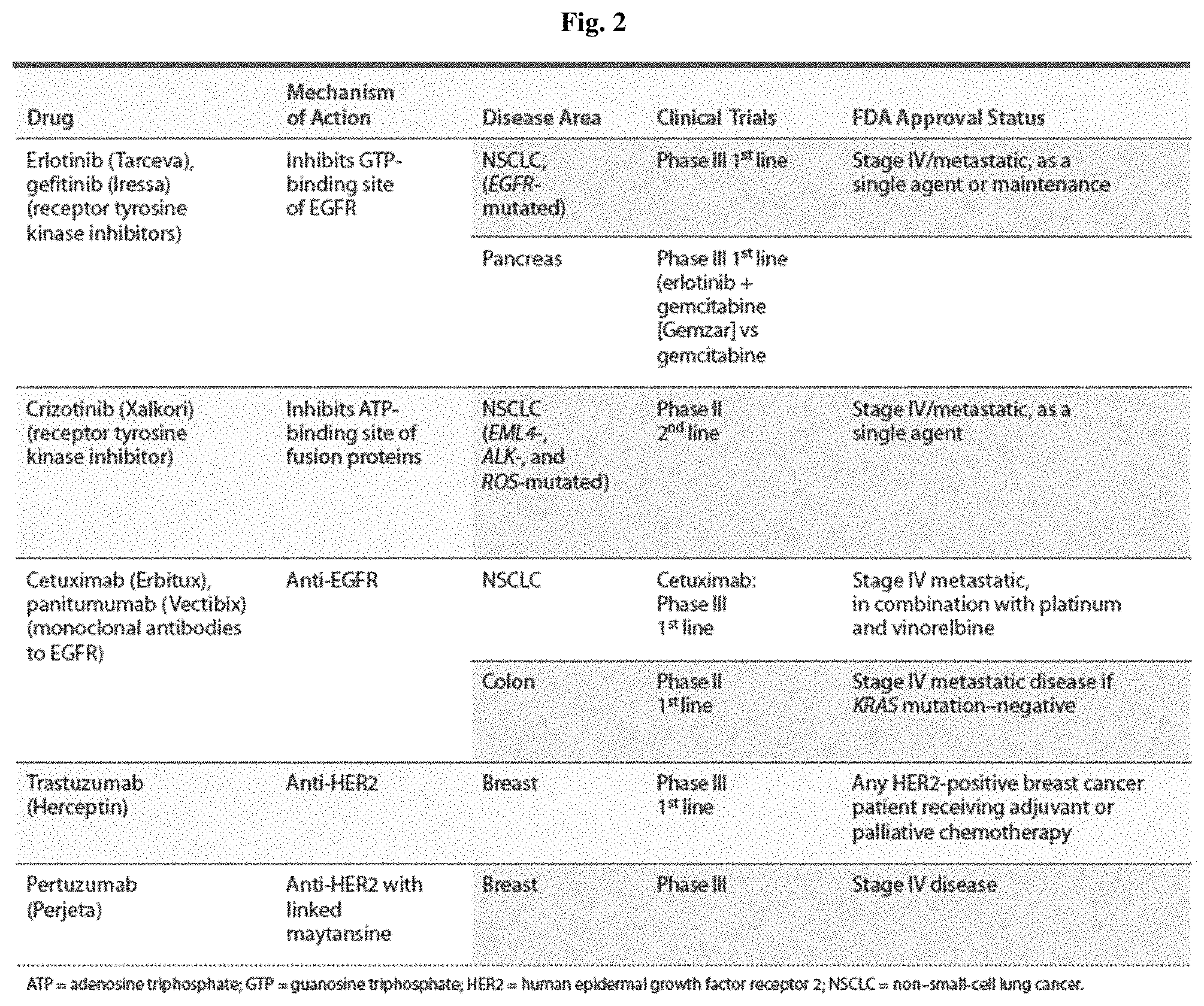
Various therapies for inhibiting the activation of the tumor-related EGFR have been emerged and now show a strong anticancer effect. New types of EGFR antagonists are being developed to compensate shortcomings such as side effects, tolerances and the like according to therapies. The details of the EGFR antagonist currently in development are the same as described in FIG. 2.
The EGFR overexpression and mutations come in many different forms in lung cancer, anal cancer, and glioblastoma multiforme (GBM). The EGFRvIII mutations are often found in a brain tumor, and reflect a poor prognosis after a treatment through surgery, radiation therapy, and chemotherapy (Kuan C T et al., 2003). The EGFR mutants in the form of a recombination have deletions or duplications of specific exons. The EGFR and the EGFR mutants in the form of a recombination are the same as illustrated in FIG. 3.
EGFRvIII is an EGFR variant of SEQ ID NO: 1, which is most frequently appeared and lacks amino acids in exons 2-7, and is expressed in 50% or more of terminally ill brain tumor patients. EGFRvIII lacks 267 amino acids in the amino acid sequence of EGFRwt, binds to exons 1 and 8 and produces glycine, which is a new amino acid, and induces phosphorylation of the intracellular domain and shows continuous activity, thereby contributing to the carcinogenesis (Downward J et al., 1984). Of course, other forms of EGFR can also be targeted, but at the present time, EGFRvIII is regarded as the most suitable EGFR variant as an anticancer target, and many researches and developments are ongoing.
In particular, in brain tumors, cell signals are transmitted by the main signaling pathway mainly via PI3K and AKT of EGFRvIII. Through the activation of Stat-3, AP-1 or MAPK as a minor pathway, it contributes to cancer cell proliferation, resistance to apoptosis, angiogenesis, increase of penetration of cancer cells, formation of cancer stem cells, and the like (Gan H K, et al., 2013). Recent studies have reported that EGFRvIII plays an important role in carcinogenesis by activating NF-.kappa.B via mTORC2 (Bonavia et al., 2012; Tanaka et al., 2011). The signaling pathway according to the activation of EGFRvIII is the same as illustrated in FIG. 4.
In addition, since EGFRvIII induces activation following c-Met phosphorylation (Huang P H. et al., 2007), when it is applied and administered to a patient in combination with a c-Met inhibitor together with an EGFR or EGFRvIII inhibitor, it may improve efficacy while reducing the resistance to chemotherapy of a patient with high levels of EGFRvIII.
EGFRvIII is usually expressed in brain tumors simultaneously with EGFR wild-type (WT) (Ekstrand et al., 1991), and since EGFRvIII itself is expressed in EGFR gene amplified tumors (Biernat et al., 2004, Frederick et al., 2000), it has been thought that the individual cancer cells would simultaneously perform EGFRwt amplification and EGFRvIII expression for interaction.
EGFRvIII is expressed at relatively high frequency in brain tumors, but further studies are needed in other carcinomas. A recent study using brain tumor cell lines transformed to express EGFRvIII revealed that networks using EGFRvIII and EGFRwt could surely have the possibility of being co-activated (Shia Q. et al., PNAS, 2012). Such interaction between EGFRvIII and EGFRwt needs to be confirmed again using EGFRvIII which is expressed naturally through xenografts model, GBM spheroid lines or other cell lines.
It has been reported that EGFR phosphorylates EGFRvIII and leads a STAT3/5 activation pathway, and greatly affects the progression of brain tumors (Fan Q W et al., 2013). EGFRvIII dimer formation greatly affects the activation of signal transduction. As the expression of EGFRwt increases, the phosphorylation and activation of EGFRvIII are enhanced. Thus, it is believed that dimerization arm and kinase activity, which form the dimerization of EGFRwt, contribute to the activation of EGFRvIII. From the result that the cancerization induced by EGFRvIII is inhibited when the expression of EGFRwt or HB-EGF is inhibited in an orthotopic transplantation model (Li L. et al., 2014), it can be said that the activation pathway surrounding EGFRwt plays a very important role in tumorigenesis via activation of EGFRvIII.
Currently, the therapeutic agents to treat (brain) tumors that are being developed while being targeted to EGFRvIII can be divided into five categories. The first one is a method of blocking the internal signal transduction of EGFRwt and EGFRvIII by using the conventional EGFR therapeutic agents. The second one is a method of inhibiting the interaction between external signal transduction and receptors by using antibodies that simultaneously target EGFR and EGFRvIII, such as ABT-806 (mAb806, Abbott). The third one is the development of an anti-EGFRvIII antibody in the form of an antibody drug conjugate (ADC), as AMG-595 which is now being developed by Amgen. The fourth one is that the form of Chimeric Antigen receptor-T cell (CAR-T) may be used as a cell immunotherapeutic agent. The fifth one is a method of joining EGFRvIII specific 14 amino acid sequences to Keyhole limpet hemocyanin (KLH) and administering it as an EGFRvIII anti-cancer vaccine. There is currently no commercialized anticancer drug targeting only EGFRvIII, not EGFR. As illustrated in FIG. 5, there is an EGFRvIII vaccine CDX-110 which is now being developed by Celldex Therapeutics which leads in clinical development.
Under these technical backgrounds, the inventors of the present disclosure prepared a novel antibody specifically binding to EGFRvIII. In particular, they confirmed that a novel antibody that binds only to EGFRvIII, but not to EGFRwt, can be prepared by producing it to specifically bind to EGFRvIII without cross-reacting with EGFRwt, and completed the present disclosure.
SUMMARY
It is an object of the present disclosure to provide a novel antibody specifically binding to EGFRvIII, a nucleic acid coding the antibody, a vector including the nucleic acid, a host cell including the vector, a preparation method thereof, and a pharmaceutical composition for treating cancer or tumors including the antibody as an active ingredient.
The present disclosure relates to an antibody binding to epidermal growth factor receptor variant III (EGFRvIII) comprising an amino acid sequence of SEQ ID NO: 1, free from cross-reacting with wild-type epidermal growth factor receptor (EGFR), wherein the antibody is specifically binding to the 1.sup.st to 13.sup.th amino acid regions of the amino acid sequence of SEQ ID NO: 1.
The present disclosure relates to a nucleic acid coding the antibody.
The present disclosure relates to a vector including the nucleic acid.
The present disclosure relates to a host cell including the vector.
The present disclosure relates to a preparation method of the antibody including expressing the antibody by culturing the host cell.
The present disclosure relates to a pharmaceutical composition for treating cancer or tumors including the antibody as an active ingredient.
The present disclosure relates to a method for treating cancer or tumors by administering the antibody to an individual in need thereof in a pharmacologically effective amount.
BRIEF DESCRIPTION OF THE DRAWINGS
The patent or application file contains at least one drawing executed in color. Copies of this patent or patent application publication with color drawing(s) will be provided by the Office upon request and payment of the necessary fee.
FIG. 1 illustrates each domain and phosphorylation site of the EGFR.
FIG. 2 illustrates the development progress of the known antagonists targeting EGFR receptors.
FIG. 3 is a schematic diagram illustrating the structure of EGFR mutants in a recombinant form with EGFR.
FIG. 4 is a schematic diagram illustrating the signal transduction flow according to the activation of EGFRvIII.
FIG. 5 illustrates a candidate list of EGFRvIII targeting therapeutic agents under clinical development obtained from www.clinicaltrials.gov.
FIG. 6a illustrates Western blot results illustrating the receptor expression of a CHOK1-EGFRvIII single cell line.
FIG. 6b illustrates Western blot results illustrating the receptor expression of a U87MG-EGFRvIII single cell line.
FIG. 7 illustrates the bead panning monophage ELISA results for peptides containing the EGFRvIII specific sequence used to select EGFRvIII antibodies.
FIG. 8 illustrates Western blot results confirming the antigen-specific binding capacity of a single phage-antibody selected by bead panning.
FIG. 9a illustrates the result of confirming IgG obtained by treatment in a non-reducing buffer through SDS-PAGE.
FIG. 9b illustrates the result of confirming IgG obtained by treatment in a reducing buffer through SDS-PAGE.
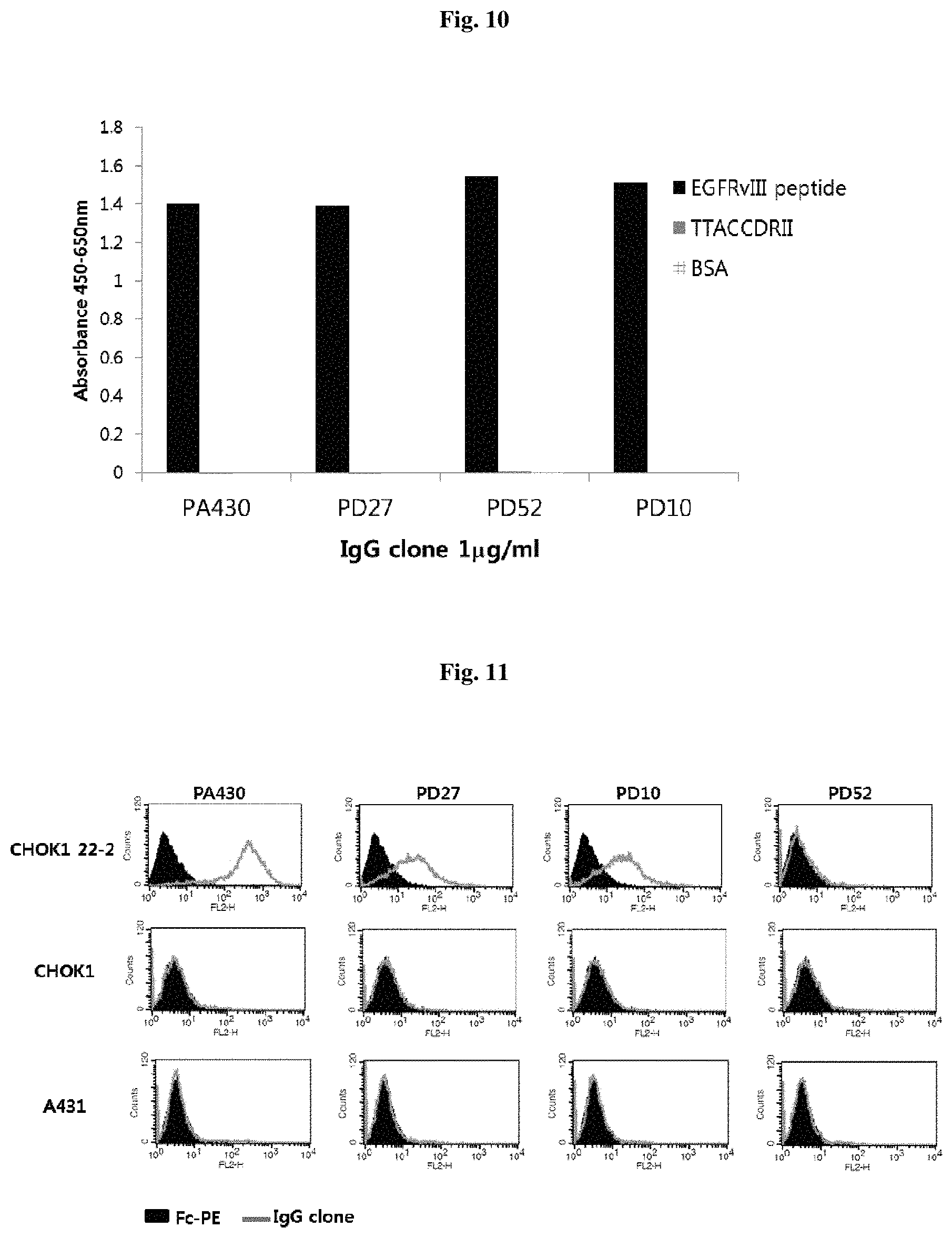
FIG. 10 illustrates the anti-EGFRvIII-peptide binding of human IgG clones using ELISA.
FIG. 11 illustrates the binding specificity to the EGFRvIII cell line of a human IgG clone using flow cytometry.
FIG. 12 illustrates the results of confirming the binding capacity affected by the concentration reduction of human IgG in the anti-EGFRvIII flow cytometry.
FIG. 13 illustrates the results of confirming the binding capacity according to the change in a light chain of human IgG by ELISA.
FIG. 14 illustrates the results of confirming the binding capacity according to the change in a light chain of human IgG by flow cytometry.
FIG. 15a illustrates the results of confirming the internalization of anti-EGFRvIII human IgG by flow cytometry using U87 MG 13.
FIG. 15b illustrates the results of confirming the internalization of anti-EGFRvIII human IgG by flow cytometry using CHOK1 22-2.
DETAILED DESCRIPTION OF THE EMBODIMENT
In one aspect, the present disclosure relates to an antibody binding to epidermal growth factor receptor variant III (EGFRvIII) comprising an amino acid sequence of SEQ ID NO: 1, free from cross-reacting with wild type epidermal growth factor receptor (EGFR), wherein the antibody is specifically binding to the 1.sup.st to 13.sup.th amino acid regions of the amino acid sequence of SEQ ID NO: 1. The detailed descriptions of each constitution are as follows.
Antigen EGFRvIII
As discussed above, EGFRvIII is a deletion mutant of EGFR in which 267 amino acids are deleted in the extracellular domain of EGFR together with a single amino acid substitution of glycine at a junction, and has a sequence of SEQ ID NO: 1.
The inventors of the present disclosure produced an antibody by using a peptide including an EGFRvIII peptide specific sequence 13mer as an antigen in order to produce an antibody specifically binding to EGFRvIII without cross-reacting with the EGFR wild type. Specifically, the antibody according to the present disclosure may specifically bind to the sequence of SEQ ID NO: 2 LEEKKGNYVVTDHCSGGKN (N is biotin) in which linker and biotin of EGFRvIII peptide specific sequences 13mer, 4mer are included. At the same time, a peptide having a sequence of TTACCDRII, for example, SEQ ID NO: 3 NEINPGNGHTNYNEKFKS (N is biotin) was used as a negative antigen to block non-specific binding. Through this, an antibody specifically binding to EGFRvIII without cross-reacting with the EGFR wild type was produced.
Antibody
The term "antibody" used in the present specification refers to an immunoglobulin molecule that is immunologically reactive with a specific antigen, means a protein molecule that serves as a receptor that specifically recognizes an antigen, and may includes both polyclonal and monoclonal antibodies and whole antibodies and antibody fragments. In addition, it may also include chimeric antibodies (e.g., humanized murine antibodies) and bivalent or bispecific molecules (e.g., bispecific antibodies), diabodies, triabodies and tetrabodies.
The whole antibody is a structure having two overall length light chains and two overall length heavy chains, and each light chain is linked to a heavy chain by a disulfide bond. The whole antibody includes IgA, IgD, IgE, IgM and IgG, and IgG is a subtype and includes IgG1, IgG2, IgG3 and IgG4. The antibody fragment means a fragment having an antigen-binding function, and includes Fab, Fab', F (ab') 2, Fv, and the like.
The Fab has one antigen-binding site in a structure having a variable region of a light chain and a heavy chain, a constant region of a light chain, and a first constant region (CH1 domain) of a heavy chain. Fab' differs from Fab in that it has a hinge region that contains at least one cysteine residue at the C-terminal of the heavy chain CH1 domain. The F(ab')2 antibody is generated when the cysteine residue of the hinge region of the Fab' forms a disulfide bond.
The variable fragment (Fv) means the minimum antibody fragment having only a heavy chain variable region and a light chain variable region. The double-stranded Fv (dsFv) is linked by a disulfide bond to a heavy chain variable region and a light chain variable region. Single chain Fv (scFv) is generally linked to a variable region of a heavy chain and a variable region of a light chain through a peptide linker by a covalent bond. Such antibody fragments may be obtained using protein hydrolytic enzymes (for example, Fab may be obtained by restricting and cutting the whole antibody with papain and F(ab')2 fragments may be obtained by cutting it with pepsin), and may be produced through gene recombinant technology (e.g., setting DNA coding the heavy chain of the antibody or its variable region and DNA coding the light chain or its variable region as a template, amplifying them by the PCR using a primer pair, and combining and amplifying the DNA coding a peptide linker and a primer pair configured to link each of the both terminals to the heavy chain and its variable region and the light chain or its variable region).
Immunoglobulins have a heavy chain and a light chain, and each heavy and light chain includes a constant region and a variable region (the site is also known as a domain). The variable regions of the light and heavy chains include three variable regions and four framework regions which are called as complementarity-determining regions (hereinafter, referred to as "CDR"). The CDR serves as the binding mainly to the epitope of an antigen. The CDRs of each chain are typically called sequentially as CDR1, CDR2, CDR3 starting from the N-terminus, and are identified by the chain in which a specific CDR is located.
The term "monoclonal antibody" used in the present specification means an antibody molecule of a single molecular composition obtained from a substantially identical antibody group, and may exhibit a single binding specificity and affinity for a specific epitope.
The term "human antibody" used in the present specification refers to a molecule derived from human immunoglobulin, wherein all of the amino acid sequences constituting the antibody including a complementarity-determining region and a framework region are composed of the amino acid sequence of human immunoglobulin. The human antibody is usually used for treating human diseases, and has advantages in that i) target cells may be more efficiently destroyed by interacting with the human immune system more stably, for example, by complement-dependent cytotoxicity (CDC) or antibody-dependent cellmediated cytotoxicity (ADCC), ii) the human immune system does not recognize the antibody as a foreign one, and iii) even if a less amount of drugs is administrated less frequently, a half-life in a human circulatory system is similar to a naturally occurring antibody.
In consideration of the above, the antibody according to the present disclosure is a monoclonal antibody specifically binding to EGFRvIII, which not only exhibits excellent affinity and specificity for EGFRvIII but also exhibits low immunogenicity because it is derived from human, and thus is suitable for the treatment of diseases such as cancer or tumors.
The term "antibody specifically binding to EGFRvIII" used in the present specification means an antibody that binds to EGFRvIII and inhibits the biological activity of EGFRvIII, and can be used interchangeably with an anti-EGFRvIII antibody. At this time, the K.sub.D for the EGFRvIII may be, for example, 10.sup.-8 or less, preferably 10.sup.-9 or less, more preferably 10.sup.-10 or less.
The term "without cross-reacting with EGFR" used in the present specification may mean that an antibody that binds to the EGFR wild type and an antibody that binds to EGFRvIII do not show cross reactivity to each antigen. At this time, the antibody that does not cross-react with the EGFR may have, for example, 10.sup.-5 or more, preferably 10.sup.-4 or more, more preferably 10.sup.-3 or more of K.sub.D of the antibody against the EGFR wild type. Substantially, the antibody that does not cross-react with the EGFR may mean an antibody that cannot detect an antibody that binds to the EGFR wild type in an unlimited form of a standard binding assay.
In one exemplary embodiment, the antibody according to the present disclosure may be, for example, as specifically described in Example 1, the monoclonal human antibodies PA430, PD27, PD52 and PD10 that do not cross-react with the EGFR, and are structurally specified and separated to be specifically bound to the region of 1.sup.st to 13.sup.th amino acids regions among the amino acid sequences of EGFRvIII of SEQ ID NO: 1. The amino acid sequences for heavy chain CDRs and light chain CDRs of each antibody are as listed in the following Tables 1 and 2.
TABLE-US-00001 TABLE 1 Ab H-CDR1 H-CDR2 H-CDR3 PA430 YHAMH AMSHDGTETSYADSVKG EGLRSNGGAFET (SEQ ID (SEQ ID NO: 8) (SEQ ID NO: 12) NO: 4) PD52 DYAMH GISWNSGAIGYADSVKG ASRGLGDAFDI (SEQ ID (SEQ ID NO: 9) (SEQ ID NO: 13) NO: 5) PD27 EHAMH GINWNSGKTGYADSVKG PGEDTGGGFDI (SEQ ID (SEQ ID NO: 10) (SEQ ID NO: 14) NO: 6) PD10 EHAMH GINWNSGKTGYADSVKG PGEDTGGGFDI (SEQ ID (SEQ ID NO: 11) (SEQ ID NO: 15) NO: 7)
TABLE-US-00002 TABLE 2 Ab L-CDR1 L-CDR2 L-CDR3 PA430 SGDVLPKHYAY KDSERPS QSVDSSDTSVV (SEQ ID NO: (SEQ ID NO: 20) (SEQ ID NO: 24) 16) PD52 SGDVLPKHYAY KDTERPS QSVDNSDTSVV (SEQ ID NO: (SEQ ID NO: 21) (SEQ ID NO: 25) 17) PD27 SGDVLADHYSY KDSERPS QSVDSSDTSVV (SEQ ID NO: (SEQ ID NO: 22) (SEQ ID NO: 26) 18) PD10 SSDVGGYNYVS DVTKRPS SSYSSSTFYV (SEQ ID NO: (SEQ ID NO: 23) (SEQ ID NO: 27) 19)
The antibody according to the present disclosure may be formed, for example, by mixing V.sub.H and V.sub.L sequences, which are structurally similar as to the V.sub.H CDR1, 2 and 3 sequences and V.sub.L CDR1, 2 and 3 sequences described in Tables 1 and 2, and by being placed as CDR1, 2 and 3 of V.sub.H/V.sub.L pairing, and may include, for example, a heavy chain variable region comprising the following heavy chain CDRs:
a heavy chain CDR1 comprising at least one amino acid sequence selected from the group consisting of SEQ ID NOS: 4-7;
a heavy chain CDR2 comprising at least one amino acid sequence selected from the group consisting of SEQ ID NOS: 8-11; and
a heavy chain CDR3 comprising at least one amino acid sequence selected from the group consisting of SEQ ID NOS: 12-15.
In addition, the antibody according to the present disclosure may include a light chain variable region comprising the following light chain CDRs:
a light chain CDR1 comprising at least one amino acid sequence selected from the group consisting of SEQ ID NOS: 16-19;
a light chain CDR2 comprising at least one amino acid sequence selected from the group consisting of SEQ ID NOS: 20-23; and
a light chain CDR3 comprising at least one amino acid sequence selected from the group consisting of SEQ ID NOS: 24-27.
In one embodiment, the antibody according to the present disclosure may specifically include an amino acid sequence of a heavy chain variable region and a light chain variable region as described in Table 3 below, or a sequence having homology thereto:
TABLE-US-00003 TABLE 3 Ab Variable Region Amino acid sequence PA430 VH QMQLVESGGGVVQPGKSLRLSCAASGFTFSYHA (SEQ ID NO: 28) MHWVRQAPGKGLEWLAAMSHDGTETSYADSV KGRITISRDNSKSALYLQMNSLRAEDTAVYYCTA EGLRSNGGAFETWGRGTMITVSS VL SYELTQPPSVSVAPGQTARITCSGDVLPKHYAYW (SEQ ID NO: 32) YQQKPGQAPVLVIYKDSERPSGIPERFTGSSSGTK VTLTISGVRAEDEADYYCQSVDSSDTSVVFGGGT KLTVLG PD52 VH QMQLVQSGGGVVQPGGSLRLSCVGSGFSFDDYA (SEQ ID NO: 29) MHWVRQAPGKGLEWVSGISWNSGAIGYADSVK GRFTVSRDNSKNSLYLQMNSLRAEDTAVYYCAT ASRGLGDAFDIWGQGTMVTVSS VL SYELTQPPSVSVSPGQTARITCSGDVLPKHYAYW (SEQ ID NO: 33) YQQKPGQAPVLVIYKDTERPSGIPERFSGSSSGTT VTLTISGVQAEDEADYYCQSVDNSDTSVVFGGG TKLTVLG PD27 VH QVQLVESGGGLVQPGGSLRLSCAASGFTFDEHA (SEQ ID NO: 30) MHWVRQAPGKGLQWVSGINWNSGKTGYADSV KGRFTISRDNSKNSLYLQMNSLRAEDTAVYYCTR PGEDTGGGFDIWGQGTMITVSS VL SYELTQPLSVSVSPGQTARITCSGDVLADHYSYW (SEQ ID NO: 34) YQQKPGQAPVLVMYKDSERPSGIPERFSGSSSGT TVTLTISGVQAEDEADYYCQSVDSSDTSVVFGGG TKLTVLG PD10 VH QVQLVESGGGLVQPGGSLRLSCAASGFTFDEHA (SEQ ID NO: 31) MHWVRQAPGKGLQWVSGINWNSGKTGYADSV KGRFTISRDNSKNSLYLQMNSLRAEDTAVYYCTR PGEDTGGGFDIWGQGTMITVSS VL NFMLTQPASVSGSPGQSITISCTGSSSDVGGYNYV (SEQ ID NO: 35) SWYQQHPGKAPQLIIYDVTKRPSGVSNRFSGSKS GNSASLTISGLQAEDEADYYCSSYSSSTFYVFGTG TKVTVLG
The antibody according to the present disclosure may include, for example, a heavy chain variable region comprising a sequence having at least 80% of homology with at least one amino acid sequence selected from the group consisting of SEQ ID NOS: 28-31. The antibody may include, i.e., the sequences of SEQ ID NOS: 28-31, as heavy chain variable regions, comprising a sequence having at least 80% of homology, preferably at least 90%, at least 95%, at least 97%, at least 98%, and at least 99% of homology, and more preferably 100% of homology with at least one amino acid sequence selected from the group consisting of SEQ ID NOS: 28-31.
In addition, the antibody according to the present disclosure may comprise a light chain variable region comprising a sequence having at least 80% of homology with at least one amino acid sequence selected from the group consisting of SEQ ID NOS: 32-35. The antibody may include i.e., the sequences of SEQ ID NOS: 32-35 as light chain variable regions comprising a sequence having at least 80% of homology, preferably at least 90%, at least 95%, at least 97%, at least 98%, and at least 99% of homology, and more preferably 100% of homology with at least one amino acid sequence selected from the group consisting of SEQ ID NOS: 32-35.
To be specific, the antibody according to the present disclosure may include the following heavy chain variable region and light chain variable region selected from the group consisting of the following, and the V.sub.H sequence and V.sub.L sequence described in Table 3 may be formed by mixing V.sub.H and V.sub.L sequences, which are structurally similar and by being placed as V.sub.H/V.sub.L pairing without limitation:
A heavy chain variable region comprising a sequence of SEQ ID NO: 28 and a light chain variable region of the sequence selected from the group consisting of SEQ ID NOS: 32-35;
A heavy chain variable region comprising a sequence of SEQ ID NO: 29 and a light chain variable region of the sequence selected from the group consisting of SEQ ID NOS: 32-35;
A heavy chain variable region comprising a sequence of SEQ ID NO: 30 and a light chain variable region of the sequence selected from the group consisting of SEQ ID NOS: 32-35;
A heavy chain variable region comprising a sequence of SEQ ID NO: 31 and a light chain variable region of the sequence selected from the group consisting of SEQ ID NOS: 32-35.
In particular, the antibody according to the present disclosure may include the following heavy chain variable region and light chain variable region:
A heavy chain variable region comprising a sequence of SEQ ID NO: 28 and a light chain variable region comprising a sequence of SEQ ID NO: 32;
A heavy chain variable region comprising a sequence of SEQ ID NO: 29 and a light chain variable region comprising a sequence of SEQ ID NO: 33.
A heavy chain variable region comprising a sequence of SEQ ID NO: 30 and a light chain variable region comprising a sequence of SEQ ID NO: 34; and
A heavy chain variable region comprising a sequence of SEQ ID NO: 31 and a light chain variable region comprising a sequence of SEQ ID NO: 35.
In another aspect, the present disclosure relates to nucleic acid coding the antibody. The term "nucleic acid" used in the present specification may be present in a cell, a cell lysate, or in a partially purified form or in a substantially pure form. The nucleic acid becomes "isolated" or "substantially pure" when purified from other cell components or other contaminants, for example, nucleic acid or proteins of the other cells by the standard technology including alkaline/SDS treatment, CsCl banding, column chromatography, agarose gel electrophoresis and the others that are well known in the pertinent art. The nucleic acid of the present disclosure may be, for example, DNA or RNA, and may or may not include an intron sequence.
In one exemplary embodiment, the nucleic acid according to the present disclosure coding the V.sub.H sequence and the V.sub.L sequence is described in Table 4, and may include at least one sequence coding a heavy chain variable region having at least 95% of homology with a sequence coding heavy chain variable region selected from the group consisting of SEQ ID NOS: 36-39 and/or may include at least one sequence coding a light chain variable region having at least 95% of homology with a sequence coding at least one light chain variable region selected from the group consisting of SEQ ID NOS: 40-43. The antibody has at least 95% of homology, preferably at least 98% of homology, more preferably 100% of homology with at least one nucleic acid sequence selected from the group consisting of SEQ ID NOS: 36-39 and/or SEQ ID NOS: 40-43.
TABLE-US-00004 TABLE 4 Ab Variable Region Nucleic acid sequence PA430 VH CAGATGCAGCTGGTGGAGTCCGGGGGAGGCGTGG (SEQ ID NO: 36) TCCAGCCTGGGAAGTCCCTGAGACTTTCCTGTGC AGCGTCTGGATTCACCTTCAGTTACCATGCCATGC ACTGGGTCCGCCAGGCTCCAGGCAAGGGGCTGGA GTGGCTGGCAGCTATGTCACATGATGGAACCGAA ACCAGCTACGCAGACTCCGTGAAGGGCCGAATCA CCATCTCCAGAGACAATTCCAAGAGTGCGTTGTAT CTACAAATGAACAGTCTGAGAGCCGAGGACACGG CCGTGTATTACTGTACCGCAGAGGGGCTTCGGAGC AATGGAGGGGCTTTTGAGACTTGGGGCCGCGGGA CAATGATCACCGTCTCCTCA VL TCCTATGAGCTGACACAGCCACCCTCAGTGTCGGT (SEQ ID NO: 40) GGCCCCAGGGCAGACGGCCAGGATCACCTGCTCT GGAGATGTACTGCCAAAACATTATGCTTATTGGTA CCAGCAGAAGCCAGGCCAGGCCCCTGTTTTGGTG ATATATAAAGACAGCGAGAGGCCCTCAGGGATCCC TGAGCGATTCACTGGTTCCAGCTCAGGGACAAAA GTCACGCTGACCATAAGTGGAGTCCGGGCAGAAG ACGAGGCTGACTATTATTGTCAATCAGTAGACAGC AGTGATACTTCTGTGGTTTTCGGCGGAGGGACCAA GCTGACCGTCCTAGGT PD52 VH CAGATGCAGCTGGTGCAGTCTGGAGGGGGCGTGG (SEQ ID NO: 37) TACAGCCTGGGGGGTCCCTGAGACTCTCCTGTGTA GGCTCTGGATTCAGCTTTGATGATTATGCCATGCAC TGGGTCCGTCAGGCTCCAGGGAAGGGCCTGGAGT GGGTCTCAGGTATTAGTTGGAATAGTGGTGCCATA GGCTATGCGGACTCTGTGAAGGGCCGATTCACCGT CTCCAGAGACAACAGCAAAAACTCCCTGTATCTG CAAATGAACAGTCTGAGAGCCGAGGACACGGCCG TGTATTACTGTGCCACAGCCTCCAGAGGACTTGGT GATGCTTTTGATATCTGGGGCCAGGGGACAATGGT CACCGTCTCCTCA VL TCCTATGAGCTGACACAGCCCCCCTCGGTGTCAGT (SEQ ID NO: 41) GTCCCCAGGACAGACGGCCAGGATCACCTGCTCT GGAGATGTACTGCCAAAACATTATGCTTATTGGTA CCAGCAGAAGCCAGGCCAGGCCCCTGTTTTGGTG ATATATAAAGACACTGAGAGGCCCTCAGGGATCCC TGAGCGATTCTCTGGCTCCAGTTCAGGGACAACA GTCACGTTGACCATCAGTGGAGTCCAGGCAGAAG ACGAGGCTGACTATTATTGTCAATCAGTAGACAAC AGTGATACTTCTGTGGTTTTCGGCGGAGGGACCAA GCTGACCGTCCTAGGT PD27 VH CAGGTGCAGCTGGTGGAGTCTGGGGGAGGCTTGG (SEQ ID NO: 38) TACAGCCTGGAGGGTCCCTGAGACTCTCCTGTGC AGCCTCTGGATTCACCTTTGATGAACATGCCATGC ACTGGGTCCGGCAAGCTCCAGGGAAGGGCCTGCA GTGGGTCTCAGGAATCAATTGGAATAGTGGTAAAA CAGGCTATGCGGACTCTGTGAAGGGCCGATTCACC ATCTCCAGAGACAACAGCAAAAACTCCCTGTATCT GCAAATGAACAGTCTGAGAGCCGAGGACACGGCC GTGTATTACTGTACTAGACCCGGGGAGGACACCGG GGGTGGCTTTGATATCTGGGGCCAAGGGACAATGA TCACCGTCTCCTCA VL TCCTATGAGCTGACTCAGCCACTCTCGGTGTCAGT (SEQ ID NO: 42) GTCCCCAGGACAGACGGCCAGGATCACCTGCTCT GGAGATGTATTGGCAGATCATTATTCTTATTGGTAC CAGCAGAAGCCAGGCCAGGCCCCTGTGTTGGTGA TGTATAAAGACAGTGAGAGGCCCTCTGGGATCCCT GAGCGATTCTCTGGCTCCAGCTCAGGGACAACAG TCACGTTGACCATCAGTGGAGTCCAGGCAGAAGA CGAGGCTGACTATTATTGTCAATCAGTAGACAGCA GTGATACTTCTGTGGTTTTCGGCGGAGGGACCAAG CTGACCGTCCTAGGT PD10 VH CAGGTGCAGCTGGTGGAGTCTGGGGGAGGCTTGG (SEQ ID NO: 39) TACAGCCTGGAGGGTCCCTGAGACTCTCCTGTGC AGCCTCTGGATTCACCTTTGATGAACATGCCATGC ACTGGGTCCGGCAAGCTCCAGGGAAGGGCCTGCA GTGGGTCTCAGGAATCAATTGGAATAGTGGTAAAA CAGGCTATGCGGACTCTGTGAAGGGCCGATTCACC ATCTCCAGAGACAACAGCAAAAACTCCCTGTATCT GCAAATGAACAGTCTGAGAGCCGAGGACACGGCC GTGTATTACTGTACTAGACCCGGGGAGGACACCGG GGGTGGCTTTGATATCTGGGGCCAAGGGACAATGA TCACCGTCTCCTCA VL AATTTTATGCTGACTCAGCCCGCCTCCGTGTCTGG (SEQ ID NO: 43) GTCCCCTGGACAGTCGATCACCATCTCCTGCACTG GAAGCAGCAGCGACGTTGGTGGTTATAACTATGTC TCCTGGTACCAACAGCACCCAGGCAAAGCCCCCC AACTCATCATTTATGATGTCACTAAGCGGCCCTCA GGGGTTTCTAATCGCTTCTCCGGCTCCAAGTCTGG CAACTCGGCCTCCCTGACCATCTCTGGACTCCAGG CTGAGGACGAGGCTGATTATTACTGCAGCTCATAC AGCAGCAGCACTTTTTACGTCTTCGGAACTGGGA CCAAGGTCACCGTCCTAGGT
The present disclosure may include a conjugate in which a toxin, a drug, or the like is joined to the antibody. The toxin or drug may include any target compound. Examples of such toxins may be duocamycin, calicheamicin, mytansine or auristatin, and the compounds may be a known anti-cancer or anti-tumor compound such as taxol, etoposide, tenofoside, vincristine, doxorubicin, etc., alkylating agents (such as cisplatin, anthracycline (e.g., doxorubicin), etc.). The conjugates may be prepared using linker using technology available in the pertinent art.
In addition, in addition to the antibody, the present disclosure may include a bispecific binding molecule including two or more different binding sites in which a peptide, a protein or an antibody is bound to the other antigen binding sites that are different from the antibody. The bispecific binding molecule may be prepared by linking a binding specific part using the methods known in the pertinent art. In addition, when the antibody is linked, it may be linked via a sulf-hydryl link at the C-terminal region of the heavy chain. According to circumstances, it may be coded in the same vector and expressed in the same host cell to be combined.
Preparation Method of the Antibody
In another aspect, the present disclosure relates to a vector comprising the nucleic acid. For expression of antibodies or antibody fragments thereof, DNA coding the partial or full length of light chains and heavy chains may be obtained by standard molecular biology technology (e.g., PCR amplification or cDNA cloning using hybridomas expressing the target antibody), and may be inserted into an expression vector in such a way that DNA is operably bound to transcriptional and translational control sequences.
The term "operably bound" used in the present specification may mean that the antibody gene is ligated into a vector so that the transcriptional and translational control sequences in the vector serve the intended function of regulating the transcription and translation of the antibody gene. The expression vector and expression control sequences are selected so as to be compatible with the host cell used for expression. The light chain gene of the antibody and the heavy chain gene of the antibody are inserted into separate vectors, or both genes are inserted into the same expression vector. The antibody is inserted into the expression vector by standard methods (e.g., ligation of complementary restriction enzyme sites on the antibody gene fragment and vector, or blunt end ligation if no restriction enzyme site is present). According to circumstances, the recombinant expression vector may code a signal peptide that facilitates secretion of the antibody chain from the host cell. The antibody chain gene may be cloned into a vector such that the signal peptide is bound to the amino terminus of the antibody chain gene to fit to the frame. The signal peptide may be an immunoglobulin signal peptide or a heterologous signal peptide (e.g., a signal peptide derived from a protein other than an immunoglobulin). In addition, the recombinant expression vector has a "regulatory sequence" that controls the expression of the antibody chain gene in the host cell. The "regulatory sequences" may include promoters, enhancers and other expression control elements (e.g., polyadenylation signals) that control transcription or translation of an antibody chain gene. Those skilled in the art may recognize that the design of an expression vector may be varied by selecting different regulatory sequences depending on factors such as the selection of a host cell to be transformed, the level of expression of the protein, and the like.
The present disclosure may also include a host cell comprising the nucleic acid or the vector. The nucleic acid or the vector is transfected. Various types of technologies commonly used to introduce exogenous DNA into prokaryotic or eukaryotic host cells to be "transfected," for example, electrophoresis, calcium phosphate precipitation method, DEAE-dextran transfection or lipofection can be used. The antibody according to the present disclosure may be expressed in a eukaryotic cell, preferably a mammalian host cell, in consideration of the possibility of being applied to a mammalian cell. Suitable mammalian host cells for expression of the antibodies include Chinese hamster ovary (CHO) cells (including, for example, dhfr-CHO cells used together with DHFR selectable markers), NSO myeloma cells, COS cells or SP2 Cells, and the like.
In another aspect, the present disclosure relates to a preparation method of the antibody comprising culturing a host cell to express the antibody. When the recombinant expression vector coding the antibody gene is introduced into a mammalian host cell, the antibody may be prepared by culturing the host cell for a period of time sufficient to allow the antibody to be expressed in the host cell, or more preferably, for a period of time sufficient to allow the antibody to be secreted into the culture medium in which the host cell is cultured. In addition, the antibody may be prepared by comprising the biopanning using EGFRvIII comprising the amino acid sequence of SEQ ID NO: 1.
According to circumstances, the expressed antibody may be separated from the host cell and purified to be homogenous. The separation or purification of the antibody may be carried out by separation and purification methods used in the conventional proteins, for example, chromatography. The chromatography may include, for example, affinity chromatography including a protein A column, a protein G column, ion exchange chromatography or hydrophobic chromatography. In addition to the chromatography, the antibody may be further separated and purified by combining filtration, ultrafiltration, salting out, dialysis, and the like.
Pharmaceutical Composition
In another aspect, the present disclosure relates to a pharmaceutical composition for treating a cancer or a tumor comprising the antibody as an active ingredient.
The term "cancer or tumors" used in the present specification is not particularly limited as long as it is the type of cancer or tumors that can be treated by the antibody of the present disclosure. For example, it may be breast cancer, lung cancer or anal cancer caused by the overexpression of EGFRvIII, and a brain tumor, for example, a primary brain tumor (i.e., occurs in the brain) and a secondary or metastatic brain tumor.
According to circumstances, the antibody according to the present disclosure may be administered in combination with other anti-cancer drugs (i.e., before, during, or after receiving treatments for cancer). It may be administered in combination with any one or more chemotherapeutic drugs known to those skilled in the art, such as alkylating agents such as carmustine, chlorambucil, cisplatin, carboplatin, oxyplatin, proccarbazine, and cyclophosphamide; antimetabolites such as fluorouracil, phloxuridine, fludarabine, gemcitabine, methotrexate and hydroxyurea; natural products such as plant alkaloids or antibiotics such as bleomycin, doxorubicin, daunorubicin, idarubicin, etoposide, mitomycin, mitoxantrone, vinblastine, vincristine, and taxol (paclitaxel) or related compounds such as Taxotere.RTM.; therapeutic agents for treatment of brain tumors such as Gliadel.RTM. wafers containing temozolomide or carmustine; and other drugs such as irinotecan, gleevec, and the like. In addition, other biological agents such as monoclonal antibodies such as Herceptin.TM. for HER2 antigen, Avastin.TM. for VEGF, antibodies against EGF receptor, such as Erbitux.RTM., and other EGF receptor antagonists may be included.
The pharmaceutical composition may be formulated to include a pharmaceutically acceptable carrier. The term "pharmaceutically acceptable carrier" refers to a carrier or diluent that does not stimulate living organisms and does not inhibit the biological activity and characteristics of the administered compound. The pharmaceutical carrier that is acceptable for the composition to be formulated into a liquid solution is suitable for sterilization and living bodies, and may be used by mixing a saline solution, sterile water, a Ringer's solution, a buffered saline solution, an albumin injection solution, a dextrose solution, a malto dextrin solution, glycerol, ethanol, and one or more components of these components. If necessary, the other common additives such as an antioxidant, a buffer, and a bacteriostatic agent may be added. In addition, diluents, dispersants, surfactants, binders and lubricants may be additionally added to be formulated into formulations for injection, pills, capsules, granules or tablets such as aqueous solutions, suspensions, emulsions and the like.
The pharmaceutical composition may be of various oral or parenteral formulations. In the case of formulation, it may be prepared using diluents or excipients such as fillers, extenders, binders, wetting agents, disintegrating agents, surfactants and the like which are usually used. Solid preparation for oral administration include tablets, pills, powders, granules, capsules and the like, and may be prepared by mixing one or more excipients such as starch, calcium carbonate, sucrose or lactose, gelatin, and the like with one or more compounds. In addition, in addition to the simple excipients, lubricants such as magnesium stearate, talc, and the like may also be used. Examples of the liquid preparation for oral administration include suspensions, solutions, emulsions and syrups. In addition to water and liquid paraffin, which are commonly used and are simple diluents, various excipients such as wetting agents, sweeteners, air freshener, preservatives and the like may be included.
Formulations for parenteral administration may include sterilized aqueous solutions, non-aqueous solutions, suspensions, emulsions, lyophilized formulations, and suppositories. Examples of the non-aqueous solvent and suspension solvent include propylene glycol, polyethylene glycol, vegetable oil such as olive oil, and injectable ester such as ethyl oleate. As a suppository base, witepsol, macrogol, tween, cacao butter, laurinum, glycerogelatin and the like may be used.
The pharmaceutical composition may have any one of formulations selected from the group consisting of tablets, pills, powders, granules, capsules, suspensions, solutions, emulsions, syrups, sterilized aqueous solutions, non-aqueous solvents, suspensions, emulsions, lyophilized formulations, and suppositories. In addition, they may be administered once or many times. At this time, the composition may be administered in the form of a liquid preparation, a powder, an aerosol, a capsule, an intravaginal tablet, a capsule, or a suppository. Routes of administration may include, but are not limited to, intraperitoneal, intravenous, intramuscular, subcutaneous, endothelial, oral, topical, intranasal, intrapulmonary, intrarectal, and the like. However, upon oral administration, the peptide must be formulated so as to protect it against degradation in the stomach or coating of the active agent since it is digested. In addition, the active substance may be administered by any device capable of migrating to the target cell.
The composition may be administered in a therapeutically effective amount, and a "therapeutically effective amount" may refer to an amount sufficient to treat a disease at a reasonable benefit/risk ratio applicable to medical treatment. Effective dose levels are determined by the elements including the type and severity of the individual, age, gender, type of cancer, drug activity, sensitivity to drug, time of administration, route of administration and rate of release, duration of treatment, drugs used simultaneously, and the other elements well known in the field of medicine. It may be administered in high doses of usually 0.1 to 5 mg/kg, for example 1, 2, 3 or 4 mg/kg, 10 mg/kg or 15 or 20 mg/kg. As a fixed unit capacity, for example, it may be provided with 50, 100, 200, 500 or 1000 mg. In order to cause regression of cancer or tumor, more preferably to remove tumor, it may be administered once to eight times (e.g., 1, 2, 3, 4, 5, 6, 7 or 8), or 10, 20 or more times. Based on a half-life of the antibody, it may be administered twice a week, every week, every two weeks, every month or at the other intervals of one week, two weeks, four weeks, eight weeks, three to six months, or longer.
Hereinafter, the present disclosure will be described in more detail by way of examples. It is apparent to a person having ordinary skill in the pertinent art that these examples merely illustrate the present disclosure, and the scope of the present disclosure is not limited to these examples.
Example 1: Establishment of EGFRvIII Cell Line
The EGFRvIII gene containing HindIII and XbaI restriction enzyme sites was obtained by using DNA synthesis (Invitrogen, SEQ ID NO: 1) and split by using HindIII and XbaI restriction enzymes. The split template DNA was cloned into the expression vector pcDNA3.1 split with HindIII and XbaI restriction enzymes to combine pcDNA3.1-EGFRvIII. PcDNA3.1-EGFRvIII was transformed into DH5.alpha. E. coli to select ampicillin-resistant strains. Whether the EGFRvIII gene is inserted by splitting with HindIII and XbaI restriction enzymes was confirmed.
The pcDNA3.1-EGFRvIII vector prepared to produce the EGFRvIII cell line was transfected by using Lipofectamine 2000 with respect to CHOK1, U87MG, and A431, and the cells were cultured in 10% FBS, DMEM for 48 hours. After the cultured cells were cultured in DMEM or RPMI to which G418 of 10% FBS 1000 .mu.g/ml, 400 .mu.g/ml, 400 .mu.g/ml in a 1:10 dilution, the cells to be colonized were selected and proliferated in a 6-well. When western blot was performed after each cell proliferated for 12 days was dissolved and was subject to electrophoresis, it was treated with anti-EGFR antibody and anti-human IgG HRP and reacted with ECL substrate (FIGS. 6a and 6b).
Example 2: Biopanning
The EGFRvIII peptide used as an antigen was synthesized to contain a specific sequence 13mer and a 4mer and a biotin used as a linker (SEQ ID NO: 2). The biotin-TTACCDRII peptide was also synthesized (SEQ ID NO: 3) as a negative antigen to block non-specific binding. Each peptide was prepared using a M-280 streptavidin (Invitrogen, USA) bead to join 7.8 ng of peptide per 5 .mu.l.
The human antibody phage library was reacted with TTACCDRII peptide joined to the bead in advance at 25.degree. C. for 30 minutes to induce binding of a non-specific phage antibody. Thereafter, the TTACCDRII peptide bound to the bead was harvested using a magnetic bar and then only supernatant liquid was taken separately. The supernatant liquid was added to the EGFRvIII peptide bound to the bead again and reacted at 25.degree. C. for 2 hours to occur binding of an antibody phage. The EGFRvIII peptide bound to the bead was harvested using a magnetic bar and phage supernatant liquid was removed. The bead EGFRvIII peptide drawn on the magnetic bar was washed repeatedly 5 times with PBST (PBS containing 0.1% Tween 20), and finally, it was washed once with PBS. 100 ul of 100 mM triethylamine solution was reacted for 10 minutes to allow the phage bound to the antigen to elute, and the eluted phage was neutralized by 50 ul of 1M Tris pH 7.5.
The neutralized phage eluate was added to 10 ml of mid-log phase XL1-Blue E. coli cell line and reacted at 37.degree. C. for 30 minutes to induce infection. The infected XL1-Blue E. coli cell line was spread on a 2.times.YT/C plate containing 1% glucose and cultured at 30.degree. C. for 16 hours. The first panning and the second and third panning were performed in the same way.
Example 3: Selection of Phage Antibody Specifically Binding to EGFRvIII
E. coli clones were randomly selected from the completed panning, and cultured in a 96 well plate at 37.degree. C. and 300 rpm on a mid-log phase by adding 500 .mu.l of 2.times.YT/C medium, followed by addition of M13 helperphage, and then, infection of phage was induced at 37.degree. C. for 30 minutes. The infected E. coli clones were cultured for 15 hours under the condition of 30.degree. C. and 30 rpm to prepare the phage-antibody to elute. Cells of the cultured E. coli clones were centrifuged at 6000 rpm for 10 minutes to remove the cells and only supernatant was obtained.
EGFRvIII peptide, TTACCDRII peptide, and BSA were coated on a Maxisorb 96 well ELISA plate (Nunc, Denmark) at 1 .mu.g/ml, blocked with 3% defatted milk/PBS for 1 hour at room temperature, and then inoculated with the obtained supernatant liquid and reacted for 1 hour at room temperature. The ELISA plate was washed three times with 0.05% of PBST, and then an anti-M13-HRP (Horse Radish Peroxidase) antibody (GE) was diluted to 1:300 PBST 0.05% for 1 hour. After treating for 10 minutes and color-developing TMB substrate (BD, USA), the reaction was stopped by treating 2N H.sub.2SO.sub.4. The color-developed ELISA was measured for absorbance at 450 nm-650 nm using a Sunrise ELISA reader (TECAN, Switzerland) (FIG. 7).
Example 4: Western Blot
EGFRvIII binding capacity by phage-antibody was observed by western blot using CHOK1-EGFRvIII cell lysate.
CHOK1 22-2, which is a CHOK1-EGFRvIII cell line prepared in Example 1, and A431 expressing CHOK1 and EGFR wildtype as a parent cell therefor were washed with PBS and dissolved by using an eluting buffer of 1% SDS, 1 mM Na3VO3, 10 mM Tris (pH 7.4), 1 mM PMSF, 10 .mu.M Leupeptin, 1.5 .mu.M pepstein, and 10 .mu.g/ml aprotinin. After 15 .mu.g of each cell lysate was separated by 6% SDS-PAGE, the protein was transferred to PVDF membrane. After 1 hour block with 3% skim milk TBST, 5.times.10.sup.10 pfu of PA430 phage and PD54 phage were treated for 1 hour, followed by dilution of anti-M13-HRP (Horse Radish Peroxidase) antibody (GE) to 1:24000 TBST for 1 hour, and after color-development by using ECL (GE Healthcare, USA) substrate, and blot was confirmed with an Odyssey Fc imaging system (Licor, USA).
The results are illustrated in FIG. 8, and referring to FIG. 8, it can be understood that PA430 phage and PD54 phage bind only in the cell line expressing EGFRvIII.
Example 5: IgG Expression and Purification
The conversion to the IgG form was accomplished by double-cutting with a sfiI restriction enzyme containing a variable heavy chain of the phage-antibody to obtain a fragment, and by double-cutting with a sfiI restriction enzyme in pIgGHD-6A6Hvy, which is a vector comprising a heavy chain region to obtain a ligation with a fragment.
The fragment was obtained by double-cutting with a BstX I restriction enzyme including a variable light chain of a phage-antibody. The fragment was ligated with the fragment in the same manner as the vector pIgGLD-6A6Lgt including the light chain region (as for the above method, please refer to Korean Patent Laid-Open Publication No. 2008-0109417).
The expression of IgG was determined by inoculating HEK293T cells with 8.times.10.sup.6 cells per 100 mm cell culture dish and culturing the cells in a 37.degree. C. CO.sub.2 5% culture medium for 24 hours so that the adherence and density of cells become 80-90% to culture cells. The cells were cultured with 80% to 90%.
10 .mu.g of the heavy chain expression vector and 10 .mu.g of the light chain expression vector of the prepared IgG, i.e., a total of 20 .mu.g of the vector were mixed with PEI at a ratio of 1:3 (DNA:PEI) .mu.g and react for 15 minutes to form a complex. DNA PEI complex was added to the cultured cells, and after reaction for 10 hours, it was washed with DMEM once. Then, by adding 10 ml of a culture medium to which 10% penicillin streptomycin (GIBCO, USA) is added to Freestyle 293 (GIBCO, USA), it was left alone for 48 hours to take the supernatant in which IgG was expressed.
The obtained supernatant was injected into a protein A column (GE healthcare, USA) equilibrated with 20 mM Tris-HCl, 50 mM NaCl and 5 mM EDTA pH 7.0, and washed with 50 mM Tris-HCl (pH 7.0), 5 mM EDTA, 500 mM NaCl, 0.2% Polysorbate 20, and then eluted with 50 mM NaCl, 0.1 M glycine-HCl (pH 3.5) solution. Then, the affinity chromatography neutralized with 1M Tris was performed. The eluted proteins were packed in a MWCO 10,000 spectra/por dialysis membrane (spectrum, USA), and the solutions were exchanged by PBS dialysis.
The results are illustrated in FIGS. 9a and 9b. With reference to FIGS. 9a and 9b, each antibody was treated with a non-reducing or reducing LDS sample buffer (Invitrogen, USA) via SDS-PAGE, and when it is loaded onto Nupage 4-12% Bis-Tris Gel (Invitrogen, USA), IgG including a 50 kDa heavy chain and a 25 kDa light chain and corresponding to 150 kDa was obtained.
Example 6: Selection of IgG Specifically Binding to EGFR vIII
ELISAs were performed to identify antigen-specific binding capacity of clones converted to IgG EGFRvIII peptide, TTACCDRII peptide and BSA were coated on 1 .mu.g/ml 96 well plates at 37.degree. C. for 2 hours. Thereafter, after blocking the uncoated portion with 3% skim milk/0.05% PBST, 1 .mu.g/ml IgG was added to the plate and reacted at 37.degree. C. for 1 hour. Anti-human IgG HRP (pierce, USA) was diluted in 1:3000 and reacted for 1 hour. Next, the TMB substrate (BD, USA) was treated for 10 minutes for color development, and the reaction was stopped by treating with 2N H2SO4. As to the color developed ELISA, the absorbance was measured at 450 nm-650 nm using a Sunrise ELISA reader (TECAN, Switzerland).
The results are illustrated in FIG. 10, and referring to FIG. 10, PA430, PD27, PD52 and PD10 showed specific binding capacity only in EGFRvIII peptide without nonspecific binding.
Based on these results, fluorescent dyed flow cytometry was performed to confirm whether the binding capacity of EGFRvIII-expression cell line was maintained with respect to the above-mentioned four types of antibodies. EGFRvIII expression cell line, CHOK1 22-2 and its mother cell, CHOK1, and A431, which expresses EGFR wild type were the objects. Each cell was obtained at 1.times.10.sup.6 cell number, diluted with 1 .mu.g/ml of each antibody in PBS added with 2% FBS and added to cells, and reacted at 4.degree. C. for 1 hour. The anti-human IgG PE (Bethyl, USA) was diluted 1:200 and reacted at 4.degree. C. for 45 minutes for fluorescence staining. Each sample was measured for 1.times.10.sup.4 cells per sample using an FACS Caliber (BD, USA) instrument.
The results are illustrated in FIG. 11. Referring to FIG. 11, PA430, PD27, and PD10 were able to bind to EGFRvIII expression cell line CHOK1 22-2, but not to EGFRvIII non-expression cell lines CHOK1 and A431. In addition, it was shown that binding was not observed in A431, a cell expressing EGFR wild type.
In order to confirm the binding characteristics of three antibodies showing binding capacity to the EGFRvIII expression cell line according to the decrease of the antibody concentration, the following experiment was performed. The antibody concentration was diluted double the amount in a sequential order starting from 1000 ng/ml, and was prepared up to 31 ng/ml. Fluorescence staining flow cytometry was performed in the same order as described above.
The results are illustrated in FIG. 12. Referring to FIG. 12, it has been observed that PA430 showed high binding capacity even at the time of treating IgG in the same concentration as compared to PD27 and PD10.
Example 7: Change in Light Chain for Affinity Enhancement
The light chain was changed to enhance the antigen specific affinity of PA430. Upon reviewing the CDRs of the variable light chains of PA430, PD52 and PD27, it has been confirmed that they are similar in sequence of the variable light chain of PA430, which has high antigen specific affinity (see Table 1-3). Using this fact, in addition to the original light chain of PA430, the light chain vector of PD52, PD27 and PD10 was mixed with the heavy chain vector of PA430 and transfected to produce IgG, which is the same as in Example 5.
When the produced and identified 430H10L, 430H52L, and 430H27L were performed in the same manner as in the ELISA of Example 6, it was confirmed that the antigen-specific binding capacity was maintained (FIG. 13).
The fluorescent staining flow cytometry of Example 6 was performed for PA430 and 430H52L, 430H27L and PD10 IgG in the same way. When U87 MG 13 cell lines produced as EGFRvIII expression cell lines based on U87MG cells and A431 16-3 cell lines produced as EGFRvIII expression cell lines based on A431 cells were additionally performed, it has been confirmed that 430H52L and 430H27L maintains binding capacity even in the EGFRvIII specific cell line. In addition, it has been observed that 430H52L is excellent in binding signal even at the time of treating the same concentration as compared to the PA430 with a circular light chain (FIG. 14).
Example 8: Analysis of Antibody Internalization Using Fluorescent Staining Flow Cytometry
By using a 430H52L antibody with EGFRvIII specific binding capacity, fluorescence staining flow cytometry was performed in order to confirm whether internalization is performed by antibody-EGFRvIII binding.
2.times.10.sup.6 cells were treated with Trypsin EDTA for U87 MG 13 cells and CHOK1 22-2 cells and then cooled at 4.degree. C. for 30 minutes to inhibit cell activity. The cells were treated with 1 .mu.g/ml of 430H52L in DMEM medium to induce antibody antigen binding at 4.degree. C. for 30 minutes. The samples treated with only secondary antibody treat DMEM only. Next, after centrifugation at 1300 rpm for 3 minutes, the cells were washed with PBS to remove residual antibody, added as DMEM medium, and reacted with a sample reacting at 4.degree. C. and an incubating sample at 37.degree. C. for 15 minutes, 37.degree. C. at 30 minutes, and 37.degree. C. for 60 minutes CO.sub.2 5%. After centrifugation, the cells were washed with PBS at 4.degree. C., and then 0.1M Glycine and 0.5M NaCl pH 2.2 were repeatedly treated three times for 10 minutes to artificially remove the antibodies bound to EGFRvIII on the cell surface. After centrifugation, the cells were washed with PBS and fixed with 4% paraformaldehyde (USB, USA) for 10 minutes at 4.degree. C. Each sample was made to penetrate by 0.1% triton-100 (sigma Aldrich, USA) PBS solution at 4.degree. C. for 10 minutes, or treated with 2% FBS PBS solution so that penetration does not occur. After centrifugation, the cells were washed with PBS and 2% FBS PBS was treated and blocked, and anti-human IgG PE (bethyl, USA) was treated in 1:400 for dyeing. Each sample was measured for 1.times.10.sup.4 cells per sample using a FACS Caliber (BD, USA) instrument.
The results are illustrated in FIGS. 15a and 15b, respectively. Referring to FIGS. 15a and 15b, when EGFRvIII expression cell line U87 MG 13 or CHOK1 22-2 cells were treated with 430H52L antibody and fluorescent staining was performed by using cell permeablization by going through reaction of each temperature and time, it has been observed that signal increases as time increases at 37.degree. C. as compared to 4.degree. C. When non-permeabilized fluorescent staining was performed in the control group without going through cell permeablization, there was no increase in signal according to increase of temperature and time. This is demonstrated by a fluorescence signal that the 430H52L IgG binds to EGFRvIII so that the internalization increases according to increase of time under the condition of 37.degree. C.
The antibody specifically binding to EGFRvIII according to the present disclosure is a novel antibody that binds only to EGFRvIII without binding to wild type EGFR (EGFRwt), and shows excellent binding capacity in cells expressing EGFRvIII. It has been confirmed that internalization occurs, and thus it can act as an effective antagonist targeting EGFRvIII. Based thereon, the antibody specifically binding to EGFRvIII according to the present disclosure can be usefully used for the treatment of diseases induced by the expression of EGFRvIII.
Numerous variations and modifications will be apparent to a person having ordinary skill in the field to which the present disclosure pertains within the scope of the present disclosure based on the above contents.
SEQUENCE LISTINGS
1
431919PRTArtificial Sequencesequence of EGFRvIII 1Leu Glu Glu Lys Lys Gly Asn Tyr Val Val Thr Asp His Gly Ser Cys1 5 10 15Val Arg Ala Cys Gly Ala Asp Ser Tyr Glu Met Glu Glu Asp Gly Val 20 25 30Arg Lys Cys Lys Lys Cys Glu Gly Pro Cys Arg Lys Val Cys Asn Gly 35 40 45Ile Gly Ile Gly Glu Phe Lys Asp Ser Leu Ser Ile Asn Ala Thr Asn 50 55 60Ile Lys His Phe Lys Asn Cys Thr Ser Ile Ser Gly Asp Leu His Ile65 70 75 80Leu Pro Val Ala Phe Arg Gly Asp Ser Phe Thr His Thr Pro Pro Leu 85 90 95Asp Pro Gln Glu Leu Asp Ile Leu Lys Thr Val Lys Glu Ile Thr Gly 100 105 110Phe Leu Leu Ile Gln Ala Trp Pro Glu Asn Arg Thr Asp Leu His Ala 115 120 125Phe Glu Asn Leu Glu Ile Ile Arg Gly Arg Thr Lys Gln His Gly Gln 130 135 140Phe Ser Leu Ala Val Val Ser Leu Asn Ile Thr Ser Leu Gly Leu Arg145 150 155 160Ser Leu Lys Glu Ile Ser Asp Gly Asp Val Ile Ile Ser Gly Asn Lys 165 170 175Asn Leu Cys Tyr Ala Asn Thr Ile Asn Trp Lys Lys Leu Phe Gly Thr 180 185 190Ser Gly Gln Lys Thr Lys Ile Ile Ser Asn Arg Gly Glu Asn Ser Cys 195 200 205Lys Ala Thr Gly Gln Val Cys His Ala Leu Cys Ser Pro Glu Gly Cys 210 215 220Trp Gly Pro Glu Pro Arg Asp Cys Val Ser Cys Arg Asn Val Ser Arg225 230 235 240Gly Arg Glu Cys Val Asp Lys Cys Asn Leu Leu Glu Gly Glu Pro Arg 245 250 255Glu Phe Val Glu Asn Ser Glu Cys Ile Gln Cys His Pro Glu Cys Leu 260 265 270Pro Gln Ala Met Asn Ile Thr Cys Thr Gly Arg Gly Pro Asp Asn Cys 275 280 285Ile Gln Cys Ala His Tyr Ile Asp Gly Pro His Cys Val Lys Thr Cys 290 295 300Pro Ala Gly Val Met Gly Glu Asn Asn Thr Leu Val Trp Lys Tyr Ala305 310 315 320Asp Ala Gly His Val Cys His Leu Cys His Pro Asn Cys Thr Tyr Gly 325 330 335Cys Thr Gly Pro Gly Leu Glu Gly Cys Pro Thr Asn Gly Pro Lys Ile 340 345 350Pro Ser Ile Ala Thr Gly Met Val Gly Ala Leu Leu Leu Leu Leu Val 355 360 365Val Ala Leu Gly Ile Gly Leu Phe Met Arg Arg Arg His Ile Val Arg 370 375 380Lys Arg Thr Leu Arg Arg Leu Leu Gln Glu Arg Glu Leu Val Glu Pro385 390 395 400Leu Thr Pro Ser Gly Glu Ala Pro Asn Gln Ala Leu Leu Arg Ile Leu 405 410 415Lys Glu Thr Glu Phe Lys Lys Ile Lys Val Leu Gly Ser Gly Ala Phe 420 425 430Gly Thr Val Tyr Lys Gly Leu Trp Ile Pro Glu Gly Glu Lys Val Lys 435 440 445Ile Pro Val Ala Ile Lys Glu Leu Arg Glu Ala Thr Ser Pro Lys Ala 450 455 460Asn Lys Glu Ile Leu Asp Glu Ala Tyr Val Met Ala Ser Val Asp Asn465 470 475 480Pro His Val Cys Arg Leu Leu Gly Ile Cys Leu Thr Ser Thr Val Gln 485 490 495Leu Ile Thr Gln Leu Met Pro Phe Gly Cys Leu Leu Asp Tyr Val Arg 500 505 510Glu His Lys Asp Asn Ile Gly Ser Gln Tyr Leu Leu Asn Trp Cys Val 515 520 525Gln Ile Ala Lys Gly Met Asn Tyr Leu Glu Asp Arg Arg Leu Val His 530 535 540Arg Asp Leu Ala Ala Arg Asn Val Leu Val Lys Thr Pro Gln His Val545 550 555 560Lys Ile Thr Asp Phe Gly Leu Ala Lys Leu Leu Gly Ala Glu Glu Lys 565 570 575Glu Tyr His Ala Glu Gly Gly Lys Val Pro Ile Lys Trp Met Ala Leu 580 585 590Glu Ser Ile Leu His Arg Ile Tyr Thr His Gln Ser Asp Val Trp Ser 595 600 605Tyr Gly Val Thr Val Trp Glu Leu Met Thr Phe Gly Ser Lys Pro Tyr 610 615 620Asp Gly Ile Pro Ala Ser Glu Ile Ser Ser Ile Leu Glu Lys Gly Glu625 630 635 640Arg Leu Pro Gln Pro Pro Ile Cys Thr Ile Asp Val Tyr Met Ile Met 645 650 655Val Lys Cys Trp Met Ile Asp Ala Asp Ser Arg Pro Lys Phe Arg Glu 660 665 670Leu Ile Ile Glu Phe Ser Lys Met Ala Arg Asp Pro Gln Arg Tyr Leu 675 680 685Val Ile Gln Gly Asp Glu Arg Met His Leu Pro Ser Pro Thr Asp Ser 690 695 700Asn Phe Tyr Arg Ala Leu Met Asp Glu Glu Asp Met Asp Asp Val Val705 710 715 720Asp Ala Asp Glu Tyr Leu Ile Pro Gln Gln Gly Phe Phe Ser Ser Pro 725 730 735Ser Thr Ser Arg Thr Pro Leu Leu Ser Ser Leu Ser Ala Thr Ser Asn 740 745 750Asn Ser Thr Val Ala Cys Ile Asp Arg Asn Gly Leu Gln Ser Cys Pro 755 760 765Ile Lys Glu Asp Ser Phe Leu Gln Arg Tyr Ser Ser Asp Pro Thr Gly 770 775 780Ala Leu Thr Glu Asp Ser Ile Asp Asp Thr Phe Leu Pro Val Pro Glu785 790 795 800Tyr Ile Asn Gln Ser Val Pro Lys Arg Pro Ala Gly Ser Val Gln Asn 805 810 815Pro Val Tyr His Asn Gln Pro Leu Asn Pro Ala Pro Ser Arg Asp Pro 820 825 830His Tyr Gln Asp Pro His Ser Thr Ala Val Gly Asn Pro Glu Tyr Leu 835 840 845Asn Thr Val Gln Pro Thr Cys Val Asn Ser Thr Phe Asp Ser Pro Ala 850 855 860His Trp Ala Gln Lys Gly Ser His Gln Ile Ser Leu Asp Asn Pro Asp865 870 875 880Tyr Gln Gln Asp Phe Phe Pro Lys Glu Ala Lys Pro Asn Gly Ile Phe 885 890 895Lys Gly Ser Thr Ala Glu Asn Ala Glu Tyr Leu Arg Val Ala Pro Gln 900 905 910Ser Ser Glu Phe Ile Gly Ala 915218PRTArtificial Sequencesequence of Antigenmisc_featureC-terminal biotin 2Leu Glu Glu Lys Lys Gly Asn Tyr Val Val Thr Asp His Cys Ser Gly1 5 10 15Gly Lys317PRTArtificial Sequencesequence of Negative Antigenmisc_featureN-terminal biotin 3Glu Ile Asn Pro Gly Asn Gly His Thr Asn Tyr Asn Glu Lys Phe Lys1 5 10 15Ser45PRTArtificial SequenceH-CDR1 4Tyr His Ala Met His1 555PRTArtificial SequenceH-CDR1 5Asp Tyr Ala Met His1 565PRTArtificial SequenceH-CDR1 6Glu His Ala Met His1 575PRTArtificial SequenceH-CDR1 7Glu His Ala Met His1 5817PRTArtificial SequenceH-CDR2 8Ala Met Ser His Asp Gly Thr Glu Thr Ser Tyr Ala Asp Ser Val Lys1 5 10 15Gly917PRTArtificial SequenceH-CDR2 9Gly Ile Ser Trp Asn Ser Gly Ala Ile Gly Tyr Ala Asp Ser Val Lys1 5 10 15Gly1017PRTArtificial SequenceH-CDR2 10Gly Ile Asn Trp Asn Ser Gly Lys Thr Gly Tyr Ala Asp Ser Val Lys1 5 10 15Gly1117PRTArtificial SequenceH-CDR2 11Gly Ile Asn Trp Asn Ser Gly Lys Thr Gly Tyr Ala Asp Ser Val Lys1 5 10 15Gly1212PRTArtificial SequenceH-CDR3 12Glu Gly Leu Arg Ser Asn Gly Gly Ala Phe Glu Thr1 5 101311PRTArtificial SequenceH-CDR3 13Ala Ser Arg Gly Leu Gly Asp Ala Phe Asp Ile1 5 101411PRTArtificial SequenceH-CDR3 14Pro Gly Glu Asp Thr Gly Gly Gly Phe Asp Ile1 5 101511PRTArtificial SequenceH-CDR3 15Pro Gly Glu Asp Thr Gly Gly Gly Phe Asp Ile1 5 101611PRTArtificial SequenceL-CDR1 16Ser Gly Asp Val Leu Pro Lys His Tyr Ala Tyr1 5 101711PRTArtificial SequenceL-CDR1 17Ser Gly Asp Val Leu Pro Lys His Tyr Ala Tyr1 5 101811PRTArtificial SequenceL-CDR1 18Ser Gly Asp Val Leu Ala Asp His Tyr Ser Tyr1 5 101911PRTArtificial SequenceL-CDR1 19Ser Ser Asp Val Gly Gly Tyr Asn Tyr Val Ser1 5 10207PRTArtificial SequenceL-CDR2 20Lys Asp Ser Glu Arg Pro Ser1 5217PRTArtificial SequenceL-CDR2 21Lys Asp Thr Glu Arg Pro Ser1 5227PRTArtificial SequenceL-CDR2 22Lys Asp Ser Glu Arg Pro Ser1 5237PRTArtificial SequenceL-CDR2 23Asp Val Thr Lys Arg Pro Ser1 52411PRTArtificial SequenceL-CDR3 24Gln Ser Val Asp Ser Ser Asp Thr Ser Val Val1 5 102511PRTArtificial SequenceL-CDR3 25Gln Ser Val Asp Asn Ser Asp Thr Ser Val Val1 5 102611PRTArtificial SequenceL-CDR3 26Gln Ser Val Asp Ser Ser Asp Thr Ser Val Val1 5 102710PRTArtificial SequenceL-CDR3 27Ser Ser Tyr Ser Ser Ser Thr Phe Tyr Val1 5 1028121PRTArtificial SequenceHv 28Gln Met Gln Leu Val Glu Ser Gly Gly Gly Val Val Gln Pro Gly Lys1 5 10 15Ser Leu Arg Leu Ser Cys Ala Ala Ser Gly Phe Thr Phe Ser Tyr His 20 25 30Ala Met His Trp Val Arg Gln Ala Pro Gly Lys Gly Leu Glu Trp Leu 35 40 45Ala Ala Met Ser His Asp Gly Thr Glu Thr Ser Tyr Ala Asp Ser Val 50 55 60Lys Gly Arg Ile Thr Ile Ser Arg Asp Asn Ser Lys Ser Ala Leu Tyr65 70 75 80Leu Gln Met Asn Ser Leu Arg Ala Glu Asp Thr Ala Val Tyr Tyr Cys 85 90 95Thr Ala Glu Gly Leu Arg Ser Asn Gly Gly Ala Phe Glu Thr Trp Gly 100 105 110Arg Gly Thr Met Ile Thr Val Ser Ser 115 12029120PRTArtificial SequenceHv 29Gln Met Gln Leu Val Gln Ser Gly Gly Gly Val Val Gln Pro Gly Gly1 5 10 15Ser Leu Arg Leu Ser Cys Val Gly Ser Gly Phe Ser Phe Asp Asp Tyr 20 25 30Ala Met His Trp Val Arg Gln Ala Pro Gly Lys Gly Leu Glu Trp Val 35 40 45Ser Gly Ile Ser Trp Asn Ser Gly Ala Ile Gly Tyr Ala Asp Ser Val 50 55 60Lys Gly Arg Phe Thr Val Ser Arg Asp Asn Ser Lys Asn Ser Leu Tyr65 70 75 80Leu Gln Met Asn Ser Leu Arg Ala Glu Asp Thr Ala Val Tyr Tyr Cys 85 90 95Ala Thr Ala Ser Arg Gly Leu Gly Asp Ala Phe Asp Ile Trp Gly Gln 100 105 110Gly Thr Met Val Thr Val Ser Ser 115 12030120PRTArtificial SequenceHv 30Gln Val Gln Leu Val Glu Ser Gly Gly Gly Leu Val Gln Pro Gly Gly1 5 10 15Ser Leu Arg Leu Ser Cys Ala Ala Ser Gly Phe Thr Phe Asp Glu His 20 25 30Ala Met His Trp Val Arg Gln Ala Pro Gly Lys Gly Leu Gln Trp Val 35 40 45Ser Gly Ile Asn Trp Asn Ser Gly Lys Thr Gly Tyr Ala Asp Ser Val 50 55 60Lys Gly Arg Phe Thr Ile Ser Arg Asp Asn Ser Lys Asn Ser Leu Tyr65 70 75 80Leu Gln Met Asn Ser Leu Arg Ala Glu Asp Thr Ala Val Tyr Tyr Cys 85 90 95Thr Arg Pro Gly Glu Asp Thr Gly Gly Gly Phe Asp Ile Trp Gly Gln 100 105 110Gly Thr Met Ile Thr Val Ser Ser 115 12031120PRTArtificial SequenceHv 31Gln Val Gln Leu Val Glu Ser Gly Gly Gly Leu Val Gln Pro Gly Gly1 5 10 15Ser Leu Arg Leu Ser Cys Ala Ala Ser Gly Phe Thr Phe Asp Glu His 20 25 30Ala Met His Trp Val Arg Gln Ala Pro Gly Lys Gly Leu Gln Trp Val 35 40 45Ser Gly Ile Asn Trp Asn Ser Gly Lys Thr Gly Tyr Ala Asp Ser Val 50 55 60Lys Gly Arg Phe Thr Ile Ser Arg Asp Asn Ser Lys Asn Ser Leu Tyr65 70 75 80Leu Gln Met Asn Ser Leu Arg Ala Glu Asp Thr Ala Val Tyr Tyr Cys 85 90 95Thr Arg Pro Gly Glu Asp Thr Gly Gly Gly Phe Asp Ile Trp Gly Gln 100 105 110Gly Thr Met Ile Thr Val Ser Ser 115 12032109PRTArtificial SequenceLv 32Ser Tyr Glu Leu Thr Gln Pro Pro Ser Val Ser Val Ala Pro Gly Gln1 5 10 15Thr Ala Arg Ile Thr Cys Ser Gly Asp Val Leu Pro Lys His Tyr Ala 20 25 30Tyr Trp Tyr Gln Gln Lys Pro Gly Gln Ala Pro Val Leu Val Ile Tyr 35 40 45Lys Asp Ser Glu Arg Pro Ser Gly Ile Pro Glu Arg Phe Thr Gly Ser 50 55 60Ser Ser Gly Thr Lys Val Thr Leu Thr Ile Ser Gly Val Arg Ala Glu65 70 75 80Asp Glu Ala Asp Tyr Tyr Cys Gln Ser Val Asp Ser Ser Asp Thr Ser 85 90 95Val Val Phe Gly Gly Gly Thr Lys Leu Thr Val Leu Gly 100 10533109PRTArtificial SequenceLv 33Ser Tyr Glu Leu Thr Gln Pro Pro Ser Val Ser Val Ser Pro Gly Gln1 5 10 15Thr Ala Arg Ile Thr Cys Ser Gly Asp Val Leu Pro Lys His Tyr Ala 20 25 30Tyr Trp Tyr Gln Gln Lys Pro Gly Gln Ala Pro Val Leu Val Ile Tyr 35 40 45Lys Asp Thr Glu Arg Pro Ser Gly Ile Pro Glu Arg Phe Ser Gly Ser 50 55 60Ser Ser Gly Thr Thr Val Thr Leu Thr Ile Ser Gly Val Gln Ala Glu65 70 75 80Asp Glu Ala Asp Tyr Tyr Cys Gln Ser Val Asp Asn Ser Asp Thr Ser 85 90 95Val Val Phe Gly Gly Gly Thr Lys Leu Thr Val Leu Gly 100 10534109PRTArtificial SequenceLv 34Ser Tyr Glu Leu Thr Gln Pro Leu Ser Val Ser Val Ser Pro Gly Gln1 5 10 15Thr Ala Arg Ile Thr Cys Ser Gly Asp Val Leu Ala Asp His Tyr Ser 20 25 30Tyr Trp Tyr Gln Gln Lys Pro Gly Gln Ala Pro Val Leu Val Met Tyr 35 40 45Lys Asp Ser Glu Arg Pro Ser Gly Ile Pro Glu Arg Phe Ser Gly Ser 50 55 60Ser Ser Gly Thr Thr Val Thr Leu Thr Ile Ser Gly Val Gln Ala Glu65 70 75 80Asp Glu Ala Asp Tyr Tyr Cys Gln Ser Val Asp Ser Ser Asp Thr Ser 85 90 95Val Val Phe Gly Gly Gly Thr Lys Leu Thr Val Leu Gly 100 10535111PRTArtificial SequenceLv 35Asn Phe Met Leu Thr Gln Pro Ala Ser Val Ser Gly Ser Pro Gly Gln1 5 10 15Ser Ile Thr Ile Ser Cys Thr Gly Ser Ser Ser Asp Val Gly Gly Tyr 20 25 30Asn Tyr Val Ser Trp Tyr Gln Gln His Pro Gly Lys Ala Pro Gln Leu 35 40 45Ile Ile Tyr Asp Val Thr Lys Arg Pro Ser Gly Val Ser Asn Arg Phe 50 55 60Ser Gly Ser Lys Ser Gly Asn Ser Ala Ser Leu Thr Ile Ser Gly Leu65 70 75 80Gln Ala Glu Asp Glu Ala Asp Tyr Tyr Cys Ser Ser Tyr Ser Ser Ser 85 90 95Thr Phe Tyr Val Phe Gly Thr Gly Thr Lys Val Thr Val Leu Gly 100 105 11036363DNAArtificial SequenceHv 36cagatgcagc tggtggagtc cgggggaggc gtggtccagc ctgggaagtc cctgagactt 60tcctgtgcag cgtctggatt caccttcagt taccatgcca tgcactgggt ccgccaggct 120ccaggcaagg ggctggagtg gctggcagct atgtcacatg atggaaccga aaccagctac 180gcagactccg tgaagggccg aatcaccatc tccagagaca attccaagag tgcgttgtat 240ctacaaatga acagtctgag agccgaggac acggccgtgt attactgtac cgcagagggg 300cttcggagca atggaggggc ttttgagact tggggccgcg ggacaatgat caccgtctcc 360tca 36337360DNAArtificial SequenceHv 37cagatgcagc tggtgcagtc tggagggggc gtggtacagc ctggggggtc cctgagactc 60tcctgtgtag gctctggatt cagctttgat gattatgcca tgcactgggt ccgtcaggct 120ccagggaagg gcctggagtg ggtctcaggt attagttgga atagtggtgc cataggctat 180gcggactctg tgaagggccg attcaccgtc tccagagaca acagcaaaaa ctccctgtat 240ctgcaaatga acagtctgag agccgaggac acggccgtgt attactgtgc cacagcctcc 300agaggacttg gtgatgcttt tgatatctgg ggccagggga caatggtcac cgtctcctca 36038360DNAArtificial SequenceHv 38caggtgcagc tggtggagtc tgggggaggc ttggtacagc ctggagggtc cctgagactc 60tcctgtgcag cctctggatt cacctttgat gaacatgcca tgcactgggt ccggcaagct 120ccagggaagg gcctgcagtg ggtctcagga atcaattgga atagtggtaa aacaggctat
180gcggactctg tgaagggccg attcaccatc tccagagaca acagcaaaaa ctccctgtat 240ctgcaaatga acagtctgag agccgaggac acggccgtgt attactgtac tagacccggg 300gaggacaccg ggggtggctt tgatatctgg ggccaaggga caatgatcac cgtctcctca 36039360DNAArtificial SequenceHv 39caggtgcagc tggtggagtc tgggggaggc ttggtacagc ctggagggtc cctgagactc 60tcctgtgcag cctctggatt cacctttgat gaacatgcca tgcactgggt ccggcaagct 120ccagggaagg gcctgcagtg ggtctcagga atcaattgga atagtggtaa aacaggctat 180gcggactctg tgaagggccg attcaccatc tccagagaca acagcaaaaa ctccctgtat 240ctgcaaatga acagtctgag agccgaggac acggccgtgt attactgtac tagacccggg 300gaggacaccg ggggtggctt tgatatctgg ggccaaggga caatgatcac cgtctcctca 36040327DNAArtificial SequenceLv 40tcctatgagc tgacacagcc accctcagtg tcggtggccc cagggcagac ggccaggatc 60acctgctctg gagatgtact gccaaaacat tatgcttatt ggtaccagca gaagccaggc 120caggcccctg ttttggtgat atataaagac agcgagaggc cctcagggat ccctgagcga 180ttcactggtt ccagctcagg gacaaaagtc acgctgacca taagtggagt ccgggcagaa 240gacgaggctg actattattg tcaatcagta gacagcagtg atacttctgt ggttttcggc 300ggagggacca agctgaccgt cctaggt 32741327DNAArtificial SequenceLv 41tcctatgagc tgacacagcc cccctcggtg tcagtgtccc caggacagac ggccaggatc 60acctgctctg gagatgtact gccaaaacat tatgcttatt ggtaccagca gaagccaggc 120caggcccctg ttttggtgat atataaagac actgagaggc cctcagggat ccctgagcga 180ttctctggct ccagttcagg gacaacagtc acgttgacca tcagtggagt ccaggcagaa 240gacgaggctg actattattg tcaatcagta gacaacagtg atacttctgt ggttttcggc 300ggagggacca agctgaccgt cctaggt 32742327DNAArtificial SequenceLv 42tcctatgagc tgactcagcc actctcggtg tcagtgtccc caggacagac ggccaggatc 60acctgctctg gagatgtatt ggcagatcat tattcttatt ggtaccagca gaagccaggc 120caggcccctg tgttggtgat gtataaagac agtgagaggc cctctgggat ccctgagcga 180ttctctggct ccagctcagg gacaacagtc acgttgacca tcagtggagt ccaggcagaa 240gacgaggctg actattattg tcaatcagta gacagcagtg atacttctgt ggttttcggc 300ggagggacca agctgaccgt cctaggt 32743333DNAArtificial SequenceLv 43aattttatgc tgactcagcc cgcctccgtg tctgggtccc ctggacagtc gatcaccatc 60tcctgcactg gaagcagcag cgacgttggt ggttataact atgtctcctg gtaccaacag 120cacccaggca aagcccccca actcatcatt tatgatgtca ctaagcggcc ctcaggggtt 180tctaatcgct tctccggctc caagtctggc aactcggcct ccctgaccat ctctggactc 240caggctgagg acgaggctga ttattactgc agctcataca gcagcagcac tttttacgtc 300ttcggaactg ggaccaaggt caccgtccta ggt 333
* * * * *
References
D00000
D00001
D00002
D00003
D00004
D00005
D00006
D00007
D00008
D00009
D00010
D00011
S00001
XML
uspto.report is an independent third-party trademark research tool that is not affiliated, endorsed, or sponsored by the United States Patent and Trademark Office (USPTO) or any other governmental organization. The information provided by uspto.report is based on publicly available data at the time of writing and is intended for informational purposes only.
While we strive to provide accurate and up-to-date information, we do not guarantee the accuracy, completeness, reliability, or suitability of the information displayed on this site. The use of this site is at your own risk. Any reliance you place on such information is therefore strictly at your own risk.
All official trademark data, including owner information, should be verified by visiting the official USPTO website at www.uspto.gov. This site is not intended to replace professional legal advice and should not be used as a substitute for consulting with a legal professional who is knowledgeable about trademark law.