Monoclonal anti-GT 468 antibodies for treatment of cancer
Sahin , et al.
U.S. patent number 10,596,256 [Application Number 14/977,325] was granted by the patent office on 2020-03-24 for monoclonal anti-gt 468 antibodies for treatment of cancer. This patent grant is currently assigned to ASTELLAS PHARMA INC., TRON--TRANSLATIONALE ONKOLOGIE AN DER UNIVERSITATSMEDIZIN DER JOHANNES GUTENBERG-UNIVERSITAT MAINZ GEMEINNUTZIGE GMBH. The grantee listed for this patent is Ganymed Pharmaceuticals AG, Johannes Gutenberg-Universitat Mainz. Invention is credited to Michael Koslowski, Rita Mitnacht-Kraus, Ugur Sahin, Ozlem Tureci.


View All Diagrams
| United States Patent | 10,596,256 |
| Sahin , et al. | March 24, 2020 |
Monoclonal anti-GT 468 antibodies for treatment of cancer
Abstract
The present invention provides antibodies useful as therapeutics for treating and/or preventing diseases associated with cells expressing GT468, including tumor-related diseases such as breast cancer, lung cancer, gastric cancer, ovarian cancer, hepatocellular cancer, colon cancer, pancreatic cancer, esophageal cancer, head & neck cancer, kidney cancer, in particular renal cell carcinoma, prostate cancer, liver cancer, melanoma, sarcoma, myeloma, neuroblastoma, placental choriocarcinoma, cervical cancer, and thyroid cancer, and the metastatic forms thereof. In one embodiment, the tumor disease is metastatic cancer in the lung.
| Inventors: | Sahin; Ugur (Mainz, DE), Tureci; Ozlem (Mainz, DE), Koslowski; Michael (Frankfurt, DE), Mitnacht-Kraus; Rita (Friedberg, DE) | ||||||||||
|---|---|---|---|---|---|---|---|---|---|---|---|
| Applicant: |
|
||||||||||
| Assignee: | TRON--TRANSLATIONALE ONKOLOGIE AN
DER UNIVERSITATSMEDIZIN DER JOHANNES GUTENBERG-UNIVERSITAT MAINZ
GEMEINNUTZIGE GMBH (Mainz, DE) ASTELLAS PHARMA INC. (Tokyo, JP) |
||||||||||
| Family ID: | 42244345 | ||||||||||
| Appl. No.: | 14/977,325 | ||||||||||
| Filed: | December 21, 2015 |
Prior Publication Data
| Document Identifier | Publication Date | |
|---|---|---|
| US 20160185873 A1 | Jun 30, 2016 | |
Related U.S. Patent Documents
| Application Number | Filing Date | Patent Number | Issue Date | ||
|---|---|---|---|---|---|
| 13636277 | 9216218 | ||||
| PCT/EP2011/001198 | Mar 10, 2011 | ||||
| 61316662 | Mar 23, 2010 | ||||
Foreign Application Priority Data
| Mar 23, 2010 [EP] | 10003082 | |||
| Current U.S. Class: | 1/1 |
| Current CPC Class: | A61P 35/00 (20180101); C07K 16/464 (20130101); C12N 5/163 (20130101); C07K 16/462 (20130101); C12N 15/1138 (20130101); A61P 35/04 (20180101); A61K 39/39558 (20130101); C07K 16/30 (20130101); A61K 2039/505 (20130101); C07K 2317/73 (20130101); C12N 2310/14 (20130101); C07K 2317/34 (20130101); C07K 2317/76 (20130101); C07K 2317/14 (20130101) |
| Current International Class: | A61K 39/395 (20060101); C07K 16/30 (20060101); C12N 15/113 (20100101); C12N 5/16 (20060101); C07K 16/46 (20060101); A61K 39/00 (20060101) |
References Cited [Referenced By]
U.S. Patent Documents
| 4439196 | March 1984 | Higuchi |
| 4447224 | May 1984 | DeCant, Jr. et al. |
| 4447233 | May 1984 | Mayfield |
| 4474893 | October 1984 | Reading |
| 4475196 | October 1984 | La Zor |
| 4486194 | December 1984 | Ferrara |
| 4487603 | December 1984 | Harris |
| 4522811 | June 1985 | Eppstein et al. |
| 4596556 | June 1986 | Morrow et al. |
| 4790824 | December 1988 | Morrow et al. |
| 4881175 | November 1989 | Ladner |
| 4941880 | July 1990 | Burns |
| 4946778 | August 1990 | Ladner et al. |
| 4954617 | September 1990 | Fanger et al. |
| 5013653 | May 1991 | Huston et al. |
| 5064413 | November 1991 | McKinnon et al. |
| 5091513 | February 1992 | Huston et al. |
| 5132405 | July 1992 | Huston et al. |
| 5258498 | November 1993 | Huston et al. |
| 5260203 | November 1993 | Ladner et al. |
| 5312335 | May 1994 | McKinnon et al. |
| 5374548 | December 1994 | Caras |
| 5383851 | January 1995 | McKinnon, Jr. et al. |
| 5399163 | March 1995 | Peterson et al. |
| 5399331 | March 1995 | Loughrey et al. |
| 5416016 | May 1995 | Low et al. |
| 5455030 | October 1995 | Ladner et al. |
| 5476786 | December 1995 | Huston |
| 5482858 | January 1996 | Huston et al. |
| 5624821 | April 1997 | Winter et al. |
| 5648260 | July 1997 | Winter et al. |
| 5744585 | April 1998 | Medenica et al. |
| 6121022 | September 2000 | Presta et al. |
| 6194551 | February 2001 | Idusogie et al. |
| 6277375 | August 2001 | Ward |
| 6812339 | November 2004 | Venter et al. |
| 8354104 | January 2013 | Sahin et al. |
| 8946388 | February 2015 | Sahin et al. |
| 8961980 | February 2015 | Sahin et al. |
| 8975375 | March 2015 | Sahin et al. |
| 9216218 | December 2015 | Sahin et al. |
| 9475867 | October 2016 | Sahin et al. |
| 2002/0065394 | May 2002 | Jacobs et al. |
| 2002/0177547 | November 2002 | Molling et al. |
| 2002/0192748 | December 2002 | Rastelli et al. |
| 2003/0017534 | January 2003 | Buelow et al. |
| 2003/0118592 | June 2003 | Ledbetter et al. |
| 2003/0133939 | July 2003 | Ledbetter et al. |
| 2004/0043387 | March 2004 | Liu et al. |
| 2004/0203037 | October 2004 | Lo et al. |
| 2005/0255114 | November 2005 | Labat et al. |
| 2011/0223182 | November 2011 | Sahin et al. |
| 2011/0300144 | December 2011 | Sahin et al. |
| 2011/0318264 | December 2011 | Sahin et al. |
| 2017/0058044 | March 2017 | Sahin et al. |
| 1663603 | Sep 2005 | CN | |||
| 0338841 | Oct 1989 | EP | |||
| 1762575 | Mar 2007 | EP | |||
| 1 970 384 | Sep 2008 | EP | |||
| 2 166 021 | Mar 2010 | EP | |||
| 2166021 | Mar 2010 | EP | |||
| 2009/553079 | Mar 2008 | JP | |||
| 2009/009816 | Nov 2009 | MX | |||
| 87/04462 | Jul 1987 | WO | |||
| 87/04462 | Jul 1987 | WO | |||
| 88/00052 | Jan 1988 | WO | |||
| 89/01036 | Feb 1989 | WO | |||
| 94/10332 | May 1994 | WO | |||
| 99/45962 | Sep 1999 | WO | |||
| 00/032769 | Jun 2000 | WO | |||
| 02/43478 | Jun 2002 | WO | |||
| 02/43478 | Jun 2002 | WO | |||
| 03/016475 | Feb 2003 | WO | |||
| 2003/075014 | Sep 2003 | WO | |||
| 03/102159 | Dec 2003 | WO | |||
| 2004/035607 | Apr 2004 | WO | |||
| 2004/035607 | Apr 2004 | WO | |||
| 2004/065629 | Aug 2004 | WO | |||
| 07/031222 | Mar 2007 | WO | |||
| 2008/110379 | Sep 2008 | WO | |||
| 2010/031551 | Mar 2010 | WO | |||
| 2010/031551 | Mar 2010 | WO | |||
Other References
|
Harris et Al. (Biotechnology, vol. 11, p. 1293-1297, 1993). cited by examiner . Rudikoff et al (Proc. Natl. Acad. Sci. USA, 79(6):1979-1983, Mar. 1982). cited by examiner . Colman P. M. (Research in Immunology, 145:33-36, 1994). cited by examiner . Komenaka et Al., Clinics in Dermatology, 2004, vol. 22, p. 251-265. cited by examiner . Evans et Al. (Q. J. Med 1999: 92: 299-307). cited by examiner . Schiffman et Al., The New England Journal of Medicine, Vo. 353, No. 20, p. 2101-2104, 2005. cited by examiner . Cuzick et Al. (The Lancet, vol. 361, p. 296-300, 2003). cited by examiner . Hernandez-Ledesma (Peptides, vol. 30, p. 426-430, 2009). cited by examiner . Baxevanis (Expert Opinion: Drug Discovery, vol. 3, No. 4, p. 441-452, 2008). cited by examiner . Polyak et Al. (Blood, vol. 99, No. 9, p. 3256-3262, 2002). cited by examiner . Munodzana et Al. (Infection and Immunity, vol. 66 No. 6, p. 2619-2624, 1998). cited by examiner . Paul, Fundamental Immunology, 3rd Edition, 1993, pp. 292-295 (Year: 1993). cited by examiner . Bendig M. M. (Methods: A Companion to Methods in Enzymology, 1995; 8:83-93) (Year: 1995). cited by examiner . Dong Xue-Yuan et al. "PLAC1 is a tumor-specific antigen capable of eliciting spontaneous antibody responses in human cancer patients" Int'l Journal of Cancer, vol. 122, No. 9, May 2008 (May 2008), pp. 2038-2043, XP009102971 ISSN: 0020-7136. cited by applicant . Sherr, Cell, 73: 1059-1065 (1993). cited by applicant . Scallon et al., J. Immunother 2006, 29:351-364. cited by applicant . Weiner et al., Lancet 2009, 373(9668):1033-1040. cited by applicant . Lacroix et al., Relevance of breast cancer cell lines as models for breast tumors: an update, Breast Cancer Research and Treatment 83:249-289 (2004). cited by applicant . Roguska et al., Current Protocols in Pharmacology 2005 (Abstract). cited by applicant . Freshney (Culture of Animals Cells, A Manual of Basic Technique, Alan R. Liss, Inc., 1983 New York, p. 4). cited by applicant . Dermer (Bio/Technology, 1994, 12:320). cited by applicant . Gura (Science, 1997, 278:1041-1042). cited by applicant . Jain (Sci. Am., 1994, 271:58-65). cited by applicant . O'Toole, et al., Therapeutic Implications of Human Neutralizing Antibody to the Macrophage-Stimulating Protein Receptor Tyrosine Kinase (RON), a c-MET Family Member, Cancer Res 2006; 66 (18), Sep. 15, 2008, pp. 1962-1970; DOI:10. 115810008-5472. CVAN-06-0283. cited by applicant . Talon, et al., Antitumor effect of parathyroid hormone-related protein neutralizing antibody in human renal cell carcinoma in vitro and in vivo, Carcinogenesis vol. 27, No. 1, pp. 73-83, 2006 (DOI:10. 1093/carcin/bgl 203. cited by applicant . Evans, et al., Serum-free hybridoma culture: ethical, scientific and safety considerations, TRENDS in Biotechnology, vol. 24 No. 3, (105-108) Mar. 2006. cited by applicant . Babcook et al., 1996. cited by applicant . Fant et al., Mol Reprod Dev. 63:430-6, 2002. cited by applicant . Seals et al., Genes Dev. 17(1) :7-30, 2003. cited by applicant . Adachi et al., Mol Reprod Dev. 64:414-21, 2003. cited by applicant . Beauchemin et al., Exp Cell Res. 252(2) :243-9, 1999. cited by applicant . Salahshor et al, BMC Cancer. 5:66, 2005. cited by applicant . Cheng et al., J Bioi. Chem. 260:15834-9, 1985. cited by applicant . Boehm et al., J Immunoi. 161(12):6715-23, 1998. cited by applicant . Guenzi et al., EMBO J. 20(20) :5568-77, 2001. cited by applicant . Arunachalam et al. Proc. Natl Acad Sci U S A. 97(2):745-50, 2000. cited by applicant . Bera et al., Biochem Biophys Res Commun. 312(4):1209-15, 2003. cited by applicant . Wessel, "Neue, hoch tumorspezifische Antikorper and ihre Targets.", Nov. 22, 2007, online: http://www.bayern-innovativ.de/ib/site/documents/media/8b861e13-3717-d967- -ed57-03d37f988a16.pdf/Wessel/pdf, XP002527064. cited by applicant . Aschheim Kathy et al: "Focus on antibody engineering and manufacture", Nature Biotechnology, vol. 23, No. 9, Sep. 1, 2005 (Sep. 1, 2005), pp. vii-viii. cited by applicant . International Preliminary Report on Patentability and Written Opinion of the International Searching Authority for International Patent Application No. PCT/EP2011/001198, dated Sep. 25, 2012 (7 pages). cited by applicant . Bendig M. M., Methods: A Companion to Methods in Enzymology, 1995; 8:83-93. cited by applicant . Colman, Research in Immunology, vol. 145, p. 33-36, 1994. cited by applicant . Rudikoff, Proceedings of the National Academy of Sciences, U.S.A., vol. 79, p. 1979-1983, 1982. cited by applicant . Paul, Fundamental Immunology, Third Edition, p. 292-295, 1993. cited by applicant . Harris, Biotechnology, vol. 11, p. 1293-1297, 1993. cited by applicant . Sominskaya, Medical Microbiology and Immunology, vol. 181, p. 215-226, 1992. cited by applicant . Int'l Search Report and Written Opinion for PCT/EP2011/001198, dated May 12, 2011. cited by applicant . IPRP and Written Opinion of PCT/EP2011/001198 dated Sep. 25, 2012. cited by applicant . Cocchia, M. et al., PLAC1, an Xq26 Gene with Placenta-Specific Expression, Genomics, Sep. 15, 2000, pp. 305-312, vol. 68, No. 3, Academic Press, San Diego, US. cited by applicant . Pardoll, M., Cancer Vaccines, Nature Medicine, May 1998, pp. 525-531, vol. 4., No. 5, Nature Publishing Group, New York, NY, US. cited by applicant . Sahin, U. et al.,Serological Identification of Human Tumor Antigens, Current Opinion in Immunology, Oct. 1997, pp. 709-716, vol. 9, No. 5, Current Biology Ltd. cited by applicant . Bruggen Van Der, P. et al., A Gene Encoding an Antigen Recognized by Cytolytic T Lymphocytes on a Human Melanoma, Science, Dec. 13, 1991, pp. 1643-1647, vol. 254, American Association for the Advancement of Science, US. cited by applicant . Nakata Yuji et al., Nucleic Acid Modulation of Gene Expression: Approaches for Nucleic Acid Therapeutics Against Cancer, Critical Reviews in Eukaryotic Gene Expression, 2005, pp. 163-182, vol. 15, No. 2. cited by applicant . Otsuki, T. et al. DNA Res., 2005, pp. 117-126, vol. 12, No. 2. cited by applicant . EMBL Database, Database accession No. AK075086, Sep. 7, 2002, webpage printout. cited by applicant . IPRP for PCT/EP2006/008695, dated Mar. 27, 2008. cited by applicant . Int'l and Written Opinion for PCT/EP2006/008695, dated Jun. 13, 2007. cited by applicant . Restriction Requirement, U.S. Appl. No. 12/066,399, dated Apr. 28, 2010. cited by applicant . Non-Final Office Action, U.S. Appl. No. 12/066,399, dated Oct. 14, 2010. cited by applicant . Schultze-Mosgau et al., (1975) Fetal placental antigens in the serum of tumor patients; Zentralblatt fur Gynakologie 97(9):563-567. cited by applicant . Chang et al., (1977) Preliminary characterization of isoimmunogenic placental antigens in the rabbit; Tissue Antigens 10(1):16-26. cited by applicant . Cancer Immunity, 2007, vol. 7, p. 18. cited by applicant . Chen Jing et al. "PLAC1/CP1 Gene Expression and Autologous Humoral Immunity in Gastric Cancer Patients," Beijing Da Xue Xue Bao. Yi Xue Ban--Journal of Peking University, Health Sciences, CN, vol. 38, No. 2, Apr. 1. 2006 (Apr. 1, 2006), pp. 124-127, Abstract. cited by applicant . Panka et al. (Proceedings of the National Academy of Sciences USA, vol. 85, 1988). cited by applicant . Wall et al., Theriogenology, vol. 45, p. 57-68, 1996. cited by applicant . Houdebine et al., Journal of Biotechnology, vol. 34, p. 269-287, 1994. cited by applicant . Kappel et al., Current Opinions in Biotechnology, vol. 3, p. 548-553, 1992. cited by applicant . Yang et al., Proc. Natl. Acad. Sci. USA, 100(12):6934-6939 (2003). cited by applicant . Ranade, J. Clin. Pharmacol., 29:685 (1989). cited by applicant . Umezawa et al., Biochem. Biophys. Res. Commun., 153:1038 (1988). cited by applicant . Bloeman et al., FEBS Lett., 357:140 (1995). cited by applicant . Owais et al., Antimicrob. Agents Chemother., 39:180 (1995). cited by applicant . Briscoe et al., Am. J. Physiol., 1233:134 (1995). cited by applicant . Sustained and Controlled Release Drug Delivery Systems, J.R. Robinson, ed., Marcell Dekker, Inc., New York, 1978. cited by applicant . Cunningham-Rundles, C et al., Biological Activities of polyethylene-glycol immunoglobulin conjugates. Resistance to enzymatic degradation. J. Immunol. Methods, 152:177-190 (1992). cited by applicant . Landor M., Materna-fetal transfer of immunoglobulins, Ann. Allerfy Asthma Immunol. 74:279-283 (1995). cited by applicant . Koslowski, M. et al., Cancer Res. 64, 5988-5993 (2004). cited by applicant . Koslowski et al., Cancer Res. 62, 6750-6755 (2002). cited by applicant . Koslowski et al., Hum. Mol. Genet. 15, 2392-2399 (2006). cited by applicant . Scheurle, et al., Cancer Res., 60, 4037-4043 (2000). cited by applicant . Rice et al., Trens Genet., 16, 276-277 (2000). cited by applicant . Jones et al., Biochemistry 33, 3038-3049 (1994). cited by applicant . Hofmann, K and Stoffel, W., Biol. Chem Hoppe-Seyler 374, 166 (1993). cited by applicant . Garnier et al., J. Mol. Bio. 120, 97-120 (1978). cited by applicant . Bork, P. & Sander, C., FEBS Lett. 300, 237-240 (1992). cited by applicant . Jovine et al, BMC Biochem. 7, 11 (2006). cited by applicant . Morgan, D.O. Annu. Rev. Cell Dev. Biol. 13, 261-291 (1997). cited by applicant . Sherr, C.J. Cancer Res. 60, 3689-3695 (2000). cited by applicant . Caldon, et al. J. Cell Biochem. 97, 261-274 (2006). cited by applicant . Sutherland, R.L. & Musgrove, E.A., J. Mammary Gland. Biol. Neoplasia 9, 95-104 (2006). cited by applicant . D'Amico, M., Hulit, J., Amanatullah, D. F., Zafonte, B. T., Albanese, C., Bouzahzah, B., Fu, M., Augenlicht, L. H., Donehower, L. A., Takemaru, K. et al. (2000) J Biol. Chem. 275, 32649-32657. cited by applicant . Muise-Helmericks, R. C., Grimes, H. L., Bellacosa, A., Malstrom, S. E., Tsichlis, P. N. & Rosen, N. (1998) J Biol. Chem. 273,29864-29872). cited by applicant . Diehl, J. A., Cheng, M., Roussel, M. F. & Sherr, C. J. (1998) Genes Dev. 12, 3499-3511. cited by applicant . Radu, A., Neubauer, V., Akagi, T., Hanafusa, H. & Georgescu, M. M. (2003) Mol. Cell Bioi. 10 23, 6139-6149). cited by applicant . Cantley, L. C. (2002) Science 296, 1655-1657. cited by applicant . Luo, J., Manning, B. D. & Cantley, L. C. (2003) Cancer Cell 4, 257-262. cited by applicant . IPRP and Written Opinion of PCT/EP2009/006704 dated Mar. 31, 2011. cited by applicant . Dr. Rainer Wessel, "Neue, hoch tumorspezifische Antikorper und ihre Targets" [online] Nov. 22, 2007 (Nov. 22, 2007), pp. 1-30, XP002527064 Wurzburg Retrieved from the Internet: URL :http://www.bayern-innovativ.de.ib.site/documents/media/8b861e13-3717-d96- 7-ed57-03d37f988a16.pdf/Wessel.pdf. cited by applicant . Pinchera et al. (eds.), pp. 475-506 (1985). cited by applicant . Final Office Action, U.S. Appl. No. 13/086,176, dated Sep. 25, 2012. cited by applicant . RCE, U.S. Appl. No. 13/086,176, dated Jan. 9, 2013. cited by applicant . Examiner sequence search result (Chen) in U.S. Appl. No. 12/066,399, dated Oct. 14, 2010. cited by applicant . Examiner sequence search result (Jacobs) in U.S. Appl. No. 12/066,399, dated Oct. 14, 2010. cited by applicant . Examiner sequence search result (Daviet) in U.S. Appl. No. 12/066,399, dated Oct. 14, 2010. cited by applicant . IPRP for PCT/EP2008/002063 dated Sep. 24, 2009. cited by applicant . "Human protein Q9H2U9, Seq ID No. 9901", Jan. 29, 2004 (Jan. 29, 2004), retrieved from EBI accession No. GSP: ADE63955, Database accession No. ADE63955. cited by applicant . "Novel human nucleic acid NOV22c", Mar. 25, 2004 (Mar. 25, 2004), retrieved from EBI accession No. GSN:ADH41740, Database accession No. ADH41740, sequence. cited by applicant . Database Geneseq [Online], May 20, 2004 (May 20, 2004), "Human ADAM7 (GP-83) beta form cDNA.", retrieved from EBI accession No. GSN:ADJ92368, Database accession No. ADJ92368, sequence. cited by applicant . Seals Darren F et al: "The ADAMs family of metalloproteases: Multidomain proteins with multiple functions", Genes and Development, Cold Springharbor Laboratory Press, Plainview, NY, US, vol. 17, No. 1, Jan. 1, 2003 (Jan. 1, 2003), pp. 7-30. cited by applicant . Homo sapiens a disintegrin and metalloproteinase 7 (ADAM7) mRNA, compldete cds. [online]. Jan. 2, 2001 uploaded. NCBI Entrez Nucleotide, Accession No. AF215824 (GI:12004291) [Retrieved on Jun. 5, 2014]. Retrieved from the Internet: URL:http://www.ncbi.nlm.nih.gov/nuccore/12004291. cited by applicant . Mol. Reprod. Dev., (2003), 64, [4], p. 414-421. cited by applicant . Proceedings of the Japanese Cancer Association, (2002), 61st, p. 390 (1840). cited by applicant . Ernst et al., "Decrease and Gain of Gene Expression Are Equally Discriminatory Markers for Prostate Carcinoma", Amer. Journ. Pathology, vol. 160, No. 6, Jun. 2002 (+ SEQ ID No. 5). cited by applicant . Yan et al., "Oosp1 Encodes a Novel Mouse Oocyte-Secreted Protein" genesis 31:105-110 (2001). cited by applicant . Lu et al., "Selection of Potential Markers for Epithelial Ovarian Cancer with Gene Expression Arrays and Recursive Descent Partition Analysis", Vo. 10, pp. 3291-3300, May 15, 2004. cited by applicant . Lin Y C et al: "Cloning and characterization of a complementary DNA encoding a human epididymis-associated disintegrin and metalloprotease 7 protein.", Biology of Reproduction Sep. 2001, vol. 65, No. 3, pp. 944-950. cited by applicant . Adams et al., Nat. Biotechnol., 23:1147-1157 (2005). cited by applicant . Brekke et al., Nat. Rev. Drug Discov., 2:52-62 (2003). cited by applicant . Carter, Nat. Rev. Cancer, 1:118-129 (2001). cited by applicant . Crone et al., Nat. Med., 8:459-465 (2002). cited by applicant . Houshmand et al., Curr. Opin. Cell Biol., 15:640-644 (2003). cited by applicant . Slamon et al., Science, 244:707-712 (1989). cited by applicant . Binz et al., Nat. Biotechnol, 23(10):1257-1268 (2005). cited by applicant . Ward et al, Nature, 341:544-546 (1989). cited by applicant . Bird et al., Science, 242:423-426 (1988). cited by applicant . Huston et al., Proc. Natl. Acad. Sci. USA, 85:5879-5883 (1988). cited by applicant . Holliger et al., Proc. Natl. Acad. Sci. USA, 90:6444-6448 (1993). cited by applicant . Poljak et al., Structure, 2:1121-1123 (1994). cited by applicant . Molecular Cloning: A Laboratory Manual, J. Sambrook et al., Editors, 2nd Edition, Cold Spring Harbor Laboratory Press, Cold Spring Harbor, New York (1989). cited by applicant . Smith et al., Ads App. Math., 2:482 (1981). cited by applicant . Pearson et al., Proc. Natl. Acad. Sci. USA, 85:2444 (1988). cited by applicant . Neddleman et al., J. Mol. Biol., 48:443 (1970). cited by applicant . Shield et al., JBC, 277:26733 (2002). cited by applicant . Kohler et al., Nature, 256:495 (1975). cited by applicant . Spieker-Polet et al., Proc. Natl. Acad. Sci. USA, 92:9348 (1995). cited by applicant . Rossi et al., Am. J. Clin. Pathol., 124:295 (2005). cited by applicant . Morrison, Science, 229:1202 (1985). cited by applicant . Verma et al., J. Immunol. Meth., 216:165-181 (1998). cited by applicant . Pollock et al., J. Immunol. Meth., 231:147-157 (1999). cited by applicant . Fischer et al., Biol. Chem., 380:825-839 (1999). cited by applicant . Riechmann et al., Nature, 332:323-327 (1998). cited by applicant . Jones et al., Nature, 321: 522-525 (1986). cited by applicant . Queen et al., Proc. Natl. Acad. Sci. USA, 86:10029-10033 (1989). cited by applicant . Kozak, J. Biol. Chem., 266:19867-19870 (1991). cited by applicant . Graziano et al., J. Immunol, 155(10):4996-5002 (1995). cited by applicant . Morton et al., Critical Reviews in Immunology, 16:423-440 (1996). cited by applicant . Monteiro et al., J. Immunol., 148:1764 (1992). cited by applicant . Kranz et al., Proc. Natl. Acad. Sci. USA, 78:5807 (1981). cited by applicant . Karpovsky et al., J. Exp. Med., 160:1686 (1984). cited by applicant . Liu et al., Proc. Natl. Acad. Sci. USA, 82:8648 (1985). cited by applicant . Paulus, Behring Ins. Mitt., 78:118-132 (1985). cited by applicant . Brennan et al., Science, 229:81-83 (1985). cited by applicant . Glennie et al., J. Immunol., 139:2367-2375 (1987). cited by applicant . Weintraub, B. Principles of Radioimmunoassays, Seventh Training Course on Radioligand Assay Techniques, The Endocrine Society, Mar. 1986. cited by applicant . Amon et al., "Monoclonal Antibodies for Immunotargeting of Drugs in Cancer Therapy", in Monoclonal Antibodies and Cancer Therapy, Resifeld et al. (eds.), pp. 243-256 (Alan R. Liss, Inc. 1985). cited by applicant . Hellstrom et al., "Antibodies for Drug Delivery", in Controlled Drug Delivery (2nd Ed.), Robinson et al. (eds.), pp. 623-653 (Marcel Dekker, Inc. 1987). cited by applicant . "Analysis, Results, and Future Prospective of the Therapeutic Use of Radiolabeled Antibody in Cancer Therapy", in Monoclonal Antibodies for Cancer Detection and Therapy, Baldwin et al. (eds.), pp. 303-316 (Academic Press 1985). cited by applicant . Thorpe et al., "The Preparation and Cytotoxic Properties of Antibody-Toxin Conjugates", Immunol. Rev., 62:119-58 (1982). cited by applicant . Remington: The Science and Practice of Pharmacy, 19th Edition, Gennaro, Ed., Mack Publishing Co., Easton, PA, 1995. cited by applicant . Berge et al., J. Pharm. Sci., 66:1-19 (1977). cited by applicant . Sustained and Controlled Release Drug Delivery Systems, J. R. Robinson, ed., Marcel Dekker, Inc., New York, 1978. cited by applicant . Strejan et al., J. Neuroimmunol., 7:27 (1984). cited by applicant . MedlinePlus Medical Encyclopedia, "Tumor", retrieved from the internet on Jul. 29, 2017, https://medlineplus.gov/ency/article/001310.htm, 4 pages. cited by applicant . Couzin-Frankel (2013, Science, 342:1432-1433). cited by applicant . Colluru (2016, Urol Oncol, 34:193-204). cited by applicant . Rajan, 2014, BMC Cancer, 14:977, pp. 1-10. cited by applicant . Molecular Reproduction and Development, 2002, vol. 63, pp. 430-436. cited by applicant . Homo sapiens placenta specific1 (PLAC1), transcript variant 1, mRNA, NCBI Reference Sequence: NM_021796.3, 2000. cited by applicant . International Search Report corresponding to International Patent Application No. PCT/EP2011/001198, dated May 12, 2011. cited by applicant . Wessel, "Neue, hoch tumorspezifische Antikorper und ihre Targets.", Nov. 22, 2007, online: http://www.bayern-innovativ.de/ib/site/documents/media/8b861e13-3717-d967- -ed57-03d37f988a16.pdf/Wessel/pdf, XP002527064. cited by applicant . Koslowski et al., "A placenta-specific gene ectopically activated in many human cancers is essentially involved in malignant cell processes" Cancer Research, vol. 67, No. 19, Oct. 1, 2007, XP002471063. cited by applicant. |
Primary Examiner: Allen; Michael
Attorney, Agent or Firm: McAndrews, Held & Malloy, Ltd.
Parent Case Text
This application claims priority to, and is a divisional of application Ser. No. 13/636,277 which is a national phase filing and claims the priority of International Patent Application No. PCT/EP2011/001198, having an International filing date of Mar. 10, 2011, which claims priority to European Patent Application No. 10003082.4, filed Mar. 23, 2010, and U.S. Provisional Patent Application Ser. No. 61/316,662, filed Mar. 23, 2010. Each of the preceding applications is hereby incorporated by reference in its entirety.
Antibody based cancer therapies have been successfully introduced into the clinic and have emerged as the most promising therapeutics in oncology over the last decade.
Antibody-based therapies for cancer have the potential of higher specificity and lower side effect profile as compared to conventional drugs. The reason is a precise distinction between normal and neoplastic cells by antibodies and the fact that their mode of action relies on less toxic immunological anti-tumor mechanisms, such as complement activation and recruitment of cytotoxic immune cells.
Targets for antibody-based therapies need to have particular qualities, which form the basis for proper discrimination between normal and neoplastic cells. Obviously, a target with either exclusive restriction to tumor cells and entirely undetectable on normal tissues is ideal for the development of efficient and safe antibody therapeutics. In another aspect, a high-level overexpression may be the basis for the therapeutic window and low side effects exemplified by the human epidermal growth factor receptor type 2 (HER-2), which as a result of gene amplification is a good target for the antibody trastuzumab (HERCEPTIN.RTM.).
Other targets for antibodies which are either already approved or in clinical development for tumor therapy have distinct qualities, which are not based on a numeric overexpression of target molecules on tumor cells. In the case of antibodies to the proteoglycan MUC-1, a peptide repeat epitope in the backbone of the target is underglycosylated in tumor cells and thus altered to its normal counterpart. In the case of antibodies to CD20 (rituximab), CD52 (alemtuzumab (CAMPATH.RTM. or CAMPATH-1H) and CD22 (epratuzumab), antibody targets have comparable expression levels on tumor cells and normal lymphocytes. Here, the ablation of normal cells by the antibody is tolerable since target-negative stem cells restore the normal lymphocyte repertoire. Other examples of differential accessibility of antibody targets are carcinoembryonal antigen (CEA) and carboanhydrase IX (CA9). Both antigens are expressed on normal epithelia of colon and kidney, respectively. However, radioactively labeled imaging antibodies do distinguish well between tumor and normal tissue, and cytotoxic antibodies are well tolerated. This is most likely due to a restricted expression of CA9 and CEA on the luminal side of normal epithelial tissue where IgG antibodies do not have access. Also antigen epithelial cell adhesion molecule (Ep-CAM) belongs to this category. As a homotypic cell adhesion molecule for epithelial cells it is localized in the intercellular space. Intriguingly, whereas high-affinity anti-Ep-CAM antibodies are very toxic, intermediate-affinity antibodies are well tolerated. This suggests accessibility of the Ep-CAM target on normal cells but also indicates that kinetics of antibody binding may open a therapeutic window.
Eight antibodies have been approved for treatment of neoplastic diseases, most of them, however in lymphoma and leukemia (Adams, G. P. & Weiner, L. M. (2005) Nat. Biotechnol. 23, 1147-1157). Only three mAbs, namely HERCEPTIN.RTM. (trastuzumab), AVASTIN.RTM. (bevacizumab) and ERBITUX.RTM. (cetuximab), address solid cancer types, which account for more than 90% of cancer-evoked mortality. The substantial remaining medical need, the significant clinical benefit approved mAbs have already provided and their considerable commercial success altogether motivated a wave of innovative approaches standing poised not only to develop antibody-based therapies for extended groups of patients but also to improve their efficacy (Brekke, O. H. & Sandlie, I. (2003) Nat. Rev. Drug Discov. 2, 52-62; Carter, P. (2001) Nat. Rev. Cancer 1, 118-129).
One of the challenges to be mastered for the advent of the next generation of upgraded antibody-based cancer therapeutics is the selection of appropriate target molecules, which is the key for a favorable toxicity/efficacy profile.
Current antibodies available for the treatment of solid cancers owing to the expression of their targets on normal tissues do not sufficiently exploit the cumulative power of action modes embedded in antibody molecules. Her2/neu, for instance, the target of HERCEPTIN.RTM. (trastuzumab), is expressed in many normal human tissues including heart muscle (Crone, S. A., Zhao, Y. Y., Fan, L., Gu, Y., Minamisawa, S., Liu, Y., Peterson, K. L., Chen, J., Kahn, R., Condorelli, G. et al. (2002) Nat. Med. 8, 459-465). As a consequence, Herceptin was designed with a reduced immunological potency and cannot be given at the maximum effective dose, because of otherwise unacceptable toxicity. This "blunting of a potentially sharp knife" limits the therapeutic efficacy of HERCEPTIN.RTM. (trastuzumab).
In addition to lack of expression in toxicity relevant normal tissues, robust and high expression on the surface of tumor cells and exhibition of a tumor promoting function are desirable characteristics for an ideal antibody target (Houshmand, P. & Zlotnik, A. (2003) Curr. Opin. Cell Biol. 15, 640-644).
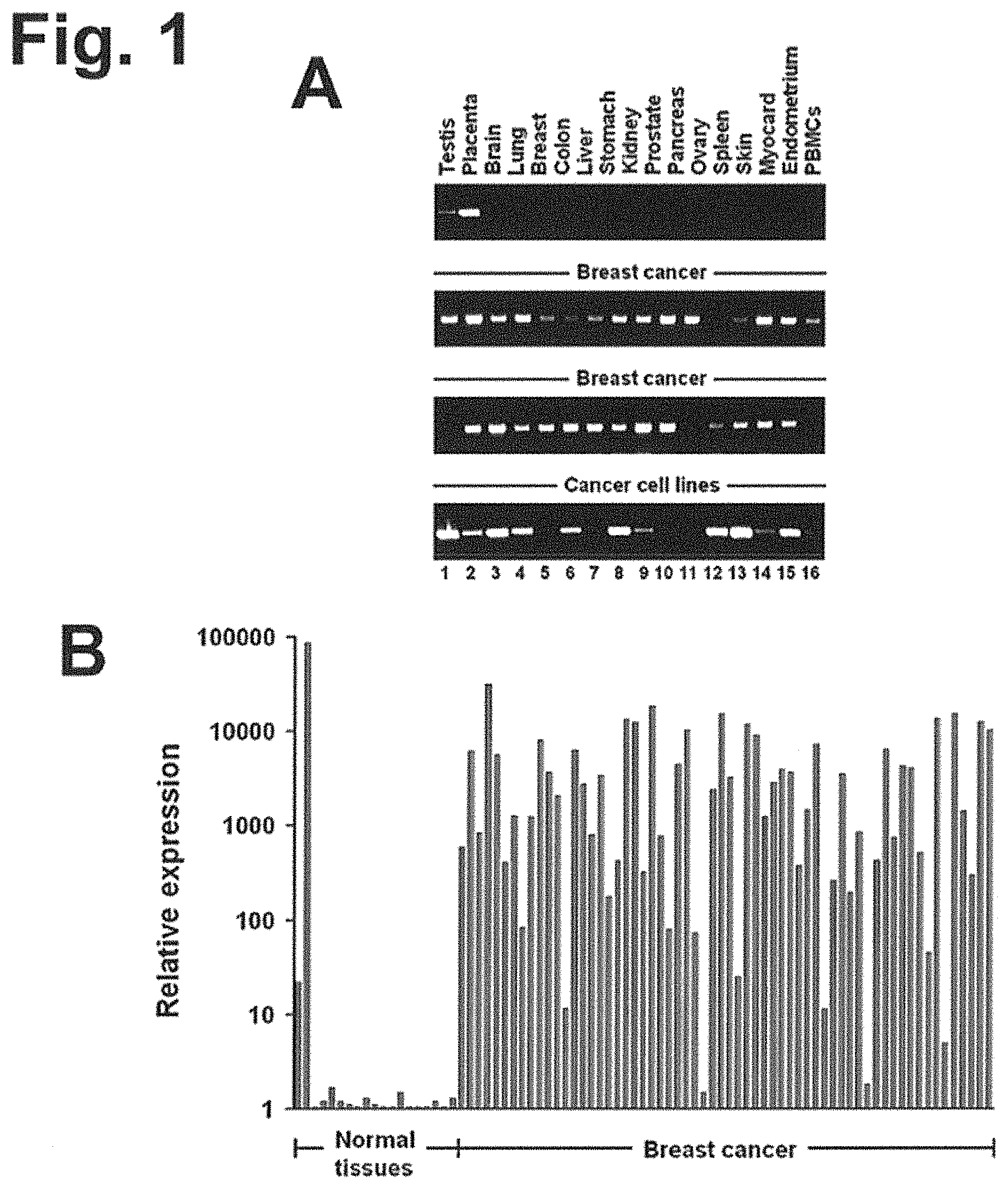
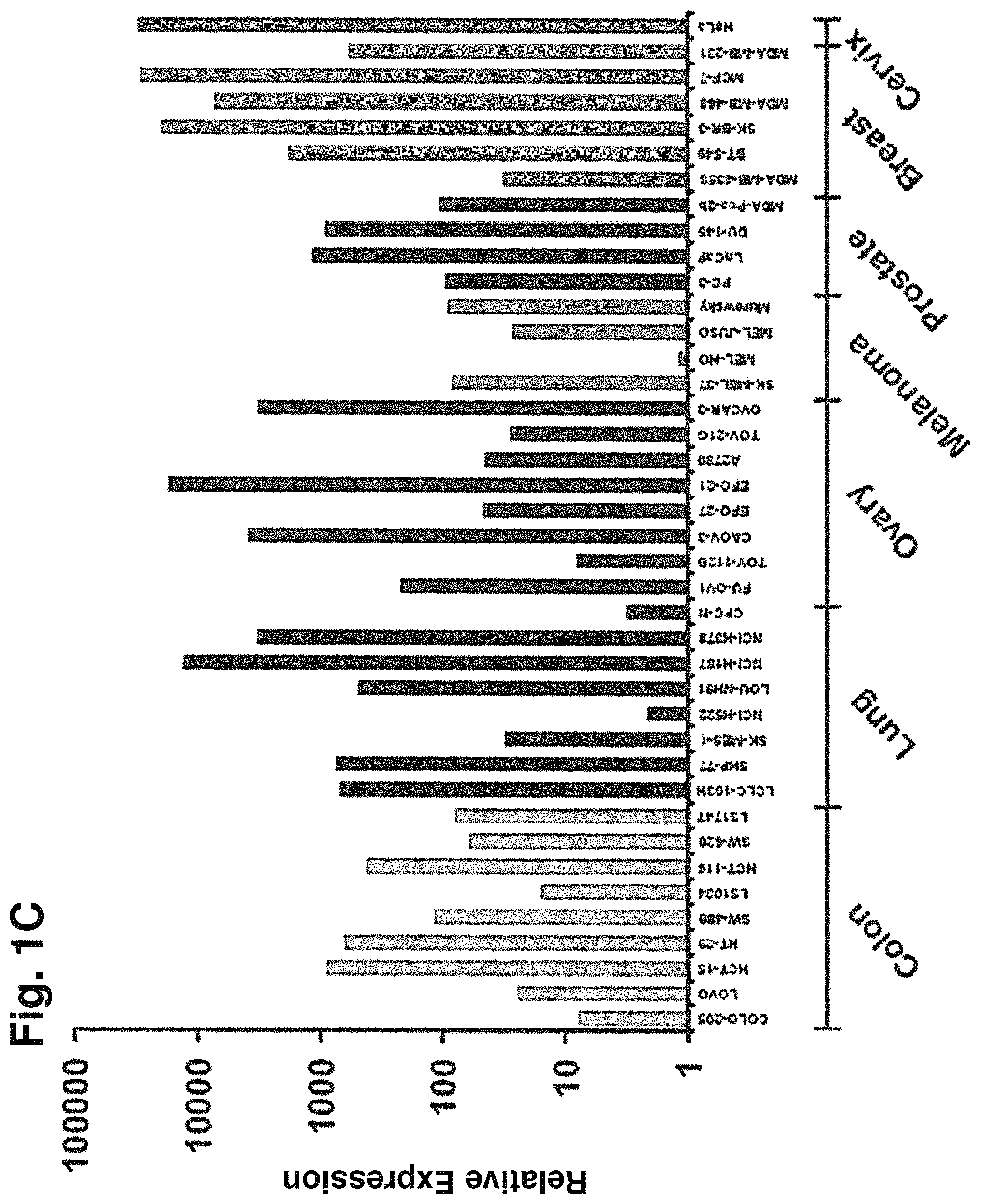
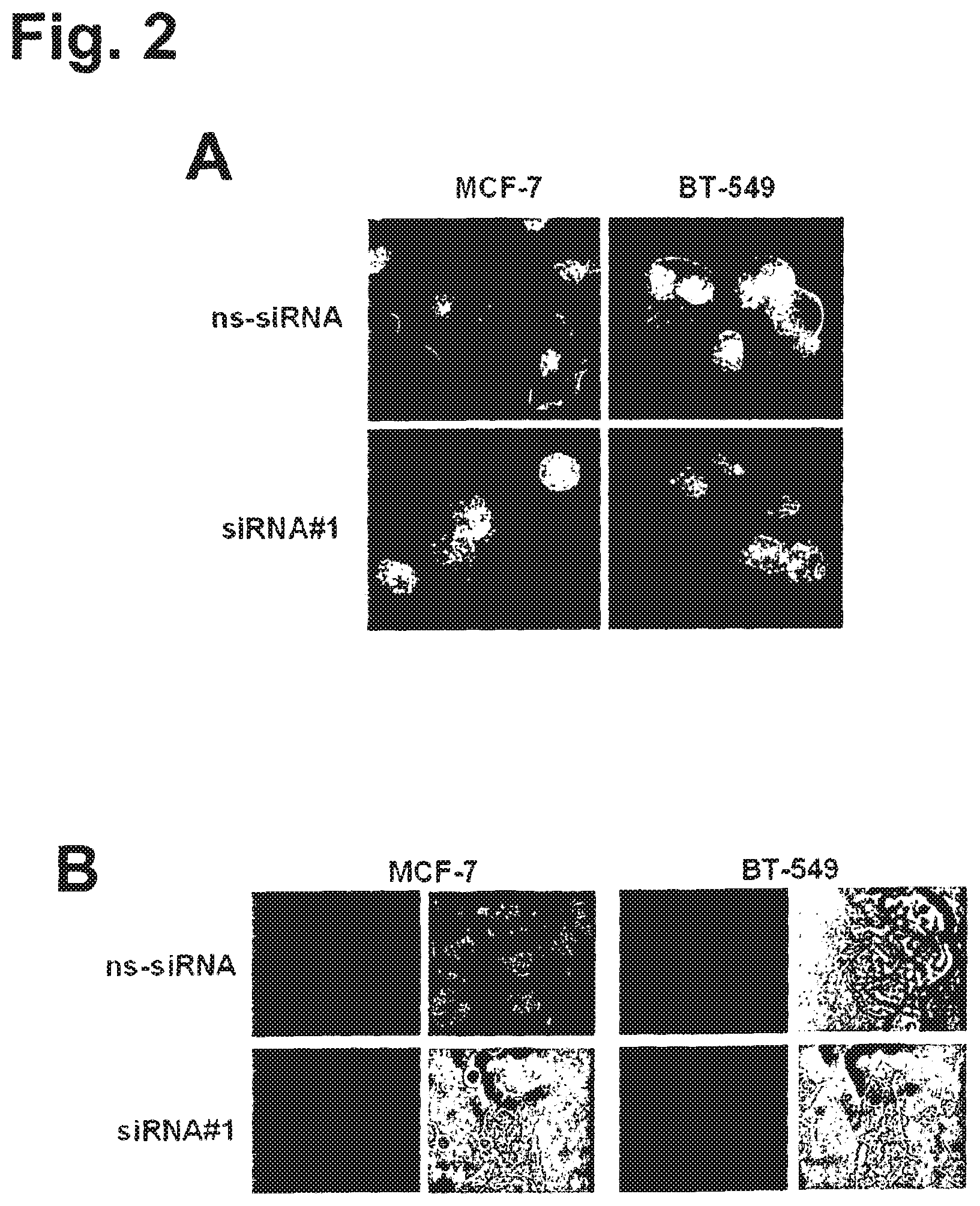
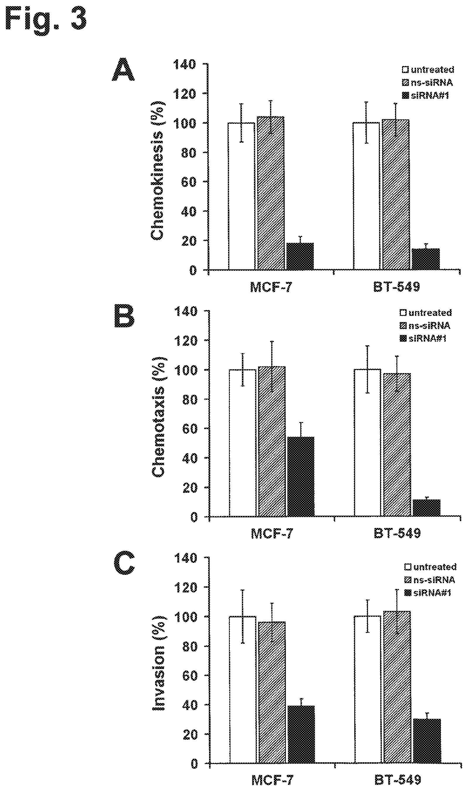
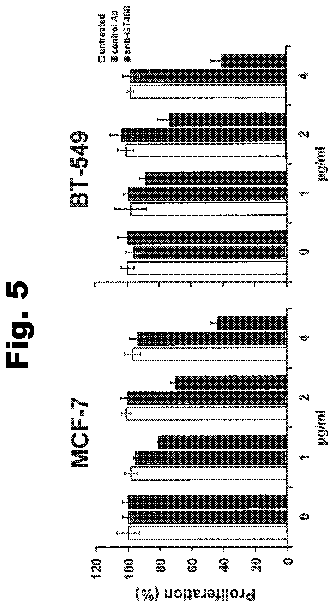
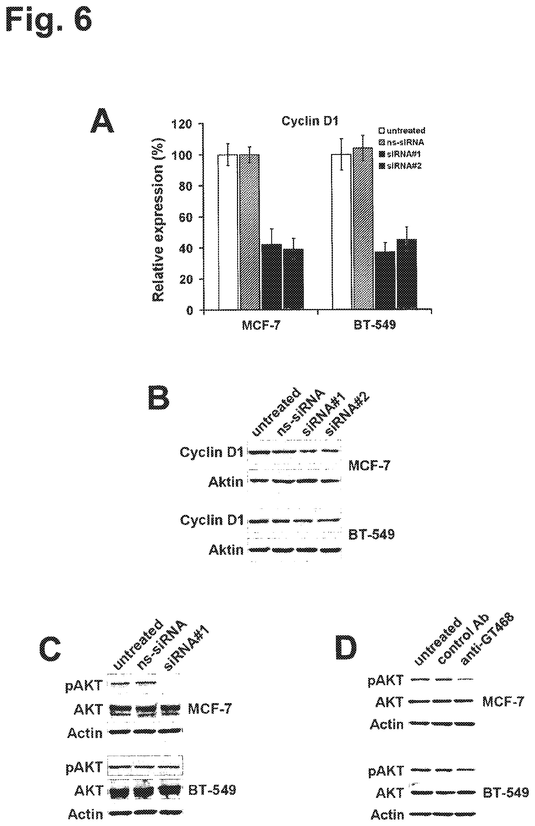
Using an integrated data mining and experimental validation approach for the discovery of new targets for antibody therapy of cancer we identified GT468. GT468 is a placenta-specific gene which is frequently aberrantly activated and highly expressed in a variety of tumor types, in particular breast cancer. RNAi-mediated silencing of GT468 in MCF-7 and BT-549 breast cancer cells profoundly impairs motility, migration and invasion and induces a G1/S cell cycle block with nearly complete abrogation of proliferation. Knock down of GT468 is associated with decreased expression of cyclin D1 and reduced phosphorylation of AKT kinase. Moreover, GT468 is localized on the surface of cancer cells and is accessible for antibodies which antagonize biological functions of this molecule.
GT468 has several properties that make it a highly attractive target for therapeutic antibodies. Being a differentiation antigen of a cell lineage which appears in the human body only in such an exceptional state as pregnancy, it is as absent from healthy toxicity-relevant tissues as a self-antigen can possibly be. Its high prevalence in a variety of tumor entities would make a broad number of patients eligible for treatment with GT468 targeting therapies. In the case of breast cancer for example, 82% of patients carry this target. Her2/neu, in contrast, the target of Herceptin, the only mAb available for treatment of this cancer type, is overexpressed in only 20-25% of breast cancer patients (Slamon, D. J., Godolphin, W., Jones, L. A., Holt, J. A., Wong, S. G., Keith, D. E., Levin, W. J., Stuart, S. G., Udove, J., Ullrich, A. et al. (1989) Science 244, 707-712). For lung cancer and for gastric cancer, in which GT468 is expressed in 42 and 58% of the cases respectively, there is no approved mAB treatment so far owing to the lack of appropriate targets in these cancer types.
GT468 is druggable by antibodies on living cells and such antibodies may precipitate anti-tumoral effects such as proliferation inhibition. GT468 is involved not only in proliferation but also cell motility, migration and invasion. Most interestingly, all these attributes do not only substantially contribute to the tumor phenotype but are also inherent properties of the human trophoblast, which physiological characteristics are to grow fast and to invade efficiently into uterus tissue. It is expected that mAbs against GT468 can be engineered, which intervene with all these functions at once on top of their potential to mediate immune effector functions such as ADCC and CDC.
Claims
The invention claimed is:
1. A method of inhibiting growth of a human cancer cell expressing GT468, comprising contacting the cell with an anti-GT468 antibody selected from the group consisting of: (i) an antibody fragment comprising six complementarity determining regions (CDRs) of an antibody produced by a hybridoma deposited under the accession number DSM ACC3039, DSM ACC3037, DSM ACC3036, DSM ACC3038, DSM ACC3040, or DSM ACC3042, (ii) a chimerized or humanized antibody comprising six CDRs of an antibody produced by a hybridoma deposited under the accession number DSM ACC3039, DSM ACC3037, DSM ACC3036, DSM ACC3038, DSM ACC3040, or DSM ACC3042, (iii) a full-length antibody comprising six CDRs of an antibody produced by a hybridoma deposited under the accession number DSM ACC3039, DSM ACC3037, DSM ACC3036, DSM ACC3038, DSM ACC3040, or DSM ACC3042, and (iv) an antibody of (i), (ii), or (iii) coupled to a therapeutic agent; wherein the cancer cell is selected from the group consisting of breast cancer, lung cancer, gastric cancer, ovarian cancer, colon cancer, pancreatic cancer, head & neck cancer, kidney cancer, prostate cancer, liver cancer, melanoma, sarcoma, myeloma, neuroblastoma, placental choriocarcinoma, cervical cancer, and thyroid cancer, and metastatic forms of the aforementioned cancers.
2. A method of killing a human cancer cell expressing GT468, comprising contacting the cell with an effective amount of an anti-GT468 antibody selected from the group consisting of: (i) an antibody fragment comprising six CDRs of an antibody produced by a hybridoma deposited under the accession number DSM ACC3039, DSM ACC3037, DSM ACC3036, DSM ACC3038, DSM ACC3040, or DSM ACC3042, (ii) a chimerized or humanized antibody comprising six CDRs of an antibody produced by a hybridoma deposited under the accession number DSM ACC3039, DSM ACC3037, DSM ACC3036, DSM ACC3038, DSM ACC3040, or DSM ACC3042, (iii) a full-length antibody comprising six CDRs of an antibody produced by a hybridoma deposited under the accession number DSM ACC3039, DSM ACC3037, DSM ACC3036, DSM ACC3038, DSM ACC3040, or DSM ACC3042, and (iv) an antibody of (i), (ii), or (iii) coupled to a therapeutic agent; wherein the cancer cell is selected from the group consisting of breast cancer, lung cancer, gastric cancer, ovarian cancer, colon cancer, pancreatic cancer, head & neck cancer, kidney cancer, prostate cancer, liver cancer, melanoma, sarcoma, myeloma, neuroblastoma, placental choriocarcinoma, cervical cancer, and thyroid cancer, and metastatic forms of the aforementioned cancers.
3. A method of inhibiting metastatic spread of a human cancer cell expressing GT468, comprising contacting the cell with an effective amount of an anti-GT468 antibody selected from the group consisting of: (i) an antibody fragment comprising six CDRs of an antibody produced by a hybridoma deposited under the accession number DSM ACC3039, DSM ACC3037, DSM ACC3036, DSM ACC3038, DSM ACC3040, or DSM ACC3042, (ii) a chimerized or humanized antibody comprising six CDRs of an antibody produced by a hybridoma deposited under the accession number DSM ACC3039, DSM ACC3037, DSM ACC3036, DSM ACC3038, DSM ACC3040, or DSM ACC3042, (iii) a full-length antibody comprising six CDRs of an antibody produced by a hybridoma deposited under the accession number DSM ACC3039, DSM ACC3037, DSM ACC3036, DSM ACC3038, DSM ACC3040, or DSM ACC3042, and (iv) an antibody of (i), (ii), or (iii) coupled to a therapeutic agent; wherein the cancer cell is selected from the group consisting of breast cancer, lung cancer, gastric cancer, ovarian cancer, colon cancer, pancreatic cancer, head & neck cancer, kidney cancer, prostate cancer, liver cancer, melanoma, sarcoma, myeloma, neuroblastoma, placental choriocarcinoma, cervical cancer, and thyroid cancer, and metastatic forms of the aforementioned cancers.
4. A method of treating a human cancer characterized by cancer cells expressing GT468 in a subject, comprising administering to said subject an anti-GT468 antibody selected from the group consisting of: (i) an antibody fragment comprising six CDRs of an antibody produced by a hybridoma deposited under the accession number DSM ACC3039, DSM ACC3037, DSM ACC3036, DSM ACC3038, DSM ACC3040, or DSM ACC3042, (ii) a chimerized or humanized antibody comprising six CDRs of an antibody produced by a hybridoma deposited under the accession number DSM ACC3039, DSM ACC3037, DSM ACC3036, DSM ACC3038, DSM ACC3040, or DSM ACC3042, (iii) a full-length antibody comprising six CDRs of an antibody produced by a hybridoma deposited under the accession number DSM ACC3039, DSM ACC3037, DSM ACC3036, DSM ACC3038, DSM ACC3040, or DSM ACC3042, (iv) an antibody of (i), (ii), or (iii) coupled to a therapeutic agent, and (v) a pharmaceutical composition comprising; (1) an antibody of (i), (ii), (iii), or (iv); and (2) a pharmaceutically acceptable carrier; wherein the cancer is selected from the group consisting of breast cancer, lung cancer, gastric cancer, ovarian cancer, colon cancer, pancreatic cancer, head & neck cancer, kidney cancer, prostate cancer, liver cancer, melanoma, sarcoma, myeloma, neuroblastoma, placental choriocarcinoma, cervical cancer, and thyroid cancer, and metastatic forms of the aforementioned cancers.
5. The method of any one of claims 1 to 4, wherein the therapeutic agent is a toxin, a radioisotope, a drug, or a cytotoxic agent.
Description
SUMMARY OF THE INVENTION
The present invention generally provides antibodies useful as therapeutics for treating and/or preventing diseases associated with cells expressing GT468 and/or being characterized by association of GT468 with their cell surface, including tumor-related diseases such as cancer, in particular breast cancer, lung cancer, gastric cancer, ovarian cancer, hepatocellular cancer, colon cancer, pancreatic cancer, esophageal cancer, head & neck cancer, kidney cancer, in particular renal cell carcinoma, prostate cancer, liver cancer, melanoma, sarcoma, myeloma, neuroblastoma, placental choriocarcinoma, cervical cancer, and thyroid cancer, and the metastatic forms thereof. In one embodiment, the cancer disease is metastatic cancer in the lung.
In one aspect the invention relates to an antibody having the ability of binding to GT468. The antibodies described herein preferably are isolated monoclonal antibodies which specifically bind to an epitope present on GT468, preferably an epitope located within the extracellular domain of GT468, more preferably SEQ ID NOs: 3-10 and 35-82. Preferably, the antibody has the ability of binding to GT468 located on the cell surface and preferably binds to one or more epitopes located within the extracellular domain of GT468, preferably within amino acid residues 23-212 of GT468, and most preferably binds to an epitope located within one of the amino acid sequences of SEQ ID Nos: 3-10 and 35-82. In one preferred embodiment, the antibody is specific for one or more of the amino acid sequences of SEQ ID Nos: 3-10 and 35-82. In various embodiments, the antibody has the ability of binding to a peptide comprising amino acids 29 to 119, preferably amino acids 29 to to 212 and more preferably amino acids 23 to 212 of SEQ ID NO: 2.
Monoclonal antibodies encompassed by the present invention include IgA, IgG1-4, IgE, IgM, and IgD antibodies. In one embodiment the antibody is an IgG1 antibody, more particularly an IgG1, kappa or IgG1, lambda isotype. In another embodiment the antibody is an IgG3 antibody, more particularly an IgG3, kappa or IgG3, lambda isotype. In yet another embodiment the antibody is an IgG4 antibody, more particularly an IgG4, kappa or IgG4, lambda isotype. In still another embodiment the antibody is an IgA1 or IgA2 antibody. In still another embodiment the antibody is an IgM antibody.
In one embodiment, the antibody of the invention binds to one or more of the peptides according to SEQ ID Nos: 75-79. In one embodiment, the antibody binds to the peptide according to SEQ ID No: 75 and/or the peptide according to SEQ ID No: 76, more preferably binds to the peptide according to SEQ ID No: 75 and the peptide according to SEQ ID No: 76. In another embodiment, the antibody binds to the peptide according to SEQ ID No: 77 and/or the peptide according to SEQ ID No: 78 and/or the peptide according to SEQ ID No: 79, more preferably binds to the peptide according to SEQ ID No: 78 and the peptide according to SEQ ID No: 79 or binds to the peptide according to SEQ ID No: 77, the peptide according to SEQ ID No: 78 and the peptide according to SEQ ID No: 79.
In one embodiment, the antibody of the invention is obtained using the peptide according to SEQ ID No: 4 or the peptide according to SEQ ID No: 80 for immunization. In another embodiment, the antibody of the invention is obtained using the peptide according to SEQ ID No: 6 or the peptide according to SEQ ID No: 81 for immunization. Preferably, the antibody of the invention binds to one or more of said peptides.
In a further embodiment, the antibody which binds to the peptide according to SEQ ID No: 75 and/or the peptide according to SEQ ID No: 76, more preferably binds to the peptide according to SEQ ID No: 75 and the peptide according to SEQ ID No: 76 is obtained using the peptide according to SEQ ID No: 4 or the peptide according to SEQ ID No: 80 for immunization. In another embodiment, the antibody which binds to the peptide according to SEQ ID No: 77 and/or the peptide according to SEQ ID No: 78 and/or the peptide according to SEQ ID No: 79, more preferably binds to the peptide according to SEQ ID No: 78 and the peptide according to SEQ ID No: 79 or binds to the peptide according to SEQ ID No: 77, the peptide according to SEQ ID No: 78 and the peptide according to SEQ ID No: 79 is obtained using the peptide according to SEQ ID No: 6 or the peptide according to SEQ ID No: 81 for immunization.
In one embodiment, the antibody of the invention binds to one or more of the peptides according to SEQ ID Nos: 60-66, preferably according to SEQ ID Nos: 61-66. In one embodiment, the antibody binds to the peptide according to SEQ ID No: 60 and/or the peptide according to SEQ ID No: 61 and/or the peptide according to SEQ ID No: 62, more preferably binds to the peptide according to SEQ ID No: 60 and the peptide according to SEQ ID No: 61 and the peptide according to SEQ ID No: 62. In one embodiment, the antibody binds to the peptide according to SEQ ID No: 61 and/or the peptide according to SEQ ID No: 62, more preferably binds to the peptide according to SEQ ID No: 61 and the peptide according to SEQ ID No: 62. In another embodiment, the antibody binds to the peptide according to SEQ ID No: 64 and/or the peptide according to SEQ ID No: 65, more preferably binds to the peptide according to SEQ ID No: 64 and the peptide according to SEQ ID No: 65. In another embodiment, the antibody binds to the peptide according to SEQ ID No: 65 and/or the peptide according to SEQ ID No: 66, more preferably binds to the peptide according to SEQ ID No: 65 and the peptide according to SEQ ID No: 66.
In one embodiment, the antibody of the invention is obtained using the peptide according to SEQ ID No: 82 for immunization. Preferably, the antibody of the invention binds to said peptide. The peptide according to SEQ ID No: 82 has a C-terminal GS-linker which has been added to assist in the formation of a disulfide bond between amino acids 1 and 16 of the peptide. This peptide presumably resembles the native conformation of GT468. Antibodies produced by the hybridoma 51-1A-1 which has been obtained using the peptide according to SEQ ID No: 82 for immunization showed strong inhibitory activity on proliferation and colony formation of GT468 expressing cancer cells.
In a further embodiment, the antibody which binds to the peptide according to SEQ ID No: 64 and/or the peptide according to SEQ ID No: 65, more preferably binds to the peptide according to SEQ ID No: 64 and the peptide according to SEQ ID No: 65 or the antibody which binds to the peptide according to SEQ ID No: 65 and/or the peptide according to SEQ ID No: 66, more preferably binds to the peptide according to SEQ ID No: 65 and the peptide according to SEQ ID No: 66 is obtained using the peptide according to SEQ ID No: 82 for immunization.
In one embodiment, the antibody of the invention binds to one or more of the peptides according to SEQ ID Nos: 57-60. In one embodiment, the antibody binds to the peptide according to SEQ ID No: 57 and/or the peptide according to SEQ ID No: 58 and/or the peptide according to SEQ ID No: 59, more preferably binds to the peptide according to SEQ ID No: 57 and the peptide according to SEQ ID No: 58 or binds to the peptide according to SEQ ID No: 57 and the peptide according to SEQ ID No: 58 and the peptide according to SEQ ID No: 59. In another embodiment, the antibody binds to the peptide according to SEQ ID No: 59 and/or the peptide according to SEQ ID No: 60, more preferably binds to the peptide according to SEQ ID No: 59 and the peptide according to SEQ ID No: 60.
In one embodiment, the antibody of the invention is obtained using the peptide according to SEQ ID No: 3 for immunization. Preferably, the antibody of the invention binds to said peptide.
Preferably, the antibody of the invention binds to cancer cells, in particular cells of the cancer types mentioned herein and, preferably, does not bind substantially to non-cancerous cells, more preferably does not bind substantially to non-cancerous cells other than placental cells and testis cells. Preferably, binding of said antibody to cells expressing GT468 and/or being characterized by association of GT468 with their cell surface such as cancer cells mediates killing of said cells and/or inhibits one or more activities of such cells such as motility, migration, invasion, proliferation and/or colony formation. Preferably, the antibody inhibits proliferation and/or colony formation of said cells.
Killing of cells and/or inhibition of one or more activities of cells, in particular cell proliferation and colony formation, by the antibody of the invention is preferably induced by binding of the antibody to GT468 expressed by said cells and/or being associated with the cell surface of said cells. Such killing of cells and/or inhibition of one or more activities of cells can be utilized therapeutically as described herein. In particular, killing of cells and/or inhibition of proliferation of cells and/or inhibition of colony formation of cells can be utilized for treating or preventing cancer, including cancer metastasis. Inhibition of motility, migration, invasion, proliferation and/or colony formation of cells can be utilized, in particular, for treating or preventing cancer metastasis and the metastatic spread of cancer cells.
The cells expressing GT468 and/or being characterized by association of GT468 with their cell surface are preferably cancer cells and are, in particular, selected from the group consisting of tumorigenic cancer cells of the following cancer diseases: breast cancer, lung cancer, gastric cancer, ovarian cancer, hepatocellular cancer, colon cancer, pancreatic cancer, esophageal cancer, head & neck cancer, kidney cancer, in particular renal cell carcinoma, prostate cancer, liver cancer, melanoma, sarcoma, myeloma, neuroblastoma, placental choriocarcinoma, cervical cancer, and thyroid cancer, and the metastatic forms thereof. In one embodiment, the cancer disease is metastatic cancer in the lung.
The antibody of the invention may be attached to one or more therapeutic effector moieties, e.g., radiolabels, cytotoxins, therapeutic enzymes, agents that induce apoptosis, and the like in order to provide for targeted cytotoxicity, i.e., killing of tumor cells.
Preferably the antibody of the invention mediates killing of cells by inducing complement dependent cytotoxicity (CDC) mediated lysis, antibody dependent cellular cytotoxicity (ADCC) mediated lysis, apoptosis, homotypic adhesion, and/or phagocytosis, preferably by inducing CDC mediated lysis and/or ADCC mediated lysis. However, the present invention also includes embodiments wherein the antibody exerts its activity as described herein such as killing of cells and/or inhibition of one or more activities of cells, e.g. cell proliferation and/or colony formation, without inducing complement dependent cytotoxicity (CDC) mediated lysis, antibody dependent cellular cytotoxicity (ADCC) mediated lysis, apoptosis, homotypic adhesion, and/or phagocytosis. For example, the antibody of the invention may also exert an effect simply by binding to GT468 on the cell surface, thus, e.g. blocking proliferation of the cells. In one embodiment the antibody of the invention does not induce CDC mediated lysis of cells.
Preferably, ADCC mediated lysis of cells takes place in the presence of effector cells, which in particular embodiments are selected from the group consisting of monocytes, mononuclear cells, NK cells and PMNs, and phagocytosis is by macrophages.
In one particularly preferred embodiment, the antibody of the invention has the activity of inhibiting or reducing proliferation of cells expressing GT468 and/or being characterized by association of GT468 with their cell surface, preferably cancer cells such as breast cancer cells. This activity can be measured in vitro by determining proliferation of GT468-expressing cancer cells in an assay using bromodeoxyuridine (5-bromo-2-deoxyuridine, BrdU). BrdU is a synthetic nucleoside which is an analogue of thymidine and can be incorporated into the newly synthesized DNA of replicating cells (during the S phase of the cell cycle), substituting for thymidine during DNA replication. Detecting the incorporated chemical using, for example, antibodies specific for BrdU indicates cells that were actively replicating their DNA. The cell lines BT-549, Caov-3, EFO-21, SK-BR-3, MCF-7, MDA-MB-468 and MDA-MB-231 can be used as GT468-expressing cancer cells. In a preferred embodiment, the antibody of the invention inhibits or reduces proliferation of one or more of the cell lines SK-BR-3, MCF-7 and MDA-MB-468, preferably inhibits or reduces proliferation of both of the cell lines SK-BR-3 and MCF-7, and most preferably inhibits or reduces proliferation of all of the cell lines SK-BR-3, MCF-7 and MDA-MB-468. Preferred antibodies of the invention in this respect are selected from the group consisting of (i) antibodies produced by and/or obtainable from the hybridomas 35-48B-1, 35-50A-2a, 38-10B-1, 38-1A-1, 45-2A-1, 45-8A-2, 48-3B-1, 48-4A-1, 49-3A-1, 51-1A-1, 53-13A-2, 53-29A1, 56-4A-2, 62-9B-1, 78H11 1H6 and 44-3A-2, (ii) antibodies which are chimerized or humanized forms of the antibodies under (i), and (iii) antibodies having the specificity of the antibodies under (i) and, in particular, antibodies comprising the antigen binding portion or antigen binding site, in particular the variable region, of the antibodies under (i). Particularly preferred antibodies of the invention in this respect are selected from the group consisting of (i) antibodies produced by and/or obtainable from the hybridomas 35-48B-1, 48-3B-1, 51-1A-1, and 56-4A-2, (ii) antibodies which are chimerized or humanized forms of the antibodies under (i), and (iii) antibodies having the specificity of the antibodies under (i) and, in particular, antibodies comprising the antigen binding portion or antigen binding site, in particular the variable region, of the antibodies under (i).
In one particularly preferred embodiment, the antibody of the invention has the activity of inhibiting or reducing colony formation of cells expressing GT468 and/or being characterized by association of GT468 with their cell surface, preferably cancer cells such as breast cancer cells. This activity can be measured in vitro in a clonogenic assay. The cell lines BT-549, Caov-3, EFO-21, SK-BR-3, MCF-7, MDA-MB-468 and MDA-MB-231 can be used as GT468-expressing cancer cells. A clonogenic assay is a microbiology technique for studying the effectiveness of specific agents on the survival and proliferation of cells. It is frequently used in cancer research laboratories to determine the effect of drugs or radiation on proliferating tumor cells. The experiment involves three major steps: (i) applying a treatment to a sample of cells, in particular cancer cells, (ii) plating the cells in a tissue culture vessel and (iii) allowing the cells to grow. The colonies produced are fixed, stained, and counted. Colony formation is of importance with respect to the formation of metastases if individual tumor cells colonize organs. The inhibitory activity of the antibodies indicates their potential in suppressing the formation of metastases. Antibodies having the activity of inhibiting or reducing colony formation in a clonogenic assay are particularly useful for treating or preventing metastasis and the metastatic spread of cancer cells, in particular of the cancer types mentioned herein. Preferred antibodies of the invention in this respect are selected from the group consisting of (i) antibodies produced by and/or obtainable from the hybridomas 51G6 2H3 2B4, 78H11 1H6, 16-5B-1, 22-1A-1, 29-8B-1, and 51-1A-1, (ii) antibodies which are chimerized or humanized forms of the antibodies under (i), and (iii) antibodies having the specificity of the antibodies under (i) and, in particular, antibodies comprising the antigen binding portion or antigen binding site, in particular the variable region, of the antibodies under (i). Particularly preferred antibodies of the invention in this respect are selected from the group consisting of (i) antibodies produced by and/or obtainable from the hybridomas 51G6 2H3 2B4, 29-8B-1, and 51-1A-1, (ii) antibodies which are chimerized or humanized forms of the antibodies under (i), and (iii) antibodies having the specificity of the antibodies under (i) and, in particular, antibodies comprising the antigen binding portion or antigen binding site, in particular the variable region, of the antibodies under (i).
In one particularly preferred embodiment, the antibody of the invention has the activity of inhibiting or reducing proliferation of cells expressing GT468 and/or being characterized by association of GT468 with their cell surface as explained above and has the activity of inhibiting or reducing colony formation of cells expressing GT468 and/or being characterized by association of GT468 with their cell surface as explained above. Preferred antibodies of the invention in this respect are selected from the group consisting of (i) antibodies produced by and/or obtainable from the hybridoma 51-1A-1, (ii) antibodies which are chimerized or humanized forms of the antibodies under (i), and (iii) antibodies having the specificity of the antibodies under (i) and, in particular, antibodies comprising the antigen binding portion or antigen binding site, in particular the variable region, of the antibodies under (i).
In one embodiment the invention relates to antibodies which (i) bind to cells expressing GT468 and/or being characterized by association of GT468 with their cell surface, and (ii) do not bind to cells not expressing GT468 and/or not being characterized by association of GT468 with their cell surface. The antibodies of the invention preferably (i) mediate killing and/or inhibit proliferation of cells expressing GT468 and/or being characterized by association of GT468 with their cell surface, and (ii) do not mediate killing and/or do not inhibit proliferation of cells not expressing GT468 and/or not being characterized by association of GT468 with their cell surface.
The antibody of the invention may be a monoclonal, chimeric, human, or humanized antibody, or a fragment of an antibody and may be selected from the group consisting of an IgG1, an IgG2, preferably IgG2a and IgG2b, an IgG3, an IgG4, an IgM, an IgA1, an IgA2, a secretory IgA, an IgD, and an IgE antibody.
Antibodies of the invention include fully human antibodies. Such antibodies may be produced in a non-human transgenic animal, e.g., a transgenic mouse, capable of producing multiple isotypes of human monoclonal antibodies to GT468 by undergoing V-D-J recombination and isotype switching. Such transgenic animal can also be a transgenic rabbit for producing polyclonal antibodies such as disclosed in US 2003/0017534.
Binding of an antibody of the invention to the GT468 antigen may mediate the killing of cells expressing GT468 and/or being characterized by association of GT468 with their cell surface (e.g. a tumor cell), e.g. by activation of the complement system, and/or may inhibit proliferation of cells expressing GT468 and/or being characterized by association of GT468 with their cell surface (e.g. a tumor cell). Alternatively or in addition to mediating killing of cells expressing GT468 and/or being characterized by association of GT468 with their cell surface and/or inhibiting proliferation of cells expressing GT468 and/or being characterized by association of GT468 with their cell surface, binding of an antibody of the invention to the GT468 antigen may inhibit motility, migration, colony formation and/or invasion of cells expressing GT468 and/or being characterized by association of GT468 with their cell surface (e.g. a tumor cell), and thus, may inhibit metastatic spread of tumor cells. The killing of cells expressing GT468 and/or being characterized by association of GT468 with their cell surface may occur by one or more of the following mechanisms: complement dependent cytotoxity (CDC) of cells expressing GT468 and/or being characterized by association of GT468 with their cell surface; apoptosis of cells expressing GT468 and/or being characterized by association of GT468 with their cell surface; effector cell phagocytosis of cells expressing GT468 and/or being characterized by association of GT468 with their cell surface; or effector cell antibody dependent cellular cytotoxicity (ADCC) of cells expressing GT468 and/or being characterized by association of GT468 with their cell surface.
According to all aspects of the invention, GT468 is preferably human GT468, preferably having the amino acid sequence according to SEQ ID NO: 2, more preferably having the amino acid sequence of the extracellular domain of the amino acid sequence according to SEQ ID NO: 2, in particular having the amino acid sequence spanning from amino acids 23 to 212 of SEQ ID NO: 2.
In particular preferred embodiments, the antibody of the invention binds to native epitopes of GT468 present on the surface of living cells such as those of SEQ ID NOs: 3-10 and 35-82. In further preferred embodiments, the antibody of the invention is specific for cancer cells, preferably breast cancer cells. Preferably, the antibody of the invention is specific for GT468-expressing cancer cells such as the cell lines BT-549, Caov-3, EFO-21, SK-BR-3, MCF-7, MDA-MB-468 and MDA-MB-231 and does not bind to cancer cells not expressing GT468 such as NUG-C4 gastric cancer cells.
In certain embodiments of the invention, GT468 is expressed on and/or associated with the surface of cells.
Antibodies of the invention may be obtained by a method comprising the step of immunizing an animal with a protein or peptide having an amino acid sequence selected from the group consisting of SEQ ID NOs: 2-10 and 35-82, or an immunogenic fragment or derivative thereof, or a nucleic acid or host cell expressing said protein or peptide, or immunogenic fragment or derivative thereof. Preferably, an antibody of the invention is specific for the afore mentioned proteins, peptides or immunogenic fragments or derivatives thereof. In the context of a protein or peptide used in immunization, a derivative relates to a variant of such protein or peptide which has the same immunogenic properties as the protein or peptide from which it is derived. In particular, the derivative of a protein or peptide when used in immunization for the production of antibodies, in particular monoclonal antibodies, provides antibodies having the same specificity as antibodies obtained when using the protein or peptide in immunization. For example, such derivative may include the deletion, substitution or addition of one or more amino acids. In particular, it may include the addition of one or more amino acids such as cysteine at either the N-terminus or C-terminus or both.
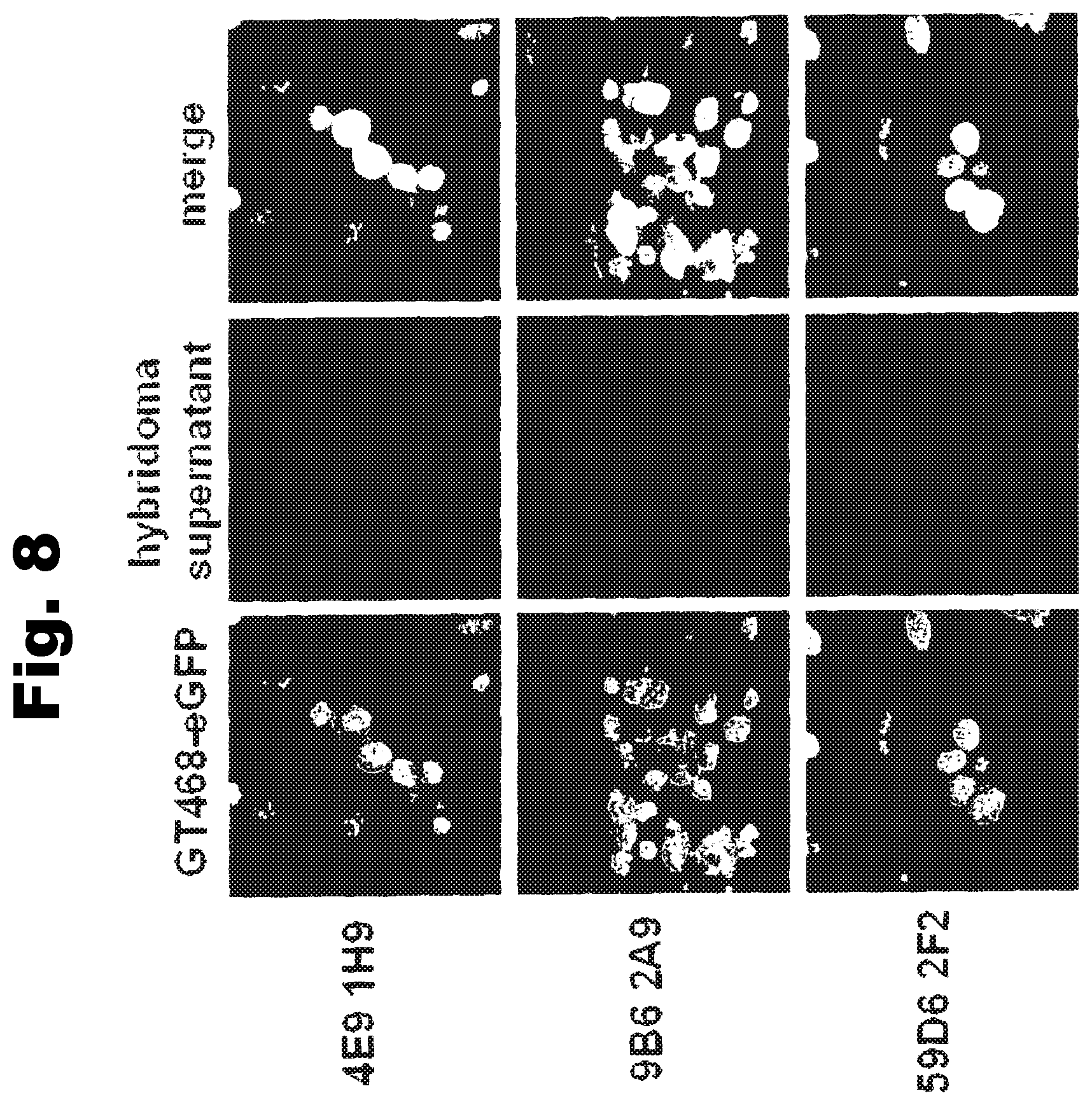
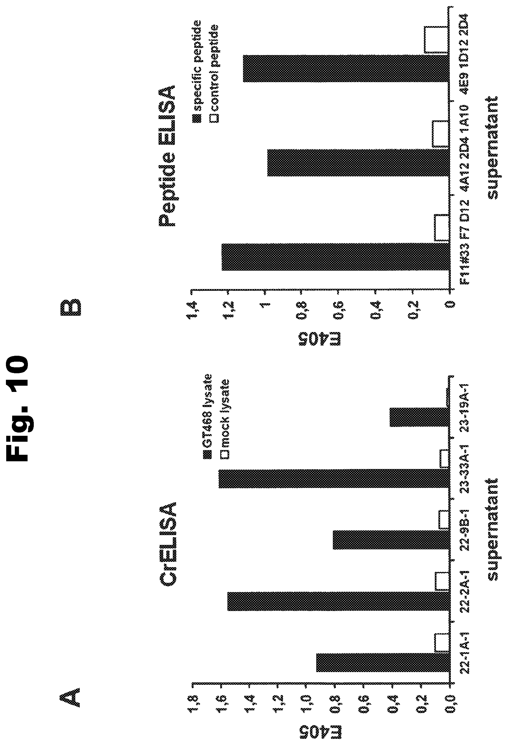
In a particularly preferred embodiment, the antibody of the invention is produced by a clone having the accession no. DSM ACC2822 (4E9-1H9), DSM ACC2826 (9B6-2A9), DSM ACC2824 (59D6-2F2), DSM ACC2825 (61C11-2B5), DSM ACC2823 (78H11-1H6), DSM ACC2895 (22-1A-1), DSM ACC2893 (22-2A-1), DSM ACC2896 (22-9B-1), DSM ACC2897 (23-33A-1), DSM ACC2891 (23-19A-1), DSM ACC2894 (F11#33F7D12), DSM ACC2892 (4A12 2D4 1A10), DSM ACC2898 (4E9 1D12 2D4), DSM ACC2961 (42H111 1C11 2B2), DSM ACC2962 (51G6 2H3 2B4), DSM ACC2943 (16-5B-1), DSM ACC2956 (20-11A-1), DSM ACC2947 (29-1A-2), DSM ACC2964 (29-8B-1), DSM ACC2959 (35-48B-1), DSM ACC2963 (35-50A-2a), DSM ACC2957 (38-10B-1), DSM ACC2958 (38-1A-1), DSM ACC2948 (44-3A-2), DSM ACC2949 (45-2A-1), DSM ACC2950 (45-8A-2), DSM ACC2951 (48-3B-1), DSM ACC2952 (48-4A-1), DSM ACC2946 (49-3A-1), DSM ACC2945 (49-8A-1), DSM ACC2944 (51-1A-1), DSM ACC2953 (53-13A-2), DSM ACC2955 (53-29A-1), DSM ACC2960 (54-4B-2), DSM ACC2954 (56-4A-2), DSM ACC3001 (62-9B-1), DSM ACC3039 (4A5 1E11 1B7), DSM ACC3037 (7H12 2E6 2C4), DSM ACC3036 (11D7 1G10 2B4), DSM ACC3038 (18-2A-1), DSM ACC3040 (63-1A-2), or DSM ACC3042 (3E5 2G4).
In one embodiment the antibody of the invention is coupled to a therapeutic agent such as a toxin, a radioisotope, a drug or a cytotoxic agent.
In a further aspect the invention relates to a hybridoma capable of producing the antibody of the invention. Preferred hybridomas are those having the accession no. DSM ACC2822 (4E9-1H9), DSM ACC2826 (9B6-2A9), DSM ACC2824 (59D6-2F2), DSM ACC2825 (61C11-2B5), DSM ACC2823 (78H11-1H16), DSM ACC2895 (22-1A-1), DSM ACC2893 (22-2A-1), DSM ACC2896 (22-9B-1), DSM ACC2897 (23-33A-1), DSM ACC2891 (23-19A-1), DSM ACC2894 (F11#33F7D12), DSM ACC2892 (4A12 2D4 1A10), DSM ACC2898 (4E9 1D12 2D4), DSM ACC2961 (42H11 1C11 2B2), DSM ACC2962 (51G6 2H3 2B4), DSM ACC2943 (16-5B-1), DSM ACC2956 (20-11A-1), DSM ACC2947 (29-1A-2), DSM ACC2964 (29-8B-1), DSM ACC2959 (35-48B-1), DSM ACC2963 (35-50A-2a), DSM ACC2957 (38-10B-1), DSM ACC2958 (38-1A-1), DSM ACC2948 (44-3A-2), DSM ACC2949 (45-2A-1), DSM ACC2950 (45-8A-2), DSM ACC2951 (48-3B-1), DSM ACC2952 (48-4A-1), DSM ACC2946 (49-3A-1), DSM ACC2945 (49-8A-1), DSM ACC2944 (51-1A-1), DSM ACC2953 (53-13A-2), DSM ACC2955 (53-29A-1), DSM ACC2960 (54-4B-2), DSM ACC2954 (56-4A-2), DSM ACC3001 (62-9B-1), DSM ACC3039 (4A5 1E11 1B7), DSM ACC3037 (7H12 2E6 2C4), DSM ACC3036 (11D7 1G10 2B4), DSM ACC3038 (18-2A-1), DSM ACC3040 (63-1A-2), or DSM ACC3042 (3E5 2G4).
Antibodies of the invention are designated herein by referring to the designation of the antibody and/or by referring to the clone producing the antibody, e.g. 4E9-1H9.
Preferably, the antibodies of the invention have the ability to discriminate GT468-variants expressed by different cell types including cancer cells and non-malignant cells.
The antibodies of the invention preferably mediate killing of cells expressing GT468 and/or being characterized by association of GT468 with their cell surface by binding to GT468. Preferably GT468 is expressed on the surface of said cells. In one embodiment, antibodies of the invention induce complement dependent cytotoxicity (CDC), e.g. at least about 20-40% CDC mediated lysis, preferably about 40-50% CDC mediated lysis, and more preferably more than 50% CDC mediated lysis of cells expressing GT468 and/or being characterized by association of GT468 with their cell surface. Alternatively or in addition to inducing CDC, antibodies of the invention may induce antibody dependent cellular cytotoxicity (ADCC) of cells expressing GT468 and/or being characterized by association of GT468 with their cell surface in the presence of effector cells (e.g., monocytes, mononuclear cells, NK cells and PMNs). Antibodies of the invention may have the ability to induce apoptosis of cells expressing GT468 and/or being characterized by association of GT468 with their cell surface, induce homotypic adhesion of cells expressing GT468 and/or being characterized by association of GT468 with their cell surface and/or induce phagocytosis of cells expressing GT468 and/or being characterized by association of GT468 with their cell surface in the presence of macrophages. The antibodies of the invention may have one or more of the above described functional properties. Preferably, antibodies of the invention induce CDC mediated lysis and ADCC mediated lysis of cells expressing GT468 and/or being characterized by association of GT468 with their cell surface and more preferably induce ADCC mediated lysis of cells expressing GT468 and/or being characterized by association of GT468 with their cell surface while they do not induce CDC mediated lysis of said cells. Exemplary target cells for antibodies of the present invention include, but are not limited to, cancer cells expressing GT468 and/or being characterized by association of GT468 with their cell surface. In a particular preferred embodiment, killing of cells mediated by antibodies of the invention is GT468 specific, i.e. antibodies of the invention mediate killing, preferably CDC and/or ADCC mediated lysis, of cells expressing GT468 and/or being characterized by association of GT468 with their cell surface but do not mediate killing of cells not expressing GT468 and/or not being characterized by association of GT468 with their cell surface. The antibodies described above may be used to mediate killing of tumor cells in the treatment or prevention of cancer such as breast cancer, lung cancer, gastric cancer, ovarian cancer, hepatocellular cancer, colon cancer, pancreatic cancer, esophageal cancer, head & neck cancer, kidney cancer, in particular renal cell carcinoma, prostate cancer, liver cancer, melanoma, sarcoma, myeloma, neuroblastoma, placental choriocarcinoma, cervical cancer, and thyroid cancer, and the metastatic forms thereof. In one embodiment, the cancer disease is metastatic cancer in the lung.
Antibodies of the invention may be derived from different species, including but not limited to mouse, rat, rabbit, guinea pig and human. Antibodies of the invention also include chimeric molecules in which an antibody constant region derived from one species, preferably human, is combined with the antigen binding site derived from another species. Moreover antibodies of the invention include humanized molecules in which the antigen binding sites of an antibody derived from a non-human species are combined with constant and framework regions of human origin.
Antibodies of the invention include polyclonal and monoclonal antibodies and include IgG2a (e.g. IgG2a, .kappa., .lamda.), IgG2b (e.g. IgG2b, .kappa., .lamda.), IgG3 (e.g. IgG3, .kappa., .lamda.) and IgM antibodies. However, other antibody isotypes are also encompassed by the invention, including IgG1, IgA1, IgA2, secretory IgA, IgD, and IgE antibodies. The antibodies can be whole antibodies or antigen-binding fragments thereof including, for example, Fab, F(ab').sub.2, Fv, single chain Fv fragments or bispecific antibodies. Furthermore, the antigen-binding fragments include binding-domain immunoglobulin fusion proteins comprising (i) a binding domain polypeptide (such as a heavy chain variable region or a light chain variable region) that is fused to an immunoglobulin hinge region polypeptide, (ii) an immunoglobulin heavy chain CH2 constant region fused to the hinge region, and (iii) an immunoglobulin heavy chain CH3 constant region fused to the CH2 constant region. Such binding-domain immunoglobulin fusion proteins are further disclosed in US2003/0118592 and US 2003/0133939.
Antibodies of the present invention preferably dissociate from GT468 with a dissociation equilibrium constant (KD) of approximately 1-100 nM or less. Preferably, antibodies of the invention do not cross-react with related cell-surface antigens and thus do not inhibit their function.
In preferred embodiments, antibodies of the present invention can be characterized by one or more of the following properties: a) specificity for GT468; b) a binding affinity to GT468 of about 100 nM or less, preferably, about 5-10 nM or less and, more preferably, about 1-3 nM or less, c) the ability to mediate a high level of CDC on either CD55/59 negative or CD55/59 positive cells; d) the ability to inhibit the growth of cells which express GT468 and/or are characterized by association of GT468 with their cell surface; e) the ability to induce apoptosis of cells which express GT468 and/or are characterized by association of GT468 with their cell surface; f) the ability to induce homotypic adhesion of cells which express GT468 and/or are characterized by association of GT468 with their cell surface; g) the ability to induce ADCC of cells which express GT468 and/or are characterized by association of GT468 with their cell surface in the presence of effector cells; h) the ability to prolong survival of a subject having tumor cells which express GT468 and/or are characterized by association of GT468 with their cell surface; i) the ability to deplete cells which express GT468 and/or are characterized by association of GT468 with their cell surface; j) the ability to deplete cells which express low levels of GT468 and/or are characterized by association of GT468 with their cell surface and/or k) the ability to aggregate GT468 on the surface of living cells
The anti-GT468 antibodies of the present invention can be derivatized, linked to or co-expressed to other binding specificities. In a particular embodiment, the invention provides a bispecific or multispecific molecule comprising at least one first binding specificity for GT468 (e.g., an anti-GT468 antibody or mimetic thereof), and a second binding specificity for a effector cell, such as a binding specificity for an Fc receptor (e.g., a Fc-gamma receptor, such as Fc-gamma RI, or any other Fc receptor) or a T cell receptor, e.g., CD3.
Accordingly, the present invention includes bispecific and multispecific molecules that bind to both GT468 and to an Fc receptor or a T cell receptor, e.g. CD3. Examples of Fc receptors are IgG receptor, Fc-gamma receptor (Fc.gamma.R), such as Fc.gamma.RI (CD64), Fc.gamma.RII (CD32), and Fc.gamma.RIII (CD16). Other Fc receptors, such as IgA receptors (e.g., Fc.alpha.RI), also can be targeted. The Fc receptor is preferably located on the surface of an effector cell, e.g., a monocyte, macrophage or an activated mononuclear cell. In a preferred embodiment, the bispecific and multispecific molecules bind to an Fc receptor at a site which is distinct from the immunoglobulin Fc (e.g., IgG or IgA) binding site of the receptor. Therefore, the binding of the bispecific and multispecific molecules is not blocked by physiological levels of immunoglobulins.
In yet another aspect, anti-GT468 antibodies of the invention are derivatized, linked to or co-expressed with another functional molecule, e.g., another peptide or protein (e.g., a Fab' fragment). For example, an antibody of the invention can be functionally linked (e.g., by chemical coupling, genetic fusion, noncovalent association or otherwise) to one or more other molecular entities, such as another antibody (e.g. to produce a bispecific or a multispecific antibody), a cytotoxin, cellular ligand or antigen (e.g. to produce an immunoconjugate, such as an immunotoxin). An antibody of the present invention can be linked to other therapeutic moieties, e.g., a radioisotope, a small molecule anti-cancer drug, a recombinant cytokine or chemokine. Accordingly, the present invention encompasses a large variety of antibody conjugates, bispecific and multispecific molecules, and fusion proteins, all of which bind to GT468 expressing cells and/or to cells being characterized by association of GT468 with their cell surface and which can be used to target other molecules to such cells.
Generally, for the purposes of the present invention, all antibody derivatives such as antibody conjugates, bispecific and multispecific molecules, and fusion proteins described herein are encompassed by the term "antibody".
In a further aspect, the invention also envisions GT468-binding proteins derived from non-immunoglobulin domains, in particular single-chain proteins. Such binding proteins and methods for their production are described, for example, in Binz et al. (2005) Nature Biotechnology 23 (10): 1257-1268, herein incorporated by reference. It is to be understood that the teaching given herein with respect to immunoglobulin or immunoglobulin derived binding molecules correspondingly also applies to binding molecules derived from non-immunoglobulin domains. In particular, using such binding molecules derived from non-immunoglobulin domains it is possible to block GT468 of cells expressing said target and/or being characterized by association of said target with their cell surface and thus, to bring about therapeutic effects as disclosed herein for antibodies of the invention, in particular the inhibition of one or more activities of tumor cells as disclosed herein such as proliferation.
Although not mandatory, it is possible to confer effector functions of antibodies to such non-immunoglobulin binding molecules by e.g. fusion to the Fc region of antibodies.
The present invention generally embraces the treatment and/or diagnosis of diseases, in particular tumor diseases, by targeting GT468 expressed by cells and/or being associated with the surface of cells. These methods provide for the selective detection and/or eradication of such cells thereby minimizing adverse effects to normal cells not expressing GT468 and/or not being characterized by association of GT468 with their cell surface. Preferred diseases for a therapy or diagnosis are those in which cells expressing GT468 and/or being characterized by association of GT468 with their cell surface are involved such as tumor diseases, in particular cancer diseases such as those described herein.
In one aspect, the invention provides compositions, e.g., pharmaceutical and diagnostic compositions/kits, comprising an antibody or a combination of antibodies of the invention. A pharmaceutical composition of the invention may comprise a pharmaceutically acceptable carrier and may optionally comprise one or more adjuvants, stabilizers etc. In a particular embodiment, the composition includes a combination of antibodies which bind to distinct epitopes or which possess distinct functional characteristics, such as inducing CDC and/or ADCC and inducing apoptosis. In this embodiment of the invention, antibodies may be used in combination, e.g., as a pharmaceutical composition comprising two or more anti-GT468 monoclonal antibodies. For example, anti-GT468 antibodies having different but complementary activities can be combined in a single therapy to achieve a desired therapeutic effect. In a preferred embodiment, the composition includes an anti-GT468 antibody that mediates CDC combined with another anti-GT468 antibody that induces apoptosis. In another embodiment, the composition includes an anti-GT468 antibody that mediates highly effective killing of target cells in the presence of effector cells, combined with another anti-GT468 antibody that inhibits the growth of cells expressing GT468 and/or being characterized by association of GT468 with their cell surface.
The present invention also includes the simultaneous or sequential administration of two or more anti-GT468 antibodies of the invention, wherein preferably at least one of said antibodies is a chimeric anti-GT468 antibody and at least one further antibody is a human anti-GT468 antibody, the antibodies binding to the same or different epitopes of GT468. Preferably, a chimeric GT468 antibody of the invention is administered first followed by the administration of a human anti-GT468 antibody of the invention, wherein the human anti-GT468 antibody is preferably administered for an extended period of time, i.e. as maintenance therapy.
Antibodies, conjugates, bispecific/multispecific molecules and compositions of the present invention can be used in a variety of methods for inhibiting growth of cells expressing GT468 and/or being characterized by association of GT468 with their cell surface and/or selectively killing cells expressing GT468 and/or being characterized by association of GT468 with their cell surface by contacting the cells with an effective amount of the antibody, conjugate, bispecific/multispecific molecule or composition, such that the growth of the cell is inhibited and/or the cell is killed. In one embodiment, the method includes killing of the cell expressing GT468 and/or being characterized by association of GT468 with its cell surface, optionally in the presence of effector cells, for example, by CDC, apoptosis, ADCC, phagocytosis, or by a combination of two or more of these mechanisms. Cells expressing GT468 and/or being characterized by association of GT468 with their cell surface which can be inhibited or killed using the antibodies of the invention include cancer cells.
Antibodies, conjugates, and bispecific/multispecific molecules and compositions of the present invention can be used to treat and/or prevent a variety of diseases involving cells expressing GT468 and/or being characterized by association of GT468 with their cell surface by administering the antibodies to patients suffering from such diseases. Exemplary diseases that can be treated (e.g., ameliorated) or prevented include, but are not limited to, tumorigenic diseases. Examples of tumorigenic diseases, which can be treated and/or prevented, include cancer diseases such as breast cancer, lung cancer, gastric cancer, ovarian cancer, hepatocellular cancer, colon cancer, pancreatic cancer, esophageal cancer, head & neck cancer, kidney cancer, in particular renal cell carcinoma, prostate cancer, liver cancer, melanoma, sarcoma, myeloma, neuroblastoma, placental choriocarcinoma, cervical cancer, and thyroid cancer, and the metastatic forms thereof. In one embodiment, the cancer disease is metastatic cancer in the lung.
In one aspect the invention relates to a method of inhibiting one or more activities selected from motility, migration, invasion, growth (proliferation), and colony formation, preferably growth and/or colony formation of a cell expressing GT468 and/or being characterized by association of GT468 with its cell surface, comprising contacting the cell with an effective amount of an antibody, conjugate, bispecific/multispecific molecule or composition of the invention. GT468 is preferably expressed on the surface of said cell.
In a further aspect the invention relates to a method of treating or preventing a disease or disorder involving cells expressing GT468 and/or being characterized by association of GT468 with their cell surface comprising administering to a subject an antibody, conjugate, bispecific/multispecific molecule or composition of the invention. Preferably the disease or disorder is a tumor-related disease and in particular embodiments is selected from the group consisting of breast cancer, lung cancer, gastric cancer, ovarian cancer, hepatocellular cancer, colon cancer, pancreatic cancer, esophageal cancer, head & neck cancer, kidney cancer, in particular renal cell carcinoma, prostate cancer, liver cancer, melanoma, sarcoma, myeloma, neuroblastoma, placental choriocarcinoma, cervical cancer, and thyroid cancer, and the metastatic forms thereof. In one embodiment, the cancer disease is metastatic cancer in the lung. GT468 is preferably expressed on the surface of said cells.
The invention may involve the use of the agents and compositions described herein for a prophylactic and/or therapeutic treatment of tumor diseases, i.e. for treating a patient having a tumor disease or being at risk of developing a tumor disease. In one aspect, the invention provides methods for inhibiting tumor growth comprising the administration of one or more of the agents and compositions described herein.
Preferably, the agents and compositions described herein are administered in a way such that the therapeutically active substance is not delivered or not substantially delivered to a tissue or organ wherein the cells when the tissue or organ is free of tumors substantially express GT468 and/or are characterized by expression of GT468 with their cell surface such as placenta tissue and/or testis tissue. To this end, the agents and compositions described herein can be administered locally.
In one aspect, the invention provides an antibody as described herein for use in the methods of treatment described herein. In one embodiment, the invention provides a pharmaceutical composition as described herein for use in the methods of treatment described herein.
In a particular embodiment of the invention, the subject being administered the antibody is additionally treated with a chemotherapeutic agent, radiation, or an agent that modulates, e.g., enhances or inhibits, the expression or activity of an Fc receptor, e.g. an Fc-gamma receptor, such as a cytokine. Typical cytokines for administration during treatment include granulocyte colony-stimulating factor (G-CSF), granulocyte-macrophage colony-stimulating factor (GM-CSF), interferon-.gamma. (IFN-.gamma.), and tumor necrosis factor (TNF). Typical therapeutic agents include, among others, anti-neoplastic agents such as doxorubicin, cisplatin, taxotere, 5-fluoruracil, methotrexat, gemzitabin and cyclophosphamide.
In yet another aspect, the invention relates to an immunization strategy to immunize non-human animals such as mice with human GT468 or a peptide fragment thereof to obtain antibodies. Preferred peptides for immunization are those selected from the group consisting of SEQ ID NO: 2-10 and 35-82, or derivatives thereof. Accordingly, in preferred embodiments, the antibodies of the invention are those obtained by immunization using peptides selected from the group consisting of SEQ ID NO: 2-10 and 35-82, or derivatives thereof. Analogously, antibodies to GT468 can be generated in a transgenic non-human animal, such as a transgenic mouse. The transgenic non-human animal may be a transgenic mouse having a genome comprising a heavy chain transgene and a light chain transgene encoding all or a portion of an antibody.
Wildtype as well as transgenic non-human animals can be immunized with a purified or enriched preparation of GT468 antigen and/or nucleic acids and/or cells expressing GT468 or a peptide fragment thereof. Preferably, the non-human animal is capable of producing multiple isotypes of human monoclonal antibodies to GT468 (e.g., IgG, IgA and/or IgM) by undergoing V-D-J recombination and isotype switching. Isotype switching may occur by e.g., classical or non-classical isotype switching.
Accordingly, in yet another aspect, the invention provides isolated B cells from a non-human animal as described above. The isolated B cells can then be immortalized by fusion to an immortalized cell to provide a source (e.g., a hybridoma) of antibodies of the invention. Such hybridomas (i.e., which produce antibodies of the invention) are also included within the scope of the invention.
In a further aspect, the present invention relates to methods for diagnosis, detection or monitoring of a tumor disease comprising the detection of and/or determination of the quantity of GT468 or cells expressing GT468 and/or being characterized by association of GT468 with their cell surface in a biological sample isolated from a patient using an antibody of the invention. The biological sample may be isolated from a patient having a tumor disease, being suspected of having or falling ill with a tumor disease or having a potential for a tumor disease.
In one embodiment of the method for diagnosis, detection or monitoring of a tumor disease according to the invention, a biological sample and/or a control/reference sample is from a tissue or organ corresponding to the tissue or organ which is to be diagnosed, detected or monitored with respect to affection by a tumor disease; e.g. the tumor disease which is to be diagnosed, detected or monitored is breast cancer and the biological sample and/or control/reference sample is breast tissue. Such tissues and organs are described herein, for example, in connection with different tumor diseases and cancers.
In one embodiment of the methods for diagnosis, detection or monitoring of a tumor disease the biological sample is from a tissue or organ wherein the cells when the tissue or organ is free of tumors do not substantially express GT468 and/or are not characterized by substantial association of GT468 with their cell surface. Preferably said tissue is a tissue other than placenta tissue or testis tissue.
Typically, the level of a target molecule in a biological sample is compared to a reference level, wherein a deviation from said reference level is indicative of the presence and/or stage of a tumor disease in a subject. The reference level may be a level as determined in a control sample (e.g., from a healthy tissue or subject) or a median level from healthy subjects. A "deviation" from said reference level designates any significant change, such as an increase or decrease by at least 10%, 20%, or 30%, preferably by at least 40% or 50%, or even more. Preferably, the presence of GT468 or cells expressing GT468 and/or being characterized by association of GT468 with their cell surface in said biological sample or a quantity of GT468 or cells expressing GT468 and/or being characterized by association of GT468 with their cell surface in the biological sample which is increased compared to a reference level indicates the presence of a tumor disease.
Typically, the detection and/or determination of the quantity in the methods of the invention involves the use of labeled antibodies which specifically bind to a target molecule.
In a particular aspect, the invention relates to a method for detection, i.e. determining the position or site, of a tumor disease, e.g. a particular tissue or organ, which comprises administering an antibody of the present invention which is coupled to a detectable label to a patient. Labelling of a tissue or organ in said patient may indicate the presence of or risk for a tumor disease in said tissue or organ.
As exemplified herein, antibodies of the invention can be obtained directly from hybridomas which express the antibody, or can be cloned and recombinantly expressed in a host cell (e.g., a CHO cell, or a lymphocytic cell). Further examples of host cells are microorganisms, such as E. coli, and fungi, such as yeast. Alternatively, they can be produced recombinantly in a transgenic non-human animal or plant. However, the present invention also envisions embodiments wherein the antibodies are produced by immunization or vaccination using immunization strategies as disclosed herein in situ in a patient.
Preferred hybridoma cells for producing antibodies of the invention are those deposited at the DSMZ (Inhoffenstr. 7B, 38124 Braunschweig, Germany) having the following designations and accession numbers:
1. 4E9-1H9, accession no. DSM ACC2822, deposited on Mar. 13, 2007
2. 9B6-2A9, accession no. DSM ACC2826, deposited on Mar. 13, 2007
3. 59D6-2F2, accession no. DSM ACC2824, deposited on Mar. 13, 2007
4. 61C11-2B5, accession no. DSM ACC2825, deposited on Mar. 13, 2007
5. 78H11-1H6, accession no. DSM ACC2823, deposited on Mar. 13, 2007
6. 22-1A-1, accession no. DSM ACC2895, deposited on Mar. 11, 2008
7. 22-2A-1, accession no. DSM ACC2893, deposited on Mar. 11, 2008
8. 22-9B-1, accession no. DSM ACC2896, deposited on Mar. 11, 2008
9. 23-33A-1, accession no. DSM ACC2897, deposited on Mar. 11, 2008
10. 23-19A-1, accession no. DSM ACC2891, deposited on Mar. 11, 2008
11. F11#33F7D12, accession no. DSM ACC2894, deposited on Mar. 11, 2008
12. 4A12 2D4 1A10, accession no. DSM ACC2892, deposited on Mar. 11, 2008
13. 4E9 1D12 2D4, accession no. DSM ACC2898, deposited on Mar. 11, 2008
14. 42H11 1C11 2B2, accession no. DSM ACC2961, deposited on Sep. 1, 2008
15. 51G6 2H3 2B4, accession no. DSM ACC2962, deposited on Sep. 1, 2008
16. 16-5B-1, accession no. DSM ACC2943, deposited on Sep. 1, 2008
17. 20-11A-1, accession no. DSM ACC2956, deposited on Sep. 1, 2008
18. 29-1A-2, accession no. DSM ACC2947, deposited on Sep. 1, 2008
19. 29-8B-1, accession no. DSM ACC2964, deposited on Sep. 2, 2008
20. 35-48B-1, accession no. DSM ACC2959, deposited on Sep. 1, 2008
21. 35-50A-2a, accession no. DSM ACC2963, deposited on Sep. 1, 2008
22. 38-10B-1, accession no. DSM ACC2957, deposited on Sep. 1, 2008
23. 38-1A-1, accession no. DSM ACC2958, deposited on Sep. 1, 2008
24. 44-3A-2, accession no. DSM ACC2948, deposited on Sep. 1, 2008
25. 45-2A-1, accession no. DSM ACC2949, deposited on Sep. 1, 2008
26. 45-8A-2, accession no. DSM ACC2950, deposited on Sep. 1, 2008
27. 48-3B-1, accession no. DSM ACC2951, deposited on Sep. 1, 2008
28. 48-4A-1, accession no. DSM ACC2952, deposited on Sep. 1, 2008
29. 49-3A-1, accession no. DSM ACC2946, deposited on Sep. 1, 2008
30. 49-8A-1, accession no. DSM ACC2945, deposited on Sep. 1, 2008
31. 51-1A-1, accession no. DSM ACC2944, deposited on Sep. 1, 2008
32. 53-13A-2, accession no. DSM ACC2953, deposited on Sep. 1, 2008
33. 53-29A-1, accession no. DSM ACC2955, deposited on Sep. 1, 2008
34. 54-4B-2, accession no. DSM ACC2960, deposited on Sep. 1, 2008
35. 56-4A-2, accession no. DSM ACC2954, deposited on Sep. 1, 2008
36. 62-9B-1, accession no. DSM ACC3001, deposited on Jul. 16, 2009
37. 4A5 1E11 1B7, accession no. DSM ACC3039, deposited on Feb. 18, 2010
38. 7H12 2E6 2C4, accession no. DSM ACC3037, deposited on Feb. 18, 2010
39. 11D7 1G10 2B4, accession no. DSM ACC3036, deposited on Feb. 18, 2010
40. 18-2A-1, accession no. DSM ACC3038, deposited on Feb. 18, 2010
41. 63-1A-2, accession no. DSM ACC3040, deposited on Feb. 18, 2010
42. 3E5 2G4, accession no. DSM ACC3042, deposited on Mar. 3, 2010
Preferred antibodies of the invention are those produced by and obtainable from the above-described hybridomas and the chimerized and humanized forms thereof. Further preferred antibodies of the invention are those having the specificity of the antibodies produced by and obtainable from the above-described hybridomas and, in particular, those comprising the antigen binding portion or antigen binding site, in particular the variable region, of the antibodies produced by and obtainable from the above-described hybridomas.
In preferred embodiments, antibodies, in particular chimerised forms of antibodies according to the invention include antibodies comprising a heavy chain constant region (CH) comprising an amino acid sequence derived from a human heavy chain constant region such as the amino acid sequence represented by SEQ ID NO: 17 or 24 or a fragment thereof. In further preferred embodiments, antibodies, in particular chimerised forms of antibodies according to the invention include antibodies comprising a light chain constant region (CL) comprising an amino acid sequence derived from a human light chain constant region such as the amino acid sequence represented by SEQ ID NO: 18 or 22 or a fragment thereof. In a particular preferred embodiment, antibodies, in particular chimerised forms of antibodies according to the invention include antibodies which comprise a CH comprising an amino acid sequence derived from a human CH such as the amino acid sequence represented by SEQ ID NO: 17 or 24 or a fragment thereof and which comprise a CL comprising an amino acid sequence derived from a human CL such as the amino acid sequence represented by SEQ ID NO: 18 or 22 or a fragment thereof.
A CH comprising the amino acid sequence represented by SEQ ID NO: 17 may be encoded by a nucleic acid comprising the nucleic acid sequence represented by SEQ ID NO: 20. A CH comprising the amino acid sequence represented by SEQ ID NO: 24 may be encoded by a nucleic acid comprising the nucleic acid sequence represented by SEQ ID NO: 23. A CL comprising the amino acid sequence represented by SEQ ID NO: 18 may be encoded by a nucleic acid comprising the nucleic acid sequence represented by SEQ ID NO: 19. A CL comprising the amino acid sequence represented by SEQ ID NO: 22 may be encoded by a nucleic acid comprising the nucleic acid sequence represented by SEQ ID NO: 21.
"Fragment" or "fragment of an amino acid sequence" as used above relates to a part of an antibody sequence, i.e. a sequence which represents the antibody sequence shortened at the N- and/or C-terminus, which when it replaces said antibody sequence in an antibody retains binding of said antibody to GT468 and preferably functions of said antibody as described herein, e.g. CDC mediated lysis or ADCC mediated lysis. Preferably, a fragment of an amino acid sequence comprises at least 80%, preferably at least 90%, 95%, 96%, 97%, 98%, or 99% of the amino acid residues from said amino acid sequence. Fragments of amino acid sequences described herein may be encoded by respective fragments of nucleic acid sequences encoding said amino acid sequences.
The present invention also relates to nucleic acids comprising genes or nucleic acid sequences encoding antibodies or parts thereof, e.g. an antibody chain, as described herein. The nucleic acids may be comprised in a vector, e.g., a plasmid, cosmid, virus, bacteriophage or another vector used e.g. conventionally in genetic engineering. The vector may comprise further genes such as marker genes which allow for the selection of the vector in a suitable host cell and under suitable conditions. Furthermore, the vector may comprise expression control elements allowing proper expression of the coding regions in suitable hosts. Such control elements are known to the artisan and may include a promoter, a splice cassette, and a translation initiation codon.
Preferably, the nucleic acid of the invention is operatively attached to the above expression control sequences allowing expression in eukaryotic or prokaryotic cells. Control elements ensuring expression in eukaryotic or prokaryotic cells are well known to those skilled in the art.
Methods for construction of nucleic acid molecules according to the present invention, for construction of vectors comprising the above nucleic acid molecules, for introduction of the vectors into appropriately chosen host cells, for causing or achieving the expression are well-known in the art.
A further aspect of the present invention relates to a host cell comprising a nucleic acid or vector as disclosed herein.
Other features and advantages of the instant invention will be apparent from the following detailed description and claims.
BRIEF DESCRIPTION OF THE DRAWINGS
FIG. 1. GT468 is a trophoblastic lineage marker aberrantly activated in cancer cells. (A) End-point 35 cycle RT-PCR in normal tissues, primary breast cancer samples and cancer cell lines (1, MCF-7; 2, MDA-MB-435S; 3, BT-549; 4, MDA-MB-231; 5, SNU-16; 6, LCLC-103H; 7, KYSE-510; 8, KYSE-30; 9, EFO-27; 10, TOV-21G; 11, TOV-112D; 12, CAOV-3; 13, EFO-21; 14, FU-OV-1; 15, LNCAP; 16, CAPAN-2). (B) Quantitative 40 cycle real-time RT-PCR in normal tissues (1, Testis; 2, Placenta; 3, Brain; 4, Lung; 5, Breast; 6, Colon; 7, Liver; 8; Stomach; 9, Kidney; 10, Prostate; 11, Pancreas; 12, Ovary; 13, Spleen; 14, Skin; 15, Myocard; 16, Endometrium; 17, rest. PBMCs; 18, prolif. PBMCs; 19, Adrenal gland), primary breast cancer specimens and (C) cancer cell lines. (D) Quantitative real-time RT-PCR analysis of siRNA-mediated GT468 silencing in MCF-7 and BT-549 breast cancer cells. (E) Western blot analysis of siRNA-mediated decrease of GT468 protein expression. Control cells were either not treated or transfected with a scrambled non-silencing duplex (ns-siRNA). (F) Western blot analysis of GT468 protein levels in normal and neoplastic human tissues. (G) Immunohistochemistry of sections derived from normal human breast tissue (left) and breast cancer (right) using a GT468 specific antibody.
FIG. 2. GT468 is a cell surface associated protein. (A) Staining of methanol-fixed and (B) non-fixed MCF-7 and BT-549 breast cancer cells with anti-GT468/C-term antibody after transfection with GT468-specific siRNA (siRNA #1) or non-silencing siRNA (ns-siRNA).
FIG. 3. GT468 expression promotes motility, migration and invasion of breast cancer cells. (A) Chemokinesis (motility) analysis in Transwell migration assays with 5% FCS added to the upper as well as the lower chamber was analyzed after 12 h. (B) Chemotaxis analysis of MCF-7 and BT-549 cells in Transwell migration assays 12 h after 5% FCS has been added to the lower chamber only to obtain a gradient. (C) Analysis of chemotactic invasion into Matrigel 24 h after 5% FCS as chemoattractant has been added to the lower chamber.
FIG. 4. GT468 expression promotes proliferation of breast cancer cells. (A) Analysis of proliferation in MCF-7 and BT-549 cells 72 h after knock down has been initiated by GT468 specific siRNA duplexes. (B) Cell cycle analysis of cells 72 h after initiation of GT468 silencing shown as bar chart of cell fractions in different cell cycle states. (C) Apoptosis of cells as determined by Annexin V staining 72 h after transfection with siRNA. As positive control for Annexin V staining cells were treated with 6 .mu.M Camptothecin for 12 h.
FIG. 5. GT468 is druggable by function-antagonizing antibodies. Proliferation analysis of MCF-7 and BT-549 cells after incubation with different amounts of anti-GT468 antibody and control antibody (isotype control) for 48 h.
FIG. 6. Cyclin D1 and AKT kinase are involved in GT468 function. (A) Quantitative real-time RT-PCR analysis and (B) western blot analysis of cyclin D1 after cells were treated for 72 h with GT468 specific siRNA duplexes. Western blot analysis of AKT Ser473 phosphorylation after (C) 72 h of GT468 knock down and after (D) 1 h of treatment with anti-GT468/C-term antibody.
FIG. 7. Peptide ELISA for determining the specificity of antibodies raised against GT468 in hybridoma supernatants. Hybridoma supernatants only are reactive with peptides used for immunization.
FIG. 8. Staining of CHO cells transfected with a GT468-eGFP construct by hybridoma supernatants containing antibodies raised against GT468. The hybridoma supernatants specifically stain cells expressing GT468-eGFP.
FIG. 9. GT468 is druggable by function-antagonizing monoclonal antibodies. Proliferation analysis of different cancer cell lines after incubation with hybridoma supernatants for 72 h.
FIG. 10. Crude-lysate (CrELISA) (A) or peptide-specific ELISA (B) for determining the specificity of antibodies raised against GT468 in hybridoma supernatants. Hybridoma supernatants only are reactive with the GT468 lysate (A) or the respective peptide used for immunization (B).
FIG. 11. Flowcytometric analysis for determining the specificity of antibodies raised against GT468 in hybridoma supernatants. All hybridoma supernatants showed specific staining of GT468 transfected cells, whereas no staining was observed on mock transfected cells.
FIG. 12. Western blot for determining the specificity of antibodies raised against GT468 in hybridoma supernatants. All hybridoma supernatants showed specific reactivity with lysates of HEK293 cells transfected with GT468 pcDNA3.1 expression plasmid, whereas lysates of mock transfected cells showed no signal. The faint signal of hybridoma supernatant 23-33A-1 in the mock lysate is due to spillover of the HEK GT468 lysate.
FIG. 13. Peptide ELISA to identify antibody-binding epitopes in the GT468 protein. Hybridoma supernatants 22-1A-1, 23-33A-1, and 23-19A-1 each showed binding to two overlapping peptides implying reactivity to a linear epitope of GT468. The binding patterns of 22-2A-1 and 22-9B-1 imply reactivity to a conformational epitope (discontinous epitope) of the GT468 protein. Peptides nos. 1-51 correspond to SEQ ID NOs: 29-79, respectively.
FIG. 14. GT468 is druggable by function-antagonizing monoclonal antibodies. Proliferation analysis of different cancer cell lines after incubation with purified hybridoma supernatants for 72 h or 120 h.
FIG. 15. Flowcytometric analysis for determining the specificity of antibodies raised against GT468 in hybridoma supernatants. Hybridoma supernatants showed specific staining of GT468aa116-212 transfected cells, whereas no staining was observed on mock transfected cells.
FIG. 16A,B. Flowcytometric analysis for determining the specific binding of the monoclonal antibodies to unfixed tumor cells endogenously expressing GT468 (MCF-7, MDA-MB-468, and SK-BR-3 breast cancer cells). NUG-C4 gastric cancer cells not expressing GT468 were used as negative control. The antibodies are capable of binding to GT468 expressing tumor cells.
FIG. 17. Western blot for determining the specificity of antibodies raised against GT468 in hybridoma supernatants. All hybridoma supernatants showed specific reactivity with lysates of HEK293 cells transfected with a GT468 expression plasmid, whereas lysates of mock transfected cells showed no signal.
FIGS. 18A-D. GT468 is druggable by function-antagonizing monoclonal antibodies. Proliferation analysis of different cancer cell lines after incubation with purified hybridoma supernatants for 72 h.
FIG. 19. Clonogenic assay for analyzing the inhibitory activity of monoclonal antibodies to GT468 on colony formation of SK-BR-3 cells endogenously expressing GT468. Incubating the cells with antibody reduced or inhibited colony formation.
FIG. 20A,B. Quantification of GT468 expression in normal tissues using real-time RT-PCR. Tissues from three individuals were tested for each normal tissue type. Only trace amounts of GT468 transcripts could be detected in normal tissues after 40 cycles of RT-PCR. The only normal tissue exceeding the expression cutoff (dashed line, mean expression of all normal tissues+3 STDs (99% percentile)) were placenta and testis. Error bars, STD. In addition to breast cancer (see FIG. 1), high expression of GT468 was found in samples from lung cancer, ovarian cancer, gastric cancer, prostate cancer, pancreatic cancer, renal cell cancer, hepatic cancer, sarcoma, thyroid cancer, and head and neck cancer.
FIG. 21. Western blot analysis of GT468 expression in cancer cell lines. GT468 expression was detected in HEK293 cells transfected with GT468 expression plasmid (positive control), SK-BR-3 (breast cancer), BEWO (placental choriocarcinoma), JAR (placental choriocarcinoma), HCT-15 (colon cancer), LnCaP (prostate cancer), HeLa (cervical cancer), MDA-MB-468 (breast cancer), JEG-3 (placental choriocarcinoma), JIMT-1 (breast cancer), LA1-55n (neuroblastoma), PC-3 (prostate cancer), BT-20 (breast cancer), and NCI-H929 (myeloma). MelHO (malignant melanoma) and NUGC4 (gastric cancer) were tested negative.
FIG. 22. GT468 is druggable by function-antagonizing monoclonal antibodies. Proliferation analysis of GT468 positive MDA-MB-468 breast cancer cells and GT468 negative NUGC4 gastric cancer cells after incubation with purified hybridoma supernatants for 72 h.
FIG. 23. Western blot for determining the specificity of antibodies raised against GT468 in hybridoma supernatants. Hybridoma supernatants showed specific reactivity with lysates of HEK293 cells transfected with a GT468 expression plasmid, whereas lysates of mock transfected cells showed no signal.
FIG. 24. Treatment with anti-GT468 monoclonal antibodies attenuates BEWO placental choriocarcinoma xenogaft tumor growth in nude mice. After injection of BEWO cells s.c. animals were treated with purified monoclonal antibodies (200 .mu.g) twice a week for 2 weeks.
FIG. 25. Peptide ELISA to identify antibody-binding epitopes in the GT468 protein. Hybridoma supernatants 29-8B-1, 35-48B-1, 44-3A-2, 49-3A-1, 49-8A-1, 51-1A-1, 53-13A-2, 62-9B1, 3E5 2G4, 4A5 1E11 1B7, 7H12 2E6 2C4, 11D7 1G10 2B4, 18-2A-1 and 63-1A2 each showed binding to one ore more overlapping peptides implying reactivity to a linear epitope of GT468. The binding patterns of 29-1A-2 and 54-4B-2 imply reactivity to a conformational epitope (discontinous epitope) of the GT468 protein. Peptides nos. 1-51 correspond to SEQ ID NOs: 29-79, respectively.
FIG. 26A,B. Treatment with anti-GT468 monoclonal antibodies significantly impairs metastatic spread of MCF-7 breast cancer cells in experimental metastasis assays. After injection of 1.times.10.sup.6 MCF-7 cells i.v. animals were treated with purified monoclonal antibodies (200 .mu.g) twice a week. Metastatic tumor load in the lungs of animals was determined 5 weeks after inoculation by quantitative PCR using oligonucleotides specific for human microsatellite DNA.
FIG. 27. GT468 is druggable by function-antagonizing monoclonal antibodies. Proliferation analysis of GT468 positive MCF-7 breast cancer cells after incubation with purified monoclonal antibodies for 72 h as indicated.
DEFINITION OF TERMS
In order that the present invention may be more readily understood, certain terms are first defined. Additional definitions are set forth throughout the detailed description.
The term "GT468" preferably relates to human GT468, and in particular to (i) a nucleic acid comprising a nucleic acid sequence encoding the amino sequence of SEQ ID NO: 2 such as a nucleic acid comprising the nucleic acid sequence of SEQ ID NO: 1 or (ii) a protein comprising the amino acid sequence of SEQ ID NO: 2, and includes any variants of said sequences, in particular mutants, splice variants, conformations, isoforms, allelic variants, species variants and species homologs, in particular those which are naturally present. An allelic variant relates to an alteration in the normal sequence of a gene, the significance of which is often unclear. Complete gene sequencing often identifies numerous allelic variants for a given gene. A species homolog is a nucleic acid or amino acid sequence with a different species of origin from that of a given nucleic acid or amino acid sequence. The term "GT468" shall encompass (i) GT468 splice variants, (ii) GT468-posttranslationally modified variants, particularly including variants with different glycosylation such as N-glycosylation status, (iii) GT468 conformation variants, (iv) GT468 cancer related and GT468 non-cancer related variants.
In one embodiment, the term "GT468" relates to the portion of GT468 corresponding to the extracellular domain or ectodomain and preferably relates to the amino acid sequence of GT468 not including the N-terminal hydrophobic domain. The term "GT468" includes a protein comprising amino acids 29 to 119, preferably amino acids 29 to 212 and more preferably amino acids 23 to 212 of SEQ ID NO: 2 or the corresponding amino acids of a variant of SEQ ID NO: 2.
According to the invention, the terms "extracellular domain" or "ectodomain" with respect to GT468 relate to the portion of GT468 which is found in association with the surface of cells expressing GT468. Preferably, said "extracellular domain" or "ectodomain" is present in the extracellular compartment. The GT468 "extracellular domain" or "ectodomain" preferably refers to the portion of full-length GT468 which lacks the N-terminal hydrophobic domain. According to the invention, the term "hydrophobic domain" with respect to GT468 relates to the portion of GT468 not being part of the extracellular domain and preferably including a hydrophobic sequence located close to the N-terminus of GT468. The "hydrophobic domain" of GT468 may include a sequence preceding the hydrophobic sequence and being located at the N-terminal end of GT468. With respect to SEQ ID NO: 2, the N-terminal hydrophobic domain preferably comprises amino acids 1 to 22. It will be understood that any hydrophobic domains or sequences identified for the GT468 polypeptides of the present invention are identified pursuant to criteria routinely employed in the art for identifying hydrophobic domains or sequences. The exact boundaries of a hydrophobic domain may vary but most likely by no more than about 5 amino acids at either end of the domain as initially identified herein. Optionally, therefore, an extracellular domain of a GT468 polypeptide may contain from about 5 or fewer amino acids on either side of the hydrophobic domain/extracellular domain boundary as identified herein.
"Cell surface" is used in accordance with its normal meaning in the art, and thus includes the outside of the cell which is accessible to binding by proteins and other molecules.
The expression "GT468 expressed on the surface of cells" means that GT468 expressed by cells is found in association with the surface of said cells.
GT468 is associated with the surface of cells if it is located at the surface of said cells and is accessible to binding by GT468-specific antibodies added to the cells. In preferred embodiments, a cell being characterized by association of GT468 with its cell surface is a cell expressing GT468. It is to be understood that in the case where GT468 is expressed by cells, the GT468 associated with the surface of said cells may only be a portion of the expressed GT468, in particular the extracellular domain thereof as defined above.
According to the invention GT468 is not substantially expressed in a cell and/or is not substantially associated with a cell surface if the level of expression and/or association is lower compared to expression and/or association in placenta cells or placenta tissue. Preferably, the level of expression and/or association is less than 10%, preferably less than 5%, 3%, 2%, 1%, 0.5%, 0.1% or 0.05% of the expression and/or association in placenta cells or placenta tissue or even lower. Preferably, GT468 is not substantially expressed in a cell and/or is not substantially associated with a cell surface if the level of expression and/or association exceeds the level of expression and/or association in non-tumorigenic, non-cancerous tissue of one or more of brain, lung, breast, colon, liver, stomach, kidney, prostate, pancreas, ovary, spleen, skin, myocard, and endometrium by no more than 2-fold, preferably 1,5-fold, and preferably does not exceed the level of expression and/or association in said non-tumorigenic, non-cancerous tissue(s). Preferably, GT468 is not substantially expressed in a cell and/or is not substantially associated with a cell surface if the level of expression or association is below the detection limit and/or if the level of expression or association is too low to allow binding by GT468-specific antibodies added to the cells.
According to the invention GT468 is expressed in a cell and/or is associated with a cell surface if the level of expression and/or association exceeds the level of expression and/or association in non-tumorigenic, non-cancerous tissue of one or more of brain, lung, breast, colon, liver, stomach, kidney, prostate, pancreas, ovary, spleen, skin, myocard, and endometrium, preferably by more than 2-fold, preferably 10-fold, 100-fold, 1000-fold, or 10000-fold. Preferably, GT468 is expressed in a cell and/or is associated with a cell surface if the level of expression or association is above the detection limit and/or if the level of expression or association is high enough to allow binding by GT468-specific antibodies added to the cells. Preferably, GT468 expressed in a cell is expressed or exposed on the surface of said cell.
The term "raft" refers to the sphingolipid- and cholesterol-rich membrane microdomains located in the outer leaflet area of the plasma membrane of a cell. The ability of certain proteins to associate within such domains and their ability of forming "aggregates" or "focal aggregates" can effect the protein's function. For example, the translocation of GT468 molecules into such structures, after being bound by antibodies of the present invention, creates a high density of GT468 antigen-antibody complexes in the plasma membranes. Such a high density of GT468 antigen-antibody complexes can enable efficient activation of the complement system during CDC.
According to the invention, the term "disease" refers to any pathological state, including cancer, in particular those forms of cancer described herein.
"Diseases associated with cells expressing GT468 and/or being characterized by association of GT468 with their cell surface" means according to the invention that expression and/or association in cells of a diseased tissue or organ is preferably increased compared to the state in a healthy tissue or organ. An increase refers to an increase by at least 10%, in particular at least 20%, at least 50%, at least 100%, at least 200%, at least 500%, at least 1000%, at least 10000% or even more. In one embodiment, expression and/or association with the cell surface is only found in a diseased tissue, while expression in a healthy tissue is repressed. According to the invention, diseases associated with cells expressing GT468 and/or being characterized by association of GT468 with their cell surface include tumor diseases such as cancer diseases. Furthermore, according to the invention, tumor diseases such as cancer diseases preferably are those wherein the tumor cells or cancer cells express GT468 and/or are characterized by association of GT468 with their cell surface.
As used herein, a "tumor disease", "tumor-related disease" or "tumorigenic disease" includes a disease characterized by aberrantly regulated cellular growth, proliferation, differentiation, adhesion, and/or migration, which may result in the production of or tendency to produce tumors and/or tumor metastasis. By "tumor cell" is meant an abnormal cell that grows by a rapid, uncontrolled cellular proliferation and continues to grow after the stimuli that initiated the new growth cease.
By "tumor" is meant an abnormal group of cells or a tissue growing by a rapid, uncontrolled cellular proliferation and continues to grow after the stimuli that initiated the new growth cease. Tumors show partial or complete lack of structural organization and functional coordination with the normal tissue, and usually form a distinct mass of tissue, which may be either benign, pre-malignant or malignant.
Preferably, a "tumor disease", "tumor-related disease" or "tumorigenic disease" according to the invention is a cancer disease, i.e. a malignant disease and a tumor cell is a cancer cell. Preferably, a "tumor disease", "tumor-related disease" or "tumorigenic disease" is characterized by cells expressing GT468 and/or being characterized by association of GT468 with their cell surface and a tumor cell expresses GT468 and/or is characterized by association of GT468 with its cell surface.
A cell expressing GT468 and/or being characterized by association of GT468 with its cell surface preferably is a tumor cell or cancer cell, preferably of the tumors and cancers described herein. Preferably, such cell is a cell other than a placental cell and/or testis cell.
Preferred cancer diseases or cancers according to the invention are selected from the group consisting of breast cancer, lung cancer, gastric cancer, ovarian cancer, hepatocellular cancer, colon cancer, pancreatic cancer, esophageal cancer, head & neck cancer, kidney cancer, in particular renal cell carcinoma, prostate cancer, liver cancer, melanoma, sarcoma, myeloma, neuroblastoma, placental choriocarcinoma, cervical cancer, and thyroid cancer, and the metastatic forms thereof. In one embodiment, the cancer disease is selected from the group consisting of metastatic cancer in the lung.
According to the invention, a "carcinoma" is a cancer that begins in the lining layer (epithelial cells) of organs.
Choriocarcinoma is a malignant, trophoblastic and aggressive cancer, usually of the placenta. It is characterized by early hematogenous spread to the lungs.
A sarcoma is a cancer of the connective tissue (bone, cartilage, fat) resulting in mesoderm proliferation. This is in contrast to carcinomas, which are of epithelial origin.
Renal cell carcinoma also known as renal cell cancer or renal cell adenocarcinoma is a kidney cancer that originates in the lining of the proximal convoluted tubule, the very small tubes in the kidney that filter the blood and remove waste products. Renal cell carcinoma is by far the most common type of kidney cancer in adults and the most lethal of all the genitourinary tumors. Distinct subtypes of renal cell carcinoma are clear cell renal cell carcinoma and papillary renal cell carcinoma. Clear cell renal cell carcinoma is the most common form of renal cell carcinoma. When seen under a microscope, the cells that make up clear cell renal cell carcinoma appear very pale or clear. Papillary renal cell carcinoma is the second most common subtype. These cancers form little finger-like projections (called papillae) in some, if not most, of the tumors.
By "metastasis" is meant the spread of cancer cells from its original site to another part of the body. The formation of metastasis is a very complex process and depends on detachment of malignant cells from the primary tumor, invasion of the extracellular matrix, penetration of the endothelial basement membranes to enter the body cavity and vessels, and then, after being transported by the blood, infiltration of target organs. Finally, the growth of a new tumor at the target site depends on angiogenesis. Tumor metastasis often occurs even after the removal of the primary tumor because tumor cells or components may remain and develop metastatic potential. In one embodiment, the term "metastasis" according to the invention relates to "distant metastasis" which relates to a metastasis which is remote from the primary tumor and the regional lymph node system.
The cells of a secondary or metastatic tumor are like those in the original tumor. This means, for example, that, if ovarian cancer metastasizes to the liver, the secondary tumor is made up of abnormal ovarian cells, not of abnormal liver cells. The tumor in the liver is then called metastatic ovarian cancer, not liver cancer.
In one embodiment, a cancer according to the invention is metastatic breast cancer, preferably metastatic breast cancer in the lung.
The term "treatment of a disease" includes curing, shortening the duration, ameliorating, preventing, slowing down or inhibiting progression or worsening, or preventing or delaying the onset of a disease or the symptoms thereof.
The term "patient" means according to the invention a human being, a nonhuman primate or another animal, in particular a mammal such as a cow, horse, pig, sheep, goat, dog, cat or a rodent such as a mouse and rat. In a particularly preferred embodiment, the patient is a human being.
According to the invention, a sample may be any sample useful according to the present invention, in particular a biological sample such a tissue sample, including bodily fluids, and/or a cellular sample and may be obtained in the conventional manner such as by tissue biopsy, including punch biopsy, and by taking blood, bronchial aspirate, sputum, urine, feces or other body fluids. According to the invention, the term "biological sample" also includes fractions of biological samples.
The term "antibody" refers to a glycoprotein comprising at least two heavy (H) chains and two light (L) chains inter-connected by disulfide bonds, or an antigen binding portion thereof. The term "antibody" also includes all recombinant forms of antibodies, in particular of the antibodies described herein, e.g., antibodies expressed in prokaryotes, unglycosylated antibodies, and any antigen-binding antibody fragments and derivatives as described herein. Each heavy chain is comprised of a heavy chain variable region (abbreviated herein as VH) and a heavy chain constant region. Each light chain is comprised of a light chain variable region (abbreviated herein as VL) and a light chain constant region. The VH and VL regions can be further subdivided into regions of hypervariability, termed complementarity determining regions (CDR), interspersed with regions that are more conserved, termed framework regions (FR). Each VH and VL is composed of three CDRs and four FRs, arranged from amino-terminus to carboxy-terminus in the following order: FR1, CDR1, FR2, CDR2, FR3, CDR3, FR4. The variable regions of the heavy and light chains contain a binding domain that interacts with an antigen. The constant regions of the antibodies may mediate the binding of the immunoglobulin to host tissues or factors, including various cells of the immune system (e.g., effector cells) and the first component (Clq) of the classical complement system.
The term "humanized antibody" refers to a molecule having an antigen binding site that is substantially derived from an immunoglobulin from a non-human species, wherein the remaining immunoglobulin structure of the molecule is based upon the structure and/or sequence of a human immunoglobulin. The antigen binding site may either comprise complete variable domains fused onto constant domains or only the complementarity determining regions (CDR) grafted onto appropriate framework regions in the variable domains. Antigen binding sites may be wild-type or modified by one or more amino acid substitutions, e.g. modified to resemble human immunoglobulins more closely. Some forms of humanized antibodies preserve all CDR sequences (for example a humanized mouse antibody which contains all six CDRs from the mouse antibody). Other forms have one or more CDRs which are altered with respect to the original antibody.
The term "chimeric antibody" refers to those antibodies wherein one portion of each of the amino acid sequences of heavy and light chains is homologous to corresponding sequences in antibodies derived from a particular species or belonging to a particular class, while the remaining segment of the chain is homologous to corresponding sequences in another. Typically the variable region of both light and heavy chains mimics the variable regions of antibodies derived from one species of mammals, while the constant portions are homologous to sequences of antibodies derived from another. One clear advantage to such chimeric forms is that the variable region can conveniently be derived from presently known sources using readily available B-cells or hybridomas from non-human host organisms in combination with constant regions derived from, for example, human cell preparations. While the variable region has the advantage of ease of preparation and the specificity is not affected by the source, the constant region being human, is less likely to elicit an immune response from a human subject when the antibodies are injected than would the constant region from a non human source. However the definition is not limited to this particular example.
The term "antigen-binding portion" of an antibody (or simply "binding portion"), as used herein, refers to one or more fragments of an antibody that retain the ability to specifically bind to an antigen. It has been shown that the antigen-binding function of an antibody can be performed by fragments of a full-length antibody. Examples of binding fragments encompassed within the term "antigen-binding portion" of an antibody include (i) Fab fragments, monovalent fragments consisting of the VL, VH, CL and CH domains; (ii) F(ab').sub.2 fragments, bivalent fragments comprising two Fab fragments linked by a disulfide bridge at the hinge region; (iii) Fd fragments consisting of the VH and CH domains; (iv) Fv fragments consisting of the VL and VH domains of a single arm of an antibody, (v) dAb fragments (Ward et al., (1989) Nature 341: 544-546), which consist of a VH domain; (vi) isolated complementarity determining regions (CDR), and (vii) combinations of two or more isolated CDRs which may optionally be joined by a synthetic linker. Furthermore, although the two domains of the Fv fragment, VL and VH, are coded for by separate genes, they can be joined, using recombinant methods, by a synthetic linker that enables them to be made as a single protein chain in which the VL and VH regions pair to form monovalent molecules (known as single chain Fv (scFv); see e.g., Bird et al. (1988) Science 242: 423-426; and Huston et al. (1988) Proc. Natl. Acad. Sci. USA 85: 5879-5883). Such single chain antibodies are also intended to be encompassed within the term "antigen-binding portion" of an antibody. A further example is binding-domain immunoglobulin fusion proteins comprising (i) a binding domain polypeptide that is fused to an immunoglobulin hinge region polypeptide, (ii) an immunoglobulin heavy chain CH2 constant region fused to the hinge region, and (iii) an immunoglobulin heavy chain CH3 constant region fused to the CH2 constant region. The binding domain polypeptide can be a heavy chain variable region or a light chain variable region. The binding-domain immunoglobulin fusion proteins are further disclosed in US 2003/0118592 and US 2003/0133939. These antibody fragments are obtained using conventional techniques known to those with skill in the art, and the fragments are screened for utility in the same manner as are intact antibodies.
The term "epitope" means a protein determinant capable of binding to an antibody, wherein the term "binding" herein preferably relates to a specific binding. Epitopes usually consist of chemically active surface groupings of molecules such as amino acids or sugar side chains and usually have specific three dimensional structural characteristics, as well as specific charge characteristics. Conformational and non-conformational epitopes are distinguished in that the binding to the former but not the latter is lost in the presence of denaturing solvents.
The term "discontinuous epitope" as used herein, means a conformational epitope on a protein antigen which is formed from at least two separate regions in the primary sequence of the protein.
The term "bispecific molecule" is intended to include any agent, e.g., a protein, peptide, or protein or peptide complex, which has two different binding specificities. For example, the molecule may bind to, or interact with (a) a cell surface antigen, and (b) an Fc receptor on the surface of an effector cell. The term "multispecific molecule" or "heterospecific molecule" is intended to include any agent, e.g., a protein, peptide, or protein or peptide complex, which has more than two different binding specificities. For example, the molecule may bind to, or interact with (a) a cell surface antigen, (b) an Fc receptor on the surface of an effector cell, and (c) at least one other component. Accordingly, the invention includes, but is not limited to, bispecific, trispecific, tetraspecific, and other multispecific molecules which are directed to GT468, and to other targets, such as Fc receptors on effector cells. The term "bispecific antibodies" also includes diabodies. Diabodies are bivalent, bispecific antibodies in which the VH and VL domains are expressed on a single polypeptide chain, but using a linker that is too short to allow for pairing between the two domains on the same chain, thereby forcing the domains to pair with complementary domains of another chain and creating two antigen binding sites (see e.g., Holliger, P., et al. (1993) Proc. Natl. Acad. Sci. USA 90: 6444-6448; Poljak, R. J., et al. (1994) Structure 2: 1121-1123).
The invention also includes derivatives of the antibodies described herein which for the purposes of the invention are encompassed by the term "antibody". The term "antibody derivatives" refers to any modified form of an antibody, e.g., a conjugate of the antibody and another agent or antibody. As used herein, an antibody is "derived from" a particular germline sequence if the antibody is obtained from a system by immunizing an animal or by screening an immunoglobulin gene library, and wherein the selected antibody is at least 90%, more preferably at least 95%, even more preferably at least 96%, 97%, 98%, or 99% identical in amino acid sequence to the amino acid sequence encoded by the germline immunoglobulin gene. Typically, an antibody derived from a particular germline sequence will display no more than 10 amino acid differences, more preferably, no more than 5, or even more preferably, no more than 4, 3, 2, or 1 amino acid difference from the amino acid sequence encoded by the germline immunoglobulin gene.
As used herein, the term "heteroantibodies" refers to two or more antibodies, derivatives thereof, or antigen binding regions linked together, at least two of which have different specificities. These different specificities include a binding specificity for an Fc receptor on an effector cell, and a binding specificity for an antigen or epitope on a target cell, e.g., a tumor cell.
The antibodies described herein may be human antibodies. The term "human antibody", as used herein, is intended to include antibodies having variable and constant regions derived from human germline immunoglobulin sequences. The human antibodies of the invention may include amino acid residues not encoded by human germline immunoglobulin sequences (e.g., mutations introduced by random or site-specific mutagenesis in vitro or by somatic mutation in vivo).
The term "monoclonal antibody" as used herein refers to a preparation of antibody molecules of single molecular composition. A monoclonal antibody displays a single binding specificity and affinity for a particular epitope. In one embodiment, the monoclonal antibodies are produced by a hybridoma which includes a B cell obtained from a non-human animal, e.g., mouse, fused to an immortalized cell.
The term "recombinant antibody", as used herein, includes all antibodies that are prepared, expressed, created or isolated by recombinant means, such as (a) antibodies isolated from an animal (e.g., a mouse) that is transgenic or transchromosomal with respect to the immunoglobulin genes or a hybridoma prepared therefrom, (b) antibodies isolated from a host cell transformed to express the antibody, e.g., from a transfectoma, (c) antibodies isolated from a recombinant, combinatorial antibody library, and (d) antibodies prepared, expressed, created or isolated by any other means that involve splicing of immunoglobulin gene sequences to other DNA sequences.
The term "transfectoma", as used herein, includes recombinant eukaryotic host cells expressing an antibody, such as CHO cells, NS/0 cells, HEK293 cells, HEK293T cells, plant cells, or fungi, including yeast cells.
As used herein, a "heterologous antibody" is defined in relation to a transgenic organism producing such an antibody. This term refers to an antibody having an amino acid sequence or an encoding nucleic acid sequence corresponding to that found in an organism not consisting of the transgenic organism, and being generally derived from a species other than the transgenic organism.
As used herein, a "heterohybrid antibody" refers to an antibody having light and heavy chains of different organismal origins. For example, an antibody having a human heavy chain associated with a murine light chain is a heterohybrid antibody.
The antibodies described herein are preferably isolated. An "isolated antibody" as used herein, is intended to refer to an antibody which is substantially free of other antibodies having different antigenic specificities (e.g., an isolated antibody that specifically binds to GT468 is substantially free of antibodies that specifically bind antigens other than GT468). An isolated antibody that specifically binds to an epitope, isoform or variant of human GT468 may, however, have cross-reactivity to other related antigens, e.g., from other species (e.g., GT468 species homologs). Moreover, an isolated antibody may be substantially free of other cellular material and/or chemicals. In one embodiment of the invention, a combination of "isolated" monoclonal antibodies relates to antibodies having different specificities and being combined in a well defined composition.
The term "binding" according to the invention preferably relates to a specific binding. "Specific binding" means that an agent such as an antibody binds stronger to a (predetermined) target such as an antigen or an epitope for which it is specific compared to the binding to another target. An agent binds stronger to a first target compared to a second target if it binds to the first target with a dissociation constant (K.sub.D) which is lower than the dissociation constant for the second target. Preferably the dissociation constant (K.sub.D) for the target to which the agent binds specifically is more than 10-fold, preferably more than 20-fold, more preferably more than 50-fold, even more preferably more than 100-fold, 200-fold, 500-fold or 1000-fold lower than the dissociation constant (K.sub.D) for the target to which the agent does not bind specifically. Typically, the antibody binds with an affinity corresponding to a KD of about 1.times.10.sup.-7 M or less, and binds to the predetermined antigen with an affinity corresponding to a KD that is at least two orders of magnitude lower than its affinity for binding to a non-specific antigen (e.g., BSA, casein) other than the predetermined antigen or a closely-related antigen.
The term "KD" (M), as used herein, is intended to refer to the dissociation equilibrium constant of a particular antibody-antigen interaction.
As used herein, "isotype" refers to the antibody class (e.g., IgM or IgG1) that is encoded by heavy chain constant region genes.
As used herein, "isotype switching" refers to the phenomenon by which the class, or isotype, of an antibody changes from one Ig class to one of the other Ig classes.
The term "naturally occurring" as used herein as applied to an object refers to the fact that an object can be found in nature. For example, a polypeptide or polynucleotide sequence that is present in an organism (including viruses) that can be isolated from a source in nature and which has not been intentionally modified by man in the laboratory is naturally occurring.
The term "rearranged" as used herein refers to a configuration of a heavy chain or light chain immunoglobulin locus wherein a V segment is positioned immediately adjacent to a D-J or J segment in a conformation encoding essentially a complete VH or VL domain, respectively.
A rearranged immunoglobulin (antibody) gene locus can be identified by comparison to germline DNA; a rearranged locus will have at least one recombined heptamer/nonamer homology element.
The term "unrearranged" or "germline configuration" as used herein in reference to a V segment refers to the configuration wherein the V segment is not recombined so as to be immediately adjacent to a D or J segment.
The term "nucleic acid molecule", as used herein, is intended to include DNA molecules and RNA molecules. A nucleic acid molecule may be single-stranded or double-stranded, but preferably is double-stranded DNA.
The nucleic acids described according to the invention have preferably been isolated. The term "isolated nucleic acid" means according to the invention that the nucleic acid was (i) amplified in vitro, for example by polymerase chain reaction (PCR), (ii) recombinantly produced by cloning, (iii) purified, for example by cleavage and gel-electrophoretic fractionation, or (iv) synthesized, for example by chemical synthesis. An isolated nucleic acid is a nucleic acid which is available for manipulation by recombinant DNA techniques.
Nucleic acids may, according to the invention, be present alone or in combination with other nucleic acids, which may be homologous or heterologous. In preferred embodiments, a nucleic acid is functionally linked to expression control sequences which may be homologous or heterologous with respect to said nucleic acid wherein the term "homologous" means that the nucleic acid is also functionally linked to the expression control sequence naturally and the term "heterologous" means that the nucleic acid is not functionally linked to the expression control sequence naturally.
A nucleic acid, such as a nucleic acid expressing RNA and/or protein or peptide, and an expression control sequence are "functionally" linked to one another, if they are covalently linked to one another in such a way that expression or transcription of said nucleic acid is under the control or under the influence of said expression control sequence. If the nucleic acid is to be translated into a functional protein, then, with an expression control sequence functionally linked to a coding sequence, induction of said expression control sequence results in transcription of said nucleic acid, without causing a frame shift in the coding sequence or said coding sequence not being capable of being translated into the desired protein or peptide.
The term "expression control sequence" comprises according to the invention promoters, ribosome binding sites, enhancers and other control elements which regulate transcription of a gene or translation of a mRNA. In particular embodiments of the invention, the expression control sequences can be regulated. The exact structure of expression control sequences may vary as a function of the species or cell type, but generally comprises 5'-untranscribed and 5'- and 3'-untranslated sequences which are involved in initiation of transcription and translation, respectively, such as TATA box, capping sequence, CAAT sequence, and the like. More specifically, 5'-untranscribed expression control sequences comprise a promoter region which includes a promoter sequence for transcriptional control of the functionally linked nucleic acid. Expression control sequences may also comprise enhancer sequences or upstream activator sequences.
According to the invention the term "promoter" or "promoter region" relates to a nucleic acid sequence which is located upstream (5') to the nucleic acid sequence being expressed and controls expression of the sequence by providing a recognition and binding site for RNA-polymerase. The "promoter region" may include further recognition and binding sites for further factors which are involved in the regulation of transcription of a gene. A promoter may control the transcription of a prokaryotic or eukaryotic gene. Furthermore, a promoter may be "inducible" and may initiate transcription in response to an inducing agent or may be "constitutive" if transcription is not controlled by an inducing agent. A gene which is under the control of an inducible promoter is not expressed or only expressed to a small extent if an inducing agent is absent. In the presence of the inducing agent the gene is switched on or the level of transcription is increased. This is mediated, in general, by binding of a specific transcription factor.
Promoters which are preferred according to the invention include promoters for SP6, T3 and T7 polymerase, human U6 RNA promoter, CMV promoter, and artificial hybrid promoters thereof (e.g. CMV) where a part or parts are fused to a part or parts of promoters of genes of other cellular proteins such as e.g. human GAPDH (glyceraldehyde-3-phosphate dehydrogenase), and including or not including (an) additional intron(s).
According to the invention, the term "expression" is used in its most general meaning and comprises the production of RNA or of RNA and protein/peptide. It also comprises partial expression of nucleic acids. Furthermore, expression may be carried out transiently or stably.
In a preferred embodiment, a nucleic acid molecule is according to the invention present in a vector, where appropriate with a promoter, which controls expression of the nucleic acid. The term "vector" is used here in its most general meaning and comprises any intermediary vehicle for a nucleic acid which enables said nucleic acid, for example, to be introduced into prokaryotic and/or eukaryotic cells and, where appropriate, to be integrated into a genome. Vectors of this kind are preferably replicated and/or expressed in the cells. Vectors comprise plasmids, phagemids, bacteriophages or viral genomes. The term "plasmid" as used herein generally relates to a construct of extrachromosomal genetic material, usually a circular DNA duplex, which can replicate independently of chromosomal DNA.
As the vector for expression of an antibody, either of a vector type in which the antibody heavy chain and light chain are present in different vectors or a vector type in which the heavy chain and light chain are present in the same vector can be used.
The teaching given herein with respect to specific nucleic acid and amino acid sequences, e.g. those shown in the sequence listing, is to be construed so as to also relate to modifications of said specific sequences resulting in sequences which are functionally equivalent to said specific sequences, e.g. amino acid sequences exhibiting properties identical or similar to those of the specific amino acid sequences and nucleic acid sequences encoding amino acid sequences exhibiting properties identical or similar to those of the amino acid sequences encoded by the specific nucleic acid sequences. Similarily, the teaching given herein with respect to specific antibodies or hybridomas producing specific antibodies is to be construed so as to also relate to antibodies characterized by an amino acid sequence and/or nucleic acid sequence which is modified compared to the amino acid sequence and/or nucleic acid sequence of the specific antibodies but being functionally equivalent. One important property is to retain binding of an antibody to its target or to sustain effector functions of an antibody. Preferably, a sequence modified with respect to a specific sequence, when it replaces the specific sequence in an antibody retains binding of said antibody to GT468 and preferably functions of said antibody as described herein, e.g. CDC mediated lysis or ADCC mediated lysis.
It will be appreciated by those skilled in the art that in particular the sequences of the CDR, hypervariable and variable regions can be modified without losing the ability to bind GT468. For example, CDR regions will be either identical or highly homologous to the regions of antibodies specified herein. By "highly homologous" it is contemplated that from 1 to 5, preferably from 1 to 4, such as 1 to 3 or 1 or 2 substitutions may be made in the CDRs. In addition, the hypervariable and variable regions may be modified so that they show substantial homology with the regions of antibodies specifically disclosed herein.
It is to be understood that the specific nucleic acids described herein also include nucleic acids modified for the sake of optimizing the codon usage in a particular host cell or organism. Differences in codon usage among organisms can lead to a variety of problems concerning heterologous gene expression. Codon optimization by changing one or more nucleotides of the original sequence can result in an optimization of the expression of a nucleic acid, in particular in optimization of translation efficacy, in a homologous or heterologous host in which said nucleic acid is to be expressed. For example if nucleic acids derived from human and encoding constant regions and/or framework regions of antibodies are to be used according to the present invention, e.g. for preparing chimeric or humanised antibodies, it may be preferred to modify said nucleic acids for the sake of optimization of codon usage, in particular if said nucleic acids, optionally fused to heterologous nucleic acids such as nucleic acids derived from other organisms as described herein, are to be expressed in cells from an organism different from human such as mouse or hamster. For example, the nucleic acid sequences encoding human light and heavy chain constant regions such as those according to SEQ ID NOs: 19 and 20, respectively, can be modified to include one or more, preferably, at least 1, 2, 3, 4, 5, 10, 15, 20 and preferably up to 10, 15, 20, 25, 30, 50, 70 or 100 or more nucleotide replacements resulting in an optimized codon usage but not resulting in a change of the amino acid sequence. Such nucleotide replacements preferably relate to replacements of nucleotides in SEQ ID Nos: 19 and 20, respectively, selected from the replacements shown in the following alignment of SEQ ID Nos: 19 and 20, respectively, with their modified counterparts and not resulting in a change in the encoded amino acid sequence or relate to corresponding replacements at corresponding positions in other nucleic acid sequences encoding human light and heavy chain constant regions, respectively. Preferably, all of the replacements shown in the following alignments of SEQ ID Nos: 19 and 20, respectively, with their modified counterparts not resulting in a change in the encoded amino acid sequence are effected in nucleic acid sequences encoding human light and heavy chain constant regions, respectively.
TABLE-US-00001 Alignment of SEQ ID NO: 19 and SEQ ID NO: 21: CGTACGGTGGCTGCACCATCTGTCTTCATCTTCCCGCCATCTGATGAGCAGTTGAAATCT 60 ||||||||||| || || || ||||||||||| || || |||||| |||| || CGTACGGTGGCCGCTCCCAGCGTGTTCATCTTCCCCCCCAGCGACGAGCAGCTGAAGTCC 60 GGAACTGCCTCTGTTGTGTGCCTGCTGAATAACTTCTATCCCAGAGAGGCCAAAGTACAG 120 || || ||| || |||||||||||||| |||||||| ||| | |||||||| || ||| GGCACCGCCAGCGTGGTGTGCCTGCTGAACAACTTCTACCCCCGGGAGGCCAAGGTGCAG 120 TGGAAGGTGGATAACGCCCTCCAATCGGGTAACTCCCAGGAGAGTGTCACAGAGCAGGAC 180 ||||||||||| |||||||| || || ||| ||||||||| ||||| ||||||||| TGGAAGGTGGACAACGCCCTGCAGAGCGGCAACAGCCAGGAGAGCGTCACCGAGCAGGAC 180 AGCAAGGACAGCACCTACAGCCTCAGCAGCACCCTGACGCTGAGCAAAGCAGACTACGAG 240 ||||||||| |||||||||||| |||||||||||||| |||||||| || ||||||||| AGCAAGGACTCCACCTACAGCCTGAGCAGCACCCTGACCCTGAGCAAGGCCGACTACGAG 240 AAACACAAAGTCTACGCCTGCGAAGTCACCCATCAGGGCCTGAGCTCGCCCGTCACAAAG 300 || ||||| || ||||||||||| || ||||| ||||||||| | ||||| || ||| AAGCACAAGGTGTACGCCTGCGAGGTGACCCACCAGGGCCTGTCCAGCCCCGTGACCAAG 300 AGCTTCAACAGGGGAGAGTGTTAG 324 |||||||||||||| ||||| ||| AGCTTCAACAGGGGCGAGTGCTAG 324 Alignment of SEQ ID NO: 20 and SEQ ID NO: 23: GGCCCATCGGTCTTCCCCCTGGCACCCTCCTCCAAGAGCACCTCTGGGGGCACAGCGGCC 60 |||||| || ||||||||||| ||| | |||||||||| || |||||||| ||| GGCCCAAGCGTGTTCCCCCTGGCCCCCAGCAGCAAGAQCACCAGCGGCGGCACAGCCGCC 60 CTGGGCTGCCTGGTCAAGGACTACTTCCCCGAACCGGTGACGGTGTCGTGGAACTCAGGC 120 |||||||||||||| ||||||||||||||||| || ||||| ||| |||||| || CTGGGCTGCCTGGTGAAGGACTACTTCCCCGAGCCCGTGACCGTGAGCTGGAACAGCGGA 120 GCCCTGACCAGCGGCGTGCACACCTTCCCGGCTGTCCTACAGTCCTCAGGACTCTACTCC 180 ||||||||| |||||||||||||||||| || || || ||| | || || ||| | GCCCTGACCTCCGGCGTGCACACCTTCCCCGCCGTGCTGCAGAGCAGCGGCCTGTACAGC 180 CTCAGCAGCGTGGTGACCGTGCCCTCCAGCAGCTTGGGCACCCAGACCTACATCTGCAAC 240 || ||||||||||||||||||||| ||||||| |||||||||||||||||||||||||| CTGAGCAGCGTGGTGACCGTGCCCAGCAGCAGCCTGGGCACCCAGACCTACATCTGCAAC 240 GTGAATCACAAGCCCAGCAACACCAAGGTGGACAAGAAAGTTGAGCCCAAATCTTGTGAC 300 ||||| ||||||||||||||||||||||||||||||| ||| |||||||| || ||| GTGAACCACAAGCCCAGCAACACCAAGGTGGACAAGAGAGTGGAGCCCAAGAGCTGCGAC 300 AAAACTCACACATGCCCACCGTGCCCAGCACCTGAACTCCTGGGGGGACCGTCAGTCTTC 360 || || ||||| ||||| || |||||||| || || || ||||| ||||| || ||| AAGACCCACACCTGCCCCCCCTGCCCAGCCCCAGAGCTGCTGGGCGGACCCAGCGTGTTC 360 CTCTTCCCCCCAAAACCCAAGGACACCCTCATGATCTCCCGGACCCCTGAGGTCACATGC 420 || |||||||| || |||||||||||||| |||||| | ||||||| ||||| || ||| CTGTTCCCCCCCAAGCCCAAGGACACCCTGATGATCAGCAGGACCCCCGAGGTGACCTGC 420 GTGGTGGTGGACGTGAGCCACGAAGACCCTGAGGTCAAGTTCAACTGGTACGTGGACGGC 480 ||||||||||||||||||||||| ||||| ||||| |||||||||||||||||||||||| GTGGTGGTGGACGTGAGCCACGAGGACCCAGAGGTGAAGTTCAACTGGTACGTGGACGGC 480 GTGGAGGTGCATAATGCCAAGACAAAGCCGCGGGAGGAGCAGTAGAACAGCACGTACCGT 540 ||||||||||| || |||||||| ||||| | |||||||||||||||||||| ||| | GTGGAGGTGCACAACGCCAAGACCAAGCCCAGAGAGGAGCAGTACAACAGCACCTAGAGG 540 GTGGTCAGCGTCCTCACCGTCCTGCACCAGGACTGGCTGAATGGCAAGGAGTACAAGTGC 600 ||||| ||| || ||||| |||||||||||||||||||| |||||||| ||||||||| GTGGTGTCCGTGCTGACCGTGCTGCACCAGGACTGGCTGAACGGCAAGGAATACAAGTGC 600 AAGGTCTCCAACAAAGCCCTCCCAGCCCCCATCGAGAAAACCATCTCCAAAQCCAAAGGG 660 |||||||||||||| ||||| |||||||||||||| || |||||| ||| ||||| || AAGGTCTCCAACAAGGCCCTGCCAGCCCCCATCGAAAAGACCATCAGCAAGGCCAAGGGC 660 CAGCCCCGAGAACCACAGGTGTACACCCTGCCCCCATCCCGGGATGAGCTGACCAAGAAC 720 ||||| || || || |||||||||||||||||||| |||||| ||| ||||||||||| CAGCCACGGGAGCCCCAGGTGTACACCCTGCCCCCCAGCCGGGAGGAGATGACCAAGAAC 720 CAGGTCAGCCTGACCTGCCTGGTCAAAGGCTTCTATCCCAGCGACATCGCCGTGGAGTGG 780 ||||| ||||||||| ||||| || |||||||| |||||||||||||||||||||||| CAGGTGTCCCTGACCTGTCTGGTGAAGGGCTTCTACCCCAGCGACATCGCCGTGGAGTGG 780 GAGAGCAATGGGCAGCCGGAGAACAACTACAAGACCACGCCTCCCGTGCTGGACTCCGAC 840 |||||||| || ||||| |||||||||||||||||||| || || ||||||||| |||| GAGAGCAACGGCCAGCCCGAGAACAACTACAAGACCACCCCCCCAGTGCTGGACAGCGAC 840 GGCTCCTTCTTCCTCTATAGCAAGCTCACCGTGGACAAGAGCAGGTGGCAGCAGGGGAAC 900 ||| ||||||||| || |||||||| |||||||||||| ||||||||||||||| ||| GGCAGCTTCTTCCTGTAGAGGAAGCTGACCGTGGACAAGTCCAGGTGGCAGCAGGGCAAC 900 GTCTTCTCATGCTCCGTGATGCATGAGGCTCTGCACAACCACTACACGCAGAAGAGCCTC 960 || ||| ||| ||||||||| ||||| ||||||||||||||||| |||||| ||| GTGTTCAGCTGCAGCGTGATGCACGAGGCCCTGCACAACCACTACACCCAGAAGTCCCTG 960 TCCCTGTCTCCGGGTAAATGA 981 |||| || || || | AGCCTGAGCCCCGGCAAGTAG 981
Furthermore, it may be desired according to the present invention to modify the amino acid sequences described herein, in particular those of human heavy chain constant regions to adapt the sequence to a desired allotype, e.g. an allotype found in the Caucasian population. Such modifications are preferably selected from the group consisting of the following amino acid replacements within SEQ ID NO: 17 or at corresponding positions within other human heavy chain constant regions: K93R, D235E, and L237M. Preferably, all of these modifications are included in amino acid sequences of human heavy chain constant regions.
According the invention, the term "corresponding positions" relates to nucleotides or amino acid residues which in a sequence alignment of two nucleic acid or protein sequences are aligned to each other.
According to the invention, a variant, derivative, modified form or fragment of a nucleic acid sequence, amino acid sequence, or peptide preferably has a functional property of the nucleic acid sequence, amino acid sequence, or peptide, respectively, from which it has been derived. Such functional properties comprise the interaction with or binding to other molecules. In one embodiment, a variant, derivative, modified form or fragment of a nucleic acid sequence, amino acid sequence, or peptide is immunologically equivalent to the nucleic acid sequence, amino acid sequence, or peptide, respectively, from which it has been derived.
Preferably the degree of identity between a specific nucleic acid sequence and a nucleic acid sequence which is modified with respect to or which is a variant of said specific nucleic acid sequence will be at least 70%, preferably at least 75%, more preferably at least 80%, even more preferably at least 90% or most preferably at least 95%, 96%, 97%, 98% or 99%. Regarding GT468 nucleic acid variants, the degree of identity is preferably given for a region of at least about 300, at least about 400, at least about 450, at least about 500, at least about 550, at least about 600 or at least about 630 nucleotides. In preferred embodiments, the degree of identity is given for the entire length of the reference nucleic acid sequence, such as the nucleic acid sequences given in the sequence listing. Preferably, the two sequences are capable of hybridizing and forming a stable duplex with one another, with hybridization preferably being carried out under conditions which allow specific hybridization between polynucleotides (stringent conditions). Stringent conditions are described, for example, in Molecular Cloning: A Laboratory Manual, J. Sambrook et al., Editors, 2nd Edition, Cold Spring Harbor Laboratory press, Cold Spring Harbor, N.Y., 1989 or Current Protocols in Molecular Biology, F. M. Ausubel et al., Editors, John Wiley & Sons, Inc., New York and refer, for example, to hybridization at 65.degree. C. in hybridization buffer (3.5.times.SSC, 0.02% Ficoll, 0.02% polyvinylpyrrolidone, 0.02% bovine serum albumin, 2.5 mM NaH.sub.2PO.sub.4 (pH 7), 0.5% SDS, 2 mM EDTA). SSC is 0.15 M sodium chloride/0.15 M sodium citrate, pH 7. After hybridization, the membrane to which the DNA has been transferred is washed, for example, in 2.times.SSC at room temperature and then in 0.1-0.5.times.SSC/0.1.times.SDS at temperatures of up to 68.degree. C.
For the purposes of the present invention, "variants" of an amino acid sequence comprise amino acid insertion variants, amino acid deletion variants and/or amino acid substitution variants.
Preferably the degree of similarity, preferably identity between a specific amino acid sequence and an amino acid sequence which is modified with respect to or which is a variant of said specific amino acid sequence such as between amino acid sequences showing substantial homology will be at least 70%, preferably at least 80%, even more preferably at least 90% or most preferably at least 95%, 96%, 97%, 98% or 99%. Regarding GT468 polypeptide variants, the degree of similarity or identity is given preferably for a region of at least about 100, at least about 120, at least about 140, at least about 160, at least about 180, at least about 200, at least about 210 or 212 amino acids. In preferred embodiments, the degree of similarity or identity is given for the entire length of the reference amino acid sequence such as the amino acid sequences given in the sequence listing.
All of the above described modified sequences or sequence variants are within the scope of the present invention.
"Sequence similarity" indicates the percentage of amino acids that either are identical or that represent conservative amino acid substitutions. "Sequence identity" between two polypeptide or nucleic acid sequences indicates the percentage of amino acids or nucleotides that are identical between the sequences.
The "percentage identity" is obtained after the best alignment, this percentage being purely statistical and the differences between the two sequences being distributed randomly and over their entire length. Sequence comparisons between two nucleotide or amino acid sequences are conventionally carried out by comparing these sequences after having aligned them optimally, said comparison being carried out by segment or by "window of comparison" in order to identify and compare local regions of sequence similarity. The optimal alignment of the sequences for comparison may be produced, besides manually, by means of the local homology algorithm of Smith and Waterman, 1981, Ads App. Math. 2, 482, by means of the local homology algorithm of Neddleman and Wunsch, 1970, J. Mol. Biol. 48, 443, by means of the similarity search method of Pearson and Lipman, 1988, Proc. Natl Acad. Sci. USA 85, 2444, or by means of computer programs which use these algorithms (GAP, BESTFIT, FASTA, BLAST P, BLAST N and TFASTA in Wisconsin Genetics Software Package, Genetics Computer Group, 575 Science Drive, Madison, Wis.).
The percentage identity is calculated by determining the number of identical positions between the two sequences being compared, dividing this number by the number of positions compared and multiplying the result obtained by 100 so as to obtain the percentage identity between these two sequences.
"Conservative substitutions," may be made, for instance, on the basis of similarity in polarity, charge, solubility, hydrophobicity, hydrophilicity, and/or the amphipathic nature of the residues involved. For example: (a) nonpolar (hydrophobic) amino acids include alanine, leucine, isoleucine, valine, proline, phenylalanine, tryptophan, and methionine; (b) polar neutral amino acids include glycine, serine, threonine, cysteine, tyrosine, asparagine, and glutamine; (c) positively charged (basic) amino acids include arginine, lysine, and histidine; and (d) negatively charged (acidic) amino acids include aspartic acid and glutamic acid. Substitutions typically may be made within groups (a)-(d). In addition, glycine and proline may be substituted for one another based on their ability to disrupt .alpha.-helices. Some preferred substitutions may be made among the following groups: (i) S and T; (ii) P and G; and (iii) A, V, L and I. Given the known genetic code, and recombinant and synthetic DNA techniques, the skilled scientist readily can construct DNAs encoding the conservative amino acid variants.
The present invention comprises antibodies in which alterations have been made in the Fc region in order to change the functional or pharmacokinetic properties of the antibodies. Such alterations may result in a decrease or increase of Clq binding and CDC or of Fc.gamma.R binding and ADCC. Substitutions can, for example, be made in one or more of the amino acid residues of the heavy chain constant region, thereby causing an alteration in an effector function while retaining the ability to bind to the antigen as compared with the modified antibody, cf. U.S. Pat. Nos. 5,624,821 and 5,648,260.
The in vivo half-life of antibodies can be improved by modifying the salvage receptor epitope of the Ig constant domain or an Ig-like constant domain such that the molecule does not comprise an intact CH2 domain or an intact Ig Fc region, cf. U.S. Pat. Nos. 6,121,022 and 6,194,551. The in vivo half-life can furthermore be increased by making mutations in the Fc region, e.g., by substituting threonine for leucine at position 252, by substituting threonine for serine at position 254, or by substituting threonine for phenylalanine at position 256, cf. U.S. Pat. No. 6,277,375.
Furthermore, the glycosylation pattern of antibodies can be modified in order to change the effector function of the antibodies. For example, the antibodies can be expressed in a transfectoma which does not add the fucose unit normally attached to Asn at position 297 of the Fc region in order to enhance the affinity of the Fc region for Fc-Receptors which, in turn, will result in an increased ADCC of the antibodies in the presence of NK cells, cf. Shield et al. (2002) JBC, 277: 26733. Furthermore, modification of galactosylation can be made in order to modify CDC.
Alternatively, in another embodiment, mutations can be introduced randomly along all or part of a anti-GT468 antibody coding sequence, such as by saturation mutagenesis, and the resulting modified anti-GT468 antibodies can be screened for binding activity.
The term "recombinant host cell" (or simply "host cell"), as used herein, is intended to refer to a cell into which a recombinant expression vector has been introduced. It should be understood that such terms are intended to refer not only to the particular subject cell but to the progeny of such a cell. Because certain modifications may occur in succeeding generations due to either mutation or environmental influences, such progeny may not, in fact, be identical to the parent cell, but are still included within the scope of the term "host cell" as used herein. Recombinant host cells include, for example, transfectomas, such as CHO cells, NS/0 cells, and lymphocytic cells.
As used herein, the term "subject" includes any human or non-human animal. The term "non-human animal" includes all vertebrates, e.g., mammals and non-mammals, such as non-human primates, sheep, dog, cow, chickens, amphibians, reptiles, etc.
The terms "transgenic animal" refers to an animal having a genome comprising one or more transgenes, preferably heavy and/or light chain transgenes, or transchromosomes (either integrated or non-integrated into the animal's natural genomic DNA) and which is preferably capable of expressing the transgenes. For example, a transgenic mouse can have a human light chain transgene and either a human heavy chain transgene or human heavy chain transchromosome, such that the mouse produces human anti-GT468 antibodies when immunized with GT468 antigen and/or cells expressing GT468. The human heavy chain transgene can be integrated into the chromosomal DNA of the mouse, as is the case for transgenic mice, e.g., HuMAb mice, such as HCo7 or HCol2 mice, or the human heavy chain transgene can be maintained extrachromosomally, as is the case for transchromosomal (e.g., KM) mice as described in WO 02/43478. Such transgenic and transchromosomal mice may be capable of producing multiple isotypes of human monoclonal antibodies to GT468 (e.g., IgG, IgA and/or IgE) by undergoing V-D-J recombination and isotype switching.
"Reduce" or "inhibit" as used herein means the ability to cause an overall decrease, preferably of 5% or greater, 10% or greater, 20% or greater, more preferably of 50% or greater, and most preferably of 75% or greater, in the level, e.g. in the level of proliferation of cells.
The phrase "inhibit activity of a cell" or similar phrases include a complete or essentially complete inhibition of activity of the cell and a reduction in activity of the cell. Preferably, said inhibition of activity of a cell reduces tumor cell growth and/or induces tumor cell death and thus, has a tumor-inhibiting or tumor-destroying effect.
Mechanisms of mAb Action
Although the following provides considerations regarding the mechanism underlying the therapeutic efficacy of antibodies of the invention it is not to be considered as limiting to the invention in any way.
The antibodies described herein may interact with components of the immune system, preferably through ADCC or CDC. Antibodies of the invention can also be used to target payloads (e.g., radioisotopes, drugs or toxins) to directly kill tumor cells or can be used synergistically with traditional chemotherapeutic agents, attacking tumors through complementary mechanisms of action that may include anti-tumor immune responses that may have been compromised owing to a chemotherapeutic's cytotoxic side effects on T lymphocytes. However, antibodies of the invention may also exert an effect simply by binding to GT468 on the cell surface, thus, e.g. blocking proliferation of the cells.
Antibody-Dependent Cell-Mediated Cytotoxicity
ADCC describes the cell-killing ability of effector cells as described herein, in particular lymphocytes, which preferably requires the target cell being marked by an antibody.
ADCC preferably occurs when antibodies bind to antigens on tumor cells and the antibody Fc domains engage Fc receptors (FcR) on the surface of immune effector cells. Several families of Fc receptors have been identified, and specific cell populations characteristically express defined Fc receptors. ADCC can be viewed as a mechanism to directly induce a variable degree of immediate tumor destruction that leads to antigen presentation and the induction of tumor-directed T-cell responses. Preferably, in vivo induction of ADCC will lead to tumor-directed T-cell responses and host-derived antibody responses.
Complement-Dependent Cytotoxicity
CDC is another cell-killing method that can be directed by antibodies. IgM is the most effective isotype for complement activation. IgG1 and IgG3 are also both very effective at directing CDC via the classical complement-activation pathway. Preferably, in this cascade, the formation of antigen-antibody complexes results in the uncloaking of multiple Clq binding sites in close proximity on the CH2 domains of participating antibody molecules such as IgG molecules (Clq is one of three subcomponents of complement Cl). Preferably these uncloaked Clq binding sites convert the previously low-affinity Clq-IgG interaction to one of high avidity, which triggers a cascade of events involving a series of other complement proteins and leads to the proteolytic release of the effector-cell chemotactic/activating agents C3a and C5a. Preferably, the complement cascade ends in the formation of a membrane attack complex, which creates pores in the cell membrane that facilitate free passage of water and solutes into and out of the cell.
Production of Antibodies
Antibodies of the invention can be produced by a variety of techniques, including conventional monoclonal antibody methodology, e.g., the standard somatic cell hybridization technique of Kohler and Milstein, Nature 256: 495 (1975). Although somatic cell hybridization procedures are preferred, in principle, other techniques for producing monoclonal antibodies can be employed, e.g., viral or oncogenic transformation of B-lymphocytes or phage display techniques using libraries of antibody genes.
The preferred animal system for preparing hybridomas that secrete monoclonal antibodies is the murine system. Hybridoma production in the mouse is a very well established procedure. Immunization protocols and techniques for isolation of immunized splenocytes for fusion are known in the art. Fusion partners (e.g., murine myeloma cells) and fusion procedures are also known.
Other preferred animal systems for preparing hybridomas that secrete monoclonal antibodies are the rat and the rabbit system (e.g. described in Spieker-Polet et al., Proc. Natl. Acad. Sci. U.S.A. 92:9348 (1995), see also Rossi et al., Am. J. Clin. Pathol. 124: 295 (2005)).
In yet another preferred embodiment, human monoclonal antibodies directed against GT468 can be generated using transgenic or transchromosomal mice carrying parts of the human immune system rather than the mouse system. These transgenic and transchromosomic mice include mice known as HuMAb mice and KM mice, respectively, and are collectively referred to herein as "transgenic mice." The production of human antibodies in such transgenic mice can be performed as described in detail for CD20 in WO2004 035607
Yet another strategy for generating monoclonal antibodies is to directly isolate genes encoding antibodies from lymphocytes producing antibodies of defined strategy e.g. see Babcock et al., 1996; A novel strategy for generating monoclonal antibodies from single, isolated lymphocytes producing antibodies of defined strategy. For details of recombinant antibody engineering see also Welschof and Kraus, Recombinant antibodes for cancer therapy ISBN-0-89603-918-8 and Benny K. C. Lo Antibody Engineering ISBN 1-58829-092-1.
Immunizations
To generate antibodies to GT468, mice can be immunized with carrier-conjugated peptides derived from the GT468 sequence, an enriched preparation of recombinantly expressed GT468 antigen or fragments thereof and/or cells expressing GT468, as described. Alternatively, mice can be immunized with DNA encoding full length human GT468 (e.g. SEQ ID NO: 1) or fragments thereof, in particular those encoding SEQ ID Nos:3-10 and 35-82. In the event that immunizations using a purified or enriched preparation of the GT468 antigen do not result in antibodies, mice can also be immunized with cells expressing GT468, e.g., a cell line, to promote immune responses.
The immune response can be monitored over the course of the immunization protocol with plasma and serum samples being obtained by tail vein or retroorbital bleeds. Mice with sufficient titers of anti-GT468 immunoglobulin can be used for fusions. Mice can be boosted intraperitonealy or intravenously with GT468 expressing cells 3 days before sacrifice and removal of the spleen to increase the rate of specific antibody secreting hybridomas.
Generation of Hybridomas Producing Monoclonal Antibodies
To generate hybridomas producing monoclonal antibodies to GT468, splenocytes and lymph node cells from immunized mice can be isolated and fused to an appropriate immortalized cell line, such as a mouse myeloma cell line. The resulting hybridomas can then be screened for the production of antigen-specific antibodies. Individual wells can then be screened by ELISA for antibody secreting hybridomas. By Immunofluorescence and FACS analysis using GT468 expressing cells, antibodies with specificity for GT468 can be identified. The antibody secreting hybridomas can be replated, screened again, and if still positive for anti-GT468 monoclonal antibodies can be subcloned by limiting dilution. The stable subclones can then be cultured in vitro to generate antibody in tissue culture medium for characterization.
Generation of Transfectomas Producing Monoclonal Antibodies
Antibodies of the invention also can be produced in a host cell transfectoma using, for example, a combination of recombinant DNA techniques and gene transfection methods as are well known in the art (Morrison, S. (1985) Science 229: 1202).
For example, in one embodiment, the gene(s) of interest, e.g., antibody genes, can be ligated into an expression vector such as a eukaryotic expression plasmid such as used by the GS gene expression system disclosed in WO 87/04462, WO 89/01036 and EP 338 841 or other expression systems well known in the art. The purified plasmid with the cloned antibody genes can be introduced in eukaryotic host cells such as CHO cells, NS/0 cells, HEK293T cells or HEK293 cells or alternatively other eukaryotic cells like plant derived cells, fungal or yeast cells. The method used to introduce these genes can be methods described in the art such as electroporation, lipofectine, lipofectamine or others. After introduction of these antibody genes in the host cells, cells expressing the antibody can be identified and selected. These cells represent the transfectomas which can then be amplified for their expression level and upscaled to produce antibodies. Recombinant antibodies can be isolated and purified from these culture supernatants and/or cells.
Alternatively, the cloned antibody genes can be expressed in other expression systems, including prokaryotic cells, such as microorganisms, e.g. E. coli. Furthermore, the antibodies can be produced in transgenic non-human animals, such as in milk from sheep and rabbits or in eggs from hens, or in transgenic plants; see e.g. Verma, R., et al. (1998) J. Immunol. Meth. 216: 165-181; Pollock, et al. (1999) J. Immunol. Meth. 231: 147-157; and Fischer, R., et al. (1999) Biol. Chem. 380: 825-839.
Use of Partial Antibody Sequences to Express Intact Antibodies (i.e. Humanization and Chimerisation).
a) Chimerization
Murine monoclonal antibodies can be used as therapeutic antibodies in humans when labeled with toxins or radioactive isotopes. Nonlabeled murine antibodies are highly immunogenic in man when repetitively applied leading to reduction of the therapeutic effect. The main immunogenicity is mediated by the heavy chain constant regions. The immunogenicity of murine antibodies in man can be reduced or completely avoided if respective antibodies are chimerized or humanized. Chimeric antibodies are antibodies, the different portions of which are derived from different animal species, such as those having a variable region derived from a murine antibody and a human immunoglobulin constant region. Chimerisation of antibodies is achieved by joining of the variable regions of the murine antibody heavy and light chain with the constant region of human heavy and light chain (e.g. as described by Kraus et al., in Methods in Molecular Biology series, Recombinant antibodies for cancer therapy ISBN-0-89603-918-8). In a preferred embodiment chimeric antibodies are generated by joining human kappa-light chain constant region to murine light chain variable region. In an also preferred embodiment chimeric antibodies can be generated by joining human lambda-light chain constant region to murine light chain variable region. The preferred heavy chain constant regions for generation of chimeric antibodies are IgG1, IgG3 and IgG4. Other preferred heavy chain constant regions for generation of chimeric antibodies are IgG2, IgA, IgD and IgM.
b) Humanization
Antibodies interact with target antigens predominantly through amino acid residues that are located in the six heavy and light chain complementarity determining regions (CDRs). For this reason, the amino acid sequences within CDRs are more diverse between individual antibodies than sequences outside of CDRs. Because CDR sequences are responsible for most antibody-antigen interactions, it is possible to express recombinant antibodies that mimic the properties of specific naturally occurring antibodies by constructing expression vectors that include CDR sequences from the specific naturally occurring antibody grafted onto framework sequences from a different antibody with different properties (see, e.g., Riechmann, L. et al. (1998) Nature 332: 323-327; Jones, P. et al. (1986) Nature 321: 522-525; and Queen, C. et al. (1989) Proc. Natl. Acad. Sci. U.S.A 86: 10029-10033). Such framework sequences can be obtained from public DNA databases that include germline antibody gene sequences. These germline sequences will differ from mature antibody gene sequences because they will not include completely assembled variable genes, which are formed by V (D) J joining during B cell maturation. Germline gene sequences will also differ from the sequences of a high affinity secondary repertoire antibody at individual evenly across the variable region. For example, somatic mutations are relatively infrequent in the amino terminal portion of framework region 1 and in the carboxy-terminal portion of framework region 4. Furthermore, many somatic mutations do not significantly alter the binding properties of the antibody. For this reason, it is not necessary to obtain the entire DNA sequence of a particular antibody in order to recreate an intact recombinant antibody having binding properties similar to those of the original antibody (see WO 99/45962). Partial heavy and light chain sequences spanning the CDR regions are typically sufficient for this purpose. The partial sequence is used to determine which germline variable and joining gene segments contributed to the recombined antibody variable genes. The germline sequence is then used to fill in missing portions of the variable regions. Heavy and light chain leader sequences are cleaved during protein maturation and do not contribute to the properties of the final antibody. To add missing sequences, cloned cDNA sequences can be combined with synthetic oligonucleotides by ligation or PCR amplification. Alternatively, the entire variable region can be synthesized as a set of short, overlapping, oligonucleotides and combined by PCR amplification to create an entirely synthetic variable region clone. This process has certain advantages such as elimination or inclusion or particular restriction sites, or optimization of particular codons.
The nucleotide sequences of heavy and light chain transcripts from hybridomas are used to design an overlapping set of synthetic oligonucleotides to create synthetic V sequences with identical amino acid coding capacities as the natural sequences. The synthetic heavy and kappa chain sequences can differ from the natural sequences in three ways: strings of repeated nucleotide bases are interrupted to facilitate oligonucleotide synthesis and PCR amplification; optimal translation initiation sites are incorporated according to Kozak's rules (Kozak, 1991, J. Biol. Chem. 266: 19867-19870); and HindIII sites are engineered upstream of the translation initiation sites.
For both the heavy and light chain variable regions, the optimized coding and corresponding non-coding, strand sequences are broken down into 30-50 nucleotides approximately at the midpoint of the corresponding non-coding oligonucleotide. Thus, for each chain, the oligonucleotides can be assembled into overlapping double stranded sets that span segments of 150-400 nucleotides. The pools are then used as templates to produce PCR amplification products of 150-400 nucleotides. Typically, a single variable region oligonucleotide set will be broken down into two pools which are separately amplified to generate two overlapping PCR products. These overlapping products are then combined by PCR amplification to form the complete variable region. It may also be desirable to include an overlapping fragment of the heavy or light chain constant region in the PCR amplification to generate fragments that can easily be cloned into the expression vector constructs.
The reconstructed chimerized or humanized heavy and light chain variable regions are then combined with cloned promoter, leader, translation initiation, constant region, 3' untranslated, polyadenylation, and transcription termination sequences to form expression vector constructs. The heavy and light chain expression constructs can be combined into a single vector, co-transfected, serially transfected, or separately transfected into host cells which are then fused to form a host cell expressing both chains. Plasmids for use in construction of expression vectors for human IgG.kappa. are described. The plasmids can be constructed so that PCR amplified V heavy and V kappa light chain cDNA sequences can be used to reconstruct complete heavy and light chain minigenes. These plasmids can be used to express completely human, or chimeric IgG1, Kappa or IgG4, Kappa antibodies. Similar plasmids can be constructed for expression of other heavy chain isotypes, or for expression of antibodies comprising lambda light chains.
Thus, in another aspect of the invention, the structural features of the anti-GT468 antibodies of the invention, are used to create structurally related humanized anti-GT468 antibodies that retain at least one functional property of the antibodies of the invention, such as binding to GT468. More specifically, one or more CDR regions of mouse monoclonal antibodies can be combined recombinantly with known human framework regions and CDRs to create additional, recombinantly-engineered, humanized anti-GT468 antibodies of the invention.
Binding to Antigen Expressing Cells
The ability of the antibody to bind GT468 can be determined using standard binding assays, such as those set forth in the examples (e.g., ELISA, Western Blot, Immunofluorescence and flow cytometric analysis)
Isolation and Characterization of Antibodies
To purify anti-GT468 antibodies, selected hybridomas can be grown in two-liter spinner-flasks for monoclonal antibody purification. Alternatively, anti-GT468 antibodies can be produced in dialysis based bioreactors. Supernatants can be filtered and, if necessary, concentrated before affinity chromatography with protein G-sepharose or protein A-sepharose. Eluted IgG can be checked by gel electrophoresis and high performance liquid chromatography to ensure purity. The buffer solution can be exchanged into PBS, and the concentration can be determined by OD280 using 1.43 extinction coefficient. The monoclonal antibodies can be aliquoted and stored at -80.degree. C.
To determine if the selected anti-GT468 monoclonal antibodies bind to unique epitopes, site-directed or multi-site directed mutagenesis can be used.
Isotype Determination
To determine the isotype of purified antibodies, isotype ELISAs with various commercial kits (e.g. Zymed, Roche Diagnostics) can be performed. Wells of microtiter plates can be coated with anti-mouse Ig. After blocking, the plates are reacted with monoclonal antibodies or purified isotype controls, at ambient temperature for two hours. The wells can then be reacted with either mouse IgG1, IgG2a, IgG2b or IgG3, IgA or mouse IgM-specific peroxidase-conjugated probes. After washing, the plates can be developed with ABTS substrate (1 mg/ml) and analyzed at OD of 405-650. Alternatively, the IsoStrip Mouse Monoclonal Antibody Isotyping Kit (Roche, Cat. No. 1493027) may be used as described by the manufacturer.
Flow Cytometric Analysis
In order to demonstrate presence of anti-GT468 antibodies in sera of immunized mice or binding of monoclonal antibodies to living cells expressing GT468, flow cytometry can be used. Cell lines expressing naturally or after transfection GT468 and negative controls lacking GT468 expression (grown under standard growth conditions) can be mixed with various concentrations of monoclonal antibodies in hybridoma supernatants or in PBS containing 1% FBS, and can be incubated at 4.degree. C. for 30 min. After washing, the APC- or ALEXA FLUOR.RTM.647-labeled anti IgG antibody can bind to GT468-bound monoclonal antibody under the same conditions as the primary antibody staining. The samples can be analyzed by flow cytometry with a FACS instrument using light and side scatter properties to gate on single, living cells. In order to distinguish GT468-specific monoclonal antibodies from non-specific binders in a single measurement, the method of co-transfection can be employed. Cells transiently transfected with plasmids encoding GT468 and a fluorescent marker can be stained as described above. Transfected cells can be detected in a different fluorescence channel than antibody-stained cells. As the majority of transfected cells express both transgenes, GT468-specific monoclonal antibodies bind preferentially to fluorescence marker expressing cells, whereas non-specific antibodies bind in a comparable ratio to non-transfected cells. An alternative assay using fluorescence microscopy may be used in addition to or instead of the flow cytometry assay. Cells can be stained exactly as described above and examined by fluorescence microscopy.
Immunofluorescence Microscopy
In order to demonstrate presence of anti-GT468 antibodies in sera of immunized mice or binding of monoclonal antibodies to living cells expressing GT468, immunofluorescence microscopy analysis can be used. For example, cell lines expressing either spontaneously or after transfection GT468 and negative controls lacking GT468 expression are grown in chamber slides under standard growth conditions in DMEM/F12 medium, supplemented with 10% fetal calf serum (FCS), 2 mM L-glutamine, 100 IU/ml penicillin and 100 .mu.g/ml streptomycin. Cells can then be fixed with methanol or paraformaldehyde or left untreated. Cells can then be reacted with monoclonal antibodies against GT468 for 30 min. at 25.degree. C. After washing, cells can be reacted with an ALEXA FLUOR.RTM.555-labelled anti-mouse IgG secondary antibody (Molecular Probes) under the same conditions. Cells can then be examined by fluorescence microscopy.
Total GT468 levels in cells can be observed when cells are methanol fixed or paraformaldehyde fixed and permeabilized with surfactant (TRITON X-100.TM.). In living cells and non-permeabilized, paraformaldehyde fixed cells surface localization of GT468 can be examined. Additionally targeting of GT468 to tight junctions can be analyzed by co-staining with tight junction markers such as ZO-1. Furthermore, effects of antibody binding and GT468 localization within the cell membrane can be examined.
Western Blot
Anti-GT468 IgG can be further tested for reactivity with GT468 antigen by Western Blotting. Briefly, cell extracts from cells expressing GT468 and appropriate negative controls can be prepared and subjected to sodium dodecyl sulfate (SDS) polyacrylamide gel electrophoresis. After electrophoresis, the separated antigens will be transferred to nitrocellulose membranes, blocked, and probed with the monoclonal antibodies to be tested. IgG binding can be detected using anti-mouse IgG peroxidase and developed with ECL substrate.
Immunohistochemistry
Anti-GT468 mouse IgGs can be further tested for reactivity with GT468 antigen by Immunohistochemistry in a manner well known to the skilled person, e.g. using paraformaldehyde or acetone fixed cryosections or paraffin embedded tissue sections fixed with paraformaldehyde from non-cancer tissue or cancer tissue samples obtained from patients during routine surgical procedures or from mice carrying xenografted tumors inoculated with cell lines expressing spontaneously or after transfection GT468. For immunostaining, antibodies reactive to GT468 can be incubated followed by horseradish-peroxidase conjugated goat anti-mouse or goat anti-rabbit antibodies (DAKO) according to the vendors instructions.
Phagocytic and Cell Killing Activities of Antibodies In Vitro
In addition to binding specifically to GT468, anti-GT468 antibodies can be tested for their ability to mediate phagocytosis and killing of cells expressing GT468 and/or being characterized by association of GT468 with their cell surface. The testing of monoclonal antibody activity in vitro will provide an initial screening prior to testing in vivo models.
Antibody Dependent Cell-Mediated Cytotoxicity (ADCC):
Briefly, polymorphonuclear cells (PMNs), NK cells, monocytes, mononuclear cells or other effector cells, from healthy donors can be purified by Ficoll Hypaque density centrifugation, followed by lysis of contaminating erythrocytes. Washed effector cells can be suspended in RPMI supplemented with 10% heat-inactivated fetal calf serum or, alternatively with 5% heat-inactivated human serum and mixed with .sup.51Cr labeled target cells expressing GT468 and/or being characterized by association of GT468 with their cell surface, at various ratios of effector cells to target cells. Alternatively, the target cells may be labeled with a fluorescence enhancing ligand (BATDA). A highly fluorescent chelate of Europium with the enhancing ligand which is released from dead cells can be measured by a fluorometer. Another alternative technique may utilize the transfection of target cells with luciferase. Added lucifer yellow may then be oxidated by viable cells only. Purified anti-GT468 IgGs can then be added at various concentrations. Irrelevant human IgG can be used as negative control. Assays can be carried out for 4 to 20 hours at 37.degree. C. depending on the effector cell type used. Samples can be assayed for cytolysis by measuring .sup.51Cr release or the presence of the EuTDA chelate in the culture supernatant. Alternatively, luminescence resulting from the oxidation of lucifer yellow can be a measure of viable cells.
Anti-GT468 monoclonal antibodies can also be tested in various combinations to determine whether cytolysis is enhanced with multiple monoclonal antibodies.
Complement Dependent Cytotoxicity (CDC):
Monoclonal anti-GT468 antibodies can be tested for their ability to mediate CDC using a variety of known techniques. For example, serum for complement can be obtained from blood in a manner known to the skilled person. To determine the CDC activity of mAbs, different methods can be used. .sup.51Cr release can for example be measured or elevated membrane permeability can be assessed using a propidium iodide (PI) exclusion assay. Briefly, target cells can be washed and 5.times.10.sup.5/ml can be incubated with various concentrations of mAb for 10-30 min. at room temperature or at 37.degree. C. Serum or plasma can then be added to a final concentration of 20% (v/v) and the cells incubated at 37.degree. C. for 20-30 min. All cells from each sample can be added to the PI solution in a FACS tube. The mixture can then be analyzed immediately by flow cytometry analysis using FACSArray.
In an alternative assay, induction of CDC can be determined on adherent cells. In one embodiment of this assay, cells are seeded 24 h before the assay with a density of 3.times.10.sup.4/well in tissue-culture flat-bottom microtiter plates. The next day growth medium is removed and the cells are incubated in triplicates with antibodies. Control cells are incubated with growth medium or growth medium containing 0.2% saponin for the determination of background lysis and maximal lysis, respectively. After incubation for 20 min. at room temperature supernatant is removed and 20% (v/v) human plasma or serum in DMEM (prewarmed to 37.degree. C.) is added to the cells and incubated for another 20 min. at 37.degree. C. All cells from each sample are added to propidium iodide solution (10 .mu.g/ml). Then, supernatants are replaced by PBS containing 2.5 .mu.g/ml ethidium bromide and fluorescence emission upon excitation at 520 nm is measured at 600 nm using a Tecan Safire. The percentage specific lysis is calculated as follows: % specific lysis=(fluorescence sample-fluorescence background)/(fluorescence maximal lysis-fluorescence background).times.100.
Inhibition of Cell Proliferation by Monoclonal Antibodies:
To test for the ability to initiate apoptosis, monoclonal anti-GT468 antibodies can, for example, be incubated with GT468 positive tumor cells or GT468 transfected tumor cells at 37.degree. C. for about 20 hours. The cells can be harvested, washed in Annexin-V binding buffer (BD biosciences), and incubated with Annexin V conjugated with FITC or APC (BD biosciences) for 15 min. in the dark. All cells from each sample can be added to PI solution (10 .mu.g/ml in PBS) in a FACS tube and assessed immediately by flow cytometry (as above). Alternatively, a general inhibition of cell-proliferation by monoclonal antibodies can be detected with commercially available kits. The DELFIA Cell Proliferation Kit (Perkin-Elmer, Cat. No. AD0200) is a non-isotopic immunoassay based on the measurement of 5-bromo-2'-deoxyuridine (BrdU) incorporation during DNA synthesis of proliferating cells in microplates. Incorporated BrdU is detected using europium labelled monoclonal antibody. To allow antibody detection, cells are fixed and DNA denatured using Fix solution. Unbound antibody is washed away and DELFIA inducer is added to dissociate europium ions from the labelled antibody into solution, where they form highly fluorescent chelates with components of the DELFIA Inducer. The fluorescence measured--utilizing time-resolved fluorometry in the detection--is proportional to the DNA synthesis in the cell of each well.
Preclinical Studies
Monoclonal antibodies which bind to GT468 also can be tested in an in vivo model (e.g. in immune deficient mice carrying xenografted tumors inoculated with cell lines expressing GT468, possibly after transfection) to determine their efficacy in controlling growth of GT468-expressing tumor cells.
In vivo studies after xenografting GT468 expressing tumor cells into immunocompromised mice or other animals can be performed using antibodies of the invention. Antibodies can be adminstered to tumor free mice followed by injection of tumor cells to measure the effects of the antibodies to prevent formation of tumors or tumor-related symptoms. Antibodies can be adminstered to tumor-bearing mice to determine the therapeutic efficacy of respective antibodies to reduce tumor growth, metastasis or tumor related symptoms. Antibody application can be combined with application of other substances as cystostatic drugs, growth factor inhibitors, cell cycle blockers, angiogenesis inhibitors or other antibodies to determine synergistic efficacy and potential toxicity of combinations. To analyze toxic side effects mediated by antibodies of the invention animals can be inoculated with antibodies or control reagents and thoroughly investigated for symptoms possibly related to GT468-antibody therapy. Possible side effects of in vivo application of GT468 antibodies particularly include toxicity at GT468 expressing tissues including placenta. Antibodies recognizing GT468 in human and in other species, e.g. mice, are particularly useful to predict potential side effects mediated by application of monoclonal GT468-antibodies in humans.
Epitope Mapping
Mapping of epitopes recognized by antibodies of invention can be performed as described in detail in "Epitope Mapping Protocols (Methods in Molecular Biology) by Glenn E. Morris ISBN-089603-375-9 and in "Epitope Mapping: A Practical Approach" Practical Approach Series, 248 by Olwyn M. R. Westwood, Frank C. Hay.
I. Bispecific/Multispecific Molecules which Bind to GT468
In yet another embodiment of the invention, antibodies to GT468 can be derivatized or linked to another functional molecule, e.g., another peptide or protein (e.g., an Fab' fragment) to generate a bispecific or multispecific molecule which binds to multiple binding sites or target epitopes. For example, an antibody of the invention can be functionally linked (e.g. by chemical coupling, genetic fusion, noncovalent association or otherwise) to one or more other binding molecules, such as another antibody, peptide or binding mimetic.
Accordingly, the present invention includes bispecific and multispecific molecules comprising at least one first binding specificity for GT468 and a second binding specificity for a second target epitope. In a particular embodiment of the invention, the second target epitope is an Fc receptor, e.g. human Fc-gammaRI (CD64) or a human Fc-alpha receptor (CD89), or a T cell receptor, e.g. CD3. Therefore, the invention includes bispecific and multispecific molecules capable of binding both to Fc-gammaR, Fc-alphaR or Fc-epsilonR expressing effector cells (e.g. monocytes, macrophagesor polymorphonuclear cells (PMNs)), and to target cells expressing GT468 and/or being characterized by association of GT468 with their cell surface. These bispecific and multispecific molecules may target cells expressing GT468 and/or being characterized by association of GT468 with their cell surface to effector cells and may trigger Fc receptor-mediated effector cell activities, such as phagocytosis of cells expressing GT468 and/or being characterized by association of GT468 with their cell surface, antibody dependent cellular cytotoxicity (ADCC), cytokine release, or generation of superoxide anion.
Bispecific and multispecific molecules of the invention can further include a third binding specificity, in addition to an anti-Fc binding specificity and an anti-GT468 binding specificity. In one embodiment, the third binding specificity is an anti-enhancement factor (EF) portion, e.g. a molecule which binds to a surface protein involved in cytotoxic activity and thereby increases the immune response against the target cell. The "anti-enhancement factor portion" can be an antibody, functional antibody fragment or a ligand that binds to a given molecule, e.g., an antigen or a receptor, and thereby results in an enhancement of the effect of the binding determinants for the Fc receptor or target cell antigen. The "anti-enhancement factor portion" can bind an Fc receptor or a target cell antigen. Alternatively, the anti-enhancement factor portion can bind to an entity that is different from the entity to which the first and second binding specificities bind. For example, the anti-enhancement factor portion can bind a cytotoxic T cell (e.g., via CD2, CD3, CD8, CD28, CD4, CD40, ICAM-1 or other immune cell that results in an increased immune response against the target cell).
In one embodiment, the bispecific and multispecific molecules of the invention comprise as a binding specificity at least one antibody, including, e.g., an Fab, Fab', F(ab').sub.2, Fv, or a single chain Fv. The antibody may also be a light chain or heavy chain dimer, or any minimal fragment thereof such as a Fv or a single chain construct as described in Ladner et al., U.S. Pat. No. 4,946,778. The antibody may also be a binding-domain immunoglobulin fusion protein as disclosed in US2003/0118592 and US 2003/0133939.
In one embodiment bispecific and multispecific molecules of the invention comprise a binding specificity for an Fc-gammaR or an Fc-alphaR present on the surface of an effector cell, and a second binding specificity for a target cell antigen, e.g., GT468.
In one embodiment, the binding specificity for an Fc receptor is provided by a monoclonal antibody, the binding of which is not blocked by human immunoglobulin G (IgG). As used herein, the term "IgG receptor" refers to any of the eight gamma-chain genes located on chromosome 1. These genes encode a total of twelve transmembrane or soluble receptor isoforms which are grouped into three Fc-gamma receptor classes: Fc-gammaRI (CD64), Fc-gammaRII (CD32), and Fc-gammaRIII (CD16). In one preferred embodiment, the Fc-gamma receptor is a human high affinity Fc-gammaRI.
The production and characterization of these preferred monoclonal antibodies are described by Fanger et al. in WO 88/00052 and in U.S. Pat. No. 4,954,617. These antibodies bind to an epitope of Fc-gammaRI, Fc-gammaRII or Fc-gammayRIII at a site which is distinct from the Fc.gamma. binding site of the receptor and, thus, their binding is not blocked substantially by physiological levels of IgG. Specific anti-Fc-gammaRI antibodies useful in this invention are mAb 22, mAb 32, mAb 44, mAb 62 and mAb 197. In other embodiments, the anti-Fc.gamma. receptor antibody is a humanized form of monoclonal antibody 22 (H22). The production and characterization of the H22 antibody is described in Graziano, R. F. et al. (1995) J. Immunol. 155 (10): 4996-5002 and WO 94/10332. The H22 antibody producing cell line was deposited at the American Type Culture Collection on Nov. 4, 1992 under the designation HA022CL1 and has the accession No. CRL 11177.
In still other preferred embodiments, the binding specificity for an Fc receptor is provided by an antibody that binds to a human IgA receptor, e.g., an Fc-alpha receptor (Fc-alphaRI (CD89)), the binding of which is preferably not blocked by human immunoglobulin A (IgA). The term "IgA receptor" is intended to include the gene product of one alpha-gene (Fc-alphaRI) located on chromosome 19. This gene is known to encode several alternatively spliced transmembrane isoforms of 55 to 110 kDa. Fc-alphaRI (CD89) is constitutively expressed on monocytes/macrophages, eosinophilic and neutrophilic granulocytes, but not on non-effector cell populations. Fc-alphaRI has medium affinity for both IgA1 and IgA2, which is increased upon exposure to cytokines such as G-CSF or GM-CSF (Morton, H. C. et al. (1996) Critical Reviews in Immunology 16: 423-440). Four Fc-alphaRI-specific monoclonal antibodies, identified as A3, A59, A62 and A77, which bind Fc-alphaRI outside the IgA ligand binding domain, have been described (Monteiro, R. C. et al. (1992) J. Immunol. 148: 1764).
Fc-alphaRI and Fc-gammaRI are preferred trigger receptors for use in the invention because they (1) are expressed primarily on immune effector cells, e.g., monocytes, PMNs, macrophages and dendritic cells; (2) are expressed at high levels (e.g., 5,000-100,000 per cell); (3) are mediators of cytotoxic activities (e.g., ADCC, phagocytosis); (4) mediate enhanced antigen presentation of antigens, including self-antigens, targeted to them.
In another embodiment the bispecific molecule is comprised of two monoclonal antibodies according to the invention which have complementary functional activities, such as one antibody predominately working by inducing CDC and the other antibody predominately working by inducing apoptosis.
An "effector cell specific antibody" as used herein refers to an antibody or functional antibody fragment that binds the Fc receptor of effector cells. Preferred antibodies for use in the subject invention bind the Fc receptor of effector cells at a site which is not bound by endogenous immunoglobulin.
As used herein, the term "effector cell" refers to an immune cell which is involved in the effector phase of an immune response, as opposed to the cognitive and activation phases of an immune response. Exemplary immune cells include cells of myeloid or lymphoid origin, e.g, lymphocytes (e.g., B cells and T cells including cytolytic T cells (CTLs), killer cells, natural killer cells, macrophages, monocytes, eosinophils, neutrophils, polymorphonuclear cells, granulocytes, mast cells, and basophils. Some effector cells express specific Fc receptors and carry out specific immune functions. In preferred embodiments, an effector cell is capable of inducing antibody-dependent cellular cytotoxicity (ADCC), e.g., a neutrophil capable of inducing ADCC. For example, monocytes, macrophages, which express FcR are involved in specific killing of target cells and presenting antigens to other components of the immune system, or binding to cells that present antigens. In other embodiments, an effector cell can phagocytose a target antigen, target cell, or microorganism. The expression of a particular FcR on an effector cell can be regulated by humoral factors such as cytokines. For example, expression of Fc-gammaRI has been found to be up-regulated by interferon gamma (IFN-.gamma.). This enhanced expression increases the cytotoxic activity of Fc-gammaRI-bearing cells against targets. An effector cell can phagocytose or lyse a target antigen or a target cell.
"Target cell" shall mean any undesirable cell in a subject (e.g., a human or animal) that can be targeted by an antibody of the invention. In preferred embodiments, the target cell is a cell expressing or overexpressing GT468 and/or being characterized by association of GT468 with its cell surface. Cells expressing GT468 and/or being characterized by association of GT468 with their cell surface typically include tumor cells.
Bispecific and multispecific molecules of the present invention can be made using chemical techniques (see e.g., D. M. Kranz et al. (1981) Proc. Natl. Acad. Sci. USA 78:5807), "polydoma" techniques (See U.S. Pat. No. 4,474,893, to Reading), or recombinant DNA techniques.
In particular, bispecific and multispecific molecules of the present invention can be prepared by conjugating the constituent binding specificities, e.g., the anti-FcR and anti-GT468 binding specificities, using methods known in the art. For example, each binding specificity of the bispecific and multispecific molecule can be generated separately and then conjugated to one another. When the binding specificities are proteins or peptides, a variety of coupling or cross-linking agents can be used for covalent conjugation. Examples of cross-linking agents include protein A, carbodiimide, N-succinimidyl-S-acetyl-thioacetate (SATA), 5,5'-dithiobis(2-nitrobenzoic acid) (DTNB), o-phenylenedimaleimide (oPDM), N-succinimidyl-3-(2-pyridyldithio)propionate (SPDP), and sulfosuccinimidyl-4-(N-maleimidomethyl)cyclohexane-1-carboxylate (sulfo-SMCC) (see e.g., Karpovsky et al. (1984) J. Exp. Med. 160: 1686; Liu, M A et al. (1985) Proc. Natl. Acad. Sci. USA 82: 8648). Other methods include those described by Paulus (Behring Ins. Mitt. (1985) No. 78, 118-132); Brennan et al. (Science (1985) 229: 81-83), and Glennie et al. (J. Immunol. (1987) 139: 2367-2375). Preferred conjugating agents are SATA and sulfo-SMCC, both available from Pierce Chemical Co. (Rockford, Ill.).
When the binding specificities are antibodies, they can be conjugated via sulfhydryl bonding of the C-terminus hinge regions of the two heavy chains. In a particularly preferred embodiment, the hinge region is modified to contain an odd number of sulfhydryl residues, preferably one, prior to conjugation.
Alternatively, both binding specificities can be encoded in the same vector and expressed and assembled in the same host cell. This method is particularly useful where the bispecific and multispecific molecule is a mAb.times.mAb, mAb.times.Fab, Fab.times.F(ab').sub.2 or ligand x Fab fusion protein. A bispecific and multispecific molecule of the invention, e.g., a bispecific molecule, can be a single chain molecule, such as a single chain bispecific antibody, a single chain bispecific molecule comprising one single chain antibody and a binding determinant, or a single chain bispecific molecule comprising two binding determinants. Bispecific and multispecific molecules can also be single chain molecules or may comprise at least two single chain molecules. Methods for preparing bi- and multispecific molecules are described for example in U.S. Pat. Nos. 5,260,203; 5,455,030; 4,881,175; 5,132,405; 5,091,513; 5,476,786; 5,013,653; 5,258,498; and 5,482,858.
Binding of the bispecific and multispecific molecules to their specific targets can be confirmed by enzyme-linked immunosorbent assay (ELISA), a radioimmunoassay (RIA), FACS analysis, a bioassay (e.g., growth inhibition), or a Western Blot Assay. Each of these assays generally detects the presence of protein-antibody complexes of particular interest by employing a labeled reagent (e.g., an antibody) specific for the complex of interest. For example, the FcR-antibody complexes can be detected using e.g., an enzyme-linked antibody or antibody fragment which recognizes and specifically binds to the antibody-FcR complexes. Alternatively, the complexes can be detected using any of a variety of other immunoassays. For example, the antibody can be radioactively labeled and used in a radioimmunoassay (RIA) (see, for example, Weintraub, B., Principles of Radioimmunoassays, Seventh Training Course on Radioligand Assay Techniques, The Endocrine Society, March, 1986). The radioactive isotope can be detected by such means as the use of a .gamma.-counter or a scintillation counter or by autoradiography.
II. Immunoconjugates
In another aspect, the present invention features an anti-GT468 antibody conjugated to a therapeutic moiety or agent, such as a cytotoxin, a drug (e.g., an immunosuppressant) or a radioisotope. Such conjugates are referred to herein as "immunoconjugates". Immunoconjugates which include one or more cytotoxins are referred to as "immunotoxins". A cytotoxin or cytotoxic agent includes any agent that is detrimental to and, in particular, kills cells. Examples include taxol, cytochalasin B, gramicidin D, ethidium bromide, emetine, mitomycin, etoposide, tenoposide, vincristine, vinblastine, colchicin, doxorubicin, daunorubicin, dihydroxy anthracin dione, mitoxantrone, mithramycin, actinomycin D, 1-dehydrotestosterone, glucocorticoids, procaine, tetracaine, lidocaine, propranolol, and puromycin and analogs or homologs thereof.
Suitable therapeutic agents for forming immunoconjugates of the invention include, but are not limited to, antimetabolites (e.g., methotrexate, 6-mercaptopurine, 6-thioguanine, cytarabine, fludarabin, 5-fluorouracil decarbazine), alkylating agents (e.g., mechlorethamine, thioepa chlorambucil, melphalan, carmustine (BSNU) and lomustine (CCNU), cyclophosphamide, busulfan, dibromomannitol, streptozotocin, mitomycin C, and cis-dichlorodiamine platinum (11) (DDP) cisplatin), anthracyclines (e.g., daunorubicin (formerly daunomycin) and doxorubicin), antibiotics (e.g., dactinomycin (formerly actinomycin), bleomycin, mithramycin, and anthramycin (AMC), and anti-mitotic agents (e.g., vincristine and vinblastine). In a preferred embodiment, the therapeutic agent is a cytotoxic agent or a radiotoxic agent. In another embodiment, the therapeutic agent is an immunosuppressant. In yet another embodiment, the therapeutic agent is GM-CSF. In a preferred embodiment, the therapeutic agent is doxorubicin, cisplatin, bleomycin, sulfate, carmustine, chlorambucil, cyclophosphamide or ricin A.
Antibodies of the present invention also can be conjugated to a radioisotope, e.g., iodine-131, yttrium-90 or indium-111, to generate cytotoxic radiopharmaceuticals for treating a GT468-related disorder, such as a cancer. The antibody conjugates of the invention can be used to modify a given biological response, and the drug moiety is not to be construed as limited to classical chemical therapeutic agents. For example, the drug moiety may be a protein or polypeptide possessing a desired biological activity. Such proteins may include, for example, an enzymatically active toxin, or active fragment thereof, such as abrin, ricin A, pseudomonas exotoxin, or diphtheria toxin; a protein such as tumor necrosis factor or interferon-.gamma., or, biological response modifiers such as, for example, lymphokines, interleukin-1 ("IL-1"), interleukin-2 ("IL-2"), interleukin-6 ("IL-6"), granulocyte macrophage colony stimulating factor ("GM-CSF"), granulocyte colony stimulating factor ("G-CSF"), or other growth factors.
Techniques for conjugating such therapeutic moiety to antibodies are well known, see, e.g., Arnon et al., "Monoclonal Antibodies For Immunotargeting Of Drugs In Cancer Therapy", in Monoclonal Antibodies And Cancer Therapy, Reisfeld et al. (eds.), pp. 243-56 (Alan R. Liss, Inc. 1985); Hellstrom et al., "Antibodies For Drug Delivery", in Controlled Drug Delivery (2nd Ed.), Robinson et al. (eds.), pp. 623-53 (Marcel Dekker, Inc. 1987); Thorpe, "Antibody Carriers Of Cytotoxic Agents In Cancer Therapy: A Review", in Monoclonal Antibodies '84: Biological And Clinical Applications, Pinchera et al. (eds.), pp. 475-506 (1985); "Analysis, Results, And Future Prospective Of The Therapeutic Use Of Radiolabeled Antibody In Cancer Therapy", in Monoclonal Antibodies For Cancer Detection And Therapy, Baldwin et al. (eds.), pp. 303-16 (Academic Press 1985), and Thorpe et al., "The Preparation And Cytotoxic Properties Of Antibody-Toxin Conjugates", Immunol. Rev., 62: 119-58 (1982).
In a further embodiment, the antibodies according to the invention are attached to a linker-chelator, e.g., tiuxetan, which allows for the antibody to be conjugated to a radioisotope.
III. Pharmaceutical Compositions
In another aspect, the present invention provides a composition, e.g., a pharmaceutical composition, containing one or a combination of antibodies of the present invention. The pharmaceutical compositions may be formulated with pharmaceutically acceptable carriers or diluents as well as any other known adjuvants and excipients in accordance with conventional techniques such as those disclosed in Remington: The Science and Practice of Pharmacy, 19th Edition, Gennaro, Ed., Mack Publishing Co., Easton, Pa., 1995. In one embodiment, the compositions include a combination of multiple (e.g., two or more) isolated antibodies of the invention which act by different mechanisms, e.g., one antibody which predominately acts by inducing CDC in combination with another antibody which predominately acts by inducing apoptosis.
Pharmaceutical compositions of the invention also can be administered in combination therapy, i.e., combined with other agents. For example, the combination therapy can include a composition of the present invention with at least one anti-inflammatory agent or at least one immunosuppressive agent. In one embodiment such therapeutic agents include one or more anti-inflammatory agents, such as a steroidal drug or a NSAID (nonsteroidal anti-inflammatory drug). Preferred agents include, for example, aspirin and other salicylates, Cox-2 inhibitors, such as rofecoxib (Vioxx) and celecoxib (Celebrex), NSAIDs such as ibuprofen (Motrin, Advil), fenoprofen (Nalfon), naproxen (Naprosyn), sulindac (Clinoril), diclofenac (Voltaren), piroxicam (Feldene), ketoprofen (Orudis), diflunisal (Dolobid), nabumetone (Relafen), etodolac (Lodine), oxaprozin (Daypro), and indomethacin (Indocin).
In another embodiment, such therapeutic agents include agents leading to the depletion or functional inactivation of regulatory T cells like low dose cyclophosphamid, anti-CTLA4 antibodies, anti-IL2 or anti-IL2-receptor antibodies.
In yet another embodiment, such therapeutic agents include one or more chemotherapeutics, such as Taxol derivatives, taxotere, gemcitabin, 5-Fluoruracil, doxorubicin (Adriamycin), cisplatin (Platinol), cyclophosphamide (Cytoxan, Procytox, Neosar). In another embodiment, antibodies of the present invention may be administered in combination with chemotherapeutic agents, which preferably show therapeutic efficacy in patients suffering from breast, lung, gastric and/or ovarian cancer, or other cancer types e.g. as described herein.
In yet another embodiment, the antibodies of the invention may be administered in conjunction with radiotherapy and/or autologous peripheral stem cell or bone marrow transplantation.
In still another embodiment, the antibodies of the invention may be administered in combination with one or more antibodies selected from anti-CD25 antibodies, anti-EPCAM antibodies, anti-EGFR, anti-Her2/neu, and anti-CD40 antibodies.
In yet a further embodiment, the antibodies of the invention may be administered in combination with an anti-C3b(i) antibody in order to enhance complement activation.
As used herein, "pharmaceutically acceptable carrier" includes any and all solvents, dispersion media, coatings, antibacterial and antifungal agents, isotonic and absorption delaying agents, and the like that are physiologically compatible. Preferably, the carrier is suitable for intravenous, intramuscular, subcutaneous, parenteral, spinal or epidermal administration (e.g., by injection or infusion). Depending on the route of administration, the active compound, e.g., antibody, bispecific and multispecific molecule, may be coated in a material to protect the compound from the action of acids and other natural conditions that may inactivate the compound.
A "pharmaceutically acceptable salt" refers to a salt that retains the desired biological activity of the parent compound and does not impart any undesired toxicological effects (see e.g., Berge, S. M., et al. (1977) J. Pharm. Sci. 66: 1-19).
Examples of such salts include acid addition salts and base addition salts. Acid addition salts include those derived from nontoxic inorganic acids, such as hydrochloric, nitric, phosphoric, sulfuric, hydrobromic, hydroiodic, phosphorous and the like, as well as from nontoxic organic acids such as aliphatic mono- and dicarboxylic acids, phenyl-substituted alkanoic acids, hydroxy alkanoic acids, aromatic acids, aliphatic and aromatic sulfonic acids and the like. Base addition salts include those derived from alkaline earth metals, such as sodium, potassium, magnesium, calcium and the like, as well as from nontoxic organic amines, such as N,N'-dibenzylethylenediamine, N-methylglucamine, chloroprocaine, choline, diethanolamine, ethylenediamine, procaine and the like.
A composition of the present invention can be administered by a variety of methods known in the art. As will be appreciated by the skilled artisan, the route and/or mode of administration will vary depending upon the desired results. The active compounds can be prepared with carriers that will protect the compound against rapid release, such as a controlled release formulation, including implants, transdermal patches, and microencapsulated delivery systems. Biodegradable, biocompatible polymers can be used, such as ethylene vinyl acetate, polyanhydrides, polyglycolic acid, collagen, polyorthoesters, and polylactic acid. Methods for the preparation of such formulations are generally known to those skilled in the art. See, e.g., Sustained and Controlled Release Drug Delivery Systems, J. R. Robinson, ed., Marcel Dekker, Inc., New York, 1978.
To administer a compound of the invention by certain routes of administration, it may be necessary to coat the compound with, or co-administer the compound with, a material to prevent its inactivation. For example, the compound may be administered to a subject in an appropriate carrier, for example, liposomes, or a diluent. Pharmaceutically acceptable diluents include saline and aqueous buffer solutions. Liposomes include water-in-oil-in-water CGF emulsions as well as conventional liposomes (Strejan et al. (1984) J. Neuroimmunol. 7: 27).
Pharmaceutically acceptable carriers include sterile aqueous solutions or dispersions and sterile powders for the extemporaneous preparation of sterile injectable solutions or dispersions. The use of such media and agents for pharmaceutically active substances is known in the art. Except insofar as any conventional media or agent is incompatible with the active compound, use thereof in the pharmaceutical compositions of the invention is contemplated. Supplementary active compounds can also be incorporated into the compositions.
Therapeutic compositions typically must be sterile and stable under the conditions of manufacture and storage. The composition can be formulated as a solution, microemulsion, liposome, or other ordered structure suitable to high drug concentration. The carrier can be a solvent or dispersion medium containing, for example, water, ethanol, polyol (for example, glycerol, propylene glycol, and liquid polyethylene glycol, and the like), and suitable mixtures thereof. The proper fluidity can be maintained, for example, by the use of a coating such as lecithin, by the maintenance of the required particle size in the case of dispersion and by the use of surfactants. In many cases, it will be preferable to include isotonic agents, for example, sugars, polyalcohols such as mannitol, sorbitol, or sodium chloride in the composition. Prolonged absorption of the injectable compositions can be brought about by including in the composition an agent that delays absorption, for example, monostearate salts and gelatin.
Sterile injectable solutions can be prepared by incorporating the active compound in the required amount in an appropriate solvent with one or a combination of ingredients enumerated above, as required, followed by sterilization microfiltration.
Generally, dispersions are prepared by incorporating the active compound into a sterile vehicle that contains a basic dispersion medium and the required other ingredients from those enumerated above. In the case of sterile powders for the preparation of sterile injectable solutions, the preferred methods of preparation are vacuum drying and freeze-drying (lyophilization) that yield a powder of the active ingredient plus any additional desired ingredient from a previously sterile-filtered solution thereof.
Dosage regimens are adjusted to provide the optimum desired response (e.g., a therapeutic response). For example, a single bolus may be administered, several divided doses may be administered over time or the dose may be proportionally reduced or increased as indicated by the exigencies of the therapeutic situation. It is especially advantageous to formulate parenteral compositions in dosage unit form for ease of administration and uniformity of dosage. Dosage unit form as used herein refers to physically discrete units suited as unitary dosages for the subjects to be treated; each unit contains a predetermined quantity of active compound calculated to produce the desired therapeutic effect in association with the required pharmaceutical carrier. The specification for the dosage unit forms of the invention are dictated by and directly dependent on (a) the unique characteristics of the active compound and the particular therapeutic effect to be achieved, and (b) the limitations inherent in the art of compounding such an active compound for the treatment of sensitivity in individuals.
Examples of pharmaceutically-acceptable antioxidants include: (1) water soluble antioxidants, such as ascorbic acid, cysteine hydrochloride, sodium bisulfate, sodium metabisulfite, sodium sulfite and the like; (2) oil-soluble antioxidants, such as ascorbyl palmitate, butylated hydroxyanisole (BHA), butylated hydroxytoluene (BHT), lecithin, propyl gallate, alpha-tocopherol, and the like; and (3) metal chelating agents, such as citric acid, ethylenediamine tetraacetic acid (EDTA), sorbitol, tartaric acid, phosphoric acid, and the like.
For the therapeutic compositions, formulations of the present invention include those suitable for oral, nasal, topical (including buccal and sublingual), rectal, vaginal and/or parenteral administration. The formulations may conveniently be presented in unit dosage form and may be prepared by any methods known in the art of pharmacy. The amount of active ingredient which can be combined with a carrier material to produce a single dosage form will vary depending upon the subject being treated, and the particular mode of administration. The amount of active ingredient which can be combined with a carrier material to produce a single dosage form will generally be that amount of the composition which produces a therapeutic effect.
Generally, out of one hundred percent, this amount will range from about 0.01 percent to about ninety-nine percent of active ingredient, preferably from about 0.1 percent to about 70 percent, most preferably from about 1 percent to about 30 percent.
Formulations of the present invention which are suitable for vaginal administration also include pessaries, tampons, creams, gels, pastes, foams or spray formulations containing such carriers as are known in the art to be appropriate. Dosage forms for the topical or transdermal administration of compositions of this invention include powders, sprays, ointments, pastes, creams, lotions, gels, solutions, patches and inhalants. The active compound may be mixed under sterile conditions with a pharmaceutically acceptable carrier, and with any preservatives, buffers, or propellants which may be required.
The phrases "parenteral administration" and "administered parenterally" as used herein means modes of administration other than enteral and topical administration, usually by injection, and includes, without limitation, intravenous, intramuscular, intraarterial, intrathecal, intracapsular, intraorbital, intracardiac, intradermal, intraperitoneal, transtracheal, subcutaneous, subcuticular, intraarticular, subcapsular, subarachnoid, intraspinal, epidural and intrasternal injection and infusion.
Examples of suitable aqueous and nonaqueous carriers which may be employed in the pharmaceutical compositions of the invention include water, ethanol, polyols (such as glycerol, propylene glycol, polyethylene glycol, and the like), and suitable mixtures thereof, vegetable oils, such as olive oil, and injectable organic esters, such as ethyl oleate. Proper fluidity can be maintained, for example, by the use of coating materials, such as lecithin, by the maintenance of the required particle size in the case of dispersions, and by the use of surfactants.
These compositions may also contain adjuvants such as preservatives, wetting agents, emulsifying agents and dispersing agents. Prevention of the presence of microorganisms may be ensured both by sterilization procedures, and by the inclusion of various antibacterial and antifungal agents, for example, paraben, chlorobutanol, phenol sorbic acid, and the like. It may also be desirable to include isotonic agents, such as sugars, sodium chloride, and the like into the compositions. In addition, prolonged absorption of the injectable pharmaceutical form may be brought about by the inclusion of agents which delay absorption such as aluminum monostearate and gelatin.
In one embodiment the monoclonal antibodies of the invention are administered in crystalline form by subcutaneous injection, cf. Yang et al. (2003) PNAS, 100 (12): 6934-6939. When the compounds of the present invention are administered as pharmaceuticals, to humans and animals, they can be given alone or as a pharmaceutical composition containing, for example, 0.01 to 99.5% (more preferably, 0.1 to 90%) of active ingredient in combination with a pharmaceutically acceptable carrier.
Regardless of the route of administration selected, the compounds of the present invention, which may be used in a suitable hydrated form, and/or the pharmaceutical compositions of the present invention, are formulated into pharmaceutically acceptable dosage forms by conventional methods known to those of skill in the art.
Actual dosage levels of the active ingredients in the pharmaceutical compositions of the present invention may be varied so as to obtain an amount of the active ingredient which is effective to achieve the desired therapeutic response for a particular patient, composition, and mode of administration, without being toxic to the patient. The selected dosage level will depend upon a variety of pharmacokinetic factors including the activity of the particular compositions of the present invention employed, the route of administration, the time of administration, the rate of excretion of the particular compound being employed, the duration of the treatment, other drugs, compounds and/or materials used in combination with the particular compositions employed, the age, sex, weight, condition, general health and prior medical history of the patient being treated, and like factors well known in the medical arts.
A physician or veterinarian having ordinary skill in the art can readily determine and prescribe the effective amount of the pharmaceutical composition required. For example, the physician or veterinarian could start doses of the compounds of the invention employed in the pharmaceutical composition at levels lower than that required in order to achieve the desired therapeutic effect and gradually increase the dosage until the desired effect is achieved. In general, a suitable daily dose of a composition of the invention will be that amount of the compound which is the lowest dose effective to produce a therapeutic effect. Such an effective dose will generally depend upon the factors described above. It is preferred that administration be intravenous, intramuscular, intraperitoneal, or subcutaneous, preferably administered proximal to the site of the target. If desired, the effective daily dose of a therapeutic composition may be administered as two, three, four, five, six or more sub-doses administered separately at appropriate intervals throughout the day, optionally, in unit dosage forms. While it is possible for a compound of the present invention to be administered alone, it is preferable to administer the compound as a pharmaceutical formulation (composition).
In one embodiment, the antibodies of the invention may be administered by infusion, preferably slow continuous infusion over a long period, such as more than 24 hours, in order to reduce toxic side effects. The administration may also be performed by continuous infusion over a period of from 2 to 24 hours, such as of from 2 to 12 hours. Such regimen may be repeated one or more times as necessary, for example, after 6 months or 12 months. The dosage can be determined or adjusted by measuring the amount of circulating monoclonal anti-GT468 antibodies upon administration in a biological sample by using anti-idiotypic antibodies which target the anti-GT468 antibodies.
In yet another embodiment, the antibodies are administered by maintenance therapy, such as, e.g., once a week for a period of 6 months or more.
In still another embodiment, the antibodies according to the invention may be administered by a regimen including one infusion of an antibody against GT468 followed by an infusion of an antibody against GT468 conjugated to a radioisotope. The regimen may be repeated, e.g., 7 to 9 days later.
Therapeutic compositions can be administered with medical devices known in the art. For example, in a preferred embodiment, a therapeutic composition of the invention can be administered with a needleless hypodermic injection device, such as the devices disclosed in U.S. Pat. Nos. 5,399,163; 5,383,851; 5,312,335; 5,064,413; 4,941,880; 4,790,824; or U.S. Pat. No. 4,596,556. Examples of well-known implants and modules useful in the present invention include those described in: U.S. Pat. No. 4,487,603, which discloses an implantable micro-infusion pump for dispensing medication at a controlled rate; U.S. Pat. No. 4,486,194, which discloses a therapeutic device for administering medicants through the skin; U.S. Pat. No. 4,447,233, which discloses a medication infusion pump for delivering medication at a precise infusion rate; U.S. Pat. No. 4,447,224, which discloses a variable flow implantable infusion apparatus for continuous drug delivery; U.S. Pat. No. 4,439,196, which discloses an osmotic drug delivery system having multi-chamber compartments; and U.S. Pat. No. 4,475,196, which discloses an osmotic drug delivery system.
Many other such implants, delivery systems, and modules are known to those skilled in the art. In certain embodiments, the antibodies of the invention can be formulated to ensure proper distribution in vivo. For example, the blood-brain barrier (BBB) excludes many highly hydrophilic compounds. To ensure that the therapeutic compounds of the invention cross the BBB (if desired), they can be formulated, for example, in liposomes. For methods of manufacturing liposomes, see, e.g., U.S. Pat. Nos. 4,522,811; 5,374,548; and 5,399,331. The liposomes may comprise one or more moieties which are selectively transported into specific cells or organs, and thus enhance targeted drug delivery (see, e.g., V. V. Ranade (1989) J. Clin. Pharmacol. 29: 685). Exemplary targeting moieties include folate or biotin (see, e.g., U.S. Pat. No. 5,416,016 to Low et al.); mannosides (Umezawa et al., (1988) Biochem. Biophys. Res. Commun. 153: 1038); antibodies (P. G. Bloeman et al. (1995) FEBS Lett. 357: 140; M. Owais et al. (1995) Antimicrob. Agents Chemother. 39: 180); and surfactant protein A receptor (Briscoe et al. (1995) Am. J. Physiol. 1233: 134).
In one embodiment of the invention, the therapeutic compounds of the invention are formulated in liposomes. In a more preferred embodiment, the liposomes include a targeting moiety. In a most preferred embodiment, the therapeutic compounds in the liposomes are delivered by bolus injection to a site proximal to the desired area, e.g., the site of a tumor. The composition must be fluid to the extent that easy syringability exists. It must be stable under the conditions of manufacture and storage and must be preserved against the contaminating action of microorganisms such as bacteria and fungi.
In a further embodiment, antibodies of the invention can be formulated to prevent or reduce their transport across the placenta. This can be done by methods known in the art, e.g., by PEGylation of the antibodies or by use of F(ab)2' fragments. Further references can be made to "Cunningham-Rundles C, Zhuo Z, Griffith B, Keenan J. (1992) Biological activities of polyethylene-glycol immunoglobulin conjugates. Resistance to enzymatic degradation. J. Immunol. Methods, 152: 177-190; and to "Landor M. (1995) Maternal-fetal transfer of immunoglobulins, Ann. Allergy Asthma Immunol. 74: 279-283.
A "therapeutically effective dosage" for tumor therapy can be measured by objective tumor responses which can either be complete or partial. A complete response (CR) is defined as no clinical, radiological or other evidence of disease. A partial response (PR) results from a reduction in aggregate tumor size of greater than 50%. Median time to progression is a measure that characterizes the durability of the objective tumor response.
A "therapeutically effective dosage" for tumor therapy can also be measured by its ability to stabilize the progression of disease. The ability of a compound to inhibit cancer can be evaluated in an animal model system predictive of efficacy in human tumors. Alternatively, this property of a composition can be evaluated by examining the ability of the compound to inhibit cell growth or apoptosis by in vitro assays known to the skilled practitioner. A therapeutically effective amount of a therapeutic compound can decrease tumor size, or otherwise ameliorate symptoms in a subject. One of ordinary skill in the art would be able to determine such amounts based on such factors as the subject's size, the severity of the subject's symptoms, and the particular composition or route of administration selected.
The composition must be sterile and fluid to the extent that the composition is deliverable by syringe. In addition to water, the carrier can be an isotonic buffered saline solution, ethanol, polyol (for example, glycerol, propylene glycol, and liquid polyetheylene glycol, and the like), and suitable mixtures thereof. Proper fluidity can be maintained, for example, by use of coating such as lecithin, by maintenance of required particle size in the case of dispersion and by use of surfactants. In many cases, it is preferable to include isotonic agents, for example, sugars, polyalcohols such as mannitol or sorbitol, and sodium chloride in the composition. Long-term absorption of the injectable compositions can be brought about by including in the composition an agent which delays absorption, for example, aluminum monostearate or gelatin.
When the active compound is suitably protected, as described above, the compound may be orally administered, for example, with an inert diluent or an assimilable edible carrier.
IV. Uses and Methods of the Invention
The antibodies (including immunoconjugates, bispecifics/multispecifics, compositions and other derivatives described herein) of the present invention have numerous therapeutic utilities involving the treatment of disorders involving cells expressing GT468 and/or being characterized by association of GT468 with their cell surface. For example, the antibodies can be administered to cells in culture, e.g., in vitro or ex vivo, or to human subjects, e.g., in vivo, to treat or prevent a variety of disorders such as those described herein. As used herein, the term "subject" is intended to include human and non-human animals which respond to the antibodies against GT468. Preferred subjects include human patients having disorders that can be corrected or ameliorated by killing diseased cells, in particular cells characterized by an altered expression pattern of GT468 and/or an altered pattern of association of GT468 with their cell surface compared to normal cells.
For example, in one embodiment, antibodies of the present invention can be used to treat a subject with a tumorigenic disorder, e.g., a disorder characterized by the presence of tumor cells expressing GT468 and/or being characterized by association of GT468 with their cell surface including, for example, breast cancer. Examples of tumorigenic diseases which can be treated and/or prevented encompass all GT468 expressing cancers and tumor entities and include breast cancer, lung cancer, gastric cancer, ovarian cancer, hepatocellular cancer, colon cancer, pancreatic cancer, esophageal cancer, head & neck cancer, kidney cancer, in particular renal cell carcinoma, prostate cancer, liver cancer, melanoma, sarcoma, myeloma, neuroblastoma, placental choriocarcinoma, cervical cancer, and thyroid cancer, and the metastatic forms thereof. These cancers may be in early, intermediate or advanced stages, e.g. metastasis. In one embodiment, the cancer disease is metastatic cancer in the lung.
The pharmaceutical compositions and methods of treatment described according to the invention may also be used for immunization or vaccination to prevent a disease described herein.
In another embodiment, antibodies of the invention can be used to detect levels of GT468 or particular forms of GT468, or levels of cells which contain GT468 on their membrane surface, which levels can then be linked to certain diseases or disease symptoms such as described above. Alternatively, the antibodies can be used to deplete or interact with the function of cells expressing GT468 and/or being characterized by association of GT468 with their cell surface, thereby implicating these cells as important mediators of the disease. This can be achieved by contacting a sample and a control sample with the anti-GT468 antibody under conditions that allow for the formation of a complex between the antibody and GT468. Any complexes formed between the antibody and GT468 are detected and compared in the sample and a control sample, i.e. a reference sample.
Antibodies of the invention can be initially tested for their binding activity associated with therapeutic or diagnostic uses in vitro. For example, the antibodies can be tested using flow cytometric assays as described herein.
Moreover, activity of the antibodies in triggering at least one effector-mediated effector cell activity, including inhibiting the growth of and/or killing of cells expressing GT468 and/or being characterized by association of GT468 with their cell surface, can be assayed. For example, the ability of the antibodies to trigger CDC and/or apoptosis can be assayed. Protocols for assaying for CDC, homotypic adhesion, molecular clustering or apoptosis are described herein.
The antibodies of the invention can be used to elicit in vivo or in vitro one or more of the following biological activities: to inhibit the growth of and/or differentiation of a cell expressing GT468 and/or being characterized by association of GT468 with its cell surface; to kill a cell expressing GT468 and/or being characterized by association of GT468 with its cell surface; to mediate phagocytosis or ADCC of a cell expressing GT468 and/or being characterized by association of GT468 with its cell surface in the presence of effector cells; to mediate CDC of a cell expressing GT468 and/or being characterized by association of GT468 with its cell surface in the presence of complement; to mediate apoptosis of a cell expressing GT468 and/or being characterized by association of GT468 with its cell surface; to induce homotypic adhesion; and/or to induce translocation into lipid rafts upon binding GT468.
In a particular embodiment, the antibodies are used in vivo or in vitro to treat, prevent or diagnose a variety of GT468-related diseases. Examples of GT468-related diseases include, among others, cancers such as breast cancer, lung cancer, gastric cancer, ovarian cancer, hepatocellular cancer, colon cancer, pancreatic cancer, esophageal cancer, head & neck cancer, kidney cancer, in particular renal cell carcinoma, prostate cancer, liver cancer, melanoma, sarcoma, myeloma, neuroblastoma, placental choriocarcinoma, cervical cancer, and thyroid cancer, and the metastatic forms thereof. In one embodiment, the cancer disease is metastatic cancer in the lung.
Suitable routes of administering the antibody compositions of the invention in vivo and in vitro are well known in the art and can be selected by those of ordinary skill.
As described above, anti-GT468 antibodies of the invention can be co-administered with one or other more therapeutic agents, e.g., a cytotoxic agent, a radiotoxic agent, antiangiogeneic agent or and immunosuppressive agent to reduce the induction of immune responses against the antibodies of invention. The antibody can be linked to the agent (as an immunocomplex) or can be administered separate from the agent. In the latter case (separate administration), the antibody can be administered before, after or concurrently with the agent or can be co-administered with other known therapies, e.g., an anti-cancer therapy, e.g., radiation. Such therapeutic agents include, among others, anti-neoplastic agents such as listed above. Co-administration of the anti-GT468 antibodies of the present invention with chemotherapeutic agents provides two anti-cancer agents which operate via different mechanisms yielding a cytotoxic effect to tumor cells. Such co-administration can solve problems due to development of resistance to drugs or a change in the antigenicity of the tumor cells which would render them unreactive with the antibody.
In another particular embodiment of the invention, the subject being administered the antibody is additionally treated with an antiagionic agent including antibodies targeting VEGF or VEGFR and one or more chemical compounds inhibiting angiogenesis. Pretreatment with or parallel applicatition of these drugs may improve the penetration of antibodies in bulk tumors.
In another particular embodiment of the invention, the subject being administered the antibody is additionally treated with a compound inhibiting growth factor receptor signaling including monoclonal antibodies binding to the EGFR receptor as well as chemical compounds inhibiting signaling initiated by the EGFR, Her1 or Her2/neu receptor.
Target-specific effector cells, e.g., effector cells linked to compositions (e.g. antibodies, multispecific and bispecific molecules) of the invention can also be used as therapeutic agents. Effector cells for targeting can be human leukocytes such as macrophages, neutrophils or monocytes. Other cells include eosinophils, natural killer cells and other IgG- or IgA-receptor bearing cells. If desired, effector cells can be obtained from the subject to be treated. The target-specific effector cells can be administered as a suspension of cells in a physiologically acceptable solution. The number of cells administered can be in the order of 10.sup.8 to 10.sup.9 but will vary depending on the therapeutic purpose. In general, the amount will be sufficient to obtain localization at the target cell, e.g., a tumor cell expressing GT468 and/or being characterized by association of GT468 with its cell surface, and to effect cell killing by, e.g., phagocytosis. Routes of administration can also vary.
Therapy with target-specific effector cells can be performed in conjunction with other techniques for removal of targeted cells. For example, anti-tumor therapy using the compositions of the invention and/or effector cells armed with these compositions can be used in conjunction with chemotherapy. Additionally, combination immunotherapy may be used to direct two distinct cytotoxic effector populations toward tumor cell rejection. For example, anti-GT468 antibodies linked to anti-Fc-RI or anti-CD3 may be used in conjunction with IgG- or IgA-receptor specific binding agents.
Bispecific and multispecific molecules of the invention can also be used to modulate Fc-gammaR or Fc-alphaR levels on effector cells, such as by capping and eliminating receptors on the cell surface. Mixtures of anti-Fc receptors can also be used for this purpose.
The compositions (e.g., antibodies, multispecific and bispecific molecules and immunoconjugates) of the invention which have complement binding sites, such as portions from IgG1, -2, or -3 or IgM which bind complement, can also be used in the presence of complement. In one embodiment, ex vivo treatment of a population of cells comprising target cells with a binding agent of the invention and appropriate effector cells can be supplemented by the addition of complement or serum containing complement. Phagocytosis of target cells coated with a binding agent of the invention can be improved by binding of complement proteins. In another embodiment target cells coated with the compositions of the invention can also be lysed by complement. In yet another embodiment, the compositions of the invention do not activate complement.
The compositions of the invention can also be administered together with complement. Accordingly, within the scope of the invention are compositions comprising antibodies, multispecific or bispecific molecules and serum or complement. These compositions are advantageous in that the complement is located in close proximity to the antibodies, multispecific or bispecific molecules.
Alternatively, the antibodies, multispecific or bispecific molecules of the invention and the complement or serum can be administered separately. Binding of the compositions of the present invention to target cells may cause translocation of the GT468 antigen-antibody complex into lipid rafts of the cell membrane. Such translocation creates a high density of antigen-antibody complexes which may efficiently activate and/or enhance CDC.
Also within the scope of the present invention are kits comprising the antibody compositions of the invention (e.g., antibodies and immunoconjugates) and instructions for use. The kit can further contain one or more additional reagents, such as an immunosuppressive reagent, a cytotoxic agent or a radiotoxic agent, or one or more additional antibodies of the invention (e.g., an antibody having a complementary activity).
Accordingly, patients treated with antibody compositions of the invention can be additionally administered (prior to, simultaneously with, or following administration of a antibody of the invention) with another therapeutic agent, such as a cytotoxic or radiotoxic agent, which enhances or augments the therapeutic effect of the antibodies of the invention.
In other embodiments, the subject can be additionally treated with an agent that modulates, e.g., enhances or inhibits, the expression or activity of Fc-gamma or Fc-alpha receptors by, for example, treating the subject with a cytokine. Preferred cytokines include granulocyte colony-stimulating factor (G-CSF), granulocyte-macrophage colony-stimulating factor (GM-CSF), interferon-.gamma. (IFN-.gamma.), and tumor necrosis factor (TNF). Other important agents for increasing the therapeutic efficacy of the antibodies and pharmaceutical compositions described herein are .beta.-glucans which are homopolysaccharides of branched glucose residues and are produced by a variety of plants and microorganisms, for example, bacteria, algae, fungi, yeast and grains. Fragments of .beta.-glucans produced by organisms may be also be used. Preferably, the .beta.-glucan is a polymer of .beta.(1,3) glucose wherein at least some of the backbone glucose units, e.g. 3-6% of the backbone glucose units, possess branches such as .beta.(1,6) branches.
In a particular embodiment, the invention provides methods for detecting the presence of GT468 antigen in a sample, or measuring the amount of GT468 antigen, comprising contacting the sample, and a control sample, with an antibody which specifically binds to GT468, under conditions that allow for formation of a complex between the antibody or portion thereof and GT468. The formation of a complex is then detected, wherein a difference complex formation between the sample compared to the control sample is indicative for the presence of GT468 antigen in the sample.
In still another embodiment, the invention provides a method for detecting the presence or quantifying the amount of cells expressing GT468 and/or being characterized by association of GT468 with their cell surface in vivo or in vitro. The method comprises (i) administering to a subject a composition of the invention conjugated to a detectable marker; and (ii) exposing the subject to a means for detecting said detectable marker to identify areas containing cells expressing GT468 and/or being characterized by association of GT468 with their cell surface.
Methods as described above are useful, in particular, for diagnosing GT468-related diseases and/or the localization of GT468-related diseases such as cancer diseases. Preferably an amount of GT468 in a sample which is higher than the amount of GT468 in a control sample is indicative for the presence of a GT468-related disease in a subject, in particular a human, from which the sample is derived.
When used in methods as described above, an antibody described herein may be provided with a label that functions to: (i) provide a detectable signal; (ii) interact with a second label to modify the detectable signal provided by the first or second label, e.g. FRET (Fluorescence Resonance Energy Transfer); (iii) affect mobility, e.g. electrophoretic mobility, by charge, hydrophobicity, shape, or other physical parameters, or (iv) provide a capture moiety, e.g., affinity, antibody/antigen, or ionic complexation. Suitable as label are structures, such as fluorescent labels, luminescent labels, chromophore labels, radioisotopic labels, isotopic labels, preferably stable isotopic labels, isobaric labels, enzyme labels, particle labels, in particular metal particle labels, magnetic particle labels, polymer particle labels, small organic molecules such as biotin, ligands of receptors or binding molecules such as cell adhesion proteins or lectins, label-sequences comprising nucleic acids and/or amino acid residues which can be detected by use of binding agents, etc. Labels comprise, in a nonlimiting manner, barium sulfate, iocetamic acid, iopanoic acid, calcium ipodate, sodium diatrizoate, meglumine diatrizoate, metrizamide, sodium tyropanoate and radio diagnostic, including positron emitters such as fluorine-18 and carbon-11, gamma emitters such as iodine-123, technetium-99m, iodine-131 and indium-111, nuclides for nuclear magnetic resonance, such as fluorine and gadolinium.
In yet another embodiment immunoconjugates of the invention can be used to target compounds (e.g., therapeutic agents, labels, cytotoxins, radiotoxins immunosuppressants, etc.) to cells which have GT468 associated with their surface by linking such compounds to the antibody. Thus, the invention also provides methods for localizing ex vivo or in vitro cells expressing GT468 and/or being characterized by association of GT468 with their cell surface, such as circulating tumor cells.
The present invention is further illustrated by the following examples which are not be construed as limiting the scope of the invention.
EXAMPLES
Example 1: Materials and Methods
The techniques and methods mentioned herein are carried out in a manner known per se and as described, for example, in Sambrook et al., Molecular Cloning: A Laboratory Manual, 2nd Edition (1989) Cold Spring Harbor Laboratory Press, Cold Spring Harbor, N.Y., or as described below. All methods including the use of kits and reagents are carried out according to the manufacturers' information.
Tissues and Cell Lines
Recombinant DNA work was done with the official permission and according to the rules of the state government of Rheinland-Pfalz. Tissues were obtained as human surplus materials during routine diagnostic or therapeutic procedures and were stored at -80.degree. C. until use. Breast cancer cell lines MCF-7 and BT549 were cultured in DMEM/10% FCS.
RNA-Isolation, RT-PCR and Real-Time RT-PCR
RNA extraction, first-strand cDNA synthesis, RT-PCR and real-time RT-PCR were performed as previously described (Koslowski, M., Sahin, U., Huber, C. & Tureci, O. (2006) Hum. Mol. Genet. 15, 2392-2399). For end-point analysis GT468-specific oligonucleotides (sense 5'-AAA TTT GGC AGC TGC CTT CAC-3' (SEQ ID NO.: 15); antisense 5'-TGA TGC CAC ATT CAG TAA CAC-3' (SEQ ID NO.: 16), 60.degree. C. annealing) were used in a 35 cycle RT-PCR. Real-time quantitative expression analysis was performed in triplicates in a 40 cycle RT-PCR. After normalization to HPRT (sense 5'-TGA CAC TGG CAA AAC AAT GCA-3' (SEQ ID NO.: 25); antisense 5'-GGT CCT TTT CAC CAG CAA GCT-3' (SEQ ID NO.: 26), 62.degree. C. annealing) GT468 transcripts in tumor samples were quantified relative to normal tissues using .DELTA..DELTA.CT calculation. Specificity of PCR reactions was confirmed by cloning and sequencing of amplification products from arbitrarily selected samples.
Bioinformatics
For in silico cloning of trophoblast-specific molecules a data mining strategy described in detail elsewhere was modified and adapted (Koslowski, M., Bell, C., Seitz, G., Lehr, H. A., Roemer, K., Muntefering, H., Huber, C., Sahin, U. & Tureci, O. (2004) Cancer Res. 64, 5988-5993; Koslowski, M., Tureci, O., Bell, C., Krause, P., Lehr, H. A., Brunner, J., Seitz, G., Nestle, F. O., Huber, C. & Sahin, U. (2002) Cancer Res. 62, 6750-6755; Koslowski, M., Sahin, U., Huber, C. & Tureci, O. (2006) Hum. Mol. Genet. 15, 2392-2399). Briefly, hierarchical keyword search of GenBank was combined with digital cDNA-library subtraction.
For keyword search nucleotide sequence files at GenBank were accessed for genes annotated to be specifically expressed in placenta or trophoblast tissue using the ENTREZ Search and Retrieval System (at the NCBI website). The sequence homology-searching program BLASTN (at the NCBI website) was run sequentially for each nucleotide sequence against all of the human nucleotide sequences to prevent redundancies. As a second filter electronic Northern (eNorthern) was performed for all clones obtained by keyword search by BLAST search of each DNA sequences of interest against EST database at NCBI (at the NCBI website). It was taken into consideration that several cDNA libraries in the public domain are not properly annotated (Scheurle, D., DeYoung, M. P., Binninger, D. M., Page, H., Jahanzeb, M. & Narayanan, R. (2000) Cancer Res. 60, 4037-4043).
For digital subtraction the cDNA xProfiler tool of the Cancer Genome Anatomy Project at NCBI (at the NCBI website) was used, which compares gene expression between two pools (A and B) of cDNA libraries where each pool can be either a single library or several libraries. The search options for Pool A and Pool B were set to "Homo sapiens" for Organism and "all EST libraries" for Library Group to search all cDNA libraries in dbEST. All cDNA libraries prepared from placenta and trophoblast tissue matching the search option settings were assigned to Pool A excluding mixed tissue libraries. For Pool B all cDNA libraries prepared from normal tissues except placenta, trophoblast, testis, ovary and whole body fetus were selected.
For analysis of the GT468 promotor region EMBOSS CpGPlot (Rice, P., Longden, I. & Bleasby, A. (2000) Trends Genet. 16, 276-277) software was used. Moreover, analysis of the GT468 protein sequence was conducted with MEMSAT3 (Jones, D. T., Taylor, W. R. & Thornton, J. M. (1994) Biochemistry 33, 3038-3049), TMpred (Hofmann, K. & Stoffel, W. (1993) Biol. Chem. Hoppe-Seyler 374, 166), and GOR IV (Gamier, J., Osguthorpe, D. J. & Robson, B. (1978) J. Mol. Biol. 120, 97-120).
Antisera, Immunofluorescence and Immunochemistry
The polyclonal antiserum raised against aa 117-127 of GT468 was generated by a custom antibody service (Squarix, Marl, Germany). Immunohistochemistry was performed on tissue cryosections using the VECTOR NovaRED Substrate Kit (Vector, Burlingame, Calif.) according to the manufacturer's instructions. For Western blot analysis 30 .mu.g of total protein extracted from cells lyzed with Triton-X was used. Extracts were diluted in reducing sample buffer (Roth, Karlsruhe, Germany), subjected to SDS-PAGE and subsequently electrotransferred onto PVDF membrane (Pall, East Hills, N.Y.). Immunostaining was performed with antibodies reactive to pAKT (Cell Signaling, Danvers, Mass.), AKT (Cell Signaling, Danvers, Mass.), cyclin D1 (Santa Cruz Biotechnology, Santa Cruz, Calif.) and beta-Actin (Abcam, Cambridge, UK) followed by detection of primary antibody with horseradish-peroxidase conjugated goat anti-mouse and goat anti-rabbit secondary antibodies (Dako, Glostrup, Denmark).
siRNA Duplexes
The GT468 siRNA duplex (Qiagen, Hilden, Germany) (sense 5'-r(CCA UGA GAG UAG CCA GCA)dTdT-3'(SEQ ID NO.: 11), antisense 5'-r(UUG CUG GCU ACU CUC AUG G)dAdG-3' (SEQ ID NO.: 12)) targeted nucleotides 670-690 of the GT468 mRNA sequence (NM_021796.3). As control a scrambled siRNA duplex (sense 5'-r(UAA CUG UAU AAU CGA CUA G)dTdT-5' (SEQ ID NO.: 27), antisense 5'-r(CUA GUC GAU UAU ACA GUU A)dGdA-3' (SEQ ID NO.: 28)) was used. For GT468 silencing studies cells were transfected with 10 nM siRNA duplex using HiPerFect transfection reagent (Qiagen) according to the manufacturer's instructions. All results were reproduced with a second set of GT468 siRNA duplexes (sense 5'-r(GGU UCA GGA CAA AGU CCA A)dTdT-3' (SEQ ID NO.: 13), antisense 5'-r(UUG GAC UUU GUC CUG AAC C)dGdG-3' (SEQ ID NO.: 14)) targeting nucleotides 342-362.
Cell Proliferation Analysis Following Transfection of siRNA
24 h after transfection with siRNA duplexes 1.times.10.sup.4 cells were cultured for 48 h in medium supplemented with 10% FCS. Proliferation was analyzed by measuring the incorporation of BrdU into newly synthesized DNA strands using the DELFIA cell proliferation Kit (Perkin Elmer, Boston, Mass.) according to the manufacturer's instructions on a Wallac Victor.sup.2 multi-label counter (Perkin Elmer, Boston, Mass.).
Cell Cycle Analysis
Cells were cultured in medium supplemented with 10% FCS in varying concentrations. 72 h after transfection with siRNA duplexes cells were harvested, EtOH-fixed, and stained with propidiumiodide prior to flowcytometric DNA content analysis. Cells in the different phases of the cell cycle were quantified using CellQuest.TM. Pro (BD, Franklin Lakes, N.J.) and FlowJo.TM. (Tree Star, Ashland, Oreg.) flowcytometric analysis software. Apoptotic cells were quantified by AnnexinV staining 48 h and 72 h after siRNA transfection.
Cell Migration and In Vitro Invasion Assay
Cell migration assays were conducted in transwell chambers with 8.0 .mu.m pore membranes (BD Biosciences, San Jose, Calif.) with cells cultured in serum-free medium for 12 h prior to experiments. For siRNA experiments cells were transferred to serum-free conditions 24 h after transfection with siRNA duplexes as described above. 4.times.10.sup.4 cells in 400 .mu.l serum-free culture medium were added to the upper chamber. The bottom chambers contained 800 .mu.l culture medium supplemented with 5% FCS as chemoattractant. 24 h later cells that had migrated to the bottom side of the membrane were fixed in ice-cold methanol; membranes were excised, placed on microscope slides and mounted with Hoechst (Dako, Glostrup, Denmark) for fluorescence microscopy. Cells in five random visual fields (100.times. magnification) were counted for each membrane. All experiments were done in triplicates. Effects on chemokinesis of cells were analyzed using the same experimental setup with chemoattractant added to both the upper and lower chamber. For in vitro invasion assays the upper chambers were prepared with 1001l of Matrigel (BD Biosciences, San Jose, N.J.) diluted to 1 mg/ml in serum free medium. Chambers were incubated at 37.degree. C. for 5 h for gelling.
Cell Proliferation Analysis Following Incubation with Antibodies
Endogenously GT468-expressing cancer cell lines BT-549, Caov-3, EFO-21, MCF-7, and MDA-MB-231 were incubated with hybridoma supernatant diluted 1:2 in DMEM cell culture medium for 72 h. Proliferation was analyzed by measuring the incorporation of BrdU into newly synthesized DNA strands using the DELFIA cell proliferation Kit (Perkin Elmer) according to the manufacturer's instructions on a Wallac Victor2 multi-label counter (Perkin Elmer).
Alternatively, endogenously GT468-expressing cancer cell lines SK-BR-3 and MCF-7, respectively, were incubated with HPLC-purified hybridoma supernatants diluted in DMEM cell culture medium for 72 h or 120 h at concentrations as indicated. Proliferation was analyzed as described above.
Alternatively, endogenously GT468-expressing cancer cell lines SK-BR-3, MCF-7, MDA-MB-468, and GT468 negative melanoma cell line MelHo as control were incubated with FPLC-purified hybridoma supernatants (10 rig/ml and 50 .mu.g/ml) diluted in DMEM (SK-BR-3, MCF-7, MDA-MB-468) or RPMI (MelHo) cell culture medium for 72 h. Proliferation was analyzed by measuring the incorporation of BrdU into newly synthesized DNA strands using the DELFIA cell proliferation Kit (Perkin Elmer) according to the manufacturer's instructions on a Wallac Victor2 multi-label counter (Perkin Elmer).
Immunofluorescence Microscopy
In order to demonstrate presence of anti-GT468 antibodies in sera of immunized mice or binding of monoclonal antibodies to living cells expressing GT468, immunofluorescence microscopy analysis was used. CHO cells transfected with GT468-eGFP were grown in chamber slides under standard growth conditions in DMEM/F12 medium, supplemented with 10% fetal calf serum (FCS), 2 mM L-glutamine, 100 IU/ml penicillin and 100 .mu.g/ml streptomycin. Cells then were fixed with methanol or paraformaldehyde/0.1% Saponin. Cells were incubated with antibodies against GT468 for 60 min. at 25.degree. C. After washing, cells were incubated with an ALEXA FLUOR.RTM.555-labelled anti-mouse IgG secondary antibody (Molecular Probes) under the same conditions.
GT468 Peptide-Specific ELISA
MaxiSorp or Microwell plates (Nunc) were coated for one hour at 37.degree. C. with the relevant GT468 peptide (5 or 10 .mu.g/ml). Blocking was performed with PBS 3% BSA overnight at 4.degree. C. After washing with PBS, the plates were loaded with hybridoma supernatants (diluted 1:5 or 1:10 in PBS/3% BSA or diluted 1:2 in 2.times.PBS/6% BSA, pH 7.3) or purified antibody (diluted 1 .mu.g/ml in PBS/3% BSA, pH 7.3) and incubated for 1 h at room temperature (orbital shaking at 90 rpm). Secondary antibody (HRPO-conjugated goat anti-mouse IgG, subclasses 1+2a+2b+3, Jackson ImmunoResearch) in PBS 3% BSA, pH 7.3 was added after washing with PBS, and incubated for 1 h at room temperature with orbital shaking at 90 rpm. After a final washing step with PBS, substrate solution consisting of 1 mM or 1.5 mM ABTS in 100 mM sodium acetate (pH 5.2) was added. Immediately before use, the substrate solution was supplemented with 0.3 .mu.l per ml of 30% H.sub.2O.sub.2. Absorption at 405 nm was measured on a Tecan Safire Plate reader (Tecan) after 30-60 min.
Generation of Hybridomas Producing Human Monoclonal Antibodies to GT468
Mouse splenocytes were isolated from animals which had been immunized previously using different immunization protocols as described below and fused with PEG to a mouse myeloma cell line based on standard protocols. The resulting hybridomas were then screened for production of immunoglobulines with GT468 specificity using peptide-specific ELISA, GT468 CrELISA and CHO cells transfected with GT468-eGFP by IF.
Single cell suspensions of splenic lymphocytes from immunized mice were fused with P3X63Ag8U.1 nonsecreting mouse myeloma cells (ATCC, CRL 1597) (or P3X63Ag8.653 mouse myeloma cells (ATCC, CRL 1580) in the case of 56-4A-2, 62-9B-1 and 63-1A-2) in a 2:1 ratio using 50% PEG (Roche Diagnostics, CRL 738641). Cells were plated at approximately 3.times.10.sup.4/well in flat bottom microtiter plates, followed by about two week incubation in selective medium containing, for example, 10% fetal bovine serum, 2% hybridoma fusion and cloning supplement (HFCS, Roche Diagnostics, CRL 1 363 735) plus 10 mM HEPES, 0.055 mM 2-mercaptoethanol, 50 .mu.g/ml gentamycin and 1.times.HAT (Sigma, CRL H0262). After 10 to 14 days individual wells were screened by peptide-specific ELISA for anti-GT468 monoclonal antibodies. The antibody secreting hybridomas were replated, screened again and, if still positive for anti-GT468 monoclonal antibodies, were subcloned by limiting dilution. The stable subclones were then cultured in vitro to generate small amounts of antibody in tissue culture medium for characterization. At least one clone from each hybridoma, which retained the reactivity of parent cells (by ELISA and IF), was chosen.
Monoclonal Antibody Purification from Hybridoma Supernatants
Antibody was prepared from hybridoma supernatants by performing single-step affinity chromatography using HiTrap.TM. MabSelect SuRe.TM.. After elution with 100 mM citrate, pH 3.0-pH 4.0 depending on the antibody isotype, the collected fractions were immediately neutralized with 1 M Tris, pH 8.0. For further experiments the antibody preparations were dialysed twice against 5 L PBS, sterile filtered (0.2 .mu.m) and stored at 4.degree. C.
Isotyping
For isotyping of hybridoma supernatants, IsoStrip Mouse Monoclonal Antibody Isotyping Kit (Roche) was used as described by the manufacturer.
CrELISA Procedure Using Crude Lysates of GT468 Expressing Bacterial Lysates
Preparation of the Antigen
E. coli XLOLR bacteria were transformed either with GT468 pQE plasmid or insertless pQE (will be referred to as "reference") and grown in LB medium to A600 nm 0.35 E. Protein expression was induced with 2 mM IPTG, and cells were allowed to grow for an additional 4 h at 37.degree. C. Proper induction of protein expression and its kinetics were monitored by Coomassie gel analysis. Bacteria were spun down and resuspended in a small volume of PBS pH 7.2 containing 0.2 mM protease inhibitor AEBSF-hydrochloride (AppliChem). Cells were placed on ice and disrupted by sonication (Branson Sonic Power A Smithkline). GT468 and reference lysates were diluted to a total protein concentration of 2 mg/ml in PBS containing 0.2 mM AEBSF and 20% (v/v) glycerol. Aliquots were shock-frozen in nitrogen and stored at -70.degree. C. until use.
Conduction of the Enzyme-Linked Immunosorbent Assay
Before use, GT468, as well as the reference lysates, was diluted in coating buffer (100 mM HEPES, pH 7.2), then transferred to flat-bottom F96 Maxisorp microwell plates (50 .mu.l/well, Nunc) and adsorbed for 2 h at 37.degree. C.
After antigen immobilization, plates were washed twice with washing buffer (50 mM Tris, 150 mM sodium chloride, pH 7.2) containing 0.1% Tween 20, and subsequently twice without detergent. Fifty microliters human sera diluted 1:100 was added per well and incubated for 1 h on an orbital shaker at ambient temperature. In some experiments, human sera were pretreated before subjecting them to the assay.
Each individual serum sample was tested in duplicate in parallel on wells coated with GT468 or reference lysate. Plates were washed again as described above and incubated for 1 h at room temperature with 50 l/well of secondary antibody (goat anti-human IgG-AP, Dianova) diluted 1:5000 in 50 mM HEPES (pH 7.4) containing 3% (w/v) milk powder. Plates were developed with 100 l/well of substrate solution [2 mg 4-nitrophenyl phosphate disodium salt hexahydrate (Merck) per ml ALP-buffer (Roche Diagnostics, Mannheim, Germany)] for 30 min at room temperature, and absorbance values immediately read at 405 nm on a microplate reader (Victor2 Wallac, Perkin-Elmer, Turku, Finland).
Flowcytometric Analysis
HEK293 cells transfected with GT468 pcDNA3.1 plasmid or insertless plasmid (mock) were harvested, fixed with ice-cold methanol, and blocked with PBS/10% FCS for 30 min. Cells were incubated with hybridoma supernatant for 1 h, washed twice with PBS/1 FCS for 10 min, and incubated with a goat anti-mouse Cy3 secondary antibody (Jackson ImmunoResearch Laboratories).
Furthermore, SK-BR-3, MCF-7, MDA-MB-468, NUG-C4 cells, or HEK293 cells transfected with GT468 plasmid DNA or insertless plasmid (mock) were harvested, and blocked with PBS/5% FCS/0.1% sodium azide. Cells were incubated with hybridoma supernatant or 5 ug/ml purified antibody diluted in PBS/5% FCS/0.1% sodium azide for 1 h, washed three times with PBS/5% FCS/0.1% sodium azide for 5 min, and incubated with a goat anti-mouse APC secondary antibody (Jackson ImmunoResearch Laboratories). The cells were analysed using an FACSarray from Becton Dickinson.
Clonogenic Assay
The clonogenic assay or colony formation assay is an in vitro cell survival assay to analyze the ability of a single cell to grow into a colony. This assay was performed to determine the effectiveness of GT468 antibodies on the capacity to produce colonies. GT468-expressing SK-BR-3 cells were seeded in 48-well plates (2000 cells/well). Cells were allowed to grow for 2 weeks in the presence of FPLC-purified antibodies from hybridoma supernatants (45 .mu.g/ml), all assays were done in triplicates. Colonies were fixed and stained with 6% glutaraldehyde (vol/vol) and 0.5% crystal violet (wt/vol). Pictures were taken from stained and dried plates using an Olympus digital compact camera (C-750 Ultra Zoom) and analyzed visually.
Western Blots
Whole cell lysates of HEK293 cells transfected with GT468 pcDNA3.1 plasmid or insertless plasmid (mock) were prepared using Triton-X based lysis buffer (50 mM HEPES (pH 7.4), 10% (v/v) Glycerol, 1% (v/v) Triton X-100, 150 mM NaCl, 1.5 mM MgCl.sub.2, 5 mM EDTA, 100 mM NaF). Extracts were diluted in reducing sample buffer (Roth), subjected to SDS-PAGE and subsequently electrotransferred onto PVDF membrane (Pall). Immunostaining was performed with a polyclonal antibody reactive to GT468 (Koslowski et al. 2007) or FPLC-purified antibodies from hybridoma supernatants (5 .mu.g/ml) followed by detection of primary antibody with horseradish-peroxidase conjugated goat anti-rabbit secondary antibodies (Jackson ImmunoResearch Laboratories).
Early Treatment Xenograft Model
5.times.10.sup.6 GT468 positive BEWO placental choriocarcinoma cells were injected s.c. into nude mice. 3 days after inoculation of tumor cells animals were treated with purified monoclonal antibodies (200 .mu.g i.v., 8 animals per group). Antibodies were given twice a week for 2 weeks. Tumor growth was monitored using caliper rule.
Experimental Metastasis Assay
One week prior to injection of estrogen-dependent MCF-7 cells athymic nude mice were prepared by s.c. implantation of a 17B-estradiol time-release pellet (1 mg, 60 day-release; Innovative Research of America). After injection of 1.times.10.sup.6 MCF-7 cells i.v. animals were treated with purified monoclonal antibodies (200 .mu.g) twice a week. Real-time PCR was used for quantification of the tumor load in the lungs of athymic mice (5-8 animals per group) five weeks after injection of cells. DNA from lung tissues was extracted using QIAamp DNA Mini Kit (Qiagen) and a 226 bp fragment of the alpha-satellite region of the human chromosome 17 (sense 5'-CAG CTG ACT AAA CAG AAG CAG-3' (SEQ ID NO.: 88); antisense 5'-GAG TTG AAT GCA GTC ATC ACA G-3' (SEQ ID NO.: 89)) was amplified from 200 ng DNA. The tumor load (DNA copy number) was quantified in reference to normal lungs from healthy control mice.
Example 2: GT468 is Aberrantly Activated and Highly Expressed in Various Tumors
To identify placenta-specific trophoblastic genes, a genome-wide data mining strategy was adapted, which we had originally developed for in silico identification of germ cell-specific molecules (Koslowski, M., Bell, C., Seitz, G., Lehr, H. A., Roemer, K., Muntefering, H., Huber, C., Sahin, U. & Tureci, O. (2004) Cancer Res. 64, 5988-5993; Koslowski, M., Tureci, O., Bell, C., Krause, P., Lehr, H. A., Brunner, J., Seitz, G., Nestle, F. O., Huber, C. & Sahin, U. (2002) Cancer Res. 62, 6750-6755; Koslowski, M., Sahin, U., Huber, C. & Tureci, O. (2006) Hum. Mol. Genet. 15, 2392-2399). In principle, hierarchical keyword search of GenBank was combined with digital cDNA-library subtraction for prediction of authentically placenta-specific genes. GT468 was identified by this approach.
GT468 mRNA was investigated in a comprehensive set of normal and neoplastic tissue specimens by end-point RT-PCR and quantitative real-time RT-PCR. It was confirmed that GT468 expression is confined to placenta. In all other normal tissue specimens transcript amounts are below or just at the detection limit of highly sensitive RT-PCR (FIG. 1A, B, C, Tab. 1). The only exception is testis, albeit with transcript levels 3 to 4 logs lower than those observed in placenta.
TABLE-US-00002 TABLE 1 Expression of GT468 in tissues and cell lines typed by end-point RT-PCR GT468 expression Normal tissues Testis 2/3 Placenta 3/3 Brain 0/3 Lung 0/3 Breast 0/3 Colon 0/3 Liver 0/3 Stomach 0/3 Kidney 0/3 Prostate 0/3 Pancreas 0/3 Ovary 0/3 Spleen 0/3 Skin 0/2 Myocard 0/2 Endometrium 0/3 rest. PBMCs 0/3 prolif. PBMCs 1/6 Small intestine 0/3 Thymus 0/2 Adrenal gland 0/2 Cancerous tissues Breast cancer 44/62 Lung cancer 21/50 Gastric cancer 18/31 Ovarian cancer 2/9 Hepatocellular carcinoma 1/5 Cancer cell lines 22/40
In 38% (86/225) of primary tumor specimens across different cancer types and 55% (22/40) of tumor cell lines, however, aberrant activation of this gene with otherwise tightly controlled transcription was found. Prevalence and transcript levels of GT468 were highest in breast cancer and breast cancer cell lines (FIG. 1A, B, C). 44 of 62 (82%) primary breast cancer samples scored positive for GT468 expression (defined as at least 100-fold above background in non-trophoblastic normal tissues), with 24% (15/62) showing low (100-1000 fold), 40% (25/62) showing moderate (1000-10.000 fold), and 17% (11/62) showing high (>10.000 fold) expression (FIG. 1B). Moreover, we found GT468 transcription in 21 of 50 (42%) lung cancer samples as well as in gastric and ovarian cancer (Tab. 1). Induction of GT468 did not correlate with histological subtype, tumor stage or tumor grade.
Using real-time RT-PCR, only trace amounts of GT468 transcripts could be detected in normal tissues after 40 cycles of RT-PCR. The only normal tissue exceeding the expression cutoff (dashed line, mean expression of all normal tissues+3 STDs (99% percentile)) were placenta and testis (FIG. 20A). In addition to breast cancer, we found high expression of GT468 in samples from lung cancer, ovarian cancer, gastric cancer, prostate cancer, pancreatic cancer, renal cell cancer, hepatic cancer, sarcoma, thyroid cancer, and head and neck cancer (FIG. 20B).
Western blot analysis of GT468 expression in HEK293 cells transfected with GT468 expression plasmid (positive control), SK-BR-3 (breast cancer), BEWO (placental choriocarcinoma), JAR (placental choriocarcinoma), HCT-15 (colon cancer), LnCaP (prostate cancer), HeLa (cervical cancer), MDA-MB-468 (breast cancer), JEG-3 (placental choriocarcinoma), JIMT-1 (breast cancer), LA1-55n (neuroblastoma), PC-3 (prostate cancer), BT-20 (breast cancer), and NCI-H929 (myeloma) demonstrated expression of GT468 in these cancer cell lines (FIG. 21). MelHO (malignant melanoma) and NUGC4 (gastric cancer) were tested negative.
Example 3: GT468 is Located on the Surface of Cancer Cells and is Accessible for Antibodies
A polyclonal rabbit antibody (rabbit anti-GT468/C-term) against a GT468-specific peptide epitope (aa 117-127 of SEQ ID NO: 2) was raised. Specificity of the antibody was verified by gene silencing of GT468 using small interfering RNA (siRNA). To exclude siRNA off target activity experiments were conducted with two sets of GT468 specific siRNA duplexes, a scrambled non-silencing oligonucleotide and non-transfected cells. By transfecting breast cancer cell lines MCF-7 and BT-549 with these siRNA duplexes a stable and reproducible reduction of constitutive GT468 mRNA expression by 80-90% compared to controls was achieved (FIG. 1D). Consistent with this observation, the 26 kDa band, detected in accordance with the predicted size of GT468 in Western blot, nearly completely disappeared in both cell lines (FIG. 1E), proving both robust knockdown of GT468 protein expression and specificity of the antibody.
Western Blot staining of GT468 protein in primary human tissue samples with rabbit anti-GT468/C-term confirmed that this gene is detectable in breast cancer specimens in levels comparable to placenta as the only normal tissue it is expressed in (FIG. 1F). Immunohistochemistry with rabbit anti-GT468/C-term on human breast tumor sections showed specific immunoreactivity in specimens typed positive for GT468 mRNA expression by RT-PCR. Staining was confined to the neoplastic cell population, whereas adjacent stromal and non-neoplastic epithelial cells as well as patient matched normal tissues were not reactive (FIG. 1G). Immunostaining of tumor cells was accentuated at the plasma membrane, providing evidence that GT468 is a cell surface protein.
In silico analysis of the topology of the GT468 protein sequence predicted a hydrophobic domain spanning aa 5 to 22 followed by a large extracellular domain constituted by aa 23 to 212. Amino acids 29 to 119 of the extracellular part of GT468 represent a truncated zona pellucida (ZP) domain. The ZP domain is found in a variety of extracellularly exposed receptor-like proteins, including TGF-beta receptor type III, uromodulin, glycoprotein GP2 as well as the sperm receptors ZP2 and ZP3 (Bork, P. & Sander, C. (1992) FEBS Lett. 300, 237-240) and is involved in polymerization (Jovine, L., Janssen, W. G., Litscher, E. S. & Wassarman, P. M. (2006) BMC. Biochem. 7, 11). The subcellular localization of constitutively expressed GT468 was assessed by immunofluorescence microscopy of MCF-7 and BT-549 breast cancer cells stained with rabbit anti-GT468/C-term, which has its epitope (aa 117 to 127) in the presumably extracellular part of the protein. Both cell lines displayed distinct staining at the cell membrane (FIG. 2A). Loss of signal upon siRNA-induced knock down of GT468 expression confirmed the specificity of the staining. Most importantly, specific membrane staining was observed not only on methanol-fixed but also non-fixed, native cells (FIG. 2B) implying that the epitope of the antibody is accessible without permeabilization of the cell membrane and thus supporting the predicted topology with extracellular localization of the carboxy-terminus.
Example 4: siRNA Induced Gene Silencing of GT468 Inhibits Motility, Migration and Invasion and Blocks Proliferation of Cancer Cells
To determine the biological significance of GT468 in tumor cells the effects of its siRNA induced gene silencing on essential cell functions were studied.
First, performance of breast cancer cell lines MCF-7 and BT-549 in transwell migration assays was investigated. Baseline motility (chemokinesis) of both cell lines assessed by adding 5% FCS as chemoattractant to both the upper and lower chamber of the system was substantially inhibited by GT468 specific siRNA duplexes (FIG. 3A). Consequently, we also observed a marked reduction of the directional chemotactic migratory capacity of the cells (FIG. 3B). Moreover, chemoinvasion activity of cells was profoundly affected by GT468 siRNA treatment, as cells were not able to migrate along chemoattractant gradients by breaking through a barrier of Matrigel (FIG. 3C).
Next, it was observed that tumor cell proliferation as measured by BrdU incorporation into DNA was reduced by 80-90% in both cell lines by GT468 specific siRNA duplexes (FIG. 4A). Cell cycle analysis revealed a distinct G1/S arrest in the cells transfected with GT468 siRNA as the underlying cause for the proliferation block (FIG. 4B). Vitality of the cells was not affected and staining for Annexin V gave no indications for apoptotic cell death (FIG. 4C).
Example 5: Treatment of Cancer Cells with Anti-GT468 Antibodies Inhibits Cell Growth
We measured proliferation of MCF-7 and BT-549 cells incubated with rabbit anti-GT468/C-term and a non-reactive control antibody. Targeting of GT468 resulted in efficient inhibition of proliferation of both cell lines in a concentration-dependent manner (FIG. 5).
Example 6: Downstream Effects of siRNA-Induced Silencing and Antibody-Induced Functional Antagonization of GT468
Proliferation and cell cycle progression in eukaryotic cells is governed by cyclins and cyclin dependent kinases (CDKs). Individual cyclins act at different phases of the cell cycle by stimulating the activities of a series of CDKs. Restriction point control is mediated by cyclin D- and E-dependent kinase families (Morgan, D. O. (1997) Annu. Rev. Cell Dev. Biol. 13, 261-291; Sherr, C. J. (2000) Cancer Res. 60, 3689-3695). To investigate whether GT468 silencing induces the observed cell cycle dysregulation via alteration of cyclin expression, expression of cyclins D1, D2, D3 and cyclin E in MCF-7 and BT-549 breast cancer cells treated with GT468 siRNA was determined.
Interestingly, a significant reduction of cyclin D1 transcripts as measured by real-time PCR (FIG. 6A) as well as cyclin D1 protein levels in Western blot (FIG. 6B) occurred as a consequence of GT468 knockdown. No change in transcription levels was observed for the other cyclins analyzed.
Cyclin D1 is known to be a major regulator of the G1 to S progression of the cell cycle. Interestingly, in the tumorigenesis of sporadic breast cancer, overexpression of cyclin D1 is regarded as an early event (Caldon, C. E., Daly, R. J., Sutherland, R. L. & Musgrove, E. A. (2006) J. Cell Biochem. 97, 261-274; Sutherland, R. L. & Musgrove, E. A. (2004) J. Mammary. Gland. Biol. Neoplasia. 9, 95-104). D-type cyclins are unstable, and their induction, synthesis, and assembly with their catalytic partners all depend on persistent mitogenic signaling. Thus, D-type cyclins act as growth factor sensors, forming active kinases in response to extracellular factors (Sutherland, R. L. & Musgrove, E. A. (2004) J. Mammary. Gland. Biol. Neoplasia. 9, 95-104; Sherr, C. J. (1993) Cell 73, 1059-1065). In breast cancer it has been shown, that cyclin D1 expression is controlled via a phosphatidylinositol 3-kinase (PI3K)/AKT-dependent pathway (Sutherland, R. L. & Musgrove, E. A. (2004) J. Mammary. Gland. Biol. Neoplasia. 9, 95-104; D'Amico, M., Hulit, J., Amanatullah, D. F., Zafonte, B. T., Albanese, C., Bouzahzah, B., Fu, M., Augenlicht, L. H., Donehower, L. A., Takemaru, K. et al. (2000) J. Biol. Chem. 275, 32649-32657; Muise-Helmericks, R. C., Grimes, H. L., Bellacosa, A., Malstrom, S. E., Tsichlis, P. N. & Rosen, N. (1998) J. Biol. Chem. 273, 29864-29872). AKT inactivates glycogen synthase kinase-3beta (GSK-3.beta.), thereby increasing cyclin D1 transcription as well as its proteolytic turnover and its protein levels in the nucleus (Sutherland, R. L. & Musgrove, E. A. (2004) J. Mammary. Gland. Biol. Neoplasia. 9, 95-104, Diehl, J. A., Cheng, M., Roussel, M. F. & Sherr, C. J. (1998) Genes Dev. 12, 3499-3511; Radu, A., Neubauer, V., Akagi, T., Hanafusa, H. & Georgescu, M. M. (2003) Mol. Cell Biol. 23, 6139-6149). In addition, the AKT pathway is an important regulator of cancer cell motility and migration (Sutherland, R. L. & Musgrove, E. A. (2004) J. Mammary. Gland. Biol. Neoplasia. 9, 95-104, Cantley, L. C. (2002) Science 296, 1655-1657; Luo, J., Manning, B. D. & Cantley, L. C. (2003) Cancer Cell 4, 257-262), two other cell functions in which GT468 is apparently involved. This prompted us to analyze whether GT468 has an impact on the regulation of AKT kinase in MCF-7 and BT-549 cells.
Constitutive phosphorylation and hyperactivation of AKT consecutive to PI3K overactivation is frequently observed in tumor cells. Quantification of levels of Ser473 phosphorylation of AKT (pAKT) subsequent to silencing of GT468 by siRNA technology and its functional antagonizing with antibody anti-GT468/C-term both resulted in a marked reduction of pAKT levels in particular in MCF-7 cells (FIG. 6C, D), suggesting that AKT kinase activation is involved in execution of down-stream effects of GT468. Interestingly, downregulation of pAKT was less prominent in BT-549 cells, which lack PTEN and therefore have a higher level of PI3K overactivation.
Example 7: GT468-Specific Monoclonal Antibodies
Balb/c or C57/BL6 mice were immunized with KLH-coupled peptides. 50 .mu.g of peptides with 50 .mu.l adjuvant (MONTANIDE.TM. ISA 50V) were injected intraperitoneally (i.p.) on days 1, 15, 45, and 86. The presence of antibodies directed against GT468 in sera of mice was monitored by peptide-specific ELISA on days 24, 57, and 92. Mice with detectable immune responses were boosted three days prior to splenectomy for generation of monoclonal antibodies.
Peptides having sequences according to SEQ ID NOs: 3-10 were used for generation of hybridomas producing monoclonal antibodies. For example, immunization using the peptide of SEQ ID NO: 3 gave hybridomas 4E9-1H9 and 9B6-2A9, immunization using the peptide of SEQ ID NO: 4 gave hybridoma 59D6-2F2, and immunization using the peptide of SEQ ID NO: 6 gave hybridomas 61C11-2B5 and 78H11-1H6. The latter antibody was of the IgG1 isotype.
A peptide-specific ELISA was performed to ensure specific binding of the monoclonal antibodies. Hybridoma supernatants were tested in 1:5 or 1:10 dilution against the respective peptide used for immunization of mice. As control all hybridoma supernatants were tested against two irrelevant peptides. All monoclonal antibodies reacted specifically only with the respective peptide used for immunization of mice (FIG. 7).
Specific binding of the monoclonal antibodies to full-length GT468 protein was analyzed by immunofluorescence (IF) microscopy. 24 h after transfection of a GT468-eGFP fusion construct CHO cells were stained with hybridoma supernatants (1:5 dilution). Merging of the eGFP signal and the signal of secondary anti-mouse antibody (ALEXA FLUOR.RTM.555) showed staining only of the GT468-eGFP transfected cells whereas non-transfected cells were negative (FIG. 8).
To analyze the impact of the monoclonal antibodies binding to GT468 on proliferation of cancer cells, endogenously GT468-expressing cancer cell lines BT-549, Caov-3, EFO-21, MCF-7, and MDA-MB-231 were incubated with hybridoma supernatants (1:2 dilution) for 72 h. Proliferation of cells was measured by BrdU incorporation into DNA. Whereas monoclonal antibody 4E9 1H9 did not alter the proliferation of the cells at the concentration used, antibodies 9B6 2A9 and 59D6 2F2 clearly reduced the proliferation of all cancer cell lines analyzed (FIG. 9).
Thus, it was shown that monoclonal antibodies can be produced which selectively target GT468 expressed by cells. Furthermore, it was shown that monoclonal antibodies to GT468 can be produced which inhibit proliferation of cancer cells expressing GT468.
Example 8: GT468-Specific Monoclonal Antibodies Obtained from Immunization with GT468 pcDNA3.1 Plasmid Followed by Peptide/Protein Injection
Balb/c or C57/BL6 mice were immunized with GT468 pcDNA3.1 plasmid with PEI Mannose as adjuvant intramuscularly (i.m.) on day 1 and 15. Thereafter, 50 .mu.g of peptides with 50 .mu.l adjuvant (MONTANIDE.TM. ISA 50V) (intraperitoneally) or 150 .mu.g protein with incomplete Freund's adjuvant (IFA) (subcutaneously) were injected on days 30 and 45. The presence of antibodies directed against GT468 in sera of mice was monitored by peptide-specific ELISA or CrELISA. Mice with detectable immune responses were boosted three days prior to splenectomy for generation of monoclonal antibodies.
Twofold intramuscular immunization using GT468 DNA followed by twofold subcutaneous administration of recombinant GT468 protein resulted in hybridomas 22-1A-1, 22-2A-1, 22-9B-1, 23-33A-1 and 23-19A-1. Twofold intramuscular immunization using GT468 DNA followed by twofold intraperitoneal administration of the peptide according to SEQ ID NO: 10 resulted in hybridoma F11#33F7D12. Twofold intramuscular immunization using GT468 DNA followed by twofold intraperitoneal administration of the peptide according to SEQ ID NO: 3 resulted in hybridomas 4A12 2D4 1A10 and 4E9 1D12 2D4.
The following table lists the antibodies obtained and their isotypes.
TABLE-US-00003 TABLE 2 Monoclonal antibodies obtained by immunization with GT468 DNA followed by injection of peptide/protein Hybridoma Isotype 22-1A-1 IgG2b 22-2A-1 lgG2b 22-9B-1 IgG2a 23-33A-1 IgG1 23-19A-1 IgG1 F11#33F7D12 IgG1 4A12 2D4 1A10 IgG1 4E9 1D12 2D4 IgG3
A crude-lysate (CrELISA) was performed to ensure specific binding of the monoclonal antibodies from hybridomas 22-1A-1, 22-2A-1, 22-9B-1, 23-33A-1, and 23-19A-1. Hybridoma supernatants were tested against the lysate of E. coli transformed with GT468 pQE expression vector. As control, hybridoma supernatants were tested on lysate of E. coli transformed with insertless (mock) pQE plasmid. All monoclonal antibodies reacted specifically only with the specific GT468 lysate (FIG. 10A).
A peptide-specific ELISA was performed to ensure specific binding of the monoclonal antibodies from hybridomas F11#33F7D12, 4A12 2D4 1A10, and 4E9 1D12 2D4. Hybridoma supernatants were tested against the respective peptide used for immunization of mice. As control, hybridoma supernatants were tested against an irrelevant peptide. The monoclonal antibodies reacted specifically only with the respective peptide used for immunization of mice (FIG. 10 B).
Specific binding of the monoclonal antibodies to full-length GT468 protein was analyzed by flowcytometric analysis as described herein. For flowcytometric analysis of monoclonal antibody 4E9 1D12 2D4 transiently transfected HEK cells with a transfection rate of approx. 40% were used. All hybridoma supernatants showed specific staining of GT468 transfected cells, whereas no staining was observed on mock transfected cells (FIG. 11).
Specific binding of the monoclonal antibodies to full-length GT468 protein was analyzed by Western blot. All hybridoma supernatants showed specific reactivity with lysates of HEK293 cells transfected with GT468 pcDNA3.1 expression plasmid, whereas lysates of mock transfected cells showed no signal (FIG. 12; The faint signal of hybridoma supernatant 23-33A-1 in the mock lysate is believed to result from spillover of the HEK GT468 lysate).
A peptide ELISA was performed to identify epitopes in the GT468 protein to which the monoclonal antibodies bind to. The complete GT468 protein sequence was synthesized as set of 51 overlapping peptides (15mers) with an overlap of 11 aa. All hybridoma supernatants were tested in ELISA for specific binding to these peptides. As control, an irrelevant peptide was used. All supernatants showed specific binding to GT468 peptides. Hybridoma supernatants 22-1A-1, 23-33A-1, and 23-19A-1 each showed binding to two overlapping peptides implying reactivity to a linear epitope of GT468. The binding patterns of 22-2A-1 and 22-9B-1 were more complex, implying reactivity to conformational epitopes of the GT468 protein.
To analyze the impact of the monoclonal antibodies binding to GT468 on proliferation of cancer cells, endogenously GT468-expressing cancer cell lines SK-BR-3 (4A12 2D4 1A10) or MCF-7 (4E9 1D12 2D4) were incubated with purified hybridoma supernatants for 72 h or 120 h at the concentrations indicated in FIG. 14. Proliferation of cells was measured by BrdU incorporation into DNA. Whereas the irrelevant control monoclonal antibody did not alter the proliferation of the cells, monoclonal antibodies 4A12 2D4 1A10 and 4E9 1D12 2D4 clearly reduced the proliferation of cells in a concentration dependent manner (FIG. 14).
Example 9: GT468-Specific Monoclonal Antibodies Obtained Using Different Immunization Strategies
Balb/c or C57/BL6 mice were immunized as shown in Table 3. Peptides: 50 .mu.g of peptides with 50 .mu.l Montanide ISA 50V as adjuvant were injected intraperitoneally (i.p.). DNA: 25 .mu.g GT468 plasmid DNA with PEI-mannose as adjuvant was injected intramuscularly (i.m.). Recombinant protein: 150 .mu.g GT468 protein with incomplete Freund's adjuvant (IFA) was injected subcutaneously (s.c.). Cells: 1-2.times.10.sup.7 HEK293 cells transfected with GT468 plasmid DNA were injected intraperitoneally (i.p.). In general, immunogen was given every second week. Mice with detectable immune responses were boosted three days prior to splenectomy for generation of monoclonal antibodies.
TABLE-US-00004 TABLE 3 Immunization protocols Clone 1. Immunization 2. Immunization 3. Immunization 4. Immunization 42H11 1C11 2B2 KLH-coupled peptide KLH-coupled peptide KLH-coupled peptide KLH-coupled peptide (Seq_ID80) (Seq_ID80) (Seq_ID80) (Seq_ID80) 51G6 2H3 2B4 1E3 KLH-coupled peptide KLH-coupled peptide KLH-coupled peptide KLH-coupled peptide (Seq_ID80) (Seq_ID80) (Seq_ID80) (Seq_ID80) 78H11 1H6 KLH-coupled peptide KLH-coupled peptide KLH-coupled peptide KLH-coupled peptide (Seq_ID81) (Seq_ID81) (Seq_ID81) (Seq_ID81) 16-5B-1 GT468 DNA (Seq_ID84) + GT468 DNA (Seq_ID84) + GT468 DNA (Seq_ID84) + recomb. GT468 protein CpG (Seq_ID83) CpG (Seq_ID83) CpG (Seq_ID83) (Seq_ID87) 20-11A-1 GT468 DNA (Seq_ID84) + GT468 DNA (Seq_ID84) + GT468 DNA (Seq_ID84) + recomb. GT468 protein PEI-mannose PEI-mannose PEI-mannose (Seq_ID87) 22-1A-1 GT468 DNA (Seq_ID84) + GT468 DNA (Seq_ID84) + recomb. GT468 protein PEI-mannose PEI-mannose (Seq_ID87) 29-1A-2 GT468 DNA (Seq_ID84) + GT468 DNA (Seq_ID84) + GT468 DNA (Seq_ID84) + recomb. GT468 protein 29-8B-1 PEI-mannose PEI-mannose PEI-mannose (Seq_ID87) 35-48B-1 GT468 DNA (Seq_ID84) + GT468 DNA (Seq_ID84) + GT468 DNA (Seq_ID84) + HEK293 cells transfected with 35-50A-2a PEI-mannose PEI-mannose PEI-mannose GT468 DNA (Seq_ID84) 38-1A-1 GT468 DNA (Seq_ID84) + GT468 DNA (Seq_ID84) + GT468 DNA (Seq_ID84) + HEK293 cells transfected with 38-10B-1 PEI-mannose PEI-mannose PEI-mannose GT468 DNA (Seq_ID86) 44-3A-2 GT468 DNA (Seq_ID84) + GT468 DNA (Seq_ID84) + GT468 DNA (Seq_ID84) + HEK293 cells transfected with PEI-mannose PEI-mannose PEI-mannose GT468 DNA (Seq_ID85) 45-2A-1 GT468 DNA (Seq_ID84) + GT468 DNA (Seq_ID84) + GT468 DNA (Seq_ID84) + HEK293 cells transfected with 45-8A-2 PEI-mannose PEI-mannose PEI-mannose GT468 DNA (Seq_ID85) 48-3B-1 GT468 DNA (Seq_ID84) + GT468 DNA (Seq_ID84) + GT468 DNA (Seq_ID84) + HEK293 cells transfected with 48-4A-1 PEI-mannose PEI-mannose PEI-mannose GT468 DNA (Seq_ID85) 49-3A-1 GT468 DNA (Seq_ID84) + GT468 DNA (Seq_ID84) + GT468 DNA (Seq_ID84) + GT468 DNA (Seq_ID85) + 49-8A-1 PEI-mannose PEI-mannose PEI-mannose PEI-mannose 51-1A-1 KLH-coupled peptide KLH-coupled peptide (Seq_ID82) (Seq_ID82) 53-13A-2 GT468 DNA (Seq_ID84) + GT468 DNA (Seq_ID84) + GT468 DNA (Seq_ID84) + GT468 DNA (Seq_ID85) + 53-29A-1 PEI-mannose PEI-mannose PEI-mannose CpG (Seq_ID83) 54-4B-2 GT468 DNA (Seq_ID85) + GT468 DNA (Seq_ID85) + GT468 DNA (Seq_ID85) + HEK293 cells transfected with CpG (Seq_ID83) CpG (Seq_ID83) CpG (Seq_ID83) GT468 DNA (Seq_ID85) 56-4A-2 GT468 DNA (Seq_ID85) + GT468 DNA (Seq_ID85) + GT468 DNA (Seq_ID85) + HEK293 cells transfected with CpG (Seq_ID83) CpG (Seq_ID83) CpG (Seq_ID83) GT468 DNA (Seq_ID85) Clone 5. Immunization 6. Immunization Boost 42H11 1C11 2B2 Peptide (Seq_ID80) 51G6 2H3 2B4 1E3 Peptide (Seq_ID80) 78H11 1H6 Peptide (Seq_ID81) 16-5B-1 recomb. GT468 protein (Seq_ID87) 20-11A-1 recomb. GT468 protein recomb. GT468 protein (Seq_ID87) (Seq_ID87) 22-1A-1 recomb. GT468 protein (Seq_ID87) 29-1A-2 recomb. GT468 protein HEK293 cells transfected 29-8B-1 (Seq_ID87) with GT468 DNA (Seq_ID84) 35-48B-1 recomb. GT468 protein Peptide mix 35-50A-2a (Seq_ID87) (Seq_ID57-71) 38-1A-1 HEK293 cells transfected 38-10B-1 with GT468 DNA (Seq_ID85) 44-3A-2 HEK293 cells transfected with GT468 DNA (Seq_ID85) 45-2A-1 HEK293 cells transfected 45-8A-2 with GT468 DNA (Seq_ID85) 48-3B-1 HEK293 cells transfected 48-4A-1 with GT468 DNA (Seq_ID85) 49-3A-1 HEK293 cells transfected HEK293 cells transfected 49-8A-1 with GT468 DNA with GT468 DNA (Seq_ID85) (Seq_ID85) 51-1A-1 HEK293 cells transfected with GT468 DNA (Seq_ID85) 53-13A-2 GT468 DNA (Seq_ID85) + GT468 DNA (Seq_ID85) + HEK293 cells transfected 53-29A-1 CpG (Seq_ID83) CpG (Seq_ID83) with GT468 DNA (Seq_ID85) 54-4B-2 HEK293 cells transfected with GT468 DNA (Seq_ID85) 56-4A-2 HEK293 cells transfected with GT468 DNA (Seq_ID85)
The following table lists the antibodies obtained and their isotypes.
TABLE-US-00005 TABLE 4 Monoclonal antibodies obtained using different immunization strategies Hybridoma Isotype 42H11 1C11 2B2 IgG2b 51G6 2H3 2B4 IgG1 16-5B-1 IgG2a 20-11A-1 IgG2a 29-1A-2 IgG1 29-8B-1 IgG1 35-48B-1 IgG3 35-50A-2a IgG2a 38-10B-1 IgG2b 38-1A-1 IgG2b 44-3A-2 IgG2a 45-2A-1 IgG2b 45-8A-2 IgG1 48-3B-1 IgG3 48-4A-1 IgG3 49-3A-1 IgG2a 49-8A-1 IgG1 51-1A-1 IgG2a 53-13A-2 IgG2a 53-29A1 IgG2b 54-4B-2 IgG2b 56-4A-2 IgG2a
Example 10: Testing of GT468-Specific Monoclonal Antibodies
A peptide-specific ELISA using the set of 51 overlapping peptides (15mers) with an overlap of 11aa shown in FIGS. 13 and 25 was performed to identify the epitopes of the GT468 protein the monoclonal antibodies are binding to. Purified hybridoma supernatants each were tested in the ELISA for specific binding to all peptides. As control an irrelevant peptide was used. The hybridomas 4A5 1E11 1B7, 7H12 2E6 2C4, 11D7 1G10 2B4, and 3E5 2G4 were produced using the peptide of SEQ ID NO: 3 for immunization.
As indicated in Table 5, 15 supernatants showed specific binding to GT468 peptides. Out of these 15 supernatants, 13 supernatants bound to 1 peptide, or to 2 or 3 adjacent peptides suggesting the recognition of a linear epitope of GT468, and 2 supernatants bound to peptides which, at least partially, are non-adjacent suggesting a linear/conformational definition of the epitope. 9 supernatants did not show specific binding to GT468 peptides in the peptide-specific ELISA, however, showed binding in the FACS or in the IF, suggesting reactivity to non-linear conformational epitopes of GT468.
TABLE-US-00006 TABLE 5 Testing of monoclonal antibodies in a peptide-specific ELISA Hybridoma Reactivity (peptide) 42H11 1C11 2B2 47, 48 51G6 2H3 2B4 47, 48 78H11 1H6 50, 51 16-5B-1 47, 48 20-11A-1 47, 48 22-1A-1 50, 51 29-1A-2 27, 29, 30 29-8B-1 49-51 35-48B-1 13 35-50A-2a negative 38-10B-1 negative 38-1A-1 negative 44-3A-2 31, 32 45-2A-1 negative 45-8A-2 negative 48-3B-1 negative 48-4A-1 negative 49-3A-1 29-31 49-8A-1 37, 38 51-1A-1 36, 37 53-13A-2 33, 34 53-29A-1 negative 54-4B-2 34, 37 56-4A-2 negative
Specific binding of monoclonal antibodies from the hybridomas to GT468 protein was analyzed by flowcytometric analysis. Native, non-fixed HEK293 cells stably transfected with pDisplay-GT468aa116-212 or mock transfected cells were stained with FPLC-purified hybridoma supernatants (5 .mu.g/ml). In the plasmid pDisplay-GT468aa16-212 amino acids 116-212 of GT468 are C-terminally fused to a PDGF receptor transmembrane domain. This construct ensures stable expression of GT468 aa6-212 on the plasma membrane of cells. Cells were stained with the supernatants in an unfixed state which detects binding to GT468 in its native conformation. Hybridoma supernatants showed specific staining of GT468 transfected cells, whereas no staining was observed on mock transfected cells (FIG. 15).
Specific binding of the monoclonal antibodies from the hybridomas to unfixed tumor cells endogenously expressing GT468 was analyzed by flowcytometric analysis. MCF-7, MDA-MB-468, and SK-BR-3 breast cancer cells were used as tumor cells endogenously expressing GT468 and NUG-C4 gastric cancer cells not expressing GT468 were used as negative control. Native, non-fixed cells were stained with FPLC-purified hybridoma supernatants (5 .mu.g/ml). Hybridoma supernatants showed specific staining of GT468 expressing cancer cells to varying extents. While for some of the supernatants the cell populations are stained significantly (42H11 1C11 2B2, 22-1A-1, 35-50A-2a, 54-4B-2), only sub-populations (about 5% of the cells) are positive for other supernatants (FIG. 16). This indicates binding to strongly expressing subpopulations. The results demonstrate that the antibodies are capable of binding to GT468 expressing tumor cells.
Specific binding of the monoclonal antibodies in hybridoma supernatants to full-length GT468 protein was analyzed by Western blot. FPLC-purified hybridoma supernatants (5 .mu.g/ml) showed specific reactivity with lysates of HEK293 cells transfected with a GT468 expression plasmid, whereas lysates of mock transfected cells showed no signal demonstrating that the binding to GT468 is specific (FIG. 17, 23).
Proliferation inhibitory activity of monoclonal antibodies from hybridoma supernatants was analyzed in proliferation assays (FIGS. 18A-D, 22). Breast cancer cells endogenously expressing GT468 (SK-BR-3, MDA-MB-468, MCF-7) and GT468 negative MelHo melanoma cells or NUGC4 gastric cancer cells were seeded in a 96-well plate (5000 cells/well) and incubated with FPLC-purified hybridoma supernatants at the indicated concentrations. After 72h proliferation of cells was measured by BrdU incorporation into DNA. All values are normalized to control cells not incubated with hybridoma supernatant. No proliferation inhibition was observed on MelHo or NUGC4 control cells. Hybridoma supernatants showed specific proliferation inhibitory activity on GT468 expressing breast cancer cells in a concentration dependent manner to varying extents. Some of the antibodies (35-48B-1, 48-3B-1, 51-1A-1, 56-4A-2) showed significant effects on all of the GT468 positive cell lines tested.
To analyze the impact of GT468 antibody binding on proliferation of cancer cells, endogenously GT468-expressing MCF-7 cancer cells were incubated with purified monoclonal antibodies for 72 h. Whereas the control antibody did not alter the proliferation of the cells, antibodies 4A5 1E11 1B7, 7H12 2E6 2C4, 11D7 1G10 2B4, 18-2A-1, 63-1A-2, and 3E5 2G4 reduced the proliferation of the cancer cells (FIG. 27).
A clonogenic assay was performed to analyze the inhibitory activity of monoclonal antibodies from hybridomas on colony formation of SK-BR-3 cells endogenously expressing GT468. Cells were seeded in a 48 well plate (3000 cells/well) and incubated with FPLC-purified hybridoma supernatants (45 .mu.g/ml). Colonies grown over a time period of 14 days were fixed with glutaraldehyde and stained with crystal violet for visual evaluation. Incubating the cells with antibody reduced or inhibited colony formation (FIG. 19). Colony formation is of importance with respect to the formation of metastases if individual tumor cells colonize organs. The inhibitory activity of the antibodies indicates their potential in suppressing the formation of metastases.
Example 11: Treatment with Anti-GT468 Monoclonal Antibodies Attenuates Xenogaft Tumor Growth in Nude Mice
GT468 positive BEWO placental choriocarcinoma cells were injected s.c. into nude mice and the animals treated with purified anti-GT468 monoclonal antibodies. FIG. 24 demonstrates that treatment with anti-GT468 monoclonal antibodies attenuates BEWO placental choriocarcinoma xenogaft tumor growth in nude mice.
Example 12: Treatment with Anti-GT468 Monoclonal Antibodies Reduces Metastatic Tumor Load in the Lungs of Nude Mice
MCF-7 cells were injected i.v. into athymic nude mice pre-treated with s.c. implantation of a 17g-estradiol time-release pellet and the animals treated with purified anti-GT468 monoclonal antibodies. Real-time PCR used for quantification of the tumor load in the lungs of the athymic mice five weeks after injection of the cells demonstrated a significant reduction of the metastatic tumor load in lungs of mice treated with anti-GT468 monoclonal antibodies (FIG. 26A,B).
IDENTIFICATION OF FURTHER DEPOSITS
The following deposits were made by Ganymed Pharmaceuticals AG (Freiligrathstra e 12, 55131 Mainz, Germany) to DSMZ-Deutsche Sammlung von Mikroorganismen und Zellkulturen GmbH (Inhoffenstr. 7B, 38124 Braunschweig, Germany):
1. Accession no. DSM ACC2826, deposited on Mar. 13, 2007
2. Accession no. DSM ACC2824, deposited on Mar. 13, 2007
3. Accession no. DSM ACC2825, deposited on Mar. 13, 2007
4. Accession no. DSM ACC2823, deposited on Mar. 13, 2007
5. Accession no. DSM ACC2895, deposited on Mar. 11, 2008
6. Accession no. DSM ACC2893, deposited on Mar. 11, 2008
7. Accession no. DSM ACC2896, deposited on Mar. 11, 2008
8. Accession no. DSM ACC2897, deposited on Mar. 11, 2008
9. Accession no. DSM ACC2891, deposited on Mar. 11, 2008
10. Accession no. DSM ACC2894, deposited on Mar. 11, 2008
11. Accession no. DSM ACC2892, deposited on Mar. 11, 2008
12. Accession no. DSM ACC2898, deposited on Mar. 11, 2008
13. Accession no. DSM ACC2961, deposited on Sep. 1, 2008
14. Accession no. DSM ACC2962, deposited on Sep. 1, 2008
15. Accession no. DSM ACC2943, deposited on Sep. 1, 2008
16. Accession no. DSM ACC2956, deposited on Sep. 1, 2008
17. Accession no. DSM ACC2947, deposited on Sep. 1, 2008
18. Accession no. DSM ACC2964, deposited on Sep. 2, 2008
19. Accession no. DSM ACC2959, deposited on Sep. 1, 2008
20. Accession no. DSM ACC2963, deposited on Sep. 1, 2008
21. Accession no. DSM ACC2957, deposited on Sep. 1, 2008
22. Accession no. DSM ACC2958, deposited on Sep. 1, 2008
23. Accession no. DSM ACC2948, deposited on Sep. 1, 2008
24. Accession no. DSM ACC2949, deposited on Sep. 1, 2008
25. Accession no. DSM ACC2950, deposited on Sep. 1, 2008
26. Accession no. DSM ACC2951, deposited on Sep. 1, 2008
27. Accession no. DSM ACC2952, deposited on Sep. 1, 2008
28. Accession no. DSM ACC2946, deposited on Sep. 1, 2008
29. Accession no. DSM ACC2945, deposited on Sep. 1, 2008
30. Accession no. DSM ACC2944, deposited on Sep. 1, 2008
31. Accession no. DSM ACC2953, deposited on Sep. 1, 2008
32. Accession no. DSM ACC2955, deposited on Sep. 1, 2008
33. Accession no. DSM ACC2960, deposited on Sep. 1, 2008
34. Accession no. DSM ACC2954, deposited on Sep. 1, 2008
35. Accession no. DSM ACC3001, deposited on Jul. 16, 2009
36. Accession no. DSM ACC3039, deposited on Feb. 18, 2010
37. Accession no. DSM ACC3037, deposited on Feb. 18, 2010
38. Accession no. DSM ACC3036, deposited on Feb. 18, 2010
39. Accession no. DSM ACC3038, deposited on Feb. 18, 2010
40. Accession no. DSM ACC3040, deposited on Feb. 18, 2010
41. Accession no. DSM ACC3042, deposited on Mar. 3, 2010
Indications for all of the above deposits include hybridoma secreting antibody against human GT468. In the cases of DSM ACC2954, DSM ACC3001 and DSM ACC 3040, indications include mouse (Mus musculus) myeloma P3X63Ag8U.1 or P3X63Ag8.653 fused with mouse (Mus musculus) splenocytes.
SEQUENCE LISTINGS
1
8911126DNAHomo sapiens 1atatatcaga ccatcagaag gatttgtata aagagtgact ctcctatgaa ggtaaaggcc 60acccctcttc agttccagtg actgagatac atttttccaa tcctgggggc aaatacagac 120acagcaagtt ccttcttccc tttggaaatt tggcagctgc cttcaccagt gagcacaaag 180ccacatttca aaggaaactg acaaattatc cccagctgcc agaagaagaa atcctcactg 240gacggcttcc tgtttcctgt ggttcattat ctgattggct gcagggatga aagtttttaa 300gttcatagga ctgatgatcc tcctcacctc tgcgttttca gccggttcag gacaaagtcc 360aatgactgtg ctgtgctcca tagactggtt catggtcaca gtgcacccct tcatgctaaa 420caacgatgtg tgtgtacact ttcatgaact acacttgggc ctgggttgcc ccccaaacca 480tgttcagcca cacgcctacc agttcaccta ccgtgttact gaatgtggca tcagggccaa 540agctgtctct caggacatgg ttatctacag cactgagata cactactctt ctaagggcac 600gccatctaag tttgtgatcc cagtgtcatg tgctgccccc caaaagtccc catggctcac 660caagccctgc tccatgagag tagccagcaa gagcagggcc acagcccaga aggatgagaa 720atgctacgag gtgttcagct tgtcacagtc cagtcaaagg cccaactgcg attgtccacc 780ttgtgtcttc agtgaagaag agcataccca ggtcccttgt caccaagcag gggctcagga 840ggctcaacct ctgcagccat ctcactttct tgatatttct gaggattggt ctcttcacac 900agatgatatg attgggtcca tgtgatcctc aggtttgggg tctcctgaag atgctatttc 960tagaattagt atatagtgta caaatgtctg acaaataagt gctcttgtga ccctcatgtg 1020agcacttttg agaaagagaa acctatagca acttcatgaa ttaagccttt ttctatattt 1080ttatattcat gtgtaaacaa aaaataaaat aaaattctga tcgcat 11262212PRTHomo sapiens 2Met Lys Val Phe Lys Phe Ile Gly Leu Met Ile Leu Leu Thr Ser Ala1 5 10 15Phe Ser Ala Gly Ser Gly Gln Ser Pro Met Thr Val Leu Cys Ser Ile 20 25 30Asp Trp Phe Met Val Thr Val His Pro Phe Met Leu Asn Asn Asp Val 35 40 45Cys Val His Phe His Glu Leu His Leu Gly Leu Gly Cys Pro Pro Asn 50 55 60His Val Gln Pro His Ala Tyr Gln Phe Thr Tyr Arg Val Thr Glu Cys65 70 75 80Gly Ile Arg Ala Lys Ala Val Ser Gln Asp Met Val Ile Tyr Ser Thr 85 90 95Glu Ile His Tyr Ser Ser Lys Gly Thr Pro Ser Lys Phe Val Ile Pro 100 105 110Val Ser Cys Ala Ala Pro Gln Lys Ser Pro Trp Leu Thr Lys Pro Cys 115 120 125Ser Met Arg Val Ala Ser Lys Ser Arg Ala Thr Ala Gln Lys Asp Glu 130 135 140Lys Cys Tyr Glu Val Phe Ser Leu Ser Gln Ser Ser Gln Arg Pro Asn145 150 155 160Cys Asp Cys Pro Pro Cys Val Phe Ser Glu Glu Glu His Thr Gln Val 165 170 175Pro Cys His Gln Ala Gly Ala Gln Glu Ala Gln Pro Leu Gln Pro Ser 180 185 190His Phe Leu Asp Ile Ser Glu Asp Trp Ser Leu His Thr Asp Asp Met 195 200 205Ile Gly Ser Met 210312PRTArtificialDescription of artificial sequence Peptide for Immunization 3Ala Pro Gln Lys Ser Pro Trp Leu Thr Lys Pro Cys1 5 10413PRTArtificialDescription of artificial sequence Peptide for Immunization 4Pro Leu Gln Pro Ser His Phe Leu Asp Ile Ser Glu Asp1 5 10511PRTArtificialDescription of artificial sequence Peptide for Immunization 5Ile Tyr Ser Thr Glu Ile His Tyr Ser Ser Lys1 5 10612PRTArtificialDescription of artificial sequence Peptide for Immunization 6Trp Ser Leu His Thr Asp Asp Met Ile Gly Ser Met1 5 10715PRTArtificialDescription of artificial sequence Peptide for Immunization 7Cys Ser Ile Asp Trp Phe Met Val Thr Val His Pro Phe Met Leu1 5 10 15812PRTArtificialDescription of artificial sequence Peptide for Immunization 8Cys Val His Phe His Glu Leu His Leu Gly Leu Gly1 5 10915PRTArtificialDescription of artificial sequence Peptide for Immunization 9Ser Ser Lys Gly Thr Pro Ser Lys Phe Val Ile Pro Val Ser Cys1 5 10 151013PRTArtificialDescription of artificial sequence Peptide for Immunization 10Val Phe Ser Leu Ser Gln Ser Ser Gln Arg Pro Asn Cys1 5 101121DNAArtificialDescription of artificial sequence siRNA 11ccaugagagu agccagcaat t 211221DNAArtificialDescription of artificial sequence siRNA 12uugcuggcua cucucaugga g 211321DNAArtificialDescription of artificial sequence siRNA 13gguucaggac aaaguccaat t 211421DNAArtificialDescription of artificial sequence siRNA 14uuggacuuug uccugaaccg g 211521DNAArtificialDescription of artificial sequence Oligonucleotide 15aaatttggca gctgccttca c 211621DNAArtificialDescription of artificial sequence Oligonucleotide 16tgatgccaca ttcagtaaca c 2117326PRTArtificialDescription of artificial sequence Translation of PCR product 17Gly Pro Ser Val Phe Pro Leu Ala Pro Ser Ser Lys Ser Thr Ser Gly1 5 10 15Gly Thr Ala Ala Leu Gly Cys Leu Val Lys Asp Tyr Phe Pro Glu Pro 20 25 30Val Thr Val Ser Trp Asn Ser Gly Ala Leu Thr Ser Gly Val His Thr 35 40 45Phe Pro Ala Val Leu Gln Ser Ser Gly Leu Tyr Ser Leu Ser Ser Val 50 55 60Val Thr Val Pro Ser Ser Ser Leu Gly Thr Gln Thr Tyr Ile Cys Asn65 70 75 80Val Asn His Lys Pro Ser Asn Thr Lys Val Asp Lys Lys Val Glu Pro 85 90 95Lys Ser Cys Asp Lys Thr His Thr Cys Pro Pro Cys Pro Ala Pro Glu 100 105 110Leu Leu Gly Gly Pro Ser Val Phe Leu Phe Pro Pro Lys Pro Lys Asp 115 120 125Thr Leu Met Ile Ser Arg Thr Pro Glu Val Thr Cys Val Val Val Asp 130 135 140Val Ser His Glu Asp Pro Glu Val Lys Phe Asn Trp Tyr Val Asp Gly145 150 155 160Val Glu Val His Asn Ala Lys Thr Lys Pro Arg Glu Glu Gln Tyr Asn 165 170 175Ser Thr Tyr Arg Val Val Ser Val Leu Thr Val Leu His Gln Asp Trp 180 185 190Leu Asn Gly Lys Glu Tyr Lys Cys Lys Val Ser Asn Lys Ala Leu Pro 195 200 205Ala Pro Ile Glu Lys Thr Ile Ser Lys Ala Lys Gly Gln Pro Arg Glu 210 215 220Pro Gln Val Tyr Thr Leu Pro Pro Ser Arg Asp Glu Leu Thr Lys Asn225 230 235 240Gln Val Ser Leu Thr Cys Leu Val Lys Gly Phe Tyr Pro Ser Asp Ile 245 250 255Ala Val Glu Trp Glu Ser Asn Gly Gln Pro Glu Asn Asn Tyr Lys Thr 260 265 270Thr Pro Pro Val Leu Asp Ser Asp Gly Ser Phe Phe Leu Tyr Ser Lys 275 280 285Leu Thr Val Asp Lys Ser Arg Trp Gln Gln Gly Asn Val Phe Ser Cys 290 295 300Ser Val Met His Glu Ala Leu His Asn His Tyr Thr Gln Lys Ser Leu305 310 315 320Ser Leu Ser Pro Gly Lys 32518107PRTArtificialDescription of artificial sequence Translation of PCR product 18Arg Thr Val Ala Ala Pro Ser Val Phe Ile Phe Pro Pro Ser Asp Glu1 5 10 15Gln Leu Lys Ser Gly Thr Ala Ser Val Val Cys Leu Leu Asn Asn Phe 20 25 30Tyr Pro Arg Glu Ala Lys Val Gln Trp Lys Val Asp Asn Ala Leu Gln 35 40 45Ser Gly Asn Ser Gln Glu Ser Val Thr Glu Gln Asp Ser Lys Asp Ser 50 55 60Thr Tyr Ser Leu Ser Ser Thr Leu Thr Leu Ser Lys Ala Asp Tyr Glu65 70 75 80Lys His Lys Val Tyr Ala Cys Glu Val Thr His Gln Gly Leu Ser Ser 85 90 95Pro Val Thr Lys Ser Phe Asn Arg Gly Glu Cys 100 10519324DNAArtificialDescription of artificial sequence PCR product 19cgtacggtgg ctgcaccatc tgtcttcatc ttcccgccat ctgatgagca gttgaaatct 60ggaactgcct ctgttgtgtg cctgctgaat aacttctatc ccagagaggc caaagtacag 120tggaaggtgg ataacgccct ccaatcgggt aactcccagg agagtgtcac agagcaggac 180agcaaggaca gcacctacag cctcagcagc accctgacgc tgagcaaagc agactacgag 240aaacacaaag tctacgcctg cgaagtcacc catcagggcc tgagctcgcc cgtcacaaag 300agcttcaaca ggggagagtg ttag 32420981DNAArtificialDescription of artificial sequence PCR product 20ggcccatcgg tcttccccct ggcaccctcc tccaagagca cctctggggg cacagcggcc 60ctgggctgcc tggtcaagga ctacttcccc gaaccggtga cggtgtcgtg gaactcaggc 120gccctgacca gcggcgtgca caccttcccg gctgtcctac agtcctcagg actctactcc 180ctcagcagcg tggtgaccgt gccctccagc agcttgggca cccagaccta catctgcaac 240gtgaatcaca agcccagcaa caccaaggtg gacaagaaag ttgagcccaa atcttgtgac 300aaaactcaca catgcccacc gtgcccagca cctgaactcc tggggggacc gtcagtcttc 360ctcttccccc caaaacccaa ggacaccctc atgatctccc ggacccctga ggtcacatgc 420gtggtggtgg acgtgagcca cgaagaccct gaggtcaagt tcaactggta cgtggacggc 480gtggaggtgc ataatgccaa gacaaagccg cgggaggagc agtacaacag cacgtaccgt 540gtggtcagcg tcctcaccgt cctgcaccag gactggctga atggcaagga gtacaagtgc 600aaggtctcca acaaagccct cccagccccc atcgagaaaa ccatctccaa agccaaaggg 660cagccccgag aaccacaggt gtacaccctg cccccatccc gggatgagct gaccaagaac 720caggtcagcc tgacctgcct ggtcaaaggc ttctatccca gcgacatcgc cgtggagtgg 780gagagcaatg ggcagccgga gaacaactac aagaccacgc ctcccgtgct ggactccgac 840ggctccttct tcctctatag caagctcacc gtggacaaga gcaggtggca gcaggggaac 900gtcttctcat gctccgtgat gcatgaggct ctgcacaacc actacacgca gaagagcctc 960tccctgtctc cgggtaaatg a 98121324DNAArtificialDescription of artificial sequence codon optimized nucleic acid 21cgtacggtgg ccgctcccag cgtgttcatc ttccccccca gcgacgagca gctgaagtcc 60ggcaccgcca gcgtggtgtg cctgctgaac aacttctacc cccgggaggc caaggtgcag 120tggaaggtgg acaacgccct gcagagcggc aacagccagg agagcgtcac cgagcaggac 180agcaaggact ccacctacag cctgagcagc accctgaccc tgagcaaggc cgactacgag 240aagcacaagg tgtacgcctg cgaggtgacc caccagggcc tgtccagccc cgtgaccaag 300agcttcaaca ggggcgagtg ctag 32422107PRTArtificialDescription of artificial sequence codon optimized protein 22Arg Thr Val Ala Ala Pro Ser Val Phe Ile Phe Pro Pro Ser Asp Glu1 5 10 15Gln Leu Lys Ser Gly Thr Ala Ser Val Val Cys Leu Leu Asn Asn Phe 20 25 30Tyr Pro Arg Glu Ala Lys Val Gln Trp Lys Val Asp Asn Ala Leu Gln 35 40 45Ser Gly Asn Ser Gln Glu Ser Val Thr Glu Gln Asp Ser Lys Asp Ser 50 55 60Thr Tyr Ser Leu Ser Ser Thr Leu Thr Leu Ser Lys Ala Asp Tyr Glu65 70 75 80Lys His Lys Val Tyr Ala Cys Glu Val Thr His Gln Gly Leu Ser Ser 85 90 95Pro Val Thr Lys Ser Phe Asn Arg Gly Glu Cys 100 10523981DNAArtificialDescription of artificial sequence codon optimized nucleic acid 23ggcccaagcg tgttccccct ggcccccagc agcaagagca ccagcggcgg cacagccgcc 60ctgggctgcc tggtgaagga ctacttcccc gagcccgtga ccgtgagctg gaacagcgga 120gccctgacct ccggcgtgca caccttcccc gccgtgctgc agagcagcgg cctgtacagc 180ctgagcagcg tggtgaccgt gcccagcagc agcctgggca cccagaccta catctgcaac 240gtgaaccaca agcccagcaa caccaaggtg gacaagagag tggagcccaa gagctgcgac 300aagacccaca cctgcccccc ctgcccagcc ccagagctgc tgggcggacc cagcgtgttc 360ctgttccccc ccaagcccaa ggacaccctg atgatcagca ggacccccga ggtgacctgc 420gtggtggtgg acgtgagcca cgaggaccca gaggtgaagt tcaactggta cgtggacggc 480gtggaggtgc acaacgccaa gaccaagccc agagaggagc agtacaacag cacctacagg 540gtggtgtccg tgctgaccgt gctgcaccag gactggctga acggcaagga atacaagtgc 600aaggtctcca acaaggccct gccagccccc atcgaaaaga ccatcagcaa ggccaagggc 660cagccacggg agccccaggt gtacaccctg ccccccagcc gggaggagat gaccaagaac 720caggtgtccc tgacctgtct ggtgaagggc ttctacccca gcgacatcgc cgtggagtgg 780gagagcaacg gccagcccga gaacaactac aagaccaccc ccccagtgct ggacagcgac 840ggcagcttct tcctgtacag caagctgacc gtggacaagt ccaggtggca gcagggcaac 900gtgttcagct gcagcgtgat gcacgaggcc ctgcacaacc actacaccca gaagtccctg 960agcctgagcc ccggcaagta g 98124326PRTArtificialDescription of artificial sequence codon optimized protein 24Gly Pro Ser Val Phe Pro Leu Ala Pro Ser Ser Lys Ser Thr Ser Gly1 5 10 15Gly Thr Ala Ala Leu Gly Cys Leu Val Lys Asp Tyr Phe Pro Glu Pro 20 25 30Val Thr Val Ser Trp Asn Ser Gly Ala Leu Thr Ser Gly Val His Thr 35 40 45Phe Pro Ala Val Leu Gln Ser Ser Gly Leu Tyr Ser Leu Ser Ser Val 50 55 60Val Thr Val Pro Ser Ser Ser Leu Gly Thr Gln Thr Tyr Ile Cys Asn65 70 75 80Val Asn His Lys Pro Ser Asn Thr Lys Val Asp Lys Arg Val Glu Pro 85 90 95Lys Ser Cys Asp Lys Thr His Thr Cys Pro Pro Cys Pro Ala Pro Glu 100 105 110Leu Leu Gly Gly Pro Ser Val Phe Leu Phe Pro Pro Lys Pro Lys Asp 115 120 125Thr Leu Met Ile Ser Arg Thr Pro Glu Val Thr Cys Val Val Val Asp 130 135 140Val Ser His Glu Asp Pro Glu Val Lys Phe Asn Trp Tyr Val Asp Gly145 150 155 160Val Glu Val His Asn Ala Lys Thr Lys Pro Arg Glu Glu Gln Tyr Asn 165 170 175Ser Thr Tyr Arg Val Val Ser Val Leu Thr Val Leu His Gln Asp Trp 180 185 190Leu Asn Gly Lys Glu Tyr Lys Cys Lys Val Ser Asn Lys Ala Leu Pro 195 200 205Ala Pro Ile Glu Lys Thr Ile Ser Lys Ala Lys Gly Gln Pro Arg Glu 210 215 220Pro Gln Val Tyr Thr Leu Pro Pro Ser Arg Glu Glu Met Thr Lys Asn225 230 235 240Gln Val Ser Leu Thr Cys Leu Val Lys Gly Phe Tyr Pro Ser Asp Ile 245 250 255Ala Val Glu Trp Glu Ser Asn Gly Gln Pro Glu Asn Asn Tyr Lys Thr 260 265 270Thr Pro Pro Val Leu Asp Ser Asp Gly Ser Phe Phe Leu Tyr Ser Lys 275 280 285Leu Thr Val Asp Lys Ser Arg Trp Gln Gln Gly Asn Val Phe Ser Cys 290 295 300Ser Val Met His Glu Ala Leu His Asn His Tyr Thr Gln Lys Ser Leu305 310 315 320Ser Leu Ser Pro Gly Lys 3252521DNAArtificialDescription of artificial sequence Oligonucleotide 25tgacactggc aaaacaatgc a 212621DNAArtificialDescription of artificial sequence Oligonucleotide 26ggtccttttc accagcaagc t 212721DNAArtificialDescription of artificial sequence siRNA 27uaacuguaua aucgacuagt t 212821DNAArtificialDescription of artificial sequence siRNA 28cuagucgauu auacaguuag a 212915PRTArtificialDescription of artificial sequence Peptide for Screening 29Met Lys Val Phe Lys Phe Ile Gly Leu Met Ile Leu Leu Thr Ser1 5 10 153015PRTArtificialDescription of artificial sequence Peptide for Screening 30Lys Phe Ile Gly Leu Met Ile Leu Leu Thr Ser Ala Phe Ser Ala1 5 10 153115PRTArtificialDescription of artificial sequence Peptide for Screening 31Leu Met Ile Leu Leu Thr Ser Ala Phe Ser Ala Gly Ser Gly Gln1 5 10 153215PRTArtificialDescription of artificial sequence Peptide for Screening 32Leu Thr Ser Ala Phe Ser Ala Gly Ser Gly Gln Ser Pro Met Thr1 5 10 153315PRTArtificialDescription of artificial sequence Peptide for Screening 33Phe Ser Ala Gly Ser Gly Gln Ser Pro Met Thr Val Leu Cys Ser1 5 10 153415PRTArtificialDescription of artificial sequence Peptide for Screening 34Ser Gly Gln Ser Pro Met Thr Val Leu Cys Ser Ile Asp Trp Phe1 5 10 153515PRTArtificialDescription of artificial sequence Peptide for Screening 35Pro Met Thr Val Leu Cys Ser Ile Asp Trp Phe Met Val Thr Val1 5 10 153615PRTArtificialDescription of artificial sequence Peptide for Screening 36Leu Cys Ser Ile Asp Trp Phe Met Val Thr Val His Pro Phe Met1 5 10 153715PRTArtificialDescription of artificial sequence Peptide for Screening 37Asp Trp Phe Met Val Thr Val His Pro Phe Met Leu Asn Asn Asp1 5 10
153815PRTArtificialDescription of artificial sequence Peptide for Screening 38Val Thr Val His Pro Phe Met Leu Asn Asn Asp Val Cys Val His1 5 10 153915PRTArtificialDescription of artificial sequence Peptide for Screening 39Pro Phe Met Leu Asn Asn Asp Val Cys Val His Phe His Glu Leu1 5 10 154015PRTArtificialDescription of artificial sequence Peptide for Screening 40Asn Asn Asp Val Cys Val His Phe His Glu Leu His Leu Gly Leu1 5 10 154115PRTArtificialDescription of artificial sequence Peptide for Screening 41Cys Val His Phe His Glu Leu His Leu Gly Leu Gly Cys Pro Pro1 5 10 154215PRTArtificialDescription of artificial sequence Peptide for Screening 42His Glu Leu His Leu Gly Leu Gly Cys Pro Pro Asn His Val Gln1 5 10 154315PRTArtificialDescription of artificial sequence Peptide for Screening 43Leu Gly Leu Gly Cys Pro Pro Asn His Val Gln Pro His Ala Tyr1 5 10 154415PRTArtificialDescription of artificial sequence Peptide for Screening 44Cys Pro Pro Asn His Val Gln Pro His Ala Tyr Gln Phe Thr Tyr1 5 10 154515PRTArtificialDescription of artificial sequence Peptide for Screening 45His Val Gln Pro His Ala Tyr Gln Phe Thr Tyr Arg Val Thr Glu1 5 10 154615PRTArtificialDescription of artificial sequence Peptide for Screening 46His Ala Tyr Gln Phe Thr Tyr Arg Val Thr Glu Cys Gly Ile Arg1 5 10 154715PRTArtificialDescription of artificial sequence Peptide for Screening 47Phe Thr Tyr Arg Val Thr Glu Cys Gly Ile Arg Ala Lys Ala Val1 5 10 154815PRTArtificialDescription of artificial sequence Peptide for Screening 48Val Thr Glu Cys Gly Ile Arg Ala Lys Ala Val Ser Gln Asp Met1 5 10 154915PRTArtificialDescription of artificial sequence Peptide for Screening 49Gly Ile Arg Ala Lys Ala Val Ser Gln Asp Met Val Ile Tyr Ser1 5 10 155015PRTArtificialDescription of artificial sequence Peptide for Screening 50Lys Ala Val Ser Gln Asp Met Val Ile Tyr Ser Thr Glu Ile His1 5 10 155115PRTArtificialDescription of artificial sequence Peptide for Screening 51Gln Asp Met Val Ile Tyr Ser Thr Glu Ile His Tyr Ser Ser Lys1 5 10 155215PRTArtificialDescription of artificial sequence Peptide for Screening 52Ile Tyr Ser Thr Glu Ile His Tyr Ser Ser Lys Gly Thr Pro Ser1 5 10 155315PRTArtificialDescription of artificial sequence Peptide for Screening 53Glu Ile His Tyr Ser Ser Lys Gly Thr Pro Ser Lys Phe Val Ile1 5 10 155415PRTArtificialDescription of artificial sequence Peptide for Screening 54Ser Ser Lys Gly Thr Pro Ser Lys Phe Val Ile Pro Val Ser Cys1 5 10 155515PRTArtificialDescription of artificial sequence Peptide for Screening 55Thr Pro Ser Lys Phe Val Ile Pro Val Ser Cys Ala Ala Pro Gln1 5 10 155615PRTArtificialDescription of artificial sequence Peptide for Screening 56Phe Val Ile Pro Val Ser Cys Ala Ala Pro Gln Lys Ser Pro Trp1 5 10 155715PRTArtificialDescription of artificial sequence Peptide for Screening 57Val Ser Cys Ala Ala Pro Gln Lys Ser Pro Trp Leu Thr Lys Pro1 5 10 155815PRTArtificialDescription of artificial sequence Peptide for Screening 58Ala Pro Gln Lys Ser Pro Trp Leu Thr Lys Pro Cys Ser Met Arg1 5 10 155915PRTArtificialDescription of artificial sequence Peptide for Screening 59Ser Pro Trp Leu Thr Lys Pro Cys Ser Met Arg Val Ala Ser Lys1 5 10 156015PRTArtificialDescription of artificial sequence Peptide for Screening 60Thr Lys Pro Cys Ser Met Arg Val Ala Ser Lys Ser Arg Ala Thr1 5 10 156115PRTArtificialDescription of artificial sequence Peptide for Screening 61Ser Met Arg Val Ala Ser Lys Ser Arg Ala Thr Ala Gln Lys Asp1 5 10 156215PRTArtificialDescription of artificial sequence Peptide for Screening 62Ala Ser Lys Ser Arg Ala Thr Ala Gln Lys Asp Glu Lys Cys Tyr1 5 10 156315PRTArtificialDescription of artificial sequence Peptide for Screening 63Arg Ala Thr Ala Gln Lys Asp Glu Lys Cys Tyr Glu Val Phe Ser1 5 10 156415PRTArtificialDescription of artificial sequence Peptide for Screening 64Gln Lys Asp Glu Lys Cys Tyr Glu Val Phe Ser Leu Ser Gln Ser1 5 10 156515PRTArtificialDescription of artificial sequence Peptide for Screening 65Lys Cys Tyr Glu Val Phe Ser Leu Ser Gln Ser Ser Gln Arg Pro1 5 10 156615PRTArtificialDescription of artificial sequence Peptide for Screening 66Val Phe Ser Leu Ser Gln Ser Ser Gln Arg Pro Asn Cys Asp Cys1 5 10 156715PRTArtificialDescription of artificial sequence Peptide for Screening 67Ser Gln Ser Ser Gln Arg Pro Asn Cys Asp Cys Pro Pro Cys Val1 5 10 156815PRTArtificialDescription of artificial sequence Peptide for Screening 68Gln Arg Pro Asn Cys Asp Cys Pro Pro Cys Val Phe Ser Glu Glu1 5 10 156915PRTArtificialDescription of artificial sequence Peptide for Screening 69Cys Asp Cys Pro Pro Cys Val Phe Ser Glu Glu Glu His Thr Gln1 5 10 157015PRTArtificialDescription of artificial sequence Peptide for Screening 70Pro Cys Val Phe Ser Glu Glu Glu His Thr Gln Val Pro Cys His1 5 10 157115PRTArtificialDescription of artificial sequence Peptide for Screening 71Ser Glu Glu Glu His Thr Gln Val Pro Cys His Gln Ala Gly Ala1 5 10 157215PRTArtificialDescription of artificial sequence Peptide for Screening 72His Thr Gln Val Pro Cys His Gln Ala Gly Ala Gln Glu Ala Gln1 5 10 157315PRTArtificialDescription of artificial sequence Peptide for Screening 73Pro Cys His Gln Ala Gly Ala Gln Glu Ala Gln Pro Leu Gln Pro1 5 10 157415PRTArtificialDescription of artificial sequence Peptide for Screening 74Ala Gly Ala Gln Glu Ala Gln Pro Leu Gln Pro Ser His Phe Leu1 5 10 157515PRTArtificialDescription of artificial sequence Peptide for Screening 75Glu Ala Gln Pro Leu Gln Pro Ser His Phe Leu Asp Ile Ser Glu1 5 10 157615PRTArtificialDescription of artificial sequence Peptide for Screening 76Leu Gln Pro Ser His Phe Leu Asp Ile Ser Glu Asp Trp Ser Leu1 5 10 157715PRTArtificialDescription of artificial sequence Peptide for Screening 77His Phe Leu Asp Ile Ser Glu Asp Trp Ser Leu His Thr Asp Asp1 5 10 157815PRTArtificialDescription of artificial sequence Peptide for Screening 78Ile Ser Glu Asp Trp Ser Leu His Thr Asp Asp Met Ile Gly Ser1 5 10 157915PRTArtificialDescription of artificial sequence Peptide for Screening 79Ser Glu Asp Trp Ser Leu His Thr Asp Asp Met Ile Gly Ser Met1 5 10 158014PRTArtificialDescription of artificial sequence Peptide for Immunization 80Cys Pro Leu Gln Pro Ser His Phe Leu Asp Ile Ser Glu Asp1 5 108113PRTArtificialDescription of artificial sequence Peptide for Immunization 81Cys Trp Ser Leu His Thr Asp Asp Met Ile Gly Ser Met1 5 108225PRTArtificialDescription of artificial sequence Peptide for Immunization 82Cys Tyr Glu Val Phe Ser Leu Ser Gln Ser Ser Gln Arg Pro Asn Cys1 5 10 15Asp Gly Gly Gly Ser Gly Gly Gly Cys 20 258320DNAArtificialDescription of artificial sequence Oligonucleotide for Immunization 83tccatgacgt tcctgacgtt 2084639DNAArtificialDescription of artificial sequence optimized Plasmid Insert 84atgaaggtgt tcaagttcat cggcctgatg atcctgctga ccagcgcctt cagcgccggc 60agcggccaga gccccatgac cgtgctgtgc agcatcgact ggttcatggt gaccgtgcac 120cccttcatgc tgaacaacga cgtgtgcgtg cacttccacg agctgcacct gggcctgggc 180tgccctccca accacgtgca gccccacgcc taccagttca cctaccgggt gaccgagtgc 240ggcatccggg ccaaggccgt gagccaggac atggtgatct acagcaccga gatccactac 300agcagcaagg gcacccccag caagttcgtg atccccgtga gctgtgccgc ccctcagaag 360agcccctggc tgaccaagcc ctgcagcatg cgggtggcca gcaagagccg ggccaccgcc 420cagaaagacg agaagtgcta cgaggtgttc agcctgagcc agagcagcca gcggcccaac 480tgcgactgcc ccccctgcgt gttcagcgag gaagagcaca cccaggtgcc ctgccaccag 540gccggagccc aggaagccca gcccctgcag cccagccact tcctggacat cagcgaggac 600tggtccctgc acaccgacga catgatcggc agcatgtga 63985495DNAArtificialDescription of artificial sequence optimized Plasmid Insert 85atggagacag acacactcct gctatgggta ctgctgctct gggttccagg ttccactggt 60gactatccat atgatgttcc agattatgct ggggcccagc cggccagatc tgccgcccct 120cagaagagcc cctggctgac caagccctgc agcatgcggg tggccagcaa gagccgggcc 180accgcccaga aagacgagaa gtgctacgag gtgttcagcc tgagccagag cagccagcgg 240cccaactgcg actgcccccc ctgcgtgttc agcgaggaag agcacaccca ggtgccctgc 300caccaggtcg acgaacaaaa actcatctca gaagaggatc tgaatgctgt gggccaggac 360acgcaggagg tcatcgtggt gccacactcc ttgcccttta aggtggtggt gatctcagcc 420atcctggccc tggtggtgct caccatcatc tcccttatca tcctcatcat gctttggcag 480aagaagccac gttag 49586591DNAArtificialDescription of artificial sequence optimized Plasmid Insert 86atggagacag acacactcct gctatgggta ctgctgctct gggttccagg ttccactggt 60gactatccat atgatgttcc agattatgct ggggcccagc cggccagatc tgccgcccct 120cagaagagcc cctggctgac caagccctgc agcatgcggg tggccagcaa gagccgggcc 180accgcccaga aagacgagaa gtgctacgag gtgttcagcc tgagccagag cagccagcgg 240cccaactgcg actgcccccc ctgcgtgttc agcgaggaag agcacaccca ggtgccctgc 300caccaggccg gagcccagga agcccagccc ctgcagccca gccacttcct ggacatcagc 360gaggactggt ccctgcacac cgacgacatg atcggcagca tggtcgacga acaaaaactc 420atctcagaag aggatctgaa tgctgtgggc caggacacgc aggaggtcat cgtggtgcca 480cactccttgc cctttaaggt ggtggtgatc tcagccatcc tggccctggt ggtgctcacc 540atcatctccc ttatcatcct catcatgctt tggcagaaga agccacgtta g 59187579PRTArtificialDescription of artificial sequence recombinant Protein for Immunization 87Met Ser Gly Ser His His His His His His Ser Ser Gly Met His Lys1 5 10 15Ile Glu Glu Gly Lys Leu Val Ile Trp Ile Asn Gly Asp Lys Gly Tyr 20 25 30Asn Gly Leu Ala Glu Val Gly Lys Lys Phe Glu Lys Asp Thr Gly Ile 35 40 45Lys Val Thr Val Glu His Pro Asp Lys Leu Glu Glu Lys Phe Pro Gln 50 55 60Val Ala Ala Thr Gly Asp Gly Pro Asp Ile Ile Phe Trp Ala His Gly65 70 75 80Arg Phe Gly Gly Tyr Ala Gln Ser Gly Leu Leu Ala Glu Ile Thr Pro 85 90 95Asp Lys Ala Phe Gln Asp Lys Leu Tyr Pro Phe Thr Trp Asp Ala Val 100 105 110Arg Tyr Asn Gly Lys Leu Ile Ala Tyr Pro Ile Ala Val Glu Ala Leu 115 120 125Ser Leu Ile Tyr Asn Lys Asp Leu Leu Pro Asn Pro Pro Lys Thr Trp 130 135 140Glu Glu Ile Pro Ala Leu Asp Lys Glu Leu Lys Ala Lys Gly Lys Ser145 150 155 160Ala Leu Met Phe Asn Leu Gln Glu Pro Tyr Phe Thr Trp Pro Leu Ile 165 170 175Ala Ala Asp Gly Gly Tyr Ala Phe Lys Tyr Glu Asn Gly Lys Tyr Asp 180 185 190Ile Lys Asp Val Gly Val Asp Asn Ala Gly Ala Lys Ala Gly Leu Thr 195 200 205Phe Leu Val Asp Leu Ile Lys Asn Lys His Met Asn Ala Asp Thr Asp 210 215 220Tyr Ser Ile Ala Glu Ala Ala Phe Asn Lys Gly Glu Thr Ala Met Thr225 230 235 240Ile Asn Gly Pro Trp Ala Trp Ser Asn Ile Asp Thr Ser Lys Val Asn 245 250 255Tyr Gly Val Thr Val Leu Pro Thr Phe Lys Gly Gln Pro Ser Lys Pro 260 265 270Phe Val Gly Val Leu Ser Ala Gly Ile Asn Ala Ala Ser Pro Asn Lys 275 280 285Glu Leu Ala Lys Glu Phe Leu Glu Asn Tyr Leu Leu Thr Asp Glu Gly 290 295 300Leu Glu Ala Val Asn Lys Asp Lys Pro Leu Gly Ala Val Ala Leu Lys305 310 315 320Ser Tyr Glu Glu Glu Leu Ala Lys Asp Pro Arg Ile Ala Ala Thr Met 325 330 335Glu Asn Ala Gln Lys Gly Glu Ile Met Pro Asn Ile Pro Gln Met Ser 340 345 350Ala Phe Trp Tyr Ala Val Arg Thr Ala Val Ile Asn Ala Ala Ser Gly 355 360 365Arg Gln Thr Val Asp Glu Ala Leu Lys Asp Ala Gln Thr Asn Ala Ala 370 375 380Ala Met His Ser Ser Ser Asn Asn Asn Asn Asn Asn Asn Asn Asn Asn385 390 395 400Leu Gly Ile Glu Gly Arg Pro Gly Arg Gly Arg Asn Asn Asp Val Cys 405 410 415Val His Phe His Glu Leu His Leu Gly Leu Gly Cys Pro Pro Asn His 420 425 430Val Gln Pro His Ala Tyr Gln Phe Thr Tyr Arg Val Thr Glu Cys Gly 435 440 445Ile Arg Ala Lys Ala Val Ser Gln Asp Met Val Ile Tyr Ser Thr Glu 450 455 460Ile His Tyr Ser Ser Lys Gly Thr Pro Ser Lys Phe Val Ile Pro Val465 470 475 480Ser Cys Ala Ala Pro Gln Lys Ser Pro Trp Leu Thr Lys Pro Cys Ser 485 490 495Met Arg Val Ala Ser Lys Ser Arg Ala Thr Ala Gln Lys Asp Glu Lys 500 505 510Cys Tyr Glu Val Phe Ser Leu Ser Gln Ser Ser Gln Arg Pro Asn Cys 515 520 525Asp Cys Pro Pro Cys Val Phe Ser Glu Glu Glu His Thr Gln Val Pro 530 535 540Cys His Gln Ala Gly Ala Gln Glu Ala Gln Pro Leu Gln Pro Ser His545 550 555 560Phe Leu Asp Ile Ser Glu Asp Trp Ser Leu His Thr Asp Asp Met Ile 565 570 575Gly Ser Met8821DNAArtificialDescription of artificial sequence Oligonucleotide 88cagctgacta aacagaagca g 218922DNAArtificialDescription of artificial sequence Oligonucleotide 89gagttgaatg cagtcatcac ag 22
* * * * *
References
D00001
D00002
D00003
D00004
D00005
D00006
D00007
D00008
D00009
D00010
D00011
D00012
D00013
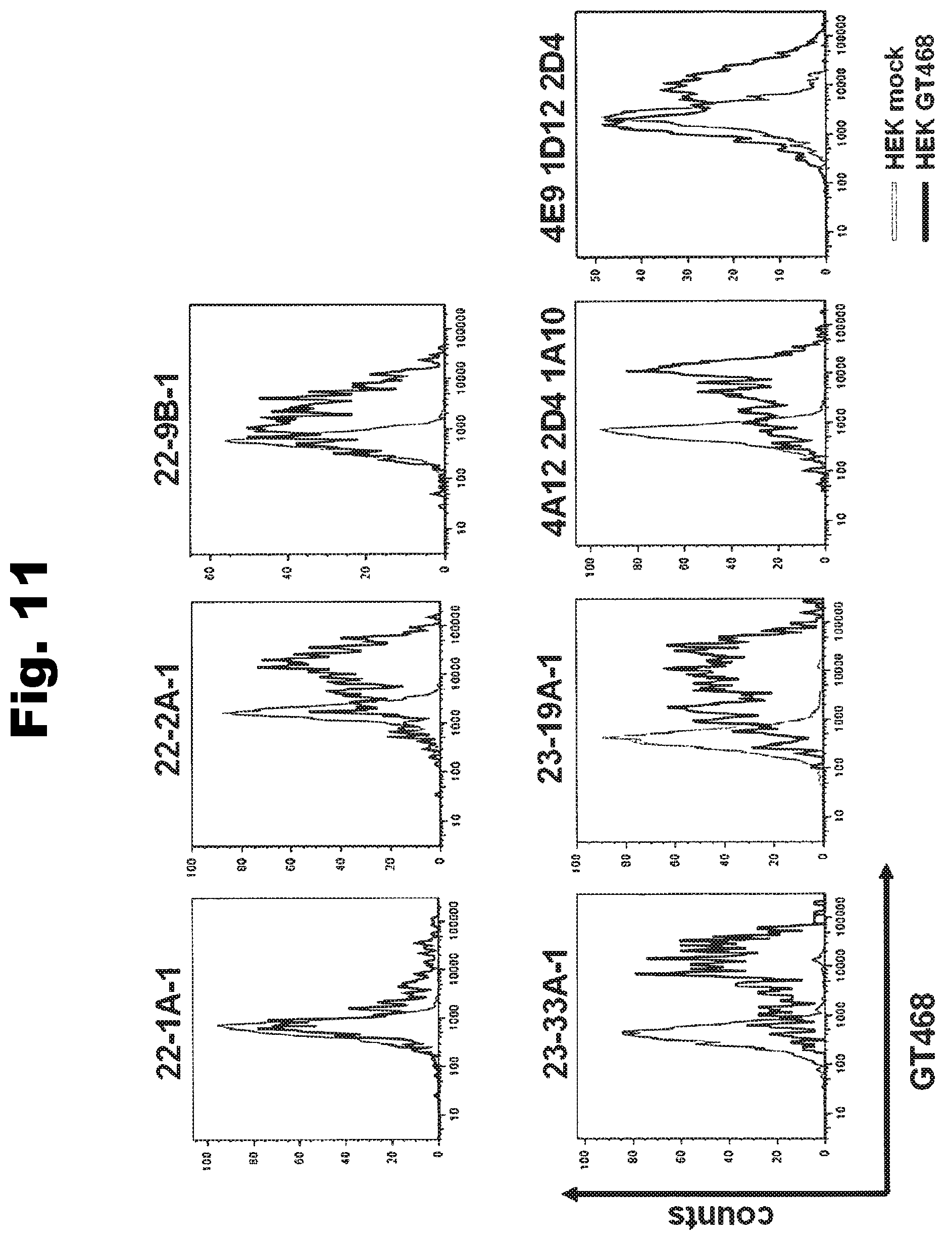
D00014

D00015
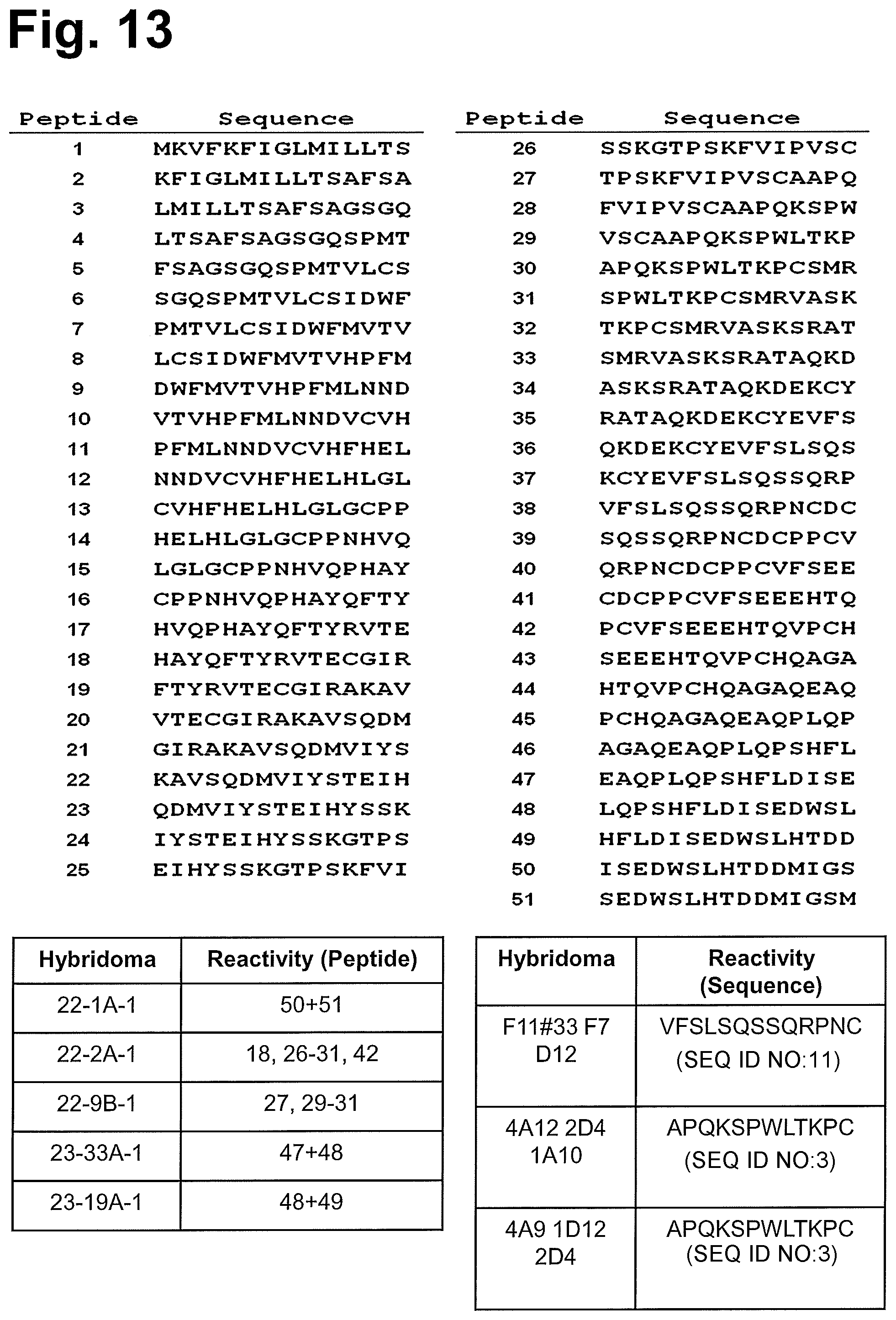
D00016
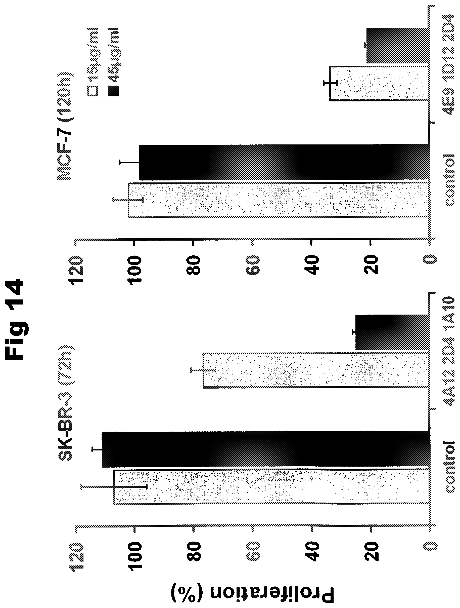
D00017
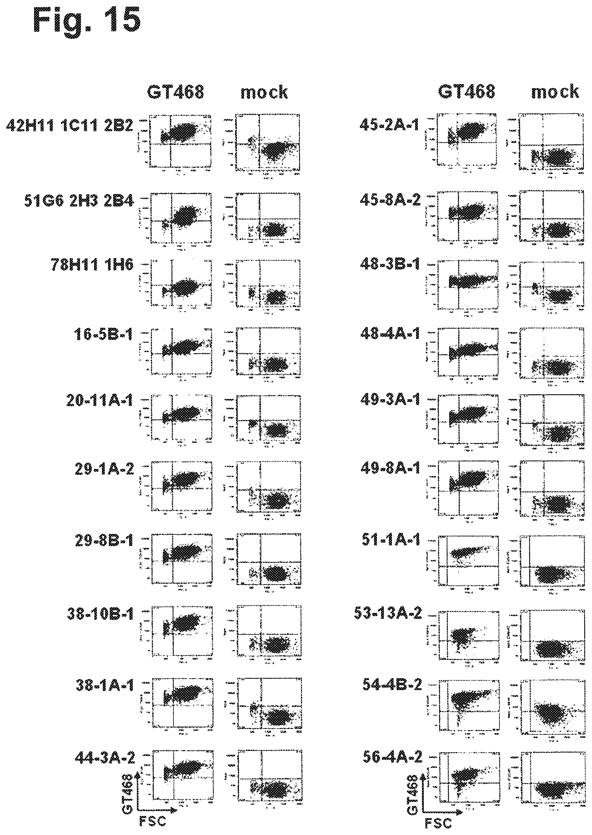
D00018
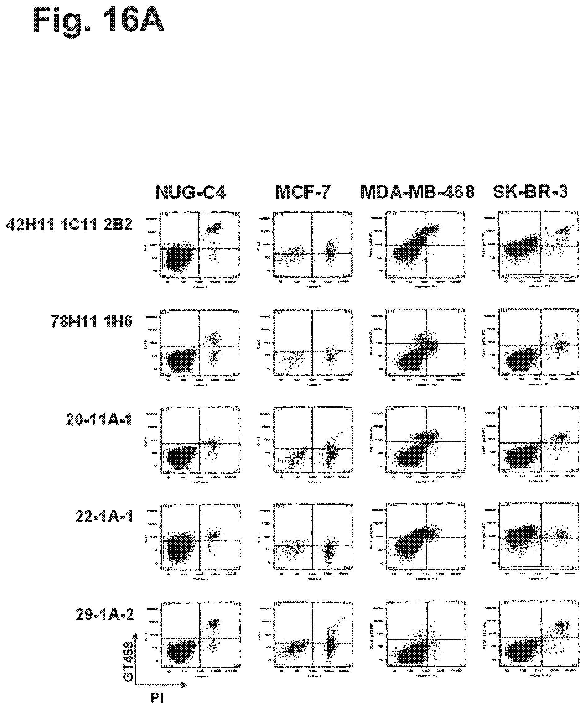
D00019
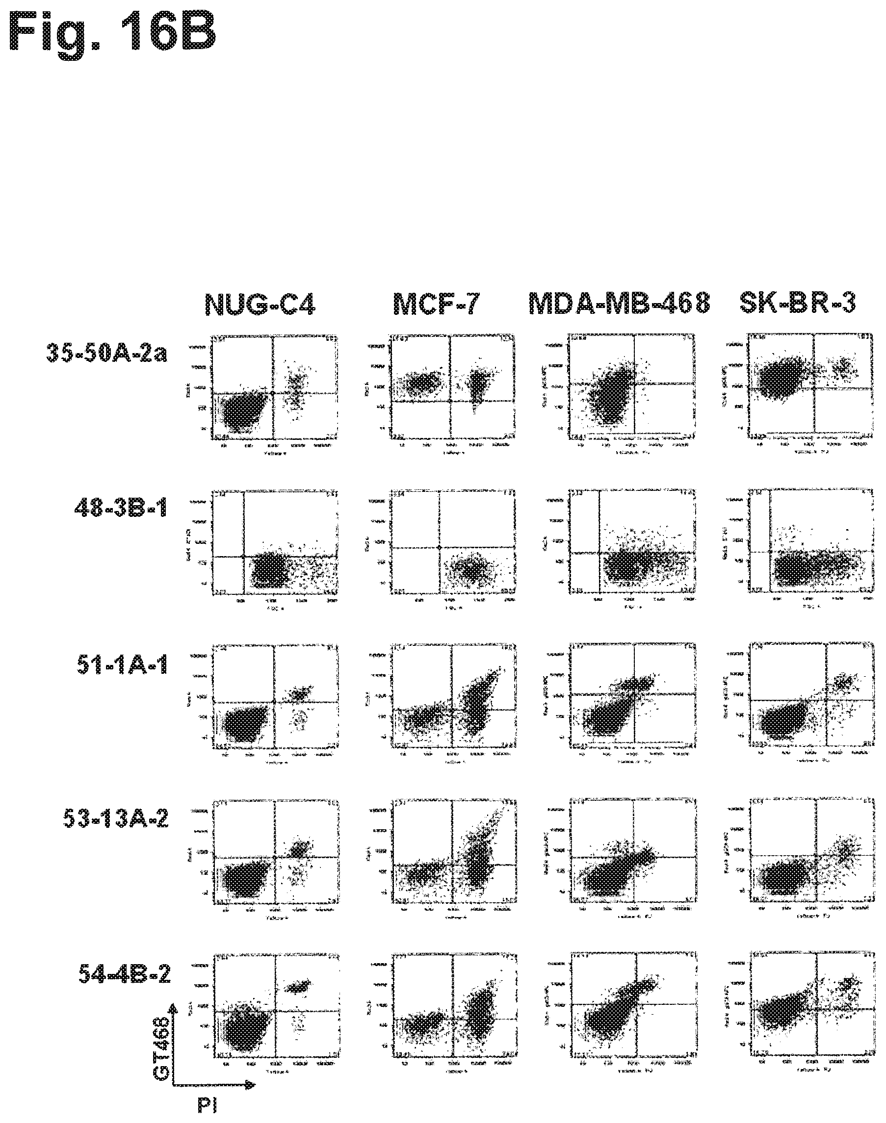
D00020

D00021
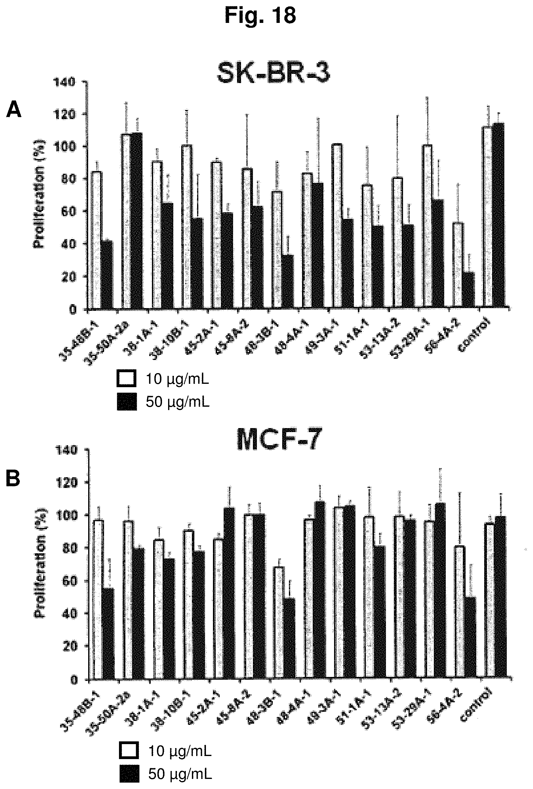
D00022
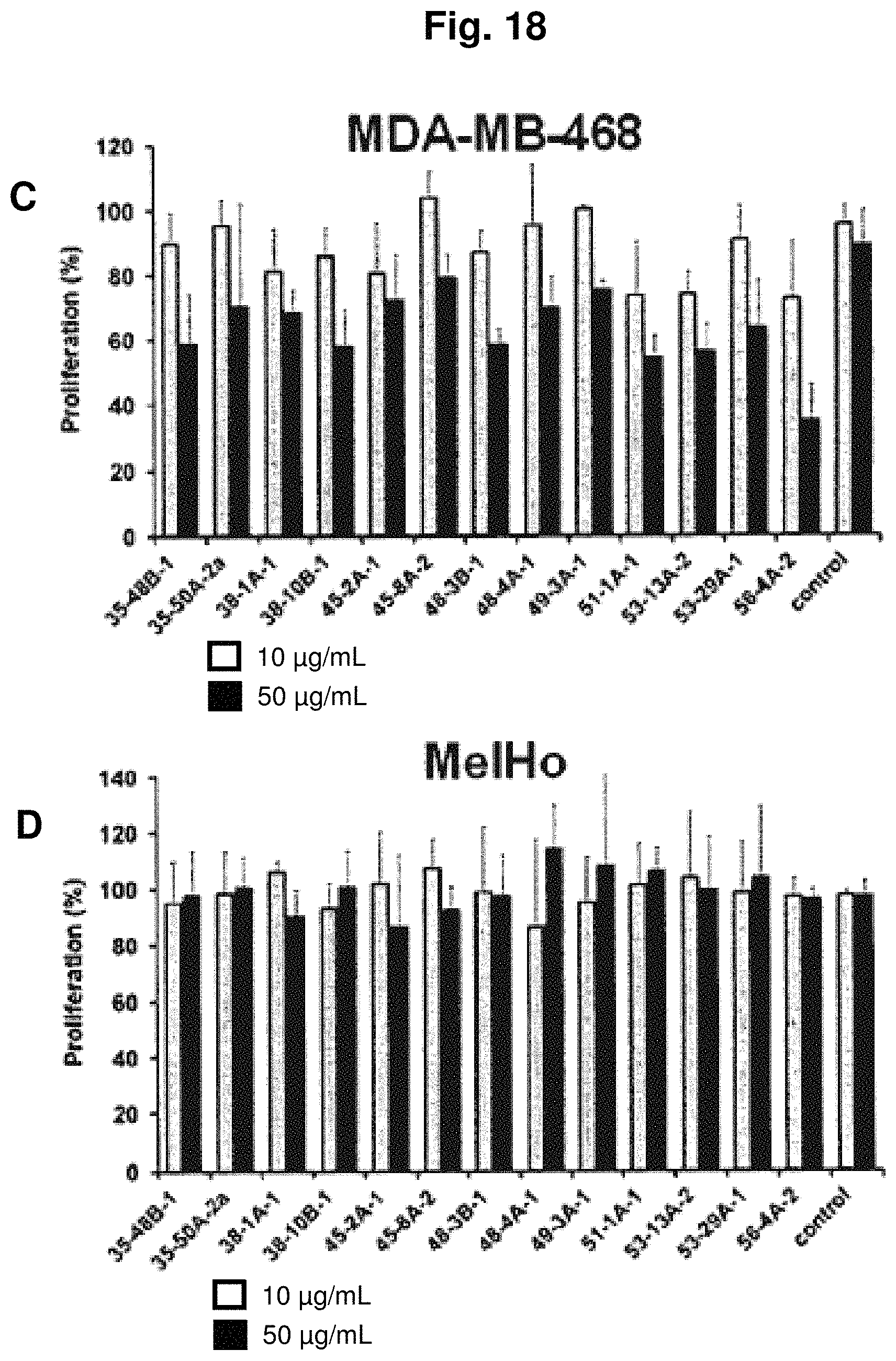
D00023

D00024
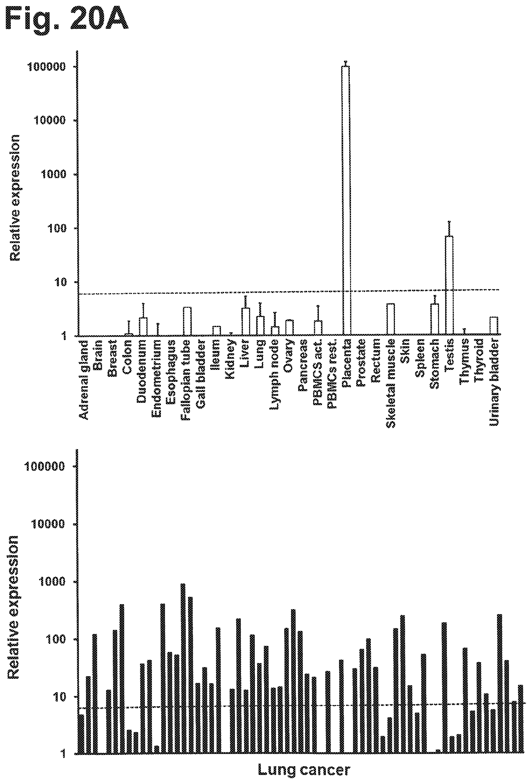
D00025
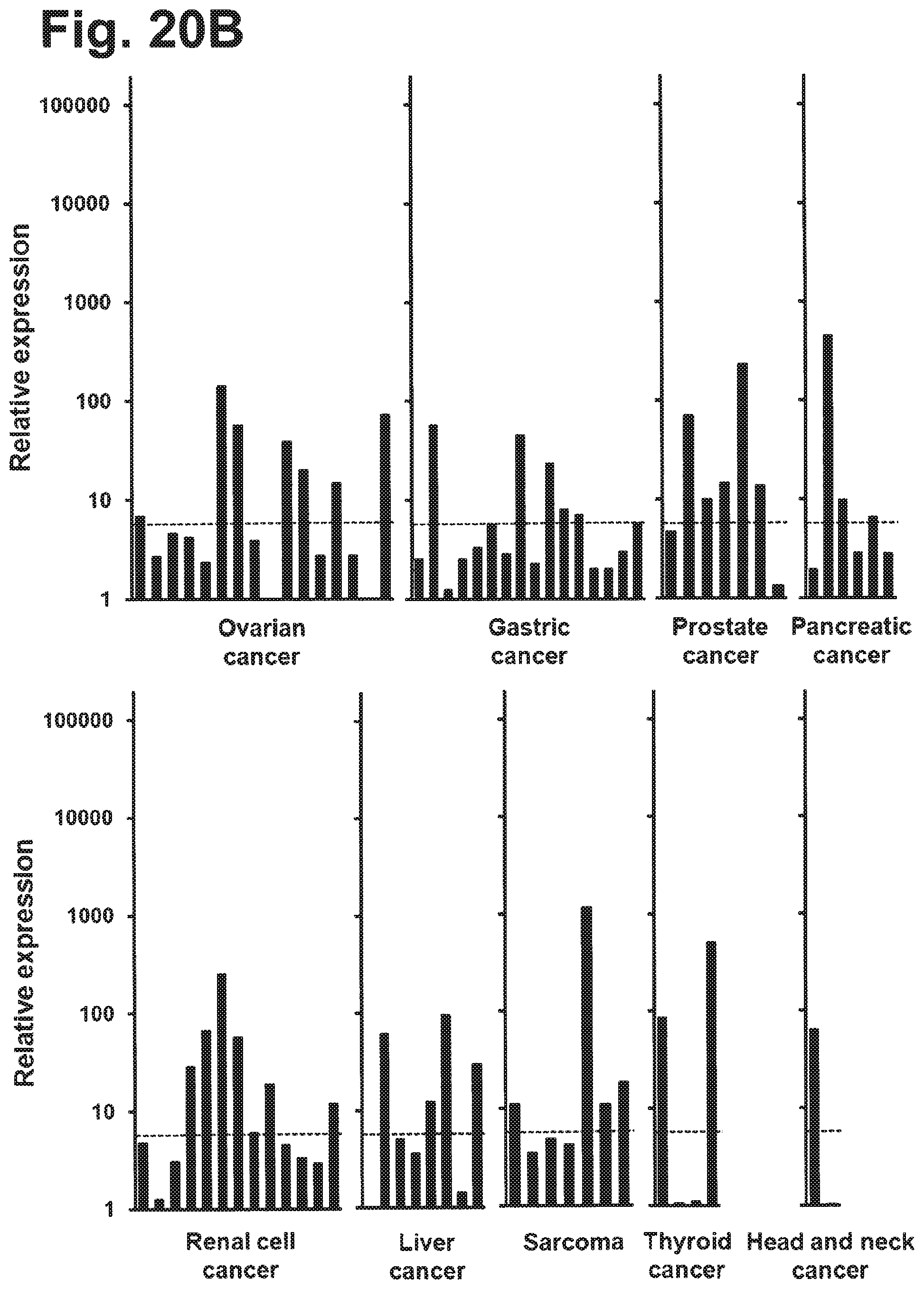
D00026
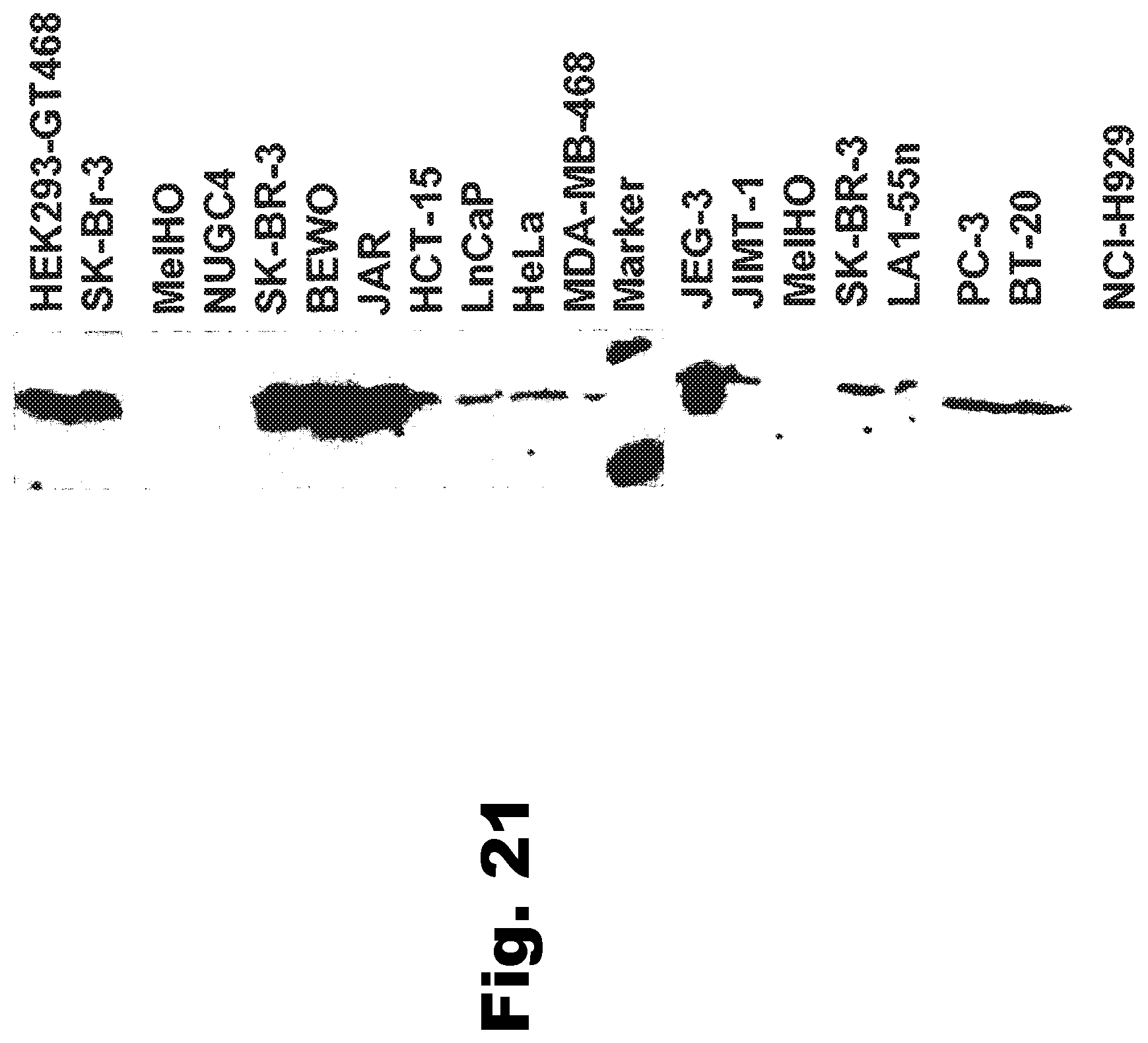
D00027

D00028
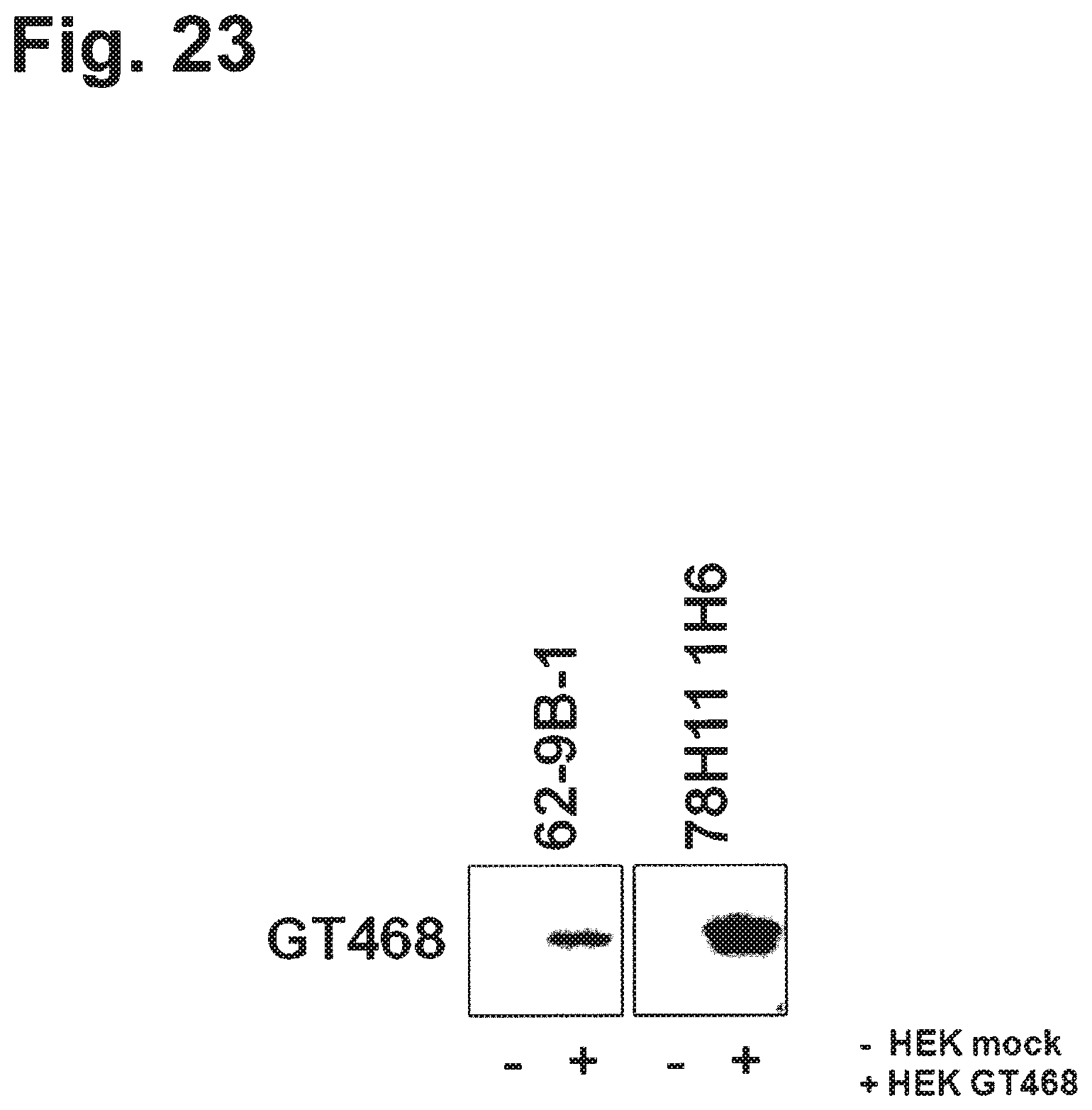
D00029

D00030
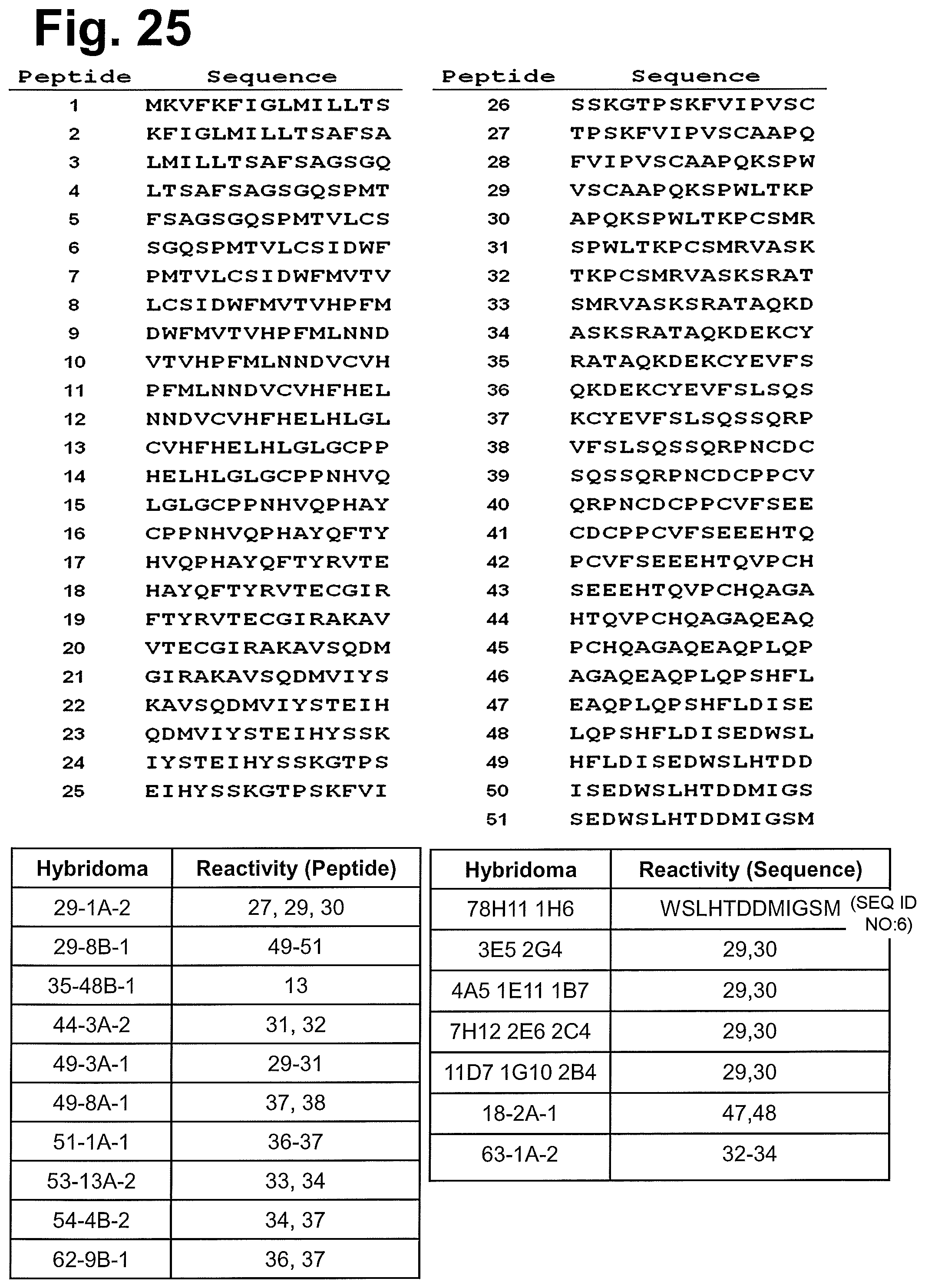
D00031

D00032

D00033
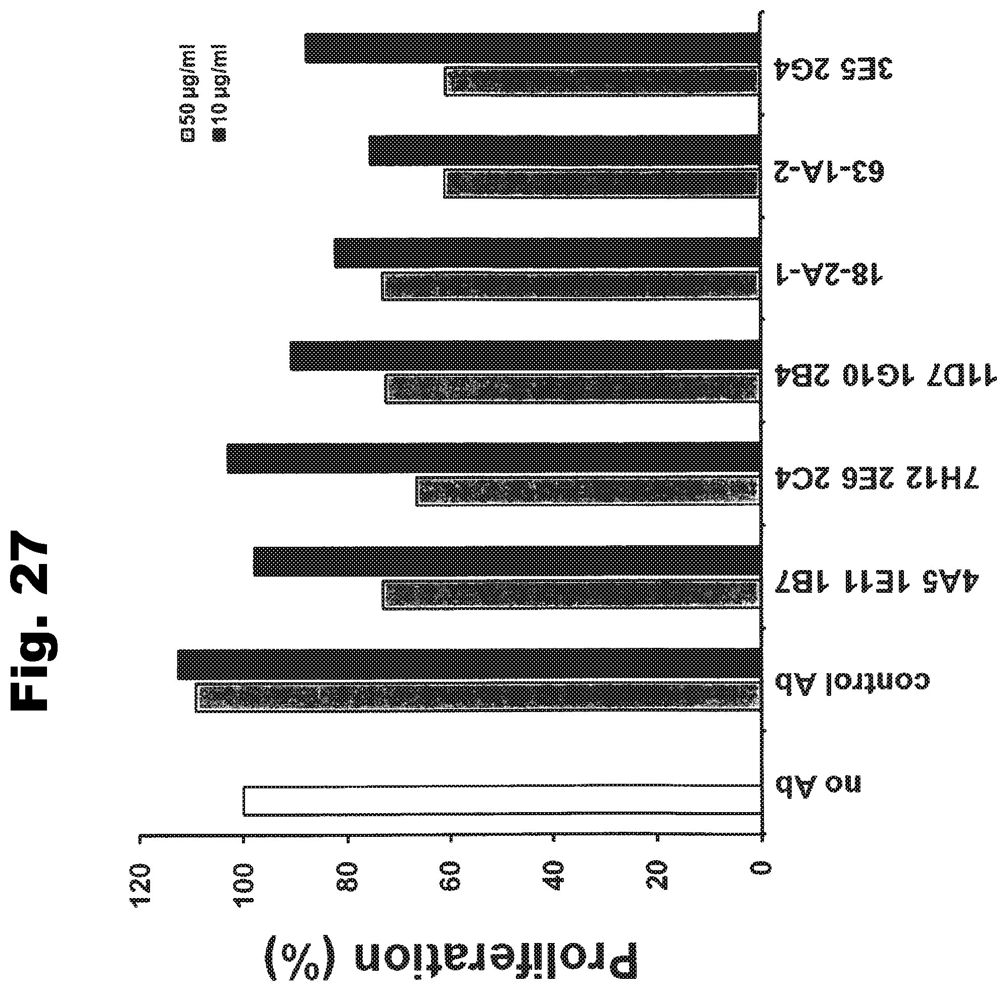
S00001
XML
uspto.report is an independent third-party trademark research tool that is not affiliated, endorsed, or sponsored by the United States Patent and Trademark Office (USPTO) or any other governmental organization. The information provided by uspto.report is based on publicly available data at the time of writing and is intended for informational purposes only.
While we strive to provide accurate and up-to-date information, we do not guarantee the accuracy, completeness, reliability, or suitability of the information displayed on this site. The use of this site is at your own risk. Any reliance you place on such information is therefore strictly at your own risk.
All official trademark data, including owner information, should be verified by visiting the official USPTO website at www.uspto.gov. This site is not intended to replace professional legal advice and should not be used as a substitute for consulting with a legal professional who is knowledgeable about trademark law.