Method of assaying Apolipoprotein AI for the in vitro diagnosis of colorectal cancer
Ataman-Onal , et al.
U.S. patent number 10,591,482 [Application Number 15/666,171] was granted by the patent office on 2020-03-17 for method of assaying apolipoprotein ai for the in vitro diagnosis of colorectal cancer. This patent grant is currently assigned to BIOMERIEUX. The grantee listed for this patent is BIOMERIEUX. Invention is credited to Yasemin Ataman-Onal, Jean-Philippe Charrier, Genevieve Choquet-Kastylevsky, Florence Poirier.


View All Diagrams
| United States Patent | 10,591,482 |
| Ataman-Onal , et al. | March 17, 2020 |
Method of assaying Apolipoprotein AI for the in vitro diagnosis of colorectal cancer
Abstract
A method assaying the levels of expression Apolipoprotein AI, Apolipoprotein AII, E-Cadherin, and Galectin-3 in a biological sample from a person having or suspected of having colorectal cancer. The sample is remote from any tumor.
| Inventors: | Ataman-Onal; Yasemin (Reyrieux, FR), Charrier; Jean-Philippe (Tassin la Demi-Lune, FR), Choquet-Kastylevsky; Genevieve (Francheville, FR), Poirier; Florence (Saint-Denis, FR) | ||||||||||
|---|---|---|---|---|---|---|---|---|---|---|---|
| Applicant: |
|
||||||||||
| Assignee: | BIOMERIEUX (Marcy l'Etoile,
FR) |
||||||||||
| Family ID: | 38658465 | ||||||||||
| Appl. No.: | 15/666,171 | ||||||||||
| Filed: | August 1, 2017 |
Prior Publication Data
| Document Identifier | Publication Date | |
|---|---|---|
| US 20170356916 A1 | Dec 14, 2017 | |
Related U.S. Patent Documents
| Application Number | Filing Date | Patent Number | Issue Date | ||
|---|---|---|---|---|---|
| 12452047 | |||||
| PCT/FR2008/051292 | Jul 10, 2008 | ||||
Foreign Application Priority Data
| Jul 19, 2007 [FR] | 07 05211 | |||
| Current U.S. Class: | 1/1 |
| Current CPC Class: | G01N 33/57419 (20130101); G01N 2333/775 (20130101) |
| Current International Class: | G01N 33/574 (20060101) |
References Cited [Referenced By]
U.S. Patent Documents
| 4529931 | July 1985 | Kuhns |
| 5002870 | March 1991 | Leavitt et al. |
| 5223789 | June 1993 | Katsuyama et al. |
| 5344760 | September 1994 | Harvey et al. |
| 5360715 | November 1994 | Leavitt et al. |
| 6001632 | December 1999 | Braxton et al. |
| 6291205 | September 2001 | Tuite et al. |
| 6451528 | September 2002 | Carr et al. |
| 6518411 | February 2003 | Murray et al. |
| 8029979 | October 2011 | Schneider-Mergener et al. |
| 2002/0160382 | October 2002 | Lasek et al. |
| 2003/0082533 | May 2003 | Yue et al. |
| 2003/0087818 | May 2003 | Jiang et al. |
| 2003/0109690 | June 2003 | Ruben et al. |
| 2003/0172388 | September 2003 | Fujise et al. |
| 2004/0157278 | August 2004 | Astle et al. |
| 2004/0191782 | September 2004 | Wang |
| 2004/0197930 | October 2004 | Rosenfeld et al. |
| 2005/0181398 | August 2005 | Fung et al. |
| 2005/0214826 | September 2005 | Mor et al. |
| 2006/0003359 | January 2006 | Feinberg et al. |
| 2006/0179496 | August 2006 | Burgess et al. |
| 2006/0205014 | September 2006 | Dotan et al. |
| 2007/0020707 | January 2007 | Holten-Andersen et al. |
| 2009/0239794 | September 2009 | Soudeyns et al. |
| 2011/0104701 | May 2011 | Ataman-Onal et al. |
| 500 719 | Mar 2006 | AT | |||
| 1396182 | Feb 2003 | CN | |||
| 0 496 174 | Jul 1992 | EP | |||
| 1 724 586 | Nov 2006 | EP | |||
| 1 775 590 | Apr 2007 | EP | |||
| 2 581 456 | Nov 1986 | FR | |||
| H02-287266 | Nov 1990 | JP | |||
| H04-064068 | Feb 1992 | JP | |||
| 2001-069971 | Mar 2001 | JP | |||
| 95/16462 | Jun 1995 | WO | |||
| 00/033083 | Jun 2000 | WO | |||
| 00/50588 | Aug 2000 | WO | |||
| 01/31019 | May 2001 | WO | |||
| 01/071357 | Sep 2001 | WO | |||
| 02/20731 | Mar 2002 | WO | |||
| 03/065003 | Aug 2003 | WO | |||
| 03/087831 | Oct 2003 | WO | |||
| 2004/073730 | Sep 2004 | WO | |||
| 2005/015218 | Feb 2005 | WO | |||
| 2005/015219 | Feb 2005 | WO | |||
| 2005/015226 | Feb 2005 | WO | |||
| 2005/035003 | Apr 2005 | WO | |||
| 2005/060996 | Jul 2005 | WO | |||
| 2005/083440 | Sep 2005 | WO | |||
| 2006/015079 | Feb 2006 | WO | |||
| 2006/074360 | Jul 2006 | WO | |||
| 2007/002535 | Jan 2007 | WO | |||
| 2007/005578 | Jan 2007 | WO | |||
| 2007/020522 | Feb 2007 | WO | |||
| 2007/068985 | Jun 2007 | WO | |||
| 2007/140352 | Dec 2007 | WO | |||
| 2008/021290 | Feb 2008 | WO | |||
| 2008/028968 | Mar 2008 | WO | |||
| 2008/036981 | Mar 2008 | WO | |||
Other References
|
Hachenn et al., J Clin Chem Clin Biochem., 1986, 24: 161-166. cited by examiner . Wilmanns et al., Clinical & Experimental Metastasis, 2004, 21: 75-78. cited by examiner . Lurisci et al., Clinical Cancer Research, 2000, 6: 1389-1393. cited by examiner . Dubois et al., J. Immunol. Methods, 1987: 96(1):115-120. cited by examiner . Holland et al., Abstract of Medicina, 1993, 53(2), 117-123, one page. cited by examiner . Zhao et al., World J. Gastroenterology, 1997, 3(1): 41-42. cited by examiner . Chen et al., Lung Cancer, 2006, 54: 95-102. cited by examiner . Gould et al., "cDNA Cloning and Sequencing of the Protein-Tyrosine Kinase Substrate, Ezrin, Reveals Homology to Band 4.1," The EMBO Journal, vol. 8, No. 13, pp. 4133-4142, 1989. cited by applicant . Etzioni et al., "The Case for Early Detection," Nature Review, vol. 3, Internet pp. 1-10, Apr. 2003. cited by applicant . Mercer, "Use of Multiple Markers to Enhance Clinical Utility," Immunol Ser., vol. 53, pp. 39-54, 1990. cited by applicant . Otsuka et al. "Differential Expression of L-Plastin Gene in Human Colorectal Cancer Progression and Metastasis," Biochemical and Biophysical Research Communications, vol. 289, No. 4, pp. 876-881, 2001. cited by applicant . Shin et al., "Global Profiling of the Cell Surface Proteome of Cancer Cells Uncovers an Abundance of Proteins with Chaperone Function," The Journal of Biological Chemistry, Feb. 28, 2003, pp. 7607-7616, vol. 278, No. 9, The American Society for Biochemistry and Molecular Biology, Inc., U.S.A. cited by applicant . Tomonaga et al., "Identification of Altered Protein Expression and Post-Translational Modifications in Primary Colorectal Cancer by Using Agarose Two-Dimensional Gel Electrophoresis," Clinical Cancer Research, vol. 10, Mar. 15, 2004, pp. 2007-2014. cited by applicant . Stierum et al., "Proteome Analysis Reveals Novel Proteins Associated with Proliferation and Differentiation of Colorectal Cancer Cell Line Caco-2," Biochemica et Biophysica Acta, 2003, pp. 73-91, vol. 1650, No. 1-2. cited by applicant . Kaetzel et al., "Protein Disulphide-Isomerase from Human Placenta and Rat Liver--Purification and Immunological Characterization with Monoclonal Antibodies," Biochem. J., 1987, pp. 39-47, vol. 241, No. 1, Great Britain. cited by applicant . Kozaki et al., "Tissue Distribution of ERp61 and Association of Its Increased Expression with IgG Production in Hybridoma Cells," Experimental Cell Research, 1994, pp. 348-358, vol. 213, No. 2, Academic Press, Inc. cited by applicant . Leys et al., "Expression and Prognostic Significance of Prothymosin-a and ERp57 in Human Gastric Cancer," Surgery, 2007, pp. 41-50, vol. 141, No. 1, Mosby Co., St. Louis, MO, U.S.A. cited by applicant . Sass et al., "Mutations in ACY1, the Gene Encoding Aminoacylase 1, Cause a Novel Inborn Error of Metabolism", The American Journal of Human Genetics, Mar. 2006, vol. 78, pp. 401-409. cited by applicant . Tockman et al., "Considerations in Bringing a Cancer Biomarker to Clinical Application", Cancer Research, May 1, 1992, vol. 52, pp. 2711s-2718s. cited by applicant . Freeman et al., "APO--All is an Elevated Biomarker of Chronic Non-Human Primate Ethanol Self-Administration", Alcohol & Alcoholism, Mar. 2006, vol. 41, No. 3, pp. 300-305. cited by applicant . Elsevier Publishing, "Human PDIA1 Tryptic Peptide--SEQ ID 33," Retrieved from EBI Accession No. GSP:ASS06881, Database Accession No. ASS06881, 2008. cited by applicant . Delanote et al., "Plastins: Versatile Modulators of Actin Organization in (patho)physiological Cellular Processes," Acta Pharmacologica Sinica, vol. 26, No. 7, pp. 769-779, Jul. 2005. cited by applicant . Hasselblatt et al., "Identification of Novel Diagnostic Markers for Choroid Plexus Tumors," Am. J. Surg Pathol, vol. 30, No. 1, pp. 66-74, Jan. 2006. cited by applicant . Lin et al., "Identification of I-Plastin, a Human Fimbrin Isoform Expressed in Intestine and Kidney," Molecular and Cellular Biology, vol. 14, No. 4, pp. 2457-2467, Apr. 1994. cited by applicant . Carroll et al., "Liver Fatty Acid-Binding Protein: A Marker for Studying Cellular Differentiation in Gut Epithelial Neoplasms," Gastroenterology, vol. 99, pp. 1727-1735, 1999. cited by applicant . Kayser et al., "Primary Colorectal Carcinomas and their Intrapulmonary Metastases: Clinical, Glyco-, Immuno- and Lectin Histochemical, Nuclear and Syntactic Structure Analysis with Emphasis on Correlation with Period of Occurrence of Metastases and Survival," AMPIS, vol. 110, pp. 435-446, 2002. cited by applicant . Kuusela et al., "Comparison of CA 19-9 and Carcinoembryonic Antigen (CEA) Levels in the Serum of Patients with Colorectal Diseases," Br. J. Cancer, vol. 49, pp. 135-139, 1984. cited by applicant . Pelsers et al., "Fatty Acid-Binding Proteins as Plasma Markers of Tissue Injury," Clinica Chimica Acta, vol. 352, pp. 15-35, 2005. cited by applicant . Niederkofler et al., "Novel Mass Spectrometric Immunoassays for the Rapid Structural Characterization of Plasma Apolipoproteins," Journal of Lipid Research, vol. 44, pp. 630-639, 2003. cited by applicant . Hortin, "The MALDI-TOF Mass Spectrometric View of the Plasma Proteome and Pepetidome," Clinical Chemistry, vol. 52, No. 7, pp. 1223-1237, 2006. cited by applicant . Hachem, "Serum Apolipoproteins A-I, A-II and B in Hepatic Metastases Comparison with other Liver Diseases: Hepatomas and Cirrhosis," J. Clin. Chem. Clin. Biochem., vol. 24, pp. 161-166, 1986. cited by applicant . Remold-O'Donnell et al., "Sequence and Molecular Characterization of Human Monocyte/Neutrophil Elastase Inhibitor," Proc. Natl. Acad. Sci., vol. 89, pp. 5635-5639, Jun. 1992. cited by applicant . Cooley et al., "The Serpin MNEI Inhibits Elastase-Like and Chymotrypsin-Like Serine Proteases through Efficient Reactions at Two Active Sites," Biochemistry, vol. 40, pp. 15762-15770, 2001. cited by applicant . Algrain et al, "Ezrin Contains Cytoskeleton and Membrane Binding Domains Accounting for its Proposed Role as a Membrane-Cytoskeletal Linker," The Journal of Cell Biology, vol. 120, No. 1, pp. 129-139, Jan. 1993. cited by applicant . Jiang et al., "Cytokine Regulation of Ezrin Expression in the Human Colon Cancer Cell Line HT29," Anticancer Research, vol. 16, pp. 861-866, 1996. cited by applicant . Hiscox et al, "Ezrin Regulates Cell-Cell and Cell-Matrix Adhesion, A Possible Role with E-Cadherin/.beta.-Catenin," Journal of Cell Science, vol. 112, pp. 3081-3090, 1999. cited by applicant . Xiao et al., "An Approach to Studying Lung Cancer-Related Proteins in Human Blood," Molecular & Cellular Proteomics, vol. 4, pp. 1480-1486, 2005. cited by applicant . Anders et al., "Aminoacyclases," Advances in Pharmacology, vol. 27, pp. 431-448, 1994. cited by applicant . Lorentz et al., "A New Method for the Assay of Aminoacylase: Elaboration of a Fixed-Incubation Method for Routine Measurements," Clinica Chimica Acta, vol. 63, pp. 263-269, 1975. cited by applicant . Lorentz et al, "Clinical Application of A New Method for the Determination of Aminoacylase in Human Serum," Clinica Chimica Acta, vol. 63, pp. 271-274, 1975. cited by applicant . Cook et al, "Human Aminoacylase-1," The Journal of Biological Chemistry, vol. 268, No. 23, pp. 17010-17017, 1993. cited by applicant . Miller et al., "Lack of Expression of Aminoacylae-1 in Small Cell Lung Cancer," The Journal of Clinical Investigation, Inc., vol. 83, pp. 2120-2124, Jun. 1989. cited by applicant . Balabanov et al., "Tumour-Related Enzyme Alterations in the Clear Cell Type of Human Renal Cell Carcinoma Identified by Two-dimensional Gel Electrophoresis," Eur. J. Biochem., vol. 268, pp. 5977-5980, 2001. cited by applicant . Chan et al, "Human Liver Fatty Acid Binding Protein cDNA and Amino Acid Sequence," The Journal of Biological Chemistry, vol. 260, No. 5, pp. 2629-2632, 1985. cited by applicant . Das et al., "Expression Pattern of Fatty Acid-Binding Proteins in Human Normal and Cancer Prostate Cells and Tissues," Clinical Cancer Research, vol. 7, 1706-1715, Jun. 2001. cited by applicant . Stulik et al., "Proteome Study of Colorectal Carcinogenesis," Electrophoresis, vol. 22, pp. 3019-3025, 2001. cited by applicant . Yamazaki et al., "Liver Fatty Acid-Binding Protein is a New Prognostic Factor for Hepatic Resection of Colorectal Cancer Metastases," Journal of Surgical Oncology, vol. 72, pp. 83-87, 1999. cited by applicant . Sweetser et al., "The Human and Rodent Intestinal Fatty Acid Binding Protein Genes," The Journal of Biological Chemistry, vol. 262, No. 33, pp. 16060-16071, 1987. cited by applicant . Pelsers et al, "Intestinal-Type and Liver-Type Fatty Acid-Binding Protein in the Intestine. Tissue Distribution and Clinical Utility", Clinical Biochemistry, vol. 36, pp. 529-535, 2003. cited by applicant . Xiao et al, "Dietary Exposure to Soy or Whey Proteins Alters Colonic Global Gene Expression Profiles During Rat Colon Tumorigenesis," Molecular Cancer, vol. 4, No. 1, pp. 1-17, Jan. 11, 2005. cited by applicant . Engwegen et al., "Identification of Serum Proteins Discriminating Colorectal Cancer Patients and Healthy Controls Using Surface-Enhanced Laser Desorption Ionisation--Time of Flight Mass Spectrometry," World J. Gastrenterol, vol. 12, No. 10, pp. 1536-1544, Mar. 14, 2006. cited by applicant . Zhang et al., "Three Biomarkers Identified from Serum Proteomic Analysis for the Detection of Early Stage Ovarian Cancer," Cancer Research, vol. 64, pp. 5882-5890, Aug. 15, 2004. cited by applicant . Lin et al., "Human Plastin Genes," The Journal of Biological Chemistry, vol. 268, No. 4, pp. 2781-2792, 1993. cited by applicant . Lavabre-Bertrand et al., "Plasma Proteasome Level is a Potential Marker in Patients with Solid Tumors and Hemopoietic Malignancies," Cancer, vol. 92, No. 10, pp. 2493-2500, Nov. 15, 2001. cited by applicant . Iurisci et al., "Concentrations of Galectin-3 in the Sera of Normal Controls and Cancer Patients," Clinical Cancer Research, vol. 6, pp. 1389-1393, Apr. 2000. cited by applicant . Schwartz, "Enzymes as Prognostic Markers and Therapeutic Indicators in Patients with Cancer," Clinica Chimica Acta, vol. 206, pp. 77-82, 1992. cited by applicant . McCool et al., "Roles of Calreticulin and Calnexin During Mucin Synthesis in LS180 and HT29/A1 Human Colonic Adenocarcinoma Cells," Biochem. J., vol. 341, pp. 593-600, 1999. cited by applicant . Intestinal Ischemia Causes, Mayo Clinic, http://www.mayoclinic.org/diseases-conditions/intestinal-ischemia/basics/- causes/con-20023818, accessed Nov. 9, 2015. cited by applicant . Sep. 19, 2014 Office Action issued in U.S. Appl. No. 13/712,340. cited by applicant . Apr. 23, 2014 Office Action issued in U.S. Appl. No. 13/712,340. cited by applicant . Sep. 30, 2013 Office Action issued in U.S. Appl. No. 12/452,034. cited by applicant . Oct. 15, 2013 Office Action issued in U.S. Appl. No. 12/452,037. cited by applicant . Apr. 17, 2012 Office Action issued in U.S. Appl. No. 12/452,038. cited by applicant . Jan. 16, 2009 International Search Report issued in International Patent Application No. PCT/FR2008/051298. cited by applicant . U.S. Appl. No. 12/452,038, filed Dec. 14, 2009, in the name of Monique Arpin et al. cited by applicant . May 13, 2011 Restriction and Election of Species Requirement issued in U.S. Appl. No. 12/452,038. cited by applicant . Sep. 1, 2011 Office Action issued in U.S. Appl. No. 12/452,038. cited by applicant . Mar. 3, 2016 Office Action issued in U.S. Appl. No. 12/452,047. cited by applicant . Giusti et al., "Proteome Analysis of Whole Saliva: A New Tool for Rheumatic Diseases--The Example of Sjogren's Syndrome," Proteomics, vol. 7, pp. 1634-1643, 2007. cited by applicant . Mar. 3, 2015 Office Action issued in U.S. Appl. No. 12/452,035. cited by applicant . Mar. 6, 2015 Office Action issued in U.S. Appl. No. 12/452,034. cited by applicant . Apr. 9, 2015 Office Action issued in U.S. Appl. No. 13/732,521. cited by applicant . Guo et al. "Combined Use of Positive and Negative Immunomagnetic Isolation Followed By Real-Time RT-PCR for Detection of the Circulating Tumor Cells in Patients with Colorectal Cancers." J Mol. Med., 2004, 82:768-774. cited by applicant . Sep. 10, 2014 Office Action issued in U.S. Appl. No. 13/732,521. cited by applicant . Aug. 1, 2014 Office Action issued in U.S. Appl. No. 12/452,034. cited by applicant . Jeck et al., "Local Ischemia Causes Carcinoma-Like Changes of the Rectum", Dis Colon Rectum., 1996, vol. 39, No. 9, pp. 1026-1030 (abstract only). cited by applicant . Jul. 23, 2014 Office Action issued in U.S. Appl. No. 12/452,035. cited by applicant . Friedman et al., "Proteome Analysis of Human Colon Cancer by Two-Dimensional Difference Gel Electrophoresis and Mass Spectrometry", Proteomics, 2004, vol. 4, pp. 793-811. cited by applicant . Jul. 24, 2014 Office Action issued in U.S. Appl. No. 12/452,047. cited by applicant . Kozak et al., "Characterization of Serum Biomarkers for Detection of Early Stage Ovarian Cancer", Proteomics, 2005, vol. 5, pp. 4589-4596. cited by applicant . Bury et al., "Quantification of Human Serum Apolipoprotein AI by Enzyme Immunoassay", Clinical Chemistry, 1985, vol. 31, No. 2, pp. 247-251. cited by applicant . Laine, Anne, "Rocket Immunoelectrophoresis Technique or Electroimmunodiffusion", Methods in Molecular Biology, 1992, vol. 10, pp. 201-205. cited by applicant . Jan. 30, 2014 Office Action issued in U.S. Appl. No. 12/452,034. cited by applicant . Jun. 18, 2013 Office Action issued in U.S. Appl. No. 12/452,037. cited by applicant . Dec. 28, 2012 Office Action issued in U.S. Appl. No. 12/452,037. cited by applicant . Aug. 3, 2012 Office Action issued in U.S. Appl. No. 12/452,037. cited by applicant . Jul. 31, 2012 Office Action issued in U.S. Appl. No. 12/452,048. cited by applicant . Jul. 24, 2012 Restriction Requirement issued in U.S. Appl. No. 12/999,242. cited by applicant . Jun. 21, 2012 Office Action issued in U.S. Appl. No. 12/452,035. cited by applicant . Jun. 18, 2012 Office Action issued in U.S. Appl. No. 12/452,047. cited by applicant . Jun. 12, 2012 Office Action issued in U.S. Appl. No. 12/452,042. cited by applicant . May 18, 2012 Office Action issued in U.S. Appl. No. 12/452,034. cited by applicant . Feb. 3, 2012 Office Action issued in U.S. Appl. No. 12/452,048. cited by applicant . Feb. 1, 2012 Office Action issued in U.S. Appl. No. 12/452,037. cited by applicant . Jan. 9, 2012 Office Action issued in U.S. Appl. No. 12/452,047. cited by applicant . Jan. 3, 2012 Office Action issued in U.S. Appl. No. 12/452,035. cited by applicant . Nov. 16, 2011 Election of Species Requirement issued in U.S. Appl. No. 12/452,037. cited by applicant . Oct. 31, 2011 Restriction and Election of Species Requirement issued in U.S. Appl. No. 12/452,035. cited by applicant . Oct. 28, 2011 Office Action issued in U.S. Appl. No. 12/452,042. cited by applicant . Oct. 28, 2011 Restriction Requirement issued in U.S. Appl. No. 12/452,047. cited by applicant . Oct. 27, 2011 Office Action issued in U.S. Appl. No. 12/452,034. cited by applicant . Sep. 15, 2011 Restriction and Election of Species Requirement issued in U.S. Appl. No. 12/452,042. cited by applicant . Aug. 1, 2011 Restriction and Election of Species Requirement issued in U.S. Appl. No. 12/452,034. cited by applicant . U.S. Appl. No. 12/452,035, filed Dec. 14, 2009, in the name of Corinne Beaulieu et al. cited by applicant . U.S. Appl. No. 12/452,037, filed Dec. 14, 2009, in the name of Yasemin Ataman-Onal et al. cited by applicant . U.S. Appl. No. 12/452,042, filed Dec. 14, 2009, in the name of Monique Arpin et al. cited by applicant . U.S. Appl. No. 12/452,048, filed Dec. 14, 2009, in the name of Jean-Philippe Charrier et al. cited by applicant . U.S. Appl. No. 12/452,034, filed Dec. 14, 2009, in the name of Yasemin Ataman-Onal et al. cited by applicant . U.S. Appl. No. 12/452,047, filed Dec. 14, 2009, in the name of Yasemin Ataman-Onal et al. cited by applicant . Nov. 25, 2009 International Search Report issued in International Patent Application No. PCT/FR2009/051361. cited by applicant . Mar. 27, 2009 International Search Report issued in International Patent Application No. PCT/FR2008/051290. cited by applicant . Mar. 18, 2009 International Search Report issued in International Patent Application No. PCT/FR2008/051291. cited by applicant . Mar. 6, 2009 International Search Report issued in International Patent Application No. PCT/FR2008/051293. cited by applicant . Mar. 5, 2009 International Search Report issued in International Patent Application No. PCT/FR2008/051292. cited by applicant . Feb. 23, 2009 International Search Report issued in International Patent Application No. PCT/FR2008/051289. cited by applicant . Feb. 18, 2009 International Search Report issued in International Patent Application No. PCT/FR2008/051295. cited by applicant . Feb. 12, 2009 International Search Report issued in International Patent Application No. PCT/FR2008/051294. cited by applicant . Bruce et al., "Cancer-Wide Tissue Microarray Survey of Ezrin and Merlin Expression, "Proc Amer Assoc Cancer Res, vol. 46, Abstract #424, 2005. cited by applicant . Motoo et al., "Serum Levels of Pancreatitis-Associated Protein in Digestive Diseases with Special Reference to Gastrointestinal Cancer," Digestive Diseases and Sciences, vol. 44, No. 6, pp. 1142-1147, Jun. 1999. cited by applicant . Abe et al., "Preparation of Recombinant MK-1/EP-CAM and Establishment of an ELISA System for Determining Soluble MK-1/EP-CAM Levels in Sera of Cancer Patients," Journal of Immunological Methods, vol. 270, pp. 227-233, 2002. cited by applicant . Katayama et al., "Soluble E-Cadherin Fragments Increased in Circulation of Cancer Patients," Br. J. Cancer, vol. 69, pp. 580-585, 1994. cited by applicant . Wilmanns et al., "Soluble Serum E-Cadherin as a Marker of Tumour Progression in Colorectal Cancer Patients," Clinical & Experimental Metastasis, vol. 21, pp. 75-78, 2004. cited by applicant . Gold et al., "Specific Carcinoembryonic Antigens of the Human Digestive System," J. Exp. Med., pp. 467-481, 1965. cited by applicant . Kim et al., "Gastrointestinal Tract Cancer Screening Using Fecal Carcinoembryonic Antigen," Annals of Clinical & Laboratory Science, vol. 33, No. 1, pp. 32-38, 2003. cited by applicant . Holmgren et al., "Detection by Monoclonal Antibody of Carbohydrate Antigen CA 50 in Serum of Patients with Carcinoma," British Medical Journal, vol. 288, pp. 1479-1482, May 19, 1984. cited by applicant . Klug et al., Monoclonal Anitbody Immunoradiometric Assay for An Antigenic Determinant (CA 72) on a Novel Pancarcinoma Antigen (TAG-72), Int. J. Cancer, vol. 38, pp. 661-669, 1986. cited by applicant . Holland et al., "Testosterona Serica: Posible Marcador En El Cancer Colorrectal," Medicina (Buenos Aires), vol. 53, pp. 117-123, 1993. cited by applicant . Model et al., "Detection of Methylated DNA in Plasma from Colorectal Cancer Patients and Controls by Real-Time PCR Analysis of Septin 9," World Congress on Gastrointestinal Cancer, Jul. 2006. cited by applicant . Ebert et al., "Aristaless-like Homeobox-4 Gene Methylation is a Potential Marker for Colorectal Adenocarcinomas," Gastroenterology, vol. 131, pp. 1418-1430, 2006. cited by applicant . Bianco et al., "Identification of Cripto-1 as a Novel Serologic Marker for Breast and Colon Cancer," Clin. Cancer Res., vol. 12, No. 17, pp. 5158-5164, Sep. 1, 2006. cited by applicant . Mori et al., "Two-Dimensional Electrophoresis Database of Fluorescence-Labeled Proteins of Colon Cancer Cells," Journal of Chromatography B, vol. 823, pp. 82-97, 2005. cited by applicant . Duffy et al., "Tumour Markers in Colorectal Cancer: European Group on Tumour Markers (EGTM) Guidelines for Clinical Use," European Journal of Cancer, vol. 43, pp. 1348-1360, 2007. cited by applicant . Rouzier et al., "Immunocytochemical Detection of Bone Marrow Micrometastases in Colorectal Carcinoma Patients, Using a Monoclonal Antibody to Villin," Cytometry (Communications in Clinical Cytometry), vol. 46, pp. 281-289, 2001. cited by applicant . Nishizuka et al., "Diagnostic Markers that Distinguish Colon and Ovarian Adenocarcinomas: Identification by Genomic, Proteomic, and Tissue Array Profiling," Cancer Research, vol. 63, pp. 5243-5250, Sep. 1, 2003. cited by applicant . Wang et al., Proteomic Dissection of the Molecular Mechanisms Underlying Hepatic Metastasis in Colorectal Cancer, XP009093529, Database Accession No. PREV200600505326, AGA Abstracts, p. A-676, W1533, Apr. 2006. cited by applicant . Mori et al., A Genome-Wide Search Identifies Epigenetic Silencing of Somatostatin, Tachykinin-1, and 5 other Genes in Colon Cancer, Gastroenterology, vol. 131, pp. 797-808, 2006. cited by applicant . Database WPI Week 200777, Thomson Scientific, London, GB; AN 2007-817830, XP002464797 & CN 1 967 246 (Proteome Analysis Research Center) May 23, 2007, Abstract. cited by applicant . Fujiwara et al., Global Gene Expression Analysis of Rat Colon Cancers Induced by a Food-Borne Carcinogen, 2-amino-1-methyl-6-phenylimidazo[4,5-b]pyridine, Carcinogenesis, vol. 25, No. 8, pp. 1495-1505, 2004. cited by applicant . Dorudi et al., "E-Cadherin Expression in Colorectal Cancer," American Journal of Pathology, vol. 142, No. 4, pp. 981-986, Apr. 1993. cited by applicant . Lawrie et al., "Liver Fatty Acid Binding Protein Expression in Colorectal Neoplasia," British Journal of Cancer, vol. 90, pp. 1955-1960, 2004. cited by applicant . Davies et al., "Loss of cellular distribution of ezrin in human colon cancer," Gastrointestinal Cancers Symposium, ASCO; Abstract 322 (2006). cited by applicant . Mahipal et al. "Epidermal growth factor receptor overexpression in resected pancreatic cancer," Journal of Clinical Oncology, ASCO; Abstract 317 (2011). cited by applicant . Brattstrom et al. "HER-2 overexpression in patients with esophageal carcinoma correlates with poor survival," Gastrointestinal Cancers Symposium, ASCO; Abstract 63 (2005). cited by applicant . Kavanagh et al. "Is overexpression of HER-2 a predictor of prognosis in colorectal cancer?" BMC Cancer; vol. 9, pp. 1-6 (2009). cited by applicant . Score sequence search result #1. Geneseq database, "20110423_191537_us-12-452-038.rag" (Apr. 23, 2011). cited by applicant . Aug. 5, 2015 Office Action issued in U.S. Appl. No. 12/452,035. cited by applicant . Aug. 5, 2015 Office Action issued in U.S. Appl. No. 12/452,047. cited by applicant . Feb. 23, 2015 Office Action issued in U.S. Appl. No. 12/452,047. cited by applicant . Jul. 15, 2015 Office Action issued in U.S. Appl. No. 13/712,340. cited by applicant . Majander-Nordenswan et al., "Genomic Structure of the Human Ezrin Gene," Human Genetics, vol. 103, 1998, pp. 662-665. cited by applicant . Sep. 8, 2015 Office Action issued in U.S. Appl. No. 14/701,002. cited by applicant . Dec. 31, 2015 Office Action issued in U.S. Appl. No. 13/732,521. cited by applicant . Abaza et al., "Effects of Amino Acid Substitutions Outside an Antigenic Site on Protein Binding to Monoclonal Antibodies of Predetermined Specificity Obtained by Peptide Immunization: Demonstration with Region 94-100 (Antigenic Site 3) of Myoglobin," Journal of Protein Chemistry, vol. 11, No. 5, 1992, pp. 433-444. cited by applicant . Harig et al., "Induction of Cytotoxic T-cell Responses Against Immunoglobulin V region-Derived Peptides Modified at Human Leukocyte Antigen-A2 Binding Residues," Blood, vol. 98, pp. 2999-3005, 2001. cited by applicant . Jan. 11, 2016 Office Action issued in U.S. Appl. No. 13/712,340. cited by applicant . Mar. 3, 2016 Office Action issued in U.S Appl. No. 12/452,035. cited by applicant . Mar. 24, 2016 Office Action issued in U.S. Appl. No. 14/701,002. cited by applicant . May 25, 2016 Office Action issued in U.S. Appl. No. 12/452,034. cited by applicant . Jul. 25, 2016 Office Action issued in U.S. Appl. No. 13/712,340. cited by applicant . Sep. 21, 2016 Office Action issued in U.S. Appl. No. 14/701,002. cited by applicant . Sep. 20, 2016 Office Action issued in U.S. Appl. No. 12/452,047. cited by applicant . Hogle, Doreen M et al. "Quantitation of Plasma Apolipoprotein A-I Using Two Monoclonal Antibodies in an Enzyme-Linked Immunosorbent Assay." Journal of Lipid Research, vol. 29, pp. 1221-1229, 1988. cited by applicant . Feb. 9, 2017 Office Action issued in U.S. Appl. No. 12/452,035. cited by applicant . Mar. 15, 2017 Office Action issued in U.S. Appl. No. 13/712,340. cited by applicant . The 1000 Genomes Project Consortium. "A Map of Human Genome Variation from Population-Scale Sequencing." Nature, 2010, vol. 467, pp. 1061-1073. cited by applicant . Apr. 4, 2017 Office Action issued in U.S. Appl. No. 12/452,047. cited by applicant . May 25, 2017 Office Action issued in U.S. Appl. No. 12/452,035. cited by applicant . Dec. 3, 2015 Office Action issued in U.S. Appl. No. 12/452,034. cited by applicant . Oct. 23, 2019 Office Action issued in U.S. Appl. No. 14/701,002. cited by applicant. |
Primary Examiner: Sang; Hong
Attorney, Agent or Firm: Oliff PLC
Parent Case Text
This is a Division of application Ser. No. 12/452,047 filed Dec. 14, 2009, which in turn is a national stage application of PCT/FR2008/051292 filed Jul. 10, 2008, which claims foreign priority to FR 0705211 filed Jul. 19, 2007. The disclosure of the prior applications is hereby incorporated by reference herein in its entirety.
Claims
The invention claimed is:
1. A method comprising: assaying the levels of expression of Apolipoprotein AI, Apolipoprotein AII, E-Cadherin, and Galectin-3 in a biological sample from a person having or suspected of having colorectal cancer, the sample being remote from any tumor.
2. The method of claim 1, wherein the biological sample is a biological fluid.
3. The method of claim 2, wherein the biological sample is a blood or serum sample.
4. The method of claim 1, wherein the person is suspected of having colorectal cancer.
5. The method of claim 1, wherein the assaying is performed with a non-turbidimetric immunoassay.
6. The method of claim 5, wherein the non-turbidimetric immunoassay is a sandwich or competition immunoassay.
7. The method of claim 1, further comprising assaying for Testosterone, Leukocyte Elastase Inhibitor, LDH-B, and L-FABP in the biological sample.
8. The method of claim 1, further comprising assaying for at least one other tumor marker selected from the group consisting of Leukocyte Elastase Inhibitor, Ezrin, Aminoacylase 1, Liver Fatty Acid-Binding Protein, and Intestinal Fatty Acid-Binding Protein in the biological sample.
9. The method of claim 1, further comprising assaying for at least one other tumor marker selected from the group consisting of Beta2-Microglobulin, Proteasome 20S, L-Lactate Dehydrogenase Chain B, Calreticulin, Regenerating Islet-Derived Protein 3 Alpha, Tumor-Associated Calcium Signal Transducer 1, Keratin type II Cytoskeletal 8, Keratin type I Cytoskeletal 18, Keratin type I Cytoskeletal 19, Epithelial-Cadherin, CEA, Villin, CA19-9, CA 242, CA 50, CA 72-2, Testosterone, TIMP-1, Cripto-1, Intelectin-1, Protein Disulfide Isomerase, Cytokeratin 20, Translationally-Controlled Tumor Protein, (Pro)defensin-A5, methylated DNA in the blood, specific alterations in fecal DNA fragments, and fecal human hemoglobin in the biological sample.
10. The method of claim 1, further comprising assaying for at least one other tumor marker selected from the group consisting of Beta2-Microglobulin, Proteasome 20S, L-Lactate Dehydrogenase Chain B, Calreticulin, Regenerating Islet-Derived Protein 3 Alpha, Tumor-Associated Calcium Signal Transducer 1, Epithelial-Cadherin, CEA, CA19-9, Testosterone, TIMP-1, Intelectin-1, Protein Disulfide Isomerase, Cytokeratin 20, Translationally-Controlled Tumor Protein, (Pro)defensin-A5, and fecal human hemoglobin in the biological sample.
Description
The present invention relates to the cancerology field. More particularly, the subject of the present invention is a method for the in vitro diagnosis of colorectal cancer in a human patient, by determining the presence of Apolipoprotein AI by immunoassay in a biological sample taken from this patient, it being possible for said method to be used both for early diagnosis, screening, therapeutic follow-up and prognosis, and for relapse diagnosis in relation to colorectal cancer.
Colorectal cancer (CRC) is a major public health problem. The worldwide incidence thereof was estimated at 875 000 new cases in 1996.sup.1. Taking into account both sexes, it is the cancer that occurs most frequently in western countries, where it is generally classed among the first 3 most common causes of death due to cancer. The 5-year survival rate, all stages taken into account, is in the region of 60%.
Only early diagnosis offers the hope of a curative treatment. However, at the current time, there is no serological screening test nor specific diagnostic test which is early.
Screening for colorectal cancer is currently carried out in Europe with two distinct approaches: firstly, using a paraclinical test which consists in looking for the presence of blood in the stools (Faecal Occult Blood Test, FOBT, marketed, for example, under the name Hemoccult.RTM.). This technique has demonstrated its clinical usefulness. When it is used every 2 years in individuals between the ages of 50 and 74, it can reduce by 15 to 20% mortality due to colorectal cancer. For this, it is necessary for more than half the population concerned to participate regularly in the screening and for a colonoscopy to be carried out in the event of a positive test, optionally followed by an appropriate treatment.
Nevertheless, this screening technique suffers from a certain number of handicaps: The major drawback of this test is its mediocre sensitivity, most especially for adenomas (precancerous dysplastic lesion) which, if they are large in size, will result in the development of cancer in 1 case out of 10. The test is also not very specific. The appearance of blood in the stools may be related to a nontumor condition: ulcerative colitis, hemorrhoids, fistulae, etc. In this case, an investigation by colonoscopy must be carried out, with the drawbacks described hereinafter. Finally, Hemoccult.RTM. tests are difficult to interpret; they must therefore be read in specialized centers, by qualified competent personnel.
Immunological tests specific for human hemoglobin (Feca EIA.RTM., Heme Select.RTM., etc.) have also been described. They probably constitute progress compared with Hemoccult.RTM., but they essentially exhibit the same problems. Thus, InSure.TM., marketed by Enterix Inc., makes it possible to detect 87% of patients suffering from CRC and 47% of those having precancerous polyps. It is a test for detecting human hemoglobin in the stools, and more particularly the globin portion of this molecule.
A second screening strategy is the systemic performing of a colonoscopy after the age of 50, which makes it possible in theory to reduce mortality due to colorectal cancer. However, the acceptability of this examination in individuals who are in good health is too low for a screening policy using endoscopy to reduce mortality (the level of compliancy for colonoscopy in European countries having set up this screening strategy is around 2%). There is a not insignificant risk (0.1%) of perforation and bleeding of the colon and of death (1/10 000), and it is also very expensive for public health. Furthermore, colonoscopy requires a very restrictive prior colonic preparation, which in large part explains the poor compliance.
Tumor markers that can be assayed by immunoassays have for a long time been described in the context of colorectal cancer. They are in particular the carcinoembryonic antigen (CEA) and CA19-9.
CEA is used for follow-up. It cannot be used for the screening or for the early diagnosis of colorectal cancer because its sensitivity and its specificity are insufficient. This is because this marker is expressed by other types of cancer, and in benign pathologies. Despite everything, it is possible to increase sensitivity without losing specificity by combining, with CEA, another tumor marker such as CA19-9 or CA72-4.
The causes of physiological variations in CA19-9 are rare, but other benign conditions (hepatobiliary conditions, pancreatic conditions), or malignant conditions may induce an increase in CA19-9. This marker, taken alone, is therefore also of no interest for diagnosis. Nevertheless, since its serum concentration is correlated with the size of the tumor and the presence of metastases, it may also enable a therapeutic follow-up or the early demonstration of relapses.
Commercially available tests have, moreover, been proposed, such as: Colopath.RTM./ColorectAlert.sup.MD, marketed by Ambrilia, is a rapid and relatively noninvasive screening test for CRC. Colopath.RTM. detects a plasmalogen (class of complex lipids which are part of phospholipids) in the rectal mucus of individuals with a colorectal pathological condition, whereas ColorectAlert.sup.MD detects T-antigen, a complex sugar in the rectal mucus. The Colopath.RTM./ColorectAlert.sup.MD test involves the application of rectal mucus to a test strip, and the positive or negative result is based on a Schiff reaction. Ambrilia has studied 1787 individuals and demonstrated that Colopath.RTM./ColorectAlert.sup.MD detects 54% of cases of early-stage colorectal cancer and 49% of all stages combined. COLARIS, marketed by Myriad Genetics, is a test for detecting, in the blood, mutations in the MLH1 and MSH2 genes for screening for hereditary nonpolyposis colon cancer (HNPCC syndrome). The result of the test is available in 3 weeks. Myriad uses the most sensitive and most specific sequencing techniques currently available. The test is expensive. DR-70.RTM., marketed by AMDL, is a test to screen for various types of cancer (lung, colon, breast, liver, stomach, etc.). It is not therefore specific for CRC. The principle of said test is based on the double sandwich ELISA technique (assaying of the DR-70 antigen). Revealing is carried out by enzymatic reaction (antibodies coupled to biotin and to streptavidin). A colored reaction indicates the presence of cancer.
The Apolipoproteins are a family of proteins constituted of polar amino acids, which allow the transport of lipids in the blood by formation of a hydrophilic macromolecular complex called a lipoprotein. For each of the human plasma Apolipoproteins, there are isoforms derived from genetic polymorphism and/or from post-translational modifications, the presence of which in the blood can be associated with certain pathological conditions.sup.3. The plasma concentration of Apolipoproteins is not insignificant, of the order of those mg/ml.sup.4.
The Apolipoprotein AI marker (NCBI No. 490098, also known as Apo A-I, Apo AI and Apo A1) is a protein of 243 amino acids and of 28 kDa. It is essentially synthesized by the liver and the intestine. This protein has been shown to be underabundant in the sera of patients suffering from colorectal cancer compared with healthy individuals, by SELDI-TOF.sup.5. However, it is specified in this article that patients with CRC are distinguished from healthy individuals by combining Apo AI with other protein markers. Moreover, this article specifies that the assaying of Apo AI by turbidimetric immunoassay, carried out by another team, does not confirm the underabundance of this protein in the sera of patients having CRC.sup.6. Hachem et al..sup.7 have, for their part, assayed Apo AI in sera of patients having had liver cancer following colorectal cancer metastases. The applicant has shown, for its part, surprisingly, that assaying by immunoassay makes it possible to demonstrate a decrease in the concentration of this protein in patients having colorectal cancer, contrary to what was put forward by Engwegen et al..sup.5, who were able to demonstrate this decrease only by implementing the SELDI-TOF technique. The assaying of Apo AI by immunoassay in biological samples is a good method for the diagnosis of colorectal cancer, said samples being remote from the tumor, insofar as the assaying by immunoassay that is carried out is not turbidimetry as used by the team of Zhang et al..sup.6.
The applicants have now demonstrated, surprisingly, a novel tumor marker which is released by colonic tumors out of the cancerous tissues and is characteristic of these tumors, such that it can be detected both in biological samples remote from the tumors and in the tumors themselves.
Thus, a first subject of the present invention is a method for the in vitro diagnosis of colorectal cancer by determining, by immunoassay, the presence of Apolipoprotein AI in biological samples taken from patients suspected of having colorectal cancer, said samples being samples that are remote from the tumors, it being understood that the assay is not turbidimetric.
The present invention also relates to the use of this method both for early diagnosis, screening, therapeutic follow-up and prognosis, and for relapse diagnosis in relation to colorectal cancer.
The method of the invention therefore makes it possible to diagnose, against all expectation, colorectal cancer specifically and early by means of a simple test consisting in searching for the presence of Apolipoprotein AI by immunological assay, in a biological sample taken from a patient, said sample being remote from the potential tumor.
The decrease in the concentration of apolipoprotein AI in a biological sample which is remote from the tumor relative to the reference values determined for healthy patients, then makes it possible to conclude with respect to the pathological condition sought. One of the advantages of the method of the invention therefore lies in the possibility of using a sample remote from the potential tumor as a diagnostic sample, thereby enabling a simple and noninvasive diagnosis, whereas a tissue diagnosis requires a biopsy taken invasively. In fact, the study of tissue markers, for example on a tissue section (immunohistochemistry), may be of prognostic interest, but is of no interest for screening for or diagnosing colorectal cancer.
The expression "immunological assay of the tumor marker" is intended to mean an assay known to those skilled in the art, also known as immunoassay, involving immunological reactions between the tumor marker, which is the antigen, and one or more specific binding partner(s), namely the antibodies directed against this antigen, it being understood that this assay is not turbidimetric.
The expression "release by colonic tumors" is intended to mean the active or passive secretion or the release, whatever the mechanism, of the tumor marker by the tumor cells themselves or by the neighboring nontumor cells following lesions or modifications of cell phenotype resulting from the tumor development.
The expression "biological sample in which the method of the invention is carried out" is intended to mean any biological which is not located directly on the tumor per se or on its metastases and which is capable of containing the tumor marker of interest. By way of example of a biological sample remote from the tumor, mention may be made of biological fluids such as whole blood or derivatives thereof, for example serum or plasma, urine, saliva and effusions, bone marrow and stools, and cells purified from these liquid samples. Blood or derivatives thereof and also stools, effusions and cells purified from these liquid samples are preferred.
The method of the invention may be improved by detecting, in addition to Apolipoprotein AI, at least one other tumor marker, where appropriate also released by colonic tumors out of the cancerous tissues. Thus, the combination of at least two markers makes it possible to improve the specificity and the sensitivity of the test for the diagnosis of colorectal cancer.
Thus, another subject of the invention also consists in determining the presence of at least one other tumor marker chosen from the following two groups of markers, considered alone or in combination: group A: Leukocyte Elastase Inhibitor, Ezrin, Aminoacylase 1, Liver Fatty Acid-Binding Protein, Intestinal Fatty Acid-Binding Protein, Apolipoprotein AII and I-Plastin, some of these markers being new markers identified by the applicant, group B: markers having an additional diagnostic interest, namely: Beta2-Microglobulin, Proteasome 20S, Galectin-3, L-Lactate Dehydrogenase Chain B, Calreticulin, Regenerating Islet-Derived Protein 3 Alpha, Tumor-Associated Calcium Signal Transducer 1, Keratin type II Cytoskeletal 8, Keratin type I Cytoskeletal 18, Keratin type I Cytoskeletal 19, Epithelial Cadherin, CEA, Villin, CA19-9, CA 242, CA 50, CA 72-2, Testosterone, TIMP-1, Cripto-1, Intelectin-1, Protein Disulfide Isomerase, Cytokeratin 20, Translationally-Controlled Tumor Protein, (Pro)defensin-A5, the detection of DNA fragments in the blood having specific alterations to their methylation profile, for instance methylated DNA of the AXL4 gene (aristaless-like homeobox-4 gene methylation) or the methylated DNA of the septin-9 gene, the detection of specific alterations in fecal DNA fragments, such as specific mutations of fecal DNA or specific alterations of the methylation profile of fecal DNA, the detection of human fecal hemaglobin.
The method of the invention may therefore be improved by detecting at least two markers, one being Apolipoprotein AI, the other being another tumor marker chosen from group A, namely Leukocyte Elastase Inhibitor, Ezrin, Aminoacylase 1, Liver Fatty Acid-Binding Protein, Intestinal Fatty Acid-Binding Protein, Apolipoprotein AII and I-Plastin.
The "newly described tumor marker" is intended to mean the protein or the messenger RNA or specific modifications of the corresponding gene, such as mutations or methylations.
The Leukocyte Elastase Inhibitor tumor marker (Swiss Prot No. P30740, also known as LEI, Serpin B1, Monocyte/neutrophil elastase inhibitor, M/NEI or EI) was sequenced in 1992.sup.8. LEI specifically inhibits proteases having elastase-type or chymotripsin-type properties by formation of a complex that cannot be dissociated under the action of SDS.sup.9. LEI thus inhibits three of the major proteases produced by neutrophils: Leukocyte Elastase, Proteinase-3 and Cathepsin G. These proteases enable the immune system to defend the organism by proteolysis of extracellular or phagocytosed substrates. However, when these proteases are in excess, they are responsible for inflammatory reactions. LEI could therefore have a role in regulating and limiting the inflammatory action induced by cell proteases. The applicant has shown, for its part, surprisingly, that this protein is a good marker in biological samples taken from a patient having colorectal cancer, said samples being remote or not from the tumor.
The Ezrin marker (Swiss Prot No. P15311, also known as p81, Cytovillin or Villin-2) is a protein which provides binding between the cell membrane and the Actin filaments of the cytoskeleton of the cell, in particular in the microvilli of intestinal epithelial cells.sup.10. W. G. Jiang and S. Hiscox.sup.11 have shown that the Interleukins IL-2, IL-8, IL-10, etc. can inhibit the expression of ezrin in the HT29 human colorectal cancer cell line. The same authors.sup.12 have shown that the inhibition of Ezrin expression in the HT115 and HRT18 colorectal cancer cell lines reduces the adhesion between cells and increases the mobility and the invasive behavior of the cells. They have concluded that Ezrin regulates cell/cell and cell/matrix adhesions by interacting with the cell adhesion molecules E-Cadherin and beta-Catenin. They have suggested that Ezrin could play an important role in controlling the invasive potential of cancer cells. Moreover, T. Xiao et al..sup.13 have used an ELISA assay to quantify the plasma Ezrin of patients with lung cancer. However, they have not observed any differences compared with control individuals. The applicant has shown, for its part, surprisingly, that this protein is a good marker in biological samples taken from a patient having colorectal cancer, said samples being remote or not from the tumor.
The Aminoacylase 1 marker (Swiss Prot No. Q03154, also known as EC 3.5.1.14, N-Acyl-L-Amino Acid Amidohydrolase or ACY-1) is part of the Aminoacylase family. They are enzymes which catalyze the hydrolysis of acylated amino acids so as to give fatty acids and amino acids.sup.14. An immunochemical assay for Aminoacylase enzymatic activity was developed as early as 1975 by K. Lorentz et al..sup.15 and was used to assay various tissues and sera.sup.16. The study showed an increase in Aminoacylase activity in the case of hepatic pathological conditions but not in the case of colon cancer. Moreover, the Aminoacylase 1 gene has been identified on chromosome 3p21.1.sup.17. The 3p21.1 region is reduced to homozygosity in a small cell lung cancer, and in this case, the Aminoacylase expression is repressed or undetectable.sup.18. Similarly, S. Balabanov et al..sup.19 have shown that the Aminoacylase expression is repressed in the case of kidney cancer. The applicant has shown, for its part, surprisingly, that this protein is a good marker in biological samples taken from a patient having colorectal cancer, said samples being remote or not from the tumor.
The Liver Fatty Acid-Binding Protein marker (Swiss Prot No. P07148, also known as L-FABP, FABP1, FABPL, Z-protein or sterol transporter protein) belongs to the FABP family which comprises nine isoforms. Each isoform is named according to the tissue in which it was first detected. These isoforms have a shared function and similar three-dimensional structures, but their sequence homology is not high. L-FABP was sequenced in 1985.sup.20. It is a small protein of 15 kDa that is abundant in the cytosol and that has the ability to bind to free fatty acids and also to bilirubin. Some recent studies appear to indicate that impairments in expression of the L-FABP protein could induce a tumorigenesis process. For prostate cancer, the level of expression of L-FABP mRNAs in tumor tissue biopsies was 10 times higher than in the normal tissue.sup.21. For colon cancer, several teams have identified a decrease in the expression of L-FABP protein in the tumor tissue compared with normal colonic mucosa, using 2-dimensional electrophoresis techniques.sup.22. This result has also been confirmed by immunohistochemistry techniques. In addition, the L-FABP protein is a prognostic liver resection marker in patients with colorectal cancer having metastasized to the liver.sup.23. The applicant has shown, for its part, surprisingly, that this protein is a good marker in biological samples taken from a patient having colorectal cancer, said samples being remote from the tumor.
The Intestinal Fatty Acid-Binding Protein marker (Swiss Prot No. P12104, also known as I-FABP, FABP-2 or FABPI) was sequenced in 1987.sup.24. It is a small protein of 15 kDa that is abundant in the cytosol and that has the ability to bind to free fatty acids and also to bilirubin. The I-FABP protein is expressed in the enterocytes of the small intestine and may constitute approximately 2% of the protein content of this cell type. At the tissue level, the duodenum and the jejunum contain significantly higher amounts of I-FABP than the colon (jejunum: 4.8 .mu.g/g, colon: 0.25 .mu.g/g).sup.25. I-FABP could not be detected in the plasma samples of healthy individuals. On the other hand, in certain pathological contexts such as intestinal ischemia, Crohn's disease or primary biliary cirrhosis, it is possible to demonstrate an increase in the plasma I-FABP concentration in certain individuals.sup.25. For prostate cancer, it has been shown that the level of expression of I-FABP mRNA in biopsies of tumor tissue is 7 times higher than in normal tissue.sup.21. In the model of induction of a colorectal tumor with azoxymethane in the rat, the level of expression of I-FABP mRNA is reduced by 2.92 to 3.97 times when the animals have a diet that reduces the incidence of cancer (soya proteins or whey hydrolysate).sup.26. The applicant has shown, for its part, surprisingly, that this protein is a good marker in biological samples taken from a patient having colorectal cancer, said samples being remote from the tumor.
The Apolipoprotein AII marker (Swiss Prot No. P02652, also known as ApoA II, Apo-AII and Apo A2) is a 17380-Da protein composed of two polypeptide chains of 77 amino acids each, linked by a disulfide bridge Like Apolipoprotein AI, Apolipoprotein AII is essentially synthesized by the liver and the intestine. Hachem et al..sup.7 have also assayed, in addition to Apo AI, Apo AII in sera of patients having had liver cancer following colorectal cancer metastases. However, the results are not significant and do not enable a conclusion to be drawn as to the pathological condition sought. The applicant has shown, for its part, surprisingly, that the decrease in the concentration of this protein in patients having colorectal cancer makes it a good marker in biological samples taken from a patient having colorectal cancer, said samples being remote from the tumor.
The I-Plastin marker (Swiss Prot No. Q14651, also known as intestine-specific Plastin or Plastin 1) belongs to the family of human Plastins of which three representatives are known: I-Plastin, L-Plastin and T-Plastin. Some authors call Plastins "Fimbrins", yet other authors reserve the name Fimbrin for I-Plastin. The Plastins are proteins that bind to Actin so as to form the cytoskeleton (cell skeleton). They are 70 kDa proteins that are relatively well-conserved throughout eukaryotic evolution. They exhibit strong tissue specificity, only one isoform at a time is present in normal tissues.sup.27. The use of Plastins with respect to cancer has already been described in patent U.S. Pat. No. 5,360,715, which proposes a method for determining whether a cell is hematopoietic or neoplastic i.e. cancerous. This method claims the assaying of L-Plastin and of T-Plastin at the cellular level, and more particularly the assaying of the mRNA thereof. However, despite these properties, no prior study has been carried out to evaluate the importance of Plastins in relation to the diagnosis of colorectal cancer using a serum or stool sample. Furthermore, I-Plastin has never even been envisioned as a potential cancer marker.sup.28. The applicant has shown, for its part, surprisingly, that this protein is a good marker in biological samples taken from a patient having colorectal cancer, said samples being remote or not from the tumor.
The concentration of the tumor marker chosen from group A will, depending on the marker under consideration, be increased or decreased in the biological sample in which the method of the invention is carried out, compared with the reference values determined for the healthy patients.
The method of the invention may also be improved by combining the detection of Apolipoprotein AI and of one other tumor marker chosen from group B, namely: the markers Beta2-Microglobulin, Proteasome 20S, Galectin-3, L-Lactate Dehydrogenase Chain B, Calreticulin, Regenerating Islet-Derived Protein 3 Alpha, Tumor-Associated Calcium Signal Transducer 1, Keratin type II Cytoskeletal 8, Keratin type I Cytoskeletal 18, Keratin type I Cytoskeletal 19, Epithelial Cadherin, CEA, Villin, CA19-9, CA 242, CA 50, CA 72-2, Testosterone, TIMP-1, Cripto-1, Intelectin-1, Protein Disulfide Isomerase, Cytokeratin 20, Translationally Controlled Tumor Protein, (Pro)defensin-A5, the detection of DNA fragments in the blood having specific alterations to their methylation profile, for instance methylated DNA of the AXL4 gene (aristaless-like homeobox-4 gene methylation) or methylated DNA of the septin-9 gene, the detection of specific alterations in fecal DNA fragments, such as specific mutations of fecal DNA or specific alterations of the methylation profile of fecal DNA, and the detection of human fecal hemoglobin. Of course, the method of the invention may also implement the detection, in the same assay, of Apolipoprotein AI, of at least one tumor marker chosen from group B and of at least one other tumor marker chosen from group A.
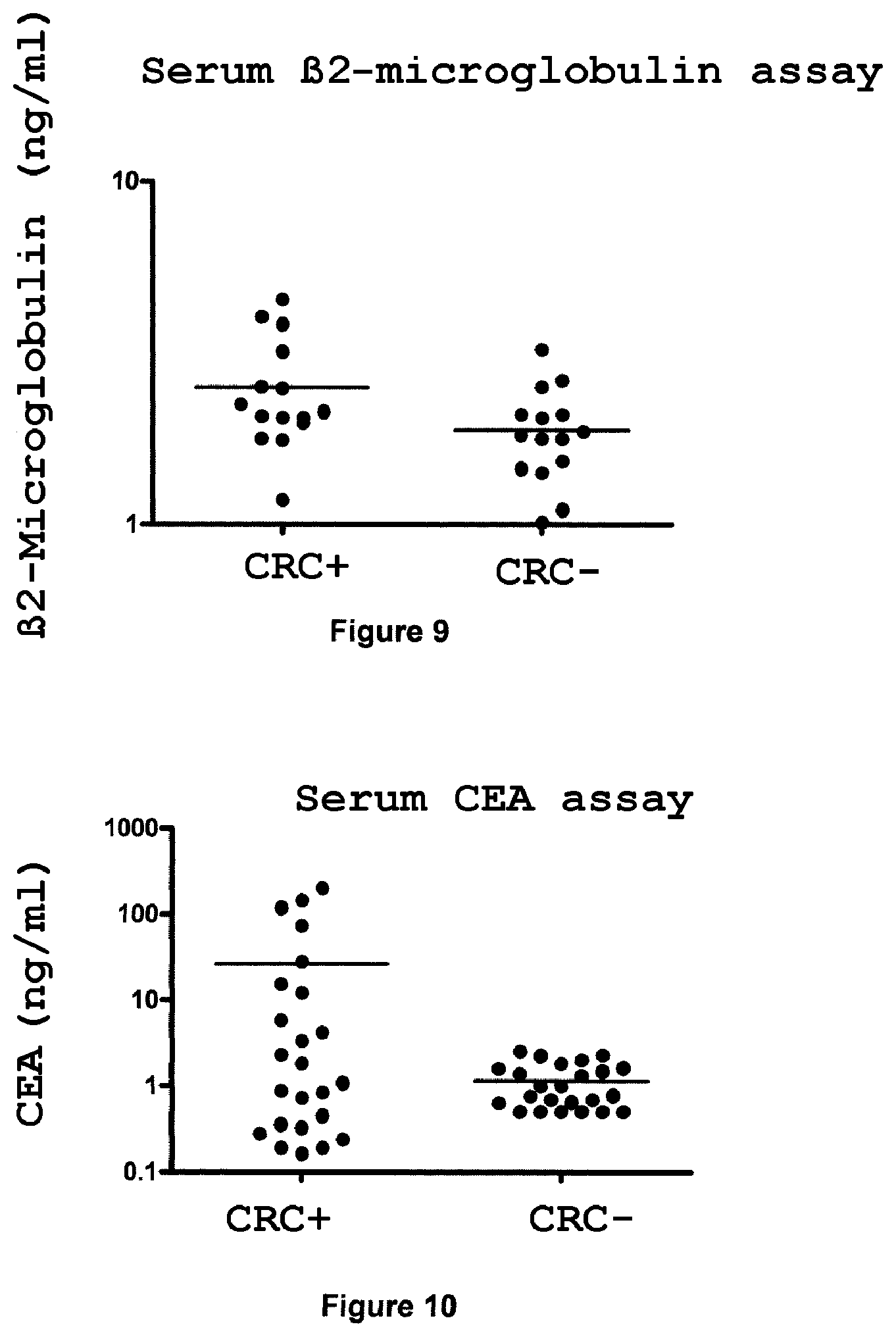
The Beta2-Microglobulin marker (Swiss Prot No. P61769, also known as .beta.2-Microglobulin, .beta.2M) is a low-molecular-weight (11 to 12 kDa) protein found at the surface of most nucleated human cells. The serum .beta.2-Microglobulin level increases in certain patients suffering from cancer, without this increase being specific, or correlated with the nature of the tumor, its stage or the severity of the disease. A significant increase is also observed in other diseases, such as lupus erythematosus, rheumatoid arthritis, Sjogren's syndrome, malignant diseases of the lymphoid system (multiple myeloma, B-cell lymphoma), certain viral diseases (hepatitis or AIDS), and in hemophiliac patients. Since .beta.2-Microglobulin is filtered by the renal glomeruli and reabsorbed by the proximal convoluted tubules, its concentration in the blood may be modified in the case of renal pathological conditions. The assaying of .beta.2-Microglobulin is thus most commonly reserved for the diagnosis of renal pathological conditions, or for the follow-up of infection with the acquired immunodeficiency virus. However, this marker is known as a tumor marker, in particular for colon cancer.
The Proteasome 20S marker (also known as Prosome) is the central structure of the proteasome, which is itself a molecular complex responsible for the intracellular degradation of ubiquitinated proteins.sup.29. The Proteasome is a molecular complex of 700 kDa, constituted of 28 subunits associated in 4 rings of 7 subunits. In humans, 7 alpha units (.alpha.1, .alpha.2, .alpha.3, .alpha.4, .alpha.5, .alpha.6 and .alpha.7) and 10 beta units (.beta.1, .beta.2, .beta.3, .beta.4, .beta.5, .beta.6, .beta.7, .beta.1i, .beta.2i and .beta.5i) are known. By virtue of its catalytic properties, the Proteasome plays a central role in the mechanisms of cell proliferation, growth, regulation and apoptosis, and therefore in the cancerization pathways. Proteasome inhibition with Bortezomib (Velcade) is a recognized treatment for multiple myeloma. Phase II or III therapeutic trials are ongoing for hematological cancers or tumors. T. Lavabre-Bertrand et al..sup.30 have shown that the serum level of Proteasome can increase on the occasion of certain pathological conditions, in particular in the case of cancers (myeloma, lymphoma and solid tumors).
The Galectin-3 marker (Swiss Prot No. P17931, also known as Gal-3, Galactose-Specific Lectin 3, MAC-2 antigen, IgE-Binding Protein, 35 kDa Lectin, Carbohydrate Binding Protein 35, CBP 35, Laminin-Binding Protein, Lectin L-29, L-31, Galactoside-Binding Protein or GALBP) is a lectin capable of binding to beta-galactoside structures of N-acetyllactosamine type. It is a protein with multiple functions involved in various biological functions, including the adhesion of tumor cells, proliferation, differentiation, angiogenesis, apoptosis, metastatic cancer progression.sup.31. Various studies have shown that Gal-3 can form complexes with numerous molecules: CEA, IgE, Laminin, Mucin, Mac-2BP, LAMP1, LAMP2, Fibronectin, etc. A serum assay of Gal-3 has been described by I. Iurisci et al..sup.32. Gal-3 was captured on microplates coated with Mac-2-binding protein (a Gal-3-binding protein) and then revealed with an anti-Gal-3 rat antibody. This study showed elevated serum of Gal-3 in the case of gastrointestinal cancers, breast cancer, lung cancer, ovarian cancer, melanomas and non-Hodgkin lymphomas.
The L-Lactate Dehydrogenase Chain B marker (Swiss Prot No. P07195, also known as LDH-B, LDH Heart Unit or LDH-H) is a protein that can form complexes in the form of homotetramers. This protein can also form complexes with the L-Lactate Dehydrogenase Chain A protein (Swiss Prot No. P00338, also known as LDH-A, LDH muscle unit or LDH-M) in the form of heterotetramers. The serum dose and/or the serum enzymatic activity of the tetrameric complexes, called LDH, increase(s) in the bloodstream proportionally to the tumor mass for many solid tumors. Its use is recommended in combination with human chorionic gonadotrophin (beta-hCG) and placental alkaline phosphatase for the follow-up of seminal vesicle cancers. LDH is considered to be a marker of importance for the prognosis of lymphomas, of leukemia and of colon cancer.sup.33.
The Calreticulin marker (Swiss Prot No. P27797, also known as CRP55, Calregulin, HACBP, ERp60 or grp60) is a multifunctional protein. It is a lectin capable of interacting transiently with virtually all the monoglycosylated proteins of the endoplasmic reticulum. D. J. McCool et al..sup.34 have thus shown that Calreticulin is involved in maturation of the colonic mucin MUC2. A method for the diagnosis of CRC which uses assaying of Calreticulin in a tissue, the stools or a body fluid is described in patent application WO 03/065003.
The Regenerating Islet-Derived Protein 3 Alpha marker (Swiss Prot No. Q06141, also known as Reg III-alpha, Pancreatitis-Associated Protein 1 or Pancreatis Associated Protein I (PAP 1)) is a protein that is weakly expressed in the healthy pancreas. It is overexpressed during the acute phases of pancreatitis and in certain patients suffering from chronic pancreatitis. In this case, it appears in the pancreatic fluid and in the bloodstream.sup.35. Y. Motoo et al..sup.36 have shown, by ELISA assay, that the level of PAP 1 in the blood increases in certain patients having colon cancer, stomach cancer, liver cancer or pancreatic cancer, and also in the case of renal insufficiency. To to this, they used the ELISA assay (PANCEPAP) marketed by the company Dynabio (La Gaude, France).
The Tumor-Associated Calcium Signal Transducer 1 marker (Swiss Prot No. P16422, also known as Major gastrointestinal tumor-associated protein GA733-2, Epithelial cell surface antigen, EpCAM, Epithelial glycoprotein, EGP, Adenocarcinoma-associated antigen, KSA, KS 1/4 antigen, Cell surface glycoprotein Trop-1 or CD326 antigen), was characterized in 1979 by virtue of its ability to be recognized by an antibody directed against colorectal cancer cells.sup.37. This protein is known by various names, as indicated above, but the most common use is to call it EpCAM. It is a transmembrane protein expressed at the basolateral surface of cells, in certain epithelia and many cancers.sup.38. As early as 1982, Herlyn et al..sup.39 showed that the injection of an anti-EpCAM monoclonal antibody could inhibit tumor growth in patients having colorectal cancer. These results resulted in the development of an antitumor treatment based on an anti-EpCAM antibody called Edrecolomab. This treatment is marketed under the name Panorex.TM.. Moreover, H. Abe et al..sup.40 have shown, by ELISA assay, that a soluble form of EpCAM, called MK-1, is increased in the bloodstream in 10% of cancer patients studied.
The Cytokeratins are part of the proteins that make up the intermediate filaments of the cytoskeleton of epithelial cells. Currently, more than 20 human Cytokeratins have been identified. The Cytokeratins 8 (Swiss Prot No. P05787, also known as Cytokeratin-8, CK-8, Keratin-8 or K8), 18 (Swiss Prot No. P05783, also known as Cytokeratin-18, CK-18, Keratin-18 or K18) and 19 (Swiss Prot No. P08727, also known as Cytokeratin-19, CK-19, Keratin-19 or K19) are the most abundant in epithelial cells and are useful tools for the diagnosis of cancer pathologies.sup.41. This clinical importance is linked to the release of Cytokeratins by epithelial cells in the apoptotic or proliferation phase. In the case of apoptosis, this release occurs in the form of soluble fragments which seem to appear under the proteolytic action of caspases. Undegraded Cytokeratin forms have never been described in the bloodstream. The three Cytokeratin assays most commonly used clinically are the tissue polypeptide antigen (TPA) assay, the tissue polypeptide specific antigen (TPS) assay and the CYFRA 21-1 assay. TPA is a broad-spectrum test which measures Cytokeratins 8, 18 and 19. The TPS and CYFRA 21-1 assays are more specific and measure, respectively, fragments of Cytokeratin 18 and of Cytokeratin 19. These 3 assays detect soluble Cytokeratin fragments that may be present on their own or in the form of protein complexes. TPA, TPS or CYFRA-21-1 have been used for the therapeutic follow-up of colorectal cancers, breast cancers, lung cancers, bladder cancers, ovarian cancers, pancreatic cancers, prostate cancers and certain ENT cancers. The assaying of soluble Cytokeratin fragments in the blood in fact has a clinical value in screening for relapses or evaluating the response to the therapy used (radiotherapy, chemotherapy, hormone treatment). Regular assaying makes it possible in particular to evaluate the progression of the tumor mass. The amount of soluble blood Cytokeratins also has a prognostic aspect with respect to the tumor stage and to the formation of metastases. Currently, the blood assay for Cytokeratin that is most commonly used is CYFRA 21-1. It is highly recommended for the follow-up of patients having non-small cell lung cancer. Various commercially available assays exist for TPA (AB Sangtec Medical Co., Byk-Roland, etc.), TPS (IDL Biotech AB, BEKI Diagnosiss, etc.) and CYFRA-21-1 (Roche Diagnosiss, CIS Bio-International, Fujirebio Diagnosiss, etc.). Moreover, H. Kim et al..sup.42 have shown that assaying fecal Cytokeratin 19 (DiNonA Inc.) may be useful in screening for gastrointestinal diseases, in combination with a fecal occult blood assay. Finally, the use of Cytokeratin 20 (Swiss Prot No. P35900, also known as Keratin, type I Cytoskeletal 20, CK-20, Keratin-20, K20, or IT protein) as a marker in colorectal cancer is described in patent application US 2002/0160382.
The Epithelial Cadherin marker (Swiss Prot No. P12830, also known as E-Cadherin, Uvomorulin, Cadherin-1, CAM 120/80 or CD324 antigen) is a transmembrane protein that mediates calcium-dependent cell adhesion. It is specifically expressed in epithelial cells, where it is involved in maintaining their phenotype. The cytoplasmic domain of E-Cadherin binds to .beta.-Catenin, which is itself bound to the actin filament networks of the cytoskeleton. This E-Cadherin/.beta.-Catenin binding plays an essential role in stabilizing cell/cell adhesions of the epithelial tissue. The loss of E-Cadherin can therefore reduce cell adhesion and increase the invasive capacity of cancer cells. A reduction in expression of E-Cadherin or of .beta.-Catenin is generally associated with greater aggressiveness and dedifferentiation of the tumor, in particular with respect to gastrointestinal cancers. F. Roca et al..sup.43 have thus shown that patients having colorectal cancer and underexpressing E-Cadherin have a more unfavorable prognosis than patients having a normal expression level. As early as 1983, Damsky et al..sup.44 showed that a soluble form of E-Cadherin could be released by the MCF-7 breast cancer cell line. This soluble form corresponds to the cleavage of the extracellular portion of E-Cadherin. Later, M. Katayama et al..sup.45 showed that the soluble form of E-Cadherin could be released into the bloodstream in the case of cancer, and C. Willmanns et al..sup.46 showed that the increase in the amount of E-Cadherin in the blood is correlated with the tumor stage in colorectal cancers. A commercially available kit is, moreover, proposed by the company Takara BioChemicals (Tokyo, Japan).
The assaying of CEA (carcinoembryonic antigen) for the diagnosis of colorectal cancer has been proposed since 1965 by P. Gold and S. Freedman.sup.47, but an assay for this marker in the blood has poor sensitivity for the diagnosis of colorectal cancers at a relatively nonadvanced stage. The assaying of serum CEA is thus especially recommended for evaluating the risk of liver metastases.sup.48 and for therapeutic follow-up. In addition, it is a marker that is not very specific for colorectal cancer; it may in fact be increased in many other cancers (lung, breast, etc.). On the other hand, the assaying of fecal CEA appears to be more sensitive and more specific than the assaying of serum CEA or than the assaying of fecal blood.sup.49. However, this assaying is not yet proposed routinely.
The reactive antigenic determinants 1116-NS-19-9, more commonly called CA19-9 (carbohydrate antigen 19.9), are carried by high-molecular-weight proteins.sup.50. The assaying of CA 19-9 in the blood is more specific than that of CEA. The CA 19-9 level in the blood increases in the event of colorectal cancer, of pancreatic cancer and of liver cancer (cholangiocarcinoma), but also in the event of noncancerous pathological conditions (cholangitis, etc.). Its use in combination with CEA is recommended both at the time of diagnosis of a cancer and for follow-up of the pathological condition.
J. Holmgren et al..sup.51 have shown that the amount of CA 50 antigen in the serum is increased in the case of colorectal cancer. The CA 50 antigen is defined by its ability to be recognized by a specific monoclonal antibody.
As regards the CA 72 marker, T. L. Klug et al..sup.52 have shown that the amount of CA 72 antigen in the serum is increased in the case of colorectal cancer. The CA 72 antigen is defined by its ability to be recognized by a specific monoclonal antibody.
Similarly, P. Kuusela et al..sup.53 have shown that the amount of CA 242 antigen in the serum is increased in the case of colorectal cancer. The CA 242 antigen is defined by its ability to be recognized by a specific monoclonal antibody.
The assaying of Testosterone for the diagnosis of colorectal cancer has been proposed in men by M. Holland et al..sup.54. These authors have shown a fall in the blood Testosterone level in the case of colorectal cancer.
As regards the TIMP-1, or tissue inhibitor of matrix metalloproteinase type-1, marker, patent application US 2007/0020707 describes in particular the assaying of TIMP-1 for the diagnosis of colorectal cancer by assaying in a body fluid.
F. Model et al..sup.55 showed, in July 2006, during the World Congress on Gastrointestinal Cancer, that it was possible to detect methylated forms of the septin-9 gene in the plasma of patients having colorectal cancer.
M. P. Ebert et al..sup.56 have shown that the ALX4 gene, or aristaless-like homeobox-4 gene, is more often methylated in the sera of patients having colorectal cancer than in control sera (P<0.0001). Using a threshold value of 41.4 pg/mL, they have obtained a sensitivity of 83.3% and a specificity of 70%.
Villin is described as a blood marker for the diagnosis of colorectal cancer in patent application FR2581456.
C. Bianco et al..sup.57 have shown that the amount of Cripto-1 in the serum is increased in the event of colorectal cancer.
The assaying of Intelectin-1 (Swiss Prot No. Q8WWA0, also known as Intestinal lactoferrin receptor, Galactofuranose-binding lectin, Endothelial lectin HL-1 or Omentin) for the diagnosis of colorectal cancer has been described in patent application US 2003/0082533.
The use of Protein Disulfide Isomerase (Swiss Prot No. P07237, also known as EC 5.3.4.1, PDI, Prolyl 4-hydroxylase subunit beta, Cellular thyroid hormone-binding protein or p55), of Translationally-Controlled Tumor Protein (Swiss Prot No. P13693, also known as TCTP, p23, Histamine-releasing factor, HRF or Fortilin) and of (Pro)defensin-A5 (Swiss Prot No. Q01523), as markers in colorectal cancer, is described respectively in patent applications EP1724586, US 2003/0172388 and US 2006/0179496. The term "(Pro)defensin" is intended to mean the precursor, namely the Prodefensin before cleavage, the propeptide, namely the N-terminal moiety after cleavage of the Prodefensin, and the mature protein, namely Defensin, corresponding to the C-terminal moiety after cleavage.
Finally, the assaying of human fecal hemoglobin is known practice and can be implemented as previously described.
The concentration of the tumor marker chosen from group B will, depending on the marker under consideration, be increased or decreased in the biological sample in which the method of the invention is carried out, compared with the reference values determined for healthy patients.
Preferably, the tumor marker(s) of group B is (are) chosen from: the markers: Beta2-Microglobulin, Proteasome 20S, Galectin-3, L-Lactate Dehydrogenase Chain B, Calreticulin, Regenerating Islet-Derived Protein 3 Alpha, Tumor-Associated Calcium Signal Transducer 1, Epithelial-Cadherin, CEA, CA19-9, Testosterone, TIMP-1, Intelectin-1, Protein Disulfide Isomerase, Cytokeratin 20, Translationally-Controlled Tumor Protein, (Pro)defensin-A5, and detection of human fecal hemoglobin.
Of course, the method of the invention may also include the detection of any other marker for colorectal cancer that is known to those skilled in the art.
The expression "the detection of the presence of a tumor marker other than apolipoprotein AI" is intended to mean the determination of the presence of the protein, or the messenger RNA thereof or the detection of a modification on the gene thereof in the coding or noncoding sequences, such as methylations.
The determination of the presence, in the biological sample, of the "protein" tumor marker of interest can be carried out by any method for determining the presence of a protein in a sample, known to those skilled in the art, such as, for example, a biochemical test, including an immunoassay, or by mass spectrometry.
The biochemical test may be any test widely known to those skilled in the art involving molecular interactions, i.e. reactions between said tumor marker and one or more binding partner(s) specific or not specific for said tumor marker.
Preferably, the biochemical test is an immunoassay known to those skilled in the art, involving immunological reactions between the tumor marker, which is the antigen, and one or more specific binding partner(s), namely the antibodies directed against this antigen.
The binding partners specific or not specific for the tumor marker(s) sought in the method of the invention are any partner capable of binding to this or these marker(s). They are said to be specific when they are capable of binding to these markers with a high specificity, or even a specificity of 100%. They are said to be nonspecific when their specificity of binding to these markers is low and they are then capable of binding to other ligands, such as proteins. By way of example, mention may be made of antibodies, antibody fractions, receptors and any other molecule capable of binding to this marker.
The binding-partner antibodies are, for example, either polyclonal antibodies or monoclonal antibodies.
The polyclonal antibodies may be obtained by immunization of an animal with the tumor marker concerned, followed by recovery of the desired antibodies in purified form, by taking the serum from said animal, and separation of said antibodies from the other serum constituents, in particular by affinity chromatography on a column to which an antigen specifically recognized by the antibodies, in particular said marker, is attached.
The monoclonal antibodies can be obtained by the hybridoma technique, the general principle of which is recalled hereinafter.
Firstly, an animal, generally a mouse, is immunized with the tumor marker of interest, the B lymphocytes of said animal then being capable of producing antibodies against said antigen. These antibody-producing lymphocytes are subsequently fused with "immortal" myeloma cells (murine in the example) so as to produce hybridomas. Using the heterogeneous mixture of cells thus obtained, a selection of cells capable of producing a particular antibody and of multiplying indefinitely is then carried out. Each hybridoma is multiplied in the form of a clone, each resulting in the production of a monoclonal antibody of which the properties of recognition with respect to said tumor marker may be tested, for example, by ELISA, by one-dimensional or two-dimensional Western blot, by immunofluorescence, or by means of a biosensor. The monoclonal antibodies thus selected are subsequently purified, in particular according to the affinity chromatography technique described above.
The monoclonal antibodies may also be recombinant antibodies obtained by genetic engineering, by means of techniques well known to those skilled in the art.
Examples of anti-Leukocyte Elastase Inhibitor antibodies are known and are available in particular in the Abcam catalog, rabbit anti-LEI polyclonal antibody, Cat. No. Ab47731. An anti-LEI monoclonal antibody, clone ELA-1, has been described in the article by R. Yasumatsu et al..sup.58.
Examples of anti-Ezrin antibodies are known and are available in particular in the Abcam catalog, anti-Ezrin monoclonal antibody, clone 3C12, Cat. No. Ab4069 and rabbit anti-Ezrin polyclonal antibody, Cat. No. Ab47418.
Examples of anti-Aminoacylase 1 antibodies are known and are available in particular in the Abnova catalog, anti-Aminoacylase 1 monoclonal antibody, clone 4F1-B7, Cat. No. H00000095-M01, and in the Abcam catalog, chicken anti-Aminoacylase 1 polyclonal antibody, Cat. No. Ab26173.
Examples of anti-Liver Fatty Acid-Binding Protein antibodies are known and are available in particular in the Abcam catalog, anti-L-FABP monoclonal antibody, clone 6B6, Cat. No. Ab10059, and rabbit anti-L-FABP polyclonal antibody, Cat. No. Ab7807.
Examples of anti-Intestinal Fatty Acid-Binding Protein antibodies are known and are available in particular in the R&D Systems catalog, anti-I-FABP monoclonal antibody, clone 323701, Cat. No. MAB3078, and in the Abcam catalog, rabbit anti-I-FABP polyclonal antibody, Cat. No. Ab7805.
Examples of anti-Apolipoprotein AI antibodies are known and are available in particular in the Biodesign Meridian Life Sciences catalog, anti-Apo AI monoclonal antibody, clone 4A90, Cat. No. H45402M and goat anti-Apo AI polyclonal antibody, Cat. No. K45252P.
Examples of anti-Apolipoprotein AII antibodies are known and are available in particular in the US Biological catalog, anti-Apo AII monoclonal antibody, clone 1402, Cat. No. A2299-31C and in the Biodesign Meridian Life Sciences catalog, goat anti-Apo AII polyclonal antibody, Cat. No. K74001P.
Examples of anti-I-Plastin polyclonal antibodies are known and are available in particular in the Santa Cruz Biotechnology catalog. The rabbit polyclonal antibody H-300 (Cat. No. sc-28531) reacts with I-Plastin, L-Plastin and T-Plastin. The applicant has developed monoclonal antibodies directed against I-Plastin.
Examples of anti-Beta2-Microglobulin, anti-CEA, anti-CA19-9 and anti-Testosterone antibodies are known and are in particular used in the applicant's assay kits, respectively Vidas.RTM. .beta.2-Microglobulin, Vidas.RTM. CEA, Vidas.RTM. CA19-9.TM. and Vidas.RTM. Testosterone.
Examples of anti-Proteasome 20S antibodies are known and are available in particular in the Affinitiy Research Products catalog.
Examples of anti-Galectin-3, anti-L-Lactate Dehydrogenase Chain B, anti-Calreticulin, anti-Tumor-Associated Calcium Signal Transducer 1, anti-Keratin type II Cytoskeletal 8, anti-Keratin type I Cytoskeletal 18, anti-Keratin type I Cytoskeletal 19, anti-Epithelial-Cadherin, anti-Villin and anti-TIMP-1 antibodies are known and are available in particular in the Abcam catalog.
Examples of anti-Regenerating Islet-Derived Protein 3 Alpha antibodies are known and are in particular used in the Dynabio assay kits (La Gaude, France).
Examples of anti-CA 242, anti-CA 50 and anti-CA 72-4 antibodies are known and are available in particular in the Fujirebio catalog.
Examples of anti-Intelectin-1 antibody are known and are available in particular in the Alexis Biochemicals catalog, anti-Intelectin-1 monoclonal antibody, clone Saly-1, Cat. No. ALX-804-850-C100 and rabbit anti-Intelectin-1 polyclonal antibody, Cat. No. ALX-210-941.
Examples of anti-Protein Disulfide Isomerase antibodies are known and are available in particular in the Abcam catalog, anti-PDI monoclonal antibody, clone RL77, Cat. No. Ab5484 and rabbit anti-PDI polyclonal antibody, Cat. No. Ab3672.
Examples of anti-Cytokeratin 20 antibodies are known and are available in particular in the Abcam catalog, anti-Cytokeratin 20 monoclonal antibody, clone Ks20.8, Cat. No. Ab962 and rabbit anti-Cytokeratin 20 polyclonal antibody, Cat. No. Ab36756.
Examples of anti-TCTP antibodies are known and are available in particular in the Abnova catalog, anti-TCTP monoclonal antibody, clone 3C7, Cat. No. 157H00007178-M01 and anti-TCTP polyclonal antibody, Cat. No. 157H00007178-A01.
Examples of anti-Defensin-A5 antibodies are known and are available in particular in the Santa Cruz Biotechnology catalog, anti-Defensin-A5 monoclonal antibody, clone 8C8, Cat. No. sc-53997 and in the Alpha Diagnosis International Inc. catalog, rabbit anti-Defensin-A5 polyclonal antibody, Cat. No. HDEFA51-A.
The binding partners which are specific or not specific for the tumor marker(s) sought in the method of the invention may be used as a capture reagent, as a detection reagent or as capture and detection reagents.
The visualization of the immunological reactions, i.e. of the tumor marker/binding partner binding, may be carried out by any means of detection, such as direct or indirect means.
In the case of direct detection, i.e. without the involvement of a label, the immunological reactions are observed, for example, by surface plasmon resonance or by cyclic voltametry on an electrode bearing a conductive polymer.
The indirect detection is carried out by means of labeling, either of the "revealing reagent" binding partner, or of the tumor marker of interest itself. In the latter case, this is then described as a competition method.
The term "labeling" is intended to mean the attachment of a label reagent capable of directly or indirectly generating a detectable signal. A nonlimiting list of these label reagents comprises: enzymes which produce a signal that can be detected, for example, by colorimetry, fluorescence or luminescence, such as horseradish peroxydase, alkaline phosphatase, .beta.-galactosidase or glucose-6-phosphate dehydrogenase, chromophores such as fluorescent or luminescent compounds or dyes, radioactive molecules such as .sup.32P, .sup.35S or .sup.125I, and fluorescent molecules such as Alexa or phycocyanins.
Indirect detection systems may also be used, such as, for example, ligands capable of reacting with an antiligand. Ligand/antiligand pairs are well known to those skilled in the art, this being the case, for example, of the following pairs: biotin/streptavidin, hapten/antibody, antigen/antibody, peptide/antibody, sugar/lectin, polynucleotide/sequence complementary to the polynucleotide. In this case, it is the ligand which carries the binding partner. The antiligand may be directly detectable by means of the label reagents described in the previous paragraph, or may itself be detectable by means of a ligand/antiligand.
These indirect detection systems may result, under certain conditions, in an amplification of the signal. This signal amplification technique is well known to those skilled in the art, and reference may be made to the prior patent applications FR98/10084 or WO-A-95/08000 by the applicant or to the article by Chevalier et al..sup.59.
Depending on the type of labeling used, those skilled in the art will add reagents that make it possible to visualize the labeling.
By way of example of immunoassays as defined above, mention may be made of "sandwich" methods such as ELISA, IRMA and RIA, "competition" methods and direct immunodetection methods such as immunohistochemistry, immunocytochemistry, Western blotting and Dot blotting.
Mass spectrometry can also be used for detecting, in the biological fluid, the tumor marker(s) sought in the method of the invention. The principle of spectrometry is widely known to those skilled in the art and is described, for example, in Patterson, S..sup.60.
To do this, the biological sample, which may or may not have been pretreated, is passed through a mass spectrometer and the spectrum obtained is compared with that of the tumor marker(s) sought in the method of the invention. An example of pretreatment of the sample consists in passing it over an immunocapture support comprising one of the binding partners for the tumor marker(s) sought in the method of the invention, for example an antibody directed against the tumor marker(s) sought in the method of the invention. Another example of pretreatment of the sample may be prefractionation of the biological sample in order to separate the proteins of the sample from one another. In techniques well known to those skilled in the art, the predominant proteins of the sample may, for example, first of all be depleted.
By virtue of recent technological advances, it has also become possible to quantify proteins in complex biological media using tandem mass spectrometry (MS/MS) carried out using a triple quadripole analyzer operating in the MRM (multiple reaction monitoring) mode. This operating mode has a double selectivity (two analyzers, parent ion selection and product ion selection) and the detection sensitivity is improved compared with other scanning modes. The technical feasibility of this approach was recently demonstrated by Anderson and Hunter.sup.61 who succeeded in detecting proteins of which the concentration is of the order of a hundred or so ng/ml in plasma, after immunodepletion of the most abundant proteins.
The determination of the presence, in the biological sample, of the "mRNA" tumor marker of interest may be carried out by any method for determining the presence of mRNA in a sample, namely either direct detection of the mRNA, or indirect detection of the mRNA, or any other method for determining the presence of an RNA in a sample, known to those skilled in the art.
The term "direct detection of the mRNA" is intended to mean the demonstration of the mRNA itself in the biological sample.
The direct detection of the mRNA in the biological sample may be carried out by any means known to those skilled in the art, such as, for example, by hybridization with a binding partner specific for the mRNA, where appropriate after amplification by the PCR or NASBA technique.
The term "hybridization" is intended to mean the process during which, under suitable conditions, two nucleotide fragments bind to one another with stable and specific hydrogen bonds so as to form a double-stranded complex. These hydrogen bonds form between the complementary bases adenine (A) and thymine (T) (or uracil (U)) (referred to as an A-T bond) or between the complementary bases guanine (G) and cytosine (C) (referred to as a G-C bond). The hybridization of two nucleotide fragments may be complete (reference is then made to complementary nucleotide fragments or sequences), i.e. the double-stranded complex obtained during this hybridization comprises only A-T bonds and C-G bonds. This hybridization may be partial (reference is then made to sufficiently complementary nucleotide fragments or sequences), i.e. the double-stranded complex obtained comprises A-T bonds and C-G bonds which make it possible to form the double-stranded complex, but also bases that are not bonded to a complementary base. The hybridization between two nucleotide fragments depends on the operating conditions that are used, and in particular the stringency. The stringency is defined in particular as a function of the base composition of the two nucleotide fragments, and also by the degree of mismatching between two nucleotide fragments. The stringency can also depend on the reaction parameters, such as the concentration and the type of ionic species present in the hybridization solution, the nature and the concentration of denaturing agents and/or the hybridization temperature. All these data are well known and the appropriate conditions can be determined by those skilled in the art. In general, depending on the length of the nucleotide fragments that it is desired to hybridize, the hybridization temperature is between approximately 20 and 70.degree. C., in particular between 35 and 65.degree. C., in a saline solution at a concentration of approximately 0.5 to 1 M. The binding partners which are specific or not specific for the mRNA are any partner capable of binding to this mRNA. By way of example, mention may be made of nucleic probes, amplification primers, and any other molecule capable of binding to this mRNA.
The term "hybridization probe" is intended to mean a nucleotide fragment comprising from 5 to 100 nucleic units, in particular from 10 to 35 nucleic units, having a hybridization specificity under given conditions so as to form a hybridization complex with the material specific for the target gene of interest. The hybridization probe may comprise a label enabling its detection.
For the purpose of the present invention, the term "amplification primer" is intended to mean a nucleotide fragment comprising from 5 to 100 nucleic units, preferably from 15 to 30 nucleic units, enabling the initiation of an enzymatic polymerization, such as, in particular, an enzymatic amplification reaction. The term "enzymatic amplification reaction" is intended to mean a process that generates multiple copies of a nucleotide fragment via the action of at least one enzyme. Such amplification reactions are well known to those skilled in the art and mention may in particular be made of the following techniques: PCR (polymerase chain reaction), as described in patents U.S. Pat. No. 4,683,195, U.S. Pat. No. 4,683,202 and U.S. Pat. No. 4,800,159, NASBA (nucleic acid sequence-based amplification) with patent application WO 91/02818, and TMA (transcription mediated amplification) with patent U.S. Pat. No. 5,399,491.
The term "detection" is intended to mean either a physical method, or a chemical method with an intercalating dye such as SYBR.RTM. Green I or ethidium bromide, or a method of detection using a label. Many detection methods exist for detecting nucleic acids.sup.62. The appropriate labels are as defined above.
For the purpose of the present invention, the hybridization probe may be a "detection" probe. In this case, the "detection" probe is labeled by means of a label as defined above. By virtue of the presence of this label, it is possible to detect the presence of a hybridization reaction between a given detection probe and the transcript to be detected.
The detection probe may in particular be a "molecular beacon" detection probe.sup.63. These "molecular beacons" become fluorescent during hybridization. They have a stem-loop structure and contain a fluorophore and a quencher group. The binding of the specific loop sequence with its complementary target nucleic acid sequence causes unfolding of the stem and the emission of a fluorescent signal during excitation at the appropriate wavelength.
The hybridization probe may also be a "capture" probe. In this case, the "capture" probe is immobilized or can be immobilized on a solid support by any appropriate means, i.e. directly or indirectly, for example by covalence or adsorption. The appropriate solid supports are known to those skilled in the art, and, by way of examples, mention may be made of synthetic materials or natural materials, latices, magnetic particles, metal derivatives, gels, etc. The solid support may be in the form of a microtitration plate, a membrane as described in application WO-A-94/12670 or a particle. It is also possible to immobilize several different capture probes on the support, each capture probe being specific for a target transcript. In particular, it is possible to use, as support, a biochip on which a large number of probes may be immobilized.
The immobilization of the probes on the support is also known to those skilled in the art, and mention may be made of a deposit of probes by direct transfer, microdeposition, in situ synthesis and photolithography.
The demonstration, in the biological sample, of the DNA modifications or anomalies in the gene encoding the tumor marker of interest may be carried out by any method for determining DNA alterations in a sample, namely either the direct detection of mutations, or the demonstration of alterations in the methylation profile of the loci of interest, or any other method for determining DNA alterations in a sample, known to those skilled in the art.
The mutations may include point substitutions of one nucleotide with another, deletions of one or more nucleotides and insertions of one or more nucleotides. The mutations may be located in the coding portion of the gene of the tumor marker of interest, or in the 5' and 3' noncoding portions, such as the transcription promoter region or the transcription termination region.
The strategies for demonstrating a mutation are based on molecular biology techniques and comprise steps of DNA extraction, amplification by PCR or another amplification technique, hybridization and/or sequencing. In the case of colorectal cancer, the following method has been successfully used to detect mutations in fecal DNA: concentration of the DNA by precipitation, enrichment in the target using capture oligonucleotides on magnetic beads, PCR amplification of the genes of interest, solid-phase sequencing for identifying point mutations.sup.64. The deletions were identified with respect to the difference in size between the expected reference fragment and the mutated fragment. Imperiale et al..sup.64 have described a panel of 21 mutations located in the K-ras, APC and p53 genes, which makes it possible to detect 16/31 of invasive cancers.
Other DNA markers used are the BAT-26 deletion, which is a marker for instability of microsatellites and highly amplifiable DNA called long DNA (L-DNA), which is not a specific marker but which appears to reflect the disorganized apoptosis of exfoliated tumor cells in the colonic lumen.sup.65. These markers are not satisfactory, either in terms of their sensitivity or in terms of their specificity.
As previously indicated, the DNA alterations may also correspond to a modification of the methylation profile of the gene corresponding to the tumor marker of interest. The modification of the methylation profile may correspond to a hypomethylation (decrease in the number of methylations) or to a hypermethylation (increase in the number of methylations). The altered units may be located in the coding portion of the gene of the tumor marker of interest, or in the 5' and 3' noncoding portions, such as the transcription promoter region or the transcription termination region.
The analysis of the DNA methylation may be carried out using techniques based on qualitative and/or quantitative PCR, such as MSP (methylation-specific PCR), bisulfite sequencing, digestion with a methylation-sensitive restriction enzyme coupled with PCR, COBRA (combined bisulfite restriction analysis) and Ms-SNuPE (methylation-sensitive single nucleotide primer extension). All these techniques have been reviewed comparatively and in detail in a methodology article.sup.66.
In the literature, several hypermethylated genes have been reported in the case of colorectal cancer. By way of example, mention may be made of the ALX4 (aristaless-like homeobox-4) gene.sup.56, the promoter region of the TPEF/HHP1 (transmembrane protein containing epidermel growth factor and follistatin domain) gene.sup.67 or else the septin-9 gene.sup.68.
When, in the method of the invention, at least two markers are detected, they may be demonstrated separately, for example using different immunoassay measurements, or else simultaneously, in a multiplex assay.
When, in the method of the invention, two markers of different nature are detected, for example a protein marker and an mRNA marker, two different detection methods, chosen from those described above, may be used. They may also be detected simultaneously, in the same detection medium and under the same reaction conditions, as described in patent application WO 03/104490. The steps of the detection method described in this patent application, which consists in simultaneously detecting hybridization and immunological reactions in a sample that may contain target analytes constituted of at least one nucleic acid and of at least one other ligand of different nature, consist in:
(i) depositing a known volume amount of the sample diluted in a reaction buffer, on a capture surface precoated with capture partners for said target analytes, said capture partners comprising at least one nucleic probe and at least one antiligand,
(ii) reacting at a temperature of between 15.degree. C. and 60.degree. C., and
(iii) visualizing the hybridization and immunological reactions thus obtained.
The biological sample may require a particular treatment because it may contain the tumor marker(s) sought in the method of the invention, as such, or else it may contain circulating tumor cells which contain the markers sought in the method of the invention and/or circulating tumor cells which are capable of secreting the marker(s) sought in the method of the invention.
Thus, according to one embodiment of the invention, the biological sample is pretreated in order to isolate the circulating tumor cells contained in said fluid.
The expression "isolate circulating tumor cells" is intended to mean obtain a cell fraction enriched in circulating tumor cells.
The treatment of the biological sample in order to isolate the circulating tumor cells can be carried out by cell sorting in a flow cytometer, by enrichment on Ficoll, by enrichment with magnetic beads covered with specific antibodies, or by any other method of specific enrichment known to those skilled in the art.
In the case of blood as biological sample, the circulating tumor cells may be isolated by means of a technique of cell separation on Ficoll combined with depletion of the blood cells using anti-CD45 antibodies coupled to magnetic beads (Dynal Biotech ASA, Norway).
The detection of the tumor marker(s) sought in the method of the invention can then be carried out directly using circulating tumor cells isolated from the biological sample, for example by immunocytochemical labeling of these cells with an antibody against tumor marker(s) sought in the method of the invention, after having deposited the circulating tumor cells on a slide by cytospin. The detection of the tumor marker(s) sought in the method of the invention may also be carried out directly in the circulating tumor cells using the flow cytometry method as described in Metezeau et al..sup.69.
Under these conditions, said circulating cells can be treated under conditions which make it possible to block the tumor marker(s) sought in the method of the invention, inside said cells. Such a treatment is described by Mathieu et al..sup.70.
The detection of the tumor marker(s) sought in the method of the invention is then carried out after having made the cell membrane permeable so as to allow entry of the binding partners specific for the marker(s) sought in the method of the invention.
The direct detection of the tumor marker(s) used in the method of the invention, based on the circulating cells, may also be carried out by means of an ELISPOT method, for example by means of the method described in patent application WO 03/076942 filed by the applicant. This method is a method for detecting and/or quantifying circulating tumor cells of a biological sample, which are capable of releasing or secreting, in vitro, one or more tumor marker(s), comprising the steps consisting in:
(i) depositing an amount of said cells at the bottom of a culture surface to which at least one binding partner specific for said tumor marker(s) is attached,
(ii) culturing said cells under conditions such that they release or secrete said tumor markers, which are immunocaptured at the bottom of the culture surface,
(iii) removing the cells by washing,
(iv) adding at least one labeled conjugate specific for said tumor markers, and
(v) visualizing the labeling thus obtained.
The direct detection of the tumor marker(s) used in the method of the invention in the tumor cells may also be carried out in the culture medium of said cells after having cultured them under conditions such that they secrete tumor marker(s) used in the method of the invention.
The culture conditions for release or the expression of the tumor markers are conventional conditions such as 37.degree. C. under a humid atmosphere and at 5% CO.sub.2.
When the biological sample is a solid sample, the presence of the tumor marker(s) may also be shown in vivo, in situ in the tumors.
In order to show the presence of a tumor marker in a tumor in vivo, any imaging method known to those skilled in the art may be used. For this, a binding partner for said tumor marker may be coupled to an imaging tracer.
The term "coupling of the binding partners to an imaging tracer" is intended to mean the attachment of a tracer capable of being detected by any imaging method known to those skilled in the art, and of directly or indirectly generating a detectable signal. Thus, the tracer may be a radioactive tracer such as technetium-99. In this case, the organ which has the primary cancer or the metastases will bind the tumor marker and its tracer. The radiation emitted by the organ can be filmed with a special camera, for example a gamma-camera. The instrument collects the scintillations generated by the radioactive substance and thus makes it possible to visualize the organ.
In another method of the invention, the tracer may comprise a positron-emitting radioactive substance (fluorine 18). The images will then be acquired by a positron emission tomography system.
In another preferred method of the invention, the partner of the tumor marker(s) may be coupled to nanoparticles. By way of example, they may be supramagnetic nanoparticles; for example, anionic magnetic nanoparticles for use in direct cell labeling and in vivo detection by nuclear magnetic resonance imaging. They may also be gold nanoparticles.
By virtue of the methods of the invention which enable the detection of the tumor marker in vivo, the areas of the body where there has been binding of the binding partner for the tumor marker, cancers producing tumor marker, and in particular colorectal cancer, and also localizations of their remote metastases and involved lymph nodes, may be visualized.
The method of the invention may be used not only for early diagnosis but also for screening, therapeutic follow-up, prognosis and relapse diagnosis in relation to colorectal cancer, since the decrease in the concentration of apolipoprotein AI in the remote sample occurs irrespective of the stage of the colorectal cancer, which constitutes another subject of the invention.
The invention will be understood more clearly by means of the following examples which are given by way of nonlimiting illustration, and also by means of the attached FIGS. 1 to 21, in which:
FIG. 1 is a graph relating to the assaying by ELISA of LEI, in ng/ml, in the serum of patients having colorectal cancer (CRC+) and of healthy patients (CRC-),
FIG. 2 is a graph relating to the assaying by ELISA of Ezrin, in ng/ml, in the serum of patients having colorectal cancer (CRC+) and of healthy patients (CRC-),
FIG. 3 is a graph relating to the assaying by ELISA of Aminoacylase 1, in ng/ml, in the serum of patients having colorectal cancer (CRC+) and of healthy patients (CRC-),
FIG. 4 is a graph relating to the assaying by ELISA of L-FABP, in ng/ml, in the serum of patients having colorectal cancer (CRC+) and of healthy patients (CRC-),
FIG. 5 is a graph relating to the assaying by ELISA of I-FABP, in pg/ml, in the serum of patients having colorectal cancer (CRC+) and of healthy patients (CRC-),
FIG. 6 is a graph relating to the assaying by ELISA of Apolipoprotein AI, in .mu.g/ml, in the serum of patients having colorectal cancer (CRC+) and of healthy patients (CRC-), either by microplate ELISA (FIG. 6A), or with the Lincoplex kit (FIG. 6B),
FIG. 7 is a graph relating to the assaying, using the Linco multiplex kit, of Apolipoprotein AII, in .mu.g/ml, in the serum of patients having colorectal cancer (CRC+) and of healthy patients (CRC-),
FIG. 8 is a graph relating to the assaying by ELISA of I-Plastin, in RFV (relative fluorescence value), in the serum of patients having colorectal cancer (CRC+) and of healthy patients (CRC-),
FIG. 9 is a graph relating to the assaying by ELISA of Beta2-Microglobulin, in ng/ml, in the serum of patients having colorectal cancer (CRC+) and of healthy patients (CRC-),
FIG. 10 is a graph relating to the assaying by ELISA of CEA, in ng/ml, in the serum of patients having colorectal cancer (CRC+) and of healthy patients (CRC-),
FIG. 11 is a graph relating to the assaying by ELISA of CA 19-9, in U/ml, in the serum of patients having colorectal cancer (CRC+) and of healthy patients (CRC-),
FIG. 12 is a graph relating to the assaying by ELISA of Testosterone, in ng/ml, in the serum of patients having colorectal cancer (CRC+) and of healthy patients (CRC-),
FIG. 13 is a graph relating to the assaying by ELISA of E-Cadherin, in ng/ml, in the serum of patients having colorectal cancer (CRC+) and of healthy patients (CRC-),
FIG. 14 is a graph relating to the assaying by ELISA of PAP1, in ng/ml, in the serum of patients having colorectal cancer (CRC+) and of healthy patients (CRC-),
FIG. 15 is a graph relating to the assaying by ELISA of Galectin-3, in RFV, in the serum of patients having colorectal cancer (CRC+) and of healthy patients (CRC-),
FIG. 16 is a graph relating to the assaying by ELISA of LDH, in ng/ml, in the serum of patients having colorectal cancer (CRC+) and of healthy patients (CRC-),
FIG. 17 is a graph relating to the assaying by ELISA of Proteasome 20S, in ng/ml, in the serum of patients having colorectal cancer (CRC+) and of healthy patients (CRC-),
FIG. 18 is a graph relating to the assaying by ELISA of Aminoacylase 1, in ng/ml, in the stools of patients having colorectal cancer (CRC+) and of healthy patients (CRC-),
FIG. 19 is a graph relating to the assaying by ELISA of Galectin-3, in RFV, in the stools of patients having colorectal cancer (CRC+) and of healthy patients (CRC-),
FIG. 20 is a graph relating to the assaying by ELISA of Proteasome 20S, in RFV, in the stools of patients having colorectal cancer (CRC+) and of healthy patients (CRC-), and
FIG. 21 is a graphic representation of an ELISPOT assay for LEI, for Ezrin and for Galectin-3, in number of spots per 10.sup.6 cancer cells of the Caco-2, HT-29 and HT29-B6 lines.
EXAMPLE 1: CLONING OF THE GENES ENCODING THE TUMOR MARKERS AND EXPRESSION OF THE RECOMBINANT PROTEINS
1. cDNA Amplification and Cloning
The Caco-2 colorectal cancer line is cultured in DMEM medium containing 2 mM of L-glutamine, without FCS (fetal calf serum) (all Gibco).
For the cloning of the LEI, L-FABP and Gal-3 genes, the messenger RNAs were extracted from a pellet of 10.sup.8 Caco-2 cells using the Invitrogen FastTrack 2.0 kit (Cat. No. 45-0019) according to the protocol supplied by the manufacturer. The reverse transcription and PCR steps are carried out in a single step using 450 ng of Caco-2 mRNA with the Superscript III One Step RT-PCR System kit (Invitrogen Cat. No. 12574-018) using the Platinum Taq DNA polymerase enzyme according to the protocol supplied by the manufacturer. The PCR primers used for the gene amplification are given in Table 1.
TABLE-US-00001 TABLE 1 Genes and primers Oligonucleotides LEI OL215 5'-ATGGAGCAGCTGAGCTCAGCAAAC-3' (SEQ ID No. 1) OL216 5'-CTAAGGGGAAGAAAATCTCCCCAA-3' (SEQ ID No. 2) L-FABP Forward 5'-CGGAGCGTCTCCCATGAGTTTCTCCGGC (SEQ ID No. 3) AAGTA-3' Reverse 5'-GAAATGCAGACTTGTCTAGATGCGCTTG (SEQ ID No. 4) CTGATGCGCTTGAAGACAATG-3' Gal-3 OL217 5'-ATGGCAGACAATTTTTCGCTCC-3' (SEQ ID No. 5) OL218 5'-TTATATCATGGTATATGAAGCACTGG-3' (SEQ ID No. 6)
The DNA fragments obtained were cloned into the vector pCR2.1 TOPO (LEI and Gal-3) with the TA cloning kit (Invitrogen Cat. No. K4520-01) or the vector pCMV6-XL4 from Origene (L-FABP) after digestion with Bsm BI and Xba I. The plasmids were sequenced in order to verify that the cDNA indeed complies with the expected sequence.
For the cloning of the gene encoding Aminoacylase 1, the total RNA was extracted from a pellet of 10.sup.8 Caco-2 cells using the RNA Easy Mini kit from Qiagen, according to the protocol supplied by the manufacturer. The reverse transcription is carried out using 10 ng of Caco-2 RNA, with the Superscript II enzyme (Invitrogen) according to the protocol supplied by the manufacturer. The reverse transcription primer is an oligo(dT).
The cDNA derived from this reaction was used as template in a PCR reaction using the AccuPrime Pfx kit (Invitrogen Cat. No. 12344-024) according to the protocol supplied by the manufacturer. The PCR primers are: ACY-1 Fwd2 (SEQ ID No. 7: 5'-GCGAATTCTTTAAGAAGGAGATATACATATGACGAGCAAAGGTCCGGAA GAGGAGCACCCATCG-3') and ACY-1 Rev (SEQ ID No. 8: 5'-GCAAGCTTCAGCTGTCACTGGGCAGGGC-3').
Under these conditions, it was possible to amplify a 1.3 kb fragment which was cloned into a cloning vector of the Zero Blunt TOPO PCR cloning kit type (Invitrogen Cat. No. K2820-20). This plasmid was sequenced in order to verify that the cDNA indeed complies with the expected sequence.
The following DNA fragment (SEQ ID No. 9) containing the I-FABP open reading frame was obtained by chemical synthesis, carried out by the company Geneart.
TABLE-US-00002 SEQ ID No. 9: GGTACCGAATTCCGCGTTTGACAGCACTTGGAAGGTAGACCGGAGTGAAA ACTATGACAAGTTCATGGAAAAAATGGGTGTTAATATAGTGAAAAGGAAG CTTGCAGCTCATGACAATTTGAAGCTGACAATTACACAAGAAGGAAATAA ATTCACAGTCAAAGAATCAAGCGCTTTTCGAAACATTGAAGTTGTTTTTG AACTTGGTGTCACCTTTAATTACAACCTAGCAGACGGAACTGAACTCAGG GGGACCTGGAGCCTTGAGGGAAATAAACTTATTGGAAAATTCAAACGGAC AGACAATGGAAACGAACTGAATACTGTCCGAGAAATTATAGGTGATGAAC TAGTCCAGACTTATGTGTATGAAGGAGTAGAAGCCAAAAGGATCTTTAAA AAGGATTCTAGAGTCGACGAGCTC.
2. Expression Vector Construction
The genes encoding LEI and Galectin-3 were subcloned into the prokaryotic expression vector pMR78.sup.71 and the L-FABP gene was cloned into the vector pET3d (New England Biolabs). The restriction sites necessary for the cloning were introduced by PCR using, as template, the plasmids pCR2.1 TOPO-LEI, pCR2.1 TOPO-Gal-3 and pCMV6-LFABP. The PCR enzyme is the Promega Pfu DNA polymerase, the PCR reaction was carried out according to the manufacturer's instructions, with the primers given in Table 2.
TABLE-US-00003 TABLE 2 Genes and primers Oligonucleotides LEI OL228 5'-ATGGGAATTCAGGAGCAGCTGAGCTCAGC (SEQ ID No. 10) AA-3' OL229 5'-CGATAAGCTTAAGGGGAAGAAAATCTCCC (SEQ ID No. 11) C-3' L-FABP Forward 5'-GCTGGCCATGGGCAGCAGCCATCATCATC (SEQ ID No. 12) ATCATCACATGAGTTTCTCCGGCAAGTACCAA C-3' Reverse 5'-GCACGGATCCTAGATGCGCTTGCTGATGC (SEQ ID No. 13) GCTTGAAGAC-3' Gal-3 OL230 5'-ATGGGAATTCAGGCAGACAATTTTTCGCT (SEQ ID No. 14) CC-3' OL231 5'-CGATAAGCTTATATCATGGTATATGAAGC (SEQ ID No. 15) ACTGG-3'
The PCR products containing the open reading frames encoding LEI or Galectin-3 were digested with the Eco RI and Hind III restriction enzymes. The fragments were introduced into the vector pMR78 restricted with the same enzymes (plasmids pMR-LEI and pMR-Gal-3). The vector pMR78 contains a 6-histidine sequence in frame with the protein to be expressed, which enables purification by metal-chelate affinity chromatography. The L-FABP PCR product was cloned into the vector pET3d, at the Nco I and Bam HI restriction sites.
For Aminoacylase 1, the TOPO cloning vector was directly digested with the Eco RI and Hind III restriction enzymes in order to generate a 1.3 kb fragment containing the acyl open reading frame, which was introduced into the vector pStaby1 (Eurogentec). The recombinant plasmid is called pStaby1-ACY.
For I-FABP, the cloning vector provided by Geneart was digested with the Eco RI and Sal I restriction enzymes in order to generate an approximately 400 bp fragment containing the coding sequence, which was introduced into the vector pMRCH79 (derived from the vector pMR78, bioMerieux). The recombinant plasmid is called pMRCH-IFABP.
The plasmids pGEX-Ezrine and pGEX-I-plastin, which make it possible to express, respectively, Ezrin and I-Plastin fused with GST (glutathione S-transferase), were supplied by the Institut Curie.
3. Recombinant Protein Expression and Purification
The expression plasmids for producing the recombinant tumor markers are introduced into E. coli BL21 bacteria and derivatives (Stratagene). The cultures are carried out at ambient temperature with shaking. The precise culture conditions for each protein are reproduced in Table 3. IPTG is isopropyl-beta-D-1-thiogalactosidase.
The bacterial pellets are taken up in 2.times.PBS (phosphate buffered saline) buffer and passed through a cell disintegrator at 1.5 kbar (Constant System). The lysates are centrifuged at 3000 g for 30 min at 4.degree. C. The supernatant contains the soluble proteins. The pellet contains the inclusion bodies. The buffer for solubilizing the inclusion bodies depends on the protein.
For LEI, the purification is carried out using the soluble fraction, on a column containing 5 mL of Ni-NTA-Sepharose resin (Qiagen) and the protein is eluted with 2.times.PBS containing 450 mM imidazole, pH 7.5.
For Galectin-3, the inclusion bodies are solubilized in 2.times.PBS containing 1M urea, and passed over 5 mL of Ni-NTA-Sepharose resin (Qiagen) and the Gal-3 protein is eluted with 2.times.PBS containing 450 mM imidazole and 1M urea, pH 7.5.
For L-FABP, the purification is carried out using the soluble fraction, with the Ni-IDA kit from Macherey-Nagel.
TABLE-US-00004 TABLE 3 Culture IPTG Strain volume induction Purification LEI BL21 250 mL 0.1 mM Ni-NTA Gal-3 BL21-Codon 400 mL 0.5 mM Ni-NTA plus (DE3)-RIPL L-FABP BL21 500 mL 0.1 mM Ni-IDA GST-Ezrin BL21 250 mL 0.1 mM GST ACY-1 BL21-Codon 500 mL 0.1 mM other plus (DE3)-RIPL
For GST-Ezrin, the purification is carried out using the inclusion bodies solubilized in 100 mM Tris buffer containing 8M urea and 10 mM DTT, by GST affinity chromatography. A column containing 5 mL of Glutathione Sepharose 4 fast flow gel (Amersham) is used. The equilibration and washing buffer is 2.times.PBS containing 0.05% Tween 20. The elution buffer is 50 mM Tris-HCl containing 20 mM reduced glutathione, pH 8.
For Aminoacylase 1, the soluble fraction of the culture is passed over an Amersham HiTrap Q FF column and the ACY-1 protein was eluted with 0.3M NaCl at pH 7.5. Since several other proteins were co-eluted under these conditions, the purification was continued on a hydrophobic-interaction column (HIC Phenyl HP, Amersham). The ACY-1 protein was eluted with 0.5M NaCl at pH 7.
The recombinant GST-I-Plastin protein was provided by the Institut Curie in purified form.
The recombinant Calreticulin protein was produced by the company Proteus Services for Industry (Dijon, France). The sequence encoding Calreticulin was obtained by chemical synthesis.
EXAMPLE 2: PRODUCTION OF MONOCLONAL ANTIBODIES DIRECTED AGAINST THE TUMOR MARKERS
1. Animal Model
The immunization experiments were carried out in female BALB/c (H-2.sup.d) mice that were 6 to 8 weeks at the time of the first immunization.
2. Immunogens and Immunizations
In order to increase the immune responses obtained in the mice and to be able to generate monoclonal antibodies, the tumor markers were produced in the form of recombinant proteins produced according to the procedures described in Example 1. The LDH protein was obtained from the company SciPac (Cat. No. 103-133). These proteins were mixed volume for volume with Freund's adjuvant (Sigma), prepared in the form of a water-in-oil emulsion and which is known to have a good immunogenic capacity. 3 mice were immunized for each tumor marker. The mice received 3 successive doses of 10 .mu.g of the immunogens at 0, 2 and 4 weeks. All the injections were given subcutaneously. The first injection is given as a mixture with complete Freund's adjuvant, the following two are given as a mixture with incomplete Freund's adjuvant. Between D50 and D70 after the first injection, the humoral responses were restimulated with an intravenous injection of 100 .mu.g of the recombinant protein.
3. Monitoring of the Appearance of the Humoral Response
In order to monitor the appearance of the antibodies, blood samples are taken regularly from the mice. The presence of the anti-tumor marker antibodies is tested using an ELISA. The protein of interest is used for capture (1 .mu.g/well); after saturation, the antigen is reacted with various dilutions of the test sera (incubation at 37.degree. C. for 1 h). The specific antibodies present in the serum are revealed with an AffiniPure goat anti-mouse IgG antibody conjugated to alkaline phosphatase (H+L, Jackson Immunoresearch, Cat no. 115-055-146), which binds to the antibodies being sought (0.1 .mu.g/well).
4. Production of Monoclonal Antibodies
Three days after the final injection, for each tumor marker, one of the immunized mice was sacrificed. The blood and the spleen were taken. The splenocytes obtained from the spleen were cultured with Sp2/0-Ag14 myeloma cells in order for them to fuse and become immortalized, according to the protocol described by Kohler and Milstein.sup.72,73. After an incubation period of 12-14 days, the supernatants of the hybridomas obtained were screened in order to determine the presence of anti-tumor marker antibodies, using the ELISA assay described in point 3 of this example. When GST fusion proteins were used as immunogen, the clones directed against GST are eliminated by carrying out an ELISA screening with uncoupled GST for capture. The positive hybridoma colonies were subcloned twice according to the limiting dilution technique, which is well known to those skilled in the art.
5. Characterization of the Monoclonal Antibodies by Immunoblotting
The list of monoclonal antibodies obtained against the various tumor markers is given in Table 4. These monoclonal antibodies were analyzed by the Western blotting technique.
TABLE-US-00005 TABLE 4 Tumor markers Monoclonal antibody name Leukocyte Elastase Inhibitor (LEI) 21B10A5 and 10E1H1 Ezrin 4A7A6C1 and 4A9H5 Aminoacylase 1 2A7F6 and 11H7D9 I-plastin 3D11D10, 8C8C5, 3A3H2, 8G2D2 Calreticulin 5C10H10 and 11B6D11 L-lactate dehydrogenase chain B 3F11E11 and 12F10G8 (LDH) Galectin-3 12F8A12 and 14A5G1
5.1. Methodology
The Caco-2 and HT-29 line cell culture extracts are prepared by directly lyzing the cell pellet with 600 .mu.l of an aqueous solution of 8.3M urea, 2M thiourea, 4% 3-[(3-cholamidopropyl)dimethylammonio]-1-propane sulfonate (CHAPS), 100 mM DTT, 2% Servalyte 4-9 (Serva, Heidelberg, Germany) and 0.1 g/l Orange G, and then treated according to the NuPAGE Novex gel sample preparation protocol (Invitrogen). To obtain the tissue extracts, tumor and mucosal biopsies of patients GHBD001, GHBD004 and CLSP109 were dissected with a scalpel, and were then subjected to 10 cycles of extraction in the Medimachine system (Becton Dickinson) using 50-.mu.m Medicons with 1 ml of PBS buffer containing 2.5 mM EDTA and protease inhibitors (tablets, Roche). These 10 ml of cell suspension are pooled, made up to 25 ml, and then centrifuged for 15 min at 600 g. The supernatant corresponds to the tissue extract which is treated according to the NuPAGE Novex gel sample preparation protocol. Reduced samples are used, at a final total protein concentration of 0.4 mg/ml. The deposit volume is 20 .mu.l per well, on a NuPAGE Novex Bis-Tris 4-12% gel, with MOPS running buffer. After migration (at 200 V, for 1 hour) and transfer onto a PVDF membrane (at 400 mA, for 45 min), the quality of the transfer is assessed by staining with amido black.
The membranes are saturated with 5% skimmed milk (Regilait) in a solution of TNT (15 mM Tris, 0.14M NaCl, 0.5% Tween 20, pH 8) at ambient temperature for 1 hour. After saturation, the membranes are incubated for 1 hour with the various test antibodies diluted to 10 .mu.g/ml in the saturating solution. After rinsing with TNT, the membranes are incubated for 1 hour at ambient temperature with an anti-mouse-horseradish peroxidase conjugate diluted to 1:5000, (Cat No. 115-035-062, Jackson Immunoresearch) in the saturating solution. After rinsing, the developing is carried out with the Substrate Supersignal West Dura Extended kit (Cat No. 34076, Pierce) according to the recommended information for use.
The chemiluminescence signal on the membranes was measured with the VersaDoc imaging system from Biorad. Based on the image of the Western blot, the volumes of the bands which correspond to the various tumor markers were evaluated with the QuantityOne software (Bio-Rad). The volume corresponds to the intensity of the chemiluminescence signal multiplied by the surface area of the band.
5.2. Results
The Western blotting results are reproduced in Table 5, which gives the volume of the bands corresponding to the tumor marker of interest for the Western blotting analyses, as a function of the various samples tested. These results show that the tumor markers tested are indeed expressed by the Caco-2 and HT-29 colon cancer lines, and also in the tissues, as shown with the extracts of tumor and mucosa, obtained from the patients. The intensity of the signal obtained with an antibody on a sample can be compared to the signals obtained with the other samples and the same antibody. The technique used makes it possible to confirm the presence or absence of the marker in the tissue (non-remote sample) and the specificity of the antibodies with respect to the markers. This technique was not used in this example in the remote samples because it would not make it possible to come to a conclusion regarding the presence or absence of the tumor marker in the remote samples, nor to determine whether the concentration of said tumor marker is increased or decreased in said samples. Furthermore, the experimental scheme used does not make it possible to compare the reactivity of one antibody with another.
TABLE-US-00006 TABLE 5 Tumor Tumor Mucosal Tumor Mucosal Tumor marker and tissue tissue tissue tissue tissue antibody Caco-2 HT-29 GHBD001 GHBD004 GHBD004 CLSP109 CLSP109 LEI 21B10A5 8365 7678 NT 60200 36506 NT NT 10E1H1 0 0 NT 13357 6893 NT NT Ezrin 4A9H5 7066 4742 NT NT NT 1588 2446 4A7A6C1 123436 116448 42480 15303 67439 NT NT Aminoacylase 1 2A7F6 10687 4787 NT NT NT 4477 7238 11H7D9 217664 232005 36093 10513 30233 NT NT I-plastin 3D11D10 136725 NT NT NT NT 275477 246564 8C8C5 557 1110 4364 77 0 NT NT Calreticulin 5C10H10 2842 3040 NT NT NT 2503 3294 11B6D11 3261 2937 NT NT NT 2070 2764 LDH 3F11E11 45391 NT NT NT NT 30411 13942 12F10G8 122907 154593 11841 15811 53285 NT NT Galectin-3 12F8A12 245712 65790 18262 12961 7307 NT NT 14A5G1 254531 120010 79833 98361 45872 NT NT NT: not tested.
5.3. Monoclonal Antibodies Directed Against I-Plastin
In the patient GHBD004, the 8C8C5 antibody does not light up, or only very weakly lights up, the band which corresponds to I-plastin. The presence of I-plastin in these samples can be demonstrated using, for example, the 8G2D2 antibody, which has a better affinity for I-plastin in blotting.
Since I-plastin is a member of a family of proteins comprising 2 other isoforms (L-plastin and T-plastin) with which it has more than 70% homology, we tested all the clones of monoclonal antibodies obtained, for their reactivity with respect to the GST-plastin-L and GST-plastin-T proteins (provided by the Institut Curie). At the end of this screening, we selected the clones 3D11D10, 8C8C5, 3A3H2 and 8G2D2 which do not exhibit any cross-reactivity with the other members of the family. These antibodies are indeed specific for the I-plastin isoform.
EXAMPLE 3: SERUM ASSAYS FOR THE TUMOR MARKERS
1. Patients and Specimens
Blood samples are collected from a network of 8 clinical centers distributed throughout France, in the context of 2 Huriet-law protocols.
In order to obtain serum, the blood sample is taken on a dry tube. In order to obtain plasma, the blood sample is taken on an EDTA tube. After coagulation, the tube is centrifuged for 10 min at 1000 g, and the serum is removed, aliquoted and stored at -80.degree. C. The tube of plasma is directly centrifuged for 10 min at 1000 g, and the plasma is removed, aliquoted and stored at -80.degree. C. The samples are completely documented for the clinical history of the patients.
2. Serum Assay for the LEI Tumor Marker
The LEI protein was assayed using the antibodies described in Example 2 and an ELISA assay using the Vidas.RTM. automated device (bioMerieux). To do this, the ELISA assay was constructed using the reagents of the Vidas.RTM. HBs Ag Ultra kit (bioMerieux, Cat. No. 30315). The reagents were used as described in the corresponding information sheet (ref. 11728 D-FR-May 2005), with the following modifications: 1. The cones were sensitized with the capture antibody 10E1H1 at a concentration of 10 .mu.g/ml. 2. The content of the second well of the HBs Ag Ultra cartridge was replaced with 300 .mu.l of revealing antibody 21B10A5, coupled to biotin, diluted to 1 .mu.g/ml in the buffer of the second well of the Vidas.RTM. HBs Ag Ultra kit (buffer with goat serum and sodium azide at 1 g/l). 3. The serum, plasma or stool samples (50 .mu.l) were diluted directly in the second well of the HBs Ag Ultra cartridge, pure or after a prior dilution to 1/20 in the buffer of the second well of the Vidas.RTM. HBs Ag Ultra kit (buffer with goat serum and sodium azide at 1 g/l). 4. The ELISA reaction was carried out using the Vidas.RTM. automated device and the protocol of the HBs Ag Ultra kit. 5. The results were obtained in the form of crude values after subtraction of the background noise (reading of the substrate before reaction). A standard curve was established by assaying a range of concentrations of the tumor marker in the form of recombinant protein. The standard curve was plotted by reporting the concentration of the tumor marker along the x-axis and the signal read by Vidas.RTM. (RFV or Relative Fluorescence Value) along the y-axis. The concentration of tumor marker present in the body fluid to be assayed (blood, serum, plasma, stool) was calculated by reporting the concentration corresponding to the RFV signal read by Vidas.RTM..
The amounts obtained for the patients analyzed are reported in FIG. 1. It may be noted, on this figure, that 3 sera of patients having stage IV colorectal cancer and 1 serum of a patient having stage III colorectal cancer show a clear increase in their amount of serum LEI.
3. Serum Assay for the Ezrin Tumor Marker
The Ezrin protein was assayed using the antibodies described in Example 2 and an ELISA assay using the Vidas.RTM. automated device (bioMerieux). To do this, the ELISA assay was constructed using the reagents of the Vidas.RTM. HBs Ag Ultra kit (bioMerieux, Cat. No. 30315). The reagents were used as described in the corresponding information sheet (ref. 11728 D-FR-May 2005), with the following modifications: 1. The cones were sensitized with the capture antibody 4A9H5 at a concentration of 30 .mu.g/ml. 2. The content of the second well of the HBs Ag Ultra cartridge was replaced with 300 .mu.l of revealing antibody 4A7A6C1, coupled to biotin, diluted to 1 .mu.g/ml in the buffer of the second well of the Vidas.RTM. HBs Ag Ultra kit (buffer with goat serum and sodium azide at 1 g/l). 3. The serum, plasma and stool samples (50 .mu.l) were diluted directly in the second well of the HBs Ag Ultra cartridge. 4. The ELISA reaction was carried out using the Vidas.RTM. automated device and the HBs Ag Ultra protocol, in which the step of incubating the sample with the capture and revealing antibodies had been taken to 100 cycles. 5. The results were obtained in the form of crude values after subtraction of the background noise (reading of the substrate before reaction).
The concentration of the tumor marker present in the body fluid to be assayed (blood, serum, plasma, stool) was calculated according to the procedure described in paragraph 2 regarding the assaying of LEI.
The amounts obtained for the patients analyzed are reported in FIG. 2. It may be noted, in this figure, that 3 sera from patients having stage IV colorectal cancer show a clear increase in their amount of serum Ezrin.
4. Serum Assay for the Aminoacylase 1 Tumor Marker
The Aminoacylase 1 protein was assayed using the antibodies described in Example 2 and an ELISA assay using the Vidas.RTM. automated device (bioMerieux). To do this, the ELISA assay was constructed using the reagents of the Vidas.RTM. HBs Ag Ultra kit (bioMerieux, Cat. No. 30315). The reagents were used as described in the corresponding information sheet (ref. 11728 D-FR-May 2005), with the following modifications: 1. The cones were sensitized with the capture antibody 2A7F6 at a concentration of 20 .mu.g/ml. 2. The content of the second well of the HBs Ag Ultra cartridge was replaced with 300 .mu.l of revealing antibody 11H7D9, coupled to biotin, diluted to 1 .mu.g/ml in the buffer of the second well of the Vidas.RTM. HBs Ag Ultra kit (buffer with goat serum and sodium azide at 1 g/l). 3. The serum, plasma or stool samples (100 .mu.l) were diluted directly in the second well of the HBs Ag Ultra cartridge. 4. The ELISA reaction was carried out using the Vidas.RTM. automated device and the HBs Ag Ultra protocol, in which the step of incubating the sample with the capture and revealing antibodies had been taken to 100 cycles. 5. The results were obtained in the form of crude values after subtraction of the background noise (reading of the substrate before reaction).
The concentration of the tumor marker present in the body fluid to be assayed (blood, serum, plasma, stool) was calculated according to the procedure described in paragraph 2 regarding the assaying of LEI.
The amounts obtained for the patients analyzed are reported in FIG. 3. It may be noted, in this figure, that 1 serum from a patient having stage II colorectal cancer, 1 serum from a patient having stage III colorectal cancer and 2 sera from patients having stage IV colorectal cancer show a clear increase in their amount of serum Aminoacylase 1.
5. Serum Assay for the L-FABP Tumor Marker
We used an ELISA kit marketed by the company Hycult Biotechnology to assay the human L-FABP protein (Cat. No. HK404). This kit makes it possible to quantify the L-FABP protein in cell culture supernatants or in serum, plasma or urine, in order to determine the presence of lesions in the liver. We followed the procedure recommended by the manufacturer, with 2 modifications: the incubations were carried out at 37.degree. C. and not at ambient temperature, the sera were diluted to 1/10.sup.th before the assay. The assaying of the L-FABP protein can be carried out by alternative techniques, well known to those skilled in the art.
FIG. 4 gives the results of this assay. In the serum panel that we tested, 41 patients out of 141 having colorectal cancer have a serum L-FABP concentration of greater than 17 ng/ml, whereas, in the control group, no individual exceeds this value. Among these 41 patients, are 8 patients with stage I colorectal cancer, 8 with stage II colorectal cancer, 13 with stage III colorectal cancer and 12 with stage IV colorectal cancer. The mean serum L-FABP concentration observed for 141 patients with colorectal cancer is 16.6.+-.1.3 ng/ml. The mean value is 6.6.+-.0.2 ng/ml for 112 healthy individuals (negative controls). This difference is statistically significant (P<0.0001, one-sided t-test with Welch's correction for unequal variances).
6. Serum Assay for the I-FABP Tumor Marker
We used an ELISA kit marketed by the company Hycult Biotechnology to assay the human I-FABP protein (Cat. No. HK406). This kit makes it possible to quantify the I-FABP protein in cell culture supernatants or in serum, plasma or urine, in order to determine the presence of ischemic lesions in the small intestine. We followed the procedure recommended by the manufacturer. The assaying of the I-FABP protein can be carried out by alternative techniques, well known to those skilled in the art.
FIG. 5 gives the results of this assay. In the serum panel that we tested, 15 patients out of 40 having colorectal cancer have a serum I-FABP concentration of greater than 40 pg/ml, whereas, in the control group, only 2 individuals out of 24 exceed this value. More clearly, 3 sera of patients having stage I colorectal cancer, 2 sera of patients having stage III colorectal cancer and 1 serum of a patient having stage IV colorectal cancer have a serum I-FABP concentration of greater than 100 pg/ml. No concentration above this value was found in the CRC- control group.
7. Serum Assay for the Apolipoprotein AI Tumor Marker
The assaying of serum Apolipoprotein AI was carried out by means of two different immunoassay techniques. Firstly, we used a microplate sandwich ELISA. The 96-well plates were coated with the anti-Apo AI monoclonal antibody, clone 1404 (Biodesign Cat. No. H45404) at 1 .mu.g per well. After 3 washes with PBS-0.05% Tween 20 (PBS-T), the plates were saturated with 10% milk in PBS-T for 1 h at 37.degree. C. The plates were washed a further 3 times in PBS-T, 100 .mu.l of the dilutions of the standard range or 100 .mu.l of the 1/100 000 dilution of the test serum samples were deposited onto the plates, and the plates were incubated for 2 h at 37.degree. C. The standard range was prepared by diluting the Apo AI protein (Biodesign Cat. No. A50620H) in PBS-T, BSA 1% (1.6 to 100 ng/ml). After 3 washes with PBS-T, the polyclonal detection antibody coupled to horseradish peroxidase (Biodesign Cat. No. K45452P) was added at 0.1 .mu.g per well, and the plates were incubated for 2 h at 37.degree. C. A further 3 washes with PBS-T were carried out, before adding the OptEIA substrate (BD), at 100 .mu.l/well. After 20 min, when the development of the color had taken place, the reaction was stopped with 2N sulfuric acid and the absorbence at 450 nm was measured.
The results of the assaying of serum Apo AI in patients by microplate ELISA are given in Table 6A.
TABLE-US-00007 TABLE 6A Pathological condition.sup.a Sample identifier Nature Stage TNM.sup.b Apo A1 .mu.g/mL Liver cancer HEPA 1/F0 1 1 9C Serum 1267 Liver cancer HEPA 10/F0 1 1 9C Serum 1358 Liver cancer HEPA 11/F0 1 1 9C Serum 1475 Liver cancer HEPA 12/F0 1 1 9C Serum 689 Liver cancer HEPA 13/F0 1 5B Serum 975 Liver cancer HEPA 2/F0 1 1 9C Serum 947 Liver cancer HEPA 3/F0 1 1 9C Serum 741 Liver cancer HEPA 4/F0 1 1 9C Serum 1051 Liver cancer HEPA 5/F0 1 1 9C Serum 1273 Liver cancer HEPA 6/F0 1 1 9C Serum 1084 Liver cancer HEPA 7/F0 1 1 9C Serum 1851 Liver cancer HEPA 8/F0 1 1 9C Serum 1408 Liver cancer HEPA 9/F0 1 1 9C Serum 1160 CRC+ CBSE007/F0 GS 1 7 2C Serum I TisN0M0 957 CRC+ CBSE016/F0 GS 2 5 8C Serum I T1N0M0 835 CRC+ GHBD035/F0 GS 2 7B Serum I TisN0M0 945 CRC+ CLSP059/F0 GS 1 7 5C Serum I TisN0M0 787 CRC+ CBSE022/F0 GS 2 5 10C Serum I T1N0M0 409 CRC+ CLSP118/F0 GS 1 5 9C Serum I T2N0M0 639 CRC+ CLSP145/F0 GS 2 5 11C Serum I T1N0M0 343 CRC+ CBSE011/F0 GS 11 6C Serum I TisN0M0 354 CRC+ CLSP150/F0 GS 2 5 6C Serum I T2N0M0 389 CRC+ GHBD003/F0 GS 2 1 5C Serum I T2N0M0 508 CRC+ GHBD015/F0 GS 2 1 10C Serum I TisN0M0 808 CRC+ P38868 s1 10C Serum II T3N0M0 894 CRC+ GHBD039/F0 GS 1 1 4C Serum II T3N0M0 805 CRC+ GHBD066/F0 GS 3 7B Serum II T3N0M0 1181 CRC+ CLSP096/F0 GS 2 5 9C Serum II T3N0M0 538 CRC+ CLSP154/F0 GS 2 5 9C Serum II T3N0M0 686 CRC+ CBSE004/F0 GS 3 1 6C Serum II T3N0M0 731 CRC+ CLSP105/F0 GS 1 10 5C Serum II T3N0M0 540 CRC+ CLSP133/F0 GS 2 5 10C Serum II T3N0M0 507 CRC+ CLSP136/F0 GS 4 5 10C Serum II T3N0M0 423 CRC+ GHBD016/F0 GS 2 1 10C Serum II T3N0M0 751 CRC+ GHBD037/F0 GS 1 4 4C Serum III T3N2M0 604 CRC+ GHBD058/F0 GS 2 5 12C Serum III T3N1M0 1248 CRC+ CBSE023/F0 GS 6 5 9C Serum III T4N2M0 750 CRC+ GHBD005/F0 GS 1 5 9C Serum III T3N1M0 500 CRC+ CLSP074/F0 GS 1 5 8C Serum III T3N2M0 552 CRC+ CLSP144/F0 GS 4 5 9C Serum III T3N1M0 804 CRC+ CLSP044/F0 GS 1 4 8C Serum III T3N1M0 526 CRC+ CLSP097/F0 GS 1 8 2C Serum III T3N1M0 630 CRC+ CLSP098/F0 GS 3 9 1C Serum III T3N2M0 384 CRC+ CLSP121/F0 GS 2 7 1C Serum III T3N2M0 706 CRC+ CLSP095/F0 GS 4 1 9C Serum IV T3N1M1 540 CRC+ CLSP161/F0 GS 2 10 8C Serum IV T3N2M1 573 CRC+ GHBD056/F0 GS 4 10 9C Serum IV T3N2M1 719 CRC+ CBSE027/F0 GS 3 1 6C Serum IV T4N2M1 907 CRC+ CLSP109/F0 GS 2 5 4C Serum IV T3N1M1 460 CRC+ GHBD030/F0 GS 2 6B Serum IV TxN0M1 957 CRC+ GHBD071/F0 GS 3 7 1C Serum IV T4N2M1 760 CRC- N017197/F0 HS 1 1 1C Serum 639 CRC- N440478/F0 HS 1 3 1C Serum 774 CRC- N748022/F0 HS 1 3 1C Serum 476 CRC- N862300/F0 HS 1 3 1C Serum 930 CRC- N376912/F0 HS 1 1 1C Serum 962 CRC- N440216/F0 HS 2 1 1C Serum 1172 CRC- N527135/F0 HS 1 1 1C Serum 1200 CRC- N527450/F0 HS 1 1 1C Serum 934 CRC- N593116/F0 HS 1 1 1C Serum 1265 CRC- N593183/F0 HS 1 1 1C Serum 1086 CRC- N370537/F0 HS 2 1 1C Serum 1041 CRC- N017365/F0 HS 1 4B Serum 945 CRC- N484882/F0 HS 0 1 5C Serum 1016 CRC- N376461/F0 HS 1 4B Serum 1148 CRC- N009901/F0 HS 1 3B Serum 1152 CRC- N011147/F0 HS 1 4B Serum 1185 CRC- N011155/F0 HS 1 3B Serum 1156 CRC- N017234/F0 HS 1 4B Serum 1390 CRC- N017269/F0 HS 1 4B Serum 833 CRC- N017402/F0 HS 1 4B Serum 929 CRC- N017410/F0 HS 1 4B Serum 966 CRC- N018552/F0 HS 1 4B Serum 988 CRC- N045730/F0 HS 1 4B Serum 1129 CRC- N314164/F0 HS 1 4B Serum 1098 CRC- N325015/F0 HS 1 4B Serum 1129 CRC- N376904/F0 HS 1 4B Serum 1174 CRC- N376920/F0 HS 1 4B Serum 1375 .sup.aCRC+: patients having colorectal cancer/CRC-: healthy individual .sup.bTNM: stage of tissue invasion (T), lymph node invasion (N) and remote invasion (metastases, M)
FIG. 6A gives the results of this assay. We demonstrated a decrease in serum concentration of Apo AI in individuals with colorectal cancer. The mean concentration in 38 individuals with stage I to IV CRC is 675.+-.36 .mu.g/ml, whereas it is much higher in 27 healthy individuals (controls): 1040.+-.39 .mu.g/ml. This difference is statistically very significant (P<0.0001, one-sided t-test). By way of comparison, in 13 individuals with liver cancer, the mean serum concentration of Apo AI is 1175.+-.87 .mu.g/ml with the sandwich ELISA technique used. The decrease in the serum concentration demonstrates that Apo AI is therefore a specific marker of colorectal cancer, it being possible for this decrease to be demonstrated by means of an immunoassay.
The second assaying technique that was used is a multiplex assay marketed by the company Linco, which makes it possible to assay several Apolipoproteins, including AI and AII, simultaneously, in the same sample (Cat. No. APO-62K). The procedure recommended by the manufacturer was applied.
The results of the assaying of serum Apo AI in patients by means of this multiplex assay are given in Table 6B.
TABLE-US-00008 TABLE 6B Patho- logical condi- Apo A1 tion.sup.a Sample identifier Nature Stage TNM.sup.b .mu.g/mL CRC+ CBSE011/F0 GS 1 1 2C Serum I TisN0M0 970 CRC+ CBSE016/F0 GS 2 2B Serum I T1N0M0 1146 CRC+ CBSE022/F0 GS 2 2B Serum I T1N0M0 551 CRC+ CLSP118/F0 GS 1 2B Serum I T2N0M0 729 CRC+ CLSP145/F0 GS 1 2B Serum I T1N0M0 897 CRC+ CLSP150/F0 GS 2 5 2C Serum I T2N0M0 580 CRC+ GHBD003/F0 AS 2 1 2C Serum I T2N0M0 852 CRC+ GHBD015/F0 GS 2 1 2C Serum I TisN0M0 747 CRC+ CBSE004/F0 GS 3 1 2C Serum II T3N0M0 784 CRC+ CLSP076/F0 GS 1 3 2C Serum II T3N0M0 886 CRC+ CLSP096/F0 GS 2 2B Serum II T3N0M0 626 CRC+ CLSP105/F0 GS 1 10 2C Serum II T3N0M0 630 CRC+ CLSP133/F0 GS 2 5 2C Serum II T3N0M0 718 CRC+ CLSP136/F0 GS 4 5 2C Serum II T3N0M0 679 CRC+ CLSP154/F0 GS 2 2B Serum II T3N0M0 926 CRC+ GHBD016/F0 GS 2 1 2C Serum II T3N0M0 1225 CRC+ GHBD020/F0 GS 1 5 2C Serum II T3N0M0 916 CRC+ CBSE023/F0 GS 6 2B Serum III T4N2M0 804 CRC+ CLSP044/F0 GS 1 4 2C Serum III T3N1M0 871 CRC+ CLSP074/F0 GS 1 8 2C Serum III T3N2M0 815 CRC+ CLSP078/F0 GS 1 7 3C Serum III T3N1M0 863 CRC+ CLSP097/F0 GS 1 2B Serum III T3N1M0 803 CRC+ CLSP098/F0 GS 3 2B Serum III T3N2M0 351 CRC+ CLSP121/F0 GS 2 2B Serum III T3N2M0 780 CRC+ CLSP144/F0 GS 1 2B Serum III T3N1M0 702 CRC+ GHBD005/F0 GS 1 2B Serum III T3N1M0 587 CRC+ CBSE021/F0 GS 2 2 4C Serum IV T4N2M1 703 CRC+ CBSE026/F0 GS 4 2B Serum IV T4N1M1 610 CRC+ CBSE027/F0 GS 3 1 2C Serum IV T4N2M1 746 CRC+ CLSP095/F0 GS 4 5 2C Serum IV T3N1M1 957 CRC+ CLSP109/F0 GS 2 5 2C Serum IV T3N1M1 502 CRC+ CLSP161/F0 GS 2 2B Serum IV T3N2M1 683 CRC+ GHBD056/F0 GS 1 2B Serum IV T3N2M1 757 CRC+ GHBD071/F0 GS 2 2B Serum IV T4N2M1 703 CRC- N017197/F0 HS 1 2 1C Serum 1303 CRC- N017218/F0 HS 1 1 7C Serum 1360 CRC- N017365/F0 HS 2 1 3C Serum 1413 CRC- N018544/F0 HS 1 3 9C Serum 1190 CRC- N314199/F0 HS 1 3 9C Serum 1060 CRC- N370510/F0 HS 1 3 2C Serum 769 CRC- N440478/F0 HS 1 3 7C Serum 1334 CRC- N748022/F0 HS 1 3 7C Serum 1675 CRC- N862300/F0 HS 1 3 8C Serum 959 CRC- N376912/F0 HS 1 1 9C Serum 1089 CRC- N440216/F0 HS 2 1 9C Serum 973 CRC- N527135/F0 HS 1 1 9C Serum 1271 CRC- N527450/F0 HS 1 1 9C Serum 1037 CRC- N593116/F0 HS 1 1 9C Serum 1333 CRC- N593183/F0 HS 1 1 9C Serum 1111 CRC- N744056/F0 HS 1 1 9C Serum 1220 CRC- N370537/F0 HS 2 1 3C Serum 1199
FIG. 6B gives the results of this assay. The decrease in the serum concentration of Apo AI in patients with CRC is confirmed with this second technique. The mean concentration of Apo AI in 34 individuals with stage I to IV CRC is 768.+-.30 .mu.g/ml, whereas it is much higher in 17 healthy individuals (controls): 1194.+-.51 .mu.g/ml. This difference is statistically very significant (P<0.0001, one-sided t-test).
8. Serum Assay for the Apolipoprotein AII Tumor Marker
The assaying of serum Apolipoprotein AII was carried out with the Linco multiplex kit. FIG. 7 gives the results of this assay. We demonstrated a decrease in the serum concentration of Apo AII in the individuals with colorectal cancer. The mean concentration of Apo AII in 34 individuals with stage I to IV CRC is 170.+-.11 .mu.g/ml, whereas it is much higher in 17 healthy individuals (controls): 277.+-.16 .mu.g/ml. This difference is statistically very significant (P<0.0001, one-sided t-test).
9. Serum Assay for the I-Plastin Tumor Marker
The I-Plastin protein was assayed using the antibodies described in Example 2 and an ELISA assay using the Vidas.RTM. automated device (bioMerieux). To do this, the ELISA assay was constructed using the reagents of the Vidas.RTM. HBs Ag Ultra kit (bioMerieux, Cat. No. 30315). The reagents were used as described in the corresponding information sheet (ref. 11728 D-FR-May 2005), with the following modifications: 1. The cones were sensitized with the capture antibody 3D11D10 at a concentration of 15 .mu.g/ml. 2. The content of the second well of the HBs Ag Ultra cartridge was replaced with 300 .mu.l of revealing antibody 8C8C5, coupled to biotin, diluted to 1 .mu.g/ml in the buffer of the second well of the Vidas.RTM. HBs Ag Ultra kit (buffer with goat serum and sodium azide at 1 g/l). 3. The serum, plasma or stool samples (100 .mu.l) were diluted directly in the second well of the HBs Ag Ultra cartridge. 4. The ELISA reaction was carried out using the Vidas.RTM. automated device and the HBs Ag Ultra protocol. 5. The results were obtained in the form of crude values after subtraction of the background noise (reading of the substrate before reaction).
The concentration of the tumor marker present in the body fluid to be assayed (blood, serum, plasma, stool) was calculated according to the procedure described in paragraph 2 regarding the assaying of LEI.
The amounts obtained for the patients analyzed are reported in FIG. 8. The 2 sera of patients having colorectal cancer who were tested show a clear increase in their amount of serum I-Plastin.
10. Serum Assay for the Group-B Tumor Markers
The Beta2-Microglobulin, CEA, CA19-9 and Testosterone tumor markers were assayed using the assay kits of the applicant, respectively Vidas.RTM. .beta.2-Microglobulin, Vidas.RTM. CEA, Vidas.RTM. CA19-9.TM. and Vidas.RTM. Testosterone, according to the procedure specific to each kit.
The E-Cadherin protein was assayed using the E-Cadherin EIA kit (Takara Biochemicals, Tokyo, Japan) according to the procedure of the kit.
The Regenerating Islet-Derived Protein 3 Alpha protein, otherwise known as pancreatitis associated protein (PAP1), was assayed using the PANCREPAP ELISA kit (DynaBio, Marseille, France) according to the procedure of the kit.
The Galectin-3 and LDH proteins were assayed using the antibodies described in Example 2. The Proteasome 20 S was assayed using the antibodies described in patent EP 0434670. To do this, the ELISA assays were constructed using the Vidas.RTM. automated device (bioMerieux) and the reagents of the Vidas.RTM. HBs Ag Ultra kit (bioMerieux, Cat. No. 30315). The reagents were used as described in the corresponding information sheet (ref. 11728 D-FR-May 2005), with the following modifications: 1. The cones were sensitized with the capture antibody at a concentration of between 5 and 30 .mu.g/ml. 2. The content of the second well of the HBs Ag Ultra cartridge was replaced with 300 .mu.l of revealing antibody, coupled to biotin, diluted to 1 .mu.g/ml in buffer with goat serum and sodium azide at 1 g/l. 3. The serum, plasma or stool samples were diluted directly in the second well of the HBs Ag Ultra cartridge after, if necessary, a dilution in buffer of the second well. 4. The ELISA reaction was carried out using the Vidas.RTM. automated device and the HBs Ag Ultra protocol. The step of incubating the sample with the capture and revealing antibodies was between 14 and 100 cycles. 5. The results were obtained in the form of crude values after subtraction of the background noise (reading of the substrate before reaction).
The concentration of the tumor marker present in the body fluid to be assayed (blood, serum, plasma, stool) was calculated according to the procedure described in paragraph 2 regarding the assaying of LEI. The assay conditions for various tumor markers have been reproduced in Table 7.
TABLE-US-00009 TABLE 7 Protein Galectin-3 LDH-B Proteasome 20 S Capture antibody 12F8A12 at 3F11E11 at GD6 at 15 .mu.g/mL 10 .mu.g/mL 30 .mu.g/mL Revealing antibody 14A5G1 12F10G8 7A11 Goat serum in dilution with with without buffer Stool volume 50 .mu.L 50 .mu.L 200 .mu.L Serum volume 50 .mu.L 50 .mu.L 100 .mu.L Sample deposit 2.sup.nd well 2.sup.nd well 1.sup.st well Incubation time .sup. 100 cycles .sup. 14 cycles .sup. 14 cycles
The amounts obtained for the patients analyzed with the beta2-Microglobulin, CEA, CA19-9, Testosterone, E-Cadherin, Regenerating Islet-Derived Protein 3 Alpha, Galectin-3, LDH and Proteasome 20S tumor markers have been reported respectively in FIGS. 9 to 17.
Three sera of patients having colorectal cancer show an increase in their amount of serum .beta.2-Microglobulin.
Ten sera of patients having colorectal cancer show an increase in their amount of serum CEA. More clearly, 1 serum of a patient having stage III colorectal cancer and 7 sera of patients having stage IV colorectal cancer show a considerable increase in their amount of serum CEA.
Nine sera of patients having colorectal cancer show an increase in their amount of serum CA 19-9. More clearly, 1 serum of a patient having stage Ill colorectal cancer and 7 sera of patients having stage IV colorectal cancer show a considerable increase in their amount of serum CA 19-9.
Ten sera of patients having colorectal cancer show a decrease in their amount of serum Testosterone. More clearly, 1 serum of a patient having stage II colorectal cancer, 1 serum of a patient having stage III colorectal cancer and 2 sera of patients having stage IV colorectal cancer show a fall in their amount of serum Testosterone.
Two sera of patients having colorectal cancer show an increase in their amount of serum Regenerating Islet-Derived Protein 3 Alpha.
Four sera of patients having stage IV colorectal cancer, 2 sera of patients having stage Ill colorectal cancer and 1 serum of a patient having stage II colorectal cancer show a clear increase in their amount of serum Galectin-3.
EXAMPLE 4: USE OF THE SERUM ASSAYS FOR TUMOR MARKERS IN COMBINATION
The applicant showed in Example 3 that abnormally elevated or abnormally reduced amounts of tumor markers could be observed in the bloodstream of certain patients having colorectal cancer. Surprisingly, the increase or the decrease in the amount, in the blood, of two given markers is not systematically observed in the same patients. As a result, the combination of several tumor markers makes it possible to increase the number of patients identified as having colorectal cancer. Thus, a patient A may present an increase or a decrease in one or more tumor markers (group X), it being possible for said markers of group X to be normal in a patient B; in this same patient B, one or more other tumor markers (group Y) may be elevated or reduced, it being possible for said markers of group Y to be normal in patient A.
The various tumor markers assayed by the applicant may thus be combined by means of various mathematical algorithms well known to those skilled in the art. By way of illustration, and without this example being exhaustive in nature, the following method was carried out: 1. A threshold value was set for each tumor marker. 2. When the amount of the tumor marker in the blood was increased in the case of colorectal cancer, the amount in the blood, obtained for a given patient, was divided by its threshold value. When the amount of the tumor marker in the blood was decreased in the case of colorectal cancer, the amount in the blood, obtained for a given patient, was inverted and then multiplied by its threshold value. 3. When the "amount in the blood divided by threshold value" ratio was greater than 1, the ratio was multiplied by a coefficient, for example 10. The value thus obtained was named the "score", for the patient studied, for the tumor marker under consideration. 4. The scores obtained for various tumor markers were added, with them being weighted by a factor specific to each marker. In the case of the example below, all the weighting factors were set at 1. 5. The sum of the scores was divided by the total number of scores added, and the value thus obtained was named the "total score". 6. The patient is diagnosed as having colorectal cancer when his or her total score is increased relative to a threshold score.
The total scores for a selection of 2, 4 and 8 markers comprising apolipoprotein AI are given in Table 8.
The combination of the Apolipoprotein AI and Galectin-3 tumor markers thus makes it possible to obtain, for the same group of 13 patients, increased total scores "2" in 9 patients having colorectal cancer, whereas assaying Apolipoprotein AI or Galectin-3 alone showed a decrease or an increase, respectively, in 6 and 5 patients only. In accordance with the teachings of Example 3, the Apolipoprotein AI marker is decreased in the case of colorectal cancer and the Galectin-3 marker is increased in the case of colorectal cancer.
The combination of the Apolipoprotein AI, Apolipoprotein AII, E-Cadherin and Galectin-3 tumor markers thus makes it possible to obtain, for the same group of 13 patients, increased total scores "4" in 10 patients having colorectal cancer, whereas assaying Apolipoprotein AI, Apolipoprotein AII, E-Cadherin or Galectin-3 alone showed a decrease or an increase, respectively, in 6, 7, 1 and 5 patients only. In accordance with the teachings of Example 3, the Apolipoprotein AI and Apolipoprotein AII markers are decreased in the case of colorectal cancer and the E-Cadherin and Galectin-3 markers are increased in the case of colorectal cancer.
The combination of the Apolipoprotein AI, Apolipoprotein AII, Testosterone, LEI, LDH-B, L-FABP, E-Cadherin and Galectin-3 tumor markers thus makes it possible to obtain, for the same group of 13 patients having colorectal cancer, increased total scores "8" in 11 patients, whereas assaying Apolipoprotein AI, Apolipoprotein AII, Testosterone, LEI, LDH-B, L-FABP, E-Cadherin or Galectin-3 alone showed a decrease or an increase, respectively, in 6, 7, 4, 3, 1, 6, 1 and 5 patients only. In accordance with the teachings of Example 3, the Apolipoprotein AI, Testosterone and Apolipoprotein AII markers are decreased in the case of colorectal cancer and the LEI, LDH-B, L-FABP, E-Cadherin and Galectin-3 markers are increased in the case of colorectal cancer.
TABLE-US-00010 TABLE 8 Total Total Total Pathological Sample Apo AI score score score condition identifier Stage TNM .mu.g/mL 2.sup.a 4.sup.b 8.sup.c CRC+ CLSP076/F0 II T3N0M0 886.00 0.51 0.51 16.09 CRC+ CBSE021/F0 IV T4N2M1 703 10.63 9.07 14.20 CRC+ CBSE026/F0 IV T4N1M1 610 12.93 15.66 66.80 CRC+ CLSP074/F0 III T3N2M0 815 0.47 2.97 3.96 CRC+ CLSP076/F0 II T3N0M0 886 5.60 3.09 12.24 CRC+ CLSP078/F0 III T3N1M0 863 7.62 6.75 39.48 CRC+ CLSP095/F0 IV T3N1M1 957 0.40 0.45 0.60 CRC+ CLSP133/F0 II T3N0M0 718 5.36 2.88 3.84 CRC+ CLSP136/F0 II T3N0M0 679 5.66 7.01 9.34 CRC+ CLSP161/F0 IV T3N2M1 683 5.63 7.35 9.80 CRC+ GHBD015/F0 I TisN0M0 747 5.15 5.28 7.03 CRC+ GHBD016/F0 II T3N0M0 1225 0.31 0.32 0.43 CRC+ GHBD020/F0 II T3N0M0 916 9.63 5.15 12.60 CRC- N017197/F0 1303 0.68 0.45 0.61 CRC- N017218/F0 1360 0.28 0.28 0.38 CRC- N017365/F0 1413 0.27 0.30 0.39 CRC- N018544/F0 1190 0.32 0.36 0.48 CRC- N314199/F0 1060 0.58 0.72 0.59 CRC- N370510/F0 769 1.00 0.92 0.79 CRC- N370537/F0 1199 0.32 0.37 0.49 CRC- N376912/F0 1089 0.35 0.39 0.53 CRC- N440216/F0 973 0.40 0.45 0.60 CRC- N440478/F0 1334 0.29 0.30 0.40 CRC- N527135/F0 1271 0.30 0.33 0.44 CRC- N527450/F0 1037 0.37 0.35 0.46 CRC- N593116/F0 1333 0.29 0.32 0.43 CRC- N593183/F0 1111 0.35 0.38 0.50 CRC- N744056/F0 1220 0.32 0.47 0.47 CRC- N748022/F0 1675 0.23 0.23 0.31 CRC- N862300/F0 959 0.40 0.40 0.53 Threshold 769.00 1.00 0.92 0.79 .sup.acombination of Apolipoprotein AI and Galectin-3 .sup.bcombination of Apolipoprotein AI, Apolipoprotein AII, E-Cadherin and Galectin-3 .sup.ccombination of Apolipoprotein AI, Apolipoprotein AII, Testosterone, LEI, LDH-B, L-FABP, E-Cadherin and Galectin-3
EXAMPLE 5: FECAL TUMOR MARKER ASSAYS
The stools are extracted using a piece weighing approximately 1 g, to which 10 ml of 100 mM sodium phosphate buffer, pH 7.2, containing 1 g/L of azide are added. The mixture is homogenized on a vortex for 1 min. The sample is then subjected to 4 cycles of ultrasound for 7 s on ice. The unsolubilized fraction is removed by centrifugation at 2000 g, for 10 min at 4.degree. C. The supernant is stored at -30.degree. C. until it is assayed.
The ELISA assays described in Example 3 were used to search for the tumor markers in the stools after, if necessary, an appropriate dilution of the stools in the buffer of the first well of the HBs Ag Ultra cartridge.
The assay determinations with the tests, Aminoacylase 1, Galectin-3 and Proteasome 20S, have been represented, respectively, in FIGS. 18 to 20. An increase in the amount of Aminoacylase 1, of Galectin-3 and of Proteasome 20S is observed, respectively, for 10, 14 and 8 stools of patients having colorectal cancer.
EXAMPLE 6: DETECTION OF THE TUMOR MARKERS BY THE ELISPOT TECHNIQUE
1. Cell Culture
The LnCAP prostate cancer line is cultured in RPMI 1640 medium supplemented with 2 mM L-glutamine, 10 mM HEPES, 1 mM sodium pyruvate and 10% FCS (all Gibco). The cells are used as a negative control.
The Caco-2 colorectal cancer line is cultured in DMEM medium containing 2 mM L-glutamine, without FCS (all Gibco).
The HT-29 colorectal cancer line is cultured in MEM medium containing 2 mM L-glutamine and 10% FCS (all Gibco).
The HT-29/B6 colorectal cancer line is cultured in DMEM medium containing 4 mM L-glutamine, without FCS (all Gibco).
The cells are maintained at 37.degree. C., in an incubator with 5% CO.sub.2.
2. The ELISPOT Technique
This procedure makes it possible to determine the number of cells secreting the protein. The 96-well ELISPOT plates with PVDF membranes (Multiscreen IP, Millipore) are coated with the mouse anti-tumor marker monoclonal antibody at 10 .mu.g/ml (capture antibody, see Table 9 below, which gives the antibodies used in ELISPOT), 100 .mu.l per well, in sterile PBS, overnight at +4.degree. C. The plates are then washed with PBS and saturated with culture medium containing 10% FCS. In parallel, the cells are trypsinized, counted, and then diluted to 10.sup.5 cells/ml. 200 .mu.l of this cell suspension are distributed per well, as are cascade dilutions of this stock solution. The plates are then incubated for 20 h at 37.degree. C. in a humid atmosphere at 5% CO.sub.2, and then washed with PBS containing 0.05% Tween-20. The remaining cells are then lyzed by treatment with ice-cold water for 10 minutes, and then the plates are again washed. The revealing antibody, the biotinylated monoclonal directed against the tumor marker to be assayed (Table 9), is then added at 0.1 .mu.g/well (incubation for 2 h at ambient temperature). The spots are revealed by adding extravidin-alkaline phosphatase (Sigma) and the substrate 5-bromo-4-chloro-3-indolyl phosphate/nitro blue tetrazolium (BCIP/NBT, Biorad). The background noise corresponds to the number of spots measured in the LnCap wells and varies between 0 and 8 spots under the reading conditions used. The average number of nonspecific spots was subtracted from the specific signal.
TABLE-US-00011 TABLE 9 Marker Capture Ab Detection Ab LEI 10E1H1 21B10A5 Ezrin 4A9H5 4A7A6C1 Galectin-3 12F8A12 14A5G1
3. Results
The number of Caco-2, HT-29 and HT-29/B6 cells secreting the tumor marker of interest, per million incubated cells, is shown in FIG. 21. The ELISPOT technique makes it possible to confirm the release or the secretion of the tumor markers by the colon cancer lines. It will be possible to carry out a search for circulating tumor cells in patients using this technique, according to the method of patent application WO 03/076942 filed by the applicant.
EXAMPLE 7: IMMUNOHISTOCHEMICAL DETECTION OF THE TUMOR MARKERS USING COLONIC TISSUES
1. Methodology
Firstly, the tissue-microarray slides are deparaffinized. For this, they are incubated successively in the following baths for 10 minutes: methylcyclohexane (twice), 100% ethanol, 95% ethanol, 70% ethanol and water. The slides are then rinsed with TBS containing 0.1% Tween 20 (TBS-T), for 10 min, with stirring. The antigens are reactivated in 10 mM citrate buffer, pH 6, by heating to 90.degree. C. for 40 min, and then by allowing to cool to ambient temperature for 30 min. The endogenous peroxidases are inhibited by incubation in TBS-T containing 3% H.sub.2O.sub.2, for 5 min. The slides are then saturated with 3% BSA in TBS-T, for 1 h at 37.degree. C., in a humid chamber.
The slides are then incubated for 2 h with the anti-Leukocyte Elastase Inhibitor (clone 3D9C2), anti-Ezrin (clone 5G2D12), anti-Aminoacylase 1 (clone 8A8A10) or anti-I-Plastin (clone 8D6A3) primary antibody diluted to 10 .mu.g/ml in TBS-T containing 3% BSA (incubation at 37.degree. C. in a humid chamber). After 3 washes of 10 min in TBS-T, the slides are incubated for 2 h at 37.degree. C., in a humid chamber, with the horseradish peroxidase-coupled anti-mouse secondary antibody (Cat. No. 115-035-003 Jackson Immunoresearch) diluted to 1/400 in the saturating solution. The slides are washed 3 times for 10 minutes in TBS-T, and then 3 times for 10 min in PBS. The slides are developed with the Sigma Fast substrate (Cat. No. D-4168, Sigma-Aldrich) for 5 min. The staining is stopped by washing in PBS. Counterstaining with Harris hematoxylin (Cat. No. MHS16, Sigma-Aldrich) is carried out for 30 sec. After washing with water and with PBS, the slides are mounted for observation under a microscope.
The antibodies used for the immunohistochemical labeling were selected specifically for this application, independently of their reactivity in ELISA or in Western blotting.
2. Immunohistochemical Detection of Leukocyte Elastase Inhibitor
Tissue-microarray slides were used to screen a large number of samples. These samples are colonic tissues spotted onto slides. The characteristics of the patients (characteristics of the colonic tissue spots present on the colorectal cancer tissue-microarray), and also the results of the immunolabelings with the anti-Leukocyte Elastase Inhibitor antibody, are reproduced in Table 10.
TABLE-US-00012 TABLE 10 Labeling in Labeling Histology and genetic epithelial in the Diagnosis characterization cells stroma Malignant tumor Conserved adenocarcinoma Positive Negative Malignant tumor Conserved adenocarcinoma Positive Negative Normal Normal mucosa Negative Negative Normal Normal mucosa Negative Negative Normal Normal mucosa Negative Negative Normal Normal mucosa Negative Negative Benign tumor Adenoma Negative Negative Malignant tumor Conserved adenocarcinoma Positive Negative Malignant tumor LOH adenocarcinoma Positive Negative Malignant tumor LOH adenocarcinoma Positive Negative Normal Normal mucosa Negative Negative Normal Normal mucosa Negative Negative Normal Normal mucosa Negative Negative Normal Normal mucosa Negative Negative Malignant tumor LOH adenocarcinoma Negative Negative Malignant tumor LOH adenocarcinoma Positive Negative Malignant tumor MSI-high adenocarcinoma Positive Negative Malignant tumor MSI-high adenocarcinoma Positive Negative Malignant tumor Colloid adenocarcinoma Negative Negative Malignant tumor Colloid adenocarcinoma Negative Negative Normal Normal mucosa Negative Negative Normal Normal mucosa Negative Negative
The results in the table demonstrate that, in the healthy colonic mucosa biopsies, there is no labeling (10 negatives). The labeling is also negative in the adenoma (1/1). The labeling is positive in the epithelial cells of the colonic adenocarcinomas (+ in 8/11 patients). There is no labeling in the stroma.
3. Immunohistochemical Detection of Ezrin
Tissue-microarray slides were used to screen a large number of samples. These samples are colonic tissues spotted onto slides. For each patient with a colonic adenocarcinoma, 3 samples were taken at the center of the tumor, 3 samples were taken at the invasion front and 3 samples were taken in the healthy tissue. Table 11 shows the results of the immunolabelings with the anti-Ezrin antibody; the level of labeling indicated is the maximum intensity over the 3 samples analyzed.
TABLE-US-00013 TABLE 11 Patient Tumor invasion identifier Tumor center front Healthy tissue 55 + ++ 0 127 + + 0 329 + ++ + 475 + ++ + 544 + ++ + 726 + + + 1203 ++ ++ + 1310 ++ +++ + 2003 + 0 + 2296 ++ ++ 0 2301 + ++ + 2377 + + 0 3095 + + 0 3430 + + 0 3636 + + 0 3748 + + 0 3839 + ++ 0 3891 0 0 0 4054 + + 0 4322 + ++ 0 445 0 ++ + 4474 ++ ++ 0 4792 + + + 4958 ++ ++ + 5101 + ++ + 5318 ++ +++ 0 5374 + + 0 5472 + 0 + 6340 ++ + 0 6353 ++ + 0
In a sampling of 30 patients, 25 exhibit overexpression of Ezrin in the tumor (tumor center or invasion front) compared with the adjacent healthy tissue.
4. Immunohistochemical Detection of Aminoacylase 1
Tissue-microarray slides were used to screen a large number of samples. These samples are colonic tissues spotted onto slides. For each patient with a colonic adenocarcinoma, 3 samples were taken at the center of the tumor, 3 samples were taken at the invasion front and 3 samples were taken in the healthy tissue. Table 12 shows the results of the immunolabelings with the anti-Aminoacylase antibody; the level of labeling indicated is the maximum intensity over the 3 samples analyzed.
TABLE-US-00014 TABLE 12 Patient Tumor invasion identifier Tumor center front Healthy tissue 55 ++ ++ 0 127 0 0 0 329 ++ ++ 0 475 ++ ++ + 544 + 0 0 726 0 0 0 1203 0 + 0 1310 0 + + 2003 ++ 0 0 2296 + + 0 2301 + + + 2377 + + + 3095 + + 0 3430 + + + 3636 ++ + + 3748 ++ ++ 0 3839 ++ ++ + 3891 ++ ++ 0 4054 + ++ 0 4322 +++ +++ + 445 + ++ + 4474 + ++ + 4792 ++ ++ + 4958 + + + 5101 + + ++ 5318 +++ ++ 0 5374 + + 0 5472 ++ ++ + 6340 ++ ++ + 6353 ++ ++ ++
In a sampling of 30 patients, 21 exhibited overexpression of Aminoacylase in the tumor (tumor center or invasion front) compared with the adjacent healthy tissue.
5. Immunohistochemical Detection of I-Plastin
Tissue-microarray slides were used to screen a large number of samples. These samples are colonic and rectal tissues spotted onto slides. The characteristics of the patients (characteristics of the colonic tissue spots present on the colorectal cancer tissue-microarray), and also the results of the immunolabelings with the anti-I-Plastin antibody, are reproduced in Table 13.
TABLE-US-00015 TABLE 13 Histology and genetic Labeling in the Labeling in the Diagnosis characterization epithelial cells stroma Malignant colon tumor Conserved adenocarcinoma ++ Negative Malignant colon tumor Conserved adenocarcinoma ++ Negative Normal colon Normal mucosa + Negative Normal colon Normal mucosa + Negative Normal colon Normal mucosa + Negative Benign colon tumor Adenoma + Negative Malignant colon tumor Conserved adenocarcinoma + Negative Malignant colon tumor LOH adenocarcinoma ++ Negative Malignant colon tumor LOH adenocarcinoma ++ Negative Normal colon Normal mucosa + Negative Normal colon Normal mucosa + Negative Normal colon Normal mucosa + Negative Malignant colon tumor LOH adenocarcinoma ++ Negative Malignant colon tumor LOH adenocarcinoma ++ Negative Malignant colon tumor MSI-high adenocarcinoma + Negative Normal colon Normal mucosa + Detached Normal colon Normal mucosa + Detached Malignant colon tumor Colloid adenocarcinoma + Negative Normal colon Normal mucosa ++ Negative Normal colon Normal mucosa ++ Negative Normal rectum Normal rectal mucosa ++ Negative Normal rectum Normal rectal mucosa Nonspecific Negative Normal rectum Normal rectal mucosa Nonspecific Negative Normal rectum Normal rectal mucosa Nonspecific Negative Malignant rectal tumor LOH adenocarcinoma + Negative Malignant rectal tumor LOH adenocarcinoma ++ Negative Malignant rectal tumor LOH adenocarcinoma ++ Negative Malignant rectal tumor LOH adenocarcinoma ++ Negative Benign rectal tumor Adenoma with low-grade dysplasia ++ Negative Benign rectal tumor Adenoma with low-grade dysplasia ++ Negative Benign rectal tumor Adenoma with low-grade dysplasia + Negative Benign rectal tumor Adenoma with low-grade dysplasia + Negative Benign rectal tumor Adenoma with low-grade dysplasia + Negative Benign rectal tumor Adenoma with low-grade dysplasia + Negative Benign rectal tumor Adenoma with low-grade dysplasia ++ Negative Benign rectal tumor Adenoma with low-grade dysplasia ++ Negative Benign rectal tumor Adenoma with low-grade dysplasia ++ Negative
The results in the table demonstrate that: in the healthy colonic mucosa biopsies, the labeling is weak in 8 samples (+) and 2 samples are ++. The labeling is also weak (+) in the colonic adenoma (1/1). The labeling is strongly positive ++ in the epithelial cells of the colonic adenocarcinomas (++ in 6/9 patients and 3 weak +, including the colonic colloid adenocarcinomas). There is no labeling in the stroma; in the healthy rectal mucosa biopsies, labeling is present in the surface epithelium in a nonspecific manner (3/4) and at ++ level in one sample. The labeling is strongly positive ++ in the rectal adenomas (5/9) or discreet + (4/9). The labeling is also strong ++ in the epithelial cells of the rectal adenocarcinomas (++ in 3/4 patients, 1 weak +). There is no labeling in the stroma.
EXAMPLE 8: DETECTION OF THE TUMOR MARKERS BY MEANS OF THE LC-MRM-MS TECHNIQUE
1. Methodology
In order to be able to decrease the detection limit to a few ng/ml, an improved MRM-MS method was used. The successive steps of this method are: 1) immunodepletion of the abundant proteins, 2) trypsin digestion, 3) SPE (solid-phase extraction) fractionation of the peptides, 4) liquid chromatography (LC) coupled to MRM-MS.
The setting up was carried out on spike samples by adding the ACY, Ezrin, L-FABP, PDI or I-Plastin recombinant proteins to a control serum pool, at a concentration of 10-250 ng/ml. Apolipoproteins A1 and A2 are naturally present in serum.
Immunodepletion.
The depletion of the abundant proteins in the serum was carried out using the commercial Vivapure anti-HSA kit from Vivascience. Alternatively, the Proteoextract Albumin/IgG kit from Calbiochem and the Aurum.TM. serum Protein Minikit from Bio-Rad were also used. It is also possible to produce the specific resins in the laboratory, by coupling a monoclonal antibody directed against the protein to be depleted, to a CNBr-activated Sepharose 4B resin (Amersham Bioscience), according to the manufacturer's instructions.
Enzymatic Digestion.
The depleted serum samples are denatured in a 6M urea solution buffered with 10 mM of Tris, pH 8, and containing 30 mM of dithiothreitol, for 40 minutes at 40.degree. C., and then alkylated with 50 mM iodoacetamide, at ambient temperature, for 40 minutes, in the dark. They are diluted 6-fold in water, and then the trypsin digestion is carried out at 37.degree. C., overnight, using an enzyme/substrate ratio of 1:30 (Promega). The digestion is stopped by adding formic acid at a final concentration of 0.5%. The digested samples are desalified by solid-phase extraction (SPE) using the Oasis HLB 3 cc reverse-phase cartridges (60 mg) (Waters). After application of the sample, the cartridges are washed with 1 ml of formic acid at 0.1%, and the elution is then carried out with a methanol/water mixture (80/20 v/v) containing 0.1% of formic acid. The eluates are dried under vacuum.
SPE Fractionation.
The dry samples are taken up in 1 ml of acetate buffer and loaded onto Oasis MCX (mixed cation exchange) 60 mg mixed cartridges (hydrophobic and cation exchange) (Waters) pre-equilibrated in acetate buffer and methanol. The cartridges are washed with 1 ml of acetate buffer and 1 ml of methanol. The peptides of interest (Table 14) are eluted with 1 ml of a methanol/acetate buffer mixture (50/50 v/v). The pH of the acetate buffer is chosen according to the isoelectric point of the peptide of interest. The eluates are dried under vacuum, and dissolved in 200 .mu.l of a solution of acetonitrile/water (3/97 v/v) containing 0.1% of formic acid. A 50 .mu.l aliquot was injected into the LC coupled to an MS-MS system.
Liquid Chromatography and Mass Spectrometry.
The LC-MS analysis was carried out on an HP 1100 series high pressure chromatographic system (HPLC) with a binary pump and injector (Agilent Technologies), coupled to a mass spectrometer, either a Sciex API 2000 triple quadripole, or a Sciex API 4000 Qtrap (hybrid triple quadripole-ion trap MS) (MDS Sciex) for better sensitivity. The LC separation was carried out on a C.sub.18 Symmetry column (Waters), at an elution flow rate of 300 .mu.l/min. (Eluent A=0.1% formic acid in water, eluent B=0.1% formic acid in acetonitrile, linear gradient of 5% B to 50% B in 25 min, then of 50% B to 100% B in 3 min). The MS analysis is carried out in the positive ionization mode at a voltage of 5500 V, applied as a needle voltage, enabling ionization in the source. The instrument verification and data acquisition are carried out with the Analyst 1.4.1 software. The nebulizing gas (air) and curtain gas (nitrogen) flows are 30 and 20 psi, respectively. The Turbo V.TM. ion source is adjusted to 400.degree. C., the auxiliary nitrogen flow to 40 psi. The MRM transitions recorded for each peptide are reproduced in Table 14. The collision energy (CE), the declustering potential (DP) and the collision cell exit potential (CXP) are optimized for each of the MRM transitions selected.
2. Results
For each tumor marker (proteins of Table 14), the list of theoretical MRM transitions was generated using the MIDAS (MRM-initiated detection and sequencing) software. This list comprises all the double-charged or triple-charged parent ions of the theoretical tryptic peptides in a mass range of from 800 to 3000 Da and all the possible fragment ions of y or b type. For each protein, each possible transition was tested in order to determine the most sensitive and most specific transitions. The result of this selection is reproduced in Table 14. Using a heavy peptide of AQUA type (Sigma) or alternatively a heavy recombinant protein that will serve as an assay standard, it is possible to quantify, in an absolute manner, the tumor marker of interest in a complex biological medium.
TABLE-US-00016 TABLE 14 Apolipoprotein A1 Sequence (SEQ ID No.) pI Q1 Q3 DP CE CXP AKPALEDLR 6.38 338.2 288.2 14 20 5 (SEQ ID No. 16) 403.2 14 15 10 532.3 14 15 25 ATEHLSTLSEK 5.53 405.9 173.1 20 20 10 (SEQ ID No. 17) 363.2 20 25 20 LSPLGEEMR 4.54 516.3 201.1 20 20 5 (SEQ ID No. 18) 831.4 20 20 40 621.3 30 30 15 QGLLPVLESFK 6.11 615.9 299.2 20 30 15 (SEQ ID No. 19) 819.5 20 35 40 186.1 20 35 5 THLAPYSDELR 5.39 651.3 239.1 20 30 5 (SEQ ID No. 20) 352.2 20 35 15 879.4 20 35 25 VQPYLDDFQK 3.71 626.8 228.1 15 25 5 (SEQ ID No. 21) 1025.5 15 20 30 513.2 15 25 25 DYVSQFEGSALGK 4.13 700.8 279.1 15 35 5 (SEQ ID No. 22) 378.2 15 20 10 1023.5 15 20 25 204.1 15 25 5 Apolipoprotein A2 EQLTPLIK 6.4 471.3 260.2 20 30 15 (SEQ ID No. 23) 147.1 20 30 25 684.5 20 20 35 Aminoacylase Sequence pI Q1 Q3 DP CE CXP AVGVPALGFSPMNR 10.35 708.4 517.3 20 30 23 (SEQ ID No. 24) 808.4 20 35 28 228.1 20 32 7 1089.6 20 32 32 VVNSILAFR 10.35 509.8 199.1 15 25 5 (SEQ ID No. 25) 820.5 15 25 28 506.3 15 25 20 393.2 15 25 15 EGSVTSVNLTK 6.99 567.8 248.2 15 28 7 (SEQ ID No. 26) 661.4 15 28 25 762.4 15 28 32 GPEEEHPSVTLFR 4.58 749.3 819.5 20 38 35 (SEQ ID No. 27) 956.5 20 38 30 Ezrin Sequence pI Q1 Q3 DP CE CXP IGFPWSEIR 7.04 552.8 787.2 20 27 25 (SEQ ID No. 28) 288 20 27 10 690.1 20 27 22 504.1 20 27 20 ELSEQIQR 4.31 501.8 303.0 20 25 15 (SEQ ID No. 29) 416.2 20 25 18 760.2 20 25 25 SGYLSSER 10.1 449.7 478.2 20 25 20 (SEQ ID No. 30) 591.4 20 22 20 391.0 20 22 15 APDFVFYAPR 6.76 591.8 272.2 20 28 10 (SEQ ID No. 31) 1111.6 20 28 35 L-FABP Sequence pI Q1 Q3 DP CE CXP AIGLPEELIQK 4.54 605.8 856.5 32 29 22 (SEQ ID No. 32) 1026.58 32 30 27 185.1 32 30 5 242.2 32 33 5 GVSEIVQNGK 6.36 515.8 658.4 28 30 35 (SEQ ID No. 33) 545.3 28 30 30 157.1 28 30 10 446.2 28 30 25 TVVQLEGDNK 4.13 551.8 261.2 35 28 10 (SEQ ID No. 34) 675.33 35 28 34 201.1 35 28 10 FTITAGSK 9.0 412.7 576.3 23 27 30 (SEQ ID No. 35) 463.3 23 27 30 249.1 23 27 10 PDI Sequence pI Q1 Q3 DP CE CXP EADDIVNWLK 3.74 601.8 560.3 20 27 18 (SEQ ID No. 36) 659.4 20 27 22 DHENIVIAK 5.24 519.8 253.1 20 25 10 (SEQ ID No. 37) 331.2 20 25 13 786.4 20 25 30 657.4 20 25 25 LITLEEEMTK 4.01 603.8 328.2 20 30 11 (SEQ ID No. 38) 766.3 20 30 25 980.5 20 30 32 ENLLDFIK 4.11 496.3 407.3 20 25 16 (SEQ ID No. 39) 522.3 20 25 21 635.4 20 25 26 I-Plastin Sequence pI Q1 Q3 DP CE CXP QFTPADVVSGNPK 6.96 729.8 983.4 20 27 30 (SEQ ID No. 40) 1183.6 20 27 35 889.4 20 25 28 SLADGILLCK 6.08 545.3 533.4 20 25 20 (SEQ ID No. 41) 703.4 20 25 25 818.4 20 25 28
LITERATURE REFERENCES
1. J. D. Potter, J Natl Cancer Inst., 91, 916-32 2: J. Faivre, 2001, Epidemiologie et depistage du cancer colorectal [Colorectal cancer epidemiology and screening], publisher Springer 3: E. E. Niederkofler et al., 2003, J Lipid Res, 44, 630-639 4: G. L. Hortin, 2006, Clinical Chemistry, 52(7), 1223-1237 5: J. Y. Engwegen et al., 2006, World J Gastroenterol, 12(10), 1536-1544 6: Z. Zhang et al., 2004, Cancer Research, 64, 5882-5890 7: H. Hachem et al., 1986, J Chem Clin Biochem, 24, 161-166 8: E. Remold-O'Donnell et al., 1992, Proc Natl Acad Sci USA, 89, 563-5639 9: J. Cooley et al., 2001, Biochemistry, 15762-15770 10: M. Algrain et al., 1993, J Cell Biol, 120, 129-139 11: W. G. Jiang and S. Hiscox, 1996, Anticancer Res, 16, 861-865 12: S. Hiscox and W. G. Jiang, 1999, J Cell Sci, 112, 3081-3090 13: T. Xiao and al, 2005, Mol. Cell. Proteomics, 4, 1480-1486 14: M. Anders and W. Dekant, 1994, Advances in Pharmacology, 431-448 15: K. Lorentz et al., 1975, Clinica Chimica Acta, 263-269 16: K. Lorentz and B. Flatter, 1975, Clinica Chimica Acta, 271-274 17: R. M. Cook et al., 1993, J Bio Chem, 17010-17017 18: Y. E. Miller et al., 1989, J Clin Invest, 2120-2124 19: S. Balabanov et al., 2001, Eur J Biochem, 5977-5980 20: E. Chan et al., 1985, J Biol Chem, 260, 2629-2632 21: R. Das et al., 2001, Clin Cancer Res, 7, 1706-1715 22: J. Stulik et al., 2001, Electrophoresis, 22, 3019-3025 23: T. Yamazaki et al., 1999, J Surg Oncol, 72, 83-87 24: D. A. Sweetser et al., 1987, J Biol Chem, 266, 16060-16071 25: M. Pelsers et al., 2003, Clin Biochem, 36, 529-535 26: R. Xiao, et al., 2005, Molecular Cancer, 4, 1-17 27: C. S. Lin, et al., 1993, J Biol Chem, 268, 2781-92 28: V. Delanote et al., 2005, Acta Pharama Sinica 769-779 29: A. P. Arrigo et al., 1988, Nature, 331, 192-194 30: T Lavabre-Bertrand et al., 2001, Cancer, 92, 2493-2500 31: S. Nakahara et al., 2005, Apoptosis, 10, 267-2793-2500 31: S. Nakahara et al., 2005, Apoptosis, 10, 267-275 32: I. Iurisci et al., 2000, Clin. Can. Res., 6, 1389-1393 33: M. K. Scwartz, 2006, Clin. Chim. Acta, 1992, 77-82 34: D. J. McCool et al., 1999, Biochem. J., 593-600 35: J. L. Iovanna et al., 1994, Gastroenterology, 106, 728-734 36: Y. Motoo et al., 1999, Dig. Dis. Sci., 44, 1142-1147 37: M. Herlyn et al., 1979, Proc. Natl. Acad. Sci. USA, 76, 1438-1442 38: A. Armstrong and S. Eck, 2003, Cancer Biol. Ther., 2, 320-325 39: D. Herlyn et al., 1982, Proc. Natl. Acad. Sci. USA, 79, 4761-4765 40: H Abe et al., 2002, J. Immunol. Methods., 270, 227-233 41: V. Barak et al., 2004, Clin. Biochem., 37, 529-540 42: H. Kim et al., 2006, Ann. Clin. Lab. Sci., 36, 294-298 43: F. Roca et al., 2006, J. Surg. Oncol., 151-160 44: C. H. Damsky et al., 1983, Cell, 455-466 45: M. Katayama et al., 1994, Br. J. Cancer, 580-585 46: C. Willmanns et al., 2004, Clin. Exp. Metastasis, 75-78 47: P. Gold and S. Freedman, 1965, J. Exp. Med., 467-481 48: M. Duffy, 2001, Clin. Chem., 624-630 49: Y. Kim et al., 2003, An. Clin. Lab. Sci., 32-38 50: J. L. Magnani et al., 1983, Cancer Research, 43, 5489-5492 51: J. Holmgren et al., 1984, Br. Med. J. (Clin. Re. Ed.), 288, 1479-1482 52: T. L. Klug et al., 1986, Int. J. Cancer, 38, 6661-669 53: P. Kuusela et al., 1991, Br. J. Cancer, 63, 636-640 54: M. Holland et al., 1993; Medicina (B. Aires), 53(2):117-23 55: F. Model et al., July 2006, World Congress on Gastrointestinal Cancer, <<Detection of Methylated DNA in Plasma from Colorectal Cancer Patients and Controls by Real-Time PCR Analysis of Septin 9>> 56: M. P. Ebert et al., 2006, Gastroentrology, 131(5), 1418-1430 57: C. Bianco et al., 2006, Clin. Cancer Res., 12, 5158-5164 58: R. Yasumatsu et al., 2006, Am J Physiol Lung Cell Mol Physiol 291, L619-L627 59: J. Chevalier et al., 1997, J Histochem Cytochem, 45, 481-491 60: S. Patterson, 2000, Mass spectrometry and proteomics. Physiological Genomics 2, 59-65 61: L. Anderson and C. L. Hunter, 2006, Mol Cell Proteomics, 5, 573-588. 62: L. J. Kricka et al., 1999, Clinical Chemistry, 45(4), 453-458 63: S. Tyagi and F. R. Kramer, 1996, Nature Biotech, 14, 303-308 64: T. F. Imperiale et al., 2004, N Engl J Med, 351(26), 2704-2714 65: D. A. Ahlquist et al., 2000, Gastroenterology, 119, 1219-1227 66: I. H. Wong, 2006, Methods Mol Biol, 336, 33-46 67: M. P. Ebert et al., 2005, Neoplasia, 7(8), 771-778 68: C. Lofton-Day et al., 2007, AACR Annual Meeting 2007, Los Angeles, U.S.A., Poster no LB-165, Clinical case-control study in plasma shows that the DNA methylation biomarker, Septin-9, detects 70% of stage I-III colorectal cancer patients 69: P. Metezeau et al., La cytometrie en flux pour l'etude de la cellule normale ou pathologique (Tome I), Eds Medsi-MacGrawhill 70: Mathieu J. et al. 2006. Fonctions cellulaires et metabolisme. In: (coordonnateurs: Ronot X. et al.). La cytometrie en flux. Tec & Doc, 255-298. ISBN 978-2-7430-0898-7 71: V. Cheynet et al., 1993, Protein Expr Purif, 4(5), 367-372 72: G. Kohler and C. Milstein, 1975, Nature, 256, 495-497 73: G. Kohler and C. Milstein, 1976, Eur J Immunol, 6, 511-519
SEQUENCE LISTINGS
1
41124DNAArtificial SequenceSynthetic construct primer 1atggagcagc tgagctcagc aaac 24224DNAArtificial SequenceSynthetic construct primer 2ctaaggggaa gaaaatctcc ccaa 24333DNAArtificial SequenceSynthetic construct primer 3cggagcgtct cccatgagtt tctccggcaa gta 33449DNAArtificial SequenceSynthetic construct primer 4gaaatgcaga cttgtctaga tgcgcttgct gatgcgcttg aagacaatg 49522DNAArtificial SequenceSynthetic construct primer 5atggcagaca atttttcgct cc 22626DNAArtificial SequenceSynthetic construct primer 6ttatatcatg gtatatgaag cactgg 26764DNAArtificial SequenceSynthetic construct primer 7gcgaattctt taagaaggag atatacatat gacgagcaaa ggtccggaag aggagcaccc 60atcg 64828DNAArtificial SequenceSynthetic construct primer 8gcaagcttca gctgtcactg ggcagggc 289424DNAArtificial SequenceSynthetic construct I-FABP 9ggtaccgaat tccgcgtttg acagcacttg gaaggtagac cggagtgaaa actatgacaa 60gttcatggaa aaaatgggtg ttaatatagt gaaaaggaag cttgcagctc atgacaattt 120gaagctgaca attacacaag aaggaaataa attcacagtc aaagaatcaa gcgcttttcg 180aaacattgaa gttgtttttg aacttggtgt cacctttaat tacaacctag cagacggaac 240tgaactcagg gggacctgga gccttgaggg aaataaactt attggaaaat tcaaacggac 300agacaatgga aacgaactga atactgtccg agaaattata ggtgatgaac tagtccagac 360ttatgtgtat gaaggagtag aagccaaaag gatctttaaa aaggattcta gagtcgacga 420gctc 4241031DNAArtificial SequenceSynthetic construct primer 10atgggaattc aggagcagct gagctcagca a 311130DNAArtificial SequenceSynthetic construct primer 11cgataagctt aaggggaaga aaatctcccc 301262DNAArtificial SequenceSynthetic construct primer 12gctggccatg ggcagcagcc atcatcatca tcatcacatg agtttctccg gcaagtacca 60ac 621339DNAArtificial SequenceSynthetic construct primer 13gcacggatcc tagatgcgct tgctgatgcg cttgaagac 391431DNAArtificial SequenceSynthetic construct primer 14atgggaattc aggcagacaa tttttcgctc c 311534DNAArtificial SequenceSynthetic construct primer 15cgataagctt atatcatggt atatgaagca ctgg 34169PRTArtificial SequenceSynthetic construct ApoA peptide 16Ala Lys Pro Ala Leu Glu Asp Leu Arg1 51711PRTArtificial SequenceSynthetic construct ApoA1 peptide 17Ala Thr Glu His Leu Ser Thr Leu Ser Glu Lys1 5 10189PRTArtificial SequenceSynthetic construct ApoA1 peptide 18Leu Ser Pro Leu Gly Glu Glu Met Arg1 51911PRTArtificial SequenceSynthetic construct ApoA1 peptide 19Gln Gly Leu Leu Pro Val Leu Glu Ser Phe Lys1 5 102011PRTArtificial SequenceSynthetic construct ApoA1 peptide 20Thr His Leu Ala Pro Tyr Ser Asp Glu Leu Arg1 5 102110PRTArtificial SequenceSynthetic construct ApoA1 peptide 21Val Gln Pro Tyr Leu Asp Asp Phe Gln Lys1 5 102213PRTArtificial SequenceSynthetic construct ApoA1 peptide 22Asp Tyr Val Ser Gln Phe Glu Gly Ser Ala Leu Gly Lys1 5 10238PRTArtificial SequenceSynthetic construct ApoA2 peptide 23Glu Gln Leu Thr Pro Leu Ile Lys1 52414PRTArtificial SequenceSynthetic construct Aminoacylase peptide 24Ala Val Gly Val Pro Ala Leu Gly Phe Ser Pro Met Asn Arg1 5 10259PRTArtificial SequenceSynthetic construct Aminoacylase peptide 25Val Val Asn Ser Ile Leu Ala Phe Arg1 52611PRTArtificial SequenceSynthetic construct Aminoacylase peptide 26Glu Gly Ser Val Thr Ser Val Asn Leu Thr Lys1 5 102713PRTArtificial SequenceSynthetic construct Aminoacylase peptide 27Gly Pro Glu Glu Glu His Pro Ser Val Thr Leu Phe Arg1 5 10289PRTArtificial SequenceSynthetic construct Ezrin peptide 28Ile Gly Phe Pro Trp Ser Glu Ile Arg1 5298PRTArtificial SequenceSynthetic construct Ezrin peptide 29Glu Leu Ser Glu Gln Ile Gln Arg1 5308PRTArtificial SequenceSynthetic construct Ezrin peptide 30Ser Gly Tyr Leu Ser Ser Glu Arg1 53110PRTArtificial SequenceSynthetic construct Ezrin peptide 31Ala Pro Asp Phe Val Phe Tyr Ala Pro Arg1 5 103211PRTArtificial SequenceSynthetic construct L-FABP peptide 32Ala Ile Gly Leu Pro Glu Glu Leu Ile Gln Lys1 5 103310PRTArtificial SequenceSynthetic construct L-FABP peptide 33Gly Val Ser Glu Ile Val Gln Asn Gly Lys1 5 103410PRTArtificial SequenceSynthetic construct L-FABP peptide 34Thr Val Val Gln Leu Glu Gly Asp Asn Lys1 5 10358PRTArtificial SequenceSynthetic construct L-FABP peptide 35Phe Thr Ile Thr Ala Gly Ser Lys1 53610PRTArtificial SequenceSynthetic construct PDI peptide 36Glu Ala Asp Asp Ile Val Asn Trp Leu Lys1 5 10379PRTArtificial SequenceSynthetic construct PDI peptide 37Asp His Glu Asn Ile Val Ile Ala Lys1 53810PRTArtificial SequenceSynthetic construct PDI peptide 38Leu Ile Thr Leu Glu Glu Glu Met Thr Lys1 5 10398PRTArtificial SequenceSynthetic construct PDI peptide 39Glu Asn Leu Leu Asp Phe Ile Lys1 54013PRTArtificial SequenceSynthetic construct I-Plastin peptide 40Gln Phe Thr Pro Ala Asp Val Val Ser Gly Asn Pro Lys1 5 104110PRTArtificial SequenceSynthetic construct I-Plastin peptide 41Ser Leu Ala Asp Gly Ile Leu Leu Cys Lys1 5 10
* * * * *
References
D00001
D00002
D00003
D00004
D00005
D00006
D00007
D00008
D00009
D00010
D00011
D00012
S00001
XML
uspto.report is an independent third-party trademark research tool that is not affiliated, endorsed, or sponsored by the United States Patent and Trademark Office (USPTO) or any other governmental organization. The information provided by uspto.report is based on publicly available data at the time of writing and is intended for informational purposes only.
While we strive to provide accurate and up-to-date information, we do not guarantee the accuracy, completeness, reliability, or suitability of the information displayed on this site. The use of this site is at your own risk. Any reliance you place on such information is therefore strictly at your own risk.
All official trademark data, including owner information, should be verified by visiting the official USPTO website at www.uspto.gov. This site is not intended to replace professional legal advice and should not be used as a substitute for consulting with a legal professional who is knowledgeable about trademark law.