Enrichment of nucleic acids by complementary capture
Vogelstein , et al.
U.S. patent number 10,590,465 [Application Number 15/608,243] was granted by the patent office on 2020-03-17 for enrichment of nucleic acids by complementary capture. This patent grant is currently assigned to The Johns Hopkins University. The grantee listed for this patent is The Johns Hopkins University. Invention is credited to Kenneth W. Kinzler, Nickolas Papadopoulos, Bert Vogelstein, Jian Wu.


View All Diagrams
| United States Patent | 10,590,465 |
| Vogelstein , et al. | March 17, 2020 |
Enrichment of nucleic acids by complementary capture
Abstract
Assays can be used to detect mutations found in neoplasms of the pancreas, as well as for other neoplasms and other uses. Nucleic acids can be captured from body fluids such as cyst fluids. Thousands of oligonucleotides can be synthesized in parallel, amplified and ligated together. The ligated products can be further amplified. The amplified, ligated products are used to capture complementary DNA sequences, which can be analyzed, for example by massively parallel sequencing.
| Inventors: | Vogelstein; Bert (Baltimore, MD), Kinzler; Kenneth W. (Baltimore, MD), Papadopoulos; Nickolas (Towson, MD), Wu; Jian (Baltimore, MD) | ||||||||||
|---|---|---|---|---|---|---|---|---|---|---|---|
| Applicant: |
|
||||||||||
| Assignee: | The Johns Hopkins University
(Baltimore, MD) |
||||||||||
| Family ID: | 47424796 | ||||||||||
| Appl. No.: | 15/608,243 | ||||||||||
| Filed: | May 30, 2017 |
Prior Publication Data
| Document Identifier | Publication Date | |
|---|---|---|
| US 20180080062 A1 | Mar 22, 2018 | |
Related U.S. Patent Documents
| Application Number | Filing Date | Patent Number | Issue Date | ||
|---|---|---|---|---|---|
| 14130007 | Jun 6, 2017 | 9670527 | |||
| PCT/US2012/044634 | Jun 28, 2012 | ||||
| 61502622 | Jun 29, 2011 | ||||
| Current U.S. Class: | 1/1 |
| Current CPC Class: | C12Q 1/6806 (20130101); C12Q 1/6806 (20130101); C12Q 2535/122 (20130101); C12Q 2563/131 (20130101); C12Q 2563/149 (20130101); C12Q 2565/518 (20130101); C12Q 2563/149 (20130101); C12Q 2565/518 (20130101); C12Q 2563/131 (20130101); C12Q 2535/122 (20130101) |
| Current International Class: | C12Q 1/68 (20180101); C12Q 1/6806 (20180101) |
References Cited [Referenced By]
U.S. Patent Documents
| 9670527 | June 2017 | Vogelstein |
| 2008/0242560 | October 2008 | Gunderson |
| 2008/0254516 | October 2008 | St. John et al. |
| 2009/0099041 | April 2009 | Church et al. |
| 421469 | Apr 1991 | EP | |||
| 2010/117817 | Oct 2010 | WO | |||
Other References
|
Al-Haddad et al., The safety of fine-needle aspiration guided by endoscopic ultrasound: a prospective study. Endoscopy 40, 204-208 (2008). cited by applicant . Allen et al., Pancreatic Cyst Fluid Protein Expression Profiling for Discriminating Between Serous Cystadenoma and Intraductal Papillary Mucinous Neoplasm. Annals of Surgery 250, 754-760 (2009). cited by applicant . Almoguera et al., Most human carcinomas of the exocrine pancreas contain mutant c-K-ras genes. Cell 53, 549-554 (1988). cited by applicant . Bartsch et al., K-ras oncogene mutations indicate malignancy in cystic tumors of the Pancreas, Ann Surg 228, 79-86 (1998). cited by applicant . Bosman et al., WHO Classification of Tumours of the Digestive system (IARC Press, Lyon, ed. 4, 2010), vol. 3. cited by applicant . C. Shi, LigAmp for sensitive detection of single nucleotide differences. Nat Methods 1, 141-147 (2004). cited by applicant . Correa-Gallego et al., Incidental Pancreatic Cysts: Do We Really Know What We Are Watching? Pancreatology 10, 144-150 (2010). cited by applicant . Crippa et al., Mucin Producing Neoplasms of the Pancreas: An Analysis of Distinguishing Clinical and Epidemiologic Characteristics, Clinical Gastroenterology and Hepatology 8, 213-219.e214 (2010). cited by applicant . Dahabreh et al., Systematic review: Antiepidermal growth factor receptor treatment effect modification by KRAS mutations in advanced colorectal cancer. Ann Intern Med 154, 37-49 (2011). cited by applicant . De Jong et al., High Prevalence of Pancreatic Cysts Detected by Screening Magnetic Resonance Imaging Examinations. Clinical Gastroenterology and Hepatology 8, 806-811 (2010). cited by applicant . Diaz et al., McCune-Albright syndrome and disorders due to activating mutations of GNASI. J Pediatr Endocrinol Metab 20, 853-880 (2007). cited by applicant . Dong et al., Detecting Colorectal Cancer in Stool With the Use of Multiple Genetic Targets. J Natl Cancer Inst 93, 858-865. (2001). cited by applicant . F. Diehl et al., BEAMing:single-molecule PCR on microparticles in water-in-oil emulsions. Nature Methods 3, 551-559 (2006). cited by applicant . F. Diehl et al., Circulating mutant DNA to assess tumor dynamics. Nat Med 14, 985-990 (2008). cited by applicant . F. Diehl et al., Circulating mutant DNA to assess tumor dynamics. Nature Medicine 14, 985-990 (2007). cited by applicant . Fouquet et al., Rapid and sensitive p53 alteration analysis in biopsies from lung cancer patients using a functional assay and a universal oligonucleotide array: a prospective study. Clin Cancer Res 10, 3479-3489 (2004). cited by applicant . Fragoso et al., Activating mutation of the stimulatory G protein (gsp) as a putative cause of ovarian and testicular human stromal Leydig cell tumors. J Clin Endocrinol Metab 83, 2074-2078 (1998). cited by applicant . Freda et al., Analysis of GNAS mutations in 60 growth hormone secreting pituitary tumors: correlation with clinical and pathological characteristics and surgical outcome based on highly sensitive GH and IGF-1 criteria for remission. Pituitary 10, 275-282 (2007). cited by applicant . Fritz et al,, Global Genomic Analysis of Intraductal Papillary Mucinous Neoplasms of the Pancreas Reveals Significant Molecular Differences Compared to Ductal Adenocarcinoma. Annals of Surgery 249, 440-44 7 (2009). cited by applicant . Fujii et al., Genetic progression and heterogeneity in intraductal papillary-mucinous neoplasms of the pancreas. American Journal of Pathology 151, 8 (1997). cited by applicant . Furnkawa et al., Classification of types of intraductal papillary-mucinous neoplasm of the pancreas: a consensus study. Virchows Arch 447, 794-799 (2005). cited by applicant . Herman et al., Filter-based hybridization capture of subgenomes enables resequencing and copy-number detection. Nature Methods 6, 507-510 (2009). cited by applicant . Hong et al., Multiple genes are hypermethylated in intraductal papillary mucinous neoplasms of the pancreas. Mod Pathol 21, 9 (2008) . cited by applicant . Idziaszczyk et al., Analysis of the frequency of GNAS codon 201 mutations in advanced colorectal cancer. Cancer Genetics and Cytogenetics 202, 67-69 (2010). cited by applicant . International Preliminary Report on Patentability in International Application No. PCT/US2012/044634, dated Jan. 7, 2014. cited by applicant . International Search Report and Written Opinion in International Application No. PCT/US2012/044634, dated Jan. 25, 2013. cited by applicant . Izawa et al., Clonality and Field Cancerization in Intraductal Papillary-Mucinous Tumors of the Pancreas. Cancer 92, 11 (2001). cited by applicant . J. Luo et al., Improving the fidelity of Themius thermophiles DNA ligase, Nucleic Acids Res 24, 3071-3078 (1996). cited by applicant . Jones et al., Comparative lesion sequencing provides insights into tumor evolution. Proc Natl Acad Sci US A 105, 4283-4288 (2008). cited by applicant . Jones et al., Core Signaling Pathways in Human Pancreatic Cancers Revealed by Global Genomic Analyses, Science 321, 1801-1806 (2008). cited by applicant . K. Wada, Does ""clonal progression"" relate to the development of intraductal papillary mucinous tumors of the pancreas? Journal of Gastrointestinal Surgery 8. 289-296 (2004). cited by applicant . Katz et al., Diagnosis and Management of Cystic Neoplasms of the Pancreas: An Evidence-Based Approach. Journal of the American College of Surgeons 207, 106-120 (2008). cited by applicant . Ke et al., Proteomic Analyses of Pancreatic Cyst Fluids Pancreas 38, 10 (2009). cited by applicant . Khalid et al., Pancreatic cyst fluid DNA analysis in evaluating pancreatic cysts: a report of the PANDA study Gastrointest Endosc 69, 1095-1102 (2009). cited by applicant . Kimura et al., Analysis of small cystic lesions of the pancreas. Int J Pancreatol 18, 197-206 (1995). cited by applicant . Kitago et al., Comparison of K-ras point mutation distributions in intraductal papillarymucinous tumors and ductal adenocarcinoma of the pancreas. International Journal of Cancer 110, 177-182 (2004). cited by applicant . Kloppel et al., Cystic Lesions and Neoplasms of the Pancreas. Pancreatology 1, 8 (2001). cited by applicant . L. D. Wood, Landscapes of Human Breast and Colorectal Cancers, Science 318, 1108-1113 (2007). cited by applicant . Laffan et al., Prevalence of unsuspected pancreatic cysts on MDCT. AJR Am J Roentgenol 191, 802-807 (2008). cited by applicant . Lania et al., G-protein and signalling in pituitary tumours. Horm Res 71 Suppl 2, 95-100 (2009). cited by applicant . Lania et al., Mechanisms of disease: Mutations of G proteins and G-protein-coupled receptors in endocrine diseases. Nat Clin Pract Endocrinol Metab 2, 681-693 (2006). cited by applicant . Lee et al., Prevalence of incidental pancreatic cysts in the adult population on MR imaging. Am J Gastroenterol 105, 2079-2084 (2010). cited by applicant . M. Tanaka, Controversies in the management of pancreatic IPMN. Nature Reviews Gastroenterology & Hepatology 8, 56-60 (2011). cited by applicant . Matthaei et al., Cystic precursors to invasive pancreatic cancer. Nature Reviews Gastroenterology & Hepatology 8, 141-150 (2011). cited by applicant . N. Kalfa, Activating Mutations of the Stimulatory G Protein in Juvenile Ovarian Granulosa Cell Tumors: A New Prognostic Factor? Journal of Clinical Endocrinology & Metabolism 91, 1842-1847 (2006). cited by applicant . Parmigiani et al., Design and analysis issues in genome-wide somatic mutation studies of cancer Genomics 93, 17-21 (2009). cited by applicant . Poultsides et al., Histopathologic basis for the favorable survival after resection of intraductal papillary mucinous neoplasm-associated invasive adenocarcinoma of the pancreas. Ann Surg 251, 470-476 (2010). cited by applicant . Rago et al., Serial Assessment of Human Tumor Burdens in Mice by the Analysis of Circulating DNA. Cancer Res 67, 9364-9370 (2007). cited by applicant . Sahani et al., Cystic pancreatic lesions: a simple imaging-based classification system for guiding management. Radiographies 25, 1471-1484 (2005). cited by applicant . Salvia et al., Main-duct intraductal papillary mucinous neoplasms of the pancreas: clinical predictors of malignancy and long-term survival following resection. Ann Surg 239, 678-685; discussion 685-677 (2004). cited by applicant . Sawhney et al., Comparison of carcinoembryonic antigen and molecular analysis in pancreatic cyst fluid. Gastrointestinal Endoscopy 69, 1106-1110 (2009). cited by applicant . Schoedel et al., K-Ras and microsatellite marker analysis of fine-needle aspirates from intraductal papillary mucinous neoplasms of the pancreas. Diagnostic Cytopathology 34, 605-608 (2006). cited by applicant . Schonleben et al., BRAF and KRAS gene mutations in intraductal papillary mucinous neoplasrnicarcinoma (IPMN/IPMC) of the pancreas. Cancer Letters 249, 242-248 (2007). cited by applicant . Schonleben et al., Mutational analyses of multiple oncogenic pathways in intraductal papillary mucinous neoplasms of the pancreas. Pancreas 36, 168-172 (2008). cited by applicant . Shibata et al., Genetic heterogeneity of the c-K-ras locus in colorectal adenomas but not in adenocarcinomas. J Natl Cancer Inst 85, 1058-1063 (1993). cited by applicant . Shin et al., PEComa of the retroperitoneum. Pathology 40, 93-95 (2008). cited by applicant . Sohn et al., Intraductal papillary mucinous neoplasms of the pancreas: an updated experience. Ann Surg 239, 788-797; discussion 797-789 (2004). cited by applicant . Sohn et al., Resected adenocarcinoma of the pancreas 616 patients' results, outcomes, and prognostic indicators. Journal of Gastrointestinal Surgery 4, 13 (2000). cited by applicant . Soldini et al., Progressive genomic alterations in intraductal papillary mucinous tumours of the pancreas and morphologically similar lesions of the pancreatic ducts. The Journal of Pathology 199, 453-461 (2003). cited by applicant . Taouli et al., Intraductal Papillary Mucinous Tumors of the Pancreas: Helical CT with Histopathologic Correlation. Radiology 217, 8 (2000). cited by applicant . Tseng et al., Serous Cystadenoma of the Pancreas. Transactions of the . . . Meeting of the American Surgical Association 123, 111-118 (2005). cited by applicant . Yamasaki et al., OH-secreting pituitary adenomas infrequently contain inactivating mutations of PRKARlA and LOH of 17q23-24. Clin Endocrinol (Oxf) 58, 464-470 (2003). cited by applicant. |
Primary Examiner: Thomas; David C
Attorney, Agent or Firm: Fish & Richardson P.C.
Government Interests
This invention was made with funds from the United States government. The United States retains certain rights to the invention according to the terms of CA 43460, CA 57345, and CA 62924.
Parent Case Text
CROSS REFERENCE TO RELATED APPLICATIONS
This application is a continuation of U.S. application Ser. No. 14/130,007, filed Jan. 29, 2014, which is a 371 application of PCT/US2012/044634, filed Jun. 28, 2012, which claims the benefit of U.S. Provisional Application No. 61/502,622, filed Jun. 29, 2011, all of which are incorporated herein in their entirety.
Claims
We claim:
1. A probe set for nucleic acid enrichment, the probe set comprising: a plurality of concatamers, wherein each concatamer comprises a plurality of subunits, each subunit comprising, in a 5' to 3' direction: a 5' universal priming site; a portion complementary to plus or minus strands of a target nucleic acid; and a 3' universal priming site; wherein the probe set is complementary to both the plus and minus strands of the target nucleic acid.
2. The probe set of claim 1, wherein the plurality of concatamers is bound to a solid support.
3. The probe set of claim 2, wherein the solid support is a filter.
4. The probe set of claim 2, wherein the solid support is a bead.
5. The probe set of claim 1, wherein the plurality of concatamers is bound to a solid support via a biotin/streptavidin linkage or a biotin/avidin linkage.
6. The probe set of claim 1, wherein the portions complementary to the plus or minus strands of the target nucleic acid have at least a 33-base overlap in a targeted portion of the plus or minus strands of the target nucleic acid.
7. A method for enrichment of nucleic acid, the method comprising: providing a probe set comprising a plurality of concatamers wherein each concatamer comprises a plurality of subunits, each subunit comprising, in a 5' to 3' direction: a 5' universal priming site, a portion complementary to plus or minus strands of a target nucleic acid, and a 3' universal priming site; wherein the probe set is complementary to both the plus and minus strands of the target nucleic acid; contacting the plurality of concatamers with a sample comprising the target nucleic acid to capture the target nucleic acid; and removing uncaptured nucleic acids to enrich for the target nucleic acid.
8. The method of claim 7, wherein the plurality of concatamers is bound to a solid support.
9. The method of claim 8, wherein the solid support comprises a filter or a bead.
10. The method of claim 8, wherein the concatamers are bound to the solid support via a biotin/streptavidin linkage or a biotin/avidin linkage.
11. The method of claim 10, wherein the removing step comprises washing the sold support.
12. The method of claim 8, further comprising eluting the captured target nucleic acid after the removing step.
13. The method of claim 7, further comprising analyzing the captured target nucleic acid.
14. The method of claim 13, wherein the analyzing comprises sequencing.
15. The method of claim 14, wherein the sequencing is massively parallel sequencing.
16. The method of claim 7, wherein the portions complementary to the plus or minus strands of the target nucleic acid have at least a 33-base overlap in a targeted portion of the plus or minus strands of the target nucleic acid.
17. The method of claim 7, wherein providing the plurality of concatamers includes: obtaining a plurality of probes, wherein each probe includes, in a 5' to 3' direction: the 5' universal priming site, the portion complementary to plus or minus strands of the target nucleic acid, and the 3' universal priming site; amplifying the plurality of probes using primers complementary to the 5' universal priming site and primers complementary to the 3' universal priming site; and ligating the amplified probes to form the plurality of concatamers.
18. The method of claim 17, further comprising synthesizing the plurality of probes.
19. The probe set of claim 1, wherein the probe set is synthesized on a solid array.
20. A probe set for nucleic acid enrichment, the probe set comprising: a plurality of concatamers, wherein each concatamer comprises a plurality of subunits, each subunit comprising, in a 5' to 3' direction: a 5' universal priming site; a portion complementary to plus or minus strands of a target nucleic acid; and a 3' universal priming site, wherein the portions complementary to the plus or minus strands of the target nuclei acid have at least a 33-base overlap in a targeted portion of the plus or minus strands of the target nucleic acid.
21. The probe set of claim 20, wherein the probe set is complementary to the plus and minus strands of the target nucleic acid.
Description
TECHNICAL FIELD OF THE INVENTION
This invention is related to the area of genetic markers. In particular, it relates to methods for enriching nucleic acid sequences for analyses. The nucleic acid sequences may comprise genetic markers, such as cancer or other disease markers.
SUMMARY OF THE INVENTION
According to one aspect of the invention a sample comprising nucleic acids is enriched for target nucleic acid analytes. A set of probes for one or more analytes of interest is synthesized. The probes are complementary to plus or minus strands of a target nucleic acid analyte. Each probe has a common 5' and 3' universal priming site. The set of probes is amplified using primers complementary to the universal priming sites. The amplified probes are ligated to each other to form concatamers. The concatamers are isothermally amplified. The amplified concatamers are bound to a solid support. The solid support is contacted with the sample comprising nucleic acids under hybridization conditions so that complementary nucleic acids in the sample are captured on the solid support and non-complementary nucleic acids are removed. Captured nucleic acids are eluted from the solid support.
According to another aspect of the invention a sample is enriched for target nucleic acid analytes. A set of probes for one or more analytes of interest is synthesized. The probes are complementary to plus or minus strands of a target nucleic acid analyte. Each probe has a common 5' and 3' universal priming site. The set of probes is amplified using primers complementary to the universal priming sites. The amplified probes are ligated together to form concatamers. The concatamers are isothermally amplified in the presence of biotyinylated nucleotides, such that biotinylated nucleotides are incorporated into the concatamers. The amplified concatamers are contacted with the sample nucleic acids to form a mixture. The mixture is subjected to hybridization conditions so that complementary nucleic acids in the mixture hybridize to the concatamers. The mixture is contacted with a solid support which comprises avidin or streptavidin, so that hybridized nucleic acids are captured on the solid support. The solid support is washed to remove nucleic acids which do not comprise biotin. The captured nucleic acids are eluted from the solid support.
These and other embodiments which will be apparent to those of skill in the art upon reading the specification provide the art with methods for assessing, characterizing, and detecting genetic markers, such as cancer markers, and in particular pancreatic cancer markers. In particular, it provides methods for enriching for desired analytes.
BRIEF DESCRIPTION OF THE DRAWINGS
FIG. 1 provides a schematic of a capture strategy. Overlapping oligonucleotides flanked by universal sequences complimentary to the 169 genes listed in FIG. 5 (Table S1) were synthesized on an array. The oligonucleotides were cleaved off the array, amplified by PCR with universal primers, ligated into concatamers and amplified in an isothermal reaction. They were then bound to nitrocellulose filters and used as bait for capturing the desired fragments. An Illumina library was constructed from the sample DNA. The library was denatured and hybridized to the probes immobilized on nitrocellulose. The captured fragments were eluted, PCR amplified and sequenced on an Illumina GAIIX instrument.
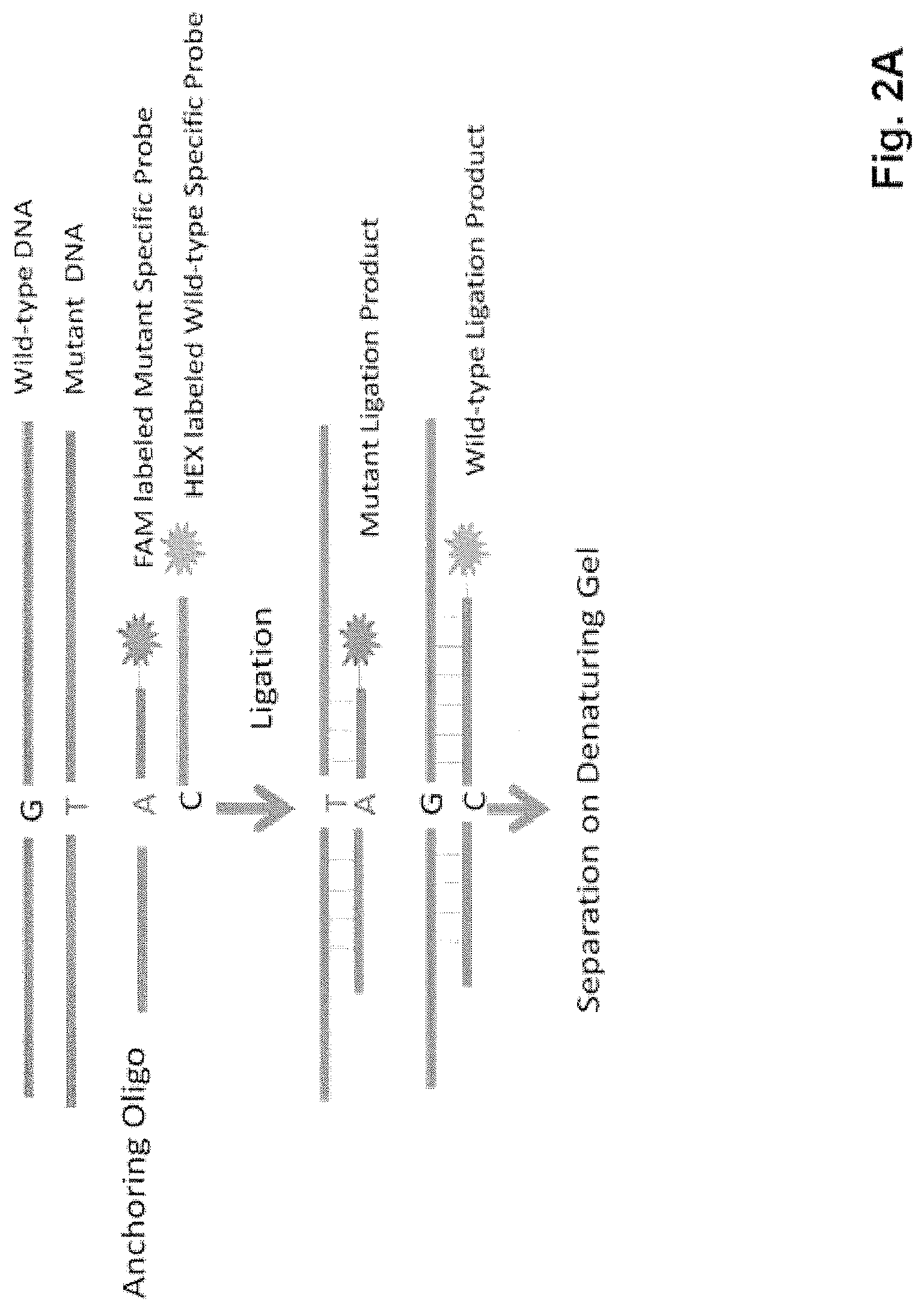
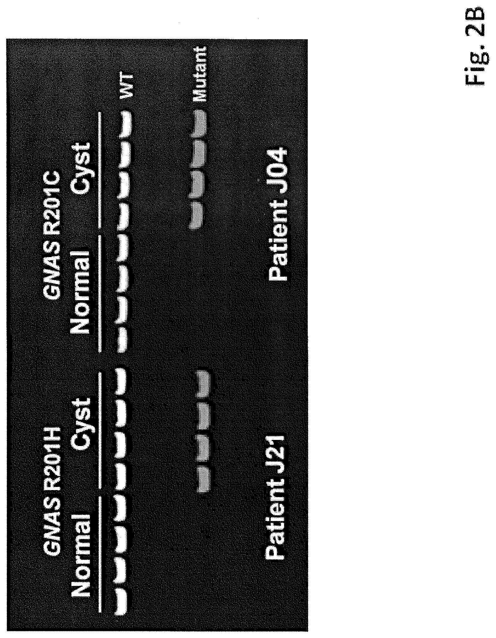
FIGS. 2A-2B show a ligation assays used to assess KRAS (v-Ki-ras2 Kirsten rat sarcoma viral oncogene homolog) and GNAS (guanine nucleotide binding protein (G protein), alpha stimulating activity polypeptide 1) mutations. (FIG. 2 A) Schematic of the ligation assay. Oligonucleotide probes complementary to either the WT or mutant sequences were incubated with a PCR product containing the sequence of interest. The WT- and mutant-specific probes were labeled with the fluorescent dyes 6-FAM and HEX, respectively, and the WT-specific probe was 11 bases longer than the mutant-specific probe. After ligation to a common anchoring primer, the ligation products were separated on a denaturing polyacrylamide slab gel. Further details of the assay are provided in the Materials and Methods. (FIG. 2 B) Examples of the results obtained with the ligation assay in the indicated patients. Templates were derived from DNA of normal duodenum or IPMN tissue. Each lane represents the results of ligation of one of four independent PCR products, each containing 200 template molecules. The probe in the left panel was specific to the GNASR201H mutation and the probe on the right panel was specific for the GNAS R201C mutation.
FIG. 3 shows BEAMing assays used to quantify mutant representation. PCR was used to amplify KRAS or GNAS sequences containing the region of interest (KRAS codon 12 and GNAS codon 201). The PCR-products were then used as templates for BEAMing, in which each template was converted to a bead containing thousands of identical copies of the templates (34). After hybridization to Cy3- or Cy5-labeled oligonucleotide probes specific for the indicated WT or mutant sequences, respectively, the beads were analyzed by flow cytometry. Scatter plots are shown for templates derived from the DNA of IPMN 130 or from normal spleen. Beads containing the WT or mutant sequences are widely separated in the scatter plots, and the fraction of mutant-containing beads are indicated. Beads whose fluorescence spectra lie between the WT and mutant-containing beads result from inclusion of both WT and mutant templates in the aqueous nanocompartments of the emulsion PCR.
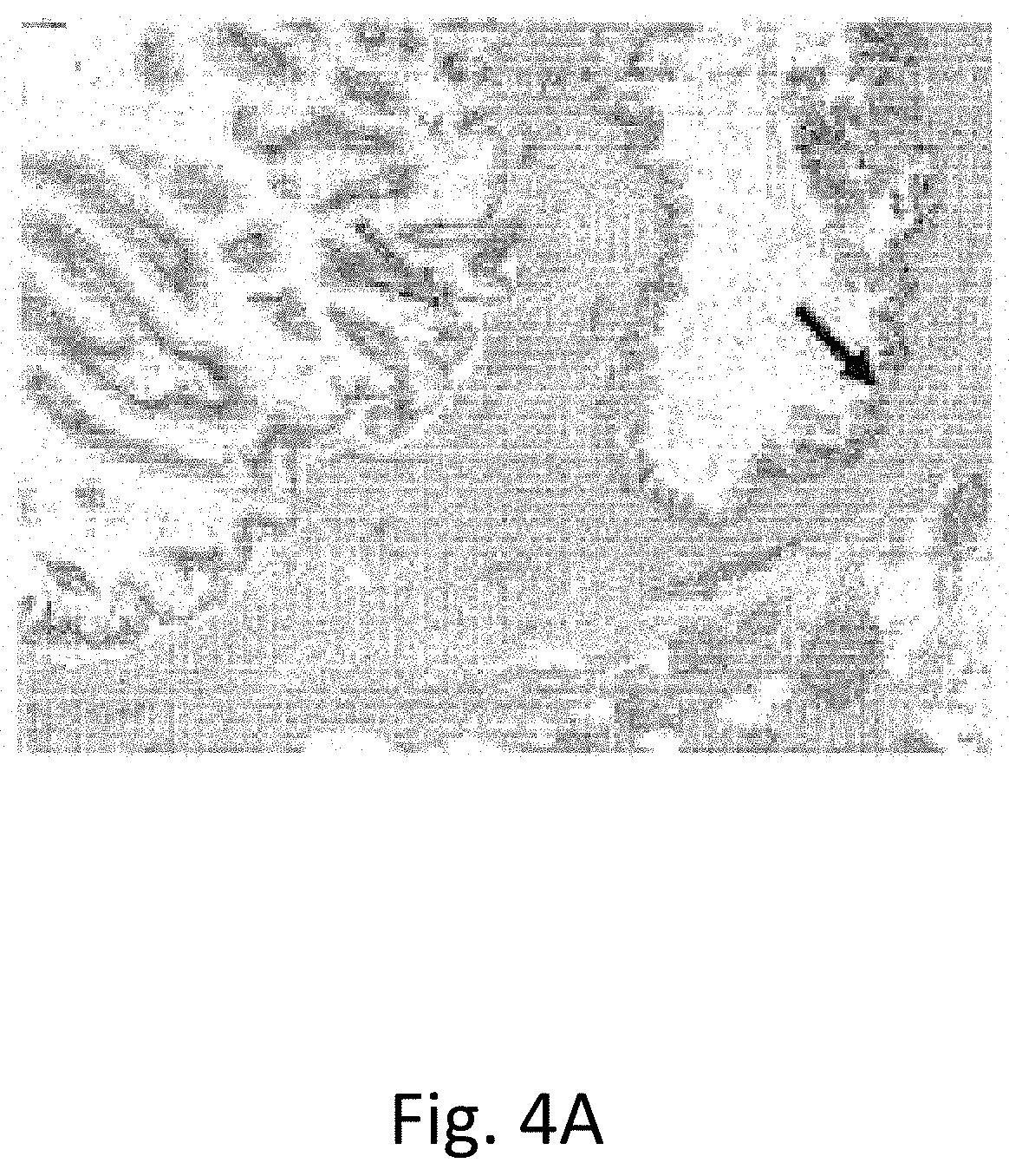
FIGS. 4A-4C show IPMN morphologies. (FIG. 4A) H&E-stained section of a formalin-fixed, paraffin embedded sample (shows two apparently independent IPMNs with distinct morphologies located close to one another. The IPMN of gastric epithelial subtype (black arrow) harbored a GNAS R201C and a KRAS G12'V while the IPMN showing the intestinal subtype (red arrow) contained a GNAS R201C mutation but no KRAS mutation. (FIG. 4B) H&E stained section of a different, typical IPMN (FIG. 4C) Same IPMN as in FIG. 4B after microdissection of the cyst wall.
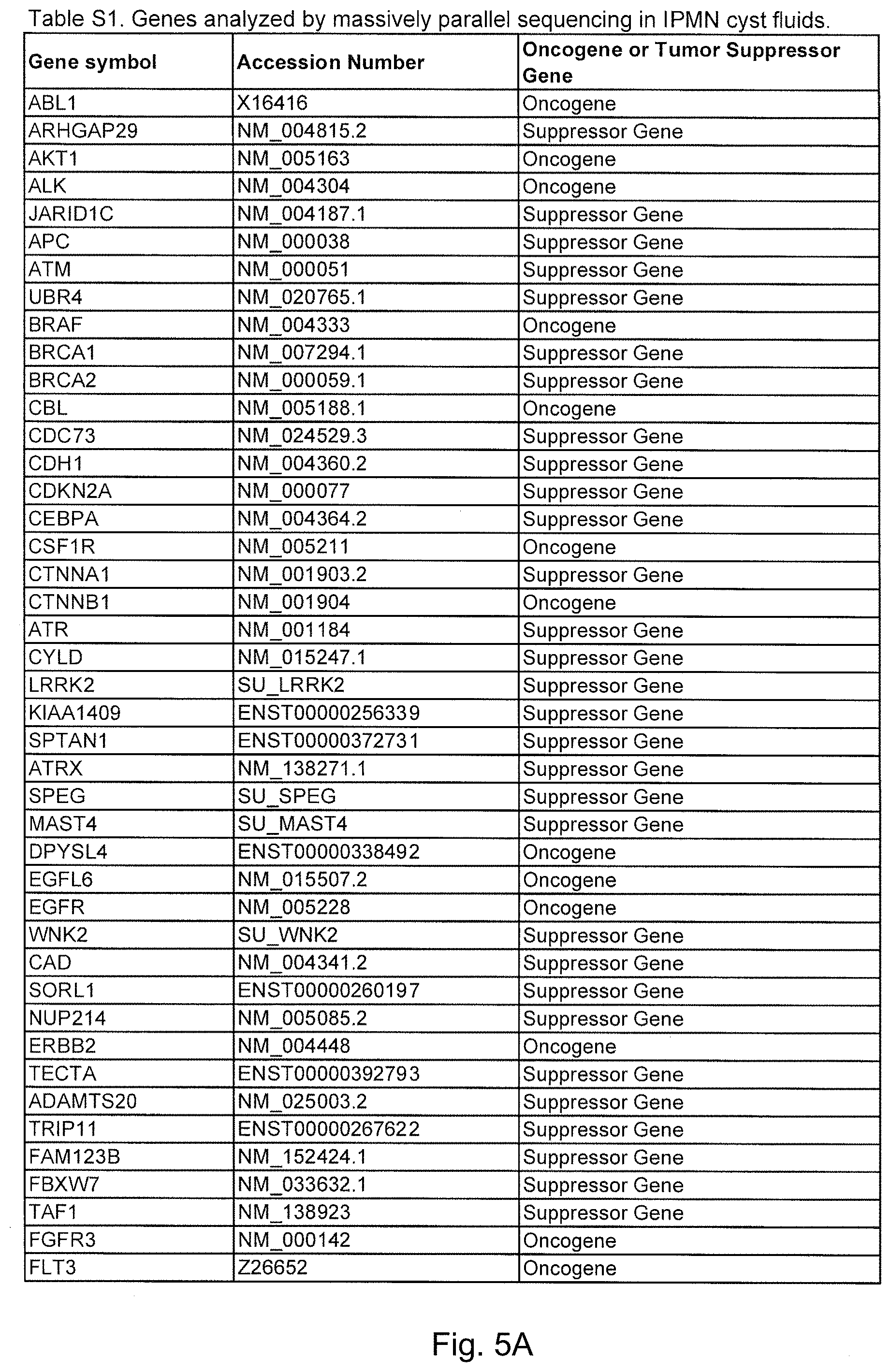
FIGS. 5A-5D. (Table S1.) Genes analyzed by massively parallel sequencing in IPMN cyst fluids.
FIGS. 6A-6C. (Table S2.) Characteristics of patients with IPMNs analyzed in this study, including GNAS and KRAS mutation status.
FIGS. 7A-7B. (Table S3.) Characteristics of patients with cyst types other than IPMN, including GNAS and KRAS mutation status.
FIG. 8. (Table S4.) Quantification of mutations in selected IPMNs containing both GNAS and KRAS mutations.
FIG. 9. (Table S5.) Comparison of mutational status in DNA from IPMNs and pancreatic adenocarcinomas from the same patients.
FIG. 10. (Table S6.) Oligonucleotide primer and probe sequences (SEQ ID NO: 4-38).
DETAILED DESCRIPTION OF THE INVENTION
Capturing and amplifying analyte nucleic acids from dilute samples can be particularly taxing. Enrichment for desired sequences can make assays feasible that would otherwise fall below detection limits. Test samples can be from any appropriate source in the patient's body that will have nucleic acids from a cancer or lesion that can be collected and tested. In some cases the nucleic acids will be amplified prior to testing. Suitable test samples may be obtained from pancreatic cyst fluid, pancreatic cyst wall tissue, pancreatic ductal adenocarcinoma tissue, blood, stool, saliva, sputum, bronchoalveolar lavage, urine, and pancreatic juice. The samples may be collected using any means conventional in the art, including from surgical samples, from biopsy samples, from endoscopic ultrasound (EUS), phlebotomy, etc. Obtaining the samples may be performed by the same person or a different person that conducts the subsequent analysis. Samples may be stored and/or transferred after collection and before analysis. Samples may be fractionated, treated, purified, enriched, prior to assay.
Solid supports which may be used are any which are convenient for the particular purpose and situation. These may be filters, beads, magnetic beads, plastic surfaces, microtiter plates, resins, etc. The supports can be treated and derivatized as is known in the art. In particular assays, attachment of a specific binding pair member may be used. For example, avidin or streptavidin may be used as one binding pair member, and biotin as another. One binding pair member may be used on the nucleic acid analyte and one binding pair member may be on the solid support. As exemplified below, biotin can be incorporated into a nucleic acid analyte using biotinylated dNTPs during amplification or synthesis. Other binding pairs which provide a strong bond may be used as well.
Isothermal amplification is one means for producing large amounts of particular sequences. Isothermal amplification is also known as Multiple Displacement Amplification or rolling circle amplification. Other means as are known can also be used.
Ligation reactions are used to join together individual oligonucleotide probes into long polymers or concatamers. Ligation reaction conditions and enzymes for performing these reactions are known in the art and can be used as is convenient.
Any means of testing for a mutation, including without limitation, a point mutation, a deletion, an amplification, a loss of heterozygosity, a rearrangement, a duplication, may be used. As an example, a mutation in codon 201 of GNAS or codon 12 of KRAS may be assayed. Any means of testing for a mutation may be used. Mutations may be detected by sequencing, by hybridization assay, by ligation assay, etc. If locations of the relevant mutations are defined, specific assays which focus on the identified locations may be used. Identifying a mutation as somatic can be accomplished by comparing a test sample to a non-neoplastic sample, either from the same patient or from a healthy individual. The defined locations of some mutations permit focused assays limited to an exon, domain, or codon. But non-targeted assays may also be used, where the location of a mutation is unknown. Any assay that is performed on a test sample involves a transformation, for example, a chemical or physical change or act. Assays and determinations are not performed merely by a perceptual or cognitive process in the body of a person.
Probes and/or primers may contain the wild-type or a mutant sequence. These can be used in a variety of different assays, as will be convenient for the particular situation. Selection of assays may be based on cost, facilities, equipment, electricity availability, speed, reproducibility, compatibility with other assays, invasiveness of sample collection, sample preparation, etc.
Any of the assay results may be recorded or communicated, as a positive act or step. Communication of an assay result, diagnosis, identification, or prognosis, may be, for example, orally between two people, in writing, whether on paper or digital media, by audio recording, into a medical chart or record, to a second health professional, or to a patient. The results and/or conclusions and/or recommendations based on the results may be in a natural language or in a machine or other code. Typically such records are kept in a confidential manner to protect the private information of the patient.
Collections of probes, primers, control samples, and reagents can be assembled into a kit for use in the methods. The reagents can be packaged with instructions, or directions to an address or phone number from which to obtain instructions. An electronic storage medium may be included in the kit, whether for instructional purposes or for recordation of results, or as means for controlling assays and data collection.
Control samples can be obtained from the same patient from a tissue that is not apparently diseased. Alternatively, control samples can be obtained from a healthy individual or a population of apparently healthy individuals. Control samples may be from the same type of tissue or from a different type of tissue than the test sample.
The data described below document the existence of a heretofore unappreciated molecular pathway leading to pancreatic neoplasia. There is no doubt that GNAS mutations plays a driving role in this IPMN-specific pathway: the mutations are remarkably common and they occur at a single codon (201), mutations of which are known to endow cells with extremely high adenyl cyclase activity and cAMP levels (37-39). Based on their rate of mutation and specificity (30), the probability that these mutations are passengers rather than drivers of IPMN development is negligible.
The data also demonstrated that >96% of IPMNs have either a GNAS or KRAS (v-Ki-ras2 Kirsten rat sarcoma viral oncogene homolog) mutation and more than half have both mutations. Which mutation--KRAS or GNAS--arises first? There were 20 cases in which GNAS mutations were identified in the absence of KRAS mutations and six additional cases in which GNAS mutations were at least 5 times more abundant than KRAS mutations in the same cyst fluid (FIG. 8 (Table S4)). The converse situation--KRAS mutations in the absence of GNAS mutations--was also observed in many cases (FIG. 6 (Table S2)). These data, in combination with the demonstration that more than one KRAS or more than one GNAS mutation could be identified in the same cyst (FIG. 8 (Table S4)), suggests two models for IPMN development. First, it is possible that IPMN locules represent independent entities whose evolution is unrelated to other locules within the same IPMN. This model is inconsistent with our data because two adjacent locules within a single grossly distinct IPMN were more likely to contain the same KRAS or GNAS mutation than the lining epithelium from two topographically different cysts, as noted in the Results section. Second, it is possible that all IPMNs are initiated by a single founding mutation in either GNAS or in KRAS. Subsequent mutations of cells within the cystic lesion would lead to independent clonal expansions, perhaps represented by different locules. Such polyclonality has been observed in colorectal adenomas, which are initiated by mutations in APC pathway genes but sometimes progress through heterogeneous KRAS mutations to a transient polyclonal stage (40). This stage is eventually replaced by subsequent clonal expansion of a cell with one of these KRAS mutations (40). A related possibility is that IPMNs are initiated by a genetic or epigenetic alteration in a gene other than KRAS or GNAS, and that we have observed subsequent clonal expansions of these initiated cells. Finally, it is possible that most IPMNs are indeed initiated by a mutation (in GNAS, KRAS, or another gene), but that occasionally two such IPMNs, initiated by completely different cells, develop adjacent to one another. This appeared to be the situation for the case shown in FIG. 4A, for example. Though these models are difficult to distinguish from one another, it is possible that lineage tracking can be accomplished by complete sequencing of IPMN locule genomes in the future (41).
Apart from its implications for understanding IPMN development, our data have potentially important practical ramifications. The appropriate management of a patient with a pancreatic cyst depends on the type of cyst (42). In particular, it is generally agreed that there is no need to remove asymptomatic SCAs because these lesions have a vanishingly small malignant potential (43). However, the distinction between SCA and mucinous cystic lesions (IPMN and MCN) of the pancreas is often not easy, even after extensive imaging and follow-up (6). One example of these difficulties is provided by the nature of the lesions in our study: the great majority of the 44 SCAs we examined were removed because they were preoperatively believed to be cysts with malignant potential. Hence, many of these 44 surgical procedures were likely unnecessary.
These diagnostic difficulties have long been appreciated and have spurred attempts to develop biomarkers as adjuncts to clinical data, imaging, and cytology (44). Indeed, new protein and glycoprotein markers are showing promising results (45, 46). One conceptual disadvantage of these protein biomarkers is that they are simply associated with cyst development and do not play a pathogenic role. Alterations of oncogenes such as KRAS are attractive alternatives because they are intimately involved in pathogenesis (47-50). In the largest previous study to date on such alterations, 45% of the fluids from mucinous cysts were shown to contain KRAS mutations (25). Our data demonstrates that KRAS mutations are actually present in a larger fraction of IPMNs, probably a result of the more sensitive methods used in our study combined with optimization of procedures used to purify cyst fluid DNA (see Materials and Methods). Third, and most important, the combination of GNAS and KRAS mutation detection provides high sensitivity and specificity for distinguishing between SCAs and IPMNs. The vast majority of IPMNs had a GNAS and/or a KRAS (95% CI 91% to 99%) while no SCAs had either mutation. These data indicate a sensitivity of 0.96 (95% CI 0.91 to 0.99) and a specificity of 1.0 (97.5% one-sided CI 0.92 to 1) for distinguishing between these two lesions. In addition, although not as sensitive, the presence of a GNAS mutation in cyst fluid can also distinguish IPMNs from MCNs (FIG. 7 (Table S3)). The assay involves just two amplicons (GNAS and KRAS) and can be performed with as little as 250 ul of cyst fluid.
Several caveats to the potential utility of such tests should be noted. First, the analysis of cyst fluid obtained through EUS, though safe, is an invasive procedure. Complications include bleeding, infection, and pancreatitis, are reversible, and are generally observed in <1% of patients (reviewed in (51)). Second, neither KRAS nor GNAS mutations can distinguish high grade or invasive from low grade IPMNs. The supplementation of KRAS and GNAS mutational analyses with other markers indicative of grade would clearly be useful (11). Third, we cannot yet reliably distinguish IPMNs from MCNs through the analysis of cyst fluid. Although MCNs do not contain GNAS mutations, a third of them contain KRAS mutations (FIG. 7 (Table S3)) MCN-specific mutations may be identified in the future through a strategy similar to the one we used to identify mutations in IPMNs.
Astute clinicians and pathologists have long suspected that adenocarcinomas of the pancreas arising in IPMNs are a "different disease" than those arising locally distant or in the absence of an IPMN (15, 52). We here provide evidence in support of this hypothesis and identify a key molecular component that underlies this difference.
The above disclosure generally describes the present invention. All references disclosed herein are expressly incorporated by reference. A more complete understanding can be obtained by reference to the following specific examples which are provided herein for purposes of illustration only, and are not intended to limit the scope of the invention.
Example 1
Materials and Methods
Patients and Specimens
The present study was approved by the Institutional Review Boards of Johns Hopkins Medical Institutions, Memorial Sloan Kettering Cancer Center and the University of Indiana. We included individuals in whom pancreatic cyst fluid samples from pancreatectomy specimens and/or fresh frozen tumor tissues were available for molecular analysis. Relevant demographic, clinicopathologic data were obtained from prospectively maintained clinical databases and correlated with mutational status.
Pancreatic cyst fluids were harvested in the Surgical Pathology suite from surgically resected pancreatectomy specimens with a sterile syringe. Aspirated fluids were stored at -80.degree. C. within 30 min of resection. Fresh-frozen tissue specimens of surgically resected cystic neoplasms of the pancreas were obtained through a prospectively maintained Johns Hopkins Surgical Pathology Tumor Bank. These lesions as well as normal tissues were macrodissected using serial frozen sections to guide the trimming of OCT embedded tissue blocks to obtain a minimum neoplastic cellularity of 80%. Formalin-fixed and paraffin-embedded archival tissues from surgically resected pancreata were sectioned at 6 .mu.m, stained with hematoxylin and eosin, and dissected with a sterile needle on a SMZ1500 stereomicroscope (Nikon). An estimated 5,000-10,000 cells were microdissected from each lesion. Lesions were classified as IPMNs, MCNs, or SCAs using standard criteria (53). IPMNs were subtyped by internationally accepted criteria (54).
DNA Purification
DNA was purified from frozen cyst walls using an AllPrep kit (Qiagen) and from forrmalin-fixed, paraffin-embedded sections using the QIAamp DNA FFPE tissue kit (Qiagen) according to the manufacturer's instructions. DNA was purified from 250 .mu.L of cyst fluid by adding 3 ml RLTM buffer (Qiagen) and then binding to an AllPrep DNA column (Qiagen) following the manufacturer's protocol. DNA was quantified in all cases with qPCR, employing primers and conditions as described (55).
Illumina Library Preparation
Cyst fluid DNA was first quantified through real-time PCR using primers specific for repeated sequences in DNA (LINE) as described (56). A minimum of 100 ng DNA from cyst fluid was used to make Illumina libraries according to manufacturer's protocol with the exception that the amount of adapters was decreased in proportional fashion when a lower amount of template DNA was used. The number of PCR cycles used to amplify the library after ligation of adapters was varied to ensure a yield of .about.5 ug of the final library product for capture.
Target DNA Enrichment
The targeted region included all of the 3386 exons of 169 cancer related genes and was enriched with custom-made oligonucleotide probes. The design of each oligonucleotide was as follows: 5'-TCCCGCGACGAC-36 bases from the genomic region of interest--GCTGGAGTCGCG-3' (SEQ ID NO: 1). Probes were designed to capture both the plus and the minus strand of the DNA and had a 33-base overlap. The probes were custom-synthesized on a chip. The oligonucleotides were cleaved from the chip by treatment for five hours with 3 ml 35% ammonium hydroxide at room temperate. The solution was transferred to two 2-ml tubes, dried under vacuum, and re-dissolved in 400 ul RNase and DNase free water. Five ul of the solution were used for PCR amplification with primers complementary to the 12 base sequence common to all probes: 5-TGATCCCGCGACGA*C-3' (SEQ ID NO: 2), 5'-GACCGCGACTCCAG*C-3' (SEQ ID NO: 3), with * indicating a phosphorothioate bond. The PCR mix contained 27 ul H.sub.2O, 5 ul template DNA, 2 ul forward primer (25 uM), 2 ul reverse primer (25 uM), 4 ul MgCl.sub.2 (50 mM), 5 ul 10.times. Platinum Taq buffer (Life Technologies), 4 ul dNTPs (10 mM each) and 1 ul Platinum Taq (5 U/ul, Life Technologies). The cycling conditions were: one cycle of 98.degree. C. for 30 s; 35 cycles of 98.degree. C. for 30 s, 40.degree. C. for 30 s, 60.degree. C. for 15 s, 72.degree. C. for 45 s; one cycle of 72.degree. C. for 5 min. The PCR product was purified using a MinElute Purification Column (Qiagen) and end-repaired using End-IT DNA End-Repair Kit (Epicentre) as follows: 34 ul DNA, 5 ul 10.times. End-Repair Buffer, 5 ul dNTP Mix, 5 ul ATP, 1 ul End-Repair Enzyme Mix. The mix was incubated at room temperature for 45 minutes, and then purified using a MinElute Purification Column (Qiagen). The PCR products were ligated to form concatamers using the following protocol: 35 ul End-Repaired DNA product, 40 ul 2.times.T4 DNA ligase buffer, 5 ul T4 DNA ligase (3000 units; Enzymatics Inc.) The mix was incubated at room temperature for 4 hours, then purified using QiaQuick Purification Column (Qiagen), and quantified by absorption at 260 nm.
Replicates of 50 ng of concatenated PCR product were amplified in 25 ul solution using the REPLI-g midi whole genome amplification kit (Qiagen) according to the manufacturer's protocol. The RepliG-amplified DNA (20 ug) was then bound to a nitrocellulose membrane and used to capture DNA libraries as described (57). In general, 5 ug of library DNA were used per capture. After washing, the captured libraries were ethanol precipitated and redissolved in 20 ul TE buffer. The DNA was then amplified in a PCR mix containing 51 ul dH.sub.2O, 20 ul 5.times. Phusion buffer, 5 ul DMSO, 2 ul 10 mM dNTPs, 50 pmol Illumina forward and reverse primers, and 1 ul Hotstart Phusion enzyme (New England Biolabs) using the following cycling program: 98.degree. C. for 30 sec; 15 cycles of 98.degree. C. for 25 sec., 65.degree. C. for 30 sec, 72.degree. C. for 30 sec; and 72.degree. C. for 5 min. The amplified PCR product was purified using a NucleoSpin column (Macherey Nagel, inc.) according to the manufacturer's suggested protocol except that the NT buffer was not diluted and the DNA bound to the column was eluted in 35 ul elution buffer. The captured library was quantified with realtime PCR with the primers used for grafting to the Illumina sequencing chip.
Ligation Assay
PCR products containing codon 12 of KRAS and codon 201 of GNAS were amplified using the primers described in FIG. 10 (Table S6). Each 10-ul PCR contained 200 template molecules in 5 ul of 2.times. Phusion Flash PCR Master Mix (New England Biolabs) and final concentrations of 0.25 uM forward and 1.5 uM reverse primers. Note that the mutant-specific probes sometimes included locked nucleic acid residues (FIG. 10 (Table S6); Exiqon). The following cycling conditions were used: 98.degree. C. for 2 min; 3 cycles of 98.degree. C. for 10 sec., 69.degree. C. for 15 sec, 72.degree. C. for 15 sec; 3 cycles of 98.degree. C. for 10 sec., 66.degree. C. for 15 sec, 72.degree. C. for 15 sec; 3 cycles of 98.degree. C. for 10 sec., 63.degree. C. for 15 sec, 72.degree. C. for 15 sec; 41 cycles of 98.degree. C. for 10 sec., 60.degree. C. for 60 sec. Reactions were performed in at least quadruplicate and each was evaluated independently. Five ul of a solution containing 0.5 ul of Proteinase K, (18.8 mg/ml, Roche) and 4.5 ul of dH.sub.2O was added to each well and incubated at 60.degree. C. for 30 minutes to inactivate the Phusion polymerase and then for 10 min at 98.degree. C. to inactivate the Proteinase K.
The ligation assay was based on techniques described previously, using thermotolerant DNA ligases (58-61). Each 10-ul reaction contained 2-ul of PCR product (unpurified), 1 ul of 10.times. Ampligase buffer (Epicentre), 0.5 ul of Ampligase (5 U/ul, Epicentre), anchoring primer (final concentration 2 uM), WT-specific primer (final concentration 0.1 uM), and mutant-specific primer (final concentration 0.025 uM). The sequences of these primers are listed in FIG. 10 (Table S6). The following cycling conditions were used: 95.degree. C. for 3 min; 35 cycles of 95.degree. C. for 10 sec., 37.degree. C. for 30 sec, 45.degree. C. for 60 sec. Five ul of each reaction was added to 5 ul of formamide and the ligation products separated on a 10% Urea-Tris-Borate-EDTA gel (Invitrogen) and imaged with an Amersham-GE Typhoon instrument (GE Healthcare).
BEAMing Assays
These were performed as described (62) using the PCR products generated for the ligation assay as templates and the oligonucleotides listed in FIG. 10 (Table S6) as hybridization probes.
Statistical Analysis
Fisher's exact tests were used to compare the differences between proportions and Wilcoxon Rank Sum tests were used to compare differences in mutational status by age. Confidence intervals for the prevalence of mutations were estimated using the binomial distribution. To compare the prevalence of mutations in grossly distinct IPMNs to adjacent locules within a single grossly distinct IPMN, we compared the probability of observing given KRAS or GNAS mutation in the 111 distinct IPMNs to conditional probability that given the first locule sequenced contained a specific KRAS or GNAS mutation all other locules contained the same KRAS or GNAS mutations. The probabilities of GNAS or KRAS mutations occurring by chance was calculated using a binomial distribution and the previously estimated mutation rates of tumors or normal cells (30). STATA version 11 was used for all statistical analysis (63).
Example 2
Massively Parallel Sequencing of 169 Genes in Cyst Fluid DNA
To initiate this study, we determined the sequences of 169 presumptive cancer genes in the cyst fluids of 19 IPMNs, each obtained from a different patient. Thirty-three of the 169 were oncogenes and the remainder were tumor suppressor genes. Though only a tiny subset of these 169 genes were known to be mutated in PDAs, all were known to be frequently mutated in at least one solid tumor type (FIG. 5 (Table S1)). We additionally sequenced these genes in normal pancreatic, splenic or intestinal tissues of the same patients to determine which of the alterations identified were somatic. We chose to use massively parallel sequencing rather than Sanger sequencing for this analysis because we did not know what fraction of DNA purified from the cyst fluid was derived from neoplastic cells. Massively parallel sequencing has the capacity to identify mutations present in 2% or more of the studied cells while Sanger sequencing often requires >25% neoplastic cells for this purpose. IPMNs are by definition connected with the pancreatic duct system and the cyst fluid containing cellular debris and shed DNA from the neoplastic cells can be expected to be admixed with that of the cells and secretions derived from normal ductal epithelial cells.
We devised a strategy to capture sequences of the 169 genes from cyst fluid DNA (FIG. 1). In brief, 244,000 oligonucleotides, each 60 bp in length and in aggregate covering the exonic sequences of all 169 genes, were synthesized in parallel using phosphoramadite chemistry on a single chip synthesized by Agilent Technologies. After removal from the chip, the oligonucleotide sequences were amplified by PCR and ligated together. Multiple displacement amplification was then used to further amplify the oligonucleotides, which were then bound to a filter. Finally, the filter was used to capture complementary DNA sequences from the cyst fluids and corresponding normal samples, and the captured DNA was subjected to massively parallel sequencing.
The target region corresponding to the coding exons of the 169 genes encompassed 584,871 bp. These bases were redundantly sequenced, with 902.+-.411 (mean.+-.1 SD) fold-coverage in the 38 samples sequenced (19 IPMN cyst fluids plus 19 matched DNA samples from normal tissues of the same patients). This coverage allowed us to confidently detect somatic mutations present in >5% of the template molecules.
There were only two genes mutated in more than one IPMN-KRAS, which was mutated in 14 of the 19 IPMNs, and GNAS, which was mutated in 6 IPMNs. The mutations in GNAS all occurred at codon 201, resulting in either a R201H or R201C substitution. GNAS is a well-known oncogene that is mutated in pituitary and other uncommon tumor types (16-19). However, such mutations have rarely been reported in common epithelial tumors (20-22). In pituitary tumors, mutations cluster at two positions--codons 201 and 227 (16, 19). This clustering provides extraordinary opportunities for diagnosis, similar to that of KRAS. For example, the clustering of KRAS mutations has facilitated the design of assays to detect mutations in tumors of colorectal cancer patients eligible for therapy with antibodies to EGFR (23). All twelve KRAS mutations identified through massively parallel sequencing of cyst fluids were at codon 12, resulting in a G12D, G12V, or G12R amino acid change. KRAS mutations at codon 12 have previously been identified in the vast majority of PDAs as well as in 40 to 60% of IPMNs (24-29). GNAS mutations have not previously been identified in pancreatic cysts or in PDAs.
Example 3
Frequency of KRAS and GNAS Mutations in Pancreatic Cyst Fluid DNA
We next determined the frequency of KRAS codon 12 and GNAS codon 201 mutations in a larger set of IPMNs. The clinical characteristics of all IPMNs analyzed in this study are listed in FIG. 6 (Table S2). To ensure that the analyses were performed robustly, we carried out preliminary experiments with cyst fluids from patients with known mutations based on the massively parallel sequencing experiments described above. We tested several methods for purifying DNA from often viscous cyst fluids and used the optimum method for subsequent experiments. Quantitative PCR was used to determine the number of amplifiable template molecules recovered with this procedure. In eight cases, we compared pelleted cells to supernatants derived from the same cyst fluid samples and found that the fraction of mutant templates in both compartments was similar. On the basis of these results, we purified DNA from 0.25 ml of whole cyst fluid (cells plus supernatant) and, as assessed by quantitative PCR, recovered an average of 670.+-.790 ng of usable DNA.
For each of 84 cyst fluid samples (an independent cohort of 65 patients plus the 19 patients whose fluids had been studied by massively parallel sequencing), we analyzed .about.800 template molecules for five distinct mutations, three at KRAS codon 12 and two within GNAS codon 201 (see Materials and Methods). A PCR/ligation method that had the capacity to detect one mutant template molecule among 200 normal (wild-type, WT) templates was used for these analyses (FIG. 2A). We identified GNAS and KRAS mutations in 61% and 82% of the IPMN fluids, respectively (representative examples in FIG. 2B). In those samples without GNAS codon 201 mutations, we searched for GNAS codon 227 mutations, but did not find any. We also analyzed macro- and microdissected frozen or paraffin-embedded cyst walls from an independent collection of 48 surgically resected IPMNs, and similarly identified a high prevalence of GNAS (75%) and KRAS (79%) mutations. In aggregate, 66% of 132 IPMNs harbored a GNAS mutation, 81% harbored a KRAS mutation, slightly more than half (51%) harbored both GNAS and KRAS mutations, while at least one of the two genes was mutated in 96.2% (FIG. 6 (Table S2)). Given background mutation rates in tumors or normal tissues (30), the probability that either GNAS or KRAS mutations occurred by chance alone was less than 10'. There were no significant correlations between the prevalence of KRAS or GNAS mutations and age, sex, or smoking history of the patients (P>0.05) (Table 1). Small (<3 cm) as well as larger cysts had similar fractions of both KRAS and GNAS mutations and the location of the IPMN (head, body, or tail) did not correlate with the presence of mutation in either gene (Table 1). GNAS and KRAS mutations were present in low-grade as well as in high-grade IPMNs. The prevalence of KRAS mutations was higher in lower grade lesions (P=0.03) whereas the prevalence of GNAS mutations was somewhat higher in more advanced lesions (P=0.11) (Table 1). GNAS, as well as KRAS mutations were present in each of the three major histologic types of IPMNs--intestinal, pancreatobiliary, and gastric. However, the prevalence of the mutations varied across the histological types (P<0.01 for both KRAS and GNAS). GNAS mutations were most prevalent in the intestinal subtype (100%), KRAS mutations had the highest frequency (100%) in the pancreatobiliary subtype and had the lowest frequency (42%) in the intestinal subtype (Table 1).
We then determined whether GNAS mutations were present in SCAs, a common but benign type of pancreatic cystic neoplasm. We examined a total of 44 surgically resected SCAs, each from a different patient (42 cyst fluids and 2 cyst walls). Many of these cysts were surgically resected because they clinically mimicked an IPMN. They would have likely not been surgically excised had they been known to be SCAs. The SCAs averaged 5.0.+-.2.8 cm in maximum diameter (FIG. 7 (Table S3)) similar to the IPMNs (4.4.+-.3.7 maximum diameter, FIG. 6 (Table S2)). There was little difference in the locations of the SCAs and IPMNs within the pancreas (FIGS. 6 and 7 (Tables S2 and S3)). However, no GNAS or KRAS mutations were identified in the SCAs, in marked contrast to the IPMNs (p<0.001, Fisher's Exact Test). GNAS mutations were also not identified in any of 21 MCNs (p=0.005 when compared to IPMNs, Fisher's Exact Test), although KRAS mutations were found in 33% of MCNs (FIG. 7 (Table S3)). GNAS mutations were also not identified in five examples of an uncommon type of cyst, called intraductal oncocytic papillary neoplasm (IOPN), with characteristic oncocytic features (FIG. 7 (Table S3)).
TABLE-US-00001 TABLE 1 Correlations between mutations and clinical and histopathologic parameters of IPMNs KRAS GNAS N, mutation P- mutation total N % value N % P-value Age in years <65 years 29 22 75.9 0.42 18 62.1 0.62 .gtoreq.65 years 103 85 82.5 69 67 Gender Male 70 58 82.9 0.58 51 72.9 0.07 Female 62 49 79 36 58.1 History of Yes 25 21 84 0.77 17 68 0.85 smoking No 37 30 81.1 26 70.3 Grade Low 23 20 87 0.43 11 47.8 0.04 Intermediate 51 46 90.2 (low vs. 34 66.7 (low vs. High 58 41 70.7 others) 42 72.4 others) Duct type Main 35 23 65.7 0.002 24 68.6 0.37 Branch 64 58 90.6 (main 38 59.4 (main Mixed 28 21 75 vs. 20 71.4 vs. branch) branch) Subtype Gastric 52 45 86.5 0.02 34 65.4 0.002 Pancreatobiliary 7 7 100 (panc. 3 42.9 (panc. Intestinal 13 6 46.2 vs 13 100 vs intestinal) intestinal) Diameter <3 cm 62 49 79 0.58 41 66.1 0.96 .gtoreq.3 cm 70 58 82.9 46 65.7 Location Proximal 77 64 83.1 0.44 53 68.8 0.38 (head) (prox. (prox. vs Distal (body, 49 38 77.6 vs 30 61.2 distal) tail) distal) Proximal and 6 5 83.3 4 66.7 distal Associated Yes 24 18 75 0.4 18 75 0.3 cancer No 108 89 82.4 69 63.9
Example 4
IPMN Polyclonality
KRAS G12D, G12V, and G12R mutations were found in 43%, 39%, and 13% of IPMNs, respectively (FIG. 6 (Table S2)). A small fraction (11%) of the IPMNs contained two different KRAS mutations and 2% contained three different mutations. Likewise, GNAS R201C and GNAS R201H mutations were present in 39% and 32% of the IPMNs, respectively, and 4% of the IPMNs had both mutations (FIG. 6 (Table S2)). More than one mutation in KRAS in IPMNs has been observed in prior studies of IPMNs (31-33) and the multiple KRAS and GNAS mutations are suggestive of a polyclonal origin of the tumor.
We investigated clonality in more detail by precisely quantifying the levels of mutations in the subset of cyst fluids containing more than one mutation of the same gene. To accomplish this, we used a technique called BEAMing (34). Through this method, individual template molecules are converted into individual magnetic beads attached to thousands of molecules with the identical sequence. The beads are then hybridized with mutation-specific probes and analyzed by flow cytometry (FIG. 3). The analysis of 17 IPMN cyst fluids, each with mutations in both KRAS and GNAS, showed that the fraction of mutant alleles varied widely, ranging from 0.8% to 45% of the templates analyzed. There was an average of 12.8%.+-.12.2% mutant alleles of KRAS and an average of 24.4.+-.13.1% mutant alleles of GNAS in the 17 IPMN cyst fluids examined (FIG. 8 (Table S4)). In two of the seven IPMNs with more than one KRAS mutation, there was a predominant mutant that out-numbered the second KRAS mutant by >5:1 (FIG. 8 (Table S4)). Similarly, two of the four cases harboring two different GNAS mutations had a predominant mutant (FIG. 8 (Table S4)). In the other cases, the different mutations in KRAS (or GNAS) were distributed more evenly (FIG. 8 (Table S4)). These data support the idea that cells within a subset of IPMNs had undergone independent clonal expansions, giving rise to apparent polyclonality (35).
IPMNs are often multilocular or multifocal in nature, looking much like a bunch of grapes (FIG. 4A) (36). To determine the relationship between cyst locules (individual grapes) and cyst fluid, we microdissected the walls from individual locules of each of ten IPMNs from whom cyst fluid was available (example in FIGS. 4B and C). The individual locule walls generally appeared to be monoclonal, as more than one KRAS mutation was only found in one (4.5%) of the 22 locules examined. No locule wall contained more than one GNAS mutation and two adjacent locules within a single grossly distinct IPMN were more likely to contain the same KRAS or GNAS mutation than the lining epithelium from two topographically different IPMNs (p<0.05, Fisher's Exact Test for KRAS G12D, KRAS G12R and GNAS R201H mutations; P<0.10 for KRAS G12V and GNAS R201H mutations). All of the ten KRAS and six GNAS mutations identified in the cyst fluid could be identified in the corresponding locule walls. These data leave little doubt that the mutations in the cyst fluid are derived from the cyst locule walls and indicate that the cyst fluid provides an excellent representation of the neoplastic cells in an IPMN.
Example 5
GNAS Mutations in Invasive Cancers Associated with IPMNs
Prior whole exome sequencing had not revealed any GNAS mutations in 24 typical PDA that occurred in the absence of an associated IPMN (29). We extended these data by examining 95 additional surgically resected PDAs in pancreata without evidence of IPMNs for mutations in GNAS R201H or R201C, using the ligation assay described above. Again, no GNAS mutations were identified in PDAs arising in the absence of IPMNs.
We suspected that IPMNs containing GNAS mutations had the potential to progress to an invasive carcinoma because fluids from IPMNs with high-grade dysplasia contained such mutations (Table 1). However, in light of the multilocular and multifocal nature of IPMNs described above, it was not clear whether the cells of the locule(s) that progress to an invasive carcinoma were those that contained GNAS mutations. To address this question, we purified DNA from invasive pancreatic adenocarcinomas that developed in association with IPMNs. In each case, the neoplastic cells of the IPMN and of the invasive adenocarcinoma were carefully microdissected. In seven of the eight patients, the identical GNAS mutation found in the neoplastic cells of the IPMN was found in the concurrent invasive adenocarcinoma (FIG. 9 (Table S5)). The KRAS mutational status of the PDA was consistent with that of the associated IPMN in the same seven cases. In the eighth case, the KRAS and GNAS mutations identified in the neoplastic cells of the IPMN were not found in the associated PDA, suggesting that this invasive cancer arose from a separate precursor lesion (FIG. 9 (Table S5)). Though KRAS mutations were found commonly in both types of PDAs, there was a dramatic difference between the prevalence of GNAS mutations in PDAs associated with IPMNs (7 of 8) vs. that in PDAs unassociated with IPMNs (0 of 116; p<0.001, Fisher's Exact Test).
Example 6
A Protocol for Enrichment on Beads
Cleave Oligos from the Chip
Place the chip into the corner of a Micro-Seal bag (Model 50068, DAZEY corporation) cut to .about.10.5.times.5.5 cm.
Seal the unsealed two sides so that the bag ends up 8 cm.times.2.6 cm, tightly wrapping the chip.
While in the Seal-a-Meal bag, treat for five hours with 3 ml 28% ammonium hydroxide at room temperate by rotator (360 deg rotation). (Make sure the chip is fully immersed in the solution)
Transfer the solution into two 2-ml eppendorf tubes, and speed vaccum dried at temperate 50.degree. C. (normally it will take 5-8 hours)
(For speed vaccum, turn on the cooler one hour before you use the vaccum)
Re-dissolve the oligos in a combined 400 ul RNase and DNase free water. Amplify the Oligos
Make 3.times.50 ul PCR mix for each chip, the PCR mix contains the following:
X ul H2O
1 ul (well 1), 2 ul (well 2), 5 ul (well 3)
TABLE-US-00002 2 ul forward primer (25 uM): 5'-TGATCCCGCGACGA*C-3', where * indicates phosphorothioate 2 ul reverse primer (25 uM): 5'-GACCGCGACTCCAG*C-3', where * indicates phosphorothioate
4 ul MgCl2 (50 mM)
5 ul 10.times. Platinum Taq buffer (Life Technologies)
4 ul dNTPs (10 mM each)
1 ul Platinum Taq (5 U/ul, Life Technologies) (Titanium and Phusion both did not work).
Note: Because of the alkalic condition after cleavage, the more template you add, the less PCR product you get.
The cycling conditions were: 1.times.98.degree. C. for 30 s
35 cycles of 98.degree. C. for 30 s, 40.degree. C. for 30 s, 60.degree. C. for 15 s, 72.degree. C. for 45 s
one cycle of 72.degree. C. for 5 min
Run the gel to see a smear from 60 bp to 120 bp. 120 bp product may be dimers, which won't interfere with capture.
The PCR products were combined, and add 2 ul Sodium Acetate (3M, pH 5.2) purified using a MinElute Purification Column (Qiagen), elute twice in 65.degree. C. pre-warmed buffer with 17 ul each (total of 34 ul).
End-Repair the PCR Product
End-repair using End-IT DNA End-Repair Kit (Epicentre) as follows:
34 ul DNA
5 ul 10.times. End-Repair Buffer
5 ul dNTP Mix
5 ul ATP
1 ul End-Repair Enzyme Mix
Incubate at room temperature for 45 minutes,
Purified using a MinElute Purification Column (Qiagen), elute twice in 65.degree. C. pre-warmed buffer with 17.5 ul each (total of 35 ul).
Ligate the PCR Product
The PCR products were ligated to form concatamers using the following protocol:
35 ul End-Repaired DNA product
40 ul 2.times.T4 DNA ligase buffer (Enzymatics Inc.)
5 ul T4 DNA ligase (600 units/ul; Enzymatics Inc.)
The mix was incubated at room temperature for at least 4 hours, (you can leave it overnight.)
The product was purified using QiaQuick PCR Purification Column (Qiagen) (not MinElute), elute twice in 65.degree. C. pre-warmed buffer with 25 ul each (total of 50 ul).
Quantify by absorption at 260 nm. (Normally you get around 3 ug DNA product.)
Dilute the product to 20 ng/ul using TE buffer.
Isothermal Amplification of the Probe with Bio-dUTP [RepliG-Midi Kit (not Mini Kit), Qiagen]
TABLE-US-00003 TABLE 1 Preparation of Buffer D1 (Volumes given are suitable for up to 15 reactions) Component Volume Reconstituted Buffer DLB 9 .mu.l Nuclease-free water 32 .mu.l Total volume 41 .mu.l
TABLE-US-00004 TABLE 2 Preparation of Buffer N1 (Volumes given are suitable for up to 15 reactions) Component Volume Stop solution 12 .mu.l Nuclease-free water 68 .mu.l Total volume 80 .mu.l
Place 2.5 .mu.l template DNA into a microcentrifuge tube.
Add 2.5 .mu.l Buffer D1 to the DNA. Mix by vortexing and centrifuge briefly
Incubate the samples at room temperature (15-25.degree. C.) for 3 min.
Add 5 .mu.l Buffer N1 to the samples. Mix by vortexing and centrifuge briefly.
Prepare a master mix on ice according to Table 3 (see below). Mix and centrifuge briefly.
Important: Add the master mix components in the order listed in Table 3. After addition of water and REPLI-g Midi Reaction Buffer,
briefly vortex and centrifuge the mixture before addition of REPLI-g Midi DNA Polymerase. The master mix should be kept on ice and used
immediately upon addition of the REPLI-g Midi DNA Polymerase.
TABLE-US-00005 TABLE 3 Preparation of Master Mix Component Volume/reaction REPLI-g Midi Reaction Buffer 14.5 .mu.l Biotin-dUTP(1 mM) (Cat. No. 11093070910, 2.5 ul Roche Applied Science) REPLI-g Midi DNA Polymerase 0.5 .mu.l Total volume 17.5 .mu.l
Add 17.5 .mu.l of the master mix to 10 .mu.l denatured DNA that was neutralized with N1 as described above. Transfer the mix to the PCR plate.
Incubate at 30.degree. C. for 16 h in PCR machine.
Inactivate REPLI-g Midi DNA Polymerase by heating the sample at 65.degree. C. for 3 min.
Transfer the mix using 2.lamda.120 ul TE to a 1.5 ml tube.
Incubate the tube in 100.degree. C. heating block for 20 minutes.
Purify the product using two QiaQuick PCR Purification Columns (Qiagen) (not MinElute), i.e., use 2 columns for one 27.5 ul reaction.
Elute each column twice with 65.degree. C. pre-warmed buffer with 27.5 ul, for a total of 55 ul, so there will be 110 ul of eluate from the two columns which should be pooled.
Quantify by absorption at 260 nm using nanodrop (I know it's single-strand DNA now, but I still use ds-DNA calcualtions in nanodrop)
In general, you will get .about.180-210 ng/ul. If it's too off, there must be something wrong.
DNA Capture
A mix was prepared as follows:
4 ug DNA library (20 ul, 200 ng/ul)
7 ul Human cot-1 DNA (Cat.No. 15279011, Invitrogen)
3 ul Herring Sperm DNA (Cat.No. 15634-017, Invitrogen)
10 ul Blocking Oligos, 1 nmol/ul each.
TABLE-US-00006 Block Oligo 1: AATGATACGGCGACCACCGAGATCTACACTCTTTCCCTACACGACGCTCT Block Oligo 2: CAAGCAGAAGACGGCATACGAGATCGGTCTCGGCATTCCTGCTGAACCGC
5 ul Capture Probe (.about.200 ng/ul)
The mix is heated at 95.degree. C. for 7 min, and 65.degree. C. for 2 min (use only one compress pad in PCR machine)
Add 25 ul of the 65.degree. C. prewarmed 2.8.times. hybridization buffer (final conc of hyb buffer will then be 1.times.)
2.8.times. hybridization buffer: (14.times.SSPE, 14.times.Denhardt's, 14 mM EDTA, 0.28% SDS), using the following reagents:
20.times.SSPE: (0810-4L, AMRESCO)
Denhardt's Solution, 50.times., 50 ml (70468, usb)
EDTA: 0.5M, PH 8.0 (46-034-CI, Mediatech Inc.)
(In case the DNA library conc is <200 ng/ul, then still use 4 ug DNA and 7 ul Cot-1, 3 ul Herring sperm, etc. but use proportionally larger volumes of 2.8.times. HybBuffer
Incubate at 65 deg for 22 hours for hybridization with PCR machine lid heat on.
Washing Procedure
Wash 50 ul MyOne beads (Invitrogen) 3 times in 1.5 ml tule and resuspend in 60 .mu.l 1.times. binding buffer (1M NaCl, 10 mM Tris-HCl, pH 7.5, and 1 mM EDTA.)
Add equal volume (70 ul) of 2.times. binding buffer (2 M NaCl, 20 mM Tris-HCl, pH 7.5, and 2 mM EDTA.) to hybrid mix, and transfer to tube with beads. Total Volume should be 200 ul.
Votex the mix thoroughly. And rotate 360 deg. for 1 hour at Room Temperature.
After binding, the beads are pulled down, and washed 15 minutes at RT in 0.5 ml Wash Buffer 1 (1.times.SSC/0.1% SDS)
Wash the beads for 15 minutes at 65.degree. C. on a heating block with shaking, five times in 0.5 ml Wash Buffer 3 (0.1.times.SSC and 0.1% SDS)
Hybrid-selected DNA are resuspended in 50 .mu.l 0.1 M NaOH at RT for 10 min.
The beads are pulled down, the supernatant transferred to a tube containing 70 .mu.l Neutralizing Buffer (1 M Tris-HCl, pH 7.5)
Neutralized DNA is desalted and concentrated on a QIAquick MinElute column and eluted in 20 .mu.l.
Note: Wash Buffer 2 (5.2 M Betaine, 0.1.times.SSC and 0.1% SDS) is a more stringent wash buffer.
For more stringent wash, you can substitute the first WB3 wash with WB2, then continue with four washes with WB3.
Change the post-Capture amplification Cycle number to 16 cycles if you use a more stringent wash.
Post-Capture Amplification
PCR mix containing:
20 captured DNA
51 ul dH2O
20 ul 5.times. Phusion buffer
5 ul DMSO
2 ul 10 mM dNTPs
0.5 ul (50 pmol) Illumina forward primer (QC1 primer for barcoding)
0.5 ul (50 pmol) Illumina reverse primer (Barcoding reverse primers for barcoding)
1 ul Hotstart Phusion enzyme (New England Biolabs)
The cycling conditions were: 1.times.98.degree. C. for 30 s
14 cycles of 98.degree. C. for 25 s, 65.degree. C. for 30 s, 72.degree. C. for 30 s
one cycle of 72.degree. C. for 5 min
The PCR is done in two wells for each sample, 50 ul each (no oil on top).
The amplified PCR product was purified using a NucleoSpin column (Macherey Nagel, inc.), eluted twice in 65.degree. C. pre-warmed buffer with 17.5 ul (total of 35 ul).
Use NanoDrop to quantify yield, which should be .about.20 ng/ul.
REFERENCES
The disclosure of each reference cited is expressly incorporated herein. T. A. Laffan, K. M. Horton, A. P. Klein, B. Berlanstein, S. S. Siegelman, S. Kawamoto, P. T. Johnson, E. K. Fishman, R. H. Hruban, Prevalence of unsuspected pancreatic cysts on MDCT. AJR Am J Roentgenol 191, 802-807 (2008). 2. K. de Jong, C. Y. Nio, J. J. Hermans, M. G. Dijkgraaf, D. J. Gouma, C. H. J. van Eijck, E. van Heel, G. Klass, P. Fockens, M. J. Bruno, High Prevalence of Pancreatic Cysts Detected by Screening Magnetic Resonance Imaging Examinations. Clinical Gastroenterology and Hepatology 8, 806-811 (2010). 3. W. Kimura, H. Nagai, A. Kuroda, T. Muto, Y. Esaki, Analysis of small cystic lesions of the pancreas. Int J Pancreatol 18, 197-206 (1995). 4. K. S. Lee, A. Sekhar, N. M. Rofsky, I. Pedrosa, Prevalence of incidental pancreatic cysts in the adult population on MR imaging. Am J Gastroenterol 105, 2079-2084 (2010). 5. H. Matthaei, R. D. Schulick, R. H. Hruban, A. Maitra, Cystic precursors to invasive pancreatic cancer. Nature Reviews Gastroenterology & Hepatology 8, 141-150 (2011). 6. M. Katz, M. Mortenson, H. Wang, R. Hwang, E. Tamm, G. Staerkel, J. Lee, D. Evans, J. Fleming, Diagnosis and Management of Cystic Neoplasms of the Pancreas: An Evidence-Based Approach. Journal of the American College of Surgeons 207, 106-120 (2008). 7. M. Tanaka, Controversies in the management of pancreatic IPMN. Nature Reviews Gastroenterology & Hepatology 8, 56-60 (2011). 8. R. H. Hruban, M. B. Pitman, D. S. Klimstra, Tumors of the pancreas. Atlas of tumor pathology (American Registry of Pathology and Armed Forces Institute of Pathology, Washington, D.C., ed. Fourth Series, Fascicle 6, (2007). 9. G. Kloppel, M. Kosmahl, Cystic Lesions and Neoplasms of the Pancreas. Pancreatology 1, 8 (2001). 10. M. Tanaka, S. Chari, V. Adsay, C. Fernandez-del Castillo, M. Falconi, M. Shimizu, K. Yamaguchi, K. Yamao, S. Matsuno, International consensus guidelines for management of intraductal papillary mucinous neoplasms and mucinous cystic neoplasms of the pancreas. Pancreatology 6, 17-32 (2006). 11. T. A. Sohn, C. J. Yeo, J. L. Cameron, R. H. Hruban, N. Fukushima, K. A. Campbell, K. D. Lillemoe, Intraductal papillary mucinous neoplasms of the pancreas: an updated experience. Ann Surg 239, 788-797; discussion 797-789 (2004). 12. S. Crippa, C. Fernandez-del Castillo, R. Salvia, D. Finkelstein, C. Bassi, I. Dominguez, A. Muzikansky, S. P. Thayer, M. Falconi, M. Mino-Kenudson, Mucin-Producing Neoplasms of the Pancreas: An Analysis of Distinguishing Clinical and Epidemiologic Characteristics. Clinical Gastroenterology and Hepatology 8, 213-219.e214 (2010). 13. G. A. Poultsides, S. Reddy, J. L. Cameron, R. H. Hruban, T. M. Pawlik, N. Ahuja, A. Jain, B. H. Edil, C. A. Iacobuzio-Donahue, R. D. Schulick, C. L. Wolfgang, Histopathologic basis for the favorable survival after resection of intraductal papillary mucinous neoplasm-associated invasive adenocarcinoma of the pancreas. Ann Surg 251, 470-476 (2010). 14. T. A. Sohn, C. J. Yeo, J. L. Cameron, L. Koniaris, S. Kaushal, R. A. Abrams, P. I. Sauter, J. Coleman, R. H. Hruban, K. D. Lillemoe, Resected adenocarcinoma of the pancreas 616 patients results, outcomes, and prognostic indicators. Journal of Gastrointestinal Surgery 4, 13 (2000). 15. R. Salvia, C. Fernandez-del Castillo, C. Bassi, S. P. Thayer, M. Falconi, W. Mantovani, P. Pederzoli, A. L. Warshaw, Main-duct intraductal papillary mucinous neoplasms of the pancreas: clinical predictors of malignancy and long-term survival following resection. Ann Surg 239, 678-685; discussion 685-677 (2004). 16. P. U. Freda, W. K. Chung, N. Matsuoka, J. E. Walsh, M. N. Kanibir, G. Kleinman, Y. Wang, J. N. Bruce, K. D. Post, Analysis of GNAS mutations in 60 growth hormone secreting pituitary tumors: correlation with clinical and pathological characteristics and surgical outcome based on highly sensitive GH and IGF-I criteria for remission. Pituitary 10, 275-282 (2007). 17. N. Kalfa, Activating Mutations of the Stimulatory G Protein in Juvenile Ovarian Granulosa Cell Tumors: A New Prognostic Factor? Journal of Clinical Endocrinology & Metabolism 91, 1842-1847 (2006). 18. M. C. Fragoso, A. C. Latronico, F. M. Carvalho, M. C. Zerbini, J. A. Marcondes, L. M. Araujo, V. S. Lando, E. T. Frazzatto, B. B. Mendonca, S. M. Villares, Activating mutation of the stimulatory G protein (gsp) as a putative cause of ovarian and testicular human stromal Leydig cell tumors. J Clin Endocrinol Metab 83, 2074-2078 (1998). 19. H. Yamasaki, N. Mizusawa, S. Nagahiro, S. Yamada, T. Sano, M. Itakura, K. Yoshimoto, GH-secreting pituitary adenomas infrequently contain inactivating mutations of PRKAR1A and LOH of 17q23-24. Clin Endocrinol (Oxf) 58, 464-470 (2003). 20. L. D. Wood, D. W. Parsons, S. Jones, J. Lin, T. Sjoblom, R. J. Leary, D. Shen, S. M. Boca, T. Barber, J. Ptak, N. Silliman, S. Szabo, Z. Derso, V. Ustyanksky, T. Nikolskaya, Y. Nikolsky, R. Karchin, P. A. Wilson, J. S. Kaminker, Z. Zhang, R. Croshaw, J. Willis, D. Dawson, M. Shipitsin, J. K. V. Willson, S. Sukumar, K. Polyak, B. H. Park, C. L. Pethiyagoda, P. V. K. Pant, D. G. Ballinger, A. B. Sparks, J. Hartigan, D. R. Smith, E. Suh, N. Papadopoulos, P. Buckhaults, S. D. Markowitz, G. Parmigiani, K. W. Kinzler, V. E. Velculescu, B. Vogelstein, The Genomic Landscapes of Human Breast and Colorectal Cancers. Science 318, 1108-1113 (2007). 21. S. Idziaszczyk, C. H. Wilson, C. G. Smith, D. J. Adams, J. P. Cheadle, Analysis of the frequency of GNAS codon 201 mutations in advanced colorectal cancer. Cancer Genetics and Cytogenetics 202, 67-69 (2010). 22. J.-S. Shin, A. Spillane, E. Wills, W. A. Cooper, PEComa of the retroperitoneum.
Pathology 40, 93-95 (2008). 23. I. J. Dahabreh, T. Terasawa, P. J. Castaldi, T. A. Trikalinos, Systematic review: Anti-epidermal growth factor receptor treatment effect modification by KRAS mutations in advanced colorectal cancer. Ann Intern Med 154, 37-49 (2011). 24. C. Almoguera, D. Shibata, K. Forrester, J. Martin, N. Arnheim, M. Perucho, Most human carcinomas of the exocrine pancreas contain mutant c-K-ras genes. Cell 53, 549-554 (1988). 25. S. Fritz, C. Fernandez-del Castillo, M. Mino-Kenudson, S. Crippa, V. Deshpande, G. Y. Lauwers, A. L. Warshaw, S. P. Thayer, A. J. Iafrate, Global Genomic Analysis of Intraductal Papillary Mucinous Neoplasms of the Pancreas Reveals Significant Molecular Differences Compared to Ductal Adenocarcinoma. Annals of Surgery 249, 440-447 (2009). 26. D. Soldini, M. Gugger, E. Burckhardt, A. Kappeler, J. A. Laissue, L. Mazzucchelli, Progressive genomic alterations in intraductal papillary mucinous tumours of the pancreas and morphologically similar lesions of the pancreatic ducts. The Journal of Pathology 199, 453-461 (2003). 27. F. Schonleben, W. Qiu, K. C. Bruckman, N. T. Ciau, X. Li, M. H. Lauerman, H. Frucht, J. A. Chabot, J. D. Allendorf, H. E. Remotti, BRAF and KRAS gene mutations in intraductal papillary mucinous neoplasm/carcinoma (IPMN/IPMC) of the pancreas. Cancer Letters 249, 242-248 (2007). 28. K. Wada, Does "clonal progression" relate to the development of intraductal papillary mucinous tumors of the pancreas? Journal of Gastrointestinal Surgery 8, 289-296 (2004). 29. S. Jones, X. Zhang, D. W. Parsons, J. C. H. Lin, R. J. Leary, P. Angenendt, P. Mankoo, H. Carter, H. Kamiyama, A. Jimeno, S. M. Hong, B. Fu, M. T. Lin, E. S. Calhoun, M. Kamiyama, K. Walter, T. Nikolskaya, Y. Nikolsky, J. Hartigan, D. R. Smith, M. Hidalgo, S. D. Leach, A. P. Klein, E. M. Jaffee, M. Goggins, A. Maitra, C. Iacobuzio-Donahue, J. R. Eshleman, S. E. Kern, R. H. Hruban, R. Karchin, N. Papadopoulos, G. Parmigiani, B. Vogelstein, V. E. Velculescu, K. W. Kinzler, Core Signaling Pathways in Human Pancreatic Cancers Revealed by Global Genomic Analyses. Science 321, 1801-1806 (2008). 30. G. Parmigiani, S. Boca, J. Lin, K. W. Kinzler, V. Velculescu, B. Vogelstein, Design and analysis issues in genome-wide somatic mutation studies of cancer. Genomics 93, 17-21 (2009). 31. F. Schonleben, J. D. Allendorf, W. Qiu, X. Li, D. J. Ho, N. T. Ciau, R. L. Fine, J. A. Chabot, H. E. Remotti, G. H. Su, Mutational analyses of multiple oncogenic pathways in intraductal papillary mucinous neoplasms of the pancreas. Pancreas 36, 168-172 (2008). 32. M. Kitago, M. Ueda, K. Aiura, K. Suzuki, S. Hoshimoto, S. Takahashi, M. Mukai, M. Kitajima, Comparison of K-ras point mutation distributions in intraductal papillary-mucinous tumors and ductal adenocarcinoma of the pancreas. International Journal of Cancer 110, 177-182 (2004). 33. T. Izawa, T. Obara, S. Tanno, Y. Mizukami, N. Yanagawa, Y. Kohgo, Clonality and Field Cancerization in Intraductal Papillary-Mucinous Tumors of the Pancreas. Cancer 92, 11 (2001). 34. F. Diehl, M. Li, Y. He, K. W. Kinzler, B. Vogelstein, D. Dressman, BEAMing: single-molecule PCR on microparticles in water-in-oil emulsions. Nature Methods 3, 551-559 (2006). 35. H. Fujii, M. Inagaki, S. Kasai, N. Miyokawa, Y. Tokusashi, E. Gabrielson, R. H. Hruban, Genetic progression and heterogeneity in intraductal papillary-mucinous neoplasms of the pancreas. American Journal of Pathology 151, 8 (1997). 36. B. Taouli, V. r. Vilgrain, M.-P. Vullierme, B. t. Terris, A. Denys, A. Sauvanet, P. Hammel, Y. Menu, Intraductal Papillary Mucinous Tumors of the Pancreas: Helical CT with Histopathologic Correlation. Radiology 217, 8 (2000). 37. A. Diaz, M. Danon, J. Crawford, McCune-Albright syndrome and disorders due to activating mutations of GNAS1. J Pediatr Endocrinol Metab 20, 853-880 (2007). 38. A. Lania, A. Spada, G-protein and signalling in pituitary tumours. Horm Res 71 Suppl 2, 95-100 (2009). 39. A. G. Lania, G. Mantovani, A. Spada, Mechanisms of disease: Mutations of G proteins and G-protein-coupled receptors in endocrine diseases. Nat Clin Pract Endocrinol Metab 2, 681-693 (2006). 40. D. Shibata, J. Schaeffer, Z. H. Li, G. Capella, M. Perucho, Genetic heterogeneity of the c-K-ras locus in colorectal adenomas but not in adenocarcinomas. J Natl Cancer Inst 85, 1058-1063 (1993). 41. S. Jones, W. D. Chen, G. Parmigiani, F. Diehl, N. Beerenwinkel, T. Antal, A. Traulsen, M. A. Nowak, C. Siegel, V. E. Velculescu, K. W. Kinzler, B. Vogelstein, J. Willis, S. D. Markowitz, Comparative lesion sequencing provides insights into tumor evolution. Proc Natl Acad Sci USA 105, 4283-4288 (2008). 42. C. Correa-Gallego, C. R. Ferrone, S. P. Thayer, J. A. Wargo, A. L. Warshaw, C. Fernandez-del Castillo, Incidental Pancreatic Cysts: Do We Really Know What We Are Watching? Pancreatology 10, 144-150 (2010). 43. J. F. Tseng, A. L. Warshaw, D. V. Sahani, G. Y. Lauwers, D. W. Rattner, C. F.-d. Castillo, Serous Cystadenoma of the Pancreas. Transactions of the . . . . Meeting of the American Surgical Association 123, 111-118 (2005). 44. S.-M. Hong, D. Kelly, M. Griffith, N. Omura, A. Li, C.-P. Li, R. H. Hruban, M. Goggins, Multiple genes are hypermethylated in intraductal papillary mucinous neoplasms of the pancreas. Mod Pathol 21, 9 (2008). 45. P. J. Allen, L.-X. Qin, L. Tang, D. Klimstra, M. F. Brennan, A. Lokshin, Pancreatic Cyst Fluid Protein Expression Profiling for Discriminating Between Serous Cystadenoma and Intraductal Papillary Mucinous Neoplasm. Annals of Surgery 250, 754-760 (2009). 46. E. Ke, B. B. Patel, T. Liu, X.-M. Li, O. Haluszka, J. P. Hoffman, H. Ehya, N. A. Young, J. C. Watson, D. S. Weinberg, M. T. Nguyen, S. J. Cohen, N. J. Meropol, S. Litwin, J. L. Tokar, A. T. Yeung, Proteomic Analyses of Pancreatic Cyst Fluids. Pancreas 38, 10 (2009). 47. A. Khalid, M. Zahid, S. D. Finkelstein, J. K. LeBlanc, N. Kaushik, N. Ahmad, W. R. Brugge, S. A. Edmundowicz, R. H. Hawes, K. M. McGrath, Pancreatic cyst fluid DNA analysis in evaluating pancreatic cysts: a report of the PANDA study. Gastrointest Endosc 69, 1095-1102 (2009). 48. M. S. Sawhney, S. Devarajan, P. O'Farrel, M. S. Cury, R. Kundu, C. M. Vollmer, A. Brown, R. Chuttani, D. K. Pleskow, Comparison of carcinoembryonic antigen and molecular analysis in pancreatic cyst fluid. Gastrointestinal Endoscopy 69, 1106-1110 (2009). 49. K. E. Schoedel, S. D. Finkelstein, N. P. Ohori, K-Ras and microsatellite marker analysis of fine-needle aspirates from intraductal papillary mucinous neoplasms of the pancreas. Diagnostic Cytopathology 34, 605-608 (2006). 50. D. Bartsch, D. Bastian, P. Barth, A. Schudy, C. Nies, O. Kisker, H. J. Wagner, M. Rothmund, K-ras oncogene mutations indicate malignancy in cystic tumors of the pancreas. Ann Surg 228, 79-86 (1998). 51. M. Al-Haddad, M. B. Wallace, T. A. Woodward, S. A. Gross, C. M. Hodgens, R. D. Toton, M. Raimondo, The safety of fine-needle aspiration guided by endoscopic ultrasound: a prospective study. Endoscopy 40, 204-208 (2008). 52. D. V. Sahani, R. Kadavigere, A. Saokar, C. Fernandez-del Castillo, W. R. Brugge, P. F. Hahn, Cystic pancreatic lesions: a simple imaging-based classification system for guiding management. Radiographics 25, 1471-1484 (2005). 53. F. T. Bosman, F. Carneiro, R. H. Hruban, N. D. Thiese, WHO Classification of Tumours of the Digestive system. (IARC Press, Lyon, ed. 4, 2010), vol. 3. 54. T. Furukawa, G. Kloppel, N. Volkan Adsay, J. Albores-Saavedra, N. Fukushima, A. Horii, R. H. Hruban, Y. Kato, D. S. Klimstra, D. S. Longnecker, J. Luttges, G. J. Offerhaus, M. Shimizu, M. Sunamura, A. Suriawinata, K. Takaori, S. Yonezawa, Classification of types of intraductal papillary-mucinous neoplasm of the pancreas: a consensus study. Virchows Arch 447, 794-799 (2005). 55. C. Rago, D. L. Huso, F. Diehl, B. Karim, G. Liu, N. Papadopoulos, Y. Samuels, V. E. Velculescu, B. Vogelstein, K. W. Kinzler, L. A. Diaz, Jr., Serial Assessment of Human Tumor Burdens in Mice by the Analysis of Circulating DNA. Cancer Res 67, 9364-9370 (2007). 56. F. Diehl, K. Schmidt, M. A. Choti, K. Romans, S. Goodman, M. Li, K. Thornton, N. Agrawal, L. Sokoll, S. A. Szabo, K. W. Kinzler, B. Vogelstein, L. A. Diaz Jr, Circulating mutant DNA to assess tumor dynamics. Nature Medicine 14, 985-990 (2007). 57. D. S. Herman, G. K. Hovingh, O. Iartchouk, H. L. Rehm, R. Kucherlapati, J. G. Seidman, C. E. Seidman, Filter-based hybridization capture of subgenomes enables resequencing and copy-number detection. Nature Methods 6, 507-510 (2009). 58. C. Fouquet, M. Antoine, P. Tisserand, R. Favis, M. Wislez, F. Commo, N. Rabbe, M. F. Carette, B. Milleron, F. Barany, J. Cadranel, G. Zalcman, T. Soussi, Rapid and sensitive p53 alteration analysis in biopsies from lung cancer patients using a functional assay and a universal oligonucleotide array: a prospective study. Clin Cancer Res 10, 3479-3489 (2004). 59. S. M. Dong, G. Traverso, C. Johnson, L. Geng, R. Favis, K. Boynton, K. Hibi, S. N. Goodman, M. D'Allessio, P. Paty, S. R. Hamilton, D. Sidransky, F. Barany, B. Levin, A. Shuber, K. W. Kinzler, B. Vogelstein, J. Jen, Detecting Colorectal Cancer in Stool With the Use of Multiple Genetic Targets. J Natl Cancer Inst 93, 858-865. (2001). 60. J. Luo, D. E. Bergstrom, F. Barany, Improving the fidelity of Thermus thermophilus DNA ligase. Nucleic Acids Res 24, 3071-3078 (1996). 61. C. Shi, S. H. Eshleman, D. Jones, N. Fukushima, L. Hua, A. R. Parker, C. J. Yeo, R. H. Hruban, M. G. Goggins, J. R. Eshleman, LigAmp for sensitive detection of single-nucleotide differences. Nat Methods 1, 141-147 (2004). 62. F. Diehl, K. Schmidt, M. A. Choti, K. Romans, S. Goodman, M. Li, K. Thornton, N. Agrawal, L. Sokoll, S. A. Szabo, K. W. Kinzler, B. Vogelstein, L. A. Diaz, Jr., Circulating mutant DNA to assess tumor dynamics. Nat Med 14, 985-990 (2008). 63. T. S. L. StataCorp. 2009. Stata Statistical Software: Release 11. College Station.
SEQUENCE LISTINGS
1
194160DNAArtificial SequenceProbe 1tcccgcgacg acnnnnnnnn nnnnnnnnnn nnnnnnnnnn nnnnnnnngc tggagtcgcg 60215DNAArtificial SequenceProbe 2tgatcccgcg acgac 15315DNAArtificial SequenceProbe 3gaccgcgact ccagc 15420DNAArtificial SequenceProbe 4ggctttggtg agatccattg 20520DNAArtificial SequenceProbe 5tccacctgga acttggtctc 20626DNAArtificial SequenceProbe 6gatcatattc gtccacaaaa tgattc 26727DNAArtificial SequenceProbe 7tgactgaata taaacttgtg gtagttg 27830DNAArtificial SequenceProbe 8atggagaact tgacgtcctg ttcgctgccg 30910DNAArtificial SequenceProbe 9ttcgctgcca 101032DNAArtificial SequenceProbe 10tgtcctgact tcggtgtcca ctagtcatgc tt 321130DNAArtificial SequenceProbe 11atggagaact tgacgtccac cttcgctgcc 301210DNAArtificial SequenceProbe 12cttcgctgct 101332DNAArtificial SequenceProbe 13gtgtcctgac ttggtgtcca ctagtcatgc tt 321429DNAArtificial SequenceProbe 14atggagaact tgacgtcctc ctacgccac 291512DNAArtificial SequenceProbe 15tgcctacgcc at 121632DNAArtificial SequenceProbe 16cagctccaac taggtgtcca ctagtcatgc tt 321730DNAArtificial SequenceProbe 17tcccgcgaaa ttaatacgag ctacgccacc 301810DNAArtificial SequenceProbe 18ctacgccacg 101935DNAArtificial SequenceProbe 19agctccaact accacggtgt ccactagtca tgctt 352029DNAArtificial SequenceProbe 20atggagaact tgacgtcctc ctacgccac 292110DNAArtificial SequenceProbe 21cctacgccaa 102232DNAArtificial SequenceProbe 22cagctccaac taggtgtcca ctagtcatgc tt 322318DNAArtificial SequenceProbe 23ctgaaacaaa attgaggt 182415DNAArtificial SequenceProbe 24aggacacggc agcga 152515DNAArtificial SequenceProbe 25aggacacagc agcga 152618DNAArtificial SequenceProbe 26ctgaaacaaa attgaggt 182715DNAArtificial SequenceProbe 27caggacacgg cagcg 152815DNAArtificial SequenceProbe 28caggacatgg cagcg 152919DNAArtificial SequenceProbe 29tgacgataca gctaattca 193015DNAArtificial SequenceProbe 30ggagctggtg gcgta 153115DNAArtificial SequenceProbe 31ggagctgatg gcgta 153219DNAArtificial SequenceProbe 32tgacgataca gctaattca 193315DNAArtificial SequenceProbe 33ggagctggtg gcgta 153415DNAArtificial SequenceProbe 34ggagctgttg gcgta 153519DNAArtificial SequenceProbe 35tgacgataca gctaattca 193615DNAArtificial SequenceProbe 36tggagctggt ggcgt 153715DNAArtificial SequenceProbe 37tggagctcgt ggcgt 153860DNAArtificial Sequenceprobes 38tcccgcgacg actctcagtg tctgacttcg acaacgccaa gggcctcagc tggagtcgcg 603960DNAArtificial Sequenceprobes 39tcccgcgacg accagtgtct gacttcgaca acgccaaggg cctcaacggc tggagtcgcg 604060DNAArtificial Sequenceprobes 40tcccgcgacg actgtctgac ttcgacaacg ccaagggcct caacgtgagc tggagtcgcg 604160DNAArtificial Sequenceprobes 41tcccgcgacg acctgacttc gacaacgcca agggcctcaa cgtgaagcgc tggagtcgcg 604260DNAArtificial Sequenceprobes 42tcccgcgacg acacttcgac aacgccaagg gcctcaacgt gaagcactgc tggagtcgcg 604360DNAArtificial Sequenceprobes 43tcccgcgacg actcgacaac gccaagggcc tcaacgtgaa gcactacagc tggagtcgcg 604460DNAArtificial Sequenceprobes 44tcccgcgacg acacaacgcc aagggcctca acgtgaagca ctacaagagc tggagtcgcg 604560DNAArtificial Sequenceprobes 45tcccgcgacg acacgccaag ggcctcaacg tgaagcacta caagatccgc tggagtcgcg 604660DNAArtificial Sequenceprobes 46tcccgcgacg acccaagggc ctcaacgtga agcactacaa gatccgcagc tggagtcgcg 604760DNAArtificial Sequenceprobes 47tcccgcgacg acagggcctc aacgtgaagc actacaagat ccgcaagcgc tggagtcgcg 604860DNAArtificial Sequenceprobes 48tcccgcgacg acgcctcaac gtgaagcact acaagatccg caagctgggc tggagtcgcg 604960DNAArtificial Sequenceprobes 49tcccgcgacg actcaacgtg aagcactaca agatccgcaa gctggacagc tggagtcgcg 605060DNAArtificial Sequenceprobes 50tcccgcgacg acacgtgaag cactacaaga tccgcaagct ggacagcggc tggagtcgcg 605160DNAArtificial Sequenceprobes 51tcccgcgacg actgaagcac tacaagatcc gcaagctgga cagcggcggc tggagtcgcg 605260DNAArtificial Sequenceprobes 52tcccgcgacg acagcactac aagatccgca agctggacag cggcggctgc tggagtcgcg 605360DNAArtificial Sequenceprobes 53tcccgcgacg acactacaag atccgcaagc tggacagcgg cggcttctgc tggagtcgcg 605460DNAArtificial Sequenceprobes 54tcccgcgacg acacaagatc cgcaagctgg acagcggcgg cttctacagc tggagtcgcg 605560DNAArtificial Sequenceprobes 55tcccgcgacg acagatccgc aagctggaca gcggcggctt ctacatcagc tggagtcgcg 605660DNAArtificial Sequenceprobes 56tcccgcgacg actccgcaag ctggacagcg gcggcttcta catcacctgc tggagtcgcg 605760DNAArtificial Sequenceprobes 57tcccgcgacg acgcaagctg gacagcggcg gcttctacat cacctcccgc tggagtcgcg 605860DNAArtificial Sequenceprobes 58tcccgcgacg acagctggac agcggcggct tctacatcac ctcccgcagc tggagtcgcg 605960DNAArtificial Sequenceprobes 59tcccgcgacg actggacagc ggcggcttct acatcacctc ccgcacccgc tggagtcgcg 606060DNAArtificial Sequenceprobes 60tcccgcgacg acacagcggc ggcttctaca tcacctcccg cacccagtgc tggagtcgcg 606160DNAArtificial Sequenceprobes 61tcccgcgacg acgcggcggc ttctacatca cctcccgcac ccagttcagc tggagtcgcg 606260DNAArtificial Sequenceprobes 62tcccgcgacg acgcggcttc tacatcacct cccgcaccca gttcaacagc tggagtcgcg 606360DNAArtificial Sequenceprobes 63tcccgcgacg acgcttctac atcacctccc gcacccagtt caacagccgc tggagtcgcg 606460DNAArtificial Sequenceprobes 64tcccgcgacg actctacatc acctcccgca cccagttcaa cagcctgcgc tggagtcgcg 606560DNAArtificial Sequenceprobes 65tcccgcgacg acacatcacc tcccgcaccc agttcaacag cctgcagcgc tggagtcgcg 606660DNAArtificial Sequenceprobes 66tcccgcgacg actcacctcc cgcacccagt tcaacagcct gcagcagcgc tggagtcgcg 606760DNAArtificial Sequenceprobes 67tcccgcgacg accctcccgc acccagttca acagcctgca gcagctgggc tggagtcgcg 606860DNAArtificial Sequenceprobes 68tcccgcgacg accccgcacc cagttcaaca gcctgcagca gctggtgggc tggagtcgcg 606960DNAArtificial Sequenceprobes 69tcccgcgacg actgccgtgt cctgacttct ggaatctttg agaccaaggc tggagtcgcg 607060DNAArtificial Sequenceprobes 70tcccgcgacg accgtgtcct gacttctgga atctttgaga ccaagttcgc tggagtcgcg 607160DNAArtificial Sequenceprobes 71tcccgcgacg acgtcctgac ttctggaatc tttgagacca agttccaggc tggagtcgcg 607260DNAArtificial Sequenceprobes 72tcccgcgacg acctgacttc tggaatcttt gagaccaagt tccaggtggc tggagtcgcg 607360DNAArtificial Sequenceprobes 73tcccgcgacg acacttctgg aatctttgag accaagttcc aggtggacgc tggagtcgcg 607460DNAArtificial Sequenceprobes 74tcccgcgacg acggcctgtt gcgtctcagc agcagtgcac taaataacgc tggagtcgcg 607560DNAArtificial Sequenceprobes 75tcccgcgacg acctgttgcg tctcagcagc agtgcactaa ataacgaggc tggagtcgcg 607660DNAArtificial Sequenceprobes 76tcccgcgacg acttgcgtct cagcagcagt gcactaaata acgagtttgc tggagtcgcg 607760DNAArtificial Sequenceprobes 77tcccgcgacg accgtctcag cagcagtgca ctaaataacg agttttttgc tggagtcgcg 607860DNAArtificial Sequenceprobes 78tcccgcgacg acctcagcag cagtgcacta aataacgagt tttttaccgc tggagtcgcg 607960DNAArtificial Sequenceprobes 79tcccgcgacg acagcagcag tgcactaaat aacgagtttt ttacccatgc tggagtcgcg 608060DNAArtificial Sequenceprobes 80tcccgcgacg acagcagtgc actaaataac gagtttttta cccatgcggc tggagtcgcg 608160DNAArtificial Sequenceprobes 81tcccgcgacg acagtgcact aaataacgag ttttttaccc atgcggctgc tggagtcgcg 608260DNAArtificial Sequenceprobes 82tcccgcgacg acgcactaaa taacgagttt tttacccatg cggctcaggc tggagtcgcg 608360DNAArtificial Sequenceprobes 83tcccgcgacg acctaaataa cgagtttttt acccatgcgg ctcagagcgc tggagtcgcg 608460DNAArtificial Sequenceprobes 84tcccgcgacg acaataacga gttttttacc catgcggctc agagctgggc tggagtcgcg 608560DNAArtificial Sequenceprobes 85tcccgcgacg acaacgagtt ttttacccat gcggctcaga gctggcgggc tggagtcgcg 608660DNAArtificial Sequenceprobes 86tcccgcgacg acgagttttt tacccatgcg gctcagagct ggcgggaggc tggagtcgcg 608760DNAArtificial Sequenceprobes 87tcccgcgacg acgtaccacc agggtggcca tcaaaaccct gaagcctggc tggagtcgcg 608860DNAArtificial Sequenceprobes 88tcccgcgacg acccaccagg gtggccatca aaaccctgaa gcctggcagc tggagtcgcg 608960DNAArtificial Sequenceprobes 89tcccgcgacg acccagggtg gccatcaaaa ccctgaagcc tggcacgagc tggagtcgcg 609096DNAArtificial Sequenceprobes 90tcccgcgacg acgggtggcc atcaaaaccc tgaagcctgg cacgatgtgc tggagtcgcg 60tctcagtgtc tgacttcgac aacgccaagg gcctca 969136DNAArtificial Sequenceprobes 91cagtgtctga cttcgacaac gccaagggcc tcaacg 369236DNAArtificial Sequenceprobes 92tgtctgactt cgacaacgcc aagggcctca acgtga 369336DNAArtificial Sequenceprobes 93ctgacttcga caacgccaag ggcctcaacg tgaagc 369436DNAArtificial Sequenceprobes 94acttcgacaa cgccaagggc ctcaacgtga agcact 369536DNAArtificial Sequenceprobes 95tcgacaacgc caagggcctc aacgtgaagc actaca 369636DNAArtificial Sequenceprobes 96acaacgccaa gggcctcaac gtgaagcact acaaga 369736DNAArtificial Sequenceprobes 97acgccaaggg cctcaacgtg aagcactaca agatcc 369836DNAArtificial Sequenceprobes 98ccaagggcct caacgtgaag cactacaaga tccgca 369936DNAArtificial Sequenceprobes 99agggcctcaa cgtgaagcac tacaagatcc gcaagc 3610036DNAArtificial Sequenceprobes 100gcctcaacgt gaagcactac aagatccgca agctgg 3610136DNAArtificial Sequenceprobes 101tcaacgtgaa gcactacaag atccgcaagc tggaca 3610236DNAArtificial Sequenceprobes 102acgtgaagca ctacaagatc cgcaagctgg acagcg 3610336DNAArtificial Sequenceprobes 103tgaagcacta caagatccgc aagctggaca gcggcg 3610436DNAArtificial Sequenceprobes 104agcactacaa gatccgcaag ctggacagcg gcggct 3610536DNAArtificial Sequenceprobes 105actacaagat ccgcaagctg gacagcggcg gcttct 3610636DNAArtificial Sequenceprobes 106acaagatccg caagctggac agcggcggct tctaca 3610736DNAArtificial Sequenceprobes 107agatccgcaa gctggacagc ggcggcttct acatca 3610836DNAArtificial Sequenceprobes 108tccgcaagct ggacagcggc ggcttctaca tcacct 3610936DNAArtificial Sequenceprobes 109gcaagctgga cagcggcggc ttctacatca cctccc 3611036DNAArtificial Sequenceprobes 110agctggacag cggcggcttc tacatcacct cccgca 3611136DNAArtificial Sequenceprobes 111tggacagcgg cggcttctac atcacctccc gcaccc 3611236DNAArtificial Sequenceprobes 112acagcggcgg cttctacatc acctcccgca cccagt 3611336DNAArtificial Sequenceprobes 113gcggcggctt ctacatcacc tcccgcaccc agttca 3611436DNAArtificial Sequenceprobes 114gcggcttcta catcacctcc cgcacccagt tcaaca 3611536DNAArtificial Sequenceprobes 115gcttctacat cacctcccgc acccagttca acagcc 3611636DNAArtificial Sequenceprobes 116tctacatcac ctcccgcacc cagttcaaca gcctgc 3611736DNAArtificial Sequenceprobes 117acatcacctc ccgcacccag ttcaacagcc tgcagc 3611836DNAArtificial Sequenceprobes 118tcacctcccg cacccagttc aacagcctgc agcagc 3611936DNAArtificial Sequenceprobes 119cctcccgcac ccagttcaac agcctgcagc agctgg 3612036DNAArtificial Sequenceprobes 120cccgcaccca gttcaacagc ctgcagcagc tggtgg 3612136DNAArtificial Sequenceprobes 121tgccgtgtcc tgacttctgg aatctttgag accaag 3612236DNAArtificial Sequenceprobes 122cgtgtcctga cttctggaat ctttgagacc aagttc 3612336DNAArtificial Sequenceprobes 123gtcctgactt ctggaatctt tgagaccaag ttccag 3612436DNAArtificial Sequenceprobes 124ctgacttctg gaatctttga gaccaagttc caggtg 3612536DNAArtificial Sequenceprobes 125acttctggaa tctttgagac caagttccag gtggac 3612636DNAArtificial Sequenceprobes 126ggcctgttgc gtctcagcag cagtgcacta aataac 3612736DNAArtificial Sequenceprobes 127ctgttgcgtc tcagcagcag tgcactaaat aacgag 3612836DNAArtificial Sequenceprobes 128ttgcgtctca gcagcagtgc actaaataac gagttt 3612936DNAArtificial Sequenceprobes 129cgtctcagca gcagtgcact aaataacgag tttttt 3613036DNAArtificial Sequenceprobes 130ctcagcagca gtgcactaaa taacgagttt tttacc 3613136DNAArtificial Sequenceprobes 131agcagcagtg cactaaataa cgagtttttt acccat 3613236DNAArtificial Sequenceprobes 132agcagtgcac taaataacga gttttttacc catgcg 3613336DNAArtificial Sequenceprobes 133agtgcactaa ataacgagtt ttttacccat gcggct 3613436DNAArtificial Sequenceprobes 134gcactaaata acgagttttt tacccatgcg gctcag 3613536DNAArtificial Sequenceprobes 135ctaaataacg agttttttac ccatgcggct cagagc 3613636DNAArtificial Sequenceprobes 136aataacgagt tttttaccca tgcggctcag agctgg 3613736DNAArtificial Sequenceprobes 137aacgagtttt ttacccatgc ggctcagagc tggcgg 3613836DNAArtificial Sequenceprobes 138gagtttttta cccatgcggc tcagagctgg cgggag 3613936DNAArtificial Sequenceprobes 139gtaccaccag ggtggccatc aaaaccctga agcctg 3614036DNAArtificial Sequenceprobes 140ccaccagggt ggccatcaaa accctgaagc ctggca 3614136DNAArtificial Sequenceprobes 141ccagggtggc catcaaaacc ctgaagcctg gcacga 3614296DNAArtificial Sequenceprobes 142gggtggccat caaaaccctg aagcctggca cgatgtgtgc ctactgcctc tcagtgtctg 60acttcgacaa cgccaagggc ctcaacgtga agcact 9614360DNAArtificial Sequenceprobes 143cctactgcct ctcagtgtct gacttcgaca acgccaaggg cctcaacgtg aagcactaca 6014460DNAArtificial Sequenceprobes 144actgcctctc agtgtctgac ttcgacaacg ccaagggcct caacgtgaag cactacaaga 6014560DNAArtificial Sequenceprobes 145gcctctcagt gtctgacttc gacaacgcca agggcctcaa cgtgaagcac tacaagatcc 6014660DNAArtificial Sequenceprobes 146tctcagtgtc tgacttcgac aacgccaagg gcctcaacgt gaagcactac aagatccgca 6014760DNAArtificial Sequenceprobes 147cagtgtctga cttcgacaac gccaagggcc tcaacgtgaa gcactacaag atccgcaagc 6014860DNAArtificial Sequenceprobes 148tgtctgactt cgacaacgcc aagggcctca acgtgaagca ctacaagatc cgcaagctgg 6014960DNAArtificial Sequenceprobes 149ctgacttcga caacgccaag ggcctcaacg tgaagcacta caagatccgc aagctggaca 6015060DNAArtificial Sequenceprobes 150acttcgacaa cgccaagggc ctcaacgtga agcactacaa gatccgcaag ctggacagcg 6015160DNAArtificial Sequenceprobes 151tcgacaacgc caagggcctc aacgtgaagc actacaagat ccgcaagctg gacagcggcg 6015260DNAArtificial Sequenceprobes 152acaacgccaa gggcctcaac gtgaagcact acaagatccg caagctggac agcggcggct 6015360DNAArtificial Sequenceprobes 153acgccaaggg cctcaacgtg aagcactaca agatccgcaa gctggacagc ggcggcttct 6015460DNAArtificial Sequenceprobes 154ccaagggcct caacgtgaag cactacaaga tccgcaagct ggacagcggc ggcttctaca 6015560DNAArtificial Sequenceprobes 155agggcctcaa cgtgaagcac tacaagatcc gcaagctgga cagcggcggc ttctacatca 6015660DNAArtificial Sequenceprobes 156gcctcaacgt gaagcactac aagatccgca agctggacag cggcggcttc tacatcacct 6015760DNAArtificial Sequenceprobes 157tcaacgtgaa gcactacaag atccgcaagc tggacagcgg cggcttctac atcacctccc 6015860DNAArtificial Sequenceprobes 158acgtgaagca ctacaagatc cgcaagctgg acagcggcgg cttctacatc acctcccgca 6015960DNAArtificial Sequenceprobes 159tgaagcacta caagatccgc aagctggaca gcggcggctt ctacatcacc tcccgcaccc 6016060DNAArtificial Sequenceprobes 160agcactacaa gatccgcaag ctggacagcg gcggcttcta catcacctcc cgcacccagt 6016160DNAArtificial Sequenceprobes 161actacaagat ccgcaagctg gacagcggcg gcttctacat cacctcccgc acccagttca 6016260DNAArtificial Sequenceprobes 162acaagatccg caagctggac agcggcggct tctacatcac ctcccgcacc cagttcaaca 6016360DNAArtificial Sequenceprobes 163agatccgcaa gctggacagc ggcggcttct acatcacctc ccgcacccag ttcaacagcc 6016460DNAArtificial Sequenceprobes 164tccgcaagct ggacagcggc ggcttctaca tcacctcccg cacccagttc aacagcctgc 6016560DNAArtificial Sequenceprobes 165gcaagctgga cagcggcggc ttctacatca cctcccgcac ccagttcaac agcctgcagc 6016660DNAArtificial Sequenceprobes 166agctggacag cggcggcttc tacatcacct cccgcaccca gttcaacagc ctgcagcagc
6016760DNAArtificial Sequenceprobes 167tggacagcgg cggcttctac atcacctccc gcacccagtt caacagcctg cagcagctgg 6016860DNAArtificial Sequenceprobes 168acagcggcgg cttctacatc acctcccgca cccagttcaa cagcctgcag cagctggtgg 6016960DNAArtificial Sequenceprobes 169gcggcggctt ctacatcacc tcccgcaccc agttcaacag cctgcagcag ctggtggcct 6017060DNAArtificial Sequenceprobes 170gcggcttcta catcacctcc cgcacccagt tcaacagcct gcagcagctg gtggcctact 6017160DNAArtificial Sequenceprobes 171gcttctacat cacctcccgc acccagttca acagcctgca gcagctggtg gcctactact 6017260DNAArtificial Sequenceprobes 172tctacatcac ctcccgcacc cagttcaaca gcctgcagca gctggtggcc tactactcca 6017360DNAArtificial Sequenceprobes 173gacctgcttc gctgccgtgt cctgacttct ggaatctttg agaccaagtt ccaggtggac 6017460DNAArtificial Sequenceprobes 174ctgcttcgct gccgtgtcct gacttctgga atctttgaga ccaagttcca ggtggacaaa 6017560DNAArtificial Sequenceprobes 175cttcgctgcc gtgtcctgac ttctggaatc tttgagacca agttccaggt ggacaaagtc 6017660DNAArtificial Sequenceprobes 176cgctgccgtg tcctgacttc tggaatcttt gagaccaagt tccaggtgga caaagtcaac 6017760DNAArtificial Sequenceprobes 177tgccgtgtcc tgacttctgg aatctttgag accaagttcc aggtggacaa agtcaacttc 6017860DNAArtificial Sequenceprobes 178gtggggacgg atggcctgtt gcgtctcagc agcagtgcac taaataacga gttttttacc 6017960DNAArtificial Sequenceprobes 179gggacggatg gcctgttgcg tctcagcagc agtgcactaa ataacgagtt ttttacccat 6018060DNAArtificial Sequenceprobes 180acggatggcc tgttgcgtct cagcagcagt gcactaaata acgagttttt tacccatgcg 6018160DNAArtificial Sequenceprobes 181gatggcctgt tgcgtctcag cagcagtgca ctaaataacg agttttttac ccatgcggct 6018260DNAArtificial Sequenceprobes 182ggcctgttgc gtctcagcag cagtgcacta aataacgagt tttttaccca tgcggctcag 6018360DNAArtificial Sequenceprobes 183ctgttgcgtc tcagcagcag tgcactaaat aacgagtttt ttacccatgc ggctcagagc 6018460DNAArtificial Sequenceprobes 184ttgcgtctca gcagcagtgc actaaataac gagtttttta cccatgcggc tcagagctgg 6018560DNAArtificial Sequenceprobes 185cgtctcagca gcagtgcact aaataacgag ttttttaccc atgcggctca gagctggcgg 6018660DNAArtificial Sequenceprobes 186ctcagcagca gtgcactaaa taacgagttt tttacccatg cggctcagag ctggcgggag 6018760DNAArtificial Sequenceprobes 187agcagcagtg cactaaataa cgagtttttt acccatgcgg ctcagagctg gcgggagcgc 6018860DNAArtificial Sequenceprobes 188agcagtgcac taaataacga gttttttacc catgcggctc agagctggcg ggagcgcctg 6018960DNAArtificial Sequenceprobes 189agtgcactaa ataacgagtt ttttacccat gcggctcaga gctggcggga gcgcctggct 6019060DNAArtificial Sequenceprobes 190gcactaaata acgagttttt tacccatgcg gctcagagct ggcgggagcg cctggctgat 6019160DNAArtificial Sequenceprobes 191ggacctggaa cggtaccacc agggtggcca tcaaaaccct gaagcctggc acgatgtctc 6019260DNAArtificial Sequenceprobes 192cctggaacgg taccaccagg gtggccatca aaaccctgaa gcctggcacg atgtctccag 6019360DNAArtificial Sequenceprobes 193ggaacggtac caccagggtg gccatcaaaa ccctgaagcc tggcacgatg tctccagagg 6019460DNAArtificial Sequenceprobes 194acggtaccac cagggtggcc atcaaaaccc tgaagcctgg cacgatgtct ccagaggcct 60
* * * * *
D00001
D00002
D00003
D00004
D00005
D00006
D00007
D00008
D00009
D00010
D00011
D00012
D00013

D00014
D00015
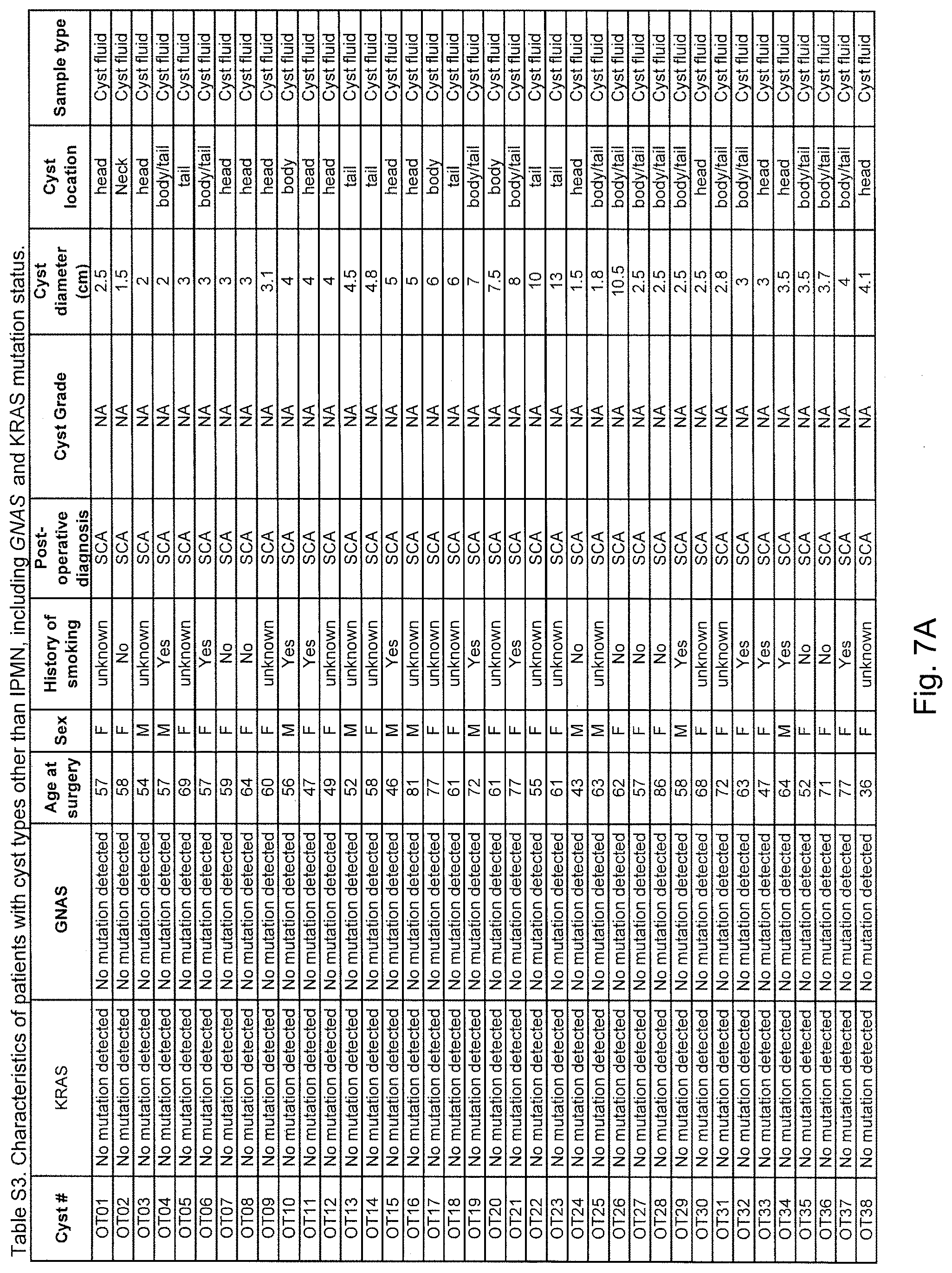
D00016

D00017
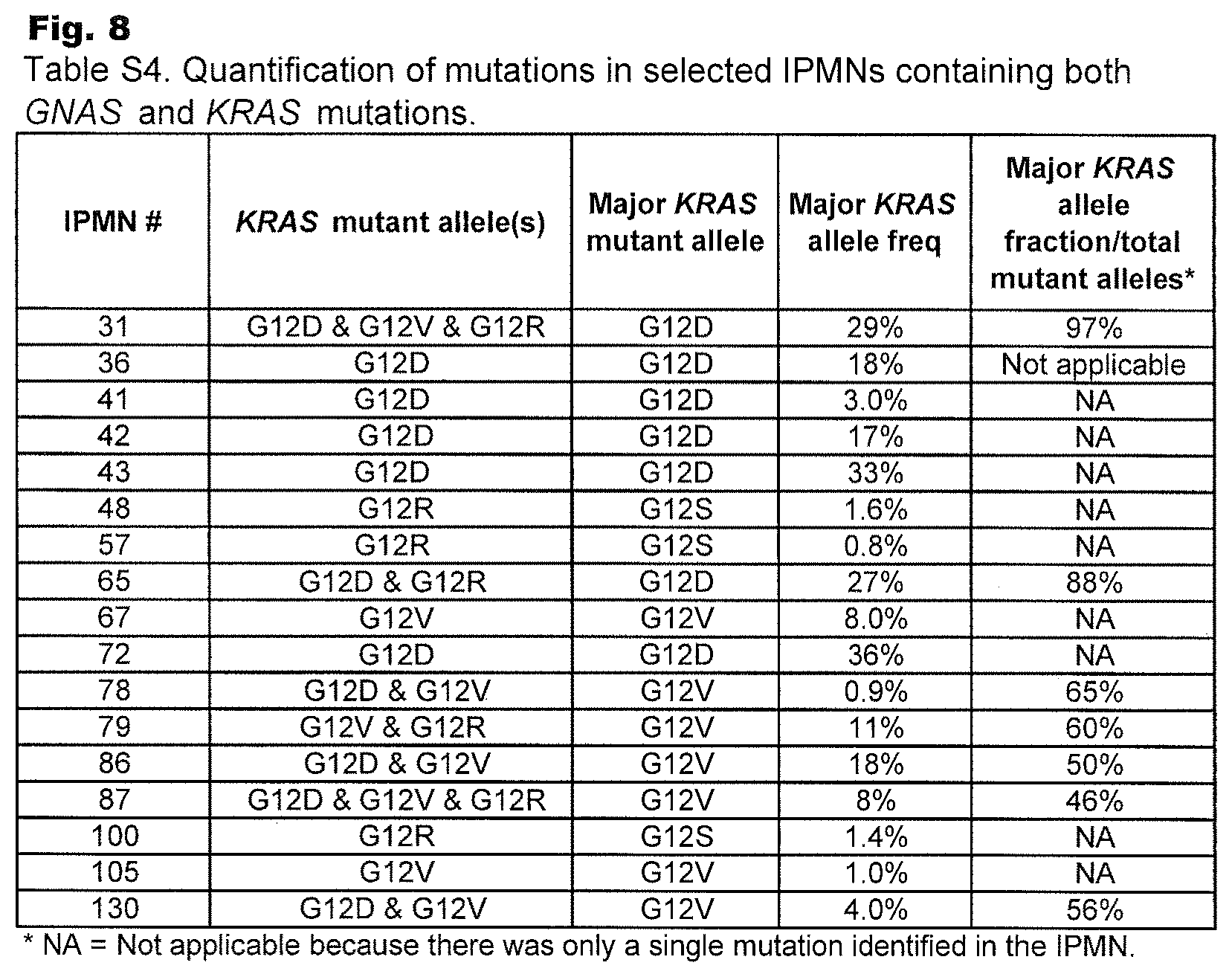
D00018
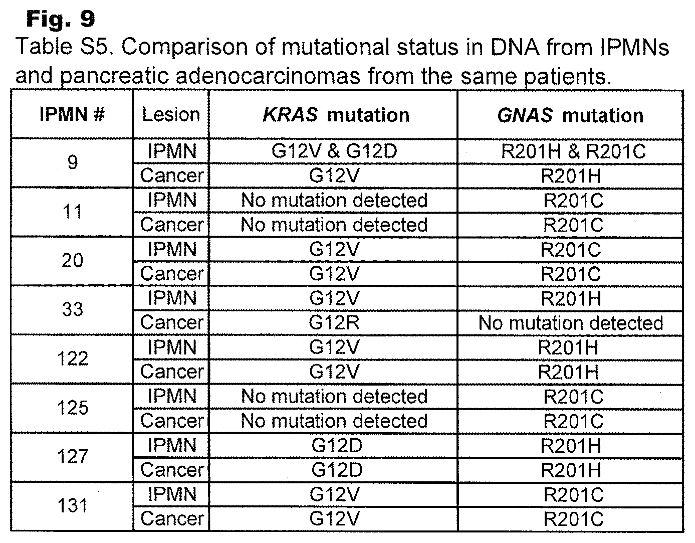
D00019
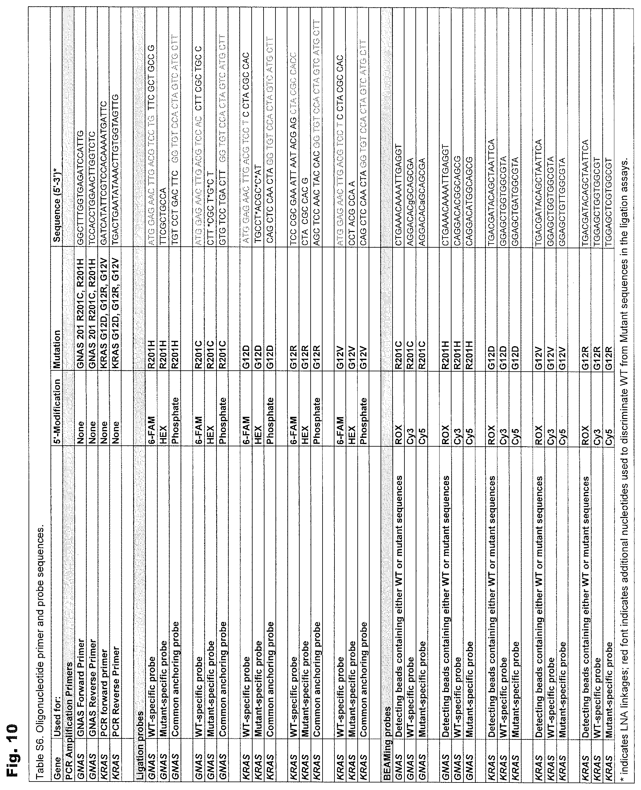
S00001
XML
uspto.report is an independent third-party trademark research tool that is not affiliated, endorsed, or sponsored by the United States Patent and Trademark Office (USPTO) or any other governmental organization. The information provided by uspto.report is based on publicly available data at the time of writing and is intended for informational purposes only.
While we strive to provide accurate and up-to-date information, we do not guarantee the accuracy, completeness, reliability, or suitability of the information displayed on this site. The use of this site is at your own risk. Any reliance you place on such information is therefore strictly at your own risk.
All official trademark data, including owner information, should be verified by visiting the official USPTO website at www.uspto.gov. This site is not intended to replace professional legal advice and should not be used as a substitute for consulting with a legal professional who is knowledgeable about trademark law.