Renilla based biosensors for monitoring biomolecule localization and trafficking in cells
Laporte , et al.
U.S. patent number 10,584,159 [Application Number 15/512,267] was granted by the patent office on 2020-03-10 for renilla based biosensors for monitoring biomolecule localization and trafficking in cells. This patent grant is currently assigned to The Royal Institution for the Advancement of Learning/McGill University, Universite de Montreal. The grantee listed for this patent is The Royal Institution for the Advancement of Learning/McGill University, Universite de Montreal. Invention is credited to Michel Bouvier, Denis Deblois, Etienne Durette, Mireille Hogue, Hiroyuki Kobayashi, Stephane Alain Laporte, Christian Le Gouill, Viktoriya Lukasheva, Yoon Namkung.


View All Diagrams
| United States Patent | 10,584,159 |
| Laporte , et al. | March 10, 2020 |
| **Please see images for: ( Certificate of Correction ) ** |
Renilla based biosensors for monitoring biomolecule localization and trafficking in cells
Abstract
Bioluminescence resonance energy transfer (BRET) biosensors for assessing the intracellular localization, internalization and trafficking into cellular compartments of proteins such as receptors, and other biomolecules such as second messengers, are disclosed. These biosensors, which are dependent on the concentration/density of the BRET donor and acceptor in cellular compartments rather that specific protein-protein interactions, use a Renilla GFP/Luc BRET pair, which allows the robust and reproducible monitoring of protein trafficking/localization, with a sensitivity compatible with high-throughput screening (HTS). The use of these biosensors for various applications, including assessing/monitoring protein endocytosis, recycling and intracellular trafficking, receptor maturation/rescue by pharmacological chaperones, various endocytosis/exocytosis processes, activation/inhibition, as well as biomolecule concentration/density in different cellular compartments, is also disclosed.
| Inventors: | Laporte; Stephane Alain (Outremont, CA), Namkung; Yoon (Pointe-Claire, CA), Bouvier; Michel (Montreal, CA), Le Gouill; Christian (Montreal, CA), Hogue; Mireille (Laval, CA), Lukasheva; Viktoriya (Pointe-Claire, CA), Kobayashi; Hiroyuki (Montreal, CA), Deblois; Denis (Montreal, CA), Durette; Etienne (Saint-Jerome, CA) | ||||||||||
|---|---|---|---|---|---|---|---|---|---|---|---|
| Applicant: |
|
||||||||||
| Assignee: | The Royal Institution for the
Advancement of Learning/McGill University (Montreal, Quebec,
CA) Universite de Montreal (Montreal, Quebec, CA) |
||||||||||
| Family ID: | 55532401 | ||||||||||
| Appl. No.: | 15/512,267 | ||||||||||
| Filed: | September 21, 2015 | ||||||||||
| PCT Filed: | September 21, 2015 | ||||||||||
| PCT No.: | PCT/CA2015/050924 | ||||||||||
| 371(c)(1),(2),(4) Date: | March 17, 2017 | ||||||||||
| PCT Pub. No.: | WO2016/041093 | ||||||||||
| PCT Pub. Date: | March 24, 2016 |
Prior Publication Data
| Document Identifier | Publication Date | |
|---|---|---|
| US 20170313762 A1 | Nov 2, 2017 | |
Related U.S. Patent Documents
| Application Number | Filing Date | Patent Number | Issue Date | ||
|---|---|---|---|---|---|
| 62052738 | Sep 19, 2014 | ||||
| Current U.S. Class: | 1/1 |
| Current CPC Class: | G01N 33/5035 (20130101); C07K 14/723 (20130101); C07K 2319/60 (20130101) |
| Current International Class: | C07K 14/72 (20060101); G01N 33/50 (20060101) |
| 2005105850 | Nov 2005 | WO | |||
| 2010/085844 | Aug 2010 | WO | |||
| 2011/067202 | Jun 2011 | WO | |||
| 2011130540 | Oct 2011 | WO | |||
Other References
|
Violin et al., G Protein coupled receptor kinase and p-arrestin mediated desensitization of the angiotensin II Type 1A receptor elucidated by diacylglycerol dynamics. J Biol. Chem. 281, 36411-36419, 2006. (Year: 2006). cited by examiner . Supplementary European Search Report corresponding to European Application No. 15841561.2 dated Feb. 5, 2018. cited by applicant . Charest et al. "Palmitoylation of the V2 vasopressin receptor carboxyl tail enhances beta-arrestin recruitment leading to efficient receptor endocytosis and ERK1/2 activation", J. Biol. Chem. 287(42):41541-41551. cited by applicant . Anborgh et al. (2000). "Receptor/beta-arrestin complex formation and the differential trafficking and resensitization of beta2-adrenergic and angiotensin II type 1A receptors." Mol Endocrinol 14(12): 2040-2053. cited by applicant . Barberis et al, (1992), "Pharmacology of oxytocin and vasopressin receptors in the central and peripheral nervous system." Ann N Y Acad Sci 652: 39-45. cited by applicant . Claing et al. (2002). "Endocytosis of G protein-coupled receptors roles of G protein-coupled receptor kinases and beta-arrestin proteins." Prog Neurobiol 66(2): 61-79. cited by applicant . Dacres et al., "Comparison of enhanced bioluminescence energy transfer donors for protease biosensors". Analytical Biochemistry, Feb. 28, 2012 (Feb. 28, 2012), vol. 424, pp. 206-210. cited by applicant . Fessart et al. (2005). "c-Src regulates clathrin adapter protein 2 interaction with beta-arrestin and the angiotensin II type 1 receptor during clathrin-mediated internalization." Mol Endocrinol 19(2): 491-503. cited by applicant . Gaborik et al. (2001). "Beta-arrestin- and dynamin-dependent endocytosis of the AT1 angiotensin receptor." Mol Pharmacol 59(2): 239-247. cited by applicant . Goupil et al. (2012). "Biasing the prostaglandin F2alpha receptor responses toward EGFR-dependent transactivation of MAPK." Mol Endocrinol 26(7): 1189-1202. cited by applicant . Hanyaloglu et al. (2008). "Regulation of GPCRs by endocytic membrane trafficking and its potential implications." Annu Rev Pharmacol Toxicol 48: 537-568. cited by applicant . Hein et al. (1997). "Intracellular trafficking of angiotensin II and its AT1 and AT2 receptors: evidence for selective sorting of receptor and ligand," Mol Endocrinol 11(9): 1266-1277. cited by applicant . Hunyady et al. (2006). "Pleiotropic AT1 receptor signaling pathways mediating physiological and pathogenic actions of angiotensin II." Mol Endocrinol 20(5): 953-970. cited by applicant . Hunyady et al. (2002). "Differential PI 3-kinase dependence of early and late phases of recycling of the internalized AT1 angiotensin receptor." J Cell Biol 157(7): 1211-1222. cited by applicant . Innamorati et al. (1998). "A serine duster prevents recycling of the V2 vasopressin receptor." Proc Natl Acad Sci U S A 95(5): 2222-2226. cited by applicant . Lan al., "Sensitive and High Resolution Localization and Tracking of Membrane Proteins in Live Cells with BRET" Traffic, Nov. 2012 (Nov. 2012), vol. 13(11), pp. 1450-1456. cited by applicant . Li et al. (2008). "Rab4 and Rab11 coordinately regulate the recycling of angiotensin II type I receptor as demonstrated by fluorescence resonance energy transfer microscopy." J Biomed Opt 13(3): 031206. cited by applicant . Molinari et al. (2008). "Functional complementation of high-efficiency resonance energy transfer: a new tool for the study of protein binding interactions in living cells." Biochem J 409(1): 251-261. cited by applicant . Morello et al., "Pharmacological chaperones rescue cell-surface expression and function of misfolded V2 vasopressin receptor mutants", J Clin Invest, 2000, 105(7): p. 887-95. cited by applicant . Oakley et al. (2000). "Differential affinities of visual arrestin, beta arrestin1, and beta arrestin2 for G protein-coupled receptors delineate two major classes of receptors." J Biol Chem 275(22): 17201-17210. cited by applicant . Posner et al. (2010). "Cellular signalling: Peptide hormones and growth factors." Prog brain Res 181: 1-16. cited by applicant . Quoyer et al. (2013). "Pepducin targeting the C-X-C chemokine receptor type 4 acts as a biased agonist favoring activation of the inhibitory G protein." Proceedings of the National Academy of Sciences of the United States of America 110(52): E5088-5097. cited by applicant . Rene et al. "Pharmacological Chaperones Restore Function to MC4R Mutants Responsible for Severe Early-Onset Obesity", J Pharmacol Exp Ther. Dec. 2010;335(3)520-32. cited by applicant . Seachrist et al. (2003). "Regulation of G protein-coupled receptor endocytosis and trafficking by Rab GTPases." Life Sci 74(2-3): 225-236. cited by applicant . Serradeil-Le Gal C., "An Overview of SR121463, a Selective Non-Peptide Vasopressin V2 Receptor Antagonist", Cardiovasc Drug Rev. 2001, 19(3):201-14. cited by applicant . Toth et al. (2012). "Acute depletion of plasma membrane phosphatidylinositol 4,5-bisphosphate impairs specific steps in endocytosis of the G-protein-coupled receptor." J Cell Sci 125(Pt 9): 2185-2197. cited by applicant . Tsao et al. (2000). "Type-specific sorting of G protein-coupled receptors after endocytosis." J Biol Chem 275(15): 11130-11140. cited by applicant . Tsao et al. (2001). "Role of endocytosis in mediating downregulation of G-protein-coupled receptors." Trends Pharmacol Sci 22(2): 91-96. cited by applicant . Ward et al. (1976). "Action spectrum and quantum yield for the photoinactivation of mnemiopsin, a bioluminescent photoprotein from the Ctenophore mnemiopsis SP." Photochem Photobiol 23(5): 351-363. cited by applicant . Wible et al., "HERG-Lite: A novel comprehensive high-throughout screen fordrug-induced hERG risk", J Pharmacol Toxicol Methods. 2005, 52(1):136-45. cited by applicant . Yeatman et al. (2014) "Allosteric Modulation of M1 Muscarinic Acetylcholine Receptor Internalization and Subcellular Trafficking", The Journal of Biological Chemistry vol. 289, No. 22, pp. 15856-15866. cited by applicant . Zhang et al. (1999). "Cellular trafficking of G protein-coupled receptor/beta-arrestin endocytic complexes." J Biol Chem 274(16): 10999-11006. cited by applicant . Zhang et al. (1996). "Dynamin and beta-arrestin reveal distinct mechanisms for G protein-coupled receptor internalization." J Biol Chem 271(31): 18302-18305. cited by applicant . Zimmerman et al. (2012). "Differential beta-arrestin-dependent conformational signaling and cellular responses revealed by angiotensin analogs." Sci Signal 5(221): ra33. cited by applicant . Zimmerman et al. (2011). "Role of beta-arrestins in bradykinin B2 receptor-mediated signalling," Cell Signal 23(4): 648-659. cited by applicant . Notification of Transmittal of the International Search Report and the Written Opinion of the International Searching Authority, or the Declaration corresponding to International Application No. PCT/CA2015/050924; dated Nov. 27, 2015. cited by applicant . Balla et al, "Demonstration of Angiotensin II-induced Ras Activation in the trans-Golgi Network and Endoplasmic Reticulum Using Bioluminescence Resonance Energy Transfer-based Biosensors", J. Biological Chem. 286 (7):5319-5327 (2011). cited by applicant . Office Action corresponding to Japanese Application No. 2017-515812 dated Jul. 16, 2019. cited by applicant. |
Primary Examiner: Stoica; Elly-Gerald
Attorney, Agent or Firm: Myers Bigel, P.A.
Parent Case Text
CROSS REFERENCE TO RELATED APPLICATIONS
This application claims the benefit of U.S. Provisional Application Ser. No. 62/052,738, filed on Sep. 19, 2014, which is incorporated herein by reference in its entirety.
Claims
What is claimed is:
1. A biosensor for assessing the trafficking and/or localization of a protein of interest comprising; a first component comprising said protein of interest tagged with a Renilla green fluorescent protein (Renilla GFP) or a Renilla luciferase protein (Renilla Luc); a second component comprising a cellular compartment targeting moiety tagged with a Renilla GFP or a Renilla Luc, wherein said cellular compartment targeting moiety is: (i) a plasma membrane (PM) targeting moiety comprising the amino acid sequence MGCIKSKGKDS(SEQ ID NO:1), GKKKKKKSKTKCVIM (SEQ ID NO:7), CMSCKCVLS (SEQ ID NO:47), CMSCKCCIL (SEQ ID NO:43), or SPKKGLLQRLFKRQHQNNSKS (SEQ ID NO:8); or (ii) an endosomal targeting moiety comprising residues 739 to 806 of human endofin (SEQ ID NO:20); wherein if said protein of interest is tagged with said Renilla GFP, said cellular compartment targeting moiety is tagged with said Renilla Luc, and if said protein of interest is tagged with said Renilla Luc, said cellular compartment targeting moiety is tagged with said Renilla GFP.
2. The biosensor of claim 1, wherein said protein of interest is tagged with said Renilla Luc and said cellular compartment targeting moiety is tagged with said Renilla GFP.
3. The biosensor of claim 1, wherein said protein of interest is i) a signalling polypeptide or a fragment thereof, ii) a protein recruited to the plasma membrane upon stimulation of a receptor, or a fragment thereof, iii) a protein sequestered away from the plasma membrane upon stimulation of a receptor, or a fragment thereof, or iv) a cell surface receptor or a fragment thereof.
4. The biosensor of claim 3, wherein said cell surface receptor is a G protein-coupled receptor (GPCR) or a receptor tyrosine kinase (RTK).
5. The biosensor of claim 1, which comprises said PM targeting moiety.
6. The biosensor of claim 1, wherein said PM targeting moiety is fused to the C-terminal end of said Renilla Luc or said Renilla GFP.
7. The biosensor of claim 1, which comprises said endosomal targeting moiety.
8. The biosensor of claim 1, wherein said endosomal targeting moiety is fused to the C-terminal end of said Renilla Luc or said Renilla GFP; and/or said protein of interest is fused to the N-terminal end of said Renilla Luc or said Renilla GFP.
9. The biosensor of claim 1, wherein said first and second component are covalently linked through a flexible linker.
10. A method for determining whether an agent modulates the trafficking of a protein of interest in a cell, said method comprising: measuring the BRET signal in the biosensor of claim 1 in the presence and absence of said agent; wherein a difference in said BRET signal in the presence of said agent relative to the absence thereof is indicative that said agent modulates the trafficking of said protein of interest in said cell.
11. The biosensor of claim 1, wherein said Renilla Luc is Renilla reniformis luciferase II (RlucII) and/or said Renilla GFP is a Renilla reniformis GFP (rGFP).
12. The biosensor of claim 3, wherein said signalling protein or fragment thereof is a G protein effector or a fragment thereof, a .beta.-arrestin polypeptide or a fragment thereof, a G protein subunit polypeptide or a fragment thereof, an adaptor protein or a fragment thereof, or a Rho-binding polypeptide or a fragment thereof.
13. The biosensor of claim 12, wherein said signalling protein or fragment thereof is an adaptor protein or a fragment thereof.
14. The biosensor of claim 13, wherein said adaptor protein or fragment thereof comprises at least one SH2 and/or SH3 domains.
15. The biosensor of claim 3, wherein said signalling protein or fragment thereof is a polypeptide that binds to a second messenger or to a second messenger precursor.
16. The biosensor of claim 15, wherein said second messenger precursor is phosphatidylinositol 4,5-bisphosphate (PIP.sub.2).
17. The biosensor of claim 16, wherein said signalling protein or fragment thereof comprises a Pleckstrin homology (PH) domain.
18. The biosensor of claim 15, wherein said second messenger is diacylglycerol (DAG).
19. The biosensor of claim 18, wherein said signalling protein or fragment thereof comprises a phorbol esters/diacylglycerol binding domain.
20. The biosensor of claim 12, wherein the signalling protein or fragment thereof is a .beta.-arrestin polypeptide or a fragment thereof fused to the N-terminal of said Renilla Luc, and the cellular compartment targeting moiety is a plasma membrane (PM) targeting moiety or an endosomal targeting moiety, fused to the C-terminal of said Renilla GFP.
21. The biosensor of claim 4, wherein the GPCR is fused to the N-terminal of said Renilla Luc, and the cellular compartment targeting moiety is fused to the C-terminal of said Renilla GFP.
22. The biosensor of claim 1, wherein said biosensor is comprised within a cell.
23. The biosensor of claim 1, wherein the protein of interest is a Grb2 polypeptide or a fragment thereof.
24. The biosensor of claim 23, wherein the Grb2 polypeptide comprises the amino acid sequence of SEQ ID NO: 24.
25. The biosensor of claim 16, wherein the signalling protein or fragment thereof comprising a PH domain is a phospholipase C delta 1 (PLC.delta.1) protein or a fragment thereof.
26. The biosensor of claim 25, wherein the PLC.delta.1 protein or fragment thereof comprises the amino acid sequence of SEQ ID NO: 25.
Description
TECHNICAL FIELD
The present invention generally relates to the assessment/monitoring of the localization, transport and trafficking of biomolecules such as proteins, for example cell surface receptor endocytosis, recycling and intracellular trafficking of receptors and effectors.
BACKGROUND ART
Protein trafficking is an active process in which proteins are re-located from one region of a cell to another. Membranes and their protein components are constantly being turned over through a mechanism that has multiple components and pathways. One of the mechanisms of modulating the activity of cell surface receptors, such as G protein-coupled receptors (GPCRs) and the Epidermal Growth Factor receptor (EGFR), is through receptor endocytosis. For GPCRs, ligand-induced receptor endocytosis can drive receptors removal from the PM through specialized compartments like clathrin-coated vesicles, which involve the recruitment of the endocytic adaptor .beta.-arrestin to liganted receptors (Claing, Laporte et al. 2002). Internalizing receptors can be directed into divergent lysosomal and recycling pathways, producing essentially opposite effects on the strength and duration of cellular signaling via heterotrimeric G proteins, and can also promote distinct signalling events from intracellular membranes through the signalling scaffolding of .beta.-arrestins (Hanyaloglu and von Zastrow 2008; Posner and Laporte 2010). Therapeutic advantages have been proposed for drugs promoting the intracellular targeting of GPCR/.beta.-arrestin complexes, while for some receptors their recycling to the PM is also essential for adequate maintenance of physiological responses.
Thus, simple and reliable systems for monitoring receptor trafficking are key to study the mechanism of receptor endocytosis and to develop efficient therapeutics acting on cell surface receptors such as GPCRs. For instance the Angiotensin II type 1 receptor (AT1R) has attracted significant attention for drug development, because of its involvement in the development of cardiovascular diseases, including hypertension, hypertrophy, fibrosis and atherosclerosis (Hunyady and Catt 2006), and because ligands, which have cardioprotective function can also promote internalization of receptors and intracellular AT1R/.beta.-arrestin signalling complexes. Great advantages can thus arise from developing assays efficiently assessing in a quantitative and high efficiency manner drugs' propensity to induce the internalization of receptors such as GPCRs.
The present description refers to a number of documents, the content of which is herein incorporated by reference in their entirety.
SUMMARY OF THE INVENTION
The present invention relates to the following items 1 to 73: 1. A biosensor for assessing the trafficking and/or localization of a protein of interest comprising;
a first component comprising said protein of interest tagged with a Renilla green fluorescent protein (Renilla GFP) or a Renilla luciferase protein (Renilla Luc);
a second component comprising a cellular compartment targeting moiety tagged with a Renilla GFP or a Renilla Luc;
wherein if said first protein is tagged with said Renilla GFP, said cellular compartment targeting moiety is tagged with said Renilla Luc, and if said first protein is tagged with said Renilla Luc, said cellular compartment targeting moiety is tagged with said Renilla GFP. 2. The biosensor of item 1, wherein said protein of interest is tagged with said Renilla Luc and said cellular compartment targeting moiety is tagged with said Renilla GFP. 3. The biosensor of item 1 or 2, wherein said protein of interest is a Rho-binding polypeptide, a .beta.-arrestin polypeptide, a cell surface receptor or a G protein subunit polypeptide. 4. The biosensor of item 3, wherein said protein of interest is a Rho-binding polypeptide. 5. The biosensor of item 3, wherein said protein of interest is a cell surface receptor. 6. The biosensor of item 5, wherein said cell surface receptor is a G protein-coupled receptor (GPCR). 7. The biosensor of any one of items 1 to 6, wherein said cellular compartment targeting moiety is a plasma membrane (PM) targeting moiety, an endosomal targeting moiety, a Golgi targeting moiety, a lysosomal targeting moiety, a peroxisomal targeting moiety, an autophagosomal targeting moiety, a ribosome targeting moiety, a mitochondria targeting moiety, a cytoskeleton targeting moiety or a nuclear targeting moiety. 8. The biosensor of item 7, wherein said cellular compartment targeting moiety is a plasma membrane (PM) targeting moiety. 9. The biosensor of item 8, wherein said PM targeting moiety is a PM protein or a fragment thereof that localizes to the PM. 10. The biosensor of item 9, wherein said PM protein or fragment thereof comprises (a) a palmitoylation, myristoylation, and/or prenylation signal sequence and/or (b) a polybasic sequence. 11. The biosensor of item 10, wherein said palmitoylation and/or myristoylation signal sequence is from the human Src family kinase Lyn. 12. The biosensor of item 11, wherein said PM targeting moiety comprises the amino acid sequence MGCIKSKGKDS (SEQ ID NO:1). 13. The biosensor of item 12, wherein said polybasic sequence and prenylation signal sequence are from human KRAS splice variant b. 14. The biosensor of item 13, wherein said PM targeting moiety comprises the amino acid sequence GKKKKKKSKTKCVIM (SEQ ID NO:7). 15. The biosensor of item 10, wherein said PM targeting moiety comprises a palmitoylation sequence and prenylation signal sequence from hRas. 16. The biosensor of item 15, wherein said PM targeting moiety comprises the amino acid sequence CMSCKCVLS (SEQ ID NO:47). 17. The biosensor of item 10, wherein said PM targeting moiety comprises a palmitoylation sequence from hRas and prenylation signal sequence from Ral1. 18. The biosensor of item 17, wherein said PM targeting moiety comprises the amino acid sequence CMSCKCCIL (SEQ ID NO:43). 19. The biosensor of item 9, wherein said PM protein or fragment thereof is Caveolin1.alpha.. 20. The biosensor of item 10, wherein said PM targeting polybasic sequence is from human GRKS. 21. The biosensor of item 20, wherein said PM targeting moiety comprises the amino acid sequence SPKKGLLQRLFKRQHQNNSKS (SEQ ID NO:8). 22. The biosensor of item 8 to 21, wherein (i) said PM targeting moiety comprises a palmitoylation and/or myristoylation signal sequence from the human Src family kinase Lyn, and is fused to the N-terminal end of said Renilla Luc or said Renilla GFP or (ii) said PM targeting moiety comprises (a) a polybasic sequence and prenylation signal sequence from human KRAS splice variant b or HRAS; (b) a palmitoylation sequence from HRAS and prenylation signal sequence from Ral1; (c) Caveolin1.alpha. or a fragment thereof; or (d) a polybasic sequence from human GRKS, and is fused to the C-terminal end of said Renilla Luc or said Renilla GFP. 23. The biosensor of item 7, wherein said cellular compartment targeting moiety is an endosomal targeting moiety. 24. The biosensor of item 23, wherein said endosomal targeting moiety is an endosomal protein or a fragment thereof that localizes to the endosomes. 25. The biosensor of item 24, wherein said endosomal protein or fragment thereof comprises a FYVE domain. 26. The biosensor of any one of items 23 to 25, wherein said endosomal targeting moiety comprises the FYVE domain of human endofin. 27. The biosensor of item 26, wherein said endosomal targeting moiety comprises residues 739 to 806 of human endofin (SEQ ID NO:20). 28. The biosensor of item 23, wherein said endosomal protein or fragment thereof is a Rab protein or a fragment thereof. 29. The biosensor of item 28, wherein said Rab protein is Rab4 or Rab 11. 30. The biosensor of any one of items 23 to 29, wherein said endosomal targeting moiety is fused to the C-terminal end of said Renilla Luc or said Renilla GFP. 31. The biosensor of any one of items 23 to 30, wherein said protein of interest is fused to the N-terminal end of said Renilla Luc or said Renilla GFP. 32. The biosensor of item 7, wherein said cellular compartment targeting moiety is a Golgi targeting moiety. 33. The biosensor of item 32, wherein said Golgi targeting moiety is a Golgi protein or a fragment thereof that localizes to the Golgi. 34. The biosensor of item 33, wherein said Golgi targeting moiety is eNOS1 or a fragment thereof that localizes to the Golgi. 35. The biosensor of item 34, wherein said Golgi targeting moiety comprises residues 1 to 73 of human eNOS1 (SEQ ID NO: 42). 36. The biosensor of any one of items 1 to 35, wherein said first and second component are covalently linked through a flexible linker. 37. The biosensor of item 36, wherein said flexible linker is a polypeptide of about 50 to about 500 amino acids. 38. The biosensor of item 37, wherein said flexible linker is a polypeptide of about 300 amino acids. 39. A nucleic acid encoding the first and/or second components of the biosensor of any one of items 1 to 38. 40. A vector comprising the nucleic acid of item 39. 41. A host cell expressing the biosensor of any one of items 1 to 38. 42. A method for determining whether an agent modulates the trafficking of a protein of interest in a cell, said method comprising: measuring the BRET signal in the biosensor of any one of items 1 to 38 in the presence and absence of said agent; wherein a difference in said BRET signal in the presence of said agent relative to the absence thereof is indicative that said agent modulates the trafficking of said protein of interest in said cell. 43. A method for determining whether an agent induces the internalization of a cell surface receptor of interest in a cell, said method comprising: measuring the BRET signal in the biosensor of any one of items 8 to 22 in the presence and absence of said agent; wherein a lower BRET signal in the presence of said agent relative to the absence thereof is indicative that said agent induces the internalization of a cell surface receptor of interest. 44. A method for assessing the recycling of an internalized receptor of interest at the cell surface, said method comprising:
(a) contacting a first and a second biosensor comprising a PM targeting moiety as defined herein in the presence of a ligand that induces the internalization of said receptor;
(b) measuring a BRET signal in the first biosensor after said contacting;
(c) washing said second biosensor to remove said ligand;
(d) measuring a BRET signal in the second biosensor after said washing; and
(e) determining the recycling of an internalized receptor of interest at the cell surface by comparing the BRET signal in the first and second biosensors,
wherein a higher BRET signal in said second biosensor relative to said first biosensor is indicative of recycling of the internalized receptor of interest at the cell surface.
45. The method of item 44, further comprising repeating steps (d) and (e) at different times after washing to study the kinetics of recycling of the internalized receptor of interest. 46. A method for determining whether an agent induces the trafficking of a cell surface receptor of interest at an endosomal compartment, said method comprising: measuring the BRET signal in the biosensor of any one of items 23 to 31 in the presence and absence of said agent; wherein a higher BRET signal in the presence of said agent relative to the absence thereof is indicative that said agent induces the trafficking of said cell surface receptor of interest in said endosomal compartment. 47. The method of item 46, wherein said method is performed using a plurality of biosensors, and wherein each of said biosensors comprises a different endosomal targeting moiety. 48. A method for determining whether an agent acts as a pharmacological chaperone for a receptor of interest, said method comprising: measuring the BRET signal in the biosensor of any one of items 8 to 22 in the presence and absence of said agent; wherein a higher BRET signal in the presence of said agent relative to the absence thereof is indicative that said agent acts as a pharmacological chaperone for said receptor of interest. 49. A method for determining whether an agent acts as a pharmacological chaperone for a receptor of interest, said method comprising:
providing a biosensor comprising: said receptor of interest tagged with a Renilla green fluorescent protein (Renilla GFP) or a Renilla luciferase protein (Renilla Luc); and an endoplasmic reticulum (ER) targeting moiety tagged with a Renilla GFP or a Renilla Luc; wherein if said receptor is tagged with said Renilla GFP, said ER targeting moiety is tagged with said Renilla Luc, and if said receptor is tagged with said Renilla Luc, said ER targeting moiety is tagged with said Renilla GFP; and
measuring the BRET acceptor signal in the presence and absence of said agent;
wherein a decrease in the BRET signal in the presence of said agent relative to the absence thereof is indicative that said agent acts as a pharmacological chaperone for said receptor.
50. The method of item 48 or 49, wherein said receptor is a mutated receptor. 51. The method of any one of items 48 to 50, wherein said receptor is a G protein-coupled receptor (GPCR). 52. The method of item 51, wherein said GPCR is a melanocortin-4 receptor (MC4R) or a vasopressin 2 receptor (V2R). 53. The method of any one of items 48 to 52, wherein said receptor is an ion channel. 54. The method of item 53, wherein said ion channel is a voltage-gated potassium channel. 55. The method of item 54, wherein said voltage-gated potassium channel is hERG. 56. The method of any one of items 48 to 55, wherein said receptor is tagged with said Renilla Luc, and said PM targeting moiety or ER targeting moiety is tagged with said Renilla GFP. 57. The method of any one of items 48 to 56, wherein said PM targeting is the PM targeting moiety defined in any one of items 9 to 22. 58. A method for determining whether an agent induces the recruitment of a .beta.-arrestin to the plasma membrane, said method comprising:
providing a biosensor comprising a cell or membrane preparation comprising: said .beta.-arrestin tagged with a Renilla green fluorescent protein (Renilla GFP) or a Renilla luciferase protein (Renilla Luc); a plasma membrane (PM) targeting moiety tagged with a Renilla GFP or a Renilla Luc; and a GPCR; wherein if said .beta.-arrestin is tagged with said Renilla GFP, said PM targeting moiety is tagged with said Renilla Luc, and if said .beta.-arrestin is tagged with said Renilla Luc, said PM targeting moiety is tagged with said Renilla GFP; and
measuring the BRET acceptor signal in the presence and absence of said agent;
wherein an increase in the BRET signal in the presence said agent relative to the absence thereof is indicative that said agent induces the recruitment of said .beta.-arrestin to the plasma membrane.
59. The method of item 58, wherein said .beta.-arrestin is tagged with said Renilla Luc. 60. The method of item 58 or 59, wherein said PM targeting moiety PM targeting is the PM targeting moiety defined in any one of items 9 to 22. 61. A method for assessing a modulation in the amount of a biomolecule at a cellular compartment between a first and a second condition, said method comprising:
providing a biosensor comprising: a first component comprising a Renilla green fluorescent protein (Renilla GFP) tagged with a protein marker that binds to said biomolecule; and a second component comprising a Renilla luciferase protein (Renilla Luc) tagged with said protein marker;
measuring the BRET acceptor signal in said first and second conditions;
wherein a difference in the BRET signal between said first and second conditions is indicative of a modulation in the amount of said biomolecule at said cellular compartment between said first and second conditions.
62. The method of item 61, wherein said first condition is the presence of an agent and said second condition is the absence of said agent. 63. The method of item 61 or 62, wherein said biomolecule is a phospholipid. 64. The method of item 63, wherein said phospholipid is phosphatidylinositol 4,5-bisphosphate (PIP.sub.2). 65. The method of item 64, wherein said protein marker comprises a Pleckstrin homology (PH) domain. 66. The method of item 65, wherein said PH domain is the PH domain of PLC.delta.1. 67. The method of item 61 or 62, wherein said biomolecule is a second messenger. 68. The method of item 67, wherein said second messenger is diacylglycerol (DAG). 69. The method of item 68, wherein said protein marker comprises a phorbol esters/diacylglycerol binding domain. 70. The method of item 69, wherein said protein marker comprises the phorbol esters/diacylglycerol binding domain domain of PKC (C1b). 71. The method of item 70, wherein said protein marker comprises the amino acid sequence of SEQ ID NO:72. 72. The method of any one of items 42 to 71, wherein the BRET signal is measured using a plate reader or by microscopy. 73. The biosensor of any one of items 1 to 38, or the method of any one of items 42 to 72, wherein said Renilla Luc is Renilla reniformis luciferase II (RlucII) and/or said Renilla GFP is a Renilla reniformis GFP (rGFP).
Other objects, advantages and features of the present invention will become more apparent upon reading of the following non-restrictive description of specific embodiments thereof, given by way of example only with reference to the accompanying drawings.
BRIEF DESCRIPTION OF DRAWINGS
In the appended drawings:
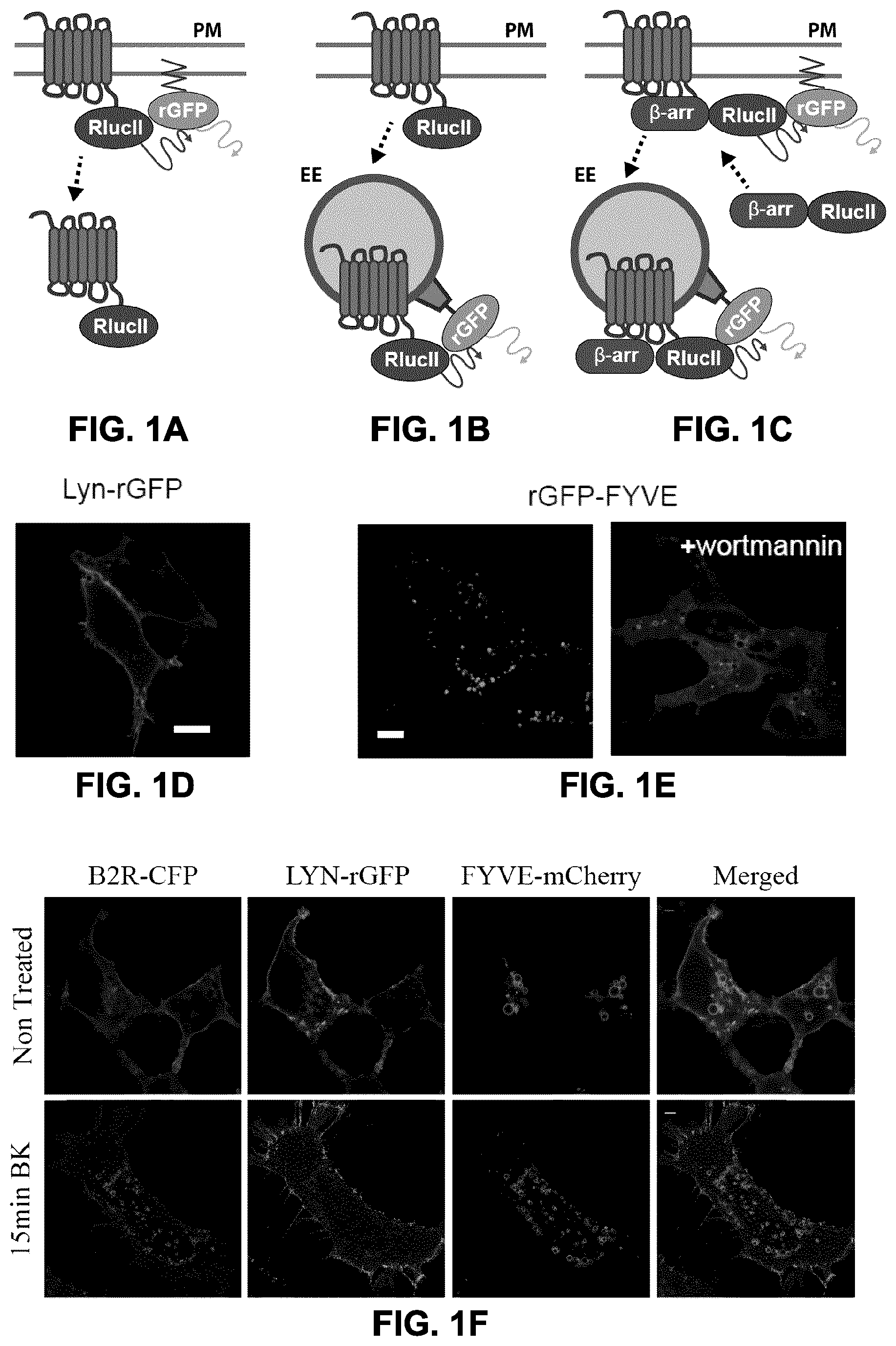
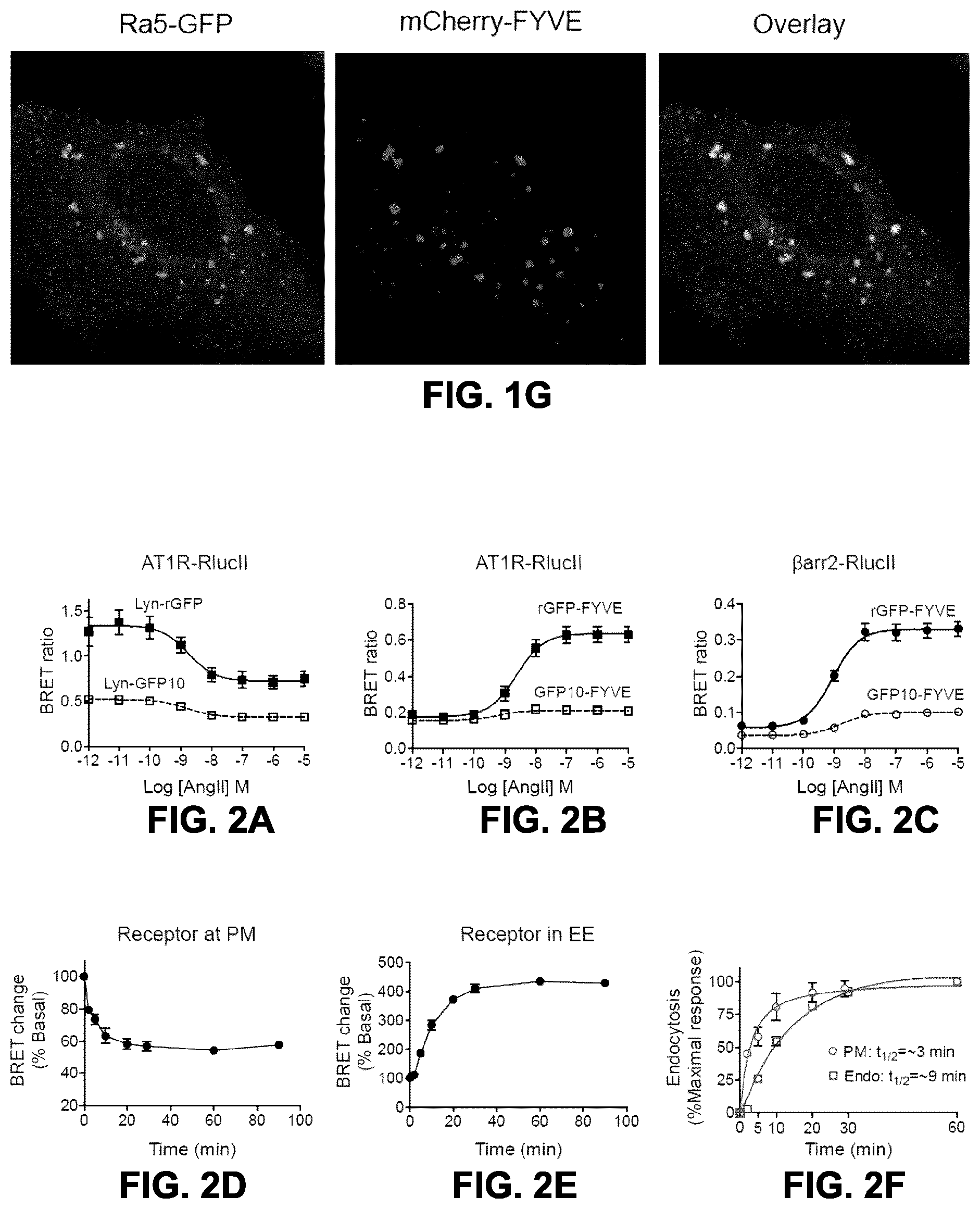
FIGS. 1A to 1F show the generation of Bioluminescence Resonance Energy Transfer (BRET)-based GPCR endocytosis sensor. FIGS. 1A to 1C: Three configurations of BRET-based sensors for assessing/monitoring GPCR endocytosis. To monitor the receptor (FIG. 1A) and .beta.-arrestin (FIG. 1C) amounts at the plasma membrane, anchor rGFP at the plasma membrane by tagging an acylation moiety of lyn-kinase (MGCIKSKGKDS) in N-terminus of rGFP. FIGS. 1B, 1C: To examine targeting of either receptor (FIG. 1B) or .beta.-arrestin (FIG. 1C) to the endosomes, FYVE domain of endofin (amino acids 739 to 806), which tethers the sensor in the endosomes, fused to the C-terminus of rGFP. HEK293SL cells expressing either lyn-rGFP (FIG. 1D) or rGFP-endofinFYVE (FIG. 1E) were subjected to confocal fluorescent microscopy. rGFP-endofinFYVE expressing cells were treated with 500 nM wortmannin for 40 min (FIG. 1E, right panel). Scale bars, 10 .mu.m. FIG. 1F: Simultaneous visualization of receptor, lyn-rGFP, and FYVE domain upon receptor endocytosis. HEK293SL cells were transiently transfected with B2R-CFP, lyn-rGFP, and mCherry-endofinFYVE. Top panel showed basal status and the bottom panel showed a bradykinin induced B2R endocytosis. Scale bars, 10 .mu.m. FIG. 1G: A mCherry-labeled variant of the endofin FYVE sensor also co-localized with Rab5, which populates EE.
FIGS. 2A to 2F show the dose and time-dependent AT1R endocytosis measured by BRET. FIG. 2A: HEK293SL cells were transfected with AT1R-RlucII along with either lyn-GFP10 (.quadrature.) or lyn-rGFP (.box-solid.). Cells were incubated with various concentrations of AngII for 40 min then BRET was measured as described under "materials and methods". FIG. 2B: HEK293SL cells were transfected with AT1R-RlucII along with either GFP10-endofinFYVE or rGFP-endofinFYVE. FIG. 2C: HEK293SL cells were transfected with AT1R and .beta.arr2-RlucII along with either GFP10-endofinFYVE or rGFP-endofinFYVE. AT1R-RlucII along with either lyn-rGFP (FIG. 2D) or rGFP-endofinFYVE (FIG. 2E) transfected cells were incubated in the absence or presence of 100 nM AngII at 37.degree. C. for the indicated times then BRET was measured. The BRET ratio change is expressed as percentage of BRET ratio observed in the control (no AngII treatment) group. Data are represented as the means.+-.S.E. from 2-6 independent experiments. FIG. 2F: Data in FIGS. 2D and 2E were normalized to the maximal responses, respectively and plotted together.
FIGS. 3A to 3F show the effect of blocking receptor endocytosis and overexpression of .beta.-arrestin2 on AngII-induced BRET changes. HEK293SL cells were transfected with AT1R-RlucII/lyn-rGFP (FIG. 3A), AT1R-RlucII/rGFP-endofinFYVE (FIG. 3B), or AT1R/.beta.arr2-RlucII/rGFP-endofinFYVE (FIG. 3C) along with either pcDNA or dynamin K44A. Cells were incubated in the absence (control, .box-solid.; DynK44A, O) or presence of 0.45 M sucrose (.tangle-solidup.) for 20 min then stimulated with various concentrations of AngII for 40 min before BRET measurement. HEK293SL cells were transfected with AT1R-RlucII/lyn-rGFP (FIG. 3D) or AT1R-RlucII/rGFP-endofinFYVE (FIG. 3E) along with either pcDNA or .beta.-arrestin2. FIG. 3F: endocytosis of AT1R in the presence of the vesicle acidification inhibitors bafilomycin A (Baf) and Chloroquine (CQ). Cells were incubated in various concentrations of AngII for 40 min before BRET measurement. Values shown are the means.+-.S.E. from at least three independent experiments.
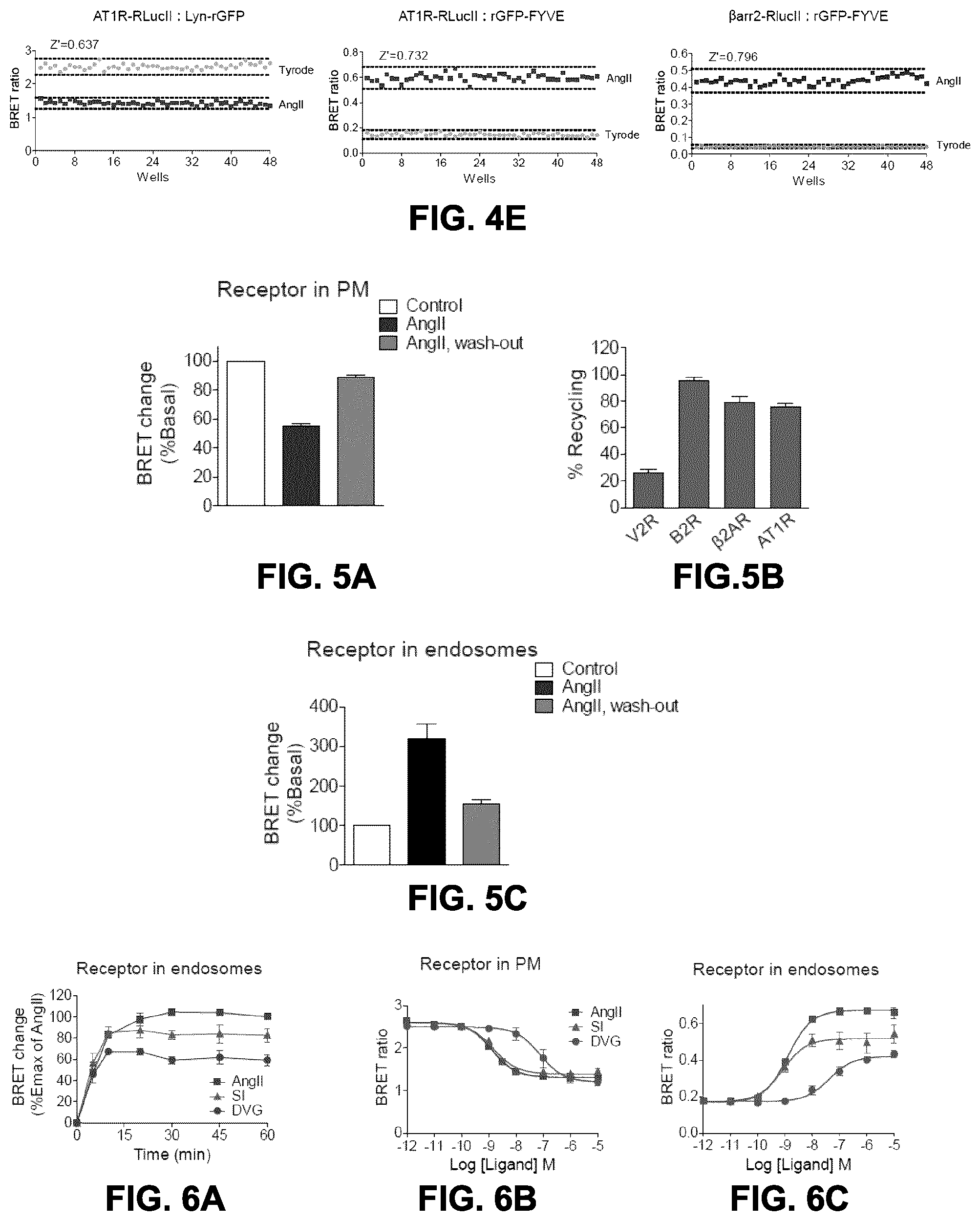
FIGS. 4A to 4E show dose-response curves obtained with the endocytosis BRET biosensors with various receptors. FIG. 4A: HEK293SL cells were transfected with lyn-rGFP along with either AT1R-RlucII, B2R-RlucII, V2R-RlucII, or .beta..sub.2AR-RlucII. FIG. 4B: HEK293SL cells were transfected with rGFP-endofinFYVE along with either AT1R-RlucII, B2R-RlucII, V2R-RlucII, or .beta..sub.2AR-RlucII. Cells were incubated with various concentrations of respective cognate ligand as described in the figure for 30 min (FIG. 4A) or 40 min (FIG. 4B) at 37.degree. C. then BRET were measured. FIG. 4C: HEK293SL cells were transfected with .beta.arr2-RlucII and rGFP-endofinFYVE along with either AT1R, B2R, V2R, .beta..sub.2AR, or FP receptor constructs. Cells were incubated with various concentrations of respective cognate ligand for 40 min before BRET measurement. The cognate ligands for each receptor were as follows: AT1R, AngII (squares); B2R, Bradykinin (BK, triangles); V2R, AVP (circles) or Oxytocin (OT, stars); .beta..sub.2AR, isoproterenol (ISO, inverted triangles); FP, PGF2.alpha. (lozenges). Data for (a) to (c) are expressed as the means.+-.S.E. of 2-3 independent experiments. FIG. 4D: Monitoring of EGFR endocytosis by BRET between RlucII-GRB2 and rGFP-endofinFYVE (GRB2 interacts with EGFR and participates in EGFR internalization). HEK293SL cells were transfected with EGFR along with RlucII-GRB2 and rGFP-endofinFYVE. Cells were incubated with various concentrations of EGF for 30 min at 37.degree. C. then BRET were measured. Results for FIG. 4D are means.+-.SE of triplicates in a single representative experiment out of two independent experiments. FIG. 4E: Assessment of Z' factors as an indication of robustness of the assays for High-Throughput Screening (HTS). HEK293 were co-transfected with AT1R-RlucII/lyn-rGFP, AT1R-RlucII/rGFP-endofinFYVE or AT1R/.beta.arr2-RlucII/rGFP-endofinFYVE and plated in a 48-well plate and stimulated with 100 nM AngII at 37.degree. C. for 20 min to allow receptor receptor dissaperance from the plasma membrane and the accumulation of both receptor and .beta.arrestin2 in endosomes. Cell surface receptor endocytosis was evaluated in BRET2. BRET values are expressed per well in the presented graphs and Z' factor evaluated over 0.64, 0.73 and 0.79 for the AngII-treated group, respectively, which indicates a robust assay for receptor internalization in endosomes.
FIGS. 5A to 5C show the monitoring of receptor recycling after ligand removal by endocytosis BRET assays. FIG. 5A: HEK293SL cells were transfected with AT1R-RlucII along with lyn-rGFP. Cells were incubated in the absence (control) or presence of 100 nM AngII for 30 min then cells were washed and further incubated in the absence of AngII for 45 min. The BRET ratio change is expressed as percentage of BRET ratio observed in the control (no AngII treatment) group. Data are represented as the means.+-.S.E. from four independent experiments. FIG. 5B: HEK293SL cells expressing lyn-rGFP along with either V2R-RluclII, B2R-RlucII, AT1R-RlucII, or .beta..sub.2AR-RlucII were subjected to the receptor recycling as described in FIG. 5A with their cognate ligands, 100 nM AVP for V2R, 100 nM BK for B2R, 100 nM AngII for AT1R, and 1 .mu.M ISO for .beta.2AR. Receptor recycling is expressed as a percent increase in the BRET ratio 45 min after ligand wash-out. All values are expressed as the means.+-..+-.S.E. from 3-4 independent experiments. FIG. 5C: HEK293SL cells were transfected with AT1R-RlucII along with rGFP-endofinFYVE. Cells were incubated in the absence (control) or presence of 100 nM AngII for 30 min then cells were washed and further incubated in the absence of AngII for 45 min. The BRET ratio change is expressed as percentage of BRET ratio observed in the control (no AngII treatment) group. Data represent as the means.+-.S.E. from three independent experiments.
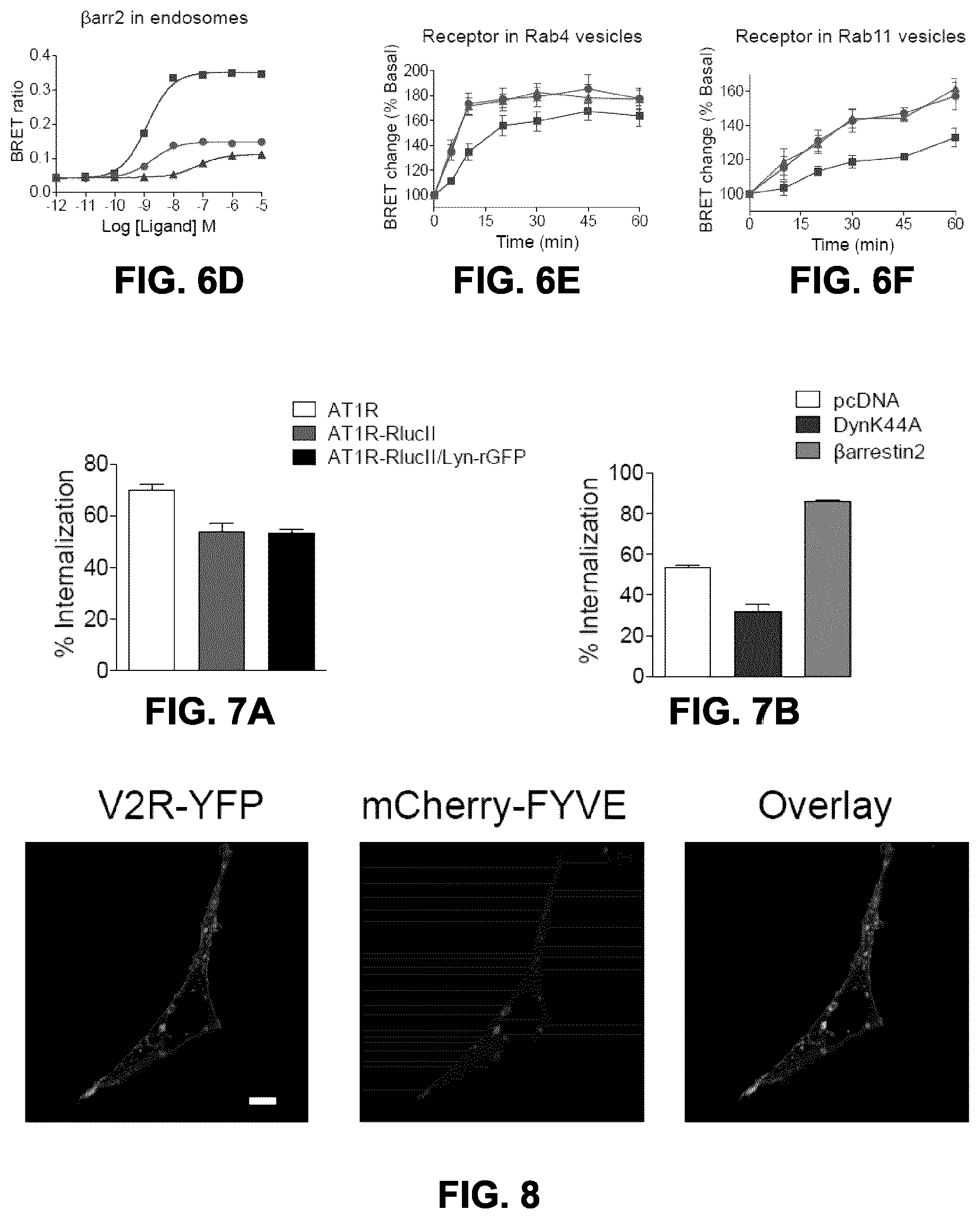
FIGS. 6A to 6F show the effects of AngII analogs on AT1R trafficking and sorting. FIG. 6A: HEK293SL cells expressing AT1R-RlucII/rGFPendofinFYVE were incubated either with 100 nM AngII (squares), 100 nM SI (triangles), or 1 .mu.M DVG (circles) for indicated times then BRET were measured. BRET ratios were normalized to the maximal AngII response (60 min) as a 100% and the basal (no ligand) as a 0%. Data are expressed as the means.+-.S.E. from at least three independent experiments. HEK293SL cells were transfected with AT1R-RlucII/lyn-rGFP (FIG. 6B), AT1R-RlucII/rGFP-endofinFYVE (FIG. 6C) or AT1R/.beta.arr2-RlucII/rGFP-endofinFYVE (FIG. 6D). Cells were incubated with various concentrations of AngII (squares), SI (triangles), or DVG (circles) for 30 min (FIG. 6B) or 40 min (FIGS. 6C, 6D) before BRET measurement. Data are represented as the means.+-.S.E. from at least three independent experiments. FIGS. 6E and 6F: HEK293SL cells were transfected with AT1R-RlucII along with either rGFP-rab4 (FIG. 6E) or rGFP-rab11 (FIG. 6F). Cells were incubated either with 100 nM AngII (squares), 100 nM SI (triangles), or 1 .mu.M DVG (circles) for indicated times then BRET were measured. The BRET ratio change is expressed as percentage of BRET ratio observed in the control (no ligand treatment) group. Data represent the mean.+-.S.E. from three independent experiments.
FIGS. 7A and B show AT1R internalization accessed by intact cell [.sup.125I]AngII-binding assay. FIG. 7A: HEK293SL cells were transiently transfected either AT1R alone (.quadrature.), AT1R-RlucII alone (.box-solid.), or AT1R-RlucII along with lyn-rGFP (.box-solid.). The cells were incubated in the absence or presence of 100 nM AngII for 30 min at 37.degree. C. then subjected to intact-cell [.sup.125I]AngII-binding assay as describe under "materials and methods". FIG. 7B: HEK293SL cells expressing AT1R-RlucII/Lyn-rGFP along with either pcDNA (.quadrature.), dynamin K44A (.box-solid.), or .beta.-arrestin2 (.box-solid.) were incubated in the absence or presence of 100 nM AngII for 30 min at 37.degree. C. then subjected to an intact-cell [.sup.125I]AngII-binding assay as describe below (Example 1). Receptor endocytosis was expressed as the percent loss of cell surface receptors. Data are represented as the means.+-.S.E. of three independent experiments.
FIG. 8 shows the high basal endosomal localization of V2R. HEK293SL cells transiently expressing V2R-YFP along with mCherry-endofinFYVE, were subjected to a confocal microscopy. Scale bar, 10 .mu.m.
FIG. 9A shows the vesicular localization of rGFP-rab4 and rGFP-rab11 with mCherry-FYVE. HEK293SL cells were transfected either rGFP-rab4 (left) or rGFP-rab11 (right), then subjected to a confocal microscopy.
FIGS. 9B to 9D show the effect of G.alpha.q inhibition on AngII-mediated AT1R internalization. FIG. 9B: HEK293SL cells were transfected with AT1R-RlucII along with Lyn-rGFP. Cells were incubated in the absence (-Ubo) or presence of 100 nM Ubo (+Ubo) for 30 min then stimulated with various concentrations of AngII for 30 min before BRET measurement. FIG. 9C: HEK293SL cells were transfected with AT1R-RlucII along with rGFP-FYVE. Cells were incubated in the absence (-Ubo) or presence of 100 nM Ubo (+Ubo) for 30 min then stimulated with various concentrations of either AngII, SI, or DVG for 40 min before BRET measurement. FIG. 9D: Cells were transfected with AT1R-RlucII along with either rGFP-rab4 or rGFP-rab11. Cells were incubated in the absence (-Ubo) or presence of 100 nM Ubo (+Ubo) for 30 min then stimulated either with 100 nM AngII, 100 nM SI, or 1 .mu.M DVG for 10 min for the rGFP-rab4 and 30 min for the rGFP-rab11 then BRET were measured. The BRET ratio change is expressed as percentage of BRET ratio observed in the control (no ligand treatment) group. Data represent the mean.+-.S.E. from three independent experiments.
FIGS. 10A and 10B depict the principle of a BRET-based pharmacological chaperone (PC) assay and sequestration assay to assess functional rescue. FIG. 10A: The pharmalogical chaperone (PC) assay is based on relocalization of a pharmalogical chaperone-rescued protein that would, otherwise, be retained in a different subcellular compartment. Relocalization detected and measured using BRET, preferentially with rGFP with a plasma-membrane targeting sequence (rGFP-CAAX, Lyn-rGFP or rGFP-PB; for a description, see FIG. 11D). Misfolded receptors and channels such as hERG are retained in intracellular compartments and translocate to the plasma membrane upon pharmalogical chaperone-mediated rescue. In this assay, the receptor is preferentially tagged with RlucII and the membrane with rGFP; the BRET signal is proportional to the density of the RlucII-tagged protein at the membrane. The misfolded protein could either be a mutant or the WT protein. For receptors that internalize upon agonist exposure, the functionality of the rescued receptor may then be assessed using the agonist-induced sequestration assay depicted in FIG. 10B (which essentially corresponds to the BRET-based sensor depicted in FIG. 1A). FIG. 10B: principle of a BRET-based agonist-induced sequestration assay. A plasma membrane (PM) marker is tagged with a BRET acceptor such as GFP (G) and the PC-rescued receptor of interest (e.g., a PC-rescued GPCR) is tagged with a BRET donor such as RLuc (R). In the absence of agonist (left), the receptors are retained at the PM and co-localize with the BRET acceptor-tagged PM marker, thus resulting in a strong BRET acceptor signal. However, in the presence of an agonist (right), the receptors are internalized, thus decreasing the density of the BRET donor-tagged receptor at the PM, which results in a decrease in the BRET acceptor signal. This assay can be performed following PC-mediated cell-surface rescue of receptors, in the same well, thus in a homogenous assay that monitor two different aspects of the receptor biology.
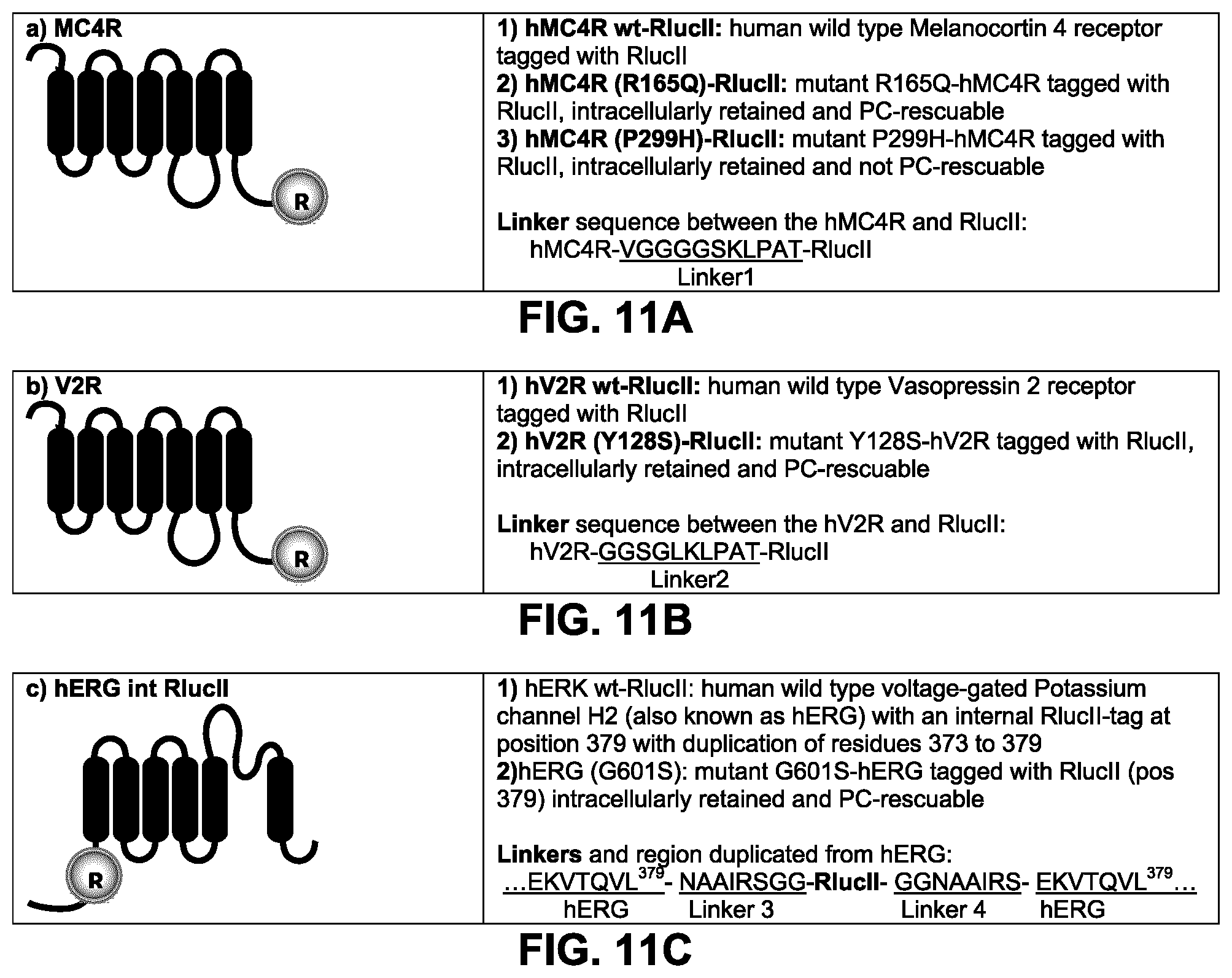
FIGS. 11A to D show the constructs used to validate and optimize a sensor to detect pharmalogical chaperone properties. FIGS. 11A to C: the chaperone-rescue assay was developed and tested with wild type (WT) and naturally-occurring substitutions of human GPCRs (Melanocortin receptor 4: hMC4R and the vasopressin receptor 2: hV2R) and a voltage-gated Potassium channel H2 (hERG) tagged with a BRET donor. The receptors were tagged in C-terminal with RlucII. The hERG channel was internally tagged with RlucII at the equivalent position of residue 379 and, the sequence from residues 373-379 was duplicated on each side of linker3 and linker 4 (see FIG. 11C). Flexible linkers were used between the receptor/channel and the RlucII tag. The sequence of the linkers is indicated in FIG. 11A for the MC4R constructs (Linker1), in FIG. 11B for the V2R constructs (Linker2) and in FIG. 11C for the hERG constructs (Linker3 & 4). FIG. 11D: A BRET acceptor (rGFP) was tagged with different plasma-membrane or Golgi apparatus targeting sequences: in N-terminal with the palmitoylation & myristoylation signal sequence from the Lyn kinase (Lyn-), in C-terminal with the polybasic sequence and prenylation signal sequence from KRAS splice variant b (-CAAX), in C-terminal with the polybasic sequence from the human GRK5 (-PB), in C-terminal with the plasma-membrane targetting palmitoylation sequence and prenylation signal sequence from hRas, in C-terminal with the plasma-membrane targetting palmitoylation sequence from hRas and prenylation signal sequence from Ral1, in C-terminal with human Caveolin1.alpha. (a marker of caveolae); and in N-terminal with the Golgi targetting sequence (residues 1-73) from human eNOS1. Linkers 5, 6 and 7 were used between the rGFP and the plasma-membrane targeting sequence: lyn, CAAX and PB, respectively. Llinker 8 was used between rGFP and palmitoylation/prenylation sequence from hRAS (CAAX) and hRAS/Ral1(CAAX=CCIL), and between rGFP and Caveolin1.alpha.. Llinker 9 was used between the golgi targetting sequence from eNOS (1-73) and rGFP.
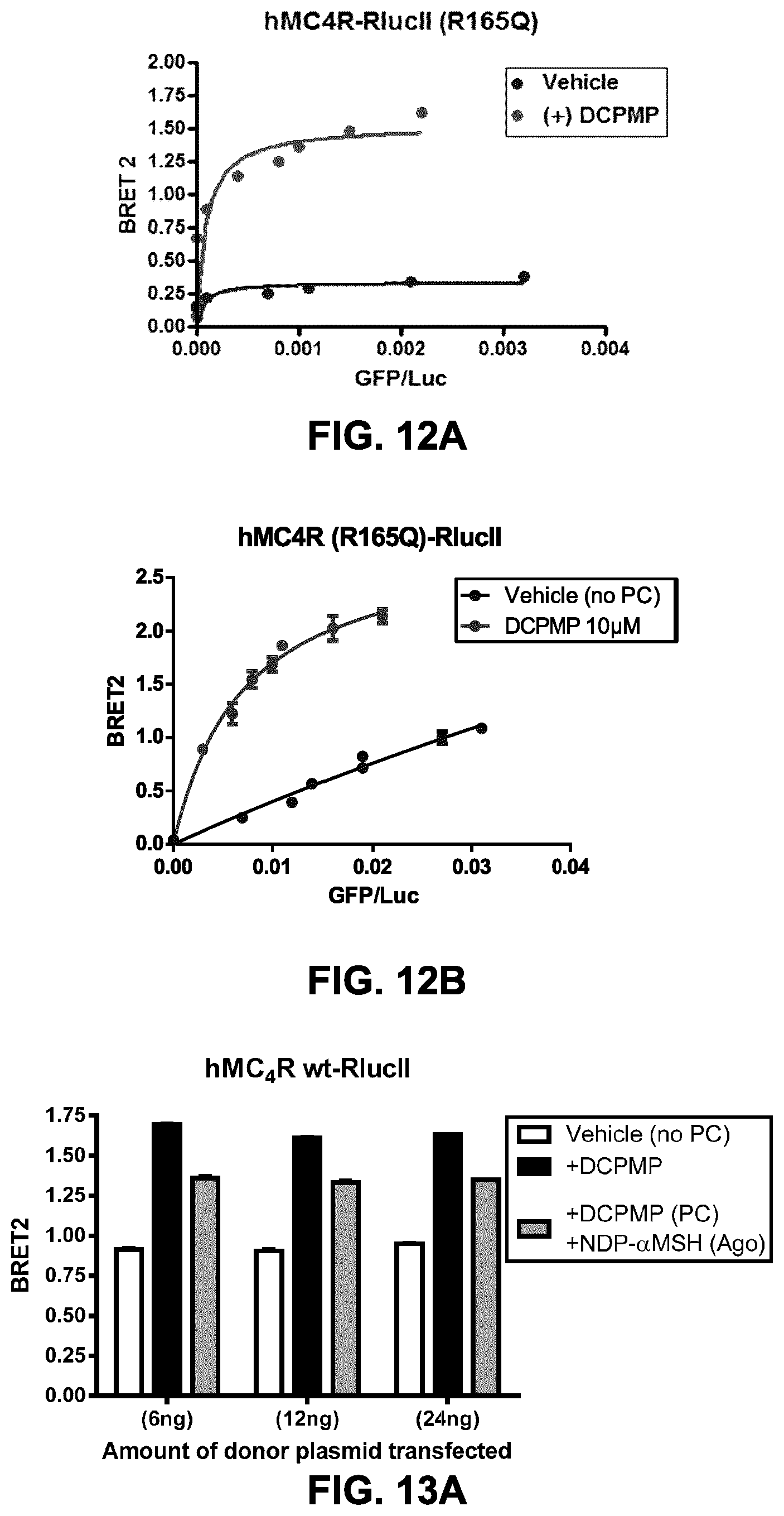
FIGS. 12A and B show the testing of different ratios (titration) of two forms of rGFP targeted to the plasma membrane. Titrations of BRET donor to acceptor and PC-rescue assay were performed on transfected cells (variable amount of rGFP construct+24 ng of receptor construct for 10 wells of a 96-well plate), following a 16 h treatment with either a chaperone: (DCPMP (N-((2R)-3(2,4-dichloroPhenyl)-1-(4-(2-((1-methoxypropan-2-ylamino)methyl- )phenyl) piperazin-1-yl)-1-oxopropan-2-yl)propionamide), 10 .mu.M) or vehicle (DMSO). HEK293 were transfected with hMC4R (R165Q)-RlucII construct and different quantities of rGFP-CAAX (FIG. 12A); and rGFP-PB construct (FIG. 12B). The BRET ratio is reported in function of GFP-construct expression (evaluated in fluorescence) over RlucII construct expression (evaluated in bioluminescence).
FIGS. 13A to C show the cell surface expression and functional PC-mediated rescue of wt and mutant MC4R at different ratios of receptor and rGFP-CAAX. HEK293 were co-transfected with an rGFP-CAAX construct (72 ng of plasmid for 10 wells of a 96-well plate) and 3 different quantities (as indicated on the graphs: 6, 12 and 24 ng for 10 wells) of hMC4R wt-RlucII (FIG. 13A); hMC4R (P299H)-RlucII (FIG. 13B); and hMC4R (R165Q)-RlucII (FIG. 13C). The PC-mediated rescue of cell surface expression and functionality (agonist-induced sequestration) was evaluated in BRET2, on transfected cells, following a 16 h-treatment with either a chaperone: (DCPMP, 10 .mu.M; solid black and grey bars) or vehicle (DMSO; white bars). The grey bars represent data obtained from DCPMP-treated cells, exposed 1 h to an agonist (alpha-MSH) to induce receptor sequestration. As expected, DCPMP-treatment induces an increase in cell surface expression, as revealed by an increase in BRET signal, compared to non-treated cells (with bars). Agonist-treatment induces sequestration as revealed by a decrease in BRET signal (grey bars) as compared to cells treated with DCPMP but not exposed to an agonist (black bars). The wt (FIG. 13A) and R165Q mutant (FIG. 13C) receptors were sensitive to both DCPMP and .alpha.-MSH (10 .mu.M, 1 h at 37C) while the P299H mutant MC4R was not PC-rescued (FIG. 13B). The optimal window for this assay is already obtained at 6 ng of donor and, increasing the quantity of transfected donor construct did not lead to a measurable rescue of hMC4R (P299H).
FIG. 13D shows polycistronic constructs encoding rGFP-CAAX(Kras) and either a WT or mutant hMC4R were transiently expressed in Hek293 cells. This figure shows that similar results for PC rescue of cell surface expression (white bars: DMSO vs. black bars: 10 .mu.M DCPMP) and functionnal rescue can be obtained, as measured by agonist-induced sequestration (+alpha-MSH; grey bars), from polycistronic and non-polycistronic constructs (FIGS. 13A-C). Agonist-induced sequestration for cells not pretreated with a chaperonne is presented (hashed-bars).
FIG. 13E shows the PC-mediated rescue of V2R mutants known to be intracellularly retained, as evidenced by the increase in BRET at the plasma membrane. The PC-mediated rescue of cell surface expression was evaluated in BRET1, on transfected cells, following a 16 h-treatment with either a chaperone: (SR121463, 10 .mu.M; solid black bars) or vehicle (DMSO; white bars).
FIGS. 14A and 14B show dose-response curves for 2 PC-mediated functional rescue of WT and mutant (R165Q) MC4R cell surface expression. HEK293 were co-transfected with an rGFP-CAAX construct and, the hMC4R wt-RlucII or hMC4R (R165Q)-RlucII constructs (72 ng of rGFP construct+24 ng of receptor construct for 10 wells of a 96-well plate). Dose-responses of PC-mediated rescue of cell surface expression, following a 16 h-treatment with variable concentrations of DCPMP (FIG. 14A) and with Compound 1 (FIG. 14B), were evaluated in BRET2. Results obtained with hMC4R wt-RlucII (upper curves) or hMC4R (R165Q)-RlucII (lower curves) are reported in function of the chaperone concentration expressed in a logarithmic scale. EC50 and other curve parameters are indicated below each graph.
FIGS. 15A to 15D show the assessment of Z' factor as an indication of robustness of the assay. HEK293 were co-transfected with an rGFP-CAAX construct and, the hMC4R wt-RlucII (FIGS. 15A and 15B) or hMC4R (R165Q)-RlucII (FIGS. 15C and 15D) constructs (72 ng of rGFP construct+24 ng of receptor construct for 10 wells of a 96-well plate). Cell surface expression was evaluated in BRET2 in FIGS. 15A and 15C using coelenterazine 400a, and in BRET1 using coelenterazine H (FIGS. 15B and 15D) following a 16 h-treatment with 10 .mu.M DCPMP (48 wells) vs. vehicle (DMSO) (48 wells). BRET values are expressed per well in the presented graphs and Z' factor evaluated over 0.63 with the hMC4R wt receptor and over 0.82 with the mutant R165Q mutant hMC4R, which indicates a robust assay with both receptors.
FIGS. 16A and 16B show the assessment of the impact of DMSO on the BRET-based cell surface expression assay. HEK293 were co-transfected with an rGFP-CAAX construct and, the hMC4R wt-RlucII (FIG. 16A) or hMC4R (R165Q)-RlucII (FIG. 16B) constructs (72 ng of rGFP construct+24 ng of receptor construct for 10 wells of a 96-well plate). Cell surface expression was evaluated in BRET2, following a 16 h-treatment with 10 .mu.M DCPMP (right bars) or vehicle (DMSO, left bars) in presence of an increasing concentration of DMSO (up to 3%) during the PC-treatment, in order to evaluate whether the BRET-based assay for cell surface evaluation is sensitive to different levels of DMSO. As presented, the results obtained indicate that this assay is resistant to at least 3% DMSO, which is compatible with HTS applications and characterization of compounds.
FIGS. 17A and 17B show PC-mediated rescue of MC4R and V2R expression in transfected and stable rGFP cell lines. HEK293 were co-transfected with an rGFP-CAAX construct (72 ng of plasmid for 10 wells of a 96-well plate) and 3 different quantities (as indicated on the graphs: 6, 12 and 24 ng for 10 wells) of hMC4R (R165Q)-RlucII (FIG. 17A) or in hV2R (Y128S)-RlucII (FIG. 17B). HEK293 cells selected for stably expressing different levels of rGFP-CAAX (low, medium (Med) & high (Hi)) were transfected with the same quantity of receptor constructs. The PC-mediated rescue of cell surface expression for MC4R was evaluated in BRET2, following a 16 h-treatment with 10 .mu.M DCPMP, and for the V2R (Y128S)-RlucII expressing cells with the SR121463 chaperone (a known antagonist with inverse agonist and pharmalogical chaperone properties; Serradeil-Le Gal C., Cardiovasc Drug Rev. 2001, 19(3): 201-14) at 10 .mu.M or vehicle (DMSO). The data is presented for the MC4R and V2R expressing cells as a % of BRET signal observed with cells treated with vehicle (DMSO). The presented data indicates that a better response can be obtained with stable cell lines expressing higher levels of rGFP-CAAX. The stable cell line expressing high levels of rGFP-CAAX (Stable:Hi) could be used to establish cell lines co-expressing a receptor-RlucII.
FIGS. 18A to 18E: PC-rescue assay for detecting ligands of hERG channel (a non-GPCR). Cell-surface expression and functionnal PC-mediated rescue of wt (FIG. 18A) and mutant (G601S; FIG. 18B) hERG at different ratios of hERG to rGFP-CAAX. HEK293 cells were co-transfected with an rGFP-CAAX construct (72 ng of plasmid for 10 wells of a 96-well plate) and 3 different quantities (as indicated on the graphs: 6, 12 and 24 ng for 10 wells) of hERG wt-RlucII (FIG. 18A) and hERG (G601S)-RlucII (FIG. 18B). The PC-mediated rescue of cell surface expression was evaluated in BRET2, following a 16 h-treatment with either a chaperone: (Astemizole, 10 .mu.M; solid black bars) or vehicle (DMSO; white bars). Astemizole-treatment induces an increase in cell surface expression, as revealed by an increase in BRET signal, compared to vehicle-treated cells. The wt (FIG. 18A) and G601S mutant (FIG. 18B) hERG were both sensitive to a PC-treatment and were used to characterize ligands known to bind and act with different efficacy as chaperones on hERG (FIGS. 18C and D). In FIG. 18E, robustness of the assay with the hERG (G601S)-RlucII construct was evaluated with a Z' factor. Cell surface expression was evaluated in BRET2, following a 16 h-treatment with 10 .mu.M Astemizole (48 wells) vs. vehicle (DMSO) (48 wells). BRET values are expressed per well in the presented graphs and Z' factor evaluated at 0.622, which indicates a robust assay that would be amenable to high throughput screening application.
FIG. 19A shows the configuration of a biosensor for monitoring .beta.-arrestin recruitment to a GPCR at the plasma membrane. A BRET acceptor (e.g., rGFP, GFP10) is tagged with a PM targeting moiety (thus tethering the BRET acceptor at the PM), and a .beta.-arrestin is tagged with a BRET donor (e.g., RlucII). In the presence of a GPCR agonist (represented by A), .beta.-arr is recruited to the GPCR, thus increasing the concentration of RlucII-.beta.-arr at the plasma membrane, which in turn results in an increase in energy transfer (BRET) between RlucII and the PM-tagged GFP.
FIGS. 19B and 19C show the increase in the BRET ratio for the recruitment of .beta.-arrestin.sub.1 and .beta.-arrestin.sub.2, respectively, at the class A GPCR .beta.2AR, for different PM-targeting moieties (Lyn, CAAX and PB-GRK5) and BRET acceptors (rGFP and GFP10), following stimulation with increasing doses of the agonist isoproterenol (iso). The .beta.-arrestin-RlucII translocation sensor with rGFP-CAAX (squares) offers the best window with both receptors.
FIGS. 19D and 19E show the increase in the BRET ratio for the recruitment of .beta.-arrestin.sub.1 and .beta.-arrestin.sub.2, respectively, at the class B GPCR V.sub.2R, for different PM-targeting moieties (Lyn, CAAX and PB-GRK5) and BRET acceptors (rGFP and GFP10), following stimulation with increasing doses of the agonist AVP.
FIG. 19F shows the recruitment of .beta.-arrestin.sub.2 to .beta.2AR following stimulation with increasing doses of the agonist isoproterenol (iso), as assessed using different PM-targeting moieties (CAAX from Kras, CAAX from Hras, the plasma-membrane targetting palmitoylation sequence from hRas and prenylation signal sequence from Ral1 (CCIL) and the marker of the caveolae structures Caveolin1.alpha. tagged with rGFP. The .beta.-arrestin-RlucII translocation sensor with rGFP-CAAX (squares) show an increase of density at the plasma-membrane. In contrast to the response obtained with the rGFP-CAAX markers, a stimulation of .beta.2AR lead to a decrease in density of .beta.-arrestin.sub.2 at the caveolae.
FIG. 19G shows dose-response curves for translocation of .beta.arrestin2 at the plasma membrane after AT1R stimulation. HEK293SL cells were transfected with AT1R and .beta.arr2-RlucII along with either Lyn-rGFP, or rGFP-CAAX or GFP10-CAAX. Cells were incubated with various concentrations of AngII for 6 min at room temperature before BRET measurements. Data are expressed as percent basal BRET. Data are the means.+-.S.E. of 3 independent experiments.
FIGS. 19H-19J show the Z' factors obtained for the .beta.arrestin.sub.2-RlucII/rGFP-CAAX biosensor and receptors of FIGS. 19C, 19E and 19G, respectively. This assay, to monitor receptor-mediated .beta.arrestin recruitment, results in Z' factors of at least 0.74 (0.74, 0.80 and 0.838), which would be amenable to screening (including high-throughput screening) applications for both class A and B GPCRs.
FIGS. 20A and 20B show the AngII-dose dependent decrease in plasma PIP2 amount as detected by BRET between RlucII-PH(PLC.delta.1) and rGFP-PH(PLC.delta.1) or Lyn-rGFP or rGFP-CAAX. HEK293SL cells were transfected with AT1R and HA-RlucII-PH(PLC.delta.1) along with either rGFP-PH(PLC.delta.1), Lyn-rGFP, or rGFP-CAAX. Cells were incubated with various concentrations of AngII for 1 min at RT then BRET was measured. Results are means.+-.S.E. of triplicates in a single representative experiment.
FIG. 21A shows the configuration of a unimolecular biosensor for monitoring .beta.-arrestin recruitment to a GPCR at the plasma membrane. A BRET acceptor (e.g., rGFP, GFP10) is tagged with a PM targeting moiety (thus tethering the construct at the PM) and a flexible linker is placed between the BRET acceptor and a BRET donor (e.g., RlucII), which is attached to a .beta.-arrestin. In the presence of a GPCR agonist (represented by A), .beta.-arr is recruited to the GPCR, thus increasing the concentration of RlucII-.beta.-arr at the plasma membrane, which in turn results in an increase in energy transfer (BRET) between RlucII and the PM-tagged GFP.
FIG. 21B shows the BRET ratio using unimolecular biosensors with flexible linkers of different lengths to assess .beta.-arrestin.sub.2 recruitment to V.sub.2R following stimulation with AVP. FIGS. 21C to 21E show dose-response curves for the recruitment of .beta.-arrestin.sub.2 at different GPCRs (AT1R, V2R and .beta.2AR) using unimolecular biosensors.
FIG. 22A shows a schematic representation of a unimolecular biosensor for measuring the translocation of the diacylglycerol-(DAG-) binding domain of PKCdelta (C1b) to the plasma-membrane. The biosensor comprises a PM-targeting domain/moiety (Mem), a BRET acceptor (e.g., GFP10), a flexible linker, a BRET donor (e.g., RlucII) and the DAG-binding domain of PKC.delta., C1b. Upon activation of PLC, membrane PIP.sub.2 is hydrolysed into IP.sub.3 and DAG. The DAG enrichment causes the C1b domain to bind to the membrane, bringing the BRET acceptor (e.g., GFP10) and BRET donor (e.g., RlucII) closer to each other, inducing a higher BRET signal.
FIG. 22B shows kinetics of DAG sensor activation following AT1R exposure to angiotensin II. AT1R stably expressing HEK293 cells were transfected with a construct encoding the unimolecular DAG sensor DNA and BRET. The BRET level was monitored every 4 s. AngII (final concentration of 100 nM) was added after 16 BRET measurements (64 s). Data are mean.+-.SD of triplicates of a representative experiment.
FIGS. 22C to 22E show dose-response curves obtained with the unimolecular DAG sensor representing the level DAG produced at the plasma membrane following activation of the Angiotensin II receptor (AT1R) with angiotensin II (ANGII) (FIG. 22C), Prostaglandin F receptor (FP) with two natural ligands, prostaglandin 2.alpha. (PGF2.alpha.; solid diamonds) and prostaglandin E2 (PGE2; open circles) (FIG. 22D), Urotensin II receptor (GPR14) with urotensin II (UTII) (FIG. 22E). The EC.sub.50 values obtained for those ligands (ANGII=5.3 nM, PGF2.alpha.=11 nM, PGE2=90 nM and UTII=1.7 nM) are similar to the data already published for another related assay (calcium influx) and binding.
FIG. 22F shows that the BRET response measured with the unimolecular DAG sensor reflects PLC activation and the concomitant production of diacyl glycerol. HEK293 cells transiently expressing the unimolecular DAG sensor were exposed to 5 uM of m-3m3FBS, a direct activator of PLC (.beta.2, .beta.3, .gamma.1, .gamma.2, .delta.1 isoforms), for the indicated time. The PLC activation lead to an increase in BRET, reflecting a sustained increase of DAG level at the plasma membrane.
FIGS. 22G and 22H show the robustness of the DAG biosensor. A Z-factor was determined for the DAG biosensor using HEK293 transiently expressing the urotensin-II (FIG. 23G) or the prostaglandin F receptor (FIG. 23H) along with the DAG biosensor. The cells were exposed to 100 nM of agonist (Uroll in FIG. 23G or PGF2.alpha. in FIG. 23H) for x min prior to BRET measurements.
FIG. 23A shows a schematic representation of a biosensor for measuring the translocation of the diacylglycerol-(DAG-) binding domain of PKCdelta (C1b) to the plasma-membrane. The biosensor comprises a PM-targeting domain/moiety attached to a BRET acceptor (e.g., rGFP) and a BRET donor (e.g., RlucII) linked to the DAG-binding domain of PKC.delta., C1b. Upon activation of PLC, membrane PIP.sub.2 is hydrolysed into IP.sub.3 and DAG. The DAG enrichment causes the C1b domain to bind to the membrane, bringing the BRET acceptor (e.g., rGFP) and BRET donor (e.g., RlucII) closer to each other, inducing a higher BRET signal.
FIGS. 23B to 23D show dose-response curves for the recruitment of C1b at the plasma membrane following activation of the histamine H1 receptor (H1R) (FIG. 23B), Bradykinin Receptor B2 (BKRB2) (FIG. 23C), dopamine D2 receptor (D2R) (FIG. 23D) and .beta.2AR (FIG. 23E) using the DAG biosensor. Gq-coupled receptors (H1R and BKRB2) activation lead to a better signal than a Gi-coupled receptor (D2R) or a Gs-coupled receptor that essentially do not lead to a detectable response in absence of co-expression of G15, a G protein of the Gq family.
FIG. 24A shows a schematic representation of a biosensor for measuring G protein translocation and activation. The biosensor comprises a PM-targeting domain/moiety (e.g., CAAX domain) attached to a BRET acceptor (e.g., rGFP) and a BRET donor (e.g., RlucII) attached to a protein G subunit, for example G.gamma. (G.beta..gamma.-sequestration based sensor) or G.alpha. (G.alpha.-sequestration based sensor). Upon activation of the GPCR by an agonist (A), the G protein subunit is released from the GPCR, thus reducing the amount/density of G protein subunit at the plasma membrane, leading to a lower BRET signal. A change in BRET could also reflect translocation from (decrease in BRET) or to (an increase in BRET) a subdomain of the membrane or sub-cellular compartment tagged with an rGFP-marker.
FIGS. 24B and 24C show the sequestration of various RlucII-tagged G.gamma. subunit from the plasma membrane (rGFP-CAAX Kras) in response to .beta.1AR (FIG. 24B) or .beta.2AR (FIG. 24C) stimulation with isoproterenol. Prior to the experiment, HEK293 cells were cotransfected with constructs encoding a .beta.-adrenergic receptor, a WT G.beta.1 subunit, an RlucII-tagged G.gamma. subunit (as indicated) and WT G.alpha.15. The combination with RlucII-G.gamma.1 subunit is giving the best window to establish dose-response curves for those 2 receptors.
FIG. 24D shows a dose-response curve for the agonist-promoted RlucII-tagged G.gamma.1 sequestration from rGFP-CAAX (Kras) following .beta.1AR (circles) and .beta.2AR (triangles) stimulation with isoproterenol of HEK293 cells transiently transfected with constructs encoding a .beta.-adrenergic receptor, a WT G.beta.1 subunit, an RlucII-tagged G.gamma.1 subunit and WT G.alpha.15. The observed EC.sub.50 are similar to the reported kd of isoproterenol for those receptors.
FIG. 24E shows dose-response curves for the agonist-promoted RlucII-tagged Gs sequestration from rGFP-CAAX Kras (circles), rGFP-CAAX Hras (squares) and rGFP-CAAX CCIL (triangles) following .beta.1AR stimulation with isoproterenol. The potency observed with the 3 PM-markers is spanning from 4.4 nM (with rGFP-CAAX Kras) to 847 nM (with rGFP-CAAX CCIL), indicating that the pharmacology of different ligands could be distinct in domains monitored with specific markers.
FIG. 24F shows the kinetics of agonist-promoted RlucII-tagged Gs sequestration from rGFP-CAAX Kras (circles), rGFP-CAAX Hras (squares) and rGFP-CAAX CCIL (triangles) following .beta.1 AR stimulation with 1 .mu.M isoproterenol for the indicated time. The maximal response is mostly reached within 5 min of stimulation as measured with the three PM-markers. The differences of EC.sub.50 in FIG. 24E are thus not driven by differences in kinetics as the dose-response curves were establish at maximal response.
FIG. 24G shows dose-response curves for the agonist-promoted RlucII-tagged G12 sequestration from rGFP-CAAX CCIL (triangles), rGFP-CAAX Hras (squares) and Golgi-rGFP (Golgi targetting domain of eNOS1; diamonds) following .beta.1AR stimulation with isoproterenol. The basal BRET indicates that G12 colocalized with the Golgi marker. However, most of the agonist-induced translocation of G12 is observed using the PM-markers and only minimally from Golgi. These results show that both Gs and G12 can be observed following the stimulation of a receptor.
FIG. 24H shows dose-response curves for the agonist-promoted RlucII-tagged Gq translocation to rGFP-CAAX Kras (circles), rGFP-CAAX Hras (squares), rGFP-CAAX CCIL (triangles) and Golgi-rGFP (Golgi targetting domain of eNOS1; diamonds) following the thromboxane A2 receptor isoforme .alpha. (TpaR) stimulation with a prototypical agonist: U46619. The dose response curves were obtained from HEK293 cells transiently transfected with constructs encoding: TpaR, G.alpha.q pos118RlucII (RlucII inserted after residue 118 of G.alpha.q), WT G.gamma.5 and G.beta.1, pretreated or not for 20 min with Ubo-Qic, a specific Gq inhibitor. The basal BRET indicates that Gq is mostly colocalized with rGFP-CAAX Kras (solid circles) and pretreatment with Ubo-Qic (open circles) further increase the density of Gq with this marker, blunting the window of response to U46619. The dose-response curves obtained with the other markers show an increase of density of Gq (an increase in BRET; solid squares, solid triangles and solid diamonds for rGFP-CAAX Hras, rGFP-CAAX CCIL and Golgi, respectively) only with cells not exposed to a Gq blocker. No response is observed with these markers with cells pretreated with the Gq inhibitor (open squares, open triangles and open diamonds for rGFP-CAAX Hras, rGFP-CAAX CCIL and Golgi, respectively). These results demonstrated that G protein translocation is linked, at least for Gq, to their activation and that it is possible to observe both sequestration or recruitment to subdomains, in response to an agonist stimulation.
FIG. 25A shows a schematic representation of a biosensor for measuring Rho activation by the translocation of the Rho binding domain of Protein kinase N1 (PKN) to the plasma-membrane. The biosensor comprises a PM-targeting domain/moiety attached to a BRET acceptor (e.g., rGFP) and a BRET donor (e.g., RlucII) linked to the Rho binding domain of PKN. Upon G protein activation, a RhoGEF is recruited to an activated G.alpha. subunit such as of the Gq and G12/13 family or to the G.beta..gamma. released from the activated G.alpha.. This GEF activates a small G protein of the Rho family. Once activated, Rho recruits specific effectors with a domain that interact specifically with an activated Rho; PKN is one of those effectors. Based on this property, a sensor to monitor Rho activation was created by subcloning of PKN1 Rho-binding domain (CRIB) in an expression vector containing a BRET donor, RlucII, and by monitoring its translocation to the plasma membrane where the activated Rho is located. The translocation is bringing the BRET acceptor (e.g., rGFP) and BRET donor (e.g., RlucII) closer to each other, inducing a higher BRET signal.
FIG. 25B shows dose-response curves for the agonist-promoted PKN-RlucII translocation to plasma membrane markers: rGFP-CAAX Kras (circles), rGFP-CAAX Hras (inverted triangles) and rGFP-CAAX (CCIL; triangles) following Tp.alpha.R stimulation with an agonist (U46619). The dose response curves were obtained from HEK293 cells transiently transfected with constructs encoding: TP.alpha.R, PKN-RlucII, and a plasma membrane rGFP-marker. TP.alpha.R is a prototypical Gq/12/13-coupled receptor known to activate RhoA.
FIG. 25C shows kinetics of Rho sensor activation following AT1R exposure to angiotensin II. HEK293SL cells were transfected with constructs encoding AT1R along with PKN-crib-RlucII and rGFP-CAAX. Cells were first incubated in the absence or presence of 100 nM Ubo-Qic (a specific Gq inhibitor also known as FR900359) for 30 min before BRET measurements at every 2 sec. Tyrode (non-stimulated) or AngII (Stimulated; final concentration of 100 nM) were injected after 30 s. Data are the average of duplicate reading at different time points of a representative experiment.
FIG. 25D to 25F shows the impact of Gq inhibition on Rho activation by different AngII ligands. HEK293SL cells were transfected with constructs encoding AT1R along with PKN-CRIB-RlucII and rGFP-CAAX. Cells were incubated in the absence (solid line) or presence (dotted line) of 100 nM Ubo-Qic (Gq inhibitor) for 30 min, then stimulated with various concentrations of AngII or analogs for 4 min before BRET measurements. Data were normalized to the Emax of AngII. Data represent as the means+/-S.E. from 3-4 independent experiments. Gq-activating ligands, such as AngII, hAngIII, SVdF, SBpa, hSarmesin, and SI showed a reduced efficacy and a rightward-shifted potency in the presence of ubo (FIGS. 25D and E). Blocking of Gq did not affect DVG, Saralasin, and TRV-mediated Rho activation since these ligands do not activate Gq. SII showed only changing EC.sub.50 by ubo treatment, suggesting that SII weakly activates Gq/11.
FIG. 25G shows the impact of Gq inhibition on Rho activation by AT1R and Acetylcholine receptors. HEK293SL Cells were transfected with constructs encoding AT1R along with PKN-CRIB-RlucII and rGFP-CAAX. Cells were incubated in the absence (control) or presence of 100 nM Ubo before being stimulated either with 100 nM AngII or 100 .mu.M carbachol (CCh) for 70 s before BRET measurements. Results show that Ubo partially blocked AngII-mediated BRET increase and completely blocked CCh-mediated responses; suggesting that Gq plays a role in Rho activation by AngII and CCh. Data are mean+/-SD of triplicates from a representative experiment.
FIG. 25H shows the Effects on a Rho inhibitor of the Rho sensor activation. HEK293SL expressing AT1R, PKN-crib-RlucII and rGFP-CAAX, were incubated with 3 .mu.g/ml of C3 toxin (Rho inhibitor, Cytoskeleton, Inc.) in Tyrode for .about.4 hr at 37.degree. C. then stimulated with 100 nM AngII for 70 s at RT. C3 toxin completely abolished agonist-mediated BRET increases, validating the sensor for monitoring Rho activity.
FIG. 25I shows that PKN translocation to the plasma membrane is dependent on Rho activation. HEK293 cells transiently expressing TPaR and the PKN sensor (PKN-RlucII+CAAX-rGFP), were pretreated or not, overnight, with of a Rho inhibitor (CT04; Cytoskeleton, Inc) and exposed to 100 nM of U46619 (TP.alpha.R agonist), 1 .mu.g/ml of Rho activator II (CN03; Cytoskeleton, Inc) or vehicle. The Rho inhibitor abolished the TP.alpha.R-mediated response while the Rho activatitor is inducing a response, validating this sensor for monitoring Rho activity.
FIGS. 26A and 26B show the BRET transfer obtained between RlucII and different BRET acceptors for unimolecular fusion constructs. The BRET signal obtained using rGFP was more than 10-fold higher than that obtained with typical BRET1 (Venus) and BRET2 (GFP2) acceptors. FIG. 26A shows the difference of energy transfer when RlucII is paired with Venus, GFP2, and rGFP. FIG. 26B shows the BRET ratio calculated from Venus-, GFP2- and rGFP-fused-constructs.
FIG. 26C shows that the rGFP-enhanced BRET signal can be used to monitor BRET in microscopy, even for a density-BRET based assay such as the recruitment of beta-arrestin to the plasma membrane. The assay used in this experiment is similar to that presented in FIG. 19C as measured using a plate reader. HEK293 cells were transiently transfected with constructs encoding the .beta.2AR, the .beta.arrestin2-RlucII and the plasma membrane marker: rGFP-CAAX(Kras). Isoproterenol stimulation induced the increase of BRET signal level only at the plasma membrane, indicating that an increase in BRET signal is a reflection of .beta.arrestin recruitment to the plasma membrane.
FIG. 27A shows the results of a screening of modulators of AT1R endocytosis. Following transient transfection of AT1R-RlucII and rGFP-FYVE, HEK293 cells were dispensed to 384-well white tissue culture treated plate (Greiner) and grown for an additional 24 h. Compounds are added using a 384 magnetic pintool (V&P scientific) at a final concentration of 15 .mu.M or 5 .mu.g/ml depending on the compound sub-library. For the agonist mode, compounds were incubated for 30 min at 37.degree. C. GFP fluorescence was red using an Envision.TM. (Perkin-Elmer.RTM.) and coelenterazine 400a was added at a final concentration of 5 .mu.M using a multidrop 384 (Thermo-Scientific.RTM.). Cells were incubated at room temperature before reading the BRET signal (RLuc at 480 nm and rGFP at 530 nm). For the antagonist mode, compounds were incubated for 30 min at 37.degree. C. Angiotensin II was added at 10 nM (EC.sub.80) and incubated for an additional 30 minutes at 37.degree. C. The rest of the assay was performed as the agonist mode. Data were analysed using ActivityBase (IDBS) and reported as % agonist or % inhibition based on the angiotensin II activation. Shown are respectively 30 and 42 compounds that act as potentiators (increased the signal over 100%) and inhibitors (blocked more than 50% the signal) for AT1R targeting to endosomes.
FIG. 27B shows the effects of compound #21 in the screening on B2R and .beta.arrestin2 endocytosis to endosomes, and FIG. 27C shows the effects of compound #10 and #29 identified in the screening on B2R endocytosis to endosomes. One day before transfection, HEK293SL cells were seeded in 35 mm glass-bottom dishes at a density of 100,000 cells/dish. Cells were transfected with B2R-YFP+mCherry-FYVE (FIGS. 27B left and middle and 27C) or B2R+.beta.arrestin2-YFP (FIG. 27C). Forty-eight hours post-transfection, cells were serum starved for 30 min and pretreated with either vehicle (FIGS. 27B left and 27C left), compound 21 (FIG. 27B middle and right), compound 10 (FIG. 27C middle), or compound 29 (FIG. 27C right) for 30 min at 37.degree. C. Then cells were stimulated with or without (non treated) bradykinin (1 .mu.M) for 15 min. Samples were analyzed on a Zeiss.TM. LSM-510 Meta laser scanning microscope using argon (514 nm) and HeNe I (543 nm) lasers, and images (2048.times.2048 pixels) were collected using a 63.times. oil immersion lens.
DISCLOSURE OF INVENTION
In the studies described herein, the present inventors have developed BRET-based biosensors that permit to assess/monitor the intracellular localization and trafficking (e.g., receptor internalization, recycling, exocytosis) of proteins, such as receptors and other proteins. Using GPCRs and an ion channel as models, the present inventors have developed sensitive means, based on the renilla's BRET pair RlucII-rGFP, for real-time monitoring and pharmacological profiling of receptor and .beta.-arrestin internalization and their trafficking into different cellular compartments, as well as for the identification of trafficking regulators. These sensors rely on changes in concentrations or densities of the donor relative to the acceptor at a given cellular localization or in a given cellular compartment, which is promoted by a modulator, independently of direct protein-protein interactions (as it is often the case for conventional BRET assays) it is more versatile and amenable to most proteins trafficking between different cellular localizations/compartments. It was found that the use of a renilla's BRET pair, such as the representative RlucII/rGFP BRET pair, system gives very robust and reproducible response, which increases the dynamic range over .about.5 to 10-fold compared to that of the traditional BRET1 (Rluc/Venus) or BRET2 (RlucII/GFP10) pairs. In general, the dynamic range of the signal is very narrow using the Rluc/Venus pair (BRET ratios of 0.04-0.08), similar to the one obtained with the version of the biosensors using the RlucII/GFP10 pair (see Examples 3 and 12, FIGS. 2A to 2C and 19A to 19E). This very shallow dynamic range greatly limits the analysis of subtle changes in receptor and effector trafficking and renders the assay inefficiently sensitive for high-throughput screening (HTS). The sensitive biosensors described herein may be useful for: Real-time monitoring of cell surface receptor internalization and recycling (i.e. receptors returning to the plasma membrane after internalization): They allow vetting removal of different receptors (e.g., GPCRs, RTKs) from the plasma membrane, and conversely, after inducing endocytosis, monitoring recycling of receptors through the regain of BRET signal at the PM following ligand removal. They also allow the study of regulation, the pharmacology and pathway-specificity of endocytosis of receptor trafficking. Real-time monitoring of receptor and .beta.-arrestin trafficking in different intracellular compartments. They allow assessing the clathrin- and .beta.-arrestin-dependent internalization of receptors (e.g., GPCRs) and the differential trafficking of receptor/.beta.-arrestin complexes into distinct cellular compartments such as the recycling endosomes (ENDs). Pharmacological profiling of receptor (e.g., GPCR) and .beta.-arrestin trafficking: They allow assessing the propensity of ligands to regulate the trafficking of receptor/.beta.-arrestin complexes into distinct cellular compartments, and monitor the effects of drugs on both the initial internalization of receptor and the cycling of receptors (e.g., GPCRs) at the PM. Identification of trafficking modulators through high-throughput screening (HTS): Because the assays are reproducible and sensitive, they allow high-throughput screening for identifying modulators of receptor (e.g., GPCR) and other protein trafficking. Proof-of-principle was provided with the AT1R, and the identification of new small molecule regulators of this receptor trafficking, and also other GPCRs like the B2R (Example 17). Identification of agents (chaperones) capable of rescuing the expression of receptors. The chaperone assay is independent on receptor signalling and is mostly a binding assay to detect ligands that stabilize or influence the conformation of a specific target (receptor). It is thus an interesting assay for screening orthosteric and allosteric ligands, in a signalling unbiased way. This binding assay could be further used to identify an "off-target" effect of an agent, i.e. to determine whether an agent identified against a particular target also cross-reacts with one or more additional targets (which could be assessed by determining whether the agent "rescues" these one or more additional targets using the biosensor described herein. Monitoring/assessing the recruitment of proteins (e.g., adaptor or signalling proteins) to receptors (e.g., .beta.-arrestin recruitment to GPCRs, G protein subunit sequestration, Grb2 recruitment to Receptor Tyrosine kinase (RTK), which reflects receptor activation.
Because the different cellular compartment markers remain in their selective compartments (e.g. plasma membrane (PM) or endosomes (ENDs)), and because only the receptor (or other tagged proteins such as .beta.arrestin, G protein subunits, effectors, etc.) moves from one compartment to the other upon modulation by a ligand (e.g., agonist stimulation, antagonist inhibition or pharmacological chaperones), it allows the tracking of trafficking proteins from the PM to ENDs, which may be revealed by a decrease (for the PM-rGFP/receptor-RlucII assay) and/or an increase (for the END-rGFP/receptor-RlucII assay) BRET signals, respectively. In addition, using the PM-rGFP/receptor-RlucII system, the receptor recycling can be monitored with ligand wash-off after its endocytosis. This assay is not limited to assessing endocytosis/recycling of receptors, but is also amenable to identification and characterisation of pharmacological chaperones (see Examples 7 to 11), and to also assess/monitor exocytosis and protein translocation processes. Therefore any type of protein movement (trafficking) between different intracellular compartments can be accessed quantitatively with high sensitivity using the biosensors described herein.
These biosensors can also be applied not only to protein (e.g., receptor, intracellular proteins) trafficking but also to monitoring any type of local concentration or density changes of proteins and other biomolecules in the cells. It can be done in two ways: First, if the rGFP or the RlucII are tagged with specific intracellular organelle or cellular compartment markers, it may be possible to follow the protein of interest in different intracellular localization upon any specific condition. Second, the biosensors of the invention can be applied to monitoring local concentration or density of proteins, as well as the local density of lipids or other biomolecules (e.g., second messengers). The RlucII-rGFP pair was applied to detect membrane PI(4,5)P.sub.2 generation using PLC.delta.1-PH domain (Example 13, FIGS. 20A and B). In the basal state, PLC.delta.1-PH-RlucII and PLC.delta.1-PH-rGFP (or rGFP fused to a PM targeting moiety such as Lyn or CAAX) are localized in the PM where PI(4,5) P.sub.2 is located, so their local concentration is high enough to generate a BRET. When the phospholipase C (PLC) is activated, PI(4,5)P.sub.2 is hydrolyzed, and the PLC.delta.1-PH domain tagged RlucII and rGFP diffuse into the cytosol reducing the local concentration of rGFP and RlucII, hence reducing the BRET signal. Similarly, the DAG-binding domain of PKCdelta (C1b) may be used to measure PIP.sub.2 hydrolysation into IP.sub.3 and DAG (FIGS. 22A and 23A). DAG enrichment causes the C1b domain to bind to the membrane, bringing the BRET acceptor (e.g., rGFP, linked to a PM targeting moiety) and BRET donor (e.g., RlucII, linked to C1b) closer to each other, inducing a higher BRET signal. With the same rationale, any kind of protein segregation also could be detected. In an embodiment, the trafficking is receptor internalization and/or recycling.
Accordingly, in a first aspect, the present invention provides biosensor for assessing the localization and/or trafficking of a protein/polypeptide of interest comprising: a first component comprising the protein/polypeptide of interest tagged with a Renilla green fluorescent protein (Renilla GFP) or a Renilla luciferase protein (Renilla Luc); a second component comprising a cellular compartment targeting moiety tagged with a Renilla GFP or a Renilla Luc; wherein if said protein/polypeptide of interest is tagged with said Renilla GFP, said cellular compartment targeting moiety is tagged with said Renilla Luc, and if said protein/polypeptide of interest is tagged with said Renilla Luc, said cellular compartment targeting moiety is tagged with said Renilla GFP.
The term "protein/polypeptide of interest" refers to any protein/polypeptide (native, mutated, soluble or membrane-bound) or fragments/portions thereof, whose localization, translocation and/or recruitment to one or more cellular compartments is to be assessed. The protein of interest may be, for example, a receptor, a protein recruited to, or sequested away from, the plasma membrane upon receptor stimulation, a protein translocating to the nucleus, etc. In an embodiment, the protein of interest is a receptor (i.e., a protein found attached to or embedded within the plasma membrane). In an embodiment, the receptor is internalized upon ligand (e.g., agonist) binding. In an embodiment, the receptor is a G-protein coupled receptor (GPCR). "GPCR" refers to full length native GPCR molecules as well as mutant GPCR molecules. A list of GPCRs is given in Foord et al (2005) Pharmacol Rev. 57, 279-288, which is incorporated herein by reference, and an updated list of GPCRs is available in the IUPHAR-DB database (Harmar A J, et al. (2009) IUPHAR-DB: the IUPHAR database of G protein-coupled receptors and ion channels. Nucl. Acids Res. 37 (Database issue): D680-D685; Sharman J L, et al., (2013) IUPHAR-DB: updated database content and new features. Nucl. Acids Res. 41 (Database Issue): D1083-8).
In another embodiment, the receptor is an ion channel, for example a voltage-gated ion channel (e.g., a sodium, calcium, potassium channel). A list of ion channels is available in the IUPHAR-DB database (see references above).
In another embodiment, the protein/polypeptide of interest is an adaptor protein (e.g., a signal transducing adaptor protein) a variant/fragment thereof. Adaptor proteins are proteins that are accessory to main proteins in a signal transduction pathway, and contain a variety of protein-binding modules (e.g., SH2 and/or SH3 domains) that link protein-binding partners together and facilitate the creation of larger signaling complexes. These proteins usually lack any intrinsic enzymatic activity themselves, but instead mediate specific protein-protein interactions that drive the formation of protein complexes. Examples of adaptor proteins include MyD88, Grb2 and SHC1.
In another embodiment, the protein of interest is a .beta.-arrestin, a .beta.-arrestin variant, or an active portion/fragment thereof, for example .beta.-arrestin-1 (RefSeq: NP_004032.2 for isoform 1; NP_064647.1 for isoform 2) or .beta.-arrestin-2 (RefSeq: NP_004304.1 for isoform 1; NP_945355.1 for isoform 2; NP_001244257.1 for isoform 3; NP_001244258.1 for isoform 4; NP_001244259.1 for isoform 5; and NP_001244260.1 for isoform 6).
In another embodiment, the protein of interest is a G protein subunit, a G protein subunit variant, or an active portion/fragment thereof, e.g., a G.alpha., G.gamma. or G.beta. subunit or an active fragment thereof.
Thus, in another aspect, the present invention provides a biosensor for assessing G protein and/or GPCR activation, said biosensor comprising: a first component comprising a G protein subunit or an active fragment thereof tagged with a Renilla green fluorescent protein (Renilla GFP) or a Renilla luciferase protein (Renilla Luc); a second component comprising a PM targeting moiety tagged with a Renilla GFP or a Renilla Luc; wherein if said G protein subunit is tagged with said Renilla GFP, said PM targeting moiety is tagged with said Renilla Luc, and if said G protein subunit is tagged with said Renilla Luc, said PM targeting moiety is tagged with said Renilla GFP.
In another aspect, the present invention provides a biosensor for assessing whether a GPCR ligand modulates the activity of a G protein subunit, said biosensor comprising: a first component comprising said G protein subunit or an active fragment thereof tagged with a Renilla green fluorescent protein (Renilla GFP) or a Renilla luciferase protein (Renilla Luc); a second component comprising a PM targeting moiety tagged with a Renilla GFP or a Renilla Luc; wherein if said G protein subunit is tagged with said Renilla GFP, said PM targeting moiety is tagged with said Renilla Luc, and if said G protein subunit is tagged with said Renilla Luc, said PM targeting moiety is tagged with said Renilla GFP.
In an embodiment, said G protein subunit or active fragment thereof is tagged with said Renilla Luc, said PM targeting moiety is tagged with said Renilla GFP. In an embodiment, the G protein subunit is a G.gamma. subunit, e.g., G.gamma.1, G.gamma.2, G.gamma.3, G.gamma.4, G.gamma.5, G.gamma.6, G.gamma.7, G.gamma.8, G.gamma.9, G.gamma.10, G.gamma.11, G.gamma.12 or G.gamma.13. In another embodiment, the G protein subunit is a G.alpha. subunit, e.g., Gq, Gs, Gi1, Gi2, Gi3, Gt-cone, Gt-rod, Gt-gus, Gz, GoA, GoB, Golf, G11, G12, G13, G14, or G15/G16. In another embodiment, the G protein subunit is a G.beta., e.g., G.beta.1, G.beta.2, G.beta.3, G.beta.4 or G.beta.5 (G.beta.5-S or G.beta.5-L).
In another embodiment, the protein of interest is a protein that binds to DAG, or an active portion/fragment thereof, e.g., a phorbol esters/diacylglycerol binding domain (DAG-binding domain). In an embodiment, the DAG-binding domain is from PKC.delta. (C1b). Other proteins that comprise a DAG-binding domain (commonly referered to as C1 domain) include, for example AKAP13; ARAF; ARHGAP29; ARHGEF2; BRAF; CDC42BPA; CDC42BPB; CDC42BPG; CHN1; CHN2; CIT; DGKA; DGKB; DGKD; DGKE; DGKG; DGKH; DGKI; DGKK; DGKQ; DGKZ; GMIP; HMHA1; KSR1; KSR2; MYO9A; MYO9B; PDZD8; PRKCA; PRKCB1; PRKCD; PRKCE; PRKCG; PRKCH; PRKCI; PRKCN; PRKCQ; PRKCZ; PRKD1; PRKD2; PRKD3; RACGAP1; RAF1; RASGRP; RASGRP1; RASGRP2; RASGRP3; RASGRP4; RASSF1; RASSF5; ROCK1; ROCK2; STAC; STAC2; STAC3; TENC1; UNC13A; UNC13B; UNC13C; VAV1; VAV2 and VAV3.
In another embodiment, the protein of interest is PLC.delta.1 or or an active portion/fragment thereof capable of binding to PI(4,5)P.sub.2, e.g., the pleckstrin homology (PH) domain of PLC.delta.1.
In another embodiment, the protein of interest is a protein that binds to a small GTPase (Expasy ENZYME entry: EC 3.6.5.2). Small GTPases are a family of about 50 enzymes with a molecular mass of 21 kDa distantly related to the a subunit of G proteins, and which are involved in cell-growth regulation (Ras subfamily), membrane vesicle traffic and uncoating (Rab and ARF subfamilies), nuclear protein import (Ran subfamily) and organization of the cytoskeleton (Rho and Rac subfamilies). In an embodiment, the protein of interest is a protein that binds to one or more members of of the Ras superfamily of small GTPases, e.g., Ras, Rho, Ran, Rab and Arf families of GTPases. The localization/translocation of such small GTPases may be assessed using a polypeptide comprising a Ras-binding domain (RBD), for example the RBD of RAF1 or a variant thereof that comprises an A85K substitution (which has a higher affinity for Ras). Other proteins that comprise a RBD include ARAF, BRAF, RGS12, RGS14, TIAM1 and PI3K. The protein of interest may thus comprises the entire/native sequence of a protein that binds to a small GTPase, or a variant of fragment thereof that maintains the ability to bind to a small GTPase.
In a further embodiment, the protein of interest is a protein that binds to one or more members of the Rho superfamily of small GTPases, Rho (A, B & C), Rac (rac1, 2, 3 or RhoG) or Cdc42 (Cdc42, RhoQ or RhoJ). In another embodiment, the protein of interest is a protein that binds to a Rho protein (RhoA, RhoB and/or RhoC, preferably RhoA), or an active fragment thereof, for example a Cdc42/Rac interactive binding (CRIB) domain. The CRIB domain (EMBL-EBI/Interpro accession No. IPR000095) is a conserved region within the N-terminal portion of the GTPase binding domain (GBD, also called p21 binding domain, PBD) that is present in many putative downstream effectors of small GTPases (e.g., Cdc42p- and/or Rho-like small GTPases), and comprises about 15-16 amino acids. Proteins that comprise a CRIB domain include mammalian activated Cdc42-associated kinases (ACKs), mammalian p21-activated kinases (PAK1 to PAK4), Rhotekin (RTKN), mammalian Wiskott-Aldrich Syndrome Proteins (WASPs), kinases of the protein kinase C superfamily, such as serine/threonine protein kinase N (PKN, also known as protein kinase C-related kinase, PRK). In an embodiment, the protein of interest comprises the CRIB domain of human PKN1 (Uniprot reference: Q16512-1) or PKN2 (Uniprot reference: Q16513), preferably PKN1. The CRIB domain of human PKN1 comprises the sequence VQSEPRSWSLLEQLG (SEQ ID NO:40), which corresponds to residues 6-20 of native human PKN1 (Uniprot reference: Q16512-1).
Thus, in another aspect, the present invention provides a biosensor for assessing the activation of a small GTPase (e.g., Rho), said biosensor comprising: a first component comprising a polypeptide comprising a domain that binds to said small GTPase (e.g., a CRIB domain) tagged with a Renilla green fluorescent protein (Renilla GFP) or a Renilla luciferase protein (Renilla Luc); a second component comprising a PM or endosomal targeting moiety tagged with a Renilla GFP or a Renilla Luc; wherein if said polypeptide comprising a domain that binds to said small GTPase (e.g., a CRIB domain) is tagged with said Renilla GFP, said PM or endosomal targeting moiety is tagged with said Renilla Luc, and if said polypeptide comprising a domain that binds to said small GTPase (e.g., a CRIB domain) is tagged with said Renilla Luc, said PM or endosomal targeting moiety is tagged with said Renilla GFP. In an embodiment, the small GTPase is a Rho protein (e.g., RhoA). In an embodiment, the domain that binds to the small GTPase is a CRIB domain, such as the CRIB domain of human PKN1. In an embodiment, the second component comprises a PM targeting moiety.
The term Renilla luciferase as used herein refers to an oxidative enzyme used in bioluminescence and that is derived from an organism of the genus Renilla, such as Renilla reniformis or Renilla mulleri. It includes the native luciferase from a Renilla organism, or variants thereof, for example the native form (in terms of amino acid sequence) of Renilla reniformis luciferase (Rluc) or variants thereof such as RlucII or Rluc8. The term "RlucII" refers to a mutant form of Renilla reniformis luciferase that comprises the following amino acid substitutions: A55T, C124A and M185V relative to a native Renilla luciferase. In an embodiment, the RlucII comprises the sequence depicted in Example 1 (SEQ ID NO:10). The term "Rluc8" refers to a mutant form of Renilla reniformis luciferase that comprises the following amino acid substitutions: A55T, C124A, 5130A, K136R, A143M, M185V, M253L, and S287L relative to a native Renilla luciferase. The amino acid sequence of native Renilla mulleri luciferase is disclosed in GenBank accession No. AAG54094.1.
The term "Renilla GFP" refers to a green fluorescent protein that is derived from organisms of the genus Renilla, such as Renilla reniformis or Renilla mulleri. It includes the native GFP from a Renilla organism, or variants thereof. In an embodiment, the Renilla GFP is a Renilla reniformis GFP (referred to herein as "rGFP"), in a further embodiment, the native form (in terms of amino acid sequence) of Renilla reniformis GFP. In an embodiment, the rGFP comprises the sequence depicted in Example 1 (SEQ ID NO:11). The amino acid sequence of native Renilla mulleri GFP is disclosed in GenBank accession No. AAG54098.1. The nucleic acid sequence of the Renilla luciferase and/or Renilla GFP may be codon-optimized for expression in human cells (i.e. "humanized", see, e.g., WO 2002057451 for a humanized version of Renilla mulleri GFP).
Resonance energy transfer (abbreviated RET) is a mechanism describing energy transfer between two chromophores, having overlapping emission/absorption spectra. When the two chromophores (the "donor" and the "acceptor"), are within a short distance (e.g., 10-100 Angstroms) of one another and their transition dipoles are appropriately oriented, the donor chromophore is able to transfer its excited-state energy to the acceptor chromophore through non-radiative dipole-dipole coupling. Bioluminescence Resonance Energy Transfer (BRET) is based on the non-radiative transfer of energy between a donor bioluminophore (bioluminescent enzyme such as renilla luciferase) and an acceptor fluorophore (e.g., renilla GFP).
The term "cellular compartment targeting moiety" refers to a biomolecule, preferably a polypeptide or peptide, which, when attached to the Renilla GFP or Renilla Luc (as a fusion protein, for example), targets them to a particular compartment, organelle or localization within the cell, such as for example the plasma membrane (or a particular subdomain of the plasma membrane, such as lipid rafts), the endosomes (e.g. early and/or late endosomes), the lysosomes, the phagosomes, the ribosomes, the mitochondria, the endoplasmic reticulum, the Golgi apparatus, the nucleus, etc., thereby increasing the effective concentration of the Renilla GFP or Renilla Luc. Such markers are typically proteins (or suitable fragments thereof) that are normally found at high levels in the targeted particular compartment. Peptides that target proteins to specific compartment, organelle or localization within the cell are known in the art and include endoplasmic reticulum (ER) signal peptide or ER-retrieval sequence, nuclear localization signal (NLS) peptide, and mitochondrial localization signal (MLS) peptide, for example.
In an embodiment, the cellular compartment targeting moiety is a plasma membrane (PM) targeting moiety. Any moiety capable of recruiting the Renilla GFP or Renilla Luc to the PM may be used in the biosensors. The Renilla GFP or Renilla Luc may thus be fused to any protein found at the plasma membrane (e.g., receptors or any other protein found at the PM), or fragments thereof. An example of such proteins is Caveolin-1, which the main component of the caveolae (a type of lipid raft that correspond to small (50-100 nm) invaginations of the plasma membrane) found in many cell types. Two isoforms of Caveolin-1, generated by alternative splicing of the CAV1 gene, have been identified: Caveolin-1.alpha. (comprising residues 2-178) and Caveolin-1.beta. (corresponding to the 32-178 sequence). Other examples of such moiety include peptides/polypeptides comprising a signal sequence for protein lipidation/fatty acid acylation, such as myristoylation, palmitoylation and prenylation, as well as polybasic domains. Several proteins are known to be myristoylated, palmitoylated and/or prenylated (e.g., protein kinases and phosphatases such as Yes, Fyn, Lyn, Lck, Hck, Fgr, G.sub..alpha. proteins, nitric oxide synthase, ADP-ribosylation factors (ARFs), calcium binding proteins and membrane or cytoskeleton-associated structural proteins such as MARCKS (see, e.g., Wright et al., J Chem Biol. March 2010; 3(1): 19-35; Alcart-Ramos et al., Biochimica et Biophysica Acta (BBA)--Biomembranes, Volume 1808, Issue 12, December 2011, Pages 2981-2994), and thus the myristoylation, palmitoylation and prenylation signal sequences from any of these proteins may be used in the biosensor. In an embodiment, the myristoylation and/or palmitoylation sequence is from the Lyn kinase.
In an embodiment, the PM membrane targeting moiety comprises a CAAX motif (C is cysteine residue, AA are two aliphatic residues, and X represents any amino acid. CAAX motifs are found in "CAAX proteins" that are defined as a group of proteins with a specific amino acid sequence at C-terminal that directs their post translational modification. CAAX proteins encompass a wide variety of molecules that include nuclear lamins (intermediate filaments) such as prelamin A, lamin B1 and lamin B2, Ras and a multitude of GTP-binding proteins (G proteins) such as Ras, Rho, Rac, and Cdc42, several protein kinases and phosphatases, etc. (see, e.g., Gao et al., Am J Transl Res. 2009; 1(3): 312-325). The proteins that have a CAAX motif or box at the end of the C-terminus typically need a prenylation process before the proteins migrate to the plasma membrane or nuclear membrane and exert different functions. In an embodiment, the CAAX box is derived from a human RAS family protein, for example HRAS, NRAS, Ral-A, KRAS4A or KRAS4b. The last C-terminal residues of RAS, NRAS, KRAS4A or KRAS4b (referred to as the hypervariable region or HVR) are depicted below, with the putative minimal plasma membrane targeting region in italics and the CAAX box underlined (see, e.g., Ahearn et al., Nature Reviews Molecular Cell Biology 13: 39-51, January 2012): HRAS: KLNPPDESGPGCMSCKCVLS; (SEQ ID NO:33); NRAS: KLNSSDDGTQGCMGLPCVVM; (SEQ ID NO:34); KRAS4A: KISKEEKTPGCVK/KKCIIM; (SEQ ID NO:35); KRAS4b: KMSKDGKKKKKKSKTKCVIM; (SEQ ID NO:36); Ral-A/Ral1: KNGKKKRKSLAKRIRERCCIL (SEQ ID NO:37).
In an embodiment, the PM targeting moiety comprises the sequence GKKKKKKSKTKCVIM (SEQ ID NO:7) from KRAS4b. In another embodiment, the PM targeting moiety comprises the the plasma-membrane targetting palmitoylation sequence from hRas and prenylation signal sequence from Ral-A/Ral1 (sequence: CMSCKCCIL, SEQ ID NO:43).
Several proteins also contain a non-lipid, polybasic domain that targets the PM such as Ras small GTPases, phosphatase PTEN, nonreceptor tyrosine kinase Src, actin regulators WASP and MARCKS, and G protein-coupled receptor kinases (GRKs) such as GRK5. In an embodiment, the polybasic domain is from GRK5, and comprises the sequence SPKKGLLQRLFKRQHQNNSKS (SEQ ID NO:8).
In a particular aspect, the present invention provides a biosensor comprising: a cell or membrane preparation comprising: (i) a first component comprising a .beta.-arrestin tagged with a Renilla GFP or a Renilla Luc; (ii) a second component comprising a plasma membrane (PM) targeting moiety tagged with a Renilla GFP or a Renilla Luc; and a GPCR; wherein if said .beta.-arrestin is tagged with said Renilla GFP, said PM targeting moiety is tagged with said Renilla Luc, and if said .beta.-arrestin is tagged with said Renilla Luc, said PM targeting moiety is tagged with said Renilla GFP. Such biosensor may be useful to monitor/measure the recruitment of a .beta.-arrestin to a GPCR located at the plasma membrane.
In an embodiment, the cellular compartment targeting moiety is an endosomal targeting moiety. Several endosomal targeting moieties/markers are known in the art and include the Rab family of proteins (RAB4, RAB5, RAB7, RAB9 and RAB11), mannose 6-phosphate receptor (M6PR), caveolin-1 and -2, transferrin and its receptor, clathrin, as well as proteins comprising a FYVE domain such as early endosome autoantigen 1 (EEA1), Rabenosyn-5, Smad anchor for receptor activation (SARA), Vps27p and Endofin. Some markers are more specific to early endosomes (e.g., RAB4, Transferrin and its receptor, and proteins comprising a FYVE domain), others are more specific to late endosomes (e.g., RAB7, RAB9, and M6PR) and others are more specific to recycling endosomes (e.g., RAB11, RAB4). Thus, these proteins or suitable fragments thereof may be fused to Renilla Luc or Renilla GFP to link/target them to an endosomal localization.
In an embodiment, the endosomal targeting moiety comprises a FYVE domain. The FYVE domain is defined by the three conserved elements: the N-terminal WxxD, the central RR/KHHCR, and the C-terminal RVC motifs. In an embodiment, the endosomal targeting moiety comprises the FYVE domain of Endofin, for example about residues 739 to 806 human Endofin.
In an embodiment, the cellular compartment targeting moiety is a lysosomal targeting moiety, such as for example LAMP1 and LAMP2. Thus, these proteins or suitable fragments thereof may be fused to Renilla Luc or Renilla GFP to link/target them to a lysosomal localization.
In an embodiment, the cellular compartment targeting moiety is a peroxisomal targeting moiety, such as for example PMP70, PXMP2 and Catalase. Thus, these proteins or suitable fragments thereof may be fused to Renilla Luc or Renilla GFP to link/target them to a peroxisomal localization.
In an embodiment, the cellular compartment targeting moiety is an autophagosomal targeting moiety, such as for example ATG (AuTophaGy related) family proteins (ATG4, ATG5, ATG16, ATG12, see Lamb et al., Nature Reviews Molecular Cell Biology 14, 759-774 (2013)), LC3A/B and SQSTM1/p62. Thus, these proteins or suitable fragments thereof may be fused to Renilla Luc or Renilla GFP to link/target them to an autophagosomal localization.
In an embodiment, the cellular compartment targeting moiety is a ribosome targeting moiety. Several endosomal targeting moieties/markers are known in the art and include the Ribosomal Proteins (L7a, S3 and S6). Thus, these proteins or suitable fragments thereof may be fused to Renilla Luc or Renilla GFP to link/target them to a ribosomal localization.
In an embodiment, the cellular compartment targeting moiety is an endoplasmic reticulum (ER) targeting moiety. Several ER targeting moieties/markers are known in the art and include ERp72, ERp29, Protein disulphide isomerase (PDI), HSP70 family proteins such as GRP78 (HSPAS), GRP94 (HSP90B1) and GRP58 (PDIA3), Calnexin and Calreticulin. Thus, these proteins or suitable fragments thereof may be fused to Renilla Luc or Renilla GFP to link/target them to an ER localization.
In an embodiment, the cellular compartment targeting moiety is a Golgi targeting moiety. Several Golgi targeting moieties/markers are known in the art and include eNOS (e.g., the N-terminal portion thereof, J. Liu et al., Biochemistry, 35 (1996), pp. 13277-13281), GM130, Golgin-97, the 58K protein, Trans-Golgi network membrane protein 2 (TGOLN2), TGN46, TGN38, Mannosidase 2, Syntaxin 6, GM130 (GOLGA2), Golgin-160, Membrin (GS27), GS28, Coatomer proteins, Rbet1 and RCAS1. Thus, these proteins or suitable fragments thereof may be fused to Renilla Luc or Renilla GFP to link/target them to a Golgi apparatus localization. In an embodiment, the Golgi targeting moiety the N-terminal portion of a human eNOS protein, for example residues 1 to 73 of human eNOS1 (SEQ ID NO: 42).
In an embodiment, the cellular compartment targeting moiety is a mitochondria targeting moiety. Several mitochondria targeting moieties/markers are known in the art and include AIF, COX IV, Cytochrome C, hexokinase I, SOD1, SDHA, Pyruvate dehydrogenase, VDAC, TOMM22, UCP1, UCP2, UCP3, PHB1 Galpha12 (or the N-terminal portion thereof; Andreeva et al., FASEB J. 2008 August; 22(8):2821-31. Epub 2008 Mar. 26), a protein of the Bcl-family member or a fragment thereof, for example a fragment of Bcl-XL (RKGQERFNRWFLTGMTVAGVVLLGSLFSRK, SEQ ID NO:87, Mossalam et al., Mol Pharm. 2012 May 7; 9(5): 1449-1458). Thus, these proteins or suitable fragments thereof may be fused to Renilla Luc or rGFP to link/target them to a mitochondrial localization. The nuclear targeting moiety may also comprise a mitochondrial targeting signal, which is a 10-70 amino acid long peptide that directs newly synthesized proteins to the mitochondria. It is found at the N-terminus and consists of an alternating pattern of hydrophobic and positively charged amino acids to form an amphipathic helix. Mitochondrial targeting signals can contain additional signals that subsequently target the protein to different regions of the mitochondria, such as the mitochondrial matrix.
In an embodiment, the cellular compartment targeting moiety is a nuclear targeting moiety. Several nuclear targeting moieties/markers are known in the art and include Lamin A/C, Nucleoporins (NUP), ASHL2, ESET, Histones, LSD1, DNA repair enzymes such as PARP, and P84/THOC1. Thus, these proteins or suitable fragments thereof may be fused to Renilla Luc or Renilla GFP to link/target them to a nuclear localization. The nuclear targeting moiety may also comprises a nuclear localization signal or sequence (NLS), which is an amino acid sequence that tags a protein for import into the cell nucleus by nuclear transport. Typically, this signal consists of one or more short sequences of positively charged lysines or arginines exposed on the protein surface. The best characterized transport signal is the classical NLS (cNLS) for nuclear protein import, which consists of either one (monopartite) or two (bipartite) stretches of basic amino acids. Monopartite cNLSs are exemplified by the SV40 large T antigen NLS (.sup.126PKKKRRV.sup.132) (SEQ ID NO:38) and bipartite cNLSs are exemplified by the nucleoplasmin NLS (.sup.155KRPAATKKAGQAKKKK.sup.170) (SEQ ID NO:39).
In an embodiment, the cellular compartment targeting moiety is a nuclear export sequence (NES). NES is a short amino acid sequence (typically 4 hydrophobic residues) in a protein that targets it for export from the cell nucleus to the cytoplasm through the nuclear pore complex using nuclear transport. The sequence of such NES may be for example LxxxLxxLxL, where "L" is a hydrophobic residue (often leucine) and "x" is any other amino acid. In proteins that are translocated from cytosol to nucleus (such as ERK or MDM2), a decrease in the BRET signal is detected using an NES moiety.
In an embodiment, the cellular compartment targeting moiety is a cytoskeleton targeting moiety, for example actin or a fragment thereof, or a protein comprising an actin-binding domain (ABD), such as the N-terminal F-actin binding domain of Inositol-1,4,5-trisphosphate-3-kinase-A (ITPKA) (Johnson and Schell, Mol. Biol. Cell Dec. 15, 2009 vol. 20 no. 24 5166-5180). In an embodiment, the cytoskeleton targeting moiety is a peptide comprising the sequence MGVADLIKKFESISKEE (SEQ ID NO: 88) ("Lifeact", Riedl et al., Nat Methods. 2008 July; 5(7): 605).
In another aspect, the present invention provides a biosensor for assessing a modulation (increase or decrease) in the amount of a biomolecule at a cellular compartment between a first and a second condition, said biosensor comprising: a first component comprising a Renilla green fluorescent protein (Renilla GFP) tagged with a protein marker that binds to said biomolecule; and a second component comprising a Renilla luciferase protein (Renilla Luc) tagged with said protein marker.
In another aspect, the present invention provides a biosensor for assessing a modulation (increase or decrease) in the amount of a biomolecule at a cellular compartment between a first and a second condition, said biosensor comprising: a first component comprising a protein marker that binds to said biomolecule tagged with a Renilla GFP or Renilla Luc; and a second component comprising a cellular compartment targeting moiety tagged with a Renilla GFP or Renilla Luc; wherein if said protein marker is tagged with Renilla GFP, said cellular compartment targeting moiety is tagged with Renilla Luc and vice-versa.
Such biosensors may be used in a method for assessing a modulation in the amount of a biomolecule at a cellular compartment between a first and a second condition, e.g., in the presence and absence of an agent. If the agent increase the amount of biomolecule at the cellular compartment (e.g., PM) the BRET signal will be increased in the presence of the agent and vice-versa.
The protein marker may be any protein or fragment thereof that binds to said biomolecule, and thus whose concentration or density at said cellular compartment is dependent on the concentration or density of said biomolecule (e.g., a second messenger, including cyclic nucleotides such as cAMP and cGMP, IP.sub.3, DAG, PIP.sub.3, Ca2.sup.+ ions) at said cellular compartment. For example, PLC.delta.1 localization at the PM is dependent on the presence of PIP.sub.2 and/or PIP.sub.3 If the concentration of PI(4,5)P.sub.2 at the PM decreases (which occurs when phospholipase C (PLC) is activated because PI(4,5)P.sub.2 is hydrolyzed), PLC.delta.1 diffuse into the cytosol reducing its concentration/density at the PM. Thus, the concentration/density of PLC.delta.1 (or a fragment thereof that binds to PIP.sub.2 and/or PIP.sub.3, such as its PH domain) at the PM, which may be measured by BRET using Renilla Luc- and Renilla GFP-tagged PLC.delta.1 (or a fragment thereof, e.g., SEQ ID NO:25), or with a Renilla Luc or GFP-tagged PLC.delta.1 and a Renilla Luc or GFP-tagged PM-targeting moiety, may be used as an indicator of the concentration or density of the biomolecule at the PM. Similarly, the PH domain and Phox homology domain (PX domain) of certain proteins, (ex: akt and PLD1) interact with PIP.sub.3, thus a protein marker comprising a PH or PX domain selective for PIP.sub.3 binding, could be used to as an indicator of the concentration or density of PIP.sub.3 at the PM. Another example is the C1 domain (also known as phorbol esters/diacylglycerol binding domain, which is found for example in the N-terminal portion of protein kinase. Also, PLC.gamma.1 can bind to different phospholipids including PIP.sub.3. The C1 domain binds to diacylglycerol (DAG), and thus a protein marker comprising a C1 domain could be used to as an indicator of the concentration or density of DAG at the PM. Thus, any protein or protein domain capable of binding to a biomolecule such as a second messenger and whose concentration or density at said cellular compartment is dependent on the concentration or density of said biomolecule could be used in such biosensor.
The term "biomolecule" refer to any molecule that may be produced by or present in a cell, for example a protein, a peptide, an amino acid, a nucleic acid (DNA or RNA), a lipid or fatty acid, a phospholipid, a sugar (polysaccharide), or any other compound such as ATP, AMP, ADP, histamine, etc. In an embodiment, the biomolecule is a second messenger (i.e. a molecules that relay signals received at receptors on the cell surface to target molecules in the cytosol and/or nucleus), e.g., Cyclic AMP, Cyclic GMP, Inositol Triphosphate (IP.sub.3), phosphatidylinositols (e.g., Phosphatidylinositol 4,5-bisphosphate or PIP.sub.2, Phosphatidylinositol 3,4,5-triphosphate or PIP.sub.3, Diacylglycerol (DAG), Ca.sup.2+. In an embodiment, the biomolecule is a hydrophobic molecule (e.g., a phospholipid) found at the PM, such as diacylglycerol and phosphatidylinositols.
The variant as used herein refers to a protein/polypeptide having has an identity or similarity of at least 60% with a reference (e.g., native) sequence and retains a desired activity thereof, for example the capacity to bind to a target protein and/or to translocation to a cellular compartment. In further embodiments, the variant has a similarity or identity of at least 65, 70, 75, 80, 85, 90, 91, 92, 93, 94, 95, 96, 97, 98 or 99% with a reference (e.g., native) sequence and retains a desired activity thereof. "Similarity" and "identity" refers to sequence similarity/identity between two polypeptide molecules. The similarity or identity can be determined by comparing each position in the aligned sequences. A degree of similarity or identity between amino acid sequences is a function of the number of matching or identical amino acids at positions shared by the sequences. Optimal alignment of sequences for comparisons of similarity or identity may be conducted using a variety of algorithms, such as the local homology algorithm of Smith and Waterman, 1981, Adv. Appl. Math 2: 482, the homology alignment algorithm of Needleman and Wunsch, 1970, J. Mol. Biol. 48: 443, the search for similarity method of Pearson and Lipman, 1988, Proc. Natl. Acad. Sci. USA 85: 2444, and the computerized implementations of these algorithms (such as GAP, BESTFIT, FASTA and TFASTA in the Wisconsin Genetics Software Package, Genetics Computer Group, Madison, Wis., U.S.A.). Sequence similarity or identity may also be determined using the BLAST algorithm, described in Altschul et al., 1990, J. Mol. Biol. 215: 403-10 (using the published default settings). Software for performing BLAST analysis may be available through the National Center for Biotechnology Information web site (http://www.ncbi.nlm.nih.gov/).
The Renilla Luc or Renilla GFP may be fused N-terminal, within or C-terminal relative to the cellular compartment targeting moiety. In an embodiment, the cellular compartment targeting moiety is a PM targeting moiety, and it is fused to the N-terminal end of said Renilla Luc or said Renilla GFP. In an embodiment, the cellular compartment targeting moiety is an endosomal targeting moiety, and it is fused to the C-terminal end of said Renilla Luc or said Renilla GFP.
The Renilla Luc or Renilla GFP may be fused N-terminal, within (see, e.g., G.alpha. subunit with internal RlucII described in the examples), or C-terminal relative to the protein of interest. In an embodiment, the Renilla Luc or Renilla GFP is fused to the N-terminal end of the protein of interest. In another embodiment, the Renilla Luc or Renilla GFP is fused to the C-terminal end of the protein of interest.
In an embodiment, the protein of interest is tagged with a Renilla Luc and the cellular compartment marker is tagged with a Renilla GFP.
Other domains or linkers may be present at the N-terminal, C-terminal or within the above-noted first and/or second components. In embodiments, the Renilla Luc or Renilla GFP may be covalently linked to the protein of interest or the cellular compartment targeting moiety either directly (e.g., through a peptide bond) or "indirectly" via a suitable linker moiety, e.g., a linker of one or more amino acids (e.g., a polyglycine linker) or another type of chemical linker (e.g., a carbohydrate linker, a lipid linker, a fatty acid linker, a polyether linker, PEG, etc. In an embodiment, one or more additional domain(s) may be inserted before (N-terminal), between or after (C-terminal) the components defined above. In an embodiment, the Renilla Luc and/or Renilla GFP are covalently linked through a peptide bond to the protein of interest and/or the cellular compartment targeting moiety. In an embodiment, a peptide linker is present between Renilla Luc or Renilla GFP and the protein of interest or the cellular compartment targeting moiety. In embodiments, the linker comprises about 4 to about 50 amino acids, about 4 to about 40, 30 or 20 amino acids, or about 5 to about 15 amino acids, e.g., 4, 5, 6, 7, 8, 9, 10, 11, 12, 13, 14, 15, 16, 17, 18, 19 or 20 amino acids. In a further embodiment, the linker is one of the linker described in Example 1 below and/or FIGS. 11A-11D).
In an embodiment, the first and second components are linked together to provide a unimolecular biosensor. The first and second components are covalently attached by a linker, preferably a flexible polypeptide linker. In an embodiment, the flexible polypeptide linker has a length corresponding to the length of a random amino acid sequence of about 50 to about 500-1000 amino acids, for example corresponding to the length of a random amino acid sequence of about 100 to about 400-500 amino acids, preferably about 200-400 amino acids, for example about 300. In a further embodiment, the flexible linker comprises a random amino acid sequence of about 50 to about 500-1000 amino acids, for example a random amino acid sequence of about 100 to about 400-500 amino acids, preferably a random amino acid sequence of about 200-400 amino acids, for example about 300 amino acids. Methods for designing flexible amino acid linkers, and more specifically linkers with minimal globularity and maximal disorder, are known in the art. This may be achieved, for example, using the Globplot 2.3 program. The sequence may be further optimized to eliminate putative aggregation hotspots, localization domains, and/or interaction and phosphorylation motifs. Such a unimolecular biosensor allows the assessment of BRET in intact cells as well as in membrane preparations.
In another aspect, the present invention provides a nucleic acid encoding the above-defined first and/or second component(s). In an embodiment, the nucleic acid is present in a vector/plasmid, in a further embodiment an expression vector/plasmid. Such vectors comprise a nucleic acid sequence capable of encoding the above-defined first and/or second component(s) operably linked to one or more transcriptional regulatory sequence(s), such as promoters, enhancers and/or other regulatory sequences. In an embodiment, the nucleic acid encodes the first and second components (polycistronic construct).
The term "vector" refers to a nucleic acid molecule, which is capable of transporting another nucleic acid to which it has been linked. One type of preferred vector is an episome, i.e., a nucleic acid capable of extra-chromosomal replication. Preferred vectors are those capable of autonomous replication and/or expression of nucleic acids to which they are linked. Vectors capable of directing the expression of genes to which they are operatively linked are referred to herein as "expression vectors". A recombinant expression vector of the present invention can be constructed by standard techniques known to one of ordinary skill in the art and found, for example, in Sambrook et al. (1989) in Molecular Cloning: A Laboratory Manual. A variety of strategies are available for ligating fragments of DNA, the choice of which depends on the nature of the termini of the DNA fragments and can be readily determined by persons skilled in the art. The vectors of the present invention may also contain other sequence elements to facilitate vector propagation and selection in bacteria and host cells. In addition, the vectors of the present invention may comprise a sequence of nucleotides for one or more restriction endonuclease sites. Coding sequences such as for selectable markers and reporter genes are well known to persons skilled in the art.
A recombinant expression vector comprising a nucleic acid sequence of the present invention may be introduced into a cell (a host cell), which may include a living cell capable of expressing the protein coding region from the defined recombinant expression vector. The living cell may include both a cultured cell and a cell within a living organism. Accordingly, the invention also provides host cells containing the recombinant expression vectors of the invention. The terms "cell", "host cell" and "recombinant host cell" are used interchangeably herein. Such terms refer not only to the particular subject cell but to the progeny or potential progeny of such a cell. Because certain modifications may occur in succeeding generations due to either mutation or environmental influences, such progeny may not, in fact, be identical to the parent cell, but are still included within the scope of the term as used herein.
Vector DNA can be introduced into cells via conventional transformation or transfection techniques. The terms "transformation" and "transfection" refer to techniques for introducing foreign nucleic acid into a host cell, including calcium phosphate or calcium chloride co-precipitation, DEAE-dextran-mediated transfection, lipofection, electroporation, microinjection and viral-mediated transfection. Suitable methods for transforming or transfecting host cells can for example be found in Sambrook et al. (Molecular Cloning: A Laboratory Manual, 2.sup.nd Edition, Cold Spring Harbor Laboratory press (1989)), and other laboratory manuals. "Transcriptional regulatory sequence/element" is a generic term that refers to DNA sequences, such as initiation and termination signals, enhancers, and promoters, splicing signals, polyadenylation signals which induce or control transcription of protein coding sequences with which they are operably linked. A first nucleic acid sequence is "operably-linked" with a second nucleic acid sequence when the first nucleic acid sequence is placed in a functional relationship with the second nucleic acid sequence. For instance, a promoter is operably-linked to a coding sequence if the promoter affects the transcription or expression of the coding sequences. Generally, operably-linked DNA sequences are contiguous and, where necessary to join two protein coding regions, in reading frame. However, since for example enhancers generally function when separated from the promoters by several kilobases and intronic sequences may be of variable lengths, some polynucleotide elements may be operably-linked but not contiguous.
In another aspect, the present invention provides a kit comprising a first nucleic acid encoding the first component and a second nucleic acid encoding the second component.
In another aspect, the present invention provides a cell comprising or expressing the above-defined first and/or second component(s). In an embodiment, the cell has been transfected or transformed with a nucleic acid encoding the above-defined first and/or second component(s). The invention further provides a recombinant expression system, vectors and cells, such as those described above, for the expression of the first and/or second component(s) of the invention, using for example culture media and reagents well known in the art. The cell may be any cell capable of expressing the first and second component(s) defined above. Suitable host cells and methods for expression of proteins are well known in the art. Any cell capable of expressing the component(s) defined above may be used. For example, eukaryotic host cells such as mammalian cells may be used (e.g., rodent cells such as mouse, rat and hamster cell lines, human cells/cell lines). In another embodiment, the above-mentioned cell is a human cell line, for example an embryonic kidney cell line (e.g., HEK293 or HEK293T cells).
In an embodiment, the above-mentioned biosensor comprises a cell comprising or expressing the first and second components. In another embodiment, the above-mentioned biosensor comprises a membrane preparation comprising the first and second components.
In another aspect, the present invention provides a method for comparing the trafficking of a protein of interest in a cell under a first and a second condition, said method comprising: measuring the BRET signal in the biosensor defined herein under said first condition; and measuring the BRET signal in the biosensor defined herein under said second condition; wherein a difference in said BRET signal between said first and second conditions is indicative of a difference in the trafficking of said protein of interest under said first and second conditions. In an embodiment, the first condition is no activation and the second condition is activation (e.g., using an agonist) or vice-versa. In another embodiment, the first condition is no inhibition and the second condition is inhibition (e.g., using an antagonist) or vice-versa.
In another aspect, the present invention provides a method for determining whether an agent modulates (increases or decreases) the density or concentration of a protein of interest at a cellular compartment, said method comprising: measuring the BRET signal in the biosensor defined herein in the presence and absence of said agent; wherein a difference in said BRET signal in the presence of said agent relative to the absence thereof is indicative that said agent modulates (increases or decreases) the density or concentration of said protein of interest at the cellular compartment. An increase in the BRET signal being indicative that the agent increases the density or concentration of said protein of interest at the cellular compartment, whereas a decrease in the BRET signal being indicative that the agent decreases the density or concentration of said protein of interest at the cellular compartment.
Methods and devices to measure the BRET signal are well known in the art. The BRET signal may be measured, for example, by determining the intensity of the Renilla GFP signal (light intensity), and/or by calculating the ratio of the signal or light intensity emitted by the Renilla GFP over the signal or light intensity emitted by the Renilla Luc (BRET ratio). The BRET signal may be measured using a microplate reader or microscope with a suitable filter set for detecting the Renilla luciferase (donor) and/or rGFP (acceptor) light emissions.
By choosing an appropriate cellular compartment targeting moiety, it is possible to assess/monitor the trafficking of a protein of interest to any cellular compartment (PM, ER, Golgi, mitochondria, endosomes, etc.). For example, to determine whether a given condition or an agent affects the trafficking of a protein of interest to the mitochondria, a biosensor comprising a mitochondrial targeting moiety tagged with Renilla GFP or Renilla Luc may be used. An increase in the BRET signal in the presence of the agent or under the given condition (relative to the absence of the agent or to a different condition) is indicative of the "recruitment" of the protein of interest to the mitochondria (i.e., an increase in the concentration/density of the protein of interest at the mitochondria). In contrast, a decrease in the BRET signal in the presence of the agent or under the given condition (relative to the absence of the agent or to a different condition) is indicative of a decrease in the concentration/density of the protein of interest at the mitochondria. Using suitable cellular compartment targeting moieties, a similar approach may be used to study the trafficking of proteins to different cellular compartments.
In an embodiment, the method comprises determining whether an agent or condition induces (i.e. increases) the trafficking of a cell surface receptor of interest in an endosomal compartment (i.e., increases the concentration/density of the protein of interest in the endosomes),
Accordingly, in another aspect, the present invention provides a method for comparing the trafficking of a cell surface receptor of interest at an endosomal compartment, said method comprising: measuring the BRET signal in the biosensor comprising an endosomal targeting moiety as defined herein under said first condition; and measuring the BRET signal in the biosensor comprising an endosomal targeting moiety as defined herein under said second condition; wherein a difference in the BRET signal between said first and second conditions is indicative of a difference in the trafficking of said protein of interest at said endosomal compartment under said first and second conditions.
In another aspect, the present invention provides a method for determining whether an agent induces (i.e. increases) the trafficking of a cell surface receptor of interest in a cell at an endosomal compartment, said method comprising: measuring the BRET signal in the biosensor comprising an endosomal targeting moiety, preferably an endosomal targeting moiety comprising a FYVE domain (e.g., the FYVE domain of Endofin) as defined herein in the presence and absence of said agent; wherein a higher BRET signal in the presence of said agent relative to the absence thereof is indicative that said agent induces (i.e. increases) the trafficking of a cell surface receptor of interest in a cell in said endosomal compartment (i.e. increase the concentration/density of the protein of interest in the endosomes).
As shown in the experiments described herein, it is possible to assess/monitor the trafficking of a protein across the endosomal pathway, for example by using a plurality of biosensors, each comprising a different endosomal targeting moiety (e.g., a first biosensor comprising a targeting moiety for the early endosomes and a second biosensor comprising a targeting moiety for the late endosomes).
In another aspect, the present invention provides a method for comparing the internalization of a cell surface receptor of interest in a cell under a first and a second condition, said method comprising: measuring the BRET signal in the biosensor comprising a PM targeting moiety as defined herein under said first condition; and measuring the BRET signal in the biosensor comprising a PM targeting moiety as defined herein under said second condition; wherein a difference in said BRET signal between said first and second conditions is indicative of a difference in the internalization of said cell surface receptor of interest under said first and second conditions.
In another aspect, the present invention provides a method for determining whether an agent induces the internalization and/or sequestration of a cell surface receptor of interest in a cell, said method comprising: measuring the BRET signal in the biosensor comprising a PM targeting moiety as defined herein in the presence and absence of said agent; wherein a lower BRET signal in the presence of said agent relative to the absence thereof is indicative that said agent induces the internalization and/or sequestration of the cell surface receptor of interest.
The biosensors described herein further permit to determine whether internalized receptors are recycled back at the cell surface, and if so to assess the kinetics of receptor recycling.
In another aspect, the present invention provides a method for monitoring the recycling of an internalized receptor of interest at the cell surface, said method comprising: (a) contacting the biosensor comprising a PM targeting moiety as defined herein in the presence of a ligand that induces the internalization of said receptor; (b) measuring a first BRET signal in the biosensor; (c) washing said biosensor to remove said ligand; (d) measuring a second BRET signal in the biosensor after said washing; and (e) determining the recycling of an internalized receptor of interest at the cell surface by comparing said first and second signals, wherein a higher second BRET signal relative to said first BRET signal is indicative of recycling of the internalized receptor of interest at the cell surface.
In another aspect, the present invention provides a method for monitoring the recycling of an internalized receptor of interest at the cell surface, said method comprising: (a) contacting a first and a second biosensor comprising a PM targeting moiety as defined herein in the presence of a ligand that induces the internalization of said receptor; (b) measuring a BRET signal in the first biosensor after said contacting; (c) washing said second biosensor to remove said ligand; (d) measuring a BRET signal in the second biosensor after said washing; and (e) determining the recycling of an internalized receptor of interest at the cell surface by comparing the BRET signal in the first and second biosensors, wherein a higher BRET signal in said second biosensor relative to said first biosensor is indicative of recycling of the internalized receptor of interest at the cell surface.
In an embodiment, the method further comprises repeating steps (d) and (e) at different times after washing to study the kinetics of recycling of the internalized receptor of interest.
In another aspect, the present invention provides a method for monitoring a modulation of G protein and/or GPCR activity between a first condition and a second condition, said method comprising: measuring the BRET signal in the biosensor for monitoring G protein and/or GPCR modulation as defined herein under said first condition; and measuring the BRET signal in the biosensor for monitoring G protein and/or GPCR modulation as defined herein under said second condition; wherein a difference in the BRET signal between said first and second conditions is indicative of a modulation of G protein and/or GPCR activity between said first and second conditions.
In an embodiment, the first condition is absence of a test compound (e.g., putative inhibitor or agonist) and the second condition is presence of a test compound, or vice-versa. A lower BRET signal in the presence of the test compound is indicative that the test compound is an agonist.
In another aspect, the present invention provides a method for determining whether a GPCR ligand modulates the activity of a G protein subunit of interest, said method comprising: measuring the BRET signal in the biosensor for monitoring G protein and/or GPCR modulation as defined herein in the presence or absence of said GPCR ligand; wherein a difference in the BRET signal in the presence vs. absence of said GPCR ligand is indicative that said GPCR ligand modulates the activity of the G protein subunit of interest.
In another aspect, the present invention provides a method for monitoring a modulation of the activity of a small GTPase between a first condition and a second condition, said method comprising: measuring the BRET signal in the biosensor for assessing the activation of a small GTPase as defined herein under said first condition; and measuring the BRET signal in the biosensor for for assessing the activation of a small GTPase as defined herein under said second condition; wherein a difference in the BRET signal between said first and second conditions is indicative of a modulation of the activity of a small GTPase between said first and second conditions.
In an embodiment, the first condition is absence of a test compound (e.g., putative inhibitor or agonist) and the second condition is presence of a test compound, or vice-versa. A higher BRET signal in the presence of the test compound is indicative that the test compound is an agonist (recruitment of the small GTPase at the PM or endosomes).
In another aspect, the present invention provides a method for determining whether a test agent modulates the activity of a small GTPase (e.g., a Rho protein), said method comprising: measuring the BRET signal in the biosensor for assessing the activation of a small GTPase as defined herein in the presence or absence of said test agent; wherein a difference in the BRET signal in the presence vs. absence of said test agent is indicative that said test agent modulates the activity of said small GTPase.
Using the biosensors described herein, it is also possible to assess/monitor the "rescue" of a protein of interest (for example, a defective protein that does not properly exit from the ER) by a pharmacological chaperone (PC). The term "pharmacological chaperone" ("PC") as used herein refers to a molecule that binds to a protein (e.g., a receptor) and has one or more of the following effects: (i) enhancing the formation of a stable molecular conformation of the protein; (ii) enhances proper trafficking of the protein from the ER to another cellular location, preferably a native cellular location, i.e., preventing ER-associated degradation of the protein; (iii) preventing aggregation of conformationally unstable, i.e., misfolded proteins; (iv) restoring or enhancing at least partial wild-type function, stability, and/or activity of the protein and/or (v) inducing a different folding of the protein. Thus, a pharmacological chaperone for a protein is a molecule that binds to the protein, resulting in proper folding, trafficking, non-aggregation, and/or activity of the protein, and/or to modulate the folding of the protein (inducing a folding of the protein that is different than the folding in the absence of the chaperone).
It has previously been shown that small molecule inhibitors of enzymes associated with lysosomal storage disorders (LSDs) can both rescue folding and activity of the mutant enzyme, and enhance folding and activity of the wild-type enzyme (see U.S. Pat. Nos. 6,274,597; 6,583,158; 6,589,964; 6,599,919; and 6,916,829). In particular, it was discovered that administration of small molecule derivatives of glucose and galactose, which were specific competitive inhibitors of mutant enzymes associated with LSDs, effectively increased in vitro and in vivo stability of the mutant enzymes and enhanced the mutant enzyme activity. The original theory behind this strategy is as follows: since the mutant enzyme protein folds improperly in the ER (Ishii et al., Biochem. Biophys. Res. Comm. 1996; 220: 812-815), the enzyme protein is retarded in the normal transport pathway (ER.fwdarw.Golgi apparatus.fwdarw.endosome.fwdarw.lysosome) and rapidly degraded. Therefore, a compound which stabilizes the correct folding of a mutant protein will serve as an active site-specific chaperone for the mutant protein to promote its smooth escape from the ER quality control system. Enzyme inhibitors occupy the catalytic center, resulting in stabilization of enzyme conformation in cells and in animals. These specific chaperones were designated "active site-specific chaperones (ASSCs)" since they bound in the active site of the enzyme.
In addition to rescuing the mutant enzymes, the ASSCs enhance ER secretion and activity of recombinant wild-type enzymes. An ASSC facilitates folding of overexpressed wild-type enzyme, which is otherwise retarded in the ER quality control system because overexpression and over production of the enzyme exceeds the capacity of the ER and leads to protein aggregation and degradation. Thus, a compound that induces a stable molecular conformation of an enzyme during folding serves as a "chaperone" to stabilize the enzyme in a proper conformation for exit from the ER. As noted above, for enzymes, one such compound unexpectedly turned out to be a competitive inhibitor of the enzyme.
In addition to the LSDs, a large and diverse number of diseases are now recognized as "conformational diseases" that are caused by adoption of non-native protein conformations, which may lead to retardation of the protein in the ER and ultimate degradation of the proteins (Kuznetsov et al., N. Engl. J. Med. 1998; 339:1688-1695; Thomas et al., Trends Biochem. Set 1995; 20:456-459; Bychkova et al., FEBS Lett. 1995; 359:6-8; Brooks, FEBS Lett. 1997; 409:115-120). For example, small synthetic compounds were found to stabilize the DNA binding domain of mutant forms of the tumor suppressor protein p53, thereby allowing the protein to maintain an active conformation (Foster et al., Science 1999; 286:2507-10). Synthesis of receptors has been shown to be rescued by small molecule receptor antagonists and ligands (Morello et al., J Clin. Invest. 2000; 105: 887-95; Petaja-Repo et al., EMBO J. 2002; 21: 1628-37). Even pharmacological rescue of membrane channel proteins and other plasma membrane transporters has been demonstrated using channel-blocking drugs or substrates (Rajamani et al., Circulation 2002; 105:2830-5; Zhou et al., J Biol. Chem. 1999; 274:31123-26; Loo et al., J. Biol. Chem. 1997; 272: 709-12; Pedemonte et al., J. Clin. Invest. 2005; 115: 2564-71). Thus, the biosensors described herein may be useful to identify chaperones that rescue the expression and/or proper maturation of proteins, and in turn which may be useful for the treatment of diseases associated with defects in the expression and/or proper maturation of one or more proteins, as described above.
In another aspect, the present invention provides a method for determining whether an agent acts as a pharmacological chaperone for a receptor of interest, said method comprising:
providing a biosensor comprising: a cell comprising: said receptor of interest tagged with a Renilla GFP or a Renilla Luc, preferably a Renilla Luc; and a plasma membrane (PM) targeting moiety tagged with Renilla GFP or a Renilla Luc, preferably a Renilla GFP; wherein if said receptor is tagged with said a Renilla GFP, said PM targeting moiety is tagged with said Renilla Luc, and if said receptor is tagged with said Renilla Luc, said PM targeting moiety is tagged with said a Renilla GFP; and
measuring the BRET acceptor signal in the presence and absence of said agent; wherein an increase in the BRET signal in the presence of said agent relative to the absence thereof is indicative that said agent acts as a pharmacological chaperone for said receptor.
In another aspect, the present invention provides a method for determining whether an agent acts as a pharmacological chaperone for a protein of interest, said method comprising:
providing a biosensor comprising: a cell comprising: said protein of interest tagged with a Renilla GFP or a Renilla Luc; and an endoplasmic reticulum (ER) targeting moiety tagged with a rGFP or a Renilla Luc; wherein if said protein is tagged with said Renilla GFP, said ER targeting moiety is tagged with said Renilla Luc, and if said protein is tagged with said Renilla Luc, said ER targeting moiety is tagged with said Renilla GFP; and
measuring the BRET acceptor signal in the presence and absence of said agent; wherein a decrease in the BRET signal in the presence of said agent relative to the absence thereof is indicative that said agent acts as a pharmacological chaperone for said receptor.
The above-mentioned method may be performed using a native protein/receptor, or a mutated receptor, as shown in the experiments described herein. The experiments described herein further shows that the biosensors are suitable to measure rescue of a GPCR as well as of a non-GPCR receptor (a voltage-dependent potassium channel), providing evidence that they may be used to monitor the rescue of any protein or receptor. In an embodiment, the protein is a native GPCR or a mutated GPCR. In a further embodiment, the GPCR is a native melanocortin-4 receptor (MC4R) or a mutated MC4R. In an embodiment, the mutated MC4R contains one or more mutations that result in reduced or improper intracellular folding of the MC4R polypeptide. Exemplary mutations are as follows: P78L, R165Q, R165W, I125K, C271Y, A175T, I316L, I316S, I317T, N97D, G98R, N62S, C271R, S58C, N62S, N97D, Y157S, I102S, L106P, L250Q, Y287X, P299H, S58C, CTCT at codon 211, and TGAT insertion at codon 244. In another embodiment, the GPCR is a native V2R or a mutated V2R. In a further embodiment, the mutated V2R comprises a Y128S or W164S substitution. In another embodiment, the protein is an ion channel, a native ion channel or a mutated ion channel, in a further embodiment a voltage-gated potassium channel, such as hERG.
In an embodiment, the above method for determining whether an agent acts as a pharmacological chaperone further comprises determining whether the rescued protein/receptor is functional, e.g., using a ligand.
In another aspect, the present invention provides a method for determining whether an agent induces the recruitment of a .beta.-arrestin at the plasma membrane (PM), said method comprising:
providing a biosensor comprising a cell or membrane preparation comprising: said .beta.-arrestin tagged with a Renilla GFP or a Renilla Luc), preferably a Renilla Luc; a plasma membrane (PM) targeting moiety tagged with a Renilla GFP or a Renilla Luc, preferably a Renilla GFP; and a GPCR; wherein if said .beta.-arrestin is tagged with said Renilla GFP, said PM targeting moiety is tagged with said Renilla Luc, and if said .beta.-arrestin is tagged with said Renilla Luc, said PM targeting moiety is tagged with said Renilla GFP; and
measuring the BRET acceptor signal in the presence and absence of said agent;
wherein an increase in the BRET signal in the presence said agent relative to the absence thereof is indicative that said agent induces the recruitment of said .beta.-arrestin at the PM.
The above-mentioned methods comprise contacting the biosensor with a substrate for a Renilla Luc, such as a luciferin, to produce energy (in the form of light) that will be accepted by (excite) the rGFP. Non-limiting examples of luciferins include D-luciferin, imidazopyrazinone-based compounds such as coelenterazine (coelenterazine 400A (DeepBlueC.TM.), coelenterazine H and analogues of e-Coelenterazine such as Prolume Purple.TM. from Nanolight.TM.), ViviRen.TM. (from Promega), Latia luciferin ((E)-2-methyl-4-(2,6,6-trimethyl-1-cyclohex-1-yl)-1-buten-1-ol formate), bacterial luciferin, Dinoflagellate luciferin, etc. In an embodiment, the substrate is coelenterazine 400A, coelenterazine H or Prolume Purple.TM..
As used herein, the term "agent" is used to refer to any molecule, for example, protein, oligopeptide, small organic molecule, polysaccharide, polynucleotide, and the like, to be tested for bioactivity. Such agents can be obtained from any number of sources including libraries of synthetic or natural compounds. For example, numerous means are available for random and directed synthesis of a wide variety of organic compounds and biomolecules, including expression of randomized oligonucleotides. Alternatively, libraries of natural compounds in the form of bacterial, fungal, plant and animal extracts are available or readily produced. Additionally, natural or synthetically produced libraries and compounds are readily modified through conventional chemical, physical and biochemical means.
Positive controls and negative controls may be used in the methods/assays. Control and test samples may be performed multiple times to obtain statistically significant results.
In an embodiment, the above-mentioned methods are high-throughput methods (high-throughput screening, HTS). The term "high-throughput screening" (HTS) as used herein refers to a method that allow screening rapidly and in parallel large numbers of compounds (hundreds, thousands) for binding activity or biological activity against target molecules. Such HTS methods are typically performed in microtiter plates having several wells, for example 384, 1536, or 3456 wells. For HTS, it is important that the readout signal be detected with high sensitivity, accuracy and reproducibility.
MODE(S) FOR CARRYING OUT THE INVENTION
The present invention is illustrated in further details by the following non-limiting examples.
Example 1: Materials and Methods
Materials. Angiotensin II (AngII; [Asp-Arg-Val-Tyr-Ile-His-Pro-Phe], SEQ ID NO:), poly-ornithine, poly-D-lysine, isoproterenol, arginine-vasopressin (AVP), bradykinin, were from Sigma.RTM.. Prostaglandin F2.alpha. (PGF2.alpha.), Prostaglandin E2 and u46619 were from Cayman Chemical.RTM. (Ann Arbor, Mich.). [Sar.sup.1, IIe.sup.8]-AngII (SI) and [Asp.sup.1,Val.sup.5, Gly.sup.8]-AngII (DVG) [Sar1-Val5-D-Phe8] AngII (SVdF) and [Sar1-D-Ala8] AngII (TRV120027), were synthesized at the Universite de Sherbrooke (Canada, QC). UBO-QIC was obtained from Institute for Pharmaceutical Biology of the University of Bonn (Germany). Iodine-125 was obtained from PerkinElmer.RTM.. Dulbecco's modified Eagles medium (DMEM), fetal bovine serum, OPTI-MEM.RTM., and other cell culture reagents were purchased from Invitrogen.RTM.. Coelenterazine 400a, Coelenterazine H and Prolume Purplel were purchased from either Goldbio.RTM., Biotium or Nanolight.RTM. Technology. Polyethylenimine (PEI; 25 kDa linear; was purchased from Polysciences (Warrington, Pa., USA). Salmon sperm DNA was purchased from Lifetechnologies (ThermoFisher). Phusion DNA polymerase was from Thermo Scientific.RTM.. Restriction enzymes and T4 DNA ligase were obtained from NEB.RTM..
Plasmids and constructions. For the construction of the lyn-GFP10, the coding sequence of the first 11 residues (MGCIKSKGKDS, SEQ ID NO: 1) of the human Lyn-kinase and the full coding region of GFP10 were synthesized at GeneScript.RTM. (Piscataway, N.J.) and subcloned into pcDNA 3.1/zeo (-) using infusion (Clontech.RTM., CA). The lyn-rGFP was generated by replacing the coding sequence of GFP10 in the lyn-GFP10 construct by the humanized rGFP, which was generated by PCR amplification. StreptagII-fused GFP10 was synthesized at GenScript.RTM. and subcloned into pcDNA3.1/zeo(-) (STII-GFP10). The FYVE domain of the human endofin (amino acids 739-806), was synthesized at Bio Basic.RTM. Inc. (Ontario, Canada) and subcloned into the STII-GFP10 construct in-frame (GFP10-endofinFYVE). rGFP-endofinFYVE was generated by inserting the FYVE domain of GFP10-endofinFYVE into a vector containing humanized rGFP in pcDNA3.1(+) in-frame. rGFP-rab4 and rGFP-rab11 were generated by replacing the FYVE domain in rGFP-endofinFYVE with PCR amplified rab4 and rab11 coding sequences, respectively. To generate RlucII fused AT1R, the human AT1R coding sequences containing a signal peptide and Flag sequence were PCR amplified and subcloned into in frame in pcDNA3.1/hygro(+) also containing the RlucII via Nhel and HindIII sites. Plasmids encoding human .beta.arr2-RlucII has been previously described (Quoyer, Janz et al. 2013). RlucII-tagged receptors were obtained by PCR using published constructs of MC4R-Venus constructs (P. Rene et al. J Pharmacol Exp Ther. 2010 December; 335(3):520-32) and hV2R wt (Morello, J. P., et al., J Clin Invest, 2000. 105(7): p. 887-95). RlucII-tagged receptors were obtained by PCR using plasmids encoding hERG, a generous gift from D. Deblois (Universite de Montreal, Montreal, Canada). Renilla reniformis GFP (rGFP) constructs were obtained by PCR from the synthetized coding sequence (from GenScript, USA). PH domain tagged RlucII and rGFP: PH domain of PLC.delta.1 was PCR amplified using PLC.delta.1 image clone (IMAGE:5769665) as a template. The PCR product was used to replace endofinFYVE domain in GFP10-endofinFYVE by subcloning into Xbal and HindIII sites. The PH domain of GFP10-PH(PLC.delta.) was inserted either into a vector containing humanized rGFP in pcDNA3.1(+) or a vector containing HA-RlucII in pcDNA3.1(+) in-frame (rGFP-PH(PLC.delta.1) and HA-RlucII-PH(PLC.delta.1), respectively). hMC4R-RlucII: Plasmids encoding the fusion protein hMC4Rwt-RlucII, hMC4R (R165Q)-RlucII and hMC4R-(P299H)-RlucII were obtained by PCR amplification of MC4R from MC4R-venus constructs and, subcloned in frame at the N-terminus to the humanized Renilla luciferase II (hRlucII) sequence (a variant of the hRluc previously reported (Leduc, Breton et al. 2009)) into pcDNA3.1 RlucII vector (linker sequence: VGGGGSKLPAT, SEQ ID NO:2). hV2R-RlucII: The V2R substitution Y128S was created using the site-directed mutagenesis with the Quick Change.TM. mutation kit (Agilent Technologies, Santa-Clara, USA). Plasmids encoding the fusion protein hV2R wt-RlucII and hV2R (Y128S)-RlucII were obtained by PCR amplification of V2R coding sequence, subcloned in frame at the N-terminus to the hRlucII sequence into pcDNA3.1 RlucII vector (linker sequence: GGSGLKLPAT, SEQ ID NO:3). hERG-RlucII: Plasmids encoding the fusion protein hERG wt-RlucII and hERG (0601S)-RlucII were obtained by PCR amplification of 3 fragments encoding: residues 1-379 of hERG, 373-1159 of hERG and the RlucII, and subcloned by Gibson Assembly (New England Biolabs) pcDNA3.1 (+) vector (linker at the N-terminal of RlucII: NAAIRSGG, SEQ ID NO:4 and at the C-terminal of RlucII: GGNAAIRS, SEQ ID NO:5). rGFP-CAAX: Plasmid encoding the fusion protein rGFP-CAAX was obtained by PCR amplification of rGFP coding sequence with a reverse primer encoding a linker (sequence: GSAGTMASNNTASG, SEQ ID NO:6) and the plasma-membrane targeting polybasic sequence and prenylation signal sequence from KRAS splice variant b: -GKKKKKKSKTKCVIM (named: CAAX, SEQ ID NO:7). The CAAX plasma-membrane targeting sequence is in frame at the C-terminus of the rGFP coding sequence. The PCR fragment is sub-cloned into pcDNA3.1 (+) vector. rGFP-PB: Plasmids encoding the fusion protein rGFP-PB was obtained by replacing the CAAX motif of rGFP-CAAX by the GRKS-plasma membrane targeting domain (PB; sequence: SPKKGLLQRLFKRQHQNNSKS, SEQ ID NO:8) using PCR amplification and Gibson assembly. The complete vector pCDNA 3.1 (+) rGFP-CAAX is amplified by PCR using oligos encoding PB. The PCR reaction product is digested with Dpnl, purified and recircularized in a Gibson assembly reaction. Cloning of RlucII-GRB2: The coding sequence of human GRB2 variant1 was PCR-amplified and subcloned at the C-terminus of RlucII in the vector pCDNA3.1 (+) RlucII with a small flexible linker (sequence: GSAGT, SEQ ID NO:9) between GRB2 and RlucII. All the PCR were done by using the Phusion.RTM. DNA polymerase. All constructs were verified by DNA sequencing prior to use.
Cell culture and Transient Transfection. Human embryonic kidney 293 (HEK293) cells were maintained in Dulbecco's Modified Eagle's Medium (DMEM) supplemented with 10% fetal bovine serum, 100 unit/ml penicillin/streptomycin at 37.degree. C. in a humidified atmosphere with 5% CO.sub.2. HEK293SL cells were cultured in DMEM supplemented with 5% fetal bovine serum and 20 .mu.g/ml gentamycin. Cells were grown at 37.degree. C. in 5% CO.sub.2 and 90% humidity.
Transfections using calcium phosphate: HEK293SL cells were transfected using a calcium phosphate method (Fessart, Simaan et al. 2005). Cells were seeded at a density of .about.7.5.times.10.sup.5 per 100 mm dishes a day before transfection and transfection was carried out as described previously (Fessart, Simaan et al. 2005). After 18 h of transfection, the medium was replaced, and the cells were divided for subsequent experiments. All assays were performed 48 h after transfection.
Transfection using Poly(ethylenimine) (PEI): Two days before the experiments, HEK293 cells from a 6-well plate were washed with PBS containing no calcium or magnesium, detached and transfected with the indicated plasmids using PEI as a transfecting agent (at a ratio of 3 to 1, PEI/DNA) and then directly seeded in 96-well plates pre-treated with poly-L-ornithine hydrobromide at a density of 35 000 cells per well.
Stable rGFP-CAAX cell lines. HEK293 cells from a 6-well plate were washed with Phosphate Buffered Saline (PBS) and transfected with 1.2 ug of rGFP-CAAX encoding construct/well using poly-ethylenimine 25-kDa linear (PEI) as a transfecting agent (at a ratio of 3 to 1, PEI/DNA) (Hamdan, Rochdi et al. 2007). The rGFP-CAAX construct also encodes for the hygromycin resistance, and transfected cells were seeded in T75 dishes and selection (hygromycin at 100 .mu.g/ml) was maintained for 4 weeks and hygromycin-resistant cells were FACS-sorted against GFP fluorescence, in populations expressing different levels of rGFP-CAAX.
BRET measurements for FIGS. 1B to 9D, 25D to 25F. The following day of transfection, cells were detached and replated onto poly-ornithine coated white 96-well plate at a density of 25,000 cells per well. The next day, cells were washed once with pre-warmed Tyrode's buffer (140 mM NaCl, 2.7 mM KCl, 1 mM CaCl.sub.2, 12 mM NaHCO.sub.3, 5.6 mM D-glucose, 0.5 mM MgCl.sub.2, 0.37 mM NaH.sub.2PO.sub.4, 25 mM HEPES, pH 7.4), and then stimulated with either various concentrations of ligands in Tyrode's buffer for the indicated time, or single concentration of ligands for various times at 37.degree. C. For recycling experiment, after stimulating the cells with the ligands for 30 min at 37.degree. C., they were washed either three times with ice-cold Tyrode's buffer or once with Tyrode's buffer/three times with acid (50 mM sodium citrate, pH 4.0)/two times with Tyrode's buffer. All the washing steps were performed on ice. Cells were then further incubated with Tyrode's buffer at 37.degree. C. in a water bath for 45 min. The cell-permeable substrate, coelenterazine 400a was added at a final concentration of 5 .mu.M in Tyrode's buffer 3-4 min before BRET measurements. Measurements were performed by using Synergy2 (BioTek.RTM.) microplate reader with a filter set of 410.+-.80 nm and 515.+-.30 nm for detecting the RlucII Renilla luciferase (donor) and GFP10 or rGFP (acceptor) light emissions, respectively. The BRET signal was determined by calculating the ratio of the light intensity emitted by the GFP10 or rGFP over the light intensity emitted by the RlucII. All the BRET measurements were performed in triplicate at 37.degree. C.
BRET assay for evaluation of PC rescue of cell surface expression and functionality (sequestration assay). In FIGS. 12A to 18E, Hek293 cells were transiently transfected using PEI as described in this section. The DNA transfected per well of a 96-well plate is as follow: in FIGS. 12A and 12B, with 2.4 ng of hMC4R-RlucII encoding construct and an increasing quantity of rGFP-CAAX (Kras) up to 9.6 ng for FIGS. 12A and for 12B, rGFP-PB up to 9.6 ng; in FIGS. 13A to 13C with 0.6, 1.2 or 2.4 ng of hMC4R-RlucII and 4.8 ng of rGFP-CAAX (Kras); in FIG. 13D with 2.4 ng of polycistronic rGFP-CAAX(Kras)/MC4R-RlucII construct; in FIG. 13E with 1.2 ng of hV2R-RlucII and 4.8 ng of rGFP-CAAX (Kras); in FIGS. 14A to 16B with 2.4 ng of hMC4R-RlucII and 7.2 ng of rGFP-CAAX (Kras); Hek293 cells stably expressing rGFP-CAAX(Kras) were transfected with 0.6, 1.2 or 2.4 ng of hMC4R-RlucII; in FIG. 17A or 0.6, 1.2 or 2.4 ng of hV2R_Y128S-RlucII; in FIG. 17B; in FIGS. 18A and 18B with 0.6, 1.2 or 2.4 ng of hERG-RlucII and 4.8 ng of rGFP-CAAX (Kras); and in FIGS. 18C-18E with 0.6 ng of hERG-RlucII and 7.2 ng of rGFP-CAAX (Kras). Transfected cells seeded in 96-well plates were treated with a pharmalogical chaperone (for MC4R: DCPMP(N-((2R)-3(2,4-dichloroPhenyl)-1-(4-(2-((1-methoxypropan-2-ylamino)m- ethyl)phenyl) piperazin-1-yl)-1-oxopropan-2-yl)propionamide) or Compound 1; for V2R: SR121463; for hERG: Astemizole, Cisapride, Quinidine, Ditiazem, Amiodarone and Acetaminophen) or vehicle for 16 h-18 h, as indicated in each figure, prior to the BRET assay performed 2-day post-transfection. For the BRET assay, cells were washed once with PBS and left in Tyrode's buffer. The cells were then optionally treated for MC4R with 10 .mu.M of .alpha.-MSH for an hour at 37.degree. C. to evaluate PC-rescue of functionality as a function of agonist induced sequestration of receptors that were expressed at the cell surface (FIG. 13). The Rluc substrate, Coel-400a (for BRET2 experiments) or coelenterazine H (for BRET1 experiments, FIGS. 13E and 15B and 15D), was added at a final concentration of 2.5 .mu.M and cells were further incubated for an additional 5 minutes. BRET values were then collected using a Mithras LB940 Multimode Microplate Reader, equipped with the following filters for BRET2: 400 nm.+-.70 nm (energy donor) and 515 nm.+-.20 nm (energy acceptor) and for BRET1: 480 nm.+-.20 nm (energy donor) and 530 nm.+-.20 nm (energy acceptor). BRET values were determined by calculating the ratio of the light emitted by the acceptor over the light emitted by the RlucII.
.beta.arrestin recruitment to plasma membrane using rGFP-markers: For FIGS. 19B to 19D, HEK293 cells were transfected with PEI, as described previously, with 3 ng of either .beta.arrestin1-RlucII (FIGS. 19B & 19D) or .beta.arrestin2-RlucII (FIGS. 19C & 19E)+4.8 ng of PM-marker (rGFP-CAAX=red triangles, GFP10-CAAX=circles, rGFP-PB=green triangles & Lyn-rGFP=squares)+10 ng V2R (FIGS. 19D & 19E) or 40 ng .beta.2AR (FIGS. 19B & 19C) per well of a 96-well plate. 48 h post-transfection, cells were washed and stimulated for 10 min with the indicated doses at 37.degree. C. Coel-400a was then added at a final concentration of 2.5 .mu.M and incubated for an additional 5 min. BRET was measured at 37.degree. C., using a Tristar Microplate Reader (Berthold Technologies). Data was normalized as a ratio of the max response obtained with the GFP10-CAAX (Kras) construct. For FIG. 19F, a transfection mix of 200 ng of .beta.2AR, 20 ng .beta.-arrestin2-RlucII, 800 ng rGFP-CAAX, complemented to 2 .mu.g with ssDNA and PEI at a ratio of PEI:DNA of 3:1, is added to 3 ml of Hek293SL (350,000 cells/ml). Cells were seeded on poly-D-lysine pretreated plates. 48 h post-transfection, cells were washed and preincubated in Tyrode+1 mM CaCl.sub.2 at 37.degree. C. for 60 min then treated with the indicated doses of Isoproterenol for 2 min at 37.degree. C. Coel-400a was then added at a final concentration of 2.5 .mu.M and incubated for an additional 6 min. BRET was measured at 37.degree. C., using a Tristar Microplate Reader (Berthold Technologies). For FIGS. 19C, 19E, 19H and 19I, HEK293 cells were transfected with PEI, as decribed previously with 3 ng of .beta.arrestin2-RlucII (FIGS. 19C & 19E)+4.8 ng of rGFP-CAAX (Kras)+10 ng V2R (FIG. 19I) or 40 ng .beta.2AR (FIG. 19H) per well of a 96-well plate. 48 h post-transfection, cells were washed and half of a 96-well plate stimulated for 10 min with 100 nM AVP (for FIG. 19H) or with isoproterenol at 1 .mu.M (for FIG. 19H) and the other half of the plate with vehicle, at 37.degree. C. Coel-400a was then added at a final concentration of 2.5 .mu.M and incubated for an additional 5 min. BRET was measured at 37.degree. C., using a Tristar Microplate Reader (Berthold Technologies). Z'-factor values were calculated as described by Zhang et al. (Zhang, Chung et al. 1999). For FIGS. 19J and 19G, Hek293SL cells were seed at 100 mm dish and then next day the cells were transfected with 90 ng of .beta.arrestin2-RlucII and 480 of rGFP-CAAX (Kras) along with 600 ng AT1R (FIG. 19J) with a calcium phosphate method, as described previously. 24 h after transfection, cells were replated onto 96-well plate then next day, cells were washed and half of a 96-well plate stimulated for 6 min with 100 nM AngII (FIG. 19J) and the other half of the plate with vehicle, at room temperature before BRET measurements. (FIG. 19G) HEk293SL cells were transfected with AT1R (600 ng) and .beta.arrestin2-RlucII (90 ng) along with either Lyn-rGFP (480 ng), rGFP-CAAX (480 ng), or GFP10-CAAX (480 ng) in 100 mm dishes. Next day, the cells were replated onto 96-well plates. 48 h post-transfection, the cells were stimulated various concentrations of AngII for 6 min before BRET measurements. Coelenterazine 400a (final concentration of 5 .mu.M) was added after 2 min of AngII stimulation. BRET was measured at room temperature, using a Synergy2 (BioTek.RTM.) microplate reader. Z'-factor values were calculated as described by Zhang et al. (Zhang, Chung et al. 1999).
.beta.arrestin recruitment unimolecular sensor: for FIG. 21B, 200 ng of V2R-pRK5, 50 ng of .beta.2AR unimolecular sensor with different linkers, complemented to 1 .mu.g with ssDNA and PEI at a ratio of PEI:DNA of 3:1, was added to 1.2 ml of HEK293SL. Cells were seeded on poly-D-lysine pretreated plates. 48 h post-transfection, cells were washed and preincubated in Tyrode +1 mM CaCl.sub.2 at 37.degree. C. for 60 min then treated with AVP (100 nM) for 10 min at 37.degree. C. Coelenterazine 400a (coel-400a) (Biotium) was added at a final concentration of 2.5 .mu.M and, incubated for an additional 5 min. BRET ratios were measured at 37.degree. C., using Mithras LB940 Multimode Microplate Reader (Berthold Technologies). For FIG. 21C, 1.times. transfection: 400 ng sphAT1R, 200 ng of .beta.2AR unimolecular sensor with a 200 residues-long linker, complemented to 4 .mu.g with ssDNA and PEI at a ratio of PEI:DNA of 3:1, was added to 7 ml ml of HEK293SL. Cells were seeded on poly-D-lysine pretreated plates. 48 h post-transfection, cells were washed and preincubated in Tyrode +1 mM CaCl.sub.2 at 37.degree. C. for 60 min then treated with different concentrations of ligand for 5 min at 37.degree. C. Coelenterazine 400a (coel-400a) (Biotium) was added at a final concentration of 2.5 .mu.M and incubated for an additional 5 min. BRET ratios were measured at 37.degree. C., using Mithras LB940 Multimode Microplate Reader (Berthold Technologies).
Unimolecular DAG sensor. For FIGS. 22B and C, HEK293SL cells stably expressing hAt1AR (.about.50 fmol/mg) were cultured in DMEM supplemented with 10% FBS and 20 .mu.g/ml gentamycin and seeded at a density of 75,000 cells/100 mm dishes and were transiently transfected the next day with 150 ng of construct encoding for the DAG unimolecular sensor, using calcium phosphate method as described previously. 48 h post-transfection, cells were washed and Coel-400a was added to a final concentration of 5 .mu.M and incubated 3 min. For FIG. 22B, BRET was measured every 4 sec, AngII is then added at 64 sec for a final concentration of 100 nM and, kinetics of agonist-promoted stimulation evaluated. Data are mean.+-.SD of triplicates of a representative experiment. For FIG. 22C, cells were stimulated with the indicated concentrations of AngII for 1 min prior to BRET measurements. Data are mean.+-.S.E. of six independent experiments. For FIGS. 22D and E, Hek293SL cells were transfected with FuGENE HD, according to Roche's protocol with a ratio of 2:1 fugene:DNA. 10 ng of construct encoding the unimolecular sensor and 400 ng for the receptor (FIGS. 22D, FP and in 2E, GPR14) complemented to 1 ug with ssDNA, were transfected per well of a 6 well plate. 48 h post-transfection, cells were washed, incubated in Tyrode's buffer for 1 h. Cells were then stimulated with the indicated doses with their respective ligands (in FIG. 22D, with PGF2.alpha. and PGE2 and in 22E, with Urotensin II) for 1 min then Coel-400a was added at a final concentration of 2.5 .mu.M for an additional 5 min. For FIG. 22F, cells were transfected as in FIG. 22D but with just the unimolecular DAG sensor encoding construct. 48 h post-transfection, cells were washed, incubated in Tyrode's buffer for 1 h. Coel-400a was added at a final concentration of 2.5 .mu.M for an additional 5 min. Cells were then stimulated with 5 .mu.M m-3m3FBS or just vehicle for the indicated time. For FIGS. 22G and 22H, cells were transfected as in FIGS. 22E and 22D, respectively. 48 h post-transfection, cells were washed, incubated in Tyrode's buffer for 1 h. Half of the wells of a 96-well plate were stimulated with 100 nM of ligands (in FIGS. 22H, with PGF2.alpha. and in 22G, with Urotensin II) for 1 min and the other half with vehicle. Coel-400a was then added at a final concentration of 2.5 .mu.M for an additional 5 min incubation. For FIGS. 22D to 22H, the BRET ratios were measured at 37.degree. C., using Mithras LB940 Multimode Microplate Reader (Berthold Technologies).
DAG sensor based on C1b recruitment to rGFP-markers. For FIGS. 23B to D, HEK293 cells were transfected using PEI, as already described, with 100 ng of RlucII-C1b, 500 ng of rGFP-CAAX (Kras) and 100 ng of either human histamine type 1 (H1R, Gq-coupled receptor, human Bradykinin type 2 (BKRB2, Gq-coupled receptor), human dopamine type 2 (D2R, Gi-coupled receptor used as negative control) receptors and complemented to 1 .mu.g with ssDNA. 48 h post-transfection, cells were washed and incubated for 1 h at RT in Tyrode's buffer. Cells were incubated 5 min with the indicated doses of their respective agonist (Histamine for H1R, Kallidin for BKRB2 and Dopamine for D2R). Prolume Purple.TM. was then added at 2 .mu.M final for an additional 5 min. BRET measurements were done using a Synergy Neo Multi-Mode Microplate Reader (BioTek Instruments, Inc). For FIG. 23E, 100 ng of .beta.2AR, 20 ng RlucII-C1b, 400 ng of rGFP-CAAX (Kras), and either 100 ng of WT G.alpha.15 or 100 ng of empty vector (Mock), complemented to 1 .mu.g with ssDNA, and PEI at a ratio of PEI:DNA of 3:1, is added to 1.2 ml of Hek293SL (350 000 cells/ml). Cells were seeded on poly-D-lysine pretreated plates. 48 h post-transfection, cells were washed and preincubated in Tyrode +1 mM CaCl.sub.2 at 37.degree. C. for 60 min. Coelenterazine 400a was then added at a final concentration of 2.5 .mu.M and incubated for 6 min. Cells were then treated with the indicated doses of Isoproterenol, for 1 min. BRET was measured at 37.degree. C., using a Tristar Microplate Reader (Berthold Technologies).
Sensor based on Gprotein translocation: For FIGS. 24B to D, 100 ng of HA-.beta.1AR or HA-.beta.2AR, 5 ng of the indicated RlucII-G.gamma., 100 ng of WT G.alpha.15, 100 ng of WT G.beta.1, 200 ng of rGFP-CAAX (Kras), complemented to 1 .mu.g with ssDNA, and PEI at a ratio of PEI:DNA of 3:1, is added to 1.2 ml of Hek293SL (350,000 cells/ml) (for FIGS. 24B & C) or 2.times. to 3 ml of HEK293SL (350,000 cells/ml) (for FIG. 24 D). Cells were seeded on poly-D-lysine pretreated plates. 48 h post-transfection, cells were washed and preincubated in Tyrode +1 mM CaCl.sub.2 at 37.degree. C. for 60 min. Coelenterazine 400a was then added at a final concentration of 2.5 .mu.M and incubated for 6 min. Cells were then treated with either 1 .mu.M (FIGS. 24B and C) or the indicated doses (FIG. 24D) of Isoproterenol, for 2 min. BRET was measured at 37.degree. C., using a Tristar.RTM. Microplate Reader (Berthold Technologies). For FIGS. 24E to G, 100 ng of HA-.beta.1AR, 30 ng of G.alpha.s pos67RlucII or G.alpha.12 pos84RlucII, 100 ng of WT G.gamma.5, 100 ng of WT G.beta.1, 400 ng of rGFP-CAAX or Golgi marker (eNOS(1-73)-rGFP), complemented to 1 .mu.g with ssDNA, and PEI at a ratio of PEI:DNA of 3:1, is added 2.times. to 3 ml of HEK293SL (350,000 cells/ml). Cells were seeded on poly-D-lysine pretreated plates. 48 h post-transfection, cells were washed and preincubated in Tyrode +1 mM CaCl.sub.2 at 37.degree. C. for 60 min. Prolume Purple.TM. was then added at a final concentration of 2 .mu.M and incubated for 6 min. Cells were then treated or not with either 1 .mu.M for the indicated time (FIG. 24F) or the indicated doses (FIGS. 24E & G) of Isoproterenol, for 4 min. BRET was measured at 37.degree. C., using a Tristar.RTM. Microplate Reader (Berthold Technologies). For FIG. 24H, 200 ng of TP.alpha.R, 30 ng of G.alpha.q pos118RlucII, 100 ng of WT G.gamma.5, 100 ng of WT G.beta.1, 400 ng of rGFP-CAAX or Golgi marker (eNOS(1-73)-rGFP), complemented to 1 .mu.g with ssDNA, and PEI at a ratio of PEI:DNA of 3:1, is added to 1.2 ml of HEK293SL (350,000 cells/ml). Cells were seeded on poly-D-lysine pretreated plates. 48 h post-transfection, cells were washed and preincubated in Tyrode +1 mM CaCl.sub.2 at 37.degree. C. for 60 min. Incubated or not with 100 nM of Ubo-Qic for 20 min. Cells were then treated for the indicated doses of U46619, for 6 min. Coel-400a was then added at a final concentration of 2.5 .mu.M and incubated for an additional 5 min. BRET was measured at 37.degree. C., using a Tristar Microplate Reader (Berthold Technologies).
PKN-based RhoA activation assay. For FIGS. 25B-D, HEK293SL cells were grown in DMEM supplemented with 6% fetal bovine serum (FBS) and 20 .mu.g/ml gentamycin, at 37.degree. C. Cells were seeded at a density of 7.5.times.10.sup.5 cells per 100 mm dishes and were transiently transfected the next day with constructs encoding AT1R (3 .mu.g) along with PKN-crib-RlucII (90 ng) and rGFP-CAAX (480 ng) using calcium phosphate method as described previously. After 24 h, cells were detached and seeded onto poly-ornithine-coated 96-well white plates at a density of 25 000 cells per well in media. The next day, cells were washed once with Tyrode's buffer and left in 80 .mu.l of Tyrode's buffer at 37.degree. C. When indicated, cells were treated with Ubo-Qic 100 nM for 30 min or C3 toxin for 3 .mu.g/ml (in FIG. 25I), 4 hours at 37.degree. C. Cell stimulation and BRET measurements were done at RT. BRET signals were monitored by addition of Coel-400a to a final concentration of 5 .mu.M using a Synergy2 (BioTek.RTM.) microplate reader. Filter set was 410.+-.80 nm and 515.+-.30 nm for detecting the RlucII Renilla luciferase (donor) and rGFP (acceptor) light emission. For FIG. 25B, a transfection mix of 200 ng of TP.alpha.R, 20 ng PKN-RlucII, 600 ng CAAX-rGFP, complemented to 2 .mu.g with ssDNA and PEI at a ratio of PEI:DNA of 3:1, is added to 3 ml of Hek293SL (350 000 cells/ml). Cells were seeded on poly-D-lysine pretreated plates. 48 h post-transfection, cells were washed and preincubated in Tyrode +1 mM CaCl.sub.2 at 37.degree. C. for 30 min then with Coel-400a at a final concentration of 2.5 .mu.M and, incubated for 6 min. Cells were stimulated for 2 min with the indicated doses of U46619. BRET was measured at 37.degree. C., using a Tristar Microplate Reader (Berthold Technologies) equipped with BRET400-GFP2/10 filter set (acceptor, 515.+-.20 nm; and donor, 400.+-.70 nm filters). For FIGS. 25I and J, a transfection mix of 200 ng of TP.alpha.R, 20 ng PKN-RlucII, 600 ng CAAX-rGFP, complemented to 2 .mu.g with ssDNA and PEI at a ratio of PEI:DNA of 3:1, is added to 3 ml of Hek293SL (350,000 cells/ml). Cells were seeded on poly-D-lysine pretreated plates. 24 h post-transfection, the Rho inhibitor (CT04; Cytoskeleton, Inc) was added when indicated, overnight at final concentration 2 .mu.g/ml. 48 h post-transfection, cells were washed and preincubated in Tyrode +1 mM CaCl.sub.2 at 37.degree. C. for 60 min then treated, as indicated, with 100 nM of U46619 or 1 .mu.g/ml of Rho activator II (CN03; Cytoskeleton, Inc) for 1 min at 37.degree. C. Coel-400a was then added at a final concentration of 2.5 .mu.M and incubated for an additional 5 min. BRET was measured at 37.degree. C., using a Tristar Microplate Reader (Berthold Technologies).
Intact cell [.sup.125I]-AngII binding. [.sup.125I]-AngII was prepared with the lodogen method, and its specific radioactivity was determined from self-displacement and saturation experiments as previously described (Zimmerman, Beautrait et al. 2012) The density of cell surface receptors was evaluated with binding assays at 4.degree. C. using [.sup.125I]-AngII as tracer. HEK293SL cells expressing either AT1R or AT1R-RlucII were seeded 1 day after transfection at a density of .about.120,000 cells per well in poly-ornithine coated 24-well plates. The following day, cells were washed once with pre-warmed DMEM with 20 mM HEPES (DMEM-H) and then incubated in the absence or presence of 100 nM AngII in DMEM-H for 30 min at 37.degree. C. The plates were quickly washed three times with ice-cold acid (50 mM sodium citrate, pH 4.0) for 5 min each on ice to stop the stimulation and remove both the remaining surface bound and unbound AngII ligand. To remove and neutralize the residual acid, cells were further washed twice with ice-cold Tyrode's buffer. Cells were then incubated with 0.5 ml of [.sup.125I]-AngII (.about.250,000 cpm) in the binding buffer (0.2% BSA, 50 mM Tris, 100 mM NaCl.sub.2, 5 mM MgCl.sub.2, pH 7.4) at 4.degree. C. overnight. Nonspecific binding was determined in the presence of 1 .mu.M AngII. Next day, the cells were washed three times with ice-cold PBS with calcium and magnesium, and 0.5 ml of 0.5 N NaOH/0.05% SDS was added. Radioactivity was counted using a PerkinElmer Wizard.RTM. 1470 automatic .gamma.-counter. Protein amounts were measured by Bio-rad.RTM. Protein Assay kit according to the manufacture's instruction with some modifications. Briefly, the cells were treated same as above except incubation without radiolabelled AngII, and then after washing, add 2 ml of diluted Protein assay reagent instead of NaOH/SDS. After mixing by pipetting, the samples were transferred to plastic cuvettes and measured absorbance at 595 nm.
Confocal microscopy. One day before transfection, HEK293SL cells were seeded in 35 mm glass-bottom dishes at a density of 100,000 cells/dish. Cells were transfected with B2R-CFP, LYN-rGFP and mCherry-endofinFYVE. Forty-eight hours post-transfection, cells were serum starved for 30 min, either left untreated (non treated) or treated with bradykinin (1 .mu.M) for 15 min. Samples were analyzed on a Zeiss LSM-510 Meta laser scanning microscope using argon (514 nm) and HeNe I (543 nm) lasers, and images (2048.times.2048 pixels) were collected using a 63.times. oil immersion lens. For detecting CFP and GFP, UV and argon lasers were used with 405 nm and 514 nm excitation, and either BP 420-480 nm or BP 505-550 nm emission filters, respectively. For mCherry detection, a HeNe I laser was used with 543 nm excitation and LP 560 nm emission filter sets.
BRET microscopy/imaging. HEK293S cells were cultured in DMEM supplemented with 10% FBS, 100 units/ml penicillin and 0.1 mg/ml streptomycin and plated on poly-D-lysine coated glass-bottom 35 mm culture dishes at the density of 1-2.times.10.sup.5 cells/dish. On the next day, cells were transfected with RlucII-fused (BRET donor) and rGFP-fused (BRET acceptor) constructs using X-treme GENE HP reagent (Roche) using 1 .mu.g DNA and 3 .mu.l reagent per dish according to the manufacturer protocol. For FIGS. 26A and 26B (luminescence spectrum measurement), cells were transfected with 100 g/dish of RlucII N-terminally fused to Venus, GFP2 or rGFP. Cells are detached from the culture surface by adding 1 ml of PBS supplemented with 5 mM EDTA, and re-suspended to PBS. As a luciferase substrate, 1 .mu.M of Prolume purple (Nanolight Technology) was added and the luminescence spectrum was obtained with Synergy Neo plate reader (BioTek) 2 min after the addition of the substrate. For FIG. 26C (luminescence microscopy), cells were transfected with 100 ng/dish of HA-.beta.2AR, 50 ng/dish or .beta.arrestin2-RlucII and 500 ng/dish of rGFP-CAAX were transfected. Cells were washed once with 1 ml of modified Hank's balanced salt solution (138 mM NaCl, 5.33 mM KCl, 0.44 mM KH.sub.2PO.sub.4, 0.33 mM Na.sub.2HPO.sub.4, 4.16 mM NaHCO.sub.3, 1.0 mM CaCl.sub.2, 1.0 mM MgCl.sub.2, 10 mM HEPES, pH 7.4) and set on the microscope. 10 .mu.M of Prolume Purple (NanoLight Technology) was added to the dish. BRET images were obtained using Nikon.RTM. Ti-U microscope equipped with 60.times. objective (Apochromat TIRF, NA 1.49, Nikon) and imaging camera (PIXIS1024, Princeton instruments) with filter changer (Lambda 10-2, Sutter instrument). Immediately after the addition of coelenterazine, camera shutter was closed and a blank image was acquired for 90 sec. Then images were acquired with filters corresponding to BRET donor (410/80 nm) and BRET acceptor (480LP or 510/40 nm) wavelength for 90 sec each. Images were captured every 5 min, and blank image values were subtracted from the corresponding pixels of BRET donor and acceptor images in order to remove photon counts deriving from dark current and sampling noises of the camera. For each time points, BRET ratio images were generated using pixel arithmetic functions of MetaMorph software version 7.8 (Molecular Devices) as follows; Pixel hue: BRET level calculated by dividing the counts of acceptor images with donor images, and allocated to default rainbow hue (lowest (typically 0.0) in purple and highest (typically 2.0) in red). Pixel brightness: the value of donor images with auto brightness.
Z'-factors determination. BRET1 and BRET2 assays were performed on cells cotransfected with rGFP-CAAX construct and either the hMC4R wt-RlucII or hMC4R (R165Q)-RlucII construct (as indicated in FIGS. 15A to 15D), with half of the 96-well plate treated with the pharmacological chaperone (10 uM DCPMP) and the second half of the plate treated with the corresponding vehicle (DMSO). Z'-factor values were calculated as described by Zhang et al. (Zhang, Chung et al. 1999). A Z'-factor over 0.4 is considered a robust assay.
Evaluation of resistance to DMSO. Ligands and compound-libraries are often dissolved in DMSO. To evaluate whether the BRET-based assay for cell surface evaluation is sensitive to concentrations of DMSO usually reached with dose-response curves of ligands selected from a compound-library, transfected cells were DCPMP-treated at 10 uM or with vehicle (DMSO) in well containing different concentrations of DMSO, as indicated in FIG. 16. BRET values were then obtained as previously described.
Data Analysis. Estimation of the t.sub.1/2 and the EC.sub.50 values for ligand-mediated endocytosis were calculated using the GraphPad.RTM. Prism curve fitting program The curves presented throughout this study, representing the best fits, and were generated using this GraphPad.RTM. Prism program as well.
Sequences: The amino acid sequences of polypeptides and constructs used herein are depicted below.
TABLE-US-00001 RLucII (SEQ ID NO: 10) MTSKVYDPEQRKRMITGPQWWARCKQMNVLDSFINYYDSEKHAENAVIFLHGNATSSYLWRHVV PHIEPVARCIIPDLIGMGKSGKSGNGSYRLLDHYKYLTAWFELLNLPKKIIFVGHDWGAALAFHYSY EHQDKIKAIVHAESVVDVIESWDEWPDIEEDIALIKSEEGEKMVLENNFFVETVLPSKIMRKLEPEEF AAYLEPFKEKGEVRRPTLSWPREIPLVKGGKPDVVQIVRNYNAYLRASDDLPKMFIESDPGFFSNA IVEGAKKFPNTEFVKVKGLHFSQEDAPDEMGKYIKSFVERVLKNEQ rGFP: Renilla reniformis preen fluorescent protein (SEQ ID NO: 11) MDLAKLGLKEVMPTKINLEGLVGDHAFSMEGVGEGNILEGTQEVKISVTKGAPLPFAFDIVSVAFSY GNRAYTGYPEEISDYFLQSFPEGFTYERNIRYQDGGTAIVKSDISLEDGKFIVNVDFKAKDLRRMG PVMQQDIVGMQPSYESMYTNVTSVIGECIIAFKLQTGKHFTYHMRTVYKSKKPVETMPLYHFIQHR LVKTNVDTASGYVVQHETAIAAHSTIKKIEGSLP GFP10 (SEQ ID NO: 12) MVSKGEELFTGVVPILVELDGDVNGHKFSVSGEGEGDATYGKLTLKFICTTGKLPVPWPTLVTTLS YGVQCFSRYPDHMKQHDFFKSAMPEGYVQERTIFFKDDGNYKTRAEVKFEGDTLVNRIELKGIDF KEDGNILGHKLEYNYNPHNVYIMADKQKNGIKVNFKIRHNIEDGSVQLADHYQQNTPIGDGPVLLP DNHYLFTQSALSKDPNEKRDHMVLLEFVTAAGITLGMDELYK hMC4R WT: human wild-type Melanocortin 4 receptor (SEQ ID NO: 13) MVNSTHRGMHTSLHLWNRSSYRLHSNASESLGKGYSDGGCYEQLFVSPEVFVTLGVISLLENILVI VAIAKNKNLHSPMYFFICSLAVADMLVSVSNGSETIVITLLNSTDTDAQSFTVNIDNVIDSVICSSLLA SICSLLSIAVDRYFTIFYALQYHNIMTVKRVGIIISCIWAACTVSGILFIIYSDSSAVIICLITMFFTMLALM ASLYVHMFLMARLHIKRIAVLPGTGAIRQGANMKGAITLTILIGVFVVCWAPFFLHLIFYISCPQNPYC VCFMSHFNLYLILIMCNSIIDPLIYALRSQELRKTFKEIICCYPLGGLCDLSSRY hMC4R (R165Q): mutant R165Q-hMC4R, intracellularly retained and PC- rescuable (SEQ ID NO: 14) MVNSTHRGMHTSLHLWNRSSYRLHSNASESLGKGYSDGGCYEQLFVSPEVFVTLGVISLLENILVI VAIAKNKNLHSPMYFFICSLAVADMLVSVSNGSETIVITLLNSTDTDAQSFTVNIDNVIDSVICSSLLA SICSLLSIAVDRYFTIFYALQYHNIMTVKQVGIIISCIWAACTVSGILFIIYSDSSAVIICLITMFFTMLALM ASLYVHMFLMARLHIKRIAVLPGTGAIRQGANMKGAITLTILIGVFVVCWAPFFLHLIFYISCPQNPYC VCFMSHFNLYLILIMCNSIIDPLIYALRSQELRKTFKEIICCYPLGGLCDLSSRY hMC4R (P299H): mutant P299H-hMC4R, intracellularly retained and mostly not PC-rescuable (SEQ ID NO: 15) MVNSTHRGMHTSLHLWNRSSYRLHSNASESLGKGYSDGGCYEQLFVSPEVFVTLGVISLLENILVI VAIAKNKNLHSPMYFFICSLAVADMLVSVSNGSETIVITLLNSTDTDAQSFTVNIDNVIDSVICSSLLA SICSLLSIAVDRYFTIFYALQYHNIMTVKRVGIIISCIWAACTVSGILFIIYSDSSAVIICLITMFFTMLALM ASLYVHMFLMARLHIKRIAVLPGTGAIRQGANMKGAITLTILIGVFVVCWAPFFLHLIFYISCPQNPYC VCFMSHFNLYLILIMCNSIIDHLIYALRSQELRKTFKEIICCYPLGGLCDLSSRY hV2R WT: human wild type Vasopressin 2 receptor (SEQ ID NO: 16) MLMASTTSAVPGHPSLPSLPSNSSQERPLDTRDPLLARAELALLSIVFVAVALSNGLVLAALARRG RRGHWAPIHVFIGHLCLADLAVALFQVLPQLAWKATDRFRGPDALCRAVKYLQMVGMYASSYMIL AMTLDRHRAICRPMLAYRHGSGAHWNRPVLVAWAFSLLLSLPQLFIFAQRNVEGGSGVTDCWAC FAEPWGRRTYVTWIALMVFVAPTLGIAACQVLIFREIHASLVPGPSERPGGRRRGRRTGSPGEGA HVSAAVAKTVRMTLVIVVVYVLCWAPFFLVQLWAAWDPEAPLEGAPFVLLMLLASLNSCTNPWIY ASFSSSVSSELRSLLCCARGRTPPSLGPQDESCTTASSSLAKDTSS hV2R (Y128S): mutant Y128S-hV2R, intracellularly retained and PC- rescuable (SEQ ID NO: 17) MLMASTTSAVPGHPSLPSLPSNSSQERPLDTRDPLLARAELALLSIVFVAVALSNGLVLAALARRG RRGHWAPIHVFIGHLCLADLAVALFQVLPQLAWKATDRFRGPDALCRAVKYLQMVGMYASSSMIL AMTLDRHRAICRPMLAYRHGSGAHWNRPVLVAWAFSLLLSLPQLFIFAQRNVEGGSGVTDCWAC FAEPWGRRTYVTWIALMVFVAPTLGIAACQVLIFREIHASLVPGPSERPGGRRRGRRTGSPGEGA HVSAAVAKTVRMTLVIVVVYVLCWAPFFLVQLWAAWDPEAPLEGAPFVLLMLLASLNSCTNPWIY ASFSSSVSSELRSLLCCARGRTPPSLGPQDESCTTASSSLAKDTSS hERG WT: human wild type voltade-dated Potassium channel H2 (SEQ ID NO: 18) MPVRRGHVAPQNTFLDTIIRKFEGQSRKFIIANARVENCAVIYCNDGFCELCGYSRAEVMQRPCTC DFLHGPRTQRRAAAQIAQALLGAEERKVEIAFYRKDGSCFLCLVDVVPVKNEDGAVIMFILNFEVV MEKDMVGSPAHDTNHRGPPTSWLAPGRAKTFRLKLPALLALTARESSVRSGGAGGAGAPGAVV VDVDLTPAAPSSESLALDEVTAMDNHVAGLGPAEERRALVGPGSPPRSAPGQLPSPRAHSLNPD ASGSSCSLARTRSRESCASVRRASSADDIEAMRAGVLPPPPRHASTGAMHPLRSGLLNSTSDSD LVRYRTISKIPQITLNFVDLKGDPFLASPTSDREIIAPKIKERTHNVTEKVTQVLSLGADVLPEYKLQA PRIHRWTILHYSPFKAVWDWLILLLVIYTAVFTPYSAAFLLKETEEGPPATECGYACQPLAVVDLIVD IMFIVDILINFITITYVNANEEVVSHPGRIAVHYFKGWFLIDMVAAIPFDLLIFGSGSEELIGLLKTARLL RLVRVARKLDRYSEYGAAVLFLLMCTFALIAHWLACIWYAIGNMEQPHMDSRIGWLHNLGDQIGK PYNSSGLGGPSIKDKYVTALYFTFSSLTSVGFGNVSPNTNSEKIFSICVMLIGSLMYASIFGNVSAII QRLYSGTARYHTQMLRVREFIRFHQIPNPLRQRLEEYFQHAWSYTNGIDMNAVLKGFPECLQADI CLHLNRSLLQHCKPFRGATKGCLRALAMKFKTTHAPPGDTLVHAGDLLTALYFISRGSIEILRGDVV VAILGKNDIFGEPLNLYARPGKSNGDVRALTYCDLHKIHRDDLLEVLDMYPEFSDHFWSSLEITFNL RDTNMIPGSPGSTELEGGFSRQRKRKLSFRRRTDKDTEQPGEVSALGPGRAGAGPSSRGRPGG PWGESPSSGPSSPESSEDEGPGRSSSPLRLVPFSSPRPPGEPPGGEPLMEDCEKSSDTCNPLS GAFSGVSNIFSFWGDSRGRQYQELPRCPAPTPSLLNIPLSSPGRRPRGDVESRLDALQRQLNRLE TRLSADMATVLQLLQRQMTLVPPAYSAVTTPGPGPTSTSPLLPVSPLPTLTLDSLSQVSQFMACEE LPPGAPELPQEGPTRRLSLPGQLGALTSQPLHRHGSDPGS hERG (G601S): mutant G601S-hERG, intracellularly retained and PC- rescuable (SEQ ID NO: 19) MPVRRGHVAPQNTFLDTIIRKFEGQSRKFIIANARVENCAVIYCNDGFCELCGYSRAEVMQRPCTC DFLHGPRTQRRAAAQIAQALLGAEERKVEIAFYRKDGSCFLCLVDVVPVKNEDGAVIMFILNFEVV MEKDMVGSPAHDTNHRGPPTSWLAPGRAKTFRLKLPALLALTARESSVRSGGAGGAGAPGAVV VDVDLTPAAPSSESLALDEVTAMDNHVAGLGPAEERRALVGPGSPPRSAPGQLPSPRAHSLNPD ASGSSCSLARTRSRESCASVRRASSADDIEAMRAGVLPPPPRHASTGAMHPLRSGLLNSTSDSD LVRYRTISKIPQITLNFVDLKGDPFLASPTSDREIIAPKIKERTHNVTEKVTQVLSLGADVLPEYKLQA PRIHRWTILHYSPFKAVWDWLILLLVIYTAVFTPYSAAFLLKETEEGPPATECGYACQPLAVVDLIVD IMFIVDILINFITITYVNANEEVVSHPGRIAVHYFKGWFLIDMVAAIPFDLLIFGSGSEELIGLLKTARLL RLVRVARKLDRYSEYGAAVLFLLMCTFALIAHWLACIWYAIGNMEQPHMDSRIGWLHNLGDQIGK PYNSSSLGGPSIKDKYVTALYFTFSSLTSVGFGNVSPNTNSEKIFSICVMLIGSLMYASIFGNVSAIIQ RLYSGTARYHTQMLRVREFIRFHQIPNPLRQRLEEYFQHAWSYTNGIDMNAVLKGFPECLQADICL HLNRSLLQHCKPFRGATKGCLRALAMKFKTTHAPPGDTLVHAGDLLTALYFISRGSIEILRGDVVVA ILGKNDIFGEPLNLYARPGKSNGDVRALTYCDLHKIHRDDLLEVLDMYPEFSDHFWSSLEITFNLRD TNMIPGSPGSTELEGGFSRQRKRKLSFRRRTDKDTEQPGEVSALGPGRAGAGPSSRGRPGGPW GESPSSGPSSPESSEDEGPGRSSSPLRLVPFSSPRPPGEPPGGEPLMEDCEKSSDTCNPLSGAF SGVSNIFSFWGDSRGRQYQELPRCPAPTPSLLNIPLSSPGRRPRGDVESRLDALQRQLNRLETRL SADMATVLQLLQRQMTLVPPAYSAVTTPGPGPTSTSPLLPVSPLPTLTLDSLSQVSQFMACEELPP GAPELPQEGPTRRLSLPGQLGALTSQPLHRHGSDPGS Lyn: palmitoylation & myristoylation signal sequence from the Lyn kinase (SEQ ID NO: 1) MGCIKSKGKDS CAAX-Kras: plasma-membrane targeting polybasic sequence and prenylation signal sequence from kRas splice variant b (SEQ ID NO: 7) GKKKKKKSKTKCVIM PB: plasma-membrane targeting polybasic sequence from the human GRK5 (SEQ ID NO: 8) SPKKGLLQRLFKRQHQNNSKS endofin's FYVE domain (SEQ ID NO: 20) QKQPTWVPDSEAPNCMNCQVKFTFTKRRHHCRACGKVFCGVCCNRKCKLQYLEKEARVCVVCY ETISK Rab4 (SEQ ID NO: 21) MSETYDFLFKFLVIGNAGTGKSCLLHQFIEKKFKDDSNHTIGVEFGSKIINVGGKYVKLQIWDTAGQ ERFRSVTRSYYRGAAGALLVYDITSRETYNALTNWLTDARMLASQNIVIILCGNKKDLDADREVTFL EASRFAQENELMFLETSALTGENVEEAFVQCARKILNKIESGELDPERMGSGIQYGDAALRQLRSP RRAQAPNAQECGC Rab11 (SEQ ID NO: 22) MGTRDDEYDYLFKVVLIGDSGVGKSNLLSRFTRNEFNLESKSTIGVEFATRSIQVDGKTIKAQIWDT AGQERYRAITSAYYRGAVGALLVYDIAKHLTYENVERWLKELRDHADSNIVIMLVGNKSDLRHLRA VPIDEARAFAEKNGLSFIETSALDSTNVEAAFQTILTEIYRIVSQKQMSDRRENDMSPSNNVVPIHV PPTTENKPKVQCCQNI signal peptide-Flag-human AT1R (spFlag-AT1R) (SEQ ID NO: 23) MKTIIALSY1FCLVFADYKDDDDAMILNSSTEDGIKRIQDDCPKAGRHNYIFVMIPTLYSIIFVVGIFGN SLVVIVIYFYMKLKTVASVFLLNLALADLCFLLTLPLWAVYTAMEYRWPFGNYLCKIASASVSFNLYA SVFLLTCLSIDRYLAIVHPMKSRLRRTMLVAKVICIIIWLLAGLASLPAIIHRNVFFIENTNITVCAFHYE SQNSTLPIGLGLTKNILGFLFPFLIILTSYTLIWKALKKAYEIQKNKPRNDDIFKIIMAIVLFFFFSWIPHQ IFTFLDVLIQLGIIRDCRIADIVDTAMPITICIAYFNNCLNPLFYGFLGKKFKRYFLQLLKYIPPKAKSHS NLSTKMSTLSYRPSDNVSSSTKKPAPCFEVE hGRB2 v1; human GRB2 variant 1 (SEQ ID NO: 24) MEAIAKYDFKATADDELSFKRGDILKVLNEECDQNWYKAELNGKDGFIPKNYIEMKPHPFGNDVQ HFKVLRDGAGKYFLWVVKFNSLNELVDYHRSTSVSRNQQIFLRDIEQVPQQPTYVQALFDFDPQE DGELGFRRGDFIHVMDNSDPNWWKGACHGQTGMFPRNYVTPVNRNV PH domain of PLC.delta.1 (SEQ ID NO: 25) DSGRDFLTLHGLQDDEDLQALLKGSQLLKVKSSSWRRERFYKLQEDCKTIWQESRKVMRTPESQ LFSIEDIQEVRMGHRTEGLEKFARDVPEDRCFSIVFKDQRNTLDLIAPSPADAQHWVLGLHKIIHHS GSMDQRQKLQHWIHSCLRKADKNKDNKMSFKELQNFLKELNI HA tag (SEQ ID NO: 26) MYPYDVPDYA Residues 1-73 of human eNOS1 (SEQ ID NO: 42) MGNLKSVAQEPGPPCGLGLGLGLGLCGKQGPATPAPEPSRAPASLLPPAPEHSPPSSPLTQPPE GPKFPRVKN Calveolin1.alpha. (SEQ ID NO: 44) MSGGKYVDSEGHLYTVPIREQGNIYKPNNKAMADELSEKQVYDAHTKEIDLVNRDPKHLNDDVVK IDFEDVIAEPEGTHSFDGIWKASFTTFTVTKYWFYRLLSALFGIPMALIWGIYFAILSFLHIWAVVPCI KSFLIEIQCISRVYSIYVHTVCDPLFEAVGKIFSNVRINLQKEI Linker1: Linker sequence between the hMC4R and RlucII (SEQ ID NO: 2) VGGGGSKLPAT Linker2: Linker sequence between the hV2R and RlucII (SEQ ID NO: 3) GGSGLKLPAT Linker3: Linker sequence in N-terminal of RlucII, following residue 379 of hERG (SEQ ID NO: 4) NAAIRSGG Linker4: Linker sequence in N-terminal of Rlucll, preceding residue 373 of hERG (SEQ ID NO: 5) GGNAAIRS Linker5: Linker between Lyn's plasma-membrane targeting sequence (Lyn) and rGFP (SEQ ID NO: 27) LSNAT Linker6: Linker between rGFP and polybasic/prenylation sequence from kRAS (CAAX) (SEQ ID NO: 28) GSAGTMASNNTASG Linker7: Linker between rGFP and polybasic sequence from GRK5 (PB): (SEQ ID NO: 3) GGSGLKLPAT Linker8: Linker between rGFP and palmitoylation/prenylation sequence from hRAS (CAAX) and hRAS/RaI1(CAAX = CCIL), between rGFP and Caveolin1.alpha., and between RlucII and GRB2:: (SEQ ID NO: 9) GSAGT Linker9: Linker between Golgi targeting sequence from eNOS (1-73) and rGFP (SEQ ID NO: 41) GSNAT Linker10: between (i) rGFP and (ii) endofin's FYVE domain, Rab4 or Rab11 (SEQ ID NO: 29) GSGGSGSGGLE Linker11: between sbFlag-AT1R and RlucII (SEQ ID NO: 30) GGSGGKLPAT Linker12: between RlucII and PH domain of PLCO1 (SEQ ID NO: 31) GNASGTGSGGSGSGGLEM Linker13: between rGFP and PH domain of PLCO1 (SEQ ID NO: 29) GSGGSGSGGLE Linker14: between HA tag and RlucII (SEQ ID NO: 32) SNAKL hV2R (W1645): mutant W1645-hV2R, intracellularly retained and PC-rescuable (SEQ ID NO: 46) MLMASTTSAVPGHPSLPSLPSNSSQERPLDTRDPLLARAELALLSIVFVAVALSNGLVLAALARRG RRGHWAPIHVFIGHLCLADLAVALFQVLPQLAWKATDRFRGPDALCRAVKYLQMVGMYASSYMIL AMTLDRHRAICRPMLAYRHGSGAHWNRPVLVASAFSLLLSLPQLFIFAQRNVEGGSGVTDCWAC FAEPWGRRTYVTWIALMVFVAPTLGIAACQVLIFREIHASLVPGPSERPGGRRRGRRTGSPGEGA HVSAAVAKTVRMTLVIVVVYVLCWAPFFLVQLWAAWDPEAPLEGAPFVLLMLLASLNSCTNPWIY ASFSSSVSSELRSLLCCARGRTPPSLGPQDESCTTASSSLAKDTSS.
CAAX (Hras): plasma-membrane targetting palmitoylation sequence and prenylation signal sequence from hRas (SEQ ID NO: 47) CMSCKCVLS CAAX (CCIL): plasma-membrane targetting palmitoylation sequence from hRas and prenylation signal sequence from RaII (SEQ ID NO: 43) CMSCKCCIL hMC4R N625 mutant Melanocortin 4 receptor, intracellularly retained and PC-rescuable (SEQ ID NO: 48) MVNSTHRGMHTSLHLWNRSSYRLHSNASESLGKGYSDGGCYEQLFVSPEVFVTLGVISLLESILVI VAIAKNKNLHSPMYFFICSLAVADMLVSVSNGSETIVITLLNSTDTDAQSFTVNIDNVIDSVICSSLLA SICSLLSIAVDRYFTIFYALQYHNIMTVKRVGIIISCIWAACTVSGILFIIYSDSSAVIICLITMFFTMLALM ASLYVHMFLMARLHIKRIAVLPGTGAIRQGANMKGAITLTILIGVFVVCWAPFFLHLIFYISCPQNPYC VCFMSHFNLYLILIMCNSIIDPLIYALRSQELRKTFKEIICCYPLGGLCDLSSRY hMC4R R165W mutant Melanocortin 4 receptor, intracellularly retained and PC-rescuable (SEQ ID NO: 49) MVNSTHRGMHTSLHLWNRSSYRLHSNASESLGKGYSDGGCYEQLFVSPEVFVTLGVISLLENILVI VAIAKNKNLHSPMYFFICSLAVADMLVSVSNGSETIVITLLNSTDTDAQSFTVNIDNVIDSVICSSLLA SICSLLSIAVDRYFTIFYALQYHNIMTVKWVGIIISCIWAACTVSGILFIIYSDSSAVIICLITMFFTMLAL MASLYVHMFLMARLHIKRIAVLPGTGAIRQGANMKGAITLTILIGVFVVCWAPFFLHLIFYISCPQNP YCVCFMSHFNLYLILIMCNSIIDPLIYALRSQELRKTFKEIICCYPLGGLCDLSSRY Unimolecular DAG sensor (SEQ ID NO: 50) MGCIKSKGKDSLSNAMVSKGEELFTGVVPILVELDGDVNGHKFSVSGEGEGDATYGKLTLKFICTT GKLPVPWPTLVTTLSYGVQCFSRYPDHMKQHDFFKSAMPEGYVQERTIFFKDDGNYKTRAEVKF EGDTLVNRIELKGIDFKEDGNILGHKLEYNYNPHNVYIMADKQKNGIKVNFKIRHNIEDGSVQLADH YQQNTPIGDGPVLLPDNHYLFTQSALSKDPNEKRDHMVLLEFVTAAGITLGMDELYKGTGTAAKE GEKQKGAMQPSEQQRGKEAQKEKNGKEPNPRPEQPKPAKVEQQEDEPEERPKREPMQLEPAE SAKQGRNLPQKVEQGEERPQEADMPGQAQSAMRPQLSNSEEGPARGKPAPEEPDEQLGEPEE AQGEHADEPAPSKPSEKHMVPQMAEPEKGEEAREPQGAEDKPAPVHKPKKEEPQRPNEEKAPK PKGRHVGRQENDDSAGKPEPGRPDRKGKEKEPEEEPAQGHSLPQEPEPMPRPKPEVRKKPHP GASPHQVSDVEDAKGPERKVNPMEGEESAKQAQQEGPAENDEAERPERPASGGAREAMTSKV YDPEQRKRMITGPQWWARCKQMNVLDSFINYYDSEKHAENAVIFLHGNATSSYLWRHVVPHIEP VARCIIPDLIGMGKSGKSGNGSYRLLDHYKYLTAWFELLNLPKKIIFVGHDWGAALAFHYSYEHQD KIKAIVHAESVVDVIESWDEWPDIEEDIALIKSEEGEKMVLENNFFVETVLPSKIMRKLEPEEFAAYL EPFKEKGEVRRPTLSWPREIPLVKGGKPDVVQIVRNYNAYLRASDDLPKMF1ESDPGFFSNAIVEG AKKFPNTEFVKVKGLHFSQEDAPDEMGKYIKSFVERVLKNEQGSGSGFNIDMPHRFKVHNYMSP TFCDHCGSLLWGLVKQGLKCEDCGMNVHHKCREKVANLCG Unimolecular Parrestin1 sensor (SEQ ID NO: 51) MGCIKSKGKDSLSNAMVSKGEELFTGVVPILVELDGDVNGHKFSVSGEGEGDATYGKLTLKFICTT GKLPVPWPTLVTTLSYGVQCFSRYPDHMKQHDFFKSAMPEGYVQERTIFFKDDGNYKTRAEVKF EGDTLVNRIELKGIDFKEDGNILGHKLEYNYNPHNVYIMADKQKNGIKVNFKIRHNIEDGSVQLADH YQQNTPIGDGPVLLPDNHYLFTQSALSKDPNEKRDHMVLLEFVTAAGITLGMDELYKGTGTAAKE GEKQKGAMQPSEQQRGKEAQKEKNGKEPNPRPEQPKPAKVEQQEDEPEERPKREPMQLEPAE SAKQGRNLPQKVEQGEERPQEADMPGQAQSAMRPQLSNSEEGPARGKPAPEEPDEQLGEPEE AQGEHADEPAPSKPSEKHMVPQMAEPEKGEEAREPQGAEDKPAPVHKPKKEEPQRPNEEKAPK PKGRHVGRQENDDSAGKPEPGRPDRKGKEKEPEEEPAQGHSLPQEPEPMPRPKPEVRKKPHP GASPHQVSDVEDAKGPERKVNPMEGEESAKQAQQEGPAENDEAERPERPASGGAREAMTSKV YDPEQRKRMITGPQWWARCKQMNVLDSFINYYDSEKHAENAVIFLHGNATSSYLWRHVVPHIEP VARCIIPDLIGMGKSGKSGNGSYRLLDHYKYLTAWFELLNLPKKIIFVGHDWGAALAFHYSYEHQD KIKAIVHAESVVDVIESWDEWPDIEEDIALIKSEEGEKMVLENNFFVETVLPSKIMRKLEPEEFAAYL EPFKEKGEVRRPTLSWPREIPLVKGGKPDVVQIVRNYNAYLRASDDLPKMF1ESDPGFFSNAIVEG AKKFPNTEFVKVKGLHFSQEDAPDEMGKYIKSFVERVLKNEQGSGSAGTAGDKGTRVFKKASPN GKLTVYLGKRDFVDHIDLVDPVDGVVLVDPEYLKERRVYVTLTCAFRYGREDLDVLGLTFRKDLFV ANVQSFPPAPEDKKPLTRLQERLIKKLGEHAYPFTFEIPPNLPCSVTLQPGPEDTGKACGVDYEVK AFCAENLEEKIHKRNSVRLVIRKVQYAPERPGPQPTAETTRQFLMSDKPLHLEASLDKEIYYHGEPI SVNVHVTNNTNKTVKKIKISVRQYADICLFNTAQYKCPVAMEEADDTVAPSSTFCKVYTLTPFLANN REKRGLALDGKLKHEDTNLASSTLLREGANREILGIIVSYKVKVKLVVSRGGLLGDLASSDVAVELP FTLMHPKPKEEPPHREVPENETPVDTNLIELDTNDDDIVFEDFARQRLKGMKDDKEEEEDGTGSP QLNNR Unimolecular Parrestin2 sensor (SEQ ID NO: 52) MGCIKSKGKDSLSNAMVSKGEELFTGVVPILVELDGDVNGHKFSVSGEGEGDATYGKLTLKFICTT GKLPVPWPTLVTTLSYGVQCFSRYPDHMKQHDFFKSAMPEGYVQERTIFFKDDGNYKTRAEVKF EGDTLVNRIELKGIDFKEDGNILGHKLEYNYNPHNVYIMADKQKNGIKVNFKIRHNIEDGSVQLADH YQQNTPIGDGPVLLPDNHYLFTQSALSKDPNEKRDHMVLLEFVTAAGITLGMDELYKGTGTAAKE GEKQKGAMQPSEQQRGKEAQKEKNGKEPNPRPEQPKPAKVEQQEDEPEERPKREPMQLEPAE SAKQGRNLPQKVEQGEERPQEADMPGQAQSAMRPQLSNSEEGPARGKPAPEEPDEQLGEPEE AQGEHADEPAPSKPSEKHMVPQMAEPEKGEEAREPQGAEDKPAPVHKPKKEEPQRPNEEKAPK PKGRHVGRQENDDSAGKPEPGRPDRKGKEKEPEEEPAQGHSLPQEPEPMPRPKPEVRKKPHP GASPHQVSDVEDAKGPERKVNPMEGEESAKQAQQEGPAENDEAERPERPASGGAREAMTSKV YDPEQRKRMITGPQWWARCKQMNVLDSFINYYDSEKHAENAVIFLHGNATSSYLWRHVVPHIEP VARCIIPDLIGMGKSGKSGNGSYRLLDHYKYLTAWFELLNLPKKIIFVGHDWGAALAFHYSYEHQD KIKAIVHAESVVDVIESWDEWPDIEEDIALIKSEEGEKMVLENNFFVETVLPSKIMRKLEPEEFAAYL EPFKEKGEVRRPTLSWPREIPLVKGGKPDVVQIVRNYNAYLRASDDLPKMF1ESDPGFFSNAIVEG AKKFPNTEFVKVKGLHFSQEDAPDEMGKYIKSFVERVLKNEQGSGSAGTAGEKPGTRVFKKSSP NCKLTVYLGKRDFVDHLDKVDPVDGVVLVDPDYLKDRKVFVTLTCAFRYGREDLDVLGLSFRKDL FIATYQAFPPVPNPPRPPTRLQDRLLRKLGQHAHPFFFTIPQNLPCSVTLQPGPEDTGKACGVDFE IRAFCAKSLEEKSHKRNSVRLVIRKVQFAPEKPGPQPSAETTRHFLMSDRSLHLEASLDKELYYHG EPLNVNVHVTNNSTKTVKKIKVSVRQYADICLFSTAQYKCPVAQLEQDDQVSPSSTFCKVYTITPLL SDNREKRGLALDGKLKHEDTNLASSTIVKEGANKEVLGILVSYRVKVKLVVSRGGDVSVELPFVLM HPKPHDHIPLPRPQSAAPETDVPVDTNLIEFDTNYATDDDIVFEDFARLRLKGMKDDDYDDQLC Human GaL2 subunit with an Rlucll inserted at position 84: (SEQ ID NO: 53) MSGVVRTLSRCLLPAEAGGARERRAGSGARDAEREARRRSRDIDALLARERRAVRRLVKILLLGA GESGKSTFLKQMRIIHGREGSGGGGSMTSKVYDPEQRKRMITGPQWWARCKQMNVLDSFINYY DSEKHAENAVIFLHGNAASSYLWRHVVPHIEPVARCIIPDLIGMGKSGKSGNGSYRLLDHYKYLTA WFELLNLPKKIIFVGHDWGAALAFHYSYEHQDKIKAIVHAESVVDVIESWDEWPDIEEDIALIKSEEG EKMVLENNFFVETVLPSKIMRKLEPEEFAAYLEPFKEKGEVRRPTLSWPREIPLVKGGKPDVVQ1V RNYNAYLRASDDLPKMFIESDPGFFSNAIVEGAKKFPNTEFVKVKGLHFSQEDAPDEMGKYIKSFV ERVLKNEQSGGGGSGTFDQKALLEFRDTIFDNILKGSRVLVDARDKLGIPWQYSENEEHGMFLMA FENKAGLPVEPATFQLYVPALSALWRDSGIREAFSRRSEFQLGESVKYFLDNLDRIGQLEYMPTE QDILLARKATKGIVEHDFVIKKIPFKMVDVGGQRSQRQKWFQCFDGITSILFMVSSSEYDQVLMED RRTNRLVESMNIFETIVNNKLFFNVSIILFLNKMDLLVEKVKTVSIKKHFPDFRGDPHRLEDVQRYLV QCFDRKRRNRSKPLFHHFTTAIDTENVRFVFHAVKDTILQENLKDIMLQ Human G.alpha.g subunit with an RlucII inserted at position 118: (SEQ ID NO: 54) MTLESIMACCLSEEAKEARRINDEIERQLRRDKRDARRELKLLLLGTGESGKSTFIKQMRIIHGSGY SDEDKRGFTKLVYQNIFTAMQAMIRAMDTLKIPYKYEHNKAHAQLVREVDVNAAIRSTRMTSKVYD PEQRKRMITGPQWWARCKQMNVLDSFINYYDSEKHAENAVIFLHGNAASSYLWRHVVPHIEPVA RCIIPDLIGMGKSGKSGNGSYRLLDHYKYLTAWFELLNLPKKIIFVGHDWGAALAFHYSYEHQDKIK AIVHAESVVDVIESWDEWPDIEEDIALIKSEEGEKMVLENNFFVETVLPSKIMRKLEPEEFAAYLEPF KEKGEVRRPTLSWPREIPLVKGGKPDVVQIVRNYNAYLRASDDLPKMFIESDPGFFSNAIVEGAKK FPNTEFVKVKGLHFSQEDAPDEMGKYIKSFVERVLKNEQCTNAAIRSEKVSAFENPYVDAIKSLWN DPGIQECYDRRREYQLSDSTKYYLNDLDRVADPAYLPTQQDVLRVRVPTTGIIEYPFDLQSVIFRM VDVGGQRSERRKWIHCFENVTSIMFLVALSEYDQVLVESDNENRMEESKALFRTIITYPWFQNSS VILFLNKKDLLEEKIMYSHLVDYFPEYDGPQRDAQAAREFILKMFVDLNPDSDKIIYSHFTCATDTEN IRFVFAAVKDTILQLNLKEYNLV Human G.alpha.S subunit with an RlucII inserted at position 67: (SEQ ID NO: 55) MGCLGNSKTEDQRNEEKAQREANKKIEKQLQKDKQVYRATHRLLLLGAGESGKSTIVKQMRILHV NGSGGGGSMTSKVYDPEQRKRMITGPQWWARCKQMNVLDSFINYYDSEKHAENAVIFLHGNAT SSYLWRHVVPHIEPVARCIIPDLIGMGKSGKSGNGSYRLLDHYKYLTAWFELLNLPKKIIFVGHDWG AALAFHYSYEHQDKIKAIVHAESVVDVIESWDEWPDIEEDIALIKSEEGEKMVLENNFFVETVLPSKI MRKLEPEEFAAYLEPFKEKGEVRRPTLSWPREIPLVKGGKPDVVQIVRNYNAYLRASDDLPKMFIE SDPGFFSNAIVEGAKKFPNTEFVKVKGLHFSQEDAPDEMGKYIKSFVERVLKNEQSGGGGSFNGE GGEEDPQAARSNSDGEKATKVQDIKNNLKEAIETIVAAMSNLVPPVELANPENQFRVDYILSVMNV PDFDFPPEFYEHAKALWEDEGVRACYERSNEYQLIDCAQYFLDKIDVIKQADYVPSDQDLLRCRVL TSGIFETKFQVDKVNFHMFDVGGQRDERRKWIQCFNDVTAIIFVVASSSYNMVIREDNQTNRLQE ALNLFKSIWNNRWLRTISVILFLNKQDLLAEKVLAGKSKIEDYFPEFARYTTPEDATPEPGEDPRVT RAKYFIRDEFLRISTASGDGRHYCYPHFTCAVDTENIRRVFNDCRDIIQRMHLRQYELL human G.beta.1 subunit (SEQ ID NO: 56) MSELDQLRQEAEQLKNQIRDARKACADATLSQITNNIDPVGRIQMRTRRTLRGHLAKIYAMHWGT DSRLLVSASQDGKLIIWDSYTTNKVHAIPLRSSWVMTCAYAPSGNYVACGGLDNICSIYNLKTREG NVRVSRELAGHTGYLSCCRFLDDNQIVTSSGDTTCALWDIETGQQTTTFTGHTGDVMSLSLAPDT RLFVSGACDASAKLWDVREGMCRQTFTGHESDINAICFFPNGNAFATGSDDATCRLFDLRADQEL MTYSHDNIICGITSVSFSKSGRLLLAGYDDFNCNVWDALKADRAGVLAGHDNRVSCLGVTDDGMA VATGSWDSFLKIWN human G.gamma.1 subunit (SEQ ID NO: 57) MPVINIEDLTEKDKLKMEVDQLKKEVTLERMLVSKCCEEVRDYVEERSGEDPLVKGIPEDKNPFKE LKGGCVIS human G.gamma.2 subunit (SEQ ID NO: 58) MASNNTASIAQARKLVEQLKMEANIDRIKVSKAAADLMAYCEAHAKEDPLLTPVPASENPFREKKF FCAIL human G.gamma.3 subunit (SEQ ID NO: 59) MKGETPVNSTMSIGQARKMVEQLKIEASLCRIKVSKAAADLMTYCDAHACEDPLITPVPTSENPFR EKKFFCALL human G.gamma.4 subunit (SEQ ID NO: 60) MKEGMSNNSTTSISQARKAVEQLKMEACMDRVKVSQAAADLLAYCEAHVREDPLIIPVPASENPF REKKFFCTIL human G.gamma.5 subunit (SEQ ID NO: 61) MSGSSSVAAMKKVVQQLRLEAGLNRVKVSQAAADLKQFCLQNAQHDPLLTGVSSSTNPFRPQKV CSFL human G.gamma.7 subunit (SEQ ID NO: 62) MSATNNIAQARKLVEQLRIEAGIERIKVSKAASDLMSYCEQHARNDPLLVGVPASENPFKDKKPCII L human G.gamma.8 subunit (SEQ ID NO: 63) MSNNMAKIAEARKTVEQLKLEVNIDRMKVSQAAAELLAFCETHAKDDPLVTPVPAAENPFRDKRLF CVLL human G.gamma.9 subunit (SEQ ID NO: 64) MAQDLSEKDLLKMEVEQLKKEVKNTRIPISKAGKEIKEYVEAQAGNDPFLKGIPEDKNPFKEKGGC LIS human G.gamma.10 subunit (SEQ ID NO: 65) MSSGASASALQRLVEQLKLEAGVERIKVSQAAAELQQYCMQNACKDALLVGVPAGSNPFREPRS CALL human G.gamma.11 subunit (SEQ ID NO: 66) MPALHIEDLPEKEKLKMEVEQLRKEVKLQRQQVSKCSEEIKNYIEERSGEDPLVKGIPEDKNPFKE KGSCVIS human G.gamma.12 subunit (SEQ ID NO: 67) MSSKTASTNNIAQARRTVQQLRLEASIERIKVSKASADLMSYCEEHARSDPLLIGIPTSENPFKDKK TCIIL human G.gamma.13 subunit (SEQ ID NO: 68) MEEWDVPQMKKEVESLKYQLAFQREMASKTIPELLKWIEDGIPKDPFLNPDLMKNNPWVEKGKC TIL human G.alpha.15 subunit (SEQ ID NO: 69) MARSLTWRCCPWCLTEDEKAAARVDQEINRILLEQKKQDRGELKLLLLGPGESGKSTFIKQMRIIH GAGYSEEERKGFRPLVYQNIFVSMRAMIEAMERLQIPFSRPESKHHASLVMSQDPYKVTTFEKRY AAAMQWLWRDAGIRACYERRREFHLLDSAVYYLSHLERITEEGYVPTAQDVLRSRMPTTGINEYC FSVQKTNLRIVDVGGQKSERKKW1HCFENVIALIYLASLSEYDQCLEENNQENRMKESLALFGTILE LPWFKSTSVILFLNKTDILEEKIPTSHLATYFPSFQGPKQDAEAAKRFILDMYTRMYTGCVDGPEGS KKGARSRRLFSHYTCATDTQNIRKVFKDVRDSVLARYLDEINLL Rho-binding domain (CRIB) of the human Protein kinase 1 (PKN) (SEQ ID NO: 70) VQSEPRSWSLLEQLGLAGADLAAPGVQQQLELERERLRREIRKELKLKEGAENLRRATTDLGRSL GPVELLLRGSSRRLDLLHQQLQE Linker between RlucII and the Rho binding domain of PKN1 (SEQ ID NO: 71) GSASAGTATMASDA DAG binding domain C1b from the human PKCO (SEQ ID NO: 72) FNIDMPHRFKVHNYMSPTFCDHCGSLLWGLVKQGLKCEDCGMNVHHKCREKVANLCG Human 31 adrenergic receptor (.beta.1AR) (SEQ ID NO: 73) MGAGVLVLGASEPGNLSSAAPLPDGAATAARLLVPASPPASLLPPASESPEPLSQQWTAGMGLL MALIVLLIVAGNVLVIVAIAKTPRLQTLTNLFIMSLASADLVMGLLVVPFGATIVVWGRWEYGSFFCE LWTSVDVLCVTASIETLCVIALDRYLAITSPFRYQSLLTRARARGLVCTVWAISALVSFLPILMHWW RAESDEARRCYNDPKCCDFVTNRAYAIASSVVSFYVPLCIMAFVYLRVFREAQKQVKKIDSCERRF LGGPARPPSPSPSPVPAPAPPPGPPRPAAAAATAPLANGRAGKRRPSRLVALREQKALKTLGIIM GVFTLCWLPFFLANVVKAFHRELVPDRLFVFFNWLGYANSAFNPIIYCRSPDFRKAFQGLLCCARR AARRRHATHGDRPRASGCLARPGPPPSPGAASDDDDDDVVGATPPARLLEPWAGCNGGAAAD SDSSLDEPCRPGFASESKV Human = adrenergic receptor (.beta.2AR) (SEQ ID NO: 74) MGQPGNGSAFLLAPNRSHAPDHDVTQQRDEVWVVGMGIVMSLIVLAIVFGNVLVITAIAKFERLQT VTNYFITSLACADLVMGLAVVPFGAAHILMKMWTFGNFWCEFWTSIDVLCVTASIETLCVIAVDRYF AITSPFKYQSLLTKNKARVIILMVWIVSGLTSFLPIQMHWYRATHQEAINCYANETCCDFFTNQAYAI ASSIVSFYVPLVIMVFVYSRVFQEAKRQLQKIDKSEGRFHVQNLSQVEQDGRTGHGLRRSSKFCL KEHKALKTLGIIMGTFTLCWLPFFIVNIVHVIQDNLIRKEVYILLNWIGYVNSGFNPLIYCRSPDFRIAF QELLCLRRSSLKAYGNGYSSNGNTGEQSGYHVEQEKENKLLCEDLPGTEDFVGHQGTVPSDNID SQGRNCSTNDSLL
Human prostaglandin 2a receptor isoform a (FP) (SEQ ID NO: 75) MSMNNSKQLVSPAAALLSNTTCQTENRLSVFFSVIFMTVGILSNSLAIAILMKAYQRFRQKSKASFL LLASGLVITDFFGHLINGAIAVFVYASDKEWIRFDQSNVLCSIFGICMVFSGLCPLLLGSVMAIERCIG VTKPIFHSTKITSKHVKMMLSGVCLFAVFIALLPILGHRDYKIQASRTWCFYNTEDIKDWEDRFYLLL FSFLGLLALGVSLLCNAITGITLLRVKFKSQQHRQGRSHHLEMVIQLLAIMCVSCICWSPFLVTMANI GINGNHSLETCETTLFALRMATWNQILDPWVYILLRKAVLKNLYKLASQCCGVHVISLHIWELSSIK NSLKVAAISESPVAEKSAST Human Thromboxane A2 receptor isoform a (TPaR) (SEQ ID NO: 76) MWPNGSSLGPCFRPTNITLEERRLIASPWFAASFCVVGLASNLLALSVLAGARQGGSHTRSSFLT FLCGLVLTDFLGLLVTGTIVVSQHAALFEWHAVDPGCRLCRFMGVVMIFFGLSPLLLGAAMASERY LGITRPFSRPAVASQRRAWATVGLVWAAALALGLLPLLGVGRYTVQYPGSWCFLTLGAESGDVAF GLLFSMLGGLSVGLSFLLNTVSVATLCHVYHGQEAAQQRPRDSEVEMMAQLLGIMVVASVCWLP LLVFIAQTVLRNPPAMSPAGQLSRTTEKELLIYLRVATWNQILDPWVYILFRRAVLRRLQPRLSTRP RSLSLQPQLTQRSGLQ Human Urotensin II receptor (GPR14) (SEQ ID NO: 77) MALTPESPSSFPGLAATGSSVPEPPGGPNATLNSSWASPTEPSSLEDLVATGTIGTLLSAMGVVG VVGNAYTLVVTCRSLRAVASMYVYVVNLALADLLYLLSIPFIVATYVTKEWHFGDVGCRVLFGLDFL TMHASIFTLIVMSSERYAAVLRPLDTVQRPKGYRKLLALGTWLLALLLTLPVMLAMRLVRRGPKSL CLPAWGPRAHRAYLTLLFATSIAGPGLLIGLLYARLARAYRRSQRASFKRARRPGARALRLVLGIVL LFWACFLPFWLWQLLAQYHQAPLAPRTARIVNYLTTCLTYGNSCANPFLYTLLTRNYRDHLRGRV RGPGSGGGRGPVPSLQPRARFORCSGRSLSSCSPQPIDSLVLAPAAPARPAPEGPRAPA Human histamine type 1 receptor (H1R) (SEQ ID NO: 78) MSLPNSSCLLEDKMCEGNKTTMASPQLMPLVVVLSTICLVTVGLNLLVLYAVRSERKLHTVGNLYI VSLSVADLIVGAVVMPMNILYLLMSKWSLGRPLCLFWLSMDYVASTASIFSVFILCIDRYRSVQQPL RYLKYRTKTRASATILGAWFLSFLWVIPILGWNHFMQQTSVRREDKCETDFYDVTWFKVMTAIINF YLPTLLMLWFYAKIYKAVRQHCQHRELINRSLPSFSEIKLRPENPKGDAKKPGKESPWEVLKRKPK DAGGGSVLKSPSQTPKEMKSPVVFSQEDDREVDKLYCFPLDIVHMQAAAEGSSRDYVAVNRSHG QLKTDEQGLNTHGASEISEDQMLGDSQSFSRTDSDTTTETAPGKGKLRSGSNTGLDYIKFTWKRL RSHSRQYVSGLHMNRERKAAKQLGFIMAAFILCWIPYFIFFMVIAFCKNCCNEHLHMFTIWLGYINS TLNPLIYPLCNENFKKTFKRILHIRS human Bradykinin type 2 receptor (BKRB2) (SEQ ID NO: 79) MFSPWKISMFLSVREDSVPTTASFSADMLNVTLQGPTLNGTFAQSKCPQVEWLGWLNTIQPPFL WVLFVLATLENIFVLSVFCLHKSSCTVAEIYLGNLAAADLILACGLPFWAITISNNFDWLFGETLCRV VNAIISMNLYSSICFLMLVSIDRYLALVKTMSMGRMRGVRWAKLYSLVIWGCTLLLSSPMLVFRTM KEYSDEGHNVTACVISYPSLIWEVFINMLLNVVGFLLPLSVITFCTMQIMQVLRNNEMQKFKEIQTE RRATVLVLVVLLLFIICWLPFQISTFLDTLHRLGILSSCQDERIIDVITQIASFMAYSNSCLNPLVYVIVG KRFRKKSWEVYQGVCQKGGCRSEPIQMENSMGTLRTSISVERQIHKLQDWAGSRQ human dopamine type 2 receptor isoform 1(D2R) (SEQ ID NO: 80) MDPLNLSWYDDDLERQNWSRPFNGSDGKADRPHYNYYATLLTLLIAVIVFGNVLVCMAVSREKAL QTTTNYLIVSLAVADLLVATLVMPWVVYLEVVGEWKFSRIHCDIFVTLDVMMCTASILNLCAISIDRY TAVAMPMLYNTRYSSKRRVTVMISIVWVLSFTISCPLLFGLNNADQNECIIANPAFVVYSSIVSFYVP FIVTLLVYIKIYIVLRRRRKRVNTKRSSRAFRAHLRAPLKGNCTHPEDMKLCTVIMKSNGSFPVNRR RVEAARRAQELEMEMLSSTSPPERTRYSPIPPSHHQLTLPDPSHHGLHSTPDSPAKPEKNGHAKD HPKIAKIFEIQTMPNGKTRTSLKTMSRRKLSQQKEKKATQMLAIVLGVFIICWLPFFITHILNIHCDCNI PPVLYSAFTWLGYVNSAVNPIIYTTFNIEFRKAFLKILHC GFP2-RlucII fusion (SEQ ID NO: 81) MVSKGEELFTGVVPILVELDGDVNGHKFSVSGEGEGDATYGKLTLKFICTTGKLPVPWPTLVTTLS YGVQCFSRYPDHMKQHDFFKSAMPEGYVQERTIFFKDDGNYKTRAEVKFEGDTLVNRIELKGIDF KEDGNILGHKLEYNYNSHNVYIMADKQKNGIKVNFKIRHNIEDGSVQLADHYQQNTPIGDGPVLLP DNHYLSTQSALSKDPNEKRDHMVLLEFVTAAGITLGMDELYKGGGGGDIEFLQPGGSGGGGMTS KVYDPEQRKRMITGPQWWARCKQMNVLDSFINYYDSEKHAENAVIFLHGNAASSYLWRHVVPHI EPVARCIIPDLIGMGKSGKSGNGSYRLLDHYKYLTAWFELLNLPKKIIFVGHDWGAALAFHYSYEH QDKIKAIVHAESVVDVIESWDEWPDIEEDIALIKSEEGEKMVLENNFFVETVLPSKIMRKLEPEEFAA YLEPFKEKGEVRRPTLSWPREIPLVKGGKPDVVQIVRNYNAYLRASDDLPKMFIESDPGFFSNAIV EGAKKFPNTEFVKVKGLHFSQEDAPDEMGKYIKSFVERVLKNEQ rGFP-RlucII fusion (SEQ ID NO: 82) MDLAKLGLKEVMPTKINLEGLVGDHAFSMEGVGEGNILEGTQEVKISVTKGAPLPFAFDIVSVAFSY GNRAYTGYPEEISDYFLQSFPEGFTYERNIRYQDGGTAIVKSDISLEDGKFIVNVDFKAKDLRRMG PVMQQDIVGMQPSYESMYTNVTSVIGECIIAFKLQTGKHFTYHMRTVYKSKKPVETMPLYHFIQHR LVKTNVDTASGYVVQHETAIAAHSTIKKIEGSLPGGGGGDIEFLQPGGSGGGGMTSKVYDPEQRK RMITGPQWWARCKQMNVLDSFINYYDSEKHAENAVIFLHGNAASSYLWRHVVPHIEPVARCIIPDL IGMGKSGKSGNGSYRLLDHYKYLTAWFELLNLPKKIIFVGHDWGAALAFHYSYEHQDKIKAIVHAE SVVDVIESWDEWPDIEEDIALIKSEEGEKMVLENNFFVETVLPSKIMRKLEPEEFAAYLEPFKEKGE VRRPTLSWPREIPLVKGGKPDVVQIVRNYNAYLRASDDLPKMFIESDPGFFSNAIVEGAKKFPNTE FVKVKGLHFSQEDAPDEMGKYIKSFVERVLKNEQ Venus-Rlucll fusion (SEQ ID NO: 83) MVSKGEELFTGVVPILVELDGDVNGHKFSVSGEGEGDATYGKLTLKLICTTGKLPVPWPTLVTTLG YGLQCFARYPDHMKQHDFFKSAMPEGYVQERTIFFKDDGNYKTRAEVKFEGDTLVNRIELKGIDF KEDGNILGHKLEYNYNSHNVYITADKQKNGIKANFKIRHNIEDGGVQLADHYQQNTPIGDGPVLLP DNHYLSYQSALSKDPNEKRDHMVLLEFVTAAGITLGMDELYKGGGGGDIEFLQPGGSGGGGMTS KVYDPEQRKRMITGPQWWARCKQMNVLDSFINYYDSEKHAENAVIFLHGNAASSYLWRHVVPHI EPVARCIIPDLIGMGKSGKSGNGSYRLLDHYKYLTAWFELLNLPKKIIFVGHDWGAALAFHYSYEH QDKIKAIVHAESVVDVIESWDEWPDIEEDIALIKSEEGEKMVLENNFFVETVLPSKIMRKLEPEEFAA YLEPFKEKGEVRRPTLSWPREIPLVKGGKPDVVQIVRNYNAYLRASDDLPKMFIESDPGFFSNAIV EGAKKFPNTEFVKVKGLHFSQEDAPDEMGKYIKSFVERVLKNEQ Linker between parrestin (1&2) and RlucII (SEQ ID NO: 84) KLPAT Human Parrestin1 (SEQ ID NO: 85) MGDKGTRVFKKASPNGKLTVYLGKRDFVDHIDLVDPVDGVVLVDPEYLKERRVYVTLTCAFRYGR EDLDVLGLTFRKDLFVANVQSFPPAPEDKKPLTRLQERLIKKLGEHAYPFTFEIPPNLPCSVTLQPG PEDTGKACGVDYEVKAFCAENLEEKIHKRNSVRLVIRKVQYAPERPGPQPTAETTRQFLMSDKPL HLEASLDKEIYYHGEPISVNVHVTNNTNKTVKKIKISVRQYADICLFNTAQYKCPVAMEEADDTVAP SSTFCKVYTLTPFLANNREKRGLALDGKLKHEDTNLASSTLLREGANREILGIIVSYKVKVKLVVSR GGLLGDLASSDVAVELPFTLMHPKPKEEPPHREVPENETPVDTNLIELDTNDDDIVFEDFARQRLK GMKDDKEEEEDGTGSPQLNNR Human parrestin2 (SEQ ID NO: 86) MGEKPGTRVFKKSSPNCKLTVYLGKRDFVDHLDKVDPVDGVVLVDPDYLKDRKVFVTLTCAFRY GREDLDVLGLSFRKDLFIATYQAFPPVPNPPRPPTRLQDRLLRKLGQHAHPFFFTIPQNLPCSVTL QPGPEDTGKACGVDFEIRAFCAKSLEEKSHKRNSVRLVIRKVQFAPEKPGPQPSAETTRHFLMSD RSLHLEASLDKELYYHGEPLNVNVHVTNNSTKTVKKIKVSVRQYADICLFSTAQYKCPVAQLEQDD QVSPSSTFCKVYTITPLLSDNREKRGLALDGKLKHEDTNLASSTIVKEGANKEVLGILVSYRVKVKL VVSRGGDVSVELPFVLMHPKPHDHIPLPRPQSAAPETDVPVDTNLIEFDTNYATDDDIVFEDFARL RLKGMKDDDYDDQLC
Example 2: Generation and Validation of New BRET Sensors for GPCR Trafficking
New BRET acceptors based on Renilla reniformis GFP (rGFP) were generated for assessing receptor internalization and their targeting with .beta.-arrestins to endosomes. These BRET acceptors were engineered for their specific expression either at the plasma membrane or in the endosomes, and for being used with the RET donors: RlucII-tagged GPCRs and .beta.-arrestins (FIGS. 1A-C). The BRET assay disclosed herein is based on changes in the local concentration of the donor relative to the acceptor rather than a specific protein-protein interaction; hence not limited by the requirement for protein interaction and the avidity of one's complex. For its plasma membrane localization, rGFP was first tagged with a lyn domain. Lyn-rGFP localized mainly at the plasma membrane when expressed in HEK293 cells (FIGS. 1D and F). Moreover, adding the endofin FYVE domain to rGFP (rGFP-endofinFYVE) showed clear and exclusive endosomal localization (FIG. 1E, left panel). A mCherry-labeled variant of the endofin FYVE (mCherry-FYVE) sensor also co-localized with the small G protein Rab5, which populates EE (FIG. 1G). Notably, blocking PI3K using wortmannin delocalized the rGFP-endofinFYVE into the cytosol and enlarged endosomal vesicles (FIG. 1E, right panel), consistent with its tethering to endosomes through PI3P binding. To visualize GPCR trafficking, the bradykinin B2 receptor tagged with CFP (B2R-CFP) was used, and lyn-rGFP and mCherry-endofin FYVE were expressed simultaneously (FIG. 1F). At basal state, B2R-CFP localized at the plasma membrane with lyn-rGFP (top panel). Upon agonist stimulation, B2R-CFP separated from the lyn-rGFP, and moved into the endosomes where it colocalized with the mCherry-endofinFYVE (bottom panel). Only the receptor redistributed from one cellular compartment to another upon agonist, as both the plasma membrane and endosomal markers (i.e. lyn-rGFP and GFP-endofinFYVE, respectively) remained in their respective compartments, making this system suitable to dynamically track receptor trafficking using BRET.
Example 3: Assessing AT1R Internalization and its Trafficking to Endosomes with .beta.-Arrestin
BRET experiments were performed to monitor receptor endocytosis. The RlucII was fused onto the C-terminal domain of the angiotensin type 1 receptor (AT1R-RlucII), another GPCR, which traffic with .beta.-arrestin through the clathrin pathway and is targeted to endosomes (Hein, Meinel et al. 1997; Zhang, Barak et al. 1999; Anborgh, Seachrist et al. 2000; Oakley, Laporte et al. 2000; Gaborik, Szaszak et al. 2001). Using radio-ligand binding, it was first validated that the engineered AT1R-RlucII internalized to the same extent as the untagged receptor (FIG. 7A). Co-expression of AT1R-RlucII and lyn-rGFP did not prevent efficient agonist-mediated removal of receptors from the plasma membrane (FIG. 7A), which internalization increased furthermore by the expression of .beta.-arrestin2 or was inhibited with the dominant negative Dynamin K44A (DynK44A; FIG. 7B). Consistent with their co-localization at the plasma membrane, expressing AT1R-RlucII and lyn-rGFP revealed a high BRET ratio at basal state (FIG. 2A). The signal rapidly decreased in a concentration-dependent manner following challenge of live cells with AngII and the removal of receptor from the plasma membrane. Expressing lyn-GFP10, on the other hand, generated both a lower basal and AngII-induced BRET ratio changes. Remarkably, expressing AT1R-RlucII with rGFP-endofinFYVE rather than GFP10-endofinFYVE resulted in a 7.7-fold increase in AngII-mediated BRET changes (.DELTA.BRET) (FIG. 2B). Similarly, expressing AT1R with .beta.-arrestin2-RlucII and rGFP-endofinFYVE instead of GFP10-endofinFYVE resulted in an increase of 4.5-fold in BRET (FIG. 2C). The temporal process of receptor endocytosis from the plasma membrane and its targeting to endosomes was next resolved using AT1R-RlucII with either the plasma membrane or endosome BRET acceptor sensors (e.g. lyn-rGFP and rGFP-endofinFYVE, respectively). AT1R disappearance from the plasma membrane was faster (t.sub.1/2.apprxeq.3 min) than its accumulation in endosomes (t.sub.1/2.apprxeq.9 min) (FIGS. 2D-F).
The extent to which AT1R internalization and its targeting to endosomes with .beta.-arrestin could be regulated was next investigated. Dynamin K44A (DynK44A), a dominant negative of Dynamin, which is key for clathrin-coated pit invagination, and sucrose have both been used as endocytosis blockers (Zhang, Ferguson et al. 1996). AngII-mediated BRET responses at the plasma membrane and in the endosomes (AT1R-RlucII/lyn-rGFP and AT1R-RlucII/rGFP-endofinFYVE, respectively) were efficiently inhibited by the expression of DynK44A (FIGS. 3A and B). Consistent with the lack of accumulation of AT1R/.beta.-arrestin complexes in endosomes in presence of DynK44A, very little AngII-mediated BRET ratio changes were observed between .beta.-arrestin2-RlucII and rGFP-endofinFYVE (FIG. 3C). Similarly, sucrose efficiently blocked the AngII-induced BRET responses between AT1R-RlucII and lyn-rGFP at the plasma membrane and in the endosomes between AT1R-RlucII and either .beta.-arrestin2-RlucII or rGFP-endofinFYVE (FIGS. 3A-C). Surprisingly, over-expression of DynK44A or sucrose treatment decreased the basal BRET ratio at the plasma membrane (with Lyn-rGFP) (FIG. 3A), but not in the endosome (with rGFP-endofinFYVE). .beta.-arrestin expression facilitates AT1R endocytosis (Gaborik, Szaszak et al. 2001). The vesicle acidification inhibitors bafilomycin A (Baf) and Chloroquine (CQ), which prevent receptor degradation and AT1R recycling (Heinz et al, Mol Endocrinol. 1997 August; 11(9):1266-77), both increased the agonist-mediated accumulation of AT1R in endosomes (FIG. 3F). Consistent with its important role in agonist-mediated GPCRs endocytosis, over-expression of .beta.arrestin2 enhanced AngII-mediated BRET by more than 50% for both receptor internalization (AT1R-RlucII and lyn-rGFP) and its targeting to endosomes (AT1R-RlucII and rGFP-endofinFYVE) (FIGS. 3D and E). The effects of DynK44A or .beta.-arrestin2 on AngII-mediated AT1R endocytosis were also validated by ligand binding experiments, and found consistent with what observed in the BRET assay (FIG. 7B). Together, these results highlight the utility of the BRET-based assays to monitor, in a dose- and time-dependent fashion, AT1R endocytosis and its trafficking with .beta.-arrestin into endosomes.
Example 4: Measurement of the Endocytosis and Trafficking of Various Receptors to Endosomes
Different GPCRs were tagged with RlucII in order to examine their trafficking. When the vasopressin V2 receptor-RlucII (V2R-RlucII) was expressed with lyn-rGFP, AVP-dose dependently decreased the BRET ratio response. Similarly to AngII-stimulated AT1R, AVP promoted the internalization of V2R with an EC.sub.50 in the nM range (1.1 nM and 7.8 nM, respectively) (FIG. 4A). On the other hand, oxytocin, which has low affinity for V2R (Barberis, Audigier et al. 1992), promoted the internalization of the receptor with lower potency (FIG. 4A, EC.sub.50=2.2 .mu.M). B2R-RlucII and .beta.2-adrenergic receptor (.beta.2AR)-RlucII also respectively showed dose-dependent decrease in the BRET ratio by their cognate ligands, bradykinin and isoproterenol. However, we observed differences in the basal BRET ratio between receptors. When receptor trafficking into the endosome was vetted, B2R-RlucII and .beta.2AR-RlucII also showed different potencies in agonist-mediated increasing in BRET ratio (FIG. 4B). Notably, V2R-RlucII and rGFPendofinFYVE showed high basal BRET ratio (FIG. 4B), as compared to B2R and the other receptors, though we could still detect robust increased in the BRET ratio upon AVP stimulation. The higher basal signal was likely caused by high basal endosomal localization of V2R as suggested by microscopy (FIG. 8). Indeed, contrarily to B2R, colocalization of V2R with mCherry-endofinFYVE was more present in absence of agonist stimulus (FIGS. 1 and 8). Next, the agonist-mediated receptor/.beta.-arrestin targeting to endosomes was examined using .beta.arr2-RlucII and rGFPendofinFYVE with different GPCRs. AT1R, V2R and B2R promoted a 6-8-fold increase over basal in BRET ratios upon agonist stimulation (FIG. 4C), which is consistent with the trafficking of class B GPCR (Oakley, Laporte et al. 2000), which traffic to endosomes with .beta.-arrestins. However, isoproterenol stimulation of .beta.2AR failed to generate a BRET signal, consistent with the internalization of a Class A GPCR in endosomes without .beta.-arrestins (Oakley, Laporte et al. 2000). Oxytocin showed very marginal trafficking of the V2R/.beta.-arrestin complex to endosome, while for the PGF2.alpha. receptor (FP), which does not internalize (Goupil, Wisehart et al. 2012), no increase over basal in the BRET ratio was detected upon agonist stimulation (FIG. 4C). These results support the use of these plasma membrane and endosome BRET sensors for studying the ligand-mediated trafficking of different classes of GPCRs. FIG. 4D shows the plasma membrane and endosome BRET sensors may also be used to study the endocytosis and trafficking of other types of receptors (i.e., non-GPCRs), such as the epidermal growth factor receptor (EGFR), a receptor tyrosine kinase (RTK). FIG. 4E shows that the Z' factor of over 0.73 for the AT1R-RlucII/rGFP-endofinFYVE biosensors following AngII stimulation, which indicates a robust and HTS compliant assay for receptor internalization in endosomes.
Example 5: Studying Receptor Recycling Using BRET
Following GPCR internalization, many receptors have been shown to recycle back to the plasma membrane or to traffic to other intracellular compartments (Tsao, Cao et al. 2001). The dynamics of receptor trafficking following agonist removal was assessed using the different rGFP/RlucII BRET sensor pairs. For receptor recycling at the plasma membrane, cells expressing AT1R-RlucII and lyn-rGFP that have been challenged with AngII for 30 min, were washed to remove the agonist, and left to recover for another 45 min, before BRET measurements. Results show that in cells pre-treated with AngII, BRET ratio decreased by around 50% compared to control, and the signal recovered to about .about.90% of control after 45 min of agonist removal (i.e. .about.80% of receptor recycling to the plasma membrane; FIG. 5A). The same paradigm was applied to the RlucII-tagged B2R, .beta.2AR, and V2R. These findings revealed that more than 80% of the endocytosed B2R and .beta.2AR recycled to the cell surface, while only about 30% of the endocytosed V2R recycled back to the plasma membrane (FIG. 5B). These results are in good agreement with previous studies (Innamorati, Sadeghi et al. 1998; Tsao and von Zastrow 2000; Zimmerman, Simaan et al. 2011). The disappearance of receptor for the early endosomes was next monitored using the AT1R-RlucII and rGFP-endofinFYVE. AngII treatment for 30 min increased the BRET ratio by 3-fold compared to unstimulated cells (control, FIG. 5C). 45 min after AngII removal, BRET ratio was 1.5-fold of basal, implying that 75% of AT1R disappeared from early endosomes, either by recycling back to the cell surface or other endocytotic sorting. These results provide evidence that the endocytosis BRET assays can be applied to monitor receptor recycling to the plasma membrane and the dynamics of endosomal sorting.
FIGS. 26A to 26C show that the changes in BRET signal resulting from .beta.arrestin translocation/recruitment to different compartments may be measured by BRET microscopy, and that the use of rGFP as the BRET acceptor results in a stronger BRET signal as compared to other BRET acceptors such as Venus and GFP2. BRET-based microscopy imaging opens up the possibility of image-based multiplexing for monitoring the translocation of an Rluc-tagged protein to distinct subcellular compartments in response to diverse stimuli.
Example 6: Differential Sorting of AT1R by Angiotensin Analogs
The ligand-mediated receptor endocytosis was next examined using different AT1R ligands: AngII, SI, and DVG, which were previously shown to have distinct biased signalling properties (Zimmerman, Beautrait et al. 2012). Their ability to temporally regulate the trafficking of AT1R-RlucII to endosomes was first evaluated using rGFP-endofinFYVE. Results revealed that the initial rates of AT1R trafficking to endosomes were similar upon ligand incubation (e.g. 0-5 min; FIG. 6A). However, DVG-bound AT1R reached maximal internalization at 10 min, while SI produced it maximal effect only after 20 min, and both ligands were respectively 40% and 20% less efficacious than AngII at promoting AT1R concentration in early endosomes. Taking the time of maximal internalization of 40 min as reference, the potency and efficacy of the different AngII ligands to promote AT1R internalization was compared. As shown in FIG. 6B, AngII, SI, and DVG decreased the BRET ratio between AT1R-RlucII and lyn-rGFP to the same extent at maximal concentrations of ligand. While AngII and SI had the same propensity to promote AT1R internalization (1.3 nM and 1.5 nM, respectively), DVG was less potent (75 nM). Both SI and DVG promoted less receptor accumulation in endosomes, as compared to AngII (FIG. 6C), although their relative order of potency remained the same as for promoting AT1R internalization. Interestingly, SI and DVG weakly promoted .beta.-arrestin2 trafficking to endosomes with receptors as compared to AngII (FIG. 6D). Together, these finding provide evidence that the different AngII ligands cause distinct AT1R/.beta.-arrestin2 complex sorting.
To test the potential differential intracellular trafficking of AT1R, other BRET-based sensors of endosomes were generated by tagging Rab proteins (Rabs) with rGFP. Rabs coordinate vesicle transport between a number of intracellular compartments and have been used to identify the pathways followed by GPCR trafficking (Seachrist and Ferguson 2003). Rab5 is found on both endocytosed and recycling vesicles of the short cycle, while rab4 is on recycling vesicles of short and long cycles, and rab11 on recycling vesicles of the long cycle and vesicles directed to lysosomes (Seachrist and Ferguson 2003). rGFP-tagged Rab4 (rGFP-rab4) and rab11 (rGFP-Rab11) were generated, since the rGFP-endofinFYVE labelled endosomes are mainly Rab5-positive. Both rGFP-rab4 and rGFP-rab11 showed good vesicular localizations when expressed in HEK293 cells (FIG. 9A). When AT1R-RlucII/rGFP-rab4 expressing cells were incubated with AngII, SI, or DVG, the BRET ratios were increased over time (FIG. 6E). Interestingly, SI and DVG stimulation generated a significantly higher BRET signal than AngII (5 and 10 min, FIG. 6E). At 10 min, the BRET ratio increased by SI and DVG were more than 2-fold of that of AngII. The signals plateaued after 10 min with SI and DVG, and at 30 min with AngII. Similarly to rGFP-rab4, rGFP-rab11 also revealed overall higher BRET signals with SI and DVG, than with AngII (FIG. 6F). Signals from AT1R-RlucII/rGFP-rab11 slowly increased over time as compared to rGFP-rab4, in good agreement with previous findings involving rab4 and rab11 in fast and slow recycling, respectively (Hunyady, Baukal et al. 2002; Li, Li et al. 2008). In both rGFP-rab4 and rGFP-rab11 BRET assays, SI and DVG showed no significant difference. These results provide evidence that SI and DVG drive AT1R into rab4- or rab11-positive vesicles and less into rab5-positive vesicles, relative to AngII.
Recent evidence suggests that G proteins play some functions in membrane trafficking, but the role of G.alpha.q in AT1R trafficking is ill studied. The BRET-based sensors was used to assess how inhibiting G.alpha.q affected receptor internalization. Treating cells with Ubo-QIC, an inhibitor that locks specifically G.alpha.q in its inactive state, did not prevent the AngII-dependent AT1R internalization as assessed by the PM EsBRET assay (FIG. 9B). Interestingly however, inhibiting G.alpha.q reduced by more than 25% the targeting of AngII-bound AT1R to Rab5 containing endosomes (FIG. 9C). Consistent with the lack of DVG and SI in activating G.alpha.q, Ubo-QIC had no effect on the ligand-mediated accumulation of receptors in these endosomes. Inhibiting G.alpha.q, increased AngII-bound AT1R in Rab 4 and Rab11 vesicles, whereas it had little effects on the sorting of the receptor to endosomes promoted by either DVG or SI (FIG. 9D). These finding suggest that AT1R sorting can be biased by different ligands.
Example 7: BRET-Based Pharmacological Chaperone (PC) Assay and Sequestration Assay to Assess Functional Rescue
In order to measure cell surface expression, an assay was developed based on plasma density of an RlucII-tagged protein (in FIG. 10A, a receptor) as detected in BRET between the RlucII-tagged protein and an energy acceptor (rGFP) located at the plasma-membrane by a subcellular localization tag. In FIG. 10A, examples of tags used for rGFP localization: the polybasic sequence and prenylation signal sequence from KRAS splice variant b (CAAX), the palmitoylation and myristoylation signal sequence from the Lyn kinase (Lyn) and plasma-membrane targeting polybasic sequence from the human GRKS (PB). A schematic representation of an assay for evaluation of cell surface expression PC-rescue of otherwise ER-retained proteins tagged with RlucII is presented and described in FIG. 10A. A BRET-based assay to evaluate cell surface expression can also be used to evaluate agonist-induced sequestration of receptors as depicted and described in FIG. 10B. Most of GPCRs and other receptors internalize or are sequestered to sub-domains of the plasma-membrane upon agonist stimulation. A sequestration assay post PC-rescue of cell surface expression of receptors can be used to evaluate receptor activation, which reflects agonist binding and thus functionality. The different constructs used in this study are described in FIG. 11; FIG. 11A: description of MC4R-RlucII; FIG. 11B: description of V2R-RlucII, FIG. 11C: description of a voltage-gated Potassium channel (hERG) that was used as an example of a non-GPCR. Three different rGFP constructs with distinct plasma-membrane targeting sequences were tested as described in FIG. 11D. For most of the assays presented, the rGFP-CAAX and MC4R-RlucII constructs were used to illustrate the robustness of the assay, resistance to DMSO and functional rescue, as evaluated by using a MC4R agonist (.alpha.-MSH) to induce agonist-promoted sequestration.
For optimization of the cell surface expression assay, two different plasma-membrane targeting sequences were tested for rGFP (constructs and tags described in FIG. 11D); the KRAS fragment-tag is targeted to the plasma-membrane by a combination of lipidation (rGFP-CAAX; prenylation) and a polybasic domain and the GRK5 fragment-tag is targeted to the plasma membrane by a polybasic domain (rGFP-PB), not requiring a lipidation of the rGFP fusion protein. In FIG. 12, titrations of the two rGFP constructs (FIG. 12A=rGFP-CAAX and FIG. 12B=rGFP-PB) were obtained from cells transiently expressing the mutant hMC4R (R165Q)-RlucII. As shown in FIG. 12A, a DCPMP-treatment leads to saturable BRET response for rGFP-CAAX, and this construct was selected for the subsequent experiments.
For optimization and validation of PC-mediated rescue of the cell surface expression and functionality of MC4R, cells transiently expressing rGFP-CAAX and 3 forms of the hMC4R, the wt receptor (hMC4R wt-RlucII), a PC rescuable mutant MC4R (hMC4R-R165Q-rlucII) and a mutant MC4R known as resistant to DCPMP-treatment (non PC-rescuable), were tested for cell surface expression following PC treatment and a 1 h-agonist treatment to induce agonist-mediated sequestration. Three different ratios receptor to rGFP-CAAX were tested. As shown, DCPMP-treatment led to an increase in BRET signal for the WT and R165Q MC4R but not for the P299H-mutant MC4R. Both wt and R165Q mutant MC4R expressed at the cell surface post-DCPMP treatment showed agonist-induced sequestration as described in FIGS. 13A to C. The condition equivalent to 24 ng of plasmid DNA per 10 wells of a 96-well plate was selected for the subsequent assays. FIG. 13D shows that the different components of the biosensors may be encoded and co-expressed from the same mRNA (polycistronic construct). Polycistronic constructs encoding rGFP-CAAX(Kras) and either a WT or mutant hMC4R was used to show the PC rescue of cell surface expression. Polycistronic constructs offer the advantage of a fixed ratio of donor to acceptor and the possibility of using only one construct for viral infection or for establishing stable cell lines. FIG. 13E shows the PC-mediated rescue of V2R mutants known to be intracellularly retained (Y128S and W164S) by the chaperone SR121463.
Example 8: BRET-Based Cell Surface Expression Assay Can Be Used for Pharmalogical Evaluation of Chaperone Potency and Efficacy
In order to verify whether this assay could be used to characterize drugs with PC properties, dose-response curves were obtained with 2 different PC (DCPMP and Compound 1) treatment of cells coexpressing rGFP-CAAX and either the hMC4R wt-RlucII construct or the hMC4R (R165Q)-rlucII construct (FIG. 14). Characteristics of the dose-response curves were compatible with data obtained with a previously described FACS-based assay (P. Rene et al. J Pharmacol Exp Ther. 2010 December; 335(3):520-32), indicating that the BRET-based assay can be used to characterize ligands.
Z'-factors were determined for the PC-mediated rescue of MC4R cell surface expression, to evaluate the robustness of the developed BRET-based assay. Z' factors were obtained for both hMC4R wt-RlucII (FIGS. 15A and 15B) and hMC4R (R165Q)-RlucII (FIGS. 15C and 15D). Cell surface expression was evaluated in BRET2 in FIGS. 15A and 15C using coelenterazine 400a, and in BRET1 using coelenterazine H (FIGS. 15B and 15D) following a 16 h-treatment with 10 .mu.M DCPMP vs. vehicle. Z' factor were evaluated to over 0.63 with the hMC4R wt receptor and over 0.82 with the mutant R165Q mutant hMC4R. The results show that the robustness of this assay is compatible with the requirements of screening applications, even with the WT MC4R.
Example 9: Evaluation of Resistance to DMSO
Libraries of ligands and compounds are often dissolved in DMSO. To evaluate whether the BRET-based assay for cell surface evaluation is sensitive to concentrations of DMSO usually reached with dose-response curves of ligands selected from library of compounds, hMC4R wt-RlucII and hMC4R (R165Q)-RlucII expressing cells were DCPMP-treated at 10 uM or with vehicle (DMSO) in well containing different concentrations of DMSO. As indicated in FIG. 16, this assay is resistant to at least 3% DMSO, which is compatible with high throughput screening (HTS) applications and characterization of compounds in dose-response curves.
Example 10: Generation of Stable Cell Lines
Cells stably expressing biosensors are usually preferred for screening purposes. PC-mediated rescue of MC4R and V2R expression was then evaluated in cells transiently expressing rGFP-CAAX and in stable rGFP-CAAX cell lines, in order to determine if the level of rGFP-CAAX reached in stable cell lines is compatible with a robust assay for screening applications. 3 different amounts (as indicated on the graphs: 6, 12 and 24 ng for 10 wells) of hMC4R (R165Q)-RlucII (FIG. 16A) or in hV2R (Y128S)-RlucII (FIG. 16B) were transfected in stable lines expressing different levels of rGFP-CAAX (low, med, high) or co-transfected in HEK293 cells along with the rGFP-CAAX construct in order to test different ratios of BRET donor to acceptor. The PC-mediated rescue of cell surface expression for MC4R was evaluated in BRET2, as indicated in FIG. 17. The data presented indicates that better responses can be obtained with stable cell lines expressing higher levels of rGFP. These stable cell lines could be used to establish cell lines expressing both receptor-RlucII and rGFP-CAAX, which would be useful for screening applications.
Example 11: Biosensors to Detect the PC-Mediated Cell Surface Rescue of an Ion Channel
In order to verify whether a BRET-based PC-mediated cell surface expression assay could be used to identify and characterize drugs that would bind hERG, RlucII-tagged constructs were created using the WT sequence of hERG and a known intracellularly retained mutant (G601S) and tested for Astemizole-mediated rescue of cell surface expression (FIGS. 18A and B). Dose-response curves were obtained with the wt-hERG (FIG. 18C) and mutant (FIG. 18D) constructs, for drugs that are known to act as ligands and pharmalogical chaperones on hERG with different efficacy and potency. Characteristics of the dose-response curves were compatible with data obtained with an ELISA-based assay (HERG-Lite: Wible B A et al. J Pharmacol Toxicol Methods. 2005, 52(1):136-45). The Z' factor obtained using the hERG-(G601S) (FIG. 18E) indicates that this BRET-based assay is robust and could be used for high throughput application such as HTS to identify hERG chaperones capable of rescuing cell surface expression of different naturally occuring mutant hERG or to identify drugs that could have off-target effects mediated throug hERG binding.
Example 12: Biosensors to Monitor .beta.-Arrestin Recruitment to GPCRs
It was next tested whether it is possible to monitor .beta.-arrestin (.beta.-arr) recruitment to GPCRs (i.e. to the plasma membrane where GPCRs are localized) using a BRET biosensor that rely on changes in the concentration/density of the donor relative to the acceptor at the plasma membrane. As shown in FIG. 19A, a BRET acceptor (e.g., GFP) is tagged with a PM targeting moiety (thus tethering the BRET acceptor at the PM), and a .beta.-arrestin is tagged with a BRET donor (e.g., RlucII). In the presence of a GPCR agonist, .beta.-arr is recruited to the GPCR, thus increasing the concentration of RlucII-.beta.-arr at the plasma membrane, which in turn results in an increase in energy transfer (BRET) between RlucII and the PM-tagged GFP.
FIGS. 19B and 19C show the increase in the BRET ratio for .beta.-arrestin.sub.2 (FIGS. 19B and 19D) and .beta.-arrestin.sub.2 (FIGS. 19C and 19E) with two different GPCRs, a class A receptor that has lower affinity for .beta.-arrestin: .beta..sub.2AR (FIGS. 19B and 19C) and a class B receptor that has higher affinity for .beta.-arrestin: V.sub.2R (FIGS. 19D and 19E), following stimulation with increasing doses of isoproterenol (iso) and AVP, respectively. The results show that a suitable BRET signal is obtained using different PM targeting moieties (including a non-lipid based targetting moiety such as the polybasic domain of GRKS) and different BRET acceptors, and the best signal being obtained using the CAAX (Kras) PM targeting moiety and rGFP as the BRET acceptor (triangles). FIG. 19F shows that using .beta.arr-RlucII with rGFP-CAAX, lyn or PB, all generated greater BRET responses than the traditional RlucII:GFP10 BRET pair such as with GFP10-CAAX. This assay to monitor beta-arrestin recruitment advantageously does not require modification of the receptor and also offers a robust assay (Z' factor of at least 0.74; FIGS. 19G and H) amenable to screening applications (including HTS) for both class A and B receptors. FIGS. 21A to 21E show the assessment of .beta.-arrestin translocation using a unimolecular biosensor, which allows performing the experiments in membrane extracts, for example. A flexible polypeptide linker of 300 amino acids provided a better BRET signal relative to shorter polypeptide linkers (FIG. 21C).
Example 13: Biosensors to Monitor PI(4,5)P.sub.2 Amount at the Plasma Membrane
The biosensor was applied to detect membrane PI(4,5)P.sub.2 generation using PLC.delta.1-PH domain. In the basal state, PLC.delta.1-PH-RlucII and PLC.delta.1-PH-rGFP (or lyn-rGFP or rGFP-CAAX) are localized at the PM where PI(4,5)P.sub.2 is located, so their local concentration/density is high enough to generate a detectable BRET signal. When the phospholipase C (PLC) was activated through activation of AT1R by its ligand AngII (thus inducing PI(4,5)P.sub.2 hydrolysis), the PLC.delta.1-PH domain tagged with RlucII and rGFP diffused into the cytosol, thereby reducing the local concentration of rGFP and RlucII and consequently the BRET signal in a dose-dependent manner (FIGS. 20A and B).
Example 14: Biosensors to Monitor Diacylglycerol (DAG) at the Plasma Membrane
Upon activation of PLC, membrane PIP.sub.2 is hydrolysed into IP.sub.3 and DAG. Although inositol trisphosphate diffuses into the cytosol, DAG remains within the plasma membrane, due to its hydrophobic properties. FIGS. 22A and 23A show schematic representations of biosensors for measuring the translocation of the diacylglycerol-(DAG-) binding domain of PKCdelta (C1b) to the plasma membrane. The biosensors comprise a PM-targeting domain/moiety attached to a BRET acceptor (e.g., rGFP, GFP10) and a BRET donor (e.g., RlucII) linked to the DAG-binding domain of PKC.delta., C1b. The DAG enrichment at the membrane following PIP.sub.2 causes the C1b domain to bind to the membrane, bringing the BRET acceptor (e.g., rGFP) and BRET donor (e.g., RlucII) closer to each other, inducing a higher BRET signal. In the biosensor depicted in FIG. 22A, the BRET acceptor and BRET donor components are linked together (unimolecular biosensor), which allows performing the experiments in membrane extracts, for example, whereas these components are expressed separely in the biosensor depicted in FIG. 23A. The results depicted in FIGS. 22B to 22H and FIGS. 23B to 23E show that DAG accumulation at the plasma membrane may be monitored using both biosensors. FIG. 22F shows that direct activation of PLC using N-(3-Trifluoromethylphenyl)-2,4,6-trimethylbenzenesulfonamide (m-3M3FBS), which induces the hydrolysis of membrane PIP.sub.2 into IP.sub.3 and DAG (thus increasing the amount of DAG at the membrane), led to an increase of the BRET signal detected using the unimolecular biosensor.
Example 15: Biosensors to Monitor G Protein Subunit Sequestration
In the absence of agonist, the G protein subunits are localized at the plasma membrane. Upon GPCR activation using an agonist (A), the G protein subunits are released from the plasma membrane. Using a PM-targeting domain/moiety attached to a BRET acceptor (e.g., rGFP, GFP10) and a BRET donor linked to a G protein subunit, it is possible to monitor GPCR activation by measuring the decrease in the BRET signal that results from the release of the G protein subunits from the PM (FIG. 24A). FIGS. 24B and 24C show the changes in BRET ratio following activation of .beta.1AR and .beta.2AR, respectively, with isoproterenol using different RlucII-tagged G.gamma. subunits, which provides evidence that G.gamma.1 and G.gamma.1 are mainly involved in signaling of .beta.1AR, and G.gamma.1 is mainly involved in signaling of .beta.2AR. FIGS. 24D to 24H show that the sequestration/translocation of different G protein subunits to different cellular compartments following agonist stimulation of GPCRs may be monitored using the biosensors.
Example 16: Biosensors to Monitor RhoA Activation
A biosensor of Rho activation was designed by monitoring the recruitment of PKN's CRIB domain, which binds the active form of Rho (Rho-GTP) that localizes at the plasma membrane, to the plasma membrane using BRET. The BRET pair is the RlucII-tagged CRIB domain of PKN as a BRET donor and the plasma membrane bound rGFP (rGFP-CAAX) as an acceptor (FIG. 25A). FIGS. 25B to 25G show that PKN CRIB domain is translocated to the plasma membrane upon agonist stimulation of GPCRs coupled to Gq/12/13, and that the translocation is decreased in the presence of specific Gq inhibitors. FIGS. 25H to 25J show the effect of Rho modulators on the BRET ratio measured using the Rho biosensor. The BRET ratio is increased in the presence of Rho activators, and decreased in the presence of Rho inhibitors, confirming the specificity of the assay.
Example 17: Identification of Regulators of AT1R by High-Throughput Screening Using a Localization/Trafficking Biosensor
Using the AT1R with .beta.arr2-RlucII and rGFP-FYVE, 115,000 were screened to identify by a BRET assay compounds that either potentiated or inhibited AngII-mediated internalization of AT1R in endosomes. 30 potentiators and 42 inhibitors were identified (FIG. 27A). FIG. 27B shows that compound #21 (Traf 21) identified in the screen blocks the targeting of B2R-YFP or .beta.arr2-YFP to endosomes, as compare to untreated, agonist-stimulated cells. FIG. 27C shows that compounds #10 (Traf 10) and #29 (Traf 29) identified in the screen which enhanced the targeting of B2R-YFP or .beta.arr2-YFP to endosomes, as compare to untreated, agonist-stimulated cells. These results show that the biosensors described herein may be used to identify regulators (e.g., agonists, antagonists) of protein localization/trafficking by high-throughput screening.
Although the present invention has been described hereinabove by way of specific embodiments thereof, it can be modified, without departing from the spirit and nature of the subject invention as defined in the appended claims. In the claims, the word "comprising" is used as an open-ended term, substantially equivalent to the phrase "including, but not limited to". The singular forms "a", "an" and "the" include corresponding plural references unless the context clearly dictates otherwise.
REFERENCES
Claing, A., S. A. Laporte, et al. (2002). "Endocytosis of G protein-coupled receptors: roles of G protein-coupled receptor kinases and beta-arrestin proteins." Prog Neurobiol 66(2): 61-79.
Hanyaloglu, A. C. and M. von Zastrow (2008). "Regulation of GPCRs by endocytic membrane trafficking and its potential implications." Annu Rev Pharmacol Toxicol 48: 537-568.
Hunyady, L. and K. J. Catt (2006). "Pleiotropic AT1 receptor signaling pathways mediating physiological and pathogenic actions of angiotensin II." Mol Endocrinol 20(5): 953-970.
Molinari, P., I. Casella, et al. (2008). "Functional complementation of high-efficiency resonance energy transfer: a new tool for the study of protein binding interactions in living cells." Biochem J 409(1): 251-261.
Posner, B. I. and S. A. Laporte (2010). "Cellular signalling: Peptide hormones and growth factors." Prog Brain Res 181: 1-16.
Toth, D. J., J. T. Toth, et al. (2012). "Acute depletion of plasma membrane phosphatidylinositol 4,5-bisphosphate impairs specific steps in endocytosis of the G-protein-coupled receptor." J Cell Sci 125(Pt 9): 2185-2197.
Ward, W. W. and H. H. Seliger (1976). "Action spectrum and quantum yield for the photoinactivation of mnemiopsin, a bioluminescent photoprotein from the Ctenophore mnemiopsis SP." Photochem Photobiol 23(5): 351-363.
P. Rene et al. J Pharmacol Exp Ther. 2010 December; 335(3):520-32
Morello, J. P., et al., J Clin Invest, 2000. 105(7): p. 887-95
Wible B A et al. J Pharmacol Toxicol Methods. 2005, 52(1):136-45 Serradeil-Le Gal C. Cardiovasc Drug Rev. 2001, 19(3):201-14
Anborgh, P. H., J. L. Seachrist, et al. (2000). "Receptor/beta-arrestin complex formation and the differential trafficking and resensitization of beta2-adrenergic and angiotensin II type 1A receptors." Mol Endocrinol 14(12): 2040-2053.
Barberis, C., S. Audigier, et al. (1992). "Pharmacology of oxytocin and vasopressin receptors in the central and peripheral nervous system." Ann NY Acad Sci 652: 39-45.
Fessart, D., M. Simaan, et al. (2005). "c-Src regulates clathrin adapter protein 2 interaction with beta-arrestin and the angiotensin II type 1 receptor during clathrin-mediated internalization." Mol Endocrinol 19(2): 491-503.
Gaborik, Z., M. Szaszak, et al. (2001). "Beta-arrestin- and dynamin-dependent endocytosis of the AT1 angiotensin receptor." Mol Pharmacol 59(2): 239-247.
Goupil, E., V. Wisehart, et al. (2012). "Biasing the prostaglandin F2alpha receptor responses toward EGFR-dependent transactivation of MAPK." Mol Endocrinol 26(7): 1189-1202.
Hein, L., L. Meinel, et al. (1997). "Intracellular trafficking of angiotensin II and its AT1 and AT2 receptors: evidence for selective sorting of receptor and ligand." Mol Endocrinol 11(9): 1266-1277.
Hunyady, L., A. J. Baukal, et al. (2002). "Differential PI 3-kinase dependence of early and late phases of recycling of the internalized AT1 angiotensin receptor." J Cell Biol 157(7): 1211-1222.
Innamorati, G., H. M. Sadeghi, et al. (1998). "A serine cluster prevents recycling of the V2 vasopressin receptor." Proc Natl Acad Sci USA 95(5): 2222-2226.
Li, H., H. F. Li, et al. (2008). "Rab4 and Rab11 coordinately regulate the recycling of angiotensin II type I receptor as demonstrated by fluorescence resonance energy transfer microscopy." J Biomed Opt 13(3): 031206.
Oakley, R. H., S. A. Laporte, et al. (2000). "Differential affinities of visual arrestin, beta arrestin1, and beta arrestin2 for G protein-coupled receptors delineate two major classes of receptors." J Biol Chem 275(22): 17201-17210.
Quoyer, J., J. M. Janz, et al. (2013). "Pepducin targeting the C-X-C chemokine receptor type 4 acts as a biased agonist favoring activation of the inhibitory G protein." Proceedings of the National Academy of Sciences of the United States of America 110(52): E5088-5097.
Seachrist, J. L. and S. S. Ferguson (2003). "Regulation of G protein-coupled receptor endocytosis and trafficking by Rab GTPases." Life Sci 74(2-3): 225-235.
Tsao, P., T. Cao, et al. (2001). "Role of endocytosis in mediating downregulation of G-protein-coupled receptors." Trends Pharmacol Sci 22(2): 91-96.
Tsao, P. I. and M. von Zastrow (2000). "Type-specific sorting of G protein-coupled receptors after endocytosis." J Biol Chem 275(15): 11130-11140.
Zhang, J., L. S. Barak, et al. (1999). "Cellular trafficking of G protein-coupled receptor/beta-arrestin endocytic complexes." J Biol Chem 274(16): 10999-11006.
Zhang, J., S. S. Ferguson, et al. (1996). "Dynamin and beta-arrestin reveal distinct mechanisms for G protein-coupled receptor internalization." J Biol Chem 271(31): 18302-18305.
Zimmerman, B., A. Beautrait, et al. (2012). "Differential beta-arrestin-dependent conformational signaling and cellular responses revealed by angiotensin analogs." Sci Signal 5(221): ra33.
Zimmerman, B., M. Simaan, et al. (2011). "Role of ssarrestins in bradykinin B2 receptor-mediated signalling." Cell Signal 23(4): 648-659.
SEQUENCE LISTINGS
1
88111PRTHomo sapiens 1Met Gly Cys Ile Lys Ser Lys Gly Lys Asp Ser1 5 10211PRTArtificial Sequencesynthetic linker 2Val Gly Gly Gly Gly Ser Lys Leu Pro Ala Thr1 5 10310PRTArtificial Sequencesynthetic linker 3Gly Gly Ser Gly Leu Lys Leu Pro Ala Thr1 5 1048PRTArtificial Sequencesynthetic linker 4Asn Ala Ala Ile Arg Ser Gly Gly1 558PRTArtificial Sequencesynthetic linker 5Gly Gly Asn Ala Ala Ile Arg Ser1 5614PRTArtificial Sequencesynthetic linker 6Gly Ser Ala Gly Thr Met Ala Ser Asn Asn Thr Ala Ser Gly1 5 10715PRTHomo sapiens 7Gly Lys Lys Lys Lys Lys Lys Ser Lys Thr Lys Cys Val Ile Met1 5 10 15821PRTHomo sapiens 8Ser Pro Lys Lys Gly Leu Leu Gln Arg Leu Phe Lys Arg Gln His Gln1 5 10 15Asn Asn Ser Lys Ser 2095PRTArtificial Sequencesynthetic linker 9Gly Ser Ala Gly Thr1 510311PRTRenilla reniformis 10Met Thr Ser Lys Val Tyr Asp Pro Glu Gln Arg Lys Arg Met Ile Thr1 5 10 15Gly Pro Gln Trp Trp Ala Arg Cys Lys Gln Met Asn Val Leu Asp Ser 20 25 30Phe Ile Asn Tyr Tyr Asp Ser Glu Lys His Ala Glu Asn Ala Val Ile 35 40 45Phe Leu His Gly Asn Ala Thr Ser Ser Tyr Leu Trp Arg His Val Val 50 55 60Pro His Ile Glu Pro Val Ala Arg Cys Ile Ile Pro Asp Leu Ile Gly65 70 75 80Met Gly Lys Ser Gly Lys Ser Gly Asn Gly Ser Tyr Arg Leu Leu Asp 85 90 95His Tyr Lys Tyr Leu Thr Ala Trp Phe Glu Leu Leu Asn Leu Pro Lys 100 105 110Lys Ile Ile Phe Val Gly His Asp Trp Gly Ala Ala Leu Ala Phe His 115 120 125Tyr Ser Tyr Glu His Gln Asp Lys Ile Lys Ala Ile Val His Ala Glu 130 135 140Ser Val Val Asp Val Ile Glu Ser Trp Asp Glu Trp Pro Asp Ile Glu145 150 155 160Glu Asp Ile Ala Leu Ile Lys Ser Glu Glu Gly Glu Lys Met Val Leu 165 170 175Glu Asn Asn Phe Phe Val Glu Thr Val Leu Pro Ser Lys Ile Met Arg 180 185 190Lys Leu Glu Pro Glu Glu Phe Ala Ala Tyr Leu Glu Pro Phe Lys Glu 195 200 205Lys Gly Glu Val Arg Arg Pro Thr Leu Ser Trp Pro Arg Glu Ile Pro 210 215 220Leu Val Lys Gly Gly Lys Pro Asp Val Val Gln Ile Val Arg Asn Tyr225 230 235 240Asn Ala Tyr Leu Arg Ala Ser Asp Asp Leu Pro Lys Met Phe Ile Glu 245 250 255Ser Asp Pro Gly Phe Phe Ser Asn Ala Ile Val Glu Gly Ala Lys Lys 260 265 270Phe Pro Asn Thr Glu Phe Val Lys Val Lys Gly Leu His Phe Ser Gln 275 280 285Glu Asp Ala Pro Asp Glu Met Gly Lys Tyr Ile Lys Ser Phe Val Glu 290 295 300Arg Val Leu Lys Asn Glu Gln305 31011233PRTRenilla reniformis 11Met Asp Leu Ala Lys Leu Gly Leu Lys Glu Val Met Pro Thr Lys Ile1 5 10 15Asn Leu Glu Gly Leu Val Gly Asp His Ala Phe Ser Met Glu Gly Val 20 25 30Gly Glu Gly Asn Ile Leu Glu Gly Thr Gln Glu Val Lys Ile Ser Val 35 40 45Thr Lys Gly Ala Pro Leu Pro Phe Ala Phe Asp Ile Val Ser Val Ala 50 55 60Phe Ser Tyr Gly Asn Arg Ala Tyr Thr Gly Tyr Pro Glu Glu Ile Ser65 70 75 80Asp Tyr Phe Leu Gln Ser Phe Pro Glu Gly Phe Thr Tyr Glu Arg Asn 85 90 95Ile Arg Tyr Gln Asp Gly Gly Thr Ala Ile Val Lys Ser Asp Ile Ser 100 105 110Leu Glu Asp Gly Lys Phe Ile Val Asn Val Asp Phe Lys Ala Lys Asp 115 120 125Leu Arg Arg Met Gly Pro Val Met Gln Gln Asp Ile Val Gly Met Gln 130 135 140Pro Ser Tyr Glu Ser Met Tyr Thr Asn Val Thr Ser Val Ile Gly Glu145 150 155 160Cys Ile Ile Ala Phe Lys Leu Gln Thr Gly Lys His Phe Thr Tyr His 165 170 175Met Arg Thr Val Tyr Lys Ser Lys Lys Pro Val Glu Thr Met Pro Leu 180 185 190Tyr His Phe Ile Gln His Arg Leu Val Lys Thr Asn Val Asp Thr Ala 195 200 205Ser Gly Tyr Val Val Gln His Glu Thr Ala Ile Ala Ala His Ser Thr 210 215 220Ile Lys Lys Ile Glu Gly Ser Leu Pro225 23012239PRTAequorea victoria 12Met Val Ser Lys Gly Glu Glu Leu Phe Thr Gly Val Val Pro Ile Leu1 5 10 15Val Glu Leu Asp Gly Asp Val Asn Gly His Lys Phe Ser Val Ser Gly 20 25 30Glu Gly Glu Gly Asp Ala Thr Tyr Gly Lys Leu Thr Leu Lys Phe Ile 35 40 45Cys Thr Thr Gly Lys Leu Pro Val Pro Trp Pro Thr Leu Val Thr Thr 50 55 60Leu Ser Tyr Gly Val Gln Cys Phe Ser Arg Tyr Pro Asp His Met Lys65 70 75 80Gln His Asp Phe Phe Lys Ser Ala Met Pro Glu Gly Tyr Val Gln Glu 85 90 95Arg Thr Ile Phe Phe Lys Asp Asp Gly Asn Tyr Lys Thr Arg Ala Glu 100 105 110Val Lys Phe Glu Gly Asp Thr Leu Val Asn Arg Ile Glu Leu Lys Gly 115 120 125Ile Asp Phe Lys Glu Asp Gly Asn Ile Leu Gly His Lys Leu Glu Tyr 130 135 140Asn Tyr Asn Pro His Asn Val Tyr Ile Met Ala Asp Lys Gln Lys Asn145 150 155 160Gly Ile Lys Val Asn Phe Lys Ile Arg His Asn Ile Glu Asp Gly Ser 165 170 175Val Gln Leu Ala Asp His Tyr Gln Gln Asn Thr Pro Ile Gly Asp Gly 180 185 190Pro Val Leu Leu Pro Asp Asn His Tyr Leu Phe Thr Gln Ser Ala Leu 195 200 205Ser Lys Asp Pro Asn Glu Lys Arg Asp His Met Val Leu Leu Glu Phe 210 215 220Val Thr Ala Ala Gly Ile Thr Leu Gly Met Asp Glu Leu Tyr Lys225 230 23513332PRTHomo sapiens 13Met Val Asn Ser Thr His Arg Gly Met His Thr Ser Leu His Leu Trp1 5 10 15Asn Arg Ser Ser Tyr Arg Leu His Ser Asn Ala Ser Glu Ser Leu Gly 20 25 30Lys Gly Tyr Ser Asp Gly Gly Cys Tyr Glu Gln Leu Phe Val Ser Pro 35 40 45Glu Val Phe Val Thr Leu Gly Val Ile Ser Leu Leu Glu Asn Ile Leu 50 55 60Val Ile Val Ala Ile Ala Lys Asn Lys Asn Leu His Ser Pro Met Tyr65 70 75 80Phe Phe Ile Cys Ser Leu Ala Val Ala Asp Met Leu Val Ser Val Ser 85 90 95Asn Gly Ser Glu Thr Ile Val Ile Thr Leu Leu Asn Ser Thr Asp Thr 100 105 110Asp Ala Gln Ser Phe Thr Val Asn Ile Asp Asn Val Ile Asp Ser Val 115 120 125Ile Cys Ser Ser Leu Leu Ala Ser Ile Cys Ser Leu Leu Ser Ile Ala 130 135 140Val Asp Arg Tyr Phe Thr Ile Phe Tyr Ala Leu Gln Tyr His Asn Ile145 150 155 160Met Thr Val Lys Arg Val Gly Ile Ile Ile Ser Cys Ile Trp Ala Ala 165 170 175Cys Thr Val Ser Gly Ile Leu Phe Ile Ile Tyr Ser Asp Ser Ser Ala 180 185 190Val Ile Ile Cys Leu Ile Thr Met Phe Phe Thr Met Leu Ala Leu Met 195 200 205Ala Ser Leu Tyr Val His Met Phe Leu Met Ala Arg Leu His Ile Lys 210 215 220Arg Ile Ala Val Leu Pro Gly Thr Gly Ala Ile Arg Gln Gly Ala Asn225 230 235 240Met Lys Gly Ala Ile Thr Leu Thr Ile Leu Ile Gly Val Phe Val Val 245 250 255Cys Trp Ala Pro Phe Phe Leu His Leu Ile Phe Tyr Ile Ser Cys Pro 260 265 270Gln Asn Pro Tyr Cys Val Cys Phe Met Ser His Phe Asn Leu Tyr Leu 275 280 285Ile Leu Ile Met Cys Asn Ser Ile Ile Asp Pro Leu Ile Tyr Ala Leu 290 295 300Arg Ser Gln Glu Leu Arg Lys Thr Phe Lys Glu Ile Ile Cys Cys Tyr305 310 315 320Pro Leu Gly Gly Leu Cys Asp Leu Ser Ser Arg Tyr 325 33014332PRTHomo sapiens 14Met Val Asn Ser Thr His Arg Gly Met His Thr Ser Leu His Leu Trp1 5 10 15Asn Arg Ser Ser Tyr Arg Leu His Ser Asn Ala Ser Glu Ser Leu Gly 20 25 30Lys Gly Tyr Ser Asp Gly Gly Cys Tyr Glu Gln Leu Phe Val Ser Pro 35 40 45Glu Val Phe Val Thr Leu Gly Val Ile Ser Leu Leu Glu Asn Ile Leu 50 55 60Val Ile Val Ala Ile Ala Lys Asn Lys Asn Leu His Ser Pro Met Tyr65 70 75 80Phe Phe Ile Cys Ser Leu Ala Val Ala Asp Met Leu Val Ser Val Ser 85 90 95Asn Gly Ser Glu Thr Ile Val Ile Thr Leu Leu Asn Ser Thr Asp Thr 100 105 110Asp Ala Gln Ser Phe Thr Val Asn Ile Asp Asn Val Ile Asp Ser Val 115 120 125Ile Cys Ser Ser Leu Leu Ala Ser Ile Cys Ser Leu Leu Ser Ile Ala 130 135 140Val Asp Arg Tyr Phe Thr Ile Phe Tyr Ala Leu Gln Tyr His Asn Ile145 150 155 160Met Thr Val Lys Gln Val Gly Ile Ile Ile Ser Cys Ile Trp Ala Ala 165 170 175Cys Thr Val Ser Gly Ile Leu Phe Ile Ile Tyr Ser Asp Ser Ser Ala 180 185 190Val Ile Ile Cys Leu Ile Thr Met Phe Phe Thr Met Leu Ala Leu Met 195 200 205Ala Ser Leu Tyr Val His Met Phe Leu Met Ala Arg Leu His Ile Lys 210 215 220Arg Ile Ala Val Leu Pro Gly Thr Gly Ala Ile Arg Gln Gly Ala Asn225 230 235 240Met Lys Gly Ala Ile Thr Leu Thr Ile Leu Ile Gly Val Phe Val Val 245 250 255Cys Trp Ala Pro Phe Phe Leu His Leu Ile Phe Tyr Ile Ser Cys Pro 260 265 270Gln Asn Pro Tyr Cys Val Cys Phe Met Ser His Phe Asn Leu Tyr Leu 275 280 285Ile Leu Ile Met Cys Asn Ser Ile Ile Asp Pro Leu Ile Tyr Ala Leu 290 295 300Arg Ser Gln Glu Leu Arg Lys Thr Phe Lys Glu Ile Ile Cys Cys Tyr305 310 315 320Pro Leu Gly Gly Leu Cys Asp Leu Ser Ser Arg Tyr 325 33015332PRTHomo sapiens 15Met Val Asn Ser Thr His Arg Gly Met His Thr Ser Leu His Leu Trp1 5 10 15Asn Arg Ser Ser Tyr Arg Leu His Ser Asn Ala Ser Glu Ser Leu Gly 20 25 30Lys Gly Tyr Ser Asp Gly Gly Cys Tyr Glu Gln Leu Phe Val Ser Pro 35 40 45Glu Val Phe Val Thr Leu Gly Val Ile Ser Leu Leu Glu Asn Ile Leu 50 55 60Val Ile Val Ala Ile Ala Lys Asn Lys Asn Leu His Ser Pro Met Tyr65 70 75 80Phe Phe Ile Cys Ser Leu Ala Val Ala Asp Met Leu Val Ser Val Ser 85 90 95Asn Gly Ser Glu Thr Ile Val Ile Thr Leu Leu Asn Ser Thr Asp Thr 100 105 110Asp Ala Gln Ser Phe Thr Val Asn Ile Asp Asn Val Ile Asp Ser Val 115 120 125Ile Cys Ser Ser Leu Leu Ala Ser Ile Cys Ser Leu Leu Ser Ile Ala 130 135 140Val Asp Arg Tyr Phe Thr Ile Phe Tyr Ala Leu Gln Tyr His Asn Ile145 150 155 160Met Thr Val Lys Arg Val Gly Ile Ile Ile Ser Cys Ile Trp Ala Ala 165 170 175Cys Thr Val Ser Gly Ile Leu Phe Ile Ile Tyr Ser Asp Ser Ser Ala 180 185 190Val Ile Ile Cys Leu Ile Thr Met Phe Phe Thr Met Leu Ala Leu Met 195 200 205Ala Ser Leu Tyr Val His Met Phe Leu Met Ala Arg Leu His Ile Lys 210 215 220Arg Ile Ala Val Leu Pro Gly Thr Gly Ala Ile Arg Gln Gly Ala Asn225 230 235 240Met Lys Gly Ala Ile Thr Leu Thr Ile Leu Ile Gly Val Phe Val Val 245 250 255Cys Trp Ala Pro Phe Phe Leu His Leu Ile Phe Tyr Ile Ser Cys Pro 260 265 270Gln Asn Pro Tyr Cys Val Cys Phe Met Ser His Phe Asn Leu Tyr Leu 275 280 285Ile Leu Ile Met Cys Asn Ser Ile Ile Asp His Leu Ile Tyr Ala Leu 290 295 300Arg Ser Gln Glu Leu Arg Lys Thr Phe Lys Glu Ile Ile Cys Cys Tyr305 310 315 320Pro Leu Gly Gly Leu Cys Asp Leu Ser Ser Arg Tyr 325 33016371PRTHomo sapiens 16Met Leu Met Ala Ser Thr Thr Ser Ala Val Pro Gly His Pro Ser Leu1 5 10 15Pro Ser Leu Pro Ser Asn Ser Ser Gln Glu Arg Pro Leu Asp Thr Arg 20 25 30Asp Pro Leu Leu Ala Arg Ala Glu Leu Ala Leu Leu Ser Ile Val Phe 35 40 45Val Ala Val Ala Leu Ser Asn Gly Leu Val Leu Ala Ala Leu Ala Arg 50 55 60Arg Gly Arg Arg Gly His Trp Ala Pro Ile His Val Phe Ile Gly His65 70 75 80Leu Cys Leu Ala Asp Leu Ala Val Ala Leu Phe Gln Val Leu Pro Gln 85 90 95Leu Ala Trp Lys Ala Thr Asp Arg Phe Arg Gly Pro Asp Ala Leu Cys 100 105 110Arg Ala Val Lys Tyr Leu Gln Met Val Gly Met Tyr Ala Ser Ser Tyr 115 120 125Met Ile Leu Ala Met Thr Leu Asp Arg His Arg Ala Ile Cys Arg Pro 130 135 140Met Leu Ala Tyr Arg His Gly Ser Gly Ala His Trp Asn Arg Pro Val145 150 155 160Leu Val Ala Trp Ala Phe Ser Leu Leu Leu Ser Leu Pro Gln Leu Phe 165 170 175Ile Phe Ala Gln Arg Asn Val Glu Gly Gly Ser Gly Val Thr Asp Cys 180 185 190Trp Ala Cys Phe Ala Glu Pro Trp Gly Arg Arg Thr Tyr Val Thr Trp 195 200 205Ile Ala Leu Met Val Phe Val Ala Pro Thr Leu Gly Ile Ala Ala Cys 210 215 220Gln Val Leu Ile Phe Arg Glu Ile His Ala Ser Leu Val Pro Gly Pro225 230 235 240Ser Glu Arg Pro Gly Gly Arg Arg Arg Gly Arg Arg Thr Gly Ser Pro 245 250 255Gly Glu Gly Ala His Val Ser Ala Ala Val Ala Lys Thr Val Arg Met 260 265 270Thr Leu Val Ile Val Val Val Tyr Val Leu Cys Trp Ala Pro Phe Phe 275 280 285Leu Val Gln Leu Trp Ala Ala Trp Asp Pro Glu Ala Pro Leu Glu Gly 290 295 300Ala Pro Phe Val Leu Leu Met Leu Leu Ala Ser Leu Asn Ser Cys Thr305 310 315 320Asn Pro Trp Ile Tyr Ala Ser Phe Ser Ser Ser Val Ser Ser Glu Leu 325 330 335Arg Ser Leu Leu Cys Cys Ala Arg Gly Arg Thr Pro Pro Ser Leu Gly 340 345 350Pro Gln Asp Glu Ser Cys Thr Thr Ala Ser Ser Ser Leu Ala Lys Asp 355 360 365Thr Ser Ser 37017371PRTHomo sapiens 17Met Leu Met Ala Ser Thr Thr Ser Ala Val Pro Gly His Pro Ser Leu1 5 10 15Pro Ser Leu Pro Ser Asn Ser Ser Gln Glu Arg Pro Leu Asp Thr Arg 20 25 30Asp Pro Leu Leu Ala Arg Ala Glu Leu Ala Leu Leu Ser Ile Val Phe 35 40 45Val Ala Val Ala Leu Ser Asn Gly Leu Val Leu Ala Ala Leu Ala Arg 50 55 60Arg Gly Arg Arg Gly His Trp Ala Pro Ile His Val Phe Ile Gly His65 70 75 80Leu Cys Leu Ala Asp Leu Ala Val Ala Leu Phe Gln Val Leu Pro Gln 85 90 95Leu Ala Trp Lys Ala Thr Asp Arg Phe Arg Gly Pro Asp Ala Leu Cys 100 105 110Arg Ala Val Lys Tyr Leu Gln Met Val Gly Met Tyr Ala Ser Ser Ser 115 120 125Met Ile Leu Ala Met Thr Leu Asp Arg His Arg Ala Ile Cys Arg Pro 130 135 140Met Leu Ala Tyr Arg His Gly Ser Gly Ala His Trp Asn Arg Pro Val145 150 155 160Leu Val Ala Trp Ala Phe Ser Leu Leu Leu
Ser Leu Pro Gln Leu Phe 165 170 175Ile Phe Ala Gln Arg Asn Val Glu Gly Gly Ser Gly Val Thr Asp Cys 180 185 190Trp Ala Cys Phe Ala Glu Pro Trp Gly Arg Arg Thr Tyr Val Thr Trp 195 200 205Ile Ala Leu Met Val Phe Val Ala Pro Thr Leu Gly Ile Ala Ala Cys 210 215 220Gln Val Leu Ile Phe Arg Glu Ile His Ala Ser Leu Val Pro Gly Pro225 230 235 240Ser Glu Arg Pro Gly Gly Arg Arg Arg Gly Arg Arg Thr Gly Ser Pro 245 250 255Gly Glu Gly Ala His Val Ser Ala Ala Val Ala Lys Thr Val Arg Met 260 265 270Thr Leu Val Ile Val Val Val Tyr Val Leu Cys Trp Ala Pro Phe Phe 275 280 285Leu Val Gln Leu Trp Ala Ala Trp Asp Pro Glu Ala Pro Leu Glu Gly 290 295 300Ala Pro Phe Val Leu Leu Met Leu Leu Ala Ser Leu Asn Ser Cys Thr305 310 315 320Asn Pro Trp Ile Tyr Ala Ser Phe Ser Ser Ser Val Ser Ser Glu Leu 325 330 335Arg Ser Leu Leu Cys Cys Ala Arg Gly Arg Thr Pro Pro Ser Leu Gly 340 345 350Pro Gln Asp Glu Ser Cys Thr Thr Ala Ser Ser Ser Leu Ala Lys Asp 355 360 365Thr Ser Ser 370181159PRTHomo sapiens 18Met Pro Val Arg Arg Gly His Val Ala Pro Gln Asn Thr Phe Leu Asp1 5 10 15Thr Ile Ile Arg Lys Phe Glu Gly Gln Ser Arg Lys Phe Ile Ile Ala 20 25 30Asn Ala Arg Val Glu Asn Cys Ala Val Ile Tyr Cys Asn Asp Gly Phe 35 40 45Cys Glu Leu Cys Gly Tyr Ser Arg Ala Glu Val Met Gln Arg Pro Cys 50 55 60Thr Cys Asp Phe Leu His Gly Pro Arg Thr Gln Arg Arg Ala Ala Ala65 70 75 80Gln Ile Ala Gln Ala Leu Leu Gly Ala Glu Glu Arg Lys Val Glu Ile 85 90 95Ala Phe Tyr Arg Lys Asp Gly Ser Cys Phe Leu Cys Leu Val Asp Val 100 105 110Val Pro Val Lys Asn Glu Asp Gly Ala Val Ile Met Phe Ile Leu Asn 115 120 125Phe Glu Val Val Met Glu Lys Asp Met Val Gly Ser Pro Ala His Asp 130 135 140Thr Asn His Arg Gly Pro Pro Thr Ser Trp Leu Ala Pro Gly Arg Ala145 150 155 160Lys Thr Phe Arg Leu Lys Leu Pro Ala Leu Leu Ala Leu Thr Ala Arg 165 170 175Glu Ser Ser Val Arg Ser Gly Gly Ala Gly Gly Ala Gly Ala Pro Gly 180 185 190Ala Val Val Val Asp Val Asp Leu Thr Pro Ala Ala Pro Ser Ser Glu 195 200 205Ser Leu Ala Leu Asp Glu Val Thr Ala Met Asp Asn His Val Ala Gly 210 215 220Leu Gly Pro Ala Glu Glu Arg Arg Ala Leu Val Gly Pro Gly Ser Pro225 230 235 240Pro Arg Ser Ala Pro Gly Gln Leu Pro Ser Pro Arg Ala His Ser Leu 245 250 255Asn Pro Asp Ala Ser Gly Ser Ser Cys Ser Leu Ala Arg Thr Arg Ser 260 265 270Arg Glu Ser Cys Ala Ser Val Arg Arg Ala Ser Ser Ala Asp Asp Ile 275 280 285Glu Ala Met Arg Ala Gly Val Leu Pro Pro Pro Pro Arg His Ala Ser 290 295 300Thr Gly Ala Met His Pro Leu Arg Ser Gly Leu Leu Asn Ser Thr Ser305 310 315 320Asp Ser Asp Leu Val Arg Tyr Arg Thr Ile Ser Lys Ile Pro Gln Ile 325 330 335Thr Leu Asn Phe Val Asp Leu Lys Gly Asp Pro Phe Leu Ala Ser Pro 340 345 350Thr Ser Asp Arg Glu Ile Ile Ala Pro Lys Ile Lys Glu Arg Thr His 355 360 365Asn Val Thr Glu Lys Val Thr Gln Val Leu Ser Leu Gly Ala Asp Val 370 375 380Leu Pro Glu Tyr Lys Leu Gln Ala Pro Arg Ile His Arg Trp Thr Ile385 390 395 400Leu His Tyr Ser Pro Phe Lys Ala Val Trp Asp Trp Leu Ile Leu Leu 405 410 415Leu Val Ile Tyr Thr Ala Val Phe Thr Pro Tyr Ser Ala Ala Phe Leu 420 425 430Leu Lys Glu Thr Glu Glu Gly Pro Pro Ala Thr Glu Cys Gly Tyr Ala 435 440 445Cys Gln Pro Leu Ala Val Val Asp Leu Ile Val Asp Ile Met Phe Ile 450 455 460Val Asp Ile Leu Ile Asn Phe Arg Thr Thr Tyr Val Asn Ala Asn Glu465 470 475 480Glu Val Val Ser His Pro Gly Arg Ile Ala Val His Tyr Phe Lys Gly 485 490 495Trp Phe Leu Ile Asp Met Val Ala Ala Ile Pro Phe Asp Leu Leu Ile 500 505 510Phe Gly Ser Gly Ser Glu Glu Leu Ile Gly Leu Leu Lys Thr Ala Arg 515 520 525Leu Leu Arg Leu Val Arg Val Ala Arg Lys Leu Asp Arg Tyr Ser Glu 530 535 540Tyr Gly Ala Ala Val Leu Phe Leu Leu Met Cys Thr Phe Ala Leu Ile545 550 555 560Ala His Trp Leu Ala Cys Ile Trp Tyr Ala Ile Gly Asn Met Glu Gln 565 570 575Pro His Met Asp Ser Arg Ile Gly Trp Leu His Asn Leu Gly Asp Gln 580 585 590Ile Gly Lys Pro Tyr Asn Ser Ser Gly Leu Gly Gly Pro Ser Ile Lys 595 600 605Asp Lys Tyr Val Thr Ala Leu Tyr Phe Thr Phe Ser Ser Leu Thr Ser 610 615 620Val Gly Phe Gly Asn Val Ser Pro Asn Thr Asn Ser Glu Lys Ile Phe625 630 635 640Ser Ile Cys Val Met Leu Ile Gly Ser Leu Met Tyr Ala Ser Ile Phe 645 650 655Gly Asn Val Ser Ala Ile Ile Gln Arg Leu Tyr Ser Gly Thr Ala Arg 660 665 670Tyr His Thr Gln Met Leu Arg Val Arg Glu Phe Ile Arg Phe His Gln 675 680 685Ile Pro Asn Pro Leu Arg Gln Arg Leu Glu Glu Tyr Phe Gln His Ala 690 695 700Trp Ser Tyr Thr Asn Gly Ile Asp Met Asn Ala Val Leu Lys Gly Phe705 710 715 720Pro Glu Cys Leu Gln Ala Asp Ile Cys Leu His Leu Asn Arg Ser Leu 725 730 735Leu Gln His Cys Lys Pro Phe Arg Gly Ala Thr Lys Gly Cys Leu Arg 740 745 750Ala Leu Ala Met Lys Phe Lys Thr Thr His Ala Pro Pro Gly Asp Thr 755 760 765Leu Val His Ala Gly Asp Leu Leu Thr Ala Leu Tyr Phe Ile Ser Arg 770 775 780Gly Ser Ile Glu Ile Leu Arg Gly Asp Val Val Val Ala Ile Leu Gly785 790 795 800Lys Asn Asp Ile Phe Gly Glu Pro Leu Asn Leu Tyr Ala Arg Pro Gly 805 810 815Lys Ser Asn Gly Asp Val Arg Ala Leu Thr Tyr Cys Asp Leu His Lys 820 825 830Ile His Arg Asp Asp Leu Leu Glu Val Leu Asp Met Tyr Pro Glu Phe 835 840 845Ser Asp His Phe Trp Ser Ser Leu Glu Ile Thr Phe Asn Leu Arg Asp 850 855 860Thr Asn Met Ile Pro Gly Ser Pro Gly Ser Thr Glu Leu Glu Gly Gly865 870 875 880Phe Ser Arg Gln Arg Lys Arg Lys Leu Ser Phe Arg Arg Arg Thr Asp 885 890 895Lys Asp Thr Glu Gln Pro Gly Glu Val Ser Ala Leu Gly Pro Gly Arg 900 905 910Ala Gly Ala Gly Pro Ser Ser Arg Gly Arg Pro Gly Gly Pro Trp Gly 915 920 925Glu Ser Pro Ser Ser Gly Pro Ser Ser Pro Glu Ser Ser Glu Asp Glu 930 935 940Gly Pro Gly Arg Ser Ser Ser Pro Leu Arg Leu Val Pro Phe Ser Ser945 950 955 960Pro Arg Pro Pro Gly Glu Pro Pro Gly Gly Glu Pro Leu Met Glu Asp 965 970 975Cys Glu Lys Ser Ser Asp Thr Cys Asn Pro Leu Ser Gly Ala Phe Ser 980 985 990Gly Val Ser Asn Ile Phe Ser Phe Trp Gly Asp Ser Arg Gly Arg Gln 995 1000 1005Tyr Gln Glu Leu Pro Arg Cys Pro Ala Pro Thr Pro Ser Leu Leu 1010 1015 1020Asn Ile Pro Leu Ser Ser Pro Gly Arg Arg Pro Arg Gly Asp Val 1025 1030 1035Glu Ser Arg Leu Asp Ala Leu Gln Arg Gln Leu Asn Arg Leu Glu 1040 1045 1050Thr Arg Leu Ser Ala Asp Met Ala Thr Val Leu Gln Leu Leu Gln 1055 1060 1065Arg Gln Met Thr Leu Val Pro Pro Ala Tyr Ser Ala Val Thr Thr 1070 1075 1080Pro Gly Pro Gly Pro Thr Ser Thr Ser Pro Leu Leu Pro Val Ser 1085 1090 1095Pro Leu Pro Thr Leu Thr Leu Asp Ser Leu Ser Gln Val Ser Gln 1100 1105 1110Phe Met Ala Cys Glu Glu Leu Pro Pro Gly Ala Pro Glu Leu Pro 1115 1120 1125Gln Glu Gly Pro Thr Arg Arg Leu Ser Leu Pro Gly Gln Leu Gly 1130 1135 1140Ala Leu Thr Ser Gln Pro Leu His Arg His Gly Ser Asp Pro Gly 1145 1150 1155Ser191159PRTHomo sapiens 19Met Pro Val Arg Arg Gly His Val Ala Pro Gln Asn Thr Phe Leu Asp1 5 10 15Thr Ile Ile Arg Lys Phe Glu Gly Gln Ser Arg Lys Phe Ile Ile Ala 20 25 30Asn Ala Arg Val Glu Asn Cys Ala Val Ile Tyr Cys Asn Asp Gly Phe 35 40 45Cys Glu Leu Cys Gly Tyr Ser Arg Ala Glu Val Met Gln Arg Pro Cys 50 55 60Thr Cys Asp Phe Leu His Gly Pro Arg Thr Gln Arg Arg Ala Ala Ala65 70 75 80Gln Ile Ala Gln Ala Leu Leu Gly Ala Glu Glu Arg Lys Val Glu Ile 85 90 95Ala Phe Tyr Arg Lys Asp Gly Ser Cys Phe Leu Cys Leu Val Asp Val 100 105 110Val Pro Val Lys Asn Glu Asp Gly Ala Val Ile Met Phe Ile Leu Asn 115 120 125Phe Glu Val Val Met Glu Lys Asp Met Val Gly Ser Pro Ala His Asp 130 135 140Thr Asn His Arg Gly Pro Pro Thr Ser Trp Leu Ala Pro Gly Arg Ala145 150 155 160Lys Thr Phe Arg Leu Lys Leu Pro Ala Leu Leu Ala Leu Thr Ala Arg 165 170 175Glu Ser Ser Val Arg Ser Gly Gly Ala Gly Gly Ala Gly Ala Pro Gly 180 185 190Ala Val Val Val Asp Val Asp Leu Thr Pro Ala Ala Pro Ser Ser Glu 195 200 205Ser Leu Ala Leu Asp Glu Val Thr Ala Met Asp Asn His Val Ala Gly 210 215 220Leu Gly Pro Ala Glu Glu Arg Arg Ala Leu Val Gly Pro Gly Ser Pro225 230 235 240Pro Arg Ser Ala Pro Gly Gln Leu Pro Ser Pro Arg Ala His Ser Leu 245 250 255Asn Pro Asp Ala Ser Gly Ser Ser Cys Ser Leu Ala Arg Thr Arg Ser 260 265 270Arg Glu Ser Cys Ala Ser Val Arg Arg Ala Ser Ser Ala Asp Asp Ile 275 280 285Glu Ala Met Arg Ala Gly Val Leu Pro Pro Pro Pro Arg His Ala Ser 290 295 300Thr Gly Ala Met His Pro Leu Arg Ser Gly Leu Leu Asn Ser Thr Ser305 310 315 320Asp Ser Asp Leu Val Arg Tyr Arg Thr Ile Ser Lys Ile Pro Gln Ile 325 330 335Thr Leu Asn Phe Val Asp Leu Lys Gly Asp Pro Phe Leu Ala Ser Pro 340 345 350Thr Ser Asp Arg Glu Ile Ile Ala Pro Lys Ile Lys Glu Arg Thr His 355 360 365Asn Val Thr Glu Lys Val Thr Gln Val Leu Ser Leu Gly Ala Asp Val 370 375 380Leu Pro Glu Tyr Lys Leu Gln Ala Pro Arg Ile His Arg Trp Thr Ile385 390 395 400Leu His Tyr Ser Pro Phe Lys Ala Val Trp Asp Trp Leu Ile Leu Leu 405 410 415Leu Val Ile Tyr Thr Ala Val Phe Thr Pro Tyr Ser Ala Ala Phe Leu 420 425 430Leu Lys Glu Thr Glu Glu Gly Pro Pro Ala Thr Glu Cys Gly Tyr Ala 435 440 445Cys Gln Pro Leu Ala Val Val Asp Leu Ile Val Asp Ile Met Phe Ile 450 455 460Val Asp Ile Leu Ile Asn Phe Arg Thr Thr Tyr Val Asn Ala Asn Glu465 470 475 480Glu Val Val Ser His Pro Gly Arg Ile Ala Val His Tyr Phe Lys Gly 485 490 495Trp Phe Leu Ile Asp Met Val Ala Ala Ile Pro Phe Asp Leu Leu Ile 500 505 510Phe Gly Ser Gly Ser Glu Glu Leu Ile Gly Leu Leu Lys Thr Ala Arg 515 520 525Leu Leu Arg Leu Val Arg Val Ala Arg Lys Leu Asp Arg Tyr Ser Glu 530 535 540Tyr Gly Ala Ala Val Leu Phe Leu Leu Met Cys Thr Phe Ala Leu Ile545 550 555 560Ala His Trp Leu Ala Cys Ile Trp Tyr Ala Ile Gly Asn Met Glu Gln 565 570 575Pro His Met Asp Ser Arg Ile Gly Trp Leu His Asn Leu Gly Asp Gln 580 585 590Ile Gly Lys Pro Tyr Asn Ser Ser Ser Leu Gly Gly Pro Ser Ile Lys 595 600 605Asp Lys Tyr Val Thr Ala Leu Tyr Phe Thr Phe Ser Ser Leu Thr Ser 610 615 620Val Gly Phe Gly Asn Val Ser Pro Asn Thr Asn Ser Glu Lys Ile Phe625 630 635 640Ser Ile Cys Val Met Leu Ile Gly Ser Leu Met Tyr Ala Ser Ile Phe 645 650 655Gly Asn Val Ser Ala Ile Ile Gln Arg Leu Tyr Ser Gly Thr Ala Arg 660 665 670Tyr His Thr Gln Met Leu Arg Val Arg Glu Phe Ile Arg Phe His Gln 675 680 685Ile Pro Asn Pro Leu Arg Gln Arg Leu Glu Glu Tyr Phe Gln His Ala 690 695 700Trp Ser Tyr Thr Asn Gly Ile Asp Met Asn Ala Val Leu Lys Gly Phe705 710 715 720Pro Glu Cys Leu Gln Ala Asp Ile Cys Leu His Leu Asn Arg Ser Leu 725 730 735Leu Gln His Cys Lys Pro Phe Arg Gly Ala Thr Lys Gly Cys Leu Arg 740 745 750Ala Leu Ala Met Lys Phe Lys Thr Thr His Ala Pro Pro Gly Asp Thr 755 760 765Leu Val His Ala Gly Asp Leu Leu Thr Ala Leu Tyr Phe Ile Ser Arg 770 775 780Gly Ser Ile Glu Ile Leu Arg Gly Asp Val Val Val Ala Ile Leu Gly785 790 795 800Lys Asn Asp Ile Phe Gly Glu Pro Leu Asn Leu Tyr Ala Arg Pro Gly 805 810 815Lys Ser Asn Gly Asp Val Arg Ala Leu Thr Tyr Cys Asp Leu His Lys 820 825 830Ile His Arg Asp Asp Leu Leu Glu Val Leu Asp Met Tyr Pro Glu Phe 835 840 845Ser Asp His Phe Trp Ser Ser Leu Glu Ile Thr Phe Asn Leu Arg Asp 850 855 860Thr Asn Met Ile Pro Gly Ser Pro Gly Ser Thr Glu Leu Glu Gly Gly865 870 875 880Phe Ser Arg Gln Arg Lys Arg Lys Leu Ser Phe Arg Arg Arg Thr Asp 885 890 895Lys Asp Thr Glu Gln Pro Gly Glu Val Ser Ala Leu Gly Pro Gly Arg 900 905 910Ala Gly Ala Gly Pro Ser Ser Arg Gly Arg Pro Gly Gly Pro Trp Gly 915 920 925Glu Ser Pro Ser Ser Gly Pro Ser Ser Pro Glu Ser Ser Glu Asp Glu 930 935 940Gly Pro Gly Arg Ser Ser Ser Pro Leu Arg Leu Val Pro Phe Ser Ser945 950 955 960Pro Arg Pro Pro Gly Glu Pro Pro Gly Gly Glu Pro Leu Met Glu Asp 965 970 975Cys Glu Lys Ser Ser Asp Thr Cys Asn Pro Leu Ser Gly Ala Phe Ser 980 985 990Gly Val Ser Asn Ile Phe Ser Phe Trp Gly Asp Ser Arg Gly Arg Gln 995 1000 1005Tyr Gln Glu Leu Pro Arg Cys Pro Ala Pro Thr Pro Ser Leu Leu 1010 1015 1020Asn Ile Pro Leu Ser Ser Pro Gly Arg Arg Pro Arg Gly Asp Val 1025 1030 1035Glu Ser Arg Leu Asp Ala Leu Gln Arg Gln Leu Asn Arg Leu Glu 1040 1045 1050Thr Arg Leu Ser Ala Asp Met Ala Thr Val Leu Gln Leu Leu Gln 1055 1060 1065Arg Gln Met Thr Leu Val Pro Pro Ala Tyr Ser Ala Val Thr Thr 1070 1075 1080Pro Gly Pro Gly Pro Thr Ser Thr Ser Pro Leu Leu Pro Val Ser 1085 1090
1095Pro Leu Pro Thr Leu Thr Leu Asp Ser Leu Ser Gln Val Ser Gln 1100 1105 1110Phe Met Ala Cys Glu Glu Leu Pro Pro Gly Ala Pro Glu Leu Pro 1115 1120 1125Gln Glu Gly Pro Thr Arg Arg Leu Ser Leu Pro Gly Gln Leu Gly 1130 1135 1140Ala Leu Thr Ser Gln Pro Leu His Arg His Gly Ser Asp Pro Gly 1145 1150 1155Ser2068PRTHomo sapiens 20Gln Lys Gln Pro Thr Trp Val Pro Asp Ser Glu Ala Pro Asn Cys Met1 5 10 15Asn Cys Gln Val Lys Phe Thr Phe Thr Lys Arg Arg His His Cys Arg 20 25 30Ala Cys Gly Lys Val Phe Cys Gly Val Cys Cys Asn Arg Lys Cys Lys 35 40 45Leu Gln Tyr Leu Glu Lys Glu Ala Arg Val Cys Val Val Cys Tyr Glu 50 55 60Thr Ile Ser Lys6521213PRTHomo sapiens 21Met Ser Glu Thr Tyr Asp Phe Leu Phe Lys Phe Leu Val Ile Gly Asn1 5 10 15Ala Gly Thr Gly Lys Ser Cys Leu Leu His Gln Phe Ile Glu Lys Lys 20 25 30Phe Lys Asp Asp Ser Asn His Thr Ile Gly Val Glu Phe Gly Ser Lys 35 40 45Ile Ile Asn Val Gly Gly Lys Tyr Val Lys Leu Gln Ile Trp Asp Thr 50 55 60Ala Gly Gln Glu Arg Phe Arg Ser Val Thr Arg Ser Tyr Tyr Arg Gly65 70 75 80Ala Ala Gly Ala Leu Leu Val Tyr Asp Ile Thr Ser Arg Glu Thr Tyr 85 90 95Asn Ala Leu Thr Asn Trp Leu Thr Asp Ala Arg Met Leu Ala Ser Gln 100 105 110Asn Ile Val Ile Ile Leu Cys Gly Asn Lys Lys Asp Leu Asp Ala Asp 115 120 125Arg Glu Val Thr Phe Leu Glu Ala Ser Arg Phe Ala Gln Glu Asn Glu 130 135 140Leu Met Phe Leu Glu Thr Ser Ala Leu Thr Gly Glu Asn Val Glu Glu145 150 155 160Ala Phe Val Gln Cys Ala Arg Lys Ile Leu Asn Lys Ile Glu Ser Gly 165 170 175Glu Leu Asp Pro Glu Arg Met Gly Ser Gly Ile Gln Tyr Gly Asp Ala 180 185 190Ala Leu Arg Gln Leu Arg Ser Pro Arg Arg Ala Gln Ala Pro Asn Ala 195 200 205Gln Glu Cys Gly Cys 21022216PRTHomo sapiens 22Met Gly Thr Arg Asp Asp Glu Tyr Asp Tyr Leu Phe Lys Val Val Leu1 5 10 15Ile Gly Asp Ser Gly Val Gly Lys Ser Asn Leu Leu Ser Arg Phe Thr 20 25 30Arg Asn Glu Phe Asn Leu Glu Ser Lys Ser Thr Ile Gly Val Glu Phe 35 40 45Ala Thr Arg Ser Ile Gln Val Asp Gly Lys Thr Ile Lys Ala Gln Ile 50 55 60Trp Asp Thr Ala Gly Gln Glu Arg Tyr Arg Ala Ile Thr Ser Ala Tyr65 70 75 80Tyr Arg Gly Ala Val Gly Ala Leu Leu Val Tyr Asp Ile Ala Lys His 85 90 95Leu Thr Tyr Glu Asn Val Glu Arg Trp Leu Lys Glu Leu Arg Asp His 100 105 110Ala Asp Ser Asn Ile Val Ile Met Leu Val Gly Asn Lys Ser Asp Leu 115 120 125Arg His Leu Arg Ala Val Pro Thr Asp Glu Ala Arg Ala Phe Ala Glu 130 135 140Lys Asn Gly Leu Ser Phe Ile Glu Thr Ser Ala Leu Asp Ser Thr Asn145 150 155 160Val Glu Ala Ala Phe Gln Thr Ile Leu Thr Glu Ile Tyr Arg Ile Val 165 170 175Ser Gln Lys Gln Met Ser Asp Arg Arg Glu Asn Asp Met Ser Pro Ser 180 185 190Asn Asn Val Val Pro Ile His Val Pro Pro Thr Thr Glu Asn Lys Pro 195 200 205Lys Val Gln Cys Cys Gln Asn Ile 210 21523383PRTHomo sapiens 23Met Lys Thr Ile Ile Ala Leu Ser Tyr Ile Phe Cys Leu Val Phe Ala1 5 10 15Asp Tyr Lys Asp Asp Asp Asp Ala Met Ile Leu Asn Ser Ser Thr Glu 20 25 30Asp Gly Ile Lys Arg Ile Gln Asp Asp Cys Pro Lys Ala Gly Arg His 35 40 45Asn Tyr Ile Phe Val Met Ile Pro Thr Leu Tyr Ser Ile Ile Phe Val 50 55 60Val Gly Ile Phe Gly Asn Ser Leu Val Val Ile Val Ile Tyr Phe Tyr65 70 75 80Met Lys Leu Lys Thr Val Ala Ser Val Phe Leu Leu Asn Leu Ala Leu 85 90 95Ala Asp Leu Cys Phe Leu Leu Thr Leu Pro Leu Trp Ala Val Tyr Thr 100 105 110Ala Met Glu Tyr Arg Trp Pro Phe Gly Asn Tyr Leu Cys Lys Ile Ala 115 120 125Ser Ala Ser Val Ser Phe Asn Leu Tyr Ala Ser Val Phe Leu Leu Thr 130 135 140Cys Leu Ser Ile Asp Arg Tyr Leu Ala Ile Val His Pro Met Lys Ser145 150 155 160Arg Leu Arg Arg Thr Met Leu Val Ala Lys Val Thr Cys Ile Ile Ile 165 170 175Trp Leu Leu Ala Gly Leu Ala Ser Leu Pro Ala Ile Ile His Arg Asn 180 185 190Val Phe Phe Ile Glu Asn Thr Asn Ile Thr Val Cys Ala Phe His Tyr 195 200 205Glu Ser Gln Asn Ser Thr Leu Pro Ile Gly Leu Gly Leu Thr Lys Asn 210 215 220Ile Leu Gly Phe Leu Phe Pro Phe Leu Ile Ile Leu Thr Ser Tyr Thr225 230 235 240Leu Ile Trp Lys Ala Leu Lys Lys Ala Tyr Glu Ile Gln Lys Asn Lys 245 250 255Pro Arg Asn Asp Asp Ile Phe Lys Ile Ile Met Ala Ile Val Leu Phe 260 265 270Phe Phe Phe Ser Trp Ile Pro His Gln Ile Phe Thr Phe Leu Asp Val 275 280 285Leu Ile Gln Leu Gly Ile Ile Arg Asp Cys Arg Ile Ala Asp Ile Val 290 295 300Asp Thr Ala Met Pro Ile Thr Ile Cys Ile Ala Tyr Phe Asn Asn Cys305 310 315 320Leu Asn Pro Leu Phe Tyr Gly Phe Leu Gly Lys Lys Phe Lys Arg Tyr 325 330 335Phe Leu Gln Leu Leu Lys Tyr Ile Pro Pro Lys Ala Lys Ser His Ser 340 345 350Asn Leu Ser Thr Lys Met Ser Thr Leu Ser Tyr Arg Pro Ser Asp Asn 355 360 365Val Ser Ser Ser Thr Lys Lys Pro Ala Pro Cys Phe Glu Val Glu 370 375 38024176PRTHomo sapiens 24Met Glu Ala Ile Ala Lys Tyr Asp Phe Lys Ala Thr Ala Asp Asp Glu1 5 10 15Leu Ser Phe Lys Arg Gly Asp Ile Leu Lys Val Leu Asn Glu Glu Cys 20 25 30Asp Gln Asn Trp Tyr Lys Ala Glu Leu Asn Gly Lys Asp Gly Phe Ile 35 40 45Pro Lys Asn Tyr Ile Glu Met Lys Pro His Pro Phe Gly Asn Asp Val 50 55 60Gln His Phe Lys Val Leu Arg Asp Gly Ala Gly Lys Tyr Phe Leu Trp65 70 75 80Val Val Lys Phe Asn Ser Leu Asn Glu Leu Val Asp Tyr His Arg Ser 85 90 95Thr Ser Val Ser Arg Asn Gln Gln Ile Phe Leu Arg Asp Ile Glu Gln 100 105 110Val Pro Gln Gln Pro Thr Tyr Val Gln Ala Leu Phe Asp Phe Asp Pro 115 120 125Gln Glu Asp Gly Glu Leu Gly Phe Arg Arg Gly Asp Phe Ile His Val 130 135 140Met Asp Asn Ser Asp Pro Asn Trp Trp Lys Gly Ala Cys His Gly Gln145 150 155 160Thr Gly Met Phe Pro Arg Asn Tyr Val Thr Pro Val Asn Arg Asn Val 165 170 17525173PRTHomo sapiens 25Asp Ser Gly Arg Asp Phe Leu Thr Leu His Gly Leu Gln Asp Asp Glu1 5 10 15Asp Leu Gln Ala Leu Leu Lys Gly Ser Gln Leu Leu Lys Val Lys Ser 20 25 30Ser Ser Trp Arg Arg Glu Arg Phe Tyr Lys Leu Gln Glu Asp Cys Lys 35 40 45Thr Ile Trp Gln Glu Ser Arg Lys Val Met Arg Thr Pro Glu Ser Gln 50 55 60Leu Phe Ser Ile Glu Asp Ile Gln Glu Val Arg Met Gly His Arg Thr65 70 75 80Glu Gly Leu Glu Lys Phe Ala Arg Asp Val Pro Glu Asp Arg Cys Phe 85 90 95Ser Ile Val Phe Lys Asp Gln Arg Asn Thr Leu Asp Leu Ile Ala Pro 100 105 110Ser Pro Ala Asp Ala Gln His Trp Val Leu Gly Leu His Lys Ile Ile 115 120 125His His Ser Gly Ser Met Asp Gln Arg Gln Lys Leu Gln His Trp Ile 130 135 140His Ser Cys Leu Arg Lys Ala Asp Lys Asn Lys Asp Asn Lys Met Ser145 150 155 160Phe Lys Glu Leu Gln Asn Phe Leu Lys Glu Leu Asn Ile 165 1702610PRTInfluenza A virus 26Met Tyr Pro Tyr Asp Val Pro Asp Tyr Ala1 5 10275PRTArtificial Sequencesynthetic linker 27Leu Ser Asn Ala Thr1 52814PRTArtificial Sequencesynthetic linker 28Gly Ser Ala Gly Thr Met Ala Ser Asn Asn Thr Ala Ser Gly1 5 102911PRTArtificial Sequencesynthetic linker 29Gly Ser Gly Gly Ser Gly Ser Gly Gly Leu Glu1 5 103010PRTArtificial SequenceGGSGGKLPAT 30Gly Gly Ser Gly Gly Lys Leu Pro Ala Thr1 5 103118PRTArtificial Sequencesynthetic linker 31Gly Asn Ala Ser Gly Thr Gly Ser Gly Gly Ser Gly Ser Gly Gly Leu1 5 10 15Glu Met325PRTArtificial Sequencesynthetic linker 32Ser Asn Ala Lys Leu1 53320PRTHomo sapiens 33Lys Leu Asn Pro Pro Asp Glu Ser Gly Pro Gly Cys Met Ser Cys Lys1 5 10 15Cys Val Leu Ser 203420PRTHomo sapiens 34Lys Leu Asn Ser Ser Asp Asp Gly Thr Gln Gly Cys Met Gly Leu Pro1 5 10 15Cys Val Val Met 203520PRTHomo sapiens 35Lys Ile Ser Lys Glu Glu Lys Thr Pro Gly Cys Val Lys Ile Lys Lys1 5 10 15Cys Ile Ile Met 203620PRTHomo sapiens 36Lys Met Ser Lys Asp Gly Lys Lys Lys Lys Lys Lys Ser Lys Thr Lys1 5 10 15Cys Val Ile Met 203721PRTHomo sapiens 37Lys Asn Gly Lys Lys Lys Arg Lys Ser Leu Ala Lys Arg Ile Arg Glu1 5 10 15Arg Cys Cys Ile Leu 20387PRTHomo sapiens 38Pro Lys Lys Lys Arg Arg Val1 53916PRTHomo sapiens 39Lys Arg Pro Ala Ala Thr Lys Lys Ala Gly Gln Ala Lys Lys Lys Lys1 5 10 154015PRTHomo sapiens 40Val Gln Ser Glu Pro Arg Ser Trp Ser Leu Leu Glu Gln Leu Gly1 5 10 15415PRTArtificial SequenceSynthetic linker 41Gly Ser Asn Ala Thr1 54273PRTHomo sapiens 42Met Gly Asn Leu Lys Ser Val Ala Gln Glu Pro Gly Pro Pro Cys Gly1 5 10 15Leu Gly Leu Gly Leu Gly Leu Gly Leu Cys Gly Lys Gln Gly Pro Ala 20 25 30Thr Pro Ala Pro Glu Pro Ser Arg Ala Pro Ala Ser Leu Leu Pro Pro 35 40 45Ala Pro Glu His Ser Pro Pro Ser Ser Pro Leu Thr Gln Pro Pro Glu 50 55 60Gly Pro Lys Phe Pro Arg Val Lys Asn65 70439PRTHomo sapiens 43Cys Met Ser Cys Lys Cys Cys Ile Leu1 544178PRTHomo sapiens 44Met Ser Gly Gly Lys Tyr Val Asp Ser Glu Gly His Leu Tyr Thr Val1 5 10 15Pro Ile Arg Glu Gln Gly Asn Ile Tyr Lys Pro Asn Asn Lys Ala Met 20 25 30Ala Asp Glu Leu Ser Glu Lys Gln Val Tyr Asp Ala His Thr Lys Glu 35 40 45Ile Asp Leu Val Asn Arg Asp Pro Lys His Leu Asn Asp Asp Val Val 50 55 60Lys Ile Asp Phe Glu Asp Val Ile Ala Glu Pro Glu Gly Thr His Ser65 70 75 80Phe Asp Gly Ile Trp Lys Ala Ser Phe Thr Thr Phe Thr Val Thr Lys 85 90 95Tyr Trp Phe Tyr Arg Leu Leu Ser Ala Leu Phe Gly Ile Pro Met Ala 100 105 110Leu Ile Trp Gly Ile Tyr Phe Ala Ile Leu Ser Phe Leu His Ile Trp 115 120 125Ala Val Val Pro Cys Ile Lys Ser Phe Leu Ile Glu Ile Gln Cys Ile 130 135 140Ser Arg Val Tyr Ser Ile Tyr Val His Thr Val Cys Asp Pro Leu Phe145 150 155 160Glu Ala Val Gly Lys Ile Phe Ser Asn Val Arg Ile Asn Leu Gln Lys 165 170 175Glu Ile458PRTHomo sapiens 45Asp Arg Val Tyr Ile His Pro Phe1 546371PRTHomo sapiens 46Met Leu Met Ala Ser Thr Thr Ser Ala Val Pro Gly His Pro Ser Leu1 5 10 15Pro Ser Leu Pro Ser Asn Ser Ser Gln Glu Arg Pro Leu Asp Thr Arg 20 25 30Asp Pro Leu Leu Ala Arg Ala Glu Leu Ala Leu Leu Ser Ile Val Phe 35 40 45Val Ala Val Ala Leu Ser Asn Gly Leu Val Leu Ala Ala Leu Ala Arg 50 55 60Arg Gly Arg Arg Gly His Trp Ala Pro Ile His Val Phe Ile Gly His65 70 75 80Leu Cys Leu Ala Asp Leu Ala Val Ala Leu Phe Gln Val Leu Pro Gln 85 90 95Leu Ala Trp Lys Ala Thr Asp Arg Phe Arg Gly Pro Asp Ala Leu Cys 100 105 110Arg Ala Val Lys Tyr Leu Gln Met Val Gly Met Tyr Ala Ser Ser Tyr 115 120 125Met Ile Leu Ala Met Thr Leu Asp Arg His Arg Ala Ile Cys Arg Pro 130 135 140Met Leu Ala Tyr Arg His Gly Ser Gly Ala His Trp Asn Arg Pro Val145 150 155 160Leu Val Ala Ser Ala Phe Ser Leu Leu Leu Ser Leu Pro Gln Leu Phe 165 170 175Ile Phe Ala Gln Arg Asn Val Glu Gly Gly Ser Gly Val Thr Asp Cys 180 185 190Trp Ala Cys Phe Ala Glu Pro Trp Gly Arg Arg Thr Tyr Val Thr Trp 195 200 205Ile Ala Leu Met Val Phe Val Ala Pro Thr Leu Gly Ile Ala Ala Cys 210 215 220Gln Val Leu Ile Phe Arg Glu Ile His Ala Ser Leu Val Pro Gly Pro225 230 235 240Ser Glu Arg Pro Gly Gly Arg Arg Arg Gly Arg Arg Thr Gly Ser Pro 245 250 255Gly Glu Gly Ala His Val Ser Ala Ala Val Ala Lys Thr Val Arg Met 260 265 270Thr Leu Val Ile Val Val Val Tyr Val Leu Cys Trp Ala Pro Phe Phe 275 280 285Leu Val Gln Leu Trp Ala Ala Trp Asp Pro Glu Ala Pro Leu Glu Gly 290 295 300Ala Pro Phe Val Leu Leu Met Leu Leu Ala Ser Leu Asn Ser Cys Thr305 310 315 320Asn Pro Trp Ile Tyr Ala Ser Phe Ser Ser Ser Val Ser Ser Glu Leu 325 330 335Arg Ser Leu Leu Cys Cys Ala Arg Gly Arg Thr Pro Pro Ser Leu Gly 340 345 350Pro Gln Asp Glu Ser Cys Thr Thr Ala Ser Ser Ser Leu Ala Lys Asp 355 360 365Thr Ser Ser 370479PRTHomo sapiens 47Cys Met Ser Cys Lys Cys Val Leu Ser1 548332PRTHomo sapiens 48Met Val Asn Ser Thr His Arg Gly Met His Thr Ser Leu His Leu Trp1 5 10 15Asn Arg Ser Ser Tyr Arg Leu His Ser Asn Ala Ser Glu Ser Leu Gly 20 25 30Lys Gly Tyr Ser Asp Gly Gly Cys Tyr Glu Gln Leu Phe Val Ser Pro 35 40 45Glu Val Phe Val Thr Leu Gly Val Ile Ser Leu Leu Glu Ser Ile Leu 50 55 60Val Ile Val Ala Ile Ala Lys Asn Lys Asn Leu His Ser Pro Met Tyr65 70 75 80Phe Phe Ile Cys Ser Leu Ala Val Ala Asp Met Leu Val Ser Val Ser 85 90 95Asn Gly Ser Glu Thr Ile Val Ile Thr Leu Leu Asn Ser Thr Asp Thr 100 105 110Asp Ala Gln Ser Phe Thr Val Asn Ile Asp Asn Val Ile Asp Ser Val 115 120 125Ile Cys Ser Ser Leu Leu Ala Ser Ile Cys Ser Leu Leu Ser Ile Ala 130 135 140Val Asp Arg Tyr Phe Thr Ile Phe Tyr Ala Leu Gln Tyr His Asn Ile145 150 155 160Met Thr Val Lys Arg Val Gly Ile Ile Ile Ser Cys Ile Trp Ala Ala 165 170 175Cys Thr Val Ser Gly Ile Leu Phe Ile Ile Tyr Ser Asp Ser Ser Ala 180 185 190Val Ile Ile Cys Leu Ile Thr Met Phe Phe Thr Met Leu Ala Leu Met 195 200 205Ala Ser Leu Tyr Val His Met Phe Leu Met Ala Arg Leu His Ile Lys 210 215 220Arg Ile Ala
Val Leu Pro Gly Thr Gly Ala Ile Arg Gln Gly Ala Asn225 230 235 240Met Lys Gly Ala Ile Thr Leu Thr Ile Leu Ile Gly Val Phe Val Val 245 250 255Cys Trp Ala Pro Phe Phe Leu His Leu Ile Phe Tyr Ile Ser Cys Pro 260 265 270Gln Asn Pro Tyr Cys Val Cys Phe Met Ser His Phe Asn Leu Tyr Leu 275 280 285Ile Leu Ile Met Cys Asn Ser Ile Ile Asp Pro Leu Ile Tyr Ala Leu 290 295 300Arg Ser Gln Glu Leu Arg Lys Thr Phe Lys Glu Ile Ile Cys Cys Tyr305 310 315 320Pro Leu Gly Gly Leu Cys Asp Leu Ser Ser Arg Tyr 325 33049332PRTHomo sapiens 49Met Val Asn Ser Thr His Arg Gly Met His Thr Ser Leu His Leu Trp1 5 10 15Asn Arg Ser Ser Tyr Arg Leu His Ser Asn Ala Ser Glu Ser Leu Gly 20 25 30Lys Gly Tyr Ser Asp Gly Gly Cys Tyr Glu Gln Leu Phe Val Ser Pro 35 40 45Glu Val Phe Val Thr Leu Gly Val Ile Ser Leu Leu Glu Asn Ile Leu 50 55 60Val Ile Val Ala Ile Ala Lys Asn Lys Asn Leu His Ser Pro Met Tyr65 70 75 80Phe Phe Ile Cys Ser Leu Ala Val Ala Asp Met Leu Val Ser Val Ser 85 90 95Asn Gly Ser Glu Thr Ile Val Ile Thr Leu Leu Asn Ser Thr Asp Thr 100 105 110Asp Ala Gln Ser Phe Thr Val Asn Ile Asp Asn Val Ile Asp Ser Val 115 120 125Ile Cys Ser Ser Leu Leu Ala Ser Ile Cys Ser Leu Leu Ser Ile Ala 130 135 140Val Asp Arg Tyr Phe Thr Ile Phe Tyr Ala Leu Gln Tyr His Asn Ile145 150 155 160Met Thr Val Lys Trp Val Gly Ile Ile Ile Ser Cys Ile Trp Ala Ala 165 170 175Cys Thr Val Ser Gly Ile Leu Phe Ile Ile Tyr Ser Asp Ser Ser Ala 180 185 190Val Ile Ile Cys Leu Ile Thr Met Phe Phe Thr Met Leu Ala Leu Met 195 200 205Ala Ser Leu Tyr Val His Met Phe Leu Met Ala Arg Leu His Ile Lys 210 215 220Arg Ile Ala Val Leu Pro Gly Thr Gly Ala Ile Arg Gln Gly Ala Asn225 230 235 240Met Lys Gly Ala Ile Thr Leu Thr Ile Leu Ile Gly Val Phe Val Val 245 250 255Cys Trp Ala Pro Phe Phe Leu His Leu Ile Phe Tyr Ile Ser Cys Pro 260 265 270Gln Asn Pro Tyr Cys Val Cys Phe Met Ser His Phe Asn Leu Tyr Leu 275 280 285Ile Leu Ile Met Cys Asn Ser Ile Ile Asp Pro Leu Ile Tyr Ala Leu 290 295 300Arg Ser Gln Glu Leu Arg Lys Thr Phe Lys Glu Ile Ile Cys Cys Tyr305 310 315 320Pro Leu Gly Gly Leu Cys Asp Leu Ser Ser Arg Tyr 325 33050941PRTHomo sapiens 50Met Gly Cys Ile Lys Ser Lys Gly Lys Asp Ser Leu Ser Asn Ala Met1 5 10 15Val Ser Lys Gly Glu Glu Leu Phe Thr Gly Val Val Pro Ile Leu Val 20 25 30Glu Leu Asp Gly Asp Val Asn Gly His Lys Phe Ser Val Ser Gly Glu 35 40 45Gly Glu Gly Asp Ala Thr Tyr Gly Lys Leu Thr Leu Lys Phe Ile Cys 50 55 60Thr Thr Gly Lys Leu Pro Val Pro Trp Pro Thr Leu Val Thr Thr Leu65 70 75 80Ser Tyr Gly Val Gln Cys Phe Ser Arg Tyr Pro Asp His Met Lys Gln 85 90 95His Asp Phe Phe Lys Ser Ala Met Pro Glu Gly Tyr Val Gln Glu Arg 100 105 110Thr Ile Phe Phe Lys Asp Asp Gly Asn Tyr Lys Thr Arg Ala Glu Val 115 120 125Lys Phe Glu Gly Asp Thr Leu Val Asn Arg Ile Glu Leu Lys Gly Ile 130 135 140Asp Phe Lys Glu Asp Gly Asn Ile Leu Gly His Lys Leu Glu Tyr Asn145 150 155 160Tyr Asn Pro His Asn Val Tyr Ile Met Ala Asp Lys Gln Lys Asn Gly 165 170 175Ile Lys Val Asn Phe Lys Ile Arg His Asn Ile Glu Asp Gly Ser Val 180 185 190Gln Leu Ala Asp His Tyr Gln Gln Asn Thr Pro Ile Gly Asp Gly Pro 195 200 205Val Leu Leu Pro Asp Asn His Tyr Leu Phe Thr Gln Ser Ala Leu Ser 210 215 220Lys Asp Pro Asn Glu Lys Arg Asp His Met Val Leu Leu Glu Phe Val225 230 235 240Thr Ala Ala Gly Ile Thr Leu Gly Met Asp Glu Leu Tyr Lys Gly Thr 245 250 255Gly Thr Ala Ala Lys Glu Gly Glu Lys Gln Lys Gly Ala Met Gln Pro 260 265 270Ser Glu Gln Gln Arg Gly Lys Glu Ala Gln Lys Glu Lys Asn Gly Lys 275 280 285Glu Pro Asn Pro Arg Pro Glu Gln Pro Lys Pro Ala Lys Val Glu Gln 290 295 300Gln Glu Asp Glu Pro Glu Glu Arg Pro Lys Arg Glu Pro Met Gln Leu305 310 315 320Glu Pro Ala Glu Ser Ala Lys Gln Gly Arg Asn Leu Pro Gln Lys Val 325 330 335Glu Gln Gly Glu Glu Arg Pro Gln Glu Ala Asp Met Pro Gly Gln Ala 340 345 350Gln Ser Ala Met Arg Pro Gln Leu Ser Asn Ser Glu Glu Gly Pro Ala 355 360 365Arg Gly Lys Pro Ala Pro Glu Glu Pro Asp Glu Gln Leu Gly Glu Pro 370 375 380Glu Glu Ala Gln Gly Glu His Ala Asp Glu Pro Ala Pro Ser Lys Pro385 390 395 400Ser Glu Lys His Met Val Pro Gln Met Ala Glu Pro Glu Lys Gly Glu 405 410 415Glu Ala Arg Glu Pro Gln Gly Ala Glu Asp Lys Pro Ala Pro Val His 420 425 430Lys Pro Lys Lys Glu Glu Pro Gln Arg Pro Asn Glu Glu Lys Ala Pro 435 440 445Lys Pro Lys Gly Arg His Val Gly Arg Gln Glu Asn Asp Asp Ser Ala 450 455 460Gly Lys Pro Glu Pro Gly Arg Pro Asp Arg Lys Gly Lys Glu Lys Glu465 470 475 480Pro Glu Glu Glu Pro Ala Gln Gly His Ser Leu Pro Gln Glu Pro Glu 485 490 495Pro Met Pro Arg Pro Lys Pro Glu Val Arg Lys Lys Pro His Pro Gly 500 505 510Ala Ser Pro His Gln Val Ser Asp Val Glu Asp Ala Lys Gly Pro Glu 515 520 525Arg Lys Val Asn Pro Met Glu Gly Glu Glu Ser Ala Lys Gln Ala Gln 530 535 540Gln Glu Gly Pro Ala Glu Asn Asp Glu Ala Glu Arg Pro Glu Arg Pro545 550 555 560Ala Ser Gly Gly Ala Arg Glu Ala Met Thr Ser Lys Val Tyr Asp Pro 565 570 575Glu Gln Arg Lys Arg Met Ile Thr Gly Pro Gln Trp Trp Ala Arg Cys 580 585 590Lys Gln Met Asn Val Leu Asp Ser Phe Ile Asn Tyr Tyr Asp Ser Glu 595 600 605Lys His Ala Glu Asn Ala Val Ile Phe Leu His Gly Asn Ala Thr Ser 610 615 620Ser Tyr Leu Trp Arg His Val Val Pro His Ile Glu Pro Val Ala Arg625 630 635 640Cys Ile Ile Pro Asp Leu Ile Gly Met Gly Lys Ser Gly Lys Ser Gly 645 650 655Asn Gly Ser Tyr Arg Leu Leu Asp His Tyr Lys Tyr Leu Thr Ala Trp 660 665 670Phe Glu Leu Leu Asn Leu Pro Lys Lys Ile Ile Phe Val Gly His Asp 675 680 685Trp Gly Ala Ala Leu Ala Phe His Tyr Ser Tyr Glu His Gln Asp Lys 690 695 700Ile Lys Ala Ile Val His Ala Glu Ser Val Val Asp Val Ile Glu Ser705 710 715 720Trp Asp Glu Trp Pro Asp Ile Glu Glu Asp Ile Ala Leu Ile Lys Ser 725 730 735Glu Glu Gly Glu Lys Met Val Leu Glu Asn Asn Phe Phe Val Glu Thr 740 745 750Val Leu Pro Ser Lys Ile Met Arg Lys Leu Glu Pro Glu Glu Phe Ala 755 760 765Ala Tyr Leu Glu Pro Phe Lys Glu Lys Gly Glu Val Arg Arg Pro Thr 770 775 780Leu Ser Trp Pro Arg Glu Ile Pro Leu Val Lys Gly Gly Lys Pro Asp785 790 795 800Val Val Gln Ile Val Arg Asn Tyr Asn Ala Tyr Leu Arg Ala Ser Asp 805 810 815Asp Leu Pro Lys Met Phe Ile Glu Ser Asp Pro Gly Phe Phe Ser Asn 820 825 830Ala Ile Val Glu Gly Ala Lys Lys Phe Pro Asn Thr Glu Phe Val Lys 835 840 845Val Lys Gly Leu His Phe Ser Gln Glu Asp Ala Pro Asp Glu Met Gly 850 855 860Lys Tyr Ile Lys Ser Phe Val Glu Arg Val Leu Lys Asn Glu Gln Gly865 870 875 880Ser Gly Ser Gly Phe Asn Ile Asp Met Pro His Arg Phe Lys Val His 885 890 895Asn Tyr Met Ser Pro Thr Phe Cys Asp His Cys Gly Ser Leu Leu Trp 900 905 910Gly Leu Val Lys Gln Gly Leu Lys Cys Glu Asp Cys Gly Met Asn Val 915 920 925His His Lys Cys Arg Glu Lys Val Ala Asn Leu Cys Gly 930 935 940511304PRTHomo sapiens 51Met Gly Cys Ile Lys Ser Lys Gly Lys Asp Ser Leu Ser Asn Ala Met1 5 10 15Val Ser Lys Gly Glu Glu Leu Phe Thr Gly Val Val Pro Ile Leu Val 20 25 30Glu Leu Asp Gly Asp Val Asn Gly His Lys Phe Ser Val Ser Gly Glu 35 40 45Gly Glu Gly Asp Ala Thr Tyr Gly Lys Leu Thr Leu Lys Phe Ile Cys 50 55 60Thr Thr Gly Lys Leu Pro Val Pro Trp Pro Thr Leu Val Thr Thr Leu65 70 75 80Ser Tyr Gly Val Gln Cys Phe Ser Arg Tyr Pro Asp His Met Lys Gln 85 90 95His Asp Phe Phe Lys Ser Ala Met Pro Glu Gly Tyr Val Gln Glu Arg 100 105 110Thr Ile Phe Phe Lys Asp Asp Gly Asn Tyr Lys Thr Arg Ala Glu Val 115 120 125Lys Phe Glu Gly Asp Thr Leu Val Asn Arg Ile Glu Leu Lys Gly Ile 130 135 140Asp Phe Lys Glu Asp Gly Asn Ile Leu Gly His Lys Leu Glu Tyr Asn145 150 155 160Tyr Asn Pro His Asn Val Tyr Ile Met Ala Asp Lys Gln Lys Asn Gly 165 170 175Ile Lys Val Asn Phe Lys Ile Arg His Asn Ile Glu Asp Gly Ser Val 180 185 190Gln Leu Ala Asp His Tyr Gln Gln Asn Thr Pro Ile Gly Asp Gly Pro 195 200 205Val Leu Leu Pro Asp Asn His Tyr Leu Phe Thr Gln Ser Ala Leu Ser 210 215 220Lys Asp Pro Asn Glu Lys Arg Asp His Met Val Leu Leu Glu Phe Val225 230 235 240Thr Ala Ala Gly Ile Thr Leu Gly Met Asp Glu Leu Tyr Lys Gly Thr 245 250 255Gly Thr Ala Ala Lys Glu Gly Glu Lys Gln Lys Gly Ala Met Gln Pro 260 265 270Ser Glu Gln Gln Arg Gly Lys Glu Ala Gln Lys Glu Lys Asn Gly Lys 275 280 285Glu Pro Asn Pro Arg Pro Glu Gln Pro Lys Pro Ala Lys Val Glu Gln 290 295 300Gln Glu Asp Glu Pro Glu Glu Arg Pro Lys Arg Glu Pro Met Gln Leu305 310 315 320Glu Pro Ala Glu Ser Ala Lys Gln Gly Arg Asn Leu Pro Gln Lys Val 325 330 335Glu Gln Gly Glu Glu Arg Pro Gln Glu Ala Asp Met Pro Gly Gln Ala 340 345 350Gln Ser Ala Met Arg Pro Gln Leu Ser Asn Ser Glu Glu Gly Pro Ala 355 360 365Arg Gly Lys Pro Ala Pro Glu Glu Pro Asp Glu Gln Leu Gly Glu Pro 370 375 380Glu Glu Ala Gln Gly Glu His Ala Asp Glu Pro Ala Pro Ser Lys Pro385 390 395 400Ser Glu Lys His Met Val Pro Gln Met Ala Glu Pro Glu Lys Gly Glu 405 410 415Glu Ala Arg Glu Pro Gln Gly Ala Glu Asp Lys Pro Ala Pro Val His 420 425 430Lys Pro Lys Lys Glu Glu Pro Gln Arg Pro Asn Glu Glu Lys Ala Pro 435 440 445Lys Pro Lys Gly Arg His Val Gly Arg Gln Glu Asn Asp Asp Ser Ala 450 455 460Gly Lys Pro Glu Pro Gly Arg Pro Asp Arg Lys Gly Lys Glu Lys Glu465 470 475 480Pro Glu Glu Glu Pro Ala Gln Gly His Ser Leu Pro Gln Glu Pro Glu 485 490 495Pro Met Pro Arg Pro Lys Pro Glu Val Arg Lys Lys Pro His Pro Gly 500 505 510Ala Ser Pro His Gln Val Ser Asp Val Glu Asp Ala Lys Gly Pro Glu 515 520 525Arg Lys Val Asn Pro Met Glu Gly Glu Glu Ser Ala Lys Gln Ala Gln 530 535 540Gln Glu Gly Pro Ala Glu Asn Asp Glu Ala Glu Arg Pro Glu Arg Pro545 550 555 560Ala Ser Gly Gly Ala Arg Glu Ala Met Thr Ser Lys Val Tyr Asp Pro 565 570 575Glu Gln Arg Lys Arg Met Ile Thr Gly Pro Gln Trp Trp Ala Arg Cys 580 585 590Lys Gln Met Asn Val Leu Asp Ser Phe Ile Asn Tyr Tyr Asp Ser Glu 595 600 605Lys His Ala Glu Asn Ala Val Ile Phe Leu His Gly Asn Ala Thr Ser 610 615 620Ser Tyr Leu Trp Arg His Val Val Pro His Ile Glu Pro Val Ala Arg625 630 635 640Cys Ile Ile Pro Asp Leu Ile Gly Met Gly Lys Ser Gly Lys Ser Gly 645 650 655Asn Gly Ser Tyr Arg Leu Leu Asp His Tyr Lys Tyr Leu Thr Ala Trp 660 665 670Phe Glu Leu Leu Asn Leu Pro Lys Lys Ile Ile Phe Val Gly His Asp 675 680 685Trp Gly Ala Ala Leu Ala Phe His Tyr Ser Tyr Glu His Gln Asp Lys 690 695 700Ile Lys Ala Ile Val His Ala Glu Ser Val Val Asp Val Ile Glu Ser705 710 715 720Trp Asp Glu Trp Pro Asp Ile Glu Glu Asp Ile Ala Leu Ile Lys Ser 725 730 735Glu Glu Gly Glu Lys Met Val Leu Glu Asn Asn Phe Phe Val Glu Thr 740 745 750Val Leu Pro Ser Lys Ile Met Arg Lys Leu Glu Pro Glu Glu Phe Ala 755 760 765Ala Tyr Leu Glu Pro Phe Lys Glu Lys Gly Glu Val Arg Arg Pro Thr 770 775 780Leu Ser Trp Pro Arg Glu Ile Pro Leu Val Lys Gly Gly Lys Pro Asp785 790 795 800Val Val Gln Ile Val Arg Asn Tyr Asn Ala Tyr Leu Arg Ala Ser Asp 805 810 815Asp Leu Pro Lys Met Phe Ile Glu Ser Asp Pro Gly Phe Phe Ser Asn 820 825 830Ala Ile Val Glu Gly Ala Lys Lys Phe Pro Asn Thr Glu Phe Val Lys 835 840 845Val Lys Gly Leu His Phe Ser Gln Glu Asp Ala Pro Asp Glu Met Gly 850 855 860Lys Tyr Ile Lys Ser Phe Val Glu Arg Val Leu Lys Asn Glu Gln Gly865 870 875 880Ser Gly Ser Ala Gly Thr Ala Gly Asp Lys Gly Thr Arg Val Phe Lys 885 890 895Lys Ala Ser Pro Asn Gly Lys Leu Thr Val Tyr Leu Gly Lys Arg Asp 900 905 910Phe Val Asp His Ile Asp Leu Val Asp Pro Val Asp Gly Val Val Leu 915 920 925Val Asp Pro Glu Tyr Leu Lys Glu Arg Arg Val Tyr Val Thr Leu Thr 930 935 940Cys Ala Phe Arg Tyr Gly Arg Glu Asp Leu Asp Val Leu Gly Leu Thr945 950 955 960Phe Arg Lys Asp Leu Phe Val Ala Asn Val Gln Ser Phe Pro Pro Ala 965 970 975Pro Glu Asp Lys Lys Pro Leu Thr Arg Leu Gln Glu Arg Leu Ile Lys 980 985 990Lys Leu Gly Glu His Ala Tyr Pro Phe Thr Phe Glu Ile Pro Pro Asn 995 1000 1005Leu Pro Cys Ser Val Thr Leu Gln Pro Gly Pro Glu Asp Thr Gly 1010 1015 1020Lys Ala Cys Gly Val Asp Tyr Glu Val Lys Ala Phe Cys Ala Glu 1025 1030 1035Asn Leu Glu Glu Lys Ile His Lys Arg Asn Ser Val Arg Leu Val 1040 1045 1050Ile Arg Lys Val Gln Tyr Ala Pro Glu Arg Pro Gly Pro Gln Pro 1055 1060 1065Thr Ala Glu Thr Thr Arg Gln Phe Leu Met Ser Asp Lys Pro Leu 1070
1075 1080His Leu Glu Ala Ser Leu Asp Lys Glu Ile Tyr Tyr His Gly Glu 1085 1090 1095Pro Ile Ser Val Asn Val His Val Thr Asn Asn Thr Asn Lys Thr 1100 1105 1110Val Lys Lys Ile Lys Ile Ser Val Arg Gln Tyr Ala Asp Ile Cys 1115 1120 1125Leu Phe Asn Thr Ala Gln Tyr Lys Cys Pro Val Ala Met Glu Glu 1130 1135 1140Ala Asp Asp Thr Val Ala Pro Ser Ser Thr Phe Cys Lys Val Tyr 1145 1150 1155Thr Leu Thr Pro Phe Leu Ala Asn Asn Arg Glu Lys Arg Gly Leu 1160 1165 1170Ala Leu Asp Gly Lys Leu Lys His Glu Asp Thr Asn Leu Ala Ser 1175 1180 1185Ser Thr Leu Leu Arg Glu Gly Ala Asn Arg Glu Ile Leu Gly Ile 1190 1195 1200Ile Val Ser Tyr Lys Val Lys Val Lys Leu Val Val Ser Arg Gly 1205 1210 1215Gly Leu Leu Gly Asp Leu Ala Ser Ser Asp Val Ala Val Glu Leu 1220 1225 1230Pro Phe Thr Leu Met His Pro Lys Pro Lys Glu Glu Pro Pro His 1235 1240 1245Arg Glu Val Pro Glu Asn Glu Thr Pro Val Asp Thr Asn Leu Ile 1250 1255 1260Glu Leu Asp Thr Asn Asp Asp Asp Ile Val Phe Glu Asp Phe Ala 1265 1270 1275Arg Gln Arg Leu Lys Gly Met Lys Asp Asp Lys Glu Glu Glu Glu 1280 1285 1290Asp Gly Thr Gly Ser Pro Gln Leu Asn Asn Arg 1295 1300521295PRTHomo sapiens 52Met Gly Cys Ile Lys Ser Lys Gly Lys Asp Ser Leu Ser Asn Ala Met1 5 10 15Val Ser Lys Gly Glu Glu Leu Phe Thr Gly Val Val Pro Ile Leu Val 20 25 30Glu Leu Asp Gly Asp Val Asn Gly His Lys Phe Ser Val Ser Gly Glu 35 40 45Gly Glu Gly Asp Ala Thr Tyr Gly Lys Leu Thr Leu Lys Phe Ile Cys 50 55 60Thr Thr Gly Lys Leu Pro Val Pro Trp Pro Thr Leu Val Thr Thr Leu65 70 75 80Ser Tyr Gly Val Gln Cys Phe Ser Arg Tyr Pro Asp His Met Lys Gln 85 90 95His Asp Phe Phe Lys Ser Ala Met Pro Glu Gly Tyr Val Gln Glu Arg 100 105 110Thr Ile Phe Phe Lys Asp Asp Gly Asn Tyr Lys Thr Arg Ala Glu Val 115 120 125Lys Phe Glu Gly Asp Thr Leu Val Asn Arg Ile Glu Leu Lys Gly Ile 130 135 140Asp Phe Lys Glu Asp Gly Asn Ile Leu Gly His Lys Leu Glu Tyr Asn145 150 155 160Tyr Asn Pro His Asn Val Tyr Ile Met Ala Asp Lys Gln Lys Asn Gly 165 170 175Ile Lys Val Asn Phe Lys Ile Arg His Asn Ile Glu Asp Gly Ser Val 180 185 190Gln Leu Ala Asp His Tyr Gln Gln Asn Thr Pro Ile Gly Asp Gly Pro 195 200 205Val Leu Leu Pro Asp Asn His Tyr Leu Phe Thr Gln Ser Ala Leu Ser 210 215 220Lys Asp Pro Asn Glu Lys Arg Asp His Met Val Leu Leu Glu Phe Val225 230 235 240Thr Ala Ala Gly Ile Thr Leu Gly Met Asp Glu Leu Tyr Lys Gly Thr 245 250 255Gly Thr Ala Ala Lys Glu Gly Glu Lys Gln Lys Gly Ala Met Gln Pro 260 265 270Ser Glu Gln Gln Arg Gly Lys Glu Ala Gln Lys Glu Lys Asn Gly Lys 275 280 285Glu Pro Asn Pro Arg Pro Glu Gln Pro Lys Pro Ala Lys Val Glu Gln 290 295 300Gln Glu Asp Glu Pro Glu Glu Arg Pro Lys Arg Glu Pro Met Gln Leu305 310 315 320Glu Pro Ala Glu Ser Ala Lys Gln Gly Arg Asn Leu Pro Gln Lys Val 325 330 335Glu Gln Gly Glu Glu Arg Pro Gln Glu Ala Asp Met Pro Gly Gln Ala 340 345 350Gln Ser Ala Met Arg Pro Gln Leu Ser Asn Ser Glu Glu Gly Pro Ala 355 360 365Arg Gly Lys Pro Ala Pro Glu Glu Pro Asp Glu Gln Leu Gly Glu Pro 370 375 380Glu Glu Ala Gln Gly Glu His Ala Asp Glu Pro Ala Pro Ser Lys Pro385 390 395 400Ser Glu Lys His Met Val Pro Gln Met Ala Glu Pro Glu Lys Gly Glu 405 410 415Glu Ala Arg Glu Pro Gln Gly Ala Glu Asp Lys Pro Ala Pro Val His 420 425 430Lys Pro Lys Lys Glu Glu Pro Gln Arg Pro Asn Glu Glu Lys Ala Pro 435 440 445Lys Pro Lys Gly Arg His Val Gly Arg Gln Glu Asn Asp Asp Ser Ala 450 455 460Gly Lys Pro Glu Pro Gly Arg Pro Asp Arg Lys Gly Lys Glu Lys Glu465 470 475 480Pro Glu Glu Glu Pro Ala Gln Gly His Ser Leu Pro Gln Glu Pro Glu 485 490 495Pro Met Pro Arg Pro Lys Pro Glu Val Arg Lys Lys Pro His Pro Gly 500 505 510Ala Ser Pro His Gln Val Ser Asp Val Glu Asp Ala Lys Gly Pro Glu 515 520 525Arg Lys Val Asn Pro Met Glu Gly Glu Glu Ser Ala Lys Gln Ala Gln 530 535 540Gln Glu Gly Pro Ala Glu Asn Asp Glu Ala Glu Arg Pro Glu Arg Pro545 550 555 560Ala Ser Gly Gly Ala Arg Glu Ala Met Thr Ser Lys Val Tyr Asp Pro 565 570 575Glu Gln Arg Lys Arg Met Ile Thr Gly Pro Gln Trp Trp Ala Arg Cys 580 585 590Lys Gln Met Asn Val Leu Asp Ser Phe Ile Asn Tyr Tyr Asp Ser Glu 595 600 605Lys His Ala Glu Asn Ala Val Ile Phe Leu His Gly Asn Ala Thr Ser 610 615 620Ser Tyr Leu Trp Arg His Val Val Pro His Ile Glu Pro Val Ala Arg625 630 635 640Cys Ile Ile Pro Asp Leu Ile Gly Met Gly Lys Ser Gly Lys Ser Gly 645 650 655Asn Gly Ser Tyr Arg Leu Leu Asp His Tyr Lys Tyr Leu Thr Ala Trp 660 665 670Phe Glu Leu Leu Asn Leu Pro Lys Lys Ile Ile Phe Val Gly His Asp 675 680 685Trp Gly Ala Ala Leu Ala Phe His Tyr Ser Tyr Glu His Gln Asp Lys 690 695 700Ile Lys Ala Ile Val His Ala Glu Ser Val Val Asp Val Ile Glu Ser705 710 715 720Trp Asp Glu Trp Pro Asp Ile Glu Glu Asp Ile Ala Leu Ile Lys Ser 725 730 735Glu Glu Gly Glu Lys Met Val Leu Glu Asn Asn Phe Phe Val Glu Thr 740 745 750Val Leu Pro Ser Lys Ile Met Arg Lys Leu Glu Pro Glu Glu Phe Ala 755 760 765Ala Tyr Leu Glu Pro Phe Lys Glu Lys Gly Glu Val Arg Arg Pro Thr 770 775 780Leu Ser Trp Pro Arg Glu Ile Pro Leu Val Lys Gly Gly Lys Pro Asp785 790 795 800Val Val Gln Ile Val Arg Asn Tyr Asn Ala Tyr Leu Arg Ala Ser Asp 805 810 815Asp Leu Pro Lys Met Phe Ile Glu Ser Asp Pro Gly Phe Phe Ser Asn 820 825 830Ala Ile Val Glu Gly Ala Lys Lys Phe Pro Asn Thr Glu Phe Val Lys 835 840 845Val Lys Gly Leu His Phe Ser Gln Glu Asp Ala Pro Asp Glu Met Gly 850 855 860Lys Tyr Ile Lys Ser Phe Val Glu Arg Val Leu Lys Asn Glu Gln Gly865 870 875 880Ser Gly Ser Ala Gly Thr Ala Gly Glu Lys Pro Gly Thr Arg Val Phe 885 890 895Lys Lys Ser Ser Pro Asn Cys Lys Leu Thr Val Tyr Leu Gly Lys Arg 900 905 910Asp Phe Val Asp His Leu Asp Lys Val Asp Pro Val Asp Gly Val Val 915 920 925Leu Val Asp Pro Asp Tyr Leu Lys Asp Arg Lys Val Phe Val Thr Leu 930 935 940Thr Cys Ala Phe Arg Tyr Gly Arg Glu Asp Leu Asp Val Leu Gly Leu945 950 955 960Ser Phe Arg Lys Asp Leu Phe Ile Ala Thr Tyr Gln Ala Phe Pro Pro 965 970 975Val Pro Asn Pro Pro Arg Pro Pro Thr Arg Leu Gln Asp Arg Leu Leu 980 985 990Arg Lys Leu Gly Gln His Ala His Pro Phe Phe Phe Thr Ile Pro Gln 995 1000 1005Asn Leu Pro Cys Ser Val Thr Leu Gln Pro Gly Pro Glu Asp Thr 1010 1015 1020Gly Lys Ala Cys Gly Val Asp Phe Glu Ile Arg Ala Phe Cys Ala 1025 1030 1035Lys Ser Leu Glu Glu Lys Ser His Lys Arg Asn Ser Val Arg Leu 1040 1045 1050Val Ile Arg Lys Val Gln Phe Ala Pro Glu Lys Pro Gly Pro Gln 1055 1060 1065Pro Ser Ala Glu Thr Thr Arg His Phe Leu Met Ser Asp Arg Ser 1070 1075 1080Leu His Leu Glu Ala Ser Leu Asp Lys Glu Leu Tyr Tyr His Gly 1085 1090 1095Glu Pro Leu Asn Val Asn Val His Val Thr Asn Asn Ser Thr Lys 1100 1105 1110Thr Val Lys Lys Ile Lys Val Ser Val Arg Gln Tyr Ala Asp Ile 1115 1120 1125Cys Leu Phe Ser Thr Ala Gln Tyr Lys Cys Pro Val Ala Gln Leu 1130 1135 1140Glu Gln Asp Asp Gln Val Ser Pro Ser Ser Thr Phe Cys Lys Val 1145 1150 1155Tyr Thr Ile Thr Pro Leu Leu Ser Asp Asn Arg Glu Lys Arg Gly 1160 1165 1170Leu Ala Leu Asp Gly Lys Leu Lys His Glu Asp Thr Asn Leu Ala 1175 1180 1185Ser Ser Thr Ile Val Lys Glu Gly Ala Asn Lys Glu Val Leu Gly 1190 1195 1200Ile Leu Val Ser Tyr Arg Val Lys Val Lys Leu Val Val Ser Arg 1205 1210 1215Gly Gly Asp Val Ser Val Glu Leu Pro Phe Val Leu Met His Pro 1220 1225 1230Lys Pro His Asp His Ile Pro Leu Pro Arg Pro Gln Ser Ala Ala 1235 1240 1245Pro Glu Thr Asp Val Pro Val Asp Thr Asn Leu Ile Glu Phe Asp 1250 1255 1260Thr Asn Tyr Ala Thr Asp Asp Asp Ile Val Phe Glu Asp Phe Ala 1265 1270 1275Arg Leu Arg Leu Lys Gly Met Lys Asp Asp Asp Tyr Asp Asp Gln 1280 1285 1290Leu Cys 129553707PRTHomo sapiens 53Met Ser Gly Val Val Arg Thr Leu Ser Arg Cys Leu Leu Pro Ala Glu1 5 10 15Ala Gly Gly Ala Arg Glu Arg Arg Ala Gly Ser Gly Ala Arg Asp Ala 20 25 30Glu Arg Glu Ala Arg Arg Arg Ser Arg Asp Ile Asp Ala Leu Leu Ala 35 40 45Arg Glu Arg Arg Ala Val Arg Arg Leu Val Lys Ile Leu Leu Leu Gly 50 55 60Ala Gly Glu Ser Gly Lys Ser Thr Phe Leu Lys Gln Met Arg Ile Ile65 70 75 80His Gly Arg Glu Gly Ser Gly Gly Gly Gly Ser Met Thr Ser Lys Val 85 90 95Tyr Asp Pro Glu Gln Arg Lys Arg Met Ile Thr Gly Pro Gln Trp Trp 100 105 110Ala Arg Cys Lys Gln Met Asn Val Leu Asp Ser Phe Ile Asn Tyr Tyr 115 120 125Asp Ser Glu Lys His Ala Glu Asn Ala Val Ile Phe Leu His Gly Asn 130 135 140Ala Ala Ser Ser Tyr Leu Trp Arg His Val Val Pro His Ile Glu Pro145 150 155 160Val Ala Arg Cys Ile Ile Pro Asp Leu Ile Gly Met Gly Lys Ser Gly 165 170 175Lys Ser Gly Asn Gly Ser Tyr Arg Leu Leu Asp His Tyr Lys Tyr Leu 180 185 190Thr Ala Trp Phe Glu Leu Leu Asn Leu Pro Lys Lys Ile Ile Phe Val 195 200 205Gly His Asp Trp Gly Ala Ala Leu Ala Phe His Tyr Ser Tyr Glu His 210 215 220Gln Asp Lys Ile Lys Ala Ile Val His Ala Glu Ser Val Val Asp Val225 230 235 240Ile Glu Ser Trp Asp Glu Trp Pro Asp Ile Glu Glu Asp Ile Ala Leu 245 250 255Ile Lys Ser Glu Glu Gly Glu Lys Met Val Leu Glu Asn Asn Phe Phe 260 265 270Val Glu Thr Val Leu Pro Ser Lys Ile Met Arg Lys Leu Glu Pro Glu 275 280 285Glu Phe Ala Ala Tyr Leu Glu Pro Phe Lys Glu Lys Gly Glu Val Arg 290 295 300Arg Pro Thr Leu Ser Trp Pro Arg Glu Ile Pro Leu Val Lys Gly Gly305 310 315 320Lys Pro Asp Val Val Gln Ile Val Arg Asn Tyr Asn Ala Tyr Leu Arg 325 330 335Ala Ser Asp Asp Leu Pro Lys Met Phe Ile Glu Ser Asp Pro Gly Phe 340 345 350Phe Ser Asn Ala Ile Val Glu Gly Ala Lys Lys Phe Pro Asn Thr Glu 355 360 365Phe Val Lys Val Lys Gly Leu His Phe Ser Gln Glu Asp Ala Pro Asp 370 375 380Glu Met Gly Lys Tyr Ile Lys Ser Phe Val Glu Arg Val Leu Lys Asn385 390 395 400Glu Gln Ser Gly Gly Gly Gly Ser Gly Thr Phe Asp Gln Lys Ala Leu 405 410 415Leu Glu Phe Arg Asp Thr Ile Phe Asp Asn Ile Leu Lys Gly Ser Arg 420 425 430Val Leu Val Asp Ala Arg Asp Lys Leu Gly Ile Pro Trp Gln Tyr Ser 435 440 445Glu Asn Glu Glu His Gly Met Phe Leu Met Ala Phe Glu Asn Lys Ala 450 455 460Gly Leu Pro Val Glu Pro Ala Thr Phe Gln Leu Tyr Val Pro Ala Leu465 470 475 480Ser Ala Leu Trp Arg Asp Ser Gly Ile Arg Glu Ala Phe Ser Arg Arg 485 490 495Ser Glu Phe Gln Leu Gly Glu Ser Val Lys Tyr Phe Leu Asp Asn Leu 500 505 510Asp Arg Ile Gly Gln Leu Glu Tyr Met Pro Thr Glu Gln Asp Ile Leu 515 520 525Leu Ala Arg Lys Ala Thr Lys Gly Ile Val Glu His Asp Phe Val Ile 530 535 540Lys Lys Ile Pro Phe Lys Met Val Asp Val Gly Gly Gln Arg Ser Gln545 550 555 560Arg Gln Lys Trp Phe Gln Cys Phe Asp Gly Ile Thr Ser Ile Leu Phe 565 570 575Met Val Ser Ser Ser Glu Tyr Asp Gln Val Leu Met Glu Asp Arg Arg 580 585 590Thr Asn Arg Leu Val Glu Ser Met Asn Ile Phe Glu Thr Ile Val Asn 595 600 605Asn Lys Leu Phe Phe Asn Val Ser Ile Ile Leu Phe Leu Asn Lys Met 610 615 620Asp Leu Leu Val Glu Lys Val Lys Thr Val Ser Ile Lys Lys His Phe625 630 635 640Pro Asp Phe Arg Gly Asp Pro His Arg Leu Glu Asp Val Gln Arg Tyr 645 650 655Leu Val Gln Cys Phe Asp Arg Lys Arg Arg Asn Arg Ser Lys Pro Leu 660 665 670Phe His His Phe Thr Thr Ala Ile Asp Thr Glu Asn Val Arg Phe Val 675 680 685Phe His Ala Val Lys Asp Thr Ile Leu Gln Glu Asn Leu Lys Asp Ile 690 695 700Met Leu Gln70554686PRTHomo sapiens 54Met Thr Leu Glu Ser Ile Met Ala Cys Cys Leu Ser Glu Glu Ala Lys1 5 10 15Glu Ala Arg Arg Ile Asn Asp Glu Ile Glu Arg Gln Leu Arg Arg Asp 20 25 30Lys Arg Asp Ala Arg Arg Glu Leu Lys Leu Leu Leu Leu Gly Thr Gly 35 40 45Glu Ser Gly Lys Ser Thr Phe Ile Lys Gln Met Arg Ile Ile His Gly 50 55 60Ser Gly Tyr Ser Asp Glu Asp Lys Arg Gly Phe Thr Lys Leu Val Tyr65 70 75 80Gln Asn Ile Phe Thr Ala Met Gln Ala Met Ile Arg Ala Met Asp Thr 85 90 95Leu Lys Ile Pro Tyr Lys Tyr Glu His Asn Lys Ala His Ala Gln Leu 100 105 110Val Arg Glu Val Asp Val Asn Ala Ala Ile Arg Ser Thr Arg Met Thr 115 120 125Ser Lys Val Tyr Asp Pro Glu Gln Arg Lys Arg Met Ile Thr Gly Pro 130 135 140Gln Trp Trp Ala Arg Cys Lys Gln Met Asn Val Leu Asp Ser Phe Ile145 150 155 160Asn Tyr Tyr Asp Ser Glu Lys His Ala Glu Asn Ala Val Ile Phe Leu 165 170 175His Gly Asn Ala Ala Ser Ser Tyr Leu Trp Arg His Val Val Pro His 180 185 190Ile Glu Pro Val Ala Arg Cys Ile Ile Pro Asp Leu Ile Gly Met Gly 195 200 205Lys Ser Gly Lys Ser Gly Asn Gly Ser Tyr Arg Leu Leu Asp His Tyr 210 215 220Lys Tyr Leu Thr Ala Trp Phe Glu Leu Leu Asn Leu Pro Lys Lys Ile225
230 235 240Ile Phe Val Gly His Asp Trp Gly Ala Ala Leu Ala Phe His Tyr Ser 245 250 255Tyr Glu His Gln Asp Lys Ile Lys Ala Ile Val His Ala Glu Ser Val 260 265 270Val Asp Val Ile Glu Ser Trp Asp Glu Trp Pro Asp Ile Glu Glu Asp 275 280 285Ile Ala Leu Ile Lys Ser Glu Glu Gly Glu Lys Met Val Leu Glu Asn 290 295 300Asn Phe Phe Val Glu Thr Val Leu Pro Ser Lys Ile Met Arg Lys Leu305 310 315 320Glu Pro Glu Glu Phe Ala Ala Tyr Leu Glu Pro Phe Lys Glu Lys Gly 325 330 335Glu Val Arg Arg Pro Thr Leu Ser Trp Pro Arg Glu Ile Pro Leu Val 340 345 350Lys Gly Gly Lys Pro Asp Val Val Gln Ile Val Arg Asn Tyr Asn Ala 355 360 365Tyr Leu Arg Ala Ser Asp Asp Leu Pro Lys Met Phe Ile Glu Ser Asp 370 375 380Pro Gly Phe Phe Ser Asn Ala Ile Val Glu Gly Ala Lys Lys Phe Pro385 390 395 400Asn Thr Glu Phe Val Lys Val Lys Gly Leu His Phe Ser Gln Glu Asp 405 410 415Ala Pro Asp Glu Met Gly Lys Tyr Ile Lys Ser Phe Val Glu Arg Val 420 425 430Leu Lys Asn Glu Gln Cys Thr Asn Ala Ala Ile Arg Ser Glu Lys Val 435 440 445Ser Ala Phe Glu Asn Pro Tyr Val Asp Ala Ile Lys Ser Leu Trp Asn 450 455 460Asp Pro Gly Ile Gln Glu Cys Tyr Asp Arg Arg Arg Glu Tyr Gln Leu465 470 475 480Ser Asp Ser Thr Lys Tyr Tyr Leu Asn Asp Leu Asp Arg Val Ala Asp 485 490 495Pro Ala Tyr Leu Pro Thr Gln Gln Asp Val Leu Arg Val Arg Val Pro 500 505 510Thr Thr Gly Ile Ile Glu Tyr Pro Phe Asp Leu Gln Ser Val Ile Phe 515 520 525Arg Met Val Asp Val Gly Gly Gln Arg Ser Glu Arg Arg Lys Trp Ile 530 535 540His Cys Phe Glu Asn Val Thr Ser Ile Met Phe Leu Val Ala Leu Ser545 550 555 560Glu Tyr Asp Gln Val Leu Val Glu Ser Asp Asn Glu Asn Arg Met Glu 565 570 575Glu Ser Lys Ala Leu Phe Arg Thr Ile Ile Thr Tyr Pro Trp Phe Gln 580 585 590Asn Ser Ser Val Ile Leu Phe Leu Asn Lys Lys Asp Leu Leu Glu Glu 595 600 605Lys Ile Met Tyr Ser His Leu Val Asp Tyr Phe Pro Glu Tyr Asp Gly 610 615 620Pro Gln Arg Asp Ala Gln Ala Ala Arg Glu Phe Ile Leu Lys Met Phe625 630 635 640Val Asp Leu Asn Pro Asp Ser Asp Lys Ile Ile Tyr Ser His Phe Thr 645 650 655Cys Ala Thr Asp Thr Glu Asn Ile Arg Phe Val Phe Ala Ala Val Lys 660 665 670Asp Thr Ile Leu Gln Leu Asn Leu Lys Glu Tyr Asn Leu Val 675 680 68555717PRTHomo sapiens 55Met Gly Cys Leu Gly Asn Ser Lys Thr Glu Asp Gln Arg Asn Glu Glu1 5 10 15Lys Ala Gln Arg Glu Ala Asn Lys Lys Ile Glu Lys Gln Leu Gln Lys 20 25 30Asp Lys Gln Val Tyr Arg Ala Thr His Arg Leu Leu Leu Leu Gly Ala 35 40 45Gly Glu Ser Gly Lys Ser Thr Ile Val Lys Gln Met Arg Ile Leu His 50 55 60Val Asn Gly Ser Gly Gly Gly Gly Ser Met Thr Ser Lys Val Tyr Asp65 70 75 80Pro Glu Gln Arg Lys Arg Met Ile Thr Gly Pro Gln Trp Trp Ala Arg 85 90 95Cys Lys Gln Met Asn Val Leu Asp Ser Phe Ile Asn Tyr Tyr Asp Ser 100 105 110Glu Lys His Ala Glu Asn Ala Val Ile Phe Leu His Gly Asn Ala Thr 115 120 125Ser Ser Tyr Leu Trp Arg His Val Val Pro His Ile Glu Pro Val Ala 130 135 140Arg Cys Ile Ile Pro Asp Leu Ile Gly Met Gly Lys Ser Gly Lys Ser145 150 155 160Gly Asn Gly Ser Tyr Arg Leu Leu Asp His Tyr Lys Tyr Leu Thr Ala 165 170 175Trp Phe Glu Leu Leu Asn Leu Pro Lys Lys Ile Ile Phe Val Gly His 180 185 190Asp Trp Gly Ala Ala Leu Ala Phe His Tyr Ser Tyr Glu His Gln Asp 195 200 205Lys Ile Lys Ala Ile Val His Ala Glu Ser Val Val Asp Val Ile Glu 210 215 220Ser Trp Asp Glu Trp Pro Asp Ile Glu Glu Asp Ile Ala Leu Ile Lys225 230 235 240Ser Glu Glu Gly Glu Lys Met Val Leu Glu Asn Asn Phe Phe Val Glu 245 250 255Thr Val Leu Pro Ser Lys Ile Met Arg Lys Leu Glu Pro Glu Glu Phe 260 265 270Ala Ala Tyr Leu Glu Pro Phe Lys Glu Lys Gly Glu Val Arg Arg Pro 275 280 285Thr Leu Ser Trp Pro Arg Glu Ile Pro Leu Val Lys Gly Gly Lys Pro 290 295 300Asp Val Val Gln Ile Val Arg Asn Tyr Asn Ala Tyr Leu Arg Ala Ser305 310 315 320Asp Asp Leu Pro Lys Met Phe Ile Glu Ser Asp Pro Gly Phe Phe Ser 325 330 335Asn Ala Ile Val Glu Gly Ala Lys Lys Phe Pro Asn Thr Glu Phe Val 340 345 350Lys Val Lys Gly Leu His Phe Ser Gln Glu Asp Ala Pro Asp Glu Met 355 360 365Gly Lys Tyr Ile Lys Ser Phe Val Glu Arg Val Leu Lys Asn Glu Gln 370 375 380Ser Gly Gly Gly Gly Ser Phe Asn Gly Glu Gly Gly Glu Glu Asp Pro385 390 395 400Gln Ala Ala Arg Ser Asn Ser Asp Gly Glu Lys Ala Thr Lys Val Gln 405 410 415Asp Ile Lys Asn Asn Leu Lys Glu Ala Ile Glu Thr Ile Val Ala Ala 420 425 430Met Ser Asn Leu Val Pro Pro Val Glu Leu Ala Asn Pro Glu Asn Gln 435 440 445Phe Arg Val Asp Tyr Ile Leu Ser Val Met Asn Val Pro Asp Phe Asp 450 455 460Phe Pro Pro Glu Phe Tyr Glu His Ala Lys Ala Leu Trp Glu Asp Glu465 470 475 480Gly Val Arg Ala Cys Tyr Glu Arg Ser Asn Glu Tyr Gln Leu Ile Asp 485 490 495Cys Ala Gln Tyr Phe Leu Asp Lys Ile Asp Val Ile Lys Gln Ala Asp 500 505 510Tyr Val Pro Ser Asp Gln Asp Leu Leu Arg Cys Arg Val Leu Thr Ser 515 520 525Gly Ile Phe Glu Thr Lys Phe Gln Val Asp Lys Val Asn Phe His Met 530 535 540Phe Asp Val Gly Gly Gln Arg Asp Glu Arg Arg Lys Trp Ile Gln Cys545 550 555 560Phe Asn Asp Val Thr Ala Ile Ile Phe Val Val Ala Ser Ser Ser Tyr 565 570 575Asn Met Val Ile Arg Glu Asp Asn Gln Thr Asn Arg Leu Gln Glu Ala 580 585 590Leu Asn Leu Phe Lys Ser Ile Trp Asn Asn Arg Trp Leu Arg Thr Ile 595 600 605Ser Val Ile Leu Phe Leu Asn Lys Gln Asp Leu Leu Ala Glu Lys Val 610 615 620Leu Ala Gly Lys Ser Lys Ile Glu Asp Tyr Phe Pro Glu Phe Ala Arg625 630 635 640Tyr Thr Thr Pro Glu Asp Ala Thr Pro Glu Pro Gly Glu Asp Pro Arg 645 650 655Val Thr Arg Ala Lys Tyr Phe Ile Arg Asp Glu Phe Leu Arg Ile Ser 660 665 670Thr Ala Ser Gly Asp Gly Arg His Tyr Cys Tyr Pro His Phe Thr Cys 675 680 685Ala Val Asp Thr Glu Asn Ile Arg Arg Val Phe Asn Asp Cys Arg Asp 690 695 700Ile Ile Gln Arg Met His Leu Arg Gln Tyr Glu Leu Leu705 710 71556340PRTHomo sapiens 56Met Ser Glu Leu Asp Gln Leu Arg Gln Glu Ala Glu Gln Leu Lys Asn1 5 10 15Gln Ile Arg Asp Ala Arg Lys Ala Cys Ala Asp Ala Thr Leu Ser Gln 20 25 30Ile Thr Asn Asn Ile Asp Pro Val Gly Arg Ile Gln Met Arg Thr Arg 35 40 45Arg Thr Leu Arg Gly His Leu Ala Lys Ile Tyr Ala Met His Trp Gly 50 55 60Thr Asp Ser Arg Leu Leu Val Ser Ala Ser Gln Asp Gly Lys Leu Ile65 70 75 80Ile Trp Asp Ser Tyr Thr Thr Asn Lys Val His Ala Ile Pro Leu Arg 85 90 95Ser Ser Trp Val Met Thr Cys Ala Tyr Ala Pro Ser Gly Asn Tyr Val 100 105 110Ala Cys Gly Gly Leu Asp Asn Ile Cys Ser Ile Tyr Asn Leu Lys Thr 115 120 125Arg Glu Gly Asn Val Arg Val Ser Arg Glu Leu Ala Gly His Thr Gly 130 135 140Tyr Leu Ser Cys Cys Arg Phe Leu Asp Asp Asn Gln Ile Val Thr Ser145 150 155 160Ser Gly Asp Thr Thr Cys Ala Leu Trp Asp Ile Glu Thr Gly Gln Gln 165 170 175Thr Thr Thr Phe Thr Gly His Thr Gly Asp Val Met Ser Leu Ser Leu 180 185 190Ala Pro Asp Thr Arg Leu Phe Val Ser Gly Ala Cys Asp Ala Ser Ala 195 200 205Lys Leu Trp Asp Val Arg Glu Gly Met Cys Arg Gln Thr Phe Thr Gly 210 215 220His Glu Ser Asp Ile Asn Ala Ile Cys Phe Phe Pro Asn Gly Asn Ala225 230 235 240Phe Ala Thr Gly Ser Asp Asp Ala Thr Cys Arg Leu Phe Asp Leu Arg 245 250 255Ala Asp Gln Glu Leu Met Thr Tyr Ser His Asp Asn Ile Ile Cys Gly 260 265 270Ile Thr Ser Val Ser Phe Ser Lys Ser Gly Arg Leu Leu Leu Ala Gly 275 280 285Tyr Asp Asp Phe Asn Cys Asn Val Trp Asp Ala Leu Lys Ala Asp Arg 290 295 300Ala Gly Val Leu Ala Gly His Asp Asn Arg Val Ser Cys Leu Gly Val305 310 315 320Thr Asp Asp Gly Met Ala Val Ala Thr Gly Ser Trp Asp Ser Phe Leu 325 330 335Lys Ile Trp Asn 3405774PRTHomo sapiens 57Met Pro Val Ile Asn Ile Glu Asp Leu Thr Glu Lys Asp Lys Leu Lys1 5 10 15Met Glu Val Asp Gln Leu Lys Lys Glu Val Thr Leu Glu Arg Met Leu 20 25 30Val Ser Lys Cys Cys Glu Glu Val Arg Asp Tyr Val Glu Glu Arg Ser 35 40 45Gly Glu Asp Pro Leu Val Lys Gly Ile Pro Glu Asp Lys Asn Pro Phe 50 55 60Lys Glu Leu Lys Gly Gly Cys Val Ile Ser65 705871PRTHomo sapiens 58Met Ala Ser Asn Asn Thr Ala Ser Ile Ala Gln Ala Arg Lys Leu Val1 5 10 15Glu Gln Leu Lys Met Glu Ala Asn Ile Asp Arg Ile Lys Val Ser Lys 20 25 30Ala Ala Ala Asp Leu Met Ala Tyr Cys Glu Ala His Ala Lys Glu Asp 35 40 45Pro Leu Leu Thr Pro Val Pro Ala Ser Glu Asn Pro Phe Arg Glu Lys 50 55 60Lys Phe Phe Cys Ala Ile Leu65 705975PRTHomo sapiens 59Met Lys Gly Glu Thr Pro Val Asn Ser Thr Met Ser Ile Gly Gln Ala1 5 10 15Arg Lys Met Val Glu Gln Leu Lys Ile Glu Ala Ser Leu Cys Arg Ile 20 25 30Lys Val Ser Lys Ala Ala Ala Asp Leu Met Thr Tyr Cys Asp Ala His 35 40 45Ala Cys Glu Asp Pro Leu Ile Thr Pro Val Pro Thr Ser Glu Asn Pro 50 55 60Phe Arg Glu Lys Lys Phe Phe Cys Ala Leu Leu65 70 756075PRTHomo sapiens 60Met Lys Glu Gly Met Ser Asn Asn Ser Thr Thr Ser Ile Ser Gln Ala1 5 10 15Arg Lys Ala Val Glu Gln Leu Lys Met Glu Ala Cys Met Asp Arg Val 20 25 30Lys Val Ser Gln Ala Ala Ala Asp Leu Leu Ala Tyr Cys Glu Ala His 35 40 45Val Arg Glu Asp Pro Leu Ile Ile Pro Val Pro Ala Ser Glu Asn Pro 50 55 60Phe Arg Glu Lys Lys Phe Phe Cys Thr Ile Leu65 70 756168PRTHomo sapiens 61Met Ser Gly Ser Ser Ser Val Ala Ala Met Lys Lys Val Val Gln Gln1 5 10 15Leu Arg Leu Glu Ala Gly Leu Asn Arg Val Lys Val Ser Gln Ala Ala 20 25 30Ala Asp Leu Lys Gln Phe Cys Leu Gln Asn Ala Gln His Asp Pro Leu 35 40 45Leu Thr Gly Val Ser Ser Ser Thr Asn Pro Phe Arg Pro Gln Lys Val 50 55 60Cys Ser Phe Leu656268PRTHomo sapiens 62Met Ser Ala Thr Asn Asn Ile Ala Gln Ala Arg Lys Leu Val Glu Gln1 5 10 15Leu Arg Ile Glu Ala Gly Ile Glu Arg Ile Lys Val Ser Lys Ala Ala 20 25 30Ser Asp Leu Met Ser Tyr Cys Glu Gln His Ala Arg Asn Asp Pro Leu 35 40 45Leu Val Gly Val Pro Ala Ser Glu Asn Pro Phe Lys Asp Lys Lys Pro 50 55 60Cys Ile Ile Leu656370PRTHomo sapiens 63Met Ser Asn Asn Met Ala Lys Ile Ala Glu Ala Arg Lys Thr Val Glu1 5 10 15Gln Leu Lys Leu Glu Val Asn Ile Asp Arg Met Lys Val Ser Gln Ala 20 25 30Ala Ala Glu Leu Leu Ala Phe Cys Glu Thr His Ala Lys Asp Asp Pro 35 40 45Leu Val Thr Pro Val Pro Ala Ala Glu Asn Pro Phe Arg Asp Lys Arg 50 55 60Leu Phe Cys Val Leu Leu65 706469PRTHomo sapiens 64Met Ala Gln Asp Leu Ser Glu Lys Asp Leu Leu Lys Met Glu Val Glu1 5 10 15Gln Leu Lys Lys Glu Val Lys Asn Thr Arg Ile Pro Ile Ser Lys Ala 20 25 30Gly Lys Glu Ile Lys Glu Tyr Val Glu Ala Gln Ala Gly Asn Asp Pro 35 40 45Phe Leu Lys Gly Ile Pro Glu Asp Lys Asn Pro Phe Lys Glu Lys Gly 50 55 60Gly Cys Leu Ile Ser656568PRTHomo sapiens 65Met Ser Ser Gly Ala Ser Ala Ser Ala Leu Gln Arg Leu Val Glu Gln1 5 10 15Leu Lys Leu Glu Ala Gly Val Glu Arg Ile Lys Val Ser Gln Ala Ala 20 25 30Ala Glu Leu Gln Gln Tyr Cys Met Gln Asn Ala Cys Lys Asp Ala Leu 35 40 45Leu Val Gly Val Pro Ala Gly Ser Asn Pro Phe Arg Glu Pro Arg Ser 50 55 60Cys Ala Leu Leu656673PRTHomo sapiens 66Met Pro Ala Leu His Ile Glu Asp Leu Pro Glu Lys Glu Lys Leu Lys1 5 10 15Met Glu Val Glu Gln Leu Arg Lys Glu Val Lys Leu Gln Arg Gln Gln 20 25 30Val Ser Lys Cys Ser Glu Glu Ile Lys Asn Tyr Ile Glu Glu Arg Ser 35 40 45Gly Glu Asp Pro Leu Val Lys Gly Ile Pro Glu Asp Lys Asn Pro Phe 50 55 60Lys Glu Lys Gly Ser Cys Val Ile Ser65 706772PRTHomo sapiens 67Met Ser Ser Lys Thr Ala Ser Thr Asn Asn Ile Ala Gln Ala Arg Arg1 5 10 15Thr Val Gln Gln Leu Arg Leu Glu Ala Ser Ile Glu Arg Ile Lys Val 20 25 30Ser Lys Ala Ser Ala Asp Leu Met Ser Tyr Cys Glu Glu His Ala Arg 35 40 45Ser Asp Pro Leu Leu Ile Gly Ile Pro Thr Ser Glu Asn Pro Phe Lys 50 55 60Asp Lys Lys Thr Cys Ile Ile Leu65 706867PRTHomo sapiens 68Met Glu Glu Trp Asp Val Pro Gln Met Lys Lys Glu Val Glu Ser Leu1 5 10 15Lys Tyr Gln Leu Ala Phe Gln Arg Glu Met Ala Ser Lys Thr Ile Pro 20 25 30Glu Leu Leu Lys Trp Ile Glu Asp Gly Ile Pro Lys Asp Pro Phe Leu 35 40 45Asn Pro Asp Leu Met Lys Asn Asn Pro Trp Val Glu Lys Gly Lys Cys 50 55 60Thr Ile Leu6569374PRTHomo sapiens 69Met Ala Arg Ser Leu Thr Trp Arg Cys Cys Pro Trp Cys Leu Thr Glu1 5 10 15Asp Glu Lys Ala Ala Ala Arg Val Asp Gln Glu Ile Asn Arg Ile Leu 20 25 30Leu Glu Gln Lys Lys Gln Asp Arg Gly Glu Leu Lys Leu Leu Leu Leu 35 40 45Gly Pro Gly Glu Ser Gly Lys Ser Thr Phe Ile Lys Gln Met Arg Ile 50 55 60Ile His Gly Ala Gly Tyr Ser Glu Glu Glu Arg Lys Gly Phe Arg Pro65 70 75 80Leu Val Tyr Gln Asn Ile Phe Val Ser Met Arg Ala Met Ile Glu Ala 85 90
95Met Glu Arg Leu Gln Ile Pro Phe Ser Arg Pro Glu Ser Lys His His 100 105 110Ala Ser Leu Val Met Ser Gln Asp Pro Tyr Lys Val Thr Thr Phe Glu 115 120 125Lys Arg Tyr Ala Ala Ala Met Gln Trp Leu Trp Arg Asp Ala Gly Ile 130 135 140Arg Ala Cys Tyr Glu Arg Arg Arg Glu Phe His Leu Leu Asp Ser Ala145 150 155 160Val Tyr Tyr Leu Ser His Leu Glu Arg Ile Thr Glu Glu Gly Tyr Val 165 170 175Pro Thr Ala Gln Asp Val Leu Arg Ser Arg Met Pro Thr Thr Gly Ile 180 185 190Asn Glu Tyr Cys Phe Ser Val Gln Lys Thr Asn Leu Arg Ile Val Asp 195 200 205Val Gly Gly Gln Lys Ser Glu Arg Lys Lys Trp Ile His Cys Phe Glu 210 215 220Asn Val Ile Ala Leu Ile Tyr Leu Ala Ser Leu Ser Glu Tyr Asp Gln225 230 235 240Cys Leu Glu Glu Asn Asn Gln Glu Asn Arg Met Lys Glu Ser Leu Ala 245 250 255Leu Phe Gly Thr Ile Leu Glu Leu Pro Trp Phe Lys Ser Thr Ser Val 260 265 270Ile Leu Phe Leu Asn Lys Thr Asp Ile Leu Glu Glu Lys Ile Pro Thr 275 280 285Ser His Leu Ala Thr Tyr Phe Pro Ser Phe Gln Gly Pro Lys Gln Asp 290 295 300Ala Glu Ala Ala Lys Arg Phe Ile Leu Asp Met Tyr Thr Arg Met Tyr305 310 315 320Thr Gly Cys Val Asp Gly Pro Glu Gly Ser Lys Lys Gly Ala Arg Ser 325 330 335Arg Arg Leu Phe Ser His Tyr Thr Cys Ala Thr Asp Thr Gln Asn Ile 340 345 350Arg Lys Val Phe Lys Asp Val Arg Asp Ser Val Leu Ala Arg Tyr Leu 355 360 365Asp Glu Ile Asn Leu Leu 3707088PRTHomo sapiens 70Val Gln Ser Glu Pro Arg Ser Trp Ser Leu Leu Glu Gln Leu Gly Leu1 5 10 15Ala Gly Ala Asp Leu Ala Ala Pro Gly Val Gln Gln Gln Leu Glu Leu 20 25 30Glu Arg Glu Arg Leu Arg Arg Glu Ile Arg Lys Glu Leu Lys Leu Lys 35 40 45Glu Gly Ala Glu Asn Leu Arg Arg Ala Thr Thr Asp Leu Gly Arg Ser 50 55 60Leu Gly Pro Val Glu Leu Leu Leu Arg Gly Ser Ser Arg Arg Leu Asp65 70 75 80Leu Leu His Gln Gln Leu Gln Glu 857114PRTHomo sapiens 71Gly Ser Ala Ser Ala Gly Thr Ala Thr Met Ala Ser Asp Ala1 5 107257PRTHomo sapiens 72Phe Asn Ile Asp Met Pro His Arg Phe Lys Val His Asn Tyr Met Ser1 5 10 15Pro Thr Phe Cys Asp His Cys Gly Ser Leu Leu Trp Gly Leu Val Lys 20 25 30Gln Gly Leu Lys Cys Glu Asp Cys Gly Met Asn Val His His Lys Cys 35 40 45Arg Glu Lys Val Ala Asn Leu Cys Gly 50 5573477PRTHomo sapiens 73Met Gly Ala Gly Val Leu Val Leu Gly Ala Ser Glu Pro Gly Asn Leu1 5 10 15Ser Ser Ala Ala Pro Leu Pro Asp Gly Ala Ala Thr Ala Ala Arg Leu 20 25 30Leu Val Pro Ala Ser Pro Pro Ala Ser Leu Leu Pro Pro Ala Ser Glu 35 40 45Ser Pro Glu Pro Leu Ser Gln Gln Trp Thr Ala Gly Met Gly Leu Leu 50 55 60Met Ala Leu Ile Val Leu Leu Ile Val Ala Gly Asn Val Leu Val Ile65 70 75 80Val Ala Ile Ala Lys Thr Pro Arg Leu Gln Thr Leu Thr Asn Leu Phe 85 90 95Ile Met Ser Leu Ala Ser Ala Asp Leu Val Met Gly Leu Leu Val Val 100 105 110Pro Phe Gly Ala Thr Ile Val Val Trp Gly Arg Trp Glu Tyr Gly Ser 115 120 125Phe Phe Cys Glu Leu Trp Thr Ser Val Asp Val Leu Cys Val Thr Ala 130 135 140Ser Ile Glu Thr Leu Cys Val Ile Ala Leu Asp Arg Tyr Leu Ala Ile145 150 155 160Thr Ser Pro Phe Arg Tyr Gln Ser Leu Leu Thr Arg Ala Arg Ala Arg 165 170 175Gly Leu Val Cys Thr Val Trp Ala Ile Ser Ala Leu Val Ser Phe Leu 180 185 190Pro Ile Leu Met His Trp Trp Arg Ala Glu Ser Asp Glu Ala Arg Arg 195 200 205Cys Tyr Asn Asp Pro Lys Cys Cys Asp Phe Val Thr Asn Arg Ala Tyr 210 215 220Ala Ile Ala Ser Ser Val Val Ser Phe Tyr Val Pro Leu Cys Ile Met225 230 235 240Ala Phe Val Tyr Leu Arg Val Phe Arg Glu Ala Gln Lys Gln Val Lys 245 250 255Lys Ile Asp Ser Cys Glu Arg Arg Phe Leu Gly Gly Pro Ala Arg Pro 260 265 270Pro Ser Pro Ser Pro Ser Pro Val Pro Ala Pro Ala Pro Pro Pro Gly 275 280 285Pro Pro Arg Pro Ala Ala Ala Ala Ala Thr Ala Pro Leu Ala Asn Gly 290 295 300Arg Ala Gly Lys Arg Arg Pro Ser Arg Leu Val Ala Leu Arg Glu Gln305 310 315 320Lys Ala Leu Lys Thr Leu Gly Ile Ile Met Gly Val Phe Thr Leu Cys 325 330 335Trp Leu Pro Phe Phe Leu Ala Asn Val Val Lys Ala Phe His Arg Glu 340 345 350Leu Val Pro Asp Arg Leu Phe Val Phe Phe Asn Trp Leu Gly Tyr Ala 355 360 365Asn Ser Ala Phe Asn Pro Ile Ile Tyr Cys Arg Ser Pro Asp Phe Arg 370 375 380Lys Ala Phe Gln Gly Leu Leu Cys Cys Ala Arg Arg Ala Ala Arg Arg385 390 395 400Arg His Ala Thr His Gly Asp Arg Pro Arg Ala Ser Gly Cys Leu Ala 405 410 415Arg Pro Gly Pro Pro Pro Ser Pro Gly Ala Ala Ser Asp Asp Asp Asp 420 425 430Asp Asp Val Val Gly Ala Thr Pro Pro Ala Arg Leu Leu Glu Pro Trp 435 440 445Ala Gly Cys Asn Gly Gly Ala Ala Ala Asp Ser Asp Ser Ser Leu Asp 450 455 460Glu Pro Cys Arg Pro Gly Phe Ala Ser Glu Ser Lys Val465 470 47574413PRTHomo sapiens 74Met Gly Gln Pro Gly Asn Gly Ser Ala Phe Leu Leu Ala Pro Asn Arg1 5 10 15Ser His Ala Pro Asp His Asp Val Thr Gln Gln Arg Asp Glu Val Trp 20 25 30Val Val Gly Met Gly Ile Val Met Ser Leu Ile Val Leu Ala Ile Val 35 40 45Phe Gly Asn Val Leu Val Ile Thr Ala Ile Ala Lys Phe Glu Arg Leu 50 55 60Gln Thr Val Thr Asn Tyr Phe Ile Thr Ser Leu Ala Cys Ala Asp Leu65 70 75 80Val Met Gly Leu Ala Val Val Pro Phe Gly Ala Ala His Ile Leu Met 85 90 95Lys Met Trp Thr Phe Gly Asn Phe Trp Cys Glu Phe Trp Thr Ser Ile 100 105 110Asp Val Leu Cys Val Thr Ala Ser Ile Glu Thr Leu Cys Val Ile Ala 115 120 125Val Asp Arg Tyr Phe Ala Ile Thr Ser Pro Phe Lys Tyr Gln Ser Leu 130 135 140Leu Thr Lys Asn Lys Ala Arg Val Ile Ile Leu Met Val Trp Ile Val145 150 155 160Ser Gly Leu Thr Ser Phe Leu Pro Ile Gln Met His Trp Tyr Arg Ala 165 170 175Thr His Gln Glu Ala Ile Asn Cys Tyr Ala Asn Glu Thr Cys Cys Asp 180 185 190Phe Phe Thr Asn Gln Ala Tyr Ala Ile Ala Ser Ser Ile Val Ser Phe 195 200 205Tyr Val Pro Leu Val Ile Met Val Phe Val Tyr Ser Arg Val Phe Gln 210 215 220Glu Ala Lys Arg Gln Leu Gln Lys Ile Asp Lys Ser Glu Gly Arg Phe225 230 235 240His Val Gln Asn Leu Ser Gln Val Glu Gln Asp Gly Arg Thr Gly His 245 250 255Gly Leu Arg Arg Ser Ser Lys Phe Cys Leu Lys Glu His Lys Ala Leu 260 265 270Lys Thr Leu Gly Ile Ile Met Gly Thr Phe Thr Leu Cys Trp Leu Pro 275 280 285Phe Phe Ile Val Asn Ile Val His Val Ile Gln Asp Asn Leu Ile Arg 290 295 300Lys Glu Val Tyr Ile Leu Leu Asn Trp Ile Gly Tyr Val Asn Ser Gly305 310 315 320Phe Asn Pro Leu Ile Tyr Cys Arg Ser Pro Asp Phe Arg Ile Ala Phe 325 330 335Gln Glu Leu Leu Cys Leu Arg Arg Ser Ser Leu Lys Ala Tyr Gly Asn 340 345 350Gly Tyr Ser Ser Asn Gly Asn Thr Gly Glu Gln Ser Gly Tyr His Val 355 360 365Glu Gln Glu Lys Glu Asn Lys Leu Leu Cys Glu Asp Leu Pro Gly Thr 370 375 380Glu Asp Phe Val Gly His Gln Gly Thr Val Pro Ser Asp Asn Ile Asp385 390 395 400Ser Gln Gly Arg Asn Cys Ser Thr Asn Asp Ser Leu Leu 405 41075359PRTHomo sapiens 75Met Ser Met Asn Asn Ser Lys Gln Leu Val Ser Pro Ala Ala Ala Leu1 5 10 15Leu Ser Asn Thr Thr Cys Gln Thr Glu Asn Arg Leu Ser Val Phe Phe 20 25 30Ser Val Ile Phe Met Thr Val Gly Ile Leu Ser Asn Ser Leu Ala Ile 35 40 45Ala Ile Leu Met Lys Ala Tyr Gln Arg Phe Arg Gln Lys Ser Lys Ala 50 55 60Ser Phe Leu Leu Leu Ala Ser Gly Leu Val Ile Thr Asp Phe Phe Gly65 70 75 80His Leu Ile Asn Gly Ala Ile Ala Val Phe Val Tyr Ala Ser Asp Lys 85 90 95Glu Trp Ile Arg Phe Asp Gln Ser Asn Val Leu Cys Ser Ile Phe Gly 100 105 110Ile Cys Met Val Phe Ser Gly Leu Cys Pro Leu Leu Leu Gly Ser Val 115 120 125Met Ala Ile Glu Arg Cys Ile Gly Val Thr Lys Pro Ile Phe His Ser 130 135 140Thr Lys Ile Thr Ser Lys His Val Lys Met Met Leu Ser Gly Val Cys145 150 155 160Leu Phe Ala Val Phe Ile Ala Leu Leu Pro Ile Leu Gly His Arg Asp 165 170 175Tyr Lys Ile Gln Ala Ser Arg Thr Trp Cys Phe Tyr Asn Thr Glu Asp 180 185 190Ile Lys Asp Trp Glu Asp Arg Phe Tyr Leu Leu Leu Phe Ser Phe Leu 195 200 205Gly Leu Leu Ala Leu Gly Val Ser Leu Leu Cys Asn Ala Ile Thr Gly 210 215 220Ile Thr Leu Leu Arg Val Lys Phe Lys Ser Gln Gln His Arg Gln Gly225 230 235 240Arg Ser His His Leu Glu Met Val Ile Gln Leu Leu Ala Ile Met Cys 245 250 255Val Ser Cys Ile Cys Trp Ser Pro Phe Leu Val Thr Met Ala Asn Ile 260 265 270Gly Ile Asn Gly Asn His Ser Leu Glu Thr Cys Glu Thr Thr Leu Phe 275 280 285Ala Leu Arg Met Ala Thr Trp Asn Gln Ile Leu Asp Pro Trp Val Tyr 290 295 300Ile Leu Leu Arg Lys Ala Val Leu Lys Asn Leu Tyr Lys Leu Ala Ser305 310 315 320Gln Cys Cys Gly Val His Val Ile Ser Leu His Ile Trp Glu Leu Ser 325 330 335Ser Ile Lys Asn Ser Leu Lys Val Ala Ala Ile Ser Glu Ser Pro Val 340 345 350Ala Glu Lys Ser Ala Ser Thr 35576343PRTHomo sapiens 76Met Trp Pro Asn Gly Ser Ser Leu Gly Pro Cys Phe Arg Pro Thr Asn1 5 10 15Ile Thr Leu Glu Glu Arg Arg Leu Ile Ala Ser Pro Trp Phe Ala Ala 20 25 30Ser Phe Cys Val Val Gly Leu Ala Ser Asn Leu Leu Ala Leu Ser Val 35 40 45Leu Ala Gly Ala Arg Gln Gly Gly Ser His Thr Arg Ser Ser Phe Leu 50 55 60Thr Phe Leu Cys Gly Leu Val Leu Thr Asp Phe Leu Gly Leu Leu Val65 70 75 80Thr Gly Thr Ile Val Val Ser Gln His Ala Ala Leu Phe Glu Trp His 85 90 95Ala Val Asp Pro Gly Cys Arg Leu Cys Arg Phe Met Gly Val Val Met 100 105 110Ile Phe Phe Gly Leu Ser Pro Leu Leu Leu Gly Ala Ala Met Ala Ser 115 120 125Glu Arg Tyr Leu Gly Ile Thr Arg Pro Phe Ser Arg Pro Ala Val Ala 130 135 140Ser Gln Arg Arg Ala Trp Ala Thr Val Gly Leu Val Trp Ala Ala Ala145 150 155 160Leu Ala Leu Gly Leu Leu Pro Leu Leu Gly Val Gly Arg Tyr Thr Val 165 170 175Gln Tyr Pro Gly Ser Trp Cys Phe Leu Thr Leu Gly Ala Glu Ser Gly 180 185 190Asp Val Ala Phe Gly Leu Leu Phe Ser Met Leu Gly Gly Leu Ser Val 195 200 205Gly Leu Ser Phe Leu Leu Asn Thr Val Ser Val Ala Thr Leu Cys His 210 215 220Val Tyr His Gly Gln Glu Ala Ala Gln Gln Arg Pro Arg Asp Ser Glu225 230 235 240Val Glu Met Met Ala Gln Leu Leu Gly Ile Met Val Val Ala Ser Val 245 250 255Cys Trp Leu Pro Leu Leu Val Phe Ile Ala Gln Thr Val Leu Arg Asn 260 265 270Pro Pro Ala Met Ser Pro Ala Gly Gln Leu Ser Arg Thr Thr Glu Lys 275 280 285Glu Leu Leu Ile Tyr Leu Arg Val Ala Thr Trp Asn Gln Ile Leu Asp 290 295 300Pro Trp Val Tyr Ile Leu Phe Arg Arg Ala Val Leu Arg Arg Leu Gln305 310 315 320Pro Arg Leu Ser Thr Arg Pro Arg Ser Leu Ser Leu Gln Pro Gln Leu 325 330 335Thr Gln Arg Ser Gly Leu Gln 34077389PRTHomo sapiens 77Met Ala Leu Thr Pro Glu Ser Pro Ser Ser Phe Pro Gly Leu Ala Ala1 5 10 15Thr Gly Ser Ser Val Pro Glu Pro Pro Gly Gly Pro Asn Ala Thr Leu 20 25 30Asn Ser Ser Trp Ala Ser Pro Thr Glu Pro Ser Ser Leu Glu Asp Leu 35 40 45Val Ala Thr Gly Thr Ile Gly Thr Leu Leu Ser Ala Met Gly Val Val 50 55 60Gly Val Val Gly Asn Ala Tyr Thr Leu Val Val Thr Cys Arg Ser Leu65 70 75 80Arg Ala Val Ala Ser Met Tyr Val Tyr Val Val Asn Leu Ala Leu Ala 85 90 95Asp Leu Leu Tyr Leu Leu Ser Ile Pro Phe Ile Val Ala Thr Tyr Val 100 105 110Thr Lys Glu Trp His Phe Gly Asp Val Gly Cys Arg Val Leu Phe Gly 115 120 125Leu Asp Phe Leu Thr Met His Ala Ser Ile Phe Thr Leu Thr Val Met 130 135 140Ser Ser Glu Arg Tyr Ala Ala Val Leu Arg Pro Leu Asp Thr Val Gln145 150 155 160Arg Pro Lys Gly Tyr Arg Lys Leu Leu Ala Leu Gly Thr Trp Leu Leu 165 170 175Ala Leu Leu Leu Thr Leu Pro Val Met Leu Ala Met Arg Leu Val Arg 180 185 190Arg Gly Pro Lys Ser Leu Cys Leu Pro Ala Trp Gly Pro Arg Ala His 195 200 205Arg Ala Tyr Leu Thr Leu Leu Phe Ala Thr Ser Ile Ala Gly Pro Gly 210 215 220Leu Leu Ile Gly Leu Leu Tyr Ala Arg Leu Ala Arg Ala Tyr Arg Arg225 230 235 240Ser Gln Arg Ala Ser Phe Lys Arg Ala Arg Arg Pro Gly Ala Arg Ala 245 250 255Leu Arg Leu Val Leu Gly Ile Val Leu Leu Phe Trp Ala Cys Phe Leu 260 265 270Pro Phe Trp Leu Trp Gln Leu Leu Ala Gln Tyr His Gln Ala Pro Leu 275 280 285Ala Pro Arg Thr Ala Arg Ile Val Asn Tyr Leu Thr Thr Cys Leu Thr 290 295 300Tyr Gly Asn Ser Cys Ala Asn Pro Phe Leu Tyr Thr Leu Leu Thr Arg305 310 315 320Asn Tyr Arg Asp His Leu Arg Gly Arg Val Arg Gly Pro Gly Ser Gly 325 330 335Gly Gly Arg Gly Pro Val Pro Ser Leu Gln Pro Arg Ala Arg Phe Gln 340 345 350Arg Cys Ser Gly Arg Ser Leu Ser Ser Cys Ser Pro Gln Pro Thr Asp 355 360 365Ser Leu Val Leu Ala Pro Ala Ala Pro Ala Arg Pro Ala Pro Glu Gly 370 375 380Pro Arg Ala Pro Ala38578487PRTHomo sapiens 78Met Ser Leu Pro Asn Ser Ser Cys Leu Leu Glu Asp Lys Met Cys Glu1 5 10 15Gly Asn Lys Thr Thr Met Ala Ser Pro Gln Leu Met Pro Leu Val Val 20 25 30Val Leu Ser Thr Ile Cys Leu Val Thr Val Gly Leu Asn Leu Leu Val
35 40 45Leu Tyr Ala Val Arg Ser Glu Arg Lys Leu His Thr Val Gly Asn Leu 50 55 60Tyr Ile Val Ser Leu Ser Val Ala Asp Leu Ile Val Gly Ala Val Val65 70 75 80Met Pro Met Asn Ile Leu Tyr Leu Leu Met Ser Lys Trp Ser Leu Gly 85 90 95Arg Pro Leu Cys Leu Phe Trp Leu Ser Met Asp Tyr Val Ala Ser Thr 100 105 110Ala Ser Ile Phe Ser Val Phe Ile Leu Cys Ile Asp Arg Tyr Arg Ser 115 120 125Val Gln Gln Pro Leu Arg Tyr Leu Lys Tyr Arg Thr Lys Thr Arg Ala 130 135 140Ser Ala Thr Ile Leu Gly Ala Trp Phe Leu Ser Phe Leu Trp Val Ile145 150 155 160Pro Ile Leu Gly Trp Asn His Phe Met Gln Gln Thr Ser Val Arg Arg 165 170 175Glu Asp Lys Cys Glu Thr Asp Phe Tyr Asp Val Thr Trp Phe Lys Val 180 185 190Met Thr Ala Ile Ile Asn Phe Tyr Leu Pro Thr Leu Leu Met Leu Trp 195 200 205Phe Tyr Ala Lys Ile Tyr Lys Ala Val Arg Gln His Cys Gln His Arg 210 215 220Glu Leu Ile Asn Arg Ser Leu Pro Ser Phe Ser Glu Ile Lys Leu Arg225 230 235 240Pro Glu Asn Pro Lys Gly Asp Ala Lys Lys Pro Gly Lys Glu Ser Pro 245 250 255Trp Glu Val Leu Lys Arg Lys Pro Lys Asp Ala Gly Gly Gly Ser Val 260 265 270Leu Lys Ser Pro Ser Gln Thr Pro Lys Glu Met Lys Ser Pro Val Val 275 280 285Phe Ser Gln Glu Asp Asp Arg Glu Val Asp Lys Leu Tyr Cys Phe Pro 290 295 300Leu Asp Ile Val His Met Gln Ala Ala Ala Glu Gly Ser Ser Arg Asp305 310 315 320Tyr Val Ala Val Asn Arg Ser His Gly Gln Leu Lys Thr Asp Glu Gln 325 330 335Gly Leu Asn Thr His Gly Ala Ser Glu Ile Ser Glu Asp Gln Met Leu 340 345 350Gly Asp Ser Gln Ser Phe Ser Arg Thr Asp Ser Asp Thr Thr Thr Glu 355 360 365Thr Ala Pro Gly Lys Gly Lys Leu Arg Ser Gly Ser Asn Thr Gly Leu 370 375 380Asp Tyr Ile Lys Phe Thr Trp Lys Arg Leu Arg Ser His Ser Arg Gln385 390 395 400Tyr Val Ser Gly Leu His Met Asn Arg Glu Arg Lys Ala Ala Lys Gln 405 410 415Leu Gly Phe Ile Met Ala Ala Phe Ile Leu Cys Trp Ile Pro Tyr Phe 420 425 430Ile Phe Phe Met Val Ile Ala Phe Cys Lys Asn Cys Cys Asn Glu His 435 440 445Leu His Met Phe Thr Ile Trp Leu Gly Tyr Ile Asn Ser Thr Leu Asn 450 455 460Pro Leu Ile Tyr Pro Leu Cys Asn Glu Asn Phe Lys Lys Thr Phe Lys465 470 475 480Arg Ile Leu His Ile Arg Ser 48579391PRTHomo sapiens 79Met Phe Ser Pro Trp Lys Ile Ser Met Phe Leu Ser Val Arg Glu Asp1 5 10 15Ser Val Pro Thr Thr Ala Ser Phe Ser Ala Asp Met Leu Asn Val Thr 20 25 30Leu Gln Gly Pro Thr Leu Asn Gly Thr Phe Ala Gln Ser Lys Cys Pro 35 40 45Gln Val Glu Trp Leu Gly Trp Leu Asn Thr Ile Gln Pro Pro Phe Leu 50 55 60Trp Val Leu Phe Val Leu Ala Thr Leu Glu Asn Ile Phe Val Leu Ser65 70 75 80Val Phe Cys Leu His Lys Ser Ser Cys Thr Val Ala Glu Ile Tyr Leu 85 90 95Gly Asn Leu Ala Ala Ala Asp Leu Ile Leu Ala Cys Gly Leu Pro Phe 100 105 110Trp Ala Ile Thr Ile Ser Asn Asn Phe Asp Trp Leu Phe Gly Glu Thr 115 120 125Leu Cys Arg Val Val Asn Ala Ile Ile Ser Met Asn Leu Tyr Ser Ser 130 135 140Ile Cys Phe Leu Met Leu Val Ser Ile Asp Arg Tyr Leu Ala Leu Val145 150 155 160Lys Thr Met Ser Met Gly Arg Met Arg Gly Val Arg Trp Ala Lys Leu 165 170 175Tyr Ser Leu Val Ile Trp Gly Cys Thr Leu Leu Leu Ser Ser Pro Met 180 185 190Leu Val Phe Arg Thr Met Lys Glu Tyr Ser Asp Glu Gly His Asn Val 195 200 205Thr Ala Cys Val Ile Ser Tyr Pro Ser Leu Ile Trp Glu Val Phe Thr 210 215 220Asn Met Leu Leu Asn Val Val Gly Phe Leu Leu Pro Leu Ser Val Ile225 230 235 240Thr Phe Cys Thr Met Gln Ile Met Gln Val Leu Arg Asn Asn Glu Met 245 250 255Gln Lys Phe Lys Glu Ile Gln Thr Glu Arg Arg Ala Thr Val Leu Val 260 265 270Leu Val Val Leu Leu Leu Phe Ile Ile Cys Trp Leu Pro Phe Gln Ile 275 280 285Ser Thr Phe Leu Asp Thr Leu His Arg Leu Gly Ile Leu Ser Ser Cys 290 295 300Gln Asp Glu Arg Ile Ile Asp Val Ile Thr Gln Ile Ala Ser Phe Met305 310 315 320Ala Tyr Ser Asn Ser Cys Leu Asn Pro Leu Val Tyr Val Ile Val Gly 325 330 335Lys Arg Phe Arg Lys Lys Ser Trp Glu Val Tyr Gln Gly Val Cys Gln 340 345 350Lys Gly Gly Cys Arg Ser Glu Pro Ile Gln Met Glu Asn Ser Met Gly 355 360 365Thr Leu Arg Thr Ser Ile Ser Val Glu Arg Gln Ile His Lys Leu Gln 370 375 380Asp Trp Ala Gly Ser Arg Gln385 39080443PRTHomo sapiens 80Met Asp Pro Leu Asn Leu Ser Trp Tyr Asp Asp Asp Leu Glu Arg Gln1 5 10 15Asn Trp Ser Arg Pro Phe Asn Gly Ser Asp Gly Lys Ala Asp Arg Pro 20 25 30His Tyr Asn Tyr Tyr Ala Thr Leu Leu Thr Leu Leu Ile Ala Val Ile 35 40 45Val Phe Gly Asn Val Leu Val Cys Met Ala Val Ser Arg Glu Lys Ala 50 55 60Leu Gln Thr Thr Thr Asn Tyr Leu Ile Val Ser Leu Ala Val Ala Asp65 70 75 80Leu Leu Val Ala Thr Leu Val Met Pro Trp Val Val Tyr Leu Glu Val 85 90 95Val Gly Glu Trp Lys Phe Ser Arg Ile His Cys Asp Ile Phe Val Thr 100 105 110Leu Asp Val Met Met Cys Thr Ala Ser Ile Leu Asn Leu Cys Ala Ile 115 120 125Ser Ile Asp Arg Tyr Thr Ala Val Ala Met Pro Met Leu Tyr Asn Thr 130 135 140Arg Tyr Ser Ser Lys Arg Arg Val Thr Val Met Ile Ser Ile Val Trp145 150 155 160Val Leu Ser Phe Thr Ile Ser Cys Pro Leu Leu Phe Gly Leu Asn Asn 165 170 175Ala Asp Gln Asn Glu Cys Ile Ile Ala Asn Pro Ala Phe Val Val Tyr 180 185 190Ser Ser Ile Val Ser Phe Tyr Val Pro Phe Ile Val Thr Leu Leu Val 195 200 205Tyr Ile Lys Ile Tyr Ile Val Leu Arg Arg Arg Arg Lys Arg Val Asn 210 215 220Thr Lys Arg Ser Ser Arg Ala Phe Arg Ala His Leu Arg Ala Pro Leu225 230 235 240Lys Gly Asn Cys Thr His Pro Glu Asp Met Lys Leu Cys Thr Val Ile 245 250 255Met Lys Ser Asn Gly Ser Phe Pro Val Asn Arg Arg Arg Val Glu Ala 260 265 270Ala Arg Arg Ala Gln Glu Leu Glu Met Glu Met Leu Ser Ser Thr Ser 275 280 285Pro Pro Glu Arg Thr Arg Tyr Ser Pro Ile Pro Pro Ser His His Gln 290 295 300Leu Thr Leu Pro Asp Pro Ser His His Gly Leu His Ser Thr Pro Asp305 310 315 320Ser Pro Ala Lys Pro Glu Lys Asn Gly His Ala Lys Asp His Pro Lys 325 330 335Ile Ala Lys Ile Phe Glu Ile Gln Thr Met Pro Asn Gly Lys Thr Arg 340 345 350Thr Ser Leu Lys Thr Met Ser Arg Arg Lys Leu Ser Gln Gln Lys Glu 355 360 365Lys Lys Ala Thr Gln Met Leu Ala Ile Val Leu Gly Val Phe Ile Ile 370 375 380Cys Trp Leu Pro Phe Phe Ile Thr His Ile Leu Asn Ile His Cys Asp385 390 395 400Cys Asn Ile Pro Pro Val Leu Tyr Ser Ala Phe Thr Trp Leu Gly Tyr 405 410 415Val Asn Ser Ala Val Asn Pro Ile Ile Tyr Thr Thr Phe Asn Ile Glu 420 425 430Phe Arg Lys Ala Phe Leu Lys Ile Leu His Cys 435 44081569PRTHomo sapiens 81Met Val Ser Lys Gly Glu Glu Leu Phe Thr Gly Val Val Pro Ile Leu1 5 10 15Val Glu Leu Asp Gly Asp Val Asn Gly His Lys Phe Ser Val Ser Gly 20 25 30Glu Gly Glu Gly Asp Ala Thr Tyr Gly Lys Leu Thr Leu Lys Phe Ile 35 40 45Cys Thr Thr Gly Lys Leu Pro Val Pro Trp Pro Thr Leu Val Thr Thr 50 55 60Leu Ser Tyr Gly Val Gln Cys Phe Ser Arg Tyr Pro Asp His Met Lys65 70 75 80Gln His Asp Phe Phe Lys Ser Ala Met Pro Glu Gly Tyr Val Gln Glu 85 90 95Arg Thr Ile Phe Phe Lys Asp Asp Gly Asn Tyr Lys Thr Arg Ala Glu 100 105 110Val Lys Phe Glu Gly Asp Thr Leu Val Asn Arg Ile Glu Leu Lys Gly 115 120 125Ile Asp Phe Lys Glu Asp Gly Asn Ile Leu Gly His Lys Leu Glu Tyr 130 135 140Asn Tyr Asn Ser His Asn Val Tyr Ile Met Ala Asp Lys Gln Lys Asn145 150 155 160Gly Ile Lys Val Asn Phe Lys Ile Arg His Asn Ile Glu Asp Gly Ser 165 170 175Val Gln Leu Ala Asp His Tyr Gln Gln Asn Thr Pro Ile Gly Asp Gly 180 185 190Pro Val Leu Leu Pro Asp Asn His Tyr Leu Ser Thr Gln Ser Ala Leu 195 200 205Ser Lys Asp Pro Asn Glu Lys Arg Asp His Met Val Leu Leu Glu Phe 210 215 220Val Thr Ala Ala Gly Ile Thr Leu Gly Met Asp Glu Leu Tyr Lys Gly225 230 235 240Gly Gly Gly Gly Asp Ile Glu Phe Leu Gln Pro Gly Gly Ser Gly Gly 245 250 255Gly Gly Met Thr Ser Lys Val Tyr Asp Pro Glu Gln Arg Lys Arg Met 260 265 270Ile Thr Gly Pro Gln Trp Trp Ala Arg Cys Lys Gln Met Asn Val Leu 275 280 285Asp Ser Phe Ile Asn Tyr Tyr Asp Ser Glu Lys His Ala Glu Asn Ala 290 295 300Val Ile Phe Leu His Gly Asn Ala Ala Ser Ser Tyr Leu Trp Arg His305 310 315 320Val Val Pro His Ile Glu Pro Val Ala Arg Cys Ile Ile Pro Asp Leu 325 330 335Ile Gly Met Gly Lys Ser Gly Lys Ser Gly Asn Gly Ser Tyr Arg Leu 340 345 350Leu Asp His Tyr Lys Tyr Leu Thr Ala Trp Phe Glu Leu Leu Asn Leu 355 360 365Pro Lys Lys Ile Ile Phe Val Gly His Asp Trp Gly Ala Ala Leu Ala 370 375 380Phe His Tyr Ser Tyr Glu His Gln Asp Lys Ile Lys Ala Ile Val His385 390 395 400Ala Glu Ser Val Val Asp Val Ile Glu Ser Trp Asp Glu Trp Pro Asp 405 410 415Ile Glu Glu Asp Ile Ala Leu Ile Lys Ser Glu Glu Gly Glu Lys Met 420 425 430Val Leu Glu Asn Asn Phe Phe Val Glu Thr Val Leu Pro Ser Lys Ile 435 440 445Met Arg Lys Leu Glu Pro Glu Glu Phe Ala Ala Tyr Leu Glu Pro Phe 450 455 460Lys Glu Lys Gly Glu Val Arg Arg Pro Thr Leu Ser Trp Pro Arg Glu465 470 475 480Ile Pro Leu Val Lys Gly Gly Lys Pro Asp Val Val Gln Ile Val Arg 485 490 495Asn Tyr Asn Ala Tyr Leu Arg Ala Ser Asp Asp Leu Pro Lys Met Phe 500 505 510Ile Glu Ser Asp Pro Gly Phe Phe Ser Asn Ala Ile Val Glu Gly Ala 515 520 525Lys Lys Phe Pro Asn Thr Glu Phe Val Lys Val Lys Gly Leu His Phe 530 535 540Ser Gln Glu Asp Ala Pro Asp Glu Met Gly Lys Tyr Ile Lys Ser Phe545 550 555 560Val Glu Arg Val Leu Lys Asn Glu Gln 56582563PRTHomo sapiens 82Met Asp Leu Ala Lys Leu Gly Leu Lys Glu Val Met Pro Thr Lys Ile1 5 10 15Asn Leu Glu Gly Leu Val Gly Asp His Ala Phe Ser Met Glu Gly Val 20 25 30Gly Glu Gly Asn Ile Leu Glu Gly Thr Gln Glu Val Lys Ile Ser Val 35 40 45Thr Lys Gly Ala Pro Leu Pro Phe Ala Phe Asp Ile Val Ser Val Ala 50 55 60Phe Ser Tyr Gly Asn Arg Ala Tyr Thr Gly Tyr Pro Glu Glu Ile Ser65 70 75 80Asp Tyr Phe Leu Gln Ser Phe Pro Glu Gly Phe Thr Tyr Glu Arg Asn 85 90 95Ile Arg Tyr Gln Asp Gly Gly Thr Ala Ile Val Lys Ser Asp Ile Ser 100 105 110Leu Glu Asp Gly Lys Phe Ile Val Asn Val Asp Phe Lys Ala Lys Asp 115 120 125Leu Arg Arg Met Gly Pro Val Met Gln Gln Asp Ile Val Gly Met Gln 130 135 140Pro Ser Tyr Glu Ser Met Tyr Thr Asn Val Thr Ser Val Ile Gly Glu145 150 155 160Cys Ile Ile Ala Phe Lys Leu Gln Thr Gly Lys His Phe Thr Tyr His 165 170 175Met Arg Thr Val Tyr Lys Ser Lys Lys Pro Val Glu Thr Met Pro Leu 180 185 190Tyr His Phe Ile Gln His Arg Leu Val Lys Thr Asn Val Asp Thr Ala 195 200 205Ser Gly Tyr Val Val Gln His Glu Thr Ala Ile Ala Ala His Ser Thr 210 215 220Ile Lys Lys Ile Glu Gly Ser Leu Pro Gly Gly Gly Gly Gly Asp Ile225 230 235 240Glu Phe Leu Gln Pro Gly Gly Ser Gly Gly Gly Gly Met Thr Ser Lys 245 250 255Val Tyr Asp Pro Glu Gln Arg Lys Arg Met Ile Thr Gly Pro Gln Trp 260 265 270Trp Ala Arg Cys Lys Gln Met Asn Val Leu Asp Ser Phe Ile Asn Tyr 275 280 285Tyr Asp Ser Glu Lys His Ala Glu Asn Ala Val Ile Phe Leu His Gly 290 295 300Asn Ala Ala Ser Ser Tyr Leu Trp Arg His Val Val Pro His Ile Glu305 310 315 320Pro Val Ala Arg Cys Ile Ile Pro Asp Leu Ile Gly Met Gly Lys Ser 325 330 335Gly Lys Ser Gly Asn Gly Ser Tyr Arg Leu Leu Asp His Tyr Lys Tyr 340 345 350Leu Thr Ala Trp Phe Glu Leu Leu Asn Leu Pro Lys Lys Ile Ile Phe 355 360 365Val Gly His Asp Trp Gly Ala Ala Leu Ala Phe His Tyr Ser Tyr Glu 370 375 380His Gln Asp Lys Ile Lys Ala Ile Val His Ala Glu Ser Val Val Asp385 390 395 400Val Ile Glu Ser Trp Asp Glu Trp Pro Asp Ile Glu Glu Asp Ile Ala 405 410 415Leu Ile Lys Ser Glu Glu Gly Glu Lys Met Val Leu Glu Asn Asn Phe 420 425 430Phe Val Glu Thr Val Leu Pro Ser Lys Ile Met Arg Lys Leu Glu Pro 435 440 445Glu Glu Phe Ala Ala Tyr Leu Glu Pro Phe Lys Glu Lys Gly Glu Val 450 455 460Arg Arg Pro Thr Leu Ser Trp Pro Arg Glu Ile Pro Leu Val Lys Gly465 470 475 480Gly Lys Pro Asp Val Val Gln Ile Val Arg Asn Tyr Asn Ala Tyr Leu 485 490 495Arg Ala Ser Asp Asp Leu Pro Lys Met Phe Ile Glu Ser Asp Pro Gly 500 505 510Phe Phe Ser Asn Ala Ile Val Glu Gly Ala Lys Lys Phe Pro Asn Thr 515 520 525Glu Phe Val Lys Val Lys Gly Leu His Phe Ser Gln Glu Asp Ala Pro 530 535 540Asp Glu Met Gly Lys Tyr Ile Lys Ser Phe Val Glu Arg Val Leu Lys545 550 555 560Asn Glu Gln83569PRTHomo sapiens 83Met Val Ser Lys Gly Glu Glu Leu Phe Thr Gly Val Val Pro Ile Leu1 5 10 15Val Glu Leu Asp Gly Asp Val Asn Gly His Lys Phe Ser Val Ser Gly 20 25 30Glu Gly Glu Gly Asp Ala Thr Tyr Gly Lys Leu Thr Leu Lys Leu Ile 35 40 45Cys Thr Thr Gly Lys Leu Pro Val Pro Trp Pro
Thr Leu Val Thr Thr 50 55 60Leu Gly Tyr Gly Leu Gln Cys Phe Ala Arg Tyr Pro Asp His Met Lys65 70 75 80Gln His Asp Phe Phe Lys Ser Ala Met Pro Glu Gly Tyr Val Gln Glu 85 90 95Arg Thr Ile Phe Phe Lys Asp Asp Gly Asn Tyr Lys Thr Arg Ala Glu 100 105 110Val Lys Phe Glu Gly Asp Thr Leu Val Asn Arg Ile Glu Leu Lys Gly 115 120 125Ile Asp Phe Lys Glu Asp Gly Asn Ile Leu Gly His Lys Leu Glu Tyr 130 135 140Asn Tyr Asn Ser His Asn Val Tyr Ile Thr Ala Asp Lys Gln Lys Asn145 150 155 160Gly Ile Lys Ala Asn Phe Lys Ile Arg His Asn Ile Glu Asp Gly Gly 165 170 175Val Gln Leu Ala Asp His Tyr Gln Gln Asn Thr Pro Ile Gly Asp Gly 180 185 190Pro Val Leu Leu Pro Asp Asn His Tyr Leu Ser Tyr Gln Ser Ala Leu 195 200 205Ser Lys Asp Pro Asn Glu Lys Arg Asp His Met Val Leu Leu Glu Phe 210 215 220Val Thr Ala Ala Gly Ile Thr Leu Gly Met Asp Glu Leu Tyr Lys Gly225 230 235 240Gly Gly Gly Gly Asp Ile Glu Phe Leu Gln Pro Gly Gly Ser Gly Gly 245 250 255Gly Gly Met Thr Ser Lys Val Tyr Asp Pro Glu Gln Arg Lys Arg Met 260 265 270Ile Thr Gly Pro Gln Trp Trp Ala Arg Cys Lys Gln Met Asn Val Leu 275 280 285Asp Ser Phe Ile Asn Tyr Tyr Asp Ser Glu Lys His Ala Glu Asn Ala 290 295 300Val Ile Phe Leu His Gly Asn Ala Ala Ser Ser Tyr Leu Trp Arg His305 310 315 320Val Val Pro His Ile Glu Pro Val Ala Arg Cys Ile Ile Pro Asp Leu 325 330 335Ile Gly Met Gly Lys Ser Gly Lys Ser Gly Asn Gly Ser Tyr Arg Leu 340 345 350Leu Asp His Tyr Lys Tyr Leu Thr Ala Trp Phe Glu Leu Leu Asn Leu 355 360 365Pro Lys Lys Ile Ile Phe Val Gly His Asp Trp Gly Ala Ala Leu Ala 370 375 380Phe His Tyr Ser Tyr Glu His Gln Asp Lys Ile Lys Ala Ile Val His385 390 395 400Ala Glu Ser Val Val Asp Val Ile Glu Ser Trp Asp Glu Trp Pro Asp 405 410 415Ile Glu Glu Asp Ile Ala Leu Ile Lys Ser Glu Glu Gly Glu Lys Met 420 425 430Val Leu Glu Asn Asn Phe Phe Val Glu Thr Val Leu Pro Ser Lys Il
References
D00001
D00002
D00003
D00004
D00005
D00006
D00007
D00008
D00009
D00010
D00011
D00012
D00013

D00014
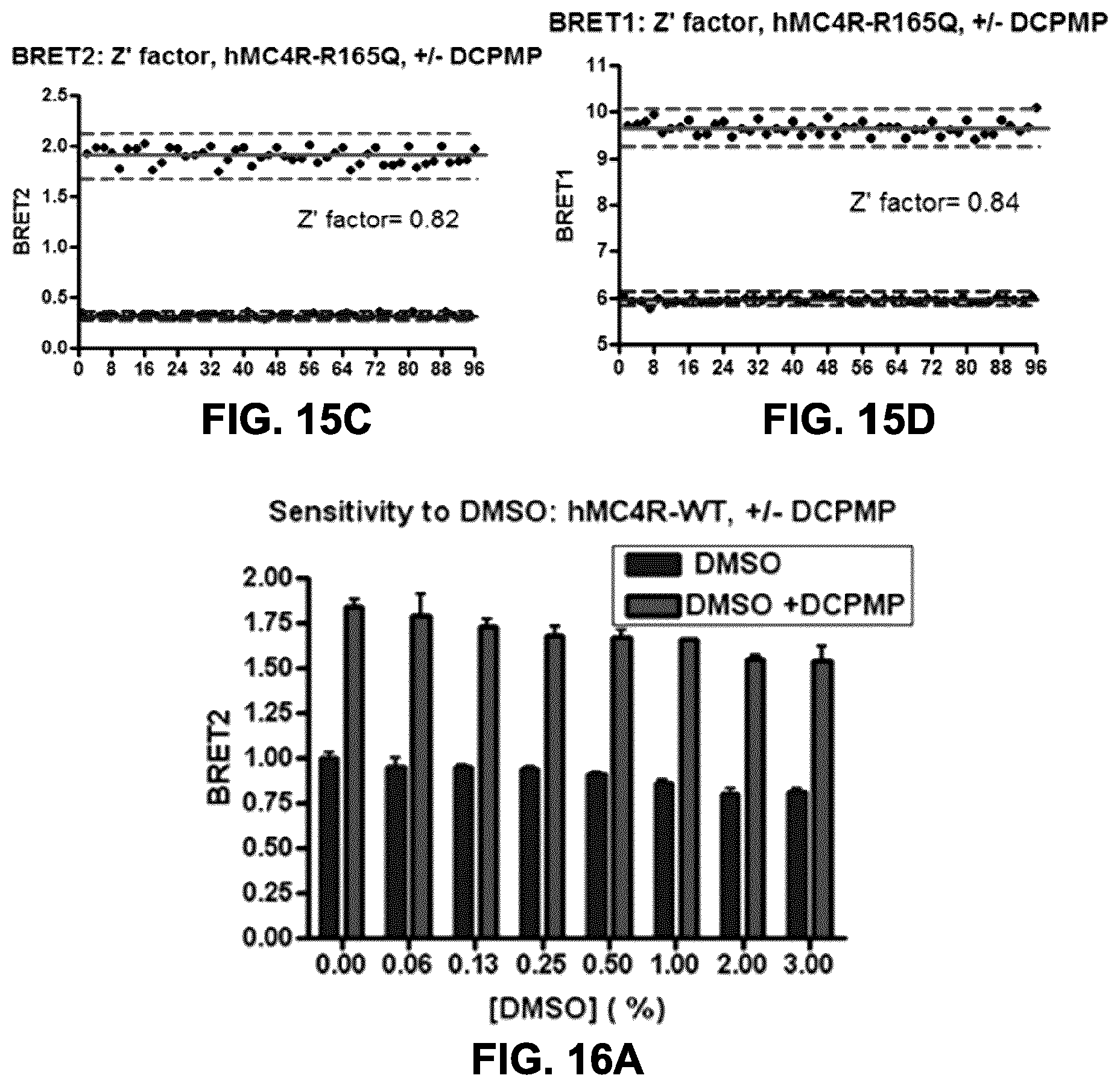
D00015
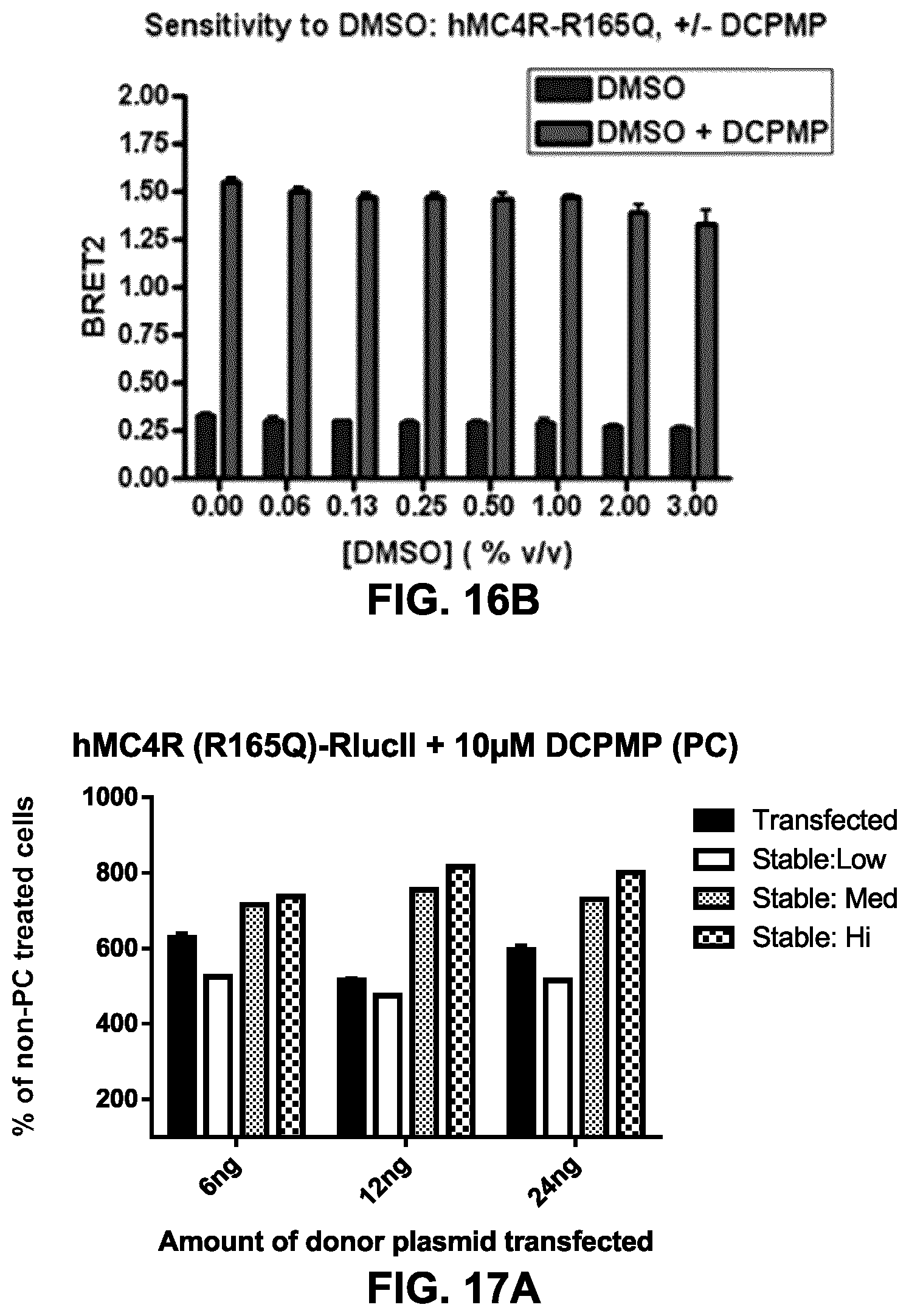
D00016
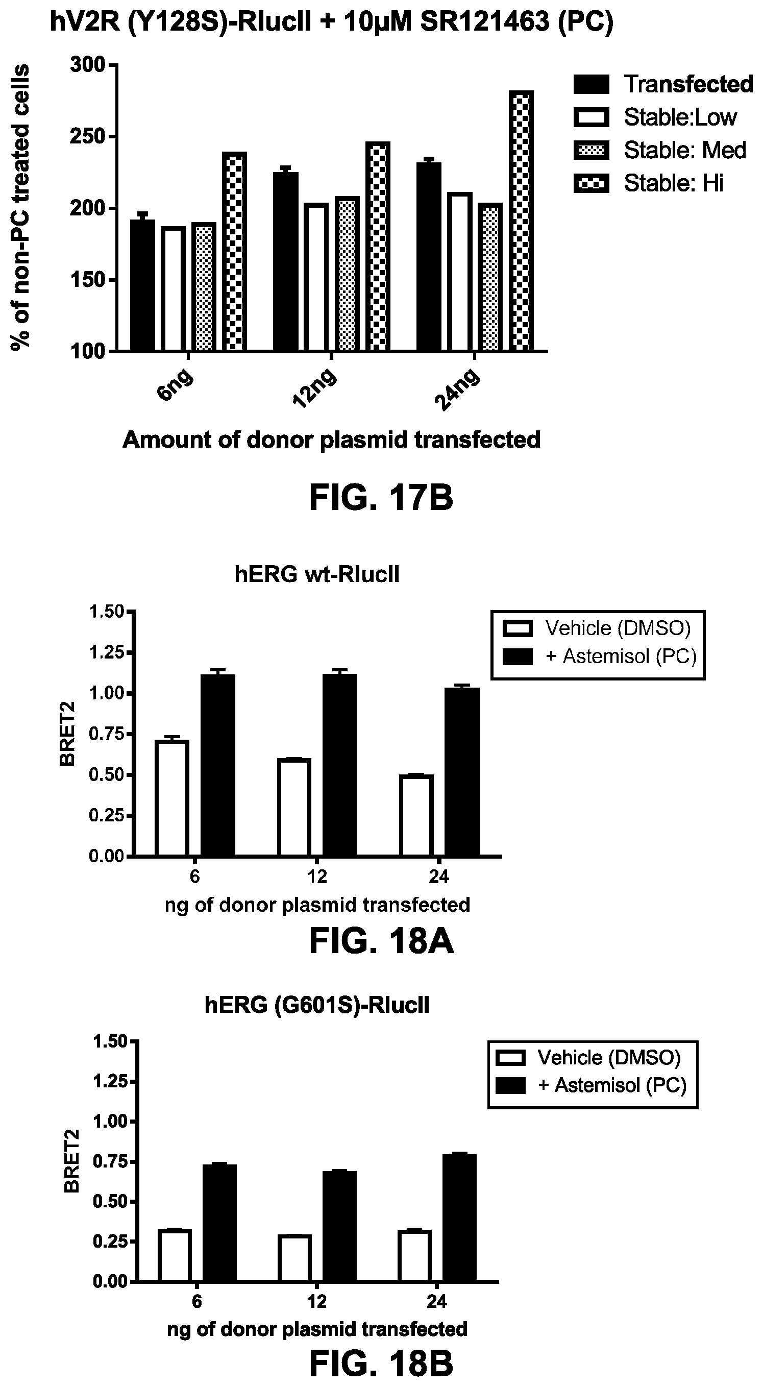
D00017

D00018

D00019

D00020

D00021
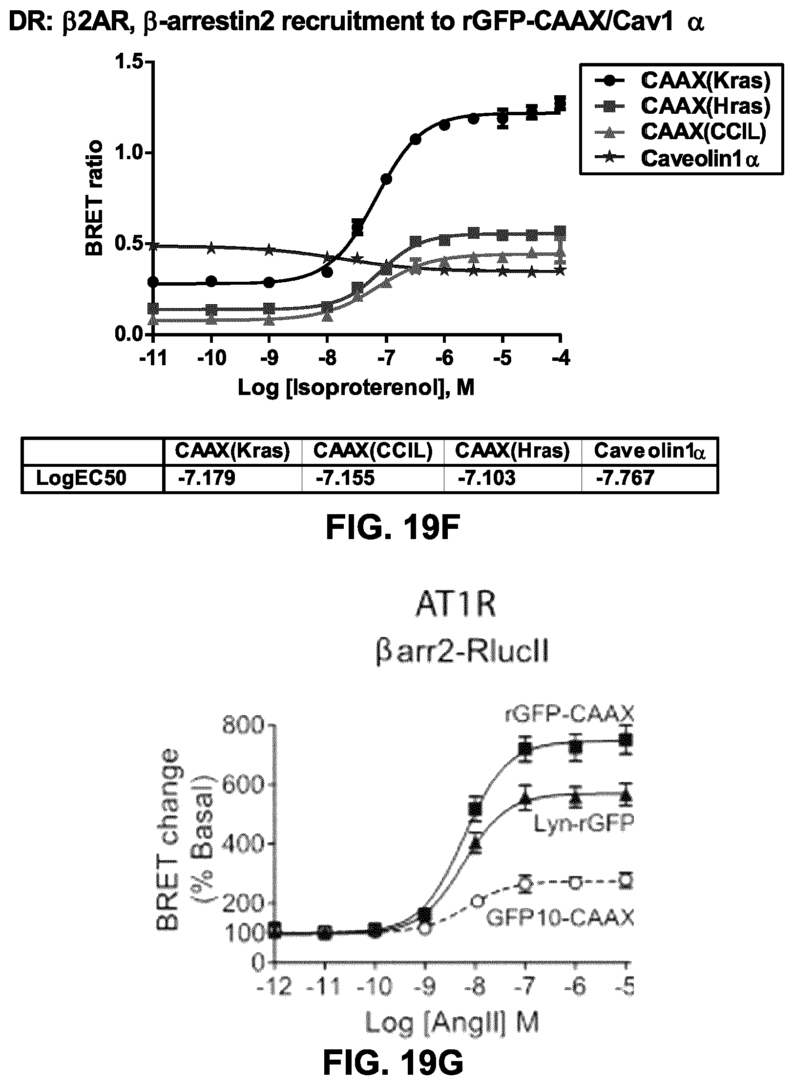
D00022
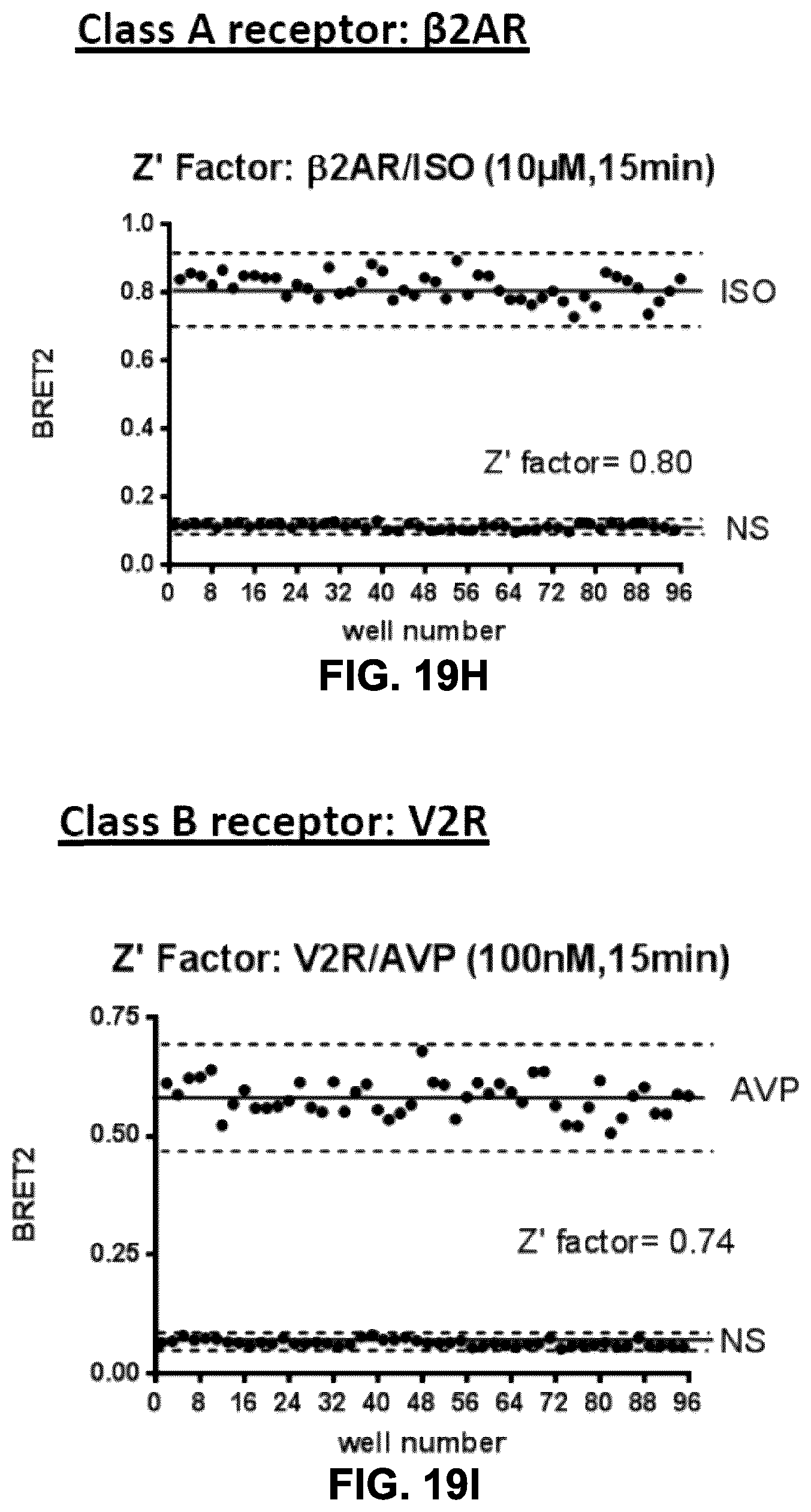
D00023
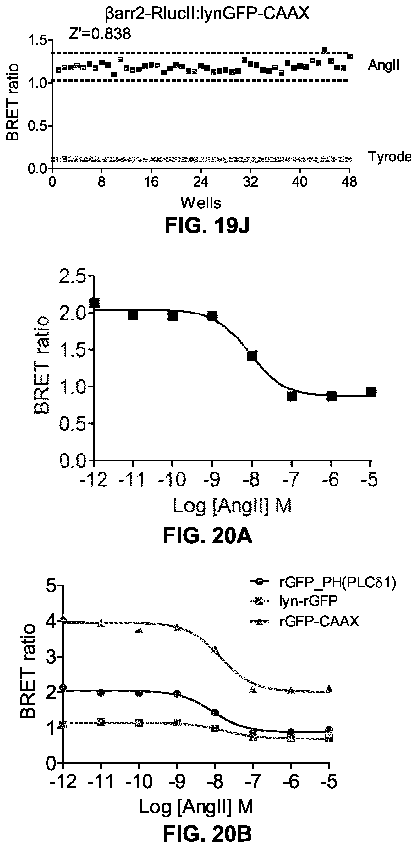
D00024

D00025
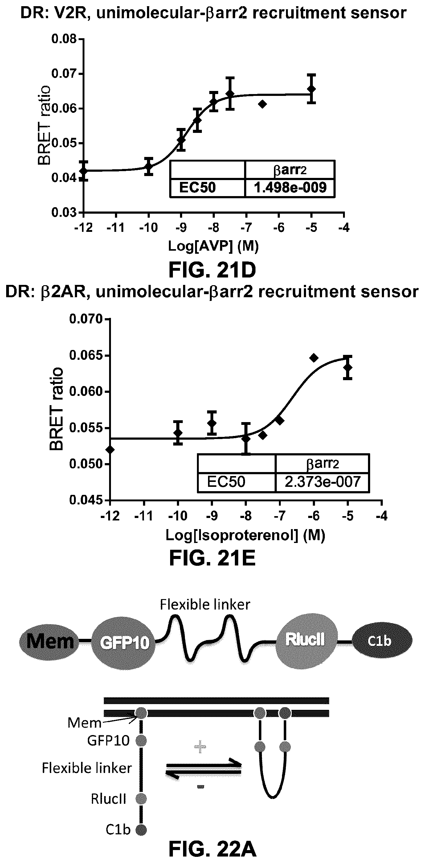
D00026

D00027
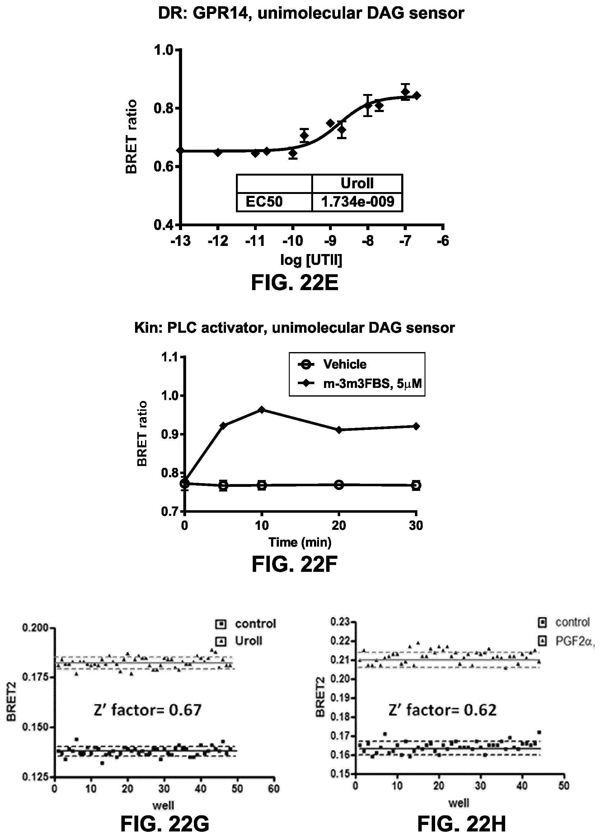
D00028

D00029
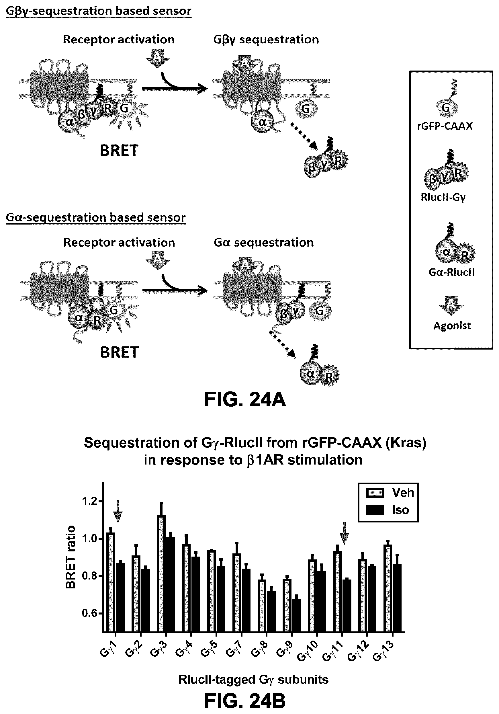
D00030

D00031

D00032

D00033

D00034

D00035

D00036

D00037

D00038
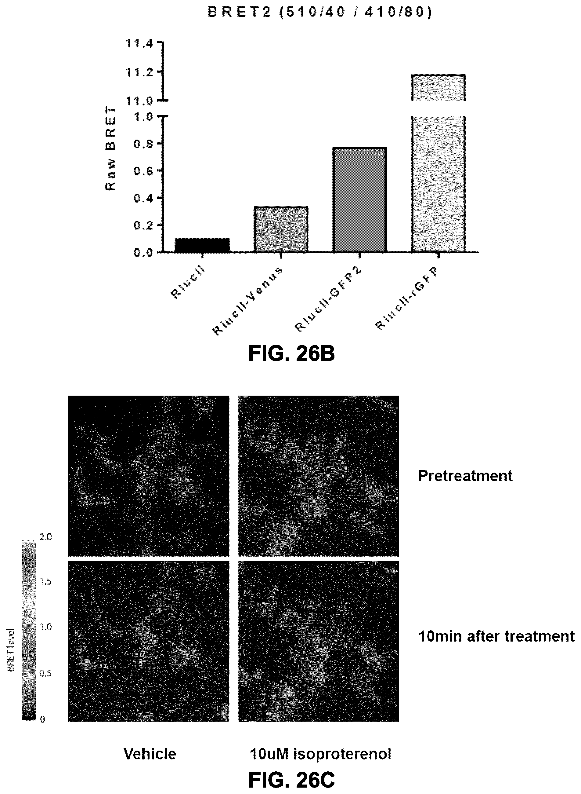
D00039

D00040

S00001
XML
uspto.report is an independent third-party trademark research tool that is not affiliated, endorsed, or sponsored by the United States Patent and Trademark Office (USPTO) or any other governmental organization. The information provided by uspto.report is based on publicly available data at the time of writing and is intended for informational purposes only.
While we strive to provide accurate and up-to-date information, we do not guarantee the accuracy, completeness, reliability, or suitability of the information displayed on this site. The use of this site is at your own risk. Any reliance you place on such information is therefore strictly at your own risk.
All official trademark data, including owner information, should be verified by visiting the official USPTO website at www.uspto.gov. This site is not intended to replace professional legal advice and should not be used as a substitute for consulting with a legal professional who is knowledgeable about trademark law.