Fluorescent silica-based nanoparticles
Bradbury , et al. Fe
U.S. patent number 10,548,997 [Application Number 15/446,319] was granted by the patent office on 2020-02-04 for fluorescent silica-based nanoparticles. This patent grant is currently assigned to Cornell University, Sloan-Kettering Institute for Cancer Research. The grantee listed for this patent is Cornell University, Sloan-Kettering Institute for Cancer Research. Invention is credited to Michelle S. Bradbury, Andrew Burns, Steven M. Larson, Jason S. Lewis, Oula Penate Medina, Hooisweng Ow, Ulrich Wiesner.


View All Diagrams
| United States Patent | 10,548,997 |
| Bradbury , et al. | February 4, 2020 |
Fluorescent silica-based nanoparticles
Abstract
The present invention provides a fluorescent silica-based nanoparticle that allows for precise detection, characterization, monitoring and treatment of a disease such as cancer. The nanoparticle has a range of diameters including between about 0.1 nm and about 100 nm, between about 0.5 nm and about 50 nm, between about 1 nm and about 25 nm, between about 1 nm and about 15 nm, or between about 1 nm and about 8 nm. The nanoparticle has a fluorescent compound positioned within the nanoparticle, and has greater brightness and fluorescent quantum yield than the free fluorescent compound. The nanoparticle also exhibits high biostability and biocompatibility. To facilitate efficient urinary excretion of the nanoparticle, it may be coated with an organic polymer, such as poly(ethylene glycol) (PEG). The small size of the nanoparticle, the silica base and the organic polymer coating minimizes the toxicity of the nanoparticle when administered in vivo. In order to target a specific cell type, the nanoparticle may further be conjugated to a ligand, which is capable of binding to a cellular component associated with the specific cell type, such as a tumor marker. In one embodiment, a therapeutic agent may be attached to the nanoparticle. To permit the nanoparticle to be detectable by not only optical fluorescence imaging, but also other imaging techniques, such as positron emission tomography (PET), single photon emission computed tomography (SPECT), computerized tomography (CT), bioluminescence imaging, and magnetic resonance imaging (MRI), radionuclides/radiometals or paramagnetic ions may be conjugated to the nanoparticle.
| Inventors: | Bradbury; Michelle S. (New York, NY), Wiesner; Ulrich (Ithaca, NY), Medina; Oula Penate (Kiel, DE), Ow; Hooisweng (Woburn, MA), Burns; Andrew (Niskayuna, NY), Lewis; Jason S. (New York, NY), Larson; Steven M. (New York, NY) | ||||||||||
|---|---|---|---|---|---|---|---|---|---|---|---|
| Applicant: |
|
||||||||||
| Assignee: | Sloan-Kettering Institute for
Cancer Research (New York, NY) Cornell University (Ithaca, NY) |
||||||||||
| Family ID: | 43411494 | ||||||||||
| Appl. No.: | 15/446,319 | ||||||||||
| Filed: | March 1, 2017 |
Prior Publication Data
| Document Identifier | Publication Date | |
|---|---|---|
| US 20170239378 A1 | Aug 24, 2017 | |
Related U.S. Patent Documents
| Application Number | Filing Date | Patent Number | Issue Date | ||
|---|---|---|---|---|---|
| 13381209 | 9625456 | ||||
| PCT/US2010/040994 | Jul 2, 2010 | ||||
| 61312827 | Mar 11, 2010 | ||||
| 61222851 | Jul 2, 2009 | ||||
| Current U.S. Class: | 1/1 |
| Current CPC Class: | A61P 31/04 (20180101); A61K 51/08 (20130101); G01N 33/54346 (20130101); B82Y 5/00 (20130101); B82Y 15/00 (20130101); G01N 33/582 (20130101); A61K 49/0019 (20130101); G01N 33/587 (20130101); A61K 51/086 (20130101); A61K 51/082 (20130101); A61P 29/00 (20180101); A61K 51/1244 (20130101); A61K 9/5169 (20130101); A61K 49/0093 (20130101); A61P 31/00 (20180101); A61P 37/06 (20180101); A61P 35/00 (20180101); A61K 9/5115 (20130101); A61P 7/02 (20180101); A61K 49/0054 (20130101); G01N 33/552 (20130101); A61K 9/5146 (20130101); A61P 39/06 (20180101); A61K 51/1255 (20130101); G01N 33/60 (20130101); A61K 49/0056 (20130101); A61K 49/0032 (20130101); G01N 33/574 (20130101); A61P 9/08 (20180101); Y10S 977/773 (20130101); Y10S 977/927 (20130101) |
| Current International Class: | A61K 51/12 (20060101); G01N 33/58 (20060101); A61K 9/51 (20060101); G01N 33/552 (20060101); G01N 33/574 (20060101); G01N 33/60 (20060101); G01N 33/543 (20060101); B82Y 15/00 (20110101); B82Y 5/00 (20110101); A61K 49/00 (20060101); A61K 51/08 (20060101) |
References Cited [Referenced By]
U.S. Patent Documents
| 6254852 | July 2001 | Glajch et al. |
| 6268222 | July 2001 | Chandler et al. |
| 8239007 | August 2012 | Voegele et al. |
| 8298677 | October 2012 | Wiesner et al. |
| 8389679 | March 2013 | Eckert et al. |
| 8409876 | April 2013 | Wiesner et al. |
| 10111963 | October 2018 | Yoo et al. |
| 2003/0219785 | November 2003 | Hallahan et al. |
| 2004/0101822 | May 2004 | Wiesner et al. |
| 2004/0248856 | December 2004 | Lanza et al. |
| 2006/0106306 | May 2006 | Essner et al. |
| 2006/0173362 | August 2006 | Toms et al. |
| 2006/0183246 | August 2006 | Wiesner et al. |
| 2006/0251726 | November 2006 | Lin et al. |
| 2008/0097225 | April 2008 | Tearney et al. |
| 2008/0139787 | June 2008 | De Jesus et al. |
| 2008/0213377 | September 2008 | Bhatia et al. |
| 2008/0292556 | November 2008 | Texier-Nogues et al. |
| 2010/0262017 | October 2010 | Frangioni |
| 2011/0028662 | February 2011 | Wiesner et al. |
| 2012/0107237 | May 2012 | Miao |
| 2013/0017265 | January 2013 | Farokhzad et al. |
| 2013/0039848 | February 2013 | Bradbury et al. |
| 2014/0248210 | September 2014 | Bradbury et al. |
| 2015/0174268 | June 2015 | Li |
| 2015/0343091 | December 2015 | Yoo et al. |
| 2016/0202185 | July 2016 | Zhuang et al. |
| 2018/0169264 | June 2018 | Bradbury et al. |
| 2018/0326103 | November 2018 | Bradbury et al. |
| WO-2006/099445 | Sep 2006 | WO | |||
| WO-2007/002540 | Jan 2007 | WO | |||
| WO-2007/149062 | Dec 2007 | WO | |||
| WO-2008/044138 | Apr 2008 | WO | |||
| WO-2008/142571 | Nov 2008 | WO | |||
| WO-2009/029870 | Mar 2009 | WO | |||
| WO-2009/064964 | May 2009 | WO | |||
| WO-2011/084620 | Jul 2011 | WO | |||
| WO-2011/130598 | Oct 2011 | WO | |||
| WO-2013/192609 | Dec 2013 | WO | |||
| WO-2014/011973 | Jan 2014 | WO | |||
| WO-2016/196201 | Dec 2016 | WO | |||
| WO-2018/237253 | Dec 2018 | WO | |||
Other References
|
Ren, G. et al., PET of Malignant Melanoma Using .sup.18F-Labeled Metallopeptides, The Journal of Nuclear Medicine, 50(11):1865-1872 (2009). cited by applicant . Benezra, M. et al., Targeted multimodal silica nanoparticles with efficient urinary excretion for nanomedicine, Cancer Research, 64(7), one page (2009). cited by applicant . Detappe, A. et al., Advanced multimodal nanoparticles delay tumor progression with clinical radiation therapy, Journal of Controlled Release, 238:103-113 (2016). cited by applicant . Fuller, , J. E. et al., Intracellular delivery of core--shell fluorescent silica nanoparticles, Science Direct, Biomaterials, 29:1526-1532 (2008). cited by applicant . Rianasari, I. et al., Covalent Coupling of Nanoparticles with Low-Density Functional Ligands to Surface via Click Chemistry, Int. J. Mol. Sci. 14:3705-3717 (2013). cited by applicant . Sharma, P. et al, Nanoparticles of bioimaging, Advances in Colloid and Interface Science, 123-126:471-485 (2006). cited by applicant . Van Schooneveld, M. M. et al., Improved Biocompatibility and Pharmacokinetics of Silica Nanoparticles by Means of a Lipid Coating: A Multimodality Investigation, Nano Letters, 8(8):2517-2525 (2008). cited by applicant . Ballou, B. et al., Sentinel Lymph Node Imaging Using Quantum Dots in Mouse Tumor Models, Bioconjugate Chem. 18:389-396 (2007). cited by applicant . Bogush, G. H. et al., Preparation of Monodisperse Silica Particles: Control of Size and Mass Fraction, J. Non-Cryst. Solids, 104:95-106 (1988). cited by applicant . Brien, J. F. et al., A Study of the Calcium Carbimide-Ethanol Interaction in Man, Europ. J. Clin. Pharmacol. 14(2):133-41 (1978). cited by applicant . Burns, et al., Fluorescent Silica Nanoparticles with Efficient Urinary Excretion for Nanomedicine, Nano Letters 9(1):442-8 (2009). cited by applicant . Chakraborty, M. et al., External Beam Radiation of Tumors Alters Phenotype of Tumor Cells to Render Them Susceptible to Vaccine-Mediated T-Cell Killing, Cancer Research, 64:4328-4337 (2004). cited by applicant . Crespi, M. D. et al., Mitroxantrone Affects Topoisomerase Activities In Human Breast Cancer Cells, Biochemical and Biophysical Research Communications, 136(2):521-8 (1986). cited by applicant . Cressman, S. et al., Binding and Uptake of RGD-Containing Ligands to Cellular .sup..chi.v.sup..beta.3 Integrins, Int J Pept Res Ther, 15:49-59 (2009). cited by applicant . Cristy, M. and Eckerman, K. F., Specific absorbed fractions of energy at various ages from internal photon sources (I-VII). Oak Ridge National Laboratory Report ORNL/TM-8381N1-7. Springfield, VA: National Technical Information Service, Dept. of Commerce (1987). cited by applicant . Crow, R. T. and Crothers, D. M., Inhibition of Topoisomerase I by Anthracycline Antibiotics: Evidence for General Inhibition of Topoisomerase I by DNA-Binding Agents, J. Med. Chem. 37(19):3191-3194 (1994). cited by applicant . De Jong, M. et al., Comparison of .sup.111In-Labeled Somatostatin Analogues for Tumor Scintigraphy and Radionuclide Therapy, Cancer Res., 58:437-41 (1998). cited by applicant . De Jong, M. et al., Internalization of radiolabelled [DTPA.sup.0]octreotide and [DOTA0,Tyr.sup.3]ocetreotide:peptides for somatostatin receptor-targeted scintigraphy and radionuclide therapy, Nucl. Med. Commun., 19(3):283-288 (1998). cited by applicant . Denny, W. A. and Baguley, B. C., Dual Topoisomerase I/II Inhibitors in Cancer Therapy, Curr. Top. Med. Chem., 3(3):339-353 (2003). cited by applicant . Ding, Y. et al., The performance of thiol-terminated PEG-paclitaxel-conjugated gold nanoparticles, Biomaterials, 34:10217-10227 (2013). cited by applicant . Doronina, S. O. et al., Novel Peptide Linkers for Highly Potent Antibody Auristatin Conjugate, Bioconjugate Chem., 19(10):1960-1963, (2008). cited by applicant . Foglesong, P. D. et al., Doxorubicin inhibits human DNA topoisomerase I, Cancer Chemother. Pharmacol., 30(2):123-125 (1992). cited by applicant . Frauwirth, K. A. and Thompson, C. B., Activation and inhibition of lymphocytes by costimulation, The Journal of clinical Investigation, 109(3):295-299 (2002). cited by applicant . Gatto, B. et al., Identification of Topoisomerase I as the Cytotoxic Target of the Protoberberine Alkaloid Coralyne, Cancer Res., 15(12):2795-2800 (1996). cited by applicant . Gladson, C. A. and Cheresh, D. A., Glioblastoma Expression of Vitronectin and Alpha v Beta 3 Integrin, Adhesion Mechanism for Transformed Glial Cells, J. Clin. Invest. 88:1924-1932 (1991). cited by applicant . Herz, E. et al., Large Stokes-Shift Fluorescent Silica Nanoparticles with Enhanced Emission over Free Dye for Single Excitation Multiplexing, Macromol Rapid Commun., 30(22):1907-1910 (2009). cited by applicant . Hilderbrand, S. A. and Weissleder, R., Near-infrared fluorescence: application to in vivo molecular imaging, Curr. Opin. Chem. Bioi., 14:71-9 (2010). cited by applicant . International Search Report, PCT/US2010/040994, dated Aug. 30, 2010. cited by applicant . International Search Report, PCT/US2015/032565, 4 pages, dated Aug. 21, 2015. cited by applicant . Kalbasi, A. et al., Radiation and immunotherapy: a synergistic combination, Clinical review, The Journal of Clinical Investigation, 127(7):2756-2763 (2013). cited by applicant . Kim, S. et al., Near-infrared fluorescent type II quantum dots for sentinel lymph node mapping, Nature Biotechnology 22(1):93-97 (2004). cited by applicant . Kim, Y. H. et al., In situ vaccination against mycosis fungoides by intratumoral injection of a TLR9 agonist combined with radiation: a phase 1/2 study, Blood, 119(2):355-363 (2012). cited by applicant . Koole et al., Paramagnetic lipid-coated silica nanoparticles with a fluorescent quantum dot core: a new contrast agent platform for multimodality imaging, Bioconjugate Chem., 19(12):2471-2479 (2008). cited by applicant . Krenning, E. P. et al, Somatostatin Receptor Scintigraphy with Indium-111-DTPA-D-Phe-1-Octreolide in Man: Metabolism, Dosimetry and Comparison with Iodine-123-Tyr-3-Octreotide, J Nucl. Med. 33:652-8 (1992). cited by applicant . Larson, D. R. et al., Silica Nanoparticle Architecture Determines Radiative Properties of Encapsulated Fluorophores, Chem. Mater. 20:2677-2684 (2008). cited by applicant . Lewis et al. Comparison of Four 64Cu-labeled Somatostatin Analogs in Vitro and in a Tumor-Bearing Rat Model: Evaluation of New Derivatives for Positron Emission Imaging and Targeted Radiotherapy. J Med Chem., 42:1341-7 (1999). cited by applicant . Li, T. et al., Human Topoisomerase I Poisoning by Protoberberines: Potential Roles for Both Drug-DNA and Drug-Enzyme Interactions, Biochemistry, 39(24):7107-7116 (2000). cited by applicant . Li, Z. et al., .sup.64 Cu-labeled Tetrameric and Octomeric RGD Peptides for Small-Animal PET of Tumor .alpha..sub.v.sup..beta..sub.3 Integrin Expression, J. Nucl Med. 48:1162-1171 (2007). cited by applicant . Loir, B. et al., Expression of the MC1 Receptor Gene in Normal and Malignant Human Melanocytes. A Semiquantitative RT-PCR Study, Cell Mol. Biol., 45(7):1083-1092 (1999). cited by applicant . Makhey et al., Sbustitute Benzo[i]phenanthridines as Mammalian Topoisomerase-Targeting Agents, Bioorg. Med. Chem. 11(8):1809-1820 (2003). cited by applicant . McKeage et al., Phase I and Pharmacokinetic Study of an Oral Platinum Complex Given Daily for 5 Days in Patients With Cancer, Journal of Clinical Oncology, 15(7):2691-2700 (1997). cited by applicant . Montet, X. et al., Multivalent Effects of RGD Peptides Obtained by Nanoparticle Display, J. Med. Chem. 49:6087-6093 (2006). cited by applicant . Ohnishi, S. et al., Organic Alternatives to Quantum Dots for Intraoperative Near-Infrared Fluorescent Sentinel Lymph Node Mapping, Molecular Imaging 4(3):172-181 (2005). cited by applicant . Ow, H. et al., Bright and Stable Core-Shell Fluorescent Silica Nanoparticles, Nano Letters, 5(1):113-117 (2005). cited by applicant . Papamicheal, D., The Use of Thymidylate Synthase Inhibitors in the Treatment of Advanced Colorectal Cancer: Current Status, The Oncologist, 4:478-487 (1999). cited by applicant . Patel, K. N. et al., MUC1 plays a role in tumor maintenance in aggressive thryroid carcinomas, Surgery 138(6):994-1002 (2005). cited by applicant . Piatyszek, M.A. et al., Iodo-Gen-Mediated Radioiodination of Nucleic Acids, J. Anal. Biochem. 172(2):356-359 (1988). cited by applicant . Pommier, Y., Topoisomerase I inhibitors: camptothecins and beyond, Nat. Rev. Cancer, 6(10):789-802 (2006). cited by applicant . Prosecution File History of Chinese Application 201080039307.2 as of Oct. 5, 2016, 54 pages. cited by applicant . Prosecution File History of European Application No. 10 794 842.4 as of Jul. 29, 2016, 30 pages. cited by applicant . Reubi, J.C. et al., Distribution of Somatostatin Receptors in Normal and Tumor Tissue, Metabolism, 39(9)(2):78-81 (1990). cited by applicant . Reubi, J.C. et al., Somatostatin Receptors and Their Subtypes in Human Tumors and in Peritumoral Vessels, Metabolism, 45(8)(1):39-41 (1996). cited by applicant . Ruoslahti, E. and Pierschbacher, M. D., New Perspectives in Cell Adhesion: RGD and Integrins, Science 238:491 (1987). cited by applicant . Sadasivan, et al., Alcoholic Solvent Effect on Silica Synthesis--NMR and DLS Investigation, J. Sol-Gel Science and Technology, 12:5-14 (1998). cited by applicant . Seftor, R. E. B. et al., Role of the alpha v beta 3 integrin in human melanoma cell invasion, Proc. Natl. Acad. Sci., 89:1557-1561 (1992). cited by applicant . Seung, S. K. et al., Phase 1 Study of Stereotactic Body Radiotherapy and Interleukin-2: Tumor and Immunological Responses, Science Translational Medicine 14(137):137ra74 1-7 (2012). cited by applicant . Seymour, L.W., Passive Tumor Targeting of Soluble Macromolecules and Drug Conjugates, Critical Reviews in Therapeutic Drug Carrier Systems, 9(2):135-187 (1992). cited by applicant . Slowing, I. I. et al., Mesoporous silica nanoparticles as controlled release drug delivery and gene transfection carriers, Advanced Drug Delivery Reviews, 60:1278-1288 (2008). cited by applicant . Stabin, M. G. et al., OLINDA/EXM: The Second-Generation Personal Computer Software for Internal Dose Assessment in Nuclear Medicine, J Nucl Med. 46:1023-1027 (2005). cited by applicant . Takeshima, T. et al., Local Radiation Therapy Inhibits Tumor Growth through the Generation of Tumor-Specific CTL: Its Potentiation by Combination with Th1 Cell Therapy, Cancer Research, 70(7):2697-2706 (2010). cited by applicant . Tanaka, E. et al, Image-Guided Oncologic Surgery Using Invisible Light: Completed Pre-Clinical Development for Sentinel Lymph Node Mapping, Annals of Surgical Oncology 13(12):1671-1681 (2006). cited by applicant . Topalian, S. L. et al., Safety, Activity, and Immune Correlates of Anti-PD-1 Antibody in Cancer, The New England Journal of Medicine, 366(26):2443-2454 (2012). cited by applicant . Vejayakumaran, P. et al., Structural and thermal characterizations of silica nanoparticles grafted with pendant maleimide and epoxide grops, Journal of Colloid and Interface Science, 328:81-91 (2008). cited by applicant . Wang, Y. et al., Tumor cell targeted delivery by specific peptide-modified mesoporous silica nanoparticles, J. Mater. Chem., 22:14608-14616, (2012). cited by applicant . Webb, et al., Sphingomyelin-cholesterol liposomes significantly enhance the pharmacokinetic and therapeutic properties of vincristine in murine and human tumour models, British J. of Cancer 72:896-904 (1995). cited by applicant . Webster, A. et al., Optical calcium sensors: development of a generic method for their introduction to the cell using conjugated cell penetrating peptides, Analyst, 130:163-70 (2005). cited by applicant . Wersall, P.J. et al., Regression of non-irradiated metastases after extracranial stereotactic radiotherapy in metastatic renal cell carcinoma, Acta Oncologica, 45:493-497 (2006). cited by applicant . Written Opinion, PCT/US2010/040994, dated Aug. 30, 2010. cited by applicant . Written Opinion, PCT/US2015/032565, 6 pages, dated Aug. 21, 2015. cited by applicant . Xu, Z. et al., DNA Minor Groove Biding-Directed Poisoning of Human DNA Topoisomerase I by Terbenzimidazoles, Biochemistry 37(10):3558-3566 (1998). cited by applicant . Zeng, J. et al., Anti-PD-1 Blockade and Stereotactic Radiation Produce Long-Term Survival in Mice With Intracranial Gliomas, Intl. J. Radiation Oncol. Biol. Phys., 86(2):343-349 (2013). cited by applicant . Zhong, Y. J. et al., Cathepsin B-cleavable doxorubicin prodrugs for targeted cancer therapy (Review), International Journal of Oncology, 42:373-383, (2013). cited by applicant . Benezra, M. et al., Multimodal silica nanoparticles are effective cancer-targeted probes in a model of human melanoma, Journal of Clinical Investigation, 121(7):2768-2780 (2011). cited by applicant . Benezra, M. et al., Ultrasmall Integrin-Targeted Silica Nanoparticles Modulate Signaling Events and Cellular Processes in a Concentration-Dependent Manner, Small, 11(14):1721-1732 (2015). cited by applicant . Dixon, S. J. et al., Ferroptosis: An Iron-Dependent Form of Nonapoptotic Cell Death, Cell, 149(51):1060-1072 (2012). cited by applicant . Kim, D. et al., Antitumor activity of sorafenib-incorporated nanoparticles of dextran/poly (dl-lactide-co-glycolide) block copolymer, Nanoscale Research Letters, 7(1):91 (2012). cited by applicant . Kim, S. E. et al., Ultrasmall nanoparticles induce ferroptosis in nutrient-deprived cancer cells and suppress tumour growth, Nature Nanotechnology, 11(11):977-985, (2016). cited by applicant . Lachaier, E. et al., Sorafenib Induces Ferroptosis in Human Cancer Cell Lines Originating from Different Solid Tumors, Anticancer Research, 34:6417-6422 (2014). cited by applicant . Mayer, R. J. et al., Randomized Trial of TAS-102 for Refractory Metastatic Colorectal Cancer, NEJM, 372(20):1909-1919 (2015). cited by applicant . Phillips, E. et al., Clinical translation of an ultrasmall inorganic optical-PET imaging nanoparticle probe, Science Translational Medicine, 6(260):260ra149-260ra149 (2014). cited by applicant . Tavernaro, I. et al., Bright Fluorescent silica-nanoparticle probes for high-resolution STED and confocal microscopy, Beilstein Journal of Nanotechnology, 8:1283-1296, (2017). cited by applicant . Zhang, X. L. et al., Ultrasmall 1-6, radioiodinated alpha MSH-C dots for melanoma imaging and therapy, Journal of Labelled Compounds and Radiopharmeceuticals, 58(1):5114 (2015). cited by applicant . Zhen, C. et al., Radioiodination of Rhenium Cyclized .alpha.-Melanocyte-Stimulating Hormone Resulting in Enhanced Radioactivity Localization and Retention in Melanoma, Cancer Research, 64:1411-1418, (2004). cited by applicant . Gerion, D. et al., Enhancement of T1 and T2 relaxation by paramagnetic silica-coated nanocrystals, UCRL-JRNL-224783, 14 pages, (2006). cited by applicant. |
Primary Examiner: Cabral; Robert S
Attorney, Agent or Firm: Choate, Hall & Stewart LLP Haulbrook; William R. Monroe; Margo R.
Government Interests
STATEMENT REGARDING FEDERALLY SPONSORED RESEARCH OR DEVELOPMENT
This invention was made with government support under grant numbers CA086438, CA083084, CA008748, and RR024996 awarded by National Institutes of Health. The government has certain rights in the invention.
Parent Case Text
CROSS REFERENCE TO RELATED APPLICATIONS
This application is a continuation of U.S. application Ser. No. 13/381,209, filed Sep. 27, 2012, which is a US national phase entry of International Application No. PCT/US10/40994, filed Jul. 2, 2010, which claims priority to US Provisional Application Nos. 61/312,827, filed Mar. 11, 2010, and 61/222,851, filed Jul. 2, 2009, the contents of which are hereby incorporated by reference in their entireties.
Claims
What is claimed is:
1. A composition comprising: a nanoparticle comprising a silica-based core and a silica shell surrounding at least a portion of the silica-based core; an organic polymer attached to the nanoparticle, thereby coating the nanoparticle; and a plurality of alpha-MSH peptide ligands attached to the polymer-coated nanoparticle, wherein the polymer-coated nanoparticle has a diameter from 1 nm to 8 nm.
2. The composition of claim 1, wherein the plurality of alpha-MSH peptide ligands is no greater than twenty in number.
3. The composition of claim 1, further comprising: a fluorescent compound within the silica-based core.
4. The composition of claim 1, wherein the organic polymer comprises polyethylene glycol.
5. The composition of claim 4, wherein the polyethylene glycol is attached to a silica surface of the nanoparticle via an amino-silane coupled to an activated ester group on the organic polymer leading to an amide bond.
6. The composition of claim 5, wherein the nanoparticle is coated with maleimido-terminated polyethylene glycol chains for attachment of the plurality of alpha-MSH peptide ligands.
7. The composition of claim 1, wherein the plurality of alpha-MSH peptide ligands is no greater than ten in number.
8. The composition of claim 1, wherein the nanoparticle is coated with maleimido-terminated polyethylene glycol chains for attachment of the plurality of alpha-MSH peptide ligands, and wherein at least one alpha-MSH peptide ligand is attached to a maleimido-terminated polyethylene glycol chain via a thiol group of a cysteine linker.
9. The composition of claim 1, wherein the plurality of alpha-MSH peptide ligands is labeled with a radionuclide.
10. The composition of claim 9, wherein the plurality of alpha-MSH peptide ligands is labeled with the radionuclide via a tyrosine (Y) linker.
11. The composition of claim 1, further comprising a therapeutic agent.
12. The composition of claim 3, wherein the fluorescent compound is Cy5.
13. The composition of claim 3, wherein the fluorescent compound is Cy5.5.
Description
FIELD OF THE INVENTION
The present invention relates to fluorescent silica-based nanoparticles, and methods of using the nanoparticles to detect, diagnose, or treat diseases such as cancer.
BACKGROUND OF THE INVENTION
Early tumor detection and treatment selection is paramount to achieving therapeutic success and long-term survival rates. At its early stage, many cancers are localized and can be treated surgically. However, well-defined tumor margins are often difficult to visualize with current imaging techniques. This has led to a disproportionate number of invasive biopsies. Highly-specific, molecular-targeted probes are needed for the early detection of molecular differences between normal and tumor cells, such as cancer-specific alterations in receptor expression levels. When combined with high-resolution imaging techniques, specific molecular-targeted probes will greatly improve detection sensitivity, facilitating characterization, monitoring and treatment of cancer.
Current fluorescence imaging probes typically consist of single conventional fluorophore (e.g., organic dyes, fluorescent proteins), fluorescent proteins (e.g., GFP) and semiconductor quantum dots (Q-dots). Single fluorophores are usually not stable and have limited brightness for imaging. Similar to dyes, the fluorescent proteins tend to exhibit excited state interactions which can lead to stochastic blinking, quenching and photobleaching. Q-dots are generally made from heavy metal ions such as Pb.sup.2+ or Cd.sup.2+ and, therefore, are toxic. Burns et al. "Fluorescent core-shell silica nanoparticles: towards "Lab on a Particle" architectures for nanobiotechnology", Chem. Soc. Rev., 2006, 35, 1028-1042.
Fluorescent nanoparticles having an electrically conducting shell and a silica core are known and have utility in modulated delivery of a therapeutic agent. U.S. Pat. Nos. 6,344,272, and 6,428,811. A shortcoming of existing fluorescent nanoparticles is their limited brightness and their low detectability as fluorescent probes in dispersed systems.
The present multifunctional fluorescent silica-based nanoparticles offer many advantages over other fluorescent probes. The nanoparticles are non-toxic, and have excellent photophysical properties (including fluorescent efficiency and photostability), high biocompatibility, and unique pharmacokinetics for molecular diagnostics and therapeutics. The nanoparticles are relatively small in size, and have a surface PEG coating that offers excellent renal clearance. The fluorescent nanoparticles of the present invention contain a fluorescent core and silica shell. The core-shell architectures, the great surface area and diverse surface chemistry of the nanoparticle permit multiple functionalities simultaneously delivered to a target cell. For example, the nanoparticle can be functionalized with targeting moieties, contrast agents for medical imaging, therapeutic agents, or other agents. The targeting moieties on the surface of the nanoparticle may be tumor ligands, which, when combined with nanoparticle-conjugated therapeutic agents, makes the nanoparticle an ideal vehicle for targeting and potentially treating cancer. Webster et al. Optical calcium sensors: development of a generic method for their introduction to the cell using conjugated cell penetrating peptides. Analyst, 2005; 130:163-70. The silica-based nanoparticle may be labeled with contrast agents for PET, SPECT, CT, MRI, and optical imaging.
SUMMARY OF THE INVENTION
It is an object of the present invention to provide a fluorescent silica-based nanoparticle comprising a silica-based core having a fluorescent compound positioned within the silica-based core; a silica shell surrounding at least a portion of the core; an organic polymer attached to the nanoparticle; from about 1 to about 20 ligands attached to the nanoparticle; and a contrast agent or a chelate attached to the nanoparticle. The diameter of the nanoparticle ranges from about 1 nm to about 25 nm, or from about 1 nm to about 8 nm. The organic polymers that may be attached to the nanoparticle include poly(ethylene glycol) (PEG), polylactate, polylactic acids, sugars, lipids, polyglutamic acid (PGA), polyglycolic acid, poly(lactic-co-glycolic acid) (PLGA), Polyvinyl acetate (PVA), or the combinations thereof.
The ligand may be capable of binding to at least one cellular component, such as a tumor marker. The number of ligands attached to the nanoparticle may also range from about 1 to about 10. Examples of the ligand include peptide, protein, biopolymer, synthetic polymer, antigen, antibody, microorganism, virus, receptor, hapten, enzyme, hormone, chemical compound, pathogen, toxin, surface modifier, or combinations thereof. Peptides such as tripeptide RGD, cyclic peptide cRGD, octreotate, EPPT1 and peptide analogs of alpha-MSH are encompassed by the present invention. Any linear, cyclic or branched peptide containing the RGD sequence is within the scope of the present invention.
A contrast agent, such as a radionuclide including .sup.89Zr, .sup.64Cu, .sup.68Ga, .sup.86Y, .sup.124I and .sup.177Lu, may be attached to the nanoparticle. Alternatively, the nanoparticle is attached to a chelate, for example, DFO, DOTA, TETA and DTPA, that is adapted to bind a radionuclide. The nanoparticle of the present invention may be detected by positron emission tomography (PET), single photon emission computed tomography (SPECT), computerized tomography (CT), magnetic resonance imaging (MRI), optical imaging (such as fluorescence imaging including near-infrared fluorescence (NIRF) imaging), bioluminescence imaging, or combinations thereof.
A therapeutic agent may be attached to the nanoparticle. The therapeutic agents include antibiotics, antimicrobials, antiproliferatives, antineoplastics, antioxidants, endothelial cell growth factors, thrombin inhibitors, immunosuppressants, anti-platelet aggregation agents, collagen synthesis inhibitors, therapeutic antibodies, nitric oxide donors, antisense oligonucleotides, wound healing agents, therapeutic gene transfer constructs, extracellular matrix components, vasodialators, thrombolytics, anti-metabolites, growth factor agonists, antimitotics, statin, steroids, steroidal and non-steroidal anti-inflammatory agents, angiotensin converting enzyme (ACE) inhibitors, free radical scavengers, PPAR-gamma agonists, small interfering RNA (siRNA), microRNA, and anti-cancer chemotherapeutic agents. The therapeutic agents encompassed by the present invention also include radionuclides, for example, .sup.90Y, .sup.131I and .sup.177Lu. The therapeutic agent may be radiolabeled, such as labeled by binding to radiofluorine .sup.18F.
After administration of the nanoparticle to a subject, blood residence half-time of the nanoparticle may range from about 2 hours to about 25 hours, from about 3 hours to about 15 hours, or from about 4 hours to about 10 hours. Tumor residence half-time of the nanoparticle after administration of the nanoparticle to a subject may range from about 5 hours to about 5 days, from about 10 hours to about 4 days, or from about 15 hours to about 3.5 days. The ratio of tumor residence half-time to blood residence half-time of the nanoparticle after administration of the nanoparticle to a subject may range from about 2 to about 30, from about 3 to about 20, or from about 4 to about 15. Renal clearance of the nanoparticle after administration of the nanoparticle to a subject may range from about 10% ID (initial dose) to about 100% ID in about 24 hours, from about 30% ID to about 80% ID in about 24 hours, or from about 40% ID to about 70% ID in about 24 hours. In one embodiment, after the nanoparticle is administered to a subject, blood residence half-time of the nanoparticle ranges from about 2 hours to about 25 hours, tumor residence half-time of the nanoparticle ranges from about 5 hours to about 5 days, and renal clearance of the nanoparticle ranges from about 30% ID to about 80% ID in about 24 hours.
When the nanoparticles in the amount of about 100 times of the human dose equivalent are administered to a subject, substantially no anemia, weight loss, agitation, increased respiration, GI disturbance, abnormal behavior, neurological dysfunction, abnormalities in hematology, abnormalities in clinical chemistries, drug-related lesions in organ pathology, mortality, or combinations thereof, is observed in the subject in about 10 to about 14 days.
Multivalency enhancement of the nanoparticle may range from about 2 fold to about 4 fold.
The present invention also provides a fluorescent silica-based nanoparticle comprising a silica-based core comprising a fluorescent compound positioned within the silica-based core; a silica shell surrounding at least a portion of the core; an organic polymer attached to the nanoparticle; and a ligand attached to the nanoparticle, wherein the nanoparticle has a diameter between about 1 nm and about 15 nm. After administration of the nanoparticle to a subject, blood residence half-time of the nanoparticle ranges from about 2 hours to about 25 hours, tumor residence half-time of the nanoparticle ranges from about 5 hours to about 5 days, and renal clearance of the nanoparticle ranges from about 30% ID to about 80% ID in about 24 hours. The number of ligands attached to the nanoparticle may range from about 1 to about 20, or from about 1 to about 10. The diameter of the nanoparticle may be between about 1 nm and about 8 nm. A contrast agent, such as a radionuclide, may be attached to the nanoparticle. Alternatively, a chelate may be attached to the nanoparticle. The nanoparticle may be detected by PET, SPECT, CT, MRI, optical imaging, bioluminescence imaging, or combinations thereof. A therapeutic agent may be attached to the nanoparticle. After administration of the nanoparticle to a subject, blood residence half-time of the nanoparticle may also range from about 3 hours to about 15 hours, or from about 4 hours to about 10 hours. Tumor residence half-time of the nanoparticle after administration of the nanoparticle to a subject may also range from about 10 hours to about 4 days, or from about 15 hours to about 3.5 days. The ratio of tumor residence half-time to blood residence half-time of the nanoparticle after administration of the nanoparticle to a subject may range from about 2 to about 30, from about 3 to about 20. or from about 4 to about 15. Renal clearance of the nanoparticle may also ranges from about 40% ID to about 70% ID in about 24 hours after administration of the nanoparticle to a subject.
Also provided in the present invention is a fluorescent silica-based nanoparticle comprising a silica-based core comprising a fluorescent compound positioned within the silica-based core; a silica shell surrounding at least a portion of the core; an organic polymer attached to the nanoparticle; and a ligand attached to the nanoparticle, wherein the nanoparticle has a diameter between about 1 nm and about 8 nm. After administration of the nanoparticle to a subject, the ratio of tumor residence half-time to blood residence half-time of the nanoparticle ranges from about 2 to about 30, and renal clearance of the nanoparticle ranges from about 30% ID to about 80% ID in about 24 hours.
The present invention further provides a method for detecting a component of a cell comprising the steps of: (a) contacting the cell with a fluorescent silica-based nanoparticle comprising a silica-based core comprising a fluorescent compound positioned within the silica-based core; a silica shell surrounding at least a portion of the core; an organic polymer attached to the nanoparticle; from about 1 to about 20 ligands attached to the nanoparticle; and a contrast agent or a chelate attached to the nanoparticle; and (b) monitoring the binding of the nanoparticle to the cell or a cellular component by at least one imaging technique.
The present invention further provides a method for targeting a tumor cell comprising administering to a cancer patient an effective amount of a fluorescent silica-based nanoparticle comprising a silica-based core comprising a fluorescent compound positioned within the silica-based core; a silica shell surrounding at least a portion of the core; an organic polymer attached to the nanoparticle; a ligand attached to the nanoparticle and capable of binding a tumor marker; and at least one therapeutic agent. The nanoparticle may be radiolabeled. The nanoparticle may be administered to the patient by, but not restricted to, the following routes: oral, intravenous, nasal, subcutaneous, local, intramuscular or transdermal.
BRIEF DESCRIPTION OF THE DRAWINGS
FIG. 1A shows a dynamic light scattering (DLS) plot (number average) of particle size for bare silica (gray) and PEG-coated (black) Cy5-containing silica nanoparticles.
FIG. 1B shows in vivo imaging of spectrally demixed Cy5 particle fluorescence (pseudocolor) overlaid on visible light imaging of nude mice 45 min post-injection with bare silica nanoparticles.
FIG. 1C shows in vivo imaging of spectrally demixed Cy5 particle fluorescence (pseudocolor) overlaid on visible light imaging of nude mice 45 min post-injection with PEG-ylated Cy5 nanoparticles.
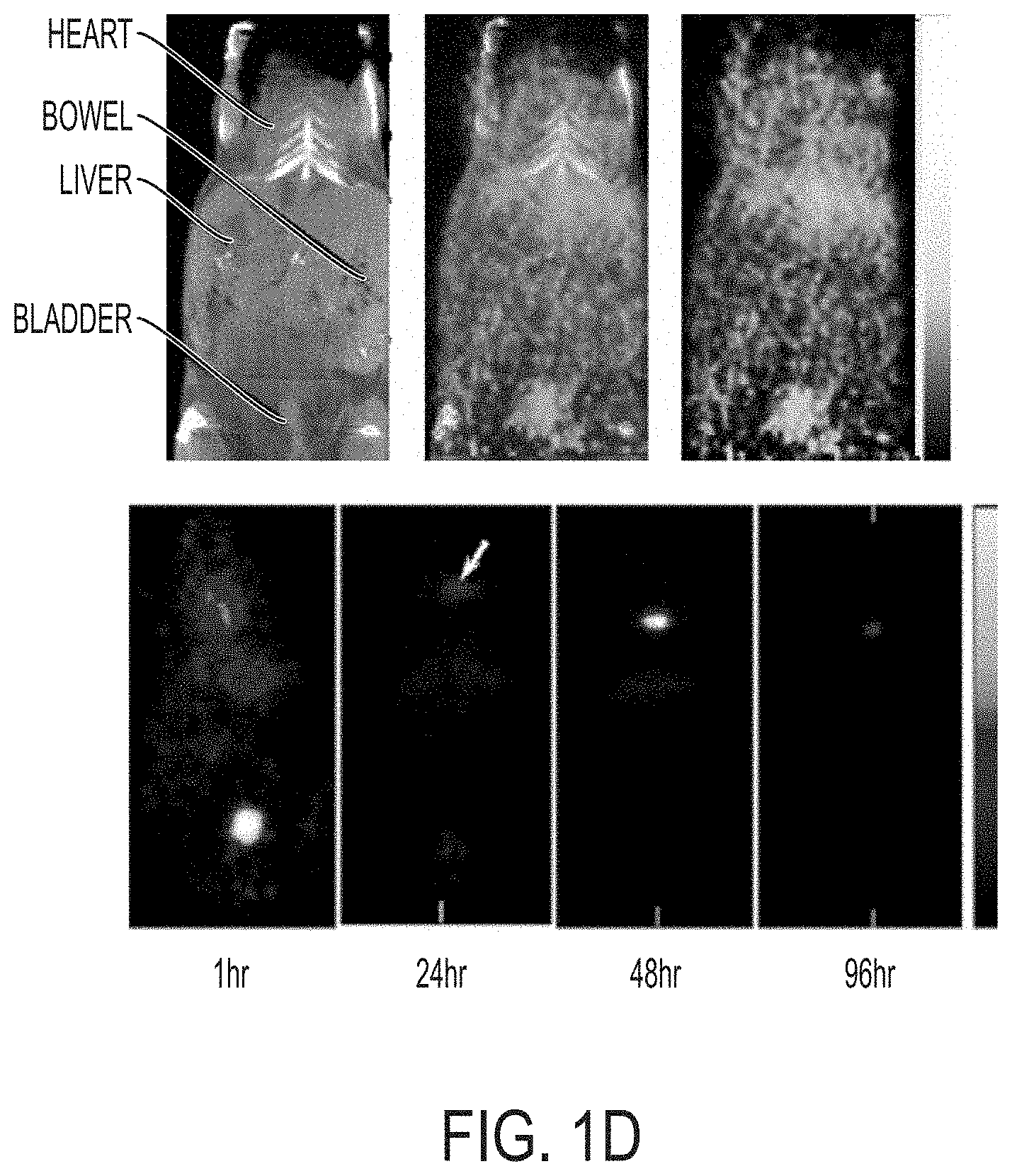
FIG. 1D shows in vivo biodistribution study using co-registered PET-CT. Upper row is serial co-registered PET-CT image 24-hr after injection of .sup.124I-labeled PEG coated nanoparticle, flanked by the independently acquired microCT and microPET scans. Lower row is serial microPET imaging.
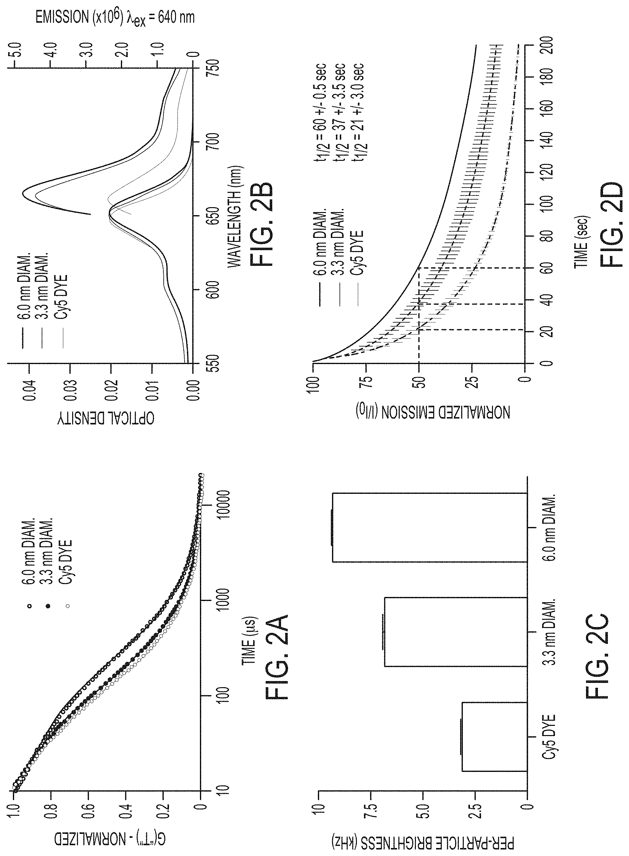
FIG. 2A shows fluorescence correlation spectroscopy (FCS) data and single exponential fits for Cy5 dye (light gray), 3.3.+-.0.06 nm diameter (dark gray, mean.+-.standard deviation, n=9) and 6.0.+-.0.1 nm diameter (black, mean.+-.standard deviation, n=6) Cy5-containing PEG-coated nanoparticles showing the differences in diffusion time resulting from the different hydrodynamic sizes of the different species.
FIG. 2B shows absorption and emission spectra of Cy5 dye (light gray), 3.3 nm diameter (dark gray) and 6.0 nm diameter (black) PEG-coated nanoparticles.
FIG. 2C shows relative brightness comparison of free dye (light gray) with 3.3 nm (dark gray) and 6.0 nm diameter (black) nanoparticles, measured as count rate per molecule/particle as determined from the FCS curves.
FIG. 2D shows photobleaching data for Cy5 dye (light gray), 3.3 nm diameter (dark gray), and 6.0 nm diameter (black) PEG-coated nanoparticles under .about.3.5 mW laser excitation.
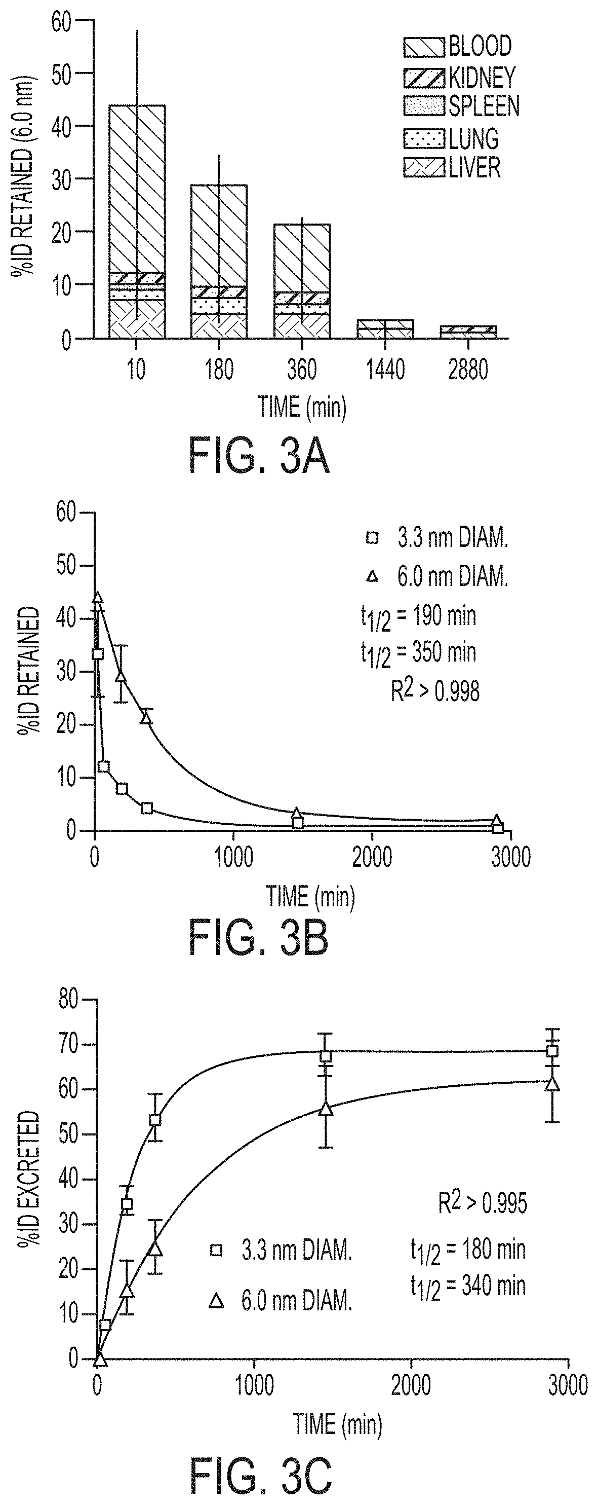
FIG. 3A shows percent of initial particle dose (% ID) retained by blood (black) and tissues: liver (light gray), lung (mid-low gray), spleen (midgray), and kidney (mid-high gray) for 6.0 nm diameter nanoparticles at various time points from 10 min to 48 h post-injection (n=3 mice, mean.+-.standard deviation).
FIG. 3B shows plot of retained particle concentration for 3.3 nm (light gray) and 6.0 nm (black) diameter nanoparticles and the associated logarithmic decay fits and half-lives.
FIG. 3C shows plot of estimated particle excretion for 3.3 nm (light gray) and 6.0 nm (black) diameter nanoparticles and the associated logarithmic fits and half-lives (mean.+-.standard deviation, n=9 (three mice per time point)).
FIGS. 4A-4C show in vivo biodistribution of the nanoparticles in non-tumor-bearing and tumor-bearing mice with subcutaneous C6 xenografts. (FIG. 4A) Bare silica particles; (FIGS. 4B-4C) PEGylated RGD particles.
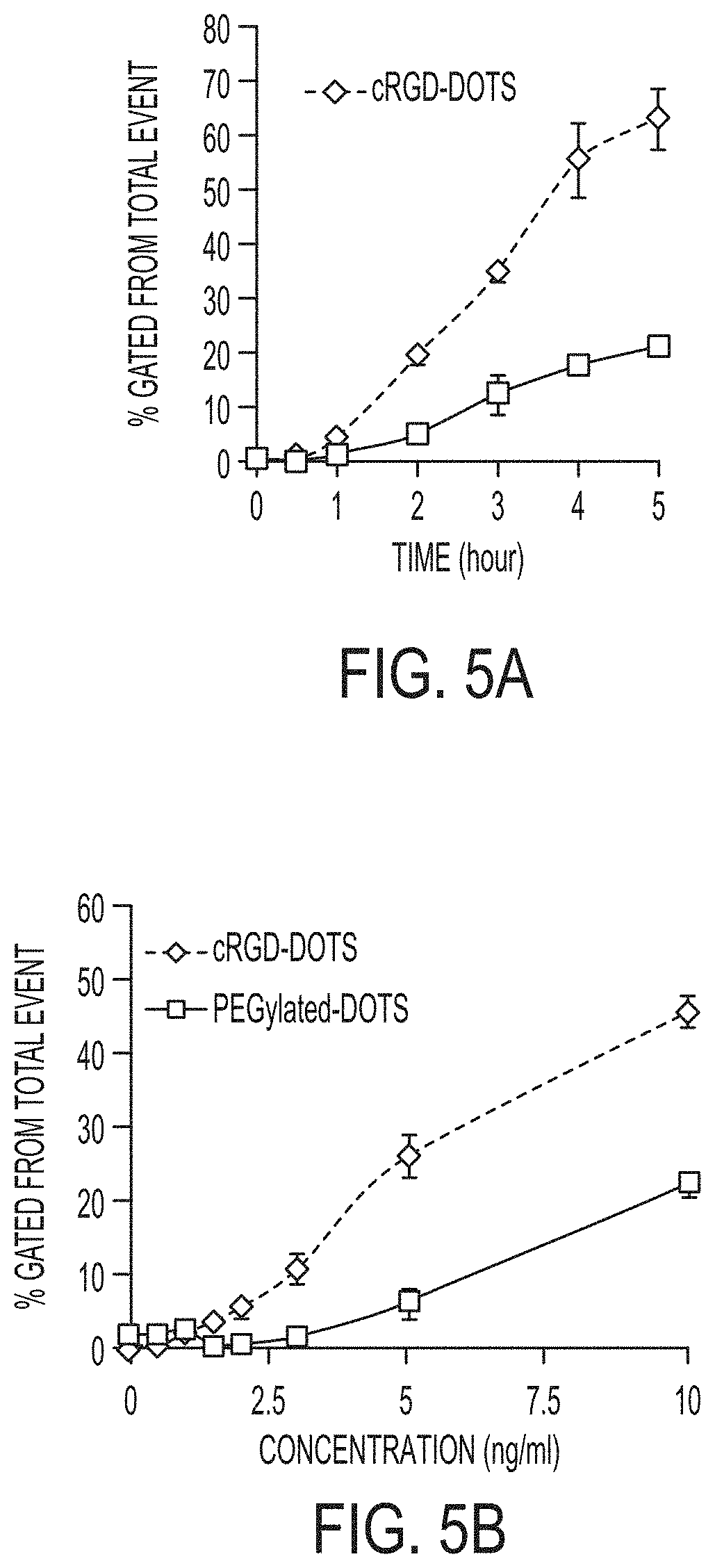
FIGS. 5A-5B show total specific binding data for cRGD- and PEG-ylated dots (i.e., nanoparticles) using flow cytometry in the Cy5 channel as a function of time (FIG. 5A) and particle concentration (FIG. 5B).
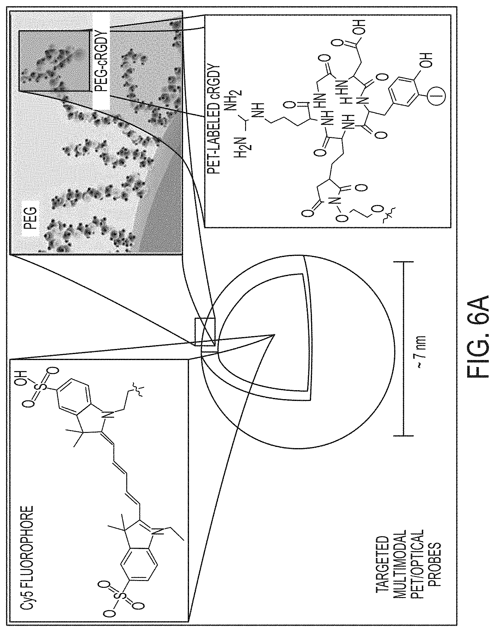
FIGS. 6A-6C show multimodal C dot design for .alpha..sub.v.beta..sub.3-integrin targeting and characterization.
FIG. 6A. Schematic representation of the .sup.124I-cRGDY-PEG-ylated core-shell silica nanoparticle with surface-bearing radiolabels and peptides and core-containing reactive dye molecules (insets).
FIG. 6B. FCS results and single exponential fits for measurements of Cy5 dyes in solution (black), PEGcoated (PEG-dot, red), and PEG-coated, cRGDY-labeled dots (blue, underneath red data set) showing diffusion time differences as a result of varying hydrodynamic sizes.
FIG. 6C. Hydrodynamic sizes (mean.+-.s.d., n=15), and relative brightness comparisons of the free dye with PEG-coated dots and cRGDY-PEG dots derived from the FCS curves, along with the corresponding dye and particle concentrations.
FIG. 7 shows purification and quality control of .sup.124I-RGDY-PEG-dots using size exclusion column chromatography. Radioactivity (right column) of .sup.124I-RGDdots and .sup.124I-PEG-dots detected by .gamma.-counting and corresponding fluorescence signal intensity (Cy5, left column) of .sup.124I-RGDY-PEG-dots and .sup.124I-PEG-dots in each eluted fraction.
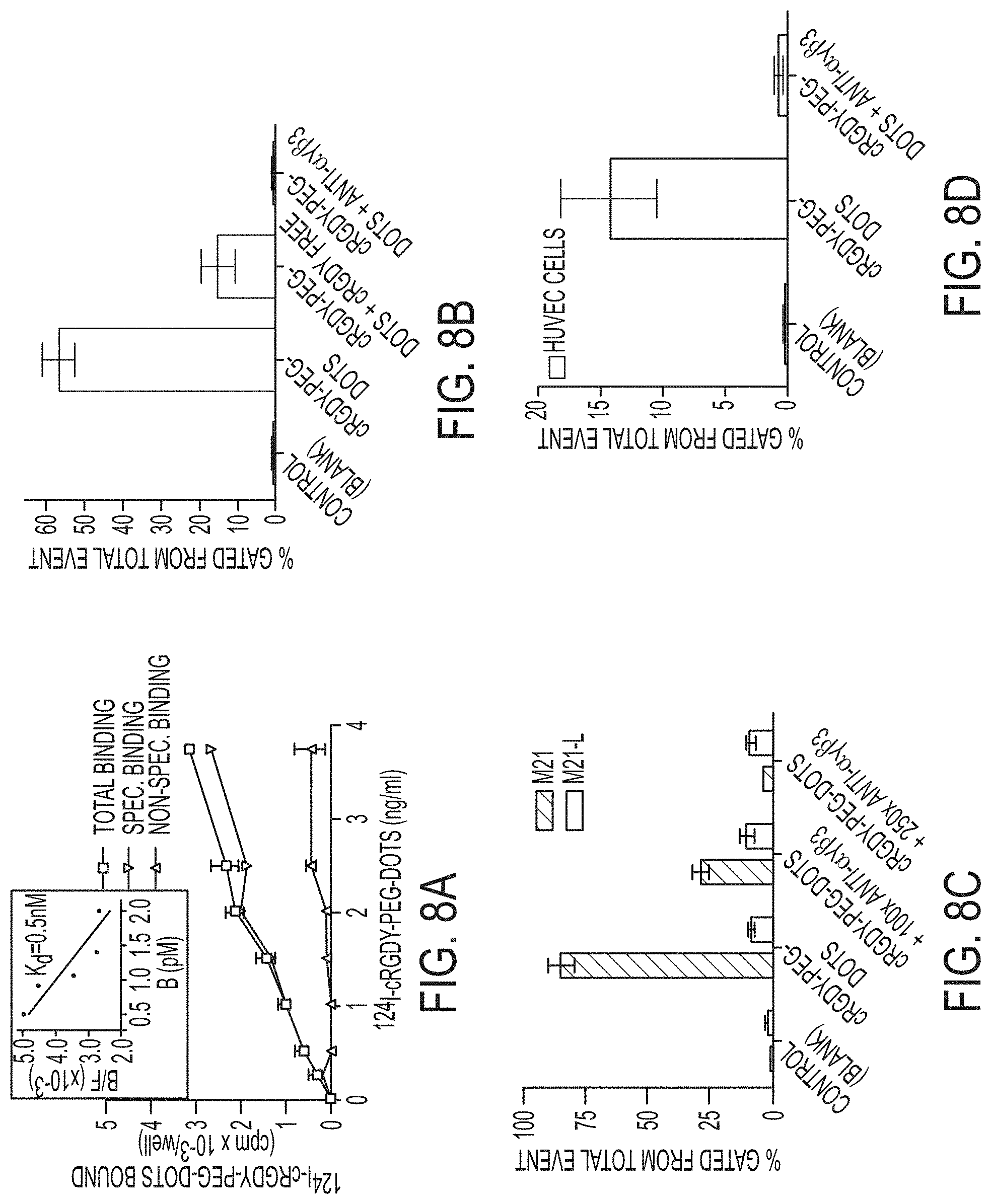
FIGS. 8A-8D show competitive integrin receptor binding studies with .sup.124I-cRGDY-PEG-dots, cRGDY peptide, and anti-.alpha..sub.v.beta..sub.3 antibody using two cell types.
FIG. 8A. High affinity and specific binding of .sup.124I-cRGDY-PEG-dots to M21 cells by .gamma.-counting. Inset shows Scatchard analysis of binding data plotting the ratio of the concentration receptor-bound (B) to unbound (or free, F) radioligand, or bound-to-free ratio, B/F, versus the receptor-bound receptor concentration, B; the slope corresponds to the dissociation constant, Kd.
FIG. 8B. .alpha..sub.v.beta..sub.3-integrin receptor blocking of M21 cells using flow cytometry and excess unradiolabeled cRGD or anti-.alpha..sub.v.beta..sub.3 antibody prior to incubation with cRGDY-PEG-dots.
FIG. 8C. Specific binding of cRGDY-PEG-dots to M21 as against M21L cells lacking surface integrin expression using flow cytometry.
FIG. 8D. Specific binding of cRGDY-PEG-dots to HUVEC cells by flow cytometry. Each bar represents mean.+-.s.d. of three replicates.
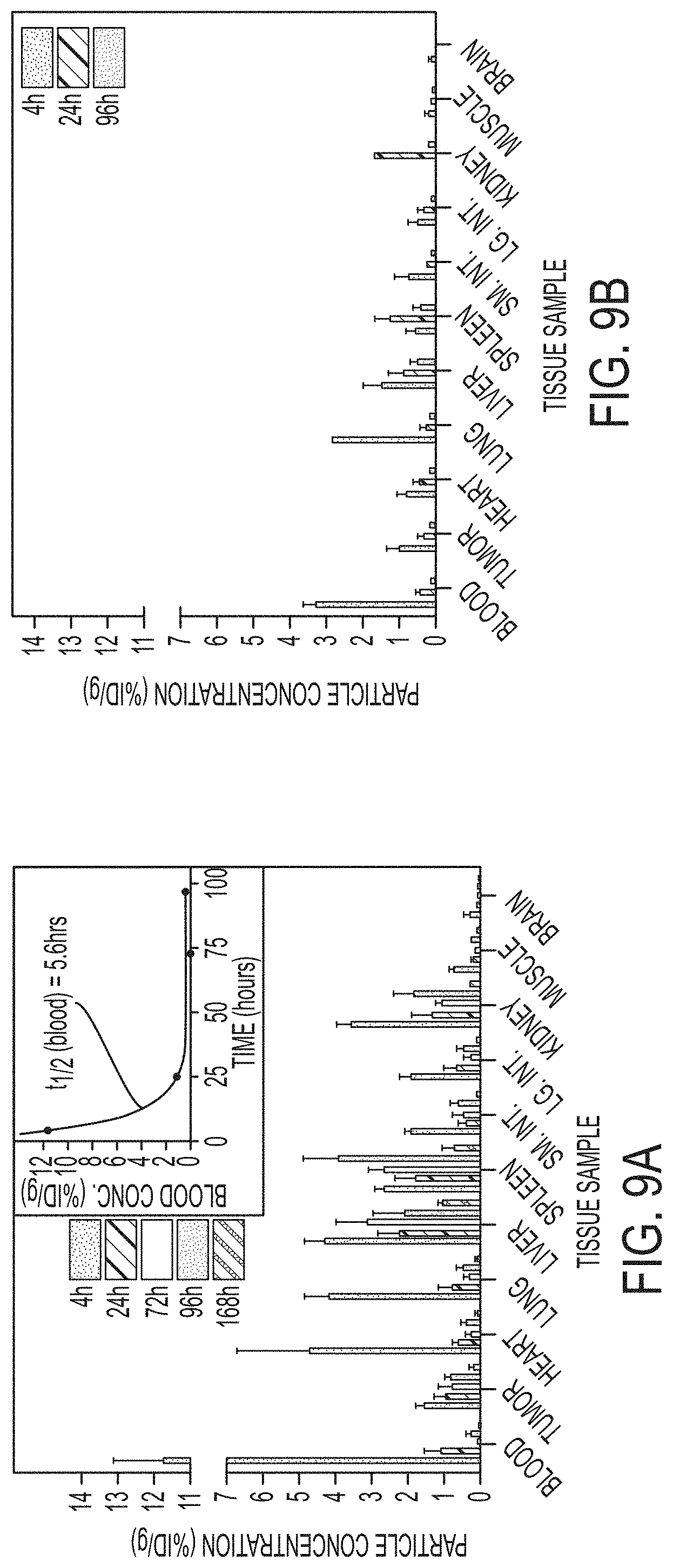
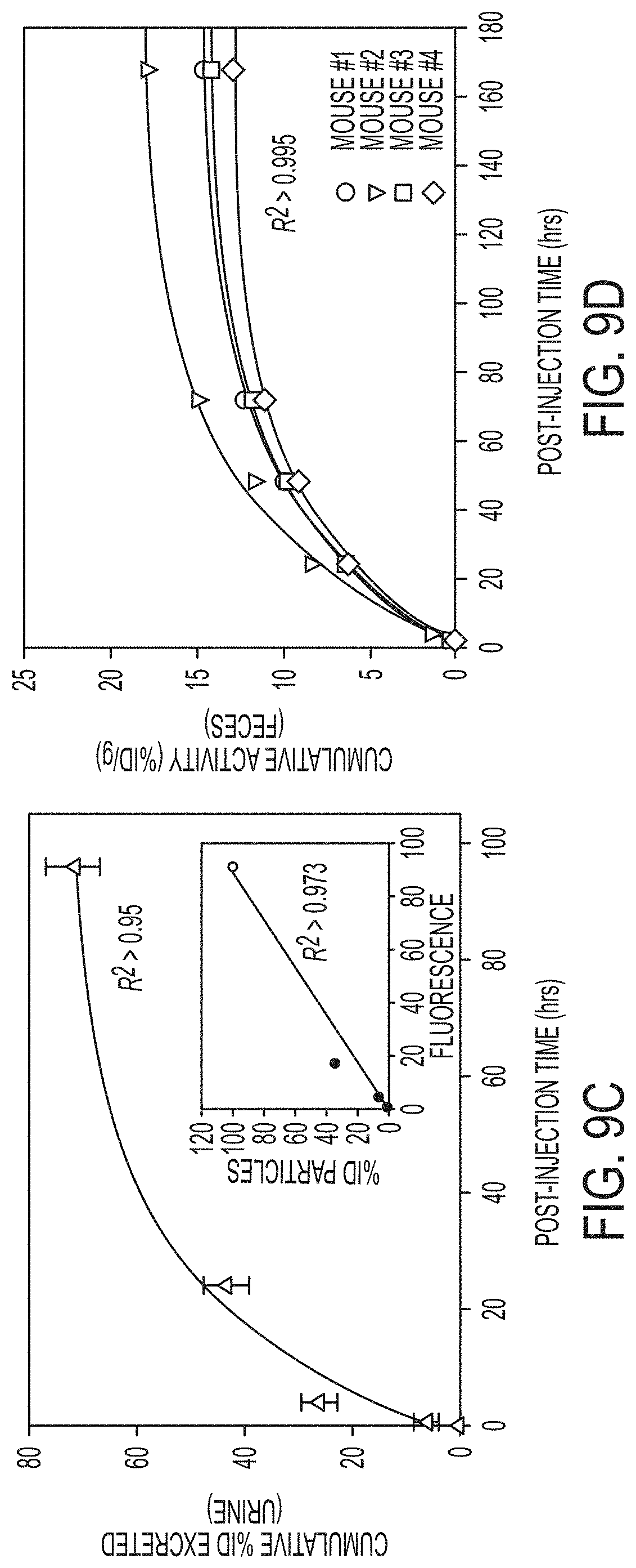
FIGS. 9A-9D show pharmacokinetics and excretion profiles of the targeted and non-targeted particle probes.
FIG. 9A. Biodistribution of .sup.124I-cRGDY-PEG-dots in M21 tumor-bearing mice at various times from 4 to 168 h p.i. The inset shows a representative plot of these data for blood to determine the residence half-time (T.sub.1/2).
FIG. 9B. Biodistribution of .sup.124I-PEG-dots from 4 to 96 h postinjection.
FIG. 9C. Clearance profile of urine samples collected up to 168 hr p.i. of unradiolabeled cRGDY-PEG-dots (n=3 mice, mean.+-.s.d.).
FIG. 9D. Corresponding cumulative % ID/g for feces at intervals up to 168 hr p.i. (n=4 mice). For biodistribution studies, bars represent the mean.+-.s.d.
FIGS. 10A-10B show acute toxicity testing results.
FIG. 10A. Representative H&E stained liver at 400.times. (upper frames) and stained kidneys at 200.times. (lower frames). Mice were treated with a single dose of either non-radiolabeled .sup.127I-RGDY-PEG-dots or .sup.127I-PEG-coated dots (control vehicle) via intravenous injection and organs collected 14 days later.
FIG. 10B. Average daily weights for each treatment group of the toxicity study. Scale bar in FIG. 10A corresponds to 100 .mu.m.
FIGS. 11A-11D show serial in vivo PET imaging of tumor-selective targeting.
FIG. 11A. Representative whole-body coronal microPET images at 4 hrs p.i. demonstrating M21 (left, arrow) and M21L (middle, arrow) tumor uptakes of 3.6 and 0.7% ID/g, respectively, and enhanced M21 tumor contrast at 24 hrs (right).
FIG. 11B. In vivo uptake of .sup.124I-cRGDY-PEG-dots in .alpha..sub.v.beta..sub.3 integrin-overexpressing M21 (black, n=7 mice) and non-expressing M21L (light gray, n=5 mice) tumors and .sup.124I-PEG-dots in M21 tumors (dark gray, n=5).
FIG. 11C. M21 tumor-to-muscle ratios for .sup.124I-cRGDY-PEG-dots (black) and .sup.124I-PEG-dots (gray).
FIG. 11D. Correlation of in vivo and ex-vivo M21 tumor uptakes of cRGDY labeled and unlabeled probes. Each bar represents the mean.+-.s.d.
FIGS. 12A-12B show nodal mapping using multi-scale near-infrared optical fluorescence imaging.
FIG. 12A. Whole body fluorescence imaging of the tumor site (T) and draining inguinal (ILN) and axillary (ALN) nodes and communicating lymphatics channels (bar, LC) 1-hr p.i. in a surgically-exposed living animal.
FIG. 12B. Corresponding co-registered white-light and high-resolution fluorescence images (upper row) and fluorescence images only (lower row) revealing nodal infrastructure of local and distant nodes, including high endothelial venules (HEV). The larger scale bar in (b) corresponds to 500 .mu.m.
FIG. 13A shows the experimental setup of using spontaneous miniswine melanoma model for mapping lymph node basins and regional lymphatics draining the site of a known primary melanoma tumor.
FIG. 13B shows small field-of-view PET image 5 minutes after subdermal injection of multimodal particles (.sup.124I-RGD-PEG-dots) about the tumor site.
FIG. 14A shows whole-body dynamic .sup.18F-fluorodeoxyglucose (.sup.18F-FDG) PET scan demonstrating sagittal, coronal, and axial images through the site of nodal disease in the neck.
FIG. 14B shows fused .sup.18F-FDG PET-CT scans demonstrating sagittal, coronal, and axial images through the site of nodal disease in the neck.
FIG. 14C shows the whole body miniswine image.
FIGS. 15A-15C show the same image sets as in FIGS. 14A-14C, but at the level of the primary melanoma lesion, adjacent to the spine on the upper back.
FIG. 16A shows high resolution dynamic PET images following subdermal, 4-quadrant injection of .sup.124I-RGD-PEG-dots about the tumor site over a 1 hour time period.
FIG. 16B shows fused PET-CT images following subdermal, 4-quadrant injection of .sup.124I-RGD-PEG-dots about the tumor site over a 1 hour time period.
FIG. 16C shows Cy5 imaging (top image), the resected node (second to top image), and H&E staining (lower two images).
FIG. 17 shows a scheme for a nanoparticle with a fluorescent dye within the core and a PEG surface-coating. The nanoparticle is decorated with triple bonds for subsequent "click chemistry" with both DFO and Tyr3-octreotate functionalized with azide groups.
FIG. 18 shows structures of PEG derivative. Standard chemical reactions are used for the production of the functionalized PEG with triple bonds, which will then be covalently attached to the nanoparticle via the silane group.
FIG. 19 shows structures of DFO derivatives.
FIG. 20A shows structures of Tyr3-octreotate.
FIG. 20B shows synthesis of the azide-containing acid for incorporation into Tyr3-Octreotate.
FIG. 21A shows a scheme of the production of functionalized nanoparticle with an NIR fluorescent dye within its core, a PEG surface-coating, DFO chelates and Tyr3-octreotate.
FIG. 21B shows a scheme of the production of a multimodality .sup.89Zr-labeled nanoparticle (PET and fluorescence) decorated with Tyr3-octreotate.
FIG. 22 shows microscopic images demonstrating co-localization between cRGF-PEG-nanoparticles and lysotracker red in the endocytotic pathway.
DETAILED DESCRIPTION OF THE INVENTION
The present invention provides a fluorescent silica-based nanoparticle that allows for precise detection, characterization, monitoring and treatment of a disease such as cancer. The nanoparticle has a range of diameters including between about 0.1 nm and about 100 nm, between about 0.5 nm and about 50 nm, between about 1 nm and about 25 nm, between about 1 nm and about 15 nm, or between about 1 nm and about 8 nm. The nanoparticle has a fluorescent compound positioned within the nanoparticle, and has greater brightness and fluorescent quantum yield than the free fluorescent compound. The nanoparticle also exhibits high biostability and biocompatibility. To facilitate efficient urinary excretion of the nanoparticle, it may be coated with an organic polymer, such as poly(ethylene glycol) (PEG). The small size of the nanoparticle, the silica base and the organic polymer coating minimizes the toxicity of the nanoparticle when administered in vivo. In order to target a specific cell type, the nanoparticle may further be conjugated to a ligand, which is capable of binding to a cellular component (e.g., the cell membrane or other intracellular component) associated with the specific cell type, such as a tumor marker or a signaling pathway intermediate. In one embodiment, a therapeutic agent may be attached to the nanoparticle. To permit the nanoparticle to be detectable by not only optical imaging (such as fluorescence imaging), but also other imaging techniques, such as positron emission tomography (PET), single photon emission computed tomography (SPECT), computerized tomography (CT), and magnetic resonance imaging (MRI), the nanoparticle may also be conjugated to a contrast agent, such as a radionuclide.
The properties of the nanoparticles enable excretion through the kidneys, as well as selective uptake and retention in tumors compared with normal tissues. This, along with the lack of in vivo toxicity, has resulted in a unique product that is promising for translation to the clinic.
The nanoparticle may have both a ligand and a contrast agent. The ligand allows for the nanoparticle to target a specific cell type through the specific binding between the ligand and the cellular component. This targeting, combined with multimodal imaging, has multiple uses. For example, the nanoparticles can be used to map sentinel lymph node (SLN), as well as to mark the tumor margins or neural structures, enabling the surgeon to resect malignant lesions under direct visualization and to obviate complications during the surgical procedure. The ligand may also facilitate entry of the nanoparticle into the cell or barrier transport, for example, for assaying the intracellular environment.
The nanoparticle can be coupled with a ligand and a therapeutic agent with or without a radiolabel. The radiolabel can additionally serve as a therapeutic agent for creating a multitherapeutic platform. This coupling allows the therapeutic agent to be delivered to the specific cell type through the specific binding between the ligand and the cellular component. This specific targeting of the therapeutic agent ensures selective treatment of the disease site with minimum side effects.
The fluorescent nanoparticle of the present invention includes a silica-based core comprising a fluorescent compound positioned within the core, and a silica shell on the core. The silica shell may surround at least a portion of the core. Alternatively, the nanoparticle may have only the core and no shell. The core of the nanoparticle may contain the reaction product of a reactive fluorescent compound and a co-reactive organo-silane compound. In another embodiment, the core of the nanoparticle may contain the reaction product of a reactive fluorescent compound and a co-reactive organo-silane compound, and silica. The diameter of the core may be from about 0.05 nm to about 100 nm, from about 0.1 nm to about 50 nm, from about 0.5 nm to about 25 nm, from about 0.8 nm to about 15 nm, or from about 1 nm to about 8 nm. The shell of the nanoparticle can be the reaction product of a silica forming compound. The shell of the nanoparticle may have a range of layers. For example, the silica shell may be from about 1 to about 20 layers, from about 1 to about 15 layers, from about 1 to about 10 layers, or from about 1 to about 5 layers. The thickness of the shell may range from about 0.01 nm to about 90 nm, from about 0.02 nm to about 40 nm, from about 0.05 nm to about 20 nm, from about 0.05 nm to about 10 nm, or from about 0.05 nm to about 5 nm.
The silica shell of the nanoparticle may cover only a portion of nanoparticle or the entire particle. For example, the silica shell may cover about 1 to about 100 percent, from about 10 to about 80 percent, from about 20 to about 60 percent, or from about 30 to about 50 percent of the nanoparticle. The silica shell can be either solid, i.e., substantially non-porous, meso-porous, such as semi-porous, or porous.
The present fluorescent nanoparticle may be synthesized by the steps of: covalently conjugating a fluorescent compound, such as a reactive fluorescent dye, with the reactive moeties including, but not limited to, maleimide, iodoacetamide, thiosulfate, amine, N-Hydroxysuccimide ester, 4-sulfo-2,3,5,6-tetrafluorophenyl (STP) ester, sulfosuccinimidyl ester, sulfodichlorophenol esters, sulfonyl chloride, hydroxyl, isothiocyanate, carboxyl, to an organo-silane compound, such as a co-reactive organo-silane compound, to form a fluorescent silica precursor, and reacting the fluorescent silica precursor to form a fluorescent core; covalently conjugating a fluorescent compound, such as a reactive fluorescent dye, to an organo-silane compound, such as a co-reactive organo-silane compound, to form a fluorescent silica precursor, and reacting the fluorescent silica precursor with a silica forming compound, such as tetraalkoxysilane, to form a fluorescent core; and reacting the resulting core with a silica forming compound, such as a tetraalkoxysilane, to form a silica shell on the core, to provide the fluorescent nanoparticle.
The synthesis of the fluorescent monodisperse core-shell nanoparticles is based on a two-step process. First, the organic dye molecules, tetramethylrhodamine isothiocynate (TRITC), are covalently conjugated to a silica precursor and condensed to form a dye-rich core. Second, the silica gel monomers are added to form a denser silica network around the fluorescent core material, providing shielding from solvent interactions that can be detrimental to photostability. The versatility of the preparative route allows for the incorporation of different fluorescent compounds, such as fluorescent organic compounds or dyes, depending on the intended nanoparticle application. The fluorescent compounds that may be incorporated in the dye-rich core can cover the entire UV-Vis to near-IR absorption and emission spectrum. U.S. patent application Ser. Nos. 10/306,614, 10/536,569 and 11/119,969. Wiesner et al., Peg-coated Core-shell Silica Nanoparticles and Mathods of Manufactire and Use, PCT/US2008/74894.
For the synthesis of the compact core-shell nanoparticle, the dye precursor is added to a reaction vessel that contains appropriate amounts of ammonia, water and solvent and allowed to react overnight. The dye precursor is synthesized by addition reaction between a specific near-infrared dye of interest and 3-aminopropyltriethoxysilane in molar ratio of 1:50, in exclusion of moisture. After the synthesis of the dye-rich compact core is completed, tetraethylorthosilicate (TEOS) is subsequently added to grow the silica shell that surrounded the core.
The synthesis of the expanded core-shell nanoparticle is accomplished by co-condensing TEOS with the dye precursor and allowing the mixture to react overnight. After the synthesis of the expanded core is completed, additional TEOS is added to grow the silica shell that surrounded the core.
The synthesis of the homogenous nanoparticles is accomplished by co-condensing all the reagents, the dye precursor and TEOS and allowing the mixture to react overnight.
The nanoparticles may incorporate any known fluorescent compound, such as fluorescent organic compound, dyes, pigments, or combinations thereof. A wide variety of suitable chemically reactive fluorescent dyes are known, see for example MOLECULAR PROBES HANDBOOK OF FLUORESCENT PROBES AND RESEARCH CHEMICALS, 6th ed., R. P. Haugland, ed. (1996). A typical fluorophore is, for example, a fluorescent aromatic or heteroaromatic compound such as is a pyrene, an anthracene, a naphthalene, an acridine, a stilbene, an indole or benzindole, an oxazole or benzoxazole, a thiazole or benzothiazole, a 4-amino-7-nitrobenz-2-oxa-1,3-diazole (NBD), a cyanine, a carbocyanine, a carbostyryl, a porphyrin, a salicylate, an anthranilate, an azulene, a perylene, a pyridine, a quinoline, a coumarin (including hydroxycoumarins and aminocoumarins and fluorinated derivatives thereof), and like compounds, see for example U.S. Pat. Nos. 5,830,912, 4,774,339, 5,187,288, 5,248,782, 5,274,113, 5,433,896, 4,810,636 and 4,812,409. In one embodiment, Cy5, a near infrared fluorescent (NIRF) dye, is positioned within the silica core of the present nanoparticle. Near infrared-emitting probes exhibit decreased tissue attenuation and autofluorescence. Burns et al. "Fluorescent silica nanoparticles with efficient urinary excretion for nanomedicine", Nano Letters, 2009, 9 (1), 442-448.
Non-limiting fluorescent compound that may be used in the present invention include, Cy5, Cy5.5 (also known as Cy5++), Cy2, fluorescein isothiocyanate (FITC), tetramethylrhodamine isothiocyanate (TRITC), phycoerythrin, Cy7, fluorescein (FAM), Cy3, Cy3.5 (also known as Cy3++), Texas Red, LightCycler-Red 640, LightCycler Red 705, tetramethylrhodamine (TMR), rhodamine, rhodamine derivative (ROX), hexachlorofluorescein (HEX), rhodamine 6G (R6G), the rhodamine derivative JA133, Alexa Fluorescent Dyes (such as Alexa Fluor 488, Alexa Fluor 546, Alexa Fluor 633, Alexa Fluor 555, and Alexa Fluor 647), 4',6-diamidino-2-phenylindole (DAPI), Propidium iodide, AMCA, Spectrum Green, Spectrum Orange, Spectrum Aqua, Lissamine, and fluorescent transition metal complexes, such as europium. Fluorescent compound that can be used also include fluorescent proteins, such as GFP (green fluorescent protein), enhanced GFP (EGFP), blue fluorescent protein and derivatives (BFP, EBFP, EBFP2, Azurite, mKalamal), cyan fluorescent protein and derivatives (CFP, ECFP, Cerulean, CyPet) and yellow fluorescent protein and derivatives (YFP, Citrine, Venus, YPet). WO2008142571, WO2009056282, WO9922026.
The silica shell surface of the nanoparticles can be modified by using known cross-linking agents to introduce surface functional groups. Crosslinking agents include, but are not limited to, divinyl benzene, ethylene glycol dimethacrylate, trimethylol propane trimethacrylate, N,N'-methylene-bis-acrylamide, alkyl ethers, sugars, peptides, DNA fragments, or other known functionally equivalent agents. The ligand may be conjugated to the nanoparticle of the present invention by, for example, through coupling reactions using carbodiimide, carboxylates, esters, alcohols, carbamides, aldehydes, amines, sulfur oxides, nitrogen oxides, halides, or any other suitable compound known in the art. U.S. Pat. No. 6,268,222.
An organic polymer may be attached to the present nanoparticle, e.g., attached to the surface of the nanoparticle. An organic polymer may be attached to the silica shell of the present nanoparticle. The organic polymer that may be used in the present invention include PEG, polylactate, polylactic acids, sugars, lipids, polyglutamic acid (PGA), polyglycolic acid, poly(lactic-co-glycolic acid) (PLGA), polyvinyl acetate (PVA), and the combinations thereof. The attachment of the organic polymer to the nanoparticle may be accomplished by a covalent bond or non-covalent bond, such as by ionic bond, hydrogen bond, hydrophobic bond, coordination, adhesive, and physical absorption. In one embodiment, the nanoparticle is covalently conjugated with PEG, which prevents adsorption of serum proteins, facilitates efficient urinary excretion and decreases aggregation of the nanoparticle. Burns et al. "Fluorescent silica nanoparticles with efficient urinary excretion for nanomedicine", Nano Letters, 2009, 9 (1), 442-448.
The surface of the nanoparticle may be modified to incorporate at least one functional group. The organic polymer (e.g., PEG) attached to the nanoparticle may be modified to incorporate at least one functional group. For example, the functional group can be a maleimide or N-Hydroxysuccinimide (NHS) ester. The incorporation of the functional group makes it possible to attach various ligands, contrast agents and/or therapeutic agents to the nanoparticle.
A ligand may be attached to the present nanoparticle. The ligand is capable of binding to at least one cellular component. The cellular component may be associated with specific cell types or having elevated levels in specific cell types, such as cancer cells or cells specific to particular tissues and organs. Accordingly, the nanoparticle can target a specific cell type, and/or provides a targeted delivery for the treatment and diagnosis of a disease. As used herein, the term "ligand" refers to a molecule or entity that can be used to identify, detect, target, monitor, or modify a physical state or condition, such as a disease state or condition. For example, a ligand may be used to detect the presence or absence of a particular receptor, expression level of a particular receptor, or metabolic levels of a particular receptor. The ligand can be, for example, a peptide, a protein, a protein fragment, a peptide hormone, a sugar (i.e., lectins), a biopolymer, a synthetic polymer, an antigen, an antibody, an antibody fragment (e.g., Fab, nanobodies), an aptamer, a virus or viral component, a receptor, a hapten, an enzyme, a hormone, a chemical compound, a pathogen, a microorganism or a component thereof, a toxin, a surface modifier, such as a surfactant to alter the surface properties or histocompatability of the nanoparticle or of an analyte when a nanoparticle associates therewith, and combinations thereof. Preferred ligands are, for example, antibodies, such as monoclonal or polyclonal antibodies, and receptor ligands. In another embodiment, the ligand is poly-L-lysine (pLysine).
An antigen may be attached to the nanoparticle. The antigen-attached nanoparticle may be used for vaccination.
The terms "component of a cell" or "cellular component" refer to, for example, a receptor, an antibody, a hapten, an enzyme, a hormone, a biopolymer, an antigen, a nucleic acid (DNA or RNA), a microorganism, a virus, a pathogen, a toxin, combinations thereof, and like components. The component of a cell may be positioned on the cell (e.g., a transmembrane receptor) or inside the cell. In one embodiment, the component of a cell is a tumor marker. As used herein, the term "tumor marker" refers to a molecule, entity or substance that is expressed or overexpressed in a cancer cell but not normal cell. For example, the overexpression of certain receptors is associated with many types of cancer. A ligand capable of binding to a tumor marker may be conjugated to the surface of the present nanoparticle, so that the nanoparticle can specifically target the tumor cell.
A ligand may be attached to the present nanoparticle directly or through a linker. The attachment of the ligand to the nanoparticle may be accomplished by a covalent bond or non-covalent bond, such as by ionic bond, hydrogen bond, hydrophobic bond, coordination, adhesive, and physical absorption. The ligand may be coated onto the surface of the nanoparticle. The ligand may be imbibed into the surface of the nanoparticle. As used herein, "imbibe" refers to assimilation or taking in. The ligand may be attached to the surface of the fluorescent nanoparticle, or may be attached to the core when the shell is porous or is covering a portion of the core. When the ligand is attached to the nanoparticle through a linker, the linker can be any suitable molecules, such as a functionalized PEG. The PEGs can have multiple functional groups for attachment to the nanoparticle and ligands. The particle can have different types of functionalized PEGs bearing different functional groups that can be attached to multiple ligands. This can enhance multivalency effects and/or contrast at the target site, which allows the design and optimization of a complex multimodal platform with improved targeted detection, treatment, and sensing in vivo.
A variety of different ligands may be attached to the nanoparticle. For example, tripeptide Arg-Gly-Asp (RGD) may be attached to the nanoparticle. Alternatively, cyclic peptide cRGD (which may contain other amino acid(s), e.g., cRGDY) may be attached to the nanoparticle. Any linear, cyclic or branched peptide containing the RGD sequence is within the scope of the present invention. RGD binds to .alpha..sub.v.beta..sub.3 integrin, which is overexpressed at the surface of activated endothelial cells during angiogenesis and in various types of tumor cells. Expression levels of .alpha..sub.v.beta..sub.3 integrin have been shown to correlate well with the aggressiveness of tumors. Ruoslahti et al. New perspectives in cell adhesion: RGD and integrins. Science 1987; 238:491. Gladson et al. Glioblastoma expression of vitronectin and alpha v beta 3 integrin. Adhesion mechanism for transformed glial cells. J. Clin. Invest. 1991; 88:1924-1932. Seftor et al. Role of the alpha v beta 3 integrin in human melanoma cell invasion. Proc. Natl. Acad. Sci. 1992; 89:1557-1561.
Alternatively, synthetic peptide EPPT1 may be the ligand attached to the nanoparticle. EPPT1, derived from the monoclonal antibody (ASM2) binding site, targets underglycosylated MUC1 (uMUC1). MUC1, a transmembrane receptor, is heavily glycosylated in normal tissues; however, it is overexpressed and aberrantly underglycosylated in almost all human epithelial cell adenocarcinomas, and is implicated in tumor pathogenesis. Moore et al. In vivo targeting of underglycosylated MUC-1 tumor antigen using a multimodal imaging probe. Cancer Res. 2004; 64:1821-7. Patel et al. MUC1 plays a role in tumor maintenance in aggressive thyroid carcinomas. Surgery. 2005; 138:994-1001. Specific antibodies including monoclonal antibodies against uMUC1 may alternatively be conjugated to the nanoparticle in order to target uMUC1.
In one embodiment, peptide analogues of .alpha.-melanotropin stimulating hormone (.alpha.-MSH) are the ligands attached to the nanoparticle. Peptide analogues of .alpha.-MSH are capable of binding to melanocortin-1 receptors (MC1R), a family of G-protein-coupled receptors overexpressed in melanoma cells. Loir et al. Cell Mol. Biol. (Noisy-le-grand) 1999, 45:1083-1092.
In another embodiment, octreotate, a peptide analog of 14-amino acid somatostatin, is the ligand attached to the nanoparticle. Octreotide, which has a longer half-life than somatostatin, is capable of binding to somatostatin receptor (SSTR). SSTR, a member of the G-protein coupled receptor family, is overexpressed on the surface of several human tumors. Reubi et al. Distribution of Somatostatin Receptors in Normal and Tumor-Tissue. Metab. Clin. Exp. 1990; 39:78-81. Reubi et al. Somatostatin receptors and their subtypes in human tumors and in peritumoral vessels. Metab. Clin. Exp. 1996; 45:39-41. Other somatostatin analogs may alternatively be conjugated to the nanoparticle to target SSTR, such as Tyr3-octreotide (Y3-OC), octreotate (TATE), Tyr3-octreotate (Y3-TATE), and .sup.111In-DTPA-OC. These somatostatin analogues may be utilized for both PET diagnostic imaging and targeted radiotherapy of cancer. de Jong et al. Internalization of radiolabelled [DTPA.sup.0]octreotide and [DOTA.sup.0, Tyr.sup.3]octreotide: peptides for somatostatin receptor targeted scintigraphy and radionuclide therapy. Nucl. Med. Commun. 1998; 19:283-8. de Jong et al. Comparison of .sup.111In-Labeled Somatostatin Analogues for Tumor Scintigraphy and Radionuclide Therapy. Cancer Res. 1998; 58:437-41. Lewis et al. Comparison of four .sup.64Cu-labeled somatostatin analogs in vitro and in a tumor-bearing rat model: evaluation of new derivatives for PET imaging and targeted radiotherapy. J Med Chem 1999; 42:1341-7. Krenning et al. Somatostatin Receptor Scintigraphy with Indium-111-DTPA-D-Phe-1-Octreotide in Man: Metabolism, Dosimetry and Comparison with Iodine-123-Tyr-3-Octreotide. J Nucl. Med. 1992; 33:652-8.
The number of ligands attached to the nanoparticle may range from about 1 to about 20, from about 2 to about 15, from about 3 to about 10, from about 1 to about 10, or from about 1 to about 6. The small number of the ligands attached to the nanoparticle helps maintain the hydrodynamic diameter of the present nanoparticle which meet the renal clearance cutoff size range. Hilderbrand et al., Near-infrared fluorescence: application to in vivo molecular imaging, Curr. Opin. Chem. Biol., 14:71-9, 2010. The number of ligands measured may be an average number of ligands attached to more than one nanoparticle. Alternatively, one nanoparticle may be measured to determine the number of ligands attached. The number of ligands attached to the nanoparticle can be measured by any suitable methods, which may or may not be related to the properties of the ligands. For example, the number of cRGD peptides bound to the particle may be estimated using FCS-based measurements of absolute particle concentrations and the starting concentration of the reagents for cRGD peptide. Average number of RGD peptides per nanoparticle and coupling efficiency of RGD to functionalized PEG groups can be assessed colorimetrically under alkaline conditions and Biuret spectrophotometric methods. The number of ligands attached to the nanoparticle may also be measured by nuclear magnetic resonance (NMR), optical imaging, assaying radioactivity, etc. The method can be readily determined by those of skill in the art.
A contrast agent may be attached to the present nanoparticle for medical or biological imaging. As used herein, the term "contrast agent" refers to a substance, molecule or compound used to enhance the visibility of structures or fluids in medical or biological imaging. The term "contrast agent" also refers to a contrast-producing molecule. The imaging techniques encompassed by the present invention include positron emission tomography (PET), single photon emission computed tomography (SPECT), computerized tomography (CT), magnetic resonance imaging (MRI), optical bioluminescence imaging, optical fluorescence imaging, and combinations thereof. The contrast agent encompassed by the present invention may be any molecule, substance or compound known in the art for PET, SPECT, CT, MRI, and optical imaging. The contrast agent may be radionuclides, radiometals, positron emitters, beta emitters, gamma emitters, alpha emitters, paramagnetic metal ions, and supraparamagnetic metal ions. The contrast agents include, but are not limited to, iodine, fluorine, copper, zirconium, lutetium, astatine, yttrium, gallium, indium, technetium, gadolinium, dysprosium, iron, manganese, barium and barium sulfate. The radionuclides that may be used as the contrast agent attached to the nanoparticle of the present invention include, but are not limited to, .sup.89Zr, .sup.64Cu, .sup.68Ga, .sup.86Y, .sup.124I and .sup.177Lu.
The contrast agent may be directly conjugated to the nanoparticle. Alternatively, the contrast agent may be indirectly conjugated to the nanoparticle, by attaching to linkers or chelates. The chelate may be adapted to bind a radionuclide. The chelates that can be attached to the present nanoparticle may include, but are not limited to, 1,4,7,10-tetraazacyclododecane-1,4,7,10-tetraacetic acid (DOTA), diethylenetriaminepentaacetic (DTPA), desferrioxamine (DFO) and triethylenetetramine (TETA).
Suitable means for imaging, detecting, recording or measuring the present nanoparticles may also include, for example, a flow cytometer, a laser scanning cytometer, a fluorescence micro-plate reader, a fluorescence microscope, a confocal microscope, a bright-field microscope, a high content scanning system, and like devices. More than one imaging techniques may be used at the same time or consecutively to detect the present nanoparticles. In one embodiment, optical imaging is used as a sensitive, high-throughput screening tool to acquire multiple time points in the same subject, permitting semi-quantitative evaluations of tumor marker levels. This offsets the relatively decreased temporal resolution obtained with PET, although PET is needed to achieve adequate depth penetration for acquiring volumetric data, and to detect, quantitate, and monitor changes in receptor and/or other cellular marker levels as a means of assessing disease progression or improvement, as well as stratifying patients to suitable treatment protocols.
A therapeutic agent may be attached to the fluorescent nanoparticle, for example, for targeted treatment of a disease. The therapeutic agent may be delivered to a diseased site in a highly specific or localized manner with release of the therapeutic agent in the disease site. Alternatively, the therapeutic agent may not be released. The fluorescent nanoparticle conjugated with the ligand can be used for targeted delivery of a therapeutic agent to a desired location in a variety of systems, such as on, or within, a cell or cell component, within the body of an organism, such as a human, or across the blood-brain barrier.
The therapeutic agent may be attached to the nanoparticle directly or indirectly. The therapeutic agent can be absorbed into the interstices or pores of the silica shell, or coated onto the silica shell of the fluorescent nanoparticle. In other embodiments where the silica shell is not covering all of the surface, the therapeutic agent can be associated with the fluorescent core, such as by physical absorption or by bonding interaction. The therapeutic agent may be associated with the ligand that is attached to the fluorescent nanoparticle. The therapeutic agent may also be associated with the organic polymer or the contrast agent. For example, the therapeutic agent may be attached to the nanoparticle through PEG. The PEGs can have multiple functional groups for attachment to the nanoparticle and therapeutic agent. The particle can have different types of functionalized PEGs bearing different functional groups that can be attached to multiple therapeutic agents. The therapeutic agent may be attached to the nanoparticle covalently or non-covalently.
As used herein, the term "therapeutic agent" refers to a substance that may be used in the diagnosis, cure, mitigation, treatment, or prevention of disease in a human or another animal. Such therapeutic agents include substances recognized in the official United States Pharmacopeia, official Homeopathic Pharmacopeia of the United States, official National Formulary, or any supplement thereof.
Therapeutic agents that can be incorporated with the fluorescent nanoparticles or the ligated-fluorescent nanoparticles of the invention include nucleosides, nucleoside analogs, small interfering RNA (siRNA), microRNA, oligopeptides, polypeptides, antibodies, COX-2 inhibitors, apoptosis promoters, urinary tract agents, vaginal agents, vasodilators neurodegenerative agents (e.g., Parkinson's disease), obesity agents, ophthalmic agents, osteoporosis agents, para-sympatholytics, para-sympathometics, antianesthetics, prostaglandins, psychotherapeutic agents, respiratory agents, sedatives, hypnotics, skin and mucous membrane agents, anti-bacterials, anti-fungals, antineoplastics, cardioprotective agents, cardiovascular agents, anti-thrombotics, central nervous system stimulants, cholinesterase inhibitors, contraceptives, dopamine receptor agonists, erectile dysfunction agents, fertility agents, gastrointestinal agents, gout agents, hormones, immunomodulators, suitably functionalized analgesics or general or local anesthetics, anti-convulsants, anti-diabetic agents, anti-fibrotic agents, anti-infectives, motion sickness agents, muscle relaxants, immuno-suppressive agents, migraine agents, non-steroidal anti-inflammatory drugs (NSAIDs), smoking cessation agents, or sympatholytics (see Physicians' Desk Reference, 55th ed., 2001, Medical Economics Company, Inc., Montvale, N.J., pages 201-202).
Therapeutic agents that may be attached to the present nanoparticle include, but are not limited to, DNA alkylating agents, topoisomerase inhibitors, endoplasmic reticulum stress inducing agents, a platinum compound, an antimetabolite, vincalkaloids, taxanes, epothilones, enzyme inhibitors, receptor antagonists, therapeutic antibodies, tyrosine kinase inhibitors, boron radiosensitizers (i.e. velcade), and chemotherapeutic combination therapies.
Non-limiting examples of DNA alkylating agents are nitrogen mustards, such as Mechlorethamine, Cyclophosphamide (Ifosfamide, Trofosfamide), Chlorambucil (Melphalan, Prednimustine), Bendamustine, Uramustine and Estramustine; nitrosoureas, such as Carmustine (BCNU), Lomustine (Semustine), Fotemustine, Nimustine, Ranimustine and Streptozocin; alkyl sulfonates, such as Busulfan (Mannosulfan, Treosulfan); Aziridines, such as Carboquone, ThioTEPA, Triaziquone, Triethylenemelamine; Hydrazines (Procarbazine); Triazenes such as Dacarbazine and Temozolomide; Altretamine and Mitobronitol.
Non-limiting examples of Topoisomerase I inhibitors include Campothecin derivatives including CPT-11 (irinotecan), SN-38, APC, NPC, campothecin, topotecan, exatecan mesylate, 9-nitrocamptothecin, 9-aminocamptothecin, lurtotecan, rubitecan, silatecan, gimatecan, diflomotecan, extatecan, BN-80927, DX-8951f, and MAG-CPT as decribed in Pommier Y. (2006) Nat. Rev. Cancer 6(10):789-802 and U.S. Patent Publication No. 200510250854; Protoberberine alkaloids and derivatives thereof including berberrubine and coralyne as described in Li et al. (2000) Biochemistry 39(24):7107-7116 and Gatto et al. (1996) Cancer Res. 15(12):2795-2800; Phenanthroline derivatives including Benzo[i]phenanthridine, Nitidine, and fagaronine as described in Makhey et al. (2003) Bioorg. Med. Chem. 11 (8): 1809-1820; Terbenzimidazole and derivatives thereof as described in Xu (1998) Biochemistry 37(10):3558-3566; and Anthracycline derivatives including Doxorubicin, Daunorubicin, and Mitoxantrone as described in Foglesong et al. (1992) Cancer Chemother. Pharmacol. 30(2):123-]25, Crow et al. (1994) J. Med. Chem. 37(19):31913194, and Crespi et al. (1986) Biochem. Biophys. Res. Commun. 136(2):521-8. Topoisomerase II inhibitors include, but are not limited to Etoposide and Teniposide. Dual topoisomerase I and II inhibitors include, but are not limited to, Saintopin and other Naphthecenediones, DACA and other Acridine-4-Carboxamindes, Intoplicine and other Benzopyridoindoles, TAS-I03 and other 7H-indeno[2,1-c]Quinoline-7-ones, Pyrazoloacridine, XR 11576 and other Benzophenazines, XR 5944 and other Dimeric compounds, 7-oxo-7H-dibenz[f,ij]Isoquinolines and 7-oxo-7H-benzo[e]Perimidines, and Anthracenyl-amino Acid Conjugates as described in Denny and Baguley (2003) Curr. Top. Med. Chem. 3(3):339-353. Some agents inhibit Topoisomerase II and have DNA intercalation activity such as, but not limited to, Anthracyclines (Aclarubicin, Daunorubicin, Doxorubicin, Epirubicin, Idarubicin, Amrubicin, Pirarubicin, Valrubicin, Zorubicin) and Antracenediones (Mitoxantrone and Pixantrone).
Examples of endoplasmic reticulum stress inducing agents include, but are not limited to, dimethyl-celecoxib (DMC), nelfinavir, celecoxib, and boron radiosensitizers (i.e. velcade (Bortezomib)).
Non-limiting examples of platinum based compound include Carboplatin, Cisplatin, Nedaplatin, Oxaliplatin, Triplatin tetranitrate, Satraplatin, Aroplatin, Lobaplatin, and JM-216. (see McKeage et al. (1997) J. Clin. Oncol. 201:1232-1237 and in general, CHEMOTHERAPY FOR GYNECOLOGICAL NEOPLASM, CURRENT THERAPY AND NOVEL APPROACHES, in the Series Basic and Clinical Oncology, Angioli et al. Eds., 2004).
Non-limiting examples of antimetabolite agents include Folic acid based, i.e. dihydrofolate reductase inhibitors, such as Aminopterin, Methotrexate and Pemetrexed; thymidylate synthase inhibitors, such as Raltitrexed, Pemetrexed; Purine based, i.e. an adenosine deaminase inhibitor, such as Pentostatin, a thiopurine, such as Thioguanine and Mercaptopurine, a halogenated/ribonucleotide reductase inhibitor, such as Cladribine, Clofarabine, Fludarabine, or a guanine/guanosine: thiopurine, such as Thioguanine; or Pyrimidine based, i.e. cytosine/cytidine: hypomethylating agent, such as Azacitidine and Decitabine, a DNA polymerase inhibitor, such as Cytarabine, a ribonucleotide reductase inhibitor, such as Gemcitabine, or a thymine/thymidine: thymidylate synthase inhibitor, such as a Fluorouracil (5-FU). Equivalents to 5-FU include prodrugs, analogs and derivative thereof such as 5'-deoxy-5-fluorouridine (doxifluroidine), 1-tetrahydrofuranyl-5-fluorouracil (ftorafur), Capecitabine (Xeloda), S-I (MBMS-247616, consisting of tegafur and two modulators, a 5-chloro-2,4dihydroxypyridine and potassium oxonate), ralititrexed (tomudex), nolatrexed (Thymitaq, AG337), LY231514 and ZD9331, as described for example in Papamicheal (1999) The Oncologist 4:478-487.
Examples of vincalkaloids, include, but are not limited to Vinblastine, Vincristine, Vinflunine, Vindesine and Vinorelbine.
Examples of taxanes include, but are not limited to docetaxel, Larotaxel, Ortataxel, Paclitaxel and Tesetaxel. An example of an epothilone is iabepilone.
Examples of enzyme inhibitors include, but are not limited to farnesyltransferase inhibitors (Tipifamib); CDK inhibitor (Alvocidib, Seliciclib); proteasome inhibitor (Bortezomib); phosphodiesterase inhibitor (Anagrelide; rolipram); IMP dehydrogenase inhibitor (Tiazofurine); and lipoxygenase inhibitor (Masoprocol). Examples of receptor antagonists include, but are not limited to ERA (Atrasentan); retinoid X receptor (Bexarotene); and a sex steroid (Testolactone).
Examples of therapeutic antibodies include, but are not limited to anti-HER1/EGFR (Cetuximab, Panitumumab); Anti-HER2/neu (erbB2) receptor (Trastuzumab); Anti-EpCAM (Catumaxomab, Edrecolomab) Anti-VEGF-A (Bevacizumab); Anti-CD20 (Rituximab, Tositumomab, Ibritumomab); Anti-CD52 (Alemtuzumab); and Anti-CD33 (Gemtuzumab). U.S. Pat. Nos. 5,776,427 and 7,601,355.
Examples of tyrosine kinase inhibitors include, but are not limited to inhibitors to ErbB: HER1/EGFR (Erlotinib, Gefitinib, Lapatinib, Vandetanib, Sunitinib, Neratinib); HER2/neu (Lapatinib, Neratinib); RTK class III: C-kit (Axitinib, Sunitinib, Sorafenib), FLT3 (Lestaurtinib), PDGFR (Axitinib, Sunitinib, Sorafenib); and VEGFR (Vandetanib, Semaxanib, Cediranib, Axitinib, Sorafenib); bcr-abl (Imatinib, Nilotinib, Dasatinib); Src (Bosutinib) and Janus kinase 2 (Lestaurtinib).
Chemotherapeutic agents that can be attached to the present nanoparticle may also include amsacrine, Trabectedin, retinoids (Alitretinoin, Tretinoin), Arsenic trioxide, asparagine depleter Asparaginase/Pegaspargase), Celecoxib, Demecolcine, Elesclomol, Elsamitrucin, Etoglucid, Lonidamine, Lucanthone, Mitoguazone, Mitotane, Oblimersen, Temsirolimus, and Vorinostat.
Examples of specific therapeutic agents that can be linked, ligated, or associated with the fluorescent nanoparticles of the invention are flomoxef; fortimicin(s); gentamicin(s); glucosulfone solasulfone; gramicidin S; gramicidin(s); grepafloxacin; guamecycline; hetacillin; isepamicin; josamycin; kanamycin(s); flomoxef; fortimicin(s); gentamicin(s); glucosulfone solasulfone; gramicidin S; gramicidin(s); grepafloxacin; guamecycline; hetacillin; isepamicin; josamycin; kanamycin(s); bacitracin; bambermycin(s); biapenem; brodimoprim; butirosin; capreomycin; carbenicillin; carbomycin; carumonam; cefadroxil; cefamandole; cefatrizine; cefbuperazone; cefclidin; cefdinir; cefditoren; cefepime; cefetamet; cefixime; cefinenoxime; cefininox; cladribine; apalcillin; apicycline; apramycin; arbekacin; aspoxicillin; azidamfenicol; aztreonam; cefodizime; cefonicid; cefoperazone; ceforamide; cefotaxime; cefotetan; cefotiam; cefozopran; cefpimizole; cefpiramide; cefpirome; cefprozil; cefroxadine; cefteram; ceftibuten; cefuzonam; cephalexin; cephaloglycin; cephalosporin C; cephradine; chloramphenicol; chlortetracycline; clinafloxacin; clindamycin; clomocycline; colistin; cyclacillin; dapsone; demeclocycline; diathymosulfone; dibekacin; dihydrostreptomycin; 6-mercaptopurine; thioguanine; capecitabine; docetaxel; etoposide; gemcitabine; topotecan; vinorelbine; vincristine; vinblastine; teniposide; melphalan; methotrexate; 2-p-sulfanilyanilinoethanol; 4,4'-sulfinyldianiline; 4-sulfanilamidosalicylic acid; butorphanol; nalbuphine. streptozocin; doxorubicin; daunorubicin; plicamycin; idarubicin; mitomycin C; pentostatin; mitoxantrone; cytarabine; fludarabine phosphate; butorphanol; nalbuphine. streptozocin; doxorubicin; daunorubicin; plicamycin; idarubicin; mitomycin C; pentostatin; mitoxantrone; cytarabine; fludarabine phosphate; acediasulfone; acetosulfone; amikacin; amphotericin B; ampicillin; atorvastatin; enalapril; ranitidine; ciprofloxacin; pravastatin; clarithromycin; cyclosporin; famotidine; leuprolide; acyclovir; paclitaxel; azithromycin; lamivudine; budesonide; albuterol; indinavir; metformin; alendronate; nizatidine; zidovudine; carboplatin; metoprolol; amoxicillin; diclofenac; lisinopril; ceftriaxone; captopril; salmeterol; xinafoate; imipenem; cilastatin; benazepril; cefaclor; ceftazidime; morphine; dopamine; bialamicol; fluvastatin; phenamidine; podophyllinic acid 2-ethylhydrazine; acriflavine; chloroazodin; arsphenamine; amicarbilide; aminoquinuride; quinapril; oxymorphone; buprenorphine; floxuridine; dirithromycin; doxycycline; enoxacin; enviomycin; epicillin; erythromycin; leucomycin(s); lincomycin; lomefloxacin; lucensomycin; lymecycline; meclocycline; meropenem; methacycline; micronomicin; midecamycin(s); minocycline; moxalactam; mupirocin; nadifloxacin; natamycin; neomycin; netilmicin; norfloxacin; oleandomycin; oxytetracycline; p-sulfanilylbenzylamine; panipenem; paromomycin; pazufloxacin; penicillin N; pipacycline; pipemidic acid; polymyxin; primycin; quinacillin; ribostamycin; rifamide; rifampin; rifamycin SV; rifapentine; rifaximin; ristocetin; ritipenem; rokitamycin; rolitetracycline; rosaramycin; roxithromycin; salazosulfadimidine; sancycline; sisomicin; sparfloxacin; spectinomycin; spiramycin; streptomycin; succisulfone; sulfachrysoidine; sulfaloxic acid; sulfamidochrysoidine; sulfanilic acid; sulfoxone; teicoplanin; temafloxacin; temocillin; tetroxoprim; thiamphenicol; thiazolsulfone; thiostrepton; ticarcillin; tigemonam; tobramycin; tosufloxacin; trimethoprim; trospectomycin; trovafloxacin; tuberactinomycin; vancomycin; azaserine; candicidin(s); chlorphenesin; dermostatin(s); filipin; fungichromin; mepartricin; nystatin; oligomycin(s); perimycin A; tubercidin; 6-azauridine; 6-diazo-5-oxo-L-norleucine; aclacinomycin(s); ancitabine; anthramycin; azacitadine; azaserine; bleomycin(s); ethyl biscoumacetate; ethylidene dicoumarol; iloprost; lamifiban; taprostene; tioclomarol; tirofiban; amiprilose; bucillamine; gusperimus; gentisic acid; glucamethacin; glycol salicylate; meclofenamic acid; mefenamic acid; mesalamine; niflumic acid; olsalazine; oxaceprol; S-enosylmethionine; salicylic acid; salsalate; sulfasalazine; tolfenamic acid; carubicin; carzinophillin A; chlorozotocin; chromomycin(s); denopterin; doxifluridine; edatrexate; eflornithine; elliptinium; enocitabine; epirubicin; mannomustine; menogaril; mitobronitol; mitolactol; mopidamol; mycophenolic acid; nogalamycin; olivomycin(s); peplomycin; pirarubicin; piritrexim; prednimustine; procarbazine; pteropterin; puromycin; ranimustine; streptonigrin; thiamiprine; mycophenolic acid; procodazole; romurtide; sirolimus (rapamycin); tacrolimus; butethamine; fenalcomine; hydroxytetracaine; naepaine; orthocaine; piridocaine; salicyl alcohol; 3-amino-4-hydroxybutyric acid; aceclofenac; alminoprofen; amfenac; bromfenac; bromosaligenin; bumadizon; carprofen; diclofenac; diflunisal; ditazol; enfenamic acid; etodolac; etofenamate; fendosal; fepradinol; flufenamic acid; Tomudex.RTM. (N-[[5-[[(1,4-Dihydro-2-methyl-4-oxo-6-quin-azolinyl)methyl]methylamino]-- 2-thienyl]carbonyl]-L-glutamic acid), trimetrexate, tubercidin, ubenimex, vindesine, zorubicin; argatroban; coumetarol or dicoumarol.
Lists of additional therapeutic agents can be found, for example, in: Physicians' Desk Reference, 55th ed., 2001, Medical Economics Company, Inc., Montvale, N.J.; USPN Dictionary of USAN and International Drug Names, 2000, The United States Pharmacopeial Convention, Inc., Rockville, Md.; and The Merck Index, 12th ed., 1996, Merck & Co., Inc., Whitehouse Station, N.J.
The therapeutic agent may also include radionuclides when the present nanoparticle is used in targeted radiotherapy. In one embodiment, low energy beta-emitting radionuclides, such as .sup.177Lu-chelated constructs, is associated with the nanoparticle and used to treat relatively small tumor burdens or micrometastatic disease. In another embodiment, higher energy beta emitters, such as yttrium-90 (.sup.90Y), may be used to treat larger tumor burdens. Iodine-131 (.sup.131I) may also be used for radiotherapy.
The surface of the nanoparticle may be modified to incorporate at least one functional group. The organic polymer (e.g., PEG) attached to the nanoparticle may be modified to incorporate at least one functional group. For example, the functional group can be a maleimide or N-Hydroxysuccinimide (NHS) ester. The incorporation of the functional group makes it possible to attach various ligands, contrast agents and/or therapeutic agents to the nanoparticle.
In one embodiment, a therapeutic agent is attached to the nanoparticle (surface or the organic polymer coating) via an NHS ester functional group. For example, tyrosine kinase inhibitor such as dasatinib (BMS) or chemotherapeutic agent (e.g., taxol), can be coupled via an ester bond to the nanoparticle. This ester bond can then be cleaved in an acidic environment or enzymatically in vivo. This approach may be used to deliver a prodrug to a subject where the drug is released from the particle in vivo.
We have tested the prodrug approach by coupling small molecule inhibitor dasatinib with the PEG molecules of the nanoparticle. Based on biodistribution results and the human drug dosing calculations, the nanoparticle has been found to have unique biological properties, including relatively rapid clearance from the blood compared to tumors and subsequent tumor tissue accumulation of the therapeutic agent, which suggests that a prodrug approach is feasible. The functionalized nanoparticle permits drugs to be dosed multiple times, ensuring that the drug concentration in the tumor is greater than that specified by the IC-50 in tumor tissue, yet will not be dose-limiting to other organ tissues, such as the heart, liver or kidney. The therapeutic agent and nanoparticle can be radiolabeled or optically labelled separately, allowing independent monitoring of the therapeutic agent and the nanoparticle. In one embodiment, radiofluorinated (i.e., .sup.18F) dasatinib is coupled with PEG-3400 moieties attached to the nanoparticle via NHS ester linkages. Radiofluorine is crucial for being able to independently monitor time-dependent changes in the distribution and release of the drug from the radioiodinated (.sup.124I) fluorescent (Cy5) nanoparticle. In this way, we can separately monitor the prodrug (dasatinib) and nanoparticle. This permits optimization of the prodrug design compared with methods in the prior art where no dual-labeling approach is used. In another embodiment, radiotherapeutic iodine molecules (i.e., I-131), or other therapeutic gamma or alpha emitters, are conjugated with PEG via a maleimide functional group, where the therapeutic agent may not dissociate from the PEG in vivo.
In order for the present nanoparticle to readily accommodate large ranges of ligands, contrast agents or chelates, the surface of the nanoparticle may be modified to incorporate a functional group. The nanoparticle may also be modified with organic polymers (e.g., PEGs) or chelates that can incorporate a functional group. In the meantime, the ligand, contrast agent, or therapeutic agent is modified to incorporate a functional group that is able to react with the functional group on the nanoparticle, or on the PEGs or chelating agents attached to the nanoparticle under suitable conditions. Accordingly, any ligand, contrast agent or therapeutic agent that has the reactive functional group is able to be readily conjugated to the nanoparticle. This generalizable approach is referred herein as "click chemistry", which would allow for a great deal of versatility to explore multimodality applications. Any suitable reaction mechanism may be adapted in the present invention for "click chemistry", so long as facile and controlled attachment of the ligand, contrast agent or chelate to the nanoparticle can be achieved. In one embodiment, a free triple bond is introduced onto PEG, which is already covalently conjugated with the shell of the nanoparticle. In the meantime, an azide bond is introduced onto the desired ligand (or contrast agent, chelate). When the PEGylated nanoparticle and the ligand (or contrast agent, chelate) are mixed in the presence of a copper catalyst, cycloaddition of azide to the triple bond will occur, resulting in the conjugation of the ligand with the nanoparticle. In a second embodiment, a maleimide functional group and a thiol group may be introduced onto the nanoparticle and the desired ligand (or contrast agent, chelate), with the nanoparticle having the maleimide functional group, the ligand (or contrast agent, chelate) having the thiol group, or vice versa. The double bond of maleimide readily reacts with the thiol group to form a stable carbon-sulfur bond. In a third embodiment, an activated ester functional group, e.g., a succinimidyl ester group, and an amine group may be introduced onto the nanoparticle and the desired ligand, contrast agent or chelate. The activated ester group readily reacts with the amine group to form a stable carbon-nitrogen amide bond.
After administration of the present nanoparticle to a subject, the blood residence half-time of the nanoparticles may range from about 2 hours to about 25 hours, from about 3 hours to about 20 hours, from about 3 hours to about 15 hours, from about 4 hours to about 10 hours, or from about 5 hours to about 6 hours. Longer blood residence half-time means longer circulation, which allows more nanoparticles to accumulate at the target site in vivo. Blood residence half-time may be evaluated as follows. The nanoparticles are first administered to a subject (e.g., a mouse, a miniswine or a human). At various time points post administration, blood samples are taken to measure nanoparticle concentrations through suitable methods.
After administration of the present nanoparticle to a subject, the tumor residence half-time of the present nanoparticles may range from about 5 hours to about 5 days, from about 10 hours to about 4 days, from about 15 hours to about 3.5 days, from about 20 hours to about 3 days, from about 2.5 days to about 3.1 days, from about 1 day to 3 days, or about 73.5 hours.
The ratio of the tumor residence half-time to the blood residence half-time of the nanoparticle may range from about 2 to about 30, from about 3 to about 20, from about 4 to about 15, from about 4 to about 10, from about 10 to about 15, or about 13.
In one embodiment, to estimate residence (or clearance) half-time values of the radiolabeled nanoparticles (T.sub.1/2) in blood, tumor, and other major organs/tissues, the percentage of the injected dose per gram (% ID/g) values are measured by sacrificing groups of mice at specified times following administration of the nanoparticles. Blood, tumor, and organs are harvested, weighed, and counted in a scintillation .gamma.-counter. The % ID/g values are corrected for radioactive decay to the time of injection. The resulting time-activity concentration data for each tissue are fit to a decreasing monoexponential function to estimate tissue/organ T.sub.1/2 values.
After administration of the present nanoparticle to a subject, the renal clearance of the present nanoparticles may range from about 10% ID (initial dose) to about 100% ID in about 24 hours, from about 20% ID to about 90% ID in about 24 hours, from about 30% ID to about 80% ID in about 24 hours, from about 40% ID to about 70% ID in about 24 hours, from about 40% ID to about 60% ID in about 24 hours, from about 40% ID to about 50% ID in about 24 hours, or about 43% ID in about 24 hours. Renal clearance may be evaluated as follows. The nanoparticles are first administered to a subject (e.g., a mouse, a miniswine or a human). At various time points post administration, urine samples are taken to measure nanoparticle concentrations through suitable methods.
In one embodiment, renal clearance (e.g., the fraction of nanoparticles excreted in the urine over time) is assayed as follows. A subject is administered with the present nanoparticles, and urine samples collected over a certain time period (e.g., 168 hours). Particle concentrations at each time point are determined using fluorometric analyses and a serial dilution calibration curve generated from background-corrected fluorescence signal measurements of urine samples mixed with known particle concentrations (% ID). Concentration values, along with estimates of average daily mouse urine volumes, are used to compute cumulative % ID/g urine excreted. In another embodiment, renal clearance of radiolabeled nanoparticles is assayed by measuring urine specimen activities (counts per minute) over similar time intervals using, for example, .gamma.-counting, and after nanoparticle administration to compute cumulative urine excretion.
In a third embodiment, to assess cumulative fecal excretion, feces are collected in metabolic cages over similar time intervals after administration of the nanoparticles and specimen activities determined using a .gamma.-counter.
When the nanoparticles in the amount of about 100 times of the human dose equivalent are administered to a subject, substantially no anemia, weight loss, agitation, increased respiration, GI disturbance, abnormal behavior, neurological dysfunction, abnormalities in hematology, abnormalities in clinical chemistries, drug-related lesions in organ pathology, mortality, or combination thereof are observed in about 10 to about 14 days.
When the present nanoparticle contains at least one attached ligand, the multivalency enhancement of the nanoparticle (e.g., compared to the ligand alone) may range from about 1.5 fold to about 10 fold, from about 2 fold to about 8 fold, from about 2 fold to about 6 fold, from about 2 fold to about 4 fold, or about 2 fold.
The nanoparticles of the present invention show unexpected in vitro and in vivo physicochemical and biological parameters in view of the prior art. For example, the blood residence half-time estimated for the ligand-attached nanoparticles (e.g., about 5.5 hrs for cRGD-attached nanoparticles) is substantially longer than that of the corresponding ligand (e.g., about 13 minutes for cRGD). Montet et. al. Multivalent effects of RGD peptides obtained by nanoparticle display. J Med Chem. 49, 6087-6093 (2006). Extended blood residence half-times may enhance probe bioavailability, facilitate tumor targeting, and yield higher tumor uptake over longer time periods. In one embodiment, the tumor residence half-time for the targeted nanoparticles (i.e., ligand-attached nanoparticles) is about 13 times greater than blood residence half-time, whereas the tumor residence half-time for the non-targeted nanoparticles (i.e., corresponding nanoparticles not attached with ligands) is only about 5 times greater than blood residence half-time. This difference suggests substantially greater tumor tissue accumulation of the targeted nanoparticles compared with the non-targeted nanoparticles. In certain embodiments, given the number of ligands attached to the nanoparticle, the present nanoparticles show unexpected high-affinity binding (e.g., K.sub.d 0.51 nM and IC.sub.50 1.2 nM for cRGD-attached nanoparticle), multivalency enhancement (e.g., more than 2 fold enhancement for cRGD-attached nanoparticles compared to cRGD peptide alone), significant differential tumor uptake (e.g., cRGD-attached PEG-nanoparticles show about 3 to 4 fold increase in differential tumor uptake relative to the PEG-coated nanoparticles over 72 hrs post-administration), and significant tumor contrast relative to normal muscle (e.g., about 3 to 5 fold over 72 hrs post-administration) based on tumor-to-muscle uptake ratios.
In one embodiment, three-fold activity-concentration increases were found for ligand-attached nanoparticles in integrin-expressing tumors over controls (e.g., ligand-attached nanoparticles in non-integrin expressing tumors, or corresponding nanoparticles not attached with ligands in integrin-expressing tumors) at the time of maximum tumor uptake (about 4 hrs post-injection of the nanoparticles). In addition, tumor-to-muscle uptake ratios for targeted nanoparticles (i.e., ligand-attached nanoparticles) reveal enhanced tumor tissue contrast relative to normal muscle, compared with decreased tumor tissue contrast relative to normal muscle for non-targeted nanoparticles (i.e., corresponding nanoparticles not attached with ligands), suggesting that the targeted nanoparticles are tumor-selective.
In another embodiment, the targeted and non-targeted nanoparticles both show efficient renal excretion over the same time period. Nearly half of the injected dose is excreted over the first 24 hrs post-injection and about 72% by 96 hrs, suggesting that the bulk of excretion occurred in the first day post-injection. By contrast, fecal excretion profiles of the targeted nanoparticles indicate that, on average, 7% and 15% of the injected dose is eliminated over 24 and 96 hrs, respectively.
The physicochemical and biological parameters of the non-toxic nanoparticles, along with its multimodal imaging capabilities (e.g., PET and optical imaging), expand the range of their potential biomedical applications. The applications include (a) long-term monitoring: the extended blood circulation time and corresponding bioavailability of the nanoparticles highlight their versatility for both early and long-term monitoring of various stages of disease management (such as diagnostic screening, pre-treatment evaluation, therapeutic intervention, and post-treatment monitoring) without restrictions imposed by toxicity considerations; (b) improved tumor penetration: the clearance properties of the targeted nanoparticles (e.g., their renal clearance is slower that of the molecular probes in the prior art) will be useful for various types of biological applications. For example, the nanoparticles would be particularly useful in cases of poorly vascularized and relatively inaccessible solid tumors in which localization of agents is typically slow after systemic administration; (c) multimodal imaging capabilities: these modalities can be combined at multiple scales (i.e., whole body to cellular levels) for acquiring complementary, depth-sensitive biological information. For example, in SLN mapping, deep nodes can be mapped by PET in terms of their distribution and number, while more precise and detailed localization of superficial nodes can be obtained by fluorescence imaging; and (d) targeted therapeutics: longer clearance of the targeted nanoparticles from tumor compared to that from blood may be exploited for combined diagnostic/therapeutic applications, in which the nanoparticles can serve as a radiotherapeutic or drug delivery vehicle.
The present invention further provides a pharmaceutical composition comprising the present nanoparticle. The pharmaceutical compositions of the invention may be administered orally in the form of a suitable pharmaceutical unit dosage form. The pharmaceutical compositions of the invention may be prepared in many forms that include tablets, hard or soft gelatin capsules, aqueous solutions, suspensions, and liposomes and other slow-release formulations, such as shaped polymeric gels.
Suitable modes of administration for the present nanoparticle or composition include, but are not limited to, oral, intravenous, rectal, sublingual, mucosal, nasal, ophthalmic, subcutaneous, intramuscular, transdermal, spinal, intrathecal, intra-articular, intra-arterial, sub-arachnoid, bronchial, and lymphatic administration, and other dosage forms for systemic delivery of active ingredients. The present pharmaceutical composition may be administered by any method known in the art, including, without limitation, transdermal (passive via patch, gel, cream, ointment or iontophoretic); intravenous (bolus, infusion); subcutaneous (infusion, depot); transmucosal (buccal and sublingual, e.g., orodispersible tablets, wafers, film, and effervescent formulations; conjunctival (eyedrops); rectal (suppository, enema)); or intradermal (bolus, infusion, depot). The composition may be delivered topically.
Oral liquid pharmaceutical compositions may be in the form of, for example, aqueous or oily suspensions, solutions, emulsions, syrups or elixirs, or may be presented as a dry product for constitution with water or other suitable vehicle before use. Such liquid pharmaceutical compositions may contain conventional additives such as suspending agents, emulsifying agents, non-aqueous vehicles (which may include edible oils), or preservatives.
The nanoparticle pharmaceutical compositions of the invention may also be formulated for parenteral administration (e.g., by injection, for example, bolus injection or continuous infusion) and may be presented in unit dosage form in ampules, pre-filled syringes, small volume infusion containers or multi-dose containers with an added preservative. The pharmaceutical compositions may take such forms as suspensions, solutions, or emulsions in oily or aqueous vehicles, and may contain formulating agents such as suspending, stabilizing and/or dispersing agents. Alternatively, the pharmaceutical compositions of the invention may be in powder form, obtained by aseptic isolation of sterile solid or by lyophilization from solution, for constitution with a suitable vehicle, e.g., sterile, pyrogen-free water, before use.
For topical administration to the epidermis, the pharmaceutical compositions may be formulated as ointments, creams or lotions, or as the active ingredient of a transdermal patch. Suitable transdermal delivery systems are disclosed, for example, in A. Fisher et al. (U.S. Pat. No. 4,788,603), or R. Bawa et al. (U.S. Pat. Nos. 4,931,279; 4,668,506; and 4,713,224). Ointments and creams may, for example, be formulated with an aqueous or oily base with the addition of suitable thickening and/or gelling agents. Lotions may be formulated with an aqueous or oily base and will in general also contain one or more emulsifying agents, stabilizing agents, dispersing agents, suspending agents, thickening agents, or coloring agents. The pharmaceutical compositions can also be delivered via ionophoresis, e.g., as disclosed in U.S. Pat. Nos. 4,140,122; 4,383,529; or 4,051,842.
Pharmaceutical compositions suitable for topical administration in the mouth include unit dosage forms such as lozenges comprising a pharmaceutical composition of the invention in a flavored base, usually sucrose and acadia or tragacanth; pastilles comprising the pharmaceutical composition in an inert base such as gelatin and glycerin or sucrose and acacia; mucoadherent gels, and mouthwashes comprising the pharmaceutical composition in a suitable liquid carrier.
For topical administration to the eye, the pharmaceutical compositions can be administered as drops, gels (S. Chrai et al., U.S. Pat. No. 4,255,415), gums (S. L. Lin et al., U.S. Pat. No. 4,136,177) or via a prolonged-release ocular insert (A. S. Michaels, U.S. Pat. No. 3,867,519 and H. M. Haddad et al., U.S. Pat. No. 3,870,791).
When desired, the above-described pharmaceutical compositions can be adapted to give sustained release of a therapeutic compound employed, e.g., by combination with certain hydrophilic polymer matrices, e.g., comprising natural gels, synthetic polymer gels or mixtures thereof.
Pharmaceutical compositions suitable for rectal administration wherein the carrier is a solid are most preferably presented as unit dose suppositories. Suitable carriers include cocoa butter and other materials commonly used in the art, and the suppositories may be conveniently formed by admixture of the pharmaceutical composition with the softened or melted carrier(s) followed by chilling and shaping in molds.
Pharmaceutical compositions suitable for vaginal administration may be presented as pessaries, tampons, creams, gels, pastes, foams or sprays containing, in addition to the nanoparticles and the therapeutic agent, such carriers are well known in the art.
For administration by inhalation, the pharmaceutical compositions according to the invention are conveniently delivered from an insufflator, nebulizer or a pressurized pack or other convenient means of delivering an aerosol spray. Pressurized packs may comprise a suitable propellant such as dichlorodifluoromethane, trichlorofluoromethane, dichlorotetrafluoroethane, carbon dioxide or other suitable gas. In the case of a pressurized aerosol, the dosage unit may be determined by providing a valve to deliver a metered amount.
Alternatively, for administration by inhalation or insufflation, the pharmaceutical compositions of the invention may take the form of a dry powder composition, for example, a powder mix of the pharmaceutical composition and a suitable powder base such as lactose or starch. The powder composition may be presented in unit dosage form in, for example, capsules or cartridges or, e.g., gelatin or blister packs from which the powder may be administered with the aid of an inhalator or insufflator.
For intra-nasal administration, the pharmaceutical compositions of the invention may be administered via a liquid spray, such as via a plastic bottle atomizer. Typical of these are the Mistometer.RTM. (isoproterenol inhaler--Wintrop) and the Medihaler.RTM. (isoproterenol inhaler--Riker).
Pharmaceutical compositions of the invention may also contain other adjuvants such as flavorings, colorings, anti-microbial agents, or preservatives.
It will be further appreciated that the amount of the pharmaceutical compositions required for use in treatment will vary not only with the therapeutic agent selected but also with the route of administration, the nature of the condition being treated and the age and condition of the patient and will be ultimately at the discretion of the attendant physician or clinician. For evaluations of these factors, see J. F. Brien et al., Europ. J. Clin. Pharmacol., 14, 133 (1978); and Physicians' Desk Reference, Charles E. Baker, Jr., Pub., Medical Economics Co., Oradell, N.J. (41st ed., 1987). Generally, the dosages of the therapeutic agent when used in combination with the fluorescent nanoparticles of the present invention can be lower than when the therapeutic agent is administered alone or in conventional pharmaceutical dosage forms. The high specificity of the fluorescent nanoparticle for a target site, such as a receptor situated on a cell's surface, can provide a relatively highly localized concentration of a therapeutic agent, or alternatively, a sustained release of a therapeutic agent over an extended time period.
The present nanoparticles or compositions can be administered to a subject. The subject can be a mammal, preferably a human. Mammals include, but are not limited to, murines, rats, rabbits, simians, bovines, ovine, swine, canines, feline, farm animals, sport animals, pets, equine, and primates.
The present invention further provides a method for detecting a component of a cell comprising the steps of: (a) contacting the cell with a fluorescent silica-based nanoparticle comprising a silica-based core comprising a fluorescent compound positioned within the silica-based core; a silica shell surrounding at least a portion of the core; an organic polymer attached to the nanoparticle; from about 1 to about 20 ligands attached to the nanoparticle; and a contrast agent or a chelate attached to the nanoparticle; and (b) monitoring the binding of the nanoparticle to the cell or a cellular component (and/or its potential intracellular uptake) by at least one imaging technique. The imaging technique may be PET, SPECT, CT, MRI, optical bioluminescence or fluorescence imaging, and combinations thereof.
The location of the cellular component can be detected and determined inside a metabolically active whole cell, in a whole cell lysate, in a permeabilized cell, in a fixed cell, or with a partially purified cell component in a cell-free environment. The amount and the duration of the contacting can depend, for example, on the diagnostic or therapeutic objectives of the treatment method, such as fluorescent detection of upregulated signaling pathway intermediates (i.e., Akt, NF-.kappa.B), disease states or conditions, the delivery of a therapeutic agent, or both. The amount and the duration of the contacting can also depend on the relative concentration of the fluorescent nanoparticle to the target analyte, and the state of the cell for treatment.
The present invention further provides a method for targeting a tumor cell comprising administering to a cancer patient an effective amount of a fluorescent silica-based nanoparticle comprising a silica-based core comprising a fluorescent compound positioned within the silica-based core; a silica shell surrounding at least a portion of the core; an organic polymer attached to the nanoparticle; a ligand attached to the nanoparticle and capable of binding a tumor marker; and at least one therapeutic agent. The nanoparticle may be radiolabeled. The nanoparticle may be administered to the patient by, but not restricted to, the following routes: oral, intravenous, nasal, subcutaneous, local, intramuscular or transdermal.
In certain embodiments it may be desirable to use a mixture of two or more types of fluorescent nanoparticles having different properties, such as different fluorescent compound, ligands, organic polymer coatings, contrast agents, or therapeutic agents in order to exploit the benefits of targeting different components of a tumor cell or different populations of the tumor cells, for example, simultaneously or sequentially.
The methods and compositions of the invention can be used to help a physician or surgeon to identify and characterize areas of disease, such as cancers and inflammatory/infectious processes, including, but not restricted to, cancers of the skin (melanoma), head & neck, prostate, brain, and bowels, to distinguish diseased and normal tissue, such as detecting tumor margins that are difficult to detect using an ordinary operating microscope, e.g., in brain surgery, to help dictate a therapeutic or surgical intervention, e.g., by determining whether a lesion is cancerous and should be removed or non-cancerous and left alone, or in surgically staging a disease, e.g., intraoperative lymph node staging, sentinel lymph node (SLN) mapping, e.g., nerve-sparing procedures for preserving vital neural structures (intraparotid nerves).
The methods and compositions of the invention may be used in metastatic disease detection, treatment response monitoring, SLN mapping/targeting, nerve sparing procedures, residual disease detection, targeted delivery of therapeutics (combined diagnostic/therapeutic platform), local delivery of non-targeted, drug-bearing nanoparticles (catheter delivery), blood-brain barrier therapeutics, treatment of inflammatory/ischemic diseases (i.e., brain, heart, urinary tract, bladder), combined treatment and sensing of disease (e.g., Ratiometric pH sensing, oxygen sensing), etc.
The methods and compositions of the invention can also be used in the detection, characterization and/or determination of the localization of a disease, especially early disease, the severity of a disease or a disease-associated condition, the staging of a disease, and/or monitoring a disease. The presence, absence, or level of an emitted signal can be indicative of a disease state. The methods and compositions of the invention can also be used to monitor and/or guide various therapeutic interventions, such as surgical and catheter-based procedures, and monitoring drug therapy, including cell based therapies. The methods of the invention can also be used in prognosis of a disease or disease condition. Cellular subpopulations residing within or marginating the disease site, such as stem-like cells ("cancer stem cells") and/or inflammatory/phagocytic cells may be identified and characterized using the methods and compositions of the invention. With respect to each of the foregoing, examples of such disease or disease conditions that can be detected or monitored (before, during or after therapy) include cancer (for example, melanoma, thyroid, colorectal, ovarian, lung, breast, prostate, cervical, skin, brain, gastrointestinal, mouth, kidney, esophageal, bone cancer), that can be used to identify subjects that have an increased susceptibility for developing cancer and/or malignancies, i.e., they are predisposed to develop cancer and/or malignancies, inflammation (for example, inflammatory conditions induced by the presence of cancerous lesions), cardiovascular disease (for example, atherosclerosis and inflammatory conditions of blood vessels, ischemia, stroke, thrombosis), dermatologic disease (for example, Kaposi's Sarcoma, psoriasis), ophthalmic disease (for example, macular degeneration, diabetic retinopathy), infectious disease (for example, bacterial, viral, fungal and parasitic infections, including Acquired Immunodeficiency Syndrome), immunologic disease (for example, an autoimmune disorder, lymphoma, multiple sclerosis, rheumatoid arthritis, diabetes mellitus), central nervous system disease (for example, a neurodegenerative disease, such as Parkinson's disease or Alzheimer's disease), inherited diseases, metabolic diseases, environmental diseases (for example, lead, mercury and radioactive poisoning, skin cancer), bone-related disease (for example, osteoporosis, primary and metastatic bone tumors, osteoarthritis) and a neurodegenerative disease.
The methods and compositions of the invention, therefore, can be used, for example, to determine the presence and/or localization of tumor and/or co-resident stem-like cells ("cancer stem cells"), the presence and/or localization of inflammatory cells, including the presence of activated macrophages, for instance in peritumoral regions, the presence and in localization of vascular disease including areas at risk for acute occlusion (i.e., vulnerable plaques) in coronary and peripheral arteries, regions of expanding aneurysms, unstable plaque in carotid arteries, and ischemic areas. The methods and compositions of the invention can also be used in identification and evaluation of cell death, injury, apoptosis, necrosis, hypoxia and angiogenesis. PCT/US2006/049222.
The following examples are presented for the purposes of illustration only and are not limiting the invention.
Example 1 Preparation and Characterization of PEG-Coated Nanoparticles
Nanoparticles containing an NIR-emitting dye (Cy-5) were synthesized and functionalized by PEGylation according to well-established protocols as disclosed in PCT/US2008/074894 and Stober et al. Controlled growth of monodispersed silica spheres in the micron size range. Colloid Interface Sci. 1968; 26:62-69. Ohnishi et al. J. Mol. Imaging 2005, 4:172-181. Cy5 malemide was reacted with a co-reactive organo silane compound, (3-Mercaptopropyl)tromethoxysilane to form a fluorescent silica precursor. This fluorescent silica precursor was co-condensed with tetraethylorthosilicate to form a fluorescent silica based core. A PEG-silane compound, with methoxy-terminated poly(ethylene glycol) chains (PEG, .about.0.5 kDa) Methoxy(Polyethy leneoxy) Propyl]-Trimethoxysilane, was added to the fluorescent silica based core to form a PEG coating on the core. PEG-coated nanoparticles were dialyzed to physiological saline (0.15M NaCl in H2O) through 3500 MWCO Snakeskin Dialysis Membranes and sterile-filtered. All samples were optical density-matched at their peak absorption wavelength (640 nm) prior to injection. Hydrodynamic size measurements were achieved by Dynamic Light Scattering (DLS) and Fluorescence Correlation Spectroscopy (FCS). Briefly, particles dialyzed to water were measured on a Brookhaven Instruments Company 200SM static/DLS system using a HeNe laser (.lamda.=632.8 nm). Due to overlap of the dye absorption with the excitation source, 15-min integration times were used to achieve acceptable signal-to-noise ratios. For FCS, particles were dialyzed to water, diluted into 0.15M NaCl, and measured on a Zeiss LSM 510 Confocor 2 FCS (HeNe 633 nm excitation). The instrument was calibrated for size prior to all measurements. Comparison of the relative brightness of PEGylated nanoparticles with free dye was determined from FCS curves, measured as count rate per molecule/particle.
Example 2 Renal Clearance of PEG Coated Nanoparticles
Fluorescent core-shell silica nanoparticles, having a hydrodynamic radius of about 3 nm, were synthesized. These nanoparticles were found to be in the 6-10 nm diameter range, as shown by dynamic light scattering (DLS) results (FIG. 1A). In vivo whole-body NIR fluorescence imaging of bare (no PEG coat) silica nanoparticles, on the order of 6-nm and 3.3-nm, in nude mice showed considerable renal clearance 45 min post-injection with a significant accumulation remaining in the liver (FIG. 1B). Eventual excretion into the enterohepatic circulation occurred during the ensuing 24 h. On the basis of these results, particles were covalently coated with methoxy-terminated poly(ethylene glycol) chains (PEG, .about.0.5 kDa), per protocols in PCT/US2008/074894, to prevent opsonization and further enhance particle clearance while maintaining a small hydrodynamic size. This treatment decreased liver retention and resulted in increased renal filtration into the bladder at 45 min post-injection by NIR fluorescence imaging (FIG. 1C), with bladder fluorescence visible out to 24 h. The probes were well tolerated, with no adverse effects or animal deaths observed over the course of the study. Serial co-registered PET-CT imaging 24-hr after injection of .sup.124I-labeled PEG coated nanoparticles (FIG. 1D, upper row) demonstrated a small amount of residual bladder activity, as well as activity overlying the liver/gastrointestinal tract (center), flanked by independently acquired microCT and microPET scans. Serial microPET images confirmed findings on NIR fluorescence imaging. The half-time of blood residence of the .sup.124I-labeled PEGylated nanoparticles based on time-dependent changes in blood activity over a 96-hour period was found to be 7.3 hours. For the .sup.124I-labeled, RGD-bound nanoparticles, the half-time of blood residence was found to be 5.6 hours.
Based on these in vivo data, a more detailed biodistribution and clearance study of coated nanoparticles was undertaken on two sets of PEGylated Cy5-containing particles to assess the effects of probe size on biodistribution. Nanoparticles with hydrodynamic diameters of 3.3.+-.0.06 and 6.0.+-.0.1 nm, as measured by fluorescence correlation spectroscopy (FCS), were generated (FIG. 2A). Prior to in vivo studies, particle photophysical properties were investigated to establish their performance levels versus free dye. Despite the extremely small particle size, silica-encapsulated dye molecules exhibited photophysical enhancements over free dye that scaled with particle size, including significant increases in brightness, as determined by absorption and emission spectroscopy (FIG. 2B) and FCS (FIG. 2C). Compared to the free dye, the 3.3 and 6.0 nm diameter nanoparticles exhibited 2- and 3-fold increases in photobleaching half-life, respectively, when irradiated with a high power 635 nm laser (FIG. 2D). Thus, these nanoparticle probes were found to be both brighter and more photostable than their free dye counterparts.
In addition to semiquantitative evaluation of in vivo nanoparticle behavior from whole-body imaging, ex-vivo analysis of tissue homogenates and fluids was performed using a fluorescence plate reader, which allowed calibrated quantitation of variations observed in NIR fluorescence imaging. Samples were grouped as "retained" (liver, kidney, lung, spleen homogenates, and blood) and "excreted" (urine) sources of particle fluorescence, were background-corrected and were converted to percent of the initial dose (% ID) per animal based on calibration curves. Tissue analysis showed minimal particle retention in major organs, with most of the fluorescence attributed to circulating blood (FIG. 3A). Net particle retention, calculated as the sum of the "retained" components, was fit with an exponential decay curve to determine the kinetics of excretion (FIG. 3B). Larger particles exhibited a longer tissue half-life (t.sub.1/2(3.3 nm)=190 min, t.sub.1/2(6.0 nm)=350 min) and greater initial organ retention. After 48 h, the 6-nm particle exhibited minimal retention in the body (R.sub.total(6.0 nm)=2.4.+-.0.6% ID). Urine samples collected at the time of sacrifice, in conjunction with serial dilution calibration data, was used to estimate the total renal clearance based on a conservative estimate of the average urine volume excreted per unit time. By this method, the % ID excreted over time for both particle sizes (FIG. 3C) was estimated.
Example 3 Fluorescent Silica Nanoparticles Conjugated with .alpha..sub.v .beta..sub.3 Integrin-Targeting Peptide (Melanoma Model)
To synthesize a multimodal nanoparticle with high affinity for tumor marker .alpha..sub.v.beta..sub.3 integrin, linear RGD hexapeptide (CGGRGD) was conjugated to the nanoparticle via a Cys-maleimide linkage. Male athymic nude mice were injected subcutaneously into their flanks with C6 rat glioma cells. At .about.0.5 cm in diameter, mice were IV-injected with either bare silica nanoparticles or PEG-ylated RGD nanoparticles (.about.500 nm/kg). FIGS. 4A-4C show the in vivo biodistribution in non-tumor-bearing and tumor-bearing mice.
In vitro binding characteristics of targeted (RGD-bound) and non-targeted (PEG-coated) nanoparticles to .alpha..sub.v.beta..sub.3-integrin-positive (M21 cells) and integrin-negative (M21L cells) human melanoma cell lines were investigated using flow cytometry (FIGS. 5A, 5B).
Example 4 Fluorescent Silica Nanoparticles Conjugated with .alpha..sub.v.beta..sub.3 Integrin-Targeting Peptide and Nodal Mapping (Melanoma Model)
We utilized a biocompatible material, silica, which has an architecture that could be precisely tuned to particle sizes optimized for renal clearance. We attached small targeting peptides and a radioactive label to the particle surface for serial PET imaging measurements in a well-characterized in vivo human melanoma model, and mapped draining lymph nodes and lymphatic channels using an encapsulated near infrared (NIR) dye and multi-scale optical fluorescence imaging methods. Ballou et al., Sentinel lymph node imaging using quantum dots in mouse tumor models. Bioconjugate Chem. 18, 389-396 (2007). Kim et al., Near-infrared fluorescent type II quantum dots for sentinel lymph node mapping. Nat. Biotechnol. 22, 93-97 (2003). Tanaka et al, Image-guided oncologic surgery using invisible light: completed pre-clinical development for sentinel lymph node mapping. J Surg Oncol. 13, 1671-1681 (2006). Toxicity testing was also performed and human normal-organ radiation doses derived. Specifically, we synthesized .about.7 nm diameter, near-infrared (NIR) dye-encapsulating core-shell silica nanoparticles, coated with PEG chains and surface functionalized with a small number (.about.6-7) of targeting peptides and radiolabels.
We demonstrate that these probes simultaneously are non-toxic, exhibit high-affinity/avidity binding, efficient excretion, and significant differential uptake and contrast between tumor and normal tissues using multimodal molecular imaging approaches. The sensitive detection, localization, and interrogation of lymph nodes and lymphatic channels, enabled by the NIR dye fluorescence, highlights the distinct potential advantage of this multimodal platform for detecting and staging metastatic disease in the clinical setting, while extending the lower range of nodal sizes that can be detected.
Materials and Methods
Synthesis of cRGDY-PEG-Nanoparticles and PEG-Nanoparticles
Particles were prepared by a modified Stober-type silica condensation as described previously. Wiesner et al., Peg-coated Core-shell Silica Nanoparticles and Mathods of Manufactire and Use, PCT/US2008/74894. Larson, et al., Silica nanoparticle architecture determines radiative properties of encapsulated chromophores. Chem. Mater. 20, 2677-2684 (2008). Bogush, et al., Preparation of Monodisperse Silica Particles: Control of Size and Mass Fraction. J. Non-Cryst. Solids, 104, 95-106 (1988). Sadasivan, et al., Alcoholic Solvent Effect on Silica Synthesis-NMR and DLS Investigation. J. Sol-Gel Sci. Technol. 12, 5-14 (1998). Herz, et al., Large Stokes-Shift Fluorescent Silica Nanoparticles with Enhanced Emission over Free Dye for Single Excitation Multiplexing. Macromol Rapid Commun. 30, 1907-1910 (2009). Tyrosine residues were conjugated to PEG chains for attachment of radioiodine or stable iodine moieties. Hermanson, Bioconiugate Techniques, (Academic Press, London, ed. 2, 2008). All samples were optical density-matched at their peak absorption wavelength (640 nm) prior to radiolabeling. cRGD peptides were attached to functionalized PEG chains via a cysteine-maleimide linkage, and the number of cRGD peptides bound to the particle was estimated using FCS-based measurements of absolute particle concentrations and the starting concentration of the reagents for cRGD peptide.
Hydrodynamic Size and Relative Brightness Comparison Measurements by Fluorescence Correlation Spectroscopy (FCS)
Particles dialyzed to water were diluted into physiological saline (0.15 M NaCl in H.sub.2O) and measured on a Zeiss LSM 510 Confocor 2 FCS using HeNe 633-nm excitation. The instrument was calibrated for size prior to all measurements. Diffusion time differences were used to evaluate variations in the hydrodynamic sizes of the dye and particle species. Relative brightness comparisons of the free dye and the PEG- and the RGDY-PEG nanoparticles were performed using count rates per molecule/particle.
Radiolabeling of C Dot Conjugates
Radiolabeling of the cRGDY-PEG and PEG-nanoparticles was performed using the IODOGEN method (Pierce, Rockford, Ill.). Piatyszek, et al., Iodo-gen mediated radioiodination of nucleic acids. J. Anal. Biochem. 172, 356-359 (1988). Activities were measured by gamma (.gamma.)-counting and fluorescence measured using a Varian fluorometer (excitation 650 nm/emission 680).
Cells and Cell Culture
Human melanoma M21 and M21 variant (M21-L, .alpha..sub.v negative) cell lines were maintained in RPMI 1640 media/10% fetal BSA, 2 mM L-glutamine penicillin, and streptomycin (Core Media Preparation Facility, Memorial Sloan Kettering Cancer Center, New York). Human umbilical venous cord endothelial cells (HUVECs) were cultured in M199 media/10% fetal bovine serum, 20 .mu.g/ml endothelial cell growth factor, 50 .mu.g/ml heparin, penicillin and streptomycin.
In Vitro Cell-Binding and Molecular Specificity of .sup.124I-cRGD-PEG-Nanoparticles
To assay particle binding and specificity for M21 cells, 24-well plates were coated with 10 .mu.g/ml collagen type I (BD Biosciences, Bedford, Mass.) in phosphate buffered saline (PBS) and incubated (37.degree. C., 30 min). M21 cells (3.0-4.0.times.105 cells/well) were grown to confluency and washed with RPMI 1640 media/0.5% bovine serum albumin (BSA). .sup.124I-cRGD-PEG-nanoparticles (0-4.0 ng/ml) were added to wells and cells incubated (25.degree. C., 4 hours), washed with RPMI 1640 media/0.5% BSA, and dissolved in 0.2 M NaOH. Radioactivity was assayed using a 1480 Automatic Gamma Counter (Perkin Elmer) calibrated for iodine-124. Nonspecific binding was determined in the presence of a 1000-fold excess of cRGD (Peptides International, Louisville, Ky.). Scatchard plots of the binding data were generated and analyzed using linear regression analyses (Microsoft Excel 2007) to derive receptor-binding parameters (Kd, Bmax, IC50).
In Vitro Cell-Binding Studies Using Optical Detection Methods
Maximum differential binding of cRGDY-PEG-nanoparticles and PEG-nanoparticles to M21 cells was determined for a range of incubation times and particle concentrations using flow cytometry, with optimum values used in competitive binding and specificity studies. Cells (3.0.times.10.sup.5 cells/well) were washed with RPMI 1640 media/0.5% BSA, detached using 0.25% trypsin/EDTA, and pelleted in a microcentrifuge tube (5 min at 1400 rpm, 25.degree. C.). Pellets were resuspended in BD FACSFlow solution (BD Biosciences, San Jose, Calif.) and analyzed in the Cy5 channel to determine the percentage of particle-bound probe (FACSCalibur, Becton Dickinson, Mountain View, Calif.). Competitive binding studies were additionally performed following incubation of cRGDY-PEG-nanoparticles (2.5 ng/ml) with M21, M21L, and HUVEC cells in the presence of excess cRGD and/or mouse monoclonal anti-human integrin .alpha..sub.v.beta..sub.3 fluorescein-conjugated antibody (Millipore, Temecula, Calif.) and analyzed by flow cytometry. To assess potency of the RGDY-PEG nanoparticles relative to the cRGD peptide, anti-adhesion assays were performed. Ninety-six-well microtiter plates were coated with vitronectin in PBS (5 .mu.g/ml), followed by 200 .mu.l of RPMI/0.5% BSA (1 h, 37.degree. C.). Cells (3.times.10.sup.4/100l/well) were incubated in quadruplicate (30 min, 25.degree. C.) with various concentrations of cRGDY-PEG-nanoparticles or cRGD peptide in RPMI/0.1% BSA, and added to vitronectin-coated plates (30 min, 37.degree. C.). Wells were gently rinsed with RPMI/0.1% BSA to remove non-adherent cells; adherent cells were fixed with 4% PFA (20 min, 25.degree. C.) and stained with methylene blue (1 h, 37.degree. C.) for determination of optical densities, measured using a Tecan Safire plate reader (.lamda.ex=650 nm, .lamda.em=680 nm, 12 nm bandwidth). The multivalent enhancement factor was computed as the ratio of the cRGD peptide to cRGDY-PEG-dot IC50 values. Montet, et al., Multivalent effects of RGD peptides obtained by nanoparticle display. J Med Chem. 49, 6087-6093 (2006).
Animal Models and Tumor Inoculation
All animal experiments were done in accordance with protocols approved by the Institutional Animal Care and Use Committee of Memorial Sloan-Kettering Cancer Center and followed National Institutes of Health guidelines for animal welfare. Male athymic nu/nu mice (6-8 weeks old, Taconic Farms Inc, Hudson, N.Y.) were provided with water containing potassium iodide solution to block uptake by the thyroid gland of any free radioiodine in vivo, and maintained on a Harlan Teklad Global Diet 2016, ad libitum, as detailed elsewhere 10. To generate M21 or M21L xenografts, equal volumes of cells (.about.5.times.10.sup.6/100 .mu.l) and matrigel were co-injected subcutaneously into the hindleg in different mice. Tumor sizes were regularly measured with calipers, yielding average tumor volumes of 200 mm.sup.3.
In Vivo Pharmacokinetic and Residence Half-Time (T.sub.1/2) Measurements
Time-dependent activity concentrations (% ID/g), corrected for radioactive decay to the time of injection, were measured by sacrificing groups of mice at specified times following i.v. injection of .sup.124I-cRGDY-PEG-nanoparticles or .sup.124I-PEG-nanoparticles (.about.20 .mu.Ci/mouse) and harvesting, weighing, and counting blood, tumor, and organs in a scintillation .gamma.-counter calibrated for .sup.124I. The resulting time-activity concentration data for each tissue were fit to a decreasing monoexponential function to determine the values of T.sub.1/2 and A, the tissue/organ residence half time and zero-time intercept, respectively, of the function.
The fraction of particles excreted in the urine over time was estimated using previously described methods. Burns, et al., Fluorescent Silica Nanoparticles with Efficient Urinary Excretion for Nanomedicine, Nano Letters 9, 442-8 (2009). Briefly, mice were injected i.v. with either 200 .mu.l unlabeled cRGDY-PEG-nanoparticles or PEG-nanoparticles, and urine samples collected over a 168-hr period (n=3 mice per time point). Particle concentrations at each time point were determined using fluorometric analyses and a serial dilution calibration curve generated from background-corrected fluorescence signal measurements of urine samples mixed with known particle concentrations (% ID). Concentration values, along with estimates of average daily mouse urine volumes, were then used to compute the cumulative % ID/g urine excreted over time. To assess cumulative fecal excretion, metabolic cages were used to collect feces over a similar time interval after i.v. injection of 200 .mu.l .sup.124I-cRGDY-PEG-nanoparticles (n=4 mice per time point). Specimen activities were measured using a .gamma.-counter calibrated for .sup.124I.
Dosimetry
Time-activity functions derived for each tissue were analytically integrated (with inclusion of the effect of radioactive decay) to yield the corresponding cumulative activity (i.e. the total number of radioactive decays). .sup.124I mouse organ absorbed doses were then calculated by multiplying the cumulative activity by the .sup.124I equilibrium dose constant for non-penetrating radiations (positrons), assuming complete local absorption of such radiations and ignoring the contribution of penetrating radiations (i.e., .gamma.-rays). Eckerman, et al., Radionuclide Data and Decay Schemes, 2nd ed. Reston, Va.: Society of Nuclear Medicine; 1989. The mouse normal-organ cumulated activities were converted to human normal-organ cumulated activities by adjustment for the differences in total-body and organ masses between mice and humans (70-kg Standard Man). Cristy, et al., Specific absorbed fractions of energy at various ages from internal photon sources (I-VII). Oak Ridge National Laboratory Report ORNL/TM-8381/V1-7. Springfield, Va.: National Technical Information Service, Dept of Commerce; 1987. The human normal-organ cumulated activities calculated were entered into the OLINDA dosimetry computer program to calculate, using the formalism of the Medical Internal Dosimetry (MIRD) Committee of the Society of Nuclear Medicine, the Standard-Man organ absorbed doses. Loevinger, et al., MIRD Primer for Absorbed Dose Calculations (Society of Nuclear Medicine, New York, 1991). Stabin, et al., OLINDA/EXM: the second-generation personal computer software for internal dose assessment in nuclear medicine. J Nucl Med. 46, 1023-1027 (2005).
Acute Toxicity Studies and Histopathology
Acute toxicity testing was performed in six groups of male and female B6D2F1 mice (7 wks old, Jackson Laboratories, Bar Harbor, Me.). The treatment group (n=6 males, n=6 females) received unlabeled targeted probe (.sup.127I-RGDY-PEG-nanoparticles) and the control group (n=6 males, n=6 females) unlabeled iodinated PEG-nanoparticles (vehicle, .sup.127I-RGDY-PEG-nanoparticles) in a single i.v. injection (200 .mu.l). Untreated controls (n=2 males, n=2 females) were additionally tested. Mice were observed daily over 14 days p.i. for signs of morbidity/mortality and weight changes, and gross necropsy, histopathology, and blood sampling for hematology and serum chemistry evaluation was performed at 7- and 14-days p.i (FIGS. 10A-10B and Table 3).
Serial PET Imaging of Tumor-Specific Targeting
Imaging was performed using a dedicated small-animal PET scanner (Focus 120 microPET; Concorde Microsystems, Nashville, Tenn.). Mice bearing M21 or M21L hindleg tumors were maintained under 2% isoflurane anesthesia in oxygen at 2 L/min during the entire scanning period. One-hour list-mode acquisitions were initiated at the time of i.v. injection of 200 .mu.Ci of .sup.124I-cRGDY-PEG-nanoparticles or .sup.124I-PEG-nanoparticles in all mice, followed by serial 30 min static images over a 96-hour interval. Image data were corrected for non-uniformity of the scanner response, dead time count losses, random counts, and physical decay to the time of injection. Voxel count rates in the reconstructed images were converted to activity concentrations (% ID/g) by use of a measured system calibration factor. Three-dimensional region-of-interest (ROI) analysis of the reconstructed images was performed by use of ASIPro software (Concorde Microsystems, Nashville, Tenn.) to determine the mean, maximum, and SD of probe uptake in the tumors. Tumor-to-muscle activity concentration ratios were derived by dividing the image-derived tumor % ID/g values by the .gamma.-counter muscle % ID/g values.
Nodal Mapping Using Combined NIR Fluorescence Imaging and Microscopy
Nude mice bearing hindleg tumors were injected by 4-quadrant, peritumoral administration using equal volumes of a 50-.mu.l cRGDY-PEG-dot sample and allowed to perambulate freely. Following a 30 min to 1-hr interval, mice were anesthetized with a 2% isofluorine/98% oxygen mixture, and a superficial paramidline incision was made vertically along the ventral aspect of the mouse to surgically expose the region from the hindlimb to the axilla ipsilateral to the tumor. In situ optical imaging of locoregional nodes (i.e., inguinal, axillary) and draining lymphatics (including axillary region) was performed using a macroscopic fluorescence microscope fitted with 650.+-.20 nm NIR excitation and 710-nm long-pass emission filters. Whole-body optical images (Cambridge Research Instruments Maestro imager) were additionally acquired and spectrally deconvolved as reported previously. Burns, et al., Fluorescent Silica Nanoparticles with Efficient Urinary Excretion for Nanomedicine, Nano Letters 9, 442-8 (2009).
Statistical Analysis
Statistical analyses comparing groups of tumor mice receiving targeted/non-targeted probes or bearing M21/M21L tumors, were performed using a one-tail Mann-Whitney U test, with P<0.05 considered statistically significant. For biodistribution studies, the tissue-specific mean % ID/g values of .sup.124I-cRGDY-PEG-(n=7 mice) and .sup.124I-PEG-nanoparticles (control, n=5 mice) were compared at each time point, with statistically significant differences in tracer activities observed in blood, tumor, and major organs at 4 and 96 hrs p.i., as well as at 24 hrs p.i. for tumor and other tissues (Table 1). For tumor targeting studies, differences in mean % ID/g values between M21 (n=7) and M21L tumor mice (n=5), as well as mice receiving control probes (n=5), were found to be maximal at 4 hrs p.i. (p=0.0015 for both controls), remaining significantly elevated at 24 hrs (p=0.0015 and p=0.004, respectively), 48 hrs (p=0.001 and p=0.003, respectively), 72 hrs (p=0.015 and 0.005, respectively), and 96 hrs (p=0.005 for M21-M21L). Tumor-to-muscle ratios for .sup.124I-cRGDY-PEG-nanoparticles (n=7) versus .sup.124I-PEG-nanoparticles (n=5) were found to be statistically significant at 24 hrs p.i. (p=0.001) and 72 hrs p.i. (p=0.006), but not at 4 hrs p.i. (p=0.35). Goodness of fit values (R2), along with their associated p values, were determined for the urine calibration curve (R2=0.973, p=0.01), as well as for the urine (R2>0.95, p=0.047) and fecal (R2>0.995, p<0.002) cumulative % ID excretion curves using non-linear regression analyses (SigmaPlot, Systat, v. 11.0).
Results
Nanoparticle Design and Characterization
Cy5 dye encapsulating core-shell silica nanoparticles (emission maxima >650 nm), coated with methoxy-terminated polyethylene glycol (PEG) chains (PEG .about.0.5 kDa), were prepared according to previously published protocols. Burns, et al., Fluorescent Silica Nanoparticles with Efficient Urinary Excretion for Nanomedicine, Nano Letters, 9, 442-8 (2009). Ow, et al., Bright and stable core-shell fluorescent silica nanoparticles. Nano Lett. 5, 113-117 (2005). The neutral PEG coating prevented non-specific uptake by the reticuloendothelial system (opsonization). The use of bifunctional PEGs enabled attachment of small numbers (.about.6-7 per particle) of .alpha..sub.v.beta..sub.3 integrin-targeting cyclic arginine-glycine-aspartic acid (cRGDY) peptide ligands to maintain a small hydrodynamic size facilitating efficient renal clearance. Peptide ligands were additionally labeled with .sup.124I through the use of a tyrosine linker to provide a signal which can be quantitatively imaged in three dimensions by PET (.sup.124I-cRGDY-PEG-dots, FIG. 6A); an important practical advantage of relatively long-lived .sup.124I (physical half-life: 4.2 d) is that sufficient signal persists long enough to allow radiodetection up to at least several days postadministration, when background activity has largely cleared and tumor-to-background contrast is maximized. Purity of the radiolabeled targeted nanoparticle was >95% by radio thin layer chromatography. Stability of the non-radiolabeled targeted nanoparticle is about 1 year by FCS measurements. Particle is excreted intact in the urine by FCS analyses. As used herein, "dot" and "nanoparticle" are used interchangeably. A PEG-coated particle containing a tyrosine residue for .sup.124I labeling served as the control probe (.sup.124I-PEG-dots). Purification of the radiolabeled samples by size exclusion chromatography (FIG. 7) resulted in radiochemical yields of >95%. Hydrodynamic diameters of .about.7 nm i.d. were measured for non-radioactive cRGDY-PEG-dots and PEG-dots by fluorescence correlation spectroscopy (FCS) (FIGS. 6B and 6C). The relative brightness of the cRGDY-PEG-dots was determined, on average, to be 200% greater than that of the free dye (FIG. 6C), consistent with earlier results. Burns, et al., Fluorescent Silica Nanoparticles with Efficient Urinary Excretion for Nanomedicine, Nano Letters, 9, 442-8 (2009). Larson, et al., Silica nanoparticle architecture determines radiative properties of encapsulated chromophores. Chem. Mater. 20, 2677-2684 (2008). Based on these physicochemical properties, we anticipated achieving a favorable balance between selective tumor uptake and retention versus renal clearance of the targeted particle, thus maximizing target-tissue localization while minimizing normal-tissue toxicity and radiation doses.
In Vitro Receptor Binding Studies
To examine in vitro binding affinity and specificity of .sup.124I-cRGDY-PEG-dots and .sup.124I-PEGdots to tumor and vascular endothelial surfaces, .alpha..sub.v.beta..sub.3 integrin-overexpressing (M21) and nonexpressing (M21L) melanoma and human umbilical vein endothelial (HUVECs) cell lines were used. Highly specific linear and saturable binding of the cRGDY-PEG-dots was observed over a range of particle concentrations (0 to 8 ng/ml) and incubation times (up to 5-hrs), with maximum differential binding at 4-hr and .about.2.0 ng/ml particle concentration (data not shown) using flow cytometry. Receptor-binding specificity of .sup.124I-cRGDY-PEG dots was tested using .gamma.-counting methods after initially incubating M21 cells with excess non-radiolabeled cRGD and then adding various concentrations of the radiolabeled targeted probe (FIG. 8A). Scatchard analysis of the binding data yielded a dissociation equilibrium constant, Kd, of 0.51 nM (FIG. 8A, inset) and receptor concentration, Bmax, of 2.5 pM. Based on the Bmax value, the .alpha..sub.v.beta..sub.3 integrin receptor density was estimated to be 1.0.times.10.sup.4 per M21 cell, in reasonable agreement with the previously published estimate of 5.6.times.10.sup.4 for this cell line. Cressman, et al., Binding and uptake of RGD-containing ligands to cellular .alpha..sub.v.beta..sub.3 integrins. Int J Pept Res Ther. 15, 49-59 (2009). Incremental increases in integrin-specific M21 cellular uptake were also observed over a temperature range of 4 to 37.degree. C., suggesting that receptor-mediated cellular internalization contributed to overall uptake (data not shown). Additional competitive binding studies using the targeted probe showed complete blocking of receptor-mediated binding with anti-.alpha..sub.v.beta..sub.3 integrin antibody (FIG. 8B) by flow cytometry. No significant reduction was seen in the magnitude of receptor binding (.about.10% of M21) with M21L cells (FIG. 8C) using either excess cRGDY or anti-.alpha..sub.v.beta..sub.3 integrin antibody. These results were confirmed by additional .gamma.-counting studies, and a 50% binding inhibition concentration, IC50, of 1.2 nM was determined for the .sup.124I-cRGDY-PEG-dot. An associated multivalent enhancement factor of greater than 2.0 was found for the cRGDY-PEG-dot relative to the monomeric cRGD peptide using an anti-adhesion assay and M21 cells (data not shown). Montet, et al., Multivalent effects of RGD peptides obtained by nanoparticle display. J Med Chem. 49, 6087-6093 (2006). Li, et al., .sup.64Cu-labeled tetrameric and octomeric RGD peptides for small-animal PET of tumor .alpha..sub.v.beta..sub.3 integrin expression. J. Nucl Med. 48, 1162-1171 (2007). Similar to M21 cells, excess antibody effectively blocked cRGDY-PEG-dot receptor binding to HUVEC cells by flow cytometry (FIG. 8D).
Biodistribution and Clearance Studies
The time-dependent biodistribution, as well as renal and hepatobiliary clearance were evaluated by intravenously administering tracer doses (.about.0.2 nanomoles) of .sup.124I-cRGDY-PEGdots and .sup.124I-PEG-dots to M21 tumor xenograft mouse models (FIGS. 9A-9D). Although tissue activity-concentrations (percent of the injected dose per gram (% ID/g)) for the targeted probe were measured over a 196-hr post-injection (p.i.) time interval, comparison of the .sup.124I-cRGDY-PEGdot (FIG. 9A) and .sup.124I-PEG-dot tracers (FIG. 9B) was restricted to a 96-hr window, as data for the latter was not acquired at 1 week. Statistically significant (p<0.05) differences in tracer activities were observed for blood, tumor, and major organs at 4 and 96 hrs p.i., as well as at 24 hrs p.i. for the tumor and several other tissues (Table 1). The targeted probe was almost entirely eliminated from the carcass at 1 week p.i (.about.3% ID). The residence half times (T.sub.1/2) for blood, tumor, and major organs for these tracers are shown in Table 2 (columns 2 and 5). A representative data set (blood residence) is shown in the inset of FIG. 9A. A relatively long blood T.sub.1/2 value of 7.3.+-.1.2 hrs was determined for the .sup.124I-PEG-dot. Upon attachment of the cRGDY peptide to synthesize the .sup.124I-cRGDY-PEG-dot, the T.sub.1/2 value decreased slightly to 5.6.+-.0.15 hrs, but was accompanied by greater probe bioavailability (Table 2, column 3). The tumor T.sub.1/2 value for the .sup.124I-cRGDY-PEG-dot was found to be about 13 times greater than that for blood, versus only a 5-fold difference for the .sup.124I PEG-dot (Table 2, columns 2 and 5).
TABLE-US-00001 TABLE 1 Biodistribution study p-values comparing .sup.124I-cRGDY-PEG- and .sup.124I-PEG-dots Post-injection times (hours) Tissue 4 24 % Blood 0.001 0.113 0.010 Tumor 0.045 0.012 0.001 Heart 0.019 0.231 0.001 Lungs -- 0.039 0.006 Liver 0.001 0.033 0.028 Spleen 0.001 0.208 0.001 Small Intestine 0.001 0.046 0.002 Large Intestine 0.001 0.137 0.003 Kidneys -- 0.356 0.001 Muscle 0.001 0.007 0.001 Bone 0.001 0.074 0.001
TABLE-US-00002 TABLE 2 Mouse Human.sup..dagger. .sup.124I-RGDY-PEG .sup.124I-PEG .sup.124I RGDY-PEG .sup.124I-PEG T.sub.1/2 A Absorbed Dose T.sub.1/2 A Absorbed Dose Absorbed Dose Target Organ (h) (% ID/g) (rad/mCi) (h) (% ID/g) (rad/mCi) (rad/mCi) Blood 5.9 18.8 626 7.3 4.7 189 (see red marrow below) Heart 6.8 7.0 266 34.1 0.8 120 0.307 (Wall) 0.087 Lungs 8.5 5.7 267 37.7 3.0 498 0.298 0.263 Liver 65.9 3.9 935 57.5 1.4 294 0.486 0.234 Spleen 42.3 45.6 1071 27.4 45.7 410 3.20 0.254 195. 286 Small Intestine 30.3 1.8 251 13.2 0.9 61 0.304 0.115 Large Intestine 23.9 2.0 228 49.2 0.5 99 0.427 (U) 0.209 0.724 (L) 0.416 Kidneys 66.0 3.0 712 33.0 2.0 388 2.50 0.320 Muscle 27.7 0.8 105 47.1 0.2 38 0.227 0.060 Brain 13.9 0.4 29 8.5 0.2 8 0.187 0.149 .sup..sctn.Tumor 73.5 1.5 380 37.0 0.9 146 n/a n/a .sup..zeta.Bone (see osteogenic cells) Adrenals 0.400 0.083 Breasts 0.141 0.042 Gallbladder Wall 0.289 0.097 Stomach Wall 0.265 0.065 Ovaries 0.303 0.124 Pancreas 0.389 0.081 Red Marrow 1.07 0.084 Osteogenic Cells 0.203 0.127 Skin 0.158 0.038 Testes 0.186 0.073 Thymus 0.173 0.052 Thyroid 0.188 0.043 Urinary Bladder Wall 2.01 1.65 Uterus 0.333 0.171 Total Body 0.034 0.075 Effective Dose Equivalent (rem/mCi) 0.863 0.256 Effective Dose (rem/mCi) 0.599 0.232 .sup..dagger.70-kg Standard Man, U (upper), L (lower), .sup..sctn.mouse melanoma model, .sup..zeta.bone activity much lower than other tissues (not reported)
By appropriate mass-adjusted translation of the foregoing biodistribution data to man, human normal-organ radiation doses were derived and found to be comparable to those of other commonly used diagnostic radiotracers (Table 2, columns 8, 9). Along with the finding that the targeted probe was non-toxic and resulted in no tissue-specific pathologic effects (i.e., no acute toxicity) (FIGS. 10A-10B and Table 3), first-in-man targeted and nontargeted molecular imaging applications with these agents are planned.
TABLE-US-00003 TABLE 3 Organ Histopathology for .sup.127I-RGDY-PEG-DOTS vs .sup.127I-PEG-DOTS Treatment UNTREATED .sup.127I-PEG-DOTS .sup.127I-RGDY-PEG-DOTS Sex M F M M F F M M F F Heart N N N N N N N N N N Thymus N N N N N N N N N N Trachea N N N N N N N N N N Lungs N N N N N N N N N N Kidneys N N N N N N N N N N Liver N N N N N N N N N N Random cellular clusters 1 Gall bladder NP N N N N NP N NP N N Pancreas N N N N N N N N N N Chronic lymph 2F N N N N Spleen N N N N N N N N N N Salivary gland N N N N N N N N N N Esophagus N N N N N N N N N N Stomach N N N N N N N N N N Small intestine N N N N N N N N N N Follic. lymph. 1 Hyperplasia MF Large intestine N N N N N N N N N N Mesenteric lymph node N N N N N N N N N NP Submandibular lymph NP NP N NP N N N N N N Adrenals N N N N N N N N N N Thyroid N N N N N N N N N N Testes N U N N U U N N U U Epididymides N U N N U U N N U U Seminal vesicles N U N N U U N N U U Coagulating glands N U N N U U N N U U Prostate N U N N U U N N U U Ovary U N U U N N U U N N Uterus U N U U N N U U N N Cervix U N U U N N U U N N Mammary gland NP NP N NP N N NP NP N NP Urinary bladder N N N N N N N N N N Bones, joint N N N N N N N N N N Bone marrow N N N N N N N N N N Spinal cord N N N N N N N N N N Brain N N N N N N N N N N Pituitary N NP N N N N N N N N Skin N N N N N N N N N N Subcut. Inflammation 1 Skeletal muscle N N N N N N N N N N Peripheral nerves N N N N N N N N N N N: normal, U: unavailable, NP: not present, 1: minimal, 2: mild, F: focal, MF: multifocal
In another study to confirm that .sup.127I-RGD-PEG dots are non-toxic after intravenous administration in mice, formal single dose toxicity testing was performed over the course of 2 weeks using .sup.127I-RGD-PEG dots at about 100 times of the human dose equivalent. .sup.127I-PEG dots served as the control particle. In summary, the procedure was as follows. Twenty-eight, 8 week old B6D2F1 mice were used in the acute toxicity study and were divided into a treatment and control group. The treatment group (n=6 males+6 females) received one dose .sup.127I-PEGylated RGD silica nanoparticles at a dose of 1.times.10.sup.-9 moles/mouse intravenously, and the control group (n=6 males+6 females) received the same amount of vehicle. Two mice/group (one male and one female/group) were sacrificed on day 7 post dose and clinical chemistry, hematology and tissue specific histopathology were done at autopsy. All remaining animals (n=5 males+5 females/group) were observed for 14 days following treatment. Four untreated mice (two males and two females) were used as reference. The conclusion of the studies was that no adverse events were observed during dosing or the following 14-days observation period. No mortality or morbidity was observed. Clinical observations included the absence of the following: anemia, weight loss, agitation, increased respiration, GI disturbance, abnormal behavior, neurological dysfunction, abnormalities in hematology, abnormalities in clinical chemistries, or drug-related lesions in terms of organ pathology. Thus, a single injection of .sup.127I-PEGylated RGD silica nanoparticles at 1.times.10.sup.-9 moles/mouse, a dose equivalent to an excess of 100 times the PEGylated RGD silica nanoparticles dose required for Phase 0 imaging studies, is safe and nontoxic in B6D2F1 mice.
Efficient renal excretion was found for the .about.7-nm diameter targeted and non-targeted probes over a 168-hr time period by fluorometric analyses of urine samples. Fluorescence signals were background-corrected and converted to particle concentrations (% ID/.mu.l) based on a serial dilution calibration scheme (FIG. 9C, inset; Table 4, column 2). Burns, et al., Fluorescent Silica Nanoparticles with Efficient Urinary Excretion for Nanomedicine, Nano Letters, 9, 442-8 (2009). Concentration values, along with age-dependent conservative estimates of the average urine excretion rate, permitted the cumulative % ID excreted to be computed (Table 4, column 4). Drickamer, Rates of urine excretion by house mouse (mus domesticus): differences by age, sex, social status, and reproductive condition. J. Chem. Ecol. 21, 1481-1493 (1995). Nearly half of the injected dose (about 43% ID) was observed to be excreted over the first 24 hrs p.i. and .about.72% ID by 96 hrs, FIG. 9C), suggesting that the bulk of excretion has occurred in the first day p.i. No significant particle fluorescence in urine could be detected 168 hrs p.i. Fecal excretion profiles of the .sup.124I-cRGDY-PEG-dot indicated that, on average, 7% ID and 15% ID of the injected dose was eliminated over 24 and 96 hrs, respectively (FIG. 9D). FCS analysis of urine samples obtained at multiple time points after injection of the targeted probe revealed that the particle was excreted intact and without release of the encapsulated dye (data not shown).
TABLE-US-00004 TABLE 4 Urine Concentration and Cumulative Excretion Data Avg. Urine Computed Time Concentration Volume Cumulative % ID (hr) (% ID/ul) (.mu.l) Excreted 7.0 nm 0 0.0 0.0 0 RGDY-PEG 1 0.292 41.6 6.07 dot 4 0.026 166.7 26.1 24 0.016 1000. 43.4 96 0.004 3974. 72.2
Serial Whole Body PET Studies
PET imaging of integrin expression in M21 and M21L subcutaneous hindleg xenograft mouse models was performed at multiple time points p.i. following i.v. injection of .sup.124I-cRGDY-PEG-dots or .sup.124I-PEG-dots (control). Representative whole-body coronal microPET images at 4 hrs (left: M21 tumor; middle: M21L tumor) and 24 hrs (right: M21 tumor) p.i. are shown in FIG. 11A. The specific targeting of the .alpha..sub.v.beta..sub.3 integrin-overexpressing M21 tumor is clearly visible from these images. Average tumor % ID/g and standard deviations are shown for groups of M21 (n=7) and M21L (control) tumors (n=5) receiving the targeted .sup.124I-cRGDY-PEG-dots, as well as for M21 tumor mice (n=5) receiving non-targeted .sup.124I-PEG-dot tracer (FIG. 11B). At the time of maximum tumor uptake (.about.4 hrs p.i.), three-fold activity-concentration increases (in % ID/g) were seen in the M21 tumors over the controls. Differences were statistically significant at all time points p.i. (p<0.05) except at 1 hr (p=0.27).
Image-derived tumor-to-muscle uptake (% ID/g) ratios for the .sup.124I-cRGDY-PEG-dots revealed enhanced tumor contrast at later times (.about.24-72 hrs p.i.), while that for .sup.124I-PEG-dots declined (FIG. 11C). This finding suggested that .sup.124I-cRGDY-PEG-dots were, in fact, tumor-selective, which became more apparent as the blood activity was cleared during the initial 24-hr period (compare FIG. 11C with inset of FIG. 9A). A statistically significant correlation was found between PET-derived tumor tissue % ID/g values for both .sup.124I-cRGDY-PEG-dots and .sup.124I-PEGdots, and the corresponding ex-vivo .gamma.-counted tumor % ID/g values (correlation coefficient r=0.94, P<0.0016; FIG. 11D), confirming the accuracy of PET for non-invasively deriving quantitative biodistribution data.
In Vivo NIR Fluorescence Imaging and Microscopy
We performed in vivo fluorescence imaging studies using our small, targeted nanoparticles for mapping local/regional nodes and lymphatic channels, thus overcoming the foregoing limitation. Importantly, the multimodal nature and small size of our targeted particle probe can be exploited to visualize a range of nodal sizes and lymphatic branches in our melanoma model following 4-quadrant, peritumoral administration, simulating intraoperative human sentinel lymph node mapping procedures. Initially, serial NIR fluorescence microscopy was performed in intact mice over a 4-hr time period using either the targeted or non-targeted particle probes. Peritumoral administration of the targeted probe revealed drainage into and persistent visualization of adjacent inguinal and popliteal nodes over this interval, with smaller and/or more distant nodes and lymphatics more difficult to visualize. By contrast, the non-targeted probe yielded shorter-term (.about.1 hr) visualization of local nodes with progressively weaker fluorescence signal observed (data not shown). Upon surgical exposure, this observation was found to be the result of more rapid particle diffusion from the tumor site, as compared with the extended retention observed with the targeted probe.
We next performed representative lymph node mapping over multiple spatial scales using live-animal whole-body optical imaging (FIG. 12A) and NIR fluorescence microscopy techniques (FIG. 12B) to visualize lymphatic drainage from the peritumoral region to the inguinal and axillary nodes in surgically exposed living animals. In addition, higher-resolution fluorescence images (FIG. 12B, lower row) permitted more detailed intranodal architecture to be visualized, including high endothelial venules, which facilitate passage of circulating naive lymphocytes into the node, and which may have important implications for nodal staging and the ability to detect micrometastases at earlier stages of disease. Smaller, less intense lymphatic branches were also visualized by fluorescence microscopy in the axillary region (data not shown). Thus, the small size of the targeted probe not only permits the first draining (or sentinel node), proximal to the tumor to be visualized, but also enables visualization of more distant nodes and of the pattern of lymphatic drainage to be visualized.
Discussion
We report on non-toxic, high-affinity, and efficiently cleared silica nanoparticles for tumor-selective targeting and nodal mapping, having successfully addressed a number of the current challenges associated with other particle technologies. This is the first targeted nanoparticle that, on the basis of its favorable properties, can be said to be clinically translatable as a combined optical-PET probe. The complementary nature of this multimodal probe, coupled with its small size (.about.7-nm diameter), may facilitate clinical assessment by enabling the seamless integration of imaging data acquired at different spatial, temporal, and sensitivity scales, potentially providing new insights into fundamental molecular processes governing tumor biology.
Our in vitro results show receptor-binding specificity of the .about.7-nm targeted particle probe to M21 and HUVEC cells. Similar findings have been reported with receptor-binding assays using the same cell types, but with the monovalent form of the peptide. Cressman, et al., Binding and uptake of RGD-containing ligands to cellular .alpha..sub.v.beta..sub.3 integrins. Int J Pept Res Ther. 15, 49-59 (2009). Importantly, the multivalency enhancement of the cRGDY-bound particle probe, along with the extended blood and tumor residence time T.sub.1/2 values, are key properties associated with the particle platform that are not found with the monovalent form of the peptide.
The relatively long blood T.sub.1/2 value of 7.3.+-.1.2 hrs estimated for the .sup.124I-PEG-dot tracer may be related to the chemically neutral PEG-coated surface, rendering the probe biologically inert and significantly less susceptible to phagocytosis by the reticuloendothelial system. That a reduction in the T.sub.1/2 value to 5.6.+-.0.15 hrs was found for the .sup.124I-cRGDY-PEG-dot tracer is most likely the result of recognition by target integrins and/or more active macrophage activity. However, it is substantially longer than published blood T.sub.1/2 values of existing cRGDY peptide tracers (.about.13 minutes), and results in greater probe bioavailability, facilitating tumor targeting and yielding higher tumor uptakes over longer periods of time. Montet, et al., Multivalent effects of RGD peptides obtained by nanoparticle display. J Med Chem. 49, 6087-6093 (2006). In addition, the tumor T.sub.1/2 value for the .sup.124I-cRGDY-PEG-dot was about 13 times greater than that for blood, versus only a fivefold difference for the .sup.124I-PEG-dot, suggesting substantially greater target-tissue localization of the former than the latter. Such mechanistic interpretations of the in vivo data can be exploited to refine clinical diagnostic, treatment planning, and treatment monitoring protocols.
The results of this study underscore the clear-cut advantages offered by PET, a powerful, quantitative, and highly sensitive imaging tool for non-invasively extracting molecular information related to receptor expression levels, binding affinity, and specificity. The greater accumulation in and slower clearance from M21 tumors, relative to surrounding normal structures, allows discrimination of specific tumor uptake mechanisms from non-specific mechanisms (i.e., tissue perfusion, leakage) in normal tissues. A small component of the M21 tumor uptake, however, presumably can be attributed to vascular permeability alterations (i.e., enhanced permeability and retention effects). Seymour, Passive tumor targeting of soluble macromolecules and drug conjugates. Crit. Rev. Ther. Drug Carrier Syst. 9, 135-187 (1992). This non-specific mode of uptake reflects a relatively small portion of the overall tumor uptake at earlier p.i. time points based on the observed % ID/g increases in mice receiving the control tracer (.sup.124I-PEG-dots, FIG. 11B). At 1-hr p.i., no significant % ID/g increases were seen in the M21 tumors over the controls. This observation may reflect the effects of differential perfusion in the first hour, with tumor accumulation and retention primarily seen at later p.i. times (i.e., 24 hrs). Further, in comparison with the clinically approved peptide tracer, .sup.18F-galacto RGD, nearly two-fold greater uptake in M21 tumors was found for the .sup.124I-cRGDY-PEG-dots 34, while additionally offering advantages of multivalent binding, extended blood circulation times, and greater renal clearance.
One advantage of a combined optical-PET probe is the ability to assess anatomic structures having sizes at or well below the resolution limit of the PET scanner (i.e., the so-called partial-volume effect), which may undermine detection and quantitation of activity in lesions. For instance, in small-animal models, assessment of metastatic disease in small local/regional nodes, important clinically for melanoma staging and treatment, may not be adequately resolved by PET imaging, given that the size of the nodes observed are typically on the order of system spatial resolution (1-2 mm). By utilizing a second complementary and sensitive imaging modality, near-infrared (NIR) fluorescence imaging, functional maps revealing nodal disease and lymphatic drainage patterns can be obtained. Ballou, et al., Sentinel lymph node imaging using quantum dots in mouse tumor models. Bioconiugate Chem. 18, 389-396 (2007). While further studies investigating the distribution of intranodal cRGDY-PEG-dot fluorescence in relation to metastatic foci are needed to determine whether sensitive localization of such foci can be achieved, these results clearly demonstrate the advantages of working with such a combined optical-PET probe.
In the clinic, the benefits of such a combined platform for tumor staging and treatment cannot be overstated. The extended blood circulation time and resulting bioavailability of this nanoprobe highlights its use as a versatile tool for both early and long-term monitoring of the various stages of disease management (diagnostic screening, pre-treatment evaluation, therapeutic intervention, and post-treatment monitoring) without restrictions imposed by toxicity considerations. An additional important advantage is that while rapidly cleared probes may be useful for certain applications where target tissue localization is itself rapid, localization of many agents in often poorly vascularized and otherwise relatively inaccessible solid tumors will likely be slow following systemic administration. Thus, the current nanoparticle platform expands the range of applications of such agents, as the kinetics of target tissue localization are no longer limiting. Furthermore, deep nodes can be mapped by PET in terms of their distribution and number while more precise and detailed localization of superficial nodes can be obtained by NIR fluorescence imaging. Finally, the relatively prolonged residence of the targeted probe from tumor relative to that from blood, in addition to its multivalency enhancement, may be exploited for future theranostic applications as a radiotherapeutic or drug delivery vehicle.
Example 5 Fluorescent Silica Nanoparticles Conjugated with .alpha..sub.v.beta..sub.3 Integrin-Targeting Peptide and/or uMUC1-Targeting Peptide (Thyroid Cancer and Squamous Cell Carcinoma (SCC) Models)
A cRGD peptide (Peptides International), having a cysteine end functionality, will be attached to the PEG-ylated nanoparticle via a thiol-maleimide linkage. The nanoparticles can optionally further be functionalized by a synthetic peptide ligand, EPPT1. The nanoparticles will be characterized on the basis of particle size, size distribution, and photobleaching.
Characterization of Nanoparticle-Peptide Conjugates
For assessing photophysical properties on a per-particle basis, spectrophotometry, spectrofluorometry, and multiphoton fluorescence correlation spectroscopy (FCS) will be used to determine the particle size, brightness, and size distribution. Size data will be corroborated by scanning electron microscopy and dynamic light scattering (DLS) measurements. Ow et al. Bright and stable core-shell fluorescent silica nanoparticles. Nano Letters 2005; 5, 113. Average number of RGD peptides per nanoparticle and coupling efficiency of RGD to functionalized PEG groups will be assessed colorimetrically under alkaline conditions and Biuret spectrophotometric methods (.lamda.=450 nm, maximum absorbance).
The nanoparticle conjugates will be iodinated via tyrosine linkers to create a radiolabeled (.sup.124I) (T.sub.1/2.about.4 d) and stable (.sup.127I) form by using Iodogen51 (Pierce, Rockford, Ill.). The end product will be purified by using size exclusion chromatography.
Evaluation of In Vitro Targeting Specificity and Biodistribution Patterns of the RGD- and RGD-EPPT-Nanoparticles.
.alpha..sub.v.beta..sub.3 integrin and uMUC1 expression patterns in thyroid and squamous cell carcinoma (SCC) cell lines will be evaluated against known .alpha..sub.v.beta..sub.3 integrin-negative and .alpha..sub.v.beta..sub.3 integrin-positive (M21-L and M21 human melanoma cell lines, respectively) and uMUC1-negative and uMUC1-positive (U87.sup.28, H-29 cell lines, respectively) controls using anti-integrin and anti-uMUC1 antibodies. Cell lines highly expressing .alpha..sub.v.beta..sub.3-integrin and/or MUC1 will be selected for differential binding studies with RGD- and RGD-EPPT-nanoparticles, as well as for in vivo imaging.
Quantitative cell binding assays will assess the labeling efficiency of tumor cells, and biodistribution studies assaying uptake in tumor, organs, and fluids will be performed using radioiodinated nanoparticle conjugates (.sup.124I-RGD-nanoparticles, .sup.124I-RGD-EPPT-nanoparticles). To compare PET uptake data of nanoparticle conjugates with that observed initially using optical NIRF imaging, each nanoparticle conjugate will also be iodinated to create a radiolabeled (.sup.124I) and stable (.sup.127I) form.
Fluorescence Microscopy with RGD- and RGD-EPPT-C-dots. Differential binding of RGD-nanoparticles and RGD-EPPT-nanoparticles to thyroid carcinoma/SCC cell lines highly expressing .alpha..sub.v.beta..sub.3-integrin and/or MUC1, versus control lines will be visualized by fluorescence microscopy.
Animal models. All animal experiments will be done in accordance with protocols approved by the Institutional Animal Care and Use Committee and following NIH guidelines for animal welfare.
In vivo Biodistribution: Male athymic nude mice (6-8 week old, n=5 per tumor) will be subcutaneously (s.c.) injected in both flanks with integrin-negative/-positive or uMUC1-negative/-positive tumors of different tissue origins (n=3/each tumor). At 0.5 cm in diameter (i.d.), mice will be injected intravenously (IV) with .sup.124I-labeled nanoparticle conjugates (.about.500 nm/kg). Animals are sacrificed at 0.5, 1, and 24-hrs later, with removal of tumors, organs, and fluids for weighing and counting (gamma counter). Biodistribution results will be expressed as the percentage of injected dose per gram of tissue.
Quantitative Cell Binding Assay. Labeling efficiency will be assessed by incubating fixed numbers of carcinoma cells highly expressing .alpha..sub.v.beta..sub.3-integrin and/or MUC1, with pre-selected concentrations of .sup.124I-labeled nanoparticle conjugates for 1-hr in a humidified CO.sub.2 atmosphere at 37.degree. C. Cells are extensively washed, lysed with 0.1% Triton X, with cell lysates counted in a gamma counter.
Assess of Relative Differences in Tumor-Specific Targeting Using In Vivo Multimodality (PET-NIRF) Imaging.
As a high-throughput diagnostic screening tool, optical NIRF imaging can be used to evaluate relative differences in the biodistribution of progressively functionalized nanoparticle conjugates in vivo with increased sensitivity and temporal resolution. Semi-quantitative data on tumor-specific targeting can also be derived. These preliminary studies facilitate the selection of cell lines strongly expressing markers of interest for further detailed quantitation of biodistribution and tumor-specific targeting using PET.
Whole-body microPET.TM. and NIRF optical imaging will be performed over a 1-week period to assess differential uptake in flank tumors. The results of these studies will be validated with fluorescence microscopy of tumors ex-vivo.
Serial In Vivo NIRF Imaging. Mice will be injected bilaterally with .alpha..sub.v.beta..sub.3 integrin-negative and .alpha..sub.v.beta..sub.3 integrin-positive cells or with uMUC1-negative and uMUC1-positive cells (n=5/tumor). After tumors reach .about.0.5 cm i.d., stable iodinated and non-iodinated nanoparticle conjugates (RGD, .sup.127I-RGD, RDG-EPPT, .sup.127I-RGD-EPPT) will be injected IV. Serial imaging will be performed using the Maestro.TM. In Vivo Fluorescence Imaging System (CRI, Woburn, Mass.) at 0, 0.5, 1, 2, 4, 6, 12, and 24 hrs. At 24-h, mice are euthanized, and major tissues/organs dissected, weighed, and placed in 6-well plates for ex-vivo imaging. Fluorescence emission will be analyzed using regions-of-interest (ROIs) over tumor, selected tissues, and reference injectates, employing spectral unmixing algorithms to eliminate autofluorescence. Dividing average fluorescence intensities of tissues by injectate values will permit comparisons to be made among the various tissues/organs for each injected nanoparticle conjugate.
Dynamic MicroPET Imaging Acquisition and Analysis. Two groups of tumor-bearing mice (n=5/tumor) will be injected with radiolabeled .sup.124I-nanoparticle conjugates (radiotracers), and dynamic PET imaging performed for 1-hr using a Focus 120 microPET.TM. (Concorde Microsystems, TN). One-hour list-mode acquisitions are initiated at the time of IV injection of .about.25.9 MBq (700 .mu.Ci) radiotracers. Resulting list-mode data are reconstructed in a 128.times.128.times.96 matrix by filtered back-projection. ROI analysis of reconstructed images is performed using ASIPro.TM. software (Concorde Microsystems, TN) to determine the mean and SD of radiotracer uptake (% ID/g) in tumors, other organs/tissues, and left ventricle (LV). Additional data will be obtained from static images at 24-, 48-, and 72-hr post-injection time points. A three-compartment, four-parameter kinetic model will be used to characterize tracer behavior in vivo. For this analysis, arterial input is measured using an ROI placed over the LV.
Example 6--Nodal Mapping in Miniswine
Real-time intraoperative scanning of the nodal basin cannot be practically achieved at the present time, as these systems are generally too cumbersome and expensive for use in the operating suite or may be unable to provide the necessary field-of-view or tissue contrast. Further, there are no clinically promising, biostable fluorophore-containing agents, offering improved photophysical features and longer circulation lifetimes over parent dyes, available to enhance tissue contrast for extended nodal mapping/resection procedures. With this animal study, we will show that advances in both multimodal particle probes and real-time molecular imaging device technologies can be readily translated to a variety of future human clinical trials. Such transformative technologies can significantly impact standard intraoperative cancer care by providing state-of-the-art targeted visualization tools for facilitating metastatic SLN detection and enabling accurate delineation of node(s) from adjoining anatomy to minimize risk of injury to crucial structures. Benefits include extended real-time in vivo intraoperative mapping of nodal disease spread and tumor extent in the head and neck. Deep nodes can be mapped by PET, while precise and detailed localization of superficial nodes can be obtained by NIR fluorescence imaging. The small size of the particle probe may also extend the lower limit of nodal sizes that can be sensitively detected. The net effect of the proposed non-toxic, multimodal platform, along with the application of combined diagnostic/treatment procedures, has important implications for disease staging, prognosis, and clinical outcome for this highly lethal disease.
Disease Target.
In addition to melanoma, a number of other tumors (i.e., breast, lung, and brain) overexpress .alpha..sub.v.beta..sub.3 integrin receptors and could serve as disease targets. Metastatic melanoma has a very poor prognosis, with a median survival of less than 1 year 11. Successful management relies on early identification with adequate surgical excision of the cancer. Surgical removal of the primary disease, screening, and treatment for regional lymph node spread is standard-of-care in the US to accurately stage disease and tailor treatment. The recently revised staging guidelines recognize the presence of microscopic nodal metastases as a hallmark of advanced stage disease leading to dramatically reduced survival. Knowledge of pathologic nodal status is critical for early risk stratification, improved outcome predictions, and selection of patient subgroups likely to benefit from adjuvant treatment (therapeutic nodal dissection, chemotherapy) or clinical trials.
Sentinel Lymph Node (SLN) Mapping.
SLN mapping techniques, routinely used in staging melanoma, identify the specific node(s) that are at highest risk of tumor metastases. This procedure identifies patients harboring metastatic disease for further treatment. Standard-of-care techniques rely on injection of radioactive technetium (.sup.99mTc) sulfur colloid dye around the primary tumor for SLN localization, followed by the intraoperative use of a gamma probe to measure radioactivity in lymphatic structures within an exposed nodal basin. Blue dye injected about the primary tumor can help delineate small SLN(s) from adjacent tissue, but the technique is unreliable and liable to complications. Current SLN mapping and biopsy techniques have limitations, and account for higher rates of non-localization of SLN(s) in the head and neck compared to other anatomic sites. The head and neck region is notorious for its unpredictable patterns of metastatic disease. The close proximity of the primary disease to nodal metastases in this region makes intraoperative use of the gamma probe difficult due to interference from the injection site. Importantly, current technology does not allow the surgeon to visualize the sentinel node and reliably differentiate it from adjoining fat or other tissues, placing vital structures (i.e., nerves) at risk for injury during dissection to identify and harvest this node. The small size of nodes and wide variation in drainage patterns provides additional challenges, resulting in a non-localization rate of around 10%.
Nanoparticles.
The majority of preclinical studies have used RGD peptide or peptide-conjugate radiotracers as targeting ligands for imaging .alpha.v.beta.3-integrin expression. .sup.18F-galacto-RGD and .sup.99mTc-NC 100692 are peptide tracers that have been used successfully in patients to diagnose disease. Peptide tracers clear rapidly, which may result in reduced receptor binding and increased background signal from non-specific tissue dispersal. These properties limit the potential of peptide tracers for longer-term monitoring. By contrast, nanoparticle probes (.about.10-100 nm), which have also been used for imaging integrin expression along tumor neovasculature, have extended circulation half times for performing longer-term monitoring (i.e., days). Nanoparticles are typically larger than antibodies and radiopharmaceuticals (<10 kDa), and are associated with slower transmembrane transport, increased RES uptake, and enhanced non-specific uptake due to altered tumor vascular permeability. The 7 nm diameter targeted nanoparticles used for this SLN mapping study are roughly comparable to the average diameter of an albumin molecule and 2-3 times smaller than the average diameter of a typical antibody. Relative to peptide tracers, the targeted particle probe is less prone to extravasation and is associated with extended circulation half times that enhance tumor targeting. Importantly, 124I-cRGDY-PEG-dots demonstrate key in vitro and in vivo properties in M21 tumors necessary for clinical translation.
Materials and Methods.
Spontaneous melanoma Sinclair miniature swine (10-12 kg, Sinclair Research Center, MO) were injected intravenously with 5 mCi .sup.18F-fluoro-deoxyglucose (.sup.18F-FDG) for whole-body screening of nodal and/or organ metastases. Miniswine underwent 1-hr dynamic .sup.18F-FDG PET whole body PET scan using a clinical PET scanner 40 minutes after injection to screen for metastatic disease, followed by CT scan acquisition for anatomic localization. Then miniswine were subdermally injected in a 4-quadrant pattern about the tumor site (head and neck sites preferentially) with multimodal .sup.124I-RGD-PEG-dots 48 hrs after .sup.8sF-FDG PET, and a second dynamic PET-CT scan performed to assess for additional nodal metastases.
Miniswine were taken to the operating room for identification of nodes. Optical fluorescence imaging was performed using large field-of-view near infrared fluorescence camera system, smaller field-of-view modified endoscope, and a modified stereomacroscope for obtaining higher resolution fluorescence images within the exposed surgical bed.
Validation of the fluorescent signal was performed intraoperatively by gamma counting with a clinically-approved hand-held PET device within the operative bed to localize targeted dots transdermally, acquired intraoperatively from skin and the nodes within and nodal basin.
The primary melanoma skin lesion was excised, and an incision made to allow access to the sentinel node(s). Nodal identity was confirmed using hand held PET and multi-scale optical imaging systems, and the nodes in question excised. Specimens was sent for histological assessment for metastases and optical confocal microscopy to confirm the presence of both malignancy and nanoparticle fluorescence.
Following harvest of the sentinel nodes, the entire lymph node basin was excised and further evaluated using histological methods (with immunohistochemical markers for melanoma as needed), fluorescence microscopy, and the hand-held PET probe for correlative purposes. This step helped identify any other malignant nodes within the nodal basin and the number of .sup.124I-RGD-PEG-dots present in adjacent nodes by their appearance on imaging.
.sup.124I-RGD-PEG-dots was administered subcutaneously into the limbs of the animal sequentially. Transit of the .sup.124I-RGD-PEG-dots to the inguinal/axillary nodes was followed using the optical imaging system and hand held PET probes to confirm the duration of transit along the lymphatic pathways. The draining nodal basins was exposed surgically and the pattern of lymph node drainage observed. The sentinel lymph node was harvested from each site to confirm the lymphatic nature of the tissue. Animals were euthanized, and any further lesions noted on imaging were excised in the necropsy room of the animal facility.
Discussion.
A whole-body .sup.18F-fluorodeoxyglucose (.sup.18F-FDG) PET-CT scan revealed a primary melanomatous lesion adjacent to the spine on the upper back, as well as a single node in the neck, posteriorly on the right side of the animal, which were both FDG-avid, and suspicious for metastatic disease. This finding was confirmed after subdermal, 4-quadrant injection of .sup.124I-RGD-PEG-dots about the tumor site, which additionally identified two more hypermetabolic nodes, as well as the draining lymphatics. Final scan interpretation pointed to 3 potential metastatic nodes. Surgical excision of the primary lesion, hypermetabolic nodes, and tissue from other nodal basins in the neck bilaterally was performed after hand-held PET probes identified and confirmed elevated count rates at the location of sentinel node(s). Patchy fluorescence signal measured in the excised right posterior sentinel node tissue correlated with sites of melanoma metastases by histologic analysis. All hypermetabolic nodal specimens were black-pigmented, and found to correlate with the presence of distinct clusters of melanoma cells. Thus, the results of surgically resected tissue submitted to pathology for H&E and staining for other known melanoma markers confirmed multimodal imaging findings.
FIG. 13A shows the experimental setup of using spontaneous miniswine melanoma model for mapping lymph node basins and regional lymphatics draining the site of a known primary melanoma tumor. This intermediate size miniswine model is needed to simulate the application of sentinel lymph node (SLN) biopsy procedures in humans, and more accurately recapitulates human disease. FIG. 13B shows small field-of-view PET image 5 minutes after subdermal injection of multimodal particles (.sup.124I-RGD-PEG-dots) about the tumor site. The tumor region, lymph nodes, and the lymphatics draining the tumor site are seen as areas of increased activity (black).
FIGS. 14A-14C show whole-body dynamic .sup.18F-fluorodeoxyglucose (.sup.18F-FDG) PET scan (FIG. 14A) and fused .sup.18F-FDG PET-CT scans (FIG. 14B) demonstrating sagittal, coronal, and axial images through the site of nodal disease in the neck. The .sup.18F-FDG PET scan was performed to map sites of metastatic disease after intravenous administration and prior to administration of the radiolabeled nanoparticle probe. A single hypermetabolic node is seen in the neck posteriorly on the right side of the animal (arrows, axial images, upper/lower panels), also identified on the whole body miniswine image (FIG. 14C).
FIGS. 15A-15C show the same image sets as in FIGS. 14A-14C, but at the level of the primary melanoma lesion, adjacent to the spine on the upper back. The PET-avid lesion is identified (arrows, axial images, upper/lower panels), as well as on the whole body miniswine image (FIG. 15C).
FIGS. 16A-16C show high resolution dynamic PET (FIG. 16A) and fused PET-CT images (FIG. 16B) following subdermal, 4-quadrant injection of .sup.124I-RGD-PEG-dots about the tumor site, simulating clinical protocol, over a 1 hour time period. Three hypermetabolic lymph nodes (arrows) were found in the neck, suggesting metastatic disease. The excised right posterior SLN was excised and whole body near infrared (NIR) fluorescence imaging was performed. Cy5 fluorescence signal was detectable within the resected node (FIG. 16C, top, Cy5 imaging) on whole-body optical imaging. Pathological analysis of this black-pigmented node (arrow, SLN) demonstrated clusters of invading melanoma cells on low- (arrows) and high-power cross-sectional views of the node by H&E staining (lower two images), and we expect melanoma specificity to be further confirmed using special stains (Melan A, HMB45, PNL2, and "melanoma associated antigen" biogenex clone NKI/C3). We additionally expect colocalization of the particle with these metastatic clusters of cells on confocal fluorescence microscopy and high resolution digital autoradiography, confirming metastatic disease detection.
Example 7 Fluorescent Silica Nanoparticles Conjugated with MC1R-Targeting Peptide (Melanoma Model)
For the multimodality (PET-NIRF) imaging experiments, the targeting peptide and the radiolabel on the nanoparticle surface will be exchanged to determine target specificity, binding affinity/avidity, and detection sensitivity. Nanoparticles will be synthesized using therapeutic radiolabels (lutetium-177, .sup.177Lu, t.sup.1/2=6.65 d) for targeted killing of MC1R-expressing melanoma cells. Combined quantitative PET and optical imaging findings will be correlated with tumor tissue autoradiography and optical imaging across spatial scales. For cellular microscopy, an in vivo confocal fluorescence scanner for combined reflectance and fluorescence imaging will be used.
Example 8 Fluorescent Nanoparticles for Targeted Radiotherapy
Dose escalation studies with .sup.131I-RGD nanoparticles will be performed and treatment response will be monitored weekly, over the course of six weeks, using .sup.18F-FDG PET. Time-dependent tumor uptake and dosimetry of the nanoparticle platform will be performed using planar gamma camera imaging. In vivo imaging data will be correlated with gamma counting of excised tumor specimens.
Male nude mice (6-8 wks, Charles River Labs, MA) will be used for generating hind leg xenograft models after injection of M21 human melanoma cells (5.times.10.sup.5 in PBS). Tumors will be allowed to grow 10-14 days until 0.5-0.9 cm.sup.3 in size.
.sup.131I-based targeted radiotherapy studies. The therapeutic radionuclide .sup.131I will be used as a radiolabel for targeted radiotherapy. In estimating the highest possible .sup.131I dose resulting in no animal deaths and less than 20% weight loss (MTD), a dose escalation study will be carried out in tumor-bearing nude mice. For a 200 rad dose to blood 54, an administered activity of 10 MBq is required, which would deliver a dose of 270 rad to tumor. 4 doses of 10 MBq each will be administered to achieve a tumor dose greater than 1000 rad with dose fractionation designed to allow repair and sparing of bone marrow. .sup.131I allows for planar gamma camera imaging using a pinhole collimator to measure the time-dependent tumor uptake and dosimetry of the nanoparticles. .sup.18F-FDG PET allows for quantitative monitoring of tumor response, thus providing complementary information.
Based on this data, and in vivo data on the effect of nanoparticles loaded with paclitaxel, a therapy study with the .sup.131I-RGD-nanoparticle conjugate will be conducted. Two groups of tumor-bearing mice (n=10 per group) will receive either four, 10.4-MBq activities once per week for 4 weeks, of i.v.-administered .sup.131I-RGD-nanoparticle conjugates or saline vehicle (control, n=10), and will be monitored over a 6-week period. Treatment response/progression will be quantified on the basis of tumor volume (via caliper measurements). All mice from the treatment groups will also be imaged once per week (.about.1 hr sessions) by SPECT imaging (Gamma Medica) over a 6 week period.
.sup.18F-FDG PET Imaging Acquisition and Analysis. Two groups of tumor-bearing mice (n=10/group) will undergo initial PET scanning prior to and then, on a weekly basis after treatment over a 6 week interval. Mice will be injected intravenously (i.v.) with 500 .mu.Ci .sup.18F-FDG and static 10-minute PET images will be acquired using a Focus 120 microPET.TM. (Concorde Microsystems, TN) before and after treatment. Acquired data will be reconstructed in a 128.times.128.times.96 matrix by filtered back-projection. Region-of-interest (ROI) analyses of reconstructed images will be performed using ASIPro.TM. software (Concorde Microsystems, TN) to determine the mean and SD of radiotracer uptake (% ID/g) in tumors. Animals will be sacrificed at the termination of the study and tumors excised for gamma counting.
Example 9 Fluorescent Nanoparticles Conjugated with Radionuclide Chelate and MC1R-Targeting Peptide
PEG-ylated nanoparticles will be conjugated with targeting peptides and macrocyclic chelates binding high-specific activity radiolabels.
High purity two-arm activated commercially available PEGs, derivatized with NHS esters or maleimide, will be attached to the silica shell of the nanoparticle using standard procedures. Either of the two functionalized PEG groups (NHS esters or maleimide) will be available for further conjugation with either the peptide-chelate construct, cyclic peptide Re-[Cys3,4,10,D-Phe7].alpha.-MSH3-13 (ReCCMSH(Arg11)), or 1,4,7,10-tetraazacyclododecane-N,N',N'',N'''-tetraacetic acid (DOTA) linker chelators. The covalent attachment of derivatized PEGs to the nanoparticle surface will be performed in such a manner as to expose different functional groups for linking DOTA and peptide-chelate constructs, as discussed below.
Synthesis and Physicochemical Characterization of Functionalized Nanoparticles.
Functionalized nanoparticles will be synthesized by establishing covalent linkages of the following moieties with the derivatized PEG groups:
(A) DOTA chelates for subsequent high-specific activity radiolabeling with positron-emitting radiometals (i.e., .sup.64Cu) to permit diagnostic detection with PET imaging. DOTA will be conjugated to the functionalized PEGs using standard Fmoc chemistry, and purification of the chelated nanoparticles will be performed by chromatography. .sup.64Cu and .sup.177Lu will be attached to DOTA by incubation of the reaction mixture at 60.degree. C. for 30 min followed by gel filtration or high pressure liquid chromatography purification. Alternatively, PET nuclides, such as .sup.124I, .sup.86Y, .sup.68Ga and .sup.89Zr, may be conjugated to the nanoparticle, either via the DOTA-functionalized PEG (radiometals) or tyrosine-functionalized PEG (.sup.124I). The single photon emitter, .sup.177Lu, obtained in the form of .sup.177LuCl.sub.3 will be complexed to DOTA for radiotherapy.
(B) .alpha.MSH melanoma targeting peptide analogue (ReCCMSH(Arg11)) is cyclized by rhenium. It is necessary to confirm the ratio of DOTA chelates to ReCCMSH(Arg11) moieties on the PEG-ylated nanoparticle surface.
Characterization of the functionalized nanoparticle preparations will be performed as follows:
(A) Average number of DOTA chelates per nanoparticle will be determined by standard isotopic dilution assays with .sup.64Cu. Briefly, .sup.64Cu will be added to solutions containing a known amount of ReCCMSH(Arg11)-nanoparticles. Incubated solutions will be spotted on silica gel-coated glass plates, developed in 1:1 10% ammonium acetate-to-methanol (with EDTA), and analyzed by radio-TLC. While .sup.64Cu-labeled ReCCMSH(Arg11)-Nanoparticles will remain at the origin, .sup.64Cu bound to EDTA will migrate. The percent labeling efficiency will be plotted against total nanomoles of .sup.64Cu added to the reaction mixture. The number of chelates attached per nanoparticle can be determined from the inflection point of this curve.
(B) Average number of ReCCMSH(Arg11) peptides per nanoparticle and coupling efficiency of the ReCCMSH(Arg11) to the functionalized PEG groups will be assessed using spectrophotometric methods (.lamda.=435 nm, maximum absorbance) and the known extinction coefficient of ReCCMSH(Arg11). The incorporation of rhenium offers the advantage that highly sensitive absorbance measurements of rhenium concentrations can be made on a small sample of product.
In Vitro and In Vivo Optical-PET Imaging of Multifunctional Nanoparticle Nanoparticles in Melanoma Models to Assess Tumor-Specific Targeting and Treatment Response.
.sup.64Cu-DOTA-ReCCMSH(Arg11)-nanoparticles will be compared with the native .sup.64Cu-DOTA-ReCCMSH(Arg11) construct to test targeting capabilities of the nanoparticles.
Competitive binding assays. The MC1R receptor-positive B16/F1 murine melanoma lines will be used. The IC.sub.50 values of ReCCMSH(Arg11) peptide, the concentration of peptide required to inhibit 50% of radioligand binding, will be determined using .sup.125I-(Tyr2)-NDP7, a radioiodinated .alpha.-MSH analog with picomolar affinity for the MC1R. Single wells will be incubated at 25.degree. C. for 3 h with approximately 50,000 cpm of .sup.125I-(Tyr2)-NDP in 0.5 ml binding medium with 25 mmol/L N-(2-hydroxyethyl)-piperazine-N-(2-ethanesulfonic acid), 0.2% BSA and 0.3 mmol/L 1,10-phenanthroline], with concentrations of (Arg11)CCMSH ranging from 10-13 to 10-5 mol/L. Radioactivity in cells and media will be separately collected and measured, and the data processed to compute the IC.sub.50 value of the Re(Arg11)CCMSH peptide with the Kell software package (Biosoft, MO).
Receptor Quantitation Assay. Aliquots of 5.times.105 B16/F1 cells will be added to wells, cultured in 200 .mu.L RPMI media, and incubated at 37.degree. C. for 1.5 h in the presence of increasing concentrations of .sup.125I-(Tyr2)-NDP (from 2.5 to 100 nCi) in 0.5 mL of binding media (MEM with 25 mM HEPES, pH 7.4). Cells will be washed with 0.5 mL of ice-cold, pH 7.4, 0.2% BSA/0.01 M PBS twice, and the level of activity associated with the cellular fraction measured in a .gamma.-counter. Nonspecific binding will be determined by incubating cells and .sup.125I-(Tyr2)-NDP with non-radioactive NDP at a final concentration of 10 .mu.M. Scatchard plots will be obtained by plotting the ratio of specific binding to free .sup.125I-(Tyr2)-NDP vs. concentration of specific binding (fmol/million cells); Bmax, the maximum number of binding sites, is the X intercept of the linear regression line.
B16/F1 murine melanoma lines (5.times.10.sup.5 in PBS) will be injected subcutaneously into the hind legs of Male nude mice (6-8 week old). The tumors will be allowed to grow 10-14 days until 0.5-0.9 cm.sup.3 in size.
Biodistribution: A small amount of the .sup.64Cu-DOTA-ReCCMSH(Arg11)-nanoparticle conjugate (.about.10 Ci, 0.20 .mu.g) will be injected intravenously into each of the mice bearing palpable B16/F1 tumors. The animals will be sacrificed at selected time points after injection (2, 4, 24, 48, 72 hours; n=4-5/time point) and desired tissues removed, weighed, and counted for accumulated radioactivity. Additional mice (n=5) injected with the native radiolabeled construct, .sup.64Cu-DOTA-ReCCMSH(Arg11) (.about.10 .mu.Ci, 0.20 .mu.g) will serve as the control group, and evaluated 1 h post-injection. To examine in vivo uptake specificity, an additional group of mice (2-h time point) will be pre-injected with 20 g of NDP to act as a receptor block immediately prior to the injection of the .sup.64Cu-DOTA-ReCCMSH(Arg11) nanoparticle conjugate. Major organs and tissues will be weighed and gamma-counted, and the percentage-injected dose per gram (% ID/g) determined.
Serial In Vivo NIRF Imaging. In parallel with the PET studies below, NIR (fluorescence tomographic imaging, FMT 225, Visen, Woburn, Mass.) will be performed using a tunable 680 nm scanning NIR laser beam and CCD before and after i.v. injection of tumor-bearing animals (n=10). Mice will be kept under continuous isoflurane anesthesia, and placed in a portable multimodal-imaging cassette (compatible with both our FMT 2500 and Focus 120 microPET) for FMT scanning before and after injection (1, 2, 4, 6, 12, 24, 48 and 72 hours). The NIR fluorescence image, measured over a 1-10 minute period, will be reconstructed using the Visen proprietary software and superimposed onto a normal photograph of the mouse. The imaging data is quantitative, as the measured intensity is directly related to the NIR fluorophore concentration, enabling parametric maps of absolute fluorophore concentrations to be generated for co-registeration with the acquired PET imaging data.
Dynamic PET Imaging Acquisition and Analysis. Two groups of tumor-bearing mice (n=5/group) will be placed in the imaging cassette for co-registering sequential PET-optical studies. Mice will be injected intravenously (i.v.); one with radiolabeled .sup.64Cu-DOTA-ReCCMSH(Arg11) nanoparticle conjugates and the second with native .sup.64Cu-DOTA-ReCCMSH(Arg11) constructs. Following injection, dynamic 1-hr PET images will be acquired using a Focus 120 microPET.TM. (Concorde Microsystems, TN). One-hour list-mode acquisitions are initiated at the time of IV injection of radiolabeled probe (.about.1 mCi). Resulting list-mode data will be reconstructed in a 128.times.128.times.96 matrix by filtered back-projection. Region-of-interest (ROI) analyses of reconstructed images are performed using ASIPro.TM. software (Concorde Microsystems, TN) to determine the mean and SD of radiotracer uptake (% ID/g) in tumors, other organs/tissues, and left ventricle (LV). Tracer kinetic modeling of the data will permit estimation of pharmacokinetic parameters, including delivery, clearance, and volume of distribution. As noted, an arterial blood input is measured using an ROI placed over the LV (as a measure of blood activity). Additional data will be obtained from static images at 24 hr, 48 hr, 72 hr post-injection time points.
Fluorescence microscopy and autoradiography of tissues. A combination of optical imaging technologies exhibiting progressively smaller spatial scales (i.e., whole body fluorescence imaging, fluorescence macroscopy, and in vivo fluorescence confocal laser scanning microscopy) will be utilized for imaging tumors in live, intact animals at 72-h post-injection. Mice will be maintained under continuous isofluorane anesthesia, thus enabling detection and localization of fluorescence signal from the whole animal/organ level to the cellular level over a range of magnifications. Whole animal/macroscopic imaging will be performed with fluorescence stereomicroscope (Visen; Nikon SMZ1500) fitted with Cy5 fluorescence filter sets and CCD cameras. Fluorescence confocal laser scanning microscopy capabilities will be developed. Mice will subsequently be euthanized for autoradiography in order to map tracer biodistributions at high resolution throughout the tumor volume. Tumors will be excised, flashfrozen, serially sectioned (1.quadrature.0.mu. sections) and slide-mounted, with alternating slices placed in contact with a phosphor plate in a light-tight cassette (up to 1 wk). H&E staining will be performed on remaining consecutive sections. Autoradiographic findings will be correlated with PET imaging data and histological results.
The therapeutic radionuclides .sup.177Lu or .sup.90Y may alternatively be used for targeted radiotherapy. In estimating the highest possible .sup.177Lu dose resulting in no animal deaths and less than 20% weight loss (MTD), a dose escalation study will be carried out in tumor-bearing nude mice. Doses of radiopharmaceutical suspected to be at (or near) the MTD based on literature values for .sup.177Lu will be evaluated.
Example 10 Fluorescent Nanoparticles Functionalized to Conjugate with Ligand and Contrast Agent Via "Click Chemistry"
Synthesis of Nanoparticles Containing Versatile Functional Groups for Subsequent Conjugation of Ligand (e.g., Peptides) and Contrast Agent (e.g., Radionuclides).
In order to synthesize an array of nanoparticle-peptide-chelate constructs suitable for high-specific activity radiolabeling, a "click-chemistry" approach may be used to functionalize the nanoparticle surface (FIG. 17). This method is based on the copper catalyzed cycloaddition of azide to a triple bond. Such an approach would allow for a great deal of versatility to explore multimodality applications.
Nanoparticle synthesis and characterization. The PEG groups that will be covalently attached will be produced following the scheme in FIGS. 14A-14C. PEG will be covalently attached to the nanoparticle via the silane group. Standard chemical pathways will be used for the production of the functionalized PEG with triple bonds.
Functionalization of nanoparticles with triple bonds. To synthesize the bi-functionalized PEGs, the first step will employ the well studied reaction of activated carboxylic ester with aliphatic amine (FIG. 18). Alternatively, another suitable triple-bond bearing amine, for example, p-aminophenylacetylene, can be used. The second step of the synthesis also relies on a well-known conjugation reaction.
Synthesis and Physicochemical Characterization of Functionalized Nanoparticles Conjugated with Model Peptides and Chelates.
The functionalized nanoparticle contains both (A) desferrioxamine B (DFO) for subsequent high-specific activity radiolabeling with the positron-emitter zirconium-89 (.sup.89Zr) and (B) the SSTR-targeting peptide, octreotate.
Synthesis of DFO with an azide bond. DFO with an azide group will be produced by reaction of DFO-B with pazido benzoic acid) (FIG. 19) and purified. The "click chemistry" reaction is a 1,3-dipolar cycloaddition at room temperature and the conditions are often referred to as "Huigsen Conditions". Although the reactions can generally be completed at room temperature in ethanol, it may be appropriate to heat the reaction. The catalyst is often Cu(I)Br, but alternatives include Cu(I)I or Cu(II)SO4 (with a reductant). Knor et al. Synthesis of novel 1,4,7,10-tetraazacyclodecane-1,4,7,10-tetraacetic acid (DOTA) derivatives for chemoselective attachment to unprotected polyfunctionalized compounds. Chemistry, 2007; 13:6082-90. Click reactions may also be run in the absence of any catalyst. Alternatively, the NH.sup.3+ group in DFO-B may be converted directly into an azide group.
Synthesis of Tyr3-octreotate with an azide. Solid phase peptide synthesis (SPPS) of Tyr3-octreotate (FIG. 20A) will be performed on a peptide synthesizer. Briefly, the synthesis will involve the Fmoc (9-fluorenylmethoxycarbonyl) method as previous described for this peptide. Briefly, the instrument protocol requires 25 .mu.mol of subsequent Fmoc-protected amino acids activated by a combination of 1-hydroxybenzotriazole (HOBt) and 2-(1Hbenzotriazol-1-yl)-1,1,3,3-tetramethyluronium hexafluorophosphate (HBTU). The Fmoc-protected amino acids will be purchased commercially unless otherwise stated; the pre-packed amino acids will be obtained from Perkin-Elmer (Norwalk, Conn.), while those unavailable in pre-packed form, such as the Damino acids and Fmoc-Cys(Acm) will be supplied by BACHEM Bioscience, Inc. (King of Prussia, Pa.) or Novabiochem (San Diego, Calif.). The azide group (for the "click" chemistry) will be introduced into the peptide backbone via coupling of an azide-containing acid to the N-terminus of the peptide, while the peptide is still protected and attached to the resin (FIG. 20B).
Synthesis of functionalized nanoparticles. The next step will be to conjugate both the DFO having an azide bond and Tyr3-octreotate having an azide bond (FIGS. 21A and 21B) to the nanoparticle. "Click chemistry" is highly selective, quantitative and can be performed very fast and using mild conditions. The number of combined azide groups from DFO and Tyr3-octreotate will be controlled to never exceed the number of available triple-bonds; the triple bonds will always be in <5% excess.
Functionalized nanoparticle characterization. Average number of DFO chelates peptide per nanoparticle will be determined by performing a standard isotopic dilution assay with .sup.89Zr (or .sup.68Ga). .sup.89Zr will be produced on cyclotron and purified. Briefly, 10 concentrations of 89Zr-oxalate will be added to solutions containing a known amount of DFO-derived nanoparticles. Following a 30 min. room temperature incubation, the solutions will be spotted on silica gel coated glass plates, developed in 1:1 10% ammonium acetate-to-methanol (with EDTA) and analyzed by radio-TLC. Whereas the .sup.89Zr-DFO-derived nanoparticles will remain at the origin, nonspecifically bound .sup.89Zr bound to EDTA will migrate. The percent labeling efficiency will be plotted as a function of total nanomoles of .sup.89Zr added to the reaction mixture. The number of chelates attached to the nanoparticle can then be determined from the inflection point of this curve.
Average number of Tyr3-octreotate peptide per nanoparticle will be determined by assaying the disulfide bridge of Tyr3-octreoate. Briefly, the disulfide bonds of the Tyr3-octreotate can be cleaved quantitatively by excess sodium sulfite at pH 9.5 and room temperature. DTNB or Elman's reagent can be used to quantitate thiols in proteins by absorption measurements. It readily forms a mixed disulfide with thiols, liberating the chromophore 5-merapto-2-nitrobenzoic acid (absorption maximum 410 nm). Only protein thiols that are accessible to this water-soluble reagent are modified. Alternatively, the Measure-iT.TM. Thiol Assay Kit from Invitrogen can be used.
In Vivo Testing in Suitable Tumor Models.
Subcutaneous xenograft models using AR42J tumor-bearing female SCID mice will be generated. Briefly, AR42J cells (1.times.10.sup.7), will be injected subcutaneously into the flanks of female SCID mice. The tumors will be allowed to grow 10-12 days until 0.5-0.9 cm.sup.3 in size.
Radiolabeling of the DFO-nanoparticle by .sup.89Zr is expected to proceed in <15 min. at room temperature. Non-specifically bound .sup.89Zr will be removed by addition of EDTA followed by a gel filtration step.
Receptor binding assays. The receptor binding assays will be performed using .sup.89Zr-DFO-nanoparticles on membranes obtained from AR42J tumors. The competing ligands, natZr-DFO-Nanoparticles and natZr-DFO-octreotate will be prepared by the reaction of high purity natural zirconium oxalate with DFO-octreotate and DFO-Nanoparticles, respectively. Purity of the final products will be confirmed by HPLC. IC50 values will be determined according to previously published methods, using the Millipore MultiScreen assay system (Bedford, Mass.). Data analysis will be performed using the programs GraFit (Erithacus Software, U.K.), LIGAND (NIH, Bethesda, Md.), and GraphPad PRISM.TM. (San Diego, Calif.).
In vitro assays. The AR42J cells will be harvested from monolayers with Cell Dissociation Solution (Sigma Chemical Co., St. Louis, Mo.) and resuspended in fresh DMEM media at a concentration of 2.times.106 cells/mL. An aliquot of about 0.3 pmol of .sup.89Zr-DFO-nanoparticles will be added to 10 mL of cells, incubated at 37.degree. C. with continuous agitation. At 1, 5, 15, 30, 45, 60 and 120 min triplicate 200-.mu.L aliquots will be removed and placed in ice. The cells will immediately be isolated by centrifugation, and the % uptake of the compound into the cells will be calculated.
Biodistribution. A small amount of the .sup.89Zr-DFO-nanoparticles (.about.10 .mu.Ci, 0.20 .mu.g) will be injected intravenously into each of the mice bearing palpable AR42J-positive tumors. The animals will be sacrificed at selected time points after injection (1, 4, 24, 48, 72 hours; n=4-5) and desired tissues will be removed, weighed, and counted for radioactivity accumulation. Two additional control groups will be studied at 1 h post-injections: (A) mice injected with the native radiolabeled peptide .sup.89Zr-DFO-octreotate (.about.10 Ci, 0.20 .mu.g), and (B) mice pre-injected with a blockade of Tyr3-octreotate (150 .mu.g) to demonstrate receptor-mediated accumulation of the .sup.89Zr-DFO-nanoparticles. Tissues including blood, lung, liver, spleen, kidney, adrenals (STTR positive) muscle, skin, fat, heart, brain, bone, pancreas (STTR positive), small intestine, large intestine, and AR42J tumor will be counted. The percentage injected dose per gram (% ID/g) and percentage injected dose per organ (% ID/organ) will be calculated by comparison to a weighed, counted standard solution.
In vivo NIRF imaging. Serial imaging will be performed using the Maestro.TM. In Vivo Fluorescence Imaging System (CRI, Woburn, Mass.) at 0, 0.5, 1, 2, 4, 6, 12, 24, 48 and 72 hrs. At 72-hr, mice will be euthanized, and major tissues/organs dissected, weighed, and placed in 6-well plates for ex-vivo imaging. Fluorescence emission will be analyzed using regions-of-interest (ROIs) over tumor, selected tissues, and reference injectates, employing spectral unmixing algorithms to eliminate autofluorescence. Fluorescence intensities and standard deviations (SD) will be averaged for groups of 5 animals. Dividing average fluorescence intensities of tissues by injectate values will permit comparisons to be made among the various tissues/organs for each injected nanoparticle conjugate.
In vivo small animal PET imaging. Small animal PET imaging will be performed on a microPET.RTM.-FOCUS.TM. system (Concorde Microsystems Inc, Knoxville Tenn.). Mice bearing the AR42J tumors (n=5 per group) will be anesthetized with 1-2% isoflurane, placed in a supine position, and immobilized in a custom prepared cradle. The mice will receive 200 .mu.Ci of the .sup.89Zr-DFO-octreotate-nanoparticle complex via the tail vein and will be imaged side by side. Animals will initially be imaged by acquiring multiple, successive 10-minute scans continuously from the time of injection over a 1-hr time frame, followed by 10-min static data acquisitions at 2, 4, 24, 48 and 72-hrs post-injection. Standard uptake values (SUVs) will be generated from regions of interest (ROIs) drawn over the tumor and other organs of interest. Co-registration of the PET images will be achieved in combination with a microCAT-II camera (Imtek Inc., Knoxville, Tenn.), which provides high-resolution X-ray CT anatomical images. The image registration between microCT and PET images will be accomplished by using a landmark registration technique and AMIRA image display software (AMIRA, TGS Inc, San Diego, Calif.). The registration method proceeds by rigid transformation of the microCT images from landmarks provided by fiducials directly attached to the animal bed.
Pharmacokinetic measurements. The biodistribution and dynamic PET data will provide the temporal concentration of .sup.89Zr-DFO-octreotate-nanoparticle in tissue which will allow for characterization of pharmacokinetic parameters of the agent.
Fluorescence microscopy and autoradiography of tissues ex vivo. Localization of nanoparticle conjugates in tissues will be performed on frozen sections. Imaging by microPET will allow us to evaluate fully the global distribution in tumors and other non-target tissues. Following the acute stage of the imaging trial, autoradiography will also be performed on the tumors, and this data will be correlated to both the PET imaging and histological results. Consecutive slices (.about.10 m) will be taken, alternating slices for autoradiography and for histological analysis. These sections will also be analyzed by multichannel fluorescence microscopy in the NIR channel.
Example 11 Particle Internalization Studies
The goal of this study is to evaluate the binding and internalization of the present nanoparticles to assess their localization in subcellular organelles and exocytosis. This will help study the fate of functionalized particles with different targeting moieties and attached therapies. For example, both diagnostic nanoparticles (e.g., non-targeted PEG-coated versus cRGD-PEG-coated nanoparticles) and therapeutic nanoparticles (e.g., cRGD-PEG-nanoparticles attached to iodine for radiotherapy, attached to tyrosine kinase inhibitors, or attached to chemotherapeutic drugs such as Taxol.)
Materials and Methods.
Internalization/uptake studies. Internalization assays and colocalization studies were performed for identifying specific uptake pathways.
Melanoma cells, including human M21 and mouse B16 cells (.about.2.times.10.sup.5 cells/well), were plated in 8-well chamber slides (1.7 cm.sup.2/well) slides or 24 well plates (1.9 cm.sup.2/well) with a 12 mm rounded coverglass and incubated at 37.degree. C. overnight. To monitor targeted nanoparticle internalization, cells were incubated with cRGD-PEG dots (0.075 mg/ml) for 3 hrs at 37.degree. C. To remove unbound particles in the medium, cells were rinsed twice with PBS. Confocal microscopy was performed on a Leica inverted confocal microscope (Leica TCS SP2 AOBS) equipped with a HCX PL APO: 63.times.1.2NA Water DICD objective to assess co-localization of cRGD-PEG-dots with organelle-specific stains or antibodies. Images were analyzed using ImageJ software version 1.37 (NIH Image; http://rsbweb.nih.gov/ij/).
Co-localization Assays/Dye-bound markers. In order to identify endocytic vesicles involved in C dot internalization, colocalization assays in living cells were performed using dye-bound markers. Cells were coincubated with nanoparticles and different dyes. The dyes include: 100 nM Lysotracker red for 30 min to label acidic organelles along endosomal pathway; 2 .mu.g/mL transferrin Alexa 488 conjugate to label recycling and sorting endosomes (clathrin-dependent pathway); 1 mg/mL 70 kDa dextran-FITC conjugate at 37.degree. C. for 30 min to label macropinosomes.
Co-localization/Organelle-specific antibodies. Immunocytochemistry will be performed with known markers for Golgi and lysosomes. For Golgi, Giantin (Abcam, rabbit polyclonal, 1:2000) will be used for human cells; GM-130 (BD Pharmingen, 1 .mu.g/ml) will be used for mouse cells. For Lysosomes, LC3B (Cell Signaling, rabbit polyclonal, 0.5 .mu.g/ml) will be used.
For Giantin or LC3B staining, cells will be blocked for 30 minutes in 10% normal goat serum/0.2% BSA in PBS. Primary antibody incubation (rabbit polyclonal anti-Giantin antibody (Abcam catalog # ab24586, 1:2000 dilution) or LC3B (Cell Signaling, C #2775, 0.5 ug/ml) will be done for 3 hours, followed by 60 minutes incubation with biotinylated goat anti-rabbit IgG (Vector labs, cat #:PK6101) in 1:200 dilution. Detection will be performed with Secondary Antibody Blocker, Blocker D, Streptavidin-HRP D (Ventana Medical Systems) and DAB Detection Kit (Ventana Medical Systems) according to manufacturer instructions.
For GM-130 staining, cells will be blocked for 30 min in Mouse IgG Blocking reagent (Vector Labs, Cat #: MKB-2213) in PBS. The primary antibody incubation (monoclonal anti-GM130, from BD Pharmingen; Cat #610822, concentration 1 ug/mL) will be done for 3 hours, followed by 60 minutes incubation of biotinylated mouse secondary antibody (Vector Labs, MOM Kit BMK-2202), in 1:200 dilution. Detection will be performed with Secondary Antibody Blocker, Blocker D, Streptavidin-HRP D (Ventana Medical Systems) and DAB Detection Kit (Ventana Medical Systems) according to manufacturer instructions.
For temperature-dependent studies, nanoparticles will be incubated with cRGD-PEG-nanoparticles at 4.degree. C., 25.degree. C., and 37.degree. C. to assess fraction of surface bound versus internalized particles.
For exocytosis studies, nanoparticles (0.075 mg/ml) will be incubated for 4 hours and chamber slides washed with PBS, followed by addition of fresh media. At time intervals of 0.5, 1.0, 1.5, 2.5, 4.5, 8.0 hrs, cells will be washed, typsinized, and fluorescence signal of cells and media measured by fluorimetry. In dose-response studies, cells will be incubated over a range of concentrations and incubation times, and assayed using flow cytometry. In viability studies, cell viability will be measured using a trypan blue exclusion assay before and after incubation to assess for toxicity. In time-lapse studies, mechanism of nanoparticle internalization in living cells will be investigated after incubating cells with nanoparticle conjugates at different temperatures of incubation (4.degree. C., 25.degree. C., and 37.degree. C.) using an inverted confocal microscope over a 12-hr period at 20 min intervals.
Discussion
cRGD-PEG-dots and PEG-dots were found to co-localize with Lysotracker Red in M21 and B16 cells suggesting uptake in the endosomal pathway (FIG. 22). Data showed that these particles strongly colocalize with transferrin and dextran. Regardless of surface functionality and total charge, nanoparticles (6-7 nm in hydrodynamic diameter) studied appeared to follow the same route. Time lapse imaging in both cell types demonstrated internalization of functionalized nanoparticles within a small fraction of the plated cells. Particles were eventually delivered to vesicular structures in the perinuclear region. Colocalization assays with Giantin (or GM-130) is not expected to show nanoparticle fluorescent signal in the Golgi.
The scope of the present invention is not limited by what has been specifically shown and described hereinabove. Those skilled in the art will recognize that there are suitable alternatives to the depicted examples of materials, configurations, constructions and dimensions. Numerous references, including patents and various publications, are cited and discussed in the description of this invention. The citation and discussion of such references is provided merely to clarify the description of the present invention and is not an admission that any reference is prior art to the invention described herein. All references cited and discussed in this specification are incorporated herein by reference in their entirety. Variations, modifications and other implementations of what is described herein will occur to those of ordinary skill in the art without departing from the spirit and scope of the invention. While certain embodiments of the present invention have been shown and described, it will be obvious to those skilled in the art that changes and modifications may be made without departing from the spirit and scope of the invention. The matter set forth in the foregoing description and accompanying drawings is offered by way of illustration only and not as a limitation.
* * * * *
References
D00001
D00002
D00003
D00004
D00005
D00006
D00007
D00008
D00009
D00010
D00011
D00012
D00013
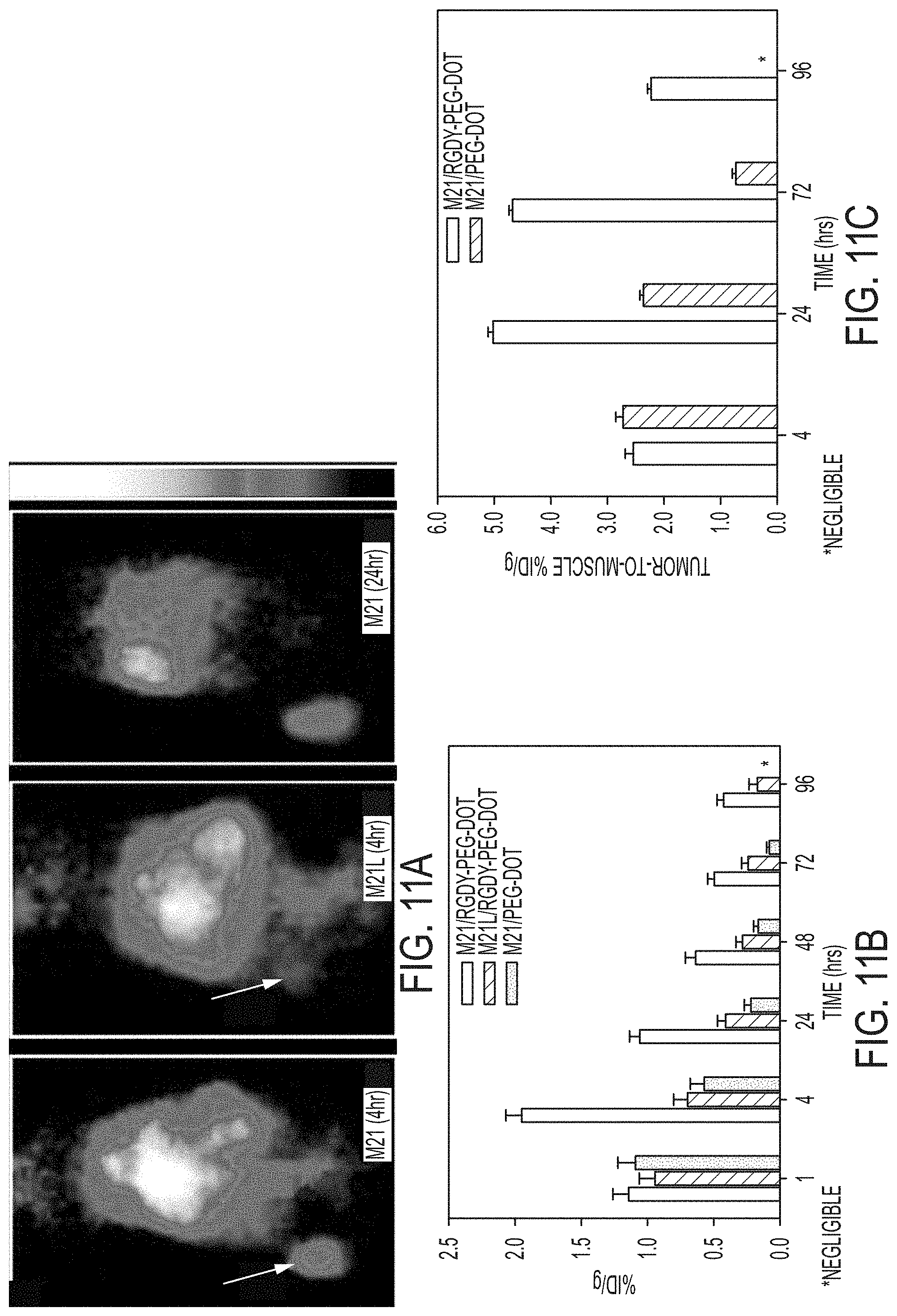
D00014
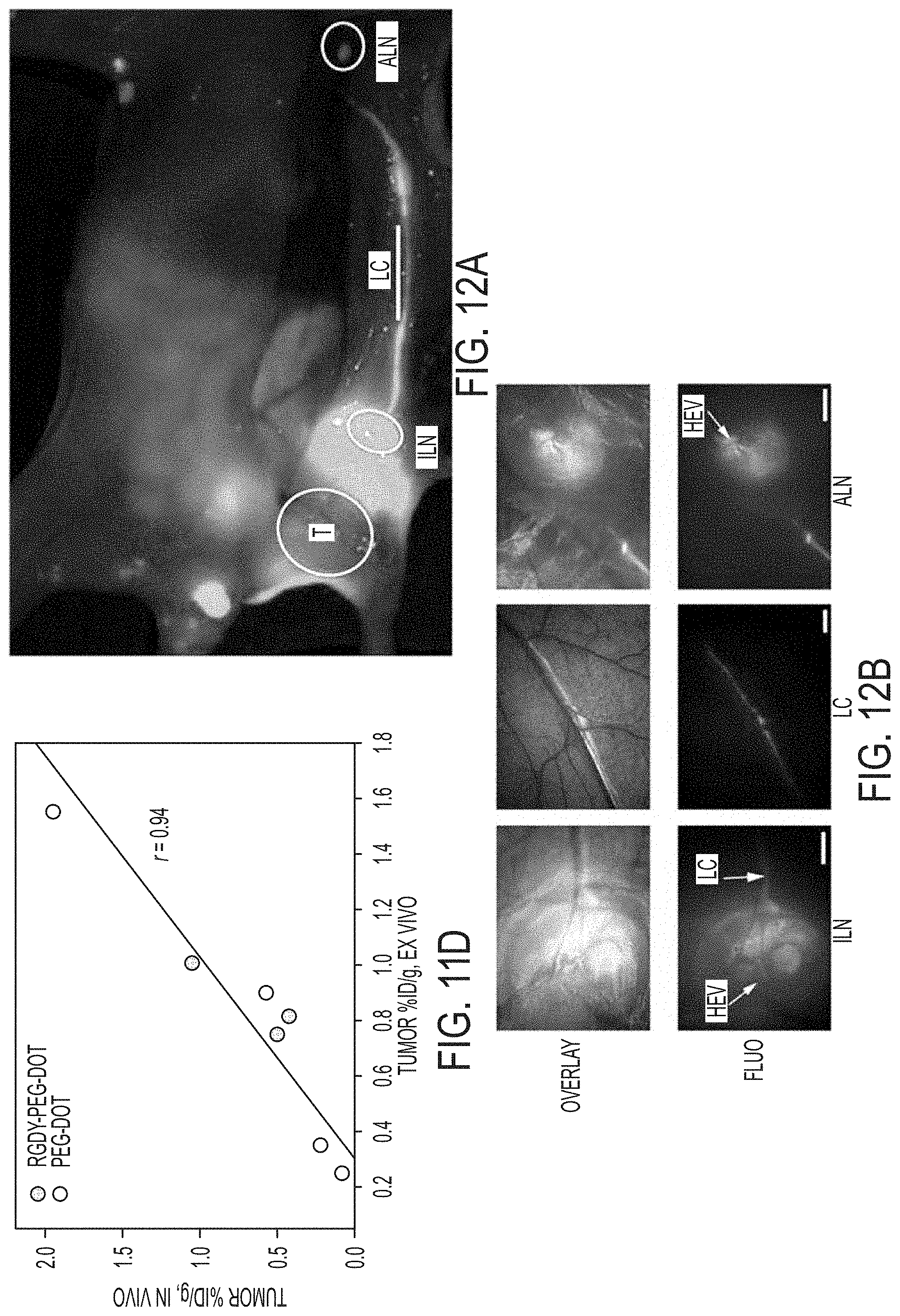
D00015

D00016

D00017
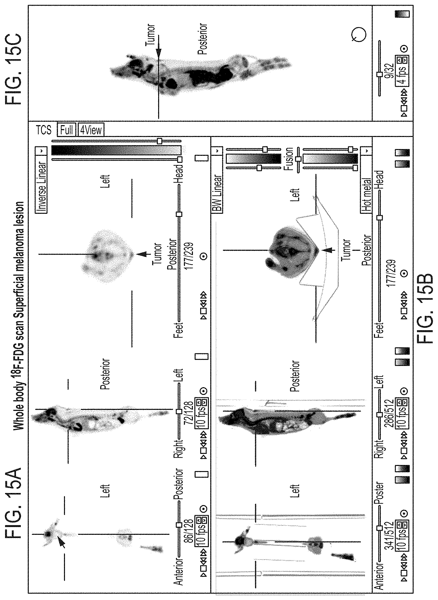
D00018

D00019

D00020

D00021

D00022

D00023

D00024
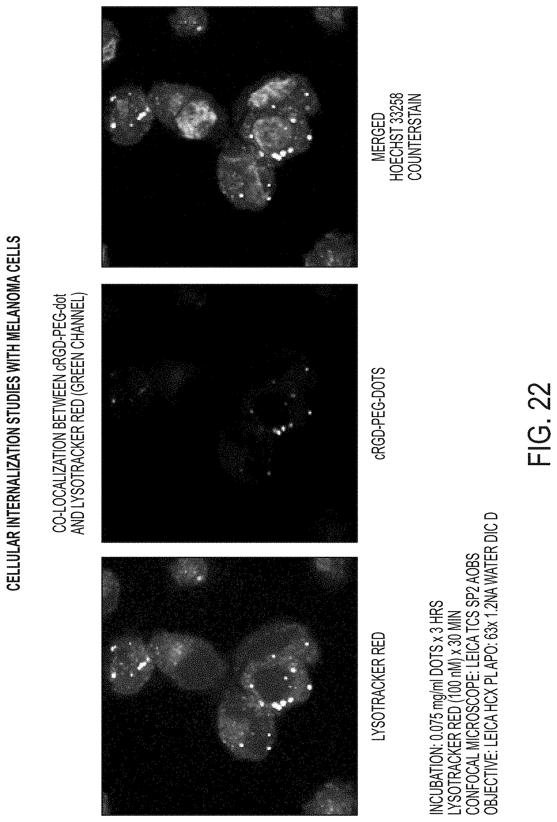
XML
uspto.report is an independent third-party trademark research tool that is not affiliated, endorsed, or sponsored by the United States Patent and Trademark Office (USPTO) or any other governmental organization. The information provided by uspto.report is based on publicly available data at the time of writing and is intended for informational purposes only.
While we strive to provide accurate and up-to-date information, we do not guarantee the accuracy, completeness, reliability, or suitability of the information displayed on this site. The use of this site is at your own risk. Any reliance you place on such information is therefore strictly at your own risk.
All official trademark data, including owner information, should be verified by visiting the official USPTO website at www.uspto.gov. This site is not intended to replace professional legal advice and should not be used as a substitute for consulting with a legal professional who is knowledgeable about trademark law.