Photosensitizing antibody-fluorophore conjugates
Kobayashi , et al. Ja
U.S. patent number 10,538,590 [Application Number 13/972,532] was granted by the patent office on 2020-01-21 for photosensitizing antibody-fluorophore conjugates. This patent grant is currently assigned to The United States of America, as represented by the Secretary, Dept. of Health and Human Services. The grantee listed for this patent is The United States of America, as represented by the Secretary, Department of Health and Human Services, The United States of America, as represented by the Secretary, Department of Health and Human Services. Invention is credited to Peter Choyke, Hisataka Kobayashi.


View All Diagrams
| United States Patent | 10,538,590 |
| Kobayashi , et al. | January 21, 2020 |
Photosensitizing antibody-fluorophore conjugates
Abstract
The present disclosure relates to compositions and methods of killing cells in vitro or in vivo. In particular examples, the method includes contacting a cell having a cell surface protein with a therapeutically effective amount of an antibody-IR700 molecule, wherein the antibody specifically binds to the cell surface protein. In particular examples the antibody recognizes a tumor-specific antigen on the surface of a tumor cell. The cell is subsequently irradiated, such as at a wavelength of 660 to 740 nm at a dose of at least 1 J cm.sup.-2, thereby killing the cell. Also provided are wearable devices that include an article of clothing, jewelry, or covering; and an NIR LED incorporated into the article, which can be used with the disclosed methods.
| Inventors: | Kobayashi; Hisataka (Laurel, MD), Choyke; Peter (Rockville, MD) | ||||||||||
|---|---|---|---|---|---|---|---|---|---|---|---|
| Applicant: |
|
||||||||||
| Assignee: | The United States of America, as
represented by the Secretary, Dept. of Health and Human
Services (Bethesda, MD) |
||||||||||
| Family ID: | 69774691 | ||||||||||
| Appl. No.: | 13/972,532 | ||||||||||
| Filed: | August 21, 2013 |
Prior Publication Data
| Document Identifier | Publication Date | |
|---|---|---|
| US 20130336995 A1 | Dec 19, 2013 | |
Related U.S. Patent Documents
| Application Number | Filing Date | Patent Number | Issue Date | ||
|---|---|---|---|---|---|
| 13180111 | Jul 11, 2011 | 8524239 | |||
| 61363079 | Jul 9, 2010 | ||||
| Current U.S. Class: | 1/1 |
| Current CPC Class: | G01N 33/5011 (20130101); A61K 47/6803 (20170801); A61K 49/0058 (20130101); A61K 31/704 (20130101); A61K 49/0032 (20130101); A61N 5/062 (20130101); C07K 16/30 (20130101); A61K 45/06 (20130101); C07K 16/2863 (20130101); C07K 16/32 (20130101); A61P 35/02 (20180101); A61P 35/00 (20180101); A61K 41/0071 (20130101); A61K 47/6869 (20170801); A61K 49/0036 (20130101); C07K 16/3069 (20130101); A61K 31/704 (20130101); A61K 2300/00 (20130101); A61K 2039/505 (20130101); C07K 2317/73 (20130101); A61N 5/1029 (20130101); A44C 15/00 (20130101); C07K 2317/77 (20130101); A41D 13/0002 (20130101); C07K 2317/24 (20130101); A41D 2400/32 (20130101) |
| Current International Class: | A61K 39/00 (20060101); C07K 16/28 (20060101); A61K 45/06 (20060101); A61K 39/395 (20060101); A61K 31/704 (20060101); A61K 41/00 (20060101); A61K 49/16 (20060101); C07K 16/30 (20060101); A61K 47/68 (20170101); C07K 16/32 (20060101); A61K 49/00 (20060101); A44C 15/00 (20060101); A41D 13/00 (20060101) |
References Cited [Referenced By]
U.S. Patent Documents
| 5494793 | February 1996 | Schindele et al. |
| 6344050 | February 2002 | Chen |
| 6534041 | March 2003 | Licha et al. |
| 7005518 | February 2006 | Peng et al. |
| 7498029 | March 2009 | Hasan et al. |
| 8524239 | September 2013 | Kobayashi et al. |
| 8623354 | January 2014 | Brown |
| 9358306 | June 2016 | Kobayashi et al. |
| 2001/0044124 | November 2001 | Bacus |
| 2004/0120949 | June 2004 | Adolf et al. |
| 2005/0157292 | July 2005 | Saitoh et al. |
| 2006/0231107 | October 2006 | Glickman et al. |
| 2007/0020272 | January 2007 | Hasan |
| 2007/0133086 | June 2007 | Wilhelm et al. |
| 2008/0073566 | March 2008 | Frangioni |
| 2008/0095699 | April 2008 | Zheng et al. |
| 2008/0253960 | October 2008 | Zheng et al. |
| 2010/0255057 | October 2010 | Hyde et al. |
| 2011/0082412 | April 2011 | Hyde et al. |
| 2012/0010558 | January 2012 | Kobayashi et al. |
| 2014/0120119 | May 2014 | Kobayashi et al. |
| 2014/0309578 | October 2014 | Anvari |
| 2015/0343060 | December 2015 | Kovar et al. |
| 2015/0343084 | December 2015 | Dilley |
| 2015/0374819 | December 2015 | Kovar |
| 2017/0122853 | May 2017 | Kobayashi et al. |
| 2018/0113246 | April 2018 | Rose et al. |
| 2018/0113247 | April 2018 | Rose et al. |
| 2018/0236076 | August 2018 | Kobayashi et al. |
| 2018/0239074 | August 2018 | Rose et al. |
| 2018/0250405 | September 2018 | Biel et al. |
| 2019/0015510 | January 2019 | Makings et al. |
| 102585003 | Jul 2012 | CN | |||
| 197 17 904 | Oct 1998 | DE | |||
| 1512963 | Mar 2005 | EP | |||
| 2003-284757 | Oct 2003 | JP | |||
| 2003-344284 | Dec 2003 | JP | |||
| 2006-515892 | Jun 2006 | JP | |||
| 2006-517230 | Jul 2006 | JP | |||
| 2007-155722 | Jun 2007 | JP | |||
| WO 2001057495 | Aug 2001 | WO | |||
| WO 03/011106 | Feb 2003 | WO | |||
| WO 03/083811 | Oct 2003 | WO | |||
| WO 2004/067038 | Aug 2004 | WO | |||
| WO 2004/071571 | Aug 2004 | WO | |||
| WO 2005099689 | Oct 2005 | WO | |||
| WO 2006/092598 | Sep 2006 | WO | |||
| WO 2008/120134 | Oct 2008 | WO | |||
| WO 2008152424 | Dec 2008 | WO | |||
| WO 2009038776 | Mar 2009 | WO | |||
| WO 2009092062 | Jul 2009 | WO | |||
| WO 2010/047611 | Apr 2010 | WO | |||
| WO 2010/085651 | Jul 2010 | WO | |||
| WO 2010121163 | Oct 2010 | WO | |||
| WO 2011025950 | Mar 2011 | WO | |||
| WO 2011038006 | Mar 2011 | WO | |||
| WO 2012076631 | Jun 2012 | WO | |||
| WO 2012082118 | Jun 2012 | WO | |||
| WO 2013009475 | Jan 2013 | WO | |||
| WO 2013044156 | Mar 2013 | WO | |||
| WO 2014/084394 | Jun 2014 | WO | |||
| WO 2014089247 | Jun 2014 | WO | |||
| WO 2014127365 | Aug 2014 | WO | |||
| WO 2014168950 | Oct 2014 | WO | |||
| WO 2015057692 | Apr 2015 | WO | |||
| WO 2015187651 | Dec 2015 | WO | |||
| WO 2015187677 | Dec 2015 | WO | |||
| WO 2016/022896 | Feb 2016 | WO | |||
| WO 2017/027247 | Feb 2017 | WO | |||
| WO 2017/031367 | Feb 2017 | WO | |||
| WO 2017031363 | Feb 2017 | WO | |||
| WO 2018080952 | May 2018 | WO | |||
| WO 2018156815 | Aug 2018 | WO | |||
| WO 2019009941 | Jan 2019 | WO | |||
Other References
|
Ogawa et al., Cancer Research, Jan. 27, 2009, 69(4): 1268-72. cited by examiner . Gao et al., Nature Biotechnology, 2004, 22(8): 969-976 and 5 pages of supplemental notes. cited by examiner . Anonymous, "Anodyne: Tratamento com tecnologia MIRE," Forumenfermagem-Projecto Feridas, Jan. 24, 2011, XP002686605, retrieved from the internet: URL:http://forumenfermagem.org/feridas/?s=anodyne, retrieved on Nov. 7, 2012. cited by applicant . Anonymous, "Near Infrared Light for the Treatment of Painful Peripheral Neuropathy," U.S. National Institutes of Health, Aug. 2, 2012, XP002686617, retrieved from the internet: URL:http://www.clinicaltrials.gov/ct2/show/NCT00125268, retrieved on Nov. 7, 2011. cited by applicant . Anonymous, "Near IR Signature Management for Combat Clothing Equipment," Australian Government Department of Defence, DSTO, Apr. 7, 2005, XP002686606, retrieved from the internet: URL:http://www.dsto.defence.gov.au/reserach/3214/?print=true, retrieved on Nov. 6, 2012. cited by applicant . Ballou et al., "Tumor Labeling in Vivo Using Cyanine-Conjugated Monoclonal Antibodies," Cancer Immunol. Immunother. 41:257-263, 1995. cited by applicant . Del Governatore et al., "Experimental Photoimmunotherapy of Hepatic Metastases of Colorectal Cancer with a 17.1A chlorin.sub.e6 Immunoconjugate," Cancer Res. 60:4200-4205, 2000. cited by applicant . European Patent Office, International Search Report and Written Opinion dated Nov. 26, 2012 for International Application No. PCT/US2012/044421. cited by applicant . Kovar et al., "A Systematic Approach to the Development of Fluorescent Contrast Agents for Optical Imagining of Mouse Cancer Models," Anal. Biochem. 367:1-12, 2007. cited by applicant . Mitchell et al., "Comparison of Two Infrared Devices in Their Effectiveness in Reducing Symptoms Associated with RLS," Physiother. Theory Prac., 2011, XP002686651, retrieved from the internet: URL:http://www.ncbi.nlm.nih.gov.pubmed/20950168, retrieved on Nov. 8, 2012. cited by applicant . Mitchell et al., "Comparison of Two Infrared Devices in Their Effectiveness in Reducing Symptoms Associated with RLS," Physiother. Theory Pract. 27:352-359, 2011. cited by applicant . Mitsunaga et al., "Cancer Cell-Selective in Vivo Near Infrared Photomimmunotherapy Targeting Specific Membrance Molecules," Nat. Med. 17:1685-1691, 2011. cited by applicant . Mitsunaga et al., "Near-Infrared Theranostic Photoimmunotherapy (PIT): Repeated Exposure of Light Enhances the Effect of Immunoconjugate," Bioconjug. Chem. 23:604-609, 2012. cited by applicant . Savellano et al., "Multiepitope HER2 Targeting Enhances Photoimmunotherapy of HER2-Overexpressing Cancer Cells with Pyropheophorbide-a Immunoconjugates," Cancer Res. 65:6371-6379, 2005. cited by applicant . Soukos et al., "Epidermal Growth Factor Receptor-Targeted Immunophotodiagnosis and Photoimmunotherapy of Oral Precancer in Vivo," Cancer Res. 61:4490-4496, 2001. cited by applicant . Van Dongen et al., "Photosensitizer-Antibody Conjugates for Detection and Therapy of Cancer," Adv Drug Deliv Rev. 56:31-52, 2004. cited by applicant . Vrouenraets et al., "Targeting of Aluminum (III) Phthalocyanine Tetrasulfonate by Use of Internalizing Monoclonal Antibodies: Improved Efficacy in Photodynamic Therapy," Cancer Res. 61:1970-1975, 2001. cited by applicant . Kirveliene et al., "Schedule-Dependent Interaction Between Doxorubicin and mTHPC-Mediated Photodynamic Therapy in Murine Hepatoma In Vitro and In Vivo," Cancer Chemother. Pharmacol. 57:65-72, 2005. cited by applicant . Peng et al., "Phthalocyanine Dye as an Extremely Photostable and Highly Fluorescent Near-Infrared Labeling Reagent," Proceed. SPIE 6097:60970E-1-60970E-12, 2006. cited by applicant . Scully et al., "Application of Fluorescence Lifetime Imaging Microscopy to the Investigation of Intracellular PDT Mechanisms," Bioimaging 5:9-18, 1997. cited by applicant . Zhu et al., "Visualization of P53.sub.264-272/HLA-A*0201 Complexes Naturally Presented on Tumor Cell Surface by a Multimeric Soluble Single-Chain T Cell Receptor," J Immunol. 176:3223-3232, 2006. cited by applicant . Zuluaga et al., "Combination of Photodynamic Therapy With Anti-Cancer Agents," Curr Med Chem. 15:1655-1673, 2008. cited by applicant . Rosenthal et al., "In Vivo Detection of Head and Neck Cancer Orthotopic Xenografts by Immunofluorescence," Laryngoscope 116:1636-1641, 2006. cited by applicant . Carter et al., "Identification and Validation of Cell Surface Antigens for Antibody Targeting in Oncology," Endocr Relat Cancer 11:659-687, 2004. cited by applicant . Davis et al., "Nanoparticle Therapeutics: An Emerging Treatment Modality for Cancer," Nat Rev Drug Dis. 7:771-782, 2008. cited by applicant . Duska et al., Combination Photoimmunotherapy and Cisplatin: Effects on Human Ovarian Cancer Ex Vivo, J Nat Cancer Inst. 91:1557-1563, 1999. cited by applicant . Serebrovskaya et al., "Targeting Cancer Cells by Using an Antireceptor Antibody-Photosensitizer Fusion Protein," Proc Nat Acad Sci. 106:9221-9225, 2009. cited by applicant . China, State Intellectual Property Office of the People's Republic of China, First Office Action for CN Application No. 201280043973.2, dated Nov. 24, 2014, and English translation (19 pages). cited by applicant . China, State Intellectual Property Office of the People's Republic of China, Second Office Action for China Application No. 201280043973.2, dated Aug. 12, 2015, and English translation (15 pages). cited by applicant . Singapore, Intellectual Property Office of Singapore, Search Report and Written Opinion for Singapore Application No. 2013091822, dated Mar. 23, 2015 (9 pages). cited by applicant . Singapore, Intellectual Property Office of Singapore, Written Opinion for Singapore Application No. 2013091822, dated Nov. 11, 2015 (10 pages). cited by applicant . EP12738664.7 Examination Report dated Jul. 6, 2016. cited by applicant . Mitsunaga et al., "Abstract 3618: Target-Specific Photo-Activatable Immunotherapy (PIT) for Cancer Based on a Monoclonal Antibody-Photosensitizer Conjugate," in Proceedings of the 102nd Annual Meeting of the American Association for Cancer Research; Apr. 2-6, 2011; Orlando, FL. Philadelphia (PA): AACR; Cancer Res. 71:3618, 2011. cited by applicant . Sano et al., "Markedly Enhanced Permeability and Retention Effects Induced by Photo-Immunotherapy of Tumors," ACS Nano. 7:717-724, 2013, including 19 pages of supporting information. cited by applicant . Japan Office Action dated Feb. 3, 2016 for Application No. 2014-520202 (with English translation). cited by applicant . China Office Action dated Feb. 22, 2016 for Application No. 201280043973.2 (with English translation). cited by applicant . Baolin et al., Practical Pathophysiology, Qing Dao Ocean University Press, Dec. 1995. (Chinese Language). cited by applicant . Barrett et al., "In vivo Diagnosis of Epidermal Growth Factor Receptor Expression using Molecular Imaging with a Cocktail of Optically Labeled Monoclonal Antibodies," Clin Cancer Res. 13:6639-6648, 2007. cited by applicant . Gleysteen et al., "Fluorescently labeled cetuximab to evaluate head and neck cancer response to treatment," Cancer Biol Ther. 6:e1-e5, 2007. cited by applicant . Kines et al., "HPV Based Photodynamic Therapy: A New Approach for Anti-Cancer Therapy," J. Immunol. 192(1): Supplement 206.8, 2014. cited by applicant . Kobayashi, "Activatable Fluorescent Imaging Probes for Cancer Detection and Diagnosis," Abstract presented at the American Chemical Society meeting in San Francisco, 2014. cited by applicant . Li-Cor, "IRDye.RTM. Infrared Dyes: Advancing Discovery with Infrared Imaging," 2010. cited by applicant . Maya et al., "Synthesis, Aggregation Behavior and Nonlinear Absorption Properties of Lead Phthalocyanines Substituted with Siloxane Chains," J Materials Chem. 13:1603-1613, 2003. cited by applicant . Nakajima et al., "Improving the Efficacy of Photoimmunotherapy (PIT) Using a Cocktail of Antibody Conjugates in a Multiple Antigen Tumor Model," Theranostics 3:357-365, 2013. cited by applicant . Watanabe et al., "Photoimmunotherapy Targeting Prostate-Specific Membrane Antigen: Are Antibody Fragments as Effective as Antibodies?," J Nucl Med. 56:140-144, 2015. cited by applicant . Xu et al., "Antibody Conjugated Magnetic Iron Oxide Nanoparticles for Cancer Cell Separation in Fresh Whole Blood," Biomaterials 32:9758-9765, 2011. cited by applicant . CN201280043973.2 Reexamination Notification dated Nov. 17, 2016, with English translation. cited by applicant . JP2014520202 Final Official Action dated Sep. 28, 2016, with English translation. cited by applicant . PCT/US2015/044168 International Search Report dated Oct. 19, 2016 (4 pages). cited by applicant . PCT/US2015/044168 Written Opinion dated Oct. 19, 2016 (7 pages). cited by applicant . Gajewski et al., "The P815 Mastocytoma Tumor Model," Curr Protoc Immunol. 43:20.4.1-20.4.18, 2001. cited by applicant . McHugh et al., "The role of suppressor T cells in regulation of immune responses," J Allergy Clin Immunol. 110:693-702, 2002. cited by applicant . Nowis et al., "The influence of photodynamic therapy on the immune response," Photodiagnosis Photodyn Ther. 2:283-298, 2005. cited by applicant . Sato et al., "Spatially selective depletion of tumor-associated regulatory T cells with near-infrared photoimmunotherapy," Sci Transl Med. 8:352ra110, 2016. cited by applicant . Steele et al., "Suppressor deletion therapy: selective elimination of T suppressor cells in vivo using a hematoporphyrin conjugated monoclonal antibody permits animals to reject syngeneic tumor cells," Cancer Immunol Immunother. 26:125-131, 1988. cited by applicant . Sugiyama et al, "Anti-CCR4 mAb selectively depletes effector-type FoxP3+CD4+ regulatory T cells, evoking antitumor immune responses in humans," Proc Natl Acad Sci. USA 110:17945-17950, 2013. cited by applicant . PCT/US2016/045090 International Search Report and Written Opinion dated Oct. 11, 2016 (12 pages). cited by applicant . Carcenac et al., "Internalisation enhances photo-induced cytotoxicity of monoclonal antibody-phthalocyanine conjugates." Br J. Cancer 85:1787-1793, 2001. cited by applicant . Chiarello, K., "In between the light and the dark: developments in Photosensitive Pharmaceuticals," Pharmaceutical Technology, pp. 48-54, Dec. 2004. cited by applicant . Chopra, "IRDye 700DX-Labeled annexin V," Molecular Imaging and Contrast Agent Database (MICAD) [Internet]. Bethesda (MD): National Center for Biotechnology Information (US); 2004-2013. Oct. 27, 2009 [updated Dec. 17, 2009]. cited by applicant . Dixit et al., "Transferrin Receptor-Targeted Theranostic Gold Nanoparticles for Photosensitizer Delivery in Brain Tumors," Nanoscale, 7(5):1782-1790, 2015. cited by applicant . Licor, "High Photostability of IRDye.RTM. 700DX," Retrieved on Aug. 23, 2018. Retrieve on https://www.licor.com/bio/products/reagents/irdye/700dx/photostability.ht- ml. cited by applicant . Maawy et al., "Near infra-red photoimmunotherapy with anti-CEA-IR700 results in extensive tumor lysis and a significant decrease in tumor burden in orthotopic mouse models of pancreatic cancer," PLoS One 10(3):e0121989, 2015. cited by applicant . Mitsunaga et al., "Immediate in vivo target-specific cancer cell death after near infrared photoimmunotherapy," BMC Cancer. 12:345, 2012. cited by applicant . Nagaya et al., "Near Infrared Photoimmunotherapy Targeting EGFR Positive Triple Negative Breast Cancer: Optimizing the Conjugate-Light Regimen," PLoS One 10(8):e0136829, 2015. cited by applicant . Nakajima et al., "Real-time monitoring of in vivo acute necrotic cancer cell death induced by near infrared photoimmunotherapy using fluorescence lifetime imaging," Cancer Res. 72(18):4622-4628, 2012. cited by applicant . Sano et al., "Acute cytotoxic effects of photoimmunotherapy assessed by 18F-FDG PET," J Nucl Med. 54(5):770-775, 2013. cited by applicant . Sano et al., "The effect of photoimmunotherapy (PIT) followed by liposomal daunorubicin in a mixed tumor model: A demonstration of the super-enhanced permeability and retention (SUPR) effect after PIT," Mol Cancer Ther. 13(2):426-432, 2014. cited by applicant . Sato et al., "Photoimmunotherapy: comparative effectiveness of two monoclonal antibodies targeting the epidermal growth factor receptor," Mol Oncol. 8(3):620-632, 2014. cited by applicant . Ali et al., "Dynamic fluorescent imaging with indocyanine green for monitoring the therapeutic effects of photoimmunotherapy," Contrast Media Mol Imaging 9(4):276-282, 2014. cited by applicant . Amoury et al., "Photoimmunotheranostic agents for triple-negative breast cancer diagnosis and therapy that can be activated on demand," Oncotarget 7(34):54925-54936, 2016. cited by applicant . de Boer et al., "A standardized light-emitting diode device for photoimmunotherapy," J Nucl Med. 55(11):1893-1898, 2014. cited by applicant . de Boer et al., "Biodistribution Study of Intravenously Injected Cetuximab-IRDye700DX in Cynomolgus Macaques," Mol Imaging Biol. 18(2):232-242, 2016. cited by applicant . Denis et al., "Synthesis, bioanalysis and biodistribution of photosensitizer conjugates for photodynamic therapy," Bioanalysis 5:1099-1114, 2013. cited by applicant . Hanaoka et al., "Glypican-3 targeted human heavy chain antibody as a drug carrier for hepatocellular carcinoma therapy," Mol Pharm. 12(6):2151-2157, 2015. cited by applicant . Hanaoka et al., "Photoimmunotherapy of hepatocellular carcinoma-targeting Glypican-3 combined with nanosized albumin-bound paclitaxel," Nanomedicine (Lond). 10(7):1139-1147, 2015. cited by applicant . Heukers et al., "Nanobody-photosensitizer conjugates for targeted photodynamic therapy," Nanomedicine 10:1441-1451, 2014. cited by applicant . Hiroshima et al., "Photoimmunotherapy Inhibits Tumor Recurrence After Surgical Resection on a Pancreatic Cancer Patient-Derived Orthotopic Xenograft (PDOX) Nude Mouse Model," Ann Surg Oncol. 22 Suppl 3:S1469-S1474, 2015. cited by applicant . Iqbal et al., "Phthalocyanine-Biomolecule Conjugated Photosensilizers for Targeted Photodynamic Therapy and Imaging," Curr Drug Metab. 16(9):816-832, 2015. cited by applicant . Ishida et al., "Trastuzumab-Based Photoimmunotherapy Integrated with Viral HER2 Transduction Inhibits Peritoneally Disseminated HER2-Negative Cancer," Mol Cancer Ther. 15(3):402-411, 2016. cited by applicant . Ito et al., "Molecular targeted photoimmunotherapy for HER2-positive human gastric cancer in combination with chemotherapy results in improved treatment outcomes through different cytotoxic mechanisms," BMC Cancer 16:37, 2016. cited by applicant . Jia et al., "Cannabinoid CB2 receptor as a new phototherapy target for the inhibition of tumor growth," Mol Pharm. 11(6):1919-1929, 2014. cited by applicant . Jing et al., "Imaging and Selective Elimination of Glioblastoma Stem Cells with Theranostic Near-Infrared-Labeled CD133-Specific Antibodies," Theranostics 6(6):862-874, 2016. cited by applicant . Kijanka et al., "Optical imaging of pre-invasive breast cancer with a combination of VHHs targeting CAIX and HER2 increases contrast and facilitates tumour characterization," EJNMMI Res. 6(1):14, 2016. cited by applicant . Kishimoto et al., "Evaluation of oxygen dependence on in vitro and in vivo cytotoxicity of photoimmunotherapy using IR-700-antibody conjugates," Free Radic Biol Med. 85:24-32, 2015. cited by applicant . Moore et al., "Photoimmunotherapy of residual disease after incomplete surgical resection in head and neck cancer models," Cancer Med. 5(7):1526-1534, 2016. cited by applicant . Nagaya et al., "Near infrared photoimmunotherapy with an anti-mesothelin antibody," Oncotarget 7(17):23361-23369, 2016. cited by applicant . Nagaya et al., "Improved micro-distribution of antibody-photon absorber conjugates after initial near infrared photoimmunotherapy (NIR-PIT)," J Control Release 232:1-8, 2016. cited by applicant . Nakajima et al., "The effects of conjugate and light dose on photo-immunotherapy induced cytotoxicity," BMC Cancer 30;14:389, 2014. cited by applicant . Nakamura et al., "MR imaging biomarkers for evaluating therapeutic effects shortly after near infrared photoimmunotherapy," Oncotarget 7(13):17254-17264, 2016. cited by applicant . Sato et al., "Photoimmunotherapy of gastric cancer peritoneal carcinomatosis in a mouse model," PLoS One 9(11):e113276, 2014. cited by applicant . Sato et al., "Near infrared photoimmunotherapy for lung metastases," Cancer Lett. 365(1):112-121, 2015. cited by applicant . Sato et al., "Near infrared photoimmunotherapy in the treatment of disseminated peritoneal ovarian cancer," Mol Cancer Ther. 14(1):141-150, 2015. cited by applicant . Sato et al., "Near infrared photoimmunotherapy in the treatment of pleural disseminated NSCLC: preclinical experience," Theranostics 5(7):698-709, 2015. cited by applicant . Sato et al., "Comparative effectiveness of light emitting diodes (LEDs) and Lasers in near infrared photoimmunotherapy," Oncotarget 7(12):14324-14335, 2016. cited by applicant . Sato et al., "Selective cell elimination in vitro and in vivo from tissues and tumors using antibodies conjugated with a near infrared phthalocyanine," RSC Adv. 5(32):25105-25114, 2015. cited by applicant . Shimoyama et al., "Viral transduction of the HER2-extracellular domain expands trastuzumab-based photoimmunotherapy for HER2-negative breast cancer cells," Breast Cancer Res Treat. 149(3):597-605, 2015. cited by applicant . Shirasu et al., "Potent and specific antitumor effect of CEA-targeted photoimmunotherapy," Int J Cancer. 135(11):2697-710, 2014. cited by applicant . van Driel et al., "EGFR targeted nanobody-photosensitizer conjugates for photodynamic therapy in a pre-clinical model of head and neck cancer," J Control Release 229:93-105, 2016. cited by applicant . von Felbert et al., "A specific photoimmunotheranostics agent to detect and eliminate skin cancer cells expressing EGFR," J Cancer Res Clin Oncol. 142(5):1003-1011, 2016. cited by applicant . Wang et al., "Theranostic Agents for Photodynamic Therapy of Prostate Cancer by Targeting Prostate-Specific Membrane Antigen," Mol Cancer Ther. 15(8):1834-1844, 2016. cited by applicant . Kobayashi, "Near infrared photoimmunotherapy: A new cancer therapy kills cancer cells with exposure of harmless near infrared light," Poster Presentation, at NEST Conference, Tokyo, Japan, Apr. 2018. cited by applicant . Nagaya et al., "Host Immunity Following Near-Infrared Photoimmunotherapy Is Enhanced with PD-1 Checkpoint Blockade to Eradicate Established Antigenic Tumors," Cancer Immunol Res. 7:401-413, 2019. cited by applicant . Nagaya et al., "Near infrared photoimmunotherapy targeting bladder cancer with a canine anti-epidermal growth factor receptor (EGFR) antibody," Oncotarget 9:19026-19038, 2018. cited by applicant . Nagaya et al., "Near infrared photoimmunotherapy with avelumab, an anti-programmed death-ligand 1 (PD-L1) antibody," Oncotarget 8:8807-8817, 2017. cited by applicant . Nagaya et al., "Syngeneic Mouse Models of Oral Cancer Are Effectively Targeted by Anti-CD44-Based NIR-PIT," Mol Cancer Res. 15:1667-1677, 2017. cited by applicant . Sato et al., "Photoinduced Ligand Release from a Silicon Phthalocyanine Dye Conjugated with Monoclonal Antibodies: A Mechanism of Cancer Cell Cytotoxicity after Near-Infrared Photoimmunotherapy," ACS Cent Sci. 4:1559-1569, 2018. cited by applicant . PCT/US2019/026488 International Search Report and Written Opinion dated Jun. 26, 2019 (15 pages). cited by applicant . Chen et al., "Tumor Vascular Permeabilization by Vascular-Targeting Photosensitization: Effects, Mechanism, and Therapeutic Implications," Clin Cancer Res. 12:917-923, Feb. 2006. cited by applicant . Greish, "Enhanced Permeability and Retention of Macromolecular Drugs in Solid Tumors: A Royal Gate for Targeted Anticancer Nanomedicines," J Drug Target. 15:457-464, Aug.-Sep. 2007. cited by applicant . Supplementary Materials from Mitsunaga et al., "Cancer Cell-Selective In Vivo Near Infrared Photoimmunotherapy Targeting Specific Membrane Molecules," Nat Med. 17:1685-1691, Nov. 2011. cited by applicant . Snyder et al., "Photodynamic Therapy: A Means to Enhanced Drug Delivery to Tumors," Cancer Res. 63:8126-8131, Dec. 2003. cited by applicant . Waite et al., "Nanoscale Drug Delivery Systems for Enhanced Drug Penetration into Solid Tumors: Current Progress and Opportunities," Crit Rev Biomed Eng. 40:21-41, Apr. 2013. cited by applicant . Wessels et al., "Advances in Cellular, Subcellular, and Nanoscale Imaging In Vitro and In Vivo," Cytometry A 77:667-676, Jul. 2010. cited by applicant. |
Primary Examiner: Sang; Hong
Attorney, Agent or Firm: Klarquist Sparkman, LLP
Parent Case Text
CROSS-REFERENCE TO RELATED APPLICATIONS
This application is a divisional of U.S. application Ser. No. 13/180,111 filed Jul. 11, 2011, now U.S. Pat. No. 8,524,239, which claims priority to U.S. Provisional Application No. 61/363,079, filed Jul. 9, 2010, both herein incorporated by reference.
Claims
We claim:
1. A phototoxic pharmaceutical composition for the treatment of a cancer expressing HER1, HER2 or PMSA, comprising: a phototoxic conjugate comprising an IR700 molecule conjugated to an antibody that binds to a cell surface protein, wherein the antibody is selected from the group consisting of Panitumumab, Trastuzumab, and J591 or an antigen binding fragment thereof; and a pharmaceutical carrier, wherein the phototoxic conjugate exhibits phototoxicity to kill cells expressing the cell surface protein bound by the antibody or antigen binding fragment of the conjugate.
2. The pharmaceutical composition of claim 1, wherein the antibody is Panitumumab.
3. The pharmaceutical composition of claim 2, wherein there are about three IR700 molecules per Panitumumab molecule.
4. The pharmaceutical composition of claim 1, wherein the antibody is Trastuzumab.
5. The pharmaceutical composition of claim 4, wherein there are about three IR700 molecules per Trastuzumab molecule.
6. The pharmaceutical composition of claim 1, wherein the antibody is J591.
7. The pharmaceutical composition of claim 6, wherein there are about two IR700 molecules per J591 molecule.
8. The pharmaceutical composition of claim 1, wherein the composition comprises physiological saline.
9. The pharmaceutical of claim 1, wherein the composition comprises water.
10. The pharmaceutical composition of claim 1, wherein the antibody fragment is selected from a Fab fragment, a Fab' fragment, a F(ab)'.sub.2 fragment, a single chain Fv (scFv) or a disulfide stabilized Fv (dsFv).
11. The pharmaceutical composition of claim 1, wherein the cell is a tumor cell.
Description
FIELD OF THE DISCLOSURE
This application relates to antibody-IR700 conjugates, and methods of their use to kill cells that specifically bind to the antibody following irradiation with infrared (NIR) light. Also provided are devices that incorporate NIR light emitting diodes (LEDs) that can also be used with the disclosed conjugates and methods.
BACKGROUND
Cancer was responsible for about 13% of all human deaths in 2007. Although there are several therapies for cancer, there remains a need for therapies that effectively kill the tumor cells while not harming non-cancerous cells.
In order to minimize the side effects of conventional cancer therapies, including surgery, radiation and chemotherapy, molecular targeted cancer therapies have been developed. Among the existing targeted therapies, monoclonal antibodies (MAb) therapy have the longest history, and to date, over 25 therapeutic MAbs have been approved by the Food and Drug Administration (FDA) (Waldmann, Nat Med 9:269-277, 2003); Reichert et al., Nat Biotechnol 23:1073-1078, 2005). Effective MAb therapy traditionally depends on three mechanisms: antibody-dependent cellular cytotoxicity (ADCC), complement-dependent cytotoxicity (CDC), and receptor blockade and requires multiple high doses of the MAb. MAbs have also been used at lower doses as vectors to deliver therapies such as radionuclides (Goldenberg et al., J Clin Oncol 24, 823-834, 2006) or chemical or biological toxins (Pastan et al., Nat Rev Cancer 6:559-565, 2006). Ultimately, however, dose limiting toxicity relates to the biodistribution and catabolism of the antibody conjugates.
Conventional photodynamic therapy (PDT), which combines a photosensitizing agent with the physical energy of non-ionizing light to kill cells, has been less commonly employed for cancer therapy because the current non-targeted photosensitizers are also taken up in normal tissues, thus, causing serious side effects, although the excitation light itself is harmless in the near infrared (NIR) range (FIG. 9).
SUMMARY OF THE DISCLOSURE
Provided herein are antibody-IR700 molecules and methods of their use for killing a target cell, such as a tumor cell. In particular examples the methods are specific in that non-target cells, such as normal cells, are not killed in significant numbers (such as less than 1% of normal cells are killed), but the target cells are. In particular examples the method includes contacting a cell having a cell surface protein with a therapeutically effective amount of an antibody-IR700 molecule, wherein the antibody (or other specific binding agent) specifically binds to the cell surface protein. Specific non-limiting examples of antibody-IR700 molecules include Panitumumab-IR700, Trastuzumab-IR700, and HuJ591-IR700. The cell is irradiated at a wavelength of 660 to 740 nm, such as 660 to 710 nm (for example, 680 nm) at a dose of at least 1 J cm.sup.-2 (such as at least 50 J cm.sup.-2), thereby killing the cell.
Any target cell can be killed with the disclosed antibody-IR700 molecules and methods, for example by using an antibody that binds to a protein on the target cell surface (such as a receptor), wherein the protein on the target cell surface is not significantly found on non-target cells (such as normal healthy cells) and thus the antibody will not significantly bind to the non-target cells. In one example the cell surface protein is a tumor-specific protein, such as HER1, HER2, or PSMA.
In some examples, the cell to be killed is present in a subject. In such examples, the method can include administering a therapeutically effective amount of the antibody-IR700 molecule to the subject and irradiating the subject, for example irradiating a tumor in the subject. In some examples, the method can further include selecting a subject with a tumor that expresses a cell surface protein that can specifically bind to the antibody-IR700 molecule.
Also provided devices, such as those that can be worn by a patient. Such devices can include an article of clothing, jewelry, or a covering, and a near infrared (NIR) light emitting diode (LED) that is incorporated into the article of clothing, jewelry, or covering. Such devices can further include power and/or cooling sources. This permits the patient to wear the device (or be covered by the device) for extended periods of time, thus permitting treatment of tumor cells present in the blood or circulatory system. Methods of using the device are also provided.
The foregoing and other features of the disclosure will become more apparent from the following detailed description of a several embodiments which proceeds with reference to the accompanying figures.
BRIEF DESCRIPTION OF THE FIGURES
FIG. 1A is a digital image showing the labeling of cells with a Trastuzumab-IR700 conjugate (Tra-IR700) at 4.degree. C. for 1 hour or 37.degree. C. for 6 hours. Light images also shown. Scale bar, 30 .mu.m.
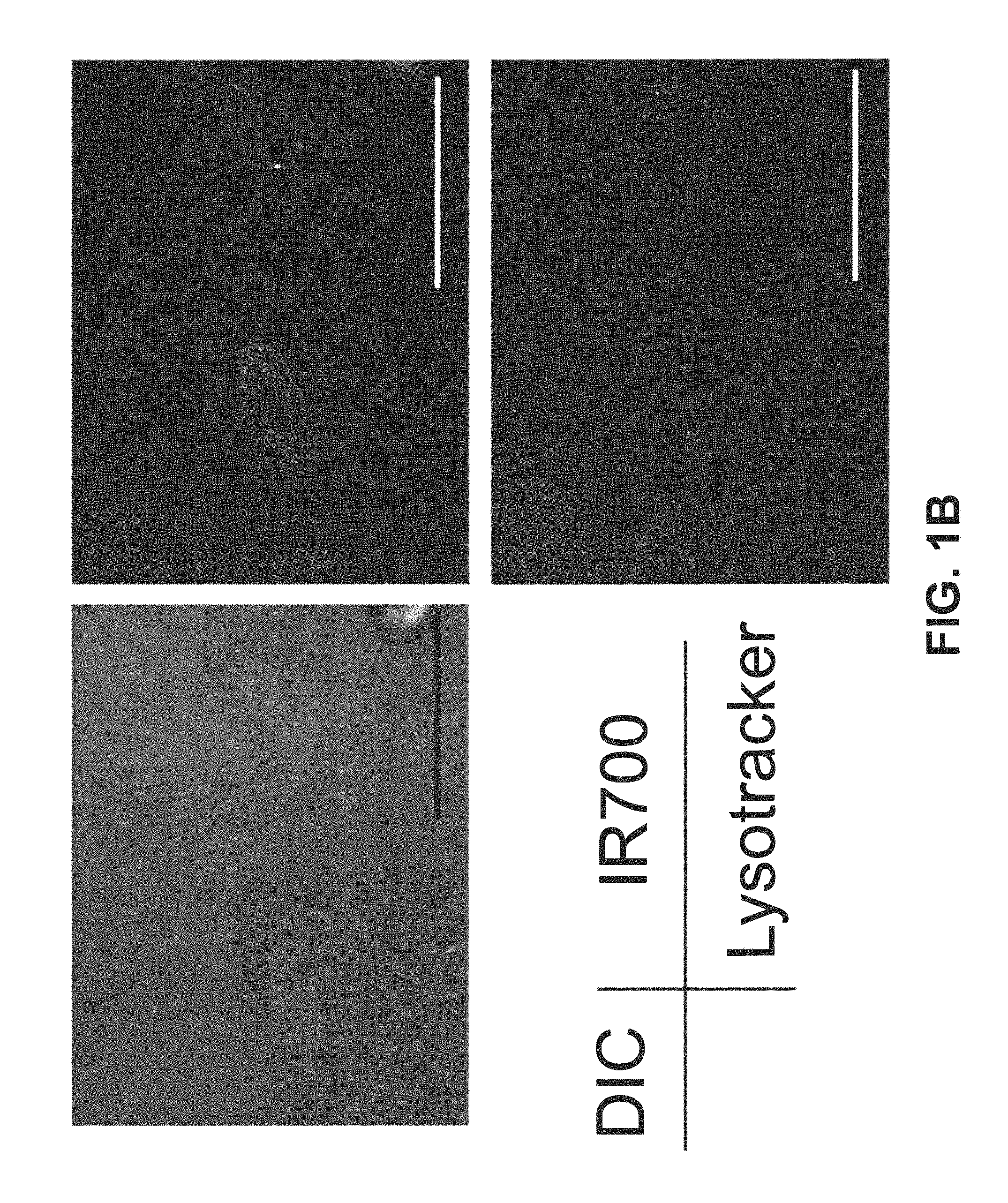
FIG. 1B is a digital image showing the lysosomal localization of Tra-1R700 6 h after incubation. Scale bar, 50 .mu.m.
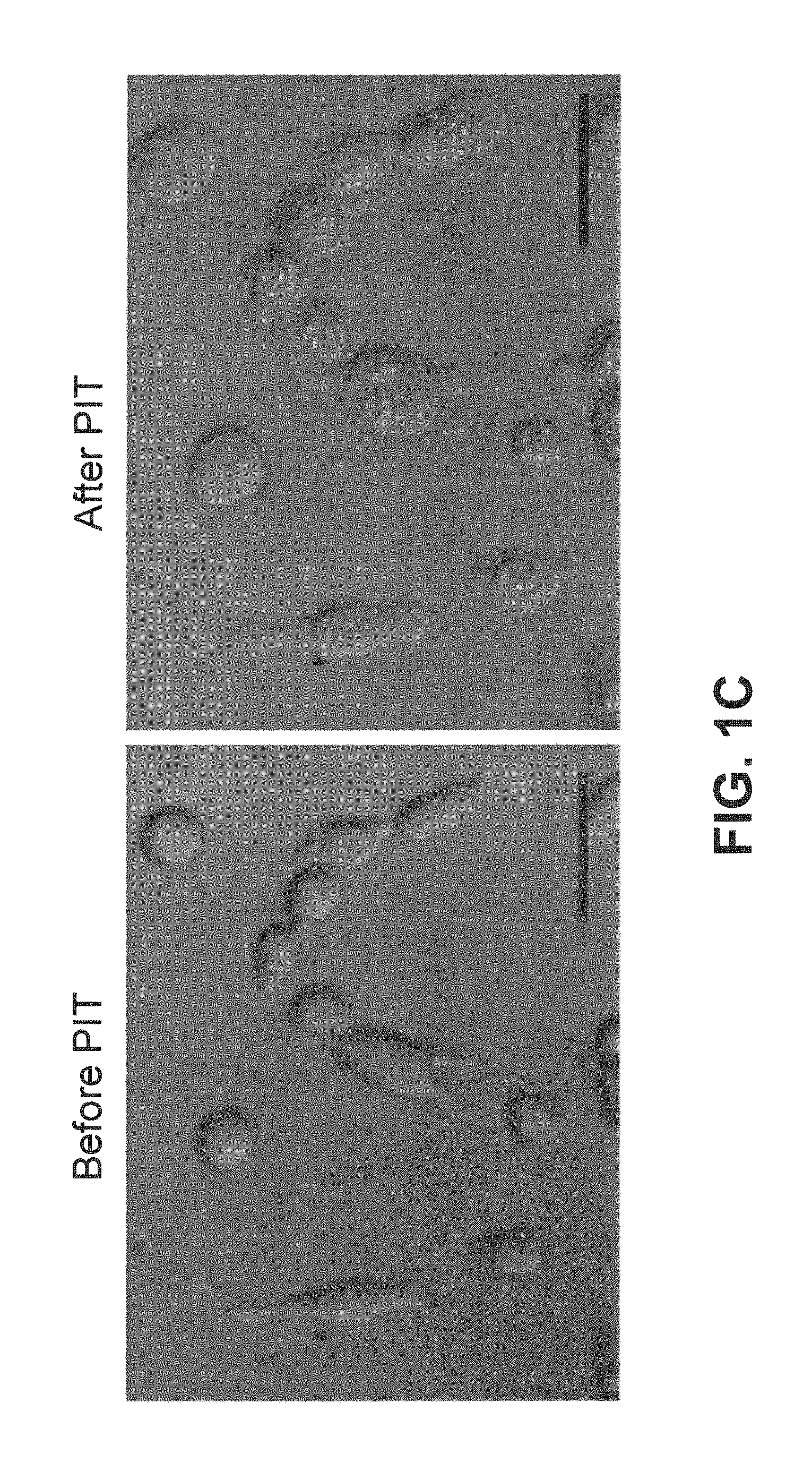
FIG. 1C is a digital image showing is a digital image showing before and after incubation with Tra-IR700 at 37.degree. C. for 6 hours followed by photoimmunotherapy (PIT). Scale bar, 50 .mu.m.
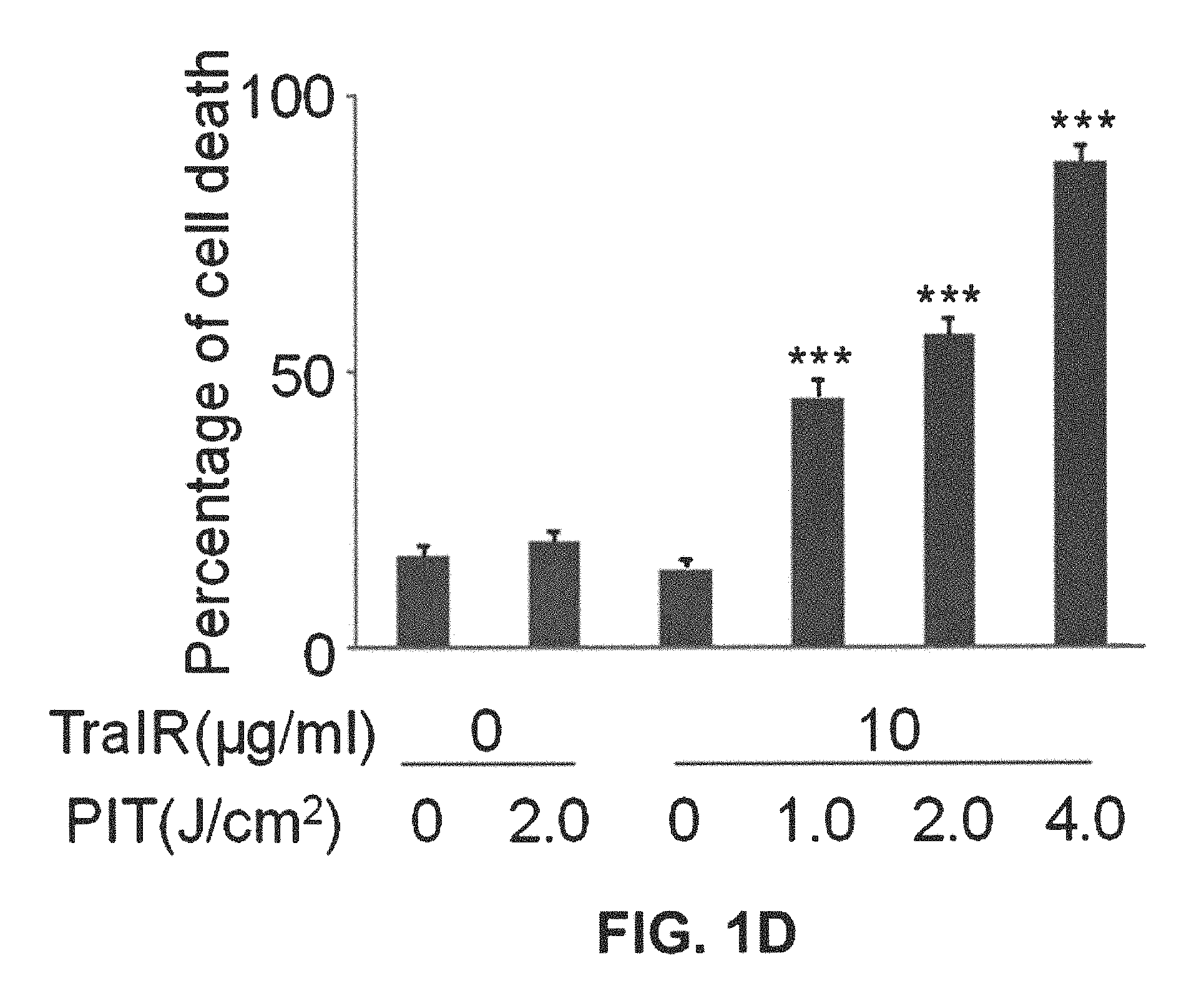
FIG. 1D is a bar graph showing the irradiation dose dependent and target specific cell death in response to Tra-1R700 mediated PIT. Data are means.+-.s.e.m. (n=at least 4, *** P<0.001 vs. non treatment control, Student's t test).
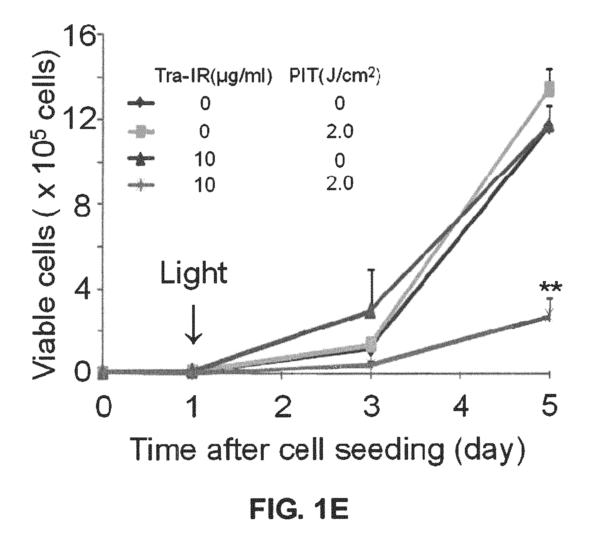
FIG. 1E is a bar graph showing the long term growth inhibition in response to Tra-1R700 mediated PIT. Data are means.+-.s.e.m. (n=3, ** P<0.01 vs. non treatment control, Student's t test).
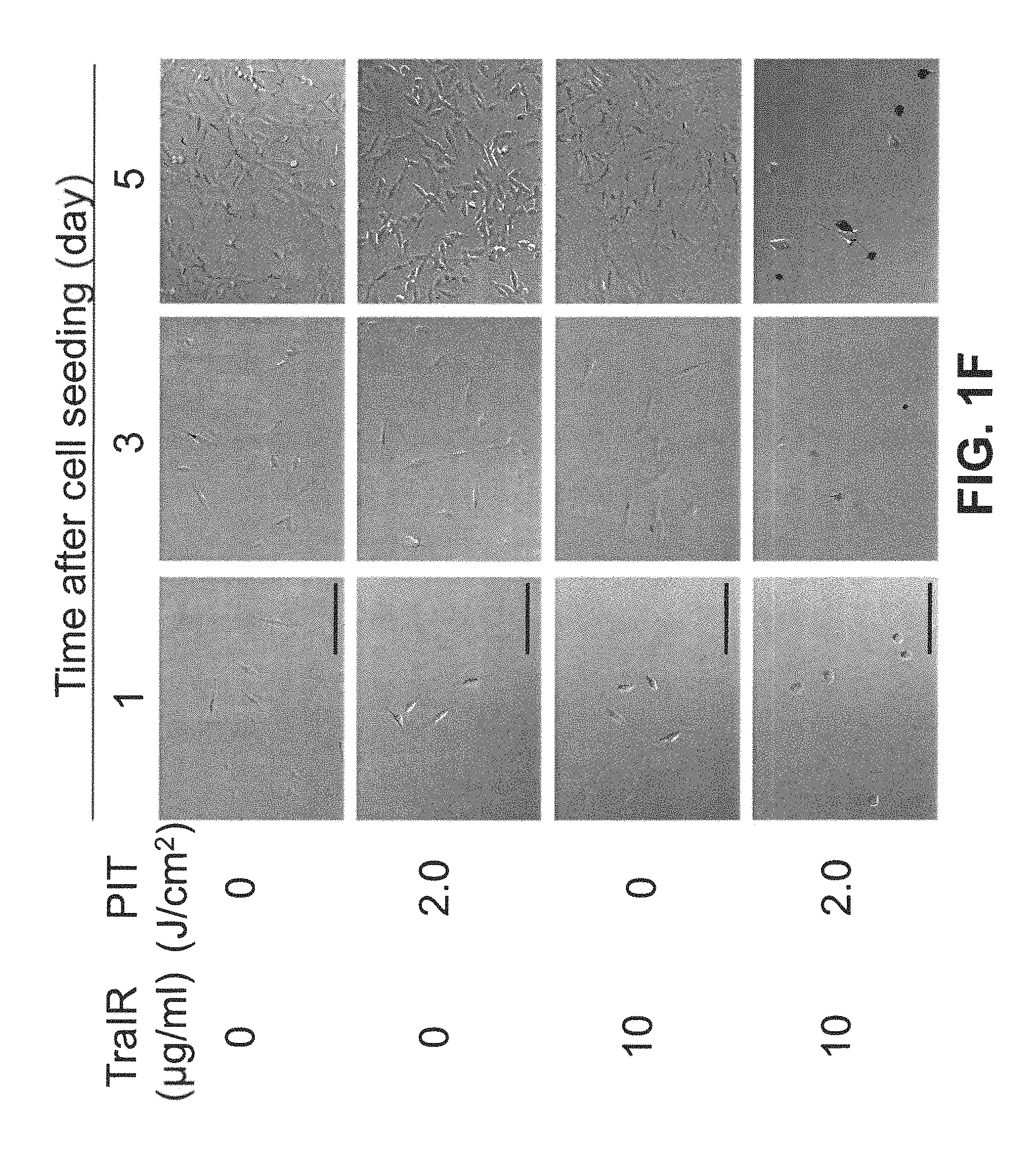
FIG. 1F is a digital image showing the microscopic observation of growth inhibition in response to TraIR700 mediated PIT. Scale bar, 100 .mu.m
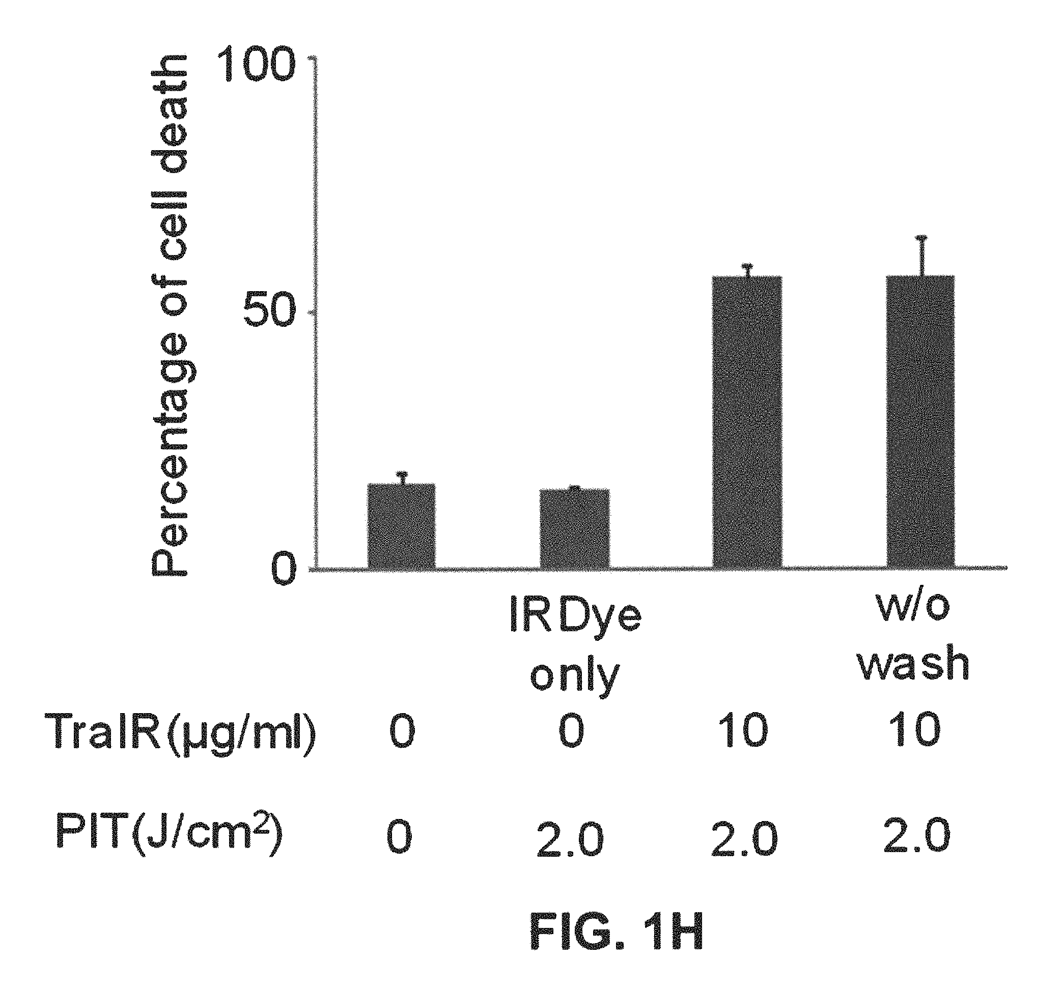
FIG. 1G is a bar graph showing that internalization of Tra-1R700 was not required for phototoxic cell death. Data are means.+-.s.e.m. (n=3).
FIG. 1H is a bar graph showing that target specific membrane binding of Tra-1R700 only induced phototoxic cell death. Data are means.+-.s.e.m. (n=3).
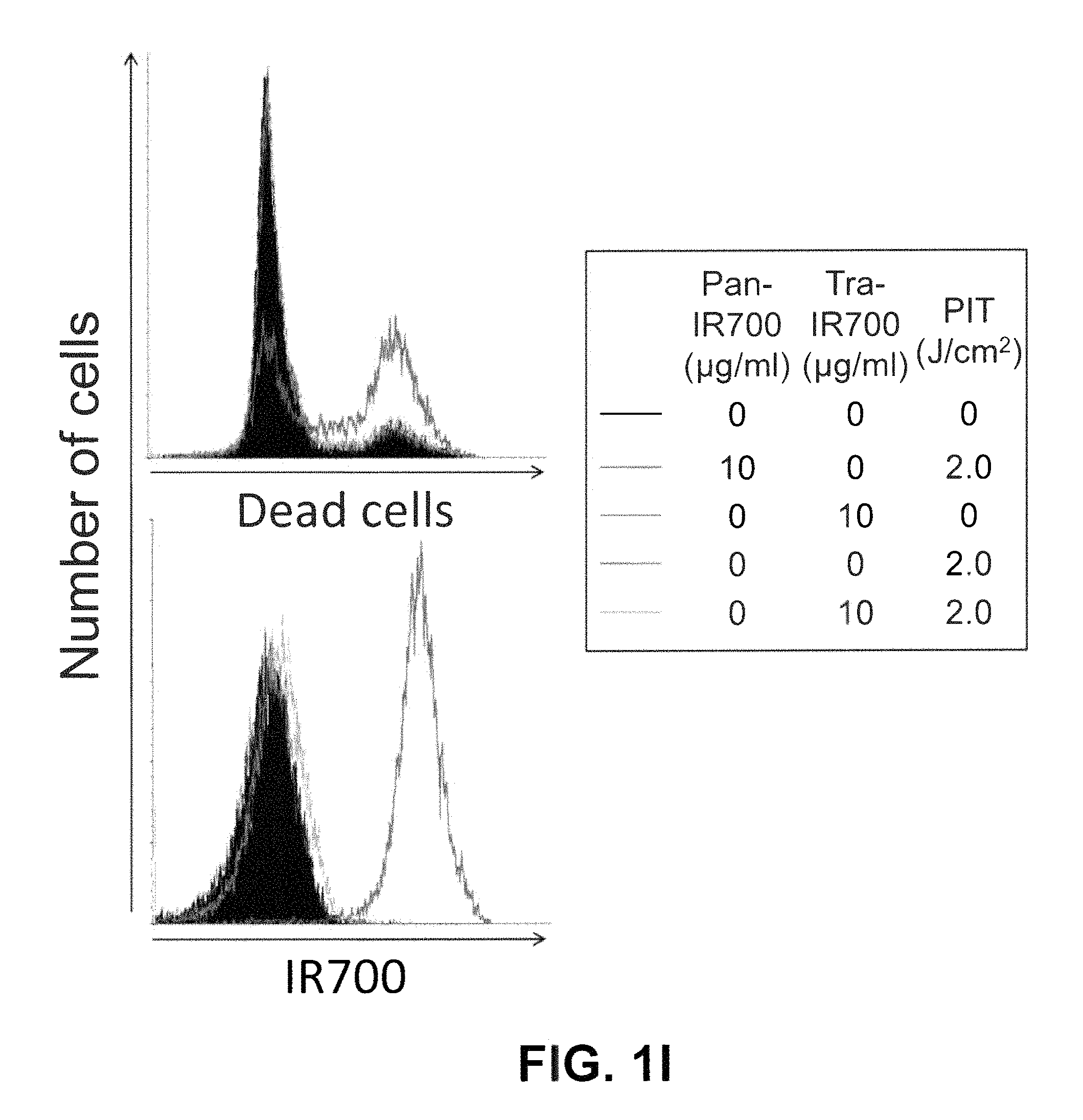
FIG. 1I is a graph showing that HER2 negatively expressing A431 cells did not show phototoxic effects with Tra-1R700 mediated PIT (n=3).
FIG. 1J is a bar graph showing sodium azide (NaN.sub.3) concentration dependent inhibition of phototoxic cell death induced by Tra-1R700 mediated PIT. Data are means.+-.s.e.m. (n=3, *** P<0.001, ** P<0.01 vs. 2.0 J cm-2 PIT treatment without NaN3 control, Student's t test). DIC: differential interference contrast. PanIR: Pan-1R700.
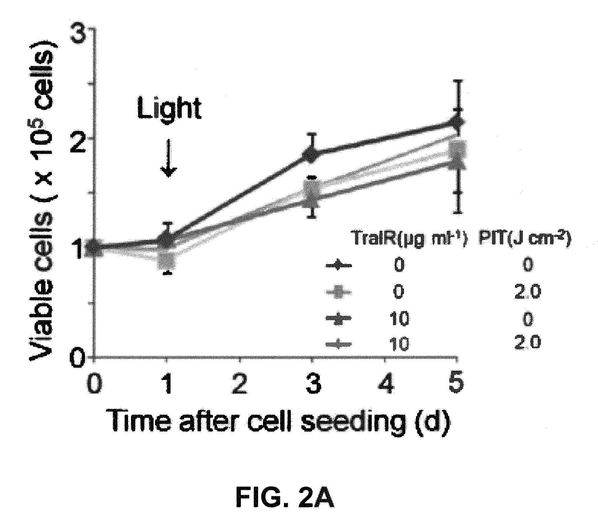
FIG. 2A is a graph showing that long term growth inhibition was not observed in Balb/3T3 (HER2 negative) cells treated with Tra-1R700 (TraIR) and exposed to light. Data are means.+-.s.e.m. (n=3).
FIG. 2B is a digital image showing that Free IR700 dye did not incorporate into 3T3/HER2 cells. Fluorescence image was taken without washing the cells. Cells were darker than the media containing free IR700 dye. Scale bar, 50 .mu.m.
FIG. 2C is a graph showing that TraIR700 mediated phototoxicity was dose-dependently blocked by the excess of unconjugated trastuzumab (Tra). Data are means.+-.s.e.m. (n=3).
FIG. 2D is a graph showing that Tra-1R700 binding for 3T3/HER2 cells was blocked by unconjugated trastuzumab dose-dependent manner (n=3). DIC: differential interference contrast.
FIG. 3A is a digital image showing that induction of target specific photoimmunotherapy (PIT) lead to HER2 expressing cell specific necrotic cell death. Scale bar, 50 .mu.m.
FIG. 3B is a digital image showing that HER2 specific cell death was confirmed with fluorescence microscopy with LIVE/DEAD Green staining. Scale bar, 100 .mu.m.
FIG. 3C is a plot showing flow cytometric analysis for detecting HER2-specific cell death induced by Tra-1R700 (TraIR) mediated PIT. Upper left quadrant: TraIR700 positive, live cells; upper right quadrant; Tra-1R700 positive, dead cells; lower left quadrant: Tra-1R700 negative, live cells; lower right quadrant: Tra-1R700 negative, dead cells (n=3). DIC: differential interference contrast.
FIG. 4A is a digital image showing the biodistribution of Tra-1R700. 3T3/HER2 tumors (both sides of dorsum) were visualized with IR700 fluorescence as early as 1 day after Tra-1R700 injection (300 .mu.g). Right side of the tumor was irradiated with near infrared (NIR) light on day 1, while left side of the tumor was covered with black tape. Tumor shrinkage was confirmed on day 7. Dashed line: irradiated tumor, solid line: non-irradiated tumor. No other specific localization of IR700 was found except for the bladder accumulation on day 1 due to the excretion of unbound dye (n=5 mice).
FIG. 4B is a graph showing mean tumor volume following administration in vivo of Tra-IR700 or carrier alone followed by PIT (50 J cm.sup.-2). Data are means.+-.s.e.m. (at least n=12 mice in each group, *** P<0.001, '' P<0.01 vs. non treatment control, Kruskal-Wallis test with post-test). Tra: trastuzumab.
FIG. 5A is a digital image showing a microscopic observation of before and after Pan-1R700 mediated PIT. Scale bar, 50 um.
FIG. 5B is a graph showing irradiation dose dependent and target specific cell death in response to Pan-1R700 (PanIR) mediated PIT. Data are means.+-.s.e.m. (n=at least 4, *** P<0.001 vs. non treatment control, Student's t test).
FIG. 5C is a digital image showing EGFR expressing cell specific necrotic cell death was induced by Pan-1R700 mediated PIT. Scale bar, 50 .mu.m. DIC: differential interference contrast.
FIG. 6A is a digital image showing specific localization of panitumumab-IR700 conjugate (Pan-IR700) in a mouse previously administered A431 cells. HER1 positive A431 tumor (left dorsum) was selectively visualized as early as 1 d after Pan-1R700 injection (50 .mu.g). HER1 negative 3T3/HER2 tumor (right dorsum) did not show detectable fluorescence (n=5 mice).
FIG. 6B is a graph showing the IR700 signal intensity in A431 tumors over time following injection of two different doses (50 .mu.g and 300 .mu.g) of Pan-IR700. Data are means.+-.s.e.m. (n=4 each mice).
FIG. 6C is a graph showing the tumor to background ratio of IR700 fluorescence intensity in A431 tumors over time following injection of two different doses (50 .mu.g and 300 .mu.g) of Pan-IR700. Data are means.+-.s.e.m. (n=4 each mice).
FIG. 6D is a digital image showing the biodistribution of Pan-1R700. A431 tumors (both sides of dorsum) were selectively visualized with IR700 fluorescence as early as 1 day after Pan-1R700 injection (300 .mu.g). Right side of the tumor was irradiated with near infrared (NIR) light on day 1, while left side of the tumor was covered with black tape. Tumor shrinkage was confirmed on day 7. Dashed line: irradiated tumor, solid line: non-irradiated tumor.
FIG. 6E is a graph showing mean tumor volume following administration in vivo of Pan-IR700 or carrier alone followed by PIT (30 J cm.sup.-2). PIT was performed on day 1 after Pan-1R700 injection (day 5 after tumor inoculation). Data are means.+-.s.e.m. (at least n=12 mice in each group, *** P<0.001 vs. other control groups, Kruskal-Wallis test with post-test).
FIG. 6F is a graph showing survival time following administration in vivo of Pan-IR700 or carrier alone followed by PIT (30 J cm.sup.-2) (at least n=12 mice in each group, *** P<0.001 vs. other control groups, log-rank test with Bonferroni's correction for multiplicity.
FIG. 6G is a digital image showing hematoxylin and eosin stained histology images (.times.40 and .times.200) 4 days after PIT treated (right) and untreated (left) tumors. n=5 mice; Scale bar, 100 um. Pan: panitumumab.
FIG. 6H is a graph showing that high-dose administration of Pan-1R700 lead to higher antitumor efficacy of Pan-1R700 mediated PIT for A431 tumors in vivo. Tumor growth inhibition by Pan-1R700 mediated PIT was Pan-1R700 dose-dependently observed. Data are means.+-.s.e.m. (at least n=12 mice in each group).
FIG. 7 is a digital image showing the biodistribution of J591-1R700. PC3-PIP tumors were selectively visualized with IR700 fluorescence after J591-1R700 injection (100 .mu.g). Right side of the tumor was irradiated with near infrared (NIR) light on days 4, 12, and 13 while left side of the tumor was covered with black tape. Tumor shrinkage was confirmed on day 5.
FIG. 8 is a digital image showing the microscopic observation of before and after PIT for various cells in the presence of with Tra-1R700 for HER2+ cells, Pan-1R700 for HER1+ cells, and huJ591-1R700 for PSMA+ cells. Scale bar, 50 .mu.m. DIC: differential interference contrast.
FIG. 9A is a schematic drawing showing a schema for explaining selective cancer therapy with PIT in the context of other physical cancer therapies employing electro-magnetic wave irradiation. Although other physical cancer therapies induce different types of damages in the normal tissue, PIT dedicatedly damages cancer cells without damaging normal cells or tissues.
FIG. 9B is a schematic drawing showing a schema for explaining photo-physical, chemical and biological basis of PIT. Humanized antibodies are employed as a delivery vehicle because of its highest binding specificity, greatest in vivo target delivery, low immunogenecity among the clinically applicable targeting reagents. A hydrophilic phtalocyanine is employed as an activatable cytotoxic "Nano-dynamite" reagent because of its great absorption of near infrared light of 700 nm and strong cytotoxicity induced only when associating with the cell membrane. Near infrared light of 700 nm is employed as an initiator for activating cytotoxicity because of its high energy among non-harmful non-ionizing photons and great in vivo tissue penetration.
DETAILED DESCRIPTION OF SEVERAL EMBODIMENTS
Unless otherwise explained, all technical and scientific terms used herein have the same meaning as commonly understood by one of ordinary skill in the art to which a disclosed invention belongs. The singular terms "a," "an," and "the" include plural referents unless context clearly indicates otherwise. Similarly, the word "or" is intended to include "and" unless the context clearly indicates otherwise. "Comprising" means "including." Hence "comprising A or B" means "including A" or "including B" or "including A and B."
Suitable methods and materials for the practice and/or testing of embodiments of the disclosure are described below. Such methods and materials are illustrative only and are not intended to be limiting. Other methods and materials similar or equivalent to those described herein can be used. For example, conventional methods well known in the art to which a disclosed invention pertains are described in various general and more specific references, including, for example, Sambrook et al., Molecular Cloning: A Laboratory Manual, 2d ed., Cold Spring Harbor Laboratory Press, 1989; Sambrook et al., Molecular Cloning: A Laboratory Manual, 3d ed., Cold Spring Harbor Press, 2001; Ausubel et al., Current Protocols in Molecular Biology, Greene Publishing Associates, 1992 (and Supplements to 2000); Ausubel et al., Short Protocols in Molecular Biology: A Compendium of Methods from Current Protocols in Molecular Biology, 4th ed., Wiley & Sons, 1999; Harlow and Lane, Antibodies: A Laboratory Manual, Cold Spring Harbor Laboratory Press, 1990; and Harlow and Lane, Using Antibodies: A Laboratory Manual, Cold Spring Harbor Laboratory Press, 1999.
The sequences associated with all GenBank Accession numbers referenced herein are incorporated by reference for the sequence available on Jul. 9, 2010.
In order to facilitate review of the various embodiments of the disclosure, the following explanations of specific terms are provided:
Administration: To provide or give a subject an agent, such as an antibody-IR700 molecule, by any effective route. Exemplary routes of administration include, but are not limited to, topical, injection (such as subcutaneous, intramuscular, intradermal, intraperitoneal, intratumoral, and intravenous), oral, sublingual, rectal, transdermal, intranasal, vaginal and inhalation routes.
Antibody: A polypeptide ligand comprising at least a light chain or heavy chain immunoglobulin variable region which specifically recognizes and binds an epitope of an antigen, such as a tumor-specific protein. Antibodies are composed of a heavy and a light chain, each of which has a variable region, termed the variable heavy (V.sub.H) region and the variable light (V.sub.L) region. Together, the V.sub.H region and the V.sub.L region are responsible for binding the antigen recognized by the antibody.
Antibodies include intact immunoglobulins and the variants and portions of antibodies well known in the art, such as Fab fragments, Fab' fragments, F(ab)'.sub.2 fragments, single chain Fv proteins ("scFv"), and disulfide stabilized Fv proteins ("dsFv"). A scFv protein is a fusion protein in which a light chain variable region of an immunoglobulin and a heavy chain variable region of an immunoglobulin are bound by a linker, while in dsFvs, the chains have been mutated to introduce a disulfide bond to stabilize the association of the chains. The term also includes genetically engineered forms such as chimeric antibodies (for example, humanized murine antibodies), heteroconjugate antibodies (such as, bispecific antibodies). See also, Pierce Catalog and Handbook, 1994-1995 (Pierce Chemical Co., Rockford, Ill.); Kuby, J., Immunology, 3.sup.rd Ed., W. H. Freeman & Co., New York, 1997
Typically, a naturally occurring immunoglobulin has heavy (H) chains and light (L) chains interconnected by disulfide bonds. There are two types of light chain, lambda (.lamda.) and kappa (k). There are five main heavy chain classes (or isotypes) which determine the functional activity of an antibody molecule: IgM, IgD, IgG, IgA and IgE.
Each heavy and light chain contains a constant region and a variable region, (the regions are also known as "domains"). In combination, the heavy and the light chain variable regions specifically bind the antigen. Light and heavy chain variable regions contain a "framework" region interrupted by three hypervariable regions, also called "complementarity-determining regions" or "CDRs." The extent of the framework region and CDRs have been defined (see, Kabat et al., Sequences of Proteins of Immunological Interest, U.S. Department of Health and Human Services, 1991, which is hereby incorporated by reference). The Kabat database is now maintained online. The sequences of the framework regions of different light or heavy chains are relatively conserved within a species, such as humans. The framework region of an antibody, that is the combined framework regions of the constituent light and heavy chains, serves to position and align the CDRs in three-dimensional space.
The CDRs are primarily responsible for binding to an epitope of an antigen. The CDRs of each chain are typically referred to as CDR1, CDR2, and CDR3, numbered sequentially starting from the N-terminus, and are also typically identified by the chain in which the particular CDR is located. Thus, a V.sub.H CDR3 is located in the variable domain of the heavy chain of the antibody in which it is found, whereas a V.sub.L CDR1 is the CDR1 from the variable domain of the light chain of the antibody in which it is found. Antibodies with different specificities (i.e. different combining sites for different antigens) have different CDRs. Although it is the CDRs that vary from antibody to antibody, only a limited number of amino acid positions within the CDRs are directly involved in antigen binding. These positions within the CDRs are called specificity determining residues (SDRs).
References to "V.sub.H" or "VH" refer to the variable region of an immunoglobulin heavy chain, including that of an Fv, scFv, dsFv or Fab. References to "V.sub.L" or "VL" refer to the variable region of an immunoglobulin light chain, including that of an Fv, scFv, dsFv or Fab.
A "monoclonal antibody" is an antibody produced by a single clone of B lymphocytes or by a cell into which the light and heavy chain genes of a single antibody have been transfected. Monoclonal antibodies are produced by methods known to those of skill in the art, for instance by making hybrid antibody-forming cells from a fusion of myeloma cells with immune spleen cells. Monoclonal antibodies include humanized monoclonal antibodies.
A "chimeric antibody" has framework residues from one species, such as human, and CDRs (which generally confer antigen binding) from another species, such as a murine antibody that specifically binds mesothelin.
A "humanized" immunoglobulin is an immunoglobulin including a human framework region and one or more CDRs from a non-human (for example a mouse, rat, or synthetic) immunoglobulin. The non-human immunoglobulin providing the CDRs is termed a "donor," and the human immunoglobulin providing the framework is termed an "acceptor." In one embodiment, all the CDRs are from the donor immunoglobulin in a humanized immunoglobulin. Constant regions need not be present, but if they are, they must be substantially identical to human immunoglobulin constant regions, i.e., at least about 85-90%, such as about 95% or more identical. Hence, all parts of a humanized immunoglobulin, except possibly the CDRs, are substantially identical to corresponding parts of natural human immunoglobulin sequences. A "humanized antibody" is an antibody comprising a humanized light chain and a humanized heavy chain immunoglobulin. A humanized antibody binds to the same antigen as the donor antibody that provides the CDRs. The acceptor framework of a humanized immunoglobulin or antibody may have a limited number of substitutions by amino acids taken from the donor framework. Humanized or other monoclonal antibodies can have additional conservative amino acid substitutions which have substantially no effect on antigen binding or other immunoglobulin functions. Humanized immunoglobulins can be constructed by means of genetic engineering (see for example, U.S. Pat. No. 5,585,089).
A "human" antibody (also called a "fully human" antibody) is an antibody that includes human framework regions and all of the CDRs from a human immunoglobulin. In one example, the framework and the CDRs are from the same originating human heavy and/or light chain amino acid sequence. However, frameworks from one human antibody can be engineered to include CDRs from a different human antibody. All parts of a human immunoglobulin are substantially identical to corresponding parts of natural human immunoglobulin sequences.
"Specifically binds" refers to the ability of individual antibodies to specifically immunoreact with an antigen, such as a tumor-specific antigen, relative to binding to unrelated proteins, such as non-tumor proteins, for example .beta.-actin. For example, a HER2-specific binding agent binds substantially only the HER-2 protein in vitro or in vivo. As used herein, the term "tumor-specific binding agent" includes tumor-specific antibodies and other agents that bind substantially only to a tumor-specific protein in that preparation.
The binding is a non-random binding reaction between an antibody molecule and an antigenic determinant of the T cell surface molecule. The desired binding specificity is typically determined from the reference point of the ability of the antibody to differentially bind the T cell surface molecule and an unrelated antigen, and therefore distinguish between two different antigens, particularly where the two antigens have unique epitopes. An antibody that specifically binds to a particular epitope is referred to as a "specific antibody".
In some examples, an antibody (such as an antibody-IR700 molecule) specifically binds to a target (such as a cell surface protein) with a binding constant that is at least 10.sup.3 M.sup.-1 greater, 10.sup.4M.sup.-1 greater or 10.sup.5 M.sup.-1 greater than a binding constant for other molecules in a sample or subject. In some examples, an antibody (e.g., monoclonal antibody) or fragments thereof, has an equilibrium constant (Kd) of 1 nM or less. For example, an antibody binds to a target, such as tumor-specific protein with a binding affinity of at least about 0.1.times.10.sup.-8 M, at least about 0.3.times.10.sup.-8 M, at least about 0.5.times.10.sup.-8 M, at least about 0.75.times.10.sup.-8 M, at least about 1.0.times.10.sup.-8 M, at least about 1.3.times.10.sup.-8 Mat least about 1.5.times.10.sup.-8M, or at least about 2.0.times.10.sup.-8 M. Kd values can, for example, be determined by competitive ELISA (enzyme-linked immunosorbent assay) or using a surface-plasmon resonance device such as the Biacore T100, which is available from Biacore, Inc., Piscataway, N.J.
Antibody-IR700 molecule or antibody-IR700 conjugate: A molecule that includes both an antibody, such as a tumor-specific antibody, conjugated to IR700. In some examples the antibody is a humanized antibody (such as a humanized monoclonal antibody) that specifically binds to a surface protein on a cancer cell.
Antigen (Ag): A compound, composition, or substance that can stimulate the production of antibodies or a T cell response in an animal, including compositions (such as one that includes a tumor-specific protein) that are injected or absorbed into an animal. An antigen reacts with the products of specific humoral or cellular immunity, including those induced by heterologous antigens, such as the disclosed antigens. "Epitope" or "antigenic determinant" refers to the region of an antigen to which B and/or T cells respond. In one embodiment, T cells respond to the epitope, when the epitope is presented in conjunction with an MHC molecule. Epitopes can be formed both from contiguous amino acids or noncontiguous amino acids juxtaposed by tertiary folding of a protein. Epitopes formed from contiguous amino acids are typically retained on exposure to denaturing solvents whereas epitopes formed by tertiary folding are typically lost on treatment with denaturing solvents. An epitope typically includes at least 3, and more usually, at least 5, about 9, or about 8-10 amino acids in a unique spatial conformation. Methods of determining spatial conformation of epitopes include, for example, x-ray crystallography and nuclear magnetic resonance.
Examples of antigens include, but are not limited to, peptides, lipids, polysaccharides, and nucleic acids containing antigenic determinants, such as those recognized by an immune cell. In some examples, an antigen includes a tumor-specific peptide (such as one found on the surface of a cancer cell) or immunogenic fragment thereof.
Cancer: A malignant tumor characterized by abnormal or uncontrolled cell growth. Other features often associated with cancer include metastasis, interference with the normal functioning of neighboring cells, release of cytokines or other secretory products at abnormal levels and suppression or aggravation of inflammatory or immunological response, invasion of surrounding or distant tissues or organs, such as lymph nodes, etc. "Metastatic disease" refers to cancer cells that have left the original tumor site and migrate to other parts of the body for example via the bloodstream or lymph system. In one example, the cell killed by the disclosed methods is a cancer cell.
Contacting: Placement in direct physical association, including both a solid and liquid form. Contacting can occur in vitro, for example, with isolated cells, such as tumor cells, or in vivo by administering to a subject (such as a subject with a tumor).
Decrease: To reduce the quality, amount, or strength of something. In one example, a therapeutic composition that includes one or more antibody-IR700 molecules decreases the viability of cells to which the antibody-IR700 molecule specifically binds, following irradiation of the cells with NIR (for example at a wavelength of about 680 nm) at a dose of at least 1 J cm.sup.2-, for example as compared to the response in the absence of the antibody-IR700 molecule. In some examples such a decrease is evidenced by the killing of the cells. In some examples, the decrease in the viability of cells is at least 20%, at least 50%, at least 75%, or even at least 90%, relative to the viability observed with a composition that does not include an antibody-IR700 molecule. In other examples, decreases are expressed as a fold change, such as a decrease in the cell viability by at least 2-fold, at least 3-fold, at least 4-fold, at least 5-fold, at least 8-fold, at least 10-fold, or even at least 15 or 20-fold, relative to the viability observed with a composition that does not include an antibody-IR700 molecule. Such decreases can be measured using the methods disclosed herein.
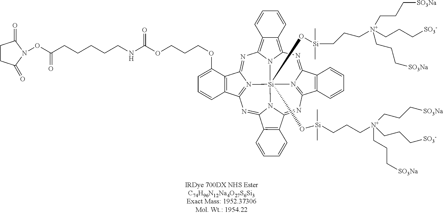
IR700 (IRDye.RTM. 700DX): A dye having the following formula:
##STR00001##
Currently commercially available from LI-COR (Lincoln, Nebr.). IR700 has several favorable chemical properties. Amino-reactive IR700 is a relatively hydrophilic dye and can be covalently conjugated with an antibody using the NHS ester of IR700. IR700 also has more than 5-fold higher extinction coefficient (2.1.times.10.sup.5 M.sup.-1cm.sup.-1 at the absorption maximum of 689 nm), than conventional photosensitizers such as thehematoporphyrin derivative Photofrin.RTM. (1.2.times.10.sup.3 M.sup.-1cm.sup.-1 at 630 nm), meta-tetrahydroxyphenylchlorin; Foscan.RTM. (2.2.times.10.sup.4 M.sup.-1cm.sup.-1 at 652 nm), and mono-L-aspartylchlorin e6; NPe6/Laserphyrin.RTM. (4.0.times.10.sup.4 M.sup.-1cm.sup.-1 at 654 nm).
Pharmaceutical composition: A chemical compound or composition capable of inducing a desired therapeutic or prophylactic effect when properly administered to a subject. A pharmaceutical composition can include a therapeutic agent, such as one or more antibody-IR700 molecules. A therapeutic or pharmaceutical agent is one that alone or together with an additional compound induces the desired response (such as inducing a therapeutic or prophylactic effect when administered to a subject). In a particular example, a pharmaceutical composition includes a therapeutically effective amount of at least one antibody-IR700 molecule.
Pharmaceutically acceptable vehicles: The pharmaceutically acceptable carriers (vehicles) useful in this disclosure are conventional. Remington's Pharmaceutical Sciences, by E. W. Martin, Mack Publishing Co., Easton, Pa., 19th Edition (1995), describes compositions and formulations suitable for pharmaceutical delivery of one or more therapeutic compounds, such as one or more antibody-IR700 molecules.
In general, the nature of the carrier will depend on the particular mode of administration being employed. For instance, parenteral formulations usually comprise injectable fluids that include pharmaceutically and physiologically acceptable fluids such as water, physiological saline, balanced salt solutions, aqueous dextrose, glycerol or the like as a vehicle. For solid compositions (for example, powder, pill, tablet, or capsule forms), conventional non-toxic solid carriers can include, for example, pharmaceutical grades of mannitol, lactose, starch, or magnesium stearate. In addition to biologically-neutral carriers, pharmaceutical compositions to be administered can contain minor amounts of non-toxic auxiliary substances, such as wetting or emulsifying agents, preservatives, and pH buffering agents and the like, for example sodium acetate or sorbitan monolaurate.
Photoimmunotherapy (PIT): A molecular targeted therapeutic that utilizes a target-specific photosensitizer based on a near infrared (NIR) phthalocyanine dye, IR700, conjugated to monoclonal antibodies (MAb) targeting cell surface receptors. In one example the cell surface receptor is one found specifically on cancer cells, such as HER1, HER2 or PSMA, and thus PIT can be used to kill such cells. Cell death of the cells occurs when the antibody-IR700 molecule binds to the cells and the cells are irradiated with NIR, while cells that do not express the cell surface receptor recognized the antibody-IR700 molecule are not killed in significant numbers.
Subject or patient: A term that includes human and non-human mammals. In one example, the subject is a human or veterinary subject, such as a mouse. In some examples, the subject is a mammal (such as a human) who has cancer, or is being treated for cancer.
Therapeutically effective amount: An amount of a composition that alone, or together with an additional therapeutic agent(s) (such as a chemotherapeutic agent) sufficient to achieve a desired effect in a subject, or in a cell, being treated with the agent. The effective amount of the agent (such as an antibody-IR700 molecule) can be dependent on several factors, including, but not limited to the subject or cells being treated, the particular therapeutic agent, and the manner of administration of the therapeutic composition. In one example, a therapeutically effective amount or concentration is one that is sufficient to prevent advancement (such as metastasis), delay progression, or to cause regression of a disease, or which is capable of reducing symptoms caused by the disease, such as cancer. In one example, a therapeutically effective amount or concentration is one that is sufficient to increase the survival time of a patient with a tumor.
In one example, a desired response is to reduce or inhibit one or more symptoms associated with cancer. The one or more symptoms do not have to be completely eliminated for the composition to be effective. For example, administration of a composition containing an antibody-IR700 molecule followed by irradiation can decrease the size of a tumor (such as the volume or weight of a tumor, or metastasis of a tumor), for example by at least 20%, at least 50%, at least 80%, at least 90%, at least 95%, at least 98%, or even at least 100%, as compared to the tumor size in the absence of the antibody-IR700 molecule. In one particular example, a desired response is to kill a population of cells by a desired amount, for example by killing at least 20%, at least 50%, at least 60%, at least 70%, at least 80%, at least 90%, at least 95%, at least 98%, or even at least 100% of the cells, as compared to the cell killing in the absence of the antibody-IR700 molecule and irradiation. In one particular example, a desired response is to increase the survival time of a patient with a tumor (or who has had a tumor recently removed) by a desired amount, for example increase survival by at least 20%, at least 50%, at least 60%, at least 70%, at least 80%, at least 90%, at least 95%, at least 98%, or even at least 100%, as compared to the survival time in the absence of the antibody-IR700 molecule and irradiation.
The effective amount of an agent that includes one of the disclosed antibody-IR700 molecules, that is administered to a human or veterinary subject will vary depending upon a number of factors associated with that subject, for example the overall health of the subject. An effective amount of an agent can be determined by varying the dosage of the product and measuring the resulting therapeutic response, such as the regression of a tumor. Effective amounts also can be determined through various in vitro, in vivo or in situ immunoassays. The disclosed agents can be administered in a single dose, or in several doses, as needed to obtain the desired response. However, the effective amount of can be dependent on the source applied, the subject being treated, the severity and type of the condition being treated, and the manner of administration.
In particular examples, a therapeutically effective dose of an antibody-IR700 molecule is at least 0.5 milligram per 60 kilogram (mg/kg), at least 5 mg/60 kg, at least 10 mg/60 kg, at least 20 mg/60 kg, at least 30 mg/60 kg, at least 50 mg/60 kg, for example 0.5 to 50 mg/60 kg, such as a dose of 1 mg/60 kg, 2 mg/60 kg, 5 mg/60 kg, 20 mg/60 kg, or 50 mg/60 kg, for example when administered iv. In another example, a therapeutically effective dose of an antibody-IR700 molecule is at least 10 .mu.g/kg, such as at least 100 .mu.g/kg, at least 500 .mu.g/kg, or at least 500 .mu.g/kg, for example 10 .mu.g/kg to 1000 .mu.g/kg, such as a dose of 100 .mu.g/kg, 250 .mu.g/kg, about 500 .mu.g/kg, 750 .mu.g/kg, or 1000 .mu.g/kg, for example when administered intratumorally or ip. In one example, a therapeutically effective dose is at least 1 .mu.g/ml, such as at least 500 .mu.g/ml, such as between 20 .mu.g/ml to 100 .mu.g/ml, such as 10 .mu.g/ml, 20 .mu.g/ml, 30 .mu.g/ml, 40 .mu.g/ml, 50 .mu.g/ml, 60 .mu.g/ml, 70 .mu.g/ml, 80 .mu.g/ml, 90 .mu.g/ml or 100 .mu.g/ml administered in topical solution. However, one skilled in the art will recognize that higher or lower dosages also could be used, for example depending on the particular antibody-IR700 molecule. In particular examples, such daily dosages are administered in one or more divided doses (such as 2, 3, or 4 doses) or in a single formulation. The disclosed antibody-IR700 molecules can be administered alone, in the presence of a pharmaceutically acceptable carrier, in the presence of other therapeutic agents (such as other anti-neoplastic agents).
Generally a suitable dose of irradiation following administration of the antibody-IR700 is at least 1 J cm.sup.-2 at a wavelength of 660-740 nm, for example, at least 10 J cm.sup.-2 at a wavelength of 660-740 nm, at least 50 J cm.sup.-2 at a wavelength of 660-740 nm, or at least 100 J cm.sup.-2 at a wavelength of 660-740 nm, for example 1 to 500 1.0 J cm.sup.-2 at a wavelength of 660-740 nm. In some examples the wavelength is 660-710 nm. In specific examples, a suitable dose of irradiation following administration of the antibody-IR700 molecule is at least 1.0 J cm.sup.-2 at a wavelength of 680 nm for example, at least 10 J cm.sup.-2 at a wavelength of 680 nm, at least 50 J cm.sup.-2 at a wavelength of 680 nm, or at least 100 J cm.sup.-2 at a wavelength of 680 nm, for example 1 to 500 1.0 J cm.sup.-2 at a wavelength of 680 nm. In particular examples, multiple irradiations are performed (such as at least 2, at least 3, or at least 4 irradiations, such as 2, 3, 4, 5, 6, 7, 8, 9 or 10 separate administrations), following administration of the antibody-IR700 molecule.
Treating: A term when used to refer to the treatment of a cell or tissue with a therapeutic agent, includes contacting or incubating an agent (such as an antibody-IR700 molecule) with the cell or tissue. A treated cell is a cell that has been contacted with a desired composition in an amount and under conditions sufficient for the desired response. In one example, a treated cell is a cell that has been exposed to an antibody-IR700 molecule under conditions sufficient for the antibody to bind to a surface protein on the cell, followed by irradiation, until sufficient cell killing is achieved.
Tumor, neoplasia, malignancy or cancer: A neoplasm is an abnormal growth of tissue or cells which results from excessive cell division. Neoplastic growth can produce a tumor. The amount of a tumor in an individual is the "tumor burden" which can be measured as the number, volume, or weight of the tumor. A tumor that does not metastasize is referred to as "benign." A tumor that invades the surrounding tissue and/or can metastasize is referred to as "malignant." A "non-cancerous tissue" is a tissue from the same organ wherein the malignant neoplasm formed, but does not have the characteristic pathology of the neoplasm. Generally, noncancerous tissue appears histologically normal. A "normal tissue" is tissue from an organ, wherein the organ is not affected by cancer or another disease or disorder of that organ. A "cancer-free" subject has not been diagnosed with a cancer of that organ and does not have detectable cancer.
Exemplary tumors, such as cancers, that can be treated with the claimed methods include solid tumors, such as breast carcinomas (e.g. lobular and duct carcinomas), sarcomas, carcinomas of the lung (e.g., non-small cell carcinoma, large cell carcinoma, squamous carcinoma, and adenocarcinoma), mesothelioma of the lung, colorectal adenocarcinoma, stomach carcinoma, prostatic adenocarcinoma, ovarian carcinoma (such as serous cystadenocarcinoma and mucinous cystadenocarcinoma), ovarian germ cell tumors, testicular carcinomas and germ cell tumors, pancreatic adenocarcinoma, biliary adenocarcinoma, hepatocellular carcinoma, bladder carcinoma (including, for instance, transitional cell carcinoma, adenocarcinoma, and squamous carcinoma), renal cell adenocarcinoma, endometrial carcinomas (including, e.g., adenocarcinomas and mixed Mullerian tumors (carcino sarcomas)), carcinomas of the endocervix, ectocervix, and vagina (such as adenocarcinoma and squamous carcinoma of each of same), tumors of the skin (e.g., squamous cell carcinoma, basal cell carcinoma, malignant melanoma, skin appendage tumors, Kaposi sarcoma, cutaneous lymphoma, skin adnexal tumors and various types of sarcomas and Merkel cell carcinoma), esophageal carcinoma, carcinomas of the nasopharynx and oropharynx (including squamous carcinoma and adenocarcinomas of same), salivary gland carcinomas, brain and central nervous system tumors (including, for example, tumors of glial, neuronal, and meningeal origin), tumors of peripheral nerve, soft tissue sarcomas and sarcomas of bone and cartilage, and lymphatic tumors (including B-cell and T-cell malignant lymphoma). In one example, the tumor is an adenocarcinoma.
The methods can also be used to treat liquid tumors, such as a lymphatic, white blood cell, or other type of leukemia. In a specific example, the tumor treated is a tumor of the blood, such as a leukemia (for example acute lymphoblastic leukemia (ALL), chronic lymphocytic leukemia (CLL), acute myelogenous leukemia (AML), chronic myelogenous leukemia (CML), hairy cell leukemia (HCL), T-cell prolymphocytic leukemia (T-PLL), large granular lymphocytic leukemia, and adult T-cell leukemia), lymphomas (such as Hodgkin's lymphoma and non-Hodgkin's lymphoma), and myelomas).
Under conditions sufficient for: A phrase that is used to describe any environment that permits the desired activity. In one example, "under conditions sufficient for" includes administering an antibody-IR700 molecule to a subject sufficient to allow the antibody-IR700 molecule to bind to cell surface proteins. In particular examples, the desired activity is killing the cells to which the antibody-IR700 molecule is bound, following therapeutic irradiation of the cells.
Untreated cell: A cell that has not been contacted with a desired agent, such as an antibody-IR700 molecule. In an example, an untreated cell is a cell that receives the vehicle in which the desired agent was delivered.
Disclosure of certain specific examples is not meant to exclude other embodiments. In addition, any treatments described herein are not necessarily exclusive of other treatment, but can be combined with other bioactive agents or treatment modalities.
Overview of Technology
Conventional photodynamic therapy (PDT) for cancer therapy is based on the preferential accumulation of a photosensitizer in tumor to produce phototoxicity with minimal damage to surrounding tissue (Dougherty et al. J Natl Cancer Inst 90:889-905, 1998). Traditionally, PDT is thought to be mediated by the generation of ROS, especially singlet oxygen, in the presence of oxygen (Dougherty et al. J Natl Cancer Inst 90:889-905, 1998). However, to the extent that existing photosensitizers lack tumor selectivity, considerable damage can be seen in normal tissues leading to dose limiting toxicity. Thus, current methods of PDT would be improved if more selective targeting of the photosensitizer and more efficient phototoxicity per photon absorbed was possible.
Disclosed herein are highly targeted photosensitizers, referred to as antibody-IR700 molecules. The photosensitizer, IR700, is excited in the NIR range leading to deeper tissue penetration resulting in successful eradication of subcutaneously xenografted tumors after only a single dose of external NIR light irradiation. Targeted phototoxicity appears to be primarily dependent on binding of the antibody-1R700 molecules to the cell membrane and to a lesser extent on internalization and ROS formation. The fluorescence induced by the conjugate can be used to non-invasively guide both PIT and monitor the results of therapy.
Although a targeted photosensitizer can distribute throughout the body, it is only active where intense light is applied, reducing the likelihood of off-target effects. In contrast, existing photosensitizers are poorly selective small molecules which bind not only to cancer cells but also to normal cells, including the skin and other epithelial surfaces, resulting in unwanted phototoxicity. In addition, target specific delivery of conventional photosensitizers is theoretically difficult because, after reaching the cell, the agent must still be internalized into organelles, such as mitochondria, to be most effective. Various combinations of conventional photosensitizers and MAbs have been tested to improve selectivity (Mew et al., J Immunol 130:1473-1477, 1983; Sobolev et al., Prog Biophys Mol Biol 73:51-90, 2000; Carcenac et al., Br J Cancer 85:1787-93, 2001; Vrouenraets et al., Cancer Res 59:1505-13, 1999; Vrouenraets et al., Cancer Res 61:1970-1975, 2001; Hamblin et al., Cancer Res 56:5205-10, 1996; Mew et al., Cancer Res 45:4380-6, 1985). However, these have had limited success especially when measured by in vivo therapeutic effects, for example because conventional photosensitizers have low extinction coefficients that require conjugation of large numbers of photosensitizers to a single antibody molecule thus, potentially decreasing binding affinity, because conventional photosensitizers are mostly hydrophobic leading to difficulties in conjugating photosensitizers to antibodies without compromising the immunoreactivity and in vivo target accumulation, and because conventional photosensitizers generally absorb light in the visible range reducing tissue penetration.
It is shown herein that antibody-based photosensitizers (such as mAb-based photosensitizers), which are activated by NIR light for targeted photoimmunotherapy (PIT) only when bound to the target molecule on the cancer cellular membrane. The fluorophore IR700 (Licor Co. Lincoln, Nebr.) can become a photosensitizer when conjugated to an antibody specific for a cell surface receptor and can thus be used for target specific photodynamic therapy of undesired cells, such as tumor or cancer cells. Further, because these agents also emit a diagnostic fluorescence, they can be used to direct the application of light to minimize light exposure to non-relevant tissues and non-invasively monitor therapeutic effects. Based on the similarity of the phototoxicity induced with three different MAbs against several different cells expressing various numbers of respective target molecules and considering the potentially additive benefits from immunotherapy this method can be generally applicable to other mAbs (such as those disclosed in Nanus et al., J. Urology 170:S84-S88, 2003 and van Dongen et al., Adv Drug Deliv Rev 56:31-52, 2004).
When IR700 was conjugated with an anti-EGFR antibody (HER1 or HER2) or a PSMA antibody, cells that selectively bound the conjugate were killed upon exposure to 680 nm near-infrared (NIR) light. Based on this novel observation patient therapies are provided. Since this antibody-dependent target-cell specific photodynamic therapy is achieved with NIR light (e.g., 680 nm) excitation and showed highly selectively cytotoxic effects only upon antibody binding, this new antibody-dependent target-cell specific photodynamic therapy using IR700 can be used in cancer patients as a way to personalize cancer therapy with minimal side effects.
The selectivity of the antibody-1R700 conjugate is derived from its activation after binding to the cell membrane of target cells; unbound conjugate does not contribute to phototoxicity. Short term viability assays, as well as long term proliferation assays, demonstrated that the conjugate was capable of inducing specific cell death. When co-cultures of receptor-positive and -negative cells were treated, only the receptor-positive cells were killed despite the presence of unbound antibody-1R700 in the culture medium. This selective cell killing minimizes damage to normal cells.
The antibody-1R700 molecule must be bound to the cellular membrane to be active. For instance, the rupture of endolysosome occurred within a second of light exposure. Cell death induced by singlet oxygen generally induces a slower apoptotic cell death. Since cell membrane damage was so quickly induced even at 4.degree. C. by this method, it is hypothesized that cell death is caused by the rapid expansion of locally heated water with relatively minor effects due to singlet oxygen effects.
Treatment with sodium azide, a redox and singlet oxygen scavenger, only partially reduced the phototoxicity but did not totally eliminate the effectiveness of the conjugate. This indicates that ROS generation is a minor part of the phototoxic effect. The observation that phototoxicity was induced after incubation with antibody-1R700 after only 1 hour at 4.degree. C. indicates that internalization of the conjugate is not required for activity. This differs from current PDT agents that require intracellular localization to be effective. Video microscopy demonstrated rapid visible damage to the membrane and lysosomes after exposure to light, following incubation for more than 6 hours at 37.degree. C., when the antibody-conjugate was internalized.
The disclosed antibody-1R700 conjugates permitted detection of targeted tissue. This can permit specific lesions to be identified with PIT rather than irradiating the entire field. Doses required for diagnosis (50 .mu.g) were significantly lower than those required for therapy (300 .mu.g). Improved intratumoral distribution of antibody occurred with the therapeutic dose. Because both bound and unbound agent fluoresces, there is relatively high background signal at therapeutic doses. Nevertheless, after PIT, the fluorescence of the treated tumors decreased and eventually disappeared, providing a means of monitoring the treatment.
Free IR700 and catabolized IR700, are readily excreted into urine within 1 hour without accumulation in any specific organ. The other component of PIT, light irradiation with NIR (e.g., at 690 nm) is unlikely to be toxic except at thermal doses. There should be no limitations on the cumulative irradiation dose of NIR light, unlike ionizing radiation such as x-ray or gamma-ray (FIG. 9). Therefore, repeated PIT can be used for long term management of cancer patients. It was observed that repeated PIT with 3 different regimens (3 or 4 fractionated NIR irradiations at a single dose of antibody-IR700 conjugate and 4 cycles of PIT every 2 weeks after multiple doses of antibody) controlled tumor regrowth, resulting in tumor free survival of more than 4 months.
Methods for Killing Cells and Treating Tumors
The present disclosure provides methods for killing a cell, such as a target cell. The cell expresses a protein on its surface, such as a tumor specific antigen, that can specifically bind to an antibody that is conjugated to the dye IR700 (referred to herein as an antibody-IR700 molecule). The cell is contacted with a therapeutically effective amount of one or more antibody-IR700 molecules (for example in the presence of a pharmaceutically acceptable carrier, such as a pharmaceutically and physiologically acceptable fluid), under conditions that permit the antibody to specifically bind to the cell surface protein. For example, the antibody-IR700 molecule can be present in a pharmaceutically effective carrier, such as water, physiological saline, balanced salt solutions (such as PBS/EDTA), aqueous dextrose, sesame oil, glycerol, ethanol, combinations thereof, or the like, as a vehicle. The carrier and composition can be sterile, and the formulation suits the mode of administration.
After administering the one or more antibody-IR700 molecules under conditions that allow the one or more antibody-IR700 molecules to bind to their target on a cell surface, the cell is then irradiated under conditions that permit killing of the cells, for example irradiation at a wavelength of 660 to 710 nm at a dose of at least 1 J cm.sup.-2. The NIR excitation light wavelength allows penetration of at least several centimeters into tissues. For example, by using fiber-coupled laser diodes with diffuser tips, NIR light can be delivered within several centimeters of otherwise inaccessible tumors located deep to the body surface. In addition to treating solid cancers, circulating tumor cells can be targeted since they can be excited when they traverse superficial vessels (for example using the NIR LED wearable devices disclosed herein). The disclosed methods can also be used as a therapy for transplant rejection.
The methods can be used to kill cells in vitro, for example by incubating the cells with the antibody-IR700 molecules in culture, or in vivo, for example, by administering one or more antibody-IR700 molecules to the subject. For example, a subject to be treated can be administered a therapeutically effective amount of one or more antibody-IR700 molecules, followed by irradiating the subject (or a tumor or tumor cell in the subject) with a therapeutic dose of irradiation.
The disclosed methods can be used in combination with other therapies, such as other anti-neoplastic therapies, such as radiation therapy, chemotherapy immunosuppressants (such as Rituximab, steroids), and cytokines (such as GM-CSF). Chemotherapeutic agents are known in the art (see for example, Slapak and Kufe, Principles of Cancer Therapy, Chapter 86 in Harrison's Principles of Internal Medicine, 14th edition; Perry et al., Chemotherapy, Ch. 17 in Abeloff, Clinical Oncology 2nd ed., 2000 Churchill Livingstone, Inc; Baltzer and Berkery. (eds): Oncology Pocket Guide to Chemotherapy, 2nd ed. St. Louis, Mosby-Year Book, 1995; Fischer Knobf, and Durivage (eds): The Cancer Chemotherapy Handbook, 4th ed. St. Louis, Mosby-Year Book, 1993). Exemplary chemotherapeutic agents that can be used in combination with the methods provided herein include but are not limited to, carboplatin, cisplatin, paclitaxel, docetaxel, doxorubicin, epirubicin, topotecan, irinotecan, gemcitabine, iazofurine, gemcitabine, etoposide, vinorelbine, tamoxifen, valspodar, cyclophosphamide, methotrexate, fluorouracil, mitoxantrone and vinorelbine. Combination chemotherapy is the administration of more than one agent to treat cancer.
In one example, contacting target cells with one or more antibody-IR700 molecules followed by irradiation kills the target cells that express a cell surface protein that specifically binds to the antibody. For example, the disclosed methods can kill at least 10%, for example at least 20%, at least 40%, at least 50%, at least 80%, at least 90%, or more of the treated cells relative to the absence of treatment with of one or more antibody-IR700 molecules followed by irradiation.
In one example, administration of one or more antibody-IR700 molecules to a subject having a tumor, followed by irradiation, kills the cells that express a cell surface protein that can specifically bind to the antibody, thereby treating the tumor. For example, the disclosed methods can decrease the size or volume of a tumor, slow the growth of a tumor, decrease or slow metastasis of the tumor (for example by reducing the number of metastases or decreasing the volume or size of a metastasis), or combinations thereof. For example, the disclosed methods can reduce tumor cell size or volume and/or a metastatic tumor cell volume (or number of metastatic tumors), such as by at least 10%, for example by at least 20%, at least 40%, at least 50%, at least 80%, at least 90%, or more, relative to the absence of administration of one or more antibody-IR700 molecules followed by irradiation. In addition, the disclosed methods can result in a decrease in the symptoms associated with a tumor and/or a metastatic tumor. In one example, administration of the disclosed compositions slows the growth of a tumor, such as by at least 10%, for example by at least 20%, at least 40%, at least 50%, at least 80%, at least 90%, or more, relative to the absence of administration of the antibody-IR700 molecules followed by irradiation. Methods of monitoring tumor volume/size/metastasis are routine in the art. In some examples, the disclosed methods can increase a subject's (such as a subject with a tumor or who has had a tumor previously removed) survival time, for example relative the absence of administration of one or more antibody-IR700 molecules followed by irradiation, such as an increase of at least 20%, at least 40%, at least 50%, at least 80%, at least 90%, or more. For example, the disclosed methods can increase a subject's survival time by at least 3 months, at least 6 months, at least 12 months, at least 18 months, at least 24 months, at least 36 months or more, relative to average survival time in the absence of administration of an antibody-IR700 molecule followed by irradiation.
Administration of therapeutically effective amounts of antibody-IR700 molecules followed by therapeutically effective doses of irradiation are capable of selectively killing tumor cells in vivo, and are capable of decreasing the weight or volume of a tumor in vivo. By selective killing of tumor cells relative to normal cells is meant that the methods are capable of killing tumor cells more effectively than normal cells such as, for example, cells not expressing the cell surface protein that specifically binds to the antibody administered.
The disclosed methods can be used to treat fixed tumors in the body as well as tumors in the circulation (e.g., leukemia cells, metastases, circulating tumor cells). However, circulating cells, by their nature, cannot be exposed to light for very long. Thus, if the cell to be killed is one that is circulating throughout the body, the methods can be accomplished by using a device that can be worn, or that covers parts of the body. For example, such a device can be worn for extended time periods. Everyday wearable items (e.g., wristwatches, jewelry (such as a necklace or bracelet), blankets, clothing (e.g., underwear, socks, and shoe inserts) and other everyday wearable items) which incorporate NIR emitting light emitting diodes (LEDs) and a battery pack, can be used. Such devices produce light on the skin underlying the device over long periods leading to continual exposure of light to superficial vessels over prolonged periods. Circulating tumor cells are exposed to the light as they transit thru the area underlying the device. As an example, a wristwatch or bracelet version of this device can include a series of NIR LEDs with battery power pack to be worn for most of the day.
After administration of the one or more antibody-IR700 molecules (e.g., intravenously), circulating cells bind the antibody-IR700 conjugate and become susceptible to killing by PIT. As these cells flow within the vessels adjacent to the LED present in the everyday wearable item (e.g., bracelet or wristwatch), they would be exposed to NIR light rendering them susceptible to cell killing. The dose of light may be adjustable according to diagnosis and cell type.
In some examples, the method further includes monitoring the therapy, such as killing of tumor cells. In such examples, the antibody-IR700 conjugate is contacted with the cells and the cells irradiated as described above. However, a lower dose of the antibody-IR700 conjugate and NIR light can be used (as cell killing may not be required, just monitoring of the therapy). In one example, the amount of antibody-IR700 conjugate administered for monitoring is at least 2-fold less (such as at least 3-, 4-, 5-, 6-, 7-, 8-, 9-, or 10-fold less than the therapeutic dose). In one example, the amount of antibody-IR700 conjugate administered for monitoring is at least 20% or at least 25% less than the therapeutic dose. In one example, the amount of NIR light used for monitoring is at least 1/1000 or at least 1/10,000 of the therapeutic dose. This permits detection of the cells being treated. For example, by using such methods, the size of the tumor and metastases can be monitored.
In some examples, the method is useful during surgery, such as endoscopic procedures. For example, after the antibody-IR700 conjugate is contacted with the cells and the cells irradiated as described above, this not only results in cell killing, but permits a surgeon or other medical care provider to visualize the margins of a tumor, and help ensure that resection of the tumor (such as a tumor of the skin, breast, lung, colon, or prostate) is complete and that the margins are clear. In some examples, a lower dose of the antibody-IR700 conjugate can be used for visualization, such as at least 2-fold less (such as at least 3-, 4-, 5-, 6-, 7-, 8-, 9-, or 10-fold less than the therapeutic dose).
Exemplary Cells
The target cell can be a cell that is not desired or whose growth is not desired, such as a tumor cell. The cells can be growing in culture, or present in a mammal to be treated, such as a patient with cancer. Any target cell can be treated with the claimed methods. In one example, the target cell expresses a cell surface protein that is not substantially found on the surface of other normal (desired) cells, an antibody can be selected that specifically binds to such protein, and an antibody-IR700 molecule generated for that protein. In one example, the cell surface protein is a tumor-specific protein. In one example, the cell surface protein is CD25, which can be used to target cells associated with undesired transplant rejection.
In one example, the tumor cell is a cancer cell, such as a cell in a patient with cancer. Exemplary cells that can be killed with the disclosed methods include cells of the following tumors: a liquid tumor such as a leukemia, including acute leukemia (such as acute lymphocytic leukemia, acute myelocytic leukemia, and myeloblastic, promyelocytic, myelomonocytic, monocytic and erythroleukemia), chronic leukemias (such as chronic myelocytic (granulocytic) leukemia and chronic lymphocytic leukemia), polycythemia vera, lymphoma, Hodgkin's disease, non-Hodgkin's lymphoma, multiple myeloma, Waldenstrdm's macroglobulinemia, heavy chain disease). In another example the cell is a solid tumor cell, such as sarcomas and carcinomas, fibrosarcoma, myxosarcoma, liposarcoma, chondrosarcoma, osteogenic sarcoma, and other sarcomas, synovioma, mesothelioma, Ewing's tumor, leiomyosarcoma, rhabdomyosarcoma, colon carcinoma, pancreatic cancer, breast cancer, ovarian cancer, prostate cancer, hepatocellular carcinoma, lung cancer, colorectal cancer, squamous cell carcinoma, basal cell carcinoma, adenocarcinoma (for example adenocarcinoma of the pancreas, colon, ovary, lung, breast, stomach, prostate, cervix, or esophagus), sweat gland carcinoma, sebaceous gland carcinoma, papillary carcinoma, papillary adenocarcinomas, medullary carcinoma, bronchogenic carcinoma, renal cell carcinoma, hepatoma, bile duct carcinoma, choriocarcinoma, Wilms' tumor, cervical cancer, testicular tumor, bladder carcinoma, CNS tumors (such as a glioma, astrocytoma, medulloblastoma, craniopharyogioma, ependymoma, pinealoma, hemangioblastoma, acoustic neuroma, oligodendroglioma, menangioma, melanoma, neuroblastoma and retinoblastoma).
Exemplary Subjects
In some examples the disclosed methods are used to treat a subject who has a tumor, such as a tumor described herein. In some examples, the tumor has been previously treated, such as surgically or chemically removed, and the disclosed methods are used subsequently to kill any remaining undesired tumor cells that may remain in the patient.
The disclosed methods can be used to treat any mammalian subject, such as a human, who has a tumor, such as a cancer, or has had such previously removed or treated. Subjects in need of the disclosed therapies can include human subjects having cancer, wherein the cancer cells express a tumor-specific protein on their surface that can specifically bind to the antibody-IR700 molecule. For example, the disclosed methods can be used as initial treatment for cancer either alone, or in combination with radiation or other chemotherapy. The disclosed methods can also be used in patients who have failed previous radiation or chemotherapy. Thus, in some examples, the subject is one who has received other therapies, but those other therapies have not provided a desired therapeutic response. The disclosed methods can also be used in patients with localized and/or metastatic cancer.
In some examples the method includes selecting a subject that will benefit from the disclosed therapies, such as selecting a subject having a tumor that expresses a cell surface protein (such as a tumor-specific protein) that can specifically bind to an antibody-IR700 molecule. For example, if the subject is determined to have a breast cancer that expresses HER2, the subject can be selected to be treated with an anti-HER2-IR700 molecule, such as Tra-IR700 described in Example 1, and the subject subsequently irradiated as described herein.
Exemplary Cell Surface Proteins
In one example, the protein on the cell surface of the target cell to be killed is not present in significant amounts on other cells. For example, the cell surface protein can be a receptor that is only found on the target cell type.
In a specific example, the cell surface protein is a tumor-specific protein (also known in the art as a tumor-specific antigen), such as members of the EGF receptor family (e.g., HER1, 2, 3, and 4) and cytokine receptors (e.g., CD20, CD25, IL-13R, CD5, CD52, etc.). Tumor specific proteins are those proteins that are unique to cancer cells or are much more abundant on them, as compared to other cells, such as normal cells. For example HER2 is primarily found in breast cancers, while HER1 is primarily found in adenocarcinomas, which can be found in many organs, such as the pancreas, breast, prostate and colon.
Exemplary tumor-specific proteins that can be found on a target cell (and to which an antibody specific for that protein can be used to formulate an antibody-IR700 molecule), include but are not limited to: any of the various MAGEs (Melanoma-Associated Antigen E), including MAGE 1 (e.g., GenBank Accession Nos. M77481 and AAA03229), MAGE 2 (e.g., GenBank Accession Nos. L18920 and AAA17729), MAGE 3 (e.g., GenBank Accession Nos. U03735 and AAA17446), MAGE 4 (e.g., GenBank Accession Nos. D32075 and A06841.1), etc.; any of the various tyrosinases (e.g., GenBank Accession Nos. U01873 and AAB60319); mutant ras; mutant p53 (e.g., GenBank Accession Nos. X54156, CAA38095 and AA494311); p97 melanoma antigen (e.g., GenBank Accession Nos. M12154 and AAA59992); human milk fat globule (HMFG) associated with breast tumors (e.g., GenBank Accession Nos. 556151 and AAB19771); any of the various BAGEs (Human B melanoma-Associated Antigen E), including BAGE1 (e.g., GenBank Accession No. Q13072) and BAGE2 (e.g., GenBank Accession Nos. NM_182482 and NP_872288), any of the various GAGEs (G antigen), including GAGE1 (e.g., GenBank Accession No. Q13065) or any of GAGE2-6; various gangliosides, CD25 (e.g., GenBank Accession Nos. NP_000408.1 and NM_000417.2).
Other tumor-specific antigens include the HPV 16/18 and E6/E7 antigens associated with cervical cancers (e.g., GenBank Accession Nos. NC_001526, FJ952142.1, ADB94605, ADB94606, and U89349), mucin (MUC 1)-KLH antigen associated with breast carcinoma (e.g., GenBank Accession Nos. J03651 and AAA35756), CEA (carcinoembryonic antigen) associated with colorectal cancer (e.g., GenBank Accession Nos. X98311 and CAA66955), gp100 (e.g., GenBank Accession Nos. 573003 and AAC60634) associated with for example melanoma, MART1 antigens associated with melanoma (e.g., GenBank Accession No. NP_005502), cancer antigen 125 (CA125, also known as mucin 16 or MUC16) associated with ovarian and other cancers (e.g., GenBank Accession Nos. NM_024690 and NP_078966); alpha-fetoprotein (AFP) associated with liver cancer (e.g., GenBank Accession Nos. NM_001134 and NP_001125); Lewis Y antigen associated with colorectal, biliary, breast, small-cell lung, and other cancers; tumor-associated glycoprotein 72 (TAG72) associated with adenocarcinomas; and the PSA antigen associated with prostate cancer (e.g., GenBank Accession Nos. X14810 and CAA32915).
Other exemplary tumor-specific proteins further include, but are not limited to, PMSA (prostate membrane specific antigen; e.g., GenBank Accession Nos. AAA60209 and AAB81971.1) associated with solid tumor neovasculature, as well prostate cancer; HER-2 (human epidermal growth factor receptor 2, e.g., GenBank Accession Nos. M16789.1, M16790.1, M16791.1, M16792.1 and AAA58637) associated with breast cancer, ovarian cancer, stomach cancer and uterine cancer, HER-1 (e.g., GenBank Accession Nos. NM_005228 and NP_005219) associated with lung cancer, anal cancer, and gliobastoma as well as adenocarcinomas; NY-ESO-1 (e.g. GenBank Accession Nos. U87459 and AAB49693) associated with melanoma, sarcomas, testicular carcinomas, and other cancers, hTERT (aka telomerase) (e.g., GenBank Accession. Nos. NM_198253 and NP_937983 (variant 1), NM_198255 and NP_937986 (variant 2)); proteinase 3 (e.g., GenBank Accession Nos. M29142, M75154, M96839, X55668, NM 00277, M96628, X56606, CAA39943 and AAA36342), and Wilms tumor 1 (WT-1, e.g. GenBank Accession Nos. NM_000378 and NP_000369 (variant A), NM_024424 and NP_077742 (variant B), NM_024425 and NP_077743 (variant C), and NM_024426 and NP_077744 (variant D)).
In one example the tumor-specific protein is CD52 (e.g., GenBank Accession. Nos. AAH27495.1 and CAI15846.1) associated with chronic lymphocytic leukemia; CD33 (e.g., GenBank Accession. Nos. NM_023068 and CAD36509.1) associated with acute myelogenous leukemia; and CD20 (e.g., GenBank Accession. Nos. NP_068769 NP_031667) associated with Non-Hodgkin lymphoma.
Thus, the disclosed methods can be used to treat any cancer that expresses a tumor-specific protein.
Exemplary Antibody-IR700 Molecules
One skilled in the art will recognize that because cell surface protein sequences are publically available (as for example shown above), that making or purchasing antibodies (or other small molecules that can be conjugated to IR700) specific for such proteins is routine. For example, if the tumor-specific protein HER2 is selected as a target, antibodies specific for HER2 (such as Trastuzumab) can be purchased or generated and attached to the IR700 dye. In one example, a patient is treated with at least two different antibody-IR700 molecules. For example, antiHER2-IR700 and antiHER2-IR700 could be injected together as a cocktail to facilitate killing of cells bearing either HER1 or HER2. Other specific examples are provided in the table below. In one example, the antibody is a humanized monoclonal antibody. Antibody-IR700 molecules can be generated using routine methods, such as those described in Example 1 below. Thus, the disclosure also provides antibody-IR700 molecules, compositions that include such molecules, and kits that include such molecules (for example a kit that includes one or more antibody-IR700 molecules and a chemotherapeutic agent, or a molecular targeting agent, or combinations thereof).
TABLE-US-00001 Tumor- Specific Exemplary Antibody/Small Antigen Exemplary Tumors Molecules HER1 Adenocarcinoma (e.g., Cetuximab, panitumamab, colorectal cancer, head zalutumumab, nimotuzumab, and neck cancer) matuzumab. Small molecule inhibitors gefitinib, erlotinib, and lapatinib can also be used. HER2 breast cancer, ovarian Trastuzumab (Herceptin .RTM.), cancer, stomach cancer, pertuzumab uterine cancer CD20 Non-Hodgkin lymphoma Tositumomab (Bexxar .RTM.); Rituximab (Rituxan, Mabthera); or Ibritumomab tiuxetan (Zevalin, for example in combination with yttrium-90 or indium- 111 therapy) CD25 T-cell lymphoma Daclizumab (Zenapax) CD33 Acute myelogenous Gemtuzumab (Mylotarg, for leukemia example in combination with calicheamicin therapy) CD52 chronic lymphocytic Alemtuzumab (Campath) leukemia CEA colorectal cancer, some CEA-scan (Fab fragment, gastric cancers, biliary approved by FDA), colo101 cancer Cancer ovarian cancer, OC125 monoclonal antibody antigen mesothelioma, breast 125 cancer (CA125) Alpha- hepatocellular carcinoma ab75705 (available from fetoprotein Abcam) and other commer- (AFP) cially available AFP anti- bodies Lewis Y colorectal cancer, biliary B3 (Humanized) cancer TAG72 adenocarcinomas including B72.3 (FDA-approved colorectal, pancreatic, monoclonal antibody) gastric, ovarian, endometrial, mammary, and non-small cell lung cancer Vascular Colorectal cancer Bevacizumab (Avastin .RTM.) endothelial growth factor
Antibody-IR700 molecules for treating transplant rejection can also be generated using Basiliximab or Daclizumab which target IL-2R.alpha. receptor (CD25)
Administration of Antibody-IR700 Molecules
Antibody-IR700 molecules can be contacted with a cell in vitro, for example by adding the antibody-IR700 molecules to growth media in which the cells or growing, or can be contacted with a cell in vivo, for example by administering the antibody-IR700 molecules to the subject to be treated.
The antibody-IR700 molecules can be administered locally or systemically using any method known in the art, for example to subjects having a tumor, such as a cancer, or who has had a tumor previously removed (for example via surgery). Although specific examples are provided, one skilled in the art will appreciate that alternative methods of administration of the disclosed antibody-IR700 molecules can be used. Such methods may include for example, the use of catheters or implantable pumps to provide continuous infusion over a period of several hours to several days into the subject in need of treatment.
In one example, the antibody-IR700 molecules are administered by parenteral means, including direct injection direct injection or infusion into a tumor (intratumorally). In some examples, the antibody-IR700 molecules are administered to the tumor by applying the antibody-IR700 molecules to the tumor, for example by bathing the tumor in a solution containing the antibody-IR700 molecules or by pouring the antibody-IR700 molecules onto the tumor.
In addition, or alternatively, the disclosed compositions can be administered systemically, for example intravenously, intramuscularly, subcutaneously, intradermally, intraperitoneally, subcutaneously, or orally, to a subject having a tumor (such as cancer).
The dosages of the antibody-IR700 molecules to be administered to a subject are not subject to absolute limits, but will depend on the nature of the composition and its active ingredients and its unwanted side effects (e.g., immune response against the antibody), the subject being treated and the type of condition being treated and the manner of administration. Generally the dose will be a therapeutically effective amount, such as an amount sufficient to achieve a desired biological effect, for example an amount that is effective to decrease the size (e.g., volume and/or weight) of the tumor, or attenuate further growth of the tumor, or decrease undesired symptoms of the tumor.
For intravenous administration of the antibody-IR700 molecules, exemplary dosages for administration to a subject for a single treatment can range from 0.5 to 100 mg/60 kg of body weight, 1 to 100 mg/60 kg of body weight, 1 to 50 mg/60 kg of body weight, 1 to 20 mg/60 kg of body weight, for example about 1 or 2 mg/60 kg of body weight. In yet another example, a therapeutically effective amount of ip or intratumoral administered antibody-IR700 molecules can vary from 10 .mu.g to 5000 .mu.g of antibody-IR700 molecule to 1 kg of body weight, such as 10 .mu.g/kg to 1000 .mu.g/kg, 10 .mu.g/kg to 500 .mu.g/kg, or 100 .mu.g/kg to 1000 .mu.g/kg.
In one example, the dose of antibody-IR700 molecule administered to a human patient is at least 50 mg, such as at least 100 mg, at least 300 mg, at least 500 mg, at least 750 mg, or even 1 g.
Treatments with disclosed antibody-IR700 molecules can be completed in a single day, or may be done repeatedly on multiple days with the same or a different dosage. Repeated treatments may be done on the same day, on successive days, or every 1-3 days, every 3-7 days, every 1-2 weeks, every 2-4 weeks, every 1-2 months, or at even longer intervals.
Irradiation of Cells
After the cells are contacted with one or more antibody-IR700 molecules, they are irradiated. Methods of irradiation are well known in the art. As only cells expressing the cell surface protein will be recognized by the antibody, only those cells will have sufficient amounts of the antibody-IR700 molecules bound to it. This decreases the likelihood of undesired side effects, such as killing of normal cells, as the irradiation will only kill the cells to which the antibody-IR700 molecules are bound, not the other cells.
In some examples, cells are irradiated in vitro, such as in a tissue culture dish. In other examples, a cell is irradiated in vivo, for example irradiating a subject who has previously been administered antibody-IR700 molecules. In some examples, the subject is irradiated, for example a tumor in the subject can be irradiated.
The cells are irradiated with a therapeutic dose of radiation at a wavelength of 660-710 nm, such as 660-700 nm, 670-690 nm, for example, 680 nm. In particular examples, the cells are irradiated at a dose of at least 1 J cm.sup.-2, such as at least 10 J cm.sup.-2, at least 30 J cm.sup.-2, at least 50 J cm.sup.-2, at least 100 J cm.sup.-2, or at least 500 J cm.sup.-2, for example, 1-1000 J cm.sup.-2, 1-500 J cm.sup.-2, 10-100 J cm.sup.-2, or 10-50 J cm.sup.-2.
Cells (or patients) can be irradiated one or more times. Thus, irradiation can be completed in a single day, or may be done repeatedly on multiple days with the same or a different dosage (such as irradiation at least 2 different times, 3 different times, 4 different times 5 different times or 10 different times). Repeated irradiations may be done on the same day, on successive days, or every 1-3 days, every 3-7 days, every 1-2 weeks, every 2-4 weeks, every 1-2 months, or at even longer intervals.
Exemplary Devices Containing NIR LEDs
Any type of item that can be worn or placed on the body, and is amenable to the incorporation of NIR LEDs, can be used. In one example, the device is a chamber into which the patient is inserted. Such devices can be used in the treatment of tumor cells circulating in the blood or lymph, such as leukemias, lymphomas, as well as metastatic cells present in the blood or lymph. In some examples, such devices can be used in the treatment of tumor cells present on the skin, such as a melanoma.
To kill all the cells circulating in the body it may be necessary to wear the devices for an extended period of time, such as several weeks or months. Thus, these devices can be incorporated into every day clothing, jewelry and nightwear such as blankets. These devices make it possible to expose the patient to NIR light using portable everyday articles of clothing and jewelry so that treatment remains private and does not interfere with everyday activities. For instance, a necklace incorporating NIR LEDs can be customizable to the patient's tastes, and worn discreetly during the day for PIT therapy (for example by killing tumor cells that pass through the carotid artery and other vasculature in the neck). Multiple devices of a similar "everyday" nature (blankets, bracelets, necklaces, underwear, socks, shoe inserts and the like) could be worn by the same patient during the treatment period. For example while sleeping, a patient could use the NIR blanket. The devices can also include a power supply, such as a battery, and a cooling element to prevent overheating for such devices as blankets.
In one example, the device is jewelry, such as a ring, watch, bracelet, or necklace. In another example, the item is an article of clothing or accessory, such as a shirt, belt, pants, underwear, socks, coat, shoe insert, scarf, hat, wrist guard, gloves, and the like. In another example, the device is an article that can cover the body, such as a blanket or towel. In another example, the device is a whole body light chamber that exposes the skin directly (such a device could also include a power supply and/or cooling supply).
By wearing the device that incorporates one or more NIR LEDs, tumor cells or other cells to be killed that are present in the blood or lymph become exposed to the light generated by the NIR LEDs (such as an NIR LED that emits at 660 to 740 nm, such as 670 to 700 nm or 680 to 720 nm). The light emitted from the NIR LED can penetrate the skin and blood vessels (such as the carotid artery or microvasculature in the skin), thus allowing the light to activate the antibody-IR700 molecule bound to the target cells, thus killing the cells to which the antibody-IR700 molecule is bound. The NIR LEDs can be arranged in the device to ensure that the skin or the blood vessels or lymphatic system are targeted.
NIR LED devices that can be used in the methods provided herein are commercially available. The applicable products from one manufacturer, Marubeni America, are listed below. The first product, a molded LED, has low power but it could be used over a longer exposure time. The other options have higher power and thus may benefit from provisions for additional cooling. Except for the last one, which is packaged in a 25 mm.times.18 mm metal case, the others are applicable to wearable devices such as bracelets, necklace, underwear, socks, gloves, hats and other wearable items. All are usable in blankets, handheld devices or chambers.
For example, Marubeni America Corporation (tech-led.com/index.shtml) provides the following NIR LEDs with lens options to set the irradiation pattern: Molded LED (tech-led.com/data/L680-AU.pdf) which is 5 mm in diameter, has a total radiated power of 4 mW, calculated power density of 5 mW cm.sup.-2 and a power requirement of 1.8V 20 mA; Surface Mount LED which is 3.5 mm.times.2.7 mm, has a total radiated power of 3 mW, calculated power density of 32 mW cm.sup.-2, and a power requirement of 1.9V 50 mA; Super Beam (tech-led.com/Superbeam_LEDs.shtml) which is 7.6 mm.times.7.6 mm, has a total radiated power of 20-52 mW, calculated power density of 34-90 mW cm.sup.-2, and a power requirement of 1.65V 100 mA; High Power Surface Mount (tech-led.com/SMB_BL_LEDs.shtml) which is 5 mm.times.5 mm or 7 mm diameter, has a total radiated power of 90 mW, calculated power density of 360 mW cm.sup.-2 and a power requirement of 2.4V 500 mA; and High Power Illuminators (tech-led.com/High_Power_Illuminators.shtml) which is 25 mm.times.18 mm, has a total radiated power of 150 mW, a calculated power density of 33 mW cm.sup.-2 and a power requirement of 10V 120 mA. Alternatively, such devices can be made that emit light at 690 nm with a similar power with short strong intermittent pulse.
During in vitro experimentation, NIR light with a power density of 2.2 mW cm.sup.-2 (or 2.2 mJ s.sup.-1 cm.sup.-2) induced cell death. Assuming an attenuation coefficient for tissue of 4 cm.sup.-1, the intensity of the light would be down to 10% at 5.8 mm and 1% at 12 mm. This indicates that for in vivo applications, the power density required needs to be 10-100 times larger. That is, the dose of light emitted by the NIR LED device in some examples is at least 20 mW cm.sup.-2, such as at least 50 mW cm.sup.-2, at least 100 mW cm.sup.-2, at least 150 mW cm.sup.-2, at least 200 mW cm.sup.-2 or, at least 300 mW cm.sup.-2. Multiple NIR LEDs can be arranged in a two-dimensional array to cover larger areas. In one example, a laser is used as the NIR light source as an alternative to an LED.
The NIR LEDs can be powered by using a power supply (which may be directly or indirectly part of the device). The power supply requirement would depend on the number of LEDs in the device. For example, one or more batteries can be used to power the NIR LED. For some LEDs, 4 AA batteries can power 3 LEDs in series. An alkaline AA battery is rated at a maximum of 3000 mAh so this configuration provide powers for up to 150, 60, and 30 hr at 20, 50 and 100 mA.
In some examples, the device further includes a cooling device (which may be directly or indirectly part of the device). For example, heat sinks can be used for passive or active cooling. Another alternative is a thermoelectric effect (Peltier). This would draw additional power but it can be used in applications where the power requirements would need a plug-in AC adapter.
Another type of device that can be used with the disclosed methods is a flashlight-like device with NIR LEDs. Such a device can be used for focal therapy of lesions during surgery, or incorporated into endoscopes to apply NIR light to body surfaces after the administration of PIT agent. Such devices can be used by physicians or qualified health personnel to direct treatment to particular targets on the body.
Treatment Using Wearable NIR LEDs
As described herein, the disclosed methods are highly specific for cancer cells. However, in order to kill the cells circulating in the body or present on the skin, the patient can wear a device that incorporates an NIR LED. In some example the patient uses at least two devices, for example an article of clothing or jewelry during the day, and a blanket at night. In some example the patient uses at least two devices at the same time, for example two articles of clothing. These devices make it possible to expose the patient to NIR light using portable everyday articles of clothing and jewelry so that treatment remains private and does not interfere with everyday activities. In some examples, the device can be worn discreetly during the day for PIT therapy.
In one example, the patient is administered one or more antibody-IR700 molecules, using the methods described herein. The patient then wears a device that incorporates an NIR LED, permitting long-term therapy and treatment of tumor cells that are present in the blood or lymph or on the skin. In some examples, the dose is at least at least 1 J cm.sup.-2, at least 10 J cm.sup.-2, at least 20 J cm.sup.-2, or at least 30 J cm.sup.-2, such as 20 J cm.sup.-2 or 30 J/cm.sup.2. In some examples, administration of the antibody-IR700 molecule is repeated over a period of time (such as bi-weekly or monthly, to ensure therapeutic levels are present in the body.
In some examples, the patient wears or uses the device, or combination of devices, for at least 1 week, such as at least 2 weeks, at least 4 weeks, at least 8 weeks, at least 12 weeks, at least 4 months, at least 6 months, or even at least 1 year. In some examples, the patient wears or uses the device, or combination of devices, for at least 4 hours a day, such as at least 12 hours a day, at least 16 hours a day, at least 18 hours a day, or 24 hours a day. It is quite possible that multiple devices of a similar "everyday" nature (blankets, bracelets, necklaces, underwear, socks, shoe inserts) could be worn by the same patient during the treatment period. At night the patient can use the NIR LED blanket or other covering.
Additional Treatments
In particular examples, prior to, during, or following administration of one or more antibody-IR700 molecules, the subject can receive one or more other therapies. In one example, the subject receives one or more treatments to remove or reduce the tumor prior to administration of the antibody-IR700 molecules.
Examples of such therapies include, but are not limited to, surgical treatment for removal or reduction of the tumor (such as surgical resection, cryotherapy, or chemoembolization), as well as anti-tumor pharmaceutical treatments which can include radiotherapeutic agents, anti-neoplastic chemotherapeutic agents, antibiotics, alkylating agents and antioxidants, kinase inhibitors, and other agents. Particular examples of additional therapeutic agents that can be used include microtubule binding agents, DNA intercalators or cross-linkers, DNA synthesis inhibitors, DNA and/or RNA transcription inhibitors, antibodies, enzymes, enzyme inhibitors, and gene regulators. These agents (which are administered at a therapeutically effective amount) and treatments can be used alone or in combination. Methods and therapeutic dosages of such agents are known to those skilled in the art, and can be determined by a skilled clinician.
"Microtubule binding agent" refers to an agent that interacts with tubulin to stabilize or destabilize microtubule formation thereby inhibiting cell division. Examples of microtubule binding agents that can be used in conjunction with the disclosed antibody-IR700 molecule therapies include, without limitation, paclitaxel, docetaxel, vinblastine, vindesine, vinorelbine (navelbine), the epothilones, colchicine, dolastatin 15, nocodazole, podophyllotoxin and rhizoxin. Analogs and derivatives of such compounds also can be used and are known to those of ordinary skill in the art. For example, suitable epothilones and epothilone analogs are described in International Publication No. WO 2004/018478. Taxoids, such as paclitaxel and docetaxel, as well as the analogs of paclitaxel taught by U.S. Pat. Nos. 6,610,860; 5,530,020; and 5,912,264 can be used.
The following classes of compounds can be used in combination with the methods disclosed herein: suitable DNA and/or RNA transcription regulators, including, without limitation, actinomycin D, daunorubicin, doxorubicin and derivatives and analogs thereof also are suitable for use in combination with the disclosed therapies. DNA intercalators and cross-linking agents that can be administered to a subject include, without limitation, cisplatin, carboplatin, oxaliplatin, mitomycins, such as mitomycin C, bleomycin, chlorambucil, cyclophosphamide and derivatives and analogs thereof. DNA synthesis inhibitors suitable for use as therapeutic agents include, without limitation, methotrexate, 5-fluoro-5'-deoxyuridine, 5-fluorouracil and analogs thereof. Examples of suitable enzyme inhibitors include, without limitation, camptothecin, etoposide, formestane, trichostatin and derivatives and analogs thereof. Suitable compounds that affect gene regulation include agents that result in increased or decreased expression of one or more genes, such as raloxifene, 5-azacytidine, 5-aza-2'-deoxycytidine, tamoxifen, 4-hydroxytamoxifen, mifepristone and derivatives and analogs thereof. Kinase inhibitors include Gleevac, Iressa, and Tarceva that prevent phosphorylation and activation of growth factors.
Other therapeutic agents, for example anti-tumor agents, that may or may not fall under one or more of the classifications above, also are suitable for administration in combination with the disclosed therapies. By way of example, such agents include adriamycin, apigenin, rapamycin, zebularine, cimetidine, and derivatives and analogs thereof.
In some examples, the subject receiving the therapeutic antibody-IR700 molecule composition is also administered interleukin-2 (IL-2), for example via intravenous administration. In particular examples, IL-2 (Chiron Corp., Emeryville, Calif.) is administered at a dose of at least 500,000 IU/kg as an intravenous bolus over a 15 minute period every eight hours beginning on the day after administration of the peptides and continuing for up to 5 days. Doses can be skipped depending on subject tolerance.
In some examples, the disclosed antibody-IR700 molecules can be co-administered with a fully human antibody to cytotoxic T-lymphocyte antigen-4 (anti-CTLA-4). In some example subjects receive at least 1 mg/kg anti-CTLA-4 (such as 3 mg/kg every 3 weeks or 3 mg/kg as the initial dose with subsequent doses reduced to 1 mg/kg every 3 weeks).
In one example, at least a portion of the tumor (such as a metastatic tumor) is surgically removed (for example via cryotherapy), irradiated, chemically treated (for example via chemoembolization) or combinations thereof, prior to administration of the disclosed therapies (such as administration of antibody-IR700 molecules). For example, a subject having a metastatic tumor can have all or part of the tumor surgically excised prior to administration of the disclosed therapies. In an example, one or more chemotherapeutic agents are administered following treatment with antibody-IR700 molecules and irradiation. In another particular example, the subject has a metastatic tumor and is administered radiation therapy, chemoembolization therapy, or both concurrently with the administration of the disclosed therapies.
EXAMPLE 1
Synthesis of IRDye 700-Conjugated Trastuzumab (Anti-Her2)
This example describes methods used to conjugate the monoclonal antibody Trastuzumab to the IRDye 700DX NHS Ester. On skilled in the art will recognize that any antibody, such as any monoclonal antibody specific for a target cell surface protein, can be conjugated to IRDye 700DX NHS Ester using similar methods.
Humanized anti-HER2 antibody, Trastuzumab (Tra; Genentech, San Francisco, Calif.) (1 mg, 6.8 nmol) was incubated with IRDye 700DX NHS Ester (IR700; LI-COR Bioscience, Lincoln, Nebr.) (66.8 .mu.g, 34.2 nmol, 5 mmol/L in DMSO) in 0.1 mol/L Na.sub.2HPO.sub.4 (pH 8.5) at room temperature for 30 to 120 min. Trastuzumab is a recombinant humanized monoclonal antibody (mAb) directed against the extracellular domain of the human epidermal growth factor receptor (EGFR) 2 (HER2) tyrosine kinase receptor. The mixture was purified with a Sephadex G50 column (PD-10; GE Healthcare, Piscataway, N.J.). The protein concentration was determined with Coomassie Plus protein assay kit (Pierce Biotechnology, Rockford, Ill.) by measuring the absorption at 595 nm with a UV-Vis system (8453 Value System; Agilent Technologies, Palo Alto, Calif.). The concentration of IR700 was measured by absorption with the UV-Vis system to confirm the number of fluorophore molecules conjugated to each Trastuzumab molecule. The number of IR700 per Trastuzumab was about 3.
The purity of the Tra-IR700 conjugate was confirmed by analytical size-exclusion HPLC (SE-HPLC) and sodium dodecyl sulfate polyacrylamidegel elctrophoresis (SDS-PAGE). SE-HPLC was performed using a Beckman System Gold (Fullerton, Calif.) equipped with model 126 solvent delivery module, a model 168 UV detector, and a JASCO fluorescence detector (excitation 689 nm and emission at 700 nm) controlled by 32 Karat software. SE chromatography was performed on a TSKgel G2000SWx1 (Tosoh Bioscience LLC, Montgomeryville, Pa.) eluted for 45 minutes using phosphate buffered saline (PBS) at 0.5 mL/min. SDS-PAGE was performed with a 4% to 20% gradient polyacrylamide gel (Invitrogen, Carlsbad, Calif.). Just after separating the proteins, fluorescence intensity was analyzed with a Fujifilm FLA-5100 fluorescence scanner (Valhalla, N.Y.) with an internal laser of 670 nm for excitation and 705 nm long pass filter for emission. The fluorescence intensity of each band was analyzed with Multigage software (Fujifilm). The gels were then stained with Colloidal Blue Staining Kit (Invitrogen), and digitally scanned. The protein concentration in each band was analyzed with ImageJ software. The trastuzumab-1R700 (Tra-1R700) and panitumumab-1R700 (Pan-1R700; see Example 8) preparations demonstrated strong association and contained no detectable MAb aggregates as determined by high performance liquid chromatography (HPLC) and sodium dodecyl sulfate polyacrylamidegel elctrophoresis SDS-PAGE.
To determine the in vitro binding characteristics of IR700 conjugates .sup.125I labeling of the conjugates using the Indo-Gen procedure was performed. The specific activities of the radiolabeled antibodies were 8.52 mCi/mg for Trastuzumab and 7.84 mCi/mg for panitumumab (see Example 8 below). It was observed that 73.38.+-.0.39% (.sup.125I-Tra-IR700) and 78.61.+-.0.89% (.sup.125I-Pan-IR700) of binding was achieved with each MAb conjugate respectively and the specificity of binding was confirmed by blocking with excess native unconjugated MAb (less than 1.4%). Since immunoreactivity of 1251-Tra and .sup.125I-Pan measured with the same method were 78.+-.2%, and 82.+-.3%, respectively, minimal loss of MAbs with IR700 conjugation was confirmed. Immunoreactivity assay was performed as described previously. Briefly, after trypsinization, 2.times.10.sup.6 of 3T3/HER2 or A431 cells were resuspended in PBS containing 1% bovine serum albumin (BSA). .sup.125I-Tra-IR700 or .sup.125I-Pan-IR700 (1 mCi, 0.2 .mu.g) was added and incubated for 1 h on ice. Cells were washed, pelleted, the supernatant decanted, and counted in a 2470 Wizard.sup.2 .gamma.-counter (Perkin Elmer, Shelton, Conn.). Nonspecific binding to the cells was examined under conditions of antibody excess (200 .mu.g of nonlabeled trastuzumab or panitumumab).
EXAMPLE 2
Selective Killing of HER2+ Cells
This example describes methods used to show that the Trastuzumab-IR700 compound described in Example 1 (referred to herein as Tra-IR700) can be used to selectively kill cells that express HER2 (HER2+), but has minimal negative effects on HER2 negative (HER2-) cells.
HER2 gene-transfected NIH3T3 (3T3/HER2+) cells were used for target photodynamic therapy (PDT). As a control, Balb/3T3 cells which express DsRed fluorescent protein but not HER2 (Balb/3T3/DsRed) were employed. Cells were grown in RPMI 1640 supplemented with 10% fetal bovine serum and 1% penicillin/streptomycin in tissue culture flasks in a humidified incubator at 37.degree. C. in an atmosphere of 95% air and 5% carbon dioxide.
Fluorescence microscopy was performed with a BX51 or IX81 microscope (Olympus America, Melville, N.Y.). The filter was set to detect IR700 and consisted of a 590-650 nm excitation filter, and a 665-740 nm band pass emission filter. To detect DsRed protein, a filter set consisting of a 480-550 nm excitation filter, and a 590 nm long pass emission filter was employed.
Fluorescence microscopy was performed to test the subcellular localization of IR700 in 3T3/HER2+ cells. Cells were seeded on cover glass-bottomed dishes and incubated for 24 hours. Tra-IR700 was added to the culture medium at 10 .mu.g/mL. As shown in FIG. 1A, Tra-IR700 was detected on the cell surface after 1 hour incubation on ice, and was mainly localized to the lysosome 6 hours after incubation at 37.degree. C., indicating gradual internalization. Co-staining with LysoTracker Green (Invitrogen, Carlsbad, Calif.), which was detected by a filter set consisting of a 420-480 nm excitation filter, and a 520 nm long pass emission filter, revealed co-localization of IR700 with the endolysosomal compartment (FIG. 1B). After 1 h and 6 h of incubation with Tra-1R700, excitation light (fluorescence microscope; power density of 2.2 mW cm-2) induced fluorescence as well as cellular swelling, bleb formation, and rupture of vesicles representing necrotic cell death (FIG. 1C).
For photoimmunotherapy (PIT), cells were seeded on 35 mm cover glass-bottomed dishes and incubated 24 hours. Medium was replaced with fresh culture medium containing Tra-IR700 at 10 .mu.g/mL and incubated for 6 hours at 37.degree. C. Culture medium was replaced with phenol red-free medium after washing with phosphate buffered saline (PBS). Cells were irradiated with light at 670 nm to 690 nm using a red light emitting diode (LED; FluorVivo; INDEC Systems Inc., Capitola, Calif.) with a power density of 2.6 mW cm.sup.-2 as measured with optical power meter (PM 100, Thorlabs, Newton, N.J.). Cell viability was assessed 1 hour after treatment with LIVE/DEAD.RTM. Fixable Green Dead Cell Stain Kit (Invitrogen). After the treatment, cells were trypsinized and washed with PBS. Green fluorescent reactive dye was added in the cell suspension and incubated at room temperature for 30 minutes. Cells were then analyzed on flow cytometer (FACS Calibur, BD BioSciences, San Jose, Calif.).
As shown in FIG. 1C, irradiation at 1.0 J cm-2 for 3T3/HER2+ cells resulted in rapid necrotic cell death, representing budding and swelling of the cell membrane.
EXAMPLE 3
Identification of Irradiation Dose
To determine the phototoxicity in response to different doses of light irradiation, PDT-treated 3T3/HER2+ cells were assayed by flow cytometry using the LIVE/DEAD.RTM. Fixable Green Dead Cell Stain Kit. The LIVE/DEAD assay, which can detect the cells with damaged membranes, was performed within 1 h after the treatment. As shown in FIG. 1D, cell death in response to Tra-IR700-mediated PDT was light dose dependent 1 hour after PIT. Cells without PIT or Tra-IR700 did not show significant phototoxicity.
EXAMPLE 4
Measurement of Cell Viability Over Time
To monitor cell viability over time, cells were labeled and irradiated as described in Example 2, then were monitored subsequently over time (5 days) using microscopy as described in Example 2.
As shown in FIG. 1F, phototoxic cell death was observed only in the treated 3T3/HER2+ cells with Tra-IR700, but not in the un-irradiated group (no PIT) or the group irradiated but did not receive Tra-IR700 (No Tra-IR700).
EXAMPLE 5
Target Specific Phototoxicity of Tra-IR700
PIT was performed as described in Example 2. As shown in FIG. 1G, there was no significant difference in phototoxicity between 1 h and 6 h incubation with Tra-1R700, indicating that membrane binding of Tra-1R700 was sufficient to induce cell death. When Tra-1R700 was localized to the endolysosomal compartment (FIG. 1B), it also induced rupture of the vesicle with cellular swelling and bleb formation after irradiation. However, this did not appear to be a major cause of cell death, since cell death was observed without endolysosomal localization of Tra-1R700 within 1 h of incubation at 4.degree. C. Failure to wash the cells prior to irradiation did not influence the phototoxic effect, indicating that cellular membrane binding was important to the phototoxic effects of the conjugate, not merely the presence of the conjugate. Further, the IR700 dye alone (200 nM; equivalent IR700 concentration of Tra-1R700 conjugates) did not incorporate into the cells or induce phototoxcity in cells (FIG. 1H and FIG. 2B). Additionally, phototoxcity was dose-dependently blocked by the excess unconjugated trastuzumab (FIGS. 2C and 2D). Furthermore, Tra-1R700 did not induce therapeutic effect to A431 cells (FIG. 1I). These results confirm that cell death is dependent on specific membrane binding of Tra-1R700.
Reactive oxygen species (ROS) have been implicated in the cell death associated with conventional PDT. To clarify the role of photon-induced redox reactions (e.g. singlet oxygen (.sup.10.sub.2)) in producing phototoxicity with Tra-1R700, a redox quencher, sodium azide (NaN.sub.3), was added to the medium when cells were irradiated. The percentage of cell death was partially decreased in the presence of sodium azide, in a dose dependent manner (FIG. 1J).
To confirm that the phototoxicity was target specific, 3T3/HER2+ cells and Balb/3T3/DsRed cells (a parental HER2 negative Balb/3T3 transfected with DsRed fluorescent protein) were co-cultured, and irradiated at 1.0 J cm.sup.-2 after 1 or 6 hours of incubation with Tra-IR700 at 37.degree. C. Tra-1R700 was distributed in a HER2 specific manner while DsRed expressing Balb/3T3 cells did not show phototoxicity upon irradiation (FIG. 3A). In addition, LIVE/DEAD Green staining demonstrated HER2 specific induction of cell death as determined by multi-color fluorescence microscopy (FIG. 3B) and flow cytometry analysis (FIG. 3C).
EXAMPLE 6
Tra-IR700 Reduces Proliferation of HER2+ Cells
3T3/HER2 cells were seeded into 35 mm cell culture dishes at a density of 1.times.10.sup.4. The next day, cells were incubated with or without Tra-IR700 and irradiated as described in Example 2. Cell viability was determined by trypan blue dye exclusion assay at day 1, 3 and 5 after the cell seeding. Viable cells for treated or untreated controls were counted on a hemacytometer after trypsinization. Cell growth was also photographed under microscope at each time point.
As shown in FIG. 1E, cells treated with Tra-IR700 and then subjected to PDT 2.0 J cm.sup.-2 had significantly reduced viability as compared to cells only treated with Tra-IR700, only PDT, or no treatment.
EXAMPLE 7
Tra-IR700 Selectively Kills HER2+ Cells In Vivo
This example describes methods used to show that Tra-IR700 can treat HER2+ tumors in vivo. One skilled in the art will appreciate that similar methods can be used with other tumor/antibody-IR700 combinations.
Six- to eight-week-old female homozygote athymic nude mice (Charles River, NCI-Frederick, Frederick, Md.) were anesthetized with isoflurane. 3T3/HER2+ or Balb/3T3 cells (2 million) were injected subcutaneously in the left dorsum of the mice. Four days after cell injection, either 50 or 300 .mu.g of Tra-IR700 was administered intravenously. Her2-specific Tra-IR700 accumulation in tumor xenografts was confirmed with an in vivo fluorescence imaging system (Pearl Imager, LI-COR Biosciences, Lincoln, Nebr.). 3T3/HER2+ tumor-specific IR700-Tra localization was observed. In contrast, Balb/3T3 tumor did not show Her2-specific IR700 fluorescence.
To evaluate the efficacy of targeted PIT with IR700-Tra in vivo, 1 million 3T3/HER2+ or Balb/3T3 cells were injected subcutaneously into the bilateral dorsum of female nude mice. When the volume of both tumors reached .about.70 mm.sup.3 (about 4 days), animals were randomized into five groups of at least 12 animals per group for the following treatments: (1) no treatment; (2) 300 .mu.g of trastuzumab injected intravenously, no PIT; (3) 300 .mu.g of Tra-IR700 injected intravenously, no PIT; (4) PIT at 50 J/cm.sup.2 for 3T3/HER2 tumor without Tra-IR700; (5) 300 .mu.g of Tra-IR700 injected intravenously, PDT was performed at 50 J/cm.sup.2.
Twenty-four hours after administration of Tra-IR700, in mice receiving PIT a 1 cm-diameter area encompassing the right dorsum including the tumor was irradiated (dose level 50 J cm.sup.-2). The left dorsum of the tumor was covered with black tape to prevent its exposure to light. Effects in response to PIT were monitored daily and tumor volumes were measured with a caliper twice a week until it reached 500-1000 mm.sup.3, at which time mice were euthanized with carbon dioxide gas. In order to determine tumor volume, the greatest longitudinal diameter (length) and the greatest transverse diameter (width) were determined with external caliper. Tumor volume based on caliper measurements were calculated by the following formula; tumor volume=length.times.width.sup.2.times.0.5. Data are expressed as means.+-.sem from a minimum of three experiments, unless otherwise indicated. Statistical analyses were carried out using a statistics program (GraphPad Prism; GraphPad Software, La Jolla, Calif.). For multiple comparisons, a one-way analysis of variance (ANOVA) with post test (Kruskal-Wallis test with post-test) was used. The cumulative probability of survival, determining herein as the tumor volume was failed to reach 500 mm.sup.3, were estimated in each group with the use of the Kaplan-Meier survival curve analysis, and the results were compared with use of the log-rank test with Bonferroni's correction for multiplicity. P<0.05 was considered to indicate a statistically significant difference.
As shown in FIGS. 4A and 4B, whereas 50 J cm.sup.-2 irradiation resulted in significant tumor growth inhibition in 3T3/HER2+ tumors at day 4, 7, 10 and 14 days after treatment, untreated tumors did not exhibit any detectable effect on tumor growth. In addition, irradiation for Balb/3T3 tumors did not show significant therapeutic effect. Furthermore, no lethal side effects were found during or after the treatment.
EXAMPLE 8
Synthesis of IRDye 700-Conjugated Vectibix.RTM. (Anti-HER1)
Panitumumab (Vectibix.RTM.), a fully humanized IgG.sub.2 MAb directed against the human EGFR was purchased from Amgen (Thousand Oaks, Calif.) and conjugated to IR700 using the methods described in Example 1. This compound is referred to as Panitumumab-IR700 or Pan-IR700. The number of IR700 per Panitumumab was about 3.
EXAMPLE 9
Pan-IR700 Selectively Kills HER1+ Cells
This example describes methods used to show that the Pan-IR700 compound described in Example 8 can be used to selectively kill cells that express HER1 (HER1+), but has minimal negative effects on HER1 negative (HER1-) cells.
EGFR-expressing A431 cells were used as the target HER1+ cells. As a control, Balb/3T3 cells which express DsRed fluorescent protein but not express HER1/EGFR (Balb/3T3/DsRed) were used. Cells were grown in RPMI 1640 supplemented with 10% fetal bovine serum and 1% penicillin/streptomycin in tissue culture flasks in a humidified incubator at 37.degree. C. in an atmosphere of 95% air and 5% carbon dioxide. A431 or Balb/3T3/DsRed cells were seeded on cover glass-bottomed dishes and incubated for 24 hours. Pan-IR700 was added to the culture medium at 10 .mu.g/mL and incubated either for 1 hour on ice or 6 hours at 37.degree. C., then cells were washed with PBS. Culture medium was replaced with phenol red-free medium after washing with phosphate buffered saline (PBS).
Fluorescence microscopy was performed as described in Example 2 to detect the antigen-specific localization of IR700. Pan-IR700 was detected on the cell surface of A431 cells after 1 hour incubation on ice, and was mainly localized to the lysosome 6 hours after incubation at 37.degree. C. No significant IR700 signal was observed with Balb/3T3/DsRed cells.
PIT was performed as described in Examples 2 and 3. As shown in FIG. 5A, irradiation of A431 cells at 0.5 to 2 J cm.sup.-2 resulted in rapid cell death in a dose-dependent manner, representing budding and swelling of the cell membrane. As shown in FIG. 5B, the percentage of cell death in target cells versus untreated control cells was significantly influenced by excitation light dose. In addition, there was no significant cytotoxicity associated with exposure to Pan-1R700 without excitation light or with light exposure without Tra-1R700. However, panitumumab itself had a noticeable treatment effect against A431 cells due to down regulation and signal inhibition of HER1 (Yang et al., Cancer Res 59:1236-43, 1999).
Target-specific phototoxicity was also confirmed with Pan-1R700 mediated PIT in A431 cells and Balb/3T3/DsRed (HER1 negative) co-cultured cells (FIG. 5C). In summary, Tra-1R700 and Pan-1R700 showed identical therapeutic effects to HER2 positive (3T3/HER2) and HER1 positive (A431) cells, respectively, except that unconjugated panitumumab showed noticeable growth inhibition but unconjugated trastzumab did not reduce growth with the dose used.
EXAMPLE 10
Pan-IR700 Selectively Kills HER1+ Cells In Vivo
This example describes methods used to show that Pan-IR700 can treat HER1+ tumors in vivo. One skilled in the art will appreciate that similar methods can be used with other tumor/antibody-IR700 combinations.
Six- to eight-week-old female homozygote athymic nude mice (Charles River, NCI-Frederick, Frederick, Md.) were anesthetized with isoflurane. One million A431 cells were injected subcutaneously in the left dorsum of the mice. Four days after cell injection, either 50 or 300 .mu.g of Pan-IR700 was administered intravenously.
To confirm antigen specific localization of Pan-IR700, 1.times.10.sup.6 of 3T3/HER2 cells (HER1 negative) were injected subcutaneously in the right dorsum at the same time of A431 cells injection. Fluorescence images were obtained at indicated time point with Pearl Imager (LI-COR Biosciences) using 700 nm fluorescence channel. Regions of interest (ROI) for both tumor and background were placed for equivalent sized areas containing the same number of pixels. Tumor to background ratio (TBR) was calculated using the following formula: TBR=((mean tumor intensity)-(mean background intensity))/((mean non-tumor intensity)-(mean background intensity)).
As shown in FIG. 6A, Pan-IR700 localized to the A431 tumor. The fluorescence intensity of Pan-1R700 in a A431 tumor decreased gradually over days, while tumor to background ratios (TBRs) increased (FIGS. 6B and 6C). The fluorescence intensity of the 3T3/HER2 tumor was the same as that of background (non-tumor lesions). When 300 .mu.g of Pan-1R700 was administered intravenously, fluorescence intensity of the A431 tumor was more than 3 times higher than 50 .mu.g injection at 1 day after injection, however, TBR was lower because of high background signal (FIGS. 6B and 6C). As less antitumor activity was found in mice receiving 50 .mu.g (vs. 300 .mu.g) of Pan-1R700 injection following irradiation the higher injection dose was used (FIG. 6H). Biodistribution of Tra-1R700 was determined with IR700 fluorescence because tissue levels of radioactivity and fluorescence might be different due to their different excretion routes and catabolism when using dual-labeled radiolabeled-Pan-1R70015. There was no other specific localization of IR700 except for bladder accumulation on day 1 probably due to excretion of catabolized and unbound dye (FIG. 6D).
As shown in FIG. 6D, PIT treatment following Pan-IR700 administration began to shrink tumors at day 2, in contrast to non-PIT treated tumors which did not shrink.
To determine the effect of Pan-IR700 or carrier alone followed by PIT, the following methods were used. In order to determine tumor volume, the greatest longitudinal diameter (length) and the greatest transverse diameter (width) were determined with external caliper. Tumor volume based on caliper measurements were calculated by the following formula; tumor volume=length.times.width.sup.2.times.0.5.sup.28. Four days after A431 cell injection as described above, tumor volume reaching around 40 mm.sup.3 were selected for the study. Animals were randomized into 8 groups of at least 12 animals per group for the following treatments: (1) no treatment; (2) 300 .mu.g of panitumumab injected intravenously, no PIT; (3) 300 .mu.g Pan-IR700 injected intravenously, no PIT; (4) PIT was performed at 30 J/cm.sup.2 without Pan-IR700; (5) Free IR700 dye, dose equivalent to 300 .mu.g of Pan-IR700, was injected intravenously, and PIT was performed at 30 J cm.sup.-2; (6) 50 .mu.g of Pan-IR700 was injected intravenously, PIT was performed at 30 J cm.sup.-2; (7) 50 .mu.g of Pan-IR700 and 250 .mu.g of panitumumab was injected intravenously, PIT was performed at 30 J cm.sup.-2; and (8) 300 .mu.g of Pan-IR700 was injected intravenously, PIT was performed at 30 J cm.sup.-2. After the treatment mice were monitored daily, and tumor volume was measured twice a week until the tumor volume reached 500 mm.sup.3, at which time mice were euthanized with carbon dioxide gas. To test a short-term toxicity, 300 .mu.g of Pan-IR700 was repeatedly administrated intravenously for non-tumor-bearing mice, twice a week, for 4 weeks.
As shown in FIG. 6E, whereas treatment with Pan-IR700 and 30 J cm.sup.-2 irradiation resulted in significant tumor growth inhibition in A431 (HER1+) tumors at day 3, 7, 10, 14 and 17 days after treatment, untreated tumors did not exhibit any detectable effect on tumor growth. In addition, as shown in FIG. 6F, treatment with Pan-IR700 and 30 J cm.sup.-2 irradiation resulted in significant increases in survival time of mice with A431 (HER1+) tumors. Furthermore, no lethal side effects were found during or after the treatment. FIG. 6G shows microscopic images of cells four days following treatment with Pan-IR700 followed by no PIT therapy or PIT therapy. Pathological analysis revealed that only scant viable A431 tumor cells were present after Pan-1R700 mediated PIT and massive granulation with inflammatory change was observed in the tumor nodule. It was also observed that tissue edema developed superficially. To assess the acute phase toxicity of Pan-1R700, we repeatedly administrated 300 .mu.g of Pan-1R700 intravenously twice a week for 4 weeks, but there were no adverse effects observed up to 8 w (n=4) compared with the control group.
EXAMPLE 11
HuJ591-IR700 Selectively Kills PSMA+Cells In Vivo
This example describes methods used to show that HuJ591-IR700 can treat prostate-specific membrane antigen (PSMA)+ tumors (such as those found in prostate cancer) in vivo. One skilled in the art will appreciate that similar methods can be used with other tumor/antibody-IR700 combinations.
J591, a fully humanized IgG.sub.2 MAb directed against human PSMA was obtained from Prof. Neil Bander, Cornell Univ and conjugated to IR700 using the methods described in Example 1. This compound is referred to as J591-IR700. The number of IR700 per J591 was about 2.
Six- to eight-week-old female homozygote athymic nude mice (Charles River, NCI-Frederick, Frederick, Md.) were anesthetized with isoflurane. On day 0 Two million PC3-PIP cells (PSMA+) were injected subcutaneously in the bottom dorsum of the mice and PC3-FLU cells (PSMA-) were injected subcutaneously in the top dorsum of the mice. On day 3, 100 .mu.g of PSMA-IR700 was administered ip.
To confirm antigen specific localization of J591-IR700, 2.times.10.sup.6 of PC3-FLU cells (PSMA-) were injected subcutaneously in a different area at the same time of PC3-PIP cell injection. Fluorescence images were obtained at indicated time point with Pearl Imager (LI-COR Biosciences) using 700 nm fluorescence channel, as described in Examples 7 and 10.
As shown in FIG. 7, J591-IR700 localized to the PC3-PIP tumor. Non-specific blood pool and enhanced permeability and retention effects (EPR effect) dimly show PC3-FLU (PSMA-) tumor.
To determine the effect of HuJ591-IR700 in the presence or absence of PIT, the right side of the mouse was irradiated and the left side was not, as described in Examples 7 and 10. Specifically, on day 4, mice received PIT 50 J/cm.sup.2 for right tumors, on day 5 PIT 100 J/cm.sup.2 for right tumors (and an image obtained), on day 11, J591-1R700 was administered (100 .mu.g ip) and an image obtained, on day 12 PIT 50 J/cm.sup.2 for right tumors, on day 13 PIT 100 J/cm.sup.2 for right tumors and on day 19 J591-IR700 was administered (100 .mu.g ip). On Day 20, an image was obtained and the tumor excised and imaged. As shown in FIG. 7, PIT treatment following J591-IR700 administration began to shrink tumors at day 5, in contrast to non-PIT treated tumors which did not shrink.
EXAMPLE 12
Selective Killing In Vitro by Antibody-IR700 Molecules
This example describes additional results showing that the disclosed antibody-IR700 compounds selectively kill cells that express the appropriate protein. The photoimmunotherapy (PIT) methods are described in Example 2.
As shown in FIG. 8, Tra-1R700 specifically killed HER2 expressing 3T3/1-IER2, SHAW, SKOV3 and MDA-MB-453 cells, Pan-1R700 specifically killed HER1 expressing A431 and MDA-MB-468 cells, and huJ591-1R700 specifically killed prostate specific membrane antigen (PSMA) expressing LNCaP cells.
EXAMPLE 13
Trastuzumab-IR700 Treatment of Metastases
This example describes methods used to show that Tra-IR700 can treat lung metastases.
HER2 expressing 3T3/HER2 cells (0.5 to 2 million cells) were injected intravenously into tail vein of female nude mice. Trastuzumab-1R700 (100 .mu.g) was injected intravenously 5 days after the tumor cell injection. As multiple tiny lung metastases were confirmed with Tra-IR700 localization in ex vivo imaging, the lungs were treated with 30 J/cm2 of NIR light from outside the body 2 days after Trastuzumab-IR700 injection. It was observed that the lung metastases cleared, and there was an observed increased in the overall survival time of the mice as compared to mice that did not receive Tra-IR700.
In view of the many possible embodiments to which the principles of the disclosure may be applied, it should be recognized that illustrated embodiments are only examples of the disclosure and should not be considered a limitation on the scope of the invention. Rather, the scope of the invention is defined by the following claims. We therefore claim as our invention all that comes within the scope and spirit of these claims.
* * * * *
References
C00001
D00001
D00002
D00003
D00004
D00005
D00006
D00007
D00008
D00009
D00010
D00011
D00012
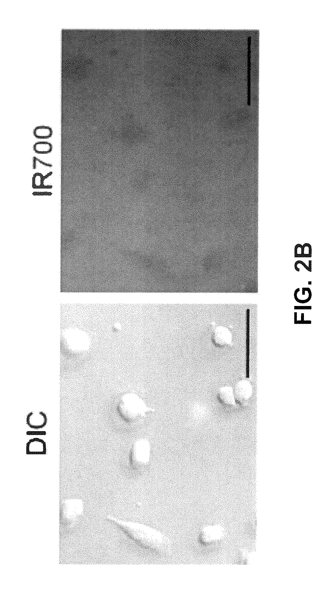
D00013

D00014

D00015

D00016
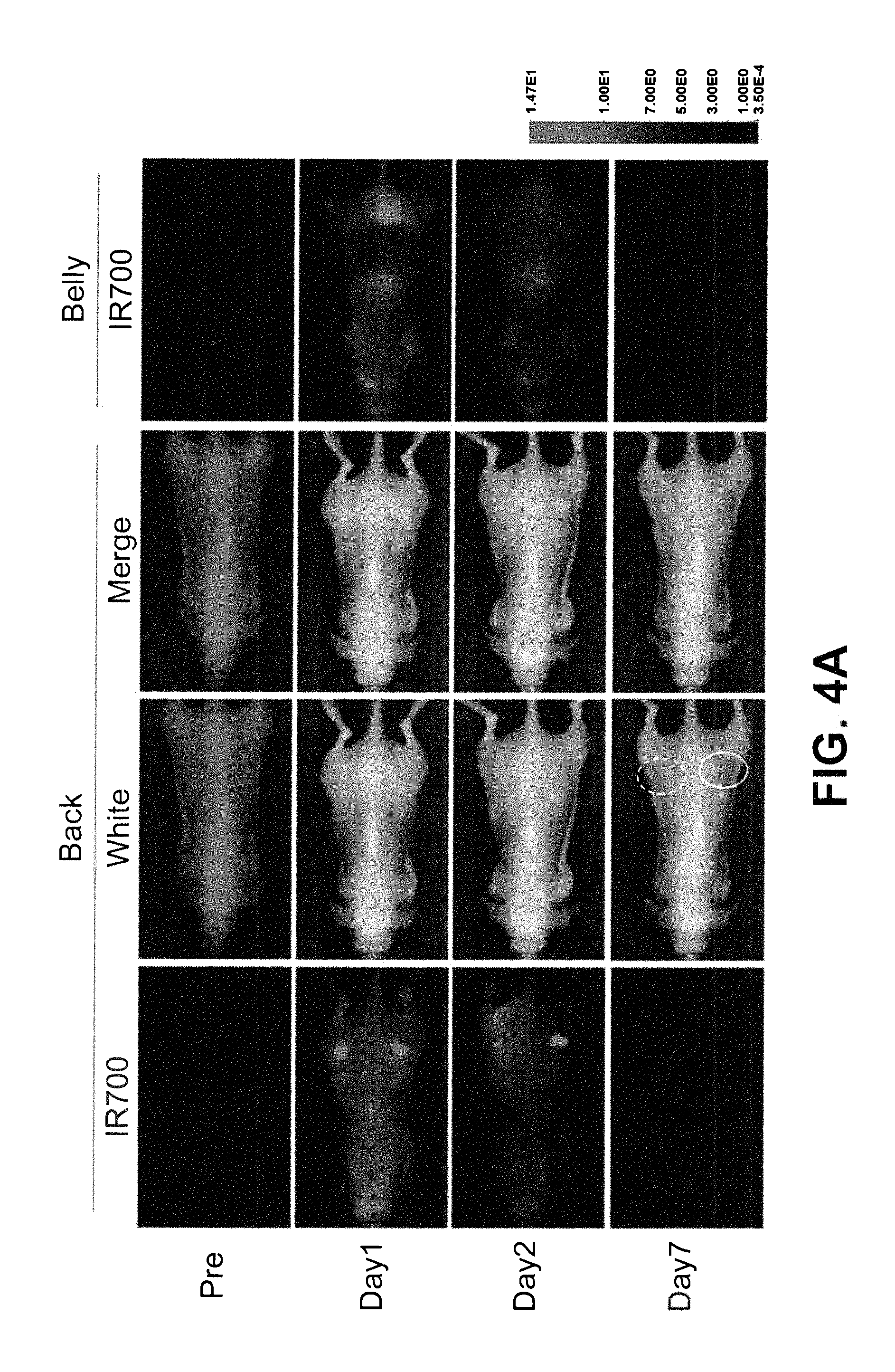
D00017
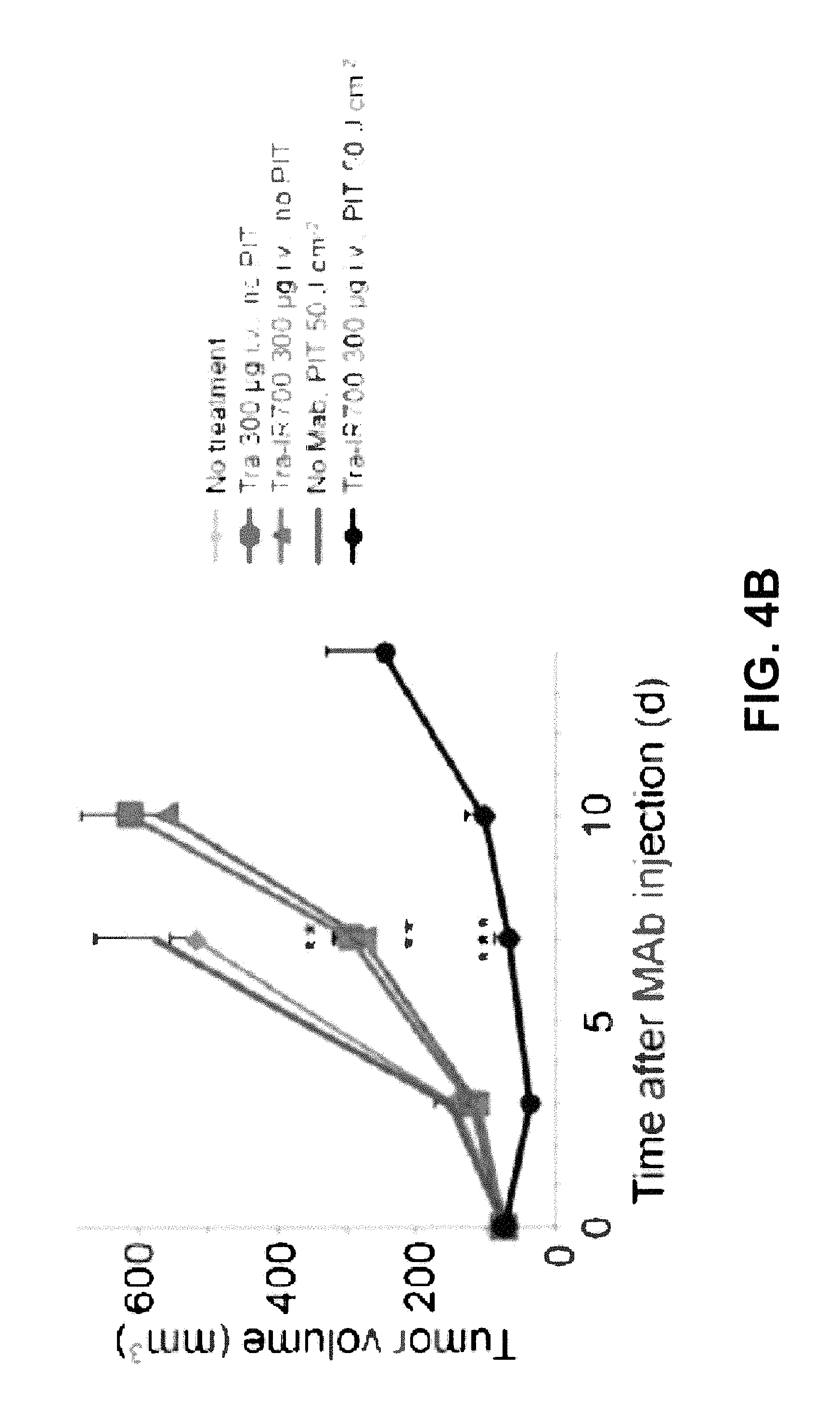
D00018

D00019

D00020

D00021
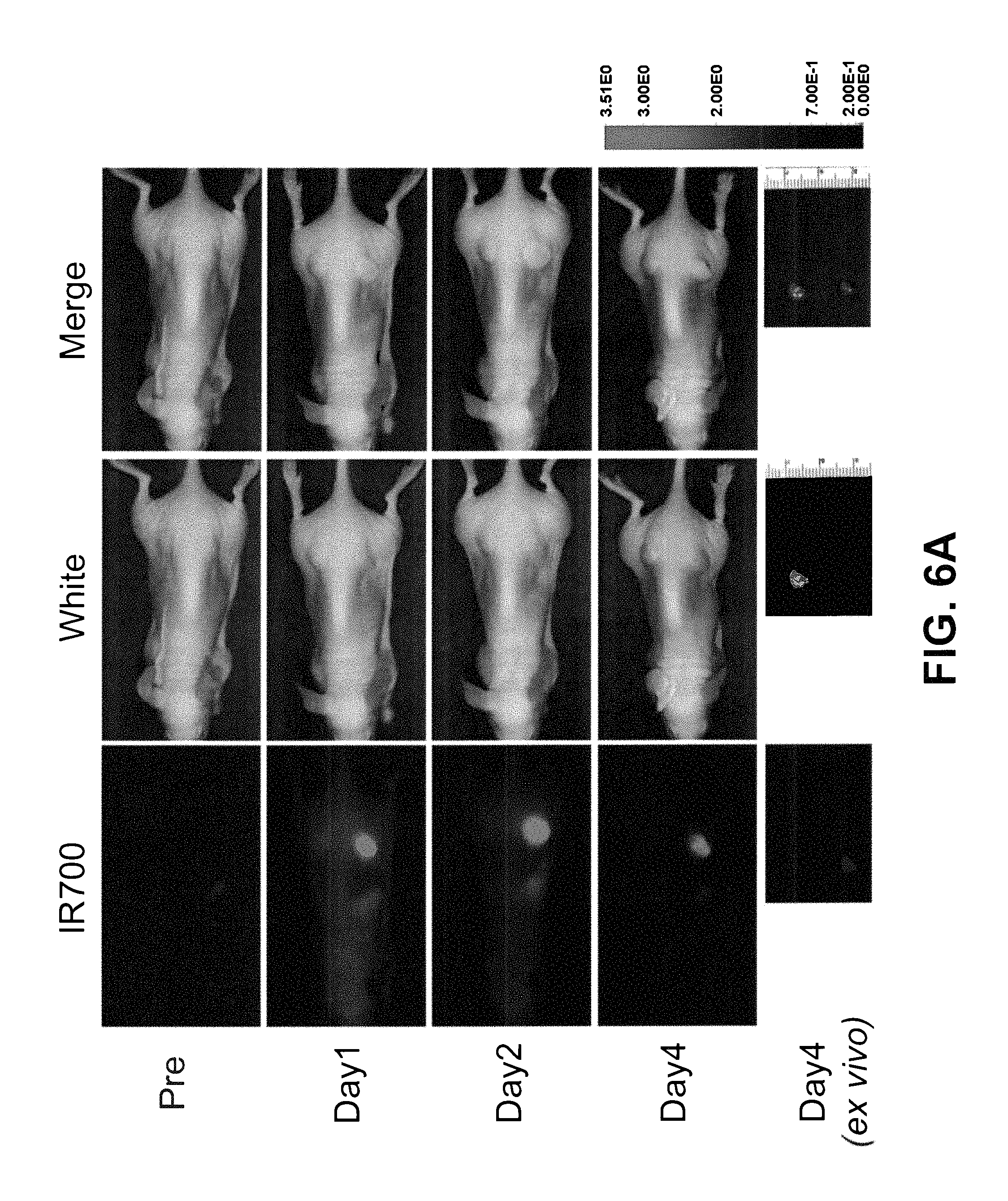
D00022
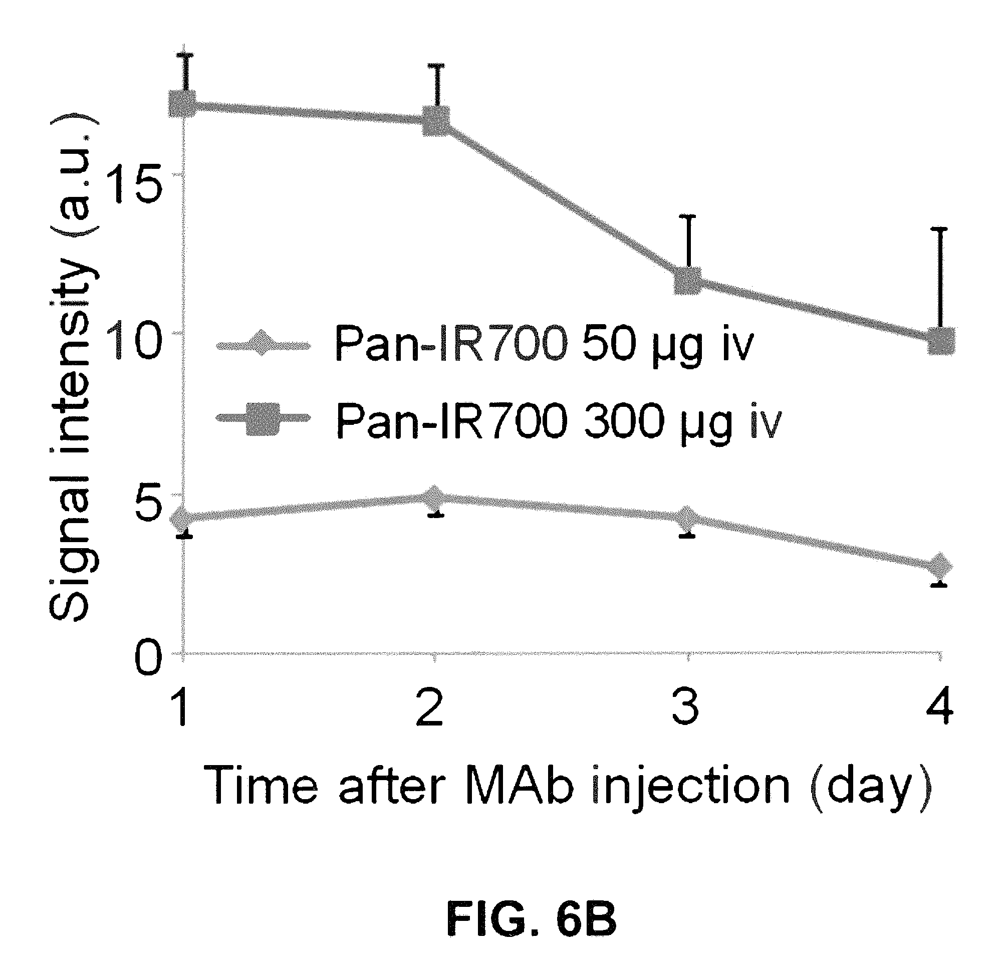
D00023
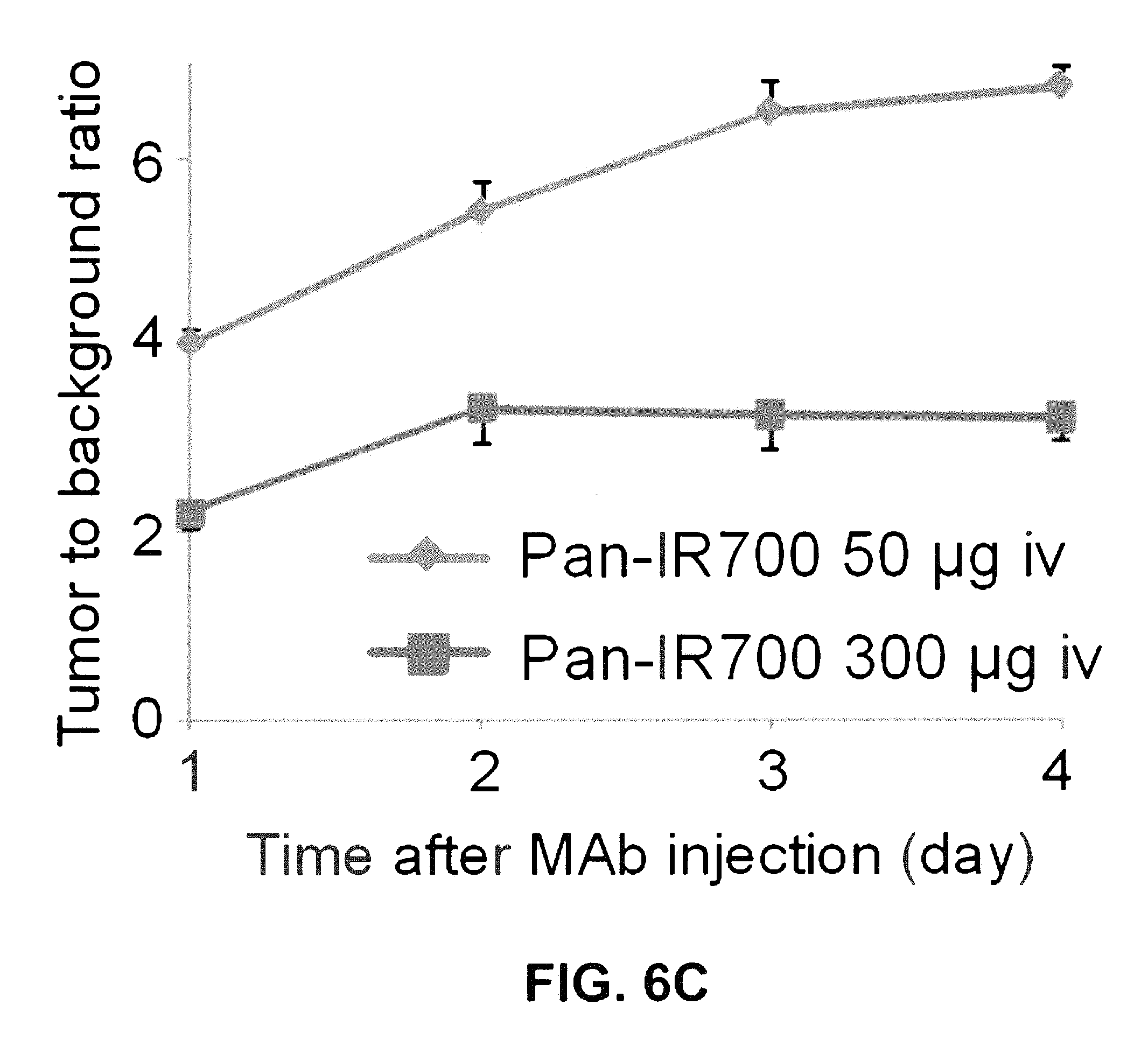
D00024

D00025
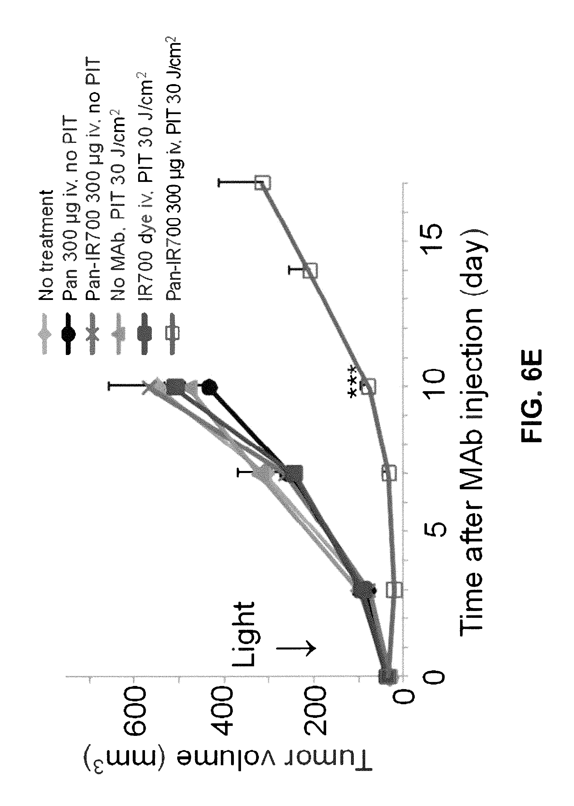
D00026
D00027
D00028
D00029
D00030

D00031
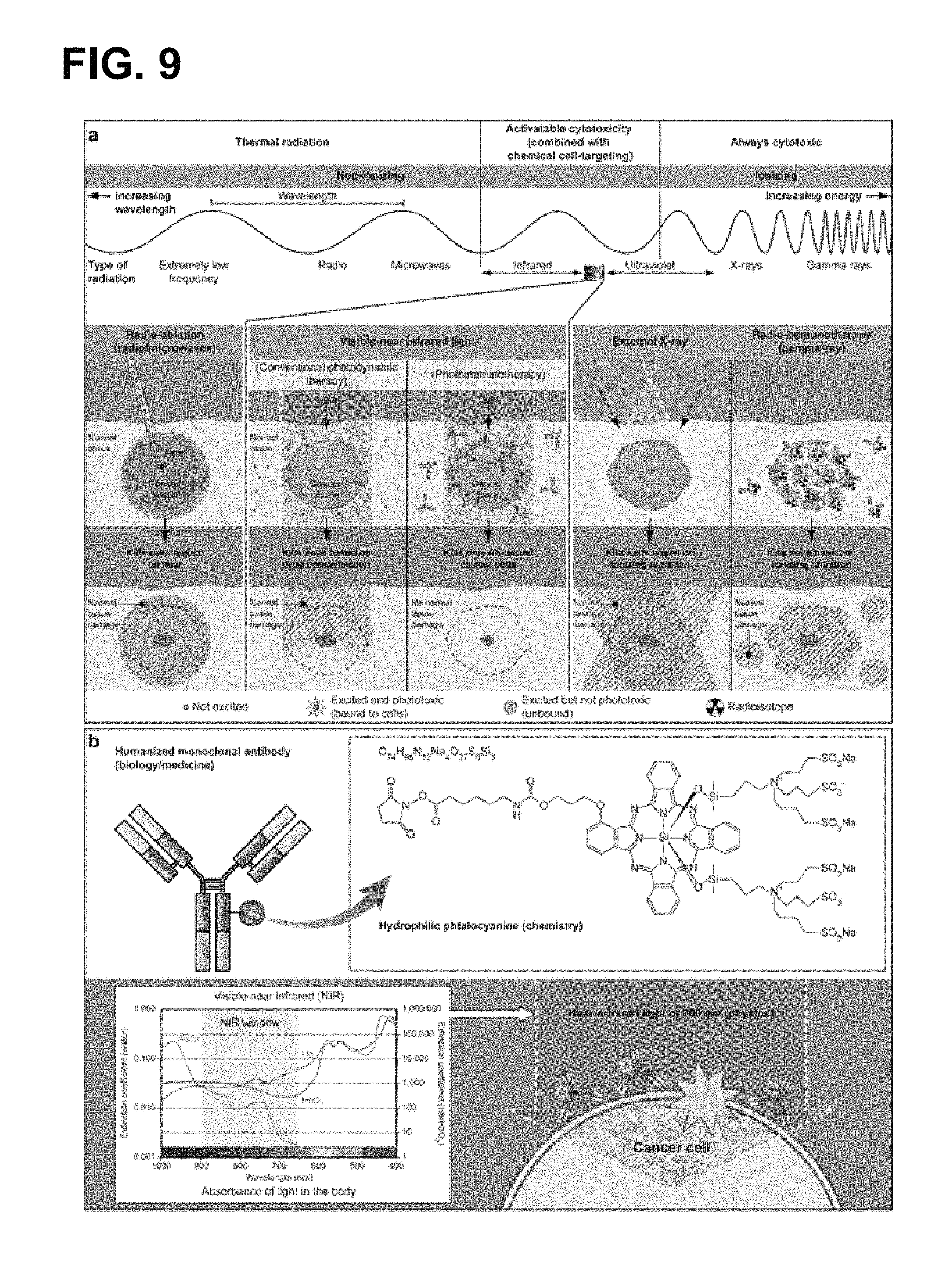
XML
uspto.report is an independent third-party trademark research tool that is not affiliated, endorsed, or sponsored by the United States Patent and Trademark Office (USPTO) or any other governmental organization. The information provided by uspto.report is based on publicly available data at the time of writing and is intended for informational purposes only.
While we strive to provide accurate and up-to-date information, we do not guarantee the accuracy, completeness, reliability, or suitability of the information displayed on this site. The use of this site is at your own risk. Any reliance you place on such information is therefore strictly at your own risk.
All official trademark data, including owner information, should be verified by visiting the official USPTO website at www.uspto.gov. This site is not intended to replace professional legal advice and should not be used as a substitute for consulting with a legal professional who is knowledgeable about trademark law.