Seizure detection methods, apparatus, and systems using a short term average/long term average algorithm
Osorio , et al. Ja
U.S. patent number 10,537,251 [Application Number 13/559,116] was granted by the patent office on 2020-01-21 for seizure detection methods, apparatus, and systems using a short term average/long term average algorithm. This patent grant is currently assigned to FLINT HILLS SCIENTIFIC, L.L.C.. The grantee listed for this patent is Alexey Lyubushin, Ivan Osorio, D. Sornette. Invention is credited to Alexey Lyubushin, Ivan Osorio, D. Sornette.


View All Diagrams
| United States Patent | 10,537,251 |
| Osorio , et al. | January 21, 2020 |
Seizure detection methods, apparatus, and systems using a short term average/long term average algorithm
Abstract
Methods for detecting a seizure, by use of a short term average/long term average (STA/LTA) algorithm applied to body data. A non-transitive, computer-readable storage device for storing data that when executed by a processor, perform such a method.
| Inventors: | Osorio; Ivan (Leawood, KS), Lyubushin; Alexey (Moscow, RU), Sornette; D. (Zurich, CH) | ||||||||||
|---|---|---|---|---|---|---|---|---|---|---|---|
| Applicant: |
|
||||||||||
| Assignee: | FLINT HILLS SCIENTIFIC, L.L.C.
(Lawrence, KS) |
||||||||||
| Family ID: | 48086428 | ||||||||||
| Appl. No.: | 13/559,116 | ||||||||||
| Filed: | July 26, 2012 |
Prior Publication Data
| Document Identifier | Publication Date | |
|---|---|---|
| US 20130096391 A1 | Apr 18, 2013 | |
Related U.S. Patent Documents
| Application Number | Filing Date | Patent Number | Issue Date | ||
|---|---|---|---|---|---|
| 61547567 | Oct 14, 2011 | ||||
| Current U.S. Class: | 1/1 |
| Current CPC Class: | A61B 5/048 (20130101); A61B 5/02055 (20130101); A61B 5/4836 (20130101); A61B 5/165 (20130101); A61B 5/726 (20130101); A61B 5/4094 (20130101); A61B 5/0205 (20130101); A61B 5/746 (20130101); A61B 5/1118 (20130101); A61B 5/14542 (20130101); A61B 5/0816 (20130101); A61B 5/0833 (20130101); A61B 5/16 (20130101); A61B 5/024 (20130101); A61B 5/04001 (20130101); A61B 5/0476 (20130101) |
| Current International Class: | G01N 33/50 (20060101); A61B 5/0205 (20060101); A61B 5/16 (20060101); A61B 5/048 (20060101); A61B 5/00 (20060101); A61B 5/024 (20060101); A61B 5/0476 (20060101); A61B 5/04 (20060101); A61B 5/08 (20060101); A61B 5/11 (20060101); A61B 5/083 (20060101); A61B 5/145 (20060101) |
References Cited [Referenced By]
U.S. Patent Documents
| 5995868 | November 1999 | Dorfmeister et al. |
| 7282030 | October 2007 | Frei et al. |
| 2005/0197590 | September 2005 | Osorio |
| 2006/0111644 | May 2006 | Guttag et al. |
| 2007/0213785 | September 2007 | Osorio |
| 2002049500 | Jun 2002 | WO | |||
| 2007034476 | Mar 2007 | WO | |||
| 2010115939 | Oct 2010 | WO | |||
| 2011020504 | Feb 2011 | WO | |||
Other References
|
International Application No. PCT/US2012/060029, PCT Search Report and Written Opinion dated Dec. 21, 2012, 15 pages. cited by applicant . Shen, Qiang et al., "Using Modulus Maximum Pair of Wavelet Transform to Detect Spike Wave of Epileptic EEG," Proceedings of the 20th Annual International Conference of the IEEE Engineering in Medicine and Biology Society, vol. 20, No. 3, 1998, 3 pages. cited by applicant. |
Primary Examiner: Sims; Jason M
Attorney, Agent or Firm: CF3 Eisenmann; Stephen
Parent Case Text
The present application claims priority from U.S. provisional patent application Ser. No. 61/547,567, filed on Oct. 14, 2011, which is incorporated herein by reference.
Claims
The invention claimed is:
1. A non-transitory computer readable program storage unit encoded with instructions that, when executed by one or more medical devices, performs a method, comprising: receiving via the one or more medical devices a time series of a first body signal of a patient obtained from one or more sensors; determining a sliding foreground time window and a sliding background time window for the time series of the first body signal; applying a spectral filter having a defined power spectral density to each of the sliding foreground time window and the sliding background time window; determining a ratio of a spectral power between the sliding foreground time window and the sliding background time window; determining a seizure onset in response to a first determination that the ratio reaches an onset threshold; initiating an electrical therapy based on the seizure onset determination; and determining a seizure termination in response to a second determination that the ratio reaches a termination threshold.
2. The non-transitory computer readable program storage unit of claim 1, including data that when executed by the one or more medical devices performs the method of claim 1, wherein: a number of samples in the sliding foreground time window (N.sub.STA) is 16; a number of samples in the sliding background time window (N.sub.LTA) is 360; the time of seizure onset (T.sub.onset) corresponds to a time fulfilling a condition STALTA(.tau.+N.sub.STA/2).gtoreq.T.sub.onset, wherein .function..tau..tau..tau..times..function..tau..tau..times..function. ##EQU00004## and .tau. is a common right-hand end of both the sliding foreground time window and the sliding background time window; a time of the seizure termination (T.sub.end) corresponds to a second time fulfilling a second condition of max {STALTA(s), .tau.-N.sub.STA.ltoreq.s.ltoreq..tau.}.ltoreq.T.sub.end; and a filter's spectral density corresponds to a Daubechies wavelet order 4, level 3.
3. The non-transitory computer readable program storage unit of claim 1, including data that when executed by the one or more medical devices performs the method of claim 1, wherein the method further comprises: where initiating the electrical therapy includes delivering the electrical therapy for the seizure at a particular time, wherein at least one of the electrical therapy, the particular time, or both is based upon the determination of the seizure onset; determining an efficacy of the electrical therapy; or issuing a warning for the seizure, wherein the warning is based upon the determination of the seizure onset.
4. The non-transitory computer readable program storage unit of claim 1, including data that when executed by a processor performs the method of claim 1, wherein the method further comprises estimating the degree of nonstationarity of said first body signal.
5. A non-transitive, computer-readable storage device for storing data that when executed by one or more medical devices, performs a method, comprising: receiving via the one or more medical devices a body signal of a patient during a first time series obtained from one or more sensors; determining a movable first time window and a movable second time window for the first time series; applying a spectral filter having a predetermined power spectral density to each of the movable first time window and the movable second time window; determining a ratio of a spectral power between the movable first time window and the movable second time window; determining a seizure onset in response to a first determination that the ratio reaches an onset threshold; initiating an electrical therapy based on the seizure onset determination; and determining a seizure termination in response to a second determination that the ratio reaches a termination threshold.
6. The non-transitive, computer-readable storage device of claim 5, including data that when executed by the one or more medical devices performs the method of claim 5, wherein: the movable first time window comprises from eight to thirty-two samples (N.sub.STA); and the movable second time window comprises from 180 to 720 samples (N.sub.LTA).
7. The non-transitive, computer-readable storage device of claim 5, including data that when executed by the one or more medical devices performs the method of claim 5, wherein the movable first time window comprises 16 samples (N.sub.STA); the movable second time window comprises 360 samples (N.sub.LTA); a time of seizure onset (T.sub.onset) corresponds to a time fulfilling a condition STALTA(.tau.+N.sub.STA/2).gtoreq.T.sub.onset, wherein .function..tau..tau..tau..times..function..tau..tau..times..function. ##EQU00005## and .tau. is a common right-hand end of both the movable first time window and the movable second time window; a time of the seizure termination corresponds to a second time fulfilling a second condition of max {STALTA(s), .tau.-N.sub.STA.ltoreq.s.ltoreq..tau.}.ltoreq.T.sub.end; and a filter's spectral density corresponds to a Daubechies wavelet order 4, level 3.
8. The non-transitive, computer-readable storage device of claim 5, including data that when executed by a processor performs the method of claim 5, wherein at least one parameter selected from the movable foreground time window, the movable background time window, the spectral filter, the onset threshold, or the termination threshold is selected based on at least one of a clinical application of said detection; a level of safety risk associated with an activity; at least one of an age, physical state, or mental state of the patient; a length of a window available for warning; a degree of efficacy of therapy; a degree of latency of efficacy of therapy; a degree of seizure control; a degree of circadian and ultradian fluctuations of said patient's seizure activity; a dependence of the patient's seizure activity on at least one of a level of consciousness, a level of cognitive activity, or a level of physical activity; the site of seizure origin; or the seizure type most commonly suffered by said patient.
9. The non-transitive, computer-readable storage device of claim 5, including data that when executed by a processor performs the method of claim 5, wherein said first time series body signal comprises at least one of a measurement of the patient's heart rate, a measurement of the patient's physical activity, a measurement of the patient's oxygen consumption, or a measurement of the patient's work.
10. The non-transitive, computer-readable storage device of claim 5, including data that when executed by the one or more medical devices performs the method of claim 5, further comprising: where initiating the electrical therapy includes delivering the electrical therapy for the seizure at a particular time, wherein at least one of the electrical therapy, the particular time of the electrical therapy delivery, or both is based upon the determination of the seizure onset and a seizure severity.
11. The non-transitive, computer-readable storage device of claim 10, including data that when executed by the one or more medical devices performs the method of claim 10, further comprising at least one of: determining an efficacy of the electrical therapy; or issuing a warning for the seizure, wherein the warning is based upon the determination of the seizure onset and the seizure severity.
12. The non-transitive, computer-readable storage device of claim 5, including data that when executed by a processor performs the method of claim 5, further comprising determining at least one of a timing of delivery of therapy, a type of therapy, at least one parameter of the therapy, a timing of sending a warning, a type of warning, a duration of the warning, or an efficacy index, based upon a timing of said determination of said seizure onset.
13. A medical device for detecting a seizure onset and a seizure termination of an epileptic seizure event from a patient body signal using a multi time-period averaging algorithm, comprising: a body data processing module for receiving a body signal of a patient during a first time series from one or more sensors; and a seizure onset/termination unit configured to: select a first time window and a second time window within the first time series; apply a spectral filter having a predetermined power spectral density to each of the first time window and the second time window; determine a ratio of a spectral power between the first time window and the second time window; determine that the seizure onset has occurred in response to a first determination that the ratio reaches an onset threshold; initiating an electrical therapy based on the seizure onset determination; and determine that the seizure termination has occurred in response to a second determination that the ratio reaches a termination threshold.
14. The medical device of claim 13, wherein the body signal processing unit comprises: a body data interface for receiving the body signal, the body data interface comprising: a buffer to perform buffering function upon the body signal; an amplifier for performing an amplification of the body signal; an analog-to-digital (A/D) converter for converting the body signal from an analog signal to a digital signal; and a filter for performing a filtering function upon the body signal; and a body data memory to store the digital signal.
15. The medical device of claim 13, wherein the seizure onset/termination unit comprises: a time window control module capable of selecting the first time window and the second time window; a spectral filter module for applying the spectral filter to the first time window and the second time window; a ratio determination unit for determining the ratio of the spectral power, and a ratio threshold comparison unit for comparing the ratio of the spectral power to a first threshold to determine whether the seizure onset has occurred, and for comparing the ratio of the spectral power to a second threshold to determine whether the seizure termination has occurred.
16. The medical device of claim 15, wherein said time window control module uses parameters selected based on at least one of a clinical application of at least one said determination; a level of safety risk associated with an activity; at least one of an age, physical state, or mental state of the patient; a length of a window available for warning; a degree of efficacy of therapy and of its latency; a degree of seizure control; a degree of circadian and ultradian fluctuations of said patient's seizure activity; a performance of the detection method as a function of the patient's sleep/wake cycle or vigilance level; a dependence of the patient's seizure activity on at least one of a level of consciousness, a level of cognitive activity, or a level of physical activity; the site of seizure origin; a seizure type, a desired sensitivity of detection of a seizure, a desired specificity of detection of a seizure, a desired speed of detection of a seizure, an input provided by the patient, or an input provided by a sensor.
17. The medical device of claim 13, wherein said first time series body signal comprises at least one of a measurement of the patient's heart activity, a measurement of the patient's respiratory activity, a measurement of the patient's kinetic activity, a measurement of the patient's brain electrical activity, a measurement of the patient's oxygen consumption, a measurement of the patient's oxygen saturation, a measurement of an endocrine activity of the patient, a measurement of a metabolic activity of the patient, a measurement of an autonomic activity of the patient, a measurement of a cognitive activity of the patient, or a measurement of a tissue stress marker of the patient.
18. The medical device of claim 13, further comprising: a therapy unit to deliver at least one therapy for the seizure at a particular time, wherein at least one of the therapy, the particular time, or both is based upon the determination of the seizure onset and a seizure severity; an efficacy unit to determine an efficacy of the therapy; a warning unit to issue at least one warning for the seizure, wherein the warning is based upon the determination of the seizure onset and the seizure severity; and a communication unit to allow communications between at least one medical device and at least one external device.
19. The medical device of claim 18, wherein at least one of the therapy delivered by the therapy unit or the warning delivered by the warning unit is based at least in part on at least one of a type of activity engaged in by the patient at the time of the seizure onset, a seizure type, the seizure severity and a time elapsed from a last seizure.
20. The medical device of claim 18, wherein the medical device is configured to determine at least one of a timing of a delivery of a therapy, a type of the therapy, at least one parameter of the therapy, a timing of sending the warning, a type of warning, a duration of the warning, and an efficacy index, based upon a timing of the determination of the seizure onset and the seizure severity.
21. The non-transitory computer readable program storage unit of claim 1, wherein the first body signal is at least one of an EKG signal, an accelerometer, and an electrical cortical signal.
22. The non-transitory computer readable program storage unit of claim 5, wherein the first body signal is at least one of an EKG signal, an electrocorticography (ECoG) signal, and an accelerometer signal.
23. The medical device of claim 13, wherein the first body signal is an electrocorticography (ECoG) signal.
24. The non-transitory computer readable program storage unit of claim 1, wherein the one or more medical devices is configured to determine and log at least one of a seizure intensity, a seizure duration, and a date and a time of a seizure occurrence.
25. The non-transitory computer readable program storage unit of claim 1, wherein the one or more medical devices is configured to determine an efficacy of the electrical therapy which includes determining one or more adverse effects of the electrical therapy.
26. The non-transitory computer readable program storage unit of claim 1, wherein the method further comprises issuing a warning and logging to memory: a time and a date of an occurrence of a seizure; a seizure duration; and a seizure severity.
Description
BACKGROUND OF THE INVENTION
1. Field of the Invention
The present invention relates generally to the field of epileptic event detection. More particularly, it concerns epileptic event detection by use of a short term average/long term average (STA/LTA) algorithm on a time series of patient body signal data.
2. Description of Related Art
There have been various advancements in the area of seizure detection, which remains a fairly subjective endeavor. The task of automated detection of epileptic seizures is generally related to and dependent on the definition of what is a seizure, definition which to date is subjective and thus inconsistent within and among experts. The lack of an objective and universal definition not only complicates the task of validation and comparison of detection algorithms, but possibly more importantly, the characterization of the spatio-temporal behavior of seizures and of other dynamical features required to formulate a comprehensive epilepsy theory.
The current state of automated seizure detection is, by extension, a reflection of the power and limitations of visual analysis, upon which it rests. The subjectivity intrinsic to expert visual analysis of seizures and its incompleteness (it cannot adequately quantify or estimate certain signal features, such as power spectrum) confound the objectivity and reproducibility of results of signal processing tools used for their automated detection. What is more, several of the factors, that enter into the determination of whether or not certain grapho-elements should be classified as a seizure, are non-explicit ("gestalt-based") and thus difficult to articulate, formalize and program into algorithms.
Most, if not all, existing seizure detection algorithms are structured to operate as expert electroencephalographers. Thus, seizure detection algorithms that apply expert-based rules are at once useful and deficient; useful as they are based on a certain fund of irreplaceable clinical knowledge and deficient as human analysis biases propagate into their architecture. These cognitive biases which pervade human decision processes and which have been the subject of formal inquiry are rooted in common practice behaviors such as: a) The tendency to rely too heavily on one feature when making decisions (e.g., if onset is not sudden, it is unlikely to be a seizure because these are paroxysmal events); b) To declare objects as equal if they have the same external properties (e.g., this is a seizure because it is just as rhythmical as those we score as seizures) or c) Classify phenomena by relying on the ease with which associations come to mind (e.g., this pattern looks just like the seizures we reviewed yesterday).
Seizure detection algorithms' discrepant results make attainment of a unitary or universal seizure definition ostensibly difficult; the notion that expert cognitive biases are the main if not only obstacle on the path to "objectivity" is rendered tenuous by certain results. These divergences in objective and reproducible results may be attributable in part, but not solely, to the distinctiveness in the architecture and parameters of each algorithm. The fractal or multi-fractal structures of seizures accounts at least in part for the differences in results and draws attention to the so-called "Richardson effect". Richardson demonstrated that the length of borders between countries (a natural fractal) is a function of the size of the measurement tool, increasing without limit as the tool's size is reduced. Mandelbrot, in his seminal contribution "How long is the coast of Britain," stressed the complexities inherent to the Richardson effect, due to the dependency of particular measurements on the scale of the tool used to perform them. Although defining seizures as a function of a detection tool would be acceptable, this approach may be impracticable when comparisons between, for example, clinical trials or algorithms are warranted. Another strategy to bring unification of definitions is to universally adopt the use of one method, but this would be to the detriment of knowledge mining from seizure-time series and by extension to clinical epileptology.
To date, performance comparisons among myriad existing algorithms have not been performed due to lack of a common and adequate database, a limitation that this invention addresses. However, if and when undertaken, said "comparisons" would be largely unwarranted and have meager, if any, clinical value/translatability, given that no universally accepted definition of what is a "seizure" has been crafted. The process of evaluation of seizure detection algorithms is plagued with cognitive biases and other confounding intricacies that impede achievement of consensus and in certain cases even of majority agreement. Performance assessment of these seizure detection algorithms relies entirely on expert visual analysis, which provides the benchmarks (seizure onset and end times) from which key metrics (detection latency in reference to electrographic and clinical onset time ("speed of detection"), sensitivity, specificity and positive predictive value) are derived, the effects of cognitive biases propagate beyond the seizure/non-seizure question into other aspects of the effectiveness of a particular seizure detection algorithm.
SUMMARY OF THE INVENTION
In one aspect, the present disclosure provides a method, comprising: receiving a time series of a first body signal of the patient; determining a sliding foreground time window and a sliding background time window for said time series of said first body signal; applying a spectral filter having a defined power spectral density to each of said foreground and background windows; determining the ratio of spectral power between said foreground and said background window; determining a seizure onset in response to a determination that said ratio reaches an onset threshold; determining a seizure termination in response to a determination that said ratio reaches a termination threshold.
In one aspect, the present disclosure provides a method, comprising: receiving a body signal of the patient during a first time series; determining a movable first time window and a movable second time window for said first time series; applying a spectral filter having a predetermined power spectral density to each of said first and second time windows; determining the ratio of spectral power between said first time window and said second time window; determining a seizure onset in response to a determination that said ratio reaches an onset threshold; and determining a seizure termination in response to a determination that said ratio reaches a termination threshold.
In one aspect, the present disclosure provides a non-transitory computer readable program storage unit encoded with instructions that, when executed by a computer, perform a method as described herein.
In one aspect, the present disclosure provides a medical device for detecting an onset and a termination of an epileptic seizure event from a patient body signal using a multi time-period averaging algorithm, comprising: a body data processing module for receiving a body signal of the patient during a first time series; and a seizure onset/termination unit to: select a first time window and a second time window within said first time series; apply a spectral filter having a predetermined power spectral density to each of said first and second time windows; determine a ratio of spectral power between said first time window and said second time window; determine that a seizure onset has occurred in response to a determination that said ratio reaches an onset threshold; and determine that a seizure termination has occurred in response to a determination that said ratio reaches a termination threshold.
BRIEF DESCRIPTION OF THE DRAWINGS
The invention may be understood by reference to the following description taken in conjunction with the accompanying drawings, in which like reference numerals identify like elements, and in which:
FIG. 1 illustrates a medical device system for detecting and classifying seizure events related to epilepsy from sensed body data processed to extract features indicative of aspects of the patient's epilepsy condition;
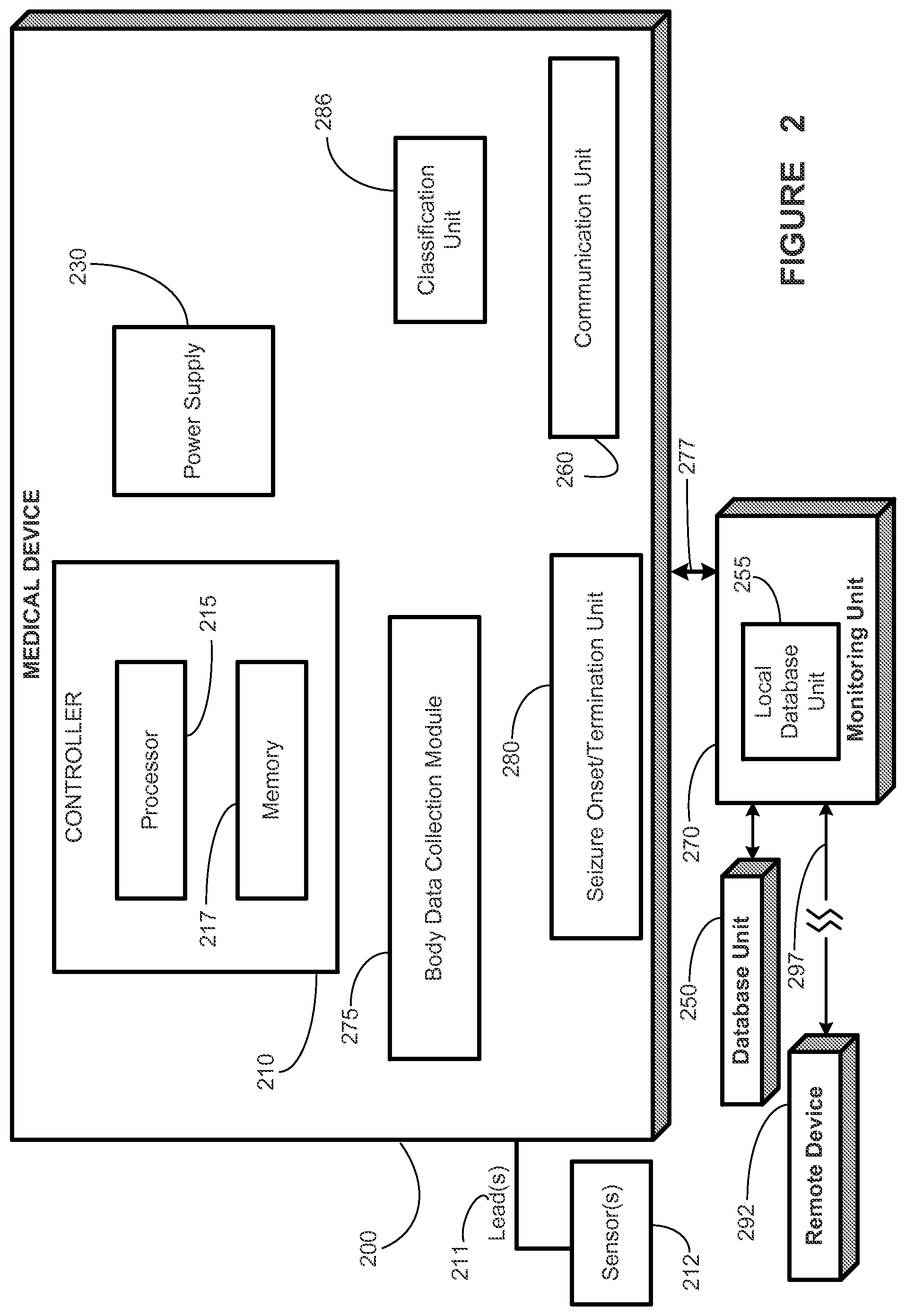
FIG. 2 illustrates a medical device system, according to an illustrative embodiment of the present disclosure;
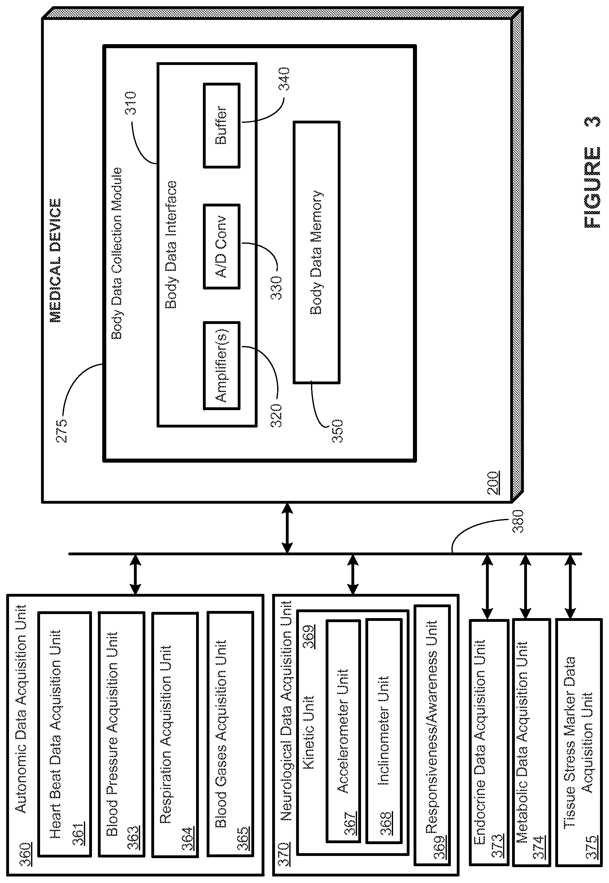
FIG. 3 provides a stylized diagram of a medical device and different data acquisition units that may provide output(s) used by other unit(s) of the medical device, in accordance with one illustrative embodiment of the present disclosure;
FIG. 4 provides a stylized diagram of a medical device and different data acquisition units that may provide output(s) used by other unit(s) of the medical device, in accordance with one illustrative embodiment of the present disclosure;
FIG. 5 provides a stylized diagram of a seizure onset/termination unit, in accordance with one illustrative embodiment of the present disclosure;
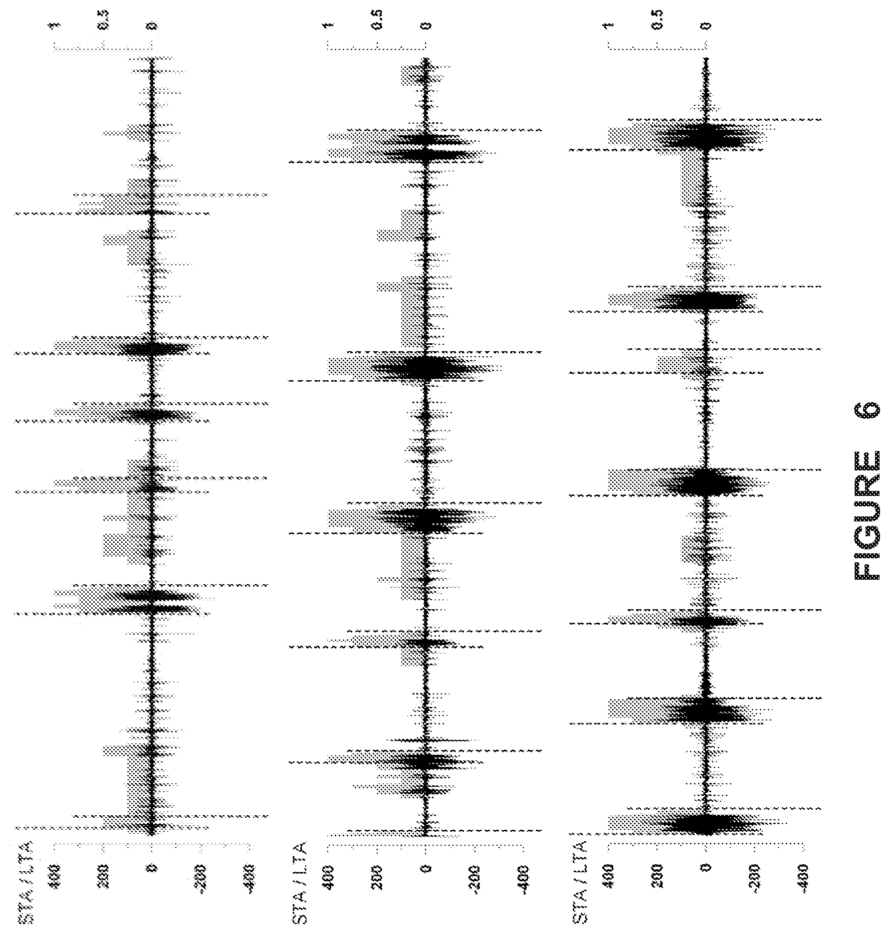
FIG. 6 illustrates the output of the STA/LTA algorithm in reference to an Average Indicator Function (AIF) making use of four algorithms, including the STA/LTA algorithm, in accordance with one illustrative embodiment of the present disclosure;
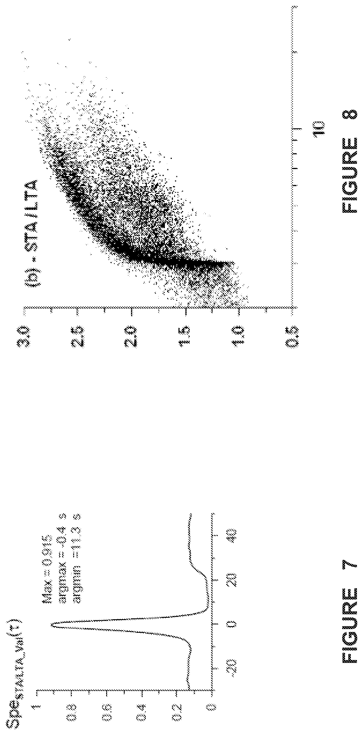
FIG. 7 shows a graph of a specificity function for the STA/LTA method as a function of time with respect to a validated algorithm's time of seizure detection, in accordance with one illustrative embodiment of the present invention;
FIG. 8 shows plots of the decimal logarithm of the dependence of seizure energy on seizure duration, in accordance with one illustrative embodiment of the present disclosure;
FIG. 9 shows the empirical "tail" of the conditional probability distribution functions for: (a) Seizure durations (minimum duration: 2 sec); and (b) the logarithm of seizure energy as estimated with four different methods, in accordance with one illustrative embodiment of the present disclosure;
FIG. 10 provides a flowchart depiction of a method, in accordance with one illustrative embodiment of the present disclosure;
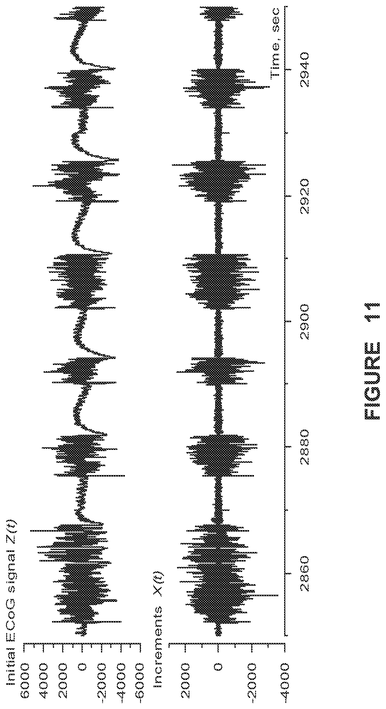
FIG. 11 shows ECoG before (upper panel) and after differentiation (lower panel), in accordance with one illustrative embodiment of the present disclosure; and
FIG. 12 shows temporal evolution of the decimal logarithm of the power spectrum of differentiated ECoG.
DESCRIPTION OF ILLUSTRATIVE EMBODIMENTS
In one aspect, the present disclosure provides several new seizure detection algorithms that may be applied to one or more streams of body data. Some of these algorithms rely principally on power variance for detection of seizures, while others rely mainly on power spectral shape.
In another aspect, the present disclosure exploits the simultaneous application of two or more individual seizure detection algorithms to derive a probabilistic measure of seizure activity (PMSA), which may be used to issue detections by majority or consensus of a plurality of the two or more seizure detection algorithms, depending on safety factors and others such as detection speed, sensitivity, specificity or any other performance measures and the clinical application(s) at hand. Real-time ("on the run") automated seizure detection provides the only means through which contingent warning to minimize risk of injury to patients, delivery of a therapy for control of seizures, or logging of the date, time of onset and termination and severity may be performed.
This disclosure, in one embodiment, provides a short term average/long term average (STA/LTA) algorithm suitable for use in epileptic event detection, by operation of the algorithm on a time series of patient body signal data. Such a STA/LTA algorithm may be used by itself, or as part of a Probabilistic Measure of Seizure Activity.
More generally, this disclosure: a) Draws attention to the intricacies inherent to the pursuit of a universal seizure definition even when powerful, well understood signal analysis methods are utilized to this end; b) Identifies this aim as a multi-objective optimization problem and discusses the advantages and disadvantages of adopting or rejecting a unitary seizure definition; c) Introduces a Probabilistic Measure of Seizure Activity to manage this thorny issue.
Seizure detection belongs to a class of optimization problems known as "multi-objective" due to the competing nature between objectives; improvements in specificity of detection invariably degrade sensitivity and vice-versa. Attempts to achieve a universal seizure definition using objective, quantitative means, are likely to be fraught with similar competing objectives, but imaginative application of tools from the field of multi-objective optimization, among others, are likely to make this objective more tractable.
Achieving a unitary seizure definition would be difficult, as consensus among epileptologists as to what grapho-elements are classifiable as ictal, is rare. In the absence of a universal definition, issuing seizure warnings for certain cases will be problematic and unsafe. For example, if a patient with seizures wishes to operate power equipment or a motor vehicle, the absence of a universal agreement on when the patient has had a seizure may preclude any viable way of ensuring, using seizure detection algorithms, that the patient's seizures are under sufficient control to allow such activities to occur. To manage the difficulties of a consensus seizure definition, substantive gains are feasible through steps entailing, for example, the application of advanced signal analysis tools to ECoG, to hasten the identification of properties/features that would lead to the probabilistic discrimination of seizures from non-seizures with worthwhile sensitivity and specificity for the task at hand. However, to even have a modicum of success, such an approach should not ignore the non-stationarity of seizures and, should strike some sort of balance between supervised (human) and unsupervised machine-learning) approaches. The resulting multidimensional parameter space, expected to be broad and intricate, may also foster discovery of hypothesized (e.g. pre-ictal) brain sub-states.
The challenges posed by the attempt to define seizures unitarily using objective means (distinct from visual analysis) may be partly related to their fractal properties and understood through a simplistic analogy to the so-called "Richardson effect". A revision of the time-honored subjective definition of seizures may be warranted to further advance epileptology.
The present inventors propose a Probabilistic Measure of Seizure Activity (PMSA) as one possible strategy for characterization of the multi-fractal, non-stationary structure of seizures, in an attempt to eschew the more substantive limitations intrinsic to other alternatives.
The PMSA may make use of "indicator functions" (IFs) denoted .chi..sub.algo for each algorithm `algo.` In one embodiment, the PMSA may also make use of an Average Indicator Function (AIF). In one embodiment, the AIF is defined as: AIF(t)=(.chi..sub.Val(t)+.chi..sub.r.sub.2(t)+.chi..sub.STA/LTA(t)+.chi..- sub.WTMM(t))/4
The subscripts Val, r.sup.2, STA/LTA and WTMM refer to four different algorithms, particular embodiments of which are described herein and/or in other related applications. One or more of these algorithms may be used to detected seizures from one or more body data streams including, but not limited to, a brain activity (e.g., EEG) data stream, a cardiac (e.g., a heart beat) data stream, and a kinetic (e.g., body movement as measured by an accelerometer) data stream.
"Val" refers to an algorithm for seizure detection using ECoG data that has been validated by experts without reaching a universal consensus about its performance (e.g., false positive, false negative and true positive detections). An "r.sup.2" algorithm may also be referred to herein as an "r{circumflex over ( )}.sup.2," "autoregression," or "autoregressive" algorithm. A "STA/LTA" algorithm refers to an algorithm characterized by the ratio of a Short-Term Average to a Long-Term Average. A "WTMM" algorithm refers to a Wavelet Transform Maximum Modulus algorithm.
For determination of an AIF from the foregoing formula, an algorithm's IF equals 1 for time intervals (0.5 sec in this application) "populated" by ictal activity and 0 by inter-ictal activity. The IF's are used to generate four stepwise time functions, one for each of: a) a 2.sup.nd order auto-regressive model (r.sup.2); b) the Wavelet Transform Maximum Modulus (WTMM); c) the ratio of short-to-long term averages (STA/LTA) and d) a Validated algorithm (Val). With these IFs, the AIF is computed (its values may range between [0-1] with intermediate values of 0.25, 0.5 and 0.75 in this embodiment). (Intermediate AIF values are functions of the number of algorithms applied to the signal. Since in this study 4 methods were used and the range of the indicator function is [0-1], the intermediated values are [0.25, 0.5, 0.75]). These values [0-1] are estimates of the probability of seizure occurrence at any given time. In another embodiment, the values of each algorithm's IF may be weighted differently, and a composite IF (e.g., a Weighted Indicator Function or WIF) different from the AIF may be computed.
Data obtained from one subject undergoing evaluation for epilepsy surgery with intra-cranial electrodes was selected for analyses as it had the largest number of clinical and subclinical seizures in the University of Kansas Medical Center Epilepsy Database. ECoG was collected in accordance with the Center's surgical evaluation protocol and with the Human Subjects Committee requirements, which include signing of a consent form by the subject.
The ECoG was recorded using electrodes implanted into the amygdala, pes hippocampus and body of hippocampus bilaterally through the temporal neocortex and had a duration of 6.9 days (142'923'853 samples; 239.75 Hz sampling rate).
For efficient analyses, ECoG signal differentiation was performed, so as to minimize the non-stationarity present in them. If Z(t) is raw ECoG, then its difference is X(t)=Z(t)-Z(t-1), where (t) corresponds to a sample time increment. This linear operation is exactly invertible and, unlike band-pass filtering or detrending, does not suppress low frequency fluctuations, but decreases their overall influence. FIG. 11 illustrates the effect of this operation on raw ECoG. The differentiated ECoG is less non-stationary (chiefly at low frequencies) than the undifferentiated one (x-axis: time in sec.; y-axis: amplitude in microvolts). FIG. 12 shows a time-frequency map of the evolution of the power spectra of differentiated ECoG segments (as shown in FIG. 11, bottom panel). The power spectra are estimated within 5 sec moving windows of length. Six brief seizures appear as marked power spectrum increases in the 10-100 Hz. band (x-axis: time in sec.; y-axis: frequency (Hz); gray scale to the right of main graph).
Seizure Detection with the STA/LTA Method.
The "short time average" (STA) divided by the "long time average" (LTA) is widely used in seismology as an earthquake detector and has several realizations, one of which will be used here. The dynamical analogies between earthquakes and seizures provide a rationale for applying this method to their detection.
Let X(t) be the output of the Daub04 3.sup.rd level band-pass filter applied to the ECoG, N.sub.STA the length (in number of samples) of the "short time average" and let N.sub.LTA be the length (in number of samples) of the "long time average". The STA/LTA ratio is defined by the formula:
.function..tau..tau..tau..times..function..tau..tau..times..function. ##EQU00001##
where .tau. is the common right-hand end of both short and long averaging time windows.
Seizure onsets .tau..sub.onset correspond to the times for which the following condition is fulfilled: STALTA(.tau.+N.sub.STA/2).gtoreq.T.sub.onset (16)
Seizure terminations .tau..sub.end are determined using the rule .tau..sub.end=.tau.*-N.sub.STA/2 where .tau.* is the time .tau. after the most recent seizure onset for which the following condition is fulfilled: max{STALTA(s),.tau.-N.sub.STA.ltoreq.s.ltoreq..tau.}.ltoreq.T.sub.end (17)
The STA/LTA-detector has the following parameters: 1. The length of the short time average N.sub.STA; 2. the length of the long time average N.sub.LTA; 3. the threshold T.sub.onset for seizure onset as defined above; 4. the threshold T.sub.end for seizure termination as defined above; their values for this application were chosen to be: N.sub.STA=360, N.sub.LTA=16N.sub.STA and T.sub.onset and T.sub.end values were chosen under the condition T.sub.onset>T.sub.end. Using different threshold values, it is possible to increase or decrease the temporal resolution with which seizures are detected.
The WTMM and Validated algorithms have been described in U.S. provisional patent application Ser. No. 61/547,567, filed on Oct. 14, 2011, which is hereby incorporated by reference herein in its entirety.
The total number of detections, their duration and the percent time spent in seizure over the time series total duration (6.9 days) are presented in Table 1.
TABLE-US-00001 TABLE 1 Summary statistics obtained by applying two different detection methods (Validated Algorithm; STA/LTA). The minimum duration of seizures was set at 2 s. Validated algorithm STA/LTA Total number of seizures 3184 16275 with duration .gtoreq. 2 s. Mean duration, s. 3.8 4.3 Median duration, s. 3.4 3.5 % time spent in seizure 2 12
FIG. 6 illustrates the output of the STA/LTA algorithm in comparison with an AIF making use of each of the validated algorithm, and the r2 (AR), STA/LTA and WTMM algorithms. Specifically, FIG. 6 shows results of applying the STA/LTA seizure detection method to a differentiated ECoG (in black; 200 sec/panel) of a human with pharmaco-resistant epilepsy. The grey boxes represent the values (right y-axis) of an Average Indicator Function in the interval [0,1]. Seizures onset and end times are indicated by vertical lines, with onset times shifted upward and end times, downward. Notice that the value of the Average Indicator Function is rarely 1, at onset or termination, indicating all methods do not detect the ECoG activity as being ictal in nature at those moments. However, with seizures exceeding certain duration (at least 20 seconds) and intensity thresholds, they converge to all detect the seizure event. This indicates that the spectral and other properties of seizures are not homogeneous at the onset and termination of seizures, which is consistent with the lack of agreement among human experts (and algorithms) during onset and termination. Left y-axis: ECoG amplitude (in UV); excursions above zero correspond to positive, and below, to negative, polarity.
Time intervals for which the pairwise product .chi..sub.Val(t).chi..sub.STA/LTA(t)=1 correspond to seizures detected by both the validated algorithm and STA/LTA. Dividing the number of time intervals when .chi..sub.Val(t).chi..sub.STA/LTA(t)=1 by the number of intervals when .chi..sub.Val(t)=1, yields the specificity of the STA/LTA method with respect to the validated algorithm. The specificity functions for the two other methods Spe.sub.WTMM_Val(.tau.) and Spe.sub.r2_Val(.tau.) are identically computed and their maximum value (dependent on .tau.) may be regarded as the mean value of the time delay of one method's function with respect to another for seizure onset and end times. Since the validated algorithm has an inherent delay of 1 s (the median filter's foreground window is 2 s) and an additional duration constraint of 0.84 s. is imposed before a detection is issued, its onset and end times are "delayed" compared to those yielded by the STA/LTA method.
The present inventors discovered that the time differences are negative for all three algorithms compared to the validated algorithm; that is, the validated algorithm's detection times lag behind those given by the other methods. More particularly, the mean delay of the validated algorithm is 0.6 s with respect to STA/LTA. The re-calculated specificity values shifted by .tau. shown in Table 2 are higher compared to those without shifting.
TABLE-US-00002 TABLE 2 Method Spe.sub.Method_Val(0) .times..tau..times..function..tau. ##EQU00002## .times..times..times..tau..times..function..tau. ##EQU00003## STA/ LTA 0.911 0.915 -0.4 s Values of specificity of STA/LTA calculated with respect to the validated method and time lag (as defined in the text) at which the specificity attains its largest value.
The information in Table 2 is also depicted graphically in FIG. 7, which illustrates a graph of a specificity function for the STA/LTA method as a function of time with respect to the Validated algorithm's time of seizure detection. Tau (.tau.) zero (x-axis) corresponds to the time at which Val issues a detection. Negative .tau. values indicate "late" detections by the validated algorithm in relation to the other three and positive value the opposite. As shown, STA/LTA issues earlier detections than Val. Values of the lags .tau. corresponding to the maximum and minimum values of the function are presented under the names argmax and argmin respectively.
The present inventors also discovered that only 19.6% of seizures recognized as such by the STA/LTA method are also detected by the validated method, indicating that in its generic form and by design, it is less sensitive and more specific for seizure detection than the STA/LTA algorithm.
The STA/LTA method, along with the other methods mentioned supra, survey different but inter-dependent ECoG signal properties, thus expanding the breadth and perhaps also the depth of insight into the spectral "structure" of epileptic seizures in a clinically relevant manner. The STA/LTA uses the ratio of variances to detect, at low computational expense, ECoG signal changes corresponding to seizures.
Algorithmic and visual expert analysis consensus as to what grapho-elements define a `seizure` seems to be highly dependent on when during the course of a `seizure` a decision is made. In this context, it is noteworthy that AIF and PIF frequently reached a value of 1, indicative of concordance among all detection methods sometime after seizure onset and before its termination (as determined by any of the methods), provided said seizures reached a certain duration (20-30 s.) as discussed in more detail in U.S. provisional patent application Ser. No. 61/547,567, filed on Oct. 14, 2011. In short, seizure onsets and terminations may be under certain conditions universally undefinable by algorithmic or expert visual analysis. A systematic investigation of the differences in signal spectral properties between the "preface"/"epilogue" and the "main body" of seizures was not performed. It is speculated that the presence of "start-up transients" (in a dynamical sense) and of temporo-spatial dispersion of the ictal signal (which impacts S/N) may be most prominent at the onset and termination of seizures. These and local and global state-dependencies of certain signal features, account in part for the temporal fluctuations in algorithmic detection performance that characterize these results.
The dependencies of seizure energy (defined as the product of the standard deviation of the differentiated ECoG signal and seizure duration, in sec.) on seizure duration, for the set of icti detected by the STA/LTA method is depicted in FIG. 8. A subset of seizures detected by all methods obeys a simple law of proportionality between energy (y-axis) and duration (x-axis, log scale, seconds), that is, the longer the seizure, the larger its energy. However, this relationship is not invariably linear for other detection algorithms, indicating the presence of interesting scaling properties of seizure energy.
FIG. 9 shows the empirical "tail" of the conditional probability distribution functions for: (a) Seizure durations (minimum duration: 2 sec); (b) the logarithm of seizure energy as estimated with the Validated method (dashed) and the Short/Long Term Average: method (solid).
The conditional probabilities of durations (FIG. 9a) and of the logarithm of energy of seizures (FIG. 9b) provide additional support that their properties are partly a function of the method used for their detection. The validated and STA/LTA algorithms yield similar durations but different from those of the WTMM and r.sup.2 methods, which are analogous to each other (FIG. 9a). The distributions of the logarithm of seizure energies as identified by each of the methods (FIG. 9b) reveals additional discrepancies as evidenced by the much narrower and shorter "tail" distribution of the validated algorithm compared to the others.
The medical and psycho-socio-economic burden imposed upon patients, caregivers and health systems by pharmaco-resistant epilepsies is enormous. Intracranial devices for automated detection, warning and delivery of therapy, the presently preferred "line of attack" for an abundance of weighty reasons, would be insufficient to adequately address said burden at a global scale. Reliance on signals that while extra-cerebral are under cortical modulation or control such as cardiac or motor and are altered by seizures, emerges as a viable research direction with potentially fruitful clinical applications.
The greater ease of implementation and lower cost of automated real-time detection, warning and therapy systems based on extra-cerebral signals, compared to those requiring intracranial placement, makes them worthy of investigation.
Cortical electrical activity has been the primary, if not sole, source of signals for visual or automated detection and quantification of seizures in clinical use. The inextricable link between brain and epilepsy has historically impelled clinical neuroscientists to leave unexploited the equally inextricable link between brain and body. The brain-epilepsy link has distracted us from certain severe limitations (for certain applications) inherent to the recording of cortical signals from scalp or even directly from its surface, such as marked cortical signal attenuation and filtering and limited access to neural sources (only about one-third of the neocortex is surveyable by scalp electrodes and subdural electrodes record little activity from the lateral and bottom walls of sulci). Yet, readily accessible sources that provide indirect but valuable information about the state of the brain, particularly during the ictal or postictal state, remain largely untapped.
The growing emphasis on widely accessible, cost-effective, good quality health care in the context of expanding populations, especially in age-groups above 60 yr. in whom the incidence of epilepsy is high, and the shrinking financial resources to support the required infra-structure, pose an enormous challenge to patients whose seizures are pharmaco-resistant as well as to epileptologists and functional neurosurgeons. The properly placed emphasis on implantable intracranial devices for automated seizure detection, warning and delivery of therapy in patients with drug-resistant seizures should be viewed in the context that even if economic resources were unlimited, human resources are starkly small. Given the number of functional neurosurgeons in the United States (one source puts the number at 300, of which about 100 work in epilepsy) is it realistic to pursue exclusively intracranial devices to address the unmet needs of pharmaco-resistant patients conservatively estimated (in the US) at 600,000? The deleterious medical, and psycho-social impact of intractable epilepsy and its high cost of care, along with the sophisticated human and technological resources needed to address them, qualifies this, in these authors opinions, as a public health care problem. Indeed, scientific advances regardless of their value may not translate into improved care of epilepsy and lessen its burden, unless devices are broadly accessible; in short the challenge of ameliorating the global burden of drug-resistant epilepsies may exceed scientific and technological ones. If the answer to the question put forth a few lines above is in the negative (intracranial devices will not meet the global burden) viable alternatives must be sought.
The utilization of certain extra-cerebral signals looms as one such alternative. Cardiac (e.g., heart rate, EKG morphology) and motor (speed, direction and force of joint movements) signals are prime candidates for the following reasons: 1. Structures that form part of the central autonomic nervous system or are strongly interconnected with it, are common sites of epileptogenesis (e.g., amygdalae-hippocampi); 2. Spread of seizures out of the primary epileptogenic zone, is prevalent in pharmaco-resistant patients so that even if the site of origin is not part of the central autonomic network, invasion of it by ictal activity is quite common; 3. Partial seizures particularly if complex, are characterized by either positive (e.g., motor automatisms, hypermotoric behavior, clonic/myoclonic activity, focal increase in anti-gravitatory muscle tone) or negative (e.g., motionless, focal loss of antigravitatory muscle tone) phenomena that are stereotypical across seizures originating from the same site and appear relatively early in the course of seizures; 4. Cardiac and motor signals are highly robust, easily recordable as they do not require implantable devices or development of ground breaking technology; EKG, actigraphs, 3-D accelerometers are widely available commercially and are considerably less costly than those required for use in the central nervous system (CNS); 5. Signals of cardiac and motor origin lack the high complexity or large dimensionality of those generated by the brain's cortex, are simpler to process and analyze, and thus are less computationally expensive. Ease of computation allows the use of simpler, smaller devices compared to those required for computation of cortical signals and as they use less power, battery recharging or replacements are less frequent; 6. The neurosurgical procedures and potential associated complications make implantable devices unappealing to a majority of pharmaco-resistant patients that responded to a survey.
Among the numerous extra-cerebral signals usable for seizure detection, cardiac, have been the most extensively investigated. Tachycardia is a common manifestation of partial seizures, occurring in almost 90% of seizures of mesial temporal origin and precedes electrographic (as determined with scalp electrodes) and clinical onset in the majority of these seizures. (Tachycardia invariably occurs in primarily/secondarily generalized tonic-clonic seizures being higher in magnitude and longer duration than in partial seizures. Tachycardia with tonic-clonic seizures is multifactorial: neurogenic, metabolic, and exertional.) From a cardiac rhythm perspective, the increases in heart rate temporally correlated with seizures are rarely pathologic, being of sinus origin; additionally their magnitude is unlikely to compromise cardiac output in healthy individuals. Ictal tachycardia has a strong neurogenic component reflective of either an increase in sympathetic or withdrawal of parasympathetic activity; while increases in motor activity in reference to the interictal state would augment its magnitude, tachycardia occurs in subjects in whom seizures manifest with motionless. Bradycardia also occurs with seizures, albeit with much lower prevalence than tachycardia; so called "temporal lobe syncope", denoting the loss of consciousness (without convulsive activity) during partial seizures is caused by profound bradycardia.
In light of the potential to apply cardiac signals, and in particular of exploiting changes in heart rate (increases or decreases relative to an interictal baseline) for automated seizure detection, algorithms are being developed and tested to this end. In addition to detection and warning of seizures, heart rate changes may be used to quantify: a) Relative duration defined as the time said changes spend above or below an interictal reference value(s); b) Relative intensity corresponding to the area under the curve or to the product of peak/bottom heart rate and duration (in sec.); c) Seizure frequency/unit time (e.g., month). The challenge of this detection modality for ambulatory clinical applications, is the ubiquitousness of heart rate changes with daily life activities that may translate into large numbers of false positive detections. Arousal from sleep, standing up from a recumbent position, climbing stairs, are but a few of the myriad daily life activities associated with relative or absolute changes (e.g., increases) in heart rate. The discriminating power or positive predictive value (ictal vs. exertional) of this detection modality is currently the subject of investigation in epilepsy monitoring units. Heart rate, among other (rate of change/slope; P-QRS-T morphology) markers, during seizures, are recorded, analyzed and compared to those associated with protocolized motor activities (e.g., walking on a treadmill). Preliminary results show that the magnitude of ictal increases in heart rate is sufficiently large compared to non-strenous exercises, so as to allow accurate differentiation and, consequently, detection of certain types of partial seizures. It would be naive and incorrect to presume that univariate (e.g., heart rate changes alone) automated detection of seizures would yield worthwhile positive predictive power (PPV=number of true positive detections/total number of detections) in ambulatory patients. Multivariate-based detection would be required to achieve satisfactory performance in a sufficiently large number of patients; ictal (reversible) changes in EKG morphology while less prevalent than in heart rate, have higher specificity and may increase considerably speed of detection (e.g., to within 3 heart beats). Visual analysis of peri-ictal R-R plots, has led to the discovery of heart rate patterns with characteristic morphology that are reproducible among seizures sharing a common epileptogenic zone and appear to be a specific ictal marker. One of these patterns resembles the letter "M" and indicates heart rate changes during seizures may not be unidirectional or monotonic: in this example, heart rate elevation is followed by a return towards its interictal baseline, which in turn gives way to a second elevation in rate. These fluctuations may be attributable, in part to the co-existence of parasympathetic and cholinergic neurons within the same autonomic nervous system structure; specifically, components of the central autonomic network such as the Dorsal Medial Hypothalamus, the Paraventricular Nucleus of the hypothalamus and the Nucleus Tractus Solitarius have dual cholinergic and noradrenergic innervation. Heart rate changes are also expected to be dependent on time of day (circadian), level of consciousness (awake vs. asleep), patient's fitness level, activity level (walking vs. jogging), and emotional and cognitive states, as well as on ingestion of drugs with adrenergic or cholinergic actions.
Ictal motor activity (movement amplitude, direction, velocity and type and number of muscles groups involved) recorded with actigraphs/3-D accelerometers would enhance specificity of cardiac-based detection, as it is stereotypical across seizures originating from the same structure(s). Use of ictal motor movements to detect seizures independent of heart rate or other sensors is actively under investigation. For example, a wrist accelerometer accurately detected seven of eight tonic-clonic seizures, and nonseizure movements were readily identified by patients thereby reducing the consequence of false detections. As wearable technologies advance, so do opportunities for more precise measurement of complicated seizure-related movements such as automatisms.
Respiratory rate is markedly increased (also a neurogenic phenomenon) during seizures manifesting with tachycardia and its specificity may be higher than heart rate changes as its magnitude and pattern differ amply from exertional increases in ventilation. Electrodermal (e.g. skin resistance, sudomotor) or vocal (e.g., non-formed vocalizations) activity, eyelid and ocular movements (gaze deviation, nystagmus), metabolic (e.g., profound normokalemic lactic acidosis with convulsions, hormonal (prolactin elevations with convulsion or certain partial seizures) or tissue stress (lactic acid, CK) indices may aid in extracerebral seizure detection.
Paradoxically, a potentially important hurdle in the path to adoption of extra-cerebral detection of seizures is the markedly low sensitivity and other limitations of patient diaries, the universal "gold" metric or "ground truth" in epileptology. The rate of automated seizure detection, whether cerebrally or extra-cerebrally based, will be higher, possibly much higher in certain cases, than obtainable with diaries as not only clinical, but also "subclinical" seizures will be logged. This "limitation" or "inconvenience" that may discourage patients and epileptologists, is compounded by the absence of simultaneously recorded cortical activity, since direct proof cannot be furnished that a change in extra-cerebral indices, was indeed caused by a seizure. A simple, but powerful means to overcome this hurdle is through the administration of complex reaction time tests implementable in real-time, into hand-held devices and triggered by changes in extra-cerebral signals such as EKG; in a cooperative, motivated patient, cardiac activity changes in the context of an abnormal response or failed test will be classified as clinical seizures, while those with a preserved response as either subclinical seizures or false positive detections.
Based on the existing evidence and body or work, it may be stated that extra-cerebral automated detection, warning, logging of seizures and delivery of therapy, looms as a useful, cost-effective and widely accessible option to better manage pharmaco-resistant epilepsies.
An embodiment of a medical device adaptable for use in implementing some aspects of embodiments of the present invention is provided in FIG. 1. As shown in FIG. 1, a system may involve a medical device system that senses body signals of the patient--such as brain or cardiac activity--and analyzes those signals to identify one or more aspects of the signal that may identify the occurrence of a seizure. The signal may be processed to extract (e.g., mathematically by an algorithm that computes certain values from the raw or partially processed signal) features that may be used to identify a seizure when compared to the inter-ictal state. As shown in the right side of FIG. 1, the features may also be graphically displayed either in real time or subsequent to the event to enable visual confirmation of the seizure event and gain additional insight into the seizure (e.g., by identifying a seizure metric associated with the seizure).
Turning now to FIG. 2, a block diagram depiction of a medical device 200 is provided, in accordance with one illustrative embodiment of the present invention. In some embodiments, the medical device 200 may be implantable (such as implantable electrical signal generator 110 from FIG. 1), while in other embodiments the medical device 200 may be completely external to the body of the patient.
The medical device 200 may comprise a controller 210 capable of controlling various aspects of the operation of the medical device 200. The controller 210 is capable of receiving internal data or external data, and in one embodiment, is capable of causing a stimulation unit (not shown) to generate and deliver an electrical signal, a drug, cooling, or two or more thereof to one or more target tissues of the patient's body for treating a medical condition. For example, the controller 210 may receive manual instructions from an operator externally, or may cause an electrical signal to be generated and delivered based on internal calculations and programming. In other embodiments, the medical device 200 does not comprise a stimulation unit. In either embodiment, the controller 210 is capable of affecting substantially all functions of the medical device 200.
The controller 210 may comprise various components, such as a processor 215, a memory 217, etc. The processor 215 may comprise one or more microcontrollers, microprocessors, etc., capable of performing various executions of software components. The memory 217 may comprise various memory portions where a number of types of data (e.g., internal data, external data instructions, software codes, status data, diagnostic data, etc.) may be stored. The memory 217 may comprise one or more of random access memory (RAM), dynamic random access memory (DRAM), electrically erasable programmable read-only memory (EEPROM), flash memory, etc.
The medical device 200 may also comprise a power supply 230. The power supply 230 may comprise a battery, voltage regulators, capacitors, etc., to provide power for the operation of the medical device 200, including delivering the therapeutic electrical signal. The power supply 230 comprises a power source that in some embodiments may be rechargeable. In other embodiments, a non-rechargeable power source may be used. The power supply 230 provides power for the operation of the medical device 200, including electronic operations and the electrical signal generation and delivery functions. The power supply 230 may comprise a lithium/thionyl chloride cell or a lithium/carbon monofluoride (LiCFx) cell if the medical device 200 is implantable, or may comprise conventional watch or 9V batteries for external (i.e., non-implantable) embodiments. Other battery types known in the art of medical devices may also be used.
The medical device 200 may also comprise a communication unit 260 capable of facilitating communications between the medical device 200 and various devices. In particular, the communication unit 260 is capable of providing transmission and reception of electronic signals to and from a monitoring unit 270, such as a handheld computer or PDA that can communicate with the medical device 200 wirelessly or by cable. The communication unit 260 may include hardware, software, firmware, or any combination thereof.
The medical device 200 may also comprise one or more sensor(s) 212 coupled via sensor lead(s) 211 to the medical device 200. The sensor(s) 212 are capable of receiving signals related to a physiological parameter, such as the patient's heart beat, blood pressure, and/or temperature, and delivering the signals to the medical device 200. The sensor 212 may also be capable of detecting kinetic signal associated with a patient's movement. The sensor 212, in one embodiment, may be an accelerometer. The sensor 212, in another embodiment, may be an inclinometer. In another embodiment, the sensor 212 may be an actigraph. In one embodiment, the sensor(s) 212 may be the same as implanted electrode(s) 126, 128 (FIG. 1). In other embodiments, the sensor(s) 212 are external structures that may be placed on the patient's skin, such as over the patient's heart or elsewhere on the patient's torso. The sensor 212, in one embodiment is a multimodal signal sensor capable of detecting various autonomic and neurologic signals, including kinetic signals associated with the patient's movement.
The seizure onset/termination module 280 is capable of detecting an epileptic event based upon one or more signals provided by body data collection module 275. The seizure onset/termination module 280 can implement one or more algorithms using the autonomic data and neurologic data in any particular order, weighting, etc. The seizure onset/termination module 280 may comprise software module(s) that are capable of performing various interface functions, filtering functions, etc. In another embodiment, the seizure onset/termination module 280 may comprise hardware circuitry that is capable of performing these functions. In yet another embodiment, the seizure onset/termination module 280 may comprise hardware, firmware, software and/or any combination thereof.
In addition to components of the medical device 200 described above, a medical device system may comprise a storage unit to store an indication of at least one of seizure or an increased risk of a seizure. The storage unit may be the memory 217 of the medical device 200, another storage unit of the medical device 200, or an external database, such as a local database unit 255 or a remote database unit 250. The medical device 200 may communicate the indication via the communications unit 260. Alternatively or in addition to an external database, the medical device 200 may be adapted to communicate the indication to at least one of a patient, a caregiver, or a healthcare provider.
In various embodiments, one or more of the units or modules described above may be located in a monitoring unit 270 or a remote device 292, with communications between that unit or module and a unit or module located in the medical device 200 taking place via communication unit 260. For example, in one embodiment, one or more of the body data collection module 275 or the seizure onset/termination module 280 may be external to the medical device 200, e.g., in a monitoring unit 270. Locating one or more of the body data collection module 275 or the seizure onset/termination module 280 outside the medical device 200 may be advantageous if the calculation(s) is/are computationally intensive, in order to reduce energy expenditure and heat generation in the medical device 200 or to expedite calculation.
The monitoring unit 270 may be a device that is capable of transmitting and receiving data to and from the medical device 200. In one embodiment, the monitoring unit 270 is a computer system capable of executing a data-acquisition program. The monitoring unit 270 may be controlled by a healthcare provider, such as a physician, at a base station in, for example, a doctor's office. In alternative embodiments, the monitoring unit 270 may be controlled by a patient in a system providing less interactive communication with the medical device 200 than another monitoring unit 270 controlled by a healthcare provider. Whether controlled by the patient or by a healthcare provider, the monitoring unit 270 may be a computer, preferably a handheld computer or PDA, but may alternatively comprise any other device that is capable of electronic communications and programming, e.g., hand-held computer system, a PC computer system, a laptop computer system, a server, a personal digital assistant (PDA), an Apple-based computer system, a cellular telephone, etc. The monitoring unit 270 may download various parameters and program software into the medical device 200 for programming the operation of the medical device, and may also receive and upload various status conditions and other data from the medical device 200. Communications between the monitoring unit 270 and the communication unit 260 in the medical device 200 may occur via a wireless or other type of communication, represented generally by line 277 in FIG. 2. This may occur using, e.g., wand 155 (FIG. 1) to communicate by RF energy with an implantable signal generator 110. Alternatively, the wand may be omitted in some systems, e.g., systems in which the MD 200 is non-implantable, or implantable systems in which monitoring unit 270 and MD 200 operate in the MICS bandwidths.
In one embodiment, the monitoring unit 270 may comprise a local database unit 255. Optionally or alternatively, the monitoring unit 270 may also be coupled to a database unit 250, which may be separate from monitoring unit 270 (e.g., a centralized database wirelessly linked to a handheld monitoring unit 270). The database unit 250 and/or the local database unit 255 are capable of storing various patient data. These data may comprise patient parameter data acquired from a patient's body, therapy parameter data, seizure severity data, and/or therapeutic efficacy data. The database unit 250 and/or the local database unit 255 may comprise data for a plurality of patients, and may be organized and stored in a variety of manners, such as in date format, severity of disease format, etc. The database unit 250 and/or the local database unit 255 may be relational databases in one embodiment. A physician may perform various patient management functions (e.g., programming parameters for a responsive therapy and/or setting references for one or more detection parameters) using the monitoring unit 270, which may include obtaining and/or analyzing data from the medical device 200 and/or data from the database unit 250 and/or the local database unit 255. The database unit 250 and/or the local database unit 255 may store various patient data.
One or more of the blocks illustrated in the block diagram of the medical device 200 in FIG. 2 may comprise hardware units, software units, firmware units, or any combination thereof. Additionally, one or more blocks illustrated in FIG. 2 may be combined with other blocks, which may represent circuit hardware units, software algorithms, etc. Additionally, any number of the circuitry or software units associated with the various blocks illustrated in FIG. 2 may be combined into a programmable device, such as a field programmable gate array, an ASIC device, etc.
Turning now to FIG. 3, a block diagram depiction of an exemplary implementation of the body data collection module 275 is shown. The body data collection module 275 may include hardware (e.g., amplifiers, accelerometers), tools for chemical assays, optical measuring tools, a body data memory 350 (which may be independent of memory 117 or part of it) for storing and/or buffering data. The body data memory 350 may be adapted to store body data for logging or reporting and/or for future body data processing and/or statistical analyses. Body data collection module 275 may also include one or more body data interfaces 310 for input/output (I/O) communications between the body data collection module 275 and sensors 112. Body data from memory 350 and/or interface 310 may be provided to one or more body index calculation unit(s) 355, which may determine one or ore body indices.
In the embodiments of FIG. 3, sensors 112 may be provided as any of various body data units/modules (e.g., autonomic data acquisition unit 360, neurological data acquisition unit 370, endocrine data acquisition unit 373, metabolic data acquisition unit 374, tissue stress marker data acquisition unit 375, and physical fitness/integrity determination unit 376) via connection 380. Connection 380 may be a wired connection (e.g., lead 111 from FIG. 1) a wireless connection, or a combination of the two. Connection 380 may be a bus-like implementation or may include an individual connection (not shown) for all or some of the body data units.
In one embodiment, the autonomic data acquisition unit 360 may include a cardiac data acquisition unit 361 adapted to acquire a phonocardiogram (PKG), EKG, echocardiography, apexcardiography and/or the like, a blood pressure acquisition unit 363, a respiration acquisition unit 364, a blood gases acquisition unit 365, and/or the like. In one embodiment, the neurologic data acquisition unit 370 may contain a kinetic unit 366 that may comprise an accelerometer unit 367, an inclinometer unit 368, and/or the like; the neurologic data acquisition unit 370 may also contain a responsiveness/awareness unit 369 that may be used to determine a patient's responsiveness to testing/stimuli and/or a patient's awareness of their surroundings. Body data collection module 275 may collect additional data not listed herein, that would become apparent to one of skill in the art having the benefit of this disclosure.
The body data units ([360-370], [373-377]) may be adapted to collect, acquire, receive/transmit heart beat data, EKG, PKG, echocardiogram, apexcardiogram, blood pressure, respirations, blood gases, body acceleration data, body inclination data, EEG/ECoG, quality of life data, physical fitness data, and/or the like.
The body data interface(s) 310 may include various units to condition the data for further processing. For example, the body data interface(s) 310 may include amplifier(s) 320, one or more A/D converters 330, and/or one or more buffers 340 or other memory (not shown). In one embodiment, the amplifier(s) 320 may be adapted to boost and condition incoming and/or outgoing signal strengths for signals such as those to/from any of the body data acquisition units/modules (e.g., ([360-370], [373-377])) or signals to/from other units/modules of the MD 100. The A/D converter(s) 330 may be adapted to convert analog input signals from the body data unit(s)/module(s) into a digital signal format for processing by controller 210 (and/or processor 215). A converted signal may also be stored in a buffer(s) 340, a body data memory 350, or some other memory internal to the MD 100 (e.g., memory 117, FIG. 1) or external to the MD 100 (e.g., monitoring unit 170, local database unit 155, database unit 150, and remote device 192). The buffer(s) 340 may be adapted to buffer and/or store signals received or transmitted by the body data collection module 275.
As an illustrative example, in one embodiment, data related to a patient's respiration may be acquired by respiration unit 364 and sent to MD 100. The body data collection module 275 may receive the respiration data using body data interface(s) 310. As the data is received by the body data interface(s) 310, it may be amplified/conditioned by amplifier(s) 320 and then converted by A/D converter(s) into a digital form. The digital signal may be buffered by a buffer(s) 340 before the data signal is transmitted to other components of the body data collection module 275 (e.g., body data memory 350) or other components of the MD 100 (e.g., controller 110, processor 115, memory 117, communication unit 160, or the like). Body data in analog form may be also used in one or more embodiments.
Body data collection module 275 may use body data from memory 350 and/or interface 310 to calculate one or more body indices in body one or more body index calculation unit(s) 355. A wide variety of body indices may be determined, including a variety of autonomic indices such as heart rate, blood pressure, respiration rate, blood oxygen saturation, neurological indices such as maximum acceleration, patient position (e.g., standing or sitting), and other indices derived from body data acquisition units 360, 370, 373, 374, 375, 376, 377, etc.
Turning now to FIG. 4, an MD 100 (as described in FIG. 3) is provided, in accordance with one illustrative embodiment of the present invention. FIG. 4 depicts the body data acquisition units similar to those shown in FIG. 3, in accordance with another embodiment, wherein these unites are included within the MD 100, rather being externally coupled to the MD 100, as shown in FIG. 3. In accordance with various embodiments, any number and type of body data acquisition units may be included within the MD 100, as shown in FIG. 4, while other body data units may be externally coupled, as shown in FIG. 3. The body data acquisition units may be coupled to the body data collection module 275 in a fashion similar to that described above with respect to FIG. 3, or in any number of different manners used in coupling intra-medical device modules and units. The manner by which the body data acquisition units may be coupled to the body data collection module 275 is not essential to, and does not limit, embodiments of the instant invention as would be understood by one of skill in the art having the benefit of this disclosure. Embodiments of the MD depicted in FIG. 4 may be fully implantable or may be adapted to be provided in a system that is external to the patient's body.
A time series body signal collected by the body data collection module 275 may comprise at least one of a measurement of the patient's heart rate, a measurement of the patient's kinetic activity, a measurement of the patient's brain electrical activity, a measurement of the patient's oxygen consumption, a measurement of the patient's work, a measurement of an endocrine activity of the patient, a measurement of a metabolic activity of the patient, a measurement of an autonomic activity of the patient, a measurement of a cognitive activity of the patient, or a measurement of a tissue stress marker of the patient.
Turning to FIG. 5, the seizure onset/termination unit 280 depicted in FIG. 2 is shown in greater detail. The seizure onset/termination unit 280 may comprise a time window control module 510. The time window control module 510 may comprise a foreground window determination module 511, adapted to determine a sliding time window for the time series body signal comprising a first or foreground window; and/or a background window determination module 512, adapted to determine a sliding time window for the time series body signal comprising a second or background window.
In other embodiments (not shown), any time window used herein may be a moving time window, not necessarily a sliding time window. As used herein, a "sliding" time window moves over continuous points of a time series and the present (e.g., foreground) and past (e.g., background) are contiguous in time, if not overlapping. For example, if the foreground window is 5 s and the background 60 s in length, and the current time is 10:00:00 AM, the temporal location of the foreground window may be 10:00:00-10:00:05 and that of the background, 09:59:00-10:00:00. A "moving" time window may move over continuous points of the time series or "jump" over discontinuous points. For example, a moving window may be chosen from past data that optimizes sensitivity, specificity or speed of detection as required by the patient's prevailing conditions, activities, time of day, said time being discontiguous from that of the foreground window. Using the example cited immediately above, in this example, the foreground window of 5 s at 10:00:00 AM may be compared to a 60 s background recorded 6 hr. earlier (04:00:00-04:01:00). A moving window may also be the average or median of several windows.
The seizure onset/termination unit 280 may also comprise a spectral filter module 530. The spectral filter module 530 may be adapted to apply spectral filter having a defined power spectral density to each of the first and second time windows.
The parameters of the spectral filter and the type of filter and/or the power spectral density may be selected based on at least one of a clinical application of said detection; a level of safety risk associated with an activity; at least one of an age, physical state, or mental state of the patient; a length of a window available for warning; a degree of efficacy of therapy and of its latency; a degree of seizure control; a degree of circadian and ultradian fluctuations of said patient's seizure activity; a performance of the detection method as a function of the patient's sleep/wake cycle or vigilance level; a dependence of the patient's seizure activity on at least one of a level of consciousness, a level of cognitive activity, or a level of physical activity; the site of seizure origin; a seizure type suffered by said patient; a desired sensitivity of detection of a seizure, a desired specificity of detection of a seizure, a desired speed of detection of a seizure, an input provided by the patient, or an input provided by a sensor.
In various embodiments, the selected parameters may reflect the degree of certainty of detections desired by the patient, a caregiver, a medical professional, or two or more thereof. Such person(s) are expected to have biases regarding their desire for certainty of detection, and variation in their risk-proneness and/or aversion to risk. Thus, in one embodiment, the patient, caregiver, and/or medical professional may be allowed to change (within certain limits and for certain activities only, if desired) the sensitivity, specificity, and/or speed of detection of the algorithms.
The seizure onset/termination unit 280 may also comprise a ratio determination module 530. The ratio determination module 530 may be adapted to determine the ratio of spectral power between the first and second windows.
The seizure onset/termination unit 280 may also comprise a seizure onset determination module 540. The seizure onset determination module 540 may be configured to determine an onset of a seizure based on the ratio of spectral power. For example, the seizure onset determination module 540 may determine a seizure onset if the ratio exceeds an onset threshold.
The seizure onset/termination unit 280 may also comprise a seizure termination determination module 550. The seizure termination determination module 550 may be adapted to determine a termination of a seizure based on the ratio of spectral power. For example, the seizure termination determination module 550 may determine a seizure termination if the ratio exceeds a termination threshold.
Turning to FIG. 10, a flowchart depiction of a method for detecting an onset and a termination of an epileptic event from a patient body signal is shown. A time series body signal of a patient may be received at 1010. Sliding first and second time windows for the time series body signal may be determined, selected, identified, or defined at 1020. A spectral filter may be applied at 1030 to each of the first and second windows. A ratio of spectral power between the windows may then be determined at 1040. An onset of a seizure may be determined at 1050 based on the ratio, for example, if the ratio exceeds an onset threshold. A termination of a seizure may be determined at 1060 based on the ratio, for example if the ratio exceeds a termination threshold.
Optionally, the method depicted in FIG. 10 may comprise other activities. The method may further involve delivering a therapy for the seizure at a particular time at 1070, wherein at least one of the therapy, the particular time, or both is based upon the determination of the seizure onset.
Alternatively or in addition, the method may further involve determining at 1080 an efficacy of the therapy.
Alternatively or in addition, the method may further involve issuing at 1085 a warning for the seizure, wherein the warning is based upon the determination of the seizure onset.
Alternatively or in addition, the method may further involve determining at 1090 at least one of a timing of delivery of therapy, a type of therapy, at least one parameter of the therapy, a timing of sending a warning, a type of warning, a duration of the warning, or an efficacy of said therapy, based upon a timing of said determination of said seizure onset, said determination of said seizure termination, or both.
Alternatively or in addition, the method may further involve determining at 1095 at least one value selected from the duration of the epileptic event, the severity of the epileptic event, the intensity of the epileptic event, the extent of spread of the epileptic event, an inter-seizure interval between the epileptic event and a prior epileptic event, a patient impact of the epileptic event, or a time of occurrence of the epileptic event. The method may further comprise logging at 1096 the at least one value.
Alternatively or in addition, at least one of the delivered therapy or the issued warning may be based at least in part on the type of activity engaged in by the patient at the time of seizure onset, the seizure type, the seizure severity and the time elapsed from the last seizure.
A method, such as that depicted in FIG. 10, may be implemented by a non-transitory computer readable program storage unit encoded with instructions that, when executed by a computer, perform the method.
An activity, such as walking, swimming, driving, etc., may be allowed or terminated, a warning may be issued or not issued, or a therapy may be delivered or not delivered, based on the determination of seizure onset, seizure termination, or both, either for the autoregression algorithm alone or a PMSA value calculated at least in part from an indicator function derived from the autoregression algorithm.
An "efficacy index" may be used herein to refer to any quantification of an efficacious result of a therapy. In one example, if a patient's seizures typically present an increase in heart rate from a resting rate of 80 beats per minute (BPM) to a peak ictal heart rate of 160 BPM, and upon administering a therapy to the patient, the patient's peak ictal heart rate is 110 BPM, this result may be quantified as an efficacy index of 50 (on a scale of non-therapy peak ictal heart rate-peak ictal heart rate after therapy), 0.625 (50 BPM reduction from peak ictal heart rate/80 BPM increase from resting rate to peak ictal heart rate in the absence of therapy), etc.
* * * * *
D00001
D00002
D00003
D00004
D00005
D00006
D00007
D00008
D00009
D00010
D00011
M00001
M00002

M00003
M00004

M00005

XML
uspto.report is an independent third-party trademark research tool that is not affiliated, endorsed, or sponsored by the United States Patent and Trademark Office (USPTO) or any other governmental organization. The information provided by uspto.report is based on publicly available data at the time of writing and is intended for informational purposes only.
While we strive to provide accurate and up-to-date information, we do not guarantee the accuracy, completeness, reliability, or suitability of the information displayed on this site. The use of this site is at your own risk. Any reliance you place on such information is therefore strictly at your own risk.
All official trademark data, including owner information, should be verified by visiting the official USPTO website at www.uspto.gov. This site is not intended to replace professional legal advice and should not be used as a substitute for consulting with a legal professional who is knowledgeable about trademark law.