Nanoparticle-mediated gene delivery, genomic editing and ligand-targeted modification in various cell populations
Kotha , et al. J
U.S. patent number 10,526,616 [Application Number 15/024,264] was granted by the patent office on 2020-01-07 for nanoparticle-mediated gene delivery, genomic editing and ligand-targeted modification in various cell populations. This patent grant is currently assigned to Rensselaer Polytechnic Institute. The grantee listed for this patent is RENSSELAER POLYTECHNIC INSTITUTE. Invention is credited to Shiva Prasad Kotha, Vaibhav A. Pandit, Andre Ronald Watson.


View All Diagrams
| United States Patent | 10,526,616 |
| Kotha , et al. | January 7, 2020 |
Nanoparticle-mediated gene delivery, genomic editing and ligand-targeted modification in various cell populations
Abstract
An improved nanoparticle for transfecting cells is provided. The nanoparticle includes a core polyplex and a silica coating on the core polyplex and, optionally, a polymer attached to an outer surface of the silica coating, where the polyplex includes an anionic polymer, a cationic polymer, a cationic polypeptide, and a polynucleotide. Also provided is an improved method of modifying intracellular polynucleotides. The method includes contacting a cell with a nanoparticle that includes a core polyplex and a silica coating on the core polyplex and, optionally, a polymer attached to an outer surface of the silica coating, where the polyplex includes an anionic polymer, a cationic polymer, a cationic polypeptide, and a polynucleotide.
| Inventors: | Kotha; Shiva Prasad (Mechanicville, NY), Watson; Andre Ronald (Troy, NY), Pandit; Vaibhav A. (Troy, NY) | ||||||||||
|---|---|---|---|---|---|---|---|---|---|---|---|
| Applicant: |
|
||||||||||
| Assignee: | Rensselaer Polytechnic
Institute (Troy, NY) |
||||||||||
| Family ID: | 52689537 | ||||||||||
| Appl. No.: | 15/024,264 | ||||||||||
| Filed: | September 23, 2014 | ||||||||||
| PCT Filed: | September 23, 2014 | ||||||||||
| PCT No.: | PCT/US2014/057000 | ||||||||||
| 371(c)(1),(2),(4) Date: | March 23, 2016 | ||||||||||
| PCT Pub. No.: | WO2015/042585 | ||||||||||
| PCT Pub. Date: | March 26, 2015 |
Prior Publication Data
| Document Identifier | Publication Date | |
|---|---|---|
| US 20160230189 A1 | Aug 11, 2016 | |
Related U.S. Patent Documents
| Application Number | Filing Date | Patent Number | Issue Date | ||
|---|---|---|---|---|---|
| 61881072 | Sep 23, 2013 | ||||
| Current U.S. Class: | 1/1 |
| Current CPC Class: | A61K 47/6455 (20170801); A61P 19/10 (20180101); C12N 15/85 (20130101); C12N 15/88 (20130101); A61K 47/6923 (20170801); A61K 47/6929 (20170801); A61K 48/0041 (20130101); C12N 2800/95 (20130101); C12N 9/22 (20130101) |
| Current International Class: | C12N 15/85 (20060101); C12N 15/88 (20060101); A61K 47/64 (20170101); A61K 47/69 (20170101); C12N 9/22 (20060101); A61K 48/00 (20060101) |
References Cited [Referenced By]
U.S. Patent Documents
| 6376248 | April 2002 | Hawley-Nelson et al. |
| 6379966 | April 2002 | Monahan et al. |
| 6805904 | October 2004 | Anders et al. |
| 7098032 | August 2006 | Trubetskoy et al. |
| 7999025 | August 2011 | Shumaker-Parry et al. |
| 8323618 | December 2012 | Bikram |
| 8324333 | December 2012 | Liu et al. |
| 8389485 | March 2013 | Czech et al. |
| 8450107 | May 2013 | Zhang et al. |
| 9315788 | April 2016 | Duchateau |
| 2004/0067503 | April 2004 | Tan |
| 2004/0102606 | May 2004 | Balicki et al. |
| 2005/0053668 | March 2005 | Vail |
| 2006/0088599 | April 2006 | Prasad et al. |
| 2007/0026069 | February 2007 | Shastri et al. |
| 2007/0190155 | August 2007 | Leary et al. |
| 2010/0015218 | January 2010 | Jadhav et al. |
| 2010/0196492 | August 2010 | Green et al. |
| 2010/0285111 | November 2010 | Ko et al. |
| 2010/0311168 | December 2010 | Samuel et al. |
| 2011/0145940 | June 2011 | Voytas et al. |
| 2012/0244224 | September 2012 | Biris et al. |
| 2014/0005379 | January 2014 | Gu |
| 1662815 | Aug 2005 | CN | |||
| 102770539 | Nov 2012 | CN | |||
| 22950954 | Mar 2007 | RU | |||
| 2005123142 | Dec 2005 | WO | |||
| 2011096408 | Aug 2011 | WO | |||
| 2014093701 | Jun 2014 | WO | |||
Other References
|
McNeer et al., Systemic Delivery of Triplex-forming PNA and Donor DNA by Nanoparticles Mediates Site-Specific Genome Editing of Human Hematopoietic Cells in Vivo, Gene Therapy (2013), Journal, Jun. 30, 2013, pp. 658-669, vol. 20, http://www.nature.com/gt/journal/v20/n6/abs/gt201282a.html. cited by applicant . Ramakrishna et al., Gene Disruption by Cell-penetrating Peptide-mediated Delivery of Cas9 Protein and Guide RNA, Journal, Genome Res. 2014, Apr. 2, 2014, pp. 1-28, http://genome.cship.org/content/early/2014/04/02/gr.171264.113. cited by applicant . Lee et al, A Fabricated siRNA Nanoparticle for Ultralong Gene Silencing in Vivo, Advanced Functional Materials, vol. 23, Issue 28, Jul. 26, 2013, Journal, pp. 3488-3493, http://onlinelibrary.wiley.com/doi/10.1002/adfm.201202777/abstract;jsessi- onid=3C0867D99024D976B6337F5D185B9454.f02103. cited by applicant . McNeer et al., Nanoparticles Deliver Triplex-Forming PNA's for Site-Specific Genomic Recombination in CD34+ Human Hematopoietic Progenitors, Mol Ther. Jan. 2011;19(1), Journal, Jan. 31, 2011, pp. 172-180, http://nature.com/mtna/journal/v2/n11/full/mtna201359a.html. cited by applicant . Tyrrell et al., Multilayered Nanoparticles for Controlled Release of Paclitaxel Formed by Near-Critical Micellization of Triblock Copolymers, Marcomolecules 2012, vol. 45, Journal, Dec. 31, 2012, pp. 4809-4817, http://pubs.acs.org/doi/abs/10.102/ma300271k. cited by applicant . Chou et al., Strategies for the Intracellular Delivery of Nanoparticles, Chem Soc Rev 2011, vol. 40, Jan. 31, 2011, Journal, pp. 233-245,http://www.ncbi.nlm.nih.gov/pubmed/20886124. cited by applicant . International Search Report for PCT/US2014/057000 dated Jan. 14, 2015. cited by applicant . Miyata, et al, "Enhanced transfection with silica-coated polyplexes loading plasmid DNA," Biomaterials, Mar. 20, 2010, vol. 31, No. 17, pp. 4764-4770. cited by applicant . Wang, et al, "Influence of the polyanion on the physico-chemical properties and biological activities of polyanion/DNA/polycation ternary polyplexes," Acta Biomater, Apr. 27, 2012, vol. 8, No. 8, pp. 3014-3026. cited by applicant . Reilly, et al, "Histone H3 tail peptides and poly(ethylenimine) have synergistic effects for gene delivery," Mol Pharm., Apr. 25, 2012, vol. 9, No. 5, pp. 1031-1040. cited by applicant . Opanasopit, et al, "The development of poly-L-arginine-coated liposomes for gene delivery," Int J Nanomedicine, Oct. 7, 2011, vol. 6, pp. 2245-2252. cited by applicant . Watson, et al, "Optimizing Polymeric Nanoparticle Core Designs for Gene Delivery," Bioengineering Conference (NEBEC), 2013 39th Annual Northeast, Apr. 5-7, 2013, Syracuse, New York. cited by applicant . Douglas, et al, "Effects of alginate inclusion on the vector properties of chitosan-based nanoparticles," Journal of Controlled Release, 2006, vol. 115, pp. 354-361. cited by applicant . Peng, et al, "Mechanisms of cellular uptake and intracellular trafficking with chitosan/DNA/poly(y-glutamic acid) complexes as a gene delivery vector," Biomaterials, 2011, vol. 32, pp. 239-248. cited by applicant . Liao, et al, "Enhancement of efficiencies of the cellular uptake and gene silencing of chitosan/siRNA complexes via the inclusion of a negatively charged poly (y-glutamic acid)," Biomaterials, 2010, vol. 31, pp. 8780-8788. cited by applicant . Larsen, et al, "Using the Epigenetic Code to Promote the Unpackaging and Transcriptional Activation of DNA Polyplexes for Gene Delivery," Molecular Pharmaceutics, 2012, vol. 9, pp. 1041-1051. cited by applicant . Vaibhav Pandit et al; Multilayered Nanoparticles for Gene Delivery Used to Reprogram Human Foreskin Fibroblasts to Neurospheres, Tissue Engineering, Part C, vol. 21, No. 8, Apr. 17, 2015, pp. 786-794. cited by applicant . Vaibhav Pandit et al; Supplementary Data, Multilayered Nanoparticles for Gene Delivery Used to Reprogram Human Foreskin Fibroblasts to Neurospheres, Tissue Engineering, Part C, vol. 21, No. 8, Apr. 17, 2015, pp. 1-8. cited by applicant . European Search Report dated Apr. 18, 2017. cited by applicant . Watson, Andre, Tyler Denman, Vaibhav Pandit, Michael O'Neil, and Shiva P. Kotha. "Semi-stable Polymeric Nanoparticles as Genetic Vectors with Novel Release Kinetics." Biomedical Engineering Society Annual Meeting. Georgia World Congress Center, Atlanta. Oct. 27, 2012. Poster Presentation. cited by applicant . Douglas, Kimberly L., Ciriaco A. Piccirillo, and Maryam Tabrizian. "Effects of alginate inclusion on the vector properties of chitosan-based nanoparticles." Journal of controlled release 115.3 (2006): 354-361. cited by applicant . Liao, Zi-Xian, et al. "Enhancement of efficiencies of the cellular uptake and gene silencing of chitosan/siRNA complexes via the inclusion of a negatively charged poly (.gamma.-glutamic acid)." Biomaterials 31.33 (2010): 8780-8788. cited by applicant . Tugyi, Regina, et al. "Partial D-amino acid substitution: Improved enzymatic stability and preserved Ab recognition of a MUC2 epitope peptide." Proceedings of the National Academy of Sciences of the United States of America 102.2 (2005): 413-418. cited by applicant . Chu, David SH, Russell N. Johnson, and Suzie H. Pun. "Cathepsin B-sensitive polymers for compartment-specific degradation and nucleic acid release." Journal of Controlled Release 157.3 (2012): 445-454. cited by applicant . Knowles, Richard G., et al. "Formation of nitric oxide from L-arginine in the central nervous system: a transduction mechanism for stimulation of the soluble guanylate cyclase." Proceedings of the National Academy of Sciences 86.13 (1989): 5159-5162. cited by applicant . Coradin, T., O. Durupthy, J. Livage "Interactions of amino-containing peptides with sodium silicate and colloidal silica: a biomimetic approach of silication." Langmuir 18 (2002): 2331-2336. cited by applicant . Abstract of Watson, Andre, Cara Yocum, Vaibhav Pandit, and Shiva P. Kotha. "Optimizing Polymeric Nanoparticle Core Designs for Gene Delivery: Design Layer by Layer." 2013 Northeast Bioengineering Conference. Syracuse University, New York. Apr. 6, 2013. cited by applicant . Watson, Andre, Cara Yocum, Vaibhav Pandit, and Shiva P. Kotha. "Optimizing Polymeric Nanoparticle Core Designs for Gene Delivery: Design Layer by Layer." 2013 Northeast Bioengineering Conference. Syracuse University, New York. Apr. 6, 2013. cited by applicant . Watson, Andre, Cara Yocum, Vaibhav Pandit, and Shiva P. Kotha. "Optimizing Polymeric Nanoparticle Core Designs for Gene Delivery: Design Layer by Layer." 2013 Northeast Bioengineering Conference. Syracuse University, New York. Apr. 6, 2013. Transcript of podium presentation. cited by applicant. |
Primary Examiner: Schultz; James D
Attorney, Agent or Firm: Murtha Cullina LLP
Government Interests
GOVERNMENT RIGHTS STATEMENT
This invention was made with U.S. Government support under R01 AG030637 awarded by the National Institutes of Health. The U.S. Government has certain rights in the invention.
Parent Case Text
CROSS-REFERENCE TO RELATED APPLICATIONS
This application is a national stage filing under section 371 of International Application No. PCT/US2014/057000, filed on Sep. 23, 2014, and published in English on Mar. 26, 2015 as WO 2015/042585 A1, and claims priority under 35 U.S.C. .sctn. 119 to U.S. Provisional Application No. 61/881,072, filed Sep. 23, 2013, the entire disclosures of each of the prior applications are hereby incorporated herein by reference.
Claims
The invention claimed is:
1. A nanoparticle comprising: a core polyplex comprising a cationic polymer, a cationic polypeptide, a polynucleotide, and one or more anionic polymers, wherein the one or more anionic polymers is selected from the group consisting of poly(D-glutamic acid), a glycosaminoglycan, a glycoprotein, a polysaccharide, poly(mannuronic acid), poly(guluronic acid), heparin, heparin sulfate, chondroitin, chondroitin sulfate, keratan, keratan sulfate, aggrecan, poly(glucosamine), and any combination of two or more of the foregoing; a silica coating on the core polyplex; and poly(L-arginine), a vasoactive endothelial growth factor peptide, or both attached to an outer surface of said silica coating; wherein the cationic polymer and the one or more anionic polymers are not covalently bound to each other.
2. The nanoparticle of claim 1 wherein the anionic polymer is poly(D-glutamic acid).
3. The nanoparticle of claim 1 wherein the cationic polymer is selected from the group consisting of poly(ethylenimine) and poly(L-arginine).
4. The nanoparticle of claim 1 wherein the cationic polypeptide is a histone tail peptide.
5. The nanoparticle of claim 4 wherein the histone tail peptide is human H3 histone tail peptide.
6. The nanoparticle of claim 1 wherein the anionic polymer is poly(D-glutamic acid), the cationic polymer is selected from the group consisting of poly(ethylenimine) and poly(L-arginine), and the cationic polypeptide is a histone tail peptide.
7. The nanoparticle of claim 6 wherein the polynucleotide comprises a nucleotide sequence that encodes a nuclease.
8. The nanoparticle of claim 7 wherein the nuclease is a TALEN.
9. The nanoparticle of claim 8 wherein the TALEN is capable of inducing a break at a site-specific locus of DNA, wherein the break results in a change of expression of a protein encoded by a gene.
10. The nanoparticle of claim 9 wherein the change is a decrease and the gene encodes a sclerostin protein.
11. A nanoparticle comprising: a core polyplex comprising a cationic polymer, a cationic polypeptide, a polynucleotide, and one or more anionic polymers, wherein the one or more anionic polymers is selected from the group consisting of poly(D-glutamic acid), a glycosaminoglycan, a glycoprotein, a polysaccharide, poly(mannuronic acid), poly(guluronic acid), heparin, heparin sulfate, chondroitin, chondroitin sulfate, keratan, keratan sulfate, aggrecan, poly(glucosamine), and any combination of two or more of the foregoing; and a silica coating on the core polyplex; wherein the cationic polymer and the one or more anionic polymers are not covalently bound to each other.
12. The nanoparticle of claim 11 wherein the anionic polymer is poly(D-glutamic acid).
13. The nanoparticle of claim 11 wherein the cationic polymer is selected from the group consisting of poly(ethylenimine) and poly(L-arginine).
14. The nanoparticle of claim 11 wherein the cationic polypeptide is a histone tail peptide.
15. The nanoparticle of claim 14 wherein the histone tail peptide is human H3 histone tail peptide.
16. The nanoparticle of claim 11 wherein the anionic polymer is poly(D-glutamic acid), the cationic polymer is selected from the group consisting of poly(ethylenimine) and poly(L-arginine), and the cationic polypeptide is a histone tail peptide.
17. The nanoparticle of claim 16 wherein the polynucleotide comprises a nucleotide sequence that encodes a nuclease.
18. The nanoparticle of claim 17 wherein the nuclease is a TALEN.
19. The nanoparticle of claim 18 wherein the TALEN is capable of inducing a break at a site-specific locus of DNA, wherein the break results in a change of expression of a protein encoded by a gene.
20. The nanoparticle of claim 19 wherein the change is a decrease and the gene encodes a sclerostin protein.
21. A nanoparticle of claim 16, further comprising a polymer attached to an outer surface of said silica coating.
22. A nanoparticle of claim 21, wherein said polymer attached to an outer surface of said silica coating comprises one or more of poly(L-arginine) or a vasoactive endothelial growth factor peptide.
23. A nanoparticle comprising: a core polyplex comprising poly(D-glutamic acid), a cationic polymer comprising poly(ethylenimine) or poly(L-arginine), or both, a polynucleotide, and a histone tail peptide, a silica coating on the core polyplex, and poly(L-arginine), a vasoactive endothelial growth factor peptide, or both attached to an outer surface of said silica coating; wherein the poly(D-glutamic acid) and the cationic polymer are not covalently bound to each other.
Description
BACKGROUND OF THE INVENTION
Technical Field
The present invention generally relates to use of nanoparticles to transfect cells. More particularly, the present invention relates to coated nanoparticles with a polyplex core for intracellular delivery of ploynucleotides to modify gene expression.
Background Information
Introducing polynucleotides into cells to alter gene expression requires appropriate packaging of the polynucleotides to protect them from degradation before cell entry, to permit entry into cells, and to direct delivery to the appropriate subcellular compartment. Effectiveness in altering expression may also depend on time-frames of release of polynucleotides from packaging after cellular entry. Available nanoparticle-based technologies for modifying gene expression suffer from low levels of cellular transfection and limited effectiveness upon transfection, at least in part because of their limitations in satisfying the foregoing requirements. It is therefore desirable to obtain a nanoparticle-based transfection agent and method of use thereof that addresses all of these requirements to enhance effectiveness.
SUMMARY OF THE INVENTION
The shortcomings of the prior art are overcome, and additional advantages are provided, through the provision, in one aspect, of a nanoparticle. The nanoparticle includes a core polyplex and a silica coating on the core polyplex, and the polyplex includes an anionic polymer, a cationic polymer, a cationic polypeptide, and a polynucleotide. In another aspect, the nanoparticle may also include a polymer attached to an outer surface of the silica coating.
A method of modifying intracellular polynucleotides is also provided. The method includes contacting a cell with a nanoparticle that includes a core polyplex and a silica coating on the core polyplex, and the polyplex includes an anionic polymer, a cationic polymer, a cationic polypeptide, and a polynucleotide. In another aspect, the nanoparticle may also include a polymer attached to an outer surface of the silica coating.
Additional features and advantages are realized through the techniques of the present invention. These, and other objects, features and advantages of this invention will become apparent from the following detailed description of the various aspects of the invention taken in conjunction with the accompanying drawings.
BRIEF DESCRIPTION OF THE DRAWINGS
One or more aspects of the present invention are particularly pointed out and distinctly claimed as examples in the claims at the conclusion of the specification. The foregoing and other objects, features, and advantages of the invention are apparent from the following detailed description taken in conjunction with the accompanying drawings in which:
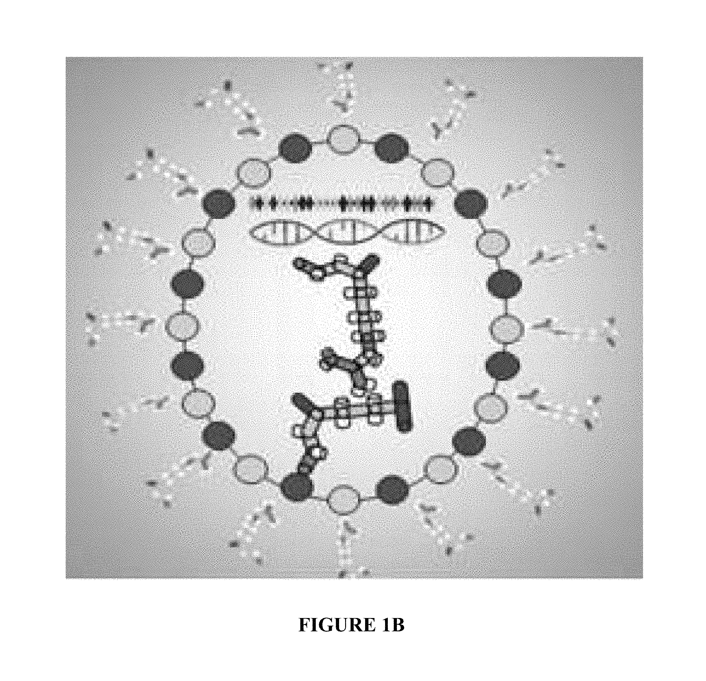
FIGS. 1A-1B are diagrammatic representations of some embodiments of a nanoparticle and components thereof in accordance with an aspect of the present invention;
FIG. 2A is a diagrammatic representation of how a nanoparticle may be manufactured in accordance with an aspect of the present invention;
FIG. 2B is a diagrammatic representation of means by which a cell may uptake and intracellularly process a nanoparticle in accordance with an aspect of the present invention;
FIG. 3 is a graph illustrating the effects on polyplex complexation of including different ratios of various charged polymers and polynucleotides in accordance with an aspect of the present invention;
FIG. 4 is a graph illustrating the effects on polyplex complexation of including different ratios of various charged polymers and polynucleotides, with or without including an anionic polymer in the polyplex, in accordance with an aspect of the present invention;
FIG. 5 is a graph illustrating the destabilizing effect on a polyplex of including increasing amounts of an anionic polymer in the presence or absence of cationic polypeptides in accordance with an aspect of the present invention;
FIG. 6 is a graph illustrating sizes of nanoparticles possessing various layers in accordance with an aspect of the present invention;
FIG. 7 is photomicrographs of cells transfected with various nanoparticles demonstrating cellular uptake and subcellular localization of nanoparticles following transfection in accordance with an aspect of the present invention;
FIG. 8 is photomicrographs of cells transfected with nanoparticles showing duration of residence of nanoparticles in cells following transfection in accordance with an aspect of the present invention;
FIGS. 9A-B is photomicrographs showing cellular uptake of nanoparticles possessing a layer of polymers attached to the outside of a silica coating of a polyplex in accordance with an aspect of the present invention;
FIG. 10 is a diagrammatic representation of TALEN peptides encoded for by a nucleic acid included in a nanoparticle that cause knockdown of expression of sclerostin in accordance with an aspect of the present invention;
FIGS. 11A-11C are graphs illustrating the effects transfecting cells with different amounts of nanoparticles that target sclerostin expression on sclerostin and .beta.-catenin expression in accordance with an aspect of the present invention;
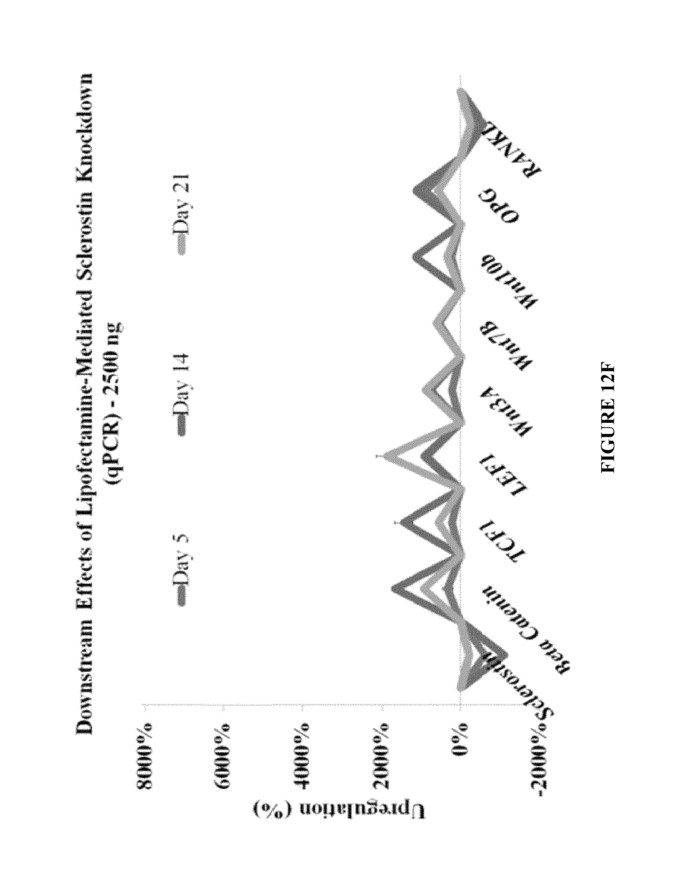
FIGS. 12A-12F are graphs illustrating the effects of transfecting cells with different amounts of nanoparticles that target sclerostin expression on expression levels of various cellular signaling peptides in accordance with an aspect of the present invention;
FIG. 13 is photomicrographs demonstrating effects of transfecting cells with nanoparticles that target sclerostin expression on expression of a co-transfected reporter gene that is responsive to transcription factors whose activity is inhibited by sclerostin-mediated signaling in accordance with an aspect of the present invention;
FIGS. 14A-14C are photomicrographs demonstrating effects of transfecting cells with nanoparticles that target sclerostin expression on mineralization in accordance with an aspect of the present invention.
DETAILED DESCRIPTION OF THE INVENTION
Aspects of the present invention and certain features, advantages, and details thereof, are explained more fully below with reference to the non-limiting embodiments illustrated in the accompanying drawings. Descriptions of well-known materials, fabrication tools, processing techniques, etc., are omitted so as to not unnecessarily obscure the invention in detail. It should be understood, however, that the detailed description and the specific examples, while indicating embodiments of the invention, are given by way of illustration only, and are not by way of limitation. Various substitutions, modifications, additions and/or arrangements within the spirit and/or scope of the underlying inventive concepts will be apparent to those skilled in the art from this disclosure.
The present disclosure provides, in part, a multilayered nanoparticle for transfecting cells with agents to modify gene expression. Nanoparticles designed for improved serum stability, targetted delivery to specific cell types, greater nuclear specificity and compartment-specific unpackaging, improved ability to retain significant payload levels during initial stages of internalization, and ability to maintain release of payload for a various durations following internalization, and methods of use thereof, are provided.
In one aspect, complexes of polynucleotides with polymers, or polyplexes, created by condensation of cationic polymers and polynucleotides in the presence of anionic polymers may mediate increased transfection efficiency over polynucleotide-cationic polymer conjugates. Though this process may produce more particles and increase the net surface area of nanoparticles exposed for cellular uptake, an improved electrostatic repulsory element may also be at play in releasing nucleic acids through this technique. Surprisingly, in contrast to a more rapid disaggregation of nucleotides from nanoparticle polyplexes that include anionic polymers as would have been predicted on the basis of existing literature, in one aspect of the present invention, including an anionic polymer in a nanoparticle polyplex core may prolong the duration of intracellular residence of the nanoparticle and release of agents that affect gene expression or otherwise regulate cellular function, or payloads.
In another aspect, the presence of a cationic polypeptide in a nanoparticle may mediate stability, subcellular compartmentalization, and payload release. As one example, fragments of the N-terminus of histone peptides, referred to generally as hi stone tail peptides, within various polyplexes are not only capable of being deprotonated by various histone modifications, such as in the case of histone acetyltransferase-mediated acetylation, but may also mediate effective nuclear-specific unpackaging as components of polyplexes. Their trafficking may be reliant on alternative endocytotic pathways utilizing retrograde transport through the Golgi and endoplasmic reticulum, and the nature of histones existing inside of the nuclear envelope suggests an innate nuclear localization sequence on histone tail peptides. In one aspect of the present invention, including a histone tail peptide may promote nuclear localization of nanoparticles and result in enzyme-mediated release of polynucleotide payload therefrom.
In another aspect, silica coatings of polyplexes may seal their payloads before and during initial cellular uptake. Commonly used polyplexes consisting of poly(ethylenimine) and DNA have a tendency to shed the majority (.about.90%) of their payloads during cellular internalization, with the remaining payload often remaining bound to its cationic nanocarrier's polymeric remains. With transiently stabilizing interlayers of silica, greater intracellular delivery efficiency may be observed despite decreased probability of cellular uptake. In another aspect of the present invention, coating a nanoparticle polyplex with a silica coating may seal the polyplex, stablizing it until its release upon processing in the intended subcellular compartment.
In another aspect of the present invention, transfection efficiency may be further increased by adding another layer of cationic polymer, making the delivery efficiency as much as two orders of magnitude greater than a bare or silica-coated polyplex, presumably due to the anionic nature of an oligomeric silica coating being cell repulsive. In a further aspect, silica-coated polyplexes and their further-layered derivatives are stable in serum and are suitable for in vivo experiments unlike cationic polymer/nucleic acid conjugates on their own.
FIGS. 1A-1B show examples of components of a nanoparticle in accordance with the present invention. In accordance with the present invention, a nanoparticle polyplex core may include a polynucleotide, an anionic polymer, a cationic polymer, and a cationic polypeptide. A silica coating may then be applied to the polyplex core, and polymers may then be attached to an outer surface of the silica coating. The polynucleotide may be a DNA vector for driving intracellular expression of a nucleic acid sequence it contains. However, a nanoparticle may also comprise other types of polynucleotides, such as linear DNA or various types of RNA, including dsDNA, ssDNA, mRNA, siRNA, or CRISPR RNA sequences, or others, or any combination of the foregoing. A nanoparticle may also include, in addition to or in place of any of the foregoing examples of polynucleotides, a peptide nucleic acid, other charged or polar small molecules between 50 and 1000 Da, or alternatively between 200 and 10 kDa, in size, such as cyclic nucleotides such as cAMP, DNA origami templates, aptamers, charged polypeptides, proteins or protein fragments between 2 and 100 kDa, peptoids, phosphorylated or sulfated constituents, anionically modified constituents, and multimeric or oligomeric combinations of the foregoing. A person of ordinary skill would understand any of the foregoing, or any combination thereof, as being included within the present invention.
Continuing with FIG. 1A, in one aspect of the invention, a cationic polymer within the polyplex may be a polypeptide containing cationic amino acids and may be, for example, poly(arginine), poly(lysine), poly(histidine), poly(ornithine), poly(citrulline), or a polypeptide that comprises any combination of more than one of the foregoing. A nanoparticle may also include, in addition to or in place of any of the foregoing examples of cationic polymers, poly(ethylenimine), poly(aspartamide), polypeptoids, a charge-functionalized polyester, a cationic polysaccharide, an acetylated amino sugar, chitosan, or a variant or variants that comprise any combination of more than one of the foregoing, in linear or branched forms.
In one example, a cationic polymer may comprise a poly(arginine), such as poly(L-arginine). A cationic polymer within the polyplex may have a molecular weight of between 1 kDa and 200 kDa. A cationic polymer within the polyplex may also have a molecular weight of between 10 kDa and 100 kDa. A cationic polymer within the polyplex may also have a molecular weight of between 15 kDa and 50 kDa. In one example, a cationic polymer comprises poly(L-arginine) with a molecular weight of approximately 29 kDa, as represented by SEQ ID NO: 1 (PLR). In another example, a cationic polymer may comprise linear poly(ethylenimine) with a molecular weight of 25 kDa (PEI). In another example, a cationic polymer may comprise branched poly(ethylenimine) with molecular weight of 10 kDa. In another example, a cationic polymer may comprise branched poly(ethylenimine) with a molecular weight of 70 kDa. In another example, a cationic polymer may comprise a D-isomer of poly(arginine) or of any of the foregoing polymers such as polypeptides, which may be particularly advantageous because polymers such as polypeptides containing a D-isomer may be less susceptible to degradation within a cell and therefore have a prolonged effect on influencing payload release and the rate thereof over time.
Continuing with FIG. 1A, in a further aspect of the invention, an anionic polymer within the polyplex may be a polypeptide containing anionic amino acids, and may be, for example, poly-glutamic acid or poly-aspartic acid, or a polypeptide that comprises any combination of the foregoing. A nanoparticle may also include, in addition to or in place of any of the foregoing examples of anionic polymers, a glycosaminoglycan, a glycoprotein, a polysaccharide, poly(mannuronic acid), poly(guluronic acid), heparin, heparin sulfate, chondroitin, chondroitin sulfate, keratan, keratan sulfate, aggrecan, poly(glucosamine), or an anionic polymer that comprises any combination of the foregoing. In one example, an anionic polymer may comprise poly-glutamic acid. An anionic polymer within the polyplex may have a molecular weight of between 1 kDa and 200 kDa. An anionic polymer within the polyplex may also have a molecular weight of between 10 kDa and 100 kDa. An anionic polymer within the polyplex may also have a molecular weight of between 15 kDa and 50 kDa. In one example, an anionic polymer is poly(glutamic acid) with a molecular weight of approximately 15 kDa. Polymers consisting of or including a D-isomer of glutamic acid may be particularly advantageous because they may be less susceptible to degradation within a cell and therefore have a prolonged effect on influencing payload release and the rate thereof over time. For example, the anionic polymer within the polyplex may have the sequence represented by SEQ ID NO: 2 (PDGA). In another example, an anionic polymer may comprise a D-isomer of any of the foregoing polymers or polypeptides, which may be particularly advantageous because polymers such as polypeptides containing a D-isomer may be less susceptible to degradation within a cell and therefore have a prolonged effect on influencing payload release and the rate thereof over time.
Continuing with FIG. 1A, in another aspect of the invention, a cationic peptide in a nanoparticle's polyplex core may be a fragment of a histone peptide, such as of the H1, H2, H3, or H4 proteins. The fragment may include amino acids whose sequence corresponds to the N-terminus of a histone protein. For example, the fragment may comprise up to the first 5 (SEQ ID NO: 9), 10 (SEQ ID NO: 10), 15 (SEQ ID NO: 11), 20 (SEQ ID NO 12), 25 (SEQ ID NO: 13) or more N-terminal amino acids of a histone protein. The fragment may also be amidated on its C-terminus. The fragment may also have been modified such that one or more lysine residue is methylated, one or more histidine, lysine, arginine, or other complementary residues are acetylated or susceptible to acetylation as a histone acetyltransferase or acetyl CoA substrate, or any combination of the foregoing. For example, a cationic peptide in a nanoparticle polyplex core may have the sequence as represented by SEQ ID NO: 3, which comprises the first 25 amino acids of the human histone 3 protein, amidated on its C-terminus, and tri-methylated on the lysine 4 in accordance with the present invention (HTP).
In another embodiment, a nanoparticle may include or contain, in addition to or in place of any of the foregoing cationic polypeptides, a nuclear localization sequence. A cationic polypeptide may comprise a nuclear localization sequence on its N- or C-terminus. A nuclear localization sequence may comprise an importin or karyopherin substrate, or may have or contain a sequence corresponding to SEQ ID NO: 8. In another embodiment, a nanoparticle may include, in addition to or in place of any of the foregoing cationic polypeptides, a mitochondrial localization signal or a peptide fragment of mtHSP70.
Continuing with FIG. 1B, in another aspect of the invention, the nanoparticle may comprise a reversible coating that provides stability to the polyplex core prior to cellular or compartmental internalization, preventing premature degradation or destabilization. For example, a silica coating may be applied to the polyplex core. In another example, calcium phosphate or hydroxyapatite may be applied to a polyplex core. In another example, a branched cationic polymer, polypeptide, or peptoid may be applied to a polyplex core, with an anionic charge excess. A coating, such as a silica coating, may protect the polyplex from degradation before exposure to the endosomal microenvironment.
In another aspect, a nanoparticle may comprise a layer of polymers attached to or electrostatically bound with the external surface of coated polyplex, such as to or with the external surface of a silica coating. Such external polymers may serve to prevent cellular repulsion of the coated polyplex so as to promote contact with and uptake by a cell. An external polymer layer may also serve to promote internalization by specific cell types, such as if the externally attached polymer is or mimics a ligand to a receptor expressed by a cell type of which transfection is desired. A polymer in a polymer layer attached to the outer surface of coating on a polyplex may be from between 0.1 to 20 kDa in size, or may be up to 40 or 50 kDa in size.
Examples of polymer comprising a polymer layer attached to the external surface of the coated core polyplex include those represented by SEQ ID NO: 4, which is an approximately 10 kDa poly(arginine) polymer, and SEQ ID NO: 5, which is human vasoactive endothelial growth factor protein, in accordance with the present invention. In another example, a polymer comprising a layer attached to the external surface of the coated core polyplex may comprise an anchor substrate of from between 1 to 25 repeating anionic or cationic moieties at the N-terminus, C-terminus, 5', or 3' end of a polymer, polypeptide, or polynucleotide to provide electrostatic conjugation of a targeting motif contained in the polymer, polypeptide, or polynucleotide to the coated polyplex core. In another example, a polymer comprising a layer attached to the external surface of the coated core polyplex may comprise a polymer, polypeptide, or polynucleotide sequence that exhibits base pair complementarity or binding affinity for an amino acid sequence binding motif to bind additional layers that may be added thereupon.
In another aspect of the present invention, illustrated in FIG. 2A, a cationic polyplex is created, then coated with a silica coating. Polyplex cores of nanoparticles may be created via electrostatic interactions leading to condensation. Two equal-volume solutions may be created, one with pH-unadjusted 40 mM HEPES (pH .about.5.5) combined with 0.1% w/v a cationic polymer and a cationic polypeptide in water and the other with 30 mM Tris-HCl (pH .about.7.4) combined with 0.1% w/v anionic polymers and a polynucleotide in water. In one embodiment, the cationic polymer comprises SEQ ID NO: 1, the anionic polymer comprises SEQ ID NO: 2, and the cationic polypeptide comprises SEQ ID NO: 3. These solutions may be combined via dropwise addition of the cationic solution to the anionic one with no stirring. After 30 minutes of incubation at room temperature, a 200 uL solution containing 10 ug of nucleic acids within polyplexes may be added dropwise to a 45 mM sodium silicate (Sigma) solution in Tris-HCl (pH=7.4) and allowed to incubate for between 8 and 24 hours at room temperature. Silica-coated polyplexes may be isolated via centrifugation with a 300 kDa Nanosep.RTM. filter (Pall, Port Washington, NY) at 3000g in order to isolate complexes from unbound silica species and polymers. Nanoparticles may further be resuspended in a solution containing a polymer to be attached to the external surface of the silica coating. For example, they may be resuspended in a solution comprising a polymer represented by SEQ ID NO: 4 or SEQ ID NO: 5 at 0.1% w/v for one hour. Nanoparticles may then be centrifuged again before resuspension in transfection medium. This method is but one example of manufacturing nanoparticles in accordance with the present invention.
FIG. 2B is a diagrammatic representation of contacting a cell with a nanoparticle in accordance with the present invention leading to cellular internalization of the nanoparticle, such as by caveolae-mediated endocytosis or macropinocytosis. Nanoparticles may further be retrogradely transported through the Golgi and endoplasmic reticulum or processed through lysosomal pathways, resulting in loss of the coating, such as a silica coating, and exposure of the polyplex core. The polyplex core may further be translocated into the cell nucleus, where enzymatic processing my degrade the cationic polymer, such as through activity of arginases, or otherwise promote unpackaging of the polyplex core, such as through acetylation of a histone tail peptide within the polyplex, leading to release of polynucleotides such as plasmid DNA from the polyplex core, in accordance with the present invention. Other intracellular processing steps modifying the constituents of a nanoparticle and its polyplex core or coating thereof or polymer layer attached to the coating may also occur in accordance with the present invention.
In a further aspect, the present invention includes optimized ratios of anionic and cationic polymers, cationic polypeptides, and polynucleotides for complexation of a polyplex core as part of a nanoparticle. In one example, plasmid DNA was fluorescently tagged with ethidium bromide (40 ng EtBr/ug DNA) before addition of various polymeric constituents in molar [1(positive)]:[1(negative)] ratios of [amine (n)]:[phosphate (p)+carboxylate (c)], or of c:p in the instance of poly(D-glutamic acid) (PDGA; SEQ ID NO: 2) addition. Addition of linear poly(ethylenimine) (PEI, 25 kDa) was compared to addition of poly(L-arginine) (PLR, 29 kDa; SEQ ID NO: 1) independently, as well as in conjunction with a H3K4(Me3) histone tail peptide (HTP; SEQ ID NO: 3), in order to quantify similar complexation behaviors between the two polymers as part of a binary complex (i.e., PEI+DNA or PEI+DNA) or ternary complexes (HTP+PEI+DNA or HTP+PLR+DNA). Where a cationic polymer and cationic polypeptide were both present, the relative molar ratio of each component was 60%:40%, respectively. A Zeiss filter and spectrophotometer were used to excite EtBr-tagged DNA at 510 nm for an emission at 595 nm, and results were compared amongst various formulations with unbound EtBr as a negative control.
FIG. 3 is a graph showing the effects of varying the ratio of anionic or cationic polymers or polypeptides to polynucleotides. The X axis shows charged polymer-to-phosphate ratio and the Y axis shows relative fluorescence following combination of indicated constituents. A decrease in relative fluorescence indicates displacement of EtBr from DNA and polyplex formation. Ratios of cationic polymer, or of cationic polymer and cationic polypeptide, to DNA of approximately 5:1 and higher exhibited an approximately 40% decrease in fluorescence indicating complexation of DNA and polymers into polyplexes. Addition of PDGA in the absence of cationic polymers or cationic polypeptides did not affect complexation.
After complexing PLR-HTP-DNA, PEI-HTP-DNA, PLR-DNA and PEI-DNA polyplexes and determining that PDGA possesses no ability to cause complexation of polynucleotides, PDGA's influence on formation kinetics was established by comparison of [5.5(positive)]:[1(negative)] and [10(positive)]:[1(negative)] molar ratios of [amine (n)]:[phosphate (p)] and [amine (n)]:[phosphate (p)+carboxylate (c)] on complexation efficiencies in order to determine effects of excess cationic and equalized charge ratios on nanoparticle complexation. Inclusion of carboxylate groups from PDGA was expected to have effects on overall formation kinetics comparable to inclusion of phosphate groups from DNA. Relative fluorescence was compared to DNA without addition of polymers or polypeptides or EtBr in the absence of DNA as controls.
FIG. 4 indicates the effects of adding PDGA to cationic polymers and cationic polypeptides on polyplex complexation kinetics. DNA was complexed with HTP, PLR or PEI, with or without addition of PDGA. Shown are experiments using cationic polymer (PLR or PEI)-to-polynucleotide molar ratios of 5.5:1 (as shown in the bars labeled n/p=5.5) and cationic polymer (PLR or PEI)-to-polynucleotide plus anionic polymer molar ratios of 5.5:1 and 10:1 (as shown in the bars labeled n/(p+2c)=5.5 or 10), with or without addition of HTP. Addition of PDGA did not impair complexation kinetics at any of the molar ratios tested.
Effects of including a cationic polymer and cationic polypeptide on polyplex destabilization were also determined, as shown in FIG. 5. Polyplex nanoparticles of DNA and cationic polypeptides (PLR with or without HTP, or PEI with HTP) with [(PDGA) carboxylate(c):(DNA) phosphate(p)] molar ratios varying from 0 to 100 were complexed as described, compared to DNA or EtBr alone as controls, and the effects of destabilization (as indicated by increased fluorescence) was determined. In the absence of HTP, addition of PDGA did not lead to polyplex destabilization. However, in the presence of HTP, adding molar ratios of PDGA to DNA of 20 and above led to polyplex destabilization. These results indicate a surprising synergistic effect of cationic polypeptide and anionic polymer on complex destabilization. Cationic polypeptide incorporation, and/or inclusion of cationic constituents of disparate molecular weights or sizes, into a nanoparticle polyplex core may beneficially enhance the ability of a cationic polymer to promote dissociation and release of the polynucleotide payload from the polyplex and its other constituents.
Dynamic light scattering (BRAND) was used to determine the hydrodynamic radii of nanoparticles at various stages of formation. Nanoparticles containing core polyplexes with plasmid DNA, PLR, PDGA, and HTP, at a molar ratio of [amide]:[(phosphate)] of 5.5:1 were complexed as described. Some polyplex cores were further coated with silica as described. And some silica-coated polyplexes were further layered with cationic polymer (SEQ ID NO: 4) as described. 30-60 minutes of measurements were obtained following initial core formation of ternary complexes, silica coating of cores, and cationic polymer-coating of silica-coated cores. FIG. 6 is a graph showing diameters of nanoparticles. Uncoated polyplex cores and polyplex cores coated with silica were approximately 70-150 nm in diameter on average. In other embodiments, polyplex cores and silica-coated polyplex cores may be within a range of 100-170 nm in average diameter. Adding a cationic polymer coating to the silica coating yielded a nanoparticle with an average diameter of approximately 170 nm. In other embodiments, silica-coated polyplex cores with an additional layer of cationic polymer attached to the outer layer of silica may be within a range of approximately 80-200 nm in average diameter.
Cellular uptake of nanoparticles was also determined. Fluorescein isothiocyanate (FITC) was covalently conjugated to amines of PEI (25 kDa linear) and PLR (29 kDa) such that the molar ratio of amines to FITC was 100:1. The reaction was performed in darkness at room temperature for four hours in equal volumes of water and DMSO. In order to establish conjugation, a 0.05% w/v 500 uL solution of each fluorescently modified polymer was centrifuged in a 10 kDa Nanosep.RTM. filter and the eluate's fluorescence intensity (485 ex./520 em.) was compared to the unfiltered polymer solution as well as water. mCherry plasmid (Addgene) was included in nanoparticles to permit fluorescent detection of plasmid-driven expression.
MC3T3 murine osteoblasts were cultured on polystyrene T-75 tissue culture plastic flasks (Corning, CA, USA). Dulbecco's modified eagle medium supplemented with 10% Fetal Bovine Serum (Thermo Fisher Scientific, VA, USA) was used for osteoblasts along with 1% penicillin/streptomycin (Invitrogen, NY, USA). Xylenol orange was added to the cell culture media from day 15 to day 25 after initiation of cell culture. At day 25 cells were fixed and assayed for mineralization. For mCherry plasmid delivery using FITC-modified nanoparticles, osteoblasts were plated at 1000 cells/well in 96-well plates and allowed to adhere for 12-16 hours in antibiotic-free DMEM containing 10% FBS. Immediately before transfection, medium was replaced with equal volumes of OptiMEM-suspended nanoparticles and DMEM containing 10% FBS.
All complexes were FITC-labeled and subjected to qualitative observation of fluorescence intensity (488/520 ex./em.) before transfection. 96-well-plated osteoblasts (1000 cells/well) were transfected with 200 ng of plasmids in triplicates for each binary (plasmid and cationic polymer), ternary (plasmid and cationic polymer, plus anionic polymer or cationic polypeptide), and quaternary (plasmid, cationic polymer, anionic polymer, and cationic polypeptide) complex as well as its silica-coated counterpart, with 1 control and 8 experimental sets (n=3) in total. 5% serum was used in order to study effects of serum on extracellular properties of aggregation.
At 30-hours post-transfection, bimodal fluorescent imaging allowed for simultaneous observation of FITC-labeled nanoparticles (488 ex./520 em.) and the mCherry gene expression that they were responsible for (633 ex./680 em.). A minimum of 20 cells were observed at different locations in each well and representative images were obtained. ImageJ was used to process the overlaid images and combine phase-contrast, 488/520 and 633/680 channels.
Photomicrographs demonstrating cellular uptake are shown in FIG. 7. Circles in FIG. 7 indicate where high levels of nuclear localization is apparent. Silica-coated binary nanoparticles show burst release properties (i.e., nuclear localization is not apparent in the DNA-PLR+silica samples). Inclusion of PDGA in polyplex cores causes prolonged release of plasmid within cell nuclei. This effect of PDGA to cause prolonged release was surprising in light of literature suggesting the opposite: that including cationic polymers in nanoparticle polyplexes would hasten, and shorten the duration of, dissociation of polynucleotide payload from other polyplex constituents. Addition of HTP also causes extensive nuclear localization.
Further coating of silica-coated nanoparticles (DNA-HTP-PDGA-PLR+Si) with poly(arginine) (SEQ ID NO: 4) causes nanoparticles to be stable in serum and causes extended residence of nanoparticle payload within cells. FIG. 8. is photomicrographs showing cellular uptake and retention of silica-coated FITC-conjugated polyplex cores, to which an additional layer of poly(L-arginine) (SEQ ID NO: 4) has been added, by MC3T3 murine osteoblasts, in accordance with the present invention. Unlike for silica-coated nanoparticles shown in FIG. 7, no aggregation of nanoparticles containing an additional layer of cationic polymers on the outside of the silica coating is observable in FIG. 8, indicating that such nanoparticles remain stable in serum. Furthermore, these nanoparticles are observed to display extended residence within the cell nucleus such that fluorescence qualitatively peaks within approximately 1.5 days and detectable fluorescence was sustained through 14 days.
Layering silica-coated polyplex cores with polymers specifically directed to bind to particular cell types can further enhance uptake. Associating ligands for cellular receptors with the surface of a nanoparticle can enhance affinity of the nanoparticle for cells that express such receptors and increase transfection of such cells. As one example in accordance with the present invention, silica-coated polyplexes were coated with VEGF (SEQ ID NO: 5), a high-affinity ligand for VEGF receptors, which are expressed at high levels by human umbilical vein endothelial cells (HUVECs). HUVECs were incubated with silica-coated FITC-conjugated polyplexes with poly(L-arginine) (SEQ ID NO: 4) or human VEGF (SEQ ID NO: 5) attached to the outer surface of the silica coating for 40 min before being washed twice with PBS then resuspended in DMEM (10% FBS). Cells were imaged 4 hrs later. After this short incubation period, only low levels of transfection with nanoparticles containing a poly(L-arginine) layer attached to the external silica surface (FIG. 9A) was observed, whereas coating with VEGF instead of poly(L-arginine) resulted in significantly greater cellular internalization at this four-hour time point. A skilled artisan would recognize that virtually any other cell type may also be transfected by nanoparticles in accordance with the present invention, and that a layer of polymers may be attached to the outer layer of silica-coated polyplex cores to promote or otherwise influence this effect. Such a person would also comprehend that other means of contacting cells with nanoparticles to effect such outcomes, such as i.p., i.v., i.m. or s.c. or other injection or transdermal administration or via suppository to, or ingestion or oral or nasal inhalation by, a human or animal, or contact with explanted tissue or cells or stem cells, would also be included within the present invention.
In another aspect of the invention, a polynucleotide encoding a nuclease may be incorporated into the nanoparticle polyplex core. As one nonlimiting example, a polynucleotide that encodes and drives expression of a TALEN (Transcription Factor-Like Effector Nucleases) may be included in the nanoparticle. Like Zinc Finger Nucleases, TALENs utilize a modular DNA binding motif (TALE) that can be modified to introduce new DNA binding specificities and even nucleases (TALEN). TALEs consist of multiple repeat variable diresidues (RVDs) which each specify binding to a single nucleotide. TALE arrays are made by stringing together RVDs in a specific order to provide specificity and binding affinity to desired DNA sequences. Commonly, these genome-splicing tools are engineered by fusing non-specific cleavage domains, such as FokI nucleases, to TALEs. TALEN assembly protocols are available that allow assembly of these repetitive sequences, including an open source assembly method known as Golden Gate.
In another aspect of the present invention, nanoparticles may be designed and used in a manner to regulate expression of signaling molecules to alter cellular function. For example, sequences of chromosomal DNA may be deleted or altered to generate cellular or animal models of disease states or treatments therefor, or to treat disease states or otherwise enhance human health. One nonlimiting example of a protein whose expression may be modified in accordance with the present invention is sclerostin (SOST). SOST binding to the LRP5/6 receptor inhibits Wnt signaling, perhaps via feedback systems between Wnt3A, Wnt7B, Wntl OA, sclerostin, .beta.-catenin, LEF1, and TCF1. Desuppressing these cascades via removal of sclerostin may result in significantly increased mineralization activity.
Osteoprogenitor (OPG) and RANKL are also expected to play a responsive role to SOST deletion, where RANKL expresses itself as a receptor for promoting osteoclastogenesis via osteoclast-linked RANK or ODF (osteoclast differentiation factor) binding, and OPG binds antagonistically to RANKL. Thus, the ratio between OPG and RANKL is a determinant of the relationship between bone formation and resorption. However, single cultures of osteoblasts will communicate through other forms of paracrine signaling and this ratio should be more reflective of behavior of altered cells in co-culture with osteoclasts or in vivo.
In another aspect of the present invention, a nanoparticle may be designed so as to allow transfection with a TALEN that may disrupt expression of SOST and consequently generate a high bone-mass phenotype. As one example, TALENS may be engineered to specifically bind to loci in the SOST gene and create double-stranded breaks in the genome to disrupt transcription or translation and reduce SOST expression. As a further example, a nanoparticle may contain plasmids that encode two TALENs that create double-stranded breaks on either side of the chromosomal locus of the start codon for SOST. Repair of endogenous genomic DNA following excision of the sequence encoding the start codon may result in transcription of sclerostin mRNA lacking the start codon that cannot be properly translated into SOST protein, thereby driving down SOST expression and activity. A diagrammatic representation of this model is shown in FIG. 10, where a "left" TALEN and "right" TALEN bind to and cleave sites on opposite sides of the SOST start codon locus. As one example, a left TALEN may have the sequence represented by SEQ ID NO: 6, and a right TALEN may have the sequence represented by SEQ ID NO: 7. A nanoparticle may comprise an expression plasmid, such as pUC19 (Genbank Accession Number L09137 X02514), into which a nucleotide sequence that encodes a right or left TALEN, such as those represented by SEQ ID NO: 6 and SEQ ID NO: 7, has been subcloned so as to drive cellular expression of the encoded TALEN. A nanoparticle may also include combinations of expression plasmids that comprise sequences that encode left and right TALENs.
A nanoparticle may also comprise other TALEN sequences, targeting SOST or any other gene of interest, and also may comprise other expression vectors, in accordance with the present invention. A nanoparticle may comprise other types of polynucleotides or analogs thereof, such as species of RNA or DNA including mRNA, siRNA, miRNA, aptamers, shRNA, AAV-derived nucleic acids, morpholeno RNA, peptoid and peptide nucleic acids, cDNA, DNA origami, DNA and RNA with synthetic nucleotides, DNA and RNA with predefined secondary structures, CRISPR sequences, and multimers and oligomers, and any combination of the foregoing, in accordance with the present invention. In another example, a nanoparticle may comprise polynucleotides whose sequence may encode other products such as any protein or polypeptide whose expression is desired. A skilled artisan would recognize that the foregoing examples are in accordance with the present invention and may be encompassed by claims thereto.
Following transfection of MC3T3 murine osteoblasts with nanoparticles designed to knock down SOST expression in accordance with the present invention, ELISA and quantitative real-time PCR (qPCR) assays were performed on cell lysate and supernatant fractions. FIGS. 11A-11C are graphs demonstrating the effectiveness of different amounts (800 ng, 1600 ng, or 2500 ng) of nanoparticles (NP) containing expression plasmids comprising nucleotide sequences that encode left (SEQ ID NO: 6) and right (SEQ ID NO: 7) SOST TALENs, in accordance with the present invention, in modulating SOST expression and .beta.-catenin expression over a period of up to over 20 days following transfection. For comparison, other cells were transfected with mRNA encoding the same TALENS using Lipofectamine, a known agent for cellular transfection. As shown in FIGS. 11A-11C, intracellular and extracellular SOST levels were suppressed for at least several weeks following transfection with nanoparticles in accordance with the present invention, whereas .beta.-catenin expression was concomitantly up-regulated, signifying effectiveness of the nanoparticles in downregulating SOST expression and activity.
qPCR was also performed to determine whether down-regulation of SOST expression with nanoparticles in accordance with the present invention may have downstream effects on other components of the relevant signaling cascade. Cells were transfected as described above. Results on expression of numerous components of the signaling pathway (SOST, .beta.-catenin, TCF1, LEF1, Wnt3A, Wnt7B, Wnt10b, OPG, and RANKL), at 5, 14, and 21 days after transfection with different amounts of nanoparticles as indicated, are shown in FIGS. 12A-12F. For comparison, other cells were transfected with mRNA encoding the same TALENS using Lipofectamine. The real time PCR results showed a greater up regulation of Wnt responsive genes in the cell lines transfected with nanoparticles delivering SOST TALENS as compared to the SOST TALENS delivered by Lipofectamine by up to 2 to 6 times as a response to knockdown of the Wnt signaling inhibitor sclerostin.
TCF/LEF-1-mediated transcription may also be upregulated following knockdown of SOST expression in accordance with the present invention. MC3T3-E1 cells were transfected with TOPflash and control FOPflash luciferase reporter plasmid constructs (Addgene#12456 and 12457) that contain TCF/LEF-1 binding sites. The cells were plated at the density of 5000 cells/well of the 8-well labtek chamber slides and transfected with 1 ug of TOPflash and FOPflash plasmid separately. To control for the efficiency of transfection a control plasmid Renilla (Promega) was used. FIG. 13 is photomicrographs showing upregulation of TCF/LEF-1-mediated transcription for 21 days following tranfection with nanoparticles containing plasmids encoding SOST-directed TALENS, in accordance with the present invention, consistent with an upregulation of TCF/LEF-1 expression and activity following transfection with the invented nanoparticles.
Knockdown of SOST expression in accordance with the present invention may also increase mineralization in stromal bone marrow cells and osteoblasts. Mineralization was quantified by two separate methods, first based on image thresholding of xylenol-orange-labeled vital cultures using MATLAB (Mathworks, Natick, MA), and second by atomic absorption spectroscopy (AAS). For the xylenol orange threshold, images of both phase and fluorescence (with Texas Red Filter Set) were taken in five adjacent regions of wells, and then stitched into a larger 8-bit image (4.times., Nikon Ti-100). The phase channel was subtracted from the fluorescence, and a threshold was set to half the level between the background and signal (-6 dB). The number of pixels above the threshold were counted and used to express the percentage of mineralized area in each well. The combination of phase and fluorescence allowed for unbound xylenol orange to be distinguished, whereas the use of decibel levels allowed for correction of the varied background levels in each image.
Mineralization was also quantified by atomic absorption with an atomic absorption spectrometer (AA-Perkin Elmer, MA). Each well was prepared by adding 0.5 mL of 10% nitric acid, and the resultant calcium content was measured relative to a standard curve and compared between groups. Care was taken to minimize interference due to ionized calcium precipitating with phosphate phases, so a large excess of potassium and lanthanum ions was added to each well.
FIGS. 14A-14C show the effects of transfection with nanoparticles in accordance with the present invention on mineralization following SOST knockdown. FIG. 14A is photomicrographs of staining of the mineralized matrix formed 25 days after SOST knockdown. Stromal cells are shown in panels A-C, wherein panel A show control cells, panel B shows cells transfected via Lipofectamine, and panel C shows cells transfected with nanoparticles containing plasmids encoding SOST-directed TALENs as described and in accordance with the present invention. MC3T3-E1 osteoblast cells are shown in panels D-G, wherein panel D show control cells, and panels E-G show cells transfected with nanoparticles containing plasmids encoding SOST-directed TALENs as described at doses of 800 ng, 1600 ng, and 2500 ng, respectively, in accordance with the present invention. FIGS. 14B and 14C are graphs showing quantification of mineralization. FIGS. 14A-C demonstrate increased calcium concentration in stromal bone marrow cells and osteoblasts following transfection with SOST-targetting TALENS via nanoparticles in accordance with the present invention, further confirming the effectiveness of this technique of modifying the cellular expression and activity of genes and downstream signaling pathways.
While several aspects of the present invention have been described and depicted herein, alternative aspects may be effected by those skilled in the art to accomplish the same objectives. Accordingly, it is intended by the appended claims to cover all such alternative aspects as fall within the true spirit and scope of the invention.
SEQUENCE LISTINGS
1
131186PRTArtificial SequenceLaboratory-synthesized sequence. 1Arg Arg Arg Arg Arg Arg Arg Arg Arg Arg Arg Arg Arg Arg Arg Arg1 5 10 15Arg Arg Arg Arg Arg Arg Arg Arg Arg Arg Arg Arg Arg Arg Arg Arg 20 25 30Arg Arg Arg Arg Arg Arg Arg Arg Arg Arg Arg Arg Arg Arg Arg Arg 35 40 45Arg Arg Arg Arg Arg Arg Arg Arg Arg Arg Arg Arg Arg Arg Arg Arg 50 55 60Arg Arg Arg Arg Arg Arg Arg Arg Arg Arg Arg Arg Arg Arg Arg Arg65 70 75 80Arg Arg Arg Arg Arg Arg Arg Arg Arg Arg Arg Arg Arg Arg Arg Arg 85 90 95Arg Arg Arg Arg Arg Arg Arg Arg Arg Arg Arg Arg Arg Arg Arg Arg 100 105 110Arg Arg Arg Arg Arg Arg Arg Arg Arg Arg Arg Arg Arg Arg Arg Arg 115 120 125Arg Arg Arg Arg Arg Arg Arg Arg Arg Arg Arg Arg Arg Arg Arg Arg 130 135 140Arg Arg Arg Arg Arg Arg Arg Arg Arg Arg Arg Arg Arg Arg Arg Arg145 150 155 160Arg Arg Arg Arg Arg Arg Arg Arg Arg Arg Arg Arg Arg Arg Arg Arg 165 170 175Arg Arg Arg Arg Arg Arg Arg Arg Arg Arg 180 1852116PRTArtificial SequenceLaboratory-created sequence.MISC_FEATURE(1)..(116)D isomer. 2Glu Glu Glu Glu Glu Glu Glu Glu Glu Glu Glu Glu Glu Glu Glu Glu1 5 10 15Glu Glu Glu Glu Glu Glu Glu Glu Glu Glu Glu Glu Glu Glu Glu Glu 20 25 30Glu Glu Glu Glu Glu Glu Glu Glu Glu Glu Glu Glu Glu Glu Glu Glu 35 40 45Glu Glu Glu Glu Glu Glu Glu Glu Glu Glu Glu Glu Glu Glu Glu Glu 50 55 60Glu Glu Glu Glu Glu Glu Glu Glu Glu Glu Glu Glu Glu Glu Glu Glu65 70 75 80Glu Glu Glu Glu Glu Glu Glu Glu Glu Glu Glu Glu Glu Glu Glu Glu 85 90 95Glu Glu Glu Glu Glu Glu Glu Glu Glu Glu Glu Glu Glu Glu Glu Glu 100 105 110Glu Glu Glu Glu 115325PRTHomo sapiensMOD_RES(4)..(4)TRIMETHYLATION.MISC_FEATURE(25)..(25)AMIDATED. 3Ala Arg Thr Lys Gln Thr Ala Arg Lys Ser Thr Gly Gly Lys Ala Pro1 5 10 15Arg Lys Gln Leu Ala Thr Lys Ala Ala 20 25464PRTArtificial SequenceLaboratory-created sequence. 4Arg Arg Arg Arg Arg Arg Arg Arg Arg Arg Arg Arg Arg Arg Arg Arg1 5 10 15Arg Arg Arg Arg Arg Arg Arg Arg Arg Arg Arg Arg Arg Arg Arg Arg 20 25 30Arg Arg Arg Arg Arg Arg Arg Arg Arg Arg Arg Arg Arg Arg Arg Arg 35 40 45Arg Arg Arg Arg Arg Arg Arg Arg Arg Arg Arg Arg Arg Arg Arg Arg 50 55 605165PRTHomo sapiens 5Ala Pro Met Ala Glu Gly Gly Gly Gln Asn His His Glu Val Val Lys1 5 10 15Phe Met Asp Val Tyr Gln Arg Ser Tyr Cys His Pro Ile Glu Thr Leu 20 25 30Val Asp Ile Phe Gln Glu Tyr Pro Asp Glu Ile Glu Tyr Ile Phe Lys 35 40 45Pro Ser Cys Val Pro Leu Met Arg Cys Gly Gly Cys Cys Asn Asp Glu 50 55 60Gly Leu Glu Cys Val Pro Thr Glu Glu Ser Asn Ile Thr Met Gln Ile65 70 75 80Met Arg Ile Lys Pro His Gln Gly Gln His Ile Gly Glu Met Ser Phe 85 90 95Leu Gln His Asn Lys Cys Glu Cys Arg Pro Lys Lys Asp Arg Ala Arg 100 105 110Gln Glu Asn Pro Cys Gly Pro Cys Ser Glu Arg Arg Lys His Leu Phe 115 120 125Val Gln Asp Pro Gln Thr Cys Lys Cys Ser Cys Lys Asn Thr Asp Ser 130 135 140Arg Cys Lys Ala Arg Gln Leu Glu Leu Asn Glu Arg Thr Cys Arg Cys145 150 155 160Asp Lys Pro Arg Arg 1656936PRTArtificial SequenceLaboratory-created sequence. 6Met Gly Asp Pro Lys Lys Lys Arg Lys Val Ile Asp Tyr Pro Tyr Asp1 5 10 15Val Pro Asp Tyr Ala Ile Asp Ile Ala Asp Leu Arg Thr Leu Gly Tyr 20 25 30Ser Gln Gln Gln Gln Glu Lys Ile Lys Pro Lys Val Arg Ser Thr Val 35 40 45Ala Gln His His Glu Ala Leu Val Gly His Gly Phe Thr His Ala His 50 55 60Ile Val Ala Leu Ser Gln His Pro Ala Ala Leu Gly Thr Val Ala Val65 70 75 80Lys Tyr Gln Asp Met Ile Ala Ala Leu Pro Glu Ala Thr His Glu Ala 85 90 95Ile Val Gly Val Gly Lys Gln Trp Ser Gly Ala Arg Ala Leu Glu Ala 100 105 110Leu Leu Thr Val Ala Gly Glu Leu Arg Gly Pro Pro Leu Gln Leu Asp 115 120 125Thr Gly Gln Leu Leu Lys Ile Ala Lys Arg Gly Gly Val Thr Ala Val 130 135 140Glu Ala Val His Ala Trp Arg Asn Ala Leu Thr Gly Ala Pro Leu Asn145 150 155 160Leu Thr Pro Gln Gln Val Val Ala Ile Ala Ser Asn Asn Gly Gly Lys 165 170 175Gln Ala Leu Glu Thr Val Gln Arg Leu Leu Pro Val Leu Cys Gln Ala 180 185 190His Gly Leu Thr Pro Glu Gln Val Val Ala Ile Ala Ser His Asp Gly 195 200 205Gly Lys Gln Ala Leu Glu Thr Val Gln Arg Leu Leu Pro Val Leu Cys 210 215 220Gln Ala His Gly Leu Thr Pro Glu Gln Val Val Ala Ile Ala Ser His225 230 235 240Asp Gly Gly Lys Gln Ala Leu Glu Thr Val Gln Arg Leu Leu Pro Val 245 250 255Leu Cys Gln Ala His Gly Leu Thr Pro Glu Gln Val Val Ala Ile Ala 260 265 270Ser His Asp Gly Gly Lys Gln Ala Leu Glu Thr Val Gln Arg Leu Leu 275 280 285Pro Val Leu Cys Gln Ala His Gly Leu Thr Pro Gln Gln Val Val Ala 290 295 300Ile Ala Ser Asn Gly Gly Gly Lys Gln Ala Leu Glu Thr Val Gln Arg305 310 315 320Leu Leu Pro Val Leu Cys Gln Ala His Gly Leu Thr Pro Glu Gln Val 325 330 335Val Ala Ile Ala Ser His Asp Gly Gly Lys Gln Ala Leu Glu Thr Val 340 345 350Gln Arg Leu Leu Pro Val Leu Cys Gln Ala His Gly Leu Thr Pro Glu 355 360 365Gln Val Val Ala Ile Ala Ser His Asp Gly Gly Lys Gln Ala Leu Glu 370 375 380Thr Val Gln Arg Leu Leu Pro Val Leu Cys Gln Ala His Gly Leu Thr385 390 395 400Pro Gln Gln Val Val Ala Ile Ala Ser Asn Gly Gly Gly Lys Gln Ala 405 410 415Leu Glu Thr Val Gln Arg Leu Leu Pro Val Leu Cys Gln Ala His Gly 420 425 430Leu Thr Pro Glu Gln Val Val Ala Ile Ala Ser His Asp Gly Gly Lys 435 440 445Gln Ala Leu Glu Thr Val Gln Arg Leu Leu Pro Val Leu Cys Gln Ala 450 455 460His Gly Leu Thr Pro Glu Gln Val Val Ala Ile Ala Ser His Asp Gly465 470 475 480Gly Lys Gln Ala Leu Glu Thr Val Gln Arg Leu Leu Pro Val Leu Cys 485 490 495Gln Ala His Gly Leu Thr Pro Glu Gln Val Val Ala Ile Ala Ser Asn 500 505 510Ile Gly Gly Lys Gln Ala Leu Glu Thr Val Gln Ala Leu Leu Pro Val 515 520 525Leu Cys Gln Ala His Gly Leu Thr Pro Glu Gln Val Val Ala Ile Ala 530 535 540Ser His Asp Gly Gly Lys Gln Ala Leu Glu Thr Val Gln Arg Leu Leu545 550 555 560Pro Val Leu Cys Gln Ala His Gly Leu Thr Pro Glu Gln Val Val Ala 565 570 575Ile Ala Ser His Asp Gly Gly Lys Gln Ala Leu Glu Thr Val Gln Arg 580 585 590Leu Leu Pro Val Leu Cys Gln Ala His Gly Leu Thr Pro Gln Gln Val 595 600 605Val Ala Ile Ala Ser Asn Gly Gly Gly Lys Gln Ala Leu Glu Thr Val 610 615 620Gln Arg Leu Leu Pro Val Leu Cys Gln Ala His Gly Leu Thr Pro Gln625 630 635 640Gln Val Val Ala Ile Ala Ser Asn Asn Gly Gly Lys Gln Ala Leu Glu 645 650 655Thr Val Gln Arg Leu Leu Pro Val Leu Cys Gln Ala His Gly Leu Thr 660 665 670Pro Gln Gln Val Val Ala Ile Ala Ser Asn Gly Gly Gly Arg Pro Ala 675 680 685Leu Glu Ser Ile Val Ala Gln Leu Ser Arg Pro Asp Pro Ala Leu Ala 690 695 700Ala Leu Thr Asn Asp His Leu Val Ala Leu Ala Cys Leu Gly Gly Arg705 710 715 720Pro Ala Leu Asp Ala Val Lys Lys Gly Leu Gly Asp Pro Ile Ser Arg 725 730 735Ser Gln Leu Val Lys Ser Glu Leu Glu Glu Lys Lys Ser Glu Leu Arg 740 745 750His Lys Leu Lys Tyr Val Pro His Glu Tyr Ile Glu Leu Ile Glu Ile 755 760 765Ala Arg Asn Ser Thr Gln Asp Arg Ile Leu Glu Met Lys Val Met Glu 770 775 780Phe Phe Met Lys Val Tyr Gly Tyr Arg Gly Lys His Leu Gly Gly Ser785 790 795 800Arg Lys Pro Asp Gly Ala Ile Tyr Thr Val Gly Ser Pro Ile Asp Tyr 805 810 815Gly Val Ile Val Asp Thr Lys Ala Tyr Ser Gly Gly Tyr Asn Leu Pro 820 825 830Ile Gly Gln Ala Asp Glu Met Gln Arg Tyr Val Glu Glu Asn Gln Thr 835 840 845Arg Asn Lys His Ile Asn Pro Asn Glu Trp Trp Lys Val Tyr Pro Ser 850 855 860Ser Val Thr Glu Phe Lys Phe Leu Phe Val Ser Gly His Phe Lys Gly865 870 875 880Asn Tyr Lys Ala Gln Leu Thr Arg Leu Asn His Ile Thr Asn Cys Asn 885 890 895Gly Ala Val Leu Ser Val Glu Glu Leu Leu Ile Gly Gly Glu Met Ile 900 905 910Lys Ala Gly Thr Leu Thr Leu Glu Glu Val Arg Arg Lys Phe Asn Asn 915 920 925Gly Glu Ile Asn Phe Ala Ala Asp 930 9357942PRTArtificial SequenceLaboratory-created sequence. 7Met Gly Asp Pro Lys Lys Lys Arg Lys Val Ile Asp Lys Glu Thr Ala1 5 10 15Ala Ala Lys Phe Glu Arg Gln His Met Asp Ser Ile Asp Ile Ala Asp 20 25 30Leu Arg Thr Leu Gly Tyr Ser Gln Gln Gln Gln Glu Lys Ile Lys Pro 35 40 45Lys Val Arg Ser Thr Val Ala Gln His His Glu Ala Leu Val Gly His 50 55 60Gly Phe Thr His Ala His Ile Val Ala Leu Ser Gln His Pro Ala Ala65 70 75 80Leu Gly Thr Val Ala Val Lys Tyr Gln Asp Met Ile Ala Ala Leu Pro 85 90 95Glu Ala Thr His Glu Ala Ile Val Gly Val Gly Lys Gln Trp Ser Gly 100 105 110Ala Arg Ala Leu Glu Ala Leu Leu Thr Val Ala Gly Glu Leu Arg Gly 115 120 125Pro Pro Leu Gln Leu Asp Thr Gly Gln Leu Leu Lys Ile Ala Lys Arg 130 135 140Gly Gly Val Thr Ala Val Glu Ala Val His Ala Trp Arg Asn Ala Leu145 150 155 160Thr Gly Ala Pro Leu Asn Leu Thr Pro Gln Gln Val Val Ala Ile Ala 165 170 175Ser Asn Asn Gly Gly Lys Gln Ala Leu Glu Thr Val Gln Arg Leu Leu 180 185 190Pro Val Leu Cys Gln Ala His Gly Leu Thr Pro Glu Gln Val Val Ala 195 200 205Ile Ala Ser Asn Ile Gly Gly Lys Gln Ala Leu Glu Thr Val Gln Ala 210 215 220Leu Leu Pro Val Leu Cys Gln Ala His Gly Leu Thr Pro Gln Gln Val225 230 235 240Val Ala Ile Ala Ser Asn Asn Gly Gly Lys Gln Ala Leu Glu Thr Val 245 250 255Gln Arg Leu Leu Pro Val Leu Cys Gln Ala His Gly Leu Thr Pro Gln 260 265 270Gln Val Val Ala Ile Ala Ser Asn Asn Gly Gly Lys Gln Ala Leu Glu 275 280 285Thr Val Gln Arg Leu Leu Pro Val Leu Cys Gln Ala His Gly Leu Thr 290 295 300Pro Glu Gln Val Val Ala Ile Ala Ser His Asp Gly Gly Lys Gln Ala305 310 315 320Leu Glu Thr Val Gln Arg Leu Leu Pro Val Leu Cys Gln Ala His Gly 325 330 335Leu Thr Pro Glu Gln Val Val Ala Ile Ala Ser Asn Ile Gly Gly Lys 340 345 350Gln Ala Leu Glu Thr Val Gln Ala Leu Leu Pro Val Leu Cys Gln Ala 355 360 365His Gly Leu Thr Pro Glu Gln Val Val Ala Ile Ala Ser His Asp Gly 370 375 380Gly Lys Gln Ala Leu Glu Thr Val Gln Arg Leu Leu Pro Val Leu Cys385 390 395 400Gln Ala His Gly Leu Thr Pro Gln Gln Val Val Ala Ile Ala Ser Asn 405 410 415Asn Gly Gly Lys Gln Ala Leu Glu Thr Val Gln Arg Leu Leu Pro Val 420 425 430Leu Cys Gln Ala His Gly Leu Thr Pro Gln Gln Val Val Ala Ile Ala 435 440 445Ser Asn Asn Gly Gly Lys Gln Ala Leu Glu Thr Val Gln Arg Leu Leu 450 455 460Pro Val Leu Cys Gln Ala His Gly Leu Thr Pro Gln Gln Val Val Ala465 470 475 480Ile Ala Ser Asn Asn Gly Gly Lys Gln Ala Leu Glu Thr Val Gln Arg 485 490 495Leu Leu Pro Val Leu Cys Gln Ala His Gly Leu Thr Pro Gln Gln Val 500 505 510Val Ala Ile Ala Ser Asn Asn Gly Gly Lys Gln Ala Leu Glu Thr Val 515 520 525Gln Arg Leu Leu Pro Val Leu Cys Gln Ala His Gly Leu Thr Pro Glu 530 535 540Gln Val Val Ala Ile Ala Ser His Asp Gly Gly Lys Gln Ala Leu Glu545 550 555 560Thr Val Gln Arg Leu Leu Pro Val Leu Cys Gln Ala His Gly Leu Thr 565 570 575Pro Gln Gln Val Val Ala Ile Ala Ser Asn Gly Gly Gly Lys Gln Ala 580 585 590Leu Glu Thr Val Gln Arg Leu Leu Pro Val Leu Cys Gln Ala His Gly 595 600 605Leu Thr Pro Glu Gln Val Val Ala Ile Ala Ser Asn Ile Gly Gly Lys 610 615 620Gln Ala Leu Glu Thr Val Gln Ala Leu Leu Pro Val Leu Cys Gln Ala625 630 635 640His Gly Leu Thr Pro Gln Gln Val Val Ala Ile Ala Ser Asn Asn Gly 645 650 655Gly Lys Gln Ala Leu Glu Thr Val Gln Arg Leu Leu Pro Val Leu Cys 660 665 670Gln Ala His Gly Leu Thr Pro Gln Gln Val Val Ala Ile Ala Ser Asn 675 680 685Gly Gly Gly Arg Pro Ala Leu Glu Ser Ile Val Ala Gln Leu Ser Arg 690 695 700Pro Asp Pro Ala Leu Ala Ala Leu Thr Asn Asp His Leu Val Ala Leu705 710 715 720Ala Cys Leu Gly Gly Arg Pro Ala Leu Asp Ala Val Lys Lys Gly Leu 725 730 735Gly Asp Pro Ile Ser Arg Ser Gln Leu Val Lys Ser Glu Leu Glu Glu 740 745 750Lys Lys Ser Glu Leu Arg His Lys Leu Lys Tyr Val Pro His Glu Tyr 755 760 765Ile Glu Leu Ile Glu Ile Ala Arg Asn Ser Thr Gln Asp Arg Ile Leu 770 775 780Glu Met Lys Val Met Glu Phe Phe Met Lys Val Tyr Gly Tyr Arg Gly785 790 795 800Lys His Leu Gly Gly Ser Arg Lys Pro Asp Gly Ala Ile Tyr Thr Val 805 810 815Gly Ser Pro Ile Asp Tyr Gly Val Ile Val Asp Thr Lys Ala Tyr Ser 820 825 830Gly Gly Tyr Asn Leu Pro Ile Gly Gln Ala Asp Glu Met Gln Arg Tyr 835 840 845Val Glu Glu Asn Gln Thr Arg Asn Lys His Ile Asn Pro Asn Glu Trp 850 855 860Trp Lys Val Tyr Pro Ser Ser Val Thr Glu Phe Lys Phe Leu Phe Val865 870 875 880Ser Gly His Phe Lys Gly Asn Tyr Lys Ala Gln Leu Thr Arg Leu Asn 885 890 895His Ile Thr Asn Cys Asn Gly Ala Val Leu Ser Val Glu Glu Leu Leu 900 905 910Ile Gly Gly Glu Met Ile Lys Ala Gly Thr Leu Thr Leu Glu Glu Val
915 920 925Arg Arg Lys Phe Asn Asn Gly Glu Ile Asn Phe Ala Ala Asp 930 935 940817PRTArtificial SequenceLaboratory-created sequence. 8Asp Arg Gln Ile Lys Ile Trp Phe Gln Asn Arg Arg Met Lys Trp Lys1 5 10 15Lys95PRTHomo sapiens 9Ala Arg Thr Lys Gln1 51010PRTHomo sapiens 10Ala Arg Thr Lys Gln Thr Ala Arg Lys Ser1 5 101115PRTHomo sapiens 11Ala Arg Thr Lys Gln Thr Ala Arg Lys Ser Thr Gly Gly Lys Ala1 5 10 151220PRTHomo sapiens 12Ala Arg Thr Lys Gln Thr Ala Arg Lys Ser Thr Gly Gly Lys Ala Pro1 5 10 15Arg Lys Gln Leu 201325PRTHomo sapiens 13Ala Arg Thr Lys Gln Thr Ala Arg Lys Ser Thr Gly Gly Lys Ala Pro1 5 10 15Arg Lys Gln Leu Ala Thr Lys Ala Ala 20 25
* * * * *
References
D00001
D00002
D00003
D00004
D00005
D00006
D00007
D00008
D00009
D00010
D00011
D00012
D00013

D00014

D00015

D00016

D00017

D00018

D00019

D00020

D00021

D00022
D00023

D00024

D00025

D00026

S00001
XML
uspto.report is an independent third-party trademark research tool that is not affiliated, endorsed, or sponsored by the United States Patent and Trademark Office (USPTO) or any other governmental organization. The information provided by uspto.report is based on publicly available data at the time of writing and is intended for informational purposes only.
While we strive to provide accurate and up-to-date information, we do not guarantee the accuracy, completeness, reliability, or suitability of the information displayed on this site. The use of this site is at your own risk. Any reliance you place on such information is therefore strictly at your own risk.
All official trademark data, including owner information, should be verified by visiting the official USPTO website at www.uspto.gov. This site is not intended to replace professional legal advice and should not be used as a substitute for consulting with a legal professional who is knowledgeable about trademark law.