Anti-activin A antibodies and uses thereof
Gromada , et al. J
U.S. patent number 10,526,403 [Application Number 15/637,867] was granted by the patent office on 2020-01-07 for anti-activin a antibodies and uses thereof. This patent grant is currently assigned to Regeneron Pharmaceuticals, Inc.. The grantee listed for this patent is Regeneron Pharmaceuticals, Inc.. Invention is credited to Jesper Gromada, Esther Latres, Lori C. Morton, Andrew J. Murphy, George D. Yancopoulos.

| United States Patent | 10,526,403 |
| Gromada , et al. | January 7, 2020 |
Anti-activin A antibodies and uses thereof
Abstract
The present invention provides antibodies that bind to Activin A and methods of using the same. According to certain embodiments of the invention, the antibodies are fully human antibodies that bind to Activin A with high affinity. The antibodies of the invention are useful for the treatment of diseases and disorders characterized by decreased muscle mass or strength, such as sarcopenia, cachexia, muscle injury, muscle wasting/atrophy, cancer, fibrosis, and weight loss. The antibodies of the invention are also useful in combination with Growth and Differentiation Factor 8 (GDF8) binding proteins for the treatment of diseases and disorders characterized by decreased muscle mass or strength. The antibodies of the invention are also useful for the prevention, treatment, or amelioration of disorders and diseases caused by, promoted by, exacerbated by, and/or aggravated by Activin A, such as renal fibrosis.
| Inventors: | Gromada; Jesper (Tarrytown, NY), Latres; Esther (New York, NY), Murphy; Andrew J. (Tarrytown, NY), Yancopoulos; George D. (Tarrytown, NY), Morton; Lori C. (Tarrytown, NY) | ||||||||||
|---|---|---|---|---|---|---|---|---|---|---|---|
| Applicant: |
|
||||||||||
| Assignee: | Regeneron Pharmaceuticals, Inc.
(Tarrytown, NY) |
||||||||||
| Family ID: | 52427866 | ||||||||||
| Appl. No.: | 15/637,867 | ||||||||||
| Filed: | June 29, 2017 |
Prior Publication Data
| Document Identifier | Publication Date | |
|---|---|---|
| US 20180155416 A1 | Jun 7, 2018 | |
Related U.S. Patent Documents
| Application Number | Filing Date | Patent Number | Issue Date | ||
|---|---|---|---|---|---|
| 14447444 | Jul 30, 2014 | 9718881 | |||
| 61913885 | Dec 9, 2013 | ||||
| 61911834 | Dec 4, 2013 | ||||
| 61864036 | Aug 9, 2013 | ||||
| 61859926 | Jul 30, 2013 | ||||
| Current U.S. Class: | 1/1 |
| Current CPC Class: | A61P 19/02 (20180101); A61P 3/10 (20180101); A61P 3/00 (20180101); A61P 3/04 (20180101); A61P 43/00 (20180101); A61P 35/00 (20180101); A61P 21/06 (20180101); A61K 39/3955 (20130101); A61P 21/04 (20180101); C07K 16/22 (20130101); A61P 25/00 (20180101); A61P 21/00 (20180101); A61P 19/10 (20180101); A61K 39/395 (20130101); A61P 25/16 (20180101); A61P 21/02 (20180101); A61K 2039/505 (20130101); C07K 2317/92 (20130101); C07K 2317/33 (20130101); C07K 2317/76 (20130101); A61K 2039/507 (20130101); C07K 2317/31 (20130101) |
| Current International Class: | C07K 16/22 (20060101); A61K 39/395 (20060101); A61K 39/00 (20060101) |
References Cited [Referenced By]
U.S. Patent Documents
| 5500362 | March 1996 | Robinson et al. |
| 5821337 | October 1998 | Carter et al. |
| 6096506 | August 2000 | Lee et al. |
| 6368597 | April 2002 | Strassmann et al. |
| 6468535 | October 2002 | Lee et al. |
| 6858208 | February 2005 | Lee et al. |
| 7070784 | July 2006 | Halkier et al. |
| 7241444 | July 2007 | Goetsch et al. |
| 7261893 | August 2007 | Veldman et al. |
| 7320789 | January 2008 | Aghajanian et al. |
| 7534432 | May 2009 | Lee et al. |
| 7632499 | December 2009 | Davies et al. |
| 7635760 | December 2009 | Han et al. |
| 7655763 | February 2010 | Veldman et al. |
| 7731961 | June 2010 | Aghajanian et al. |
| 7745583 | June 2010 | Han et al. |
| 7785587 | August 2010 | Whittemore et al. |
| 7807159 | October 2010 | Chin et al. |
| 7807631 | October 2010 | Knopf et al. |
| 7888486 | February 2011 | Walsh et al. |
| 7892561 | February 2011 | Junker et al. |
| 7910107 | March 2011 | Walsh et al. |
| 8309082 | November 2012 | Han et al. |
| 8586713 | November 2013 | Davis et al. |
| 8840894 | September 2014 | Stitt et al. |
| 8871209 | October 2014 | Stitt et al. |
| 9260515 | February 2016 | Stitt et al. |
| 2004/0142382 | July 2004 | Veldman et al. |
| 2005/0175612 | August 2005 | Lee et al. |
| 2006/0034831 | February 2006 | Tobin |
| 2007/0178095 | August 2007 | Smith et al. |
| 2007/0280945 | December 2007 | Stevens et al. |
| 2008/0187543 | August 2008 | Kambadur et al. |
| 2008/0299126 | December 2008 | Han et al. |
| 2009/0136481 | May 2009 | Kambadur et al. |
| 2009/0148436 | June 2009 | Lavallie et al. |
| 2009/0227497 | September 2009 | Sun et al. |
| 2009/0311252 | December 2009 | Knopf et al. |
| 2010/0080811 | April 2010 | Davies et al. |
| 2010/0166764 | July 2010 | Sayers et al. |
| 2010/0272734 | October 2010 | Berger et al. |
| 2010/0322942 | December 2010 | Whittemore et al. |
| 2011/0008375 | January 2011 | Hq et al. |
| 2011/0020330 | January 2011 | Aghajanian et al. |
| 2011/0256132 | October 2011 | Ashman et al. |
| 2011/0293630 | December 2011 | Stitt et al. |
| 2012/0015877 | January 2012 | Seehra et al. |
| 2012/0237521 | September 2012 | Berger et al. |
| 2013/0336982 | December 2013 | Mader et al. |
| 2015/0037339 | February 2015 | Gromada et al. |
| 2016/0304595 | October 2016 | Pordy et al. |
| 2016/0340421 | October 2016 | Pordy et al. |
| 2594280 | May 2013 | EP | |||
| 2004/037861 | May 2004 | WO | |||
| 2005/094446 | Oct 2005 | WO | |||
| 2005/103081 | Nov 2005 | WO | |||
| 2006/116269 | Nov 2006 | WO | |||
| 2007/044411 | Apr 2007 | WO | |||
| 2007/047112 | Apr 2007 | WO | |||
| 2008/031061 | Mar 2008 | WO | |||
| 2009/058346 | May 2009 | WO | |||
| 2009/059943 | May 2009 | WO | |||
| 2010/070094 | Jun 2010 | WO | |||
| 2011/063018 | May 2011 | WO | |||
| 2011/150008 | Dec 2011 | WO | |||
| 2012/064771 | May 2012 | WO | |||
| 2013/074557 | May 2013 | WO | |||
| 2014/121221 | Aug 2014 | WO | |||
| 2016/039796 | Mar 2016 | WO | |||
Other References
|
Cooper et al. (Molecular Immunology, 1994; 31(8):577-584) (Year: 1994). cited by examiner . Aagaard et al (Advanced Drug Delivery Reviews 59 (2007) 75-86) (Year: 2007). cited by examiner . Warzocha et al (Leukemia and Lymphoma (1997) vol. 24. pp. 267-281) (Year: 1997). cited by examiner . McKeague et al (J Nucleic Acids. 2012:748913. Epub Oct. 24, 2012) (Year: 2012). cited by examiner . Guido et al (Curr Med Chem. 2008;15(1):37-46) (Year: 2008). cited by examiner . Clark et al (J. Med. Chem., 2014, 57 (12), pp. 5023-5038) (Year: 2014). cited by examiner . Bowie et al. (Science, 1990, 247:1306-1310) (Year: 1990). cited by examiner . Burgess et al. (J. Cell Biol. 111:2129-2138, 1990) (Year: 1990). cited by examiner . Lazar et al. (Mol. Cell. Biol., 8:1247-1252, 1988) (Year: 1988). cited by examiner . International Search Report dated Jan. 8, 2015 for Corresponding PCT application PCT/US2014/048957. cited by applicant . Dufner et al., "Harnessing Phage and Ribosome Display for Antibody Optimization", Trends in Biotechnology, 24(11):523-529 (Nov. 1, 2006). cited by applicant . Canziani et al., "Characterization of Neutralizing Affinity-Matured Human Respiratory Syncytial Virus F Binding Antibodies in the Sub-Picomolar Affinity Range"; J of Molecular Recognition, 25(3):136-146 (Mar. 28, 2012). cited by applicant . Hanes et al., "Picomolar Affinity Antibodies from a Fully Synthetic Naive Library Selected and Evolved by Ribosome Display"; Nature Biotechnology, 18(12):1287-1292 (Dec. 1, 2000). cited by applicant . Hoogenboom, "Selecting and Screening Recombinant Antibody Libraries", Nature Biotechnology, 23(9):1105-1116 (Sep. 1, 2005). cited by applicant . Orcutt et al., "Engineering an Antibody with Picomoloar Affinity to DOTA Chelates of Multiple Radionuclides for Pretargeted Radioimmunotherapy and Imaging", Nuclear Medicine and Biology, 38(2):223-233 (Aug. 31, 2010). cited by applicant . Rajpal et al., "A General Method for Greatly Improving the Affinity of Antibodies by Using Combinatorial Libraries", Proceedings of the National Academy of Sciences, 102(24):8466-8477 (Jun. 1, 2005). cited by applicant . Tornetta et al., "Antibody Fab Display and Selection Through Fusion to the pIX Coat pProtein of Filamentous Phage", J of Immunological Methods, 360(1-2):39-46 (Aug. 31, 2010). cited by applicant . Wark et al., "Latest Technologies for the Enhancement of Antibody Affinity", Science Direct, 58(5-6):547-670 (Aug. 7, 2006). cited by applicant . Al-Lazikani et al., "Standard Conformations for the Canonical Structures of Immunoglobulins"; J Mol Biol; 273(4):927-948 (1997). cited by applicant . Altschul et al., "Basic local alignment search tool"; J Mol Biol; 215(3):403-410 (1990). cited by applicant . Altschul et al., "Gapped BLAST and PSI-BLAST: a new generation of protein database search programs"; Nucleic Acids Res; 25(17):3389-33402 (1997). cited by applicant . Angal et al., "A single amino acid substitution abolishes the heterogeneity of chimeric mouse/human (IgG4) antibody"; Molecular Immunology; 30(1):105-108 (Jan. 1993). cited by applicant . Brown et al., "Tolerance of single, but Not Multiple, Amino Acid Replacements in Antibody VH CDR2: A Means of Minimizing B Cell Wastage from Somatic Hypermutation?"; The J of Immunology; 156(9):3285-3291 (May 1, 1996). cited by applicant . Chilean Substantive Report dated Oct. 10, 2014, in corresponding Chilean Patent Application 3283-2012. cited by applicant . Clynes et al., "Fc receptors are required in passive and active immunity to melanoma"; Immunology, Proc. Nat'l. Acad. Sci. USA; 95:652-656 (Jan. 1998). cited by applicant . Cochrane et al., "Renal Structural and Functional Repair in a Mouse Model of Reversal of Ureteral Obstruction"; J Am Soc Nephrol; 16(12):3623-3630 (Dec. 1, 2005). cited by applicant . Colombian Office Action dated Aug. 19, 2014 for related Colombian patent application 12233131. cited by applicant . Davies et al., "Affinity improvement of single antibody VH domains: residues in all three hypervariable regions affect antigen binding" Immunotechnology; 2(3):169-179 (Sep. 1996). cited by applicant . Ehring, "Hydrogen Exchange/electrospray Ionizatino Mass Spectrometry Sudies of Structural Features of Proteins and Protein/Protein Interactions"; Analytical Biochemistry; 267(2):252-259 (Feb. 15, 1999). cited by applicant . Engen et al., "Investigating protein structure and dynamics by hydrogen exchange MS"; Anal. Chem.; 73(9):256A-265A (May 1, 2001). cited by applicant . Giusti et al., "Somatic diversification of S107 from an antiphosphocholine to an anti-DNA autoantibody is due to a single base change in its heavy chain variable region"; PNAS, USA; 84(9):2926-2930 (May 1987). cited by applicant . Gonnet et al., "Exhaustive Matching of the Entire Protein Sequence Database"; Science; 256(5062):1443-1445 (Jun. 5, 1992). cited by applicant . Goodson, "Dental applications"; Medical Applications of Controlled Release; 2:115-138 (1984). cited by applicant . He et al., "Activin A inhibits formation of skeletal muscle during chick development"; Anat. Embryol (Berl); 209(5):401-407 (Jun. 2005). cited by applicant . Holt et al., "Domain antibodies: proteins for therapy"; Trends in Biotechnology; 21(11):484-490 (Nov. 2003). cited by applicant . International Search Report dated Sep. 21, 2011, in corresponding PCT/US2011/037837. cited by applicant . International Search Report dated May 23, 2013, in corresponding PCT/US2012/064911. cited by applicant . Junghans et al., "Anti-Tac-H, a Humanized Antibody to the Interleukin 2 Receptor with New Features for Immunotherapy in Malignant and Immune Disorders"; Cancer Res.; 50:1495-1502 (Mar. 1, 1990). cited by applicant . Kabat, "Sequences of Proteins of Immunological Interest"; National Institutes of Health (U.S.); 6 pages (1991). cited by applicant . Kazane et al., "Self-Assembled Antibody Multimers through Peptide Nucleic Acid Conjugation"; J Am Chem Soc.; 135(1):340-346 (Jan. 9, 2013). cited by applicant . Khurana et al., "Pharmacological Strategies for Muscular Dystrophy"; Nature Reviews/Druq Discovery; 2.379-390 (2003). cited by applicant . Klein et al., "Progress in overcoming the chain association issue in bispecific heterodimeric IgG antibodies"; mAbs; 4(6):653-663 (Nov./Dec. 2012). cited by applicant . Kufer et al.; "A revival of bispecific antibodies"; Trends Biotechnol; 22(5):238-244 (May 2004). cited by applicant . Kussie et al., "A Single Engineered Amino Acid Substitution Changes Antibody Fine Specificity"; J of Immunology; 152:146-152 (1994). cited by applicant . Langer, "New Methods of Drug Delivery"; Science; 249:1527-1533 (Sep. 23, 1990). cited by applicant . Lee et al., "Regulation of muscle growth by multiple ligands signaling through Activin type II receptors"; PNAS USA; 102(50):18117-18122 (Dec. 13, 2005) (Epub Dec. 5, 2005). cited by applicant . Lee et al., "Regulation of GDF-11 and myostatin activity by GASP-1 and GASP-2"; PNAS USA.; 110(3):E3713-E3722 (Sep. 9, 2013). cited by applicant . Liu et al., "Fine mapping of the antigen-antibody interaction of scFv215, a recombinant antibody inhibiting RNA polymerase II from Drosophila melanogaster"; J of Molecular Recognition; 12(2):103-111 (1999). cited by applicant . MacCallum et al., "Antibody-Antigen Interactions: Contact Analysis and Binding Site Topography"; J. Mol. Biol.; 262(5):732-745 (Oct. 11, 1996). cited by applicant . Martin et al., "Modeling antibody bypervariable loops: A combined algorithm"; PNAS USA; 86(23):9268-9272 (Dec. 1, 1989). cited by applicant . Maynard et al., "Antibody Engineering"; Annu. Rev. Biomed. Eng.; 02:339-376 (2000). cited by applicant . McPherron et al., "Regulation of skeletal muscle mass in mice by a new TGF-p superfamily member"; Nature; 387(6628):83-90 (May 1, 1997). cited by applicant . McPherron et al., "Redundancy of myostatin and growth/differentiation factor 11 function"; BMC Dev Biol; 9:24 (9 pgs) (Mar. 19, 2009). cited by applicant . Mordenti et al., "Interspecies Scaling of Clearance and Volume of Distribution Data for Five Therapeutic Proteins"; Pharm Res; 8(11):1351-1359 (Nov. 1991). cited by applicant . Munoz et al., "Biologicals Targeting Myostatin/GDF-11/Activins Prevent Burn-Induced Muscle Loss in Mice"; Journal of Surgical Research; 186(2) (abstract 34.6):591-592 (Feb. 2014). cited by applicant . Pearson, "Using the FASTA program to search protein and DNA sequence databases"; Methods Mol Biol,; 24(Ch 26): 307-331 (1994). cited by applicant . Pearson, "Flexible sequence similarity searching with the FASTA3 program package"; Methods Mol Biol; 132: 185-219 (2000). cited by applicant . Pini et al., "Design and Use of a Phage Display Library: Human Antibodies with Subnanomolar Affinity Against a Marker of Angiogenesis Eluted from a Two-Dimentional Gel"; J Biol Chem; 273(34): 21769-21776 (Aug. 21, 1998). cited by applicant . Powell et al., "Compendium of Excipients for Parenteral Formulations"; J of Pharm Science & Technology; 52(5):238-311 (Sep.-Oct. 1998). cited by applicant . Rajpal et al., "A general method for greatly improving the affinity of antibodies by using combinatorial libraries"; PNAS US; 102(24)8466-8471 (Jun. 1, 2005). cited by applicant . Reddy et al., "Elimination of Fc Receptor-Dependent Effector Functions of a Modified IgG4 Monoclonal Antibody to Human CD4"; J Immunol; 164:1925-1933 (2000). cited by applicant . Reineke, "Antibody epitope mapping using arrays of synthetic peptides"; Methods Mol Biol; 248(26):443-463 (2004). cited by applicant . Rudikoff et al., "Single amino acid substitution altering antigen-binding specificity"; PNAS, USA; 79:1979-1983 (Mar. 1982). cited by applicant . Schildbach, et al., "Heavy Chain Position 50 Is a Determinant of Affinity and Specificity for the Anti-digoxin Antibody 26-10"; J Biol Chem; 268(29):21739-21747 (Oct. 15, 1993). cited by applicant . Schildbach et al., "Contribution of a single heavy chain residue to specificity of an anti-digoxin monoclonal antibody"; Protein Science; 3(5):737-749 (1994). cited by applicant . Sefton, "Implantable Pumps"; CRC Crit. Ref. Biomed. Eng.; 14:201-240 (1987). cited by applicant . Shield et al., "Lack of fucose on human IgG1 N-linked oligosaccharide improves binding to human Fcgamma RIII and antibody-dependent cellular toxicity"; J Biol Chem; 277(30):26733-26740 (Jul. 26, 2002). cited by applicant . Souza et al., "Proteomic identification and functional validation of activins and bone morphogenetic protein 11 as candidate novel muscle mass regulators"; Mol Endocrinol; 22(12):2689-2702 (Dec. 22, 2008). cited by applicant . Sozzani et al., "The yin and yang of Activin A"; Blood; 117(19):5013-5015 (May 12, 2011). cited by applicant . Sutcliffe et al., "Antibodies that React with Predetermined Sites on Proteins"; Science; 219:660-666(Feb. 11, 1983). cited by applicant . Taylor et al., "A Transgenic Mouse that Expresses a Diversity of Human Sequence Heavy and Light Chain Immunoglobulins"; Nucleic Acids Research; 20(23):6287-6295 (1992). cited by applicant . Tomer, Hochleitner et al., "Characterization of a discontinuous epitope of the human immunodeficiency virus (HIV) core protein p24 by epitope excision and differential chemical modification followed by mass spectrometric peptide mapping analysis"; Protein Science; 9:487-496 (2000). cited by applicant . Tsuchida et al., "Activin signaling as an emerging target for therapeutic interventions"; Cell Commun Signal; 7:15 (Jun. 18, 2009). cited by applicant . Tutt et al., "Trispecific F(ab')3 derivatives that use cooperative signaling via the TCR/CD3 complex and CD2 to activiate and redirect resting cytotoxic T cells"; J Immunol; 147(1):60-69 (1991). cited by applicant . Vajdos et al., "Comprehensive Functional Maps of the Antigen-Binding Site of an Anti-EibB2 Antibody Obtained with Shotgun Scanning Mutagenesis"; J. Mol. Biol.; 320(2):415-428 (Jul. 2002). cited by applicant . Whittemore et al., "Inhibition of myostatin in adult mice increases skeletal muscle mass and strength"; Biochem, Biophys. Res. Commun; 300:965-971 (2003). cited by applicant . Wu et al., "Receptor-Mediated in Vitro Gene Transformation by a Soluble DNA Carrier System"; J Biol Chem; 262(10):4429-4432 (1987). cited by applicant . Wu et al., "Humanization of a Murine Monoclonal Antibody by Simultaneous Optimization of Framework and CDR Residues", J. Mol. Biol.; 294(1): 151-162 (Nov. 19, 1999). cited by applicant . Xiang et al., "Study of B72.3 combining sites by molecular modeling and site-directed mutagenesis"; Protein Eng.; 13(5):339-344 (May 2000). cited by applicant . International Search Report and Written Opinion dated Jun. 30, 2016 in WO 2016/168613, 22 pages total. cited by applicant . Wagner et al., "A phase I/II trial of MYO-29 in adult subjects with muscular dystrophy," Annals of Neurology, vol. 63, No. 5, May 1, 2008, pp. 561-571. cited by applicant . LeBrasseur et al., "Myostatin inhibition enhances the effects of exercise on performance and metabolic outcomes in aged mice," Journals of Gerontology, Series A, Biological Sciences and Medical Sciences, vol. 64A, No. 9, Sep. 1, 2009, pp. 940-948. cited by applicant . Padhi et al., Pharmacological inhibition of myostatin and changes in lean body mass and lower extremity muscle size in patients receiving androgen deprivation therapy for prostate cancer, Journal of Clinical Endocrinology and Metabolism, vol. 99, No. 10, Oct. 1, 2014, pp. E1967-E1975. cited by applicant . Sharp et al., "The effects of a myostatin inhibitor on lean body mass, strength, and power in resistance trained males," Journal of the International Society of Sports Nutrition, vol. 11, No. Suppl 1, Dec. 1, 2014, p. P42. cited by applicant . Smith et al., "Myostatin inhibitors as therapies for muscle wasting associated with cancer and other disorders," Current Opinion in Supportive and Palliative Care, vol. 7, No. 4, Dec. 2013, p. 352-360. cited by applicant . Allen et al., "Expression and function of myostatin in obesity, diabetes, and exercise adaptation," Medicine and Science in Sports and Exercise, vol. 43, No. 10, Oct. 1, 2011, pp. 1828-1835. cited by applicant . Cadena et al., "Administration of a soluble activin type IIB receptor promotes muscle growth independent of fiber type," Journal of Applied Physiology, vol. 109, pp. 635-642 (2010). cited by applicant . Search Report and Written Opinion for Singapore patent application No. 11201600731W, dated Mar. 1, 2017, 12 pages total. cited by applicant . Bogdanovich et al., "Myostatin blockade improves function but not histopathology in a murine model of limb-girdle muscular dystrophy 2C," Muscle Nerve, Mar. 2008, vol. 37, pp. 308-316. cited by applicant . Holzbaur et al., "Myostatin inhibition slows muscle atrophy in rodent models of amyotrophic lateral sclerosis," Neurobiology of Disease, 2006, vol. 23, pp. 697-707. cited by applicant . McPherron, "Metabolic functions of myostatin and GDF11," Immunol Endocr Metab Agents Med Chem, Dec. 2010, 10(4):217-231. cited by applicant . Musculoskeletal Diseases, in MESH Database, National Center for Biotechnology Information, Bethesda, Maryland, USA [online], [retrieved on Jan. 9, 2017]. Retrieved from the Internet: <URL: https://www.ncbi.nlm.nih.gov/mesh/?term=musculoskeletal+diseases>. cited by applicant . Zhou et al., "Reversal of cancer cachexia and muscle wasting by ActRIIB antagonism leads to prolonged survival," Cell, Aug. 20, 2010, vol. 142, pp. 531-543. cited by applicant . Casset at al., "A peptide mimetic of an anti-CD4 monoclonal antibody by rational design," Biochemical and Biophysical Research Communications, 2003, vol. 307, pp. 198-205. cited by applicant . Chen et al., "Selection and analysis of an optimized anti-VEGF antibody: crystal structure of an affinity-matured Fab in complex with antigen," Journal of Molecular Biology, 1999, vol. 293, pp. 865-888. cited by applicant . Holm et al., "Functional mapping and single chain construction of the anti-cytokeratin 8 monoclonal antibody TS1," Molecular Immunology, 2007, vol. 44, pp. 1075-1084. cited by applicant . Pascalis et al., "Grafting of "abbreviated" complentarity-determining regions containing specificity-determining residues essential for ligand contact to engineer a less immunogenic humanized monoclonal antibody", Journal of Immunology, 2002, vol. 169, pp. 30763084. cited by applicant . Willis et al., "Effects of aerobic and/or resistance training on body mass and fat mass in overweight or obese adults," Journal of Applied Physiology, 113(12): 1831-1837, (1985). cited by applicant . Wark et al., "Latest technologies for the enhancement of antibody affinity", Advanced Drug Delivery Reviews, Elsevier, Amsterdam, NL, 58(5-6):657-670 (2006). cited by applicant . Office Action for Chilean Patent Application No. 201600251 (dated May 5, 2018). cited by applicant . Office Action for Chilean Patent Application No. 201600251 (dated Aug. 27, 2018). cited by applicant . Office Action for Japanese Patent Application No. 2016-531870 (dated Jul. 24, 2018). cited by applicant . Office Action for Taiwanese Patent Application No. 103125622 (dated Aug. 23, 2018). cited by applicant . Office Action for Moroccan Patent Application No. 38807 (dated May 8, 2018). cited by applicant. |
Primary Examiner: Gangle; Brian
Assistant Examiner: McCollum; Andrea K
Attorney, Agent or Firm: Merchant & Gould, P. C.
Parent Case Text
CROSS-REFERENCE TO RELATED APPLICATIONS
This application is a continuation of U.S. application Ser. No. 14/447,444, filed Jul. 30, 2014, now U.S. Pat. No. 9,718,881, issued Aug. 1, 2017, which claims the benefit under 35 U.S.C. .sctn. 119(e) of U.S. provisional application Nos. 61/859,926, filed Jul. 30, 2013, 61/864,036, filed Aug. 9, 2013, 61/911,834, filed Dec. 4, 2013, and 61/913,885, filed Dec. 9, 2013, the disclosures of each of which are herein incorporated by reference in their entireties.
Claims
What is claimed is:
1. An isolated antibody or antigen-binding fragment thereof that specifically binds Activin A with a binding dissociation equilibrium constant (K.sub.D) of less than about 5 pM as measured in a surface plasmon resonance assay at 25.degree. C., wherein the antibody or antigen-binding fragment comprises: (a) a heavy chain variable region (HCVR) having the amino acid sequence of SEQ ID NO: 66; and (b) a light chain variable region (LCVR) having the amino acid sequence of SEQ ID NO: 74.
2. The isolated antibody or antigen-binding fragment thereof of claim 1, wherein the isolated antibody or antigen-binding fragment thereof specifically binds Activin A with a K.sub.D of less than about 4 pM as measured in a surface plasmon resonance assay at 25.degree. C.
3. The isolated antibody or antigen-binding fragment thereof of claim 1, wherein the isolated antibody or antigen-binding fragment thereof specifically binds Activin A with a binding association equilibrium constant (K.sub.a) of less than about 500 nM.
4. The isolated antibody or antigen-binding fragment thereof of claim 1 or 2, wherein the antibody or antigen-binding fragment thereof blocks binding of at least one Activin A receptor to Activin A.
5. The isolated antibody or antigen-binding fragment thereof of claim 4, wherein the antibody or antigen-binding fragment thereof blocks Activin A binding to an Activin A receptor with an IC.sub.50 value of less than about 80 pM as measured in an in vivo receptor/ligand binding bioassay at 25.degree. C.
6. The isolated antibody or antigen-binding fragment thereof of claim 5, wherein the antibody or antigen-binding fragment thereof blocks Activin A binding to an Activin A receptor with an IC.sub.50 value of less than about 60 pM as measured in an in vivo receptor/ligand binding bioassay at 25.degree. C.
7. The isolated antibody or antigen-binding fragment thereof of claim 1 or 2, wherein the antibody or antigen-binding fragment thereof blocks activation of at least one Activin A receptor by Activin A, wherein the Activin A receptor is selected from the group consisting of Activin Type IIA receptor (ActRIIA), Activin Type IIB receptor (ActRIIB), and Activin Type I receptor.
8. The isolated antibody or antigen-binding fragment thereof of claim 7, wherein the antibody or antigen-binding fragment thereof does not block binding of Activin A to an Activin Type II receptor.
9. The isolated antibody or antigen-binding fragment thereof of claim 1, wherein the antibody or antigen-binding fragment thereof inhibits binding of Activin A to an Activin A receptor selected from the group consisting of Activin Type IIA receptor (ActRIIA), Activin Type IIB receptor (ActRIIB), and Activin Type I receptor.
10. The isolated antibody or antigen-binding fragment thereof of claim 1, wherein the antibody or antigen-binding fragment thereof inhibits Activin A-mediated activation of SMAD complex signaling transduction pathway.
11. The isolated antibody or antigen-binding fragment thereof of claim 1, wherein the antibody or antigen-binding fragment thereof competes for binding to Activin A with a reference antibody comprising a heavy chain variable region (HCVR)/light chain variable region (LCVR) sequence pair selected from the group consisting of SEQ ID NOs: 2/10, 50/58, 82/90, 106/90, and 130/90.
12. An isolated antibody or antigen-binding fragment thereof that specifically binds Activin A, wherein the antibody or antigen-binding fragment comprises: (a) the complementarity determining regions (CDRs) of a heavy chain variable region (HCVR) having the amino acid sequence of SEQ ID NO: 66; and (b) the CDRs of a light chain variable region (LCVR) having the amino acid sequence of SEQ ID NO: 74.
13. The isolated antibody or antigen-binding fragment thereof of claim 12, wherein the antibody or antigen-binding fragment thereof comprises HCDR1-HCDR2-HCDR3-LCDR1-LCDR2-LCDR3 domains, respectively, of: SEQ ID NOs: 68-70-72-76-78-80.
14. An isolated antibody or antigen-binding fragment thereof that specifically binds Activin A, wherein the antibody or antigen-binding fragment comprises: (a) a HCVR having the amino acid sequence of SEQ ID NO: 66; and (b) a LCVR having the amino acid sequence of SEQ ID NO: 74.
15. A pharmaceutical composition comprising the antibody or antigen-binding fragment of claim 1, and a pharmaceutically acceptable carrier or diluent.
16. A pharmaceutical composition comprising the antibody or antigen-binding fragment of claim 1, a growth and differentiation factor 8 (GDF8)-specific binding protein, and a pharmaceutically acceptable carrier or diluent.
17. The pharmaceutical composition of claim 16, wherein the GDF8-specific binding protein is selected from the group consisting of a GDF8-binding fusion protein, an anti-GDF8 antibody, and an antigen-binding fragment of an anti-GDF8 antibody.
18. The pharmaceutical composition of claim 17, wherein the anti-GDF8 antibody, or antigen-binding fragment of an anti-GDF8 antibody, comprises an HCVR comprising the amino acid sequence of SEQ ID NO:217, and a LCVR comprising the amino acid sequence of SEQ ID NO:221.
19. The pharmaceutical composition of claim 17, wherein the GDF8-specific binding protein is an anti-GDF8 antibody or antigen-binding fragment thereof comprising: a) three HCDRs comprising SEQ ID NO:218, SEQ ID NO:219, and SEQ ID NO:220, and b) three LCDRs comprising SEQ ID NO:222, SEQ ID NO:223, and SEQ ID NO:224.
20. A method for increasing muscle mass or strength in a subject, the method comprising administering to the subject the pharmaceutical composition of claim 15.
21. The method of claim 20, further comprising the administration of a growth and differentiation factor 8 (GDF8)-specific binding protein, wherein the GDF8-specific binding protein is an anti-GDF8 antibody or antigen-binding fragment thereof.
22. The method of claim 21, wherein the GDF8-specific binding protein is an anti-GDF8 antibody or antigen-binding fragment thereof comprising the heavy chain complementarity determining regions (HCDRs) of a HCVR comprising SEQ ID NO:217, and the light chain complementarity determining regions (LCDRs) of a LCVR comprising SEQ ID NO:221.
23. The method of claim 21, wherein the GDF8-specific binding protein is an anti-GDF8 antibody or antigen-binding fragment thereof comprising: a) three HCDRs comprising SEQ ID NO:218, SEQ ID NO:219, and SEQ ID NO:220, and b) three LCDRs comprising SEQ ID NO:222, SEQ ID NO:223, and SEQ ID NO:224.
24. A method for increasing muscle mass or strength in a subject, the method comprising administering to the subject the pharmaceutical composition of claim 16.
25. A method for increasing muscle mass or strength in a subject, the method comprising administering to the subject an antigen-binding molecule comprising an Activin A-specific binding domain and a growth and differentiation factor 8 (GDF8)-specific binding domain, wherein the Activin A-specific binding domain comprises: (a) the complementarity determining regions (CDRs) of a heavy chain variable region (HCVR) having the amino acid sequence of SEQ ID NO: 66; and (b) the CDRs of a light chain variable region (LCVR) having the amino acid sequence of SEQ ID NO: 74.
26. The method of claim 25, wherein the Activin A-specific binding domain comprises a HCVR and a LCVR.
27. The method of claim 26, wherein the HCVR has the amino acid sequence of SEQ ID NO: 66; and the LCVR has the amino acid sequence of SEQ ID NO: 74.
28. The method of claim 25, wherein the GDF8-specific binding domain comprises a HCVR and a LCVR.
29. The method of claim 28, wherein the HCVR comprises three heavy chain complementarity determining regions (HCDRs) comprising SEQ ID NO:218, SEQ ID NO:219, and SEQ ID NO:220, and wherein the LCVR comprises three light chain complementarity determining regions (LCDRs) comprising SEQ ID NO:222, SEQ ID NO:223, and SEQ ID NO:224.
30. The method of claim 25, wherein the Activin A-specific binding domain comprises a heavy chain variable region (HCVR) and a light chain variable region (LCVR), and wherein the GDF8-specific binding domain comprises a heavy chain variable region (HCVR) and a light chain variable region (LCVR).
31. The method of claim 25, wherein the antigen-binding molecule is a bispecific antibody.
32. A method for treating or ameliorating decreased muscle mass or strength in a subject having a disease or disorder characterized by decreased muscle mass or strength, the method comprising administering to the subject in need thereof an Activin A-specific binding protein, wherein the Activin A-specific binding protein comprises: (a) the complementarity determining regions (CDRs) of a HCVR having the amino acid sequence of SEQ ID NO: 66; and (b) the CDRs of a LCVR having the amino acid sequence of SEQ ID NO: 74.
33. A method for treating or ameliorating decreased muscle mass or strength in a subject having a disease or disorder characterized by decreased muscle mass or strength, the method comprising administering to the subject in need thereof an Activin A-specific binding protein and a growth and differentiation factor 8 (GDF8)-specific binding protein, wherein the Activin A-specific binding domain comprises: (a) the complementarity determining regions (CDRs) of a HCVR having the amino acid sequence of SEQ ID NO: 66; and (b) the CDRs of a LCVR having the amino acid sequence of SEQ ID NO: 74.
34. The method of claim 33, wherein the disease or disorder characterized by decreased muscle mass or strength is selected from the group consisting of sarcopenia, cachexia, muscle injury, muscle wasting/atrophy, cancer, obesity, diabetes, arthritis, multiple sclerosis, muscular dystrophy, amyotrophic lateral sclerosis, Parkinson's disease, osteoporosis, osteoarthritis, osteopenia, and a metabolic syndrome.
35. The method of claim 34, wherein the cachexia is idiopathic or is cachexia secondary to another condition.
36. The method of claim 35, wherein the condition is cancer, chronic renal failure, or chronic obstructive pulmonary disease.
37. The method of claim 34, wherein the muscle wasting/atrophy is caused by or associated with a condition selected from the group consisting of disuse, immobilization, bed rest, injury, medical treatment, surgical intervention and by necessity of mechanical ventilation.
38. The method of claim 37, wherein the surgical intervention is selected from the group consisting of hip fracture, hip replacement, and knee replacement.
39. The method of claim 34, wherein the metabolic syndrome includes a disease or disorder selected from the group consisting of diabetes, obesity, nutritional disorders, organ atrophy, chronic obstructive pulmonary disease, and anorexia.
40. A method for treating or ameliorating decreased muscle mass or strength in a subject having a disease or disorder characterized by decreased muscle mass or strength, the method comprising administering to the subject in need thereof an antigen-binding molecule comprising an Activin A-specific binding domain and a growth and differentiation factor 8 (GDF8)-specific binding domain, wherein the Activin A-specific binding domain comprises: (a) the complementarity determining regions (CDRs) of a HCVR having the amino acid sequence of SEQ ID NO: 66; and (b) the CDRs of a LCVR having the amino acid sequence of SEQ ID NO: 74.
41. A method for treating or ameliorating a disease or disorder that is associated with decreased muscle mass or strength caused by, promoted by, exacerbated by, or aggravated by Activin A activity, the method comprising administering to a subject in need thereof an Activin A antibody or antigen-binding fragment thereof, wherein the antibody or antigen-binding fragment comprises: (a) the complementarity determining regions (CDRs) of a HCVR having the amino acid sequence of SEQ ID NO: 66; and (b) the CDRs of a LCVR having the amino acid sequence of SEQ ID NO: 74.
42. The method of claim 41, wherein the disease or disorder is renal fibrosis.
43. The method of claim 41, wherein the disease or disorder is cachexia.
Description
FIELD OF THE INVENTION
The present invention relates to antibodies, and antigen-binding fragments thereof, which are specific for Activin A, and methods of use thereof, including methods of using antibodies specific for Activin A in conjunction with a myostatin inhibitor.
BACKGROUND
Activins belong to the transforming growth factor-beta (TGF-.beta.) superfamily and exert a broad range of biological effects on cell proliferation, differentiation, and apoptosis. Activins are homo- or heterodimers of Inhibin.beta.A, Inhibin.beta.B, Inhibin.beta.C and Inhibin.beta.E, different combinations of which create the various members of the activin protein group. For example, Activin A is a homodimer of Inhibin.beta.A and Activin B is a homodimer of Inhibin.beta.B, whereas Activin AB is a heterodimer of Inhibin.beta.A and Inhibin.beta.B and Activin AC is a heterodimer of Inhibin.beta.A and Inhibin.beta.C (Tsuchida, K. et al., Cell Commun Signal 7:15 (2009)).
Activin A binds to and activates receptor complexes on the surface of cells known as Activin Type II receptors (Type IIA and Type IIB, also known as ActRIIA and ActRIIB, respectively). The activation of these receptors leads to the phosphorylation of an Activin Type I receptor (e.g., Alk4 or 7), which in turn leads to the phosphorylation of SMAD 2 and 3 proteins, the formation of SMAD complexes (with SMAD4), and the translocation of the SMAD complex to the cell nucleus, where SMAD2 and SMAD3 function to regulate transcription of various genes (Sozzani, S. and Musso, T., Blood 117(19):5013-5015 (2011)).
Numerous other ligands bind to and activate ActRIIB, including GDF8 (myostatin), Activin B, Activin AB, Inhibin A, Inhibin B, GDF3, GDF11, Nodal, BMP2, BMP4, BMP7, BMP9, and BMP10. Blocking the interactions of ActRIIB with its ligands can lead to beneficial physiological effects. For example, GDF8 plays a central role in the development and maintenance of skeletal muscle, acting as a negative regulator of muscle mass (McPherron A C et al. (1997). Nature 387(6628):83-90). Administration of ActRIIB-Fc (i.e., the extracellular portion of the Type IIB receptor, ActRIIB, stabilized by fusion to an IgG Fc domain) leads to significant increases in skeletal muscle mass and improves muscle weight and measurements of muscle strength in mice (Lee S J, et al. (2005) Proc Natl Acad Sci USA 102(50):18117-18122). The efficacy of ActRIIB-Fc is attenuated but not eliminated in Mstn (myostatin) null mice, demonstrating that other ActRIIB ligand(s) in addition to myostatin can function as negative regulators of muscle growth. Thus, a need exists for additional inhibitors of ActRIIB signaling that can provide clinical benefits.
BRIEF SUMMARY OF THE INVENTION
The present invention provides antibodies that bind inhibin .beta.A and dimers containing inhibin .beta.A, e.g., Activin A, Activin AB, etc. The antibodies of the invention are useful, inter alia, for inhibiting Activin A-mediated signaling, producing beneficial clinical outcomes through the inhibition of Activin A-mediated signaling, e.g., for treating diseases and disorders caused by or related to Activin A activity and/or signaling. The antibodies of the invention also have utility for use in conjunction with inhibitors of other ligands of the ActRIIA and ActRIIB receptors, such as GDF8 inhibitors.
The antibodies of the invention can be full-length (for example, an IgG1 or IgG4 antibody) or may comprise only an antigen-binding portion (for example, a Fab, F(ab).sub.2 or scFv fragment), and may be modified to affect functionality, e.g., to eliminate residual effector functions (Reddy et al., J Immunol 164:1925-1933 (2000)).
The present invention provides isolated antibodies, or antigen-binding fragments thereof, that specifically bind Activin A with a binding association equilibrium constant (K.sub.a) of less than about 500 nM and a dissociation equilibrium constant (K.sub.D) of less than about 5 pM as measured in a surface plasmon resonance assay at 25.degree. C. In some embodiments of the invention, the isolated antibodies, or antigen-binding fragments thereof, specifically bind Activin A with a K.sub.D of less than about 4 pM as measured in a surface plasmon resonance assay at 25.degree. C. In some embodiments of the invention, the isolated antibodies, or antigen-binding fragments thereof, specifically bind Activin A with a binding association equilibrium constant (K.sub.a) of less than about 500 nM.
The present invention provides isolated antibodies, or antigen-binding fragments thereof, that specifically bind Activin A and block binding of at least one Activin A receptor to Activin A. In some embodiments of the invention, the isolated antibodies, or antigen-binding fragments thereof, block Activin A binding to an Activin A receptor with an IC.sub.50 value of less than about 80 pM as measured in an in vivo receptor/ligand binding bioassay at 25.degree. C. In some embodiments of the invention, the isolated antibodies, or antigen-binding fragments thereof, block Activin A binding to an Activin A receptor with an IC.sub.50 value of less than about 60 pM as measured in an in vivo receptor/ligand binding bioassay at 25.degree. C. The present invention also provides isolated antibodies, or antigen-binding fragments thereof, that specifically bind Activin A and block activation of at least one Activin A receptor by Activin A. In some embodiments of the invention, the isolated antibodies, or antigen-binding fragments thereof, do not significantly block binding of Activin A to an Activin Type II receptor. In some embodiments of the invention, the isolated antibodies, or antigen-binding fragments thereof, inhibit binding of Activin A to an Activin A receptor selected from the group consisting of Activin Type IIA receptor (ActRIIA), Activin Type IIB receptor (ActRIIB), and Activin Type I receptor. In some embodiments of the invention, the isolated antibodies, or antigen-binding fragments thereof, inhibit Activin A-mediated activation of SMAD complex signaling.
The present invention provides antibodies, or antigen-binding fragments thereof comprising a heavy chain variable region (HCVR) having an amino acid sequence selected from the group consisting of SEQ ID NO: 2, 18, 34, 50, 66, 82, 98, 106, 114, 122, 130, 138, 154, 162, 170, 178, 186, 194, and 202, or a substantially similar sequence thereof having at least 90%, at least 95%, at least 98% or at least 99% sequence identity.
The present invention also provides an antibody or antigen-binding fragment of an antibody comprising a light chain variable region (LCVR) having an amino acid sequence selected from the group consisting of SEQ ID NO: 10, 26, 42, 58, 74, 90, 146, and 210, or a substantially similar sequence thereof having at least 90%, at least 95%, at least 98% or at least 99% sequence identity.
The present invention also provides an antibody or antigen-binding fragment thereof comprising a HCVR and LCVR (HCVR/LCVR) sequence pair selected from the group consisting of SEQ ID NO: 2/10, 18/26, 34/42, 50/58, 66/74, 82/90, 98/90, 106/90, 114/90, 122/90, 130/90, 138/146, 154/146, 162/146, 170/146, 178/146, 186/146, 194/146, and 202/210.
The present invention also provides an antibody or antigen-binding fragment of an antibody comprising a heavy chain CDR3 (HCDR3) domain having an amino acid sequence selected from the group consisting of SEQ ID NO: 8, 24, 40, 56, 72, 88, 104, 112, 120, 128, 136, 144, 160, 168, 176, 184, 192, 200, and 208, or a substantially similar sequence thereof having at least 90%, at least 95%, at least 98% or at least 99% sequence identity; and a light chain CDR3 (LCDR3) domain having an amino acid sequence selected from the group consisting of SEQ ID NO: 16, 32, 48, 64, 80, 96, 152, and 216, or a substantially similar sequence thereof having at least 90%, at least 95%, at least 98% or at least 99% sequence identity.
In certain embodiments, the antibody or antigen-binding portion of an antibody comprises a HCDR3/LCDR3 amino acid sequence pair selected from the group consisting of SEQ ID NO: 8/16, 24/32, 40/48, 56/64, 72/80, 88/96, 104/96, 112/96, 120/96, 128/96, 136/96, 144/152, 160/152, 168/152, 176/152, 184/152, 192/152, 200/152, and 208/216.
The present invention also provides an antibody or fragment thereof further comprising a heavy chain CDR1 (HCDR1) domain having an amino acid sequence selected from the group consisting of SEQ ID NO: 4, 20, 36, 52, 68, 84, 100, 108, 116, 124, 132, 140, 156, 164, 172, 180, 188, 196, and 204, or a substantially similar sequence thereof having at least 90%, at least 95%, at least 98% or at least 99% sequence identity; a heavy chain CDR2 (HCDR2) domain having an amino acid sequence selected from the group consisting of SEQ ID NO: 6, 22, 38, 54, 70, 86, 102, 110, 118, 126, 134, 142, 158, 166, 174, 182, 190, 198, and 206, or a substantially similar sequence thereof having at least 90%, at least 95%, at least 98% or at least 99% sequence identity; a light chain CDR1 (LCDR1) domain having an amino acid sequence selected from the group consisting of SEQ ID NO: 12, 28, 44, 60, 76, 92, 148, and 212, or a substantially similar sequence thereof having at least 90%, at least 95%, at least 98% or at least 99% sequence identity; and a light chain CDR2 (LCDR2) domain having an amino acid sequence selected from the group consisting of SEQ ID NO: 14, 30, 46, 62, 78, 94, 150, and 214, or a substantially similar sequence thereof having at least 90%, at least 95%, at least 98% or at least 99% sequence identity.
Certain non-limiting, exemplary antibodies and antigen-binding fragments of the invention comprise HCDR1-HCDR2-HCDR3-LCDR1-LCDR2-LCDR3 domains, respectively, having the amino acid sequences selected from the group consisting of: SEQ ID NOs: 4-6-8-12-14-16 (e.g. H4H10423P); 20-22-24-28-30-32 (e.g. H4H10424P); 36-38-40-44-46-48 (e.g. H4H10426P); 52-54-56-60-62-64 (e.g. H4H10429P); 68-70-72-76-78-80 (e.g. H4H10430P); 84-86-88-92-94-96 (e.g. H4H10432P2; 100-102-104-92-94-96 (e.g. H4H10433P2); 108-110-112-92-94-96 (e.g. H4H10436P2); 116-118-120-92-94-96 (e.g. H4H10437P2); 124-126-128-92-94-96 (e.g. H4H10438P2); 132-134-136-92-94-96 (e.g. H4H10440P2); 140-142-144-148-150-152 (e.g. H4H10442P2); 156-158-160-148-150-152 (H4H10445P2); 164-166-168-148-150-152 (H4H10446P2); 172-174-176-148-150-152 (H4H10447P2); 180-182-184-148-150-152 (H4H10448P2); 188-190-192-148-150-152 (H4H10452P2); 196-198-200-148-150-152 (H4H10468P2); and 204-206-208-212-214-216 (H2aM10965N).
In a related embodiment, the invention includes an antibody or antigen-binding fragment of an antibody which specifically binds Activin A, wherein the antibody or fragment comprises the heavy and light chain CDR domains contained within heavy and light chain variable region (HCVR/LCVR) sequences selected from the group consisting of SEQ ID NO: 2/10, 18/26, 34/42, 50/58, 66/74, 82/90, 98/90, 106/90, 114/90, 122/90, 130/90, 138/146, 154/146, 162/146, 170/146, 178/146, 186/146, 194/146, and 202/210. Methods and techniques for identifying CDRs within HCVR and LCVR amino acid sequences are well known in the art and can be used to identify CDRs within the specified HCVR and/or LCVR amino acid sequences disclosed herein. Exemplary conventions that can be used to identify the boundaries of CDRs include, e.g., the Kabat definition, the Chothia definition, and the AbM definition. In general terms, the Kabat definition is based on sequence variability, the Chothia definition is based on the location of the structural loop regions, and the AbM definition is a compromise between the Kabat and Chothia approaches. See, e.g., Kabat, "Sequences of Proteins of Immunological Interest," National Institutes of Health, Bethesda, Md. (1991); Al-Lazikani et al., J Mol Biol 273:927-948 (1997); and Martin et al., PNAS (USA) 86:9268-9272 (1989). Public databases are also available for identifying CDR sequences within an antibody.
The present invention also provides nucleic acid molecules encoding anti-Activin A antibodies or portions thereof. For example, the present invention provides nucleic acid molecules encoding any of the HCVR amino acid sequences listed in Table 1; in certain embodiments the nucleic acid molecule comprises a polynucleotide sequence selected from any of the HCVR nucleic acid sequences listed in Table 2, or a substantially similar sequence thereof having at least 90%, at least 95%, at least 98% or at least 99% sequence identity thereto.
The present invention also provides nucleic acid molecules encoding any of the LCVR amino acid sequences listed in Table 1; in certain embodiments the nucleic acid molecule comprises a polynucleotide sequence selected from any of the LCVR nucleic acid sequences listed in Table 2, or a substantially similar sequence thereof having at least 90%, at least 95%, at least 98% or at least 99% sequence identity thereto.
The present invention also provides nucleic acid molecules encoding any of the HCDR1 amino acid sequences listed in Table 1; in certain embodiments the nucleic acid molecule comprises a polynucleotide sequence selected from any of the HCDR1 nucleic acid sequences listed in Table 2, or a substantially similar sequence thereof having at least 90%, at least 95%, at least 98% or at least 99% sequence identity thereto.
The present invention also provides nucleic acid molecules encoding any of the HCDR2 amino acid sequences listed in Table 1; in certain embodiments the nucleic acid molecule comprises a polynucleotide sequence selected from any of the HCDR2 nucleic acid sequences listed in Table 2, or a substantially similar sequence thereof having at least 90%, at least 95%, at least 98% or at least 99% sequence identity thereto.
The present invention also provides nucleic acid molecules encoding any of the HCDR3 amino acid sequences listed in Table 1; in certain embodiments the nucleic acid molecule comprises a polynucleotide sequence selected from any of the HCDR3 nucleic acid sequences listed in Table 2, or a substantially similar sequence thereof having at least 90%, at least 95%, at least 98% or at least 99% sequence identity thereto.
The present invention also provides nucleic acid molecules encoding any of the LCDR1 amino acid sequences listed in Table 1; in certain embodiments the nucleic acid molecule comprises a polynucleotide sequence selected from any of the LCDR1 nucleic acid sequences listed in Table 2, or a substantially similar sequence thereof having at least 90%, at least 95%, at least 98% or at least 99% sequence identity thereto.
The present invention also provides nucleic acid molecules encoding any of the LCDR2 amino acid sequences listed in Table 1; in certain embodiments the nucleic acid molecule comprises a polynucleotide sequence selected from any of the LCDR2 nucleic acid sequences listed in Table 2, or a substantially similar sequence thereof having at least 90%, at least 95%, at least 98% or at least 99% sequence identity thereto.
The present invention also provides nucleic acid molecules encoding any of the LCDR3 amino acid sequences listed in Table 1; in certain embodiments the nucleic acid molecule comprises a polynucleotide sequence selected from any of the LCDR3 nucleic acid sequences listed in Table 2, or a substantially similar sequence thereof having at least 90%, at least 95%, at least 98% or at least 99% sequence identity thereto.
The present invention also provides nucleic acid molecules encoding an HCVR, wherein the HCVR comprises a set of three CDRs HCDR1-HCDR2-HCDR3), wherein the HCDR1-HCDR2-HCDR3 amino acid sequence set is as defined by any of the exemplary anti-Activin A antibodies listed in Table 1.
The present invention also provides nucleic acid molecules encoding an LCVR, wherein the LCVR comprises a set of three CDRs LCDR1-LCDR2-LCDR3), wherein the LCDR1-LCDR2-LCDR3 amino acid sequence set is as defined by any of the exemplary anti-Activin A antibodies listed in Table 1.
The present invention also provides nucleic acid molecules encoding both an HCVR and an LCVR, wherein the HCVR comprises an amino acid sequence of any of the HCVR amino acid sequences listed in Table 1, and wherein the LCVR comprises an amino acid sequence of any of the LCVR amino acid sequences listed in Table 1. In certain embodiments, the nucleic acid molecule comprises a polynucleotide sequence selected from any of the HCVR nucleic acid sequences listed in Table 2, or a substantially similar sequence thereof having at least 90%, at least 95%, at least 98% or at least 99% sequence identity thereto, and a polynucleotide sequence selected from any of the LCVR nucleic acid sequences listed in Table 2, or a substantially similar sequence thereof having at least 90%, at least 95%, at least 98% or at least 99% sequence identity thereto. In certain embodiments according to this aspect of the invention, the nucleic acid molecule encodes an HCVR and LCVR, wherein the HCVR and LCVR are both derived from the same anti-Activin A antibody listed in Table 1.
The present invention also provides recombinant expression vectors capable of expressing a polypeptide comprising a heavy or light chain variable region of an anti-Activin A antibody. For example, the present invention includes recombinant expression vectors comprising any of the nucleic acid molecules mentioned above, i.e., nucleic acid molecules encoding any of the HCVR, LCVR, and/or CDR sequences as set forth in Table 1. Also included within the scope of the present invention are host cells into which such vectors have been introduced, as well as methods of producing the antibodies or portions thereof by culturing the host cells under conditions permitting production of the antibodies or antibody fragments, and recovering the antibodies and antibody fragments so produced.
The present invention includes anti-Activin A antibodies having a modified carbohydrate content. In some applications, modification to remove undesirable glycosylation sites may be useful. In some applications, modification to alter glycosylation patterns may be useful, e.g., modifying an antibody to lack a fucose moiety present on an oligosaccharide chain, for example, to increase antibody dependent cellular cytotoxicity (ADCC) function (see Shield et al. J Biol Chem 277:26733 (2002)). In other applications, modification of galactosylation can be made in order to modify complement dependent cytotoxicity (CDC). In some applications, antibodies may have modified glycosylation patterns in order to minimize effector function. For example, antibodies may be modified to obtain additionally glycosylated or sialylated antibodies.
In another aspect, the invention provides a pharmaceutical composition comprising a recombinant human antibody or fragment thereof which specifically binds Activin A and a pharmaceutically acceptable carrier. In a related aspect, the invention features a composition which is a combination of an anti-Activin A antibody and a second therapeutic agent. In one embodiment, the second therapeutic agent is any agent that is advantageously combined with an anti-Activin A antibody. Exemplary agents that may be advantageously combined with an anti-Activin A antibody include, without limitation, other agents that inhibit Activin A activity (including other antibodies or antigen-binding fragments thereof, peptide inhibitors, small molecule antagonists, etc.) and/or agents which do not directly bind Activin A but nonetheless interfere with, block or attenuate Activin A-mediated signaling. In one embodiment, the secondary therapeutic agent inhibits, interferes, blocks and/or attenuates the activity of another ligand of the ActRIIA and/or ActRIIB receptor (e.g., GDF8, Activin B, Activin AB, Inhibin A, Inhibin B, GDF3, GDF11, Nodal, BMP2, BMP4, and/or BMP7). In one embodiment, the secondary therapeutic agent is an anti-GDF8 antagonist (e.g., a human anti-GDF8 antibody or antigen-binding fragment thereof). Exemplary anti-GDF8 agents for use with the anti-Activin A antibodies of the invention include a human anti-GDF8 antibody (e.g., an anti-GDF8 antibody comprising any of the HCVR/LCVR or CDR amino acid sequences as set forth in US 2011-0293630 A1 (e.g., H4H1657N2, which is an anti-GDF8 antibody with heavy chain complementarity determining regions (HCDRs) of a HCVR comprising SEQ ID NO:217 (e.g., the CDR1, CDR2, and CDR3 sequences set forth in SEQ ID NO:218, 219, and 220, respectively), and the light chain complementarity determining regions (LCDRs) of a LCVR comprising SEQ ID NO:221 (e.g., the CDR1, CDR2, and CDR3 sequences set forth in SEQ ID NO:222, 223, and 224)). Additional combination therapies and co-formulations involving the anti-Activin A antibodies of the present invention are disclosed elsewhere herein.
In an additional aspect of the invention, an antigen-binding molecule is provided comprising an Activin A-specific binding domain and a GDF8-specific binding domain. In one embodiment of this aspect of the invention, the antigen-binding molecule is a bispecific antibody comprising a first variable domain that specifically binds Activin A and a second variable domain that specifically binds GDF8.
In yet another aspect, the invention provides therapeutic methods for inhibiting Activin A activity using an anti-Activin A antibody or antigen-binding portion of an antibody of the invention, wherein the therapeutic methods comprise administering a therapeutically effective amount of a pharmaceutical composition comprising an antibody or antigen-binding fragment of an antibody of the invention. The disorder treated is any disease or condition which is improved, ameliorated, inhibited or prevented by removal, inhibition or reduction of Activin A activity or signaling. The anti-Activin A antibodies or antibody fragments of the invention may function to block the interaction between Activin A and an Activin Type II receptor (e.g., Activin Type IIA receptor and/or Activin Type IIB receptor); between Activin A and an Activin Type I receptor; between Activin A and both a Type II and a Type I receptor; or otherwise inhibit the signaling activity of Activin A.
The present invention also includes the use of an anti-Activin A antibody or antigen binding portion of an antibody of the invention in the manufacture of a medicament for the treatment of a disease or disorder related to or caused by Activin A activity in a patient. The present invention also provides methods for increasing muscle mass or strength in a subject by administering to the subject an Activin A antibody or antigen-binding fragment thereof. The present invention also provides methods for increasing muscle mass or strength in a subject by administering to the subject an Activin A-specific binding protein and a GDF8-specific binding protein, or by administering to the subject an antigen-binding molecule comprising an Activin A-specific binding domain and a GDF8-specific binding domain.
The invention also includes methods for treating, preventing and/or ameliorating a disease or disorder characterized by decreased muscle mass or strength by administering to a subject in need thereof an Activin A-specific binding protein (e.g., an anti-Activin A antibody). In a related aspect, methods of the invention include the treating, preventing and/or ameliorating a disease or disorder characterized by decreased muscle mass or strength by administering to a subject in need thereof an Activin A-specific binding protein and a GDF8-specific binding protein (e.g., an anti-Activin A antibody and an anti GDF8 antibody). Methods of the invention also include treating, preventing and/or ameliorating a disease or disorder characterized by decreased muscle mass or strength by administering to a subject in need thereof an antigen-binding molecule comprising an Activin A-specific binding domain and a GDF8-specific binding domain. Diseases or disorders characterized by decreased muscle mass or strength that can be treated, prevented and/or ameliorated using methods of the invention include sarcopenia, cachexia (e.g., idiopathic cachexia or cachexia secondary to another condition (e.g., cancer, chronic renal failure, or chronic obstructive pulmonary disease)), muscle injury, muscle wasting and/or atrophy (e.g., caused by or associated with disuse, immobilization, bed rest, injury, medical treatment, surgical intervention (e.g., hip fracture, hip replacement, and knee replacement) and by necessity of mechanical ventilation), cancer, obesity, diabetes, arthritis, multiple sclerosis, muscular dystrophy, amyotrophic lateral sclerosis, Parkinson's disease, osteoporosis, osteoarthritis, osteopenia, and metabolic syndromes (e.g., one or more of diabetes, obesity, nutritional disorders, organ atrophy, chronic obstructive pulmonary disease, and anorexia).
The invention also includes methods for treating, preventing and/or ameliorating diseases or disorders caused by, promoted by, exacerbated by, or aggravated by the activity of a molecule containing inhibin .beta.A (e.g., dimers containing inhibin .beta.A, e.g., Activin A, Activin AB, etc.) by administering to a subject in need thereof a binding protein specific for Activin A (i.e., inhibin .beta.A dimer), e.g., an anti-Activin A antibody or antigen-binding fragment thereof. In one aspect of the invention, methods of the invention include methods of treating, preventing, and/or ameliorating renal fibrosis by administering to a subject in need thereof an anti-Activin A antibody. In particular aspects of the invention, methods of the invention include methods of treating, preventing, and/or ameliorating renal fibrosis caused by chronic kidney disease (e.g., as a consequence of hypertension, diabetes, glomerulonephritis, inherited diseases (such as polycystic kidney disease), malformations of the kidney, autoimmune disease (e.g., lupus), or obstructions (e.g., kidney stones, tumors, enlarged prostate gland), or repeated urinary infections) by administering to a subject in need thereof an anti-Activin A antibody. Additional aspects of the invention include methods of treating, preventing, and/or ameliorating sepsis, chronic heart failure, chronic obstructive pulmonary disease, benign or malignant pheochromocytoma, uterine fibroids/leiomyomata, preeclampsia, keloids, hypertrophic scars, or pulmonary artery hypertension by administering to a subject in need thereof an anti-Activin A antibody. Additional aspects of the invention include methods of treating, preventing, and/or ameliorating cachexia caused by, promoted by, exacerbated by, or aggravated by Activin A activity by administering to a subject in need thereof an anti-Activin A antibody. Additional aspects of the invention include methods of treating, preventing, and/or ameliorating weight loss caused by, promoted by, exacerbated by, or aggravated by Activin A activity by administering to a subject in need thereof an anti-Activin A antibody.
Other embodiments will become apparent from a review of the ensuing detailed description.
BRIEF DESCRIPTION OF THE FIGURES
FIG. 1 is a matrix showing the results of an antibody cross-competition assay in which a first anti-Activin A antibody ("Antibody Sample") was applied to an anti-human FC-coated sensor tip, followed by emersion in a solution of a second anti-Activin A antibody (1 .mu.M) pre-bound to Activin A. Binding responses (numerical values 0.22 to 1.84) for each antibody combination tested are depicted. Binding responses presented in white boxes with black type indicate no competition for binding of Activin A, suggesting distinct binding regions.
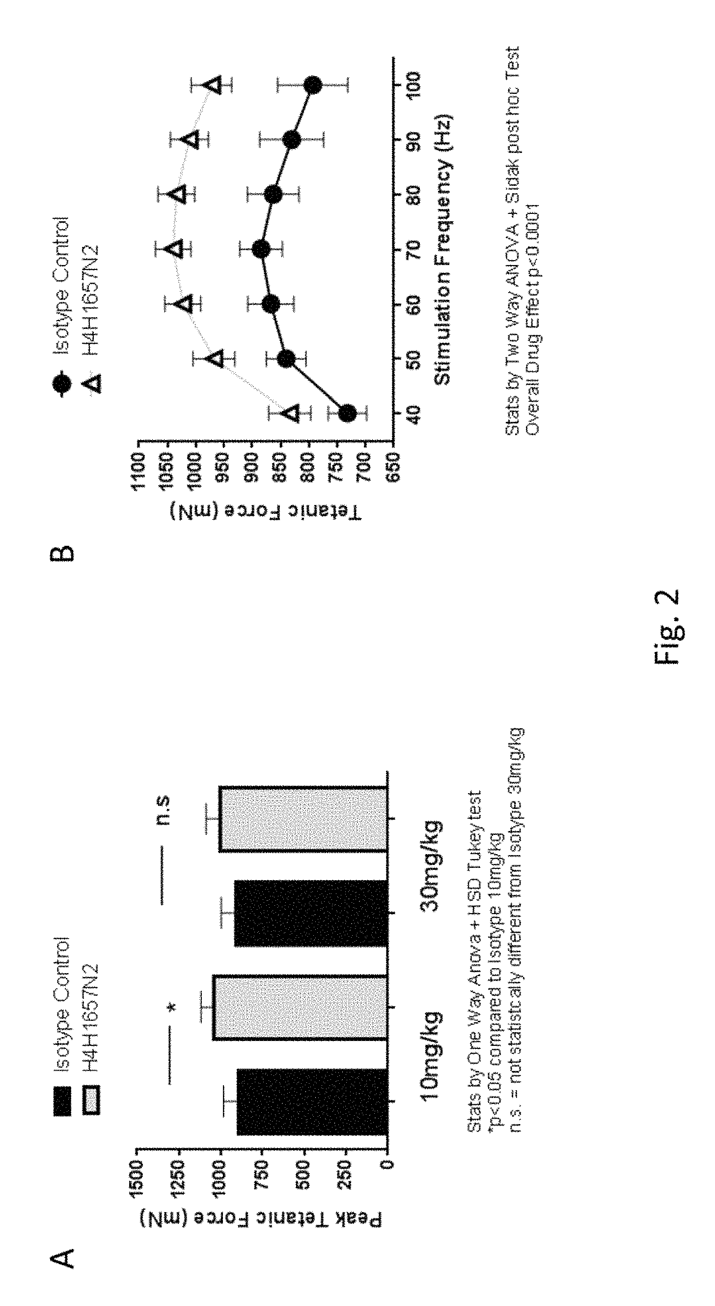
FIG. 2: Panel A shows the effects of 21 days of anti-GDF8 antibody treatment (H4H1657N2, 10 mg/kg or 30 mg/kg) on average peak tetanic force compared to isotype control antibody. Data analyzed using one-way analysis of variance (ANOVA) followed by Tukey's test. *p<0.05 significance over isotype control (n=6, unpaired Student t test); n.s.=not statistically significant compared to isotype 30 mg/kg. Panel B shows the increase in tibialis anterior (TA) muscle peak tetanic force in H4H1657N2-treated mice (10 mg/kg) versus mice treated with isotype control antibodies for three weeks (n=6), when stimulated by electric current over a range of frequencies (40 to 100 Hz). Data are expressed as mean average peak force.+-.SEM.
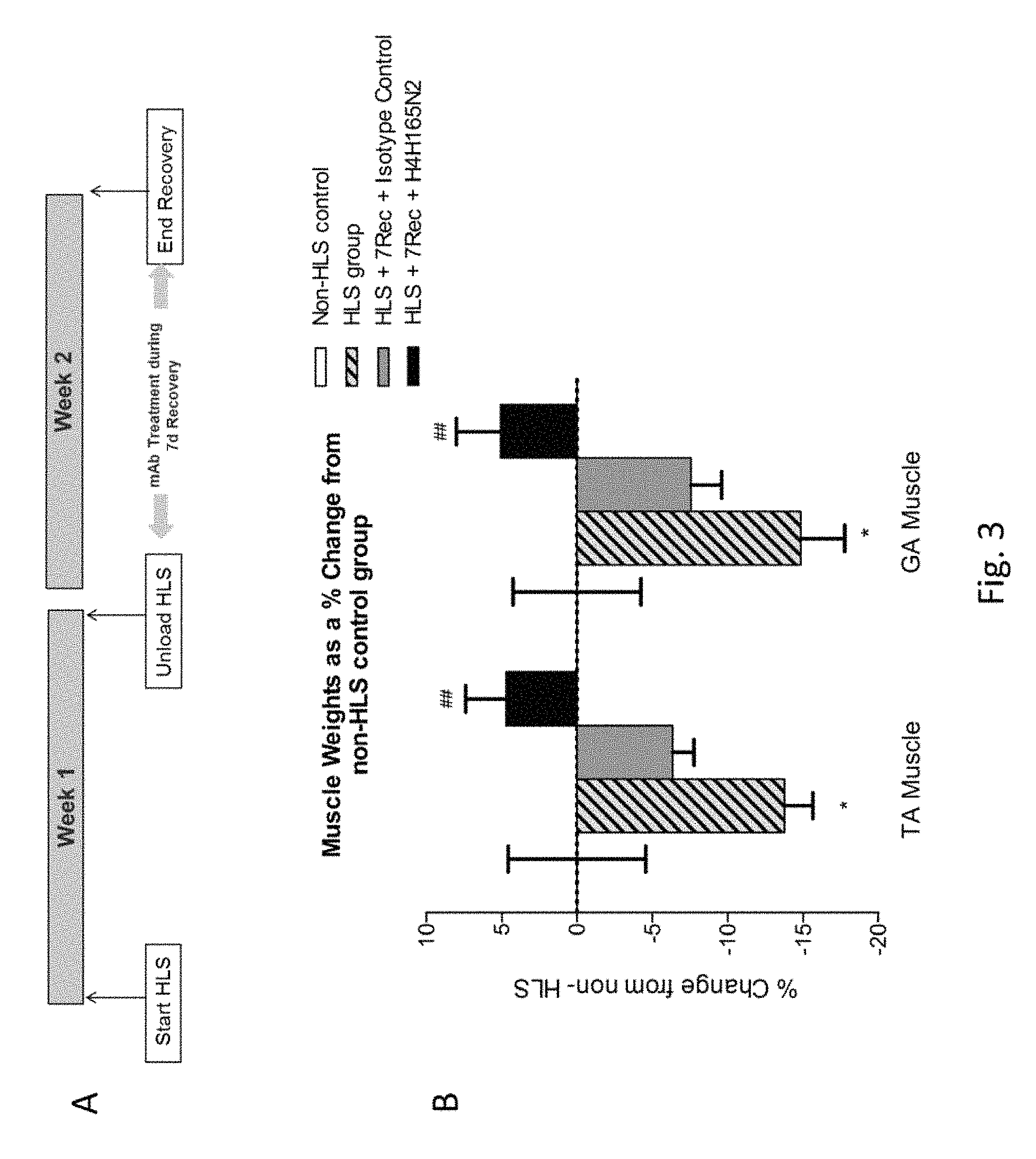
FIG. 3: Panel A shows the design of an experiment to evaluate the effects of H4H1657N2 during the recovery phase from hind limb suspension-induced muscle atrophy. Panel B shows the percentage change in TA and Gastrocnemius (GA) muscle weights for H4H1657N2-treated and isotyple control antibody-treated mice post-recovery after 7 days of hind limb suspension (HLS+7Rec) versus mice without a recovery period after 7 days of hind limb suspension (HLS) and control mice (non-HLS control). Values are expressed as the mean percentage change over control non-HLS values.+-.SEM. Data analyzed using one-way analysis of variance (ANOVA) followed by Tukey's test. *=p<0.05 significance over Non-HLS group. #=p<0.05 significance over HLS group.
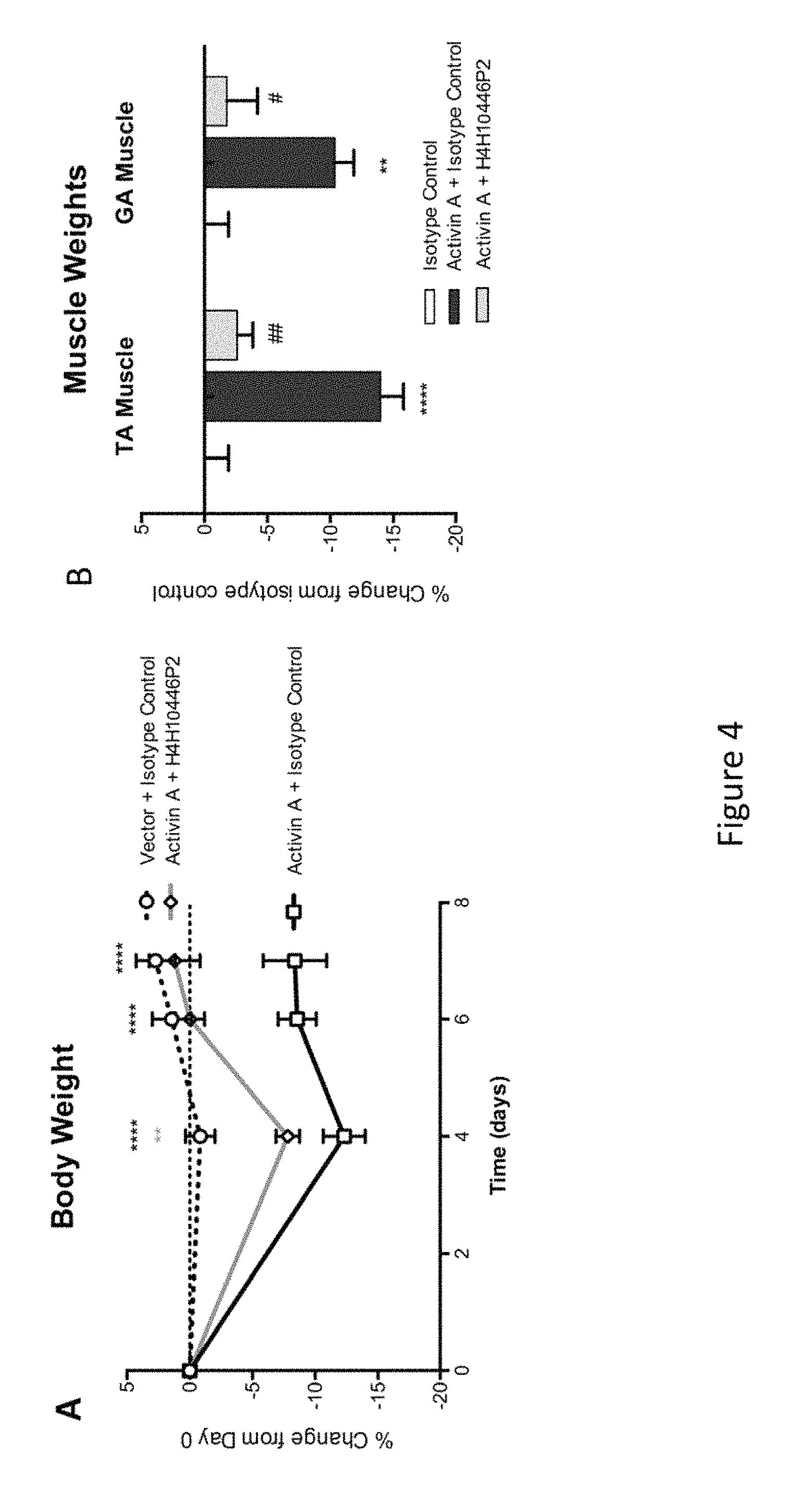
FIG. 4: Panel A shows the effects of the administration of the anti-Activin A antibody H4H10446P2 on body weight of mice overexpressing Activin A (versus isotype control). Data was analyzed using two-way analysis of variance (Repeated Measures ANOVA+Boneferroni Multiple Comparison Test) followed by Tukey's test. *=p<0.05 vs Isotype Control; #=p<0.05 vs Activin A+Isotype Control. Panel B shows the effects of anti-Activin A antibody H4H10446P2 on tibialis anterior (TA) and Gastrocnemius (GA) muscle weights in mice overexpressing Activin A (versus isotype control). Data analyzed using one-way analysis of variance (ANOVA) followed by Tukey's test. *=p<0.05 over Vector+Isotype Control; #=p<0.05 over Activin A+Isotype Control.
DETAILED DESCRIPTION
Before the present invention is described, it is to be understood that this invention is not limited to particular methods and experimental conditions described, as such methods and conditions may vary. It is also to be understood that the terminology used herein is for the purpose of describing particular embodiments only, and is not intended to be limiting, since the scope of the present invention will be limited only by the appended claims.
Unless defined otherwise, all technical and scientific terms used herein have the same meaning as commonly understood by one of ordinary skill in the art to which this invention belongs. As used herein, the term "about," when used in reference to a particular recited numerical value, means that the value may vary from the recited value by no more than 1%. For example, as used herein, the expression "about 100" includes 99 and 101 and all values in between (e.g., 99.1, 99.2, 99.3, 99.4, etc.).
Although any methods and materials similar or equivalent to those described herein can be used in the practice or testing of the present invention, the preferred methods and materials are now described. All patents, applications and non-patent publications mentioned in this specification are incorporated herein by reference in their entireties.
Antigen-Specific Binding Proteins
The present invention relates to compositions comprising antigen-specific binding proteins. More specifically, the present invention provides a composition comprising an Activin A-specific binding protein.
As used herein, the expression "antigen-specific binding protein" means a protein comprising at least one domain which specifically binds a particular antigen. Exemplary categories of antigen-specific binding proteins include antibodies, antigen-binding portions of antibodies, peptides that specifically interact with a particular antigen (e.g., peptibodies), receptor molecules that specifically interact with a particular antigen, and proteins comprising a ligand-binding portion of a receptor that specifically binds a particular antigen.
The present invention includes antigen-specific binding proteins that specifically bind Activin A, i.e., "Activin A-specific binding proteins". Activins are homo- and hetero-dimeric molecules comprising beta subunits, i.e., Inhibin .beta.A, inhibin .beta.B, inhibin .beta.C, and/or inhibin .beta.E. The .beta.A subunit has the amino acid sequence of SEQ ID NO:226 and the .beta.B subunit has the amino acid sequence of SEQ ID NO:228. Activin A is a homodimer of two .beta.A subunits; Activin B is a homodimer of two .beta.B subunits; Activin AB is a heterodimer of one .beta.A subunit and one .beta.B subunit; and Activin AC is a heterodimer of one .beta.A subunit and one .beta.C subunit. An Activin A-specific binding protein may be an antigen-specific binding protein that specifically binds the .beta.A subunit. Since the .beta.A subunit is found in Activin A, Activin AB, and Activin AC molecules, an "Activin A-specific binding protein" can be an antigen-specific binding protein that specifically binds Activin A as well as Activin AB and Activin AC (by virtue of its interaction with the .beta.A subunit). Therefore, according to one embodiment of the present invention, an Activin A-specific binding protein specifically binds Activin A; or Activin A and Activin AB; or Activin A and Activin AC; or Activin A, Activin AB and Activin AC, but does not bind other ActRIIB ligands such as Activin B, GDF3, GDF8, BMP2, BMP4, BMP7, BMP9, BMP10, GDF11, Nodal, etc. Thus, in one embodiment of the invention, an Activin A-specific binding protein specifically binds to Activin A but does not bind significantly to Activin B or Activin C. In another embodiment, an Activin A-specific binding protein may also bind to Activin B (by virtue of cross-reaction with the .beta.B subunit, i.e., Inhibin.beta.B). In another embodiment, an Activin A-specific binding protein is a binding protein that binds specifically to Activin A but does not bind to any other ligand of ActRIIB. In another embodiment, an Activin A-specific binding protein is a binding protein and binds specifically to Activin A and does not bind to any Bone Morphogenetic Protein (BMP) (e.g., BMP2, BMP4, BMP6, BMP9, BMP10). In another embodiment, an Activin A-specific binding protein is a binding protein that binds specifically to Activin A but does not bind to any other member of the transforming growth factor beta (TGF.beta.) superfamily.
The present invention also includes antigen-specific binding proteins that specifically bind GDF8, i.e., "GDF8-specific binding proteins". The term "GDF8" (also referred to as "growth and differentiation factor-8" and "myostatin") means the protein having the amino acid sequence of SEQ ID NO:225 (mature protein). According to the present invention, GDF8-specific binding proteins specifically bind GDF8 but do not bind other ActRIIB ligands such as GDF3, BMP2, BMP4, BMP7, BMP9, BMP10, GDF11, Activin A, Activin B, Activin AB, Nodal, etc.
In the context of the present invention, molecules such as ActRIIB-Fc (e.g., "ACE-031"), which comprise the ligand-binding portion of the ActRIIB receptor, are not considered "Activin A-specific binding proteins" or "GDF8-specific binding proteins" because such molecules bind multiple ligands besides GDF8, Activin A and Activin AB.
All references to proteins, polypeptides and protein fragments herein are intended to refer to the human version of the respective protein, polypeptide or protein fragment unless explicitly specified as being from a non-human species.
Antigen-Binding Molecules with Two Different Antigen-Specific Binding Domains
The present invention also includes antigen-binding molecules comprising two different antigen-specific binding domains. In particular, the present invention includes antigen-binding molecules comprising an Activin A-specific binding domain and a GDF8-specific binding domain. The term "antigen-specific binding domain," as used herein, includes polypeptides comprising or consisting of: (i) an antigen-binding fragment of an antibody molecule, (ii) a peptide that specifically interacts with a particular antigen (e.g., a peptibody), and/or (iii) a ligand-binding portion of a receptor that specifically binds a particular antigen. For example, the present invention includes bispecific antibodies with one arm comprising a first heavy chain variable region/light chain variable region (HCVR/LCVR) pair that specifically binds Activin A and another arm comprising a second HCVR/LCVR pair that specifically binds GDF8.
Specific Binding
The term "specifically binds" or the like, as used herein, means that an antigen-specific binding protein, or an antigen-specific binding domain, forms a complex with a particular antigen characterized by a dissociation constant (K.sub.D) of 500 .mu.M or less, and does not bind other unrelated antigens under ordinary test conditions. "Unrelated antigens" are proteins, peptides or polypeptides that have less than 95% amino acid identity to one another. Methods for determining whether two molecules specifically bind one another are well known in the art and include, for example, equilibrium dialysis, surface plasmon resonance, and the like. For example, an antigen-specific binding protein or an antigen-specific binding domain, as used in the context of the present invention, includes molecules that bind a particular antigen (e.g., Activin A and/or AB, or GDF8) or a portion thereof with a K.sub.D of less than about 500 pM, less than about 400 pM, less than about 300 pM, less than about 200 pM, less than about 100 pM, less than about 90 pM, less than about 80 pM, less than about 70 pM, less than about 60 pM, less than about 50 pM, less than about 40 pM, less than about 30 pM, less than about 20 pM, less than about 10 pM, less than about 5 pM, less than about 4 pM, less than about 2 pM, less than about 1 pM, less than about 0.5 pM, less than about 0.2 pM, less than about 0.1 pM, or less than about 0.05 pM, as measured in a surface plasmon resonance assay.
As used herein, an antigen-specific binding protein or antigen-specific binding domain "does not bind" to a specified molecule (e.g., "does not bind GDF11", "does not bind BMP9", "does not bind BMP10", etc.) if the protein or binding domain, when tested for binding to the molecule at 25.degree. C. in a surface plasmon resonance assay, exhibits a K.sub.D of greater than 50.0 nM, or fails to exhibit any binding in such an assay or equivalent thereof.
The term "surface plasmon resonance", as used herein, refers to an optical phenomenon that allows for the analysis of real-time interactions by detection of alterations in protein concentrations within a biosensor matrix, for example using the BIAcore.TM. system (Biacore Life Sciences division of GE Healthcare, Piscataway, N.J.).
The term "K.sub.D", as used herein, means the equilibrium dissociation constant of a particular protein-protein interaction (e.g., antibody-antigen interaction). Unless indicated otherwise, the K.sub.D values disclosed herein refer to K.sub.D values determined by surface plasmon resonance assay at 25.degree. C.
Antibodies and Antigen-Binding Fragments of Antibodies
As indicated above, an antigen-specific binding protein can comprise or consist of an antibody or antigen-binding fragment of an antibody. Furthermore, in the case of antigen-binding molecules comprising two different antigen-specific binding domains, one or both of the antigen-specific binding domains may comprise or consist of an antigen-binding fragment of an antibody.
As used herein, "an antibody that binds Activin" or an "anti-Activin A antibody" includes antibodies, and antigen-binding fragments thereof, that bind a soluble fragment of the Activin A protein and may also bind to an Activin .beta.A subunit-containing Activin heterodimer.
The term "antibody", as used herein, means any antigen-binding molecule or molecular complex comprising at least one complementarity determining region (CDR) that specifically binds to or interacts with a particular antigen (e.g., Activin A). The term "antibody" includes immunoglobulin molecules comprising four polypeptide chains, two heavy (H) chains and two light (L) chains inter-connected by disulfide bonds, as well as multimers thereof (e.g., IgM). Each heavy chain comprises a heavy chain variable region (abbreviated herein as HCVR or V.sub.H) and a heavy chain constant region. The heavy chain constant region comprises three domains, C.sub.H1, C.sub.H2 and C.sub.H3. Each light chain comprises a light chain variable region (abbreviated herein as LCVR or V.sub.L) and a light chain constant region. The light chain constant region comprises one domain (C.sub.L1). The V.sub.H and V.sub.L regions can be further subdivided into regions of hypervariability, termed complementarity determining regions (CDRs), interspersed with regions that are more conserved, termed framework regions (FR). Each V.sub.H and V.sub.L is composed of three CDRs and four FRs, arranged from amino-terminus to carboxy-terminus in the following order: FR1, CDR1, FR2, CDR2, FR3, CDR3, FR4. In different embodiments of the invention, the FRs of the anti-Activin A antibody (or antigen-binding portion thereof) may be identical to the human germline sequences, or may be naturally or artificially modified. An amino acid consensus sequence may be defined based on a side-by-side analysis of two or more CDRs.
The term "antibody", as used herein, also includes antigen-binding fragments of full antibody molecules. The terms "antigen-binding portion" of an antibody, "antigen-binding fragment" of an antibody, and the like, as used herein, include any naturally occurring, enzymatically obtainable, synthetic, or genetically engineered polypeptide or glycoprotein that specifically binds an antigen to form a complex. Antigen-binding fragments of an antibody may be derived, e.g., from full antibody molecules using any suitable standard techniques such as proteolytic digestion or recombinant genetic engineering techniques involving the manipulation and expression of DNA encoding antibody variable and optionally constant domains. Such DNA is known and/or is readily available from, e.g., commercial sources, DNA libraries (including, e.g., phage-antibody libraries), or can be synthesized. The DNA may be sequenced and manipulated chemically or by using molecular biology techniques, for example, to arrange one or more variable and/or constant domains into a suitable configuration, or to introduce codons, create cysteine residues, modify, add or delete amino acids, etc.
Non-limiting examples of antigen-binding fragments include: (i) Fab fragments; (ii) F(ab')2 fragments; (iii) Fd fragments; (iv) Fv fragments; (v) single-chain Fv (scFv) molecules; (vi) dAb fragments; and (vii) minimal recognition units consisting of the amino acid residues that mimic the hypervariable region of an antibody (e.g., an isolated complementarity determining region (CDR) such as a CDR3 peptide), or a constrained FR3-CDR3-FR4 peptide. Other engineered molecules, such as domain-specific antibodies, single domain antibodies, domain-deleted antibodies, chimeric antibodies, CDR-grafted antibodies, diabodies, triabodies, tetrabodies, minibodies, nanobodies (e.g. monovalent nanobodies, bivalent nanobodies, etc.), small modular immunopharmaceuticals (SMIPs), and shark variable IgNAR domains, are also encompassed within the expression "antigen-binding fragment," as used herein.
An antigen-binding fragment of an antibody will typically comprise at least one variable domain. The variable domain may be of any size or amino acid composition and will generally comprise at least one CDR which is adjacent to or in frame with one or more framework sequences. In antigen-binding fragments having a V.sub.H domain associated with a V.sub.L domain, the V.sub.H and V.sub.L domains may be situated relative to one another in any suitable arrangement. For example, the variable region may be dimeric and contain V.sub.H-V.sub.H, V.sub.H-V.sub.L or V.sub.L-V.sub.L dimers. Alternatively, the antigen-binding fragment of an antibody may contain a monomeric V.sub.H or V.sub.L domain.
In certain embodiments, an antigen-binding fragment of an antibody may contain at least one variable domain covalently linked to at least one constant domain. Non-limiting, exemplary configurations of variable and constant domains that may be found within an antigen-binding fragment of an antibody of the present invention include: (i) V.sub.H-C.sub.H1; (ii) V.sub.H-C.sub.H2; (iii) V.sub.H-C.sub.H3; (iv) V.sub.H-C.sub.H1-C.sub.H2; (V) V.sub.H-C.sub.H1-C.sub.H2-C.sub.H3; V.sub.H-C.sub.H2-C.sub.H3; (Vii) V.sub.H-C.sub.L; VL-C.sub.H1; (ix) VL-C.sub.H2; (x) VL-C.sub.H3; (xi) V.sub.L-C.sub.H1-C.sub.H2; (xii) VL-C.sub.H1-C.sub.H2-C.sub.H3; (xiii) VL-C.sub.H2-C.sub.H3; and (xiv) V.sub.L-C.sub.L. In any configuration of variable and constant domains, including any of the exemplary configurations listed above, the variable and constant domains may be either directly linked to one another or may be linked by a full or partial hinge or linker region. A hinge region may consist of at least 2 (e.g., 5, 10, 15, 20, 40, 60 or more) amino acids which result in a flexible or semi-flexible linkage between adjacent variable and/or constant domains in a single polypeptide molecule. Moreover, an antigen-binding fragment of an antibody of the present invention may comprise a homo-dimer or hetero-dimer (or other multimer) of any of the variable and constant domain configurations listed above in non-covalent association with one another and/or with one or more monomeric V.sub.H or V.sub.L domain (e.g., by disulfide bond(s)).
As with full antibody molecules, antigen-binding fragments may be monospecific or multispecific (e.g., bispecific). A multispecific antigen-binding fragment of an antibody will typically comprise at least two different variable domains, wherein each variable domain is capable of specifically binding to a separate antigen or to a different epitope on the same antigen. Any multispecific antibody format, including the exemplary bispecific antibody formats disclosed herein, may be adapted for use in the context of an antigen-binding fragment of an antibody of the present invention using routine techniques available in the art.
The antibodies of the present invention may function through complement-dependent cytotoxicity (CDC) or antibody-dependent cell-mediated cytotoxicity (ADCC). "Complement-dependent cytotoxicity" (CDC) refers to lysis of antigen-expressing cells by an antibody of the invention in the presence of complement. "Antibody-dependent cell-mediated cytotoxicity" (ADCC) refers to a cell-mediated reaction in which nonspecific cytotoxic cells that express Fc receptors (FcRs) (e.g., Natural Killer (NK) cells, neutrophils, and macrophages) recognize bound antibody on a target cell and thereby lead to lysis of the target cell. CDC and ADCC can be measured using assays that are well known and available in the art. (See, e.g., U.S. Pat. Nos. 5,500,362 and 5,821,337, and Clynes et al., PNAS USA 95:652-656 (1998)). The constant region of an antibody is important in the ability of an antibody to fix complement and mediate cell-dependent cytotoxicity. Thus, the isotype of an antibody may be selected on the basis of whether it is desirable for the antibody to mediate cytotoxicity.
In certain embodiments of the invention, the anti-Activin A antibodies of the invention are human antibodies. The term "human antibody", as used herein, is intended to include antibodies having variable and constant regions derived from human germline immunoglobulin sequences. The human antibodies of the invention may include amino acid residues not encoded by human germline immunoglobulin sequences (e.g., mutations introduced by random or site-specific mutagenesis in vitro or by somatic mutation in vivo), for example in the CDRs and in particular CDR3. However, the term "human antibody", as used herein, is not intended to include antibodies in which CDR sequences derived from the germline of another mammalian species, such as a mouse, have been grafted onto human framework sequences.
The antibodies of the invention may, in some embodiments, be recombinant human antibodies. The term "recombinant human antibody", as used herein, is intended to include all human antibodies that are prepared, expressed, created or isolated by recombinant means, such as antibodies expressed using a recombinant expression vector transfected into a host cell (described further below), antibodies isolated from a recombinant, combinatorial human antibody library (described further below), antibodies isolated from an animal (e.g., a mouse) that is transgenic for human immunoglobulin genes (see e.g., Taylor et al., Nucl Acids Res 20:6287-6295 (1992)) or antibodies prepared, expressed, created or isolated by any other means that involves splicing of human immunoglobulin gene sequences to other DNA sequences. Such recombinant human antibodies have variable and constant regions derived from human germline immunoglobulin sequences. In certain embodiments, however, such recombinant human antibodies are subjected to in vitro mutagenesis (or, when an animal transgenic for human Ig sequences is used, in vivo somatic mutagenesis) and thus the amino acid sequences of the V.sub.H and V.sub.L regions of the recombinant antibodies are sequences that, while derived from and related to human germline V.sub.H and V.sub.L sequences, may not naturally exist within the human antibody germline repertoire in vivo.
Human antibodies can exist in two forms that are associated with hinge heterogeneity. In one form, an immunoglobulin molecule comprises a stable four chain construct of approximately 150-160 kDa in which the dimers are held together by an interchain heavy chain disulfide bond. In a second form, the dimers are not linked via inter-chain disulfide bonds and a molecule of about 75-80 kDa is formed composed of a covalently coupled light and heavy chain (half-antibody). These forms have been extremely difficult to separate, even after affinity purification.
The frequency of appearance of the second form in various intact IgG isotypes is due to, but not limited to, structural differences associated with the hinge region isotype of the antibody. A single amino acid substitution in the hinge region of the human IgG4 hinge can significantly reduce the appearance of the second form (Angal et al. Molecular Immunology 30:105 1993)) to levels typically observed using a human IgG1 hinge. The instant invention encompasses antibodies having one or more mutations in the hinge, C.sub.H2 or C.sub.H3 region which may be desirable, for example, in production, to improve the yield of the desired antibody form.
The antibodies of the invention may be isolated antibodies. An "isolated antibody," as used herein, means an antibody that has been identified and separated and/or recovered from at least one component of its natural environment. For example, an antibody that has been separated or removed from at least one component of an organism, or from a tissue or cell in which the antibody naturally exists or is naturally produced, is an "isolated antibody" for purposes of the present invention. An isolated antibody also includes an antibody in situ within a recombinant cell. Isolated antibodies are antibodies that have been subjected to at least one purification or isolation step. According to certain embodiments, an isolated antibody may be substantially free of other cellular material and/or chemicals.
The present invention includes neutralizing and/or blocking anti-Activin A antibodies. A "neutralizing" or "blocking" antibody, as used herein, is intended to refer to an antibody whose binding to Activin A: (i) interferes with the interaction between Activin A and an Activin A receptor (e.g., Activin Type IIA receptor, Activin Type IIB receptor, Activin Type I receptor, etc.); (ii) interferes with the formation of Activin-Activin receptor complexes; and/or (iii) results in inhibition of at least one biological function of Activin A. The inhibition caused by an Activin A neutralizing or blocking antibody need not be complete so long as it is detectable using an appropriate assay. Exemplary assays for detecting Activin A inhibition are described in the working Examples herein.
The anti-Activin A antibodies disclosed herein may comprise one or more amino acid substitutions, insertions and/or deletions in the framework and/or CDR regions of the heavy and light chain variable domains as compared to the corresponding germline sequences from which the antibodies were derived. Such mutations can be readily ascertained by comparing the amino acid sequences disclosed herein to germline sequences available from, for example, public antibody sequence databases. The present invention includes antibodies, and antigen-binding fragments thereof, which are derived from any of the amino acid sequences disclosed herein, wherein one or more amino acids within one or more framework and/or CDR regions are mutated to the corresponding residue(s) of the germline sequence from which the antibody was derived, or to the corresponding residue(s) of another human germline sequence, or to a conservative amino acid substitution of the corresponding germline residue(s) (such sequence changes are referred to herein collectively as "germline mutations"). A person of ordinary skill in the art, starting with the heavy and light chain variable region sequences disclosed herein, can easily produce numerous antibodies and antigen-binding fragments which comprise one or more individual germline mutations or combinations thereof. In certain embodiments, all of the framework and/or CDR residues within the V.sub.H and/or V.sub.L domains are mutated back to the residues found in the original germline sequence from which the antibody was derived. In other embodiments, only certain residues are mutated back to the original germline sequence, e.g., only the mutated residues found within the first 8 amino acids of FR1 or within the last 8 amino acids of FR4, or only the mutated residues found within CDR1, CDR2 or CDR3. In other embodiments, one or more of the framework and/or CDR residue(s) are mutated to the corresponding residue(s) of a different germline sequence (i.e., a germline sequence that is different from the germline sequence from which the antibody was originally derived). Furthermore, the antibodies of the present invention may contain any combination of two or more germline mutations within the framework and/or CDR regions, e.g., wherein certain individual residues are mutated to the corresponding residue of a particular germline sequence while certain other residues that differ from the original germline sequence are maintained or are mutated to the corresponding residue of a different germline sequence. Once obtained, antibodies and antigen-binding fragments that contain one or more germline mutations can be easily tested for one or more desired property such as, improved binding specificity, increased binding affinity, improved or enhanced antagonistic or agonistic biological properties (as the case may be), reduced immunogenicity, etc. Antibodies and antigen-binding fragments obtained in this general manner are encompassed within the present invention.
The present invention also includes anti-Activin A antibodies comprising variants of any of the HCVR, LCVR, and/or CDR amino acid sequences disclosed herein having one or more conservative substitutions. For example, the present invention includes anti-Activin A antibodies having HCVR, LCVR, and/or CDR amino acid sequences with, e.g., 10 or fewer, 8 or fewer, 6 or fewer, 4 or fewer, etc. conservative amino acid substitutions relative to any of the HCVR, LCVR, and/or CDR amino acid sequences disclosed herein.
The term "epitope" refers to an antigenic determinant that interacts with a specific antigen binding site in the variable region of an antibody molecule known as a paratope. A single antigen may have more than one epitope. Thus, different antibodies may bind to different areas on an antigen and may have different biological effects. Epitopes may be either conformational or linear. A conformational epitope is produced by spatially juxtaposed amino acids from different segments of the linear polypeptide chain. A linear epitope is one produced by adjacent amino acid residues in a polypeptide chain. In certain circumstance, an epitope may include moieties of saccharides, phosphoryl groups, or sulfonyl groups on the antigen.
The term "substantial identity" or "substantially identical," when referring to a nucleic acid or fragment thereof, indicates that, when optimally aligned with appropriate nucleotide insertions or deletions with another nucleic acid (or its complementary strand), there is nucleotide sequence identity in at least about 95%, and more preferably at least about 96%, 97%, 98% or 99% of the nucleotide bases, as measured by any well-known algorithm of sequence identity, such as FASTA, BLAST or Gap, as discussed below. A nucleic acid molecule having substantial identity to a reference nucleic acid molecule may, in certain instances, encode a polypeptide having the same or substantially similar amino acid sequence as the polypeptide encoded by the reference nucleic acid molecule.
As applied to polypeptides, the term "substantial similarity" or "substantially similar" means that two peptide sequences, when optimally aligned, such as by the programs GAP or BESTFIT using default gap weights, share at least 95% sequence identity, even more preferably at least 98% or 99% sequence identity. Preferably, residue positions which are not identical differ by conservative amino acid substitutions. A "conservative amino acid substitution" is one in which an amino acid residue is substituted by another amino acid residue having a side chain (R group) with similar chemical properties (e.g., charge or hydrophobicity). In general, a conservative amino acid substitution will not substantially change the functional properties of a protein. In cases where two or more amino acid sequences differ from each other by conservative substitutions, the percent sequence identity or degree of similarity may be adjusted upwards to correct for the conservative nature of the substitution. Means for making this adjustment are well-known to those of skill in the art. See, e.g., Pearson, W. R., Methods Mol Biol 24: 307-331 (1994), herein incorporated by reference. Examples of groups of amino acids that have side chains with similar chemical properties include (1) aliphatic side chains: glycine, alanine, valine, leucine and isoleucine; (2) aliphatic-hydroxyl side chains: serine and threonine; (3) amide-containing side chains: asparagine and glutamine; (4) aromatic side chains: phenylalanine, tyrosine, and tryptophan; (5) basic side chains: lysine, arginine, and histidine; (6) acidic side chains: aspartate and glutamate, and (7) sulfur-containing side chains are cysteine and methionine. Preferred conservative amino acids substitution groups are: valine-leucine-isoleucine, phenylalanine-tyrosine, lysine-arginine, alanine-valine, glutamate-aspartate, and asparagine-glutamine. Alternatively, a conservative replacement is any change having a positive value in the PAM250 log-likelihood matrix disclosed in Gonnet et al., Science 256: 1443-1445 (1992), herein incorporated by reference. A "moderately conservative" replacement is any change having a nonnegative value in the PAM250 log-likelihood matrix.
Sequence similarity for polypeptides, which is also referred to as sequence identity, is typically measured using sequence analysis software. Protein analysis software matches similar sequences using measures of similarity assigned to various substitutions, deletions and other modifications, including conservative amino acid substitutions. For instance, GCG software contains programs such as Gap and Bestfit which can be used with default parameters to determine sequence homology or sequence identity between closely related polypeptides, such as homologous polypeptides from different species of organisms or between a wild type protein and a mutein thereof. See, e.g., GCG Version 6.1. Polypeptide sequences also can be compared using FASTA using default or recommended parameters, a program in GCG Version 6.1. FASTA (e.g., FASTA2 and FASTA3) provides alignments and percent sequence identity of the regions of the best overlap between the query and search sequences (see, e.g., Pearson, W. R., Methods Mol Biol 132: 185-219 (2000), herein incorporated by reference). Another preferred algorithm when comparing a sequence of the invention to a database containing a large number of sequences from different organisms is the computer program BLAST, especially BLASTP or TBLASTN, using default parameters. See, e.g., Altschul et al., J Mol Biol 215:403-410 (1990) and Altschul et al., Nucleic Acids Res 25:3389-402 (1997), each herein incorporated by reference.
Biological Characteristics of the Antibodies
The present invention includes anti-Activin A antibodies and antigen-binding fragments thereof that bind Activin A with high affinity. For example, the present invention includes antibodies and antigen-binding fragments of antibodies that bind Activin A (e.g., at 25.degree. C. or 37.degree. C.) with a K.sub.D of less than about 30 nM as measured by surface plasmon resonance, e.g., using the assay format as defined in Example 3 herein. In certain embodiments, the antibodies or antigen-binding fragments of the present invention bind Activin A with a K.sub.D of less than about 25 nM, less than about 20 nM, less than about 15 nM, less than about 10 nM, less than about 5 nM, less than about 2 nM, less than about 1 nM, less than about 500 pM, less than about 250 pM, less than about 240 pM, less than about 230 pM, less than about 220 pM, less than about 210 pM, less than about 200 pM, less than about 190 pM, less than about 180 pM, less than about 170 pM, less than about 160 pM, less than about 150 pM, less than about 140 pM, less than about 130 pM, less than about 120 pM, less than about 110 pM, less than about 100 pM, less than about 95 pM, less than about 90 pM, less than about 85 pM, less than about 80 pM, less than about 75 pM, less than about 70 pM, less than about 65 pM, less than about 60 pM, less than about 55 pM, less than about 50 pM, less than about 45 pM, less than about 40 pM, less than about 35 pM, less than about 30 pM, less than about 25 pM, less than about 20 pM, less than about 15 pM, less than about 10 pM, less than about 9 pM, less than about 8 pM, less than about 7 pM, less than about 6 pM, less than about 5 pM, less than about 4 pM, or less than about 3 pM, as measured by surface plasmon resonance, e.g., using the assay format as defined in Example 3 herein, or a substantially similar assay.
The present invention also includes anti-Activin A antibodies and antigen-binding fragments thereof that inhibit Activin A-mediated cellular signaling. For example, the present invention includes anti-Activin A antibodies that inhibit the activation of the SMAD complex signal transduction pathway via the binding of Activin A to Activin Type I or II receptors with an IC.sub.50 value of less than about 4 nM, as measured in a cell-based blocking bioassay, e.g., using the assay format as defined in Example 6 herein, or a substantially similar assay. In certain embodiments, the antibodies or antigen-binding fragments of the present invention inhibit the activation of the SMAD complex signal transduction pathway via the binding of Activin A to Activin Type I or II receptors with an IC.sub.50 value of less than about 3 nM, less than about 2 nM, less than about 1 nm, less than about 500 pM, less than about 250 pM, less than about 240 pM, less than about 230 pM, less than about 220 pM, less than about 210 pM, less than about 200 pM, less than about 190 pM, less than about 180 pM, less than about 170 pM, less than about 160 pM, less than about 150 pM, less than about 140 pM, less than about 130 pM, less than about 120 pM, less than about 110 pM, less than about 100 pM, less than about 95 pM, less than about 90 pM, less than about 85 pM, less than about 80 pM, less than about 75 pM, less than 70 pM, less than about 65 pM, less than about 60 pM, less than about 55 pM, less than about 50 pM, less than about 49 pM, less than about 48 pM, less than about 47 pM, less than about 46 pM, less than about 45 pM, less than about 44 pM, less than about 43 pM, less than about 42 pM, less than about 41 pM, less than about 40 pM, or less than about 39 pM, as measured in a cell-based blocking bioassay, e.g., using the assay format as defined in Example 6 herein, or a substantially similar assay. In certain embodiments, the antibodies or antigen-binding fragments of the present invention inhibit the signaling activing of Activin B by interfering with the binding of Activin B to Activin Type I or II receptors with an IC.sub.50 value of less than about 50 nM, less than about 20 nM, less than about 10 nm, less than about 5 nM, or less than about 1 nM, as measured in a cell-based blocking bioassay, e.g., using the assay format as defined in Example 6 herein, or a substantially similar assay. In certain embodiments, the antibodies or antigen-binding fragments of the present invention inhibit the activation of the SMAD complex signal transduction pathway via the binding of Activin AB to Activin Type I or II receptors with an IC.sub.50 value of less than about 500 pM, less than about 450 pM, less than about 440 pM, less than about 430 pM, less than about 420 pM, less than about 410 pM, less than about 400 pM, less than about 390 pM, less than about 380 pM, less than about 370 pM, less than about 360 pM, less than about 350 pM, less than about 340 pM, less than about 320 pM, less than about 310 pM, less than about 300 pM, less than about 290 pM, less than about 280 pM, less than about 270 pM, less than about 260 pM, less than about 250 pM, less than about 240 pM, less than about 230 pM, less than about 220 pM, less than about 210 pM, less than about 200 pM, less than about 190 pM, less than about 180 pM, less than about 170 pM, less than about 160 pM, less than about 150 pM, or less than about 140 pM, as measured in a cell-based blocking bioassay, e.g., using the assay format as defined in Example 6 herein, or a substantially similar assay. In certain embodiments, the antibodies or antigen-binding fragments of the present invention inhibit the activation of the SMAD complex signal transduction pathway via the binding of Activin AC to Activin Type I or II receptors with an IC.sub.50 value of less than about 1 nM, less than about 900 pM, less than about 800 pM, less than about 750 pM, less than about 700 pM, less than about 650 pM, less than about 600 pM, or less than about 580 pM, as measured in a cell-based blocking bioassay, e.g., using the assay format as defined in Example 6 herein, or a substantially similar assay.
The antibodies of the present invention may possess one or more of the aforementioned biological characteristics, or any combinations thereof. Other biological characteristics of the antibodies of the present invention will be evident to a person of ordinary skill in the art from a review of the present disclosure including the working Examples herein.
Anti-Activin A Antibodies Comprising Fc Variants
According to certain embodiments of the present invention, anti-Activin A antibodies are provided comprising an Fc domain comprising one or more mutations which enhance or diminish antibody binding to the FcRn receptor, e.g., at acidic pH as compared to neutral pH. For example, the present invention includes anti-Activin A antibodies comprising a mutation in the C.sub.H2 or a C.sub.H3 region of the Fc domain, wherein the mutation(s) increases the affinity of the Fc domain to FcRn in an acidic environment (e.g., in an endosome where pH ranges from about 5.5 to about 6.0). Such mutations may result in an increase in serum half-life of the antibody when administered to an animal. Non-limiting examples of such Fc modifications include, e.g., a modification at position 250 (e.g., E or Q); 250 and 428 (e.g., L or F); 252 (e.g., L/Y/F/W or T), 254 (e.g., S or T), and 256 (e.g., S/R/Q/E/D or T); or a modification at position 428 and/or 433 (e.g., H/L/R/S/P/Q or K) and/or 434 (e.g., A, W, H, F or Y [N434A, N434W, N434H, N434F or N434Y]); or a modification at position 250 and/or 428; or a modification at position 307 or 308 (e.g., 308F, V308F), and 434. In one embodiment, the modification comprises a 428L (e.g., M428L) and 434S (e.g., N434S) modification; a 428L, 2591 (e.g., V2591), and 308F (e.g., V308F) modification; a 433K (e.g., H433K) and a 434 (e.g., 434Y) modification; a 252, 254, and 256 (e.g., 252Y, 254T, and 256E) modification; a 250Q and 428L modification (e.g., T250Q and M428L); and a 307 and/or 308 modification (e.g., 308F or 308P). In yet another embodiment, the modification comprises a 265A (e.g., D265A) and/or a 297A (e.g., N297A) modification.
For example, the present invention includes anti-Activin A antibodies comprising an Fc domain comprising one or more pairs or groups of mutations selected from the group consisting of: 250Q and 248L (e.g., T250Q and M248L); 252Y, 254T and 256E (e.g., M252Y, S254T and T256E); 428L and 434S (e.g., M428L and N434S); 257I and 311I (e.g., P257I and Q311I); 257I and 434H (e.g., P257I and N434H); 376V and 434H (e.g., D376V and N434H); 307A, 380A and 434A (e.g., T307A, E380A and N434A); and 433K and 434F (e.g., H433K and N434F). All possible combinations of the foregoing Fc domain mutations, and other mutations within the antibody variable domains disclosed herein, are contemplated within the scope of the present invention.
The present invention also includes anti-Activin A antibodies comprising a chimeric heavy chain constant (CH) region, wherein the chimeric CH region comprises segments derived from the CH regions of more than one immunoglobulin isotype. For example, the antibodies of the invention may comprise a chimeric CH region comprising part or all of a C.sub.H2 domain derived from a human IgG1, human IgG2 or human IgG4 molecule, combined with part or all of a C.sub.H3 domain derived from a human IgG1, human IgG2 or human IgG4 molecule. According to certain embodiments, the antibodies of the invention comprise a chimeric CH region having a chimeric hinge region. For example, a chimeric hinge may comprise an "upper hinge" amino acid sequence (amino acid residues from positions 216 to 227 according to EU numbering) derived from a human IgG1, a human IgG2 or a human IgG4 hinge region, combined with a "lower hinge" sequence (amino acid residues from positions 228 to 236 according to EU numbering) derived from a human IgG1, a human IgG2 or a human IgG4 hinge region. According to certain embodiments, the chimeric hinge region comprises amino acid residues derived from a human IgG1 or a human IgG4 upper hinge and amino acid residues derived from a human IgG2 lower hinge. An antibody comprising a chimeric CH region as described herein may, in certain embodiments, exhibit modified Fc effector functions without adversely affecting the therapeutic or pharmacokinetic properties of the antibody. (See, e.g., U.S. Provisional Appl. No. 61/759,578, filed Feb. 1, 2013, the disclosure of which is hereby incorporated by reference in its entirety).
Epitope Mapping and Related Technologies
The present invention includes anti-Activin A antibodies which interact with one or more amino acids found within Activin A (e.g., within the Activin Type II receptor binding site). The epitope to which the antibodies bind may consist of a single contiguous sequence of 3 or more (e.g., 3, 4, 5, 6, 7, 8, 9, 10, 11, 12, 13, 14, 15, 16, 17, 18, 19, 20 or more) amino acids located within the Activin .beta.A subunit. Alternatively, the epitope may consist of a plurality of non-contiguous amino acids (or amino acid sequences) located within the Activin A dimer.
Various techniques known to persons of ordinary skill in the art can be used to determine whether an antibody "interacts with one or more amino acids" within a polypeptide or protein. Exemplary techniques include, e.g., routine cross-blocking assay such as that described Antibodies, Harlow and Lane (Cold Spring Harbor Press, Cold Spring Harb., N.Y.), alanine scanning mutational analysis, peptide blots analysis (Reineke, Methods Mol Biol 248:443-463 (2004)), and peptide cleavage analysis. In addition, methods such as epitope excision, epitope extraction and chemical modification of antigens can be employed (Tomer, Protein Science 9:487-496 (2000)). Another method that can be used to identify the amino acids within a polypeptide with which an antibody interacts is hydrogen/deuterium exchange detected by mass spectrometry. In general terms, the hydrogen/deuterium exchange method involves deuterium-labeling the protein of interest, followed by binding the antibody to the deuterium-labeled protein. Next, the protein/antibody complex is transferred to water to allow hydrogen-deuterium exchange to occur at all residues except for the residues protected by the antibody (which remain deuterium-labeled). After dissociation of the antibody, the target protein is subjected to protease cleavage and mass spectrometry analysis, thereby revealing the deuterium-labeled residues which correspond to the specific amino acids with which the antibody interacts. See, e.g., Ehring, Analytical Biochemistry 267(2):252-259 (1999); Engen and Smith, Anal. Chem. 73:256A-265A (2001).
The present invention further includes anti-Activin A antibodies that bind to the same epitope as any of the specific exemplary antibodies described herein (e.g., H4H10423P, H4H10424P, H4H10426P, H4H10429P, H4H10430P, H4H10432P2, H4H10433P2, H4H10436P2, H4H10437P2, H4H10438P2, H4H10440P2, H4H10442P2, H4H10445P2, H4H10446P2, H4H10447P2, H4H10448P2, H4H10452P2, H4H10468P2, H2aM10965N, etc.). Likewise, the present invention also includes anti-Activin A antibodies that compete for binding to Activin A with any of the specific exemplary antibodies described herein (e.g., H4H10423P, H4H10424P, H4H10426P, H4H10429P, H4H10430P, H4H10432P2, H4H10433P2, H4H10436P2, H4H10437P2, H4H10438P2, H4H10440P2, H4H10442P2, H4H10445P2, H4H10446P2, H4H10447P2, H4H10448P2, H4H10452P2, H4H10468P2, H2aM10965N, etc.). For example, the present invention includes anti-Activin A antibodies that cross-compete for binding to Activin A with one or more antibodies of "Bin 1" as defined in Example 5 herein (e.g., H4H10423P, H4H10446P2, H4H10468P2 and H4H10442P2). The present invention also includes anti-Activin A antibodies that cross-compete for binding to Activin A with one or more antibodies of "Bin 2" as defined in Example 5 herein (e.g., H4H10429, H4H10430P, H4H10432P2, H4H10436P2, and H4H10440P2).
One can easily determine whether an antibody binds to the same epitope as, or competes for binding with, a reference anti-Activin A antibody by using routine methods known in the art and exemplified herein. For example, to determine if a test antibody binds to the same epitope as a reference anti-Activin A antibody of the invention, the reference antibody is allowed to bind to Activin A (or a .beta.A subunit-containing heterodimer). Next, the ability of a test antibody to bind to Activin A is assessed. If the test antibody is able to bind to Activin A following saturation binding with the reference anti-Activin A antibody, it can be concluded that the test antibody binds to a different epitope than the reference anti-Activin A antibody. On the other hand, if the test antibody is not able to bind to Activin A following saturation binding with the reference anti-Activin A antibody, then the test antibody may bind to the same epitope as the epitope bound by the reference anti-Activin A antibody of the invention. Additional routine experimentation (e.g., peptide mutation and binding analyses) can then be carried out to confirm whether the observed lack of binding of the test antibody is in fact due to binding to the same epitope as the reference antibody or if steric blocking (or another phenomenon) is responsible for the lack of observed binding. Experiments of this sort can be performed using ELISA, RIA, BIACORE.TM. real-time surface plasmon resonance biosensor, flow cytometry or any other quantitative or qualitative antibody-binding assay available in the art. In accordance with certain embodiments of the present invention, two antibodies bind to the same (or overlapping) epitope if, e.g., a 1-, 5-, 10-, 20- or 100-fold excess of one antibody inhibits binding of the other by at least 50% but preferably 75%, 90% or even 99% as measured in a competitive binding assay (see, e.g., Junghans et al., Cancer Res. 50:1495-1502 (1990)). Alternatively, two antibodies are deemed to bind to the same epitope if essentially all amino acid mutations in the antigen that reduce or eliminate binding of one antibody reduce or eliminate binding of the other. Two antibodies are deemed to have "overlapping epitopes" if only a subset of the amino acid mutations that reduce or eliminate binding of one antibody reduce or eliminate binding of the other.
To determine if an antibody competes for binding (or cross-competes for binding) with a reference anti-Activin A antibody, the above-described binding methodology is performed in two orientations: In a first orientation, the reference antibody is allowed to bind to Activin A protein (or a .beta.A subunit-containing heterodimer) under saturating conditions followed by assessment of binding of the test antibody to the Activin A molecule. In a second orientation, the test antibody is allowed to bind to Activin A under saturating conditions followed by assessment of binding of the reference antibody to Activin A. If, in both orientations, only the first (saturating) antibody is capable of binding to Activin A, then it is concluded that the test antibody and the reference antibody compete for binding to Activin A (see, e.g., the assay format described in Example 4 herein, in which a test Activin A antibody is captured onto sensor tips that are then submerged in a solution containing a reference Activin A antibody pre-bound with Activin A). As will be appreciated by a person of ordinary skill in the art, an antibody that competes for binding with a reference antibody may not necessarily bind to the same epitope as the reference antibody, but may sterically block binding of the reference antibody by binding an overlapping or adjacent epitope.
Anti-Activin A antibodies of the invention may bind to an epitope on Activin A that is within or near the binding site for an Activin Type II receptor, directly block interaction between Activin A and an Activin Type II receptor, and indirectly block interaction between Activin A and an Activin Type I receptor. Anti-Activin A antibodies of the invention may bind to an epitope on Activin A that is within or near the binding site for the Activin Type I receptor and directly block interaction between Activin A and an Activin Type I receptor. In one embodiment of the invention, an anti-Activin A antibody of the invention that binds to Activin A at or near the Activin Type I receptor binding site does not block interaction between Activin A and an Activin A Type II receptor.
Preparation of Human Antibodies
Methods for generating monoclonal antibodies, including fully human monoclonal antibodies are known in the art. Any such known methods can be used in the context of the present invention to make human antibodies that specifically bind to human Activin A.
Using VELOCIMMUNE.TM. technology, for example, or any other known method for generating fully human monoclonal antibodies, high affinity chimeric antibodies to human Activin A are initially isolated having a human variable region and a mouse constant region. As in the experimental section below, the antibodies are characterized and selected for desirable characteristics, including affinity, selectivity, epitope, etc. If necessary, mouse constant regions are replaced with a desired human constant region, for example wild-type or modified IgG1 or IgG4, to generate a fully human anti-Activin A antibody. While the constant region selected may vary according to specific use, high affinity antigen-binding and target specificity characteristics reside in the variable region. In certain instances, fully human anti-Activin A antibodies are isolated directly from antigen-positive B cells.
Bioequivalents
The anti-Activin A antibodies and antibody fragments of the present invention encompass proteins having amino acid sequences that vary from those of the described antibodies but that retain the ability to bind human Activin A. Such variant antibodies and antibody fragments comprise one or more additions, deletions, or substitutions of amino acids when compared to parent sequence, but exhibit biological activity that is essentially equivalent to that of the described antibodies. Likewise, the anti-Activin A antibody-encoding DNA sequences of the present invention encompass sequences that comprise one or more additions, deletions, or substitutions of nucleotides when compared to the disclosed sequence, but that encode an anti-Activin A antibody or antibody fragment that is essentially bioequivalent to an anti-Activin A antibody or antibody fragment of the invention. Examples of such variant amino acid and DNA sequences are discussed above.
Two antigen-binding proteins, or antibodies, are considered bioequivalent if, for example, they are pharmaceutical equivalents or pharmaceutical alternatives whose rate and extent of absorption do not show a significant difference when administered at the same molar dose under similar experimental conditions, either single does or multiple dose. Some antibodies will be considered equivalents or pharmaceutical alternatives if they are equivalent in the extent of their absorption but not in their rate of absorption and yet may be considered bioequivalent because such differences in the rate of absorption are intentional and are reflected in the labeling, are not essential to the attainment of effective body drug concentrations on, e.g., chronic use, and are considered medically insignificant for the particular drug product studied.
In one embodiment, two antigen-binding proteins are bioequivalent if there are no clinically meaningful differences in their safety, purity, and potency.
In one embodiment, two antigen-binding proteins are bioequivalent if a patient can be switched one or more times between the reference product and the biological product without an expected increase in the risk of adverse effects, including a clinically significant change in immunogenicity, or diminished effectiveness, as compared to continued therapy without such switching.
In one embodiment, two antigen-binding proteins are bioequivalent if they both act by a common mechanism or mechanisms of action for the condition or conditions of use, to the extent that such mechanisms are known.
Bioequivalence may be demonstrated by in vivo and in vitro methods. Bioequivalence measures include, e.g., (a) an in vivo test in humans or other mammals, in which the concentration of the antibody or its metabolites is measured in blood, plasma, serum, or other biological fluid as a function of time; (b) an in vitro test that has been correlated with and is reasonably predictive of human in vivo bioavailability data; (c) an in vivo test in humans or other mammals in which the appropriate acute pharmacological effect of the antibody (or its target) is measured as a function of time; and (d) in a well-controlled clinical trial that establishes safety, efficacy, or bioavailability or bioequivalence of an antibody.
Bioequivalent variants of anti-Activin A antibodies of the invention may be constructed by, for example, making various substitutions of residues or sequences or deleting terminal or internal residues or sequences not needed for biological activity. For example, cysteine residues not essential for biological activity can be deleted or replaced with other amino acids to prevent formation of unnecessary or incorrect intramolecular disulfide bridges upon renaturation. In other contexts, bioequivalent antibodies may include anti-Activin A antibody variants comprising amino acid changes which modify the glycosylation characteristics of the antibodies, e.g., mutations which eliminate or remove glycosylation.
Species Selectivity and Species Cross-Reactivity
The present invention, according to certain embodiments, provides anti-Activin A antibodies that bind to human Activin A but not to Activin A from other species. The present invention also includes anti-Activin A antibodies that bind to human Activin A and to Activin A from one or more non-human species. For example, the anti-Activin A antibodies of the invention may bind to human Activin A and may bind or not bind, as the case may be, to one or more of mouse, rat, guinea pig, hamster, gerbil, pig, cat, dog, rabbit, goat, sheep, cow, horse, camel, cynomologous, marmoset, rhesus or chimpanzee Activin A. According to certain exemplary embodiments of the present invention, anti-Activin A antibodies are provided which specifically bind human Activin A (e.g., Activin A or a .beta.A subunit-containing heterodimer) and cynomolgus monkey (e.g., Macaca fascicularis) Activin A.
Immunoconjugates
The invention encompasses anti-Activin A monoclonal antibodies conjugated to a therapeutic moiety ("immunoconjugate"), such as a cytotoxin, a chemotherapeutic drug, an immunosuppressant or a radioisotope. Cytotoxic agents include any agent that is detrimental to cells. Examples of suitable cytotoxic agents and chemotherapeutic agents for forming immunoconjugates are known in the art (see for example, WO 05/103081).
Multispecific Antibodies
The antibodies of the present invention may be monospecific, bi-specific, or multispecific. Multispecific antibodies may be specific for different epitopes of one target polypeptide or may contain antigen-binding domains specific for more than one target polypeptide. See, e.g., Tutt et al., J Immunol 147:60-69 (1991); Kufer et al., Trends Biotechnol 22:238-244 (2004). The anti-Activin A antibodies of the present invention can be linked to or co-expressed with another functional molecule, e.g., another peptide or protein. For example, an antibody or fragment thereof can be functionally linked (e.g., by chemical coupling, genetic fusion, noncovalent association or otherwise) to one or more other molecular entities, such as another antibody or antibody fragment to produce a bi-specific or a multispecific antibody with a second binding specificity. For example, the present invention includes bi-specific antibodies wherein one arm of an immunoglobulin is specific for human Activin A or a fragment thereof, and the other arm of the immunoglobulin is specific for a second therapeutic target or is conjugated to a therapeutic moiety. One embodiment of the invention includes bi-specific antibodies wherein one arm of an immunoglobulin is specific for human Activin A or a fragment thereof, and the other arm of the immunoglobulin is specific for GDF8.
An exemplary bi-specific antibody format that can be used in the context of the present invention involves the use of a first immunoglobulin (Ig) C.sub.H3 domain and a second Ig C.sub.H3 domain, wherein the first and second Ig C.sub.H3 domains differ from one another by at least one amino acid, and wherein at least one amino acid difference reduces binding of the bispecific antibody to Protein A as compared to a bi-specific antibody lacking the amino acid difference (see, e.g., U.S. Pat. No. 8,586,713, incorporated by reference herein in its entirety). In one embodiment, the first Ig C.sub.H3 domain binds Protein A and the second Ig C.sub.H3 domain contains a mutation that reduces or abolishes Protein A binding such as an H95R modification (by IMGT exon numbering; H435R by EU numbering). The second C.sub.H3 may further comprise a Y96F modification (by IMGT; Y436F by EU). Further modifications that may be found within the second C.sub.H3 include: D16E, L18M, N44S, K52N, V57M, and V821 (by IMGT; D356E, L358M, N384S, K392N, V397M, and V4221 by EU) in the case of IgG1 antibodies; N44S, K52N, and V821 (IMGT; N384S, K392N, and V4221 by EU) in the case of IgG2 antibodies; and Q15R, N44S, K52N, V57M, R69K, E79Q, V821, and L105P (by IMGT; Q355R, N384S, K392N, V397M, R409K, E419Q, V4221, and L445P by EU) in the case of IgG4 antibodies. Variations on the bi-specific antibody format described above are contemplated within the scope of the present invention.
Other exemplary bispecific formats that can be used in the context of the present invention include, without limitation, e.g., scFv-based or diabody bispecific formats, IgG-scFv fusions, dual variable domain (DVD)-Ig, Quadroma, knobs-into-holes, common light chain (e.g., common light chain with knobs-into-holes, etc.), CrossMab, CrossFab, (SEED)body, leucine zipper, Duobody, IgG1/IgG2, dual acting Fab (DAF)-IgG, and Mabe bispecific formats (see, e.g., Klein et al., mAbs 4:6, 1-11 (2012), and references cited therein, for a review of the foregoing formats). Bispecific antibodies can also be constructed using peptide/nucleic acid conjugation, e.g., wherein unnatural amino acids with orthogonal chemical reactivity are used to generate site-specific antibody-oligonucleotide conjugates which then self-assemble into multimeric complexes with defined composition, valency and geometry. (See, e.g., Kazane et al., J Am Chem Soc. 135(1):340-346 (2013)).
Therapeutic Formulation and Administration
The invention provides pharmaceutical compositions comprising the anti-Activin A antibodies or antigen-binding fragments thereof of the present invention. The pharmaceutical compositions of the invention are formulated with suitable carriers, excipients, and other agents that provide improved transfer, delivery, tolerance, and the like. A multitude of appropriate formulations can be found in the formulary known to all pharmaceutical chemists: Remington's Pharmaceutical Sciences, Mack Publishing Company, Easton, Pa. These formulations include, for example, powders, pastes, ointments, jellies, waxes, oils, lipids, lipid (cationic or anionic) containing vesicles (such as LIPOFECTIN.TM., Life Technologies, Carlsbad, Calif.), DNA conjugates, anhydrous absorption pastes, oil-in-water and water-in-oil emulsions, emulsions CARBOWAX.TM. (polyethylene glycols of various molecular weights), semi-solid gels, and semi-solid mixtures containing carbowax. See also Powell et al. "Compendium of excipients for parenteral formulations" PDA, J Pharm Sci Technol 52:238-311 (1998).
The dose of antibody administered to a patient may vary depending upon the age and the size of the patient, target disease, conditions, route of administration, and the like. The preferred dose is typically calculated according to body weight or body surface area. When an antibody of the present invention is used for treating a condition or disease associated with Activin A activity in an adult patient, it may be advantageous to intravenously administer the antibody of the present invention normally at a single dose of about 0.01 to about 20 mg/kg body weight, more preferably about 0.02 to about 7, about 0.03 to about 5, or about 0.05 to about 3 mg/kg body weight. Depending on the severity of the condition, the frequency and the duration of the treatment can be adjusted. Effective dosages and schedules for administering anti-Activin A antibodies may be determined empirically; for example, patient progress can be monitored by periodic assessment, and the dose adjusted accordingly. Moreover, interspecies scaling of dosages can be performed using well-known methods in the art (e.g., Mordenti et al., Pharmaceut Res 8:1351 (1991)).
Various delivery systems are known and can be used to administer the pharmaceutical composition of the invention, e.g., encapsulation in liposomes, microparticles, microcapsules, recombinant cells capable of expressing an antibody or other therapeutic protein of the invention, receptor mediated endocytosis (see, e.g., Wu et al., J Biol Chem 262:4429-4432 (1987)). The antibodies and other therapeutically active components of the present invention may also be delivered by gene therapy techniques. Methods of introduction include, but are not limited to, intradermal, intramuscular, intraperitoneal, intravenous, subcutaneous, intranasal, epidural, and oral routes. The composition may be administered by any convenient route, for example by infusion or bolus injection, by absorption through epithelial or mucocutaneous linings (e.g., oral mucosa, rectal and intestinal mucosa, etc.) and may be administered together with other biologically active agents. Administration can be systemic or local.
A pharmaceutical composition of the present invention can be delivered subcutaneously or intravenously with a standard needle and syringe. In addition, with respect to subcutaneous delivery, a pen delivery device readily has applications in delivering a pharmaceutical composition of the present invention. Such a pen delivery device can be reusable or disposable. A reusable pen delivery device generally utilizes a replaceable cartridge that contains a pharmaceutical composition. Once all of the pharmaceutical composition within the cartridge has been administered and the cartridge is empty, the empty cartridge can readily be discarded and replaced with a new cartridge that contains the pharmaceutical composition. The pen delivery device can then be reused. In a disposable pen delivery device, there is no replaceable cartridge. Rather, the disposable pen delivery device comes prefilled with the pharmaceutical composition held in a reservoir within the device. Once the reservoir is emptied of the pharmaceutical composition, the entire device is discarded.
Numerous reusable pen and autoinjector delivery devices have applications in the subcutaneous delivery of a pharmaceutical composition of the present invention. Examples include, but are not limited to AUTOPEN.TM. (Owen Mumford, Inc., Woodstock, UK), HUMIRA.TM. Pen (Disetronic Medical Systems, Bergdorf, Switzerland), HUMALOG MIX 75/25.TM. pen, HUMALOG.TM. pen, HUMALIN 70/30.TM. pen (Eli Lilly and Co., Indianapolis, Ind.), NOVOPEN.TM. I, II and III (Novo Nordisk, Copenhagen, Denmark), NOVOPEN JUNIOR.TM. (Novo Nordisk, Copenhagen, Denmark), BD.TM. pen (Becton Dickinson, Franklin Lakes, N.J.), OPTIPEN.TM., OPTIPEN PRO.TM., OPTIPEN STARLET.TM., and OPTICLIK.TM. (sanofi-aventis, Frankfurt, Germany), to name only a few. Examples of disposable pen delivery devices having applications in subcutaneous delivery of a pharmaceutical composition of the present invention include, but are not limited to the SOLOSTAR.TM. pen (sanofi-aventis), the FLEXPEN.TM. (Novo Nordisk), and the KWIKPEN.TM. (Eli Lilly), the SURECLICK.TM. Autoinjector (Amgen, Thousand Oaks, Calif.), the PENLET.TM. (Haselmeier, Stuttgart, Germany), the EPIPEN.RTM. (Dey, L. P.), and the HUMIRA.TM. Pen (Abbott Labs, Abbott Park Ill.), to name only a few.
In certain situations, the pharmaceutical composition can be delivered in a controlled release system. In one embodiment, a pump may be used (see Langer, supra; Sefton, CRC Crit. Ref. Biomed. Eng. 14:201 (1987)). In another embodiment, polymeric materials can be used; see, Medical Applications of Controlled Release, Langer and Wise (eds.), 1974, CRC Pres., Boca Raton, Fla. In yet another embodiment, a controlled release system can be placed in proximity of the composition's target, thus requiring only a fraction of the systemic dose (see, e.g., Goodson, 1984, in Medical Applications of Controlled Release, supra, vol. 2, pp. 115-138). Other controlled release systems are discussed in the review by Langer, Science 249:1527-1533 (1990).
The injectable preparations may include dosage forms for intravenous, subcutaneous, intracutaneous and intramuscular injections, drip infusions, etc. These injectable preparations may be prepared by methods publicly known. For example, the injectable preparations may be prepared, e.g., by dissolving, suspending or emulsifying the antibody or its salt described above in a sterile aqueous medium or an oily medium conventionally used for injections. As the aqueous medium for injections, there are, for example, physiological saline, an isotonic solution containing glucose and other auxiliary agents, etc., which may be used in combination with an appropriate solubilizing agent such as an alcohol (e.g., ethanol), a polyalcohol (e.g., propylene glycol, polyethylene glycol), a nonionic surfactant [e.g., polysorbate 80, HCO-50 (polyoxyethylene (50 mol) adduct of hydrogenated castor oil)], etc. As the oily medium, there are employed, e.g., sesame oil, soybean oil, etc., which may be used in combination with a solubilizing agent such as benzyl benzoate, benzyl alcohol, etc. The injection thus prepared is preferably filled in an appropriate ampoule.
Advantageously, the pharmaceutical compositions for oral or parenteral use described above are prepared into dosage forms in a unit dose suited to fit a dose of the active ingredients. Such dosage forms in a unit dose include, for example, tablets, pills, capsules, injections (ampoules), suppositories, etc. The amount of the aforesaid antibody contained is generally about 5 to about 500 mg per dosage form in a unit dose; especially in the form of injection, it is preferred that the aforesaid antibody is contained in about 5 to about 100 mg and in about 10 to about 250 mg for the other dosage forms.
Therapeutic Uses of the Antibodies
The antibodies of the invention are useful, inter alia, for the treatment, prevention and/or amelioration of any disease or disorder associated with or mediated by Activin A expression, signaling, or activity, or treatable by blocking the interaction between Activin A and an Activin A receptor (e.g., ActRIIA, ActRIIB, Activin Type I receptor, etc.) or otherwise inhibiting Activin A activity and/or signaling. For example, the present invention provides methods of treating conditions or afflictions which can be cured, alleviated or improved by increasing muscle strength/power and/or muscle mass and/or muscle function in an individual, or by favorably altering metabolism (carbohydrate, lipid and protein processing) by specifically binding Activin A and not binding other ActRIIB ligands, or by specifically binding Activin A and GDF8 and not binding other ActRIIB ligands. For example, the present invention includes methods for increasing muscle strength/power and/or muscle mass and/or muscle function in a subject, or for treating a disease or disorder characterized by decreased muscle mass or strength in a subject, by administering to the subject an Activin A-specific binding protein. The present invention also includes methods for increasing muscle strength/power and/or muscle mass and/or muscle function in a subject, or for treating a disease or disorder characterized by decreased muscle mass or strength in a subject, by administering to the subject an Activin A-specific binding protein and a GDF8-specific binding protein. Any of the Activin A-specific binding proteins and/or GDF8-specific binding proteins disclosed or referred to herein can be used in the context of these aspects of the invention. For example, the therapeutic methods of the present invention include administering to a subject an anti-Activin A antibody and/or an anti-GDF8 antibody.
Thus, in the context of the methods of treatment described herein, the anti-Activin A antibody may be administered as a monotherapy (i.e., as the only therapeutic agent) or in combination with one or more additional therapeutic agents (e.g., an anti-GDF8 antibody), further examples of which are described elsewhere herein.
In methods which comprise administering an Activin A-specific binding protein and a GDF8-specific binding protein to a subject, the Activin A-specific binding protein and the GDF8-specific binding protein may be administered to the subject at the same or substantially the same time, e.g., in a single therapeutic dosage, or in two separate dosages which are administered simultaneously or within less than about 5 minutes of one another. Alternatively, the Activin A-specific binding protein and the GDF8-specific binding protein may be administered to the subject sequentially, e.g., in separate therapeutic dosages separated in time from one another by more than about 5 minutes.
The present invention also includes methods for increasing muscle strength/power and/or muscle mass and/or muscle function in a subject, or for treating a disease or disorder characterized by decreased muscle mass or strength in a subject, by administering to the subject an antigen-binding molecule comprising an Activin A-specific binding domain and a GDF8-specific binding domain. Any of the antigen-binding molecules disclosed or referred to herein can be used in the context of this aspect of the invention. For example, the therapeutic methods of the present invention include administering to a subject a bispecific antibody comprising a first variable domain comprising a HCVR/LCVR pair that specifically binds Activin A and a second variable domain comprising a HCVR/LCVR pair that specifically binds GDF8.
The compositions of the present invention may be administered to a subject along with one or more additional therapeutic agents, including, e.g., growth factor inhibitors, immunosuppressants, anti-inflammatory agents, metabolic inhibitors, enzyme inhibitors, and cytotoxic/cytostatic agents. The additional therapeutic agent(s) may be administered prior to, concurrent with, or after the administration of the Activin A- and GDF8-specific binding proteins of the present invention.
Exemplary diseases, disorders and conditions that can be treated with the compositions of the present invention include, but are not limited to, sarcopenia, cachexia (either idiopathic or secondary to other conditions, e.g., cancer, chronic renal failure, or chronic obstructive pulmonary disease), muscle injury, muscle trauma, muscle wasting and muscle atrophy, e.g., muscle atrophy or wasting caused by or associated with disuse, e.g., muscular, immobilization, bed rest, injury, medical treatment or surgical intervention (e.g., hip fracture, hip replacement, knee replacement, and other joint, tendon, or ligament injuries such as tears in the anterior cruciate ligament (ACL) and/or the medial collateral ligament (MCL), etc.), muscular dystrophy (e.g., Myotonic, Duchenne, Becker, Limb-girdle, Facioscapulohumeral (FSHD, also known as Landouzy-Dejerine disease), Congenital, Oculopharyngeal, Distal, Emery-Dreifuss, etc.), glucocorticoid-induced myopathy, stroke rehabilitation (e.g., rehabilitation for stroke hemiparesis) or by necessity of mechanical ventilation. The compositions of the invention may also be used to treat, prevent or ameliorate diseases such as cancer, obesity, diabetes, arthritis, multiple sclerosis, muscular dystrophy, amyotrophic lateral sclerosis, Parkinson's disease, osteoporosis, osteoarthritis, osteopenia, and metabolic syndromes (including, but not limited to diabetes, obesity, nutritional disorders, organ atrophy, chronic obstructive pulmonary disease, and anorexia). Additional diseases, disorders, and conditions that can be prevented, treated and/or ameliorated using compositions of the present invention include sepsis, chronic heart failure, benign and malignant pheochromocytoma, uterine fibroids/leiomyomata, preeclampsia, keloids and hypertrophic scars, and pulmonary artery hypertension.
Improved Specificity of Binding and Activity
The present invention includes methods for increasing muscle strength/power and/or muscle mass and/or muscle function in a subject, or for treating a disease or disorder characterized by decreased muscle mass or strength in a subject, or for treating a disease or disorder caused by, promoted by, or aggravated by Activin A activity, without causing adverse effects associated with the administration of molecules which bind multiple (e.g., 3 or more) ActRIIB ligands. In other words, methods using anti-Activin A antibodies or antigen binding proteins thereof (e.g., wherein the anti-Activin A antibody only significantly binds to Activin A) may treat a disease or disorder without causing unwanted or adverse effects seen with molecules which bind multiple ActRIIB ligands. For example, the clinical molecule referred to as ACE-031 (Acceleron Pharma, Inc., Cambridge, Mass.) is a multimer consisting of the extracellular portion of ActRIIB fused to an IgG Fc domain (this molecule is also referred to herein as "ActRIIB-Fc"). ActRIIB-Fc binds Activin A as well as other ActRIIB ligands such as, e.g., Activin B, GDF8, GDF11, BMP9, BMP10, and TGF.beta., and is known to cause various adverse effects when administered to human patients. Significantly, the present inventors have unexpectedly discovered that specifically inhibiting Activin A and GDF8 (e.g., by administering an anti-Activin A antibody and an anti-GDF8 antibody), while not inhibiting other ActRIIB ligands such as Activin B, GDF11, BMP9, BMP10, and TGF.beta., results in an increase in muscle mass that is at least equivalent to that observed by administration of ActRIIB-Fc, without causing the adverse effects associated with binding agents such as ActRIIB-Fc.
Combination Therapies and Formulations
The present invention includes compositions and therapeutic formulations comprising any of the anti-Activin A antibodies described herein in combination with one or more additional therapeutically active components, and methods of treatment comprising administering such combinations to subjects in need thereof. The present invention also includes compositions and therapeutic formulations comprising any of the anti-Activin A antibodies described herein in combination with one or more additional therapeutically active components, and methods of treatment comprising administering such combinations to subjects in need thereof. For example, the anti-Activin A antibodies of the invention may also be administered and/or co-formulated in combination with antivirals, antibiotics, analgesics, corticosteroids, steroids, oxygen, antioxidants, metal chelators, IFN-gamma, and/or NSAIDs. The anti-Activin A antibodies of the invention may also be administered as part of a treatment regimen that also includes radiation treatment and/or conventional chemotherapy (e.g., in the context of methods of treating cancer or inhibiting tumor growth). Any of the aforementioned additional therapeutically active components may be administered in combination with any of the anti-Activin A antibodies of the present invention for the treatment of any disease or disorder in which administration of an anti-Activin A antibody is beneficial, including, e.g., sarcopenia, cachexia, muscle injury, muscle wasting and muscle atrophy. Any of the aforementioned additional therapeutically active components may also be administered in combination with any of the anti-Activin A antibodies of the present invention along with a GDF8 inhibitor (e.g., an anti-GDF8 antibody).
The additional therapeutically active component(s) may be administered to a subject prior to administration of an anti-Activin A antibody of the present invention. For example, a first component may be deemed to be administered "prior to" a second component if the first component is administered 1 week before, 72 hours before, 60 hours before, 48 hours before, 36 hours before, 24 hours before, 12 hours before, 6 hours before, 5 hours before, 4 hours before, 3 hours before, 2 hours before, 1 hour before, 30 minutes before, 15 minutes before, 10 minutes before, 5 minutes before, or less than 1 minute before administration of the second component. In other embodiments, the additional therapeutically active component(s) may be administered to a subject after administration of an anti-Activin A antibody of the present invention. For example, a first component may be deemed to be administered "after" a second component if the first component is administered 1 minute after, 5 minutes after, 10 minutes after, 15 minutes after, 30 minutes after, 1 hour after, 2 hours after, 3 hours after, 4 hours after, 5 hours after, 6 hours after, 12 hours after, 24 hours after, 36 hours after, 48 hours after, 60 hours after, 72 hours after administration of the second component. In yet other embodiments, the additional therapeutically active component(s) may be administered to a subject concurrent with administration of anti-Activin A antibody of the present invention. "Concurrent" administration, for purposes of the present invention, includes, e.g., administration of an anti-Activin A antibody and an additional therapeutically active component to a subject in a single dosage form, or in separate dosage forms administered to the subject within about 30 minutes or less of each other. If administered in separate dosage forms, each dosage form may be administered via the same route (e.g., both the anti-Activin A antibody and the additional therapeutically active component may be administered intravenously, subcutaneously, intravitreally, etc.); alternatively, each dosage form may be administered via a different route (e.g., the anti-Activin A antibody may be administered locally (e.g., intravitreally) and the additional therapeutically active component may be administered systemically). In any event, administering the components in a single dosage from, in separate dosage forms by the same route, or in separate dosage forms by different routes are all considered "concurrent administration," for purposes of the present disclosure. For purposes of the present disclosure, administration of an anti-Activin A antibody "prior to", "concurrent with," or "after" (as those terms are defined herein above) administration of an additional therapeutically active component is considered administration of an anti-Activin A antibody "in combination with" an additional therapeutically active component).
The present invention includes pharmaceutical compositions in which an anti-Activin A antibody of the present invention is co-formulated with one or more of the additional therapeutically active component(s) as described elsewhere herein.
Dosage
The amount of active ingredient (e.g., anti-Activin A antibodies, anti-GDF8 antibodies given in combination with anti-Activin A antibodies, or bispecific antibodies that specifically bind Activin A and GDF8) that can be administered to a subject is, generally, a therapeutically effective amount. As used herein, the phrase "therapeutically effective amount" means a dose of antigen-specific binding proteins and/or antigen-binding molecules that results in a detectable increase in one or more of the following parameters: body weight, muscle mass (e.g., tibialis anterior [TA] muscle mass, gastrocnemius [GA] muscle mass, quadriceps [Quad] muscle mass, etc.), muscle strength/power, and/or muscle function. For example, a "therapeutically effective amount" of an Activin A-specific binding protein and/or a GDF8-specific binding protein includes, e.g., an amount of Activin A-specific binding protein and/or GDF8-specific binding protein that, when administered to a test subject, causes an increase in TA or GA muscle mass of at least 2%, 5%, 10%, 15%, 20%, 25%, 30%, 40%, 50%, 60% or more, compared to control treated subjects, e.g., as illustrated in Example 7, herein.
In the case of antibodies of the present invention (e.g., anti-Activin A antibodies, anti-GDF8 antibodies given in combination with anti-Activin A antibodies, or bispecific antibodies that specifically bind Activin A and GDF8), a therapeutically effective amount can be from about 0.05 mg to about 600 mg; e.g., about 0.05 mg, about 0.1 mg, about 1.0 mg, about 1.5 mg, about 2.0 mg, about 10 mg, about 20 mg, about 30 mg, about 40 mg, about 50 mg, about 60 mg, about 70 mg, about 80 mg, about 90 mg, about 100 mg, about 110 mg, about 120 mg, about 130 mg, about 140 mg, about 150 mg, about 160 mg, about 170 mg, about 180 mg, about 190 mg, about 200 mg, about 210 mg, about 220 mg, about 230 mg, about 240 mg, about 250 mg, about 260 mg, about 270 mg, about 280 mg, about 290 mg, about 300 mg, about 310 mg, about 320 mg, about 330 mg, about 340 mg, about 350 mg, about 360 mg, about 370 mg, about 380 mg, about 390 mg, about 400 mg, about 410 mg, about 420 mg, about 430 mg, about 440 mg, about 450 mg, about 460 mg, about 470 mg, about 480 mg, about 490 mg, about 500 mg, about 510 mg, about 520 mg, about 530 mg, about 540 mg, about 550 mg, about 560 mg, about 570 mg, about 580 mg, about 590 mg, about 600 mg, about 610 mg, about 620 mg, about 630 mg, about 640 mg, about 650 mg, about 660 mg, about 670 mg, about 680 mg, about 690 mg, about 700 mg, about 710 mg, about 720 mg, about 730 mg, about 740 mg, about 750 mg, about 760 mg, about 770 mg, about 780 mg, about 790 mg, about 800 mg, about 810 mg, about 820 mg, about 830 mg, about 840 mg, about 850 mg, about 860 mg, about 870 mg, about 880 mg, about 890 mg, about 900 mg, about 910 mg, about 920 mg, about 930 mg, about 940 mg, about 950 mg, about 960 mg, about 970 mg, about 980 mg, about 990 mg, or about 1000 mg, of the respective antibody.
The amount of antibody of the present invention (e.g., anti-Activin A antibodies, anti-GDF8 antibodies given in combination with anti-Activin A antibodies, or bispecific antibodies that specifically bind Activin A and GDF8) contained within the individual doses may be expressed in terms of milligrams of antibody per kilogram of patient body weight (i.e., mg/kg). For example, the anti-Activin A, anti-GDF8 and/or anti-Activin A/anti-GDF8 bispecific antibodies of the present invention may be administered to a patient at a dose of about 0.0001 to about 50 mg/kg of patient body weight (e.g. 0.1 mg/kg, 0.5 mg/kg, 1.0 mg/kg, 1.5 mg/kg, 2.0 mg/kg, 2.5 mg/kg, 3.0 mg/kg, 3.5 mg/kg, 4.0 mg/kg, 4.5 mg/kg, 5.0 mg/kg, 5.5 mg/kg, 6.0 mg/kg, 6.5 mg/kg, 7.0 mg/kg, 7.5 mg/kg, 8.0 mg/kg, 8.5 mg/kg, 9.0 mg/kg, 9.5 mg/kg, 10.0 mg/kg, 10.5 mg/kg, 11.0 mg/kg, 11.5 mg/kg, 12.0 mg/kg, 12.5 mg/kg, 13.0 mg/kg, 13.5 mg/kg, 14.0 mg/kg, 14.5 mg/kg, 15.0 mg/kg, 15.5 mg/kg, 16.0 mg/kg, 16.5 mg/kg, 17.0 mg/kg, 17.5 mg/kg, 18.0 mg/kg, 18.5 mg/kg, 19.0 mg/kg, 19.5 mg/kg, 20.0 mg/kg, etc.).
The compositions of the present invention may comprise equal amounts of Activin A-specific binding protein and GDF8-specific binding protein. Alternatively, the amount of Activin A-specific binding protein in the composition may be less than or greater than the amount of GDF8-specific binding protein. A person of ordinary skill in the art, using routine experimentation, will be able to determine the appropriate amounts of the individual components in the compositions of the present invention necessary to produce a desired therapeutic effect.
Administration Regimens
According to certain embodiments of the present invention, multiple doses of an active ingredient (e.g., an anti-Activin A antibody, an anti-GDF8 antibody administered in combination with an anti-Activin A antibody, a pharmaceutical composition comprising a combination of anti-Activin A antibody and any of the additional therapeutically active agents mentioned herein, including, e.g., an anti-GDF8 antibody, or a bispecific antibody that specifically bind Activin A and GDF8) may be administered to a subject over a defined time course. The methods according to this aspect of the invention comprise sequentially administering to a subject multiple doses of an active ingredient of the invention. As used herein, "sequentially administering" means that each dose of an active ingredient is administered to the subject at a different point in time, e.g., on different days separated by a predetermined interval (e.g., hours, days, weeks or months). The present invention includes methods which comprise sequentially administering to the patient a single initial dose of an active ingredient, followed by one or more secondary doses of the active ingredient, and optionally followed by one or more tertiary doses of the active ingredient.
The terms "initial dose," "secondary doses," and "tertiary doses," refer to the temporal sequence of administration of the active ingredient, e.g., anti-Activin A antibody of the invention or of a combination therapy of the invention, e.g., an anti-Activin A antibody and an anti-GDF8 antibody. Thus, the "initial dose" is the dose which is administered at the beginning of the treatment regimen (also referred to as the "baseline dose"); the "secondary doses" are the doses which are administered after the initial dose; and the "tertiary doses" are the doses which are administered after the secondary doses. The initial, secondary, and tertiary doses may all contain the same amount of the active ingredient, e.g., anti-Activin A antibody, but generally may differ from one another in terms of frequency of administration. In certain embodiments, however, the amount of the active ingredient, e.g., anti-Activin A antibody, contained in the initial, secondary and/or tertiary doses varies from one another (e.g., adjusted up or down as appropriate) during the course of treatment. In certain embodiments, two or more (e.g., 2, 3, 4, or 5) doses are administered at the beginning of the treatment regimen as "loading doses" followed by subsequent doses that are administered on a less frequent basis (e.g., "maintenance doses").
In certain exemplary embodiments of the present invention, each secondary and/or tertiary dose is administered 1 to 26 (e.g., 1, 1%, 2, 2%, 3, 3%, 4, 4%, 5, 5%, 6, 6%, 7, 7%, 8, 8%, 9, 9%, 10, 10%, 11, 11%, 12, 12%, 13, 13%, 14, 14%, 15, 15%, 16, 16%, 17, 17%, 18, 18%, 19, 19%, 20, 20%, 21, 21%, 22, 22%, 23, 23%, 24, 24%, 25, 25%, 26, 26%, or more) weeks after the immediately preceding dose. The phrase "the immediately preceding dose," as used herein, means, in a sequence of multiple administrations, the dose of the active ingredient, e.g., an anti-Activin A antibody, which is administered to a patient prior to the administration of the very next dose in the sequence with no intervening doses.
The methods according to this aspect of the invention may comprise administering to a patient any number of secondary and/or tertiary doses of an active ingredient of the invention, e.g., an anti-Activin A antibody. For example, in certain embodiments, only a single secondary dose is administered to the patient. In other embodiments, two or more (e.g., 2, 3, 4, 5, 6, 7, 8, or more) secondary doses are administered to the patient. Likewise, in certain embodiments, only a single tertiary dose is administered to the patient. In other embodiments, two or more (e.g., 2, 3, 4, 5, 6, 7, 8, or more) tertiary doses are administered to the patient.
In embodiments involving multiple secondary doses, each secondary dose may be administered at the same frequency as the other secondary doses. For example, each secondary dose may be administered to the patient 1 to 2 weeks or 1 to 2 months after the immediately preceding dose. Similarly, in embodiments involving multiple tertiary doses, each tertiary dose may be administered at the same frequency as the other tertiary doses. For example, each tertiary dose may be administered to the patient 2 to 12 weeks after the immediately preceding dose. In certain embodiments of the invention, the frequency at which the secondary and/or tertiary doses are administered to a patient can vary over the course of the treatment regimen. The frequency of administration may also be adjusted during the course of treatment by a physician depending on the needs of the individual patient following clinical examination.
The present invention includes administration regimens in which 2 to 6 loading doses are administered to a patient a first frequency (e.g., once a week, once every two weeks, once every three weeks, once a month, once every two months, etc.), followed by administration of two or more maintenance doses to the patient on a less frequent basis. For example, according to this aspect of the invention, if the loading doses are administered at a frequency of once a month, then the maintenance doses may be administered to the patient once every six weeks, once every two months, once every three months, etc.).
Diagnostic Uses of the Antibodies
The anti-Activin A antibodies of the present invention may also be used to detect and/or measure Activin A, or Activin A-expressing cells in a sample, e.g., for diagnostic purposes. For example, an anti-Activin A antibody, or fragment thereof, may be used to diagnose a condition or disease characterized by aberrant expression (e.g., over-expression, under-expression, lack of expression, etc.) of Activin A. Exemplary diagnostic assays for Activin A may comprise, e.g., contacting a sample, obtained from a patient, with an anti-Activin A antibody of the invention, wherein the anti-Activin A antibody is labeled with a detectable label or reporter molecule. Alternatively, an unlabeled anti-Activin A antibody can be used in diagnostic applications in combination with a secondary antibody which is itself detectably labeled. The detectable label or reporter molecule can be a radioisotope, such as .sup.3H, .sup.14C, .sup.32P, .sup.35S, or .sup.125I; a fluorescent or chemiluminescent moiety such as fluorescein isothiocyanate, or rhodamine; or an enzyme such as alkaline phosphatase, beta-galactosidase, horseradish peroxidase, or luciferase. Specific exemplary assays that can be used to detect or measure Activin A in a sample include enzyme-linked immunosorbent assay (ELISA), radioimmunoassay (RIA), and fluorescence-activated cell sorting (FACS).
Samples that can be used in Activin A diagnostic assays according to the present invention include any tissue or fluid sample obtainable from a patient which contains detectable quantities of Activin A protein, or fragments thereof, under normal or pathological conditions. Generally, levels of Activin A in a particular sample obtained from a healthy patient (e.g., a patient not afflicted with a disease or condition associated with abnormal Activin A levels or activity) will be measured to initially establish a baseline, or standard, level of Activin A. This baseline level of Activin A can then be compared against the levels of Activin A measured in samples obtained from individuals suspected of having an Activin A-related disease or condition.
EXAMPLES
The following examples are put forth so as to provide those of ordinary skill in the art with a complete disclosure and description of how to make and use the methods and compositions of the invention, and are not intended to limit the scope of what the inventors regard as their invention. Efforts have been made to ensure accuracy with respect to numbers used (e.g., amounts, temperature, etc.) but some experimental errors and deviations should be accounted for. Unless indicated otherwise, parts are parts by weight, molecular weight is average molecular weight, temperature is in degrees Centigrade, and pressure is at or near atmospheric.
Example 1. Generation of Human Antibodies to Activin A
An immunogen comprising the Activin A protein (inhibin-.beta.A dimer) was administered directly, with an adjuvant to stimulate the immune response, to a VELOCIMMUNE.RTM. mouse comprising DNA encoding human Immunoglobulin heavy and kappa light chain variable regions. The antibody immune response was monitored by a Activin A-specific immunoassay. When a desired immune response was achieved splenocytes were harvested and fused with mouse myeloma cells to preserve their viability and form hybridoma cell lines. The hybridoma cell lines were screened and selected to identify cell lines that produce Activin A-specific antibodies. Using this technique several anti-Activin A chimeric antibodies (i.e., antibodies possessing human variable domains and mouse constant domains) were obtained. An exemplary antibody obtained in this manner is H2aM10965N. The human variable domains from the chimeric antibodies were subsequently cloned onto human constant domains to make fully human anti-Activin A antibodies as described herein.
Anti-Activin A antibodies were also isolated directly from antigen-positive B cells without fusion to myeloma cells, as described in US 2007/0280945A1. Using this method, several fully human anti-Activin A antibodies (i.e., antibodies possessing human variable domains and human constant domains) were obtained; exemplary antibodies generated in this manner were designated as follows: H4H10423P, H4H10429P, H4H10430P, H4H10432P2, H4H10440P2, H4H10442P2, H4H10436P2, and H4H10446P2.
Certain biological properties of the exemplary anti-Activin A antibodies generated in accordance with the methods of this Example are described in detail in the Examples set forth below.
Example 2. Heavy and Light Chain Variable Region Amino Acid Sequences
Table 1 sets forth the heavy and light chain variable region amino acid sequence pairs of selected anti-Activin A antibodies and their corresponding antibody identifiers. The corresponding nucleic acid sequence identifiers are set forth in Table 2.
TABLE-US-00001 TABLE 1 Amino Acid Sequence Identifiers Antibody SEQ ID NOs: Designation HCVR HCDR1 HCDR2 HCDR3 LCVR LCDR1 LCDR2 LCDR3 H4H10423P 2 4 6 8 10 12 14 16 H4H10424P 18 20 22 24 26 28 30 32 H4H10426P 34 36 38 40 42 44 46 48 H4H10429P 50 52 54 56 58 60 62 64 H4H10430P 66 68 70 72 74 76 78 80 H4H10432P2 82 84 86 88 90 92 94 96 H4H10433P2 98 100 102 104 90 92 94 96 H4H10436P2 106 108 110 112 90 92 94 96 H4H10437P2 114 116 118 120 90 92 94 96 H4H10438P2 122 124 126 128 90 92 94 96 H4H10440P2 130 132 134 136 90 92 94 96 H4H10442P2 138 140 142 144 146 148 150 152 H4H10445P2 154 156 158 160 146 148 150 152 H4H10446P2 162 164 166 168 146 148 150 152 H4H10447P2 170 172 174 176 146 148 150 152 H4H10448P2 178 180 182 184 146 148 150 152 H4H10452P2 186 188 190 192 146 148 150 152 H4H10468P2 194 196 198 200 146 148 150 152 H2aM10965N 202 204 206 208 210 212 214 216
TABLE-US-00002 TABLE 2 Nucleic Acid Sequence Identifiers Antibody SEQ ID NOs: Designation HCVR HCDR1 HCDR2 HCDR3 LCVR LCDR1 LCDR2 LCDR3 H4H10423P 1 3 5 7 9 11 13 15 H4H10424P 17 19 21 23 25 27 29 31 H4H10426P 33 35 37 39 41 43 45 47 H4H10429P 49 51 53 55 57 59 61 63 H4H10430P 65 67 69 71 73 75 77 79 H4H10432P2 81 83 85 87 89 91 93 95 H4H10433P2 97 99 101 103 89 91 93 95 H4H10436P2 105 107 109 111 89 91 93 95 H4H10437P2 113 115 117 119 89 91 93 95 H4H10438P2 121 123 125 127 89 91 93 95 H4H10440P2 129 131 133 135 89 91 93 95 H4H10442P2 137 139 141 143 145 147 149 151 H4H10445P2 153 155 157 159 145 147 149 151 H4H10446P2 161 163 165 167 145 147 149 151 H4H10447P2 169 171 173 175 145 147 149 151 H4H10448P2 177 179 181 183 145 147 149 151 H4H10452P2 185 187 189 191 145 147 149 151 H4H10468P2 193 195 197 199 145 147 149 151 H2aM10965N 201 203 205 207 209 211 213 215
Antibodies are typically referred to herein according to the following nomenclature: Fc prefix (e.g. "H1M," "H2aM," "H4H"), followed by a numerical identifier (e.g. "10423," "10424," or "10426" as shown in Tables 1 and 2), followed by a "P," "P2" or "N" suffix. Thus, according to this nomenclature, an antibody may be referred to herein as, e.g., "H4H10423P," "H4H10432P2," "H2aM10965N," etc. The HIM, H2M and H4H prefixes on the antibody designations used herein indicate the particular Fc region isotype of the antibody. For example, an "H2aM" antibody has a mouse IgG2a Fc, whereas an "H4H" antibody has a human IgG4 Fc. As will be appreciated by a person of ordinary skill in the art, an antibody having a particular Fc isotype can be converted to an antibody with a different Fc isotype (e.g., an antibody with a mouse IgG2a Fc can be converted to an antibody with a human IgG4, etc.), but in any event, the variable domains (including the CDRs)--which are indicated by the numerical identifiers shown in Table 1--will remain the same, and the binding properties are expected to be identical or substantially similar regardless of the nature of the Fc domain.
Control Constructs Used in the Following Examples
Anti-Activin A control molecules were included in the following Examples for comparative purposes. The control antibody designated herein as Control 1 is a human anti-Activin A antibody with heavy and light chain variable domain sequences of "A1" as set forth in U.S. Pat. No. 8,309,082. Control 2 is an anti-human Activin Receptor Type II B antibody (anti-ActR2B mAb) disclosed as MOR8159 in U.S. Patent Application No. 2012/0237521 A1. Control 3 is a murine anti-Activin A monoclonal antibody from R&D Systems, Minneapolis, Minn. (catalog number MAB3381). Control 4 is an Activin Type IIB receptor-Fc fusion molecule (a soluble Activin RIIB receptor extracelullar domain produced with a C-terminal human IgG1 Fc fusion protein (E23-P133 of NP_001097 followed by a Gly-Ser linker followed by a C-terminal human IgG1 Fc fusion), the sequence of which is provided as SEQ ID NO:227.
Example 3. Antibody Binding to Human Activin A as Determined by Surface Plasmon Resonance
Binding affinities and kinetic constants for antigen binding to selected purified anti-human Activin A monoclonal antibodies were determined using a real-time surface plasmon resonance biosensor (BIACORE.TM. T200 or BIACORE.TM. 4000, GE Healthcare Life Sciences, Piscataway, N.J.) assay at 25.degree. C. and 37.degree. C. Antibodies, expressed as either mouse Fc (prefix H2aM) or human Fc (prefix H4H), were captured on their respective anti-Fc sensor surfaces (mAb capture format). Anti-Activin A antibodies were captured on either a goat anti-mouse IgG polyclonal antibody (GE Healthcare, #BR-1008-38) or a mouse anti-human IgG monoclonal antibody (GE Healthcare, #BR-1008-39) surface created through direct amine coupling to a BIACORE.TM. CM5 sensor chip. Kinetic experiments were carried out using either HBS-EP (10 mM HEPES, 150 mM NaCl, 3 mM EDTA, 0.05% Surfactant P20, at pH 7.4) or PBS-P (10 mM Sodium Phosphate, 2.7 mM KCl, 137 mM NaCl, 0.02% NaN3, 0.05% Surfactant P20, pH 7.4), as both the running buffer and the sample buffer. Antigen-antibody association rates were measured by injecting various concentrations (4-fold dilutions ranging from 50 to 0.2 nM) of either Activin A (R&D Systems, #338-AC-050/CF), Activin B (R&D Systems, #659-AB-025/CF), Activin AB (R&D Systems, #1006-AB-005), Activin AC (R&D Systems, #4879-AC/CF), or Inhibin E (Novus Biologicals, #H00083729-P01) over the captured antibody surface. Antibody-antigen association was monitored for 240 seconds while dissociation in buffer was monitored for 600 seconds. Kinetic association and dissociation rate constants were determined by processing and fitting the data using Scrubber software version 2.0c. Binding equilibrium dissociation constants (K.sub.D) and dissociative half-lives (t.sub.1/2) were then calculated from the kinetic rate constants as: K.sub.D (M)=k.sub.d/k.sub.a and t.sub.1/2 (min)=[In2/(60*k.sub.d)]. Kinetic binding parameters for different anti-Activin A monoclonal antibodies are shown in Tables 3 to 10. (NB=no binding observed under the conditions used; NT=not tested).
TABLE-US-00003 TABLE 3 Binding Characteristics of Anti-Activin A Antibodies to Activin A at 25.degree. C. Amount of Activin mAb A- Captured 20 nM Antibody (RU .+-. SE) (RU) k.sub.a (M.sup.-1s.sup.-1) k.sub.d (s.sup.-1) K.sub.D (Molar) t.sub.1/2 (min) H4H10423P 86.2 .+-. 0.7 19.4 3.33E+06 1.09E-04 3.26E-11 106.4 H4H10424P 337 82 3.14E+06 7.19E-04 2.29E-10 16 H4H10426P 81 23 1.18E+07 7.00E-04 5.95E-11 16 H4H10429P 115.2 .+-. 1 24.9 7.82E+06 6.39E-05 8.17E-12 180.8 H4H10430P 90.3 .+-. 4.2 19.4 4.75E+07 1.67E-04 3.52E-12 69 H4H10432P2 109.6 .+-. 1.2 20.7 1.57E+07 5.00E-05 .ltoreq.3.18E-12.sup. .gtoreq.231 H4H10433P2 102 16 1.42E+07 5.77E-04 4.06E-11 20 H4H10436P2 113.6 .+-. 0.6 23.2 8.85E+06 1.68E-04 1.90E-11 68.7 H4H10437P2 167 30 1.58E+07 2.13E-03 1.34E-10 5 H4H10438P2 124 25 1.20E+07 5.88E-04 4.92E-11 20 H4H10440P2 79.2 .+-. 0.7 12.9 3.76E+06 9.28E-05 2.47E-11 124.5 H4H10442P2 139.3 .+-. 1 31.3 1.10E+07 5.00E-05 .ltoreq.4.55E-12.sup. .gtoreq.231 H4H10445P2 149 43 2.40E+06 5.00E-05 .ltoreq.2.08E-11.sup. .gtoreq.231 H4H10446P2 104.6 .+-. 0.7 24.1 1.29E+07 5.00E-05 .ltoreq.3.88E-12.sup. .gtoreq.231 H4H10447P2 164 43 2.36E+06 5.00E-05 .ltoreq.2.12E-11.sup. .gtoreq.231 H4H10448P2 244 64 4.76E+06 5.00E-05 .ltoreq.1.05E-11.sup. .gtoreq.231 H4H10452P2 191 55 4.69E+06 5.00E-05 .ltoreq.1.07E-11.sup. .gtoreq.231 H4H10468P2 .sup. 93 .+-. 0.1 21.7 7.86E+06 5.00E-05 .ltoreq.6.36E-12.sup. .gtoreq.231 H2aM10965N 393 76 1.48E+06 1.10E-03 7.45E-10 10 Control 1 84.7 .+-. 0.3 15.9 7.26E+06 9.92E-05 1.37E-11 116.4
For k.sub.d values that are italicized, no dissociation of the analyte was observed under these experimental conditions, and the value of k.sub.d was therefore fixed at 5.0E-05 s.sup.-1
TABLE-US-00004 TABLE 4 Binding Characteristics of Anti-Activin A Antibodies to to Activin A at 37.degree. C. Amount of Activin mAb A- Captured 20 nM Antibody (RU .+-. SE) (RU) k.sub.a (M.sup.-1s.sup.-1) kd (s.sup.-1) K.sub.D (Molar) t.sub.1/2 (min) H4H10423P .sup. 101 .+-. 1.4 25.2 3.95E+06 5.00E-05 .ltoreq.1.26E-11 .gtoreq.231 H4H10424P 231 58 4.59E+06 3.64E-03 .sup. 7.94E-10 3 H4H10426P 71 21 1.61E+07 1.98E-03 .sup. 1.23E-10 6 H4H10429P 150.8 .+-. 5.3 31.4 1.33E+07 5.00E-05 .ltoreq.3.75E-12 .gtoreq.231 H4H10430P 109.3 .+-. 1.3 25.0 3.80E+07 1.51E-04 .sup. 3.97E-12 76.5 H4H10432P2 141.8 .+-. 1.6 25.1 2.30E+07 5.00E-05 .ltoreq.2.18E-12 .gtoreq.231 H4H10433P2 85 12 2.00E+07 1.07E-03 .sup. 5.37E-11 11 H4H10436P2 139.8 .+-. 1.4 29.4 1.49E+07 5.00E-05 .ltoreq.3.35E-12 .gtoreq.231 H4H10437P2 115 20 2.04E+07 4.68E-03 .sup. 2.29E-10 2 H4H10438P2 99 18 1.87E+07 2.38E-03 .sup. 1.27E-10 5 H4H10440P2 98.6 .+-. 1.1 15.3 6.37E+06 3.28E-04 .sup. 5.15E-11 35.2 H4H10442P2 .sup. 181 .+-. 2.5 40.5 1.44E+07 5.00E-05 .ltoreq.3.48E-12 .gtoreq.231 H4H10445P2 120 36 4.33E+06 5.00E-05 .ltoreq.1.15E-11 .gtoreq.231 H4H10446P2 137.2 .+-. 1.7 31.5 1.54E+07 5.00E-05 .ltoreq.3.25E-12 .gtoreq.231 H4H10447P2 126 36 4.69E+06 5.00E-05 .ltoreq.1.07E-11 .gtoreq.231 H4H10448P2 175 49 7.86E+06 5.00E-05 .ltoreq.6.36E-12 .gtoreq.231 H4H10452P2 146 43 7.94E+06 5.00E-05 .ltoreq.6.30E-12 .gtoreq.231 H4H10468P2 98.7 .+-. 0.7 24.5 1.22E+07 5.00E-05 .ltoreq.4.10E-12 .gtoreq.231 H2aM10965N 435 80 2.35E+06 4.15E-03 .sup. 1.77E-09 3 Control 1 93.9 .+-. 0.7 18.0 8.99E+06 5.00E-05 .ltoreq.5.56E-12 .gtoreq.231
For k.sub.d values that are italicized, no dissociation of the analyte was observed under these experimental conditions, and the value of k.sub.d was therefore fixed at 5.0E-05 s.sup.-1
TABLE-US-00005 TABLE 5 Binding Characteristics of Anti-Activin A Antibodies to Activin B at 25.degree. C. Amount of 50 nM mAb Ag Captured Bound Antibody (RU .+-. SE) (RU) k.sub.a (M.sup.-1s.sup.-1) k.sub.d (s.sup.-1) K.sub.D (Molar) t.sub.1/2 (min) H4H10423P 83.1 .+-. 0.6 4.7 4.89E+05 3.02E-02 6.18E-08 0.4 H4H10429P 112.3 .+-. 0.7 26.4 3.49E+06 1.31E-02 3.75E-09 0.9 H4H10432P2 104.4 .+-. 1.8 5.1 NB NB NB NB H4H10436P2 110.8 .+-. 3.9 32.8 9.52E+06 5.28E-04 5.54E-11 21.9 H4H10440P2 75.7 .+-. 0.8 18.8 1.06E+06 1.16E-03 1.09E-09 10.0 H4H10442P2 .sup. 136 .+-. 0.7 3.4 NB NB NB NB H4H10430P 88 .+-. 0.5 3.9 NB NB NB NB H4H10446P2 101.5 .+-. 0.4 3.6 NB NB NB NB H4H10468P2 92.5 .+-. 0.2 6.2 NB NB NB NB Control 1 84.1 .+-. 0.3 6.4 NB NB NB NB
TABLE-US-00006 TABLE 6 Binding Characteristics of Anti-Activin A Antibodies to Activin B at 37.degree. C. Amount of 50 nM mAb Ag Captured Bound Antibody (RU .+-. SE) (RU) k.sub.a (M.sup.-1s.sup.-1) k.sub.d (s.sup.-1) K.sub.D (Molar) t.sub.1/2 (min) H4H10423P 96 .+-. 1.2 4.4 NB NB NB NB H4H10429P 142.8 .+-. 1.3 25.3 3.43E+06 3.43E-02 9.98E-09 0.3 H4H10432P2 134.1 .+-. 1.7 5.1 NB NB NB NB H4H10436P2 132 .+-. 1.4 38.1 9.78E+06 1.36E-03 1.39E-10 8.5 H4H10440P2 94 .+-. 4.5 20.9 1.28E+06 4.19E-03 3.29E-09 2.8 H4H10442P2 173.1 .+-. 1.4 4.4 NB NB NB NB H4H10430P 105.8 .+-. 1.3 3.6 NB NB NB NB H4H10446P2 131.4 .+-. 1.2 3.8 NB NB NB NB H4H10468P2 95.5 .+-. 1 3.4 NB NB NB NB Control 1 90.2 .+-. 0.9 2.7 NB NB NB NB
TABLE-US-00007 TABLE 7 Binding Characteristics of Anti-Activin A Antibodies to Activin AB at 25.degree. C. Amount of 50 nM mAb Ag Captured Bound Antibody (RU .+-. SE) (RU) k.sub.a (M.sup.-1s.sup.-1) k.sub.d (s.sup.-1) K.sub.D (Molar) t.sub.1/2 (min) H4H10423P 81.3 .+-. 0.5 14.7 6.13E+05 2.03E-02 3.31E-08 0.6 H4H10429P 110.7 .+-. 0.5 40.0 4.53E+06 1.03E-04 2.28E-11 111.7 H4H10432P2 101.2 .+-. 1.6 38.3 4.00E+06 2.27E-03 5.68E-10 5.1 H4H10436P2 107.5 .+-. 0.3 28.2 7.66E+06 2.61E-04 3.41E-11 44.2 H4H10440P2 73.7 .+-. 0.4 15.5 2.97E+06 5.26E-04 1.77E-10 22.0 H4H10442P2 133.3 .+-. 0.6 34.6 5.53E+06 1.77E-03 3.20E-10 6.5 H4H10430P 86.9 .+-. 0.5 33.0 1.17E+07 2.17E-04 1.85E-11 53.3 H4H10446P2 99.8 .+-. 0.4 31.9 4.99E+06 4.06E-03 8.15E-10 2.8 H4H10468P2 92.1 .+-. 0.2 34.7 3.76E+06 2.09E-03 5.56E-10 5.5 Control 1 83.5 .+-. 0.6 31.1 3.44E+06 2.83E-04 8.22E-11 40.9
TABLE-US-00008 TABLE 8 Binding Characteristics of Anti-Activin A Antibodies to Activin AB at 37.degree. C. Amount of 50 nM mAb Ag Captured Bound Antibody (RU .+-. SE) (RU) k.sub.a (M.sup.-1s.sup.-1) k.sub.d (s.sup.-1) K.sub.D (Molar) t.sub.1/2 (min) H4H10423P 90.8 .+-. 1.2 21.7 8.80E+05 2.13E-02 2.42E-08 0.5 H4H10429P 137.7 .+-. 1.2 50.0 6.47E+06 4.88E-04 7.55E-11 23.6 H4H10432P2 127.7 .+-. 1.3 44.4 5.40E+06 5.92E-03 1.10E-09 2.0 H4H10436P2 126.8 .+-. 0.8 33.9 1.03E+07 4.58E-04 4.43E-11 25.2 H4H10440P2 88.9 .+-. 1.7 17.7 5.20E+06 1.63E-03 3.14E-10 7.1 H4H10442P2 166.5 .+-. 1.7 45.9 9.17E+06 4.25E-03 4.64E-10 2.7 H4H10430P 101.6 .+-. 1.2 41.0 1.01E+07 5.41E-04 5.35E-11 21.3 H4H10446P2 126.6 .+-. 1.2 41.5 6.08E+06 8.17E-03 1.34E-09 1.4 H4H10468P2 92.2 .+-. 0.8 34.5 5.03E+06 4.43E-03 8.80E-10 2.6 Control 1 86.4 .+-. 0.6 29.3 3.77E+06 7.38E-04 1.96E-10 15.7
TABLE-US-00009 TABLE 9 Binding Characteristics of Anti-Activin A Antibodies to Activin AC at 25.degree. C. Amount of 50 nM mAb Ag Captured Bound Antibody (RU .+-. SE) (RU) k.sub.a (M.sup.-1s.sup.-1) k.sub.d (s.sup.-1) K.sub.D (Molar) t.sub.1/2 (min) H4H10423P 79.9 .+-. 0.4 -0.8 NB NB NB NB H4H10429P 108.9 .+-. 0.5 28.0 9.13E+05 9.10E-05 9.97E-11 126.9 H4H10432P2 101.6 .+-. 0.7 34.9 6.29E+05 1.87E-03 2.98E-09 6.2 H4H10436P2 106.7 .+-. 0.4 30.1 6.98E+05 1.56E-03 2.24E-09 7.4 H4H10440P2 73.5 .+-. 0.4 11.8 5.13E+05 2.27E-04 4.42E-10 50.8 H4H10442P2 132.5 .+-. 3.1 18.6 1.31E+06 2.05E-03 1.57E-09 5.6 H4H10430P 85.1 .+-. 0.3 23.6 1.23E+06 1.09E-02 8.86E-09 1.1 H4H10446P2 96.9 .+-. 0.5 12.6 1.04E+06 1.22E-02 1.18E-08 0.9 H4H10468P2 91.4 .+-. 0.3 17.2 7.98E+05 5.92E-03 7.41E-09 2.0 Control 1 82.5 .+-. 0.3 22.3 5.58E+05 2.25E-03 4.03E-09 5.1
TABLE-US-00010 TABLE 10 Binding Characteristics of Anti-Activin A Antibodies to Activin AC at 37.degree. C. Amount of 50 nM mAb Ag Captured Bound Antibody (RU .+-. SE) (RU) k.sub.a (M.sup.-1s.sup.-1) k.sub.d (s.sup.-1) K.sub.D (Molar) t.sub.1/2 (min) H4H10423P 85.9 .+-. 1.1 0.0 NB NB NB NB H4H10429P 132.6 .+-. 1.2 35.7 1.34E+06 6.20E-04 4.62E-10 18.6 H4H10432P2 123.8 .+-. 1.4 34.6 7.22E+05 9.02E-03 1.25E-08 1.3 H4H10436P2 122.9 .+-. 1.3 32.6 8.81E+05 3.31E-03 3.75E-09 3.5 H4H10440P2 86.6 .+-. 2.7 13.3 7.18E+05 7.55E-04 1.05E-09 15.3 H4H10442P2 160.1 .+-. 1.5 21.4 1.46E+06 5.99E-03 4.10E-09 1.9 H4H10430P 96.8 .+-. 1 25.3 1.20E+06 2.00E-02 1.67E-08 0.6 H4H10446P2 120.3 .+-. 1 14.4 9.59E+05 2.16E-02 2.25E-08 0.5 H4H10468P2 88.4 .+-. 0.8 10.7 7.19E+05 1.24E-02 1.73E-08 0.9 Control 1 83.2 .+-. 0.9 15.6 6.51E+05 6.52E-03 1.00E-08 1.8
As show n in Tables 3 and 4, anti-Activin A antibodies of the invention bound to Activin A with K.sub.D values ranging from less than 3.18 pM 3.18E-12) to 745 pM (i.e., 7.45E-10) at 25.degree. C. and with K.sub.D values ranging from less than 2.18 pM 2.18E-12) to 1.77 nM (1.77E-09) at 37.degree. C. As shown in Tables 5 and 6, several of the anti-Activin A antibodies (i.e., H4H10432P2, H4H10442P2, H4H10430P2, H4H10446P2, and H4H10468P2) demonstrated no measurable binding to Activin B at 25.degree. C. or 37.degree. C. Some of the antibodies demonstrated measurable binding to Activin AB with K.sub.D values ranging from approximately 18.5 pM (i.e., 1.85E-11) to 33.1 nM 3.31E-08) at 25.degree. C. (Table 7) and from approximately 44.3 pM 4.43E-11) to 24.2 nM (i.e., 2.42E-08) at 37.degree. C. (Table 8). Some of the antibodies demonstrated measurable binding to Activin AC with K.sub.D values ranging from approximately 99.7 pM 9.97E-11) to 11.8 nM (i.e., 1.18E-08) at 25.degree. C. (Table 9) and from approximately 462 pM (i.e., 4.62E-10) to 22.5 nM (i.e., 2.25E-08) at 37.degree. C. (Table 10). Furthermore, none of the tested anti-Activin A antibodies of the invention demonstrated measurable binding to Inhibin E (data not shown).
Example 4. Antibody Binding to TGF-Beta Family Members as Determined by Surface Plasmon Resonance
Activin A mAbs were tested for binding cross-reactivity to a panel of TGF-beta family members. For the binding experiment, a BIACORE.TM. 4000 real-time surface plasmon resonance biosensor instrument was used. The antibodies H4H10429P, H4H10430P, H4H10436P2, H4H10442P2, H4H10446P2; Control 4 (the ActR2B soluble ecto domain protein produced with a C-terminal human IgG1 Fc tag (ActR2B-hFc; SEQ ID NO:227)); and an isotype control antibody were captured on a Biacore CM4 biosensor chip that was first derivatized by amine coupling with a monoclonal mouse anti-human Fc antibody (GE, Catalog# BR-1008-39). All Biacore binding studies were performed in HBS-T running buffer (0.01 M HEPES pH 7.4, 0.5 M NaCl, 3 mM EDTA, 0.5 mg/ml bovine serum albumin, 0.05% v/v Surfactant P20). Human TGF-beta family member ligands were purchased from R&D systems (Activin A, #338-AC; Activin B, #659-AB; Activin AB, #1066-AB; Activin AC, #4879-AC; BPM2, #355-BM; hBMP4, #314-BP; hBMP6, #507-BP; hBMP7, #354-BP; hBMP9, #3209-BP; hBMP10, #2926-BP; hGDF8, #788-G8; hGDF11, #1958-GD). All binding measurements were performed at 37.degree. C. Capture levels ranging from 60-200 resonance units (RUs) were obtained for each of the antibodies or the soluble receptor. Over the captured antibody surface was injected the TGF-beta family ligands through concentrations ranging from 3.1 nM to 200 nM. Binding values for the 200 nM analyte injections are shown in Table 11.
TABLE-US-00011 TABLE 11 Binding of anti-Activin A monoclonal antibodies to human TGF-.beta. family ligands at 37.degree. C. Binding response (resonance units) for 200 nM of TGF-beta family ligand TGF-beta injected over captured antibody sensor surface family Control 4 ligand (ActR2B-hFc) tested H4H10429P H4H10430P H4H10436P2 H4H10442P2 H4H10446P2 H4H8925C (Posi- tive Control) Isotype Control mAb Activin A 56.8 69.1 56.5 63.9 53.2 67.6 70.9 -0.1 Activin B 51.5 0.4 60.1 -1.8 2.6 0.3 68.0 1.9 Activin AB 76.5 95.0 54.3 65.2 66.7 102.0 59.5 -0.1 Activin AC 43.1 34.8 55.6 14.2 15.2 32.5 59.4 -0.1 hBMP2 3.6 -1.7 14.9 -2.3 3.3 -4.0 36.9 -1.0 hBMP4 1.1 -0.6 19.3 -0.7 0.8 -0.5 26.4 0.4 hBMP6 4.6 5.7 4.0 1.1 5.3 4.8 86.3 5.1 hBMP7 9.2 6.4 13.6 1.5 5.7 4.5 64.2 4.3 hBMP9 33.4 -0.6 11.7 0.0 -0.3 -0.1 32.3 -1.0 hBMP10 32.4 0.3 22.5 -0.7 0.5 0.0 34.2 0.3 GDF8 -0.4 -0.1 -0.5 0.5 0.7 -0.1 25.8 0.5 GDF11 1.6 3.0 0.0 1.0 1.8 3.3 24.2 3.0
The observed binding responses of the captured activin A antibodies to the injected TGF-beta family ligands at 200 nM could be compared to the binding responses of a negative control antibody (Isotype Control mAb), which provides a measure of background-level non-specific binding, and to the binding responses of ActR2B-hFc, which was observed to bind to the entire panel of TGF-beta family members tested and therefore serves as a positive control ligand-binding protein (Table 11). From this comparison, it was found that several of the antibodies (e.g., H4H10430, H4H10442, H4H20446) bound to Activin A, Activin AB, Activin AC but not appreciably to Activin B or to the BMP or GDF ligands. It was also found that some of the antibodies bound with broader cross-reactivity to additional TGF-beta family ligands. For example, H4H10429P bound appreciably to Activin A, Activin B, Activin AB, Activin AC and also to BMP9 and BMP10. H4H10436P2 showed appreciable binding to Activin A, Activin B, Activin AB, Activin AC, BMP2, BMP4, BMP7, BMP9, and BMP10. From these data it is shown that antibodies with different binding specificities to TGF-beta family ligands can be obtained after immunizing mice with the Activin A ligand.
Example 5. Cross-Competition Analysis of Anti-Activin A Antibodies
A cross-competition assay was conducted to assess the ability of a panel of 9 antibodies (H4H10446P2, H4H10468P2, H4H10442P2, H4H10423P, H4H10430P, H4H10429P, H4H10432P2, H4H10436P2 and H4H10440P2) to compete with one another for binding to human Activin A. Two isotype control antibodies and two control Activin A antibodies, Control 1 (a human anti-Activin A antibody with heavy and light chain variable domain sequences of "Al" as set forth in U.S. Pat. No. 8,309,082) and Control 3 (MAB3381, available from R&D Systems, Inc., Minneapolis, Minn.) were also included in the assays. All assays were performed at 25.degree. C. with a microtiter plate shaking rate of 1000 rpm in OCTET.RTM. HBST buffer (0.01 M HEPES pH7.4, 0.15M NaCl, 3 mM EDTA, 0.05% v/v Surfactant P20, 0.1 mg/mL BSA) according to manufacturer's instructions (ForteBio Corp., Menlo Park, Calif.). Briefly, an amount of anti-Activin A antibody giving a binding response of approximately 1.8 nm was captured onto anti-human Fc antibody coated OCTET.RTM. sensor tips (Fortebio, #18-0015) by dipping the tips for 5 minutes in a 10 .mu.g/mL solutions of each anti-Activin A antibody. Any remaining anti-hFc binding sites on the tips were blocked by incubating the tips in a 50 ug/mL solution of irrelevant antibody for 5 minutes. Sensor tips were then submerged into wells containing a solution of 50 nM Activin A (R&D Systems, #338-AC/CF) pre-bound with 1 pM of a second anti-Activin A antibody. Binding of the second Activin A antibody/Activin A solution to the Activin A antibody coated sensor tip was monitored for 5 minutes at 1000 rpm. The response of the mAb/Activin A complex binding to the anti-Activin A coated sensor tip was compared and competitive/non-competitive behavior of different anti-Activin A monoclonal antibodies was determined. Results are illustrated in FIG. 1.
In FIG. 1, competitive binding responses are shown in black or light gray shading and indicate that the corresponding antibody pairs compete with one another for binding to Activin A. Light gray boxes with black font represent binding response for self-competition between the same antibodies. Black boxes with white font represent antibodies that compete for Activin A binding in both directions, independent of the order of binding. Dark grey boxes with black font represent readings for isotype control (i.e., non-binding) antibodies, indicating a lack of binding of isotype control antibodies to anti-Activin A antibody-Activin A complexes (when isotype control antibodies are bound to the Octet sensor tip) or the lack of binding of isotype control antibodies to Activin A (when isotype control antibodies are used as the second antibody in wells with Activin A). White boxes with black font represent no competition between antibodies, which suggests the antibodies have distinct binding epitopes on Activin A.
Four antibodies, H4H10446P2, H4H10468P2, H4H10442P2, and H4H10423P, bi-directionally compete with each other for binding to Activin A. Additionally, these four antibodies do not compete with Control 1 or Control 3 for binding. Three of these four Activin A antibodies, H4H10446P2, H4H10468P2, and H4H10442P2, do not cross compete with any other Activin A antibodies. One of the four antibodies (H4H10423P) also bidirectionally competes with H4H10430P for binding to Activin A. Five antibodies, H4H10430P, H4H10429, H4H10432P2, H4H10436P2, and H4H10440P2, bi-directionally compete with each other for binding to Activin A, as well as with Control 1 and Control 3. Four of these five antibodies (i.e., H4H10429, H4H10432P2, H4H10436P2, and H4H10440P2) do not cross compete with any other Activin A antibodies, whereas H4H10423P also cross-competes with H4H10430P, as noted above.
The results of this Example indicate that the anti-Activin A antibodies of the invention can be grouped into two distinct "bins" based on epitope binding characteristics: Bin 1 includes H4H10423P, H4H10446P2, H4H10468P2 and H4H10442P2. Bin 2 includes H4H10429, H4H10430P, H4H10432P2, H4H10436P2, and H4H10440P2. Further, one antibody from each bin, i.e., H4H10423P and H4H10430P, cross-compete with each other. The results of this Example suggest that the antibodies of Bin 1 bind to distinct regions on Activin A than the antibodies of Bin 2.
Example 6. Inhibition of Activin A-Mediated Receptor Activation and SMAD Complex Signaling with Anti-Activin A Antibodies
To further characterize anti-Activin A antibodies of the present invention, a bioassay was developed to detect the activation of the activin Type IIA and IIB receptors (ActRIIA and ActRIIB, respectively) and the subsequent phosphorylation and activation of an Activin Type I receptor. The interaction between ActRIIA and ActRIIB and activin leads to the induction of diverse cellular processes including growth regulation, metastatis of cancer cells and differentiation of embryonic stem cells (Tsuchida, K. et al., Cell Commun Signal 7:15 (2009)). Phosphorylation and activation of the Type I receptor leads to phosphorylation of SMAD 2 and 3 proteins which form activated SMAD complexes leading to transcriptional regulation of genes.
To detect the activation of the SMAD complex signal transduction pathway via activin binding to activin Type II receptors, a human A204 rhabdomyosarcoma cell line (ATCC, # HTB-82) was transfected with a Smad 2/3-luciferase reporter plasmid (CAGAx12-Luc; Dennler, 1998) to create the A204/CAGAx12-Luc cell line. A204/CAGAx12-Luc cells were maintained in McCoy's 5A (Irvine Scientific, #9090) supplemented with 10% fetal bovine serum (FBS), penicillin/streptomycin/glutamine and 250 pg/mL of G418. For the bioassay, A204/CAGAx12-Luc cells were seeded onto 96-well assay plates at 10,000 cells/well in low serum media, 0.5% FBS and OPTIMEM.TM. (Invitrogen, #31985-070), and incubated at 37.degree. C. and 5% CO.sub.2 overnight. To determine the ligand dose response, Activin A (R&D Systems, #338-AC), Activin B (R&D Systems, #659-AB), Activin AB (R&D Systems, #1066-AB) and Activin AC (R&D Systems, #4879-AC/CF) were serially diluted at 1:3 from 100 to 0.002 nM and added to cells starting along with a control containing no Activin. Activin A, Activin B, Activin AB, and Activin AC were observed to activate the A204/CAGAx12-Luc cell line with EC.sub.50 values of 99 pM, 47 pM, 19 pM, and 4.4 nM, respectively. To measure inhibition, antibodies were serially diluted at 1:3 starting from 100 to 0.002 nM, 1000 to 0.02 nM, or 300 to 0.005 nM including control samples containing either an appropriate isotype control antibody or no antibody and added to cells with a constant concentration of 100 pM Activin A, 50 pM Activin B, 30 pM Activin AB or 4 nM Activin AC. Also used as a positive blocking control in this assay was Control 4 (ActRIIB-hFc; SEQ ID No:227). After 5.5 hours of incubation in 37.degree. C. and 5% CO.sub.2, ONEGLO.TM. substrate (Promega, # E6051) was added and then luciferase activity was detected using a VICTOR.TM. X (Perkin Elmer) instrument. The results were analyzed using nonlinear regression (4-parameter logistics) with Prism 5 software (GraphPad).
As shown in Table 12, anti-Activin A antibodies of the invention blocked 100 pM of Activin A with IC.sub.50 values ranging from 39 pM to 3.5 nM, while Control 1 blocked with an IC.sub.50 value of 83 pM. A subset of the anti-Activin A antibodies of the invention were tested for blocking Activin B, AB, and AC. Four of the 9 antibodies tested blocked 50 pM of Activin B with IC.sub.50 values ranging from 130 pM to 100 nM. Five antibodies of the invention that were tested for Activin B blockade only blocked at high antibody concentrations, while Control 1 did not show any measurable Activin B blockade. Eight antibodies of the invention tested blocked 30 pM of Activin AB with IC.sub.50 values ranging from 100 pM to 8.2 nM, while Control 4 blocked with an IC.sub.50 value of 540 pM. One antibody, H4H10423P, only demonstrated weak blockade of Activin AB. Seven of the 8 antibodies tested blocked 4 nM of Activin AC with IC.sub.50 values ranging from 580 pM to 6.5 nM, while Control 4 blocked with an IC.sub.50 value of 1.1 nM. One antibody, H4H10423P, did not demonstrate any blockade of Activin AC. Both mouse IgG (mIgG isotype control) and human IgG (hIgG isotype control) negative controls did not block ligand activation of the receptors.
TABLE-US-00012 TABLE 12 Inhibition of Activin A, Activin B, Activin AB, and Activin AC by anti-Activin A antibodies (IC.sub.50 [M]) Constant: Activin A Activin B Activin AB Activin AC Antibody H4H10423P 2.0E-10 Weak Blocker Non-Blocker H4H10424P 7.6E-10 H4H10426P 2.3E-10 H4H10429P 1.6E-10 7.9E-08 2.9E-10 5.8E-10 H4H10430P 6.1E-11 Block at High Conc. 1.0E-10 9.3E-10 H4H10432P2 1.1E-10 Block at High Conc. 8.0E-10 2.8E-09 H4H10433P2 1.5E-10 1.0E-07 H4H10436P2 2.0E-10 1.3E-10 1.4E-10 1.3E-09 H4H10437P2 2.9E-10 Block at High Conc. H4H10438P2 2.6E-10 H4H10440P2 2.8E-10 5.2E-09 4.3E-10 7.5E-10 H4H10442P2 5.6E-11 2.2E-09 6.5E-09 H4H10445P2 5.3E-11 H4H10446P2 4.7E-11 Block at High Conc. 8.2E-09 5.6E-09 H4H10447P2 7.8E-11 H4H10448P2 4.6E-11 H4H10452P2 5.8E-11 H4H10468P2 3.9E-11 Block at High Conc. 2.3E-09 3.4E-09 H2aM10965N 3.5E-09 mIgG isotype Non-Blocker control hIgG isotype Non-Blocker Non-Blocker Non-Blocker Non-Blocker control Control 1 8.3E-11 Non-Blocker 5.4E-10 1.1E-09
The bioassay using A204/CAGAx12-Luc cells could also be stimulated by GDF8 (R&D Systems, Cat #788-G8/CF) and GDF11 (R&D Systems, Cat #1958-GD-010/CF). To test for functional inhibition of these ligands with activin A antibodies, the assay was performed using conditions described above but substituting GDF8 or GDF11 for the activating ligand, which resulted in EC50 values of 188 pM and 84 pM, respectively. In this assay, activation by a constant concentration of 0.50 nM GDF8 or 0.40 nM GDF11 was completely blocked by Control 4 with IC.sub.50 values of 298 pM and 214 pM, respectively. Using these same constant concentrations of ligands, no inhibition of either GDF8 or GDF11 was observed by the activin A antibodies, H4H10446P2 and H4H10430P, when tested at up to 100 nM of the antibodies. On a separate day, the activin A antibodies H4H10429P and H4H10436P2 were tested for inhibition in this assay in the presence of constant concentrations of 250 pM GDF8 or 250 pM GDF11, and no inhibition was observed after incubation of the cells with up to 150 nM of the tested activin A antibodies; GDF8 and GDF11 alone in this assay exhibited EC50 values of 124 pM and 166 pM, respectively. These data demonstrate that the Activin A antibodies H4H10446P2, H4H10430P, H4H10429P and H4H10436P2 do not functionally inhibit GDF8 or GDF11.
Example 7. Stimulation of Skeletal Muscle Hypertrophy Using Activin A Antibodies
Skeletal muscle hypertrophy induced by administration of a myostatin-specific antagonist, the anti-GDF8 antibody H4H1657N2 (see US 2011-0293630 A1, hereby incorporated by reference in its entirety), or a combination of H4H1657N2 and different anti-Activin A antibodies, was evaluated in CB17 SCID mice. The extent of hypertrophy was measured relative to treatment with an isotype-matched control antibody. Also included in these studies was treatment with the extracellular domain of human ActRIIB, produced with a C-terminal human IgG1 Fc domain (Control 4, SEQ ID No: 227). Control 4 has been previously shown to induce muscle hypertrophy in vivo and also to bind and block the activity of multiple TGF-beta family member ligands (Souza, T A et al. Mol Endocrinol 22:2689-702 (2008); Lee, S J et al. Proc Natl Acad Sci U.S.A. 102(50):18117-22 (2005)).
A total of eight anti-Activin A antibodies of the invention and Control 1 were tested in combination with H4H1657N2 or alone in eight studies, in comparison to isotype control, Control 4, H4H1657N2 alone, or Control 2 (an anti-Activin RIIB antibody having VH/VL of the antibody MOR08159 described in US 2010/0272734 A1) treatment groups. For the studies, male CB17 SCID mice (Taconic, #CB17SC-M) of approximately 10 weeks of age were divided evenly according to body weight into 6 groups of 5 mice. Groups of mice were treated in each study as described in Table 13.
TABLE-US-00013 TABLE 13 Antibodies and controls tested in in vivo muscle hypertrophy studies Dosing interval of Dosing interval of Study # Samples Tested Dosage 1 dosage 1 Dosage 2 dosage 2 1 Isotype Control 10 mg/kg days 0, 3, and 7 8 mg/kg day 14 H4H1657N2 10 mg/kg days 0, 3, and 7 8 mg/kg day 14 Control 4 10 mg/kg days 0, 3, and 7 8 mg/kg day 14 H4H10423P + H4H1657N2 10 mg/kg + 10 mg/kg days 0, 3, and 7 8 mg/kg + 8 mg/kg day 14 H4H10432P2 + H4H1657N2 10 mg/kg + 10 mg/kg days 0, 3, and 7 8 mg/kg + 8 mg/kg day 14 H4H10442P2 + H4H1657N2 10 mg/kg + 10 mg/kg days 0, 3, and 7 8 mg/kg + 8 mg/kg day 14 2 Isotype Control 10 mg/kg days 0, 3, and 7 8 mg/kg day 14 H4H1657N2 10 mg/kg days 0, 3, and 7 8 mg/kg day 14 Control 4 10 mg/kg days 0, 3, and 7 8 mg/kg day 14 H4H10429P + H4H1657N2 10 mg/kg + 10 mg/kg days 0, 3, and 7 8 mg/kg + 8 mg/kg day 14 H4H10436P2 + H4H1657N2 10 mg/kg + 10 mg/kg days 0, 3, and 7 8 mg/kg + 8 mg/kg day 14 H4H10440P2 + H4H1657N2 10 mg/kg + 10 mg/kg days 0, 3, and 7 8 mg/kg + 8 mg/kg day 14 3 Isotype Control 10 mg/kg days 0, 3, 7, and 14 N/A H4H1657N2 10 mg/kg days 0, 3, 7, and 14 Control 4 10 mg/kg days 0, 3, 7, and 14 H4H10446P2 + H4H1657N2 10 mg/kg + 10 mg/kg days 0, 3, 7, and 14 H4H10430P + H4H1657N2 10 mg/kg + 10 mg/kg days 0, 3, 7, and 14 4 Isotype Control 25 mg/kg days 0, 3, 7, and 14 N/A H4H1657N2 10 mg/kg days 0, 3, 7, and 14 H4H10430P 10 mg/kg days 0, 3, 7, and 14 H4H10430P + H4H1657N2 2 mg/kg + 10 mg/kg days 0, 3, 7, and 14 H4H10430P + H4H1657N2 10 mg/kg + 10 mg/kg days 0, 3, 7, and 14 H4H10430P + H4H1657N2 25 mg/kg + 10 mg/kg days 0, 3, 7, and 14 5 Isotype Control 25 mg/kg days 0, 3, 7, and 14 N/A H4H1657N2 10 mg/kg days 0, 3, 7, and 14 H4H10446P2 10 mg/kg days 0, 3, 7, and 14 H4H10446P2 + H4H1657N2 2 mg/kg + 10 mg/kg days 0, 3, 7, and 14 H4H10446P2 + H4H1657N2 10 mg/kg + 10 mg/kg days 0, 3, 7, and 14 H4H10446P2 + H4H1657N2 25 mg/kg + 10 mg/kg days 0, 3, 7, and 14 6 Isotype Control 10 mg/kg days 0, 3, 7, 14, and N/A 21 H4H1657N2 10 mg/kg days 0, 3, 7, 14, and 21 Control 4 10 mg/kg days 0, 3, 7, 14, and 21 Control 1 10 mg/kg days 0, 3, 7, 14, and 21 Control 1 + H4H1657N2 10 mg/kg + 10 mg/kg days 0, 3, 7, 14, and 21 7 Isotype Control 10 mg/kg days 0, 3, 7, and 14 N/A Control 4 10 mg/kg days 0, 3, 7, and 14 Control 2 25 mg/kg days 0, 3, 7, and 14 H4H10430P + H4H1657N2 10 mg/kg + 10 mg/kg days 0, 3, 7, and 14 H4H10446P2 + H4H1657N2 10 mg/kg + 10 mg/kg days 0, 3, 7, and 14 8 Isotype Control 25 mg/kg days 0, 3, 7, and 14 N/A H4H1657N2 10 mg/kg days 0, 3, 7, and 14 Control 4 25 mg/kg days 0, 3, 7, and 14 Control 2 25 mg/kg days 0, 3, 7, and 14 H4H10423P + H4H1657N2 25 mg/kg + 10 mg/kg days 0, 3, 7, and 14 H4H10430P + H4H1657N2 25 mg/kg + 10 mg/kg days 0, 3, 7, and 14
For studies 1-5, 7, and 8, antibodies and Control 4 were administered subcutaneously at a dose of 10 mg/kg of each protein twice during the first week of the experiment (days 0 and 3) and once at a dose of 10 mg/kg of each protein during the second week (day 7). A final dose of antibody or Control 4 during the third week (day 14) was administered subcutaneously at 8 mg/kg for studies #1 and #2 or at 10 mg/kg for studies #3-#8 (Table 13). On day 21, mice were euthanized and total body weight for each mouse was measured. For study 6, antibodies were administered for previous studies 1-5 but the treatment was extended to day 28 with an additional injection at day 21. The tibialis anterior (TA) and gastrocnemius (GA) muscles from each mouse were dissected and weighed. Tissue weights were normalized to the starting body weight, and the mean percent change in weight over the mean weight of the isotype control antibody treatment group was calculated. Results summarized in Tables 14-21 are expressed as mean percent increase over isotype control.+-.standard error of the mean.
TABLE-US-00014 TABLE 14 Percent change in body and muscle weights compared to isotype control treatment, Study 1 Isotype H4H10423P + H4H10432P2 + H4H10442P2 + Control H4H1657N2 Control 4 H4H1657N2 H4H1657N2 H4H1657N2 Dose 10 mg/kg 10 mg/kg 10 mg/kg 10 mg/kg + 10 mg/kg + 10 mg/kg + 10 mg/kg 10 mg/kg 10 mg/kg Body Weight 0.00 .+-. 0.91 10.99 .+-. 0.48 18.45 .+-. 0.89 13.36 .+-. 1.10 12.84 .+-. 0.98 12.09 .+-. 0.78 TA Muscle 0.00 .+-. 1.15 19.54 .+-. 2.67 45.80 .+-. 1.47 32.03 .+-. 2.12 24.83 .+-. 2.95 40.76 .+-. 2.59 GA Muscle 0.00 .+-. 0.89 26.46 .+-. 3.63 31.91 .+-. 1.40 27.58 .+-. 1.61 26.39 .+-. 1.87 30.62 .+-. 2.32
As shown in Table 14, in the first study, Control 4 induced significant hypertrophy in all muscles examined, with increases of 45.80.+-.1.47% in TA, and 31.91.+-.1.4% in GA weights as compared to the isotype control treated mice. Treatment with H4H1657N2 alone also induced hypertrophy in TA (19.54.+-.2.67% increase) and GA (26.46.+-.3.63% increase) muscle weights, but it was less efficacious than Control 4. The combination of H4H1657N2+H4H10442P2 induced similar increases in average TA (40.76.+-.2.59%) and GA (30.62.+-.2.32%) muscle weights as compared to mice treated with Control 4. The combination treatments H4H1657N2/H4H10423P and H4H1657N2/H4H10432P2 did not induce increases in average TA weights as great as those induced by the H4H16757N2/H4H10442P or the Control 4 treatments.
TABLE-US-00015 TABLE 15 Percent change in body and muscle weights compared to isotype control treatment, Study 2 Isotype H4H10429P + H4H10436P2 + H4H10440P2 + Control H4H1657N2 Control 4 H4H1657N2 H4H1657N2 H4H1657N2 Dose 10 mg/kg 10 mg/kg 10 mg/kg 10 mg/kg + 10 mg/kg 10 mg/kg + 10 mg/kg + 10 mg/kg 10 mg/kg Body Weight 0.00 .+-. 2.53 4.12 .+-. 2.19 11.22 .+-. 1.71 7.17 .+-. 1.57 7.89 .+-. 0.37 1.89 .+-. 1.39 TA Muscle 0.00 .+-. 3.59 16.70 .+-. 2.73 43.47 .+-. 2.37 34.14 .+-. 2.55 29.31 .+-. 1.59 14.55 .+-. 2.22 GA Muscle 0.00 .+-. 3.54 18.54 .+-. 3.48 29.24 .+-. 2.22 26.24 .+-. 3.11 26.55 .+-. 2.41 15.65 .+-. 2.66
As shown in Table 15, in the second study, Control 4 induced hypertrophy in all muscles examined, with increases of 43.47.+-.2.37% in average TA weight and 29.24.+-.2.22% in GA average muscle weight as compared with the isotype control treated mice. In this study, treatment with H4H1657N2 alone also induced increases in TA and GA average muscle weights (16.7.+-.2.73% and 18.54.+-.3.48%, respectively) as compared with the isotype control treated mice, but these average increases were less than those observed for the Control 4 treatment group. The combination treatments H4H1657N2/H4H10429P and H4H1657N2/H4H10436P2 induced increases in average TA (34.14.+-.2.55% and 29.31.+-.1.59%, respectively) and average GA (26.24.+-.3.11% and 26.55.+-.2.41%, respectively), increases that were between the increases observed for either H4H1657N2 or Control 4 alone. The combination H4H1657N2/H4H10440P2 did not induce increases in TA or GA average weights as great as those induced by the other two combinations in this study or by the Control 4 treatment.
TABLE-US-00016 TABLE 16 Percent change in body and muscle weights compared to isotype control treatment, Study 3 H4H10446P2 + H4H10430P + Isotype Control H4H1657N2 Control 4 H4H1657N2 H4H1657N2 Dose 10 mg/kg 10 mg/kg 10 mg/kg 10 mg/kg + 10 mg/kg 10 mg/kg + 10 mg/kg Body Weight 0.00 .+-. 2.00 1.43 .+-. 1.14 18.92 .+-. 3.53 10.90 .+-. 2.51 8.88 .+-. 1.58 TA Muscle 0.00 .+-. 2.13 14.19 .+-. 3.19 39.90 .+-. 3.58 40.01 .+-. 3.67 28.30 .+-. 3.27 GA Muscle 0.00 .+-. 1.62 15.73 .+-. 0.58 34.01 .+-. 2.87 31.29 .+-. 2.60 21.55 .+-. 2.30
As shown in Table 16, in the third study, Control 4 induced hypertrophy in all muscles examined, with increases of 39.90.+-.3.58% in average TA muscle weight, and 34.01.+-.2.87% in average GA muscle weight as compared with the isotype control-treated mice. Treatment with H4H1657N2 alone also induced increases in TA (14.19.+-.3.19%) and GA average muscle weight (15.73.+-.0.58%) as compared with the isotype control treated mice, but these average increases were less than those observed for the Control 4 treatment group. The combination treatment H4H1657N2/H4H10446P2 induced similar increases in TA (40.01.+-.3.67%) and GA (31.29.+-.2.60%) average muscle weights as for mice treated with Control 4. The combination treatment with H4H1657N2/H4H10430P induced increases in TA (28.30.+-.3.27%) and GA (21.55.+-.2.30%) average muscle weights that were between those observed for H4H1657N2 alone and the H4H1657N2/H4H10446P2 combination treatment.
TABLE-US-00017 TABLE 17 Percent change in body and muscle weights compared to isotype control treatment, Study 4 Isotype H4H10430P + H4H10430P + H4H10430P + Control H4H1657N2 H4H10430P H4H1657N2 H4H1657N2 H4H1657N2 Dose 25 mg/kg 10 mg/kg 10 mg/kg 2 mg/kg + 10 mg/kg + 25 mg/kg + 10 mg/kg 10 mg/kg 10 mg/kg Body 0.00 .+-. 0.57 9.89 .+-. 0.98 4.20 .+-. 1.00 14.53 + 0.80 12.61 .+-. 1.81 13.78 .+-. 1.58 Weight TA Muscle 0.00 .+-. 3.04 21.05 .+-. 2.64 7.83 .+-. 2.74 39.02 .+-. 3.55 40.20 .+-. 2.48 44.92 .+-. 5.70 GA Muscle 0.00 .+-. 2.71 22.85 .+-. 2.28 8.86 .+-. 1.24 27.57 .+-. 1.26 22.46 .+-. 5.03 30.22 .+-. 2.97
As shown in Table 17, in the fourth study, H4H1657N2 induced hypertrophy in the muscles examined, with increase of 21.05.+-.2.64% in average TA muscle weight and 22.85.+-.2.28% in average GA muscle weight as compared with the isotype control treated mice. In this study, treatment with H4H10430P alone slightly increased muscle weights as compared to the isotype control treated mice but the values were not statistically significant. The combination treatment of H4H1657N2 and H4H10430P at 10 mg/kg and 2 mg/kg, respectively, induced increases in TA (39.02.+-.3.55%) and GA (27.57.+-.1.26%) average muscle weights that were greater in TA muscle than those observed for H4H1657N2 or H4H10430P alone. The combination treatment of H4H1657N2 and H4H10430P at 10 mg/kg and 10 mg/kg, respectively, induced increases in TA (40.20.+-.2.48%) and GA (22.46.+-.5.03%) average muscle weights that were greater in TA muscle than those observed for H4H1657N2 or H4H10430P alone. The combination treatment of H4H1657N2 and H4H10430P at 10 mg/kg and 25 mg/kg, respectively, induced increases in TA (44.92.+-.5.70%) and GA (30.22.+-.2.97%) average muscle weights that were greater in TA muscle than those observed for H4H1657N2 or H4H10430P alone.
TABLE-US-00018 TABLE 18 Percent change in body and muscle weights compared to isotype control treatment, Study 5 Isotype H4H10446P2 + H4H10446P2 + H4H10446P2 + Control H4H1657N2 H4H10446P2 H4H1657N2 H4H1657N2 H4H1657N2 Dose 25 mg/kg 10 mg/kg 10 mg/kg 2 mg/kg + 10 mg/kg + 10 mg/kg 25 mg/kg + 10 mg/kg 10 mg/kg Body 0.00 .+-. 1.23 10.94 .+-. 1.03 0.29 .+-. 1.33 14.26 .+-. 1.45 12.61 .+-. 1.26 16.31 .+-. 2.04 Weight TA Muscle 0.00 .+-. 2.20 25.40 .+-. 1.35 3.70 .+-. 1.67 51.29 .+-. 4.20 49.64 .+-. 4.08 49.79 .+-. 5.46 GA Muscle 0.00 .+-. 2.92 22.82 .+-. 1.97 2.70 .+-. 1.06 39.24 .+-. 3.08 35.56 .+-. 3.39 35.14 .+-. 3.49
As shown in Table 18, in the fifth study, H4H1657N2 induced hypertrophy in the muscles examined, with increase of 25.4.+-.1.35% in average TA muscle weight and 22.82.+-.1.97% in average GA muscle weight as compared with the isotype control treated mice. In this study, treatment with H4H10446P2 alone induced a low level of muscle hypertrophy with increase of 3.70.+-.1.67% in average TA muscle weight and 2.70.+-.1.06% in average GA muscle weight as compared with the isotype control treated mice. The combination treatment of H4H1657N2 and H4H10446P2 at 10 mg/kg and 2 mg/kg, respectively, induced increases in TA (51.29.+-.4.20%) and GA (39.24.+-.3.08%) average muscle weights that were greater than those observed for H4H1657N2 or H4H10446P2 alone. The combination treatment of H4H1657N2 and H4H10446P2, each at a 10 mg/kg dose, induced increases in TA (49.64.+-.4.08%) and GA (35.56.+-.3.39%) average muscle weights that were greater than those observed for H4H1657N2 or H4H10446P2 alone. The combination treatment of H4H1657N2 and H4H10446P2 at 10 mg/kg and 25 mg/kg, respectively, induced increases in TA (49.79.+-.5.46%) and GA (35.14.+-.3.49%) average muscle weights that were greater than those observed for H4H1657N2 or H4H10446P2 alone.
TABLE-US-00019 TABLE 19 Percent change in body and muscle weights compared to isotype control treatment, Study 6 Isotype Control 1 1 + Control Control 4 H4H1657N2 Control 1 H4H1657N2 Dose 10 mg/kg 10 mg/kg 10 mg/kg 10 mg/kg 10 mg/kg + 10 mg/kg Body Weight 0.00 .+-. 0.51 17.04 .+-. 2.90 8.92 .+-. 1.26 3.52 .+-. 0.86 15.84 .+-. 0.75 TA Muscle 0.00 .+-. 2.15 47.34 .+-. 2.63 17.21 .+-. 2.97 4.54 .+-. 2.25 30.06 .+-. 5.51 GA Muscle 0.00 .+-. 1.71 32.17 .+-. 3.81 21.57 .+-. 1.90 2.72 .+-. 1.30 30.72 .+-. 3.64
As shown in Table 19, in the sixth study, Control 4 induced hypertrophy in all muscles examined, with increases of 47.34.+-.2.63% in average TA weight and 32.17.+-.3.81% in GA average muscle weight as compared with the isotype control treated mice. In this study, treatment with H4H1657N2 alone also induced increases in TA and GA average muscle weights 17.21.+-.2.97% and 21.57.+-.1.90%, respectively, as compared with the isotype control treated mice, but these average increases were less than those observed for the Control 4 treatment group. In this study, treatment with Control 1 alone induced a low level of muscle hypertrophy with increase of 4.54.+-.2.25% in average TA muscle weight and 2.72+1.30% in average GA muscle weight as compared with the isotype control treated mice. The combination treatment of H4H1657N2 and Control 1 at 10 mg/kg and 10 mg/kg, respectively, induced increases in TA (30.06.+-.5.51%) and GA (30.72.+-.3.64%) average muscle weights that were greater than those observed for H4H1657N2 or Control 1 alone.
TABLE-US-00020 TABLE 20 Percent change in body and muscle weights compared to isotype control treatment, Study 7 Isotype H4H10430P + H4H10446P2 + Control Control 4 Control 2 H4H1657N2 H4H1657N2 Dose 10 mg/kg 10 mg/kg 25 mg/kg 10 mg/kg + 10 mg/kg 10 mg/kg + 10 mg/kg Body Weight 0.00 .+-. 0.90 8.17 .+-. 3.30 19.18 .+-. 1.75 10.55 .+-. 1.48 11.67 .+-. 0.98 TA Muscle 0.00 .+-. 2.30 34.43 .+-. 5.92 36.75 .+-. 3.88 33.13 .+-. 2.02 41.28 .+-. 2.76 GA Muscle 0.00 .+-. 2.01 14.86 .+-. 3.65 26.41 .+-. 3.16 22.82 .+-. 1.34 29.21 .+-. 2.62
As shown in Table 20, in the seventh study, Control 4-induced hypertrophy in all muscles examined, with increases of 34.43.+-.5.92% in average TA weight and 14.86.+-.3.65% in GA average muscle weight as compared with the isotype control treated mice. In this study, treatment with Control 2 alone induced hypertrophy in the muscles examined, with increases of 36.75.+-.3.88% in average TA weight and 26.41.+-.3.16% in GA average muscle weight as compared with the isotype control treated mice. The combination treatment H4H1657N2 and H4H10430P at 10 mg/kg and 10 mg/kg, respectively, induced increases in TA (33.13.+-.2.02%) and GA (22.82.+-.1.34%) average muscle weights that were between increases observed for ActRIIB-Fc alone and Control 2 alone. The combination treatment H4H1657N2 and H4H10446P2 at 10 mg/kg and 10 mg/kg, respectively, induced increases in TA (41.28.+-.2.76%) and GA (29.21.+-.2.62%) average muscle weights.
TABLE-US-00021 TABLE 21 Percent change in body and muscle weights compared to isotype control treatment, Study 8 Isotype H4H10423P + H4H10430P + Control Control 4 H4H1657N2 Control 2 H4H1657N2 H4H1657N2 Dose 25 mg/kg 25 mg/kg 10 mg/kg 25 mg/kg 25 mg/kg + 25 mg/kg + 10 mg/kg 10 mg/kg Body 0.00 .+-. 0.64 19.81 .+-. 0.90 8.64 .+-. 1.30 21.56 .+-. 1.29 10.45 .+-. 1.40 15.45 .+-. 1.18 Weight TA Muscle 0.00 .+-. 2.72 53.74 .+-. 5.31 18.44 .+-. 2.30 39.90 .+-. 1.69 36.33 .+-. 3.67 43.83 .+-. 1.56 GA Muscle 0.00 .+-. 0.76 39.39 .+-. 4.56 21.17 .+-. 1.72 25.87 .+-. 2.72 28.18 .+-. 3.11 31.24 .+-. 1.90
As shown in Table 21, in the eighth study, Control 4 induced hypertrophy in all muscles examined, with increases of 53.74.+-.5.31% in average TA weight and 39.39.+-.4.56% in GA average muscle weight as compared with the isotype control treated mice. In this study, treatment with H4H1657N2 alone also induced increases in TA and GA average muscle weights of 18.44.+-.2.30% and 21.17.+-.1.72%, respectively, as compared with the isotype control treated mice, but these average increases were less than those observed for the Control 4 treatment group. In this study, treatment with Control 2 alone induced hypertrophy in the muscles examined, with increases of 39.90.+-.1.69% in average TA weight and 25.87.+-.2.72% in GA average muscle weight as compared with the isotype control treated mice. The combination treatment H4H1657N2 and H4H10423P at 10 mg/kg and 25 mg/kg, respectively, induced increases in TA (36.33.+-.3.67%) and GA (28.18.+-.3.11%) average muscle weights as compared with the isotype control treated mice. The combination treatment H4H1657N2 and H4H10430P at 10 mg/kg and 25 mg/kg, respectively, induced increases in TA (43.83.+-.1.56%) and GA (31.24.+-.1.90%) average muscle weights that were between increases observed for Control 4 alone and Control 2 alone.
These studies show that administration of anti-Activin A antibodies with a myostatin inhibitor can further increase skeletal muscle hypertrophy to a significantly greater degree than treatment with a myostatin inhibitor alone at the doses and injection frequencies tested.
Example 8. Blocking of Activin A Binding Using Activin A Antibodies
The ability of selected anti-Activin A antibodies to block the interaction of Activin A with its receptors, ActRIIB and ActRIIA, as well as its endogenous antagonist, Follistatin, was determined using a Biacore 3000 instrument. For this experiment, Control 4 (human ActRIIB expressed with a C-terminal human Fc tag (SEQ ID:227)), human ActRIIA expressed with a C-terminal human Fc tag (hActRIIA-Fc; R&D Systems, #340-R2-100), or Follistatin-288 (R&D Systems, #5836-FS-025) were amine-coupled to a Biacore CM5 sensor surface. Activin A (R&D Systems, #338-AC) at a fixed concentration of 5 nM either alone or mixed with Activin A antibodies, hActRIIA-Fc, hActRIIB-Fc, or isotype control antibody at a final concentration of 60 nM (12-fold molar excess over Activin A) was incubated at room temperature for 1 hour. The antibody-Activin A mixtures were then injected over the amine-coupled Control 4, hActRIIA-Fc, or Follistatin-288 surfaces at a flow rate of 20 uL/min. The binding signal (RU) was measured at 150 seconds after the start of the injection, and this signal was subtracted by the measured RU value for a negative control reference surface to determine the specific binding signal. The percentage of free Activin A binding over the receptor or antagonist surfaces in the presence of each anti-Activin A antibody was calculated as the ratio of the observed specific binding signal divided by the specific binding signal from 5 nM Activin A in the presence of no antibody.
TABLE-US-00022 Blocking of Activin A Binding to Follistatin by anti-Activin A Antibodies mAb/protein Follistatin-288 surface (3000RU captured)-Normalized concentration to Activin A (% bound RU w/ no inhibitor) (nM) H4H10442P2 H4H10446P2 H4H10430P H4H10440P2 H4H10429P H4H10436P2 0 100 100 100 100 100 100 0.94 73 77 79 76 97 78 1.88 46 54 59 57 80 61 3.75 6 7 15 17 20 16 7.5 3 3 1 4 1 1 15 3 3 1 2 1 1 30 3 3 1 1 2 2 60 3 3 1 0 3 2 Follistatin-288 surface (3000RU captured)-Normalized to Activin A (% bound RU w/ no inhibitor) mAb/protein Control 4 concentration (hActRIIB- isotype (-) (nM) H4H10423P Control 1 Control 3 hActRIIA-hFc hFc) control 0 100 100 100 100 100 100 0.94 120 83 172 156 169 100 1.88 122 68 170 148 163 102 3.75 103 27 145 138 151 97 7.5 97 0 33 116 120 102 15 96 1 5 60 43 102 30 94 1 7 11 1 104 60 93 2 9 13 1 103
As shown in Table 22, 6 of the 7 anti-Activin A antibodies of the invention tested and both Control 1 and Control 3 blocked the binding of Actin A to Follistatin-288. One antibody of the invention, H4H10423P, did not prevent binding of Activin A to Follistatin-288. Control 4 and hActRIIA-Fc blocked the binding of Activin A to Follistatin-288 at higher concentrations.
TABLE-US-00023 TABLE 23 Blocking of Activin A Binding to hActRIIA-Fc by anti-Activin A Antibodies g hActRIIA-hFc surface (8000RU captured)-Normalized mAb/protein to Activin A (% bound RU w/ no inhibitor) concentration (nM) H4H10442P2 H4H10446P2 H4H10430P H4H10440P2 H4H10429P H4H10436P2 H4H1- 0423P 0.00 100 100 100 100 100 100 100 0.94 114 111 81 75 87 75 112 1.88 114 115 62 52 66 55 114 3.75 95 85 19 17 19 16 111 7.50 105 94 3 6 1 2 110 15 113 108 2 4 1 2 112 30 117 98 2 3 1 2 114 60 118 118 2 3 1 2 116 hActRIIA-hFc surface (8000RU captured)-Normalized g to Activin A (% bound RU w/ no inhibitor) mAb/protein Control 4 (hActRIIB- concentration (nM) Control 1 Control 3 hActRIIA-hFc hFc) isotype (-) control 0.00 100 100 100 100 100 0.94 82 207 236 276 109 1.88 66 190 222 266 112 3.75 28 139 188 231 110 7.50 1 32 128 160 115 15 1 1 50 51 116 30 1 1 5 2 118 60 2 0 3 1 119
As shown in Table 23, 4 of the 7 anti-Activin A antibodies of the invention tested and both Control 1 and Control 3 blocked the binding of hActRIIA-Fc to Activin A. Three antibodies of the invention, H4H10442P2, H4H10446P2, and H4H10423P, did not prevent binding of Activin A to hActRIIA-Fc. Control 4 and hActRIIA-Fc blocked the binding of Activin A to hActRIIA-Fc.
TABLE-US-00024 TABLE 24 Blocking of Activin A Binding to hActRIIB-Fc by anti-Activin A Antibodies hActRIIB-hFc (Control 4) surface (4000RU captured)-Normalized mAb/protein to Activin A (% bound RU w/ no inhibitor) concentration (nM) H4H10442P2 H4H10446P2 H4H10430P H4H10440P2 H4H10429P H4H10436P2 H4H1- 0423P 0.00 100 100 100 100 100 100 100 0.94 110 107 80 79 87 80 93 1.88 106 105 62 58 67 60 78 3.75 88 76 20 19 19 19 47 7.50 103 95 4 7 2 3 42 15 115 115 3 4 2 2 42 30 122 89 3 4 2 3 41 60 124 129 3 4 3 4 41 hActRIIB-hFc (Control 4) surface (4000RU captured)-Normalized to Activin A (% bound RU w/ no inhibitor) mAb/protein Control 4 (hActRIIB- concentration (nM) Control 1 Control 3 hActRIIA-hFc hFc) isotype (-) control 0.00 100 100 100 100 100 0.94 85 135 131 149 105 1.88 69 133 129 148 105 3.75 31 120 127 144 104 7.50 2 33 113 130 107 15 2 2 56 51 110 30 2 1 5 3 111 60 3 2 5 2 115
As shown in Table 24, 4 of the 7 anti-Activin A antibodies of the invention tested and both Control 1 and Control 3 blocked the binding of Activin A to hActRIIB-Fc. Two antibodies of the invention, H4H10442P2 and H4H10446P2, did not prevent binding of Activin A to hActRIIB-Fc. One antibody of the invention, H4H10423P, demonstrated the ability to partial block the binding of Activin A to hActRIIB-Fc at higher concentrations of antibody tested. Both hActRIIB-Fc and hActRIIA-Fc blocked the binding of Activin A to hActRIIB-Fc.
Example 9. Effects of H4H1657N2 on Muscle Mass and Exercise Performance
The effects of the anti-GDF8 antibody H4H1657N2 on muscle mass and exercise performance was evaluated in aged male C57BL/6 mice (19 months old).
Mice were randomized into four groups (n=6-8/group), a sedentary or exercise group receiving subcutaneous doses of H4H1657N2 or an isotype control antibody (10 mg/kg) twice weekly for 21 days (6 injections). Mice in the exercise group were placed on an exercise regimen involving one training session a day, consisting of 20 minutes on an Exer 6M treadmill (Columbus Instruments, Columbus, Ohio) at 10 m/min with a 5.degree. incline, five days a week for three consecutive weeks. At the end of three weeks of treatment, endurance was measured in all four groups using a treadmill exhaustion test. The data were analyzed with two-way ANOVA followed by Tukey HSD test. Muscle weights were reported as normalized weights (i.e., muscle weights were normalized to the body weights measured at the start of the experiment). Results for quadriceps muscle are provided in Table 25 as average % change for each group (.+-.standard error of the mean) compared to the isotype control antibody group.
TABLE-US-00025 TABLE 25 Quadracept Muscle Weight Change Isotype Control H4H1657N2 Isotype Control + H4H1657N2 + (Sedentary) (Sedentary) Exercise Exercise Quad 0.00 .+-. 2.72 15.77 .+-. 2.73 9.85 .+-. 3.57 17.66 .+-. 3.24 Weight % Change from isotype control. Means .+-. SEM are shown.
As seen in Table 25, H4H1657N2 treatment resulted in significant increases in the mass of quadriceps muscles (p<0.01 significance over isotype control for both H4H1657N2 groups). Increases in hindlimb muscle group weights (TA, GA,) were seen in exercised (17.4%, 12.5%, respectively) and sedentary (14.1%, 11.6%, respectively) aged mice, compared with an isotype control antibody. A slight increase in muscle weight was observed between exercised and sedentary aged mice that received isotype control antibody, but it was not statistically significant (Table 25).
The effects of H4H1657N2 treatment on exercise endurance was also examined in 19 month old male C57BL/6 mice (Table 26).
TABLE-US-00026 TABLE 26 Endurance Testing Isotype Control H4H1657N2 Isotype Control + H4H1657N2 + (Sedentary) (Sedentary) Exercise Exercise Time Ran until 27.94 .+-. 4.12 28.54 .+-. 6.10 50.26 .+-. 8.56 73.23 .+-. 4.68 Exhaustion (min) Distance Ran 428.42 .+-. 71.91 535.99 .+-. 155.61 930.06 .+-. 179.78 1366.65 .+-. 95.91 until Exhaustion (m)
In exercised aged mice, H4H1657N2 also induced significant increases in endurance, as measured by treadmill running time (73.2 min versus 50.2 min) and distance (1.33 km versus 0.93 km), compared with the isotype control group (Table 26). However, in sedentary mice, H4H1657N2 did not significantly increase endurance compared with the isotype control group.
As in the muscle weight study, H4H1657N2 induced significant increases in endurance, as measured by treadmill running time and distance, in the exercised mice only, but not in the sedentary mice. These results show that H4H1657N2 increases physical performance outcomes when combined with exercise training.
Example 10. Effects of H4H1657N2 on Skeletal Muscle Mass and Isometric Force in Mice
The ability of H4H1657N2 to induce skeletal muscle hypertrophy was assessed in vivo in 9 week old male C57BL/6 mice.
Repeated subcutaneous doses of H4H1657N2 or an isotype control antibody, at either 10 or 30 mg/kg, were administered twice weekly for 3 weeks (n=6). H4H1657N2 treatment for 21 days produced increases in body weight of 4.7.+-.2.3% (n.s.) and 7.1.+-.1.5% (n.s.), respectively, compared to mice receiving isotype control administered at equal doses. Individual muscle weights were increased as follows compared to isotype control (10 mg/kg & 30 mg/kg): Tibialis anterior (19.4.+-.4.9%** & 20.6.+-.1.5%**), Gastrocnemius: (14.9.+-.2.9%** & 25.3.+-.1.9%***), and Quadriceps (17.7.+-.3.6%* & 26.2.+-.3.8%**). (All stats by One Way ANOVA with Tukey's post hoc test [* p<0.05; ** p<0.01; *** p<0.001; n.s.=not statistically different].)
The increase in Tibialis anterior (TA) muscle mass was accompanied by an increase in ex vivo isometric force, indicating the ability to maintain both muscle function and mass. Mice previously treated with repeated subcutaneous doses of H4H1657N2 or isotype control antibody (at 10 mg/kg administered twice weekly for 3 weeks, n=6 per group) were individually anesthetized and maintained under Isoflurane gas while the TA muscle was excised placed in a oxygenated lactated ringers bath constantly maintained at 25.degree. C. The superior end of the TA was firmly tied to a submerged stanchion in the bath while the distal tendon was tied to 305C lever arm (Cambridge Systems). Optimal length was determined by slightly stretching the TA and then testing the force produced by a 1 Hz stimulation at a minimal voltage. TA muscles were repeatedly stretched and stimulated until there was a decline in force and then relaxed to the previous position. Voltage was then incrementally increased in a series of 1 HZ stimulations to achieve maximal force output. Once optimal length and voltage had been determined, TA muscles were stimulated for 400 milliseconds at increasing frequencies (40-100 Hz) to determine maximum tetanic force. TA muscles were given 2 minute rest periods between each tetanic stimulation.
TA muscles from mice treated with an isotype control antibody at 10 mg/kg and 30 mg/kg dose for 21 days generated an average peak tetanic force of 892.6.+-.37 and 906.1.+-.37.8, respectively. TA muscles from mice treated with H4H1657N2 generated an average peak tetanic force of 1041.3.+-.31.7 and 1003.3.+-.35.7 mN, respectively. These force values represent increase of 16.7%* (10 mg/kg) and 10.7% n.s (30 mg/kg) in average peak tetanic force compared to isotype control (FIG. 2A). The overall drug effect of H4H1657N2 treatment on peak tetanic force was statistically different from isotype control at both 10 mg/kg and 30 mg/kg doses (10 mg/kg dose shown in FIG. 2B). (FIG. 2A: statistical analysis by One Way ANOVA with Tukey's post hoc test [* p<0.05; n.s.=not statistically different]. FIG. 2B: statistical analysis by Two way ANOVA and Sidaks post hoc test [p>0.0001].)
Example 11. H4H1657N2 Improves the Recovery from Hind Limb Suspension (HLS)-Induced Atrophy
The effect of H4H1657N2 on skeletal muscle mass during the recovery phase from 7 days of hindlimb suspension (HLS) induced atrophy was assessed in one-year old C57BL/6 male mice.
At day 0, eighteen mice were suspended by the tail so that both hind legs could not touch the ground for the duration of 7 days. Mice were housed in special cages with free access to food and water. Concurrently, one additional group of six mice was left in normal caging and served as a control (Non-HLS control). At day 7, the suspended mice were taken down and randomized by percentage of body weight lost during HLS into three groups (n=6 each). At day 7, the muscle weights from the Non-HLS control group and one HLS group (HLS group) were taken to assess the percentage of atrophy in response to HLS. The two remaining HLS groups (n=6 each) were allowed to recover for 8 days (i.e., day 7 through day 15 of the experiment) in normal caging and treated subcutaneously with 10 mg/kg doses of either H4H1657N2 or an isotype control on days 7 and 10 (i.e., after zero days and 3 days of recovery) (HLS+7Rec+H4H1657N2 and HLS+7rec+Isotype Control, respectively). At day 15 (i.e., after 8 days of recovery), muscle weights were taken to assess the percentage of recovery after HLS-induced atrophy.
As seen in FIG. 3B, seven days of HLS resulted in significant loss of mass in both tibialis anterior (TA) and gastrocnemius (GA) (HLS group), as compared to the Non-HLS control group (-13.7%* and -14.8%* respectively). After 8 days of recovery, the HLS+7rec+Isotype Control group maintained losses in TA and GA muscle mass (-6.3% and -7.5%) as compared to the Non-HLS control group, whereas the HLS+7Rec+H4H1657N2 group showed gains in mass (4.7% and 5%) as compared to the Non-HLS group.
When comparing the two recovery groups (i.e., HLS+7Rec+H4H1657N2 versus HLS+7rec+Isotype Control), the effects of H4H1657N2 on TA and GA mass were not statistically different from the effects seen with the isotype control antibody. However, while the HLS+7rec+Isotype Control group's muscle mass was not statistically different from the HLS group or the Non-HLS control group, the HLS+7Rec+H4H1657N2 group had statistically larger TA and GA mass when compared to the HLS group. (All stats by One Way ANOVA with Tukey's post hoc test [* p<0.05 vs. No HLS; ## p<0.01 vs. HLS.)
Example 12. Inhibition of BMP Receptor Type I and II Activation by Anti-Activin A Antibodies and ActRIIB-Fc
Bone morphogenetic proteins (BMPs) belong to the TGF-.beta. superfamily and are involved in regulation of many physiological processes by activating receptor complexes on the cell surface that are composed of BMP receptor types I and II. Activation of receptors leads to phosphorylation of SMAD proteins and transcriptional activation of ligand-responsive genes.
A bioassay was developed to detect the regulation of BMP signaling in W-20-17 cells, a mouse bone marrow stromal cell line previously shown to be responsive to BMP2. The cells were engineered to stably express a luciferase reporter (i.e., BMP-responsive element (BRE(2X)-luciferase-IRES-GFP)), and sorted for high expression of GFP. The resultant stable cell line is referred to as W-20-17/BRE-luc and was maintained in 10% FBS, DMEM, Pen/Strep, and 200 pg/ml G418. These cells were used to measure BMP activation and the inhibition of this activation by anti-Activin A antibodies and ActRIIB-hFc (Control 4, SEQ ID No:227).
The ability of four anti-Activin A antibodies and ActRIIB-hFc to inhibit BMP signaling was evaluated using the W-20-17/BRE-luc cell line. For the bioassay, W-20-17/BRE-luc cells are seeded onto 96-well assay plates at 10,000 cells/well and incubated at 37.degree. C. and 5% CO.sub.2 overnight. The next day, BMP2, BMP4, BMP6, BMP9 or BMP10 were serially diluted at 1:3 and added to cells from 100 nM to 0.002 nM (including no BMP control for dose responses). For inhibition of BMPs by anti-Activin A antibodies or ActRIIB-hFc, antibodies or ActRIIB-hFc were serially diluted at 1:3 from 1000 nM to 0.02 nM (including no antibody, control antibody, or negative control for ActRIIB-hFc (i.e., an irrelevant protein tagged with hFc, "Control Protein")) and added to cells along with 100 pM BMP2, 100 pM BMP4, 10 nM BMP6, 800 pM BMP9 or 4 nM BMP10, as indicated. Luciferase activity was detected after 5.5 hrs of incubation in 37.degree. C. and 5% CO.sub.2 with Victor X (Perkin Elmer) and the results were analyzed using using nonlinear regression (4-parameter logistics) with Prism 5 software (GraphPad).
As shown in Table 27 below, H4H10446P2 and H4H10430P did not inhibit of any of the BMPs tested, whereas the other Activin A antibodies tested (H4H10429 and H4H10436P2) and ActRIIB-hFc all showed some inhibition of some of the BMPs. H4H10429P showed inhibition of BMP9 and BMP10 with IC.sub.50 values of 8.1 nM and 3.5 nM, respectively, but did not inhibit BMP2, BMP4 and BMP6. H4H10436P2 showed weak inhibition of BMP2 and BMP4 at highest concentrations of the antibody and inhibition of BMP10 with an IC.sub.50 value of >100 nM, but did not show any inhibition of BMP6 and BMP9. ActRIIB-hFc showed inhibition of BMP9 and BMP10 with IC.sub.50 values of 2 nM and 1 nM but did not inhibit BMP2, BMP4, and BMP6. Neither of the control molecules (i.e., an isotype control antibody (Control mAb) and irrelevant protein tagged with hFc (Control Protein)), were seen to inhibit any of the BMPs, whereas BMP2, BMP4, BMP6, BMP9, or BMP10 alone (i.e., without antibodies or hFc-tagged proteins) activated the W-20-17/BRE-luc cells with EC.sub.50 values of 34 pM, 63 pM, 4.5 nM, 260 pM, and 2.5 nM, respectively.
TABLE-US-00027 TABLE 27 Inhibition by anti-Activin A antibodies and ActRIIb-hFc of BMPs in W-20-17/BRE-luc cells Ligands BMP2 BMP4 BMP6 BMP9 BMP10 EC50 [M] 3.4E-11 6.3E-11 4.5E-09 2.6E-10 2.5E-09 Constant BMP 100 pM 100 pM 10 nM 800 pM 4 nM Antibodies IC50 [M] IC50 [M] IC50 [M] IC50 [M] IC50 [M] H4H10446P2 No Inhibition No Inhibition No Inhibition No Inhibition No Inhibition H4H10430P No Inhibition No Inhibition No Inhibition No Inhibition No Inhibition H4H10429P No Inhibition No Inhibition No Inhibition 8.1E-09 3.5E-09 H4H10436P2 Weak (31% Weak (51% No Inhibition No Inhibition >1.0E-07 inhibition at inhibition at 1 uM) 1 uM) ActRIIB-hFc No Inhibition No Inhibition No Inhibition 2.0E-09 1.0E-09 Control mAb No Inhibition No Inhibition No Inhibition No Inhibition No Inhibition Control Protein No Inhibition No Inhibition No Inhibition No Inhibition No Inhibition
Example 13. Treatment with Anti-Activin A Antibody H4H10446P2 Reduces Renal Fibrosis In Vivo
The effect of a specific anti-activin A antibody of the invention, H4H10446P2, on renal fibrosis was determined in an unilateral ureteral obstruction (UUO) mouse model of renal fibrosis. The UUO model was developed by complete ligation of the left ureter while keeping the right kidney function intact. Briefly, UUO was performed in mice under Ketamine/Xylazine anesthesia, whereby the left ureter was accessed via flank incision, and two ligatures were placed on the proximal one-third of the ureter using 5-0 silk thread at 5 mm apart. Sham surgeries were done in a similar fashion without placing any ligatures on the ureter. In this model, severe fibrosis develops in the kidney within 14 days following UUO, which has been assessed by measuring kidney collagen by directly measuring the amount of hydroxyproline in the sample, which is referred to as the hydroxyproline method. Hydroxyproline is a specific component of collagens, and represents approximately 14.4% of the amino acid composition of collagen in most mammalian tissues (Cochrane et al., J Am Soc Nephrol 16:3623-30 (2005)). To measure collagen content via the hydroxyproline method, first frozen kidney samples were dried overnight using a vacuum chamber. Dried kidney tissue samples were then homogenized in an ice-cold NaCl/NaHCO.sub.3 solution and were then hydrolyzed using 6 M HCl. The samples were subsequently dried using a vacuum centrifuge, and then were rehydrated using 0.1 M HCl. The hydroxyproline in the rehydrated samples was oxidized with 300 mM Chloramine T (Sigma, #857319) and Ehrlich's reagent [3.5M p-dimethylaminobenzaldehyde (FW: 149.19, Sigma, #39070) in 60% perchloric acid (Sigma, #311413)] was then added to develop the color. Finally, using a spectrophotometer, absorbance of the samples was measured at 558 nm and this was compared to hydroxyproline standards (Sigma, # H5534) of known concentration, to determine the kidney hydroxyproline content. The measured hydroxyproline value was then multiplied by a factor of 6.94 to determine the collagen value. Fourteen days following UUO, dry kidney weight decreases as a result of parenchymal damage. Sham (n=10) or UUO (n=20) surgeries were performed on 16-week old male C57BL/6 mice (Taconic farms, Inc.). Mice, which underwent UUO surgery, were then divided into two groups. Each UUO group received a subcutaneous injection of either H4H10446P2 (40 mg/kg, n=10) or an isotype control antibody (40 mg/kg, n=10), which does not bind to any known mouse protein, starting a day before the surgeries, and on 1, 3, 6, 8, 10, and 13 days after the surgery. The mice that underwent the sham surgery received vehicle (sterile PBS) during this time using the same schedule as the UUO groups. All the mice were sacrificed on day 14 following surgery. The kidney weights were measured, and the kidneys were flash-frozen using liquid nitrogen, and kept at -80.degree. C. until the collagen content was measured. Kidney collagen content was measured using the hydroxyproline method, and then expressed as either total kidney collagen (.mu.g) or kidney collagen normalized to kidney weight (.mu.g/mg of dry weight). Statistical analysis was done using One-Way ANOVA with Turkey's multiple comparison test. The results including summarizes total kidney collagen, normalized kidney collagen, and dry kidney weights for each treatment group were expressed as mean.+-.SEM in Table 28 below.
TABLE-US-00028 TABLE 28 Total Kidney collagen, Normalized Kidney Collagen, and Dry Kidney Weight in each group (mean .+-. SEM) Total Kidney Normalized Kidney Collagen Dry Kidney Treatment Group Collagen (.mu.g) (.mu.g/mg of tissue dry weight) Weight (g) Sham + Vehicle 429.6 .+-. 25.93 8.16 .+-. 0.29 0.0524 .+-. 0.002 UUO + Isotype Control 980.7 .+-. 50.48 25.07 .+-. 0.86 0.0396 .+-. 0.0027 UUO + H4H10446P2 730.7 .+-. 48.02 17.48 .+-. 0.79 0.0422 .+-. 0.0029
As shown in Table 28, both total kidney collagen and kidney collagen normalized to kidney weight was significantly increased in UUO mice compared to sham-operated mice. UUO mice treated with H4H10446P2 exhibited significant reduction in both total kidney collagen and kidney collagen normalized to kidney weight (approximately 45% reduction in fibrotic collagen) compared to isotype control antibody treated UUO mice, indicating the anti-activin A antibody lead to decreased fibrosis in the kidney. UUO mice treated with H4H10446P2 exhibited an increase in dry kidney weight compared to the isotype control antibody treated UUO mice, indicating preservation of parenchyma in the anti-activin A antibody treated mice.
Example 14. Effects of H4H10446P2 on Body Weight and Muscle Mass in Mice Overexpressing Activin A
To assess the efficacy of H4H10446P2 in neutralizing elevated levels of Activin A in mice, Activin A was over-expressed in C57BL/6 mice (10 weeks-old) by hydrodynamic delivery (HDD) of a DNA construct encoding full-length Activin A. Mice were randomized into three groups (n=5-6/group); one was injected with a mixture of saline/2.5 pg of a DNA construct control in presence of an isotype control antibody, and two groups were injected with a mixture of saline/2.5 pg of a DNA construct containing Activin A in presence of an isotype control antibody or H4H10446P2. DNA constructs were injected on day 0, and antibodies were administered on days 0 and 4 at 2.5 mg/kg (2 injections) for 7 days. Muscle weights were reported as normalized weights (i.e., muscle weights were normalized to the body weights measured at the start of the experiment). Results for body weights are shown as average change from starting body weights. Results for tibialis anterior (TA) and gastrocnemius (GA) muscles are shown in FIG. 4 as average percent change for each group (.+-.standard error of the mean) compared to the HDD delivery of a construct control+isotype control antibody group. The data were analyzed with one or two-way ANOVA followed by Tukey HSD test.
As seen in FIG. 4, seven days after HDD, delivery of Activin A in mice treated with an isotype control antibody resulted in significant decreases in body weights (-10.81.+-.2.46%) and the mass of tibialis and gastrocnemius muscles (of -13.96.+-.1.85% and of -10.34.+-.1.51%, respectively) (p<0.01 significance over isotype control). Delivery of Activin A in mice treated with H4H10446P2 resulted in a significant attenuation of body weights (-1.49.+-.1.98%) and the mass of tibialis and gastrocnemius muscles at the end of seven days of treatment (of -2.57.+-.1.26% and of -1.77.+-.2.42%, respectively).
The present invention is not to be limited in scope by the specific embodiments described herein. Indeed, various modifications of the invention in addition to those described herein will become apparent to those skilled in the art from the foregoing description and the accompanying figures. Such modifications are intended to fall within the scope of the appended claims.
SEQUENCE LISTINGS
1
2281366DNAArtificial SequenceSynthetic 1caggtacagc tgcagcagtc aggtccagga ctgctgaagc cctcgcagac cctctcactc 60acctgtgcca tctccgggga cagtgtctct agcaacagtg ctgcttggag ttggatcagg 120cagtccccat cgagaggcct tgagtggctg ggaaggacat attacagggc caactggttt 180aatgattatg cactttctgt gaaaagtcga ataaccatca acccagtcac atccacgaac 240cacttctccc tgcagctgca ctctgtgact cccgaggaca cggctgtgta ttactgtgca 300agagaagggg ctctgggata ctactttgac tcctggggcc agggaaccct ggtcaccgtc 360tcctca 3662122PRTArtificial SequenceSynthetic 2Gln Val Gln Leu Gln Gln Ser Gly Pro Gly Leu Leu Lys Pro Ser Gln1 5 10 15Thr Leu Ser Leu Thr Cys Ala Ile Ser Gly Asp Ser Val Ser Ser Asn 20 25 30Ser Ala Ala Trp Ser Trp Ile Arg Gln Ser Pro Ser Arg Gly Leu Glu 35 40 45Trp Leu Gly Arg Thr Tyr Tyr Arg Ala Asn Trp Phe Asn Asp Tyr Ala 50 55 60Leu Ser Val Lys Ser Arg Ile Thr Ile Asn Pro Val Thr Ser Thr Asn65 70 75 80His Phe Ser Leu Gln Leu His Ser Val Thr Pro Glu Asp Thr Ala Val 85 90 95Tyr Tyr Cys Ala Arg Glu Gly Ala Leu Gly Tyr Tyr Phe Asp Ser Trp 100 105 110Gly Gln Gly Thr Leu Val Thr Val Ser Ser 115 120330DNAArtificial SequenceSynthetic 3ggggacagtg tctctagcaa cagtgctgct 30410PRTArtificial SequenceSynthetic 4Gly Asp Ser Val Ser Ser Asn Ser Ala Ala1 5 10527DNAArtificial SequenceSynthetic 5acatattaca gggccaactg gtttaat 2769PRTArtificial SequenceSynthetic 6Thr Tyr Tyr Arg Ala Asn Trp Phe Asn1 5736DNAArtificial SequenceSynthetic 7gcaagagaag gggctctggg atactacttt gactcc 36812PRTArtificial SequenceSynthetic 8Ala Arg Glu Gly Ala Leu Gly Tyr Tyr Phe Asp Ser1 5 109339DNAArtificial SequenceSynthetic 9gacatcgtga tgacccagtc tccagactcc ctggctgtgt ctctgggcga gagggccacc 60atcaactgca agtccagtca aagtgtttta tacagctcca acaataagaa ttatttagct 120tggtaccaac agaaaccagg gcagcctcct acactgctca tttactgggc atctacccgg 180gaatccgggg tccctgaccg attcagtggc agcgggtctg ggacagattt cactctcacc 240atcagcagcc tgcaggcgga agatgtggca atttattact gtcaccaata ttttattact 300ccactcactt tcggcggagg gaccaaggtg gagatcaaa 33910113PRTArtificial SequenceSynthetic 10Asp Ile Val Met Thr Gln Ser Pro Asp Ser Leu Ala Val Ser Leu Gly1 5 10 15Glu Arg Ala Thr Ile Asn Cys Lys Ser Ser Gln Ser Val Leu Tyr Ser 20 25 30Ser Asn Asn Lys Asn Tyr Leu Ala Trp Tyr Gln Gln Lys Pro Gly Gln 35 40 45Pro Pro Thr Leu Leu Ile Tyr Trp Ala Ser Thr Arg Glu Ser Gly Val 50 55 60Pro Asp Arg Phe Ser Gly Ser Gly Ser Gly Thr Asp Phe Thr Leu Thr65 70 75 80Ile Ser Ser Leu Gln Ala Glu Asp Val Ala Ile Tyr Tyr Cys His Gln 85 90 95Tyr Phe Ile Thr Pro Leu Thr Phe Gly Gly Gly Thr Lys Val Glu Ile 100 105 110Lys1136DNAArtificial SequenceSynthetic 11caaagtgttt tatacagctc caacaataag aattat 361212PRTArtificial SequenceSynthetic 12Gln Ser Val Leu Tyr Ser Ser Asn Asn Lys Asn Tyr1 5 10139DNAArtificial SequenceSynthetic 13tgggcatct 9143PRTArtificial SequenceSynthetic 14Trp Ala Ser11527DNAArtificial SequenceSynthetic 15caccaatatt ttattactcc actcact 27169PRTArtificial SequenceSynthetic 16His Gln Tyr Phe Ile Thr Pro Leu Thr1 517378DNAArtificial SequenceSynthetic 17caggtgcagc tggtggagtc tgggggaggc gtggtccagc ctgggaggtc cctgagactc 60tcctgtgcag cgtctggatt caccttcagt agctatggca tgcactgggt ccgccaggct 120ccaggcaagg ggctggagtg ggtggcagtt atatggtatg atggaagtaa taaatacaat 180gcagactccg tgaagggccg attcaccatc tccagagaca attccaagaa cacggtgtat 240ctgcaaatga acagcctgag agccgaggac acggctgtgt attactgtgc gagagcccgg 300aattacgata ttttgactgg ttattataac ctcggtatgg acgtctgggg ccaagggacc 360acggtcaccg tctcctca 37818126PRTArtificial SequenceSynthetic 18Gln Val Gln Leu Val Glu Ser Gly Gly Gly Val Val Gln Pro Gly Arg1 5 10 15Ser Leu Arg Leu Ser Cys Ala Ala Ser Gly Phe Thr Phe Ser Ser Tyr 20 25 30Gly Met His Trp Val Arg Gln Ala Pro Gly Lys Gly Leu Glu Trp Val 35 40 45Ala Val Ile Trp Tyr Asp Gly Ser Asn Lys Tyr Asn Ala Asp Ser Val 50 55 60Lys Gly Arg Phe Thr Ile Ser Arg Asp Asn Ser Lys Asn Thr Val Tyr65 70 75 80Leu Gln Met Asn Ser Leu Arg Ala Glu Asp Thr Ala Val Tyr Tyr Cys 85 90 95Ala Arg Ala Arg Asn Tyr Asp Ile Leu Thr Gly Tyr Tyr Asn Leu Gly 100 105 110Met Asp Val Trp Gly Gln Gly Thr Thr Val Thr Val Ser Ser 115 120 1251924DNAArtificial SequenceSynthetic 19ggattcacct tcagtagcta tggc 24208PRTArtificial SequenceSynthetic 20Gly Phe Thr Phe Ser Ser Tyr Gly1 52124DNAArtificial SequenceSynthetic 21atatggtatg atggaagtaa taaa 24228PRTArtificial SequenceSynthetic 22Ile Trp Tyr Asp Gly Ser Asn Lys1 52357DNAArtificial SequenceSynthetic 23gcgagagccc ggaattacga tattttgact ggttattata acctcggtat ggacgtc 572419PRTArtificial SequenceSynthetic 24Ala Arg Ala Arg Asn Tyr Asp Ile Leu Thr Gly Tyr Tyr Asn Leu Gly1 5 10 15Met Asp Val25321DNAArtificial SequenceSynthetic 25gacatccaga tgacccagtc tccatcctcc ctgtctgcat ctgtaggaga cagagtcacc 60atcacttgcc gggcaagtca gggcattaga aatgatttag gctggtatca acagaaacca 120gggaaagccc ctaagcgcct gatctatgct gcatccagtt tgcaaagtgg ggtcccatca 180aggttcagcg gcagtggatc tgggacagaa ttcactctca caatcagcag cctgcagcct 240gaagattttg caacttatta ctgtctacag cataatagtt acccgtacac ttttggccag 300gggaccaagc tggagatcaa a 32126107PRTArtificial SequenceSynthetic 26Asp Ile Gln Met Thr Gln Ser Pro Ser Ser Leu Ser Ala Ser Val Gly1 5 10 15Asp Arg Val Thr Ile Thr Cys Arg Ala Ser Gln Gly Ile Arg Asn Asp 20 25 30Leu Gly Trp Tyr Gln Gln Lys Pro Gly Lys Ala Pro Lys Arg Leu Ile 35 40 45Tyr Ala Ala Ser Ser Leu Gln Ser Gly Val Pro Ser Arg Phe Ser Gly 50 55 60Ser Gly Ser Gly Thr Glu Phe Thr Leu Thr Ile Ser Ser Leu Gln Pro65 70 75 80Glu Asp Phe Ala Thr Tyr Tyr Cys Leu Gln His Asn Ser Tyr Pro Tyr 85 90 95Thr Phe Gly Gln Gly Thr Lys Leu Glu Ile Lys 100 1052718DNAArtificial SequenceSynthetic 27cagggcatta gaaatgat 18286PRTArtificial SequenceSynthetic 28Gln Gly Ile Arg Asn Asp1 5299DNAArtificial SequenceSynthetic 29gctgcatcc 9303PRTArtificial SequenceSynthetic 30Ala Ala Ser13127DNAArtificial SequenceSynthetic 31ctacagcata atagttaccc gtacact 27329PRTArtificial SequenceSynthetic 32Leu Gln His Asn Ser Tyr Pro Tyr Thr1 533375DNAArtificial SequenceSynthetic 33gaagtgcagc tggtggagtc tgggggaaac ttggtacagt ctggcaggtc cctgagactc 60tcctgtacag cctctggatt cgcctttgat gattttgcca tgcactgggt ccggcaagtt 120ccagggaagg gcctggagtg ggtctcaggt attagttgga atagtgatac catcggctat 180gcggactctg tgaagggccg attcaccatt tccagagaca acgcccagaa ctccctgttt 240ctgcaaatgg acagtctgag agctgaggac acggccttgt attactgtgt aaaagatatg 300gttcggggac ttataggcta ctactactac ggtatggacg tctggggcca agggaccacg 360gtcaccgtct cctca 37534125PRTArtificial SequenceSynthetic 34Glu Val Gln Leu Val Glu Ser Gly Gly Asn Leu Val Gln Ser Gly Arg1 5 10 15Ser Leu Arg Leu Ser Cys Thr Ala Ser Gly Phe Ala Phe Asp Asp Phe 20 25 30Ala Met His Trp Val Arg Gln Val Pro Gly Lys Gly Leu Glu Trp Val 35 40 45Ser Gly Ile Ser Trp Asn Ser Asp Thr Ile Gly Tyr Ala Asp Ser Val 50 55 60Lys Gly Arg Phe Thr Ile Ser Arg Asp Asn Ala Gln Asn Ser Leu Phe65 70 75 80Leu Gln Met Asp Ser Leu Arg Ala Glu Asp Thr Ala Leu Tyr Tyr Cys 85 90 95Val Lys Asp Met Val Arg Gly Leu Ile Gly Tyr Tyr Tyr Tyr Gly Met 100 105 110Asp Val Trp Gly Gln Gly Thr Thr Val Thr Val Ser Ser 115 120 1253524DNAArtificial SequenceSynthetic 35ggattcgcct ttgatgattt tgcc 24368PRTArtificial SequenceSynthetic 36Gly Phe Ala Phe Asp Asp Phe Ala1 53724DNAArtificial SequenceSynthetic 37attagttgga atagtgatac catc 24388PRTArtificial SequenceSynthetic 38Ile Ser Trp Asn Ser Asp Thr Ile1 53954DNAArtificial SequenceSynthetic 39gtaaaagata tggttcgggg acttataggc tactactact acggtatgga cgtc 544018PRTArtificial SequenceSynthetic 40Val Lys Asp Met Val Arg Gly Leu Ile Gly Tyr Tyr Tyr Tyr Gly Met1 5 10 15Asp Val41321DNAArtificial SequenceSynthetic 41gaaatagtgt tgacgcagtc tccagccatc ctgtctttgt ctccagggga aagagccatc 60ctctcctgca gggccagtca gagtatttac acctacttat cctggtacca acagacacct 120ggccgggctc ccaggctcct catctatgag acatccagca gggccactgg catcccagcc 180aggttcattg gcagtgggtc tgggacagac ttcactctca ccatcagtag cctagagcct 240gaagattttg cattttatta ctgtcagcac cgtagcgact ggcctcccac ttttggccag 300gggaccaagc tggagatcaa a 32142107PRTArtificial SequenceSynthetic 42Glu Ile Val Leu Thr Gln Ser Pro Ala Ile Leu Ser Leu Ser Pro Gly1 5 10 15Glu Arg Ala Ile Leu Ser Cys Arg Ala Ser Gln Ser Ile Tyr Thr Tyr 20 25 30Leu Ser Trp Tyr Gln Gln Thr Pro Gly Arg Ala Pro Arg Leu Leu Ile 35 40 45Tyr Glu Thr Ser Ser Arg Ala Thr Gly Ile Pro Ala Arg Phe Ile Gly 50 55 60Ser Gly Ser Gly Thr Asp Phe Thr Leu Thr Ile Ser Ser Leu Glu Pro65 70 75 80Glu Asp Phe Ala Phe Tyr Tyr Cys Gln His Arg Ser Asp Trp Pro Pro 85 90 95Thr Phe Gly Gln Gly Thr Lys Leu Glu Ile Lys 100 1054318DNAArtificial SequenceSynthetic 43cagagtattt acacctac 18446PRTArtificial SequenceSynthetic 44Gln Ser Ile Tyr Thr Tyr1 5459DNAArtificial SequenceSynthetic 45gagacatcc 9463PRTArtificial SequenceSynthetic 46Glu Thr Ser14727DNAArtificial SequenceSynthetic 47cagcaccgta gcgactggcc tcccact 27489PRTArtificial SequenceSynthetic 48Gln His Arg Ser Asp Trp Pro Pro Thr1 549351DNAArtificial SequenceSynthetic 49caggtgcagc tggtggagtc ggggggaggc gtggtccagc ctgggaggtc cctgagactc 60tcctgtgtag cgtctggatt caccgtcagt agttatggca ttcactgggt ccgccaggct 120ccaggcaagg gactggagtg ggtgtcagtt atatggtatg atggaagaaa taaagactat 180gcagactccg tgaagggccg attcaccatc tccagagaca attccaagaa cacggtgtat 240ttggaaatga aaggcctgag agccgaggac acggctcttt attattgtgc gagagacaaa 300actggggatt ttgactcctg gggccaggga accctggtca ccgtctcctc a 35150117PRTArtificial SequenceSynthetic 50Gln Val Gln Leu Val Glu Ser Gly Gly Gly Val Val Gln Pro Gly Arg1 5 10 15Ser Leu Arg Leu Ser Cys Val Ala Ser Gly Phe Thr Val Ser Ser Tyr 20 25 30Gly Ile His Trp Val Arg Gln Ala Pro Gly Lys Gly Leu Glu Trp Val 35 40 45Ser Val Ile Trp Tyr Asp Gly Arg Asn Lys Asp Tyr Ala Asp Ser Val 50 55 60Lys Gly Arg Phe Thr Ile Ser Arg Asp Asn Ser Lys Asn Thr Val Tyr65 70 75 80Leu Glu Met Lys Gly Leu Arg Ala Glu Asp Thr Ala Leu Tyr Tyr Cys 85 90 95Ala Arg Asp Lys Thr Gly Asp Phe Asp Ser Trp Gly Gln Gly Thr Leu 100 105 110Val Thr Val Ser Ser 1155124DNAArtificial SequenceSynthetic 51ggattcaccg tcagtagtta tggc 24528PRTArtificial SequenceSynthetic 52Gly Phe Thr Val Ser Ser Tyr Gly1 55324DNAArtificial SequenceSynthetic 53atatggtatg atggaagaaa taaa 24548PRTArtificial SequenceSynthetic 54Ile Trp Tyr Asp Gly Arg Asn Lys1 55530DNAArtificial SequenceSynthetic 55gcgagagaca aaactgggga ttttgactcc 305610PRTArtificial SequenceSynthetic 56Ala Arg Asp Lys Thr Gly Asp Phe Asp Ser1 5 1057321DNAArtificial SequenceSynthetic 57gacatccaga tgacccagtc tccatcctcc ctgtctgcat ctgtaggtga cagagtcacc 60atcacttgcc gggcaagtca gaacattaac agctttttaa gttggtatca gcagaaacca 120ggaaaagccc ctaagttcct gatctatgat gcttccagta tacaaagtgg ggccccatcg 180aggttcagtg gcagtggatc tgggacagat ttcactctca ccatcagtag tctgcaacct 240gaagattttg caacttacta ctgtcaacag agttacagtt ccccgttcac ttttggccag 300gggaccaagc tggagatcaa a 32158107PRTArtificial SequenceSynthetic 58Asp Ile Gln Met Thr Gln Ser Pro Ser Ser Leu Ser Ala Ser Val Gly1 5 10 15Asp Arg Val Thr Ile Thr Cys Arg Ala Ser Gln Asn Ile Asn Ser Phe 20 25 30Leu Ser Trp Tyr Gln Gln Lys Pro Gly Lys Ala Pro Lys Phe Leu Ile 35 40 45Tyr Asp Ala Ser Ser Ile Gln Ser Gly Ala Pro Ser Arg Phe Ser Gly 50 55 60Ser Gly Ser Gly Thr Asp Phe Thr Leu Thr Ile Ser Ser Leu Gln Pro65 70 75 80Glu Asp Phe Ala Thr Tyr Tyr Cys Gln Gln Ser Tyr Ser Ser Pro Phe 85 90 95Thr Phe Gly Gln Gly Thr Lys Leu Glu Ile Lys 100 1055918DNAArtificial SequenceSynthetic 59cagaacatta acagcttt 18606PRTArtificial SequenceSynthetic 60Gln Asn Ile Asn Ser Phe1 5619DNAArtificial SequenceSynthetic 61gatgcttcc 9623PRTArtificial SequenceSynthetic 62Asp Ala Ser16327DNAArtificial SequenceSynthetic 63caacagagtt acagttcccc gttcact 27649PRTArtificial SequenceSynthetic 64Gln Gln Ser Tyr Ser Ser Pro Phe Thr1 565375DNAArtificial SequenceSynthetic 65gaagtgcagc tggtggagtc tgggggaggc ttggtacagc ctggcaggtc cctgagactc 60tcctgtaaag cctctggatt cgcctttgat gatttcgcca tgcactgggt ccggcaagct 120ccagggaagg gcctggagtg ggtctcaggt attgtttgga acagtggtga cataggctat 180gcggactctg tgaagggccg attcaccatc tccagagaca acgccaagaa ctccctgtat 240ctgcaactga atagtctgag aactgaggac acggccttgt atttctgtgt aaaagatatg 300gttcggggac ttatgggctt caactattac ggtatggacg tctggggcca agggaccacg 360gtcaccgtct cctca 37566125PRTArtificial SequenceSynthetic 66Glu Val Gln Leu Val Glu Ser Gly Gly Gly Leu Val Gln Pro Gly Arg1 5 10 15Ser Leu Arg Leu Ser Cys Lys Ala Ser Gly Phe Ala Phe Asp Asp Phe 20 25 30Ala Met His Trp Val Arg Gln Ala Pro Gly Lys Gly Leu Glu Trp Val 35 40 45Ser Gly Ile Val Trp Asn Ser Gly Asp Ile Gly Tyr Ala Asp Ser Val 50 55 60Lys Gly Arg Phe Thr Ile Ser Arg Asp Asn Ala Lys Asn Ser Leu Tyr65 70 75 80Leu Gln Leu Asn Ser Leu Arg Thr Glu Asp Thr Ala Leu Tyr Phe Cys 85 90 95Val Lys Asp Met Val Arg Gly Leu Met Gly Phe Asn Tyr Tyr Gly Met 100 105 110Asp Val Trp Gly Gln Gly Thr Thr Val Thr Val Ser Ser 115 120 1256724DNAArtificial SequenceSynthetic 67ggattcgcct ttgatgattt cgcc 24688PRTArtificial SequenceSynthetic 68Gly Phe Ala Phe Asp Asp Phe Ala1 56924DNAArtificial SequenceSynthetic 69attgtttgga acagtggtga cata 24708PRTArtificial SequenceSynthetic 70Ile Val Trp Asn Ser Gly Asp Ile1 57154DNAArtificial SequenceSynthetic 71gtaaaagata tggttcgggg acttatgggc ttcaactatt acggtatgga cgtc 547218PRTArtificial
SequenceSynthetic 72Val Lys Asp Met Val Arg Gly Leu Met Gly Phe Asn Tyr Tyr Gly Met1 5 10 15Asp Val73321DNAArtificial SequenceSynthetic 73gaaattgtgt tgacgcagtc tccagccacc ctgtctttgt ctccagggga aagagccacc 60ctctcctgca gggccagtca aactattagt acttatttag tctggtaccg acagagacct 120ggccaggctc ccagtctcct catttatgat gcatccaaca gggccactga catcccagcc 180aggttcagtg gcagtgggtc tgggacagac ttcactctca ccatcagcag ccttgagcct 240gaagattttg cagtttatta ctgtcagcag cgtagcaact ggccgatcac cttcggccaa 300gggacacgac tggagattaa a 32174107PRTArtificial SequenceSynthetic 74Glu Ile Val Leu Thr Gln Ser Pro Ala Thr Leu Ser Leu Ser Pro Gly1 5 10 15Glu Arg Ala Thr Leu Ser Cys Arg Ala Ser Gln Thr Ile Ser Thr Tyr 20 25 30Leu Val Trp Tyr Arg Gln Arg Pro Gly Gln Ala Pro Ser Leu Leu Ile 35 40 45Tyr Asp Ala Ser Asn Arg Ala Thr Asp Ile Pro Ala Arg Phe Ser Gly 50 55 60Ser Gly Ser Gly Thr Asp Phe Thr Leu Thr Ile Ser Ser Leu Glu Pro65 70 75 80Glu Asp Phe Ala Val Tyr Tyr Cys Gln Gln Arg Ser Asn Trp Pro Ile 85 90 95Thr Phe Gly Gln Gly Thr Arg Leu Glu Ile Lys 100 1057518DNAArtificial SequenceSynthetic 75caaactatta gtacttat 18766PRTArtificial SequenceSynthetic 76Gln Thr Ile Ser Thr Tyr1 5779DNAArtificial SequenceSynthetic 77gatgcatcc 9783PRTArtificial SequenceSynthetic 78Asp Ala Ser17927DNAArtificial SequenceSynthetic 79cagcagcgta gcaactggcc gatcacc 27809PRTArtificial SequenceSynthetic 80Gln Gln Arg Ser Asn Trp Pro Ile Thr1 581369DNAArtificial SequenceSynthetic 81gaagtgcagc tggtggagtc tgggggaggc ttggtacagc ctggcaggtc cctgacactc 60tcctgtgcag tctctggatt cacctttgat gatcatgcca tgcactgggt ccggcaagct 120ccagggaagg gcctggagtg ggtctcaggt attagttgga atagtgtaag tataggctat 180gcggactctg tgaagggccg attcacgatc tccagagaca acgccaagac ctccctctat 240ctgcaaatga acagtctgag agttgacgac acggccttat attactgtgt gcaagattta 300aacgatattt tgactggtta tcccctcttt gacttttggg gccagggaac cctggtcacc 360gtctcctca 36982123PRTArtificial SequenceSynthetic 82Glu Val Gln Leu Val Glu Ser Gly Gly Gly Leu Val Gln Pro Gly Arg1 5 10 15Ser Leu Thr Leu Ser Cys Ala Val Ser Gly Phe Thr Phe Asp Asp His 20 25 30Ala Met His Trp Val Arg Gln Ala Pro Gly Lys Gly Leu Glu Trp Val 35 40 45Ser Gly Ile Ser Trp Asn Ser Val Ser Ile Gly Tyr Ala Asp Ser Val 50 55 60Lys Gly Arg Phe Thr Ile Ser Arg Asp Asn Ala Lys Thr Ser Leu Tyr65 70 75 80Leu Gln Met Asn Ser Leu Arg Val Asp Asp Thr Ala Leu Tyr Tyr Cys 85 90 95Val Gln Asp Leu Asn Asp Ile Leu Thr Gly Tyr Pro Leu Phe Asp Phe 100 105 110Trp Gly Gln Gly Thr Leu Val Thr Val Ser Ser 115 1208324DNAArtificial SequenceSynthetic 83ggattcacct ttgatgatca tgcc 24848PRTArtificial SequenceSynthetic 84Gly Phe Thr Phe Asp Asp His Ala1 58524DNAArtificial SequenceSynthetic 85attagttgga atagtgtaag tata 24868PRTArtificial SequenceSynthetic 86Ile Ser Trp Asn Ser Val Ser Ile1 58748DNAArtificial SequenceSynthetic 87gtgcaagatt taaacgatat tttgactggt tatcccctct ttgacttt 488816PRTArtificial SequenceSynthetic 88Val Gln Asp Leu Asn Asp Ile Leu Thr Gly Tyr Pro Leu Phe Asp Phe1 5 10 1589324DNAArtificial SequenceSynthetic 89gacatccaga tgacccagtc tccatcctcc ctgtctgcat ctgtaggaga cagagtcacc 60atcacttgcc gggcaagtca gagcattagc agctatttaa attggtatca gcagaaacca 120gggaaagccc ctaagctcct gatctatgct gcatccagtt tgcaaagtgg ggtcccgtca 180aggttcagtg gcagtggatc tgggacagat ttcactctca ccatcagcag tctgcaacct 240gaagattttg caacttacta ctgtcaacag agttacagta cccctccgat caccttcggc 300caagggacac gactggagat taaa 32490108PRTArtificial SequenceSynthetic 90Asp Ile Gln Met Thr Gln Ser Pro Ser Ser Leu Ser Ala Ser Val Gly1 5 10 15Asp Arg Val Thr Ile Thr Cys Arg Ala Ser Gln Ser Ile Ser Ser Tyr 20 25 30Leu Asn Trp Tyr Gln Gln Lys Pro Gly Lys Ala Pro Lys Leu Leu Ile 35 40 45Tyr Ala Ala Ser Ser Leu Gln Ser Gly Val Pro Ser Arg Phe Ser Gly 50 55 60Ser Gly Ser Gly Thr Asp Phe Thr Leu Thr Ile Ser Ser Leu Gln Pro65 70 75 80Glu Asp Phe Ala Thr Tyr Tyr Cys Gln Gln Ser Tyr Ser Thr Pro Pro 85 90 95Ile Thr Phe Gly Gln Gly Thr Arg Leu Glu Ile Lys 100 1059118DNAArtificial SequenceSynthetic 91cagagcatta gcagctat 18926PRTArtificial SequenceSynthetic 92Gln Ser Ile Ser Ser Tyr1 5939DNAArtificial SequenceSynthetic 93gctgcatcc 9943PRTArtificial SequenceSynthetic 94Ala Ala Ser19530DNAArtificial SequenceSynthetic 95caacagagtt acagtacccc tccgatcacc 309610PRTArtificial SequenceSynthetic 96Gln Gln Ser Tyr Ser Thr Pro Pro Ile Thr1 5 1097369DNAArtificial SequenceSynthetic 97gaagtgcagc tggtggagtc tgggggaggc ttggtacagg ctggcaggtc cctaagactc 60tcctgtgaag cctctggatt cacctttgat gattatggca tgcactgggt ccggcaaggt 120ccagggaagg gcctggagtg ggtctcaggt attagttgga atagtggtaa catagactat 180gcggactctg tgaagggccg attcaccatc tccagagaca acgccaagac ctccctgtat 240ctgcaaatga acagtctgaa aactgacgac acggccttgt atttctgtgc aaaagatgct 300gtggggttta actggaacta ctttctcttt gactactggg gccagggaac cctggtcacc 360gtctcctca 36998123PRTArtificial SequenceSynthetic 98Glu Val Gln Leu Val Glu Ser Gly Gly Gly Leu Val Gln Ala Gly Arg1 5 10 15Ser Leu Arg Leu Ser Cys Glu Ala Ser Gly Phe Thr Phe Asp Asp Tyr 20 25 30Gly Met His Trp Val Arg Gln Gly Pro Gly Lys Gly Leu Glu Trp Val 35 40 45Ser Gly Ile Ser Trp Asn Ser Gly Asn Ile Asp Tyr Ala Asp Ser Val 50 55 60Lys Gly Arg Phe Thr Ile Ser Arg Asp Asn Ala Lys Thr Ser Leu Tyr65 70 75 80Leu Gln Met Asn Ser Leu Lys Thr Asp Asp Thr Ala Leu Tyr Phe Cys 85 90 95Ala Lys Asp Ala Val Gly Phe Asn Trp Asn Tyr Phe Leu Phe Asp Tyr 100 105 110Trp Gly Gln Gly Thr Leu Val Thr Val Ser Ser 115 1209924DNAArtificial SequenceSynthetic 99ggattcacct ttgatgatta tggc 241008PRTArtificial SequenceSynthetic 100Gly Phe Thr Phe Asp Asp Tyr Gly1 510124DNAArtificial SequenceSynthetic 101attagttgga atagtggtaa cata 241028PRTArtificial SequenceSynthetic 102Ile Ser Trp Asn Ser Gly Asn Ile1 510348DNAArtificial SequenceSynthetic 103gcaaaagatg ctgtggggtt taactggaac tactttctct ttgactac 4810416PRTArtificial SequenceSynthetic 104Ala Lys Asp Ala Val Gly Phe Asn Trp Asn Tyr Phe Leu Phe Asp Tyr1 5 10 15105351DNAArtificial SequenceSynthetic 105caggtgcagc tggtggagtc tgggggaggc gtggtccagc ctgggaggtc cctgagactc 60acctgtgtag cgtctggatt caccgtcagt agttatggaa tgcactgggt ccgccaggcc 120ccaggcaagg ggctggagtg ggtggcagtt atgttttatg atgaaagtaa aaaatattat 180gcagactccg tgaagggccg attcaccatc tccagagaca attccaagaa cacgctgtat 240ctgcaaatga acagcctgag agtcgaggac acggctgtgt attactgtgc gagagatgaa 300cagctcgact ttgaatactg gggccaggga accctggtca ccgtctcctc a 351106117PRTArtificial SequenceSynthetic 106Gln Val Gln Leu Val Glu Ser Gly Gly Gly Val Val Gln Pro Gly Arg1 5 10 15Ser Leu Arg Leu Thr Cys Val Ala Ser Gly Phe Thr Val Ser Ser Tyr 20 25 30Gly Met His Trp Val Arg Gln Ala Pro Gly Lys Gly Leu Glu Trp Val 35 40 45Ala Val Met Phe Tyr Asp Glu Ser Lys Lys Tyr Tyr Ala Asp Ser Val 50 55 60Lys Gly Arg Phe Thr Ile Ser Arg Asp Asn Ser Lys Asn Thr Leu Tyr65 70 75 80Leu Gln Met Asn Ser Leu Arg Val Glu Asp Thr Ala Val Tyr Tyr Cys 85 90 95Ala Arg Asp Glu Gln Leu Asp Phe Glu Tyr Trp Gly Gln Gly Thr Leu 100 105 110Val Thr Val Ser Ser 11510724DNAArtificial SequenceSynthetic 107ggattcaccg tcagtagtta tgga 241088PRTArtificial SequenceSynthetic 108Gly Phe Thr Val Ser Ser Tyr Gly1 510924DNAArtificial SequenceSynthetic 109atgttttatg atgaaagtaa aaaa 241108PRTArtificial SequenceSynthetic 110Met Phe Tyr Asp Glu Ser Lys Lys1 511130DNAArtificial SequenceSynthetic 111gcgagagatg aacagctcga ctttgaatac 3011210PRTArtificial SequenceSynthetic 112Ala Arg Asp Glu Gln Leu Asp Phe Glu Tyr1 5 10113369DNAArtificial SequenceSynthetic 113gaagtgcagc tggtggagtc tgggggaggc ttggtacagc ctggcaggtc cctgagactc 60tcctgtgcag cctctggatt cacctttgat gattatgcca tgcactgggt ccggcaagct 120ccagggaagg gcctggagtg ggtctcaggt attagttgga atagtggtag cataggctat 180gcggactctg tgaagggccg attcaccatc tccagagaca acgccaagaa ctccctgtat 240ctgcaaatga acagtctgag agctgaggac acggccttgt attactgtgc aaaagatata 300atggggaact gggactactt ctacggtatg gacgtctggg gccaagggac cacggtcacc 360gtctcctca 369114123PRTArtificial SequenceSynthetic 114Glu Val Gln Leu Val Glu Ser Gly Gly Gly Leu Val Gln Pro Gly Arg1 5 10 15Ser Leu Arg Leu Ser Cys Ala Ala Ser Gly Phe Thr Phe Asp Asp Tyr 20 25 30Ala Met His Trp Val Arg Gln Ala Pro Gly Lys Gly Leu Glu Trp Val 35 40 45Ser Gly Ile Ser Trp Asn Ser Gly Ser Ile Gly Tyr Ala Asp Ser Val 50 55 60Lys Gly Arg Phe Thr Ile Ser Arg Asp Asn Ala Lys Asn Ser Leu Tyr65 70 75 80Leu Gln Met Asn Ser Leu Arg Ala Glu Asp Thr Ala Leu Tyr Tyr Cys 85 90 95Ala Lys Asp Ile Met Gly Asn Trp Asp Tyr Phe Tyr Gly Met Asp Val 100 105 110Trp Gly Gln Gly Thr Thr Val Thr Val Ser Ser 115 12011524DNAArtificial SequenceSynthetic 115ggattcacct ttgatgatta tgcc 241168PRTArtificial SequenceSynthetic 116Gly Phe Thr Phe Asp Asp Tyr Ala1 511724DNAArtificial SequenceSynthetic 117attagttgga atagtggtag cata 241188PRTArtificial SequenceSynthetic 118Ile Ser Trp Asn Ser Gly Ser Ile1 511948DNAArtificial SequenceSynthetic 119gcaaaagata taatggggaa ctgggactac ttctacggta tggacgtc 4812016PRTArtificial SequenceSynthetic 120Ala Lys Asp Ile Met Gly Asn Trp Asp Tyr Phe Tyr Gly Met Asp Val1 5 10 15121369DNAArtificial SequenceSynthetic 121gaagtgcagc tggtggagtc tgggggaggc ttggtacagc ctggcaggtc cctgagactc 60tcctgtgcag cctctggatt cacctttgat gataatgcca tgcactgggt ccggcaacct 120ccagggaagg gcctggagtg ggtctcaggt attagttgga atagtggaag cataggctat 180gcggactctg tgaagggccg attcaccatc tccagagaca acgccaagaa ctccctgtat 240ctgcaaatga acagtctgag agctgaggac acggccttgt attactgtgc aaaagatata 300aacgatattt tgactggtta tcctcttttt gattactggg gccagggaac cctggtcacc 360gtctcctca 369122123PRTArtificial SequenceSynthetic 122Glu Val Gln Leu Val Glu Ser Gly Gly Gly Leu Val Gln Pro Gly Arg1 5 10 15Ser Leu Arg Leu Ser Cys Ala Ala Ser Gly Phe Thr Phe Asp Asp Asn 20 25 30Ala Met His Trp Val Arg Gln Pro Pro Gly Lys Gly Leu Glu Trp Val 35 40 45Ser Gly Ile Ser Trp Asn Ser Gly Ser Ile Gly Tyr Ala Asp Ser Val 50 55 60Lys Gly Arg Phe Thr Ile Ser Arg Asp Asn Ala Lys Asn Ser Leu Tyr65 70 75 80Leu Gln Met Asn Ser Leu Arg Ala Glu Asp Thr Ala Leu Tyr Tyr Cys 85 90 95Ala Lys Asp Ile Asn Asp Ile Leu Thr Gly Tyr Pro Leu Phe Asp Tyr 100 105 110Trp Gly Gln Gly Thr Leu Val Thr Val Ser Ser 115 12012324DNAArtificial SequenceSynthetic 123ggattcacct ttgatgataa tgcc 241248PRTArtificial SequenceSynthetic 124Gly Phe Thr Phe Asp Asp Asn Ala1 512524DNAArtificial SequenceSynthetic 125attagttgga atagtggaag cata 241268PRTArtificial SequenceSynthetic 126Ile Ser Trp Asn Ser Gly Ser Ile1 512748DNAArtificial SequenceSynthetic 127gcaaaagata taaacgatat tttgactggt tatcctcttt ttgattac 4812816PRTArtificial SequenceSynthetic 128Ala Lys Asp Ile Asn Asp Ile Leu Thr Gly Tyr Pro Leu Phe Asp Tyr1 5 10 15129378DNAArtificial SequenceSynthetic 129gaagtgcagc tggtggagtc tgggggaggc gtggtacagc ctggggggtc cctgagactc 60tcctgtgcag cctctacatt cacctttgat gattttgcca tgcactgggt ccgtcaagct 120ccagggaagg gtctggagtg ggtctctctt attactgggg atggtggtag cacatactat 180gcagaccctg tgaagggccg attcaccatc tccagagaca acagcaaaaa ctccctgtat 240ctgcaaatga acagtctgag aactgaggac accgccttgt attactgtgt aaaagattgg 300tggatagcag ctcgtccgga ctactactac tacggtatgg acgtctgggg ccaagggacc 360acggtcaccg tctcctca 378130126PRTArtificial SequenceSynthetic 130Glu Val Gln Leu Val Glu Ser Gly Gly Gly Val Val Gln Pro Gly Gly1 5 10 15Ser Leu Arg Leu Ser Cys Ala Ala Ser Thr Phe Thr Phe Asp Asp Phe 20 25 30Ala Met His Trp Val Arg Gln Ala Pro Gly Lys Gly Leu Glu Trp Val 35 40 45Ser Leu Ile Thr Gly Asp Gly Gly Ser Thr Tyr Tyr Ala Asp Pro Val 50 55 60Lys Gly Arg Phe Thr Ile Ser Arg Asp Asn Ser Lys Asn Ser Leu Tyr65 70 75 80Leu Gln Met Asn Ser Leu Arg Thr Glu Asp Thr Ala Leu Tyr Tyr Cys 85 90 95Val Lys Asp Trp Trp Ile Ala Ala Arg Pro Asp Tyr Tyr Tyr Tyr Gly 100 105 110Met Asp Val Trp Gly Gln Gly Thr Thr Val Thr Val Ser Ser 115 120 12513124DNAArtificial SequenceSynthetic 131acattcacct ttgatgattt tgcc 241328PRTArtificial SequenceSynthetic 132Thr Phe Thr Phe Asp Asp Phe Ala1 513324DNAArtificial SequenceSynthetic 133attactgggg atggtggtag caca 241348PRTArtificial SequenceSynthetic 134Ile Thr Gly Asp Gly Gly Ser Thr1 513557DNAArtificial SequenceSynthetic 135gtaaaagatt ggtggatagc agctcgtccg gactactact actacggtat ggacgtc 5713619PRTArtificial SequenceSynthetic 136Val Lys Asp Trp Trp Ile Ala Ala Arg Pro Asp Tyr Tyr Tyr Tyr Gly1 5 10 15Met Asp Val137378DNAArtificial SequenceSynthetic 137caggtgcagc tgcaggagtc gggcccagga ctggtgaagc cctcggagac cctgtccatc 60acctgcactg tctctggtgg ctccttcagt agtcacttct ggacctggat ccggcagccc 120ccaggaaagg gactggaatg gattggatat ctccattata gtgggggcac cagctacaac 180ccctccctca agagtcgagt catcatatca gtggacacgt ccaagaacca gttctccctg 240aaactgaact ctgtgaccgc tgcggacacg gccgtgtatt actgtgcgag agctagatcg 300gggattactt ttgggggact tatcgtccct ggttcttttg atatctgggg ccaagggaca 360atggtcaccg tctcttca 378138126PRTArtificial SequenceSynthetic 138Gln Val Gln Leu Gln Glu Ser Gly Pro Gly Leu Val Lys Pro Ser Glu1 5 10 15Thr Leu Ser Ile Thr Cys Thr Val Ser Gly Gly Ser Phe Ser Ser His 20 25 30Phe Trp Thr Trp Ile Arg Gln Pro Pro Gly Lys Gly Leu Glu Trp Ile 35 40 45Gly Tyr Leu His Tyr Ser Gly Gly Thr Ser Tyr Asn Pro Ser Leu Lys 50 55 60Ser Arg Val Ile Ile Ser Val Asp Thr Ser Lys Asn Gln Phe Ser Leu65
70 75 80Lys Leu Asn Ser Val Thr Ala Ala Asp Thr Ala Val Tyr Tyr Cys Ala 85 90 95Arg Ala Arg Ser Gly Ile Thr Phe Gly Gly Leu Ile Val Pro Gly Ser 100 105 110Phe Asp Ile Trp Gly Gln Gly Thr Met Val Thr Val Ser Ser 115 120 12513924DNAArtificial SequenceSynthetic 139ggtggctcct tcagtagtca cttc 241408PRTArtificial SequenceSynthetic 140Gly Gly Ser Phe Ser Ser His Phe1 514121DNAArtificial SequenceSynthetic 141ctccattata gtgggggcac c 211427PRTArtificial SequenceSynthetic 142Leu His Tyr Ser Gly Gly Thr1 514360DNAArtificial SequenceSynthetic 143gcgagagcta gatcggggat tacttttggg ggacttatcg tccctggttc ttttgatatc 6014420PRTArtificial SequenceSynthetic 144Ala Arg Ala Arg Ser Gly Ile Thr Phe Gly Gly Leu Ile Val Pro Gly1 5 10 15Ser Phe Asp Ile 20145324DNAArtificial SequenceSynthetic 145gaaatagttt tgacacagag tcccggcaca ctgtcactct ctcccgggga aagagccacc 60ttgtcatgta gagcaagtca gtcagtctct agctcttatc tcgcctggta ccagcagaag 120ccgggacagg cccctagact gctgatctac ggggcaagtt ccagggccac cggaatcccc 180gaccggttca gtggaagcgg aagcggaacc gattttactt tgacgatttc tagactggag 240ccagaggatt tcgccgttta ctattgtcaa cagtacggaa gcagcccgtg gacgtttggc 300cagggcacga aggtagaaat caag 324146108PRTArtificial SequenceSynthetic 146Glu Ile Val Leu Thr Gln Ser Pro Gly Thr Leu Ser Leu Ser Pro Gly1 5 10 15Glu Arg Ala Thr Leu Ser Cys Arg Ala Ser Gln Ser Val Ser Ser Ser 20 25 30Tyr Leu Ala Trp Tyr Gln Gln Lys Pro Gly Gln Ala Pro Arg Leu Leu 35 40 45Ile Tyr Gly Ala Ser Ser Arg Ala Thr Gly Ile Pro Asp Arg Phe Ser 50 55 60Gly Ser Gly Ser Gly Thr Asp Phe Thr Leu Thr Ile Ser Arg Leu Glu65 70 75 80Pro Glu Asp Phe Ala Val Tyr Tyr Cys Gln Gln Tyr Gly Ser Ser Pro 85 90 95Trp Thr Phe Gly Gln Gly Thr Lys Val Glu Ile Lys 100 10514721DNAArtificial SequenceSynthetic 147cagtcagtct ctagctctta t 211487PRTArtificial SequenceSynthetic 148Gln Ser Val Ser Ser Ser Tyr1 51499DNAArtificial SequenceSynthetic 149ggggcaagt 91503PRTArtificial SequenceSynthetic 150Gly Ala Ser115127DNAArtificial SequenceSynthetic 151caacagtacg gaagcagccc gtggacg 271529PRTArtificial SequenceSynthetic 152Gln Gln Tyr Gly Ser Ser Pro Trp Thr1 5153378DNAArtificial SequenceSynthetic 153caggtgcagc tgcaggagtc gggcccagga ctggtgaagc cttcggagac cctgtccctc 60atttgttctg tctctggtgg ctccttcagt agtcacttct ggagttggat ccggcagccc 120ccagggaagg gactggagtg gattgggtat gtcctttaca gtgggggcac caattacaac 180ccctccctca agagtcgagt caccatatca gtagacacgt ccaagaatca gttcttcctg 240aaactgagct ctgtgaccgc tgcggacacg gccgattatt actgtgcgag agctatatcg 300gggattacgt ttgggggaat tatcgtccct ggttcttttg atatctgggg ccaagggaca 360atggtcaccg tctcttca 378154126PRTArtificial SequenceSynthetic 154Gln Val Gln Leu Gln Glu Ser Gly Pro Gly Leu Val Lys Pro Ser Glu1 5 10 15Thr Leu Ser Leu Ile Cys Ser Val Ser Gly Gly Ser Phe Ser Ser His 20 25 30Phe Trp Ser Trp Ile Arg Gln Pro Pro Gly Lys Gly Leu Glu Trp Ile 35 40 45Gly Tyr Val Leu Tyr Ser Gly Gly Thr Asn Tyr Asn Pro Ser Leu Lys 50 55 60Ser Arg Val Thr Ile Ser Val Asp Thr Ser Lys Asn Gln Phe Phe Leu65 70 75 80Lys Leu Ser Ser Val Thr Ala Ala Asp Thr Ala Asp Tyr Tyr Cys Ala 85 90 95Arg Ala Ile Ser Gly Ile Thr Phe Gly Gly Ile Ile Val Pro Gly Ser 100 105 110Phe Asp Ile Trp Gly Gln Gly Thr Met Val Thr Val Ser Ser 115 120 12515524DNAArtificial SequenceSynthetic 155ggtggctcct tcagtagtca cttc 241568PRTArtificial SequenceSynthetic 156Gly Gly Ser Phe Ser Ser His Phe1 515721DNAArtificial SequenceSynthetic 157gtcctttaca gtgggggcac c 211587PRTArtificial SequenceSynthetic 158Val Leu Tyr Ser Gly Gly Thr1 515960DNAArtificial SequenceSynthetic 159gcgagagcta tatcggggat tacgtttggg ggaattatcg tccctggttc ttttgatatc 6016020PRTArtificial SequenceSynthetic 160Ala Arg Ala Ile Ser Gly Ile Thr Phe Gly Gly Ile Ile Val Pro Gly1 5 10 15Ser Phe Asp Ile 20161378DNAArtificial SequenceSynthetic 161caggtgcagc tgcaggagtc gggcccagga ctggtgaagc cctcggagac cctgtccctc 60acctgcactg tctctggtgg ctccttcagt agtcacttct ggagctggat ccggcagccc 120ccagggaagg gactggagtg gattggatat atcttataca ctgggggcac cagcttcaac 180ccctccctca agagtcgagt ctccatgtca gtgggcacgt ccaagaacca gttctccctg 240aaattgagct ctgtgaccgc tgcggacacg gccgtatatt actgtgcgag agctagatcg 300gggataacgt ttacgggtat tatcgtccct ggctcttttg atatctgggg ccaagggaca 360atggtcaccg tctcttca 378162126PRTArtificial SequenceSynthetic 162Gln Val Gln Leu Gln Glu Ser Gly Pro Gly Leu Val Lys Pro Ser Glu1 5 10 15Thr Leu Ser Leu Thr Cys Thr Val Ser Gly Gly Ser Phe Ser Ser His 20 25 30Phe Trp Ser Trp Ile Arg Gln Pro Pro Gly Lys Gly Leu Glu Trp Ile 35 40 45Gly Tyr Ile Leu Tyr Thr Gly Gly Thr Ser Phe Asn Pro Ser Leu Lys 50 55 60Ser Arg Val Ser Met Ser Val Gly Thr Ser Lys Asn Gln Phe Ser Leu65 70 75 80Lys Leu Ser Ser Val Thr Ala Ala Asp Thr Ala Val Tyr Tyr Cys Ala 85 90 95Arg Ala Arg Ser Gly Ile Thr Phe Thr Gly Ile Ile Val Pro Gly Ser 100 105 110Phe Asp Ile Trp Gly Gln Gly Thr Met Val Thr Val Ser Ser 115 120 12516324DNAArtificial SequenceSynthetic 163ggtggctcct tcagtagtca cttc 241648PRTArtificial SequenceSynthetic 164Gly Gly Ser Phe Ser Ser His Phe1 516521DNAArtificial SequenceSynthetic 165atcttataca ctgggggcac c 211667PRTArtificial SequenceSynthetic 166Ile Leu Tyr Thr Gly Gly Thr1 516760DNAArtificial SequenceSynthetic 167gcgagagcta gatcggggat aacgtttacg ggtattatcg tccctggctc ttttgatatc 6016820PRTArtificial SequenceSynthetic 168Ala Arg Ala Arg Ser Gly Ile Thr Phe Thr Gly Ile Ile Val Pro Gly1 5 10 15Ser Phe Asp Ile 20169378DNAArtificial SequenceSynthetic 169caggtgcagc tgcaggagtc gggcccagga ctggtgaagc cttcggagac cctgtccctc 60acttgttctg tctctggtgg ctccttcagt agtcacttct ggagctggat ccggcagccc 120ccagggaagg gactggagtg gattggatat atccattaca gtgggggcac caattacaac 180ccctccctca agagtcgagt caccatatca gtagacacgt ccaagaacca gttctccctt 240aaactgactt ctgtgaccgc tgcggacacg gccgattatt actgtgcgag agctatatcg 300gggattacgt ttgggggaat gatcgtccct ggttcttttg atgtctgggg cgaagggaca 360atggtcaccg tctcttca 378170126PRTArtificial SequenceSynthetic 170Gln Val Gln Leu Gln Glu Ser Gly Pro Gly Leu Val Lys Pro Ser Glu1 5 10 15Thr Leu Ser Leu Thr Cys Ser Val Ser Gly Gly Ser Phe Ser Ser His 20 25 30Phe Trp Ser Trp Ile Arg Gln Pro Pro Gly Lys Gly Leu Glu Trp Ile 35 40 45Gly Tyr Ile His Tyr Ser Gly Gly Thr Asn Tyr Asn Pro Ser Leu Lys 50 55 60Ser Arg Val Thr Ile Ser Val Asp Thr Ser Lys Asn Gln Phe Ser Leu65 70 75 80Lys Leu Thr Ser Val Thr Ala Ala Asp Thr Ala Asp Tyr Tyr Cys Ala 85 90 95Arg Ala Ile Ser Gly Ile Thr Phe Gly Gly Met Ile Val Pro Gly Ser 100 105 110Phe Asp Val Trp Gly Glu Gly Thr Met Val Thr Val Ser Ser 115 120 12517124DNAArtificial SequenceSynthetic 171ggtggctcct tcagtagtca cttc 241728PRTArtificial SequenceSynthetic 172Gly Gly Ser Phe Ser Ser His Phe1 517321DNAArtificial SequenceSynthetic 173atccattaca gtgggggcac c 211747PRTArtificial SequenceSynthetic 174Ile His Tyr Ser Gly Gly Thr1 517560DNAArtificial SequenceSynthetic 175gcgagagcta tatcggggat tacgtttggg ggaatgatcg tccctggttc ttttgatgtc 6017620PRTArtificial SequenceSynthetic 176Ala Arg Ala Ile Ser Gly Ile Thr Phe Gly Gly Met Ile Val Pro Gly1 5 10 15Ser Phe Asp Val 20177378DNAArtificial SequenceSynthetic 177caggtgcagc tgcaggagtc gggcccagga ctggtgaagc cttcggagac cctgtccctc 60acctgcactg tctctggtgg caccttcagt agtcacttct ggagctggat ccggcagccc 120ccaggaaagg gactggagtg gattggatat atcttttaca ctgggggcac caaccacaac 180ccctccctca agagtcgagt caccatatca atagacacgt ccaagaacca gttctccctg 240aaactgacct ctgtgaccgc tgcggacacg gccgtgtatt actgtgcgag agctagatcg 300gggattacgt ttgggggagt tatcgtccct ggttcttttg atatctgggg ccaagggaca 360atggtcaccg tctcttca 378178126PRTArtificial SequenceSynthetic 178Gln Val Gln Leu Gln Glu Ser Gly Pro Gly Leu Val Lys Pro Ser Glu1 5 10 15Thr Leu Ser Leu Thr Cys Thr Val Ser Gly Gly Thr Phe Ser Ser His 20 25 30Phe Trp Ser Trp Ile Arg Gln Pro Pro Gly Lys Gly Leu Glu Trp Ile 35 40 45Gly Tyr Ile Phe Tyr Thr Gly Gly Thr Asn His Asn Pro Ser Leu Lys 50 55 60Ser Arg Val Thr Ile Ser Ile Asp Thr Ser Lys Asn Gln Phe Ser Leu65 70 75 80Lys Leu Thr Ser Val Thr Ala Ala Asp Thr Ala Val Tyr Tyr Cys Ala 85 90 95Arg Ala Arg Ser Gly Ile Thr Phe Gly Gly Val Ile Val Pro Gly Ser 100 105 110Phe Asp Ile Trp Gly Gln Gly Thr Met Val Thr Val Ser Ser 115 120 12517924DNAArtificial SequenceSynthetic 179ggtggcacct tcagtagtca cttc 241808PRTArtificial SequenceSynthetic 180Gly Gly Thr Phe Ser Ser His Phe1 518121DNAArtificial SequenceSynthetic 181atcttttaca ctgggggcac c 211827PRTArtificial SequenceSynthetic 182Ile Phe Tyr Thr Gly Gly Thr1 518360DNAArtificial SequenceSynthetic 183gcgagagcta gatcggggat tacgtttggg ggagttatcg tccctggttc ttttgatatc 6018420PRTArtificial SequenceSynthetic 184Ala Arg Ala Arg Ser Gly Ile Thr Phe Gly Gly Val Ile Val Pro Gly1 5 10 15Ser Phe Asp Ile 20185378DNAArtificial SequenceSynthetic 185caggtgcagc tgcaggagtc gggcccagga ctggtgaaac cttcggagac cctgtccctc 60acctgcactg tctctggtgg ctccttcagc agtcacttct ggaactggat ccggcagtcc 120ccagggaggg gactggaatg gattggatat atctattaca gtgggggcac caactataac 180ccctccttca agagtcgagt caccatgtca gtagacacgt ccaagaacca gttctccctg 240aaactgagct ctgtgaccgc tgcggacacg gccgtgtttt actgtgcgag agctagatcg 300gggataacgt ttgggggagt tctcgtccct ggttcttttg atatttgggg ccaagggaca 360atggtcaccg tctcttca 378186126PRTArtificial SequenceSynthetic 186Gln Val Gln Leu Gln Glu Ser Gly Pro Gly Leu Val Lys Pro Ser Glu1 5 10 15Thr Leu Ser Leu Thr Cys Thr Val Ser Gly Gly Ser Phe Ser Ser His 20 25 30Phe Trp Asn Trp Ile Arg Gln Ser Pro Gly Arg Gly Leu Glu Trp Ile 35 40 45Gly Tyr Ile Tyr Tyr Ser Gly Gly Thr Asn Tyr Asn Pro Ser Phe Lys 50 55 60Ser Arg Val Thr Met Ser Val Asp Thr Ser Lys Asn Gln Phe Ser Leu65 70 75 80Lys Leu Ser Ser Val Thr Ala Ala Asp Thr Ala Val Phe Tyr Cys Ala 85 90 95Arg Ala Arg Ser Gly Ile Thr Phe Gly Gly Val Leu Val Pro Gly Ser 100 105 110Phe Asp Ile Trp Gly Gln Gly Thr Met Val Thr Val Ser Ser 115 120 12518724DNAArtificial SequenceSynthetic 187ggtggctcct tcagcagtca cttc 241888PRTArtificial SequenceSynthetic 188Gly Gly Ser Phe Ser Ser His Phe1 518921DNAArtificial SequenceSynthetic 189atctattaca gtgggggcac c 211907PRTArtificial SequenceSynthetic 190Ile Tyr Tyr Ser Gly Gly Thr1 519160DNAArtificial SequenceSynthetic 191gcgagagcta gatcggggat aacgtttggg ggagttctcg tccctggttc ttttgatatt 6019220PRTArtificial SequenceSynthetic 192Ala Arg Ala Arg Ser Gly Ile Thr Phe Gly Gly Val Leu Val Pro Gly1 5 10 15Ser Phe Asp Ile 20193378DNAArtificial SequenceSynthetic 193caggtgcagc tgcaggagtc gggcccagga ctggtgaagc cttcggagac cctgtccctc 60acctgcactg tctctggtgg ctccttcagt agtcacttct ggagctggat ccggcagccc 120ccaggaaagg gactggagtg gattgggtat atctattaca gtgggggcac ccactacaac 180ccctccctcg agagtcgagt caccatatca gtagacacgt ccaagaacca gttctccctg 240aaactgaact ctgtgaccgc tgcggacacg gccgtttatt actgtgcgag agctagatcg 300gggattactt ttgggggact tatcgtccct ggttcttttg atatctgggg ccaagggaca 360atggtcaccg tctcttca 378194126PRTArtificial SequenceSynthetic 194Gln Val Gln Leu Gln Glu Ser Gly Pro Gly Leu Val Lys Pro Ser Glu1 5 10 15Thr Leu Ser Leu Thr Cys Thr Val Ser Gly Gly Ser Phe Ser Ser His 20 25 30Phe Trp Ser Trp Ile Arg Gln Pro Pro Gly Lys Gly Leu Glu Trp Ile 35 40 45Gly Tyr Ile Tyr Tyr Ser Gly Gly Thr His Tyr Asn Pro Ser Leu Glu 50 55 60Ser Arg Val Thr Ile Ser Val Asp Thr Ser Lys Asn Gln Phe Ser Leu65 70 75 80Lys Leu Asn Ser Val Thr Ala Ala Asp Thr Ala Val Tyr Tyr Cys Ala 85 90 95Arg Ala Arg Ser Gly Ile Thr Phe Gly Gly Leu Ile Val Pro Gly Ser 100 105 110Phe Asp Ile Trp Gly Gln Gly Thr Met Val Thr Val Ser Ser 115 120 12519524DNAArtificial SequenceSynthetic 195ggtggctcct tcagtagtca cttc 241968PRTArtificial SequenceSynthetic 196Gly Gly Ser Phe Ser Ser His Phe1 519721DNAArtificial SequenceSynthetic 197atctattaca gtgggggcac c 211987PRTArtificial SequenceSynthetic 198Ile Tyr Tyr Ser Gly Gly Thr1 519960DNAArtificial SequenceSynthetic 199gcgagagcta gatcggggat tacttttggg ggacttatcg tccctggttc ttttgatatc 6020020PRTArtificial SequenceSynthetic 200Ala Arg Ala Arg Ser Gly Ile Thr Phe Gly Gly Leu Ile Val Pro Gly1 5 10 15Ser Phe Asp Ile 20201375DNAArtificial SequenceSynthetic 201cgggtgcaac tggtgcagtc tgggtctgag gtgaagaagc ctggggcctc agtgaaggtc 60tcctgcaggg cttctggata catcttcacc agttatgata tcaattgggt gcgacaggcc 120actggacaag ggcttgagtg gatgggatgg atgaacccta ataatggtaa cacagcctat 180acacagaagt tccagggcag agtcaccatg accaggaaca cctccataag cacagcctac 240atggagctga gcagcctgag atctgaggac acggccgtgt attactgtgc gagaaaggga 300ttactatggt tcgggaagtt attagggtac ggtatggacg tctggggcca agggaccacg 360gtcaccgtct cctca 375202125PRTArtificial SequenceSynthetic 202Arg Val Gln Leu Val Gln Ser Gly Ser Glu Val Lys Lys Pro Gly Ala1 5 10 15Ser Val Lys Val Ser Cys Arg Ala Ser Gly Tyr Ile Phe Thr Ser Tyr 20 25 30Asp Ile Asn Trp Val Arg Gln Ala Thr Gly Gln Gly Leu Glu Trp Met 35 40 45Gly Trp Met Asn Pro Asn Asn Gly Asn Thr Ala Tyr Thr Gln Lys Phe 50 55 60Gln Gly Arg Val Thr Met Thr Arg Asn Thr Ser Ile Ser Thr Ala Tyr65 70 75 80Met Glu Leu Ser Ser Leu Arg Ser Glu Asp Thr Ala Val Tyr Tyr Cys 85 90 95Ala Arg Lys Gly Leu Leu Trp Phe Gly Lys Leu Leu Gly Tyr Gly Met 100 105 110Asp Val Trp Gly Gln Gly Thr Thr Val Thr Val Ser Ser 115 120 12520324DNAArtificial SequenceSynthetic 203ggatacatct tcaccagtta tgat 242048PRTArtificial SequenceSynthetic 204Gly Tyr Ile Phe Thr Ser Tyr Asp1
520524DNAArtificial SequenceSynthetic 205atgaacccta ataatggtaa caca 242068PRTArtificial SequenceSynthetic 206Met Asn Pro Asn Asn Gly Asn Thr1 520754DNAArtificial SequenceSynthetic 207gcgagaaagg gattactatg gttcgggaag ttattagggt acggtatgga cgtc 5420818PRTArtificial SequenceSynthetic 208Ala Arg Lys Gly Leu Leu Trp Phe Gly Lys Leu Leu Gly Tyr Gly Met1 5 10 15Asp Val209324DNAArtificial SequenceSynthetic 209gaaattgtgt tgacgcagtc tccaggcacc ctgtctttgt ctccagggga aagagccacc 60ctctcctgca gggccagtca gagtgttagc agcagctact tagcctggta ccagcagaaa 120cctggccagg ctcccaggct cctcatctat ggtgcatcca gcagggccac tggcatccca 180gacaggttca gtggcagtgg gtctgggaca gacttcactc tcaccatcag cagactggag 240cctgaagatt ttgcagtgta ttactgtcag cagtatggta gctcaccttg gacgttcggc 300caagggacca aggtggaaat caaa 324210108PRTArtificial SequenceSynthetic 210Glu Ile Val Leu Thr Gln Ser Pro Gly Thr Leu Ser Leu Ser Pro Gly1 5 10 15Glu Arg Ala Thr Leu Ser Cys Arg Ala Ser Gln Ser Val Ser Ser Ser 20 25 30Tyr Leu Ala Trp Tyr Gln Gln Lys Pro Gly Gln Ala Pro Arg Leu Leu 35 40 45Ile Tyr Gly Ala Ser Ser Arg Ala Thr Gly Ile Pro Asp Arg Phe Ser 50 55 60Gly Ser Gly Ser Gly Thr Asp Phe Thr Leu Thr Ile Ser Arg Leu Glu65 70 75 80Pro Glu Asp Phe Ala Val Tyr Tyr Cys Gln Gln Tyr Gly Ser Ser Pro 85 90 95Trp Thr Phe Gly Gln Gly Thr Lys Val Glu Ile Lys 100 10521121DNAArtificial SequenceSynthetic 211cagagtgtta gcagcagcta c 212127PRTArtificial SequenceSynthetic 212Gln Ser Val Ser Ser Ser Tyr1 52139DNAArtificial SequenceSynthetic 213ggtgcatcc 92143PRTArtificial SequenceSynthetic 214Gly Ala Ser121527DNAArtificial SequenceSynthetic 215cagcagtatg gtagctcacc ttggacg 272169PRTArtificial SequenceSynthetic 216Gln Gln Tyr Gly Ser Ser Pro Trp Thr1 5217120PRTArtificial SequenceSynthetic 217Glu Val Gln Val Leu Glu Ser Gly Gly Asp Leu Val Gln Pro Gly Gly1 5 10 15Ser Leu Arg Leu Ser Cys Ala Ala Ser Gly Phe Thr Phe Ser Ala Tyr 20 25 30Ala Met Thr Trp Val Arg Gln Ala Pro Gly Lys Gly Leu Glu Trp Val 35 40 45Ser Ala Ile Ser Gly Ser Gly Gly Ser Ala Tyr Tyr Ala Asp Ser Val 50 55 60Lys Gly Arg Phe Thr Ile Ser Arg Asp Asn Ser Lys Asn Thr Val Tyr65 70 75 80Leu Gln Met Asn Ser Leu Arg Ala Glu Asp Thr Ala Val Tyr Tyr Cys 85 90 95Ala Lys Asp Gly Ala Trp Lys Met Ser Gly Leu Asp Val Trp Gly Gln 100 105 110Gly Thr Thr Val Ile Val Ser Ser 115 1202188PRTArtificial SequenceSynthetic 218Gly Phe Thr Phe Ser Ala Tyr Ala1 52198PRTArtificial SequenceSynthetic 219Ile Ser Gly Ser Gly Gly Ser Ala1 522013PRTArtificial SequenceSynthetic 220Ala Lys Asp Gly Ala Trp Lys Met Ser Gly Leu Asp Val1 5 10221107PRTArtificial SequenceSynthetic 221Asp Ile Gln Met Thr Gln Ser Pro Ala Ser Leu Ser Ala Ser Val Gly1 5 10 15Asp Arg Val Thr Ile Thr Cys Arg Ala Ser Gln Asp Ile Ser Asp Tyr 20 25 30Leu Ala Trp Tyr Gln Gln Lys Pro Gly Lys Ile Pro Arg Leu Leu Ile 35 40 45Tyr Thr Thr Ser Thr Leu Gln Ser Gly Val Pro Ser Arg Phe Arg Gly 50 55 60Ser Gly Ser Gly Thr Asp Phe Thr Leu Thr Ile Ser Ser Leu Gln Pro65 70 75 80Glu Asp Val Ala Thr Tyr Tyr Cys Gln Lys Tyr Asp Ser Ala Pro Leu 85 90 95Thr Phe Gly Gly Gly Thr Lys Val Glu Ile Lys 100 1052226PRTArtificial SequenceSynthetic 222Gln Asp Ile Ser Asp Tyr1 52233PRTArtificial SequenceSynthetic 223Thr Thr Ser12249PRTArtificial SequenceSynthetic 224Gln Lys Tyr Asp Ser Ala Pro Leu Thr1 5225109PRTHomo sapiens 225Asp Phe Gly Leu Asp Cys Asp Glu His Ser Thr Glu Ser Arg Cys Cys1 5 10 15Arg Tyr Pro Leu Thr Val Asp Phe Glu Ala Phe Gly Trp Asp Trp Ile 20 25 30Ile Ala Pro Lys Arg Tyr Lys Ala Asn Tyr Cys Ser Gly Glu Cys Glu 35 40 45Phe Val Phe Leu Gln Lys Tyr Pro His Thr His Leu Val His Gln Ala 50 55 60Asn Pro Arg Gly Ser Ala Gly Pro Cys Cys Thr Pro Thr Lys Met Ser65 70 75 80Pro Ile Asn Met Leu Tyr Phe Asn Gly Lys Glu Gln Ile Ile Tyr Gly 85 90 95Lys Ile Pro Ala Met Val Val Asp Arg Cys Gly Cys Ser 100 105226426PRTHomo sapiens 226Met Pro Leu Leu Trp Leu Arg Gly Phe Leu Leu Ala Ser Cys Trp Ile1 5 10 15Ile Val Arg Ser Ser Pro Thr Pro Gly Ser Glu Gly His Ser Ala Ala 20 25 30Pro Asp Cys Pro Ser Cys Ala Leu Ala Ala Leu Pro Lys Asp Val Pro 35 40 45Asn Ser Gln Pro Glu Met Val Glu Ala Val Lys Lys His Ile Leu Asn 50 55 60Met Leu His Leu Lys Lys Arg Pro Asp Val Thr Gln Pro Val Pro Lys65 70 75 80Ala Ala Leu Leu Asn Ala Ile Arg Lys Leu His Val Gly Lys Val Gly 85 90 95Glu Asn Gly Tyr Val Glu Ile Glu Asp Asp Ile Gly Arg Arg Ala Glu 100 105 110Met Asn Glu Leu Met Glu Gln Thr Ser Glu Ile Ile Thr Phe Ala Glu 115 120 125Ser Gly Thr Ala Arg Lys Thr Leu His Phe Glu Ile Ser Lys Glu Gly 130 135 140Ser Asp Leu Ser Val Val Glu Arg Ala Glu Val Trp Leu Phe Leu Lys145 150 155 160Val Pro Lys Ala Asn Arg Thr Arg Thr Lys Val Thr Ile Arg Leu Phe 165 170 175Gln Gln Gln Lys His Pro Gln Gly Ser Leu Asp Thr Gly Glu Glu Ala 180 185 190Glu Glu Val Gly Leu Lys Gly Glu Arg Ser Glu Leu Leu Leu Ser Glu 195 200 205Lys Val Val Asp Ala Arg Lys Ser Thr Trp His Val Phe Pro Val Ser 210 215 220Ser Ser Ile Gln Arg Leu Leu Asp Gln Gly Lys Ser Ser Leu Asp Val225 230 235 240Arg Ile Ala Cys Glu Gln Cys Gln Glu Ser Gly Ala Ser Leu Val Leu 245 250 255Leu Gly Lys Lys Lys Lys Lys Glu Glu Glu Gly Glu Gly Lys Lys Lys 260 265 270Gly Gly Gly Glu Gly Gly Ala Gly Ala Asp Glu Glu Lys Glu Gln Ser 275 280 285His Arg Pro Phe Leu Met Leu Gln Ala Arg Gln Ser Glu Asp His Pro 290 295 300His Arg Arg Arg Arg Arg Gly Leu Glu Cys Asp Gly Lys Val Asn Ile305 310 315 320Cys Cys Lys Lys Gln Phe Phe Val Ser Phe Lys Asp Ile Gly Trp Asn 325 330 335Asp Trp Ile Ile Ala Pro Ser Gly Tyr His Ala Asn Tyr Cys Glu Gly 340 345 350Glu Cys Pro Ser His Ile Ala Gly Thr Ser Gly Ser Ser Leu Ser Phe 355 360 365His Ser Thr Val Ile Asn His Tyr Arg Met Arg Gly His Ser Pro Phe 370 375 380Ala Asn Leu Lys Ser Cys Cys Val Pro Thr Lys Leu Arg Pro Met Ser385 390 395 400Met Leu Tyr Tyr Asp Asp Gly Gln Asn Ile Ile Lys Lys Asp Ile Gln 405 410 415Asn Met Ile Val Glu Glu Cys Gly Cys Ser 420 425227340PRTArtificial SequenceSynthetic 227Glu Ala Glu Thr Arg Glu Cys Ile Tyr Tyr Asn Ala Asn Trp Glu Leu1 5 10 15Glu Arg Thr Asn Gln Ser Gly Leu Glu Arg Cys Glu Gly Glu Gln Asp 20 25 30Lys Arg Leu His Cys Tyr Ala Ser Trp Ala Asn Ser Ser Gly Thr Ile 35 40 45Glu Leu Val Lys Lys Gly Cys Trp Leu Asp Asp Phe Asn Cys Tyr Asp 50 55 60Arg Gln Glu Cys Val Ala Thr Glu Glu Asn Pro Gln Val Tyr Phe Cys65 70 75 80Cys Cys Glu Gly Asn Phe Cys Asn Glu Arg Phe Thr His Leu Pro Glu 85 90 95Ala Gly Gly Pro Glu Val Thr Tyr Glu Pro Pro Pro Thr Ala Pro Ser 100 105 110Gly Asp Lys Thr His Thr Cys Pro Pro Cys Pro Ala Pro Glu Leu Leu 115 120 125Gly Gly Pro Ser Val Phe Leu Phe Pro Pro Lys Pro Lys Asp Thr Leu 130 135 140Met Ile Ser Arg Thr Pro Glu Val Thr Cys Val Val Val Asp Val Ser145 150 155 160His Glu Asp Pro Glu Val Lys Phe Asn Trp Tyr Val Asp Gly Val Glu 165 170 175Val His Asn Ala Lys Thr Lys Pro Arg Glu Glu Gln Tyr Asn Ser Thr 180 185 190Tyr Arg Val Val Ser Val Leu Thr Val Leu His Gln Asp Trp Leu Asn 195 200 205Gly Lys Glu Tyr Lys Cys Lys Val Ser Asn Lys Ala Leu Pro Ala Pro 210 215 220Ile Glu Lys Thr Ile Ser Lys Ala Lys Gly Gln Pro Arg Glu Pro Gln225 230 235 240Val Tyr Thr Leu Pro Pro Ser Arg Asp Glu Leu Thr Lys Asn Gln Val 245 250 255Ser Leu Thr Cys Leu Val Lys Gly Phe Tyr Pro Ser Asp Ile Ala Val 260 265 270Glu Trp Glu Ser Asn Gly Gln Pro Glu Asn Asn Tyr Lys Thr Thr Pro 275 280 285Pro Val Leu Asp Ser Asp Gly Ser Phe Phe Leu Tyr Ser Lys Leu Thr 290 295 300Val Asp Lys Ser Arg Trp Gln Gln Gly Asn Val Phe Ser Cys Ser Val305 310 315 320Met His Glu Ala Leu His Asn His Tyr Thr Gln Lys Ser Leu Ser Leu 325 330 335Ser Pro Gly Lys 340228407PRTHomo Sapiens 228Met Asp Gly Leu Pro Gly Arg Ala Leu Gly Ala Ala Cys Leu Leu Leu1 5 10 15Leu Ala Ala Gly Trp Leu Gly Pro Glu Ala Trp Gly Ser Pro Thr Pro 20 25 30Pro Pro Thr Pro Ala Ala Gln Pro Pro Pro Pro Pro Pro Gly Ser Pro 35 40 45Gly Gly Ser Gln Asp Thr Cys Thr Ser Cys Gly Gly Phe Arg Arg Pro 50 55 60Glu Glu Leu Gly Arg Val Asp Gly Asp Phe Leu Glu Ala Val Lys Arg65 70 75 80His Ile Leu Ser Arg Leu Gln Met Arg Gly Arg Pro Asn Ile Thr His 85 90 95Ala Val Pro Lys Ala Ala Met Val Thr Ala Leu Arg Lys Leu His Ala 100 105 110Gly Lys Val Arg Glu Asp Gly Arg Val Glu Ile Pro His Leu Asp Gly 115 120 125His Ala Ser Pro Gly Ala Asp Gly Gln Glu Arg Val Ser Glu Ile Ile 130 135 140Ser Phe Ala Glu Thr Asp Gly Leu Ala Ser Ser Arg Val Arg Leu Tyr145 150 155 160Phe Phe Ile Ser Asn Glu Gly Asn Gln Asn Leu Phe Val Val Gln Ala 165 170 175Ser Leu Trp Leu Tyr Leu Lys Leu Leu Pro Tyr Val Leu Glu Lys Gly 180 185 190Ser Arg Arg Lys Val Arg Val Lys Val Tyr Phe Gln Glu Gln Gly His 195 200 205Gly Asp Arg Trp Asn Met Val Glu Lys Arg Val Asp Leu Lys Arg Ser 210 215 220Gly Trp His Thr Phe Pro Leu Thr Glu Ala Ile Gln Ala Leu Phe Glu225 230 235 240Arg Gly Glu Arg Arg Leu Asn Leu Asp Val Gln Cys Asp Ser Cys Gln 245 250 255Glu Leu Ala Val Val Pro Val Phe Val Asp Pro Gly Glu Glu Ser His 260 265 270Arg Pro Phe Val Val Val Gln Ala Arg Leu Gly Asp Ser Arg His Arg 275 280 285Ile Arg Lys Arg Gly Leu Glu Cys Asp Gly Arg Thr Asn Leu Cys Cys 290 295 300Arg Gln Gln Phe Phe Ile Asp Phe Arg Leu Ile Gly Trp Asn Asp Trp305 310 315 320Ile Ile Ala Pro Thr Ser Tyr Tyr Gly Asn Tyr Cys Glu Gly Ser Cys 325 330 335Pro Ala Tyr Leu Ala Gly Val Pro Gly Ser Ala Ser Ser Phe His Thr 340 345 350Ala Val Val Asn Gln Tyr Arg Met Arg Gly Leu Asn Pro Gly Thr Val 355 360 365Asn Ser Cys Cys Ile Pro Thr Lys Leu Ser Thr Met Ser Met Leu Tyr 370 375 380Phe Asp Asp Glu Tyr Asn Ile Val Lys Arg Asp Val Pro Asn Met Ile385 390 395 400Val Glu Glu Cys Gly Cys Ala 405
* * * * *
References
D00001
D00002
D00003
D00004
S00001
XML
uspto.report is an independent third-party trademark research tool that is not affiliated, endorsed, or sponsored by the United States Patent and Trademark Office (USPTO) or any other governmental organization. The information provided by uspto.report is based on publicly available data at the time of writing and is intended for informational purposes only.
While we strive to provide accurate and up-to-date information, we do not guarantee the accuracy, completeness, reliability, or suitability of the information displayed on this site. The use of this site is at your own risk. Any reliance you place on such information is therefore strictly at your own risk.
All official trademark data, including owner information, should be verified by visiting the official USPTO website at www.uspto.gov. This site is not intended to replace professional legal advice and should not be used as a substitute for consulting with a legal professional who is knowledgeable about trademark law.