Interleukin-17A-specific and interleukin-23-specific binding polypeptides and uses thereof
Hinner , et al. J
U.S. patent number 10,526,384 [Application Number 14/442,924] was granted by the patent office on 2020-01-07 for interleukin-17a-specific and interleukin-23-specific binding polypeptides and uses thereof. This patent grant is currently assigned to Pieris Pharmaceuticals GmbH. The grantee listed for this patent is PIERIS PHARMACEUTICALS GMBH. Invention is credited to Andrea Allersdorfer, Laurent Audoly, Marlon Hinner, Martin Huelsmeyer, Kristian Jensen, Gabriele Matschiner, Shane Olwill, Alexander Wiedenmann.


View All Diagrams
| United States Patent | 10,526,384 |
| Hinner , et al. | January 7, 2020 |
Interleukin-17A-specific and interleukin-23-specific binding polypeptides and uses thereof
Abstract
The present invention relates to novel, specific-binding therapeutic and/or diagnostic polypeptides directed against the target of Swiss Prot Q16552 and novel, specific-binding therapeutic and/or diagnostic polypeptides directed against the target of Swiss Prot Q9NPF7. In addition, the present invention relates to novel, specific-binding therapeutic and/or diagnostic polypeptides directed against one or both of Swiss Prot Q16552 and Swiss Prot Q9NPF7. The invention also relates to nucleic acid molecules encoding such polypeptides and to methods for generation of such polypeptides and nucleic acid molecules. In addition, the invention is directed to compositions comprising the polypeptides, and therapeutic and/or diagnostic uses of these polypeptides.
| Inventors: | Hinner; Marlon (Munich, DE), Audoly; Laurent (Toulouse, FR), Huelsmeyer; Martin (Roemerberg, DE), Jensen; Kristian (Vienna, AT), Matschiner; Gabriele (Munich, DE), Olwill; Shane (Freising, DE), Wiedenmann; Alexander (Neufahm bei Freising, DE), Allersdorfer; Andrea (Geisenhausen, DE) | ||||||||||
|---|---|---|---|---|---|---|---|---|---|---|---|
| Applicant: |
|
||||||||||
| Assignee: | Pieris Pharmaceuticals GmbH
(Freising-Weihenstephan, DE) |
||||||||||
| Family ID: | 48044515 | ||||||||||
| Appl. No.: | 14/442,924 | ||||||||||
| Filed: | November 20, 2013 | ||||||||||
| PCT Filed: | November 20, 2013 | ||||||||||
| PCT No.: | PCT/EP2013/074224 | ||||||||||
| 371(c)(1),(2),(4) Date: | May 14, 2015 | ||||||||||
| PCT Pub. No.: | WO2014/076321 | ||||||||||
| PCT Pub. Date: | May 22, 2014 |
Prior Publication Data
| Document Identifier | Publication Date | |
|---|---|---|
| US 20150344538 A1 | Dec 3, 2015 | |
Related U.S. Patent Documents
| Application Number | Filing Date | Patent Number | Issue Date | ||
|---|---|---|---|---|---|
| 61727988 | Nov 19, 2012 | ||||
Foreign Application Priority Data
| Mar 26, 2013 [EP] | 13001538 | |||
| Current U.S. Class: | 1/1 |
| Current CPC Class: | C07K 14/54 (20130101); C07K 14/47 (20130101); C07K 14/00 (20130101); C07K 14/4703 (20130101); G01N 33/6869 (20130101); C07K 19/00 (20130101); A61K 38/16 (20130101); A61K 38/00 (20130101); G01N 2333/54 (20130101); C07K 2318/20 (20130101); C07K 2319/00 (20130101) |
| Current International Class: | A61K 38/17 (20060101); C07K 19/00 (20060101); C07K 14/00 (20060101); A61K 38/16 (20060101); C07K 14/47 (20060101); G01N 33/68 (20060101); C07K 14/54 (20060101); A61K 38/00 (20060101) |
References Cited [Referenced By]
U.S. Patent Documents
| 5728553 | March 1998 | Goodey et al. |
| 5849576 | December 1998 | Skerra et al. |
| 6099517 | August 2000 | Daugherty |
| 6103493 | August 2000 | Skerra et al. |
| 6123936 | September 2000 | Platz et al. |
| 6177074 | January 2001 | Glue et al. |
| 6403564 | June 2002 | Ganguly et al. |
| 6500930 | December 2002 | Adamson |
| 6620413 | September 2003 | De Sauvage et al. |
| 6696245 | February 2004 | Winter et al. |
| 7250297 | July 2007 | Beste et al. |
| 7252998 | August 2007 | Skerra et al. |
| 8313924 | November 2012 | Jensen |
| 8986951 | March 2015 | Hohlbaum |
| 9051382 | June 2015 | Trentmann et al. |
| 9260492 | February 2016 | Matschiner et al. |
| 9549968 | January 2017 | Skerra et al. |
| 2003/0069395 | April 2003 | Sato et al. |
| 2006/0058510 | March 2006 | Skerra et al. |
| 2006/0088908 | April 2006 | Skerra et al. |
| 2013/0079286 | March 2013 | Skerra et al. |
| 2017/0107266 | April 2017 | Hinner et al. |
| 2017/0114109 | April 2017 | Skerra et al. |
| 2017/0166615 | June 2017 | Matschiner et al. |
| 2017/0369542 | December 2017 | Trentmann et al. |
| 2018/0016312 | January 2018 | Bel Aiba et al. |
| 2018/0141988 | May 2018 | Hinner et al. |
| 2018/0148484 | May 2018 | Hinner et al. |
| 4417598 | Dec 1995 | DE | |||
| 19641876 | Apr 1998 | DE | |||
| 19742706 | Apr 1999 | DE | |||
| 19926068 | Jan 2001 | DE | |||
| 0 330 451 | Aug 1989 | EP | |||
| 0 361 991 | Apr 1990 | EP | |||
| 2005503829 | Feb 2005 | JP | |||
| 2007284351 | Nov 2007 | JP | |||
| WO-96/23879 | Aug 1996 | WO | |||
| WO-98/16873 | Apr 1998 | WO | |||
| WO-99/16873 | Apr 1999 | WO | |||
| WO-99/064016 | Dec 1999 | WO | |||
| WO-00/075308 | Dec 2000 | WO | |||
| WO-03/029462 | Apr 2003 | WO | |||
| WO-03/029463 | Apr 2003 | WO | |||
| WO-03/029471 | Apr 2003 | WO | |||
| WO-2005/019254 | Mar 2005 | WO | |||
| WO-2005/019255 | Mar 2005 | WO | |||
| WO-2005/019256 | Mar 2005 | WO | |||
| WO-2006/056464 | Jun 2006 | WO | |||
| WO-2007/038619 | Apr 2007 | WO | |||
| WO-2007/147019 | Dec 2007 | WO | |||
| WO-2007/149032 | Dec 2007 | WO | |||
| WO-2008/103432 | Aug 2008 | WO | |||
| WO-2009/043933 | Apr 2009 | WO | |||
| WO-2009/052390 | Apr 2009 | WO | |||
| WO-2009/156456 | Dec 2009 | WO | |||
| WO-2012/156219 | Nov 2012 | WO | |||
| WO-2014/076321 | May 2014 | WO | |||
Other References
|
Pieris AG's presentation: Pieris Presents Data and Reveals Targets for Anticalin.RTM. Bispecific PRS-190 at European Antibody Congress (Nov. 29, 2012. cited by examiner . Hohlbaum and Skerra, "Anticalins (R): the lipocalin family as a novel protein scaffold for the development of next-generation immunotherapies", Expert Review of Clinical Immunology, Future Drugs Ltd., vol. 3, No. 4, pp. 491-501, 2007. cited by applicant . International Search Report dated Apr. 30, 2014 issued in PCT/EP2013/074224. cited by applicant . Schlehuber and Skerra, "Anticalins as an alternative to antibody technology," Expert Opinion on Biological Therapy, vol. 5., No. 11, pp. 1453-1462, Jan. 2005. cited by applicant . Skerra, "Alternative binding proteins: Anticalins--harnessing the structural plasticity of the lipocalin ligand pocket to engineer novel binding activites," FEBS Journal, vol. 275, No. 11, pp. 2677-2683, Jun. 2008. cited by applicant . Skerra, "Anticalins as alternative binding proteins for therapeutic use," Current Opinion in Molecular Therapeutics, Current Drugs, vol. 9, No. 4, pp. 336-344, Aug. 2007. cited by applicant . Skerra, "Anticalins: a new class of engineered ligand-binding proteins with antibody-like properties", Reviews in Molecular Biotechnology, vol. 74, No. 4, pp. 257-275, Jun. 2001. cited by applicant . Weiss and Lowman, "Anticalins versus antibodies: made to order binding proteins for small molecules," Chemistry and Biology, Current Biology, vol. 7, No. 8, Aug. 2000. cited by applicant . "Chain A Crystal Structure of Siderocalin (Ngal, Lipocalin 2) Complexed With Trencam-3,2-Hopo, A Cepabactin Analogue," GenBank Accession No. 1X71_A, Sep. 24, 2008. cited by applicant . Altschul, S. et al., Gapped BLAST and PSI-BLAST: a new generation of protein database search programs, Nucl. Acids Res.. 1997, 25(17):3389-3402. cited by applicant . Amstutz, P. et al., In vitro display technologies: novel developments and applications, Curr. Opin. Biotechnol., 2001, 12:400-405. cited by applicant . Bachmann, Barbara J., Linkage Map of Escherichia coli K-12. Edition 8, Microbial. Rev., Jun. 1990, 54(2):130-197. cited by applicant . Beck, et al., Nucleotide Sequence and Genome Organisation of Filamentous Bacteriophages f1 and fd, Gene, vol. 16, pp. 35-58, 1981. cited by applicant . Beste, G. et al., Small antibody-like proteins with prescribed ligand specificities derived from the lipocalin fold, Proc. Natl. Acad. Sci. USA, Mar. 1999, 96:1898-1903. cited by applicant . Bittker, J. et al., Nucleic acid evolution and minimization by nonhomologous random recombination, Nat. Biotechnol., Oct. 2002, 20:1024-1029. cited by applicant . Bos et al., OctoDEX.TM.--Controlled Release of Pharmaceutical Proteins from Hydrogels, Business Briefing: Pharmatech, 2003:1-6. cited by applicant . Breustedt, D. et al., Comparative ligand-binding analysis of ten human lipocalins, Biochim. Biophys. Acta, 2006, 1764:161-173. cited by applicant . Broders, O et al., Hyperphage. Improving antibody presentation in phage display, Methods Mol. Biol., 2003, 205:295-302. cited by applicant . Brody et al., Active and Passive Immunotherapy for Neurodegenerative Disorders, Annu. Rev. Neurosci., 2008, 31:175-193. cited by applicant . Bruckdorfer, T., et al., From Production of Peptides in Milligram Amounts for Research to Multi-Tons Quantities for Drugs of the Future, Curr. Pharm. Biotechnol., 2004, 5:29-43. cited by applicant . Bullock, W. et al., XL1-Blue: A High Efficiency Plasmid Transforming recA Escherichia coli Strain with Beta-Galactosidase Selection, Biotechniques, 1987, 5(4):376-378. cited by applicant . Bundgaard, J.R. et al., Molecular Cloning and Expression of A cDNA Encode NGAL: A Lipocalin Expressed in Human Neutrophils, Biochemical and Biophysical Research Communications, Aug. 15, 1994, pp. 1468-1475, vol. 202, No. 3, XP002036694. cited by applicant . Carnemolla et al., Phage Antibodies with PAN-Species Recognition of the Oncofoetal Angiogenesis Marker Fibronectin ED-B Domain, Int. J. Cancer, 1996, 68:397-405. cited by applicant . Chan et al., The primary structure of rat .alpha. 2.mu. globulin-related protein, Nucleic Acids Research, vol. 16, No. 23, pp. 11368, 1988. cited by applicant . Coles, et al., The Solution Structure and Dynamics of Human Neutrophil Gelatinase-associated Lipocalin, J. Mol. Biol., vol. 289, pp. 139-157, 1999. cited by applicant . Dennis, M. et al., Albumin Binding as a General Strategy for Improving the Pharmacokinetics of Proteins, J. Biol. Chem., Sep. 20, 2002, 277(38):35035-35043. cited by applicant . Dodel et al., Immunotherapy for Alzheimer's disease, Lancet Neurology, Apr. 2003, 2:215-220. cited by applicant . Ebbinghaus et al., Diagnostic and Therapeutic Applications of Recombinant Antibodies: Targeting the Extra-Domain B of Fibronectin, A Marker of Tumor Angiogenesis, Curr. Pharm. Des., 2004, 10:1537-1549. cited by applicant . Fitzgerald, Kevin, In Vitro Display Technologies--New Tools for Drug Discovery, Reviews, vol. 5, No. 6, pp. 253-258, Jun. 2000. cited by applicant . Fling, S. and Gregerson, D., Peptide and Protein Molecular Weight Determination by Electrophoresis Using a High-Molarity Tris Buffer System without Urea, Anal. Biochem., 1986, 155:83-88. cited by applicant . Flower, Darren R., The lipocalin protein family: structure and function, Biochem. J., 1996, 318:1-14. cited by applicant . Frank, Ronald, The SPOT-synthesis technique Synthetic Peptide arrays on membrane supports--principles and applications, J. Immunol. Methods, 2002, 267:13-26. cited by applicant . Fuerteges, F. and Abuchowski, A., The Clinical Efficacy of Poly(Ethylene Glycol)--Modified Proteins,: J. Control. Release, 1990, 11:139-148. cited by applicant . Fujii, Phage display and beyond antibody--molecular target by antibody molecule, Seikagaku, 2010, vol. 82, No. 8, pp. 710-726, Abstract. cited by applicant . Gaillard, P. et al., Diphtheria toxin receptor-targeted brain drug delivery, International Congress Series., 2005, 1277:185-198. cited by applicant . Gaillard, P. et al., Targeted delivery across the blood-brain barrier, Expert Opin Drug Deliv., 2005, 2(2):299-309. cited by applicant . Gasteiger et al., ExPASy: the proteomics server for in-depth protein knowledge and analysis, Nucleic Acids Res., 2003, 31(13):3784-3788. cited by applicant . Goetz, D. et al., Ligand Preference Inferred from the Structure of Neutrophil Gelatinase Associated Lipocalin, Biochemistry, 2000, 39:1935-1941. cited by applicant . Gronwall et al., Selection and characterization of Affibody ligands binding to Alzheimer amyloid .beta. peptides, J. Biotechnol., 2007, 128:162-183. cited by applicant . Haass et al., Soluble protein oligomers in neurodegeneration: lessons from the Alzheimer's amyloid .beta.-peptide, Nat. Rev. Mol. Cell. Biol., Feb. 2007, 8:101-112. cited by applicant . Hengen, Paul N., Methods and Reagents, Trends Biochem. Sci., vol. 21, pp. 75-76, 1996. cited by applicant . Hoess, Ronald H., Phage Display of Peptides and Protein Domains, Structural Biology, vol. 3, pp. 572-279, 1993. cited by applicant . Holzfeind, P. et al., Structural Organization of the Gene Encoding the Human Lipocalin Tear Prealbumin and Synthesis of the Recombinant Protein in Escherichia coli, Gene, vol. 139, pp. 177-183, 1994. cited by applicant . Hortschansky et al., The aggregation Kinetics of Alzheimer's .beta.-amyloid peptide is controlled by stochastic nucleation, Protein Sci., 2005, 14:1753-1759. cited by applicant . Hoyer, W. et al., Stabilization of a .beta.-hairpin in monomeric Alzheimer's amyloid-.beta. peptide inhibits amyloid formation, Proc. Natl. Acad. Sci. USA, Apr. 1, 2008, 105(13):5099-5104. cited by applicant . International Search report issued in Application No. PCT/EP2015/061034 dated Dec. 21, 2015. cited by applicant . Karlsson et al., Kinetic analysis of monoclonal antibody-antigen interactions with a new biosensor based analytical system, J. Immunol. Methods, 1991, 145:229-240. cited by applicant . Kaspar et al., Fibronectin as target for tumor therapy, Int. J. Cancer, 2006, 118:1331-1339. cited by applicant . Khurana et al., Mechanism of thioflavin T binding to amyloid fibrils, J. Struct. Biol., 2005, 151:229-238. cited by applicant . Kim, H. et al., High-Affinity Recognition of Lanthanide(III) Chelate Complexes by a Reprogrammed Human Lipocalin 2, J. Am. Chem. Soc., 2009, 131:3565-3576. cited by applicant . Kjelsden, L. et al., Human Neutrophil Gelatinase-Associated Lipocalin and Homologous Proteins in Rat and Mouse, Biochimica et Biophysica Acta, vol. 1482, pp. 272-283, 2000. cited by applicant . Konig, T. and Skerra, A., Use of an albumin-binding domain for the selective immobilization of recombinant capture antibody fragments on ELISA plates, J. Immunol. Methods, 1998, 218:73-83. cited by applicant . Korean Office Action issued in corresponding application No. 10-2012-7017730 dated Jul. 28, 2018 with English translation. cited by applicant . Kraulis, et al., The Serum Albumin-Binding Domain of Streptococcal Protein G is a Three-Helical Bundle: A Heteronuclear NMR Study, FEBS Letters, vol. 378, pp. 190-194, 1996. cited by applicant . Leahy et al., Crystallization of a Fragment of Human Fibronectin: Introduction of Methionine by Site-Directed Mutagenesis to Allow Phasing via Selenomethionine, Proteins, 1994, 19:48-54. cited by applicant . Lichtlen et al., Antibody-based approaches in Alzheimer's research: safety, pharmacokinetics, metabolism, and analytical tools, J. Neurochem., 2007, 104:859-874. cited by applicant . Lohrengel, B. et al., Expression and Purification of Woodchuck Tumour Necrosis Factor Alpha, Cytokine, vol. 12, No. 6, pp. 573-577, Jun. 2000. cited by applicant . Low, N. et al., Mimicking Somatic Hypermutation: Affinity Maturation of Antibodies Displayed on Bacteriophage Using a Bacterial Mutator Strain, J. Mol. Biol., vol. 260, pp. 359-368, 1996. cited by applicant . Lowman, H.B. Bacteriophage display and discovery of peptides leads for drug development, Annu. Rev. Biophys. Biomol. Struct., 1997, 26:401-424. cited by applicant . Mateo, C. et al., Removal of Amphipathic Epitopes from Genetically Engineered Antibodies: Production of Modified Immunoglobulins with Reduced Immunogenicity, Hybridoma, 2000, 19(6):463-471. cited by applicant . Meidan et al., Emerging Technologies in Transdermal Therapeutics, Am. J. Ther., 2004, 11(4):312-316. cited by applicant . Moretto et al., Conformation-sensitive Antibodies against Alzheimer Amyloid-.beta. by Immunization with a Thioredoxin-constrained B-cell Epitope Peptide, J. Biol. Chem., 2007, 282(15):11436-11445. cited by applicant . Murakami, H. et al., Random insertion and deletion of arbitrary number of bases for codon-based random mutation of DNAs, Nat. Biotechnol., Jan. 2002, 20:76-81. cited by applicant . Notice of Reasons for Rejections dated Jan. 20, 2015 issued in Japanese Application No. 2012-542505, with English translation. cited by applicant . Osborn, B. et al., Pharmacokinetic and Pharmacodynamic Studies of a Human Serum Albumin-Interferon-.alpha. Fusion Protein in Cynomolgus Monkeys, J. Pharmacol. Exp. Ther., 2002, 303(2):540-548. cited by applicant . Paine et al., The Lipocalin website, Elsevier Science B.V., Biochimica et Biophysica Acta 1482, pp. 351-352, 2000. cited by applicant . Papiz, et al., The Structure of Beta-Lactoglobulin and Its Similarity to Plasma Retinol-Binding Protein, Nature, vol. 324, pp. 383-385, 1986. cited by applicant . Pervaiz, et al., Homology and Structure-Function Correlations Between .alpha.1-Acid Glycoprotein and Serum Retinol-Binding Protein and Its Relatives, 1987, Department of Biochemistry, University of Miami School of Medicine. cited by applicant . Pini et al., Design and Use of a Phage Display Library, J. Biol. Chem., Aug. 21, 1998, 273(34):21769-21776. cited by applicant . Pini, A. et al., Phage Display and Colony Filter Screening for High-Throughput Selection of Antibody Libraries, Comb. Chem. High Throughput Screen., 2002, 5:503-510. cited by applicant . Pujuguet et al., Expression of Fibronectin ED-A.sup.30 and ED-B.sup.+ Isoforms by Human and Experimental Colorectal Cancer, Am. J. Pathol., Feb. 1996, 148(2):579-592. cited by applicant . Redl, Bernhard, Human tear lipocalin, Biochim. Biophys. Acta, 2000, 1482:241-248. cited by applicant . Roberts, Richard W., Totally In Vitro Protein Selection Using mRNA-Protein Fusions and Ribosome Display, Current Opinion in Chemical Biology, vol. 3, pp. 268-273, 1999. cited by applicant . Rodi, D. and Makowski, L., Phage-display technology--finding a needle in a vast molecular haystack, Curr. Opin. Biotechnol., 1999, 10:87-93. cited by applicant . Schlehuber, S. and Skerra, A. et al., Duocalins, engineered ligand-binding proteins with dual specificity derived from the lipocalin fold, Biol. Chem., Sep. 2001, 382:1335-1342. cited by applicant . Schlehuber, S. et al., A Novel Type of Receptor Protein, Based on the Lipocalin Scaffold, with Specificity for Digoxigenin, J. Mol. Biol., 2000, 297:1105-1120. cited by applicant . Schliemann et al., Antibody-based targeting of the tumor vasculature, Biochim. Biophys. Acta, 2007, 1776:175-192. cited by applicant . Schmidt et al., The Strep-tag system for one-step purification and high-affinity detection of capturing of proteins, Nat. Protoc., 2007, 2(6):1528-1535. cited by applicant . Schmidt, T. et al., Molecular Interaction Between the Strep-tag Affinity Peptide and its Cognate Target, Streptavidin, J. Mol. Biol., 1996, 255:753-766. cited by applicant . Schoepfer, Ralf, The pRSET Family of T7 Promoter Expression Vectors for Escherichia coli, Gene, vol. 124, pp. 83-85, 1993. cited by applicant . Schonfeld, D. et al., An engineered lipocalin specific for CTLA-4 reveals a combining site with structural and conformational features similar to antibodies, PNAS, May 19, 2009, 106(20):8198-8203. cited by applicant . Skerra, `Anticalins`: a new class of engineered ligand-binding proteins with antibody-like properties; Reviews in Molecular Biotechnology, 74(4): 257-275 (Jun. 2001). cited by applicant . Skerra, Arne, Anticalins as alternative binding proteins for therapeutic use, Current Opinion in Molecular Therapeutics, 2007, 9(4):336-344. cited by applicant . Skerra, Arne, Use of the tetracycline promoter for the tightly regulated production of a murine antibody fragment in Escherichia coli, Gene, 1994, 151:131-135. cited by applicant . Skerra, et al., Filter Screening of Antibody Fab Fragments Secreted From Individual Bacterial Colonies: Specific Detection of Antigen Binding with a Two-Membrane System, Anal. Biochem., vol. 196, pp. 151-155, 1991. cited by applicant . Skerra, S., et al., Lipocalins as a scaffold, Elsevier Science B.V., Biochimica et Biophysica Acta 1482, pp. 337-350, 2000. cited by applicant . Stoesz, S. et al., Overexpression of neu-related lipocalin (NRL) in neu-initiated but not ras or chemically initiated rat mammary carcinomas, Oncogene (1995), 11, pp. 2233-2241. cited by applicant . Studier, F.W., and Moffatt, B.A., Use of Bacteriophage T7 RNA Polymerase to Direct Selective High-level Expression of Cloned Genes, J. Mol. Biol., 1986, 189:113-130. cited by applicant . Tartof et al., Improved Media for Growing Plasmid and Cosmid Clones, Focus, Bethesda Research Laboratory, 1987, 9(2):12. cited by applicant . Tulasne, D. et al., C-Terminal Peptide of Thrombospondin-1 Includes Platelet Aggregation Through the Fc Receptor .gamma.-Chain-Associated Signaling Pathway and by Agglutination, Blood, vol. 98, No. 12, pp. 3346-3352, Dec. 1, 2001. cited by applicant . Vajo, Z. and Duckworth, W., Genetically Engineered Insulin Analogs: Diabetes in the New Millenium, Pharmacol. Rev., 2000, 52(1):1-9. cited by applicant . Venturi, M. et al., High Level Production of Functional Antibody Fab Fragments in an Oxidizing Bacterial Cytoplasm, J. Mol. Biol., 2002, 315:1-8. cited by applicant . Virnekas et al., Trinucleotide phosphoramidites: ideal reagents for the synthesis of mixed oligonucleotides for random mutagenesis, Nucleic Acids Res, 1994, 22(25):5600-5607. cited by applicant . Vogt, M. and Skerra, A., Construction of an Artificial Receptor Protein ("Anticalin") Based on the Human Apolipoprotein D, ChemBioChem, 5: 191-199 (2004). cited by applicant . Voss, et al., Mutagenesis of a Flexible Loop in Streptavidin Leads to Higher Affinity for the Strep-Tag II Peptide and Improved Performance in Recombinant Protein Purification, Protein Engineering, vol. 10, No. 8, pp. 975-982, 1997. cited by applicant . Wang et al., Expanding the Genetic Code of Escherichia coli, Science, Apr. 20, 2001, 292:498-500. cited by applicant . Wang et al., Expanding the genetic code, Chem. Comm., 2002, 1:1-11. cited by applicant . Wang, A. M. et al., Molecular Cloning of the Complementary DNA for Human Tumor Necrosis Factor, Science, vol. 228, pp. 149-154, 1985 (Abstract). cited by applicant . Wells, J. et al., Rapid Evolution of Peptide and Protein Binding Properties In Vitro, Current Opinion in Structural Biology, vol. 2, pp. 597-604, 1992. cited by applicant . Wilson, D. et al., The use of mRNA display to select high-affinity protein-binding peptides, Proc. Natl. Acad. Sci. USA, Mar. 27, 2001, 98(7):3750-3755. cited by applicant . Yanisch-Perron, C. et al., Improved M13 phage cloning vectors and host strains: nucleotide sequences of the M13mp18 and pUC19 vectors, Gene, 1985, 33:103-119. cited by applicant . Zaccolo, M. et al., An Approach to Random Mutagenesis of DNA Using Mixtures of Triphosphate Derivatives of Nucleoside Analogues, J. Mal. Biol., 1996, 255:589-603. cited by applicant . Zardi, L. et al., Transformed human cells produce a new fibronectin isoform by preferential alternative splicing of a previously unobserved exon, EMBO J, 6(8):2337-42 (1987). cited by applicant. |
Primary Examiner: Jiang; Dong
Attorney, Agent or Firm: Jarrell; Brenda H. Reese; Brian E. Choate, Hall & Stewart, LLP
Claims
The invention claimed is:
1. A lipocalin mutein having binding specificity for IL-17A and comprising, at positions corresponding to positions 26-34, 55-58, 60-61, 64, 101, 104-108, 111, 114 and 153 of the linear polypeptide sequence of mature human tear lipocalin (SEQ ID NO: 1), the same set of amino acid residues that one of the muteins set forth in SEQ ID NOs: 2-5 and 14 has at the respective positions, wherein the lipocalin mutein has at least 90% sequence identity to the amino acid sequence of one of the muteins set forth in SEQ ID NOs: 2-5 and 14.
2. The lipocalin mutein of claim 1, wherein the lipocalin mutein has at least 95% sequence identity to the amino acid sequence of one of the muteins set forth in SEQ ID NOs: 2-5 and 14.
3. The lipocalin mutein of claim 1, wherein the lipocalin mutein comprises, at positions corresponding to positions 26-34, 55-58, 60-61, 64, 101, 104-108, 111, 114 and 153 of the linear polypeptide sequence of mature human tear lipocalin (SEQ ID NO: 1), the same set of amino acid residues set forth in SEQ ID NO: 2 at the respective positions.
4. The lipocalin mutein of claim 1, wherein the lipocalin mutein comprises, at positions corresponding to positions 26-34, 55-58, 60-61, 64, 101, 104-108, 111, 114 and 153 of the linear polypeptide sequence of mature human tear lipocalin (SEQ ID NO: 1), the same set of amino acid residues set forth in SEQ ID NO: 3 at the respective positions.
5. The lipocalin mutein of claim 1, wherein the lipocalin mutein comprises, at positions corresponding to positions 26-34, 55-58, 60-61, 64, 101, 104-108, 111, 114 and 153 of the linear polypeptide sequence of mature human tear lipocalin (SEQ ID NO: 1), the same set of amino acid residues set forth in SEQ ID NO: 4 at the respective positions.
6. The lipocalin mutein of claim 1, wherein the lipocalin mutein comprises, at positions corresponding to positions 26-34, 55-58, 60-61, 64, 101, 104-108, 111, 114 and 153 of the linear polypeptide sequence of mature human tear lipocalin (SEQ ID NO: 1), the same set of amino acid residues set forth in SEQ ID NO: 5 at the respective positions.
7. The lipocalin mutein of claim 1, wherein the lipocalin mutein comprises, at positions corresponding to positions 26-34, 55-58, 60-61, 64, 101, 104-108, 111, 114 and 153 of the linear polypeptide sequence of mature human tear lipocalin (SEQ ID NO: 1), the same set of amino acid residues set forth in SEQ ID NO: 14 at the respective positions.
8. The lipocalin mutein of claim 1, wherein the lipocalin mutein comprises the polypeptide sequence of any of SEQ ID NOs: 2-5 and 14.
9. The lipocalin mutein of claim 1, wherein the lipocalin mutein is fused at its N-terminus and/or its C-terminus to a moiety which is a protein, or a protein domain or a peptide.
10. The lipocalin mutein of claim 1, wherein the lipocalin mutein is conjugated to a compound selected from the group consisting of an organic molecule, an enzyme label, a radioactive label, a colored label, a fluorescent label, a chromogenic label, a luminescent label, a hapten, digoxigenin, biotin, a cytostatic agent, a toxin, a metal complex, a metal, and colloidal gold.
11. The lipocalin mutein of claim 1, wherein the lipocalin mutein is conjugated to a moiety that extends the serum half-life of the mutein, wherein the moiety that extends the serum half-life is selected from the group consisting of a polyalkylene glycol molecule, a polyethylene glycol molecule, hydroxyethyl starch, a Fc part of an immunoglobulin, a CH3 domain of an immunoglobulin, a CH4 domain of an immunoglobulin, an albumin binding peptide, and an albumin binding protein.
12. A diagnostic or analytical kit comprising the lipocalin mutein of claim 1.
13. A nucleic acid molecule comprising a nucleotide sequence encoding the lipocalin mutein of claim 1.
14. A host cell containing the nucleic acid molecule of claim 13.
15. A method of producing the lipocalin mutein of claim 1, wherein the method comprises culturing a host cell transformed with a nucleic acid encoding said lipocalin mutein under conditions suitable for expression of the nucleic acid so that the lipocalin mutein is produced.
16. A method of detecting the presence of IL-17A in a sample, the method comprising contacting the sample with the lipocalin mutein of claim 1 under conditions that allow the formation of a complex of the lipocalin mutein and IL-17A and detecting the complex for the presence of IL-17A.
Description
I. BACKGROUND
Muteins of various lipocalins are a rapidly expanding class of therapeutics. Indeed, lipocalin muteins can be constructed to exhibit a high affinity and specificity against a target that is different than a natural ligand of wild type lipocalins (e.g., WO 99/16873, WO 00/75308, WO 03/029463, WO 03/029471 and WO 05/19256), such as Interleukin-17A or Interleukin-23.
A. Interleukin-17A
Interleukin-17A (IL-17A, synonymous with IL-17) is a cytokine produced from the Th17 lineage of T cells. IL-17 was originally designated "CTL-associated antigen 8" (CTLA-8) (Rouvier et al., J. Immunol, 150 5445-5556 (1993); Yao et al., Immunity, 3: 811-821 (1995)). The human equivalent of CTLA-8 was later cloned and designated "IL-17" (Yao et al., J. Immunol, 155(12): 5483-5486 (1995); Fossiez et al., J. Exp. Med., 183(6): 2593-2603 (1996)).
Human IL-17A (CTLA-8, further named as IL-17, Swiss Prot Q16552) is a glycoprotein with a Mr of 17,000 daltons (Spriggs et al., J. Clin. Immunol, 17: 366-369 (1997)). IL-17A may exist as either a homodimer IL-17 A/A or as a heterodimer complexed with the homolog IL-17F to form heterodimeric IL-17 A/F. IL-17F (IL-24, ML-1) shares a 55% amino acid identity with IL-17A. IL-17A and IL-17F also share the same receptor (IL-17RA), which is expressed on a wide variety of cells including vascular endothelial cells, peripheral T cells, B cells, fibroblast, lung cells, myelomonocytic cells, and marrow stromal cells (Kolls et al., Immunity, 21: 467-476 (2004); Kawaguchi et al., J. Allergy Clin. Immunol, 114(6): 1267-1273 (2004); Moseley et al., Cytokine Growth Factor Rev., 14(2): 155-174 (2003)). Additional IL-17 homologs have been identified (IL-17B, IL-17C, IL-17D, IL-17E). These other family members share less than 30% amino acid identity with IL-17A (Kolls et al., 2004).
IL-17A is mainly expressed by Th17 cells and is present at elevated levels in synovial fluid of patients with rheumatoid arthritis (RA) and has been shown to be involved in early RA development. IL-17A is also over-expressed in the cerebrospinal fluid of multiple sclerosis (MS) patients. In addition, IL-17 is an inducer of TNF-.alpha. and IL-1, the latter being mainly responsible for bone erosion and the very painful consequences for affected patients (Lubberts E. (2008) Cytokine, 41, p. 84-91). Furthermore, inappropriate or excessive production of IL-17A is associated with the pathology of various other diseases and disorders, such as osteoarthritis, loosening of bone implants, acute transplant rejection (Antonysamy et al., (1999) J. Immunol, 162, p. 577-584; van Kooten et al. (1998) J. Am. Soc. Nephrol., 9, p. 1526-1534), septicemia, septic or endotoxic shock, allergies, asthma (Molet et al., (2001) J. Allergy Clin. Immunol., 108, p. 430-438), bone loss, psoriasis (Teunissen et al. (1998) J. Invest. Dermatol, 111, p. 645-649), ischemia, systemic sclerosis (Kurasawa et al., (2000) Arthritis Rheum., 43, p. 2455-2463), stroke, and other inflammatory disorders.
Although a variety of inhibitors of IL-17A have been described, since the discovery of this critical proinflammatory cytokine, current approaches are not optimal, such as the necessity of complex mammalian cell production systems, a dependency on disulfide bond stability, the tendency of some antibody fragments to aggregate, limited solubility and last but not least, they may elicit undesired immune responses even when humanized. There remains a need, therefore, to develop proteins such as lipocalin muteins with binding-affinity for IL-17A.
B. Interleukin-23
Interleukin-23 (also known as IL-23) is a heterodimeric cytokine comprised of two subunits, i.e., p19 and p40 (B. Oppmann et al, Immunity 13, 715 (2000)). The p19 (Swiss Prot Q9NPF7, herein referred to interchangeably as "IL-23p19") subunit is structurally related to IL-6, granulocyte-colony stimulating factor (G-CSF), and the p35 subunit of IL-12. IL-23 mediates signaling by binding to a heterodimeric receptor, comprised of IL-23R and IL-12beta1. The IL-12beta1 subunit is shared by the IL-12 receptor, which is composed of IL-12beta1 and IL-12beta2. Transgenic p19 mice have been recently described to display profound systemic inflammation and neutrophilia (M. T. Wiekowski et al, J Immunol 166, 7563 (2001)).
Human IL-23 has been reported to promote the proliferation of T cells, in particular memory T cells and can contribute to the differentiation and/or maintenance of Thl 7 cells (D. M. Frucht, Sci STKE 2002 Jan. 8; 2002(114):PE1).
Although a variety of selective inhibitors of IL-23 (via binding to the p19 subunit) have been described, since the discovery of this critical heterodimeric cytokine, these current approaches still have a number of serious drawbacks, such as the necessity of complex mammalian cell production systems, a dependency on disulfide bond stability, the tendency of some antibody fragments to aggregate, limited solubility and last but not least, they may elicit undesired immune responses even when humanized. There is an unmet need to, therefore, to develop proteins such as lipocalin muteins with binding-affinity for IL-23.
II. DEFINITIONS
The following list defines terms, phrases, and abbreviations used throughout the instant specification. All terms listed and defined herein are intended to encompass all grammatical forms.
As used herein, "IL-17A" (including IL-17 A/A as well as IL-17A in complex with IL-17F, also termed as IL-17 A/F) means a full-length protein defined by Swiss Prot Q16552, a fragment thereof, or a variant thereof.
As used herein, "IL-23p19" means a full-length protein defined by Swiss Prot Q9NPF7, a fragment thereof, or a variant thereof.
As used herein, "detectable affinity" means the ability to bind to a selected target with an affinity constant of generally at least about 10.sup.-5 M. Lower affinities are generally no longer measurable with common methods such as ELISA and therefore of secondary importance. For example, binding affinities of lipocalin muteins according to the disclosure may in some embodiments be of a K.sub.D below 800 nM, in some embodiments be of a K.sub.D below 30 nM and in some embodiments about 50 picomolar (pM) or below.
As used herein, "binding affinity" of a protein of the disclosure (e.g. a mutein of a lipocalin) or a fusion polypeptide thereof to a selected target (in the present case, IL-17A or IL-23p19), can be measured (and thereby KD values of a mutein-ligand complex be determined) by a multitude of methods known to those skilled in the art. Such methods include, but are not limited to, fluorescence titration, competition ELISA, calorimetric methods, such as isothermal titration calorimetry (ITC), and surface plasmon resonance (BIAcore). Such methods are well established in the art and examples thereof are also detailed below.
It is also noted that the complex formation between the respective binder and its ligand is influenced by many different factors such as the concentrations of the respective binding partners, the presence of competitors, pH and the ionic strength of the buffer system used, and the experimental method used for determination of the dissociation constant K.sub.D (for example fluorescence titration, competition ELISA or surface plasmon resonance, just to name a few) or even the mathematical algorithm which is used for evaluation of the experimental data.
Therefore, it is also clear to the skilled person that the K.sub.D values (dissociation constant of the complex formed between the respective binder and its target/ligand) may vary within a certain experimental range, depending on the method and experimental setup that is used for determining the affinity of a particular lipocalin mutein for a given ligand. This means that there may be a slight deviation in the measured K.sub.D values or a tolerance range depending, for example, on whether the K.sub.D value was determined by surface plasmon resonance (Biacore), by competition ELISA, or by "direct ELISA."
As used herein, a "mutein," a "mutated" entity (whether protein or nucleic acid), or "mutant" refers to the exchange, deletion, or insertion of one or more nucleotides or amino acids, compared to the naturally occurring (wild-type) nucleic acid or protein "reference" scaffold.
The term "fragment" as used herein in connection with the muteins of the disclosure relates to proteins or peptides derived from full-length mature human tear lipocalin that are N-terminally and/or C-terminally shortened, i.e. lacking at least one of the N-terminal and/or C-terminal amino acids. Such fragments may include at least 10, more such as 20 or 30 or more consecutive amino acids of the primary sequence of the mature lipocalin and are usually detectable in an immunoassay of the mature lipocalin. In general, the term "fragment", as used herein with respect to the corresponding protein ligand IL-17A (including IL-17 A/A and IL-17 A/F) or IL-23p19 of a lipocalin mutein of the disclosure or of the combination according to the disclosure or of a fusion protein described herein, relates to N-terminally and/or C-terminally shortened protein or peptide ligands, which retain the capability of the full length ligand to be recognized and/or bound by a mutein according to the disclosure.
The term "mutagenesis" as used herein means that the experimental conditions are chosen such that the amino acid naturally occurring at a given sequence position of the mature lipocalin can be substituted by at least one amino acid that is not present at this specific position in the respective natural polypeptide sequence. The term "mutagenesis" also includes the (additional) modification of the length of sequence segments by deletion or insertion of one or more amino acids. Thus, it is within the scope of the disclosure that, for example, one amino acid at a chosen sequence position is replaced by a stretch of three random mutations, leading to an insertion of two amino acid residues compared to the length of the respective segment of the wild type protein. Such an insertion of deletion may be introduced independently from each other in any of the peptide segments that can be subjected to mutagenesis in the disclosure. In one exemplary embodiment of the disclosure, an insertion of several mutations may be introduced into the loop AB of the chosen lipocalin scaffold (cf. International Patent Application WO 2005/019256 which is incorporated by reference its entirety herein).
The term "random mutagenesis" means that no predetermined single amino acid (mutation) is present at a certain sequence position but that at least two amino acids can be incorporated with a certain probability at a predefined sequence position during mutagenesis.
"Identity" is a property of sequences that measures their similarity or relationship. The term "sequence identity" or "identity" as used in the present disclosure means the percentage of pair-wise identical residues--following (homologous) alignment of a sequence of a polypeptide of the disclosure with a sequence in question--with respect to the number of residues in the longer of these two sequences. Identity is measured by dividing the number of identical residues by the total number of residues and multiplying the product by 100.
The term "homology" is used herein in its usual meaning and includes identical amino acids as well as amino acids which are regarded to be conservative substitutions (for example, exchange of a glutamate residue by an aspartate residue) at equivalent positions in the linear amino acid sequence of a polypeptide of the disclosure (e.g., any lipocalin mutein of the disclosure).
The percentage of sequence homology or sequence identity can, for example, be determined herein using the program BLASTP, version blastp 2.2.5 (Nov. 16, 2002; cf. Altschul, S. F. et al. (1997) Nucl. Acids Res. 25, 3389-3402). In this embodiment the percentage of homology is based on the alignment of the entire polypeptide sequences (matrix: BLOSUM 62; gap costs: 11.1; cutoff value set to 10.sup.-3) including the propeptide sequences, preferably using the wild type protein scaffold as reference in a pairwise comparison. It is calculated as the percentage of numbers of "positives" (homologous amino acids) indicated as result in the BLASTP program output divided by the total number of amino acids selected by the program for the alignment.
Specifically, in order to determine whether an amino acid residue of the amino acid sequence of a lipocalin (mutein) different from a wild-type lipocalin corresponds to a certain position in the amino acid sequence of a wild-type lipocalin, a skilled artisan can use means and methods well-known in the art, e.g., alignments, either manually or by using computer programs such as BLAST2.0, which stands for Basic Local Alignment Search Tool or ClustalW or any other suitable program which is suitable to generate sequence alignments. Accordingly, a wild-type lipocalin can serve as "subject sequence" or "reference sequence", while the amino acid sequence of a lipocalin different from the wild-type lipocalin described herein serves as "query sequence". The terms "reference sequence" and "wild type sequence" are used interchangeably herein.
"Gaps" are spaces in an alignment that are the result of additions or deletions of amino acids. Thus, two copies of exactly the same sequence have 100% identity, but sequences that are less highly conserved, and have deletions, additions, or replacements, may have a lower degree of identity. Those skilled in the art will recognize that several computer programs are available for determining sequence identity using standard parameters, for example Blast (Altschul, et al. (1997) Nucleic Acids Res. 25, 3389-3402), Blast2 (Altschul, et al. (1990) J. Mol. Biol. 215, 403-410), and Smith-Waterman (Smith, et al. (1981) J. Mol. Biol. 147, 195-197).
The term "variant" as used in the present disclosure relates to derivatives of a protein or peptide that include modifications of the amino acid sequence, for example by substitution, deletion, insertion or chemical modification. Such modifications do in some embodiments not reduce the functionality of the protein or peptide. Such variants include proteins, wherein one or more amino acids have been replaced by their respective D-stereoisomers or by amino acids other than the naturally occurring 20 amino acids, such as, for example, ornithine, hydroxyproline, citrulline, homoserine, hydroxylysine, norvaline. However, such substitutions may also be conservative, i.e. an amino acid residue is replaced with a chemically similar amino acid residue. Examples of conservative substitutions are the replacements among the members of the following groups: 1) alanine, serine, and threonine; 2) aspartic acid and glutamic acid; 3) asparagine and glutamine; 4) arginine and lysine; 5) isoleucine, leucine, methionine, and valine; and 6) phenylalanine, tyrosine, and tryptophan. The term "variant", as used herein with respect to the corresponding protein ligand IL-17A (including IL-17 A/A and IL-17 A/F) or IL-23p19 of a lipocalin mutein of the disclosure or of the combination according to the disclosure or of a fusion protein described herein, relates to a IL-17 protein or fragment thereof or IL-23 protein or fragment thereof, respectively, that has one or more such as 1, 2, 3, 4, 5, 6, 7, 8, 9, 10, 12, 14, 16, 18, 20, 22, 24, 26, 28, 30, 40, 50, 60, 70, 80 or more amino acid substitutions, deletions and/or insertions in comparison to a wild-type IL-17A or IL-23p19 protein, respectively, such as a IL-17A or IL-23p19 reference protein as deposited with SwissProt as described herein. A IL-17A or IL-23p19 variant, respectively, has preferably an amino acid identity of at least 50%, 60%, 70%, 80%, 85%, 90% or 95% with a wild-type IL-17A or IL-23p19 protein, respectively, such as a IL-17A or IL-23p19 reference protein as deposited with SwissProt as described herein.
By a "native sequence" lipocalin is meant a lipocalin that has the same amino acid sequence as the corresponding polypeptide derived from nature. Thus, a native sequence lipocalin can have the amino acid sequence of the respective naturally-occurring lipocalin from any organism, in particular a mammal. Such native sequence polypeptide can be isolated from nature or can be produced by recombinant or synthetic means. The term "native sequence" polypeptide specifically encompasses naturally-occurring truncated or secreted forms of the lipocalin, naturally-occurring variant forms such as alternatively spliced forms and naturally-occurring allelic variants of the lipocalin. A polypeptide "variant" means a biologically active polypeptide having at least about 50%, 60%, 70%, 80% or at least about 85% amino acid sequence identity with the native sequence polypeptide. Such variants include, for instance, polypeptides in which one or more amino acid residues are added or deleted at the N- or C-terminus of the polypeptide. Generally a variant has at least about 70%, including at least about 80%, such as at least about 85% amino acid sequence identity, including at least about 90% amino acid sequence identity or at least about 95% amino acid sequence identity with the native sequence polypeptide. As an illustrative example, the first 4 N-terminal amino acid residues (HHLA) and the last 2 C-terminal amino acid residues (Ser, Asp) can be deleted, for example, in a tear lipocalin (Tlc) mutein of the disclosure without affecting the biological function of the protein, e.g. SEQ ID NOs: 2-7 and 12-13.
The term "position" when used in accordance with the disclosure means the position of either an amino acid within an amino acid sequence depicted herein or the position of a nucleotide within a nucleic acid sequence depicted herein. To understand the term "correspond" or "corresponding" as used herein in the context of the amino acid sequence positions of one or more lipocalin muteins, a corresponding position is not only determined by the number of the preceding nucleotides/amino acids. Accordingly, the position of a given amino acid in accordance with the disclosure which may be substituted may vary due to deletion or addition of amino acids elsewhere in a (mutant or wild-type) lipocalin. Similarly, the position of a given nucleotide in accordance with the present disclosure which may be substituted may vary due to deletions or additional nucleotides elsewhere in a mutein or wild type lipocalin 5'-untranslated region (UTR) including the promoter and/or any other regulatory sequences or gene (including exons and introns).
Thus, for a corresponding position in accordance with the disclosure, it is preferably to be understood that the positions of nucleotides/amino acids may differ in the indicated number than similar neighbouring nucleotides/amino acids, but said neighbouring nucleotides/amino acids, which may be exchanged, deleted, or added, are also comprised by the one or more corresponding positions.
In addition, for a corresponding position in a lipocalin mutein based on a reference scaffold in accordance with the disclosure, it is preferably to be understood that the positions of nucleotides/amino acids are structurally corresponding to the positions elsewhere in a (mutant or wild-type) lipocalin, even if they may differ in the indicated number, as appreciated by the skilled in light of the highly-conserved overall folding pattern among lipocalins.
The term "albumin" includes all mammal albumins such as human serum albumin or bovine serum albumin or rat serum albumin.
The term "organic molecule" or "small organic molecule" as used herein for the non-natural target denotes an organic molecule comprising at least two carbon atoms, but preferably not more than 7 or 12 rotatable carbon bonds, having a molecular weight in the range between 100 and 2000 Dalton, preferably between 100 and 1000 Dalton, and optionally including one or two metal atoms.
The word "detect", "detection", "detectable" or "detecting" as used herein is understood both on a quantitative and a qualitative level, as well as a combination thereof. It thus includes quantitative, semi-quantitative and qualitative measurements of a molecule of interest.
A "subject" is a vertebrate, preferably a mammal, more preferably a human. The term "mammal" is used herein to refer to any animal classified as a mammal, including, without limitation, humans, domestic and farm animals, and zoo, sports, or pet animals, such as sheep, dogs, horses, cats, cows, rats, pigs, apes such as cynomolgous monkeys and etc., to name only a few illustrative examples. Preferably, the mammal herein is human.
An "effective amount" is an amount sufficient to effect beneficial or desired results. An effective amount can be administered in one or more administrations.
A "sample" is defined as a biological sample taken from any subject. Biological samples include, but are not limited to, blood, serum, urine, feces, semen, or tissue.
III. DESCRIPTIONS OF FIGURES
FIG. 1: demonstrates that the lipocalin mutein SEQ ID NO: 5 is capable of blocking the interaction between hIL-17A and its receptor hIL-17RA with an IC50 of 50 pM. Biotinylated hIL-17A was pre-incubated with variable concentrations of said mutein and non-neutralized hIL-17A was quantified on an ELISA plate with immobilized soluble hIL17-RA. Negative control SEQ ID NO: 7 has no competitive effect. Data were fitted with a single-site binding model.
FIG. 2: shows the crossreactivity profile of the lipocalin mutein SEQ ID NO: 5 as measured in a competition ELISA format. Specificity of said lipocalin mutein to the IL-17A subunit of IL-17A and IL-17 A/F is demonstrated by identical 1050 values for binding to hIL-17A and hIL-17 A/F (IC50=0.4 nM), while binding to hIL-17F is not detectable. Full crossreactivity to cynomolgus monkey IL-17 is evident from nearly identical IC50 values of hIL-17A and cIL-17A. Within the concentration range tested, there is no crossreactivity to mouse IL-17A, and no binding to hIL-6, which serves as negative control. Data were fitted with a single-site binding model.
FIG. 3: illustrates that the lipocalin mutein SEQ ID NO: 5 is highly effective in blocking hIL-17A binding to its receptor hIL-17RA in a cell-based assay. The assay is based on hIL-17A-induced secretion of G-CSF in U87-MG cells. Cells are incubated with a fixed concentration of hIL-17A and titrated with muteins SEQ ID NOs: 2, 5 and 7 or, for comparison, benchmark antibody molecules. Plotted is the concentration of G-CSF in arbitrary units as measured by MSD (Meso Scale Discovery.RTM., hereafter "MSD") against the concentration of lipocalin muteins or antibody molecules. The resulting IC50 value for the lipocalin mutein of SEQ ID NO: 5 is 1.0 nM, in a similar range as benchmark antibody molecules, benchmark antibody 1 (whose heavy chain and light chain are described in SEQ ID NOs: 19 and 20, respectively) and benchmark antibody 2 (whose heavy chain and light chain are described in SEQ ID NOs: 21 and 22, respectively), with IC50=1.4 nM and 0.6 nM, respectively. The lipocalin mutein of SEQ ID NO: 2 has an IC50 value of 289 nM. Negative controls, consisting of the Tlc mutein of SEQ ID NO: 7 and a human IgG isotype antibody (Dianova, CAT#009-000-002), have no effect. Binding of SEQ ID NO: 5 and SEQ ID NO: 2 or benchmark antibody molecules to IL-17A blocks IL-17A's binding to cell-surface IL-17RA and, thus, prevents induction of G-CSF secretion. Data were fitted with a single-site binding model, assuming equal G-CSF concentration plateaus for all molecules.
FIG. 4: demonstrates that the lipocalin muteins SEQ ID NO: 9 and SEQ ID NO: 10 are capable of blocking the interaction between hIL-23 and its receptor hIL-23R with an IC50 of 119 nM (SEQ ID NO: 9) and 1.9 nM (SEQ ID NO: 10), respectively. Biotinylated hIL-23 was pre-incubated with variable concentrations of said lipocalin muteins and non-neutralized hIL-23 was quantified on an ELISA plate with immobilized soluble hIL-23R. Negative control SEQ ID NO: 11 has no competitive effect. Data were fitted with a single-site binding model.
FIG. 5: shows the crossreactivity profile and specificity of the lipocalin muteins SEQ ID NO: 10 (FIG. 5A) and SEQ ID NO: 9 (FIG. 5B) as measured in a competition ELISA format. While the lipocalin mutein of SEQ ID NO: 9 is fully crossreactive for human, cynomolgus monkey and mouse IL-23, the lipocalin mutein of SEQ ID NO: 10 is fully crossreactive for hIL-23 and cIL-23 but displays a reduced affinity towards mIL-23. As desired, specific binding of both muteins to the IL-23p19 subunit of IL-23, is demonstrated by lack of binding to IL-12p40, the second subunit of IL23. Data were fitted with a single-site binding model.
FIG. 6: provides typical measurements of on-rate and off-rate by Surface Plasmon Resonance for the lipocalin muteins SEQ ID NO: 5 (FIG. 6A), SEQ ID NO: 9 (FIG. 6B), and SEQ ID NO: 10 (FIG. 6C). The resulting dissociation constants (KD) are 1 nM to hIL-17A (SEQ ID NO: 5), 135 nM to hIL-23 (SEQ ID NO: 9) and 11 nM to hIL-23 (SEQ ID NO: 10), respectively.
FIG. 7: provides typical measurements of on-rate and off-rate for binding and unbinding of lipocalin muteins to hIL-17A as measured by Surface Plasmon Resonance for the lipocalin muteins SEQ ID NO: 14 (FIG. 7A), SEQ ID NO: 3 (FIG. 7B), and SEQ ID NO: 4 (FIG. 7C). The resulting dissociation constants (KD) are 0.6 .mu.M (SEQ ID NO: 14), 8.7 nM (SEQ ID NO: 3) and 1.6 nM (SEQ ID NO: 4), respectively.
FIG. 8: demonstrates that the lipocalin muteins SEQ ID NO: 14, SEQ ID NO: 3 and SEQ ID NO: 4 are capable of blocking the interaction between hIL-17A and its receptor hIL-17RA with an IC50 of 33 nM, 0.15 nM and 0.2 nM, respectively. Biotinylated hIL-17A was pre-incubated with variable concentrations of said muteins and non-neutralized hIL-17A was quantified on an ELISA plate with immobilized soluble hIL-17-RA. Negative control SEQ ID NO: 7 has no competitive effect. Data were fitted with a single-site binding model.
FIG. 9: shows the crossreactivity profile of the lipocalin muteins SEQ ID NO: 14, SEQ ID NO: 3 and SEQ ID NO: 4 as measured in a competition ELISA format. For all molecules, there is only weak binding to hIL-17F, no relevant crossreactivity to mouse IL-17A, and no binding to hIL-6, which serves as negative control. For SEQ ID NO: 3, strong binding to hIL-17A, hIL-17 A/F and species cross-reactivity to cIL-17 A/F is shown by an apparent affinity K.sub.D,app of around 0.8 nM for all three ligands. The same applies to SEQ ID NO: 4, with a K.sub.D,app of around 2 nM for all three ligands. SEQ ID NO: 14 binds to all three ligands with fitted values of K.sub.D,app/hIL-17A=4.0 nM, K.sub.D,app/hIL-17 NE=12.5 nM and K.sub.D,app/cIL-17A=35.1 nM. Data were fitted with a single-site binding model.
FIG. 10: demonstrates that the lipocalin muteins SEQ ID NO: 6, SEQ ID NO: 12 and SEQ ID NO: 13 are capable of blocking the interaction between hIL-23 and its receptor hIL-23R with an IC50 of 25 nM (SEQ ID NO: 6), 10 nM (SEQ ID NO: 12), and 11 nM (SEQ ID NO: 13), respectively. Biotinylated hIL-23 was pre-incubated with variable concentrations of said lipocalin muteins and non-neutralized hIL-23 was quantified on an ELISA plate with immobilized soluble hIL-23R. Negative control SEQ ID NO: 7 has no competitive effect. Data were fitted with a single-site binding model.
FIG. 11: shows the crossreactivity profile and specificity of the lipocalin muteins SEQ ID NO: 6 (FIG. 11A), SEQ ID NO: 12 (FIG. 11B) and SEQ ID NO: 13 (FIG. 11C), as measured in a competition ELISA format. All lipocalin muteins are fully crossreactive for human and cynomolgus monkey IL-23. As desired, specific binding of both muteins to the IL-23p19 subunit of IL-23, is demonstrated by lack of binding to IL-12p40, the second subunit of IL-23. Data were fitted with a single-site binding model.
FIG. 12: demonstrates that the lipocalin mutein SEQ ID NO: 15 is capable of blocking the interaction between hIL-23 and its receptor hIL-23R in vitro with an IC50 of 0.3 nM. Biotinylated hIL-23 was pre-incubated with variable concentrations of said lipocalin mutein and non-neutralized hIL-23 was quantified on an ELISA plate with immobilized soluble hIL-23R. Negative control SEQ ID NO: 11 has no competitive effect. Data were fitted with a single-site binding model.
FIG. 13: demonstrates that the lipocalin muteins SEQ ID NO: 10 and SEQ ID NO: 15 are capable of blocking biological activity of hIL-23 in a cell-based proliferation assay. In the assay, SEQ ID NO: 10, SEQ ID NO: 15, negative control SEQ ID NO:11, the benchmark antibody ustekinumab as its corresponding negative control hIgG were preincubated with hIL-23 and subsequently added to Ba/F3 cells transfected with hIL-23R and hIL-12R.beta.1. The transfected Ba/F3 cells proliferate in response to human IL-23. The experiment shows that this biological activity is blocked by SEQ ID NO: 10, SEQ ID NO: 15 and the benchmark antibody ustekinumab with IC50 values of 296 nM, 0.7 nM and 2.0 nM, respectively. SEQ ID NO: 15 is therefore more effective than the ustekinumab in blocking hIL-23 activity. Negative controls SEQ ID NO: 11 and hIgG have no effect on cell proliferation. Data were fitted with a sigmoidal dose-response model.
FIG. 14: demonstrates that the fusion proteins SEQ ID NO: 16 and SEQ ID NO: 17 are capable of blocking the interaction between hIL-17A and its receptor hIL-17RA in vitro with an IC50 of 0.08 nM and 0.05 nM, respectively, similar to the lipocalin mutein SEQ ID NO: 5, which displays an IC50 of 0.01 nM. Biotinylated hIL-17A was pre-incubated with variable concentrations of said muteins and non-neutralized hIL-17A was quantified on an ELISA plate with immobilized hIL-17-RA. Negative control SEQ ID NO: 7 has no competitive effect. Data were fitted with a single-site binding model.
FIG. 15: illustrates that the fusion proteins SEQ ID NO: 16 and SEQ ID NO: 17 are highly effective in blocking hIL-17A binding to its receptor hIL-17RA in a cell-based assay, with a potency that is comparable to the lipocalin mutein SEQ ID NO: 5. The assay is based on hIL-17A-induced secretion of G-CSF in U87-MG cells. Cells are incubated with a fixed concentration of hIL-17A and titrated with muteins SEQ ID NOs: 16, 17, 5 and 7. Plotted is the concentration of G-CSF in arbitrary units as measured by MSD against the concentration of fusion protein(s), lipocalin mutein(s), or antibody molecule(s). The fusion proteins (SEQ ID NO: 16 and SEQ ID NO: 17) and the lipocalin mutein SEQ ID NO: 5 display the following 1050 values: IC50=2.2 nM for SEQ ID NO: 16, IC50=1.7 nM for SEQ ID NO: 17, and IC50=0.7 nM for SEQ ID NO: 5, respectively. Negative control SEQ ID NO: 7 has no effect. Data were fitted with a single-site binding model, assuming equal G-CSF concentration plateaus for all molecules.
FIG. 16: depicts an alignment of amino acid sequences of certain human tear lipocalin based muteins in comparison with the polypeptide sequence of the mature human tear lipocalin. Compared to the linear polypeptide sequence of the mature human tear lipocalin (SEQ ID NO: 1), the first 4 N-terminal amino acid residues (His, His, Leu, Ala) and the last 2 C-terminal amino acid residues (Ser, Asp) are deleted in these muteins (listed as SEQ ID NOs: 2-7 and 12-14).
FIG. 17: depicts an alignment of amino acid sequences of certain human neutrophil gelatinase-associated lipocalin based muteins (listed as SEQ ID NOs: 9-11 and 15) in comparison with the polypeptide sequence of the mature neutrophil gelatinase-associated lipocalin (SEQ ID NO: 8).
FIG. 18: illustrates that the ABD fusion (SEQ ID NO: 41) is as effective as its IL-17A-binding building block (lipocalin mutein, SEQ ID NO: 5) alone in blocking hIL-17A binding to its receptor hIL-17RA in a cell-based assay. The assay is based on hIL-17A-induced secretion of G-CSF in U87-MG cells. Cells are incubated with a fixed concentration of hIL-17A and titrated with the ABD fusion (diamonds) or SEQ ID NO: 5 (triangles) as a positive control. Plotted is the concentration of G-CSF in arbitrary units as measured by MSD against the concentration of the two molecules. The resulting IC50 values for the ABD fusion and its building block SEQ ID NO: 5 alone are identical and both amount to IC50=1.2 nM. Negative control (circles), consisting of a mixture of SEQ ID NO: 11 and SEQ ID NO: 7, has no effect. Binding of the ABD fusion and SEQ ID NO: 5 to IL-17A blocks IL-17A's binding to cell-surface IL-17RA and, thus, prevents induction of G-CSF secretion.
FIG. 19: illustrates that the ABD fusion (SEQ ID NO: 41) is as effective as its IL-23-binding building block (ABD fusion, SEQ ID NO: 42) alone in antagonising biological activity of hIL-23 in a cell-based proliferation assay. In the assay, the ABD fusion of SEQ ID NO: 41 (squares), the building block of SEQ ID NO: 42 (circles), and negative control (triangles) consisting a mixture of SEQ ID NO: 11 and SEQ ID NO: 7, were preincubated with hIL-23 and subsequently added to Ba/F3 cells transfected with hIL-23R and hIL-12R.beta.1. The transfected Ba/F3 cells proliferate in response to human IL-23. The experiment shows that this biological activity is blocked by SEQ ID NO: 41 and its building block SEQ ID NO: 42 with comparable potency, with IC50 values of 0.42 nM and 0.22 nM, respectively. Negative control has no effect on cell proliferation.
FIG. 20: illustrates that the bivalent fusion protein (SEQ ID NO: 40) displays an avidity effect to its homodimeric target IL-17A, and is therefore considerably more effective than its IL-17A-binding building block (lipocalin mutein, SEQ ID NO: 5) alone in antagonising hIL-17A binding to its receptor hIL-17RA in a cell-based assay. The assay is based on hIL-17A-induced secretion of G-CSF in U87-MG cells. Cells were incubated with a fixed concentration of hIL-17A and titrated with the fusion protein (squares) or the single building block SEQ ID NO: 5 (circles) as a positive control. The concentration of G-CSF in arbitrary units as measured by MSD is plotted against the concentration of the two molecules. The resulting IC50 value for the bivalent fusion protein lies at 0.12 nM, and therefore close to the limit of the assay which is governed by the employed concentration of IL-17A, which lies at about 100 pM. The bivalent fusion protein therefore has a much lower IC50 value than the IC50 value of the building block of SEQ ID NO: 5 alone, which is 1.2 nM. Negative control, consisting of a mixture of SEQ ID NO: 11 and SEQ ID NO: 7 (triangles), has no effect. Binding of the fusion protein and SEQ ID NO: 5 to IL-17A blocks IL-17A's binding to cell-surface IL-17RA and, thus, prevents induction of G-CSF secretion.
FIG. 21: illustrates that the fusion protein ((comprising the amino acids shown in SEQ ID NOs: 43 and 44) is effective in antagonising hIL-17A binding to its receptor hIL-17RA in a cell-based assay. Moreover, there is a prominent avidity effect showing the enhanced potency of the fusion protein in the cell-based assay compared to the IL-17A-binding building block of SEQ ID NO: 5 alone. The assay is based on hIL-17A-induced secretion of G-CSF in U87-MG cells. Cells were incubated with a fixed concentration of hIL-17A and titrated with the fusion protein (squares) or the single building block of SEQ ID NO: 5 (circles) as a positive control. The concentration of G-CSF in arbitrary units as measured by MSD is plotted against the concentration of lipocalin muteins and the resulting IC50 value for the fusion protein lies at 0.17 nM, which was rather close to the limit of the assay which is governed by the employed concentration of IL-17A (about 100 pM). Therefore, the fusion protein has a much lower IC50 value than the IC50 value the building block of SEQ ID NO: 5 alone, which was 1.2 nM. Negative control, consisting of a mixture of human IgG (CAT#. 009-000-003, Dianova) and SEQ ID NO: 7 (triangles), has no effect. Binding of the fusion protein and SEQ ID NO: 5 to IL-17A blocks IL-17A's binding to cell-surface IL-17RA and, thus, prevents induction of G-CSF secretion.
FIG. 22: illustrates that the fusion protein (comprising the amino acids shown in SEQ ID NOs: 43 and 44) (diamonds) is as effective as the IL-23-binding building block (an IgG antibody, comprising the amino acids shown in SEQ ID NOs: 51 and 52) alone (triangles) in blocking the interaction between hIL-23 and its receptor hIL-23R in vitro, yielding an IC50 value of 0.16 nM for for the fusion protein and an IC50 value of 0.22 nM for the IL-23-binding building block. Biotinylated hIL-23 was pre-incubated with variable concentrations of said two molecules and non-neutralized hIL-23 was quantified on an ELISA plate with immobilized soluble hIL-23R.
FIG. 23: provides examples of potential fusion protein variants comprising at least an antibody and at least a lipocalin mutein. FIG. 23A shows that the lipocalin mutein could be fused to the C-terminus of the antibody heavy chain. FIG. 23B shows that the lipocalin mutein could be fused to the C-terminus of the antibody light chain. FIG. 23C shows that one lipocalin muteins could be fused to the C-terminus of an antibody heavy chain, while one lipocalin mutein could be simultaneously fused to the C-terminus of the antibody light chain, with preferential pairing induced by a knob-in-hole approach (Ridgway et al. (1996), Protein Eng. 9/7), 617-621).
FIG. 24: demonstrates that the ABD fusion of SEQ ID NO: 41 is capable of engaging both hIL-17A and hIL-23 simultaneously. The titration of the ABD fusion on coated hIL-17A following detection with biotinylated hIL-23 resulted in an EC50 of 4 nM, while no full saturation was achieved for titration of the ABD fusion on coated hIL-23 and detection via biotinylated hIL-17A. Fitted EC50 values from this assay format do not reflect binding affinities.
FIG. 25: demonstrates that the fusion proteins (SEQ ID NO: 53 and SEQ ID NO: 57) are capable of blocking the interaction between hIL-17A and its receptor hIL-17RA in vitro with an IC50 of 0.08 nM and 0.09 nM, respectively; similar to the lipocalin mutein of SEQ ID NO: 5, which displays an IC50 of 0.08 nM. Biotinylated hIL-17A was pre-incubated with variable concentrations of the fusion proteins and non-neutralized hIL-17A was quantified on an ELISA plate with immobilized soluble hIL-17-RA. Negative control SEQ ID NO: 7 has no competitive effect. Data were fitted with a single-site binding model.
FIG. 26: illustrates that the fusion proteins (SEQ ID NO: 53 and SEQ ID NO: 57) are capable of blocking the interaction between hIL-23 and its receptor hIL-23R in vitro, yielding IC50 values of 0.8 nM for SEQ ID NO: 53, 1.6 nM for SEQ ID NO: 57; while the lipocalin mutein of SEQ ID NO: 15 displays an IC50 of 0.27 nM. Biotinylated hIL-23 was pre-incubated with variable concentrations of the fusion proteins and non-neutralized hIL-23 was quantified on a microtiter plate with immobilized soluble hIL-23R. Negative control SEQ ID NO: 11 does not show any effect on the IL23/IL23R interaction.
FIG. 27: illustrates that the fusion proteins (SEQ ID NO: 53 and SEQ ID NO: 57) are highly effective in blocking hIL-17A binding to its receptor hIL-17RA in a cell-based assay, with a potency that is comparable to the lipocalin mutein of SEQ ID NO: 5. The assay is based on hIL-17A-induced secretion of G-CSF in U87-MG cells. Cells were incubated with a fixed concentration of hIL-17A and titrated with the fusion proteins, the lipocalin mutein and a mixture of SEQ ID NO: 11 and SEQ ID NO: 7. The fusion proteins and the lipocalin mutein display the following IC50 values: 2.0 nM for SEQ ID NO: 5, 2.9 nM for SEQ ID NO: 53, and 1.5 nM for SEQ ID NO: 57, respectively. Negative control, consisting of a mixture of SEQ ID NO: 11 and SEQ ID NO: 7, has no effect. Data were fitted with a sigmoidal binding model.
FIG. 28: demonstrates that the fusion proteins (SEQ ID NO: 53 and SEQ ID NO: 57) are capable of blocking biological activity of hIL-23 in a cell-based proliferation assay. In the assay, SEQ ID NO: 53, SEQ ID NO: 57, SEQ ID NO: 15, negative control (SEQ ID NO: 11), as well as the benchmark antibody ustekinumab and its corresponding negative control human IgG (CAT#. 009-000-003, Dianova), were preincubated with hIL-23 and subsequently added to Ba/F3 cells transfected with hIL-23R and hIL-12R.beta.1. The transfected Ba/F3 cells proliferated in response to hIL-23. The experiment shows that this biological activity is blocked by SEQ ID NO: 53 and SEQ ID NO: 57 with 1050 values of 4.3 nM and 4.4 nM, respectively. For SEQ ID NO: 5, an IC50 value of 3.8 nM was determined; and for the benchmark antibody ustekinumab, an IC50 value of 11.1 nM was determined. SEQ ID NO: 53, SEQ ID NO: 57, SEQ ID NO: 15 are therefore more effective than the benchmark antibody in blocking hIL-23 activity. Negative controls (SEQ ID NO: 11 and hIgG) have no effect on cell proliferation. Data were fitted with a sigmoidal dose-response model.
FIG. 29: demonstrates that the fusion protein (SEQ ID NO: 55) is capable of blocking the interaction between hIL-17A and its receptor hIL-17RA in vitro with an improved IC50 of 0.019 nM compared to the lipocalin mutein of SEQ ID NO: 5, which displays an IC50 of 0.08 nM. The bivalent fusion protein (SEQ ID NO: 55) displays an avidity effect to the homodimeric target IL-17A, and is, therefore, considerably more effective than the lipocalin mutein of SEQ ID NO: 5 in antagonising hIL-17A binding to its receptor hIL-17RA. Biotinylated hIL-17A was pre-incubated with variable concentrations of the fusion protein and the lipocalin mutein and non-neutralized hIL-17A was quantified on an ELISA plate with immobilized soluble hIL-17-RA. Negative control (SEQ ID NO: 7)) has no competitive effect. Data were fitted with a single-site binding model.
FIG. 30: illustrates that the fusion protein (SEQ ID NO: 55) is capable of blocking the interaction between hIL-23 and its receptor hIL-23R in vitro, yielding an IC50 value of 0.65 nM; while the lipocalin mutein of SEQ ID NO: 15 displays an IC50 value of 0.27 nM. Biotinylated hIL-23 was pre-incubated with variable concentrations of said two molecules and non-neutralized hIL-23 was quantified on a microtiter plate with immobilized soluble hIL-23R. Negative control (SEQ ID NO: 11) does not show any effect on the IL23/IL23R interaction.
IV. DETAILED DESCRIPTION OF THE DISCLOSURE
The present disclosure contributes to the state of art a polypeptide having binding specificity for IL-17A and/or IL-23p19, wherein the polypeptide comprises a lipocalin mutein that binds with at least a detectable affinity to IL-17A or IL-23p19.
In some embodiments, the polypeptide is a lipocalin mutein that is capable of binding IL-17A with at least a detectable affinity. In some embodiments, the polypeptide is a lipocalin mutein that is capable of binding IL-23p19 with at least a detectable affinity. The present disclosure also relates to use of both polypeptides, for the binding of IL-17A and IL-23p19 in a subject.
In some aspects, the polypeptide is a fusion protein comprising at least two subunits, wherein one subunit has binding specificity for IL-17A and another subunit has binding specificity for IL-23p19. In some further embodiments, the fusion protein may further comprise a subunit, wherein the subunit has binding specificity for IL-23p19 or IL-17A. In some still further embodiments, the fusion protein may comprise one subunit specific for IL-17A, one subunit specific for IL-23p19, and one subunit containing a bacterial albumin binding domain (ABD).
In some other aspects, a polypeptide of the disclosure may also be a fusion protein comprising at least two subunits specific for IL-17A, or a fusion protein comprising at least two subunits specific for IL-23p19.
In some embodiments, the subunit of the fusion protein having binding specificity for IL-17A comprises a lipocalin mutein specific for IL-17A of the disclosure. In some embodiments, the subunit of the fusion protein having binding specificity for IL-23p19 comprises an antibody that binds to IL-23p19. In some other embodiments, the subunit of the fusion protein having binding specificity for IL-23p19 comprises a lipocalin mutein specific for IL-23p19 of the disclosure. In some embodiments, the subunit of the fusion protein having binding specificity for IL-17A comprises an antibody that binds to IL-17A.
A. Lipocalin Muteins with Binding-Affinity for Interleukin-17A (IL-17A, Synonymous with IL-17).
In one aspect, the present disclosure provides human lipocalin muteins that bind human IL-17A (same as "IL-17") and useful applications therefor. Binding proteins described herein may bind human IL-17A homodimer (same as "IL-17 A/A") and/or heterodimers of human IL-17A and the human IL-17F homolog (same as "IL-17 A/F"). The disclosure also provides methods of making IL-17A binding proteins described herein as well as compositions comprising such proteins. IL-17A binding proteins of the disclosure as well as compositions thereof may be used in methods of detecting IL-17A (including IL-17 A/A and IL-17 A/F) in a sample or in methods of binding of IL-17A (including IL-17 A/A and IL-17 A/F) in a subject. No such human lipocalin muteins having these features attendant to the uses provided by present disclosure have been previously described.
1. Exemplary Lipocalin Muteins with Binding-Affinity for Interleukin-17A (IL-17A).
One embodiment of the current disclosure relates to a lipocalin mutein that is capable of binding Interleukin-17A (IL-17A) with an affinity measured by a KD of about 600 nM or lower. More preferably, the lipocalins can have an affinity measured by a KD of about 10 nM or lower, i.e., in the picomolar range. In another embodiment, the lipocalin mutein is capable of binding to human IL-17A in a competition assay preferably with an EC50 value of about 30 nM, 0.2 nM, 0.15 nM, 50 pM or lower.
A lipocalin mutein of the disclosure can be capable of blocking IL-17A binding to its receptor IL-17RA. In some further embodiments, the lipocalin muetin has an IC50 value at least as good as or superior to the IC50 value of a benchmark antibody, when said lipocalin mutein and the benchmark antibody are measured in an assay essentially as described in Example 4. The lipocalin mutein may have an IC50 value of 1 nM or less in the assay when at the same time the benchmark antibody has an IC50 value of 1.4 nM or less in the assay; the benchmark antibody can be a polypeptide comprising (i) SEQ ID NO: 19 or 21 as the first subunit and (ii) SEQ ID NO: 20 or 22 as the second subunit.
A lipocalin is a polypeptide defined by its supersecondary structure, namely cylindrical .beta.-pleated sheet supersecondary structural region comprising eight .beta.-strands connected pair-wise by four loops at one end to define thereby a binding pocket. The present disclosure is not limited to lipocalin muteins specifically disclosed herein. In this regard, the disclosure relates to a lipocalin mutein having a cylindrical .beta.-pleated sheet supersecondary structural region comprising eight .beta.-strands connected pair-wise by four loops at one end to define thereby a binding pocket, wherein at least one amino acid of each of at least three of said four loops has been mutated and wherein said lipocalin muetein is effective to bind IL-17 with detectable affinity.
A lipocalin mutein binding IL-17 with detectable affinity may include at least one amino acid substitution of a native cysteine residue by another amino acid, e.g. by a serine residue. A lipocalin mutein binding IL-17 with detectable affinity may include one or more non-native cysteine residues, substituting one or more amino acids of a wild type lipocalin with a cysteine residue. This also includes at least two amino acid substitutions of a native amino acid by a cysteine residue, hereby to form one or more cysteine briges between two cysteine residues. The cysteine residues may be situated in a manner that a resulting cysteine bridge can "connect" two loop regions of the lipocalin mutein, which may enhance stability of such polypeptide. The definition of these regions is used herein in accordance with Flower (Flower, 1996, supra, Flower, et al., 2000, supra) and Breustedt et al. (2005, supra).
A polypeptide or protein of the disclosure can be a mutein of a lipocalin, preferably a lipocalin selected from the group consisting of retinol-binding protein (RBP), bilin-binding protein (BBP), apolipoprotein D (APO D), neutrophil gelatinase associated lipocalin (NGAL), tear lipocalin (TLPC or Tlc), .alpha..sub.2-microglobulin-related protein (A2m), 24p3/uterocalin (24p3), von Ebners gland protein 1 (VEGP 1), von Ebners gland protein 2 (VEGP 2), and Major allergen Can f1 precursor (ALL-1), with Tlc and NGAL each being a preferred lipocalin.
As used herein, a "lipocalin" is defined as a monomeric protein of approximately 18-20 kDA in weight, having a cylindrical .beta.-pleated sheet supersecondary structural region comprising a plurality of (preferably eight) .beta.-strands connected pair-wise by a plurality of (preferably four) loops at one end to define thereby a binding pocket. It is the diversity of the loops in the otherwise rigid lipocalin scaffold that gives rise to a variety of different binding modes among the lipocalin family members, each capable of accommodating targets of different size, shape, and chemical character (reviewed, e.g., in Flower, D. R. (1996), supra; Flower, D. R. et al. (2000), supra, or Skerra, A. (2000) Biochim. Biophys. Acta 1482, 337-350). Indeed, the lipocalin family of proteins have naturally evolved to bind a wide spectrum of ligands, sharing unusually low levels of overall sequence conservation (often with sequence identities of less than 20%) yet retaining a highly conserved overall folding pattern. The correspondence between positions in various lipocalins is well known to one of skill in the art. See, for example, U.S. Pat. No. 7,250,297.
In one preferred embodiment, a protein disclosed herein is a mutein of human tear lipocalin (TLPC or Tlc), also termed lipocalin-1, tear pre-albumin or von Ebner gland protein. The term "human tear lipocalin" or "Tlc" or "lipocalin-1" as used herein refers to the mature human tear lipocalin with the SWISS-PROT/UniProt Data Bank Accession Number P31025 (Isoform 1). The amino acid sequence shown in SWISS-PROT/UniProt Data Bank Accession Number P31025 may be used as a preferred "reference sequence".
In one aspect, the present disclosure relates to novel, specific-binding human tear lipocalin muteins directed against or specific for Interleukin-17A (IL-17A). Human tear lipocalin muteins disclosed herein may be used for therapeutic and/or diagnostic purposes. A human tear lipocalin mutein of the disclosure may also be designated herein as "a Tlc mutein". As used herein, a Tlc mutein of the disclosure "specifically binds" a target (e.g. here, IL-17A) if it is able to discriminate between that target and one or more reference targets, since binding specificity is not an absolute, but a relative property. "Specific binding" can be determined, for example, in accordance with Western blots, ELISA-, RIA-, ECL-, IRMA-tests, FACS, IHC and peptide scans.
In this regard, the disclosure provides one or more Tlc muteins that are capable of binding Interleukin-17A (IL-17A) with an affinity measured by a KD of about 100 nM, about 10 nM, about 1 nM or lower. More preferably, the Tlc muteins can have an affinity measured by a KD of about 1 nM, 0.8 nM, 0.6 nM, 100 pM or lower.
In some embodiments, a Tlc mutein of the disclosure includes at least two amino acid substitutions, which are located at one or more sequence positions of the positions 26-34, 55-58, 60-61, 64, 101, 104-108, 111, 114 and 153 of the linear polypeptide sequence of the mature human tear lipocalin.
In further particular embodiments, the Tlc muteins of the disclosure may further comprise a mutated amino acid residue at one or more positions corresponding to position 26-34, 55-58, 60-61, 64, 104-108 of the linear polypeptide sequence of the mature human tear lipocalin (SWISS-PROT Data Bank Accession Number P31025).
In further particular embodiments, the Tlc muteins of the disclosure may further include a mutated amino acid residue at one or more positions corresponding to positions 101, 111, 114 and 153 of the linear polypeptide sequence of the mature human tear lipocalin.
In particular embodiments, the Tlc muteins of the disclosure comprise at least 1, 2, 3, 4, 5, 6, 7, 8, 9, 10, 11, 12, 13, 14, 15, 16, 17, 18, 19, 20 or 21, sometimes even more, mutated amino acid residues at one or more sequence positions corresponding to sequence positions 26, 27, 28, 29, 30, 31, 32, 33, 34, 55, 56, 57, 58, 60, 61, 64, 101, 104, 105, 106, 107, 108, 111, 114 and 153 of the linear polypeptide sequence of the mature human tear lipocalin.
Similarly, the disclosure relates to a polypeptide comprising a tear lipocalin shown in SEQ ID NO: 1, wherein said tear lipocalin comprises at least 2, 3, 4, 5, 6, 7, 8, 9, 10, 11, 12, 13, 14, 15, 16, 17, 18, 19, 20 or 21, sometimes even more, mutated amino acid residues at the sequence positions 26, 27, 28, 29, 30, 31, 32, 33, 34, 55, 56, 57, 58, 60, 61, 64, 101, 104, 105, 106, 107, 108, 111, 114 and/or 153. Said polypeptide is preferably a lipocalin mutein.
In further particular embodiments, a Tlc mutein according to the current disclosure comprises an amino acid sequence selected from the group consisting of SEQ ID NOs: 2-5 and 14. In another embodiment, the mutein has at least 70% identity or at least 70% sequence homology to the sequence of a wild-type human lipocalin, including the human tear lipocalin. Preferably, said mutein comprises at least 2, 3, 4, 5, 6, 7, 8, 9, 10, 11, 12, 13, 14, 15, 16, 17, 18, 19, 20 or 21, sometimes even more, mutated amino acid residues at the sequence positions 26, 27, 28, 29, 30, 31, 32, 33, 34, 55, 56, 57, 58, 60, 61, 64, 101, 104, 105, 106, 107, 108, 111, 114 and/or 153 of the linear polypeptide sequence of tear lipocalin (SEQ ID NO: 1).
As two illustrative examples, the mutein of the SEQ ID NO: 2 has an amino acid sequence identity or a sequence homology of approximately 85.44% with the amino acid sequence of mature human tear lipocalin, and the mutein of the SEQ ID NO: 4 has an amino acid sequence identity or a sequence homology of approximately 84.8% with mature human tear lipocalin.
In some embodiments, a lipocalin mutein according to the disclosure may include at least one amino acid substitution of a native cysteine residue by e.g. a serine residue. In some embodiments, a Tlc mutein according to the disclosure includes an amino acid substitution of a native cysteine residue at positions 61 and/or 153 by a serine residue. In this context it is noted that it has been found that removal of the structural disulfide bond (on the level of a respective naive nucleic acid library) of wild type tear lipocalin that is formed by the cysteine residues 61 and 153 (cf. Breustedt, et al., 2005, supra) may providetear lipocalin muteins that are not only stably folded but are also able to bind a given non-natural ligand with high affinity. Without wishing to be bound by theory, it is also believed that the elimination of the structural disulde bond provides the further advantage of allowing for the (spontaneous) generation or deliberate introduction of non-natural artificial disulfide bonds into muteins of the disclosure, thereby increasing the stability of the muteins. For example, in some embodiments, a Tlc mutein according to the disclosure includes an amino acid substitution of a native cysteine residue at position 101 by a serine residue. Further, in some embodiments, a mutein according to the disclosure includes an amino acid substitution of a native arginine residue at positions 111 by a proline residue. In some embodiments a mutein according to the disclosure includes an amino acid substitution of a native lysine residue at positions 114 by a tryptophan residue.
A Tlc mutein according to the disclosure may further include, with respect to the amino acid sequence of the mature human tear lipocalin (SWISS-PROT Data Bank Accession Number P31025), one or more, including at least two, at least three, at least four, at least five, at least six, at least seven, at least eight, at least nine, at least ten, at least eleven, at least twelve, at least thirteen or at least fourteen amino acid substitutions of native amino acid residues by cysteine residues at any of positions 26-34, 55-58, 60-61, 64, 101, 104-106, 108, 111, 114 and 153 of the mature human tear lipocalin.
In some embodiments, a mutein according to the disclosure includes an amino acid substitution of a native amino acid by a cysteine residue at positions 28 or 105 with respect to the amino acid sequence of mature human tear lipocalin. In some embodiments a mutein according to the disclosure includes an amino acid substitution of a native amino acid by a cysteine residue at positions 28 or 105 with respect to the amino acid sequence of mature human tear lipocalin. In a further particular embodiment, a mutein according to the disclosure includes an amino acid substitution of a native amino acid by two cysteine residues at positions 28 and 105 with respect to the amino acid sequence of mature human tear lipocalin.
In some embodiments, a Tlc mutein according to the disclosure includes a substituted amino acid of at least one or of both of the cysteine residues occurring at each of the sequences positions 61 and 153 by another amino acid and the mutation of at least three amino acid residue at any one of the sequence positions 26-34, 55-58, 60-61, 64, 101, 104-108, 111, 114 and 153 of the linear polypeptide sequence of the mature human tear lipocalin (SWISS-PROT Data Bank Accession Number P31025). The positions 26-34 are included in the AB loop, the position 55 is located at the very end of a beta-sheet and following positions 56-58 as well as 60-61 and 64 are included in the CD loop. The positions 104-108 are included in the GH loop in the binding site at the open end of the .beta.-barrel structure of the mature human tear lipocalin. The definition of these regions is used herein in accordance with Flower (Flower, 1996, supra, Flower, et al., 2000, supra) and Breustedt et al. (2005, supra). In some embodiments, the Tlc mutein according to the disclosure includes the amino acid substitutions Cys 61.fwdarw.Ala, Phe, Lys, Arg, Thr, Asn, Gly, Gln, Asp, Asn, Leu, Tyr, Met, Ser, Pro or Trp and Cys 153.fwdarw.Ser or Ala. Such a substitution has proven useful to prevent the formation of the naturally occurring disulphide bridge linking Cys 61 and Cys 153, and thus to facilitate handling of the mutein. However, tear lipocalin muteins that binds IL-17A and that have the disulphide bridge formed between Cys 61 and Cys 153 are also part of the present disclosure.
In some embodiments, the Tlc mutein according to the disclosure includes at least one amino acid substitution, which may be an additional amino acid substitution, selected from Arg 111.fwdarw.Pro and Lys 114.fwdarw.Trp. A Tlc mutein of the disclosure may further include the cysteine at position 101 of the sequence of the mature human tear lipocalin substituted by another amino acid. This substitution may, for example, be the mutation Cys 101.fwdarw.Ser or Cys 101.fwdarw.Thr.
As defined above, a Tlc mutein of the disclosure includes at least two amino acid substitutions, which are located at one or more sequence positions of the positions 26-34, 55-58, 60-61, 64, 101, 104-108, 111, 114 and 153 of the linear polypeptide sequence of the mature human tear lipocalin. In some embodiments, a mutein of the disclosure includes two or more, such as 3, 4, 5, 6, 7, 8, 9, 10, 11, 12, 13, 14, 16, 18, 19, 20 or 21 amino acid substitutions of these sequence positions of the mature human tear lipocalin. In one embodiment, the Tlc mutein has a mutated amino acid residue at each of the sequence positions 26-34, 55-58, 60-61, 64, 101, 104-108, 111, 114 and 153 of the linear polypeptide sequence of the mature human tear lipocalin.
In some embodiments, a Tlc mutein of the disclosure has at any one or more of the sequence positions 26-34, 55-58, 60-61, 64, 101, 104-108, 111, 114 and 153 of the linear polypeptide sequence of the mature human tear lipocalin one or more of the following mutated amino acid residues: Arg 26.fwdarw.Asp, Thr, Ser, Gly, Phe, Tyr, Val or Glu, Pro 29.fwdarw.Arg, Lys, Ser, Glu, Leu or Phe, Asn 32.fwdarw.Tyr, Trp, Gln, His, Leu, Ser, Phe or Arg, Glu 34.fwdarw.Gly, Asn, Pro, Trp, Arg or His, Leu 56.fwdarw.Pro, Ser, Phe, Tyr, Arg, Asn, Ala, Val, Asp, Gln, Glu or Thr, Ser 58.fwdarw.Asp, Trp, Phe, Ala, Glu, His, Arg, Pro or Gly, Cys 61.fwdarw.Arg, Ser, Gly, Ala, Trp, Lys, Tyr, Asp, Thr, Val, Ile, Thr, Phe, Asn, Leu, Gln or Glu, Glu 104.fwdarw.Trp, Thr, Ser, His, Ile, Asp or Ala and His 106.fwdarw.Ala, Tyr, Phe, Pro, Thr or Glu. In some embodiments, a Tlc mutein of the disclosure includes two or more, such as 3, 4, 5, 6, 7, 8 or all mutated amino acid residues at these sequence positions of the mature human tear lipocalin.
In some embodiments, a Tlc mutein according to the disclosure includes at least one of the following substitutions: Glu 27.fwdarw.Thr, Asn, Asp, Trp, Arg, Leu, Gly or Val, Glu 30.fwdarw.Gly, Lys, Phe, His, Trp or Asn, Met 31.fwdarw.Ala, His, Leu, Val, Trp, Gly, Pro or Arg, Leu 33.fwdarw.Gln, Asp, Gly, Val, Glu, Ile or Phe, Met 55.fwdarw.Gln, Asn, Ile, Thr, Ser or Leu, Ile 57.fwdarw.Leu, Trp or Ser, Arg 60.fwdarw.Tyr, Asp, Thr, Phe, Ile, Ser or Arg, Gly 107.fwdarw.Leu or Asp and Lys 108.fwdarw.Leu, Ser, Phe, Ala or Trp. In some embodiments, a Tlc mutein according to the disclosure includes two or more, such as 3, 4, 5, 6, 7 or all of the substitutions amino acid substitutions of these sequence positions of the mature human tear lipocalin.
In some embodiments, a Tlc mutein according to the disclosure includes one of the substitutions selected from the group consisting Val 64.fwdarw.Phe, Val 64.fwdarw.Leu, Val 64.fwdarw.Asp or Val 64.fwdarw.Ala.
Additionally, a Tlc mutein according to the disclosure may further include an amino acid substitution Arg 111.fwdarw.Pro. A Tlc mutein according to the disclosure may also include a substitution Lys 114.fwdarw.Trp. It may also comprise a substitution Cys 101.fwdarw.Ser or Cys 101.fwdarw.Thr. In some preferred embodiments, a Tlc mutein according to the disclosure may also comprise a substitution Cys 153.fwdarw.Ser.
In some embodiments, the Tlc mutein binding IL-17A includes at least one, including 2, 3, 4, 5, 6 or 7 of the following the amino acid substitutions: Arg 26.fwdarw.Phe; Glu 27.fwdarw.Trp; Phe 28.fwdarw.Cys; Pro 29.fwdarw.Ser; Glu 30.fwdarw.Gly; Leu 33.fwdarw.Glu; Leu 56.fwdarw.Asp; Ser 58.fwdarw.Arg; Cys 101.fwdarw.Ser; Glu 104.fwdarw.Asp; Leu 105.fwdarw.Cys; Arg 111.fwdarw.Pro; Lys 114.fwdarw.Trp and Cys 153.fwdarw.Ser.
In some embodiments, the Tlc mutein binding IL-17A includes with respect to the amino acid sequence of mature human tear lipocalin at least 1, 2, 3, 4, 5, 6 or 7 amino acid substitutions selected from the group consisting of Arg 26.fwdarw.Phe; Glu 27.fwdarw.Trp; Phe 28.fwdarw.Cys; Pro 29.fwdarw.Ser; Glu 30.fwdarw.Gly; Leu 33.fwdarw.Asp; Leu 56.fwdarw.Asp; Ser 58.fwdarw.Glu; Cys 101.fwdarw.Ser; Glu 104.fwdarw.Asp; Leu 105.fwdarw.Cys; Arg 111.fwdarw.Pro; Lys 114.fwdarw.Trp and Cys 153.fwdarw.Ser. In some embodiments, the Tlc mutein includes all of these amino acid substitutions.
Additionally, such a Tlc mutein further comprising one of the following sets of amino acid substitutions:
1. Met 31.fwdarw.Val, Asn 32.fwdarw.His, Leu 56.fwdarw.Asp, Ser 58.fwdarw.Gly, Arg 60.fwdarw.Phe; Cys 61.fwdarw.Leu; His 106.fwdarw.Pro; and Lys 108.fwdarw.Ser; or
2. Met 31.fwdarw.Ile, Asn 32.fwdarw.His, Leu 56.fwdarw.Asp, Ser 58.fwdarw.Ala, Arg 60.fwdarw.Phe; Cys 61.fwdarw.Leu; His 106.fwdarw.pro; and Lys 108.fwdarw.Leu.
In some embodiments the Tlc mutein binding IL-17A includes one of the following sets of amino acid substitutions:
1. Arg 26.fwdarw.Phe; Glu 27.fwdarw.Trp; Glu 30.fwdarw.Gly; Met 31.fwdarw.Val; Asn 32.fwdarw.His; Leu 33.fwdarw.Glu; Leu 56.fwdarw.Asp; Ser 58.fwdarw.Gly; Arg 60.fwdarw.Phe; Cys 61.fwdarw.Leu; His 106.fwdarw.Pro; Lys 108.fwdarw.Leu;
2. Arg 26.fwdarw.Thr; Glu 27.fwdarw.Trp; Glu 30.fwdarw.Gly; Met 31.fwdarw.Ile; Asn 32.fwdarw.His; Leu 33.fwdarw.Asp; Leu 56.fwdarw.Asp; Ser 58.fwdarw.Ala; Arg 60.fwdarw.Phe; Cys 61.fwdarw.Leu; His 106.fwdarw.Pro;
3. Arg 26.fwdarw.Phe; Glu 27.fwdarw.Trp; Glu 30.fwdarw.Gly; Met 31.fwdarw.Ile; Asn 32.fwdarw.His; Leu 33.fwdarw.Glu; Leu 56.fwdarw.Asp; Ser 58.fwdarw.Arg; Arg 60.fwdarw.Phe; Cys 61.fwdarw.Leu; His 106.fwdarw.Pro; Lys 108.fwdarw.Ser; or
4. Arg 26.fwdarw.Thr; Glu 27.fwdarw.Trp; Glu 30.fwdarw.Gly; Met 31.fwdarw.Ile; Asn 32.fwdarw.His; Leu 33.fwdarw.Glu; Leu 56.fwdarw.Asp; Ser 58.fwdarw.Glu; Arg 60.fwdarw.Phe; Cys 61.fwdarw.Leu; His 106.fwdarw.Pro.
In the residual region, i.e. the region differing from sequence positions 26-34, 55-58, 60-61, 64, 101, 104-106, 108, 111, 114 and 153, a Tlc mutein of the disclosure may include the wild type (natural) amino acid sequence outside the mutated amino acid sequence positions. A protein of the disclosure may include the wild type (natural) amino acid sequence of the "parental" protein scaffold (such as a lipocalin) outside the mutated amino acid sequence positions. In some embodiments, a lipocalin mutein according to the disclosure may also carry one or more amino acid mutations at a sequence position/positions as long as such a mutation does, at least essentially not hamper or not interfere with the binding activity and the folding of the mutein. Such mutations can be accomplished very easily on DNA level using established standard methods (Sambrook, J. et al. (2001) Molecular Cloning: A Laboratory Manual, 3rd Ed., Cold Spring Harbor Laboratory Press, Cold Spring Harbor, N.Y.). Illustrative examples of alterations of the amino acid sequence are insertions or deletions as well as amino acid substitutions. Such substitutions may be conservative, i.e. an amino acid residue is replaced with an amino acid residue of chemically similar properties, in particular with regard to polarity as well as size. Examples of conservative substitutions are the replacements among the members of the following groups: 1) alanine, serine, and threonine; 2) aspartic acid and glutamic acid; 3) asparagine and glutamine; 4) arginine and lysine; 5) isoleucine, leucine, methionine, and valine; and 6) phenylalanine, tyrosine, and tryptophan. On the other hand, it is also possible to introduce non-conservative alterations in the amino acid sequence. In addition, instead of replacing single amino acid residues, it is also possible to either insert or delete one or more continuous amino acids of the primary structure of the human tear lipocalin as long as these deletions or insertion result in a stable folded/functional mutein (for example, Tlc muteins with truncated N- and C-terminus). In such mutein, for instance, one or more amino acid residues are added or deleted at the N- or C-terminus of the polypeptide. Generally such a mutein may have about at least 70%, including at least about 80%, such as at least about 85% amino acid sequence identity, with the amino acid sequence of the mature human tear lipocalin. As an illustrative example, the first 4 N-terminal amino acid residues (His, His, Leu, Ala) and the last 2 C-terminal amino acid residues (Ser, Asp) can be deleted in a tear lipocalin mutein of the disclosure without affecting the biological function of the protein, e.g. SEQ ID NOs: 2-5. In addition, one GH loop amino acid residue (Lys) corresponding to sequence position 108 of the linear polypeptide sequence of the mature human tear lipocalin (SWISS-PROT Data Bank Accession Number P31025) can be deleted in a tear lipocalin mutein of the disclosure without affecting the biological function of the protein, e.g. SEQ ID NO: 3 and SEQ ID NO: 5.
Such modifications of the amino acid sequence include directed mutagenesis of single amino acid positions in order to simplify sub-cloning of the mutated lipocalin gene or its parts by incorporating cleavage sites for certain restriction enzymes. In addition, these mutations can also be incorporated to further improve the affinity of a Tlc mutein for IL-17A. Furthermore, mutations can be introduced in order to modulate certain characteristics of the mutein such as to improve folding stability, serum stability, protein resistance or water solubility or to reduce aggregation tendency, if necessary. For example, naturally occurring cysteine residues may be mutated to other amino acids to prevent disulphide bridge formation. Exemplary possibilities of such a mutation to introduce a cysteine residue into the amino acid sequence of a Tlc mutein include the substitutions Thr 40.fwdarw.Cys, Glu 73.fwdarw.Cys, Arg 90.fwdarw.Cys, Asp 95.fwdarw.Cys, and Glu 131.fwdarw.Cys. The generated thiol moiety at the side of any of the amino acid positions 40, 73, 90, 95 and/or 131 may be used to PEGylate or HESylate the mutein, for example, in order to increase the serum half-life of a respective Tlc mutein.
The present disclosure also encompasses Tlc muteins as defined above, in which the first four N-terminal amino acid residues of the sequence of mature human tear lipocalin (His-His-Leu-Leu; positions 1-4) and/or the last two C-terminal amino acid residues (Ser-Asp; positions 157-158) of the linear polypeptide sequence of the mature human tear lipocalin (SWISS-PROT Data Bank Accession Number P31025) have been deleted (SEQ ID NOs: 2-5). In addition, the present disclosure encompasses Tlc muteins as defined above, in which one GH loop amino acid residue (Lys) corresponding to sequence position 108 of the linear polypeptide sequence of the mature human tear lipocalin has been deleted (SEQ ID NO: 3 and SEQ ID NO: 5). Another possible mutation of the wild type polypeptide sequence of the mature human tear lipocalin is to change the amino acid sequence at sequence positions 5 to 7 (Ala Ser Asp) to Gly Gly Asp as described in PCT application WO 2005/019256.
The Tlc muteins of the disclosure may include, consist essentially of or consist of any one of the amino acid sequences set forth in SEQ ID NOs: 2-5 and 14 or a fragment or variant thereof.
A Tlc mutein of the disclosure may include with respect to the amino acid sequence of mature human tear lipocalin at least 6, 7, 8, 9, 10, 11, 12, 13, 14 or 15 amino acid substitutions selected from the group consisting of Arg 26.fwdarw.Phe; Glu 27.fwdarw.Trp; Pro 29.fwdarw.Ser; Glu 30.fwdarw.Gly; Met 31.fwdarw.Ile; Asn 32.fwdarw.His; Leu 33.fwdarw.Glu; Leu 56.fwdarw.Asp; Arg 60.fwdarw.Phe; Glu 104.fwdarw.Asp and His 106.fwdarw.Pro and may further include at least one amino acid substitution selected from the group consisting of Phe 28.fwdarw.Cys; Cys 61.fwdarw.Leu; Cys 101.fwdarw.Ser; Leu 105.fwdarw.Cys; Arg 111.fwdarw.Pro; Lys 114.fwdarw.Trp; and Cys 153.fwdarw.Ser.
In one specific embodiment, such Tlc mutein includes the following amino acid substitutions: Arg 26.fwdarw.Phe; Glu 27.fwdarw.Trp; Pro 29.fwdarw.Ser; Glu 30.fwdarw.Gly; Met 31.fwdarw.Ile; Asn 32.fwdarw.His; Leu 33.fwdarw.Glu; Leu 56.fwdarw.Asp; Ser 58.fwdarw.Glu; Arg 60.fwdarw.Phe; Val 64-Phe; Glu 104.fwdarw.Asp; His 106.fwdarw.Pro and Lys 108.fwdarw.Thr.
A Tlc mutein according to the present disclosure can be obtained by means of mutagenesis of a naturally occurring form of human tear lipocalin.
In one embodiment of the disclosure, the method for the generation of a mutein of human tear lipocalin includes mutating at least 2, 3, 4, 5, 6, 8, 10, 12, 14, 15, 16, 17, 18, 19, 20 or 21, sometimes even more, of the codons of any of the amino acid sequence positions 26, 27, 28, 29, 30, 31, 32, 33, 34, 55, 56, 57, 58, 60, 61, 64, 101, 104, 105, 106, 107, 108, 111, 114 and 153 of the linear polypeptide sequence of the mature human tear lipocalin. In one embodiment all 25 of the codons of amino acid sequence positions 26, 27, 28, 29, 30, 31, 32, 33, 34, 55, 56, 57, 58, 60, 61, 64, 101, 104, 105, 106, 107, 108, 111, 114 and 153 of the linear polypeptide sequence of the mature human tear lipocalin can be mutated.
In a further embodiment, the methods according to the disclosure include the mutation of both of the codons encoding cysteine at positions 61 and 153 in the linear polypeptide sequence of mature human tear lipocalin. In one embodiment position 61 is mutated to encode an alanine, phenylalanine, lysine, arginine, threonin, asparagine, tyrosine, methionine, serine, proline or a tryptophan residue, to name only a few possibilities. In embodiments where position 153 is mutated, an amino acid such as a serine or alanine can be introduced at position 153.
In another embodiment of the disclosure, the codons encoding amino acid sequence positions 111 and/or 114 of the linear polypeptide sequence of mature human tear lipocalin are mutated to encode for example a proline at position 111 and a tryptophan at position 114.
Another embodiment of the methods as described herein involves mutagenesis of the codon encoding the cysteine at position 101 of the linear polypeptide sequence of mature human tear lipocalin so that this codon encodes any other amino acid. In one embodiment the mutated codon encoding position 101 encodes a serine. Accordingly, in some embodiments either two or all three of the cysteine codons at position 61, 101 and 153 are replaced by a codon of another amino acid.
In further particular embodiments, a Tlc mutein of the disclosure has an amino acid sequence as set forth in any one of SEQ ID NOs: 2-5 and 14 or of a fragment or variant thereof.
In further particular embodiments, a Tlc mutein of the disclosure has at least 75%, at least 80%, at least 85% or higher identity to an amino acid sequence selected from the group consisting of SEQ ID NOs: 2-5 and 14.
In some embodiments a substitution (or replacement) is a conservative substitution. Nevertheless, any substitution--including non-conservative substitution or one or more from the exemplary substitutions below--is envisaged as long as the lipocalin mutein retains its capability to bind to IL-17A, and/or it has an identity to the then substituted sequence in that it is at least 60%, such as at least 65%, at least 70%, at least 75%, at least 80%, at least 85% or higher identity to the amino acid sequence of the mature human tear lipocalin.
2. Applications of Lipocalin Muteins with Binding-Affinity for Interleukin-17A (IL-17A)
IL-17A is a pro-inflammatory cytokine produced by a subset of memory T cells (called Th17) that has been implicated in the pathogenesis of many disorders, e.g. multiple sclerosis (MS) (Hellings, P. W. et al., Am. J. Resp. Cell Mol. Biol. 28 (2003) 42-50; Matusevicius, D. et al., Multiple Sclerosis 5 (1999) 101-104), rheumatoid arthritis (RA) (Ziolkovvska, M. et al., J. Immunol. 164 (2000) 2832-38; Kotake, S. et al., J. Clin. Invest. 103 (1999) 1345-52; Hellings, P. W. et al., Am. J. Resp. Cell Mol. Biol. 28 (2003) 42-50). IL-17A plays a role in the induction of other inflammatory cytokines, chemokines and adhesion molecules (Komiyama, Y. et al., J. Immunol. 177 (2006) 566-573), psoriasis, Crohn's disease, chronic obstructive pulmonary disease (COPD), asthma, and transplant rejection.
IL-17A is involved in the induction of proinflammatory responses and induces or mediates expression of a variety of other cytokines, factors, and mediators including tissue necrosis factor-alpha (TNF-.alpha.), IL-6, IL-8, IL-1.beta., granulocyte colony-stimulating factor (G-CSF), prostaglandin E2 (PGE2), IL-10, IL-12, IL-IR antagonist, leukemia inhibitory factor, and stromelysin (Yao et al., J. Immunol, 155(12): 5483-5486 (1995); Fossiez et al., J. Exp. Med., 183(6): 2593-2603 (1996); Jovanovic et al., J. Immunol, 160: 3513-3521 (1998); Teunissen et al., J. Investig. Dermatol, 111: 645-649 (1998); Chabaud et al., J. Immunol, 161: 409-414 (1998)). IL-17A also induces nitric oxide in chondrocytes and in human osteoarthritis explants (Shalom-Barak et al., J. Biol Chem., 273: 27467-27473 (1998); Attur et al., Arthritis Rheum., 40: 1050-1053 (1997)). Through its role in T cell mediated autoimmunity, IL-17A induces the release of cytokines, chemokines, and growth factors (as noted above), is an important local orchestrator of neutrophil accumulation, and plays a role in cartilage and bone destruction. There is growing evidence that targeting IL-17A signaling might prove useful in a variety of autoimmune diseases including rheumatoid arthritis (RA), psoriasis, Crohn's disease, multiple sclerosis (MS), psoriatric disease, asthma, and lupus (SLE) (see, e.g., Aggarwal et al., J. Leukoc. Biol, 71(1): 1-8 (2002); Lubberts et al., "Treatment with a neutralizing anti-murine interleukin-17 antibody after the onset of collagen-induced arthritis reduces joint inflammation, cartilage destruction, and bone erosion," Arthritis Rheum., 50: 650-659 (2004)).
In addition, it is known in the art that inflammatory and immunoregulatory processes are implicated in the pathogenesis of various forms of cardiovascular disease (Biasucci, L., et al., Circulation 1999, 99:855-860; Albert, C, et al, Circulation 2002, 105:2595-9; Buffon, A., et al, NEJM 2002, 347:55-7; Nakajima, T., et al., Circulation 2002, 105:570-5). Recent studies have established a basis for treating cardiovascular disease by reducing inflammatory and immunoregulatory responses of the disease (Blankenberg, S., et al., Circulation 2002, 106:24-30; Mallat, Z., et al, Circulation 2001, 104:1598-603; Mallat, Z., et al, Circ Res. 2001, 89:E41-5). Cardiovascular disease encompasses a number of disorders that affect the muscle and/or blood vessels of the heart, peripheral blood vessels, muscles and various organs.
Numerous possible applications for the Tlc muteins of the disclosure, therefore, exist in medicine. In one further aspect, the disclosure relates to the use of a Tlc mutein disclosed for detecting IL-17A (including IL-17 A/A and IL-17 A/F) in a sample as well as a respective method of diagnosis.
The present disclosure also involves the use of one or more Tlc muteins as described for complex formation with IL-17A.
Therefore, in another aspect of the disclosure, the disclosed muteins are used for the detection of IL-17A. Such use may include the steps of contacting one or more said muteins, under suitable conditions, with a sample suspected of containing IL-17A, thereby allowing formation of a complex between the muteins and IL-17A, and detecting the complex by a suitable signal.
The detectable signal can be caused by a label, as explained above, or by a change of physical properties due to the binding, i.e. the complex formation, itself. One example is plasmon surface resonance, the value of which is changed during binding of binding partners from which one is immobilized on a surface such as a gold foil.
The muteins disclosed herein may also be used for the separation of IL-17A. Such use may include the steps of contacting one or more said muteins, under suitable conditions, with a sample supposed to contain IL-17A, thereby allowing formation of a complex between the muteins and IL-17A, and separating the complex from the sample.
In the use of the disclosed muteins for the detection of IL-17A as well as the separation of IL-17A, the muteins and/or IL-17A or a domain or fragment thereof may be immobilized on a suitable solid phase.
In still another aspect, the present disclosure features a diagnostic or analytical kit comprising a Tlc mutein according to the disclosure.
In addition to their use in diagnostics, in yet another aspect, the disclosure encompasses the use of a mutein of the disclosure or a composition comprising such mutein for the binding of IL-17A in a subject and/or inhibiting the binding of IL-17A to its receptor in a subject.
In still another aspect, the present disclosure features a method of binding IL-17A in a subject, comprising administering to said subject an effective amount of one or more lipocalin muteins of the disclosure or of one or more compositions comprising such muteins.
In still another aspect, the present disclosure involves a method for inhibiting the binding of IL-17A to its receptor in a subject, comprising administering to said subject an effective amount of one or more lipocalin muteins of the disclosure or of one or more compositions comprising such muteins.
In the context of the present disclosure, the disclosed lipocalin muteins with binding-affinity for IL-17A can bind to IL-17A that exists as a homodimer, but such muteins can also bind to IL-17A that exists as a heterodimer complexed with the homolog IL-17F to form heterodimeric IL-17 A/F. In one preferred embodiment, one lipocalin mutein of the disclosure may bind with a detectable affinity to IL-17A in complex with IL-17F.
B. Lipocalin Muteins with Binding-Affinity for Interleukin-23p19 (IL-23p19).
In addition, the present disclosure fulfills the need for alternative inhibitors of IL-23p19 by providing human lipocalin muteins that bind human IL-23p19 and useful applications therefor. Accordingly, the disclosure also provides methods of making and using the IL-23p19 binding proteins described herein as well as compositions that may be used in methods of detecting IL-23p19 in a sample or in methods of binding of IL-23p19 in a subject. No such human lipocalin muteins having these features attendant to the uses provided by present disclosure have been previously described.
One embodiment of the current disclosure relates to a lipocalin mutein that is capable of binding Interleukin-23p19 (IL-23p19), with an affinity measured by a KD of about 140 nM or lower. More preferably, the lipocalins can have an affinity measured by a KD of about 12 nM or 1 nM or lower, i.e., in the picomolar range. In another embodiment, the lipocalin mutein is capable of binding to human IL-23p19 in a competition assay preferably with an EC50 value of about 120 nM, 25 nM, 10 nM, 2 nM or lower.
Another embodiment of the current disclosure provides a lipocalin mutein that is capable of blocking IL-23 binding to its receptor. In some further embodiments, the lipocalin muetin has an IC50 value at least as good as or superior to the IC50 value of ustekinumab, when said lipocalin mutein and ustekinumab are measured in an assay essentially as described in Example 13. In some still further embodiments, the lipocalin mutein has an IC50 value of 1 nM or less in the assay (e.g. FIG. 13) when at the same time ustekinumab has an IC50 value of 3.5 nM or less in the assay.
As noted above, a lipocalin is a polypeptide defined by its supersecondary structure, namely cylindrical .beta.-pleated sheet supersecondary structural region comprising eight .beta.-strands connected pair-wise by four loops at one end to define thereby a binding pocket. The present disclosure is not limited to lipocalin muteins specifically disclosed herein. In this regard, the disclosure relates to a lipocalin mutein having a cylindrical .beta.-pleated sheet supersecondary structural region comprising eight .beta.-strands connected pair-wise by four loops at one end to define thereby a binding pocket, wherein at least one amino acid of each of at least three of said four loops has been mutated and wherein said lipocalin is effective to bind IL-23p19 with detectable affinity.
In some embodiments, a lipocalin mutein binding IL-23p19 with detectable affinity may include at least one amino acid substitution of a native cysteine residue by another amino acid, for example, a serine residue. In some other embodiments, a lipocalin mutein binding IL-23p19 with detectable affinity may include one or more non-native cysteine residues substituting one or more amino acids of a wild type lipocalin. In a further particular embodiment, a lipocalin mutein according to the disclosure includes at least two amino acid substitutions of a native amino acid by a cysteine residue, hereby to form one or more cysteine briges. In some embodiments, said cysteine bridge may connect at least two loop regions. The definition of these regions is used herein in accordance with Flower (Flower, 1996, supra, Flower, et al., 2000, supra) and Breustedt et al. (2005, supra). In a related embodiment, the disclosure teaches one or more lipocalin muteins that are capable of inhibiting the binding of IL-23 to its receptor. In a related embodiment, the disclosure teaches one or more lipocalin muteins that are capable of inhibiting the binding of IL-23 to its receptor.
A protein of the disclosure can be a mutein of a lipocalin, preferably a lipocalin selected from the group consisting of retinol-binding protein (RBP), bilin-binding protein (BBP), apolipoprotein D (APO D), neutrophil gelatinase associated lipocalin (NGAL), tear lipocalin (TLPC), .alpha..sub.2-microglobulin-related protein (A2m), 24p3/uterocalin (24p3), von Ebners gland protein 1 (VEGP 1), von Ebners gland protein 2 (VEGP 2), and Major allergen Can f1 precursor (ALL-1), with NGAL being a preferred lipocalin. As used herein, a "lipocalin" is defined as monomeric protein of approximately 18-20 kDA in weight, having a cylindrical .beta.-pleated sheet supersecondary structural region comprising a plurality of (preferably eight) .beta.-strands connected pair-wise by a plurality of (preferably four) loops at one end to define thereby a binding pocket. It is the diversity of the loops in the otherwise rigid lipocalin scaffold that gives rise to a variety of different binding modes among the lipocalin family members, each capable of accommodating targets of different size, shape, and chemical character (reviewed, e.g., in Flower, D. R. (1996), supra; Flower, D. R. et al. (2000), supra, or Skerra, A. (2000) Biochim. Biophys. Acta 1482, 337-350). Indeed, the lipocalin family of proteins have naturally evolved to bind a wide spectrum of ligands, sharing unusually low levels of overall sequence conservation (often with sequence identities of less than 20%) yet retaining a highly conserved overall folding pattern. The correspondence between positions in various lipocalins is well known to one of skill in the art. See, for example, U.S. Pat. No. 7,250,297.
In one preferred embodiment, a protein of the disclosure is a mutein of human tear lipocalin (TLPC or Tlc), also termed lipocalin-1, tear pre-albumin or von Ebner gland protein. The term "human tear lipocalin" or "Tlc" or "lipocalin-1" as used herein refers to the mature human tear lipocalin with the SWISS-PROT/UniProt Data Bank Accession Number P31025 (Isoform 1). The amino acid sequence shown in SWISS-PROT/UniProt Data Bank Accession Number P31025 may be used as a preferred "reference sequence".
In yet another preferred embodiment, a protein of the disclosure is a mutein of Lipocalin 2 (Lcn 2; also known as human neutrophil gelatinase-associated lipocalin, hNGAL, or as siderocalin). The term "human neutrophil gelatinase-associated lipocalin" or "hNGAL" or "lipocalin 2" or "Lcn2" as used herein refers to the mature hNGAL with the SWISS-PROT/UniProt Data Bank Accession Number P80188 (Isoform 1). The amino acid sequence shown in SWISS-PROT/UniProt Data Bank Accession Number P80188 may be used as a preferred "reference sequence".
1. Exemplary Lipocalin Muteins with Binding-Affinity for Interleukin-23p19 (IL-23p19)
In one aspect, the present disclosure relates to novel, specific-binding human tear lipocalin muteins directed against or specific for Interleukin-23 (IL-23p19). Human tear lipocalin muteins disclosed herein may be used for therapeutic and/or diagnostic purposes. A human tear lipocalin mutein of the disclosure may also be designated herein as "a Tlc mutein". As used herein, a Tlc mutein of the disclosure "specifically binds" a target (here, IL-23p19) if it is able to discriminate between that target and one or more reference targets, since binding specificity is not an absolute, but a relative property. "Specific binding" can be determined, for example, in accordance with Western blots, ELISA-, RIA-, ECL-, IRMA-tests, FACS, IHC and peptide scans.
In some embodiments, a Tlc mutein binds IL-23p19 with a K.sub.D of 50 nM or less, 25 nM or less, 10 nM or less.
In this regard, the disclosure provides one or more Tlc muteins that are capable of binding Interleukin-23p19 (IL-23p19) with an affinity measured by a KD of about 25 nM or lower. More preferably, the Tlc muteins can have an affinity measured by a KD of about 10 nM or lower.
In some embodiments, a Tlc mutein of the disclosure includes at least two amino acid substitutions, which are located at one or more sequence positions of the positions 26-34, 55-58, 60-61, 64, 101, 104-108, 111, 114 and 153 of the linear polypeptide sequence of the mature human tear lipocalin.
In further particular embodiments, the Tlc muteins of the disclosure may further comprise a mutated amino acid residue at one or more positions corresponding to position 26-34, 55-58, 60-61, 64, 104-108 of the linear polypeptide sequence of the mature human tear lipocalin (SWISS-PROT Data Bank Accession Number P31025).
In further particular embodiments, the Tlc muteins of the disclosure may further include a mutated amino acid residue at one or more positions corresponding to positions 101, 111, 114 and 153 of the linear polypeptide sequence of the mature human tear lipocalin.
In particular embodiments, the Tlc muteins of the disclosure comprise at least 1, 2, 3, 4, 5, 6, 7, 8, 9, 10, 11, 12, 13, 14, 15, 16, 17, 18, 19, 20 or 21, sometimes even more, mutated amino acid residues at one or more sequence positions corresponding to sequence positions 26, 27, 28, 29, 30, 31, 32, 33, 34, 56, 58, 60, 61, 64, 101, 104, 105, 106, 108, 111, 114 and 153 of the linear polypeptide sequence of the mature human tear lipocalin.
Similarly, the disclosure relates to a polypeptide comprising tear lipocalin shown in SEQ ID NO: 1, wherein said tear lipocalin comprises at least 2, 3, 4, 5, 6, 7, 8, 9, 10, 11, 12, 13, 14, 15, 16, 17, 18, 19, 20 or 21, sometimes even more, mutated amino acid residues at the sequence positions 26, 27, 28, 29, 30, 31, 32, 33, 34, 56, 58, 60, 61, 64, 101, 104, 105, 106, 108, 111, 114 and/or 153. Said polypeptide is preferably a lipocalin mutein.
In further particular embodiments, a Tlc mutein according to the current disclosure comprises an amino acid sequence selected from the group consisting of SEQ ID NOs: 6, 12, 13 and 15. In another embodiment, the mutein has at least 70% identity or at least 70% sequence homology to the sequence of a wild-type human lipocalin, including the human tear lipocalin. Preferably, said mutein comprises at least 2, 3, 4, 5, 6, 7, 8, 9, 10, 11, 12, 13, 14, 15, 16, 17, 18, 19, 20 or 21, sometimes even more, mutated amino acid residues at the sequence positions 26, 27, 28, 29, 30, 31, 32, 33, 34, 55, 56, 57, 58, 60, 61, 64, 101, 104, 105, 106, 107, 108, 111, 114 and/or 153 of the linear polypeptide sequence of tear lipocalin (SEQ ID NO: 1).
In another embodiment, the current disclosure relates to a nucleic acid molecule comprising a nucleotide sequence encoding a Tlc mutein disclosed herein.
In yet another embodiment, the disclosure encompasses a host cell containing said nucleic acid molecule.
The amino acid sequence of a Tlc mutein disclosed herein has a high sequence identity to mature human tear lipocalin (SWISS-PROT Data Bank Accession Number P31025) when compared to sequence identities with other lipocalins. In this general context, the amino acid sequence of a Tlc mutein of the disclosure is at least substantially similar to the amino acid sequence of mature human tear lipocalin, with the proviso that possibly there are gaps (as defined below) in an alignment that are the result of additions or deletions of amino acids. A respective sequence of a mutein of the disclosure, being substantially similar to the sequences of mature human tear lipocalin, has, in some embodiments, at least 70% identity or sequence homology, at least 75% identity or sequence homology, at least 80% identity or sequence homology, at least 82% identity or sequence homology, at least 85% identity or sequence homology, at least 87% identity or sequence homology, or at least 90% identity or sequence homology including at least 95% identity or sequence homology, to the sequence of mature human tear lipocalin, with the proviso that the altered position or sequence is retained and that one or more gaps are possible.
As three illustrative examples, the mutein of the SEQ ID NO: 6 has an amino acid sequence identity or a sequence homology of approximately 84.8% with the amino acid sequence of mature human tear lipocalin; the mutein of the SEQ ID NO: 12 has an amino acid sequence identity or a sequence homology of approximately 83.54% with the amino acid sequence of mature human tear lipocalin; and the mutein of the SEQ ID NO: 13 has an amino acid sequence identity or a sequence homology of approximately 84.18% with the amino acid sequence of mature human tear lipocalin.
In some embodiments, a Tlc mutein according to the disclosure includes an amino acid substitution of a native cysteine residue at positions 61 and/or 153 by a serine residue. In this context it is noted that it has been found that removal of the structural disulfide bond (on the level of a respective naive nucleic acid library) of wild type tear lipocalin that is formed by the cysteine residues 61 and 153 (cf. Breustedt, et al., 2005, supra) provides tear lipocalin muteins that are not only stably folded but in addition are also able to bind a given non-natural ligand with high affinity. Without wishing to be bound by theory, it is also believed that the elimination of the structural disulde bond provides the further advantage of allowing for the (spontaneous) generation or deliberate introduction of non-natural artificial disulfide bonds into muteins of the disclosure, thereby increasing the stability of the muteins. For example, in some embodiments, a Tlc mutein according to the disclosure includes an amino acid substitution of a native cysteine residue at position 101 by a serine residue. Further, in some embodiments, a mutein according to the disclosure includes an amino acid substitution of a native arginine residue at positions 111 by a proline residue. In some embodiments a mutein according to the disclosure includes an amino acid substitution of a native lysine residue at positions 114 by a tryptophan residue.
A Tlc mutein according to the disclosure may further include, with respect to the amino acid sequence of the mature human tear lipocalin (SWISS-PROT Data Bank Accession Number P31025), one or more, including at least two, at least three, at least four, at least five, at least six, at least seven, at least eight, at least nine, at least ten, at least eleven, at least twelve, at least thirteen or at least fourteen amino acid substitutions of native amino acid residues by cysteine residues at any of positions 26-34, 55-58, 60-61, 64, 101, 104-106, 108, 111, 114 and 153 of the mature human tear lipocalin.
In some embodiments, a mutein according to the disclosure includes an amino acid substitution of a native amino acid by a cysteine residue at positions 28 or 105 with respect to the amino acid sequence of mature human tear lipocalin. In some embodiments a mutein according to the disclosure includes an amino acid substitution of a native amino acid by a cysteine residue at positions 28 or 105 with respect to the amino acid sequence of mature human tear lipocalin.
In some embodiments, a Tlc mutein according to the disclosure includes a substituted amino acid of at least one or of both of the cysteine residues occurring at each of the sequences positions 61 and 153 by another amino acid and the mutation of at least three amino acid residue at any one of the sequence positions 26-34, 55-58, 60-61, 64, 101, 104-108, 111, 114 and 153 of the linear polypeptide sequence of the mature human tear lipocalin (SWISS-PROT Data Bank Accession Number P31025). The positions 26-34 are included in the AB loop, the position 55 is located at the very end of a beta-sheet and following positions 56-58 as well as 60-61 and 64 are included in the CD loop. The positions 104-108 are included in the GH loop in the binding site at the open end of the .beta.-barrel structure of the mature human tear lipocalin. The definition of these regions is used herein in accordance with Flower (Flower, 1996, supra, Flower, et al., 2000, supra) and Breustedt et al. (2005, supra). In some embodiments, the Tlc mutein according to the disclosure includes the amino acid substitutions Cys 61.fwdarw.Ala, Phe, Lys, Arg, Thr, Asn, Gly, Gln, Asp, Asn, Leu, Tyr, Met, Ser, Pro or Trp and Cys 153.fwdarw.Ser or Ala. Such a substitution has proven useful to prevent the formation of the naturally occurring disulphide bridge linking Cys 61 and Cys 153, and thus to facilitate handling of the mutein. However, tear lipocalin muteins that binds IL-23p19 and that have the disulphide bridge formed between Cys 61 and Cys 153 are also part of the present disclosure.
In some embodiments, the Tlc mutein according to the disclosure includes at least one amino acid substitution, which may be an additional amino acid substitution, selected from Arg 111.fwdarw.Pro and Lys 114.fwdarw.Trp. A Tlc mutein of the disclosure may further include the cysteine at position 101 of the sequence of the mature human tear lipocalin substituted by another amino acid. This substitution may, for example, be the mutation Cys 101.fwdarw.Ser or Cys 101.fwdarw.Thr.
As defined above, a Tlc mutein of the disclosure includes at least two amino acid substitutions, which are located at one or more sequence positions of the positions 26-34, 55-58, 60-61, 64, 101, 104-108, 111, 114 and 153 of the linear polypeptide sequence of the mature human tear lipocalin. In some embodiments, a mutein of the disclosure includes two or more, such as 3, 4, 5, 6, 7, 8, 9, 10, 11, 12, 13, 14, 16, 18, 19, 20 or 21 amino acid substitutions of these sequence positions of the mature human tear lipocalin. In one embodiment, the Tlc mutein has a mutated amino acid residue at each of the sequence positions 26-34, 55-58, 60-61, 64, 101, 104-108, 111, 114 and 153 of the linear polypeptide sequence of the mature human tear lipocalin.
In some embodiments, the mutated amino acid residues at any one or more of the sequence positions 26-34, 55-58, 60-61, 64, 101, 104-108, 111, 114 and 153 of the linear polypeptide sequence of the mature human tear lipocalin include one or more of the following substitutions: Arg 26.fwdarw.Asp, Thr, Ser, Gly, Phe, Tyr, Val or Trp, Pro 29.fwdarw.Arg, Ala, Ser, Glu, Leu or Thr, Asn 32.fwdarw.Tyr, Trp, Gin, His, Leu, Ser, Phe or Asp, Glu 34.fwdarw.Gly, Asn, Pro, Trp, Arg or His, Leu 56.fwdarw.Pro, Ser, Phe, Tyr, Arg, Asn, Ala, Val, Asp, Gin, Glu or Thr, Ser 58.fwdarw.Asp, Trp, Phe, Ala, Glu, His, Arg, Pro or Gly, Cys 61.fwdarw.Arg, Ser, Gly, Ala, Trp, Lys, Tyr, Asp, Thr, Val, Ile, Thr, Phe, Asn, Leu, Gin or Glu, Glu 104.fwdarw.Trp, Thr, Ser, His, Ile, Arg or Ala and His 106.fwdarw.Ala, Tyr, Phe, His, Thr or Glu. In some embodiments, a Tlc mutein of the disclosure includes two or more, such as 3, 4, 5, 6, 7, 8 or all amino acid substitutions of these sequence positions of the mature human tear lipocalin.
In some embodiments, a Tlc mutein according to the disclosure includes at least one of the following substitutions: Glu 27.fwdarw.Thr, Asn, Asp, Trp, Arg, Val, Phe or Gln, Glu 30.fwdarw.Gly, Lys, Phe, His, Trp or Asn, Met 31.fwdarw.Ala, His, Leu, Val, Trp, Gly, Pro or Asp, Leu 33.fwdarw.Gln, Asp, Gly, Val, Glu, Ile or Phe, Met 55.fwdarw.Gln, Asn, Ile, Thr, Ser or Leu, Ile 57.fwdarw.Leu, Trp or Ser, Arg 60.fwdarw.Tyr, Asp, Thr, Phe, Ile, Ser or Leu, Gly 107.fwdarw.Leu or Asp and Lys 108.fwdarw.Arg, Ser, His, Gln or Trp. In some embodiments, a Tlc mutein according to the disclosure includes two or more, such as 3, 4, 5, 6, 7 or all of the substitutions amino acid substitutions of these sequence positions of the mature human tear lipocalin.
In some embodiments, a Tlc mutein according to the disclosure includes one of the substitutions selected from the group consisting Val 64.fwdarw.Glu, Val 64.fwdarw.Leu, Val 64.fwdarw.Asp or Val 64.fwdarw.Ala.
Additionally, a Tlc mutein according to the disclosure may further include an amino acid substitution Arg 111.fwdarw.Pro. A Tlc mutein according to the disclosure may also include a substitution Lys 114.fwdarw.Trp. It may also comprise a substitution Cys 101.fwdarw.Ser or Cys 101.fwdarw.Thr. In some preferred embodiments, a Tlc mutein according to the disclosure may also comprise a substitution Cys 153.fwdarw.Ser.
In the residual region, i.e. the region differing from sequence positions 26-34, 55-58, 60-61, 64, 101, 104-106, 108, 111, 114 and 153, a Tlc mutein of the disclosure may include the wild type (natural) amino acid sequence outside the mutated amino acid sequence positions. In some embodiments a lipocalin mutein according to the disclosure may also carry one or more amino acid mutations at a sequence position/positions as long as such a mutation does, at least essentially not hamper or not interfere with the binding activity and the folding of the mutein. Such mutations can be accomplished very easily on DNA level using established standard methods. Illustrative examples of alterations of the amino acid sequence are insertions or deletions as well as amino acid substitutions. Such substitutions may be conservative, i.e. an amino acid residue is replaced with an amino acid residue of chemically similar properties, in particular with regard to polarity as well as size. On the other hand, it is also possible to introduce non-conservative alterations in the amino acid sequence. In addition, instead of replacing single amino acid residues, it is also possible to either insert or delete one or more continuous amino acids of the primary structure of the human tear lipocalin as long as these deletions or insertion result in a stable folded/functional mutein (for example, Tlc muteins with truncated N- and C-terminus). In such mutein, for instance, one or more amino acid residues are added or deleted at the N- or C-terminus of the polypeptide. Generally such a mutein may have about at least 70%, including at least about 80%, such as at least about 85% amino acid sequence identity, with the amino acid sequence of the mature human tear lipocalin. As an illustrative example, the first 4 N-terminal amino acid residues (His, His, Leu, Ala) and the last 2 C-terminal amino acid residues (Ser, Asp) can be deleted in a tear lipocalin mutein of the disclosure without affecting the biological function of the protein, e.g. SEQ ID NOs: 6, 12, 13 and 15.
Such modifications of the amino acid sequence include directed mutagenesis of single amino acid positions in order to simplify sub-cloning of the mutated lipocalin gene or its parts by incorporating cleavage sites for certain restriction enzymes. In addition, these mutations can also be incorporated to further improve the affinity of a Tlc mutein for IL-23p19. Furthermore, mutations can be introduced in order to modulate certain characteristics of the mutein such as to improve folding stability, serum stability, protein resistance or water solubility or to reduce aggregation tendency, if necessary. For example, naturally occurring cysteine residues may be mutated to other amino acids to prevent disulphide bridge formation. Exemplary possibilities of such a mutation to introduce a cysteine residue into the amino acid sequence of a Tlc mutein include the substitutions Thr 40.fwdarw.Cys, Glu 73.fwdarw.Cys, Arg 90.fwdarw.Cys, Asp 95.fwdarw.Cys, and Glu 131.fwdarw.Cys. The generated thiol moiety at the side of any of the amino acid positions 40, 73, 90, 95 and/or 131 may be used to PEGylate or HESylate the mutein, for example, in order to increase the serum half-life of a respective Tlc mutein.
The present disclosure also encompasses Tlc muteins as defined above, in which, e.g., the first four N-terminal amino acid residues of the sequence of mature human tear lipocalin (His-His-Leu-Leu; positions 1-4) and/or the last two C-terminal amino acid residues (Ser-Asp; positions 157-158) of the linear polypeptide sequence of the mature human tear lipocalin (SWISS-PROT Data Bank Accession Number P31025) have been deleted (SEQ ID NOs: 6, 12, 13 and 15). Another possible mutation of the wild type polypeptide sequence of the mature human tear lipocalin is to change the amino acid sequence at sequence positions 5 to 7 (Ala Ser Asp) to Gly Gly Asp as described in PCT application WO 2005/019256.
The Tlc muteins of the disclosure may include, consist essentially of or consist of any one of the amino acid sequences set forth in SEQ ID NOs: 6, 12, 13 and 15 or a fragment or variant thereof.
In some embodiments, the Tlc muteins binding IL-23p19 may include with respect to the amino acid sequence of mature human tear lipocalin at least 1, 2, 3, 4, 5, 6 or 7 amino acid substitutions selected from the group consisting of Arg 26.fwdarw.Trp; Glu 27.fwdarw.Gln; Pro 29.fwdarw.Thr; Glu 30.fwdarw.Trp; Met 31.fwdarw.Asp; Asn 32.fwdarw.Asp; Leu 33.fwdarw.Asp; Glu 32.fwdarw.Pro; Leu 56.fwdarw.Pro; Ser 58.fwdarw.Phe; Arg 60.fwdarw.Leu; Val 64.fwdarw.Glu; Glu 104.fwdarw.Ala; His 106.fwdarw.Tyr and Lys 108.fwdarw.Gln. In some embodiments, the Tlc muteins include all of these amino acid substitutions. In some embodiments, the Tlc muteins may further include at least one amino acid substitution selected from the group consisting of Phe 28.fwdarw.Cys; Cys 101.fwdarw.Ser; Leu 105.fwdarw.Cys; Arg 111.fwdarw.Pro; Lys 114.fwdarw.Trp; and Cys 153.fwdarw.Ser.
In some embodiments, the Tlc muteins binding IL-23p19 may include with respect to the amino acid sequence of mature human tear lipocalin at least 1, 2, 3, 4, 5, 6 or 7 amino acid substitutions selected from the group consisting of Arg 26.fwdarw.Trp; Glu 27.fwdarw.Val; Pro 29.fwdarw.Ala; Glu 30.fwdarw.Phe; Met 31.fwdarw.Ala; Asn 32.fwdarw.Asp; Leu 33.fwdarw.Glu; Glu 32.fwdarw.Pro; Met 55.fwdarw.Ile; Leu 56.fwdarw.Pro; Ile 57.fwdarw.Thr; Ser 58.fwdarw.Phe; Arg 60.fwdarw.Leu; Val 64.fwdarw.Glu; Glu 104.fwdarw.Arg; His 106.fwdarw.Trp and Lys 108.fwdarw.His or Arg. In some embodiments, the Tlc muteins include all of these amino acid substitutions. In some embodiments, the Tlc muteins may further include at least one amino acid substitution selected from the group consisting of Phe 28.fwdarw.Cys; Cys 101.fwdarw.Ser; Leu 105.fwdarw.Cys; Arg 111.fwdarw.Pro; Lys 114.fwdarw.Trp; and Cys 153.fwdarw.Ser.
In further particular embodiments, a Tlc mutein of the disclosure has an amino acid sequence as set forth in any one of SEQ ID NOs: 6, 12, 13 and 15 or of a fragment or variant thereof.
In further particular embodiments, a Tlc mutein of the disclosure has at least 75%, at least 80%, at least 85% or higher identity to an amino acid sequence selected from the group consisting of SEQ ID NOs: 6, 12, 13 and 15.
In some embodiments a substitution (or replacement) is a conservative substitution. Nevertheless, any substitution--including non-conservative substitution or one or more from the exemplary substitutions below--is envisaged as long as the lipocalin mutein retains its capability to bind to IL-23p19, and/or it has an identity to the then substituted sequence in that it is at least 60%, such as at least 65%, at least 70%, at least 75%, at least 80%, at least 85% or higher identity to the amino acid sequence of the mature human tear lipocalin.
In one aspect, the present disclosure relates to novel, specific-binding human lipocalin 2 (Lcn2 or NGAL) muteins directed against or specific for Interleukin-23p19 (IL-23p19). Human lipocalin 2 muteins disclosed herein may be used for therapeutic and/or diagnostic purposes. A human lipocalin 2 mutein of the disclosure may also be designated herein as "a NGAL mutein". As used herein, a Tlc mutein of the disclosure "specifically binds" a target (here, IL-23p19) if it is able to discriminate between that target and one or more reference targets, since binding specificity is not an absolute, but a relative property. "Specific binding" can be determined, for example, in accordance with Western blots, ELISA-, RIA-, ECL-, IRMA-tests, FACS, IHC and peptide scans.
In some embodiments, a NGAL mutein binds IL-23p19 with a K.sub.D of 200 nM or less, 140 nM or less, 20 nM or less, 2 nM or less.
In this regard, the disclosure provides one or more NGAL muteins that are capable of binding Interleukin-23p19 (IL-23p19) with an affinity measured by a KD of about 140 nM or lower. More preferably, the NGAL muteins can have an affinity measured by a KD of about 12 nM or lower.
In some embodiments, a NGAL mutein of the disclosure includes at one or more positions corresponding to position 28, 36, 40-41, 49, 52, 68, 70, 72-73, 75, 77, 79, 81, 87, 96, 100, 103, 106, 125, 127, 132 and 134 of the linear polypeptide sequence of SEQ ID NO: 8 a substitution.
In particular embodiments, a lipocalin mutein of the disclosure comprises at least 1, 2, 3, 4, 5, 6, 7, 8, 9, 10, 11 or 12, sometimes even more, substitution(s) at a sequence position corresponding to sequence position 28, 36, 40-41, 49, 52, 68, 70, 72-73, 75, 77, 79, 81, 87, 96, 100, 103, 106, 125, 127, 132 and 134 of the linear polypeptide sequence of the mature human NGAL (SWISS-PROT Data Bank Accession Number P80188, SEQ ID NO: 8). Preferably, it is envisaged that the disclosure relates to a lipocalin mutein which comprises, in addition to one or more substitutions at positions corresponding to positions 36, 87 and/or 96 of the linear polypeptide sequence of human NGAL (SEQ ID NO: 8), at one or more positions corresponding to positions 28, 40-41, 49, 52, 68, 70, 72-73, 77, 79, 81, 100, 103, 106, 125, 127, 132 and 134 of the linear polypeptide sequence of human NGAL (SEQ ID NO: 8) a substitution. More preferably, the latter one or more positions correspond to positions 28, 36, 40, 41, 49, 52, 68, 70, 72, 73, 77, 79, 81, 87, 96, 100, 103, 106, 125, 127, 132 and/or 134 of the linear polypeptide sequence of human NGAL (SEQ ID NO: 8). Said lipocalin muteins are preferably NGAL muteins which bind IL-23p19, in particular human IL-23p19.
Similarly, the disclosure relates to a polypeptide comprising SEQ ID NO: 8, wherein said polypeptide comprises at least 1, 2, 3, 4, 5, 6, 7, 8, 9, 10, 11 o 12 mutated amino acid residues at the sequence positions 28, 36, 40-41, 49, 52, 68, 70, 72-73, 75, 77, 79, 81, 87, 96, 100, 103, 106, 125, 127, 132 and 134. Said polypeptide is preferably a NGAL mutein which binds IL-23p19, in particular human IL-23p19.
In further particular embodiments, a lipocalin mutein according to the current disclosure comprises an amino acid sequence selected from the group consisting of SEQ ID NOs: 9-10. In another embodiment, the mutein has at least 70% identity to the sequence of a wild-type human lipocalin, including human Lipocalin 2 (Lcn 2 or NGAL). Preferably, said mutein comprises 1, 2, 3, 4, 5, 6, 7, 8, 9, 10, 11 or 12 mutated amino acid residues at the sequence positions 28, 36, 40-41, 49, 52, 68, 70, 72-73, 75, 77, 79, 81, 87, 96, 100, 103, 106, 125, 127, 132 and 134 of the linear polypeptide sequence of human NGAL (SEQ ID NO: 8).
Specifically, in order to determine whether a nucleotide residue or amino acid residue of the amino acid sequence of a lipocalin different from a NGAL lipocalin mutein of the disclosure corresponds to a certain position in the nucleotide sequence or the amino acid sequence of a NGAL lipocalin mutein as described, in particular any of SEQ ID NOs: 8-11 or that having one or more amino acid substitutions at position 28, 36, 40-41, 49, 52, 68, 70, 72-73, 75, 77, 79, 81, 87, 96, 100, 103, 106, 125, 127, 132 and 134 of the linear polypeptide sequence of SEQ ID NO: 8, a skilled artisan can use means and methods well-known in the art, e.g., alignments, either manually or by using computer programs such as BLAST2.0, which stands for Basic Local Alignment Search Tool or ClustalW or any other suitable program which is suitable to generate sequence alignments. Accordingly, a lipocalin mutein of any of SEQ ID NOs: 8-11 or that having one or more amino acid substitutions at position 28, 36, 40-41, 49, 52, 68, 70, 72-73, 75, 77, 79, 81, 87, 96, 100, 103, 106, 125, 127, 132 and 134 of the linear polypeptide sequence of SEQ ID NO: 8 can serve as "subject sequence" or "reference sequence", while the amino acid sequence of a lipocalin different from any of the NGAL muteins described herein serves as "query sequence".
Given the above, a skilled artisan is thus readily in a position to determine which amino acid position mutated in Lcn2 as described herein corresponds to an amino acid of a scaffold other than Lcn2, preferably such as one of those described herein. Specifically, a skilled artisan can align the amino acid sequence of a mutein as described herein, in particular a NGAL mutein of the disclosure with the amino acid sequence of a different lipocalin to determine which amino acid(s) of said mutein correspond(s) to the respective amino acid(s) of the amino acid sequence of said different lipocalin. More specifically, a skilled artisan can thus determine which amino acid of the amino acid sequence of said different lipocalin corresponds to the amino acid at position(s) 28, 36, 40-41, 49, 52, 68, 70, 72-73, 75, 77, 79, 81, 87, 96, 100, 103, 106, 125, 127, 132 and 134 of the linear polypeptide sequence of SEQ ID NO: 8.
Proteins of the disclosure, which are directed against or specific for IL-23p19, include any number of specific-binding protein muteins that are based on a defined protein scaffold. Preferably, the number of nucleotides or amino acids, respectively, that is exchanged, deleted or inserted is 1, 2, 3, 4, 5, 6, 7, 8, 9, 10, 11, 12, 13, 14, 15, 16, 17, 18, 19, 20 or more such as 25, 30, 35, 40, 45 or 50, with 1, 2, 3, 4, 5, 6, 7, 8, 9, 10, or 11 being preferred and 9, 10 or 11 being even more preferred. However, it is preferred that a lipocalin mutein of the disclosure is still capable of binding IL-23p19, in particular human IL-23p19.
The amino acid sequence of a protein of the disclosure may have a high sequence identity to mature human Lipocalin 2 or other lipocalins. In this context, a protein of the disclosure may have at least 70%, at least 75%, at least 80%, at least 82%, at least 85%, at least 87%, at least 90% identity, including at least 95% identity to a protein selected from the group consisting of the sequence of SEQ ID NOs: 8-11.
The disclosure also includes structural homologues of the proteins selected from the group consisting of the sequence of SEQ ID NOs: 8-11, which have an amino acid sequence homology or sequence identity of more than about 60%, preferably more than 65%, more than 70%, more than 75%, more than 80%, more than 85%, more than 90%, more than 92% and most preferably more than 95% in relation thereto.
In yet another aspect, the present disclosure includes various lipocalin muteins, including muteins of human Lipocalin 2 that specifically bind IL-23p19. In this sense, IL-23p19 can be regarded a non-natural ligand of wild type human Lipocalin 2, where "non-natural ligand" refers to a compound that does not bind to wildtype lipocalins, including human Lipocalin 2 under physiological conditions. By engineering wildtype lipocalins such as human Lipocalin 2 with mutations at certain positions, the present inventors have demonstrated that high affinity and high specificity for a non-natural ligand is possible. In one aspect at least at 1, 2, 3, 4, 5, 6, 7, 8, 9, 10, 11 or 12 nucleotide triplet(s) encoding for any of the sequence positions 28, 36, 40-41, 49, 52, 68, 70, 72-73, 75, 77, 79, 81, 87, 96, 100, 103, 106, 125, 127, 132 and 134 of the linear polypeptide sequence of SEQ ID NO: 8, or other parallel sites on lipocalins, a random mutagenesis can be carried out by allowing substitution at this positions by a subset of nucleotide triplets.
Further, the lipocalins can be used to generate muteins that have a mutated amino acid residue at any one or more, including at least at any one, two, three, four, five, six, seven, eight, nine, ten, eleven or twelve, of the sequence positions of the sequence positions corresponding to the sequence positions 28, 36, 40-41, 49, 52, 68, 70, 72-73, 75, 77, 79, 81, 87, 96, 100, 103, 106, 125, 127, 132 and 134 of the linear polypeptide sequence of SEQ ID NO: 8.
In some embodiments, a NGAL mutein of the disclosure has at any one or more of the sequence positions 28, 36, 40-41, 49, 52, 68, 70, 72-73, 75, 77, 79, 81, 87, 96, 100, 103, 106, 125, 127, 132 and 134 of the linear polypeptide sequence of the mature human NGAL (SEQ ID NO: 8) one or more of the following mutated amino acid residues: Gln 28.fwdarw.His, Asp or Ala, Leu 36.fwdarw.Asp, Gln, Glu, Met or Ser, Lys 50.fwdarw.Asn, Asp, Leu, Pro or Arg, Tyr 52.fwdarw.Ser, Thr or Glu, Trp 79.fwdarw.Thr, Pro, Ser or Gln, Arg 81.fwdarw.Ala, Gly or Thr, Asn 96.fwdarw.Arg, Gly or His, Thr 104.fwdarw.Trp, Val, Glu, or Thr, Tyr 106.fwdarw.Phe, Lys 125.fwdarw.Leu, His, or Tyr, Ser 127.fwdarw.Glu, Tyr or Asp and Lys 134.fwdarw.Ala, Glu or Ser. In some embodiments, a NGAL mutein of the disclosure includes two or more, such as 3, 4, 5, 6, 7 or all mutated amino acid residues at these sequence positions of the mature human NGAL.
In some embodiments, a NGAL mutein according to the disclosure includes at least one of the following substitutions of these sequence positions of the mature human NGAL: Ala 40.fwdarw.Arg, Leu or Tyr, Ile 41.fwdarw.Met or Leu, Gln 49.fwdarw.Met, Thr, Arg or Asp, Met 51.fwdarw.Val or Ile, Ser 68.fwdarw.Trp, Arg or Thr, Leu 70.fwdarw.Ile, Glu or Asp, Phe 71.fwdarw.Ser or Leu, Arg 72.fwdarw.Ser, Pro, Ile or Asp, Lys 73.fwdarw.His, Phe, Met or Thr, Asp 77.fwdarw.Glu, Lys, Trp or Leu, Tyr 100.fwdarw.Gly or Met, Gly 102.fwdarw.Asp or Met, Leu 103.fwdarw.Lys, Met or Asp, Val 126.fwdarw.Ala and Tyr 132.fwdarw.Ser, Phe or His. In some embodiments, a NGAL mutein according to the disclosure includes two or more, such as 3, 4, 5, 6, 7 or all of the s amino acid substitutions of these sequence positions of the mature human NGAL.
Additionally, a NGAL mutein according to the disclosure may also comprise a substitution Cys 87.fwdarw.Ser or Cys 87.fwdarw.Thr.
Additionally, a NGAL mutein according to the disclosure may also comprise a substitution Cys 76.fwdarw.Ser, Tyr, Arg or Phe. In a further embodiment, the mutein may also comprise a substitution Cys 175.fwdarw.Lys, Arg, Ser, Trp or Ala.
Additionally, a NGAL mutein according to the disclosure may also comprise a substitution Lys 75.fwdarw.Thr or Arg.
In some embodiments, the NGAL mutein binding IL-23p19 includes at least one, including 2, 3, 4 or 5 of the following the amino acid substitutions: Tyr 100.fwdarw.Met, Leu 103.fwdarw.Met, Tyr 106.fwdarw.Phe, Lys 125.fwdarw.Tyr, Ser 127.fwdarw.Tyr and Lys 134.fwdarw.Glu. In some embodiments, the NGAL mutein includes all of these amino acid substitutions.
In some embodiments, the NGAL mutein binding IL-23p19 includes with respect to the amino acid sequence of mature human NGAL at least 1, 2, 3 or 4 amino acid substitutions selected from the group consisting of Leu 36.fwdarw.Met, Ile 41.fwdarw.Met, Ser 68.fwdarw.Trp, Arg 72.fwdarw.Ile and Tyr 132.fwdarw.Phe.
Additionally, such a Tlc mutein further comprising one of the following sets of amino acid substitutions:
1. Ala 40.fwdarw.Leu, Arg 49.fwdarw.Thr, Lys 52.fwdarw.Ser, Leu 70.fwdarw.Asp, Lys 73.fwdarw.Phe, Asp 77.fwdarw.Lys, Trp 79.fwdarw.Gln, Arg 81.fwdarw.Gly, Asn 96.fwdarw.Gly; or
2. Ala 40.fwdarw.Thr, Arg 49.fwdarw.Asp, Lys 52.fwdarw.Glu, Leu 70.fwdarw.Asp, Lys 73.fwdarw.Met, Asp 77.fwdarw.Trp, Trp 79.fwdarw.Phe, Arg 81.fwdarw.Thr, Asn 96.fwdarw.His.
In one embodiment, a mutein of the disclosure, which binds to IL-23p19 includes the following amino acid replacements: (a) Leu 36.fwdarw.Met, Ala 40.fwdarw.Leu, Ile 41.fwdarw.Met, Arg 49.fwdarw.Thr, Lys 52.fwdarw.Ser, Ser 68.fwdarw.Trp, Leu 70.fwdarw.Asp, Lys 73.fwdarw.Phe, Asp 77.fwdarw.Lys, Trp 79.fwdarw.Gln, Arg 81.fwdarw.Gly, Asn 96.fwdarw.Gly, Tyr 100.fwdarw.Met, Leu 103.fwdarw.Met, Tyr 106.fwdarw.Phe, Lys 125.fwdarw.Tyr, Ser 127.fwdarw.Tyr, Lys 134.fwdarw.Glu; or (b) Ala 40.fwdarw.Thr, Arg 49.fwdarw.Asp, Lys 52.fwdarw.Glu, Leu 70.fwdarw.Asp, Arg 72-Ile, Lys 73.fwdarw.Met, Asp 77.fwdarw.Trp, Trp 79.fwdarw.Phe, Arg 81.fwdarw.Thr, Asn 96.fwdarw.His, Tyr 100.fwdarw.Met, Leu 103.fwdarw.Met, Tyr 106.fwdarw.Phe, Lys 125.fwdarw.Tyr, Ser 127.fwdarw.Tyr, Tyr 132.fwdarw.Phe, Lys 134.fwdarw.Glu.
In addition, the muteins referred to in (a) to (b) may have the amino acid substitutions in comparison to wild type human NGAL (Lcn2) which are apparent from the sequence alignment shown in FIG. 17. These substitutions may be at position 28, 36, 40, 41, 49, 52, 68, 70, 72, 73, 77, 79, 81, 87, 96, 100, 103, 106, 125, 127, 132 and/or 134. For example, when the wild type sequence has at position 100 a Tyrosine residue, then each of the muteins has a Methionine residue at the corresponding position. The disclosure also includes structural homologues of the proteins selected from the group consisting of the sequences shown in FIG. 17, which have an amino acid sequence homology or sequence identity of more than about 60%, preferably more than 65%, more than 70%, more than 75%, more than 80%, more than 85%, more than 90%, more than 92% and most preferably more than 95% in relation thereto.
In some embodiments a substitution (or replacement) is a conservative substitution. Nevertheless, any substitution--including non-conservative substitution or one or more from the exemplary substitutions below--is envisaged as long as the lipocalin mutein retains its capability to bind to IL-23p19, and/or it has an identity to the then substituted sequence in that it is at least 60%, such as at least 65%, at least 70%, at least 75%, at least 80%, at least 85% or higher identity to the amino acid sequence of the mature human lipocalin 2.
2. Applications of Lipocalin Muteins with Binding-Affinity for Interleukin-23p19 (IL-23p19)
Interleukin-23 (IL-23) is a heterodimeric cytokine composed of a unique subunit, p19 (herein referred to interchangeably as "IL-23p19"), and the p40 subunit, which is shared with interleukin-12 (IL-12) (Oppmann, Immunity 13:115 (2000)). IL-23 has been found to stimulate the production and/or maintenence of IL-17A and IL-17F from activated CD4 T cells in what has now been termed as a "new" T-helper (Th) subset, designated Thl 7. A review of IL-23 cytokine and receptor biology is reviewed in Holscher, Curr. Opin. Invest. Drugs 6:489 (2005) and Langrish et al. Immunol Rev. 202:96 (2004). Similar to Thl and Th2 lineages, Thl7 cells have most likely evolved to provide adaptive immunity to specific classes of pathogens, such as extracellular bacteria. However, inappropriate Th 17 responses have been strongly implicated in a growing list of autoimmune disorders, including multiple sclerosis, rheumatoid arthritis, inflammatory bowel disease, and psoriasis.
In this regard, IL-23 promotes a distinct CD4 T cell activation state characterized by the production of interleukin-17 (J. Biol. Chem. 278:1910-191 (2003); see also Langrish et al). IL-23 drives a pathogenic T cell population that induces autoimmune inflammation (J. Exp. Med. 201: 233-240 (2005); Starnes et al. "Cutting edge: IL-17F, a novel cytokine selectively expressed in activated T cells and monocytes, regulates angiogenesis and endothelial cell cytokine production" J. Immunol. 167:4137-4140 (2001)).
Numerous possible applications for the muteins with binding-affinity for IL-23p19 of the disclosure, therefore, exist in medicine. In one further aspect, the disclosure relates to the use of such a mutein disclosed for detecting IL-23p19 in a sample as well as a respective method of diagnosis.
The present disclosure also involves the use of one or more muteins with binding-affinity for IL-23p19 as described for complex formation with IL-23p19.
Therefore, in another aspect of the disclosure, the disclosed muteins are used for the detection of IL-23p19. Such use may include the steps of contacting one or more said muteins, under suitable conditions, with a sample suspected of containing IL-23p19, thereby allowing formation of a complex between the muteins and IL-23p19, and detecting the complex by a suitable signal.
The detectable signal can be caused by a label, as explained above, or by a change of physical properties due to the binding, i.e. the complex formation, itself. One example is plasmon surface resonance, the value of which is changed during binding of binding partners from which one is immobilized on a surface such as a gold foil.
The muteins disclosed herein may also be used for the separation of IL-23p19. Such use may include the steps of contacting one or more said muteins, under suitable conditions, with a sample supposed to contain IL-23p19, thereby allowing formation of a complex between the muteins and IL-23p19, and separating the complex from the sample.
In the use of the disclosed muteins for the detection of IL-23p19 as well as the separation of IL-23p19, the muteins and/or IL-23p19 or a domain or fragment thereof may be immobilized on a suitable solid phase.
Accordingly, the presence or absence of a molecule such as IL-23p19, e.g., in a sample, as well as its concentration or level may be determined.
In still another aspect, the present disclosure features a diagnostic or analytical kit comprising a mutein with binding-affinity for IL-23p19 according to the disclosure.
In addition to their use in diagnostics, in yet another aspect, the disclosure encompasses the use of such a mutein of the disclosure or a composition comprising such mutein for the binding of IL-23p19 in a subject and/or inhibiting the binding of IL-23 to its receptor in a subject.
In still another aspect, the present disclosure features a method of binding IL-23p19 in a subject, comprising administering to said subject an effective amount of one or more lipocalin muteins with binding-affinity for IL-23p19 of the disclosure or of one or more compositions comprising such a mutein.
In still another aspect, the present disclosure involves a method for inhibiting the binding of IL-23 to its receptor in a subject, comprising administering to said subject an effective amount of one or more lipocalin muteins with binding-affinity for IL-23p19 of the disclosure or of one or more compositions comprising such a mutein.
C. Compositions Comprising an IL-17A Binding Lipocalin Mutein and/or an IL-23p19 Binding Lipocalin Mutein and Uses of the Lipocalin Muteins
IL-17A and IL-23 are cytokines involved in inflammation. Human interleukin-17A (also known as "IL-17", including IL-17 A/A and IL-17 A/F) is a cytokine which stimulates the expression of interleukin-6 (IL-6), intracellular adhesion molecule 1 (ICAM-I), interleukin-8 (IL-8), granulocyte macrophage colony-stimulating factor (GM-CSF), and prostaglandin E2 expression, and plays a role in the preferential maturation of CD34+ hematopoietic precursors into neutrophils (Yao et al, J. Immunol 755:5483 (1995); Fossiez et al, J. Exp. Med. 183:2593 (1996)). Human interleukin-23 (also known as "IL-23") is a cytokine which has been reported to promote the proliferation of T cells, in particular memory T cells.
Both IL-17A (including IL-17A in complex with IL-17F, also termed as IL-17 A/F) and IL-23 have been reported to play important roles in many autoimmune diseases, such as multiple sclerosis, rheumatoid arthritis, Crohn's disease, and psoriasis. Both IL-23 and IL-17A are overexpressed in the centralnervous system of humans with multiple sclerosis and in mice undergoing an animal model of multiple sclerosis, experimental autoimmune encephalomyelitis (EAE). The overexpression is observed in mice when the EAE is induced by either myelinoligodendrocyte glycoprotein (MOG) 35-55 peptide- or proteolipid peptide (PLP). Furthermore, neutralization of either IL-23p19 or IL-17A results in amelioration of EAE symptoms in mice (Park et al., Immunol 6:1133 (2005); Chen et al., J Clin Invest. 116:1317 (2006)).
It has also been demonstrated that IL-17A and Th17 cells can be produced from IL-23-independent sources, and the in vivo development of an IL-17 effector response has been shown to be IL-23-independent (Mangan et al., Nature 441:231 (2006)). Neutralization of IL-23 would theoretically eliminate existing IL-17A producing cells, but would not completely prevent the development of new Th17 cells.
The present disclosure, therefore, concerns the binding of both of these proinflammatory cytokines, IL-17A and IL-23p19, since binding both IL-23 (via p19) and IL-17A is more effective therapeutically than neutralization of IL-23p19 alone or IL-17A alone and thus, beneficial for the effective treatment of inflammatory diseases.
Although antibodies against IL-17A and/or IL-23p19 have been described, these antibody-based approaches still have a number of serious drawbacks such as the necessity of complex mammalian cell production systems, a dependency on disulfide bond stability, the tendency of some antibody fragments to aggregate, limited solubility and last but not least, they may elicit undesired immune responses even when humanized. There is an unmet need to, therefore, to develop small globular proteins such as lipocalins as scaffolds for the generation of a novel class of IL-17A or IL-23p19 binding proteins, e.g. lipocalin muteins with binding-affinity for IL-17A or IL-23p19.
Accordingly, it is an object of the present disclosure to provide human lipocalin muteins that bind IL-17A (including IL-17 A/A and IL-17 A/F) and/or IL-23p19 and can be used in pharmaceutical applications. The disclosure also provides one or more compositions comprising such lipocalin muteins and, optionally, one or more pharmaceutically or diagnostically acceptable excipients (e.g. adjuvants, diluents or carriers). Lipocalin muteins of the disclosure as well as compositions thereof may be used in methods of detecting IL-17A (including IL-17 A/A and IL-17 A/F) and/or IL-23p19 in a sample or in methods of binding of IL-17A (including IL-17 A/A and IL-17 A/F) and/or IL-23p19 in a subject.
As discussed above, binding IL-17A (including IL-17 A/A and IL-17 A/F) and IL-23p19 concomitantly with lipocalin muteins specific for IL-17A (including IL-17 A/A and IL-17 A/F) or IL-23p19, respectively, could overcome some of the hypoxia-mediated effects that binding IL-17A (including IL-17 A/A and IL-17 A/F) alone or binding IL-23p19 alone, respectively, might induce. The present disclosure, therefore, encompasses use of (i) a first lipocalin mutein specific for IL-17A and (ii) a second lipocalin mutein specific for IL-23p19, for the binding of IL-17A and IL-23p19 in a subject. Such use includes a step of administering to a subject an effective amount of (i) a first lipocalin mutein specific for IL-17A and (ii) a second lipocalin mutein specific for IL-23p19.
In the context of the present disclosure, the lipocalin mutein specific for IL-17A can binds to IL-17A that exists as a homodimer (i.e. IL-17 A/A), but it can also binds to IL-17A that exists as a heterodimer complexed with the homolog IL-17F to form heterodimeric IL-17 A/F. In one preferred embodiment, said lipocalin mutein binds to IL-17A and IL-17F complex.
The first lipocalin mutein and the second lipocalin mutein may be administered in combination, including concurrently, concomitantly or in series. In some embodiments, the first lipocalin mutein and the second lipocalin mutein may be included in a composition that may be administered. The composition may include an effective amount of the first and the second lipocalin mutein as active ingredients, in association with at least one pharmaceutically acceptable adjuvant, diluent or carrier. The first lipocalin mutein and the second lipocalin mutein may also be administered independent from each other, including at individual intervals at independent points of time.
In some embodiments, the present disclosure also relates to a composition comprising a first lipocalin mutein specific for IL-17A and (ii) a second lipocalin mutein specific for IL-23p19, which composition can be used in a method of binding of IL-17A and IL-23p19 e.g. in a subject. In addition, such composition may be used in a method of detecting IL-17A (including IL-17 A/A and IL-17 A/F) and IL-23p19 e.g. in a sample.
In some other embodiments, the present disclosure relates to a combination of a first lipocalin mutein and a second lipocalin mutein. One of these lipocalin muteins can bind to IL-17A as a given non-natural target with detectable affinity. The other lipocalin mutein can bind to IL-23p19 as a given non-natural target with detectable affinity. The respective lipocalin mutein thus binds to IL-17A or to IL-23p19, respectively, as a given non-natural target. The term "non-natural target" refers to a compound, which does not bind to the corresponding lipocalin under physiological conditions. For example, the first lipocalin mutein can bind to IL-17A and the second lipocalin mutein can bind to IL-23p19, or vice versa. The combination of the first lipocalin mutein and the second lipocalin mutein may be provided in various forms.
In some embodiments, the lipocalin mutein specific for IL-17A as used in the disclosure is able to bind IL-17A with detectable affinity, i.e. with a dissociation constant of at least 200 nM, including about 100 nM, about 50 nM, about 25 nM or about 15 nM. In some embodiments, the lipocalin mutein specific for IL-23p19 as used in the disclosure is able to bind IL-23p19 with detectable affinity, i.e. with a dissociation constant of at least 200 nM including about 100 nM, about 50 nM, about 25 nM or about 15 nM. In some further preferred embodiments, a lipocalin mutein of the combination according to the disclosure binds IL-17A or IL-23p19, respectively, with a dissociation constant for IL-17A or IL-23p19 of at least about 10 nM, about 1 nM, about 0.1 nM, about 10 pM, or even less. The present disclosure, thus, provides a combination of (i) a mutein of a lipocalin that has a particularly high affinity to IL-17A and (ii) a mutein of a lipocalin that has a particularly high affinity to IL-23p19.
In some embodiments, the lipocalin muteins with a detectable affinity for IL-17A are muteins of human tear lipocalin. These and further details on lipocalin muteins with a detectable affinity for IL-17A can be found in Section A of the current disclosure.
In a particularly preferred embodiment, a lipocalin mutein that is specific for IL-17A is shown in any one of SEQ ID NOs: 2-5.
In some embodiments, the lipocalin muteins with a detectable affinity for IL-23p19 are muteins of human tear lipocalin or muteins of human neutrophil gelatinase associated lipocalin. These and further details of lipocalin muteins with a detectable affinity for IL-23p19 have been disclosed in in Section B of the current disclosure.
In a particular preferred embodiment, the lipocalin mutein that is specific for IL-23p19 is shown in any one of SEQ ID NOs: 6, 9, 10, 12, 13 and 15.
In still another aspect, the present disclosure features a method of binding IL-17A and IL-23 in a subject comprising administering to said subject an effective amount of (i) a first lipocalin mutein specific for IL-17A and (ii) a second lipocalin mutein specific for IL-23p19.
In still another aspect, the present disclosure involves a method for inhibiting the binding of IL-17A and IL-23 to their respective receptor(s) in a subject comprising administering to said subject an effective amount of (i) a first lipocalin mutein specific for IL-17A and (ii) a second lipocalin mutein specific for IL-23p19.
The present disclosure also involves the use of (i) a first lipocalin mutein specific for IL-17A and (ii) a second lipocalin mutein specific for IL-23p19 for complex formation with IL-17A and IL-23p19.
Therefore, in another aspect of the disclosure, the disclosed muteins can be used for the detection of IL-17A and IL-23p19. Such use may include the steps of contacting two or more said muteins, under suitable conditions, with a sample suspected of containing IL-17A and IL-23p19, thereby allowing formation of a complex between the muteins and IL-17A or between the muteins and IL-23p19, respectively, and detecting the complex by a suitable signal.
The detectable signal can be caused by a label, as explained above, or by a change of physical properties due to the binding, i.e. the complex formation, itself. One example is plasmon surface resonance, the value of which is changed during binding of binding partners from which one is immobilized on a surface such as a gold foil.
The muteins disclosed herein may also be used for the separation of IL-17A and IL-23p19. Such use may include the steps of contacting one or more said muteins, under suitable conditions, with a sample supposed to contain IL-17A and IL-23p19, thereby allowing formation of a complex between the muteins and IL-17A or between the muteins and IL-23, respectively, and separating the complex from the sample.
In the use of the disclosed muteins for the detection of IL-17A and IL-23p19 as well as the separation of IL-17A and IL-23p19, the muteins and/or IL-17A and IL-23p19 or a domain or fragment thereof may be immobilized on a suitable solid phase.
Accordingly, the presence or absence of IL-17A and/or IL-23p19, e.g., in a sample, as well as their concentration or level may be determined.
In another aspect, the disclosure provides for a kit of parts. The kit includes a first and a second container. The first container includes the first lipocalin mutein and the second container includes the second lipocalin mutein. In one aspect, the disclosure relates to a kit that includes, in one or more containers, separately or in admixture, a lipocalin mutein specific for IL-17A. In yet another aspect, the disclosure also relates to a kit that includes, in one or more containers, separately or in admixture, a lipocalin mutein specific for IL-23p19. In some embodiments, the disclosure relates to a kit that includes, in one or more containers, separately or in admixture, a lipocalin mutein specific for IL-17A and a lipocalin mutein specific for IL-23p19. In some further preferred embodiments, the kid comprises a first container that includes a first lipocalin mutein specific for IL-17A and a second container that includes a second lipocalin mutein specific for IL-23p19. In some embodiments the kit further includes integrally thereto or as one or more separate documents, information pertaining to the contents or the kit and the use of the lipocalin muteins. The kit may include in some embodiments one or more compositions that are formulated for reconstitution in a diluent. Such a diluent, e.g. a sterile diluent, may also be included in the kit, for example within a container.
D. Fusion Proteins with Binding Affinity for IL-17A and/or IL-23p19 and Uses Thereof
In one aspect, the present disclosure relates to a fusion protein comprising at least two subunits: one subunit has binding specificity for IL-17A and another subunit has binding specificity for IL-23p19.
For example, the present disclosure provides a fusion protein that has protein moieties with binding specificity for IL-17A (including IL-17 A/A and IL-17 A/F) and IL-23p19, respectively. In this regard, one subunit of said fusion protein may comprise a lipocalin mutein of the disclosure specific for IL-17A (including IL-17 A/A and IL-17 A/F) while another subunit of said fusion protein may comprise a lipocalin mutein of the disclosure specific for IL-23p19.
In another aspect, the present disclosure is pertinent to a fusion protein comprising at least two subunits, where each has a binding specificity for IL-17A (including IL-17 A/A and IL-17 A/F). In some embodiments, at least one subunit comprises a lipocalin mutein specific for IL-17A. In some embodiments, the fusion protein has a binding affinity for IL-17A by a KD of 10 nM or lower. In some further embodiments, each of the two subunits comprises a lipocalin mutein specific for IL-17 A/A. In some further embodiments, each of the two subunits comprises a lipocalin mutein specific for IL-17 A/F. The two lipocalin muteins may have a different amino acid sequence. Hence, in some embodiment, the two lipocalin muteins bind to a different epitope on IL-17A. In some other embodiments, however, the two lipocalin muteins may be identical to each other. For example, such a fusion protein may comprise two lipocalin muteins of SEQ ID NO: 5. In this regard, the fusion protein may have the amino acids shown in SEQ ID NO: 40.
In some embodiments, a fusion protein of the disclosure having two subunits that have binding specificity to IL-17A (including IL-17 A/A and IL-17 A/F) may exhibit a higher potency than a single subunit, due to an avidity effect of the two subunits, which is brought about by the dimeric nature of the target (e.g. IL17 A/A). In this regard, the fusion protein can be a bivalent fusion protein. In still another aspect, the present disclosure also encompasses a fusion protein comprising at least two subunits that have binding specificity for IL-23p19. In some embodiments, at least one subunit comprises a lipocalin mutein specific for IL-23p19. In some embodiments, the fusion protein has a binding affinity for IL-23p19 by a KD of 50 nM or lower. In some further embodiments, each of the two subunits comprises a lipocalin mutein specific for IL-23p19. The two lipocalin muteins may have a different amino acid sequence. Hence, in some embodiment, the two lipocalin muteins bind to a different epitope on IL-23p19. In some other embodiments, however, the two lipocalin muteins may be identical to each other.
In one further aspect, the present application discloses a fusion protein comprising (i) an antibody (for example, a full-length IgG) or antibody-derived protein that binds to IL23 and (ii) a lipocalin mutein specific for IL-17A.
In another aspect, the present application discloses a fusion protein comprising (i) an antibody (for example, a full-length IgG) or antibody-derived protein that binds to IL-17A and (ii) a lipocalin mutein specific for IL-23p19.
Exemplary lipocalin muteins specific for IL-17A (including IL-17 A/A and IL-17 A/F) include those disclosed in Section A of the current disclosure. In a particularly preferred embodiment, the lipocalin mutein is shown in any one of the SEQ ID NOs: 2-5 and 14.
In some further embodiments, an antibody that binds to IL-23 may be an antibody disclosed in WO 2008/103432. Such an antibody has the heavy chain and light chain as shown in SEQ ID NOs: 6 and 14 of WO 2008/103432, respectively. SEQ ID NO: 6 of WO 2008/103432 corresponds to SEQ ID NO: 51 of the present application and SEQ ID NO: 14 of WO 2008/103432 corresponds to SEQ ID NO: 52 of the present application.
Exemplary lipocalin muteins specific for IL-23p19 include those disclosed in Section B of the current disclosure. In a particularly preferred embodiment, the lipocalin mutein is shown in any one of the SEQ ID NOs: 6, 9, 10, 12, 13 and 15.
In some further embodiments, an antibody that binds to IL-17A (including IL-17 A/A and IL-17 A/F) may be benchmark antibody 1 (whose heavy chain and light chain are described in SEQ ID NOs: 19 and 20, respectively) or benchmark antibody 2 (whose heavy chain and light chain are described in SEQ ID NOs: 21 and 22, respectively).
In some particular embodiments, the lipocalin mutein can be linked, for example, via a peptide bond, to the C-terminus of the antibody heavy chain domain (VH), the N-terminus of the VH, the C-terminus of the antibody light chain (VL), and/or the N-terminus of the VL (see FIG. 23). In a particular embodiment, a fusion protein of the disclosure may comprise a lipocalin mutein attached to the C-terminus of each VH of an IgG antibody. For example, such an IgG antibody may be an IgG antibody disclosed in WO 2008/103432. Specifically, SEQ ID NOs: 6 and 14 of WO 2008/103432 show the heavy chain and light chain of such an IgG antibody, respectively. In this regard, one of such fusion proteins comprises the amino acids shown in SEQ ID NOs: 43 and 44.
There may be distinct advantages in choosing one construct over another (e.g., as between an antibody-lipocalin mutein fusion protein and a bivalent lipocalin mutein fusion protein), as will become apparent to the skilled person in the art through routine testing, for example, by comparing the IC50 values of different molecules (such as the IC50 values of the fusion protein of SEQ ID NO: 40 and the fusion protein comprises the amino acids shown in SEQ ID NOs: 43 and 44, as generated in Examples 23 and 24, respectively).
In a related embodiment, one or more fusion proteins of the disclosure are capable of inhibiting the binding of IL-17A and the binding of IL-23 to their respective receptor(s). In some further embodiments, one or more fusion proteins of the disclosure are capable of engaging IL-17A and IL-23p19 simultaneously, and, hence, thereby are capable of inhibiting the binding of IL-17A and the binding of IL-23 to their respective receptor(s) at the same time.
In this aspect, the present disclosure relates to a fusion polypeptide comprising at least two subunits in any order, including one subunit comprises a lipocalin mutein specific for IL-17A (including IL-17 A/A and IL-17 A/F) and one subunit comprises a lipocalin mutein specific for IL-23p19. In some further embodiments, the fusion polypeptide may contain an additional subunit, which subunit comprises a lipocalim mutein specific for IL-17A (including IL-17 A/A and IL-17 A/F) or IL-23p19. In some embodiments, two IL-17A-specific lipocalin muteins, as included in two different subunits of the fusion polypeptide, may bind to different epitopes on the IL-17A target; alternatively, the two IL-17A-specific lipocalin muteins, as included in two different subunits of the fusion polypeptide, may have the same amino acid sequence and, hence, have the specificity for the same epitope on the IL-17A target. A fusion polypeptide of the disclosure having two subunits binding to IL-17A may exhibit a stronger binding to IL-17A than a fusion polypeptide having only one subunit binding to IL-17A, due to an avidity effect brought about by the dimeric nature of the target. Likewise, two IL-23p19-specific lipocalin muteins, as included in two different subunits of the fusion polypeptide, may bind to different epitopes on the IL-23p19 target; alternatively, the two IL-23p19-specific lipocalin mutein, as included in two different subunits of the fusion polypeptide, may have the same amino acid sequence and, hence, the specificity for the same epitope on the IL-23p19 target. A fusion polypeptide may also include a linker that links one subunit to another subunit.
In some embodiments, one subunit of a fusion protein of the disclosure comprises a lipocalin mutein disclosed in Section A of the current disclosure. In a particularly preferred embodiment, the subunit comprises a lipocalin mutein shown in any one of the SEQ ID NOs: 2-5 and 14.
In some embodiments, one subunit of a fusion protein of the disclosure comprises a lipocalin mutein disclosed in in Section B of the current disclosure. In a particular preferred embodiment, the subunit comprises a lipocalin mutein shown in any one of the SEQ ID NOs: 6, 9, 10, 12, 13 and 15.
In a particular embodiment, one subunit of a fusion protein of the disclosure comprises the amino acids shown in SEQ ID NO: 5.
In a particular embodiment, one subunit a fusion protein of the disclosure comprises the amino acids shown in SEQ ID NO: 9 or SEQ ID NO: 15.
In a still preferred embodiment, a fusion protein of the disclosure comprises the amino acids shown in SEQ ID NO: 16, SEQ ID NO: 17, SEQ ID NO: 53, SEQ ID NO: 57 or SEQ ID NO: 55.
In another aspect, the present application discloses a fusion protein comprising at least two subunits, wherein one subunit has binding specificity for IL-17A or IL-23p19 and another subunit contains a bacterial albumin binding domain (ABD). In some embodiments, the subunit has binding specificity for IL-17A or IL-23p19 comprises a lipocalin mutein specific for IL-17A or IL-23p19 of the disclosure. Furthermore, the fusion protein may comprise in any order (i) one subunit specific for IL-17A, (ii) one subunit specific for IL-23p19 and (iii) one subunit that contains a bacterial albumin binding domain. In some embodiments, the subunit has binding specificity for IL-17A comprises a lipocalin mutein specific for IL-17A of the disclosure. In some other embodiments, the subunit has binding specificity for IL-23p19 comprises a lipocalin mutein specific for IL-23p19 of the disclosure.
In some embodiments, the bacterial albumin binding domain may be a streptococcal protein G (Konig, T., & Skerra, A. (1998) J. Immunol. Methods 218, 73-83) e.g. as shown in SEQ ID NO: 39.
In particular, the present disclosure provides an ABD fusion protein of SEQ ID NO: 42, which is capable of binding to IL-23p19. In addition, the present application features an ABD fusion protein of SEQ ID NO: 41, which have binding specificity to both IL-17A and IL-23p19.
In some embodiments, a fusion protein of the disclosure has a binding affinity for IL-17A (including IL-17 A/A and IL-17 A/F) measured by a KD of 600 nM or less. More preferably, said fusion protein may have an affinity measured by a KD of at least 10 nM. In some further embodiments, the IL-17A-binding moiety (or moieties) of a fusion protein of the disclosure may have a binding affinity for IL-17A (including IL-17 A/A and IL-17 A/F) as good as that of such moiety as a stand-alone polypeptide.
In some embodiments, a fusion protein of the disclosure has a binding affinity for IL-23p19 measured by a KD of 140 nM or lower. More preferably, said fusion protein may have an affinity measured by a KD of 50 nM or lower. In some further embodiments, the IL-23p19-binding moiety (or moieties) of a fusion protein of the disclosure may have a binding affinity for IL-23p19 as good as that of such moiety as a stand-alone polypeptide.
In a related embodiment, one or more fusion proteins of the disclosure are capable of inhibiting the binding of IL-17A to its receptor.
In a related embodiment, a fusion protein of the disclosure is capable of inhibiting the binding of IL-23 to its receptor.
In some embodiments, a fusion protein of the disclosure may also include a linker (e.g. a peptide bond) that covalently links a lipocalin mutein of the disclosure and another lipocalin mutein of the disclosure to each other. This can be achieved, for example, by expression of the linked lipocalin muteins as a single polypeptide connected by a peptide linker. A suitable peptide linker can be comprised of a stretch of amino acids of arbitrary length containing any amino acids. A preferred linker design utilizes a repeated stretch of amino acids of glycines and serines following the formula (GxSy)n, where x is the number of glycine repeats and y the number of serine repeats in a building block that is repeated n times. The values of each of the variables x, y, and n can range from 0 to 100, preferably from 0 to 10. Two non-limiting examples are hereby provided with SEQ ID NO: 18 and SEQ ID NOs: 36-38.
In some other embodiments, chemical methods of covalently linking may be applied to link a lipocalin mutein of the disclosure to another lipocalin mutein of the disclosure. One example is the use of bifunctional linkers that allow reactive chemistry between the linker and an amino acid side chain, for example, between a maleimide and and a free cysteine in a lipocalin mutein, or an activated carboxylic acid ester and a primary amine in the lipocalin mutein. This includes reaction with non-natural amino acid side chains that may be included during protein expression, and which provide a functionality that can be selectively derivatised. In some still further embodiments, "click" chemistry, such as the cycloaddition of an azide and an alkine, may be used to link one or more subunits of a fusion polypeptide of the disclosure.
In some further preferred embodiments, a fusion protein of the disclosure further comprises the amino acids shown in SEQ ID NO: 18 or SEQ ID NOs: 36-38.
In some further embodiments, one subunit comprising a lipocalin mutein of the disclosure may be, directly or via a chemical linker attached, to another subunit comprising a lipocalin mutein of the disclosure in a fusion protein as disclosed herein.
In some still further embodiments, a lipocalin mutein of the disclosure can be fused either to the N- or C-terminus or to both the N- and the C-termini of another lipocalin mutein.
In some embodiments, each of the subunits as comprised in a fusion polypeptide of disclosure, stay thermostable (e.g. can resist a melting temperature at a T.sub.m of at least 40.degree. C.). In some embodiments, each of said three subunits, comprised in a fusion polypeptide of disclosure, are with high cooperativity of unfolding with respect to one or more other subunits (e.g. eliminate partial unfolding, and thus significantly reducing their rate of degradation). This elimination of partial unfolding is termed "cooperative," because unfolding is an all-or-none process. In some further embodiments, one or more lipocalin muteins as included in the fusion polypeptide can resist a melting temperature at a Tm of at least 50.degree. C., at least 55.degree. C., at least 60.degree. C. or even higher. In some still further embodiments, one or more HSA component as included in the fusion polypeptide can resist a melting temperature at a T.sub.m of at least 30.degree. C., at least 35.degree. C., at least 40.degree. C. or even higher.
In some embodiments, the one or more fusion proteins of the disclosure comprise multimers: e.g., tetramers, trimers or dimers of the lipocalin muteins of the disclosure, wherein at least one lipocalin mutein is fused to at least one side (e.g. to the N-terminus) of another lipocalin mutein. In some further embodiments, multimeric fusion proteins may be preferred to the corresponding monomeric fusion protein. For example, a dimeric fusion protein of the disclosure binding to IL-17A may exhibit a stronger binding to IL-17A due to an avidity effect brought about by the dimeric nature of the target.
In some further embodiment, one or more fusion proteions of the disclosure result in the formation of "Duocalins" as described in Schlehuber, S., and Skerra, A. (2001), Duocalins, engineered ligand-binding proteins with dual specificity derived from the lipocalin fold. Biol. Chem. 382, 1335-1342, the disclosure of which is hereby incorporated by reference in its entirety.
In still another aspect, the disclosure encompasses the use of one or more fusion proteins of the disclosure or of one or more compositions comprising such proteins for the binding of IL-17A and/or IL-23p19 in a subject and/or inhibiting the binding of IL-17 and/or IL-23 to their respective receptor(s) in a subject.
In still another aspect, the present disclosure features a method of binding IL-17A and/or IL-23p19 in a subject, comprising administering to said subject an effective amount of one or more fusion proteins of the disclosure or of one or more compositions comprising such proteins.
In still another aspect, the present disclosure involves a method for inhibiting the binding of IL-17 and/or IL-23 to their respective receptor(s) in a subject, comprising administering to said subject an effective amount of one or more fusion proteins of the disclosure or of one or more compositions comprising such proteins.
Fusion proteins of the disclosure may also include a signal sequence. Signal sequences at the N-terminus of a polypeptide direct this polypeptide to a specific cellular compartment, for example the periplasm of E. coli or the endoplasmatic reticulum of eukaryotic cells. A large number of signal sequences are known in the art. An illustrative signal sequence for secretion a polypeptide into the periplasm of E. coli is the OmpA-signal sequence.
The present disclosure also involves the use of one or more fusion proteins of the disclosure for complex formation with IL-17A and/or IL-23p19.
Therefore, in another aspect of the disclosure, one or more fusion proteins of the disclosure can be used for the detection of IL-17A and/or IL-23p19. Such use may include the steps of contacting one or more fusion proteins of the disclosure, under suitable conditions, with a sample suspected of containing IL-17A and/or IL-23p19, thereby allowing formation of a complex between the proteins and IL-17A and/or between the proteins and IL-23p19, respectively, and detecting the complex by a suitable signal.
The detectable signal can be caused by a label, as explained above, or by a change of physical properties due to the binding, i.e. the complex formation, itself. One example is plasmon surface resonance, the value of which is changed during binding of binding partners from which one is immobilized on a surface such as a gold foil.
The one or more fusion proteins disclosed herein may also be used for the separation of IL-17A and/or IL-23p19 from a sample that contains other substances. Such use may include the steps of contacting one or more said fusion proteins, under suitable conditions, with a sample supposed to contain IL-17A and/or IL-23p19, thereby allowing formation of a complex between the proteins and IL-17A and/or between the proteins and IL-23, respectively, and separating the complex from the sample.
In the use of a disclosed fusion proteins for the detection of IL-17A and/or IL-23p19 as well as the separation of IL-17A and/or IL-23p19, the fusion protein, IL-17A, IL-23p19 and/or a domain or fragment thereof may be immobilized on a suitable solid phase.
Accordingly, the presence or absence of molecules such as IL-17A and/or IL-23p19, e.g., in a sample, as well as its concentration or level may be determined.
In another aspect, the disclosure provides for a kit comprising at least one fusion protein of the disclosure and one or more instructions for using the kit.
In some embodiments the kit further includes integrally thereto or as one or more separate documents, information pertaining to the contents or the kit and the use of the fusion proteins. The kit may include in some embodiments one or more fusion proteins of the disclosure that are formulated for reconstitution in a diluent. Such a diluent, e.g. a sterile diluent, may also be included in the kit, for example within a container.
E. Lipocalin Mueteins and Fusion Proteins of the Disclosure
Lipocalins are proteinaceous binding molecules that have naturally evolved to bind ligands. Lipocalins occur in many organisms, including vertebrates, insects, plants and bacteria. The members of the lipocalin protein family (Pervaiz, S., & Brew, K. (1987) FASEB J. 1, 209-214) are typically small, secreted proteins and have a single polypeptide chain. They are characterized by a range of different molecular-recognition properties: their ability to bind various, principally hydrophobic molecules (such as retinoids, fatty acids, cholesterols, prostaglandins, biliverdins, pheromones, tastants, and odorants), their binding to specific cell-surface receptors and their formation of macromolecular complexes. Although they have, in the past, been classified primarily as transport proteins, it is now clear that the lipocalins fulfill a variety of physiological functions. These include roles in retinol transport, olfaction, pheromone signalling, and the synthesis of prostaglandins. The lipocalins have also been implicated in the regulation of the immune response and the mediation of cell homoeostasis (reviewed, for example, in Flower, D. R. (1996) Biochem. J. 318, 1-14 and Flower, D. R. et al. (2000) Biochim. Biophys. Acta 1482, 9-24).
The lipocalins share unusually low levels of overall sequence conservation, often with sequence identities of less than 20%. In strong contrast, their overall folding pattern is highly conserved. The central part of the lipocalin structure consists of a single eight-stranded anti-parallel .beta.-sheet closed back on itself to form a continuously hydrogen-bonded .beta.-barrel. This .beta.-barrel forms a central cavity. One end of the barrel is sterically blocked by the N-terminal peptide segment that runs across its bottom as well as three peptide loops connecting the .beta.-strands. The other end of the .beta.-barrel is open to the solvent and encompasses a target-binding site, which is formed by four flexible peptide loops. It is this diversity of the loops in the otherwise rigid lipocalin scaffold that gives rise to a variety of different binding modes each capable of accommodating targets of different size, shape, and chemical character (reviewed, e.g., in Flower, D. R. (1996), supra; Flower, D. R. et al. (2000), supra, or Skerra, A. (2000) Biochim. Biophys. Acta 1482, 337-350).
A lipocalin mutein according to the present disclosure may be a mutein of any chosen lipocalin. Examples of suitable lipocalins (also sometimes designated as "protein `reference` scaffolds" or simply "scaffolds") of which a mutein may be used include, but are not limited to, tear lipocalin (lipocalin-1, von Ebner gland protein), retinol binding protein, neutrophil, lipocalin-type prostaglandin D-synthase, .beta.-lactoglobulin, bilin-binding protein (BBP), apolipoprotein D (APO D), neutrophil gelatinase associated lipocalin (NGAL), tear lipocalin (Tlc), .alpha.2-microglobulin-related protein (A2m), 24p3/uterocalin (24p3), von Ebners gland protein 1 (VEGP 1), von Ebners gland protein 2 (VEGP 2), and Major allergen Can f1 precursor (ALL-1). In related embodiments, the lipocalin mutein is selected from the group consisting of human neutrophil gelatinase associated lipocalin (NGAL), human tear lipocalin (Tlc), human apolipoprotein D (APO D) and the bilin-binding protein of Pieris brassicae.
When used herein in the context of the lipocalin muteins of the present disclosure that bind to IL-17A or IL-23p19, the term "specific for" includes that the lipocalin mutein is directed against, binds to, or reacts with IL-17A or IL-23p19, respectively. Thus, being directed to, binding to or reacting with includes that the lipocalin mutein specifically binds to IL-17A or IL-23p19, respectively. The term "specifically" in this context means that the lipocalin mutein reacts with an IL-17A protein or an IL-23p19 protein, as described herein, but essentially not with another protein. The term "another protein" includes any non-IL-17A or non-IL-23p19 protein, respectively, including proteins closely related to or being homologous to IL-17A or IL-23p19 against which the lipocalins disclosed herein are directed to. However, IL-17A or IL-23p19 proteins, fragments and/or variants from species other than human such as those described in the context of the definition "subject" are not excluded by the term "another protein". The term "does not essentially bind" means that the lipocalin mutein of the present disclosure does not bind another protein, i.e., shows a cross-reactivity of less than 30%, preferably 20%, more preferably 10%, particularly preferably less than 9, 8, 7, 6 or 5%. Whether the lipocalin specifically reacts as defined herein above can easily be tested, inter alia, by comparing the reaction of a lipoclin mutein of the present disclosure with IL-17A or IL-23p19 and the reaction of said lipocalin with (an) other protein(s). "Specific binding" can also be determined, for example, in accordance with Western blots, ELISA-, RIA-, ECL-, IRMA-tests, FACS, IHC and peptide scans.
The amino acid sequence of a lipocalin mutein according to the disclosure has a high sequence identity to respective lipocalin when compared to sequence identities with another lipocalin (see also above). In this general context the amino acid sequence of a lipocalin mutein of the combination according to the disclosure is at least substantially similar to the amino acid sequence of the corresponding lipocalin (the wild-type or reference lipocalin). A respective sequence of a lipocalin mutein of the combination according to the disclosure, being substantially similar to the sequences of the corresponding lipocalin, has in some to the wild-type (or reference) lipocalin, one or more amino acid embodiments at least 65%, at least 70%, at least 75%, at least 80%, at least 82%, at least 85%, at least 87%, at least 90% identity, including at least 95% identity to the sequence of the corresponding lipocalin. In this regard, a lipocalin mutein of the disclosure of course may contain, in comparison substitutions as described herein which renders the lipocalin mutein capable of binding to IL-17A or IL-23p19, respectively. Typically a mutein of a lipocalin includes one or more mutations--relative to the native sequence lipocalin--of amino acids in the four loops at the open end of the ligand binding site of the lipocalin (cf. above). As explained above, these regions are essential in determining the binding specificity of a lipocalin mutein for a desired target. As an illustrative example, a mutein derived from a polypeptide of tear lipocalin, NGAL lipocalin or a homologue thereof, may have one, two, three, four or more mutated amino acid residues at any sequence position in the N-terminal region and/or in the three peptide loops BC, DE, and FG arranged at the end of the .beta.-barrel structure that is located opposite to the natural lipocalin binding pocket. As a further illustrative example, a mutein derived from a polypeptide of tear lipocalin or a homologue thereof, may have no mutated amino acid residues in peptide loop DE arranged at the end of the .beta.-barrel structure, compared to wild type sequence of tear lipocalin.
A lipocalin mutein according to the disclosure includes one or more, such as two, three, four, five, six, seven, eight, nine, ten, eleven, twelve, thirteen, fourteen, fifteen, sixteen, seventeen, eighteen, nineteen or even twenty substitutions in comparison to the corresponding native lipocalin, provided that such a lipocalin mutein should be capable of binding to IL-17A or IL-23p19, respectively. For example, a lipocalin mutein can have a substitution at a position corresponding to a distinct position (i.e. at a corresponding position) of the wild-type lipocalin having the wild-type sequence of, for example, tear lipocalin, NGAL lipocalin, or any other lipocalin disclosed herein. In some embodiments a lipocalin mutein of the combination according to the disclosure includes at least two amino acid substitutions, including 2, 3, 4 or 5, sometimes even more, amino acid substitutions of a native amino acid by an arginine residue. Accordingly, the nucleic acid of a protein `reference` scaffold as described herein is subject to mutagenesis with the aim of generating a lipocalin mutein which is capable of binding to IL-17A or IL-23p19, respectively.
Also, a lipocalin mutein of the present disclosure can comprise a heterologous amino acid sequence at its N- or C-Terminus, preferably C-terminus, such as a Strep-tag, e.g., Strep II tag without affecting the biological activity (binding to its target e.g. IL-17A or IL-23p19, respectively) of the lipocalin mutein.
Likewise, a lipocalin mutein of the present disclosure may lack 1, 2, 3, 4 or more amino acids at its N-terminal end and/or 1, 2 or more amino acids at its C-terminal end, in comparison to the respective wild-type lipocalin; for example, SEQ ID NOs: 2-7 and 12-14.
Specifically, in order to determine whether an amino acid residue of the amino acid sequence of a lipocalin mutein different from a wild-type lipocalin corresponds to a certain position in the amino acid sequence of a wild-type lipocalin, a skilled artisan can use means and methods well-known in the art, e.g., alignments, either manually or by using computer programs such as BLAST2.0, which stands for Basic Local Alignment Search Tool or ClustalW or any other suitable program which is suitable to generate sequence alignments. Accordingly, a wild-type lipocalin can serve as "subject sequence" or "reference sequence", while the amino acid sequence of a lipocalin different from the wild-type lipocalin described herein serves as "query sequence". The terms "reference sequence" and "wild type sequence" are used interchangeably herein.
In some embodiments a substitution (or replacement) is a conservative substitution. Nevertheless, any substitution--including non-conservative substitution or one or more from the exemplary substitutions listed below--is envisaged as long as the lipocalin mutein retains its capability to bind to IL-17A or IL-23p19, respectively, and/or it has an identity to the then substituted sequence in that it is at least 60%, such as at least 65%, at least 70%, at least 75%, at least 80%, at least 85% or higher identical to the "original" sequence.
Conservative substitutions are generally the following substitutions, listed according to the amino acid to be mutated, each followed by one or more replacement(s) that can be taken to be conservative: Ala.fwdarw.Gly, Ser, Val; Arg.fwdarw.Lys; Asn.fwdarw.Gln, His; Asp.fwdarw.Glu; Cys.fwdarw.Ser; Gln.fwdarw.Asn; Glu.fwdarw.Asp; Gly.fwdarw.Ala; His.fwdarw.Arg, Asn, Gln; Ile.fwdarw.Leu, Val; Leu.fwdarw.Ile, Val; Lys.fwdarw.Arg, Gln, Glu; Met.fwdarw.Leu, Tyr, Ile; Phe.fwdarw.Met, Leu, Tyr; Ser.fwdarw.Thr; Thr.fwdarw.Ser; Trp.fwdarw.Tyr; Tyr.fwdarw.Trp, Phe; Val.fwdarw.Ile, Leu. Other substitutions are also permissible and can be determined empirically or in accord with other known conservative or non-conservative substitutions. As a further orientation, the following eight groups each contain amino acids that can typically be taken to define conservative substitutions for one another:
b. Alanine (Ala), Glycine (Gly);
c. Aspartic acid (Asp), Glutamic acid (Glu);
d. Asparagine (Asn), Glutamine (Gln);
e. Arginine (Arg), Lysine (Lys);
f. Isoleucine (Ile), Leucine (Leu), Methionine (Met), Valine (Val);
g. Phenylalanine (Phe), Tyrosine (Tyr), Tryptophan (Trp);
h. Serine (Ser), Threonine (Thr); and
i. Cysteine (Cys), Methionine (Met)
If such substitutions result in a change in biological activity, then more substantial changes, such as the following, or as further described below in reference to amino acid classes, may be introduced and the products screened for a desired characteristic. Examples of such more substantial changes are: Ala.fwdarw.Leu, Ile; Arg.fwdarw.Gln; Asn.fwdarw.Asp, Lys, Arg, His; Asp.fwdarw.Asn; Cys.fwdarw.Ala; Gln.fwdarw.Glu; Glu.fwdarw.Gln; His.fwdarw.Lys; Ile.fwdarw.Met, Ala, Phe; Leu.fwdarw.Ala, Met, Norleucine; Lys.fwdarw.Asn; Met.fwdarw.Phe; Phe.fwdarw.Val, Ile, Ala; Trp.fwdarw.Phe; Tyr.fwdarw.Thr, Ser; Val.fwdarw.Met, Phe, Ala.
Substantial modifications in the biological properties of the lipocalin are accomplished by selecting substitutions that differ significantly in their effect on maintaining (a) the structure of the polypeptide backbone in the area of the substitution, for example, as a sheet or helical conformation, (b) the charge or hydrophobicity of the molecule at the target site, or (c) the bulk of the side chain. Naturally occurring residues are divided into groups based on common side-chain properties: (1) hydrophobic: norleucine, methionine, alanine, valine, leucine, iso-leucine; (2) neutral hydrophilic: cysteine, serine, threonine; (3) acidic: asparitic acid, glutamic acid; (4) basic: asparagine, glutamine, histidine, lysine, arginine; (5) residues that influence chain orientation: glycine, proline; and (6) aromatic: tryptophan, tyrosine, phenylalanine.
Non-conservative substitutions will entail exchanging a member of one of these classes for another class. Any cysteine residue not involved in maintaining the proper conformation of the respective lipocalin also may be substituted, generally with serine, to improve the oxidative stability of the molecule and prevent aberrant crosslinking. Conversely, cysteine bond (s) may be added to the lipocalin to improve its stability.
Any mutation, including an insertion as discussed above, can be accomplished very easily on the nucleic acid, e.g. DNA level using established standard methods. Illustrative examples of alterations of the amino acid sequence are insertions or deletions as well as amino acid substitutions. Such substitutions may be conservative, i.e. an amino acid residue is replaced with an amino acid residue of chemically similar properties, in particular with regard to polarity as well as size. Examples of conservative substitutions are the replacements among the members of the following groups: 1) alanine, serine, and threonine; 2) aspartic acid and glutamic acid; 3) asparagine and glutamine; 4) arginine and lysine; 5) iso-leucine, leucine, methionine, and valine; and 6) phenylalanine, tyrosine, and tryptophan. On the other hand, it is also possible to introduce non-conservative alterations in the amino acid sequence. In addition, instead of replacing single amino acid residues, it is also possible to either insert or delete one or more continuous amino acids of the primary structure of tear lipocalin as long as these deletions or insertion result in a stable folded/functional mutein.
Modifications of the amino acid sequence include directed mutagenesis of single amino acid positions in order to simplify sub-cloning of the mutated lipocalin gene or its parts by incorporating cleavage sites for certain restriction enzymes. In addition, these mutations can also be incorporated to further improve the affinity of a lipocalin mutein or a fusion protein for a given target. Furthermore, mutations can be introduced in order to modulate certain characteristics of the mutein or fusion protein such as to improve folding stability, serum stability, protein resistance or water solubility or to reduce aggregation tendency, if necessary. For example, naturally occurring cysteine residues may be mutated to other amino acids to prevent disulphide bridge formation. It is also possible to deliberately mutate other amino acid sequence position to cysteine in order to introduce new reactive groups, for example for the conjugation to other compounds, such as polyethylene glycol (PEG), hydroxyethyl starch (HES), biotin, peptides or proteins, or for the formation of non-naturally occurring disulphide linkages. The generated thiol moiety may be used to PEGylate or HESylate the mutein or the fusion protein, for example, in order to increase the serum half-life of a respective lipocalin mutein or fusion protein.
In some embodiments, if one of the above moieties is conjugated to a lipocalin mutein or a fusion protein of the disclosure, conjugation to an amino acid side chain can be advantageous. Suitable amino acid side chains may occur naturally in the amino acid sequence of a human lipocalin or may be introduced by mutagenesis. In case a suitable binding site is introduced via mutagenesis, one possibility is the replacement of an amino acid at the appropriate position by a cysteine residue.
For example, such mutation includes at least one of Thr 40.fwdarw.Cys, Glu 73.fwdarw.Cys, Arg 90.fwdarw.Cys, Asp 95.fwdarw.Cys or Glu 131.fwdarw.Cys substitution in the wild type sequence of human tear lipocalin. The newly created cysteine residue at any of these positions can in the following be utilized to conjugate the mutein or the fusion protein to a moiety prolonging the serum half-life of the mutein or a fusion protein thereof, such as PEG or an activated derivative thereof.
With respect to a mutein of human Lipocalin 2, exemplary possibilities of such a mutation to introduce a cysteine residue into the amino acid sequence of a lipocalin including human Lipocalin 2 mutein to include the introduction of a cysteine (Cys) residue at at least at one of the sequence positions that correspond to sequence positions 14, 21, 60, 84, 88, 116, 141, 145, 143, 146 or 158 of the wild type sequence of human NGAL. In some embodiments where a human Lipocalin 2 mutein of the disclosure has a sequence in which, in comparison to the sequence of the SWISS-PROT/UniProt Data Bank Accession Number P80188, a cysteine has been replaced by another amino acid residue, the corresponding cysteine may be reintroduced into the sequence. As an illustrative example, a cysteine residue at amino acid position 87 may be introduced in such a case by reverting to a cysteine as originally present in the sequence of SWISS-PROT accession No P80188. The generated thiol moiety at the side of any of the amino acid positions 14, 21, 60, 84, 88, 116, 141, 145, 143, 146 and/or 158 may be used to PEGylate or HESylate the mutein, for example, in order to increase the serum half-life of a respective human Lipocalin 2 mutein or a fusion protein thereof.
In another embodiment, in order to provide suitable amino acid side chains for conjugating one of the above moieties to a lipocalin mutein or a fusion protein according to the present disclosure, artificial amino acids may be introduced by mutagenesis. Generally, such artificial amino acids are designed to be more reactive and thus to facilitate the conjugation to the desired compound. One example of such an artificial amino acid that may be introduced via an artificial tRNA is para-acetyl-phenylalanine.
For several applications of the muteins or fusion proteins disclosed herein it may be advantageous to use them in the form of conjugates, for example, as fused to a moiety which is a protein, or a protein domain or a peptide. In some embodiments, a lipocalin mutein or a fusion protein thereof is fused at the N-terminus or the C-terminus of the lipocalin mutein (including as comprised in a fusion protein of the disclosure) to a protein, a protein domain or a peptide, for instance, a signal sequence and/or an affinity tag.
Affinity tags such as the Strep-Tag.RTM. or Strep-Tag.RTM. II (Schmidt, T. G. M. et al. (1996) J. Mol. Biol. 255, 753-766), the myc-tag, the FLAG-tag, the His.sub.6-tag or the HA-tag or proteins such as glutathione-S-transferase also allow easy detection and/or purification of recombinant proteins are further examples of suitable fusion partners. Finally, proteins with chromogenic or fluorescent properties such as the green fluorescent protein (GFP) or the yellow fluorescent protein (YFP) are suitable fusion partners for lipocalin muteins of the disclosure as well.
In general, it is possible to label the lipocalin muteins or fusion proteins of the disclosure with a compound including any appropriate chemical substance or enzyme, which directly or indirectly generates a detectable compound or signal in a chemical, physical, optical, or enzymatic reaction. An example for a physical reaction and at the same time optical reaction/marker is the emission of fluorescence upon irradiation or the emission of X-rays when using a radioactive label. Alkaline phosphatase, horseradish peroxidase and .beta.-galactosidase are examples of enzyme labels (and at the same time optical labels) which catalyze the formation of chromogenic reaction products. In general, all labels commonly used for antibodies (except those exclusively used with the sugar moiety in the Fc part of immunoglobulins) can also be used for conjugation to the lipocalin muteins or fusion proteins of the disclosure. The lipocalin muteins or fusion proteins of the disclosure may also be conjugated with any suitable therapeutically active agent, e.g., for the targeted delivery of such agents to a given cell, tissue or organ or for the selective targeting of cells, e.g., of tumor cells without affecting the surrounding normal cells. Examples of such therapeutically active agents include radionuclides, toxins, small organic molecules, and therapeutic peptides (such as peptides acting as agonists/antagonists of a cell surface receptor or peptides competing for a protein binding site on a given cellular target). The lipocalin muteins or fusion proteins of the disclosure may, however, also be conjugated with therapeutically active nucleic acids such as antisense nucleic acid molecules, small interfering RNAs, micro RNAs or ribozymes. Such conjugates can be produced by methods well known in the art.
As indicated above, a lipocalin mutein or a fusion protein of the disclosure may in some embodiments be conjugated to a moiety that extends the serum half-life of the mutein or the fusion protein (in this regard see also PCT publication WO 2006/56464 where such conjugation strategies are described with references to muteins of human neutrophile gelatinase-associated lipocalin with binding affinity for CTLA-4). The moiety that extends the serum half-life may be a polyalkylene glycol molecule, hydroxyethyl starch, fatty acid molecules, such as palmitic acid (Vajo & Duckworth 2000, Pharmacol. Rev. 52, 1-9), an Fc part of an immunoglobulin, a CH3 domain of an immunoglobulin, a CH4 domain of an immunoglobulin, an albumin binding peptide, or an albumin binding protein, transferrin to name only a few. The albumin binding protein may be a bacterial albumin binding protein, an antibody, an antibody fragment including domain antibodies (see U.S. Pat. No. 6,696,245, for example), or a lipocalin mutein with binding activity for albumin. Accordingly, suitable conjugation partner for extending the half-life of a lipocalin mutein or a fusion protein of the disclosure includes a bacterial albumin binding domain, such as the one of streptococcal protein G (Konig, T., & Skerra, A. (1998) J. Immunol. Methods 218, 73-83) or the one as shown in SEQ ID NO: 39. In addition, examples of albumin binding peptides that can be used as conjugation partner are, for instance, those having a Cys-Xaa.sub.1-Xaa.sub.2-Xaa.sub.3-Xaa.sub.4-Cys consensus sequence, wherein Xaa.sub.1 is Asp, Asn, Ser, Thr, or Trp; Xaa.sub.2 is Asn, Gln, His, Ile, Leu, or Lys; Xaa.sub.3 is Ala, Asp, Phe, Trp, or Tyr; and Xaa.sub.4 is Asp, Gly, Leu, Phe, Ser, or Thr as described in US patent application 2003/0069395 or Dennis et al. (Dennis, M. S., Zhang, M., Meng, Y. G., Kadkhodayan, M., Kirchhofer, D., Combs, D. & Damico, L. A. (2002) J Biol Chem 277, 35035-35043).
In other embodiments, albumin itself (Osborn, B. L. et al., 2002, J. Pharmacol. Exp. Ther. 303, 540-548), or a biological active fragment of albumin can be used as conjugation partner of a lipocalin mutein or a fusion protein of the disclosure. The term "albumin" includes all mammal albumins such as human serum albumin or bovine serum albumin or rat albumine
If the albumin-binding protein is an antibody fragment it may be a domain antibody. Domain Antibodies (dAbs) are engineered to allow precise control over biophysical properties and in vivo half-life to create the optimal safety and efficacy product profile. Domain Antibodies are for example commercially available from Domantis Ltd. (Cambridge, UK and MA, USA).
When transferrin is used as a moiety to extend the serum half-life of the lipocalin muteins or the fusion proteins of the disclosure, the muteins or the fusion proteins can be genetically fused to the N or C terminus, or both, of non-glycosylated transferrin. Non-glycosylated transferrin has a half-life of 14-17 days, and a transferrin-conjugated mutein or fusion protein will similarly have an extended half-life. The transferrin carrier also provides high bioavailability, biodistribution and circulating stability. This technology is commercially available from BioRexis (BioRexis Pharmaceutical Corporation, PA, USA). Recombinant human transferrin (DeltaFerrin.TM.) for use as a protein stabilizer/half-life extension partner is also commercially available from Novozymes Delta Ltd. (Nottingham, UK).
If an Fc part of an immunoglobulin is used for the purpose to prolong the serum half-life of the lipocalin muteins or fusion proteins of the disclosure, the SynFusion.TM. technology, commercially available from Syntonix Pharmaceuticals, Inc (MA, USA), may be used. The use of this Fc-fusion technology allows the creation of longer-acting biopharmaceuticals and may, for example, consist of two copies of a mutein linked to the Fc region of an antibody to improve pharmacokinetics, solubility, and production efficiency.
Yet another alternative to prolong the half-life of the lipocalin muteins or fusion proteins of the disclosure is to fuse to the N- or C-terminus of the muteins (including as comprised in fusion proteins of the disclosure) long, unstructured, flexible glycine-rich sequences (for example, poly-glycine with about 20 to 80 consecutive glycine residues). This approach disclosed in WO2007/038619, for example, has also been term "rPEG" (recombinant PEG).
If polyalkylene glycol is used as conjugation partner, the polyalkylene glycol can be substituted, unsubstituted, linear or branched. It can also be an activated polyalkylene derivative. Examples of suitable compounds are polyethylene glycol (PEG) molecules as described in WO 99/64016, in U.S. Pat. No. 6,177,074 or in U.S. Pat. No. 6,403,564 in relation to interferon, or as described for other proteins such as PEG-modified asparaginase, PEG-adenosine deaminase (PEG-ADA) or PEG-superoxide dismutase (see for example, Fuertges et al. (1990) The Clinical Efficacy of Poly(Ethylene Glycol)-Modified Proteins J. Control. Release 11, 139-148). The molecular weight of such a polymer, such as polyethylene glycol, may range from about 300 to about 70.000 Dalton, including, for example, polyethylene glycol with a molecular weight of about 10.000, of about 20.000, of about 30.000 or of about 40.000 Dalton. Moreover, as e.g. described in U.S. Pat. No. 6,500,930 or 6,620,413, carbohydrate oligo- and polymers such as starch or hydroxyethyl starch (HES) can be conjugated to a mutein or a fusion protein of the disclosure for the purpose of serum half-life extension.
In addition, a lipocalin mutein or fusion protein disclosed herein may be conjugated to a moiety that may confer new characteristics to the lipocalin muteins or fusion proteins of the disclosure such as enzymatic activity or binding affinity for other molecules. Examples of suitable moieties include alkaline phosphatase, horseradish peroxidase, gluthation-S-transferase, the albumin-binding domain of protein G, protein A, antibody fragments, oligomerization domains or toxins.
In addition, it may be possible to fuse a lipocalin mutein or fusion protein disclosed herein with a separate enzyme active site such that both "components" of the resulting fusion protein together act on a given therapeutic target. For example, the binding domain of the lipocalin mutein (including as comprised in a fusion protein of the disclosure) may attach to the disease-causing target, allowing the enzyme domain to abolish the biological function of the target.
In some embodiments, a lipocalin mutein or a fusion protein of the disclosed may be conjugated to a moiety via a linker (e.g. a peptide bond) that covalently links a lipocalin mutein of the disclosure and another disclosed moiety to each other. This can be achieved, for example, by expression of the linked lipocalin muteins as a single polypeptide connected by a peptide linker. A suitable peptide linker can be comprised of a stretch of amino acids of arbitrary length containing any amino acids. A preferred linker design utilizes a repeated stretch of amino acids of glycines and serines following the formula (GxSy)n, where x is the number of glycine repeats and y the number of serine repeats in a building block that is repeated n times. The values of each of the variables x, y, and n can range from 0 to 100, preferably from 0 to 10. Non-limiting examples are hereby provided with SEQ ID NO: 18 and SEQ ID NOs: 36-38.
In some other embodiments, chemical methods of covalently linking may be applied to link a lipocalin mutein of the disclosure to another disclosed moiety. One example is the use of bifunctional linkers that allow reactive chemistry between the linker and an amino acid side chain, for example, between a maleimide and and a free cysteine in a lipocalin mutein, or an activated carboxylic acid ester and a primary amine in the lipocalin mutein. This includes reaction with non-natural amino acid side chains that may be included during protein expression, and which provide a functionality that can be selectively derivatised. In some still further embodiments, "click" chemistry, such as the cycloaddtion of an azide and an alkine, may be used to link one or more subunits of a fusion polypeptide of the disclosure.
The present disclosure also relates to nucleic acid molecules (DNA and RNA) that include nucleotide sequences encoding the lipocalin muteins and the fusion proteins of the disclosure. Since the degeneracy of the genetic code permits substitutions of certain codons by other codons specifying the same amino acid, the disclosure is not limited to a specific nucleic acid molecule encoding a lipocalin mutein or a fusion protein as described herein but encompasses all nucleic acid molecules that include nucleotide sequences encoding a functional mutein or a functional fusion protein. In this regard, the present disclosure provides nucleotide sequences encoding some exemplary lipocalin muteins, some exemplary fusion proteins generic as shown in SEQ ID NOs: 23-35, 45-49 and 54.
In one embodiment of the disclosure, the method includes subjecting the nucleic acid molecule to mutagenesis at nucleotide triplets coding for at least one, sometimes even more, of the sequence positions corresponding to the sequence positions 28, 36, 40-41, 49, 52, 68, 70, 72-73, 75, 77, 79, 81, 87, 96, 100, 103, 106, 125, 127, 132 and 134 of the linear polypeptide sequence of human NGAL (SEQ ID NO: 8).
In another embodiment of the method according to the disclosure, a nucleic acid molecule encoding a human tear lipocalin is firstly subjected to mutagenesis at one or more of the amino acid sequence positions 26-34, 55-58, 60-61, 64, 104-108 of the linear polypeptide sequence of human tear lipocalin (SEQ ID NO: 1). Secondly, the nucleic acid molecule encoding a human tear lipocalin is also subjected to mutagenesis at one or more of the amino acid sequence positions 101, 111, 114 and 153 of the linear polypeptide sequence of the mature human tear lipocalin.
The disclosure also includes nucleic acid molecules encoding the lipocalin muteins and fusion proteins of the disclosure, which include additional mutations outside the indicated sequence positions of experimental mutagenesis. Such mutations are often tolerated or can even prove to be advantageous, for example if they contribute to an improved folding efficiency, serum stability, thermal stability or ligand binding affinity of the muteins and the fusion proteins.
A nucleic acid molecule disclosed in this application may be "operably linked" to a regulatory sequence (or regulatory sequences) to allow expression of this nucleic acid molecule.
A nucleic acid molecule, such as DNA, is referred to as "capable of expressing a nucleic acid molecule" or capable "to allow expression of a nucleotide sequence" if it includes sequence elements which contain information regarding to transcriptional and/or translational regulation, and such sequences are "operably linked" to the nucleotide sequence encoding the polypeptide. An operable linkage is a linkage in which the regulatory sequence elements and the sequence to be expressed are connected in a way that enables gene expression. The precise nature of the regulatory regions necessary for gene expression may vary among species, but in general these regions include a promoter which, in prokaryotes, contains both the promoter per se, i.e. DNA elements directing the initiation of transcription, as well as DNA elements which, when transcribed into RNA, will signal the initiation of translation. Such promoter regions normally include 5' non-coding sequences involved in initiation of transcription and translation, such as the -35/-10 boxes and the Shine-Dalgarno element in prokaryotes or the TATA box, CAAT sequences, and 5'-capping elements in eukaryotes. These regions can also include enhancer or repressor elements as well as translated signal and leader sequences for targeting the native polypeptide to a specific compartment of a host cell.
In addition, the 3' non-coding sequences may contain regulatory elements involved in transcriptional termination, polyadenylation or the like. If, however, these termination sequences are not satisfactory functional in a particular host cell, then they may be substituted with signals functional in that cell.
Therefore, a nucleic acid molecule of the disclosure can include a regulatory sequence, such as a promoter sequence. In some embodiments a nucleic acid molecule of the disclosure includes a promoter sequence and a transcriptional termination sequence. Suitable prokaryotic promoters are, for example, the tet promoter, the IacUV5 promoter or the T7 promoter. Examples of promoters useful for expression in eukaryotic cells are the SV40 promoter or the CMV promoter.
The nucleic acid molecules of the disclosure can also be part of a vector or any other kind of cloning vehicle, such as a plasmid, a phagemid, a phage, a baculovirus, a cosmid or an artificial chromosome.
In one embodiment, the nucleic acid molecule is included in a phasmid. A phasmid vector denotes a vector encoding the intergenic region of a temperent phage, such as M13 or f1, or a functional part thereof fused to the cDNA of interest. After superinfection of the bacterial host cells with such an phagemid vector and an appropriate helper phage (e.g. M13K07, VCS-M13 or R408) intact phage particles are produced, thereby enabling physical coupling of the encoded heterologous cDNA to its corresponding polypeptide displayed on the phage surface (see e.g. Lowman, H. B. (1997) Annu. Rev. Biophys. Biomol. Struct. 26, 401-424, or Rodi, D. J., and Makowski, L. (1999) Curr. Opin. Biotechnol. 10, 87-93).
Such cloning vehicles can include, aside from the regulatory sequences described above and a nucleic acid sequence encoding a lipocalin mutein or a fusion protein as described herein, replication and control sequences derived from a species compatible with the host cell that is used for expression as well as selection markers conferring a selectable phenotype on transformed or transfected cells. Large numbers of suitable cloning vectors are known in the art, and are commercially available.
The DNA molecule encoding a lipocalin mutein or a fusion protein as described herein, and in particular a cloning vector containing the coding sequence of such a mutein can be transformed into a host cell capable of expressing the gene. Transformation can be performed using standard techniques. Thus, the disclosure is also directed to a host cell containing a nucleic acid molecule as disclosed herein.
The transformed host cells are cultured under conditions suitable for expression of the nucleotide sequence encoding a lipocalin mutein or a fusion protein of the disclosure. Suitable host cells can be prokaryotic, such as Escherichia coli (E. coli) or Bacillus subtilis, or eukaryotic, such as Saccharomyces cerevisiae, Pichia pastoris, SF9 or High5 insect cells, immortalized mammalian cell lines (e.g., HeLa cells or CHO cells) or primary mammalian cells.
The disclosure also relates to a method for the production of a polypeptide as described herein, wherein the lipocalin mutein or the fusion protein is produced starting from the nucleic acid coding for the lipocalin mutein or the fusion protein by means of genetic engineering methods. The method can be carried out in vivo, the lipocalin mutein or the fusion protein can for example be produced in a bacterial or eucaryotic host organism and then isolated from this host organism or its culture. It is also possible to produce a protein in vitro, for example by use of an in vitro translation system.
When producing the lipocalin mutein, the fusion protein or the fragment in vivo, a nucleic acid encoding such mutein is introduced into a suitable bacterial or eukaryotic host organism by means of recombinant DNA technology (as already outlined above). For this purpose, the host cell is first transformed with a cloning vector that includes a nucleic acid molecule encoding a lipocalin mutein or a fusion protein as described herein using established standard methods. The host cell is then cultured under conditions, which allow expression of the heterologous DNA and thus the synthesis of the corresponding polypeptide. Subsequently, the polypeptide is recovered either from the cell or from the cultivation medium.
In some embodiments, a nucleic acid molecule, such as DNA, disclosed in this application may be "operably linked" to another nucleic acid molecule of the disclosure to allow expression of a fusion proteion of the disclosure. In this regard, an operable linkage is a linkage in which the sequence elements of the first nucleic acid molecule and the sequence elements of the second nucleic acid molecule are connected in a way that enables expression of the fusion protein as a single polypeptide.
In addition, in some embodiments, the naturally occurring disulfide bond between Cys 76 and Cys 175 may be removed in NGAL muteins of the disclosure (including as comprised in fusion proteins of the disclosure). In some embodiments for Tlc muteins of the disclosure as well (including as comprised in fusion proteins of the disclosure), the naturally occurring disulfide bond between Cys 61 and Cys 153 may be removed. Accordingly, such muteins or fusion proteins can be produced in a cell compartment having a reducing redox milieu, for example, in the cytoplasma of Gram-negative bacteria.
In case a lipocalin mutein or a fusion protein of the disclosure includes intramolecular disulfide bonds, it may be preferred to direct the nascent polypeptide to a cell compartment having an oxidizing redox milieu using an appropriate signal sequence. Such an oxidizing environment may be provided by the periplasm of Gram-negative bacteria such as E. coli, in the extracellular milieu of Gram-positive bacteria or in the lumen of the endoplasmatic reticulum of eukaryotic cells and usually favors the formation of structural disulfide bonds.
It is, however, also possible to produce a lipocalin mutein or a fusion protein of the disclosure in the cytosol of a host cell, preferably E. coli. In this case, the polypeptide can either be directly obtained in a soluble and folded state or recovered in form of inclusion bodies, followed by renaturation in vitro. A further option is the use of specific host strains having an oxidizing intracellular milieu, which may thus allow the formation of disulfide bonds in the cytosol (Venturi et al. (2002) J. Mol. Biol. 315, 1-8).
However, a lipocalin mutein or a fusion protein as described herein may not necessarily be generated or produced only by use of genetic engineering. Rather, such a lipocalin mutein or a fusion protein can also be obtained by chemical synthesis such as Merrifield solid phase polypeptide synthesis or by in vitro transcription and translation. It is for example possible that promising mutations are identified using molecular modeling and then to synthesize the wanted (designed) polypeptide in vitro and investigate the binding activity for IL-17A. Methods for the solid phase and/or solution phase synthesis of proteins are well known in the art (see e.g. Bruckdorfer, T. et al. (2004) Curr. Pharm. Biotechnol. 5, 29-43).
In another embodiment, the lipocalin muteins or the fusion proteins of the disclosure may be produced by in vitro transcription/translation employing well-established methods known to those skilled in the art.
The skilled worker will appreciate methods useful to prepare lipocalin muteins contemplated by the present disclosure but whose protein or nucleic acid sequences are not explicity disclosed herein. As an overview, such modifications of the amino acid sequence include, e.g., directed mutagenesis of single amino acid positions in order to simplify sub-cloning of a mutated lipocalin gene or its parts by incorporating cleavage sites for certain restriction enzymes. In addition, these mutations can also be incorporated to further improve the affinity of a lipocalin mutein or a fusion protein for its target (e.g. IL-17A or IL-23p19, respectively). Furthermore, mutations can be introduced to modulate certain characteristics of the mutein or the fusion protein, such as, to improve folding stability, serum stability, protein resistance or water solubility or to reduce aggregation tendency, if necessary. For example, naturally occurring cysteine residues may be mutated to other amino acids to prevent disulphide bridge formation.
The lipocalin muteins and fusion proteins, disclosed herein, as well as their derivatives can be used in many fields similar to antibodies or fragments thereof. For example, the lipocalin muteins and/or fusion proteins, as well as their respective derivatives, can be used for labeling with an enzyme, an antibody, a radioactive substance or any other group having biochemical activity or defined binding characteristics. By doing so, their respective targets thereof can be detected or brought in contact with them. In addition, lipocalin muteins and/or fusion proteins of the disclosure can serve to detect chemical structures by means of established analytical methods (e.g., ELISA or Western Blot) or by microscopy or immunosensorics. In this regard, the detection signal can either be generated directly by use of a suitable mutein, or a suitable fusion protein; or indirectly by immunochemical detection of the bound mutein via an antibody.
Other protein scaffolds that can be engineered in accordance with the present invention to provide protein muteins that bind IL-17 and/or IL-23 with detectable affinity include: an EGF-like domain, a Kringle-domain, a fibronectin type I domain, a fibronectin type II domain, a fibronectin type III domain, a PAN domain, a G1a domain, a SRCR domain, a Kunitz/Bovine pancreatic trypsin Inhibitor domain, tendamistat, a Kazal-type serine protease inhibitor domain, a Trefoil (P-type) domain, a von Willebrand factor type C domain, an Anaphylatoxin-like domain, a CUB domain, a thyroglobulin type I repeat, LDL-receptor class A domain, a Sushi domain, a Link domain, a Thrombospondin type I domain, an immunoglobulin domain or a an immunoglobulin-like domain (for example, domain antibodies or camel heavy chain antibodies), a C-type lectin domain, a MAM domain, a von Willebrand factor type A domain, a Somatomedin B domain, a WAP-type four disulfide core domain, a F5/8 type C domain, a Hemopexin domain, an SH2 domain, an SH3 domain, a Laminin-type EGF-like domain, a C2 domain, "Kappabodies" (III. et al. "Design and construction of a hybrid immunoglobulin domain with properties of both heavy and light chain variable regions" Protein Eng 10:949-57 (1997)), "Minibodies" (Martin et al. "The affinity-selection of a minibody polypeptide inhibitor of human interleukin-6" EMBO J 13:5303-9 (1994)), "Diabodies" (Holliger et al. "`Diabodies`: small bivalent and bispecific antibody fragments" PNAS USA 90:6444-6448 (1993)), "Janusins" (Traunecker et al. "Bispecific single chain molecules (Janusins) target cytotoxic lymphocytes on HIV infected cells" EMBO J 10:3655-3659 (1991) and Traunecker et al. "Janusin: new molecular design for bispecific reagents" Int J Cancer Suppl 7:51-52 (1992), a nanobody, an adnectin, a tetranectin, a microbody, an affilin, an affibody an ankyrin, a crystallin, a knottin, ubiquitin, a zinc-finger protein, an autofluorescent protein, an ankyrin or ankyrin repeat protein or a leucine-rich repeat protein, an avimer (Silverman, Lu Q, Bakker A, To W, Duguay A, Alba B M, Smith R, Rivas A, Li P, Le H, Whitehorn E, Moore K W, Swimmer C, Perlroth V, Vogt M, Kolkman J, Stemmer W P 2005, Nat Biotech, December; 23(12):1556-61, E-Publication in Nat Biotech. 2005 Nov. 20 edition); as well as multivalent avimer proteins evolved by exon shuffling of a family of human receptor domains as also described in Silverman J, Lu Q, Bakker A, To W, Duguay A, Alba B M, Smith R, Rivas A, Li P, Le H, Whitehorn E, Moore K W, Swimmer C, Perlroth V, Vogt M, Kolkman J, Stemmer W P, Nat Biotech, December; 23(12):1556-61, E-Publication in Nat. Biotechnology. 2005 Nov. 20 edition).
Additional objects, advantages, and features of this disclosure will become apparent to those skilled in the art upon examination of the following Examples and the attached Figures thereof, which are not intended to be limiting. Thus, it should be understood that although the present disclosure is specifically disclosed by exemplary embodiments and optional features, modification and variation of the disclosures embodied therein herein disclosed may be resorted to by those skilled in the art, and that such modifications and variations are considered to be within the scope of this disclosure.
V. EXAMPLES
Example 1: Affinity of a Lipocalin Mutein to IL-17A
To measure the binding affinity of the lipocalin mutein SEQ ID NO: 5 to IL-17A, a Surface Plasmon Resonance (SPR) based assay was employed utilizing a Biacore T200 instrument (GE Healthcare). In the SPR affinity assay (FIG. 6A), IL-17A was immobilized on a CM5 sensor chip using standard amine chemistry: The surface of the chip was activated using EDC and NHS. Subsequently, 5 .mu.g/mL of IL-17A solution in 10 mM Acetate pH 5 was applied at a flow rate of 10 .mu.L/min until an immobilization level of 279 resonance units (RU) was achieved. Residual activated groups were quenched with ethanolamine. The reference channels underwent blank immobilization by treatment with EDC/NHS following ethanolamine.
To determine the affinity, four dilutions of SEQ ID NO: 5 were prepared in HBS-EP+ buffer and applied to the prepared chip surface, using concentrations of 111, 37, 12 and 4 nM. The binding assay was carried out with a contact time of 300 s, dissociation time of 1200 s and applying a flow rate of 30 .mu.L/min. All measurements were performed at 25.degree. C. Regeneration of the immobilized IL-17A surface was achieved with consecutive injections of 10 mM aqueous H.sub.3PO.sub.4 (30 s) and 10 mM glycine-HCl pH 1.5 (15 s) followed by an extra wash with running buffer and a stabilization period of 30 s. Prior to the protein measurements three startup cycles were performed for conditioning purposes. Data were evaluated with Biacore T200 Evaluation software (V 1.0). Double referencing was used. The 1:1 Binding model was used to fit the raw data.
The resulting fit curves are shown in FIG. 6A. The data shows that SEQ ID NO: 5 bound with high affinity to IL-17A, with an association rate constant of k.sub.a=3.1.times.10.sup.5 M.sup.-1 sec.sup.-1 and a dissociation rate constant of k.sub.d=3.2.times.10.sup.-4 sec.sup.-1, resulting in a dissocation constant of K.sub.d=1.0 nM.
Example 2: Competitive Mode of Action of a Lipocalin Mutein to IL-17A
Whether SEQ ID NO: 5 binds to IL-17A in a competitive mode was tested in vitro using a competition ELISA format (FIG. 1). In this experiment, a constant concentration of biotinylated IL-17A was incubated with variable concentrations of SEQ ID NO: 5 for 1 h. After this pre-incubation in solution, an aliquot of the lipocalin mutein/IL-17A mixture was transferred to an ELISA plate coated with human IL-17RA receptor to measure the concentration of hIL-17A that was not blocked to bind to the IL-17RA receptor (FIG. 1).
All incubation steps were performed with shaking at 300 rpm, and the plate was washed after each incubation step with 80 .mu.l PBS-T buffer (PBS, 0.05% Tween 20) for five times using a Biotek ELx405 select CW washer. In the first step, a 384 well MSD plate was directly coated with 20 .mu.l of soluble human IL-17RA receptor at a concentration of 1 .mu.g/ml in PBS over night at 4.degree. C. After washing, the receptor coated wells were blocked with 60 .mu.l PBS-T/BSA (2% BSA in PBS containing 0.1% Tween 20) for 1 h at room temperature.
A fixed concentration of 0.01 nM human IL-17A was incubated in solution with varying concentrations of SEQ ID NO: 5, or with SEQ ID NO: 7 as a negative control, using a starting concentration of SEQ ID NO: 5 of 100 nM (negative control SEQ ID NO: 7: 1000 nM) which was serially diluted at a 1:4 ratio down to 1 pM in PBS-T/BSA buffer (negative control: 10 pM). After 1 h incubation at room temperature, 20 .mu.l of the reaction mixture was transferred to the IL-17RA receptor-coated MSD plate to capture unbound (free) or non-competitively bound hIL-17A for 20 min at RT. To allow for transformation of ELISA readout results into absolute free hIL-17A concentrations (cf. below), a standard curve containing varying concentrations of hIL-17A starting with 25 nM (1:4 serially diluted in 11 steps) was prepared in PBS-T/BSA and incubated for 20 min on the same MSD plate as well.
To allow for detection and quantitation of bound biotinylated hIL-17A, the residual supernatants were discarded and 20 .mu.l Strepavidin Sulfo-tag was added at a concentration of 1 .mu.g/mL in PBS-T/BSA and incubated for 1 h at RT. After washing, 60 .mu.l MSD Read Buffer T (2.times.) was added to each well and the plate was read within 15 min.
The resulting Electrochemoluminescence (ECL) signal was measured using the SECTOR Imager 2400 (Meso Scale Discovery). The evaluation was performed as follows: free IL-17A concentration c(IL-17A).sub.free was calculated (from the standard curve determined in parallel) and plotted versus SEQ ID NO: 5 concentration, c(SEQ ID NO: 5). To obtain the SEQ ID NO: 5 concentration at which formation of the IL-17A/IL-17RA-complex was blocked by 50% (1050), the curves were fitted by nonlinear regression with a single-sites binding model according to c(IL-17A).sub.free=c(IL-17A).sub.tot(1+c(SEQ ID NO: 5)/IC50)), with the total tracer concentration c(IL-17A).sub.tot and the IC50 value as free parameters. Curve fitting was performed using GraphPad Prism 4 software.
In summary, the negative control SEQ ID NO: 7 did not bind to hIL-17A; in contrast, SEQ ID NO: 5 showed a strong competitive binding to hIL-17A, with a fitted IC50 value of 50 pM.
Example 3: Specificity and Species Crossreactivity of a Lipocalin Mutein to IL-17A
Specificity and species crossreactivity (FIG. 2) of SEQ ID NO: 5 were assayed by a "Solution competition ELISA", the principle of which was as follows: A constant concentration of SEQ ID NO: 5 was incubated with variable concentrations of ligands (hIL-17A, hIL-17 A/F, hIL-17F, cIL-17A, mIL-17A, and hIL-6 as a negative control) for 1 h. After this pre-incubation in solution, an aliquot of the mutein/ligand mixture was transferred to an ELISA plate coated with hIL-17A to measure the remaining concentration of free SEQ ID NO: 5. The concentration of free (non ligand-bound) SEQ ID NO: 5 was determined via a quantitative ELISA setup (FIG. 2). Note that this assay relies on all ligands targeting the same binding site in the SEQ ID NO: 5, i.e. the ligands bind to the SEQ ID NO: 5 in competition with each other.
In the following detailed experimental protocol, incubation and washing steps were performed as described above in the competition ELISA protocol. A 384-well plate suitable for fluorescence measurements (Greiner FLUOTRAC.TM. 600, black flat bottom, high-binding) was coated with 20 .mu.l of Neutravidin at a concentration of 5 .mu.g/ml in PBS over night at 4.degree. C. After washing, the Neutravidin-coated wells were blocked with 100 .mu.l blocking buffer (PBS-T/BSA) for 1 h at room temperature. After washing again, 20 .mu.l biotinylated hIL-17A at a concentration of 1 .mu.g/mL in PBS was added for 1 h at room temperature, and excess reagent was removed.
A fixed concentration of 0.5 nM SEQ ID NO: 5 was incubated in solution with varying concentrations of ligands (hIL-17A, hIL-17 A/F, hIL-17F, cIL-17A, mIL-17A, and hIL-6 as a negative control), using a starting concentration of 1000 nM which was serially diluted at a 1:3 ratio down to 17 pM in PBS-T/BSA. After 1 h incubation at room temperature, 20 .mu.l of the reaction mixture was transferred to the 384-well plate upon which biotinylated hIL-17A was immobilized to capture unbound (free) SEQ ID NO: 5 for 20 min at RT. To allow for transformation of ELISA readout results into absolute free SEQ ID NO: 5 concentrations (cf. below), a standard curve containing varying concentrations of SEQ ID NO: 5 starting with 200 nM (1:3 serially diluted in 11 steps) was prepared in PBS-T/BSA and incubated for 20 min on the same ELISA plate as well.
The residual supernatants were discarded and 20 .mu.l HRP-labeled anti-lipocalin antibody was added at a predetermined optimal concentration in PBS-T/BSA and incubated for 1 h at RT. The anti-lipocalin antibody had been obtained by immunization of rabbits with a mixture of lipocalin muteins, and was subsequently coupled to HRP using a kit (EZ-link Plus Activated Peroxidase, Thermo Scientific) according to the manufacturer's intructions, to obtain the antibody-HRP conjugate. After washing, 20 .mu.l fluorogenic HRP substrate (Quantablue, Pierce) was added to each well, and the reaction allowed to proceed for 60 minutes. The fluorescence intensity of every well on the plate was read using a Genios Plus Microplate reader (Tecan). To evaluate the data, free SEQ ID NO: 5 concentration, c(SEQ ID NO: 5).sub.free, was calculated based on the standard curve results, and plotted versus ligand concentration, c(Ligand). To obtain the ligand concentration at which formation of the IL-17A/SEQ ID NO: 5 complex was blocked by 50% (IC50), the curves were fitted by nonlinear regression with a single-sites binding model according to c(SEQ ID NO: 5).sub.free=c(SEQ ID NO: 5).sub.tot/(1+c(Ligand)/IC50)), with the total tracer concentration c(SEQ ID NO: 5).sub.tot and the IC50 value as free parameters. Curve fitting was performed using GraphPad Prism 4 software.
In summary, binding of SEQ ID NO: 5 to hIL-17F, mIL-17A, and the negative control hIL-6 could not be detected. The remaining ligands evidently were bound with high affinity, and curve fitting yielded the following results: IC50.sub.hIL-17A=0.4 nM, IC50.sub.hIL-17 A/F=0.4 nM and IC50.sub.cIL-17A=0.3 nM. The identical IC50 values for human IL-17A and human IL-17 A/F in the absence of binding to human IL-17F demonstrate that SEQ ID NO: 5 bound specifically to the IL-17A subunit of IL-17A and IL-17 A/F. Binding to the IL-17A subunit is evidently in no way influenced or disturbed by the nature of the second subunit in a dimer containing IL-17A. Further, the result shows that SEQ ID NO: 5 was fully crossreactive to cynomolgus monkey IL-17A.
Example 4: Lipocalin-Mutein-Mediated Blockade of IL-17A Induced G-CSF Secretion in a Cell-Based Assay
We employed a cell-based assay to demonstrate the ability of SEQ ID NO: 5 to block the biological activity of IL-17A. The assay was based on IL-17A-induced secretion of G-CSF in U87-MG cells (ATCC catalog# HTB-14). In this assay, recombinant hIL-17A was preincubated with SEQ ID NO: 5, SEQ ID NO: 2, benchmark antibody molecules or controls and added to the cells. Besides SEQ ID NO: 5, the following benchmarks and controls were included in the assay: antibody molecules (benchmark antibody 1 and benchmark antibody 2 as described above), and the lipocalin mutein SEQ ID NO: 2 as benchmarks and positive controls, and SEQ ID NO: 7 and a human IgG isotype antibody (Dianova, CAT#009-000-002) as negative controls. The concentration of G-CSF in the supernatant was then measured by ELISA.
U87-MG cells were cultured in cell culture flasks under standard conditions (Dulbecco's Modified Eagle Medium DMEM (PAN Biotech GmbH) containing 10% fetal calf serum FCS (PAA Laboratories), 37.degree. C., 5% CO.sub.2 atmosphere).
On day 1 of the experiment, the adherent cells were dissociated from their substrate with Accutase (PAA Laboratories) according to the manufacturer's instructions. Subsequently, cells were centrifuged down for 5 minutes at 1000 rpm, resuspended in medium and filtered through a 100 .mu.m cell strainer (Falcon) to remove cell aggregates. Cells were then seeded in 96-well flat bottom tissue culture plates (Greiner) at a density of 8000 cells per well using an end volume of 100 .mu.l. They were incubated overnight under standard conditions.
SEQ ID NO: 5, SEQ ID NO: 2, SEQ ID NO: 7, a human IgG isotype antibody (Dianova, CAT#009-000-002), benchmark antibody 1 and benchmark antibody 2 (as described above) were the molecules under study ("MUS"). On day 2, a fixed concentration of 0.5 nM recombinant hIL-17A was precincubated with a dilution series of MUS for 30 minutes at 37.degree. C., with concentrations ranging from 5000 nM to 13 nM (dilution steps were carried out at a ratio of 1:5). The medium of the cells grown in the 96-well plate was replaced by 80 .mu.l fresh medium, to which 20 .mu.l of the preincubated IL-17A/MUS solutions were subsequently added. This was done in triplicate for each MUS or control. The cells were incubated for a further 20-24 hours under standard conditions. Before collection of the supernatants for measurement of G-CSF levels, wells were visually inspected with a microscope. Wells that exhibited considerable cell death or the presence of cellular aggregates were excluded from evaluation. G-CSF levels were determined using the G-CSF Ultra-Sensitive Kit from MSD. To evaluate the data, the G-CSF concentration in arbitrary units was plotted versus the MUS concentration, c(MUS). To obtain the MUS concentration at which induction of G-CSF production by U-87 MG cells was reduced to 50% (1050), the curves were fitted by nonlinear regression with a single-sites binding model according to c(G-CSF)=c(G-CSF).sub.Min+[c(G-CSF).sub.Max-c(G-CSF).sub.Min]/[1+c(MUS)/I- C50], with free parameters being IC50, the induced G-CSF concentration c(G-CSF).sub.Max, and the uninduced G-CSF concentration c(G-CSF).sub.Min. Here, it was assumed that c(G-CSF).sub.Max and c(G-CSF).sub.Min were independent of the antagonist or control molecule under study, and they were therefore fitted to common values for all molecules.
As shown in FIG. 3, the resulting IC50 value for SEQ ID NO: 5 was 1.0 nM, in a similar range as benchmark antibody molecules (benchmark antibody 1 and benchmark antibody 2 as described above), with IC50=1.4 nM and 0.6 nM, respectively. SEQ ID NO: 2 had an 1050 value of 289 nM. This is also evidence of the broad dynamic range of the assay. Negative controls, consisting of SEQ ID NO: 7 and a human IgG isotype antibody (Dianova, CAT#009-000-002), had no effect on IL-17A-induced G-CSF production of the cells.
Example 5: Affinity of Lipocalin Muteins to IL-23
To measure the binding affinity of the lipocalin muteins SEQ ID NO: 9 and SEQ ID NO: 10 to IL-23, a Surface Plasmon Resonance (SPR) based assay was employed utilizing a Biacore T200 instrument (GE Healthcare). In the SPR affinity assay (FIG. 6B and FIG. 6C), IL-23 was immobilized on a CM5 sensor chip using standard amine chemistry: The surface of the chip was activated using EDC and NHS. Subsequently, 5 .mu.g/mL of IL-23 solution in 10 mM acetate pH 4.5 was applied at a flow rate of 10 .mu.L/min until an immobilization level of 169 resonance units (RU) was achieved. Residual activated groups were quenched with ethanolamine. The reference channels were treated with EDC/NHS following ethanolamine (blank immobilization).
To determine the respective mutein affinities, five dilutions of SEQ ID NO: 10 and SEQ ID NO: 9, respectively, were prepared in HBS-EP+ buffer and applied to the prepared chip surface, using concentrations of 3000, 600, 120, 24 and 4.8 nM (SEQ ID NO: 10) and 3000, 1000, 333, 111 and 37 nM (SEQ ID NO: 9). The binding assay was carried out with a contact time of 300 s, dissociation time of 1200 s and applying a flow rate of 30 .mu.L/min. All measurements were performed at 25.degree. C. Regeneration of the immobilized IL-23 surface was achieved by injection of 10 mM aqueous H.sub.3PO.sub.4 (30 s) followed by an extra wash with running buffer and a stabilization period of 30 s. Prior to the protein measurements three startup cycles were performed for conditioning purposes. Data were evaluated with Biacore T200 Evaluation software (V 1.0). Double referencing was used. The 1:1 Binding model was used to fit the raw data.
As shown in FIG. 6C, the resulting fit curves demonstrate that SEQ ID NO: 10 bound with high affinity to IL-23, with an association rate constant of k.sub.a=4.2.times.10.sup.4 M.sup.-1 sec.sup.-1 and a dissociation rate constant of k.sub.d=4.8.times.10.sup.-4 sec.sup.-1, resulting in a dissocation constant of K.sub.d=11.5 nM. Similarly, as shown in FIG. 6B, SEQ ID NO: 9 bound with moderate affinity to IL-23, with an association rate constant of ka=4.7.times.10.sup.3 M.sup.-1 sec.sup.-1 and a dissociation rate constant of kd=6.3.times.10.sup.-4 sec.sup.-1, resulting in a dissocation constant of K.sub.d=135.4 nM. The rates of complex formation and complex dissocation are in a typical range of biomolecular interactions.
Example 6: Competitive Mode of Action of Several Lipocalin Muteins to IL-23
Whether the lipocalin muteins SEQ ID NO: 9 and SEQ ID NO: 10 bind to IL-23 in a competitive mode was tested in vitro using a competition ELISA format (FIG. 4), in analogy to Example 2, but using IL-23 as the target.
All incubation steps were performed with shaking 300 rpm, and the plate was washed after each incubation step with 80 .mu.l PBS/0.05% Tween 20 for five times using a Biotek ELx405 select CW washer. A 384 well MSD plate was directly coated with 20 .mu.l of soluble human IL-23 receptor at a concentration of 1 .mu.g/ml in PBS over night at 4.degree. C. After washing, the receptor-coated wells were blocked with 60 .mu.l PBS-T/BSA for 1 h at room temperature.
A fixed concentration of 0.01 nM biotinylated human IL-23 was incubated in solution with varying concentrations of SEQ ID NO: 10 or SEQ ID NO: 9, or with SEQ ID NO: 11 as a negative control, using starting concentrations of 200 nM (SEQ ID NO: 10) and 2000 nM (SEQ ID NO: 9 and SEQ ID NO: 11) which were serially diluted at a 1:4 ratio down to 0.2 pM (SEQ ID NO: 10) and 2 pM (SEQ ID NO: 9 and SEQ ID NO: 11) in PBS-T/BSA. After 1 h incubation at room temperature, 20 .mu.l of the reaction mixture was transferred to the IL-23 receptor-coated MSD plate to capture unbound (free) or non-competitively bound hIL-23 for 20 min at RT. To allow for transformation of ELISA readout results into absolute free hIL-23 concentrations (cf. below), a standard curve containing varying concentrations of hIL-23 starting with 100 nM (1:4 serially diluted in 11 steps) was prepared in PBS-T/BSA and incubated for 20 min on the same MSD plate as well. To allow for detection and quantitation of bound biotinylated hIL-23, the residual supernatants were discarded and 20 .mu.l Strepavidin Sulfo-tag was added at a concentration of 1 .mu.g/mL in PBS-T/BSA and incubated for 1 h at RT. After washing, 60 .mu.l MSD Read Buffer T (2.times.) was added to each well and the plate was read within 15 min.
The resulting ECL signal was measured using the SECTOR Imager 2400 (Meso Scale Discovery). The evaluation was performed in analogy to Example 2. As shown in FIG. 4, the negative control SEQ ID NO: 11 did not bind to hIL-23, in contrast, SEQ ID NO: 10 showed a competitive binding to hIL23, with a fitted IC50 value of 1.9 nM, and SEQ ID NO: 9 showed competitive binding, with a fitted IC50 value of 119 nM.
Example 7: Specificity and Species Crossreactivity of Lipocalin Muteins to IL-23
Specificity and species crossreactivity of the lipocalin muteins SEQ ID NO: 10 (FIG. 5A) and SEQ ID NO: 9 (FIG. 5B) were assayed by a "Solution competition ELISA", in analogy to Example 3, but studying different ligands, namely, hIL-23, cIL-23, mIL-23 and hIL-12p40.
In the following detailed experimental protocol, incubation and washing steps were performed as described above in the competition ELISA protocol. A 384-well plate suitable for fluorescence measurements (Greiner FLUOTRAC.TM. 600, black flat bottom, high-binding) was coated with 20 .mu.l of Neutravidin at a concentration of 5 .mu.g/ml in PBS over night at 4.degree. C. After washing, the Neutravidin-coated wells were blocked with 100 .mu.l PBS-T/BSA for 1 h at room temperature. After washing again, 20 .mu.l biotinylated hIL-23-Bio at a concentration of 0.25 .mu.g/mL in PBS was added for 1 h at room temperature, and excess reagent was removed.
A fixed concentration of 10 nM SEQ ID NO: 9 or 1 nM SEQ ID NO: 10 was incubated in solution with varying concentrations of ligands (hIL-23, cIL-23, mIL-23 and hIL-12p40), using a starting concentration of 1000 nM which was serially diluted at a 1:3 ratio down to 17 pM in PBS-T/BSA. After 20 minutes incubation at room temperature, 20 .mu.l of the reaction mixture was transferred to the hIL-23 coated 384-well plate to capture unbound (free) SEQ ID NO: 10 and SEQ ID NO: 9, for 20 min at RT. To allow for transformation of ELISA readout results into absolute free SEQ ID NO: 10 and SEQ ID NO: 9 concentrations (cf. below), a standard curve containing varying concentrations of SEQ ID NO: 10 and SEQ ID NO: 9 starting with 100 nM (SEQ ID NO: 10) or 1000 nM (SEQ ID NO: 9)--1:3 serially diluted in 11 steps--was prepared in PBS-T/BSA and incubated for 20 min on the same MSD plate as well.
To quantitate plate-captured SEQ ID NO: 10 and SEQ ID NO: 9, the residual supernatants were discarded and 20 .mu.l HRP-labeled anti-lipocalin antibody was added at a predetermined optimal concentration in PBS-T/BSA and incubated for 1 h at RT. The anti-lipocalin antibody had been obtained by immunization of rabbits with a mixture of lipocalin muteins, and was subsequently coupled to HRP using a kit (EZ-link Plus Activated Peroxidase, Thermo Scientific) according to the manufacturer's intructions, to obtain the antibody-HRP conjugate. Further handling of the plates, data acquisition and evaluation were performed as described in Example 3.
As shown in FIG. 5, the result demonstrates that SEQ ID NO: 9 was fully crossreactive for human, cynomolgus monkey and mouse IL-23, with IC50.sub.hIL-23=55 nM, IC50.sub.cIL-23=56 nM and IC50.sub.mIL-23=88 nM, while SEQ ID NO: 10 was fully crossreactive only for hIL-23 and cIL-23, with a much reduced affinity towards mIL-23, with IC50.sub.hIL-23=1.4 nM, IC50.sub.cIL-23=1.3 nM and IC50.sub.mIL-23=361 nM. As desired, specific binding of both muteins to the IL-23p19 subunit of IL-23 is demonstrated by lack of binding to IL-12p40, the second subunit of IL23.
Example 8: Competitive Mode of Action of Additional Lipocalin Muteins to IL-23
Whether SEQ ID NO: 6, SEQ ID NO: 12 and SEQ ID NO: 13 bind to human IL-23 in a competitive manner was tested in vitro using a competition ELISA format (FIG. 10). The experiment was carried out in analogy to Example 2, with modifications as described below.
All incubation steps were performed with shaking at 300 rpm, and the plate was washed after each incubation step with 80 .mu.l PBS-T buffer (PBS, 0.05% Tween 20) for five times using a Biotek ELx405 select CW washer. In the first step, a 384-well plate suitable for fluorescence measurements (Greiner FLUOTRAC.TM. 600, black flat bottom, high-binding) was coated with 20 .mu.l of a an anti-human IgG-Fc antibody raised from mouse at a concentration of 5 .mu.g/ml in PBS over night at 4.degree. C. After washing, the receptor coated wells were blocked with 100 .mu.l PBS-T/BSA (2% BSA in PBS containing 0.1% Tween 20) for 2.5 h at room temperature, and blocking solution was removed again by washing. Subsequently, 20 .mu.l of an Fc-fusion of soluble human IL-23 receptor at a concentration of 1 .mu.g/mL was added to the wells of the antibody-coated 384 well plate, resulting in capture of the receptor.
A fixed concentration of 1.6 nM human IL-23 was incubated in solution with varying concentrations of SEQ ID NO: 6, SEQ ID NO: 12 and SEQ ID NO: 13, or with SEQ ID NO: 7 as a negative control, using a starting concentration of 500 nM which was serially diluted at a 1:3 ratio down to 8 pM in PBS-T/BSA buffer. After 20 minutes incubation at room temperature, 20 .mu.l of the reaction mixture was transferred to the IL-23 receptor-coated 384 well plate to capture unbound (free) or non-competitively bound hIL-23 for 20 min at RT. To allow for transformation of ELISA readout results into absolute free hIL-23 concentrations, a standard curve containing varying concentrations of hIL-23 starting with 500 nM (1:3 serially diluted in 10 steps) was prepared in PBS-T/BSA and incubated for 20 min on the same 384 well plate as well.
To allow for detection and quantitation of bound biotinylated hIL-23, the residual supernatants were discarded and 20 .mu.l HRP-labeled Extravidin (Sigma) was added at a predetermined optimal concentration in PBS-T/BSA and incubated for 1 h at RT. After washing, 20 .mu.l fluorogenic HRP substrate (Quantablue, Pierce) was added to each well, and the reaction allowed to proceed for 2 minutes. Further handling of the plates, data acquisition and evaluation were performed in analogy to Example 3, with exception of the instrument used for fluorescence reading, which in the current example was a Safire Microplate reader (Tecan).
In FIG. 10, it clearly demonstrates that SEQ ID NO: 6, SEQ ID NO: 12 and SEQ ID NO: 13 all bound to hIL-23 in competition with the soluble hIL-23 receptor. The result of data fitting as shown in FIG. 10 is as follows: SEQ ID NO: 6 displayed an IC50 value of 25 nM, SEQ ID NO: 12 displayed an IC50 value of 10 nM, and SEQ ID NO: 13 displayed an IC50 value of 11 nM. As expected, the negative control SEQ ID NO: 7 did not bind to hIL-23.
Example 9: Specificity and Species Crossreactivity of Lipocalin Muteins to IL-23 Subunit
Specificity and species crossreactivity of SEQ ID NO: 6 (FIG. 11A), SEQ ID NO: 12 (FIG. 11B) and SEQ ID NO: 13 (FIG. 11C) were assayed by a "Solution competition ELISA", in analogy to Example 3, but assaying the targets human IL-23, cynomolgus monkey IL-23 and human IL-12p40.
In the following detailed experimental protocol, incubation and washing steps were performed as described above in the competition ELISA protocol. A 384-well plate suitable for fluorescence measurements (Greiner FLUOTRAC.TM. 600, black flat bottom, high-binding) was coated with 20 .mu.l of Neutravidin at a concentration of 5 .mu.g/ml in PBS over night at 4.degree. C. After washing, the Neutravidin-coated wells were blocked with 100 .mu.l blocking buffer (PBS-T/BSA) for 1 h at room temperature. After washing again, 20 .mu.l biotinylated hIL-23 at a concentration of 1 .mu.g/mL in PBS was added for 1 h at room temperature, and excess reagent was removed.
In separate experiments, a fixed concentration of 100 nM SEQ ID NO: 6, SEQ ID NO: 12 or SEQ ID NO: 13 was incubated in solution with varying concentrations of ligands (hIL-23, cIL-23, hIL-12p40), using a starting concentration of 1000 nM which was serially diluted at a 1:3 ratio down to 17 pM in PBS-T/BSA. Note that the lipocalein muteins contained a C-terminal Streptag fusion to allow binding and subsequent detection with the aid of an anti-Streptag antibody. After 20 minutes incubation at room temperature, 20 .mu.l of the reaction mixture was transferred to the 384-well plate upon which biotinylated hIL-23 was immobilized to capture unbound (free) SEQ ID NO: 6, SEQ ID NO: 12 or SEQ ID NO: 13, respectively, for 20 min at RT. To allow for transformation of ELISA readout results into absolute free SEQ ID NO: 6, SEQ ID NO: 12 or SEQ ID NO: 13 concentrations, a standard curve containing varying concentrations of each lipocalin mutein starting with 1000 nM (1:3 serially diluted in 11 steps) was prepared in PBS-T/BSA and incubated for 20 min on the same ELISA plate as well.
The residual supernatants were discarded and 20 .mu.l of HRP-conjugated anti-Streptag IgG antibody (Genscript) was added at a predetermined optimal concentration in PBS-T/BSA and incubated for 1 h at RT. After washing, 20 .mu.l fluorogenic HRP substrate (Quantablue, Pierce) was added to each well, and the reaction allowed to proceed for 30 minutes. Data acquisition, handling and fitting were performed in analogy to Example 3.
As shown in FIG. 11, the analysis of the data demonstrates that SEQ ID NO: 6, SEQ ID NO: 12 or SEQ ID NO: 13 were fully crossreactive for human, and cynomolgus monkey IL-23. The fitted 1050 values for SEQ ID NO: 6 (FIG. 11A) were IC50.sub.hIL-23=89 nM and IC50.sub.cIL-23=177 nM; for SEQ ID NO: 12 (FIG. 11B), were IC50.sub.hIL-23=66 nM and IC50.sub.cIL-23=51 nM; and for SEQ ID NO: 13 (FIG. 11C), were IC50.sub.hIL-23=24 nM and IC50.sub.cIL-23=30 nM. As desired, specific binding of all three muteins to the IL-23p19 subunit of IL-23 is demonstrated by lack of binding to IL-12p40, the second subunit of IL-23.
Example 10: Affinity of Additional Lipocalin Muteins to IL-17A
To measure the binding affinity of the lipocalin muteins of SEQ ID NO: 3, SEQ ID NO: 4 and SEQ ID NO: 14 to IL-17A, a SPR-based assay was employed in analogy to Example 1.
The SPR traces and resulting fit curves are shown in FIG. 7(A-C). The results are as follows: SEQ ID NO: 3 bound to IL-17A with an association rate constant of k.sub.a=5.0.times.10.sup.4 M.sup.-1 sec.sup.-1 and a dissociation rate constant of k.sub.d=4.4.times.10.sup.-4 sec.sup.-1, resulting in a dissocation constant of K.sub.d=8.7 nM; SEQ ID NO: 4 bound to IL-17A with an association rate constant of k.sub.a=5.0.times.10.sup.4 M.sup.-1 sec.sup.-1 and a dissociation rate constant of k.sub.d=4.4.times.10.sup.-4 sec.sup.-1, resulting in a dissocation constant of K.sub.d=8.7 nM; and SEQ ID NO: 14 bound to IL-17A with an association rate constant of k.sub.a=1.1.times.10.sup.3 M.sup.-1 sec.sup.-1 and a dissociation rate constant of k.sub.d=6.5.times.10.sup.-4 sec.sup.-1, resulting in a dissocation constant of K.sub.d=0.6 .mu.M.
Example 11: Competitive Mode of Action of Additional Lipocalin Muteins to IL-17A
Whether SEQ ID NO: 3, SEQ ID NO: 4 and SEQ ID NO: 14 bind to IL-17A in a competitive mode was tested in vitro using a competition ELISA format (FIG. 8). The experiment was carried out in analogy to Example 2, with modifications as described below.
All incubation steps were performed with shaking at 300 rpm, and the plate was washed after each incubation step with 80 .mu.l PBS-T buffer (PBS, 0.05% Tween 20) for five times using a Biotek ELx405 select CW washer. In the first step, a 384-well plate suitable for fluorescence measurements (Greiner FLUOTRAC.TM. 600, black flat bottom, high-binding) was coated with 20 .mu.l of a an anti-human IgG-Fc antibody raised from goat at a concentration of 5 .mu.g/ml in PBS over night at 4.degree. C. After washing, the receptor coated wells were blocked with 60 .mu.l PBS-T/BSA (2% BSA in PBS containing 0.1% Tween 20) for 1 h at room temperature. Subsequently, 20 .mu.l of an Fc-fusion of soluble human IL-17RA receptor at a concentration of 1 .mu.g/mL was added to the wells of the antibody-coated 384 well plate, resulting in capture of the receptor.
A fixed concentration of 0.2 nM human IL-17A was incubated in solution with varying concentrations of SEQ ID NO: 3, SEQ ID NO: 4 and SEQ ID NO: 14, or with SEQ ID NO: 7 as a negative control, using a starting concentration of 1000 nM which was serially diluted at a 1:3 ratio down to 17 pM in PBS-T/BSA buffer. After 1 h incubation at room temperature, 20 .mu.l of the reaction mixture was transferred to the IL-17RA receptor-coated 384 well plate to capture unbound (free) or non-competitively bound hIL-17A for 20 min at RT. To allow for transformation of ELISA readout results into absolute free hIL-17A concentrations, a standard curve containing varying concentrations of hIL-17A starting with 500 nM (1:3 serially diluted in 10 steps) was prepared in PBS-T/BSA and incubated for 20 min on the same 384 well plate as well.
To allow for detection and quantitation of bound biotinylated hIL-17A, the residual supernatants were discarded and 20 .mu.l HRP-labeled Extravidin (Sigma) was added at a predetermined optimal concentration in PBS-T/BSA and incubated for 1 h at RT. After washing, 20 .mu.l fluorogenic HRP substrate (Quantablue, Pierce) was added to each well, and the reaction allowed to proceed for 30 minutes. Further handling of the plates, data acquisition and evaluation were performed in analogy to Example 3.
The data in FIG. 8 clearly demonstrates that SEQ ID NO: 3, SEQ ID NO: 4 and SEQ ID NO: 14 all bound to hIL-17A in competition with the soluble hIL-17RA receptor. The result of data fitting as shown in FIG. 8 is as follows: SEQ ID NO: 3 displayed an IC50 value of 0.15 nM, SEQ ID NO: 4 displayed an IC50 value of 0.2 nM, and SEQ ID NO: 14 displayed an IC50 value of 33 nM in this assay. As expected, the negative control SEQ ID NO: 7 did not bind to hIL-17A.
Example 12: Specificity and Species Crossreactivity of Additional Lipocalin Muteins to IL-17A
As shown in FIG. 9 (A-C), specificity and species crossreactivity of SEQ ID NO: 3, SEQ ID NO: 4 and SEQ ID NO: 14 were assayed by an Affinity ELISA, the principle of which was as follows: All ligands to be assayed (hIL-17-A, hIL-17 A/F, hIL-17-F, cIL-17-A, mIL-17A, and hIL-6 as a negative control) were directly coated on a 384-well plate suitable for fluorescence measurements for 1 h. Then, varying concentrations of the three lipocalin muteins or SEQ ID NO: 7 as a negative control were added to the ELISA plate, and bound lipocalin muteins were detected by fluorescence using HRP-conjugated anti-lipocalin antibody and a fluorogenic HRP substrate as described in detail below.
In the following detailed experimental protocol, incubation and washing steps were performed as described above in Example 2. A 384-well plate (Greiner FLUOTRAC.TM. 600, black flat bottom, high-binding) was coated with each respective ligand by addition of 20 .mu.l of PBS containing hIL-17A, hIL-17 A/F, hIL-17F, cIL-17A, mIL-17A, or hIL-6 at a concentration of 5 .mu.g/mL and incubation overnight at 4.degree. C. Neutravidin at a concentration of 5 .mu.g/ml in PBS over night at 4.degree. C. After washing, the ligand-coated wells were blocked with 100 .mu.l blocking buffer (PBS-T/BSA) for 1 h at room temperature.
After washing again, a lipocalin-mutein dilution series in 20 .mu.l PBS-T/BSA was added to the ligand-coated wells, containing a range of concentrations of SEQ ID NO: 3, SEQ ID NO: 4 and SEQ ID NO: 14 (1000 nM starting concentration, 1:3 dilution in 11 steps). After 1 h incubation at room temperature, the residual supernatants were discarded and 20 .mu.l HRP-conjugated anti-lipocalin antibody (cf. Example 3) was added at a predetermined optimal concentration in PBS-T/BSA and incubated for 1 h at RT. After washing, 20 .mu.l fluorogenic HRP substrate (Quantablue, Pierce) was added to each well, and the reaction allowed to proceed for 30 minutes. The fluorescence intensity of every well on the plate was read using a Genios Plus Microplate reader (Tecan). To determine the apparent affinity K.sub.D,app of the lipocalin mutein to each ligand as a measure of target specificity, the fluorescence F was plotted against the lipocalin concentration c(Lipocalin) (FIG. 9) and fitted by nonlinear regression with a single-sites binding model according to F=F.sub.MAX/(1+K.sub.D,app/c(Lipocalin), with the maximum fluorescence signal in arbitrary units F.sub.MAX and K.sub.D,app as free parameters. Curve fitting was performed using Graph Pad Prism 4 software.
In summary, binding of SEQ ID NO: 3, SEQ ID NO: 4 and SEQ ID NO: 14 to mIL-17A and hIL-17F was in all cases too low to allow a meaningful fit of the data, and binding to the negative control hIL-6 could not be detected. The remaining ligands were bound with high affinity. For SEQ ID NO: 3, the results were K.sub.D,app/hIL-17A=0.7 nM, K.sub.D,app/hIL-17 A/F=0.8 nM and K.sub.D,app/cIL-17A=0.8 nM. For SEQ ID NO: 4, the results were K.sub.D,app/hIL-17A=2.4 nM, K.sub.D,app/hIL-17 A/F=1.8 nM and K.sub.D,app/cIL-17A=2.6 nM. For SEQ ID NO: 14, the results were K.sub.D,app/hIL-17A=4.0 nM, K.sub.D,app/hIL-17A/F=12.5 nM and K.sub.D,app/cIL-17A=35.1 nM.
Example 13: Competitive Mode of Action of an Additional Lipocalin Mutein Binding to IL-23
Whether the lipocalin mutein SEQ ID NO: 15 binds to IL-23 in a competitive mode was tested in vitro using a competition ELISA format in analogy to Example 6, with the modification that biotinylated human IL-23 was employed at a concentration of 0.2 nM (FIG. 12). SEQ ID NO: 11 was used as the negative control. Both SEQ ID NO: 15 and the negative control were serially diluted (at a 1:4 ratio) down to 0.5 pM from a starting concentration of 500 nM. All other conditions of the experiment, including detection and data analysis were analogous to Example 6.
As shown in FIG. 12, the negative control SEQ ID NO: 11 did not bind to hIL-23, in contrast, SEQ ID NO: 15 showed a competitive binding to hIL23, with a fitted IC50 value of 0.3 nM.
Example 14: Lipocalin-Mutein-Mediated Blockade of IL-23 in Cell-Based Proliferation Assays
The ability of the lipocalin mutein SEQ ID NO: 15 to neutralize the biological activity of hIL-23 was assessed by the application of short-term proliferation bioassays employing cells that recombinantly expressing the human IL-23 receptor. The Ba/F3 transfectant cell line expresses both subunits of the receptor, hIL-23R and hIL-12R.beta.1, and is responsive to both human IL-23 and cynomolgus monkey IL-23. The Ba/F3 cells proliferate responding to hIL-23 in a dose-dependent manner, and proliferation can be inhibited by an IL-23-neutralizing agent. In the assay, SEQ ID NO: 15 was preincubated at various concentrations with a constant amount of hIL-23, and the mixtures were subsequently added to Ba/F3 cells in culture. After three days in culture, the extent of proliferation was assessed by quantifying the number of viable cells. This was performed using the CellTiter-Glo Luminescent Cell Viability Assay (Promega CAT# G7571) to measure ATP levels, which correlate with the number of metabolically active cells. The ability of SEQ ID NO: 15 to neutralize hIL-23 was assessed by its IC50 value, i.e. the concentration of the lipocalin mutein that leads to half-maximal inhibition of hIL-23 induced proliferation.
The detailed procedure of setting up the assay is hereby described in the following. Ba/F3 transfectants were maintained in RPMI-1640 medium, 10% fetal calf serum, 0.05 mM 2-mercaptoethanol, 500 .mu.g/mL geneticin (G418), 1 ng/mL mIL-3, 2 .mu.g/mL puromycin, and 200 .mu.g/mL zeocin. Ba/F3 proliferation assays were carried out in RPMI-1640 medium, 10% fetal calf serum, and 0.05 mM 2-mercaptoethanol. Assays were performed in 96-well white clear flat-bottom plates (Greiner) in 100 .mu.L per well.
On day 1, cells from a Ba/F3 suspension cell culture were counted, pelleted, washed twice in assay medium, and resuspended to 1.times.10.sup.5/mL for plating. 140 pM of hIL-23 (CAT# HZ-1254, HumanZyme)--corresponding to the predetermined EC50 required to induce Ba/F3 cell proliferation--were preincubated for 30 minutes at room temperature with a dilution series of five samples: two lipocalin muteins SEQ ID NO: 10 and SEQ ID NO: 15, the negative control SEQ ID NO: 11, the benchmark antibody ustekinumab (Stelara, obtained from Janssen-Cilag), and a human IgG isotype antibody as another negative control (CAT#. 009-000-003, Dianova). All titration series were performed with a serial 1:3 dilution in assay medium starting with 1 .mu.M test compound. After preincubation, the mixture of the five samples was added to 1.times.10.sup.5 cells each (20 .mu.l to 80 .mu.l cells, providing to a final volume of 100p1) in a 96-well plate. Subsequently, the cells were allowed to proliferate for 72 hours at 37.degree. C. To ensure that the potency of hIL-23 was not subject to inter- and intra-day variability, the dose-dependent proliferation response of the Ba/F3 cells to hIL-23 was checked by adding a dilution series of hIL-23 alone to Ba/F3 cells, using 1:3 dilution steps in assay medium starting with 50 nM. To quantify cell proliferation after 72 hours, 100 .mu.L CellTiter-Glo reagents were added to the cells in each of the wells and incubated for 2 minutes on an orbital shaker to induce cell lysis, and luminescence was measured using the PheraStar FS reader.
IC50 values were determined using GraphPad Prism software (GraphPad Software Inc., San Diego, Calif., USA) by plotting Luminescence signal agains samples' concentration and non-linear regression of the data with sigmoidal dose-response model.
Outcome of the experiment is shown in FIG. 13. The proliferation assay disclosed above (Exp 4 in Table 1 below) was one typical example of the total four proliferation assays performed in accordance with the same procedure described herein, to test the potency of hIL-23-antagonising lipocalin muteins SEQ ID NO: 10 and SEQ ID NO: 15. Table 1 below shows the results of those four independent proliferation assays (Exp 1, Exp 2, Exp 3 and Exp 4). As shown in Table 1 below, the potency (average+/-standard deviation) of three tested samples was determined as following: 323+/-133 nM for SEQ ID NO: 10, 0.6+/-0.3 nM for SEQ ID NO: 15 and 2.4+/-0.7 nM for ustekinumab.
TABLE-US-00001 TABLE 1 EC50 EC50 EC50 EC50 EC50 EC50 [nM] [nM] [nM] [nM] [nM] [nM] Molecule Exp 1 Exp 2 Exp 3 Exp 4 AVERAGE STDEV SEQ ID NO: 10 515.0 199.0 316.0 296.0 331.5 132.6 SEQ ID: 15 0.3 0.4 1.0 0.7 0.6 0.3 Ustekinumab 1.9 2.3 3.5 2.0 2.4 0.7
Example 15: Competitive Mode of Action of Fusion Proteins Binding to IL-17A
The performance of the fusion proteins SEQ ID NO: 16 and SEQ ID NO: 17 relative to SEQ ID NO: 5 was assessed using a competition ELISA format in analogy to Example 2. In this experiment, a constant concentration of biotinylated IL-17A was incubated with variable concentrations of four samples, SEQ ID NO: 7, SEQ ID NO: 16, SEQ ID NO: 17 and SEQ ID NO: 5, for 1 h. Among them, SEQ ID NO: 7 was used as the negative control. After this pre-incubation in solution, an aliquot of the sample/IL-17A mixture was transferred to an ELISA plate coated with human IL-17RA receptor to measure the concentration of hIL-17A that was not blocked to bind to the IL-17RA receptor. The experimental procedure, including data analysis, was performed in a manner that was identical to Example 2.
The result of the experiment is shown in FIG. 14. While the negative control SEQ ID NO: 7 did not antagonise the IL-17A/IL-17RA receptor interaction, the fusion proteins SEQ ID NO: 16 and SEQ ID NO: 17 displayed IC50 values that were close to the IC50 of the corresponding lipocalin mutein SEQ ID NO: 5. The fitted values for the experiment were 0.08 nM for SEQ ID NO: 16, 0.05 nM for SEQ ID NO: 17 and 0.01 nM for SEQ ID NO: 5, respectively.
Example 16: Fusion Protein-Mediated Blockade of IL-17A Induced G-CSF Secretion in a Cell-Based Assay
A cell-based assay was employed to demonstrate the ability of fusion proteins SEQ ID NO: 16 and SEQ ID NO: 17 to block the biological activity of IL-17A. For comparison, the activity of SEQ ID NO: 5 was also assayed. The assay was based on IL-17A-induced secretion of G-CSF in U87-MG cells and was performed in analogy to Example 4. Recombinant hIL-17A was preincubated with SEQ ID NO: 5, SEQ ID NO: 16, SEQ ID NO: 17 and a negative control was subsequently added to the cells. As for said negative control, a mixture of the lipocalin muteins SEQ ID NO: 7 and SEQ ID NO: 11 was used. The concentration of G-CSF in the supernatant was then measured by ELISA. The experimental procedure, including data analysis, was performed as described for Example 4.
Outcome of the experiment is shown in FIG. 15. Three tested samples SEQ ID NO: 5, SEQ ID NO: 16 and SEQ ID NO: 17 all effectively blocked IL-17A-induced G-CSF secretion, while the negative control had no effect. The potency of SEQ ID NO: 16 and SEQ ID NO: 17 were nearly identical, showing that whether SEQ ID NO: 5 is the N-terminal part or C-terminal part of a fusion protein of the disclosure does not have an effect on potency. The potency of the fusion proteins was only slightly reduced compared to SEQ ID NO: 5 alone. The cell-based assay disclosed above (Exp 1 in Table 2 below) serves as an example of the two independent proliferation assays performed in accordance with the same procedure described herein. The average potency of three tested samples was determined as following as shown in Table 2 below: SEQ ID NO: 5 displayed an IC50 of IC50=0.7 nM, SEQ ID NO: 16 displayed an IC50 of IC50=2.2 nM, and SEQ ID NO: 17 displayed an IC50 of IC50=1.7 nM.
TABLE-US-00002 TABLE 2 EC50 [nM] EC50 [nM] EC50 [nM] Molecule Exp 1 Exp 2 AVERAGE SEQ IQ NO: 17 2.1 1.3 1.7 SEQ IQ NO: 16 2.3 2.1 2.2 SEQ IQ NO: 5 0.7 0.7 0.7
Example 17: Competitive Mode of Action of Fusion Proteins Binding to IL-17A
A competition ELISA assay was employed to assess the competitive binding of the fusion proteins (which contain at least two subunits: one subunit comprising the lipocalin mutein of SEQ ID NO: 5, either at the N- or at the C-terminal side, is linked to another subunit comprising the lipocalin mutein of SEQ ID NO: 15, by a peptide linker (e.g. SEQ ID NO: 18 or SEQ ID NO: 36)) to human IL-17A. The performance of the fusion proteins using SEQ ID NO: 5 as a positive control is assessed using a competition ELISA format in analogy to Example 2. In this experiment, a constant concentration of biotinylated IL-17A is incubated with variable concentrations of thes test molecules, a mixture of SEQ ID NO: 7 and SEQ ID NO: 11, and SEQ ID NO: 5, for 1 h. Among them, the mixture of SEQ ID NO: 7 and SEQ ID NO: 11 is used as the negative control. After this pre-incubation in solution, an aliquot of the sample/IL-17A mixture is transferred to an ELISA plate coated with human IL-17RA receptor to measure the concentration of hIL-17A that is not blocked to bind to the IL-17RA receptor. The experimental procedure, including data analysis, was performed in a manner that is analogous to Example 2.
In the ELISA measuring binding to IL-17A in a manner that antagonises the IL-17A/IL-17RA interaction, the fusion proteins have an IC50 value of 1 nM or less. The positive controls perform as described in Example 2 and Example 15, with slight variations within experimental error; the negative control has no effect.
Example 18: Competitive Mode of Action of Fusion Proteins Binding to IL-23
A competition ELISA assay was employed to assess the competitive binding of the fusion proteins as described in Example 17 to human IL-23. The in vitro assay was performed using a competition ELISA format in analogy to Example 6, with the modification that biotinylated human IL-23 was employed at one of the following concentrations: 0.2 nM, 0.1 nM, 0.05 nM or 0.01 nM. A mixture of SEQ ID NO: 7 and SEQ ID NO: 11 is used as the negative control, while SEQ ID NO: 15 is used as the positive control. The fusion proteins, the positive and the negative control are serially diluted down to 0.5 pM from a starting concentration of 500 nM or lower. All other conditions of the experiment, including detection and data analysis are analogous to Example 6.
In the ELISA measuring binding to IL-23 in a manner that antagonises the IL-23/IL-23 receptor interaction, the fusion proteins have an IC50 value of 5 nM or less. The positive controls perform as described in Example 6 with slight variations within experimental error; the negative control has no effect.
Example 19: Fusion Protein-Mediated Blockade of IL-17A Induced G-CSF Secretion in a Cell-Based Assay
A cell-based assay was employed to demonstrate the ability of the fusion proteins as described in Example 17 to block the biological activity of human IL-17A. The fusion proteins contain an IL-17A-binding lipocalin mutein linked to an IL-23-binding lipocalin mutein in both possible orders. For comparison, the IL-17A-binding lipocalin mutein was also assayed.
To assess blockade of the biological activity of human IL-17A, the cell-assay based on IL-17A-induced secretion of G-CSF in U87-MG cells was performed in analogy to Example 4. Recombinant hIL-17A is preincubated with the fusion proteins of the disclosure, a negative control as well as multiple positive controls, and the mixtures are subsequently added to the cells. As for the negative control, a mixture of the lipocalin muteins SEQ ID NO: 7 and SEQ ID NO: 11 is used. As positive controls, SEQ ID NO: 5, benchmark antibody 1 and benchmark antibody 2 are employed. The concentration of G-CSF is measured by ELISA. The experimental procedure, including data analysis, was performed as described in Example 4.
In the cell-based assay of antagonism of IL-17A induced G-CSF secretion, the fusion proteins have an IC50 value of 5 nM or less. The positive controls perform as described in Example 4 and Example 16, with slight variations within experimental error, the negative control has no effect.
Example 20: Fusion Protein-Mediated Blockade of IL-23 Induced Proliferation of Molecules in a Cell Based Assay
A cell-based assay was employed to demonstrate the ability of the fusion proteins as described in Example 17 to block the biological activity of human IL-23. The fusion proteins contain an IL-17A-binding lipocalin mutein linked to an IL-23-binding lipocalin mutein in both possible orders. For comparison, the IL-23-binding lipocalin mutein was also assayed.
To assess blockade of the biological activity of human IL-23, the proliferation assay utilizing the Ba/F3 transfectant cell line expressing both hIL-23R and hIL-12R.beta.1 was employed in analogy to Example 14. The fusion proteins as well as negative and positive controls are preincubated at various concentrations with a constant amount of hIL-23 and the mixtures are subsequently added to Ba/F3 cells in culture. As the negative control, a mixture of the lipocalin muteins SEQ ID NO: 7 and SEQ ID NO: 11 is used. As positive controls, SEQ ID NO: 15 and the benchmark antibody ustekinumab were employed. After three days in culture, the extent of proliferation was assessed by quantifying the number of viable cells. This can, for example, be performed using the CellTiter-Glo Luminescent Cell Viability Assay (Promega CAT# G7571) to measure ATP levels, which correlate with the number of metabolically active cells. The ability of an IL-23-neutralizing agent to neutralize hIL-23 was assessed by its IC50 value, i.e. the concentration of the agent that leads to half-maximal inhibition of hIL-23 induced proliferation.
In the cell-based assay of antagonism of IL-23 induced cell proliferation, the fusion proteins have an IC50 value of 5 nM or less. The positive control performs as described in Example 14; the negative control has no effect.
Example 21: ABD Fusion-Mediated Blockade of IL-17A Induced G-CSF Secretion in a Cell-Based Assay
We employed a cell-based assay to demonstrate the ability of an ABD fusion (SEQ ID NO: 41) to block the biological activity of IL-17A. The assay was based on IL-17A-induced secretion of G-CSF in U87-MG cells (ATCC catalog# HTB-14). In this experiment, a constant concentration of biotinylated IL-17A was preincubated with variable concentrations of (i) the ABD fusion, (ii) a mixture of SEQ ID NO: 15 and SEQ ID NO: 7 or (iii) SEQ ID NO: 5. Among them, the mixture of SEQ ID NO: 15 and SEQ ID NO: 7 was used as the negative control, while and SEQ ID NO: 5 served as the positive control. The concentration of G-CSF in the supernatant was then measured by ELISA.
U87-MG cells were cultured in cell culture flasks under standard conditions (Dulbecco's Modified Eagle Medium DMEM containing 10% fetal calf serum FCS, 37.degree. C., 5% CO.sub.2 atmosphere).
On day 1 of the experiment, the adherent cells were dissociated from their substrate with Accutase. Subsequently, cells were centrifuged down for 5 minutes at 1000 rpm, resuspended in medium and filtered through a 100 .mu.m cell strainer (e.g. Falcon) to remove cell aggregates. Cells were then seeded in 96-well flat bottom tissue culture plates (e.g. Greiner) at a density of 8000 cells per well using an end volume of 100 .mu.l. They were incubated overnight under standard conditions.
The ABD fusion and controls: the mixture of SEQ ID NO: 15 and SEQ ID NO: 7 and SEQ ID NO: 5 were the analytes under study. On day 2, a fixed concentration of 0.5 nM recombinant hIL-17A was precincubated with a dilution series of the ABD fusion or controls for 30 minutes at 37.degree. C., with concentrations ranging from 70 nM to the picomolar range. The medium of the cells grown in the 96-well plate was replaced by 80 .mu.l fresh medium, to which 20 .mu.l of the preincubated IL-17A/the ABD fusion or controls solutions were subsequently added. As a further negative control, the ABD fusion or controls were added to the cells without IL-17A. This was done in triplicate for each molecule. The cells were incubated for a further 21 hours under standard conditions. Before collection of the supernatants for measurement of G-CSF levels, wells were visually inspected with a microscope. Wells that exhibit considerable cell death or the presence of cellular aggregates were excluded from evaluation. G-CSF levels were determined using the G-CSF Ultra-Sensitive Kit from MSD. To evaluate the data, the G-CSF concentration in arbitrary units was plotted versus the analyte concentration, c(Analyte). To obtain the ABD fusion or controls concentration at which induction of G-CSF production by U-87 MG cells was reduced to 50% (1050), the curves were fitted by nonlinear regression with a single-sites binding model according to c(G-CSF)=c(G-CSF).sub.Min+[c(G-CSF).sub.Max-c(G-CSF).sub.Min]/[1+c(Analyt- e)/IC50], with free parameters being IC50, the induced G-CSF concentration c(G-CSF).sub.Max, and the uninduced G-CSF concentration c(G-CSF).sub.Min. Here, it was assumed that c(G-CSF).sub.Max and c(G-CSF).sub.Min were independent of the ABD-fusion or control molecule under study, and they were therefore fitted to common values for all molecules.
In the cell-based assay of antagonism of IL-17A induced G-CSF secretion shown in FIG. 18, the ABD fusion displayed an IC50 value of 1.2 nM, a value which was confirmed in a repeat experiment, where the IC50 value was 1.1 nM. The positive control EQ ID NO: 5 displayed an identical IC50 value of 1.2 nM. The negative control had no effect.
Example 22: ABD Fusion-Mediated Blockade of IL-23 Induced Proliferation of Cells
The ability of the ABD fusion, referred to in Example 21, to neutralize the biological activity of hIL-23 was assessed by the application of short-term proliferation bioassays employing cells that recombinantly express the human IL-23 receptor. The Ba/F3 transfectant cell line expresses both subunits of the receptor, hIL-23R and hIL-12R.beta.1, and is responsive to both human IL-23 and cynomolgus monkey IL-23. The Ba/F3 cells proliferate responding to hIL-23 in a dose-dependent manner, and proliferation can be inhibited by an IL-23-neutralizing agent. In the assay, the ABD fusion was preincubated at various concentrations with a constant amount of hIL-23, and the mixtures were subsequently added to Ba/F3 cells in culture. After three days in culture, the extent of proliferation was assessed by quantifying the number of viable cells. This was for example performed using the CellTiter-Glo Luminescent Cell Viability Assay (Promega CAT# G7571) to measure ATP levels, which correlate with the number of metabolically active cells. The ability of the ABD fusion to neutralize hIL-23 was assessed by its IC50 value, i.e. the concentration of the protein that leads to half-maximal inhibition of hIL-23 induced proliferation.
The detailed procedure of setting up the assay is described in the following. Ba/F3 transfectants were maintained in RPMI-1640 medium, 10% fetal calf serum, 0.05 mM 2-mercaptoethanol, 500 .mu.g/mL geneticin (G418), 1 ng/mL mIL-3, 2 .mu.g/mL puromycin, and 200 .mu.g/mL zeocin. Ba/F3 proliferation assays were carried out in RPMI-1640 medium, 10% fetal calf serum, and 0.05 mM 2-mercaptoethanol. Assays were performed in 96-well white clear flat-bottom plates (e.g. Greiner) in 100 .mu.L per well.
On day 1, cells from a Ba/F3 suspension cell culture were counted, pelleted, washed twice in assay medium, and resuspended to 1.times.10.sup.5/mL for plating. A fixed concentration of 140 pM hIL-23 (CAT# HZ-1254, HumanZyme)--corresponding to the predetermined EC50 required to induce Ba/F3 cell proliferation--was preincubated for 30 minutes at room temperature with a dilution series of three samples: the ABD fusion (SEQ ID NO: 41), the negative control consisting of a mixture of SEQ ID NO: 8 and SEQ ID NO: 7, and the positive control ABD fusion (SEQ ID NO: 42). All titration series were performed with a serial 1:3 dilution in assay medium starting with 1 .mu.M test compound. After preincubation, the mixture of the three samples was added to 1.times.10.sup.5 cells each (20 .mu.l to 80 .mu.l cells, reaching a final volume of 100 .mu.l) in a 96-well plate. Subsequently, the cells were allowed to proliferate for 72 hours at 37.degree. C. To ensure that the potency of hIL-23 was not subject to inter- and intra-day variability, the dose-dependent proliferation response of the Ba/F3 cells to hIL-23 was checked by adding a dilution series of hIL-23 alone to Ba/F3 cells, starting with 50 nM. To quantify cell proliferation 100 .mu.L CellTiter-Glo reagent was added to the cells in each of the wells and incubates for 2 minutes on an orbital shaker to induce cell lysis, and luminescence was measured using a PheraStar FS reader.
IC50 values were determined by plotting Luminescence signal against the samples concentration and non-linear regression of the data with a sigmoidal dose-response model.
In three independent experiments of antagonism of IL-23 induced cell proliferation, with one example shown in FIG. 19, the ABD fusion (SEQ ID NO: 41) had an IC50 value of 0.38+/-0.04 nM. Within the experimental error, the positive control ABD fusion (SEQ ID NO: 42) had an identical IC50 value of 0.41+/-0.25 nM; the negative control had no effect.
Example 23: Fusion Protein-Mediated Blockade of IL-17A Induced G-CSF Secretion in a Cell-Based Assay
We employed a cell-based assay to demonstrate the ability of a fusion protein (SEQ ID NO: 40) to block the biological activity of IL-17A. The assay was based on IL-17A-induced secretion of G-CSF in U87-MG cells (ATCC catalog# HTB-14). In this experiment, a constant concentration of biotinylated IL-17A was preincubated with variable concentrations of (i) the fusion protein, a mixture of (ii) SEQ ID NO: 15 and SEQ ID NO: 7, or (iii) SEQ ID NO: 5. Among them, the mixture of SEQ ID NO: 15 and SEQ ID NO: 7 was used as the negative control, while SEQ ID NO: 5 served as the positive control. The concentration of G-CSF in the supernatant was then measured by ELISA.
U87-MG cells were cultured in cell culture flasks under standard conditions (Dulbecco's Modified Eagle Medium DMEM containing 10% fetal calf serum FCS, 37.degree. C., 5% CO.sub.2 atmosphere).
On day 1 of the experiment, the adherent cells were dissociated from their substrate with Accutase. Subsequently, cells were centrifuged down for 5 min. at 1000 rpm, resuspended in medium and filtered through a 100 .mu.m cell strainer (e.g. Falcon) to remove cell aggregates. Cells were then seeded in 96-well flat bottom tissue culture plates (e.g. Greiner) at a density of 8000 cells per well using an end volume of 100 .mu.l. They were incubated overnight under standard conditions.
The fusion protein and controls: the mixture of SEQ ID NO: 15 and SEQ ID NO: 7, and SEQ ID NO: 5 were the analytes under study. On day 2, a fixed concentration of 0.5 nM recombinant hIL-17A was precincubated with a dilution series of fusion protein (or controls) for 30 minutes at 37.degree. C., with concentrations ranging from 70 nM (controls) or 18 nM (fusion protein) to the picomolar range. The medium of the cells grown in the 96-well plate was replaced by 80 .mu.l fresh medium, to which 20 .mu.l of the preincubated IL-17A/fusion protein or controls solutions were subsequently added. As a further negative control, the fusion protein or controls were added to the cells without IL-17A. This was done in triplicate for each molecule. The cells were incubated for a further 21 hours under standard conditions. Before collection of the supernatants for measurement of G-CSF levels, wells were visually inspected with a microscope. Wells that exhibit considerable cell death or the presence of cellular aggregates were excluded from evaluation. G-CSF levels were determined using the G-CSF Ultra-Sensitive Kit from MSD. To evaluate the data, the G-CSF concentration in arbitrary units was plotted versus the analyte concentration, c(Analyte). To obtain the fusion protein or controls concentration at which induction of G-CSF production by U-87 MG cells was reduced to 50% (1050), the curves were fitted by nonlinear regression with a single-sites binding model according to c(G-CSF)=c(G-CSF).sub.Min+[c(G-CSF).sub.Max-c(G-CSF).sub.Min]/[1+c(Analyt- e)/IC50], with free parameters being IC50, the induced G-CSF concentration c(G-CSF).sub.Max, and the uninduced G-CSF concentration c(G-CSF).sub.Min. Here, it was assumed that c(G-CSF).sub.Max and c(G-CSF).sub.Min were independent of the fusion protein or control molecule under study, and they were therefore fitted to common values for all molecules.
In the cell-based assay of antagonism of IL-17A induced G-CSF secretion shown in FIG. 20, the fusion protein (SEQ ID NO: 40) displayed an IC50 value of 120 pM. The positive control SEQ ID NO: 5 displayed an IC50 value of 1.2 nM. The negative control had no effect.
Example 24: Fusion Protein-Mediated Blockade of IL-17A Induced G-CSF Secretion in a Cell-Based Assay
We employed a cell-based assay to demonstrate the ability of a fusion protein (comprising the amino acids shown in SEQ ID NOs: 43 and 44) to block the biological activity of IL-17A. The assay was based on IL-17A-induced secretion of G-CSF in U87-MG cells (ATCC catalog# HTB-14). In this experiment, a constant concentration of biotinylated IL-17A was preincubated with variable concentrations of (i) the fusion protein, (ii) a mixture of a human IgG isotype antibody (CAT#. 009-000-003, Dianova) and SEQ ID NO: 7, or (iii) SEQ ID NO: 5. Among them, the mixture of human IgG and SEQ ID NO: 7 was used as the negative control, while SEQ ID NO: 5 served as the positive control. The concentration of G-CSF in the supernatant was then measured by ELISA.
U87-MG cells were cultured in cell culture flasks under standard conditions (Dulbecco's Modified Eagle Medium DMEM containing 10% fetal calf serum FCS, 37.degree. C., 5% CO.sub.2 atmosphere).
On day 1 of the experiment, the adherent cells were dissociated from their substrate with Accutase. Subsequently, cells were centrifuged down for 5 minutes at 1000 rpm, resuspended in medium and filtered through a 100 .mu.m cell strainer (e.g. Falcon) to remove cell aggregates. Cells were then seeded in 96-well flat bottom tissue culture plates (e.g. Greiner) at a density of 8000 cells per well using an end volume of 100 .mu.l. They were incubated overnight under standard conditions.
The fusion protein and controls: the mixture of human IgG, SEQ ID NO: 7, and SEQ ID NO: 5 were the analytes under study. On day 2, a fixed concentration of 0.5 nM recombinant hIL-17A was precincubated with a dilution series of fusion protein or controls for 30 minutes at 37.degree. C., with concentrations ranging from 70 nM (controls) or 4.4 nM (fusion protein) to the picomolar range. The medium of the cells grown in the 96-well plate was replaced by 80 .mu.l fresh medium, to which 20 .mu.l of the preincubated IL-17A/fusion protein or controls solutions were subsequently added. As a further negative control, fusion protein or controls were added to the cells without IL-17A. This was done in triplicate for each molecule. The cells were incubated for a further 21 hours under standard conditions. Before collection of the supernatants for measurement of G-CSF levels, wells were visually inspected with a microscope. Wells that exhibit considerable cell death or the presence of cellular aggregates were excluded from evaluation. G-CSF levels were determined using the G-CSF Ultra-Sensitive Kit from MSD. To evaluate the data, the G-CSF concentration in arbitrary units was plotted versus the analyte concentration, c(Analyte). To obtain the fusion protein or controls concentration at which induction of G-CSF production by U-87 MG cells was reduced to 50% (IC50), the curves were fitted by nonlinear regression with a single-sites binding model according to c(G-CSF)=c(G-CSF).sub.Min+[c(G-CSF).sub.Max-c(G-CSF).sub.Min]/[1+c(Analyt- e)/IC50], with free parameters being IC50, the induced G-CSF concentration c(G-CSF).sub.Max, and the uninduced G-CSF concentration c(G-CSF).sub.Min. Here, it was assumed that c(G-CSF).sub.Max and c(G-CSF).sub.Min were independent of the antagonist or control molecule under study, and they were therefore fitted to common values for all molecules.
In the cell-based assay of antagonism of IL-17A induced G-CSF secretion shown in FIG. 21, the fusion protein (comprising the amino acids shown in SEQ ID NOs: 43 and 44) displayed an IC50 value of 170 pM. The positive control SEQ ID NO: 5 displayed an IC50 value of 1.2 nM. The negative control had no effect.
Example 25: Competitive Mode of Action of Binding of Fusion Protein to IL-23
A competition ELISA assay was employed to assess the competitive binding of the fusion protein (comprising the amino acids shown in SEQ ID NOs: 43 and 44), referred to in Example 24, to human IL-23. In this experiment, a constant concentration of biotinylated IL-23 was incubated with variable concentrations of the fusion protein, or an IgG antibody (comprising the amino acids shown in SEQ ID NOs: 51 and 52), for 1 h. The IgG antibody served as the positive control. After this pre-incubation in solution, an aliquot of the sample/IL-23 mixture was transferred to an ELISA plate coated with human IL-23 receptor to measure the concentration of hIL-23 that was not blocked to bind to the IL-23 receptor. The detailed experimental procedure was described as follows.
All incubation steps were performed with shaking 300 rpm, and the plate was washed after each incubation step with 80 .mu.l PBS/0.05% Tween 20 for five times using a Biotek ELx405 select CW washer. A 384 well MSD plate was directly coated with 20 .mu.l of soluble human IL-23 receptor at a concentration of 1 .mu.g/ml in PBS over night at 4.degree. C. After washing, the receptor-coated wells were blocked with 60 .mu.l PBS-T/BSA for 1 h at room temperature.
A fixed concentration of 0.05 nM biotinylated human IL-23 was incubated in solution with varying concentrations of the fusion protein or the positive control IgG, which two are the analytes under study, using starting concentrations of 50 nM (fusion protein) or 100 nM (control), which were serially diluted to picomolar levels in PBS-T/BSA. After 1 h incubation at room temperature, 20 .mu.l of the reaction mixture was transferred to the IL-23 receptor-coated MSD plate to capture unbound (free) or non-competitively bound hIL-23 for 20 min at RT. To allow for transformation of ELISA readout results into absolute free hIL-23 concentrations (cf. below), a standard curve containing varying concentrations of hIL-23 starting with 100 nM (1:4 serially diluted in 11 steps) was prepared in PBS-T/BSA and incubated for 20 min on the same MSD plate as well. To allow for detection and quantitation of bound biotinylated hIL-23, the residual supernatants were discarded and 20 .mu.l Strepavidin Sulfo-tag was added at a concentration of 1 .mu.g/mL in PBS-T/BSA and incubated for 1 h at RT. After washing, 60 .mu.l MSD Read Buffer T (2.times.) was added to each well and the plate was read within 15 min.
The resulting Electrochemoluminescence (ECL) signal was measured with a suitable instrument such as the SECTOR Imager 2400 (Meso Scale Discovery). The evaluation was performed as follows: free IL-23 concentration c(IL-23).sub.free was calculated (from the standard curve determined in parallel) and plotted versus fusion protein or control concentration, c(Analyte). To obtain the fusion protein concentration at which formation of the IL-23/IL-23R-complex was blocked by 50% (IC50), the curves were fitted by nonlinear regression with a single-sites binding model according to c(IL-23).sub.free=c(IL-23).sub.tot/(1+c(Analyte)/IC50)), with the total tracer concentration c(IL-23).sub.tot and the IC50 value as free parameters. The positive control was evaluated in the same manner.
In the ELISA measuring binding to IL-23 in a manner that antagonises the IL-23/IL-23 receptor interaction shown in FIG. 22, two measurements led to an average IC50 value of 0.14 nM (0.16; 0.12) for the fusion protein (comprising the amino acids shown in SEQ ID NOs: 43 and 44). The positive control IgG (comprising the amino acids shown in SEQ ID NOs: 51 and 52), in contrast, had an 1050 value of 0.26 nM (0.22; 0.29).
Example 26: Simultaneous Binding of ABD Fusion to IL-17A and IL-23
Simultaneous binding of an ABD fusion (SEQ ID NO: 41) to both targets (human IL-17A and human IL-23) was assayed in a binding ELISA (FIG. 24), the principle of which was as follows: One target (human IL-17A or human IL-23) was coated on an ELISA plate and variable concentrations of the ABD fusion were added. Bound ABD fusion was detected with the complementary biotinylated target (human IL-23-bio or human IL-17A-bio). Resulting binding curves prove the ABD fusion's ability to bind both targets (IL-17A and IL-23) simultaneously.
All incubation steps were performed with shaking at 300 rpm, and the plate was washed after each incubation step with 100 .mu.L PBS-T buffer (PBS pH7.4, 0.05% Tween 20) for five times using a Biotek ELx405 select CW washer. The assay buffer was PBS-T/BSA (PBS pH7.4, 0.1% Tween 20, 2% BSA). A 384-well plate suitable for fluorescence measurements (Greiner FLUOTRAC.TM. 600, black flat bottom, high-binding) was coated with 20 .mu.L of human IL-17A or human IL-23 at a concentration of 5 .mu.g/mL in PBS over night at 4.degree. C. After washing, the ligand-coated wells were blocked with 100 .mu.L blocking buffer (PBS-T/BSA) for 1 h at room temperature.
Concentration of ABD fusion solutions was adjusted to 100 nM and then solutions were serially diluted at a 1:3 ratio down to 2 nM in PBS-T/BSA. A volume of 20 .mu.L of the dilution was transferred to the 384-well plate and allowed to bind for 1 h at room temperature.
After the incubation, the residual supernatants were discarded and 20 .mu.L of the complementary biotinylated target (human IL-23-bio or human IL-17A-bio) at 1 .mu.g/mL in PBS-T/BSA was added and incubated for 1 h at RT. Supernatants were discarded again. To detect bound biotinylated target, 20 .mu.L Extravidin HRP (Sigma) in PBS-T/BSA were added at a 1:5000 dilution and incubated for 1 h at room temperature.
After washing, 20 .mu.L fluorogenic HRP substrate (Quantablue, Pierce) was added to each well, and the reaction was allowed to proceed for 5 minutes (human IL-17A coating and human IL-23-bio detection) or 15 minutes (human IL-23 coating and human IL-17A detection). The fluorescence intensity in relative fluorescence units (RFU) of every well on the plate was read using a Genios Plus Microplate reader (Tecan). To obtain the ABD fusion concentration at which the maximum fluorescence signal is reached by 50% (EC50), the curves were fitted by nonlinear regression with a single-site binding model according to RFU=RFU.sub.maxc(ABD fusion)/(EC50+c(ABD fusion)), with the maximum relative fluorescence units RFU.sub.max and the EC50 value as free parameters. Curve fitting was performed using GraphPad Prism 4 software.
Simultaneous binding of the ABD fusion to human IL-23 and to human IL-17AA could be detected in both orientations of the assays. The fitted EC50 values are shown in FIG. 24.
Example 27: Competitive Mode of Action of Fusion Proteins Binding to IL-17A
The performance of the fusion proteins (SEQ ID NO: 53 and SEQ ID NO: 57) relative to the lipocalin of SEQ ID NO: 5 was assessed using a competition ELISA format in analogy to Example 2. In this experiment, a constant concentration of biotinylated IL-17A was incubated with variable concentrations of four samples, SEQ ID NO: 5, SEQ ID NO: 53, SEQ ID NO: 57 and SEQ ID NO: 7, for 1 h. Among them, SEQ ID NO: 7 was used as the negative control. After this pre-incubation in solution, an aliquot of the sample/IL-17A mixture was transferred to an ELISA plate coated with human IL-17RA receptor to measure the concentration of hIL-17A that was not blocked to bind to the IL-17RA receptor. The experimental procedure, including data analysis, was performed in a manner that was identical to Example 2.
The result of the experiment is shown in FIG. 25. While the negative control SEQ ID NO: 7 did not antagonise the IL-17A/IL-17RA receptor interaction, the fusion proteins SEQ ID NO: 53 and SEQ ID NO: 57 displayed IC50 values that were close to the IC50 of the corresponding lipocalin mutein of SEQ ID NO: 5. The fitted values for the experiment were 0.08 nM for SEQ ID NO: 53, 0.09 nM for SEQ ID NO: 57 and 0.08 nM for SEQ ID NO: 5.
Example 28: Competitive Mode of Action of Fusion Proteins Binding to IL-23
A competition ECL assay was employed to assess the competitive binding of the fusion proteins as described in Example 27 to human IL-23. The in vitro assay was performed using a competition ELISA format in analogy to Example 6, with the modification that biotinylated human IL-23 was employed at one of the following concentrations: 0.2 nM, 0.1 nM, 0.05 nM or 0.01 nM. SEQ ID NO: 11 was used as the negative control, while the lipocalin mutein of SEQ ID NO: 15 was used as positive control. The fusion proteins, the positive and the negative control were serially diluted down to 10 fM from a starting concentration of 500 nM or lower. All other conditions of the experiment, including detection and data analysis were analogous to Example 6.
The result of the experiments is shown in FIG. 26. In the ELISA measuring binding to IL-23 in a manner that antagonises the IL-23/IL-23 receptor interaction, the SEQ ID NO: 53 had an IC50 value of 0.8 nM, SEQ ID NO: 57 had an IC50 value of 1.6 nM, and SEQ ID NO: 15 had an 1050 value of 0.3 nM. The negative control showed no effect.
Example 29: Fusion Protein-Mediated Blockade of IL-17A Induced G-CSF Secretion in a Cell-Based Assay
A cell-based assay was employed to demonstrate the ability of the fusion proteins as described in Example 27 to block the biological activity of human IL-17A. The fusion proteins contain an IL-17A-binding lipocalin mutein linked to an IL-23-binding lipocalin mutein in both possible orders. For comparison, the IL-17A-binding lipocalin mutein was also assayed.
A cell-based assay was employed to demonstrate the ability of fusion proteins SEQ ID NO: 53 and SEQ ID NO: 57 to block the biological activity of IL-17A. For comparison, the activity of SEQ ID NO: 5 was also assayed. The assay was based on IL-17A-induced secretion of G-CSF in U87-MG cells and was performed in analogy to Example 4. Recombinant hIL-17A was preincubated with SEQ ID NO: 53, SEQ ID NO: 57, SEQ ID NO: 5 and a negative control was subsequently added to the cells. As for said negative control, a mixture of the lipocalin muteins SEQ ID NO: 7 and SEQ ID NO: 11 was used. The concentration of G-CSF in the supernatant was then measured by ELISA. The experimental procedure, including data analysis, was performed as described for Example 4.
Outcome of the experiment is shown in FIG. 27. Three tested samples (SEQ ID NO: 53, SEQ ID NO: 57 and SEQ ID NO: 5) all effectively blocked IL-17A-induced G-CSF secretion, while the negative control had no effect. The potency of SEQ ID NO: 53 and SEQ ID NO: 57 were nearly identical. The potency of three tested samples was determined as following: SEQ ID NO: 5 displayed an IC50 of 2 nM, SEQ ID NO: 53 displayed an IC50 of 2.9 nM, and SEQ ID NO: 57 displayed an IC50 of 1.5 nM.
Example 30: Fusion Protein-Mediated Blockade of IL-23 Induced Proliferation of Molecules in a Cell Based Assay
A cell-based assay was employed to demonstrate the ability of the fusion proteins as described in Example 27 to block the biological activity of human IL-23. The fusion proteins contain an IL-17A-binding lipocalin mutein linked to an IL-23-binding lipocalin mutein in both possible orders. For comparison, the IL-23-binding lipocalin mutein of was also assayed.
To assess blockade of the biological activity of human IL-23, the proliferation assay utilizing the Ba/F3 transfectant cell line expressing both hIL-23R and hIL-12R.beta.1 was employed in analogy to Example 14. The fusion proteins as well as negative and positive controls were preincubated at various concentrations with a constant amount of hIL-23 and the mixtures are subsequently added to Ba/F3 cells in culture. As the negative control, a mixture of the lipocalin muteins SEQ ID NO: 7 and SEQ ID NO: 11 is used. As positive controls, SEQ ID NO: 15 and the benchmark antibody ustekinumab were employed. After three days in culture, the extent of proliferation was assessed by quantifying the number of viable cells. This can, for example, be performed using the CellTiter-Glo Luminescent Cell Viability Assay (Promega CAT# G7571) to measure ATP levels, which correlate with the number of metabolically active cells. The ability of an IL-23-neutralizing agent to neutralize hIL-23 was assessed by its IC50 value, i.e. the concentration of the agent that leads to half-maximal inhibition of hIL-23 induced proliferation.
In FIG. 28 the results of the experiment are displayed. In the cell-based assay of antagonism of IL-23 induced cell proliferation the IL23 building block, SEQ ID NO: 15 had an IC50 3.8 nM whereas the benchmark antibody ustekinumab had an IC50 of 11.1 nM. The fusions proteins SEQ ID NO: 53 and SEQ ID NO: 57 displayed comparable IC50 values of 4.3 nM and 4.4 nM, respectively. The negative control had no effect.
Example 31: Competitive Mode of Action of Fusion Proteins Binding to IL-17A
The performance of the fusion protein SEQ ID NO: 55 relative to the mutein of SEQ ID NO: 5 was assessed using a competition ELISA format in analogy to Example 2. In this experiment, a constant concentration of biotinylated IL-17A was incubated with variable concentrations of SEQ ID NO: 55, SEQ ID NO: 5 and SEQ ID NO: 7, for 1 h. Among them, SEQ ID NO: 7 was used as the negative control. After this pre-incubation in solution, an aliquot of the sample/IL-17A mixture was transferred to an ELISA plate coated with human IL-17RA receptor to measure the concentration of hIL-17A that was not blocked to bind to the IL-17RA receptor. The experimental procedure, including data analysis, was performed in a manner that was identical to Example 2.
The result of the experiment is shown in FIG. 29. While the negative control SEQ ID NO: 7 did not antagonise the IL-17A/IL-17RA receptor interaction, an 1050 of 0.019 nM was obtained for the fusion protein SEQ ID NO: 55 while SEQ ID NO: 5 displayed an IC50 of 0.08 nM.
The bivalent fusion protein (SEQ ID NO: 55) displayed an avidity effect to the homodimeric target IL-17A, and was, therefore, considerably more effective than the lipocalin mutein of SEQ ID NO: 5 in antagonising hIL-17A binding to its receptor hIL-17RA.
Example 32: Competitive Mode of Action of Fusion Proteins Binding to IL-23
A competition ECL assay was employed to assess the competitive binding of the fusion protein as described in Example 31 to human IL-23. The in vitro assay was performed using a competition ELISA format in analogy to Example 18. The fusion protein, the positive and the negative control were serially diluted down to 10 fM from a starting concentration of 500 nM or lower. All other conditions of the experiment, including detection and data analysis were analogous to Example 6.
The result of the experiments is shown in FIG. 30. In the ELISA measuring binding to IL-23 in a manner that antagonises the IL-23/IL-23 receptor interaction, the fusion protein SEQ ID NO: 55 had an IC50 value of 0.65 nM while an IC50 of 0.27 was determined for SEQ ID NO: 15.
Embodiments illustratively described herein may suitably be practiced in the absence of any element or elements, limitation or limitations, not specifically disclosed herein. Thus, for example, the terms "comprising", "including", "containing", etc. shall be read expansively and without limitation. Additionally, the terms and expressions employed herein have been used as terms of description and not of limitation, and there is no intention in the use of such terms and expressions of excluding any equivalents of the features shown and described or portions thereof, but it is recognized that various modifications are possible within the scope of the invention claimed. Thus, it should be understood that although the present embodiments have been specifically disclosed by preferred embodiments and optional features, modification and variations thereof may be resorted to by those skilled in the art, and that such modifications and variations are considered to be within the scope of this invention. All patents, patent applications, textbooks and peer-reviewed publications described herein are hereby incorporated by reference in their entirety. Furthermore, where a definition or use of a term in a reference, which is incorporated by reference herein is inconsistent or contrary to the definition of that term provided herein, the definition of that term provided herein applies and the definition of that term in the reference does not apply. Each of the narrower species and subgeneric groupings falling within the generic disclosure also forms part of the invention. This includes the generic description of the invention with a proviso or negative limitation removing any subject matter from the genus, regardless of whether or not the excised material is specifically recited herein. In addition, where features are described in terms of Markush groups, those skilled in the art will recognize that the disclosure is also thereby described in terms of any individual member or subgroup of members of the Markush group. Further embodiments will become apparent from the following claims.
SEQUENCE LISTINGS
1
581158PRThumanHuman tear lipocalin 1His His Leu Leu Ala Ser Asp Glu Glu Ile Gln Asp Val Ser Gly Thr1 5 10 15Trp Tyr Leu Lys Ala Met Thr Val Asp Arg Glu Phe Pro Glu Met Asn 20 25 30Leu Glu Ser Val Thr Pro Met Thr Leu Thr Thr Leu Glu Gly Gly Asn 35 40 45Leu Glu Ala Lys Val Thr Met Leu Ile Ser Gly Arg Cys Gln Glu Val 50 55 60Lys Ala Val Leu Glu Lys Thr Asp Glu Pro Gly Lys Tyr Thr Ala Asp65 70 75 80Gly Gly Lys His Val Ala Tyr Ile Ile Arg Ser His Val Lys Asp His 85 90 95Tyr Ile Phe Tyr Cys Glu Gly Glu Leu His Gly Lys Pro Val Arg Gly 100 105 110Val Lys Leu Val Gly Arg Asp Pro Lys Asn Asn Leu Glu Ala Leu Glu 115 120 125Asp Phe Glu Lys Ala Ala Gly Ala Arg Gly Leu Ser Thr Glu Ser Ile 130 135 140Leu Ile Pro Arg Gln Ser Glu Thr Cys Ser Pro Gly Ser Asp145 150 1552152PRTArtificialMutein of human tear lipocalin 2Ala Ser Asp Glu Glu Ile Gln Asp Val Ser Gly Thr Trp Tyr Leu Lys1 5 10 15Ala Met Thr Val Asp Phe Trp Cys Ser Gly Val His Glu Glu Ser Val 20 25 30Thr Pro Met Thr Leu Thr Thr Leu Glu Gly Gly Asn Leu Glu Ala Lys 35 40 45Val Thr Met Asp Ile Gly Gly Phe Leu Gln Glu Val Lys Ala Val Leu 50 55 60Glu Lys Thr Asp Glu Pro Gly Lys Tyr Thr Ala Asp Gly Gly Lys His65 70 75 80Val Ala Tyr Ile Ile Arg Ser His Val Lys Asp His Tyr Ile Phe Tyr 85 90 95Ser Glu Gly Asp Cys Pro Gly Leu Pro Val Pro Gly Val Trp Leu Val 100 105 110Gly Arg Asp Pro Lys Asn Asn Leu Glu Ala Leu Glu Asp Phe Glu Lys 115 120 125Ala Ala Gly Ala Arg Gly Leu Ser Thr Glu Ser Ile Leu Ile Pro Arg 130 135 140Gln Ser Glu Thr Ser Ser Pro Gly145 1503151PRTArtificialMutein of human tear lipocalin 3Ala Ser Asp Glu Glu Ile Gln Asp Val Ser Gly Thr Trp Tyr Leu Lys1 5 10 15Ala Met Thr Val Asp Phe Trp Cys Ser Gly Ile His Asp Glu Ser Val 20 25 30Thr Pro Met Thr Leu Thr Thr Leu Glu Gly Gly Asn Leu Glu Ala Lys 35 40 45Val Thr Met Asp Ile Ala Gly Phe Leu Gln Glu Val Lys Ala Val Leu 50 55 60Glu Lys Thr Asp Glu Pro Gly Lys Tyr Thr Ala Asp Gly Gly Lys His65 70 75 80Val Ala Tyr Ile Ile Arg Ser His Val Lys Asp His Tyr Ile Phe Tyr 85 90 95Ser Glu Gly Asp Cys Pro Gly Pro Val Pro Gly Val Trp Leu Val Gly 100 105 110Arg Asp Pro Lys Asn Asn Leu Glu Ala Leu Glu Asp Phe Glu Lys Ala 115 120 125Ala Gly Ala Arg Gly Leu Ser Thr Glu Ser Ile Leu Ile Pro Arg Gln 130 135 140Ser Glu Thr Ser Ser Pro Gly145 1504152PRTArtificialMutein of human tear lipocalin 4Ala Ser Asp Glu Glu Ile Gln Asp Val Ser Gly Thr Trp Tyr Leu Lys1 5 10 15Ala Met Thr Val Asp Phe Trp Cys Ser Gly Ile His Glu Glu Ser Val 20 25 30Thr Pro Met Thr Leu Thr Thr Leu Glu Gly Gly Asn Leu Glu Ala Lys 35 40 45Val Thr Met Asp Ile Arg Gly Phe Leu Gln Glu Phe Lys Ala Val Leu 50 55 60Glu Lys Thr Asp Glu Pro Gly Lys Tyr Thr Ala Asp Gly Gly Lys His65 70 75 80Val Ala Tyr Ile Ile Arg Ser His Val Lys Asp His Tyr Ile Phe Tyr 85 90 95Ser Glu Gly Asp Cys Pro Asp Ser Pro Val Pro Gly Val Trp Leu Val 100 105 110Gly Arg Asp Pro Lys Asn Asn Leu Glu Ala Leu Glu Asp Phe Glu Lys 115 120 125Ala Ala Gly Ala Arg Gly Leu Ser Thr Glu Ser Ile Leu Ile Pro Arg 130 135 140Gln Ser Glu Thr Ser Ser Pro Gly145 1505151PRTArtificialMutein of human tear lipocalin 5Ala Ser Asp Glu Glu Ile Gln Asp Val Ser Gly Thr Trp Tyr Leu Lys1 5 10 15Ala Met Thr Val Asp Phe Trp Cys Ser Gly Ile His Glu Glu Ser Val 20 25 30Thr Pro Met Thr Leu Thr Thr Leu Glu Gly Gly Asn Leu Glu Ala Lys 35 40 45Val Thr Met Asp Ile Glu Gly Phe Leu Gln Glu Phe Lys Ala Val Leu 50 55 60Glu Lys Thr Asp Glu Pro Gly Lys Tyr Thr Ala Asp Gly Gly Lys His65 70 75 80Val Ala Tyr Ile Ile Arg Ser His Val Lys Asp His Tyr Ile Phe Tyr 85 90 95Ser Glu Gly Asp Cys Pro Gly Pro Val Pro Gly Val Trp Leu Val Gly 100 105 110Arg Asp Pro Lys Asn Asn Leu Glu Ala Leu Glu Asp Phe Glu Lys Ala 115 120 125Ala Gly Ala Arg Gly Leu Ser Thr Glu Ser Ile Leu Ile Pro Arg Gln 130 135 140Ser Glu Thr Ser Ser Pro Gly145 1506152PRTArtificialMutein of human tear lipocalin 6Ala Ser Asp Glu Glu Ile Gln Asp Val Ser Gly Thr Trp Tyr Leu Lys1 5 10 15Ala Met Thr Val Asp Trp Gln Cys Thr Trp Asp Asp Asp Pro Ser Val 20 25 30Thr Pro Met Thr Leu Thr Thr Leu Glu Gly Gly Asn Leu Glu Ala Lys 35 40 45Val Thr Met Pro Ile Phe Gly Leu Trp Gln Glu Glu Lys Ala Val Leu 50 55 60Glu Lys Thr Asp Glu Pro Gly Lys Tyr Thr Ala Asp Gly Gly Lys His65 70 75 80Val Ala Tyr Ile Ile Arg Ser His Val Lys Asp His Tyr Ile Phe Tyr 85 90 95Ser Glu Gly Ala Cys Tyr Gly Gln Pro Val Pro Gly Val Trp Leu Val 100 105 110Gly Arg Asp Pro Lys Asn Asn Leu Glu Ala Leu Glu Asp Phe Glu Lys 115 120 125Ala Ala Gly Ala Arg Gly Leu Ser Thr Glu Ser Ile Leu Ile Pro Arg 130 135 140Gln Ser Glu Thr Ser Ser Pro Gly145 1507152PRTArtificialMutein of human tear lipocalin 7Ala Ser Asp Glu Glu Ile Gln Asp Val Ser Gly Thr Trp Tyr Leu Lys1 5 10 15Ala Met Thr Val Asp Arg Glu Cys Pro Glu Met Asn Leu Glu Ser Val 20 25 30Thr Pro Met Thr Leu Thr Thr Leu Glu Gly Gly Asn Leu Glu Ala Lys 35 40 45Val Thr Met Leu Ile Ser Gly Arg Ser Gln Glu Val Lys Ala Val Leu 50 55 60Glu Lys Thr Asp Glu Pro Gly Lys Tyr Thr Ala Asp Gly Gly Lys His65 70 75 80Val Ala Tyr Ile Ile Arg Ser His Val Lys Asp His Tyr Ile Phe Tyr 85 90 95Ser Glu Gly Glu Cys His Gly Lys Pro Val Pro Gly Val Trp Leu Val 100 105 110Gly Arg Asp Pro Lys Asn Asn Leu Glu Ala Leu Glu Asp Phe Glu Lys 115 120 125Ala Ala Gly Ala Arg Gly Leu Ser Thr Glu Ser Ile Leu Ile Pro Arg 130 135 140Gln Ser Glu Thr Ser Ser Pro Gly145 1508178PRThumanHuman Lipocalin 2 (hNGAL) 8Gln Asp Ser Thr Ser Asp Leu Ile Pro Ala Pro Pro Leu Ser Lys Val1 5 10 15Pro Leu Gln Gln Asn Phe Gln Asp Asn Gln Phe Gln Gly Lys Trp Tyr 20 25 30Val Val Gly Leu Ala Gly Asn Ala Ile Leu Arg Glu Asp Lys Asp Pro 35 40 45Gln Lys Met Tyr Ala Thr Ile Tyr Glu Leu Lys Glu Asp Lys Ser Tyr 50 55 60Asn Val Thr Ser Val Leu Phe Arg Lys Lys Lys Cys Asp Tyr Trp Ile65 70 75 80Arg Thr Phe Val Pro Gly Cys Gln Pro Gly Glu Phe Thr Leu Gly Asn 85 90 95Ile Lys Ser Tyr Pro Gly Leu Thr Ser Tyr Leu Val Arg Val Val Ser 100 105 110Thr Asn Tyr Asn Gln His Ala Met Val Phe Phe Lys Lys Val Ser Gln 115 120 125Asn Arg Glu Tyr Phe Lys Ile Thr Leu Tyr Gly Arg Thr Lys Glu Leu 130 135 140Thr Ser Glu Leu Lys Glu Asn Phe Ile Arg Phe Ser Lys Ser Leu Gly145 150 155 160Leu Pro Glu Asn His Ile Val Phe Pro Val Pro Ile Asp Gln Cys Ile 165 170 175Asp Gly9178PRTArtificialMutein of hNGAL 9Gln Asp Ser Thr Ser Asp Leu Ile Pro Ala Pro Pro Leu Ser Lys Val1 5 10 15Pro Leu Gln Gln Asn Phe Gln Asp Asn Gln Phe His Gly Lys Trp Tyr 20 25 30Val Val Gly Met Ala Gly Asn Leu Met Leu Arg Glu Asp Lys Asp Pro 35 40 45Thr Lys Met Ser Ala Thr Ile Tyr Glu Leu Lys Glu Asp Lys Ser Tyr 50 55 60Asn Val Thr Trp Val Asp Phe Arg Phe Lys Lys Cys Lys Tyr Gln Ile65 70 75 80Gly Thr Phe Val Pro Gly Ser Gln Pro Gly Glu Phe Thr Leu Gly Gly 85 90 95Ile Lys Ser Met Pro Gly Met Thr Ser Phe Leu Val Arg Val Val Ser 100 105 110Thr Asn Tyr Asn Gln His Ala Met Val Phe Phe Lys Tyr Val Tyr Gln 115 120 125Asn Arg Glu Tyr Phe Glu Ile Thr Leu Tyr Gly Arg Thr Lys Glu Leu 130 135 140Thr Ser Glu Leu Lys Glu Asn Phe Ile Arg Phe Ser Lys Ser Leu Gly145 150 155 160Leu Pro Glu Asn His Ile Val Phe Pro Val Pro Ile Asp Gln Cys Ile 165 170 175Asp Gly10178PRTArtificialMutein of hNGAL 10Gln Asp Ser Thr Ser Asp Leu Ile Pro Ala Pro Pro Leu Ser Lys Val1 5 10 15Pro Leu Gln Gln Asn Phe Gln Asp Asn Gln Phe His Gly Lys Trp Tyr 20 25 30Val Val Gly Leu Ala Gly Asn Thr Ile Leu Arg Glu Asp Lys Asp Pro 35 40 45Asp Lys Met Glu Ala Thr Ile Tyr Glu Leu Lys Glu Asp Lys Ser Tyr 50 55 60Asn Val Thr Ser Val Asp Phe Ile Met Lys Lys Cys Trp Tyr Phe Ile65 70 75 80Thr Thr Phe Val Pro Gly Ser Gln Pro Gly Glu Phe Thr Leu Gly His 85 90 95Ile Lys Ser Met Pro Gly Met Thr Ser Phe Leu Val Arg Val Val Ser 100 105 110Thr Asn Tyr Asn Gln His Ala Met Val Phe Phe Lys Tyr Val Tyr Gln 115 120 125Asn Arg Glu Phe Phe Glu Ile Thr Leu Tyr Gly Arg Thr Lys Glu Leu 130 135 140Thr Ser Glu Leu Lys Glu Asn Phe Ile Arg Phe Ser Lys Ser Leu Gly145 150 155 160Leu Pro Glu Asn His Ile Val Phe Pro Val Pro Ile Asp Gln Cys Ile 165 170 175Asp Gly11178PRTArtificialMutein of hNGAL 11Gln Asp Ser Thr Ser Asp Leu Ile Pro Ala Pro Pro Leu Ser Lys Val1 5 10 15Pro Leu Gln Gln Asn Phe Gln Asp Asn Glu Phe His Gly Lys Trp Tyr 20 25 30Val Val Gly Leu Ala Gly Asn Ala Ile Leu Arg Glu Asp Lys Asp Pro 35 40 45Gln Lys Met Tyr Ala Thr Ile Tyr Glu Leu Lys Glu Asp Lys Ser Tyr 50 55 60Asn Val Thr Ser Val Leu Phe Arg Lys Lys Lys Cys Asp Tyr Trp Ile65 70 75 80Arg Thr Phe Val Pro Gly Ser Gln Pro Gly Glu Phe Thr Leu Gly Asn 85 90 95Ile Lys Ser Tyr Pro Gly Leu Thr Ser Tyr Leu Val Arg Val Val Ser 100 105 110Thr Asn Tyr Asn Gln His Ala Met Val Phe Phe Lys Lys Val Ser Gln 115 120 125Asn Arg Glu Tyr Phe Lys Ile Thr Leu Tyr Gly Arg Thr Lys Glu Leu 130 135 140Thr Ser Glu Leu Lys Glu Asn Phe Ile Arg Phe Ser Lys Ser Leu Gly145 150 155 160Leu Pro Glu Asn His Ile Val Phe Pro Val Pro Ile Asp Gln Cys Ile 165 170 175Asp Gly12152PRTArtificialMutein of human tear lipocalin 12Ala Ser Asp Glu Glu Ile Gln Asp Val Ser Gly Thr Trp Tyr Leu Lys1 5 10 15Ala Met Thr Val Asp Trp Gln Cys Thr Trp Ala Asp Glu Pro Ser Val 20 25 30Thr Pro Met Thr Leu Thr Thr Leu Glu Gly Gly Asn Leu Glu Ala Lys 35 40 45Val Thr Ile Pro Thr Phe Gly Leu Ala Glu Glu Glu Lys Ala Val Leu 50 55 60Glu Lys Thr Asp Glu Pro Gly Lys Tyr Thr Ala Asp Gly Gly Lys His65 70 75 80Val Ala Tyr Ile Ile Arg Ser His Val Lys Asp His Tyr Ile Phe Tyr 85 90 95Ser Glu Gly Arg Cys Trp Gly Arg Pro Val Pro Gly Val Trp Leu Val 100 105 110Gly Arg Asp Pro Lys Asn Asn Leu Glu Ala Leu Glu Asp Phe Glu Lys 115 120 125Ala Ala Gly Ala Arg Gly Leu Ser Thr Glu Ser Ile Leu Ile Pro Arg 130 135 140Gln Ser Glu Thr Ser Ser Pro Gly145 15013152PRTArtificialMutein of human tear lipocalin 13Ala Ser Asp Glu Glu Ile Gln Asp Val Ser Gly Thr Trp Tyr Leu Lys1 5 10 15Ala Met Thr Val Asp Trp Val Cys Ala Phe Asp Asp Asp Pro Ser Val 20 25 30Thr Pro Met Thr Leu Thr Thr Leu Glu Gly Gly Asn Leu Glu Ala Lys 35 40 45Val Thr Ile Pro Thr Phe Gly Leu Tyr Gln Glu Glu Lys Ala Val Leu 50 55 60Glu Lys Thr Asp Glu Pro Gly Lys Tyr Thr Ala Asp Gly Gly Lys His65 70 75 80Val Ala Tyr Ile Ile Arg Ser His Val Lys Asp His Tyr Ile Phe Tyr 85 90 95Ser Glu Gly Ala Cys His Gly His Pro Val Pro Gly Val Trp Leu Val 100 105 110Gly Arg Asp Pro Lys Asn Asn Leu Glu Ala Leu Glu Asp Phe Glu Lys 115 120 125Ala Ala Gly Ala Arg Gly Leu Ser Thr Glu Ser Ile Leu Ile Pro Arg 130 135 140Gln Ser Glu Thr Ser Ser Pro Gly145 15014152PRTArtificialMutein of human tear lipocalin 14Ala Ser Asp Glu Glu Ile Gln Asp Val Ser Gly Thr Trp Tyr Leu Lys1 5 10 15Ala Met Thr Val Asp Tyr Gly Cys Asn His Pro Ser Ile Trp Ser Val 20 25 30Thr Pro Met Thr Leu Thr Thr Leu Glu Gly Gly Asn Leu Glu Ala Lys 35 40 45Val Thr Met Tyr Trp Glu Gly Ser Arg Gln Glu Asp Lys Ala Val Leu 50 55 60Glu Lys Thr Asp Glu Pro Gly Lys Tyr Thr Ala Asp Gly Gly Lys His65 70 75 80Val Ala Tyr Ile Ile Arg Ser His Val Lys Asp His Tyr Ile Phe 85 90 95Tyr Ser Glu Gly Ile Cys Glu Gly Ala Pro Val Pro Gly Val Trp 100 105 110Leu Val Gly Arg Asp Pro Lys Asn Asn Leu Glu Ala Leu Glu Asp 115 120 125Phe Glu Lys Ala Ala Gly Ala Arg Gly Leu Ser Thr Glu Ser Ile 130 135 140Leu Ile Pro Arg Gln Ser Glu Thr Ser Ser Pro Gly 145 15015178PRTArtificialMutein of hNGAL 15Gln Asp Ser Thr Ser Asp Leu Ile Pro Ala Pro Pro Leu Ser Lys Val1 5 10 15Pro Leu Gln Gln Asn Phe Gln Asp Asn Gln Phe His Gly Lys Trp Tyr 20 25 30Val Val Gly Glu Ala Gly Asn Leu Leu Leu Arg Glu Asp Lys Asp Pro 35 40 45Arg Lys Met Thr Ala Thr Ile Tyr Glu Leu Lys Glu Asp Lys Ser Tyr 50 55 60Asn Val Thr Arg Val Glu Phe Gly Val Lys Thr Tyr Lys Tyr Gln Ile65 70 75 80Gly Thr Phe Val Pro Gly Ser Gln Pro Gly Glu Phe Thr Leu Gly Gly 85 90 95Ile Lys Ser Met Pro Gly Met Thr Ser Phe Leu Val Arg Val Val Ser 100 105 110Thr Asn Tyr Asn Gln His Ala Met Val Phe Phe Lys Tyr Val Tyr Gln 115 120 125Asn Arg Glu Tyr Phe Glu Ile Thr Leu Tyr Gly Arg Thr Lys Glu Leu 130 135 140Thr Ser Glu Leu Lys Glu Asn Phe Ile Arg Phe Ser Lys Ser Leu Gly145 150 155
160Leu Pro Glu Asn His Ile Val Phe Pro Val Pro Ile Asp Gln Ala Ile 165 170 175Asp Gly16338PRTArtificialFusion protein 16Gln Asp Ser Thr Ser Asp Leu Ile Pro Ala Pro Pro Leu Ser Lys Val1 5 10 15Pro Leu Gln Gln Asn Phe Gln Asp Asn Gln Phe His Gly Lys Trp Tyr 20 25 30Val Val Gly Met Ala Gly Asn Leu Met Leu Arg Glu Asp Lys Asp Pro 35 40 45Thr Lys Met Ser Ala Thr Ile Tyr Glu Leu Lys Glu Asp Lys Ser Tyr 50 55 60Asn Val Thr Trp Val Asp Phe Arg Phe Lys Lys Cys Lys Tyr Gln Ile65 70 75 80Gly Thr Phe Val Pro Gly Ser Gln Pro Gly Glu Phe Thr Leu Gly Gly 85 90 95Ile Lys Ser Met Pro Gly Met Thr Ser Phe Leu Val Arg Val Val Ser 100 105 110Thr Asn Tyr Asn Gln His Ala Met Val Phe Phe Lys Tyr Val Tyr Gln 115 120 125Asn Arg Glu Tyr Phe Glu Ile Thr Leu Tyr Gly Arg Thr Lys Glu Leu 130 135 140Thr Ser Glu Leu Lys Glu Asn Phe Ile Arg Phe Ser Lys Ser Leu Gly145 150 155 160Leu Pro Glu Asn His Ile Val Phe Pro Val Pro Ile Asp Gln Cys Ile 165 170 175Asp Gly Gly Gly Gly Ser Gly Gly Gly Gly Ser Ala Ser Asp Glu Glu 180 185 190Ile Gln Asp Val Ser Gly Thr Trp Tyr Leu Lys Ala Met Thr Val Asp 195 200 205Phe Trp Cys Ser Gly Ile His Glu Glu Ser Val Thr Pro Met Thr Leu 210 215 220Thr Thr Leu Glu Gly Gly Asn Leu Glu Ala Lys Val Thr Met Asp Ile225 230 235 240Glu Gly Phe Leu Gln Glu Phe Lys Ala Val Leu Glu Lys Thr Asp Glu 245 250 255Pro Gly Lys Tyr Thr Ala Asp Gly Gly Lys His Val Ala Tyr Ile Ile 260 265 270Arg Ser His Val Lys Asp His Tyr Ile Phe Tyr Ser Glu Gly Asp Cys 275 280 285Pro Gly Pro Val Pro Gly Val Trp Leu Val Gly Arg Asp Pro Lys Asn 290 295 300Asn Leu Glu Ala Leu Glu Asp Phe Glu Lys Ala Ala Gly Ala Arg Gly305 310 315 320Leu Ser Thr Glu Ser Ile Leu Ile Pro Arg Gln Ser Glu Thr Ser Ser 325 330 335Pro Gly17341PRTArtificialFusion protein 17Ala Ser Asp Glu Glu Ile Gln Asp Val Ser Gly Thr Trp Tyr Leu Lys1 5 10 15Ala Met Thr Val Asp Phe Trp Cys Ser Gly Ile His Glu Glu Ser Val 20 25 30Thr Pro Met Thr Leu Thr Thr Leu Glu Gly Gly Asn Leu Glu Ala Lys 35 40 45Val Thr Met Asp Ile Glu Gly Phe Leu Gln Glu Phe Lys Ala Val Leu 50 55 60Glu Lys Thr Asp Glu Pro Gly Lys Tyr Thr Ala Asp Gly Gly Lys His65 70 75 80Val Ala Tyr Ile Ile Arg Ser His Val Lys Asp His Tyr Ile Phe Tyr 85 90 95Ser Glu Gly Asp Cys Pro Gly Pro Val Pro Gly Val Trp Leu Val Gly 100 105 110Arg Asp Pro Lys Asn Asn Leu Glu Ala Leu Glu Asp Phe Glu Lys Ala 115 120 125Ala Gly Ala Arg Gly Leu Ser Thr Glu Ser Ile Leu Ile Pro Arg Gln 130 135 140Ser Glu Thr Ser Ser Pro Gly Ser Gly Ala Gly Gly Gly Ser Gly Gly145 150 155 160Gly Gly Ser Gln Asp Ser Thr Ser Asp Leu Ile Pro Ala Pro Pro Leu 165 170 175Ser Lys Val Pro Leu Gln Gln Asn Phe Gln Asp Asn Gln Phe His Gly 180 185 190Lys Trp Tyr Val Val Gly Met Ala Gly Asn Leu Met Leu Arg Glu Asp 195 200 205Lys Asp Pro Thr Lys Met Ser Ala Thr Ile Tyr Glu Leu Lys Glu Asp 210 215 220Lys Ser Tyr Asn Val Thr Trp Val Asp Phe Arg Phe Lys Lys Cys Lys225 230 235 240Tyr Gln Ile Gly Thr Phe Val Pro Gly Ser Gln Pro Gly Glu Phe Thr 245 250 255Leu Gly Gly Ile Lys Ser Met Pro Gly Met Thr Ser Phe Leu Val Arg 260 265 270Val Val Ser Thr Asn Tyr Asn Gln His Ala Met Val Phe Phe Lys Tyr 275 280 285Val Tyr Gln Asn Arg Glu Tyr Phe Glu Ile Thr Leu Tyr Gly Arg Thr 290 295 300Lys Glu Leu Thr Ser Glu Leu Lys Glu Asn Phe Ile Arg Phe Ser Lys305 310 315 320Ser Leu Gly Leu Pro Glu Asn His Ile Val Phe Pro Val Pro Ile Asp 325 330 335Gln Cys Ile Asp Gly 3401810PRTArtificialPeptide bond 18Gly Gly Gly Gly Ser Gly Gly Gly Gly Ser1 5 1019457PRTArtificialHeavy chain of benchmark antibody 1 19Glu Val Gln Leu Val Glu Ser Gly Gly Gly Leu Val Gln Pro Gly Gly1 5 10 15Ser Leu Arg Leu Ser Cys Ala Ala Ser Gly Phe Thr Phe Ser Asn Tyr 20 25 30Trp Met Asn Trp Val Arg Gln Ala Pro Gly Lys Gly Leu Glu Trp Val 35 40 45Ala Ala Ile Asn Gln Asp Gly Ser Glu Lys Tyr Tyr Val Gly Ser Val 50 55 60Lys Gly Arg Phe Thr Ile Ser Arg Asp Asn Ala Lys Asn Ser Leu Tyr65 70 75 80Leu Gln Met Asn Ser Leu Arg Val Glu Asp Thr Ala Val Tyr Tyr Cys 85 90 95Val Arg Asp Tyr Tyr Asp Ile Leu Thr Asp Tyr Tyr Ile His Tyr Trp 100 105 110Tyr Phe Asp Leu Trp Gly Arg Gly Thr Leu Val Thr Val Ser Ser Ala 115 120 125Ser Thr Lys Gly Pro Ser Val Phe Pro Leu Ala Pro Ser Ser Lys Ser 130 135 140Thr Ser Gly Gly Thr Ala Ala Leu Gly Cys Leu Val Lys Asp Tyr Phe145 150 155 160Pro Glu Pro Val Thr Val Ser Trp Asn Ser Gly Ala Leu Thr Ser Gly 165 170 175Val His Thr Phe Pro Ala Val Leu Gln Ser Ser Gly Leu Tyr Ser Leu 180 185 190Ser Ser Val Val Thr Val Pro Ser Ser Ser Leu Gly Thr Gln Thr Tyr 195 200 205Ile Cys Asn Val Asn His Lys Pro Ser Asn Thr Lys Val Asp Lys Lys 210 215 220Val Glu Pro Lys Ser Cys Asp Lys Thr His Thr Cys Pro Pro Cys Pro225 230 235 240Ala Pro Glu Leu Leu Gly Gly Pro Ser Val Phe Leu Phe Pro Pro Lys 245 250 255Pro Lys Asp Thr Leu Met Ile Ser Arg Thr Pro Glu Val Thr Cys Val 260 265 270Val Val Asp Val Ser His Glu Asp Pro Glu Val Lys Phe Asn Trp Tyr 275 280 285Val Asp Gly Val Glu Val His Asn Ala Lys Thr Lys Pro Arg Glu Glu 290 295 300Gln Tyr Asn Ser Thr Tyr Arg Val Val Ser Val Leu Thr Val Leu His305 310 315 320Gln Asp Trp Leu Asn Gly Lys Glu Tyr Lys Cys Lys Val Ser Asn Lys 325 330 335Ala Leu Pro Ala Pro Ile Glu Lys Thr Ile Ser Lys Ala Lys Gly Gln 340 345 350Pro Arg Glu Pro Gln Val Tyr Thr Leu Pro Pro Ser Arg Asp Glu Leu 355 360 365Thr Lys Asn Gln Val Ser Leu Thr Cys Leu Val Lys Gly Phe Tyr Pro 370 375 380Ser Asp Ile Ala Val Glu Trp Glu Ser Asn Gly Gln Pro Glu Asn Asn385 390 395 400Tyr Lys Thr Thr Pro Pro Val Leu Asp Ser Asp Gly Ser Phe Phe Leu 405 410 415Tyr Ser Lys Leu Thr Val Asp Lys Ser Arg Trp Gln Gln Gly Asn Val 420 425 430Phe Ser Cys Ser Val Met His Glu Ala Leu His Asn His Tyr Thr Gln 435 440 445Lys Ser Leu Ser Leu Ser Pro Gly Lys 450 45520215PRTArtificialLight chain of benchmark antibody 1 20Glu Ile Val Leu Thr Gln Ser Pro Gly Thr Leu Ser Leu Ser Pro Gly1 5 10 15Glu Arg Ala Thr Leu Ser Cys Arg Ala Ser Gln Ser Val Ser Ser Ser 20 25 30Tyr Leu Ala Trp Tyr Gln Gln Lys Pro Gly Gln Ala Pro Arg Leu Leu 35 40 45Ile Tyr Gly Ala Ser Ser Arg Ala Thr Gly Ile Pro Asp Arg Phe Ser 50 55 60Gly Ser Gly Ser Gly Thr Asp Phe Thr Leu Thr Ile Ser Arg Leu Glu65 70 75 80Pro Glu Asp Phe Ala Val Tyr Tyr Cys Gln Gln Tyr Gly Ser Ser Pro 85 90 95Cys Thr Phe Gly Gln Gly Thr Arg Leu Glu Ile Lys Arg Thr Val Ala 100 105 110Ala Pro Ser Val Phe Ile Phe Pro Pro Ser Asp Glu Gln Leu Lys Ser 115 120 125Gly Thr Ala Ser Val Val Cys Leu Leu Asn Asn Phe Tyr Pro Arg Glu 130 135 140Ala Lys Val Gln Trp Lys Val Asp Asn Ala Leu Gln Ser Gly Asn Ser145 150 155 160Gln Glu Ser Val Thr Glu Gln Asp Ser Lys Asp Ser Thr Tyr Ser Leu 165 170 175Ser Ser Thr Leu Thr Leu Ser Lys Ala Asp Tyr Glu Lys His Lys Val 180 185 190Tyr Ala Cys Glu Val Thr His Gln Gly Leu Ser Ser Pro Val Thr Lys 195 200 205Ser Phe Asn Arg Gly Glu Cys 210 21521446PRTArtificialHeavy chain of benchmark antibody 2 21Gln Val Gln Leu Val Gln Ser Gly Ala Glu Val Lys Lys Pro Gly Ser1 5 10 15Ser Val Lys Val Ser Cys Lys Ala Ser Gly Tyr Ser Phe Thr Asp Tyr 20 25 30His Ile Ser Trp Val Arg Gln Ala Pro Gly Gln Gly Leu Glu Trp Met 35 40 45Gly Val Ile Asn Pro Glu Tyr Gly Thr Thr Asp Tyr Asn Gln Arg Phe 50 55 60Lys Gly Arg Val Thr Ile Thr Ala Asp Glu Ser Thr Ser Thr Ala Tyr65 70 75 80Met Glu Leu Ser Ser Leu Arg Ser Glu Asp Thr Ala Val Tyr Tyr Cys 85 90 95Ala Arg Tyr Asp Tyr Phe Thr Gly Thr Gly Ala Tyr Trp Gly Gln Gly 100 105 110Thr Leu Val Thr Val Ser Ser Ala Ser Thr Lys Gly Pro Ser Val Phe 115 120 125Pro Leu Ala Pro Cys Ser Arg Ser Thr Ser Glu Ser Thr Ala Ala Leu 130 135 140Gly Cys Leu Val Lys Asp Tyr Phe Pro Glu Pro Val Thr Val Ser Trp145 150 155 160Asn Ser Gly Ala Leu Thr Ser Gly Val His Thr Phe Pro Ala Val Leu 165 170 175Gln Ser Ser Gly Leu Tyr Ser Leu Ser Ser Val Val Thr Val Pro Ser 180 185 190Ser Ser Leu Gly Thr Lys Thr Tyr Thr Cys Asn Val Asp His Lys Pro 195 200 205Ser Asn Thr Lys Val Asp Lys Arg Val Glu Ser Lys Tyr Gly Pro Pro 210 215 220Cys Pro Ser Cys Pro Ala Pro Glu Phe Leu Gly Gly Pro Ser Val Phe225 230 235 240Leu Phe Pro Pro Lys Pro Lys Asp Thr Leu Met Ile Ser Arg Thr Pro 245 250 255Glu Val Thr Cys Val Val Val Asp Val Ser Gln Glu Asp Pro Glu Val 260 265 270Gln Phe Asn Trp Tyr Val Asp Gly Val Glu Val His Asn Ala Lys Thr 275 280 285Lys Pro Arg Glu Glu Gln Phe Asn Ser Thr Tyr Arg Val Val Ser Val 290 295 300Leu Thr Val Leu His Gln Asp Trp Leu Asn Gly Lys Glu Tyr Lys Cys305 310 315 320Lys Val Ser Asn Lys Gly Leu Pro Ser Ser Ile Glu Lys Thr Ile Ser 325 330 335Lys Ala Lys Gly Gln Pro Arg Glu Pro Gln Val Tyr Thr Leu Pro Pro 340 345 350Ser Gln Glu Glu Met Thr Lys Asn Gln Val Ser Leu Thr Cys Leu Val 355 360 365Lys Gly Phe Tyr Pro Ser Asp Ile Ala Val Glu Trp Glu Ser Asn Gly 370 375 380Gln Pro Glu Asn Asn Tyr Lys Thr Thr Pro Pro Val Leu Asp Ser Asp385 390 395 400Gly Ser Phe Phe Leu Tyr Ser Arg Leu Thr Val Asp Lys Ser Arg Trp 405 410 415Gln Glu Gly Asn Val Phe Ser Cys Ser Val Met His Glu Ala Leu His 420 425 430Asn His Tyr Thr Gln Lys Ser Leu Ser Leu Ser Leu Gly Lys 435 440 44522219PRTArtificialLight chain of benchmark antibody 2 22Asp Ile Val Met Thr Gln Thr Pro Leu Ser Leu Ser Val Thr Pro Gly1 5 10 15Gln Pro Ala Ser Ile Ser Cys Arg Ser Ser Gln Ser Leu Lys His Ser 20 25 30Arg Gly Asn Thr Phe Leu His Trp Tyr Leu Gln Lys Pro Gly Gln Ser 35 40 45Pro Gln Leu Leu Ile Tyr Lys Val Ser Asn Arg Phe Ile Gly Val Pro 50 55 60Asp Arg Phe Ser Gly Ser Gly Ser Gly Thr Asp Phe Thr Leu Lys Ile65 70 75 80Ser Arg Val Glu Ala Glu Asp Val Gly Val Tyr Tyr Cys Ser Gln Ser 85 90 95Thr His Tyr Pro Phe Thr Phe Gly Gln Gly Thr Lys Leu Glu Ile Lys 100 105 110Arg Thr Val Ala Ala Pro Ser Val Phe Ile Phe Pro Pro Ser Asp Glu 115 120 125Gln Leu Lys Ser Gly Thr Ala Ser Val Val Cys Leu Leu Asn Asn Phe 130 135 140Tyr Pro Arg Glu Ala Lys Val Gln Trp Lys Val Asp Asn Ala Leu Gln145 150 155 160Ser Gly Asn Ser Gln Glu Ser Val Thr Glu Gln Asp Ser Lys Asp Ser 165 170 175Thr Tyr Ser Leu Ser Ser Thr Leu Thr Leu Ser Lys Ala Asp Tyr Glu 180 185 190Lys His Lys Val Tyr Ala Cys Glu Val Thr His Gln Gly Leu Ser Ser 195 200 205Pro Val Thr Lys Ser Phe Asn Arg Gly Glu Cys 210 21523456DNAartificialDNA sequence for the mutein shown in SEQ ID NO 2 23gcctcagacg aggagattca ggatgtgtca gggacgtggt atctgaaggc catgacggtg 60gatttttggt gttctggggt gcatgaggag tctgttacgc caatgactct gactaccctt 120gaaggcggca atctggaggc taaggtcacc atggatatag gggggttcct gcaagaggtg 180aaagcagtgt tagagaagac agatgaaccg ggtaaatata cggccgatgg cggtaaacat 240gttgcctata tcattcgcag ccatgtgaaa gatcattaca tcttttatag cgagggcgat 300tgccctgggc tgcctgttcc aggggtgtgg ctcgtgggca gagaccccaa gaacaacctg 360gaagccttgg aggactttga gaaagccgca ggagcccgcg gactcagcac ggagagcatc 420ctcatcccca ggcagagcga aaccagctct ccaggg 45624453DNAartificialDNA sequence for the mutein shown in SEQ ID NO 3 24gcctcagacg aggagattca ggatgtgtca gggacgtggt atctgaaggc catgacggtg 60gatttttggt gttcgggtat tcatgatgag tctgttacgc caatgactct gactaccctt 120gaaggcggca atctggaggc taaggtcacc atggatattg ctggatttct tcaagaggtt 180aaggcagtgt tagagaagac agatgaaccg ggtaaatata cggccgatgg cggtaaacat 240gttgcctata tcattcgcag ccatgtgaaa gatcattaca tcttttatag cgagggagat 300tgtcctgggc cggttccagg ggtgtggctc gtgggcagag accccaagaa caacctggaa 360gccttggagg actttgagaa agccgcagga gcccgcggac tcagcacgga gagcatcctc 420atccccaggc agagcgaaac cagctctcca ggg 45325456DNAartificialDNA sequence for the mutein shown in SEQ ID NO 4 25gcctcagacg aggagattca ggatgtgtca gggacgtggt atctgaaggc catgacggtg 60gatttttggt gttcggggat tcatgaggag tctgttacgc caatgactct gactaccctt 120gaaggcggca atctggaggc taaggtcacc atggatattc ggggatttct tcaagagttt 180aaggcagtgt tagagaagac agatgaaccg ggtaaatata cggccgatgg cggtaaacat 240gttgcctata tcattcgcag ccatgtgaaa gatcattaca tcttttatag cgagggagat 300tgtcctgatt ctccggttcc aggggtgtgg ctcgtgggca gagaccccaa gaacaacctg 360gaagccttgg aggactttga gaaagccgca ggagcccgcg gactcagcac ggagagcatc 420ctcatcccca ggcagagcga aaccagctct ccaggg 45626453DNAartificialDNA sequence for the mutein shown in SEQ ID NO 5 26gcctcagacg aggagattca ggatgtgtca gggacgtggt atctgaaggc catgacggtg 60gatttttggt gttctgggat tcatgaggag tctgttacgc caatgactct gactaccctt 120gaaggcggca atctggaggc taaggtcacc atggatattg agggatttct tcaagagttt 180aaggcagtgt tagagaagac agatgaaccg ggtaaatata cggccgatgg cggtaaacat 240gttgcctata tcattcgcag ccatgtgaaa gatcattaca tcttttatag cgagggagat 300tgtcctggtc cggttccagg ggtgtggctc gtgggcagag accccaagaa caacctggaa 360gccttggagg actttgagaa agccgcagga gcccgcggac tcagcacgga gagcatcctc 420atccccaggc agagcgaaac cagctctcca ggg 45327456DNAartificialDNA sequence for the mutein shown in SEQ ID NO 14 27gcctcagacg aggagattca ggatgtgtca gggacgtggt atctgaaggc catgacggtg 60gattatgggt gtaatcatcc ttctatatgg tcagttacgc caatgactct gactaccctt
120gaaggcggca atctggaggc taaggtcacc atgtattggg aggggtcccg tcaagaggat 180aaagcagtgt tagagaagac agatgaaccg ggtaaatata cggccgatgg cggtaaacat 240gttgcctata tcattcgcag ccatgtgaaa gatcattaca tcttttatag cgagggcata 300tgcgaggggg caccggttcc aggggtgtgg ctcgtgggca gagaccccaa gaacaacctg 360gaagccttgg aggactttga gaaagccgca ggagcccgcg gactcagcac ggagagcatc 420ctcatcccca ggcagagcga aaccagctct ccaggg 45628534DNAartificialDNA sequence for the mutein shown in SEQ ID NO 9 28caggactcca cctcagacct gatcccagcc ccacctctga gcaaggtccc tctgcagcag 60aacttccagg acaaccaatt ccatgggaaa tggtatgtcg tgggcatggc cggaaatctg 120atgctgcgtg aggataagga tccgaccaaa atgagcgcga ccatttacga gttgaaagaa 180gataaatcat ataacgtcac ctgggtggac tttcgtttca agaaatgcaa ataccaaatt 240ggaacctttg tgccggggag ccagccgggc gagtttactt taggcggaat taaaagtatg 300ccgggcatga catcattctt ggtccgcgtc gtgagcacca actacaacca gcatgccatg 360gtgttcttca agtacgtgta ccagaaccgc gagtactttg agatcacact gtacgggcgc 420acgaaagaac tgacaagcga gctgaaggaa aattttatcc gcttttccaa atctctgggc 480ctccctgaaa accacatcgt cttccctgtc ccaatcgacc agtgtatcga cggc 53429534DNAartificialDNA sequence for the mutein shown in SEQ ID NO 10 29caggactcca cctcagacct gatcccagcc ccacctctga gcaaggtccc tctgcagcag 60aacttccagg acaaccaatt ccatgggaaa tggtatgtcg tgggcctggc cggaaatacc 120atcctgcgtg aggataagga tccggacaaa atggaggcga ccatttacga gttgaaagaa 180gataaatcat ataacgtcac cagcgtggac tttatcatga agaaatgctg gtacttcatt 240accacctttg tgccggggag ccagccgggc gagtttactt taggccacat taaaagtatg 300ccgggcatga catcattctt ggtccgcgtc gtgagcacca actacaacca gcatgccatg 360gtgttcttca agtacgtgta ccagaaccgc gagttctttg agatcacact gtacgggcgc 420acgaaagaac tgacaagcga gctgaaggaa aattttatcc gcttttccaa atctctgggc 480ctccctgaaa accacatcgt cttccctgtc ccaatcgacc agtgtatcga cggc 53430534DNAartificialDNA sequence for the mutein shown in SEQ ID NO 15 30caggactcca cctcagacct gatcccagcc ccacctctga gcaaggtccc tctgcagcag 60aacttccagg acaaccaatt ccatgggaaa tggtatgtcg tgggcgaggc cggaaatctt 120ttgctgcgtg aggataagga tccgaggaaa atgacggcga ccatttacga gttgaaagaa 180gataaatcat ataacgtcac ccgggtggag tttggggtta agacatacaa gtaccagatt 240gggacctttg tgccggggag ccagccgggc gagtttactt taggcggtat taaaagtatg 300ccgggcatga catcattttt ggtccgcgtc gtgagcacca actacaacca gcatgccatg 360gtgttcttca agtatgtgta tcagaaccgc gagtattttg agatcacact gtacgggcgc 420acgaaagaac tgacaagcga gctgaaggaa aattttatcc gcttttccaa atctctgggc 480ctccctgaaa accacatcgt cttccctgtc ccaatcgacc aggctatcga cggc 53431456DNAartificialDNA sequence for the mutein shown in SEQ ID NO 6 31gcctcagacg aggagattca ggatgtgtca gggacgtggt atctgaaggc catgacggtg 60gattggcaat gtacttggga tgatgatcct tcagttacgc caatgactct gactaccctt 120gaaggcggca atctggaggc taaggtcacc atgcctatat ttgggctctg gcaggaggag 180aaggcagtgt tagagaagac agatgaaccg ggtaaatata cggccgatgg cggtaaacat 240gttgcctata tcattcgcag ccatgtgaaa gatcattaca tcttttatag cgagggagca 300tgttatgggc aaccggttcc aggggtgtgg ctcgtgggca gagaccccaa gaacaacctg 360gaagccttgg aggactttga gaaagccgca ggagcccgcg gactcagcac ggagagcatc 420ctcatcccca ggcagagcga aaccagctct ccaggg 45632456DNAartificialDNA sequence for the mutein shown in SEQ ID NO 12 32gcctcagacg aggagattca ggatgtgtca gggacgtggt atctgaaggc catgacggtg 60gattggcaat gtacgtgggc ggatgagccg tcagttacgc caatgactct gactaccctt 120gaaggcggca atctggaggc taaggtcacc attccaacct ttgggctcgc ggaggaggag 180aaggcagtgt tagagaagac agatgaaccg ggtaaatata cggccgatgg cggtaaacat 240gttgcctata tcattcgcag ccatgtgaaa gatcattaca tcttttatag cgagggacgc 300tgttgggggc gcccggttcc aggggtgtgg ctcgtgggca gagaccccaa gaacaacctg 360gaagccttgg aggactttga gaaagccgca ggagcccgcg gactcagcac ggagagcatc 420ctcatcccca ggcagagcga aaccagctct ccaggg 45633456DNAartificialDNA sequence for the mutein shown in SEQ ID NO 13 33gcctcagacg aggagattca ggatgtgtca gggacgtggt atctgaaggc catgacggtg 60gattgggtct gtgcgtttga tgatgatcct tcagttacgc caatgactct gactaccctt 120gaaggcggca atctggaggc taaggtcact attcctacat ttgggctcta tgaggaggag 180aaggcagtgt tagagaagac agatgaaccg ggtaaatata cggccgatgg cggtaaacat 240gttgcctata tcattcgcag ccatgtgaaa gatcattaca tcttttatag cgagggagcc 300tgtcatgggc acccggttcc aggggtgtgg ctcgtgggca gagaccccaa gaacaacctg 360gaagccttgg aggactttga gaaagccgca ggagcccgcg gactcagcac ggagagcatc 420ctcatcccca ggcagagcga aaccagctct ccaggg 456341014DNAartificialDNA sequence for the fusion protein shown in SEQ ID NO 16 34caggactcca cctcagacct gatcccagcc ccacctctga gcaaggtccc tctgcagcag 60aacttccagg acaaccaatt ccatgggaaa tggtatgtcg tgggcatggc cggaaatctg 120atgctgcgtg aggataagga tccgaccaaa atgagcgcga ccatttacga gttgaaagaa 180gataaatcat ataacgtcac ctgggtggac tttcgtttca agaaatgcaa ataccaaatt 240ggaacctttg tgccggggag ccagccgggc gagtttactt taggcggaat taaaagtatg 300ccgggcatga catcattctt ggtccgcgtc gtgagcacca actacaacca gcatgccatg 360gtgttcttca agtacgtgta ccagaaccgc gagtactttg agatcacact gtacgggcgc 420acgaaagaac tgacaagcga gctgaaggaa aattttatcc gcttttccaa atctctgggc 480ctccctgaaa accacatcgt cttccctgtc ccaatcgacc agtgtatcga cggcggtggt 540ggttctggtg gtggtggatc ggcctcagac gaggagattc aggatgtgtc agggacgtgg 600tatctgaagg ccatgacggt ggatttttgg tgttctggga ttcatgagga gtctgttacg 660ccaatgactc tgactaccct tgaaggcggc aatctggagg ctaaggtcac catggatatt 720gagggatttc ttcaagagtt taaggcagtg ttagagaaga cagatgaacc gggtaaatat 780acggccgatg gcggtaaaca tgttgcctat atcattcgca gccatgtgaa agatcattac 840atcttttata gcgagggaga ttgtcctggt ccggttccag gggtgtggct cgtgggcaga 900gaccccaaga acaacctgga agccttggag gactttgaga aagccgcagg agcccgcgga 960ctcagcacgg agagcatcct catccccagg cagagcgaaa ccagctctcc aggg 1014351023DNAartificialDNA sequence for the fusion protein shown in SEQ ID NO 17 35gcctcagacg aggagattca ggatgtgtca gggacgtggt atctgaaggc catgacggtg 60gatttttggt gttctgggat tcatgaggag tctgttacgc caatgactct gactaccctt 120gaaggcggca atctggaggc taaggtcacc atggatattg agggatttct tcaagagttt 180aaggcagtgt tagagaagac agatgaaccg ggtaaatata cggccgatgg cggtaaacat 240gttgcctata tcattcgcag ccatgtgaaa gatcattaca tcttttatag cgagggagat 300tgtcctggtc cggttccagg ggtgtggctc gtgggcagag accccaagaa caacctggaa 360gccttggagg actttgagaa agccgcagga gcccgcggac tcagcacgga gagcatcctc 420atccccaggc agagcgaaac cagctctcca gggagcgggg caggtggtgg ttctggtggt 480ggtggatcgc aggactccac ctcagacctg atcccagccc cacctctgag caaggtccct 540ctgcagcaga acttccagga caaccaattc catgggaaat ggtatgtcgt gggcatggcc 600ggaaatctga tgctgcgtga ggataaggat ccgaccaaaa tgagcgcgac catttacgag 660ttgaaagaag ataaatcata taacgtcacc tgggtggact ttcgtttcaa gaaatgcaaa 720taccaaattg gaacctttgt gccggggagc cagccgggcg agtttacttt aggcggaatt 780aaaagtatgc cgggcatgac atcattcttg gtccgcgtcg tgagcaccaa ctacaaccag 840catgccatgg tgttcttcaa gtacgtgtac cagaaccgcg agtactttga gatcacactg 900tacgggcgca cgaaagaact gacaagcgag ctgaaggaaa attttatccg cttttccaaa 960tctctgggcc tccctgaaaa ccacatcgtc ttccctgtcc caatcgacca gtgtatcgac 1020ggc 10233611PRTArtificialPeptide bond 36Gly Ala Gly Gly Gly Ser Gly Gly Gly Gly Ser1 5 103713PRTartificialPeptide bond 37Ser Gly Ala Gly Gly Gly Gly Ser Gly Gly Gly Gly Ser1 5 10383PRTartificialPeptide bond 38Ser Ala Gly13952PRTartificialresidues 254-299 from Immunoglobulin G-binding protein G from Streptococcus dysgalactiae subsp. equisimilis AC-2713 39Ala Val Asp Ala Asn Ser Leu Ala Glu Ala Lys Val Leu Ala Asn Arg1 5 10 15Glu Leu Asp Lys Tyr Gly Val Ser Asp Tyr Tyr Lys Asn Leu Ile Asn 20 25 30Asn Ala Lys Thr Val Glu Gly Val Lys Ala Leu Ile Asp Glu Ile Leu 35 40 45Ala Ala Leu Pro 5040315PRTartificialFusion protein 40Ala Ser Asp Glu Glu Ile Gln Asp Val Ser Gly Thr Trp Tyr Leu Lys1 5 10 15Ala Met Thr Val Asp Phe Trp Cys Ser Gly Ile His Glu Glu Ser Val 20 25 30Thr Pro Met Thr Leu Thr Thr Leu Glu Gly Gly Asn Leu Glu Ala Lys 35 40 45Val Thr Met Asp Ile Glu Gly Phe Leu Gln Glu Phe Lys Ala Val Leu 50 55 60Glu Lys Thr Asp Glu Pro Gly Lys Tyr Thr Ala Asp Gly Gly Lys His65 70 75 80Val Ala Tyr Ile Ile Arg Ser His Val Lys Asp His Tyr Ile Phe Tyr 85 90 95Ser Glu Gly Asp Cys Pro Gly Pro Val Pro Gly Val Trp Leu Val Gly 100 105 110Arg Asp Pro Lys Asn Asn Leu Glu Ala Leu Glu Asp Phe Glu Lys Ala 115 120 125Ala Gly Ala Arg Gly Leu Ser Thr Glu Ser Ile Leu Ile Pro Arg Gln 130 135 140Ser Glu Thr Ser Ser Pro Gly Ser Gly Ala Gly Gly Gly Gly Ser Gly145 150 155 160Gly Gly Gly Ser Ala Ser Asp Glu Glu Ile Gln Asp Val Ser Gly Thr 165 170 175Trp Tyr Leu Lys Ala Met Thr Val Asp Phe Trp Cys Ser Gly Ile His 180 185 190Glu Glu Ser Val Thr Pro Met Thr Leu Thr Thr Leu Glu Gly Gly Asn 195 200 205Leu Glu Ala Lys Val Thr Met Asp Ile Glu Gly Phe Leu Gln Glu Phe 210 215 220Lys Ala Val Leu Glu Lys Thr Asp Glu Pro Gly Lys Tyr Thr Ala Asp225 230 235 240Gly Gly Lys His Val Ala Tyr Ile Ile Arg Ser His Val Lys Asp His 245 250 255Tyr Ile Phe Tyr Ser Glu Gly Asp Cys Pro Gly Pro Val Pro Gly Val 260 265 270Trp Leu Val Gly Arg Asp Pro Lys Asn Asn Leu Glu Ala Leu Glu Asp 275 280 285Phe Glu Lys Ala Ala Gly Ala Arg Gly Leu Ser Thr Glu Ser Ile Leu 290 295 300Ile Pro Arg Gln Ser Glu Thr Ser Ser Pro Gly305 310 31541396PRTartificialFusion protein 41Ala Ser Asp Glu Glu Ile Gln Asp Val Ser Gly Thr Trp Tyr Leu Lys1 5 10 15Ala Met Thr Val Asp Phe Trp Cys Ser Gly Ile His Glu Glu Ser Val 20 25 30Thr Pro Met Thr Leu Thr Thr Leu Glu Gly Gly Asn Leu Glu Ala Lys 35 40 45Val Thr Met Asp Ile Glu Gly Phe Leu Gln Glu Phe Lys Ala Val Leu 50 55 60Glu Lys Thr Asp Glu Pro Gly Lys Tyr Thr Ala Asp Gly Gly Lys His65 70 75 80Val Ala Tyr Ile Ile Arg Ser His Val Lys Asp His Tyr Ile Phe Tyr 85 90 95Ser Glu Gly Asp Cys Pro Gly Pro Val Pro Gly Val Trp Leu Val Gly 100 105 110Arg Asp Pro Lys Asn Asn Leu Glu Ala Leu Glu Asp Phe Glu Lys Ala 115 120 125Ala Gly Ala Arg Gly Leu Ser Thr Glu Ser Ile Leu Ile Pro Arg Gln 130 135 140Ser Glu Thr Ser Ser Pro Gly Ser Gly Ala Gly Gly Gly Ser Gly Gly145 150 155 160Gly Gly Ser Gln Asp Ser Thr Ser Asp Leu Ile Pro Ala Pro Pro Leu 165 170 175Ser Lys Val Pro Leu Gln Gln Asn Phe Gln Asp Asn Gln Phe His Gly 180 185 190Lys Trp Tyr Val Val Gly Glu Ala Gly Asn Leu Leu Leu Arg Glu Asp 195 200 205Lys Asp Pro Arg Lys Met Thr Ala Thr Ile Tyr Glu Leu Lys Glu Asp 210 215 220Lys Ser Tyr Asn Val Thr Arg Val Glu Phe Gly Val Lys Thr Tyr Lys225 230 235 240Tyr Gln Ile Gly Thr Phe Val Pro Gly Ser Gln Pro Gly Glu Phe Thr 245 250 255Leu Gly Gly Ile Lys Ser Met Pro Gly Met Thr Ser Phe Leu Val Arg 260 265 270Val Val Ser Thr Asn Tyr Asn Gln His Ala Met Val Phe Phe Lys Tyr 275 280 285Val Tyr Gln Asn Arg Glu Tyr Phe Glu Ile Thr Leu Tyr Gly Arg Thr 290 295 300Lys Glu Leu Thr Ser Glu Leu Lys Glu Asn Phe Ile Arg Phe Ser Lys305 310 315 320Ser Leu Gly Leu Pro Glu Asn His Ile Val Phe Pro Val Pro Ile Asp 325 330 335Gln Ala Ile Asp Gly Ser Ala Gly Ala Val Asp Ala Asn Ser Leu Ala 340 345 350Glu Ala Lys Val Leu Ala Asn Arg Glu Leu Asp Lys Tyr Gly Val Ser 355 360 365Asp Tyr Tyr Lys Asn Leu Ile Asn Asn Ala Lys Thr Val Glu Gly Val 370 375 380Lys Ala Leu Ile Asp Glu Ile Leu Ala Ala Leu Pro385 390 39542233PRTartificialFusion protein 42Gln Asp Ser Thr Ser Asp Leu Ile Pro Ala Pro Pro Leu Ser Lys Val1 5 10 15Pro Leu Gln Gln Asn Phe Gln Asp Asn Gln Phe His Gly Lys Trp Tyr 20 25 30Val Val Gly Glu Ala Gly Asn Leu Leu Leu Arg Glu Asp Lys Asp Pro 35 40 45Arg Lys Met Thr Ala Thr Ile Tyr Glu Leu Lys Glu Asp Lys Ser Tyr 50 55 60Asn Val Thr Arg Val Glu Phe Gly Val Lys Thr Tyr Lys Tyr Gln Ile65 70 75 80Gly Thr Phe Val Pro Gly Ser Gln Pro Gly Glu Phe Thr Leu Gly Gly 85 90 95Ile Lys Ser Met Pro Gly Met Thr Ser Phe Leu Val Arg Val Val Ser 100 105 110Thr Asn Tyr Asn Gln His Ala Met Val Phe Phe Lys Tyr Val Tyr Gln 115 120 125Asn Arg Glu Tyr Phe Glu Ile Thr Leu Tyr Gly Arg Thr Lys Glu Leu 130 135 140Thr Ser Glu Leu Lys Glu Asn Phe Ile Arg Phe Ser Lys Ser Leu Gly145 150 155 160Leu Pro Glu Asn His Ile Val Phe Pro Val Pro Ile Asp Gln Ala Ile 165 170 175Asp Gly Ser Ala Gly Ala Val Asp Ala Asn Ser Leu Ala Glu Ala Lys 180 185 190Val Leu Ala Asn Arg Glu Leu Asp Lys Tyr Gly Val Ser Asp Tyr Tyr 195 200 205Lys Asn Leu Ile Asn Asn Ala Lys Thr Val Glu Gly Val Lys Ala Leu 210 215 220Ile Asp Glu Ile Leu Ala Ala Leu Pro225 23043214PRTartificialArm of fusion protein (antibody and lipocalin mutein) 43Asp Ile Gln Met Thr Gln Ser Pro Ser Ser Leu Ser Ala Ser Val Gly1 5 10 15Asp Arg Val Thr Ile Thr Cys Arg Thr Ser Glu Asn Ile Tyr Ser Tyr 20 25 30Leu Ala Trp Tyr Gln Gln Lys Pro Gly Lys Ala Pro Lys Leu Leu Ile 35 40 45Tyr Asn Ala Lys Thr Leu Ala Glu Gly Val Pro Ser Arg Phe Ser Gly 50 55 60Ser Gly Ser Gly Thr Asp Phe Thr Leu Thr Ile Ser Ser Leu Gln Pro65 70 75 80Glu Asp Phe Ala Thr Tyr Tyr Cys Gln His His Tyr Gly Ile Pro Phe 85 90 95Thr Phe Gly Gln Gly Thr Lys Val Glu Ile Lys Arg Thr Val Ala Ala 100 105 110Pro Ser Val Phe Ile Phe Pro Pro Ser Asp Glu Gln Leu Lys Ser Gly 115 120 125Thr Ala Ser Val Val Cys Leu Leu Asn Asn Phe Tyr Pro Arg Glu Ala 130 135 140Lys Val Gln Trp Lys Val Asp Asn Ala Leu Gln Ser Gly Asn Ser Gln145 150 155 160Glu Ser Val Thr Glu Gln Asp Ser Lys Asp Ser Thr Tyr Ser Leu Ser 165 170 175Ser Thr Leu Thr Leu Ser Lys Ala Asp Tyr Glu Lys His Lys Val Tyr 180 185 190Ala Cys Glu Val Thr His Gln Gly Leu Ser Ser Pro Val Thr Lys Ser 195 200 205Phe Asn Arg Gly Glu Cys 21044609PRTartificialArm of fusion protein (antibody and lipocalin mutein) 44Gln Val Gln Leu Val Gln Ser Gly Ala Glu Val Lys Lys Pro Gly Ala1 5 10 15Ser Val Lys Val Ser Cys Lys Ala Ser Gly Tyr Ile Phe Ile Thr Tyr 20 25 30Trp Met Thr Trp Val Arg Gln Ala Pro Gly Gln Gly Leu Glu Trp Met 35 40 45Gly Gln Ile Phe Pro Ala Ser Gly Ser Ala Asp Tyr Asn Glu Lys Phe 50 55 60Glu Gly Arg Val Thr Met Thr Thr Asp Thr Ser Thr Ser Thr Ala Tyr65 70 75 80Met Glu Leu Arg Ser Leu Arg Ser Asp Asp Thr Ala Val Tyr Tyr Cys 85 90 95Ala Arg Gly Gly Gly Gly Phe Ala Tyr Trp Gly Gln Gly Thr Leu Val 100 105 110Thr Val Ser Ser Ala Ser Thr Lys Gly Pro Ser Val Phe Pro Leu Ala 115 120 125Pro Ser Ser Lys Ser Thr Ser Gly Gly Thr Ala Ala Leu Gly Cys Leu 130 135 140Val Lys Asp Tyr Phe Pro Glu Pro Val Thr Val Ser Trp Asn Ser Gly145 150 155 160Ala Leu Thr Ser Gly Val His Thr Phe Pro Ala Val Leu Gln Ser Ser 165 170 175Gly Leu Tyr Ser Leu Ser Ser Val Val Thr Val Pro Ser Ser Ser Leu
180 185 190Gly Thr Gln Thr Tyr Ile Cys Asn Val Asn His Lys Pro Ser Asn Thr 195 200 205Lys Val Asp Lys Arg Val Glu Pro Lys Ser Cys Asp Lys Thr His Thr 210 215 220Cys Pro Pro Cys Pro Ala Pro Glu Leu Leu Gly Gly Pro Ser Val Phe225 230 235 240Leu Phe Pro Pro Lys Pro Lys Asp Thr Leu Met Ile Ser Arg Thr Pro 245 250 255Glu Val Thr Cys Val Val Val Asp Val Ser His Glu Asp Pro Glu Val 260 265 270Lys Phe Asn Trp Tyr Val Asp Gly Val Glu Val His Asn Ala Lys Thr 275 280 285Lys Pro Arg Glu Glu Gln Tyr Asn Ser Thr Tyr Arg Val Val Ser Val 290 295 300Leu Thr Val Leu His Gln Asp Trp Leu Asn Gly Lys Glu Tyr Lys Cys305 310 315 320Lys Val Ser Asn Lys Ala Leu Pro Ala Pro Ile Glu Lys Thr Ile Ser 325 330 335Lys Ala Lys Gly Gln Pro Arg Glu Pro Gln Val Tyr Thr Leu Pro Pro 340 345 350Ser Arg Glu Glu Met Thr Lys Asn Gln Val Ser Leu Thr Cys Leu Val 355 360 365Lys Gly Phe Tyr Pro Ser Asp Ile Ala Val Glu Trp Glu Ser Asn Gly 370 375 380Gln Pro Glu Asn Asn Tyr Lys Thr Thr Pro Pro Val Leu Asp Ser Asp385 390 395 400Gly Ser Phe Phe Leu Tyr Ser Lys Leu Thr Val Asp Lys Ser Arg Trp 405 410 415Gln Gln Gly Asn Val Phe Ser Cys Ser Val Met His Glu Ala Leu His 420 425 430Asn His Tyr Thr Gln Lys Ser Leu Ser Leu Ser Pro Gly Lys Gly Gly 435 440 445Gly Gly Ser Gly Gly Gly Gly Ser Ala Ser Asp Glu Glu Ile Gln Asp 450 455 460Val Ser Gly Thr Trp Tyr Leu Lys Ala Met Thr Val Asp Phe Trp Cys465 470 475 480Ser Gly Ile His Glu Glu Ser Val Thr Pro Met Thr Leu Thr Thr Leu 485 490 495Glu Gly Gly Asn Leu Glu Ala Lys Val Thr Met Asp Ile Glu Gly Phe 500 505 510Leu Gln Glu Phe Lys Ala Val Leu Glu Lys Thr Asp Glu Pro Gly Lys 515 520 525Tyr Thr Ala Asp Gly Gly Lys His Val Ala Tyr Ile Ile Arg Ser His 530 535 540Val Lys Asp His Tyr Ile Phe Tyr Ser Glu Gly Asp Cys Pro Gly Pro545 550 555 560Val Pro Gly Val Trp Leu Val Gly Arg Asp Pro Lys Asn Asn Leu Glu 565 570 575Ala Leu Glu Asp Phe Glu Lys Ala Ala Gly Ala Arg Gly Leu Ser Thr 580 585 590Glu Ser Ile Leu Ile Pro Arg Gln Ser Glu Thr Ser Ser Pro Gly Ser 595 600 605Asp45945DNAartificialNucleic acid sequence coding for fusion protein shown in SEQ ID NO40 45gcctcagacg aggagattca ggatgtgtca gggacgtggt atctgaaggc catgacggtg 60gatttttggt gttctgggat tcatgaggag tctgttacgc caatgactct gactaccctt 120gaaggcggca atctggaggc taaggtcacc atggatattg agggatttct tcaagagttt 180aaggcagtgt tagagaagac agatgaaccg ggtaaatata cggccgatgg cggtaaacat 240gttgcctata tcattcgcag ccatgtgaaa gatcattaca tcttttatag cgagggagat 300tgtcctggtc cggttccagg ggtgtggctc gtgggcagag accccaagaa caacctggaa 360gccttggagg actttgagaa agccgcagga gcccgcggac tcagcacgga gagcatcctc 420atccccaggc agagcgaaac cagctctcca gggagcgggg caggtggtgg tggttctggt 480ggtggtggat cggcctcaga cgaggagatt caggatgtgt cagggacgtg gtatctgaag 540gccatgacgg tggatttttg gtgttctggg attcatgagg agtctgttac gccaatgact 600ctgactaccc ttgaaggcgg caatctggag gctaaggtca ccatggatat tgagggattt 660cttcaagagt ttaaggcagt gttagagaag acagatgaac cgggtaaata tacggccgat 720ggcggtaaac atgttgccta tatcattcgc agccatgtga aagatcatta catcttttat 780agcgagggag attgtcctgg tccggttcca ggggtgtggc tcgtgggcag agaccccaag 840aacaacctgg aagccttgga ggactttgag aaagccgcag gagcccgcgg actcagcacg 900gagagcatcc tcatccccag gcagagcgaa accagctctc caggg 945461188DNAartificialNucleotide sequence coding for fusion protein shown in SEQ ID NO 41 46gcctcagacg aggagattca ggatgtgtca gggacgtggt atctgaaggc catgacggtg 60gatttttggt gttctgggat tcatgaggag tctgttacgc caatgactct gactaccctt 120gaaggcggca atctggaggc taaggtcacc atggatattg agggatttct tcaagagttt 180aaggcagtgt tagagaagac agatgaaccg ggtaaatata cggccgatgg cggtaaacat 240gttgcctata tcattcgcag ccatgtgaaa gatcattaca tcttttatag cgagggagat 300tgtcctggtc cggttccagg ggtgtggctc gtgggcagag accccaagaa caacctggaa 360gccttggagg actttgagaa agccgcagga gcccgcggac tcagcacgga gagcatcctc 420atccccaggc agagcgaaac cagctctcca gggagcgggg caggtggtgg ttctggtggt 480ggtggatcgc aggactccac ctcagacctg atcccagccc cacctctgag caaggtccct 540ctgcagcaga acttccagga caaccaattc catgggaaat ggtatgtcgt gggcgaggcc 600ggaaatcttt tgctgcgtga ggataaggat ccgaggaaaa tgacggcgac catttacgag 660ttgaaagaag ataaatcata taacgtcacc cgggtggagt ttggggttaa gacatacaag 720taccagattg ggacctttgt gccggggagc cagccgggcg agtttacttt aggcggtatt 780aaaagtatgc cgggcatgac atcatttttg gtccgcgtcg tgagcaccaa ctacaaccag 840catgccatgg tgttcttcaa gtatgtgtat cagaaccgcg agtattttga gatcacactg 900tacgggcgca cgaaagaact gacaagcgag ctgaaggaaa attttatccg cttttccaaa 960tctctgggcc tccctgaaaa ccacatcgtc ttccctgtcc caatcgacca ggctatcgac 1020ggcagcgctg gtgccgtcga cgctaactct ctggctgaag ctaaagttct ggctaaccgt 1080gaactggaca aatacggtgt ttccgactac tacaaaaacc tcatcaacaa cgctaaaacc 1140gttgaaggtg ttaaagctct gatcgacgaa attctcgcag cactgccg 118847699DNAartificialNucleotide sequence coding for fusion protein shwon in SEQ ID NO42 47caggactcca cctcagacct gatcccagcc ccacctctga gcaaggtccc tctgcagcag 60aacttccagg acaaccaatt ccatgggaaa tggtatgtcg tgggcgaggc cggaaatctt 120ttgctgcgtg aggataagga tccgaggaaa atgacggcga ccatttacga gttgaaagaa 180gataaatcat ataacgtcac ccgggtggag tttggggtta agacatacaa gtaccagatt 240gggacctttg tgccggggag ccagccgggc gagtttactt taggcggtat taaaagtatg 300ccgggcatga catcattttt ggtccgcgtc gtgagcacca actacaacca gcatgccatg 360gtgttcttca agtatgtgta tcagaaccgc gagtattttg agatcacact gtacgggcgc 420acgaaagaac tgacaagcga gctgaaggaa aattttatcc gcttttccaa atctctgggc 480ctccctgaaa accacatcgt cttccctgtc ccaatcgacc aggctatcga cggcagcgct 540ggtgccgtcg acgctaactc tctggctgaa gctaaagttc tggctaaccg tgaactggac 600aaatacggtg tttccgacta ctacaaaaac ctcatcaaca acgctaaaac cgttgaaggt 660gttaaagctc tgatcgacga aattctcgca gcactgccg 69948645DNAartificialNucleotide sequence coding for one arm of fusion protein shown in SEQ ID NO 43 48gacatccaga tgacccagag cccatcctcc ctgtctgcct ctgtgggaga cagggtgacc 60atcacttgta ggacctctga gaacatctac tcctacctgg cttggtatca acagaagcct 120ggcaaggctc caaaactgct gatttacaat gccaagaccc tggctgaggg agtgccaagc 180aggttctctg gctctggctc tggcacagac ttcaccctga ccatctcctc cctccaacct 240gaggactttg ccacctacta ctgtcaacac cactatggca tcccattcac ctttggacaa 300ggcaccaagg tggagattaa gcgtacggtg gctgcaccat ctgtcttcat cttcccgcca 360tctgatgagc agttgaaatc tggaactgcc tctgttgtgt gcctgctgaa taacttctat 420cccagagagg ccaaagtaca gtggaaggtg gataacgccc tccaatcggg taactcccag 480gagagtgtca cagagcagga cagcaaggac agcacctaca gcctcagcag caccctgacg 540ctgagcaaag cagactacga gaaacacaaa gtctacgcct gcgaagtcac ccatcagggc 600ctgagctcgc ccgtcacaaa gagcttcaac aggggagagt gttag 645491830DNAartificialNucleotide sequence coding for one arm of fusion protein shown in SEQ ID NO 44 49caggtccaac ttgtccagtc tggagcagag gtgaagaagc ctggagcctc tgtgaaggtg 60tcctgtaagg catctggcta catcttcatc acctactgga tgacctgggt gagacaggct 120cctggacaag gattggagtg gatgggacaa atctttcctg cctctggctc tgctgactac 180aatgagaagt ttgagggcag ggtgacaatg accacagaca ccagcaccag cacagcctat 240atggaactga ggtccctgag gtctgatgac acagcagtct actactgtgc caggggagga 300ggaggctttg cctactgggg acaaggcacc ctggtgacag tgtcctctgc tagcaccaag 360ggcccatcgg tcttccccct ggcaccctcc tccaagagca cctctggggg cacagcggcc 420ctgggctgcc tggtcaagga ctacttcccc gaaccggtga cggtgtcgtg gaactcaggc 480gccctgacca gcggcgtgca caccttcccg gctgtcctac agtcctcagg actctactcc 540ctcagcagcg tggtgaccgt gccctccagc agcttgggca cccagaccta catctgcaac 600gtgaatcaca agcccagcaa caccaaggtg gacaagagag ttgagcccaa atcttgtgac 660aaaactcaca catgcccacc gtgcccagca cctgaactcc tggggggacc gtcagtcttc 720ctcttccccc caaaacccaa ggacaccctc atgatctccc ggacccctga ggtcacgtgc 780gtggtggtgg acgtgagcca cgaagacccc gaggtcaagt tcaactggta cgtggacggc 840gtggaggtgc ataatgccaa gacaaagccg cgggaggagc agtacaacag cacgtaccgt 900gtggtcagcg tcctcaccgt cctgcaccag gactggctga atggcaagga gtacaagtgc 960aaggtctcca acaaagccct cccagccccc atcgagaaaa ccatctccaa agccaaaggg 1020cagccccgag aaccacaggt gtacaccctg cccccatccc gggaagagat gaccaagaac 1080caggtcagcc tgacctgcct ggtcaaaggc ttctatccca gcgacatcgc cgtggagtgg 1140gagagcaatg ggcagccgga gaacaactac aagaccacgc ctcccgtgct ggactccgac 1200ggctccttct tcctctacag caagctcacc gtggacaaga gcaggtggca gcaggggaac 1260gtcttctcat gctccgtgat gcatgaggct ctgcacaacc actacacgca gaagagcctc 1320tccctgtctc cgggtaaagg tggaggaggc tctggaggtg gaggcagcgc ctctgatgag 1380gagattcagg atgtgtctgg cacctggtat ctgaaagcta tgacagtgga cttctggtgt 1440tctggcatcc atgaggagtc tgtgacacct atgaccctga ccaccttgga gggaggcaac 1500ttggaggcta aggtgacaat ggacattgag ggcttcctcc aagagttcaa ggctgtgttg 1560gagaagacag atgaacctgg caaatacaca gcagatggag gcaaacatgt ggcttacatc 1620atcaggtctc atgtgaagga ccactacatc ttctactctg agggagactg tcctggacct 1680gtgcctggag tgtggctggt gggcagggac ccaaagaaca acttggaggc tttggaggac 1740tttgagaagg ctgctggagc caggggactg agcacagaga gcatcctgat tccaagacag 1800tctgagacct ccagccctgg ctctgactaa 183050156DNAartificialNucleotide sequence coding for to residues 254-299 from Immunoglobulin G-binding protein G from Streptococcus dysgalactiae subsp. equisimilis AC-2713 shown in SEQ ID NO39 50gccgtcgacg ctaactctct ggctgaagct aaagttctgg ctaaccgtga actggacaaa 60tacggtgttt ccgactacta caaaaacctc atcaacaacg ctaaaaccgt tgaaggtgtt 120aaagctctga tcgacgaaat tctcgcagca ctgccg 15651446PRTartificialHeavy chain of an IgG antibody 51Gln Val Gln Leu Val Gln Ser Gly Ala Glu Val Lys Lys Pro Gly Ala1 5 10 15Ser Val Lys Val Ser Cys Lys Ala Ser Gly Tyr Ile Phe Ile Thr Tyr 20 25 30Trp Met Thr Trp Val Arg Gln Ala Pro Gly Gln Gly Leu Glu Trp Met 35 40 45Gly Gln Ile Phe Pro Ala Ser Gly Ser Ala Asp Tyr Asn Glu Lys Phe 50 55 60Glu Gly Arg Val Thr Met Thr Thr Asp Thr Ser Thr Ser Thr Ala Tyr65 70 75 80Met Glu Leu Arg Ser Leu Arg Ser Asp Asp Thr Ala Val Tyr Tyr Cys 85 90 95Ala Arg Gly Gly Gly Gly Phe Ala Tyr Trp Gly Gln Gly Thr Leu Val 100 105 110Thr Val Ser Ser Ala Ser Thr Lys Gly Pro Ser Val Phe Pro Leu Ala 115 120 125Pro Ser Ser Lys Ser Thr Ser Gly Gly Thr Ala Ala Leu Gly Cys Leu 130 135 140Val Lys Asp Tyr Phe Pro Glu Pro Val Thr Val Ser Trp Asn Ser Gly145 150 155 160Ala Leu Thr Ser Gly Val His Thr Phe Pro Ala Val Leu Gln Ser Ser 165 170 175Gly Leu Tyr Ser Leu Ser Ser Val Val Thr Val Pro Ser Ser Ser Leu 180 185 190Gly Thr Gln Thr Tyr Ile Cys Asn Val Asn His Lys Pro Ser Asn Thr 195 200 205Lys Val Asp Lys Lys Val Glu Pro Lys Ser Cys Asp Lys Thr His Thr 210 215 220Cys Pro Pro Cys Pro Ala Pro Glu Leu Leu Gly Gly Pro Ser Val Phe225 230 235 240Leu Phe Pro Pro Lys Pro Lys Asp Thr Leu Met Ile Ser Arg Thr Pro 245 250 255Glu Val Thr Cys Val Val Val Asp Val Ser His Glu Asp Pro Glu Val 260 265 270Lys Phe Asn Trp Tyr Val Asp Gly Val Glu Val His Asn Ala Lys Thr 275 280 285Lys Pro Arg Glu Glu Gln Tyr Asn Ser Thr Tyr Arg Val Val Ser Val 290 295 300Leu Thr Val Leu His Gln Asp Trp Leu Asn Gly Lys Glu Tyr Lys Cys305 310 315 320Lys Val Ser Asn Lys Ala Leu Pro Ala Pro Ile Glu Lys Thr Ile Ser 325 330 335Lys Ala Lys Gly Gln Pro Arg Glu Pro Gln Val Tyr Thr Leu Pro Pro 340 345 350Ser Arg Asp Glu Leu Thr Lys Asn Gln Val Ser Leu Thr Cys Leu Val 355 360 365Lys Gly Phe Tyr Pro Ser Asp Ile Ala Val Glu Trp Glu Ser Asn Gly 370 375 380Gln Pro Glu Asn Asn Tyr Lys Thr Thr Pro Pro Val Leu Asp Ser Asp385 390 395 400Gly Ser Phe Phe Leu Tyr Ser Lys Leu Thr Val Asp Lys Ser Arg Trp 405 410 415Gln Gln Gly Asn Val Phe Ser Cys Ser Val Met His Glu Ala Leu His 420 425 430Asn His Tyr Thr Gln Lys Ser Leu Ser Leu Ser Pro Gly Lys 435 440 44552214PRTartificialLight chain of an IgG antibody 52Asp Ile Gln Met Thr Gln Ser Pro Ser Ser Leu Ser Ala Ser Val Gly1 5 10 15Asp Arg Val Thr Ile Thr Cys Arg Thr Ser Glu Asn Ile Tyr Ser Tyr 20 25 30Leu Ala Trp Tyr Gln Gln Lys Pro Gly Lys Ala Pro Lys Leu Leu Ile 35 40 45Tyr Asn Ala Lys Thr Leu Ala Glu Gly Val Pro Ser Arg Phe Ser Gly 50 55 60Ser Gly Ser Gly Thr Asp Phe Thr Leu Thr Ile Ser Ser Leu Gln Pro65 70 75 80Glu Asp Phe Ala Thr Tyr Tyr Cys Gln His His Tyr Gly Ile Pro Phe 85 90 95Thr Phe Gly Gln Gly Thr Lys Val Glu Ile Lys Arg Thr Val Ala Ala 100 105 110Pro Ser Val Phe Ile Phe Pro Pro Ser Asp Glu Gln Leu Lys Ser Gly 115 120 125Thr Ala Ser Val Val Cys Leu Leu Asn Asn Phe Tyr Pro Arg Glu Ala 130 135 140Lys Val Gln Trp Lys Val Asp Asn Ala Leu Gln Ser Gly Asn Ser Gln145 150 155 160Glu Ser Val Thr Glu Gln Asp Ser Lys Asp Ser Thr Tyr Ser Leu Ser 165 170 175Ser Thr Leu Thr Leu Ser Lys Ala Asp Tyr Glu Lys His Lys Val Tyr 180 185 190Ala Cys Glu Val Thr His Gln Gly Leu Ser Ser Pro Val Thr Lys Ser 195 200 205Phe Asn Arg Gly Glu Cys 21053341PRTartificialFusion protein 53Ala Ser Asp Glu Glu Ile Gln Asp Val Ser Gly Thr Trp Tyr Leu Lys1 5 10 15Ala Met Thr Val Asp Phe Trp Cys Ser Gly Ile His Glu Glu Ser Val 20 25 30Thr Pro Met Thr Leu Thr Thr Leu Glu Gly Gly Asn Leu Glu Ala Lys 35 40 45Val Thr Met Asp Ile Glu Gly Phe Leu Gln Glu Phe Lys Ala Val Leu 50 55 60Glu Lys Thr Asp Glu Pro Gly Lys Tyr Thr Ala Asp Gly Gly Lys His65 70 75 80Val Ala Tyr Ile Ile Arg Ser His Val Lys Asp His Tyr Ile Phe Tyr 85 90 95Ser Glu Gly Asp Cys Pro Gly Pro Val Pro Gly Val Trp Leu Val Gly 100 105 110Arg Asp Pro Lys Asn Asn Leu Glu Ala Leu Glu Asp Phe Glu Lys Ala 115 120 125Ala Gly Ala Arg Gly Leu Ser Thr Glu Ser Ile Leu Ile Pro Arg Gln 130 135 140Ser Glu Thr Ser Ser Pro Gly Ser Asp Gly Gly Gly Gly Ser Gly Gly145 150 155 160Gly Gly Ser Gln Asp Ser Thr Ser Asp Leu Ile Pro Ala Pro Pro Leu 165 170 175Ser Lys Val Pro Leu Gln Gln Asn Phe Gln Asp Asn Gln Phe His Gly 180 185 190Lys Trp Tyr Val Val Gly Glu Ala Gly Asn Leu Leu Leu Arg Glu Asp 195 200 205Lys Asp Pro Arg Lys Met Thr Ala Thr Ile Tyr Glu Leu Lys Glu Asp 210 215 220Lys Ser Tyr Asp Val Thr Arg Val Glu Phe Gly Val Lys Thr Tyr Lys225 230 235 240Tyr Gln Ile Gly Thr Phe Val Pro Gly Ser Gln Pro Gly Glu Phe Thr 245 250 255Leu Gly Gly Ile Lys Ser Met Pro Gly Met Thr Ser Phe Leu Val Arg 260 265 270Val Val Ser Thr Asn Tyr Asn Gln His Ala Met Val Phe Phe Lys Tyr 275 280 285Val Tyr Gln Asn Arg Glu Tyr Phe Glu Ile Thr Leu Tyr Gly Arg Thr 290 295 300Lys Glu Leu Thr Ser Glu Leu Lys Glu Asn Phe Ile Arg Phe Ser Lys305 310 315 320Ser Leu Gly Leu Pro Glu Asn His Ile Val Phe Pro Val Pro Ile Asp 325 330 335Gln Ala Ile Asp Gly 340541023DNAartificialNucleotide sequence encoding fusion protein
shown in SEQ ID NO 53 54gcctcagacg aggagattca ggatgtgtca gggacgtggt atctgaaggc catgacggtg 60gatttttggt gttctgggat tcatgaggag tctgttacgc caatgactct gactaccctt 120gaaggcggca atctggaggc taaggtcacc atggatattg agggatttct tcaagagttt 180aaggcagtgt tagagaagac agatgaaccg ggtaaatata cggccgatgg cggtaaacat 240gttgcctata tcattcgcag ccatgtgaaa gatcattaca tcttttatag cgagggagat 300tgtcctggtc cggttccagg ggtgtggctc gtgggcagag accccaagaa caacctggaa 360gccttggagg actttgagaa agccgcagga gcccgcggac tcagcacgga gagcatcctc 420atccccaggc agagcgaaac cagctctcca ggatccgacg gtggtggtgg ttctggtggt 480ggtggatcgc aggactccac ctcagacctg atcccagccc cacctctgag caaggtccct 540ctgcagcaga acttccagga caaccaattc catgggaaat ggtatgtcgt gggcgaggcc 600ggaaatcttt tgctgcgtga ggataaggat ccgaggaaaa tgacggcgac catttacgag 660ttgaaagaag ataaatcata tgatgtcacc cgggtggagt ttggggttaa gacatacaag 720taccagattg ggacctttgt gccggggagc cagccgggcg agtttacttt aggcggtatt 780aaaagtatgc cgggcatgac atcatttttg gtccgcgtcg tgagcaccaa ctacaaccag 840catgccatgg tgttcttcaa gtatgtgtat cagaaccgcg agtattttga gatcacactg 900tacgggcgca cgaaagaact gacaagcgag ctgaaggaaa attttatccg cttttccaaa 960tctctgggcc tccctgaaaa ccacatcgtc ttccctgtcc caatcgacca ggctatcgac 1020ggc 102355504PRTArtificialFusion protein 55Ala Ser Asp Glu Glu Ile Gln Asp Val Ser Gly Thr Trp Tyr Leu Lys1 5 10 15Ala Met Thr Val Asp Phe Trp Cys Ser Gly Ile His Glu Glu Ser Val 20 25 30Thr Pro Met Thr Leu Thr Thr Leu Glu Gly Gly Asn Leu Glu Ala Lys 35 40 45Val Thr Met Asp Ile Glu Gly Phe Leu Gln Glu Phe Lys Ala Val Leu 50 55 60Glu Lys Thr Asp Glu Pro Gly Lys Tyr Thr Ala Asp Gly Gly Lys His65 70 75 80Val Ala Tyr Ile Ile Arg Ser His Val Lys Asp His Tyr Ile Phe Tyr 85 90 95Ser Glu Gly Asp Cys Pro Gly Pro Val Pro Gly Val Trp Leu Val Gly 100 105 110Arg Asp Pro Lys Asn Asn Leu Glu Ala Leu Glu Asp Phe Glu Lys Ala 115 120 125Ala Gly Ala Arg Gly Leu Ser Thr Glu Ser Ile Leu Ile Pro Arg Gln 130 135 140Ser Glu Thr Ser Ser Pro Gly Ser Asp Gly Gly Gly Gly Ser Gly Gly145 150 155 160Gly Gly Ser Ala Ser Asp Glu Glu Ile Gln Asp Val Ser Gly Thr Trp 165 170 175Tyr Leu Lys Ala Met Thr Val Asp Phe Trp Cys Ser Gly Ile His Glu 180 185 190Glu Ser Val Thr Pro Met Thr Leu Thr Thr Leu Glu Gly Gly Asn Leu 195 200 205Glu Ala Lys Val Thr Met Asp Ile Glu Gly Phe Leu Gln Glu Phe Lys 210 215 220Ala Val Leu Glu Lys Thr Asp Glu Pro Gly Lys Tyr Thr Ala Asp Gly225 230 235 240Gly Lys His Val Ala Tyr Ile Ile Arg Ser His Val Lys Asp His Tyr 245 250 255Ile Phe Tyr Ser Glu Gly Asp Cys Pro Gly Pro Val Pro Gly Val Trp 260 265 270Leu Val Gly Arg Asp Pro Lys Asn Asn Leu Glu Ala Leu Glu Asp Phe 275 280 285Glu Lys Ala Ala Gly Ala Arg Gly Leu Ser Thr Glu Ser Ile Leu Ile 290 295 300Pro Arg Gln Ser Glu Thr Ser Ser Pro Gly Ser Asp Gly Gly Gly Gly305 310 315 320Ser Gly Gly Gly Gly Ser Gln Asp Ser Thr Ser Asp Leu Ile Pro Ala 325 330 335Pro Pro Leu Ser Lys Val Pro Leu Gln Gln Asn Phe Gln Asp Asn Gln 340 345 350Phe His Gly Lys Trp Tyr Val Val Gly Glu Ala Gly Asn Leu Leu Leu 355 360 365Arg Glu Asp Lys Asp Pro Arg Lys Met Thr Ala Thr Ile Tyr Glu Leu 370 375 380Lys Glu Asp Lys Ser Tyr Asp Val Thr Arg Val Glu Phe Gly Val Lys385 390 395 400Thr Tyr Lys Tyr Gln Ile Gly Thr Phe Val Pro Gly Ser Gln Pro Gly 405 410 415Glu Phe Thr Leu Gly Gly Ile Lys Ser Met Pro Gly Met Thr Ser Phe 420 425 430Leu Val Arg Val Val Ser Thr Asn Tyr Asn Gln His Ala Met Val Phe 435 440 445Phe Lys Tyr Val Tyr Gln Asn Arg Glu Tyr Phe Glu Ile Thr Leu Tyr 450 455 460Gly Arg Thr Lys Glu Leu Thr Ser Glu Leu Lys Glu Asn Phe Ile Arg465 470 475 480Phe Ser Lys Ser Leu Gly Leu Pro Glu Asn His Ile Val Phe Pro Val 485 490 495Pro Ile Asp Gln Ala Ile Asp Gly 500561512DNAartificialNucleotide sequence encoding fusion protein shown in SEQ ID NO 55 56gcctcagacg aggagattca ggatgtgtca gggacgtggt atctgaaggc catgacggtg 60gatttttggt gttctgggat tcatgaggag tctgttacgc caatgactct gactaccctt 120gaaggcggca atctggaggc taaggtcacc atggatattg agggatttct tcaagagttt 180aaggcagtgt tagagaagac agatgaaccg ggtaaatata cggccgatgg cggtaaacat 240gttgcctata tcattcgcag ccatgtgaaa gatcattaca tcttttatag cgagggagat 300tgtcctggtc cggttccagg ggtgtggctc gtgggcagag accccaagaa caacctggaa 360gccttggagg actttgagaa agccgcagga gcccgcggac tcagcacgga gagcatcctc 420atccccaggc agagcgaaac cagctctcca gggagcgacg gtggtggtgg ttctggtggt 480ggtggatcgg cctcagacga ggagattcag gatgtgtcag ggacgtggta tctgaaggcc 540atgacggtgg atttttggtg ttctgggatt catgaggagt ctgttacgcc aatgactctg 600actacccttg aaggcggcaa tctggaggct aaggtcacca tggatattga gggatttctt 660caagagttta aggcagtgtt agagaagaca gatgaaccgg gtaaatatac ggccgatggc 720ggtaaacatg ttgcctatat cattcgcagc catgtgaaag atcattacat cttttatagc 780gagggagatt gtcctggtcc ggttccaggg gtgtggctcg tgggcagaga ccccaagaac 840aacctggaag ccttggagga ctttgagaaa gccgcaggag cccgcggact cagcacggag 900agcatcctca tccccaggca gagcgaaacc agctctccag gatccgacgg tggtggtggt 960tctggtggtg gtggatcgca ggactccacc tcagacctga tcccagcccc acctctgagc 1020aaggtccctc tgcagcagaa cttccaggac aaccaattcc atgggaaatg gtatgtcgtg 1080ggcgaggccg gaaatctttt gctgcgtgag gataaggatc cgaggaaaat gacggcgacc 1140atttacgagt tgaaagaaga taaatcatat gatgtcaccc gggtggagtt tggggttaag 1200acatacaagt accagattgg gacctttgtg ccggggagcc agccgggcga gtttacttta 1260ggcggtatta aaagtatgcc gggcatgaca tcatttttgg tccgcgtcgt gagcaccaac 1320tacaaccagc atgccatggt gttcttcaag tatgtgtatc agaaccgcga gtattttgag 1380atcacactgt acgggcgcac gaaagaactg acaagcgagc tgaaggaaaa ttttatccgc 1440ttttccaaat ctctgggcct ccctgaaaac cacatcgtct tccctgtccc aatcgaccag 1500gctatcgacg gc 151257339PRTartificialFusion protein 57Gln Asp Ser Thr Ser Asp Leu Ile Pro Ala Pro Pro Leu Ser Lys Val1 5 10 15Pro Leu Gln Gln Asn Phe Gln Asp Asn Gln Phe His Gly Lys Trp Tyr 20 25 30Val Val Gly Glu Ala Gly Asn Leu Leu Leu Arg Glu Asp Lys Asp Pro 35 40 45Arg Lys Met Thr Ala Thr Ile Tyr Glu Leu Lys Glu Asp Lys Ser Tyr 50 55 60Asp Val Thr Arg Val Glu Phe Gly Val Lys Thr Tyr Lys Tyr Gln Ile65 70 75 80Gly Thr Phe Val Pro Gly Ser Gln Pro Gly Glu Phe Thr Leu Gly Gly 85 90 95Ile Lys Ser Met Pro Gly Met Thr Ser Phe Leu Val Arg Val Val Ser 100 105 110Thr Asn Tyr Asn Gln His Ala Met Val Phe Phe Lys Tyr Val Tyr Gln 115 120 125Asn Arg Glu Tyr Phe Glu Ile Thr Leu Tyr Gly Arg Thr Lys Glu Leu 130 135 140Thr Ser Glu Leu Lys Glu Asn Phe Ile Arg Phe Ser Lys Ser Leu Gly145 150 155 160Leu Pro Glu Asn His Ile Val Phe Pro Val Pro Ile Asp Gln Ala Ile 165 170 175Asp Gly Gly Gly Gly Gly Ser Gly Gly Gly Gly Ser Ala Ser Asp Glu 180 185 190Glu Ile Gln Asp Val Ser Gly Thr Trp Tyr Leu Lys Ala Met Thr Val 195 200 205Asp Phe Trp Cys Ser Gly Ile His Glu Glu Ser Val Thr Pro Met Thr 210 215 220Leu Thr Thr Leu Glu Gly Gly Asn Leu Glu Ala Lys Val Thr Met Asp225 230 235 240Ile Glu Gly Phe Leu Gln Glu Phe Lys Ala Val Leu Glu Lys Thr Asp 245 250 255Glu Pro Gly Lys Tyr Thr Ala Asp Gly Gly Lys His Val Ala Tyr Ile 260 265 270Ile Arg Ser His Val Lys Asp His Tyr Ile Phe Tyr Ser Glu Gly Asp 275 280 285Cys Pro Gly Pro Val Pro Gly Val Trp Leu Val Gly Arg Asp Pro Lys 290 295 300Asn Asn Leu Glu Ala Leu Glu Asp Phe Glu Lys Ala Ala Gly Ala Arg305 310 315 320Gly Leu Ser Thr Glu Ser Ile Leu Ile Pro Arg Gln Ser Glu Thr Ser 325 330 335Ser Pro Gly581023DNAartificialNucleotide sequence encoding fusion protein shown in SEQ ID NO 57 58caggactcca cctcagacct gatcccagcc ccacctctga gcaaggtccc tctgcagcag 60aacttccagg acaaccaatt ccatgggaaa tggtatgtcg tgggcgaggc cggaaatctt 120ttgctgcgtg aggataagga tccgaggaaa atgacggcga ccatttacga gttgaaagaa 180gataaatcat atgatgtcac ccgggtggag tttggggtta agacatacaa gtaccagatt 240gggacctttg tgccggggag ccagccgggc gagtttactt taggcggtat taaaagtatg 300ccgggcatga catcattttt ggtccgcgtc gtgagcacca actacaacca gcatgccatg 360gtgttcttca agtatgtgta tcagaaccgc gagtattttg agatcacact gtacgggcgc 420acgaaagaac tgacaagcga gctgaaggaa aattttatcc gcttttccaa atctctgggc 480ctccctgaaa accacatcgt cttccctgtc ccaatcgacc aggctatcga cggcggcggt 540ggtggttctg gtggtggtgg atcggcctca gacgaggaga ttcaggatgt gtcagggacg 600tggtatctga aggccatgac ggtggatttt tggtgttctg ggattcatga ggagtctgtt 660acgccaatga ctctgactac ccttgaaggc ggcaatctgg aggctaaggt caccatggat 720attgagggat ttcttcaaga gtttaaggca gtgttagaga agacagatga accgggtaaa 780tatacggccg atggcggtaa acatgttgcc tatatcattc gcagccatgt gaaagatcat 840tacatctttt atagcgaggg agattgtcct ggtccggttc caggggtgtg gctcgtgggc 900agagacccca agaacaacct ggaagccttg gaggactttg agaaagccgc aggagcccgc 960ggactcagca cggagagcat cctcatcccc aggcagagcg aaaccagctc tccaggatcc 1020gac 1023
* * * * *
D00001
D00002
D00003
D00004
D00005
D00006
D00007
D00008
D00009
D00010
D00011
D00012
D00013
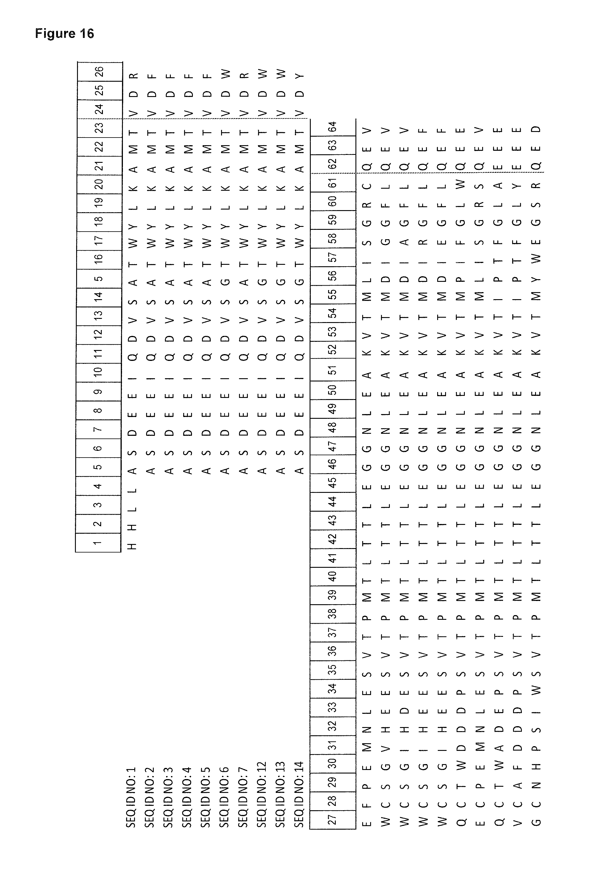
D00014
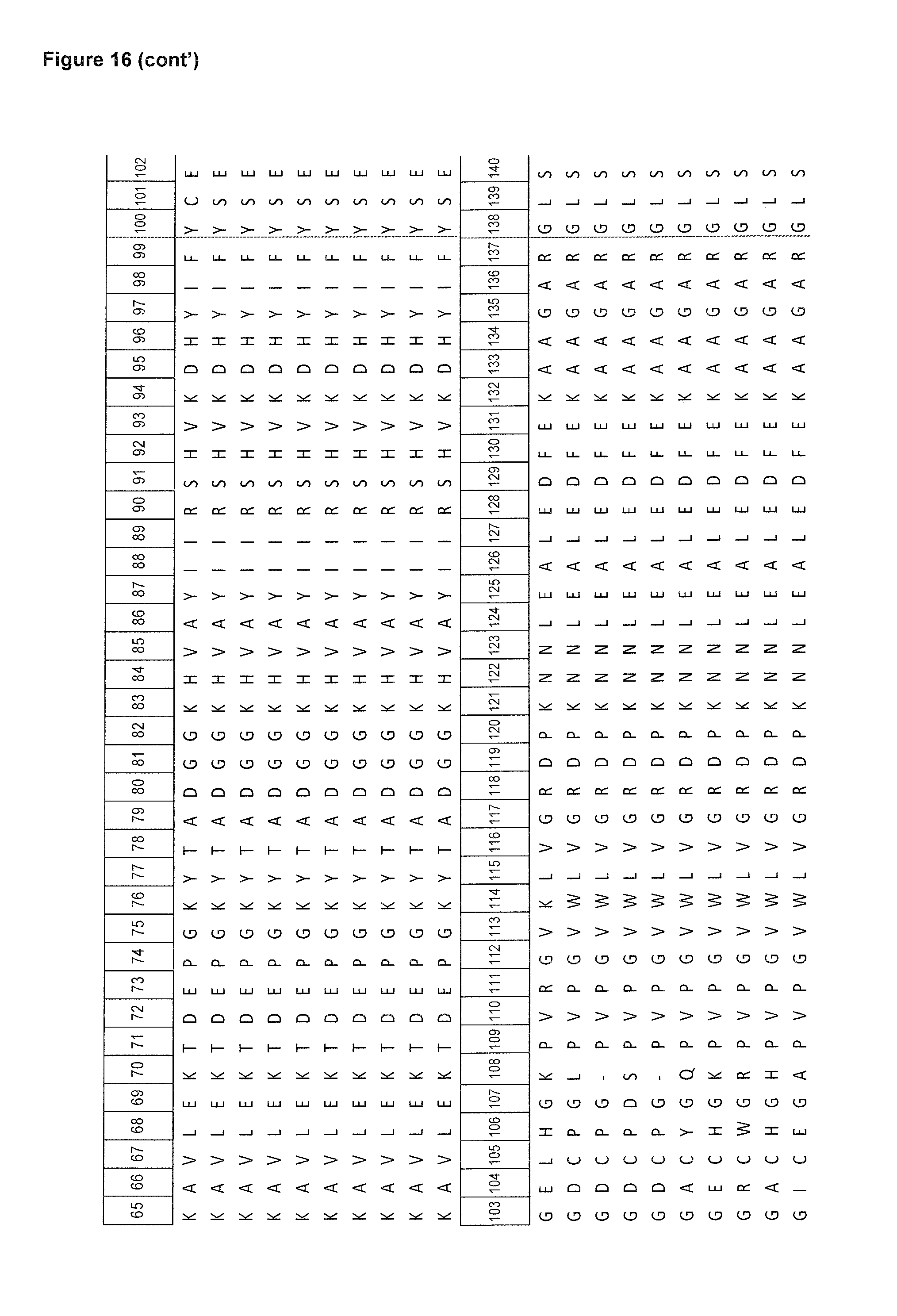
D00015
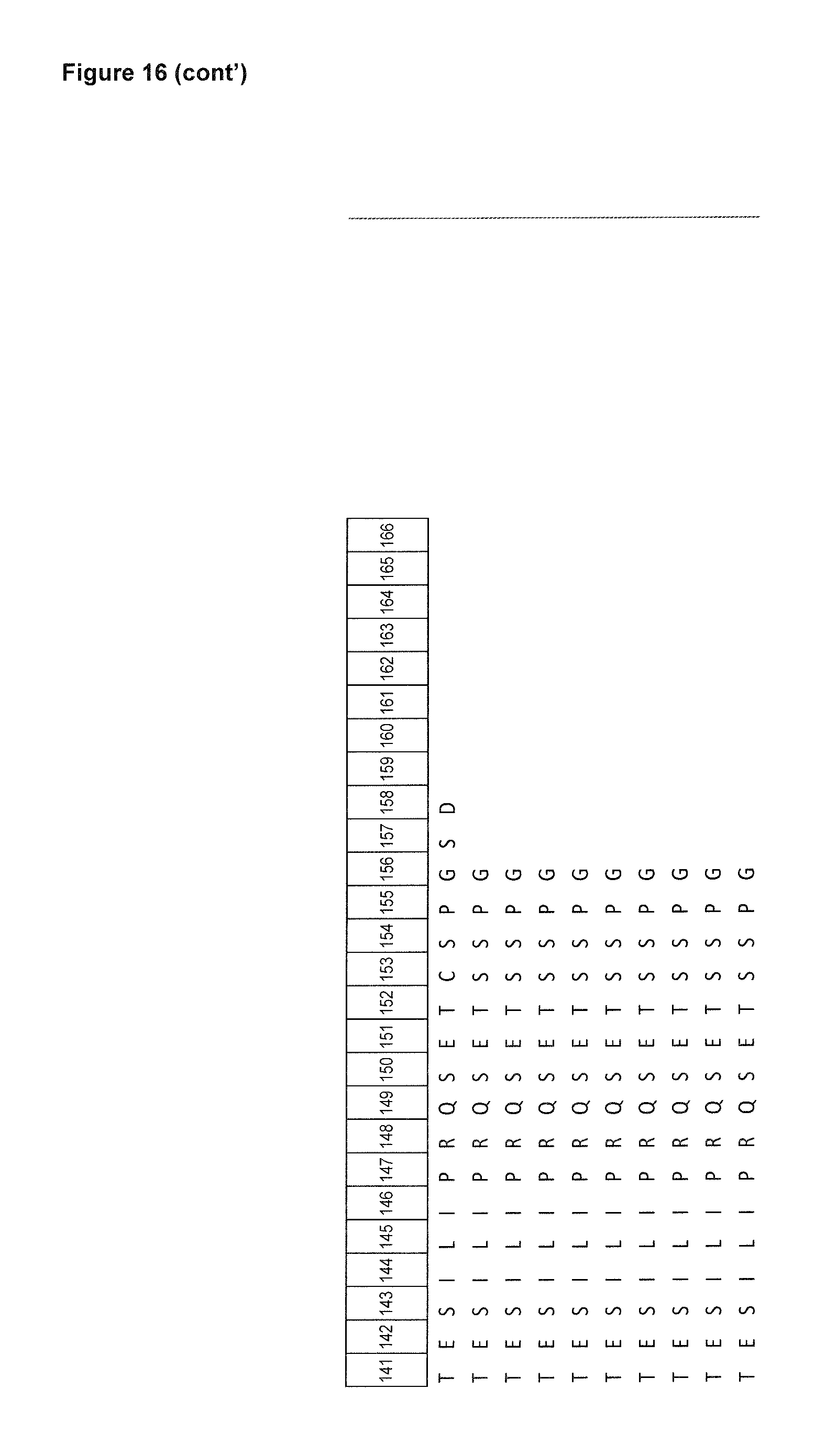
D00016

D00017

D00018

D00019
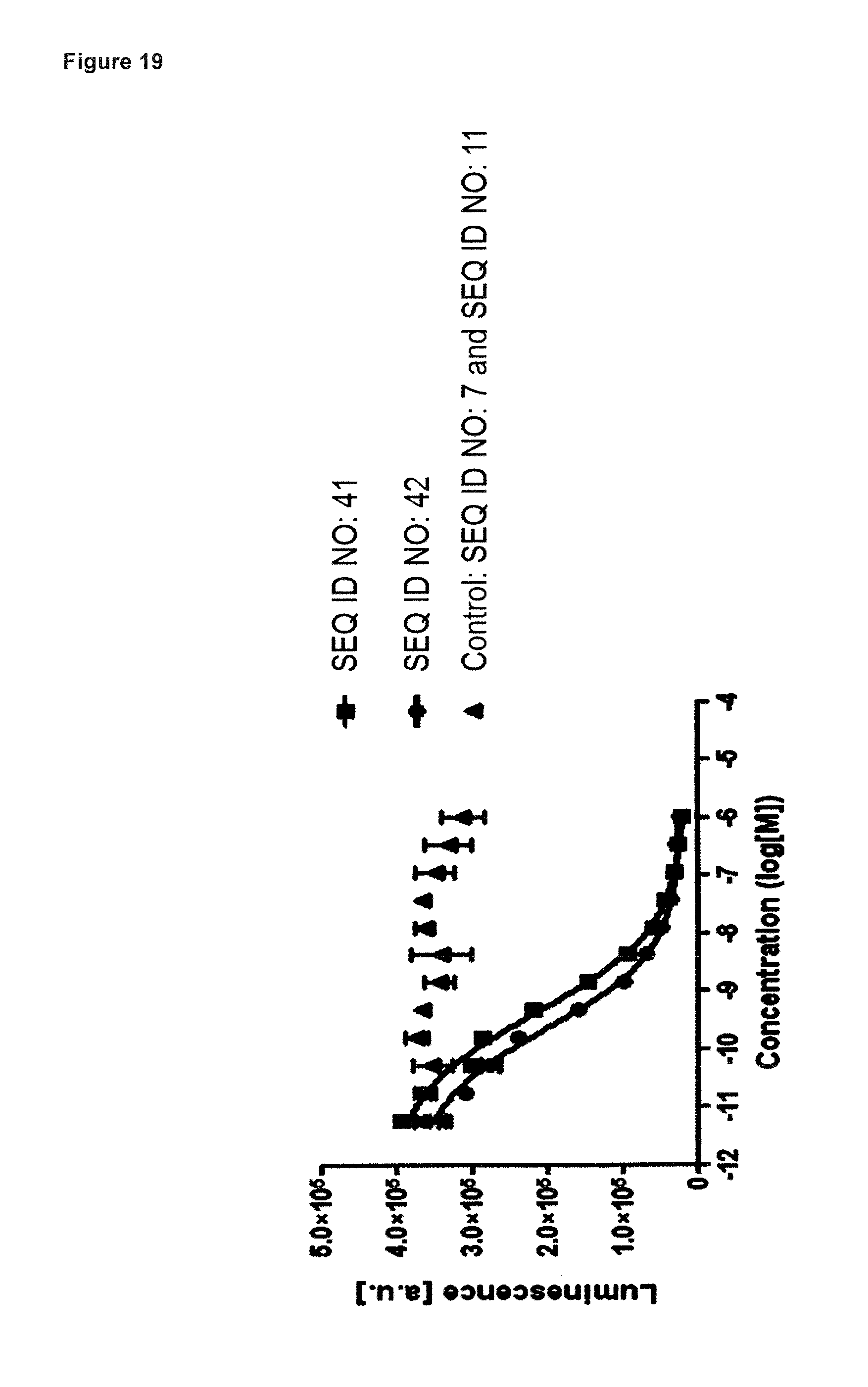
D00020

D00021

D00022

D00023

D00024

D00025

D00026
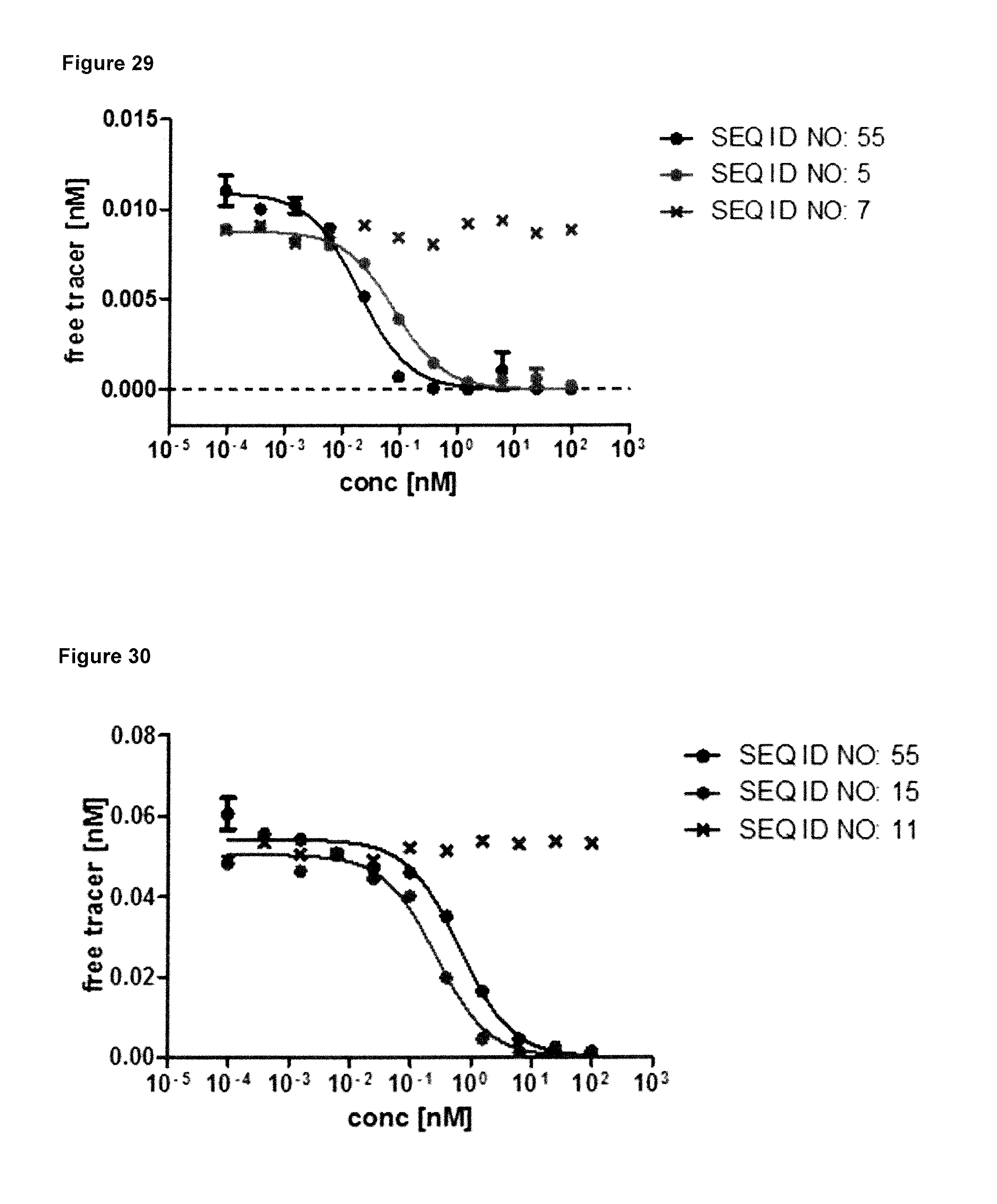
S00001
XML
uspto.report is an independent third-party trademark research tool that is not affiliated, endorsed, or sponsored by the United States Patent and Trademark Office (USPTO) or any other governmental organization. The information provided by uspto.report is based on publicly available data at the time of writing and is intended for informational purposes only.
While we strive to provide accurate and up-to-date information, we do not guarantee the accuracy, completeness, reliability, or suitability of the information displayed on this site. The use of this site is at your own risk. Any reliance you place on such information is therefore strictly at your own risk.
All official trademark data, including owner information, should be verified by visiting the official USPTO website at www.uspto.gov. This site is not intended to replace professional legal advice and should not be used as a substitute for consulting with a legal professional who is knowledgeable about trademark law.