Fusion proteins comprising P3 of bacteriophage
Krishnan , et al. J
U.S. patent number 10,526,377 [Application Number 15/975,328] was granted by the patent office on 2020-01-07 for fusion proteins comprising p3 of bacteriophage. This patent grant is currently assigned to PROCLARA BIOSCIENCES, INC.. The grantee listed for this patent is PROCLARA BIOSCIENCES, INC.. Invention is credited to Richard Fisher, Rajaraman Krishnan.


View All Diagrams
| United States Patent | 10,526,377 |
| Krishnan , et al. | January 7, 2020 |
Fusion proteins comprising P3 of bacteriophage
Abstract
The invention relates to agents and to pharmaceutical compositions for reducing the formation of amyloid and/or for promoting the disaggregation of an proteins. The compositions may also be used to detect amyloid.
| Inventors: | Krishnan; Rajaraman (Ashland, MA), Fisher; Richard (Cambridge, MA) | ||||||||||
|---|---|---|---|---|---|---|---|---|---|---|---|
| Applicant: |
|
||||||||||
| Assignee: | PROCLARA BIOSCIENCES, INC.
(Cambridge, MA) |
||||||||||
| Family ID: | 49354958 | ||||||||||
| Appl. No.: | 15/975,328 | ||||||||||
| Filed: | May 9, 2018 |
Prior Publication Data
| Document Identifier | Publication Date | |
|---|---|---|
| US 20180354994 A1 | Dec 13, 2018 | |
Related U.S. Patent Documents
| Application Number | Filing Date | Patent Number | Issue Date | ||
|---|---|---|---|---|---|
| 15587180 | May 4, 2017 | 10208090 | |||
| 14432861 | Jun 27, 2017 | 9688728 | |||
| PCT/US2013/062862 | Oct 1, 2013 | ||||
| 61828105 | May 28, 2013 | ||||
| 61801349 | Mar 15, 2013 | ||||
| 61730316 | Nov 27, 2012 | ||||
| 61708709 | Oct 2, 2012 | ||||
| Current U.S. Class: | 1/1 |
| Current CPC Class: | A61P 21/02 (20180101); A61P 25/16 (20180101); C07K 14/005 (20130101); A61P 43/00 (20180101); C07K 14/01 (20130101); A61P 25/00 (20180101); A61P 25/28 (20180101); A61P 35/00 (20180101); A61K 38/162 (20130101); A61P 25/14 (20180101); A61K 48/00 (20130101); C12N 2750/00033 (20130101); C12N 2795/14122 (20130101); C12N 2750/00022 (20130101); C07K 2319/30 (20130101); C07K 2319/00 (20130101); C12N 2795/14133 (20130101) |
| Current International Class: | C07K 14/005 (20060101); A61K 38/16 (20060101); A61K 48/00 (20060101); C07K 14/01 (20060101) |
References Cited [Referenced By]
U.S. Patent Documents
| 3941763 | March 1976 | Sarantakis |
| 4215051 | July 1980 | Schroeder et al. |
| 5252714 | October 1993 | Harris et al. |
| 5480981 | January 1996 | Goodwin et al. |
| 5808029 | September 1998 | Brockhaus et al. |
| 6686179 | February 2004 | Fleer et al. |
| 7208147 | April 2007 | Carr et al. |
| 7867487 | January 2011 | Solomon et al. |
| 8022270 | September 2011 | Dickey et al. |
| 9493515 | November 2016 | Krishnan et al. |
| 9493516 | November 2016 | Krishnan et al. |
| 9688728 | June 2017 | Krishnan et al. |
| 2002/0052311 | May 2002 | Solomon et al. |
| 2007/0269435 | November 2007 | Gillies et al. |
| 2009/0105090 | April 2009 | Uchiyama |
| 2009/0180991 | July 2009 | Solomon et al. |
| 2009/0304726 | December 2009 | Solomon et al. |
| 2009/0317324 | December 2009 | Solomon et al. |
| 2009/0324554 | December 2009 | Solomon et al. |
| 2010/0137420 | June 2010 | Nath |
| 2011/0142803 | June 2011 | Solomon et al. |
| 2011/0182948 | July 2011 | Solomon et al. |
| 2014/0335016 | November 2014 | Krishnan |
| 2016/0009766 | January 2016 | Krishnan et al. |
| 2016/0115223 | April 2016 | Fisher et al. |
| 2017/0115311 | April 2017 | Krishnan et al. |
| 0 154 316 | Sep 1989 | EP | |||
| 0 401 384 | Mar 1996 | EP | |||
| 2008-528688 | Jul 2008 | JP | |||
| WO 92/16221 | Oct 1992 | WO | |||
| WO 95/34326 | Dec 1995 | WO | |||
| WO 98/52976 | Nov 1998 | WO | |||
| WO 00/34317 | Jun 2000 | WO | |||
| WO 2002/074243 | Sep 2002 | WO | |||
| WO 2004/018685 | Mar 2004 | WO | |||
| WO 2006/083795 | Aug 2006 | WO | |||
| WO 2007/094003 | Aug 2007 | WO | |||
| WO 2008/011503 | Jan 2008 | WO | |||
| WO 2008/044032 | Apr 2008 | WO | |||
| WO 2009/143465 | Nov 2009 | WO | |||
| WO 2009/143470 | Nov 2009 | WO | |||
| WO 2010/060073 | May 2010 | WO | |||
| WO 2011/084714 | Jul 2011 | WO | |||
| WO 2012/125555 | Sep 2012 | WO | |||
| WO 2013/082114 | Jun 2013 | WO | |||
| WO 2014/193935 | Dec 2014 | WO | |||
| WO 2016/090022 | Jun 2016 | WO | |||
Other References
|
Aguib et al. (2009) "Autophagy induction by trehalose counteracts cellular prion infection" Autophagy, 5(3):361-369. cited by applicant . Aguzzi & O-Connor (2010) "Protein aggregation diseases: pathogenicity and therapeutic perspectives" Nature Reviews: Drug Discovery, 9:237-48. cited by applicant . Aruffo et al. (1990) "CD44 Is the Principal Cell Surface Receptor for Hyaluronate" Cell, 61:1303-13. cited by applicant . Ashkenazi et al. (1991) "Protection against endotoxic shock by a tumor necrosis factor receptor immunoadhesin" Proc. Natl. Acad. Sci. USA, 88:10535-39. cited by applicant . Beck et al. (1978) "Nucleotide sequence of bacteriophage fd DNA" Nucleic Acids Research, 5(12):4495-503. cited by applicant . Bennett et al. (1991) "Extracellular Domain-IgG Fusion Proteins for the Three Human Natriuretic Peptide Receptors" J. Biol. Chem. 266(34):23060-67. cited by applicant . Byrn et al. (Apr. 1990) "Biological properties of a CD4 immunoadhesin" Nature, 344:667-70. cited by applicant . Capon et al. (Feb. 1989) "Designing CD4 immunoadhesins for AIDS therapy" Nature, 337:525-31. cited by applicant . Cascales et al., (2007) "Colicin Biology" Microbiol. Mol. Biol. Rev., 71(1):158-229. cited by applicant . Chalupny et al. (1992) "T-cell activation molecule 4-1BB binds to extracellular matrix proteins" Proc. Natl. Acad. Sci. USA, 89:10360-64. cited by applicant . Chang and Kuret (2008) "Detection and Quantification of Tau Aggregation Using a Membrane Filter Assay" Anal. Biochem., 373(2):330-6. NIH Public Access Author Manuscript; available in PMC Feb. 15, 2009 (13 pages). cited by applicant . Chiti & Dobson (2006) "Protein Misfolding, Functional Amyloid, and Human Disease" Annu. Rev. Biochem., 75:333-66. cited by applicant . Coruzzi et al. (1984) "Tissue-specific and light-regulated expression of a pea nuclear gene encoding the small subunit of ribulose-1,5-bisphosphate carboxylase" EMBO J., 3:1671-79. cited by applicant . Darocha-Souto et al. (2011) "Brain Oligomeric .beta.-Amyloid but Not Total Amyloid Plaque Burden Correlates With Neuronal Loss and Astrocyte Inflammatory Response in Amyloid Precursor Protein/Tau Transgenic Mice" J. Neuropathol. Exp. Neurol., 70(5):360-76. NIH Public Access Author Manuscript; available in PMC Jul. 29, 2013 (26 pages). cited by applicant . Dehay et al. (2015) "Targeting .alpha.-synuclein for treatment of Parkinson's disease: mechanistic and therapeutic considerations" Lancet Neurol., 14:855-866. cited by applicant . Deng and Perham (2002) "Delineating the Site of Interaction on the pill Protein of Filamentous Bacteriophage fd with the F-pilus of Escherichia coli" J. Mol. Biol., 319:603-14. cited by applicant . Devlin et al. (1990) "Random Peptide Libraries: A Source of Specific Protein Binding Molecules" Science, 249:404-06. cited by applicant . Dimant et al. (2009) "Modulation effect of filamentous phage on alpha-synuclein aggregation" Biochem. Biophys. Res. Commun., 383(4):491-496. cited by applicant . Duyckaerts et al. (2008) "Alzheimer disease models and human neuropathology: similarities and differences" Acta Neuropathol., 115:5-38. cited by applicant . Eckert et al. (2007) "A Conformational Unfolding Reaction Activates Phage fd for the Infections of Escherichia coli" J. Mol. Biol., 373(2):452-461. cited by applicant . Eichner and Radford (2011) "A Diversity of Assembly Mechanisms of a Generic Amyloid Fold" Mol. Cell, 43:8-18. cited by applicant . Gascoigne et al. (1987) "Secretion of a chimeric T-cell receptor-immunoglobulin protein" Proc. Natl. Acad. Sci. USA, 84:2936-40. cited by applicant . Gentz et al. (1989) "Bioassay for trans-activation using purified human immunodeficiency virus tat-encoded protein: Trans-activation requires mRNA synthesis" Proc. Natl. Acad. Sci. USA, 86:821-24. cited by applicant . Gurley et al. (1986) "Upstream Sequences Required for Efficient Expression of a Soybean Heat Shock Gene" Mol. Cell. Biol., 6:559-65. cited by applicant . Heiseke et al. (2009) "Lithium induces clearance of protease resistant prion protein in prion-infected cells by induction of autophagy" J. Neurochem., 109:25-34. cited by applicant . Hill and Petersen (1982) "Nucleotide sequence of bacteriophage f1 DNA" J. Virol., 44(1):32-46. cited by applicant . Hoffmann-Thoms et al. (May 2013) "Initiation of Phage Infection by Partial Unfolding and Prolyl Isomerization" J. Biol. Chem., 288(18):12979-91. cited by applicant . Holliger et al. (1999) "Crystal Structure of the Two N-terminal Domains of g3p from Filamentous Phage fd at 1.9 .ANG. Evidence for Conformational Lability" J. Mol. Biol., 288(4):649-57. cited by applicant . Hsiao et al. (1996) "Correlative Memory Deficits, A.beta. Elevation, and Amyloid Plaques in Transgenic Mice" Science, 274:99-102. cited by applicant . Hughes (2004) "The value of spontaneous alternation behavior (SAB) as a test of retention in pharmacological investigations of memory" Neurosci. Biobehav. Rev., 28:497-505. cited by applicant . International Patent Application No. PCT/US2012/066793, filed Nov. 28, 2012, by Neurophage Pharmaceuticals, Inc.: International Preliminary Report on Patentability, dated Feb. 6, 2014. cited by applicant . International Patent Application No. PCT/US2012/066793, filed Nov. 28, 2012, by Neurophage Pharmaceuticals, Inc.: International Search Report and Written Opinion, dated Apr. 19, 2013. cited by applicant . International Patent Application No. PCT/US2013/062862, filed Oct. 1, 2013, by Neurophage Pharmaceuticals, Inc.: International Search Report and Written Opinion, dated Feb. 24, 2014. cited by applicant . International Patent Application No. PCT/US2014/039760, filed May 28, 2014, by Neurophage Pharmaceuticals, Inc.: International Search Report and Written Opinion, dated Nov. 3, 2014. cited by applicant . International Patent Application No. PCT/US2014/039760, filed May 28, 2014, by Neurophage Pharmaceuticals, Inc.: Written Opinion of the International Preliminary Examining Authority, dated May 15, 2015. cited by applicant . International Patent Application No. PCT/US2014/039760, filed May 28, 2014, by Neurophage Pharmaceuticals, Inc.: International Preliminary Report on Patentability, dated Aug. 14, 2015. cited by applicant . International Patent Application No. PCT/US2015/063476, filed Dec. 2, 2015, by Neurophage Pharmaceuticals, Inc.: International Search Report and Written Opinion, dated May 3, 3016. cited by applicant . Josephs et al. (2011) "Neuropathological background of phenotypical variability in frontotemporal dementia" Acta Neuropathol., 122:137-53. cited by applicant . Kather et al. (2005) "A Stable Disulfide-free Gene-3-protein of Phage fd Generated by In vitro Evolution" J. Mol. Biol., 354(3):666-678. cited by applicant . Kerr et al (2001) "Lysostaphin expression in mammary glands confers protection against staphylococcal infection in transgenic mice" Nature Biotechnol., 19(1):66-70. cited by applicant . King et al. (1999) "Progressive and gender-dependent cognitive impairment in the APP.sub.SW transgenic mouse model for Alzheimer's disease" Brain Res., 103:145-62. cited by applicant . Kingstedt and Nilsson (2012) "Luminescent conjugated poly- and oligo-thiophenes: optical ligands for spectral assignment of a plethora of protein aggregates" Biochem. Soc. Trans., 40(4):704-710. cited by applicant . Kong, B. et al. (2006) "Display of aggregation-prone ligand binding domain of human PPAR gamma on surface of bacteriophage lambda" Acta Pharmacologica Sinica, 27(1):91-99. cited by applicant . Kosik et al. (1986) "Microtubule-associated protein tau (tau) is a major antigenic component of paired helical filaments in Alzheimer disease" Proc. Natl. Acad. Sci. USA, 83(11):4044-48. cited by applicant . Krishnan et al. (2014) "A Bacteriophage Capsid Protein Provides a General Amyloid Interaction Motif (GAIM) That Binds and Remodels Misfolded Protein Assemblies" J. Mol. Biol., 426:2500-19. cited by applicant . Kurschner et al. (1992) "Construction, Purification, and Characterization of New Interferon .gamma. (IFN.gamma.) Inhibitor Proteins" J. Biol. Chem., 267:9354-60. cited by applicant . Lalond et al. (2012) "Neurologic and motor dysfunctions in APP transgenic mice" Rev. Neurosci., 23(4):36379. NIH Public Access Author Manuscript; available in PMC Jan. 1, 2013 (25 pages). cited by applicant . Lalonde & Strazielle (2012) "Brain regions and genes affecting myoclonus in animals" Neurosci. Res., 74(2):69-79. cited by applicant . Lee et al. (2001) "Neurodegenerative Tauopathies" Annu. Rev. Neurosci., 24:1121-59. cited by applicant . Lesslauer et al. (1991) "Recombinant soluble tumor necrosis factor receptor proteins protect mice from lipopolysaccharide-induced lethality" Eur. J. Immunol., 21(11):2883-86. cited by applicant . Lewis et al. (2000) "Neurofibrillary tangles, amyotrophy and progressive motor disturbance in mice expressing mutant (P301L) tau protein" Nat. Genet., 25:402-5. cited by applicant . Li et al. (2015) "Trehalose Decreases Mutant SOD1 Expression and Alleviates Motor Deficiency in Early But Not End-Stage Amyotrophic Lateral Sclerosis in a SOD1-G93A Mouse Model" Neurosci., 298:12-25. cited by applicant . Lin et al. (2011) "Inhibition of Bacterial Conjugation by Phage M13 and Its Protein g3p: Quantitative Analysis and Model" PLoS ONE,6(5):e19991. doi:10.1371/journal.pone.0019991 (11 pages). cited by applicant . Linsley et al. (1991) "Binding of the B cell activation antigen B7 to CD28 costimulates T cell proliferation and interleukin 2 mRNA accumulation" J. Exp. Med., 173:721-30. cited by applicant . Linsley et al. (1991) "CTLA-4 Is a Second Receptor for the B Cell Activation Antigen B7" J. Exp. Med., 174:561-69. cited by applicant . Liu et al. (2005) "Trehalose differentially inhibits aggregation and neurotoxicity of beta-amyloid 40 and 42" Neurobiol. Dis., 20:74-81. cited by applicant . Lo et al. (1998) "High level expression and secretion of Fc-X fusion proteins in mammalian cells" Protein Engineering, 11(6):495-500. cited by applicant . Logan and Shenk (1984) "Adenovirus tripartite leader sequence enhances translation of mRNAs late after infection" Proc. Natl. Acad. Sci. USA, 81:3655-59. cited by applicant . Lorenz et al. (2011) "The Filamentous Phages fd and IF1 Use Different Mechanisms to Infect Escherichia coli" J. Mol. Biol., 405:989-1003. cited by applicant . Lou et al. (2012) "Probing of C-terminal lysine variation in a recombinant monoclonal antibody production using Chinese hamster ovary cells with chemically defined media" Biotechnol. Bioeng., 109(9):2306-15. cited by applicant . Lubkowski et al. (1998) "Filamentous phage infection: crystal structure of g3p in complex with its coreceptor, the C-terminal domain of TolA" Structure, 7(6):711-22. cited by applicant . Mackett et al. (1982) "Vaccinia virus: A selectable eukaryotic cloning and expression vector" Proc. Natl. Acad. Sci. USA, 79:7415-19. cited by applicant . Mackett et al. (1984) "General Method for Production and Selection of Infectious Vaccinia Virus Recombinants Expressing Foreign Genes" J. Virol., 49:857-64. cited by applicant . Martin and Schmid (2003) "Evolutionary Stabilization of the Gene-3-protein of Phage fd Reveals the Principles that Govern the Thermodynamic Stability of Two-domain Proteins" J. Mol. Biol., 328:863-75. cited by applicant . Marvin (1998) "Filamentous phage structure, infection and assembly" Curr. Opin. in Struct. Biol., 8:150-8. cited by applicant . Masliah et al. (2000) "Dopaminergic Loss and Inclusion Body Formation in .alpha.-Synuclein Mice: Implications for Neurodegenerative Disorders" Science, 287:1265-69. cited by applicant . McKhann et al. (2011) "The diagnosis of dementia due to Alzheimer's disease: Recommendations from the National Institute on Aging and the Alzheimer's Association workgroup" [Article in Press] Alzheimer's & Dementia, doi:10.1016/j.jalz.2011.03.005, 7 pages; final publication in 7(3):263-9. cited by applicant . Mega et al. (1996) "The spectrum of behavioral changes in Alzheimer's disease" Neurology, 46:130-5. cited by applicant . Messing and Ayer, "Enterobacteria phage M13 isolate WT variety Rutgers, complete genome" GenBank Database Accession No. JX412914, Version GI:401823911; submitted Jul. 20, 2012. cited by applicant . Muir, E.M. et al. (2010) "Modification of N-glycosylation sites allows secretion of bacterial chondroitinase ABC from mammalian cells" J. Biotechnol., 145(2):103-110. cited by applicant . Olofsson et al. (2006) "The Solvent Protection of Alzheimer Amyloid-.beta.-(1-42) Fibrils as Determined by Solution NMR Spectroscopy" J. Biol. Chem., 281(1):477-83. cited by applicant . Panicali et al. (1982) "Construction of poxviruses as cloning vectors: Insertion of the thymidine kinase gene from herpes simplex virus into the DNA of infectious vaccinia virus" Proc. Natl. Acad. Sci. USA, 79:4927-31. cited by applicant . Pankiewicz et al. (2006) "Clearance and prevention of prion infection in cell culture by anti-PrP antibodies" NIH Public Access Author Manuscript, available in PMC Jan. 22, 2007. Final publication in: Eur. J. Neurosci., 23:2635-47. cited by applicant . Peppel et al. (1991) "A tumor necrosis factor (TNF) receptor-IgG heavy chain chimeric protein as a bivalent antagonist of TNF activity" J. Exp. Med., 174:1483-89. cited by applicant . Perrier et al. (2004) "Anti-Prp antibodies block PrP.sup.Sc replication in prion-infected cell cultures by accelerating PrP.sup.c degradation" J. Neurochem., 84:454-63. cited by applicant . Petkova et al. (2005) "Self-propagating, molecular-level polymorphism in Alzheimer's .beta.-amyloid fibrils" Science, 307:262-65. cited by applicant . Rasched and Oberer (1986) "Ff Coliphages: Structural and Functional Relationships" Microbiol. Rev., 50:401-27. cited by applicant . REFSEQ database Accession No. NC_003287.2, version GI:56718463, "Enterobacteria phage M13, complete genome" circular PHG Apr. 17, 2009 (7 pages). cited by applicant . Resnick and Sojkova (2011) "Amyloid imaging and memory change for prediction of cognitive impairment" Alzheimer's Res Ther., 3:3, doi:10.1186/alzrt62 [online]. Retrieved from: http://alzres.com/content/3/1/3. cited by applicant . Robinson et al. (2015) "Drugs and drug delivery systems targeting amyloid-.beta. in Alzheimer's disease" Mol. Sci., 2(3):332-358. cited by applicant . Sadowski et al. (2009) "Anti-PrP Mab 6D11 suppresses PrP.sup.Sc replication in prion infected myeloid precursor line FDC-P1/22L and in the lymphoreticular system in vivo" NIH Public Access Author Manuscript, available Jul. 20, 2009. Final publication in: Neurobiol Dis., 34(2): 267-78. cited by applicant . Sanchez et al. (2011) "A.beta.40 and A.beta.42 Amyloid Fibrils Exhibit Distinct Molecular Recycling Properties" J. Am. Chem. Soc., 133:6505-08. cited by applicant . Sarkar et al. (2005) "Lithium induces autophagy by inhibiting inositol monophosphatase" J. Cell Biol., 170(7):1101-11. cited by applicant . Sarkar et al. (2007) "Trehalose, a Novel mTOR-independent Autophagy Enhancer, Accelerates the Clearance of Mutant Huntingtin and -.alpha.-Synuclein" J. Biol. Chem., 282(8):5641-52. cited by applicant . Sato et al. (2006) "Inhibitors of Amyloid Toxicity Based on .beta.-sheet Packing of A.beta.40 and A.beta.42" Biochemistry, 45:5503-16. cited by applicant . Sciarretta et al. (2006) "Peptide-Based Inhibitors of Amyloid Assembly" Meth. Enzymol., 413:273-312. cited by applicant . Scott and Smith (1990) "Searching for Peptide Ligands with an Epitope Library" Science, 249:386-90. cited by applicant . Simonsen and Levinson (1983) "Isolation and expression of an altered mouse dihydrofolate reductase cDNA" Proc. Natl. Acad. Sci. USA, 80:2495-99. cited by applicant . Smith et al. (1983) "Molecular Engineering of the Autographa californica Nuclear Polyhedrosis Virus Genome: Deletion Mutations Within the Polyhedrin Gene" J. Virol., 46:584-93. cited by applicant . Smith et al. (1997) "Phage display" Chem. Rev., 97:391-410. cited by applicant . Stamenkovic et al. (1991) "The B Lymphocyte Adhesion Molecule CD22 Interacts with Leukocyte Common Antigen CD45RO on T Cells and .alpha.2-6 Sialyltransferase, CD75, on B Cells" Cell, 66:1133-44. cited by applicant . Stassen et al. (1992) "Nucleotide Sequence of the Genome of the Filamentous Bacteriophage I2-2: Module Evolution of the Filamentous Phage Genome" J. Mol. Evol., 34:141-52. cited by applicant . Sterniczuk et al. (2010) "Characterization of the 3xTg-AD mouse model of Alzheimer's disease: Part 1. Circadian changes" Brain Res., 1348:139-48. cited by applicant . Sterniczuk et al. (2010) "Characterization of the 3xTg-AD mouse model of Alzheimer's disease: Part 2. Behavioral and cognitive changes" Brain Res., 1348:149-55. cited by applicant . Stine et al. (2003) "In Vitro Characterization of Conditions for Amyloid-.beta. Peptide Oligomerization and Fibrillogenesis" J. Biol. Chem., 278(13):11612-22. cited by applicant . Stine et al. (2011) "Preparing synthetic A.beta. in different aggregation states" HHS Public Access Author Manuscript, available Aug. 26, 2013, PMCID: PMC3752843. Final publication in: Methods Mol. Biol., 670: 13-32. cited by applicant . Sunde et al. (1997) "Common Core Structure of Amyloid Fibrils by Synchrotron X-ray Diffraction" J. Mol. Biol., 273:729-39. cited by applicant . Takamatsu et al. (1987) "Expression of bacterial chloramphenicol acetyltransferase gene in tobacco plants mediated by TMV-RNA" EMBO J., 6:307-11. cited by applicant . Terpe (2003) "Overview of tagg protein fusions: from molecular and biochemical fundamentals to commercial systems" Appl. Microbiol. Biotechnol., 60:523-33. cited by applicant . Tjernberg et al. (1996) "Arrest of .beta.-Amyloid Fibril Formation by a Pentapeptide Ligand" J. Biol. Chem., 271(12):8545-48. cited by applicant . Traunecker et al. (May 1989) "Highly efficient neutralization of HIV with recombinant CD4-immunoglobulin molecules" Nature, 339:68-70. cited by applicant . Uniprot Accession No. O80297 (Entry date: Jul. 15, 1999) "Protein: Attachment protein G3P. Organism: Enterobacteria phage lf1 (Bacteriophage lf1)." [online]. Retrieved from the Internet: httn://www.uninrot.org/uninrot/O80297. cited by applicant . Uniprot Accession No. P03661 (Entry date: Jul. 21, 1986) "Protein: Attachment protein G3P. Organism: Enterobacteria phage fd (Bacteriophage fd)." [online]. Retrieved from the Internet: http://www.uniprot.org/uniprot/P03661. cited by applicant . Uniprot Accession No. P03663 (Entry date: Jul. 21, 1986) "Protein: Attachment protein G3P. Organism: Enterobacteria phage IKe (Bacteriophage IKe)." [online]. Retrieved from the Internet: http://www.uniprot.org/uniprot/P03663. cited by applicant . Uniprot Accession No. P15415 (Entry date: Apr. 1, 1990) "Protein: Attachment protein G3P. Organism: Enterobacteria phage I2-2 (Bacteriophage I2-2)." [online]. Retrieved from the Internet: http://www.uniprot.org/uniprot/P15415. cited by applicant . Uniprot Accession No. P69168 (Entry date: Jul. 21, 1986) "Protein: Attachment protein G3P. Organism: Enterobacteria phage M13 (Bacteriophage M13)." [online]. Retrieved from the Internet: . http://www.uniprot.org/uniprot/P69168. cited by applicant . Uniprot Accession No. P69169 (Entry date: Jul. 21, 1986) "Protein: Attachment protein G3P. Organism: Enterobacteria phage f1 (Bacteriophage fl)." [online]. Retrieved from the Internet: http://www.uniprot.org/uniprot/P69169. cited by applicant . Van Wezenbeek et al. (1980) "Nucleotide sequence of the filamentous bacteriophage M13 DNA genome: comparison with phage fd" Gene, 11:129-48. cited by applicant . Van Wezenbeek et al., "Structural protein [Enterobacteria phage M13]" NCBI Protein Sequence Database Accession No. NP_510891.1, Version GI:17426224; submitted Dec. 8, 2001. cited by applicant . Wang et al. (2010) "Degradation of TDP-43 and its pathogenic form by autophagy and the ubiquitin-proteasome system" Neurosci. Lett., 469:112-116. cited by applicant . Wang et al. (2010) "Generating a Prion with Bacterially Expressed Recombinant Prion Protein" Science, 327:1132-35. cited by applicant . Wanker et al. (1999) "Membrane Filter Assay for Detection of Amyloid-like Polyglutamine-Containing Protein Aggregates" Methods Enzymol., 309:375-86. cited by applicant . Watson et al. (1990) "A homing receptor-IgG chimera as a probe for adhesive ligands of lymph node high endothelial venules" J. Cell. Biol., 110:2221-29. cited by applicant . Watson et al. (Jan. 1991) "Neutrophil influx into an inflammatory site inhibited by a soluble homing receptor-IgG chimaera" Nature, 349:164-67. cited by applicant . Whittemore et al. (2005) "Hydrogen-Deuterium (H/D) Exchange Mapping of A.beta..sub.1-40 Amyloid Fibril Secondary Structure Using Nuclear Magnetic Resonance Spectroscopy" Biochemistry, 44:4434-41. cited by applicant . Wilcock et al. (2004) "Passive Amyloid Immunotherapy Clears Amyloid and Transiently Activates Microglia in a Transgenic Mouse Model of Amyloid Deposition" J. Neurosci., 24(27):6144-51. cited by applicant . Yamaguchi et al. (2004) "Core and Heterogeneity of .beta.2-Microglobulin Amyloid Fibrils as Revealed by H/D Exchange" J. Mol. Biol., 338:559-71. cited by applicant . Zettmeissl et al. (1990) "Expression and characterization of human CD4: Immunoglobulin fusion proteins" DNA Cell Biol., 9(5):347-53. cited by applicant . Zhao et al. (2012) "Tagged and untagged TRAIL show different activity against tumor cells" Oncol. Lett., 4:1301-4. cited by applicant . Zheng et al. (1995) "Administration of noncytolytic IL-10/Fc in murine models of lipopolysaccharide-induced septic shock and allogeneic islet transplantation" J. Immunol., 154:5590-5600. cited by applicant. |
Primary Examiner: Emch; Gregory S
Attorney, Agent or Firm: Finnegan, Henderson, Farabow, Garrett & Dunner, LLP
Parent Case Text
CROSS REFERENCE TO RELATED APPLICATIONS
This application is a continuation of application Ser. No. 15/587,180, filed May 4, 2017, which is a divisional of application Ser. No. 14/432,861, filed Apr. 1, 2015, which is a national-stage filing of International Application No. PCT/US2013/062862, filed Oct. 1, 2013, which claims the benefit of priority of U.S. Provisional Application No. 61/828,105, filed May 28, 2013, U.S. Provisional Application No. 61/801,349, filed Mar. 15, 2013, U.S. Provisional Application No. 61/730,316, filed Nov. 27, 2012, and U.S. Provisional Application No. 61/708,709, filed Oct. 2, 2012, all of which are incorporated herein by reference.
Claims
What is claimed is:
1. A method of treating a patient for a disease or condition by administering to the patient a fusion protein comprising an amyloid binding fragment of g3p and an Fc fragment of an immunoglobulin, wherein the fusion protein comprises: (a) amino acids 21-506 of SEQ ID NO:9; (b) amino acids 22-506 of SEQ ID NO:9; (c) amino acids 23-506 of SEQ ID NO:9; (d) amino acids 21-505 of SEQ ID NO:9; (e) amino acids 22-505 of SEQ ID NO:9; (f) amino acids 23-505 of SEQ ID NO:9; (g) amino acids 21-506 of SEQ ID NO:11; (h) amino acids 22-506 of SEQ ID NO:11; (i) amino acids 23-506 of SEQ ID NO:11; (j) amino acids 21-505 of SEQ ID NO:11; (k) amino acids 22-505 of SEQ ID NO:11; (l) amino acids 23-505 of SEQ ID NO:11; (m) amino acids 21-509 of SEQ ID NO:13; (n) amino acids 22-509 of SEQ ID NO:13; (o) amino acids 23-509 of SEQ ID NO:13; (p) amino acids 21-508 of SEQ ID NO:13; (q) amino acids 22-508 of SEQ ID NO:13; (r) amino acids 23-508 of SEQ ID NO:13; (s) amino acids 21-528 of SEQ ID NO:31; (t) amino acids 22-528 of SEQ ID NO:31; (u) amino acids 23-528 of SEQ ID NO:31; (v) amino acids 21-527 of SEQ ID NO:31; (w) amino acids 22-527 of SEQ ID NO:31; (x) amino acids 23-527 of SEQ ID NO:31; or (y) a mutant or variant that is at least 95% identical to the amino acid sequence of any one of (a)-(x) and is capable of binding to amyloid; and wherein the disease or condition is selected from peripheral amyloidosis, familial amyloidotic polyneuropathy (FAP), Finnish form of FAP (aggregation of gelsolin), familial amyloidotic cardiomyopathy (FAC), senile systemic amyloidosis (SSA), islet amyloid polypeptide (IAPP) amyloidosis, and disease characterized by formation of amyloid protein by aggregation of IgG light chain.
2. The method of claim 1, wherein the mutant or variant of (y) has no more than 10 amino acid differences as compared to the amino acid sequence of any one of (a)-(x).
3. The method of claim 1, wherein the mutant or variant of (y) has up to 5 amino acid substitutions as compared to the amino acid sequence of any one of (a)-(x).
4. The method of claim 1, wherein the fusion protein comprises amino acids 21-509 of SEQ ID NO:13 or a mutant or variant thereof having up to 5 amino acid substitutions.
5. The method of claim 1, wherein the fusion protein comprises amino acids 22-509 of SEQ ID NO:13 or a mutant or variant thereof having up to 5 amino acid substitutions.
6. The method of claim 1, wherein the fusion protein comprises amino acids 23-509 of SEQ ID NO:13 or a mutant or variant thereof having up to 5 amino acid substitutions.
7. The method of claim 1, wherein the fusion protein comprises amino acids 21-508 of SEQ ID NO:13 or a mutant or variant thereof having up to 5 amino acid substitutions.
8. The method of claim 1, wherein the fusion protein comprises amino acids 22-508 of SEQ ID NO:13 or a mutant or variant thereof having up to 5 amino acid substitutions.
9. The method of claim 1, wherein the fusion protein comprises amino acids 23-508 of SEQ ID NO:13 or a mutant or variant thereof having up to 5 amino acid substitutions.
10. The method of claim 1, wherein the disease or condition is selected from the group consisting of FAP, Finnish form of FAP, and FAC.
11. The method of claim 1, wherein the disease or condition is SSA or peripheral amyloidosis.
12. The method of claim 1, wherein the disease or condition is IAPP amyloidosis.
13. The method of claim 1, wherein the disease or condition is a disease characterized by formation of amyloid protein by aggregation of IgG light chain.
Description
The invention relates to fusion proteins comprising an amyloid binding fragment of filamentous bacteriophage g3p protein or mutant or variant forms of such amyloid binding fragment. Nucleic acid molecules and constructs encoding such fusion proteins, cells transformed with such nucleic acid molecules, and methods of making such fusion proteins recombinantly are encompassed. In addition, the invention relates to pharmaceutical compositions comprising the fusion proteins disclosed herein, and to the use of such compositions therapeutically and prophylactically to decrease amyloid load associated with diseases, such as systemic and peripheral amyloid diseases, neurodegenerative diseases including neurodegenerative tauopathies, and transmissible spongiform encephalopathies (prion-associated diseases). Also encompassed is the use of those compositions to prevent the accumulation of amyloid load associated with these diseases, and the use of those compositions as diagnostics to detect amyloid and thus, diagnose such diseases.
Filamentous bacteriophage M13, and related filamentous phage, have shown utility in animal models of protein misfolding disease, and therefore represent a potential therapeutic class for protein misfolding diseases. See United States patent publication US 2011/0142803, incorporated by reference herein in its entirety. In particular, it has been discovered that filamentous bacteriophage have the ability to mediate clearance of amyloid that have already formed in the brain. See, e.g., WO2006083795 and WO2010060073, incorporated by reference herein in their entirety.
M13 phage, a member of the Ff family of phages, has a mature g3p of 406 amino acids. GenBank Ref Seq NP_510891.1 provides a reference sequence that includes the 18 residue amino-terminal signal sequence. Variants that have amino acid differences as compared to published sequences are common. Filamentous phage of the I-family have g3p that differs from Ff family members, but even between families g3p is still highly conserved. Stassen et al., J Mol Evol (1992) 34:141-52.
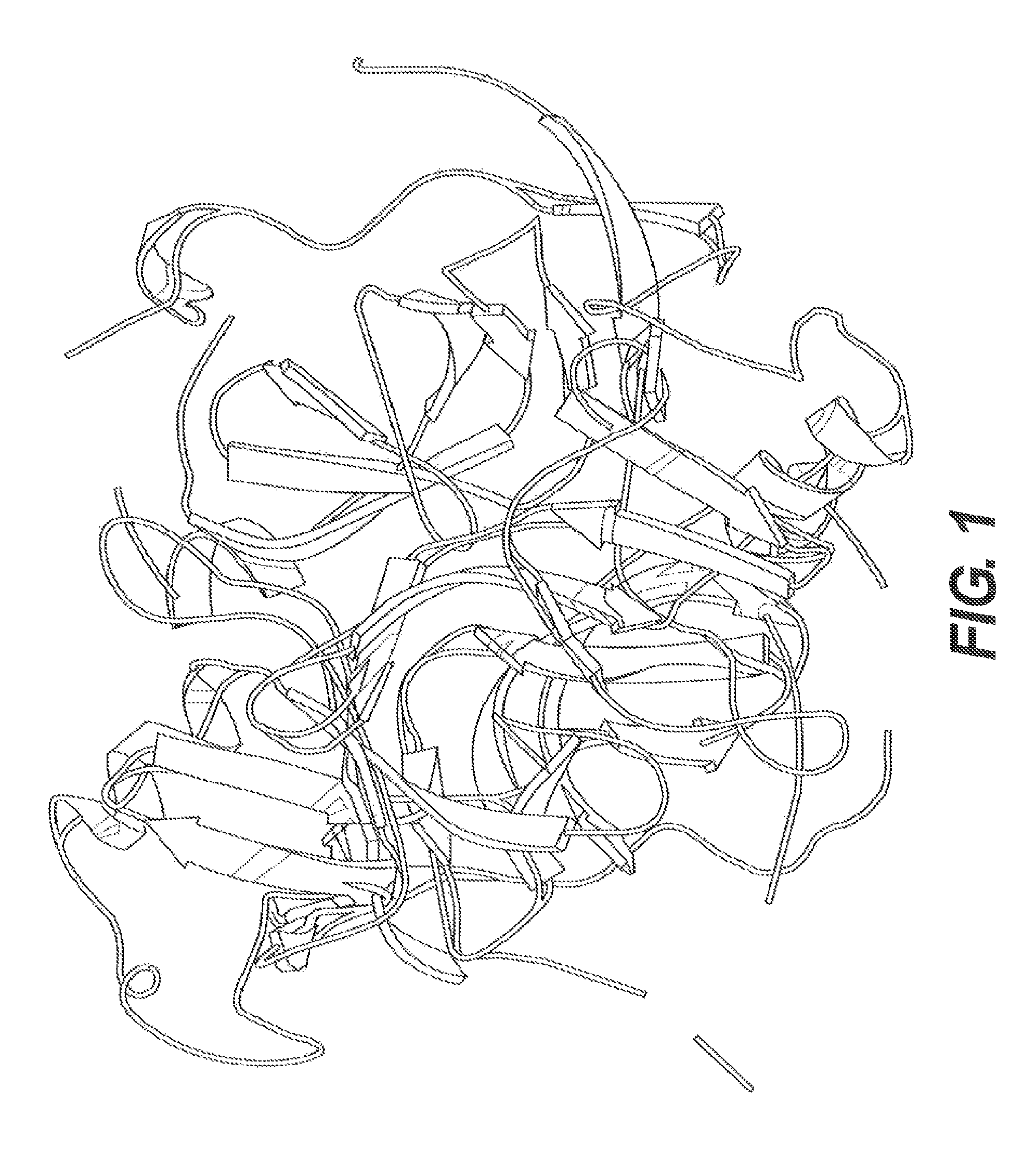
A crystal structure is available for g3p. Lubkowski et al., Structure (1998) 7(6) 711-722. The protein comprises 3 folded domains separated by flexible glycine-rich linker sequences. There are two amino-terminal domains, N1 and N2 comprising 262 amino acids, that interact to form an N1-N2 complex. The carboxy-terminal (CT, also called N3) domain is 146 amino acids and it serves to anchor g3p in the phage particle by hydrophobic interactions with g8p. Marvin, Current Opin. in Structural Biology (1998) 8:150-158. A publically available ribbon structure prepared using the N1-N2 domain fusion protein 2g3p of Holliger, J Mol. Biol. (1999) 288(4):649-57 is presented in FIG. 1.
Unlike most proteins, unfolding of the N1 and N2 domains from the latent "locked" form is required for g3p to acquire its native biological activity. Eckert & Schmid, J. Mol. Biol. (2007) 373:452-461. In the initial step in infection, N2 binds the bacterial F-pilus via residues on the outer rim of N2. Deng & Perham, 2002. This initial binding by N2 "unlocks" g3p by "opening" the N1-N2 complex, permitting N1 to then bind the co-receptor TolA. In an N1-N2 fragment of g3p, the thermal transition for the initial unlocking step in which N2 unfolds occurs at a melting temperature (T.sub.M) of 48.1.degree. C. Part of the process involves an isomerization at the Gln212-Pro213 peptide bond. Pro213 converting is trans in the unlocked state. N1 remains stably folded until the second step, which occurs at a T.sub.M of 60.2.degree. C. Reviewed in Eckert & Schmid, 2007.
Mutations in the N1-N2 fragment have been used to study the stability and infectivity of various mutants. Eckert & Schmid, 2007. One variant, designated "3A" impaired pilus binding and decreased the stability of the N2 domain. For this mutation, the T.sub.M is decreased to 42.6.degree. C. 3A carries the following mutations: W181A, F190A, and F194A. Another mutant in N2, G153D, destabilized N2, decreasing T.sub.M to 44.4.degree. C. A Q129H mutant stabilized N2, increasing the T.sub.M to 51.4.degree. C. The IY variant contains the mutations T101I and D209Y in the hinge and increases the stability of the N1-N2 fragment (T.sub.M=56.5.degree. C.). IHY contains the mutations T101I, Q129H, and D209Y (T.sub.M=60.1.degree. C.). IIHY contains the mutations T13I, T101I, Q129H, and D209Y (T.sub.M=61.8.degree. C.). Both the Q129Y and T13I mutations are stabilizing, and adding these mutations further increases the melting temperature, T.sub.M. Phage infectivity varied inversely with the strength of the domain interactions within g3p. Eckert & Schmid, 2007. Deletion of the N2 domain (phage fd(.DELTA.N2)) increased the infectivity by removing the blocking effect of the N2 domain on N1-binding of TolA. Id.
Recently, it was discovered that g3p also mediates binding of the filamentous phage to amyloid in a manner analogous to the process by which phage infect bacteria. WO 2013/082114 discloses that phage g3p directly binds amyloid fibers and that phage-mediated disaggregation is dependent upon this initial binding step. The recognition that g3p is responsible for filamentous phage-mediated amyloid binding provides a basis for new classes of therapeutics and diagnostics. The present invention provides those therapeutic and diagnostic compositions as well as methods of using them to detect, diagnose, treat, prevent, or delay onset of diseases and disorders associated with amyloid aggregation.
Additional objects and advantages of the invention are set forth in part in the description which follows, and will be obvious from the description, or may be learned by practice of the invention. The objects and advantages of the invention will be realized and attained by means of the elements and combinations particularly pointed out in the appended claims. It is to be understood that both the foregoing general description and the following detailed description are exemplary and explanatory only and are not restrictive of the invention, as claimed.
BRIEF DESCRIPTION OF THE DRAWINGS
FIG. 1 presents a ribbon structure of the N1 and N2 domains of g3p, and the hinge.
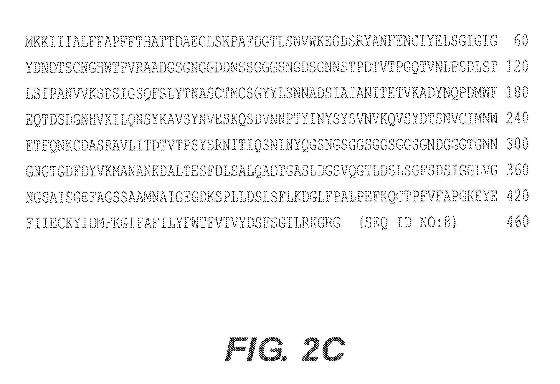
FIGS. 2A-2C present alignments of g3p's from different sources. FIG. 2A is an alignment of g3p from phage M13 (SEQ ID NO: 1), fd (SEQ ID NO:2), and f1 (SEQ ID NO: 3), including a consensus sequence (SEQ ID NO: 4). FIG. 2B shows an alignment of g3p from phage I2-2 (SEQ ID NO: 5) and Ike (SEQ ID NO: 6), along with a consensus sequence between I2-2 and Ike (SEQ ID NO: 7). FIG. 2C presents the amino acid sequence of g3p from phage If (SEQ ID NO: 8).
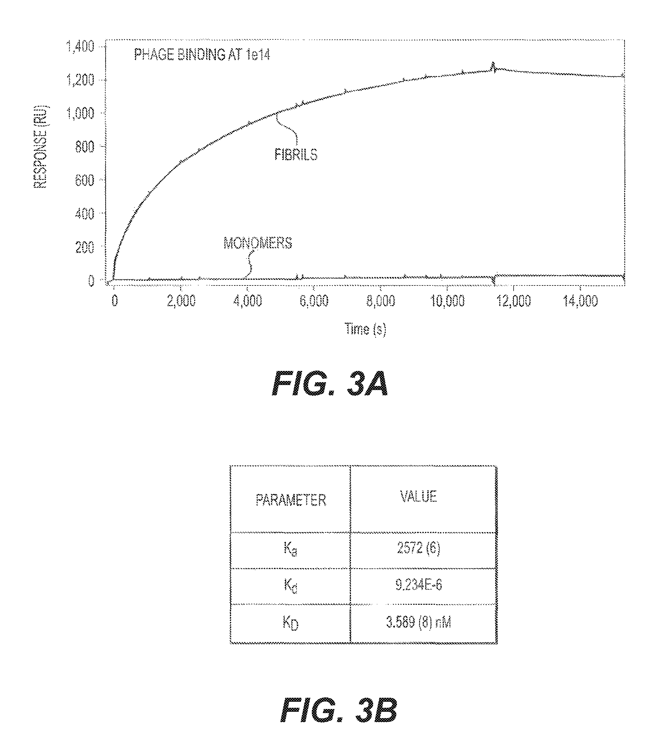
FIGS. 3A and 3B present a surface plasmon resonance (SPR) study of phage binding. Binding to A.beta. fibrils was compared to binding to A.beta. monomers using 10.sup.14 phage/mL flowed across the biosensor chip. FIG. 3B shows the K.sub.a, K.sub.d, and K.sub.D calculated from the SPR data shown in FIG. 3A.
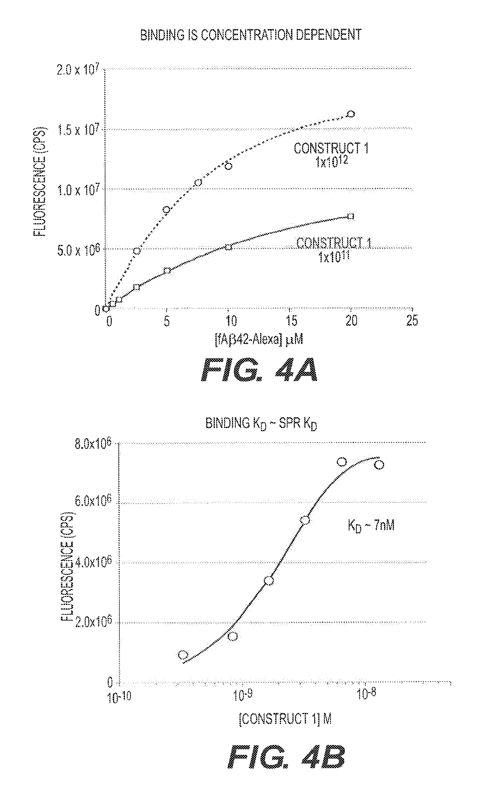
FIGS. 4A and 4B present binding studies. FIG. 4A shows a direct binding assay for two phage doses (10.sup.11/mL and 10.sup.12/mL) with increasing molar amounts of fA.beta.42. FIG. 4B is a binding competition study and provides an alternate way to determine the K.sub.D for M13 binding. Construct 1 was used.
FIG. 5 shows binding competition results using heat denatured (boxes--90.degree. C. for 10 minutes) versus native conformation (circles) M13 (Construct 1) in the amyloid fiber binding competition assay.
FIG. 6 shows a Thioflavin T (ThT) fluorescence assay using fA.beta.42 incubated in the presence or absence of 2 concentrations of M13 phage (Construct 1).
FIGS. 7A and 7B show the effect of varying individual assay parameters in the ThT disaggregation assay. FIG. 7A presents disaggregation percentages in the presence of two salt concentrations (0.15 M and 1.5 M). FIG. 7B presents percentages of fA.beta. remaining at two temperatures (4.degree. C. and 37.degree. C.). Construct 1 was used.
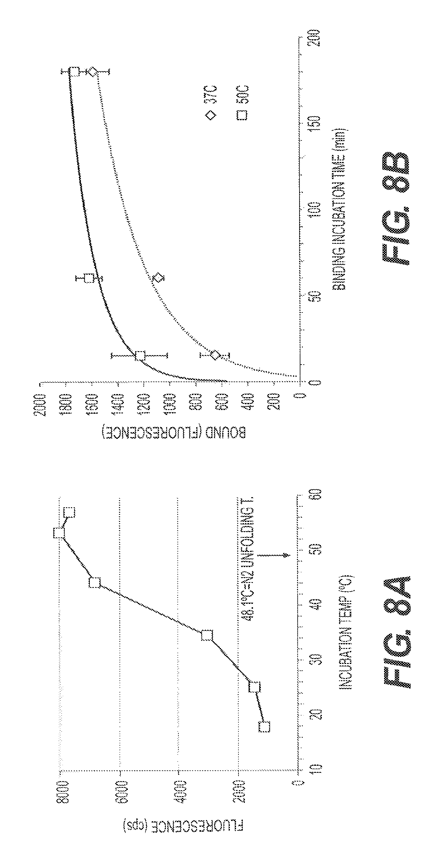
FIGS. 8A and 8B represent M13-amyloid binding assays using fA.beta.42. In FIG. 8A, M13 binding is reported using incubation temperatures from 18.degree. C. to 58.degree. C. for 3 hours. FIG. 8B shows binding kinetics for incubations at 37.degree. C. vs. 50.degree. C.
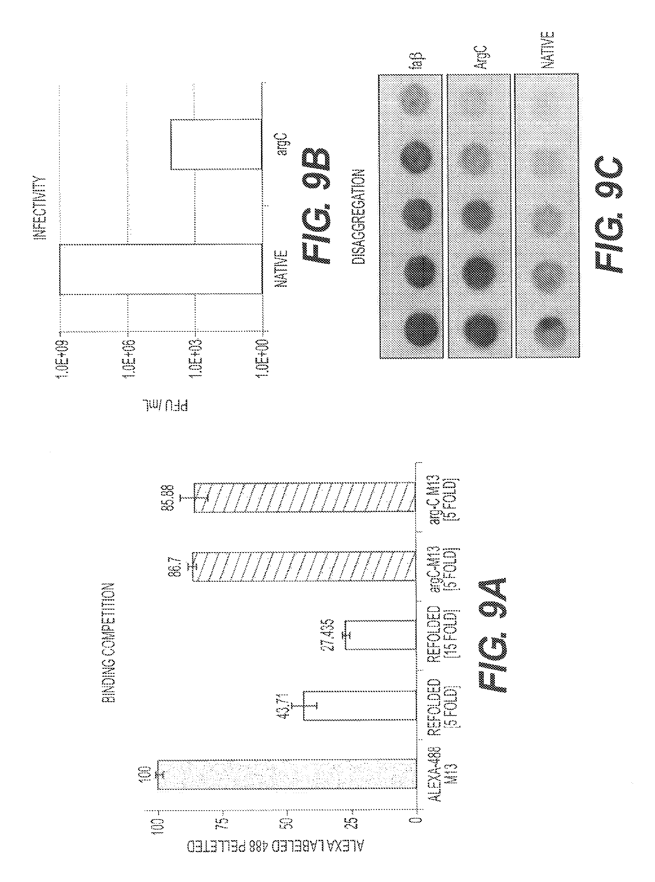
FIGS. 9A-9C show the effect of proteolytic removal of g3p on phage-amyloid interactions. The protease Arg C was used to clip g3p from M13 phage (M13.DELTA.g3p). FIG. 9A presents the results of an A.beta. binding competition study using M13.DELTA.g3p phage compared to native (treated identically to the ArgC-treated phage but without protease treatment) phage. FIG. 9B shows the effect of Arg C treatment on infectivity of the M13.DELTA.g3p phage compared to native phage. FIG. 9C compares ArgC treated phage to native phage in the disaggregation assay.
FIGS. 10A and 10B present the results of a binding competition assay using a N1-N2 fragment of g3p, herein referred to as recombinant soluble N1N2 (rs-g3p(N1N2), "Construct 3"), M13.DELTA.3p (Arg C treated), and M13 as competitors of labeled M13 binding to fA.beta.42. FIG. 10B shows a repeat of the competition assay.
FIG. 11 presents competition data for phage fd, IIHY, AAA, and M13. Phages fd, AAA, and IIHY were pre-activated at 50.degree. C. for 1.5 hours, then activated and non-activated Fd, AAA, & IIHY were compared for their ability to compete with labeled M13 for binding to A.beta. during a 45 minute incubation at 37.degree. C.
FIG. 12A shows a schematic of rs-g3p(N1N2) (Construct 3). FIG. 12B presents an ion exchange profile for rs-g3p(N1N2). FIG. 12C shows the results of a gel filtration assay using Sephacryl S-300 and rs-g3p(N1N2). FIG. 12D shows a Western Blot of rs-g3p(N1N2) together with g3p and g8p controls. M13 phage are run in lanes 1 and 2 as a positive control, and detected with a polyclonal anti-M13 antibody, which detects both g8p and g3p. Purified rs-g3p is run in lanes 3 and 4, and detected with the same polyclonal anti-M13 antibody.
FIG. 13 presents SPR data using rs-g3p(N1N2) (Construct 3). rs-g3p(N1N2) potently binds fA.beta.42 with a K.sub.D of about 160 nM, but does not bind monomers.
FIG. 14A and FIG. 14B present a ThT fluorescence assay used to measure the amyloid present in a given sample. 10 .mu.M of A.beta.42 monomers was incubated in the presence or absence of 5 concentrations of rs-g3p(N1N2) (Construct 3) at 37.degree. C. for 3 days. The amount of fibers formed at the end of 3 days was measured by quantitating the bound ThT fluorescence. The IC.sub.50 is approximately 20 nM indicating that rs-g3p(N1N2) potently inhibits formation of A.beta.42 fibers. The figures also indicate that binding is dose-dependent. Repeated experiments show IC.sub.50's between 20 nM and 100 nM.
FIG. 15A shows the transmission electron micrography (TEM) results of incubating fA.beta.42 in the presence or absence of rs-g3p(N1 N2) (Construct 3). FIG. 15B shows the results of a ThT fluorescence assay using A.beta.42 and 2 .mu.M rs-g3p(N1N2) (Construct 3) incubated at 37.degree. C. for 7 days. rs-g3p(N1N2) blocks the formation of fA.beta.42.
FIG. 16 demonstrates that rs-g3p(N1N2) (Construct 3) potently inhibits the formation of .alpha.-synuclein fibers. 25 .mu.M of .alpha.-synuclein was assembled by agitating at 300 rpm for 4 days at 37.degree. C. (see, Bar 1). The second bar on the graph represents alpha-synuclein monomers plus 1.times.10.sup.-13 pentameric M13 phage shaking at 37.degree. C. for 3 days. The results shown in bar 2 indicate that pentameric M13 blocks assembly of .alpha.-synuclein fibers. The third bar on the graph represents alpha-synuclein monomers+83 nM rsg3p monomers. The results shown in bar 3 indicate that monomers are less effective at inhibiting .alpha.-synuclein fiber formation than pentameric M13. Bar 4 is a negative control showing alpha synuclein monomers at time zero. In bar 5, g3p monomers without .alpha.-synuclein fibers are shown to determine whether g3p binds to pTAA and sequesters the dye from binding to the fibers. The results shown in bar 5 indicate that g3p does not bind to pTAA.
FIG. 17 presents competition binding data for rs-g3p(N1N2) (Construct 3), M13 (Construct 2), rs-g3p(N1N2)-hIgG4-Fc fusion protein (Construct 4), and an IgG4-Fc negative control.
FIG. 18 presents competition binding data comparing M13 (Construct 2; squares), rs-g3p(N1N2) (Construct 3; triangles), rs-g3p(N1N2)-hIgG4-Fc fusion protein (Construct 4; upside down triangles), and a recombinant IgG4-Fc negative control (diamonds).
FIG. 19 shows a filter trap assay comparing five concentrations of A.beta.42 fibers plus or minus two concentrations of M13 (Construct 2), 800 nM rs-g3p(N1N2) (Construct 3), and three concentrations of rs-g3p(N1N2)-hIgG4-Fc fusion protein (Construct 4).
FIG. 20 presents competition binding data for rs-g3p(N1N2) (Construct 3; "monomer") and streptavidin conjugated rs-g3p(N1N2) ("SA[g3pN1N2].sub.n=2-4", "SA-g3p"; "tetramer"). rs-g3p(N1N2) and SA-g3p were compared for their ability to compete with labeled M13 for binding to A.beta. during a three hour incubation at 37.degree. C.
FIG. 21 shows a filter trap assay comparing five concentrations of fA.beta.42 plus or minus two concentrations of rs-g3p(N1N2) (Construct 3; "monomer") and two concentrations of SA-g3p ("tetramer").
FIGS. 22A and 22B show TEMs of fA.beta.42 at times zero (FIG. 22A) and three days after incubation with SA-g3p (FIG. 22B).
FIG. 23 shows the amino acid sequence of one rs-g3p(N1N2)-hIgG4-Fc construct "Construct 4" (SEQ ID NO:9). The N1N2 region of "Construct 4" is derived from the N1N2 region of "Construct 1" (SEQ ID NO:10).
FIG. 24 shows the amino acid sequence of another rs-g3p(N1N2)-hIgG4-Fc construct "Construct 5" (SEQ ID NO:11). The N1N2 region of "Construct 5" is derived from the N1N2 region of "Construct 2" (SEQ ID NO:12).
FIG. 25 shows the amino acid sequence of one rs-g3p(N1N2)-hIgG1-Fc construct "Construct 6" (SEQ ID NO:13). The N1N2 region of "Construct 6" is derived from the N1N2 region of "Construct 2".
FIG. 26 shows the amino acid sequence alignment of N2 from: fd (SEQ ID NO:14), f1 (SEQ ID NO:15), M13 (SEQ ID NO:16), Ike (SEQ ID NO:17), I2-2 (SEQ ID NO:18), and If1 (SEQ ID NO:19). An asterisk "*" indicates positions which have a single, fully conserved residue. A colon ":" indicates conservation between groups of strongly similar properties that score greater than 0.5 in the Gonnet PAM 250 matrix. A period "." indicates conservation between groups of weakly similar properties that score equal to or less than 0.5 in the Gonnet PAM 250 matrix.
FIG. 27A shows a schematic of Construct 3. FIG. 27B shows the DNA sequence of the g3p portion of Construct 3 (SEQ ID NO:23). FIG. 27C shows the amino acid sequence of the g3p portion of Construct 3 (SEQ ID NO:24).
FIG. 28 shows the results of an experiment testing two rs-g3p(N1N2)-IgG fusion proteins for their ability to reduce amyloid .beta. in a transgenic mouse model of Alzheimer's disease. rs-g3p(N1N2)-hIgG4-Fc (Construct 5) and rs-g3p(N1N2)-hIgG1-Fc (Construct 6) both significantly reduced the level of amyloid .beta. in the hippocampus of Alzheimer's disease mice.
FIG. 29 shows the results of an experiment testing two rs-g3p(N1N2)-IgG fusion proteins for their ability to reduce amyloid .beta. in a transgenic mouse model of Alzheimer's disease. rs-g3p(N1N2)-hIgG4-Fc (Construct 5) and rs-g3p(N1N2)-hIgG1-Fc (Construct 6) were both able to significantly reduce the level of amyloid .beta. in the cerebral cortex of Alzheimer's Disease mice.
FIGS. 30A and 30B shows assembly inhibition of A.beta.42 with rs-g3p(N1N2)-hIgG1-Fc (Construct 6). FIG. 30A shows a "native" agarose gel made without SDS. The samples were run in TEA buffer without SDS and not boiled. The results indicate that Construct 6 is capable of inhibiting the assembly of fA.beta.42. FIG. 30B presents a ThT fluorescence assay used to measure the amyloid present in a given sample. 10 .mu.M of A.beta.42 monomers were incubated in the presence or absence of 2 concentrations of rs-g3p(N1N2)-hIgG1-Fc (Construct 6) at 37.degree. C. for 1 day. The amount of fibers formed at the end of day 1 was measured by quantitating the bound ThT fluorescence. rs-g3p(N1N2)-hIgG1-Fc (Construct 6) potently inhibits formation of A.beta.42 fibers. The figure also indicates that inhibition of fiber formation with Construct 6 is dose-dependent.
FIGS. 31A, 31B, 31C, and 31D present representative circular dichroism data showing that A.beta.42 assembly is inhibited by rs-g3p(N1N2) (Construct 3). Circular dichroism measures the .alpha.-helix and .beta.-sheet content of the A.beta. fibers to be assessed. FIG. 31A shows the ellipticity versus wavelength for A.beta.42 at T=0, T=24 hours, and T=48 hours. FIG. 31B shows ellipticity versus wavelength for A.beta.42 plus Construct 3 at T=0, T=24 hours, and T=48 hours. FIG. 31C shows a representative ThT assay where the amount of fibers formed between 24 and 48 hours was measured by quantitating the bound ThT fluorescence. Construct 3 potently inhibits formation of A.beta.42 fibers. FIG. 31D shows ellipticity versus wavelength for Construct 3 at T=0, T=24 hours, and T=48 hours. Taken together, these data confirm the ability of Construct 3 to inhibit A.beta.42 assembly.
FIG. 32 presents representative data showing that M13 (Construct 2) and rs-g3p(N1N2)-hIgG1-Fc (Construct 6) block oligomer-induced toxicity of N2a cells. See, e.g., Stine et al. (2003) J. Biol. Chem. 278(13): 11612-11622 and Stine et al. (2011) Erik D. Roberson (ed.) Alzheimer's Disease and Frontotemporal Dementia, Methods in Molecular Biology, vol. 670: 13-32. N2a cells were differentiated by serum starvation for 48 hours prior to treatment. A.beta.42 oligomers (2 uM) were pre-incubated with Construct 2 and Construct 6 at 37.degree. C. for 3 hrs before addition to N2a cells. Time zero ("TO") complexes were not pre-incubated. After 24 hours of incubation, adenylate kinase ("AK") release was monitored. AK release into the medium indicates cell death/lysis. A.beta.42 oligomers were made as described by Stine et al., 2011. The results indicate that M13 and rs-g3p(N1N2)-hIgG1-Fc are potent inhibitors of toxic oligomers.
FIG. 33 shows a filter trap assay comparing six concentrations of A.beta.42 fibers plus or minus 1.times.10.sup.12/ml M13 (Construct 2); 80 nm and 800 nM rs-g3p(N1N2)-hIgG4-Fc construct (Construct 5); and 80 nm and 800 nM of rs-g3p(N1N2)-hIgG1-Fc (Construct 6). A.beta.42 fibers were incubated with Constructs 2, 5, and 6 at 37.degree. C. for 3 days, followed by filter retardation. The filter was probed by mAb 6E10 (1:15000), which recognizes A.beta.42 fibers trapped on the filter. 800 nM of Construct 5 or Construct 6 equals 5.times.10.sup.14/ml Construct 2 by molecular molarity. The results indicate that Constructs 2, 5, and 6 potently disaggregate .beta.-amyloid fibers.
FIGS. 34A and 34B present representative assays used to measure the amount of M13 (Construct 2) bound to fA.beta.42 after 3 hours of preincubation with ftau. 5 .mu.M of A.beta.42 monomers bound to Construct 2 was incubated in the presence or absence of 4 concentrations of ftau at 37.degree. C. for 3 hours. Since fAbeta:M13-Alexa488 pellets but ftau:M13-Alexa488 does not pellet, measuring the loss of fluorescence from the pelleted material indicates that ftau competed the fAbeta binding. Here, the amount of M13-fA.beta. formed at the end of 3 hours was measured by quantitating the Alexa488 fluorescence in the pelleted binding competition reaction. The results indicate that ftau is able to compete with M13-Alexa488 (Construct 2) for fA.beta.42 binding.
FIG. 35 shows the results of one representative SPR assay testing the ability of rs-g3p(N1N2)-hIgG4-Fc (Construct 4) to bind to ftau. The results indicate that Construct 4 potently binds ftau.
FIGS. 36A and 36B show the ability of rs-g3p(N1N2)-hIgG1-Fc (Construct 6) to disaggregate ftau. Tau fibers were prepared by diluting 40 uM of the microtubule binding repeat region ("MTBR") of tau into 50 mM superoxide dismutase ("Sod"). Various concentrations of Construct 6 and the prepared ftau were incubated in acetate buffer at pH7.0, 37.degree. C. for 72 hrs. ThT fluorescence was recorded in the presence of 5 fold excess ThT. FIG. 36A presents the results of a representative ThT assay showing the ability of Construct 6 to disaggregate ftau. FIG. 36B shows another representative experiment confirming the ability of Construct 6 to disaggregate tau. FIGS. 36A and 36B also show that disaggregation of ftau by Construct 6 is dose dependent.
FIGS. 37A, 37B, 37C, and 37D present representative experiments showing the inhibition of A.beta. aggregation by rs-g3p(N1N2)-hIgG1-Fc (Construct 6) and rs-g3p(N1N2) (Construct 3) over time. A.beta.42 was dissolved in DMSO and diluted into PBS containing NaN3. A.beta.42 was aggregated at 37.degree. C. plus or minus various concentrations of Construct 3 and Construct 6. Aggregation of A.beta.42 was measured by ThT fluorescence. FIG. 37A shows an SDS PAGE of the samples. FIG. 37B shows the results from one representative experiment. FIG. 37C shows the results from another representative experiment. FIG. 37D summarizes the results.
FIG. 38A and FIG. 38B present the results of experiments showing the ability of rs-g3p(N1N2)-hIgG1-Fc (Construct 6) to block the conversion of PrP to PrP-Sc. Construct 6 and IgG cell lysates were subjected to ultra-centrifugation to separate soluble (supernatant) and insoluble (pellet) PrP species. PrP species were visualized biochemically with an anti-PrP monoclonal antibody (6D11). In the presence of IgG, there is a partitioning of PrP in both soluble and insoluble fractions. In the presence of Construct 6, there is limited insoluble PrP. Data represents n=4.
FIG. 39A and FIG. 39B present the results of experiments showing the ability of rs-g3p(N1N2)-hIgG1-Fc (Construct 6) to reduce the accumulation and aggregation of PrP.sup.Sc in a cell culture model of prion disease. FIG. 39A shows biochemically resolved undigested and PK-digested N2a22L.sup.Sc cell lysates following treatment with Construct 6 and IgG. A significant reduction in PrP.sup.Sc levels is clearly observed in cells treated with increasing concentrations of Construct 6. An approximately 50% reduction in PrP.sup.Sc levels is achieved with treatment of .about.0.08 .mu.g/ml Construct 6. Treatment with 10 .mu.g/ml Construct 6 reduces PrP.sup.Sc levels to 5.725%, p<0.0001. No marked changes in PrP.sup.Sc levels were observed in N2A22L.sup.Sc cells treated with 1 .mu.g/ml murine IgG. For FIG. 39B, the X-ray films were subsequently digitized and initially normalized to the effect in IgG treated N2a22L.sup.Sc cells from the same passage which was considered to be 100%. The densitometry data from PK-digested blots was then analyzed relative to the equivalently blotted undigested lysates and expressed as a percent change PrP.sup.Sc/PrPc. Data represents n=4.
FIG. 40 presents the results of an experiment testing two rs-g3p(N1N2)-IgG fusion proteins for their effect on synaptophysin levels in the hippocampus in a transgenic mouse model of Alzheimer's disease after treatment with rs-g3p(N1N2)-hIgG4-Fc (Construct 5) and rs-g3p(N1N2)-hIgG1-Fc (Construct 6). Both constructs significantly increased the level of synaptophysin in the hippocampus of Alzheimer's disease mice.
FIG. 41 presents the results of an experiment testing two rs-g3p(N1N2)-IgG fusion proteins for their effect on Iba-1 levels in the hippocampus in a transgenic mouse model of Alzheimer's disease after treatment with rs-g3p(N1N2)-hIgG4-Fc (Construct 5) and rs-g3p(N1N2)-hIgG1-Fc (Construct 6). Both constructs significantly increased the level of Iba-1 in the hippocampus of Alzheimer's disease mice.
FIG. 42 presents the results of an experiment testing two rs-g3p(N1N2)-IgG fusion proteins for their effect on GFAP levels in the hippocampus in a transgenic mouse model of Alzheimer's disease after treatment with rs-g3p(N1N2)-hIgG4-Fc (Construct 5) and rs-g3p(N1N2)-hIgG1-Fc (Construct 6). Neither construct significantly altered the level of GFAP in the hippocampus of Alzheimer's disease mice.
FIG. 43 presents the results of a binding experiment designed to compare rs-g3p(N1N2)-hIgG4-Fc (Construct 5) and rs-g3p(N1N2)-hIgG1-Fc (Construct 6). The constructs bind to fA.beta. potently with similar K.sub.D's (.about.11).
FIGS. 44A and 44B show the ability of rs-g3p(N1N2) (Construct 3) to block assembly of tau. In FIG. 44A, 1 .mu.M tau was incubated alone or co-incubated with 1 .mu.M of Construct 3, and analyzed by TEM after 5 days. Construct 3 blocks assembly of tau. FIG. 44B shows the results of a ThT fluorescence assay using ftau incubated in the presence or absence of 3 concentrations of rs-g3p(N1N2) (Construct 3). Construct 3 dose-dependently blocks the assembly of tau.
FIG. 45 shows a schematic of the experiment to analyze the interactions between fA.beta.42 and rs-g3p(N1N2) (Construct 3).
FIG. 46 shows the results of an NMR study to analyze the interactions between fA.beta.42 and rs-g3p(N1N2) (Construct 3). H stands for hydrogen and D stands for deuterium. The hydrogens of the A.beta. fibers exchange for deuterium over time and the rate is affected by binding of Construct 3 to the fibers. The results indicate a molecular iteration between Construct 3 and fA.beta.42 at residues 17-22 and 33-40 of fA.beta.42.
FIGS. 47A, 47B, 47C, and 47D show representative TEMs of Construct 3 disaggregating preformed fA.beta.42 after incubation for 744 hours. FIG. 47A shows fA.beta.42 alone. FIG. 47B shows fA.beta.42 plus Construct 3. FIG. 47C shows fA.beta.42 alone at an increased magnification as compared to FIG. 47A. FIG. 47D shows fA.beta.42 plus Construct 3 at higher magnification as compared to FIG. 47B.
FIG. 48 shows the results of a representative SPR assay showing that rs-g3p(N1N2)-hIgG1-Fc (Construct 6) potently binds ftau.
FIG. 49A presents the results of a representative ThT assay showing the ability of Construct 6 to disaggregate ftau. FIG. 49B shows a graphical representation of the experiment of FIG. 49A. FIGS. 49A and 49B also show that disaggregation of ftau by Construct 6 is dose dependent.
FIG. 50A presents a SPR study of rs-g3p(If1-N1N2)-hIgG4-Fc (Construct 8) binding to A.beta. fibrils. The results show that Construct 8 strongly binds A.beta. fibrils (K.sub.D.about.36 nM). An N1N2 fragment of g3p (not linked to an Fc domain) showed weaker binding (K.sub.D.about.36 nM). FIG. 50B present the results of a binding competition assay showing the ability of rs-g3p(If1-N1N2)-hIgG1-Fc (Construct 8) to bind to fA.beta.1-42. An N1N2 fragment of g3p (not linked to an Fc domain) showed weaker binding.
FIG. 51 shows an amino acid comparison between If1 g3p (SEQ ID NO:29) and fd g3p (SEQ ID NO:30). Amino acids that are identical between If1 and fd in the N1 domain of g3p are shaded. The N1 domain is boxed.
FIGS. 52A and 52B present the results of experiments showing the ability of rs-g3p(N1N2)-hIgG1-Fc (Construct 6) to significantly reduce A.beta. deposition and tau fibers following direct injection to the brain in an in vivo model of Alzheimer's disease. In FIG. 52A, the level of A.beta. is significantly reduced in mice that received Construct 6 as compared to control. In FIG. 52B, the level of tau is significantly reduced in mice that received Construct 6 as compared to control
FIGS. 53A and 53B present the results of experiments showing the ability of rs-g3p(N1N2)-hIgG1-Fc (Construct 6) to treat AD when given systemically rather than as a direct injection to the brain. FIGS. 53A and 53B show that AD mice that received Construct 6 have reduced hyperactivity as compared to mice given a control.
FIG. 54 presents the results of experiments showing the ability of rs-g3p(N1N2)-hIgG1-Fc (Construct 6) to treat AD when given systemically rather than as a direct injection to the brain. In FIG. 54, the ability of AD mice to circle is reduced in mice that received Construct 6 as compared to control.
FIG. 55 presents the results of experiments showing the ability of rs-g3p(N1N2)-hIgG1-Fc (Construct 6) to treat AD when given systemically rather than as a direct injection to the brain. In FIG. 55, corner jumping of AD mice is significantly reduced in mice that received Construct 6 as compared to control.
FIG. 56 presents the results of experiments showing the ability of rs-g3p(N1N2)-hIgG1-Fc (Construct 6) to treat AD when given systemically rather than as a direct injection to the brain. In FIG. 56, AD mice receiving Construct 6 exhibited significantly more spontaneous alternation of arm entries in the Y-maze relative to mice receiving control PBS.
FIG. 57 presents a schematic showing the timing of injections of PBS or Construct 6 (arrows) as well as timing for behavioral assessments (asterisks).
BRIEF DESCRIPTION OF THE SEQUENCES
TABLE-US-00001 SEQ ID NO: Construct 1 M13 g3p 2 fd g3p 3 f1 g3p 4 consensus sequence of SEQ ID NOs: 1, 2, and 3 5 I2-2 g3p 6 Ike g3p 7 consensus sequence of SEQ ID NOs: 5 and 6 8 If1 g3p 9 Amino Acid of Construct 4 10 DNA of Construct 1 11 Amino Acid of Construct 5 12 DNA of Construct 2 13 Amino Acid of Construct 6 14 fd N2 15 f1 N2 16 M13 N2 17 Ike N2 18 I2-2 N2 19 If1 N2 20 Amino Acid of Construct 3 21 GxFxGxF 22 KLVFF 23 DNA sequence of the g3p portion of Construct 3 24 Amino acid sequence of g3p portion of Construct 3 25 His His His His His His 26 DNA of Construct 4 27 DNA of Construct 5 28 DNA of Construct 6 29 If1 g3p (FIG. 51) 30 fd g3p (FIG. 51) 31 Amino Acids of rs-g3p(If1-N1N2)-hIgG1-Fc construct "Construct 8" 32 DNA of rs-g3p(If1-N1N2)-hIgG1-Fc construct "Construct 8" 33 forward primer: AAAAAAGGGAATTCGATGGCTGAAACTGTTGAAAGTTG 34 reverse primer: AAAAAACCATGGCACCGGAACCAGAGCCAC
DESCRIPTION OF EMBODIMENTS
The invention provides fusion proteins that comprise an amyloid binding fragment of g3p or a mutant or variant thereof. In specific embodiments, the fusion proteins of the invention further comprise an Fc fragment of an immunoglobulin constant region. In one aspect of these embodiments, the fusion proteins are soluble. In another aspect of these embodiments, the fusion proteins reduce amyloid by, for example, disaggregating and/or preventing or inhibiting the aggregation of amyloid (e.g., amyloid plaque). The fusion proteins of the invention bind to amyloid. In some embodiments, the fusion proteins of the invention remove and/or inhibit the formation of toxic oligomers.
The invention provides pharmaceutical compositions of the fusion proteins of the invention, as well as their use to bind to and reduce amyloid. Reducing amyloid encompasses, for example, disaggregating amyloid, preventing and/or inhibiting the aggregation of amyloid, and removing and/or preventing the formation of toxic oligomers. Use of the compositions to detect amyloid deposits and diagnose diseases and disorders characterized by amyloid is encompassed.
Definitions
The term "g3p" when used alone or in terms such as "g3p-derived" refers to any wild type or recombinant filamentous phage g3p protein (including fragments, variants, and mutants of g3p). The term should not be construed as limited to g3p derived from any particular filamentous bacteriophage. By way of example, the term "g3p" includes SEQ ID NO: 1 and the related proteins shown in FIGS. 2A-2C.
The term "domain" means a region of a polypeptide (including proteins) having some distinctive physical feature or role including for example an independently folded structure composed of one section of a polypeptide chain. A domain may contain the sequence of the distinctive physical feature of the polypeptide or it may contain a fragment of the physical feature which retains its binding characteristics (i.e., it can bind to a second domain). A domain may be associated with another domain. In other words, a first domain may naturally bind to a second domain. For example, the g3p N2 domain binds F-pili and the g3p N1 domain binds TolA.
The terms "amyloid," "amyloid fibrils," and "amyloid fibers," as used herein are generic terms for a tertiary structure that is formed by aggregation of any of several different proteins and that consists of an ordered arrangement of .beta. sheets stacked perpendicular to a fiber axis. Sunde et al., J. Mol. Biol. (1997) 273:729-39. One exemplary amyloid is the aggregate of amyloid-.beta. formed in Alzheimer's disease, which is composed of beta-amyloid peptide ".beta.A," which are 39-43 amino acid internal fragments cleaved from the human amyloid precursor protein (hAPP). There are short forms, such as A.beta.40, and long forms, such as the more fibrillogenic A.beta. isoform, A.beta.42. Other exemplary amyloid proteins include misfolded .alpha.-synuclein (associated with Parkinson's disease), huntingtin (associated with Huntington's disease), tau (associated with Alzheimer's disease), and the abnormal conformation of the prion protein, PrP.sup.Sc. Additional examples are provided throughout the description and are known to those of skill in the art (see, e.g., Aguzzi (2010), and Eichner and Radford, Mol. Cell (2011) 43:8-18). Thus, unless a protein or peptide is specified, use of the terms "amyloid," "amyloid fibrils," or "amyloid fibers" should not be construed as limited to any particular protein or disease.
The term "amyloid binding fragment of g3p" refers to a fragment of g3p that maintains the ability to bind to amyloid. The term "amyloid binding fragment of g3p" also refers to mutants and variants of g3p, including N-, C-, or N- and C-terminal truncations of g3p, that maintain the ability to bind to amyloid.
The term "beta amyloid peptide" is synonymous with ".beta.-amyloid peptide," ".beta.AP," ".beta.A," and "A.beta.." All of these terms refer to an amyloid forming peptide derived from the human amyloid precursor protein (hAPP).
Fusion proteins of the invention or compositions comprising those fusion proteins described as "disaggregating" or "mediating disaggregation" reduce aggregates that have already formed. Disaggregation can be measured by the filter trap assay. Wanker et al., Methods Enzymol (1999) 309:375-86. The filter trap assay is described herein and can be used both to detect aggregates and to monitor disaggregation mediated by compositions of the invention. Disaggregation is detected as decreased retention of amyloid on the filter, as shown by a decrease in staining, in the presence of increasing concentrations of the disaggregating agent.
As used herein, a fusion protein or composition that "reduces amyloid" or "decreases amyloid load" does one or more of the following: inhibits amyloid formation, causes amyloid disaggregation, promotes amyloid clearance, inhibits amyloid aggregation, blocks and/or prevents the formation of toxic amyloid oligomers, and/or promotes the clearance of toxic amyloid oligomers.
Any of the products or compositions of the invention described as "protecting neurons from amyloid damage" prevent the accumulation of new amyloid and/or prevent the formation of toxic amyloid oligomers. Products or compositions of the invention described as "protecting neurons from amyloid damage" may be taken prophylactically. Whether or not a product or composition protects neurons from amyloid damage may be measured by the neuronal cell culture cytotoxicity assay described herein.
As used herein, "PrP protein," "PrP," and "prion," refer to polypeptides that are capable under appropriate conditions, of inducing the formation of aggregates responsible for protein misfolding diseases. For example, normal cellular prion protein (PrP.sup.c) is converted under such conditions into the corresponding scrapie isoform (PrP.sup.Sc) which is responsible for diseases such as, but not limited to, bovine spongiform encephalopathy (BSE), or mad cow disease, feline spongiform encephalopathy of cats, kuru, Creutzfeldt-Jakob Disease (CJD), Gerstmann-Straussler-Scheinker disease (GSS), and fatal familial insomnia (FFI).
The term "variant" as used herein in conjunction with a fusion protein or an amyloid binding fragment of g3p portion of the fusion protein, refers to a corresponding amino acid sequence that contains at least one amino acid difference (substitution, insertion or deletion) as compared to the reference substance. In certain embodiments a "variant" has high amino acid sequence homology and/or conservative amino acid substitutions, deletions and/or insertions as compared to the reference sequence. In some embodiments, a variant has no more than 75, 50, 40, 30, 25, 20, 15, 12, 10, 9, 8, 7, 6, 5, 4, 3, 2, 1 amino acid differences as compared to the reference sequence. A "conservative substitution" refers to the replacement of a first amino acid by a second amino acid that does not substantially alter the chemical, physical and/or functional properties of the protein, polypeptide or amino acid sequence, such as, e.g., a g3p protein or amyloid binding fragment of g3p (e.g., the g3p protein or amyloid binding fragment retains the same charge, structure, polarity, hydrophobicity/hydrophilicity, and/or preserves functions such as the ability to recognize, bind to, and/or reduce amyloid). Such conservative amino acid modifications are based on the relative similarity of the amino acid side-chain substituents, for example, their hydrophobicity, hydrophilicity, charge, size, and the like. Exemplary conservative substitutions which take various of the foregoing characteristics into consideration are well known to those of skill in the art and include: arginine and lysine; glutamate and aspartate; serine and threonine; glutamine and asparagine; and valine, leucine, and isoleucine. The terms "g3p variant" or a "variant of an amyloid binding fragment of g3p" also encompass polypeptides having at least 70%, at least 75%, at least 78%, at least 80%, at least 82%, at least 85%, at least 86%, at least 87%, at least 88%, at least 89%, at least 90%, at least 91%, at least 92%, at least 93%, at least 94%, at least 95%, at least 96%, at least 97%, at least 98%, at least 99% amino acid sequence identity to a wild type g3p or corresponding fragment thereof.
The term "mutant" refers to a fusion protein or an amyloid binding fragment of g3p of the fusion protein that is mutated at one or more amino acids in order to modulate its therapeutic or diagnostic efficacy. In certain embodiments, a mutant contains a substitution, deletion and/or insertion at an amino that is known to interact with amyloid. In other embodiments, a mutant contains a substitution, deletion and/or insertion at an amino that is a conserved amino acid present in a wild type g3p or amyloid binding fragment thereof. In some embodiments, a mutant has no more than 75, 50, 40, 30, 25, 20, 15, 12, 10, 9, 8, 7, 6, 5, 4, 3, 2, 1 amino acid differences as compared to the reference sequence. In some embodiments, the amino acid substitutions are conservative substitutions. The terms "variant" and "mutant" are used interchangeably herein except that a "variant" is typically non-recombinant in nature, whereas a "mutant" is typically recombinant. The terms "mutant g3p" or "mutant of an amyloid binding fragment of g3p" also encompass polypeptides having at least 70%, at least 75%, at least 78%, at least 80%, at least 82%, at least 85%, at least 86%, at least 87%, at least 88%, at least 89%, at least 90%, at least 91%, at least 92%, at least 93%, at least 94%, at least 95%, at least 96%, at least 97%, at least 98%, at least 99% amino acid sequence identity to a wild type g3p or corresponding fragment thereof.
A "fusion protein" is a non-naturally occurring protein comprising at least two polypeptide domains. Fusion proteins of the invention comprise an amyloid binding fragment of g3p linked, fused, or conjugated to a second protein or polypeptide. In specific embodiments, the fusion proteins comprise an amyloid binding fragment of g3p and an Fc fragment of an immunoglobulin.
The terms "active compounds," "active agent," and "active ingredient" are used interchangeably herein to refer to the portion of a fusion protein that provides the biological activity of the fusion protein. The g3p portion of the fusion proteins of the invention is the "active compound," "active agent," or "active ingredient." Likewise, the g3p portion of the fusion proteins of the invention is the biologically active or therapeutically effective portion. The g3p portion of the fusion protein of the present invention is not used to facilitate protein folding of a fusion partner, which then acts as a therapeutic agent unrelated to g3p, as described in WO 2004/018685. Nor are the fusion proteins of the invention used for phage display as described in US 2009/105090.
The term "immunogenic" is used herein to refer to the ability of a composition to elicit an immune response in a mammal that has been exposed to the composition.
As used herein, "Construct 1" is derived from wild type M13 (see, Genbank file: NC_003287.2, version GI:56718463. In Construct 1, as compared to wild type M13, Ser378(AGC) is changed to Gly(GGC), and Ile87 (ATT) is changed to Asn(AAC)). Thus, whereas in wild type M13 there is a "G" at nucleic acid number 2710, in Construct 1 there is an "A" at this position. Likewise, in wild type M13 there is an "A" at nucleic acid number 4479, in Construct 1 there is a "T" at this position. Finally, in wild type M13 there is a "C" at nucleic acid number 4480, whereas in Construct 1 there is a "T" at this position. Construct 1 comprises the nucleic acids of SEQ ID NO:10.
"Construct 2" is a wild type M13 isolate (GenBank JX412914.1). Construct 2 comprises the nucleic acids of SEQ ID NO:12.
"Construct 3" is a recombinant soluble g3p fragment comprising the N1 and N2 domains of g3p (rs-g3p(N1N2)) comprising the amino acids of SEQ ID NO:20.
"Construct 4" is recombinant soluble g3p fragment IgG4 Fc fusion protein (rs-g3p(N1N2)-hIgG4-Fc) comprising the amino acids of SEQ ID NO:9. The N1N2 region of "Construct 4" is derived from the N1N2 region of "Construct 1." The nucleic acid sequence encoding "Construct 4" is set forth in SEQ ID NO:26.
The first 21 amino acids set forth in SEQ ID NO:9 represent a signal sequence that is cleaved between amino acids 20 and 21 during recombinant production. The methionine at amino acid 21 of SEQ ID NO:9 is an artifact of cloning (encoded by the multiple cloning site used to fuse the signal sequence to the N1-N2 sequence) and is sometimes also cleaved during recombinant. The alanine at amino acid 22 of SEQ ID NO:9 corresponds to the N-terminal amino acid of g3p isolated from M13 phage. The alanine at amino acid 22 of SEQ ID NO:9 is sometimes also cleaved during recombinant. The C-terminal lysine at amino acid 506 of SEQ ID NO:9 is also sometimes cleaved during recombinant production in eukaryotic cells. Products containing one or more of the above-identified N- and C-terminal deletions are part of the present invention.
Thus, in some embodiments, the g3p fusion protein described as "Construct 4" is a "Mature form of Construct 4", and comprises amino acid 21-506 of SEQ ID NO:9. In some embodiments, the g3p fusion protein comprises amino acids 22-506 of SEQ ID NO:9 ("N-terminal Met-truncated mature form of Construct 4"). In some embodiments, the g3p fusion protein comprises amino acids 23-506 of SEQ ID NO:9 ("N-terminal Met-Ala-truncated mature form of Construct 4"). In some embodiments, the g3p fusion protein comprises amino acids 21-505 of SEQ ID NO:9 ("C-terminal Lys-truncated mature form of Construct 4"). In some embodiments, the g3p fusion protein comprises amino acids 22-505 of SEQ ID NO:9 ("N-terminal Met-truncated, C-terminal Lys-truncated mature form of Construct 4"). In some embodiments, the g3p fusion protein comprises amino acids 23-505 of SEQ ID NO:9 ("N-terminal Met-Ala-truncated, C-terminal Lys-truncated mature form of Construct 4").
Likewise, nucleic acids encoding the full length, N-, C-, and N- and C-terminal truncated forms of Construct 4, as described herein, are encompassed. In one embodiment, the nucleic acid encoding the g3p fusion protein comprises the nucleotides of SEQ ID NO:26. In other embodiments, the nucleic acid encoding the g3p fusion protein is the portion of SEQ ID NO:26 that encodes the g3p portion, or the g3p-Ig portion, excluding the nucleotides encoding the signal sequence (i.e., excluding the nucleotides encoding amino acids 1-20, 1-22, or 1-23 of SEQ ID NO:9).
"Construct 5" is a recombinant soluble g3p fragment IgG4 Fc fusion protein (rs-g3p(N1N2)-hIgG4-Fc) comprising the amino acids of SEQ ID NO:11. The N1N2 region of "Construct 5" is derived from the N1N2 region of "Construct 2." The nucleic acid sequence encoding "Construct 5" is set forth in SEQ ID NO:27.
The first 21 amino acids set forth in SEQ ID NO:11 represent a signal sequence that is cleaved between amino acids 20 and 21 during recombinant production. The methionine at amino acid 21 of SEQ ID NO:11 is an artifact of cloning (encoded by the multiple cloning site used to fuse the signal sequence to the N1-N2 sequence) and is sometimes also cleaved during recombinant production. The alanine at amino acid 22 of SEQ ID NO:11 corresponds to the N-terminal amino acid of g3p isolated from M13 phage. The alanine at amino acid 22 of SEQ ID NO:11 is sometimes also cleaved during recombinant. The C-terminal lysine at amino acid 506 of SEQ ID NO:11 is also sometimes cleaved during recombinant production. Products containing one or more of the above-identified N- and C-terminal deletions are part of the present invention.
Thus, in one embodiment, the g3p fusion protein described as "Construct 5" is a "Mature form of Construct 5", and comprises amino acid 21-506 of SEQ ID NO:11. In some embodiments, the g3p fusion protein comprises amino acids 22-506 of SEQ ID NO:11 ("N-terminal Met-truncated mature form of Construct 5"). In some embodiments, the g3p fusion protein comprises amino acids 23-506 of SEQ ID NO:11 ("N-terminal Met-Ala-truncated mature form of Construct 5"). In some embodiments, the g3p fusion protein comprises amino acids 21-505 of SEQ ID NO:11 ("C-terminal Lys-truncated mature form of Construct 5"). In some embodiments, the g3p fusion protein comprises amino acids 22-505 of SEQ ID NO:11 ("N-terminal Met-truncated, C-terminal Lys-truncated mature form of Construct 5"). In some embodiments, the g3p fusion protein comprises amino acids 23-505 of SEQ ID NO:11 ("N-terminal Met-Ala-truncated, C-terminal Lys-truncated mature form of Construct 5").
Likewise, nucleic acids encoding the full length, N-, C-, and N- and C-terminal truncated forms of Construct 5, as described herein, are encompassed. In one embodiment, the nucleic acid encoding the g3p fusion protein comprises the nucleotides of SEQ ID NO:27. In other embodiments, the nucleic acid encoding the g3p fusion protein is the portion of SEQ ID NO:27 that encodes the g3p portion, or the g3p-Ig portion, excluding the nucleotides encoding the signal sequence (i.e., excluding the nucleotides encoding amino acids 1-20, 1-22, or 1-23 of SEQ ID NO:11).
"Construct 6" is a recombinant soluble g3p fragment IgG1 Fc fusion protein (rs-g3p(N1N2)-hIgG1-Fc) comprising the amino acids of SEQ ID NO:13. The N1N2 region of "Construct 6" is derived from the N1N2 region of "Construct 2." The nucleic acid sequence encoding "Construct 6" is set forth in SEQ ID NO:28.
The first 21 amino acids set forth in SEQ ID NO:13 represent a signal sequence that is cleaved between amino acids 20 and 21 during recombinant production. The methionine at amino acid 21 of SEQ ID NO:13 is an artifact of cloning (encoded by the multiple cloning site used to fuse the signal sequence to the N1-N2 sequence) and is sometimes also cleaved during recombinant. The alanine at amino acid 22 of SEQ ID NO:13 corresponds to the N-terminal amino acid of g3p isolated from M13 phage. The alanine at amino acid 22 of SEQ ID NO:13 is sometimes also cleaved during recombinant production. The C-terminal lysine at amino acid 509 of SEQ ID NO:13 is also sometimes cleaved during recombinant production. The removal of C-terminal lysine is not uncommon in the recombinant production of antibodies and associated fusion proteins (J Lou et al., Biotechnol Bioeng 2012 September; 109(9):2306-15). Products containing one or more of the above-identified N- and C-terminal deletions are part of the present invention.
Thus, in some embodiments, the g3p fusion protein described as "Construct 6" is a "Mature form of Construct 6" that comprises amino acid 21-509 of SEQ ID NO:13. In some embodiments, the g3p fusion protein comprises amino acids 22-509 of SEQ ID NO:13 ("N-terminal Met-truncated mature form of Construct 6"). In some embodiments, the g3p fusion protein comprises amino acids 23-509 of SEQ ID NO:13 ("N-terminal Met-Ala-truncated mature form of Construct 6"). In some embodiments, the g3p fusion protein comprises amino acids 21-508 of SEQ ID NO:13 ("C-terminal Lys-truncated mature form of Construct 6"). In some embodiments, the g3p fusion protein comprises amino acids 22-508 of SEQ ID NO:13 ("N-terminal Met-truncated, C-terminal Lys-truncated mature form of Construct 6"). In some embodiments, the g3p fusion protein comprises amino acids 23-508 of SEQ ID NO:13 ("N-terminal Met-Ala-truncated, C-terminal Lys-truncated mature form of Construct 6").
Likewise, nucleic acids encoding the full length, N-, C-, and N- and C-terminal truncated forms of Construct 6, as described herein, are encompassed. In one embodiment, the nucleic acid encoding the g3p fusion protein comprises the nucleotides of SEQ ID NO:28. In other embodiments, the nucleic acid encoding the g3p fusion protein is the portion of SEQ ID NO:28 that encodes the g3p portion, or the g3p-Ig portion, excluding the nucleotides encoding the signal sequence (i.e., excluding the nucleotides encoding amino acids 1-20, 1-22, or 1-23 of SEQ ID NO:13).
"Construct 8" is recombinant soluble g3p fragment IgG1 Fc fusion protein (rs-g3p(If1 N1N2)-hIgG1-Fc) comprising the amino acids of SEQ ID NO:31. The g3p regions of "Construct 8" are derived from If1. The nucleic acid sequence encoding "Construct 8" is set forth in SEQ ID NO:32.
SEQ ID NO:31 is recited below, and shows the amino acids of Construct 8. The g3p N1 domain is from If1 except that certain If1 phage have a cysteine (C) to tryptophan (W) as indicated by a highlight/box below, and whereas shown below, the first 21 amino acids correspond to the IL2 secretory sequence that is part of the pFUSE Ig-fusion vector from Invivogen. The next stretch of amino acids is in bold and underline, and correspond to the g3p N1 domain of If1. Note that the C.fwdarw.W amino acid change is highlighted and boxed. The next stretch of amino acids, which is underlined, is the linker sequence found between the N1 and N2 domains of g3p If1. The g3p N2 domain from If1 is italicized. The g3p N2 domain is followed by a second linker sequence that in If1 links the N2 and N3 domains (shown in italics and underlining). Finally, the bolded, italicized, and underlined amino acids are IgG1-Fc sequences from the pFUSE vector.
TABLE-US-00002 (SEQ ID NO: 31 (Amino Acid of Construct 8)) M Y R M Q L L S C I A L S L A L V T N S M A T T D A E C L S K P A F D G T L S N V W K E G D S R Y A N F E N C I Y E L S G I G I G Y ##STR00001## G D D N S S G G G S N G D S G N N S T P D T V T P G Q T V N L P S D L S T L S I P A N V V K S D S I G S Q F S L Y T N A S C T M C S G Y Y L S N N A D S I A I A N I T E T V K A D Y N Q P D M W F E Q T D S D G N H V K I L Q N S Y K A V S Y N V E S K Q S D V N N P T Y I N Y S Y S V N V K Q V S Y D T S N V C I M N W E T F Q N K C D A S R A V L I T D T V T P S Y S R N I T I Q S N I N Y Q G S N G S G G S G G S G G S G A M V R S D K T H T C P P C P A P E L L G G P S V F L F P P K P K D T L M I S R T P E V T C V V V D V S H E D P E V K F N W Y V D G V E V H N A K T K P R E E Q Y N S T Y R V V S V L T V L H Q D W L N G K E Y K C K V S N K A L P A P I E K T I S K A K G Q P R E P Q V Y T L P P S R E E M T K N Q V S L T C L V K G F Y P S D I A V E W E S N G Q P E N N Y K T T P P V L D S D G S F F L Y S K L T V D K S R W Q Q G N V F S C S V M H E A L H N H Y T Q K S L S L S P G K
The first 21 amino acids set forth in SEQ ID NO:31 represent a signal sequence that is cleaved between amino acids 20 and 21 during recombinant production. The methionine at amino acid 21 of SEQ ID NO:31 is an artifact of cloning (encoded by the multiple cloning site used to fuse the signal sequence to the N1-N2 sequence) and is sometimes also cleaved during recombinant. The alanine at amino acid 22 of SEQ ID NO:31 corresponds to the N-terminal amino acid of g3p isolated from M13 phage. The alanine at amino acid 22 of SEQ ID NO:31 is sometimes also cleaved during recombinant production. The C-terminal lysine at amino acid 528 of SEQ ID NO:31 is also sometimes cleaved during recombinant production. The removal of C-terminal lysine is not uncommon in the recombinant production of antibodies and associated fusion proteins (J Lou et al., Biotechnol Bioeng 2012 September, 109(9):2306-15). Products containing one or more of the above-identified N- and C-terminal deletions are part of the present invention.
Thus, in some embodiments, the g3p fusion protein described as "Construct 8" is a "Mature form of Construct 8" and comprises amino acid 21-528 of SEQ ID NO:31. In some embodiments, the g3p fusion protein comprises amino acids 22-528 of SEQ ID NO:31 ("N-terminal Met-truncated mature form of Construct 8"). In some embodiments, the g3p fusion protein comprises amino acids 23-528 of SEQ ID NO:31 ("N-terminal Met-Ala-truncated mature form of Construct 8"). In some embodiments, the g3p fusion protein comprises amino acids 21-527 of SEQ ID NO:31 ("C-terminal Lys-truncated mature form of Construct 8"). In some embodiments, the g3p fusion protein comprises amino acids 22-527 of SEQ ID NO:31 ("N-terminal Met-truncated, C-terminal Lys-truncated mature form of Construct 8"). In some embodiments, the g3p fusion protein comprises amino acids 23-527 of SEQ ID NO:31 ("N-terminal Met-Ala-truncated, C-terminal Lys-truncated mature form of Construct 8"). The nucleic acid sequence encoding "Construct 8" is set forth in SEQ ID NO:32.
Nucleic acids encoding the full length, N-, C-, and N- and C-terminal truncated forms of Construct 8, as described herein, are encompassed. In one embodiment, the nucleic acid encoding the g3p fusion protein comprises the nucleotides of SEQ ID NO:32. In other embodiments, the nucleic acid encoding the g3p fusion protein is the portion of SEQ ID NO:32 that encodes the g3p portion, or the g3p-Ig portion, excluding the nucleotides encoding the signal sequence (i.e., excluding the nucleotides encoding amino acids 1-20, 1-22, or 1-23 of SEQ ID NO:31).
Constructs 4, 5, 6, and 8, as well as mutants and variants thereof, are exemplary g3p fusion proteins of the invention.
G3p Fusion Proteins
The g3p fusion proteins of the invention comprise an amyloid binding fragment of g3p that is the active agent, active ingredient, active compound, biologically active portion, therapeutically effective portion, and/or pharmaceutically effective portion, of each fusion. Fusion proteins comprising mutant, variant, and fragment g3p's are encompassed by the invention. In one aspect, the g3p fusion protein comprises a fragment of g3p that binds to amyloid. In some embodiments, the g3p fusion protein of the invention comprises a g3p N1N2 domain or a mutant, variant, or fragment thereof, wherein the amyloid binding fragment of g3p is linked, fused, conjugated, coupled, or associated with at least one non-g3p protein. In specific embodiments, the non-g3p protein is an Fc fragment of an immunoglobulin. The g3p portion of the fusion protein is not provided for facilitating protein folding of a therapeutic fusion partner as described in WO 2004/018685, or for phage display as described in US 2009/105090.
In other aspects, the fusion protein comprises a g3p polypeptide that binds to amyloid and comprises a g3p N2 domain or a mutant, variant, or thereof, wherein the amyloid binding fragment of g3p is linked, fused, conjugated, coupled, or associated with at least one non-g3p protein. In specific embodiments, the non-g3p protein is an Fc fragment of an immunoglobulin. In still other aspects, the fusion protein comprises a g3p polypeptide that binds to amyloid and comprises a g3p N1N2 domain, wherein the g3p N2 domain comprises a mutant, variant, or fragment g3p N2 that stabilizes the g3p N1 domain, or otherwise places the g3p N1 domain in a conformation that is amenable to g3p binding.
The g3p portion of the fusion protein is linked, fused, conjugated, coupled, or associated with/to at least one additional protein or protein domain with which it is not normally associated. In certain embodiments the non-g3p portion of the g3p fusion protein of the invention comprises an Fc fragment of an immunoglobulin. In one embodiment, the fusion protein is a g3p fusion protein that comprises an amyloid binding fragment of g3p linked to a second domain comprising an Fc fragment of an immunoglobulin. In another embodiment, the fusion protein consists of an amyloid-binding fragment of a g3p protein linked to an Fc fragment of an immunoglobulin. As noted, some fusion proteins of the invention comprise a mutated or variated amyloid-binding fragment of g3p, such as a mutated or variated N1N2 or N2 domain that binds amyloid fibers. Thus, fusion proteins comprising these mutated or variated forms are also part of the invention.
The amyloid binding fragment of g3p and the fusion partner polypeptide may be part of a continuous amino acid sequence with the fusion partner polypeptide linked directly or through a short peptide linker to either the N terminus or the C terminus of the g3p or amyloid binding fragment polypeptide. In such cases, the amyloid binding fragment of g3p and the fusion partner polypeptide may be translated as a single polypeptide from a coding sequence that encodes both the amyloid binding fragment of g3p and the fusion partner polypeptide.
A. G3p Portions of the g3p Fusion Proteins
G3p has two amino-terminal domains, N1 and N2, that interact to form an N1-N2 complex, and one carboxy-terminal domain, N3 (also called "CT"). A hinge allows opening and closing between N1 and N2. Sometimes the hinge is considered part of N2, whereas in other instances it is treated as a separate element. N1 and N2 are also linked by flexible glycine-rich linker sequence. Within N1, there are two disulphide bridges between Cys7 and Cys36 and between Cys46 and Cys53. There is a single disulphide bridge in N2 between Cys188 and Cys201. In the carboxy terminal domain there is a disulphide bridge between Cys354 and Cys371. Marvin, 1998. There are no interdomain disulphide bridges in g3p.
Examples of amyloid binding fragments of g3p include the N2 domain either with the hinge or without the hinge; and the N1-N2 domains, either with or without the intervening linker sequence, and either with or without the hinge. In any of the foregoing examples, the N2 or N1N2 fragments may be the N2 or N1N2 found in a wild type filamentous bacteriophage or a recombinant N2 or N1N2. In any of the foregoing examples, the N2 or N1N2 fragments may be mutants or variants of the wild type filamentous bacteriophage sequence.
A primary structure alignment of N2 from: fd, f1, M13, Ike, I2-2, and If1 is shown as FIG. 26. The amino acids of fd are shown in SEQ ID NO:14; f1 in SEQ ID NO:15; M13 in SEQ ID NO:16; Ike in SEQ ID NO:17; 12-2 in SEQ ID NO:18; and If1 in SEQ ID NO:19. Using this figure and alignment as guidance, one embodiment of the invention encompasses a g3p fusion protein comprising a g3p polypeptide that binds to amyloid and comprises a g3p N2 domain or a fragment of the g3p N2 domain. In some aspects, the g3p N2 domain stabilizes the amyloid binding portions of g3p in the composition. Any g3p fusion protein comprising g3p N2 domains include mutants, variants, and fragments of g3p N2. A fragment of g3p N2 is any full length g3p N2 domain with at least one truncation on either or both of the N- or C-termini. G3p N2 polypeptides are exemplified by the amino acids of SEQ ID NO:14, 15, 16, 17, 18, or 19 and fragments, variants, and mutants thereof.
A primary structure alignment of fd, f1, and M13 is shown as FIG. 2A, and Ike, I2-2, and If1 as FIG. 2B. Using this alignment as guidance, one embodiment of the invention encompasses a fusion protein that comprises a g3p polypeptide that binds to amyloid and comprises a g3p N1N2 domain or a fragment of the g3p N1N2 domain. The g3p fusion protein comprising g3p N1N2 domains include mutants, variants, and fragments of g3p N1N2. A fragment of g3p N1N2 is any full length g3p N1N2 having at least one truncation on either or both of the N- or C-termini. G3p N1N2 polypeptides are exemplified by the amino acids of any of SEQ ID NOs:1-9, 11, 13, 20, 24, and 29-31, fragments, variants, and mutants thereof.
B. Non-g3p Portions of the g3p Fusion Proteins
The fusion proteins of the invention may comprise an Fc fragment of an immunoglobulin constant region as the second domain. Fusion proteins comprised of immunoglobulin constant regions linked to a protein of interest, or fragment thereof, have been described (see, e.g., U.S. Pat. Nos. 5,480,981 and 5,808,029; Gascoigne et al. 1987, Proc. Natl. Acad. Sci. USA 84:2936; Capon et al. 1989, Nature 337:525; Traunecker et al. 1989, Nature 339:68; Zettmeissl et al. 1990, DNA Cell Biol. USA 9:347; Byrn et al. 1990, Nature 344:667; Watson et al. 1990, J. Cell. Biol. 110:2221; Watson et al. 1991, Nature 349:164; Aruffo et al. 1990, Cell 61:1303; Linsley et al. 1991, J. Exp. Med. 173:721; Linsley et al. 1991, J. Exp. Med. 174:561; Stamenkovic et al., 1991, Cell 66:1133; Ashkenazi et al. 1991, Proc. Natl. Acad. Sci. USA 88:10535; Lesslauer et al. 1991, Eur. J. Immunol. 27:2883; Peppel et al. 1991, J. Exp. Med. 174:1483; Bennett et al. 1991, J. Biol. Chem. 266:23060; Kurschner et al. 1992, J. Biol. Chem. 267:9354; Chalupny et al. 1992, Proc. Natl. Acad. Sci. USA 89:10360; Ridgway and Gorman, 1991, J. Cell. Biol. 115, Abstract No. 1448; Zheng et al. 1995, J. Immun. 154:5590). These molecules usually possess both the biological activity associated with the linked molecule of interest as well as the effector function, or some other desired characteristic associated with the immunoglobulin constant region (e.g., biological stability, cellular secretion).
Fc expression cassettes may be purchased commercially. The Fc fragment can be comprised of the CH2 and CH3 domains of an immunoglobulin and the hinge region of the immunoglobulin. The Fc fragment can be the Fc fragment of an IgG1, an IgG2, an IgG3 or an IgG4. In one specific embodiment, the portion of an immunoglobulin constant region is an Fc fragment of an IgG1. In another embodiment, the portion of an immunoglobulin constant region is an Fc fragment of an IgG4. In still another embodiment, the portion of an immunoglobulin constant region is an Fc fragment of an IgM.
Thus, in one embodiment, a recombinant soluble amyloid-binding fragment of g3p is fused to an immunoglobulin Fc domain using standard molecular biology techniques. The recombinant soluble amyloid-binding fragment of g3p may be mutated or variated. For example, an amyloid-binding fragment of g3p, such as the N1N2 domain or the N2 domain, can be cloned into an IgGFc fusion expression vector. Exemplary IgGFc fusion vectors include, for example, one of the pFUSE-Fc vectors available from InvivoGen. In some embodiments, the resulting bivalent (e.g., g3p(N1N2)-IgGFc or g3p(N2)-IgGFc) fusion protein will have higher avidity for amyloid binding than the recombinant soluble g3p since it is now bivalent.
In other embodiments, the fusion protein comprises at least two amyloid-binding fragments of g3p. In other embodiments, the fusion protein comprises three or more amyloid-binding fragments of g3p. In other embodiments, the fusion protein comprises five amyloid-binding fragments of g3p. Such dimeric and multimeric fusion proteins provide higher avidity interactions since they include more than one amyloid-binding fragments of g3p.
In certain embodiments, the fusion protein comprises albumin. See for example, U.S. Pat. No. 6,686,179.
C. Exemplary g3p Fusion Proteins of the Invention
In some embodiments, the fusion protein comprises a first domain comprising an amyloid binding fragment of g3p, and a second domain comprising a non-g3p protein such as, e.g., an Fc fragment of an immunoglobulin. The first domain comprising an amyloid binding fragment of g3p is the active ingredient and confers therapeutic biological activity to the fusion protein. In some aspects, the g3p portion of the fusion protein comprises an amino acid sequence that is at least 70%, at least 80%, at least 85%, at least 90%, at least 95%, or at least 98% identical to the g3p portion of SEQ ID NO:9, SEQ ID NO:11, SEQ ID NO:13, or SEQ ID NO:31 or any of the N, C, or N and C terminal truncations of SEQ ID NO:9, SEQ ID NO:11, SEQ ID NO:13, or SEQ ID NO:31 described herein. In one aspect of this embodiment, the g3p portion of the fusion protein comprises an amino acid sequence that is identical to the g3p portion of SEQ ID NO:9, SEQ ID NO:11, SEQ ID NO:13 or SEQ ID NO:31. In other embodiments, the g3p portion may be a mutant, variant, or fragment as compared to any g3p portion recited in SEQ ID NO:9, SEQ ID NO:11, SEQ ID NO:13, or SEQ ID NO:31 and is capable of binding to amyloid. In another aspect, the fusion protein is selected from amino acids 21-506 of SEQ ID NO:9, amino acids 22-506 of SEQ ID NO:9, amino acids 23-506 of SEQ ID NO:9, amino acids 21-505 of SEQ ID NO:9, amino acids 22-505 of SEQ ID NO:9, amino acids 23-505 of SEQ ID NO:9, amino acids 21-506 of SEQ ID NO:11, amino acids 22-506 of SEQ ID NO:11, amino acids 23-506 of SEQ ID NO:11, amino acids 21-505 of SEQ ID NO:11, amino acids 22-505 of SEQ ID NO:11, amino acids 23-505 of SEQ ID NO:11, amino acids 21-509 of SEQ ID NO:13, amino acids 22-509 of SEQ ID NO:13, amino acids 23-509 of SEQ ID NO:13, amino acids 21-508 of SEQ ID NO:13, amino acids 22-508 of SEQ ID NO:13, amino acids 23-508 of SEQ ID NO:13, amino acids 21-528 of SEQ ID NO:31, amino acids 22-528 of SEQ ID NO:31, amino acids 23-528 of SEQ ID NO:31, amino acids 21-527 of SEQ ID NO:31, amino acids 22-527 of SEQ ID NO:31, or amino acids 23-527 of SEQ ID NO:31, or is a mutant or variant of any of these fusion proteins.
In another aspect, the g3p portion of the fusion protein is a mutant g3p that comprises an amino acid sequence that has from 1 to 20 amino acid substitutions as compared to the corresponding g3p portion of any of SEQ ID NO:9, SEQ ID NO:11, SEQ ID NO:13, or SEQ ID NO:31, wherein the fusion protein is less immunogenic in a human than its unmodified counterpart. In some aspects of these embodiments, the fusion protein has from 1 to 10 amino acid substitutions as compared to the corresponding g3p portion of any of SEQ ID NO:9, SEQ ID NO:11, SEQ ID NO:13, or SEQ ID NO:31. In other aspects, the fusion protein has from 1 to 5 amino acid substitutions as compared to the corresponding g3p portion of any of SEQ ID NO:9, SEQ ID NO:11, SEQ ID NO:13, or SEQ ID NO:31. In still other aspects, the amino acid substitutions are present in the g3p portion of the fusion protein (e.g., amino acids 21, 22, or 23 to 238 of SEQ ID NO:9, amino acids 21, 22, or 23 to 238 of SEQ ID NO:11, amino acids 21, 22, or 23 to 238 of SEQ ID NO:13, or amino acids 21, 22, or 23 to 296 of SEQ ID NO:31).
In other embodiments, the less immunogenic fusion protein is a variant of amino acids 21, 22, or 23 to 505 or 506 of SEQ ID NO:9, amino acids 21, 22, or 23 to 505 or 506 of SEQ ID NO:11, amino acids 21, 22, or 23 to 508 or 509 of SEQ ID NO:13, or amino acids 21, 22, or 23 to 527 or 528 of SEQ ID NO:31, wherein the variant differs from the recited amino acid sequence only by having 1 to 20 amino acid substitutions in the region of amino acids 22 or 23 to 238 of SEQ ID NO:9, amino acids 22 or 23 to 238 of SEQ ID NO:11, amino acids 22 or 23 to 238 of SEQ ID NO:13, or amino acids 22 or 23 to 296 of SEQ ID NO:31, respectively. In some aspects of these embodiments, the fusion protein has from 1 to 10 amino acid substitutions. In other aspects, the fusion protein has from 1 to 5 amino acid substitutions.
The less immunogenic fusion proteins referred to above can be identified using well known deimmunizing processes. Typically, the g3p portions of the fusion proteins are screened to determine where they contain T-cell epitopes. Such potential T-cell epitopes are commonly defined as any amino acid residue sequence with the ability to bind to MHC Class II molecules. In other embodiments, the entire fusion protein is screed for T-cell epitopes.
In the art, methods have been provided to enable the detection of T-cell epitopes by computational means scanning for recognized sequence motifs in experimentally determined T-cell epitopes or alternatively using computational techniques to predict MHC class II-binding peptides and in particular DR-binding peptides. For example, WO98/52976 and WO00/34317 teach computational threading approaches to identifying polypeptide sequences with the potential to bind a subset of human MHC class II DR allotypes. WO08/044032 teaches screening a primary amino acid sequence against a database of known T-cell epitopes to identify T-cell epitopes in that sequence.
Alternatively, peptide portions of the protein to be deimmunized (e.g., the g3p portions of SEQ ID NO:9, SEQ ID NO:11 or SEQ ID NO:13) can be synthesized and tested in silico or in a cell assay or in vivo to determine if they bind to MHC molecules (see, e.g. U.S. Pat. No. 7,208,147).
Once the predicted T-cell epitopes are identified, judicious amino acid substitution within the primary sequence of the g3p portion of the fusion protein of the invention is used in an attempt to decrease immunogenicity. These predicted deimmunized mutants of the g3p portion of SEQ ID NO:9, SEQ ID NO:11, SEQ ID NO:13 and SEQ ID NO:31, are then re-screened for both activity and immunogenicity to identify those that retain activity, but have reduced or no immunogenicity as compared to the g3p portion of any of SEQ ID NO:9, SEQ ID NO:11 or SEQ ID NO:13. Typically, this will require substitution of from 1 to about 20 amino acids.
In all instances, the amyloid binding fragment of g3p in the fusion protein of the invention encompasses mutants and variants thereof.
In general, the fusion proteins bind to amyloid at least as effectively as the corresponding unlinked amyloid binding fragment of g3p. When applicable, the fusion proteins are at least as effective in mediating disaggregation of amyloid, promoting amyloid clearance, inhibiting amyloid aggregation, and/or removing or preventing the formation of toxic oligomers as the corresponding unlinked amyloid binding fragment of g3p. In some embodiments, the fusion protein binds amyloid and is at least as effective in mediating disaggregation of amyloid, promoting amyloid clearance, inhibiting amyloid aggregation, and/or removing or preventing the formation of toxic oligomers as is a recombinant, soluble g3p comprising SEQ ID NO:1. In still other embodiments, the fusion protein binds amyloid and is at least as effective in mediating disaggregation of amyloid, promoting amyloid clearance, inhibiting amyloid aggregation, and/or removing or preventing the formation of toxic oligomers as phage M13. In yet other embodiments, the fusion protein binds amyloid and is more effective in mediating disaggregation of amyloid, promoting amyloid clearance, inhibiting amyloid aggregation, and/or removing or preventing the formation of toxic oligomers than phage M13. In some embodiments, the fusion protein binds amyloid and is at least as effective in reducing amyloid in a protein misfolding disease as phage M13. In still other embodiments, the fusion protein binds amyloid and is more effective in reducing amyloid in a protein misfolding disease as phage M13. In still other embodiments, the fusion protein binds amyloid and is at least or more effective in preventing amyloid formation as phage M13.
Fusion proteins can be synthesized using techniques well known in the art. For example, the fusion proteins of the invention can be synthesized recombinantly in cells (see, e.g., Sambrook et al. 1989, Molecular Cloning A Laboratory Manual, Cold Spring Harbor Laboratory, N.Y. and Ausubel et al. 1989, Current Protocols in Molecular Biology, Greene Publishing Associates and Wiley Interscience, N.Y.). Alternatively, the fusion proteins of the invention can be synthesized using known synthetic methods such as solid phase synthesis. Synthetic techniques are well known in the art (see, e.g., Merrifield, 1973, Chemical Polypeptides, (Katsoyannis and Panayotis eds.) pp. 335-61; Merrifield 1963, J. Am. Chem. Soc. 85:2149; Davis et al. 1985, Biochem. Intl. 10:394; Finn et al. 1976, The Proteins (3d ed.) 2:105; Erikson et al. 1976, The Proteins (3d ed.) 2:257; U.S. Pat. No. 3,941,763. Alternatively, the final construct may share essentially the same function as a recombinantly produced fusion protein, but simply be produced using non-recombinant techniques, such as ligation chemistry. Components of the fusion proteins may be prepared using the same general methodology described for g3p expression and g3p mutations.
D. Nucleic Acids Encoding g3p Fusion Proteins
In some embodiments, the invention provides a nucleic acid sequence encoding a fusion protein of the invention comprising an amyloid binding fragment of g3p, wherein the g3p portion of the fusion protein comprises a mutant or variant amyloid binding fragment of g3p having an amino acid sequence that is at least 70%, at least 80%, at least 85%, at least 90%, at least 95%, or at least 98% identical to the g3p portion of SEQ ID NO:9, SEQ ID NO:11, SEQ ID NO:13 or SEQ ID NO:31. In one aspect of these embodiments, the nucleic acid sequence is selected from SEQ ID NO:26, SEQ ID NO:27, SEQ ID NO:28 and SEQ ID NO:32 (actual nucleic acid sequences that encode SEQ ID NO:9, SEQ ID NO:11, SEQ ID NO:13, and SEQ ID NO:31, respectively), or a nucleic acid sequence that is degenerative to, but encodes the same polypeptide as any one of the foregoing. In another aspect of these embodiments, the nucleic acid sequence is selected from the g3p-Ig encoding portion of SEQ ID NO:26, SEQ ID NO:27, SEQ ID NO:28 or SEQ ID NO:32 (i.e., a nucleic acid sequence encoding any one of: amino acids 21, 22, or 23 to 505, or 506 of SEQ ID NO:9, amino acids 21, 22, or 23 to 505 or 506 of SEQ ID NO:11, amino acids 21, 22, or 23 to 508 or 509 of SEQ ID NO:13 or amino acids 21, 22, or 23 to 527 or 528 of SEQ ID NO:31) fused at its 5' end and in frame to a nucleotide sequence that encodes a signal sequence, or a nucleic acid sequence that is degenerative to, but encodes the same polypeptide as any one of the foregoing. In some embodiments the signal sequence is mammalian.
In another aspect of these embodiments, the nucleic acid sequence is selected from the g3p encoding portion of SEQ ID NO:26, SEQ ID NO:27, SEQ ID NO:28 or SEQ ID NO:32 (i.e., a nucleic acid sequence encoding any one of: amino acids 21, 22, or 23 to 238 of SEQ ID NO:9, amino acids 21, 22, or 23 to 238 of SEQ ID NO:11, amino acids 21, 22, or 23 to 238 of SEQ ID NO:13 or amino acids 21, 22, or 23 to 296 of SEQ ID NO:31) fused at its 5' end and in frame to a nucleotide sequence that encodes a signal sequence, or a nucleic acid sequence that is degenerative to, but encodes the same polypeptide as any one of the foregoing. In some embodiments the signal sequence is mammalian.
In some embodiments, the nucleic acid encoding the g3p fusion protein comprises SEQ ID NO:26, SEQ ID NO:27, SEQ ID NO:28 and SEQ ID NO:32, wherein the nucleic acids encoding the signal sequence (i.e., amino acids 1-20, 1-21, or 1-22 of any of SEQ ID NOs: 9, 11, 13, or 31) are excluded. In certain embodiments, the nucleic acid that encodes a g3p fusion protein, but excludes the nucleotides encoding a signal sequence is selected from i) nucleotides 61, 64, or 67 to 1521 of any of SEQ ID NOs:26 or 27; ii) nucleotides 61, 64, or 67 to 1527 of SEQ ID NO:28; and iii) nucleotides 61, 64, or 67 to 1587 of SEQ ID NO:32. In some embodiments, the nucleic acid sequence encodes a g3p N1N2 domain or mutant, variant or fragment thereof.
In some embodiments, the invention provides a nucleic acid sequence encoding a fusion protein comprising an amino acid sequence that has from 1 to 20 amino acid substitutions as compared to the g3p portion of any of SEQ ID NO:9, SEQ ID NO:11 SEQ ID NO:13, or SEQ ID NO:31, wherein the fusion protein is less immunogenic in a human than the g3p portion of any of SEQ ID NO:9, SEQ ID NO:11 SEQ ID NO:13, or SEQ ID NO:31. In some aspect of these embodiments, the nucleic acid sequence encodes a fusion protein has from 1 to 10 amino acid substitutions as compared to the g3p portion of SEQ ID NO:9, SEQ ID NO:11, SEQ ID NO:13, or SEQ ID NO:31. In other aspects of these embodiments, the nucleic acid sequence encodes a fusion protein has from 1 to 5 amino acid substitutions as compared to the g3p portion of SEQ ID NO:9, SEQ ID NO:11 SEQ ID NO:13, or SEQ ID NO:31. In still other aspects of these embodiments, the nucleic acid sequence encodes a polypeptide wherein the amino acid substitutions are all present in the g3p portion of the fusion protein (e.g., amino acids 21, 22 or 23 to 238 of SEQ ID NO:9, amino acids 21, 22 or 23 to 238 of SEQ ID NO:11, amino acids 21, 22, or 23 to 238 of SEQ ID NO:13, or amino acids 21, 22, or 23 to 296 of SEQ ID NO:31, or mutants, variants or fragments thereof).
In some embodiments, the invention provides a nucleic acid sequence encoding a fusion protein that is a variant of any one of: amino acids 21, 22, or 23 to 505 or 506 of SEQ ID NO:9, amino acids 21, 22, or 23 to 505 or 506 of SEQ ID NO:11, amino acids 21, 22, or 23 to 508 or 509 of SEQ ID NO:13, or amino acids 21, 22, or 23 to 527 or 528 of SEQ ID NO:31, and less immunogenic than SEQ ID NO:9, SEQ ID NO:11, SEQ ID NO:13, SEQ ID NO:31, wherein the variant differs from its corresponding amino acid sequence only by having 1 to 20 amino acid substitutions in the g3p region (i.e, amino acids 21, 22, or 23 to 238 of SEQ ID NO:9, amino acids 22 or 23 to 238 of SEQ ID NO:11, amino acids 22 or 23 to 238 of SEQ ID NO:13, or amino acids 22 or 23 to 296 of SEQ ID NO:31, respectively). In some aspects of these embodiments, the nucleic acid sequence encodes a fusion protein has from 1 to 10 such amino acid substitutions. In other aspects, the nucleic acid sequence encodes a fusion protein has from 1 to 5 amino such acid substitutions.
E. Mutant g3p Fusion Proteins
In another aspect, the invention relates to fusion proteins comprising mutant amyloid-binding fragments of g3p. Fusion proteins comprising mutant amyloid-binding fragments of g3p may be produced, or selected, for properties that contribute to the therapeutic efficacy of the pharmaceutical compositions described in this application. For example, amyloid-binding fragments of g3p may be recombinantly mutated or otherwise selected to possess one or more of the following properties relative to g3p of M13: increased affinity for amyloid binding, a reduced hinge T.sub.M, increased avidity (avidity being distinguished from affinity in that avidity is used to describe the sum of all available amyloid binding where a g3p comprises more than one amyloid binding site), increased ability to disaggregate amyloid aggregates, or increased ability to prevent aggregation of amyloid fibrils. Alternatively, or in addition, the mutant amyloid fragments of g3p may incorporate other useful properties described elsewhere in the description.
Mutant amyloid fragments of g3p can be produced by mutagenesis of phage, or by recombinant techniques, such as PCR-based site directed mutagenesis or random mutagenesis.
Amyloid binding fragments of g3p, e.g., N1N2 domains or N2 domains, may also be mutagenized using recombinant techniques and incorporated into fusion proteins of the invention. For example, a vector as described herein carrying g3p or an amyloid binding fragment thereof (e.g., N1N2 or N2) may be mutated using PCR-based mutagenesis strategies. The encoded, mutated protein is then expressed and amyloid binding and affinity of the mutants assessed as described.
Mutant amyloid binding fragments of g3p may also be derived from mutant g3p. For example, by mutating g3p and/or selecting for a mutated g3p with desirable properties and then obtaining the desired amyloid binding fragment therefrom, e.g., by proteolysis and subsequent purification.
In some embodiments, the g3p fusion protein of the invention comprises a mutant amyloid-binding fragment of g3p that binds amyloid with an affinity that is at least 3, 5, 10, 20, 30, 40, 50, 100, 200, 300, 400, 500 or even 1000 higher than binding of the corresponding unmutated g3p fragment from M13, or of the corresponding unmutated g3p fusion protein. In other embodiments, the fusion protein comprising the mutant g3p amyloid-binding fragment retains amyloid-binding that is at least 70, 75, 80, 85, 90, 91, 92, 93, 94, 95, 96, 97, 98, or 99% as strong as binding of the corresponding unmutated amyloid-binding g3p fragment from M13, or of the corresponding unmutated g3p fusion protein. In some embodiments a mutant amyloid binding fragment of g3p that displays lower amyloid-binding affinity than the corresponding unmutated form also possesses another desirable biological (e.g., greater ability to disaggregate amyloid; greater ability to prevent aggregation of amyloid fibrils) or pharmaceutical (e.g., greater metabolic stability, favorable pharmacokinetic profile, greater solubility) property that is improved as compared to the corresponding unmutated form. Amyloid binding may be assessed by surface plasmon resonance or in a competitive ELISA, as described in the Examples.
In some embodiments, variants and/or mutants of amyloid fragments of g3p may be identified by screening DNA libraries using hybridization to M13 g3p to select related DNAs that hybridize to M13 g3p under either high stringency or moderate stringency conditions.
In some embodiments, a g3p fusion protein of the invention comprising a mutated amyloid fragment of g3p is recombinantly produced and comprises an amyloid binding fragment of g3p that differs from the unmutated g3p polypeptide by at least one amino acid residue but still binds amyloid. In some embodiments, individual point mutations are specified by providing the amino acid of the unmutated g3p at a particular residue of the g3p polypeptide and the replacement amino acid at that residue. For example, "F194A" means the phenylalanine at position 194 of the mature M13 sequence (SEQ ID NO:1) has been changed to an alanine. In other embodiments, a mutated g3p is described by specifying a percent amino acid similarity to a particular amino acid sequence, again with the caveat that the mutated g3p binds amyloid fibrils. In these embodiments, the mutated g3p portion of the fusion protein of the invention shares at least 70%, at least 80%, at least 85%, at least 86%, at least 87%, at least 88%, at least 89%, at least 90%, at least 91%, at least 92%, at least 93%, at least 94%, at least 95%, at least 96%, at least 97%, at least 98%, or at least 99% identity over the full length of the corresponding portion of SEQ ID NO:1. In those embodiments of the invention comprising a mutated amyloid binding fragment of g3p, the mutated amyloid-binding fragment shares at least 70%, at least 80%, at least 85%, at least 86%, at least 87%, at least 88%, at least 89%, at least 90%, at least 91%, at least 92%, at least 93%, at least 94%, at least 95%, at least 96%, at least 97%, at least 98%, or at least 99% identity over the full length of the corresponding fragment of SEQ ID NO:9, SEQ ID NO:11, SEQ ID NO:13, or SEQ ID NO: 31.
As a practical matter, whether any particular polypeptide is at least 70%, 80%, 85%, 86%, 87%, 88%, 89%, 90%, 91%, 92%, 93%, 94%, 95%, 96%, 97%, 98%, or 99% identical to another sequence can be determined conventionally using known computer programs, such the Bestfit program. When using Bestfit or other sequence alignment program to determine whether a particular sequence is, for instance, 95% identical to a reference sequence according to the present invention, the parameters are set, of course, that the percentage of identity is calculated over the full length of the portion of the reference amino acid sequence that is homologous to the query sequence.
In some embodiments of the various aspects, mutant amyloid binding fragments of the g3p portion of the g3p fusion proteins of the invention include no mutations at an amino acid residue that is conserved among the corresponding portion of g3p of the Ff family, the I-family, or both the Ff and I-families. In other embodiments, the mutant amyloid binding fragments of g3p include at most mutations at 1, 2, 3, 4, 5, 6, 7, 8, 9, or 10 amino acid residues that are conserved among the corresponding portion of g3p of the Ff family, the I-family, or both the Ff and I-families. In still other embodiments, the mutant amyloid binding fragments of g3p include at most mutations at 0, 1, 2, 3, 4, 5, 6, 7, 8, 9, or 10 amino acid residues that are not conserved among the corresponding portion of g3p of the Ff family, the I-family, or both the Ff and I-families. In still another embodiment, the mutant amyloid binding fragments of g3p include at most mutations at 0, 1, 2, 3, 4, 5, 6, 7, 8, 9, or 10 amino acid residues that are not conserved between the corresponding portion of one or more of 122, Ike, and If1. In yet other embodiments, the mutant amyloid binding fragments of g3p include at most mutations at 1, 2, 3, 4, 5, 6, 7, 8, 9, or 10 amino acid residues that are not conserved among the corresponding portion of g3p of the Ff family, the I-family, or both the Ff and I-families. In some embodiments, the at most 1, 2, 3, 4, 5, 6, 7, 8, 9, or 10 mutations are located within the N1 domain. In some embodiments, the at most 1, 2, 3, 4, 5, 6, 7, 8, 9, or 10 mutations are located within the N2 domain. In some embodiments, the at most 1, 2, 3, 4, 5, 6, 7, 8, 9, or 10 mutations are located within the N2 domain and are not within the hinge region.
Site directed mutagenesis may target residues known to be important for stability of g3p, N1N2, or the N2 domain. For example, alanine replacement mutations at D94 and T95; E115, N122; L125; E126 and E127; E127 and E128; Q129; Q145; T154 and T156; Q157; T159 and D160; K163 and T164; Y166; and E196 and D197 have been previously shown to not significantly affect phage binding to F-pili, Deng & Perham, 2002. Accordingly, these positions are tolerant of mutation and a mutation at one or more of these positions may either enhance or have a neutral effect on the amyloid-binding ability in the g3p fusion proteins of the invention. Thus, in some embodiments, the invention includes a g3p fusion protein comprising a g3p amyloid-binding fragment that is mutated at one or more of D94, T95, E115, N122, L125, E126, E127, E128, Q129, Q145, T154, T156, Q157, T159, D160, K163, T164, Y166, E196, or D197 (relative to SEQ ID NO:1). In some embodiments, the mutation is at one or more of D94, T95, E115, N122, L125, E126, E127, E128, Q129, Q145, T154, T156, Q157, T159, D160, K163, T164, Y166, E196, or D197 is not exclusively a mutation to alanine.
Alanine replacement mutations at F194; F190 and H191; K184, R186, and D187; R142 and R144 have been previously shown to decrease binding to F-pili, Deng & Perham, 2002. Thus, in some embodiments, a mutation in the g3p portion of a fusion protein of the invention is chosen from a mutation that does not include one or more of the following residues: R142, R144, W181, K184, R186, D187, F190, H191, or F194 (numbering relative to SEQ ID NO:1). However, replacement of R142, R144, W181, K184, R186, D187, F190, H191, or F194 with a non-alanine residue may increase amyloid binding. Thus, in one embodiment, the mutation is a non-alanine mutation at one or more of R142, R144, W181, K184, R186, D187, F190, H191, or F194. In one embodiment, the mutation is a non-alanine mutation at F194. In another embodiment, the mutation is a non-alanine mutation at F190 and H191. In another embodiment, the mutation is a non-alanine mutation at K184, R186, and D187. In another embodiment, the mutation is a non-alanine mutation at W181. In another embodiment, the mutation is a non-alanine mutation at R142 and R144. In certain embodiments, the mutation is not exclusively one, some, or all of: T13I, T101I, Q129H, G153D, W181A, F190A, F194A, and D209Y.
In some embodiments, the mutation in the g3p portion of a fusion protein of the invention is at one or more residues located on the surface of the N2 domain, which is the portion of g3p that binds F-pili. In one embodiment, the mutation is at one or more residues located on the outer rim of the N2 domain. In other embodiments, the mutation is at one or more residues located on the surface of the N1 domain, which is the portion of g3p that binds TolA. In one embodiment, the mutation in the g3p portion of a fusion protein of the invention is at one or more residues located on the outer rim of the N1 domain. In another embodiment, the mutation is at one or more solvent accessible residues on g3p. In yet another embodiment, the mutation(s) shifts the cis/trans equilibrium at Pro213 to greater than 50, 60, 70, 80, 90, or 95% trans. Thus, in some embodiments, the g3p fusion protein comprises a mutated g3p with a cis/trans equilibrium at Pro213 that is at least 50, at least 60, at least 70, at least 80, at least 90, or at least 95% trans.
In some embodiments, the amyloid binding fragment of g3p in the g3p portion of a fusion protein of the invention does not include mutations at structurally conserved residues. Examples of structurally conserved residues include residues that, despite potential sequence insertions, are involved in providing domain structure in both Ff and I-family members.
In some embodiments, any mutation made in the g3p portion of a fusion protein of the invention preserves amyloid binding. In other embodiments, the mutation does not replace a proline residue.
In some embodiments, any mutation made in the g3p portion of a fusion protein of the invention preserves amyloid binding and does not replace a cysteine residue. In some embodiments, the mutation preserves all, at least one, at least two, at least three or all four of the disulphide bridges found within g3p. Thus, in one embodiment, any mutation preserves the two disulphide bridges in N1 between Cys7 and Cys36 and between Cys46 and Cys53. In another embodiment, any mutation preserves either, but not both, of the disulphide bridges in N1 between Cys7 and Cys36 and between Cys46 and Cys53. In one embodiment, the disulphide bridge between Cys188 and Cys201 is preserved. In some embodiments, each of the disulphide bridges Cys7 and Cys36, Cys46 and Cys53, and Cys188 and Cys201 are preserved. In one embodiment, the mutations preserve the disulphide bridge between Cys354 and Cys371. In some embodiments, the mutations preserve the disulphide bridges between Cys7 and Cys36, Cys46 and Cys53, Cys188 and Cys201, and Cys354 and Cys371.
In some embodiments, any mutation made in the g3p portion of a fusion protein of the invention preserves amyloid binding and decreases the melting temperature (T.sub.M) of N1N2. T.sub.M may be measured using any of the methods described in the Examples. Mutants that decrease the T.sub.M of N1N2 are expected to exhibit better binding to A.beta., inhibit A.beta. assembly to a greater extent, and to be at least as effective in a disaggregation assay as g3p of M13. Accordingly, such fusion proteins comprising these mutant amyloid binding fragments of g3p are expected to be at least as efficacious therapeutically as the corresponding sequences in M13, fusion proteins comprising corresponding unmutated amyloid binding fragments of g3p, and intact M13, respectively, in treating one or more protein misfolding diseases.
Fusion proteins comprising mutant amyloid binding fragments of g3p may also be designed to include a targeting sequence. Such targeting sequences may be inserted into the flexible linker regions between N1N2, or between N2 and another domain in an N2 fusion protein. Targeting nuclear localization sequences (NLS) might be beneficial in Huntington's disease. Targeting the endosome may be beneficial in Parkinson's disease.
In addition to targeting specific regions in the cell, targeting sequences may be used to target different kinds of amyloid. Nucleating sequences may increase affinity and direct the mutant protein to a particular amyloid. Other mutant amyloid binding fragments of g3p in the g3p fusion proteins of the invention may be prepared that include peptide sequences that are so hydrophobic that they precipitate on their own. For example, multiple AVVAI sequences can be added to g3p and or amyloid binding fragments thereof (e.g., N2 and N1N2) and/or their fusion proteins to generate chimeric proteins that have enhanced, multiple binding sequences. Some examples of peptides that bind amyloid and may be incorporated into the mutant or chimeric amyloid binding fragment of g3p, and fusion proteins comprising these mutant or chimeric amyloid binding fragments of g3p are the peptide inhibitors based on the GxFxGxF (SEQ ID NO:21) framework described in Sato, Biochemistry (2006) 45:5503-16 and the KLVFF (SEQ ID NO:22) peptide described in Tjernberg et al., J. Biol. Chem. (1996) 271:8545-48. Other targeting moieties are known and may also be used in the present invention. See, e.g., Sciarretta et al., Methods in Enzymology (2006) 413:273-312. The terms "variant" and "mutant" are used interchangeably herein except that a "variant" is typically non-recombinant in nature, whereas a "mutant" is typically recombinant.
Recombinant Techniques
In general, a DNA encoding a g3p fusion protein (as well as mutants and variants thereof) is prepared using conventional recombinant DNA techniques and may involve cloning of the g3p gene, direct DNA synthesis, or by isolating the corresponding DNA from a library using, for example, the M13 sequence as a probe. (See, e.g., Sambrook et al. 1989, Molecular Cloning A Laboratory Manual, Cold Spring Harbor Laboratory, N.Y. and Ausubel et al. 1989, Current Protocols in Molecular Biology, Greene Publishing Associates and Wiley Interscience, N.Y.).
For recombinant production, a nucleic acid sequence encoding a g3p fusion protein of the invention, or amyloid binding fragment thereof is inserted into an appropriate expression vector which contains the necessary elements for the transcription and translation of the inserted coding sequence, or in the case of an RNA viral vector, the necessary elements for replication and translation. The encoding nucleic acid is inserted into the vector in proper reading frame.
Accordingly, the invention provides vectors comprising polynucleotides that encode the g3p fusion proteins disclosed herein, including mutants and variants thereof. Vectors comprising polynucleotides that encode a g3p or g3p-fusion molecule are also provided. Such vectors include, but are not limited to, DNA vectors, phage vectors, viral vectors, retroviral vectors, etc.
Exemplary cell types for recombinant expression include: insect cells, including the Baculovirus system; fungal cells, including Pichia, Saccharomyces, and Aspergillus cells; bacterial cells, including E. coli cells; animal cell lines, including NSO, CHO, HEK293, COS, HeLa, or any other established animal cell line; and transgenic animals, e.g., goat.
In some embodiments, a vector is selected that is optimized for expression of polypeptides in CHO or CHO-derived cells. Exemplary such vectors are described, e.g., in Running Deer et al., Biotechnol. Prog. (2004) 20:880-889.
In some embodiments, a vector is chosen for in vivo expression of g3p, amyloid binding fragment thereof and/or g3p fusion molecules in animals, including humans. In some such embodiments, expression of the polypeptide is under the control of a promoter that functions in a tissue-specific manner.
Expression vectors are transfected or co-transfected into a suitable target cell, which will express the polypeptides. Nonlimiting exemplary transfection methods are described, e.g., in Sambrook et al., Molecular Cloning, A Laboratory Manual, 3.sup.rd ed. Cold Spring Harbor Laboratory Press (2001). Nucleic acids may be transiently or stably transfected in the desired host cells, according to methods known in the art. A variety of host-expression vector systems may be utilized to express the proteins described herein including either prokaryotic or eukaryotic cells. These include, but are not limited to, microorganisms such as bacteria (e.g., E. coli) transformed with recombinant bacteriophage DNA or plasmid DNA expression vectors containing an appropriate coding sequence; yeast or filamentous fungi transformed with recombinant yeast or fungi expression vectors containing an appropriate coding sequence; insect cell systems infected with recombinant virus expression vectors (e.g., baculovirus) containing an appropriate coding sequence; plant cell systems infected with recombinant virus expression vectors (e.g., cauliflower mosaic virus or tobacco mosaic virus) or transformed with recombinant plasmid expression vectors (e.g., Ti plasmid) containing an appropriate coding sequence; or animal cell systems, including mammalian cells (e.g., CHO, Cos, HeLa, or HEK-293 cells). The proteins may also be produced recombinantly in duckweed. See, e.g., U.S. Pat. No. 8,022,270.
Vectors used in transformation will usually contain a selectable marker used to identify transformants. In bacterial systems, this can include an antibiotic resistance gene such as ampicillin or kanamycin. Selectable markers for use in cultured mammalian cells include genes that confer resistance to drugs, such as neomycin, hygromycin, and methotrexate. The selectable marker may be an amplifiable selectable marker. One amplifiable selectable marker is the DHFR gene. Another amplifiable marker is the DHFRr cDNA (Simonsen and Levinson, Proc. Natl. Acad. Sci. (USA), (1983) 80:2495). Selectable markers are reviewed by Thilly (Mammalian Cell Technology, Butterworth Publishers, Stoneham, Mass.) and the choice of selectable markers is well within the level of ordinary skill in the art.
The expression elements of the expression systems vary in their strength and specificities. Depending on the host/vector system utilized, any of a number of suitable transcription and translation elements, including constitutive and inducible promoters, may be used in the expression vector. For example, when cloning in bacterial systems, inducible promoters such as pL of bacteriophage A, plac, ptrp, ptac (ptrp-lac hybrid promoter) and the like may be used; when cloning in insect cell systems, promoters such as the baculovirus polyhedron promoter may be used; when cloning in plant cell systems, promoters derived from the genome of plant cells (e.g., heat shock promoters; the promoter for the small subunit of RUBISCO; the promoter for the chlorophyll a/b binding protein) or from plant viruses (e.g., the 35S RNA promoter of CaMV; the coat protein promoter of TMV) may be used; when cloning in mammalian cell systems, promoters derived from the genome of mammalian cells (e.g., metallothionein promoter) or from mammalian viruses (e.g., the adenovirus late promoter; the vaccinia virus 7.5 K promoter) may be used; when generating cell lines that contain multiple copies of expression product, SV40-, BPV- and EBV-based vectors may be used with an appropriate selectable marker.
In cases where plant expression vectors are used, the expression of sequences encoding linear or non-cyclized forms of the expression product of the invention may be driven by any of a number of promoters. For example, viral promoters such as the 35S RNA and 19S RNA promoters of CaMV (Brisson et al., Nature (1984) 310:511-514), or the coat protein promoter of TMV (Takamatsu et al., EMBO J. (1987) 6:307-311) may be used; alternatively, plant promoters such as the small subunit of RUBISCO (Coruzzi et al., EMBO J. (1984) 3:1671-1680; Broglie et al., Science (1984) 224:838-843) or heat shock promoters, e.g., soybean hsp17.5-E or hsp17.3-B (Gurley et al., Mol. Cell. Biol. (1986) 6:559-565) may be used. These constructs can be introduced into plant cells using Ti plasmids, Ri plasmids, plant virus vectors, direct DNA transformation, microinjection, electroporation, etc. For reviews of such techniques see, e.g., Weissbach & Weissbach 1988, Methods for Plant Molecular Biology, Academic Press, NY, Section VIII, pp. 421-463; and Grierson & Corey 1988, Plant Molecular Biology, 2d Ed., Blackie, London, Ch. 7-9.
In one insect expression system that may be used to produce proteins of the invention, Autographa californica nuclear polyhidrosis virus (AcNPV) is used as a vector to express the foreign genes. The virus grows in Spodoptera frugiperda cells. A coding sequence may be cloned into non-essential regions (for example, the polyhedron gene) of the virus and placed under control of an AcNPV promoter (for example, the polyhedron promoter). Successful insertion of a coding sequence will result in inactivation of the polyhedron gene and production of non-occluded recombinant virus (i.e. virus lacking the proteinaceous coat coded for by the polyhedron gene). These recombinant viruses are then used to infect Spodoptera frugiperda cells in which the inserted gene is expressed. (see, e.g., Smith et al., J. Virol. (1983) 46:584; U.S. Pat. No. 4,215,051). Further examples of this expression system may be found in Ausubel et al., eds. 1989, Current Protocols in Molecular Biology, Vol. 2, Greene Publish. Assoc. & Wiley Interscience.
In mammalian host cells, a number of viral based expression systems may be utilized. In cases where an adenovirus is used as an expression vector, a coding sequence may be ligated to an adenovirus transcription/translation control complex, e.g., the late promoter and tripartite leader sequence. This fusion gene may then be inserted in the adenovirus genome by in vitro or in vivo recombination. Insertion in a non-essential region of the viral genome (e.g., region E1 or E3) will result in a recombinant virus that is viable and capable of expressing peptide in infected hosts (see, e.g., Logan & Shenk, Proc. Natl. Acad. Sci. (USA) (1984) 81:3655). Alternatively, the vaccinia 7.5 K promoter may be used (see, e.g., Mackett et al., Proc. Natl. Acad. Sci. (USA) (1982) 79:7415; Mackett et al., J. Virol. (1984) 49:857; Panicali et al., Proc. Natl. Acad. Sci. (USA) (1982) 79:4927). Other viral expression systems include adeno-associated virus and lentiviruses.
Host cells containing the DNA constructs are grown in an appropriate growth medium. As used herein, the term "appropriate growth medium" means a medium containing nutrients required for the growth of cells. The recombinantly produced protein of the invention can be isolated from the culture media using techniques conventional in the art.
In Vitro Assays
In some embodiments, disaggregation of amyloid may be monitored using the Thioflavin T Fluorescence (ThT) assay.
In some embodiments, disaggregation is tested by monitoring detergent solubilization in the presence or absence of a fusion protein or composition of the invention. For example, aggregated .alpha.-synuclein can be treated with a composition of the invention. A fusion protein or composition that disaggregates the aggregated .alpha.-synuclein will cause the .alpha.-synuclein fibers to solubilize faster in detergents such as SDS, compared to untreated fibers. This conversion of the amyloid fibers into soluble forms can be monitored by incorporating a proportion of labeled (e.g., with Cy5) .alpha.-synuclein monomers during aggregation.
In some embodiments, preventing the formation of toxic amyloid oligomers is tested by a neuronal cell culture cytotoxicity assay. In this assay, differentiated N2a neuroblastoma cells or equivalents are coincubated with A.beta.42 oligomers. The oligomers bind membranes and cause membrane perturbation and the leaking of cytosolic enzymes into the media. Prolonged incubation with high concentrations of oligomers will kill cells. When oligomers are pre-treated with phage or g3p prior to incubating with cells, the oligomers are at least less toxic and sometimes nontoxic. This neutralizing effect may be quantitated by measuring the release of adenylate kinase, one exemplary cytosolic enzyme released by the neuronal cells after membrane perturbation.
In some embodiments, a g3p fusion protein or composition of the invention inhibits conversion of soluble prion protein into proteinase K resistant conformer in the protein misfolding cyclic amplification (PMCA) assay. Wang et al., Science, (2010) 327:1132-35. In this assay, recombinant PrP is mixed with the lipid POPG and RNA in either the presence or absence of a fusion protein or composition of the invention. The material is then subjected to multiple (e.g., 48) cycles of a 30 second sonication followed by 29.5 minute incubation. A fraction of the reaction mixture is then used to seed another substrate tube and the cycle repeated. Each round is tested for the presence of proteinase K resistant material, which is indicative of the infectious form of PrP. Reduction in proteinase K resistant material in the presence of a composition of the invention indicates that the fusion protein or composition inhibits formation of the PK resistant conformer.
As noted above, amyloid forms of certain prion proteins, such as yeast prion protein NM, can also be detected in the filter trap assay. Accordingly, depending upon the prior protein, in some embodiments the ability of a fusion protein or composition of the invention to disaggregate prion protein aggregates may be tested in the filter trap assay.
In Vivo Functional Assays
In addition to activities such as increased binding affinity for amyloid or decrease in T.sub.M, that can be demonstrated in in vitro assays, fusion proteins or compositions of the invention may also reduce amyloid in one of several in vivo assays. One method for determining amyloid reduction in vivo uses positron emission tomography (PET) with the imaging agent florbetapir (F18-AV-45, Eli Lilly) before and after treatment to compare the number and/or distribution of .beta.-amyloid. Of course, as additional biomarkers are identified, they may also be used to measure reduction of amyloid.
Another method of determining whether a g3p fusion protein or composition of the invention reduces amyloid in vivo uses the hAPP mouse model. Rockenstein, J Neurosci Res. (2001) 66(4):573-82. These mice develop high levels of .beta.-amyloid at an early age (3-4 months). The ability of a fusion protein or composition of the invention to reduce amyloid can be determined by injecting mice with a composition of the invention then comparing levels of amyloid in those mice compared to non-injected controls. It is also possible to inject a fusion protein or composition of the invention into only one hemisphere of an hAPP mouse, allowing comparison of amyloid levels between injected and non-injected hemispheres in the same mouse.
In another example, fusion protein or compositions of the invention are tested in the transgenic mouse model for Alzheimer's disease (TgAD) described in US2011/0142803, Hsiao et al., Science (1996) 274:99-102, or Duyckaerts et al., Acta Neuropathol (2008) 115:5-38. Briefly, wild type, as well as transgenic mice, are challenged. To assess the potential of a fusion protein or composition of the invention to act as disaggregating agent, a composition is injected intracranially or systemically to transgenic mice (Taconic, APPSWE(2576), 10 month-old). For example, for intracranial injection, compositions comprising the fusion proteins of the invention may be injected to one hemisphere, while to the contra-lateral side, phosphate-buffered-saline (PBS) is applied as a control. Treated mice are then sacrificed at different time points and brains post-fixed overnight in 4% paraformaldehyde, and cut using a microtome. Thioflavin-S (ThS) staining is performed to evaluate amyloid load. Sections are stained with Mayer's hematoxylin to quench nuclear autofluorescence and after washing ThS solution (1%) is applied for 3 minutes. Differentiation is done using 1% acetic acid for 20 min, and after washes the slides are dried and mounted with anti-fade mounting medium. Amyloid load is calculated using LEICA Qwin program. Alternatively, amyloid load can be assessed with an anti-amyloid antibody.
Biodistribution of radioactive (e.g., I.sup.125) or fluorescently labeled fusion protein or compositions, or unlabeled fusion protein or compositions can also be measured to show that a fusion protein or composition of the invention binds amyloid in vivo. For example, the fusion protein may be radioactively or fluorescently labeled. BALB/c mice are divided into groups. Each mouse then receives intranasally fusion protein over an hour. The first group of mice is sacrificed an hour after administration of intra-cardial perfusion using 4% paraformaldehyde. The second group is sacrificed 3 hours post-treatment, and the last group, after 24 hours. After perfusion, brains as well as peripheral organs are removed and the label is measured. Alternatively, unlabeled fusion proteins or compositions can be assessed for binding using similar methods but co-staining brain sections with a stain that recognizes amyloid and a stain that recognizes the composition or phage.
Other transgenic models of protein misfolding disease may also be used to demonstrate that a fusion protein or composition of the invention reduces amyloid. Non limiting examples include the "D line" .alpha.-synuclein mice (a model of Parkinson's disease, Masliah et al., Science (2000) 287:1265-1269); Tg2576 mice (a model of Alzheimer's disease, Hsiao et al., Science (1996) 274:99-102 and Duyckaerts et al., Acta Neuropathol (2008) 115:5-38 at 9); various Jax.RTM. Mice for Parkinson's Disease Research (Jackson Laboratories, Bar Harbor, Me.); and mouse and rat models available from JSW Lifescience, including those for Parkinson's disease, Alzheimer's disease, Huntington's disease.
Pharmaceutical Compositions
In another aspect, the invention provides pharmaceutically acceptable compositions comprising any of the g3p fusion proteins described above, including the fusion proteins of SEQ ID NO:9, SEQ ID NO:11, SEQ ID NO:13, or SEQ ID NO:31, or fragments, mutants or variants thereof. In particular, the pharmaceutical compositions of the invention may comprise amino acids 21-506 of SEQ ID NO:9, amino acids 22-506 of SEQ ID NO:9, amino acids 23-506 of SEQ ID NO:9, amino acids 21-505 of SEQ ID NO:9, amino acids 22-505 of SEQ ID NO:9, or amino acids 23-505 of SEQ ID NO:9; or mutants or variants of any of these sequences as described above. In other embodiments, the pharmaceutical compositions of the invention may comprise amino acids 21-506 of SEQ ID NO:11, amino acids 22-506 of SEQ ID NO:11, amino acids 23-506 of SEQ ID NO:11, amino acids 21-505 of SEQ ID NO:11, amino acids 22-505 of SEQ ID NO:11, or amino acids 23-505 of SEQ ID NO:11, amino acids 21-508 of SEQ ID NO:13, amino acids 22-508 of SEQ ID NO:13, amino acids 23-508 of SEQ ID NO:13, amino acids 21-507 of SEQ ID NO:13, amino acids 22-507 of SEQ ID NO:13, or amino acids 23-507 of SEQ ID NO:13; or amino acids 21-528 of SEQ ID NO:31, amino acids 22-528 of SEQ ID NO:31, amino acids 23-528 of SEQ ID NO:31, amino acids 21-527 of SEQ ID NO:31, amino acids 22-527 of SEQ ID NO:31, or amino acids 23-527 of SEQ ID NO:31 or mutants or variants of any of these sequences as described above.
A "pharmaceutical composition" refers to a therapeutically effective amount of a g3p fusion protein as described herein with a physiologically suitable carrier and/or excipient, wherein the g3p portion of the fusion protein is responsible for the therapeutic effect of the fusion protein. A pharmaceutical composition does not cause significant irritation to an organism. The phrases "physiologically acceptable carrier" and "pharmaceutically acceptable carrier" which may be used interchangeably refer to a carrier or a diluent that does not cause significant irritation to an organism and does not abrogate the biological activity and properties of the administered composition.
The term "excipient" refers to an inert substance added to a pharmaceutical composition to further facilitate administration of an active ingredient. Examples, without limitation, include, for example, saline, calcium carbonate, calcium phosphate, various sugars and types of starch, cellulose derivatives, gelatin, vegetable oils, polyethylene glycols, and surfactants, including, for example, polysorbate 20.
Pharmaceutical compositions for use in accordance with the present invention may be formulated in a conventional manner using one or more physiologically acceptable carriers comprising excipients and auxiliaries, which facilitate processing of the g3p active ingredients into compositions which can be used pharmaceutically. Proper formulation is dependent upon the route of administration chosen and upon the nature of the composition delivered.
Suitable routes of administration for the pharmaceutical compositions of the invention may, for example, include transmucosal, especially transnasal delivery; parenteral delivery, including intramuscular, subcutaneous, intramedullary, intrathecal, intraventricular, intravenous, intraperitoneal, intranasal, or intraocular injections; oral; or rectal delivery.
In some embodiments, a pharmaceutical composition is administered in a local rather than systemic manner, for example, via injection of the pharmaceutical composition directly into the brain of a patient. In some embodiments, the injection technique is any technique that avoids the blood-brain barrier, for example, by direct intramedullary, intrathecal, or intraventricular injection.
For injection, the active ingredients of the invention may be formulated in aqueous solutions, preferably in physiologically compatible buffers such as Hank's solution, Ringer's solution, or physiological salt buffer. For transmucosal administration, penetrants appropriate to the barrier to be permeated are used in the formulation. Such penetrants are generally known in the art.
In some embodiments, a pharmaceutical composition of the invention is administered via intranasal administration. Intranasal delivery has been reported to enable the direct entry of viruses and macromolecules into the cerebrospinal fluid (CSF) or CNS. Mathison et al., 1998; Chou et al., 1997; Draghia et al., 1995.
For administration by the intranasal route, compositions are conveniently delivered in the form of an aerosol spray from a pressurized pack or a nebulizer with the use of a suitable propellant, e.g., dichlorodifluoromethane, trichlorofluoromethane, dichloro-tetrafluoroethane or carbon dioxide. In the case of a pressurized aerosol, the dosage unit may be determined by providing a valve to deliver a metered amount. Capsules and cartridges of, e.g., gelatin for use in a dispenser may be formulated containing a powder mix of the compound and a suitable powder base such as lactose or starch.
The various fusion proteins described herein as components of pharmaceutical compositions may also be delivered to the brain using olfactory receptor neurons as a point of delivery. For example, an adenovirus vector comprising a gene encoding any of those proteins may be delivered via olfactory receptor neurons. Draghia et al., 1995.
The compositions described herein may be formulated for parenteral administration, e.g., by bolus injection or continuous infusion. Pharmaceutical compositions for parenteral administration include aqueous solutions of the composition in water-soluble form. Additionally, suspensions of the active ingredients may be prepared as oily or water based injection suspensions. Suitable lipophilic solvents or vehicles include fatty oils such as sesame oil, or synthetic fatty acids esters such as ethyl oleate, triglycerides or liposomes. Aqueous injection suspensions may contain substances, which increase the viscosity of the suspension, such as sodium carboxymethyl cellulose, sorbitol or dextran. Optionally, the suspension may also contain suitable stabilizers or agents (e.g., surfactants such as polysorbate (Tween 20)) which increase the solubility of the active ingredients to allow for the preparation of highly concentrated solutions. A protein based agent such as, for example, albumin may be used to prevent adsorption of M13 to the delivery surface (i.e., IV bag, catheter, needle, etc.).
For oral administration, the compositions can be formulated readily by combining the active compounds with pharmaceutically acceptable carriers well known in the art.
Formulations may be presented in unit dosage form, e.g., in vials, ampoules or in multidose containers with optionally, an added preservative. The compositions may be suspensions, solutions or emulsions in oily or aqueous vehicles, and may contain formulatory agents such as suspending, stabilizing and/or dispersing agents. Single dosage forms may be in a liquid or a solid form. Single dosage forms may be administered directly to a patient without modification or may be diluted or reconstituted prior to administration. In certain embodiments, a single dosage form may be administered in bolus form, e.g., single injection, single oral dose, including an oral dose that comprises multiple tablets, capsule, pills, etc. In alternate embodiments, a single dosage form may be administered over a period of time, such as by infusion, or via an implanted pump, such as an ICV pump. In the latter embodiment, the single dosage form may be an infusion bag or pump reservoir pre-filled with fusion protein. Alternatively, the infusion bag or pump reservoir may be prepared just prior to administration to a patient by mixing a single dose of the fusion protein with the infusion bag or pump reservoir solution.
Another aspect of the invention includes methods for preparing a pharmaceutical composition of the invention. Techniques for formulation of drugs may be found, for example, in "Remington's Pharmaceutical Sciences," Mack Publishing Co., Easton, Pa., latest edition, which is incorporated herein by reference in its entirety.
Pharmaceutical compositions suitable for use in the context of the present invention include compositions wherein the g3p active ingredients are contained in an amount effective to achieve the intended purpose. Pharmaceutical compositions of the present invention comprise g3p fusion proteins, wherein the g3p portion of the fusion protein is in an amount effective to reduce amyloid, inhibit amyloid formation, inhibit amyloid aggregation, or remove and/or prevent the formation of toxic oligomers in a patient in need thereof. The composition does not comprise a bacteriophage.
Determination of a therapeutically or diagnostically effective amount is well within the capability of those skilled in the art, especially in light of the detailed disclosure provided herein.
Dosage amount and interval may be adjusted individually to provide brain levels of the phage display vehicle which are sufficient to treat or diagnose a particular brain disease, disorder, or condition (minimal effective concentration, MEC). The MEC will vary for each preparation, but can be estimated from in vitro data. Dosages necessary to achieve the MEC will depend on individual characteristics.
Dosage intervals can also be determined using the MEC value. Preparations should be administered using a regimen, which maintains brain levels above the MEC for 10-90% of the time, preferable between 30-90% and most preferably 50-90%.
Depending on the severity and responsiveness of the condition to be treated, dosing can be of a single or a plurality of administrations, with course of treatment lasting from several days to several weeks or until cure is effected or diminution of the disease state is achieved.
The amount of a composition to be administered will, of course, be dependent on the subject being treated or diagnosed, the severity of the affliction, the judgment of the prescribing physician, etc.
Compositions of the present invention comprising g3p fusion proteins or mutants or variants of thereof described herein may, if desired, be presented in a pack or dispenser device, such as an FDA approved kit, which may contain one or more unit dosage forms containing the active ingredient. The pack may, for example, comprise metal or plastic foil, such as a blister pack. The pack or dispenser device may be accompanied by instructions for administration. The pack or dispenser may also be accommodated by a notice associated with the container in a form prescribed by a governmental agency regulating the manufacture, use or sale of pharmaceuticals, which notice is reflective of approval by the agency of the form of the compositions or human or veterinary administration. Such notice, for example, may be of labeling approved by the U.S. Food and Drug Administration for prescription drugs or of an approved product insert. Compositions comprising a preparation of the invention formulated in a compatible pharmaceutical carrier may also be prepared, placed in an appropriate container, and labeled for treatment of an indicated condition, as if further detailed above.
It is to be understood that both the foregoing and following description are exemplary and explanatory only and are not restrictive of the invention, as claimed.
Therapeutic Uses
Another aspect of the invention relates to the use of any of the g3p fusion proteins or compositions of the invention in the treatment and/or prevention of protein misfolding diseases, including, but not limited to, those diseases involving any of fA.beta.42, f.alpha.syn, fNM, or ftau.
Another aspect of the invention relates to the use of any of the g3p fusion proteins or compositions of the invention to reduce amyloid, inhibit amyloid formation, inhibit amyloid aggregation, and/or remove and/or prevent the formation of toxic oligomers in a patient in need thereof.
In the context of treatments and/or preventions, the terms "patient", "subject" and "recipient" are used interchangeably and include humans as well as other mammals. In some embodiments, a patient is a human who is positive for a biomarker associated with a protein misfolding disease. In one embodiment, the patient exhibits .beta.-amyloid deposits as detected by PET imaging with florbetapir.
The term "treating" is intended to mean reducing, slowing, or reversing the progression of a disease in a patient exhibiting one or more clinical symptoms of a disease. "Treating" is also intended to mean reducing, slowing, or reversing the symptoms of a disease in a patient exhibiting one more clinical symptoms of a disease. In one embodiment, the patient exhibits .beta.-amyloid deposits as detected by PET imaging with florbetapir and the number of .beta.-amyloid deposits is reduced by the treatment. In one embodiment, the patient exhibits .beta.-amyloid deposits as detected by the g3p compositions of the present invention and the number of .beta.-amyloid deposits are reduced or maintained by the treatment. In another embodiment, the patient exhibits any type of amyloid deposits as detected by PET imaging and the cognitive function of the patient is improved by the treatment. Improvement in cognitive function may be assayed by the methods and tests of McKhann et al., Alzheimer's & Dementia (2011) May; 7(3):263-9.
"Prophylaxis" is distinct from treating and refers to administration of a composition to an individual before the onset of any clinical symptoms. As used herein use of the g3p fusion protein or composition of the invention "prophylactically" is synonymous with use of the g3p fusion protein or composition of the invention "preventively." Prophylaxis using any of the fusion proteins or compositions of the present invention is encompassed. Prophylaxis may be implicated in individuals who are known to be at increased risk for a disease, or whom are certain to develop a disease, solely on the basis of one or more genetic markers. Many genetic markers have been identified for the various protein misfolding diseases. For examples, individuals with one or more of the Swedish mutation, the Indiana mutation, or the London mutation in human amyloid precursor protein (hAPP) are at increased risk for developing early-onset Alzheimer's disease and so are candidates for prophylaxis. Likewise, individuals with the trinucleotide CAG repeat in the huntingtin gene, particularly those with 36 or more repeats, will eventually develop Huntington's disease and so are candidates for prophylaxis.
The term "protein misfolding" refers to diseases characterized by formation of amyloid protein by an aggregating protein (amyloid forming peptide), such as, but not limited to, .beta.-amyloid, serum amyloid A, cystatin C, IgG kappa light chain, or a prion protein. Diseases known to be associated with misfolded and/or aggregated amyloid protein include Alzheimer's disease, which includes early onset Alzheimer's disease, late onset Alzheimer's disease, and presymptomatic Alzheimer's disease, Parkinson's disease, SAA amyloidosis, cystatin C, hereditary Icelandic syndrome, senility, multiple myeloma, prion diseases including but not limited to kuru, Creutzfeldt-Jakob disease (CJD), Gerstmann-Straussler-Scheinker disease (GSS), fatal familial insomnia (FFI), scrapie, and bovine spongiform encephalitis (BSE); amyotrophic lateral sclerosis (ALS), spinocerebellar ataxia (SCA1), (SCA3), (SCA6), (SCA7), Huntington disease, entatorubral-pallidoluysian atrophy, spinal and bulbar muscular atrophy, hereditary cerebral amyloid angiopathy, familial amyloidosis, frontotemporal lobe dementia, British/Danish dementia, and familial encephalopathy. The g3p fusion proteins and compositions of the invention may be used to treat "protein misfolding" diseases.
Many of these misfolded and/or aggregated amyloid protein diseases occur in the central nervous system (CNS). Some examples of diseases occurring in the CNS are Parkinson's disease; Alzheimer's disease; frontotemporal dementia (FTD) including those patients having the following clinical syndromes: behavioral variant FTD (bvFTD), progressive non-fluent aphasia (PNFA) and semantic dementia (SD); frontotemporal lobar degenerations (FTLDs), and Huntington's disease. The g3p fusion proteins or compositions of the invention may be used to treat diseases characterized by misfolded and/or aggregated amyloid protein that occur in the central nervous system (CNS).
Misfolding and/or aggregation of proteins may also occur outside the CNS. Amyloidosis A (AA) (for which the precursor protein is serum acute phase apolipoprotein, SAA) and multiple myeloma (precursor proteins immunoglobulin light and/or heavy chain) are two widely known protein misfolding and/or aggregated protein diseases that occur outside the CNS. Other examples include disease involving amyloid formed by .beta..sub.2-microglobulin, transthyretin (Familial Amyloidotic Polyneuropathy [FAP], Familial Amyloidotic Cardiomyopathy [FAC], and Senile Systemic Amyloidosis [SSA]), (apo)serum AA, apolipoproteins AI, AII, and AIV, gelsolin (Finnish form of Familial Amyloidotic Polyneuropathy), lysozyme, fibrinogen, cystatin C (Cerebral Amyloid Angiopathy, Hereditary Cerebral Hemorrhage with Amyloidosis, Icelandic Type), (pro)calcitonin, islet amyloid polypeptide (IAPP amyloidosis), atrial natriuretic factor, prolactin, insulin, lactahedrin, kerato-epithelin, lactoferrin, odontogenic ameloblast-associated protein, and semenogelin I. The g3p fusion protein or compositions of the invention may be used to treat diseases involving misfolding and/or aggregation of proteins that occur outside the CNS.
Neurodegenerative diseases may also involve tau lesions. (Reviewed in Lee et al. (2001) Annu. Rev. Neurosci. 24:1121-159). Tau proteins are microtubule-associated proteins expressed in axons of both central and peripheral nervous system neurons. Neurodegenerative tauopathies (sometimes referred to as tauopathies) are encompassed, and may be treated by the g3p fusion proteins and compositions described herein. Examples of tauopathies include Alzheimer's disease, Amyotrophic lateral sclerosis/parkinsonism-dementia complex, Argyrophilic grain dementia, Corticobasal degeneration, Creutzfeldt-Jakob disease, Dementia pugilistica, diffuse neurofibrillary tangles with calcification, Down's syndrome, Frontotemporal dementias including frontotemporal dementia with parkinsonism linked to chromosome 17, Gerstmann-Straussler-Scheinker disease, Hallervorden-Spatz disease, Myotonic dystrophy, Niemann-Pick disease type C, Non-Guamanian motor neuron disease with neurofibrillary tangles, Pick's disease, Postencephalitic parkinsonism, Prion protein cerebral amyloid angiopathy, Progressive subcortical gliosis, Progressive supranuclear palsy, Subacute sclerosing panencephalitis, and Tangle only dementia. Some of these diseases may also include deposits of fibrillar amyloid .beta. peptides. For example, Alzheimer's disease exhibits both amyloid .beta. deposits and tau lesions. Similarly, prion-mediated diseases such as Creutzfeldt-Jakob disease, prion protein cerebral amyloid angiopathy, and Gerstmann-Straussler-Scheinker syndrome may have also have tau lesions. Thus an indication that a disease is a "tauopathy" should not be interpreted as excluding the disease from other neurodegenerative disease classifications or groupings, which are provided merely as a convenience. The g3p fusion protein or compositions of the invention may be used to treat neurodegenerative diseases as well as diseases involving tau lesions.
In one embodiment, a g3p fusion protein, pharmaceutical composition or formulation as described herein is for use in a method of reducing amyloid in a patient exhibiting symptoms related to the presence of amyloid or that is positive for a biomarker associated with a protein misfolding disease, such as florbetapir (AV-45, Eli Lilly). In one embodiment, the route of administration is selected from intrathecal injection, direct intraventricular injection, intraparenchymal injection, or intranasal delivery.
In one embodiment, a g3p fusion protein, pharmaceutical composition or formulation as described herein is for use in a method of maintaining the level of amyloid in a patient exhibiting symptoms related to the presence of amyloid or that is positive for a biomarker associated with a protein misfolding disease, such as florbetapir (AV-45, Eli Lilly). The patient is administered an effective amount of a g3p fusion protein, pharmaceutical composition, or formulation as described herein. In one embodiment, the route of administration is selected from intrathecal injection, direct intraventricular injection, intraparenchymal injection, or intranasal delivery.
In one embodiment, a g3p fusion protein, pharmaceutical composition or formulation is for use in a method of disaggregating amyloid in a patient comprising administering to a patient having amyloid an effective amount of a g3p fusion protein, pharmaceutical composition or formulation as described herein. In one embodiment, the route of administration is selected from intrathecal injection, direct intraventricular injection, intraparenchymal injection, or intranasal delivery.
In one embodiment, a g3p fusion protein, pharmaceutical composition or formulation is for use in a method of causing the disaggregation of .beta.-amyloid deposits in the brain, comprising injecting directly into the brain of a patient in need thereof an effective amount of pharmaceutical composition as described herein, thereby causing a reduction in .beta.-amyloid deposits in the brain.
In one embodiment, a g3p fusion protein, pharmaceutical composition or formulation is for use in a method of reducing amyloid formation in the brain. Reducing amyloid formation in the brain may prevent, treat or reduce the symptoms or severity of a protein-misfolding or neurodegenerative disease. In one embodiment, the route of administration is selected from intrathecal injection, direct intraventricular injection, intraparenchymal injection, or intranasal delivery.
In one embodiment, a g3p fusion protein, pharmaceutical composition or formulation of the invention is for use in a method for promoting amyloid clearance in the brain. Promoting amyloid clearance may prevent, treat or reduce the symptoms or severity of a protein-misfolding or neurodegenerative disease. In one embodiment, the route of administration is selected from intrathecal injection, direct intraventricular injection, intraparenchymal injection, or intranasal delivery.
In one embodiment, a g3p fusion protein, pharmaceutical composition or formulation of the invention is for use in a method for inhibiting amyloid aggregation in the brain. Inhibiting amyloid aggregation in the brain may prevent, treat or reduce the symptoms or severity of a protein-misfolding or neurodegenerative disease. In one embodiment, the route of administration is selected from intrathecal injection, direct intraventricular injection, intraparenchymal injection, or intranasal delivery.
In one embodiment, a g3p fusion protein, pharmaceutical composition or formulation of the invention is for use in a method for clearing toxic amyloid oligomers in the brain. Clearing toxic amyloid oligomers in the brain may prevent, treat or reduce the symptoms or severity of a protein-misfolding or neurodegenerative disease. In one embodiment, the route of administration is selected from intrathecal injection, direct intraventricular injection, intraparenchymal injection, or intranasal delivery.
In one embodiment, a g3p fusion protein, pharmaceutical composition or formulation of the invention is for use in a method for preventing the formation of toxic amyloid oligomers in the brain. Preventing the formation of toxic oligomers in the brain may prevent, treat or reduce the symptoms or severity of a protein-misfolding or neurodegenerative disease. In one embodiment, the route of administration is selected from intrathecal injection, direct intraventricular injection, intraparenchymal injection, or intranasal delivery.
In one embodiment, a g3p fusion protein, pharmaceutical composition or formulation of the invention is for use in a method for protecting neurons from amyloid damage. Protecting neurons from amyloid damage may prevent, treat or reduce the symptoms or severity of a protein-misfolding or neurodegenerative disease. In one embodiment, the route of administration is selected from intrathecal injection, direct intraventricular injection, intraparenchymal injection, or intranasal delivery. In one embodiment, a g3p fusion protein, pharmaceutical composition or formulation of the invention for use in protecting neurons from amyloid damage is given prophylactically.
In some embodiments, the patient is positive for a biomarker associated with a protein misfolding and/or aggregation disease. In one embodiment, the biomarker is florbetapir (AV45, Eli Lilly).
In some embodiments, the patient is exhibiting symptoms of a neurodegenerative disease that is associated with the presence of amyloid. In various embodiments, the amyloid is any of fA.beta.42, f.alpha.syn, fNM, or ftau.
In certain embodiments, the neurodegenerative disease is Parkinson's disease, Alzheimer's disease, or Huntington's disease. In one embodiment, the neurodegenerative disease is Alzheimer's disease. In one embodiment, the neurodegenerative disease is Alzheimer's disease and the patient exhibits .beta.-amyloid as detected by the imaging agent florbetapir (AV-45, Eli Lilly).
In some embodiments, the patient is exhibiting symptoms of a prion-mediated disease.
In certain embodiments, the prion-mediated disease is chosen from Creutzfeldt-Jakob disease, kuru, fatal familial insomnia, or Gerstmann-Straussler-Scheinker syndrome.
In some embodiments, the patient is exhibiting symptoms of a neurodegenerative tauopathy other than Alzheimer's disease. In certain embodiments, the disease to be treated is selected from Argyrophilic grain dementia, Corticobasal degeneration, Dementia pugilistica, diffuse neurofibrillary tangles with calcification, Down's syndrome, Frontotemporal dementias including frontotemporal dementia with parkinsonism linked to chromosome 17, Hallervorden-Spatz disease, Myotonic dystrophy, Niemann-Pick disease type C, Non-Guamanian motor neuron disease with neurofibrillary tangles, Pick's disease, Postencephalitic parkinsonism, Progressive subcortical gliosis, Progressive supranuclear palsy, Subacute sclerosing panencephalitis, and Tangle only dementia.
Diagnostics
In another aspect of the invention, the g3p fusion proteins and compositions described herein, are used in vitro and/or in vivo in diagnostic applications associated with the various diseases described herein. For example, binding of a g3p fusion protein or composition of the invention when used as an imaging agent either in vivo or in vitro may be part of a diagnosis of one of the protein misfolding diseases described.
Diagnostic agents, otherwise referred to herein as diagnostic compositions, are encompassed, and may comprise any of the above-described fusion proteins or compositions of the invention. The diagnostic agent may further comprise a detectable label, or may be otherwise detected in vivo.
In some embodiments, g3p fusion protein of the invention or a composition comprising the fusion protein is used as an amyloid imaging agent. The imaging agent can detect amyloid and diagnose diseases associated with amyloid. Because the fusion proteins and compositions of the invention bind amyloid irrespective of the type of fiber, they are advantageous in that they can image any amyloid aggregate (A.beta., tau, .alpha.-synuclein, etc.)--all with a single imaging agent. At present, there are no acceptable imaging agents/methods for tau or alpha synuclein aggregates in the CNS. And while imaging agents for .beta.-amyloid exist, there is still a need for additional agents that may provide improved correlation between cognitive function and imaging results and/or that better predict which patients will deteriorate versus remain stable. For a review, see Resnick & Sojkova, Alzheimer's Res Ther. (2011) 3(1):3.
The diagnostic g3p fusion proteins and compositions of the invention may be used as imaging agents in combination with an imaging agent that is specific for .beta.-amyloid such as, for example, F18-AV-45, Eli Lilly. Since there are currently no known imaging agents for non-.beta.-amyloid aggregates, the use of a diagnostic composition of the invention together with a .beta.-amyloid-specific imaging agent will result in the detection of non-.beta.-amyloid aggregates based on differential detection. Thus, in one embodiment, a diagnostic g3p fusion protein or composition of the invention is used as an imaging agent in combination with a .beta.-amyloid imaging agent to detect non-.beta.-amyloid aggregates.
In another embodiment, a diagnostic composition of the invention is used as an imaging agent to detect .beta.-amyloid in the CNS, including the brain.
A diagnostic composition of the invention generally requires that the amyloid-binding fusion protein component be attached to one or more detectable labels when it is used as an imaging agent. Various labels can be attached to the amyloid binding component of the diagnostic composition using standard techniques for labeling proteins. Examples of labels include fluorescent labels and radiolabels. There are a wide variety of radiolabels that can be used, but in general the label is often selected from radiolabels including, but not limited to, .sup.18F, .sup.11C, and .sup.123I. These and other radioisotopes can be attached to the protein using well known chemistry. In one embodiment, the label is detected using positron emission tomography (PET). However, any other suitable technique for detection of radioisotopes may also be used to detect the radiotracer.
Diagnostic compositions of the invention may be administered using the same routes described for therapeutic compositions. In one embodiment, intrathecal administration is used as the route for administering the diagnostic composition. In another embodiment, intravenous administration is used as the route for administering the diagnostic composition.
EXAMPLES
Although the demonstrated therapeutic efficacy of filamentous phage as binding and anti-aggregation agents is not contingent upon any particular mechanism of action, understanding the mechanism permits the design of phage with greater therapeutic efficacy. In addition, it serves as a basis for preparing additional anti-aggregation agents.
As noted previously, M13 has been shown to bind to and disaggregate at least four different amyloid fibers: amyloid-.beta. 1-42 fibers (fA.beta.42), .alpha.-synuclein fibers (f.alpha.syn), yeast prion NM fibers (fNM), and tau fibers (ftau). The four proteins that make up these amyloid fibers have unrelated primary amino acid sequence, but all four are misfolded into the canonical amyloid fold. Eichner & Radford, 2011. The ability of M13 to bind to and mediate disaggregation of each of these indicates that M13 recognizes a structural motif, such as cross-beta sheet conformation or a conformational feature such as hydrophobic groves, both of which are defining characteristics of all amyloid fibers.
But amyloid disaggregation is not a general property of all phage. For example, the structurally distinct icosahedral phage T7 does not mediate disaggregation of fA.beta.42, even when T7 is incubated with fA.beta.42 for 3 days at 37.degree. C. Bacteriophage T7 did not show any dissociation activity even at concentrations at which M13 dissociates over 70% of the co-incubated amyloid fibers. In contrast, the bacteriophage fd, which carries a negatively charged amino acid in its g8p compared to M13 (and therefore displays 2800 more negative charges/phage than M13 given the copy number of g8p), bound and disaggregated fA.beta.42 similar to M13. These initial studies, along with the finding that amyloid disaggregation could also be mediated by tobacco mosaic virus (TMV) E. coli pili, and the tail tubes of T4, all of which also have a helical cylinder shape and repeating units (see US 2011/0182948), suggested that it may be the shape of the phage that is critical for its amyloid fiber-disassociation activity.
However, the following examples describe an alternate (although not mutually exclusive) mechanism for the reported binding and anti-aggregation property of filamentous phage. Based on these examples and the mechanism of action they support, g3p fusion proteins having improved binding to amyloid are provided.
Example 1: M13 Phage Preferentially Binds A.beta. Fibrils
Binding of M13 to A.beta. fibrils versus A.beta. monomers was determined by surface plasmon resonance (SPR).
M13 phage preferentially binds A.beta. fibrils; it does not bind A.beta. monomers. Surface plasmon resonance studies using 10.sup.14 phage/mL flowed across a biosensor chip with immobilized fA.beta. are reported in FIG. 3A. FIG. 3B shows that the K.sub.D of M13 binding is about 4 nM, which is comparable to binding by a monoclonal antibody. This high affinity interaction indicates that a specific binding process is occurring between phage and the amyloid fiber.
Example 2: Binding of M13 to Ab Fibrils is Dose Dependent
M13 binding to fA.beta.42 is also dose dependent. In FIG. 4A, the binding of two phage doses with increasing molar amounts of fA.beta.42 was compared. In this M13-Amyloid fiber binding assay, M13-Alexa488 was mixed with A.beta. (fA.beta.) for 2-3 hours to allow complexes to form, then the complex sedimented by centrifugation at 7500 rpm for 10 minutes. The fluorescence in the pellet was proportional to the M13 bound to the amyloid. This assay provides both a quantitative measure of binding of phage to fA.beta. and provides a system for assessing the ability of other agents to compete with phage for binding. FIG. 4B shows that the K.sub.D for M13 binding competition is similar to that observed for binding using surface plasmon resonance.
Example 3: Binding of M13 to A.beta. Fibrils Requires Native Conformation
When M13 phage is heated at 90.degree. C. for 10 minutes, its ability to compete for binding is essentially abrogated. FIG. 5 shows binding competition results using heat treated (boxes) versus native conformation (circles) M13 in the amyloid fiber competition binding assay.
Example 4: Temperature Correlates with M13-Amyloid Interactions
M13 potently disaggregates amyloid fibers. FIG. 6 shows a Thioflavin T (ThT) fluorescence assay using fA.beta. In the presence of M13, fA.beta.42 disaggregates.
FIG. 7A shows that changing the salt concentration in the ThT fluorescence 10 fold (from 0.15 to 1.5 M) results in only a 2-3 fold difference in the percentage of fA.beta. that is disaggregated. This indicates that hydrophobic interactions are responsible for most of the disaggregation observed.
In contrast to the relatively minor effect of salt concentration, FIG. 7B shows that changing the temperature from 4.degree. C. to 37.degree. C. results in an 8-10 fold difference in disaggregation.
These results indicate that M13 disaggregation is dependent on a protein that is more active at a higher temperature and that is relatively insensitive to the effect of salt in the assay, implying a hydrophobic interaction. Phage g3p fits this description. Its N1 and N2 domains are linked by a flexible glycine-rich linker that "opens" up following binding of N2 to the bacterial F-pilus. N1 is then available for binding a bacterial co-receptor as part of the infection process. Increasing the temperature in the disaggregation assay is expected to "open" up the N2 and N1 domains of g3p.
While inactivating M13 at high temperature (90.degree. C., 10 minutes, see FIG. 5) abrogates binding, increasing the incubation temperature in the M13-amyloid binding assay has a positive effect on binding. FIG. 8A shows that increasing the temperature from 18.degree. C. to 58.degree. C. results in progressively better binding up to about the hinge unfolding T.sub.M of about 50.degree. C., at which point binding begins to decrease. This optimal binding temperature is consistent with the temperature of the N1-N2 unfolding (the so-called melting temperature, or T.sub.M) in g3p, which is 48.1.degree. C. Increasing the incubation temperature to 50.degree. C. vs 37.degree. C. also results in more rapid binding of M13 to fA.beta.42. FIG. 8B.
Example 5: g3p is Required for M13-.beta.-Amyloid Interaction
To directly test whether g3p is required for M13-.beta.-amyloid interaction, g3p was removed from phage by proteolytic treatment with ArgC (M13.DELTA.3p) and the M13.DELTA.3p phage compared to refolded phage for A.beta. binding. Treatment with ArgC, a Bacillus protease, selectively removes the g3p subunits from phage. The results are presented in FIG. 9A. Refolded M13 still competes with wild type M13 in the competition binding assay, albeit at a decreased level. However, even 15 fold of M13.DELTA.3p competed poorly, if at all with wild type M13. This inability to compete with wild type M13 is consistent with a loss of infectivity in the M13.DELTA.3p phage. FIG. 9B. ArgC treatment also caused a loss of disaggregation activity. FIG. 9C.
If g3p is mediating binding in a manner analogous to its role in infection, then the N1 and N2 domains that are important for infection should also compete with M13 for binding. To test this, recombinant soluble N1N2 ("rs-g3p(N1N2)", "Construct 3") was prepared and tested in the competition assay. As shown in FIGS. 10A and 10B, M13 competes with the labeled M13 for binding to fA.beta.42, but M13.DELTA.3p does not. In contrast, rs-g3p(N1N2) was able to compete with M13, indicating that the N1 and N2 domains of g3p are sufficient for .beta.-amyloid binding. Similar results were obtained in a repeat of the competition assay. FIG. 10B.
Example 6: g3p Hinge Unfolding Mutations Modulate Amyloid Binding
Mutations that affect the ability of the hinge between the N1 and N2 domains of g3p to open up should also affect the ability of phage bearing those mutations to compete with wild type M13 for binding to A.beta.. Eckert & Schmid, 2007, described several variant phage that were used to test this hypothesis. Variant "AAA" (also known as "3A") impairs pilus binding and decreases the stability of the N2 domain. AAA carries the following mutations in g3p: W181A, F190A, and F194A. IIHY contains the mutations T13I, T101I, Q129H, and D209Y, which stabilize the N2 domain and increase T.sub.M.
Binding competition was assessed for phage fd, which has the same amino acid sequence as M13 g3p in the N1 and N2 domains (FIGS. 2A-2C); IIHY, which has a higher hinge Tm than M13, and AAA. Phages fd, AAA, and IIHY were pre-activated at 50.degree. C. for 1.5 hours, then activated and non-activated Fd, AAA, & IIHY were compared for their ability to compete with labeled M13. FIG. 11 presents the results. Wild type fd was a better competitor when activated by heating. In contrast, heating had little effect on IIHY, which has a higher hinge Tm. AAA, which has decreased N2 domain stability relative to M13, was a better competitor with or without heat pretreatment.
These data support the conclusion that the interaction of M13 with .beta.-amyloid is via a mechanism similar to that by which M13 infects bacteria. First, they indicate that hydrophobic interactions are important for the M13-.beta.-amyloid interaction. Second, the temperature dependence of M13 binding and disaggregation activities reflect the N1-N2 hinge unfolding Tm. Third, selective proteolysis of g3p abrogates M13-.beta.-amyloid interactions.
Example 7: A g3p Fragment Selectively & Potently Binds Amyloid, but not Monomers
To assess whether a g3p fragment retains the ability to bind to amyloid, a g3p fragment comprising N1 and N2 was prepared and assessed for its ability to bind A.beta. fibrils versus A.beta. monomers by surface plasmon resonance (SPR). The results indicate that rs-g3p(N1N2) preferentially binds A.beta. fibrils; it does not bind A.beta. monomers. Surface plasmon resonance studies using 4 .mu.M rs-g3p(N1N2) are reported in FIG. 13, which also shows the K.sub.D of rs-g3p(N1N2) binding to be about 160 nM. This high affinity interaction indicates that a specific binding process is occurring between rs-g3p(N1N2) and the amyloid fiber.
Additional constructs were assessed by SPR. The table below summarizes the results.
TABLE-US-00003 Analytes ka (1/M s) kd (1/s) K.sub.D Construct 1 2.6e3 9.2e-6 3.59 nM M13 Construct 3 1.5e3 2.4e-4 0.15 uM rs-G3P(N1N2), 25.degree. C. Construct 3 4.1e3 2e-4 0.05 uM rs-G3P(N1N2), preheated at 37.degree. C. Construct 4 1.75e4 1.28e-4 7.32 nM rs-g3p (N1N2)- hIgG4Fc fusion protein, 25.degree. C. Construct 5 1.52e4 1.66e-4 10.9 nM rs-g3p (N1N2)- hIgG4Fc fusion protein, 25.degree. C. Construct 6 1.71e4 1.58e-4 9.2 nM N1N2-IgG1Fc fusion protein, 25.degree. C.
Example 8: A g3p Fragment Potently Blocks the Ability of A.beta.42 Fibers to a Assemble
To test whether a g3p fragment can block the assembly of amyloid fibers, rs-g3p(N1N2) was tested in a ThT fluorescence assay for its ability to prevent assembly of fA.beta.42 fibrils. The results indicate that rs-g3p(N1N2) potently blocks the assembly of A.beta.42. FIG. 14A shows the results of this experiment, that rs-g3p(N1N2) prevents assembly of A.beta.42 into fibers in a dose dependent fashion. FIG. 14B shows the IC.sub.50 to be approximately 20 nM.
In a separate experiment, A.beta.42 was incubated with or without rs-g3p(N1N2) at a concentration of 2 .mu.M for seven days at 37.degree. C. and the integrity of the A.beta.42 fibers was assessed by transmission electron micrography. FIG. 15A shows the results of this experiment, that rs-g3p(N1N2) blocks the ability of A.beta.42 to assemble into amyloid fibers. FIG. 15B reports the results of a ThT assay on these same samples.
Example 9: rs-g3p(N1N2) Blocks .alpha.-Synuclein, ftau, and A.beta. Assembly and a g3p-Ig Fusion Protein Blocks Assembly and Inhibits Aggregation of A.beta.
To determine whether g3p can block .alpha.-synuclein fiber assembly, and also to determine whether the valency (i.e., the number of copies of g3p) plays a role, an assay testing the ability of pentameric g3p (5 copies of g3p) and monomeric g3p (one copy of g3p) to block .alpha.-synuclein activity was conducted. The results show that g3p blocks .alpha.-synuclein fiber assembly, and that pentameric g3p is more efficient than monomeric g3p at this activity. See FIG. 16.
The ability of rs-g3p(N1N2) (Construct 3) and a representative g3p fusion protein, rs-g3p(N1N2)-hIgG1-Fc (Construct 6), to inhibit assembly of A.beta.42 was also assessed. As shown in FIGS. 30A-30B and FIGS. 31A-31D, Construct 3 and Construct 6 are capable of inhibiting the assembly of fA.beta.42 in a dose-dependent fashion. As shown in FIGS. 37A-37D, Construct 3 and Construct 6 are capable of inhibiting fA.beta.42 aggregation.
The ability of rs-g3p(N1N2) (Construct 3) to inhibit assembly of ftau was also assessed. As shown in FIG. 44A and FIG. 44B, Construct 3 is capable of inhibiting the assembly of ftau in a dose-dependent fashion.
Example 10: A g3p Fusion Protein Binds to and Disaggregates A.beta.
To assess whether g3p valency plays a role in the potency of g3p binding to amyloid, an Ig fusion protein that is bivalent for rs-g3p(N1N2) ("rs-g3p(N1N2)-Ig fusion") was made and compared with pentavalent M13 for its ability to bind to A.beta. fibers. As shown in FIG. 17, rs-g3p(N1N2)-Ig fusion binds to A.beta. with similar affinity as M13, and more potently than rs-g3p(N1N2) alone, indicating that the valency of g3p may be important. Similar results were obtained in a repeat of the competition assay. FIG. 18. In FIG. 18, the squares represent Construct 2 (M13); the triangles represent Construct 3 (rs-g3p(N1N2)); the upside down triangles represent Construct 4 (rs-g3p(N1N2)-Ig fusion); and the diamonds represent a r-IgG4 Fc negative control.
To confirm that the g3p fusion proteins of the invention do not bind non-specifically to non-amyloid proteins and polymers, such as, for example, hydrophobic proteins and protein polymers, Construct 6 (50 nM or 200 nM) was tested for binding to non-amyloid proteins/polymers casein (hydrophobic), gelatin (polymer), elastin (collagen polymer), and bovine serum albumin (hydrophobic serum protein). A.beta.42 fibers were used as a positive control. An ELISA format was used in which 100 ng of each test protein was immobilized to high adsorbent microtiter wells (two types tested) followed by incubation with Construct 6 or an unrelated IgG1 antibody negative control (200 nM), for 1 hr at 37.degree. C. Detection of bound Construct 6 or IgG1 negative control was carried out after blocking and wash steps using an anti-human-Fc antibody conjugated to horse radish peroxidase (HRP). Quantitation was by absorbance at 405 nm after addition of HRP developer. The results show that Construct 6 preferentially bound amyloid fibers compared to non-amyloid protein polymers or hydrophobic proteins. Construct 6 binding to non-amyloid proteins was comparable to the unrelated hIgG1-negative control antibody.
To assess whether or not valency also plays a role in disaggregation, bivalent rs-g3p(N1N2)-Ig fusion ("Construct 4") was compared to pentavalent M13 in a filter trap assay. FIG. 19. The results indicate that both bivalent rs-g3p(N1N2)-Ig fusion and pentavalent M13 potently disaggregate .beta.-amyloid fibers. Also indicated is that valency may be important for potency of disaggregation, as indicated by the ability of 1.7 nM pentavalent M13 to reduce aggregates at a level similar to 40 nM rs-g3p(N1N2)-Ig fusion. FIG. 19.
In a similar assay, 1.times.10.sup.12/ml M13 (Construct 2); 80 nm and 800 nM rs-g3p(N1N2)-hIgG4-Fc (Construct 5); and 80 nm and 800 nM of rs-g3p(N1N2)-hIgG1-Fc (Construct 6) were assayed for their ability to disaggregate A.beta.42 fibers in a filter trap assay. Constructs 2, 5, and 6 potently disaggregate .beta.-amyloid fibers. FIG. 33. Thus, g3p fusions proteins may be used therapeutically or prophylactically to treat any disease or disorder where amyloid is present.
Example 11: Tetrameric Streptavidin-[Biotin-g3p(N1N2)] Protein Binds to and Disaggregates fA.beta.
To further assess the role of valency on g3p's ability to bind and disaggregate amyloid, a tetrameric streptavidin conjugated g3p(N1N2) was prepared by combining rs-g3p(N1N2) with Biotin-Lys-NTA in the presence of NiSO.sub.4. Excess ligand was removed using a MWCO 3KDa membrane. Streptavidin was added, and excess rs-g3p(N1N2)-Biotin was removed using a MWCO 100 KDa membrane. The resulting g3p construct, streptavidin-[biotin-g3p(N1N2)], has four rs-g3p(N1N2) moieties. Streptavidin-[biotin-g3p(N1N2)] was compared to rs-g3p(N1N2) ("Construct 3") in a binding assay. FIG. 20. Tetrameric streptavidin-[biotin-g3p(N1N2)] bound to fA.beta. more potently than monomeric rs-g3p(N1N2), providing a further indication that valency is important for potency of binding. FIG. 20. However, even monomeric rs-g3p(N1N2) bound to fA.beta. in therapeutically acceptable levels.
To assess whether or not valency also plays a role in disaggregation, monomeric rs-g3p(N1N2) was compared to tetrameric streptavidin-[biotin-g3p(N1N2)] in a filter trap assay. FIG. 21. The results indicate that both monomeric rs-g3p(N1N2) and tetrameric streptavidin-[biotin-g3p(N1N2)] potently disaggregate fA.beta. fibers. Also indicated is that valency may be important for potency of disaggregation, as indicated by the superior ability of 360 nM tetrameric streptavidin-[biotin-g3p(N1N2)] to abrogate up to 200 ng fA.beta. aggregates, as compared to the reduced disaggregation of A.beta. by 2.5 .mu.M monomeric rs-g3p(N1N2). FIG. 21, row 2 compared to row 4, for example.
Disaggregation of A.beta. by streptavidin-[biotin-g3p(N1N2)] was also assessed by TEM. Streptavidin-[biotin-g3p(N1N2)] completely disaggregated fA.beta.42 after a three day incubation. FIG. 22B.
Example 12: g3p-Ig Fusion Proteins Significantly Reduce Pre-Existing A.beta. Plaque in a Murine Model of Alzheimer's Disease
Using a well-known mouse model for studying Alzheimer's disease (Hsiao et al., Science (1996) 274:99-102; Duyckaerts et al., Acta Neuropathol (2008) 115:5-38), male Tg2576 mice were aged to greater than 500 days, injected (2 .mu.L/injection) bilaterally into the hippocampus with two different preparations of N1N2-Ig fusions (Construct 5 at 7.8 .mu.g/injection and Construct 6 at 8.2 .mu.g/injection) or saline as a negative control, and sacrificed on day 7. Brain tissue was harvested, sectioned, and stained for plaque load quantification using an anti-amyloid beta monoclonal antibody (82E1; cat. # MBS490005-IJ10323 from MyBioSource). As shown in FIG. 28, both N1N2-Ig fusion proteins significantly reduced the plaque load measured in the hippocampus compared to saline-treated mice. As shown in FIG. 29, both N1N2-Ig fusion proteins significantly reduced the plaque load measured in the cerebral cortex compared to saline-treated mice.
The level of synaptophysin, a component of the neurons at synapses, was measured in the hippocampus of the Tg2576 mice after treatment with Constructs 5 and 6. An anti-synaptophysin SY38 antibody (Milliporte) was used. Synaptophysin levels are known to correlate with synaptic density, and thus increased expression indicates recovery from previous plaque damage. See DaRocha-Souto et al. (2011) J. Neuropathol Exp Neurol 70(5):360-376. As shown in FIG. 40, both N1N2-IgG fusion proteins significantly increased the level of synaptophysin in the hippocampus of Alzheimer's disease mice. The results indicate that in addition to reducing levels of plaque, treatment of Alzheimer's disease mice with N1N2-Ig fusion proteins results in additional physiological benefits.
Similarly, the level of Iba-1, a marker of microglia activation that is believed to be necessary for plaque clearance (See, e.g., Wilcock et al. (2004) J. Neurosci. 24(27):6144-6151), was measured in the hippocampus of the Tg2576 mice after treatment with Constructs 5 and 6 using an anti-Iba-1 antibody. As shown in FIG. 41, both N1N2-IgG fusion proteins significantly increased the level of Iba-1 in the hippocampus of Alzheimer's disease mice when tested 7 days/weeks after treatment. The results are confirmation that treatment of Alzheimer's disease mice with N1N2-Ig fusion proteins results in clearance of plaque.
Levels of glial fibrillary acidic protein (GFAP), a marker of astrocyte activation and brain inflammation were measured in the hippocampus of the Tg2576 mice after treatment with Constructs 5 and 6 using an anti-GFAP antibody (Millipore # AB5804). GFAP levels are known to increase with damage to the brain. See, e.g., DaRocha-Souto et al. (2011) J. Neuropathol Exp Neurol 70(5):360-376 at page 374. Thus, GFAP can be used as a marker after any particular treatment to assess whether the treatment increases GFAP levels, which indicates that the treatment may damage the brain. As shown in FIG. 42, neither N1N2-IgG fusion proteins significantly increased the level of GFAP in the hippocampus of Alzheimer's disease mice, suggesting that treatment of Alzheimer's disease mice with N1N2-Ig fusion proteins does not damage the hippocampus. Thus, g3p fusions proteins may be used therapeutically or prophylactically to treat Alzheimer's disease.
Example 13: g3p-Ig Fusion Proteins Block A.beta. Oligomer Induced Cytotoxicity
A.beta. oligomers cause the release of certain toxic enzymes in neuronal cells. The enzyme can be assayed to determine whether a compound can inhibit the A.beta. oligomer induced cytotoxicity. FIG. 32 presents representative data showing that M13 (Construct 2) and rs-g3p(N1N2)-hIgG1-Fc (Construct 6) block oligomer-induced toxicity to N2a cells. g3p-Ig fusion proteins are therefore potent inhibitors of A.beta. oligomer induced cytotoxicity.
Example 14: g3p-Ig Fusion Proteins Bind to and Disaggregate Tau, and Reduce A.beta. Plaque & p-Tau in Aged 3.times.Tg Mice and in Aged Tau Transgenic Mice
To assess whether a g3p-Ig fusion protein binds to tau, a g3p fragment-Ig fusion protein comprising N1 and N2 was prepared and assessed for its ability to bind ftau by surface plasmon resonance (SPR). FIG. 35 shows the results of one representative SPR assay showing that rs-g3p(N1N2)-hIgG4-Fc (Construct 4) potently binds ftau. FIG. 48 shows the results of another representative SPR assay showing that rs-g3p(N1N2)-hIgG1-Fc (Construct 6) potently binds ftau.
To test whether a g3p-Ig fusion protein can disaggregate tau, a g3p fragment-Ig fusion protein comprising N1 and N2 was tested in a ThT fluorescence assay for its ability to degrade preformed ftau. The results indicate that a g3p-Ig fusion protein potently disaggregates ftau. See, FIG. 36A, FIG. 36B, FIG. 49A, and FIG. 49B.
To test whether a g3p-Ig fusion protein is effective in vivo, 19-20 month old male and female 3.times.Tg mice, which are recognized as a model for Alzheimer's disease (see Sterniczuk et al., 2010 Brain Res 1348:139; Sterniczuk et al., 2010 Brain Res 1348:149), were given a direct injection of rs-g3p(N1N2)-hIgG1-Fc (Construct 6) or a control into the hippocampal region of the brain. Brain tissue was harvested 7 days after injection. Sections were immunostained for A.beta. plaque with an anti-amyloid beta monoclonal antibody (82E1; cat. # MBS490005-IJ10323 from MyBioSource), or for p-Tau with an anti-PHF1 antibody (see Kosik et al., 1986, Proc Natl Acad Sci USA 83(11): 4044; PHF=paired helical filament(s)). Paired helical filaments are one family of tau-containing fibers that accumulate in large cytoplasmic aggregates known as neurofibrillary tangles, and are recognized as a marker for tau. The results show that Construct 6 significantly decreased A.beta. plaque levels in the hippocampus of treated mice as compared to controls. See, FIG. 52A. The results also show that Construct 6 significantly decreased tau levels in the hippocampus of treated mice as compared to controls. See, FIG. 52B. A one-tailed Dunnett's test (*p<0.05; **p<0.01) was used.
To assess the ability of the g3p fusion proteins of the invention to reduce tau in a different in vivo model, aged tau(P301L) transgenic mice were studied. Tau(P301L) mice overexpress tau, and are recognized as a model for studying Alzheimer's disease. See, Lewis J et al. (2000) Nat Genet 25:402-405. Aged P301L mice exhibit marked hypoactivity as a phenotype associated with tau pathology. Construct 6 (at about 2 mg/Kg) was infused to the spines of tau(P301L) mice (N=7) chronically via implanted intrathecal pumps for 14 days. After 14 days, the mice were observed for motor activity (distance traveled over 10 mins) in a blinded fashion. Construct 6 treated mice, but not PBS treated mice, showed significantly greater motor activity. Construct 6 had no effect on wild type mice (N=10). Thus, the g3p fusion proteins of the invention may be used therapeutically and/or prophylactically in any disease or disorder where tau is present.
Example 15: g3p-Ig Fusion Proteins Inhibit PrP.sup.Sc Accumulation, Aggregation, and PrP.sup.Sc Formation in a Cell Culture Model of Prion Disease (N2a22L.sup.Sc)
Prion diseases are characterized by the conversion of normal cellular prion protein (PrP.sup.c) to the protease-resistant pathological form PrP.sup.Sc. PrP.sup.Sc is distinguished from PrP.sup.c on the basis of protease resistance: protease partly degrades PrP.sup.Sc to form a protease-resistant C-terminal core fragment (PrPres), which has an unglycosylated form with a molecular weight of 19-21 kDa. Inhibition, reversal, and reduction of PrP.sup.Sc constitutes a viable therapeutic approach to treatment of several degenerative diseases.
To determine whether a g3p-Ig fusion protein comprising N1 and N2 interferes with the formation of pathological prion conformers (PrP.sup.Sc) in in vitro models of prion disease, and to verify disaggregation or change in solubility of PrP in N2a22L.sup.Sc cells in the presence or absence of a g3p-Ig fusion protein, cells were cultured for 24h in the absence or presence of 1 .mu.g/ml Construct 6 or IgG and harvested in lysis buffer. 100 .mu.g of total protein was ultracentrifuged at 4.degree. C. for 90 min at 55,000 rpm in a TLA 100.1 rotor in a Beckman Optima TL ultracentrifuge. 25 .mu.l samples of solubilized pellets and supernatants were subjected to SDS-PAGE and downstream analysis with anti-PrP antibody 6D11 mAb. Increased detergent insolubility precedes acquisition of proteinase K (PK) resistance by PrP.sup.Sc or PrP mutants, therefore the ability of a g3p-Ig fusion protein to alter PrP solubility was assessed. Construct 6-treated cells exhibited significantly reduced amounts of aggregated/insoluble PrP compared to IgG treated N2a22L.sup.Sc cells. See FIG. 38A and FIG. 38B.
For FIGS. 38A and 38B, N2a22L.sup.Sc cells were generated as described previously (Pankiewicz et al., Eur. J. Neurosci. (2006) 23:2635-2647). Briefly, brains of terminally ill CD-1 mice infected with mouse-adapted 22 L prion strain were homogenized by sonication (10% weight/volume) in cold phosphate-buffered saline and 5% dextrose in sterile conditions. For infection, the brain homogenate was further diluted to 2% in Opti-MEM and added to subconfluent six-well plates (Corning, Acton, Mass., USA), 1 mL per 10-cm.sup.2 well. After 5 h, 1 mL of regular MEM was added and the cells were incubated in the presence of infectious brain homogenate for an additional 12 h. The cells were washed and standard MEM growth media was added. Cells were grown until confluent and then split into 1:2 dilutions and transferred to 25-cm.sup.2 flasks (Corning). Cells grown in one of the flasks were split 1:2 every 4 days to give rise to subsequent passages, whereas cells grown in the other flask were harvested and homogenized to monitor the level of PrP.sup.Sc. Based on prior studies, the presence of inoculum derived PrP.sup.Sc is only detected in the first and second passages, so passage 4 (P4) cells were utilized for all subsequent studies. Cells were lysed in a homogenizing buffer composed of (50 mM Tris-HCl, pH 7.4, 150 mM NaCl, 1 mM ethylene glycol tetraacetic acid (EGTA), 1 mM Na.sub.3V0.sub.4, 1 mM NaF, 2.5 mM Na.sub.4P.sub.2O.sub.7, 1 mM .beta.-glycerophosphate, 1% NP-40, 0.25% sodium deoxycholate, 0.5 mM phenylmethylsulfonylfluoride (PMSF), 1 mM leupeptin, 1 mM pepstatin A, 1 mM) or without PMSF for PK digestion) for 5 min at 4.degree. C. and insoluble materials were removed by centrifugation at 10,000 g for 10 min at 4.degree. C. For cellular fractionation, 100 .mu.g of protein was spun at 55,000 rpm for 90 min, after which the pellet was reconstituted in the starting volume. 20% of both pellet and supernatant were resolved and characterized biochemically.
To address whether g3p-Ig fusion proteins dose-dependently alter the propagation of PrP.sup.Sc, by disaggregation or changing its physicochemical properties, N2a22L.sup.Sc cells were cultured for 24h in the absence or presence of increasing concentrations of Construct 6 or IgG and harvested in lysis buffer. Aliquots of lysed cells with and without PK treatment were subjected to SDS-PAGE and downstream analysis with anti-PrP antibody 6D11 and 6H4 mAb. PrP immunoreactivity in biochemically resolved PK digested and undigested lysate from Control IgG and Construct 6-treated cells was assessed. Treatments included: N2a22L.sup.Sc+10 .mu.g/ml, 3 .mu.g/ml, 1 .mu.g/ml, 0.333 .mu.g/ml, 0.111 .mu.g/ml, 0.037 .mu.g/ml, 0.012 .mu.g/ml, and 0.004 .mu.g/ml Construct 6, or N2a22L.sup.Sc+1 .mu.g/ml mIgG.
The results indicate a significant dose-dependent decrease in PrP.sup.Sc in the presence of Construct 6, with 50% less PrP.sup.Sc generated in the presence of 0.08 .mu.g/ml Construct 6 compared to 1 .mu.g/ml IgG. See FIG. 39A and FIG. 39B. Repeated experiments confirmed these results.
To assess proteinase K (PK)-resistant conformer of PrP, aliquots of lysed cells, were treated with PK (1 .mu.g/.mu.g) 1:50 dilution at 37.degree. C. for 30 min, according to previous methods (Perrier et al., J. Neurochem (2004) 84:454-463, Pankiewicz et al., Eur J Neurosci (2006) 23:2635-2647). After incubation, digestion was stopped by the addition of PMSF to 4 mM.
Protein concentrations were determined using the BCA protein assay kit (Pierce). Samples were diluted in sample buffer (250 mM Tris-HCl, pH 6.8, 10% SDS, 5 mM .beta.-mercaptoethanol, 50% glycerol, 0.02% Coomassie blue G250) and boiled for 5 min. Processed samples were resolved by SDS-PAGE under reducing conditions.
Anti-PrP monoclonal antibody 6D11 (See Sadowski et al. (2009) Neurobiol Dis. 34(2): 267-278) and 6H4 (See Cordes et al. (2008) J Immunol Methods 337:106-120) as well as anti-actin were used to characterize the samples. The antigen-antibody complexes were detected using horseradish peroxidase-conjugated anti-mouse IgG (GE Healthcare UK Limited, Buckinghamshire, UK) and visualized using the ECL system (GE Healthcare UK Limited) following the manufacturers instructions. Quantification of protein bands was performed by densitometric analysis of the films (Image J, NIH).
Taken together, the results shown in FIGS. 38A and 38B and FIGS. 39A and 39B demonstrate the ability of a g3p-Ig fusion protein to directly inhibit PrP.sup.Sc formation in vitro. Thus, the g3p fusion proteins of the invention may be used therapeutically and/or prophylactically in any disease or disorder where prions are present.
Example 16: NMR Studies
To assess the residues on fA.beta.42 that interact with rs-g3p(N1N2) (Construct 3), the hydrogen/deuterium (H/D) exchange behavior of fA.beta.42, using .sup.15N-HSQC NMR, was analysed. See, e.g., Sanchez et al. (2011) J. Am. Chem Soc 133:6505-08. The hydrogen/deuterium (H/D) exchange of backbone amide hydrogens was measured to ascertain the dissociation rate of A.beta. molecules from the fiber, to determine the binding moiety exhibited on the amyloid fiber, and to monitor whether the fiber structure was perturbed by the binding of Construct 3.
Mature amyloid fibers (fA.beta.42) from uniformly .sup.15N-labelled A.beta. monomers were generated and exposed to D.sub.2O for 0 to 744 hrs (0-31 days) at 37.degree. C. in the presence and absence of Construct 3. Following exposure to Construct 3, the fibers were subjected to lyophilization, and the lyophilized material was dissolved in a dimethylsulfoxide/dichloroacetic acid (DMSO/DCA) solvent. The ratio of H to D associated with each amino acid was then measured using .sup.15N-HSQC NMR spectra. As some further H/D exchange occurred in the DMSO/DCA solution, for each time of exposure of fibers to D.sub.2O, the time dependence of the H to D ratio in the DMSO/DCA solution was measured and the data extrapolated to the time of dissolution in DMSO/DCA. See, e.g., Whittemore et al. (2005) Biochemistry 44:4434-41 and Sanchez et al. (2011) J. Am. Chem Soc 133:6505-08 for methods.
The H/D exchange experiments were carried out in the presence and absence of Construct 3 at a stoichiometric ratio of 1:3 Construct 3: fA.beta.42 (25:75 .mu.M). A 10-fold excess was used to ensure saturated binding.
The results indicate that the H/D exchange does not conform to a single phase process, as observed previously for A.beta. fibers, and for other amyloids (Yamaguchi et al. (2004) J. Mol. Bio 338:559-571; Sanchez et al., 2011). In the absence of Construct 3, the exchange profile showed that two stretches of A.beta.1-42 sequence, residues 17-27 and 31-40, were relatively protected from H/D exchange. Addition of Construct 3 increased protection for residues 17-22 and 33-40. Thus, the results indicate that Construct 3 recognizes A.beta. fibers through hydrophobic core residues. Note that residues 18, 32, 36 and 41 were omitted from the analysis due to signal overlap and residues 2, 7, 8, 14, 30 and 38 were exchanged in the DMSO/DCA dissolution dead time, therefore the effects on these residues were not reported, and their contribution can only be inferred from data on adjacent amino acids.
The results also indicate that the protection is primarily manifested in the slower phase of exchange, which is strongly suppressed.
The interaction between Construct 3 and fA.beta.42 was also analyzed by TEM. The results show that incubation of Construct 3 with preformed fA.beta.42 for 744 hours increases amorphous aggregates, and thus disaggregates preformed fA.beta.42. TEM of fA.beta.42 generated at pH 2.0 consistently showed dense networks of fiber, and a 10-fold dilution was often necessary to clearly determine morphology. The fibers formed were distinct individual fibers with an observed twist and little lateral association, similar to those observed previously for A.beta.42 (Olofsson et al. (2006) J. Biol Chem 281:477-83) and twisted A.beta.40 (Petkova et al. (2005) Science 307:262-65). Amorphous aggregates were relatively rare, while oligomeric structures were observed in most images as a minor background species. Oligomers and amorphous aggregates, along with small amounts of fiber, were observed with a greater frequently in images from the supernatant that remained after centrifugation at 190 000 RCF.sub.MAX.
TEM of samples taken during the course of the H/D exchange times (in pH 7.4 buffer) but not centrifuged indicated an increased amount of amorphous aggregate relative to the original preparation. The amorphous aggregates did not centrifuge with fA.beta.42. After 744 hours, the non-centrifuged material contained less fiber when Construct 3 was present. Thus, at 744 hours, Construct 3 disaggregates preformed fA.beta.42.
A schematic of the experiment is shown in FIG. 45. FIG. 46 shows a graphical representation of the residues on fA.beta.42 that interact with Construct 3. FIGS. 47A-47D show a representative TEM of Construct 3 disaggregating preformed fA.beta.42 at 744 hours.
Example 17: An If1 N1N2 Fusion Protein Potently Binds to fA.beta.42
If1 phage and M13 phage share about 30% identity across the N1 domain of g3p, but have virtually no identity across the N2 domains. A g3p N1N2-IgG-Fc fusion construct was made where the N1N2 region of g3p was from If1 phage (Construct 8; SEQ ID NOs:31 and 32). Construct 8 was analyzed by SPR to determine its binding affinity to A.beta. fibers. 100 .mu.M A.beta.42 fibers (rPeptide A.beta.1-42 assembled in 10 mM HCl for 3 days) were 1:1 mixed with 10 mM NaAc pH 5.0 and immobilized on CM3 surfaces. Approximately 2 .mu.M Construct 3 (N1N2 g3p non-fusion protein) and Construct 8 in running buffer [10 mM HEPES pH7.5, 150 mM NaCl, 3 mM EDTA, 0.005% P20] were passed across the surfaces of the biochip at 5 .mu.l/min for 10-12 min. All samples were prepared at 25.degree. C. The results are presented in FIG. 50A. Construct 8 strongly binds A.beta. fibrils with a K.sub.D of .about.36 nM. An N1N2 fragment of g3p (not linked to an Fc domain) showed weaker binding (K.sub.D.about.36 nM).
Next, another amyloid fiber binding assay was conducted to compare the ability of an If1 N1N2 g3p and M13 N1N2 g3p to bind fA.beta.. In this assay, M13-Alexa488 was mixed with A.beta. (fA.beta.) for 2-3 hours to allow complexes to form, test constructs (If1 N1N2 g3p and M13 N1N2 g3p; both without Fc domains) were added, then the complexes sedimented by centrifugation at 7500.times.g for 10 minutes. The fluorescence in the pellet was proportional to the M13 bound to the amyloid. This assay provides both a quantitative measure of binding of phage to fA.beta. and provides a system for assessing the ability of other agents to compete with phage for binding. FIG. 50B present the results, which show that If1 N1N2 g3p binds about four times better than M13 N1N2 g3p.
Example 18: g3p-Ig Fusion Proteins Reduce Amyloid and Improve Behaviors Associated with Alzheimer's Disease in Aged Tg2576 Mice when Administered Systemically
To determine whether g3p-Ig fusion proteins are effective in treating Alzheimer's disease when administered systemically, 10 mg/kg of rs-g3p(N1N2)-hIgG1-Fc (Construct 6) or PBS was administered to aged Tg2576 mice (21 to 23 month old retired male breeders) intraperitoneally. All mice received anti-CD4 pretreatment (0.5 mg-IP) to suppress a potential peripheral immune response to systemically administered rs-g3p(N1N2)-hIgG1-Fc (Construct 6).
At week 3, mice were assessed for myoclonic corner jumping, which assesses function of hippocampus, striatum, and regions involved in motor coordination & control (brainstem, cerebellum, motor cortex). See Lalonde & Strazielle, 2012, Neurosci Res, 74(2):69. This behavior is analogous to myoclonus seen in patients with late-stage Alzheimer's disease. See Lalonde et al, 2012, Rev. Neurosci, 23(4):363. Mice were observed over a 5 min period in a novel environment and the number of bouts of continuous jumping and/or climbing behavior while rearing against the corner of the cage were noted. The results indicate that mice that received Construct 6 exhibited significantly less myoclonic corner jumping episodes as compared to mice receiving control PBS. See, FIG. 55.
At week 6, mice were assessed for hyperactivity by assessing total distance run, see. FIG. 53A and FIG. 53B, and circling activity, see, FIG. 54. These assays test the ameliorization of hyperactivity by measuring total distance traveled in a novel environment. These behavioral assays model the heightened agitation routinely observed in AD patients. See Mega et al., 1996 Neurology 46:130; and King et al., 1999 Brain Res, 103:145. Mice were placed into a 40 cm.times.40 cm square arena for 10 min and spontaneous movement and behaviors were scored in real-time with a video tracking system. The results indicate that mice that received Construct 6 had a significantly reduced spontaneous locomotor activity as compared to mice receiving control PBS. See, FIGS. 53A and 53B. Mice that that received Construct 6 also had reduced circling behavior as compared to mice receiving control PBS. See, FIG. 54.
At week 9, mice were assessed for spontaneous alternation in a Y-maze, which assesses short-term spatial memory and novelty exploration in rodents. See, Hughes, 2004 Neurosci & Biobehav Rev, 28:497. See, FIG. 56. Mice impaired in short-term memory, such as the Tg2576 mouse model, exhibit less spontaneous alternation relative to mice with normal short-term memory. This task mimics the impairments in short-term memory and spatial memory seen in patients with AD. Mice were placed into a Y-maze for 10 min and arm entries were tracked using an automated, video-based tracking system. Mice receiving Construct 6 exhibited significantly more spontaneous alternation of arm entries in the Y-maze relative to mice receiving control PBS indicating improved memory. See, FIG. 57.
At 10 weeks, mice treated with Construct 6, and PBS-treated mice were sacrificed and assessed for A.beta. plaque load reduction by anti-A.beta. antibody staining and by thioflavin S staining. The results indicate that systemically administered g3p-Ig fusion protein significantly reduced the A13 plaque load in the hippocampi of mice with Alzheimer's disease.
In a separate experiment, Construct 6 was intraperitoneally (i.p.) delivered weekly to Tg2576 Alzheimer's disease model mice aged>19 months old for 10 weeks at 3 dosage groups (0.2 mg/Kg, N=13; 2 mg/Kg, N=12; and 20 mg/Kg, N=13). A PBS-treated group (N=13) served as negative control. Spontaneous alternation testing of Construct 6-treated mice showed a dose dependent improvement in spatial memory, such that the group receiving 20 mg/Kg was significantly improved.
Combined, these results show that g3p-Ig fusion proteins ameliorate many psychomotor phenotypes and cognitive deficits in Tg2576 mice that are analogous to symptoms observed in patients with mid- to late-stage Alzheimer's disease, as well as reduce amyloid in the brain. Thus, systemically administered g3p fusions proteins may be used therapeutically or prophylactically to treat Alzheimer's disease.
Example 19: Cloning, Expression and Purification of Construct 6
M13 ssDNA was extracted by QIAprep Spin M13 kit (Qiagen, Cat#27704). The N1-linker-N2-linker domain of M13 g3p (g3pN1N2) was amplified by PCR with forward primer (AAAAAAGGGAATTCGATGGCTGAAACTGTTGAAAGTTG; SEQ ID NO:33) and reverse primer (AAAAAACCATGGCACCGGAACCAGAGCCAC; SEQ ID NO:34). The g3pN1N2 PCR product (which encoded amino acids 22-277 of SEQ ID NO:13) and pFUSE-hIgG1-Fc2 vector (InvivoGen, Cat# pfuse-hgIfc2) were digested with restriction enzymes EcoRI-HF (New England Biolabs, Cat#R3101) and NcoI-HF (New England Biolabs, Cat#R3193). This vector encodes the IL2 signal sequence (amino acids 1-20 of SEQ ID NO:13), a multiple cloning site, and the human IgG1-Fc2 (amino acids 287-509 of SEQ ID NO:13). The digested vector was dephosphorylated and ligated with the digested insert. The ligation product was transformed into NEB 5-alpha Competent E. coli (New England Biolabs, Cat# C2987). The ligation product is predicted to encode a methionine (amino acid 21 of SEQ ID NO:13) between the signal sequence and the first amino acid of the g3PN1N2 due to the use of the multiple cloning site in the pFUSE-hIgG1-Fc2 vector. Single colony transformants were picked and grown up for plasmid preparation. The plasmids were extracted and tested for insert size by restriction digestion and agarose gel electrophoresis. Plasmid candidates of IgG1Fc-g3p(N1N2) were confirmed by DNA sequencing.
Plasmids were prepared by Endotoxin-free Maxi kit (Qiagen) and filter sterilized. High-yield protein expression was performed using Expi293.TM. Expression System (Life Technologies, Cat# A14635) as described below.
Expi293F.TM. cells were cultured according to manufacturer's directions. Transfections were performed in 30 ml to 0.5 liter batches in shaker flasks. Cells were diluted to 2.times.10.sup.6 cells/ml the day before transfections. For transfection of 30 ml cell suspension (2.5.times.10.sup.6 cells/ml), 30 .mu.g plasmid DNA was diluted in 1.5 ml of Opti-MEM and 80 .mu.l of ExpiFectamine.TM. 293 Reagent was diluted in 1.5 ml Opti-MEM. Each mix was incubated for 5 minutes before adding the plasmid DNA to the ExpiFectamine.TM. 293 Reagent followed by additional 20-30 minutes incubation. The DNA-ExpiFectamine.TM. 293 Reagent mix was added slowly to the cells while gently swirling. Cells were incubated for approximately 16 hours before 150 .mu.l of ExpiFectamine.TM. 293 Transfection Enhancer I and 1.5 ml of ExpiFectamine.TM. 293 Transfection Enhancer II was added. The cells were incubated for another 5-6 days before harvesting media. Cell media was collected by centrifugation at 10,000.times.g for 30 minutes and kept sterile at 4.degree. C. until purification.
Purification of the expressed fusion protein was done using HiTrap.TM. rProtein A FF columns (GE Healthcare, Uppsala, Sweden). Columns were regenerated with elution buffer 0.1M glycine-HCl buffer pH 3, equilibrated with 20 mM sodium phosphate buffer pH 7 and sample was added according to manufacturer's recommendation. After washing with 20 mM sodium phosphate buffer pH7 the protein was eluted with 0.1M glycine-HCl buffer pH 3 into tubes containing Tris-buffer pH 9. Purity of the fusion protein was checked on SDS-PAGE gels followed by Coomassie staining after which the protein was dialyzed into PBS pH 7, concentrated and filter sterilized using an Amicon.RTM. Ultracel 30 k spin filters (EMD Millipore Corp, Billerica, Mass.) and Ultrafree CL spin columns respectively (EMD Millipore Corp, Billerica, Mass.). All proteins were stored at 4.degree. C. prior to use.
The resulting fusion protein was predicted to correspond to amino acids 21-509 of SEQ ID NO:13. Peptide sequencing of the purified fusion protein revealed an amino acid sequence corresponding to amino acids 23-508 of SEQ ID NO:13, indicating that Met21, Ala22 and Lys509 were removed by the Expi293F.TM. cells during recombinant production. It is not known whether similar processing would occur and, if so, to what degree, upon recombinant production in other eukaryotic or non-eukaryotic expression systems. Such processing (or lack thereof) is not expected to affect the ability of the expressed fusion protein to bind to and cause disaggregation of amyloid.
Example 20: g3p-Ig Fusion Proteins Reduce Amyloid in Amyotrophic Lateral Sclerosis (ALS)
To determine whether g3p fusion proteins are beneficial in reducing amyloid in amyotrophic lateral sclerosis (ALS), a representative g3p-Ig fusion protein (Construct 6) was tested for its ability to interfere with SOD-1 fiber assembly, which is implicated in the pathology of ALS. SOD-1 monomers (apo-enzyme, 3.5 .mu.M in PBS, 5 mM EDTA, 1 mM .beta.ME) were agitated at 100 rpm for 24 hrs in the presence or absence of Construct 6 (1.5 .mu.M, 0.75 .mu.M, 0.15 .mu.M). Fiber formation (i.e., aggregation of monomers into fibers) was measured by thioflavin T (ThT) fluorescence. SOD-1 monomers formed fibers in the absence of Construct 6. However, Construct 6 inhibited SOD-1 fiber formation with an IC50 of .about.0.75 .mu.M. Thus, the g3p-Ig fusion proteins of the invention reduce amyloid in a model for ALS, and can therefore be used therapeutically and/or prophylactically to treat ALS.
Example 21: g3p-Ig Fusion Proteins Reduce Alpha-Synuclein in Parkinson's Disease
To test for the efficacy of the g3p fusion proteins of the invention in an in vivo model of Parkinson's disease (PD), 8-month old human alpha-synuclein overexpressing mice (Line 61, E. Masliah, N=9/group) were injected with 2 .mu.L of Construct 6 (5.3 mg/mL) or PBS into the caudate bilaterally. Non-transgenic mice (N=5) were also injected with PBS as a further control. All mice were sacrificed 14 days after injection and brains were harvested for neuropathology and biochemical analyses. Measurement for proteinase-K resistant alpha-synuclein in striatum homogenates showed that Construct 6-treated mice had significantly lower alpha-synuclein aggregates and significantly increased levels of tyrosine hydroxylase (dopamine synthesis enzyme), both measures of clinical improvement. Thus, the g3p fusions proteins of the invention reduce amyloid in a model for Parkinson's disease, and can therefore be used therapeutically or prophylactically to treat Parkinson's disease.
Example 22: g3p Fusions Proteins are Successful in Treating Amyloidosis
Aggregated transthyretin (ttr) is a hallmark of amyloidosis. To confirm that the g3p fusion proteins of the invention are capable of treating amyloidosis, a representative g3p fusion protein was tested for its ability to bind to aggregated ttr. Ttr expressed from E. coli and purified to >90% purity was aggregated by incubation at 0.2 mg/mL in aggregation buffer (100 nM sodium acetate, 100 mM KCl, 1 nM EDTA, pH4.3) at 37.degree. C. with or without agitation (350 rpm). The resulting fibers, verified by ThT fluorescence and transmission electron microscopy, were serial diluted onto nitrocellulose membrane for Western blot analysis. The membranes were incubated with either Construct 6, a representative g3p fusion protein of the invention, (100 nM) or anti-ttr antibody, to quantify binding. Native ttr (tetramers) and A.beta.42 fibers were used as controls. Construct 6 was detected with an HRP-conjugated goat anti-human IgG antibody. The results show that Construct 6 quantitatively binds ttr fibers, made with or without agitation, but does not recognize native ttr. The membrane treated with anti-ttr antibody served as a binding control and showed that the aggregated and the native ttr preps were both quantitatively retained on the membrane, and that anti-ttr antibody did not recognize the A.beta.42 fiber control. Thus, g3p fusion proteins, including Construct 6, potently bind aggregated ttr, which is implicated in pathophysiology of amyloidosis, and does not bind native ttr, the normal physiological tetramer. This data confirms that the g3p fusion proteins of the invention bind to the amyloid conformation of ttr, and are therefore useful therapeutically and prophylactically in treating amyloidosis.
SEQUENCE LISTINGS
1
341424PRTEnterobacteria phage 1Met Lys Lys Leu Leu Phe Ala Ile Pro Leu Val Val Pro Phe Tyr Ser1 5 10 15His Ser Ala Glu Thr Val Glu Ser Cys Leu Ala Lys Pro His Thr Glu 20 25 30Asn Ser Phe Thr Asn Val Trp Lys Asp Asp Lys Thr Leu Asp Arg Tyr 35 40 45Ala Asn Tyr Glu Gly Cys Leu Trp Asn Ala Thr Gly Val Val Val Cys 50 55 60Thr Gly Asp Glu Thr Gln Cys Tyr Gly Thr Trp Val Pro Ile Gly Leu65 70 75 80Ala Ile Pro Glu Asn Glu Gly Gly Gly Ser Glu Gly Gly Gly Ser Glu 85 90 95Gly Gly Gly Ser Glu Gly Gly Gly Thr Lys Pro Pro Glu Tyr Gly Asp 100 105 110Thr Pro Ile Pro Gly Tyr Thr Tyr Ile Asn Pro Leu Asp Gly Thr Tyr 115 120 125Pro Pro Gly Thr Glu Gln Asn Pro Ala Asn Pro Asn Pro Ser Leu Glu 130 135 140Glu Ser Gln Pro Leu Asn Thr Phe Met Phe Gln Asn Asn Arg Phe Arg145 150 155 160Asn Arg Gln Gly Ala Leu Thr Val Tyr Thr Gly Thr Val Thr Gln Gly 165 170 175Thr Asp Pro Val Lys Thr Tyr Tyr Gln Tyr Thr Pro Val Ser Ser Lys 180 185 190Ala Met Tyr Asp Ala Tyr Trp Asn Gly Lys Phe Arg Asp Cys Ala Phe 195 200 205His Ser Gly Phe Asn Glu Asp Pro Phe Val Cys Glu Tyr Gln Gly Gln 210 215 220Ser Ser Asp Leu Pro Gln Pro Pro Val Asn Ala Gly Gly Gly Ser Gly225 230 235 240Gly Gly Ser Gly Gly Gly Ser Glu Gly Gly Gly Ser Glu Gly Gly Gly 245 250 255Ser Glu Gly Gly Gly Ser Glu Gly Gly Gly Ser Gly Gly Gly Ser Gly 260 265 270Ser Gly Asp Phe Asp Tyr Glu Lys Met Ala Asn Ala Asn Lys Gly Ala 275 280 285Met Thr Glu Asn Ala Asp Glu Asn Ala Leu Gln Ser Asp Ala Lys Gly 290 295 300Lys Leu Asp Ser Val Ala Thr Asp Tyr Gly Ala Ala Ile Asp Gly Phe305 310 315 320Ile Gly Asp Val Ser Gly Leu Ala Asn Gly Asn Gly Ala Thr Gly Asp 325 330 335Phe Ala Gly Ser Asn Ser Gln Met Ala Gln Val Gly Asp Gly Asp Asn 340 345 350Ser Pro Leu Met Asn Asn Phe Arg Gln Tyr Leu Pro Ser Leu Pro Gln 355 360 365Ser Val Glu Cys Arg Pro Phe Val Phe Ser Ala Gly Lys Pro Tyr Glu 370 375 380Phe Ser Ile Asp Cys Asp Lys Ile Asn Leu Phe Arg Gly Val Phe Ala385 390 395 400Phe Leu Leu Tyr Val Ala Thr Phe Met Tyr Val Phe Ser Thr Phe Ala 405 410 415Asn Ile Leu Arg Asn Lys Glu Ser 4202424PRTEnterobacteria phage 2Met Lys Lys Leu Leu Phe Ala Ile Pro Leu Val Val Pro Phe Tyr Ser1 5 10 15His Ser Ala Glu Thr Val Glu Ser Cys Leu Ala Lys Pro His Thr Glu 20 25 30Asn Ser Phe Thr Asn Val Trp Lys Asp Asp Lys Thr Leu Asp Arg Tyr 35 40 45Ala Asn Tyr Glu Gly Cys Leu Trp Asn Ala Thr Gly Val Val Val Cys 50 55 60Thr Gly Asp Glu Thr Gln Cys Tyr Gly Thr Trp Val Pro Ile Gly Leu65 70 75 80Ala Ile Pro Glu Asn Glu Gly Gly Gly Ser Glu Gly Gly Gly Ser Glu 85 90 95Gly Gly Gly Ser Glu Gly Gly Gly Thr Lys Pro Pro Glu Tyr Gly Asp 100 105 110Thr Pro Ile Pro Gly Tyr Thr Tyr Ile Asn Pro Leu Asp Gly Thr Tyr 115 120 125Pro Pro Gly Thr Glu Gln Asn Pro Ala Asn Pro Asn Pro Ser Leu Glu 130 135 140Glu Ser Gln Pro Leu Asn Thr Phe Met Phe Gln Asn Asn Arg Phe Arg145 150 155 160Asn Arg Gln Gly Ala Leu Thr Val Tyr Thr Gly Thr Val Thr Gln Gly 165 170 175Thr Asp Pro Val Lys Thr Tyr Tyr Gln Tyr Thr Pro Val Ser Ser Lys 180 185 190Ala Met Tyr Asp Ala Tyr Trp Asn Gly Lys Phe Arg Asp Cys Ala Phe 195 200 205His Ser Gly Phe Asn Glu Asp Pro Phe Val Cys Glu Tyr Gln Gly Gln 210 215 220Ser Ser Asp Leu Pro Gln Pro Pro Val Asn Ala Gly Gly Gly Ser Gly225 230 235 240Gly Gly Ser Gly Gly Gly Ser Glu Gly Gly Gly Ser Glu Gly Gly Gly 245 250 255Ser Glu Gly Gly Gly Ser Glu Gly Gly Gly Ser Gly Gly Gly Ser Gly 260 265 270Ser Gly Asp Phe Asp Tyr Glu Lys Met Ala Asn Ala Asn Lys Gly Ala 275 280 285Met Thr Glu Asn Ala Asp Glu Asn Ala Leu Gln Ser Asp Ala Lys Gly 290 295 300Lys Leu Asp Ser Val Ala Thr Asp Tyr Gly Ala Ala Ile Asp Gly Phe305 310 315 320Ile Gly Asp Val Ser Gly Leu Ala Asn Gly Asn Gly Ala Thr Gly Asp 325 330 335Phe Ala Gly Ser Asn Ser Gln Met Ala Gln Val Gly Asp Gly Asp Asn 340 345 350Ser Pro Leu Met Asn Asn Phe Arg Gln Tyr Leu Pro Ser Leu Pro Gln 355 360 365Ser Val Glu Cys Arg Pro Tyr Val Phe Gly Ala Gly Lys Pro Tyr Glu 370 375 380Phe Ser Ile Asp Cys Asp Lys Ile Asn Leu Phe Arg Gly Val Phe Ala385 390 395 400Phe Leu Leu Tyr Val Ala Thr Phe Met Tyr Val Phe Ser Thr Phe Ala 405 410 415Asn Ile Leu Arg Asn Lys Glu Ser 4203424PRTEnterobacteria phage 3Met Lys Lys Leu Leu Phe Ala Ile Pro Leu Val Val Pro Phe Tyr Ser1 5 10 15His Ser Ala Glu Thr Val Glu Ser Cys Leu Ala Lys Pro His Thr Glu 20 25 30Asn Ser Phe Thr Asn Val Trp Lys Asp Asp Lys Thr Leu Asp Arg Tyr 35 40 45Ala Asn Tyr Glu Gly Cys Leu Trp Asn Ala Thr Gly Val Val Val Cys 50 55 60Thr Gly Asp Glu Thr Gln Cys Tyr Gly Thr Trp Val Pro Ile Gly Leu65 70 75 80Ala Ile Pro Glu Asn Glu Gly Gly Gly Ser Glu Gly Gly Gly Ser Glu 85 90 95Gly Gly Gly Ser Glu Gly Gly Gly Thr Lys Pro Pro Glu Tyr Gly Asp 100 105 110Thr Pro Ile Pro Gly Tyr Thr Tyr Ile Asn Pro Leu Asp Gly Thr Tyr 115 120 125Pro Pro Gly Thr Glu Gln Asn Pro Ala Asn Pro Asn Pro Ser Leu Glu 130 135 140Glu Ser Gln Pro Leu Asn Thr Phe Met Phe Gln Asn Asn Arg Phe Arg145 150 155 160Asn Arg Gln Gly Ala Leu Thr Val Tyr Thr Gly Thr Val Thr Gln Gly 165 170 175Thr Asp Pro Val Lys Thr Tyr Tyr Gln Tyr Thr Pro Val Ser Ser Lys 180 185 190Ala Met Tyr Asp Ala Tyr Trp Asn Gly Lys Phe Arg Asp Cys Ala Phe 195 200 205His Ser Gly Phe Asn Glu Asp Pro Phe Val Cys Glu Tyr Gln Gly Gln 210 215 220Ser Ser Asp Leu Pro Gln Pro Pro Val Asn Ala Gly Gly Gly Ser Gly225 230 235 240Gly Gly Ser Gly Gly Gly Ser Glu Gly Gly Gly Ser Glu Gly Gly Gly 245 250 255Ser Glu Gly Gly Gly Ser Glu Gly Gly Gly Ser Gly Gly Gly Ser Gly 260 265 270Ser Gly Asp Phe Asp Tyr Glu Lys Met Ala Asn Ala Asn Lys Gly Ala 275 280 285Met Thr Glu Asn Ala Asp Glu Asn Ala Leu Gln Ser Asp Ala Lys Gly 290 295 300Lys Leu Asp Ser Val Ala Thr Asp Tyr Gly Ala Ala Ile Asp Gly Phe305 310 315 320Ile Gly Asp Val Ser Gly Leu Ala Asn Gly Asn Gly Ala Thr Gly Asp 325 330 335Phe Ala Gly Ser Asn Ser Gln Met Ala Gln Val Gly Asp Gly Asp Asn 340 345 350Ser Pro Leu Met Asn Asn Phe Arg Gln Tyr Leu Pro Ser Leu Pro Gln 355 360 365Ser Val Glu Cys Arg Pro Phe Val Phe Gly Ala Gly Lys Pro Tyr Glu 370 375 380Phe Ser Ile Asp Cys Asp Lys Ile Asn Leu Phe Arg Gly Val Phe Ala385 390 395 400Phe Leu Leu Tyr Val Ala Thr Phe Met Tyr Val Phe Ser Thr Phe Ala 405 410 415Asn Ile Leu Arg Asn Lys Glu Ser 4204422PRTArtificial Sequencesource/note="Description of Artificial Sequence Synthetic consensus sequence"VARIANT(375)..(375)/replace="Tyr"VARIANT(376)..(376)/r- eplace="Gly"misc_feature(375)..(376)note="Residues given in the sequence have no preference with respect to those in the annotations for said positions" 4Met Lys Lys Leu Leu Phe Ala Ile Pro Leu Val Val Pro Phe Tyr Ser1 5 10 15His Ser Ala Glu Thr Val Glu Ser Cys Leu Ala Lys Pro His Thr Glu 20 25 30Asn Ser Phe Thr Asn Val Trp Lys Asp Asp Lys Thr Leu Asp Arg Tyr 35 40 45Ala Asn Tyr Glu Gly Cys Leu Trp Asn Ala Thr Gly Val Val Val Cys 50 55 60Thr Gly Asp Glu Thr Gln Cys Tyr Gly Thr Trp Val Pro Ile Gly Leu65 70 75 80Ala Ile Pro Glu Asn Glu Gly Gly Gly Ser Glu Gly Gly Gly Ser Glu 85 90 95Gly Gly Gly Ser Glu Gly Gly Gly Thr Lys Pro Pro Glu Tyr Gly Asp 100 105 110Thr Pro Ile Pro Gly Tyr Thr Tyr Ile Asn Pro Leu Asp Gly Thr Tyr 115 120 125Pro Pro Gly Thr Glu Gln Asn Pro Ala Asn Pro Asn Pro Ser Leu Glu 130 135 140Glu Ser Gln Pro Leu Asn Thr Phe Met Phe Gln Asn Asn Arg Phe Arg145 150 155 160Asn Arg Gln Gly Ala Leu Thr Val Tyr Thr Gly Thr Val Thr Gln Gly 165 170 175Thr Asp Pro Val Lys Thr Tyr Tyr Gln Tyr Thr Pro Val Ser Ser Lys 180 185 190Ala Met Tyr Asp Ala Tyr Trp Asn Gly Lys Phe Arg Asp Cys Ala Phe 195 200 205His Ser Gly Phe Asn Glu Asp Pro Phe Val Cys Glu Tyr Gln Gly Gln 210 215 220Ser Ser Asp Leu Pro Gln Pro Pro Val Asn Ala Gly Gly Gly Ser Gly225 230 235 240Gly Gly Ser Gly Gly Gly Ser Glu Gly Gly Gly Ser Glu Gly Gly Gly 245 250 255Ser Glu Gly Gly Gly Ser Glu Gly Gly Gly Ser Gly Gly Gly Ser Gly 260 265 270Ser Gly Asp Phe Asp Tyr Glu Lys Met Ala Asn Ala Asn Lys Gly Ala 275 280 285Met Thr Glu Asn Ala Asp Glu Asn Ala Leu Gln Ser Asp Ala Lys Gly 290 295 300Lys Leu Asp Ser Val Ala Thr Asp Tyr Gly Ala Ala Ile Asp Gly Phe305 310 315 320Ile Gly Asp Val Ser Gly Leu Ala Asn Gly Asn Gly Ala Thr Gly Asp 325 330 335Phe Ala Gly Ser Asn Ser Gln Met Ala Gln Val Gly Asp Gly Asp Asn 340 345 350Ser Pro Leu Met Asn Asn Phe Arg Gln Tyr Leu Pro Ser Leu Pro Gln 355 360 365Ser Val Glu Cys Arg Pro Phe Ser Ala Gly Lys Pro Tyr Glu Phe Ser 370 375 380Ile Asp Cys Asp Lys Ile Asn Leu Phe Arg Gly Val Phe Ala Phe Leu385 390 395 400Leu Tyr Val Ala Thr Phe Met Tyr Val Phe Ser Thr Phe Ala Asn Ile 405 410 415Leu Arg Asn Lys Glu Ser 4205434PRTEnterobacteria phage 5Met Lys Arg Lys Ile Ile Ala Ile Ser Leu Phe Leu Tyr Ile Pro Leu1 5 10 15Ser Asn Ala Asp Asn Trp Glu Ser Ile Thr Lys Ser Tyr Tyr Thr Gly 20 25 30Phe Ala Met Ser Lys Thr Val Glu Ser Lys Asp Gln Asp Gly Lys Thr 35 40 45Val Arg Lys Glu Val Ile Thr Gln Ala Asp Leu Thr Thr Ala Cys Asn 50 55 60Asp Ala Lys Ala Ser Ala Gln Asp Val Phe Asn Gln Met Lys Leu Thr65 70 75 80Phe Ser Gly Ile Trp Pro Asp Ser Gln Phe Arg Leu Val Thr Gly Asp 85 90 95Thr Cys Val Tyr Asn Gly Ser Pro Ser Glu Lys Thr Glu Ser Trp Ser 100 105 110Ile Arg Ala Gln Val Glu Gly Asp Met Gln Arg Ser Val Pro Asp Glu 115 120 125Glu Pro Ser Glu Gln Thr Pro Glu Glu Ile Cys Glu Ala Lys Pro Pro 130 135 140Ile Asp Gly Val Phe Asn Asn Val Ser Lys Gly Asp Glu Gly Gly Phe145 150 155 160Tyr Ile Asn Tyr Asn Gly Cys Glu Tyr Glu Ala Thr Gly Val Thr Val 165 170 175Cys Gln Asn Asp Gly Thr Val Cys Ala Ser Ser Ala Trp Lys Pro Thr 180 185 190Gly Tyr Val Pro Glu Ser Gly Glu Ser Ser Ser Ser Pro Val Lys Asp 195 200 205Gly Asp Thr Gly Gly Thr Gly Glu Gly Gly Ser Asp Thr Gly Gly Asp 210 215 220Thr Gly Gly Gly Asp Thr Gly Gly Gly Ser Thr Gly Gly Asp Thr Gly225 230 235 240Gly Ser Thr Gly Gly Gly Ser Thr Gly Gly Gly Ser Thr Gly Gly Ser 245 250 255Thr Gly Lys Ser Leu Thr Lys Glu Asp Val Thr Ala Ala Ile His Asp 260 265 270Ala Ser Pro Ser Ile Gly Asp Ala Val Lys Asp Ser Leu Thr Glu Asp 275 280 285Asn Asp Gln Asn Asp Asn Gln Lys Lys Ala Asp Glu Gln Ser Ala Lys 290 295 300Ala Ser Ala Ser Val Ser Asp Ala Ile Ser Asp Gly Met Arg Gly Val305 310 315 320Gly Asn Phe Val Asp Asp Leu Gly Gly Glu Ser Ser Gln Tyr Gly Ile 325 330 335Gly Asn Ser Glu Met Asp Leu Ser Val Ser Leu Ala Lys Gly Gln Leu 340 345 350Gly Ile Asp Leu Glu Gly His Gly Ser Ala Trp Glu Ser Phe Leu Asn 355 360 365Asp Gly Ala Leu Arg Pro Ser Ile Pro Ser Gly His Gly Cys Thr Asp 370 375 380Phe Val Met Phe Gln Gly Ser Val Tyr Gln Leu Asp Ile Gly Cys Asp385 390 395 400Lys Leu Gly Asp Ile Lys Ser Val Leu Ser Trp Val Met Tyr Cys Leu 405 410 415Thr Phe Trp Tyr Val Phe Gln Ser Ala Thr Ser Leu Leu Arg Lys Gly 420 425 430Glu Gln6434PRTEnterobacteria phage 6Met Lys Arg Lys Ile Ile Ala Ile Ser Leu Phe Leu Tyr Ile Pro Leu1 5 10 15Ser Asn Ala Asp Asn Trp Glu Ser Ile Thr Lys Ser Tyr Tyr Thr Gly 20 25 30Phe Ala Ile Ser Lys Thr Val Glu Ser Lys Asp Lys Asp Gly Lys Pro 35 40 45Val Arg Lys Glu Val Ile Thr Gln Ala Asp Leu Thr Thr Ala Cys Asn 50 55 60Asp Ala Lys Ala Ser Ala Gln Asn Val Phe Asn Gln Ile Lys Leu Thr65 70 75 80Leu Ser Gly Thr Trp Asn Asp Ser Gln Phe Arg Leu Val Thr Gly Asp 85 90 95Thr Cys Val Tyr Asn Gly Ser Pro Gly Glu Lys Thr Glu Ser Trp Ser 100 105 110Ile Arg Ala Gln Val Glu Gly Asp Ile Gln Arg Ser Val Pro Asp Glu 115 120 125Glu Pro Ser Glu Gln Thr Pro Glu Glu Ile Cys Glu Ala Lys Pro Pro 130 135 140Ile Asp Gly Val Phe Asn Asn Val Phe Lys Gly Asp Glu Gly Gly Phe145 150 155 160Tyr Ile Asn Tyr Asn Gly Cys Glu Tyr Glu Ala Thr Gly Val Thr Val 165 170 175Cys Gln Asn Asp Gly Thr Val Cys Ser Ser Ser Ala Trp Lys Pro Thr 180 185 190Gly Tyr Val Pro Glu Ser Gly Glu Pro Ser Ser Ser Pro Leu Lys Asp 195 200 205Gly Asp Thr Gly Gly Thr Gly Glu Gly Gly Ser Asp Thr Gly Gly Asp 210 215 220Thr Gly Gly Gly Asp Thr Gly Gly Gly Ser Thr Gly Gly Asp Thr Gly225 230 235 240Gly Ser Ser Gly Gly Gly Ser Ser Gly Gly Gly Ser Thr Gly Gly Ser 245 250 255Thr Gly Lys Ser Leu Thr Lys Glu Asp Val Thr Ala Ala Ile His Val 260 265 270Ala Ser Pro Ser Ile Gly Asp Ala Val Lys Asp Ser Leu Thr Glu Asp 275 280 285Asn Asp
Gln Tyr Asp Asn Gln Lys Lys Ala Asp Glu Gln Ser Ala Lys 290 295 300Ala Ser Ala Ser Val Ser Asp Ala Ile Ser Asp Gly Met Arg Gly Val305 310 315 320Gly Asn Phe Val Asp Asp Phe Gly Gly Glu Ser Ser Gln Tyr Gly Thr 325 330 335Gly Asn Ser Glu Met Asp Leu Ser Val Ser Leu Ala Lys Gly Gln Leu 340 345 350Gly Ile Asp Arg Glu Gly His Gly Ser Ala Trp Glu Ser Phe Leu Asn 355 360 365Asp Gly Ala Leu Arg Pro Ser Ile Pro Thr Gly His Gly Cys Thr Asn 370 375 380Phe Val Met Tyr Gln Gly Ser Val Tyr Gln Ile Glu Ile Gly Cys Asp385 390 395 400Lys Leu Asn Asp Ile Lys Ser Val Leu Ser Trp Val Met Tyr Cys Leu 405 410 415Thr Phe Trp Tyr Val Phe Gln Ser Val Thr Ser Leu Leu Arg Lys Gly 420 425 430Glu Gln7434PRTArtificial Sequencesource/note="Description of Artificial Sequence Synthetic consensus sequence"VARIANT(35)..(35)/replace="Ile"VARIANT(35)..(35)note="- Residue given in the sequence has no preference with respect to that in the annotations for said position"VARIANT(44)..(44)/replace="Lys"misc_feature(44)..(44)note="Resid- ue given in the sequence has no preference with respect to that in the annotations for said position"VARIANT(48)..(48)/replace="Pro"misc_feature(48)..(48)note="Resid- ue given in the sequence has no preference with respect to that in the annotations for said position"VARIANT(72)..(72)/replace="Asn"misc_feature(72)..(72)note="Resid- ue given in the sequence has no preference with respect to that in the annotations for said position"VARIANT(77)..(77)/replace="Ile"misc_feature(77)..(77)note="Resid- ue given in the sequence has no preference with respect to that in the annotations for said position"VARIANT(81)..(81)/replace="Leu"misc_feature(81)..(81)note="Resid- ue given in the sequence has no preference with respect to that in the annotations for said position"VARIANT(84)..(84)/replace="Thr"misc_feature(84)..(84)note="Resid- ue given in the sequence has no preference with respect to that in the annotations for said position"VARIANT(86)..(86)/replace="Asn"misc_feature(86)..(86)note="Resid- ue given in the sequence has no preference with respect to that in the annotations for said position"VARIANT(105)..(105)/replace="Gly"misc_feature(105)..(105)note="R- esidue given in the sequence has no preference with respect to that in the annotations for said position"VARIANT(121)..(121)/replace="Ile"misc_feature(121)..(121)note="R- esidue given in the sequence has no preference with respect to that in the annotations for said position"VARIANT(153)..(153)/replace="Phe"misc_feature(153)..(153)note="R- esidue given in the sequence has no preference with respect to that in the annotations for said position"VARIANT(185)..(185)/replace="Ser"misc_feature(185)..(185)note="R- esidue given in the sequence has no preference with respect to that in the annotations for said position"VARIANT(201)..(201)/replace="Pro"misc_feature(201)..(201)note="R- esidue given in the sequence has no preference with respect to that in the annotations for said position"VARIANT(206)..(206)/replace="Leu"misc_feature(206)..(206)note="R- esidue given in the sequence has no preference with respect to that in the annotations for said position"VARIANT(243)..(243)/replace="Ser"misc_feature(243)..(243)note="R- esidue given in the sequence has no preference with respect to that in the annotations for said position"VARIANT(248)..(248)/replace="Ser"misc_feature(248)..(248)note="R- esidue given in the sequence has no preference with respect to that in the annotations for said position"VARIANT(272)..(272)/replace="Val"misc_feature(272)..(272)note="R- esidue given in the sequence has no preference with respect to that in the annotations for said position"VARIANT(292)..(292)/replace="Tyr"misc_feature(292)..(292)note="R- esidue given in the sequence has no preference with respect to that in the annotations for said position"VARIANT(327)..(327)/replace="Phe"misc_feature(327)..(327)note="R- esidue given in the sequence has no preference with respect to that in the annotations for said position"VARIANT(336)..(336)/replace="Thr"misc_feature(336)..(336)note="R- esidue given in the sequence has no preference with respect to that in the annotations for said position"VARIANT(356)..(356)/replace="Arg"misc_feature(356)..(356)note="R- esidue given in the sequence has no preference with respect to that in the annotations for said position"VARIANT(378)..(378)/replace="Thr"misc_feature(378)..(378)note="R- esidue given in the sequence has no preference with respect to that in the annotations for said position"VARIANT(384)..(384)/replace="Asn"misc_feature(384)..(384)note="R- esidue given in the sequence has no preference with respect to that in the annotations for said position"VARIANT(388)..(388)/replace="Tyr"misc_feature(388)..(388)note="R- esidue given in the sequence has no preference with respect to that in the annotations for said position"VARIANT(395)..(395)/replace="Ile"VARIANT(396)..(396)/replace="Gl- u"misc_feature(395)..(396)note="Residues given in the sequence have no preference with respect to those in the annotations for said positions"VARIANT(403)..(403)/replace="Asn"misc_feature(403)..(403)note="- Residue given in the sequence has no preference with respect to that in the annotations for said position"VARIANT(425)..(425)/replace="Val"misc_feature(425)..(425)note="R- esidue given in the sequence has no preference with respect to that in the annotations for said position" 7Met Lys Arg Lys Ile Ile Ala Ile Ser Leu Phe Leu Tyr Ile Pro Leu1 5 10 15Ser Asn Ala Asp Asn Trp Glu Ser Ile Thr Lys Ser Tyr Tyr Thr Gly 20 25 30Phe Ala Met Ser Lys Thr Val Glu Ser Lys Asp Gln Asp Gly Lys Thr 35 40 45Val Arg Lys Glu Val Ile Thr Gln Ala Asp Leu Thr Thr Ala Cys Asn 50 55 60Asp Ala Lys Ala Ser Ala Gln Asp Val Phe Asn Gln Met Lys Leu Thr65 70 75 80Phe Ser Gly Ile Trp Pro Asp Ser Gln Phe Arg Leu Val Thr Gly Asp 85 90 95Thr Cys Val Tyr Asn Gly Ser Pro Ser Glu Lys Thr Glu Ser Trp Ser 100 105 110Ile Arg Ala Gln Val Glu Gly Asp Met Gln Arg Ser Val Pro Asp Glu 115 120 125Glu Pro Ser Glu Gln Thr Pro Glu Glu Ile Cys Glu Ala Lys Pro Pro 130 135 140Ile Asp Gly Val Phe Asn Asn Val Ser Lys Gly Asp Glu Gly Gly Phe145 150 155 160Tyr Ile Asn Tyr Asn Gly Cys Glu Tyr Glu Ala Thr Gly Val Thr Val 165 170 175Cys Gln Asn Asp Gly Thr Val Cys Ala Ser Ser Ala Trp Lys Pro Thr 180 185 190Gly Tyr Val Pro Glu Ser Gly Glu Ser Ser Ser Ser Pro Val Lys Asp 195 200 205Gly Asp Thr Gly Gly Thr Gly Glu Gly Gly Ser Asp Thr Gly Gly Asp 210 215 220Thr Gly Gly Gly Asp Thr Gly Gly Gly Ser Thr Gly Gly Asp Thr Gly225 230 235 240Gly Ser Thr Gly Gly Gly Ser Thr Gly Gly Gly Ser Thr Gly Gly Ser 245 250 255Thr Gly Lys Ser Leu Thr Lys Glu Asp Val Thr Ala Ala Ile His Asp 260 265 270Ala Ser Pro Ser Ile Gly Asp Ala Val Lys Asp Ser Leu Thr Glu Asp 275 280 285Asn Asp Gln Asn Asp Asn Gln Lys Lys Ala Asp Glu Gln Ser Ala Lys 290 295 300Ala Ser Ala Ser Val Ser Asp Ala Ile Ser Asp Gly Met Arg Gly Val305 310 315 320Gly Asn Phe Val Asp Asp Leu Gly Gly Glu Ser Ser Gln Tyr Gly Ile 325 330 335Gly Asn Ser Glu Met Asp Leu Ser Val Ser Leu Ala Lys Gly Gln Leu 340 345 350Gly Ile Asp Leu Glu Gly His Gly Ser Ala Trp Glu Ser Phe Leu Asn 355 360 365Asp Gly Ala Leu Arg Pro Ser Ile Pro Ser Gly His Gly Cys Thr Asp 370 375 380Phe Val Met Phe Gln Gly Ser Val Tyr Gln Leu Asp Ile Gly Cys Asp385 390 395 400Lys Leu Gly Asp Ile Lys Ser Val Leu Ser Trp Val Met Tyr Cys Leu 405 410 415Thr Phe Trp Tyr Val Phe Gln Ser Ala Thr Ser Leu Leu Arg Lys Gly 420 425 430Glu Gln8460PRTEnterobacteria phage 8Met Lys Lys Ile Ile Ile Ala Leu Phe Phe Ala Pro Phe Phe Thr His1 5 10 15Ala Thr Thr Asp Ala Glu Cys Leu Ser Lys Pro Ala Phe Asp Gly Thr 20 25 30Leu Ser Asn Val Trp Lys Glu Gly Asp Ser Arg Tyr Ala Asn Phe Glu 35 40 45Asn Cys Ile Tyr Glu Leu Ser Gly Ile Gly Ile Gly Tyr Asp Asn Asp 50 55 60Thr Ser Cys Asn Gly His Trp Thr Pro Val Arg Ala Ala Asp Gly Ser65 70 75 80Gly Asn Gly Gly Asp Asp Asn Ser Ser Gly Gly Gly Ser Asn Gly Asp 85 90 95Ser Gly Asn Asn Ser Thr Pro Asp Thr Val Thr Pro Gly Gln Thr Val 100 105 110Asn Leu Pro Ser Asp Leu Ser Thr Leu Ser Ile Pro Ala Asn Val Val 115 120 125Lys Ser Asp Ser Ile Gly Ser Gln Phe Ser Leu Tyr Thr Asn Ala Ser 130 135 140Cys Thr Met Cys Ser Gly Tyr Tyr Leu Ser Asn Asn Ala Asp Ser Ile145 150 155 160Ala Ile Ala Asn Ile Thr Glu Thr Val Lys Ala Asp Tyr Asn Gln Pro 165 170 175Asp Met Trp Phe Glu Gln Thr Asp Ser Asp Gly Asn His Val Lys Ile 180 185 190Leu Gln Asn Ser Tyr Lys Ala Val Ser Tyr Asn Val Glu Ser Lys Gln 195 200 205Ser Asp Val Asn Asn Pro Thr Tyr Ile Asn Tyr Ser Tyr Ser Val Asn 210 215 220Val Lys Gln Val Ser Tyr Asp Thr Ser Asn Val Cys Ile Met Asn Trp225 230 235 240Glu Thr Phe Gln Asn Lys Cys Asp Ala Ser Arg Ala Val Leu Ile Thr 245 250 255Asp Thr Val Thr Pro Ser Tyr Ser Arg Asn Ile Thr Ile Gln Ser Asn 260 265 270Ile Asn Tyr Gln Gly Ser Asn Gly Ser Gly Gly Ser Gly Gly Ser Gly 275 280 285Gly Ser Gly Asn Asp Gly Gly Gly Thr Gly Asn Asn Gly Asn Gly Thr 290 295 300Gly Asp Phe Asp Tyr Val Lys Met Ala Asn Ala Asn Lys Asp Ala Leu305 310 315 320Thr Glu Ser Phe Asp Leu Ser Ala Leu Gln Ala Asp Thr Gly Ala Ser 325 330 335Leu Asp Gly Ser Val Gln Gly Thr Leu Asp Ser Leu Ser Gly Phe Ser 340 345 350Asp Ser Ile Gly Gly Leu Val Gly Asn Gly Ser Ala Ile Ser Gly Glu 355 360 365Phe Ala Gly Ser Ser Ala Ala Met Asn Ala Ile Gly Glu Gly Asp Lys 370 375 380Ser Pro Leu Leu Asp Ser Leu Ser Phe Leu Lys Asp Gly Leu Phe Pro385 390 395 400Ala Leu Pro Glu Phe Lys Gln Cys Thr Pro Phe Val Phe Ala Pro Gly 405 410 415Lys Glu Tyr Glu Phe Ile Ile Glu Cys Lys Tyr Ile Asp Met Phe Lys 420 425 430Gly Ile Phe Ala Phe Ile Leu Tyr Phe Trp Thr Phe Val Thr Val Tyr 435 440 445Asp Ser Phe Ser Gly Ile Leu Arg Lys Gly Arg Gly 450 455 4609506PRTEnterobacteria phage 9Met Tyr Arg Met Gln Leu Leu Ser Cys Ile Ala Leu Ser Leu Ala Leu1 5 10 15Val Thr Asn Ser Met Ala Glu Thr Val Glu Ser Cys Leu Ala Lys Pro 20 25 30His Thr Glu Asn Ser Phe Thr Asn Val Trp Lys Asp Asp Lys Thr Leu 35 40 45Asp Arg Tyr Ala Asn Tyr Glu Gly Cys Leu Trp Asn Ala Thr Gly Val 50 55 60Val Val Cys Thr Gly Asp Glu Thr Gln Cys Tyr Gly Thr Trp Val Pro65 70 75 80Ile Gly Leu Ala Ile Pro Glu Asn Glu Gly Gly Gly Ser Glu Gly Gly 85 90 95Gly Ser Glu Gly Gly Gly Ser Glu Gly Gly Gly Thr Lys Pro Pro Glu 100 105 110Tyr Gly Asp Thr Pro Ile Pro Gly Tyr Thr Tyr Ile Asn Pro Leu Asp 115 120 125Gly Thr Tyr Pro Pro Gly Thr Glu Gln Asn Pro Ala Asn Pro Asn Pro 130 135 140Ser Leu Glu Glu Ser Gln Pro Leu Asn Thr Phe Met Phe Gln Asn Asn145 150 155 160Arg Phe Arg Asn Arg Gln Gly Ala Leu Thr Val Tyr Thr Gly Thr Val 165 170 175Thr Gln Gly Thr Asp Pro Val Lys Thr Tyr Tyr Gln Tyr Thr Pro Val 180 185 190Ser Ser Lys Ala Met Tyr Asp Ala Tyr Trp Asn Gly Lys Phe Arg Asp 195 200 205Cys Ala Phe His Ser Gly Phe Asn Glu Asp Pro Phe Val Cys Glu Tyr 210 215 220Gln Gly Gln Ser Ser Asp Leu Pro Gln Pro Pro Val Asn Ala Gly Gly225 230 235 240Gly Ser Gly Gly Gly Ser Gly Gly Gly Ser Glu Gly Gly Gly Ser Glu 245 250 255Gly Gly Gly Ser Glu Gly Gly Gly Ser Glu Gly Gly Gly Ser Gly Gly 260 265 270Gly Ser Gly Ser Gly Ala Met Val Arg Ser Pro Pro Cys Pro Ser Cys 275 280 285Pro Ala Pro Glu Phe Leu Gly Gly Pro Ser Val Phe Leu Phe Pro Pro 290 295 300Lys Pro Lys Asp Thr Leu Met Ile Ser Arg Thr Pro Glu Val Thr Cys305 310 315 320Val Val Val Asp Val Ser Gln Glu Asp Pro Glu Val Gln Phe Asn Trp 325 330 335Tyr Val Asp Gly Val Glu Val His Asn Ala Lys Thr Lys Pro Arg Glu 340 345 350Glu Gln Phe Asn Ser Thr Tyr Arg Val Val Ser Val Leu Thr Val Leu 355 360 365His Gln Asp Trp Leu Asn Gly Lys Glu Tyr Lys Cys Lys Val Ser Asn 370 375 380Lys Gly Leu Pro Ser Ser Ile Glu Lys Thr Ile Ser Lys Ala Lys Gly385 390 395 400Gln Pro Arg Glu Pro Gln Val Tyr Thr Leu Pro Pro Ser Gln Glu Glu 405 410 415Met Thr Lys Asn Gln Val Ser Leu Thr Cys Leu Val Lys Gly Phe Tyr 420 425 430Pro Ser Asp Ile Ala Val Glu Trp Glu Ser Asn Gly Gln Pro Glu Asn 435 440 445Asn Tyr Lys Thr Thr Pro Pro Val Leu Asp Ser Asp Gly Ser Phe Phe 450 455 460Leu Tyr Ser Arg Leu Thr Val Asp Lys Ser Arg Trp Gln Glu Gly Asn465 470 475 480Val Phe Ser Cys Ser Val Met His Glu Ala Leu His Asn His Tyr Thr 485 490 495Gln Lys Ser Leu Ser Leu Ser Pro Gly Lys 500 505106407DNAEnterobacteria phage 10aacgctacta ctattagtag aattgatgcc accttttcag ctcgcgcccc aaatgaaaat 60atagctaaac aggttattga ccatttgcga aatgtatcta atggtcaaac taaatctact 120cgttcgcaga attgggaatc aactgttaca tggaatgaaa cttccagaca ccgtacttta 180gttgcatatt taaaacatgt tgagctacag caccagattc agcaattaag ctctaagcca 240tccgcaaaaa tgacctctta tcaaaaggag caattaaagg tactctctaa tcctgacctg 300ttggagtttg cttccggtct ggttcgcttt gaagctcgaa ttaaaacgcg atatttgaag 360tctttcgggc ttcctcttaa tctttttgat gcaatccgct ttgcttctga ctataatagt 420cagggtaaag acctgatttt tgatttatgg tcattctcgt tttctgaact gtttaaagca 480tttgaggggg attcaatgaa tatttatgac gattccgcag tattggacgc tatccagtct 540aaacatttta ctattacccc ctctggcaaa acttcttttg caaaagcctc tcgctatttt 600ggtttttatc gtcgtctggt aaacgagggt tatgatagtg ttgctcttac tatgcctcgt 660aattcctttt ggcgttatgt atctgcatta
gttgaatgtg gtattcctaa atctcaactg 720atgaatcttt ctacctgtaa taatgttgtt ccgttagttc gttttattaa cgtagatttt 780tcttcccaac gtcctgactg gtataatgag ccagttctta aaatcgcata aggtaattca 840caatgattaa agttgaaatt aaaccatctc aagcccaatt tactactcgt tctggtgttt 900ctcgtcaggg caagccttat tcactgaatg agcagctttg ttacgttgat ttgggtaatg 960aatatccggt tcttgtcaag attactcttg atgaaggtca gccagcctat gcgcctggtc 1020tgtacaccgt tcatctgtcc tctttcaaag ttggtcagtt cggttccctt atgattgacc 1080gtctgcgcct cgttccggct aagtaacatg gagcaggtcg cggatttcga cacaatttat 1140caggcgatga tacaaatctc cgttgtactt tgtttcgcgc ttggtataat cgctgggggt 1200caaagatgag tgttttagtg tattctttcg cctctttcgt tttaggttgg tgccttcgta 1260gtggcattac gtattttacc cgtttaatgg aaacttcctc atgaaaaagt ctttagtcct 1320caaagcctct gtagccgttg ctaccctcgt tccgatgctg tctttcgctg ctgagggtga 1380cgatcccgca aaagcggcct ttaactccct gcaagcctca gcgaccgaat atatcggtta 1440tgcgtgggcg atggttgttg tcattgtcgg cgcaactatc ggtatcaagc tgtttaagaa 1500attcacctcg aaagcaagct gataaaccga tacaattaaa ggctcctttt ggagcctttt 1560tttttggaga ttttcaacgt gaaaaaatta ttattcgcaa ttcctttagt tgttcctttc 1620tattctcact ccgctgaaac tgttgaaagt tgtttagcaa aaccccatac agaaaattca 1680tttactaacg tctggaaaga cgacaaaact ttagatcgtt acgctaacta tgagggctgt 1740ctgtggaatg ctacaggcgt tgtagtttgt actggtgacg aaactcagtg ttacggtaca 1800tgggttccta ttgggcttgc tatccctgaa aatgagggtg gtggctctga gggtggcggt 1860tctgagggtg gcggttctga gggtggcggt actaaacctc ctgagtacgg tgatacacct 1920attccgggct atacttatat caaccctctc gacggcactt atccgcctgg tactgagcaa 1980aaccccgcta atcctaatcc ttctcttgag gagtctcagc ctcttaatac tttcatgttt 2040cagaataata ggttccgaaa taggcagggg gcattaactg tttatacggg cacttttact 2100caaggcactg accccgttaa aacttattac cagtacactc ctgtatcatc aaaagccatg 2160tatgacgctt actggaacgg taaattcaga gactgcgctt tccattctgg ctttaatgag 2220gatccattcg tttgtgaata tcaaggccaa tcgtctgacc tgcctcaacc tcctgtcaat 2280gctggcggcg gctctggtgg tggttctggt ggcggctctg agggtggtgg ctctgagggt 2340ggcggttctg agggtggcgg ctctgaggga ggcggttccg gtggtggctc tggttccggt 2400gattttgatt atgaaaagat ggcaaacgct aataaggggg ctatgaccga aaatgccgat 2460gaaaacgcgc tacagtctga cgctaaaggc aaacttgatt ctgtcgctac tgattacggt 2520gctgctatcg atggtttcat tggtgacgtt tccggccttg ctaatggtaa tggtgctact 2580ggtgattttg ctggctctaa ttcccaaatg gctcaagtcg gtgacggtga taattcacct 2640ttaatgaata atttccgtca atatttacct tccctccctc aatcggttga atgtcgccct 2700tttgtcttta gcgctggtaa accatatgaa ttttctattg attgtgacaa aataaactta 2760ttccgtggtg tctttgcgtt tcttttatat gttgccacct ttatgtatgt attttctacg 2820tttgctaaca tactgcgtaa taaggagtct taatcatgcc agttcttttg ggtattccgt 2880tattattgcg tttcctcggt ttccttctgg taactttgtt cggctatctg cttacttttc 2940ttaaaaaggg cttcggtaag atagctattg ctatttcatt gtttcttgct cttattattg 3000ggcttaactc aattcttgtg ggttatctct ctgatattag cgctcaatta ccctctgact 3060ttgttcaggg tgttcagtta attctcccgt ctaatgcgct tccctgtttt tatgttattc 3120tctctgtaaa ggctgctatt ttcatttttg acgttaaaca aaaaatcgtt tcttatttgg 3180attgggataa ataatatggc tgtttatttt gtaactggca aattaggctc tggaaagacg 3240ctcgttagcg ttggtaagat tcaggataaa attgtagctg ggtgcaaaat agcaactaat 3300cttgatttaa ggcttcaaaa cctcccgcaa gtcgggaggt tcgctaaaac gcctcgcgtt 3360cttagaatac cggataagcc ttctatatct gatttgcttg ctattgggcg cggtaatgat 3420tcctacgatg aaaataaaaa cggcttgctt gttctcgatg agtgcggtac ttggtttaat 3480acccgttctt ggaatgataa ggaaagacag ccgattattg attggtttct acatgctcgt 3540aaattaggat gggatattat ttttcttgtt caggacttat ctattgttga taaacaggcg 3600cgttctgcat tagctgaaca tgttgtttat tgtcgtcgtc tggacagaat tactttacct 3660tttgtcggta ctttatattc tcttattact ggctcgaaaa tgcctctgcc taaattacat 3720gttggcgttg ttaaatatgg cgattctcaa ttaagcccta ctgttgagcg ttggctttat 3780actggtaaga atttgtataa cgcatatgat actaaacagg ctttttctag taattatgat 3840tccggtgttt attcttattt aacgccttat ttatcacacg gtcggtattt caaaccatta 3900aatttaggtc agaagatgaa attaactaaa atatatttga aaaagttttc tcgcgttctt 3960tgtcttgcga ttggatttgc atcagcattt acatatagtt atataaccca acctaagccg 4020gaggttaaaa aggtagtctc tcagacctat gattttgata aattcactat tgactcttct 4080cagcgtctta atctaagcta tcgctatgtt ttcaaggatt ctaagggaaa attaattaat 4140agcgacgatt tacagaagca aggttattca ctcacatata ttgatttatg tactgtttcc 4200attaaaaaag gtaattcaaa tgaaattgtt aaatgtaatt aattttgttt tcttgatgtt 4260tgtttcatca tcttcttttg ctcaggtaat tgaaatgaat aattcgcctc tgcgcgattt 4320tgtaacttgg tattcaaagc aatcaggcga atccgttatt gtttctcccg atgtaaaagg 4380tactgttact gtatattcat ctgacgttaa acctgaaaat ctacgcaatt tctttatttc 4440tgttttacgt gcaaataatt ttgatatggt aggttctatt ccttccatta ttcagaagta 4500taatccaaac aatcaggatt atattgatga attgccatca tctgataatc aggaatatga 4560tgataattcc gctccttctg gtggtttctt tgttccgcaa aatgataatg ttactcaaac 4620ttttaaaatt aataacgttc gggcaaagga tttaatacga gttgtcgaat tgtttgtaaa 4680gtctaatact tctaaatcct caaatgtatt atctattgac ggctctaatc tattagttgt 4740tagtgctcct aaagatattt tagataacct tcctcaattc ctttcaactg ttgatttgcc 4800aactgaccag atattgattg agggtttgat atttgaggtt cagcaaggtg atgctttaga 4860tttttcattt gctgctggct ctcagcgtgg cactgttgca ggcggtgtta atactgaccg 4920cctcacctct gttttatctt ctgctggtgg ttcgttcggt atttttaatg gcgatgtttt 4980agggctatca gttcgcgcat taaagactaa tagccattca aaaatattgt ctgtgccacg 5040tattcttacg ctttcaggtc agaagggttc tatctctgtt ggccagaatg tcccttttat 5100tactggtcgt gtgactggtg aatctgccaa tgtaaataat ccatttcaga cgattgagcg 5160tcaaaatgta ggtatttcca tgagcgtttt tcctgttgca atggctggcg gtaatattgt 5220tctggatatt accagcaagg ccgatagttt gagttcttct actcaggcaa gtgatgttat 5280tactaatcaa agaagtattg ctacaacggt taatttgcgt gatggacaga ctcttttact 5340cggtggcctc actgattata aaaacacttc tcaggattct ggcgtaccgt tcctgtctaa 5400aatcccttta atcggcctcc tgtttagctc ccgctctgat tctaacgagg aaagcacgtt 5460atacgtgctc gtcaaagcaa ccatagtacg cgccctgtag cggcgcatta agcgcggcgg 5520gtgtggtggt tacgcgcagc gtgaccgcta cacttgccag cgccctagcg cccgctcctt 5580tcgctttctt cccttccttt ctcgccacgt tcgccggctt tccccgtcaa gctctaaatc 5640gggggctccc tttagggttc cgatttagtg ctttacggca cctcgacccc aaaaaacttg 5700atttgggtga tggttcacgt agtgggccat cgccctgata gacggttttt cgccctttga 5760cgttggagtc cacgttcttt aatagtggac tcttgttcca aactggaaca acactcaacc 5820ctatctcggg ctattctttt gatttataag ggattttgcc gatttcggcc tattggttaa 5880aaaatgagct gatttaacaa aaatttaacg cgaattttaa caaaatatta acgtttacaa 5940tttaaatatt tgcttataca atcttcctgt ttttggggct tttctgatta tcaaccgggg 6000tacatatgat tgacatgcta gttttacgat taccgttcat cgattctctt gtttgctcca 6060gactctcagg caatgacctg atagcctttg tagacctctc aaaaatagct accctctccg 6120gcattaattt atcagctaga acggttgaat atcatattga tggtgatttg actgtctccg 6180gcctttctca cccttttgaa tctttaccta cacattactc aggcattgca tttaaaatat 6240atgagggttc taaaaatttt tatccttgcg ttgaaataaa ggcttctccc gcaaaagtat 6300tacagggtca taatgttttt ggtacaaccg atttagcttt atgctctgag gctttattgc 6360ttaattttgc taattctttg ccttgcctgt atgatttatt ggatgtt 640711506PRTEnterobacteria phage 11Met Tyr Arg Met Gln Leu Leu Ser Cys Ile Ala Leu Ser Leu Ala Leu1 5 10 15Val Thr Asn Ser Met Ala Glu Thr Val Glu Ser Cys Leu Ala Lys Pro 20 25 30His Thr Glu Asn Ser Phe Thr Asn Val Trp Lys Asp Asp Lys Thr Leu 35 40 45Asp Arg Tyr Ala Asn Tyr Glu Gly Cys Leu Trp Asn Ala Thr Gly Val 50 55 60Val Val Cys Thr Gly Asp Glu Thr Gln Cys Tyr Gly Thr Trp Val Pro65 70 75 80Ile Gly Leu Ala Ile Pro Glu Asn Glu Gly Gly Gly Ser Glu Gly Gly 85 90 95Gly Ser Glu Gly Gly Gly Ser Glu Gly Gly Gly Thr Lys Pro Pro Glu 100 105 110Tyr Gly Asp Thr Pro Ile Pro Gly Tyr Thr Tyr Ile Asn Pro Leu Asp 115 120 125Gly Thr Tyr Pro Pro Gly Thr Glu Gln Asn Pro Ala Asn Pro Asn Pro 130 135 140Ser Leu Glu Glu Ser Gln Pro Leu Asn Thr Phe Met Phe Gln Asn Asn145 150 155 160Arg Phe Arg Asn Arg Gln Gly Ala Leu Thr Val Tyr Thr Gly Thr Phe 165 170 175Thr Gln Gly Thr Asp Pro Val Lys Thr Tyr Tyr Gln Tyr Thr Pro Val 180 185 190Ser Ser Lys Ala Met Tyr Asp Ala Tyr Trp Asn Gly Lys Phe Arg Asp 195 200 205Cys Ala Phe His Ser Gly Phe Asn Glu Asp Pro Phe Val Cys Glu Tyr 210 215 220Gln Gly Gln Ser Ser Asp Leu Pro Gln Pro Pro Val Asn Ala Gly Gly225 230 235 240Gly Ser Gly Gly Gly Ser Gly Gly Gly Ser Glu Gly Gly Gly Ser Glu 245 250 255Gly Gly Gly Ser Glu Gly Gly Gly Ser Glu Gly Gly Gly Ser Gly Gly 260 265 270Gly Ser Gly Ser Gly Ala Met Val Arg Ser Pro Pro Cys Pro Ser Cys 275 280 285Pro Ala Pro Glu Phe Leu Gly Gly Pro Ser Val Phe Leu Phe Pro Pro 290 295 300Lys Pro Lys Asp Thr Leu Met Ile Ser Arg Thr Pro Glu Val Thr Cys305 310 315 320Val Val Val Asp Val Ser Gln Glu Asp Pro Glu Val Gln Phe Asn Trp 325 330 335Tyr Val Asp Gly Val Glu Val His Asn Ala Lys Thr Lys Pro Arg Glu 340 345 350Glu Gln Phe Asn Ser Thr Tyr Arg Val Val Ser Val Leu Thr Val Leu 355 360 365His Gln Asp Trp Leu Asn Gly Lys Glu Tyr Lys Cys Lys Val Ser Asn 370 375 380Lys Gly Leu Pro Ser Ser Ile Glu Lys Thr Ile Ser Lys Ala Lys Gly385 390 395 400Gln Pro Arg Glu Pro Gln Val Tyr Thr Leu Pro Pro Ser Gln Glu Glu 405 410 415Met Thr Lys Asn Gln Val Ser Leu Thr Cys Leu Val Lys Gly Phe Tyr 420 425 430Pro Ser Asp Ile Ala Val Glu Trp Glu Ser Asn Gly Gln Pro Glu Asn 435 440 445Asn Tyr Lys Thr Thr Pro Pro Val Leu Asp Ser Asp Gly Ser Phe Phe 450 455 460Leu Tyr Ser Arg Leu Thr Val Asp Lys Ser Arg Trp Gln Glu Gly Asn465 470 475 480Val Phe Ser Cys Ser Val Met His Glu Ala Leu His Asn His Tyr Thr 485 490 495Gln Lys Ser Leu Ser Leu Ser Pro Gly Lys 500 505126407DNAEnterobacteria phage 12aacgctacta ctattagtag aattgatgcc accttttcag ctcgcgcccc aaatgaaaat 60atagctaaac aggttattga ccatttgcga aatgtatcta atggtcaaac taaatctact 120cgttcgcaga attgggaatc aactgttaca tggaatgaaa cttccagaca ccgtacttta 180gttgcatatt taaaacatgt tgagctacag caccagattc agcaattaag ctctaagcca 240tccgcaaaaa tgacctctta tcaaaaggag caattaaagg tactctctaa tcctgacctg 300ttggagtttg cttccggtct ggttcgcttt gaagctcgaa ttaaaacgcg atatttgaag 360tctttcgggc ttcctcttaa tctttttgat gcaatccgct ttgcttctga ctataatagt 420cagggtaaag acctgatttt tgatttatgg tcattctcgt tttctgaact gtttaaagca 480tttgaggggg attcaatgaa tatttatgac gattccgcag tattggacgc tatccagtct 540aaacatttta ctattacccc ctctggcaaa acttcttttg caaaagcctc tcgctatttt 600ggtttttatc gtcgtctggt aaacgagggt tatgatagtg ttgctcttac tatgcctcgt 660aattcctttt ggcgttatgt atctgcatta gttgaatgtg gtattcctaa atctcaactg 720atgaatcttt ctacctgtaa taatgttgtt ccgttagttc gttttattaa cgtagatttt 780tcttcccaac gtcctgactg gtataatgag ccagttctta aaatcgcata aggtaattca 840caatgattaa agttgaaatt aaaccatctc aagcccaatt tactactcgt tctggtgttt 900ctcgtcaggg caagccttat tcactgaatg agcagctttg ttacgttgat ttgggtaatg 960aatatccggt tcttgtcaag attactcttg atgaaggtca gccagcctat gcgcctggtc 1020tgtacaccgt tcatctgtcc tctttcaaag ttggtcagtt cggttccctt atgattgacc 1080gtctgcgcct cgttccggct aagtaacatg gagcaggtcg cggatttcga cacaatttat 1140caggcgatga tacaaatctc cgttgtactt tgtttcgcgc ttggtataat cgctgggggt 1200caaagatgag tgttttagtg tattctttcg cctctttcgt tttaggttgg tgccttcgta 1260gtggcattac gtattttacc cgtttaatgg aaacttcctc atgaaaaagt ctttagtcct 1320caaagcctct gtagccgttg ctaccctcgt tccgatgctg tctttcgctg ctgagggtga 1380cgatcccgca aaagcggcct ttaactccct gcaagcctca gcgaccgaat atatcggtta 1440tgcgtgggcg atggttgttg tcattgtcgg cgcaactatc ggtatcaagc tgtttaagaa 1500attcacctcg aaagcaagct gataaaccga tacaattaaa ggctcctttt ggagcctttt 1560tttttggaga ttttcaacgt gaaaaaatta ttattcgcaa ttcctttagt tgttcctttc 1620tattctcact ccgctgaaac tgttgaaagt tgtttagcaa aaccccatac agaaaattca 1680tttactaacg tctggaaaga cgacaaaact ttagatcgtt acgctaacta tgagggctgt 1740ctgtggaatg ctacaggcgt tgtagtttgt actggtgacg aaactcagtg ttacggtaca 1800tgggttccta ttgggcttgc tatccctgaa aatgagggtg gtggctctga gggtggcggt 1860tctgagggtg gcggttctga gggtggcggt actaaacctc ctgagtacgg tgatacacct 1920attccgggct atacttatat caaccctctc gacggcactt atccgcctgg tactgagcaa 1980aaccccgcta atcctaatcc ttctcttgag gagtctcagc ctcttaatac tttcatgttt 2040cagaataata ggttccgaaa taggcagggg gcattaactg tttatacggg cacttttact 2100caaggcactg accccgttaa aacttattac cagtacactc ctgtatcatc aaaagccatg 2160tatgacgctt actggaacgg taaattcaga gactgcgctt tccattctgg ctttaatgag 2220gatccattcg tttgtgaata tcaaggccaa tcgtctgacc tgcctcaacc tcctgtcaat 2280gctggcggcg gctctggtgg tggttctggt ggcggctctg agggtggtgg ctctgagggt 2340ggcggttctg agggtggcgg ctctgaggga ggcggttccg gtggtggctc tggttccggt 2400gattttgatt atgaaaagat ggcaaacgct aataaggggg ctatgaccga aaatgccgat 2460gaaaacgcgc tacagtctga cgctaaaggc aaacttgatt ctgtcgctac tgattacggt 2520gctgctatcg atggtttcat tggtgacgtt tccggccttg ctaatggtaa tggtgctact 2580ggtgattttg ctggctctaa ttcccaaatg gctcaagtcg gtgacggtga taattcacct 2640ttaatgaata atttccgtca atatttacct tccctccctc aatcggttga atgtcgccct 2700tttgtctttg gcgctggtaa accatatgaa ttttctattg attgtgacaa aataaactta 2760ttccgtggtg tctttgcgtt tcttttatat gttgccacct ttatgtatgt attttctacg 2820tttgctaaca tactgcgtaa taaggagtct taatcatgcc agttcttttg ggtattccgt 2880tattattgcg tttcctcggt ttccttctgg taactttgtt cggctatctg cttacttttc 2940ttaaaaaggg cttcggtaag atagctattg ctatttcatt gtttcttgct cttattattg 3000ggcttaactc aattcttgtg ggttatctct ctgatattag cgctcaatta ccctctgact 3060ttgttcaggg tgttcagtta attctcccgt ctaatgcgct tccctgtttt tatgttattc 3120tctctgtaaa ggctgctatt ttcatttttg acgttaaaca aaaaatcgtt tcttatttgg 3180attgggataa ataatatggc tgtttatttt gtaactggca aattaggctc tggaaagacg 3240ctcgttagcg ttggtaagat tcaggataaa attgtagctg ggtgcaaaat agcaactaat 3300cttgatttaa ggcttcaaaa cctcccgcaa gtcgggaggt tcgctaaaac gcctcgcgtt 3360cttagaatac cggataagcc ttctatatct gatttgcttg ctattgggcg cggtaatgat 3420tcctacgatg aaaataaaaa cggcttgctt gttctcgatg agtgcggtac ttggtttaat 3480acccgttctt ggaatgataa ggaaagacag ccgattattg attggtttct acatgctcgt 3540aaattaggat gggatattat ttttcttgtt caggacttat ctattgttga taaacaggcg 3600cgttctgcat tagctgaaca tgttgtttat tgtcgtcgtc tggacagaat tactttacct 3660tttgtcggta ctttatattc tcttattact ggctcgaaaa tgcctctgcc taaattacat 3720gttggcgttg ttaaatatgg cgattctcaa ttaagcccta ctgttgagcg ttggctttat 3780actggtaaga atttgtataa cgcatatgat actaaacagg ctttttctag taattatgat 3840tccggtgttt attcttattt aacgccttat ttatcacacg gtcggtattt caaaccatta 3900aatttaggtc agaagatgaa attaactaaa atatatttga aaaagttttc tcgcgttctt 3960tgtcttgcga ttggatttgc atcagcattt acatatagtt atataaccca acctaagccg 4020gaggttaaaa aggtagtctc tcagacctat gattttgata aattcactat tgactcttct 4080cagcgtctta atctaagcta tcgctatgtt ttcaaggatt ctaagggaaa attaattaat 4140agcgacgatt tacagaagca aggttattca ctcacatata ttgatttatg tactgtttcc 4200attaaaaaag gtaattcaaa tgaaattgtt aaatgtaatt aattttgttt tcttgatgtt 4260tgtttcatca tcttcttttg ctcaggtaat tgaaatgaat aattcgcctc tgcgcgattt 4320tgtaacttgg tattcaaagc aatcaggcga atccgttatt gtttctcccg atgtaaaagg 4380tactgttact gtatattcat ctgacgttaa acctgaaaat ctacgcaatt tctttatttc 4440tgttttacgt gcaaataatt ttgatatggt aggttctaac ccttccatta ttcagaagta 4500taatccaaac aatcaggatt atattgatga attgccatca tctgataatc aggaatatga 4560tgataattcc gctccttctg gtggtttctt tgttccgcaa aatgataatg ttactcaaac 4620ttttaaaatt aataacgttc gggcaaagga tttaatacga gttgtcgaat tgtttgtaaa 4680gtctaatact tctaaatcct caaatgtatt atctattgac ggctctaatc tattagttgt 4740tagtgctcct aaagatattt tagataacct tcctcaattc ctttcaactg ttgatttgcc 4800aactgaccag atattgattg agggtttgat atttgaggtt cagcaaggtg atgctttaga 4860tttttcattt gctgctggct ctcagcgtgg cactgttgca ggcggtgtta atactgaccg 4920cctcacctct gttttatctt ctgctggtgg ttcgttcggt atttttaatg gcgatgtttt 4980agggctatca gttcgcgcat taaagactaa tagccattca aaaatattgt ctgtgccacg 5040tattcttacg ctttcaggtc agaagggttc tatctctgtt ggccagaatg tcccttttat 5100tactggtcgt gtgactggtg aatctgccaa tgtaaataat ccatttcaga cgattgagcg 5160tcaaaatgta ggtatttcca tgagcgtttt tcctgttgca atggctggcg gtaatattgt 5220tctggatatt accagcaagg ccgatagttt gagttcttct actcaggcaa gtgatgttat 5280tactaatcaa agaagtattg ctacaacggt taatttgcgt gatggacaga ctcttttact 5340cggtggcctc actgattata aaaacacttc tcaggattct ggcgtaccgt tcctgtctaa 5400aatcccttta atcggcctcc tgtttagctc ccgctctgat tctaacgagg aaagcacgtt 5460atacgtgctc gtcaaagcaa ccatagtacg cgccctgtag cggcgcatta agcgcggcgg 5520gtgtggtggt tacgcgcagc gtgaccgcta cacttgccag cgccctagcg cccgctcctt 5580tcgctttctt cccttccttt ctcgccacgt tcgccggctt tccccgtcaa gctctaaatc 5640gggggctccc tttagggttc cgatttagtg ctttacggca cctcgacccc aaaaaacttg 5700atttgggtga tggttcacgt agtgggccat cgccctgata gacggttttt cgccctttga 5760cgttggagtc cacgttcttt aatagtggac tcttgttcca aactggaaca acactcaacc 5820ctatctcggg ctattctttt gatttataag ggattttgcc gatttcggcc tattggttaa 5880aaaatgagct gatttaacaa aaatttaacg cgaattttaa caaaatatta acgtttacaa 5940tttaaatatt tgcttataca atcttcctgt ttttggggct tttctgatta tcaaccgggg 6000tacatatgat tgacatgcta gttttacgat taccgttcat cgattctctt gtttgctcca 6060gactctcagg caatgacctg atagcctttg tagacctctc aaaaatagct accctctccg 6120gcattaattt atcagctaga acggttgaat atcatattga tggtgatttg actgtctccg 6180gcctttctca
cccttttgaa tctttaccta cacattactc aggcattgca tttaaaatat 6240atgagggttc taaaaatttt tatccttgcg ttgaaataaa ggcttctccc gcaaaagtat 6300tacagggtca taatgttttt ggtacaaccg atttagcttt atgctctgag gctttattgc 6360ttaattttgc taattctttg ccttgcctgt atgatttatt ggatgtt 640713509PRTEnterobacteria phage 13Met Tyr Arg Met Gln Leu Leu Ser Cys Ile Ala Leu Ser Leu Ala Leu1 5 10 15Val Thr Asn Ser Met Ala Glu Thr Val Glu Ser Cys Leu Ala Lys Pro 20 25 30His Thr Glu Asn Ser Phe Thr Asn Val Trp Lys Asp Asp Lys Thr Leu 35 40 45Asp Arg Tyr Ala Asn Tyr Glu Gly Cys Leu Trp Asn Ala Thr Gly Val 50 55 60Val Val Cys Thr Gly Asp Glu Thr Gln Cys Tyr Gly Thr Trp Val Pro65 70 75 80Ile Gly Leu Ala Ile Pro Glu Asn Glu Gly Gly Gly Ser Glu Gly Gly 85 90 95Gly Ser Glu Gly Gly Gly Ser Glu Gly Gly Gly Thr Lys Pro Pro Glu 100 105 110Tyr Gly Asp Thr Pro Ile Pro Gly Tyr Thr Tyr Ile Asn Pro Leu Asp 115 120 125Gly Thr Tyr Pro Pro Gly Thr Glu Gln Asn Pro Ala Asn Pro Asn Pro 130 135 140Ser Leu Glu Glu Ser Gln Pro Leu Asn Thr Phe Met Phe Gln Asn Asn145 150 155 160Arg Phe Arg Asn Arg Gln Gly Ala Leu Thr Val Tyr Thr Gly Thr Phe 165 170 175Thr Gln Gly Thr Asp Pro Val Lys Thr Tyr Tyr Gln Tyr Thr Pro Val 180 185 190Ser Ser Lys Ala Met Tyr Asp Ala Tyr Trp Asn Gly Lys Phe Arg Asp 195 200 205Cys Ala Phe His Ser Gly Phe Asn Glu Asp Pro Phe Val Cys Glu Tyr 210 215 220Gln Gly Gln Ser Ser Asp Leu Pro Gln Pro Pro Val Asn Ala Gly Gly225 230 235 240Gly Ser Gly Gly Gly Ser Gly Gly Gly Ser Glu Gly Gly Gly Ser Glu 245 250 255Gly Gly Gly Ser Glu Gly Gly Gly Ser Glu Gly Gly Gly Ser Gly Gly 260 265 270Gly Ser Gly Ser Gly Ala Met Val Arg Ser Asp Lys Thr His Thr Cys 275 280 285Pro Pro Cys Pro Ala Pro Glu Leu Leu Gly Gly Pro Ser Val Phe Leu 290 295 300Phe Pro Pro Lys Pro Lys Asp Thr Leu Met Ile Ser Arg Thr Pro Glu305 310 315 320Val Thr Cys Val Val Val Asp Val Ser His Glu Asp Pro Glu Val Lys 325 330 335Phe Asn Trp Tyr Val Asp Gly Val Glu Val His Asn Ala Lys Thr Lys 340 345 350Pro Arg Glu Glu Gln Tyr Asn Ser Thr Tyr Arg Val Val Ser Val Leu 355 360 365Thr Val Leu His Gln Asp Trp Leu Asn Gly Lys Glu Tyr Lys Cys Lys 370 375 380Val Ser Asn Lys Ala Leu Pro Ala Pro Ile Glu Lys Thr Ile Ser Lys385 390 395 400Ala Lys Gly Gln Pro Arg Glu Pro Gln Val Tyr Thr Leu Pro Pro Ser 405 410 415Arg Glu Glu Met Thr Lys Asn Gln Val Ser Leu Thr Cys Leu Val Lys 420 425 430Gly Phe Tyr Pro Ser Asp Ile Ala Val Glu Trp Glu Ser Asn Gly Gln 435 440 445Pro Glu Asn Asn Tyr Lys Thr Thr Pro Pro Val Leu Asp Ser Asp Gly 450 455 460Ser Phe Phe Leu Tyr Ser Lys Leu Thr Val Asp Lys Ser Arg Trp Gln465 470 475 480Gln Gly Asn Val Phe Ser Cys Ser Val Met His Glu Ala Leu His Asn 485 490 495His Tyr Thr Gln Lys Ser Leu Ser Leu Ser Pro Gly Lys 500 50514131PRTEnterobacteria phage 14Thr Lys Pro Pro Glu Tyr Gly Asp Thr Pro Ile Pro Gly Tyr Thr Tyr1 5 10 15Ile Asn Pro Leu Asp Gly Thr Tyr Pro Pro Gly Thr Glu Gln Asn Pro 20 25 30Ala Asn Pro Asn Pro Ser Leu Glu Glu Ser Gln Pro Leu Asn Thr Phe 35 40 45Met Phe Gln Asn Asn Arg Phe Arg Asn Arg Gln Gly Ala Leu Thr Val 50 55 60Tyr Thr Gly Thr Val Thr Gln Gly Thr Asp Pro Val Lys Thr Tyr Tyr65 70 75 80Gln Tyr Thr Pro Val Ser Ser Lys Ala Met Tyr Asp Ala Tyr Trp Asn 85 90 95Gly Lys Phe Arg Asp Cys Ala Phe His Ser Gly Phe Asn Glu Asp Pro 100 105 110Phe Val Cys Glu Tyr Gln Gly Gln Ser Ser Asp Leu Pro Gln Pro Pro 115 120 125Val Asn Ala 13015131PRTEnterobacteria phage 15Thr Lys Pro Pro Glu Tyr Gly Asp Thr Pro Ile Pro Gly Tyr Thr Tyr1 5 10 15Ile Asn Pro Leu Asp Gly Thr Tyr Pro Pro Gly Thr Glu Gln Asn Pro 20 25 30Ala Asn Pro Asn Pro Ser Leu Glu Glu Ser Gln Pro Leu Asn Thr Phe 35 40 45Met Phe Gln Asn Asn Arg Phe Arg Asn Arg Gln Gly Ala Leu Thr Val 50 55 60Tyr Thr Gly Thr Val Thr Gln Gly Thr Asp Pro Val Lys Thr Tyr Tyr65 70 75 80Gln Tyr Thr Pro Val Ser Ser Lys Ala Met Tyr Asp Ala Tyr Trp Asn 85 90 95Gly Lys Phe Arg Asp Cys Ala Phe His Ser Gly Phe Asn Glu Asp Pro 100 105 110Phe Val Cys Glu Tyr Gln Gly Gln Ser Ser Asp Leu Pro Gln Pro Pro 115 120 125Val Asn Ala 13016131PRTEnterobacteria phage 16Thr Lys Pro Pro Glu Tyr Gly Asp Thr Pro Ile Pro Gly Tyr Thr Tyr1 5 10 15Ile Asn Pro Leu Asp Gly Thr Tyr Pro Pro Gly Thr Glu Gln Asn Pro 20 25 30Ala Asn Pro Asn Pro Ser Leu Glu Glu Ser Gln Pro Leu Asn Thr Phe 35 40 45Met Phe Gln Asn Asn Arg Phe Arg Asn Arg Gln Gly Ala Leu Thr Val 50 55 60Tyr Thr Gly Thr Val Thr Gln Gly Thr Asp Pro Val Lys Thr Tyr Tyr65 70 75 80Gln Tyr Thr Pro Val Ser Ser Lys Ala Met Tyr Asp Ala Tyr Trp Asn 85 90 95Gly Lys Phe Arg Asp Cys Ala Phe His Ser Gly Phe Asn Glu Asp Pro 100 105 110Phe Val Cys Glu Tyr Gln Gly Gln Ser Ser Asp Leu Pro Gln Pro Pro 115 120 125Val Asn Ala 13017128PRTEnterobacteria phage 17Ser Pro Gly Glu Lys Thr Glu Ser Trp Ser Ile Arg Ala Gln Val Glu1 5 10 15Gly Asp Ile Gln Arg Ser Val Pro Asp Glu Glu Pro Ser Glu Gln Thr 20 25 30Pro Glu Glu Ile Cys Glu Ala Lys Pro Pro Ile Asp Gly Val Phe Asn 35 40 45Asn Val Phe Lys Gly Asp Glu Gly Gly Phe Tyr Ile Asn Tyr Asn Gly 50 55 60Cys Glu Tyr Glu Ala Thr Gly Val Thr Val Cys Gln Asn Asp Gly Thr65 70 75 80Val Cys Ser Ser Ser Ala Trp Lys Pro Thr Gly Tyr Val Pro Glu Ser 85 90 95Gly Glu Pro Ser Ser Ser Pro Leu Lys Asp Gly Asp Thr Gly Gly Thr 100 105 110Gly Glu Gly Gly Ser Asp Thr Gly Gly Asp Thr Gly Gly Gly Asp Thr 115 120 12518128PRTEnterobacteria phage 18Ser Pro Ser Glu Lys Thr Glu Ser Trp Ser Ile Arg Ala Gln Val Glu1 5 10 15Gly Asp Met Gln Arg Ser Val Pro Asp Glu Glu Pro Ser Glu Gln Thr 20 25 30Pro Glu Glu Ile Cys Glu Ala Lys Pro Pro Ile Asp Gly Val Phe Asn 35 40 45Asn Val Ser Lys Gly Asp Glu Gly Gly Phe Tyr Ile Asn Tyr Asn Gly 50 55 60Cys Glu Tyr Glu Ala Thr Gly Val Thr Val Cys Gln Asn Asp Gly Thr65 70 75 80Val Cys Ala Ser Ser Ala Trp Lys Pro Thr Gly Tyr Val Pro Glu Ser 85 90 95Gly Glu Ser Ser Ser Ser Pro Val Lys Asp Gly Asp Thr Gly Gly Thr 100 105 110Gly Glu Gly Gly Ser Asp Thr Gly Gly Asp Thr Gly Gly Gly Asp Thr 115 120 12519161PRTEnterobacteria phage 19Ser Thr Pro Asp Thr Val Thr Pro Gly Gln Thr Val Asn Leu Pro Ser1 5 10 15Asp Leu Ser Thr Leu Ser Ile Pro Ala Asn Val Val Lys Ser Asp Ser 20 25 30Ile Gly Ser Gln Phe Ser Leu Tyr Thr Asn Ala Ser Cys Thr Met Cys 35 40 45Ser Gly Tyr Tyr Leu Ser Asn Asn Ala Asp Ser Ile Ala Ile Ala Asn 50 55 60Ile Thr Glu Thr Val Lys Ala Asp Tyr Asn Gln Pro Asp Met Trp Phe65 70 75 80Glu Gln Thr Asp Ser Asp Gly Asn His Val Lys Ile Leu Gln Asn Ser 85 90 95Tyr Lys Ala Val Ser Tyr Asn Val Glu Ser Lys Gln Ser Asp Val Asn 100 105 110Asn Pro Thr Tyr Ile Asn Tyr Ser Tyr Ser Val Asn Val Lys Gln Val 115 120 125Ser Tyr Asp Thr Ser Asn Val Cys Ile Met Asn Trp Glu Thr Phe Gln 130 135 140Asn Lys Cys Asp Ala Ser Arg Ala Val Leu Ile Thr Asp Thr Val Thr145 150 155 160Pro20227PRTEnterobacteria phage 20Met Ala Glu Thr Val Glu Ser Cys Leu Ala Lys Ser His Thr Glu Asn1 5 10 15Ser Phe Thr Asn Val Trp Lys Asp Asp Lys Thr Leu Asp Arg Tyr Ala 20 25 30Asn Tyr Glu Gly Cys Leu Trp Asn Ala Thr Gly Val Val Val Cys Thr 35 40 45Gly Asp Glu Thr Gln Cys Tyr Gly Thr Trp Val Pro Ile Gly Leu Ala 50 55 60Ile Pro Glu Asn Glu Gly Gly Gly Ser Glu Gly Gly Gly Ser Glu Gly65 70 75 80Gly Gly Ser Glu Gly Gly Gly Thr Lys Pro Pro Glu Tyr Gly Asp Thr 85 90 95Pro Ile Pro Gly Tyr Thr Tyr Ile Asn Pro Leu Asp Gly Thr Tyr Pro 100 105 110Pro Gly Thr Glu Gln Asn Pro Ala Asn Pro Asn Pro Ser Leu Glu Glu 115 120 125Ser Gln Pro Leu Asn Thr Phe Met Phe Gln Asn Asn Arg Phe Arg Asn 130 135 140Arg Gln Gly Ala Leu Thr Val Tyr Thr Gly Thr Val Thr Gln Gly Thr145 150 155 160Asp Pro Val Lys Thr Tyr Tyr Gln Tyr Thr Pro Val Ser Ser Lys Ala 165 170 175Met Tyr Asp Ala Tyr Trp Asn Gly Lys Phe Arg Asp Cys Ala Phe His 180 185 190Ser Gly Phe Asn Glu Asp Leu Phe Val Cys Glu Tyr Gln Gly Gln Ser 195 200 205Ser Asp Leu Pro Gln Pro Pro Val Asn Ala Pro Ser Gly His His His 210 215 220His His His225217PRTArtificial Sequencesource/note="Description of Artificial Sequence Synthetic peptide"MOD_RES(2)..(2)Any amino acidMOD_RES(4)..(4)Any amino acidMOD_RES(6)..(6)Any amino acid 21Gly Xaa Phe Xaa Gly Xaa Phe1 5225PRTArtificial Sequencesource/note="Description of Artificial Sequence Synthetic peptide" 22Lys Leu Val Phe Phe1 523684DNAArtificial Sequencesource/note="Description of Artificial Sequence Synthetic polynucleotide" 23atggctgaaa ctgttgaaag ttgtttagca aaatcccata cagaaaattc atttactaac 60gtctggaaag acgacaaaac tttagatcgt tacgctaact atgagggctg tctgtggaat 120gctacaggcg ttgtagtttg tactggtgac gaaactcagt gttacggtac atgggttcct 180attgggcttg ctatccctga aaatgagggt ggtggctctg agggtggcgg ttctgagggt 240ggcggttctg agggtggcgg tactaaacct cctgagtacg gtgatacacc tattccgggc 300tatacttata tcaaccctct cgacggcact tatccgcctg gtactgagca aaaccccgct 360aatcctaatc cttctcttga ggagtctcag cctcttaata ctttcatgtt tcagaataat 420aggttccgaa ataggcaggg ggcattaact gtttatacgg gcactgttac tcaaggcact 480gaccccgtta aaacttatta ccagtacact cctgtatcat caaaagccat gtatgacgct 540tactggaacg gtaaattcag agactgcgct ttccattctg gctttaatga ggatttattt 600gtttgtgaat atcaaggcca atcgtctgac ctgcctcaac ctcctgtcaa tgctccgtcc 660gggcatcatc atcatcatca ttaa 68424227PRTArtificial Sequencesource/note="Description of Artificial Sequence Synthetic polypeptide" 24Met Ala Glu Thr Val Glu Ser Cys Leu Ala Lys Ser His Thr Glu Asn1 5 10 15Ser Phe Thr Asn Val Trp Lys Asp Asp Lys Thr Leu Asp Arg Tyr Ala 20 25 30Asn Tyr Glu Gly Cys Leu Trp Asn Ala Thr Gly Val Val Val Cys Thr 35 40 45Gly Asp Glu Thr Gln Cys Tyr Gly Thr Trp Val Pro Ile Gly Leu Ala 50 55 60Ile Pro Glu Asn Glu Gly Gly Gly Ser Glu Gly Gly Gly Ser Glu Gly65 70 75 80Gly Gly Ser Glu Gly Gly Gly Thr Lys Pro Pro Glu Tyr Gly Asp Thr 85 90 95Pro Ile Pro Gly Tyr Thr Tyr Ile Asn Pro Leu Asp Gly Thr Tyr Pro 100 105 110Pro Gly Thr Glu Gln Asn Pro Ala Asn Pro Asn Pro Ser Leu Glu Glu 115 120 125Ser Gln Pro Leu Asn Thr Phe Met Phe Gln Asn Asn Arg Phe Arg Asn 130 135 140Arg Gln Gly Ala Leu Thr Val Tyr Thr Gly Thr Val Thr Gln Gly Thr145 150 155 160Asp Pro Val Lys Thr Tyr Tyr Gln Tyr Thr Pro Val Ser Ser Lys Ala 165 170 175Met Tyr Asp Ala Tyr Trp Asn Gly Lys Phe Arg Asp Cys Ala Phe His 180 185 190Ser Gly Phe Asn Glu Asp Leu Phe Val Cys Glu Tyr Gln Gly Gln Ser 195 200 205Ser Asp Leu Pro Gln Pro Pro Val Asn Ala Pro Ser Gly His His His 210 215 220His His His225256PRTArtificial Sequencesource/note="Description of Artificial Sequence Synthetic 6xHis tag" 25His His His His His His1 5261521DNAArtificial Sequencesource/note="Description of Artificial Sequence Synthetic polynucleotide" 26atgtacagga tgcaactcct gtcttgcatt gcactaagtc ttgcacttgt cacgaattcg 60atggctgaaa ctgttgaaag ttgtttagca aaaccccata cagaaaattc atttactaac 120gtctggaaag acgacaaaac tttagatcgt tacgctaact atgagggctg tctgtggaat 180gctacaggcg ttgtagtttg tactggtgac gaaactcagt gttacggtac atgggttcct 240attgggcttg ctatccctga aaatgagggt ggtggctctg agggtggcgg ttctgagggt 300ggcggttctg agggtggcgg tactaaacct cctgagtacg gtgatacacc tattccgggc 360tatacttata tcaaccctct cgacggcact tatccgcctg gtactgagca aaaccccgct 420aatcctaatc cttctcttga ggagtctcag cctcttaata ctttcatgtt tcagaataat 480aggttccgaa ataggcaggg ggcattaact gtttatacgg gcactgttac tcaaggcact 540gaccccgtta aaacttatta ccagtacact cctgtatcat caaaagccat gtatgacgct 600tactggaacg gtaaattcag agactgcgct ttccattctg gctttaatga ggatccattc 660gtttgtgaat atcaaggcca atcgtctgac ctgcctcaac ctcctgtcaa tgctggcggc 720ggctctggtg gtggttctgg tggcggctct gagggtggtg gctctgaggg tggcggttct 780gagggtggcg gctctgaggg aggcggttcc ggtggtggct ctggttccgg tgccatggtt 840agatctcccc catgcccatc atgcccagca cctgagttcc tggggggacc atcagtcttc 900ctgttccccc caaaacccaa ggacactctc atgatctccc ggacccctga ggtcacgtgc 960gtggtggtgg acgtgagcca ggaagacccc gaggtccagt tcaactggta cgtggatggc 1020gtggaggtgc ataatgccaa gacaaagccg cgggaggagc agttcaacag cacgtaccgt 1080gtggtcagcg tcctcaccgt cctgcaccag gactggctga acggcaagga gtacaagtgc 1140aaggtctcca acaaaggcct cccgtcctcc atcgagaaaa ccatctccaa agccaaaggg 1200cagccccgag agccacaggt gtacaccctg cccccatccc aggaggagat gaccaagaac 1260caggtcagcc tgacctgcct ggtcaaaggc ttctacccca gcgacatcgc cgtggagtgg 1320gagagcaatg ggcagccgga gaacaactac aagaccacgc ctcccgtgct ggactccgac 1380ggctccttct tcctctacag caggctaacc gtggacaaga gcaggtggca ggaggggaat 1440gtcttctcat gctccgtgat gcatgaggct ctgcacaacc actacacaca gaagagcctc 1500tccctgtctc cgggtaaatg a 1521271521DNAArtificial Sequencesource/note="Description of Artificial Sequence Synthetic polynucleotide" 27atgtacagga tgcaactcct gtcttgcatt gcactaagtc ttgcacttgt cacgaattcg 60atggctgaaa ctgttgaaag ttgtttagca aaaccccata cagaaaattc atttactaac 120gtctggaaag acgacaaaac tttagatcgt tacgctaact atgagggctg tctgtggaat 180gctacaggcg ttgtagtttg tactggtgac gaaactcagt gttacggtac atgggttcct 240attgggcttg ctatccctga aaatgagggt ggtggctctg agggtggcgg ttctgagggt 300ggcggttctg agggtggcgg tactaaacct cctgagtacg gtgatacacc tattccgggc 360tatacttata tcaaccctct cgacggcact tatccgcctg gtactgagca aaaccccgct 420aatcctaatc cttctcttga ggagtctcag cctcttaata ctttcatgtt tcagaataat 480aggttccgaa ataggcaggg ggcattaact gtttatacgg gcacttttac tcaaggcact 540gaccccgtta aaacttatta ccagtacact cctgtatcat caaaagccat gtatgacgct 600tactggaacg gtaaattcag agactgcgct ttccattctg gctttaatga ggatccattc 660gtttgtgaat atcaaggcca atcgtctgac ctgcctcaac ctcctgtcaa tgctggcggc 720ggctctggtg gtggttctgg tggcggctct gagggtggtg gctctgaggg tggcggttct 780gagggtggcg
gctctgaggg aggcggttcc ggtggtggct ctggttccgg tgccatggtt 840agatctcccc catgcccatc atgcccagca cctgagttcc tggggggacc atcagtcttc 900ctgttccccc caaaacccaa ggacactctc atgatctccc ggacccctga ggtcacgtgc 960gtggtggtgg acgtgagcca ggaagacccc gaggtccagt tcaactggta cgtggatggc 1020gtggaggtgc ataatgccaa gacaaagccg cgggaggagc agttcaacag cacgtaccgt 1080gtggtcagcg tcctcaccgt cctgcaccag gactggctga acggcaagga gtacaagtgc 1140aaggtctcca acaaaggcct cccgtcctcc atcgagaaaa ccatctccaa agccaaaggg 1200cagccccgag agccacaggt gtacaccctg cccccatccc aggaggagat gaccaagaac 1260caggtcagcc tgacctgcct ggtcaaaggc ttctacccca gcgacatcgc cgtggagtgg 1320gagagcaatg ggcagccgga gaacaactac aagaccacgc ctcccgtgct ggactccgac 1380ggctccttct tcctctacag caggctaacc gtggacaaga gcaggtggca ggaggggaat 1440gtcttctcat gctccgtgat gcatgaggct ctgcacaacc actacacaca gaagagcctc 1500tccctgtctc cgggtaaatg a 1521281527DNAArtificial Sequencesource/note="Description of Artificial Sequence Synthetic polynucleotide" 28atgtacagga tgcaactcct gtcttgcatt gcactaagtc ttgcacttgt cacgaattcg 60atggctgaaa ctgttgaaag ttgtttagca aaaccccata cagaaaattc atttactaac 120gtctggaaag acgacaaaac tttagatcgt tacgctaact atgagggctg tctgtggaat 180gctacaggcg ttgtagtttg tactggtgac gaaactcagt gttacggtac atgggttcct 240attgggcttg ctatccctga aaatgagggt ggtggctctg agggtggcgg ttctgagggt 300ggcggttctg agggtggcgg tactaaacct cctgagtacg gtgatacacc tattccgggc 360tatacttata tcaaccctct cgacggcact tatccgcctg gtactgagca aaaccccgct 420aatcctaatc cttctcttga ggagtctcag cctcttaata ctttcatgtt tcagaataat 480aggttccgaa ataggcaggg ggcattaact gtttatacgg gcacttttac tcaaggcact 540gaccccgtta aaacttatta ccagtacact cctgtatcat caaaagccat gtatgacgct 600tactggaacg gtaaattcag agactgcgct ttccattctg gctttaatga ggatccattc 660gtttgtgaat atcaaggcca atcgtctgac ctgcctcaac ctcctgtcaa tgctggcggc 720ggctctggtg gtggttctgg tggcggctct gagggtggtg gctctgaggg tggcggttct 780gagggtggcg gctctgaggg aggcggttcc ggtggtggct ctggttccgg tgccatggtt 840agatctgaca aaactcacac atgcccaccg tgcccagcac ctgaactcct ggggggaccg 900tcagtcttcc tcttcccccc aaaacccaag gacaccctca tgatctcccg gacccctgag 960gtcacatgcg tggtggtgga cgtgagccac gaagaccctg aggtcaagtt caactggtac 1020gtggacggcg tggaggtgca taatgccaag acaaagccgc gggaggagca gtacaacagc 1080acgtaccgtg tggtcagcgt cctcaccgtc ctgcaccagg actggctgaa tggcaaggag 1140tacaagtgca aggtctccaa caaagccctc ccagccccca tcgagaaaac catctccaaa 1200gccaaagggc agccccgaga accacaggtg tacaccctgc ccccatcccg ggaggagatg 1260accaagaacc aggtcagcct gacctgcctg gtcaaaggct tctatcccag cgacatcgcc 1320gtggagtggg agagcaatgg gcagccggag aacaactaca agaccacgcc tcccgtgctg 1380gactccgacg gctccttctt cctctacagc aagctcaccg tggacaagag caggtggcag 1440caggggaacg tcttctcatg ctccgtgatg cacgaggctc tgcacaacca ctacacgcag 1500aagagcctct ccctgtctcc gggtaaa 152729264PRTEnterobacteria phage 29Ala Thr Thr Asp Ala Glu Cys Leu Ser Lys Pro Ala Phe Asp Gly Thr1 5 10 15Leu Ser Asn Val Trp Lys Glu Gly Asp Ser Arg Tyr Ala Asn Phe Glu 20 25 30Asn Cys Ile Tyr Glu Leu Ser Gly Ile Gly Ile Gly Tyr Asp Asn Asp 35 40 45Thr Ser Cys Asn Gly His Trp Thr Pro Val Arg Ala Ala Asp Gly Ser 50 55 60Gly Asn Gly Gly Asp Asp Asn Ser Ser Gly Gly Gly Ser Asn Gly Asp65 70 75 80Ser Gly Asn Asn Ser Thr Pro Asp Thr Val Thr Pro Gly Gln Thr Val 85 90 95Asn Leu Pro Ser Asp Leu Ser Thr Leu Ser Ile Pro Ala Asn Val Val 100 105 110Lys Ser Asp Ser Ile Gly Ser Gln Phe Ser Leu Tyr Thr Asn Ala Ser 115 120 125Cys Thr Met Cys Ser Gly Tyr Tyr Leu Ser Asn Asn Ala Asp Ser Ile 130 135 140Ala Ile Ala Asn Ile Thr Glu Thr Val Lys Ala Asp Tyr Asn Gln Pro145 150 155 160Asp Met Trp Phe Glu Gln Thr Asp Ser Asp Gly Asn His Val Lys Ile 165 170 175Leu Gln Asn Ser Tyr Lys Ala Val Ser Tyr Asn Val Glu Ser Lys Gln 180 185 190Ser Asp Val Asn Asn Pro Thr Tyr Ile Asn Tyr Ser Tyr Ser Val Asn 195 200 205Val Lys Gln Val Ser Tyr Asp Thr Ser Asn Val Cys Ile Met Asn Trp 210 215 220Glu Thr Phe Gln Asn Lys Cys Asp Ala Ser Arg Ala Val Leu Ile Thr225 230 235 240Asp Thr Val Thr Pro Ser Tyr Ser Arg Asn Ile Thr Ile Gln Ser Asn 245 250 255Ile Asn Tyr Gln Gly Ser Asn Gly 26030219PRTEnterobacteria phage 30Ala Glu Thr Val Glu Ser Cys Leu Ala Lys Pro His Ile Glu Asn Ser1 5 10 15Phe Thr Asn Val Trp Lys Asp Asp Lys Thr Leu Asp Arg Tyr Ala Asn 20 25 30Tyr Glu Gly Cys Leu Trp Asn Ala Thr Gly Val Val Val Cys Thr Gly 35 40 45Asp Glu Thr Gln Cys Tyr Gly Thr Trp Val Pro Ile Gly Leu Ala Ile 50 55 60Pro Glu Asn Glu Gly Gly Gly Ser Glu Gly Gly Gly Ser Glu Gly Gly65 70 75 80Gly Ser Glu Gly Gly Gly Thr Lys Pro Pro Glu Tyr Gly Asp Thr Pro 85 90 95Ile Pro Gly Tyr Ile Tyr Ile Asn Pro Leu Asp Gly Thr Tyr Pro Pro 100 105 110Gly Thr Glu Gln Asn Pro Ala Asn Pro Asn Pro Ser Leu Glu Glu Ser 115 120 125His Pro Leu Asn Thr Phe Met Phe Gln Asn Asn Arg Phe Arg Asn Arg 130 135 140Gln Gly Ala Leu Thr Val Tyr Thr Gly Thr Val Thr Gln Gly Thr Asp145 150 155 160Pro Val Lys Thr Tyr Tyr Gln Tyr Thr Pro Val Ser Ser Lys Ala Met 165 170 175Tyr Asp Ala Tyr Trp Asn Gly Lys Phe Arg Asp Cys Ala Phe His Ser 180 185 190Gly Phe Asn Glu Asp Leu Phe Val Cys Glu Tyr Gln Gly Gln Ser Ser 195 200 205Tyr Leu Pro Gln Pro Pro Val Asn Ala Pro Ser 210 21531528PRTArtificial Sequencesource/note="Description of Artificial Sequence Synthetic polypeptide" 31Met Tyr Arg Met Gln Leu Leu Ser Cys Ile Ala Leu Ser Leu Ala Leu1 5 10 15Val Thr Asn Ser Met Ala Thr Thr Asp Ala Glu Cys Leu Ser Lys Pro 20 25 30Ala Phe Asp Gly Thr Leu Ser Asn Val Trp Lys Glu Gly Asp Ser Arg 35 40 45Tyr Ala Asn Phe Glu Asn Cys Ile Tyr Glu Leu Ser Gly Ile Gly Ile 50 55 60Gly Tyr Asp Asn Asp Thr Ser Trp Asn Gly His Trp Thr Pro Val Arg65 70 75 80Ala Ala Asp Gly Ser Gly Asn Gly Gly Asp Asp Asn Ser Ser Gly Gly 85 90 95Gly Ser Asn Gly Asp Ser Gly Asn Asn Ser Thr Pro Asp Thr Val Thr 100 105 110Pro Gly Gln Thr Val Asn Leu Pro Ser Asp Leu Ser Thr Leu Ser Ile 115 120 125Pro Ala Asn Val Val Lys Ser Asp Ser Ile Gly Ser Gln Phe Ser Leu 130 135 140Tyr Thr Asn Ala Ser Cys Thr Met Cys Ser Gly Tyr Tyr Leu Ser Asn145 150 155 160Asn Ala Asp Ser Ile Ala Ile Ala Asn Ile Thr Glu Thr Val Lys Ala 165 170 175Asp Tyr Asn Gln Pro Asp Met Trp Phe Glu Gln Thr Asp Ser Asp Gly 180 185 190Asn His Val Lys Ile Leu Gln Asn Ser Tyr Lys Ala Val Ser Tyr Asn 195 200 205Val Glu Ser Lys Gln Ser Asp Val Asn Asn Pro Thr Tyr Ile Asn Tyr 210 215 220Ser Tyr Ser Val Asn Val Lys Gln Val Ser Tyr Asp Thr Ser Asn Val225 230 235 240Cys Ile Met Asn Trp Glu Thr Phe Gln Asn Lys Cys Asp Ala Ser Arg 245 250 255Ala Val Leu Ile Thr Asp Thr Val Thr Pro Ser Tyr Ser Arg Asn Ile 260 265 270Thr Ile Gln Ser Asn Ile Asn Tyr Gln Gly Ser Asn Gly Ser Gly Gly 275 280 285Ser Gly Gly Ser Gly Gly Ser Gly Ala Met Val Arg Ser Asp Lys Thr 290 295 300His Thr Cys Pro Pro Cys Pro Ala Pro Glu Leu Leu Gly Gly Pro Ser305 310 315 320Val Phe Leu Phe Pro Pro Lys Pro Lys Asp Thr Leu Met Ile Ser Arg 325 330 335Thr Pro Glu Val Thr Cys Val Val Val Asp Val Ser His Glu Asp Pro 340 345 350Glu Val Lys Phe Asn Trp Tyr Val Asp Gly Val Glu Val His Asn Ala 355 360 365Lys Thr Lys Pro Arg Glu Glu Gln Tyr Asn Ser Thr Tyr Arg Val Val 370 375 380Ser Val Leu Thr Val Leu His Gln Asp Trp Leu Asn Gly Lys Glu Tyr385 390 395 400Lys Cys Lys Val Ser Asn Lys Ala Leu Pro Ala Pro Ile Glu Lys Thr 405 410 415Ile Ser Lys Ala Lys Gly Gln Pro Arg Glu Pro Gln Val Tyr Thr Leu 420 425 430Pro Pro Ser Arg Glu Glu Met Thr Lys Asn Gln Val Ser Leu Thr Cys 435 440 445Leu Val Lys Gly Phe Tyr Pro Ser Asp Ile Ala Val Glu Trp Glu Ser 450 455 460Asn Gly Gln Pro Glu Asn Asn Tyr Lys Thr Thr Pro Pro Val Leu Asp465 470 475 480Ser Asp Gly Ser Phe Phe Leu Tyr Ser Lys Leu Thr Val Asp Lys Ser 485 490 495Arg Trp Gln Gln Gly Asn Val Phe Ser Cys Ser Val Met His Glu Ala 500 505 510Leu His Asn His Tyr Thr Gln Lys Ser Leu Ser Leu Ser Pro Gly Lys 515 520 525321587DNAArtificial Sequencesource/note="Description of Artificial Sequence Synthetic polynucleotide" 32atgtacagga tgcaactcct gtcttgcatt gcactaagtc ttgcacttgt cacgaattcg 60atggcaacta cagacgctga atgtttaagt aaacccgcat ttgatggcac attaagtaat 120gtctggaaag aaggagactc acgttatgca aattttgaaa actgcattta cgaactttcg 180ggtattggta tcggttatga taatgatact tcgtggaatg ggcactggac gcctgttcgt 240gctgctgatg gctctggcaa tggtggtgat gataattcat ctggcggggg tagtaatgga 300gactcaggaa acaattctac gccagataca gtaacacccg ggcagactgt gaatttaccg 360tctgacttat ctactctgag cattcctgct aatgtggtta aatctgactc aataggttct 420cagttttcgc tttatacaaa tgccagttgc acaatgtgtt cagggtatta tctgtctaac 480aatgctgatt caattgccat tgctaacatt acggaaacgg taaaggctga ttataaccag 540cctgatatgt ggtttgagca aaccgacagt gacggcaatc atgttaaaat actacagaac 600agttataagg ctgtttctta taatgtggaa tcaaaacaat ctgacgtgaa taacccgaca 660tacattaact attcttattc cgttaatgta aaacaagttt cctatgacac atcaaatgtc 720tgcataatga actgggaaac ttttcagaat aagtgtgatg cctcacgtgc tgttttgata 780actgatacgg ttacgccatc ttattccaga aatataacga tacagtcgaa tattaattat 840cagggtagca acgggtcagg cgggtcaggc gggtcaggcg ggtcaggcgc catggttaga 900tctgacaaaa ctcacacatg cccaccgtgc ccagcacctg aactcctggg gggaccgtca 960gtcttcctct tccccccaaa acccaaggac accctcatga tctcccggac ccctgaggtc 1020acatgcgtgg tggtggacgt gagccacgaa gaccctgagg tcaagttcaa ctggtacgtg 1080gacggcgtgg aggtgcataa tgccaagaca aagccgcggg aggagcagta caacagcacg 1140taccgtgtgg tcagcgtcct caccgtcctg caccaggact ggctgaatgg caaggagtac 1200aagtgcaagg tctccaacaa agccctccca gcccccatcg agaaaaccat ctccaaagcc 1260aaagggcagc cccgagaacc acaggtgtac accctgcccc catcccggga ggagatgacc 1320aagaaccagg tcagcctgac ctgcctggtc aaaggcttct atcccagcga catcgccgtg 1380gagtgggaga gcaatgggca gccggagaac aactacaaga ccacgcctcc cgtgctggac 1440tccgacggct ccttcttcct ctacagcaag ctcaccgtgg acaagagcag gtggcagcag 1500gggaacgtct tctcatgctc cgtgatgcac gaggctctgc acaaccacta cacgcagaag 1560agcctctccc tgtctccggg taaatga 15873338DNAArtificial Sequencesource/note="Description of Artificial Sequence Synthetic primer" 33aaaaaaggga attcgatggc tgaaactgtt gaaagttg 383430DNAArtificial Sequencesource/note="Description of Artificial Sequence Synthetic primer" 34aaaaaaccat ggcaccggaa ccagagccac 30
* * * * *
References
C00001
D00001
D00002
D00003
D00004
D00005
D00006
D00007
D00008
D00009
D00010
D00011
D00012
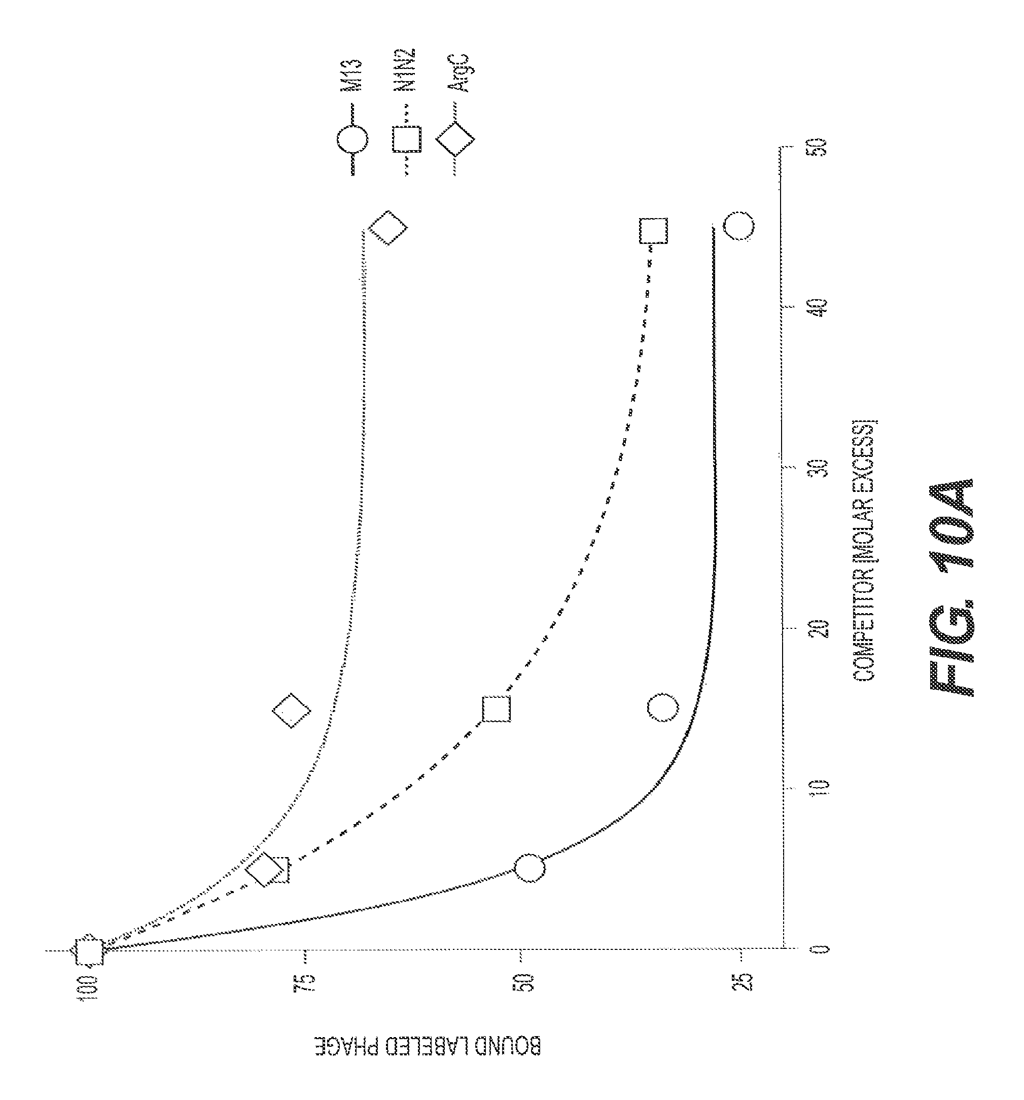
D00013
D00014

D00015
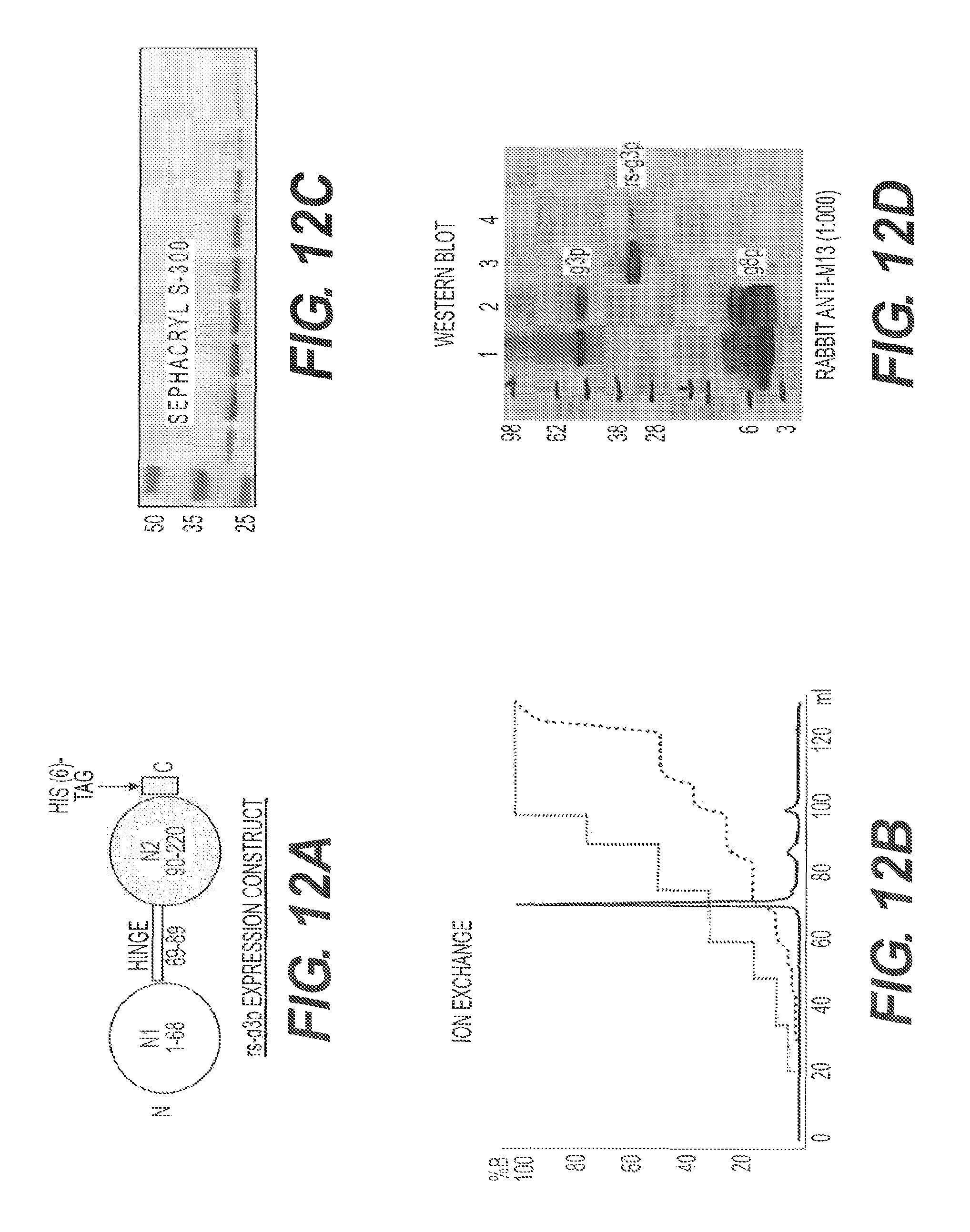
D00016
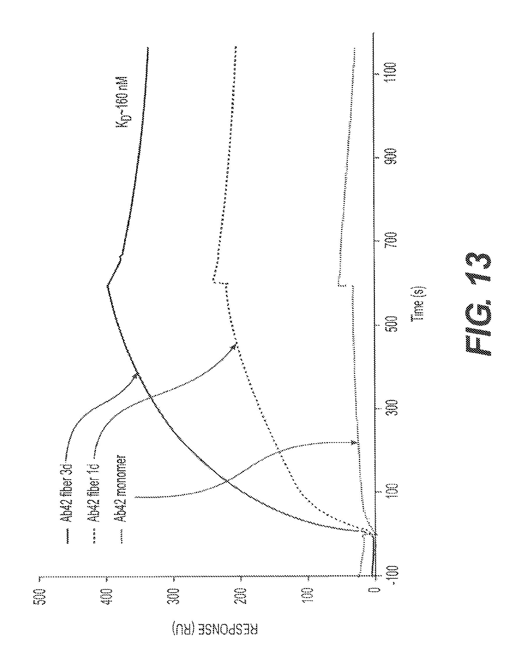
D00017
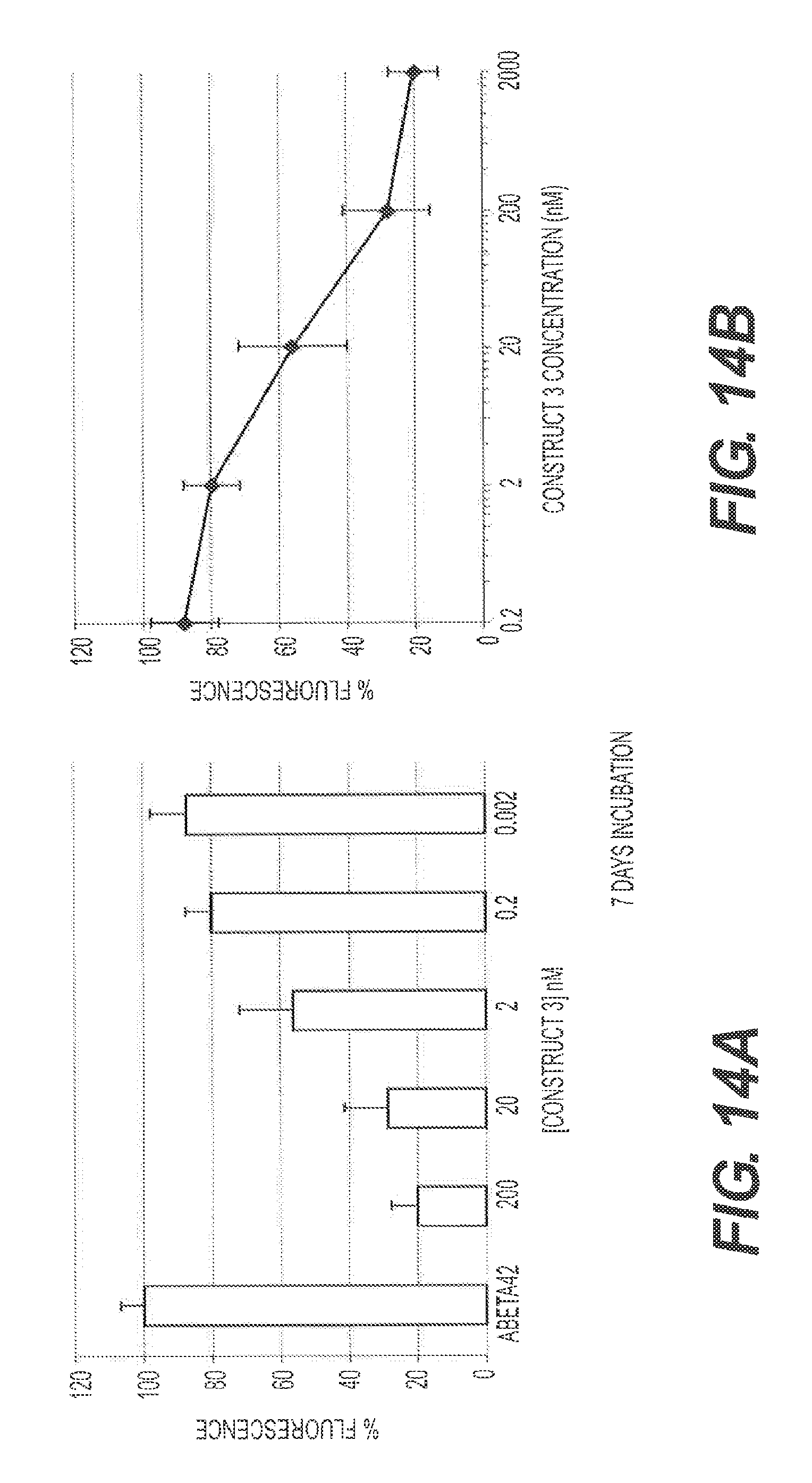
D00018
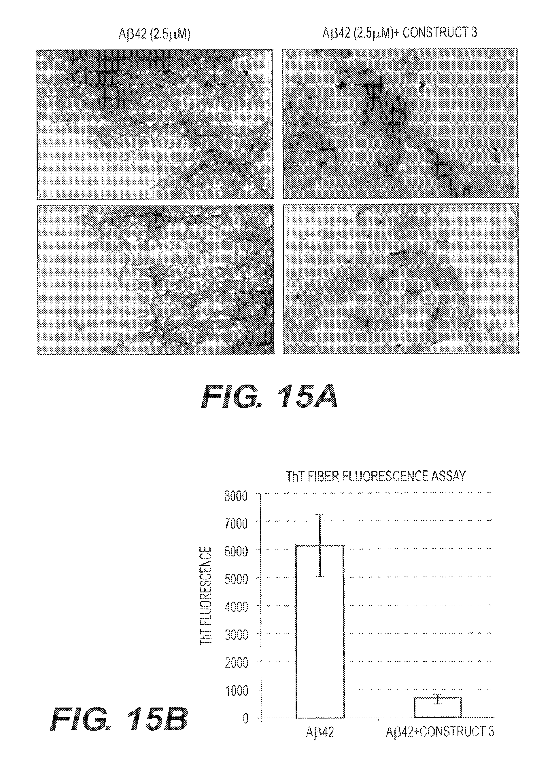
D00019
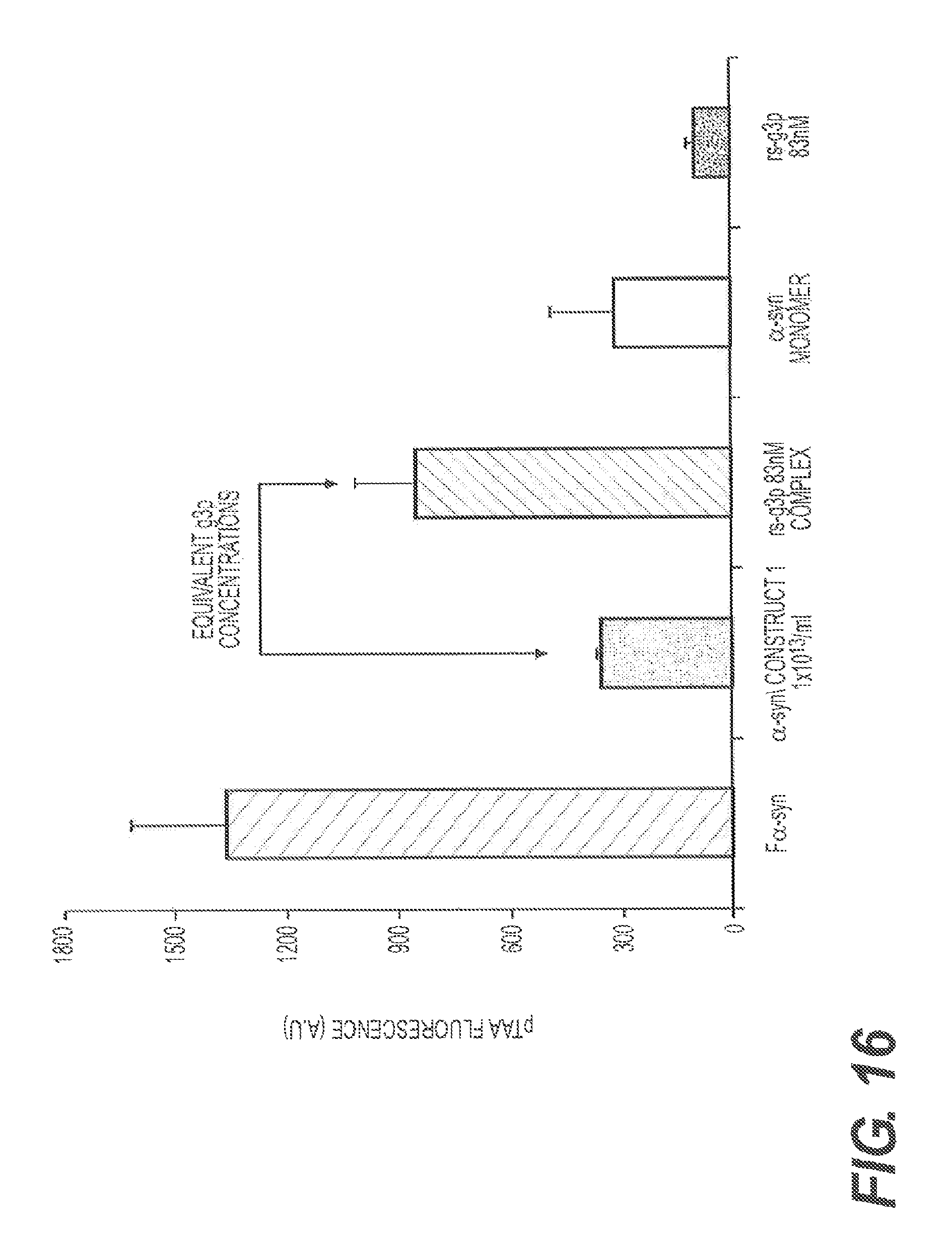
D00020
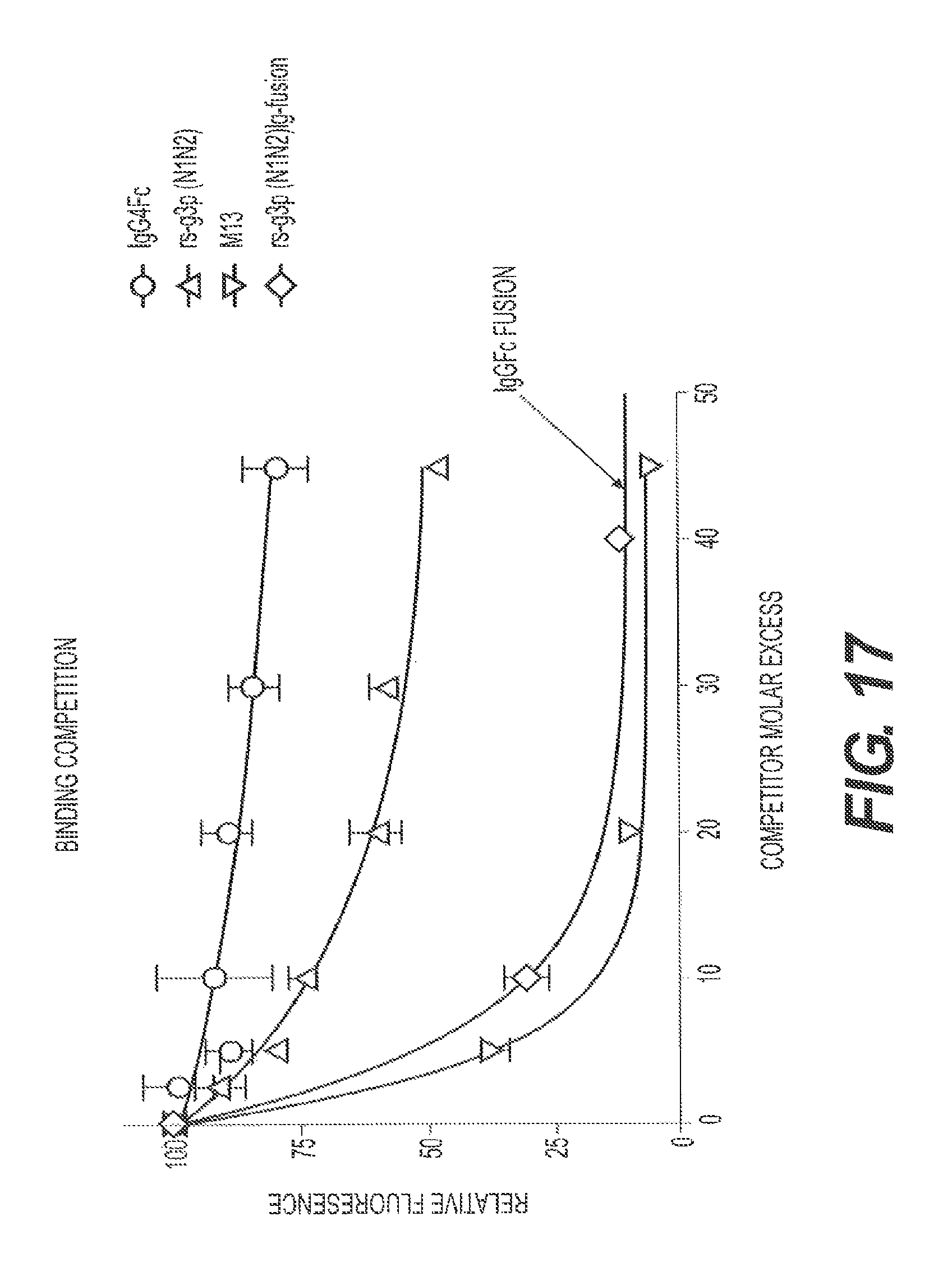
D00021
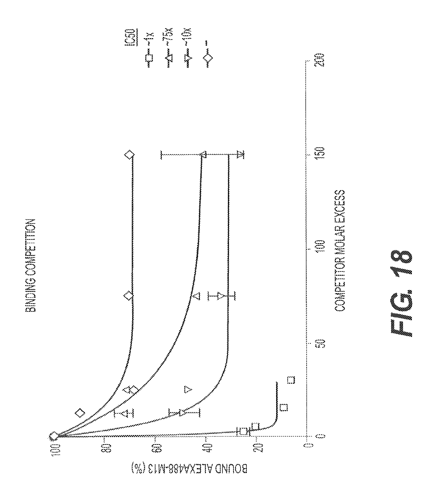
D00022
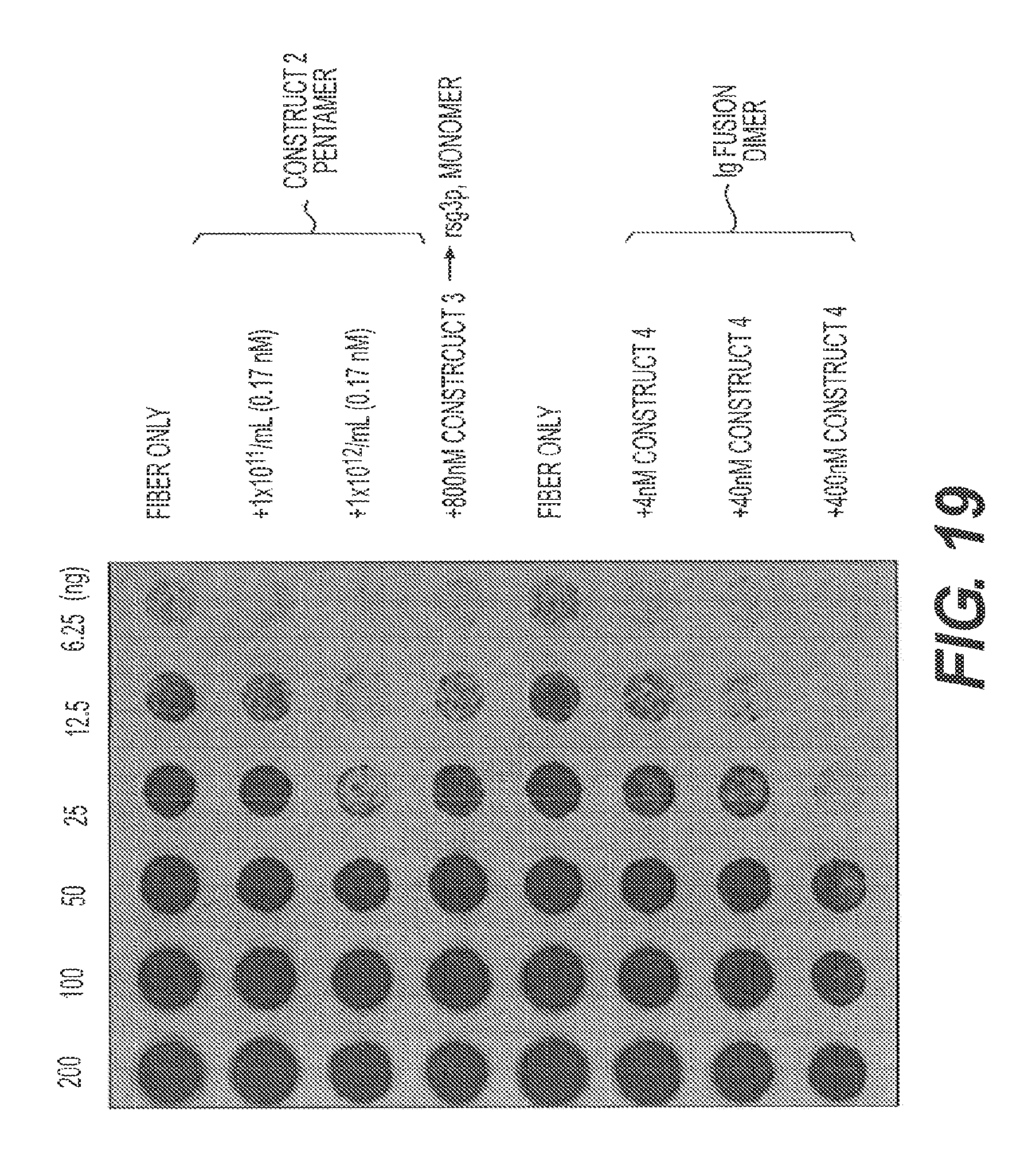
D00023
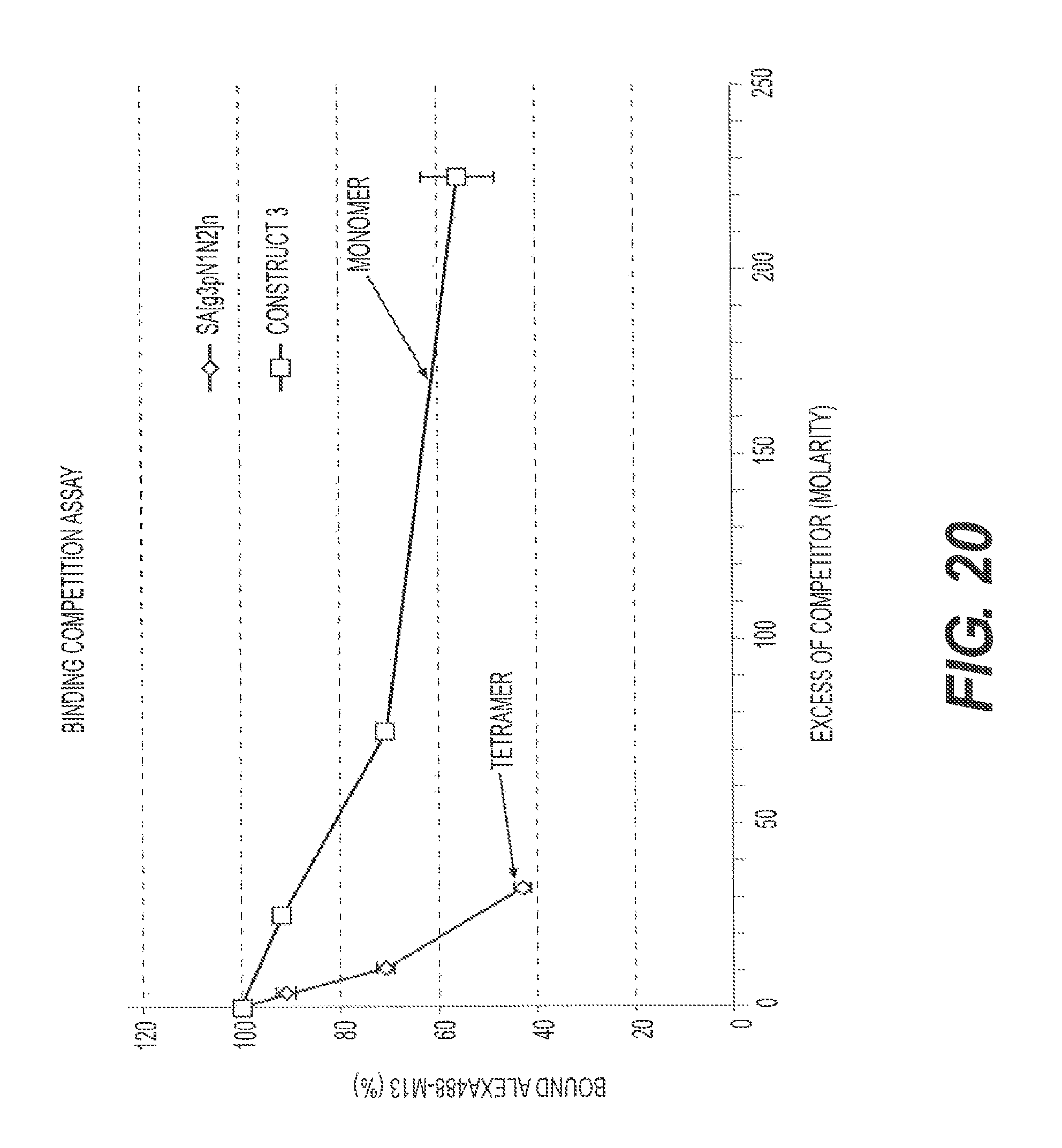
D00024

D00025
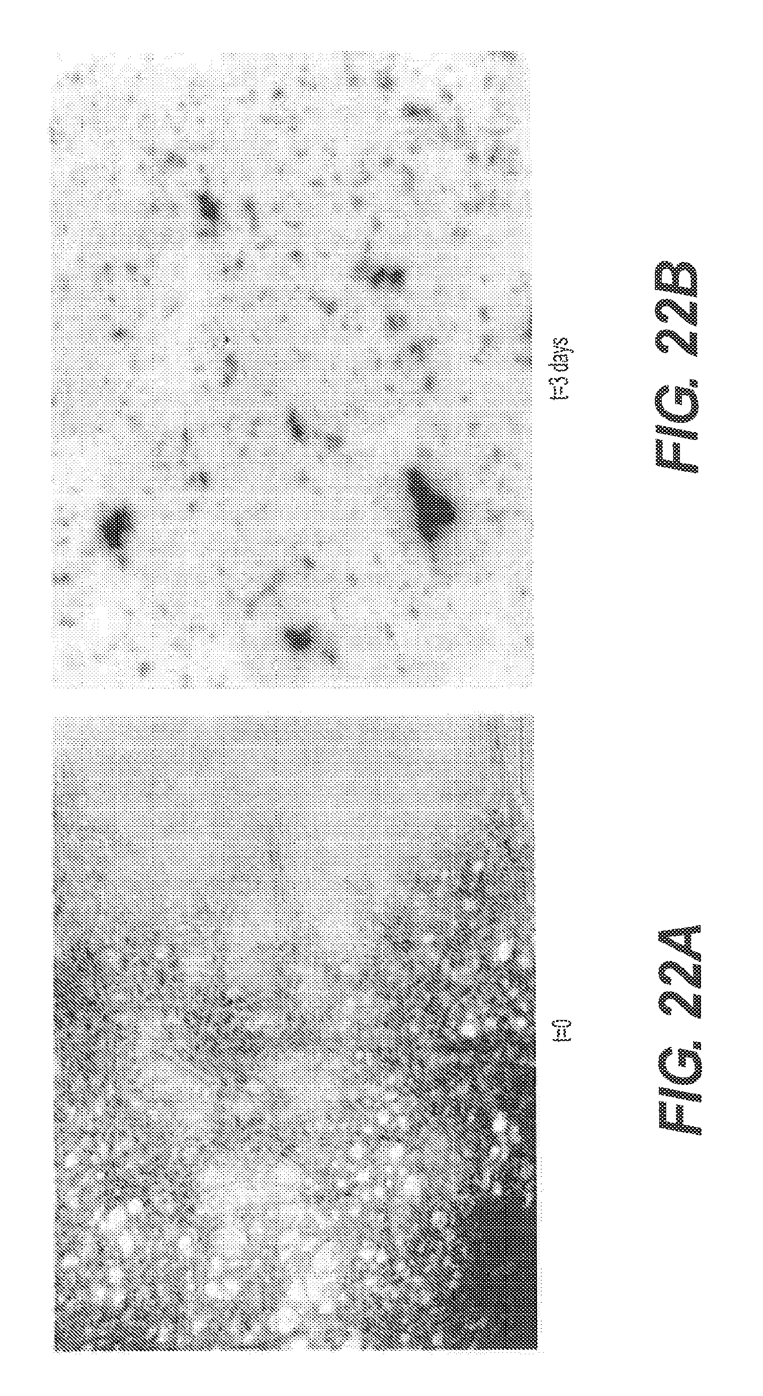
D00026
D00027
D00028
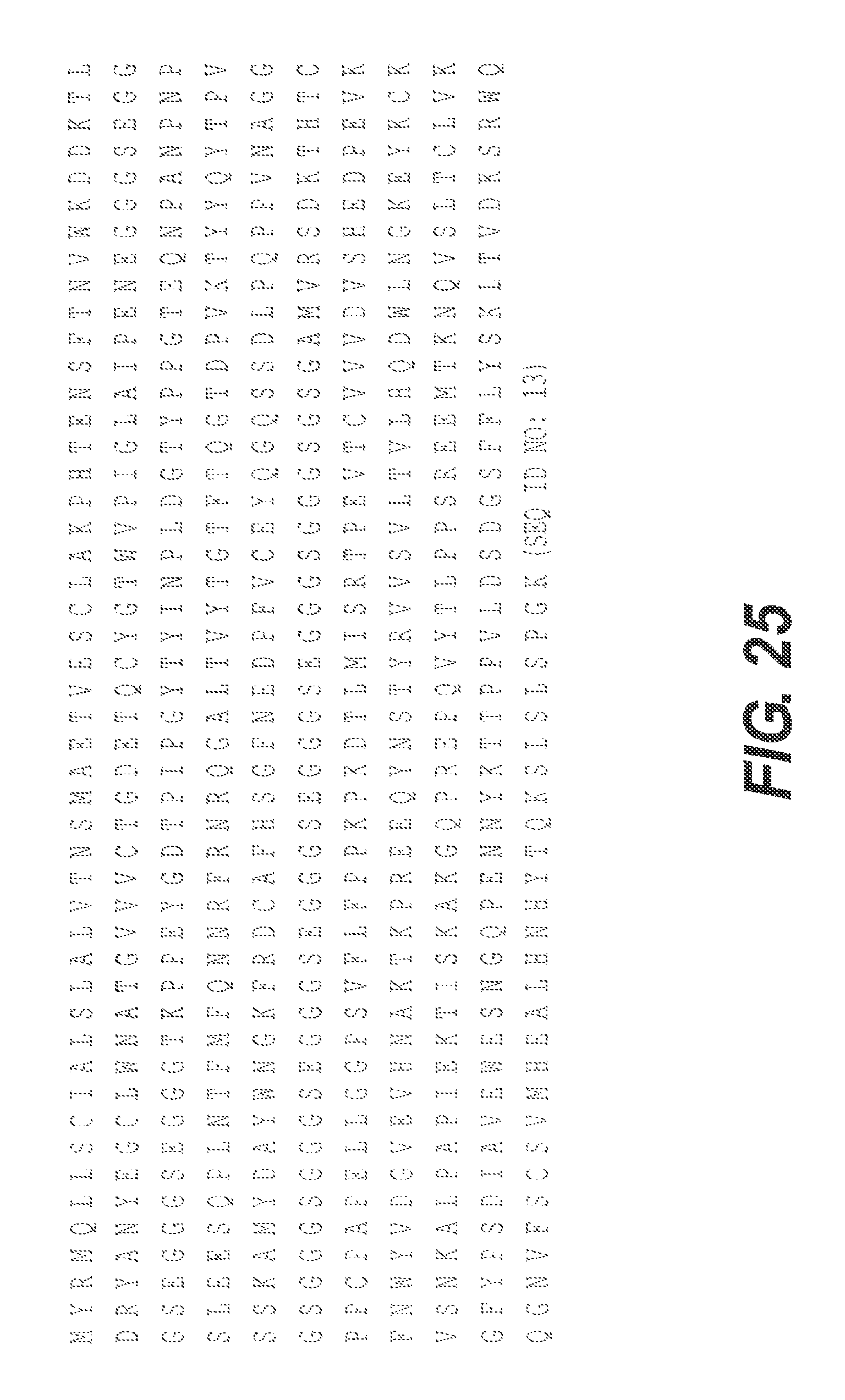
D00029
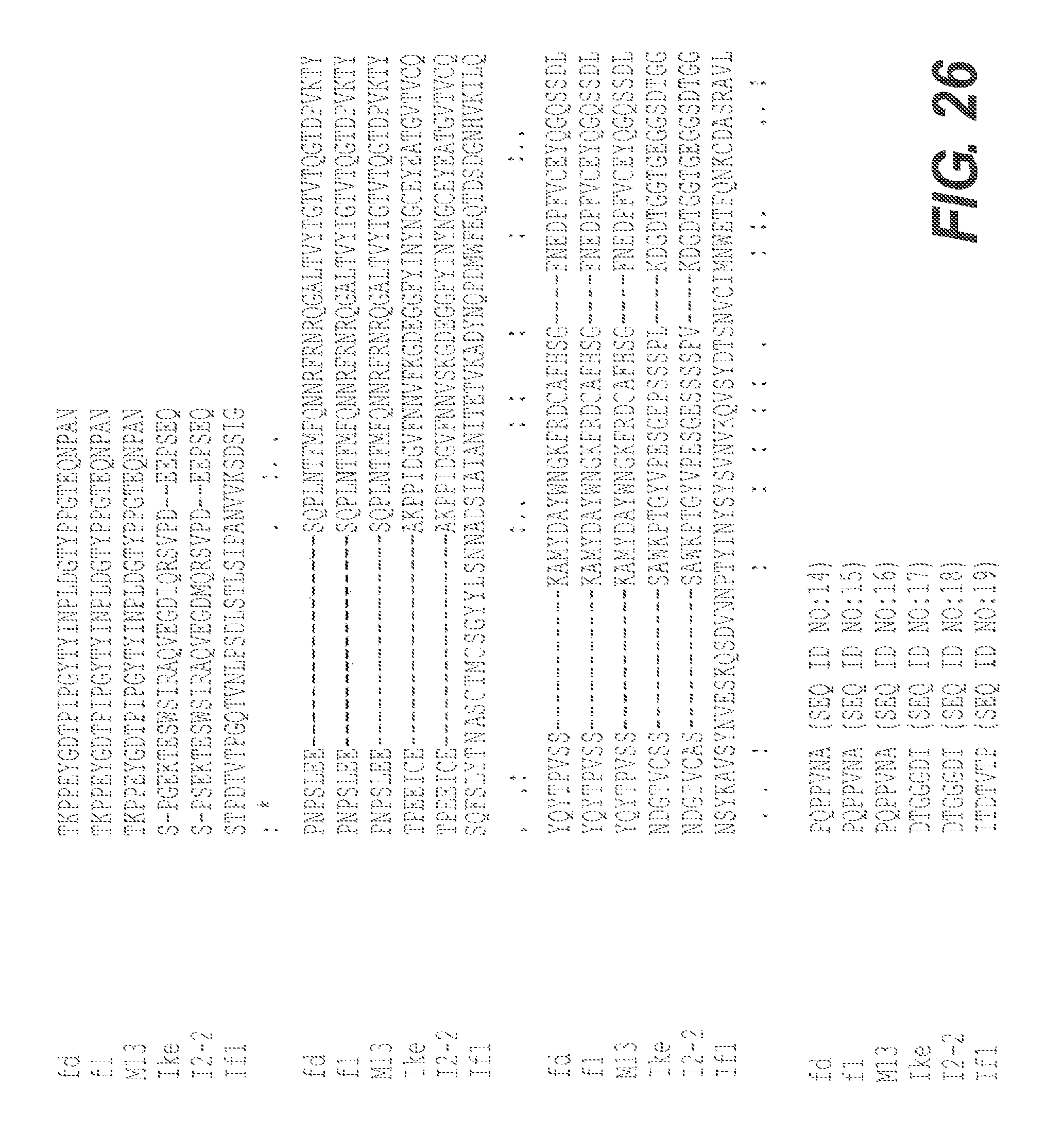
D00030

D00031

D00032
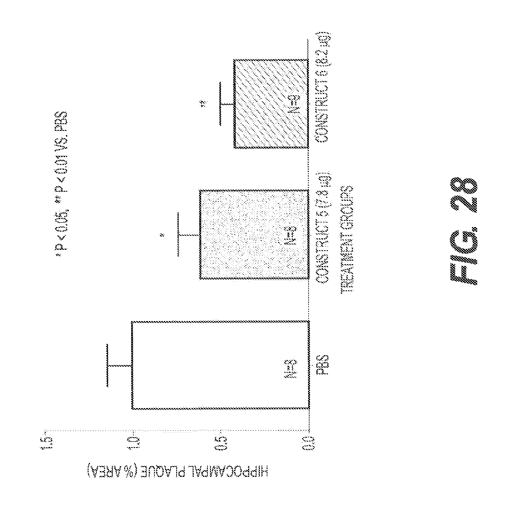
D00033
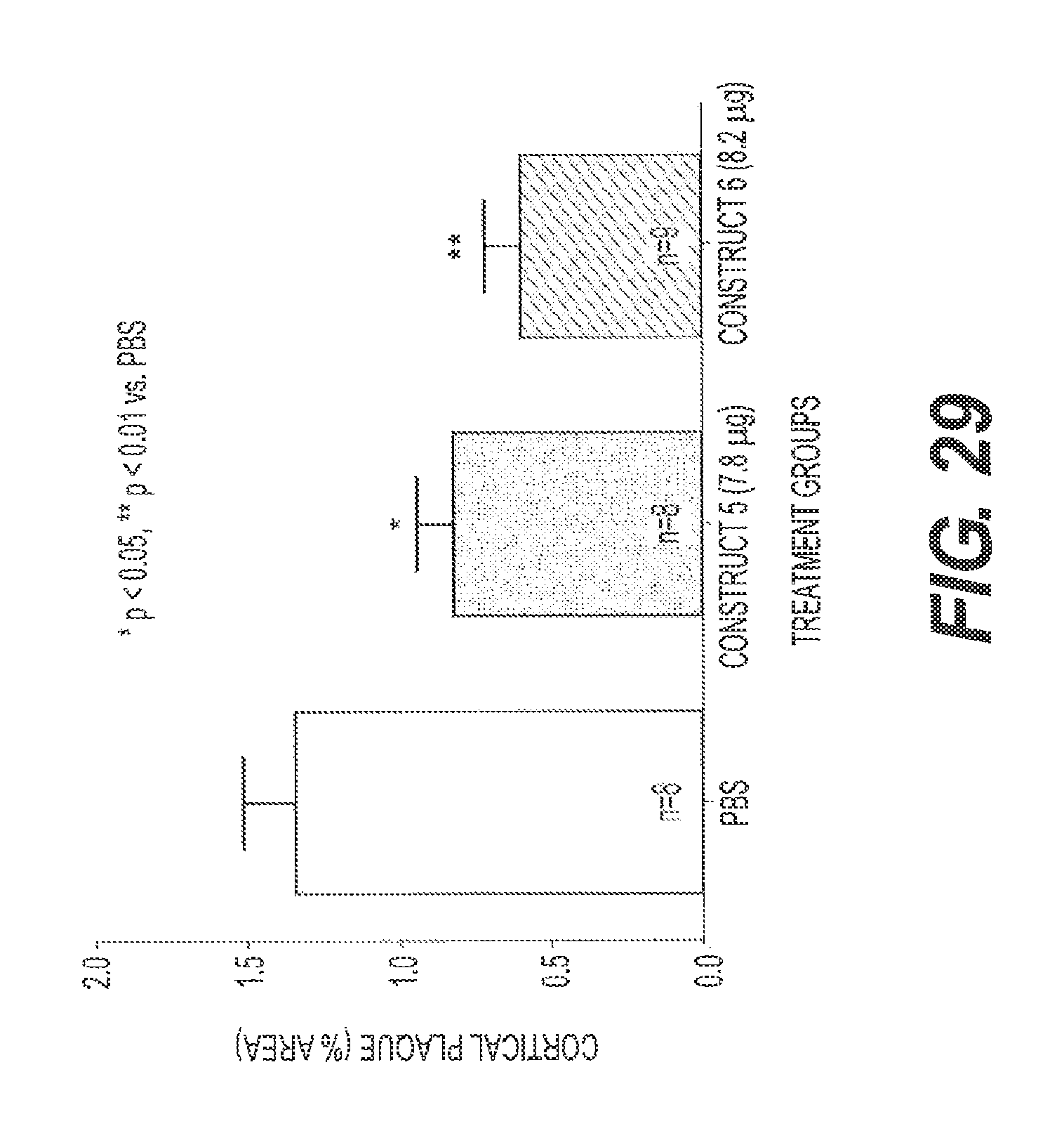
D00034
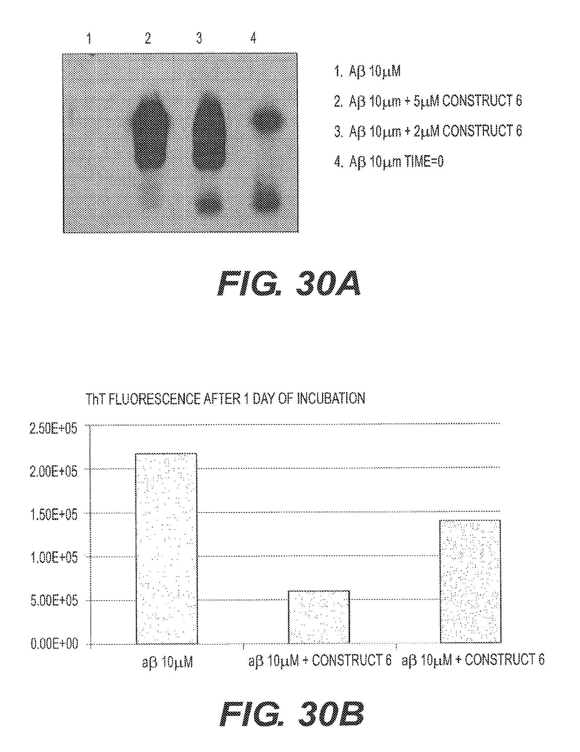
D00035
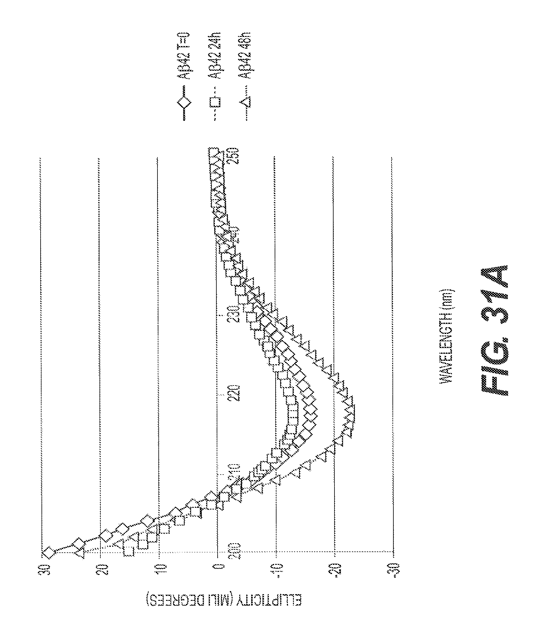
D00036

D00037
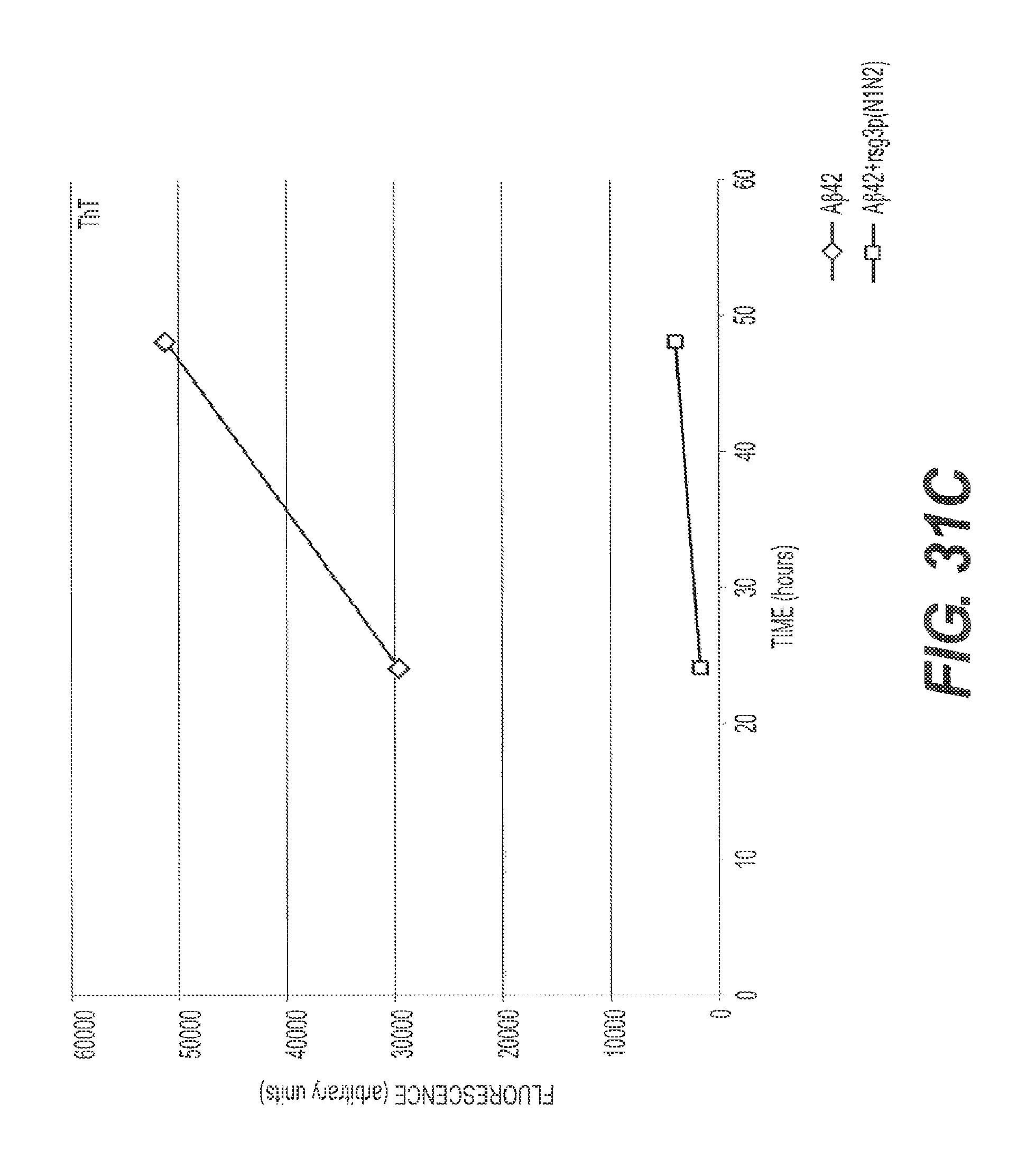
D00038
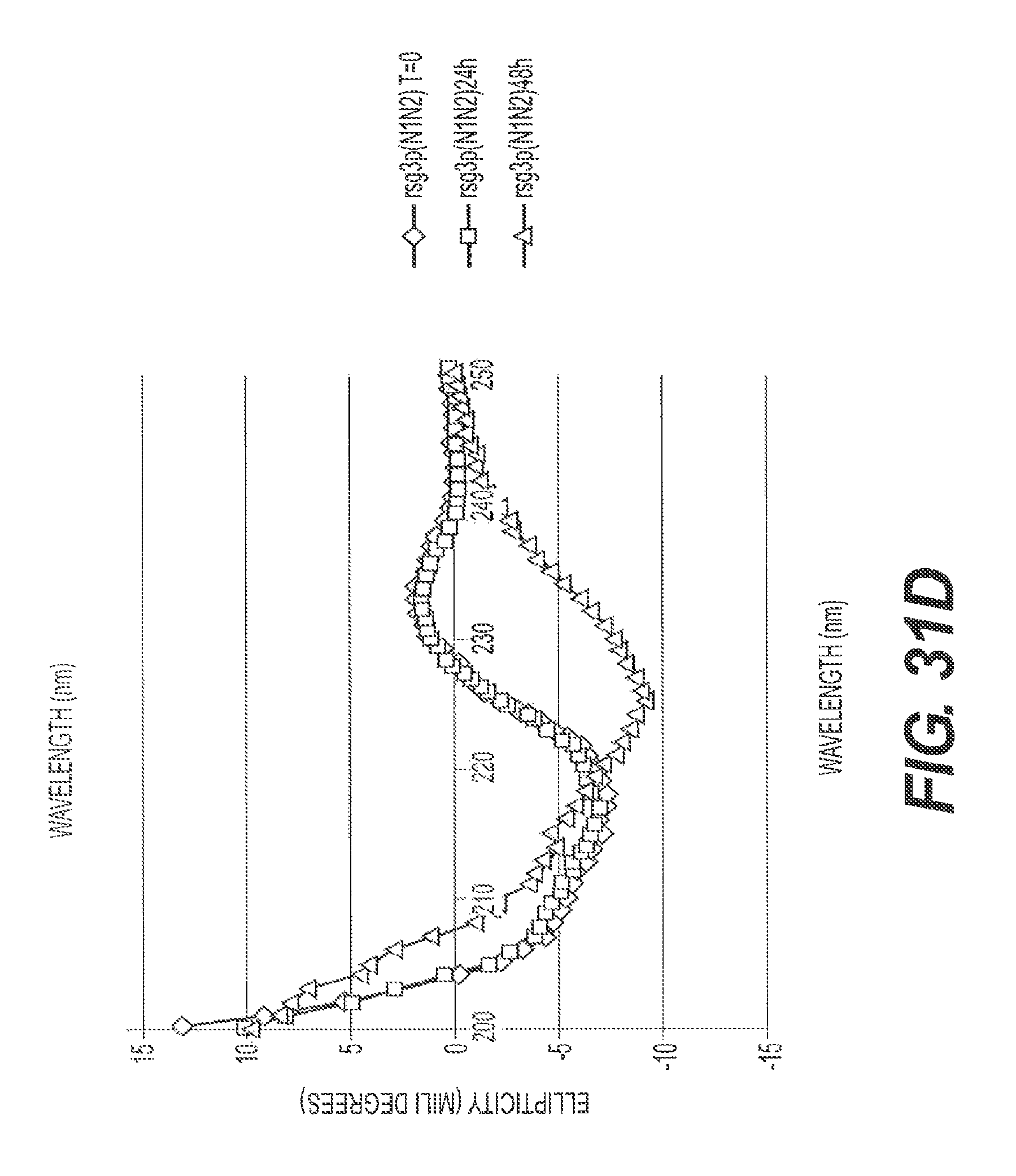
D00039
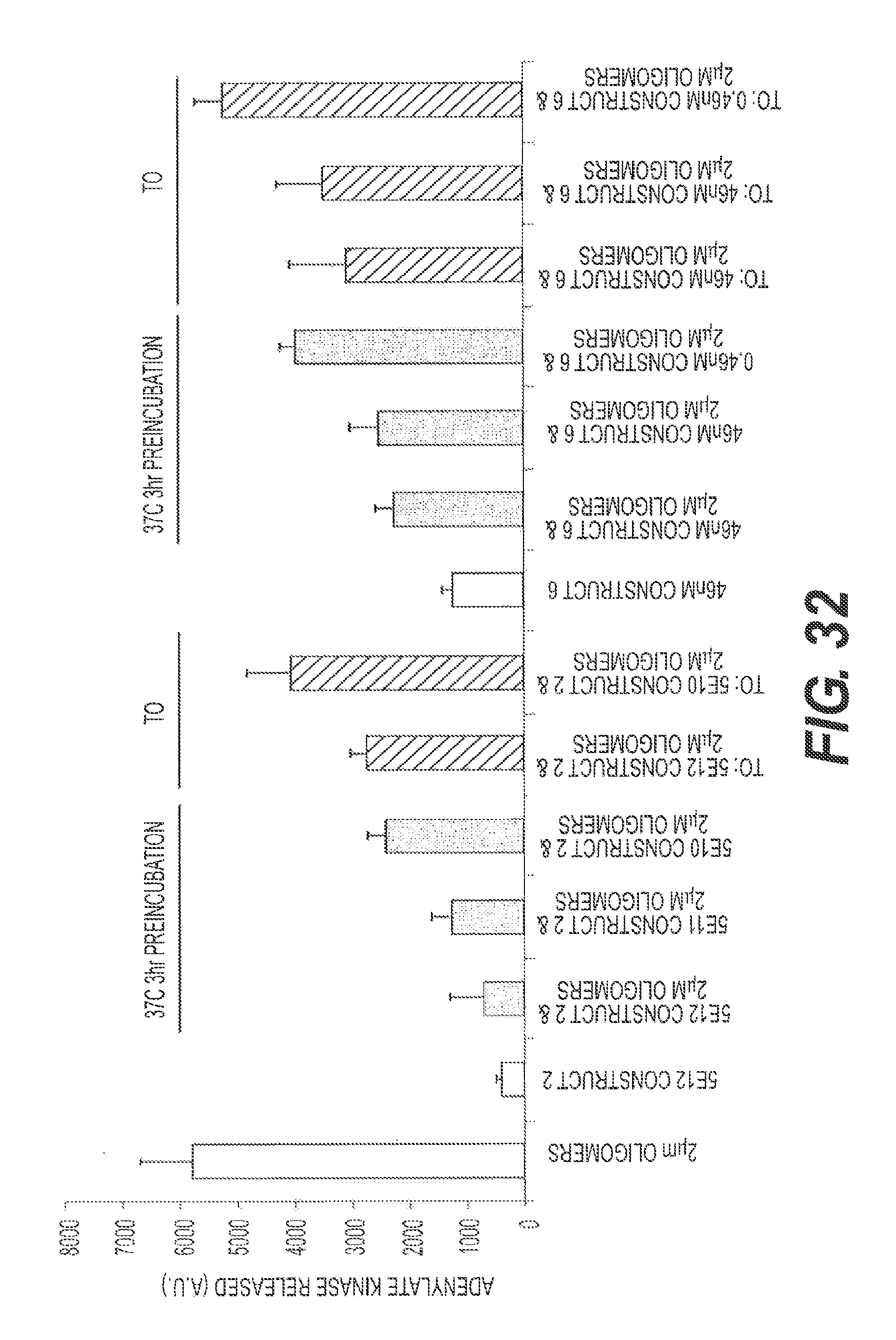
D00040

D00041
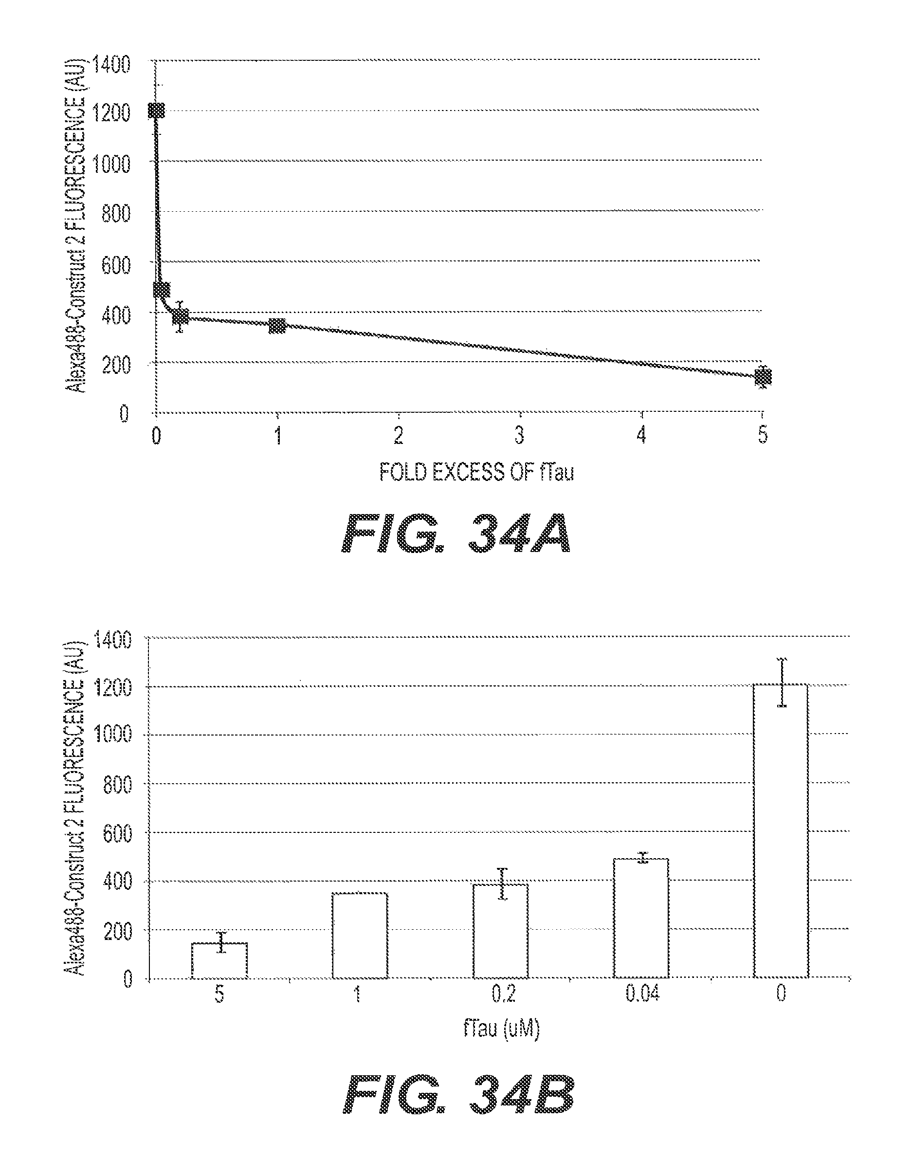
D00042
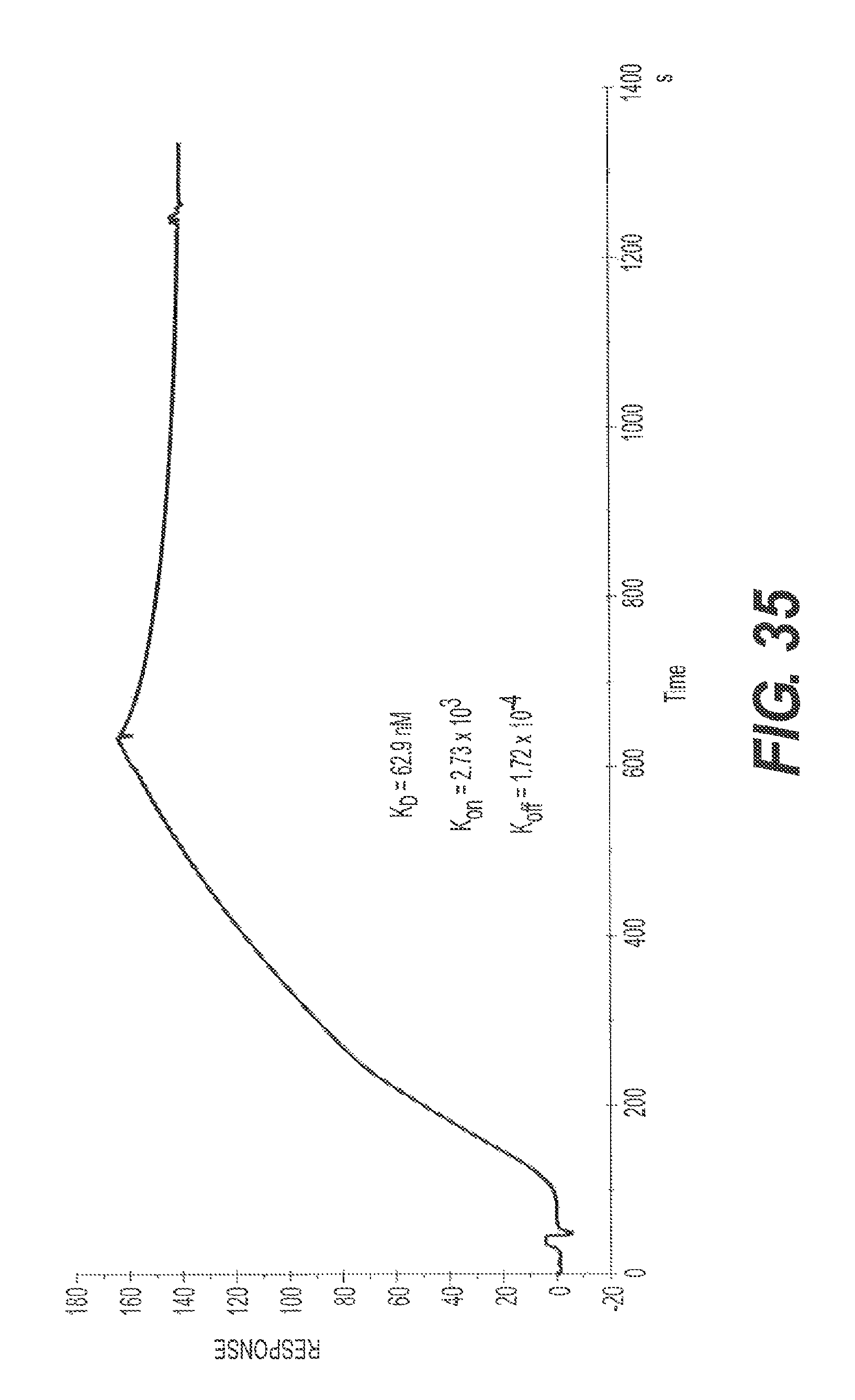
D00043
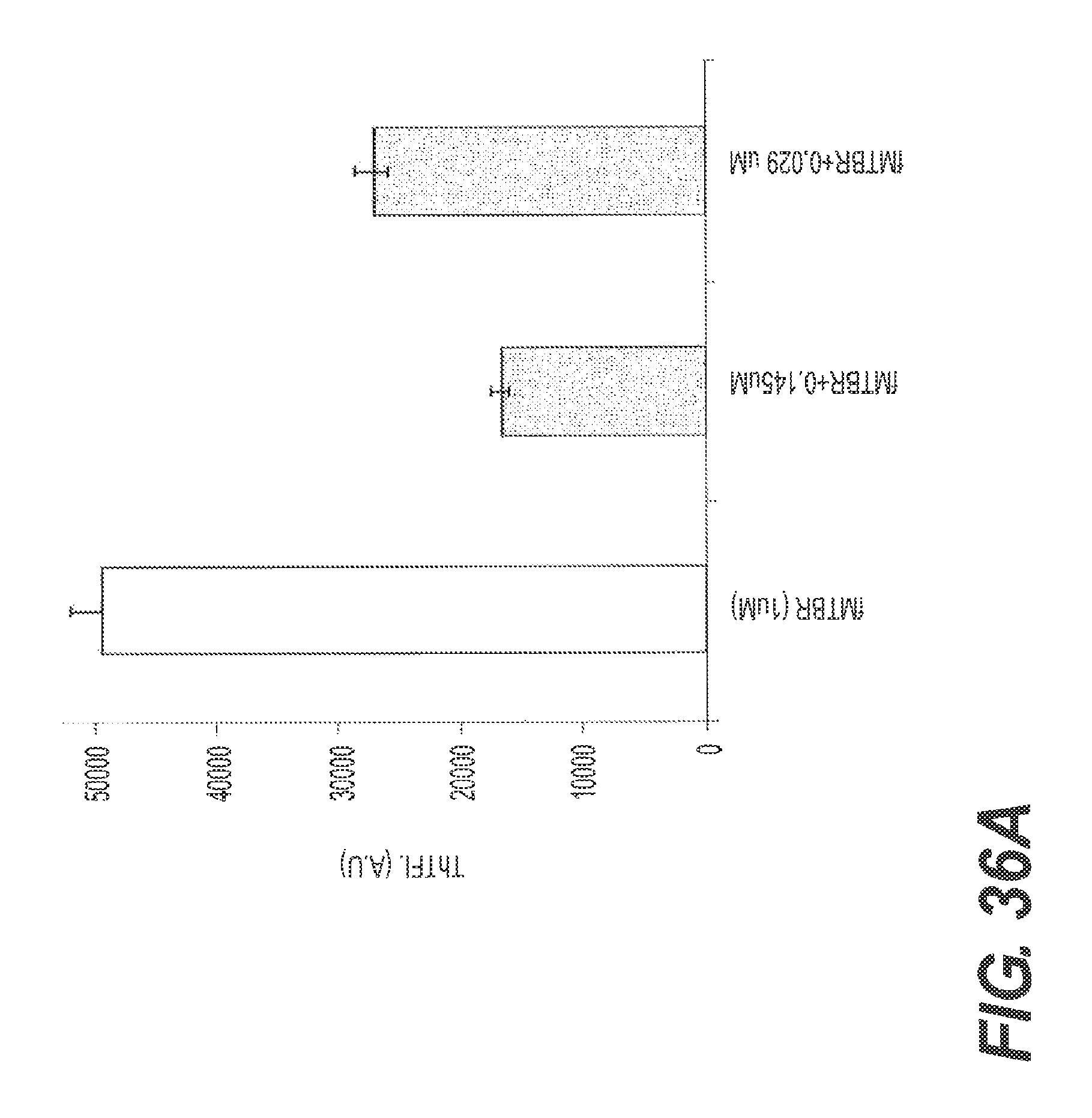
D00044
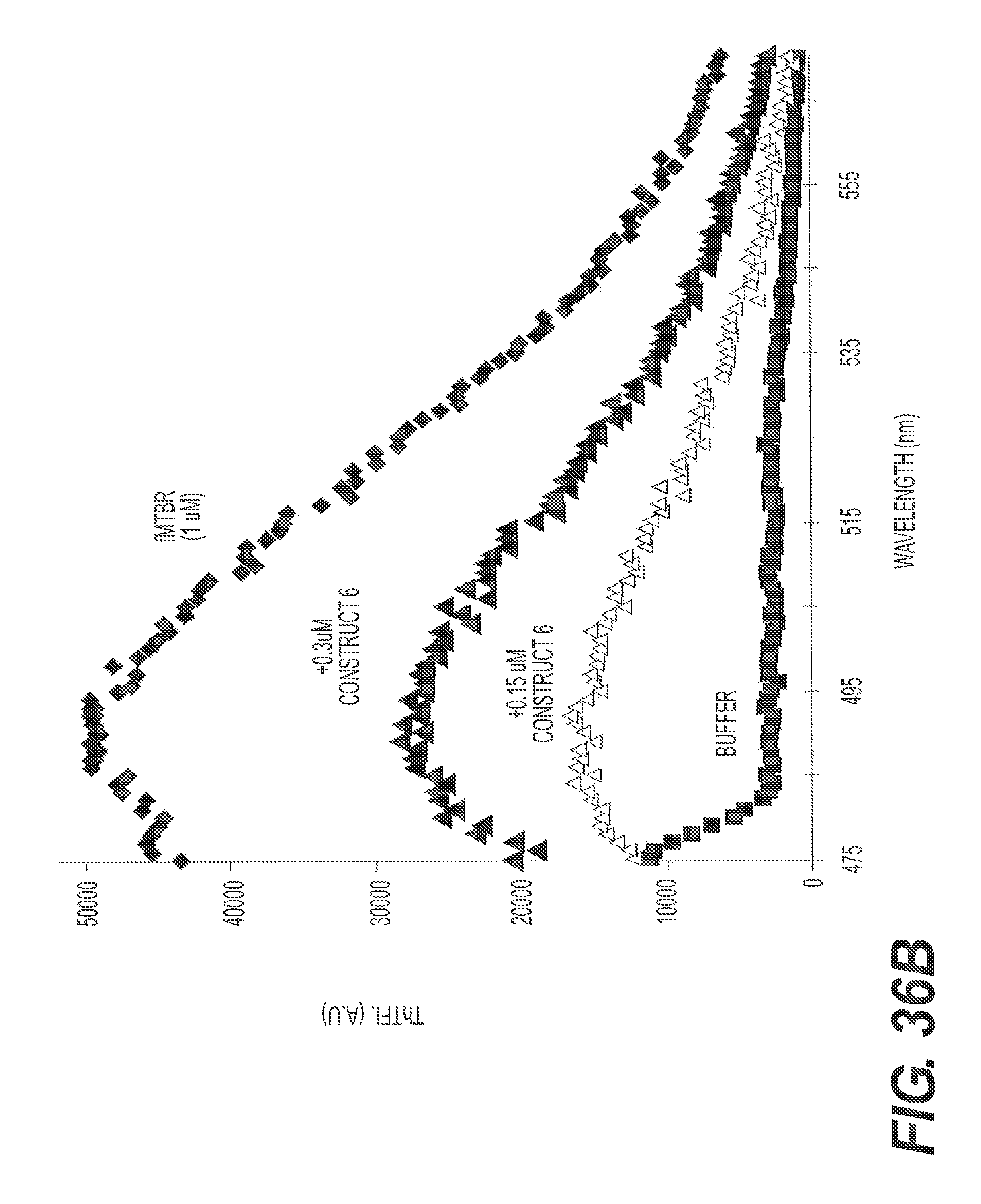
D00045

D00046
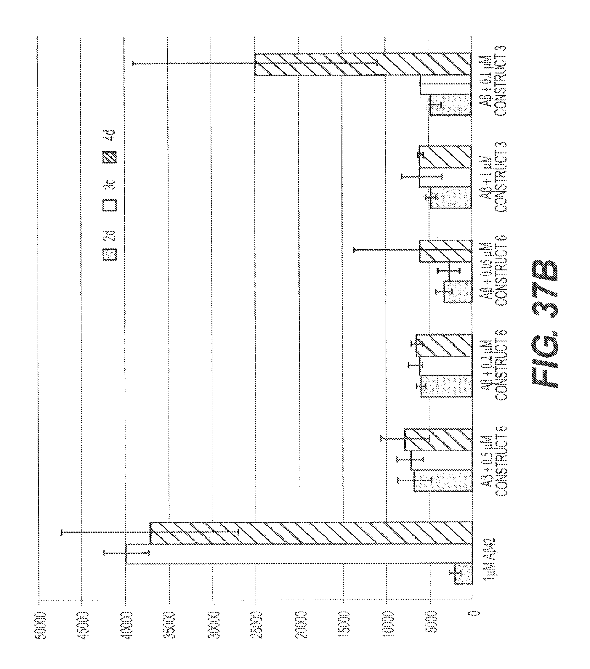
D00047
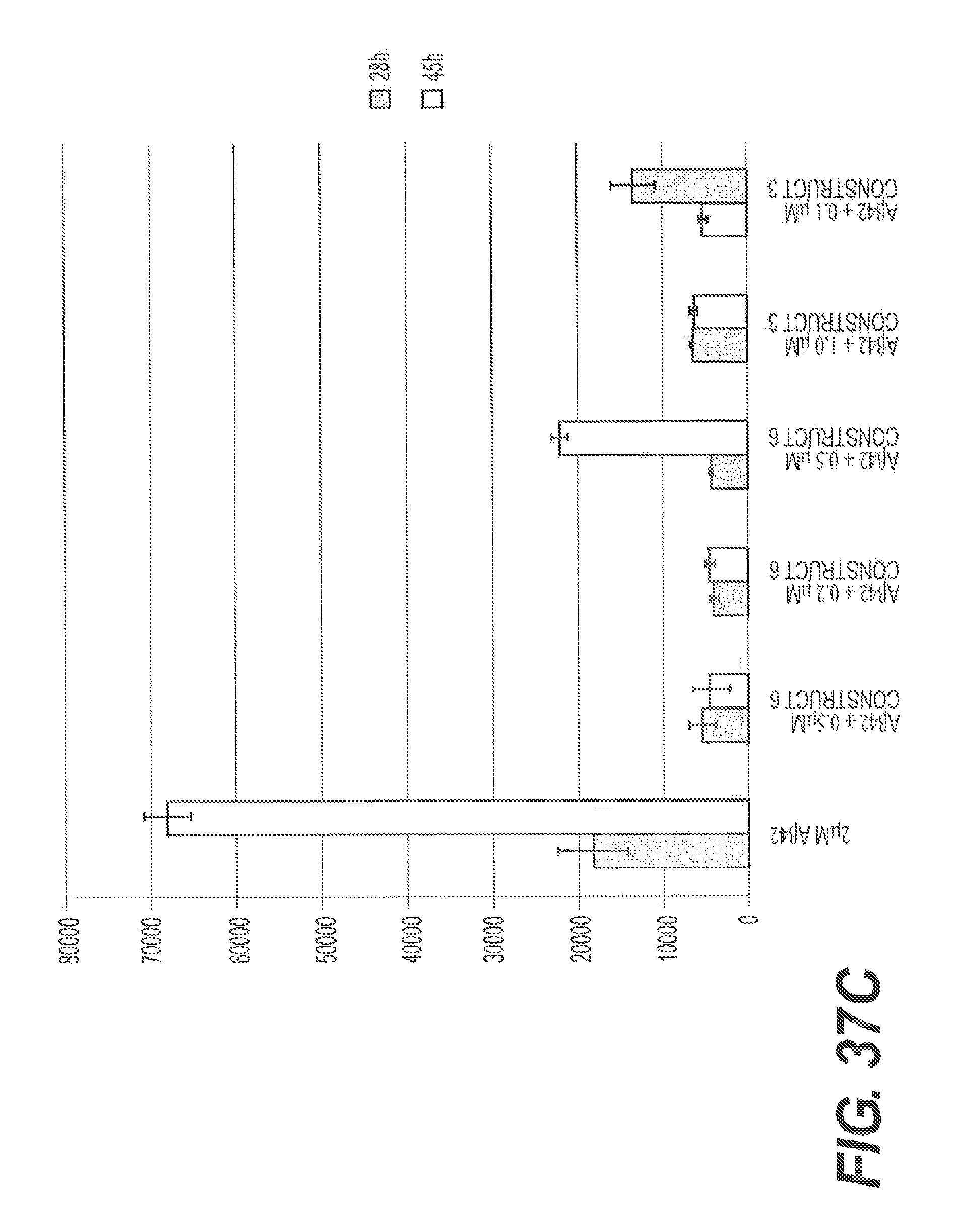
D00048
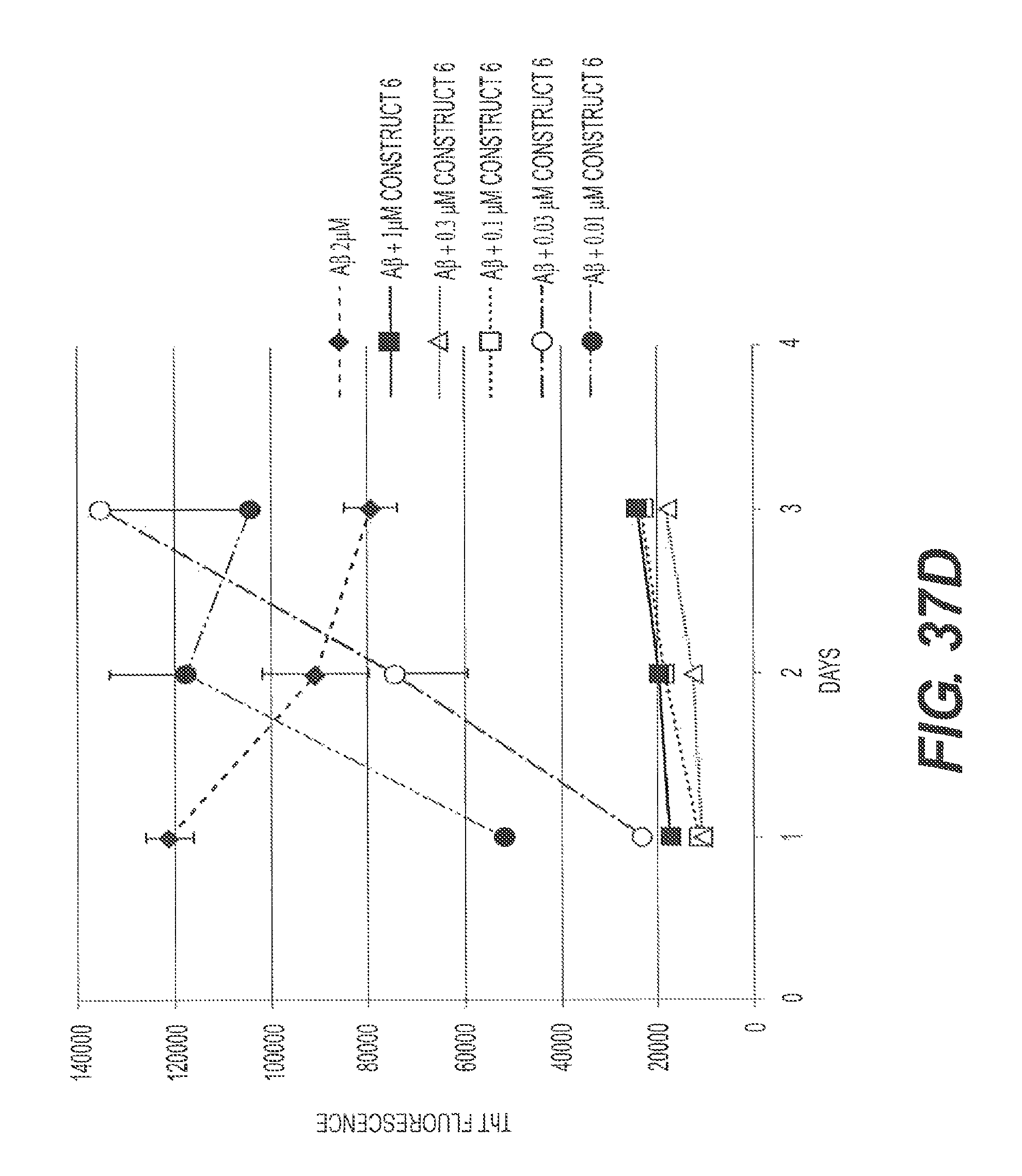
D00049
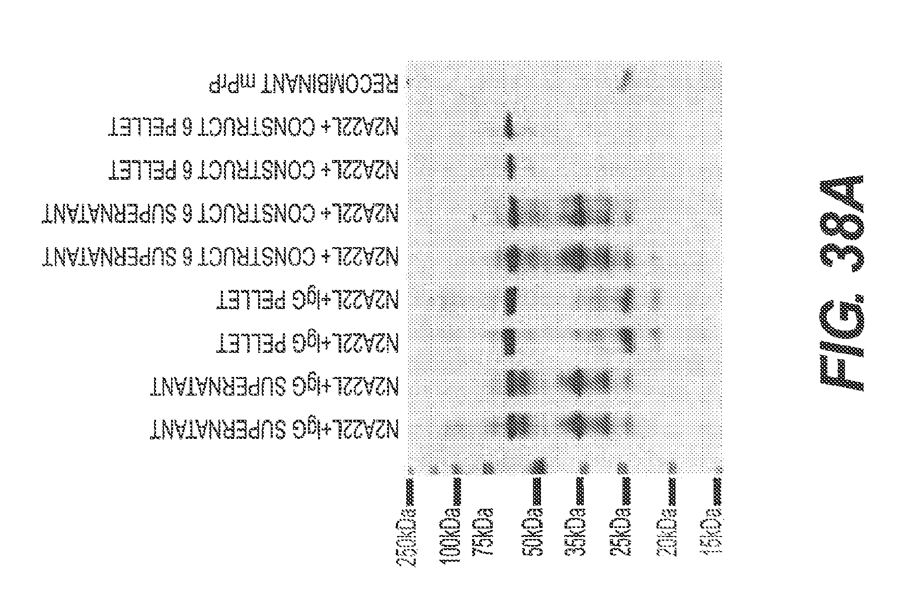
D00050
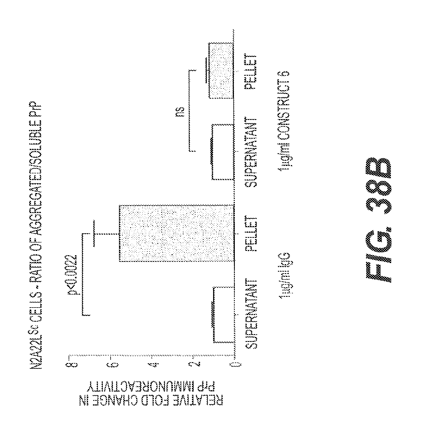
D00051
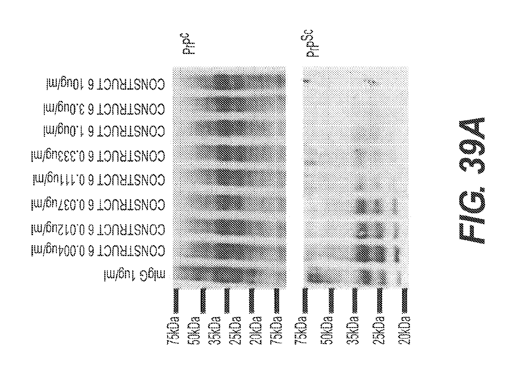
D00052
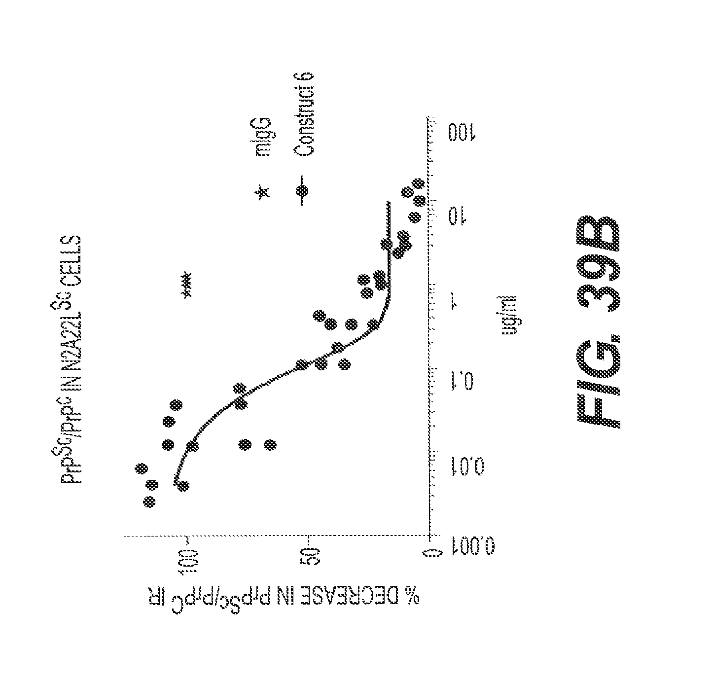
D00053
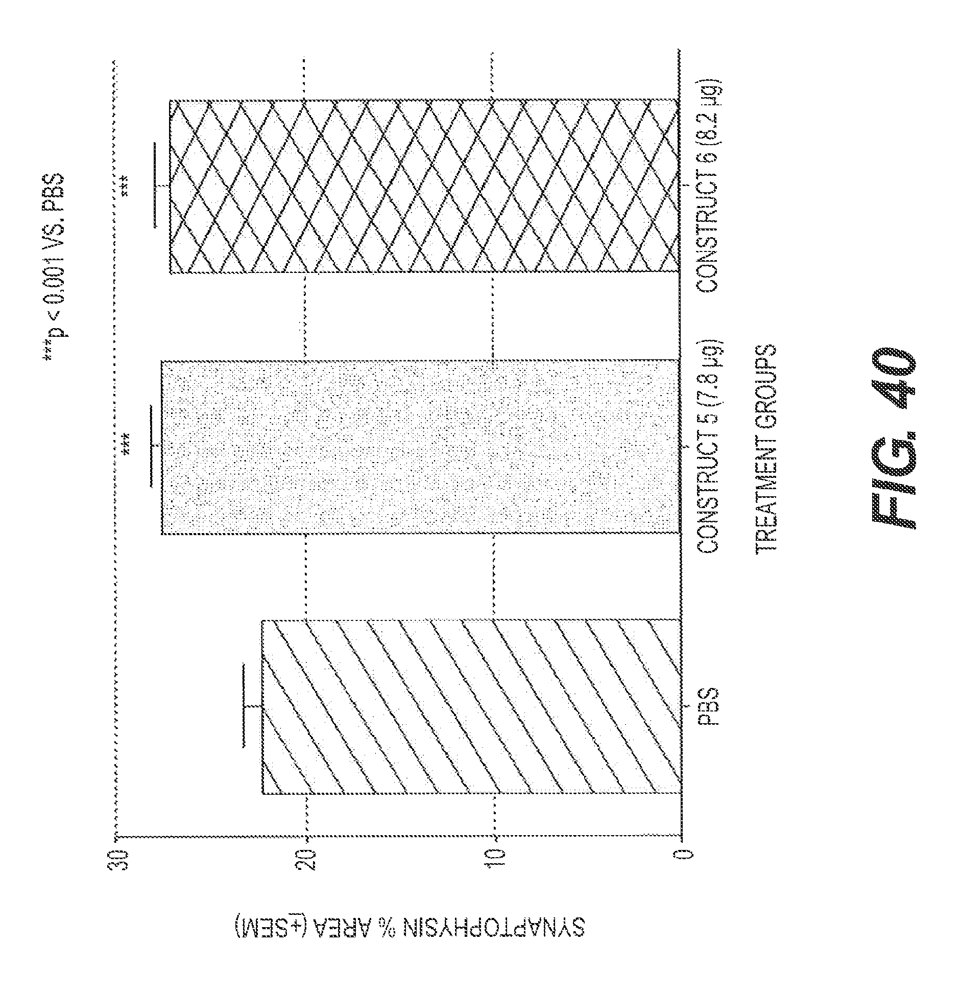
D00054
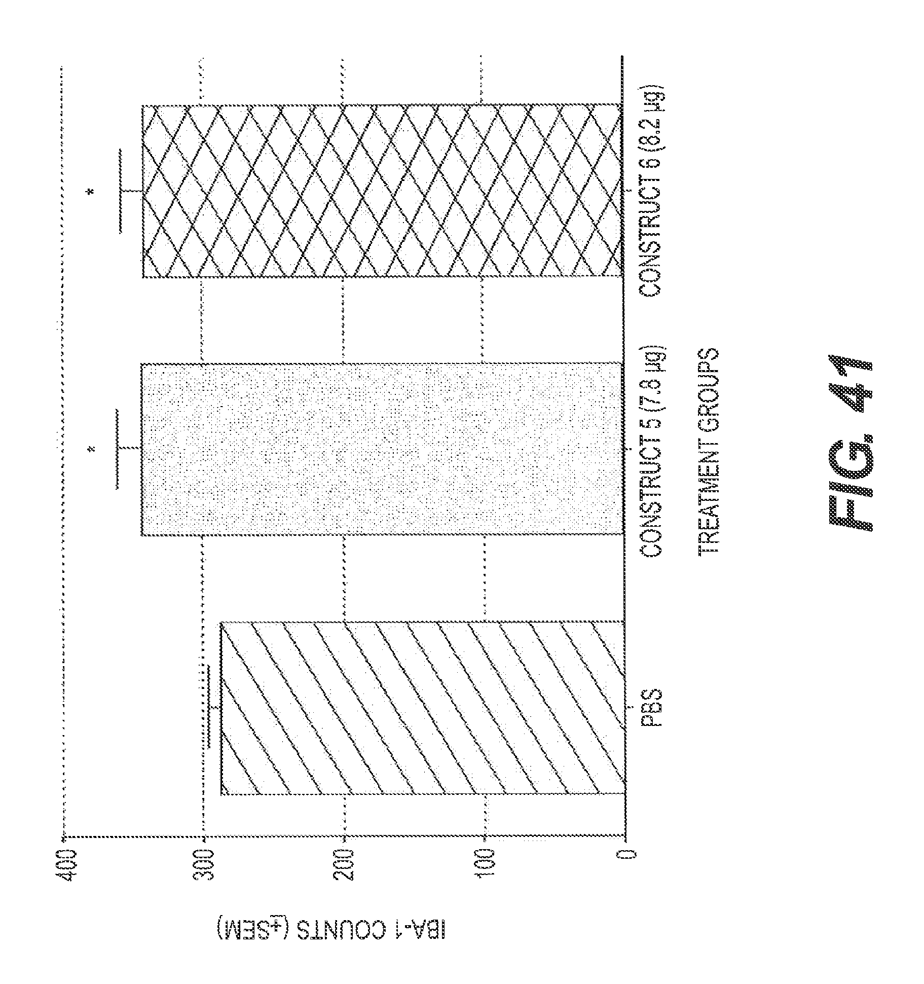
D00055
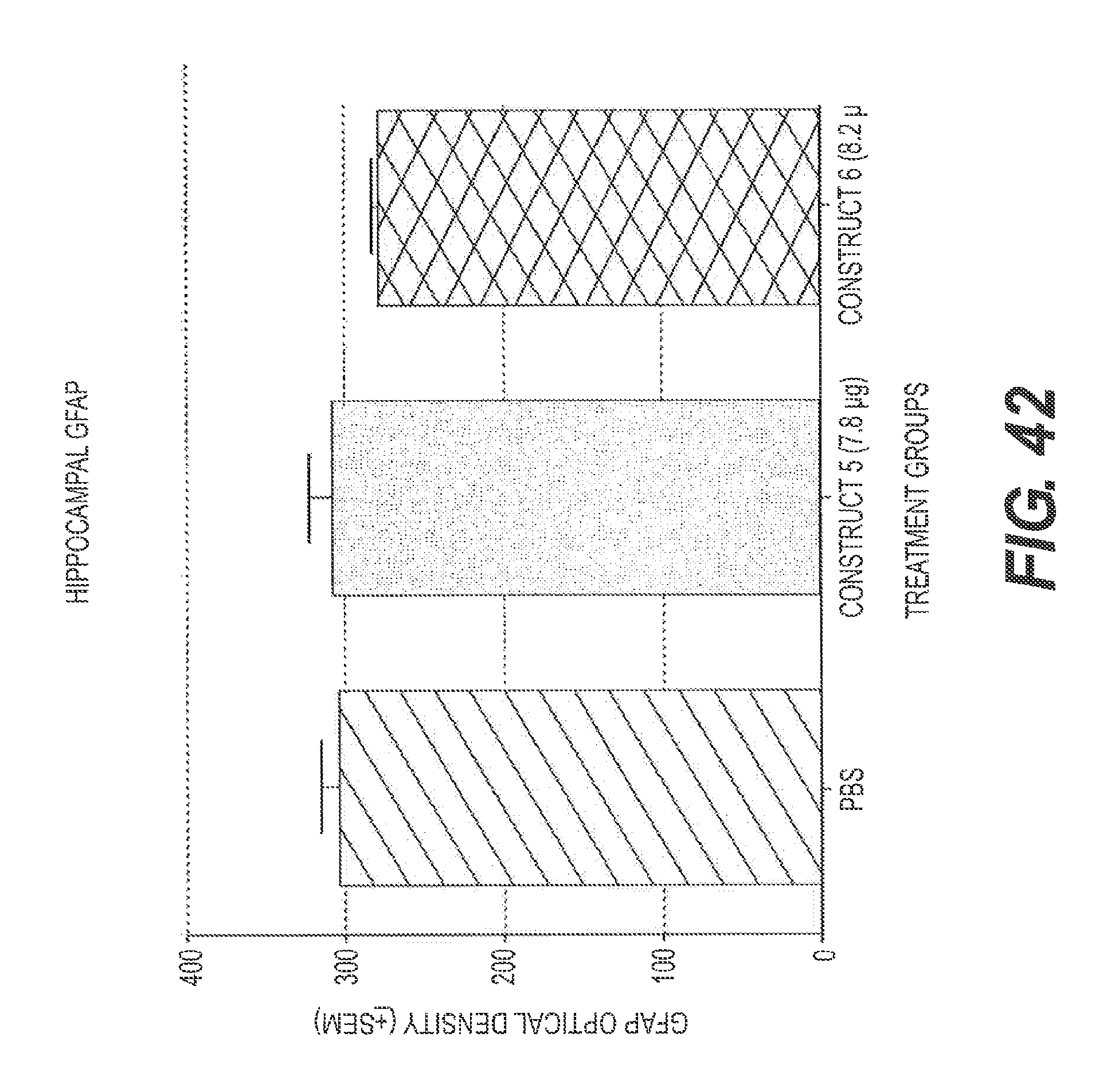
D00056
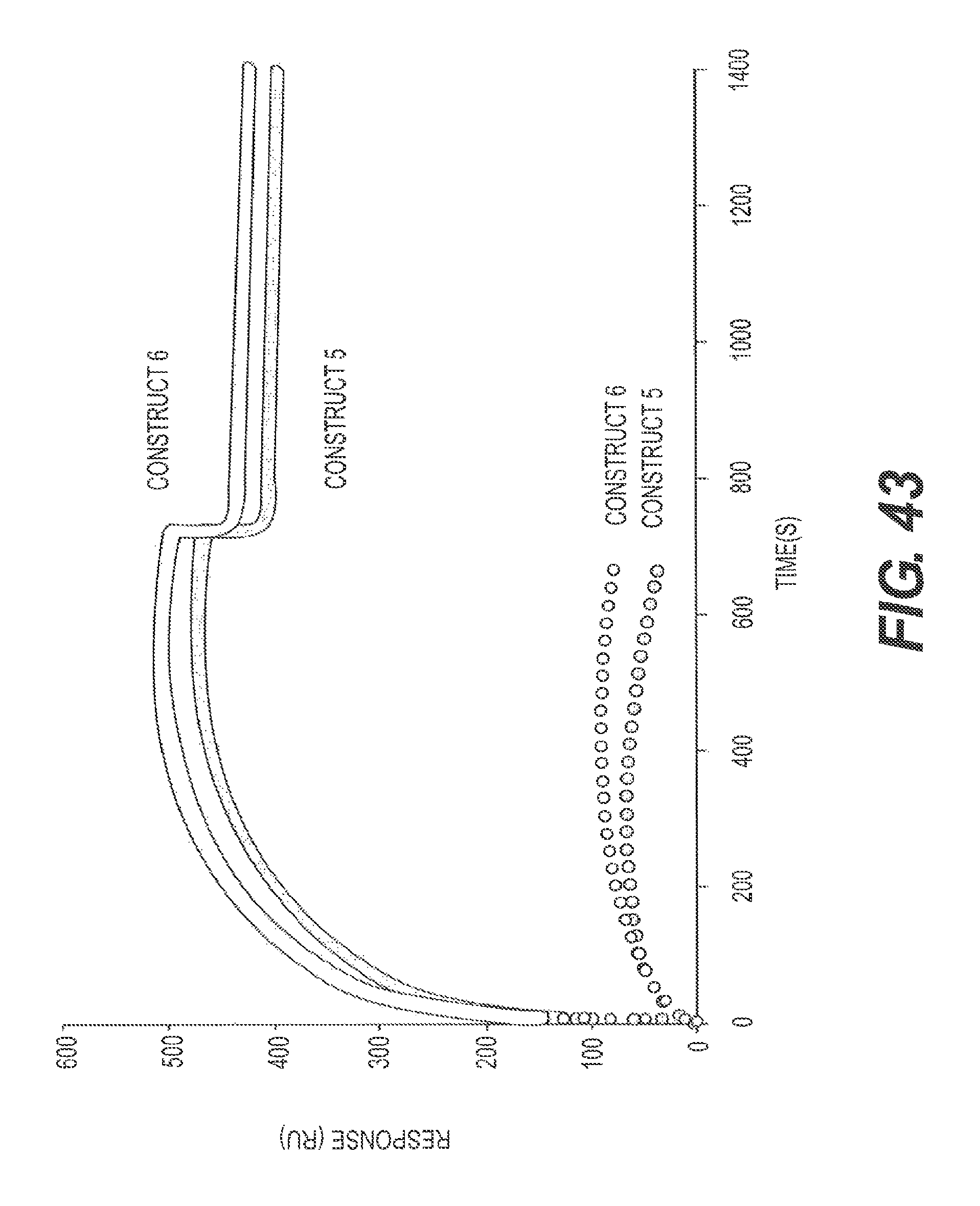
D00057
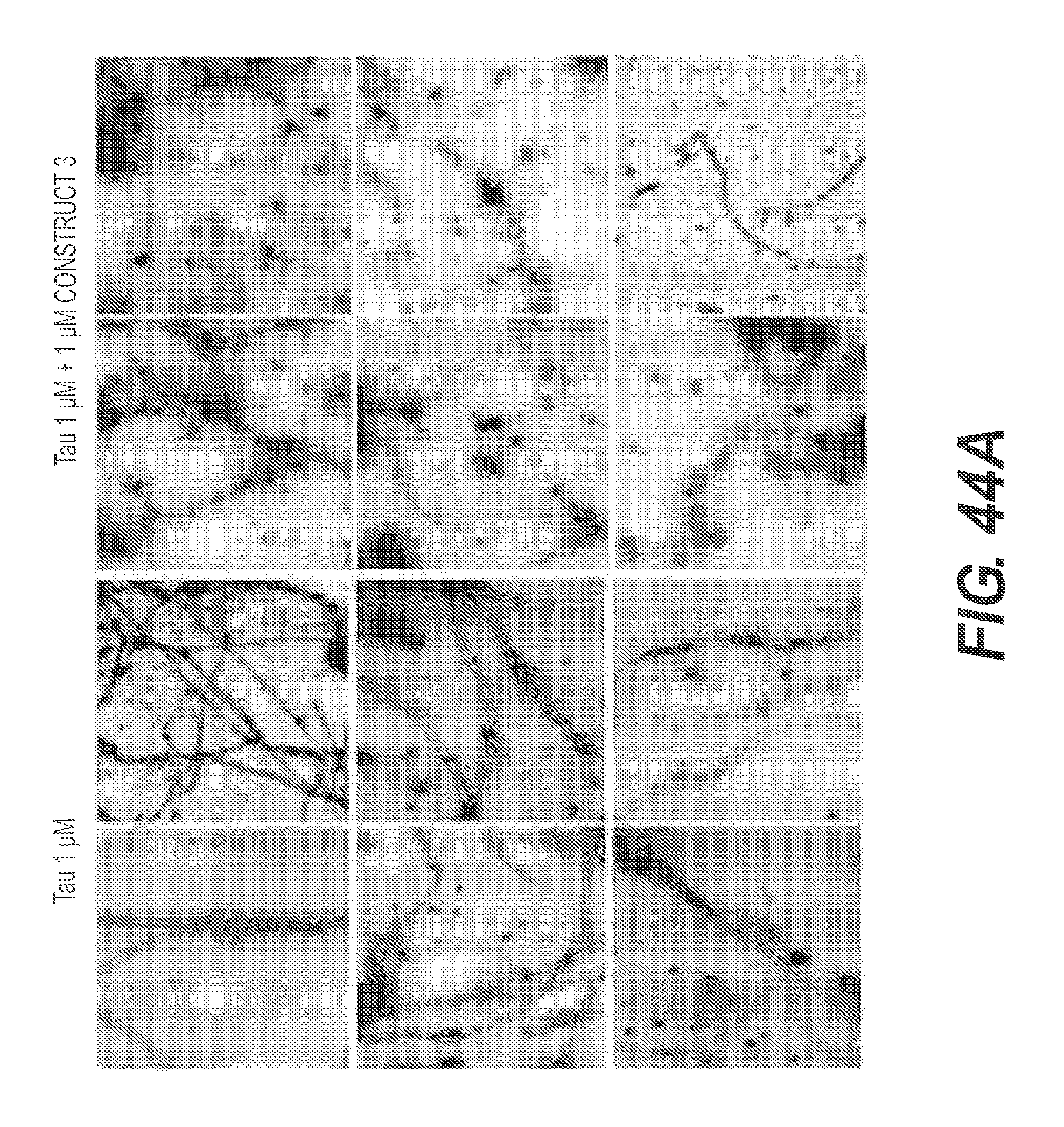
D00058
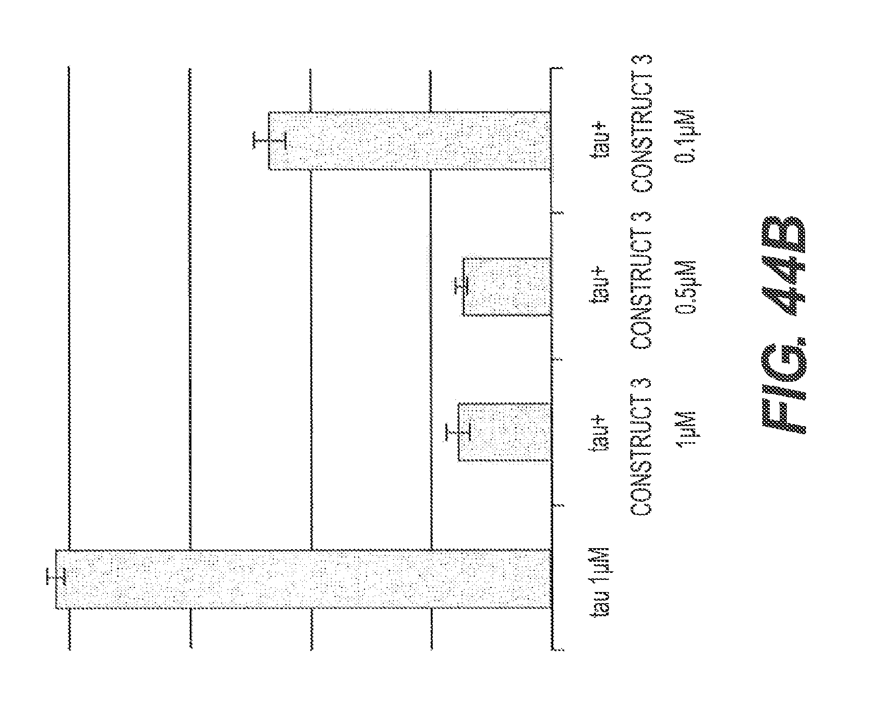
D00059
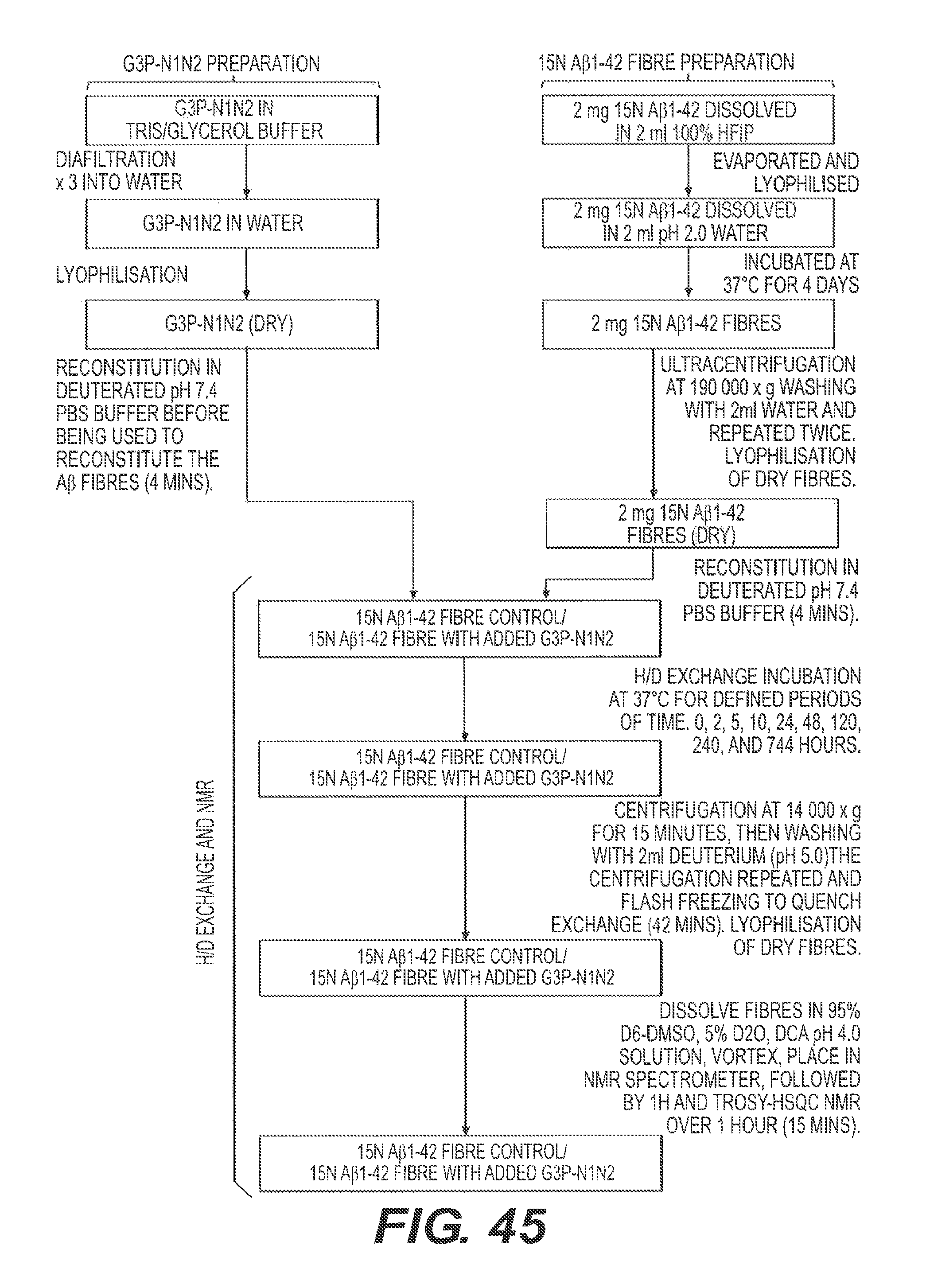
D00060
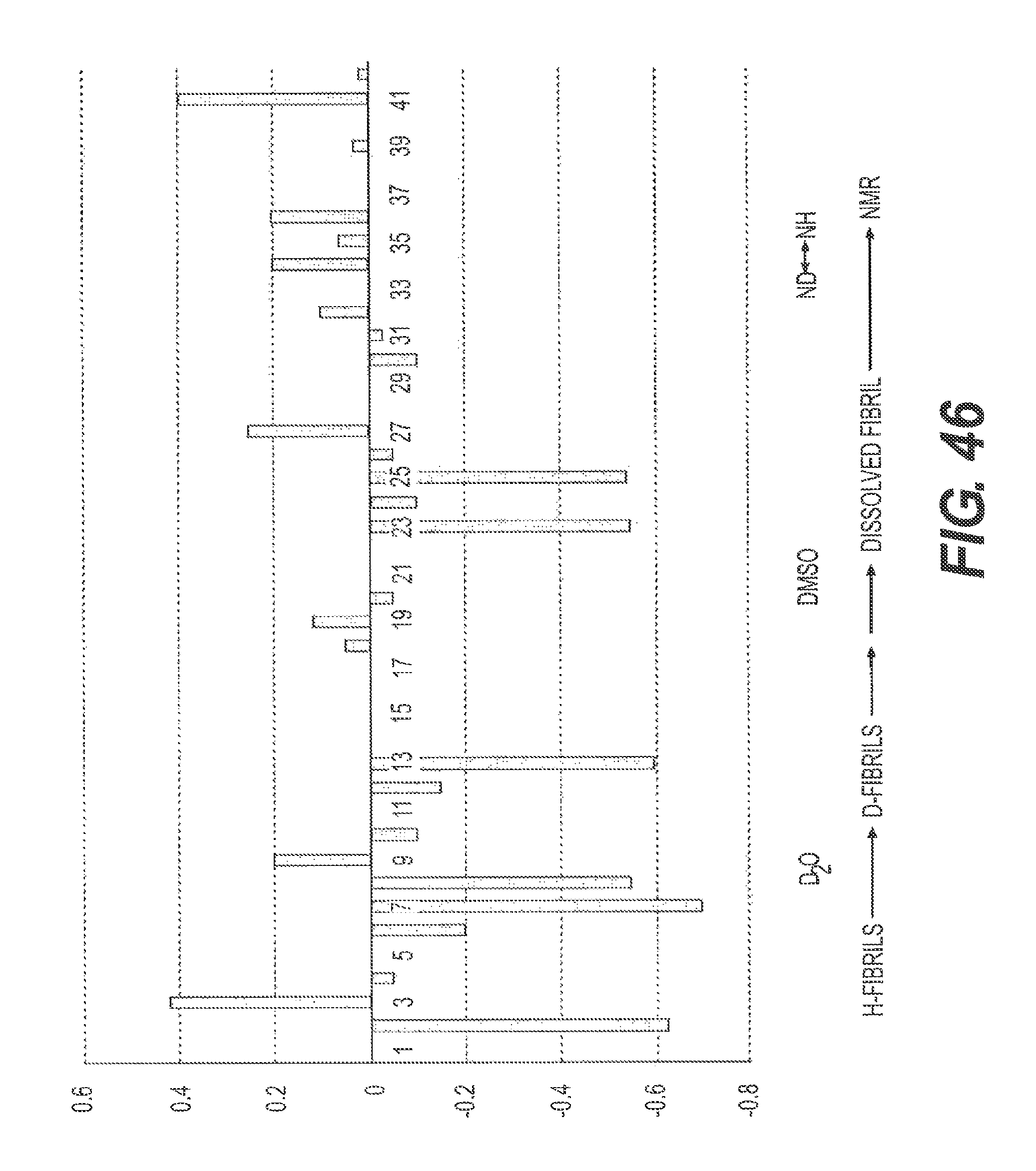
D00061

D00062
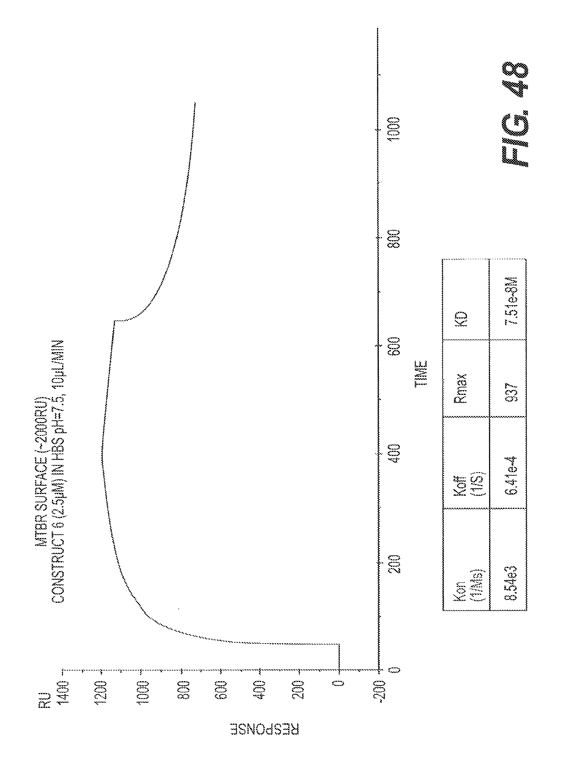
D00063
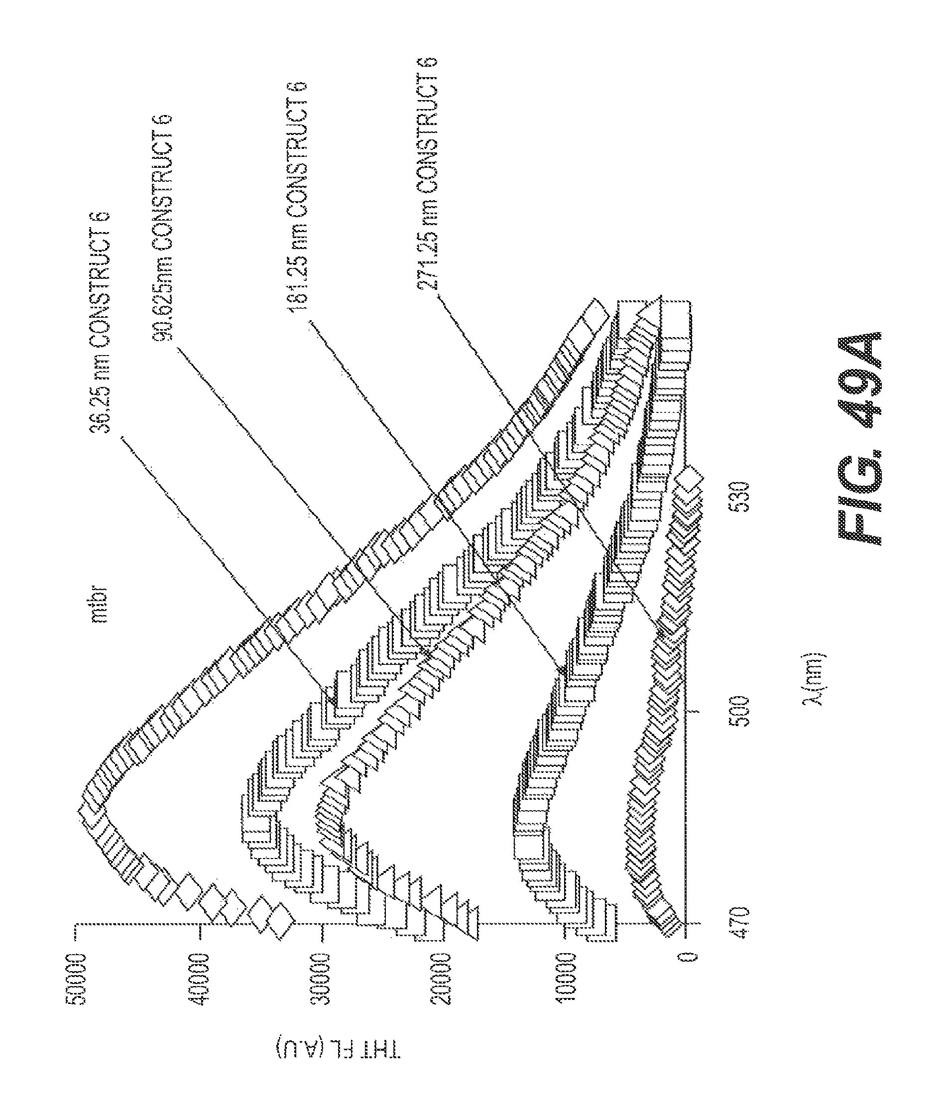
D00064
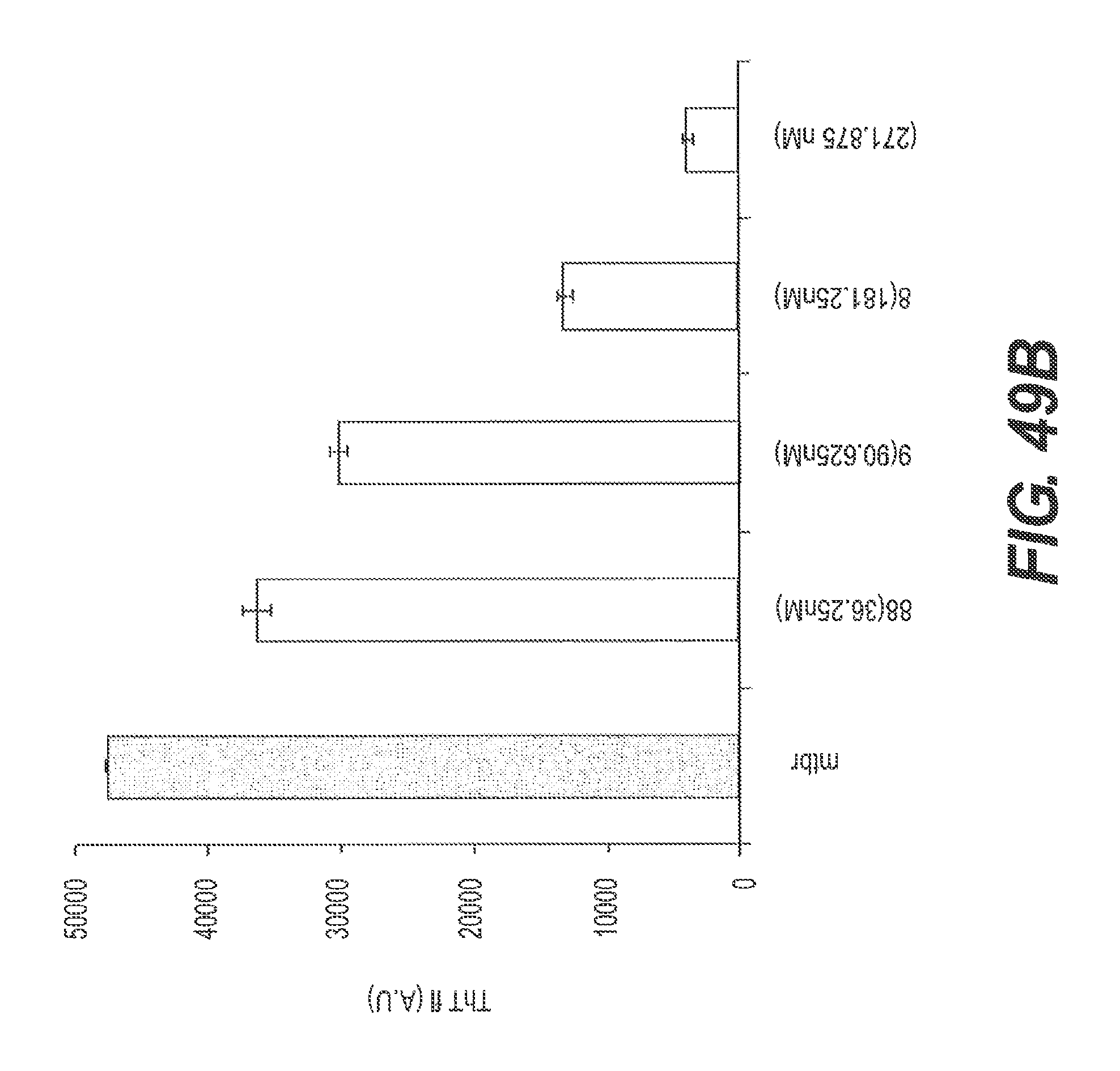
D00065
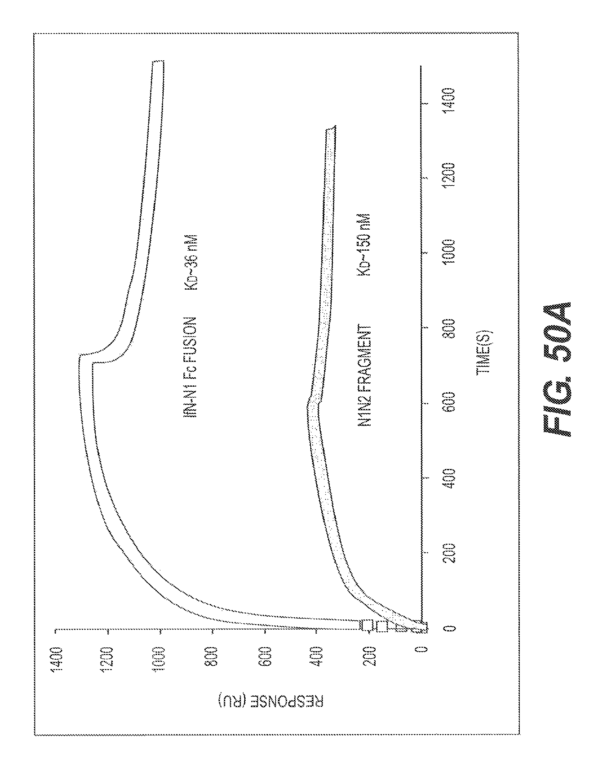
D00066
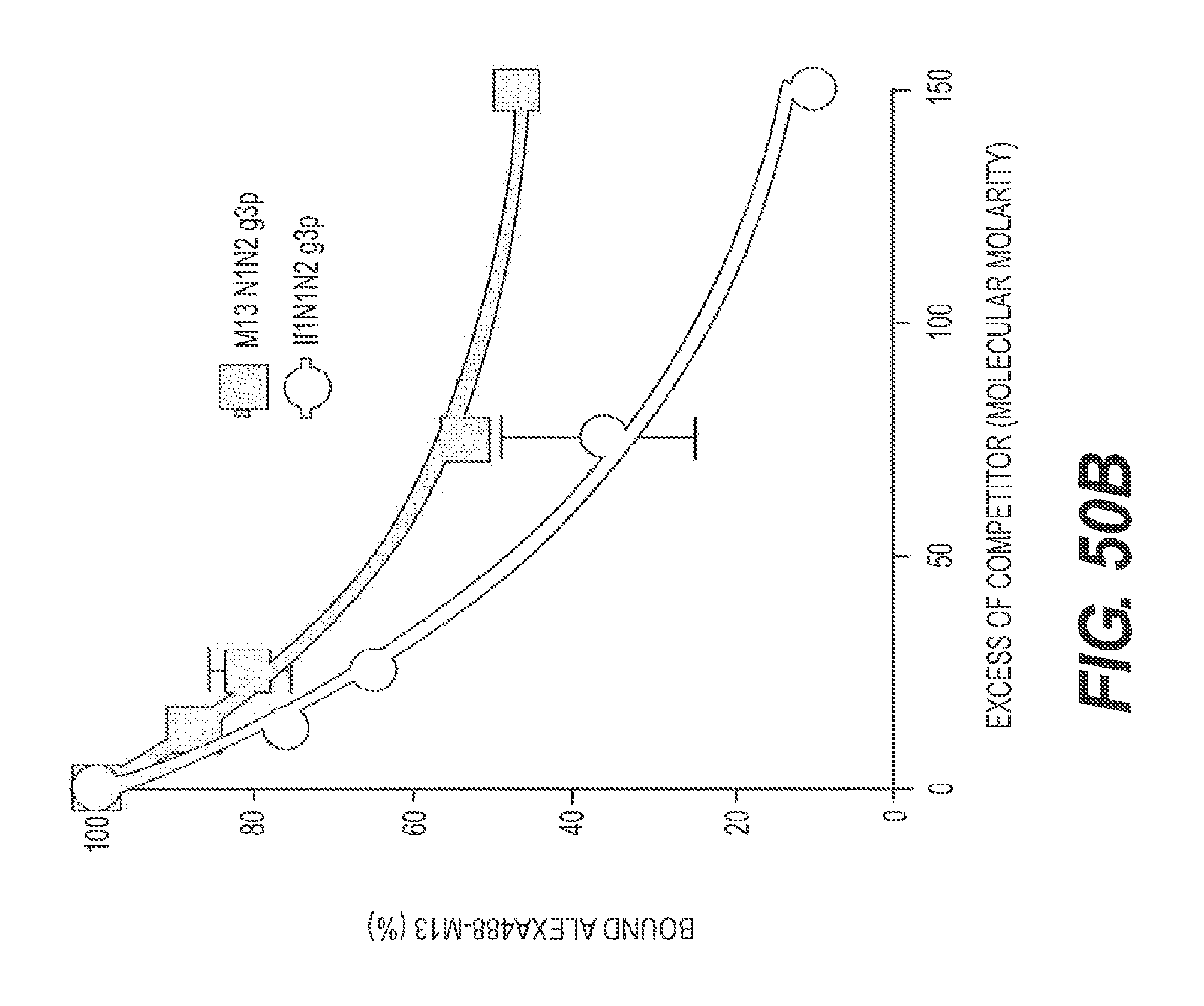
D00067
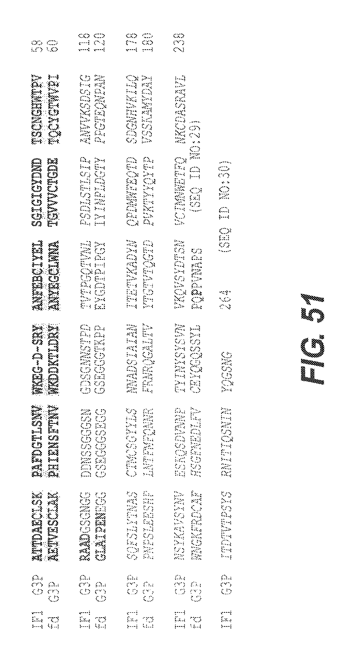
D00068
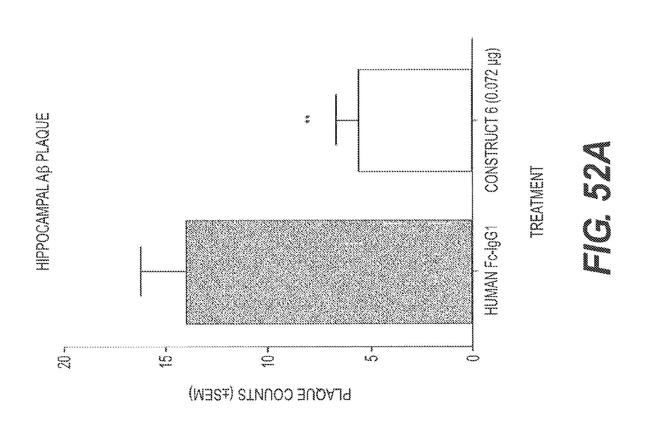
D00069
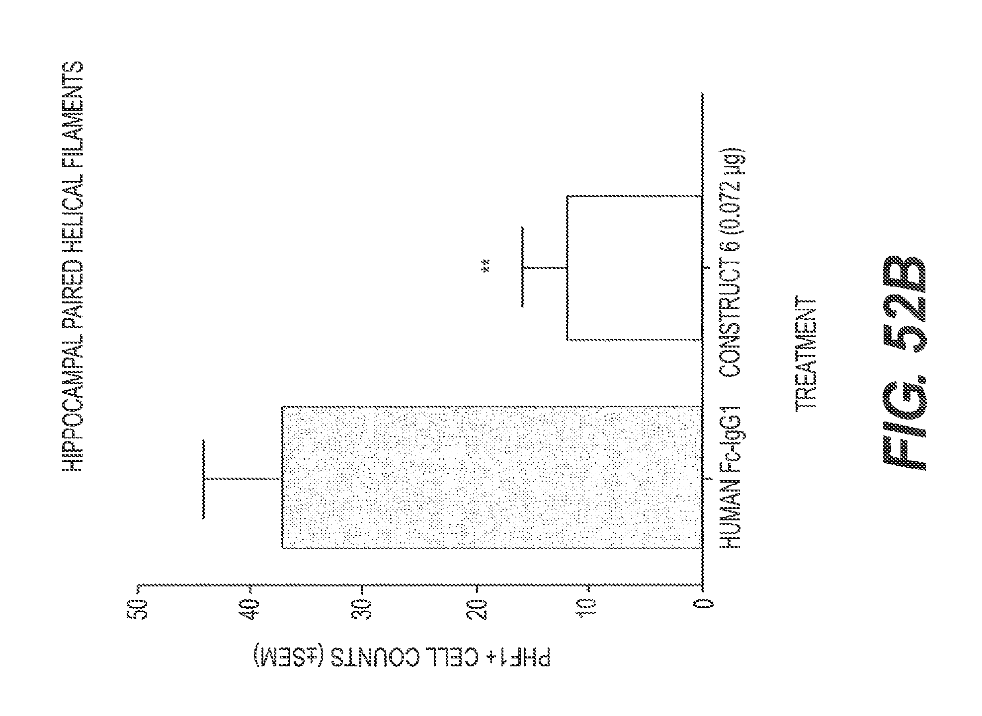
D00070

D00071
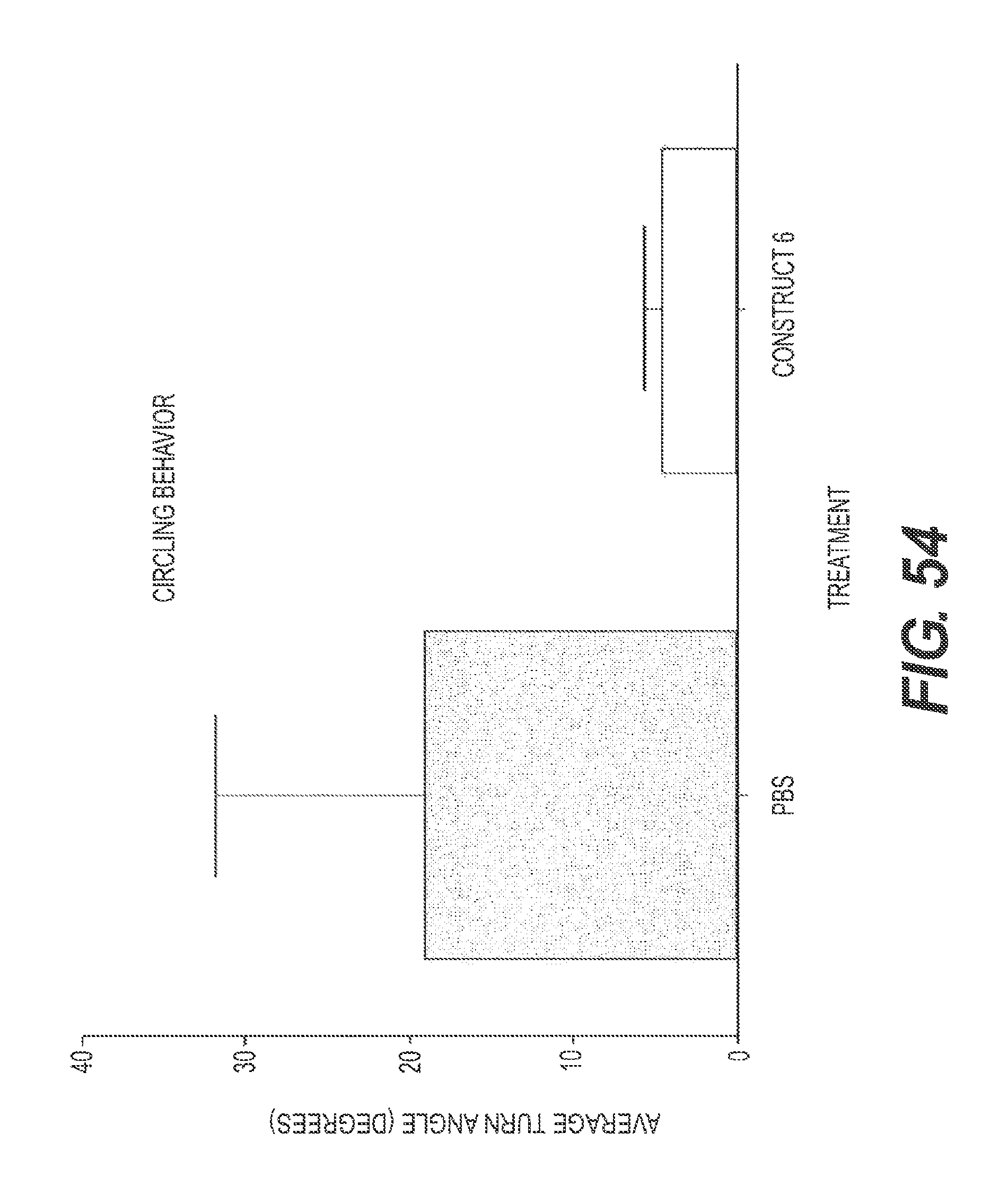
D00072
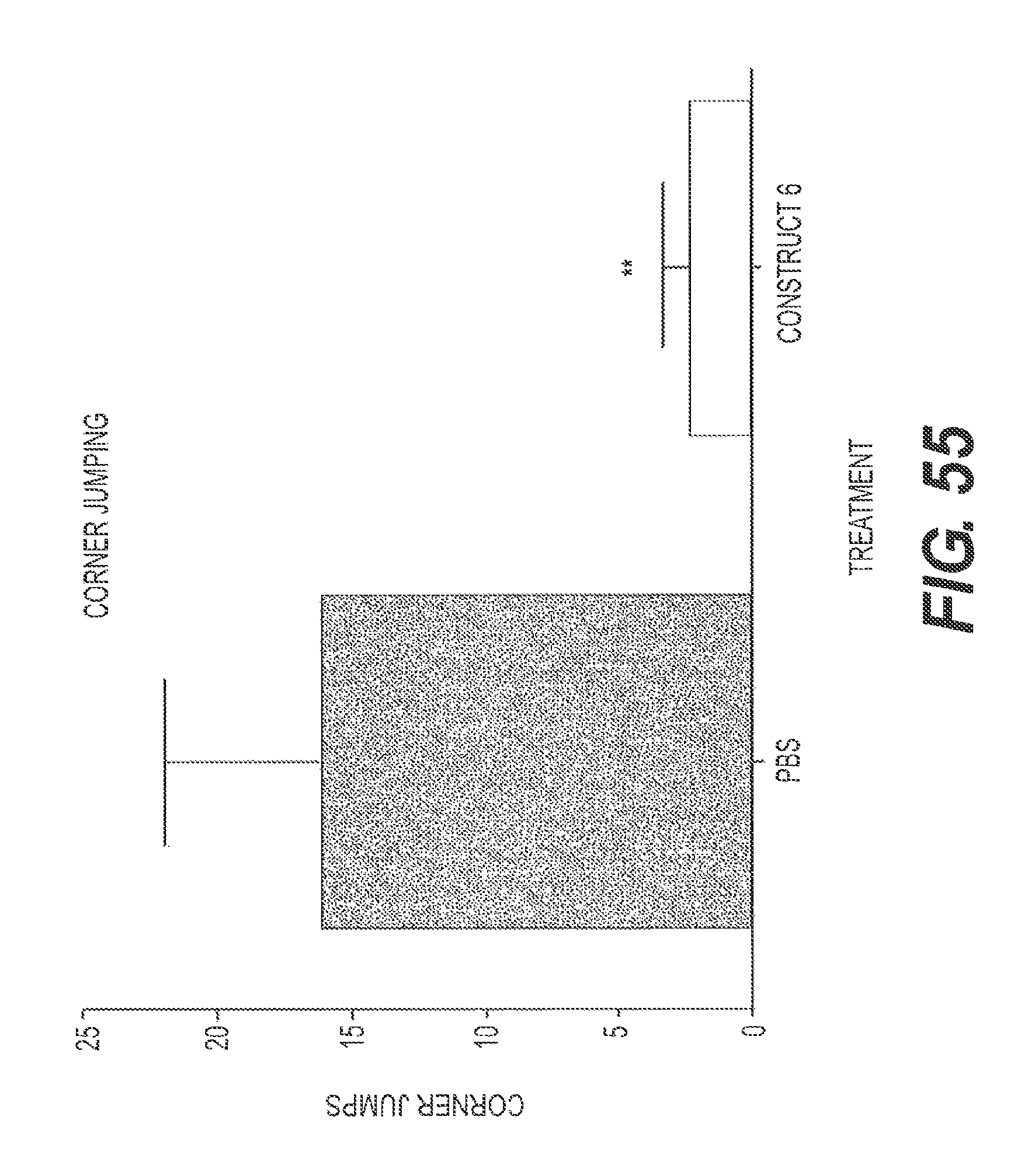
D00073

D00074
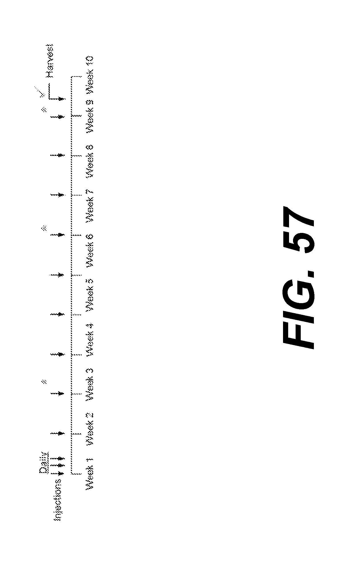
S00001
XML
uspto.report is an independent third-party trademark research tool that is not affiliated, endorsed, or sponsored by the United States Patent and Trademark Office (USPTO) or any other governmental organization. The information provided by uspto.report is based on publicly available data at the time of writing and is intended for informational purposes only.
While we strive to provide accurate and up-to-date information, we do not guarantee the accuracy, completeness, reliability, or suitability of the information displayed on this site. The use of this site is at your own risk. Any reliance you place on such information is therefore strictly at your own risk.
All official trademark data, including owner information, should be verified by visiting the official USPTO website at www.uspto.gov. This site is not intended to replace professional legal advice and should not be used as a substitute for consulting with a legal professional who is knowledgeable about trademark law.