Adhesive complex coacervates and methods of making and using thereof
Stewart , et al. Dec
U.S. patent number 10,517,987 [Application Number 15/918,423] was granted by the patent office on 2019-12-31 for adhesive complex coacervates and methods of making and using thereof. This patent grant is currently assigned to University of Utah Research Foundation. The grantee listed for this patent is UNIVERSITY OF UTAH RESEARCH FOUNDATION. Invention is credited to Hui Shao, Russell J. Stewart.


View All Diagrams
| United States Patent | 10,517,987 |
| Stewart , et al. | December 31, 2019 |
Adhesive complex coacervates and methods of making and using thereof
Abstract
Described herein is the synthesis of adhesive complex coacervates and their use thereof. The adhesive complex coacervates are composed of a mixture of one or more polycations and one or more polyanions. The polycations and polyanions in the adhesive complex coacervate are crosslinked with one another by covalent bonds upon curing. The adhesive complex coacervates have several desirable features when compared to conventional bioadhesives, which are effective in water-based applications. The adhesive complex coacervates described herein exhibit good interfacial tension in water when applied to a substrate (i.e., they spread over the interface rather than being beaded up). Additionally, the ability of the complex coacervate to crosslink intermolecularly increases the cohesive strength of the adhesive complex coacervate. The adhesive complex coacervates have numerous biological applications as bioadhesives and drug delivery devices. In particular, the adhesive complex coacervates described herein are particularly useful in underwater applications and situations where water is present such as, for example, physiological conditions.
| Inventors: | Stewart; Russell J. (Salt Lake City, UT), Shao; Hui (Salt Lake City, UT) | ||||||||||
|---|---|---|---|---|---|---|---|---|---|---|---|
| Applicant: |
|
||||||||||
| Assignee: | University of Utah Research
Foundation (Salt Lake City, UT) |
||||||||||
| Family ID: | 43499829 | ||||||||||
| Appl. No.: | 15/918,423 | ||||||||||
| Filed: | March 12, 2018 |
Prior Publication Data
| Document Identifier | Publication Date | |
|---|---|---|
| US 20180272027 A1 | Sep 27, 2018 | |
Related U.S. Patent Documents
| Application Number | Filing Date | Patent Number | Issue Date | ||
|---|---|---|---|---|---|
| 13617882 | Sep 14, 2012 | 9913926 | |||
| 12508280 | Oct 9, 2012 | 8283384 | |||
| PCT/US2008/083311 | Nov 13, 2008 | ||||
| 61023173 | Jan 24, 2008 | ||||
| Current U.S. Class: | 1/1 |
| Current CPC Class: | C09D 133/26 (20130101); C08F 8/06 (20130101); A61L 24/0015 (20130101); A61F 2/2846 (20130101); A61L 24/0073 (20130101); C08F 8/00 (20130101); A61L 27/54 (20130101); A61L 27/26 (20130101); A61P 19/08 (20180101); A61L 24/043 (20130101); C08L 33/26 (20130101); C08F 8/10 (20130101); C09J 133/14 (20130101); C08F 8/00 (20130101); C08F 230/02 (20130101); C08F 8/06 (20130101); C08F 230/02 (20130101); C08F 8/10 (20130101); C08F 230/02 (20130101); C08F 2810/30 (20130101); C08F 2800/10 (20130101); A61L 2300/602 (20130101); A61L 2300/62 (20130101); C08F 2810/20 (20130101); C08F 230/02 (20130101); C08F 220/54 (20130101); C08F 220/56 (20130101) |
| Current International Class: | A61L 24/00 (20060101); C08F 8/06 (20060101); C08F 8/00 (20060101); C09D 133/26 (20060101); C08F 8/10 (20060101); C09J 133/14 (20060101); A61F 2/28 (20060101); A61L 27/54 (20060101); C08F 230/02 (20060101); C08L 33/26 (20060101); A61L 24/04 (20060101); A61L 27/26 (20060101) |
References Cited [Referenced By]
U.S. Patent Documents
| 3458360 | July 1969 | Shepard et al. |
| 3947396 | March 1976 | Kangas et al. |
| 3950296 | April 1976 | Kangas et al. |
| 4767463 | August 1988 | Brode et al. |
| 4913743 | April 1990 | Brode et al. |
| 5529914 | June 1996 | Hubbell et al. |
| 6312725 | November 2001 | Wallace et al. |
| 6428978 | August 2002 | Olsen et al. |
| 6497729 | December 2002 | Moussey et al. |
| 6568398 | May 2003 | Cohen |
| 6916488 | July 2005 | Meier et al. |
| 7622533 | November 2009 | Lee |
| 8283384 | October 2012 | Stewart et al. |
| 9173971 | November 2015 | Stewart |
| 9272069 | March 2016 | Stewart et al. |
| 9421300 | August 2016 | Stewart |
| 9867899 | January 2018 | Stewart |
| 9913926 | March 2018 | Stewart |
| 9913927 | March 2018 | Stewart |
| 2001/0056301 | December 2001 | Goupil et al. |
| 2002/0006886 | January 2002 | Beerse et al. |
| 2002/0154364 | November 2002 | Quong |
| 2002/0169476 | November 2002 | Cohen |
| 2003/0023000 | January 2003 | Bavouzet et al. |
| 2004/0013738 | January 2004 | Voigt et al. |
| 2004/0086479 | May 2004 | Grinstaff et al. |
| 2005/0020734 | January 2005 | Asgarzadeh et al. |
| 2005/0147580 | July 2005 | Conner et al. |
| 2005/0220751 | October 2005 | Chamot et al. |
| 2005/0281883 | December 2005 | Daniloff et al. |
| 2006/0007528 | January 2006 | Cao et al. |
| 2006/0015083 | January 2006 | Munro et al. |
| 2006/0039950 | February 2006 | Zhu et al. |
| 2006/0241242 | March 2006 | Devlin |
| 2006/0116682 | June 2006 | Longo |
| 2006/0122290 | June 2006 | Hubbell et al. |
| 2006/0156954 | July 2006 | Li et al. |
| 2006/0183848 | August 2006 | Maier et al. |
| 2006/0240064 | October 2006 | Hunter et al. |
| 2006/0275337 | December 2006 | Stuart et al. |
| 2006/0276371 | December 2006 | Schreiner et al. |
| 2007/0020469 | January 2007 | Wood et al. |
| 2007/0077276 | April 2007 | Haynie |
| 2007/0085059 | April 2007 | Mora-Gutierrez et al. |
| 2007/0191273 | August 2007 | Ambati et al. |
| 2007/0196454 | August 2007 | Stockman et al. |
| 2008/0003288 | January 2008 | Broomberg et al. |
| 2008/0075778 | March 2008 | Heller |
| 2009/0162407 | June 2009 | Biggs et al. |
| 2010/0056474 | March 2010 | Baker et al. |
| 2010/0120923 | July 2010 | Stewart et al. |
| 2010/0291169 | November 2010 | Toreki et al. |
| 2010/0305626 | December 2010 | Stewart et al. |
| 2011/0054392 | March 2011 | Nies |
| 2011/0287067 | November 2011 | Stewart |
| 2011/0288274 | November 2011 | Russell et al. |
| 2012/0177918 | July 2012 | Stewart |
| 2013/0189313 | July 2013 | Stewart |
| 2014/0287061 | September 2014 | Landolina |
| 2018/0099070 | April 2018 | Stewart |
| 2018/0147316 | May 2018 | Stewart |
| 1341032 | Mar 2002 | CN | |||
| 1446590 | Oct 2003 | CN | |||
| 101405037 | Apr 2009 | CN | |||
| 19810965 | Sep 1999 | DE | |||
| 0632329 | Jun 1994 | EP | |||
| 2003280056 | Dec 1991 | JP | |||
| 2002166158 | Jun 2002 | JP | |||
| 2009084224 | Apr 2009 | JP | |||
| 2009084292 | Apr 2009 | JP | |||
| 1995006056 | Mar 1995 | WO | |||
| 2002092217 | Nov 2002 | WO | |||
| 2002100453 | Dec 2002 | WO | |||
| 2005019421 | Mar 2005 | WO | |||
| 2007024972 | Mar 2007 | WO | |||
| 2007030811 | Mar 2007 | WO | |||
| 2009094060 | Jul 2009 | WO | |||
| 2011011658 | Jan 2011 | WO | |||
| 2011028967 | Mar 2011 | WO | |||
| 2011106595 | Sep 2011 | WO | |||
| 2011149907 | Dec 2011 | WO | |||
| 2012065148 | May 2012 | WO | |||
Other References
|
Berg et al., "The Thermal Transition of a Non-Hydroxylated Form of Collagen. Evidence for a Role for Hydroxyproline in Stabilizing the Triple-Helix of Collagen," Biochem. Biophys. Res. Commun, 1973, 52:115-120. cited by applicant . Canadian Office Action for Application 2,712,843 dated Jul. 9, 2014. cited by applicant . Chinese First Office Action for Chinese Patent Application No. 200880128307.2 dated Oct. 28, 2011. cited by applicant . Chinese First Office Action for CN Patent Application 201080038397 dated Apr. 3, 2013 (English summary). cited by applicant . Chinese First Office Action for CN patent Application 201180010546 dated Aug. 13, 2013 (English translation). cited by applicant . Chinese First Office Action for CN Patent Application 201180024981.8 dated Mar. 27, 2014 (English summary). cited by applicant . Chinese First Office Action for CN Patent Application 201180050846.0 dated Apr. 3, 2014 (English summary). cited by applicant . Chinese Office Action for Application 201080038397.3 dated Sep. 11, 2014. cited by applicant . Chinese Office Action for Application 2012800362923 dated Jan. 7, 2015. cited by applicant . Chinese Second Office Action for Chinese Patent Application 200880128307.2 dated Apr. 1, 2012. cited by applicant . Chinese Second Office Action for CN Patent Appliation No. 201180010546 dated Jul. 2, 2014 (English summary). cited by applicant . Chinese Second Office Action for CN Patent Application 201080038397 dated Dec. 16, 2013. cited by applicant . Chinese Third Office Action for CN Patent Application 201080038397 dated Aug. 21, 2014. cited by applicant . Espinosa-Andrews et al., "Gum Arabic-Chitosan Complex Coacervation," Biomacromolecules, 2007, 8:1313-1318. cited by applicant . European Search Report for 10 802 933.1 dated Jul. 19, 2016. cited by applicant . First Examination Report for Indian Application 1584/MUMNP/2010 dated May 15, 2015. cited by applicant . Hwang et al., "Expression of Functional Recombinant Mussel Adhesive Protein Mgfp-5 in Escherichia coli," Applied and Env. Micribiol., 2004, 70:3352-3359. cited by applicant . International Search Report and Written Opinion for PCT/US2011/376797 dated Sep. 13, 2011. cited by applicant . International Prelimimary Report on Patentability for PCT/US08/083311 dated Jul. 27, 2010. cited by applicant . International Search Report and Written Opinion for PCT/US09/083311 dated Jan. 6, 2009. cited by applicant . International Search Report and Written Opinion for PCT/US10/043009 dated Nov. 22, 2010. cited by applicant . International Search Report dated Aug. 26, 2013 for PCT/US2013/029131. cited by applicant . International Search Report dated May 7, 2012 for PCT/US2011/060500. cited by applicant . Japanese First Office Action in Application JP 2012-521803 dated Aug. 6, 2014. cited by applicant . Japanese First Office Action in Application JP-2012-555168 dated Aug. 12, 2014. cited by applicant . Kamachi et al., "Synthesis of Block Polymers for Desalination Membranes. Preparation of Block Copolymers of 2-Vinylpyridine and Methacrylic Acid or Acryoic Acid," Macromolecules, 1972, 5:161-168. cited by applicant . Kayitmazer et al., "Mesophase separation and probe dynamics in protein-polyelectrolyte coacervates," Soft Matter, 2007, 3:1064-1076. cited by applicant . Lee et al., "Rapid Gel Formation and Adhesion in Photocurable and Biodegradable Block Copolymers with High DOPA Content," Macromolecules, 2006, 39:1740-1748. cited by applicant . Lee et al., "Single-Molecule Mechanics of Mussel Adhesion," Proc. Natl. Acad. Sci. USA, 2006, 103:12999-13003. cited by applicant . Lee et al., "Synthesis of 3,4-dihydroxyphenylalanine (DOPA) containing monomers and their copolymerization with PEG-diacrylate to form hydrogels," J. Biomater. Sci. Polymer Edn., 2004, 15:449-464. cited by applicant . Lim et al., "The Adhesive Properties of Coacervated Recombinant Hybrid Mussel," Biomaterials, 2010, 31:3715-3722. cited by applicant . Liu et al., "Chemistry of Periodate-Mediated Cross-Linking of 3,4-Dihydroxylphenylalanine-Containing Molecules to Proteins," J. Am. Chem. Soc., 2006, 15228-15235. cited by applicant . Mo et al., J. Biomate. Sci. Polymer Edn., 2000, 11:341-351. cited by applicant . Notice of Preliminary Rejection from Korean Intellectual Property Office for Application 10-2010-7018637 dated Jan. 6, 2015. cited by applicant . Office Action for Russian Patent Application 2010135333/15(050196) dated Nov. 6, 2012. cited by applicant . Official Action for European Application 13 156 643.2 dated Dec. 22, 2014. cited by applicant . Polyethyleneimine: EPOMIN, website, Nippon Shokubai, <http://www.shokubai.co.jp/en/products/functionality/epomin1.html> accessed Feb. 16, 2015. cited by applicant . Shao et al., "A Water-Borne Adhesive Modeled after the Sandcastle Glue of P. californica," Macromolecular Bioscience, 2008, 9:464-471. cited by applicant . Stevens et al., "Multiscale Structure of the Underwater Adhesive of Phragmatopoma californica: a Nanostructured Latex with a Steep Microporosity Gradient," Langmuir, 2008, 5045-5049. cited by applicant . Stewart et al., "The Tube Cement of Phragmatopoma californica: A Solid Foam," J. Exp. Biol., 2004, 207:4727-2734. cited by applicant . Supplementary European Search Report for EP 10802933.1 dated Jun. 24, 2015. cited by applicant . Supplementary European Search Report for EP Application No. 12804996 dated Feb. 19, 2015. cited by applicant . Supplementary Extended European Search Report for European Application No. 08871349.0 dated Nov. 14, 2011. cited by applicant . U.S. Office Action for U.S. Appl. No. 12/508,280 dated Sep. 20, 2011. cited by applicant . U.S. Office Action for U.S. Appl. No. 12/864,035 dated Jun. 18, 2014. cited by applicant . U.S. Office Action for U.S. Appl. No. 12/864,045 dated Jan. 5, 2015. cited by applicant . U.S. Office Action for U.S. Appl. No. 12/864,045 dated Oct. 7, 2013. cited by applicant . U.S. Office Action for U.S. Appl. No. 12/864,045 dated Sep. 23, 2014. cited by applicant . U.S. Office Action for U.S. Appl. No. 13/114,397 dated Feb. 27, 2014. cited by applicant . U.S. Office Action for U.S. Appl. No. 13/114,397 dated Mar. 23, 2015. cited by applicant . U.S. Office Action for U.S. Appl. No. 13/114,397 dated May 22, 2015. cited by applicant . U.S. Office Action for U.S. Appl. No. 13/114,397 dated Oct. 12, 2014. cited by applicant . U.S. Office Action for U.S. Appl. No. 13/295,061 dated Jan. 12, 2015. cited by applicant . U.S. Office Action for U.S. Appl. No. 13/295,061 dated Mar. 4, 2014. cited by applicant . U.S. Office Action for U.S. Appl. No. 13/580,794 dated Aug. 1, 2014. cited by applicant . U.S. Office Action for U.S. Appl. No. 13/580,794 dated Jan. 10, 2014. cited by applicant . U.S. Office Action for U.S. Appl. No. 13/580,794 dated May 12, 2015. cited by applicant . U.S. Office Action for U.S. Appl. No. 13/580,794 dated Nov. 13, 2014. cited by applicant . U.S. Office Action for U.S. Appl. No. 13/617,882 dated Aug. 27, 2013. cited by applicant . U.S. Office Action for U.S. Appl. No. 13/617,882 dated Jun. 5, 2014. cited by applicant . U.S. Office Action for U.S. Appl. No. 13/617,882 dated Nov. 19, 2015. cited by applicant . U.S. Office Action for U.S. Appl. No. 13/617,882 dated Sep. 18, 2014. cited by applicant . Wang et al., "A novel bioadhesive protein of silk filaments spun underwater by caddisfly larvae" Adv. Mater. Res., 2009, 79-82:1631-1634. cited by applicant . Written Opinion issued in PCT/US2011/26169 dated May 17, 2011. cited by applicant . Written Opinion issued in PCT/US2014/044299 dated Nov. 16, 2012. cited by applicant . Yu et al., "Synthetic Polypeptide Mimics of Marine Adhesives," Macromolecules, 1998, 31:4739-4745. cited by applicant . Zhao et al.,"Cement Proteins of the Tube-Binding Polychaete Phragmatoporna californica," J Biol. Chem., 2005, 280:42938-42944. cited by applicant . Unpublished Application for U.S. Appl. No. 15/880,650, filed Jan. 26, 2018. cited by applicant . U.S. Appl. No. 12/864,045, U.S. Pat. No. 9,272,069. cited by applicant . U.S. Appl. No. 15/131,583, U.S. Pat. No. 9,867,899. cited by applicant . U.S. Appl. No. 15/833,157, 2018/0099070. cited by applicant . U.S. Appl. No. 13/295,061, U.S. Pat. No. 9,173,971. cited by applicant . U.S. Appl. No. 13/617,882, U.S. Pat. No. 9,913,926. cited by applicant . U.S. Appl. No. 12/508,280, U.S. Pat. No. 8,283,384. cited by applicant . U.S. Appl. No. 14/874,854, U.S. Pat. No. 9,421,300. cited by applicant . U.S. Appl. No. 15/067,291, 2018/0147316. cited by applicant . U.S. Appl. No. 15/325,885, U.S. Pat. No. 9,913,927. cited by applicant . U.S. Appl. No. 15/880,650. cited by applicant. |
Primary Examiner: Falkowitz; Anna R
Attorney, Agent or Firm: Thomas|Horstemeyer, LLP
Government Interests
ACKNOWLEDGEMENTS
This invention was made with government support under EB005288 awarded by the National Institutes of Health. The government has certain rights in the invention.
Parent Case Text
CROSS REFERENCE TO RELATED APPLICATIONS
This application is a continuation application of U.S. application Ser. No. 13/617,882, filed Sep. 14, 2012, which is a continuation application of U.S. application Ser. No. 12/508,280, filed Jul. 23, 2009, which is a continuation-in-part of PCT International Application No. PCT/US2008/083311, filed Nov. 13, 2008, which claims priority upon U.S. provisional application Ser. No. 61/023,173, filed Jan. 24, 2008. These applications are hereby incorporated by reference in their entireties.
Claims
What is claimed:
1. A liquid adhesive complex coacervate, wherein the complex coacervate comprises at least one polycation and at least one polyanion, and wherein the complex coacervate does not include a multivalent cation.
2. The liquid adhesive complex coacervate of claim 1, wherein the polycation comprises a polyamino compound.
3. The liquid adhesive complex coacervate of claim 1, wherein the polycation comprises a polyacrylate having pendant amino groups.
4. The liquid adhesive complex coacervate of claim 3, wherein the polyacrylate is a homopolymer or copolymer derived from the polymerization of an acrylate monomer comprising an acrylate, a methacrylate, an acrylamide, or any combination thereof.
5. The liquid adhesive complex coacervate of claim 1, wherein the polycation comprises a polyacrylate comprising a primary, secondary, or tertiary amino group, an alkylamino group, a heteroaryl group, or an aromatic group substituted with one or more amino groups, or imidazole groups.
6. The liquid adhesive complex coacervate of claim 1, wherein the polyanion comprises a polymer comprising pendant sulfate, sulfonate, carboxylate, borate, boronate, phosphonate, phosphate groups, or any combination thereof.
7. The liquid adhesive complex coacervate of claim 1, wherein the polyanion comprises a polyphosphate compound.
8. The liquid adhesive complex coacervate of claim 1, wherein the polyanion comprises an inorganic polyphosphate.
9. The liquid adhesive complex coacervate of claim 1, wherein the polyanion comprises a metaphosphate salt.
10. The liquid adhesive complex coacervate of claim 1, wherein the polyanion comprises a polyacrylate comprising pendant phosphate or phosphonate groups.
11. The liquid adhesive complex coacervate of claim 1, wherein the polyanion comprises a naturally-occurring polyphosphate.
12. The liquid adhesive complex coacervate of claim 1, wherein the complex coacervate further comprises a bioactive agent.
13. The liquid adhesive complex coacervate of claim 1, wherein the polycation and polyanion comprises at least one group capable of covalent crosslinking with each other.
14. The liquid adhesive complex coacervate of claim 13, wherein the crosslinking group on the polycation comprises a nucleophilic group and the crosslinking group on the polyanion comprises an electrophilic group.
15. The liquid adhesive complex of claim 13, wherein the crosslinking group on the polycation comprises an electrophilic group and the crosslinking group on the polyanion comprises a nucleophilic group.
16. The liquid adhesive complex coacervate of claim 13, wherein the crosslinking group on the polycation and polyanion comprises an ortho-dihydroxy aromatic group capable of undergoing oxidative crosslinking.
17. The liquid adhesive complex coacervate of claim 13, wherein (1) the crosslinking group on the polyanion comprises a quinone group and the polycation comprises a nucleophilic group capable of reacting with the quinone group to form a covalent bond or (2) the crosslinking group on the polycation comprises a quinone group and the polyanion comprises a nucleophilic group capable of reacting with the quinone group to form a covalent bond.
18. The liquid adhesive complex coacervate of claim 17, wherein the nucleophilic group is a hydroxyl, a thiol group, or a substituted or unsubstituted amino group.
19. The liquid adhesive complex coacervate of claim 13, wherein the crosslinking group on the polyanion and the polycation comprises an actinically crosslinkable group.
20. The liquid adhesive complex coacervate of claim 19, wherein the actinically crosslinkable group comprises an olefinic group.
21. The liquid adhesive complex coacervate of claim 20, wherein the olefinic group comprises an acrylate group, a methacrylate group, an acrylamide group, a methacrylamide group, an allyl group, a vinyl group, a vinylester group, or a styrenyl group.
22. The liquid adhesive complex coacervate of claim 1, wherein the coacervate further comprises an initiator and optionally a co-initiator.
23. The liquid adhesive complex coacervate of claim 22, wherein the initiator comprises (1) one or more of a radical initiator, a thermal initiator, or a photoinitiator, or (2) two or more radical initiators, thermal initiators, or a photoinitiators.
24. The liquid adhesive complex coacervate of claim 23, wherein the photoinitiator and optionally a co-initiator are covalently attached to the polycation and/or polyanion.
25. The liquid adhesive complex coacervate of claim 23, wherein the photoinitiator comprises a water-soluble initiator comprising riboflavin, eosin, eosin y, or rose Bengal.
26. The liquid adhesive complex coacervate of claim 23, wherein the photoinitiator comprises a phosphine oxide, a peroxide, an azide compound, an .alpha.-hydroxyketone, or an .alpha.-aminoketone.
27. A method for inhibiting blood flow in a blood vessel of a subject comprising introducing the liquid adhesive complex coacervate of claim 1 into the vessel.
28. A method for repairing a bone fracture in a subject, comprising contacting the fractured bone with the liquid adhesive complex coacervate of claim 1.
29. A method for adhering a metal substrate to a bone of a subject comprising contacting the bone with the liquid adhesive complex coacervate of claim 1 and applying the metal substrate to the coated bone.
30. A method for adhering a bone-tissue scaffold to a bone of a subject comprising contacting the bone and tissue with the liquid adhesive complex coacervate of claim 1 and applying the bone-tissue scaffold to the bone and tissue.
31. A method for delivering one or more bioactive agents comprising administering the liquid adhesive complex coacervate of claim 1 to a subject.
32. A method for repairing a laceration, comprising applying to the laceration the liquid adhesive complex coacervate of claim 1.
33. A method for securing a dental implant, comprising (a) applying to an oral substrate and/or dental implant the liquid complex coacervate of claim 1, and (b) attaching the dental implant to the substrate.
Description
CROSS REFERENCE TO SEQUENCE LISTING
Proteins described herein are referred to by a sequence identifier number (SEQ ID NO). The SEQ ID NO corresponds numerically to the sequence identifiers <400>1, <400>2, etc. The Sequence Listing, in written computer readable format (CFR), is incorporated by reference in its entirety.
BACKGROUND
Bone fractures are a serious health concern in society today. In addition to the fracture itself, a number of additional health risks are associated with the fracture. For example, intra-articular fractures are bony injuries that extend into a joint surface and fragment the cartilage surface. Fractures of the cartilage surface often lead to debilitating posttraumatic arthritis. The main determining factors in the development of posttraumatic arthritis are thought to be the amount of energy imparted at the time of injury, the patient's genetic predisposition (or lack thereof) to posttraumatic arthritis, and the accuracy and maintenance of reduction. Of the three prognostic factors, the only factor controllable by orthopedic caregivers is achievement and maintenance of reduction. Comminuted injuries of the articular surface (the cartilage) and the metaphysis (the portion of the bone immediately below the cartilage) are particularly challenging to maintain in reduced (aligned) position. This relates to the quality and type of bone in this area. It also relates to the limitations of fixation with titanium or stainless steel implants.
Currently, stainless steel and titanium implants are the primary methods of fixation, but their size and the drilling necessary to place them frequently interfere with the exact manipulation and reduction of smaller pieces of bone and cartilage. A variety of bone adhesives have been tested as alternatives to mechanical fixation. These fall into four categories: polymethylmethacrylates (PMMA), fibrin-based glues, calcium phosphate (CP) cements, and CP resin composites. PMMA cements, which are used in the fixation of prostheses, have well-known drawbacks, one of the most serious being that the heat generated from the exothermic setting reaction can kill adjacent bone tissue. Also, the poor bonding to bone leads to aseptic loosening, the major cause of PMMA cemented prosthesis failure.
Fibrin glues, based on the blood clotting protein fibrinogen, have been tested for fixing bone grafts and repairing cartilage since the 1970s and yet have not been widely deployed. One of the drawbacks of fibrin glues is that they are manufactured from pooled human donor blood. As such, they carry risk of transmitting infections and could potentially be of limited supply.
CP cements are powders of one or more forms of CP, e.g., tetracalcium phosphate, dicalcium phosphate anhydride, and .beta.-tricalcium phosphate. When the powder is mixed with water it forms a paste that sets up and hardens through the entanglement of one or more forms of CP crystals, including hydroxyapatite. Advantages of CP cements include isothermal set, proven biocompatibility, osteoconductivity, and they serve as a reservoir for Ca and PO.sub.4 for hydroxyapatite formation during healing. The primary disadvantages are that CP cements are brittle, have low mechanical strength and are therefore not ideal for stable reduction of small articular segments. CP cements are used mostly as bone void fillers. The poor mechanical properties of CP cements have led to composite cements of CP particles and polymers. By varying the volume fractions of the particulate phase and the polymer phase, the modulus and strength of the glue can be adjusted toward those of natural bone, an avenue that is also open to us.
Given the overall health impact associated with bone fractures and the imperfect state of current fixation methods, new fixation methods are needed.
SUMMARY
Described herein is the synthesis of adhesive complex coacervates and their use thereof. The adhesive complex coacervates are composed of a mixture of one or more polycations and one or more polyanions. The polycations and polyanions are crosslinked with one another by covalent bonds upon curing. The adhesive complex coacervates have several desirable features when compared to conventional adhesives, which are effective in water-based applications. The adhesive complex coacervates described herein exhibit low interfacial tension in water when applied to a substrate (i.e., they spread over the interface rather than being beaded up). Additionally, the ability of the complex coacervate to crosslink intermolecularly increases the cohesive strength of the adhesive complex coacervate. The adhesive complex coacervates have numerous biological applications as bioadhesives and drug delivery devices. In particular, the adhesive complex coacervates described herein are particularly useful in underwater applications and situations where water is present such as, for example, physiological conditions.
The advantages of the invention will be set forth in part in the description which follows, and in part will be obvious from the description, or may be learned by practice of the aspects described below. The advantages described below will be realized and attained by means of the elements and combinations particularly pointed out in the appended claims. It is to be understood that both the foregoing general description and the following detailed description are exemplary and explanatory only and are not restrictive.
BRIEF DESCRIPTION OF THE DRAWINGS
The accompanying drawings, which are incorporated in and constitute a part of this specification, illustrate several aspects described below.
FIGS. 1A-1C show a model of pH dependent coacervate structure and adhesive mechanisms. FIG. 1A shows the polyphosphate (black) with low charge density paired with the polyamine (red) form nm-scale complexes. The complexes have a net positive charge. FIG. 1B shows the extended high charge density polyphosphates form a network connected by more compact lower charge density polyamines and when present divalent cations (green symbols). The net charge on the copolymers is negative. FIG. 1C shows the oxidation of 3,4-dihydroxyphenol (D) by O.sub.2 or an added oxidant initiates crosslinking between the quinone (Q) and primary amine sidechains. The coacervate can adhere to the hydroxyapatite surface through electrostatic interactions, 3,4-dihydroxyphenol sidechains, and quinone-mediated covalent coupling to matrix proteins.
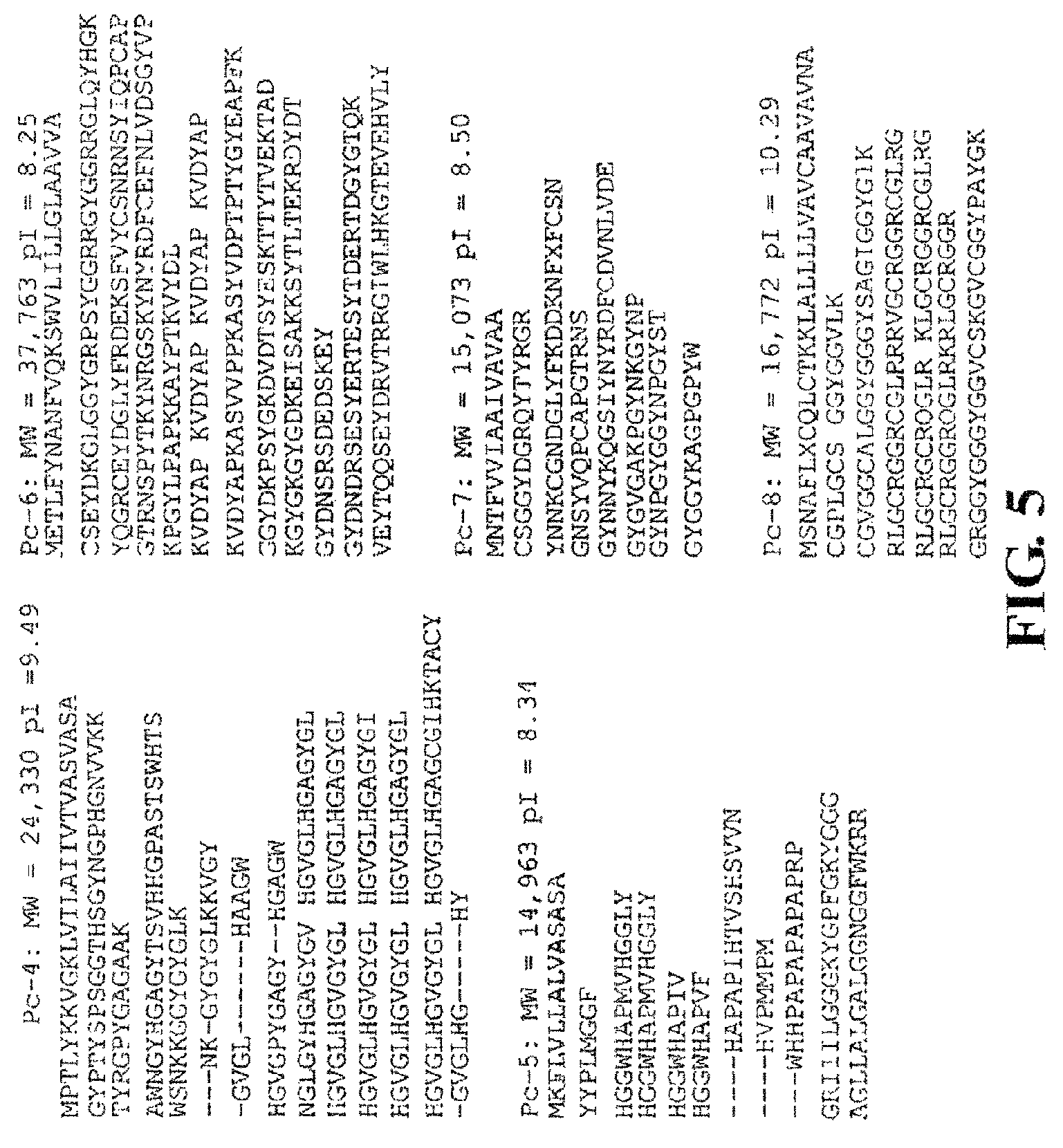
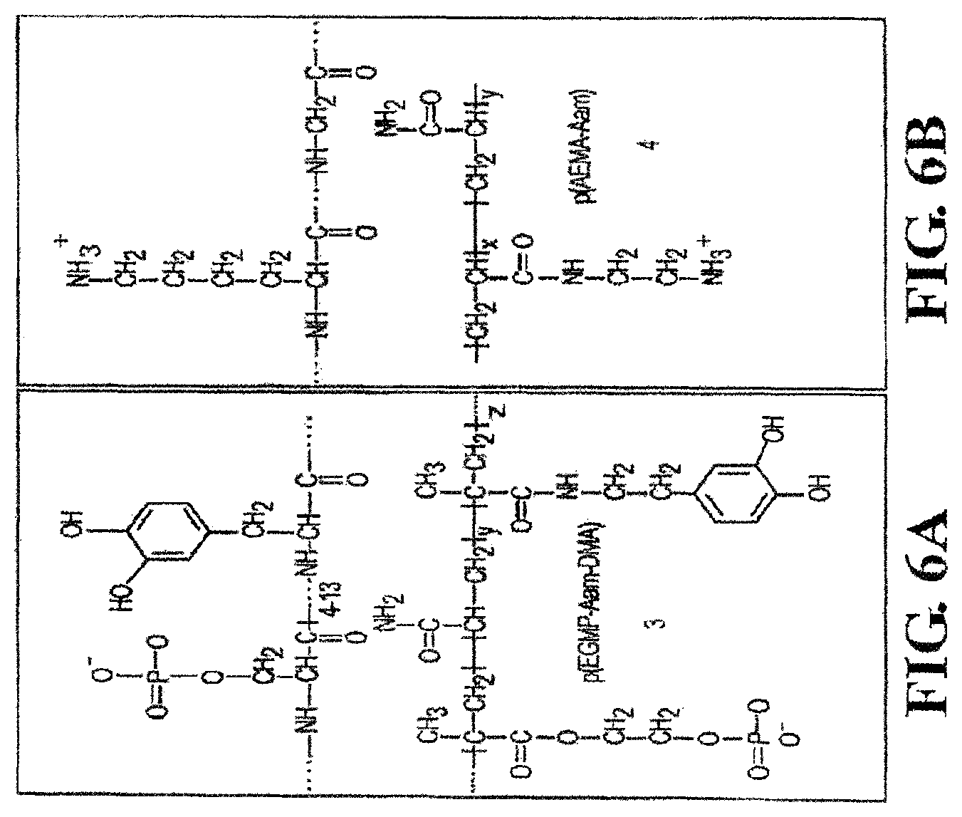
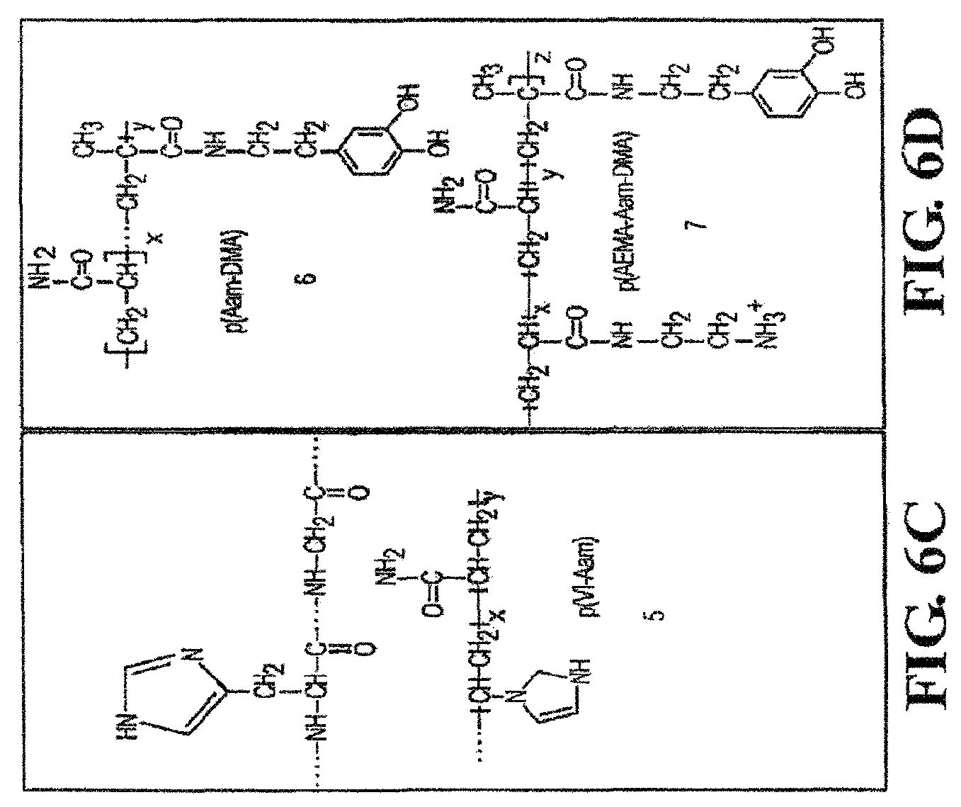
FIGS. 2, 3, 4, 5, 6A, 6B, 6C, 6D, and 7 show several protein sequences produced by P. californica that can be used as polycations and polyanions in the present invention as well as synthetic polycations and polyanions useful in the present invention.
FIG. 8 shows different mechanisms of DOPA crosslinking.
FIGS. 9A-9B show the dual syringe systems for applying small "spot welds" of complex coacervates described herein to repair fractures (FIG. 9A), small bone injuries (FIG. 9B), or bonding synthetic scaffolds to bony tissue (FIG. 9C).
FIGS. 10A-10B show the structure and UV/VIS characterization of mimetic copolymers. FIG. 10A shows the Pc3 analog, 1, contained 88.4 mol % phosphate, 9.7 mol % dopamide, and 0.1 mol % FITC sidechains. The Pc1 analog, 2, contained 8.1 mol % amine sidechains. The balance was acrylamide subunits in both cases. FIG. 10B shows a single peak at 280 nm characteristic of the catechol form of 3,4-dihydroxyphenol was present in the spectrum of 1. Following oxidation with NaIO.sub.4 a peak at 395 nm corresponding to the quinone form appeared confirming the expected redox behavior of the 3,4-dihydroxyphenol containing polymer.
FIGS. 11A-11C show the pH dependent complex coacervation of mixed polyelectrolytes. FIG. 11A shows at low pH, a 50 mg/ml mixture of 1 and 2 having equal quantities of amine and phosphate sidechains formed stable colloidal PECs. As the pH increased the polymers condensed into a dense liquid complex coacervate phase. At pH 10 the copolymers went into solution and oxidatively crosslinked into a clear hydrogel. FIG. 11B shows the net charge of the copolymer sidechains as a function of pH calculated from the copolymer sidechain densities. FIG. 11C shows the diameter of the PECs (circles) increased nearly three-fold over the pH range 2-4. Above pH 4 the complexes flocculate and their size could not be measured. The zeta potential (squares) was zero near pH 3.6 in agreement with the calculated net charge.
FIG. 12 shows the liquid character of an adhesive complex coacervate. The solution of 1 and 2 contained equal quantities of amine and phosphate sidechains, pH 7.4.
FIG. 13 shows the phase diagram of polyelectrolytes and divalent cations. The amine to phosphate sidechain and phosphate sidechain to divalent cation ratios were varied at a fixed pH 8.2. The state of the solutions represented in a gray scale. The mass (mg) of the coacervate phase is indicated in the dark grey squares. The compositions indicated with an asterisk were used to test bond strength.
FIGS. 14A-14B show the bond strength, shear modulus, and dimensional stability of coacervate bonded bones. FIG. 14A shows the bond strength at failure increased .about.50% and the stiffness doubled as the divalent cation ratio went from 0 to 0.4 relative to phosphate sidechains. Specimens wet bonded with a commercial cyanoacrylate adhesive were used as a reference. (n=6 for all conditions) FIG. 14B shows the bonds of adhered bone specimens fully submerged in PBS for four months (pH 7.2) did not swell appreciably.
FIG. 15 shows UV-vis spectra of dopamine copolymers before and after oxidation (pH 7.2). A catechol peak present before oxidation was converted into the quinone form. Top left: p(DMA[8]-Aam[92]). Bottom left: p(AEMA[30]-DMA[8]). Right: Hydrogel formation by oxidative crosslinking of dopamine copolymers. (A) p(DMA[8]-Aam[92]). (B) p(EGMP[92]-DMA[8]). (C) p(DMA[8]-Aam[92]) mixed with p(AEMA[30]-Aam[70]). (D) p(EGMP[92]-DMA[8]) mixed with p(AEMA[30]-Aam[70]). Bracketed numbers indicate mol % of sidechains. Arrows indicate direction spectra are changing over time.
FIG. 16 shows pH dependence of dopamine oxidation in poly(EGMP[92]-DMA[8]). Arrows indicate direction spectra change with time. Top: pH 5.0, time course inset. Bottom: pH 6.0.
FIG. 17 shows direct contact of (A) human foreskin fibroblasts, (B) human tracheal fibroblasts, and (C) rat primary astrocytes with adhesive (red auto-fluorescent chunks, white asterisks). Cell morphology, fibronectin secretion, and motility are indistinguishable from cells growing in the absence of glue. Green=intermediate filament proteins. Red=secreted fibronection. Blue=DAPI stained nuclei.
FIG. 18 shows a multi-fragment rat calvarial defect model. (A) Generation of defect. (B) Fragmentation of bone cap. (C) Replacement of fragments in defect. (D) Application of bone glue. (E-F) Curing (darkening) of glue. Fragments are firmly fixed in E and F.
FIG. 19 shows the effect of pH and normalized net charge with respect to forming adhesive complex coacervates.
FIG. 20 provides the amino acid mole % of Pc1-Pc8.
FIG. 21 shows a reaction scheme for producing amine-modified gelatin.
FIG. 22 shows (A) an example of an adhesive complex coacervate in water (white arrow) and (B) the phase behavior of polyelectrolytes with setting and crosslinking mechanisms.
FIG. 23 shows phase diagrams of polyphosphate-gelatin-divalent cation mixtures: (A) Ca.sup.2+ compositions, pH 5; (B) Ca.sup.2+ compositions, pH 7.4; (C) Mg.sup.2+ compositions, pH 5; (D) Mg.sup.2+ compositions, pH 7.4. The total concentration of copolymers in each mixture was 5 wt %. Soluble compositions are white, compositions that condensed into complex coacervates are light grey, compositions that formed gels or hard solid precipitates are darker grey. The numbers in the squares represent the concentration (wt %) of the separated complex coacervate phase. Grey boxes without numbers contained complex coacervates but with volumes too low to allow accurate measurement of the concentration. The compositional space containing complex coacervates is higher with Mg.sup.2+ and increases with pH. The Mg.sup.2+ solid phases were softer and more gel-like than the hard Ca.sup.2+ precipitates.
FIG. 24 shows the solidification temperature determined by dynamic oscillatory rheology. (A) Ca.sup.2+/gelatin/polyphosphate rheology. The elastic modulus (G', black symbol) increased sigmoidally as the temperature was raised from 0 to 40.degree. C. at Ca.sup.2+ ratios greater than 0.15. (Inset) The crossover of the elastic (GD and viscous (G'', grey symbol) moduli, the solidification or gellation temperature, decreased with increasing Ca.sup.2+ ratio. The 0.25 Ca.sup.2+ ratio was excluded from the inset for clarity. (Symbols: .diamond-solid.0.3/0.6, .box-solid.0.25/0.6, .tangle-solidup.0.2/0.6, 0.15/0.6 Ca.sup.2+ ratios). (B) Mg.sup.2+/gelatin/polyphosphate rheology. (Symbols: .diamond-solid.0.8/01.0, .box-solid.0.9/1.0, .tangle-solidup.1.0/01.0 Mg.sup.2+ ratios). The comparative measurements were made with constant strain of 0.1% and frequency of 1.0 hz.
FIG. 25 shows the shear strength as a function of divalent cation ratio and temperature. (A) The Ca.sup.2+ ratio to phosphate was varied at a constant amine ratio. (B) The Mg.sup.2+ ratio was varied with a constant amine ratio. Tests were done with adherents fully submerged in a temperature-controlled water bath (pH 7.4). Dark bars represent shear tests done at 37.degree. C. without oxidative crosslinking White bars indicate shear tests done below the transition temperature without oxidative crosslinking. Cross hatched bars represent shear tests done at 37.degree. C. after oxidative crosslinking with NaIO.sub.4 at a ratio of 1:2 relative to dopamide sidechains. The crosslinked bonds were cured (24 hr) and tested while fully submerged in a temperature-controlled water bath. The bars represent the average+/-s.d. (n=9 for all compositions).
FIG. 26 shows the synthesis of polycations and polyanions with actinically crosslinkable groups and subsequent crosslinking of the polyacations and polyanions.
DETAILED DESCRIPTION
Before the present compounds, compositions, articles, devices, and/or methods are disclosed and described, it is to be understood that the aspects described below are not limited to specific compounds, synthetic methods, or uses as such may, of course, vary. It is also to be understood that the terminology used herein is for the purpose of describing particular aspects only and is not intended to be limiting.
In this specification and in the claims that follow, reference will be made to a number of terms that shall be defined to have the following meanings:
It must be noted that, as used in the specification and the appended claims, the singular forms "a," "an" and "the" include plural referents unless the context clearly dictates otherwise. Thus, for example, reference to "a pharmaceutical carrier" includes mixtures of two or more such carriers, and the like.
"Optional" or "optionally" means that the subsequently described event or circumstance can or cannot occur, and that the description includes instances where the event or circumstance occurs and instances where it does not. For example, the phrase "optionally substituted lower alkyl" means that the lower alkyl group can or can not be substituted and that the description includes both unsubstituted lower alkyl and lower alkyl where there is substitution.
Ranges may be expressed herein as from "about" one particular value, and/or to "about" another particular value. When such a range is expressed, another aspect includes from the one particular value and/or to the other particular value. Similarly, when values are expressed as approximations, by use of the antecedent "about," it will be understood that the particular value forms another aspect. It will be further understood that the endpoints of each of the ranges are significant both in relation to the other endpoint, and independently of the other endpoint.
References in the specification and concluding claims to parts by weight, of a particular element or component in a composition or article, denotes the weight relationship between the element or component and any other elements or components in the composition or article for which a part by weight is expressed. Thus, in a compound containing 2 parts by weight of component X and 5 parts by weight component Y, X and Y are present at a weight ratio of 2:5, and are present in such ratio regardless of whether additional components are contained in the compound.
A weight percent of a component, unless specifically stated to the contrary, is based on the total weight of the formulation or composition in which the component is included.
Variables such as R.sup.1, R.sup.2, R.sup.3, R.sup.4, R.sup.5, R.sup.13-R.sup.22, A, X, d, m, n, s, t, u, v, w, and x used throughout the application are the same variables as previously defined unless stated to the contrary.
The term "alkyl group" as used herein is a branched or unbranched saturated hydrocarbon group of 1 to 25 carbon atoms, such as methyl, ethyl, n-propyl, isopropyl, n-butyl, isobutyl, t-butyl, pentyl, hexyl, heptyl, octyl, decyl, tetradecyl, hexadecyl, eicosyl, tetracosyl and the like. Examples of longer chain alkyl groups include, but are not limited to, a palmitate group. A "lower alkyl" group is an alkyl group containing from one to six carbon atoms.
Any of the compounds described herein can be the pharmaceutically-acceptable salt. In one aspect, pharmaceutically-acceptable salts are prepared by treating the free acid with an appropriate amount of a pharmaceutically-acceptable base. Representative pharmaceutically-acceptable bases are ammonium hydroxide, sodium hydroxide, potassium hydroxide, lithium hydroxide, calcium hydroxide, magnesium hydroxide, ferrous hydroxide, zinc hydroxide, copper hydroxide, aluminum hydroxide, ferric hydroxide, isopropylamine, trimethylamine, diethylamine, triethylamine, tripropylamine, ethanolamine, 2-dimethylaminoethanol, 2-diethylaminoethanol, lysine, arginine, histidine, and the like. In one aspect, the reaction is conducted in water, alone or in combination with an inert, water-miscible organic solvent, at a temperature of from about 0.degree. C. to about 100.degree. C. such as at room temperature. In certain aspects where applicable, the molar ratio of the compounds described herein to base used are chosen to provide the ratio desired for any particular salts. For preparing, for example, the ammonium salts of the free acid starting material, the starting material can be treated with approximately one equivalent of pharmaceutically-acceptable base to yield a neutral salt.
In another aspect, if the compound possesses a basic group, it can be protonated with an acid such as, for example, HCl, HBr, or H.sub.2SO.sub.4, to produce the cationic salt. In one aspect, the reaction of the compound with the acid or base is conducted in water, alone or in combination with an inert, water-miscible organic solvent, at a temperature of from about 0.degree. C. to about 100.degree. C. such as at room temperature. In certain aspects where applicable, the molar ratio of the compounds described herein to base used are chosen to provide the ratio desired for any particular salts. For preparing, for example, the ammonium salts of the free acid starting material, the starting material can be treated with approximately one equivalent of pharmaceutically-acceptable base to yield a neutral salt.
Described herein are adhesive complex coacervates and their applications thereof. In general, the complexes are a mixture of cations and anions in balanced proportions to produce stable aqueous complexes at a desired pH. The adhesive complex coacervate comprises at least one polycation, at least one polyanion, and at least one multivalent cation, wherein at least one polycation or polyanion is a synthetic compound, and the polycation and/or polyanion are crosslinked with one another upon curing the complex coacervate. Each component of the coacervate and methods for making the same are described below.
The adhesive complex coacervate is an associative liquid with a dynamic structure in which the individual polymer components diffuse throughout the entire phase. Complex coacervates behave rheologically like viscous particle dispersions rather than a viscoelastic polymer solution. As described above, the adhesive complex coacervates exhibit low interfacial tension in water when applied to substrates either under water or that are wet. In other words, the complex coacervate spreads evenly over the interface rather than beading up. Additionally, upon intermolecular crosslinking, the adhesive complex coacervate forms a strong, insoluble, cohesive material.
Conversely, polyelectrolyte complexes (PECs), which can be a precursor to the adhesive complex coacervates described herein, are small colloidal particles. For example, referring to FIG. 11A, a solution of PECs at pH 3.1 and 4.2 exists as a milky solution of colloidal particles having a diameter of about 300 nm. Upon raising the pH to 7.2 and 8.1, the PEC condenses into a liquid phase of concentrated polymers (the coacervate phase) and a dilute equilibrium phase. In this aspect, the PEC can be converted to an adhesive complex coacervate described herein.
An exemplary model of the differences in phase behavior between the polyelectrolyte complex and the adhesive complex coacervate is presented in FIG. 1. At low pH the oppositely charged polyelectrolytes associate electrostatically into nano-complexes with a net positive surface charge that stabilizes the suspension to produce PEC 1. With increasing pH the net charge of the complexes changes from positive to negative but remains near net neutrality. The PEC can form a loose precipitate phase, which can be converted to a complex coacervate 2 by raising the pH further (FIG. 1). Thus, in certain aspects, the conversion of the PEC to complex coacervate can be "triggered" by adjusting the pH and/or the concentration of the multivalent cation. For example, the PEC can be produced at a pH of less than or equal to 4, and the pH of the PEC can be raised to greater than or equal to 7.0, from 7.0 to 9.0, or from 8.0 to 9.0 to convert the PEC to a complex coacervate. Subsequent crosslinking between the polycation and polyanions (e.g., oxidation and covalent crosslinking as shown in FIG. 1C) results in the formation of the adhesive complex coacervate described herein.
The polycations and polyanions contain groups that permit crosslinking between the two polymers upon curing to produce new covalent bonds and the adhesive complex coacervate described herein. The mechanism of crosslinking can vary depending upon the selection of the crosslinking groups. In one aspect, the crosslinking groups can be electrophiles and nucleophiles. For example, the polyanion can have one or more electrophilic groups, and the polycations can have one or more nucleophilic groups capable of reacting with the electrophilic groups to produce new covalent bonds. Examples of electrophilic groups include, but are not limited to, anhydride groups, esters, ketones, lactams (e.g., maleimides and succinimides), lactones, epoxide groups, isocyanate groups, and aldehydes. Examples of nucleophilic groups are presented below.
In another aspect, the polycation and polyanion each have an actinically crosslinkable group. As used herein, "actinically crosslinkable group" in reference to curing or polymerizing means that the crosslinking between the polycation and polyanion is performed by actinic irradiation, such as, for example, UV irradiation, visible light irradiation, ionized radiation (e.g. gamma ray or X-ray irradiation), microwave irradiation, and the like. Actinic curing methods are well-known to a person skilled in the art. The actinically crosslinkable group can be an unsaturated organic group such as, for example, an olefinic group. Examples of olefinic groups useful herein include, but are not limited to, an acrylate group, a methacrylate group, an acrylamide group, a methacrylamide group, an allyl group, a vinyl group, a vinylester group, or a styrenyl group.
In another aspect, the crosslinkable group includes a dihydroxyl-substituted aromatic group capable of undergoing oxidation in the presence of an oxidant. In one aspect, the dihydroxyl-substituted aromatic group is a dihydroxyphenol or halogenated dihydroxyphenol group such as, for example, DOPA and catechol (3,4 dihydroxyphenol). For example, in the case of DOPA, it can be oxidized to dopaquinone. Dopaquinone is an electrophilic group that is capable of either reacting with a neighboring DOPA group or another nucleophilic group. In the presence of an oxidant such as oxygen or other additives including, but not limited to, peroxides, periodates (e.g., NaIO.sub.4), persulfates, permanganates, dichromates, transition metal oxidants (e.g., a Fe.sup.+3 compound, osmium tetroxide), or enzymes (e.g., catechol oxidase), the dihydroxyl-substituted aromatic group can be oxidized. In another aspect, crosslinking can occur between the polycation and polyanion via light activated crosslinking through azido groups. Once again, new covalent bonds are formed during this type of crosslinking.
In certain aspects, the oxidant can be stabilized. For example, a compound that forms a complex with periodate that is not redox active can result in a stabilized oxidant. In other words, the periodate is stabilized in a non-oxidative form and cannot oxidize the dihydroxyl-substituted aromatic group while in the complex. The complex is reversible and even if it has a very high stability constant there is a small amount of uncomplexed periodate formed. The dihydroxyl-substituted aromatic group competes with the compound for the small amount of free periodate. As the free periodate is oxidized more is released from the complex because it is in equilibrium. In one aspect, sugars possessing a cis,cis-1,2,3-triol grouping on a six-membered ring can form competitive periodate complexes. An example of a specific compound that forms stable periodate complex is 1,2-O-isopropylidene-alpha-D-glucofuranose. The stabilized oxidant can control the rate of crosslinking. Not wishing to be bound by theory, the stabilized oxidant slows it down the rate of oxidation so that there is time to add the oxidant and position the substrate before the adhesive hardens irreversibly.
The stability of the oxidized crosslinker can vary. For example, the phosphono containing polyanions described herein that contain oxidizable crosslinkers are stable in solution and do not crosslink with themselves. This permits nucleophilic groups present on the polycation to react with the oxidized crosslinker. This is a desirable feature of the invention, which permits the formation of intermolecular bonds and, ultimately, the formation of a strong adhesive. Examples of nucleophilic groups that are useful include, but are not limited to, hydroxyl, thiol, and nitrogen containing groups such as substituted or unsubstituted amino groups and imidazole groups. For example, residues of lysine, histidine, and/or cysteine can be incorporated into the polycation and introduce nucleophilic groups. An example of this is shown in FIG. 8. DOPA residue 1 can be oxidized to form a dopaquinone residue 2. Dopaquinone is a reactive intermediate and can crosslink (i.e., react) with a DOPA residue on another polymer or the same polymer to produce a di-DOPA group. Alternatively, the dopaquinone residue can react with nucleophiles such as, for example, amino, hydroxyl, or thiol groups via a Michael-type addition to form a new covalent bond. Referring to FIG. 8, a lysyl group, cysteinyl group, and histidyl group react with the dopaquinone residue to produce new covalent bonds. Although DOPA is a suitable crosslinking group, other groups such as, for example, tyrosine can be used herein. The importance of crosslinking with respect to the use of the adhesive complex coacervates described herein will be discussed below.
In other aspects, the crosslinkers present on the polycation and/or polyanion can form coordination complexes with transition metal ions. For example, a transition metal ion can be added to a mixture of polycation and polyanion, where both polymers contain crosslinkers capable of coordinating with the transition metal ion. The rate of coordination and dissociation can be controlled by the selection of the crosslinker, the transition metal ion, and the pH. Thus, in addition to covalent crosslinking as described above, crosslinking can occur through electrostatic, ionic, or other non-covalent bonding. Transition metal ions such as, for example, iron, copper, vanadium, zinc, and nickel can be used herein.
The polycation and polyanion are generally composed of a polymer backbone with a plurality of chargeable groups at a particular pH. The groups can be pendant to the polymer backbone and/or incorporated within the polymer backbone. In certain aspects, (e.g., biomedical applications), the polycation is any biocompatible polymer possessing cationic groups or groups that can be readily converted to cationic groups by adjusting the pH. In one aspect, the polycation is a polyamine compound. The amino groups of the polyamine can be branched or part of the polymer backbone. The amino group can be a primary, secondary, or tertiary amino group that can be protonated to produce a cationic ammonium group at a selected pH. In general, the polyamine is a polymer with a large excess of positive charges relative to negative charges at the relevant pH, as reflected in its isoelectric point (pI), which is the pH at which the polymer has a net neutral charge. The number of amino groups present on the polycation ultimately determines the charge of the polycation at a particular pH. For example, the polycation can have from 10 to 90 mole %, 10 to 80 mole %, 10 to 70 mole %, 10 to 60 mole %, 10 to 50 mole %, 10 to 40 mole %, 10 to 30 mole %, or 10 to 20 mole % amino groups. In one aspect, the polyamine has an excess positive charge at a pH of about 7, with a pI significantly greater than 7. As will be discussed below, additional amino groups can be incorporated into the polymer in order to increase the pI value.
In one aspect, the amino group can be derived from a residue of lysine, histidine, or imidazole attached to the polycation. Any anionic counterions can be used in association with the cationic polymers. The counterions should be physically and chemically compatible with the essential components of the composition and do not otherwise unduly impair product performance, stability or aesthetics. Non-limiting examples of such counterions include halides (e.g., chloride, fluoride, bromide, iodide), sulfate and methylsulfate.
In one aspect, when the polycation is naturally-occurring, the polycation can be a positively-charged protein produced from a natural organism. For example, proteins produced by P. californica can be used as the polycation. FIGS. 2-6 show the protein sequences of several cement proteins produced by P. californica (Zhao et al. "Cement Proteins of the tube building polychaete Phragmatopoma californica" J. Biol. Chem. (2005) 280: 42938-42944). FIG. 20 provides the amino acid mole % of each protein. Referring to FIGS. 2-5, Pc1, Pc2, Pc4-Pc18 (SEQ ID NOS 1, 2, 5-19, respectively) are polycations, where the polymers are cationic at neutral pH. The type and number of amino acids present in the protein can vary in order to achieve the desired solution properties. For example, referring to FIG. 20, Pc1 is enriched with lysine (13.5 mole %) while Pc4 and Pc5 are enriched with histidine (12.6 and 11.3 mole %, respectively).
In another aspect, the polycation can be a biodegradable polyamine. The biodegradable polyamine can be a synthetic polymer or naturally-occurring polymer. The mechanism by which the polyamine can degrade will vary depending upon the polyamine that is used. In the case of natural polymers, they are biodegradable because there are enzymes that can hydrolyze the polymers and break the polymer chain. For example, proteases can hydrolyze natural proteins like gelatin. In the case of synthetic biodegradable polyamines, they also possess chemically labile bonds. For example, .beta.-aminoesters have hydrolyzable ester groups. In addition to the nature of the polyamine, other considerations such as the molecular weight of the polyamine and crosslink density of the adhesive can be varied in order to modify the degree of biodegradability.
In one aspect, the biodegradable polyamine includes a polysaccharide, a protein, or a synthetic polyamine. Polysaccharides bearing one or more amino groups can be used herein. In one aspect, the polysaccharide is a natural polysaccharide such as chitosan. Similarly, the protein can be a synthetic or naturally-occurring compound. In another aspect, the biodegradable polyamine is a synthetic polyamine such as poly(.beta.-aminoesters), polyester amines, poly(disulfide amines), mixed poly(ester and amide amines), and peptide crosslinked polyamines.
In the case when the polycation is a synthetic polymer, a variety of different polymers can be used; however, in certain applications such as, for example, biomedical applications, it is desirable that the polymer be biocompatible and non-toxic to cells and tissue. In one aspect, the biodegradable polyamine can be an amine-modified natural polymer. For example, the amine-modified natural polymer can be gelatin modified with one or more alkylamino groups, heteroaryl groups, or an aromatic group substituted with one or more amino groups. Examples of alkylamino groups are depicted in Formulae IV-VI
##STR00001## wherein R.sup.13-R.sup.22 are, independently, hydrogen, an alkyl group, or a nitrogen containing substituent; s, t, u, v, w, and x are an integer from 1 to 10; and A is an integer from 1 to 50, where the alkylamino group is covalently attached to the natural polymer. In one aspect, if the natural polymer has a carboxyl group (e.g., acid or ester), the carboxyl group can be reacted with a polyamine compound to produce an amide bond and incorporate the alkylamino group into the polymer. Thus, referring to formulae IV-VI, the amino group NR.sup.13 is covalently attached to the carbonyl group of the natural polymer.
As shown in formula IV-VI, the number of amino groups can vary. In one aspect, the alkylamino group is --NHCH.sub.2NH.sub.2, --NHCH.sub.2CH.sub.2NH.sub.2, --NHCH.sub.2CH.sub.2CH.sub.2NH.sub.2, --NHCH.sub.2CH.sub.2CH.sub.2CH.sub.2NH.sub.2, --NHCH.sub.2CH.sub.2CH.sub.2CH.sub.2CH.sub.2NH.sub.2, --NHCH.sub.2NHCH.sub.2CH.sub.2CH.sub.2NH.sub.2, --NHCH.sub.2CH.sub.2NHCH.sub.2CH.sub.2CH.sub.2NH.sub.2, --NHCH.sub.2CH.sub.2CH.sub.2NHCH.sub.2CH.sub.2CH.sub.2CH.sub.2NHCH.sub.2C- H.sub.2CH.sub.2NH.sub.2, --NHCH.sub.2CH.sub.2NHCH.sub.2CH.sub.2CH.sub.2CH.sub.2NH.sub.2, --NHCH.sub.2CH.sub.2NHCH.sub.2CH.sub.2CH.sub.2NHCH.sub.2CH.sub.2CH.sub.2N- H.sub.2, or --NHCH.sub.2CH.sub.2NH(CH.sub.2CH.sub.2NH).sub.dCH.sub.2CH.sub.2NH.sub.2, where d is from 0 to 50.
In one aspect, the amine-modified natural polymer can include an aryl group having one or more amino groups directly or indirectly attached to the aromatic group. Alternatively, the amino group can be incorporated in the aromatic ring. For example, the aromatic amino group is a pyrrole, an isopyrrole, a pyrazole, imidazole, a triazole, or an indole. In another aspect, the aromatic amino group includes the isoimidazole group present in histidine. In another aspect, the biodegradable polyamine can be gelatin modified with ethylenediamine.
In one aspect, the polycation includes a polyacrylate having one or more pendant amino groups. For example, the backbone can be a homopolymer or copolymer derived from the polymerization of acrylate monomers including, but not limited to, acrylates, methacrylates, acrylamides, and the like. In one aspect, the backbone of the polycation is polyacrylamide. In other aspects, the polycation is a block co-polymer, where segments or portions of the co-polymer possess cationic groups depending upon the selection of the monomers used to produce the co-polymer.
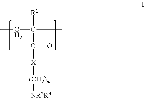
In one aspect, the polycation is a polyamino compound. In another aspect, the polyamino compound has 10 to 90 mole % tertiary amino groups. In a further aspect, the polycation polymer has at least one fragment of the formula I
##STR00002## wherein R.sup.1, R.sup.2, and R.sup.3 are, independently, hydrogen or an alkyl group, X is oxygen or NR.sup.5, where R.sup.5 is hydrogen or an alkyl group, and m is from 1 to 10, or the pharmaceutically-acceptable salt thereof. In another aspect, R.sup.1, R.sup.2, and R.sup.3 are methyl and m is 2. Referring to formula I, the polymer backbone is composed of CH.sub.2--CR.sup.1 units with pendant --C(O)X(CH.sub.2).sub.mNR.sup.2R.sup.3 units. In this aspect, the fragment having the formula I is a residue of an acrylate, methacrylate, acrylamide, or methacrylamide. FIG. 3 (structures C and D) and FIG. 6 (4 and 7) show examples of polycations having the fragment of formula I, where the polymer backbone is derived from acrylamide and methacrylate residues as discussed above. In one aspect, the polycation is the free radical polymerization product of a cationic tertiary amine monomer (2-dimethylamino-ethyl methacrylate) and acrylamide, where the molecular weight is from 10 to 20 kd and possesses tertiary monomer concentrations from 15 to 30 mol %. FIG. 4 (structures E and F) and FIG. 6 (5) provide examples of polycations useful herein, where imidazole groups are directly attached to the polymer backbone (structure F) or indirectly attached to the polymer backbone via a linker (structure E via a methylene linker).
Similar to the polycation, the polyanion can be a synthetic polymer or naturally-occurring. In one aspect, when the polyanion is naturally-occurring, the polyanion is a negatively-charged protein produced from P. californica. FIGS. 2 and 7 show the sequences of two proteins (Pc3a and Pc3b) produced by P. californica (Zhao et al. "Cement Proteins of the tube building polychaete Phragmatopoma californica" J. Biol. Chem. (2005) 280: 42938-42944). Referring to FIG. 20, Pc3a and Pc3b are essentially composed of polyphosphoserine, which is anionic at neutral pH. Examples of other naturally-occurring polyanions include glycosaminoglycans such as condroitin sulfate, heparin, heparin sulfate, dermatan sulfate, and hyaluronic acid.
When the polyanion is a synthetic polymer, it is generally any polymer possessing anionic groups or groups that can be readily converted to anionic groups by adjusting the pH. Examples of groups that can be converted to anionic groups include, but are not limited to, carboxylate, sulfonate, phosphonate, boronate, sulfate, borate, or phosphate. Any cationic counterions can be used in association with the anionic polymers if the considerations discussed above are met.
In one aspect, the polyanion is a polyphosphate. In another aspect, the polyanion is a polyphosphate compound having from 10 to 90 mole % phosphate groups. For example, the polyphosphate can be a naturally-occurring compound such as, for example, highly phosphorylated proteins like phosvitin (an egg protein), dentin (a natural tooth phosphoprotein), casein (a phosphorylated milk protein), or bone proteins (e.g. osteopontin).
In other aspects, phosphorous containing polymers can be converted to polyanions. For example, a phospholipid or phosphosugar is not a polyanion but it can be converted into a polyanion by creating a liposome or a micelle with it. Thus, in this aspect, the complex coacervate is a charged colloid. Alternatively, the colloid can be produced by any of the polyanions or polycations described herein.
In another aspect, the polyphosphate can be a synthetic compound. For example, the polyphosphate can be a polymer with pendant phosphate groups attached to the polymer backbone and/or present in the polymer backbone. (e.g., a phosphodiester backbone). In one aspect, the polyphosphate can be produced by chemically or enzymatically phosphorylating a protein (e.g., natural serine-rich proteins).
In one aspect, the polyanion includes a polyacrylate having one or more pendant phosphate groups. For example, the backbone can be a homopolymer or copolymer derived from the polymerization of acrylate monomers including, but not limited to, acrylates, methacrylates, acrylamides, and the like. In one aspect, the backbone of the polyanion is derived from the polymerization of polyacrylamide. In other aspects, the polyanion is a block co-polymer, where segments or portions of the co-polymer possess anionic groups depending upon the selection of the monomers used to produce the co-polymer. In a further aspect, the polyanion can be heparin sulfate, hyaluronic acid, chitosan, and other biocompatible and biodegradable polymers typically used in the art.
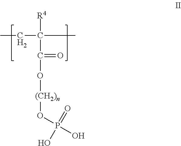
In one aspect, the polyanion is a polyphosphate. In another aspect, the polyanion is a polymer having at least one fragment having the formula II
##STR00003## wherein R.sup.4 is hydrogen or an alkyl group, and n is from 1 to 10, or the pharmaceutically-acceptable salt thereof. In another aspect, wherein R.sup.4 is methyl and n is 2. Similar to formula I, the polymer backbone of formula II is composed of a residue of an acrylate or methacrylate. The remaining portion of formula II is the pendant phosphate group. FIG. 7 (structure B), shows an example of a polyanion useful herein that has the fragment of formula II, where the polymer backbone is derived from acrylamide and methacrylate residues. In one aspect, the polyanion is the copolymerization product of ethylene glycol methacrylate phosphate and acrylamide, where the molecular weight is from 10,000 to 50,000, preferably 30,000, and has phosphate groups in the amount of 45 to 90 mol %.
As described above, the polycation and polyanion contain crosslinkable groups. In one aspect, the polycation and polyanion includes an actinically crosslinkable group defined herein. Any of the polymers described above (synthetic or naturally-occurring) that can be used as the polycation and polyanion can be modified to include the actinically crosslinkable group. For example, the polycation can be a polyacrylate having one or more pendant amino groups (e.g., imidazole groups). In the case of the polyanion, in one aspect, a polyphosphate can be modified to include the actinically crosslinkable group(s). For example, wherein the polycation and polyanion includes at least one fragment having the formula VII
##STR00004## wherein R.sup.1, R.sup.2, and R.sup.3 are, independently, hydrogen or an alkyl group, X is oxygen or NR.sup.5, where R.sup.5 is hydrogen or an alkyl group, and m is from 1 to 10, or the pharmaceutically-acceptable salt thereof, wherein at least one of R.sup.2 or R.sup.3 is an actinically crosslinkable group. In one aspect, referring to formula VII, R.sup.1 is methyl, R.sup.2 is hydrogen, R.sup.3 is an acrylate or methacrylate group, X is NH, and m is 2.
In another aspect, the polyanion can include one or more groups that can undergo oxidation, and the polycation contains on or more nucleophiles that can react with the oxidized crosslinker to produce new covalent bonds. In one aspect, the polyanion includes at least one dihydroxyl aromatic group capable of undergoing oxidation, wherein the dihydroxyl aromatic group is covalently attached to the polyanion. Examples of dihydroxyl aromatic groups include a DOPA residue or a catechol residue. Any of the polyanions described above can be modified to include one or more dihydroxyl aromatic residues. In one aspect, the polyanion is polymerization product between two or more monomers, where one of the monomers has a dihydroxyl aromatic group covalently attached to the monomer. For example, the monomer can have an unsaturated group capable of undergoing free-radical polymerization with the dihydroxyl aromatic group attached to the monomer. For example, the polyanion can be the polymerization product between (1) a phosphate acrylate and/or phosphate methacrylate and (2) a second acrylate and/or second methacrylate having a dihydroxyl aromatic group covalently bonded to the second acrylate or second methacrylate. In another aspect, the polyanion is the polymerization product between monoacryloxyethyl phosphate and dopamine methacrylamide. Polymers 3 and 7 in FIG. 6 provide examples of DOPA residues incorporated into a polyanion and polycation, respectively. In each of these polymers, an acrylate containing the pendant DOPA residue is polymerized with the appropriate monomers to produce the polyanion 3 and polycation 7 with pendant DOPA residues.
Not wishing to be bound by theory, the polyanion with the dihydroxyl aromatic group(s) are stable in that they react slowly with itself in solution. Thus, the polyanion reacts with the polycation primarily via intermolecular cross-linking (e.g., polycation has a nucleophilic group or a dihydroxyl aromatic group) to produce the complex coacervate. This provides numerous advantages with respect to the use and administration of the complex coacervate. For example, the polycation and polyanion can be premixed and administered to a subject instead of the sequential administration of the polymers. This greatly simplifies administration of the complex coacervate that is not an option with currently available bioadhesives.
It is contemplated that the polycation can be a naturally occurring compound (e.g., protein from P. californica) and the polyanion is a synthetic compound. In another aspect, the polycation can be a synthetic compound and the polyanion is a naturally occurring compound (e.g., protein from P. californica). In a further aspect, both the polyanion and polycation are synthetic compounds.
The adhesive complex coacervates can optionally contain one or more multivalent cations (i.e., cations having a charge of +2 or greater). In one aspect, the multivalent cation can be a divalent cation composed of one or more alkaline earth metals. For example, the divalent cation can be a mixture of Ca.sup.+2 and Mg.sup.+2. In other aspects, transition metal ions with a charge of +2 or greater can be used as the multivalent cation. In addition to the pH, the concentration of the multivalent cations can determine the rate and extent of coacervate formation. Not wishing to be bound by theory, weak cohesive forces between particles in the fluid may be mediated by multivalent cations bridging excess negative surface charges. The amount of multivalent cation used herein can vary. In one aspect, the amount is based upon the number of anionic groups and cationic groups present in the polyanion and polycation. For example, when the multivalent cation is a mixture of calcium and magnesium, the polycation is a polyamine, the polyanion is a polyphosphate, and the ratio of calcium to amine/phosphate groups can be from 0.1 to 0.3, and the ratio of magnesium to amine/phosphate groups can be from 0.8 to 1.0. In the Examples, the selection of the amount of multivalent cations with respect to producing adhesive complex coacervates and other physical states is addressed.
The adhesive complex coacervate can be synthesized a number of different ways. In one aspect, the polycation, the polyanion, and at least one multivalent cation, can be mixed with one another to produce the adhesive complex coacervate. By adding the appropriate amount of multivalent cation to the mixture of polyanion and polycation, the adhesive complex coacervate can be produced. In another aspect, the adhesive complex coacervate can be produced by the process comprising:
(a) preparing a polyelectrolyte complex comprising admixing at least one polycation and at least one polyanion, wherein at least one polycation or polyanion is a synthetic compound, and the polycation and/or polyanion comprises at least one group capable of crosslinking with each other; and (b) adjusting the pH of the polyelectrolyte complex, the concentration of at least one multivalent cation, or a combination thereof to produce the adhesive complex coacervate.
The adhesive complex coacervates produced herein can undergo subsequent phase changes that ultimately lead to the formation of an adhesive. In one aspect, the adhesive can be produced by the process comprising
(a) heating an adhesive complex coacervate comprising at least one polycation, and at least one polyanion, wherein the polycation and/or polyanion comprises a crosslinker, wherein upon heating the adhesive complex coacervate the coacervate is converted to an insoluble solid; and (b) crosslinking the polycation and polyanion in the insoluble solid to produce the adhesive. In this aspect, heating the adhesive complex coacervate converts the coacervate to an insoluble solid. The temperature can vary depending upon the nature of the coacervate (i.e., selection of polycation, polyanion, multivalent cations, etc.). For example, at room temperature, a complex coacervate can be present. However, by injecting the coacervate into a subject where the temperature is 37.degree. C., the coacervate solidifies at body temperature. As will be discussed below, this has numerous applications in tissue/bone repair as well as for the delivery of drugs.
In other aspects, the adhesive is produced by the process comprising
(a) preparing an adhesive complex coacervate comprising admixing at least one polycation and at least one polyanion, wherein at least one polycation or polyanion is a synthetic compound, and the polycation and/or polyanion comprises at least one group capable of crosslinking with each other; (b) adjusting the pH of the adhesive complex coacervate to produce an insoluble solid; and (c) crosslinking the polycation and polyanion in the insoluble solid to produce the adhesive. In this aspect, the complex coacervate is converted to an insoluble soluble solid by adjusting the pH. The adjustment of the pH can be accomplished by a number of techniques. For example, the pH can be actively changed by the delivery of a second component (e.g., acid or base) in combination with the complex coacervate to convert the complex coacervate to an insoluble solid. Alternatively, the complex coacervate can be introduced into an environment having a pH that is different from that of the complex coacervate, where the change in pH can convert the complex coacervate to an insoluble solid. In one aspect, the pH is raised to a pH greater than or equal to 7.0, or up to a pH of 8.0.
In these aspects, once the adhesive complex coacervate is converted to an insoluble solid, the insoluble solid is crosslinked to produce a strong adhesive. As discussed above, the polycation and polyanion possess one or more crosslinkable groups capable of forming covalent bonds. For example, the polycation and/or polyanion can possess at least one dihydroxyl aromatic group capable of undergoing oxidation. In this aspect, the dihydroxyl aromatic group can be oxidized by a variety oxidants such O.sub.2, NaIO.sub.4, a peroxide, a transition metal oxidant, or stabilized oxidant as described above. In the case when the polycation or polyanion has dihydroxyl aromatic group, the other polymer can possess a nucleophilic group that can react with the oxidized form of the dihydroxyl aromatic group to produce a new covalent bond. In other aspects, when the polycation and polyanion possess an actinically crosslinkable group, the insoluble solid can be irradiated with light to crosslink the polycation and polyanion to produce the adhesive. In this aspect, the insoluble solid (and complex coacervate precursor) can include a photoinitiator to facilitate crosslinking between the actinically crosslinkable groups. Examples of photoinitiators useful herein include, but are not limited to, a phosphine oxide, a peroxide, an azide compound, an .alpha.-hydroxyketone, or an .alpha.-aminoketone. Upon crosslinking, a strong adhesive is produced having numerous applications.
The adhesive complex coacervates and adhesives produced therefrom described herein have numerous benefits with respect to their use as biological glues and delivery devices. For example, the coacervates have low initial viscosity, specific gravity greater than one, and being mostly water by weight, low interfacial tension in an aqueous environment, all of which contribute to their ability to adhere to a wet surface. An additional advantage with respect to the bonding mechanism (i.e., crosslinking) of the adhesive complex coacervates includes low heat production during setting, which prevents damage to living tissue. The components can be pre-polymerized in order to avoid heat generation by in situ exothermic polymerization. This is due for the most part by the ability of the adhesive complex coacervates to crosslink intermolecularly under very mild conditions as described above.
The adhesive complex coacervates described herein can be applied to a number of different biological substrates. The substrate can be contacted in vitro or in vivo. The rate of crosslinking within the adhesive complex coacervate can be controlled by for example pH and the presence of an oxidant or other agents that facilitate crosslinking. One approach for applying the adhesive complex coacervate to the substrate can be found in FIG. 9. The techniques depicted in FIG. 9 are referred to herein as "spot welding," where the adhesive complex coacervate is applied at distinct and specific regions of the substrate. In one aspect, the adhesive complex coacervate can be produced in situ. Referring to FIG. 9A, a pre-formed stable PEC solution 1 composed of polycations and polyanions at low pH (e.g., 5) is simultaneously applied to a substrate with a curing solution 2 composed of an oxidant at a higher pH (e.g., 10) with the use of syringes. Upon mixing, the curing solution simultaneously produces the adhesive complex coacervate by crosslinking the polymers on the surface of the substrate.
In another aspect, referring to FIG. 9B, a solution of polyanions 3 and polycations 4 are applied simultaneously to the substrate. One of the solutions has a pH higher than the other in order to produce the adhesive complex coacervate. Referring to FIG. 9B, polyanion 3 is at a lower pH than the polycation solution 4; however, it is also contemplated that the polyanion can be in solution having a higher pH than the polycation. The solution having the higher pH can include an oxidant in order to facilitate crosslinking.
FIG. 9C depicts another aspect of spot welding. In this aspect, the substrate is primed with polycation at a particular pH. Next, a solution of the polyanion at a higher pH is applied to the polycation in order to produce the adhesive complex coacervate in situ. It is also contemplated that the substrate can be primed with polyanion first followed by polycation. An oxidant can then be applied separately on the complex coacervate to facilitate crosslinking to produce the adhesive complex coacervate. Alternatively, the solution applied after the substrate has been primed can contain the oxidant so that the adhesive complex coacervate is formed and subsequently crosslinked in situ.
The properties of the adhesive complex coacervates described herein make them ideal for underwater applications such as the administration to a subject. For example, the adhesive complex coacervates and adhesives produced therefrom can be used to repair a number of different bone fractures and breaks. The coacervates adhere to bone (and other minerals) through several mechanisms (see FIG. 1C). The surface of the bone's hydroxyapatite mineral phase (Ca.sub.5(PO.sub.4).sub.3(OH)) is an array of both positive and negative charges. The negative groups present on the polyanion (e.g., phosphate groups) can interact directly with the positive surface charges or it can be bridged to the negative surface charges through the cationic groups on the polycation and/or multivalent cations. Likewise, direct interaction of the polycation with the negative surface charges would contribute to adhesion. Additionally, when the polycation and/or polyanion contain catechol moieties, they can facilitate the adhesion of the coacervate to readily wet hydroxyapatite. Other adhesion mechanisms include direct bonding of unoxidized crosslinker (e.g., DOPA or other catechols) to hydroxyapatite. Alternatively, oxidized crosslinkers can couple to nucleophilic sidechains of bone matrix proteins.
Examples of such breaks include a complete fracture, an incomplete fracture, a linear fracture, a transverse fracture, an oblique fracture, a compression fracture, a spiral fracture, a comminuted fracture, a compacted fracture, or an open fracture. In one aspect, the fracture is an intra-articular fracture or a craniofacial bone fracture. Fractures such as intra-articular fractures are bony injuries that extend into and fragment the cartilage surface. The adhesive complex coacervates may aid in the maintenance of the reduction of such fractures, allow less invasive surgery, reduce operating room time, reduce costs, and provide a better outcome by reducing the risk of post-traumatic arthritis.
In other aspects, the adhesive complex coacervates and adhesives produced therefrom can be used to join small fragments of highly comminuted fractures. In this aspect, small pieces of fractured bone can be adhered to an existing bone. It is especially challenging to maintain reduction of the small fragments by drilling them with mechanical fixators. The smaller and greater number of fragments the greater the problem. In one aspect, the adhesive complex coacervate or precursor thereof may be injected in small volumes to create spot welds as described above in order to fix the fracture rather than filling the entire crack. The small biocompatible spot welds would minimize interference with healing of the surrounding tissue and would not necessarily have to be biodegradable. In this respect it would be similar to permanently implanted hardware.
In other aspects, the adhesive complex coacervates and adhesives produced therefrom can be used to secure scaffolds to bone and other tissues such as, for example, cartilage, ligaments, tendons, soft tissues, organs, and synthetic derivatives of these materials. Using the complexes and spot welding techniques described herein, the adhesive complex coacervates and adhesives produced therefrom can be used to position biological scaffolds in a subject. Small adhesive tacks composed of the adhesive complex coacervates described herein would not interfere with migration of cells or transport of small molecules into or out of the scaffold. In certain aspects, the scaffold can contain one or more drugs that facilitate growth or repair of the bone and tissue. In other aspects, the scaffold can include drugs that prevent infection such as, for example, antibiotics. For example, the scaffold can be coated with the drug or, in the alternative, the drug can be incorporated within the scaffold so that the drug elutes from the scaffold over time.
The adhesive complex coacervates and adhesives produced therefrom have numerous dental applications. For example, the adhesive complex coacervates can be used to repair breaks or cracks in teeth, for securing crowns, or seating implants and dentures. Using the spot weld techniques described herein, the adhesive complex coacervate or precursor thereof can be applied to a specific points in the mouth (e.g., jaw, sections of a tooth) followed by attaching the implant to the substrate.
In other aspects, the adhesive complex coacervates and adhesives produced therefrom can adhere a metal substrate to bone. For example, implants made from titanium oxide, stainless steel, or other metals are commonly used to repair fractured bones. The adhesive complex coacervate or a precursor thereof can be applied to the metal substrate, the bone, or both prior to adhering the substrate to the bone. In certain aspects, the crosslinking group present on the polycation or polyanion can form a strong bond with titanium oxide. For example, it has been shown that DOPA can strongly bind to wet titanium oxide surfaces (Lee et al., PNAS 103:12999 (2006)). Thus, in addition to bonding bone fragments, the adhesive complex coacervates described herein can facilitate the bonding of metal substrates to bone, which can facilitate bone repair and recovery.
It is also contemplated that the adhesive complex coacervates and adhesives produced therefrom can encapsulate one or more bioactive agents. The bioactive agents can be any drug that will facilitate bone growth and repair when the complex is applied to the bone. The rate of release can be controlled by the selection of the materials used to prepare the complex as well as the charge of the bioactive agent if the agent is a salt. In certain aspects, when the adhesive complex coacervate is converted to an insoluble solid by a change in temperature and/or pH, the complex coacervate can be administered to a subject and produce an insoluble solid in situ. Thus, in this aspect, the insoluble solid can perform as a localized controlled drug release depot. It may be possible to simultaneously fix tissue and bones as well as deliver bioactive agents to provide greater patient comfort, accelerate bone healing, and/or prevent infections.
The adhesive complex coacervates and adhesives produced therefrom can be used in a variety of other surgical procedures. For example, adhesive complex coacervates and adhesives produced therefrom can be used to repair lacerations caused by trauma or by the surgical procedure itself. In one aspect, the adhesive complex coacervates and adhesives produced therefrom can be used to repair a corneal laceration in a subject. In other aspects, the adhesive complex coacervates and adhesives produced therefrom can be used to inhibit blood flow in a blood vessel of a subject. In general, the adhesive complex coacervate is injected into the vessel followed by converting to an insoluble solid (e.g., heating the complex coacervate) and adhesive to partially or completely block the vessel. This method has numerous applications including hemostasis or the creation of an artificial embolism to inhibit blood flow to a tumor or aneurysm.
Examples
The following examples are put forth so as to provide those of ordinary skill in the art with a complete disclosure and description of how the compounds, compositions, and methods described and claimed herein are made and evaluated, and are intended to be purely exemplary and are not intended to limit the scope of what the inventors regard as their invention. Efforts have been made to ensure accuracy with respect to numbers (e.g., amounts, temperature, etc.) but some errors and deviations should be accounted for. Unless indicated otherwise, parts are parts by weight, temperature is in .degree. C. or is at ambient temperature, and pressure is at or near atmospheric. There are numerous variations and combinations of reaction conditions, e.g., component concentrations, desired solvents, solvent mixtures, temperatures, pressures and other reaction ranges and conditions that can be used to optimize the product purity and yield obtained from the described process. Only reasonable and routine experimentation will be required to optimize such process conditions.
I. Synthesis and Characterization of Adhesive Complex Coacervates
Mimetic Copolymer Synthesis and Characterization.
Pc3 Analogs.
The dopa analog monomer (dopamine methacrylamide, DMA) was prepared by slight modification of a published procedure. (Lee B P, Huang K, Nunalee F N, Shull K R, Messersmith P B. Synthesis of 3,4-dihydroxyphenylalanine (DOPA) containing monomers and their co-polymerization with PEG-diacrylate to form hydrogels. J Biomater Sci Polym Ed 2004; 15(4):449-464). Briefly, a borate-dopamine complex was reacted at pH >9 with methacryloyl chloride. After disrupting the borate-catechol bond by acidification, the product was washed with ethyl acetate, recrystallized from hexane, and verified by .sup.1H NMR (400 MHz, DMSO-TMS): d.quadrature.8.8-8.58 (2H, (OH).sub.2--Ar--), 7.92 (1H, --C(.dbd.O)--NH--), 6.64-6.57 (2H, C.sub.6HH.sub.2(OH).sub.2--), 6.42 (1H, C.sub.6H.sub.2H(OH).sub.2--), 5.61 (1H, --C(.dbd.O)--C(--CH.sub.3).dbd.CHH), 5.30 (1H, --C(.dbd.O)--C(--CH.sub.3).dbd.CHH), 3.21 (2H, C.sub.6H.sub.3(OH).sub.2--CH.sub.2--CH.sub.2(NH)--C(.dbd.O)--), 2.55 (2H, C.sub.6H.sub.3(OH).sub.2--CH.sub.2--CH.sub.2(NH)--C(.dbd.O)--), 1.84 (3H, --C(.dbd.O)--C(--CH.sub.3).dbd.CH.sub.2).
Before polymerization monoacryloxyethyl phosphate (MAEP, Polysciences) was diluted in MeOH and extracted with hexane to remove dienes. Copolymer 1 was prepared by mixing 90 mol % MAEP, 8 mol % DMA, 2 mol % acrylamide (Aam, Polysciences), and 0.1 mol % FITC-methacrylamide in MeOH at a final monomer concentration of 5 wt %. Free radical polymerization was initiated with azobisisobutyronitrile (AIBN) and proceeded at 60.degree. C. for 24 hrs in sealed ampules. A similar procedure was used to make polymers 3-7 as shown in FIGS. 2-7. Copolymer 1 (FIG. 10) was recovered by size exclusion chromatography (SEC) in MeOH on a Sephadex LH-20 column (Sigma-Aldrich), concentrated by rotary evaporation, dissolved in DI water, and freeze dried.
The MW and polydispersity index (PDI) of 1 were determined by SEC in DMF on a PLgel column (Polymer Labs) connected to a small angle light scattering detector (Brookhaven BI-MWA) and refractive index monitor (Brookhaven BI-DNDC). The column was calibrated with polystyrene standards. The MW of 1 was 245 kda with a PDI of 1.9. The dopamine sidechain concentration and reactivity was verified by UV/VIS spectroscopy (e.sub.280=2600 M.sup.-1cm.sup.-1). The phosphate sidechain concentration were determined by titration with 0.005 M NaOH using an automated titrator (Brinkmann Titrando 808). The UV/vis spectrum of 1 contained a single absorption peak at 280 nm characteristic of the catechol form of dopamine (FIG. 10B). Addition of a 1:1 molar ratio of NaIO.sub.4 to 1 at pH 5.0 oxidized the dopa catechol to dopaquinone with an absorption peak near 395 nm as expected. The dopaquinone peak was stable for several hrs at pH<5.
Pc1 Analogs.
The lysine sidechains of Pc1 were mimicked with N-(3-aminopropyl) methacrylamide hydrochloride (APMA, Polysciences). Copolymer 2 (FIG. 10) was synthesized by dissolving 10 mol % APMA and 90 mol % Aam in DI water, degassing with N.sub.2 and initiating polymerization with 2 mol % ammonium persulfate (Polysciences). Polymerization proceeded at 50.degree. C. for 24 hrs in sealed ampules. Polymer was recovered by dialysis against water for 3 days, and then freeze dried. The primary amine sidechain mol % was determined by .sup.1H NMR (400 MHz, DMSO-TMS) from the ratios of d 13.45 (3H, --CH3) and d 51.04 (1H, RC(.dbd.O)CHR2). The MW and PDI of 2 were determined by SEC in PBS (20 mM PO.sub.4, 300 mM NaCl, pH 7.2) on a Superose 6 column (Pharmacia). The column was calibrated with poly-2-hydroxypropyl methacrylate standards. The MW of 2 was 165 kd and PDI was 2.4.
Coacervate Formation and Characterization.
A 5 wt % aqueous solution of 2 was added dropwise while stirring to a 5 wt % aqueous solution of 1 until reaching the target amine/phosphate ratio. Total copolymer concentration was 50 mg/ml. After mixing for 30 min the pH was adjusted with NaOH (6M). Compositions at pH (<4) conducive to polyelectrolyte complex (PEC) formation were diluted to 1 mg/ml in DI H.sub.2O and the zeta potentials and size distribution of PECs were measured on a Zeta-Sizer 3000HS (Malvern Instruments). At higher pH, coacervated compositions were centrifuged at 2500 rpm in a microfuge (Eppendorf), at 25.degree. C. for 2 min to collect the coacervate phase. The volume of both phases was measured. The coacervate phases were freeze dried and weighed to determine their mass and concentration.
The phase behavior of 1 and 2 mixed at a 1:1 molar ratio of phosphate to amine sidechains (50 mg/ml combined concentration) over the pH range 3-10 is shown in FIG. 11A. The calculated net copolymer charge normalized to the total ionizable sidechain concentration is shown in FIG. 11B. Ascorbate, a reductant, was added at a 1:5 molar ratio to dopa to retard oxidation of dopa by O.sub.2 and subsequent crosslinking at elevated pH. At low pH, the polyelectrolytes formed a stable milky solution of colloidal polyelectrolyte complexes (PECs). The mean diameter of the PECs at pH 2.1, determined by dynamic light scattering, was 360 nm with a narrow dispersity and increased to 1080 nm at pH 4.0 (FIG. 11C). The crossover of the zeta potential from positive to negative at pH 3.6 fit well with the calculated pH dependent net charge of the complexes (FIG. 11B). The particle size could not be measured accurately above pH 4 because the complexes flocculated. As the net charge increased due to the deprotonation of the phosphate sidechains, the copolymers condensed into a dense second phase. At pH 5.1 the separated phase had the character of a loose low density precipitate. At pH 7.2 and 8.3 the dense phase had the character of a cohesive liquid complex coacervate (FIG. 12). The copolymers were concentrated about three-fold to 148 and 153 mg/ml, respectively, in the coacervated phases. At pH 9.5 the polyelectrolyte mixture formed a dense non-liquid ionic gel. At pH 10 the copolymers went into solution and spontaneously crosslinked through the dopaquinone and amine sidechains into a clear hydrogel.
Extraction of divalent cations with the chelator EDTA resulted in a 50% decrease in compressive strength of P. californica tubes, a ten-fold decrease in adhesiveness, and collapse of the glues porous structure. The effect of divalent cations on the phase behavior of the mimetic polyelectrolytes was investigated by mixing 1 and 2 at amine to phosphate sidechain ratios ranging from 1:1 to 0:1 with divalent cation to phosphate sidechain ratios ranging from 0:1 to 1:1 to create a coacervate phase diagram (FIG. 13). The pH was fixed at 8.2, the pH of seawater, and divalent cations were added as a 4:1 mixture of Mg.sup.2+ and Ca.sup.2+, the approximate Mg.sup.2+/Ca.sup.2+ ratio in the natural glue determined by elemental analysis. The highest mass of coacervate (dark gray squares) occurred in mixtures with higher amine to phosphate sidechain ratios and lower divalent cation to phosphate sidechain ratios. Mixtures with lower polyamine ratios were clear (clear squares) even at higher divalent cation/phosphate sidechain ratios. At higher amine/phosphate and divalent cation/phosphate ratios the solutions were turbid (light gray squares) with slight precipitates but much less turbid than solutions containing PECs (medium gray squares).
Mechanical Bond Testing.
Bone test specimens, .about.1 cm.sup.3, were cut with a band saw from bovine femur cortical bone, obtained from a local grocery store, sanded with 320 grit sandpaper, and stored at -20.degree. C. NaIO.sub.4 at a 1:2 molar ratio to dopa sidechains was evenly applied to one face each of two wet bone specimens. Forty ml, a volume sufficient to completely fill the space between 1 cm.sup.2 bone interfaces, of the test coacervate solution was applied with a pipette, the bone specimens were pressed together squeezing out a small excess of adhesive, clamped, and immediately wrapped in PBS (20 mM PO.sub.4, 150 mM NaCl, pH 7.4) soaked gauze. The applied coacervate contained ascorbate at a 1:5 molar ratio to dopa to prevent premature crosslinking. The bonded specimens were incubated at 37.degree. C. for at least 24 hr in a sealed container containing soaked sponges to maintain 100% humidity. Reference specimens were bonded with 40 ml Loctite 401 superglue in exactly the same manner. A commercial non-medical grade cyanoacrylate was used because there are no hard tissue medical adhesives available for comparison. Mechanical tests were performed on a custom built material testing system using a 1 kg load cell. The instrument was controlled and data acquired using LabView (National Instruments). One bone of a bonded pair was clamped laterally 1 mm from the bond interface. The second bone was pressed with a cross-head speed of 0.02 mm/s against a dull blade positioned 1 mm lateral to the bond interface. Bond strength tests were performed at room temperature immediately after unwrapping the wet specimens to prevent drying. After testing, the bonds were examined for failure mode. The bonded area was measured by tracing an outline of the bone contact surface on paper, cutting out the trace, and determining its area from the weight of the paper cut-out. At least 6 specimens were tested for each condition.
The shear modulus and strength at failure were measured with bovine cortical bone specimens bonded while wet with the three coacervating compositions marked with an asterisk in FIG. 13. The coacervate density in the three compositions increased with increasing divalent cation ratios (to 120, 125, and 130 mg/ml, respectively). Both the modulus and bond strength of the fully hydrated specimens increased with increasing divalent cation concentration, reaching 37% of the strength of wet bones bonded with a commercial cyanoacrylate adhesive (FIG. 14A). The cyanoacrylate adhesive was used as a reference point because there are no bone adhesives in clinical use for comparison. The strength of the mimetic adhesive is also about 1/3 the strength of natural P. californica glue estimated to be 350 kPa and mussel byssal glue estimated to range from 320 to 750 kPa dependent on the season. In almost all cases the bonds failed cohesively leaving adhesive on both bone interfaces, which suggested the compositions formed strong interfacial bonds with hydroxyapatite. The bonds were dimensionally stable, neither shrinking nor swelling appreciably after complete submersion in PBS pH 7.2 for several months (FIG. 14B). Dimensional stability during cure and long term exposure to water is an important requirement for a useful bone adhesive.
Dopamine-Mediated Copolymer Crosslinking.
Addition of NaIO.sub.4 to solutions of 3 at a 1:1 molar ratio immediately and quantitatively oxidized DOPA (280 nm) to dopaquinone (392 nm). Within a few minutes the quinine peak decayed into broad general absorption as the reactive quinones formed covalent diDOPA crosslinks (FIG. 15, top left). Crosslinking between the quinones and primary amines (FIG. 15, bottom left) led to a broader general absorption than diDOPA crosslinking. Dopamine oxidation and crosslinking chemistry therefore behaved as expected in the dopamine copolymers. The dopamine copolymers rapidly formed hydrogels as a result of oxidative crosslinking (FIGS. 15, A&C). Oxidized phosphodopamine 3 did not gel by itself (FIG. 15B) but when mixed with 4 it gelled rapidly (FIG. 15D). Intermolecular diDOPA crosslinking between PO.sub.4 copolymers was inhibited but not intermolecular DOPA-amine crosslinking. This provides a crosslinking control mechanism that may be useful for formulating and delivering a synthetic adhesive.
pH Triggered DOPA-Mediated Crosslinking.
To explore, the pH dependence and kinetics of DOPA oxidation, crosslinking of the dopamine copolymers were evaluated by UV-Vis spectroscopy. Results with p(EGMP[92]-DMA[8]) (3) are shown in FIG. 16. UV-vis spectra were acquired at increasing time after addition of a stoichiometric amount of NaIO.sub.4. At pH 5.0 (top), dopaquinone absorbance (398 nm) was maximal in .about.15 min and remained stable for several hrs (inset). At pH 6.0, absorbance at 398 nm peaked in <1 min and evolved into broad absorbance with peaks at 310 and 525 nm. The broad absorbance is not due to dopaquinone crosslinking since gels do not form (FIG. 16). For comparison, 6 was oxidized at low pH crosslinked but at a significantly slower rate (not shown).
The results show that the dopaquinone is stable at low pH and diDOPA crosslinking was inhibited at higher pH in the phosphodopamine copolymers. In the presence of the polyamine, the covalent crosslinking was channeled toward intermolecular amine-DOPA bonds. This is an important observation because it lays out a path to controlled delivery and setting of the synthetic adhesive.
In Vitro Cytotoxicity.
Solutions of 3 and 4, 40 wt % each, were mixed at low pH to form a polyelectrolyte complex. The solution was partially oxidized with NaIO.sub.4 and basified with NaOH just before application to sterile glass coverslips. The adhesive-treated coverslips were placed in the bottom of culture plate wells and human foreskin fibroblasts, human tracheal fibroblasts, and rat primary astrocytes in serum containing media were added to separate wells at 30K cells/well (FIG. 17). After 24 hr, the cells were fixed with 4% para-formaldehyde, then immunostained for the intermediate filament protein, vimentin, to visualize cell morphology (green, A-B), pericellular fibronectin to assess ECM secretion (red, B), glial fibrillary protein to visual primary astrocyte morphology (green, C), and DAPI to visualize nuclei (blue, C). The granular globs of adhesive auto-fluoresced orangish-red (A-C).
In the representative images (FIG. 17), all cell types had morphologies indistinguishable from cells growing on glass without adhesive. The cells had normal motility and in several cases extended processes that directly contacted the adhesive. No toxicity was apparent.
Rat Calvarial Defect Model.
Production of the fragmented defect and repair with an adhesive complex coacervate is shown in FIGS. 18A-F. Male Sprague Dawley rats (256-290 g) (Harlan) were anesthetized with a mixture of ketamine (65 mg/kg), xylazine (7.5 mg/kg), and acepromazine (0.5 mg/kg). At full depth of anesthesia, the eyes were covered with ophthalmic ointment, the head shaved, and the scalp disinfected with isopropanol and butadiene. With the prepped rats in a stereotactic frame, a compressed air-driven drill operating at 5000 RPM was lowered using a stereotactic fine toothed manipulator. Sterile saline or PBS was continuously applied at the craniotomy site while the custom made trephine tool was lowered 600 microns (previously determined as the skull thickness of rats the age of which were used in the experiment). The result is a round, accurate hole through the skull with little observable effect on the underlying dura or vasculature (FIG. 18A-B). The bone plug was recovered with fine curved forceps and broken into fragments using a hemostat and fine rongeur (FIG. 18B). The bone fragments were returned to the defect (FIG. 18C) and 5 .mu.l of test adhesive (3 and 4 mixed immediately prior to the application of the fracture) was applied with a micropipettor (FIG. 18D). The low viscosity adhesive solution (pre-formed PECS mixed with curing solution just before delivery) readily and cleanly wicked into the fractures. Within 5 min the fragments were sufficiently fixed that they could be tapped sharply with the forceps without displacement. The adhesive continued to turn dark reddish brown as it cured (FIG. 18E-F).
II. Adhesive Complex Coacervates Produced from an Amine-Modified Polymer
A. Materials and Methods
Materials.
Low endotoxin, non-gelling, gelatin (MW 3.5 kDa) was provided by Gelita Inc. (Souix City, Iowa). 1-Ethyl-3-[3-dimethylaminopropyl]carbodiimide hydrochloride (EDC) and ethylenediamine dihydrochloride were purchased from Thermo Scientific Inc. Monoacryloxyethyl phosphate (MAEP), 2,2'-azobisisobutyronitrile (AIBN) were purchased from Polysciences, Inc. Sodium periodate (NaIO.sub.4), Sephadex LH-20, dopamine hydrochloride was obtained from Sigma-Aldrich.
Polyphosphodopamide Synthesis.
The polyphosphodopamide copolymer (poly(MAEP.sub.85-DMA.sub.15)) was synthesized by free radical polymerization of MAEP and dopamine methacrylamide (DMA) using azobisisobutyronitrile (AIBN) as initiator. The copolymer was recovered by size exclusion chromatography (SEC) in MeOH on a Sephadex LH-20 column (Sigma-Aldrich). MeOH was removed, the copolymer resuspended in water, lyophilized, and stored at -80.degree. C. The mol % dopamide side chains in the copolymers were determined by UV/vis spectroscopy: the catechol form of dopamide has an absorption peak at 279 nm (.lamda..sub.279=2600 M.sup.-1cm.sup.-1).
Gelatin Modification.
The general reaction scheme for producing amine-modified gelatin is provided in FIG. 21. Gelatin (100 mg/ml) was mixed with ethylenediamine dihydrochloride (1:1 molar ratio to the gelatin carboxyl groups). The pH was adjusted to 5.2 with 6M HCl. EDC at 1.2:1 molar ratio to ethylenediamine dihydrochloride was added to the reaction mixture while stirring. The reaction proceeded for 2 hrs at room temperature. The amine-modified gelatin was dialyzed against DI water for 3 days then lyophilized. The primary amine side chain concentration was determined by ninhydrin assay using glycine as a standard. Zeta potential measurements of gelatin (1 mg/ml in water) were determined by electrophoresis using a Malvern Zetasizer Nano-ZS ZEN 3600 (Malvern Instruments Ltd., Worcestershire, UK).
Gelatin Coacervate Formation.
A 50 mg/ml aqueous solution of amine-modified gelatin (pH 5.0) was added dropwise while stirring to a 50 mg/ml aqueous solution (pH 5.0) of poly(MAEP.sub.85-DOPA.sub.15) containing various ratios of divalent cation (Ca.sup.2+ or Mg.sup.2+) until reaching the target amine/phosphate ratio. The pH of the mixture was raised to 7.4 with NaOH. The coacervate phase was allowed to settle for 24 hrs. The coacervate and equilibrium phases were separated and their volumes measured. The coacervate phases were lyophilized and weighed to determine their mass and concentration.
Dynamic Rheology.
The elastic (G') and storage (G'') moduli were measured with a cone and plate configuration (20 mm diameter, 4.degree. C. cone) on a stress-controlled rheometer (TA Instruments, AR 500). To compare coacervate compositions the measurements were made with a constant frequency of 1 Hz and dynamic strain of 0.1% as the temperature was ramped from 0.degree. C. to 40.degree. C. at a rate of 0.5.degree. C./min.
Adhesive Bond Strength.
Aluminum test adherends, 0.12.times.0.6.times.5 cm, were cut from 5052 aluminum sheet (0.050 in) with a water saw. The adherends were polished with 600 grit super fine sandpaper and then cleaned following the procedure of ASTM D2651. Briefly, the adherends were sonicated twice in MeOH, air-dried, dipped into a solution of sulfuric acid and nochromix for 15 mins, then rinsed thoroughly with DI water and stored in DI water until bonded. The adherends were bonded within 12 hr of cleaning. For each adhesive sample, 9 wet aluminum test specimens were bonded. NaIO.sub.4 at 1:2 molar ratio to dopamide sidechains was evenly applied to the bond area of two aluminum adherends. The test coacervate solution (6 .mu.l) was applied to wet adherends with a pipette, which were then pressed together with an overlap of about 25 mm, clamped, and immediately submerged in water adjusted to pH 7.4 with NaOH. The bonded specimens cured fully submerged in water for .about.24 hr at the specified temperature. Shear strengths were measured while the adherends where fully submerged in a temperature-controlled water bath mounted on an Instron 3342 materials testing system with a 100 N load cell. The instrument was controlled and data acquired using Bluehill Lite software (Instron, Inc.).
B. Results
An adhesive complex coacervate was created using a low MW (3-5 kda) non-gelling collagen hydrolysate as the polycation. As received the collagen hydrolysate did not form complex coacervates with the phosphodopa copolymer (poly(MAEP).sub.85-co-dopamide.sub.15)) at physiological pH. Amination of carboxylic acid sidechains with ethylenediamine increased the amine concentration to .about.16 mol % and shifted the pI from 5.5 to 10.4. The aminated collagen formed dense coacervates at 25.degree. C. over a broad range of compositions. At pH 5, concentrated coacervates formed at amine to phosphate sidechain ratios from 0.5-1.0 and Ca.sup.2+ to phosphate ratios up to 0.8 (FIG. 23A). None of the compositions precipitated. At pH 7.4, the coacervation space was more confined; at Ca.sup.2+ ratios higher than 0.2 the copolymers precipitated as hard solids, reflecting the decreased solubility of the mixed polyelectrolytes and Ca.sup.2+ with increasing pH (FIG. 23B).
Investigation of the separate effect of Mg.sup.2+ on coacervation of the polyelectrolytes revealed significant differences compared with Ca.sup.2+. At pH 5 the coacervated region was larger. At ratios up to 1:1 Mg.sup.2+ to phosphate none of the compositions precipitated (FIG. 23C). With Mg.sup.2+ the copolymers condensed into more concentrated coacervates, in some cases >380 mg/ml, an almost 8-fold increase from the initial copolymer concentration. At pH 7.4 the coacervation range is broader and at high Mg.sup.2+ ratios compositions with mixed phases of fluid and solid occur due to decreased solubility with increased pH (FIG. 23D). The expanded coacervation space at higher pH again illustrates the dense fluid coacervates are stably balanced intermediates between soluble polyelectrolytes and insoluble solids. The physical nature of the solidified state at high Mg.sup.2+ ratios is non-fluid, but softer and more gel-like than the hard Ca.sup.2+ precipitates, reflecting perhaps an intermediate state of desolvation relative to fluid coacervates and solids. The distinct physical nature and solubility profile of the Mg.sup.2+ complexes are likely consequences of the smaller radius, higher charge density, and smaller coordination number of Mg.sup.2+ ions compared to Ca.sup.2+ ions. Mg.sup.2+ tends to coordinate single bulky ligands, like phosphate, because multiple ligands won't fit around the small ion. As a consequence much of its solvation sphere is retained. The larger Ca.sup.2+ ion, on the other hand, can accommodate several bulky ligands resulting in displacement of its solvation sphere and cross-link formation between ligands. Coacervates prepared with mixed Mg.sup.2+ and Ca.sup.2+ occupied space in between the coacervated regions of the individual cations.
The phase diagrams in FIG. 23 illustrate empirically how the pH differential between secretory granules and seawater could trigger a phase change that drives the rapid but well-timed initial setting reaction of the natural adhesive. The condensed fluid complex coacervate phase is thermodynamically balanced between stable colloidal complexes and gelled or precipitated polymeric salts. The composition of the natural adhesive may be adapted to fall just inside the coacervation boundary within the secretory pathway, but to be outside of the coacervated region at the elevated pH of seawater. In other words, they are composed to undergo a pH dependent phase change upon secretion. For example, row 4 compositions (FIGS. 23A and B), with ratios of 0.4 Ca.sup.2+ and greater than 0.3 amine are coacervated at pH 5 but solid at pH 7.4 and higher.
At 0.degree. C. the coacervated region in FIG. 23B was shifted approximately one row lower, while at 37.degree. C. it is shifted one row higher (not shown). The temperature dependent phase transition of several compositions at pH 7.4 with increasing Ca.sup.2+ ratios and a fixed amine ratio of 0.6 were investigated in more detail by dynamic oscillatory rheology (FIG. 24A). At low temperature the viscous shear moduli (G'') were greater than the elastic moduli (G') consistent with the fluid character of the complex coacervates. With increasing temperature G' rose sigmoidally in a Ca.sup.2+ ratio dependent manner. The crossover points at which G'=G'' (FIG. 24A, inset), taken as the transition temperature where the compositions begin to change from viscous fluids to load-bearing elastic solids, were 36, 21, 12, and 9.degree. C., for Ca.sup.2+ ratios of 0.15, 0.20, 0.25, and 0.30, respectively. The Mg.sup.2+ containing coacervates demonstrated qualitatively similar behavior: there was no crossover of G' and G'' at Mg.sup.2+ to phosphate ratios up to 0.8 at pH 7.4, at higher ratios the crossover temperature again decreased with increasing Mg.sup.2+ ratios. The elastic moduli at 37.degree. C. were much lower with Mg.sup.2+ than with Ca.sup.2+ (FIG. 24B), consistent with the more hydrated gel-like quality of the solidified Mg.sup.2+ coacervates.
Bonds formed with Ca.sup.2+ ratios ranging from 0 to 0.3 with an amine ratio fixed at 0.6 were tested with polished aluminum adherends fully submerged in a temperature controlled water bath at 37.degree. C., well above the transition temperatures of the compositions. The lap shear strength increased with increasing Ca.sup.2+ up to a ratio of 0.3 (FIG. 25A, black bars). The 0.2 and 0.25 Ca.sup.2+/0.6 amine compositions were also tested slightly below their respective transition temperatures at 10 and 20.degree. C. In both cases, the bond strengths above the transition temperature were greater than below the transition temperature (FIG. 25A, white bars). Under the conditions of the test set-up there is likely to be little covalent oxidative crosslinking between the dopamide sidechains of the polyphosphate and the amines of gelatin: the rate of dopa oxidation is much slower at pH 7.4 than 8.2, diffusion of dissolved O.sub.2 into the narrow bond gap (62 .mu.m) was restricted, and there was no evident browning of the adhesive indicative of dopa oxidation. Therefore the increase in bond strength above the transition temperature was predominantly due to the state change of the adhesive. Similar tests with the 1.0 Mg.sup.2+ ratio demonstrated a more dramatic increase, a more than six-fold increase in bond strength above the transition temperature than below (FIG. 25B). As a practical matter, the results demonstrated that temperature differentials can be exploited as a convenient means to trigger the initial set of the synthetic adhesive and that the temperature trigger can be adjusted within a physiologically relevant range by small changes in the divalent cation ratio.
Next, oxidative coupling between the polyphosphate dopamide sidechains and the gelatin amines was initiated by adding 0.5 equivalents NaIO.sub.4 relative to the dopamide sidechains during the bonding procedure in order to investigate the contribution of covalent crosslinking to bond strength of the synthetic adhesive. The bonds were cured and tested at 37.degree. C. while fully submerged in water adjusted to pH 7.4. The bonds strengths increased with increasing divalent cation ratio for both Ca.sup.2+ and Mg.sup.2+ (FIG. 25, hatched bars). Maximum bond strengths with Mg.sup.2+ were double the bond strength of Ca.sup.2+, reaching 765 kPa.
In conclusion, the adhesive complex coacervates were dense, partially water-immiscible fluids precariously balanced between soluble polymers and insoluble polymeric salts (see white arrow in FIG. 22A). Referring to FIG. 22B, the top row represents the phase behavior of the polyelectrolytes. The bottom row connects the features of the phase behavior to solving the several problems of creating an underwater glue. The change from fluid complex coacervate to insoluble solid, the initial setting reaction, is triggered by a change in the pH, temperature, or both. Covalent hardening occurs through oxidative coupling between catechol and primary amine sidechains.
III. Preparation of Photocrosslinkable Polymers
Synthesis of Methacrylate-Grafted Pol
References
C00001
C00002
C00003
C00004
D00001
D00002
D00003
D00004
D00005
D00006
D00007
D00008
D00009
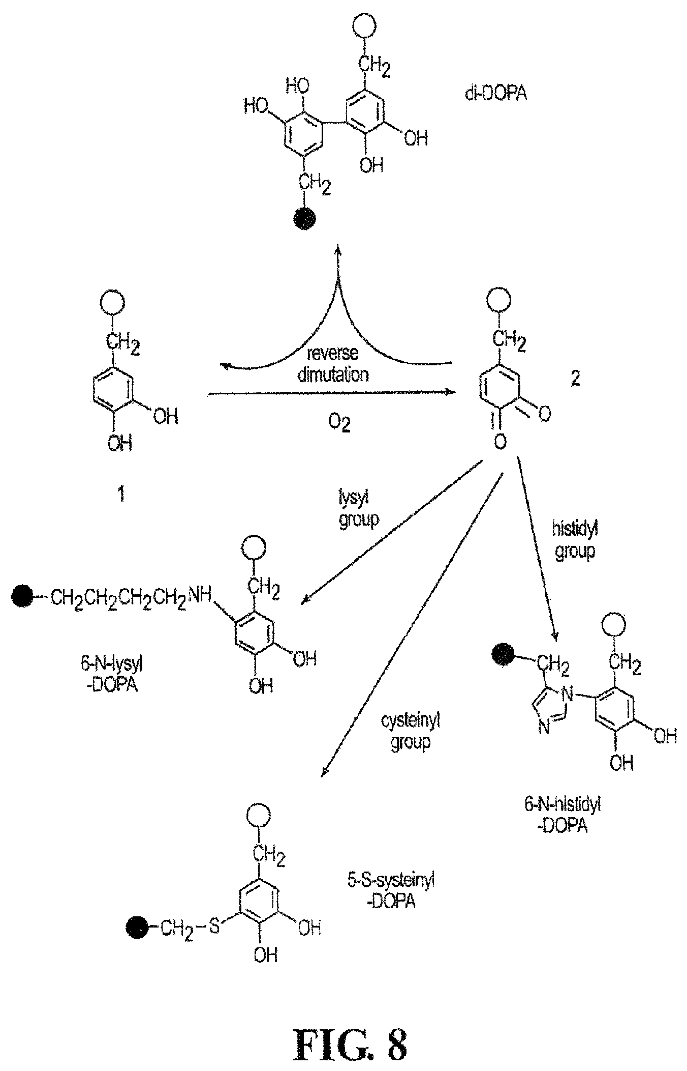
D00010

D00011

D00012
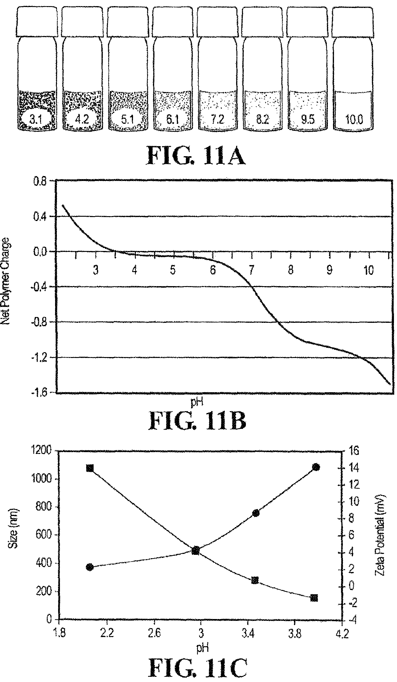
D00013

D00014

D00015

D00016

D00017

D00018
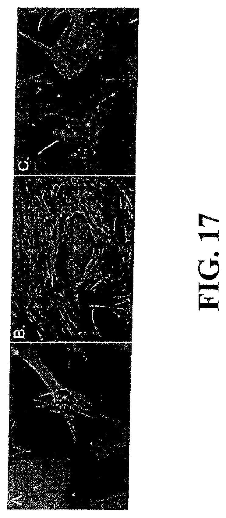
D00019

D00020

D00021

D00022

D00023

D00024

D00025

D00026
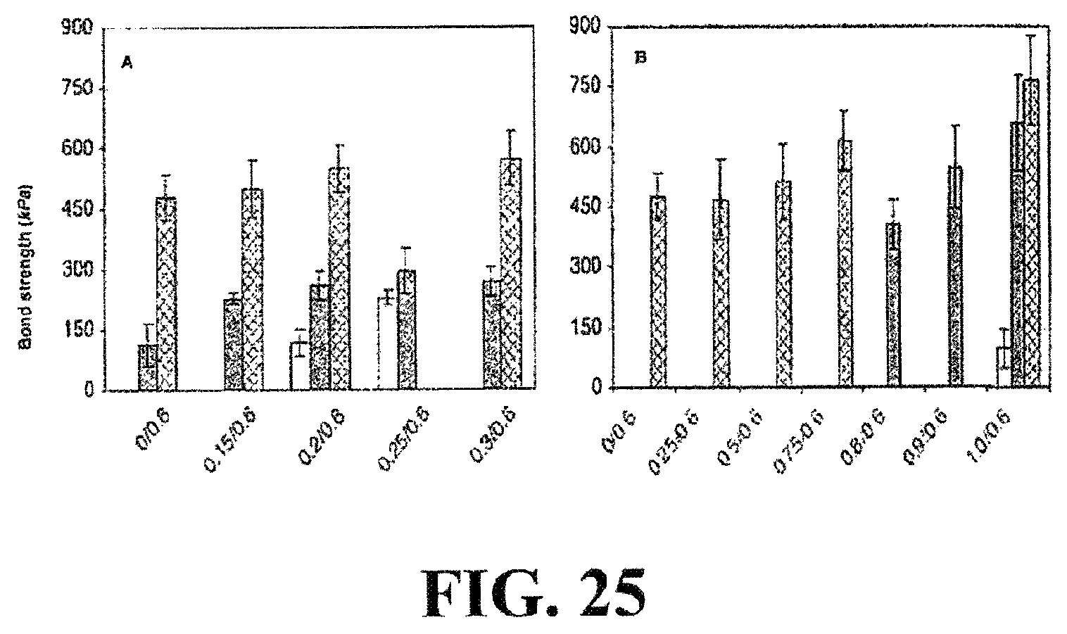
D00027
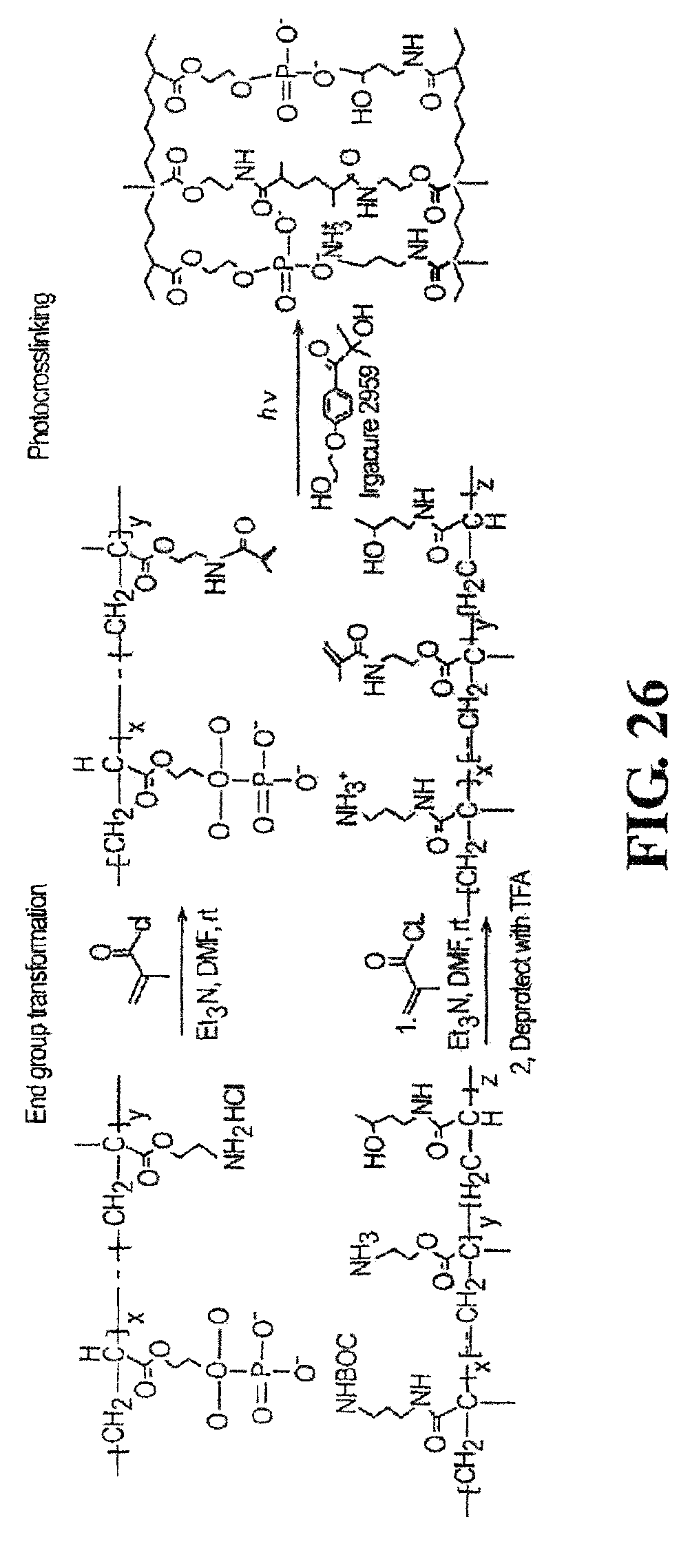
S00001
XML
uspto.report is an independent third-party trademark research tool that is not affiliated, endorsed, or sponsored by the United States Patent and Trademark Office (USPTO) or any other governmental organization. The information provided by uspto.report is based on publicly available data at the time of writing and is intended for informational purposes only.
While we strive to provide accurate and up-to-date information, we do not guarantee the accuracy, completeness, reliability, or suitability of the information displayed on this site. The use of this site is at your own risk. Any reliance you place on such information is therefore strictly at your own risk.
All official trademark data, including owner information, should be verified by visiting the official USPTO website at www.uspto.gov. This site is not intended to replace professional legal advice and should not be used as a substitute for consulting with a legal professional who is knowledgeable about trademark law.