Bifunctional polypeptides
Jakobsen , et al. Dec
U.S. patent number 10,517,960 [Application Number 16/378,481] was granted by the patent office on 2019-12-31 for bifunctional polypeptides. This patent grant is currently assigned to Immunocore Limited. The grantee listed for this patent is Immunocore Limited. Invention is credited to Bent Karsten Jakobsen, Yi Li, Annelise Brigitte Vuidepot.

| United States Patent | 10,517,960 |
| Jakobsen , et al. | December 31, 2019 |
Bifunctional polypeptides
Abstract
A bifunctional polypeptide comprising a specific binding partner for a peptide-MHC epitope, such as an antibody or T cell receptor, and an immune effector, such as an antibody or a cytokine, the immune effector part being linked to the N-terminus of the peptide-MHC binding part.
| Inventors: | Jakobsen; Bent Karsten (Abingdon, GB), Vuidepot; Annelise Brigitte (Abingdon, GB), Li; Yi (Guangzhou, CN) | ||||||||||
|---|---|---|---|---|---|---|---|---|---|---|---|
| Applicant: |
|
||||||||||
| Assignee: | Immunocore Limited (Abingdon,
GB) |
||||||||||
| Family ID: | 40834242 | ||||||||||
| Appl. No.: | 16/378,481 | ||||||||||
| Filed: | April 8, 2019 |
Prior Publication Data
| Document Identifier | Publication Date | |
|---|---|---|
| US 20190231895 A1 | Aug 1, 2019 | |
Related U.S. Patent Documents
| Application Number | Filing Date | Patent Number | Issue Date | ||
|---|---|---|---|---|---|
| 16151144 | Oct 3, 2018 | ||||
| 13319597 | Nov 20, 2018 | 10130721 | |||
| PCT/GB2010/000988 | May 19, 2010 | ||||
Foreign Application Priority Data
| May 20, 2009 [GB] | 0908613.3 | |||
| Current U.S. Class: | 1/1 |
| Current CPC Class: | C07K 14/7051 (20130101); A61P 37/00 (20180101); A61K 47/6849 (20170801); A61P 43/00 (20180101); A61K 47/6425 (20170801); A61P 31/04 (20180101); A61P 35/02 (20180101); A61P 35/00 (20180101); A61P 9/00 (20180101); C07K 16/2809 (20130101); C07K 16/46 (20130101); C07K 2319/33 (20130101); C07K 2317/622 (20130101); A61K 38/00 (20130101); C07K 2317/73 (20130101) |
| Current International Class: | A61K 47/68 (20170101); A61K 47/64 (20170101); C07K 14/725 (20060101); C07K 16/46 (20060101); C07K 16/28 (20060101); A61K 38/00 (20060101) |
References Cited [Referenced By]
U.S. Patent Documents
| 5821337 | October 1998 | Carter et al. |
| 8105830 | January 2012 | Weidanz et al. |
| 8519100 | August 2013 | Jakobsen et al. |
| 9068178 | June 2015 | Jakobsen et al. |
| 10130721 | November 2018 | Jakobsen |
| 2003/0223994 | December 2003 | Hoogenboom et al. |
| 2004/0253632 | December 2004 | Rhode et al. |
| 2006/0034850 | February 2006 | Weidanz et al. |
| 2008/0044413 | February 2008 | Hammond et al. |
| 2009/0042285 | February 2009 | Weidanz et al. |
| WO 99/60120 | Nov 1999 | WO | |||
| WO 01/93913 | Dec 2001 | WO | |||
| WO 02/102299 | Dec 2002 | WO | |||
| WO 2003/020763 | Mar 2003 | WO | |||
| WO 2003/070752 | Aug 2003 | WO | |||
| WO 2004/033685 | Apr 2004 | WO | |||
| WO 2004/074322 | Sep 2004 | WO | |||
| WO 2005/113595 | Dec 2005 | WO | |||
| WO 2005/120166 | Dec 2005 | WO | |||
| WO 2006/037960 | Apr 2006 | WO | |||
| WO 2007/071426 | Jun 2007 | WO | |||
| WO 2008/135734 | Nov 2008 | WO | |||
| WO 2010/026377 | Mar 2010 | WO | |||
Other References
|
Adams, Katherine, "Redirected T Cell Activity by High Affinity Tcr-Anti-CD3 Bispecific Candidate Therapeutics," Cardiff University for the Degree of Doctor of Philosophy, 2013, pp. 1-290. cited by applicant . Apr. 29, 2009 date stamped front page of the Journal of Immunotherapy, vol. 32, No. 4, obtained from the National Library of Medicine, 1 page. cited by applicant . Arnett et al., PNAS, vol. 101, No. 46, 2004, pp. 16268-16273, 2004. cited by applicant . Baeuerle et al., Current Opinion in Molecular Therapeutics, 2009, 11(1), 22-30. cited by applicant . Belmont, H., et al., "Potent antitumor activity of a tumor-specific soluble TCR/IL-2 fusion protein," Clinical Immunology, 2006, 121(1):29-39. cited by applicant . Bibollet-Ruche, F., et al., "The Quality of Chimpanzee T-Cell Activation and Simian Immunodeficiency Virus/Human Immunodeficiency Virus Susceptibility Achieved via Antibody-Mediated T-Cell Receptor/CD3 Stimulation Is a Function of the Anti-CD3 Antibody Isotype," Journal of Virology, Oct. 2008, pp. 10271-10278, vol. 82, No. 20. cited by applicant . Bluemel et al., Cancer Immunol Immunother (2010) 59:1197-1209. cited by applicant . Boulter et al. (Protein Engineering vol. 16 No. 9 pp. 707-711, 2003). cited by applicant . Bridgeman et al., Immunology, 135, 9-18 (2011). cited by applicant . Chames, P., et al., "Direct selection of a human antibody fragment directed against the tumor T-cell epitope HLA-A1-MAGE-A1 from a nonimmunized phage-Fab library," PNAS, Jul. 5, 2000, pp. 7969-7974, vol. 97, No. 14. cited by applicant . Choudhuri et al., Nature. Jul. 28, 2005;436(7050):578-82. cited by applicant . Davis et al., Nature Immunology, vol. 7., No. 8, 2006, pp. 803-809. cited by applicant . Denkberg, G., et al., "Selective Targeting of Melanoma and APCs Using a Recombinant Antibody with TCR-Like Specificity Directed Toward a Melanoma Differentiation Antigen," The Journal of Immunology, 2003, pp. 2197-2207, vol. 171. cited by applicant . Dissertation of Elisa Kieback, "A new safeguard eliminates T cell receptor gene-modified auto-reactive T cells after adoptive therapy," Oct. 23, 2008, 106 pages in total. cited by applicant . Dudley et al. (J Clin Oncol. Nov. 10, 2008;26(32):5233-9). cited by applicant . Hunder et al. (N Engl J Med. Jun. 19, 2008;358(25):2698-703). cited by applicant . International Search Report for PCT/GB2010/000988 dated Oct. 7, 2010. cited by applicant . Kirkwood et al., (J Clin Oncol. Jul. 10, 2008;26(20):3445-55). cited by applicant . Kjer-Nielsen et al. (PNAS 2004;101;7675-7680). (Year: 2004). cited by applicant . Li, B., et al., "Construction and characterization of a humanized anti-human CD3 monoclonal antibody 12F6 with effective immunoregulation functions," Immunology, 2005, pp. 487-498, vol. 116. cited by applicant . Lutterbuese et al. (J Immunother 2009;32:341-352). cited by applicant . MacCallum et al., J. Mol. Biol. (1996) 262, 732-745. cited by applicant . Marget, M., et al., "A HLA-Cw6 specific single-chain antibody fragment (scFv) recognizing a natural killer cell receptor epitope," Molecular Immunology, 2005, pp. 643-649, vol. 42. cited by applicant . Molhoj et al., Molecular Immunology 44 (2007) 1935-1943. cited by applicant . Molloy et al. (Current Opinion in Pharmacology 2005, 5:438-443). cited by applicant . Mosquera, L., et al., "In Vitro and In Vivo Characterization of a Novel Antibody-Like Single-Chain TCR Human IgG1 Fusion Protein," The Journal of Immunology, 2005, pp. 4381-4388. cited by applicant . Neethling, F., et al., "Assessing vaccine potency using TCRmimic Antibodies," Vaccine, 2008, 26(25):3092-102. cited by applicant . Offner et al., Molecular Immunology 43 (2006) 763-771. cited by applicant . Padlan et al., Molecular Immunology, vol. 31, No. 3, pp. 169-217, 1994. cited by applicant . Pan, S-H., et al., "Reduced Background Expression and Improved Plasmid Stability with pET Vectors in BL21 (DE3)," BioTechniques, Dec. 2000, pp. 1234-1238, vol. 29. cited by applicant . Richman et al. (Mol Immunol. Feb. 2009;46(5):902-16, Epub Oct. 29, 2008). cited by applicant . Rudolph et al. (Current Opinion in Immunology 2002, 14:52-65). cited by applicant . Salmeron et al. (J. of Immunology, vol. 147, pp. 3047-3052, 1991). (Year: 1991). cited by applicant . Schlitt, H.J., et al., "Different Activation States of Human Lymphocytes after Antibody-Mediated Stimulation via CD3 and the .alpha./.beta. T-Cell Receptor," Scandinavian Journal of Immunology, Dec. 1990, vol. 32, Issue 6, pp. 717-726. cited by applicant . Schodin et al. (Molecular Immunology, vol. 33, No. 9, pp. 819-829, 1996). cited by applicant . Shalaby et al. (J Exp Med. Jan. 1, 1992;175(1):217-25). cited by applicant . Van Der Merwe et al., Curr Biol. Jan. 1, 1995;5(1):74-84. cited by applicant . Weber et al. (Proc Natl Acad Sci U S A. Dec. 27, 2005;102(52):19033-8). cited by applicant . Wen et al., Cancer Immunol Immunother (2008) 57:1781-1794. cited by applicant . Willemsen et al. (Cytometry Part A, 73A: 1093-1099, 2008). cited by applicant . Willemsen, R., et al., "Selection of Human Antibody Fragments Directed Against Tumor T-Cell Epitopes for Adoptive T-Cell Therapy," 2008, Cytometry Part A, 73A:1093-1099. cited by applicant . Willemsen, R., et al., "T Cell Retargeting with MHC Class I-Restricted Antibodies: The CD28 Costimulatory Domain Enhances Antigen-Specific Cytotoxicity and Cytokine Production," The Journal of Immunology, 2005, pp. 7853-7858, vol. 174. cited by applicant . Woo et al. (Protein Expression and Purification 58 (2008) 1-11). cited by applicant . Written Opinion for PCT/GB2010/000988 dated Oct. 7, 2010. cited by applicant . Zhu et al. (Int J Cancer. Jul. 28, 1995;62(3):319-24). cited by applicant . Zhu et al., Biochemistry 2002, 41, 12163-12170. cited by applicant . Argos, P., "An investigation of oligopeptides linking domains in protein tertiary structures and possible candidates for general gene fusion," Journal of Molecular Biology, vol. 211, Issue 4, Feb. 20, 1990, pp. 943-958. cited by applicant . Boulter et al., "Stable, soluble T-cell receptor molecules for crystallization and therapeutics," Protein Engineering, Design and Selection, vol. 16, Issue 9, Sep. 1, 2003, pp. 707-711. cited by applicant . Card et al., "A soluble single-chain T-cell receptor IL-2 fusion protein retains MHC-restricted peptide specificity and IL-2 bioactivity," Apr. 2004, vol. 53, Issue 4, pp. 345-357. cited by applicant . Denkberg et al., "Recombinant antibodies with T-cell receptor-like specificity: Novel tools to study MHC class I presentation," Autoimmunity Reviews, vol. 5, Issue 4, Apr. 2006, pp. 252-257. cited by applicant . Molloy et al., "Soluble T cell receptors: novel immunotherapies," Current Opinion in Pharmacology, vol. 5, Issue 4, Aug. 2005, pp. 438-443. cited by applicant . Notice of opposition, European patent application No. EP16176249.7, dated Jan. 23, 2019, 159 pages. cited by applicant . Ortiz-Sanchez et al., "Antibody-cytokine fusion proteins: applications in cancer therapy," Expert Opin Biol. Ther. 2008, 8(5), pp. 609-632. cited by applicant . Peng et al., "A Single-Chain IL-12 IgG3 Antibody Fusion Protein Retains Antibody Specificity and IL-12 Bioactivity and Demonstrates Antitumor Activity," The Journal of Immunology, vol. 1, Issue 1, Jul. 1, 1999, pp. 250-258. cited by applicant . Wen et al., "Targeting activity of a TCR/IL-2 fusion protein against established tumors," Cancer Immunology, Immunotherapy, Dec. 2008, vol. 57, Issue 12, pp. 1781-1794. cited by applicant . Davis, C. et al., "Immunocytokines: amplification of anti-cancer immunity," Cancer Immunology, Immunotherapy, vol. 52, Issue 5, May 2003, pp. 297-308. cited by applicant . Klechevsky, E. et al., "Antitumor Activity of Immunotoxins with T-Cell Receptor-like Specificity against Human Melanoma Xenografts," Cancer Research, vol. 68, No. 15, Aug. 1, 2008, pp. 6360-6367. cited by applicant . Notice of opposition, European Patent Application No. 16176246.3, dated May 8, 2019, 42 pages. cited by applicant . Pule, M. et al., "Artificial T-Cell Receptors," Cytotherapy, vol. 5, No. 3, Jan. 2003, pp. 211-226. cited by applicant . Ruf, P. et al., "Induction of a long-lasting antitumor immunity by a trifunctional bispecific antibody," Blood, vol. 98, No. 8, Oct. 15, 2001, pp. 2526-2534. cited by applicant . Sadelain, M. et al., "The promise and potential pitfalls of chimeric antigen receptors," Current Opinion in Immunology, vol. 21, No. 2, Apr. 2009, pp. 215-223. cited by applicant . Xiong, C-Y. et al., "Development of tumor targeting anti-MUC-1 multimer: effects of di-scFv unpaired cysteine location of PEGylation and tumor binding," Protein Engineering, Design & Selection, vol. 19, No. 8, 2006, pp. 359-367. cited by applicant. |
Primary Examiner: Skelding; Zachary S
Attorney, Agent or Firm: Fenwick & West LLP
Parent Case Text
CROSS REFERENCE TO RELATED APPLICATIONS
This application is a continuation of U.S. application Ser. No. 16/151,144, filed Oct. 3, 2018, which is a continuation of U.S. application Ser. No. 13/319,597, filed Apr. 5, 2012, now U.S. Pat. No. 10,130,721, which is the National Stage of International Application No. PCT/GB2010/000988, filed May 19, 2010, which claims the benefit of and priority to Great Britain Patent Application No. 0908613.3, filed May 20, 2009, each of which is herein incorporated in its entirety by reference.
Claims
What is claimed is:
1. A bifunctional molecule, comprising: a heterodimeric .alpha..beta. T-Cell Receptor (.alpha..beta. TCR), wherein the .alpha..beta. TCR binds specifically to a given pMHC epitope, and an immune effector polypeptide, wherein: the N-terminus of the .alpha. chain of the .alpha..beta. TCR is linked to the C-terminus of a single chain of an immune effector polypeptide comprising CDRs of an anti-CD3 antibody, and the bifunctional molecule is capable of binding to the given pMHC epitope and CD3, each on a different cell, and thus simultaneously binding to two different cells.
2. A bifunctional molecule, comprising: a heterodimeric .alpha..beta. T-Cell Receptor (.alpha..beta. TCR), wherein the .alpha..beta. TCR binds specifically to a given pMHC epitope, and an immune effector polypeptide, wherein: the N-terminus of the .beta. chain of the .alpha..beta. TCR is linked to the C-terminus of a single chain of an immune effector polypeptide comprising CDRs of an anti-CD3 antibody, and the bifunctional molecule is capable of binding to the given pMHC epitope and CD3, each on a different cell, and thus simultaneously binding to two different cells.
3. The bifunctional molecule of claim 1, wherein the immune effector polypeptide is a single chain immunoglobulin (scFv) which specifically binds to CD3.
4. The bifunctional molecule of claim 2, wherein the immune effector polypeptide is a single chain immunoglobulin (scFv) which specifically binds to CD3.
5. The bifunctional molecule of claim 3, wherein the single chain immunoglobulin comprises SEQ ID NO: 3.
6. The bifunctional molecule of claim 4, wherein the single chain immunoglobulin comprises SEQ ID NO: 3.
Description
SEQUENCE LISTING
The instant application contains a Sequence Listing which has been submitted electronically in ASCII format and is hereby incorporated by reference in its entirety. Said ASCII copy, created on Apr. 4, 2019, is named 42799US_CRF_SequenceListing.txt and is 23,605 bytes in size.
FIELD OF THE INVENTION
This invention relates to a bifunctional polypeptide comprising a specific binding partner for a peptide-MHC epitope, such as an antibody or a T cell receptor ("TCR"), and an immune effector, such as an antibody or a cytokine, the immune effector part being linked to the N-terminus of the peptide-MHC binding part.
BACKGROUND TO THE INVENTION
TCRs mediate the recognition of Specific Major Histocompatibility Complex (MHC)-peptide complexes ("pMHC complexes") which are presented as epitopes on antigen presenting cells (APC), and TCRs mediate the recognition of such pMHC epitopes by T cells. As such TCRs are essential to the functioning of the cellular arm of the immune system.
Antibodies are also known which specifically bind pMHC epitopes presented by antigen presenting cells (see for example: Neethling F A. et al., Vaccine (2008) 26 (25): 3092-102). There are antigen-binding fragment (Fab) antibodies (see for example: Chames P. et al., Proc Natl Acad Sci USA (2000) 97 (14): 7969-74; Willemsen R A. et al., J Immunol (2005) 174 (12): 7853-8; Willemsen R. et al., Cytometry A (2008) 73 (11): 1093-9) or single-chain antibody fragments (scFv) (see for example: Denkberg G. et al., J Immunol (2003) 171 (5): 2197-207; Marget M. e al., Mol Immunol (2005) 42 (5): 643-9) which specifically bind pMHC epitopes.
The native TCR is a heterodimeric cell surface protein of the immunoglobulin superfamily which is associated with invariant proteins of the CD3 complex involved in mediating signal transduction. TCRs exist in .alpha..beta. and .gamma..delta. forms, which are structurally similar but have quite distinct anatomical locations and probably functions. The MEW class I and class II ligands are also immunoglobulin superfamily proteins but are specialised for antigen presentation, with a highly polymorphic peptide binding site which enables them to present a diverse array of short peptide fragments at the APC cell surface.
The extracellular portion of native heterodimeric .alpha..beta. TCRs consist of two polypeptides (the .alpha. and .beta. chains) each of which has a membrane-proximal constant domain, and a membrane-distal variable domain. Each of the constant and variable domains includes an intra-chain disulfide bond. The variable domains contain the highly polymorphic loops analogous to the complementarity determining regions (CDRs) of antibodies. CDR3 of .alpha..beta. TCRs interact with the peptide presented by MEW, and CDRs 1 and 2 of .alpha..beta. TCRs interact with the peptide and the MEW. The diversity of TCR sequences is generated via somatic rearrangement of linked variable (V), diversity (D), joining (J), and constant genes (C).
Functional a chain polypeptides are formed by rearranged V-J-C regions, whereas .beta. chains consist of V-D-J-C regions. The extracellular constant domain has a membrane proximal region and an immunoglobulin region. There is a single a chain constant domain, known as TRAC. The .beta. chain constant domain is composed of one of two different .beta. constant domains, known as TRBC1 and TRBC2 (IMGT nomenclature). There are four amino acid changes between these .beta. constant domains. These changes are all within exon 1 of TRBC1 and TRBC2: N.sub.4K.sub.5.fwdarw.K.sub.5N.sub.5 and F.sub.37.fwdarw.Y (IMGT numbering, differences TRBC1.fwdarw.TRBC2), the final amino acid change between the two TCR .beta. chain constant regions being in exon 3 of TRBC1 and TRBC2: V.sub.1.fwdarw.E.
A number of constructs have been devised to date for the production of recombinant TCRs. These constructs fall into two broad classes, single-chain TCRs and dimeric TCRs. Single-chain TCRs (scTCRs) are artificial constructs consisting of a single amino acid strand, which like native heterodimeric TCRs bind to MHC-peptide complexes. scTCRs can consist of a combination of TCR .alpha. and .beta. variable regions (V.alpha. and V.beta. respectively) and TCR .alpha. and .beta. constant regions (C.alpha. and C.beta. respectively), linked by a linker sequence (L) in several possible orientations, for example, but not limited to, the following V.alpha.-L-V.beta., V.beta.-L-V.alpha., V.alpha.-C.alpha.-L-V.beta. or V.beta.-C.beta.-L-V.alpha..
A number of papers describe the production of TCR heterodimers which include the native disulphide bridge which connects the respective subunits. However, although such TCRs can be recognised by TCR-specific antibodies, none have been shown to recognise its native ligand at anything other than relatively high concentrations and/or were not stable.
In WO 03/020763 a soluble TCR is described which is correctly folded so that it is capable of recognising its native ligand, is stable over a period of-time, and can be produced in reasonable quantities. This TCR comprises a TCR .alpha. chain extracellular domain dimerised to a TCR .beta. chain extracellular domain respectively, by means of an inter-chain disulfide bond between cysteines introduced into the constant regions of the respective chains.
Specific pMHC binding partners, ie antibodies specific for pMHC epitopes, and TCRs of both the heterodimeric and single chain type, have been proposed as targeting vectors for the delivery of therapeutic agents to antigen presenting cells. For that purpose, the therapeutic agent is required to be associated with the pMHC-binding partner in some way. Therapeutic agents which have been suggested for such targeted delivery in association with pMHC-binding partners include antibodies (see for example: Mosquera L A. et al., J Immunol (2005) 174 (7): 4381-8), cytokines (see for example: Belmont H J. et al., Clin Immunol (2006) 121 (1): 29-39; Wen J. et al. Cancer Immunol Immunother (2008) 57 (12): 1781-94), and cytotoxic agents. Where the therapeutic agent is a polypeptide, the means of association with the pMHC binding partner may be by peptidic fusion, either direct fusion or fusion via a linker sequence, to the pMHC binding partner. In those cases, there are essentially only two fusion possibilities. In the case of single chain antibodies or TCRs, fusion can in principle be at the C- or N-terminus of the TCR chain; In the case of heterodimeric antibodies or TCRs, the fusion can in principle be at the C- or N-terminus of either chain. In practice however, it appears that all known examples of pMHC binding partner-therapeutic agent fusions have been with the therapeutic agent fused to the C-terminus (see for example: Mosquera L A. et al., J Immunol (2005) 174 (7): 4381-8; Belmont H J. et al., Clin Immunol (2006) 121 (1): 29-39; Wen J. et al., Cancer Immunol Immunother (2008) 57 (12): 1781-94). This is because the functionality of an antibody or TCR, whether single chain or heterodimeric, depends on correct folding and orientation of the variable regions. Fusion of the therapeutic agent to the N-terminus of the pMHC binding partner places it ahead of one of the variable regions, and there has been an assumption in the art that the therapeutic agent located at the N-terminus will interfere with binding of the antibody or TCR to the pMHC complex, thereby reducing its binding efficiency.
SUMMARY OF THE INVENTION
Contrary to that assumption in the art, it has now been found that bifunctional molecules wherein an immune effector part is fused to the N-terminus of a pMHC-binding partner are more effective in their induction of the relevant immune response that the corresponding construct wherein the fusion is to the C-terminus of the pMHC-binding partner. This enhanced immune response of the N-fused construct achieved despite the similar pMHC binding affinities of the N- and C-fused version.
BRIEF DESCRIPTION OF THE SEVERAL VIEWS OF THE DRAWINGS
These and other features, aspects, and advantages of the present invention will become better understood with regard to the following description, and accompanying drawings, where:
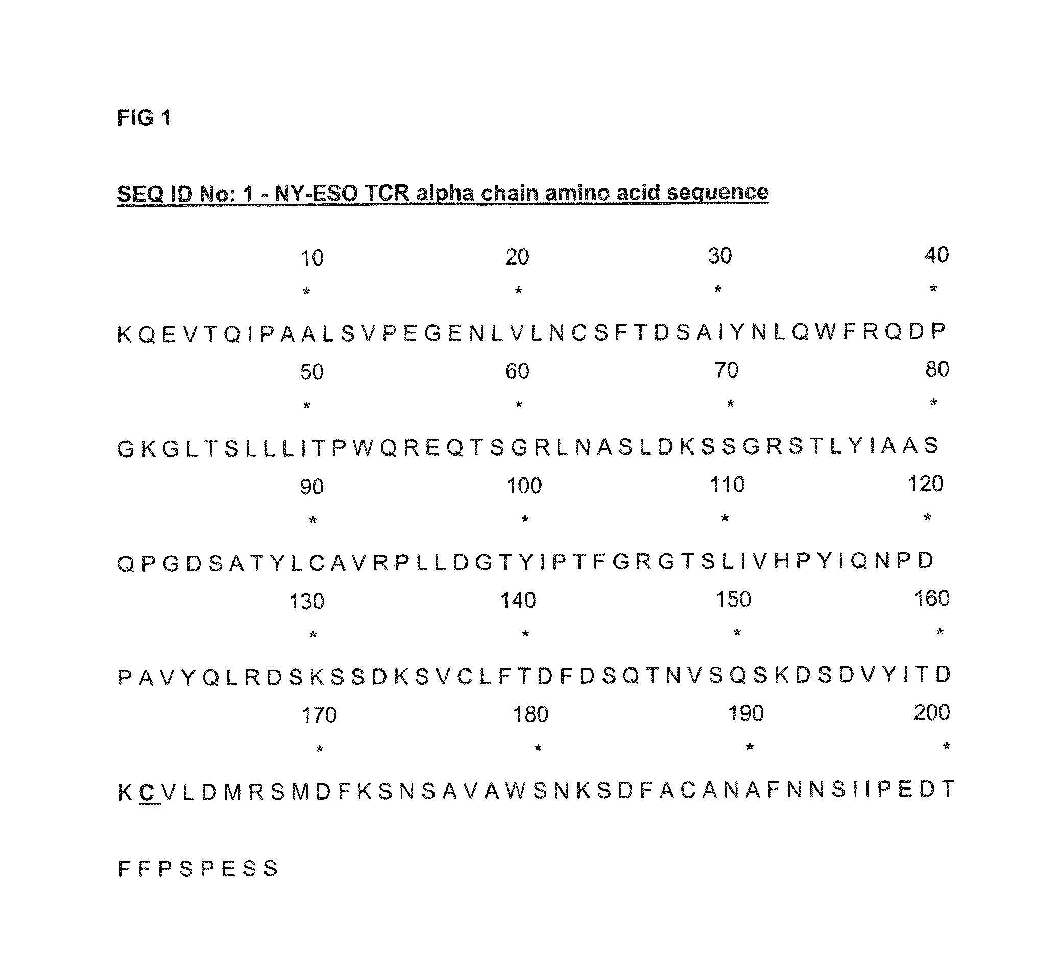
FIG. 1 shows the amino acid sequence of the alpha chain of an NY-ESO TCR, in which C162 (using the numbering of SEQ ID No: 1) replaces T48 of its TRAC constant region.
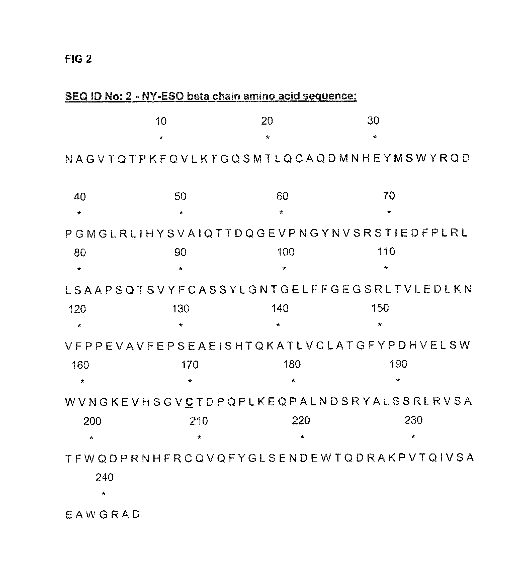
FIG. 2 shows the amino acid sequence of the beta chain NY ESO-TCR, in which C170 (using the numbering of SEQ ID No: 2) replaces S57 of its TRBC2 constant region.
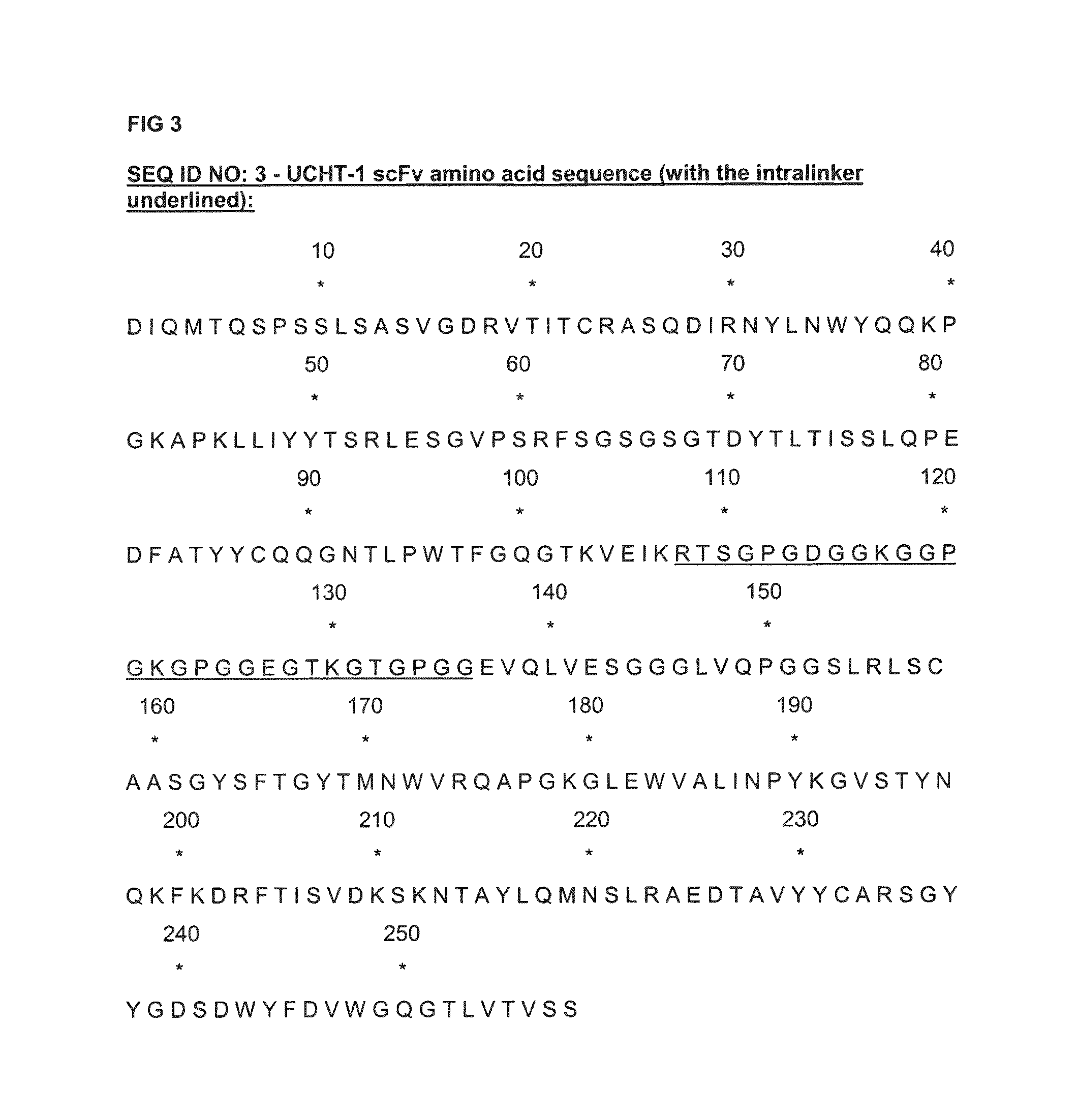
FIG. 3 shows the amino acid sequence of an anti CD3 UCHT-1 scFv antibody, with its intralinker sequence underlined.
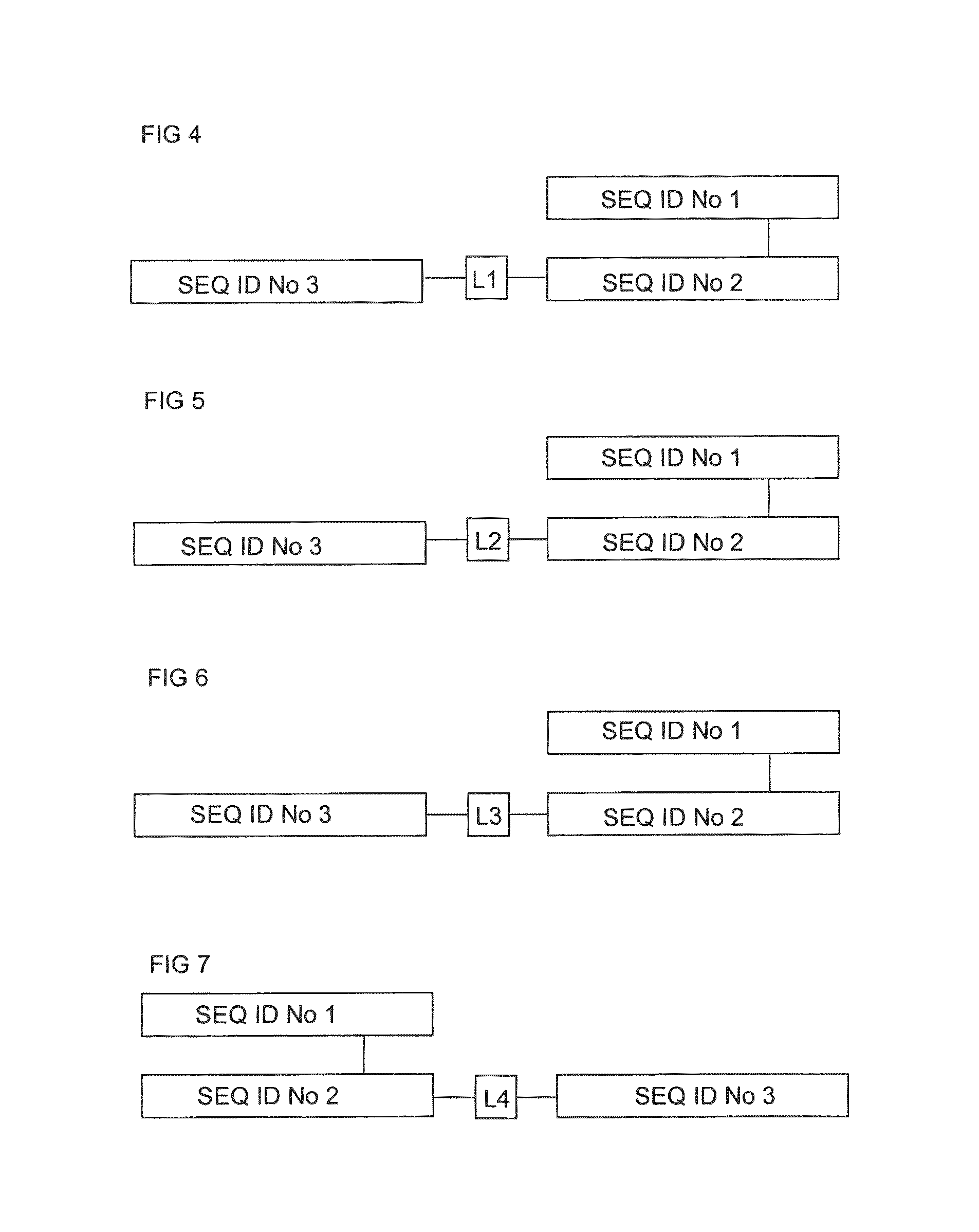
FIG. 4 shows in block diagram form the structure of a soluble NY-ESO .alpha..beta. TCR having the .alpha. chain SEQ ID No: 1 and the .beta. chain SEQ ID No: 2, and having the anti CD3 UCHT-1 scFv antibody SEQ ID No: 3 fused at the N terminus of the TCR .beta. chain SEQ ID No: 2 via a linker sequence L1 namely GGEGS (SEQ ID No: 4).
FIG. 5 shows in block diagram form the structure of a soluble NY-ESO .alpha..beta. TCR having the .alpha. chain SEQ ID No: 1 and the .beta. chain SEQ ID No: 2, and having the anti CD3 UCHT-1 scFv antibody SEQ ID No: 3 fused at the N terminus of the TCR .beta. chain SEQ ID No: 2 via a linker sequence L2 namely AHHSEDPSSKAPKAP (SEQ ID No: 5).
FIG. 6 shows in block diagram form the structure of a soluble NY-ESO .alpha..beta. TCR having the .alpha. chain SEQ ID No: 1 and the .beta. chain SEQ ID No: 2, and having the anti CD3 UCHT-1 scFv antibody SEQ ID No: 3 fused at the N terminus of the TCR .beta. chain SEQ ID No: 2 via a linker sequence L3 namely GGEGGGSEGGGS (SEQ ID No: 6).
FIG. 7 shows in block diagram form the structure of a soluble NY-ESO .alpha..beta. TCR having the .alpha. chain SEQ ID No: 1 and the .beta. chain SEQ ID No: 2, and having the anti CD3 UCHT-1 scFv antibody SEQ ID No: 3 fused at the C terminus of the TCR .beta. chain SEQ ID No: 2 via a linker sequence L4, which in this case is the single amino acid, S.
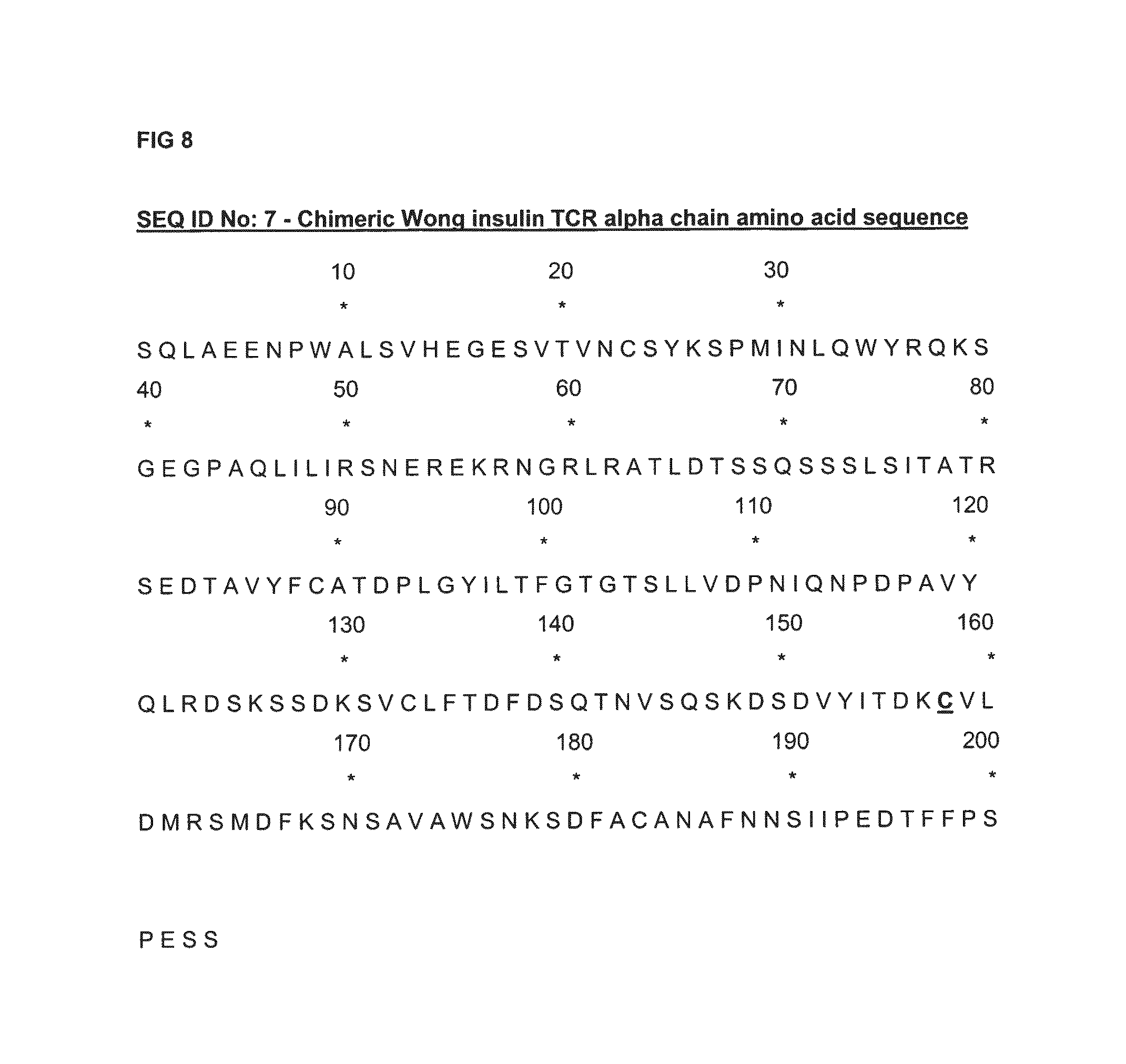
FIG. 8 shows the amino acid sequence of the alpha chain of a TCR having the property of binding to a murine insulin-derived peptide, LYLVCGERG (SEQ ID NO: 8), presented by the murine H-2K.sup.d complex, LYLVCGERG-H-2K.sup.d ("LYLVCGERG" disclosed as SEQ ID NO: 8), in which C158 (using the numbering of SEQ ID No: 7) replaces T48 of its TRAC constant region.
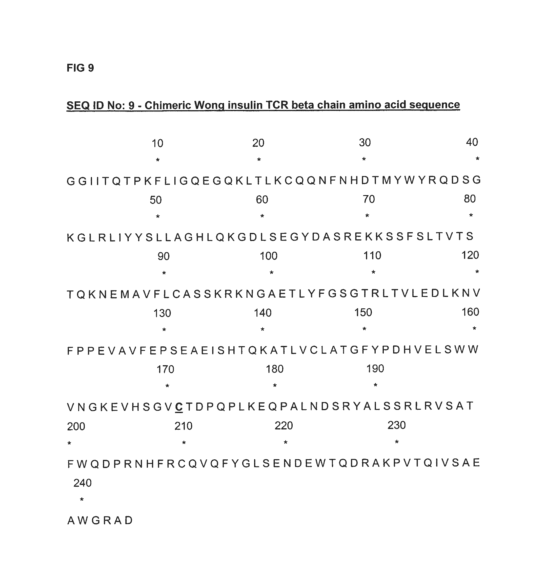
FIG. 9 shows the amino acid sequence of the beta chain of the same TCR which binds the murine LYLVCGERG-H-2Kd complex ("LYLVCGERG" disclosed as SEQ ID NO: 8), in which C171 (using the numbering of SEQ ID No: 9) replaces S57 of its TRBC2 constant region.
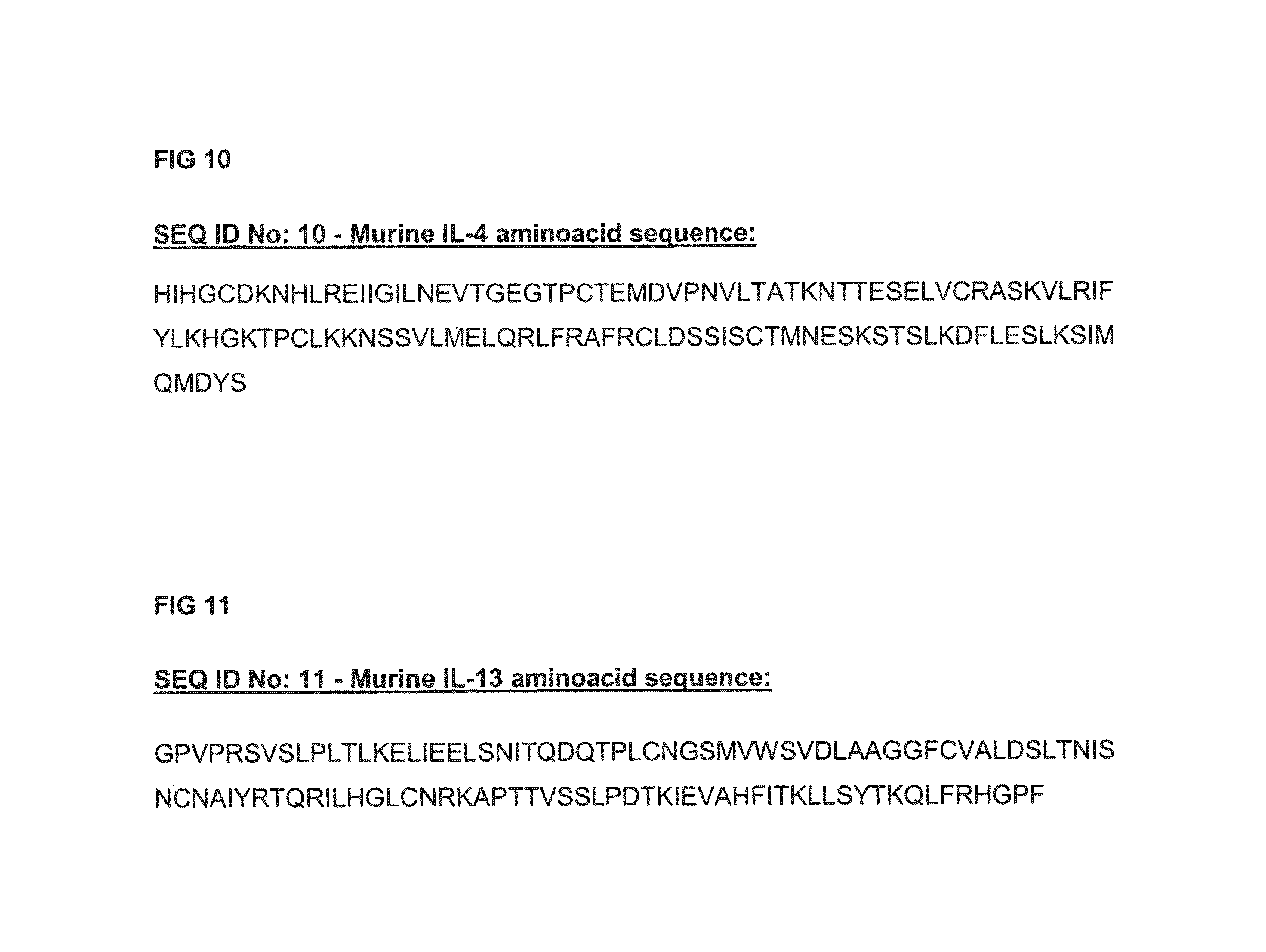
FIG. 10 shows the amino acid sequence of a murine IL-4 polypeptide represented by SEQ ID No: 10.
FIG. 11 shows SEQ ID No: 11 which is the amino acid sequence of a murine IL-13 polypeptide.
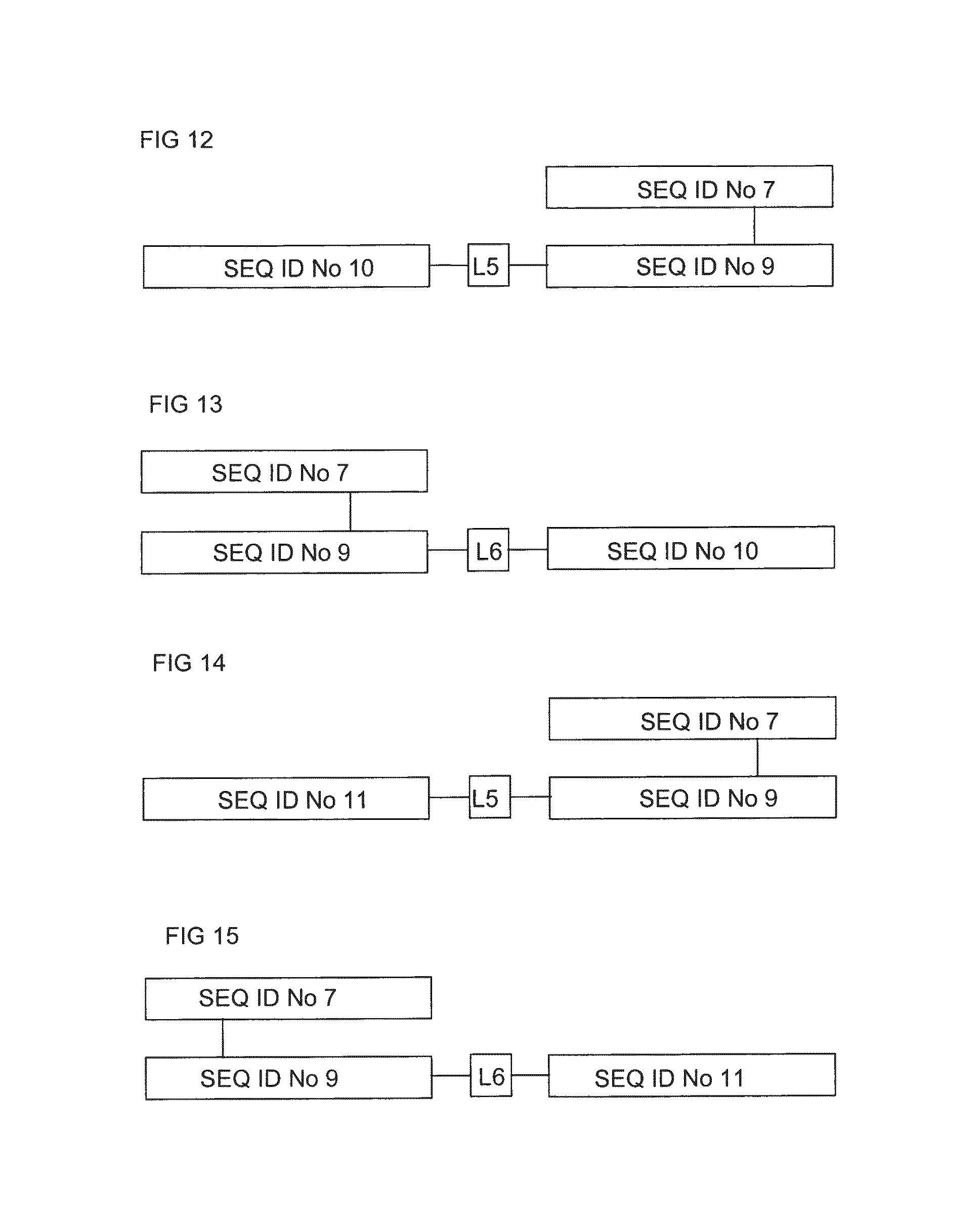
FIG. 12 shows in block diagram form the structure of a soluble chimeric insulin .alpha..beta. TCR having the .alpha. chain SEQ ID No: 7 and the .beta. chain SEQ ID No: 9, and having murine IL-4 SEQ ID No: 10 fused at the N terminus of the TCR .beta. chain SEQ ID No: 9 via the linker sequence L5, namely GGEGGGP (SEQ ID No: 12).
FIG. 13 shows in block diagram form the structure of a soluble chimeric insulin .alpha..beta. TCR having the .alpha. chain SEQ ID No: 7 and the .beta. chain SEQ ID No: 9, and having the murine IL-4 (SEQ ID No: 10) fused at the C terminus of the TCR .beta. chain (SEQ ID No: 9) via the linker sequence L6, namely GSGGP (SEQ ID No: 13).
FIG. 14 shows in block diagram form the structure of a soluble chimeric insulin TCR having the .alpha. chain SEQ ID No: 7 and the .beta. chain SEQ ID No: 9, and having murine IL-13 (SEQ ID No: 11) fused at the N terminus of the TCR chain (SEQ ID No: 9) via the linker sequence L5, namely GGEGGGP (SEQ ID No: 12).
FIG. 15 shows in block diagram form the structure of a soluble chimeric insulin TCR having the .alpha. chain SEQ ID No: 7 and the .beta. chain SEQ ID No: 9, and having murine IL-13 (SEQ ID No: 11) fused at the C terminus of the TCR .beta. chain (SEQ ID No: 9) via the linker sequence L6, namely GSGGP (SEQ ID No: 13).
FIG. 16 shows the amino acid sequence (SEQ ID No:14) of the beta chain of FIG. 2 with the N-terminus of an anti-CD3 scFv fused to the C-terminus of the TCR beta chain via another peptide linker sequence (underlined).
FIG. 17 shows the amino acid sequence (SEQ ID No:15) of the beta chain of FIG. 2 with the C-terminus of an anti-CD3 scFv fused to the N-terminus of the TCR beta chain via the same peptide linker sequence as in SEQ ID No 14 (again underlined).
DETAILED DESCRIPTION OF THE INVENTION
Accordingly, the present invention provides a bifunctional molecule comprising a polypeptide binding partner specific for a given pMHC epitope, and an immune effector polypeptide, the N-terminus of the pMHC binding partner being linked to the C-terminus of the immune effector polypeptide, PROVIDED THAT the said polypeptide binding partner is not a T-cell receptor comprising the alpha chain SEQ ID No: 7 and the beta chain SEQ ID No: 9.
As mentioned, the polypeptide pMHC binding partner may be an antibody or a TCR. Thus in one embodiment of the invention the pMHC binding partner is a heterodimeric TCR polypeptide pair, or a single chain TCR polypeptide, and the N-terminus of the .alpha. or .beta. chain of the heterodimeric TCR polypeptide pair, or the N-terminus of the scTCR polypeptide, is linked to a C-terminal amino acid of the immune effector polypeptide.
Linkage of the pMHC binding partner and the immune effector polypeptide may be direct, or indirect via linker sequence. Linker sequences are usually flexible, in that they are made up of amino acids such as glycine, alanine and serine which do not have bulky side chains likely to restrict flexibility. Usable or optimum lengths of linker sequences are easily determined in the case of any given pMHC binding partner-immune effector construct. Often the linker sequence will by less than about 12, such as less that 10, or from 5-10 amino acids in length.
In some embodiments of the invention the pMHC binding partner is a heterodimeric a.beta. TCR polypeptide pair wherein the .alpha. and .beta. polypeptides each have TCR variable and constant regions, but lack TCR transmembrane and cytoplasmic regions. The TCR part in such cases is soluble. In particularly preferred bifunctional molecules of this type, a non-native disulfide bond between residues of the constant regions of TCR .alpha. and .beta. polypeptides is present. In particular the constant regions of the .alpha. and .beta. polypeptides may be linked by a disulfide bond between cysteine residues substituted for Thr 48 of exon 1 of TRAC1 and Ser 57 of exon 1 of TRBC1 or TRBC2, or by the native disulfide bond between Cys4 of exon 2 of TRAC*01 and Cys2 of exon 2 of TRBC1 or TRBC2.
In other embodiments of the invention, the pMHC binding partner is a single chain .alpha..beta. TCR polypeptide of the V.alpha.-L-V.beta., V.alpha..beta.-L-V.alpha., V.alpha.-C.alpha.-L-V.beta., or V.alpha.-L-V.beta.-C.beta. type wherein V.alpha. and VP are TCR .alpha. and .beta. variable regions respectively, C.alpha. and C.beta. a are TCR .alpha. and .beta. constant regions respectively, and L is a linker sequence.
Immune effector polypeptides are known. They are molecules which induce or stimulate an immune response, through direct or indirect activation of the humoural or cellular arm of the immune system, such as by activation of T-cells. Examples include: IL-1, IL-la, IL-3, IL-4, IL-5, IL-6, IL-7, IL-10, IL-11, IL-12, IL-13, IL-15, IL-21, IL-23, TGF-.beta., IFN-.gamma., TNF.alpha., Anti-CD2 antibody, Anti-CD3 antibody, Anti-CD4 antibody, Anti-CD8 antibody, Anti-CD44 antibody, Anti-CD45RA antibody, Anti-CD45RB antibody, Anti-CD45RO antibody, Anti-CD49a antibody, Anti-CD49b antibody, Anti-CD49c antibody, Anti-CD49d antibody, Anti-CD49e antibody, Anti-CD49f antibody, Anti-CD16 antibody, Anti-CD28 antibody, Anti-1L-2R antibodies, Viral proteins and peptides, and Bacterial proteins or peptides. Where the immune effector polypeptide is an antibody it may be scFv antibody, one such being an anti-CD3 scFv. Examples of anti-CD3 antibodies include but are not limited to OKT3, UCHT-1, BMA031 and 12F6.
The principles of the invention are illustrated by the following Examples.
Example A. Preparation of Soluble .alpha..beta. TCRs Having Effector Polypeptides Fused to the C- or N-Terminus of the TCR .beta. Chain
A1. Soluble NY-ESO TCR with Anti-CD3 Antibody as Effector Polypeptide
The soluble NY-ESO TCR of this example has the property of binding to the SLLMWITQV peptide (SEQ ID NO: 16) when presented on an HLA-A2 molecule.
SEQ ID No: 1 (FIG. 1) is the amino acid sequence of the alpha chain of an NY-ESO TCR, in which C162 (using the numbering of SEQ ID No: 1) replaces T48 of its TRAC constant region.
SEQ ID No: 2 (FIG. 2) is the amino acid sequence of the beta chain NY ESO-TCR, in which C170 (using the numbering of SEQ ID No: 2) replaces S57 of its TRBC2 constant region.
SEQ ID No: 3 (FIG. 3) is the amino acid sequence of an anti CD3 UCHT-1 scFv antibody, with its intralinker sequence underlined.
FIG. 4 shows in block diagram form the structure of a soluble NY-ESO .alpha..beta. TCR having the .alpha. chain SEQ ID No: 1 and the .beta. chain SEQ ID No: 2, and having the anti CD3 UCHT-1 scFv antibody SEQ ID No: 3 fused at the N terminus of the TCR .beta. chain SEQ ID No: 2 via a linker sequence L1 namely GGEGS (SEQ ID No: 4).
SEQ ID No: 14 (FIG. 16) is the amino acid sequence of the beta chain of FIG. 2 with the N-terminus of an anti-CD3 scFv fused to the C-terminus of the TCR beta chain via another peptide linker sequence (underlined).
SEQ ID No: 15 (FIG. 17) is the amino acid sequence of the beta chain of FIG. 2 with the C-terminus of an anti-CD3 scFv fused to the N-terminus of the TCR beta chain via the same peptide linker sequence as in SEQ ID No 14 (again underlined).
The construct of FIG. 4 was prepared as follows:
Ligation
Synthetic genes encoding (a) the TCR .alpha. chain SEQ ID No: 1 and (b) the fusion sequence of SEQ ID No: 2 and EQ ID No: 3, were separately ligated into pGMT7-based expression plasmids, which contain the T7 promoter for high level expression in E. coli strain BL21-DE3(pLysS) (Pan et al., Biotechniques (2000) 29 (6): 1234-8.
Expression
The expression plasmids were transformed separately into E. coli strain BL21 (DES) Rosetta pLysS, and single ampicillin-resistant colonies were grown at 37.degree. C. in TYP (ampicillin 100 .mu.g/ml) medium to OD.sub.600 of .about.0.6-0.8 before inducing protein express with 0.5 mM IPTG. Cells were harvested three hours post-induction by centrifugation for 30 minutes at 4000 rpm in a Beckman J-6B. Cell pellets were lysed with 25 ml Bug Buster (NovaGen) in the presence of MgCl.sub.2 and DNase. Inclusion body pellets were recovered by centrifugation for 30 minutes at 13000 rpm in a Beckman J2-21 centrifuge. Three detergent washes were then carried out to remove cell debris and membrane components. Each time the inclusion body pellet was homogenised in a Triton buffer (50 mM Tris-HCl pH 8.0, 0.5% Triton-X100, 200 mM NaCl, 10 mM NaEDTA) before being pelleted by centrifugation for 15 minutes at 13000 rpm in a Beckman J2-21. Detergent and salt was then removed by a similar wash in the following buffer: 50 mM Tris-HCl pH 8.0, 1 mM NaEDTA. Finally, the inclusion bodies were divided into 30 mg aliquots and frozen at -70.degree. C.
Refolding
Approximately 20 mg of TCR .alpha. chain and 40 mg of scFv-TCR .beta. chain solubilised inclusion bodies were thawed from frozen stocks, diluted into 20 ml of a guanidine solution (6 M Guanidine-hydrochloride, 50 mM Tris HCl pH 8.1, 100 m NaCl, 10 mM EDTA, 20 mM OTT), and incubated in a 37.degree. C. water bath for 30 min-1 hr to ensure complete chain de-naturation. The guanidine solution containing fully reduced an denatured TCR chains was then injected into 1 litre of the following refolding buffer: 100 mM Tris pH 8.1, 400 mM L-Arginine, 2 mM EDTA, 5M Urea. The redox couple (cysteamine hydrochloride and cystamine dihydrochloride (to final concentrations of 16 mM and 1.8 mM, respectively)) were added approximately 5 minutes before addition of the denatured TCR .alpha. and scFv-TCR .beta. chains. The solution was left for .about.30 minutes. The refolded scFv-TCR was dialysed in dialysis tubing cellulose membrane (Sigma-Aldrich; Product No. D9402) against 10 L H.sub.2O for 18-20 hours. After this time, the dialysis buffer was changed twice to fresh 10 mM Tris pH 8.1 (10 L) and dialysis was continued at 5.degree. C..+-.3.degree. C. for another .about.8 hours. Soluble and correctly folded scFv-TCR was separated from misfolded, degradation products a impurities by a 3-step purification method as described below. The second purification step can either be an ion exchange chromatography or an affinity chromatography, depending on the pl of the soluble. anti-CD3 scFv-TCR fusion.
First Purification Step
The dialysed refold (in 10 mM Tris pH8.1) was loaded onto a POROS 50HQ anion exchange column and the bound protein eluted with a gradient of 0-500 mM NaCl over 6 column volumes using an Akta purifier (GE Healthcare). Peak fractions (eluting at a conductivity .about.20 mS/cm) were stored at 4.degree. C. Peak fractions were analysed by Instant Blue Stain (Novexin) stained SDS-PAGE before being pooled.
Second Purification Step
Ion Exchange Chromatography
Cation Exchange Purification:
The anion exchange pooled fractions were buffer exchanged by dilution with 20 mM IVIES pH6-6.5, depending on the pl of the scFv-TCR fusion. The soluble and correctly folded scFv-TCR was separated from misfolded, degradation products and impurities by loading the diluted pooled fractions (in 20 mM IVIES pH6-6.5) onto a POROS 50HS cation exchange column and eluting bound protein with a gradient of 0-500 mM NaCl over 6 column volumes using an Akta purifier (GE Healthcare). Peak fractions (eluting at a conductivity .about.10 mS/cm) were stored at 4.degree. C.
Alternatively, ion exchange purification using hydroxyapatite matrix can be used a explained below.
Hydroxyapatite Chromatography:
The anion exchange pooled fractions were buffer exchanged by dilution with 10 mM NaH.sub.2PO.sub.4 pH6.0. The soluble and correctly folded scFv-TCR was separated from misfolded, degradation products and impurities by loading the diluted pooled fractions (in 10 mM NaH.sub.2PO.sub.4 pH6.0) onto a hydroxyapatite column and eluting bound protein with a gradient of 10-500 mM NaH.sub.2PO.sub.4/1M NaCl over 6 column volumes using an Akta purifier (GE Healthcare). Peak fractions (eluting at a conductivity .about.20 mS/cm) were stored at 4.degree. C.
Affinity Chromatography
For some scFv-TCR fusions with a pl close to 6-6.5, the ion exchange step cannot be used but can be replaced by an affinity chromatography step. The protein L affinity chromatography column (Pierce, product number 89928) isolates and purifies certain immunoglobulin classes via their kappa light chains. Protein L can also binds sing chain variable fragments (scFv). The anion exchange pooled fractions were buffer exchanged by dilution with PBS/0.02% sodium azide. The soluble and correctly folded scFv-TCR was separated from misfolded, degradation products and impurities by loading the diluted pooled fractions onto a Protein L column and eluting bound protein with a gradient of 0-25 mM Glycine pH2.3/0.02% sodium azide over 3 column volumes using an Akta purifier (GE Healthcare). The scFv-TCR eluted very late in gradient and the pH of the eluted fractions was neutralized by addition of Tris pH8 1 (100 mM Tris pH8.1 final concentration). The peak fractions were stored at 4.degree. C.
Final Purification Step
Peak fractions from second purification step were analysed by Instant Blue Stain (Novexin) stained SDS-PAGE before being pooled. The pooled fractions were then concentrated for the final purification step, when the soluble scFv-TCR was purified and characterised using a Superdex 5200 gel filtration column (GE Healthcare) pre-equilibrated in PBS buffer (Sigma). The peak eluting at a relative molecular weight of approximately 78 kDa was analysed by Instant Blue Stain (Novexin) stained SDS-PAGE before being pooled. In a similar manner to that described for the construct of FIG. 4, the constructs of FIGS. 5, 6 and 7 were prepared:
FIG. 5 shows in block diagram form the structure of a soluble NY-ESO .alpha..beta. TCR having the .alpha. chain SEQ ID No: 1 and the .beta. chain SEQ ID No: 2, and having the anti CD3 UCHT-1 scFv antibody SEQ ID No: 3 fused at the N terminus of the TCR .beta. chain SEQ ID No: 2 via a linker sequence L2 namely AHHSEDPSSKAPKAP (SEQ ID No: 5).
FIG. 6 shows in block diagram form the structure of a soluble NY-ESO .alpha..beta. TCR having the .alpha. chain SEQ ID No: 1 and the .beta. chain SEQ ID No: 2, and having the anti CD3 UCHT-1 scFv antibody SEQ ID No: 3 fused at the N terminus of the TCR .beta. chain SEQ ID No: 2 via a linker sequence L3 namely GGEGGGSEGGGS (SEQ ID No: 6).
FIG. 7 shows in block diagram form the structure of a soluble NY-ESO .alpha..beta. TCR having the .alpha. chain SEQ ID No: 1 and the .beta. chain SEQ ID No: 2, and having the anti CD3 UCHT-1 scFv antibody SEQ ID No: 3 fused at the C terminus of the TCR .beta. chain SEQ ID No: 2 via a linker sequence L4 which in this case is single amino acid S.
In a similar manner to that described for the constructs of FIGS. 4, 5, 6 and 7, the fusion proteins having the TCR .alpha. chain SEQ ID No: 1 and the TCR .beta. chain-anti-C scFv SEQ ID No: 14, where the anti-CD3 scFv is fused to the C-terminus of the TCR beta chain, or the TCR .alpha. chain SEQ ID No: 1 and the TCR .beta. chain-anti-CD3 scFv SEQ ID No: 15, where the anti-CD3 scFv is fused to the N-terminus of the TCR beta chain, were prepared.
A2. Soluble Chimeric TCR with Cytokines as Effector Polypeptides
SEQ ID No: 7 (FIG. 8) is the amino acid sequence of the alpha chain of a TCR having the property of binding to a murine insulin-derived peptide, LYLVCGERG (SEQ ID NO: 8), presented by the murine H-2K.sup.d complex. (LYLVCGERG-H-2K.sup.d ("LYLVCGERG" disclosed as SEQ ID NO: 8)), in which C158 (using the numbering of SEQ ID No: 7) replaces T48 of its TRAC constant region.
SEQ ID No: 9 (FIG. 9) is the amino acid sequence of the beta chain of the same TCR which binds the murine LYLVCGERG-H-2K.sup.d complex ("LYLVCGERG" disclosed as SEQ ID NO: 8), in which C171 (using the numbering of SEQ ID No: 9) replaces S57 of its TRBC2 constant region.
The SEQ ID No: 7 and 9 TCR is a chimeric TCR consisting of an alpha and a beta TCR chain, each comprising a murine variable region and a human constant region. The chimeric version of the TCR was constructed to improve refolding problems encountered with the fully murine TCR; and the chimeric TCR was shown to have the same affinity as the murine TCR for the murine insulin-derived peptide-murine H-2K.sup.d complex.
SEQ ID No: 10 (FIG. 10) is the amino acid sequence of a murine IL-4 polypeptide.
SEQ ID No: 11 (FIG. 11) is the amino acid sequence of a murine IL-13 polypeptide.
FIG. 12 shows in block diagram form the structure of a soluble chimeric insulin .alpha..beta. TCR having the .alpha. chain SEQ ID No: 7 and the .beta. chain SEQ ID No: 9, and having murine IL-4 SEQ ID No: 10 fused at the N terminus of the TCR (3 chain SEQ ID No: 9 via the linker sequence L5, namely GGEGGGP (SEQ ID No: 12).
FIG. 13 shows in block diagram form the structure of a soluble chimeric insulin .alpha..beta. TCR having the .alpha. chain SEQ ID No: 7 and the (3 chain SEQ ID No: 9, and having the murine IL-4 SEQ ID No: 10 fused at the C terminus of the TCR .beta. chain SEQ ID No: 9 via the linker sequence L6, namely GSGGP (SEQ ID No: 13).
FIG. 14 shows in block diagram form the structure of a soluble chimeric insulin .alpha..beta. TCR having the .alpha. chain SEQ ID No: 7 and the (3 chain SEQ ID No: 9, and having murine IL-13 SEQ ID No: 11 fused at the N terminus of the TCR .beta. chain SEQ ID No: 9 via the linker sequence L5, namely GGEGGGP (SEQ ID No: 12).
FIG. 15 shows in block diagram form the structure of a soluble chimeric insulin .alpha..beta. TCR having the .alpha. chain SEQ ID No: 7 and the (3 chain SEQ ID No: 9, and having murine IL-13 SEQ ID No: 11 fused at the C terminus of the TCR .beta. chain SEQ ID No: 9 via the linker sequence L6, namely GSGGP (SEQ ID No: 13).
The constructs of FIGS. 12-15 were prepared as follows.
Ligation
Synthetic genes encoding (a) the TCR .alpha. chain SEQ ID No: 7 and (b) the fusion sequence of SEQ ID No: 9 and SEQ ID No: 10 or 11, were separately ligated into pGMT7-based expression plasmids, which contain the T7 promoter for high level expression in E. coli strain BL21-DE3(pLysS) (Pan et al., Biotechniques (2000) 29 1234-8).
Expression
The expression plasmids containing the TCR .alpha.-chain and cytokine-.beta.-chain respectively were transformed separately into E. coli strain BL21 (DE3) Rosetta pLysS, and single ampicillin-resistant colonies were grown at 37.degree. C. in TYP (ampicillin 100 .mu.g/ml) medium to OD.sub.600 of .about.0.6-0.8 before inducing protein expression with 0.5 mM IPTG. Cells were harvested three hours post-induction by centrifugation for 30 minutes at 4000 rpm in a Beckman J-6B. Cell pellets were lysed with 25 ml Bug Buster (NovaGen) in the presence of MgCl.sub.2 and DNase. Inclusion body pellets were recovered by centrifugation for 30 minutes at 13000 rpm in a Beckman J2-21 centrifuge. Three detergent washes were then carried out to remove cell debris and membrane components. Each time the inclusion body pellet was homogenised in a Triton buffer (50 mM Tris-HCl pH 8.0, 0.5% Triton-X100, 200 mM NaCl, 10 mM NaEDTA) before being pelleted by centrifugation for 15 minutes at 13000 rpm in a Beckman J2-21. Detergent and salt was then removed by a similar wash in the following buffer: 50 mM Tris-HCl pH 8.0, 1 mM NaEDTA. Finally, the inclusion bodies were divided into 30 mg aliquots and frozen at -70.degree. C. Inclusion body protein yield was quantified by solubilising with 6M guanidine-HCl and an OD measurement was taken on a Hitachi U-2001 Spectrophotometer. The protein concentration was the calculated using the theoretical extinction coefficient.
Refolding
Approximately 20 mg of TCR .alpha. chain and 40 mg of cytokine-TCR .beta. chain solubilise inclusion bodies were thawed from frozen stocks and diluted into 20 ml of a guanidine solution (6 M Guanidine-hydrochloride, 50 mM Tris HCl pH 8.1, 100 m NaCl, 10 mM EDTA, 10 mM DTT), and incubated in a 37.degree. C. water bath for 30 min-1 hr to ensure complete chain de-naturation. The guanidine solution containing fully reduced and denatured TCR chains was then injected into 1 litre of cold (5-10.degree. C.) refolding buffer: 100 mM Tris pH 8.1, 400 mM L-Arginine, 2 mM EDTA, 5M Urea. The redox couple (cysteamine hydrochloride and cystamine dihydrochloride (to final concentrations 10 mM and 2.5 mM, respectively)) were added approximately 5 minutes before addition of the denatured TCR .alpha. and cytokine-TCR .beta. chains. The solution was left for .about.30 minutes. The refolded cytokine-TCR was dialysed in dialysis tubing cellulose membrane (Sigma-Aldrich; Product No. 09402) against 10 L H.sub.2O for 18-20 hours. After this time, the dialysis buffer was changed twice to fresh 10 mM Tris pH 8.1 (10 L) and dialysis was continued at 5.degree. C..+-.3.degree. C. for another .about.8 hours.
Purification
Soluble cytokine-TCR fusion was separated from degradation products and impurities by a 3-step purification method at RT as described below.
First Purification Step
The dialysed refold was filtered using a Sartopore 0.2 .mu.m capsule (Sartorius) prior to column purification. Filtered refold was loaded onto a POROS 50HQ anion exchange column and the bound protein eluted with a linear gradient of 0-500 mM NaCl over 6 column volumes using an Akta purifier (GE Healthcare). Peak fractions eluting at 250 mM NaCl, consisting of correctly folded protein, were stored at 4.degree. C. Peak fractions were analysed by Instant Blue Stain (Novexin) stained SDS-PAGE before being pooled.
Second Purification Step
Pooled fractions containing soluble cytokine-TCR were mixed with an equivalent volume of 50 mM Tris/1M (NH.sub.4).sub.2SO.sub.4 pH 8 to give a final concentration of 0.5 M (NH.sub.4).sub.2SO.sub.4 and a conductivity of 75-80 mS/cm at RT. The soluble cytokine-TCR w separated from degradation products and impurities by loading this sample onto pre-equilibrated (50 mM Tris/0.5M (NH.sub.4).sub.2SO.sub.4 pH 8) butyl hydrophobic interaction column (5 ml HiTrap GE Healthcare) and collecting the flow through using an Akta purifier (GE Healthcare). Flow through sample containing soluble cytokine-TCR was analysed by Instant Blue Stain (Novexin) stained SDS-PAGE before being pooled and stored at 4.degree. C.
Final Purification Step
Pooled fractions were diluted with an equivalent volume of 10 mM Tris pH8 and concentrated to 10 ml (concentration of <3 mg/ml). The soluble cytokine-TCR as purified using a Superdex S200 gel filtration column (GE Healthcare) pre-equilibrated in PBS buffer (Sigma). The peak eluting at a relative molecular weight of approximately 63 kDa was analysed by Instant Blue Stain (Novexin) stained SDS-PAGE before being pooled.
Example B. Properties of Soluble .alpha..beta. TCRs Having Effector Polypeptides Fused to the C- or N-Terminus of the TCR .beta. Chain
B1. Soluble NY-ESO TCR with Anti-CD3 Antibody as Effector Polypeptide
a. Redirection and Activation of CD8.sup.+ T Cells by the Soluble NY-ESO TCR Fused to an Anti-CD3 Antibody Against NY-ESO Peptide-Presenting Cells
The following assay was carried out to demonstrate the activation of cytotoxic T lymphocytes (CTLs) by an anti-CD3 scFv-TCR fusion via specific peptide-MHC complex. IFN-.gamma. production, as measured using the ELISPQT assay, was used as a read-out for cytotoxic T lymphocyte (CTL) activation and the evaluation of the potency of the anti-CD3 scFv portion of the fusion.
Reagents
Assay media: 10% FCS (Gibco, Cat#2011-09), 88% RPMI 1640 (Gibco, Cat#42401), 1% glutamine (Gibco Cat#25030) and 1% penicillin/streptomycin (Gibco Cat#15070-063).
Peptide: (SLLMWITQV (SEQ ID NO: 16)) initially dissolved in DMSO (Sigma, cat#D2650) at 4 mg/ml and frozen. T2 cells were pulsed with the described peptide and used as target cells.
Wash buffer: 0.01 M PBS/0.05% Tween 20
PBS (Gibco Cat#10010)
The Human IFN.gamma. ELISPOT PVDF-Enzymatic kit (Diaclone, France; Cat#856.051.020) contains all other reagents required. (Capture and detection antibodies, skimmed milk powder, BSA, streptavidin-alkaline phosphatase and BCIP/NBT solution as well as the Human IFN-.gamma. PVDF ELISPOT 96 well plates)
Method
Target Cell Preparation
The target cells used in this method were either (1) natural epitope-presenting cell (such as Mel624 or Mel526 cells) or (2) T2 cells pulsed with the peptide of interest, described in the reagents section. Sufficient target cells (50 000 cells/well) were washed by centrifugation three times at 1200 rpm, 10 min in a Megafuge 1.0 (Heraeus). Cells were then re-suspended in assay media at 10.sup.6 cells/ml.
Effector Cell Preparation
The effector cells (T cells) used in this method were either CD8+ T cells (obtained by negative selection (using the CD8 Negative Isolation Kit, Dynal, Cat#113.19) from PBL), T cells from an EBV cell line or PBMCs. Effector cells were defrosted and placed in assay media prior to washing by centrifugation at 1200 rpm, 10 min in a Megafuge 1.0 (Heraeus). Cells were then re-suspended in assay media at a 4.times. the final required concentration.
Reagent/Test Compound Preparation
Varying concentrations of test compounds (the TCR-anti-CD3 fusions; from 10 nM to 0.03 pM) were prepared by dilution into assay media to give 4.times. final concentration.
ELISPOTs
Plates were prepared as follows: 100 .mu.l anti-1FN-y capture antibody was diluted in 10 ml sterile PBS per plate. 100 .mu.l of the diluted capture antibody was then aliquoted into each well. The plates were then incubated overnight at 4.degree. C. Following incubation the plates were washed (programme 1, plate type 2, Ultrawash Plus 96-well plate washer; Dynex) to remove the capture antibody. Plates were then blocked by adding 100 .mu.l 2% skimmed milk in sterile PBS to each well and incubating the plates at room temperature for two hours. The skimmed milk was then washed from the plates (programme 1, plate type 2, Ultrawash Plus 96-well plate washer, Dynex) and any remaining wash buffer was removed by flicking and tapping the ELISPOT plates on a paper towel.
The constituents of the assay were then added to the ELISPOT plate in the following order: 50 .mu.l of target cells 10.sup.6 cells/ml (giving a total of 50 000 target cells/well) 50 .mu.l of reagent (the anti-CD3 scFv-TCR fusions; varying concentrations) 50 .mu.l media (assay media) 50 .mu.l effector cells (between 1000 and 50000 CD8.sup.+ cells/well; between 500 and 1000 EBV cells/well; between 1000 and 50000 PBMC/well).
The plates were then incubated overnight (37.degree. C./5% CO.sub.2). The next day the plates were washed three times (programme 1, plate type 2, Ultrawash Plus 96-well plate washer, Dynex) with wash buffer and tapped on paper towel to rer11ove excess wash buffer. 100 50 .mu.l primary detection antibody was then added to each well. The primary detection antibody was prepared by adding 550 .mu.l of distilled water to a vial of detection antibody supplied with the Diaclone kit. 100 .mu.l of this solution was then diluted in 10 ml PBS/1% BSA (the volume required for a single plate). Plates were then incubated at room temperature for at least 2 hr prior to being washed three times (programme 1, plate type 2, Ultrawash Plus 96-well plate washer, Dynex) with wash buffer, excess wash buffer was removed by tapping the plate on a paper towel. Secondary detection was performed by adding 100 .mu.l of diluted streptavidin-Alkaline phosphatase to each well and incubating the plate at room temperature for 1 hour. The streptavidin-Alkaline phosphatase was prepared by addition of 10 .mu.l streptavidin-Alkaline phosphatase to 10 ml PBS/1% BSA (the volume required for a single plate). The plates were then washed three times (programme 1, plate type 2, Ultrawash Plus 96-well plate washer, Dynex) with wash buffer and tapped on paper towel to remove excess wash buffer. 100 .mu.l of BCIP/NBT solution, as supplied with the Diaclone kit, was then added to each well. During development plates were covered in foil and left for 5-15 min. Developing plates were regularly checked for spots during this period to determine optimal time to terminate the reaction. The plates were washed in a sink full of tap water to terminate the development reaction, and shaken dry prior to their disassembly into their three constituent parts. The plates were then dried at 50.degree. C. for 1 hr prior to counting the spots that have formed on the membrane using an Immunospot Plate reader (CTL; Cellular Technology Limited).
Results
The anti-CD3 scFv-TCR fusion constructs of FIGS. 4-7 were tested by ELISPOT assay (as described above). The number of ELISPOT spots observed in each well was plotted against the concentration of the test construct using Prism (Graph Pad).
From these dose-response curves, the EC.sub.50 values were determined (EC.sub.50 are determined at the concentration of anti-CD3 scFv-TCR fusion that induces 50% of the maximum response).
TABLE-US-00001 TABLE 1 Test Construct EC50 EC50 EC50 FIG. 7 C-terminal fusion 5.044e-9 1.864e-9 2.383e-9 FIG. 5 N-terminal fusion 8.502e-11 FIG. 4 N-terminal fusion 4.825e-11 FIG. 6 N-terminal fusion 3.95e-11
These results show that the N-fused constructs of FIGS. 4, 5 and 6 were at least 2 fold more potent in their ability to activate cytotoxic T lymphocytes than the C-fused construct of FIG. 7.
b. Redirection of CD8+ T Cells by the Soluble NY-ESO TCR Fused to an Anti-CD3 Antibody to Kill the IM9 EBV Transformed B Cell Line (Non-Radioactive Cytotoxicity Assay)
The following assay was carried out to demonstrate the activation of cytotoxic T lymphocytes (CTLs) by a TCR-anti-CD3 scFv fusion via specific peptide-MHC complex and the evaluation of the potency of the anti-CD3 scFv portion of the fusion to activate the CTLs to kill the IM9 cells. This assay is a colorimetric alternative to .sup.51Cr release cytotoxicity assays and quantitatively measures lactate dehydrogenase (LDH) which is an enzyme that is released upon cell lysis. Released LDH in culture supernatants is measured with a 30-minute coupled enzymatic assay, which results in the conversion of a tetrazolium salt (INT) into a red formazan product. The amount of colour formed is proportional to the number of lysed cells. The absorbance data is collected using a standard 96-well plate reader at 490 nm.
Materials
CytoTox96.RTM. Non-Radioactive Cytotoxicity Assay (Promega) (G1780) contains Substrate Mix, Assay Buffer, Lysis Solution, and Stop Solution
Assay media: 10% FCS (heat-inactivated, Gibco, cat#10108-165), 88% RPMI 1640 without phenol red (Invitrogen, cat#32404014), 1% glutamine, 200 mM (Invitrogen, cat#25030024), 1% penicillin/streptomycin (Invitrogen cat#15070063)
Nunc microwell round bottom 96 well tissue culture plate (Nunc, cat#163320)
Nunc-Immuno plates Maxisorb (Nunc, cat#442404)
Method
Target Cell Preparation
The targets cells used in this assay were the IM9 EBV transformed B cell-line derived from a multiple myeloma patient (HLA-A2.sup.+ NY-ESO). The Mel526 melanoma cell line was used as a control and is HLA-A2.sup.+ NY-ESO. Target cells were prepared in assay medium: target cell concentration was adjusted to 2.times.10.sup.5 cells/ml to give 1.times.10.sup.4 cells/well in 50 .mu.l.
Effector Cell Preparation
The effector cells used in this assay were CD8.sup.+ T cells. The effector to target ratio used was 10:1 (2.times.10.sup.6 cells/ml to give 1.times.10.sup.5 cells/well in 50 .mu.l).
Reagent/Test Compound Preparation
Varying concentrations of the NY-ESO TCR-anti-CD3 fusions, having the TCR alpha chain SEQ ID No: 1 and the TCR beta chain-anti-CD3 scFv fusion SEQ ID No: 14, or having the TCR alpha chain SEQ ID No: 1 and the TCR beta chain-anti-CD3 scFv fusion SEQ ID No: 15, were prepared as described in example A1 and prepared for this assay by dilution (10.sup.-13 to 10.sup.-8M final concentration) into assay media.
Assay Preparation
The constituents of the assay were added to the plate in the following order:
50 .mu.l of target cells, IM9 or Mel526 (prepared as explained previously), to each well
50 .mu.l of reagent (prepared as explained previously) to each well.
50 .mu.l of effector cells (prepared as explained previously) to each well
Several controls were prepared as explained below:
Effector spontaneous release: 50 .mu.l effector cells alone.
Target cells spontaneous release: 50 .mu.l target cells alone.
Target maximum release: 50 .mu.l target cells plus 80 ug/ml digitonin at the start of the assay to lyse cells
Assay medium control: 150 .mu.l medium alone.
Experimental wells are prepared in triplicate and control wells in duplicate in a final volume of 150 .mu.l.
The plate was centrifuged at 250.times.g for 4 minutes then incubated at 37.degree. C. for 24 hours.
The plate was centrifuged at 250.times.g for 4 minutes. 37.5 .mu.l of the supernatant from each well of the assay plate was transferred to the corresponding well of a flat-bottom 96 well Nunc Maxisorb plate. The Substrate Mix was reconstituted using Assay Buffer (12 ml). 37.5 .mu.l of the reconstituted Substrate Mix was then added to each well of the plate. The plate was covered with aluminum foil and incubated at room temperature for 30 minutes. 37.5 .mu.l of Stop Solution was added to each well the plate to stop the reaction. The absorbance at 490 nm was recorded on an ELIS plate reader within one hour after the addition of Stop Solution.
Calculation of Results
The average of absorbance values of the culture medium background was subtracted from all absorbance values of Experimental, Target Cell Spontaneous Release and Effector Cell Spontaneous Release and Target maximum release.
The corrected values obtained in the first two steps were used in the following formula to compute percent cytotoxicity: % cytotoxicity=100.times.(Experimental-Effector Spontaneous-Target Spontaneous)/(Target Maximum Release-Target Spontaneous) Results
The NY-ESO TCR-anti-CD3 scFv fusion constructs having (i) the TCR alpha chain SEQ ID No: 1 and the TCR beta chain-anti-CD3 scFv fusion SEQ ID No: 14 (C-terminal fusion) or (ii) the TCR alpha chain SEQ ID No: 1 and the TCR beta chain anti-CD3 scFv fusion SEQ ID No: 15 (N-terminal fusion) were tested by LDH release assay (as described above). The % cytotoxicity observed in each well was plotted against the concentration of the test construct using Prism (Graph Pad). From these dose-response curves, the EC50 values were determined (EC50 are determined at the concentration of TCR fusion that induces 50% of the maximum response).
TABLE-US-00002 TABLE 2 Test Construct EC50 C-terminal fusion 1.2e.sup.-9 (SEQ ID No: 1 and SEQ ID No: 14) N-terminal fusion 3.2e.sup.-11 (SEQ ID No: 1 and SEQ ID No: 15)
These results show that the N-terminal fusion comprising the TCR alpha chain SEQ ID No: 1 and the TCR beta chain-anti-CD3 scFv fusion SEQ ID No: 15 was at least 2-fold more potent in its ability to redirect cytotoxic T lymphocytes to kill the target cells than the C-terminal fusion construct comprising the TCR alpha chain SEQ ID No: 1 and the TCR beta chain-anti-CD3 scFv fusion SEQ ID No: 14.
B2. Soluble Chimeric TCR with Cytokines as Effector Polypeptides
a. Murine IL-4 Cytokine as Effector Polypeptide
The following assay was used to test the biological activity of the cytokine portion of the murine IL-4-TCR fusion constructs of FIGS. 12-13. This is a bioassay using the murine cell line, CTLL-2, which are dependent on murine IL-4 for growth and are used here to demonstrate the biological activity of the cytokine portion of a murine IL-4-TCR fusion.
Materials
CTLL-2 cells, Promega CellTiter-Glo.RTM. luminescent cell viability assay (Cat# G7572) including CellTiter-Glo.RTM. Buffer and CellTiter-Glo.RTM. Substrate (lyophilized) Assay media: RPMI supplemented with 10% heat inactivated foetal bovine serum (Gibco, cat#10108-165), 88% RPMI 1640 (Gibco, cat#42401-018), 1% glutamine (Gibco, cat#25030-024), 1% penicillin/streptomycin (Gibco, cat#15070-063).
CTLL-2 cells were harvested, washed once in assay media (centrifuged at 1200 rpm for 5 mins), counted, and viability was assessed using Trypan blue solution. If viability was less than 80% a ficoll gradient was performed to remove the dead cells (800.times.g for 15 mins with brake off). Cells were washed a further two times and the volume was adjusted to give 1.times.10.sup.5 cells/ml final. CTLL-2 cells were added to a Nunc white flat-bottomed 96-well plate (5000 cells/well), followed by 50 .mu.l titrated concentrations of standard murine IL-4 (Peprotech), or murine IL-4-chimeric TCR fusion constructs of FIGS. 12 and 13 (7 points of 1 in 10 dilutions, from 10.sup.-8 to 10.sup.14M). Controls included cells alone, assay media only, and cells with 200 U/ml Proleukin (Chiron). The plate was incubated at 37.degree. C., 5% CO2 overnight. Following the manufacturers instructions, CellTiter-Glo reagent was thawed and added to plate (100 .mu.l per well). The plate was incubated for 10 minutes to stabilise the luminescent signal and then recorded using the luminescence reader. The background signal (cells alone) was subtracted from the readings and a graph plotted in Prism (Graph Pad) so that the EC50's of the murine IL-4-TCR fusion constructs of FIGS. 12 and 13 can be compared with the `free` recombinant murine IL-4.
Results
TABLE-US-00003 TABLE 3 Test construct EC50 EC50 EC50 m-IL4 4.984e-13 3.767e-13 5.148e-13 FIG. 13 7.464e-12 C-term fusion FIG. 12 5.913e-13 8.897e-13 N-term fusion
These results show that the N-fused construct of FIG. 12 was at least 2 fold more potent in its ability to activate cell proliferation than the C-fused construct of FIG. 13.
b. Murine IL-13 Cytokine as Effector Polypeptide
The following assay was used to test the biological activity of the cytokine portion of the murine IL-13-TCR fusion constructs of FIGS. 14-15.
This assay was carried out to demonstrate the activity of the cytokine portion from a cytokine-TCR fusion, i.e. the inhibition of the production of IL-1.beta. by human monocytes. This assay can be used to test cytokine-TCR fusions where the cytokine is murine IL-13.
Materials
Monocytes derived from buffy coats (buffy coats from NBS Bristol Transfusion Service)
Dynal Dynabeads MyPure Monocyte Kit 2 for untouched human cells (113.35) Assay media: 10% foetal bovine serum (heat-inactivated, Gibco, cat#10108-165), 88% RPMI 1640 (Gibco, cat#42401-018), 1% glutamine (Gibco, cat#25030-024), 1% penicillin/streptomycin (Gibco, cat#15070-063)
Wash buffer: 0.01M PBS/0.05% Tween 20 (1 sachet of Phosphate buffered saline with Tween 20, pH7.4 from Sigma, cat# P-3563 dissolved in 1 Litre distilled water gives final composition 0.01M PBS, 0.138M NaCl, 0.0027M KCl, 0.05% Tween 20) PBS (Gibco, cat#10010-015).
HBS.sup.+S Ca.sup.+2 and.sup.+ .sup.+2 Free (Gibco, cat#1018-165)
Cytokine Eli-pair ELISA kits: IL-1.beta. (Diaclone cat# DC-851.610.020) these kits contain all other reagents required i.e. capture antibody, detection biotinylated antibody, streptavidin-HRP, IL-1.beta. standards, ready-to-use TMB. The following method is based on the instructions supplied with each kit.
Nunc-Immuno plates Maxisorb (Nunc, cat#442404).
Nunc microwell round bottom 96 well tissue culture plate (Nunc, cat#163320)
BSA (Sigma, cat# A3059)
H2SO4 (Sigma cat# S1526)
Trypan blue (Sigma cat# T8154)
Lipopolysaccharides (LPS) derived from E. coli 0111:84 (Sigma, cat# L4391)
Recombinant murine IL-13 (Peprotech, cat#210-13) standard used when murine IL-13-TCR fusion reagents tested.
Monocyte Isolation
PBMCs were isolated from buffy coats: a buffy coat was diluted 1 in 2 with HBSS (Ca.sup.2+ and Mg.sup.2+ free), diluted blood was layered onto lymphoprep (up to 35 ml blood over 15 ml lymphoprep) and centrifuged 15 min at 800.times.g (room temp) with the brake off; cells at the interface were removed and washed four times with HBSS and centrifuged at 1200 rpm for 10 min. After the final wash, cells were resuspended 50 ml assay media counted and viability was assessed using Trypan blue solution. Dynal Dynabeads MyPure Monocyte Kit 2 was used to isolate the monocytes. The PBMC were resuspended in PBS/0.1% BSA in 100 .mu.l buffer per 10.sup.7 cells, 20 .mu.l of Blocking Reagent per 10.sup.7 cells and 20 .mu.l Antibody Mix per 10.sup.7 cells were added and cells were incubated for 20 min at 4.degree. C. The cells were washed and resuspended in 0.9 ml PBS/0.1% BSA per 10.sup.7 cells. Pre-washed beads were added (100 .mu.l per 10.sup.7 cells), mixed and incubated for a further 15 min at 20.degree. C. with gentle rotation. Rose were resuspended by careful pipetting and 1 ml PBS/0.1% BSA per 10.sup.7 cells were added. The tube was placed in the Dynal magnet for 2 minutes. Supernatant containing negatively isolated cells was transferred to a fresh tube and counted. Cells were either used immediately or frozen down in 90% FCS/10% DMSO for future use.
Cell Assay Preparation
The ELISA plate was coated with 100 .mu.l/well IL-1.beta. capture antibody in PBS and left at 4.degree. C. overnight. Monocytes were thawed, washed twice in assay media and resuspended at 5.times.10.sup.5 cells/ml. The monocytes were plated out into a round bottomed 96 well plate (100 .mu.l per well, i.e. 5.times.10.sup.4 per well). LPS, Peprotech recombinant cytokine and test cytokine-TCR fusion proteins were prepared by dilution into assay media to give 4.times. final concentration. LPS was added in each well (long/ml final) followed by 50 .mu.l of titrated concentrations (6 points of 1 in 10 serial dilutions) of Peprotech recombinant IL-13 (10.sup.-8 to 10.sup.-13M final) or test cytokine-TCR fusion proteins (10.sup.-7 to 10.sup.-13M final) in triplicate wells. The plate was incubated at 37.degree. C., 5% CO2 overnight.
IL-1.beta. ELISA
The antibody coated IL-1.beta. ELISA plate was washed three times in wash buffer and blocked with 250 .mu.l PBS/5% BSA/well for at least 2 hours at room temperature (or overnight at 4.degree. C.). The ELISA plate was washed three times in wash buffer and tapped dry. The IL-1.beta. standards were diluted in PBS/1% BSA. The plate containing the cells was centrifuged at 1200 rpm for 5 mins. The supernatant from each well was then transferred to the pre-coated IL-1.beta. ELISA plate. 100 .mu.l of cell supernatant (diluted 1 in 3 with PBS/1% BSA) or standard were added to the relevant wells and 50 .mu.l detection antibody/well (dilution as per kit instructions) were added. The plate was incubated for 2 hours at room temperature. Plates were washed three times in wash buffer. 100 .mu.l of streptavidin-HRP were added per well (dilution as per kit instructions) and plates were incubated at room temp for 20 min. Plates were washed three times in wash buffer. 100 .mu.l of ready-to-use TMB per well were added and plates let to develop for 5-20 min (depending on signal strength) in the dark (under foil). Reaction was stopped by adding 100 .mu.l/well 1M H2 SO4.
Plates absorbance was read on microplate reader at 450 nm and a reference filter set to 650 nm. The amount of inhibition for each titration point is calculated as a percentage of the sample containing monocytes and LPS without cytokine-TCR fusion protein which gives the maximum signal thus producing a dose-response curve.
Results
TABLE-US-00004 TABLE 4 Test construct EC50 EC50 m-IL13 1.535e-10 9.534e-11 FIG. 15 6.21e-10 C-term fusion FIG. 14 1.493e-10 N-term fusion
These results show that the N-fused construct of FIG. 14 was at least 2 fold more potent in its ability to inhibit the production of IL-1.beta. by human monocytes than the C-fused construct of FIG. 15.
SEQUENCE LISTINGS
1
161208PRTArtificial SequenceDescription of Artificial Sequence Synthetic T Cell Receptor alpha chain 1Lys Gln Glu Val Thr Gln Ile Pro Ala Ala Leu Ser Val Pro Glu Gly1 5 10 15Glu Asn Leu Val Leu Asn Cys Ser Phe Thr Asp Ser Ala Ile Tyr Asn 20 25 30Leu Gln Trp Phe Arg Gln Asp Pro Gly Lys Gly Leu Thr Ser Leu Leu 35 40 45Leu Ile Thr Pro Trp Gln Arg Glu Gln Thr Ser Gly Arg Leu Asn Ala 50 55 60Ser Leu Asp Lys Ser Ser Gly Arg Ser Thr Leu Tyr Ile Ala Ala Ser65 70 75 80Gln Pro Gly Asp Ser Ala Thr Tyr Leu Cys Ala Val Arg Pro Leu Leu 85 90 95Asp Gly Thr Tyr Ile Pro Thr Phe Gly Arg Gly Thr Ser Leu Ile Val 100 105 110His Pro Tyr Ile Gln Asn Pro Asp Pro Ala Val Tyr Gln Leu Arg Asp 115 120 125Ser Lys Ser Ser Asp Lys Ser Val Cys Leu Phe Thr Asp Phe Asp Ser 130 135 140Gln Thr Asn Val Ser Gln Ser Lys Asp Ser Asp Val Tyr Ile Thr Asp145 150 155 160Lys Cys Val Leu Asp Met Arg Ser Met Asp Phe Lys Ser Asn Ser Ala 165 170 175Val Ala Trp Ser Asn Lys Ser Asp Phe Ala Cys Ala Asn Ala Phe Asn 180 185 190Asn Ser Ile Ile Pro Glu Asp Thr Phe Phe Pro Ser Pro Glu Ser Ser 195 200 2052243PRTArtificial SequenceDescription of Artificial Sequence Synthetic T Cell Receptor beta chain 2Asn Ala Gly Val Thr Gln Thr Pro Lys Phe Gln Val Leu Lys Thr Gly1 5 10 15Gln Ser Met Thr Leu Gln Cys Ala Gln Asp Met Asn His Glu Tyr Met 20 25 30Ser Trp Tyr Arg Gln Asp Pro Gly Met Gly Leu Arg Leu Ile His Tyr 35 40 45Ser Val Ala Ile Gln Thr Thr Asp Gln Gly Glu Val Pro Asn Gly Tyr 50 55 60Asn Val Ser Arg Ser Thr Ile Glu Asp Phe Pro Leu Arg Leu Leu Ser65 70 75 80Ala Ala Pro Ser Gln Thr Ser Val Tyr Phe Cys Ala Ser Ser Tyr Leu 85 90 95Gly Asn Thr Gly Glu Leu Phe Phe Gly Glu Gly Ser Arg Leu Thr Val 100 105 110Leu Glu Asp Leu Lys Asn Val Phe Pro Pro Glu Val Ala Val Phe Glu 115 120 125Pro Ser Glu Ala Glu Ile Ser His Thr Gln Lys Ala Thr Leu Val Cys 130 135 140Leu Ala Thr Gly Phe Tyr Pro Asp His Val Glu Leu Ser Trp Trp Val145 150 155 160Asn Gly Lys Glu Val His Ser Gly Val Cys Thr Asp Pro Gln Pro Leu 165 170 175Lys Glu Gln Pro Ala Leu Asn Asp Ser Arg Tyr Ala Leu Ser Ser Arg 180 185 190Leu Arg Val Ser Ala Thr Phe Trp Gln Asp Pro Arg Asn His Phe Arg 195 200 205Cys Gln Val Gln Phe Tyr Gly Leu Ser Glu Asn Asp Glu Trp Thr Gln 210 215 220Asp Arg Ala Lys Pro Val Thr Gln Ile Val Ser Ala Glu Ala Trp Gly225 230 235 240Arg Ala Asp3258PRTArtificial SequenceDescription of Artificial Sequence Synthetic antibody fragment 3Asp Ile Gln Met Thr Gln Ser Pro Ser Ser Leu Ser Ala Ser Val Gly1 5 10 15Asp Arg Val Thr Ile Thr Cys Arg Ala Ser Gln Asp Ile Arg Asn Tyr 20 25 30Leu Asn Trp Tyr Gln Gln Lys Pro Gly Lys Ala Pro Lys Leu Leu Ile 35 40 45Tyr Tyr Thr Ser Arg Leu Glu Ser Gly Val Pro Ser Arg Phe Ser Gly 50 55 60Ser Gly Ser Gly Thr Asp Tyr Thr Leu Thr Ile Ser Ser Leu Gln Pro65 70 75 80Glu Asp Phe Ala Thr Tyr Tyr Cys Gln Gln Gly Asn Thr Leu Pro Trp 85 90 95Thr Phe Gly Gln Gly Thr Lys Val Glu Ile Lys Arg Thr Ser Gly Pro 100 105 110Gly Asp Gly Gly Lys Gly Gly Pro Gly Lys Gly Pro Gly Gly Glu Gly 115 120 125Thr Lys Gly Thr Gly Pro Gly Gly Glu Val Gln Leu Val Glu Ser Gly 130 135 140Gly Gly Leu Val Gln Pro Gly Gly Ser Leu Arg Leu Ser Cys Ala Ala145 150 155 160Ser Gly Tyr Ser Phe Thr Gly Tyr Thr Met Asn Trp Val Arg Gln Ala 165 170 175Pro Gly Lys Gly Leu Glu Trp Val Ala Leu Ile Asn Pro Tyr Lys Gly 180 185 190Val Ser Thr Tyr Asn Gln Lys Phe Lys Asp Arg Phe Thr Ile Ser Val 195 200 205Asp Lys Ser Lys Asn Thr Ala Tyr Leu Gln Met Asn Ser Leu Arg Ala 210 215 220Glu Asp Thr Ala Val Tyr Tyr Cys Ala Arg Ser Gly Tyr Tyr Gly Asp225 230 235 240Ser Asp Trp Tyr Phe Asp Val Trp Gly Gln Gly Thr Leu Val Thr Val 245 250 255Ser Ser45PRTArtificial SequenceDescription of Artificial Sequence Synthetic peptide linker 4Gly Gly Glu Gly Ser1 5515PRTArtificial SequenceDescription of Artificial Sequence Synthetic peptide linker 5Ala His His Ser Glu Asp Pro Ser Ser Lys Ala Pro Lys Ala Pro1 5 10 15612PRTArtificial SequenceDescription of Artificial Sequence Synthetic peptide linker 6Gly Gly Glu Gly Gly Gly Ser Glu Gly Gly Gly Ser1 5 107204PRTArtificial SequenceDescription of Artificial Sequence Synthetic T Cell Receptor alpha chain 7Ser Gln Leu Ala Glu Glu Asn Pro Trp Ala Leu Ser Val His Glu Gly1 5 10 15Glu Ser Val Thr Val Asn Cys Ser Tyr Lys Ser Pro Met Ile Asn Leu 20 25 30Gln Trp Tyr Arg Gln Lys Ser Gly Glu Gly Pro Ala Gln Leu Ile Leu 35 40 45Ile Arg Ser Asn Glu Arg Glu Lys Arg Asn Gly Arg Leu Arg Ala Thr 50 55 60Leu Asp Thr Ser Ser Gln Ser Ser Ser Leu Ser Ile Thr Ala Thr Arg65 70 75 80Ser Glu Asp Thr Ala Val Tyr Phe Cys Ala Thr Asp Pro Leu Gly Tyr 85 90 95Ile Leu Thr Phe Gly Thr Gly Thr Ser Leu Leu Val Asp Pro Asn Ile 100 105 110Gln Asn Pro Asp Pro Ala Val Tyr Gln Leu Arg Asp Ser Lys Ser Ser 115 120 125Asp Lys Ser Val Cys Leu Phe Thr Asp Phe Asp Ser Gln Thr Asn Val 130 135 140Ser Gln Ser Lys Asp Ser Asp Val Tyr Ile Thr Asp Lys Cys Val Leu145 150 155 160Asp Met Arg Ser Met Asp Phe Lys Ser Asn Ser Ala Val Ala Trp Ser 165 170 175Asn Lys Ser Asp Phe Ala Cys Ala Asn Ala Phe Asn Asn Ser Ile Ile 180 185 190Pro Glu Asp Thr Phe Phe Pro Ser Pro Glu Ser Ser 195 20089PRTMus musculus 8Leu Tyr Leu Val Cys Gly Glu Arg Gly1 59244PRTArtificial SequenceDescription of Artificial Sequence Synthetic T Cell Receptor beta chain 9Gly Gly Ile Ile Thr Gln Thr Pro Lys Phe Leu Ile Gly Gln Glu Gly1 5 10 15Gln Lys Leu Thr Leu Lys Cys Gln Gln Asn Phe Asn His Asp Thr Met 20 25 30Tyr Trp Tyr Arg Gln Asp Ser Gly Lys Gly Leu Arg Leu Ile Tyr Tyr 35 40 45Ser Leu Leu Ala Gly His Leu Gln Lys Gly Asp Leu Ser Glu Gly Tyr 50 55 60Asp Ala Ser Arg Glu Lys Lys Ser Ser Phe Ser Leu Thr Val Thr Ser65 70 75 80Thr Gln Lys Asn Glu Met Ala Val Phe Leu Cys Ala Ser Ser Lys Arg 85 90 95Lys Asn Gly Ala Glu Thr Leu Tyr Phe Gly Ser Gly Thr Arg Leu Thr 100 105 110Val Leu Glu Asp Leu Lys Asn Val Phe Pro Pro Glu Val Ala Val Phe 115 120 125Glu Pro Ser Glu Ala Glu Ile Ser His Thr Gln Lys Ala Thr Leu Val 130 135 140Cys Leu Ala Thr Gly Phe Tyr Pro Asp His Val Glu Leu Ser Trp Trp145 150 155 160Val Asn Gly Lys Glu Val His Ser Gly Val Cys Thr Asp Pro Gln Pro 165 170 175Leu Lys Glu Gln Pro Ala Leu Asn Asp Ser Arg Tyr Ala Leu Ser Ser 180 185 190Arg Leu Arg Val Ser Ala Thr Phe Trp Gln Asp Pro Arg Asn His Phe 195 200 205Arg Cys Gln Val Gln Phe Tyr Gly Leu Ser Glu Asn Asp Glu Trp Thr 210 215 220Gln Asp Arg Ala Lys Pro Val Thr Gln Ile Val Ser Ala Glu Ala Trp225 230 235 240Gly Arg Ala Asp10120PRTMus musculus 10His Ile His Gly Cys Asp Lys Asn His Leu Arg Glu Ile Ile Gly Ile1 5 10 15Leu Asn Glu Val Thr Gly Glu Gly Thr Pro Cys Thr Glu Met Asp Val 20 25 30Pro Asn Val Leu Thr Ala Thr Lys Asn Thr Thr Glu Ser Glu Leu Val 35 40 45Cys Arg Ala Ser Lys Val Leu Arg Ile Phe Tyr Leu Lys His Gly Lys 50 55 60Thr Pro Cys Leu Lys Lys Asn Ser Ser Val Leu Met Glu Leu Gln Arg65 70 75 80Leu Phe Arg Ala Phe Arg Cys Leu Asp Ser Ser Ile Ser Cys Thr Met 85 90 95Asn Glu Ser Lys Ser Thr Ser Leu Lys Asp Phe Leu Glu Ser Leu Lys 100 105 110Ser Ile Met Gln Met Asp Tyr Ser 115 12011111PRTMus musculus 11Gly Pro Val Pro Arg Ser Val Ser Leu Pro Leu Thr Leu Lys Glu Leu1 5 10 15Ile Glu Glu Leu Ser Asn Ile Thr Gln Asp Gln Thr Pro Leu Cys Asn 20 25 30Gly Ser Met Val Trp Ser Val Asp Leu Ala Ala Gly Gly Phe Cys Val 35 40 45Ala Leu Asp Ser Leu Thr Asn Ile Ser Asn Cys Asn Ala Ile Tyr Arg 50 55 60Thr Gln Arg Ile Leu His Gly Leu Cys Asn Arg Lys Ala Pro Thr Thr65 70 75 80Val Ser Ser Leu Pro Asp Thr Lys Ile Glu Val Ala His Phe Ile Thr 85 90 95Lys Leu Leu Ser Tyr Thr Lys Gln Leu Phe Arg His Gly Pro Phe 100 105 110127PRTArtificial SequenceDescription of Artificial Sequence Synthetic peptide linker 12Gly Gly Glu Gly Gly Gly Pro1 5135PRTArtificial SequenceDescription of Artificial Sequence Synthetic peptide linker 13Gly Ser Gly Gly Pro1 514501PRTArtificial SequenceDescription of Artificial Sequence Synthetic T Cell Receptor beta chain-antibody fusion 14Asn Ala Gly Val Thr Gln Thr Pro Lys Phe Gln Val Leu Lys Thr Gly1 5 10 15Gln Ser Met Thr Leu Gln Cys Ala Gln Asp Met Asn His Glu Tyr Met 20 25 30Ser Trp Tyr Arg Gln Asp Pro Gly Met Gly Leu Arg Leu Ile His Tyr 35 40 45Ser Val Ala Ile Gln Thr Thr Asp Gln Gly Glu Val Pro Asn Gly Tyr 50 55 60Asn Val Ser Arg Ser Thr Ile Glu Asp Phe Pro Leu Arg Leu Leu Ser65 70 75 80Ala Ala Pro Ser Gln Thr Ser Val Tyr Phe Cys Ala Ser Ser Tyr Leu 85 90 95Gly Asn Thr Gly Glu Leu Phe Phe Gly Glu Gly Ser Arg Leu Thr Val 100 105 110Leu Glu Asp Leu Lys Asn Val Phe Pro Pro Glu Val Ala Val Phe Glu 115 120 125Pro Ser Glu Ala Glu Ile Ser His Thr Gln Lys Ala Thr Leu Val Cys 130 135 140Leu Ala Thr Gly Phe Tyr Pro Asp His Val Glu Leu Ser Trp Trp Val145 150 155 160Asn Gly Lys Glu Val His Ser Gly Val Cys Thr Asp Pro Gln Pro Leu 165 170 175Lys Glu Gln Pro Ala Leu Asn Asp Ser Arg Tyr Ala Leu Ser Ser Arg 180 185 190Leu Arg Val Ser Ala Thr Phe Trp Gln Asp Pro Arg Asn His Phe Arg 195 200 205Cys Gln Val Gln Phe Tyr Gly Leu Ser Glu Asn Asp Glu Trp Thr Gln 210 215 220Asp Arg Ala Lys Pro Val Thr Gln Ile Val Ser Ala Glu Ala Trp Gly225 230 235 240Arg Ala Asp Gly Gly Gly Gly Ser Ala Ile Gln Met Thr Gln Ser Pro 245 250 255Ser Ser Leu Ser Ala Ser Val Gly Asp Arg Val Thr Ile Thr Cys Arg 260 265 270Ala Ser Gln Asp Ile Arg Asn Tyr Leu Asn Trp Tyr Gln Gln Lys Pro 275 280 285Gly Lys Ala Pro Lys Leu Leu Ile Tyr Tyr Thr Ser Arg Leu Glu Ser 290 295 300Gly Val Pro Ser Arg Phe Ser Gly Ser Gly Ser Gly Thr Asp Tyr Thr305 310 315 320Leu Thr Ile Ser Ser Leu Gln Pro Glu Asp Phe Ala Thr Tyr Tyr Cys 325 330 335Gln Gln Gly Asn Thr Leu Pro Trp Thr Phe Gly Gln Gly Thr Lys Val 340 345 350Glu Ile Lys Gly Gly Gly Gly Ser Gly Gly Gly Gly Ser Gly Gly Gly 355 360 365Gly Ser Gly Gly Gly Gly Ser Gly Gly Gly Ser Glu Val Gln Leu Val 370 375 380Glu Ser Gly Gly Gly Leu Val Gln Pro Gly Gly Ser Leu Arg Leu Ser385 390 395 400Cys Ala Ala Ser Gly Tyr Ser Phe Thr Gly Tyr Thr Met Asn Trp Val 405 410 415Arg Gln Ala Pro Gly Lys Gly Leu Glu Trp Val Ala Leu Ile Asn Pro 420 425 430Tyr Lys Gly Val Ser Thr Tyr Asn Gln Lys Phe Lys Asp Arg Phe Thr 435 440 445Ile Ser Val Asp Lys Ser Lys Asn Thr Ala Tyr Leu Gln Met Asn Ser 450 455 460Leu Arg Ala Glu Asp Thr Ala Val Tyr Tyr Cys Ala Arg Ser Gly Tyr465 470 475 480Tyr Gly Asp Ser Asp Trp Tyr Phe Asp Val Trp Gly Gln Gly Thr Leu 485 490 495Val Thr Val Ser Ser 50015501PRTArtificial SequenceDescription of Artificial Sequence Synthetic T Cell Receptor beta chain-antibody fusion 15Ala Ile Gln Met Thr Gln Ser Pro Ser Ser Leu Ser Ala Ser Val Gly1 5 10 15Asp Arg Val Thr Ile Thr Cys Arg Ala Ser Gln Asp Ile Arg Asn Tyr 20 25 30Leu Asn Trp Tyr Gln Gln Lys Pro Gly Lys Ala Pro Lys Leu Leu Ile 35 40 45Tyr Tyr Thr Ser Arg Leu Glu Ser Gly Val Pro Ser Arg Phe Ser Gly 50 55 60Ser Gly Ser Gly Thr Asp Tyr Thr Leu Thr Ile Ser Ser Leu Gln Pro65 70 75 80Glu Asp Phe Ala Thr Tyr Tyr Cys Gln Gln Gly Asn Thr Leu Pro Trp 85 90 95Thr Phe Gly Gln Gly Thr Lys Val Glu Ile Lys Gly Gly Gly Gly Ser 100 105 110Gly Gly Gly Gly Ser Gly Gly Gly Gly Ser Gly Gly Gly Gly Ser Gly 115 120 125Gly Gly Ser Glu Val Gln Leu Val Glu Ser Gly Gly Gly Leu Val Gln 130 135 140Pro Gly Gly Ser Leu Arg Leu Ser Cys Ala Ala Ser Gly Tyr Ser Phe145 150 155 160Thr Gly Tyr Thr Met Asn Trp Val Arg Gln Ala Pro Gly Lys Gly Leu 165 170 175Glu Trp Val Ala Leu Ile Asn Pro Tyr Lys Gly Val Ser Thr Tyr Asn 180 185 190Gln Lys Phe Lys Asp Arg Phe Thr Ile Ser Val Asp Lys Ser Lys Asn 195 200 205Thr Ala Tyr Leu Gln Met Asn Ser Leu Arg Ala Glu Asp Thr Ala Val 210 215 220Tyr Tyr Cys Ala Arg Ser Gly Tyr Tyr Gly Asp Ser Asp Trp Tyr Phe225 230 235 240Asp Val Trp Gly Gln Gly Thr Leu Val Thr Val Ser Ser Gly Gly Gly 245 250 255Gly Ser Asn Ala Gly Val Thr Gln Thr Pro Lys Phe Gln Val Leu Lys 260 265 270Thr Gly Gln Ser Met Thr Leu Gln Cys Ala Gln Asp Met Asn His Glu 275 280 285Tyr Met Ser Trp Tyr Arg Gln Asp Pro Gly Met Gly Leu Arg Leu Ile 290 295 300His Tyr Ser Val Ala Ile Gln Thr Thr Asp Gln Gly Glu Val Pro Asn305 310 315 320Gly Tyr Asn Val Ser Arg Ser Thr Ile Glu Asp Phe Pro Leu Arg Leu 325 330 335Leu Ser Ala Ala Pro Ser Gln Thr Ser Val Tyr Phe Cys Ala Ser Ser 340 345 350Tyr Leu Gly Asn Thr Gly Glu Leu Phe Phe Gly Glu Gly Ser Arg Leu 355 360 365Thr
Val Leu Glu Asp Leu Lys Asn Val Phe Pro Pro Glu Val Ala Val 370 375 380Phe Glu Pro Ser Glu Ala Glu Ile Ser His Thr Gln Lys Ala Thr Leu385 390 395 400Val Cys Leu Ala Thr Gly Phe Tyr Pro Asp His Val Glu Leu Ser Trp 405 410 415Trp Val Asn Gly Lys Glu Val His Ser Gly Val Cys Thr Asp Pro Gln 420 425 430Pro Leu Lys Glu Gln Pro Ala Leu Asn Asp Ser Arg Tyr Ala Leu Ser 435 440 445Ser Arg Leu Arg Val Ser Ala Thr Phe Trp Gln Asp Pro Arg Asn His 450 455 460Phe Arg Cys Gln Val Gln Phe Tyr Gly Leu Ser Glu Asn Asp Glu Trp465 470 475 480Thr Gln Asp Arg Ala Lys Pro Val Thr Gln Ile Val Ser Ala Glu Ala 485 490 495Trp Gly Arg Ala Asp 500169PRTArtificial SequenceDescription of Artificial Sequence Synthetic peptide 16Ser Leu Leu Met Trp Ile Thr Gln Val1 5
* * * * *
D00001
D00002
D00003
D00004
D00005
D00006
D00007
D00008
D00009
S00001
XML
uspto.report is an independent third-party trademark research tool that is not affiliated, endorsed, or sponsored by the United States Patent and Trademark Office (USPTO) or any other governmental organization. The information provided by uspto.report is based on publicly available data at the time of writing and is intended for informational purposes only.
While we strive to provide accurate and up-to-date information, we do not guarantee the accuracy, completeness, reliability, or suitability of the information displayed on this site. The use of this site is at your own risk. Any reliance you place on such information is therefore strictly at your own risk.
All official trademark data, including owner information, should be verified by visiting the official USPTO website at www.uspto.gov. This site is not intended to replace professional legal advice and should not be used as a substitute for consulting with a legal professional who is knowledgeable about trademark law.