Optical method for the detection of Alzheimer's disease using curcumin
Koronyo , et al. Dec
U.S. patent number 10,512,699 [Application Number 15/798,612] was granted by the patent office on 2019-12-24 for optical method for the detection of alzheimer's disease using curcumin. This patent grant is currently assigned to Cedars-Sinai Medical Center, Yeda Research and Development Co., Ltd.. The grantee listed for this patent is Cedars-Sinai Medical Center, Yeda Research and Development Co., Ltd.. Invention is credited to Keith Black, Daniel L. Farkas, Maya Koronyo, Yosef Koronyo, Michal Schwartz.


View All Diagrams
| United States Patent | 10,512,699 |
| Koronyo , et al. | December 24, 2019 |
Optical method for the detection of Alzheimer's disease using curcumin
Abstract
The present subject matter relates to a non-invasive optical imaging method for monitoring early pathological events specific to Alzheimer's disease (AD), such as the development, amount and location of amyloid plaques. The ability to monitor such events provides a basis for, among other things, AD diagnosis, prognosis and assessment of potential therapies. In addition, the present subject matter introduces novel methods for treating AD and retinal ailments associated with AD. A.beta.-plaque detection in living brains is extremely limited, especially at high resolution; therefore the present invention is based on studies focusing on the eyes as an alternative to brain-derived tissue that can be imaged directly, repetitively and non-invasively.
| Inventors: | Koronyo; Yosef (Los Angeles, CA), Koronyo; Maya (Los Angeles, CA), Black; Keith (Los Angeles, CA), Schwartz; Michal (Rehovot, IL), Farkas; Daniel L. (Los Angeles, CA) | ||||||||||
|---|---|---|---|---|---|---|---|---|---|---|---|
| Applicant: |
|
||||||||||
| Assignee: | Cedars-Sinai Medical Center
(Los Angeles, CA) Yeda Research and Development Co., Ltd. (Rehovot, IL) |
||||||||||
| Family ID: | 42039899 | ||||||||||
| Appl. No.: | 15/798,612 | ||||||||||
| Filed: | October 31, 2017 |
Prior Publication Data
| Document Identifier | Publication Date | |
|---|---|---|
| US 20190022255 A1 | Jan 24, 2019 | |
Related U.S. Patent Documents
| Application Number | Filing Date | Patent Number | Issue Date | ||
|---|---|---|---|---|---|
| 13119596 | 9839699 | ||||
| PCT/US2009/057569 | Sep 18, 2008 | ||||
| 61098206 | Sep 18, 2008 | ||||
| Current U.S. Class: | 1/1 |
| Current CPC Class: | A61P 9/10 (20180101); A61P 25/28 (20180101); A61P 27/12 (20180101); A61P 27/02 (20180101); A61P 27/06 (20180101); A61K 49/0021 (20130101); A61K 49/006 (20130101); A61B 6/506 (20130101); A61K 31/4965 (20130101); G01N 33/5091 (20130101); G01N 33/5008 (20130101); G01N 2333/4709 (20130101); A61B 3/14 (20130101); G01N 2800/52 (20130101); A61B 3/102 (20130101); A61K 31/437 (20130101); A61K 49/0004 (20130101); A61B 5/7264 (20130101); C07D 401/14 (20130101); G01N 2800/28 (20130101); A61K 31/404 (20130101); A61K 49/0032 (20130101); A61K 49/0056 (20130101); G01N 2800/2821 (20130101); A61K 31/5377 (20130101); A61B 3/0025 (20130101); A61B 5/4088 (20130101); A61K 31/496 (20130101); A61B 6/037 (20130101); G01N 33/5082 (20130101) |
| Current International Class: | A61B 5/00 (20060101); A61B 3/00 (20060101); A61B 6/00 (20060101); A61K 49/00 (20060101); C07D 401/14 (20060101); A61B 6/03 (20060101); A61B 3/14 (20060101); A61B 3/10 (20060101); A61K 31/404 (20060101); A61K 31/437 (20060101); A61K 31/496 (20060101); A61K 31/5377 (20060101); A61K 31/4965 (20060101); G01N 33/50 (20060101) |
References Cited [Referenced By]
U.S. Patent Documents
| 5987351 | November 1999 | Chance |
| 6013034 | January 2000 | Fernandes Da Cunha Vaz et al. |
| 6198532 | March 2001 | Cabib et al. |
| 6274119 | August 2001 | Barrio et al. |
| 6660530 | December 2003 | Barrio et al. |
| 7107092 | September 2006 | Goldstein |
| 7341709 | March 2008 | Barrio et al. |
| 7575321 | August 2009 | Newman et al. |
| 7653428 | January 2010 | Goldstein et al. |
| 7678819 | March 2010 | Kung et al. |
| 7700616 | April 2010 | Tamagnan et al. |
| 7799808 | September 2010 | Cheng et al. |
| 7854510 | December 2010 | Verdooner et al. |
| 8318132 | November 2012 | Kolb et al. |
| 8372380 | February 2013 | Barrio et al. |
| 9730580 | August 2017 | Verdooner |
| 9808155 | November 2017 | Verdooner |
| 9839699 | December 2017 | Koronyo |
| 2002/0022002 | February 2002 | Barrio et al. |
| 2002/0091321 | July 2002 | Goldstein et al. |
| 2002/0182152 | December 2002 | Goldstein |
| 2004/0072371 | April 2004 | Barrio et al. |
| 2004/0152068 | August 2004 | Goldstein et al. |
| 2004/0167217 | August 2004 | Scapagnini et al. |
| 2004/0192588 | September 2004 | Eisenbach-Schwartz et al. |
| 2006/0025658 | February 2006 | Newman et al. |
| 2007/0053831 | March 2007 | Barrio et al. |
| 2007/0060644 | March 2007 | Vander Jagt et al. |
| 2009/0041666 | February 2009 | Goldstein et al. |
| 2009/0123373 | May 2009 | Wang et al. |
| 2009/0304591 | December 2009 | Russmann et al. |
| 2011/0046378 | February 2011 | Kolb et al. |
| 2011/0234977 | September 2011 | Verdooner |
| WO 2003/018068 | Mar 2003 | WO | |||
| WO 2002/016951 | Feb 2008 | WO | |||
| WO 2010/017094 | Feb 2010 | WO | |||
Other References
|
Perez et al. Invest. Ophthalmol. Vis. Sci. published online Sep. 12, 2008, 2009; 50:793-800. cited by examiner . Vickers, Australian and New Zealand J. of Ophthalmol., 1997; 25:105-109. cited by examiner . Garcia-Alloza et al., J. Neurochem., 2007; 102:1095-1104. cited by examiner . Wang et al. ARch Ophthalmol. 2009; 127:875-881. cited by examiner . Anand et al. Molecular. Pharmaceutics, 2007, 4:807-818. cited by applicant . Yang et al. J. Biol. Chem., 2005; 280:5892-5901. cited by applicant . LeVine Ana. Biochem. 2006; 356: 265-272. cited by applicant . PCT/US09/57569 ISR and Written Opinion dated Nov. 19, 2009. cited by applicant . Xie Zhao-yang, et al., Chinese Journal of New Drugs, 2007, vol. 16 No. 1, pp. 36-40. cited by applicant . Koronyo et al., "Noninvasive Optical Imaging of Retinal Amyloid Plaques in Mice Model of Alzheimer's disease: Early Detection and Treatment Assessment," alzheimer's association, Abstract, 3 pages (2010). cited by applicant . Koroyno et al., "Optical imaging of amyloid plaques in the retina for Alzheimer's disease detection and treatment assessment," Neuroscience 2009, Abstract, 2 pages (2009). cited by applicant . Koroyno et al. "Noninvsive Optical Imaging of .beta.-Amyloid Plaques in Live AD Mice Retina and Identification of A.beta. Plaques in Postmortem Retinas from Human AD Patients," Poster # P3-188, Abstract Control #2932, Cedar-Sinai Medical Center (2009). cited by applicant . Koroyno et al., In Vivo Optical Imaging of Retinal Amyloid Plaques in Mice Model of Alzheimer's disease: Early Detection and Treatmet Assessment, Poster #12, Abstract, Cedar-Sinai Medical Center (2009). cited by applicant . Frenkel et al., "Nasal vaccination with a proteasome-based adjuvant and glatiramer acetate clears .beta.-amyloid in a mouse model of Alzheimer disease," The Journal of Clinical Investigation, vol. 115, No. 9, pp. 2423-2433 (2005). cited by applicant . Guo et al., "Targeting amyloid-.beta. in glaucoma treatment," Proc. National Academy of Science, vol. 104, No. 33, pp. 13444-13449 (2007). cited by applicant . Goldstein et al., "Cytosolic .beta.-amyloid deposition and supranuclear cataracts in lenses from people with Alzheimer's disease," The Lancet, vol. 361, No. 9365, pp. 1258-1265 (2003). cited by applicant . Perez et al., ".beta.-Amyloid Deposition and Functional Impairment in the Retina of the APPswe/PS1.DELTA.E9 Transgenic Mouse Model for Alzheimer's Disease," Investigative Ophthalmology & Visual Science, vol. 50, No. 2, pp. 793-800 (2008). cited by applicant . Shimazawa et al., "Reduced retinal function in amyloid precursor protein-over-expressing transgenic mice via attenuating glutamate-N-methyl-D-asparatate receptor signaling," Journal of Neurochemistry, vol. 107, No. 1, pp. 279-290 (2008). cited by applicant . Ning et al., "Amyloid-.beta. Deposits Lead to Retinal Degeneration in a Mouse Model of Alzheimer Disease,"Investigative Ophthalmology & Visual Science, vol. 49, No. 11, pp. 5136-5143 (2008). cited by applicant . Sharifzadeh et al., "Resonance Raman imaging of macular pigment distrubutions in the human retina," Journal of the Optical Society of America, vol. 25, No. 4, pp. 947-957 (2008). cited by applicant . Yang et al., "Curcumin Inhibits Formation of Amyloid .beta. Oligomers and Fibrils, Binds Plaques, and Reduces Amyloid in Vivo," Journal of Biological Chemistry, vol. 280, No. 7, pp. 5892-5901 (2005). cited by applicant . Koronyo-Hamaoui et al., "Identification of amyloid plaques in retinas from Alzheimer's patients and noninvasive in vivo optical imaging of retinal plaques in a mouse model," NeuroImage, vol. 54, pp. 5204-5217 (2011). cited by applicant . Hinton, D.R., et al., "Optic-nerve degeneration in Alzheimer's disease", N Engl J Med. Aug. 21, 1986;315(8)485-7 (Abstract only). cited by applicant . Blanks, J.C., et al., "Retinal ganglion cell degeneration in Alzheimer's disease", Brain Res. Nov. 6, 1989;501(2):364-72 (Abstract only). cited by applicant . M.L. Ford and B.D. Evavold, "An MHC anchor-substituted analog of myelin oligodenrocyte glycoprotein 35-55 induces IFN-.gamma. and autoantibodies in the absence of experimental autoimmune encephalomyelitis and optic neuritis", Eur. J. Immunol., 2004, 34:388-397. cited by applicant . L.E. Goldstein, et al., "Cytosolic .beta.-amyloid deposition and supranuclear cataracts in lenses from people with Alzheimer's disease", Lancet 2003; 361: 1258-65. cited by applicant . Anand, et al., "Bioavailability of Curcumin: Problems and Promises", Mol. Pharmaceutics, 2007, 4 (6), 807-818. cited by applicant . Mannermaa, et al., "Drug transport in corneal epithelium and blood-retina barrier: emerging role of transporters in ocular pharmacokinetics", Adv Drug Deliv Rev. Nov. 15, 2006;58(11):1136-63. Epub Sep. 16, 2006. (Abstract Only). cited by applicant . Hosoya, et al., "Vitamin C transport in oxidized form across the rat blood-retinal barrier", Invest Ophthalmol Vis Sci. Apr. 2004;45(4):1232-9. cited by applicant . Zsila, et al., "Circular dichroism spectroscopic studies reveal pH dependent binding of curcumin in the minor groove of natural and synthetic nucleic acids", Org Biomol Chem. Oct. 21, 2004;2(20):2902-10. Epub Sep. 23, 2004. (Abstract Only). cited by applicant . Zsila, et al., "Induced circular dichroism spectra reveal binding of the antiinflammatory curcumin to human alpha1-acid glycoprotein", Bioorg Med Chem. Jun. 15, 2004;12(12):3239-45. (Abstract Only). cited by applicant . Gupta, et al., "Dietary antioxidant curcumin inhibits microtubule assembly through tubulin binding", FEBS J. Dec. 2006;273(23):5302-32. Epub Oct. 26, 2006. (Abstract Only). cited by applicant . Barik, et al., "Photophysical studies on binding of curcumin to bovine serum albumins", Photochem Photobiol. Jun. 2003;77(6):597-603. (Abstract Only). cited by applicant . Reinke, A. A. and Gestwicki, J. E. (2007), Structure-activity Relationships of Amyloid Beta-aggregation Inhibitors Based on Curcumin: Influence of Linker Length and Flexibility. Chemical Biology & Drug Design, 70: 206-215. (Abstract Only). cited by applicant . L.C. Serpell, "Alzheimer's amyloid fibrils: structure and assembly", Biochimica et Biophysica Acta, 1502:16-30 (2000). cited by applicant . B. O'Nuallain and R. Wetzel, "Conformational Abs Recognizing a Generic Amyloid Fibril Epitope", PNAS, vol. 99, No. 3, pp. 1485-1490 (2002). cited by applicant . T. Cavallero, et al., "The Retinal Pigment Epithelium Is the Unique Site of Transthyretin Synthesis in the Rat Eye", Investigative Ophthalmology & Visual Science, vol. 31, No. 3, pp. 497-501, Mar. 1990. cited by applicant . P.M. Martin, et al., "Expression and Polarized Localization of the Hemochromatosis Gene Product HFE in Retinal Pigment Epithelium", Investigative Ophthalmology & Visual Science, vol. 47, No. 10, pp. 4238-4244, Oct. 2006. cited by applicant . G. Cancel, et al., "Distribution of Ataxin-7 in Normal Human Brain and Retina", Brain, 123:2519-2530, 2000. cited by applicant . P.P.N. Rao, et al., "Curcumin Binding to Beta Amyloid: A Computational Study", Chemical Biology & Drug Design, 2015, 86:813-820. cited by applicant . J.-D. Ding, et al., "Targeting age-related macular degeneration with Alzheimer's disease based immunotherapies: Anti-amyloid-.beta. antibody attenuates pathologies in an age-related macular degeneration mouse model", Vision Research 48 (2008) 339-345. cited by applicant . M. Shimazawa, et al., "Reduced retinal function in amyloid precursor protein-over-expressing transgenic mice via attenuating glutamate-N-methyl-D-aspartate receptor signaling", J. of Neurochemistry, 2008, 107:279-290. cited by applicant . A. Ning, et al., "Amyloid-.beta. Deposits Lead to Retinal Degeneration in a Mouse Model of Alzheimer Disease", Investigative Ophthalmology & Visual Science, Nov. 2008, vol. 49, No. 11, 5136-5143. cited by applicant . N. Okamura, et al., "In Vivo Imagining of Amyloid Plaques", Folia Pharmacol. Jpn., (2008) 131:333-337 [Japanese language reference with English language abstract]. cited by applicant. |
Primary Examiner: Wang; Chang-Yu
Attorney, Agent or Firm: Law Offices of Alan J. Morrison
Parent Case Text
PRIORITY DATA
This application is a continuation of U.S. application Ser. No. 13/119,596, filed Jul. 22, 2011, which is a .sctn. 371 national stage entry of PCT Application No. PCT/US2009/057569, filed Sep. 18, 2009, which claims priority of U.S. Provisional Application No. 61/098,206, filed Sep. 18, 2008, the entire disclosures of which are incorporated herein by reference.
Claims
What is claimed is:
1. A method for diagnosing Alzheimer's disease in vivo in a human subject, comprising systemically administering curcumin to the subject for staining retinal A.beta. plaques; imaging the subject's retina with an optical imaging system suitable for visualizing retinal A.beta. plaques of from 1 .mu.m to 10 .mu.m; examining the images for stained retinal A.beta. plaques of from 1 .mu.m to 10 .mu.m; and diagnosing the subject as having Alzheimer's disease if stained retinal A.beta. plaques of from 1 .mu.m to 10 .mu.m are present.
2. The method of claim 1, wherein stained retinal A.beta. plaques are present in the ganglion cell layer (GCL), inner plexiform layer (IPL) and inner nuclear layer (INL) of the subject's retina.
3. The method of claim 1, wherein stained retinal A.beta. plaques are present in the ganglion cell layer (GCL) of the subject's retina.
4. The method of claim 1, wherein the optical imaging system is selected from the group consisting of a spectrometer, a fluorescence microscope, a stereomicroscope, a mercury arc lamp, a variable wavelength light source, a xenon arc lamp, a CCD gated camera, a color digital camera, an acoustic-optic tunable filter-based spectral image acquisition system, adaptive optics, imaging software, and combinations thereof.
5. The method of claim 1, wherein the amount of curcumin administered is less than 12.0 grams and greater than 7.5 mg.
6. A method for diagnosing Alzheimer's disease in vivo in a human subject, comprising systemically administering curcumin to the subject for staining retinal A.beta. plaques; imaging the subject's retina with an optical imaging system suitable for visualizing retinal A.beta. plaques of more than 5 .mu.m; examining the image for stained retinal A.beta. plaques of more than 5 .mu.m; and diagnosing the subject as having Alzheimer's disease if stained retinal A.beta. plaques of more than 5 .mu.m are present.
7. The method of claim 6, wherein stained retinal A.beta. plaques are present in the ganglion cell layer (GCL), inner plexiform layer (IPL) and inner nuclear layer (INL) of the subject's retina.
8. The method of claim 6, wherein stained retinal A.beta. plaques are present in the ganglion cell layer (GCL) of the subject's retina.
9. The method of claim 6, wherein the optical imaging system is selected from the group consisting of a spectrometer, a fluorescence microscope, a stereomicroscope, a mercury arc lamp, a variable wavelength light source, a xenon arc lamp, a CCD gated camera, a color digital camera, an acoustic-optic tunable filter-based spectral image acquisition system, adaptive optics, imaging software, and combinations thereof.
10. The method of claim 6, wherein the amount of curcumin administered is less than 12.0 grams and greater than 7.5 mg.
11. A method for identifying A.beta. plaques in a human subject's retina in vivo, comprising systemically administering curcumin to the subject for staining retinal A.beta. plaques; imaging the subject's retina with an optical imaging system suitable for visualizing retinal A.beta. plaques of from 1 .mu.m to 10 .mu.m; and examining the image for stained retinal A.beta. plaques of from 1 .mu.m to 10 .mu.m.
12. The method of claim 11, wherein stained retinal A.beta. plaques are present in the ganglion cell layer (GCL), inner plexiform layer (IPL) and inner nuclear layer (INL) of the subject's retina.
13. The method of claim 11, wherein stained retinal A.beta. plaques are present in the ganglion cell layer (GCL) of the subject's retina.
14. The method of claim 11, wherein the optical imaging system is selected from the group consisting of a spectrometer, a fluorescence microscope, a stereomicroscope, a mercury arc lamp, a variable wavelength light source, a xenon arc lamp, a CCD gated camera, a color digital camera, an acoustic-optic tunable filter-based spectral image acquisition system, adaptive optics, imaging software, and combinations thereof.
15. The method of claim 11, wherein the amount of curcumin administered is less than 12.0 grams and greater than 7.5 mg.
16. A method for identifying A.beta. plaques in a human subject's retina in vivo, comprising systemically administering curcumin to the subject for staining retinal A.beta. plaques; imaging the subject's retina with an optical imaging system suitable for visualizing retinal A.beta. plaques of more than 5 .mu.m; and examining the image for stained retinal A.beta. plaques of more than 5 .mu.m.
17. The method of claim 16, wherein stained retinal A.beta. plaques are present in the ganglion cell layer (GCL), inner plexiform layer (IPL) and inner nuclear layer (INL) of the subject's retina.
18. The method of claim 16, wherein stained retinal A.beta. plaques are present in the ganglion cell layer (GCL) of the subject's retina.
19. The method of claim 16, wherein the optical imaging system is selected from the group consisting of a spectrometer, a fluorescence microscope, a stereomicroscope, a mercury arc lamp, a variable wavelength light source, a xenon arc lamp, a CCD gated camera, a color digital camera, an acoustic-optic tunable filter-based spectral image acquisition system, adaptive optics, imaging software, and combinations thereof.
20. The method of claim 16, wherein the amount of curcumin administered is less than 12.0 grams and greater than 7.5 mg.
Description
FIELD OF THE SUBJECT MATTER
The present subject matter relates to methods for noninvasive monitoring of early pathological events specific to Alzheimer's disease, and thus includes methods and systems for the diagnosis, treatment, prognosis and evaluation of response to treatment of AD.
BACKGROUND OF THE SUBJECT MATTER
All publications herein are incorporated by reference to the same extent as if each individual publication or patent application was specifically and individually indicated to be incorporated by reference. The following description includes information that may be useful in understanding the present invention. It is not an admission that any of the information provided herein is prior art or relevant to the presently claimed invention, or that any publication specifically or implicitly referenced is prior art.
Alzheimer's disease (AD) is a common and devastating age-dependent neurodegenerative disease. AD brain pathology is characterized by typical accumulation of proteolytic products of the amyloid precursor protein (APP), amyloid-.beta. peptides (A.beta.), which form extracellular aggregates termed A.beta. plaques. These plaques are believed to contribute to disrupted cellular activities and communication in the brain, leading to neurotoxic inflammation and neuronal death [2,3]. Molecular imaging, which allows a non-invasive monitoring of pathological processes in living subjects, has the potential to enhance detection and understanding of disease and drug effectiveness. Accordingly, major efforts have been invested in developing tools to enable noninvasive detection of amyloid plaques through the skull of living AD patients and animal models [4-9]; however, noninvasive monitoring of amyloid plaques is still clinically challenging and of limited availability at high resolution [10-12]. Optical imaging constitutes a powerful, high-resolution and specific tool for in vivo imaging, as recently demonstrated using multiphoton microscopy to image A.beta. plaques in vivo in the mouse brain via a cranial window [13]. The present subject matter poses an alternative and noninvasive approach in humans to image the retina of AD patients by optical modalities, provided that A.beta. plaques develop in these patients' retinas and share similar properties with those in the brain.
APP is widely expressed in the retinal ganglion cells (RGCs), an outgrowth of the central nervous system (CNS), and is transported to the axonal plasma membrane and the nerve terminals via the optic nerve [14]. Formation of plaques in the retina came recently under investigation, especially in two related neurodegenerative disorders: aged-related macular degeneration (AMD) and glaucoma [50-53]. It was unclear whether A.beta.-plaques are found in the retina in early or late stage of AD patients. Past evidence pointed to the presence of A.beta.-plaques in retinas of glaucoma and AMD patients and their rodent models. For example, A.beta. deposition in the RGC layer has been reported in glaucoma patients [50, 51]. In experimental models of glaucoma, apoptosis of RGCs has been associated with the accumulation of A.beta.-peptides, and agents targeting their formation were shown to exert neuroprotective activity [52]. In AMD patients, A.beta. deposits were found in drusen that correlated with the location of degenerating photoreceptors and retinal pigment epithelium cells [53].
In a Drosophila transgenic model of AD, based on the targeted expression of mutated human APP and presenilin (PS) genes, A.beta. immunoreactivity was found in the compound eye, and in association with retinal photoreceptor degeneration [15]. A recent study demonstrated A.beta. deposits in the retinal nerve fiber layer (NFL) and ganglion cell layer (GCL) in AD transgenic mice at an advanced stage of the disease (later than 10 months of age). The A.beta. deposits were further correlated with neurodegeneration of the RGCs and with microglial activation [16].
Despite this encouraging research, there remains a need in the art for systems and methods for the diagnosis, prognosis and treatment of AD. The present subject matter meets these needs by discovering the presence of A.beta. plaques in retinas of postmortem eyes of AD patients. Using mice expressing mutated forms of the human APP and PS1 genes (APPswe/PS1dE9, referred to here as AD-Tg mice), the present subject matter also provides evidence disclosing the early formation of A.beta. plaques in the retina prior to their manifestation in the brain. Furthermore, the present subject matter identifies an immune-based therapy, using a weak agonist of a myelin-derived peptide loaded on dendritic cells [17, 18], effective in reducing A.beta. plaques in the mouse brains and retinas of AD-Tg mice. Finally, the subject matter demonstrated that systemic injection of curcumin (diferuloylmethane), a natural compound that binds and labels A.beta. plaques [19, 20], into live animals allows for non-invasive high-resolution and specific visualization of A.beta. plaques in the retina. The present subject matter teaches methods that, for the first time, allow for A.beta. plaques to be detected by number and location, and be repeatedly counted and monitored in real-time in the retina of AD mammals.
BRIEF DESCRIPTION OF THE FIGURES
Exemplary embodiments are illustrated in referenced figures. It is intended that the embodiments and figures disclosed herein are considered illustrative rather than restrictive.
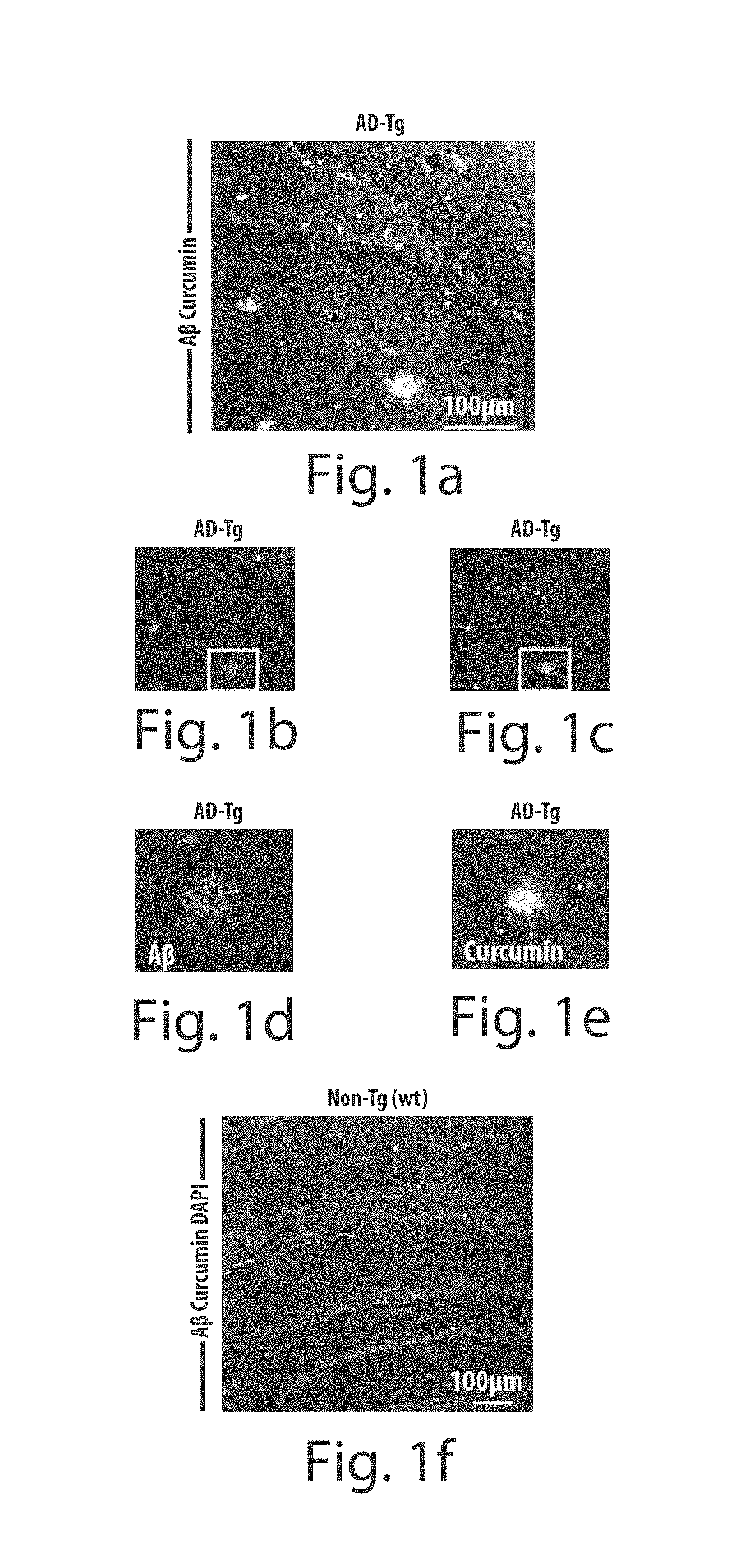
FIGS. 1a through 1n depict retinal A.beta. deposition in the retina of AD-Tg mice visualized by curcumin. FIGS. 1a-1f depict images of brain cryosections from 9-month-old AD-Tg (FIGS. 1a-1e) and non-Tg (wt) (FIG. 1f) mice stained with anti-human A.beta. antibody and curcumin ex vivo, indicating co-localization of A.beta. plaque staining by both detection methods. FIGS. 1d and 1e depict higher magnification images of plaque staining pattern presented for each channel FIG. 1f shows no evidence for double-positive anti-human A.beta.plaques and curcumin in the non-Tg (wt) mouse. Cell nuclei were labeled with DAPI (blue). Scale bar=100 .mu.m. FIGS. 1g-1j are representative images of retinal whole-mounts from 10-month-old AD-Tg (n=27) and non-Tg (wt) mice (n=18) stained with anti-A.beta. antibody and curcumin ex vivo. The formation of A.beta. plaques (yellow spots of overlapping red and green channels) is demonstrated in several different retinal layers: FIG. 1g depicts IPL-Inner Plexiform Layer, FIG. 1h depicts INL-Inner Nuclear/OPL-Outer Plexiform Layers, and FIG. 1i depicts ONL-Outer Nuclear Layer. FIG. 1j shows A.beta. plaques were essentially absent in the non-Tg (wt) mice. (FIGS. 1g and 1j, lower row). Higher magnification images for separate channels demonstrate plaque-staining patterns with both procedures. Scale bars=5 .mu.m. FIGS. 1k-1n depict whole eye sagittal cryosections stained with curcumin in vivo, followed by anti-human A.beta. antibody and DAPI ex vivo. In FIGS. 1k-1m, A.beta. plaques were detected in most retinal layers and in the choroid in 10-month-old AD-Tg mice. In FIG. 1n, A.beta. plaques were undetectable in the retina and choroid of non-Tg (wt) mice. Scale bar=20 .mu.m.
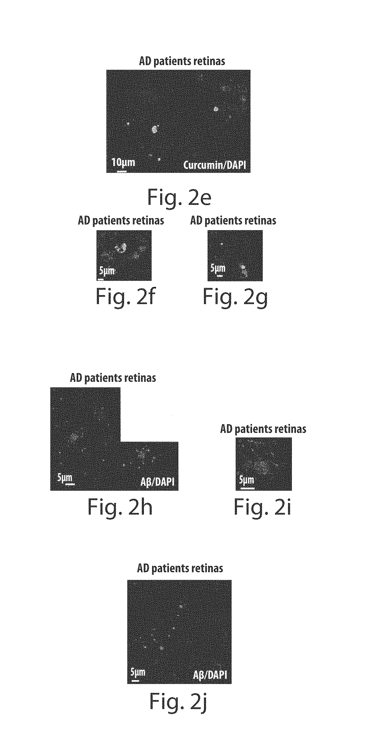
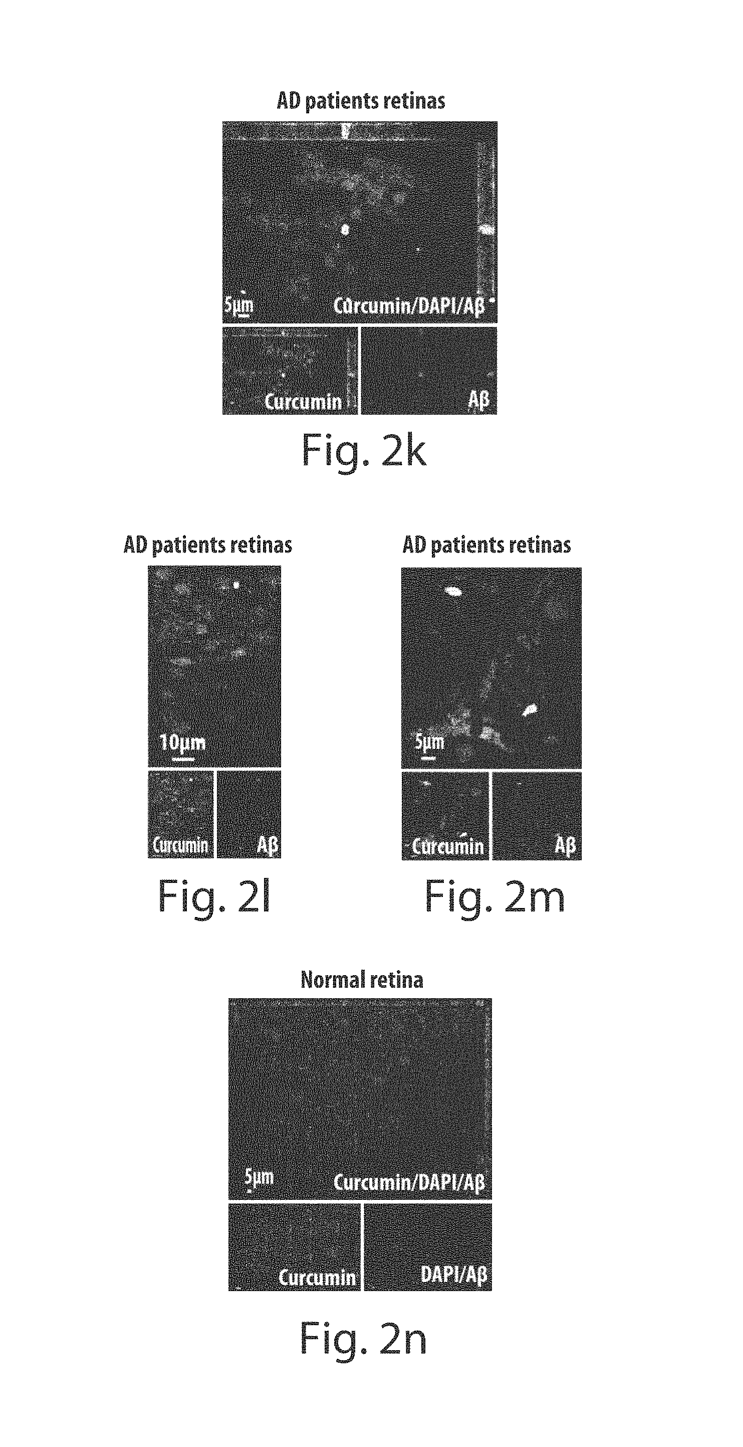
FIGS. 2a through 2n depict A.beta. plaques in the human retina of Alzheimer's disease patients. FIGS. 2a and 2b are representative images of the human whole-mount retina of an 87-yr-old AD patient after staining with Sudan Black B to eliminate non-specific autofluorescence signals, and following curcumin ex vivo staining (curcumin-labeled plaques are indicated by white arrows). Scale bars=10 .mu.m. Cell nuclei are labeled with DAPI (blue). FIGS. 2c and 2d show higher magnification images of the human whole-mount retina of a 65-yr-old AD patient following Sudan Black B staining (black spots for Sudan staining), and then curcumin staining (curcumin-labeled plaque is indicated by white arrow). Scale bars=5 FIGS. 2e-2g provide additional examples of curcumin-positive plaques in retinas of a series of 65- to 90-yr-old human AD patients. FIGS. 2h-2j are representative images of human whole-mount retinas of 65- and 87-yr-old human AD patients stained with anti-human A.beta. antibodies followed by Sudan Black B treatment at several retinal depths (to include RGC and IPL). FIG. 2i represents a higher magnification image of the retinal plaque. A.beta. plaque morphology was similar to that found in the mouse retinas and brains. Scale bars=5 .mu.m. FIGS. 2k-2m represent subsequent staining of the same human retinas with curcumin, which reveals that the plaques were selectively colabeled with human A.beta. antibodies and curcumin (lower row images for separate channels). FIG. 2n depicts double staining with human A.beta. antibodies and curcumin in postmortem non-AD human retinal whole mount showing no signs of A.beta. plaques (lower row images for separate channels). Scale bars=5 .mu.m.
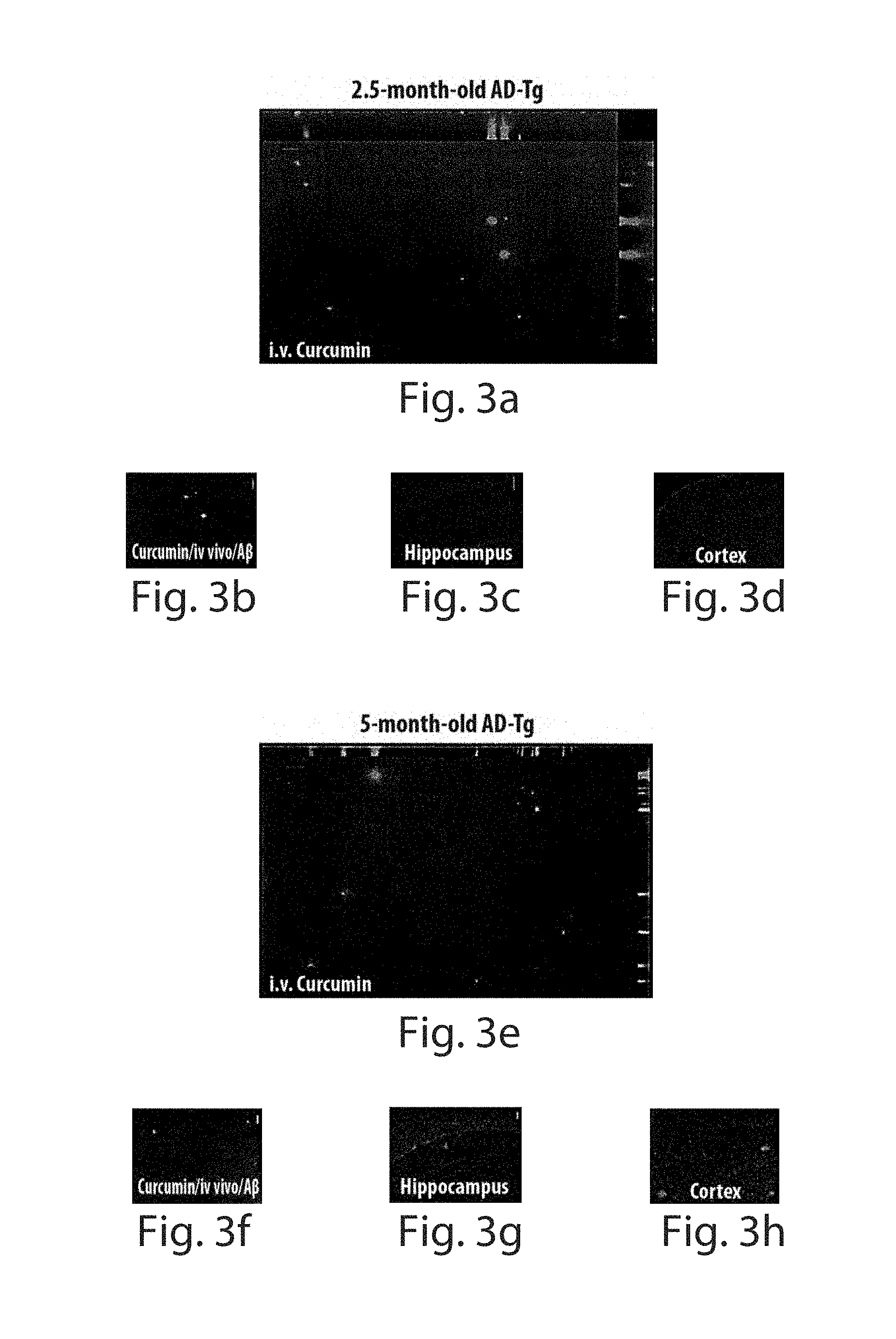
FIGS. 3a through 3q depict mouse retinal A.beta. plaque formations at the pre-symptomatic early stage and accumulation during disease progression. Following i.v. curcumin injections into the tail vein, A.beta. plaques were visible in AD-Tg mice retinas and brains. FIGS. 3a-3n are representative z-axis projection images of whole-mount retinas from AD-Tg (n=18) and non-Tg (wt; n=10) mice at various ages; FIGS. 3a-3d depict 2.5-month-old AD-Tg mouse, with FIG. 3a showing presence of plaques in the retina and FIG. 3b showing validation of A.beta. plaque staining using specific anti-human antibody ex vivo at the same location (co-localization of curcumin and A.beta. antibody in yellow). Scale bars=10 .mu.m. FIGS. 3c and 3d show no plaques were detected in the brain hippocampus and cortex. Scale bars=100 .mu.m. FIGS. 3e-3h depict 5-month-old AD-Tg mouse, with FIG. 3e depicting the presence of plaques in the retina and FIG. 3f following specific A.beta. antibody staining ex vivo. Scale bars=10 .mu.m. FIGS. 3g and 3h show detection of plaques in the brain. Scale bars=50 .mu.m. FIGS. 3i-3k depict 9-month-old AD-Tg mouse, with FIG. 3i showing multiple plaques in the retina and FIGS. 3j and 3k showing plaques in the brain. Scale bars (i)=10 .mu.m and (j,k)=50 .mu.m. FIGS. 3l-3n depict 17-month-old AD-Tg mouse, with FIG. 3l showing numerous plaques in the retina and FIGS. 3m and 3n showing plaques in the brain. Scale bars (i)=10 .mu.m and (m,n)=100 .mu.m. FIGS. 3o-3q depicts 9-month-old non-Tg (wt) mouse, with FIG. 3o showing no plaques in the retina and FIGS. 3p and 3q showing no plaques in the brain. Scale bars (o)=10 .mu.m and (p,q)=100 .mu.m.
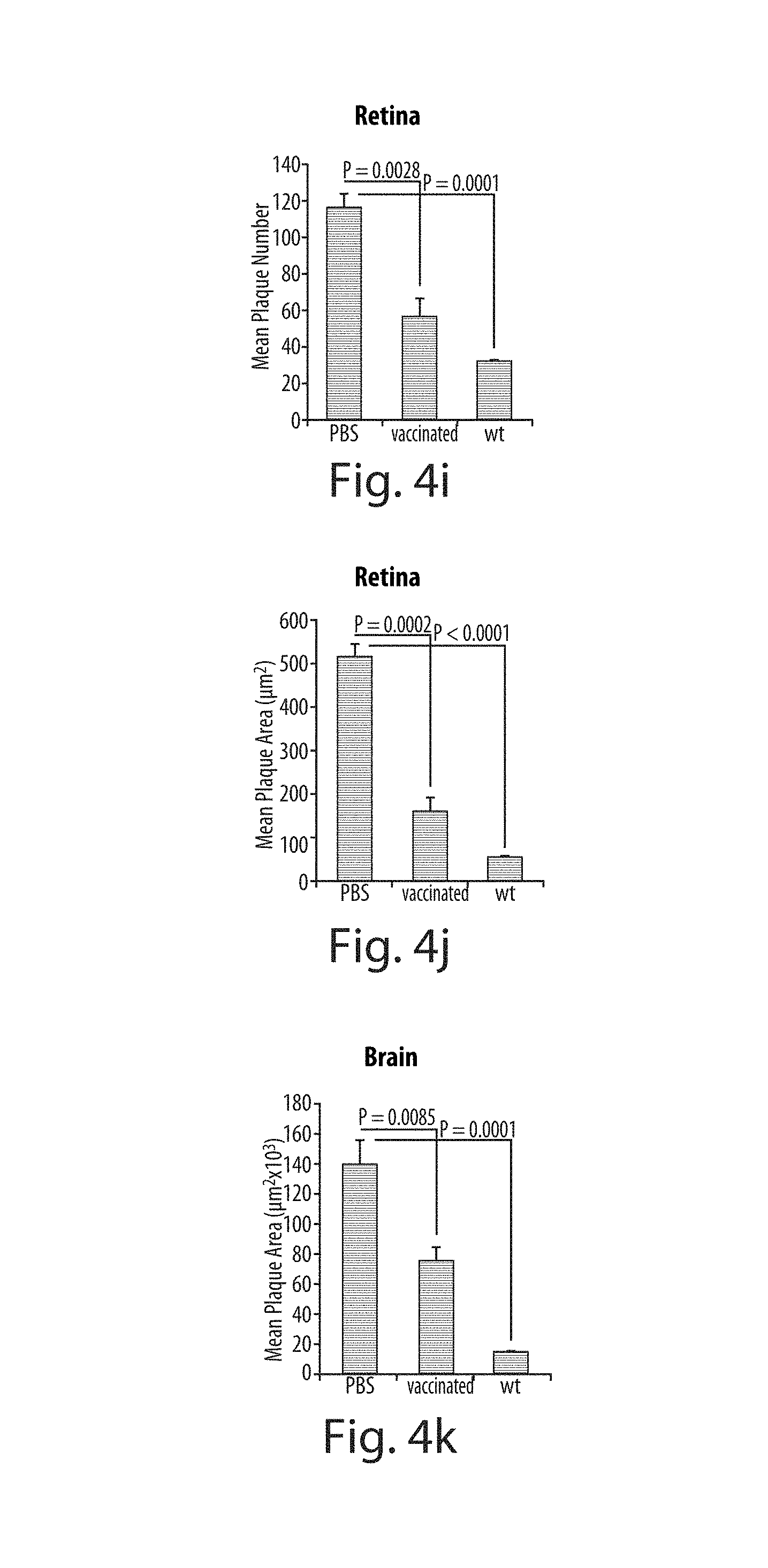
FIGS. 4a through 4k depict decreased A.beta. plaques in the retina of AD-Tg mice following dendritic cell-based vaccination. FIGS. 4a-4g are representative z-axis projection images of whole-mount retinas from 10 month-old mice, FIGS. 4a-4c show PBS-treated AD-Tg mouse control, FIGS. 4d-4f show vaccinated AD-Tg mouse, and FIG. 4g shows non-Tg (wt) mouse stained ex vivo with curcumin and anti-human A.beta. antibodies. FIGS. 4b and 4c, and FIGS. 4c and 4f depict separate channel images for curcumin and anti-A.beta. antibodies labeling in the retina. Scale bars=10 .mu.m. FIG. 4h is an illustration of 12 regions around the optic disc (indicated by rectangles 1-12) representing the area covered for quantitative analyses of plaques in the retinal whole-mounts (n=4 mice per group; two retinas per mouse). Scale bar=200 .mu.m. FIG. 4i depicts the decrease in plaque number, observed in the retinas of AD-Tg mice treated with immunebased vaccination as compared to PBS-treated controls (Student's t-test; P=0.0028). FIG. 4j depicts a decrease in mean plaque area, observed in the retinas of vaccinated AD-Tg mice as compared to their controls (Student's t-test P=0.0002). FIG. 4k shows that the significant reduction in the total area covered by plaques was also detected in the brain hippocampus and cortex of the same mice following immune-based vaccination (Student's t-test P=0.0085). Error bars in each panel represent SEM.
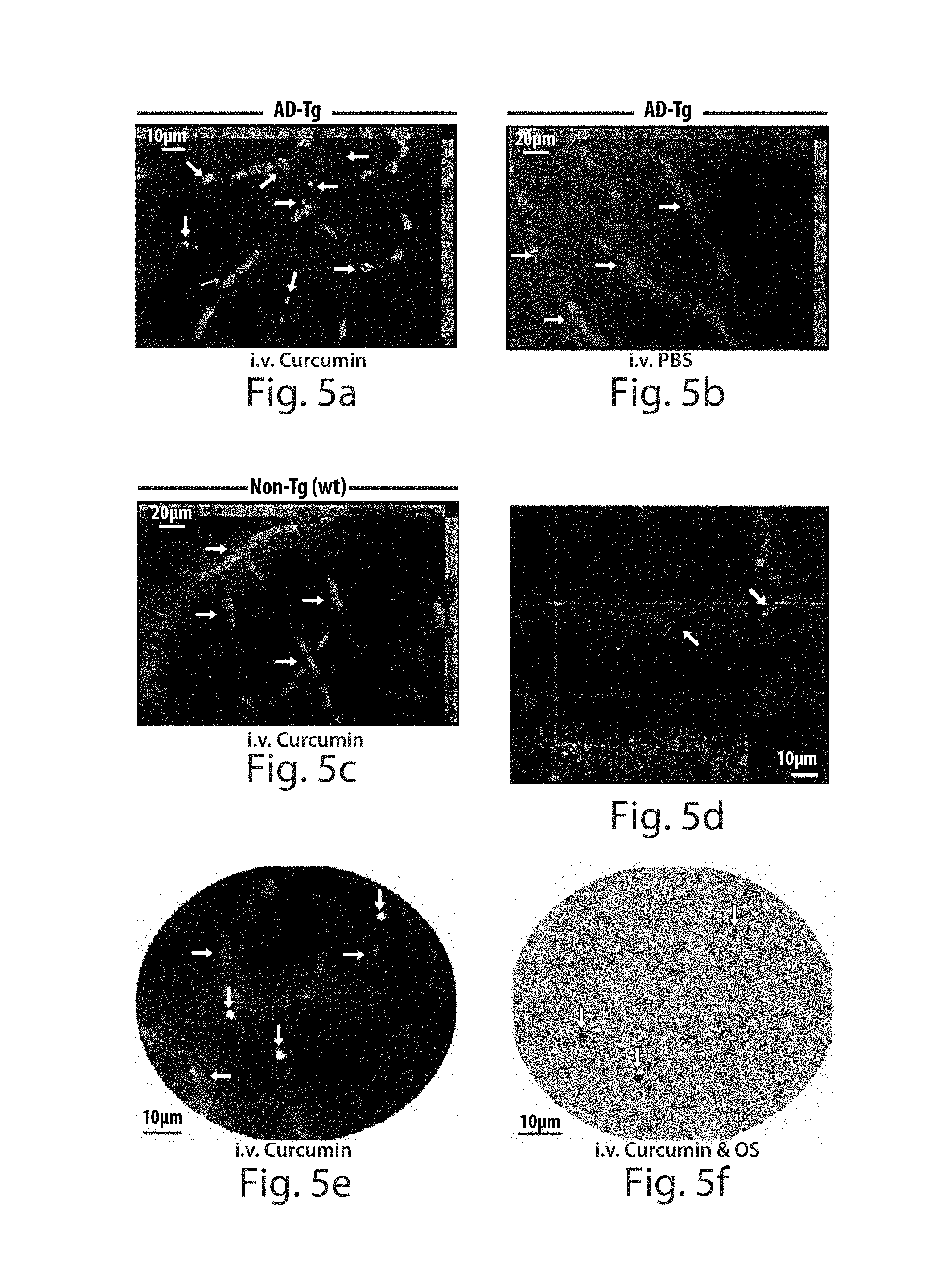
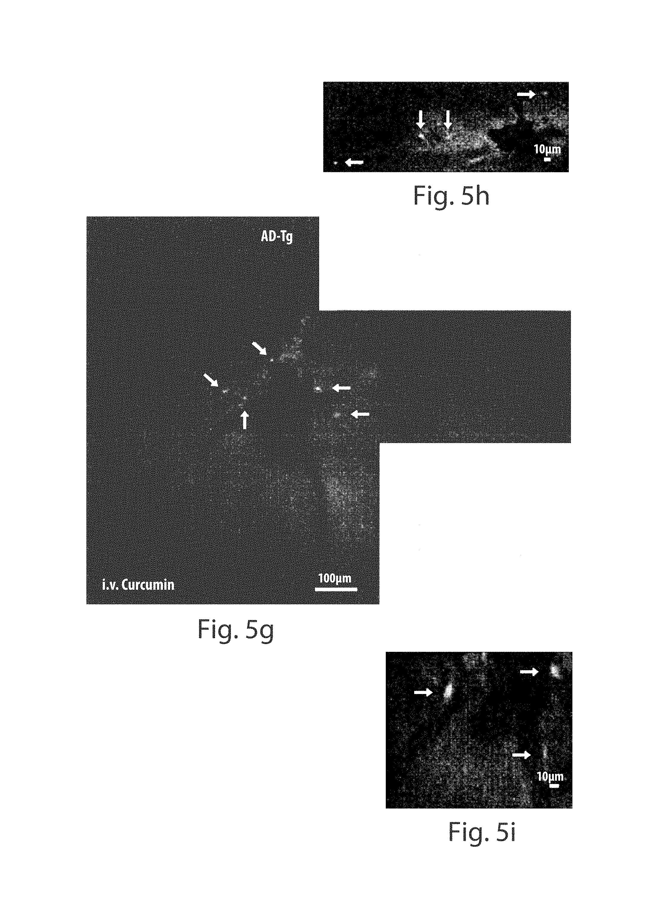
FIGS. 5a through 5k depict in vivo imaging of curcumin-labeled plaques in AD-Tg mouse retinas. FIGS. 5a-5c are representative z-axis projection images taken from retinal whole-mounts of non-perfused AD-Tg versus non-Tg (wt) mice (10-month-old), following i.v. curcumin or PBS administration in vivo (blood vessels are indicated by red arrows). FIG. 5a shows A.beta. plaques were visible (indicated by white arrows) in AD-Tg mouse retinas following i.v. injection of curcumin (n=6). FIG. 5b shows plaques were undetectable in the retinas of AD-Tg mice after i.v. injection of PBS (n=5). FIG. 5c shows plaques were undetectable in the retinas of non-Tg (wt) mice following i.v. injection of curcumin (n=5). FIG. 5d is a representative confocal z-axis projection image, using three channels and sagittal/coronal virtual sections, demonstrating A.beta. plaques (plaques inside the vessels indicated with white arrows), stained with anti-human A.beta. antibodies, in the parenchyma and inside the blood vessels of AD-Tg mouse retinal whole-mount. FIGS. 5e and 5f depict images captured using a fluorescence microscope with AOTF-based spectral imaging system, and analyzed and visualized by segmentation and classification software. In FIG. 5e, A.beta. plaques (in white) and blood vessels (indicated by arrows), were visible in a retinal whole-mount stained in vivo with curcumin and imaged at a single channel (ex. 562/40 nm; em. 624/40 nm). FIG. 5f depicts a spectrally-classified image using optical signature (OS) specific to A.beta. plaques labeled with curcumin in the same retinal whole-mount and region. A.beta. plaques are shown in pseudocolor (indicated by white arrows) and all non-plaque tissue is in green pseudocolor. Scale bars=10 .mu.m. FIGS. 5g-5j are images following a single injection of curcumin, wherein plaques (indicated with white arrows) were visible in live AD-Tg mouse retinas (n=4) by emission of light following excitation with a spectrally controlled source (wavelength of 546/15 nm). FIGS. 5i and 5j are higher magnification images, where plaques were mostly detected in areas close to the optic disc and the average plaque size was compatible with that observed in the whole-mount retinas (ex vivo). FIG. 5k shows no plaques detected in the non-Tg (wt) mice (n=4) i.v. injected with curcumin. Scale bars (g, k)=100 .mu.m, and (h-j)=10 .mu.m. 23.
FIG. 6 depicts a flow diagram of a spectral imaging system for diagnosing, prognosing, and analyzing A.beta. plaques in accordance with an embodiment of the present invention.
FIG. 7 depicts a flow diagram of a spectral imaging system for diagnosing, prognosing and/or analyzing A.beta. plaques in accordance with an embodiment of the present invention.
FIGS. 8a through 8d depict high-resolution images of small retinal plaques (mostly<1 .mu.m in diameter), which were found to originate from the endogenous mouse APP gene. Images are of a 10 month old Non-Tg(wt) mouse retina. FIG. 8a (curcumin), FIG. 8b (mouse A.beta.), FIG. 8c (DAPI) and FIG. 8d (Merged).
FIG. 9 depicts images of live AD-Tg (upper right and lower right panels) and Non-Tg(wt) (upper left and lower left panels) mouse retinas showing curcumin stained plaques in the AD-Tg retina, and the lack of curcumin stained plaques in the Non-Tg(wt) retina.
DETAILED DESCRIPTION OF THE INVENTION
All references cited herein are incorporated by reference in their entirety as though fully set forth. Unless defined otherwise, technical and scientific terms used herein have the same meaning as commonly understood by one of ordinary skill in the art to which this invention belongs. Singleton et al., Dictionary of Microbiology and Molecular Biology 3rd ed, J. Wiley & Sons (New York, N.Y. 2001); March, Advanced Organic Chemistry Reactions, Mechanisms and Structure 5.sup.th ed., J. Wiley & Sons (New York, N.Y. 2001); and Sambrook and Russel, Molecular Cloning: A Laboratory Manual 3rd ed, Cold Spring Harbor Laboratory Press (Cold Spring Harbor, N.Y. 2001), provide one skilled in the art with a general guide to many of the terms used in the present application.
One skilled in the art will recognize many methods and materials similar or equivalent to those described herein, which could be used in the practice of the present invention. Indeed, the present invention is in no way limited to the methods and materials described.
"Administering" and/or "Administer" as used herein refer to any route for delivering a pharmaceutical composition to a patient. Routes of delivery may include non-invasive peroral (through the mouth), topical (skin), transmucosal (nasal, buccal/sublingual, vaginal, ocular and rectal) and inhalation routes, as well as parenteral routes, and other methods know in the art. Parenteral refers to a route of administration that is generally associated with injection, including intraorbital, infusion, intraarterial, intracarotid, intracapsular, intracardiac, intradermal, intramuscular, intraperitoneal, intrapulmonary, intraspinal, intrasternal, intrathecal, intrauterine, intravenous, subarachnoid, subcapsular, subcutaneous, transmucosal, or transtracheal. Via the parenteral route, the compositions may be in the form of solutions or suspensions for infusion or for injection, or as lyophilized powders.
"Alzheimer's Disease" as used herein refers to all form of dementia, identified as a degenerative and terminal cognitive disorder. The disease may be static, the result of a unique global brain injury, or progressive, resulting in long-term decline in cognitive function due to damage or disease in the body beyond what might be expected from normal aging.
"Age-related macular degeneration" as used herein refers to is a medical condition in older adults that results in a loss of vision in the center of the visual field (the macula) due to damage to the retina.
"Cataracts" as used herein refers to a clouding that develops in the crystalline lens of the eye or in its envelope, varying in degree from slight to complete opacity and obstructing the passage of light. Early in the development of age-related cataract the power of the lens may be increased, causing near-sightedness (myopia), and the gradual yellowing and opacification of the lens may reduce the perception of blue colors. Cataracts typically progress slowly to cause vision loss and are potentially blinding if untreated.
"Fluorescent Marker" as used herein refers to any and all compounds containing fiurophore for attaching the compound to another molecule, such as a protein or nucleic acid. This is generally accomplished using a reactive derivative of the fluorophore that selectively binds to a functional group contained in the target molecule.
"Glaucoma" as used herein refers to a group of diseases that affect the optic nerve and involves a loss of retinal ganglion cells in a characteristic pattern. Glaucoma is categorized as a type of optic neuropathy.
"Mammal" as used herein refers to any member of the class Mammalia, including, without limitation, humans and nonhuman primates such as chimpanzees, and other apes and monkey species; farm animals such as cattle, sheep, pigs, goats and horses; domestic mammals such as dogs and cats; laboratory animals including rodents such as mice, rats and guinea pigs, and the like. The term does not denote a particular age or sex. Thus adult and newborn subjects, as well as fetuses, whether male or female, are intended to be including within the scope of this term.
"Therapeutically effective amount" as used herein refers to that amount which is capable of achieving beneficial results in a mammal being treated. A therapeutically effective amount can be determined on an individual basis and can be based, at least in part, on consideration of the physiological characteristics of the mammal, the type of delivery system or therapeutic technique used and the time of administration relative to the progression of the disease, disorder or condition being treated.
"Treat," "treating" and "treatment" as used herein refer to both therapeutic treatment and prophylactic or preventative measures, wherein the object is to prevent or slow down (lessen) the targeted pathologic condition, disease or disorder even if the treatment is ultimately unsuccessful. Those in need of treatment may include those already with the disorder as well as those prone to have the disorder or those in whom the disorder is to be prevented.
.beta.-Amyloid deposition is central to AD neuropathology and a key hallmark of Alzheimer's disease. However, monitoring A.beta. plaques in the brains of living Alzheimer's patients and animals is limited by the current resolution and specificity of MRI and PET, and a definite diagnosis of Alzheimer's or other ailment or condition characterized by the formation of A.beta. plaques is only possible after brain tissue autopsy by monitoring number and distribution of plaques and tangles. Hence, developing means to identify plaques in vivo is essential for diagnosis as well as for evaluation of disease progression in response to therapies.
The present subject matter establishes the formation of retinal A.beta. plaques in mammals and teaches a method for identifying, quantizing, and imaging retinal A.beta. plaque. The present subject matter may be incorporated for patients with Alzheimer's disease, dementia, and other clinical conditions and ailments characterized by the formation of A.beta. plaques. Furthermore, the present subject matter discovered that the formation of A.beta. plaques in the retina of AD patients preceded their appearance in the brain. Accordingly, the present subject matter discloses a method for early diagnosis of AD in a mammal comprising the steps of administering a fluorescent marker to the patient for staining A.beta. plaques in the retina, and imaging the retina of the patient with a optical imaging system to identify stained A.beta. peptides.
Another embodiment of the present subject matter teaches a method for prognosing AD in mammals by measuring the increase or decrease of A.beta. plaques in the retina of patients before and after treatment. The method of prognosis comprises the steps of administering a fluorescent marker to the patient for staining A.beta. plaques in the retina and imaging the retina of the patient with an optical imaging system to identify stained A.beta. peptides, followed by administering an AD treatment to the patient and allowing due course for the AD treatment to take effect. And, re-administering a fluorescent marker to the patient for staining A.beta. plaques in the retina after AD treatment and imaging the retina of the patient with the optical imaging system to identify an increase or decrease in stained A.beta. peptides.
In a further embodiment, the present subject matter discloses a method for treating AD in mammalian patients, comprising administering a therapeutically effective amount of myelin-derived peptides and/or agonist of myelin-derived peptides to the patient in reducing the formation of, and dissolving the existence of A.beta. plaques.
The present subject matter also finds utility in disclosing methods for improving eyesight in mammalian patients containing retinal A.beta. plaques, comprising the steps of administering a therapeutically effective amount of myelin-derived peptides and/or agonist of myelin-derived peptides to the patient. The method of improving eyesight may be applicable for patients with AD, dementia, or other clinical conditions and ailments characterized by the formation of A.beta. plaques, such as Age-Related Macular Degeneration (AMD), and glaucoma.
In further embodiments the subject matter describes that A.beta. plaques are present in the retina of mammals and may be utilized to analyze, prognose and diagnose a multitude of other clinical conditions and ailments characterized by retinal A.beta. plaques. Representative clinical condition and ailments may include AMD and glaucoma.
Further discoveries identified in the present subject matter include an optical imaging system for visualizing A.beta. plaques in vivo in the retina of non-human mammalian and human patients. The optical imaging system incorporates the use of a fluorescence microscope, mercury and xenon arc lamps, a CCD camera, an AOTF (acousto-optic tunable filters)-based spectral image acquisition apparatus, and post-analysis imaging software. The optical imaging system incorporates the foregoing tools to provide retinal images of stained A.beta. plaques, providing a visual pseudo-color representation of the spectral signature extracted from the raw images, representing the size and location of the A.beta. plaques objects.
In an alternative embodiment, the optical imaging system incorporates the use of a stereomicroscope that is adjusted to visualize fluorescence and scatter signals at higher resolutions. The stereomicroscope may be fitted with a Polychrome V variable wavelength light source. In additional embodiments, the optical imaging system may incorporate a MicroFire color digital camera and one or more magnifying lenses to improve magnification and image detail. Image acquisition is attained and perfected by post-analysis image segmentation and classification using imaging software.
In further embodiments, the optical imaging system may be incorporated in methods for diagnosing, prognosing, and treating A.beta. plaques in mammals. Furthermore, the optical imaging system may be augmented with adaptive optics, used to improve the performance of the optical imaging system by reducing the effects of rapidly changing optical distortion.
In yet another embodiment, the subject matter method may be utilized for drug development and testing. As the non-invasive, rapidly repetitive imaging methods would enable back-to-back comparison of various drugs and various dosage of drugs, the present subject matter would find favorable utility in drug development and testing.
A previous report has identified A.beta. pathology in the brain, based on the finding of A.beta. accumulation in the lenses of AD patients [31]. The current study provides evidence for the existence of A.beta. plaques in the retinas of AD patients that could be specifically visualized by curcumin. plaques were found in the retina of all examined AD patients, whereas they could not be detected in the non-AD controls. In both young as well as in aged AD mice, a good correlation between retinal and brain A.beta. plaque pathology was observed; plaques accumulated in an age-dependent manner during disease progression, and both retina and brain showed A.beta. plaque reduction as a response to the same therapeutic modality. Overall, the retinal tissue, which shares many similarities with the brain, can potentially be used for diagnosis and monitoring of AD.
In the present study, A.beta. plaques in the retinas of the AD patients were detected mostly within the RGC layer. In AD mouse eyes, plaques were seen in most of the retinal layers and in the choroid. Plaques were noticeable from the NFL to the ONL, and clusters of A.beta. plaques were seen more often in the inner layers of the retina, signifying the possibility of plaque imaging through the eyes of living subjects. Retinas of AD mice undergo an age-dependent increase in A.beta. plaque load in terms of both number and size, similar to the age-dependent accumulation of plaques observed in the brain. Our results demonstrating retinal plaque pathology are consistent with a recent report that reveals retinal A.beta. deposition in correlation with retinal inflammation and degeneration in adult and aged AD-Tg mice [16]. The present subject matter, we not only provides evidence supporting a link between retinal and brain plaque pathology, but also show that A.beta. plaques are detectable in the retina prior to their detection in the brain, in young AD-Tg mice. We were further able to show a significant reduction of A.beta. plaques in the retinas of AD-Tg mice following vaccination with myelin-derived peptide; this treatment as well as related ones were found to be effective in attenuating A.beta. plaque burden in the brain [17, 24, 25]. These findings provide that assessment of retinal plaques may be used to evaluate responses to plaque-reducing therapy, and that retinal plaques may respond to the same treatment that is effective in A.beta. plaque reduction in the brain.
Importantly, the fact that plaques were seen in the GCL in the human eyes, reaching a size of more than 5 .mu.m, makes imaging Alzheimer's patients through the retina a feasible approach, with some modifications, even with the currently available tools for human eye imaging, such as an adaptive optics ophthalmoscope [32]. In live mice, a commercially available mydriatic retinal camera, was found to be effective in recording fundus photographs enabling evaluation of longitudinal changes of retinal ganglion cells [33]. A blue-light confocal scanning laser ophthalmoscope (bCSLO) system that was modified to visualize cyan fluorescent protein, also provides a noninvasive approach to visualize RGCs in the living mouse retina [34]. Here, for the proof-of-concept, we were able to detect curcumin-labeled plaques in live mice using a stereomicroscope (Leica S6E) equipped with Polychrome V spectral light source and double convex lens. Moreover, using an AOTF system, we were able to detect retinal A.beta. plaques by curcumin while eliminating strong background autofluorescence signals (from red blood cells).
In the present study, curcumin was effective in detecting retinal A.beta. plaques when systemically administered at a single dose of 7.5 mg/kg or when given orally. Curcumin demonstrated the ability to cross the blood-brain and blood-retina barriers, which is a requirement for a useful plaque-imaging agent. In terms of safety, Phase I and II trials using curcumin in patients with cancer have proven its low toxicity in humans even at high doses (12 g/day), and when given over extended periods of time [35]. Translation of curcumin doses given intravenously or orally, from mice to humans (below 1 g) for retinal plaque visualization, is expected to remain within the reported safety levels. Furthermore, recent studies have reported various approaches to significantly increase curcumin stability and bioavailability in humans [36].
The identification of A.beta. plaques in the retina of AD patients, provides a novel opportunity for developing a high resolution and sensitive imaging method, that will allow their detection in vivo. These results may be consistent with the early visual dysfunctions found in AD patients [37,38], and with the evidence for retinal abnormalities such as loss of cells in the GCL and atrophy of the NFL, reported in AD patients [39-44]. Although it is unclear whether A.beta. plaques are found in the retina at early or later stages of AD, the current discovery of A.beta. plaques in the retina of these patients at different ages, and the fact that these plaques are detectable at a very early pre-symptomatic phase of the disease in AD-Tg mice, strengthens the possibility that curcumin-labeled plaques, seen through the eyes, could be used for early diagnosis of AD. Importantly, based on their unique size and distribution within the retinas, the plaques observed in AD patients could be eventually used for differential diagnosis: plaques that were detected in age-related macular degeneration are locally restricted to retinal pigment epithelium within drusen and appear smaller in size [45-47]. In terms of the retinal abnormalities seen in AD patients, it is possible that a plaque-reducing therapy, such as the current DC vaccination, may also help to ameliorate some of the visual dysfunctions, even leading to improved eyesight.
Along with aging of the world's population and the growing epidemic of AD, an early detection of AD becomes ever more critical for evaluating risk, assessing new therapies, and treating AD with early intervention once it has developed. AD pathology, including amyloid plaques and neurofibrillary tangles, is believed to appear many years before symptoms manifest and before any substantial neurodegeneration occurs. Discovery of early measurable markers specific to AD, such as the A.beta.-plaques in the retina, which may predict development of brain pathology and cognitive decline in still cognitively normal subjects, is especially needed. The inventors' findings in mice models of AD support the use of imaging of retinal plaques in vivo labeled with curcumin as a non-invasive tool for early indication of AD pathology and response to a therapeutic intervention.
Furthermore, the present subject matter introduces vaccination therapies to reduce and/or eliminate A.beta.-plaques in the retina, often associated with degeneration of the eyes and eyesight in AD patients. Myelin-derived peptides or weak agonists of myelin-derived peptides were used to effectively induce neuroprotection and to reduce plaque formation in the retina.
In summary, we identified A.beta. plaques in human retinas, and describe a new approach to detect and monitor Alzheimer's plaque pathology earlier and more readily than in the brain, by imaging A.beta. plaques in the retina using a systemically administered compound, proven safe in humans. This may predict development of brain pathology and cognitive decline in subjects who are still cognitively normal and well before a significant functional deficit is seen. These findings show that optical imaging of the retina can be used as a noninvasive approach for monitoring AD progression and response to therapeutic interventions [48].
EXAMPLES
The following examples are provided to better illustrate the claimed invention and are not to be interpreted as limiting the scope of the invention. To the extent that specific materials are mentioned, it is merely for purposes of illustration and is not intended to limit the invention. One skilled in the art may develop equivalent means or reactants without the exercise of inventive capacity and without departing from the scope of the invention.
Example 1
Results
A.beta. Deposits in AD Mouse Retina can be Visualized Using Curcumin
AD-Tg mice carrying the human APPswe and PS1dE9 transgenes were used to assess the potential of developing a noninvasive tool for detecting A.beta. plaques in the eye. We first verified that curcumin had an affinity to the same plaques that were detected by antibodies specific to human A.beta. in the hippocampus of AD-Tg mice (FIG. 1a; separate channels FIGS. 1b and 1c). At higher magnification, images show the specific staining pattern obtained following each procedure (FIGS. 1d and 1e). Human A.beta.-plaques were undetectable in the brains of non-Tg littermate wild type (wt) mice (FIG. 1f). We then tested whether A.beta. plaques in the eyes of AD-Tg mice could also bind curcumin. Examination at high resolution revealed the presence of A.beta. plaques labeled by both curcumin and anti-human AP-antibodies in the retinas of AD-Tg mice (FIG. 1g retinal whole mount; FIG. 1k-1m, cross-section) but not in the retinas of non-Tg (wt) mice (FIGS. 1j and 1n). The representative images display the location of A.beta. plaques in retinal whole mounts at various depths (consecutive acquisition at focal planes of 80 .mu.m depth) to include the inner plexiform layer (IPL; FIG. 1g), inner nuclear layer (INL)/outer plexiform layer (OPL; FIG. 1h), and outer nuclear layer (ONL; FIG. 1i). Analysis of cross sections further verified A.beta. plaque deposition in deep retinal layers and the choroid, with an apparent predominance in the ganglion cell layer (GCL) and IPL through OPL layers (FIGS. 1k-1m). Whereas human-A.beta. plaques were absent in the non-Tg (wt) littermates (FIGS. 1j and 1n), occasional small curcumin-positive plaques were detected. To determine the nature of these small and sparse plaques that were detected by curcumin staining in wt mice, we carried out a double-staining experiment using curcumin and antibodies specific to mouse-A.beta. in wholemount retinas of 10-month old wt mice. Indeed, the small plaques detected by curcumin in the wt retinas were found to be co-labeled with the anti-mouse A.beta. antibodies, thus confirming their identity as endogenously formed mouse-A.beta. deposits (FIGS. 8a-8d).
Example 2
Results
A.beta. Plaques are Formed in Retinas of AD Patients and could be Visualized by Curcumin
We next examined the presence of A.beta. plaques in postmortem eyes of patients with definite diagnosis of AD (n=9; age range from 48 to 94 years; different disease severities, categorized based on their neuropathology reports), and in postmortem eyes of age-matched normal controls (n=4; 66 to 92 years; see human donor eye records in (see Table 1). Autofluorescence and non-specific signals of fixed human eyes observed under excitation ranging from 360-710 nm and associated with lipofuscin/lipid deposits and/or long-term fixation with formalin [21, 22], were eliminated by Sudan Black B staining (FIGS. 2a-2j). For curcumin staining, we first immersed human whole-mount retinas with Sudan Black B (FIGS. 2a and 2c; no plaques were observed), followed by exposure to curcumin (FIGS. 2b and 2d; representative images display plaques within the same tissue location). Plaques detected by curcumin, ranging in size from 1 to 10 .mu.m (typically around 5 .mu.m), were found in all AD patient eyes examined, at various focal depths corresponding to the GCL, IPL and INL retinal layers (FIGS. 2a and 2g), and with an apparent correlation to the reported plaque pathology in the brain. We also analyzed the human retinas with antibodies directed against human A.beta.. We identified A.beta. plaques in AD patients and found that their structure was similar to that found in the mouse retina and brain [FIGS. 2h and 2i represent the innermost retinal layers (i.e. GCL) where the plaques are easily detected; FIG. 2i is a higher magnification image of the retinal A.beta. plaque structure; FIG. 2j represents deeper consecutive focal planes (i.e. IPL)]. The plaques could not be detected when only secondary antibodies were used (data not shown). Exposure of the human retinas to curcumin after their immunolabeling for A.beta., confirmed their co-localization (FIGS. 2k-2m). In non-AD human eyes, no A.beta. plaques were detected (FIG. 2n).
TABLE-US-00001 TABLE 1 Patient Gender.sup.1 & Pre-Mortem Diagnosis Post-Mortem Final # Age (yrs) (Disease Duration) Neuropathology Diagnosis Cause of Death 412 F, 48 Dementia, AD Moderate to frequent NPs AD Cerebral atrophy (10 yrs) and NFTs in the neocortex definite with and hippocampus hydrocephalus 404 F, 65 Dementia, AD Large no. of diffuse AD N/A (5 yrs) plaques, NPs, NFTs in the definite entorhinal cortex and hippocampus 435 M, 70 Dementia, AD Moderate no. of NPs and AD Pneumonia of (5 yrs) NFTs definite posterior lungs 539 M, 78 Dementia Large no. of NPs most AD Subdural (3 yrs) with cores and abundant definite hematoma NFTs and diffuse plaques 484 M, 86 Dementia, AD Abundant NFTs and NPs AD Cerebrum & (11 yrs) in the neocortex and definite Cerebellum hippocampus infarction 664 M, 87 Dementia, AD Moderate to frequent NPs AD Cerebrum (8 yrs) with NFTs in the definite infraction neocortex and Hippocampus CA-1 486 F, 88 Dementia, AD Severe NPs and NFTs AD Pneumonia (6 yrs) definite 513 F, 90 Dementia, AD.sup.2 Mild NPs.sup.3 and abundant AD Cerebral (14 yrs) NFTs.sup.4 definite.sup.6 hemorrhage infarction focal 525 F, 94 Dementia, AD Abundant NPs with AD Cerebrum (11 yrs) mature cores and definite infarction numerous NDTs 93-78 M, 66 Absence of N/A Normal Liver failure Dementia Brain Normal 93-111 M, 77 Absence of N/A Normal Sepsis Dementia Brain Normal 476 M, 88 Absence of Sparse neocortical NPs Normal Cerebrum Dementia and NFTs Brain infarctions Occipital CVA.sup.5 529 F, 92 Absence of Moderate no. of diffuse Normal Cerebrum Dementia plaques and small no. of Brain infarction remote Normal NPs only in the multiple hippocampus .sup.1Gender: F = Female. M = Male .sup.2AD--Alzheimer's Disease .sup.3Neuritic Plaques (NPs) and .sup.4Neurofibrillary Tangeles (NFTs) were determined by Silver (Galiyas or Bielschowsky) strain and Thioflavin stain in several CNS Sites: Hippocampus CA-1. Entorhinal Cortex. Mid Frontal. Sup./Mid. Temporal. Inferior parietal. Primary Visual. Visual association area. .sup.5CVA--Cerebral vascular accident or stroke. .sup.6AD definite--According to CERAD criteria.
Example 3
Results
A.beta. Plaques in AD Mice, Stained In Vivo by Curcumin, are Detected in the Retina Earlier than in the Brain and Accumulate During Disease Progression
To establish the use of curcumin for imaging plaques in the retina, we tested its bioavailability to the eye when injected systemically. To this end, mice were intravenously injected with curcumin. Labeled plaques following the administered curcumin could be detected in the retinas and brains of AD-Tg mice, but not in the non-Tg (wt) controls (FIGS. 3a-3q). These findings confirmed that curcumin crosses the blood-brain barrier and suggest that it also crosses the blood-retina barrier and has a high affinity for A.beta. plaques in vivo. Importantly, curcumin-labeled plaques could be detected following a single curcumin injection or following multiple injections. Representative z-axis projections of retinal and brain hippocampus and cortical images of AD-Tg mice at the ages of 2.5, 5, 9 and 17 months demonstrated an age-dependent correlation between plaque deposition in the retina and the brain, and increased accumulation over the course of disease progression (FIGS. 3a-3n). Importantly, plaques were detected in the retina (FIGS. 3a and 3b) but not in the brain (FIGS. 3c and 3d) as early as at 2.5 months of age in AD-Tg mice following in vivo curcumin administration, suggesting that A.beta. plaques in the retinas precede brain pathology. We further confirmed that these curcumin-labeled plaques were co-localized ex vivo with anti-human A.beta. antibody staining (FIGS. 3b and 3f). A.beta. plaques were first detectable in the brain at the age of 5 months (FIGS. 3g and 3h), in line with previous descriptions of disease initiation and progression in this strain of AD-Tg mice [23]. In the wt mice, A.beta. plaques were undetectable both in the retina (FIG. 3o) and in the brain (FIGS. 3p and 3q) as late as 9 months of age.
Example 4
Results
A.beta. Plaque Burden is Decreased in the Retina Following Vaccination Therapy
We further investigated whether the fate of retinal plaques observed in AD-Tg mice, is similar to that of brain A.beta. plaques in response to the same treatment. Myelin-derived peptides or weak agonists of myelin-derived peptides have been shown to effectively induce neuroprotection and to reduce plaque formation [24-26]. To ensure the beneficial effect of the vaccination without the risk of inducing autoimmune encephalomyelitis, we chose to vaccinate AD-Tg mice with an altered myelin-derived peptide (MOG45D, derived from MOG 35-5527, 28) using dendritic cells (DCs) as a carrier and adjuvant. Whole-mounted retinas of 10-month old AD-Tg mice injected with either MOG45D-loaded DCs or with PBS, and those of wt littermates (4 mice/8 retinas per group), were ex vivo labeled for A.beta. plaques, using both curcumin and anti-A.beta. antibody (FIGS. 4a-4k). Representative axial (z-stack) projection images demonstrated substantial reduction of the number of A.beta. plaques in vaccinated AD-Tg mice compared to PBS-treated controls (FIGS. 4a-4c versus FIGS. 4d and 4f, respectively; separate channels in FIGS. 4b and 4c, and FIGS. 4e and 4f). No A.beta. plaques (double stained with curcumin and antihuman A.beta. antibody) were detected in the wt mice (FIG. 4g). In high-resolution images, we occasionally detected small retinal plaques (mostly<1 .mu.m in diameter), which were found to originate from the endogenous mouse APP gene (FIGS. 8a-8d). These small plaques were stained by curcumin but not by anti-human A.beta. antibodies in all three experimental groups (FIGS. 4a, 4d and 4g). We further quantified plaque number and size by capturing 12 areas (total of approx. 0.45 mm.sup.2) around the optic disc, and quantified plaques across a 60-.mu.m scanning depth in each area (FIG. 4h; each area is indicated by rectangle 1-12). A significant decrease in plaque number detected by curcumin staining was found in the retinas of vaccinated AD-Tg mice compared to PBS-treated controls (FIG. 4i; P=0.0028). Substantial reduction was also observed in the average area covered by the retinal plaques in vaccinated versus PBS-treated AD-Tg mice (FIG. 3j; P=0.0002). Notably, significant reduction, relative to PBS-treated mice, in the total plaque area was also observed in the hippocampus and cortex from the brains of the same vaccinated mice (FIG. 4k; P=0.0085).
Example 5
Results
In Vivo Imaging of A.beta. Plaques in the Eyes Using Systemically Injected Curcumin
To further investigate the potential of visualizing A.beta. plaques by curcumin in the eyes of live subjects, we first tested our ability to identify A.beta. plaques in whole-mount retinas of mice that were not perfused prior to their eye enucleation, a more physiological setting. Representative axial projection images demonstrated that even under these conditions, which included background signals from red blood cells in the capillaries, plaques could be identified in the retinas of AD-Tg mice that had been previously i.v. injected with curcumin (FIG. 5a). Importantly, in the absence of curcumin, plaques were undetectable in AD-Tg mice that had been i.v. injected with PBS (FIG. 5b), suggesting that when using these imaging modalities, plaques are barely detectable in the retina solely by their autofluorescence signals. As expected, in non-Tg(wt) mice injected with curcumin, plaques were also not detected (FIG. 5c). Additional labeling of plaques with anti-human A.beta. antibodies ex vivo, confirmed the A.beta. specificity of curcumin staining (data not shown). A.beta. plaques in whole-mount retinas of ADTg mice labeled with anti-A.beta. antibodies were found inside blood vessels as well as in their parenchymal vicinities (FIG. 5d; confocal virtual cross-section). We further assessed whether it would be possible to detect A.beta. plaques while reducing the background signal emerging from blood vessels. To this end, we monitored the specific optical signature using a fluorescence microscope (Nikon TE2000) including a multi-spectral imaging technology, comprised of spectral imaging with acousto-optic tunable filters (AOTF) [29] and fluorescence lifetime imaging using a gated camera; image acquisition was followed by post-analysis image segmentation and classification using software that was previously developed by us [30]. Curcumin-labeled plaques imaged using a microscope equipped with AOTF at a single wavelength channel were observed in AD-Tg mouse retina (FIG. 5e). By applying the AOTF-based imaging, capturing the spectral signature of curcumin-labeled plaques, and post-translation into color-classified digital images, we were able to identify the specific optical signature of A.beta. plaques as "true" signals, while eliminating the autofluorescence noise generated by the blood vessels (FIG. 5f). To investigate the feasibility of our approach for noninvasive plaque detection, we conducted an in vivo imaging of the retina in live mice using a modified stereomicroscope (Leica S6E) with a wavelength-controlled light source and a digital camera. Following a single injection of curcumin (7.5 mg/kg) two hours prior to imaging, curcumin-labeled plaques were visible in AD-Tg mice retina specifically at an excitation wavelength of 546/15 nm (FIGS. 5g-5j). Plaques were mostly detected in areas close to the optic disc. The average plaque size was compatible with that observed in the whole-mount retina (ex vivo). No plaques were detected in the non-Tg (wt) mice injected i.v. with curcumin (FIG. 5k) or in AD-Tg mice that did not receive curcumin injection (data not shown). To verify that the signals captured by the modified stereomicroscope originated from the plaques, mice were euthanized, and the presence of the curcumin-labeled plaques was confirmed on whole-mount retinas (data not shown).
Example 6
Mice
Double-transgenic mice (females and males at equal numbers) that harbor the chimeric mouse/human APP (APPswe) and the mutant human presenilin 1 (deletion in exon 9 PSEN1.DELTA.E9) genes and their aged-matched non-Tg littermates, were purchased from the Jackson Laboratories (Bar Harbour, Me., strain #4462) and were bred and maintained in the animal center of comparative medicine of Cedars-Sinai Medical Center (Los Angeles, Calif.). All experiments were approved and conducted according to regulations devised by the Cedars-Sinai Institutional Animal Care and Use Committee.
Example 7
Genotyping
Genomic DNA was extracted from 0.5 cm tail tip using a DNA extraction kit (Qiagen, Valencia, Calif.) following the manufacturer's protocol. Mice used in this study were genotyped for the presence of the transgenes by PCR as previously described (Jankowsky, 2004, Ref #120).
Example 8
Vaccination Preparations
Modified myelin-derived peptide (MOG45D) is derived from the encephalitogenic peptide MOG.sub.(35-55) (Koehler, 2002, Ref #285; Shao, 2002, Ref. #283; Zhong, 2002, Ref. #284; Hauben, 2001, Ref. #28; and Hauben, 2001, Ref. #35). For vaccinations, MOG45D (Invitrogen, Carlsbad, Calif.) was added to bone marrow-derived dendritic cells from non-Tg littermates' donor mice. Preparation of dendritic cells for vaccination was as previously described (Hauben, 2003, Ref. #34).
Example 9
Experimental Regimen for Vaccinations
AD-Tg mice at the age of 7 months were injected with DC-MOG45 (0.5.times.10.sup.6 cells/200 ml in 1.times.PBS per animal) once a month, for three months. Control groups of 7-month-old AD-Tg mice were injected with 1.times.PBS according to the corresponding regimens. At the end of the study, all mice were perfused under anesthesia with 1.times.PBS following 2.5% paraformaldehyde ("PFA") (Sigma) and their brains and eyes were collected for further analysis.
Example 10
Animal Tissue
Mice were anesthetized and perfused with 4% ice-cold buffered PFA, and a group of mice were not perfused. Their eyes were enucleated and fixed immediately in 4% fresh PFA overnight. For whole mount retinas, the eyes were dissected and the anterior part was removed. The eyecups were soaked for 10 minutes in hyaluronidase (type I-S) (0.07 mg/ml) (Sigma) to liquefy and remove the vitreous residues, than washed for 10 minutes.times.3 in PBS, and the whole mount retinas were collected. For whole eye sectioning, the eyes were put in 30% sucrose in 4% PFA for 2 hours, than washed for 15 minutes.times.3 in PBS. The eyes were embedded in O.C.T and frozen slowly on dry ice than sagittal sectioned (7 .mu.m) with cryostat. Brains were collected and fixed immediately in 4% fresh PFA overnight. The brains were put in 30% sucrose (in 4% PFA) gradient. Brains were washed for 15 minutes.times.3 in PBS, next embedded in O.C.T and frozen slowly on dry ice, then coronal sectioned (30 .mu.m) with cryostat.
Example 11
Human Autopsy Eyes
Autopsy eyes from Alzheimer's patients were procured from the Alzheimer's Disease Research Center, Department of Pathology, University of Southern California (Los Angeles, Calif.), under IRB protocols 99491 and 3201. Healthy donor eyes were purchased from National Disease Research Interchange (NDRI, Philadelphia, Pa.). NDRI has a human tissue collection protocol approved by a managerial committee and subject to National Institutes of Health oversight. Diseased and normal eyes were fixed and stored in 10% neutral buffered formalin. In addition, we used two healthy eyes that were frozen without fixation and stored at -80.degree. C. Whole-mount retinas were prepared from the eyes and further studied by immunohistochemisty.
Example 12
Tail Vein Injection of Curcumin
For in vivo A.beta.-plaque imaging, AD-Tg and non-Tg wild-type mice were intravenously injected to the tail vein with curcumin in PBS (7.5 mg/kg/day, for 7 consecutive days) or with PBS. Subsequently, brains and eyes were cryosectioned, or prepared for whole mount retina. In an alternative embodiment, curcumin may be administered to the patient orally.
Example 13
Immunohistochemistry
Brain cryosections (30 .mu.m thick), retina cross sections (cryosections) (7 .mu.m thick) and whole mount retinas were treated with a permeabilization/blocking solution containing 20% horse serum (Invitrogen) and 0.01-0.1% Triton X-100 (Sigma-Aldrich, St. Louis, Mo.). Sections were stained overnight at 4.degree. C. with a specified combination of the following primary Abs in PBS containing 10% blocking solution: mouse anti-A.beta. [human amino-acid residues 1-17; clone 6E10 (1:100; Milipore, Temecula, Calif.)]. The sections were incubated for 1 hour in room temperature with secondary Abs, then washed three times with 1.times.PBS and mounted using Vectorshield containing or not 4',6-diamidino-2-phenylindole, dihydrochloride (DAPI, Vector Laboratories, Peterborough, UK). Secondary Abs solution in 1.times.PBS contained Cy-5-conjugated donkey anti-mouse antibody (1:200; Jackson ImmunoResearch Laboratories, West Grove, Pa.). A negative control was processed with the same protocol with the omission of the primary antibody to assess nonspecific labeling. For microscopic analysis we used a Zeiss ApoTome fluorescent.
Example 14
Curcumin Staining
Curcumin solution at 0.1 mg/ml was prepared by dissolving curcumin (Sigma-Aldrich) in 0.5M NaOH, pH=7.9, following immediate dilution in 1.times.PBS. Brain and retina tissue cryosections (30 .mu.m and 7 .mu.m thick respectively) and whole mount retinas were stained with curcumin solution for 10 minutes in room temperature, then rinsed three times with 1.times.PBS for 15 minutes each. The samples were covered with GVA mounting media (Zymed).
Additional compounds are known in the art that can stain/label in vivo amyloid plaques, including, Thioflavin S and T and some derivatives, Congo Red and derivatives, methoxy-X04, Pittsburgh Compound-B (PiB), DDNP, Chrysamine-G and several more. However, curcumin and it's derivatives are very appealing for in vivo optical imaging of amyloid plaques in animal models as well as humans, because of the following advantage. Curcumin generates specific and very bright signals in the commonly used optical spectrum, and is commercially available, exceptionally low cost. Safety issues related to curcumin are minimal (even at high dosages) and may even consider to be beneficial to the patient's health as an antioxidant. Curcumin is an effective ligand with very good in vitro and in vivo binding characteristics to A.beta., and offers good initial brain uptake and washout rate from the brain (important properties for in vivo imaging agents).
Example 15
Quantification
Micrographs of stained tissues were obtained on an Axio Imager Z1 ApoTome-equipped microscope (with motorized Z-drive) with AxioCam MRm monochrome camera ver. 3.0 (at a resolution of 1388.times.1040 pixels, 6.45 .mu.m.times.6.45 .mu.m pixel size, dynamic range of >1:2200, that delivers low-noise images due to Peltier-cooled sensor). Quantitative analysis of A.beta. plaque number and area (mm.sup.2) was performed from two wholemounted retinas per mouse (n=4 mice per group). Each image, captured at 40.times. objective with resolution of 0.28 .mu.m, included an area of 0.04 mm.sup.2, and a total of 12 rectangular areas around the optic disc within scanning depth of 60 .mu.m (multiple virtual section images at consecutive focal planes using a motorized scanning stage). Measurements of the average plaque radius (following curcumin staining) were completed for each animal group followed by calculation of the average plaque area in each animal group. For the acquisition, we used similar exposure times (approx. 75 ms) and the same gain values (0) for all images. No image post-processing was preformed. The emission signals of A.beta.plaques stained with curcumin were compared to the background signals in the retinal tissue, to determine signal to background ratio. The calculated signal-to-background noise ratio from the images was high and within the range of 3:1 to 10:1. Quantitative analysis of A.beta. plaque number and area (mm.sup.2) in the brain was determined from three coronal sections (two hemispheres each) per mouse with 450 .mu.m intervals, over an area covering hippocampal and cortical regions. Optical sections from each field of the specimen were imported into the NIH Image J program (National Institutes of Health). Conversion to greyscale was performed to distinguish between areas of immunoreactivity and background. Total area and quantitative levels of immunoreactivity were determined using a standardized custom histogram-based threshold technique, and then subjected to particle analysis.
Example 16
Spectral and Multispectral Imaging
Spectral imaging provides digital images of an object at a large, sequential number of wavelengths generating precise optical signatures at every pixel. The fluorescence spectral signature of A.beta. plaques, labeled in vivo with curcumin, was captured by our spectral imaging system using the following equipment: Nikon fluorescence microscopes (E800 and TE2000), mercury and xenon arc lamps, a CCD camera, an AOTF (acousto-optic tunable filters)-based spectral image acquisition system (ChromoDynamics, Inc)[29] and post-analysis imaging software developed by our Minimally Invasive Surgical Technologies Institute [30]. The final images provided a visual pseudo-color representation of the spectral signature extracted from the raw images, representing the size and location of the analyzed objects. In multispectral imaging, fluorescence lifetime imaging, performed with a pulsed laser and a LaVision PicoStar HR gated camera, was supplementing the spectral acquisition.
FIG. 6 depicts a flow diagram of a spectral imaging system 100 for diagnosing, prognosing, and analyzing A.beta. plaques in vivo in accordance with an embodiment of the present invention. The subject matter retina 110 is stained with a fluorescent marker to label A.beta. plaques. Shortly thereafter, the stained retina 110 is imaged by an imaging device 120 that is adjusted to visualize fluorescence and scatter signals at higher resolutions. The imaging device 120 may be fitted with a Polychrome V variable spectral light source 130. In additional embodiments, the spectral imaging system 100 may incorporate a color digital camera 140 (e.g. MicroFire) and one or more magnifying lenses to improve magnification and image detail. Image acquisition 170 is attained by post-analysis image segmentation and classification using imaging software 160.
FIG. 7 depicts a flow diagram of a spectral imaging system 200 for diagnosing, prognosing, and analyzing A.beta. plaques in accordance with an embodiment of the present invention. The subject matter retina 210 is stained with a fluorescent marker to label A.beta. plaques. Shortly thereafter, the stained retina 210 is imaged using an imaging device 220. The imaging device 220 may be fitted with multi-spectral imaging technology, comprising of spectral imaging with acousto-optic tunable filters (AOTF) 230 and fluorescence lifetime imaging using a digital camera 240. Image acquisition 260 is attained by post-analysis image segmentation and classification using imaging software 250.
Example 17
In-Vivo Imaging of Mouse Retina
AD-Tg and wt mice retinas were imaged two hours following curcumin intravenous injection. Mice were anaesthetized with Ketamine 100 mg/ml/kg and Xylazine 20 mg/ml/kg. Mouse pupils were dilated to about 2 mm in diameter with 0.5% phenylephrine hydrochloride ophthalmic solution (Bausch & Lomb) combined with 0.5% tropicamide ophthalmic solution (Mydral; Bausch & Lomb). During the imaging process, the mice were positioned on a stage of the stereomicroscope, and the eye was covered with a drop of PBS supplemented with calcium and magnesium, which served as an optical coupling medium between the eye surface and the imaging lens. A modified stereomicroscope (Leica S6E) that was adjusted to visualize fluorescence and scatter signals at higher resolution was used to capture images (exposure time 750 ms. with gain 4). The modified stereomicroscope was assembled to include a Polychrome V (Till Photonics) variable wavelength light source, a MicroFire color digital camera (Optronics), and an additional 6.times. (double convex) magnifying lens, with a focal length of 10 cm. Images were repeatedly captured at several different angles, in order to visualize a larger field and to eliminate non-specific reflection signals.
Example 18
Statistical Analysis
Results were analyzed by one tailed unpaired Student's t test for the p values of two-group comparison. Results are expressed as means.+-.SD.
Various embodiments of the present subject matter are described above in the Detailed Description. While these descriptions directly describe the above embodiments, it is understood that those skilled in the art may conceive modifications and/or variations to the specific embodiments shown and described herein. Any such modifications or variations that fall within the purview of this description are intended to be included therein as well. Unless specifically noted, it is the intention of the inventors that the words and phrases in the specification and claims be given the ordinary and accustomed meanings to those of ordinary skill in the applicable art(s).
The foregoing description of various embodiments of the present subject matter known to the applicant at this time of filing the application has been presented and is intended for the purposes of illustration and description. The present description is not intended to be exhaustive nor limit the subject matter to the precise form disclosed and many modifications and variations are possible in the light of the above teachings. The embodiments described serve to explain the principles of the present subject matter and its practical application and to enable others skilled in the art to utilize the subject matter in various embodiments and with various modifications as are suited to the particular use contemplated. Therefore, it is intended that the present subject matter not be limited to the particular embodiments disclosed for carrying out the subject matter.
While particular embodiments of the present subject matter have been shown and described, it will be obvious to those skilled in the art that, based upon the teachings herein, changes and modifications may be made without departing from this subject matter and its broader aspects and, therefore, the appended claims are to encompass within their scope all such changes and modifications as are within the true spirit and scope of this subject matter. It will be understood by those within the art that, in general, terms used herein are generally intended as "open" terms (e.g., the term "including" should be interpreted as "including but not limited to," the term "having" should be interpreted as "having at least," the term "includes" should be interpreted as "includes but is not limited to," etc.).
REFERENCES
1. Sisodia, S. S. & Price, D. L. Role of the beta-amyloid protein in Alzheimer's disease. FASEB J 9, 366-370 (1995). 2. Hardy, J. & Selkoe, D. J. The amyloid hypothesis of Alzheimer's disease: progress and problems on the road to therapeutics. Science 297, 353-356 (2002). 3. McGeer, P. L. & McGeer, E. G. Local neuroinflammation and the progression of Alzheimer's disease. J Neurovirol 8, 529-538 (2002). 4. Klunk, W. E. et al. Imaging brain amyloid in Alzheimer's disease with Pittsburgh Compound-B. Ann Neurol 55, 306-319 (2004). 5. Ng, S. et al. Visual assessment versus quantitative assessment of 11C-PIB PET and 18F-FDG PET for detection of Alzheimer's disease. J Nucl Med 48, 547-552 (2007). 6. Wang, Y. et al. Development of a PET/SPECT agent for amyloid imaging in Alzheimer's disease. J Mol Neurosci 24, 55-62 (2004). 7. Nordberg, A. Amyloid plaque imaging in vivo: current achievement and future prospects. Eur J Nucl Med Mol Imaging 35 Suppl 1, S46-50 (2008). 8. Hintersteiner, M. et al. In vivo detection of amyloid-beta deposits by near-infrared imaging using an oxazine-derivative probe. Nat Biotechnol 23, 577-583 (2005). 9. Nakada, T., Matsuzawa, H., Igarashi, H., Fujii, Y. & Kwee, I. L. In vivo visualization of senile-plaque-like pathology in Alzheimer's disease patients by MR microscopy on a 7T system. J Neuroimaging 18, 125-129 (2008). 10. Toyama, H. et al. PET imaging of brain with the beta-amyloid probe, [11C]6-OHBTA-1, in a transgenic mouse model of Alzheimer's disease. Eur Nucl Med Mal Imaging 32, 593-600 (2005). 11. Klunk, W. E. et al. Binding of the positron emission tomography tracer Pittsburgh compound-B reflects the amount of amyloid-beta in Alzheimer's disease brain but not in transgenic mouse brain. J Neurosci 25, 10598-10606 (2005). 12. Lockhart, A. et al. PIB is a non-specific imaging marker of amyloid-beta (Abeta) peptide-related cerebral amyloidosis. Brain 130, 2607-2615 (2007). 13. Meyer-Luehmarm, M. et al. Rapid appearance and local toxicity of amyloid-beta plaques in a mouse model of Alzheimer's disease. Nature 451, 720-724 (2008). 14. Morin, P. J. et al. Amyloid precursor protein is synthesized by retinal ganglion cells, rapidly transported to the optic nerve plasma membrane and nerve terminals, and metabolized. J Neurochem 61, 464-473 (1993). 15. Greeve, I. et al. Age-dependent neurodegeneration and Alzheimer-amyloid plaque formation in transgenic Drosophila. J Neurosci 24, 3899-3906 (2004). 16. Ning, A., Cui, J. Z., To, E., Hsiao Ashe, K. & Matsubara, J. A. Amyloid Beta Deposits Lead to Retinal Degeneration in a Mouse Model of Alzheimer Disease. Invest Ophthalmol Vis Sci (2008). 17. Koronyo-Hamaoui, M. et al. Dendritic-cell based vaccination with a weak agonist of myelin-derived peptide attenuates Alzheimer's-like pathology in APP/PS1 transgenic mice. SfN abstract 740.1/Q1 (2008). 18. Hauben, E. et al. Vaccination with dendritic cells pulsed with peptides of myelin basic protein promotes functional recovery from spinal cord injury. J Neurosci 23, 8808-8819 (2003). 19. Yang, F. et al. Curcumin inhibits formation of amyloid beta oligomers and fibrils, binds plaques, and reduces amyloid in vivo. J Biol Chem 280, 5892-5901 (2005). 20. Garcia-Alloza, M., Borrelli, L. A., Rozkalne, A., Hyman, B. T. & Bacskai, B. J. Curcumin labels amyloid pathology in vivo, disrupts existing plaques, and partially 18 restores distorted neurites in an Alzheimer mouse model. J Neurochem 102, 1095-1104 (2007). 21. Schnell, S. A., Staines, W. A. & Wessendorf, M. W. Reduction of lipofuscin-like autofluorescence in fluorescently labeled tissue. J Histochem Cytochem 47, 719-730 (1999). 22. Baschong, W., Suetterlin, R. & Laeng, R. H. Control of autofluorescence of archival formaldehyde-fixed, paraffin-embedded tissue in confocal laser scanning microscopy (CLSM). J Histochem Cytochem 49, 1565-1572 (2001). 23. Garcia-Alloza, M. et al. Characterization of amyloid deposition in the APPswe/PS1dE9 mouse model of Alzheimer disease. Neurobiol Dis 24, 516-524 (2006). 24. Frenkel, D., Maron, R., Burt, D. S. & Weiner, H. L. Nasal vaccination with a proteosome-based adjuvant and glatiramer acetate clears beta-amyloid in a mouse model of Alzheimer disease. J Clin Invest 115, 2423-2433 (2005). 25. Butovsky, O. et al. Glatiramer acetate fights against Alzheimer's disease by inducing dendritic-like microglia expressing insulin-like growth factor 1. Proc Natl Acad Sci USA 103, 11784-11789 (2006). 26. Butovsky, O., Kunis, G., Koronyo-Hamaoui, M. & Schwartz, M. Selective ablation of bone marrow-derived dendritic cells increases amyloid plaques in a mouse Alzheimer's disease model. Eur J Neurosci 26, 413-416 (2007). 27. Ford, M. L. & Evavold, B. D. An MHC anchor-substituted analog of myelin oligodendrocyte glycoprotein 35-55 induces IFN-gamma and autoantibodies in the absence of experimental autoimmune encephalomyelitis and optic neuritis. Eur J Immunol 34, 388-397 (2004). 28. Ziv, Y., Avidan, H., Pluchino, S., Martino, G. & Schwartz, M. Synergy between immune cells and adult neural stem/progenitor cells promotes functional recovery from spinal cord injury. Proc Natl Acad Sci USA 103, 13174-13179 (2006). 29. Wachman, E. S., Niu, W. & Farkas, D. L. AOTF microscope for imaging with increased speed and spectral versatility. Biophys J 73, 1215-1222 (1997). 30. Burton, K., Jeong, J., Wachsmann-Hogiu, S, and Farkas, D. L. Spectral optical imaging in biology and medicine in Biomedical Optical Imaging, Edn. In press. (Oxford University Press 2008). 31. Goldstein, L. E. et al. Cytosolic beta-amyloid deposition and supranuclear cataracts in lenses from people with Alzheimer's disease. Lancet 361, 1258-1265 (2003). 32. Carroll, J., Choi, S. S. & Williams, D. R. In vivo imaging of the photoreceptor mosaic of a rod monochromat. Vision Res 48, 2564-2568 (2008). 33. Murata, H. et al. Imaging mouse retinal ganglion cells and their loss in vivo by a fundus camera in the normal and ischemic-reperfusion model. Invest Ophthalmol Vis Sci 49, 5546-5552 (2008). 34. Leung, C. K. et al. Longitudinal profile of retinal ganglion cell damage after optic nerve crush with blue-light confocal scanning laser ophthalmoscopy. Invest Ophthalmol Vis Sci 49, 4898-4902 (2008). 35. Dhillon, N. et al. Phase II trial of curcumin in patients with advanced pancreatic cancer. Clin Cancer Res 14, 4491-4499 (2008). 36. Anand, P., Kunnumakkara, A. B., Newman, R. A. & Aggarwal, B. B. Bioavailability of curcumin: problems and promises. Mol Pharm 4, 807-818 (2007). 37. Sadun, A. A., Borchert, M., DeVita, E., Hinton, D. R. & Bassi, C. J. Assessment of visual impairment in patients with Alzheimer's disease. Am J Ophthalmol 104, 113-120 (1987). 38. Katz, B. & Rimmer, S. Ophthalmologic manifestations of Alzheimer's disease. Surv Ophthalmol 34, 31-43 (1989). 39. Hinton, D. R., Sadun, A. A., Blanks, J. C. & Miller, C. A. Optic-nerve degeneration in Alzheimer's disease. N Engl J Med 315, 485-487 (1986). 40. Blanks, J. C., Torigoe, Y., Hinton, D. R. & Blanks, R. H. Retinal pathology in Alzheimer's disease. I. Ganglion cell loss in foveal/parafoveal retina. Neurobiol Aging 17, 377-384 (1996). 41. Trick, G. L., Barris, M. C. & Bickler-Bluth, M. Abnormal pattern electroretinograms in patients with senile dementia of the Alzheimer type. Ann Neurol 26, 226-231 (1989). 42. Katz, B., Rimmer, S., Iragui, V. & Katzman, R. Abnormal pattern electroretinogram in Alzheimer's disease: evidence for retinal ganglion cell degeneration? Ann Neurol 26, 221-225 (1989). 43. Parisi, V. et al. Morphological and functional retinal impairment in Alzheimer's disease patients. Clin Neurophysiol 112, 1860-1867 (2001). 44. Berisha, F., Feke, G. T., Trempe, C. L., McMeel, J. W. & Schepens, C. L. Retinal abnormalities in early Alzheimer's disease. Invest Ophthalmol Vis Sci 48, 2285-2289 (2007). 45. Anderson, D. H. et al. Characterization of beta amyloid assemblies in drusen: the deposits associated with aging and age-related macular degeneration. Exp Eye Res 78, 243-256 (2004). 46. Dentchev, T., Milam, A. H., Lee, V. M., Trojanowski, J. Q. & Dunaief, J. L. Amyloidbeta is found in drusen from some age-related macular degeneration retinas, but not in drusen from normal retinas. Mol Vis 9, 184-190 (2003). 47. Ding, J. D. et al. Targeting age-related macular degeneration with Alzheimer's disease based immunotherapies: anti-amyloid-beta antibody attenuates pathologies in an agerelated macular degeneration mouse model. Vision Res 48, 339-345 (2008). 48. Koronyo, Y., Koronyo-Hamaoui, M., Black, K. L., Schwartz, M. & Farkas, D. L., Optical method for the detection of Alzheimer's Disease. Patent application submitted to the USPTO (2008) 49. Jankowsky, J. L. et al. APP processing and amyloid deposition in mice haploinsufficient for presenilin 1. Neurobiol Aging 25, 885-892 (2004). 50. McKinnon S J, Lehman D M, Kerrigan-Baumrind L A, Merges C A, Pease M E, Kerrigan D F, Ransom N L, Tahzib N G, Reitsamer H A, Levkovitch-Verbin H, et al. (2002) Invest Ophthalmol Vis Sci 43, 1077-1087. 51. Yoneda S, Hara H, Hirata A, Fukushima M, Inomata Y, & Tanihara H (2005) Jpn J Ophthalmol 49, 106-108. 52. Guo L, Salt T E, Luong V, Wood N, Cheung W, Maass A, Ferrari G, Russo-Marie F, Sillito A M, Cheetham M E, et al. (2007) Proc Natl Acad Sci USA 104, 13444-13449. 53. Dentchev T, Milam A H, Lee V M, Trojanowski J Q, & DunaiefJ L (2003) Mol Vis 9, 184-190.
* * * * *
D00001
D00002
D00003
D00004
D00005
D00006
D00007
D00008
D00009
D00010
D00011
D00012
D00013
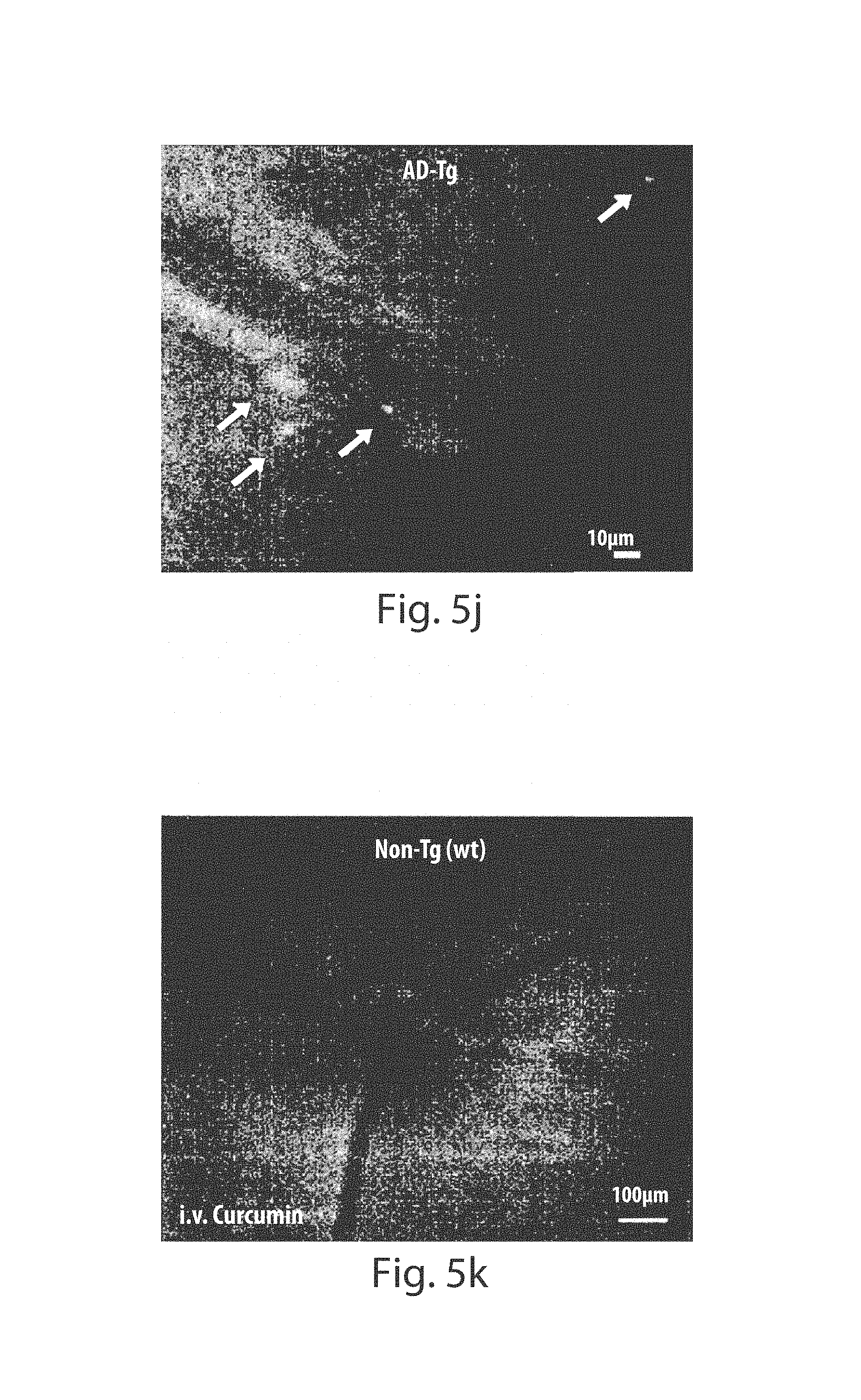
D00014
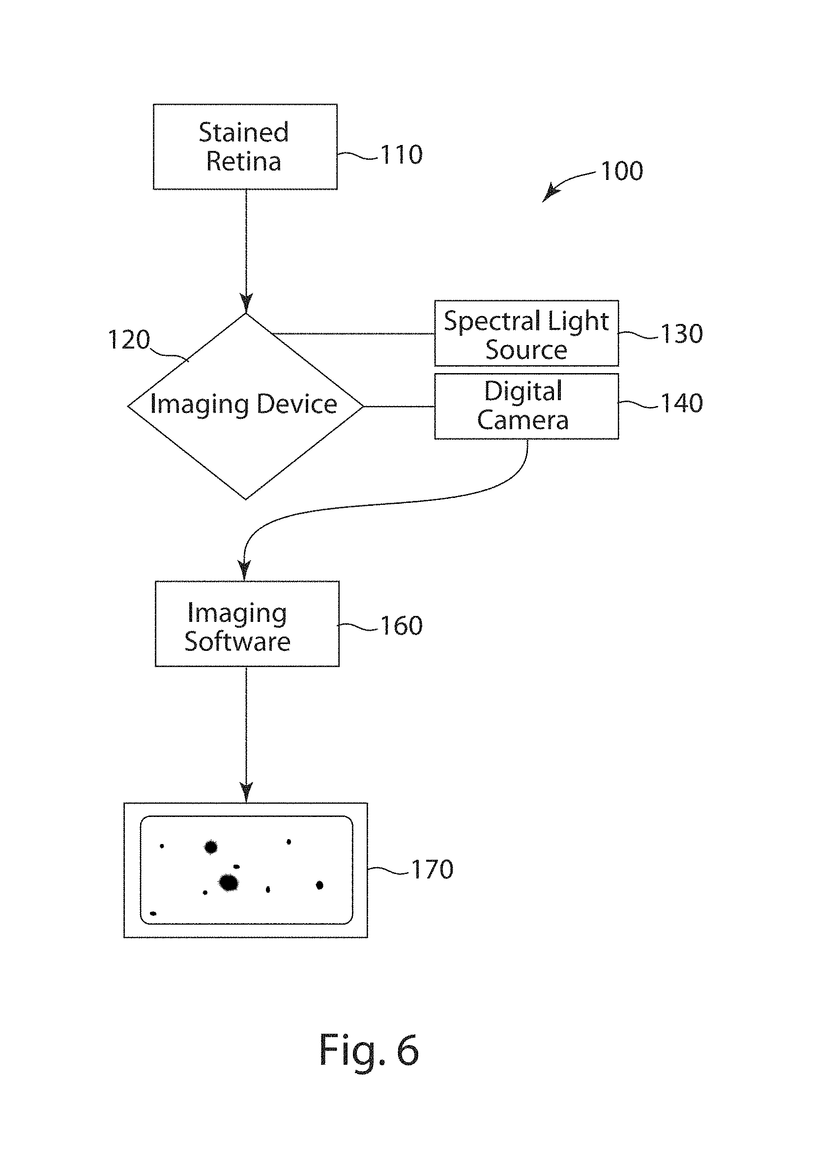
D00015
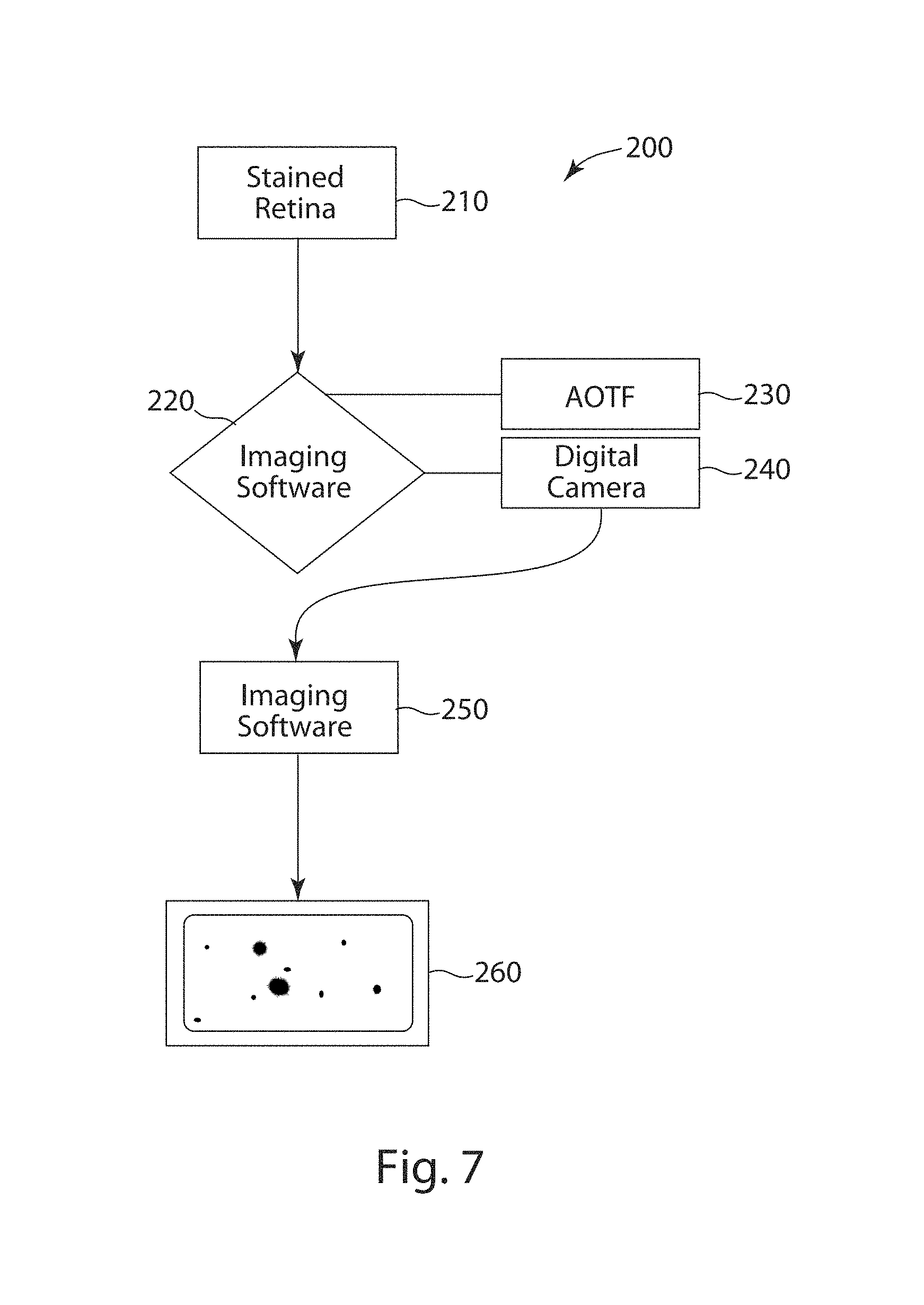
D00016
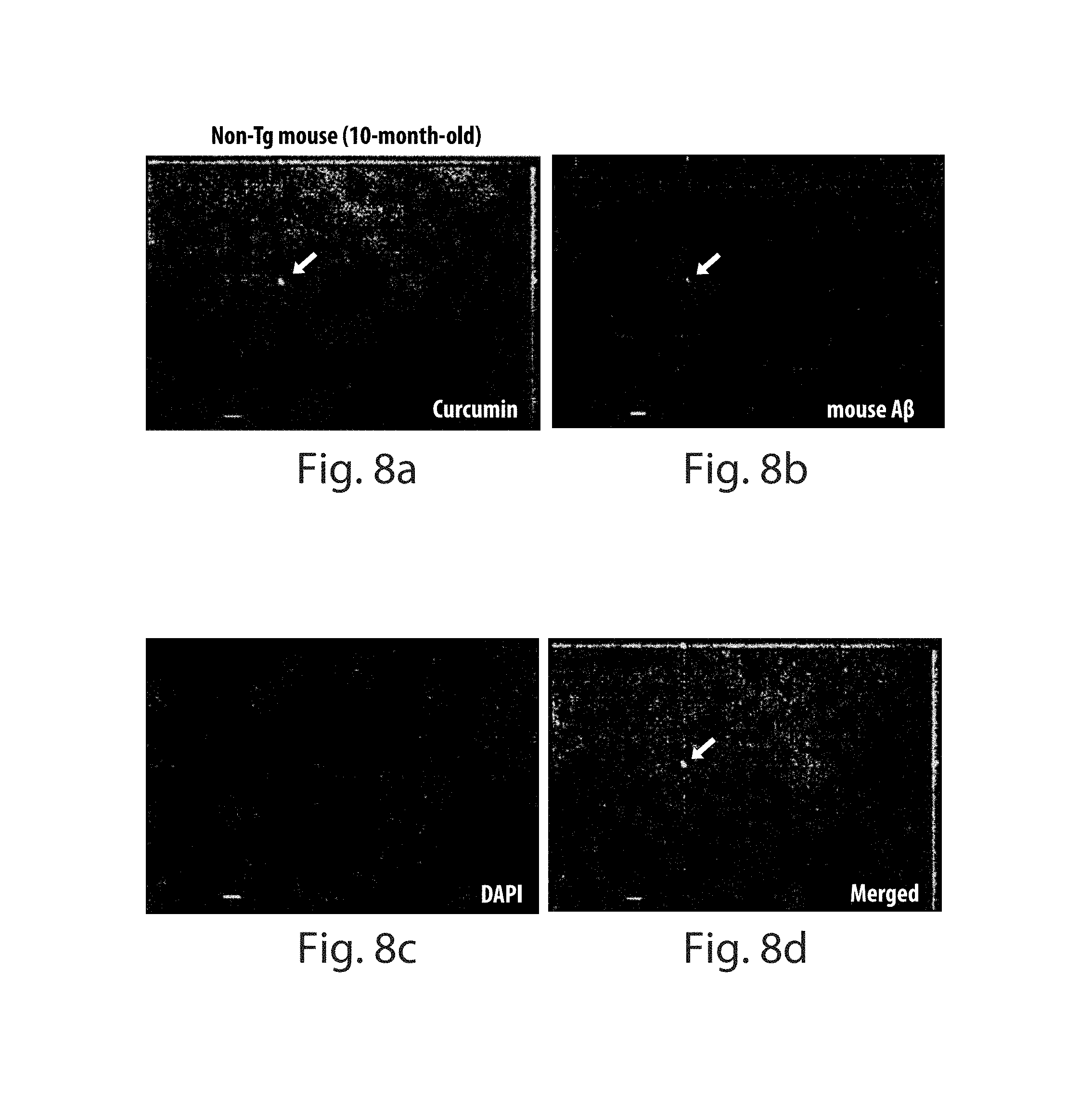
D00017

XML
uspto.report is an independent third-party trademark research tool that is not affiliated, endorsed, or sponsored by the United States Patent and Trademark Office (USPTO) or any other governmental organization. The information provided by uspto.report is based on publicly available data at the time of writing and is intended for informational purposes only.
While we strive to provide accurate and up-to-date information, we do not guarantee the accuracy, completeness, reliability, or suitability of the information displayed on this site. The use of this site is at your own risk. Any reliance you place on such information is therefore strictly at your own risk.
All official trademark data, including owner information, should be verified by visiting the official USPTO website at www.uspto.gov. This site is not intended to replace professional legal advice and should not be used as a substitute for consulting with a legal professional who is knowledgeable about trademark law.