Mutant 3-hydroxybutyrate dehydrogenase from alcaligenes faecalis as well as methods and uses involving the same
Kratzsch , et al. Dec
U.S. patent number 10,508,267 [Application Number 16/014,355] was granted by the patent office on 2019-12-17 for mutant 3-hydroxybutyrate dehydrogenase from alcaligenes faecalis as well as methods and uses involving the same. This patent grant is currently assigned to Roche Diabetes Care, Inc., Roche Diagnostics Operations, Inc.. The grantee listed for this patent is Roche Diabetes Care, Inc., Roche Diagnostics Operations, Inc.. Invention is credited to Daniela Beck, Mara Boenitz-Dulat, Stacy Hunt DuVall, Peter Kratzsch, Thomas Streidl.


| United States Patent | 10,508,267 |
| Kratzsch , et al. | December 17, 2019 |
| **Please see images for: ( Certificate of Correction ) ** |
Mutant 3-hydroxybutyrate dehydrogenase from alcaligenes faecalis as well as methods and uses involving the same
Abstract
The present disclosure relates to a mutant 3-hydroxybutyrate dehydrogenase (3-HBDH) with improved performance relative to the wild-type 3-HBDH, a nucleic acid encoding the mutant 3-HBDH, a cell having the mutant 3-HBDH or the nucleic acid, and/or a method of determining the amount or concentration of 3-hydroxybutyrate in a sample. Also disclosed is the use of the mutant 3-HBDH for determining the amount or concentration of 3-hydroxybutyrate in a sample, and a device for determining the amount or concentration of 3-hydroxybutyrate in a sample.
| Inventors: | Kratzsch; Peter (Penzberg, DE), Boenitz-Dulat; Mara (Tutzing, DE), Beck; Daniela (Penzberg, DE), Streidl; Thomas (Seeshaupt, DE), DuVall; Stacy Hunt (Indianapolis, IN) | ||||||||||
|---|---|---|---|---|---|---|---|---|---|---|---|
| Applicant: |
|
||||||||||
| Assignee: | Roche Diagnostics Operations,
Inc. (Indianapolis, IN) Roche Diabetes Care, Inc. (Indianapolis, IN) |
||||||||||
| Family ID: | 55808953 | ||||||||||
| Appl. No.: | 16/014,355 | ||||||||||
| Filed: | June 21, 2018 |
Prior Publication Data
| Document Identifier | Publication Date | |
|---|---|---|
| US 20180291354 A1 | Oct 11, 2018 | |
Related U.S. Patent Documents
| Application Number | Filing Date | Patent Number | Issue Date | ||
|---|---|---|---|---|---|
| PCT/EP2016/082257 | Dec 21, 2016 | ||||
| 62270133 | Dec 21, 2015 | ||||
Foreign Application Priority Data
| Apr 14, 2016 [EP] | 16165421 | |||
| Current U.S. Class: | 1/1 |
| Current CPC Class: | C12Q 1/54 (20130101); C12Y 101/0103 (20130101); C12Q 1/32 (20130101); C12N 9/0006 (20130101) |
| Current International Class: | C12N 9/04 (20060101); C12Q 1/54 (20060101); C12Q 1/32 (20060101) |
References Cited [Referenced By]
U.S. Patent Documents
| 4816224 | March 1989 | Vogel et al. |
| 5413690 | May 1995 | Kost et al. |
| 5762770 | June 1998 | Pritchard et al. |
| 5801006 | September 1998 | Kaufman |
| 5997817 | December 1999 | Crismore et al. |
| 6255093 | July 2001 | Schmuck et al. |
| 6541216 | April 2003 | Wilsey et al. |
| 9266109 | February 2016 | Howell et al. |
| 2011/0212503 | September 2011 | Yamaguchi et al. |
| 2014/0124384 | May 2014 | Gerber et al. |
| 19815685 | Oct 1999 | DE | |||
| 102007035109 | Jan 2009 | DE | |||
| 0262445 | Apr 1988 | EP | |||
| 8070856 | Mar 1996 | JP | |||
| 20150093557 | Aug 2015 | KR | |||
| 1998/033936 | Aug 1998 | WO | |||
| 2001/046457 | Jun 2001 | WO | |||
| 2001/094370 | Dec 2001 | WO | |||
| 2006/040172 | Apr 2006 | WO | |||
| 2007/012494 | Feb 2007 | WO | |||
| 2007/118647 | Oct 2007 | WO | |||
| 2009/060432 | May 2009 | WO | |||
| 2011/012270 | Feb 2011 | WO | |||
| 2014/068024 | May 2014 | WO | |||
| 2014/195363 | Dec 2014 | WO | |||
| 2017/137491 | Aug 2017 | WO | |||
Other References
|
Anderson, Bruce M., Analogs of Pyridine Nucleotide Coenzymes, The Pyridine Nucleotide Coenzymes, 1982, pp. 91-133, Ch. 4, Academic Press, Inc., New York. cited by applicant . Database EMBL, SubName: Full=3-hydroxybutyrate dehydrogenase, Retrived from UniPro EMBL A0A0N9NPT3, 2016, 1 p. cited by applicant . Database EMBL, SubName: Full=D-beta-hydroxybutyrate dehydrogenase, Retrieved from UniProt A0A0D6B253, 2015, 1 p. cited by applicant . Database UNIPROT, Castellaniella defragrans 65Phen D-beta-hydroxybutyrate dehydrogenase--sequence, Retrieved from EBI Accession No. UNIPROT:W8X517, 2014, 2 pp. cited by applicant . Database UNIPROT, Castellaniella defragrans 65Phen; Bacteria, Retrieved from EBI Accession No. UNIPROT:W8X517 CASDE, 2014, 1 p. cited by applicant . Database UNIPROT, SubName: Full=3-hydroxybutyrate dehydrogenase, Retrieved from EBI Accession No. UNIPROT A0A0L8EMR0, 2015, 1 p. cited by applicant . International Search Report dated Mar. 10, 2017, in Application No. PCT/EP2016/082257, 3 pp. cited by applicant . Lillis, B. et al., Investigation into immobilisation of lactate oxidase to improve stability, Sensors and Actuators B, 2000, pp. 109-114, vol. 68. cited by applicant . Oppenheimer, Norman J., Chemistry and Solution Conformation of the Pyridine Coenzymes, The Pyridine Nucleotide Coenzymes, 1982, pp. 51-89, Ch. 3, Academic Press, Inc., New York. cited by applicant . Petasch, Jan et al., The oxygen-independent metabolism of cyclic monoterpenes in Castellaniella defragrans 65Phen, BMC Microbiology, 2014, 13 pp., vol. 14, No. 164. cited by applicant . Yeon, Young Joo et al., Enzymatic reduction of levulinic acid by engineering the substrate specificity of 3-hydroxybutyrate dehydrogenase, Bioresource Technology, 2013, pp. 377-380, vol. 134. cited by applicant. |
Primary Examiner: Noakes; Suzanne M
Assistant Examiner: Lee; Jae W
Attorney, Agent or Firm: Dinsmore & Shohl LLP
Parent Case Text
CROSS-REFERENCE TO RELATED APPLICATIONS
This application is a continuation of International Application No. PCT/EP2016/082257 filed Dec. 21, 2016, which claims the benefit of U.S. Provisional Application No. 62/270,133 filed Dec. 21, 2015, and priority to European Application No. 16165421.5 filed Apr. 14, 2016, the disclosures of which are hereby incorporated by reference.
Claims
The invention claimed is:
1. A mutant 3-hydroxybutyrate dehydrogenase (3-HBDH) from Alcaligenes faecalis with improved performance relative to the wild-type 3-HBDH, wherein the mutant comprises an amino acid sequence that is at least 80% identical to the amino acid sequence of SEQ ID NO: 1 (3-HBDH from Alcaligenes faecalis; wild-type 3-HBDH) or the amino acid sequence of SEQ ID NO: 2 (extended core sequence of wild-type 3-HBDH) and wherein the mutant has at least three amino acid substitutions relative to the wild-type 3-HBDH, wherein the amino acid at the position corresponding to position 253 of SEQ ID NO: 1 is substituted with Ile (253Ile), Ala (253Ala) or Cys (253Cys), position 233 of SEQ ID NO: 1 is substituted with Ser (233Ser), Ile (233Ile), Ala (233Ala), Pro (233Pro), Arg (233Arg), Thr (233Thr), Lys (233Lys), or Cys (233Cys), and position 234 of SEQ ID NO: 1 is substituted with Thr (234Thr), wherein improved performance relative to the wild-type 3-HBDH is increased thermal stability relative to the wild-type 3-HBDH; and/or increased substrate affinity relative to the wild-type 3-HBDH; and/or increased cofactor affinity.
2. The mutant 3-HBDH of claim 1, wherein the amino acid at the position corresponding to position 253 of SEQ ID NO: 1 is substituted with Ile (253Ile).
3. The mutant 3-HBDH of claim 1, wherein the amino acid at the position corresponding to position 233 of SEQ ID NO: 1 is substituted with Lys (233Lys).
4. The mutant 3-HBDH of claim 1, wherein the mutant has at least one further amino acid substitution at one or more of the position(s) corresponding to position(s) 18, 19, 33, 38, 39, 62, 125, 143, 148, 170, 175, 187 and/or 216 of SEQ ID NO: 1.
5. The mutant 3-HBDH of claim 1, wherein the amino acid at the position corresponding to position 18 of SEQ ID NO: 1 is substituted with Arg (18Arg), Lys (18Lys), Glu (18Glu), or Pro (18Pro); position 19 of SEQ ID NO: 1 is substituted with Gly (19Gly); position 33 of SEQ ID NO: 1 is substituted with Ala (33Ala), Leu (33Leu), or Thr (33Thr); position 38 of SEQ ID NO: 1 is substituted with Lys (38 Lys); position 39 of SEQ ID NO: 1 is substituted with Gly (39Gly); position 62 of SEQ ID NO: 1 is substituted with Phe (62Phe), Met (62Met), Lys (62Lys), Arg (62Arg), Leu (62Leu), or Val (62Val), especially Val (62Val); position 125 of SEQ ID NO: 1 is substituted with Gly (125Gly); position 143 of SEQ ID NO: 1 is substituted with Val (143Val); position 148 of SEQ ID NO: 1 is substituted with Gly (148Gly); position 170 of SEQ ID NO: 1 is substituted with Gly (170Gly); position 175 of SEQ ID NO: 1 is substituted with Thr (175Thr), Val (175Val), or Ile (175Ile); position 187 of SEQ ID NO: 1 is substituted with Phe (187Phe); and/or position 216 of SEQ ID NO: 1 is substituted with Val (216Val).
6. The mutant 3-HBDH of claim 1, wherein the mutant 3-HBDH has at least the following mutations, corresponding to positions of SEQ ID NO:1 253Ile, 233Lys, 234Thr; 253Ile, 62Val, 233Lys, 234Thr; 253Ile, 62Val, 147Arg, 233Lys, 234Thr; 253Ile, 39Gly, 62Val, 143Val, 148Gly, 233Lys, 234Thr; or 253Ile, 62Val, 143Val, 148Gly, 233Lys, 234Thr, optionally in combination with 19Gly; 170Gly; 125Gly, 187Phe; 18Glu, 33Leu; 33Leu, 125Gly, 187Phe; 33Leu, 125Gly, 187Phe, 216Val; 18Glu, 187Phe; 33Leu, 125Gly, 175Val, 187Phe; 175Val, 216Val; 175Val, 187Phe, 216Val; 19Gly, 125Gly, 187Phe; 125Gly, 175Thr, 187Phe; 19Gly, 33Leu; 19Gly, 175Ile; 19Gly, 175Thr; 170Gly, 175Thr; 18Glu, 125Gly, 187Phe; 33Leu, 125Gly, 175Thr, 187Phe; 33Leu, 125Gly, 175Ile, 187Phe; 33Leu, 125Gly, 170Gly, 187Phe; 18Glu, 175Ile, 187Phe; 33Leu, 125Gly, 175Val, 187Phe; 33Leu, 125Gly, 175Val, 187Phe, 216Val; 33Leu, 125Gly, 175Thr, 187Phe; or 33Leu, 38Lys, 39Gly, 125Gly, 175Val, 187Phe.
7. The mutant 3-HBDH of claim 1, wherein the mutant 3-HBDH comprises an amino acid sequence that is at least 85% identical to the amino acid sequence of any of SEQ ID NO: 1 to 16.
8. The mutant 3-HBDH of claim 1, wherein the mutant 3-HBDH has an at least 2-fold increased thermal stability relative to the wild-type 3-HBDH and/or wherein the mutant 3-HBDH has an increased substrate and/or cofactor affinity relative to the wild-type 3-HBDH.
9. A method of determining the amount or concentration of 3-hydroxybutyrate in a sample, the method comprising a) contacting the sample with the mutant 3-HBDH of claim 1 under conditions conducive to the activity of the 3-HBDH; b) reacting 3-hydroxybutyrate with nicotinamide adenine dinucleotide (NAD) or a functionally active derivative thereof; and c) determining the change in the redox state of NAD or the derivative thereof; thereby determining the amount or concentration of 3-hydroxybutyrate in the sample.
10. The method of claim 9, a) wherein the determining of the change in the redox state of NAD or the derivate thereof includes the determination of the concentration of (i) NAD or the derivate thereof and/or (ii) NADH or the derivate thereof; and/or b) wherein the determining the change in the redox state of NAD or the derivative thereof is electrochemically or optically; and/or c) wherein the method further comprises determining the amount or concentration of acetoacetate and/or acetone; and/or d) wherein the method further comprises determining the amount or concentration of glucose; and/or e) wherein the functionally active derivative of NAD is carba-NAD; and/or f) wherein the mutant 3-HBDH is part of a sensor, a test strip, a test element, a test strip device or a liquid test; and/or g) wherein the sample is a body fluid, particularly a blood sample or a urine sample.
11. A device for determining the amount or concentration of 3-hydroxybutyrate in a sample comprising the mutant 3-HBDH of claim 1 and a further component required for said determining.
12. The device according to claim 11, wherein the device comprises a sensor or a test strip.
13. The device of claim 12, wherein the device further allows for determining the amount or concentration of glucose in the sample.
Description
SUMMARY OF THE INVENTION
The present invention relates to a mutant 3-hydroxybutyrate dehydrogenase (3-HBDH) with improved performance relative to the wild-type 3-HBDH, a nucleic acid encoding the mutant 3-HBDH, a cell comprising the mutant 3-HBDH or the nucleic acid, a method of determining the amount or concentration of 3-hydroxybutyrate in a sample, the use of the mutant 3-HBDH for determining the amount or concentration of 3-hydroxybutyrate in a sample, and a device for determining the amount or concentration of 3-hydroxybutyrate in a sample.
BACKGROUND OF THE INVENTION
Ketone bodies are produced in the liver, mainly from the oxidation of fatty acids, and are exported to peripheral tissues for use as an energy source. They are particularly important for the brain, which has no other substantial non-glucose-derived energy source. The two main ketone bodies are 3-hydroxybutyrate and acetoacetate. Biochemically, abnormalities of ketone body metabolism can be subdivided into three categories: ketosis, hypoketotic hypoglycemia, and abnormalities of the 3-hydroxybutyrate/acetoacetate ratio.
An abnormal elevation of the 3-hydroxybutyrate/acetoacetate ratio usually implies a non-oxidized state of the hepatocyte mitochondrial matrix resulting from hypoxia-ischemia or other causes.
The presence of ketosis normally implies that lipid energy metabolism has been activated and that the entire pathway of lipid degradation is intact. In rare cases, ketosis reflects an inability to utilize ketone bodies. Ketosis is normal during fasting, after prolonged exercise, and when a high-fat diet is consumed. During the neonatal period, infancy and pregnancy, times at which lipid energy metabolism is particularly active, ketosis develops readily.
Pathologic causes of ketosis include diabetes, ketotic hypoglycemia of childhood, corticosteroid or growth hormone deficiency, intoxication with alcohol or salicylates, and several inborn errors of metabolism.
The formation of ketone bodies is increased when lipolysis is increased e.g. in insulin deficiency (diabetes mellitus; in particular type I diabetics), when the glucagon concentration is increased and in a fasting state. In such cases the normal physiological concentration of less than 7 mg/dl can increase to more than 10-fold. Over the past years 3-hydroxybutyrate has proven to be an extremely reliable parameter for monitoring an insulin therapy.
The absence of ketosis in a patient with hypoglycemia is abnormal and suggests the diagnosis of either hyperinsulinism or an inborn error of fat energy metabolism.
Accordingly, ketone bodies are an interesting diagnostic target, particularly for diabetes. Therefore, there is an ongoing need for robust and sensitive test-systems for ketone bodies, especially 3-hydroxybutyrate. The ratio of 3-hydroxybutyrate to acetone or acetoacetic acid is normally 3:1. In keto-acidoses the ratio increases to 6:1 to 12:1. A suitable 3-HBDH should enable the development of a quantitative test for 3-hydroxybutyrate.
SUMMARY OF THE FIGURE
FIG. 1 shows the amino acid sequence and domains of the wild-type 3-HBDH from Alcaligenes faecalis
DETAILED DESCRIPTION OF THE INVENTION
Surprisingly, it has been found that a variety of mutants of the 3-hydroxybutyrate dehydrogenase from Alcaligenes faecalis show improved performance, particularly increased thermal stability and/or affinity for substrate and/or cofactor. As shown in the Examples, there are many sites at the wild-type enzyme which allow for mutations, which increase thermal stability and/or affinity for substrate and/or cofactor of the mutant relative to the wild-type enzyme.
One site which is presently regarded as particularly suitable in increasing performance is the amino acid at the position corresponding to position 253 of SEQ ID NO: 1 (see below). Particularly, the amino acid may be substituted with Ile (253Ile), Ala (253Ala) or Cys (253Cys) to increase affinity for substrate and/or cofactor (for 3-hydroxybutyrate and/or carba-NAD) and optionally thermal stability (see Tables 1A, 1B and 4).
Moreover, mutations at one or more of the position(s) corresponding to position(s) 18, 19, 33, 38, 39, 62, 125, 143, 148, 170, 175, 187, 216, 233 and/or 234 of SEQ ID NO: 1, particularly at least position(s) 62, 143, 148, 233 and/or 234 of SEQ ID NO: 1 were found to be suitable as well (see Tables 1 B, 2A-C and 4). The performance of the mutant 3-HBDH could be further increased by combining mutations at the above sites (see Tables 2A-D and 3). Particularly suitable examples of those mutants include those with the mutations (for definitions of abbreviations see below) 253Ile, 233Lys, 234Thr; OR 253Ile, 62Val, 233Lys, 234Thr; OR 253Ile, 62Val, 147Arg, 233Lys, 234Thr; OR 253Ile, 39Gly, 62Val, 143Val, 148Gly, 233Lys, 234Thr; OR 253Ile, 62Val, 143Val, 148Gly, 233Lys, 234Thr; OR 62Val, 111Leu, 140Met, 148Gly
(see Tables 2A-D).
Surprisingly, the mutant having mutations 253Ile, 233Lys and 234Thr referred to as AFDH3 showed a dramatic increase in thermostability, if compared to a mutant with mutation 253Ile, only (referred to as AFDH2) (see table 2D), wherein AFDH2 had already an increase in thermostability vis a vis the wild-type (referred to AFDH1). Additional mutations, namely 18Arg, 18Lys, 18Glu, 18Pro, 19Gly, 33Ala, 33Leu, 33Thr, 38Lys, 39Gly, 62Phe, 62Met, 62Lys, 62Arg, 62Leu, 62Val, 125Gly, 143Val, 147Arg, 148Gly, 170Gly, 175Thr, 175Val, 175Ile, 187Phe and 216Val have been proven to further improve the performance of the mutants relative to the wild-type 3-HBDH or AFDH2 (see tables 2D, 3 and 4).
Accordingly, in a first aspect, the present invention relates to a mutant 3-hydroxybutyrate dehydrogenase (3-HBDH) from Alcaligenes faecalis with improved performance relative to the wild-type 3-HBDH.
3-Hydroxybutyrate dehydrogenase (3-HBDH) (EC 1.1.1.30) belongs to the family of oxidoreductases, to be specific is an enzyme that catalyzes the stereospecific oxidation of 3-hydroxybutanoate/3-hydroxybutyrate/(R)-3-hydroxybutyrate/3-hydroxybutyr- ic acid (3-HB) to acetoacetate with NAD or derivatives as cofactor: (R)-3-hydroxybutyrate+NAD+acetoacetate+NADH+H+
The systematic name of this enzyme class is (R)-3-hydroxybutanoate:NAD+ oxidoreductase. Other names in common use include NAD+-beta-hydroxybutyrate dehydrogenase, hydroxybutyrate oxidoreductase, beta-hydroxybutyrate dehydrogenase, D-beta-hydroxybutyrate dehydrogenase, D-3-hydroxybutyrate dehydrogenase, D-(-)-3-hydroxybutyrate dehydrogenase, beta-hydroxybutyric acid dehydrogenase, 3-D-hydroxybutyrate dehydrogenase, and beta-hydroxybutyric dehydrogenase. This enzyme participates in synthesis and degradation of ketone bodies and butanoate metabolism and it may be used in order to determine ketone bodies in samples, such as a blood sample.
The term "wild-type 3-HBDH" relates to a 3-HBDH as it typically occurs in nature. A wild-type 3-HBDH from the Gram-negative bacterium Alcaligenes faecalis was cloned in E. coli and further described in 1996 (Jpn. Kokai Tokkyo Koho (1996), JP 08070856 A 19960319). However, the enyzme is characterized by a rather low stability (resulting in short storage life), which is disadvantageous in biotechnical, biomedical and diagnostic applications. Particularly, the short storage life has a negative impact on applicability of the enzyme, if the use of a previously prepared enzyme is intended. This includes particularly commercial products such as enzyme preparations, kits, test strips etc, which are usually prepared in large scale, stored and then marketed e.g. in smaller batches. Additionally, it is evidently desirable to increase the enzyme's affinity for the substrates and/or cofactors. Increased affinity and/or stability will increase the performance of the enzyme in biotechnical applications and devices. The commonly accepted amino acid sequence of the typically occurring wild-type 3-HBDH of Alcaligenes faecalis is given below as SEQ ID NO:1.
The term "mutant 3-hydroxybutyrate dehydrogenase (3-HBDH) from Alcaligenes faecalis" relates to a 3-HBDH enzyme whose amino acid sequence differs from the wild-type 3-HBDH from Alcaligenes faecalis, particularly the amino acid sequence of SEQ ID NO:1, by at least one mutation, i.e. one or more amino acid substitutions, additions, deletions or combinations thereof, particularly by at least one substitution.
The term "with improved performance relative to the wild-type 3-HBDH" means that the performance of the mutant enzyme is improved relative to the wild-type 3-HBDH from Alcaligenes faecalis. The performance is improved, if the mutant enzyme has a higher performance, e.g. higher activity in converting 3-HB into acetoacetate, at any condition (e.g. after storage, at a particular pH, with a specific buffer, at a chosen temperature, with a particular cofactor (e.g. carba-NAD), at a specific substrate or cofactor concentration etc). Higher performance may be especially increased stability (such as thermal stability) and/or affinity for one or more substrates/cofactors (e.g. carba-NAD and/or 3-HB). Preferably, the performance is improved, if the mutant enzyme, e.g., has a higher relative remaining activity in converting 3-HB into acetoacetate, upon being stressed with given conditions (e.g. storage, buffer conditions, temperature, concentration of cofactor or substrate) compared to the activity without being stressed under these conditions and/or if the mutant enzyme, e.g., has a higher relative activity (i.e. activity at a subsaturation concentration/activity at a saturation concentration)
The sequence of the wild-type 3-HBDH from Alcaligenes faecalis is shown as SEQ ID NO: 1:
TABLE-US-00001 (3-HBDH from Alcaligenes faecalis) SEQ ID NO: 1 MLKGKKAVVT GSTSGIGLAM ATELAKAGAD VVINGFGQPE DIERERSTLE SKFGVKAYYL 60 NADLSDAQAT RDFIAKAAEA LGGLDILVNN AGIQHTAPIE EFPVDKWNAI IALNLSAVFH 120 GTAAALPIMQ KQGWGRIINI ASAHGLVASV NKSAYVAAKH GVVGLTKVTA LENAGKGITC 180 NAICPGWVRT PLVEKQIEAI SQQKGIDIEA AARELLAEKQ PSLQFVTPEQ LGGAAVFLSS 240 AAADQMTGTT LSLDGGWTAR 260
FIG. 1 shows the various domains (catalytic center and binding sites) of the wild-type enzyme. In accordance with these domains, the core sequence of the enzyme can be determined. As detailed above, 3-HBDH mutants with mutations at the position corresponding to position 253 of SEQ ID NO: 1 were found to be particularly useful. Accordingly, an extended core sequence of 3-HBDH from Alcaligenes faecalis encompassing this position was established and is shown as SEQ ID NO: 2:
TABLE-US-00002 (extended core sequence of 3-HBDH from Alcaligenes faecalis) SEQ ID NO: 2 ADLSDAQATR DFIAKAAEAL GGLDILVNNA GIQHTAPIEE FPVDKWNAII ALNLSAVFHG 60 TAAALPIMQK QGWGRIINIA SAHGLVASVN KSAYVAAKHG VVGLTKVTAL ENAGKGITCN 120 AICPGWVRTP LVEKQIEAIS QQKGIDIEAA ARELLAEKQP SLQFVTPEQL GGAAVFLSSA 180 AADQMTGTTL SL 192
Accordingly, a preferred example of a naturally occurring wild-type 3-HBDH is given above, in FIG. 1 and as SEQ ID NO: 1.
If one or more mutations are introduced into the wild-type sequence, especially the sequence of SEQ ID NO: 1, a mutant 3-HBDH is obtained. With respect to the mutant 3-HBDH of the present invention it is noted that the mutant is functionally active and shows improved performance relative to the wild-type 3-HBDH. This means that the mutant has maintained its enzymatic function of conversion of 3-HB into acetoacetate. Additionally, preferably at least one of the stability and the enzyme's affinity for the substrate and/or cofactors is increased. The mutant differs from the wild-type 3-HBDH by one or more amino acid substitutions, additions and/or deletions, especially at least one or more substitutions.
The sequence of the mutant 3-HBDH according to the present invention comprises (optionally in addition to the substitutions specified herein) one or more amino acid substitution(s), deletion(s) or addition(s), especially one or more substitutions, particularly one or more conservative amino acid substitutions.
In one embodiment of the present invention, the mutant 3-HBDH according to the present invention may comprise one or more amino acid substitution(s), particularly a limited number of substitutions (e.g. up to 50, 40, 30, 20 especially 10 amino acid substitutions). Suitable substitutions are given in the Examples, particularly in the Tables. All the substitutions identified in the Examples as suitable in order to increase performance of 3-HBDH may be used in the invention either alone or in combination with each other and/or with further substitutions, e.g. conservative substitutions. "Conservative amino acid substitution" refers to a substitution of a residue with a different residue having a similar side chain, and thus typically involves substitution of the amino acid in the polypeptide with amino acids within the same or similar defined class of amino acids. By way of example and not limitation, an amino acid with an aliphatic side chain may be substituted with another aliphatic amino acid, e.g., alanine, valine, leucine, and isoleucine; an amino acid with hydroxyl side chain is substituted with another amino acid with a hydroxyl side chain, e.g., serine and threonine; an amino acid having aromatic side chains is substituted with another amino acid having an aromatic side chain, e.g., phenylalanine, tyrosine, tryptophan, and histidine; an amino acid with a basic side chain is substituted with another amino acid with a basic side chain, e.g., lysine and arginine; an amino acid with an acidic side chain is substituted with another amino acid with an acidic side chain, e.g., aspartic acid or glutamic acid; and a hydrophobic or hydrophilic amino acid is replaced with another hydrophobic or hydrophilic amino acid, respectively. Examples of conservative amino acid substitutions include those listed below:
TABLE-US-00003 Original Residue Conservative Substitutions Ala, Leu, Val, Ile Other aliphatic (Ala, Leu, Val, Ile) Other non-polar (Ala, Leu, Val, Ile, Gly, Met) Gly, Met Other non-polar (Ala, Leu, Val, Ile, Gly, Met) Asp, Glu Other acidic (Asp, Glu) Lys, Arg Other basic (Lys, Arg) Asn, Gln, Ser, Thr Other polar (Asn, Gln, Ser, Thr) His, Tyr, Trp, Phe Other aromatic (His, Tyr, Trp, Phe) Cys, Pro None
In one embodiment of the present invention, the mutant 3-HBDH according to the present invention may comprise one or more amino acid addition(s), particularly small (e.g. up to 50, 40, 30, 20 especially 10 amino acids) internal amino acid additions. Alternatively, additions may be achieved by combining the mutant 3-HBDH into a fusion protein comprising the mutant 3-HBDH of the present invention.
Fusion proteins are proteins created by joining of two or more originally separate proteins or peptides. This procedure results in a polypeptide with functional properties derived from each of the original proteins. Accordingly, depending on the intended use of the 3-HBDH it may be combined with a further peptide or protein into a fusion protein. The proteins may be fused via a linker or spacer, which increases the likelihood that that the proteins fold independently and behave as expected. Especially in the case where the linkers enable protein purification, linkers in protein or peptide fusions are sometimes engineered with cleavage sites for proteases or chemical agents that enable the liberation of the two separate proteins. Di- or multimeric fusion proteins can be manufactured through genetic engineering by fusion to the original proteins of peptide domains that induce artificial protein di- or multimerization (e.g., streptavidin or leucine zippers). Fusion proteins can also be manufactured with toxins or antibodies attached to them. Other fusions include the addition the addition of signal sequences, such a lipidation signal, sequence, a secretion signal sequence, a glycosylation signal sequence, a translocation signal peptide etc.
Preferably, the fusion protein of the present invention comprises a tag. Tags are attached to proteins for various purposes, e.g. in order to ease purification, to assist in the proper folding in proteins, to prevent precipitation of the protein, to alter chromatographic properties, to modify the protein or to mark or label the protein. Examples of tags include Arg-tag, the His-tag, the Strep-tag, the Flag-tag, the T7-tag, the V5-peptide-tag, the GST-tag and the c-Myc-tag.
In one embodiment of the present invention, the mutant 3-HBDH according to the present invention may comprise one or more amino acid deletion(s), particularly N- and/or C-terminal deletions, especially in the portions of SEQ ID NO:1 absent in SEQ ID NO: 2. The deletions may be small (e.g. up to 50, 40, 30, 20, especially 10 amino acids).
In another embodiment, the sequence of the mutant 3-HBDH according to the present invention may comprise, preferably in addition to the substitutions specified herein, a combination of one or more deletion(s), substitution(s) or addition(s) as defined above.
In a preferred embodiment of the present invention, the mutant 3-HBDH comprises an amino acid sequence that is at least 80% identical to the amino acid sequence of SEQ ID NO: 1 (3-HBDH from Alcaligenes faecalis; wild-type 3-HBDH) or the amino acid sequence of SEQ ID NO: 2 (extended core sequence of wild-type 3-HBDH).
The term "at least 80% identical" or "at least 80% sequence identity" as used herein means that the sequence of the mutant 3-HBDH according to the present invention has an amino acid sequence characterized in that, within a stretch of 100 amino acids, at least 80 amino acids residues are identical to the sequence of the corresponding wild-type sequence. Sequence identities of other percentages are defined accordingly.
Sequence identity according to the present invention can, e.g., be determined by methods of sequence alignment in form of sequence comparison. Methods of sequence alignment are well known in the art and include various programs and alignment algorithms which have been described in, e.g., Pearson and Lipman (1988). Moreover, the NCBI Basic Local Alignment Search Tool (BLAST) is available from several sources, including the National Center for Biotechnology Information (NCBI, Bethesda, Md.) and on the internet, for use in connection with the sequence analysis programs blastp, blastn, blastx, tblastn and tblastx. Percentage of identity of mutants according to the present invention relative to the amino acid sequence of e.g. SEQ ID NO: 1 is typically characterized using the NCBI Blast blastp with standard settings. Alternatively, sequence identity may be determined using the software GENEious with standard settings. Alignment results can be, e.g., derived from the Software Geneious (version R8), using the global alignment protocol with free end gaps as alignment type, and Blosum62 as a cost matrix.
As detailed above, the mutant 3-HBDH of the present invention in one embodiment comprises an amino acid sequence that is at least 80% identical to the amino acid sequence of SEQ ID NO: 1 or 2. In a preferred embodiment, the mutant 3-HBDH comprises or consists of an amino acid sequence which is at least 85%, 90%, 95%, 96%, 97%, 98%, or 99% identical to any of the amino acid sequences of SEQ ID NO: 1 or 2. Sequence identity may be determined as described above.
In another preferred embodiment the mutant of the present invention has at least one amino acid substitution relative to the wild-type 3-HBDH, wherein the amino acid at the position corresponding to position 253 of SEQ ID NO: 1 is substituted with Ile (253Ile), Ala (253Ala) or Cys (253Cys), particularly Ile (253Ile). It has been shown in the Examples that substitution of the amino acid at the position corresponding to position 253 of SEQ ID NO: 1 increases stability and/or affinity for the substrates and/or cofactors (see e.g. Tables 1A, 1B and 4).
Additionally, it could be shown that the substitution at the position corresponding to the position 253 of SEQ ID NO: 1 can be combined with a variety of other substitutions in order to further optimize the enzyme. Substitution of Leu at position 253 with Ile (253Ile) is particularly suitable in order to increase thermal stability as well as affinity for the substrate, as shown in Tables 1A-C and Tables 2 to 4 for single and multiple mutations relative to the wild-type, respectively. It could be proven that performance is improved after pre-incubations at various temperatures and at different conditions (buffers, pH). The positive effect of this substitution is particularly surprising as the amino acid in the wild-type (leucine) is substituted with a quite similar amino acid (isoleucine). Both amino acids are constitutional isomers. Both are regarded as non-polar and hydrophobic and have the same molecular weight.
The substitution of leucine at a position corresponding to position 253 of SEQ ID NO:1 with the non-polar amino acid alanine or the non-charged, non-acidic non-polar, hydrophilic amino acid cysteine increased significantly the affinities for substrate and cofactor (see Table 1B). Due to the chemical differences between the introduced amino acids Ala and Cys, also this result is unexpected and surprising.
A highly preferred embodiment of the present invention relates to a mutant 3-hydroxybutyrate dehydrogenase (3-HBDH) from Alcaligenes faecalis with improved performance relative to the wild-type 3-HBDH, wherein the mutant comprises an amino acid sequence that is at least 80% identical to the amino acid sequence of SEQ ID NO: 1 (3-HBDH from Alcaligenes faecalis; wild-type 3-HBDH) or the amino acid sequence of SEQ ID NO: 2 (extended core sequence of wild-type 3-HBDH) and wherein the mutant has at least three amino acid substitutions relative to the wild-type 3-HBDH, wherein the amino acid at the position corresponding to position 253 of SEQ ID NO: 1 is substituted with Ile (253Ile), Ala (253Ala) or Cys (253Cys), position 233 of SEQ ID NO: 1 is substituted with Ser (233Ser), Ile (233Ile), Ala (233Ala), Pro (233Pro), Arg (233Arg), Thr (233Thr), Lys (233Lys), or Cys (233Cys), and position 234 of SEQ ID NO: 1 is substituted with Thr (234Thr).
More preferably, the amino acid at the position corresponding to position 253 of SEQ ID NO: 1 is substituted with Ile (253Ile) and/or the amino acid at the position corresponding to position 233 of SEQ ID NO: 1 is substituted with Lys (233Lys) in this highly preferred mutant and optionally the mutant has at least one further amino acid substitution at one or more of the position(s) corresponding to position(s) 18, 19, 33, 38, 39, 62, 125, 143, 148, 170, 175, 187 and/or 216 of SEQ ID NO: 1, particularly at least position(s) 62, 143 and/or 148 of SEQ ID NO: 1.
As already detailed above, it could be shown that amino acids at one or more further positions of SEQ ID NO: 1 may be substituted alternatively or additionally, in order to improve the performance of the enzyme. Accordingly, in a further embodiment of the present invention, the mutant of the present invention has at least one amino acid substitution at one or more of the position(s) corresponding to position(s) 18, 19, 33, 38, 39, 62, 125, 143, 148, 170, 175, 187, 216, 233 and/or 234 of SEQ ID NO: 1, particularly at least position(s) 62, 143, 148, 233 and/or 234 of SEQ ID NO: 1, especially at least position(s) 62, 143, 148, 233 and 234 of SEQ ID NO: 1. Substitutions at these positions may be combined with each other as well as with a substitution at position 253, especially 253Ile, 253Ala, 253Cys, particularly 253Ile (see also Examples).
Particularly suitable examples of positions for substitutions as well as combinations thereof for mutant 3-HBDH of the present invention are given in the following: position 253 of SEQ ID NO: 1 is substituted with Ile (253Ile), Ala (253Ala) or Cys (253Cys), especially Ile (253Ile); position 18 of SEQ ID NO: 1 is substituted with Arg (18Arg), Lys (18Lys), Glu (18Glu), or Pro (18Pro); position 19 of SEQ ID NO: 1 is substituted with Gly (19Gly); position 33 of SEQ ID NO: 1 is substituted with Ala (33Ala), Leu (33Leu), or Thr (33Thr); position 38 of SEQ ID NO: 1 is substituted with Lys (38 Lys); position 39 of SEQ ID NO: 1 is substituted with Gly (39Gly); position 62 of SEQ ID NO: 1 is substituted with Phe (62Phe), Met (62Met), Lys (62Lys), Arg (62Arg), Leu (62Leu), or Val (62Val), especially Val (62Val); position 125 of SEQ ID NO: 1 is substituted with Gly (125Gly); position 143 of SEQ ID NO: 1 is substituted with Val (143Val); position 148 of SEQ ID NO: 1 is substituted with Gly (148Gly); position 170 of SEQ ID NO: 1 is substituted with Gly (170Gly); position 175 of SEQ ID NO: 1 is substituted with Thr (175Thr), Val (175Val), or Ile (175Ile); position 187 of SEQ ID NO: 1 is substituted with Phe (187Phe); position 216 of SEQ ID NO: 1 is substituted with Val (216Val); position 233 of SEQ ID NO: 1 is substituted with Ser (233Ser), Ile (233Ile), Ala (233Ala), Pro (233Pro), Arg (233Arg), Thr (233Thr), Lys (233Lys), or Cys (233Cys), especially Lys (233Lys); and/or position 234 of SEQ ID NO: 1 is substituted with Thr (234Thr).
In a particularly preferred embodiment of the present invention, the mutant 3-HBDH has at least or only the following mutations: 253Ile; 253Ile, 233Lys, 234Thr; 253Ile, 62Val, 233Lys, 234Thr; 253Ile, 62Val, 147Arg, 233Lys, 234Thr; 253Ile, 39Gly, 62Val, 143Val, 148Gly, 233Lys, 234Thr; 253Ile, 62Val, 143Val, 148Gly, 233Lys, 234Thr; or 62Val, 111Leu, 140Met, 148Gly,
particularly wherein the mutant 3-HBDH has at least or only the mutations 253Ile, 62Val, 143Val, 143Val, 148Gly, 233Lys, 234Thr, optionally in combination with 19Gly; 170Gly; 125Gly, 187Phe; 18Glu, 33Leu; 33Leu, 125Gly, 187Phe; 33Leu, 125Gly, 187Phe, 216Val; 18Glu, 187Phe; 33Leu, 125Gly, 175Val, 187Phe; 175Val, 216Val; 175Val, 187Phe, 216Val; 19Gly, 125Gly, 187Phe; 125Gly, 175Thr, 187Phe; 19Gly, 33Leu; 19Gly, 175Ile; 19Gly, 175Thr; 170Gly, 175Thr; 18Glu, 125Gly, 187Phe; 33Leu, 125Gly, 175Thr, 187Phe; 33Leu, 125Gly, 17511e, 187Phe; 33Leu, 125Gly, 170Gly, 187Phe; 18Glu, 17511e, 187Phe; 33Leu, 125Gly, 175Val, 187Phe; 33Leu, 125Gly, 175Val, 187Phe, 216Val; 33Leu, 125Gly, 175Thr, 187Phe; or 33Leu, 38Lys, 39Gly, 125Gly, 175Val, 187Phe.
In a preferred embodiment of the present invention, the mutant 3-HBDH comprises or consists of an amino acid sequence that is at least 85%, 90%, 95%, 96%, 97%, 98%, or 99% identical to the amino acid sequence of any of SEQ ID NO: 1 to 16, particularly wherein the mutant 3-HBDH comprises or consists of the sequence selected from the group consisting of SEQ ID NOs: 3 to 16.
As detailed above, the sequences of SEQ ID NO: 1 and 2 relate to the wild-type 3-HBDH and a fragment thereof, respectively. The sequences of SEQ ID NO: 3 to 16 are the sequences of highly preferred mutants. The sequences of the mutants and the substitutions effected are shown in the section "Sequences". Sequence identity may be determined as described above.
In a preferred embodiment of the present invention, improved performance relative to the wild-type 3-HBDH is increased stability, especially thermal stability, relative to the wild-type 3-HBDH; and/or increased substrate affinity, especially for 3-hydroxybutyrate, relative to the wild-type 3-HBDH; and/or increased cofactor affinity, especially for NAD or a derivative thereof, particularly wherein the derivate is carba-NAD, relative to the wild-type 3-HBDH.
The term "increased stability, especially thermal stability, relative to the wild-type 3-HBDH" means that the mutant 3-HBDH is less prone to loss of (enzyme) activity at a certain condition, especially at elevated temperatures. Stabilization of enzymes including avoidance of denaturation mechanisms in order to realize their full potential as catalysts is an important goal in biotechnology. Enzyme stabilization has notable importance due to increasing number of enzyme applications. The increase in stability allows for sustained usability (e.g. longer storage, usability for a longer time etc). Increased stability of the mutant relative to the wild-type can be determined by comparing the remaining activity of both enzymes (wild-type and mutant), e.g. after storage or exposure to a particular condition (e.g. elevated temperature, drying, buffer, or salt) (absolute remaining activity). Alternatively, the stability is improved compared to the wild-type, if the mutant, e.g., has a higher relative remaining activity in converting 3-HB into acetoacetate. Relative remaining activity may be determined by comparing the remaining or residual acidity after storage at given conditions (e.g. storage time, temperature) to the initial activity before storage.
The term enzyme activity and its determination are well-known to the person skilled in the art. Enzyme activity is generally defined as conversion of amount of substrate per time. The SI unit for enzyme activity is katal (1 katal=1 mol s-1). A more practical and commonly used value is enzyme unit (U)=1 .mu.mol min-1.1 U corresponds to 16.67 nanokatals and is defined as the amount of the enzyme that catalyzes the conversion of 1 micro mole of substrate per minute. The specific activity of an enzyme is the activity of an enzyme per milligram of total protein (expressed in .mu.mol min-1 mg-1).
The enzyme activity may be determined in an assay measuring either the consumption of substrate or cofactor or the formation of product over time. A large number of different methods of measuring the concentrations of substrates and products exist and many enzymes can be assayed in several different ways as known to the person skilled in the art. In the present invention, the 3-HBDH in question is, e.g., incubated with 3-hydroxybutyrate cofactor (e.g. NAD or derivative thereof such as carba-NAD) and the conversion of the cofactor (NAD to NADH or carba-NAD to carba-NADH) is monitored. Monitoring can, e.g., be done by measuring light absorbance at 340 nm. The obtained change of absorption per minute (dE/min) represents the enzymatic activity. For details see Example 2.
In a preferred embodiment of the present invention, increased stability of the mutant 3-HBDH relative to the respective 3-HBDH without mutation may be expressed as increase in half-life of the mutant (t1/2(mutant)) relative to the half-life of to the respective wild-type 3HBDH (t1/2(wild-type)). The half-life (t1/2) of the enzyme indicates the amount of time in which 50% of the original activity (activity at t=0) is lost and after which the remaining activity amounts to 50%. Accordingly, an increased stability results in an increased half-life of the mutant relative to the respective wild-type. Increase in stability may be determined as t1/2(mutant)/t1/2(wild-type). Percental increase in half-life may be determined as [t1/2(mutant)/t1/2(wild-type)-1]*100.
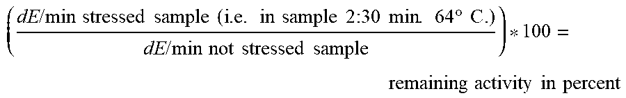
In a highly preferred embodiment of the present invention, increased stability of the mutant 3-HBDH relative to the respective 3-HBDH without mutation may be determined and expressed as remaining activity after a stress incubation (e.g. 30 min at e.g. 64.degree. C. or any other condition given in the Examples) in relation to the initial activity before stress incubation/storage (see Examples). For this, the enzymatic reaction may be monitored (e.g. at room temperature at 340 nm for 5 minutes) and the change in absorption per time (e.g. dE/min) may be calculated for each sample. The values obtained for stressed samples may be compared to the respective un-stressed sample (value set to 100% activity) and calculated in percent activity ((dE/min (stressed sample)/dE/min (unstressed sample)*100). Accordingly, a mutant's value higher than the value obtained with wild-type enzyme represents an improvement in thermal stability. The stability is increased, if [% remaining activity of the mutant]-[% remaining activity of the wild-type]>0. Alternatively, the remaining activity of the mutant may be also expressed as activity in percent and may be calculated as follows: [% remaining activity of the mutant]/[% remaining activity of the wild-type]*100%. The stability of the mutant relative to the wild-type is increased if the resulting value is >100%. A particular suitable test for determining stability is described in detail in Example 2. In accordance with this, the stability may be expressed as remaining activity and calculated as [dE/min (stressed sample)]/[dE/min (not stressed sample)]*100. Further details are given in Example 2. A value obtained with a mutant higher than the value obtained with wild-type enzyme represents an increased stability of the mutant.
Preferably, stability is increased by at least 10%, 20%, 30% or 40%, preferably at least 50%, 75% or 100%, still more preferably at least 125%, 150%, 175% or 200%, especially 250% and most preferably 300%.
Moreover, there are commercial advantages in carrying out enzymatic reactions at higher temperatures. Accordingly, thermal stability of the mutant is preferably increased. This means that resistance of the mutant relative to the wild-type to increased temperatures is higher. Increase in thermal stability may be particularly determined as shown in the Examples, e.g. in Example 3 (Tables 2A-D). In general, the mutant and the wild-type enzyme may be preincubated at an increased temperature (e.g. above 50.degree. C. such as 64.degree. C. or 68.degree. C. or 75.degree. C. etc.) for a defined time (such as 10 min or 30 min), after which the remaining activity in converting 3-HB into acetoacetate of the mutant is compared to the remaining activity of the wild-type enzyme. If the remaining activity of the mutant is higher than that of the wild-type, the mutant has an increased thermal stability.
A suitable method for the determination of increased thermal stability is detailed in the Examples. Exemplary conditions for stress conditions may be preincubation at 60-90.degree. C. (e.g., 64.degree. C., or 68.degree. C. or 75.degree. C.) for 30 min and testing afterwards with 62.22 mM 3-hydroxybutyrate; 4.15 mM cNAD; 0.1% Triton X-100; 200 mM Hepes pH 9,0 or 150 mM 3-hydroxybutyrate; 5 mM cNAD; 0.1% Triton X-100; 70 mM Mops pH 7.5
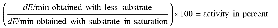
The term "increased substrate affinity, especially for 3-hydroxybutyrate, relative to the wild-type 3-HBDH" means that the affinity of the mutant for the substrate 3-hydroxybutyric acid/3-hydroxybutyrate/3-hydroxybutanoate (3-HB) which is converted into acetoacetate is increased. For the determination, the enzymatic reaction may be monitored (e.g. at room temperature at 340 nm for 5 minutes) and the dE/min may be calculated for each sample. The affinity of the mutant is increased compared to the wild-type, if the mutant, e.g., has a higher absolute or relative affinity for the substrate, particularly 3-HB. Affinity of the mutant compared to the wild-type can be determined by comparing the absolute affinities of both enzymes (wild-type and mutant) (absolute comparison) and may be calculated in percent activity ((dE/min (mutant)/dE (wild-type))*100) (%). Alternatively, the affinity of the mutant compared to the wild-type can be determined by comparing the relative affinities of both enzymes (wild-type and mutant) (relative comparison). Relative affinity of wild-type or mutant may be determined by setting the affinity at subsaturation substrate concentration in relation to the affinity at saturation substrate concentration. As detailed in the Examples 2-4, affinity to 3-hydroxybutyrate may be determined in an activity assay with reduced amount of substrate (i.e. at subsaturation concentration), e.g. with 1.94 mM 3-hydroxybutyrate (further exemplary conditions: 4.15 mM cNAD; 0.1% Triton X-100; 200 mM Hepes pH 9.0) or 5 mM 3-hydroxybutyrate (further exemplary conditions: 5 mM cNAD; 0.1% Triton X-100; 70 mM Mops pH 7.5). A particular suitable test for determining affinity is described in detail in Example 2. In accordance with this, the affinity may be expressed as relative activity and calculated as [dE/min (subsaturation substrate concentration)]/[dE/min (saturation substrate concentration)]*100. Further details are given in Example 2. A value obtained with a mutant higher than the value obtained with wild-type enzyme represents an increase in affinity for the mutant.
An increased affinity correlates with a lower Km value. The Michaelis constant Km is the substrate concentration at which an enzyme reaction rate is at half-maximum and is an inverse measure of the substrate's affinity for the enzyme.
The term "increased cofactor affinity, especially for NAD or a derivative thereof, particularly wherein the derivate is carba-NAD, relative to the wild-type 3-HBDH" means that the affinity of the mutant for the cofactor (NAD or a derivate thereof, especially carba-NAD) needed to convert 3-HB into acetoacetate is increased. As detailed in the Examples 2-4, affinity to the cofactor may be determined in an activity assay with reduced amount of cofactor (i.e. below saturation), e.g. with 0.032 mM cNAD (further exemplary conditions: 62.22 mM 3-hydroxybutyrate; 0.1% Triton X-100; 200 mM Hepes pH 9.0) or 0.5 mM cNAD (further exemplary conditions: 150 mM 3-hydroxybutyrate; 0.1% Triton X-100; 70 mM Mops pH 7.5). The above details given with respect to substrate affinity are analogously applicable to the cofactor affinity. A particular suitable test for determining affinity is described in detail in Example 2.
Particularly, the mutant 3-HBDH of the present invention is characterized in that the mutant 3-HBDH has an at least 2-fold increased stability relative to the wild-type 3-HBDH, preferably an at least 3-fold increased stability, preferably an at least 4-fold increased stability, more preferably an at least 5-fold increased stability; and/or characterized in that the mutant 3-HBDH has an increased substrate and/or cofactor affinity relative to the wild-type 3-HBDH, particularly an increased affinity for (i) 3-hydroxybutyrate and/or (ii) nicotinamide adenine dinucleotide (NAD) or a functionally active derivative thereof and/or particularly wherein the substrate and/or cofactor affinity is increased by at least 5%, more particularly at least 10%, still more particularly by at least 15% or 20%.
In another aspect, the present invention relates to a nucleic acid encoding the mutant 3-HBDH of the present invention as described above.
The term "nucleic acid" as used herein generally relates to any nucleotide molecule which encodes the mutant 3-HBDH of the invention and which may be of variable length. Examples of a nucleic acid of the invention include, but are not limited to, plasmids, vectors, or any kind of DNA and/or RNA fragment(s) which can be isolated by standard molecular biology procedures, including, e.g. ion-exchange chromatography. A nucleic acid of the invention may be used for transfection or transduction of a particular cell or organism.
Nucleic acid molecule of the present invention may be in the form of RNA, such as mRNA or cRNA, or in the form of DNA, including, for instance, cDNA and genomic DNA e.g. obtained by cloning or produced by chemical synthetic techniques or by a combination thereof. The DNA may be triple-stranded, double- stranded or single-stranded. Single-stranded DNA may be the coding strand, also known as the sense strand, or it may be the non-coding strand, also referred to as the anti-sense strand. Nucleic acid molecule as used herein also refers to, among other, single- and double- stranded DNA, DNA that is a mixture of single- and double-stranded RNA, and RNA that is a mixture of single- and double-stranded regions, hybrid molecules comprising DNA and RNA that may be single-stranded or, more typically, double-stranded, or triple-stranded, or a mixture of single- and double-stranded regions. In addition, nucleic acid molecule as used herein refers to triple-stranded regions comprising RNA or DNA or both RNA and DNA.
Additionally, the nucleic acid may contain one or more modified bases. Such nucleic acids may also contain modifications e.g. in the ribose-phosphate backbone to increase stability and half life of such molecules in physiological environments. Thus, DNAs or RNAs with backbones modified for stability or for other reasons are "nucleic acid molecule" as that feature is intended herein. Moreover, DNAs or RNAs comprising unusual bases, such as inosine, or modified bases, such as tritylated bases, to name just two examples, are nucleic acid molecule within the context of the present invention. It will be appreciated that a great variety of modifications have been made to DNA and RNA that serve many useful purposes known to those of skill in the art. The term nucleic acid molecule as it is employed herein embraces such chemically, enzymatically or metabolically modified forms of nucleic acid molecule, as well as the chemical forms of DNA and RNA characteristic of viruses and cells, including simple and complex cells, inter alia.
Furthermore, the nucleic acid molecule encoding the mutant 3-HBDH of the invention can be functionally linked, using standard techniques such as standard cloning techniques, to any desired sequence, such as a regulatory sequence, leader sequence, heterologous marker sequence or a heterologous coding sequence to create a fusion protein.
The nucleic acid of the invention may be originally formed in vitro or in a cell in culture, in general, by the manipulation of nucleic acids by endonucleases and/or exonucleases and/or polymerases and/or ligases and/or recombinases or other methods known to the skilled practitioner to produce the nucleic acids.
The nucleic acid of the invention may be comprised in an expression vector, wherein the nucleic acid is operably linked to a promoter sequence capable of promoting the expression of the nucleic acid in a host cell.
As used herein, the term "expression vector" generally refers to any kind of nucleic acid molecule that can be used to express a protein of interest in a cell (see also above details on the nucleic acids of the present invention). In particular, the expression vector of the invention can be any plasmid or vector known to the person skilled in the art which is suitable for expressing a protein in a particular host cell including, but not limited to, mammalian cells, bacterial cell, and yeast cells. An expression construct of the present invention may also be a nucleic acid which encodes a 3-HBDH of the invention, and which is used for subsequent cloning into a respective vector to ensure expression. Plasmids and vectors for protein expression are well known in the art, and can be commercially purchased from diverse suppliers including, e.g., Promega (Madison, Wis., USA), Qiagen (Hilden, Germany), Invitrogen (Carlsbad, Calif., USA), or MoBiTec (Germany). Methods of protein expression are well known to the person skilled in the art and are, e.g., described in Sambrook et al., 2000 (Molecular Cloning: A laboratory manual, Third Edition).
The vector may additionally include nucleic acid sequences that permit it to replicate in the host cell, such as an origin of replication, one or more therapeutic genes and/or selectable marker genes and other genetic elements known in the art such as regulatory elements directing transcription, translation and/or secretion of the encoded protein. The vector may be used to transduce, transform or infect a cell, thereby causing the cell to express nucleic acids and/or proteins other than those native to the cell. The vector optionally includes materials to aid in achieving entry of the nucleic acid into the cell, such as a viral particle, liposome, protein coating or the like. Numerous types of appropriate expression vectors are known in the art for protein expression, by standard molecular biology techniques. Such vectors are selected from among conventional vector types including insects, e.g., baculovirus expression, or yeast, fungal, bacterial or viral expression systems. Other appropriate expression vectors, of which numerous types are known in the art, can also be used for this purpose. Methods for obtaining such expression vectors are well-known (see, e.g. Sambrook et al, supra).
As detailed above, the nucleic acid which encodes a mutant 3-HBDH of the invention is operably linked to sequence which is suitable for driving the expression of a protein in a host cell, in order to ensure expression of the protein. However, it is encompassed within the present invention that the claimed expression construct may represent an intermediate product, which is subsequently cloned into a suitable expression vector to ensure expression of the protein. The expression vector of the present invention may further comprise all kind of nucleic acid sequences, including, but not limited to, polyadenylation signals, splice donor and splice acceptor signals, intervening sequences, transcriptional enhancer sequences, translational enhancer sequences, drug resistance gene(s) or alike. Optionally, the drug resistance gene may be operably linked to an internal ribosome entry site (IRES), which might be either cell cycle-specific or cell cycle-independent.
The term "operably linked" as used herein generally means that the gene elements are arranged as such that they function in concert for their intended purposes, e.g. in that transcription is initiated by the promoter and proceeds through the DNA sequence encoding the protein of the present invention. That is, RNA polymerase transcribes the sequence encoding the fusion protein into mRNA, which in then spliced and translated into a protein.
The term "promoter sequence" as used in the context of the present invention generally refers to any kind of regulatory DNA sequence operably linked to a downstream coding sequence, wherein said promoter is capable of binding RNA polymerase and initiating transcription of the encoded open reading frame in a cell, thereby driving the expression of said downstream coding sequence. The promoter sequence of the present invention can be any kind of promoter sequence known to the person skilled in the art, including, but not limited to, constitutive promoters, inducible promoters, cell cycle-specific promoters, and cell type-specific promoters.
Another aspect of the present invention relates to a cell comprising the mutant 3-HBDH of the present invention or the nucleic acid of the present invention. In one embodiment of the present invention, a cell comprising the mutant is used in the context of the present invention. The cell is preferably a host cell. A "host cell" of the present invention can be any kind of organism suitable for application in recombinant DNA technology, and includes, but is not limited to, all sorts of bacterial and yeast strain which are suitable for expressing one or more recombinant protein(s). Examples of host cells include, for example, various Bacillus subtilis or E. coli strains. A variety of E. coli bacterial host cells are known to a person skilled in the art and include, but are not limited to, strains such as DH5-alpha, HB101, MV1190, JM109, JM101, or XL-1 blue which can be commercially purchased from diverse suppliers including, e.g., Stratagene (CA, USA), Promega (WI, USA) or Qiagen (Hilden, Germany). A particularly suitable host cell is also described in the Examples, namely E. coli XL-1 Blue cells. Bacillus subtilis strains which can be used as a host cell include, e.g., 1012 wild type: leuA8 metB5 trpC2 hsdRM1 and 168 Marburg: trpC2 (Trp-), which are, e.g., commercially available from MoBiTec (Germany).
The cultivation of host cells according to the invention is a routine procedure known to the skilled person. That is, a nucleic acid encoding a mutant 3-HBDH of the invention can be introduced into a suitable host cell(s) to produce the respective protein by recombinant means. These host cells can by any kind of suitable cells, preferably bacterial cells such as E. coli, which can be easily cultivated. At a first step, this approach may include the cloning of the respective gene into a suitable plasmid vector. Plasmid vectors are widely used for gene cloning, and can be easily introduced, i.e. transformed, into bacterial cells which have been made competent. After the protein has been expressed in the respective host cell, the cells can be broken by means of either chemical or mechanical cell lysis are well known to the person skilled in the art, and include, but are not limited to, e.g. hypotonic salt treatment, detergent treatment, homogenization, or ultrasonification. In another aspect the present invention relates to a method of determining the amount or concentration of 3-hydroxybutyrate in a sample, the method comprising a) contacting the sample with the mutant 3-HBDH of the present invention under conditions conducive to the activity of the 3-HBDH; b) reacting 3-hydroxybutyrate with nicotinamide adenine dinucleotide (NAD) or a functionally active derivative thereof; and c) determining the change in the redox state of NAD or the derivative thereof, thereby determining the amount or concentration of 3-hydroxybutyrate in the sample.
The above method is based on the fact that 3-HBDH may be used to catalyze the conversion of 3-HB to acetoacetate according to the following scheme: (R)-3-hydroxybutyrate+NAD+acetoacetate+NADH+H+
In a first step of the method of the present invention a sample is contacted with the 3-HBDH of the present invention. The contacting of the sample with the mutant 3-HBDH can be direct (e.g. in liquid assays) or indirect (e.g. in sensor systems in which only a fraction of the sample (containing the analyte) is contacting the 3-HBDH).
It is evident that the contacting should be carried out under conditions conducive to the activity of the 3-HBDH, i.e. allowing the enzyme to convert 3-HB to acetoacetate. Incubation step can vary from about 5 seconds to several hours, preferably from about 10 seconds to about 10 minutes. However, the incubation time will depend upon the assay format, volume of solution, concentrations and the like. Usually the assay will be carried out at ambient temperature or a temperature required for other test formats carried out concomitantly (e.g. 25.degree. C. to 38.degree. C.; such as 30.degree. C. or 37.degree. C.), although it can be conducted over a range of temperatures, such as 10.degree. C. to 40.degree. C.
Optionally, the enzyme can be fixed to or immobilized into a support layer prior to the contacting with the sample to facilitate the assay. Examples of support layers include glass or plastic in the form of, for example, a microtiter plate, a glass microscope slide or cover slip, a stick, a bead, or a microbead, membranes (e.g. used in test strips) and layers of biosensors.
The sample may be any sample suspected of containing 3-HB, particularly a sample from a subject. The term "sample from a subject" includes all biological fluids, excretions and tissues isolated from any given subject, particularly a human. In the context of the present invention such samples include, but are not limited to, blood, blood serum, blood plasma, nipple aspirate, urine, semen, seminal fluid, seminal plasma, prostatic fluid, excreta, tears, saliva, sweat, biopsy, ascites, cerebrospinal fluid, milk, lymph, bronchial and other lavage samples, or tissue extract samples. Preferably, the subject is an animal (including human), more preferably a mammal, still more preferably a human. Preferably, the sample is a body fluid, particularly a blood sample or a urine sample.
Typically, blood samples are preferred test samples for use in the context of the present invention. For this, blood may be drawn from a vein, usually from the inside of the elbow or the back of the hand or a fingertip. Particularly, in infants or young children, a sharp tool called a lancet may be used to puncture the skin and make it bleed. The blood may be collected e.g. into a pipette or cannula, or onto a slide or test strip.
After the contacting and the conversion of 3-HB, if present, the change in the redox state of NAD or derivate mediated by the 3-HBDH are determined, thereby determining 3-HB in the sample. Evidently, the amount of NADH or derivate thereof produced and the amount of NAD or derivate thereof consumed correlate with the amount of 3-HB present in the sample. Accordingly, the change in the redox state of NAD includes the determination of the amount or concentration of NAD and/or NADH as well as the ratio of the two. The same applies to NAD derivates.
A variety of methods for determining NADH/NAD or derivate thereof are known in the art and any of these can be used.
Exemplary methods for determining NADH/NAD or derivate thereof include electrochemical methods (e.g. as described in U.S. Pat. No. 6,541,216) or optical methods (e.g. by measuring NAD/NADH conversion by light absorbance at e.g. 340 nm or 365 nm or by assays based on a reductase to form luciferin, which is then quantified optically). If electrochemical methods are used, NADH/NAD or derivate thereof can either a) react directly on a measurement electrode or b) NADH/NAD or derivate thereof reacts in a first step with an additional redoxmediator substance which changes its redox state in a defined relation to the redox state of NADH/NAD or derivate thereof and this redoxmediator reacts in a subsequent step on the measurement electrode.
Both NAD and NADP are base-labile molecules the degradation paths of which are described in the literature (see e.g. N.J. Oppenheimer in The Pyridine Nucleotide Coenzymes Academic Press, New York, London 1982, J. Everese, B. Anderson, K. Yon, Editors, chapter 3, pages 56-65). Therefore, derivatives of NADH/NAD have been developed and are commercially available. Some derivates are described in the Pyridine Nucleotide Coenzymes, Academic Press New York, London 1982, Eds. J. Everese, B. Anderson, K. You, Chapter 4, WO 01/94370, WO 98/33936 and U.S. Pat. No. 5,801,006.
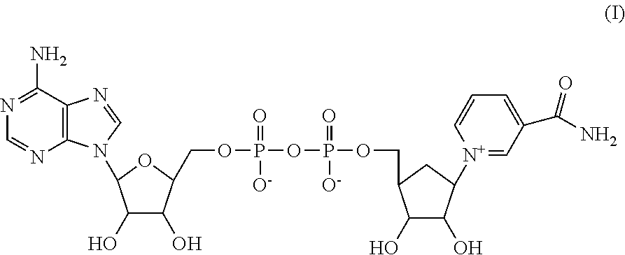
Preferably, carba-NAD (cNAD) is used as a derivative of NAD. In carba-NAD the ribose is substituted by a carbacyclic sugar unit. Carba-NAD has the following structure (I):
##STR00001##
The compound, its production and use are described in detail in WO 2007/012494, WO 2011/012270 and WO2014/195363. The cofactor in the present invention is preferably carba-NAD. In one embodiment of the present invention, the cofactor is a functionally active derivative of NAD as disclosed in formula III of WO 2011/012270 to which it is explicitly referred. In one embodiment of the present invention, NADP is used instead of NAD.
The method of the present invention can be carried out in a so-called liquid or wet test, for example in a cuvette, or as a so-called dry test on an appropriate reagent carrier, the necessary test reagents thereby being present in or on a solid carrier, which is preferably an absorbent or swellable material.
Alternatively or additionally, the 3-HBDH may be part of a sensor, a test strip, a test element, a test strip device or a liquid test.
A sensor is an entity that measures a physical/chemical quantity and converts it into a signal which can be read by an observer or by an instrument. In the present invention, the 3-HBDH may be part of a sensor. The sensor converts 3-HB and NAD or a derivate thereof into acetoacetate and NADH or a derivate thereof, which is further converted into a signal such as a change in colour or a value displayed e.g. on a display or monitor.
In one embodiment, the sensor may comprise 3-HBDH and an amperometric device to determine 3-HB of a sample. Also, a microdialysis system coupled with an electrochemical flow cell could be used for continuous monitoring of 3-HB in a sample or subject. The working electrode of the flow cell could be prepared with the 3-HBDH immobilized in a redox polymer film on the electrode surface. Coupling an electrochemical 3-HB sensor directly with microdialysis eliminates the need to transfer sample aliquots to a liquid chromatography system with a post-column oxidase enzyme reactor. 3-HB in the dialysate from the microdialysis probe can be selectively detected at the enzyme electrode without any significant interference from other oxidizable species. Furthermore, enzyme-coupled biosensors have been described in the art. In accordance with this, 3-HBDH may be coupled to a surface (e.g. by printing a 3-HBDH/graphite mixture onto electroplated graphite pads or by adsorption or immobilization of the mutant 3-HBDH on carbon particles, platinized carbon particles, carbon/manganese dioxide particles, glassy carbon, or mixing it with carbon paste electrodes etc.)
A test strip or a test element is an analytic or diagnostic device used to determine presence and/or quantity of a target substance within a sample. A standard test strip may comprise one or more different reaction zones or pads comprising reagents which react (e.g. change colour) when contacted with a sample. Test strips are known in many embodiments, for example from U.S. Pat. No. 6,541,216, EP 262445 and U.S. Pat. No. 4,816,224. It is commonly known that one or more reagents (e.g. enzymes) needed for carrying out the determination methods are present on or in solid carrier layers. As carrier layers, there are especially preferred absorbent and/or swellable materials which are wetted by the sample liquid to be analyzed. Examples include gelatine, cellulose and synthetic fiber fleece layers.
The 3-HBDH of the present invention may also be part of a liquid test. A liquid test is a test wherein test components react in a liquid medium. Usually in the field of laboratory analytics, the liquid reagents are on water basis, e.g. a buffered salt solution in order to provide the activity of enzyme(s) involved. The liquid is usually adapted to the specific intended use. For carrying out a liquid test, all test components are solved in a liquid and combined (or vice versa). Typical containments for carrying out such tests include vials, multi wells plates, cuvettes, vessels, reagent cups, tubes etc.
In one embodiment of the present invention, the 3-HBDH of the present invention may be immobilized. Typical methods of immobilization include covalent binding e.g. to a membrane, encapsulation in a polymer, cross-linking to a supporting matrix or immobilization in a sol-gel matrix (e.g. glasses such as silicate glasses) or adsorption on porous substrates. Suitable methods for immobilizing enzymes are known in the art (see e.g. Lillis et al., 2000, Sensors and Actuators B 68: 109-114).
In a preferred embodiment of the present invention, the method further comprises determining the amount or concentration of glucose. In another embodiment of the present invention the method comprises determining the amount or concentration of acetone and/or acetoacetate; and/or determining the amount or concentration of glucose. The determination of these compounds is of particular relevance in the diagnosis of the above diseases and medical condition. Methods for determining these compounds are well-known in the art. Additionally, systems and methods for multiple analyte analysis are known from WO 2014/068024.
Accordingly and preferably, the method of the present invention is further characterized in that a) wherein the determining of the change in the redox state of NAD or the derivate thereof includes the determination of the concentration of (i) NAD or the derivate thereof and/or (ii) NADH or the derivate thereof; and/or b) wherein the determining the change in the redox state of NAD or the derivative thereof is electrochemically or optically; and/or c) wherein the method further comprises determining the amount or concentration of acetoacetate and/or acetone; and/or d) wherein the method further comprises determining the amount or concentration of glucose; and/or e) wherein the derivative of NAD is carba-NAD; and/or f) wherein the mutant 3-HBDH is part of a sensor, a test strip, a test element, a test strip device or a liquid test; and/or g) wherein the sample is a body fluid, particularly a blood sample or a urine sample.
In still another aspect, the present invention relates to the use of the mutant 3-HBDH of the present invention for determining the amount or concentration of 3-hydroxybutyrate in a sample.
With respect to the use of the present invention it is referred to the terms, examples and specific embodiments used in the context of the other aspects of the present disclosure, which are also applicable to this aspect. Particularly, the mutant 3-HBDH according to the present invention may be used as detailed with respect to the methods of the present invention.
Yet, in another aspect, the present invention relates to a device for determining the amount or concentration of 3-hydroxybutyrate in a sample comprising the mutant 3-HBDH of the present invention and optionally a further component required for said determining.
With respect to the device of the present invention it is referred to the terms, examples and specific embodiments used in the context of the other aspects of the present disclosure, which are also applicable to this aspect. Particularly, the mutant 3-HBDH according to the present invention may be employed as detailed above.
The 3-HBDH of the present invention may be part of a device for determining 3-HB in a sample. The device may be any device suitable for this purpose. The device may be a machine or tool which can be used for determining 3-HB. Preferably, the device is a sensor, preferably an electrochemical sensor, or a test strip. Exemplary devices are described above and in the following:
The device may be a sensor, e.g. a biosensor, which is an analytical device for the detection of an analyte that combines a biological component (here the 3-HBDH according to the present invention) with a detector component, particularly a physicochemical detector component.
Biosensors are particularly useful to determine the concentration of various analytes (including 3-HB) from biological samples, particularly from blood. Exemplary biosensors based on an electrochemical test strip format are described in U.S. Pat. Nos. 5,413,690; 5,762,770 and 5,997,817.
In the (bio)sensor of the present invention, 3-HB converted into acetoacetate in the presence of the 3-HBDH and NAD or derivative and the change in the redox state of NAD or derivative may be monitored by the transducer or detector element.
Particularly, 3-HB sensors have been combined with other sensors, e.g. for determining glucose, acetone, acetoacetate, cholesterol, triglycerides, urea, blood gases or electrolytes etc. Evidently, the mutant 3-HBDH of the present invention could also be used in these multi-analyte devices.
As detailed above, the sensor is preferably an electrochemical or optical sensor. An electrochemical sensor is based on the translation of a chemical signal (here presence of 3-HB) into an electrical signal (e.g. current). A suitable electrode can measure the 3-HB-mediated production of NADH or derivative thereof as an electrical signal.
A suitable optical sensor can measure the 3-HBDH-mediated change in the redox state of NAD or derivate thereof. The signal may be the NAD/NADH-mediated absorbance/emission of light.
The device of the present invention may comprise--in addition to the 3-HBDH of the present invention--one or more further component(s), such as other reagents, required for or helpful in said determining. The components may be any of these described in the context of the methods and devices of the present invention. Additionally, this may include an instruction manual, a lancet device, a capillary pipette, a further enzyme, a substrate and/or a control solution etc.
Preferably, the device of the present invention is characterized in that the device is or comprises a sensor, preferably an electrochemical sensor or an optical sensor, or a test strip, particularly a test strip and/or allows for determining the amount or concentration of glucose in the sample. With respect to the device of the present invention it is also referred to the terms, examples and specific embodiments described above.
Unless defined otherwise, all technical and scientific terms and any acronyms used herein have the same meanings as commonly understood by one of ordinary skill in the art in the field of the invention. Definitions of common terms in molecular biology can be found in Benjamin Lewin, Genes V, published by Oxford University Press, 1994 (ISBN 0-19-854287-9); Kendrew et al. (eds.), The Encyclopedia of Molecular Biology, published by Blackwell Science Ltd., 1994 (ISBN 0-632-02182-9); and Robert A. Meyers (ed.), Molecular Biology and Biotechnology: a Comprehensive Desk Reference, published by VCH Publishers, Inc., 1995 (ISBN 1 -56081 -569-8).
The invention is not limited to the particular methodology, protocols, and reagents described herein because they may vary. Although any methods and materials similar or equivalent to those described herein can be used in the practice of the present invention, the preferred methods, and materials are described herein. Further, the terminology used herein is for the purpose of describing particular embodiments only and is not intended to limit the scope of the present invention.
As used herein and in the appended claims, the singular forms "a", "an", and "the" include plural reference unless the context clearly dictates otherwise. Similarly, the words "comprise", "contain" and "encompass" are to be interpreted inclusively rather than exclusively. Similarly, the word "or" is intended to include "and" unless the context clearly indicates otherwise. The term "plurality" refers to two or more.
The following Figures and Examples are intended to illustrate various embodiments of the invention. As such, the specific modifications discussed are not to be construed as limitations on the scope of the invention. It will be apparent to the person skilled in the art that various equivalents, changes, and modifications may be made without departing from the scope of the invention, and it is thus to be understood that such equivalent embodiments are to be included herein.
EXAMPLES
Example 1
Establishing a Library of 3-HBDH Mutants
The gene for 3-hydroxybutyrate dehydrogenase (3-HBDH) from Alcaligenes faecalis (Database UniProtKB--D0VWQ0) was synthesized, cloned in vector pKKt5 and transformed in E. coli strain XL-1 Blue by common methods of molecular biology.
Saturation mutagenesis was applied on many amino acid positions of the enzyme. Mutagenesis was achieved by applying randomly synthesized primers with QuikChange Site-Directed Mutagenesis Kit (Agilent Technologies Cat. 200518).
The 5'-and the 3'-primer used for mutagenesis were complementary to each other and contained NNN (randomly synthesized nucleotides) for the amino acid exchange in a central position. This randomly created codon was flanked by 12 to 16 nucleotides at each end. The sequences of these nucleotides were identical to the cDNA-strand or to the complementary cDNA-strand flanking the codon for the amino substitution. Mutant library was created by transformation of mutated genes in E. coli strain XI-Blue and cultivation on agar plates over night at 37.degree. C.
Example 2
Determination of Properties of 3-HBDH Mutants from First Round of Mutagenesis (Mutants with Single Amino Acid Substitutions)
A library of 3-HBDH mutants produced as described in Example 1 was screened for the enzymatic following properties: Thermal stability Affinity for 3-hydroxybutyrate Affinity for c-NAD (carba-NAD=artificial cofactor cf. US20120130062A1)
Mutant colonies on agar plates described above were picked in microtiter plates (mtp) containing 200 .mu.l LB-Ampicillin-media/hole and incubated at 37.degree. C. over night. These plates were referred to as master plates. For each amino acid position two master plates were picked to assure that every possible exchange is included.
From each master plate, 40 .mu.l sample/cavity was transferred to a mtp containing 200 .mu.l 0.1% Triton X-100; 500 mM NaCl; 200 mM Hepes pH 9.0; 2% B-Per/cavity (B-PER=Bacterial Protein Extraction Reagent Pierce No.78248) and incubated for cell disruption at 40.degree. C. for 30 minutes. This plate was referred to as working plate.
From the working plate 4.times.20 .mu.l sample/cavity was transferred to four empty mtps. One of these was tested with 62.22 mM 3-hydroxybutyrate; 4.15 mM cNAD; 0.1% Triton X-100; 200 mM Hepes pH 9.0 at room temperature and referred to as reference measurement. The other mtps were tested under different conditions and the obtained values compared to the reference plate in percent.
Following parameters were measured: Thermal stability: Mtp was incubated for 30 min. at 64.degree. C. and tested afterwards with 62.22 mM 3-hydroxybutyrate; 4.15 mM cNAD; 0.1% Triton X-100; 200 mM Hepes pH 9.0 Affinity to 3-hydroxybutyrate: Activity assay with reduced amount of substrate (i.e. below substrate saturation). Measurement with 1.94 mM 3-hydroxybutyrate; 4.15 mM cNAD; 0.1% Triton X-100; 200 mM Hepes pH 9.0 Affinity to cNAD: Activity assay with reduced amount of cofactor (i.e. below saturation). Measurement with 62.22 mM 3-hydroxybutyrate; 0.032 mM cNAD; 0.1% Triton X-100; 200 mM Hepes pH 9.0
The enzymatic reaction was monitored at room temperature at 340 nm for 5 minutes and the dE/min calculated for each working plate. The value from the reference measurement was set to 100% activity. The values obtained with the other three plates (thermal stability, affinity to 3-hydroxybutyrate or cNAD) were compared to the reference and calculated in percent activity ((dE/min Parameter/dE/min Reference)*100). Each master plate contained beside the mutants wild-type enzyme as control to better estimate improvements or deteriorations of the properties.
Thermal stability expressed as remaining activity was calculates as follows:
.times..times..times..times..times..times..times..times..times..times..ti- mes..times..times..times..times..times..times..times..times..degree..times- ..times..times..times..times..times..times..times..times..times..times..ti- mes..times..times..times..times. ##EQU00001##
A value obtained with a mutant higher than the value obtained with wild-type enzyme represents an increase in thermal stability for the mutant.
Substrate affinity expressed as activity ratio was calculated as follows:
.times..times..times..times..times..times..times..times..times..times..ti- mes..times..times..times..times..times..times..times..times..times..times.- .times..times..times..times..times. ##EQU00002##
A mutant with higher substrate affinity will show higher activity when reacted with less substrate (below substrate saturation) than a mutant with lower substrate affinity. A value obtained with a mutant higher than the value obtained with wild-type enzyme represents an increase in substrate affinity for the mutant.
Cofactor affinity expressed as activity ratio was calculated accordingly:
.times..times..times..times..times..times..times..times..times..times..ti- mes..times..times..times..times..times..times..times..times..times..times.- .times..times..times..times..times. ##EQU00003##
Data below 0.001 dE/min were set to zero, resulting in "zero" values like in Tables 1B and 1C.
The results relative to wild type enzyme are summarized in Tables 1A, 1B and 1C.
TABLE-US-00004 TABLE 1A Thermal stability and affinity of various single mutants relative to the wild-type 3-HBDH referred to as AFDH1 Affinity for Thermal Clone 3-HB cNAD Stability AFDH1 + M1V + .smallcircle. - AFDH1 + L2C + .smallcircle. .smallcircle. AFDH1 + L2Q + .smallcircle. .smallcircle. AFDH1 + L2R + + - AFDH1 + L2D + .smallcircle. .smallcircle. AFDH1 + L2M + .smallcircle. - AFDH1 + L2S + + - AFDH1 + K3C .smallcircle. - + + AFDH1 + K6V .smallcircle. - + AFDH1 + K6G + - + AFDH1 + T10S + - + AFDH1 + T10G .smallcircle. .smallcircle. + AFDH1 + T13S - - + AFDH1 + L18R + .smallcircle. - AFDH1 + L18K + .smallcircle. - AFDH1 + L18E + .smallcircle. + AFDH1 + L18P - - + + AFDH1 + A19G + .smallcircle. + + AFDH1 + A21G + .smallcircle. + + AFDH1 + V31C + .smallcircle. + AFDH1 + V33A .smallcircle. .smallcircle. + AFDH1 + I33T + + - AFDH1 + V33L + .smallcircle. + AFDH1 + P39G .smallcircle. .smallcircle. + AFDH1 + E40G .smallcircle. .smallcircle. + AFDH1 + E41F + .smallcircle. - AFDH1 + I42T .smallcircle. .smallcircle. + + AFDH1 + I42R - - + + AFDH1 + I42L - - + + AFDH1 + E43I + + + AFDH1 + E43G - - + AFDH1 + E43C - - + AFDH1 + E43L - - + AFDH1 + E45H + .smallcircle. - AFDH1 + E45Q + .smallcircle. - AFDH1 + A62M + + - AFDH1 + A62K + + - AFDH1 + A62F + + - AFDH1 + A62R + + .smallcircle. AFDH1 + A62L + + - AFDH1 + A62V .smallcircle. .smallcircle. + AFDH1 + A69V + + - AFDH1 + A69T + + - AFDH1 + T70P .smallcircle. - + + AFDH1 + D72P + .smallcircle. + AFDH1 + F73T + .smallcircle. - AFDH1 + A109Y + .smallcircle. - AFDH1 + I111V + + - AFDH1 + I111L - .smallcircle. + AFDH1 + A112E .smallcircle. - + AFDH1 + A112R - - + AFDH1 + A112T - - + AFDH1 + A112C .smallcircle. - + AFDH1 + N114D - + + AFDH1 + S116D .smallcircle. - + AFDH1 + A117T - - + AFDH1 + V118S - - + AFDH1 + A124V + .smallcircle. - AFDH1 + A124I + .smallcircle. + + AFDH1 + A124V + .smallcircle. + AFDH1 + A124C + - + AFDH1 + A125G + - + AFDH1 + I140M - - + + AFDH1 + A141S + + - AFDH1 + A143V - - + + AFDH1 + V147R .smallcircle. .smallcircle. + + AFDH1 + A148G .smallcircle. - + + AFDH1 + S153I + .smallcircle. - AFDH1 + V156T - - + AFDH1 + V168A - - + + AFDH1 + A170G + .smallcircle. + AFDH1 + N173K + .smallcircle. + AFDH1 + G175T + .smallcircle. + AFDH1 + G175V + .smallcircle. + AFDH1 + G175I + .smallcircle. + AFDH1 + G177P + + + - AFDH1 + G177Q + + - AFDH1 + W187F + .smallcircle. + AFDH1 + P191R + + - AFDH1 + P191A + .smallcircle. - AFDH1 + L192M + + - AFDH1 + V193T + .smallcircle. - AFDH1 + K195V + .smallcircle. - AFDH1 + I200R + .smallcircle. - AFDH1 + Q203I - - + AFDH1 + R213Y + .smallcircle. - AFDH1 + R213W + .smallcircle. - AFDH1 + R213D + .smallcircle. - AFDH1 + L216V + + - AFDH1 + A217L + + - AFDH1 + E218P - - + + AFDH1 + Q220M + + + - AFDH1 + Q220T + + + - AFDH1 + L223H + .smallcircle. - AFDH1 + L223N + .smallcircle. - AFDH1 + Q224T + .smallcircle. - AFDH1 + Q224A + .smallcircle. - AFDH1 + Q224V + .smallcircle. - AFDH1 + T227E - + + AFDH1 + T227N + + + + - AFDH1 + T227A + + + - AFDH1 + T227R + + + - AFDH1 + T227G + + + - AFDH1 + T227V + + + - AFDH1 + T227K + + + - AFDH1 + P228R + .smallcircle. - AFDH1 + P228V + .smallcircle. - AFDH1 + P228G + .smallcircle. - AFDH1 + G233S + + - AFDH1 + G233I + + - AFDH1 + G233A .smallcircle. + - AFDH1 + G233P + + - AFDH1 + G233R .smallcircle. + - AFDH1 + G233T .smallcircle. + - AFDH1 + G233K + + - AFDH1 + A241P .smallcircle. .smallcircle. + AFDH1 + A242T - - + AFDH1 + A242Y + .smallcircle. + + AFDH1 + L251W + .smallcircle. - AFDH1 + L251M .smallcircle. .smallcircle. + AFDH1 + S252V .smallcircle. .smallcircle. + AFDH1 + L253I + .smallcircle. + AFDH1 + L253A + + + - AFDH1 + L253C + + + - AFDH1 + G256A + .smallcircle. - AFDH1 + A259S + + - AFDH1 + R260I + + - AFDH1 + R260G + + - AFDH1 + R260W + + - (+ = improved; .smallcircle. = similar; - = decreased)
TABLE-US-00005 TABLE 1B Thermal stability and affinity of various single mutants of 3-HBDH Affinity for* Thermal Clone 3-HB cNAD Stability** AFDH1 19 6 7 AFDH1 + K3C 10 0 100 AFDH1 + L18K 41 3 0 AFDH1 + L18E 31 3 96 AFDH1 + L18P 11 0 107 AFDH1 + A19G 34 5 100 AFDH1 + V33A 20 3 80 AFDH1 + I33T 29 7 0 AFDH1 + V33L 25 4 78 AFDH1 + P39G 16 4 84 AFDH1 + A62M 30 10 5 AFDH1 + A62K 31 10 6 AFDH1 + A62F 31 6 8 AFDH1 + A62R 32 11 11 AFDH1 + A62L 30 9 6 AFDH1 + A62V 13 4 44 AFDH1 + A125G 23 3 34 AFDH1 + A143V 12 2 93 AFDH1 + A148G 17 3 113 AFDH1 + A170G 22 4 91 AFDH1 + G175T 25 4 91 AFDH1 + G175V 26 4 72 AFDH1 + G175I 26 4 55 AFDH1 + W187F 27 4 75 AFDH1 + L216V 25 20 0 AFDH1 + G233S 23 19 0 AFDH1 + G233I 27 38 0 AFDH1 + G233A 19 41 0 AFDH1 + G233P 35 44 0 AFDH1 + G233R 18 40 0 AFDH1 + G233T 17 38 0 AFDH1 + G233K 20 41 0 AFDH1 + L253I 32 7 76 AFDH1 + L253A 30 53 0 AFDH1 + L253C 30 51 0 *given as activity ratio (%) with 1.94/62.22 mM 3-HB and 0.032/4.15 mM cNAD **given as remaining activity (%) after a 30 minute-incubation at 64.degree. C.
TABLE-US-00006 TABLE 1C Various single mutants with highly improved thermal stability relative to the wild-type 3-HBDH Affinity* Thermal Clone 3-HB cNAD Stability** AFDH1 19 6 7 AFDH1 + K3C 10 0 100 AFDH1 + L18P 11 0 107 AFDH1 + A19G 34 5 100 AFDH1 + A21G 18 3 102 AFDH1 + V31C 18 4 84 AFDH1 + V33A 20 3 80 AFDH1 + V33L 25 4 78 AFDH1 + P39G 16 4 84 AFDH1 + E40G 12 3 95 AFDH1 + I42T 19 4 100 AFDH1 + I42R 9 0 100 AFDH1 + I42L 15 3 105 AFDH1 + T70P 20 0 100 AFDH1 + D72P 30 4 77 AFDH1 + I111L 12 5 84 AFDH1 + A112E 21 3 78 AFDH1 + A112R 15 3 70 AFDH1 + A124I 26 5 99 AFDH1 + A124V 22 4 52 AFDH1 + I140M 13 4 100 AFDH1 + A143V 12 2 93 AFDH1 + V147R 19 4 122 AFDH1 + A148G 17 3 100 AFDH1 + A170G 22 4 91 AFDH1 + G175T 25 4 91 AFDH1 + W187F 27 4 75 AFDH1 + E218P 6 0 100 AFDH1 + A241P 20 4 80 AFDH1 + A242Y 20 4 100 AFDH1 + L251M 19 4 50 AFDH1 + S252V 19 7 59 AFDH1 + L253I 32 7 100 *given as activity ratio (%) with 1.94/62.22 mM 3-HB and 0.032/4.15 mM **given as remaining activity (%) after a 30-minute incubation at 64.degree. C.
Exemplary mutant with exchange L253I was chosen for further optimization by combining of additional found positions.
Example 3
Screening of 3-HBDH Mutants from Second Round of Mutagenesis (Mutants with Amino Acid Substitution L253I and Optionally Further Amino Acid Substitution(s))
Unless indicated otherwise, the experiments have been carried out as detailed in Example 2. In a second round of mutagenesis selected mutations were introduced into variant AFDH1+L253I. The results are summarized in Table 2A:
TABLE-US-00007 TABLE 2A Thermal stability (68.degree. C., 30 min) and affinity of various mutants with amino acid substitution L253I Affinity for* Thermal Clone 3-HB cNAD Stability** AFDH1 + L253I 32 8 91 AFDH1 + L253I + E43G 29 6 104 AFDH1 + L253I + E43V 25 5 101 AFDH1 + L253I + G233M 27 6 57 AFDH1 + L253I + G233K + 29 6 101 A234T AFDH1 + L253I + G233C 29 5 78 AFDH1 + L253I + L2C 37 7 41 AFDH1 + L253I + V157R 30 7 71 *given as activity ratio (%) with 1.94/62.22 mM 3-HB and 0.032/4.15 mM cNAD **given as remaining activity (%) after a 30-minute incubation at 68.degree. C.
Exemplary variant AFDH1+L253I+G233K+A234T was further combined with other found positions. The results are summarized in Table 2B:
TABLE-US-00008 TABLE 2B Thermal stability (75.degree. C., 30 min) and affinity of various mutants with amino acid substitutions L253I, G233K and A234T Aff. Thermal Clone 3-HB* Stability** AFDH1 + L253I + G233K + A234T 14 5 AFDH1 + A62V + L253I + G233K + A234T 16 17 AFDH1 + L253I + G233K + A234T + 148G 11 78 AFDH1 + L253I + G233K + A234T + 124I + 148G 13 91 AFDH1 + L253I + G233K + A234T + 39G + 124I + 148G 12 98 AFDH1 + L253I + G233K + A234T + 124I 16 37 AFDH1 + L253I + G233K + A234T + 38K + 39G + 124I 17 33 AFDH1 + L253I + G233K + A234T + 39G + 124I 15 32 AFDH1 + L253I + G233K + A234T + 39G 12 29 AFDH1 + L253I + G233K + A234T + 39G + 124I 16 31 AFDH1 + L253I + G233K + A234T + 175V 18 56 AFDH1 + L253I + G233K + A234T + 39G + 62R + 148G 12 83 AFDH1 + L253I + G233K + A234T + 39G + 124I 15 38 AFDH1 + L253I + G233K + A234T + 62R + 175V + 242Y 23 0 AFDH1 + L253I + G233K + A234T + 39G + 124I + 148G + 18 52 175V + 242Y AFDH1 + A62V + L253I + G233K + A234T + 39G + 111L 14 79 AFDH1 + A62V + L253I + G233K + A234T + 143V 13 95 AFDH1 + A62V + L253I + G233K + A234T + 33L 19 0 AFDH1 + A62V + L253I + G233K + A234T + 175I 28 54 AFDH1 + A62V + L253I + G233K + A234T + 175V 24 68 AFDH1 + A62V + L253I + G233K + A234T + 175V + 242Y 22 0 AFDH1 + A62V + L253I + G233K + A234T + 175T 23 78 AFDH1 + A62V + L253I + G233K + A234T + 18E 25 36 AFDH1 + A62V + L253I + G233K + A234T + 39G + 187F 17 26 *given as activity ratio (%) with 1.94/62.22 mM 3-HB **given as remaining activity (%) after a 30-minute incubation at 75.degree. C.
Selected mutants were chosen and stressed at 80.degree. C. for 30 minutes. The results are summarized in Table2C:
TABLE-US-00009 TABLE 2C Thermal stability (80.degree. C., 30 min) and affinity of selected mutants Affinity Thermal Clone 3-HB* Stability** AFDH1 + L253I + G233K + A234T + 124I + 148G 13 54 AFDH1 + L253I + G233K + A234T + 39G + 124I + 12 52 148G AFDH1 + L253I + G233K + A234T + 39G + 62R + 12 7 148G AFDH1 + A62V + L253I + G233K + A234T + 143V 13 94 *given as activity ratio (%) with 1.94/62.22 mM 3-HB **given as remaining activity (%) after a 30-minute incubation at 80.degree. C.
Example 4
Screening of 3-HBDH Mutants with Amino Acid Substitutions A62V+A143V+A148G+G233K+A234T+L253I and Optionally Further Amino Acid Substitution(s))
Selected mutants from example 3 were measured at pH 7.5 and the best variant selected for further mutagenesis.
Unless indicated otherwise, the experiments have been carried out as detailed in Example 2. Found mutants were screened analogously to Example 3 but with different solutions at pH 7.5. Reference mtp was tested with 150 mM 3-hydroxybutyrate; 5 mM cNAD; 0.1% Triton X-100; 70 mM Mops pH 7.5.
Following parameters were measured: Mtp for thermal stability incubated for 30 min. at 60-90.degree. C. (depending on the stability of enzyme variant) and tested afterwards with 150 mM 3-hydroxybutyrate; 5 mM cNAD; 0.1% Triton X-100; 70 mM Mops pH 7.5 Mtp for affinity to .beta.-hydroxybutyrate was measured with 5 mM 3-hydroxybutyrate; 5 mM cNAD; 0.1% Triton X-100; 70 mM Mops pH 7.5 Mtp for affinity to cNAD was measured with 150 mM 3-hydroxybutyrate; 0.5 mM cNAD; 0.1% Triton X-100; 70 mM Mops pH 7.5
TABLE-US-00010 TABLE 2D Thermal stability (various temperatures, 30 min) and affinity of selected mutants Affinity for* Thermal Stability** Clone 3-HB cNAD 60.degree. C. 70.degree. C. 73.degree. C. 75.degree. C. 87.degree. C. 90.degree. C. AFDH1 14 28 17 1 0 0 0 0 AFDH2 21 29 19 15 AFDH3 24 22 100 68 53 AFDH4 25 29 100 88 70 4 0 0 AFDH4 + 147R 29 29 100 76 AFDH5 19 29 78 3 AFDH6 29 29 91 41 AFDHx 15 17 91 *given as activity ratio (%) with 5/150 mM 3-HB and 0.5/5 mM cNAD **given as remaining activity (%) after a 30-minute incubation at indicated temperature AFDH1: wild-type AFDH2: AFDH1 + L253I AFDH3: AFDH1 + G233K + A234T + L253I AFDH4: AFDH1 + A62V + G233K + A234T + L253I AFDH4 + 147R: AFDH1 + A62V + V147R + G233K + A234T + L253I AFDH5: AFDH1 + P39G + A62R + A143V + A148G + G233K + A234T + L253I AFDH6: AFDH1 + A62V + A143V + A148G + G233K + A234T + L253I AFDHx: AFDH1 + A62V + E111L + I140M + A148G
On basis of mutant AFDH6 (i.e. AFDH1 plus mutations A62V+A143V+A148G+G233K+A234T+L253I) further rounds of mutagenesis were performed with following mutants listed in Table 3
TABLE-US-00011 TABLE 3 Thermal stability (87.degree. C. and 90.degree. C., 30 min) and affinity of selected mutants Affinity for* Thermal stability** Clone 3-HB cNAD 87.degree. C. 90.degree. C. AFDH1 14 17 0 0 AFDH6 29 29 AFDH6 + 19G 35 40 4 (13) AFDH6 + 170G 34 36 4 (13) AFDH6 + 125G + 187F 35 34 24 (13) AFDH6 + 18E + 33L 29 33 34 (13) AFDH6 + 33L + 125G + 187F 35 39 86 (60) AFDH6 + 33L + 125G + 187F + 216V 37 40 14 (60) AFDH6 + 18E + 187F 34 40 80 (60) AFDH6 + 33L + 125G + 175V + 187F 41 40 97 (60) AFDH6 + 175V + 216V 43 47 7 (60) AFDH6 + 175V + 187F + 216V 34 40 54 (60) AFDH6 + 19G + 125G + 187F 41 42 5 (50) AFDH6 + 125G + 175T + 187F 45 38 92 (50) AFDH6 + 19G + 33L 42 48 0 (50) AFDH6 + 19G + 175I 47 52 0 (50) AFDH6 + 19G + 175T 37 42 95 (50) AFDH6 + 170G + 175T 39 37 101 (50) AFDH6 + 18E + 125G + 187F 47 42 67 (3) AFDH6 + 33L + 125G + 175T + 187F 37 32 95 (3) AFDH6 + 33L + 125G + 175I + 187F 47 40 100 (3) AFDH6 + 33L + 125G + 170G + 187F 45 39 41 (3) AFDH6 + 18E + 175I + 187F 41 39 58 (3) AFDH6 + 33L + 125G + 175V + 187F 36 31 90 (3) AFDH6 + 33L + 125G + 175V + 187F + 216V 49 48 23 (3) AFDH6 + 33L + 125G + 175T + 187F 35 35 94 (3) AFDH6 + 33L + 38K + 39G + 125G + 175V + 47 40 51 (3) 187F *given as activity ratio (%) with 5/150 mM 3-HB and 0.5/5 mM cNAD **given as remaining activity (%) after a 30-minute incubation at indicated temperature Note: Values in () are data from AFDH6 measured in same test series as control.
TABLE-US-00012 TABLE 4 Amino acid substitutions found to improve performance of 3-HBDHs, particularly when combined with AFDH6 (i.e. AFDH1 plus mutations A62V + A143V + A148G + G233K + A234T + L253I) Amino acid Amino acid Amino acid position in substitutions in in wild-type wild-type improved mutants 18 L R/K/E/P 19 A G 33 I A/L/T 38 Q K 39 P G 62 A F/M/K/R/L/V 125 A G 143 A V 148 A G 170 A G 175 G T/V/I 187 W F 216 L V 233 G S/I/A/P/R/T/K/C 234 A T 253 L I/A/C
TABLE-US-00013 SEQUENCES AFDH1: wild-type 3-HBDH from Alcaligenes faecalis (SEQ ID NO: 1) MLKGKKAVVT GSTSGIGLAM ATELAKAGAD VVINGFGQPE DIERERSTLE SKFGVKAYYL 60 NADLSDAQAT RDFIAKAAEA LGGLDILVNN AGIQHTAPIE EFPVDKWNAI IALNLSAVFH 120 GTAAALPIMQ KQGWGRIINI ASAHGLVASV NKSAYVAAKH GVVGLTKVTA LENAGKGITC 180 NAICPGWVRT PLVEKQIEAI SQQKGIDIEA AARELLAEKQ PSLQFVTPEQ LGGAAVFLSS 240 AAADQMTGTT LSLDGGWTAR 260 Extended core sequence of A FDH1 (SEQ ID NO: 2) ADLSDAQATR DFIAKAAEAL GGLDILVNNA GIQHTAPIEE FPVDKWNAII ALNLSAVFHG 60 TAAALPIMQK QGWGRIINIA SAHGLVASVN KSAYVAAKHG VVGLTKVTAL ENAGKGITCN 120 AICPGWVRTP LVEKQIEAIS QQKGIDIEAA ARELLAEKQP SLQFVTPEQL GGAAVFLSSA 180 AADQMTGTTL SL 192 AFDH2: AFDH1 + L253I (SEQ ID NO: 3) MLKGKKAVVT GSTSGIGLAM ATELAKAGAD VVINGFGQPE DIERERSTLE SKFGVKAYYL 60 NADLSDAQAT RDFIAKAAEA LGGLDILVNN AGIQHTAPIE EFPVDKWNAI IALNLSAVFH 120 GTAAALPIMQ KQGWGRIINI ASAHGLVASV NKSAYVAAKH GVVGLTKVTA LENAGKGITC 180 NAICPGWVRT PLVEKQIEAI SQQKGIDIEA AARELLAEKQ PSLQFVTPEQ LGGAAVFLSS 240 AAADQMTGTT LSIDGGWTAR 260 AFDH3: AFDH1 + G233K + A234T + L253I (SEQ ID NO: 4) MLKGKKAVVT GSTSGIGLAM ATELAKAGAD VVINGFGQPE DIERERSTLE SKFGVKAYYL 60 NADLSDAQAT RDFIAKAAEA LGGLDILVNN AGIQHTAPIE EFPVDKWNAI IALNLSAVFH 120 GTAAALPIMQ KQGWGRIINI ASAHGLVASV NKSAYVAAKH GVVGLTKVTA LENAGKGITC 180 NAICPGWVRT PLVEKQIEAI SQQKGIDIEA AARELLAEKQ PSLQFVTPEQ LGKTAVFLSS 240 AAADQMTGTT LSIDGGWTAR 260 AFDH4: AFDH1 + A62V + G233K + A234T + L253I (SEQ ID NO: 5) MLKGKKAVVT GSTSGIGLAM ATELAKAGAD VVINGFGQPE DIERERSTLE SKFGVKAYYL 60 NVDLSDAQAT RDFIAKAAEA LGGLDILVNN AGIQHTAPIE EFPVDKWNAI IALNLSAVFH 120 GTAAALPIMQ KQGWGRIINI ASAHGLVASV NKSAYVAAKH GVVGLTKVTA LENAGKGITC 180 NAICPGWVRT PLVEKQIEAI SQQKGIDIEA AARELLAEKQ PSLQFVTPEQ LGKTAVFLSS 240 AAADQMTGTT LSIDGGWTAR 260 AFDH4 + 147R: AFDH1 + A62V+ V147R + G233K + A234T + L253I (SEQ ID NO: 6) MLKGKKAVVT GSTSGIGLAM ATELAKAGAD VVINGFGQPE DIERERSTLE SKFGVKAYYL 60 NVDLSDAQAT RDFIAKAAEA LGGLDILVNN AGIQHTAPIE EFPVDKWNAI IALNLSAVFH 120 GTAAALPIMQ KQGWGRIINI ASAHGLRASV NKSAYVAAKH GVVGLTKVTA LENAGKGITC 180 NAICPGWVRT PLVEKQIEAI SQQKGIDIEA AARELLAEKQ PSLQFVTPEQ LGKTAVFLSS 240 AAADQMTGTT LSIDGGWTAR 260 AFDH5: AFDH1 + P39G + A62R + A143V + A148G + G233K + A234T + L253I (SEQ ID NO: 7) MLKGKKAVVT GSTSGIGLAM ATELAKAGAD VVINGFGQGE DIERERSTLE SKFGVKAYYL 60 NRDLSDAQAT RDFIAKAAEA LGGLDILVNN AGIQHTAPIE EFPVDKWNAI IALNLSAVFH 120 GTAAALPIMQ KQGWGRIINI ASVHGLVGSV NKSAYVAAKH GVVGLTKVTA LENAGKGITC 180 NAICPGWVRT PLVEKQIEAI SQQKGIDIEA AARELLAEKQ PSLQFVTPEQ LGKTAVFLSS 240 AAADQMTGTT LSIDGGWTAR 260 AFDH6: AFDH1 + A62V + A143V + A148G + G233K + A234T + L253I (SEQ ID NO: 8) MLKGKKAVVT GSTSGIGLAM ATELAKAGAD VVINGFGQPE DIERERSTLE SKFGVKAYYL 60 NVDLSDAQAT RDFIAKAAEA LGGLDILVNN AGIQHTAPIE EFPVDKWNAI IALNLSAVFH 120 GTAAALPIMQ KQGWGRIINI ASVHGLVGSV NKSAYVAAKH GVVGLTKVTA LENAGKGITC 180 NAICPGWVRT PLVEKQIEAI SQQKGIDIEA AARELLAEKQ PSLQFVTPEQ LGKTAVFLSS 240 AAADQMTGTT LSIDGGWTAR 260 AFDH A: AFDH6 + 33L + 125G + 175V + 187F (SEQ ID NO: 9) MLKGKKAVVT GSTSGIGLAM ATELAKAGAD VVLNGFGQPE DIERERSTLE SKFGVKAYYL 60 NVDLSDAQAT RDFIAKAAEA LGGLDILVNN AGIQHTAPIE EFPVDKWNAI IALNLSAVFH 120 GTAAGLPIMQ KQGWGRIINI ASVHGLVGSV NKSAYVAAKH GVVGLTKVTA LENAVKGITC 180 NAICPGFVRT PLVEKQIEAI SQQKGIDIEA AARELLAEKQ PSLQFVTPEQ LGKTAVFLSS 240 AAADQMTGTT LSIDGGWTAR 260 AFDH B: AFDH6 + 125G + 175T + 187F (SEQ ID NO: 10) MLKGKKAVVT GSTSGIGLAM ATELAKAGAD VVINGFGQPE DIERERSTLE SKFGVKAYYL 60 NVDLSDAQAT RDFIAKAAEA LGGLDILVNN AGIQHTAPIE EFPVDKWNAI IALNLSAVFH 120 GTAAGLPIMQ KQGWGRIINI ASVHGLVGSV NKSAYVAAKH GVVGLTKVTA LENATKGITC 180 NAICPGFVRT PLVEKQIEAI SQQKGIDIEA AARELLAEKQ PSLQFVTPEQ LGKTAVFLSS 240 AAADQMTGTT LSIDGGWTAR 260 AFDH C: AFDH6 + 170G + 175T (SEQ ID NO: 11) MLKGKKAVVT GSTSGIGLAM ATELAKAGAD VVINGFGQPE DIERERSTLE SKFGVKAYYL 60 NVDLSDAQAT RDFIAKAAEA LGGLDILVNN AGIQHTAPIE EFPVDKWNAI IALNLSAVFH 120 GTAAALPIMQ KQGWGRIINI ASVHGLVGSV NKSAYVAAKH GVVGLTKVTG LENATKGITC 180 NAICPGWVRT PLVEKQIEAI SQQKGIDIEA AARELLAEKQ PSLQFVTPEQ LGKTAVFLSS 240 AAADQMTGTT LSIDGGWTAR 260 AFDH D: AFDH6 + 18E + 125G + 187F (SEQ ID NO: 12) MLKGKKAVVT GSTSGIGEAM ATELAKAGAD VVINGFGQPE DIERERSTLE SKFGVKAYYL 60 NVDLSDAQAT RDFIAKAAEA LGGLDILVNN AGIQHTAPIE EFPVDKWNAI IALNLSAVFH 120 GTAAGLPIMQ KQGWGRIINI ASVHGLVGSV NKSAYVAAKH GVVGLTKVTA LENAGKGITC 180 NAICPGFVRT PLVEKQIEAI SQQKGIDIEA AARELLAEKQ PSLQFVTPEQ LGKTAVFLSS 240 AAADQMTGTT LSIDGGWTAR 260 AFDH E: AFDH6 + 33L + 125G + 175I + 187F (SEQ ID NO: 13) MLKGKKAVVT GSTSGIGLAM ATELAKAGAD VVLNGFGQPE DIERERSTLE SKFGVKAYYL 60 NVDLSDAQAT RDFIAKAAEA LGGLDILVNN AGIQHTAPIE EFPVDKWNAI IALNLSAVFH 120 GTAAGLPIMQ KQGWGRIINI ASVHGLVGSV NKSAYVAAKH GVVGLTKVTA LENAIKGITC 180 NAICPGFVRT PLVEKQIEAI SQQKGIDIEA AARELLAEKQ PSLQFVTPEQ LGKTAVFLSS 240 AAADQMTGTT LSIDGGWTAR 260 AFDH F: AFDH6 + 33L + 125G + 170G + 187F (SEQ ID NO: 14) MLKGKKAVVT GSTSGIGLAM ATELAKAGAD VVLNGFGQPE DIERERSTLE SKFGVKAYYL 60 NVDLSDAQAT RDFIAKAAEA LGGLDILVNN AGIQHTAPIE EFPVDKWNAI IALNLSAVFH 120 GTAAGLPIMQ KQGWGRIINI ASVHGLVGSV NKSAYVAAKH GVVGLTKVTG LENAGKGITC 180 NAICPGFVRT PLVEKQIEAI SQQKGIDIEA AARELLAEKQ PSLQFVTPEQ LGKTAVFLSS 240 AAADQMTGTT LSIDGGWTAR 260 AFDH G: AFDH6 + 33L + 125G + 175V + 187F + 216V (SEQ ID NO: 15) MLKGKKAVVT GSTSGIGLAM ATELAKAGAD VVLNGFGQPE DIERERSTLE SKFGVKAYYL 60 NVDLSDAQAT RDFIAKAAEA LGGLDILVNN AGIQHTAPIE EFPVDKWNAI IALNLSAVFH 120 GTAAGLPIMQ KQGWGRIINI ASVHGLVGSV NKSAYVAAKH GVVGLTKVTA LENAVKGITC 180 NAICPGFVRT PLVEKQIEAI SQQKGIDIEA AARELVAEKQ PSLQFVTPEQ LGKTAVFLSS 240 AAADQMTGTT LSIDGGWTAR 260 AFDH H: AFDH6 + 33I + 38K + 39G + 125G + 175V + 187F (SEQ ID NO: 16) MLKGKKAVVT GSTSGIGLAM ATELAKAGAD VVLNGFGKGE DIERERSTLE SKFGVKAYYL 60 NVDLSDAQAT RDFIAKAAEA LGGLDILVNN AGIQHTAPIE EFPVDKWNAI IALNLSAVFH 120 GTAAGLPIMQ KQGWGRIINI ASVHGLVGSV NKSAYVAAKH GVVGLTKVTA LENAVKGITC 180 NAICPGFVRT PLVEKQIEAI SQQKGIDIEA AARELLAEKQ PSLQFVTPEQ LGKTAVFLSS 240 AAADQMTGTT LSIDGGWTAR 260
SEQUENCE LISTINGS
1
161260PRTAlcaligenes faecalis 1Met Leu Lys Gly Lys Lys Ala Val Val Thr Gly Ser Thr Ser Gly Ile1 5 10 15Gly Leu Ala Met Ala Thr Glu Leu Ala Lys Ala Gly Ala Asp Val Val 20 25 30Ile Asn Gly Phe Gly Gln Pro Glu Asp Ile Glu Arg Glu Arg Ser Thr 35 40 45Leu Glu Ser Lys Phe Gly Val Lys Ala Tyr Tyr Leu Asn Ala Asp Leu 50 55 60Ser Asp Ala Gln Ala Thr Arg Asp Phe Ile Ala Lys Ala Ala Glu Ala65 70 75 80Leu Gly Gly Leu Asp Ile Leu Val Asn Asn Ala Gly Ile Gln His Thr 85 90 95Ala Pro Ile Glu Glu Phe Pro Val Asp Lys Trp Asn Ala Ile Ile Ala 100 105 110Leu Asn Leu Ser Ala Val Phe His Gly Thr Ala Ala Ala Leu Pro Ile 115 120 125Met Gln Lys Gln Gly Trp Gly Arg Ile Ile Asn Ile Ala Ser Ala His 130 135 140Gly Leu Val Ala Ser Val Asn Lys Ser Ala Tyr Val Ala Ala Lys His145 150 155 160Gly Val Val Gly Leu Thr Lys Val Thr Ala Leu Glu Asn Ala Gly Lys 165 170 175Gly Ile Thr Cys Asn Ala Ile Cys Pro Gly Trp Val Arg Thr Pro Leu 180 185 190Val Glu Lys Gln Ile Glu Ala Ile Ser Gln Gln Lys Gly Ile Asp Ile 195 200 205Glu Ala Ala Ala Arg Glu Leu Leu Ala Glu Lys Gln Pro Ser Leu Gln 210 215 220Phe Val Thr Pro Glu Gln Leu Gly Gly Ala Ala Val Phe Leu Ser Ser225 230 235 240Ala Ala Ala Asp Gln Met Thr Gly Thr Thr Leu Ser Leu Asp Gly Gly 245 250 255Trp Thr Ala Arg 2602192PRTArtificial Sequenceextended core sequence of 3-HBDH from Alcaligenes faecalis 2Ala Asp Leu Ser Asp Ala Gln Ala Thr Arg Asp Phe Ile Ala Lys Ala1 5 10 15Ala Glu Ala Leu Gly Gly Leu Asp Ile Leu Val Asn Asn Ala Gly Ile 20 25 30Gln His Thr Ala Pro Ile Glu Glu Phe Pro Val Asp Lys Trp Asn Ala 35 40 45Ile Ile Ala Leu Asn Leu Ser Ala Val Phe His Gly Thr Ala Ala Ala 50 55 60Leu Pro Ile Met Gln Lys Gln Gly Trp Gly Arg Ile Ile Asn Ile Ala65 70 75 80Ser Ala His Gly Leu Val Ala Ser Val Asn Lys Ser Ala Tyr Val Ala 85 90 95Ala Lys His Gly Val Val Gly Leu Thr Lys Val Thr Ala Leu Glu Asn 100 105 110Ala Gly Lys Gly Ile Thr Cys Asn Ala Ile Cys Pro Gly Trp Val Arg 115 120 125Thr Pro Leu Val Glu Lys Gln Ile Glu Ala Ile Ser Gln Gln Lys Gly 130 135 140Ile Asp Ile Glu Ala Ala Ala Arg Glu Leu Leu Ala Glu Lys Gln Pro145 150 155 160Ser Leu Gln Phe Val Thr Pro Glu Gln Leu Gly Gly Ala Ala Val Phe 165 170 175Leu Ser Ser Ala Ala Ala Asp Gln Met Thr Gly Thr Thr Leu Ser Leu 180 185 1903260PRTArtificial SequenceMutant 3-HBDH 3Met Leu Lys Gly Lys Lys Ala Val Val Thr Gly Ser Thr Ser Gly Ile1 5 10 15Gly Leu Ala Met Ala Thr Glu Leu Ala Lys Ala Gly Ala Asp Val Val 20 25 30Ile Asn Gly Phe Gly Gln Pro Glu Asp Ile Glu Arg Glu Arg Ser Thr 35 40 45Leu Glu Ser Lys Phe Gly Val Lys Ala Tyr Tyr Leu Asn Ala Asp Leu 50 55 60Ser Asp Ala Gln Ala Thr Arg Asp Phe Ile Ala Lys Ala Ala Glu Ala65 70 75 80Leu Gly Gly Leu Asp Ile Leu Val Asn Asn Ala Gly Ile Gln His Thr 85 90 95Ala Pro Ile Glu Glu Phe Pro Val Asp Lys Trp Asn Ala Ile Ile Ala 100 105 110Leu Asn Leu Ser Ala Val Phe His Gly Thr Ala Ala Ala Leu Pro Ile 115 120 125Met Gln Lys Gln Gly Trp Gly Arg Ile Ile Asn Ile Ala Ser Ala His 130 135 140Gly Leu Val Ala Ser Val Asn Lys Ser Ala Tyr Val Ala Ala Lys His145 150 155 160Gly Val Val Gly Leu Thr Lys Val Thr Ala Leu Glu Asn Ala Gly Lys 165 170 175Gly Ile Thr Cys Asn Ala Ile Cys Pro Gly Trp Val Arg Thr Pro Leu 180 185 190Val Glu Lys Gln Ile Glu Ala Ile Ser Gln Gln Lys Gly Ile Asp Ile 195 200 205Glu Ala Ala Ala Arg Glu Leu Leu Ala Glu Lys Gln Pro Ser Leu Gln 210 215 220Phe Val Thr Pro Glu Gln Leu Gly Gly Ala Ala Val Phe Leu Ser Ser225 230 235 240Ala Ala Ala Asp Gln Met Thr Gly Thr Thr Leu Ser Ile Asp Gly Gly 245 250 255Trp Thr Ala Arg 2604260PRTArtificial SequenceMutant 3-HBDH 4Met Leu Lys Gly Lys Lys Ala Val Val Thr Gly Ser Thr Ser Gly Ile1 5 10 15Gly Leu Ala Met Ala Thr Glu Leu Ala Lys Ala Gly Ala Asp Val Val 20 25 30Ile Asn Gly Phe Gly Gln Pro Glu Asp Ile Glu Arg Glu Arg Ser Thr 35 40 45Leu Glu Ser Lys Phe Gly Val Lys Ala Tyr Tyr Leu Asn Ala Asp Leu 50 55 60Ser Asp Ala Gln Ala Thr Arg Asp Phe Ile Ala Lys Ala Ala Glu Ala65 70 75 80Leu Gly Gly Leu Asp Ile Leu Val Asn Asn Ala Gly Ile Gln His Thr 85 90 95Ala Pro Ile Glu Glu Phe Pro Val Asp Lys Trp Asn Ala Ile Ile Ala 100 105 110Leu Asn Leu Ser Ala Val Phe His Gly Thr Ala Ala Ala Leu Pro Ile 115 120 125Met Gln Lys Gln Gly Trp Gly Arg Ile Ile Asn Ile Ala Ser Ala His 130 135 140Gly Leu Val Ala Ser Val Asn Lys Ser Ala Tyr Val Ala Ala Lys His145 150 155 160Gly Val Val Gly Leu Thr Lys Val Thr Ala Leu Glu Asn Ala Gly Lys 165 170 175Gly Ile Thr Cys Asn Ala Ile Cys Pro Gly Trp Val Arg Thr Pro Leu 180 185 190Val Glu Lys Gln Ile Glu Ala Ile Ser Gln Gln Lys Gly Ile Asp Ile 195 200 205Glu Ala Ala Ala Arg Glu Leu Leu Ala Glu Lys Gln Pro Ser Leu Gln 210 215 220Phe Val Thr Pro Glu Gln Leu Gly Lys Thr Ala Val Phe Leu Ser Ser225 230 235 240Ala Ala Ala Asp Gln Met Thr Gly Thr Thr Leu Ser Ile Asp Gly Gly 245 250 255Trp Thr Ala Arg 2605260PRTArtificial SequenceMutant 3-HBDH 5Met Leu Lys Gly Lys Lys Ala Val Val Thr Gly Ser Thr Ser Gly Ile1 5 10 15Gly Leu Ala Met Ala Thr Glu Leu Ala Lys Ala Gly Ala Asp Val Val 20 25 30Ile Asn Gly Phe Gly Gln Pro Glu Asp Ile Glu Arg Glu Arg Ser Thr 35 40 45Leu Glu Ser Lys Phe Gly Val Lys Ala Tyr Tyr Leu Asn Val Asp Leu 50 55 60Ser Asp Ala Gln Ala Thr Arg Asp Phe Ile Ala Lys Ala Ala Glu Ala65 70 75 80Leu Gly Gly Leu Asp Ile Leu Val Asn Asn Ala Gly Ile Gln His Thr 85 90 95Ala Pro Ile Glu Glu Phe Pro Val Asp Lys Trp Asn Ala Ile Ile Ala 100 105 110Leu Asn Leu Ser Ala Val Phe His Gly Thr Ala Ala Ala Leu Pro Ile 115 120 125Met Gln Lys Gln Gly Trp Gly Arg Ile Ile Asn Ile Ala Ser Ala His 130 135 140Gly Leu Val Ala Ser Val Asn Lys Ser Ala Tyr Val Ala Ala Lys His145 150 155 160Gly Val Val Gly Leu Thr Lys Val Thr Ala Leu Glu Asn Ala Gly Lys 165 170 175Gly Ile Thr Cys Asn Ala Ile Cys Pro Gly Trp Val Arg Thr Pro Leu 180 185 190Val Glu Lys Gln Ile Glu Ala Ile Ser Gln Gln Lys Gly Ile Asp Ile 195 200 205Glu Ala Ala Ala Arg Glu Leu Leu Ala Glu Lys Gln Pro Ser Leu Gln 210 215 220Phe Val Thr Pro Glu Gln Leu Gly Lys Thr Ala Val Phe Leu Ser Ser225 230 235 240Ala Ala Ala Asp Gln Met Thr Gly Thr Thr Leu Ser Ile Asp Gly Gly 245 250 255Trp Thr Ala Arg 2606260PRTArtificial SequenceMutant 3-HBDH 6Met Leu Lys Gly Lys Lys Ala Val Val Thr Gly Ser Thr Ser Gly Ile1 5 10 15Gly Leu Ala Met Ala Thr Glu Leu Ala Lys Ala Gly Ala Asp Val Val 20 25 30Ile Asn Gly Phe Gly Gln Pro Glu Asp Ile Glu Arg Glu Arg Ser Thr 35 40 45Leu Glu Ser Lys Phe Gly Val Lys Ala Tyr Tyr Leu Asn Val Asp Leu 50 55 60Ser Asp Ala Gln Ala Thr Arg Asp Phe Ile Ala Lys Ala Ala Glu Ala65 70 75 80Leu Gly Gly Leu Asp Ile Leu Val Asn Asn Ala Gly Ile Gln His Thr 85 90 95Ala Pro Ile Glu Glu Phe Pro Val Asp Lys Trp Asn Ala Ile Ile Ala 100 105 110Leu Asn Leu Ser Ala Val Phe His Gly Thr Ala Ala Ala Leu Pro Ile 115 120 125Met Gln Lys Gln Gly Trp Gly Arg Ile Ile Asn Ile Ala Ser Ala His 130 135 140Gly Leu Arg Ala Ser Val Asn Lys Ser Ala Tyr Val Ala Ala Lys His145 150 155 160Gly Val Val Gly Leu Thr Lys Val Thr Ala Leu Glu Asn Ala Gly Lys 165 170 175Gly Ile Thr Cys Asn Ala Ile Cys Pro Gly Trp Val Arg Thr Pro Leu 180 185 190Val Glu Lys Gln Ile Glu Ala Ile Ser Gln Gln Lys Gly Ile Asp Ile 195 200 205Glu Ala Ala Ala Arg Glu Leu Leu Ala Glu Lys Gln Pro Ser Leu Gln 210 215 220Phe Val Thr Pro Glu Gln Leu Gly Lys Thr Ala Val Phe Leu Ser Ser225 230 235 240Ala Ala Ala Asp Gln Met Thr Gly Thr Thr Leu Ser Ile Asp Gly Gly 245 250 255Trp Thr Ala Arg 2607260PRTArtificial SequenceMutant 3-HBDH 7Met Leu Lys Gly Lys Lys Ala Val Val Thr Gly Ser Thr Ser Gly Ile1 5 10 15Gly Leu Ala Met Ala Thr Glu Leu Ala Lys Ala Gly Ala Asp Val Val 20 25 30Ile Asn Gly Phe Gly Gln Gly Glu Asp Ile Glu Arg Glu Arg Ser Thr 35 40 45Leu Glu Ser Lys Phe Gly Val Lys Ala Tyr Tyr Leu Asn Arg Asp Leu 50 55 60Ser Asp Ala Gln Ala Thr Arg Asp Phe Ile Ala Lys Ala Ala Glu Ala65 70 75 80Leu Gly Gly Leu Asp Ile Leu Val Asn Asn Ala Gly Ile Gln His Thr 85 90 95Ala Pro Ile Glu Glu Phe Pro Val Asp Lys Trp Asn Ala Ile Ile Ala 100 105 110Leu Asn Leu Ser Ala Val Phe His Gly Thr Ala Ala Ala Leu Pro Ile 115 120 125Met Gln Lys Gln Gly Trp Gly Arg Ile Ile Asn Ile Ala Ser Val His 130 135 140Gly Leu Val Gly Ser Val Asn Lys Ser Ala Tyr Val Ala Ala Lys His145 150 155 160Gly Val Val Gly Leu Thr Lys Val Thr Ala Leu Glu Asn Ala Gly Lys 165 170 175Gly Ile Thr Cys Asn Ala Ile Cys Pro Gly Trp Val Arg Thr Pro Leu 180 185 190Val Glu Lys Gln Ile Glu Ala Ile Ser Gln Gln Lys Gly Ile Asp Ile 195 200 205Glu Ala Ala Ala Arg Glu Leu Leu Ala Glu Lys Gln Pro Ser Leu Gln 210 215 220Phe Val Thr Pro Glu Gln Leu Gly Lys Thr Ala Val Phe Leu Ser Ser225 230 235 240Ala Ala Ala Asp Gln Met Thr Gly Thr Thr Leu Ser Ile Asp Gly Gly 245 250 255Trp Thr Ala Arg 2608260PRTArtificial SequenceMutant 3-HBDH 8Met Leu Lys Gly Lys Lys Ala Val Val Thr Gly Ser Thr Ser Gly Ile1 5 10 15Gly Leu Ala Met Ala Thr Glu Leu Ala Lys Ala Gly Ala Asp Val Val 20 25 30Ile Asn Gly Phe Gly Gln Pro Glu Asp Ile Glu Arg Glu Arg Ser Thr 35 40 45Leu Glu Ser Lys Phe Gly Val Lys Ala Tyr Tyr Leu Asn Val Asp Leu 50 55 60Ser Asp Ala Gln Ala Thr Arg Asp Phe Ile Ala Lys Ala Ala Glu Ala65 70 75 80Leu Gly Gly Leu Asp Ile Leu Val Asn Asn Ala Gly Ile Gln His Thr 85 90 95Ala Pro Ile Glu Glu Phe Pro Val Asp Lys Trp Asn Ala Ile Ile Ala 100 105 110Leu Asn Leu Ser Ala Val Phe His Gly Thr Ala Ala Ala Leu Pro Ile 115 120 125Met Gln Lys Gln Gly Trp Gly Arg Ile Ile Asn Ile Ala Ser Val His 130 135 140Gly Leu Val Gly Ser Val Asn Lys Ser Ala Tyr Val Ala Ala Lys His145 150 155 160Gly Val Val Gly Leu Thr Lys Val Thr Ala Leu Glu Asn Ala Gly Lys 165 170 175Gly Ile Thr Cys Asn Ala Ile Cys Pro Gly Trp Val Arg Thr Pro Leu 180 185 190Val Glu Lys Gln Ile Glu Ala Ile Ser Gln Gln Lys Gly Ile Asp Ile 195 200 205Glu Ala Ala Ala Arg Glu Leu Leu Ala Glu Lys Gln Pro Ser Leu Gln 210 215 220Phe Val Thr Pro Glu Gln Leu Gly Lys Thr Ala Val Phe Leu Ser Ser225 230 235 240Ala Ala Ala Asp Gln Met Thr Gly Thr Thr Leu Ser Ile Asp Gly Gly 245 250 255Trp Thr Ala Arg 2609260PRTArtificial SequenceMutant 3-HBDH 9Met Leu Lys Gly Lys Lys Ala Val Val Thr Gly Ser Thr Ser Gly Ile1 5 10 15Gly Leu Ala Met Ala Thr Glu Leu Ala Lys Ala Gly Ala Asp Val Val 20 25 30Leu Asn Gly Phe Gly Gln Pro Glu Asp Ile Glu Arg Glu Arg Ser Thr 35 40 45Leu Glu Ser Lys Phe Gly Val Lys Ala Tyr Tyr Leu Asn Val Asp Leu 50 55 60Ser Asp Ala Gln Ala Thr Arg Asp Phe Ile Ala Lys Ala Ala Glu Ala65 70 75 80Leu Gly Gly Leu Asp Ile Leu Val Asn Asn Ala Gly Ile Gln His Thr 85 90 95Ala Pro Ile Glu Glu Phe Pro Val Asp Lys Trp Asn Ala Ile Ile Ala 100 105 110Leu Asn Leu Ser Ala Val Phe His Gly Thr Ala Ala Gly Leu Pro Ile 115 120 125Met Gln Lys Gln Gly Trp Gly Arg Ile Ile Asn Ile Ala Ser Val His 130 135 140Gly Leu Val Gly Ser Val Asn Lys Ser Ala Tyr Val Ala Ala Lys His145 150 155 160Gly Val Val Gly Leu Thr Lys Val Thr Ala Leu Glu Asn Ala Val Lys 165 170 175Gly Ile Thr Cys Asn Ala Ile Cys Pro Gly Phe Val Arg Thr Pro Leu 180 185 190Val Glu Lys Gln Ile Glu Ala Ile Ser Gln Gln Lys Gly Ile Asp Ile 195 200 205Glu Ala Ala Ala Arg Glu Leu Leu Ala Glu Lys Gln Pro Ser Leu Gln 210 215 220Phe Val Thr Pro Glu Gln Leu Gly Lys Thr Ala Val Phe Leu Ser Ser225 230 235 240Ala Ala Ala Asp Gln Met Thr Gly Thr Thr Leu Ser Ile Asp Gly Gly 245 250 255Trp Thr Ala Arg 26010260PRTArtificial SequenceMutant 3-HBDH 10Met Leu Lys Gly Lys Lys Ala Val Val Thr Gly Ser Thr Ser Gly Ile1 5 10 15Gly Leu Ala Met Ala Thr Glu Leu Ala Lys Ala Gly Ala Asp Val Val 20 25 30Ile Asn Gly Phe Gly Gln Pro Glu Asp Ile Glu Arg Glu Arg Ser Thr 35 40 45Leu Glu Ser Lys Phe Gly Val Lys Ala Tyr Tyr Leu Asn Val Asp Leu 50 55 60Ser Asp Ala Gln Ala Thr Arg Asp Phe Ile Ala Lys Ala Ala Glu Ala65 70 75 80Leu Gly Gly Leu Asp Ile Leu Val Asn Asn Ala Gly Ile Gln His Thr 85 90 95Ala Pro Ile Glu Glu Phe Pro Val Asp Lys Trp Asn Ala Ile Ile Ala 100 105 110Leu Asn Leu Ser Ala Val Phe His Gly Thr Ala Ala Gly Leu Pro Ile 115 120 125Met Gln Lys Gln Gly Trp Gly Arg Ile Ile Asn Ile Ala Ser Val His 130 135
140Gly Leu Val Gly Ser Val Asn Lys Ser Ala Tyr Val Ala Ala Lys His145 150 155 160Gly Val Val Gly Leu Thr Lys Val Thr Ala Leu Glu Asn Ala Thr Lys 165 170 175Gly Ile Thr Cys Asn Ala Ile Cys Pro Gly Phe Val Arg Thr Pro Leu 180 185 190Val Glu Lys Gln Ile Glu Ala Ile Ser Gln Gln Lys Gly Ile Asp Ile 195 200 205Glu Ala Ala Ala Arg Glu Leu Leu Ala Glu Lys Gln Pro Ser Leu Gln 210 215 220Phe Val Thr Pro Glu Gln Leu Gly Lys Thr Ala Val Phe Leu Ser Ser225 230 235 240Ala Ala Ala Asp Gln Met Thr Gly Thr Thr Leu Ser Ile Asp Gly Gly 245 250 255Trp Thr Ala Arg 26011260PRTArtificial SequenceMutant 3-HBDH 11Met Leu Lys Gly Lys Lys Ala Val Val Thr Gly Ser Thr Ser Gly Ile1 5 10 15Gly Leu Ala Met Ala Thr Glu Leu Ala Lys Ala Gly Ala Asp Val Val 20 25 30Ile Asn Gly Phe Gly Gln Pro Glu Asp Ile Glu Arg Glu Arg Ser Thr 35 40 45Leu Glu Ser Lys Phe Gly Val Lys Ala Tyr Tyr Leu Asn Val Asp Leu 50 55 60Ser Asp Ala Gln Ala Thr Arg Asp Phe Ile Ala Lys Ala Ala Glu Ala65 70 75 80Leu Gly Gly Leu Asp Ile Leu Val Asn Asn Ala Gly Ile Gln His Thr 85 90 95Ala Pro Ile Glu Glu Phe Pro Val Asp Lys Trp Asn Ala Ile Ile Ala 100 105 110Leu Asn Leu Ser Ala Val Phe His Gly Thr Ala Ala Ala Leu Pro Ile 115 120 125Met Gln Lys Gln Gly Trp Gly Arg Ile Ile Asn Ile Ala Ser Val His 130 135 140Gly Leu Val Gly Ser Val Asn Lys Ser Ala Tyr Val Ala Ala Lys His145 150 155 160Gly Val Val Gly Leu Thr Lys Val Thr Gly Leu Glu Asn Ala Thr Lys 165 170 175Gly Ile Thr Cys Asn Ala Ile Cys Pro Gly Trp Val Arg Thr Pro Leu 180 185 190Val Glu Lys Gln Ile Glu Ala Ile Ser Gln Gln Lys Gly Ile Asp Ile 195 200 205Glu Ala Ala Ala Arg Glu Leu Leu Ala Glu Lys Gln Pro Ser Leu Gln 210 215 220Phe Val Thr Pro Glu Gln Leu Gly Lys Thr Ala Val Phe Leu Ser Ser225 230 235 240Ala Ala Ala Asp Gln Met Thr Gly Thr Thr Leu Ser Ile Asp Gly Gly 245 250 255Trp Thr Ala Arg 26012260PRTArtificial SequenceMutant 3-HBDH 12Met Leu Lys Gly Lys Lys Ala Val Val Thr Gly Ser Thr Ser Gly Ile1 5 10 15Gly Glu Ala Met Ala Thr Glu Leu Ala Lys Ala Gly Ala Asp Val Val 20 25 30Ile Asn Gly Phe Gly Gln Pro Glu Asp Ile Glu Arg Glu Arg Ser Thr 35 40 45Leu Glu Ser Lys Phe Gly Val Lys Ala Tyr Tyr Leu Asn Val Asp Leu 50 55 60Ser Asp Ala Gln Ala Thr Arg Asp Phe Ile Ala Lys Ala Ala Glu Ala65 70 75 80Leu Gly Gly Leu Asp Ile Leu Val Asn Asn Ala Gly Ile Gln His Thr 85 90 95Ala Pro Ile Glu Glu Phe Pro Val Asp Lys Trp Asn Ala Ile Ile Ala 100 105 110Leu Asn Leu Ser Ala Val Phe His Gly Thr Ala Ala Gly Leu Pro Ile 115 120 125Met Gln Lys Gln Gly Trp Gly Arg Ile Ile Asn Ile Ala Ser Val His 130 135 140Gly Leu Val Gly Ser Val Asn Lys Ser Ala Tyr Val Ala Ala Lys His145 150 155 160Gly Val Val Gly Leu Thr Lys Val Thr Ala Leu Glu Asn Ala Gly Lys 165 170 175Gly Ile Thr Cys Asn Ala Ile Cys Pro Gly Phe Val Arg Thr Pro Leu 180 185 190Val Glu Lys Gln Ile Glu Ala Ile Ser Gln Gln Lys Gly Ile Asp Ile 195 200 205Glu Ala Ala Ala Arg Glu Leu Leu Ala Glu Lys Gln Pro Ser Leu Gln 210 215 220Phe Val Thr Pro Glu Gln Leu Gly Lys Thr Ala Val Phe Leu Ser Ser225 230 235 240Ala Ala Ala Asp Gln Met Thr Gly Thr Thr Leu Ser Ile Asp Gly Gly 245 250 255Trp Thr Ala Arg 26013260PRTArtificial SequenceMutant 3-HBDH 13Met Leu Lys Gly Lys Lys Ala Val Val Thr Gly Ser Thr Ser Gly Ile1 5 10 15Gly Leu Ala Met Ala Thr Glu Leu Ala Lys Ala Gly Ala Asp Val Val 20 25 30Leu Asn Gly Phe Gly Gln Pro Glu Asp Ile Glu Arg Glu Arg Ser Thr 35 40 45Leu Glu Ser Lys Phe Gly Val Lys Ala Tyr Tyr Leu Asn Val Asp Leu 50 55 60Ser Asp Ala Gln Ala Thr Arg Asp Phe Ile Ala Lys Ala Ala Glu Ala65 70 75 80Leu Gly Gly Leu Asp Ile Leu Val Asn Asn Ala Gly Ile Gln His Thr 85 90 95Ala Pro Ile Glu Glu Phe Pro Val Asp Lys Trp Asn Ala Ile Ile Ala 100 105 110Leu Asn Leu Ser Ala Val Phe His Gly Thr Ala Ala Gly Leu Pro Ile 115 120 125Met Gln Lys Gln Gly Trp Gly Arg Ile Ile Asn Ile Ala Ser Val His 130 135 140Gly Leu Val Gly Ser Val Asn Lys Ser Ala Tyr Val Ala Ala Lys His145 150 155 160Gly Val Val Gly Leu Thr Lys Val Thr Ala Leu Glu Asn Ala Ile Lys 165 170 175Gly Ile Thr Cys Asn Ala Ile Cys Pro Gly Phe Val Arg Thr Pro Leu 180 185 190Val Glu Lys Gln Ile Glu Ala Ile Ser Gln Gln Lys Gly Ile Asp Ile 195 200 205Glu Ala Ala Ala Arg Glu Leu Leu Ala Glu Lys Gln Pro Ser Leu Gln 210 215 220Phe Val Thr Pro Glu Gln Leu Gly Lys Thr Ala Val Phe Leu Ser Ser225 230 235 240Ala Ala Ala Asp Gln Met Thr Gly Thr Thr Leu Ser Ile Asp Gly Gly 245 250 255Trp Thr Ala Arg 26014260PRTArtificial SequenceMutant 3-HBDH 14Met Leu Lys Gly Lys Lys Ala Val Val Thr Gly Ser Thr Ser Gly Ile1 5 10 15Gly Leu Ala Met Ala Thr Glu Leu Ala Lys Ala Gly Ala Asp Val Val 20 25 30Leu Asn Gly Phe Gly Gln Pro Glu Asp Ile Glu Arg Glu Arg Ser Thr 35 40 45Leu Glu Ser Lys Phe Gly Val Lys Ala Tyr Tyr Leu Asn Val Asp Leu 50 55 60Ser Asp Ala Gln Ala Thr Arg Asp Phe Ile Ala Lys Ala Ala Glu Ala65 70 75 80Leu Gly Gly Leu Asp Ile Leu Val Asn Asn Ala Gly Ile Gln His Thr 85 90 95Ala Pro Ile Glu Glu Phe Pro Val Asp Lys Trp Asn Ala Ile Ile Ala 100 105 110Leu Asn Leu Ser Ala Val Phe His Gly Thr Ala Ala Gly Leu Pro Ile 115 120 125Met Gln Lys Gln Gly Trp Gly Arg Ile Ile Asn Ile Ala Ser Val His 130 135 140Gly Leu Val Gly Ser Val Asn Lys Ser Ala Tyr Val Ala Ala Lys His145 150 155 160Gly Val Val Gly Leu Thr Lys Val Thr Gly Leu Glu Asn Ala Gly Lys 165 170 175Gly Ile Thr Cys Asn Ala Ile Cys Pro Gly Phe Val Arg Thr Pro Leu 180 185 190Val Glu Lys Gln Ile Glu Ala Ile Ser Gln Gln Lys Gly Ile Asp Ile 195 200 205Glu Ala Ala Ala Arg Glu Leu Leu Ala Glu Lys Gln Pro Ser Leu Gln 210 215 220Phe Val Thr Pro Glu Gln Leu Gly Lys Thr Ala Val Phe Leu Ser Ser225 230 235 240Ala Ala Ala Asp Gln Met Thr Gly Thr Thr Leu Ser Ile Asp Gly Gly 245 250 255Trp Thr Ala Arg 26015260PRTArtificial SequenceMutant 3-HBDH 15Met Leu Lys Gly Lys Lys Ala Val Val Thr Gly Ser Thr Ser Gly Ile1 5 10 15Gly Leu Ala Met Ala Thr Glu Leu Ala Lys Ala Gly Ala Asp Val Val 20 25 30Leu Asn Gly Phe Gly Gln Pro Glu Asp Ile Glu Arg Glu Arg Ser Thr 35 40 45Leu Glu Ser Lys Phe Gly Val Lys Ala Tyr Tyr Leu Asn Val Asp Leu 50 55 60Ser Asp Ala Gln Ala Thr Arg Asp Phe Ile Ala Lys Ala Ala Glu Ala65 70 75 80Leu Gly Gly Leu Asp Ile Leu Val Asn Asn Ala Gly Ile Gln His Thr 85 90 95Ala Pro Ile Glu Glu Phe Pro Val Asp Lys Trp Asn Ala Ile Ile Ala 100 105 110Leu Asn Leu Ser Ala Val Phe His Gly Thr Ala Ala Gly Leu Pro Ile 115 120 125Met Gln Lys Gln Gly Trp Gly Arg Ile Ile Asn Ile Ala Ser Val His 130 135 140Gly Leu Val Gly Ser Val Asn Lys Ser Ala Tyr Val Ala Ala Lys His145 150 155 160Gly Val Val Gly Leu Thr Lys Val Thr Ala Leu Glu Asn Ala Val Lys 165 170 175Gly Ile Thr Cys Asn Ala Ile Cys Pro Gly Phe Val Arg Thr Pro Leu 180 185 190Val Glu Lys Gln Ile Glu Ala Ile Ser Gln Gln Lys Gly Ile Asp Ile 195 200 205Glu Ala Ala Ala Arg Glu Leu Val Ala Glu Lys Gln Pro Ser Leu Gln 210 215 220Phe Val Thr Pro Glu Gln Leu Gly Lys Thr Ala Val Phe Leu Ser Ser225 230 235 240Ala Ala Ala Asp Gln Met Thr Gly Thr Thr Leu Ser Ile Asp Gly Gly 245 250 255Trp Thr Ala Arg 26016260PRTArtificial SequenceMutant 3-HBDH 16Met Leu Lys Gly Lys Lys Ala Val Val Thr Gly Ser Thr Ser Gly Ile1 5 10 15Gly Leu Ala Met Ala Thr Glu Leu Ala Lys Ala Gly Ala Asp Val Val 20 25 30Leu Asn Gly Phe Gly Lys Gly Glu Asp Ile Glu Arg Glu Arg Ser Thr 35 40 45Leu Glu Ser Lys Phe Gly Val Lys Ala Tyr Tyr Leu Asn Val Asp Leu 50 55 60Ser Asp Ala Gln Ala Thr Arg Asp Phe Ile Ala Lys Ala Ala Glu Ala65 70 75 80Leu Gly Gly Leu Asp Ile Leu Val Asn Asn Ala Gly Ile Gln His Thr 85 90 95Ala Pro Ile Glu Glu Phe Pro Val Asp Lys Trp Asn Ala Ile Ile Ala 100 105 110Leu Asn Leu Ser Ala Val Phe His Gly Thr Ala Ala Gly Leu Pro Ile 115 120 125Met Gln Lys Gln Gly Trp Gly Arg Ile Ile Asn Ile Ala Ser Val His 130 135 140Gly Leu Val Gly Ser Val Asn Lys Ser Ala Tyr Val Ala Ala Lys His145 150 155 160Gly Val Val Gly Leu Thr Lys Val Thr Ala Leu Glu Asn Ala Val Lys 165 170 175Gly Ile Thr Cys Asn Ala Ile Cys Pro Gly Phe Val Arg Thr Pro Leu 180 185 190Val Glu Lys Gln Ile Glu Ala Ile Ser Gln Gln Lys Gly Ile Asp Ile 195 200 205Glu Ala Ala Ala Arg Glu Leu Leu Ala Glu Lys Gln Pro Ser Leu Gln 210 215 220Phe Val Thr Pro Glu Gln Leu Gly Lys Thr Ala Val Phe Leu Ser Ser225 230 235 240Ala Ala Ala Asp Gln Met Thr Gly Thr Thr Leu Ser Ile Asp Gly Gly 245 250 255Trp Thr Ala Arg 260
* * * * *
C00001
D00001
M00001
M00002
M00003
P00001
S00001
XML
uspto.report is an independent third-party trademark research tool that is not affiliated, endorsed, or sponsored by the United States Patent and Trademark Office (USPTO) or any other governmental organization. The information provided by uspto.report is based on publicly available data at the time of writing and is intended for informational purposes only.
While we strive to provide accurate and up-to-date information, we do not guarantee the accuracy, completeness, reliability, or suitability of the information displayed on this site. The use of this site is at your own risk. Any reliance you place on such information is therefore strictly at your own risk.
All official trademark data, including owner information, should be verified by visiting the official USPTO website at www.uspto.gov. This site is not intended to replace professional legal advice and should not be used as a substitute for consulting with a legal professional who is knowledgeable about trademark law.