Anti-B7-H3 antibodies and diagnostic uses thereof
Couto , et al. Dec
U.S. patent number 10,501,544 [Application Number 15/363,992] was granted by the patent office on 2019-12-10 for anti-b7-h3 antibodies and diagnostic uses thereof. This patent grant is currently assigned to Spring BioScience Corporation. The grantee listed for this patent is Spring BioScience Corporation. Invention is credited to Fernando Jose Rebelo do Couto, Zhiming Liao, Yifei Zhu.


| United States Patent | 10,501,544 |
| Couto , et al. | December 10, 2019 |
Anti-B7-H3 antibodies and diagnostic uses thereof
Abstract
Provided herein are B7-H3 antibodies, fragments of such antibodies, and compositions comprising the same. The antibodies, antibody fragments and compositions are useful in a number of analytical methods, including immunohistochemical and immunocytochemical detection and analysis of B7-H3. Also provided herein are isolated peptides, compositions, and fusion proteins containing immunogenic determinants for said B7-H3 antibodies, animals immunized with the peptides and fusion proteins, isolated B cells obtained from the animals, and hybridomas made from the isolated B cells.
| Inventors: | Couto; Fernando Jose Rebelo do (Pleasanton, CA), Liao; Zhiming (Livermore, CA), Zhu; Yifei (San Jose, CA) | ||||||||||
|---|---|---|---|---|---|---|---|---|---|---|---|
| Applicant: |
|
||||||||||
| Assignee: | Spring BioScience Corporation
(Pleasanton, CA) |
||||||||||
| Family ID: | 53267378 | ||||||||||
| Appl. No.: | 15/363,992 | ||||||||||
| Filed: | November 29, 2016 |
Prior Publication Data
| Document Identifier | Publication Date | |
|---|---|---|
| US 20170073416 A1 | Mar 16, 2017 | |
Related U.S. Patent Documents
| Application Number | Filing Date | Patent Number | Issue Date | ||
|---|---|---|---|---|---|
| PCT/EP2015/061777 | May 28, 2015 | ||||
| 62082681 | Nov 21, 2014 | ||||
| 62004605 | May 29, 2014 | ||||
| Current U.S. Class: | 1/1 |
| Current CPC Class: | G01N 33/57407 (20130101); A61P 13/12 (20180101); G01N 33/57438 (20130101); C07K 16/2827 (20130101); A61P 35/00 (20180101); G01N 33/57423 (20130101); A61P 43/00 (20180101); G01N 33/57492 (20130101); A61P 13/10 (20180101); A61P 11/00 (20180101); C07K 2317/92 (20130101); C07K 2317/34 (20130101); C07K 2317/515 (20130101); C07K 2317/51 (20130101); G01N 2333/70532 (20130101); C07K 2317/14 (20130101); C07K 2317/565 (20130101) |
| Current International Class: | C07K 16/28 (20060101); G01N 33/53 (20060101); G01N 33/574 (20060101) |
References Cited [Referenced By]
U.S. Patent Documents
| 8802091 | August 2014 | Johnson |
| 2006/0154313 | July 2006 | Anderson et al. |
| 2703486 | Mar 2014 | EP | |||
| 2010096734 | Aug 2010 | WO | |||
| 20111/09400 | Nov 2011 | WO | |||
| 2012024543 | Feb 2012 | WO | |||
Other References
|
Damschroder et al. Molecular Immunology (2004) 41: 985-1000. cited by examiner . Khan et al. Sci. Rep. (2017) 7, 45163; doi: 10.1038/srep45163 (12 pages). cited by examiner . Zhu et al. Cell (2015) 161: 1280-1292. cited by examiner . Lee et al. Nature Medicine (2016) 22: 1456-1464. cited by examiner . Abdiche et al. mAbs (2016) 8: 264-277. cited by examiner . Konitzer et al. mAbs (2017) 9: 536-549. cited by examiner . Ferrara et al. mAbs (2015) 7: 32-41. cited by examiner . Parola et al. Immunology (2018) 153: 31-41. cited by examiner . Boyd et al. Current Opinion in Immunology 2016, 40: 103-109. cited by examiner . Van Regenmortel MHV. Front. Immunol. (2018) vol. 8, Article 2009 (11 pages). cited by examiner . Conroy et al. Methods (2017) 116: 12-22. cited by examiner . Sheehan et al. Microbiol. Spectr. (2015) 3(1): AID-0028-2014; 17 pages. cited by examiner . Japanese Office Action dated Mar. 26, 2019 in Application No. 2017-514958, 4 pages. cited by applicant . International Preliminary Report on Patentability dated Nov. 29, 2016 in corresponding PCT/EP2015/061777 filed May 28, 2015, pp. 1-8. cited by applicant . International Search Report and Written Opinion dated Sep. 8, 2015 in corresponding PCT/EP2015/061777 filed May 28, 2015, pp. 1-14. cited by applicant . Yan. et. al., "A Novel Monoclonal Antibody Against Mouse B7-H3 Developed in Rats", Hybridoma, 2012, 267-271, 31(4). cited by applicant . Sun, M. et al., "Characterization of Mouse and Human B7-H3 Genes", The Journal of Immunology, 2002, 6294-6297, 168. cited by applicant. |
Primary Examiner: Ouspenski; Ilia I
Attorney, Agent or Firm: Ventana Medical Systems, Inc.
Parent Case Text
CROSS-REFERENCE TO RELATED APPLICATIONS
This patent application is a continuation of International Patent Application No. PCT/EP2015/061777 filed May 28, 2015, which claims priority to and the benefit of U.S. Provisional Application No. 62/004,605, filed May 29, 2014, and U.S. Provisional Application No. 62/082,681, filed Nov. 21, 2014. Each of the above patent applications is incorporated herein by reference as if set forth in its entirety.
Claims
The invention claimed is:
1. An isolated antibody or an antigen binding fragment thereof, said antibody or antigen-binding fragment thereof comprising a heavy chain (HC) immunoglobulin variable domain sequence and a light chain (LC) immunoglobulin variable domain sequence, wherein the antibody binds to an epitope of human B7-H3 comprising the amino acid sequence KHSDSKEDDGQEIA (SEQ ID NO: 1), wherein: (a) the HC comprises a CDR3 sequence of RAPVVSTSMTFNI (SEQ ID NO: 4) or TVVGGWGYALDL (SEQ ID NO: 10); or (b) the LC comprises a CDR3 sequence QGEFTCSGADCGA (SEQ ID NO: 7); or (c) the HC comprises a CDR3 sequence of RAPVVSTSMTFNI (SEQ ID NO: 4) or TVVGGWGYALDL (SEQ ID NO: 10), and wherein the LC comprises a CDR3 sequence QGEFTCSGADCGA (SEQ ID NO: 7).
2. The antibody of claim 1, wherein the HC further comprises a CDR2 sequence of GSGKRGNPYYASWAKS (SEQ ID NO: 3) or CIYAGSSLNTYYAPWAKG (SEQ ID NO: 9).
3. The antibody of claim 1, wherein the HC further comprises a CDR1 sequence of SYGVS (SEQ ID NO: 2) or SSYWIC (SEQ ID NO: 8).
4. The antibody of claim 1, wherein the LC further comprises a CDR2 sequence EASTLAS (SEQ ID NO: 6).
5. The antibody of claim 1, wherein the LC further comprises a CDR1 sequence QASQSVYNNKNLS (SEQ ID NO: 5).
6. The antibody of claim 1, wherein: the HC comprises (a) a HC CDR1 comprising the amino acid sequence SYGVS (SEQ ID NO: 2); and/or (b) a HC CDR2 comprising the amino acid sequence GSGKRGNPYYASWAKS (SEQ ID NO: 3); and/or (c) a HC CDR3 comprising the amino acid sequence RAPVVSTSMTFNI (SEQ ID NO: 4); and/or the LC comprises (a) a LC CDR1 comprising the amino acid sequence QASQSVYNNKNLS (SEQ ID NO: 5); and/or (b) a LC CDR2 comprising the amino acid sequence EASTLAS (SEQ ID NO: 6); and/or (c) a LC CDR3 comprising the amino acid sequence QGEFTCSGADCGA (SEQ ID NO: 7).
7. The antibody claim 1, wherein: the HC comprises (a) a HC CDR1 comprising the amino acid sequence SSYWIC (SEQ ID NO: 8); and/or (b) a HC CDR2 comprising the amino acid sequence CIYAGSSLNTYYAPWAKG (SEQ ID NO: 9); and/or (c) a HC CDR3 comprising the amino acid sequence TVVGGWGYALDL (SEQ ID NO: 10); and/or the LC comprises (a) a LC CDR1 comprising the amino acid sequence QASQSVYNNKNLS (SEQ ID NO: 5); and/or (b) a LC CDR2 comprising the amino acid sequence EASTLAS (SEQ ID NO: 6); and/or (c) a LC CDR3 comprising the amino acid sequence QGEFTCSGADCGA (SEQ ID NO: 7).
8. The antibody of claim 1, wherein the HC immunoglobulin variable domain sequence comprises the amino acid sequence of SEQ ID NOS: 11 or 13.
9. The antibody of claim 1, wherein the LC immunoglobulin variable domain sequence comprises the amino acid sequence of SEQ ID NOS: 12 or 14.
10. The antibody of claim 1, wherein the antibody is selected from the group consisting of: a monoclonal antibody, a chimeric antibody or a humanized antibody.
11. The antigen binding fragment of claim 1, wherein the antigen binding fragment is selected from the group consisting of Fab, F(ab')2, Fab', scF.sub.v, and F.sub.v.
12. A composition comprising the antibody or antigen binding fragment of claim 1, and a carrier.
13. A composition comprising the antibody or antigen binding fragment of claim 1, bound to a peptide comprising SEQ ID NO: 1.
14. The composition of claim 13, wherein the peptide is a human B7-H3 protein.
15. The composition of claim 13, wherein the peptide is associated with a cell.
16. The composition of claim 13, wherein the peptide is bound to a solid support.
17. The composition of claim 13, wherein the peptide is disposed in a solution.
18. The composition of claim 13, wherein the peptide is associated with a matrix.
19. A method of detecting B7-H3 in a biological sample comprising contacting the sample with the antibody or antigen binding fragment of claim 1, and detecting a complex formed by the binding of the antibody or antigen binding fragment to B7-H3.
20. The method of claim 19, wherein the sample comprises a cell sample or a tissue sample.
21. The method of claim 20, wherein the sample is obtained from a subject that is diagnosed as having, suspected as having, or at risk of having cancer.
22. The method of claim 21, wherein the cancer is selected from the group consisting of bladder transitional cell carcinoma, renal cell carcinoma, and lung squamous cell carcinoma.
23. The method of claim 19, wherein the detection comprises one or more of immunocytochemistry (ICC), immunohistochemistry (IHC), Western blotting, flow cytometry or ELISA.
24. A kit for detecting B7-H3 comprising the antibody or antigen binding fragment of claim 1, and instructions for use.
Description
BACKGROUND OF THE INVENTION
Field of the Invention
This disclosure relates to novel B7-H3 antibodies, compositions comprising the same, and methods for using the same for detecting B7-H3 in tissues, including tumors. Also provided herein are isolated peptides and fusion proteins containing immunogenic determinants for said B7-H3 antibodies.
Description of Related Art
The following description is provided to assist the understanding of the reader. None of the information provided or references cited is admitted to be prior art.
B7-H3 is a type I transmembrane protein that shares 20%-27% amino acid identity with other B7 family members. While murine B7-H3 consists of a single extracellular variable-type immunoglobulin (Ig)V-IgC domain and a signature intracellular domain (2Ig B7-H3), human B7-H3 possesses an additional isoform, the so-called 4Ig B7-H3 that contains a nearly exact tandem duplication of the IgV-IgC domain. The 4Ig transcript is the dominant form in human tissues.
So far, only one potential receptor of murine B7-H3 called triggering receptor expressed on myeloid cells (TREM-) like transcript 2 (TLT-2) has been identified. TLT-2 belongs to the TREM receptor family, which function as modulators of cellular responses and play important roles in both innate and adaptive immunities. TLT-2 protein expression has been shown on CD8.sup.+ T-cells constitutively and is induced on activated CD4.sup.+ T-cells.
As an accessory costimulatory molecule, B7-H3 protein is not constitutively expressed on T-cells, natural killer (NK) cells, and APCs, but its expression can be induced on these cell types. B7-H3 protein is also found on osteoblasts, fibroblasts, fibroblast-like synoviocytes, and epithelial cells as well as in human liver, lung, bladder, testis, prostate, breast, placenta, and lymphoid organs. This broad expression pattern suggests more diverse immunological and probably nonimmunological functions of B7-H3, especially in peripheral tissues.
B7-H3 expression has also been found in a variety of different human cancers, including prostate cancer, clear cell renal cell carcinoma (ccRCC), non-small-cell lung cancer (NSCLC), pancreatic cancer, gastric cancer, ovarian cancer, colorectal cancer (CRC) and urothelial cell carcinoma. In prostate cancer, the intensity of expression of B7-H3 positively correlates with clinicopathological malignancy such as tumor volume, extraprostatic invasion, or Gleason score, and also correlates with cancer progression. Further, in ovarian cancer, the expression of B7-H3 correlates with lymph node metastasis and pathological progression. Thus, measuring the amount of B7-H3 protein in biological samples may aid in the early detection of cancer pathologies and may help assess the efficacy and durability of investigational drugs that inhibit the binding of the B7-H3 protein.
However, the use of B7-H3 protein expression as an accurate predictor for cancer and/or the efficacy of B7-H3 targeted therapies remains challenging. Many commercially available antibodies directed to B7-H3, such as M3.2D7, fail to specifically bind to B7-H3-Ig protein, thereby making them unreliable diagnostic reagents. See Yan et. al., Hybridoma 31(4): 267-271 (2012).
SUMMARY
In one aspect, the present disclosure provides an isolated antibody comprising a heavy chain (HC) immunoglobulin variable domain sequence and a light chain (LC) immunoglobulin variable domain sequence, wherein the antibody binds to an epitope of human B7-H3 comprising the amino acid sequence KHSDSKEDDGQEIA (SEQ ID NO: 1) and/or has a half maximal effective concentration (EC.sub.50) of at least 6.7.times.10.sup.-11 M.
In a further aspect, (a) the HC comprises a CDR3 sequence of RAPVVSTSMTFNI (SEQ ID NO: 4) or TVVGGWGYALDL (SEQ ID NO: 10); or (b) the LC comprises a CDR3 sequence QGEFTCSGADCGA (SEQ ID NO: 7); or (c) the HC comprises a CDR3 sequence of RAPVVSTSMTFNI (SEQ ID NO: 4) or TVVGGWGYALDL (SEQ ID NO: 10), and wherein the LC comprises a CDR3 sequence QGEFTCSGADCGA (SEQ ID NO: 7).
Additionally or alternatively, in some aspects of the antibody, the HC further comprises a CDR2 sequence of GSGKRGNPYYASWAKS (SEQ ID NO: 3) or CIYAGSSLNTYYAPWAKG (SEQ ID NO: 9).
Additionally or alternatively, in some aspects of the antibody, the HC further comprises a CDR1 sequence of SYGVS (SEQ ID NO: 2) or SSYWIC (SEQ ID NO: 8).
Additionally or alternatively, in some aspects of the antibody, the LC further comprises a CDR2 sequence EASTLAS (SEQ ID NO: 6).
Additionally or alternatively, in some aspects of the antibody, the LC further comprises a CDR1 sequence QASQSVYNNKNLS (SEQ ID NO: 5).
In some aspects of the antibody, the HC comprises (a) a HC CDR1 comprising the amino acid sequence SYGVS (SEQ ID NO: 2); and/or (b) a HC CDR2 comprising the amino acid sequence GSGKRGNPYYASWAKS (SEQ ID NO: 3); and/or (c) a HC CDR3 comprising the amino acid sequence RAPVVSTSMTFNI (SEQ ID NO: 4); and/or the LC comprises (a) a LC CDR1 comprising the amino acid sequence QASQSVYNNKNLS (SEQ ID NO: 5); and/or (b) a LC CDR2 comprising the amino acid sequence EASTLAS (SEQ ID NO: 6); and/or (c) a LC CDR3 comprising the amino acid sequence QGEFTCSGADCGA (SEQ ID NO: 7).
In some aspects of the antibody, the HC comprises (a) a HC CDR1 comprising the amino acid sequence SSYWIC (SEQ ID NO: 8); and/or (b) a HC CDR2 comprising the amino acid sequence CIYAGSSLNTYYAPWAKG (SEQ ID NO: 9); and/or (c) a HC CDR3 comprising the amino acid sequence TVVGGWGYALDL (SEQ ID NO: 10); and/or the LC comprises (a) a LC CDR1 comprising the amino acid sequence QASQSVYNNKNLS (SEQ ID NO: 5); and/or (b) a LC CDR2 comprising the amino acid sequence EASTLAS (SEQ ID NO: 6); and/or (c) a LC CDR3 comprising the amino acid sequence QGEFTCSGADCGA (SEQ ID NO: 7).
In some aspects of the antibody, the HC immunoglobulin variable domain sequence comprises the amino acid sequence of SEQ ID NOS: 11 or 13.
In some aspects of the antibody, the LC immunoglobulin variable domain sequence comprises the amino acid sequence of SEQ ID NOS: 12 or 14.
In some aspects of the antibody, the HC immunoglobulin variable domain sequence comprises the amino acid sequence of SEQ ID NOS: 11 or 13, and wherein the LC immunoglobulin variable domain sequence comprises the amino acid sequence of SEQ ID NOS: 12 or 14.
In some aspects of the antibody, the antibody is selected from the group of: a monoclonal antibody, a chimeric antibody, or a humanized antibody.
In another aspect, provided herein is an antigen binding fragment of the antibodies disclosed herein, wherein the antigen binding fragment is selected from the group consisting of Fab, F(ab')2, Fab', scF.sub.v, and F.sub.v.
Also provided herein is a B7-H3-specific antibody that competes for binding to human B7-H3 with SP265or S10-H50L58.
Also provided are compositions comprising, or alternatively consisting essentially of, or yet further consisting of, an antibody, fragment or equivalent thereof, as disclosed herein and a carrier. Exemplary carriers include, for example, pharmaceutically acceptable carriers, long-term storage solutions, antibody diluents, lyophilate components, etc.
In another aspect, provided herein is a composition comprising an antibody or antigen binding fragment as disclosed herein bound to a peptide comprising SEQ ID NO: 1, for example, a B7-H3 protein or a fragment thereof. In one aspect, the peptide comprising SEQ ID NO: 1 is associated with a cell. For example, the composition may comprise a disaggregated cell sample labeled with an antibody or antibody fragment as disclosed herein, which composition is useful in, for example, affinity chromatography methods for isolating cells or for flow cytometry-based cellular analysis or cell sorting. As another example, the composition may comprise a fixed tissue sample or cell smear labeled with an antibody or antibody fragment as disclosed herein, which composition is useful in, for example, immunohistochemistry or cytology analysis. In another aspect, the antibody or the antibody fragment is bound to a solid support, which is useful in, for example: ELISAs; affinity chromatography or immunoprecipitation methods for isolating B7-H3 proteins or fragments thereof, B7-H3-positive cells, or complexes containing B7-H3 and other cellular components. In another aspect, the peptide comprising SEQ ID NO: 1 is bound to a solid support. For example, the peptide may be bound to the solid support via a secondary antibody specific for the peptide, which is useful in, for example, sandwich ELISAs. As another example, the peptide may be bound to a chromatography column, which is useful in, for example, isolation or purification of antibodies according to the present technology. In another aspect, the peptide is disposed in a solution, such as a lysis solution or a solution containing a sub-cellular fraction of a fractionated cell, which is useful in, for example, ELISAs and affinity chromatography or immunoprecipitation methods of isolating B7-H3 proteins or fragments thereof or complexes containing B7-H3 and other cellular components. In another aspect, the peptide is associated with a matrix, such as, for example, a gel electrophoresis gel or a matrix commonly used for western blotting (such as membranes made of nitrocellulose or polyvinylidene difluoride), which compositions are useful for electrophoretic and/or immunoblotting techniques, such as Western blotting.
In another aspect, provided herein is a method of detecting B7-H3 in a biological sample comprising, or alternatively consisting essentially of, or yet further consisting of, contacting the sample with an antibody or an antigen binding fragment as disclosed herein, and detecting a complex formed by the binding of the antibody or antigen binding fragment to B7-H3. In one aspect, the method further comprises, or alternatively consists essentially of, or yet further consisting of, isolating the sample prior to contacting the sample with the antibody or antigen binding fragment.
In some aspects of the method, the sample comprises a cell or a tissue sample.
In some aspects of the method, the sample is obtained from a subject that is diagnosed as having, suspected as having, or at risk of having cancer.
In some aspects of the method, the cancer is selected from the group consisting of bladder transitional cell carcinoma, renal cell carcinoma, and lung squamous cell carcinoma.
In some aspects of the method, the detection comprises one or more of immunocytochemistry (ICC), immunohistochemistry (IHC), Western blotting, Flow cytometry or ELISA.
In another aspect, provided herein is a method of detecting a pathological cell in a sample isolated from a subject, comprising, or alternatively consisting essentially of, or yet further consisting of: (a) detecting the level of B7-H3 in a biological sample from the subject by detecting a complex formed by an antibody or antigen binding fragment of the present disclosure binding to B7-H3 in the sample; and (b) comparing the levels of B7-H3 observed in step (a) with the levels of B7-H3 observed in a control biological sample; wherein the pathological cell is detected when the level of B7-H3 is elevated compared to that observed in the control biological sample and the pathological cell is not detected when the level of B7-H3 is not elevated as compared to the observed in the control biological sample.
In some aspects of the method, the biological sample of the subject comprises one or more of a sample isolated from bladder, kidney or lung.
In some aspects of the method, the detection comprises one or more of immunocytochemistry (ICC), immunohistochemistry (IHC), Western Blotting, Flow cytometry or ELISA.
Additionally or alternatively, in some aspects, the methods disclosed herein further comprise isolating the biological sample from the subject prior to performance of the methods.
Additionally or alternatively, in some aspects of the methods, the subject is a mammal. In some aspects, the mammal is selected from the group of: a murine, feline, canine, ovine, bovine, simian, and a human.
In another aspect, provided herein is a B7-H3-specific antibody or antigen binding fragment thereof, wherein the antibody or antigen binding fragment has the same epitope specificity as the antibody as disclosed herein.
In another aspect, provided herein is a kit for detecting B7-H3 comprising an antibody or antigen binding fragment as disclosed herein that optionally comprises instructions for use.
Also provided is a method of detecting B7-H3 in a tumor sample comprising (a) contacting the sample with an antibody or an antigen binding fragment of the antibody, wherein the antibody is as disclosed herein, e.g., comprises a heavy chain (HC) immunoglobulin variable domain sequence and a light chain (LC) immunoglobulin variable domain sequence, wherein the antibody binds to an epitope of human B7-H3 comprising the amino acid sequence KHSDSKEDDGQEIA (SEQ ID NO: 1) and/or has a half maximal effective concentration (EC.sub.50) of at least 6.7.times.10.sup.-11 M, wherein the HC comprises (i) a HC CDR1 comprising the amino acid sequence SYGVS (SEQ ID NO: 2); (ii) a HC CDR2 comprising the amino acid sequence GSGKRGNPYYASWAKS (SEQ ID NO: 3); and (iii) a HC CDR3 comprising the amino acid sequence RAPVVSTSMTFNI (SEQ ID NO: 4); and the LC comprises (i) a LC CDR1 comprising the amino acid sequence QASQSVYNNKNLS (SEQ ID NO: 5); (ii) a LC CDR2 comprising the amino acid sequence EASTLAS (SEQ ID NO: 6); and (iii) a LC CDR3 comprising the amino acid sequence QGEFTCSGADCGA (SEQ ID NO: 7); and (b) detecting a complex formed by the binding of the antibody or antigen binding fragment to B7-H3.
Further provided is an isolated polypeptide comprising, or alternatively consisting essentially of, or yet further consisting of, the amino acid sequence KHSDSKEDDGQEIA (SEQ ID NO: 1), that are useful to generate antibodies that bind to B7-H3, as well as isolated polynucleotides that encode them. In one aspect, the isolated polypeptides or polynucleotides further comprise a label and/or contiguous polypeptide sequences (e.g., keyhole limpet haemocyanin (KLH) carrier protein) operatively coupled to the amino or carboxyl terminus. The polypeptides can be combined with various carriers, e.g., phosphate buffered saline and are useful to generate the antibodies of this disclosure. Accordingly, this disclosure also provides methods to generate antibodies having the characteristics as described herein as well as methods to replicate the polypeptides or polynucleotides using conventional and well known techniques such as the use of recombinant cell systems.
Also provided herein is an isolated peptide comprising SEQ ID NO: 1, with the proviso that the isolated peptide is not a full length B7-H3 protein. In another aspect, the present disclosure provides a fusion protein comprising a fragment of human B7-H3 comprising SEQ ID NO: 1 linked to a carrier protein. In some aspects of the fusion protein, the fragment of human B7-H3 is from 14 to 50 amino acids in length. In some aspects of the fusion protein, the fragment of human B7-H3 is from 14 to 40 amino acids in length. In some aspects of the fusion protein, the fragment of human B7-H3 is from 14 to 30 amino acids in length. In some aspects of the fusion protein, the fragment of human B7-H3 is from 14 to 25 amino acids in length. In some aspects of the fusion protein, the fragment of human B7-H3 is from 14 to 20 amino acids in length. In some aspects of the fusion protein, the fragment of human B7-H3 consists essentially of SEQ ID NO: 1. In some aspects of the fusion protein, the fragment of human B7-H3 consists of SEQ ID NO: 1. Additionally or alternatively, in some aspects of the fusion protein, the carrier protein is keyhole limpet haemocyanin (KLH). In another aspect, an animal (such as a mouse, rat, rabbit, or goat) immunized with the isolated peptide or fusion protein is provided. In another aspect, an isolated B cell obtained from said immunized animal is provided, wherein said isolated B cell produces an antibody that is capable of specifically binding to an epitope of human B7-H3 comprising SEQ ID NO: 1 and has a half maximal effective concentration (EC.sub.50) of at least 6.7.times.10.sup.-11 M. In another aspect, a hybridoma produced from such an isolated B cell is provided.
SEQUENCE LISTING INCORPORATION BY REFERENCE
This application hereby incorporates-by-reference a sequence listing submitted herewith in a computer-readable format, having a file name of 392937_P32153-WO_ST25, created on Nov. 21, 2016, which is 33,411 bytes in size.
BRIEF DESCRIPTION OF THE FIGURES
The patent or application file contains at least one drawing executed in color. Copies of this patent or patent application publication with color drawing(s) will be provided by the Office upon request and payment of the necessary fee.
FIG. 1 shows the overall procedure for generating the monoclonal B7-H3 antibodies disclosed herein.
FIG. 2 shows images of various slides stained using anti-B7-H3 antibody SP265. Row A shows the results of immunostaining on a formalin-fixed, paraffin embedded (FFPE) HS700t cells. Row B shows the results of immunostaining on a FFPE MDA-MB-231 cells. Row C shows the results of immunostaining on a FFPE PC3 cells. Row D shows the results of immunostaining on a FFPE Raji cells. The left column contains color images, in which antibody staining appears as brown. The middle and right columns contain grayscale images of the color images. The middle column is the grayscale image, and the right column is the same grayscale image with arrows overlaid to indicate antibody staining.
FIG. 3 shows images of the results of immunohistochemistry (IHC) on various formalin-fixed, paraffin embedded (FFPE) tissues using anti-B7-H3 antibody SP265: (A) bladder transitional cell carcinoma; (B) normal urinal bladder; (C) renal cell carcinoma; (D) normal kidney; (E) lung squamous cell carcinoma; (F) normal lung. The left column contains color images, in which antibody staining appears as brown. The middle and right columns contain grayscale images of the color images. The middle column is the grayscale image, and the right column is the same grayscale image with arrows overlaid to indicate antibody staining. No arrows are shown in (A) and (E) because specific antibody staining is found throughout.
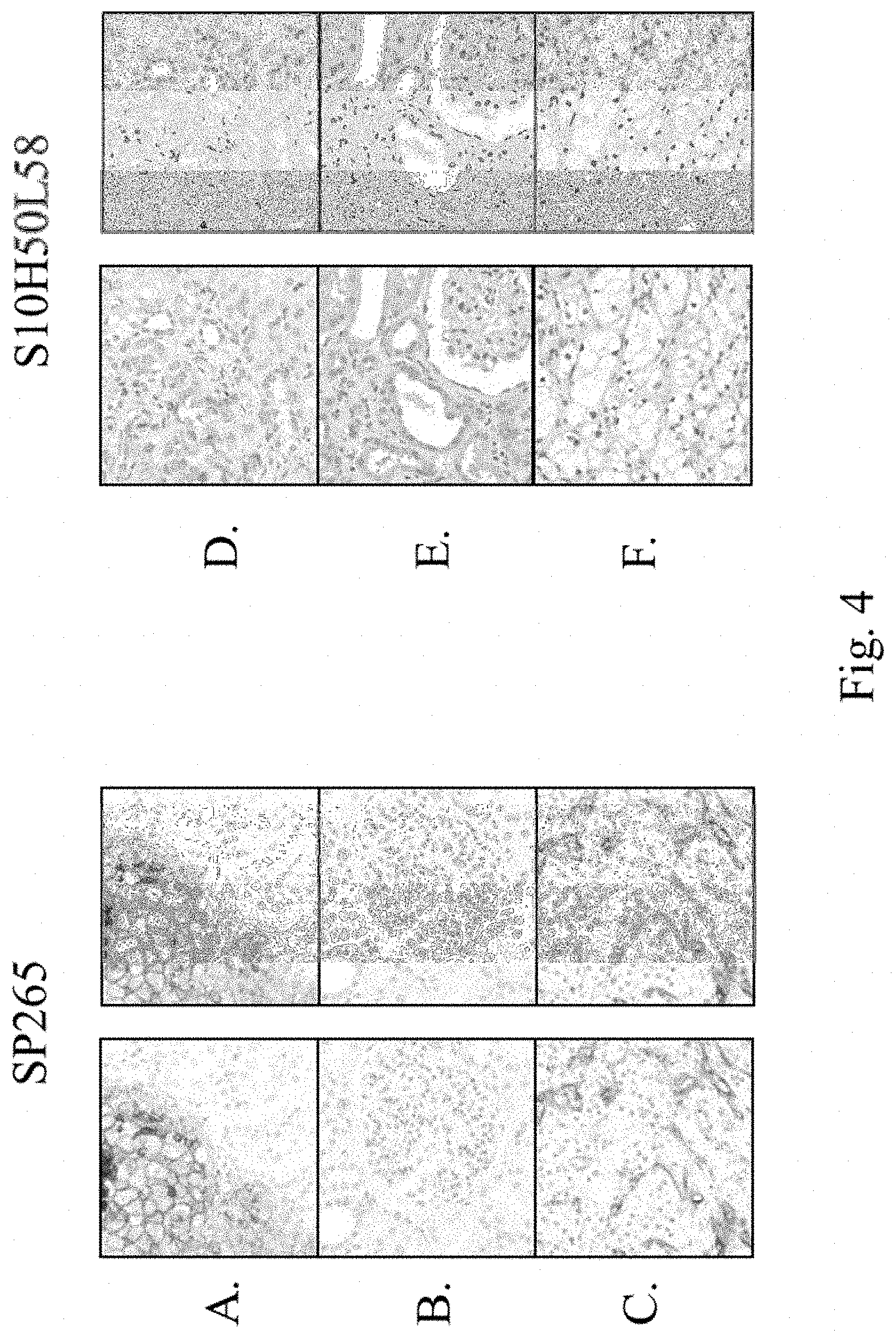
FIG. 4 shows a clone comparison of rabbit anti-human B7-H3 monoclonal antibodies SP265 and S10HSOL58 for IHC testing. (A) and (D) are Hs700t tumor cells. (B) and (E) are normal kidney tissue. (C) and (F) are renal cell carcinoma tissue sections. Left column for each antibody are color images. Right column is a grayscale of the color image.
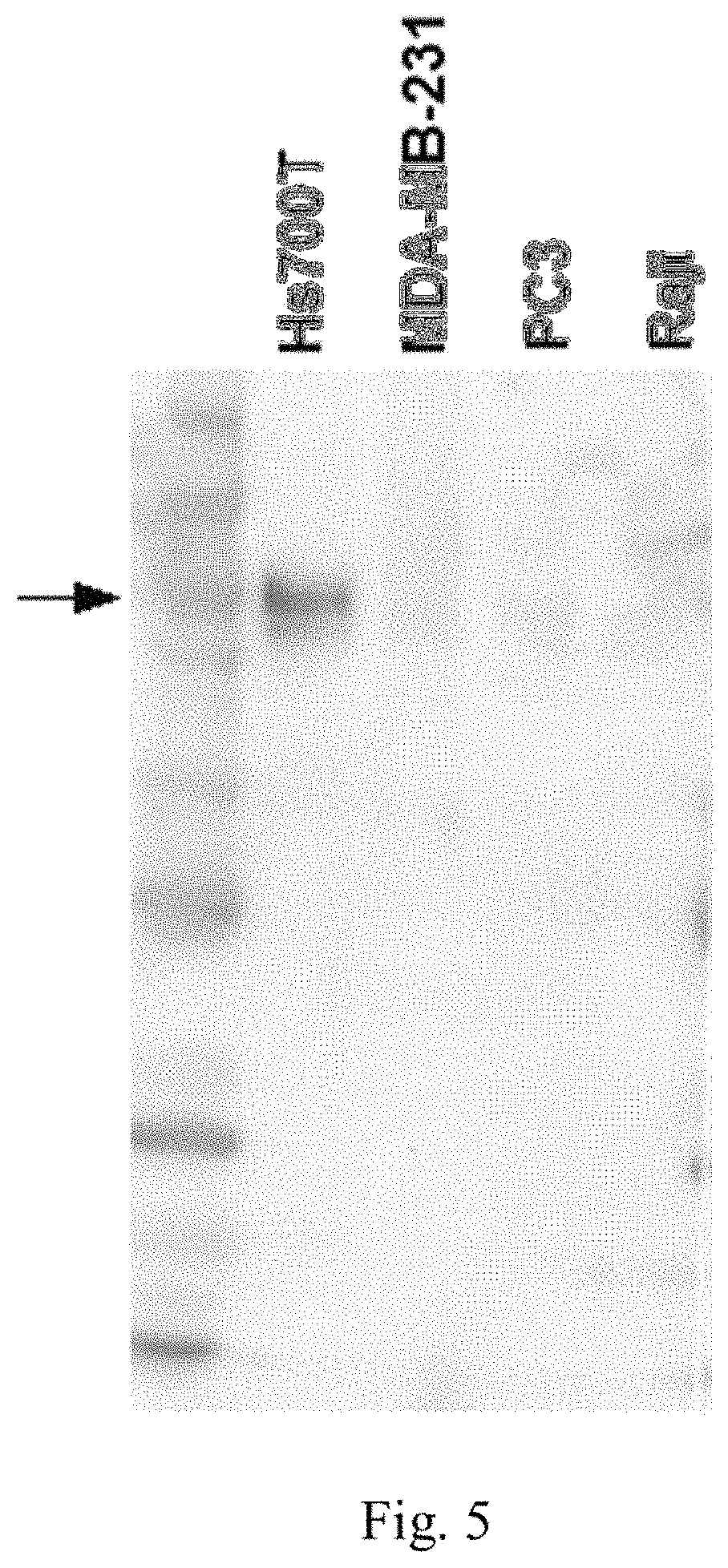
FIG. 5 is a Western blot showing B7-H3 expression in cell lysates from a HS700t cell line (high expression), a MDA-MB-231 cell line (weak expression), a PC3 cell line (weak expression), and a Raji cell line (no expression) using anti-B7-H3 antibody SP265.
FIG. 6 shows the results of an ELISA assay involving SP265 binding to immobilized peptide immunogen (aa 521-534).
DETAILED DESCRIPTION
It is to be understood that the present disclosure is not limited to particular aspects described, as such may, of course, vary. It is also to be understood that the terminology used herein is for the purpose of describing particular aspects only, and is not intended to be limiting, since the scope of the present disclosure will be limited only by the appended claims.
Unless defined otherwise, all technical and scientific terms used herein have the same meanings as commonly understood by one of ordinary skill in the art to which this technology belongs. Although any methods and materials similar or equivalent to those described herein can be used in the practice or testing of the present technology, the preferred methods, devices and materials are now described. All technical and patent publications cited herein are incorporated herein by reference in their entirety. Nothing herein is to be construed as an admission that the present technology is not entitled to antedate such disclosure by virtue of prior invention.
The practice of the present technology will employ, unless otherwise indicated, conventional techniques of tissue culture, immunology, molecular biology, microbiology, cell biology and recombinant DNA, which are within the skill of the art. See, e.g., Sambrook and Russell eds. (2001) Molecular Cloning: A Laboratory Manual, 3rd edition; the series Ausubel et al. eds. (2007) Current Protocols in Molecular Biology; the series Methods in Enzymology (Academic Press, Inc., N.Y.); MacPherson et al. (1991) PCR 1: A Practical Approach (IRL Press at Oxford University Press); MacPherson et al. (1995) PCR 2: A Practical Approach; Harlow and Lane eds. (1999) Antibodies, A Laboratory Manual; Freshney (2005) Culture of Animal Cells: A Manual of Basic Technique, 5th edition; Gait ed. (1984) Oligonucleotide Synthesis; U.S. Pat. No. 4,683,195; Hames and Higgins eds. (1984) Nucleic Acid Hybridization; Anderson (1999) Nucleic Acid Hybridization; Hames and Higgins eds. (1984) Transcription and Translation; Immobilized Cells and Enzymes (IRL Press (1986)); Perbal (1984) A Practical Guide to Molecular Cloning; Miller and Calos eds. (1987) Gene Transfer Vectors for Mammalian Cells (Cold Spring Harbor Laboratory); Makrides ed. (2003) Gene Transfer and Expression in Mammalian Cells; Mayer and Walker eds. (1987) Immunochemical Methods in Cell and Molecular Biology (Academic Press, London); and Herzenberg et al. eds (1996) Weir's Handbook of Experimental Immunology.
All numerical designations, e.g., pH, temperature, time, concentration and molecular weight, including ranges, are approximations which are varied (+) or (-) by increments of 1.0 or 0.1, as appropriate, or alternatively by a variation of +/-15%, or alternatively 10%, or alternatively 5% or alternatively 2%. It is to be understood, although not always explicitly stated, that all numerical designations are preceded by the term "about". It also is to be understood, although not always explicitly stated, that the reagents described herein are merely exemplary and that equivalents of such are known in the art.
It is to be inferred without explicit recitation and unless otherwise intended, that when the present technology relates to a polypeptide, protein, polynucleotide or antibody, an equivalent or a biologically equivalent of such is intended within the scope of the present technology.
As used in the specification and claims, the singular form "a", "an" and "the" include plural references unless the context clearly dictates otherwise. For example, the term "a cell" includes a plurality of cells, including mixtures thereof.
As used herein, the "administration" of an agent or drug to a subject or subject includes any route of introducing or delivering to a subject a compound to perform its intended function. Suitable dosage formulations and methods of administering the agents are known in the art. Route of administration can also be determined and method of determining the most effective route of administration are known to those of skill in the art and will vary with the composition used for treatment, the purpose of the treatment, the health condition or disease stage of the subject being treated and target cell or tissue. Non-limiting examples of route of administration include oral administration, vaginal, nasal administration, injection, topical application and by suppository. Administration includes self-administration and the administration by another. It is also to be appreciated that the various modes of treatment or prevention of medical conditions as described are intended to mean "substantial", which includes total but also less than total treatment or prevention, and wherein some biologically or medically relevant result is achieved.
Administration can be effected in one dose, continuously or intermittently throughout the course of treatment. Methods of determining the most effective means and dosage of administration are known to those of skill in the art and will vary with the composition used for therapy, the purpose of the therapy, the target cell being treated and the subject being treated. Single or multiple administrations can be carried out with the dose level and pattern being selected by the treating physician.
As used herein, the term "animal" refers to living multi-cellular vertebrate organisms, a category that includes, for example, mammals and birds. The term "mammal" includes both human and non-human mammals. Similarly, the term "subject" or "patient" includes both human and veterinary subjects, for example, humans, non-human primates, dogs, cats, sheep, mice, horses, and cows.
As used herein, the term "antibody" collectively refers to immunoglobulins or immunoglobulin-like molecules including by way of example and without limitation, IgA, IgD, IgE, IgG and IgM, combinations thereof, and similar molecules produced during an immune response in any vertebrate, for example, in mammals such as goats, rabbits and mice, as well as non-mammalian species, such as shark immunoglobulins. The term "antibody" includes intact immunoglobulins and "antibody fragments" or "antigen binding fragments" that specifically bind to a molecule of interest (or a group of highly similar molecules of interest) to the substantial exclusion of binding to other molecules (for example, antibodies and antibody fragments that have a binding constant for the molecule of interest that is at least 10.sup.3 M.sup.-1 greater, at least 10.sup.4 M.sup.-1 greater or at least 10.sup.5 M.sup.-1 greater than a binding constant for other molecules in a biological sample). The term "antibody" also includes genetically engineered forms such as chimeric antibodies (for example, humanized murine antibodies), heteroconjugate antibodies (such as, bispecific antibodies). See also, Pierce Catalog and Handbook, 1994-1995 (Pierce Chemical Co., Rockford, Ill.); Kuby, J., Immunology, 3.sup.rd Ed., W.H. Freeman & Co., New York, 1997.
More particularly, "antibody" refers to a polypeptide ligand comprising at least a light chain or heavy chain immunoglobulin variable region which specifically recognizes and binds an epitope of an antigen. Antibodies are composed of a heavy and a light chain, each of which has a variable region, termed the variable heavy (V.sub.H) region and the variable light (V.sub.L) region. Together, the V.sub.H region and the V.sub.L region are responsible for binding the antigen recognized by the antibody.
Typically, an immunoglobulin has heavy (H) chains and light (L) chains interconnected by disulfide bonds. There are two types of light chain, lambda (.lamda.) and kappa (.kappa.). There are five main heavy chain classes (or isotypes) which determine the functional activity of an antibody molecule: IgM, IgD, IgG, IgA and IgE. Each heavy and light chain contains a constant region and a variable region, (the regions are also known as "domains"). In combination, the heavy and the light chain variable regions specifically bind the antigen. Light and heavy chain variable regions contain a "framework" region interrupted by three hypervariable regions, also called "complementarity-determining regions" or "CDRs". The extent of the framework region and CDRs have been defined (see, Kabat et al., Sequences of Proteins of Immunological Interest, U.S. Department of Health and Human Services, 1991, which is hereby incorporated by reference). The Kabat database is now maintained online. The sequences of the framework regions of different light or heavy chains are relatively conserved within a species. The framework region of an antibody, that is the combined framework regions of the constituent light and heavy chains, largely adopt a .beta.-sheet conformation and the CDRs form loops which connect, and in some cases form part of, the .beta.-sheet structure. Thus, framework regions act to form a scaffold that provides for positioning the CDRs in correct orientation by inter-chain, non-covalent interactions.
The CDRs are primarily responsible for binding to an epitope of an antigen. The CDRs of each chain are typically referred to as CDR1, CDR2, and CDR3, numbered sequentially starting from the N-terminus, and are also typically identified by the chain in which the particular CDR is located. Thus, a V.sub.H CDR3 is located in the variable domain of the heavy chain of the antibody in which it is found, whereas a V.sub.L CDR1 is the CDR1 from the variable domain of the light chain of the antibody in which it is found. An antibody that binds B7-H3 will have a specific V.sub.H region and the V.sub.L region sequence, and thus specific CDR sequences. Antibodies with different specificities (i.e. different combining sites for different antigens) have different CDRs. Although it is the CDRs that vary from antibody to antibody, only a limited number of amino acid positions within the CDRs are directly involved in antigen binding. These positions within the CDRs are called specificity determining residues (SDRs).
The term "antibody" is further intended to encompass digestion fragments, specified portions, derivatives and variants thereof, including antibody mimetics or comprising portions of antibodies that mimic the structure and/or function of an antibody or specified fragment or portion thereof, including single chain antibodies and fragments thereof. Examples of binding fragments encompassed within the term "antigen binding portion" of an antibody include a Fab fragment, a monovalent fragment consisting of the V.sub.L, V.sub.H, C.sub.L and C.sub.H, domains; a F(ab').sub.2 fragment, a bivalent fragment comprising two Fab fragments linked by a disulfide bridge at the hinge region; a F.sub.d fragment consisting of the V.sub.H and C.sub.H, domains; a F.sub.v fragment consisting of the V.sub.L and V.sub.H domains of a single arm of an antibody, a dAb fragment (Ward et al. (1989) Nature 341:544-546), which consists of a V.sub.H domain; and an isolated complementarity determining region (CDR). Furthermore, although the two domains of the F.sub.v fragment, V.sub.L and V.sub.H, are coded for by separate genes, they can be joined, using recombinant methods, by a synthetic linker that enables them to be made as a single protein chain in which the V.sub.L and V.sub.H regions pair to form monovalent molecules (known as single chain F.sub.v (scF.sub.v)). Bird et al. (1988) Science 242:423-426 and Huston et al. (1988) Proc. Natl. Acad Sci. USA 85:5879-5883. Single chain antibodies are also intended to be encompassed within the term "fragment of an antibody." Any of the above-noted antibody fragments are obtained using conventional techniques known to those of skill in the art, and the fragments are screened for binding specificity and neutralization activity in the same manner as are intact antibodies.
"Antibody fragments" or "antigen binding fragments" include proteolytic antibody fragments (such as F(ab').sub.2 fragments, Fab' fragments, Fab'-SH fragments and Fab fragments as are known in the art), recombinant antibody fragments (such as sF.sub.v fragments, dsF.sub.v fragments, bispecific sF.sub.v fragments, bispecific dsF.sub.v fragments, F(ab)'.sub.2 fragments, single chain Fv proteins ("scF.sub.v"), disulfide stabilized F.sub.v proteins ("dsF.sub.v"), diabodies, and triabodies (as are known in the art), and camelid antibodies (see, for example, U.S. Pat. Nos. 6,015,695; 6,005,079; 5,874,541; 5,840,526; 5,800,988; and 5,759,808). An scF.sub.v protein is a fusion protein in which a light chain variable region of an immunoglobulin and a heavy chain variable region of an immunoglobulin are bound by a linker, while in dsF.sub.vs, the chains have been mutated to introduce a disulfide bond to stabilize the association of the chains.
As used herein, the term "antibody derivative" is intended to encompass molecules that bind an epitope as defined herein and which are modifications or derivatives of an isolated B7-H3 antibody of the present technology. Derivatives include, but are not limited to, for example, bispecific, heterospecific, trispecific, tetraspecific, multispecific antibodies, diabodies, chimeric, recombinant and humanized. As used herein, the term "bispecific molecule" is intended to include any agent, e.g., a protein, peptide, or protein or peptide complex, which has two different binding specificities. As used herein, the term "multispecific molecule" or "heterospecific molecule" is intended to include any agent, e.g., a protein, peptide, or protein or peptide complex, which has more than two different binding specificities. As used herein, the term "heteroantibodies" refers to two or more antibodies, antibody binding fragments (e.g., Fab), derivatives thereof, or antigen binding regions linked together, at least two of which have different specificities.
The term "antibody variant" is intended to include antibodies produced in a species other than a rabbit. It also includes antibodies containing post-translational modifications to the linear polypeptide sequence of the antibody or fragment. It further encompasses fully human antibodies.
As used herein, the term "antigen" refers to a compound, composition, or substance that may be specifically bound by the products of specific humoral or cellular immunity, such as an antibody molecule or T-cell receptor. Antigens can be any type of molecule including, for example, haptens, simple intermediary metabolites, sugars (e.g., oligosaccharides), lipids, and hormones as well as macromolecules such as complex carbohydrates (e.g., polysaccharides), phospholipids, and proteins. Common categories of antigens include, but are not limited to, viral antigens, bacterial antigens, fungal antigens, protozoa and other parasitic antigens, tumor antigens, antigens involved in autoimmune disease, allergy and graft rejection, toxins, and other miscellaneous antigens.
As used herein, "binding affinity" refers to the tendency of one molecule to bind (typically non-covalently) with another molecule, such as the tendency of a member of a specific binding pair for another member of a specific binding pair. A binding affinity can be measured as a binding constant, which binding affinity for a specific binding pair (such as an antibody/antigen pair) can be at least 1.times.10.sup.-5 M, at least 1.times.10.sup.-6 M, at least 1.times.10.sup.-7 M, at least 1.times.10.sup.-8 M, at least 1.times.10.sup.-9 M, at least 1.times.10.sup.-10 M, at least 1.times.10.sup.-11 M or at least 1.times.10.sup.-12 M. In one aspect, binding affinity is calculated by a modification of the Scatchard method described by Frankel et al., Mol. Immunol., 16:101-106, 1979. In another aspect, binding affinity is measured by an antigen/antibody dissociation rate. In yet another aspect, a high binding affinity is measured by a competition radioimmunoassay. In several examples, a high binding affinity for an antibody/antigen pair is at least about 1.times.10.sup.-8 M. In other aspects, a high binding affinity is at least about 1.5.times.10.sup.-8 M, at least about 2.0.times.10.sup.-8 M, at least about 2.5.times.10.sup.-8 M, at least about 3.0.times.10.sup.-8 M, at least about 3.5.times.10.sup.-8 M, at least about 4.0.times.10.sup.-8 M, at least about 4.5.times.10.sup.-8 M, or at least about 5.0.times.10.sup.-8 M.
As used herein, the term "biological equivalent thereof" is intended to be synonymous with "equivalent thereof" when referring to a reference protein, antibody, polypeptide, polynucleotide or nucleic acid, and intends those having minimal homology while still maintaining desired structure or functionality. Unless specifically recited herein, it is contemplated that any nucleic acid, polynucleotide, polypeptide, protein or antibody mentioned herein also includes equivalents thereof. For example, an equivalent intends at least about 80% homology or identity and alternatively, at least about 85%, or alternatively at least about 90%, or alternatively at least about 95%, or alternatively 98% percent homology or identity and exhibits substantially equivalent biological activity to the reference protein, polypeptide, antibody or nucleic acid.
A "composition" typically intends a combination of the active agent, e.g., compound or composition, and a carrier, inert (for example, a detectable agent or label) or active, such as an adjuvant, diluent, binder, stabilizer, buffers, salts, lipophilic solvents, preservative, adjuvant or the like and include pharmaceutically acceptable carriers. Carriers also include pharmaceutical excipients and additives proteins, peptides, amino acids, lipids, and carbohydrates (e.g., sugars, including monosaccharides, di-, tri-, tetra-oligosaccharides, and oligosaccharides; derivatized sugars such as alditols, aldonic acids, esterified sugars and the like; and polysaccharides or sugar polymers), which can be present singly or in combination, comprising alone or in combination 1-99.99% by weight or volume. Exemplary protein excipients include serum albumin such as human serum albumin (HSA), recombinant human albumin (rHA), gelatin, casein, and the like. Representative amino acid/antibody components, which can also function in a buffering capacity, include alanine, glycine, arginine, betaine, histidine, glutamic acid, aspartic acid, cysteine, lysine, leucine, isoleucine, valine, methionine, phenylalanine, aspartame, and the like. Carbohydrate excipients are also intended within the scope of this technology, examples of which include but are not limited to monosaccharides such as fructose, maltose, galactose, glucose, D-mannose, sorbose, and the like; disaccharides, such as lactose, sucrose, trehalose, cellobiose, and the like; polysaccharides, such as raffinose, melezitose, maltodextrins, dextrans, starches, and the like; and alditols, such as mannitol, xylitol, maltitol, lactitol, xylitol sorbitol (glucitol) and myoinositol.
The term carrier further includes a buffer or a pH adjusting agent; typically, the buffer is a salt prepared from an organic acid or base that in one aspect, serves to stabilize the antibody in a formulation for storage. Representative buffers include organic acid salts such as salts of citric acid, ascorbic acid, gluconic acid, carbonic acid, tartaric acid, succinic acid, acetic acid, or phthalic acid; Tris, tromethamine hydrochloride, or phosphate buffers. Additional carriers include polymeric excipients/additives such as polyvinylpyrrolidones, ficolls (a polymeric sugar), dextrates (e.g., cyclodextrins, such as 2-hydroxypropyl-.quadrature.-cyclodextrin), polyethylene glycols, antimicrobial agents, antioxidants, antistatic agents, surfactants (e.g., polysorbates such as "TWEEN 20" and "TWEEN 80"), lipids (e.g., phospholipids, fatty acids), steroids (e.g., cholesterol), and chelating agents (e.g., EDTA).
As used herein, the term carrier includes typical pharmaceutically acceptable carriers, e.g., such as a phosphate buffered saline solution, water, and emulsions, such as an oil/water or water/oil emulsion, and various types of wetting agents. Examples of pharmaceutically acceptable carriers include ion exchangers, alumina, aluminum stearate, lecithin, serum proteins, such as human serum albumin, buffer substances, such as phosphates, glycine, sorbic acid, potassium sorbate, partial glyceride mixtures of saturated vegetable fatty acids, water, salts or electrolytes, such as protamine sulfate, disodium hydrogen phosphate, potassium hydrogen phosphate, sodium chloride, zinc salts, colloidal silica, magnesium trisilicate, polyvinyl pyrrolidone, cellulose-based substances, polyethylene glycol, sodium carboxymethylcellulose, polyacrylates, waxes, polyethylene-polyoxypropylene-block polymers, polyethylene glycol and wool fat. For examples of carriers, stabilizers and adjuvants, see Martin REMINGTON'S PHARM. SCI., 15th Ed. (Mack Publ. Co., Easton (1975) and Williams & Williams, (1995), and in the "PHYSICIAN'S DESK REFERENCE", 52.sup.nd ed., Medical Economics, Montvale, N.J. (1998).
In one aspect, the term "equivalent" or "biological equivalent" of an antibody means the ability of the antibody to selectively bind its epitope protein or fragment thereof as measured by ELISA, IHC or other suitable methods. Biologically equivalent antibodies include, but are not limited to, those antibodies, peptides, antibody fragments, antibody variant, antibody derivative and antibody mimetics that bind to the same epitope as the reference antibody. The skilled artisan can prepare an antibody functionally equivalent to the antibodies of the present disclosure by introducing appropriate mutations into the antibody using site-directed mutagenesis (Hashimoto-Gotoh, T. et al., Gene 152, 271-275 (1995); Zoller & Smith, Methods Enzymol. 100, 468-500 (1983); Kramer, W. et al., Nucleic Acids Res. 12, 9441-9456 (1984); Kramer W. & Fritz H J., Methods. Enzymol. 154, 350-367 (1987); Kunkel, T A., Proc Natl Acad Sci USA. 82, 488-492 (1985); and Kunkel Methods Enzymol. 85, 2763-2766 (1988)).
Antibodies that are functionally equivalent to the antibodies of the present disclosure and comprise an amino acid sequence comprising mutation of one or more amino acids in the amino acid sequence of an antibody disclosed herein are also included in the antibodies of the present technology. In such mutants, the number of amino acids that are mutated is generally 50 amino acids or less, preferably 30 or less, and more preferably 10 or less (for example, 5 amino acids or less). An amino acid residue is preferably mutated into one that conserves the properties of the amino acid side chain. For example, based on their side chain properties, amino acids are classified into:
hydrophobic amino acids (A, I, L, M, F, P, W, Y, and V);
hydrophilic amino acids (R, D, N, C, E, Q, G, H, K, S, and T);
amino acids having aliphatic side-chains (G, A, V, L, I, and P);
amino acids having hydroxyl group-containing side-chains (S, T, and Y);
amino acids having sulfur atom-containing side-chains (C and M);
amino acids having carboxylic acid- and amide-containing side-chains (D, N, E, and Q);
base-containing side-chains (R, K, and H); and
amino acids having aromatic-containing side-chains (H, F, Y, and W).
(The letters within parentheses indicate one-letter amino acid codes)
As used herein, the term "biological sample" means sample material derived from or contacted by living cells. The term "biological sample" is intended to include tissues, cells and biological fluids isolated from a subject, as well as tissues, cells, and fluids present within a subject. Biological samples of the present disclosure include, e.g., but are not limited to, whole blood, plasma, semen, saliva, tears, urine, fecal material, sweat, buccal, skin, cerebrospinal fluid, and hair. Biological samples can also be obtained from biopsies of internal organs or from cancers. Biological samples can be obtained from subjects for diagnosis or research or can be obtained from healthy individuals, as controls or for basic research.
As used herein, "B7-H3" is a member of the B7/CD28 superfamily of costimulatory molecules serving as an accessory modulator of T-cell response. B7-H3 is a type I transmembrane protein that shares 20%-27% amino acid identity with other B7 family members. While murine B7-H3 consists of a single extracellular variable-type immunoglobulin (Ig)V-IgC domain and a signature intracellular domain (2Ig B7-H3), human B7-H3 possesses an additional isoform, the so-called 4Ig B7-H3 that contains a nearly exact tandem duplication of the IgV-IgC domain. (Entrez Gene ID: 80381, UniProtKB: Q5ZPR3 http://www.ncbi.nlm.nih gov/last accessed Oct. 20, 2014).
The terms "cancer," "neoplasm," and "tumor," used interchangeably and in either the singular or plural form, refer to cells that have undergone a malignant transformation that makes them pathological to the host organism and are selected from the group consisting of bladder transitional cell carcinoma, renal cell carcinoma, and lung squamous cell carcinoma.
Primary cancer cells (that is, cells obtained from near the site of malignant transformation) can be readily distinguished from non-cancerous cells by well-established techniques, particularly histological examination. The definition of a cancer cell, as used herein, includes not only a primary cancer cell, but also any cell derived from a cancer cell ancestor. This includes metastasized cancer cells, and in vitro cultures and cell lines derived from cancer cells. When referring to a type of cancer that normally manifests as a solid tumor, a "clinically detectable" tumor is one that is detectable on the basis of tumor mass; e.g., by such procedures as CAT scan, magnetic resonance imaging (MRI), X-ray, ultrasound or palpation. Biochemical or immunologic findings alone may be insufficient to meet this definition.
A neoplasm is an abnormal mass or colony of cells produced by a relatively autonomous new growth of tissue. Most neoplasms arise from the clonal expansion of a single cell that has undergone neoplastic transformation. The transformation of a normal to a neoplastic cell can be caused by a chemical, physical, or biological agent (or event) that directly and irreversibly alters the cell genome. Neoplastic cells are characterized by the loss of some specialized functions and the acquisition of new biological properties, foremost, the property of relatively autonomous (uncontrolled) growth. Neoplastic cells pass on their heritable biological characteristics to progeny cells.
The past, present, and future predicted biological behavior, or clinical course, of a neoplasm is further classified as benign or malignant, a distinction of great importance in diagnosis, treatment, and prognosis. A malignant neoplasm manifests a greater degree of autonomy, is capable of invasion and metastatic spread, may be resistant to treatment, and may cause death. A benign neoplasm has a lesser degree of autonomy, is usually not invasive, does not metastasize, and generally produces no great harm if treated adequately.
Cancer is a generic term for malignant neoplasms. Anaplasia is a characteristic property of cancer cells and denotes a lack of normal structural and functional characteristics (undifferentiation).
A tumor is literally a swelling of any type, such as an inflammatory or other swelling, but modem usage generally denotes a neoplasm.
Histogenesis is the origin of a tissue and is a method of classifying neoplasms on the basis of the tissue cell of origin. Adenomas are benign neoplasms of glandular epithelium. Carcinomas are malignant tumors of epithelium. Sarcomas are malignant tumors of mesenchymal tissues. One system to classify neoplasia utilizes biological (clinical) behavior, whether benign or malignant, and the histogenesis, the tissue or cell of origin of the neoplasm as determined by histologic and cytologic examination. Neoplasms may originate in almost any tissue containing cells capable of mitotic division. The histogenetic classification of neoplasms is based upon the tissue (or cell) of origin as determined by histologic and cytologic examination.
As used herein, the term "chimeric antibody" means an antibody in which the Fc constant region of a monoclonal antibody from one species (e.g., a mouse Fc constant region) is replaced, using recombinant DNA techniques, with an Fc constant region from an antibody of another species (e.g., a human Fc constant region). See generally, Robinson et al., PCT/US86/02269; Akira et al., European Patent Application 184,187; Taniguchi, European Patent Application 171,496; Morrison et al., European Patent Application 173,494; Neuberger et al., WO 86/01533; Cabilly et al. U.S. Pat. No. 4,816,567; Cabilly et al., European Patent Application 125,023; Better et al., Science 240: 1041-1043, 1988; Liu et al., Proc. Natl. Acad. Sci. USA 84: 3439-3443, 1987; Liu et al., J. Immunol. 139: 3521-3526, 1987; Sun et al., Proc. Natl. Acad. Sci. USA 84: 214-218, 1987; Nishimura et al., Cancer Res 47: 999-1005, 1987; Wood et al., Nature 314: 446-449, 1885; and Shaw et al., J. Natl. Cancer Inst. 80: 1553-1559, 1988. In certain aspects the target binding region or site will be from a non-human source (e.g. mouse or primate) and the constant region is human.
As used herein, the term "comprising" is intended to mean that the compositions and methods include the recited elements, but do not exclude others. "Consisting essentially of" when used to define compositions and methods, shall mean excluding other elements of any essential significance to the combination for the intended use. For example, a composition consisting essentially of the elements as defined herein would not exclude trace contaminants from the isolation and purification method and pharmaceutically acceptable carriers, such as phosphate buffered saline, preservatives and the like. "Consisting of" shall mean excluding more than trace elements of other ingredients and substantial method steps for administering the compositions disclosed herein. Aspects defined by each of these transition terms are within the scope of the present disclosure.
A "control" biological sample is an alternative sample used in an experiment for comparison purpose. A control can be "positive" or "negative". For example, where the purpose of the experiment is to determine a correlation of the efficacy of a therapeutic agent for the treatment for a particular type of cancer, it is generally preferable to use a positive control (a compound or composition known to exhibit the desired therapeutic effect) and a negative control (a subject or a sample that does not receive the therapy or receives a placebo).
As used herein, the term "detectable label" refers to a molecule or material that can produce a detectable (such as visually, electronically or otherwise) signal that indicates the presence and/or concentration of the label in a sample. When conjugated to a specific binding molecule, the detectable label can be used to locate and/or quantify the target to which the specific binding molecule is directed. Thereby, the presence and/or concentration of the target in a sample can be detected by detecting the signal produced by the detectable label. A detectable label can be detected directly or indirectly, and several different detectable labels conjugated to different specific-binding molecules can be used in combination to detect one or more targets. For example, a first detectable label conjugated to an antibody specific to a target can be detected indirectly through the use of a second detectable label that is conjugated to a molecule that specifically binds the first detectable label. Multiple detectable labels that can be separately detected can be conjugated to different specific binding molecules that specifically bind different targets to provide a multiplexed assay that can provide simultaneous detection of the multiple targets in a sample. A detectable signal can be generated by any mechanism including absorption, emission and/or scattering of a photon (including radio frequency, microwave frequency, infrared frequency, visible frequency and ultra-violet frequency photons). Detectable labels include colored, fluorescent, phosphorescent and luminescent molecules and materials, catalysts (such as enzymes) that convert one substance into another substance to provide a detectable difference (such as by converting a colorless substance into a colored substance or vice versa, or by producing a precipitate or increasing sample turbidity), haptens that can be detected through antibody-hapten binding interactions using additional detectably labeled antibody conjugates, and paramagnetic and magnetic molecules or materials. Particular examples of detectable labels include enzymes such as horseradish peroxidase, alkaline phosphatase, acid phosphatase, glucose oxidase, .beta.-galactosidase or .beta.-glucuronidase; fluorphores such as fluoresceins, luminophores, coumarins, BODIPY dyes, resorufins, and rhodamines (many additional examples of fluorescent molecules can be found in The Handbook--A Guide to Fluorescent Probes and Labeling Technologies, Molecular Probes, Eugene, Oreg.); nanoparticles such as quantum dots (obtained, for example, from QuantumDot Corp, Invitrogen Nanocrystal Technologies, Hayward, Calif.; see also, U.S. Pat. Nos. 6,815,064, 6,682,596 and 6,649,138, each of which patents is incorporated by reference herein); metal chelates such as DOTA and DPTA chelates of radioactive or paramagnetic metal ions like Gd.sup.3+; and liposomes, for example, liposomes containing trapped fluorescent molecules. Where the detectable label includes an enzyme, a detectable substrate such as a chromogen, a fluorogenic compound, or a luminogenic compound can be used in combination with the enzyme to generate a detectable signal (A wide variety of such compounds are commercially available, for example, from Invitrogen Corporation, Eugene Oreg.). Particular examples of chromogenic compounds include diaminobenzidine (DAB), 4-nitrophenylphospate (pNPP), fast red, bromochloroindolyl phosphate (BCIP), nitro blue tetrazolium (NBT), BCIP/NBT, fast red, AP Orange, AP blue, tetramethylbenzidine (TMB), 2,2'-azino-di-[3-ethylbenzothiazoline sulphonate] (ABTS), o-dianisidine, 4-chloronaphthol (4-CN), nitrophenyl-.beta.-D-galactopyranoside (ONPG), o-phenylenediamine (OPD), 5-bromo-4-chloro-3-indolyl-.beta.-galactopyranoside (X-Gal), methylumbelliferyl-.beta.-D-galactopyranoside (MU-Gal), p-nitrophenyl-.alpha.-D-galactopyranoside (PNP), 5-bromo-4-chloro-3-indolyl-.beta.-D-glucuronide (X-Gluc), 3-amino-9-ethyl carbazol (AEC), fuchsin, iodonitrotetrazolium (INT), tetrazolium blue and tetrazolium violet. Alternatively, an enzyme can be used in a metallographic detection scheme. Metallographic detection methods include using an enzyme such as alkaline phosphatase in combination with a water-soluble metal ion and a redox-inactive substrate of the enzyme. The substrate is converted to a redox-active agent by the enzyme, and the redox-active agent reduces the metal ion, causing it to form a detectable precipitate. (See, for example, co-pending U.S. patent application Ser. No. 11/015,646, filed Dec. 20, 2004, PCT Publication No. 2005/003777 and U.S. Patent Application Publication No. 2004/0265922; each of which is incorporated by reference herein). Metallographic detection methods include using an oxido-reductase enzyme (such as horseradish peroxidase) along with a water soluble metal ion, an oxidizing agent and a reducing agent, again to form a detectable precipitate. (See, for example, U.S. Pat. No. 6,670,113, which is incorporated by reference herein). Haptens are small molecules that are specifically bound by antibodies, although by themselves they will not elicit an immune response in an animal and must first be attached to a larger carrier molecule such as a protein to generate an immune response. Examples of haptens include di-nitrophenyl, biotin, digoxigenin, and fluorescein. Additional examples of oxazole, pyrazole, thiazole, nitroaryl, benzofuran, triperpene, urea, thiourea, rotenoid, coumarin and cyclolignan haptens are disclosed in U.S. Provisional Patent Application No., 60/856,133, filed Nov. 1, 2006, which is incorporated by reference herein.
As used herein, an "epitope" or "antigenic determinant" refers to particular chemical groups or contiguous or non-contiguous peptide sequences on a molecule that are antigenic, i.e., that elicit a specific immune response. An antibody binds a particular antigenic epitope. Epitopes usually consist of chemically active surface groupings of molecules such as amino acids or sugar side chains and usually have specific three dimensional structural characteristics, as well as specific charge characteristics. Conformational and nonconformational epitopes are distinguished in that the binding to the former but not the latter is lost in the presence of denaturing solvents.
As used herein, "expression" refers to the process by which polynucleotides are transcribed into mRNA and/or the process by which the transcribed mRNA is subsequently being translated into peptides, polypeptides, or proteins. If the polynucleotide is derived from genomic DNA, expression may include splicing of the mRNA in an eukaryotic cell. The expression level of a gene may be determined by measuring the amount of mRNA or protein in a cell or tissue sample. In one aspect, the expression level of a gene from one sample may be directly compared to the expression level of that gene from a control or reference sample. In another aspect, the expression level of a gene from one sample may be directly compared to the expression level of that gene from the same sample following administration of a compound.
As used herein, "homology" or "identical", percent "identity" or "similarity", when used in the context of two or more nucleic acids or polypeptide sequences, refers to two or more sequences or subsequences that are the same or have a specified percentage of nucleotides or amino acid residues that are the same, e.g., at least 60% identity, preferably at least 65%, 70%, 75%, 80%, 85%, 90%, 91%, 92%, 93%, 94%, 95%, 96%, 97%, 98%, 99%, or higher identity over a specified region (e.g., nucleotide sequence encoding an antibody described herein or amino acid sequence of an antibody described herein). Homology can be determined by comparing a position in each sequence which may be aligned for purposes of comparison. When a position in the compared sequence is occupied by the same base or amino acid, then the molecules are homologous at that position. A degree of homology between sequences is a function of the number of matching or homologous positions shared by the sequences. The alignment and the percent homology or sequence identity can be determined using software programs known in the art, for example those described in Current Protocols in Molecular Biology (Ausubel et al., eds. 1987) Supplement 30, section 7.7.18, Table 7.7.1. Preferably, default parameters are used for alignment. A preferred alignment program is BLAST, using default parameters. In particular, preferred programs are BLASTN and BLASTP, using the following default parameters: Genetic code=standard; filter=none; strand=both; cutoff=60; expect=10; Matrix=BLOSUM62; Descriptions=50 sequences; sort by=HIGH SCORE; Databases=non-redundant, GenBank+EMBL+DDBJ+PDB+GenBank CDS translations+SwissProtein+SPupdate+PIR. Details of these programs can be found at the following Internet address: ncbi.nlm.nih gov/cgi-bin/BLAST. The terms "homology" or "identical", percent "identity" or "similarity" also refer to, or can be applied to, the complement of a test sequence. The terms also include sequences that have deletions and/or additions, as well as those that have substitutions. As described herein, the preferred algorithms can account for gaps and the like. Preferably, identity exists over a region that is at least about 25 amino acids or nucleotides in length, or more preferably over a region that is at least 50-100 amino acids or nucleotides in length. An "unrelated" or "non-homologous" sequence shares less than 40% identity, or alternatively less than 25% identity, with one of the sequences disclosed herein.
The term "human antibody" as used herein, is intended to include antibodies having variable and constant regions derived from human germline immunoglobulin sequences. The human antibodies of the present technology may include amino acid residues not encoded by human germline immunoglobulin sequences (e.g., mutations introduced by random or site-specific mutagenesis in vitro or by somatic mutation in vivo). However, the term "human antibody" as used herein, is not intended to include antibodies in which CDR sequences derived from the germline of another mammalian species, such as a rabbit, have been grafted onto human framework sequences. Thus, as used herein, the term "human antibody" refers to an antibody in which substantially every part of the protein (e.g., CDR, framework, C.sub.L, C.sub.H domains (e.g., C.sub.H1, C.sub.H2, C.sub.H3), hinge, V.sub.L, V.sub.H) is substantially non-immunogenic in humans, with only minor sequence changes or variations. Similarly, antibodies designated primate (monkey, baboon, chimpanzee, etc.), rodent (mouse, rat, rabbit, guinea pig, hamster, and the like) and other mammals designate such species, sub-genus, genus, sub-family, family specific antibodies. Further, chimeric antibodies include any combination of the above. Such changes or variations optionally and preferably retain or reduce the immunogenicity in humans or other species relative to non-modified antibodies. Thus, a human antibody is distinct from a chimeric or humanized antibody. It is pointed out that a human antibody can be produced by a non-human animal or prokaryotic or eukaryotic cell that is capable of expressing functionally rearranged human immunoglobulin (e.g., heavy chain and/or light chain) genes. Further, when a human antibody is a single chain antibody, it can comprise a linker peptide that is not found in native human antibodies. For example, an F.sub.v can comprise a linker peptide, such as two to about eight glycine or other amino acid residues, which connects the variable region of the heavy chain and the variable region of the light chain. Such linker peptides are considered to be of human origin.
As used herein, the term "humanized antibody" refers to an antibody comprising a humanized light chain and a humanized heavy chain immunoglobulin. A humanized antibody binds to the same antigen as the donor antibody that provides the CDRs. The acceptor framework of a humanized immunoglobulin or antibody may have a limited number of substitutions by amino acids taken from the donor framework. Humanized or other monoclonal antibodies can have additional conservative amino acid substitutions which have substantially no effect on antigen binding or other immunoglobulin functions. Humanized immunoglobulins can be constructed by means of genetic engineering (see for example, U.S. Pat. No. 5,585,089).
As used herein, the term "humanized immunoglobulin" refers to an immunoglobulin including a human framework region and one or more CDRs from a non-human (for example a mouse, rat, rabbit or synthetic) immunoglobulin. The non-human immunoglobulin providing the CDRs is termed a "donor," and the human immunoglobulin providing the framework is termed an "acceptor." In one aspect, all the CDRs are from the donor immunoglobulin in a humanized immunoglobulin. Constant regions need not be present, but if they are, they must be substantially identical to human immunoglobulin constant regions, i.e., at least about 85-90%, or at least about 95% or more identical. Hence, all parts of a humanized immunoglobulin, except possibly the CDRs, are substantially identical to corresponding parts of natural human immunoglobulin sequences.
The term "isolated" as used herein refers to molecules or biological or cellular materials being substantially free from other materials. In one aspect, the term "isolated" refers to nucleic acid, such as DNA or RNA, or protein or polypeptide (e.g., an antibody or derivative thereof), or cell or cellular organelle, or tissue or organ, separated from other DNAs or RNAs, or proteins or polypeptides, or cells or cellular organelles, or tissues or organs, respectively, that are present in the natural source. The term "isolated" also refers to a nucleic acid or peptide that is substantially free of cellular material, viral material, or culture medium when produced by recombinant DNA techniques, or chemical precursors or other chemicals when chemically synthesized. Moreover, an "isolated nucleic acid" is meant to include nucleic acid fragments which are not naturally occurring as fragments and would not be found in the natural state. The term "isolated" is also used herein to refer to polypeptides which are isolated from other cellular proteins and is meant to encompass both purified and recombinant polypeptides. The term "isolated" is also used herein to refer to cells or tissues that are isolated from other cells or tissues and is meant to encompass both cultured and engineered cells or tissues.
As used herein, the term "monoclonal antibody" refers to an antibody produced by a single clone of B-lymphocytes or by a cell into which the light and heavy chain genes of a single antibody have been transfected. Monoclonal antibodies are produced by methods known to those of skill in the art, for instance by making hybrid antibody-forming cells from a fusion of myeloma cells with immune spleen cells. Monoclonal antibodies include humanized monoclonal antibodies.
As used herein, a "pathological cell" is one that is pertaining to or arising from disease. Pathological cells can be hyperproliferative. A "hyperproliferative cell" means cells or tissue are dividing and growing at a rate greater than that when the cell or tissue is in a normal or healthy state. Examples of such include, but are not limited to precancerous (i.e., epithelial dysplasia) and cancer cells. Hyperproliferative cells also include de-differentiated, immortalized, neoplastic, malignant, metastatic, and cancer cells such as sarcoma cells, leukemia cells, carcinoma cells, or adenocarcinoma cells.
The term "protein", "peptide" and "polypeptide" are used interchangeably and in their broadest sense to refer to a compound of two or more subunit amino acids, amino acid analogs or peptidomimetics. The subunits may be linked by peptide bonds. In another aspect, the subunit may be linked by other bonds, e.g., ester, ether, etc. A protein or peptide must contain at least two amino acids and no limitation is placed on the maximum number of amino acids which may comprise a protein's or peptide's sequence. As used herein the term "amino acid" refers to either natural and/or unnatural or synthetic amino acids, including glycine and both the D and L optical isomers, amino acid analogs and peptidomimetics.
The terms "polynucleotide" and "oligonucleotide" are used interchangeably and refer to a polymeric form of nucleotides of any length, either deoxyribonucleotides or ribonucleotides or analogs thereof. Polynucleotides can have any three-dimensional structure and may perform any function, known or unknown. The following are non-limiting examples of polynucleotides: a gene or gene fragment (for example, a probe, primer, EST or SAGE tag), exons, introns, messenger RNA (mRNA), transfer RNA, ribosomal RNA, RNAi, ribozymes, cDNA, recombinant polynucleotides, branched polynucleotides, plasmids, vectors, isolated DNA of any sequence, isolated RNA of any sequence, nucleic acid probes and primers. A polynucleotide can comprise modified nucleotides, such as methylated nucleotides and nucleotide analogs. If present, modifications to the nucleotide structure can be imparted before or after assembly of the polynucleotide. The sequence of nucleotides can be interrupted by non-nucleotide components. A polynucleotide can be further modified after polymerization, such as by conjugation with a labeling component. The term also refers to both double- and single-stranded molecules. Unless otherwise specified or required, any aspect of this technology that is a polynucleotide encompasses both the double-stranded form and each of two complementary single-stranded forms known or predicted to make up the double-stranded form.
A polynucleotide is composed of a specific sequence of four nucleotide bases: adenine (A); cytosine (C); guanine (G); thymine (T); and uracil (U) for thymine when the polynucleotide is RNA. Thus, the term "polynucleotide sequence" is the alphabetical representation of a polynucleotide molecule. This alphabetical representation can be input into databases in a computer having a central processing unit and used for bioinformatics applications such as functional genomics and homology searching
As used herein, the term "purified" does not require absolute purity; rather, it is intended as a relative term. Thus, for example, a purified nucleic acid, peptide, protein, biological complexes or other active compound is one that is isolated in whole or in part from proteins or other contaminants. Generally, substantially purified peptides, proteins, biological complexes, or other active compounds for use within the disclosure comprise more than 80% of all macromolecular species present in a preparation prior to admixture or formulation of the peptide, protein, biological complex or other active compound with a pharmaceutical carrier, excipient, buffer, absorption enhancing agent, stabilizer, preservative, adjuvant or other co-ingredient in a complete pharmaceutical formulation for therapeutic administration. More typically, the peptide, protein, biological complex or other active compound is purified to represent greater than 90%, often greater than 95% of all macromolecular species present in a purified preparation prior to admixture with other formulation ingredients. In other cases, the purified preparation may be essentially homogeneous, wherein other macromolecular species are not detectable by conventional techniques.
As used herein, the term "specific binding" means the contact between an antibody and an antigen with a binding affinity of at least 10.sup.-6 M. In certain aspects, antibodies bind with affinities of at least about 10.sup.-7 M, and preferably 10.sup.-8 M, 10.sup.-9 M, 10.sup.-10 M, 10.sup.-11 M, or 10.sup.-12 M.
As used herein, the term "recombinant protein" refers to a polypeptide which is produced by recombinant DNA techniques, wherein generally, DNA encoding the polypeptide is inserted into a suitable expression vector which is in turn used to transform a host cell to produce the heterologous protein.
As used herein, "treating" or "treatment" of a disease in a subject refers to (1) preventing the symptoms or disease from occurring in a subject that is predisposed or does not yet display symptoms of the disease; (2) inhibiting the disease or arresting its development; or (3) ameliorating or causing regression of the disease or the symptoms of the disease. As understood in the art, "treatment" is an approach for obtaining beneficial or desired results, including clinical results. For the purposes of the present technology, beneficial or desired results can include one or more, but are not limited to, alleviation or amelioration of one or more symptoms, diminishment of extent of a condition (including a disease), stabilized (i.e., not worsening) state of a condition (including disease), delay or slowing of condition (including disease), progression, amelioration or palliation of the condition (including disease), states and remission (whether partial or total), whether detectable or undetectable. Preferred are compounds that are potent and can be administered locally at very low doses, thus minimizing systemic adverse effects.
"Histochemistry" and "cytochemistry" are techniques often used to identify biomarkers within the context of intact cells by labeling the samples with molecules that bind specifically to the biomarker in a manner that can be visualized on a microscope. Immunohistochemistry (IHC) and immunocytochemistry (ICC) are types of histochemistry and cytochemistry that use antibodies to label the biomarkers. By identifying the biomarker in the context of a tissue environment or cellular environment, spatial relationships between the biomarkers and other morphological or molecular features of the cell or tissue sample can be elucidated, which may reveal information that is not apparent from other molecular or cellular techniques.
MODES FOR CARRYING OUT THE DISCLOSURE
Compositions
The general structure of antibodies is known in the art and will only be briefly summarized here. An immunoglobulin monomer comprises two heavy chains and two light chains connected by disulfide bonds. Each heavy chain is paired with one of the light chains to which it is directly bound via a disulfide bond. Each heavy chain comprises a constant region (which varies depending on the isotype of the antibody) and a variable region. The variable region comprises three hypervariable regions (or complementarity determining regions) which are designated CDRH1, CDRH2 and CDRH3 and which are supported within framework regions. Each light chain comprises a constant region and a variable region, with the variable region comprising three hypervariable regions (designated CDRL1, CDRL2 and CDRL3) supported by framework regions in an analogous manner to the variable region of the heavy chain.
The hypervariable regions of each pair of heavy and light chains mutually cooperate to provide an antigen binding site that is capable of binding a target antigen. The binding specificity of a pair of heavy and light chains is defined by the sequence of CDR1, CDR2 and CDR3 of the heavy and light chains. Thus once a set of CDR sequences (i.e. the sequence of CDR1, CDR2 and CDR3 for the heavy and light chains) is determined which gives rise to a particular binding specificity, the set of CDR sequences can, in principle, be inserted into the appropriate positions within any other antibody framework regions linked with any antibody constant regions in order to provide a different antibody with the same antigen binding specificity.
In one aspect, the present disclosure provides an isolated antibody comprising a heavy chain (HC) immunoglobulin variable domain sequence and a light chain (LC) immunoglobulin variable domain sequence, wherein the heavy chain and light chain immunoglobulin variable domain sequences form an antigen binding site that binds to an epitope of human B7-H3 comprising the amino acid sequence KHSDSKEDDGQEIA (SEQ ID NO: 1) and/or has a half maximal effective concentration (EC.sub.50) of at least 6.7.times.10.sup.-11 M.
In one aspect, the sequences of CDR3 of the heavy and light chains of the B7-H3 antibodies of the present disclosure comprise the amino acid sequences of SEQ ID NOS: 4 or 10 and SEQ ID NO: 7 respectively.
In one aspect, the sequences of CDR1 and CDR2 of the heavy chain of the B7-H3 antibodies of the present disclosure comprise the amino acid sequences of SEQ ID NOS: 2 or 8 and SEQ ID NOS: 3 or 9 respectively.
In another aspect, the sequences of CDR1 of the light chain of the B7-H3 antibodies of the present disclosure comprises the amino acid sequence of SEQ ID NO: 5.
In another aspect, the sequence of CDR2 of the light chain of the B7-H3 antibodies of the present disclosure comprises the amino acid sequence of SEQ ID NO: 6.
In another aspect, the B7-H3 antibodies of the present disclosure has the CDR3 sequence of the light chain comprises the amino acid sequence of SEQ ID NO: 7 and the CDR3 sequence of the heavy chain comprises the amino acid sequence of SEQ ID NO: 4.
In another aspect, the B7-H3 antibodies of the present disclosure has the CDR3 sequence of the light chain comprises the amino acid sequence of SEQ ID NO: 7 and the CDR3 sequence of the heavy chain comprises the amino acid sequence of SEQ ID NO: 10.
Specific CDR1, CDR2 and CDR3 sequences from the preferred antibodies (SP265 and S10-H50L58) are set out in Table 1. Thus, the present disclosure provides antibodies comprising CDRs 1 to 3 having the sequences from these preferred antibodies.
In another aspect of the present technology, the isolated antibody includes one or more of the following characteristics:
(a) the LC immunoglobulin variable domain sequence comprises one or more CDRs that are at least 85% identical to a CDR of a LC variable domain of SP265 and S10-H50L58;
(b) the HC immunoglobulin variable domain sequence comprises one or more CDRs that are at least 85% identical to a CDR of a HC variable domain of SP265 and S10-H50L58;
(c) the LC immunoglobulin variable domain sequence is at least 85% identical to a LC variable domain of SP265 and S10-H50L58;
(d) the HC immunoglobulin variable domain sequence is at least 85% identical to a HC variable domain of SP265 and S10-H50L58; and
(e) the antibody binds an epitope that overlaps with an epitope bound by SP265 and S10-H50L58.
In one aspect, the present disclosure provides an isolated antibody that is at least 85% identical to an antibody selected from the group consisting of SP265 and S10-H50L58. In one aspect, the present disclosure provides an isolated antibody selected from the group consisting of SP265 and S10-H50L58.
In one aspect, the present disclosure provides an isolated antibody comprising the CDRs of SP265. In one aspect, the present disclosure provides an isolated antibody that is at least 85% identical to SP265. The CDRs of SP265 are represented in Table 1.
In one aspect, the present disclosure provides an isolated antibody comprising the CDRs of S10-H50L58. In one aspect, the present disclosure provides an isolated antibody that is at least 85% identical to S10-H50L58. The CDRs of S10-H50L58 are represented in Table 1.
In some aspects of the antibodies provided herein, the HC variable domain sequence comprises a variable domain sequence of SP265 and the LC variable domain sequence comprises a variable domain sequence of SP265.
In some aspects of the antibodies provided herein, the HC variable domain sequence comprises a variable domain sequence of S10-H50L58 and the LC variable domain sequence comprises a variable domain sequence of S10-H50L58.
In some of the aspects of the antibodies provided herein, the antibody binds human B7-H3 with a dissociation constant (K.sub.D) of less than 10.sup.-4 M, 10.sup.-5 M, 10.sup.-6 M, 10.sup.-7 M, 10.sup.-8 M, 10.sup.-9 M, 10.sup.-10 M, 10.sup.-11 M, or 10.sup.-12 M. In some of the aspects of the antibodies provided herein, the antigen binding site specifically binds to human B7-H3.
In some of the aspects of the antibodies provided herein, the antibody is soluble Fab.
In some of the aspects of the antibodies provided herein, the HC and LC variable domain sequences are components of the same polypeptide chain. In some of the aspects of the antibodies provided herein, the HC and LC variable domain sequences are components of different polypeptide chains.
In some of the aspects of the antibodies provided herein, the antibody is a full-length antibody.
In some of the aspects of the antibodies provided herein, the antibody is a monoclonal antibody.
In some of the aspects of the antibodies provided herein, the antibody is chimeric or humanized.
In some of the aspects of the antibodies provided herein, the antibody is selected from the group consisting of Fab, F(ab)'2, Fab', scF.sub.v, and F.sub.v.
In some of the aspects of the antibodies provided herein, the antibody comprises an Fc domain. In some of the aspects of the antibodies provided herein, the antibody is a rabbit antibody. In some of the aspects of the antibodies provided herein, the antibody is a human or humanized antibody or is non-immunogenic in a human.
In some of the aspects of the antibodies provided herein, the antibody comprises a human antibody framework region.
In other aspects, one or more amino acid residues in a CDR of the antibodies provided herein are substituted with another amino acid. The substitution may be "conservative" in the sense of being a substitution within the same family of amino acids. The naturally occurring amino acids may be divided into the following four families and conservative substitutions will take place within those families.
1) Amino acids with basic side chains: lysine, arginine, histidine.
2) Amino acids with acidic side chains: aspartic acid, glutamic acid
3) Amino acids with uncharged polar side chains: asparagine, glutamine, serine, threonine, tyrosine.
4) Amino acids with nonpolar side chains: glycine, alanine, valine, leucine, isoleucine, proline, phenylalanine, methionine, tryptophan, cysteine.
In another aspect, one or more amino acid residues are added to or deleted from one or more CDRs of an antibody. Such additions or deletions occur at the N or C termini of the CDR or at a position within the CDR.
By varying the amino acid sequence of the CDRs of an antibody by addition, deletion or substitution of amino acids, various effects such as increased binding affinity for the target antigen may be obtained.
It is to be appreciated that antibodies of the present disclosure comprising such varied CDR sequences still bind B7-H3 with similar specificity and sensitivity profiles as SP265. This may be tested by way of the binding assays disclosed in Examples described herein.
The constant regions of antibodies may also be varied from those specifically disclosed for antibodies SP265. For example, antibodies may be provided with Fc regions of any isotype: IgA (IgA1, IgA2), IgD, IgE, IgG (IgG1, IgG2, IgG3, IgG4) or IgM. Non-limiting examples of constant region sequences include:
TABLE-US-00001 Human IgD constant region, Uniprot: P01880 SEQ ID NO: 15 APTKAPDVFPIISGCRHPKDNSPVVLACLITGYHPTSVTVTWYMGTQSQP QRTFPEIQRRDSYYMTSSQLSTPLQQWRQGEYKCVVQHTASKSKKEIFRW PESPKAQASSVPTAQPQAEGSLAKATTAPATTRNTGRGGEEKKKEKEKEE QEERETKTPECPSHTQPLGVYLLTPAVQDLWLRDKATFTCFVVGSDLKDA HLTWEVAGKVPTGGVEEGLLERHSNGSQSQHSRLTLPRSLWNAGTSVTCT LNHPSLPPQRLMALREPAAQAPVKLSLNLLASSDPPEAASWLLCEVSGFS PPNILLMWLEDQREVNTSGFAPARPPPQPGSTTFWAWSVLRVPAPPSPQP ATYTCVVSHEDSRTLLNASRSLEVSYVTDHGPMK Human IgG1 constant region, Uniprot: P01857 SEQ ID NO: 16 ASTKGPSVFPLAPSSKSTSGGTAALGCLVKDYFPEPVTVSWNSGALTSGV HTFPAVLQSSGLYSLSSVVTVPSSSLGTQTYICNVNHKPSNTKVDKKVEP KSCDKTHTCPPCPAPELLGGPSVFLFPPKPKDTLMISRTPEVTCVVVDVS HEDPEVKFNWYVDGVEVHNAKTKPREEQYNSTYRVVSVLTVLHQDWLNGK EYKCKVSNKALPAPIEKTISKAKGQPREPQVYTLPPSRDELTKNQVSLTC LVKGFYPSDIAVEWESNGQPENNYKTTPPVLDSDGSFFLYSKLTVDKSRW QQGNVFSCSVMHEALHNHYTQKSLSLSPGK Human IgG2 constant region, Uniprot: P01859 SEQ ID NO: 17 ASTKGPSVFPLAPCSRSTSESTAALGCLVKDYFPEPVTVSWNSGALTSGV HTFPAVLQSSGLYSLSSVVTVPSSNFGTQTYTCNVDHKPSNTKVDKTVER KCCVECPPCPAPPVAGPSVFLFPPKPKDTLMISRTPEVTCVVVDVSHEDP EVQFNWYVDGVEVHNAKTKPREEQFNSTFRVVSVLTVVHQDWLNGKEYKC KVSNKGLPAPIEKTISKTKGQPREPQVYTLPPSREEMTKNQVSLTCLVKG FYPSDISVEWESNGQPENNYKTTPPMLDSDGSFFLYSKLTVDKSRWQQGN VFSCSVMHEALHNHYTQKSLSLSPGK Human IgG3 constant region, Uniprot: P01860 SEQ ID NO: 18 ASTKGPSVFPLAPCSRSTSGGTAALGCLVKDYFPEPVTVSWNSGALTSGV HTFPAVLQSSGLYSLSSVVTVPSSSLGTQTYTCNVNHKPSNTKVDKRVEL KTPLGDTTHTCPRCPEPKSCDTPPPCPRCPEPKSCDTPPPCPRCPEPKSC DTPPPCPRCPAPELLGGPSVFLFPPKPKDTLMISRTPEVTCVVVDVSHED PEVQFKWYVDGVEVHNAKTKPREEQYNSTFRVVSVLTVLHQDWLNGKEYK CKVSNKALPAPIEKTISKTKGQPREPQVYTLPPSREEMTKNQVSLTCLVK GFYPSDIAVEWESSGQPENNYNTTPPMLDSDGSFFLYSKLTVDKSRWQQG NIFSCSVMHEALHNRFTQKSLSLSPGK Human IgM constant region, Uniprot: P01871 SEQ ID NO: 19 GSASAPTLFPLVSCENSPSDTSSVAVGCLAQDFLPDSITLSWKYKNNSDI SSTRGFPSVLRGGKYAATSQVLLPSKDVMQGTDEHVVCKVQHPNGNKEKN VPLPVIAELPPKVSVFVPPRDGFFGNPRKSKLICQATGFSPRQIQVSWLR EGKQVGSGVTTDQVQAEAKESGPTTYKVTSTLTIKESDWLGQSMFTCRVD HRGLTFQQNASSMCVPDQDTAIRVFAIPPSFASIFLTKSTKLTCLVTDLT TYDSVTISWTRQNGEAVKTHTNISESHPNATFSAVGEASICEDDWNSGER FTCTVTHTDLPSPLKQTISRPKGVALHRPDVYLLPPAREQLNLRESATIT CLVTGFSPADVFVQWMQRGQPLSPEKYVTSAPMPEPQAPGRYFAHSILTV SEEEWNTGETYTCVAHEALPNRVTERTVDKSTGKPTLYNVSLVMSDTAGT CY Human IgG4 constant region, Uniprot: P01861 SEQ ID NO: 20 ASTKGPSVFPLAPCSRSTSESTAALGCLVKDYFPEPVTVSWNSGALTSGV HTFPAVLQSSGLYSLSSVVTVPSSSLGTKTYTCNVDHKPSNTKVDKRVES KYGPPCPSCPAPEFLGGPSVFLFPPKPKDTLMISRTPEVTCVVVDVSQED PEVQFNWYVDGVEVHNAKTKPREEQFNSTYRVVSVLTVLHQDWLNGKEYK CKVSNKGLPSSIEKTISKAKGQPREPQVYTLPPSQEEMTKNQVSLTCLVK GFYPSDIAVEWESNGQPENNYKTTPPVLDSDGSFFLYSRLTVDKSRWQEG NVFSCSVMHEALHNHYTQKSLSLSLGK Human IgA1 constant region, Uniprot: P01876 SEQ ID NO: 21 ASPTSPKVFPLSLCSTQPDGNVVIACLVQGFFPQEPLSVTWSESGQGVTA RNFPPSQDASGDLYTTSSQLTLPATQCLAGKSVTCHVKHYTNPSQDVTVP CPVPSTPPTPSPSTPPTPSPSCCHPRLSLHRPALEDLLLGSEANLTCTLT GLRDASGVTFTWTPSSGKSAVQGPPERDLCGCYSVSSVLPGCAEPWNHGK TFTCTAAYPESKTPLTATLSKSGNTFRPEVHLLPPPSEELALNELVTLTC LARGFSPKDVLVRWLQGSQELPREKYLTWASRQEPSQGTTTFAVTSILRV AAEDWKKGDTFSCMVGHEALPLAFTQKTIDRLAGKPTHVNVSVVMAEVDG TCY Human IgA2 constant region, Uniprot: P01877 SEQ ID NO: 22 ASPTSPKVFPLSLDSTPQDGNVVVACLVQGFFPQEPLSVTWSESGQNVTA RNFPPSQDASGDLYTTSSQLTLPATQCPDGKSVTCHVKHYTNPSQDVTVP CPVPPPPPCCHPRLSLHRPALEDLLLGSEANLTCTLTGLRDASGATFTWT PSSGKSAVQGPPERDLCGCYSVSSVLPGCAQPWNHGETFTCTAAHPELKT PLTANITKSGNTFRPEVHLLPPPSEELALNELVTLTCLARGFSPKDVLVR WLQGSQELPREKYLTWASRQEPSQGTTTFAVTSILRVAAEDWKKGDTFSC MVGHEALPLAFTQKTIDRMAGKPTHVNVVVMAEVDGTCY Human Ig kappa constant region, Uniprot: P01834 SEQ ID NO: 23 TVAAPSVFIFPPSDEQLKSGTASVVCLLNNFYPREAKVQWKVDNALQSGN SQESVTEQDSKDSTYSLSSTLTLSKADYEKHKVYACEVTHQGLSSPVTKS FNRGEC
In some aspects, the SP265 and S10-H50L58 antibodies comprise a heavy chain constant region that is at least 80% identical to SEQ ID NOS: 15-21 or 22.
In some aspects, the SP265 and S10-H50L58 antibodies comprise a light chain constant region that is at least 80% identical to SEQ ID NO: 23.
In some aspects of the antibodies provided herein, the antibody binds to the epitope bound by SP265 and S10-H50L58 antibodies.
In some aspects of the antibodies provided herein, the B7-H3-specific antibody competes for binding to human B7-H3 with SP265or S10-H50L58.
In some aspects of the antibodies provided herein, the antibody contains structural modifications to facilitate rapid binding and cell uptake and/or slow release. In some aspects, the B7-H3 antibody contains a deletion in the CH2 constant heavy chain region of the antibody to facilitate rapid binding and cell uptake and/or slow release. In some aspects, a Fab fragment is used to facilitate rapid binding and cell uptake and/or slow release. In some aspects, a F(ab)'2 fragment is used to facilitate rapid binding and cell uptake and/or slow release.
The antibodies, fragments, and equivalents thereof can be combined with a carrier, e.g., a pharmaceutically acceptable carrier or other agents to provide a formulation for use and/or storage.
Further provided is an isolated polypeptide comprising, or alternatively consisting essentially of, or yet further consisting of, the amino acid sequence KHSDSKEDDGQEIA (SEQ ID NO: 1), that are useful to generate antibodies that bind to B7-H3, as well as isolated polynucleotides that encode them. In one aspect, the isolated polypeptides or polynucleotides further comprise a label and/or contiguous polypeptide sequences (e.g., keyhole limpet haemocyanin (KLH) carrier protein) or in the case of polynucleotides, polynucleotides encoding KLH, operatively coupled to polypeptide or polynucleotide. The polypeptides or polynucleotides can be combined with various carriers, e.g., phosphate buffered saline. Further provided are host cells, e.g., prokaryotic or eukaryotic cells, e.g., bacteria, yeast, mammalian (rat, simian, hamster, or human), comprising the isolated polypeptides or polynucleotides. The host cells can be combined with a carrier.
Processes for Preparing Compositions
Antibodies, their manufacture and uses are well known and disclosed in, for example, Harlow, E. and Lane, D., Antibodies: A Laboratory Manual, Cold Spring Harbor Laboratory Press, Cold Spring Harbor, N.Y., 1999. The antibodies may be generated using standard methods known in the art. Examples of antibodies include (but are not limited to) monoclonal, single chain, and functional fragments of antibodies.
Antibodies may be produced in a range of hosts, for example goats, rabbits, rats, mice, and others. They may be immunized by injection with a target antigen or a fragment or oligopeptide thereof which has immunogenic properties, such as a C-terminal fragment of B7-H3 or an isolated polypeptide comprising, or alternatively consisting essentially of, or yet further consisting of, the amino acid sequence KHSDSKEDDGQEIA (SEQ ID NO: 1). Depending on the host species, various adjuvants may be added and used to increase an immunological response. Such adjuvants include, but are not limited to, Freund's, mineral gels such as aluminum hydroxide, and surface active substances such as lysolecithin, pluronic polyols, polyanions, peptides, oil emulsions, keyhole limpet hemocyanin, and dinitrophenol. Among adjuvants used in humans, BCG (Bacille Calmette-Guerin) and Corynebacterium parvum are particularly useful. This this disclosure also provides the isolated polypeptide and an adjuvant.
In certain aspects, the antibodies of the present disclosure are polyclonal, i.e., a mixture of plural types of anti-B7-H3 antibodies having different amino acid sequences. In one aspect, the polyclonal antibody comprises a mixture of plural types of anti-B7-H3 antibodies having different CDRs. As such, a mixture of cells which produce different antibodies is cultured, and an antibody purified from the resulting culture can be used (see WO 2004/061104).
Monoclonal Antibody Production. Monoclonal antibodies to B7-H3 may be prepared using any technique which provides for the production of antibody molecules by continuous cell lines in culture. Such techniques include, but are not limited to, the hybridoma technique (see, e.g., Kohler & Milstein, Nature 256: 495-497 (1975)); the trioma technique; the human B-cell hybridoma technique (see, e.g., Kozbor, et al., Immunol. Today 4: 72 (1983)) and the EBV hybridoma technique to produce human monoclonal antibodies (see, e.g., Cole, et al., in: MONOCLONAL ANTIBODIES AND CANCER THERAPY, Alan R. Liss, Inc., pp. 77-96 (1985)). Human monoclonal antibodies can be utilized in the practice of the present technology and can be produced by using human hybridomas (see, e.g., Cote, et al., Proc. Natl. Acad. Sci. 80: 2026-2030 (1983)) or by transforming human B-cells with Epstein Barr Virus in vitro (see, e.g., Cole, et al., in: MONOCLONAL ANTIBODIES AND CANCER THERAPY, Alan R. Liss, Inc., pp. 77-96 (1985)). For example, a population of nucleic acids that encode regions of antibodies can be isolated. PCR utilizing primers derived from sequences encoding conserved regions of antibodies is used to amplify sequences encoding portions of antibodies from the population and then reconstruct DNAs encoding antibodies or fragments thereof, such as variable domains, from the amplified sequences. Such amplified sequences also can be fused to DNAs encoding other proteins--e.g., a bacteriophage coat, or a bacterial cell surface protein--for expression and display of the fusion polypeptides on phage or bacteria. Amplified sequences can then be expressed and further selected or isolated based, e.g., on the affinity of the expressed antibody or fragment thereof for an antigen or epitope present on the B7-H3 polypeptide. Alternatively, hybridomas expressing anti-B7-H3 monoclonal antibodies can be prepared by immunizing a subject, e.g., with an isolated polypeptide comprising, or alternatively consisting essentially of, or yet further consisting of, the amino acid sequence KHSDSKEDDGQEIA (SEQ ID NO: 1), and then isolating hybridomas from the subject's spleen using routine methods. See, e.g., Milstein et al., (Galfre and Milstein, Methods Enzymol 73: 3-46 (1981)). Screening the hybridomas using standard methods will produce monoclonal antibodies of varying specificity (i.e., for different epitopes) and affinity. A selected monoclonal antibody with the desired properties, e.g., B7-H3 binding, can be (i) used as expressed by the hybridoma, (ii) bound to a molecule such as polyethylene glycol (PEG) to alter its properties, or (iii) a cDNA encoding the monoclonal antibody can be isolated, sequenced and manipulated in various ways. In one aspect, the anti-B7-H3 monoclonal antibody is produced by a hybridoma which includes a B cell obtained from a transgenic non-human animal, e.g., a transgenic mouse, having a genome comprising a human heavy chain transgene and a light chain transgene fused to an immortalized cell. Hybridoma techniques include those known in the art and taught in Harlow et al., Antibodies: A Laboratory Manual Cold Spring Harbor Laboratory, Cold Spring Harbor, N.Y., 349 (1988); Hammerling et al., Monoclonal Antibodies And T-Cell Hybridomas, 563-681 (1981).
Phage Display Technique. As noted above, the antibodies of the present disclosure can be produced through the application of recombinant DNA and phage display technology. For example, anti-B7-H3 antibodies, can be prepared using various phage display methods known in the art. In phage display methods, functional antibody domains are displayed on the surface of a phage particle which carries polynucleotide sequences encoding them. Phage with a desired binding property are selected from a repertoire or combinatorial antibody library (e.g., human or murine) by selecting directly with an antigen, typically an antigen bound or captured to a solid surface or bead. Phage used in these methods are typically filamentous phage including fd and M13 with Fab, F.sub.v or disulfide stabilized F.sub.v antibody domains are recombinantly fused to either the phage gene III or gene VIII protein. In addition, methods can be adapted for the construction of Fab expression libraries (see, e.g., Huse, et al., Science 246: 1275-1281, 1989) to allow rapid and effective identification of monoclonal Fab fragments with the desired specificity for a B7-H3 polypeptide, e.g., a polypeptide or derivatives, fragments, analogs or homologs thereof. Other examples of phage display methods that can be used to make the isolated antibodies of the present disclosure include those disclosed in Huston et al., Proc. Natl. Acad. Sci. U.S.A., 85: 5879-5883 (1988); Chaudhary et al., Proc. Natl. Acad. Sci. U.S.A., 87: 1066-1070 (1990); Brinkman et al., J. Immunol. Methods 182: 41-50 (1995); Ames et al., J. Immunol. Methods 184: 177-186 (1995); Kettleborough et al., Eur. J. Immunol. 24: 952-958 (1994); Persic et al., Gene 187: 9-18 (1997); Burton et al., Advances in Immunology 57: 191-280 (1994); PCT/GB91/01134; WO 90/02809; WO 91/10737; WO 92/01047; WO 92/18619; WO 93/11236; WO 95/15982; WO 95/20401; WO 96/06213; WO 92/01047 (Medical Research Council et al.); WO 97/08320 (Morphosys); WO 92/01047 (CAT/MRC); WO 91/17271 (Affymax); and U.S. Pat. Nos. 5,698,426, 5,223,409, 5,403,484, 5,580,717, 5,427,908, 5,750,753, 5,821,047, 5,571,698, 5,427,908, 5,516,637, 5,780,225, 5,658,727 and 5,733,743.
Methods useful for displaying polypeptides on the surface of bacteriophage particles by attaching the polypeptides via disulfide bonds have been described by Lohning, U.S. Pat. No. 6,753,136. As described in the above references, after phage selection, the antibody coding regions from the phage can be isolated and used to generate whole antibodies, including human antibodies, or any other desired antigen binding fragment, and expressed in any desired host including mammalian cells, insect cells, plant cells, yeast, and bacteria. For example, techniques to recombinantly produce Fab, Fab' and F(ab').sub.2 fragments can also be employed using methods known in the art such as those disclosed in WO 92/22324; Mullinax et al., BioTechniques 12: 864-869 (1992); Sawai et al., AJRI 34: 26-34 (1995); and Better et al., Science 240: 1041-1043 (1988).
Generally, hybrid antibodies or hybrid antibody fragments that are cloned into a display vector can be selected against the appropriate antigen in order to identify variants that maintained good binding activity, because the antibody or antibody fragment will be present on the surface of the phage or phagemid particle. See e.g. Barbas III et al., Phage Display, A Laboratory Manual (Cold Spring Harbor Laboratory Press, Cold Spring Harbor, N.Y., 2001). However, other vector formats could be used for this process, such as cloning the antibody fragment library into a lytic phage vector (modified T7 or Lambda Zap systems) for selection and/or screening.
Alternate Methods of Antibody Production. Antibodies may also be produced by inducing in vivo production in the lymphocyte population or by screening recombinant immunoglobulin libraries or panels of highly specific binding reagents (Orlandi et al., PNAS 86: 3833-3837 (1989); Winter, G. et al., Nature, 349: 293-299 (1991)).
Alternatively, techniques for the production of single chain antibodies may be used. Single chain antibodies (scF.sub.vs) comprise a heavy chain variable region and a light chain variable region connected with a linker peptide (typically around 5 to 25 amino acids in length). In the scF.sub.v, the variable regions of the heavy chain and the light chain may be derived from the same antibody or different antibodies. scF.sub.vs may be synthesized using recombinant techniques, for example by expression of a vector encoding the scF.sub.v in a host organism such as E. coli. DNA encoding scF.sub.v can be obtained by performing amplification using a partial DNA encoding the entire or a desired amino acid sequence of a DNA selected from a DNA encoding the heavy chain or the variable region of the heavy chain of the above-mentioned antibody and a DNA encoding the light chain or the variable region of the light chain thereof as a template, by PCR using a primer pair that defines both ends thereof, and further performing amplification combining a DNA encoding a polypeptide linker portion and a primer pair that defines both ends thereof, so as to ligate both ends of the linker to the heavy chain and the light chain, respectively. An expression vector containing the DNA encoding scF.sub.v and a host transformed by the expression vector can be obtained according to conventional methods known in the art.
Antigen binding fragments may also be generated, for example the F(ab').sub.2 fragments which can be produced by pepsin digestion of the antibody molecule and the Fab fragments which can be generated by reducing the disulfide bridges of the F(ab').sub.2fragments. Alternatively, Fab expression libraries may be constructed to allow rapid and easy identification of monoclonal Fab fragments with the desired specificity (Huse et al., Science, 256: 1275-1281 (1989)).
Antibody Modifications. The antibodies of the present disclosure may be multimerized to increase the affinity for an antigen. The antibody to be multimerized may be one type of antibody or a plurality of antibodies which recognize a plurality of epitopes of the same antigen. As a method of multimerization of the antibody, binding of the IgG CH3 domain to two scF.sub.v molecules, binding to streptavidin, introduction of a helix-turn-helix motif and the like can be exemplified.
The antibody compositions disclosed herein may be in the form of a conjugate formed between any of these antibodies and another agent (immunoconjugate). In one aspect, the antibodies disclosed herein are conjugated to radioactive material. In another aspect, the antibodies disclosed herein can be bound to various types of molecules such as polyethylene glycol (PEG).
Antibody Screening. Various immunoassays may be used for screening to identify antibodies having the desired specificity. Numerous protocols for competitive binding or immunoradiometric assays using either polyclonal or monoclonal antibodies with established specificities are well known in the art. Such immunoassays typically involve the measurement of complex formation between B7-H3, or any fragment or oligopeptide thereof and its specific antibody. A two-site, monoclonal-based immunoassay utilizing monoclonal antibodies specific to two non-interfering B7-H3 epitopes may be used, but a competitive binding assay may also be employed (Maddox et al., J. Exp. Med., 158: 1211-1216 (1983)).
Automated immunohistochemistry (IHC) screening of potential anti-B7-H3 antibodies can be performed using a Ventana Medical Systems, Inc (VMSI) Discovery XT and formalin-fixed, paraffin-embedded human tissue on glass slides. Tissue samples first undergo deparaffinization, antigen retrieval, followed by the addition of the potential anti-B7-H3 antibody and a detection antibody. The detection antibody is visualized using a chromogen detection reagent from VMSI. Stained slides are manually screened under a microscope. Samples having a correct primary antibody staining pattern are selected as potential anti-B7-H3 candidates.
Antibody Purification. The antibodies disclosed herein can be purified to homogeneity. The separation and purification of the antibodies can be performed by employing conventional protein separation and purification methods.
By way of example only, the antibody can be separated and purified by appropriately selecting and combining use of chromatography columns, filters, ultrafiltration, salt precipitation, dialysis, preparative polyacrylamide gel electrophoresis, isoelectric focusing electrophoresis, and the like. Strategies for Protein Purification and Characterization: A Laboratory Course Manual, Daniel R. Marshak et al. eds., Cold Spring Harbor Laboratory Press (1996); Antibodies: A Laboratory Manual. Ed Harlow and David Lane, Cold Spring Harbor Laboratory (1988).
Examples of chromatography include affinity chromatography, ion exchange chromatography, hydrophobic chromatography, gel filtration chromatography, reverse phase chromatography, and adsorption chromatography. In one aspect, chromatography can be performed by employing liquid chromatography such as HPLC or FPLC.
In one aspect, a Protein A column or a Protein G column may be used in affinity chromatography. Other exemplary columns include a Protein A column, Hyper D, POROS, Sepharose F. F. (Pharmacia) and the like.
Methods of Use
General. The antibodies disclosed herein are useful in methods known in the art relating to the localization and/or quantitation of a B7-H3 polypeptide (e.g., for use in measuring levels of the B7-H3 polypeptide within appropriate physiological samples, for use in diagnostic methods, for use in imaging the polypeptide, and the like). The antibodies disclosed herein are useful in isolating a B7-H3 polypeptide by standard techniques, such as affinity chromatography or immunoprecipitation. A B7-H3 antibody disclosed herein can facilitate the purification of natural B7-H3 polypeptides from biological samples, e.g., mammalian sera or cells as well as recombinantly-produced B7-H3 polypeptides expressed in a host system. Moreover, B7-H3 antibody can be used to detect a B7-H3 polypeptide (e.g., in plasma, a cellular lysate or cell supernatant) in order to evaluate the abundance and pattern of expression of the polypeptide. The B7-H3 antibodies disclosed herein can be used diagnostically to monitor B7-H3 levels in tissue as part of a clinical testing procedure, e.g., to determine the efficacy of a given treatment regimen. The detection can be facilitated by coupling (i.e., physically linking) the B7-H3 antibodies disclosed herein to a detectable substance.
In another aspect, provided herein is a composition comprising an antibody or antigen binding fragment as disclosed herein bound to a peptide comprising SEQ ID NO: 1, for example, a human B7-H3 protein or a fragment thereof. In one aspect, the peptide comprising SEQ ID NO: 1 is associated with a cell. For example, the composition may comprise a disaggregated cell sample labeled with an antibody or antibody fragment as disclosed herein, which composition is useful in, for example, affinity chromatography methods for isolating cells or for flow cytometry-based cellular analysis or cell sorting. As another example, the composition may comprise a fixed tissue sample or cell smear labeled with an antibody or antibody fragment as disclosed herein, which composition is useful in, for example, immunohistochemistry or cytology analysis. In another aspect, the antibody or the antibody fragment is bound to a solid support, which is useful in, for example: ELISAs; affinity chromatography or immunoprecipitation methods for isolating B7-H3 proteins or fragments thereof, B7-H3-positive cells, or complexes containing B7-H3 and other cellular components. In another aspect, the peptide comprising SEQ ID NO: 1 is bound to a solid support. For example, the peptide may be bound to the solid support via a secondary antibody specific for the peptide, which is useful in, for example, sandwich ELISAs. As another example, the peptide may be bound to a chromatography column, which is useful in, for example, isolation or purification of antibodies according to the present technology. In another aspect, the peptide is disposed in a solution, such as a lysis solution or a solution containing a sub-cellular fraction of a fractionated cell, which is useful in, for example, ELISAs and affinity chromatography or immunoprecipitation methods of isolating B7-H3 proteins or fragments thereof or complexes containing B7-H3 and other cellular components. In another aspect, the peptide is associated with a matrix, such as, for example, a gel electrophoresis gel or a matrix commonly used for western blotting (such as membranes made of nitrocellulose or polyvinylidene difluoride), which compositions are useful for electrophoretic and/or immunoblotting techniques, such as Western blotting.
Detection of B7-H3 Polypeptide. An exemplary method for detecting the level of B7-H3 polypeptides in a biological sample involves obtaining a biological sample from a subject and contacting the biological sample with a B7-H3 antibody disclosed herein which is capable of detecting the B7-H3 polypeptides.
In one aspect, the B7-H3 antibodies SP265 and S10H50L58, or fragments thereof are detectably labeled. The term "labeled", with regard to the antibody is intended to encompass direct labeling of the antibody by coupling (i.e., physically linking) a detectable substance to the antibody, as well as indirect labeling of the antibody by reactivity with another compound that is directly labeled. Non-limiting examples of indirect labeling include detection of a primary antibody using a fluorescently-labeled secondary antibody and end-labeling of a DNA probe with biotin such that it can be detected with fluorescently-labeled streptavidin.
The detection method of the present disclosure can be used to detect expression levels of B7-H3 polypeptides in a biological sample in vitro as well as in vivo. In vitro techniques for detection of B7-H3 polypeptides include enzyme linked immunosorbent assays (ELISAs), Western blots, flow cytometry, immunoprecipitations, radioimmunoassay, and immunofluorescence (e.g., IHC). Furthermore, in vivo techniques for detection of B7-H3 polypeptides include introducing into a subject a labeled anti-B7-H3 antibody. By way of example only, the antibody can be labeled with a radioactive marker whose presence and location in a subject can be detected by standard imaging techniques. In one aspect, the biological sample contains polypeptide molecules from the test subject.
Immunoassay and Imaging. A B7-H3 antibody disclosed herein can be used to assay B7-H3 polypeptide levels in a biological sample (e.g. human plasma) using antibody-based techniques. For example, protein expression in tissues can be studied with classical immunohistochemical (IHC) staining methods. Jalkanen, M. et al., J. Cell. Biol. 101: 976-985 (1985); Jalkanen, M. et al., J. Cell. Biol. 105: 3087-3096 (1987). Other antibody-based methods useful for detecting protein gene expression include immunoassays, such as the enzyme linked immunosorbent assay (ELISA) and the radioimmunoassay (RIA). Suitable antibody assay labels are known in the art and include enzyme labels, such as, glucose oxidase, and radioisotopes or other radioactive agents, such as iodine (.sub.125I, .sup.121I, .sup.131I) carbon (.sup.14C), sulfur (.sup.35S), tritium (.sup.3H), indium (.sup.112In), and technetium (.sup.99mTc), and fluorescent labels, such as fluorescein and rhodamine, and biotin.
In addition to assaying B7-H3 polypeptide levels in a biological sample, B7-H3 polypeptide levels can also be detected in vivo by imaging. Labels that can be incorporated with anti-B7-H3 antibodies for in vivo imaging of B7-H3 polypeptide levels include those detectable by X-radiography, NMR or ESR. For X-radiography, suitable labels include radioisotopes such as barium or cesium, which emit detectable radiation but are not overtly harmful to the subject. Suitable markers for NMR and ESR include those with a detectable characteristic spin, such as deuterium, which can be incorporated into the B7-H3 antibody by labeling of nutrients for the relevant scF.sub.v clone.
A B7-H3 antibody which has been labeled with an appropriate detectable imaging moiety, such as a radioisotope (e.g., .sup.131I, .sup.112In, .sup.99mTc), a radio-opaque substance, or a material detectable by nuclear magnetic resonance, is introduced (e.g., parenterally, subcutaneously, or intraperitoneally) into the subject. It will be understood in the art that the size of the subject and the imaging system used will determine the quantity of imaging moiety needed to produce diagnostic images. In the case of a radioisotope moiety, for a human subject, the quantity of radioactivity injected will normally range from about 5 to 20 millicuries of .sup.99mTc. The labeled B7-H3 antibody will then preferentially accumulate at the location of cells which contain the specific target polypeptide. For example, in vivo tumor imaging is described in S. W. Burchiel et al., Tumor Imaging: The Radiochemical Detection of Cancer 13 (1982).
In some aspects, B7-H3 antibodies containing structural modifications that facilitate rapid binding and cell uptake and/or slow release are useful in in vivo imaging detection methods. In some aspects, the B7-H3 antibody contains a deletion in the CH2 constant heavy chain region of the antibody to facilitate rapid binding and cell uptake and/or slow release. In some aspects, a Fab fragment is used to facilitate rapid binding and cell uptake and/or slow release. In some aspects, a F(ab)'2 fragment is used to facilitate rapid binding and cell uptake and/or slow release.
Diagnostic Uses of B7-H3 antibodies. The B7-H3 antibody compositions disclosed herein are useful in diagnostic and prognostic methods. As such, the present disclosure provides methods for using the antibodies disclosed herein in the diagnosis of B7-H3-related medical conditions in a subject. Antibodies disclosed herein may be selected such that they have a high level of epitope binding specificity and high binding affinity to the B7-H3 polypeptide. In general, the higher the binding affinity of an antibody, the more stringent wash conditions can be performed in an immunoassay to remove nonspecifically bound material without removing the target polypeptide. Accordingly, B7-H3 antibodies of the present technology useful in diagnostic assays usually have binding affinities of at least 10.sup.-6, 10.sup.-7, 10.sup.-8, 10.sup.-9, 10.sup.-10, 10.sup.-11, or 10.sup.-12 M. In certain aspects, B7-H3 antibodies used as diagnostic reagents have a sufficient kinetic on-rate to reach equilibrium under standard conditions in at least 12 hours, at least 5 hours, at least 1 hour, or at least 30 minutes.
Some methods of the present technology employ polyclonal preparations of anti-B7-H3 antibodies and polyclonal anti-B7-H3 antibody compositions as diagnostic reagents, and other methods employ monoclonal isolates. In methods employing polyclonal human anti-B7-H3 antibodies prepared in accordance with the methods described above, the preparation typically contains an assortment of B7-H3 antibodies, e.g., antibodies, with different epitope specificities to the target polypeptide. The monoclonal anti-B7-H3 antibodies of the present disclosure are useful for detecting a single antigen in the presence or potential presence of closely related antigens.
The B7-H3 antibodies of the present disclosure can be used as diagnostic reagents for any kind of biological sample. In one aspect, the B7-H3 antibodies disclosed herein are useful as diagnostic reagents for human biological samples. B7-H3 antibodies can be used to detect B7-H3 polypeptides in a variety of standard assay formats. Such formats include immunoprecipitation, Western blotting, ELISA, radioimmunoassay, flow cytometry, IHC and immunometric assays. See Harlow & Lane, Antibodies, A Laboratory Manual (Cold Spring Harbor Publications, New York, 1988); U.S. Pat. Nos. 3,791,932; 3,839,153; 3,850,752; 3,879,262; 4,034,074, 3,791,932; 3,817,837; 3,839,153; 3,850,752; 3,850,578; 3,853,987; 3,867,517; 3,879,262; 3,901,654; 3,935,074; 3,984,533; 3,996,345; 4,034,074; and 4,098,876. Biological samples can be obtained from any tissue (including biopsies), cell or body fluid of a subject.
Prognostic Uses of B7-H3 antibodies. The present disclosure also provides for prognostic (or predictive) assays for determining whether a subject is at risk of developing a medical disease or condition associated with increased B7-H3 polypeptide expression or activity (e.g., detection of a precancerous cell). Such assays can be used for prognostic or predictive purpose to thereby prophylactically treat an individual prior to the onset of a medical disease or condition characterized by or associated with B7-H3 polypeptide expression.
Another aspect of the present disclosure provides methods for determining B7-H3 expression in a subject to thereby select appropriate therapeutic or prophylactic compounds for that subject.
Alternatively, the prognostic assays can be utilized to identify a subject having or at risk for developing bladder transitional cell carcinoma, renal cell carcinoma, and lung squamous cell carcinoma. Thus, the present disclosure provides a method for identifying a disease or condition associated with increased B7-H3 polypeptide expression levels in which a test sample is obtained from a subject and the B7-H3 polypeptide detected, wherein the presence of increased levels of B7-H3 polypeptides compared to a control sample is predictive for a subject having or at risk of developing a disease or condition associated with increased B7-H3 polypeptide expression levels. In some aspects, the disease or condition associated with increased B7-H3 polypeptide expression levels is selected from the group consisting of bladder transitional cell carcinoma, renal cell carcinoma, and lung squamous cell carcinoma.
In another aspect, the present disclosure provides methods for determining whether a subject can be effectively treated with a compound for a disorder or condition associated with increased B7-H3 polypeptide expression wherein a biological sample is obtained from the subject and the B7-H3 polypeptide is detected using the B7-H3 antibody. The expression level of the B7-H3 polypeptide in the biological sample obtained from the subject is determined and compared with the B7-H3 expression levels found in a biological sample obtained from a subject who is free of the disease. Elevated levels of the B7-H3 polypeptide in the sample obtained from the subject suspected of having the disease or condition compared with the sample obtained from the healthy subject is indicative of the B7-H3-associated disease or condition in the subject being tested.
There are a number of disease states in which the elevated expression level of B7-H3 polypeptides is known to be indicative of whether a subject with the disease is likely to respond to a particular type of therapy or treatment. Thus, the method of detecting a B7-H3 polypeptide in a biological sample can be used as a method of prognosis, e.g., to evaluate the likelihood that the subject will respond to the therapy or treatment. The level of the B7-H3 polypeptide in a suitable tissue or body fluid sample from the subject is determined and compared with a suitable control, e.g., the level in subjects with the same disease but who have responded favorably to the treatment.
In one aspect, the present disclosure provides for methods of monitoring the influence of agents (e.g., drugs, compounds, or small molecules) on the expression of B7-H3 polypeptides. Such assays can be applied in basic drug screening and in clinical trials. For example, the effectiveness of an agent to decrease B7-H3 polypeptide levels can be monitored in clinical trials of subjects exhibiting elevated expression of B7-H3, e.g., patients diagnosed with cancer. An agent that affects the expression of B7-H3 polypeptides can be identified by administering the agent and observing a response. In this way, the expression pattern of the B7-H3 polypeptide can serve as a marker, indicative of the physiological response of the subject to the agent. Accordingly, this response state may be determined before, and at various points during, treatment of the subject with the agent.
Automated Embodiments. A person of ordinary skill in the art will appreciate that aspects of the methods for using the B7-H3 antibodies disclosed herein can be automated. Ventana Medical Systems, Inc. is the assignee of a number of United States patents disclosing systems and methods for performing automated analyses, including U.S. Pat. Nos. 5,650,327, 5,654,200, 6,296,809, 6,352,861, 6,827,901 and 6,943,029, and U.S. published application Nos. 20030211630 and 20040052685, each of which is incorporated herein by reference. Particular aspects of B7-H3 staining procedures can be conducted using various automated processes.
Kits
As set forth herein, the present disclosure provides diagnostic methods for determining the expression level of B7-H3. In one particular aspect, the present disclosure provides kits for performing these methods as well as instructions for carrying out the methods of the present disclosure such as collecting tissue and/or performing the screen, and/or analyzing the results.
The kit comprises, or alternatively consists essentially of, or yet further consists of, a B7-H3 antibody composition (e.g., monoclonal antibodies) disclosed herein, and instructions for use. The kits are useful for detecting the presence of B7-H3 polypeptides in a biological sample e.g., any body fluid including, but not limited to, e.g., sputum, serum, plasma, lymph, cystic fluid, urine, stool, cerebrospinal fluid, acitic fluid or blood and including biopsy samples of body tissue. The test samples may also be a tumor cell, a normal cell adjacent to a tumor, a normal cell corresponding to the tumor tissue type, a blood cell, a peripheral blood lymphocyte, or combinations thereof. The test sample used in the above-described method will vary based on the assay format, nature of the detection method and the tissues, cells or extracts used as the sample to be assayed. Methods for preparing protein extracts or membrane extracts of cells are known in the art and can be readily adapted in order to obtain a sample which is compatible with the system utilized.
In some aspects, the kit can comprise: one or more B7-H3 antibodies capable of binding a B7-H3 polypeptide in a biological sample (e.g., an antibody or antigen-binding fragment thereof having the same antigen-binding specificity of B7-H3 antibody SP265); means for determining the amount of the B7-H3 polypeptide in the sample; and means for comparing the amount of the B7-H3 polypeptide in the sample with a standard. One or more of the B7-H3 antibodies may be labeled. The kit components, (e.g., reagents) can be packaged in a suitable container. The kit can further comprise instructions for using the kit to detect the B7-H3 polypeptides. In certain aspects, the kit comprises a first antibody, e.g., attached to a solid support, which binds to a B7-H3 polypeptide; and, optionally; 2) a second, different antibody which binds to either the B7-H3 polypeptide or the first antibody and is conjugated to a detectable label.
The kit can also comprise, e.g., a buffering agent, a preservative or a protein-stabilizing agent. The kit can further comprise components necessary for detecting the detectable-label, e.g., an enzyme or a substrate. The kit can also contain a control sample or a series of control samples, which can be assayed and compared to the test sample. Each component of the kit can be enclosed within an individual container and all of the various containers can be within a single package, along with instructions for interpreting the results of the assays performed using the kit. The kits of the present disclosure may contain a written product on or in the kit container. The written product describes how to use the reagents contained in the kit.
As amenable, these suggested kit components may be packaged in a manner customary for use by those of skill in the art. For example, these suggested kit components may be provided in solution or as a liquid dispersion or the like.
EXAMPLES
Example 1
Rabbit Monoclonal Antibody Generation
FIG. 1 illustrates the overall procedure used to create B7-H3 monoclonal antibodies using a rabbit host. The anti-B7-H3 rabbit monoclonal primary antibodies were directed against the sequence KHSDSKEDDGQEIA (SEQ ID NO: 1), which represents amino acid residues 521-534 of human B7-H3. Thus, the resulting antibodies would target the C-terminal region of human B7-H3.
The 14-amino acid peptide was synthesized and an additional two amino acids (Cys-Gly) were added to the N-terminus of the sequence during synthesis to facilitate conjugation to a keyhole limpet haemocyanin (KLH) carrier protein. New Zealand white rabbits were immunized with KLH-conjugated peptide emulsified with complete Freund's adjuvant followed by a series of booster doses of immunogen emulsified with incomplete Freund's adjuvant. The rabbit that generated an IHC positive polyclonal antibody was selected for further monoclonal development. Antibody-expressing cells were isolated and screened by enzyme-linked immunoabsorbant assay (ELISA) (see Antibodies: A Laboratory Manual, Second edition, page 661) for reactivity to the sequence KHSDSKEDDGQEIA (SEQ ID NO: 1) and by IHC assays on control Hs700t xenograft blocks (FIG. 2A). Once IHC positive antibody producing cells were identified, the cDNAs coding for the antibody heavy chain and light chain were isolated and cloned using standard recombinant techniques. Monoclonal antibodies were subsequently produced by co-transfecting the cloned heavy and light chain cDNAs and the functionality of the resulting antibodies was verified by IHC. Rabbit anti-human B7-H3 monoclonal antibodies with the best specificity, i.e., SP265 and S10-H50L58 were selected and subsequently purified through a Protein A column. The CDR regions of the SP265 and S10-H50L58 antibodies are provided in Table 1:
TABLE-US-00002 TABLE 1 HC Antibody CDR1 CDR2 CDR3 SP265 SYGVS GSGKRGNPYYASWAKS RAPVVSTSMTFNI (SEQ ID NO: 2) (SEQ ID NO: 3) (SEQ ID NO: 4) S10- SSYWIC CIYAGSSLNTYYAPWAKG TVVGGWGYALDL H50L58 (SEQ ID NO: 8) (SEQ ID NO: 9) (SEQ ID NO: 10) LC Antibody CDR1 CDR2 CDR3 SP265 QASQSVYNNKNLS EASTLAS QGEFTCSGADCGA (SEQ ID NO: 5) (SEQ ID NO: 6) (SEQ ID NO: 7) S10- QASQSVYNNKNLS EASTLAS QGEFTCSGADCGA H50OL58 (SEQ ID NO: 5) (SEQ ID NO: 6) (SEQ ID NO: 7)
The HC immunoglobulin variable domain sequences and LC immunoglobulin variable domain sequences of the SP265 are provided below.
SP265 HC immunoglobulin variable domain sequence:
TABLE-US-00003 (SEQ ID NO: 11) QSVEESRGGLIKPTDTLTLTCTVSGFSLGSYGVSWVRQAPGNGLEWIGGS GKRGNPYYASWAKSRSTITRNTNLNTVTLKMTSLTAADTATYFCASRAPV VSTSMTFNIWGPGTLVTVSS
SP265 LC immunoglobulin variable domain sequence:
TABLE-US-00004 (SEQ ID NO: 12) AQVPTQTPSPVSAAVGGTVTINCQASQSVYNNKNLSWYQQKPGQPPKLLI YEASTLASGVPSRFSGSGSGTQFALTISGVQCEDAATYYCQGEFTCSGAD CGAFGGGTEVVVK
The HC immunoglobulin variable domain sequences and LC immunoglobulin variable domain sequences of the S10-H50L58 are provided below.
S10-H50L58 HC immunoglobulin variable domain sequence:
TABLE-US-00005 (SEQ ID NO: 13) QEQLEESGGDLVKPGASLTLTCTASGFSFSSSYWICWVRQAPGKGLEWIA CIYAGSSLNTYYAPWAKGRFTISKTSSATVTLQMTSLTAADTATYSCART VVGGWGYALDLWGPGTLVTVSS
S10-H50L58 LC immunoglobulin variable domain sequence:
TABLE-US-00006 (SEQ ID NO: 14) QVLTQTPSPVSAAVGGTVTINCQASQSVYNNKNLSWYQQKPGQPPKLLIY EASTLASGVPSRFSGSGSGTQFALTISGVQCEDAATYYCQGEFTCSGADC GAFGGGTEVVVK
Example 2
Target Specificity of Anti-B7-H3 Antibodies
Rabbit anti-human B7-H3 monoclonal antibodies SP265 and S10H50L58 were applied onto formalin-fixed paraffin embedded (FFPE) tissue samples to assess the staining patterns of these antibodies. Tissue samples include Hs700t (positive control), kidney (negative control) and renal cell carcinoma (positive control). IHC was performed on BenchMark Ultra (Ventana Medical Systems) using mild CC1 cell conditioning with OptiView detection kit.
As shown in FIGS. 4A and 4C, the SP265 anti-B7-H3 antibody exhibited strong membrane staining in Hs700t tumor cells and the vasculature of renal cell carcinoma, and no staining in normal kidney tissue (FIG. 4B). In contrast, no specific membrane staining was observed in Hs700t cells (FIG. 4D) and renal cell carcinoma (FIG. 4F) when stained with the S10H50L58 anti-B7-H3 antibody. Moreover, the S10H50L58 anti-B7-H3 antibody yielded non-specific cytoplasmic staining in normal kidney tubules (FIG. 4E). The SP265 anti-B7-H3 antibody was selected for further characterization in light of its favorable immunostaining properties.
Example 3
Characterization of the SP265 Anti-human B7-H3 Antibody
Western blot analysis was used to assess the binding specificity of the SP265 anti-B7-H3 antibody in biological samples. Cell lysates from a Hs700t cell line (positive control), MDA-MB-231 cell line, PC3 cell line and Raji cell line (negative control) were fractionated by SDS-PAGE and were subjected to western blotting with the SP265 anti-B7-H3 antibody using standard techniques.
As shown in FIG. 5, SP265 detected a 110 kDa band by Western blotting. This band was detected in Hs700t pancreatic cancer cells (which are known to exhibit elevated levels of B7-H3), and was absent in the negative control Raji cells. (FIG. 5).
IHC experiments were conducted to assess the ability of SP265 anti-B7-H3 antibody to detect B7-H3 polypeptide levels in intact cells. Rabbit anti-human B7-H3 monoclonal antibody (SP265) was applied onto formalin-fixed paraffin embedded (FFPE) cell slides comprising Hs700t, MDA-MB-231, PC3, and Raji cells. Immunohistochemistry was performed on BenchMark Ultra (Ventana Medical System) using mildCC1 cell conditioning with Opt iView detection kit. The primary antibody was incubated at 0.3 .mu.g/ml for 16 min.
As shown in FIG. 2A, the SP265 antibody yielded a strong signal in Hs700t cells and no background staining in the negative control Raji cells (FIG. 2D), thus comporting with the Western blot data shown in FIG. 5. Further, weak B7-H3 expression was detected in MDA-MB-231 and PC3 cell lines via IHC, both of which are known to express low levels of B7-H3. See FIGS. 2B and 2C. These results demonstrate that the B7-H3 antibodies of the present disclosure are capable of detecting low levels of B7-H3 in intact cells.
ELISA studies were performed with the SP265 antibody to evaluate binding to the immobilized peptide immunogen (human B7-H3 aa 521-534). A summary of the results are shown in FIG. 6. The EC.sub.50 of the SP265 antibody is 6.7.times.10.sup.-11 M, thus demonstrating the high potency of the antibody with respect to binding the B7-H3 epitope.
Thus, the B7-H3 antibodies of the present disclosure are useful in methods for detecting B7-H3 polypeptide levels in a biological sample.
Example 4
SP265 Anti-human B7-H3 Antibody Detects Elevated B7-H3 Levels in Cancerous Tissues
SP265 B7-H3 antibody was applied onto FFPE bladder transitional cell carcinoma, renal cell carcinoma and lung squamous cell carcinoma tissue samples. Each of these carcinomas is known to exhibit high levels of B7-H3 expression. Urinal bladder, kidney, and lung tissue samples isolated from healthy subjects were used as negative controls. Immunohistochemistry was performed on Benchmark Ultra (Ventana Medical System) using mildCC1 cell conditioning with Opt iView detection kit. Primary antibody was incubated at 0.3 .mu.g/ml for 16 min.
As shown in FIG. 3A, the level of B7-H3 expression in the bladder transitional cell carcinoma sample was significantly increased compared to that observed in normal bladder tissue (see FIG. 3B). Likewise, the SP265 anti-B7-H3 antibody generated a strong signal in renal cell carcinoma (FIG. 3C) and lung squamous cell carcinoma (FIG. 3E) tissue samples and little to no background staining in the tissue-matched negative controls (FIGS. 3D and 3F).
These results demonstrate that the B7-H3 antibodies of the present disclosure are useful in methods for detecting B7-H3 polypeptide levels in a biological sample.
Equivalents
Unless otherwise defined, all technical and scientific terms used herein have the same meaning as commonly understood by one of ordinary skill in the art to which this technology belongs.
The present technology illustratively described herein may suitably be practiced in the absence of any element or elements, limitation or limitations, not specifically disclosed herein. Thus, for example, the terms "comprising," "including," "containing," etc. shall be read expansively and without limitation. Additionally, the terms and expressions employed herein have been used as terms of description and not of limitation, and there is no intention in the use of such terms and expressions of excluding any equivalents of the features shown and described or portions thereof, but it is recognized that various modifications are possible within the scope of the present technology claimed.
Thus, it should be understood that the materials, methods, and examples provided here are representative of preferred aspects, are exemplary, and are not intended as limitations on the scope of the present technology.
The present technology has been described broadly and generically herein. Each of the narrower species and sub-generic groupings falling within the generic disclosure also form part of the present technology. This includes the generic description of the present technology with a proviso or negative limitation removing any subject matter from the genus, regardless of whether or not the excised material is specifically recited herein.
In addition, where features or aspects of the present technology are described in terms of Markush groups, those skilled in the art will recognize that the present technology is also thereby described in terms of any individual member or subgroup of members of the Markush group.
All publications, patent applications, patents, and other references mentioned herein are expressly incorporated by reference in their entirety, to the same extent as if each were incorporated by reference individually. In case of conflict, the present specification, including definitions, will control.
Other aspects are set forth within the following claims.
SEQUENCE LISTINGS
1
23114PRTArtificial SequenceFragment of human B7-H3 1Lys His Ser Asp Ser Lys Glu Asp Asp Gly Gln Glu Ile Ala1 5 1025PRTArtificial SequenceHeavy chain CDR1 sequence for SP265 2Ser Tyr Gly Val Ser1 5316PRTArtificial SequenceHeavy chain CDR2 sequence for SP265 3Gly Ser Gly Lys Arg Gly Asn Pro Tyr Tyr Ala Ser Trp Ala Lys Ser1 5 10 15413PRTArtificial SequenceHeavy chain CDR3 sequence for SP265 4Arg Ala Pro Val Val Ser Thr Ser Met Thr Phe Asn Ile1 5 10513PRTArtificial SequenceLight chain CDR1 sequence for SP265 and S10-H50L58 5Gln Ala Ser Gln Ser Val Tyr Asn Asn Lys Asn Leu Ser1 5 1067PRTArtificial SequenceLight chain CDR2 sequence for SP265 and S10-H50L58 6Glu Ala Ser Thr Leu Ala Ser1 5713PRTArtificial SequenceLight chain CDR3 sequence for SP265 and S10-H50L58 7Gln Gly Glu Phe Thr Cys Ser Gly Ala Asp Cys Gly Ala1 5 1086PRTArtificial SequenceCDR1 sequence of S10-H50L58 8Ser Ser Tyr Trp Ile Cys1 5918PRTArtificial SequenceHeavy chain CDR2 sequence for S10-H50L58 9Cys Ile Tyr Ala Gly Ser Ser Leu Asn Thr Tyr Tyr Ala Pro Trp Ala1 5 10 15Lys Gly1012PRTArtificial SequenceHeavy chain CDR3 sequence for S10-H50L58 10Thr Val Val Gly Gly Trp Gly Tyr Ala Leu Asp Leu1 5 1011120PRTArtificial SequenceSP265 heavy chain immunoglobulin variable domain sequence 11Gln Ser Val Glu Glu Ser Arg Gly Gly Leu Ile Lys Pro Thr Asp Thr1 5 10 15Leu Thr Leu Thr Cys Thr Val Ser Gly Phe Ser Leu Gly Ser Tyr Gly 20 25 30Val Ser Trp Val Arg Gln Ala Pro Gly Asn Gly Leu Glu Trp Ile Gly 35 40 45Gly Ser Gly Lys Arg Gly Asn Pro Tyr Tyr Ala Ser Trp Ala Lys Ser 50 55 60Arg Ser Thr Ile Thr Arg Asn Thr Asn Leu Asn Thr Val Thr Leu Lys65 70 75 80Met Thr Ser Leu Thr Ala Ala Asp Thr Ala Thr Tyr Phe Cys Ala Ser 85 90 95Arg Ala Pro Val Val Ser Thr Ser Met Thr Phe Asn Ile Trp Gly Pro 100 105 110Gly Thr Leu Val Thr Val Ser Ser 115 12012113PRTArtificial SequenceSP265 Light Chain immunoglobulin variable domain sequence 12Ala Gln Val Pro Thr Gln Thr Pro Ser Pro Val Ser Ala Ala Val Gly1 5 10 15Gly Thr Val Thr Ile Asn Cys Gln Ala Ser Gln Ser Val Tyr Asn Asn 20 25 30Lys Asn Leu Ser Trp Tyr Gln Gln Lys Pro Gly Gln Pro Pro Lys Leu 35 40 45Leu Ile Tyr Glu Ala Ser Thr Leu Ala Ser Gly Val Pro Ser Arg Phe 50 55 60Ser Gly Ser Gly Ser Gly Thr Gln Phe Ala Leu Thr Ile Ser Gly Val65 70 75 80Gln Cys Glu Asp Ala Ala Thr Tyr Tyr Cys Gln Gly Glu Phe Thr Cys 85 90 95Ser Gly Ala Asp Cys Gly Ala Phe Gly Gly Gly Thr Glu Val Val Val 100 105 110Lys13122PRTArtificial SequenceS10-H50L58 heavy chain immunoglobulin variable domain sequence 13Gln Glu Gln Leu Glu Glu Ser Gly Gly Asp Leu Val Lys Pro Gly Ala1 5 10 15Ser Leu Thr Leu Thr Cys Thr Ala Ser Gly Phe Ser Phe Ser Ser Ser 20 25 30Tyr Trp Ile Cys Trp Val Arg Gln Ala Pro Gly Lys Gly Leu Glu Trp 35 40 45Ile Ala Cys Ile Tyr Ala Gly Ser Ser Leu Asn Thr Tyr Tyr Ala Pro 50 55 60Trp Ala Lys Gly Arg Phe Thr Ile Ser Lys Thr Ser Ser Ala Thr Val65 70 75 80Thr Leu Gln Met Thr Ser Leu Thr Ala Ala Asp Thr Ala Thr Tyr Ser 85 90 95Cys Ala Arg Thr Val Val Gly Gly Trp Gly Tyr Ala Leu Asp Leu Trp 100 105 110Gly Pro Gly Thr Leu Val Thr Val Ser Ser 115 12014112PRTArtificial SequenceS10-H50L58 light chain immunoglobulin variable domain sequence 14Gln Val Leu Thr Gln Thr Pro Ser Pro Val Ser Ala Ala Val Gly Gly1 5 10 15Thr Val Thr Ile Asn Cys Gln Ala Ser Gln Ser Val Tyr Asn Asn Lys 20 25 30Asn Leu Ser Trp Tyr Gln Gln Lys Pro Gly Gln Pro Pro Lys Leu Leu 35 40 45Ile Tyr Glu Ala Ser Thr Leu Ala Ser Gly Val Pro Ser Arg Phe Ser 50 55 60Gly Ser Gly Ser Gly Thr Gln Phe Ala Leu Thr Ile Ser Gly Val Gln65 70 75 80Cys Glu Asp Ala Ala Thr Tyr Tyr Cys Gln Gly Glu Phe Thr Cys Ser 85 90 95Gly Ala Asp Cys Gly Ala Phe Gly Gly Gly Thr Glu Val Val Val Lys 100 105 11015384PRTHomo sapiens 15Ala Pro Thr Lys Ala Pro Asp Val Phe Pro Ile Ile Ser Gly Cys Arg1 5 10 15His Pro Lys Asp Asn Ser Pro Val Val Leu Ala Cys Leu Ile Thr Gly 20 25 30Tyr His Pro Thr Ser Val Thr Val Thr Trp Tyr Met Gly Thr Gln Ser 35 40 45Gln Pro Gln Arg Thr Phe Pro Glu Ile Gln Arg Arg Asp Ser Tyr Tyr 50 55 60Met Thr Ser Ser Gln Leu Ser Thr Pro Leu Gln Gln Trp Arg Gln Gly65 70 75 80Glu Tyr Lys Cys Val Val Gln His Thr Ala Ser Lys Ser Lys Lys Glu 85 90 95Ile Phe Arg Trp Pro Glu Ser Pro Lys Ala Gln Ala Ser Ser Val Pro 100 105 110Thr Ala Gln Pro Gln Ala Glu Gly Ser Leu Ala Lys Ala Thr Thr Ala 115 120 125Pro Ala Thr Thr Arg Asn Thr Gly Arg Gly Gly Glu Glu Lys Lys Lys 130 135 140Glu Lys Glu Lys Glu Glu Gln Glu Glu Arg Glu Thr Lys Thr Pro Glu145 150 155 160Cys Pro Ser His Thr Gln Pro Leu Gly Val Tyr Leu Leu Thr Pro Ala 165 170 175Val Gln Asp Leu Trp Leu Arg Asp Lys Ala Thr Phe Thr Cys Phe Val 180 185 190Val Gly Ser Asp Leu Lys Asp Ala His Leu Thr Trp Glu Val Ala Gly 195 200 205Lys Val Pro Thr Gly Gly Val Glu Glu Gly Leu Leu Glu Arg His Ser 210 215 220Asn Gly Ser Gln Ser Gln His Ser Arg Leu Thr Leu Pro Arg Ser Leu225 230 235 240Trp Asn Ala Gly Thr Ser Val Thr Cys Thr Leu Asn His Pro Ser Leu 245 250 255Pro Pro Gln Arg Leu Met Ala Leu Arg Glu Pro Ala Ala Gln Ala Pro 260 265 270Val Lys Leu Ser Leu Asn Leu Leu Ala Ser Ser Asp Pro Pro Glu Ala 275 280 285Ala Ser Trp Leu Leu Cys Glu Val Ser Gly Phe Ser Pro Pro Asn Ile 290 295 300Leu Leu Met Trp Leu Glu Asp Gln Arg Glu Val Asn Thr Ser Gly Phe305 310 315 320Ala Pro Ala Arg Pro Pro Pro Gln Pro Gly Ser Thr Thr Phe Trp Ala 325 330 335Trp Ser Val Leu Arg Val Pro Ala Pro Pro Ser Pro Gln Pro Ala Thr 340 345 350Tyr Thr Cys Val Val Ser His Glu Asp Ser Arg Thr Leu Leu Asn Ala 355 360 365Ser Arg Ser Leu Glu Val Ser Tyr Val Thr Asp His Gly Pro Met Lys 370 375 38016330PRTHomo sapiens 16Ala Ser Thr Lys Gly Pro Ser Val Phe Pro Leu Ala Pro Ser Ser Lys1 5 10 15Ser Thr Ser Gly Gly Thr Ala Ala Leu Gly Cys Leu Val Lys Asp Tyr 20 25 30Phe Pro Glu Pro Val Thr Val Ser Trp Asn Ser Gly Ala Leu Thr Ser 35 40 45Gly Val His Thr Phe Pro Ala Val Leu Gln Ser Ser Gly Leu Tyr Ser 50 55 60Leu Ser Ser Val Val Thr Val Pro Ser Ser Ser Leu Gly Thr Gln Thr65 70 75 80Tyr Ile Cys Asn Val Asn His Lys Pro Ser Asn Thr Lys Val Asp Lys 85 90 95Lys Val Glu Pro Lys Ser Cys Asp Lys Thr His Thr Cys Pro Pro Cys 100 105 110Pro Ala Pro Glu Leu Leu Gly Gly Pro Ser Val Phe Leu Phe Pro Pro 115 120 125Lys Pro Lys Asp Thr Leu Met Ile Ser Arg Thr Pro Glu Val Thr Cys 130 135 140Val Val Val Asp Val Ser His Glu Asp Pro Glu Val Lys Phe Asn Trp145 150 155 160Tyr Val Asp Gly Val Glu Val His Asn Ala Lys Thr Lys Pro Arg Glu 165 170 175Glu Gln Tyr Asn Ser Thr Tyr Arg Val Val Ser Val Leu Thr Val Leu 180 185 190His Gln Asp Trp Leu Asn Gly Lys Glu Tyr Lys Cys Lys Val Ser Asn 195 200 205Lys Ala Leu Pro Ala Pro Ile Glu Lys Thr Ile Ser Lys Ala Lys Gly 210 215 220Gln Pro Arg Glu Pro Gln Val Tyr Thr Leu Pro Pro Ser Arg Asp Glu225 230 235 240Leu Thr Lys Asn Gln Val Ser Leu Thr Cys Leu Val Lys Gly Phe Tyr 245 250 255Pro Ser Asp Ile Ala Val Glu Trp Glu Ser Asn Gly Gln Pro Glu Asn 260 265 270Asn Tyr Lys Thr Thr Pro Pro Val Leu Asp Ser Asp Gly Ser Phe Phe 275 280 285Leu Tyr Ser Lys Leu Thr Val Asp Lys Ser Arg Trp Gln Gln Gly Asn 290 295 300Val Phe Ser Cys Ser Val Met His Glu Ala Leu His Asn His Tyr Thr305 310 315 320Gln Lys Ser Leu Ser Leu Ser Pro Gly Lys 325 33017326PRTHomo sapiens 17Ala Ser Thr Lys Gly Pro Ser Val Phe Pro Leu Ala Pro Cys Ser Arg1 5 10 15Ser Thr Ser Glu Ser Thr Ala Ala Leu Gly Cys Leu Val Lys Asp Tyr 20 25 30Phe Pro Glu Pro Val Thr Val Ser Trp Asn Ser Gly Ala Leu Thr Ser 35 40 45Gly Val His Thr Phe Pro Ala Val Leu Gln Ser Ser Gly Leu Tyr Ser 50 55 60Leu Ser Ser Val Val Thr Val Pro Ser Ser Asn Phe Gly Thr Gln Thr65 70 75 80Tyr Thr Cys Asn Val Asp His Lys Pro Ser Asn Thr Lys Val Asp Lys 85 90 95Thr Val Glu Arg Lys Cys Cys Val Glu Cys Pro Pro Cys Pro Ala Pro 100 105 110Pro Val Ala Gly Pro Ser Val Phe Leu Phe Pro Pro Lys Pro Lys Asp 115 120 125Thr Leu Met Ile Ser Arg Thr Pro Glu Val Thr Cys Val Val Val Asp 130 135 140Val Ser His Glu Asp Pro Glu Val Gln Phe Asn Trp Tyr Val Asp Gly145 150 155 160Val Glu Val His Asn Ala Lys Thr Lys Pro Arg Glu Glu Gln Phe Asn 165 170 175Ser Thr Phe Arg Val Val Ser Val Leu Thr Val Val His Gln Asp Trp 180 185 190Leu Asn Gly Lys Glu Tyr Lys Cys Lys Val Ser Asn Lys Gly Leu Pro 195 200 205Ala Pro Ile Glu Lys Thr Ile Ser Lys Thr Lys Gly Gln Pro Arg Glu 210 215 220Pro Gln Val Tyr Thr Leu Pro Pro Ser Arg Glu Glu Met Thr Lys Asn225 230 235 240Gln Val Ser Leu Thr Cys Leu Val Lys Gly Phe Tyr Pro Ser Asp Ile 245 250 255Ser Val Glu Trp Glu Ser Asn Gly Gln Pro Glu Asn Asn Tyr Lys Thr 260 265 270Thr Pro Pro Met Leu Asp Ser Asp Gly Ser Phe Phe Leu Tyr Ser Lys 275 280 285Leu Thr Val Asp Lys Ser Arg Trp Gln Gln Gly Asn Val Phe Ser Cys 290 295 300Ser Val Met His Glu Ala Leu His Asn His Tyr Thr Gln Lys Ser Leu305 310 315 320Ser Leu Ser Pro Gly Lys 32518377PRTHomo sapiens 18Ala Ser Thr Lys Gly Pro Ser Val Phe Pro Leu Ala Pro Cys Ser Arg1 5 10 15Ser Thr Ser Gly Gly Thr Ala Ala Leu Gly Cys Leu Val Lys Asp Tyr 20 25 30Phe Pro Glu Pro Val Thr Val Ser Trp Asn Ser Gly Ala Leu Thr Ser 35 40 45Gly Val His Thr Phe Pro Ala Val Leu Gln Ser Ser Gly Leu Tyr Ser 50 55 60Leu Ser Ser Val Val Thr Val Pro Ser Ser Ser Leu Gly Thr Gln Thr65 70 75 80Tyr Thr Cys Asn Val Asn His Lys Pro Ser Asn Thr Lys Val Asp Lys 85 90 95Arg Val Glu Leu Lys Thr Pro Leu Gly Asp Thr Thr His Thr Cys Pro 100 105 110Arg Cys Pro Glu Pro Lys Ser Cys Asp Thr Pro Pro Pro Cys Pro Arg 115 120 125Cys Pro Glu Pro Lys Ser Cys Asp Thr Pro Pro Pro Cys Pro Arg Cys 130 135 140Pro Glu Pro Lys Ser Cys Asp Thr Pro Pro Pro Cys Pro Arg Cys Pro145 150 155 160Ala Pro Glu Leu Leu Gly Gly Pro Ser Val Phe Leu Phe Pro Pro Lys 165 170 175Pro Lys Asp Thr Leu Met Ile Ser Arg Thr Pro Glu Val Thr Cys Val 180 185 190Val Val Asp Val Ser His Glu Asp Pro Glu Val Gln Phe Lys Trp Tyr 195 200 205Val Asp Gly Val Glu Val His Asn Ala Lys Thr Lys Pro Arg Glu Glu 210 215 220Gln Tyr Asn Ser Thr Phe Arg Val Val Ser Val Leu Thr Val Leu His225 230 235 240Gln Asp Trp Leu Asn Gly Lys Glu Tyr Lys Cys Lys Val Ser Asn Lys 245 250 255Ala Leu Pro Ala Pro Ile Glu Lys Thr Ile Ser Lys Thr Lys Gly Gln 260 265 270Pro Arg Glu Pro Gln Val Tyr Thr Leu Pro Pro Ser Arg Glu Glu Met 275 280 285Thr Lys Asn Gln Val Ser Leu Thr Cys Leu Val Lys Gly Phe Tyr Pro 290 295 300Ser Asp Ile Ala Val Glu Trp Glu Ser Ser Gly Gln Pro Glu Asn Asn305 310 315 320Tyr Asn Thr Thr Pro Pro Met Leu Asp Ser Asp Gly Ser Phe Phe Leu 325 330 335Tyr Ser Lys Leu Thr Val Asp Lys Ser Arg Trp Gln Gln Gly Asn Ile 340 345 350Phe Ser Cys Ser Val Met His Glu Ala Leu His Asn Arg Phe Thr Gln 355 360 365Lys Ser Leu Ser Leu Ser Pro Gly Lys 370 37519452PRTHomo sapiens 19Gly Ser Ala Ser Ala Pro Thr Leu Phe Pro Leu Val Ser Cys Glu Asn1 5 10 15Ser Pro Ser Asp Thr Ser Ser Val Ala Val Gly Cys Leu Ala Gln Asp 20 25 30Phe Leu Pro Asp Ser Ile Thr Leu Ser Trp Lys Tyr Lys Asn Asn Ser 35 40 45Asp Ile Ser Ser Thr Arg Gly Phe Pro Ser Val Leu Arg Gly Gly Lys 50 55 60Tyr Ala Ala Thr Ser Gln Val Leu Leu Pro Ser Lys Asp Val Met Gln65 70 75 80Gly Thr Asp Glu His Val Val Cys Lys Val Gln His Pro Asn Gly Asn 85 90 95Lys Glu Lys Asn Val Pro Leu Pro Val Ile Ala Glu Leu Pro Pro Lys 100 105 110Val Ser Val Phe Val Pro Pro Arg Asp Gly Phe Phe Gly Asn Pro Arg 115 120 125Lys Ser Lys Leu Ile Cys Gln Ala Thr Gly Phe Ser Pro Arg Gln Ile 130 135 140Gln Val Ser Trp Leu Arg Glu Gly Lys Gln Val Gly Ser Gly Val Thr145 150 155 160Thr Asp Gln Val Gln Ala Glu Ala Lys Glu Ser Gly Pro Thr Thr Tyr 165 170 175Lys Val Thr Ser Thr Leu Thr Ile Lys Glu Ser Asp Trp Leu Gly Gln 180 185 190Ser Met Phe Thr Cys Arg Val Asp His Arg Gly Leu Thr Phe Gln Gln 195 200 205Asn Ala Ser Ser Met Cys Val Pro Asp Gln Asp Thr Ala Ile Arg Val 210 215 220Phe Ala Ile Pro Pro Ser Phe Ala Ser Ile Phe Leu Thr Lys Ser Thr225 230 235 240Lys Leu Thr Cys Leu Val Thr Asp Leu Thr Thr Tyr Asp Ser Val Thr 245 250 255Ile Ser Trp Thr Arg Gln Asn Gly Glu Ala Val Lys Thr His Thr Asn 260 265 270Ile Ser Glu Ser His Pro Asn Ala Thr Phe Ser Ala Val Gly Glu Ala 275 280 285Ser Ile Cys Glu Asp Asp Trp Asn Ser Gly Glu Arg Phe Thr Cys Thr 290 295 300Val Thr His Thr Asp Leu Pro Ser Pro Leu Lys Gln Thr Ile Ser Arg305 310 315 320Pro Lys Gly Val Ala Leu His
Arg Pro Asp Val Tyr Leu Leu Pro Pro 325 330 335Ala Arg Glu Gln Leu Asn Leu Arg Glu Ser Ala Thr Ile Thr Cys Leu 340 345 350Val Thr Gly Phe Ser Pro Ala Asp Val Phe Val Gln Trp Met Gln Arg 355 360 365Gly Gln Pro Leu Ser Pro Glu Lys Tyr Val Thr Ser Ala Pro Met Pro 370 375 380Glu Pro Gln Ala Pro Gly Arg Tyr Phe Ala His Ser Ile Leu Thr Val385 390 395 400Ser Glu Glu Glu Trp Asn Thr Gly Glu Thr Tyr Thr Cys Val Ala His 405 410 415Glu Ala Leu Pro Asn Arg Val Thr Glu Arg Thr Val Asp Lys Ser Thr 420 425 430Gly Lys Pro Thr Leu Tyr Asn Val Ser Leu Val Met Ser Asp Thr Ala 435 440 445Gly Thr Cys Tyr 45020327PRTHomo sapiens 20Ala Ser Thr Lys Gly Pro Ser Val Phe Pro Leu Ala Pro Cys Ser Arg1 5 10 15Ser Thr Ser Glu Ser Thr Ala Ala Leu Gly Cys Leu Val Lys Asp Tyr 20 25 30Phe Pro Glu Pro Val Thr Val Ser Trp Asn Ser Gly Ala Leu Thr Ser 35 40 45Gly Val His Thr Phe Pro Ala Val Leu Gln Ser Ser Gly Leu Tyr Ser 50 55 60Leu Ser Ser Val Val Thr Val Pro Ser Ser Ser Leu Gly Thr Lys Thr65 70 75 80Tyr Thr Cys Asn Val Asp His Lys Pro Ser Asn Thr Lys Val Asp Lys 85 90 95Arg Val Glu Ser Lys Tyr Gly Pro Pro Cys Pro Ser Cys Pro Ala Pro 100 105 110Glu Phe Leu Gly Gly Pro Ser Val Phe Leu Phe Pro Pro Lys Pro Lys 115 120 125Asp Thr Leu Met Ile Ser Arg Thr Pro Glu Val Thr Cys Val Val Val 130 135 140Asp Val Ser Gln Glu Asp Pro Glu Val Gln Phe Asn Trp Tyr Val Asp145 150 155 160Gly Val Glu Val His Asn Ala Lys Thr Lys Pro Arg Glu Glu Gln Phe 165 170 175Asn Ser Thr Tyr Arg Val Val Ser Val Leu Thr Val Leu His Gln Asp 180 185 190Trp Leu Asn Gly Lys Glu Tyr Lys Cys Lys Val Ser Asn Lys Gly Leu 195 200 205Pro Ser Ser Ile Glu Lys Thr Ile Ser Lys Ala Lys Gly Gln Pro Arg 210 215 220Glu Pro Gln Val Tyr Thr Leu Pro Pro Ser Gln Glu Glu Met Thr Lys225 230 235 240Asn Gln Val Ser Leu Thr Cys Leu Val Lys Gly Phe Tyr Pro Ser Asp 245 250 255Ile Ala Val Glu Trp Glu Ser Asn Gly Gln Pro Glu Asn Asn Tyr Lys 260 265 270Thr Thr Pro Pro Val Leu Asp Ser Asp Gly Ser Phe Phe Leu Tyr Ser 275 280 285Arg Leu Thr Val Asp Lys Ser Arg Trp Gln Glu Gly Asn Val Phe Ser 290 295 300Cys Ser Val Met His Glu Ala Leu His Asn His Tyr Thr Gln Lys Ser305 310 315 320Leu Ser Leu Ser Leu Gly Lys 32521353PRTHomo sapiens 21Ala Ser Pro Thr Ser Pro Lys Val Phe Pro Leu Ser Leu Cys Ser Thr1 5 10 15Gln Pro Asp Gly Asn Val Val Ile Ala Cys Leu Val Gln Gly Phe Phe 20 25 30Pro Gln Glu Pro Leu Ser Val Thr Trp Ser Glu Ser Gly Gln Gly Val 35 40 45Thr Ala Arg Asn Phe Pro Pro Ser Gln Asp Ala Ser Gly Asp Leu Tyr 50 55 60Thr Thr Ser Ser Gln Leu Thr Leu Pro Ala Thr Gln Cys Leu Ala Gly65 70 75 80Lys Ser Val Thr Cys His Val Lys His Tyr Thr Asn Pro Ser Gln Asp 85 90 95Val Thr Val Pro Cys Pro Val Pro Ser Thr Pro Pro Thr Pro Ser Pro 100 105 110Ser Thr Pro Pro Thr Pro Ser Pro Ser Cys Cys His Pro Arg Leu Ser 115 120 125Leu His Arg Pro Ala Leu Glu Asp Leu Leu Leu Gly Ser Glu Ala Asn 130 135 140Leu Thr Cys Thr Leu Thr Gly Leu Arg Asp Ala Ser Gly Val Thr Phe145 150 155 160Thr Trp Thr Pro Ser Ser Gly Lys Ser Ala Val Gln Gly Pro Pro Glu 165 170 175Arg Asp Leu Cys Gly Cys Tyr Ser Val Ser Ser Val Leu Pro Gly Cys 180 185 190Ala Glu Pro Trp Asn His Gly Lys Thr Phe Thr Cys Thr Ala Ala Tyr 195 200 205Pro Glu Ser Lys Thr Pro Leu Thr Ala Thr Leu Ser Lys Ser Gly Asn 210 215 220Thr Phe Arg Pro Glu Val His Leu Leu Pro Pro Pro Ser Glu Glu Leu225 230 235 240Ala Leu Asn Glu Leu Val Thr Leu Thr Cys Leu Ala Arg Gly Phe Ser 245 250 255Pro Lys Asp Val Leu Val Arg Trp Leu Gln Gly Ser Gln Glu Leu Pro 260 265 270Arg Glu Lys Tyr Leu Thr Trp Ala Ser Arg Gln Glu Pro Ser Gln Gly 275 280 285Thr Thr Thr Phe Ala Val Thr Ser Ile Leu Arg Val Ala Ala Glu Asp 290 295 300Trp Lys Lys Gly Asp Thr Phe Ser Cys Met Val Gly His Glu Ala Leu305 310 315 320Pro Leu Ala Phe Thr Gln Lys Thr Ile Asp Arg Leu Ala Gly Lys Pro 325 330 335Thr His Val Asn Val Ser Val Val Met Ala Glu Val Asp Gly Thr Cys 340 345 350Tyr22340PRTHomo sapiens 22Ala Ser Pro Thr Ser Pro Lys Val Phe Pro Leu Ser Leu Asp Ser Thr1 5 10 15Pro Gln Asp Gly Asn Val Val Val Ala Cys Leu Val Gln Gly Phe Phe 20 25 30Pro Gln Glu Pro Leu Ser Val Thr Trp Ser Glu Ser Gly Gln Asn Val 35 40 45Thr Ala Arg Asn Phe Pro Pro Ser Gln Asp Ala Ser Gly Asp Leu Tyr 50 55 60Thr Thr Ser Ser Gln Leu Thr Leu Pro Ala Thr Gln Cys Pro Asp Gly65 70 75 80Lys Ser Val Thr Cys His Val Lys His Tyr Thr Asn Pro Ser Gln Asp 85 90 95Val Thr Val Pro Cys Pro Val Pro Pro Pro Pro Pro Cys Cys His Pro 100 105 110Arg Leu Ser Leu His Arg Pro Ala Leu Glu Asp Leu Leu Leu Gly Ser 115 120 125Glu Ala Asn Leu Thr Cys Thr Leu Thr Gly Leu Arg Asp Ala Ser Gly 130 135 140Ala Thr Phe Thr Trp Thr Pro Ser Ser Gly Lys Ser Ala Val Gln Gly145 150 155 160Pro Pro Glu Arg Asp Leu Cys Gly Cys Tyr Ser Val Ser Ser Val Leu 165 170 175Pro Gly Cys Ala Gln Pro Trp Asn His Gly Glu Thr Phe Thr Cys Thr 180 185 190Ala Ala His Pro Glu Leu Lys Thr Pro Leu Thr Ala Asn Ile Thr Lys 195 200 205Ser Gly Asn Thr Phe Arg Pro Glu Val His Leu Leu Pro Pro Pro Ser 210 215 220Glu Glu Leu Ala Leu Asn Glu Leu Val Thr Leu Thr Cys Leu Ala Arg225 230 235 240Gly Phe Ser Pro Lys Asp Val Leu Val Arg Trp Leu Gln Gly Ser Gln 245 250 255Glu Leu Pro Arg Glu Lys Tyr Leu Thr Trp Ala Ser Arg Gln Glu Pro 260 265 270Ser Gln Gly Thr Thr Thr Phe Ala Val Thr Ser Ile Leu Arg Val Ala 275 280 285Ala Glu Asp Trp Lys Lys Gly Asp Thr Phe Ser Cys Met Val Gly His 290 295 300Glu Ala Leu Pro Leu Ala Phe Thr Gln Lys Thr Ile Asp Arg Met Ala305 310 315 320Gly Lys Pro Thr His Val Asn Val Ser Val Val Met Ala Glu Val Asp 325 330 335Gly Thr Cys Tyr 34023106PRTHomo sapiens 23Thr Val Ala Ala Pro Ser Val Phe Ile Phe Pro Pro Ser Asp Glu Gln1 5 10 15Leu Lys Ser Gly Thr Ala Ser Val Val Cys Leu Leu Asn Asn Phe Tyr 20 25 30Pro Arg Glu Ala Lys Val Gln Trp Lys Val Asp Asn Ala Leu Gln Ser 35 40 45Gly Asn Ser Gln Glu Ser Val Thr Glu Gln Asp Ser Lys Asp Ser Thr 50 55 60Tyr Ser Leu Ser Ser Thr Leu Thr Leu Ser Lys Ala Asp Tyr Glu Lys65 70 75 80His Lys Val Tyr Ala Cys Glu Val Thr His Gln Gly Leu Ser Ser Pro 85 90 95Val Thr Lys Ser Phe Asn Arg Gly Glu Cys 100 105
* * * * *
References
D00001
D00002
D00003
D00004
D00005
D00006
S00001
XML
uspto.report is an independent third-party trademark research tool that is not affiliated, endorsed, or sponsored by the United States Patent and Trademark Office (USPTO) or any other governmental organization. The information provided by uspto.report is based on publicly available data at the time of writing and is intended for informational purposes only.
While we strive to provide accurate and up-to-date information, we do not guarantee the accuracy, completeness, reliability, or suitability of the information displayed on this site. The use of this site is at your own risk. Any reliance you place on such information is therefore strictly at your own risk.
All official trademark data, including owner information, should be verified by visiting the official USPTO website at www.uspto.gov. This site is not intended to replace professional legal advice and should not be used as a substitute for consulting with a legal professional who is knowledgeable about trademark law.