Engineered antibody for inhibition of fibrosis
Fertala , et al. Dec
U.S. patent number 10,501,533 [Application Number 15/699,083] was granted by the patent office on 2019-12-10 for engineered antibody for inhibition of fibrosis. This patent grant is currently assigned to Thomas Jefferson University. The grantee listed for this patent is Thomas Jefferson University. Invention is credited to Andrzej Fertala, Andrzej Steplewski.


View All Diagrams
| United States Patent | 10,501,533 |
| Fertala , et al. | December 10, 2019 |
Engineered antibody for inhibition of fibrosis
Abstract
A chimeric, humanized or single-chain antibody contains a light chain variable region containing the complementarity determining regions of SEQ ID NO:1, SEQ ID NO:2 and SEQ ID NO:3, and a heavy chain variable region containing the complementarity determining regions SEQ ID NO:5 and SEQ ID NO:6. The antibody or antibody fragment thereof is capable of binding the C-terminal telopeptide of the .alpha.2(I) chain of human collagen I, and is useful in the treatment of diseases or disorders associated with excessive collagen fibril.
| Inventors: | Fertala; Andrzej (Voorhees, NJ), Steplewski; Andrzej (Phoenixville, PA) | ||||||||||
|---|---|---|---|---|---|---|---|---|---|---|---|
| Applicant: |
|
||||||||||
| Assignee: | Thomas Jefferson University
(Philadelphia, PA) |
||||||||||
| Family ID: | 49383912 | ||||||||||
| Appl. No.: | 15/699,083 | ||||||||||
| Filed: | September 8, 2017 |
Prior Publication Data
| Document Identifier | Publication Date | |
|---|---|---|
| US 20180072799 A1 | Mar 15, 2018 | |
Related U.S. Patent Documents
| Application Number | Filing Date | Patent Number | Issue Date | ||
|---|---|---|---|---|---|
| 14391585 | 9777055 | ||||
| PCT/US2013/027787 | Feb 26, 2013 | ||||
| 61636073 | Apr 20, 2012 | ||||
| Current U.S. Class: | 1/1 |
| Current CPC Class: | C07K 16/18 (20130101); C07K 2317/622 (20130101); C07K 2317/24 (20130101); A61K 2039/505 (20130101); C07K 2317/92 (20130101); C07K 2317/76 (20130101); C07K 2317/565 (20130101) |
| Current International Class: | C07H 21/04 (20060101); C07K 16/18 (20060101); A61K 39/00 (20060101) |
References Cited [Referenced By]
U.S. Patent Documents
| 5530101 | June 1996 | Queen et al. |
| 5656439 | August 1997 | Eyre |
| 5800815 | September 1998 | Chestnut et al. |
| 6204367 | March 2001 | Eyre |
| 6642007 | November 2003 | Saltarelli et al. |
| 6660481 | December 2003 | Rosenquist et al. |
| 7365167 | April 2008 | Watkins et al. |
| 7390885 | June 2008 | Watkins et al. |
| 7566770 | July 2009 | Watkins et al. |
| 9777055 | October 2017 | Fertala |
| 2005/0136428 | June 2005 | Area |
| 2007/0077199 | April 2007 | Watkins et al. |
| 2008/0050388 | February 2008 | Watkins et al. |
| 2008/0131912 | June 2008 | Tu et al. |
| 2009/0104215 | April 2009 | Ekiel et al. |
| 2009/0203037 | August 2009 | Brophy et al. |
| 2009/0252729 | October 2009 | Farrington et al. |
| 2011/0044896 | February 2011 | Nakamura et al. |
Other References
|
Arakawa, F., et al., "cDNA sequence analysis of monoclonal antibody FU-MK-1 specific for a transmembrane carcinoma-associated antigen, and construction of a mouse/human chimeric antibody", Hybridoma, vol. 18, No. 2, pp. 131-138, Abstract, Apr. 1999. cited by applicant . Beger, E., et al., "A Peptide DNA Surrogate Accelerates Autoimmune Manifestations and Nephritis in Lupus-Prone Mice", The Journal of Immunology, vol. 168, pp. 3617-3626, 2002. cited by applicant . Chung, H.J., et al., "Collagen Fibril Formation a New Target to Limit Fibrosis", The Journal of Biological Chemistry, vol. 283, No. 38, pp. 25879-25886, Sep. 19, 2008. cited by applicant . Fertala, J., et al., "Engineering and Characterization of the Chimeric Antibody that Targets the C-Terminal Telopeptide of the .alpha.2 Chain of Human Collagen I: A Next Step in the Quest to Reduce Localized Fibrosis", Connect Tissue Res., Feb. 22, 2013, [downloaded Apr. 5, 2013 from <http://informahealthcare.com/doi/abs/10.3109/02770903.2013.778839>- ]. cited by applicant . Fledelius, C., et al., "Development of a monoclonal antibody to urinary degradation products from the C-terminal telopeptide alpha 1 chain of type I collagen. Application in an enzyme immunoassay and comparison to CrossLaps ELISA", PubMed, Scand J Clin Lab Invest., vol. 57, No. 1, pp. 73-83, Abstract, Feb. 1997. cited by applicant . GenBank: AAL92968.1, Mar. 23, 2002. cited by applicant . GenBank: BAA32079.1, Mar. 27, 2002. cited by applicant . GenBank: AAO73036.1, Mar. 31, 2003. cited by applicant . GenBank: AAR75704.1, Dec. 18, 2003. cited by applicant . GenBank: CAA75918.1, Nov. 14, 2006. cited by applicant . GenBank: CAT03374.1, Dec. 14, 2008. cited by applicant . Kirschbaum, T., et al., "The 3' part of the immunoglobulin kappa locus of the mouse", Eur J Immunolo., vol. 28, No. 5, pp. 1458-1466, May 1998. cited by applicant . Prockop, D.J., et al., "Inhibition of the Self-assembly of Collagen I into Fibrils with Synthetic Peptides", The Journal of Biological Chemistry, vol. 273, No. 25, pp. 15598-15604, 1998. cited by applicant . Srivastava, A.K., et al., "A new monoclonal antibody ELISA for detection and characterization of C-telopeptide fragments of type I collagen in urine", PubMed, Calcif Tissue Int., vol. 69, No. 6, pp. 327-336, Abstract, Dec. 2001. cited by applicant . Steplewski, A., et al., "Inhibition of collagen fibril formation", Fibrogenesis & Tissue Repair, vol. 5 (Suppl. 1):529, Jun. 2012. cited by applicant . UniProt Direct Submission A0N587_9MURI, Jun. 28, 2011, [Retrieved from the Internet Apr. 6, 2013: <http://www.uniprot.org/uniprot/A0N587.txt>]. cited by applicant. |
Primary Examiner: Haddad; Maher M
Attorney, Agent or Firm: Saul Ewing Arnstein & Lehr LLP Doyle; Kathryn
Government Interests
REFERENCE TO GOVERNMENT GRANT
The invention was made with government support under grant 5RO1 AR048544-05 and IR21AR061118-01 awarded by the National Institutes of Health. The government has certain rights in the invention.
Parent Case Text
CROSS REFERENCE TO RELATED APPLICATIONS
This application is a divisional of U.S. patent application Ser. No. 14/391,585, filed Oct. 9, 2014, which is a National Stage Entry of International Patent Application No. PCT/US13/27787, filed Feb. 26, 2013, which claims the benefit of U.S. Provisional Patent Application No. 61/636,073, filed Apr. 20, 2012. The entire disclosures of the aforesaid applications are incorporated herein by reference in their entirety.
Claims
What is claimed is:
1. A polynucleotide for expressing a chimeric antibody light chain, said polynucleotide comprising a first segment encoding an antibody light chain variable region having the amino acid sequence shown in SEQ ID NO:10 and a second segment encoding a human antibody light chain constant region.
2. The polynucleotide according to claim 1, wherein the first segment comprises the nucleotide sequence shown in SEQ ID NO:22.
3. A polynucleotide for expressing a chimeric antibody heavy chain, said polynucleotide comprising a first segment encoding an antibody heavy chain variable region having the amino acid sequence shown in SEQ ID NO:11 and a second segment encoding a human antibody heavy chain constant region.
4. The polynucleotide according to claim 3, wherein the first segment comprises the nucleotide sequence shown in SEQ ID NO:23.
5. A polynucleotide for expressing a single chain antibody comprising a first segment encoding an antibody light chain variable region having the amino acid sequence shown in SEQ ID NO:9, SEQ ID NO:10 or SEQ ID NO:26, and a second segment encoding an antibody heavy chain variable region comprising the amino acid sequence shown in SEQ ID NO:11.
6. The polynucleotide of claim 5 comprising a linker segment between said first and second segments, said linker segment encoding a linker comprising one or more amino acids.
7. The polynucleotide according to claim 6, wherein the linker segment encodes a linker comprising from two to twelve repeats of the amino acid sequence Gly-Gly-Ser, or from two to twelve repeats of the amino acid sequence Gly-Gly-Gly-Gly-Ser (SEQ ID NO:29).
8. The polynucleotide according to claim 7 wherein the first DNA segment comprises the nucleotide sequence SEQ ID NO:22, SEQ ID NO:27 or SEQ ID NO:28, and the second DNA segment comprises the nucleotide sequence SEQ ID NO:23.
9. The polynucleotide according to claim 5, encoding a single chain antibody comprising the amino acid sequence SEQ ID NO:12.
10. The polynucleotide according to claim 9, comprising the nucleotide sequence SEQ ID NO:13.
11. The polynucleotide according to claim 5, encoding a single chain antibody comprising the amino acid sequence SEQ ID NO:16.
12. The polynucleotide according to claim 11, comprising the nucleotide sequence SEQ ID NO:17.
Description
SEQUENCE LISTING
The instant application contains a Sequence Listing which has been submitted in ASCII format via EFS-Web and is hereby incorporated by reference in its entirety. Said ASCII copy, created on Sep. 12, 2019, is named 205961-7033US2_SequenceListing.txt and is 30,096 bytes in size.
FIELD OF THE INVENTION
The invention relates to engineered antibodies for the treatment of fibrotic disorders.
BACKGROUND OF THE INVENTION
Collagen I, the most abundant structural protein of connective tissue such as skin, bone and tendon, is first synthesized as a precursor molecule, procollagen. Formation of collagen fibrils is initiated by enzymatic processing of procollagen to expose telopeptides, which engage in site-specific intermolecular interactions to drive collage self assembly. In vivo, collagen fibrils are stabilized by the covalent cross-links formed between fibril-incorporated collagen molecules. Self assembly of collagen molecules results in collagen fibrils, the main component of fibrotic lesions, particularly scarring.
Collagen/collagen binding is mediated through the interaction of the C-terminal .alpha.1(I) and .alpha.2(I) telopeptides of one collagen molecule, and the Triple-helical Telopeptide-Binding Region (T-TBR) of another binding partner. The T-TBR is located within an .alpha.1(I) chain in the region flanked by resides 776 and 796 (Prockop, et al. (1998) J Biol Chem. 273, 15598-15604).
Fibrosis is the formation of excess fibrous connective tissue in an organ or tissue in a reparative or reactive process. Types of fibrosis include, for example, pulmonary fibrosis (lungs), idiopathic pulmonary fibrosis (where the cause is unknown), cirrhosis (liver), endomyocardial fibrosis (heart), mediastinal fibrosis (soft tissue of the mediastinum), myelofibrosis (bone marrow), retroperitoneal fibrosis (soft tissue of the retroperitoneum), progressive massive fibrosis (lungs), nephrogenic systemic fibrosis (skin), Crohn's Disease intestine, keloid (skin), myocardial infarction (heart), scleroderma/systemic sclerosis (skin, lungs), arthrofibrosis (knee, shoulder, other joints) and some forms of adhesive capsulitis (shoulder).
Although the fibrotic changes seen in excessive scarring may be triggered in many ways, such as trauma, accidental injury or surgical procedures, most of them are developed through fundamentally similar pathways that, in the end, lead to altering a number of functions of involved tissues. For instance, after surgery in the abdomen, the formation of excessive scar tissue around abdominal organs often interferes with their functionality. After plastic surgery to the face, the formation of excessive scar tissue frequently compromises the benefits of the surgery. Excessive scar formation also presents a major complication in the eye after glaucoma surgery performed to maintain a lamellar channel from the subconjunctival space to the anterior chamber. Frequently, however, the excessive scar formation closes this pressure-reducing channel, thereby forcing the intraocular pressure to rise (Addicks, et al. (1983) Arch Ophthalmol. 101, 795-798). Yet another significant problem with excessive formation of fibrous deposits is the foreign body response to medical devices and materials implanted in the human body (Anderson, et al. (2008) Semin Immunol. 20, 86-100). Moreover, excessive scarring of the vocal folds may severely alter their ability to vibrate, thereby causing a number of voice disorders (Lim, et al. (2006) Ann Otol Rhinol Laryngol. 115, 921-929). Another medical problem of localized fibrosis is the formation of keloids, excessive scars for which there are no successful treatment methods. This particular scarring is an ongoing and rising problem; as keloids are more common in Americans of African and Asian descent, it is expected that in the near future the number of keloid cases in the USA will increase due to the foreseen rise in the percentage of these ethnic groups (Taylor, et al. (2002) J Am Acad Dermatol. 46, S41-62).
To date, no effective therapeutics for excessive fibrosis are available. What is needed are therapeutics and therapeutic methods that can prevent the excessive deposition of collagen fibrils that is characteristic of fibrotic processes, and to reduce localized and systemic fibrotic lesions.
SUMMARY OF THE INVENTION
A chimeric, humanized or single-chain antibody is provided. The antibody comprises a light chain variable region comprising complementarity determining regions comprising the amino acid sequences SEQ ID NO:1, SEQ ID NO:2 and SEQ ID NO:3, and a heavy chain variable region comprising the complementarity determining regions comprising the amino acid sequences SEQ ID NO:5 and SEQ ID NO:6. Also provided are fragments of said chimeric or humanized antibodies. The antibody or antibody fragment is capable of binding the C-terminal telopeptide of the .alpha.2(I) chain of human collagen I.
In one embodiment, the antibody or antibody fragment is a chimeric antibody or chimeric antibody fragment. In one embodiment, the chimeric antibody comprises an antibody light chain comprising a light variable region having the amino acid sequence SEQ ID NO:10, and an antibody heavy chain comprising a heavy chain variable region having the amino acid sequence SEQ ID NO:11. In another embodiment, a fragment of said chimeric antibody is provided, which antibody fragment binds the C-terminal telopeptide of the .alpha.2(I) chain of human collagen.
In embodiments of the chimeric antibody, the antibody light chain comprises a human antibody light chain constant region, and the antibody heavy chain comprises a human antibody heavy chain constant region. In another embodiment, a fragment of said chimeric antibody is provided, which antibody fragment binds the C-terminal telopeptide of the .alpha.2(I) chain of human collagen. In some embodiments, the constant region comprises an IgG constant region.
In another embodiment, the antibody or antibody fragment is a humanized antibody or fragment of a humanized antibody, which fragment binds the C-terminal telopeptide of the .alpha.2(I) chain of human collagen. In some embodiments, the humanized antibody or antibody fragment comprises a human antibody framework region and/or a human antibody constant region. In some embodiment, the constant region comprises an IgG constant region. Also provided are fragment of said humanized antibody which bind the C-terminal telopeptide of the .alpha.2(I) chain of human collagen.
In other embodiments, the antibody is a single chain antibody. In one embodiment, the single chain antibody comprises a light chain variable region having the amino acid sequence shown in SEQ ID NO:9, SEQ ID NO:10 or SEQ ID NO:26, and a heavy chain variable region having the amino acid sequence shown in SEQ ID NO:11.
In certain embodiments, the single chain antibody comprises a linker comprising a sequence of amino acids that links the light chain variable region and the heavy chain variable region.
In some embodiments, the linker connects the carboxy terminus of the light chain variable region to the amino terminus of the heavy chain variable region. In other embodiments, the linker connects the carboxy terminus of the heavy chain variable region to the amino terminus of the light chain variable region.
In some embodiments, the linker may comprise the amino acid sequence Gly-Gly-Ser, or the amino acid sequence Gly-Gly-Gly-Gly-Ser (SEQ ID NO:29). In some embodiments, the linker comprises from two to twelve repeats of the amino acid sequence Gly-Gly-Ser or from two to twelve repeats of the amino acid sequence Gly-Gly-Gly-Gly-Ser (SEQ ID NO:29). In some embodiments, the aforesaid linker connects the carboxy terminus of the light chain variable region to the amino terminus of the heavy chain variable region. In other embodiments, the aforesaid linker connects the carboxy terminus of the heavy chain variable region to the amino terminus of the light chain variable region.
In an embodiment, the single chain antibody comprises the amino acid sequence shown in SEQ ID NO:12. In another embodiment, the single chain antibody comprises the amino acid sequence shown in SEQ ID NO:16.
Provided is a polynucleotide for expressing a chimeric antibody light chain, said polynucleotide comprising a first segment encoding an antibody light chain variable region having the amino acid sequence SEQ ID NO:10 and a second segment encoding a human antibody light chain constant region. In one embodiment, of the aforesaid polynucleotide, the first segment comprises the nucleotide sequence SEQ ID NO:22.
Provided is a polynucleotide for expressing a chimeric antibody heavy chain, said polynucleotide comprising a first segment encoding an antibody heavy chain variable region having the amino acid sequence SEQ ID NO:11 and a second segment encoding a human antibody heavy chain constant region. In one embodiment of the aforesaid polynucleotide, the first segment comprises the nucleotide sequence SEQ ID NO:23.
Provided is a polynucleotide for expressing a single chain antibody comprising a first segment encoding an antibody light chain variable region having the amino acid sequence shown in SEQ ID NO:9, SEQ ID NO:10 or SEQ ID NO:26, and a second segment encoding an antibody heavy chain variable region comprising the amino acid sequence shown in SEQ ID NO:11. In certain embodiments, the polynucleotide comprises a linker segment between said first and second segments. The linker segment encodes a linker comprising one or more amino acids. In certain embodiments, the linker segment encodes a linker comprising from two to twelve repeats of the amino acid sequence Gly-Gly-Ser, or from two to twelve repeats of the amino acid sequence Gly-Gly-Gly-Gly-Ser (SEQ ID NO:29). In embodiments, the first DNA segment comprises the nucleotide sequence SEQ ID NO:22, SEQ ID NO:27 or SEQ ID NO:28, and the second DNA segment comprises the nucleotide sequence SEQ ID NO:23.
In an embodiment, the polynucleotide for expressing a single chain antibody encodes a single chain antibody comprising the amino acid sequence SEQ ID NO:12. In an embodiment, the aforesaid polynucleotide comprises the nucleotides sequence SEQ ID NO:13.
In an embodiment, the polynucleotide for expressing a single chain antibody encodes a single chain antibody comprising the amino acid sequence SEQ ID NO:16. In an embodiment, the aforesaid polynucleotide comprises the nucleotide sequence SEQ ID NO:17.
Also provided are pharmaceutical compositions comprising the aforementioned chimeric, humanized or single-chain antibody, or fragment of a chimeric or humanized antibody, and a pharmaceutically acceptable carrier.
Also provided is a method of treating a disease or disorder associated with excessive collagen fibril formation in a subject, comprising administering to the subject an effective amount of the aforesaid antibody or antibody fragment. In one embodiment, the disease or disorder comprises scar formation. In some embodiments, the disease or disorder comprises fibrosis. Non-limiting examples of such fibroses include: pulmonary fibrosis, idiopathic pulmonary fibrosis, cirrhosis, endomyocardial fibrosis, mediastinal fibrosis, myelofibrosis, retroperitoneal fibrosis, progressive massive fibrosis, nephrogenic systemic fibrosis, Crohn's Disease, keloid formation, myocardial infarction, scleroderma/systemic sclerosis, arthrofibrosis, and adhesive capsulitis.
In certain embodiments of the method of treatment, the fibrosis results from a surgical procedure, e.g., abdominal surgery, plastic surgery, glaucoma surgery or surgery for implantation of a medical implant or device.
In another embodiment, the aforesaid chimeric, humanized or single-chain antibody, or fragment of a chimeric or humanized antibody is provided for use in medicine. In another embodiment, the aforesaid chimeric, humanized or single-chain antibody, or fragment of a chimeric or humanized antibody is for treating a disease or disorder associated with excessive collagen fibril formation in a subject. In another embodiment, the aforesaid chimeric, humanized or single-chain antibody, or fragment of a chimeric or humanized antibody is used for the preparation of a medicament for treating a disease or disorder associated with excessive collagen fibril formation in a subject.
BRIEF DESCRIPTION OF THE FIGURES
FIG. 1 shows the amino acid sequence (SEQ ID NO:7) of the light chain variable region of an engineered antibody of the invention. The complementarity determining regions (CDRs) are double-underlined. The first fifteen amino acids in FIG. 1 comprise a signal sequence. The signal sequenced is single-underlined.
FIG. 2 shows the amino acid sequence (SEQ ID NO:8) of the heavy chain variable region of an engineered antibody of the invention. The first nineteen amino acids in FIG. 2 comprise a signal sequence. The signal sequenced is single-underlined.
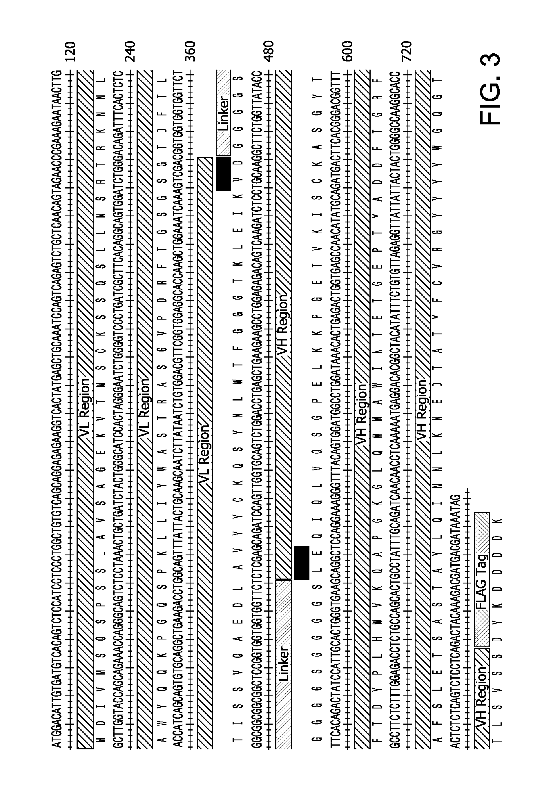
FIG. 3 shows the DNA (SEQ ID NO:15) and amino acid (SEQ ID NO:14) sequences of an scFv for bacterial expression. The sequences of the V.sub.H and V.sub.L regions as well as that of the linker are indicated. The black boxes indicate insertions made during the cloning process. A FLAG-tag is also indicated.
FIG. 4 is a schematic of the scFv construct of FIG. 3 cloned into the pET-22 vector.
FIG. 5A shows an electrophoretic assay of a refolded scFv of the invention expressed in bacteria purified with anti-FLAG antibody. Electrophoretic migration of the purified single chain antibody indicates the predicted mass.
FIG. 5B shows an electrophoretic assay of the refolded single chain antibody expressed in bacteria and purified by ion exchange chromatography. The fraction including the scFv was eluted with 200 mM NaCl, while contaminating proteins were eluted with 500 mM NaCl.
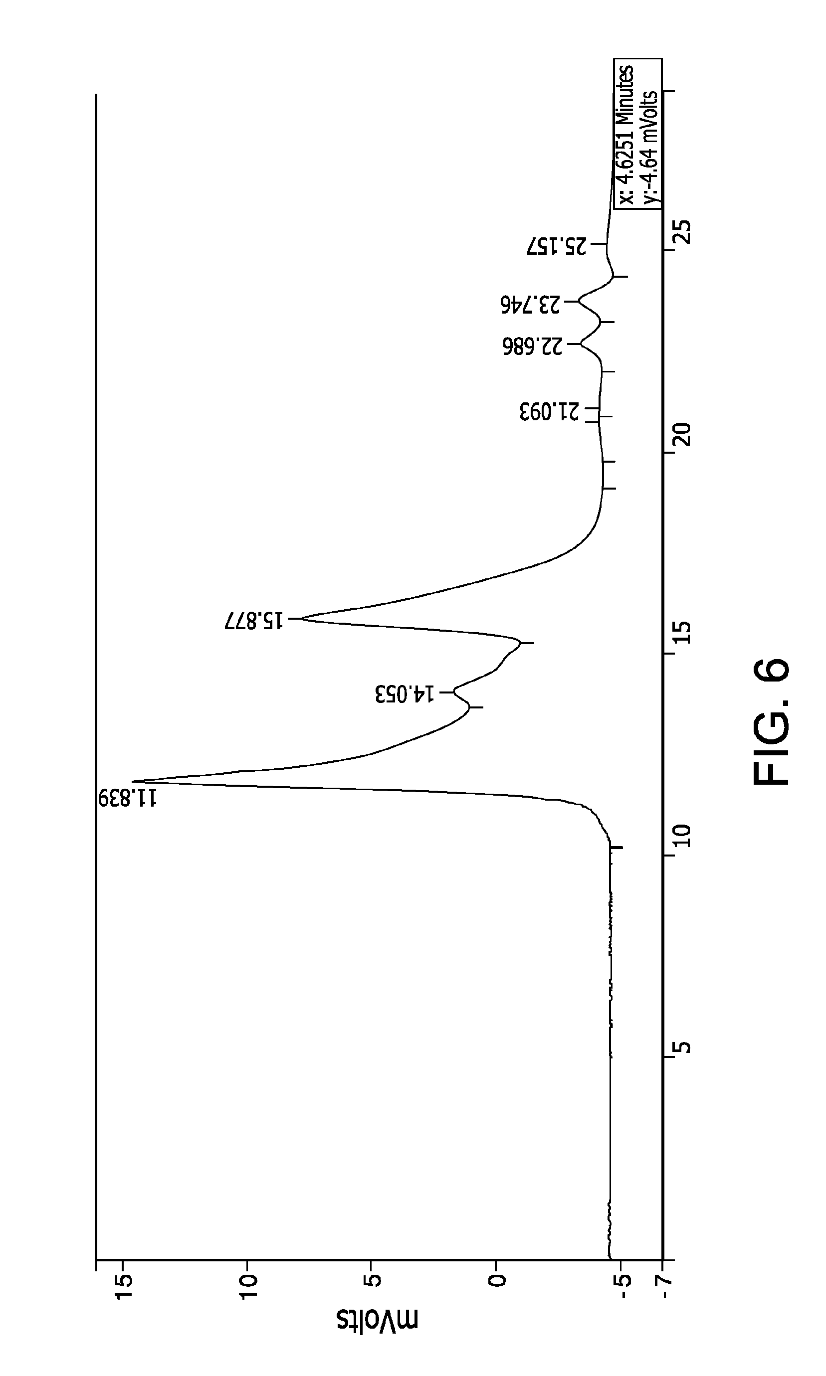
FIG. 6 shows a chromatography profile of the same scFv analyzed by HPLC size exclusion. The three main peaks indicate the single chain antibody monomers and oligomers.
FIG. 7 shows the results of a Western blot analysis of the binding of the same scFv to collagen .alpha.2-chain. The lines in FIG. 7 depict different repeats of the assay, and were identified as procollagen I .alpha.2 chains, thereby indicating the binding specificity of the single chain antibody for collagen .alpha.2-chain.
FIG. 8 shows the inhibition of collagen fibril deposition in cultures of keloid-derived human fibroblasts by the scFv: (I) non treated, (II) treated with tissue growth factor .beta.(TGF-.beta.) and single chain antibody, and (III) treated only with TGF-.beta.. Cell layers consisting of collagen-rich extracellular matrix and cells were collected and analyzed for the presence of collagen I (Col-I), a main constituent of collagen fibrils. An equal number of cells in the analyzed groups is indicated by comparable amounts of GAPDH. The analyzed groups and the collagen I marker (Cm) are indicated.
FIGS. 9A-9C show the DNA coding sequence (upper sequence, which is SEQ ID NO:19), the DNA non-coding sequence (middle sequence, which is SEQ ID NO:30), and amino acid (SEQ ID NO:18) sequences of the construct designated A_L_K, for the expression of an scFv in yeast. Restriction sites utilized in the cloning strategy are indicated.
FIG. 10 is a schematic of the scFv construct of FIGS. 9A-9C cloned into the pPIC-9K plasmid. Restriction sites utilized in the cloning strategy are indicated.
FIG. 11 shows the DNA (SEQ ID NO:20) and amino acid (SEQ ID NO:8) sequences of a construct encoding a mouse heavy chain variable region (mV.sub.H), for the preparation of a further construct for expressing a chimeric antibody heavy chain consisting of the mV.sub.H and the constant region of a human .gamma. chain (mV.sub.H-h.gamma.). Restriction sites utilized in the cloning strategy are indicated.
FIG. 12 is a schematic of a plasmid for expression of the mV.sub.H-h.gamma., utilizing the mV.sub.H construct of FIG. 11. The mV.sub.H construct was cloned into the pFUSE-CHIg-hG1 plasmid as shown in FIG. 12 to express the mV.sub.H-h.gamma.. Restriction sites utilized in the cloning strategy are indicated.
FIG. 13 shows the DNA (SEQ ID NO:21) and amino acid (SEQ ID NO:7) sequences of a construct encoding a mouse light chain variable region (mV.sub.L), for the preparation of a plasmid for expressing a chimeric antibody heavy chain consisting of the mV.sub.L and the constant region of a human .kappa. chain (mV.sub.H-h.kappa.). Restriction sites utilized in the cloning strategy are indicated.
FIG. 14 is a schematic of a plasmid for expression of the chimeric mV.sub.L-h.kappa., utilizing the mV.sub.L construct of FIG. 13. The mV.sub.L construct was cloned into the pFUSE2-CLIg-hk plasmid as shown in FIG. 14 to express the mV.sub.H-h.kappa.. Restriction sites utilized in the cloning strategy are indicated.
FIG. 15A is a Western blot analysis under reducing conditions showing that specific antibodies against IgG .gamma. and .kappa. chains (Anti-.gamma.; Anti-.kappa.) reacted with a chimeric antibody of the invention secreted by CHO cells. The results confirm the presence of .gamma. and .kappa. chains in the chimeric antibody (chimeric-.gamma.; chimeric-.kappa.). Human IgG containing a .kappa. chain was used as positive marker (h-IgG(.kappa.)). The chimeric antibody was composed of a murine heavy .alpha. chain variable region (SEQ ID NO:8)/human .gamma. constant region chain and a murine light chain .kappa. region (SEQ ID NO:7)/human .kappa. constant region chain.
FIG. 15B is a Western blot analysis of the same chimeric antibody under non-reducing conditions with anti-human .gamma. and anti-human .kappa. antibodies. The results indicate that the antibody heavy and light chains (the component mV.sub.H-h.gamma. and mV.sub.L-h.kappa. chains) were covalently linked via disulfide bonds, thereby indicating the formation of the chimeric antibody. The presence of a high molecular band (CHO-IgG) indicates production of chimeric IgG molecules by the CHO cells. Human IgG with the .kappa. chain was used as positive marker (h-IgG(.kappa.)).
FIG. 16 is a Western blot assay showing the binding of the purified chimeric antibody to its target, .alpha.2Ct, as detected by chemiluminescence with the use of the anti-human .gamma. antibodies conjugated with horseradish peroxidase (HRP). Positive bands were identified as those corresponding to intact procollagen I .alpha.2 chain (upper band) and its partially degraded form. Native human IgG with the .kappa. chain was used as a positive marker (Bethyl Laboratories Inc.).
FIG. 17 shows association and dissociation curves illustrating kinetics of the binding between procollagen and the following anti-.alpha.2Ct antibody variants: the original anti-.alpha.2Ct mouse IgA antibody (IgA) from which the mV.sub.H and mV.sub.L were obtained; the bacterial-derived scFv variant (scFv) of FIG. 3; the mouse-human chimeric IgG (chimeric IgG); and human IgG with the light .kappa. chain (human-IgG) (control). In each panel, the curves represent association and dissociation events during the interaction between immobilized procollagen I and free antibody variants present at concentrations ranging from 4.times.10.sup.-7 M to 1.25.times.10.sup.-8 M. For each assay, the association rate constants (k.sub.on) and the dissociation rate constants (k.sub.off) were obtained, and the equilibrium dissociation constants (K.sub.D) values were calculated from a ratio of k.sub.off/k.sub.on.
FIG. 18A is a Western blot assay of scFv variant prepared in yeast from the A_L_K construct. The lanes show the secretion of various yeast clones cultured in His(-) conditions. Cell culture media from non-transformed yeast cells (left lane, "Ctrl") are negative for the scFv variant.
FIG. 18B shows the result of PCR assays of yeast clones producing scFv variants from the A_L_K DNA construct, and from another DNA construct, K_L_A. The presence of the DNA constructs in the genomes of the analyzed clones is indicated.
FIG. 19 shows an electrophoretic assay of the A_L_K yeast scFv variant after purification by nickel column. Electrophoretic migration of the purified scFv in denaturing/reducing conditions indicates its predicted mass.
FIG. 20 is a Western blot analysis showing in lane (A) electrophoresis of the purified human procollagen I in a polyacrylamide gel, showing staining of procollagen I .alpha.1 and .alpha.2 chains. The procollagen chains were transferred to a nitrocellulose membrane and probed with His-tagged yeast-derived A_L_K scFv variant. The A_L_K scFv variant bound to its specific target procollagen I .alpha.2 chain, as shown in lane (B).
DEFINITIONS
As used herein, each of the following terms has the meaning associated with it in this section.
The articles "a" and "an" are used herein to refer to one or to more than one (i.e. to at least one) of the grammatical object of the article. By way of example, "an element" means one element or more than one element.
The term "about" will be understood by persons of ordinary skill in the art and will vary to some extent depending on the context in which it is used. As used herein, "about" is meant to encompass variations of .+-.20% or .+-.10%, more preferably .+-.5%, even more preferably .+-.1%, and still more preferably .+-.0.1%.
Unless otherwise specified herein, the terms "antibody" and "antibodies" broadly encompass naturally-occurring forms of antibodies (e.g., IgG, IgA, IgM, IgE) and recombinant antibodies as well as derivatives have at least an antigenic binding site. Antibody derivatives may comprise a protein or chemical moiety conjugated to an antibody.
By "chimeric antibody" is meant an antibody molecule in which different portions are derived from different animal species, such as those having a variable region derived from a murine antibody and a human immunoglobulin constant region. Typically, a chimeric antibody is composed of a variable region comprising light and heavy chain variable regions, and a constant region derived or obtained from a human antibody.
A "humanized antibody" refers to an antibody in which the complementarity defining regions (CDRs) of an antibody of a non-human mammal, e.g., mouse, are grafted to a human antibody. The variable domain of each of an antibody heavy chain and light chain comprise three CDRs; the intervening sequence segments are "framework segments". Each variable domain is composed of four framework segments. In a humanized antibody, the framework segments are typically of human origin.
A "single chain antibody", also known as a "single-chain variable fragment" (scFv) is a fusion protein of the variable regions of the heavy and light chains of an immunoglobulin, wherein the regions are optionally connected by a linker. As used herein, "single chain antibody" or "single-chain variable fragment" includes such fusion proteins, and also multimers (linear or branched) formed of such fusion proteins.
A "polynucleotide" as utilized in the practice of the present invention may include any polymer or oligomer of pyrimidine and purine bases, preferably cytosine, thymine, and uracil, and adenine and guanine, respectively. The polynucleotide may comprise any deoxyribonucleotide, ribonucleotide or peptide nucleic acid component, and any chemical variants thereof, such as methylated, hydroxymethylated or glucosylated forms of these bases, and the like. The polynucleotide may comprise DNA or RNA, or a mixture thereof, and may exist permanently or transitionally in single-stranded or double-stranded form, including homoduplex, heteroduplex, and hybrid states.
As used herein, the term "subject" or "patient" refers to any animal (e.g., a mammal), including, but not limited to humans, non-human primates, rodents, and the like. Typically, the terms "subject" and "patient" are used interchangeably herein in reference to a human subject.
The term "treating" as used herein, unless otherwise indicated, means reversing, alleviating, inhibiting the progress of, or preventing, either partially or completely, an indicated disease or disorder. The term "treatment" as used herein, unless otherwise indicated, refers to the act of treating. The term "treating" does not necessarily mean that the disease or disorder will, in fact, be eliminated.
The term "therapeutically effective amount" or "effective amount" means the amount of a subject compound or combination that will elicit the biological or medical response of a tissue, system, animal or human that is being sought.
As envisioned in the present invention with respect to the disclosed compositions of matter and methods, in one aspect the embodiments of the invention comprise the components and/or steps disclosed herein. In another aspect, the embodiments of the invention consist essentially of the components and/or steps disclosed herein. In yet another aspect, the embodiments of the invention consist of the components and/or steps disclosed herein.
DETAILED DESCRIPTION OF THE INVENTION
Engineered antibodies are provided that bind to the C-terminal telopeptides of the .alpha.2-chain of collagen. The engineered antibodies inhibit the formation of collagen fibrils. Free collagen molecules that do not incorporate into fibrils are readily accessible for degradation by enzymes present in the extracellular space (Prockop and Fertala, J. Biol. Chem. 273:15598-15604 (1988); Chang et al., Diabetes 29:778-781 (1980)). The engineered antibodies comprise a light chain variable region comprising the three complementarity determining regions having the amino acid sequences shown in SEQ ID NO:1, SEQ ID NO:2 and SEQ ID NO:3, and a heavy chain variable region comprising the three complementarity determining regions having the amino acid sequences shown in SEQ ID NO:4, SEQ ID NO:5 and SEQ ID NO:6. The engineered antibodies are capable of binding the C-terminal telopeptide of the .alpha.2(I) chain of human collagen I.
In one embodiment, the engineered antibody is a chimeric antibody comprising the mouse light chain variable region (mV.sub.L) having the amino acid sequence of SEQ ID NO:10, and the mouse heavy chain variable region (mV.sub.H) having the amino acid sequence of SEQ ID NO:11. The mouse light chain variable region and mouse heavy chain variable regions, with predicted signal peptides, are shown in FIGS. 1 and 2, respectively. The mV.sub.L and mV.sub.H complementarity determining regions are shown by double underlining in FIG. 1. The first fifteen amino acids in FIG. 1 (indicated by single underlining) comprise the signal sequence for the mV.sub.L. The first nineteen amino acids in FIG. 2 (indicated by single underlining) comprise the signal sequence for the mV.sub.H.
Using the sequences encoding the above light and heavy chain variable regions, chimeric antibodies may be produced by any of the well-known techniques for production of chimeric antibodies (Morrison et al., 1984, Proc. Natl. Acad. Sci., 81:6851-5; Neuberger et al., 1984, Nature, 312:604-8; Takeda et al., 1985, Nature, 314:452-4). Accordingly, a DNA molecule encoding the light chain variable region SEQ ID NO:10, e.g., the DNA molecule having the nucleotide sequence of SEQ ID NO:22, is prepared. The DNA molecule may further encode a signal sequence for the light chain variable region, such as the fifteen amino acid signal sequence shown in FIG. 1 by single underlining. SEQ ID NO:7 is the amino acid sequence of the polypeptide comprising the signal sequence and light chain variable region shown in FIG. 1. SEQ ID NO:7 may be encoded by, for example, the nucleotide sequence of SEQ ID NO:24.
A DNA molecule encoding the heavy chain variable region SEQ ID NO:11, e.g., the DNA molecule having the nucleotide sequence of SEQ ID NO:23, is prepared. The DNA molecule may further encode a signal sequence for the heavy chain variable region, such as the nineteen amino acid signal sequence shown in FIG. 2 by single underlining. SEQ ID NO:8 is the amino acid sequence of the polypeptide comprising the signal sequence and heavy chain variable region shown in FIG. 2. SEQ ID NO:8 may be encoded by, for example, the nucleotide sequence of SEQ ID NO:25.
The DNA molecules encoding the light and heavy chain variable regions are then ligated into vector DNA and expressed using, for example, any of the available ligation kits to construct a recombinant vector. See e.g., J. Sambrook et, Molecular Cloning, Cold Spring Harbor Laboratory Press, 1989.
In one embodiment, a chimeric antibody may be prepared by ligating a mouse leader sequence and a variable region sequence present in a cloned mouse cDNA to a sequence coding for a human antibody constant region already present in an expression vector of a mammalian cell. Alternatively, a mouse leader sequence and a variable region sequence present in a cloned cDNA are ligated to a sequence coding for a human antibody constant region followed by ligation to a mammalian cell expression vector. The mouse leader sequence may comprise, for example, the signal sequences shown in FIG. 1 or FIG. 2.
The polypeptide comprising human antibody constant region can comprise any of the heavy or light chain constant regions of human antibodies, including, for example, .gamma.1, .gamma.2, .gamma.3 or .gamma.4 for heavy chains, and .kappa. for light chains.
To prepare a chimeric antibody, two expression vectors are constructed. A first expression vector contains DNAs coding for the light chain variable region and human light chain constant region under the control of an expression control element such as an enhancer/promoter system. A second expression vector contains DNAs coding for the heavy chain variable region and human heavy chain constant region under the control of an expression control element such as an enhancer/promoter system. Host cells such as mammalian cells (for example, COS cell) are cotransformed with these expression vectors. The transformed cells are cultivated in vitro or in vivo to produce a chimeric antibody, See, e.g. WO91/16928.
As an alternative, mouse leader sequence present in cloned cDNA and DNAs coding for mouse light chain variable region and human light chain constant regions, as well as a mouse leader sequence and DNAs coding for mouse heavy chain variable region and human heavy chain constant region, are introduced into a single expression vector (see, for example, WO94/11523). The single vector is used to transform a host cell. The transformed host is cultured in vivo or in vitro to produce a desired chimeric antibody.
The vector for the expression of the heavy chain of the chimeric antibody can be obtained by introducing cDNA comprising a nucleotide sequence coding for the mouse heavy chain variable region into a suitable expression vector containing genomic DNA comprising a nucleotide sequence coding for the heavy chain constant region of human antibody, or cDNA coding for the heavy chain constant region. As indicated above, the heavy chain constant region may comprise, for example, .gamma.1, .gamma.2, .gamma.3 or .gamma.4.
Expression vectors comprising genomic DNA coding for a heavy chain constant region include, for example, HEF-PMh-g gamma 1 (WO92/19759) and DHER-INCREMENT E-RVh-PM1-f (WO92/19759). Alternatively, a human constant region library can be prepared using cDNA from human peripheral blood mononuclear cells, as described by Liu. et al., Proc. Natl. Acad. Sci. USA, 84:3439-43 (1987) or Reff et al., Blood 83(2): 435-45 (1994), for example.
The cDNA coding for the mouse heavy chain variable region of SEQ ID NO:11 (e.g., the nucleotide sequence SEQ ID NO:23, which does not contain a signal sequence segment; or the nucleotide sequence of SEQ ID NO:25, which contains a signal sequence) is treated with suitable restriction enzyme(s) and inserted into genomic DNA coding for a heavy chain constant region, to construct a chimeric heavy chain expression vector containing the genome DNA coding for the heavy chain constant region. Insertion is by ligation of the cDNA encoding the heavy chain constant region, and insertion into an expression vector such as pQCXIH (Clontech), to construct an expression vector containing the complete cDNA encoding the complete chimeric heavy chain. Alternatively, the cDNA encoding the mouse heavy chain variable region may be ligated into an appropriate commercially available cloning plasmid that expresses the constant region of a human heavy chain, e.g., pFUSE-CHIg-hG1 (InvivoGen, San Diego, Calif.).
In a similar fashion, a vector for the expression of the light chain of the chimeric antibody can be constructed by ligating a cDNA coding for the mouse light chain variable region (e.g., the nucleotide sequence SEQ ID NO:22, which does not contain a signal sequence; or the nucleotide sequence of SEQ ID NO:24, which contains a signal sequence) and a genomic DNA or cDNA coding for the light chain constant region of a human antibody, and introduction into a suitable expression vector. The light chain constant region includes, for example, .kappa. or .lamda. chains. Any of the four known .lamda. chain constant region isotypes may be utilized. Alternatively, the cDNA encoding the mouse light chain variable region may be ligated into an appropriate commercially available cloning plasmid that expresses the constant region of a human .kappa. light chain, e.g., pFUSE2-CLIg-hk (InvivoGen, San Diego, Calif.).
FIG. 11 shows the DNA (SEQ ID NO:20) and amino acid (SEQ ID NO:8) sequences of a construct encoding a mouse heavy chain variable region (mV.sub.H) containing a leader signal peptide, for the preparation of a further construct for expressing a chimeric antibody heavy chain consisting of the mV.sub.H and the constant region of a human .gamma. chain (mV.sub.H-h.gamma.). FIG. 12 is a schematic of a plasmid for expression of the mV.sub.H-h.gamma., utilizing the mV.sub.H construct of FIG. 11. The mV.sub.H construct was cloned into the pFUSE-CHIg-hG1 plasmid as shown in FIG. 12. Restriction sites utilized in the cloning strategy are indicated.
FIG. 13 shows the DNA (SEQ ID NO:21) and amino acid (SEQ ID NO:7) sequences of a construct encoding a mouse light chain variable region (mV.sub.L) containing a leader signal peptide, for the preparation of a plasmid for expressing a chimeric antibody heavy chain consisting of the mV.sub.L and the constant region of a human .kappa. chain (mV.sub.L-h.kappa.). FIG. 14 is a schematic of a plasmid for expression of the chimeric mV.sub.L-h.kappa., utilizing the mV.sub.L construct of FIG. 13. The mV.sub.L construct was cloned into the pFUSE2-CLIg-hk plasmid as shown in FIG. 14. Restriction sites utilized in the cloning strategy are indicated.
In another embodiment, the engineered antibody is a humanized antibody. The humanized antibody comprises a human antibody framework region and advantageously further comprises a human antibody constant region. The six complementarity determining regions (CDRs) SEQ ID NOS:1-6 are grafted to a human antibody. The general genetic recombination procedure for producing humanized antibodies are described, for example, in EP 125023 and WO 96/02576. Accordingly, a DNA sequence is designed in which DNA encoding the aforementioned CDRs are ligated through framework regions. The DNA sequence is synthesized by a polymerase chain reaction method using oligonucleotide primers which are designed to have regions overlapping the terminal regions of the CDRs and the framework regions. The resultant DNA is ligated to DNA encoding the human antibody constant region, and the ligation product is integrated into an expression vector. The resultant recombinant expression vector is introduced into a host, thereby producing the humanized antibody. See, e.g., WO 96/02576.
The framework regions ligated through the CDRs are selected so that the CDRs can form a functional antigen binding site. If necessary, an amino acid(s) in the framework regions of the antibody variable region may be replaced so that the CDRs of the resulting humanized antibody can form an appropriate antigen binding site. See Sato et al., Cancer Res. 53:851-6 (1993).
The amino acid sequences of the framework regions are preferably selected to reflect a high homology to the framework sequences of a human antibody. In this regard, a comparison may be undertaken between the variable regions SEQ ID NO:10 and SEQ ID NO:11 and the variable regions of structurally elucidated human antibodies using, e.g., the Protein Data Bank. Other research tools that may consulted include the Kabat Database of Sequences of Proteins of Immunological Interest, www<<dot>>kabatdatabase<<dot>>com and the search tools included as part of that database; and Kabat, E. A. et al., (1991) Sequences of Proteins of Immunological Interest, 5.sup.th edition. U.S. Department of Health and Human Services.
A humanized antibody variable region is selected as a basis for the humanized antibody variable region. For example, the framework region of a human antibody variable region having a high homology, e.g., greater than about 80%, or greater than about 90%, or greater than about 95%, with the framework regions of SEQ ID NOS:10 and 11 (the non-CDR regions of SEQ ID NOS:10 and 11) is selected. A polypeptide comprising the framework regions of the humanized antibody and the CDRs of SEQ ID NOs:1-6 can be produced by "CDR-grafting", a PCR-based method utilizing a DNA fragment of a human antibody as a template. For an example of CDR grafting, see Kettleborough et al., Protein Eng. 4(7):773-783 (1991); "Antibody Humanization by CDR Grafting", Methods of Molecular Biology, 248(11):135-159 (2004).
Humanized antibodies may be prepared, as exemplified in Jones et al., 1986 Nature 321:522-525, which describes replacing the complementarity-determining regions in a human antibody with those from a mouse. Also see Riechmann, 1988, Nature 332:323-327; Queen et al., 1989, Proc. Nat. Acad. Sci. USA 86:10029 (preparation of humanized antibody binding the interleukin 2 receptor); and Orlandi et al., 1989, Proc. Natl. Acad. Sci. USA 86:3833 (describing the cloning of immunoglobulin variable domains for expression by the polymerase chain reaction).
Any suitable expression system may be used to produce the chimeric or humanized antibody. For example, eukaryotic cells include animal cells such as established mammalian cell lines, fungal cells, and yeast cells; prokaryotic cells include bacterial cells such as Escherichia coli. Mammalian host cells are preferred. The expression system may incorporate conventional promoters useful for the expression in mammalian cells, e.g., the human cytomegalovirus (HCMV) immediate early promoter. Promoters for expression in mammalian cells may include virus promoters, such as those of retrovirus, polyoma virus, adenovirus and simian virus (SV) 40, and mammalian cell derived promoters, such as those of human polypeptide chain elongation factor-1 alpha (HEF-1 alpha).
The expression system may include a replication origin such as those derived from SV40, polyoma virus, adenovirus or bovine papilloma virus (BPV). The expression vector may comprise a gene for phosphotransferase APH(3') II or I (neo), thymidine kinase (TK), E. coli xanthine-guanine phosphoribosyltransferase (Ecogpt) or dihydrofolate reductase (DHFR) as a selective marker for increasing the gene copy number in a host cell system.
The expressed chimeric or humanized antibody is produced by culturing the thus-transformed host cells, and isolating and purifying the antibody from the cells according to well-known techniques. The concentration of the resulting purified antibody can be determined by, for example, enzyme-linked immunosorbent assay (ELISA). Antigen-binding activity can be confirmed by known methods antibody, techniques such as ELISA, enzyme immunoassay, radioimmunoassay or fluorescent assay.
Fragments of the chimeric or humanized antibody retaining antigen-binding activity may be prepared, e.g., Fab, F(ab').sub.2, and Fv fragments. Antibody fragments can be produced by cleaving the antibody with an enzyme (e.g., papain, pepsin) into antibody fragments, or by constructing a gene encoding the antibody fragment and inserting the gene into an expression vector and introducing the resultant recombinant expression vector into a suitable host cell, thereby expressing the antibody fragment (see, for example, Co et al., J. Immunol. 152:2968-76 (1994)).
In another embodiment, the engineered antibody is a single-chain antibody (SCA), also referred to herein as a single-chain variable fragment (scFv), comprising a light chain variable region comprising the complementarity determining regions (CDRs) having the amino acid sequences shown in SEQ ID NO:1, SEQ ID NO:2 and SEQ ID NO:3, and a heavy chain variable region comprising the complementarity determining regions having the amino acid sequences shown in SEQ ID NO:5 and SEQ ID NO:6. The scFv can be produced by ligating a heavy chain variable region comprising the CDRs of SEQ ID NOS:5 and 6 to a light chain variable region comprising the CDRs of SEQ ID NOS:1, 2 and 3 through a linker. The linker is preferably a peptide linker, i.e., the linker is composed of amino acid residues. The residues for the linker may be selected from naturally occurring amino acids, non-naturally occurring amino acids, and modified amino acids. The linker will typically connect the carboxy terminus of the heavy chain variable region to the amino terminus of said light chain variable region. The reverse is also possible, i.e., using the linker to connect the carboxy terminus of the light chain variable region to the amino terminus of the heavy chain variable region. The linker may comprise any number of amino acids. The linker may thus comprise, for example, 1, 2, 3, 4, 5, 6, 7, 8, 9, 10, 11, 12, 13, 14, 15, 16, 17, 18, 19, 21, 22, 23, 24, 25, 26, 27, 28, 29, 30, 31, 32, 33, 34, 35, 36, 37, 38, 39, 40, 41, 42, 43, 44, 45, 46, 47, 48, 49, 50, 51, 52, 53, 54, 55, 56, 57, 58, 59, 60, or more amino acids. In some embodiments, the linker may be composed of from 3 to 60 amino acid residues, from 3 to 40 amino acids, from 3 to 30 amino acids, from 3 to 24 amino acids, from 3 to 18 amino acids, or from 3 to 15 amino acids. The linker may comprise, for example, a repeating sub-sequence of 2, 3, 4, 5 or more amino acid residues, comprising 2, 3, 4, 5, 6, 7, 8, 9, 10, 11, 12 or more repeats of the sub-sequence.
In one embodiment, the linker comprises the amino acid sequence Gly-Ser, or repeats thereof. See, e.g., Huston, et al., Methods in Enzymology, 203:46-88 (1991). In another embodiment, the linker comprises the amino acid sequence Glu-Lys, or repeats thereof. See, e.g., Whitlow et al., Protein Eng., 6:989 (1993)). In another embodiment, the linker comprises the amino acid sequence Gly-Gly-Ser, or repeats thereof. In another embodiment, the linker comprises the amino acid sequence Gly-Gly-Gly-Gly-Ser (SEQ ID NO:29), or repeats thereof. In certain specific embodiments, the linker contains from 2 to 12 repeats of Gly-Gly-Ser or Gly-Gly-Gly-Gly-Ser (SEQ ID NO:29).
An scFv of the invention having the amino acid sequence SEQ ID NO:14 is illustrated in FIG. 3. The scFv comprises from amino terminus to carboxy terminus, (i) an initial methionine residue, (ii) the mV.sub.L sequence SEQ ID NO:9, which comprises the parental mV.sub.L of SEQ ID NO:10 without the terminal Arg residue of SEQ ID NO:10, (iii) Val-Asp, (iv) a linker, (v) Leu-Glu, (vi) the mV.sub.H sequence SEQ ID NO.11, and (vii) a FLAG-Tag for affinity chromatography purification.
The antibody or antibody fragments of the invention may be utilized to bind to the C-terminal telopeptides of the .alpha.2-chain of collagen to inhibit the formation of collagen fibrils, and thereby prevent excessive deposition of collagen fibrils that is characteristic of fibrotic processes. The antibody or antibody fragments of the invention may thus be utilized to reduce localized and systemic fibrotic lesions, including but not limited to fibroses occurring in internal organs, the dermus or the eye.
Types of fibrosis that may be treated or prevented include, for example, pulmonary fibrosis, idiopathic pulmonary fibrosis, cirrhosis, endomyocardial fibrosis, mediastinal fibrosis, myelofibrosis, retroperitoneal fibrosis, progressive massive fibrosis, nephrogenic systemic fibrosis, Crohn's Disease, keloid formation, myocardial infarction, scleroderma/systemic sclerosis, arthrofibrosis, and adhesive capsulitis. The fibrotic lesion treated may be triggered by trauma, accidental injury or surgical procedures, among other causes. The antibody or antibody fragments of the invention may be administered, for example, after surgery in the abdomen to avoid the formation of excessive scar tissue around abdominal organs; after plastic surgery to the face to reduce scar formation; to the eye following glaucoma surgery performed to maintain a lamellar channel from the subconjunctival space to the anterior chamber, to prevent excessive scar formation that may function to closes the pressure-reducing channel and cause intraocular pressure to rise; and following implantation of medical devices and materials implanted in the human body, which would otherwise trigger a fibrotic response. In one embodiment, the antibody or antibody fragments are administered to teat or prevent the formation of keloids,
Pharmaceutical compositions for use in accordance with the present invention can be formulated in conventional manner using one or more pharmaceutically acceptable carriers or excipients. The antibodies or fragments thereof can be formulated for administration in accordance with the route of administration. Formulations for injection can be presented in unit dosage form, e.g., in ampoules or in multi-dose containers, with an added preservative. The compositions can take such forms as suspensions, solutions, or emulsions in oily or aqueous vehicles, and can contain formulatory agents such as suspending, stabilizing and/or dispersing agents. Alternatively, the antibody can be in lyophilized powder form for constitution with a suitable vehicle, e.g., sterile pyrogen-free water, before use.
In some embodiments, the subject of treatment is human. In other embodiments, the subject is a veterinary subject.
Treatment may involve administration of one or more antibodies or antibody fragments of the invention, alone or with a pharmaceutically acceptable carrier. The active agent may be administered in combination with one or more other therapeutic, diagnostic or prophylactic agents. The administered composition may thus further comprise an additional agent selected from the group consisting of corticosteroids, antiinflammatories, immunosuppressants, antimetabolites, and immunomodulators, for example.
The pharmaceutically acceptable carrier may comprise any solvents, dispersion media, coatings, antibacterial and antifungal agents, isotonic and absorption enhancing or delaying agents, and the like that are physiologically compatible. Some examples of pharmaceutically acceptable carriers are water, saline, phosphate buffered saline, acetate buffer with sodium chloride, dextrose, glycerol, polyethylene glycol, ethanol and the like, as well as combinations thereof. In some cases, it will be preferable to include isotonic agents, for example, sugars, polyalcohols such as mannitol, sorbitol, or sodium chloride in the composition. Additional examples of pharmaceutically acceptable substances are surfactants, wetting agents or minor amounts of auxiliary substances such as wetting or emulsifying agents, preservatives or buffers, which enhance the shelf life or effectiveness of the antibody.
The compositions used in the practice of the invention may be in a variety of forms, for example, liquid, semi-solid and solid dosage forms, such as liquid solutions, dispersions or suspensions, tablets, pills, lyophilized cake, dry powders, liposomes and suppositories. The preferred form depends on the intended mode of administration and therapeutic application. In some embodiments, the antibody or antibody fragment may be administered by using a pump, enema, suppository, or indwelling reservoir or such like.
Therapeutic compositions typically must be sterile and stable under the conditions of manufacture and storage. The composition can be formulated as a solution, lyophilized cake, dry powder, microemulsion, dispersion, liposome, or other ordered structure suitable to high drug concentration. Sterile solutions can be prepared by incorporating the antibody or fragment in the required amount in an appropriate solvent with one or a combination of ingredients enumerated above, as required, followed by sterilization. In the case of sterile powders for the preparation of sterile solutions, the preferred methods of preparation are vacuum drying and freeze-drying that yields a powder of the active ingredient plus any additional desired ingredient from a previously sterile solution thereof. Generally, dispersions are prepared by incorporating the active compound into a sterile vehicle that contains a basic dispersion medium and the required other ingredients from those enumerated above. The desired characteristics of a solution can be maintained, for example, by the use of surfactants and the required particle size in the case of dispersion by the use of surfactants, phospholipids and polymers. Prolonged absorption of injectable compositions can be brought about by including in the composition an agent that delays absorption, for example, monostearate salts, polymeric materials, oils and gelatin.
In certain embodiments, the antibody or antibody fragment compositions of the invention may be prepared with a carrier that will protect the antibody against rapid release, such as a controlled release formulation, including implants, transdermal patches, and microencapsulated delivery systems. Biodegradable, biocompatible polymers can be used, such as ethylene vinyl acetate, polyanhydrides, polyglycolic acid, collagen, polyorthoesters, and polylactic acid. Many methods for the preparation of such formulations are patented or generally known to those skilled in the art. See, e.g., Sustained and Controlled Release Drug Delivery Systems (J. R. Robinson, ed., Marcel Dekker, Inc., New York (1978)).
The compositions of the invention may be administered parenterally. The term "parenteral" as used herein includes subcutaneous, intravenous, intramuscular, intra-articular, intra-synovial, intrasternal, intrathecal, intrahepatic, intralesional and intracranial injection or infusion techniques.
The compositions of the invention may be given orally, in any orally acceptable dosage form including, but not limited to, capsules, tablets, aqueous suspensions or solutions. In the case of tablets for oral use, carriers which are commonly used include lactose and corn starch. Lubricating agents, such as magnesium stearate, are also typically added. For oral administration in a capsule form, useful diluents include lactose and dried corn starch.
The compositions of the invention may be administered topically. For topical applications, the pharmaceutical compositions may be formulated in a suitable ointment containing the active component suspended or dissolved in one or more carriers. Carriers for topical administration of the compounds of this invention include, but are not limited to, mineral oil, liquid petrolatum, white petrolatum, propylene glycol, polyoxyethylene, polyoxypropylene compound, emulsifying wax and water. Alternatively, the pharmaceutical compositions can be formulated in a suitable lotion or cream containing the active components suspended or dissolved in one or more pharmaceutically acceptable carriers. Suitable carriers include, but are not limited to, mineral oil, sorbitan monostearate, polysorbate 60, cetyl esters wax, cetearyl alcohol, 2-octyldodecanol, benzyl alcohol and water. Topically-transdermal patches may also be used. For topical applications, the pharmaceutical compositions may be formulated in a suitable ointment containing the active component suspended or dissolved in one or more carriers. Carriers for topical administration of the compounds of this invention include, but are not limited to, mineral oil, liquid petrolatum, white petrolatum, propylene glycol, polyoxyethylene, polyoxypropylene compound, emulsifying wax and water. Alternatively, the pharmaceutical compositions can be formulated in a suitable lotion or cream containing the active components suspended or dissolved in one or more pharmaceutically acceptable carriers. Suitable carriers include, but are not limited to, mineral oil, sorbitan monostearate, polysorbate 60, cetyl esters wax, cetearyl alcohol, 2-octyldodecanol, benzyl alcohol and water.
Topical or other localized administration is advantageously utilized at the site of a localized fibrosis, e.g., by localized injection. The location of the fibrosis may comprise, for example, a wound, particularly a wound edge.
The dosage of active agent may vary according to factors such as the disease state, age, sex, and weight of the individual, and the ability of the antibody or antibody portion to elicit a desired response in the individual. A therapeutically effective amount is also one in which any toxic or detrimental effects of the antibody or antibody portion are outweighed by the therapeutically beneficial effects. Detection and measurement of indicators of efficacy may be measured by a number of available diagnostic tools, including, for example, by physical examination including blood tests, pulmonary function tests, and chest X-rays; CT scan; bronchoscopy; bronchoalveolar lavage; lung biopsy and CT scan. A "prophylactically effective amount" refers to an amount effective, at dosages and for periods of time necessary, to achieve the desired prophylactic result. Typically, since a prophylactic dose is used in subjects prior to or at an earlier stage of disease, the prophylactically effective amount may be less than the therapeutically effective amount.
Dosage regimens can be adjusted to provide the optimum desired response (e.g., a therapeutic or prophylactic response). For example, a single bolus can be administered, several divided doses can be administered over time or the dose can be proportionally reduced or increased as indicated by the exigencies of the therapeutic situation. It is especially advantageous to formulate parenteral compositions in dosage unit form for ease of administration and uniformity of dosage. Dosage unit form as used herein refers to physically discrete units suited as unitary dosages for the mammalian subjects to be treated; each unit containing a pre-determined quantity of active compound calculated to produce the desired therapeutic effect in association with the required pharmaceutical carrier.
As a non-limiting example, an effective amount of an antibody or antibody fragment active agent is from about 0.025 to about 50 mg/kg, or from about 0.1 to about 50 mg/kg, or from about 0.1-25 mg/kg, or from about 0.1 to about 10 mg/kg, or from about 0.1 to about 3 mg/kg. Dosage may vary with the type and severity of the condition to be alleviated. For any particular treatment subject, specific dosage regimens may be adjusted over time according to the individual need and the professional judgment of the person administering or supervising the administration of the compositions, and that dosage ranges set forth herein are exemplary only and are not intended to limit the scope or practice of the claimed composition.
The practice of the invention is illustrated by the following non-limiting example. The invention should not be construed to be limited solely to the compositions and methods described herein, but should be construed to include other compositions and methods as well. One of skill in the art will know that other compositions and methods are available to perform the procedures described herein.
EXAMPLES
Example 1: Single-Chain Antibody by Bacterial Expression
A construct for preparing a single chain antibody, also referred to herein as a single-chain variable fragment (scFv), was prepared containing DNA sequences encoding the light chain variable region of SEQ ID NO:7 and the heavy chain variable region of SEQ ID NO:8. The DNA sequences were obtained by PCR from a hybridoma expressing an IgA class antibody to collagen .alpha.2-chain (Chung et al., J. Biol. Chem. 283(38):25879-25886 (2008)). Total RNA was prepared from hybridoma cells with the use of an RNA-isolation kit according to the manufacturer's protocol (QIAGEN). PCR products spanning the V.sub.H of the .alpha. and the V.sub.L of the .kappa. chains were cloned into the pETBlue-1 Blunt vector and sequenced.
A. Bacterial Expression System
For the bacterial expression of the scFv, a construct was created in which the V.sub.L region was connected with the V.sub.H region via a 15 amino acid linker comprising three repeats of the Gly-Gly-Gly-Gly-Ser (SEQ ID NO:29) motif. A DNA sequence encoding a FLAG tag was fused to the 3' end of the construct to facilitate downstream purification of the recombinant scFv (FIG. 3). Minor additions to the original amino acid sequences of the V.sub.L (SEQ ID NO:10) and V.sub.H (SEQ ID NO:11) regions were created as a consequence of the cloning process (FIG. 3). Moreover, the native signal peptides shown in FIGS. 1 and 2 (single underlining) were omitted in the scFv construct. The constructs were thus arranged to provide a scFv comprising, from N-terminus to C-terminus, light chain variable region-linker-heavy chain variable region, where the linker connected the N-terminus of the heavy chain to the C-terminus of the light chain. More specifically, the scFv comprised, from amino terminus to carboxy terminus, (i) an initial methionine residue, (ii) the mV.sub.L sequence SEQ ID NO:9, which comprises the parental mV.sub.L of SEQ ID NO:10 without the terminal Arg residue of SEQ ID NO:10, (iii) Val-Asp, (iv) a linker, (v) Leu-Glu, (vi) the mV.sub.H sequence SEQ ID NO.11, and (vii) a FLAG-Tag. The fidelity of the construct was confirmed by DNA sequencing.
The scFv DNA construct was initially cloned into the NcoI/SacI site of the pET-28 (not shown), a vector that does not include any signaling sequences for the secretion of exogenous proteins into the periplasm (EMD Biosciences). Because the expression of exogenous proteins in bacteria is frequently associated with the formation of insoluble aggregates packed into the inclusion bodies, the pET-22 vector was also employed (FIG. 4) (EMD Biosciences). In contrast to the pET-28 vector, the pET-22 vector carries an N-terminal pelB signal sequence for potential periplasmic localization of the expressed protein, a situation in which the produced proteins are potentially more soluble. The two above vectors were tested for their ability to facilitate the production of the scFv variant.
Bacteria expressing the scFv cloned into the pET-22 and pET-28 vectors were cultured in the presence of ampicillin or kanamycin, respectively. After reaching the optical density (OD.sub.600) of 0.5 units, bacterial cultures were treated with 1 mM isopropyl .beta.-D-1-thiogalactopyranoside (IPTG) to induce the expression of the scFv. Subsequently, the bacterial cultures were incubated for an additional 3 h at 37.degree. C.
Bacteria were collected by centrifugation and then lysed with the B-PER II Protein Extraction Reagent (Thermo Scientific) in the presence of lysozyme, and then the scFv-rich insoluble inclusion bodies were collected by centrifugation. Subsequently, the inclusion bodies were solubilized in 6M guanidinium hydrochloride (GdnHCl) in the presence of the reducing agent dithiothreitol (DTT). Refolding conditions were tested by running pilot-scale refolding assays with the use of the Protein Refolding Kit (Thermo Scientific). As a result of these assays, the denatured scFv molecules present in the GdnHCl-solubilized material were refolded by diluting them into 100 volumes of a refolding buffer consisting of 55 mM Tris, 21 mM NaCl, 0.88 mM KCL, pH 8.2. Subsequently, the sample was concentrated 100-fold by ultrafiltration.
B. scFv Purification and Analyses
Pilot purification of the scFv was carried out with the agarose-conjugated anti-FLAG antibody according to the manufacturer's protocol (Sigma-Aldrich). The purity of purified soluble scFv was analyzed by polyacrylamide gel electrophoresis and by Western blot assay with the anti-FLAG antibody (Sigma-Aldrich) run in denaturing and reducing conditions. The result is shown in FIG. 5A. The electrophoretic migration of the purified scFv indicated the predicted mass.
In addition, due to the relatively high cost of the anti-FLAG antibody, an ion exchange chromatography on the MonoQ Sepharose resin (GE Healthcare Life Sciences) was employed to purify the semi-preparative amounts of the scFv variant. In brief, a sample containing the refolded scFv was loaded onto a MonoQ Sepharose column, and then bound proteins were eluted with a 0-1 M NaCl gradient. The collected peaks were then checked for the presence of the scFv variant by gel electrophoresis assays and Western blot assays with the use of the anti-FLAG antibody (Sigma-Aldrich) (FIG. 5B) in denaturing and reducing conditions. The fraction including the scFv was eluted with 200 mM NaCl, while contaminating proteins were eluted with 500 mM NaCl (FIG. 5B).
The molecular mass of the purified native scFv was analyzed by high pressure liquid size exclusion chromatography (SEC HPLC) run in non-denaturing conditions. The chromatography profile is shown in FIG. 6. The three main peaks in FIG. 6 indicate the single chain antibody monomers and oligomers.
The binding specificity of the scFv for its intended epitope, the .alpha.2-chain of collagen, was confirmed by a Western blot-based assay as follows. Purified human procollagen I was electrophoresed in a polyacrylamide gel. Subsequently, procollagen chains were transferred to a nitrocellulose membrane. Next, the membranes were incubated with FLAG-tagged single chain antibody. The bound single chain antibody was detected with chemiluminescence by an anti-FLAG antibody conjugated to horseradish peroxidase. The results are shown in FIG. 7. The lines in FIG. 7 depict different repeats of the assay, and were identified as procollagen I .alpha.2 chains, thereby indicating specific binding of the scFv to collagen .alpha.2-chain.
Example 2: Single Chain Antibody Inhibition of Collagen Fibril Deposition
Assays of the inhibition of collagen fibril deposition by the scFv of Example 1 were done in cultures of keloid-derived fibroblasts. In brief, the fibroblasts were seeded into the wells of a 96-well plate at the density of 4.times.10.sup.3 cells/well. Six hours after seeding, the attached cell layers were washed and fresh media supplemented with 40 .mu.g/ml of L-ascorbic acid phosphate magnesium salt n-hydrate (Wako Pure Chemical Co.) was added to the cells. Three experimental groups of cells were analysed: (I) non treated, (II) treated with tissue growth factor .beta. (TGF-.beta.) and the scFv, and (III) treated only with TGF-.beta.. TGF-.beta. was added at a concentration of 1 ng/ml, while scFV was added at 1, 5 or 10 .mu.g/ml. The purpose of adding TGF-.beta., an activator of collagen biosynthesis, was to stimulate cells to produce and secrete high amounts of collagen. After the 48-h culture in the presence or the absence of TGF-.beta. and scFv, layers consisting of collagen-rich extracellular matrix and cells were solubilized by adding a lysis buffer and analyzed by Western blot assays for the presence of collagen I, a main constituent of collagen fibrils. The results shown in FIG. 8 indicate a marked decrease of collagen deposits in a group treated with the scFv. An equal number of cells in the analyzed groups is indicated by comparable amounts of GAPDH. The analyzed groups and the collagen I marker (Cm) are indicated.
Example 3: Constructs for Single-Chain Antibody Production by Yeast Expression
Constructs for the expression of the scFv of Example 1 in yeasts were also engineered. The rationale for employing yeasts to produce the scFv variants was dictated by the fact that those organisms are eukaryotes, a characteristic that potentially may improve the folding and solubility of the recombinant scFv variants.
Accordingly, the following scFv variants were designed: (i) A_L_K, a construct in which a cassette for the V.sub.H of the mouse heavy .alpha. chain was linked with a cassette for the V.sub.L of the mouse light .kappa. chain; and (ii) K_L_A, a construct in which a cassette for the V.sub.L of the light .kappa. chain was linked with a cassette for the V.sub.H of the heavy .alpha. chain. In comparison to the 15-amino acid linker designed for the bacterial scFv construct (FIG. 3), the scFv constructs for yeast expression consisted of a 30-amino acid linker. See FIGS. 9A-9C. By increasing the length of the linker, the predicted solubility of the A_L_K and K_L_A scFv variants will be higher than that with a shorter linker. Moreover, the increased length of the linker may decrease the potential of scFv variants to form oligomers. If necessary, that the length of the linker may be readily changed by DNA engineering technology.
The nucleotide (SEQ ID NOS:19 and 30) and amino acid (SEQ ID NO:18) sequences of the A_L_K construct are shown in FIGS. 9A-9C. A schematic of the same construct cloned into the pPIC-9K plasmid is shown in FIG. 10. In both figures, restrictions sites utilized in the cloning strategy are indicated--SnaBI and NotI restriction sites were incorporated at the 5' and 3' ends of the construct, respectively, to facilitate its downstream cloning into the pPIC-9K yeast-expression vector (Invitrogen). A construct for the K_L_A scFv was prepared in a similar way (not shown). The entire DNA constructs for the A_L_K and K_L_A variants were synthesized commercially (Blue Heron Biotechnology, Bothell, Wash. 98021 USA). In both constructs, a His-tag-coding sequence was incorporated to facilitate the downstream purification process (FIGS. 9A-9C). The fidelity of constructs was confirmed by sequencing. The nucleotide and amino acid sequences of the V.sub.H-linker-V.sub.L portion of the A_L_K construct (without added restriction sites and His-Tag), comprise SEQ ID NO:17 (nucleotide) and SEQ ID NO:16 (amino acid).
Example 4: Preparation of Chimeric Antibody By Mammalian Cell Expression
Constructs for the mammalian-cell expression of full-length mouse/human chimeric IgG were created. The constructs consist of sequences encoding a mouse variable region fused to sequences encoding constant regions of human to .gamma. and .kappa. chains. Two such DNA constructs were prepared as follows for the expression of a chimeric mouse/human antibody. A first construct encoded a chimera of the mouse-derived heavy chain variable region and a human-derived heavy .gamma. chain (mV.sub.H-h.gamma.), while the second construct encoded a chimera of the mouse-derived light chain variable region and a human-derived light .kappa. chain (mV.sub.L-h.kappa.).
A. Preparation of DNA Construct for Chimeric Mouse V.sub.H-Human Heavy .gamma. Chain (mV.sub.H-h.gamma.)
The DNA sequence encoding the V.sub.H region of the heavy mouse .alpha. chain of the original anti-.alpha.2Ct IgA was cloned into the pETBlue-1 Blunt vector (EMD Biosciences/Novagen). To facilitate downstream cloning into the pFUSE-CHIg-hG1 vector that includes the sequence encoding the constant region (CH) of the heavy .gamma. chain of the human IgG1 (InvivoGen), the EcoRI and NheI restriction sites were introduced via PCR to the DNA sequence encoding the V.sub.H region. See FIG. 11. (The nucleotide and amino acid sequences shown in FIG. 11 are SEQ ID NO:20 and SEQ ID NO:8, respectively.) Subsequently, an insert encoding the V.sub.H region of the heavy mouse .alpha. chain was cloned into corresponding restriction sites of the pFUSE-CHIg-hG1 vector (FIG. 12). The fidelity of the mV.sub.H-h.gamma. construct was confirmed by DNA sequencing.
B. Preparation of DNA Construct for Chimeric Mouse V.sub.L-Human Light .kappa. Chain (mV.sub.L-h.kappa.)
DNA sequence encoding the V.sub.L region of the light mouse .kappa. chain of the original anti-.alpha.2Ct IgA was cloned into the pETBlue-1 Blunt vector (EMD Biosciences/Novagen). To facilitate downstream cloning into the pFUSE2-CLIg-hk vector that includes the sequence encoding the constant region (CH) of the human light .kappa. chain (InvivoGen), the AgeI and BsiWI restriction sites were introduced via PCR to the DNA sequence encoding the V.sub.L region of the light mouse .kappa. chain. See FIG. 13. (The nucleotide and amino acid sequences shown in FIG. 13 are SEQ ID NO:21 and SEQ ID NO:7, respectively.) Subsequently, the insert encoding the V.sub.L region of the light mouse .kappa. chain was cloned into the corresponding restriction sites of the pFUSE2-CLIg-hk vector (FIG. 14). The fidelity of the mV.sub.L-h.kappa. construct was confirmed by DNA sequencing.
C. Selection of Clones Expressing Chimeric mV.sub.H-h.gamma. and mV.sub.L-h.kappa. Variants in Mammalian Cells
Chimeric variants were expressed in Chinese hamster ovary cells (CHO). In brief, CHO cells were transfected with a DNA construct encoding the mV.sub.H-h.gamma. variant. Zeocin-resistant clones were selected and screened for the presence of the mV.sub.H-h.gamma.. Specifically, proteins secreted to the media by selected clones were analyzed by Western blot for the presence of the mV.sub.H-h.gamma. variant with the use of the goat anti-human .gamma. chain polyclonal antibody conjugated with HRP (Sigma-Aldrich). Next, the selected .gamma. chain-positive CHO clone was expanded in cell culture and then transfected with a DNA construct encoding the mV.sub.L-h.kappa. variant. Subsequently, cell culture media from the double-transfected clones resistant to Zeocin and Blasticidin were analyzed by Western blot for the production of the mV.sub.L-h.kappa. chain with the use of the polyclonal goat anti-human k chain antibodies conjugated with HRP. Selected clones stably co-expressing mV.sub.H-h.gamma. and mV.sub.L-h.kappa. chains were expanded in cell culture and then cryopreserved in liquid nitrogen.
D. Purification of the Chimeric Antibody Consisting of the mV.sub.H-h.gamma. and mV.sub.L-h.kappa. Chains
Cell culture media from selected CHO cells producing the chimeric antibody were collected. Subsequently, proteins secreted to the media were precipitated with ammonium sulfate added to a 50-% saturation. Precipitated proteins were collected by centrifugation and then the protein pellet was solubilized in phosphate buffered saline (PBS). Insoluble material was removed by centrifugation while the supernatant was collected for further processing. The chimeric IgG was purified by employing the Protein-L agarose (Thermo Scientific), a resin that specifically binds to the .kappa. chain of human but not bovine origin.
Purified chimeric antibodies were analyzed in reducing conditions for the presence of the .gamma. and .kappa. chains. Specific antibodies against particular chains confirm the production of the .gamma. and .kappa. chains by CHO cells (FIG. 15A). To determine if the two chains of the chimeric IgG antibody, i.e., mV.sub.H-h.gamma. and mV.sub.L-h.kappa. chains, co-assemble into native-like molecules consisting of both types of chains linked via disulfide bonds, the purified proteins secreted from CHO cells were separated in a polyacrylamide gel in non-reducing conditions. Subsequent Western blot assays with the anti-human .gamma. chain and the anti human .kappa. chain antibodies indicate that mV.sub.H-h.gamma. and mV.sub.L-h.kappa. chains were covalently linked via disulfide bonds, thereby indicating the formation of chimeric antibody molecules consisting of heavy and light chains of expected molecular mass (FIG. 15B). The presence of a high molecular band indicates production of chimeric IgG molecules by CHO cells. Human IgG with the .kappa. chain was used as a positive marker (h-IgG(.kappa.).
E. Binding of the Chimeric IgG to its Designated Target
Western blot assays were employed to test the binding of purified chimeric IgG to its designated target, i.e., the C terminal telopeptide of the .alpha.2 chain of human procollagen I (.alpha.2Ct). In brief, the .alpha.1 and .alpha.2 chains of procollagen I purified from the cultured of human dermal fibroblasts were separated in a polyacrylamide gel followed by their transfer into a nitrocellulose membrane. Subsequently, the membrane was incubated with chimeric IgG solubilized in a blocking buffer. The binding of the chimeric IgG to the .alpha.2Ct was detected by chemiluminescence with the use of the anti-human .gamma. antibodies conjugated with horseradish peroxidase (HRP), as shown in FIG. 16. Positive bands were identified as those corresponding to intact procollagen I .alpha.2 chain (upper band) and its partially degraded form. Native human IgG with the .kappa. chain was used as a positive marker (Bethyl Laboratories Inc.).
Example 5: Binding Kinetics of Anti-.alpha.2Ct Variants to Human Procollagen
The binding of the anti-.alpha.2Ct variants (scFv and chimeric-Ig) to human procollagen I was analyzed with the use of the SensiQ Pioneer biosensor (ICx Nomadics). In brief, purified human procollagen I, a protein that includes a native .alpha.2 C telopeptide, was covalently immobilized on a sensor chip (COOH2, ICx Nomadics). Subsequently, the kinetics of the binding of the original anti-.alpha.2Ct mouse IgA antibody, the binding of the bacterial-derived scFv variant of Example 1, and the binding of the mouse-human chimeric IgG of Example 4, were analyzed. Human IgG with the light .kappa. chain was used as a control (Bethyl Laboratories, Inc.). The dissociation equilibrium constant (K.sub.D) values for each interaction were calculated with the use of the QDat software (ICx Nomadics). The results are show in FIG. 17. In each panel, the curves represent association and dissociation events during the interaction between immobilized procollagen I and free antibody variants present at concentrations ranging from 4.times.10.sup.-7 M to 1.25.times.10.sup.-8 M. For each assay, the association rate constants (k.sub.on) and the dissociation rate constants (k.sub.off) were obtained, and the equilibrium dissociation constants (K.sub.D) values were calculated from a ratio of k.sub.off/k.sub.on. Although specific K.sub.D values differ, all variants are characterized by a strong binding affinity to procollagen I. The lack of binding of control human IgG to procollagen I indicates the high specificity of the presented binding assays.
Example 6: Expression, Purification and Testing of scFv Variants Produced in Yeast
A. Expression of scFv in Yeasts
Yeast clones expressing the A_L_K or K_L_A scFv variants cloned into the pPIC-9K yeast-expression vector according to Example 3 were selected by culturing them in the absence of histidine (His(-) conditions) according to manufacturer's suggestions (Invitrogen). Subsequently, the selected clones were tested for production of the scFv variants. In brief, the selected clones were cultured in the presence of methanol as a sole source of carbon. After six days cell culture media were tested for the presence of secreted scFv variants. Specifically, His-tagged scFv variants were detected by Western blot assays in which the anti-His tag antibody was employed. In addition to Western blot assays, PCR was employed to confirm the presence of DNA encoding the specific scFv variants in the yeasts' genome.
The results are shown in FIGS. 18A (Western blot assay) and 18B (PCR assay). Because of noticeably smaller yield of the K_L_A variant (not shown), only the A_L_K scFv construct was chosen for further analyses. As shown in FIG. 18A, Western blot assays of the A_L_K scFv variant secreted by various yeast clones cultured in His (-) conditions stained positive for His-tagged scFv variant. Cell culture media from non-transformed yeast cells (Ctrl) are negative for the scFv variant. As shown in FIG. 148, PCR assay confirmed the presence of DNA constructs encoding the A_L_K and K_L_A variants in the genome of analyzed clones.
B. Purification of Yeast scFv Variants
Purification of the scFv variants was carried out with a nickel column according to the manufacturer's protocol (Invitrogen). The His-tagged A_L_K was purified on the nickel column. The purified scFv variants were analyzed by gel electrophoresis and by Western blot assays run in denaturing and reducing conditions. Furthermore, the molecular mass of the purified native scFv was analyzed by high pressure liquid size exclusion chromatography (SEC HPLC) run in non-denaturing conditions.
As indicated in FIG. 19, the A_L_K scFv variant eluted from the nickel column contributed the main band seen in the electrophoretic gel. In the denaturing/reducing conditions of the assay, the A_L_K scFv variant purified from yeast cultures migrated in the electrophoretic field according to its predicted mass of 28 kDa. SEC HPLC assays (not shown), however, demonstrate that in the applied native conditions, most of the concentrated scFv molecules (concentration of 0.6 mg/ml) aggregate to form high-molecular mass assemblies. Such aggregate formation is a common characteristic of recombinant proteins, and may be remedied by optimization of experimental conditions in which aggregate formation is minimized.
C. Western Blot Assays of scFv Binding to its Designated Target
Western blot assays were employed to test the binding of purified scFv to its designated target i.e. the C terminal telopeptide of the .alpha.2 chain of human procollagen I (.alpha.2Ct). In brief, the Pro-.alpha.1 and Pro-.alpha.2 chains of procollagen I purified from the culture of human dermal fibroblasts were separated in a polyacrylamide gel followed by their transfer into a nitrocellulose membrane. Subsequently, the nitrocellulose membrane was incubated with scFv. The binding of the His-tagged scFv to the .alpha.2Ct was detected by chemiluminescence with the use of the anti-His antibodies conjugated with horseradish peroxidase (HRP). The results, shown in FIG. 20. Lane (A) represents the electrophoresis of the purified human procollagen I in a polyacrylamide gel (Coomassie blue-stained chains are indicated). Lane B represents the nitrocellulose membrane incubated with His-tagged yeast-derived scFv. The presented protein band (Lane B) was identified as procollagen I .alpha.2 chain, thereby indicating specific scFv-.alpha.2Ct binding.
The disclosures of each and every patent, patent application, publication and GenBank record cited herein are hereby incorporated herein by reference in their entirety.
One skilled in the art will readily appreciate that the present invention is well adapted to carry out the objects and obtain the ends and advantages mentioned, as well as those inherent therein. While the invention has been disclosed with reference to specific embodiments, it is apparent that other embodiments and variations of this invention may be devised by others skilled in the art without departing from the true spirit and scope used in the practice of the invention. The appended claims are intended to be construed to include all such embodiments and equivalent variations.
SEQUENCE LISTINGS
1
30117PRTMus musculus 1Lys Ser Ser Gln Ser Leu Leu Asn Ser Arg Thr Arg Lys Asn Asn Leu1 5 10 15Ala27PRTMus musculus 2Trp Ala Ser Thr Arg Glu Ser1 538PRTMus musculus 3Lys Gln Ser Tyr Asn Leu Trp Thr1 5416PRTMus musculus 4Gln Ile Gln Leu Val Gln Ser Gly Pro Glu Leu Lys Lys Pro Gly Glu1 5 10 15519PRTMus musculus 5Met Ala Trp Ile Asn Thr Glu Thr Gly Glu Pro Thr Tyr Ala Asp Asp1 5 10 15Phe Thr Gly65PRTMus musculus 6Gly Tyr Tyr Tyr Tyr1 57128PRTMus musculusSIGNAL(1)..(15) 7Met Val Leu Met Leu Leu Leu Leu Trp Val Ser Gly Thr Cys Gly Asp1 5 10 15Ile Val Met Ser Gln Ser Pro Ser Ser Leu Ala Val Ser Ala Gly Glu 20 25 30Lys Val Thr Met Ser Cys Lys Ser Ser Gln Ser Leu Leu Asn Ser Arg 35 40 45Thr Arg Lys Asn Asn Leu Ala Trp Tyr Gln Gln Lys Pro Gly Gln Ser 50 55 60Pro Lys Leu Leu Ile Tyr Trp Ala Ser Thr Arg Glu Ser Gly Val Pro65 70 75 80Asp Arg Phe Thr Gly Ser Gly Ser Gly Thr Asp Phe Thr Leu Thr Ile 85 90 95Ser Ser Val Gln Ala Glu Asp Leu Ala Val Tyr Tyr Cys Lys Gln Ser 100 105 110Tyr Asn Leu Trp Thr Phe Gly Gly Gly Thr Lys Leu Glu Ile Lys Arg 115 120 1258133PRTMus musculusSIGNAL(1)..(19) 8Met Gly Trp Val Trp Asn Leu Leu Phe Leu Met Ala Ala Ala Gln Cys1 5 10 15Ile Gln Ala Gln Ile Gln Leu Val Gln Ser Gly Pro Glu Leu Lys Lys 20 25 30Pro Gly Glu Thr Val Lys Ile Ser Cys Lys Ala Ser Gly Tyr Thr Phe 35 40 45Thr Asp Tyr Pro Leu His Trp Val Lys Gln Ala Pro Gly Lys Gly Leu 50 55 60Gln Trp Met Ala Trp Ile Asn Thr Glu Thr Gly Glu Pro Thr Tyr Ala65 70 75 80Asp Asp Phe Thr Gly Arg Phe Ala Phe Ser Leu Glu Thr Ser Ala Ser 85 90 95Thr Ala Tyr Leu Gln Ile Asn Asn Leu Lys Asn Glu Asp Thr Ala Thr 100 105 110Tyr Phe Cys Val Arg Gly Tyr Tyr Tyr Tyr Trp Gly Gln Gly Thr Thr 115 120 125Leu Ser Val Ser Ser 1309112PRTMus 9Asp Ile Val Met Ser Gln Ser Pro Ser Ser Leu Ala Val Ser Ala Gly1 5 10 15Glu Lys Val Thr Met Ser Cys Lys Ser Ser Gln Ser Leu Leu Asn Ser 20 25 30Arg Thr Arg Lys Asn Asn Leu Ala Trp Tyr Gln Gln Lys Pro Gly Gln 35 40 45Ser Pro Lys Leu Leu Ile Tyr Trp Ala Ser Thr Arg Glu Ser Gly Val 50 55 60Pro Asp Arg Phe Thr Gly Ser Gly Ser Gly Thr Asp Phe Thr Leu Thr65 70 75 80Ile Ser Ser Val Gln Ala Glu Asp Leu Ala Val Tyr Tyr Cys Lys Gln 85 90 95Ser Tyr Asn Leu Trp Thr Phe Gly Gly Gly Thr Lys Leu Glu Ile Lys 100 105 11010113PRTMus musculus 10Asp Ile Val Met Ser Gln Ser Pro Ser Ser Leu Ala Val Ser Ala Gly1 5 10 15Glu Lys Val Thr Met Ser Cys Lys Ser Ser Gln Ser Leu Leu Asn Ser 20 25 30Arg Thr Arg Lys Asn Asn Leu Ala Trp Tyr Gln Gln Lys Pro Gly Gln 35 40 45Ser Pro Lys Leu Leu Ile Tyr Trp Ala Ser Thr Arg Glu Ser Gly Val 50 55 60Pro Asp Arg Phe Thr Gly Ser Gly Ser Gly Thr Asp Phe Thr Leu Thr65 70 75 80Ile Ser Ser Val Gln Ala Glu Asp Leu Ala Val Tyr Tyr Cys Lys Gln 85 90 95Ser Tyr Asn Leu Trp Thr Phe Gly Gly Gly Thr Lys Leu Glu Ile Lys 100 105 110Arg11114PRTMus musculus 11Gln Ile Gln Leu Val Gln Ser Gly Pro Glu Leu Lys Lys Pro Gly Glu1 5 10 15Thr Val Lys Ile Ser Cys Lys Ala Ser Gly Tyr Thr Phe Thr Asp Tyr 20 25 30Pro Leu His Trp Val Lys Gln Ala Pro Gly Lys Gly Leu Gln Trp Met 35 40 45Ala Trp Ile Asn Thr Glu Thr Gly Glu Pro Thr Tyr Ala Asp Asp Phe 50 55 60Thr Gly Arg Phe Ala Phe Ser Leu Glu Thr Ser Ala Ser Thr Ala Tyr65 70 75 80Leu Gln Ile Asn Asn Leu Lys Asn Glu Asp Thr Ala Thr Tyr Phe Cys 85 90 95Val Arg Gly Tyr Tyr Tyr Tyr Trp Gly Gln Gly Thr Thr Leu Ser Val 100 105 110Ser Ser12246PRTArtificial SequenceRecombinantly produced single chain antibody 12Met Asp Ile Val Met Ser Gln Ser Pro Ser Ser Leu Ala Val Ser Ala1 5 10 15Gly Glu Lys Val Thr Met Ser Cys Lys Ser Ser Gln Ser Leu Leu Asn 20 25 30Ser Arg Thr Arg Lys Asn Asn Leu Ala Trp Tyr Gln Gln Lys Pro Gly 35 40 45Gln Ser Pro Lys Leu Leu Ile Tyr Trp Ala Ser Thr Arg Glu Ser Gly 50 55 60Val Pro Asp Arg Phe Thr Gly Ser Gly Ser Gly Thr Asp Phe Thr Leu65 70 75 80Thr Ile Ser Ser Val Gln Ala Glu Asp Leu Ala Val Tyr Tyr Cys Lys 85 90 95Gln Ser Tyr Asn Leu Trp Thr Phe Gly Gly Gly Thr Lys Leu Glu Ile 100 105 110Lys Val Asp Gly Gly Gly Gly Ser Gly Gly Gly Gly Ser Gly Gly Gly 115 120 125Gly Ser Leu Glu Gln Ile Gln Leu Val Gln Ser Gly Pro Glu Leu Lys 130 135 140Lys Pro Gly Glu Thr Val Lys Ile Ser Cys Lys Ala Ser Gly Tyr Thr145 150 155 160Phe Thr Asp Tyr Pro Leu His Trp Val Lys Gln Ala Pro Gly Lys Gly 165 170 175Leu Gln Trp Met Ala Trp Ile Asn Thr Glu Thr Gly Glu Pro Thr Tyr 180 185 190Ala Asp Asp Phe Thr Gly Arg Phe Ala Phe Ser Leu Glu Thr Ser Ala 195 200 205Ser Thr Ala Tyr Leu Gln Ile Asn Asn Leu Lys Asn Glu Asp Thr Ala 210 215 220Thr Tyr Phe Cys Val Arg Gly Tyr Tyr Tyr Tyr Trp Gly Gln Gly Thr225 230 235 240Thr Leu Ser Val Ser Ser 24513738DNAArtificial SequenceRecombinantly produced polynucleotide encoding single chain antibody of SEQ ID NO12 13atggacattg tgatgtcaca gtctccatcc tccctggctg tgtcagcagg agagaaggtc 60actatgagct gcaaatccag tcagagtctg ctcaacagta gaacccgaaa gaataacttg 120gcttggtacc agcagaaacc agggcagtct cctaaactgc tgatctactg ggcatccact 180agggaatctg gggtccctga tcgcttcaca ggcagtggat ctgggacaga tttcactctc 240accatcagca gtgtgcaggc tgaagacctg gcagtttatt actgcaagca atcttataat 300ctgtggacgt tcggtggagg caccaagctg gaaatcaaag tcgacggtgg tggtggttct 360ggcggcggcg gctccggtgg tggtggttct ctcgagcaga tccagttggt gcagtctgga 420cctgagctga agaagcctgg agagacagtc aagatctcct gcaaggcttc tggttatacc 480ttcacagact atccattgca ctgggtgaag caggctccag gaaagggttt acagtggatg 540gcctggataa acactgagac tggtgagcca acatatgcag atgacttcac gggacggttt 600gccttctctt tggagacctc tgccagcact gcctatttgc agatcaacaa cctcaaaaat 660gaggacacgg ctacatattt ctgtgttaga ggttattatt actactgggg ccaaggcacc 720actctctcag tctcctca 73814254PRTArtificial SequenceRecombinantly produced single chain antibody comprising C-terminal FLAG tag 14Met Asp Ile Val Met Ser Gln Ser Pro Ser Ser Leu Ala Val Ser Ala1 5 10 15Gly Glu Lys Val Thr Met Ser Cys Lys Ser Ser Gln Ser Leu Leu Asn 20 25 30Ser Arg Thr Arg Lys Asn Asn Leu Ala Trp Tyr Gln Gln Lys Pro Gly 35 40 45Gln Ser Pro Lys Leu Leu Ile Tyr Trp Ala Ser Thr Arg Glu Ser Gly 50 55 60Val Pro Asp Arg Phe Thr Gly Ser Gly Ser Gly Thr Asp Phe Thr Leu65 70 75 80Thr Ile Ser Ser Val Gln Ala Glu Asp Leu Ala Val Tyr Tyr Cys Lys 85 90 95Gln Ser Tyr Asn Leu Trp Thr Phe Gly Gly Gly Thr Lys Leu Glu Ile 100 105 110Lys Val Asp Gly Gly Gly Gly Ser Gly Gly Gly Gly Ser Gly Gly Gly 115 120 125Gly Ser Leu Glu Gln Ile Gln Leu Val Gln Ser Gly Pro Glu Leu Lys 130 135 140Lys Pro Gly Glu Thr Val Lys Ile Ser Cys Lys Ala Ser Gly Tyr Thr145 150 155 160Phe Thr Asp Tyr Pro Leu His Trp Val Lys Gln Ala Pro Gly Lys Gly 165 170 175Leu Gln Trp Met Ala Trp Ile Asn Thr Glu Thr Gly Glu Pro Thr Tyr 180 185 190Ala Asp Asp Phe Thr Gly Arg Phe Ala Phe Ser Leu Glu Thr Ser Ala 195 200 205Ser Thr Ala Tyr Leu Gln Ile Asn Asn Leu Lys Asn Glu Asp Thr Ala 210 215 220Thr Tyr Phe Cys Val Arg Gly Tyr Tyr Tyr Tyr Trp Gly Gln Gly Thr225 230 235 240Thr Leu Ser Val Ser Ser Asp Tyr Lys Asp Asp Asp Asp Lys 245 25015765DNAArtificial SequenceRecombinantly produced polynucleotide encoding single chain antibody of SEQ ID NO14 and STOP codon 15atggacattg tgatgtcaca gtctccatcc tccctggctg tgtcagcagg agagaaggtc 60actatgagct gcaaatccag tcagagtctg ctcaacagta gaacccgaaa gaataacttg 120gcttggtacc agcagaaacc agggcagtct cctaaactgc tgatctactg ggcatccact 180agggaatctg gggtccctga tcgcttcaca ggcagtggat ctgggacaga tttcactctc 240accatcagca gtgtgcaggc tgaagacctg gcagtttatt actgcaagca atcttataat 300ctgtggacgt tcggtggagg caccaagctg gaaatcaaag tcgacggtgg tggtggttct 360ggcggcggcg gctccggtgg tggtggttct ctcgagcaga tccagttggt gcagtctgga 420cctgagctga agaagcctgg agagacagtc aagatctcct gcaaggcttc tggttatacc 480ttcacagact atccattgca ctgggtgaag caggctccag gaaagggttt acagtggatg 540gcctggataa acactgagac tggtgagcca acatatgcag atgacttcac gggacggttt 600gccttctctt tggagacctc tgccagcact gcctatttgc agatcaacaa cctcaaaaat 660gaggacacgg ctacatattt ctgtgttaga ggttattatt actactgggg ccaaggcacc 720actctctcag tctcctcaga ctacaaagac gatgacgata aatag 76516259PRTArtificial SequenceRecombinantly produced single chain antibody 16Gln Ala Gln Ile Gln Leu Val Gln Ser Gly Pro Glu Leu Lys Lys Pro1 5 10 15Gly Glu Thr Val Lys Ile Ser Cys Lys Ala Ser Gly Tyr Thr Phe Thr 20 25 30Asp Tyr Pro Leu His Trp Val Lys Gln Ala Pro Gly Lys Gly Leu Gln 35 40 45Trp Met Ala Trp Ile Asn Thr Glu Thr Gly Glu Pro Thr Tyr Ala Asp 50 55 60Asp Phe Thr Gly Arg Phe Ala Phe Ser Leu Glu Thr Ser Ala Ser Thr65 70 75 80Ala Tyr Leu Gln Ile Asn Asn Leu Lys Asn Glu Asp Thr Ala Thr Tyr 85 90 95Phe Cys Val Arg Gly Tyr Tyr Tyr Tyr Trp Gly Gln Gly Thr Thr Leu 100 105 110Ser Val Ser Ser Gly Gly Gly Gly Ser Gly Gly Gly Gly Ser Gly Gly 115 120 125Gly Gly Ser Gly Gly Gly Gly Ser Gly Gly Gly Gly Ser Gly Gly Gly 130 135 140Gly Ser Asp Ile Val Met Ser Gln Ser Pro Ser Ser Leu Ala Val Ser145 150 155 160Ala Gly Glu Lys Val Thr Met Ser Cys Lys Ser Ser Gln Ser Leu Leu 165 170 175Asn Ser Arg Thr Arg Lys Asn Asn Leu Ala Trp Tyr Gln Gln Lys Pro 180 185 190Gly Gln Ser Pro Lys Leu Leu Ile Tyr Trp Ala Ser Thr Arg Glu Ser 195 200 205Gly Val Pro Asp Arg Phe Thr Gly Ser Gly Ser Gly Thr Asp Phe Thr 210 215 220Leu Thr Ile Ser Ser Val Gln Ala Glu Asp Leu Ala Val Tyr Tyr Cys225 230 235 240Lys Gln Ser Tyr Asn Leu Trp Thr Phe Gly Gly Gly Thr Lys Leu Glu 245 250 255Ile Lys Arg17777DNAArtificial SequenceRecombinantly produced polynucleotide encoding single chain antibody of SEQ ID NO16 17caagcacaga tccagttggt gcagtctgga cctgagctga agaagcctgg agagacagtc 60aagatctcct gcaaggcttc tggttatacc ttcacagact atccattgca ctgggtgaag 120caggctccag gaaagggttt acagtggatg gcctggataa acactgagac tggtgagcca 180acatatgcag atgacttcac gggacggttt gccttctctt tggagacctc tgccagcact 240gcctatttgc agatcaacaa cctcaaaaat gaggacacgg ctacatattt ctgtgttaga 300ggttattatt actactgggg ccaaggcacc actctctcag tctcctcagg tggtggtggt 360tctggcggcg gcggctccgg tggtggtggt agcggcggtg gtggttccgg cggcggcggc 420tctggtggtg gtggttctga cattgtgatg tcacagtctc catcctccct ggctgtgtca 480gcaggagaga aggtcactat gagctgcaaa tccagtcaga gtctgctcaa cagtagaacc 540cgaaagaata acttggcttg gtaccagcag aaaccagggc agtctcctaa actgctgatc 600tactgggcat ccactaggga atctggggtc cctgatcgct tcacaggcag tggatctggg 660acagatttca ctctcaccat cagcagtgtg caggctgaag acctggcagt ttattactgc 720aagcaatctt ataatctgtg gacgttcggt ggaggcacca agctggaaat caaacgt 77718263PRTArtificial SequenceRecombinantly produced single chain antibody with C-terminal His tag 18Gln Ile Gln Leu Val Gln Ser Gly Pro Glu Leu Lys Lys Pro Gly Glu1 5 10 15Thr Val Lys Ile Ser Cys Lys Ala Ser Gly Tyr Thr Phe Thr Asp Tyr 20 25 30Pro Leu His Trp Val Lys Gln Ala Pro Gly Lys Gly Leu Gln Trp Met 35 40 45Ala Trp Ile Asn Thr Glu Thr Gly Glu Pro Thr Tyr Ala Asp Asp Phe 50 55 60Thr Gly Arg Phe Ala Phe Ser Leu Glu Thr Ser Ala Ser Thr Ala Tyr65 70 75 80Leu Gln Ile Asn Asn Leu Lys Asn Glu Asp Thr Ala Thr Tyr Phe Cys 85 90 95Val Arg Gly Tyr Tyr Tyr Tyr Trp Gly Gln Gly Thr Thr Leu Ser Val 100 105 110Ser Ser Gly Gly Gly Gly Ser Gly Gly Gly Gly Ser Gly Gly Gly Gly 115 120 125Ser Gly Gly Gly Gly Ser Gly Gly Gly Gly Ser Gly Gly Gly Gly Ser 130 135 140Asp Ile Val Met Ser Gln Ser Pro Ser Ser Leu Ala Val Ser Ala Gly145 150 155 160Glu Lys Val Thr Met Ser Cys Lys Ser Ser Gln Ser Leu Leu Asn Ser 165 170 175Arg Thr Arg Lys Asn Asn Leu Ala Trp Tyr Gln Gln Lys Pro Gly Gln 180 185 190Ser Pro Lys Leu Leu Ile Tyr Trp Ala Ser Thr Arg Glu Ser Gly Val 195 200 205Pro Asp Arg Phe Thr Gly Ser Gly Ser Gly Thr Asp Phe Thr Leu Thr 210 215 220Ile Ser Ser Val Gln Ala Glu Asp Leu Ala Val Tyr Tyr Cys Lys Gln225 230 235 240Ser Tyr Asn Leu Trp Thr Phe Gly Gly Gly Thr Lys Leu Glu Ile Lys 245 250 255Arg His His His His His His 26019831DNAArtificial SequenceRecombinantly produced construct encoding single chain antibody with restriction sites and His tag 19taatacgtac aagcacagat ccagttggtg cagtctggac ctgagctgaa gaagcctgga 60gagacagtca agatctcctg caaggcttct ggttatacct tcacagacta tccattgcac 120tgggtgaagc aggctccagg aaagggttta cagtggatgg cctggataaa cactgagact 180ggtgagccaa catatgcaga tgacttcacg ggacggtttg ccttctcttt ggagacctct 240gccagcactg cctatttgca gatcaacaac ctcaaaaatg aggacacggc tacatatttc 300tgtgttagag gttattatta ctactggggc caaggcacca ctctctcagt ctcctcaggt 360ggtggtggtt ctggcggcgg cggctccggt ggtggtggta gcggcggtgg tggttccggc 420ggcggcggct ctggtggtgg tggttctgac attgtgatgt cacagtctcc atcctccctg 480gctgtgtcag caggagagaa ggtcactatg agctgcaaat ccagtcagag tctgctcaac 540agtagaaccc gaaagaataa cttggcttgg taccagcaga aaccagggca gtctcctaaa 600ctgctgatct actgggcatc cactagggaa tctggggtcc ctgatcgctt cacaggcagt 660ggatctggga cagatttcac tctcaccatc agcagtgtgc aggctgaaga cctggcagtt 720tattactgca agcaatctta taatctgtgg acgttcggtg gaggcaccaa gctggaaatc 780aaacgtcatc atcatcatca tcatggaggt agttcttagg cggccgcata a 83120431DNAArtificial SequenceRecombinantly produced construct encoding mouse antibody heavy chain variable region, with restriction sites 20cagaattcgt gatctagtcg acatgggttg ggtgtggaac ttgctattcc tgatggcagc 60tgcccaatgt atccaagcac agatccagtt ggtgcagtct ggacctgagc tgaagaagcc 120tggagagaca gtcaagatct cctgcaaggc ttctggttat accttcacag actatccatt 180gcactgggtg aagcaggctc caggaaaggg tttacagtgg atggcctgga taaacactga 240gactggtgag ccaacatatg cagatgactt cacgggacgg tttgccttct ctttggagac 300ctctgccagc actgcctatt tgcagatcaa caacctcaaa aatgaggaca cggctacata 360tttctgtgtt agaggttatt attactactg gggccaaggc accactctct cagtctcctc 420agctagcgta t 43121459DNAArtificial SequenceRecombinantly produced construct encoding mouse antibody light chain variable
region, with restriction sites 21cacgaccggt ctagagctag cctaggctcg agaagcttgt cgacgaattc agatactagt 60cgacatggtt ctcatgttac tgctgctatg ggtatctggt acctgtgggg acattgtgat 120gtcacagtct ccatcctccc tggctgtgtc agcaggagag aaggtcacta tgagctgcaa 180atccagtcag agtctgctca acagtagaac ccgaaagaat aacttggctt ggtaccagca 240gaaaccaggg cagtctccta aactgctgat ctactgggca tccactaggg aatctggggt 300ccctgatcgc ttcacaggca gtggatctgg gacagatttc actctcacca tcagcagtgt 360gcaggctgaa gacctggcag tttattactg caagcaatct tataatctgt ggacgttcgg 420tggaggcacc aagctggaaa tcaaacgtac ggatgctgc 45922339DNAArtificial SequenceRecombinantly produced polynucleotide encoding mouse antibody light chain variable region 22gacattgtga tgtcacagtc tccatcctcc ctggctgtgt cagcaggaga gaaggtcact 60atgagctgca aatccagtca gagtctgctc aacagtagaa cccgaaagaa taacttggct 120tggtaccagc agaaaccagg gcagtctcct aaactgctga tctactgggc atccactagg 180gaatctgggg tccctgatcg cttcacaggc agtggatctg ggacagattt cactctcacc 240atcagcagtg tgcaggctga agacctggca gtttattact gcaagcaatc ttataatctg 300tggacgttcg gtggaggcac caagctggaa atcaaacgg 33923342DNAArtificial SequenceRecombinantly produced polynucleotide encoding mouse antibody heavy chain variable region 23cagatccagt tggtgcagtc tggacctgag ctgaagaagc ctggagagac agtcaagatc 60tcctgcaagg cttctggtta taccttcaca gactatccat tgcactgggt gaagcaggct 120ccaggaaagg gtttacagtg gatggcctgg ataaacactg agactggtga gccaacatat 180gcagatgact tcacgggacg gtttgccttc tctttggaga cctctgccag cactgcctat 240ttgcagatca acaacctcaa aaatgaggac acggctacat atttctgtgt tagaggttat 300tattactact ggggccaagg caccactctc tcagtctcct ca 34224384DNAArtificial SequenceRecombinantly produced polynucleotide encoding mouse antibody light chain variable region, with signal sequence 24atggttctca tgttactgct gctatgggta tctggtacct gtggggacat tgtgatgtca 60cagtctccat cctccctggc tgtgtcagca ggagagaagg tcactatgag ctgcaaatcc 120agtcagagtc tgctcaacag tagaacccga aagaataact tggcttggta ccagcagaaa 180ccagggcagt ctcctaaact gctgatctac tgggcatcca ctagggaatc tggggtccct 240gatcgcttca caggcagtgg atctgggaca gatttcactc tcaccatcag cagtgtgcag 300gctgaagacc tggcagttta ttactgcaag caatcttata atctgtggac gttcggtgga 360ggcaccaagc tggaaatcaa acgg 38425399DNAArtificial SequenceRecombinantly produced polynucleotide encoding mouse antibody heavy chain variable region, with signal sequence 25atgggttggg tgtggaactt gctattcctg atggcagctg cccaatgtat ccaagcacag 60atccagttgg tgcagtctgg acctgagctg aagaagcctg gagagacagt caagatctcc 120tgcaaggctt ctggttatac cttcacagac tatccattgc actgggtgaa gcaggctcca 180ggaaagggtt tacagtggat ggcctggata aacactgaga ctggtgagcc aacatatgca 240gatgacttca cgggacggtt tgccttctct ttggagacct ctgccagcac tgcctatttg 300cagatcaaca acctcaaaaa tgaggacacg gctacatatt tctgtgttag aggttattat 360tactactggg gccaaggcac cactctctca gtctcctca 39926113PRTArtificial SequenceRecombinantly produced antibody heavy chain variable region derived from mouse with native signal sequence removed and native C-terminal arginine residue removed; including an inserted N-terminal methionine residue 26Met Asp Ile Val Met Ser Gln Ser Pro Ser Ser Leu Ala Val Ser Ala1 5 10 15Gly Glu Lys Val Thr Met Ser Cys Lys Ser Ser Gln Ser Leu Leu Asn 20 25 30Ser Arg Thr Arg Lys Asn Asn Leu Ala Trp Tyr Gln Gln Lys Pro Gly 35 40 45Gln Ser Pro Lys Leu Leu Ile Tyr Trp Ala Ser Thr Arg Glu Ser Gly 50 55 60Val Pro Asp Arg Phe Thr Gly Ser Gly Ser Gly Thr Asp Phe Thr Leu65 70 75 80Thr Ile Ser Ser Val Gln Ala Glu Asp Leu Ala Val Tyr Tyr Cys Lys 85 90 95Gln Ser Tyr Asn Leu Trp Thr Phe Gly Gly Gly Thr Lys Leu Glu Ile 100 105 110Lys27336DNAArtificial SequenceRecombinantly produced polynucleotide encoding mouse antibody light chain variable region, without native signal sequence and without native C-terminal arginine residue 27gacattgtga tgtcacagtc tccatcctcc ctggctgtgt cagcaggaga gaaggtcact 60atgagctgca aatccagtca gagtctgctc aacagtagaa cccgaaagaa taacttggct 120tggtaccagc agaaaccagg gcagtctcct aaactgctga tctactgggc atccactagg 180gaatctgggg tccctgatcg cttcacaggc agtggatctg ggacagattt cactctcacc 240atcagcagtg tgcaggctga agacctggca gtttattact gcaagcaatc ttataatctg 300tggacgttcg gtggaggcac caagctggaa atcaaa 33628339DNAArtificial SequenceRecombinantly produced polynucleotide encoding mouse antibody light chain variable region, without native signal sequence and without native C-terminal arginine residue; including an inserted N-terminal methionine residue 28atggacattg tgatgtcaca gtctccatcc tccctggctg tgtcagcagg agagaaggtc 60actatgagct gcaaatccag tcagagtctg ctcaacagta gaacccgaaa gaataacttg 120gcttggtacc agcagaaacc agggcagtct cctaaactgc tgatctactg ggcatccact 180agggaatctg gggtccctga tcgcttcaca ggcagtggat ctgggacaga tttcactctc 240accatcagca gtgtgcaggc tgaagacctg gcagtttatt actgcaagca atcttataat 300ctgtggacgt tcggtggagg caccaagctg gaaatcaaa 339295PRTArtificial SequenceSynthesized linker segment 29Gly Gly Gly Gly Ser1 530831DNAArtificial SequenceRecombinantly produced construct encoding single chain antibody with restriction sites and His Tag; complementary strand to SEQ ID NO19 30ttatgcggcc gcctaagaac tacctccatg atgatgatga tgatgacgtt tgatttccag 60cttggtgcct ccaccgaacg tccacagatt ataagattgc ttgcagtaat aaactgccag 120gtcttcagcc tgcacactgc tgatggtgag agtgaaatct gtcccagatc cactgcctgt 180gaagcgatca gggaccccag attccctagt ggatgcccag tagatcagca gtttaggaga 240ctgccctggt ttctgctggt accaagccaa gttattcttt cgggttctac tgttgagcag 300actctgactg gatttgcagc tcatagtgac cttctctcct gctgacacag ccagggagga 360tggagactgt gacatcacaa tgtcagaacc accaccacca gagccgccgc cgccggaacc 420accaccgccg ctaccaccac caccggagcc gccgccgcca gaaccaccac cacctgagga 480gactgagaga gtggtgcctt ggccccagta gtaataataa cctctaacac agaaatatgt 540agccgtgtcc tcatttttga ggttgttgat ctgcaaatag gcagtgctgg cagaggtctc 600caaagagaag gcaaaccgtc ccgtgaagtc atctgcatat gttggctcac cagtctcagt 660gtttatccag gccatccact gtaaaccctt tcctggagcc tgcttcaccc agtgcaatgg 720atagtctgtg aaggtataac cagaagcctt gcaggagatc ttgactgtct ctccaggctt 780cttcagctca ggtccagact gcaccaactg gatctgtgct tgtacgtatt a 831
* * * * *
References
D00001
D00002
D00003
D00004
D00005
D00006
D00007
D00008
D00009
D00010
D00011
D00012
D00013

D00014
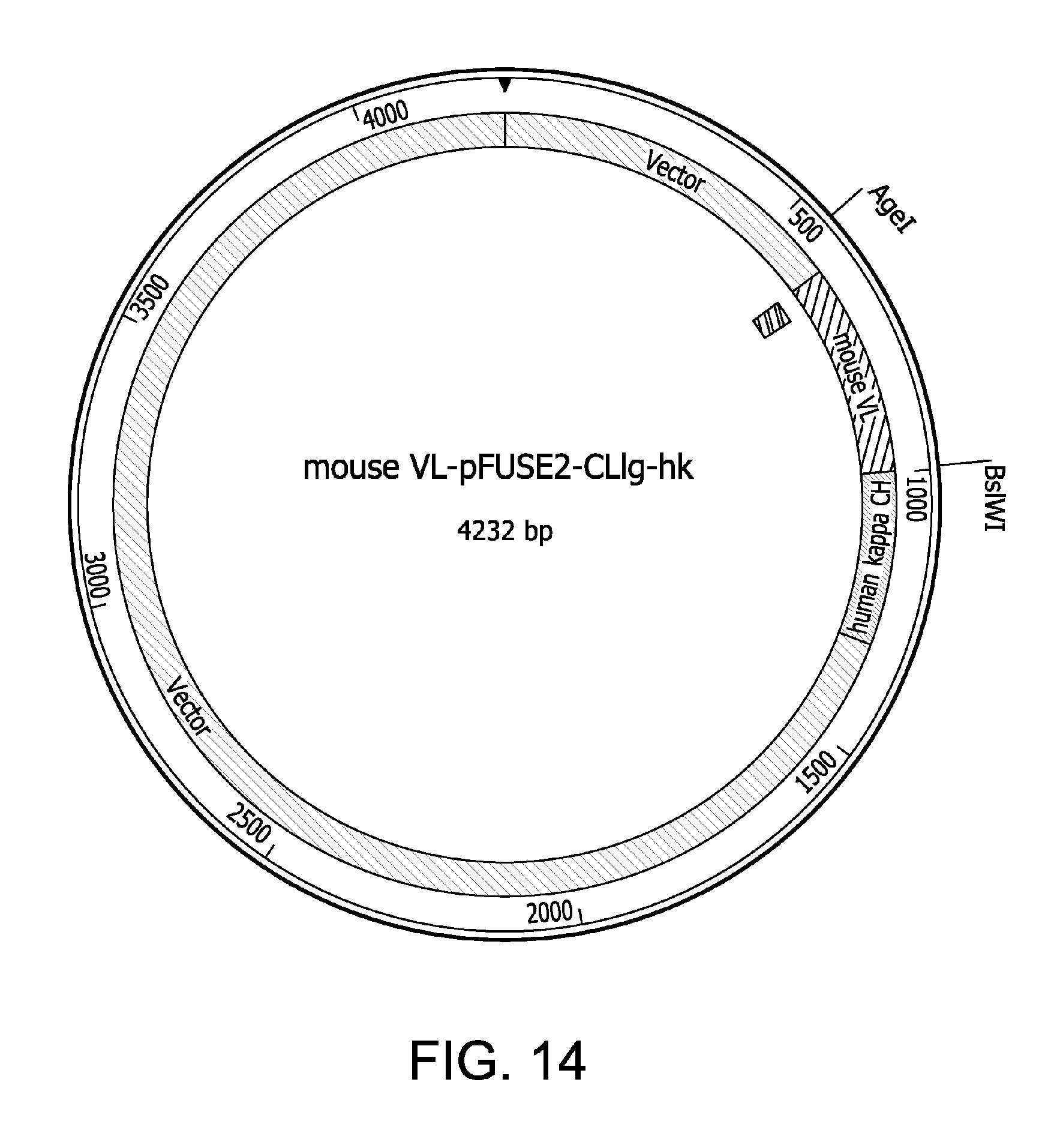
D00015

D00016

D00017

D00018

S00001
XML
uspto.report is an independent third-party trademark research tool that is not affiliated, endorsed, or sponsored by the United States Patent and Trademark Office (USPTO) or any other governmental organization. The information provided by uspto.report is based on publicly available data at the time of writing and is intended for informational purposes only.
While we strive to provide accurate and up-to-date information, we do not guarantee the accuracy, completeness, reliability, or suitability of the information displayed on this site. The use of this site is at your own risk. Any reliance you place on such information is therefore strictly at your own risk.
All official trademark data, including owner information, should be verified by visiting the official USPTO website at www.uspto.gov. This site is not intended to replace professional legal advice and should not be used as a substitute for consulting with a legal professional who is knowledgeable about trademark law.