Labeled inhibitors of prostate specific membrane antigen (PSMA), their use as imaging agents and pharmaceutical agents for the treatment of prostate cancer
Eder , et al. Nov
U.S. patent number 10,471,160 [Application Number 16/038,729] was granted by the patent office on 2019-11-12 for labeled inhibitors of prostate specific membrane antigen (psma), their use as imaging agents and pharmaceutical agents for the treatment of prostate cancer. This patent grant is currently assigned to DEUTSCHES KREBSFORSCHUNGSZENTRUM, RUPRECHT-KARLS-UNIVERSITAT HEIDELBERG. The grantee listed for this patent is DEUTSCHES KREBSFORSCHUNGSZENTRUM, RUPRECHT-KARLS-UNIVERSITAT HEIDELBERG. Invention is credited to Ulrike Bauder-Wust, Martina Benesova, Matthias Eder, Michael Eisenhut, Uwe Haberkorn, Hans-Christian Kliem, Klaus Kopka, Clemens Kratochwil, Walter Mier, Martin Schafer.


View All Diagrams
| United States Patent | 10,471,160 |
| Eder , et al. | November 12, 2019 |
| **Please see images for: ( Certificate of Correction ) ** |
Labeled inhibitors of prostate specific membrane antigen (PSMA), their use as imaging agents and pharmaceutical agents for the treatment of prostate cancer
Abstract
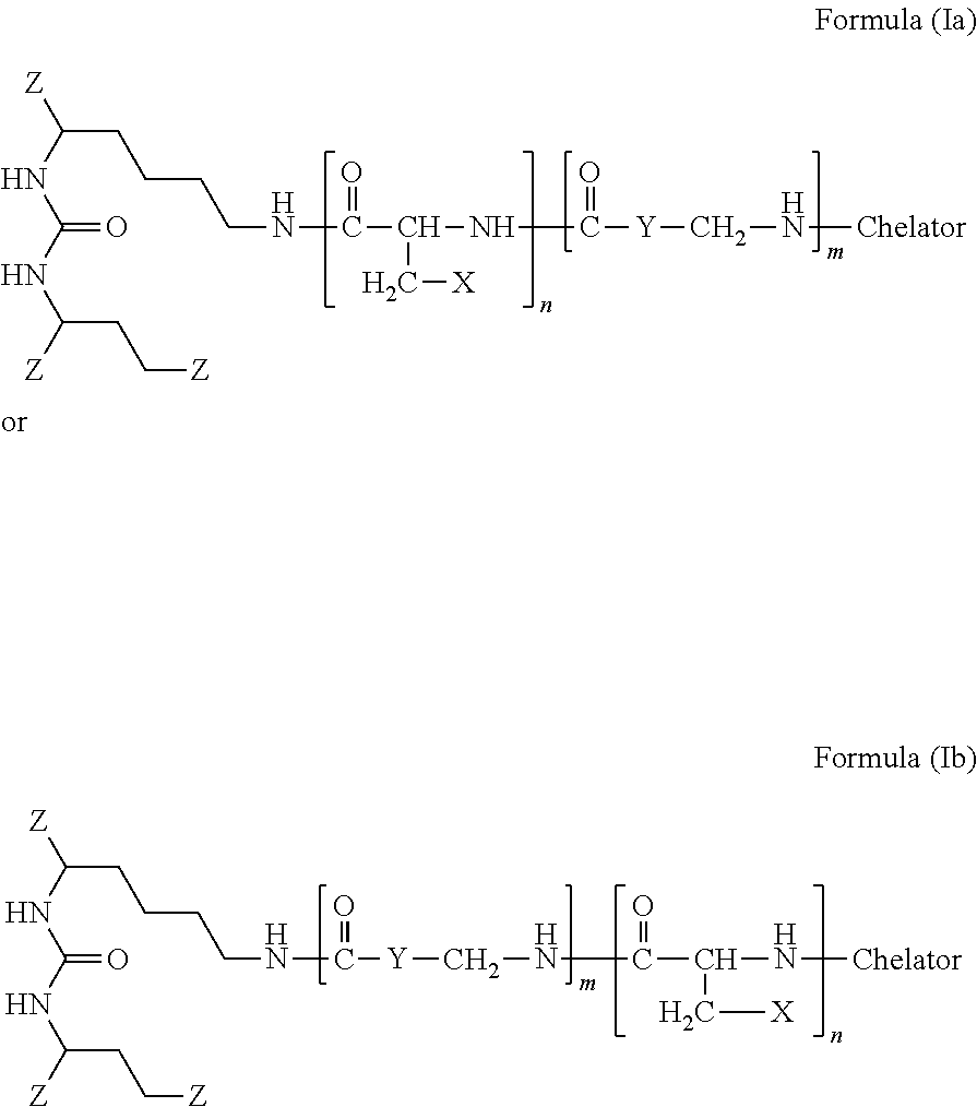
The present invention generally relates to the field of radiopharmaceuticals and their use in nuclear medicine as tracers, imaging agents and for the treatment of various disease states of prostate cancer. Thus, the present invention concerns compounds that are represented by the general Formulae (Ia) or (Ib).
| Inventors: | Eder; Matthias (Mannheim, DE), Kopka; Klaus (Dossenheim, DE), Schafer; Martin (Neckarsteinach, DE), Bauder-Wust; Ulrike (Schriesheim, DE), Haberkorn; Uwe (Schwetzingen, DE), Eisenhut; Michael (Heidelberg, DE), Mier; Walter (Bensheim, DE), Benesova; Martina (Heidelberg, DE), Kliem; Hans-Christian (Heppenheim, DE), Kratochwil; Clemens (Hirschberg a.d.B, DE) | ||||||||||
|---|---|---|---|---|---|---|---|---|---|---|---|
| Applicant: |
|
||||||||||
| Assignee: | DEUTSCHES
KREBSFORSCHUNGSZENTRUM (Heidelberg, DE) RUPRECHT-KARLS-UNIVERSITAT HEIDELBERG (Heidelberg, DE) |
||||||||||
| Family ID: | 51903864 | ||||||||||
| Appl. No.: | 16/038,729 | ||||||||||
| Filed: | July 18, 2018 |
Prior Publication Data
| Document Identifier | Publication Date | |
|---|---|---|
| US 20190008988 A1 | Jan 10, 2019 | |
Related U.S. Patent Documents
| Application Number | Filing Date | Patent Number | Issue Date | ||
|---|---|---|---|---|---|
| 15131118 | Apr 18, 2016 | ||||
| PCT/EP2014/002808 | Oct 17, 2014 | ||||
Foreign Application Priority Data
| Oct 18, 2013 [EP] | 13004991 | |||
| Jul 3, 2014 [EP] | 14175612 | |||
| Current U.S. Class: | 1/1 |
| Current CPC Class: | A61P 35/00 (20180101); A61K 51/0402 (20130101); A61P 35/04 (20180101); A61K 51/0482 (20130101); C07D 295/145 (20130101); A61K 51/0497 (20130101); C07B 59/002 (20130101); C07D 257/02 (20130101); A61P 13/08 (20180101) |
| Current International Class: | A61K 51/04 (20060101); C07D 257/02 (20060101); C07B 59/00 (20060101); C07D 295/145 (20060101) |
References Cited [Referenced By]
U.S. Patent Documents
| 4691024 | September 1987 | Kunikatsu et al. |
| 4713249 | December 1987 | Schroder |
| 5103018 | April 1992 | Motomichi et al. |
| 5266333 | November 1993 | Cady et al. |
| 5418982 | May 1995 | Kishi |
| 5627165 | May 1997 | Glazier |
| 5795877 | August 1998 | Jackson et al. |
| 5863536 | January 1999 | Jackson et al. |
| 5866679 | February 1999 | DeFeo-Jones et al. |
| 5902817 | May 1999 | Jackson et al. |
| 5948750 | September 1999 | Garsky et al. |
| 5962237 | October 1999 | Ts'o et al. |
| 5962521 | October 1999 | Jackson et al. |
| 5968915 | October 1999 | Jackson et al. |
| 5998362 | December 1999 | Feng et al. |
| 6054444 | April 2000 | Jackson et al. |
| 6068830 | May 2000 | Diamandis et al. |
| 6127333 | October 2000 | Brady et al. |
| 6174858 | January 2001 | Brady et al. |
| 6177404 | January 2001 | DeFeo-Jones et al. |
| 6232287 | May 2001 | Ruoslahti et al. |
| 6265540 | July 2001 | Isaacs et al. |
| 6342491 | January 2002 | Dey et al. |
| 6355611 | March 2002 | Karki et al. |
| 6368598 | April 2002 | D'Amico et al. |
| 6391305 | May 2002 | Feng et al. |
| 6428785 | August 2002 | Gocken |
| 6479470 | November 2002 | Kozikowski et al. |
| 6504014 | January 2003 | Isaacs et al. |
| 6511676 | January 2003 | Boulikas |
| 6518033 | February 2003 | Gromeier et al. |
| 6528499 | March 2003 | Kozikowski et al. |
| 6548260 | April 2003 | Tewari |
| 6596755 | July 2003 | Burman et al. |
| 6602274 | August 2003 | Chen |
| 6613793 | September 2003 | Burman et al. |
| 6692724 | February 2004 | Yang et al. |
| 6875886 | April 2005 | Frangioni |
| 6946133 | September 2005 | Schlom et al. |
| 7005429 | February 2006 | Dey et al. |
| 7008765 | March 2006 | Bussemakers et al. |
| 7041786 | May 2006 | Shailubhai et al. |
| 7045605 | May 2006 | Bander et al. |
| 7052703 | May 2006 | Pastan et al. |
| 7060284 | June 2006 | Kaumaya |
| 7128893 | October 2006 | Leamon et al. |
| 7129254 | October 2006 | Berger et al. |
| 7147837 | December 2006 | Lauffer et al. |
| 7153841 | December 2006 | Roncucci et al. |
| 7160885 | January 2007 | Currie et al. |
| 7166691 | January 2007 | Koochekpour et al. |
| 7192586 | March 2007 | Bander |
| 7232805 | June 2007 | Weinshenker et al. |
| 7238785 | July 2007 | Govindan et al. |
| 7282567 | October 2007 | Goldenberg et al. |
| 7361338 | April 2008 | Jakobovits et al. |
| 7381745 | June 2008 | Kozikowski et al. |
| 7399460 | July 2008 | Wedeking et al. |
| 7408079 | August 2008 | Pomper et al. |
| 7468354 | December 2008 | Isaacs |
| 7485299 | February 2009 | Afar et al. |
| 7514078 | April 2009 | Bander et al. |
| 7534580 | May 2009 | Reeves et al. |
| 7547773 | June 2009 | Schlom et al. |
| 7585491 | September 2009 | Govindan |
| 7601332 | October 2009 | Vlahov et al. |
| 7635682 | December 2009 | Denmeade et al. |
| 7638122 | December 2009 | Yu et al. |
| 7638525 | December 2009 | Jiang et al. |
| 7659395 | February 2010 | Pajouheshs et al. |
| 7662795 | February 2010 | Rodriquez et al. |
| 7691962 | April 2010 | Boyd et al. |
| 7696185 | April 2010 | Berkman |
| 7713944 | May 2010 | Kinberger et al. |
| 7740847 | June 2010 | Allan et al. |
| 7767202 | August 2010 | Pardoll et al. |
| 7767803 | August 2010 | Diener et al. |
| 7794929 | September 2010 | Baylin et al. |
| 7862798 | January 2011 | Leamon et al. |
| 7872235 | January 2011 | Rousso et al. |
| 7875586 | January 2011 | Kovbasnjuk et al. |
| 7879981 | February 2011 | Obata |
| 7910594 | March 2011 | Vlahov et al. |
| 7990533 | August 2011 | Maier et al. |
| 8000773 | August 2011 | Rousso et al. |
| 8013991 | September 2011 | Maier et al. |
| 8088387 | January 2012 | Steeves et al. |
| 8101369 | January 2012 | Nam et al. |
| 8101713 | January 2012 | Cuello et al. |
| 8105568 | January 2012 | Vlahov et al. |
| 8211402 | March 2012 | Babich et al. |
| 8153595 | April 2012 | Chen |
| 8194660 | June 2012 | Birze et al. |
| 8211401 | July 2012 | Babich et al. |
| 8211473 | July 2012 | Troiano et al. |
| 8211635 | July 2012 | Barton |
| 8227634 | July 2012 | Pomper et al. |
| 8236330 | August 2012 | Zale et al. |
| 8246968 | August 2012 | Zale et al. |
| 8258111 | September 2012 | Shen et al. |
| 8273363 | September 2012 | Zale et al. |
| 8313728 | November 2012 | Leamon et al. |
| 8388977 | March 2013 | Low et al. |
| 8394922 | March 2013 | Cheng et al. |
| 8404817 | March 2013 | Sherman et al. |
| 8414864 | April 2013 | Cappelletti et al. |
| 8414898 | April 2013 | Afar et al. |
| 8445851 | May 2013 | Rousso et al. |
| 8450290 | May 2013 | Worm et al. |
| 8465725 | June 2013 | Babich et al. |
| 8487128 | July 2013 | Babich et al. |
| 8487129 | July 2013 | Babich et al. |
| 8491896 | July 2013 | Goldenberg et al. |
| 8507434 | August 2013 | Popel et al. |
| 8557772 | October 2013 | Popel et al. |
| 8565945 | October 2013 | Babich et al. |
| 8603499 | December 2013 | Zale et al. |
| 8603500 | December 2013 | Zale et al. |
| 8603501 | December 2013 | Zale et al. |
| 8606349 | December 2013 | Rousso et al. |
| 8644910 | February 2014 | Rousso et al. |
| 8685416 | April 2014 | Klinman et al. |
| 8685891 | April 2014 | Muraca |
| 8703918 | April 2014 | Colombatti et al. |
| 8709483 | April 2014 | Farokhzad et al. |
| 8772226 | June 2014 | Denmeade et al. |
| 8772459 | June 2014 | Ho et al. |
| 8778305 | July 2014 | Pomper et al. |
| 8802153 | August 2014 | Cheng et al. |
| 8816095 | August 2014 | Brown et al. |
| 8834842 | September 2014 | Leamon et al. |
| 8840865 | September 2014 | Babich et al. |
| 8852630 | October 2014 | Spiegel et al. |
| 8858509 | October 2014 | Spiegel et al. |
| 8865126 | October 2014 | Leamon et al. |
| 8877970 | November 2014 | Zimmerman et al. |
| 8901294 | December 2014 | Kim et al. |
| 8907058 | December 2014 | Low et al. |
| 8916167 | December 2014 | Low et al. |
| 8921378 | December 2014 | Toermakaengas et al. |
| 8926944 | January 2015 | Babich et al. |
| 8926945 | January 2015 | Port et al. |
| 8940871 | January 2015 | Wu et al. |
| 8946388 | February 2015 | Sahin et al. |
| 8962799 | February 2015 | Babich et al. |
| 8987319 | March 2015 | Miller |
| 9006415 | April 2015 | Ren et al. |
| 9029340 | May 2015 | Lupold et al. |
| 9044468 | June 2015 | Pomper et al. |
| 9056841 | June 2015 | Pomper et al. |
| 9074000 | July 2015 | Scheinberg et al. |
| 9120837 | September 2015 | Babich et al. |
| 9123725 | September 2015 | Cho et al. |
| 9180203 | November 2015 | Cui et al. |
| 9180214 | November 2015 | Miao |
| 9193763 | November 2015 | Low et al. |
| 9216218 | December 2015 | Sahin et al. |
| 9226981 | January 2016 | Pomper et al. |
| 9242012 | January 2016 | Ma et al. |
| 9255262 | February 2016 | Wong et al. |
| 9278067 | March 2016 | Boulikas |
| 9295727 | March 2016 | Zale et al. |
| 9309193 | April 2016 | Babich et al. |
| 9346846 | May 2016 | Herzon et al. |
| 9371360 | June 2016 | Pomper et al. |
| 9387344 | July 2016 | Sgouros et al. |
| 9422234 | August 2016 | Chandran et al. |
| 9429575 | August 2016 | Ban et al. |
| 9433594 | September 2016 | Babich et al. |
| 9447121 | September 2016 | Babich et al. |
| 9498546 | November 2016 | Pomper et al. |
| 9556167 | January 2017 | Spiegel et al. |
| 9567402 | February 2017 | Liu |
| 9580467 | February 2017 | Cheng et al. |
| 9580474 | February 2017 | Viscidi et al. |
| 9585957 | March 2017 | Powell et al. |
| 9617602 | April 2017 | Joseph et al. |
| 9636413 | May 2017 | Vlahov et al. |
| 9687572 | June 2017 | Babich et al. |
| 9717484 | August 2017 | Kalloo et al. |
| 9745380 | August 2017 | Govindan et al. |
| 9757084 | September 2017 | Sgouros et al. |
| 9764039 | September 2017 | Thanos et al. |
| 9770467 | September 2017 | Dubensky, Jr. et al. |
| 9770517 | September 2017 | Govindan et al. |
| 9808516 | November 2017 | Brockstedt et al. |
| 9814759 | November 2017 | Wong et al. |
| 9861444 | January 2018 | Kalloo et al. |
| 9889199 | February 2018 | Basilion |
| 9932411 | April 2018 | Terrell et al. |
| 9951049 | April 2018 | Kamal et al. |
| 9951324 | April 2018 | Low et al. |
| 9956305 | May 2018 | Babich et al. |
| 9988407 | June 2018 | Slusher et al. |
| 10010624 | July 2018 | Howard et al. |
| 10011632 | July 2018 | Wang et al. |
| 10016519 | July 2018 | Kopka et al. |
| 10029023 | July 2018 | Pomper et al. |
| 10064957 | September 2018 | Govindan et al. |
| 2003/0049203 | March 2003 | Elmaleh et al. |
| 2003/0220241 | November 2003 | DeFeo-Jones et al. |
| 2003/0232760 | December 2003 | Garsky et al. |
| 2004/0001846 | January 2004 | Israeli et al. |
| 2004/0002478 | January 2004 | Kozikowski et al. |
| 2004/0002587 | January 2004 | Watkins et al. |
| 2004/0018203 | January 2004 | Pastan et al. |
| 2004/0029778 | February 2004 | Isaacs |
| 2004/0033195 | February 2004 | Leamon et al. |
| 2004/0052727 | March 2004 | Dalton et al. |
| 2004/0054190 | March 2004 | Pomper et al. |
| 2004/0058857 | March 2004 | Yao |
| 2004/0110723 | June 2004 | Frangioni |
| 2004/0146516 | July 2004 | Roben et al. |
| 2004/0213791 | October 2004 | Bander et al. |
| 2004/0229845 | November 2004 | Frangioni |
| 2004/0242582 | December 2004 | Green et al. |
| 2005/0002942 | January 2005 | Vlahov et al. |
| 2005/0069889 | March 2005 | Nihei et al. |
| 2005/0107325 | May 2005 | Manoharan et al. |
| 2005/0119166 | June 2005 | Brady et al. |
| 2005/0158780 | July 2005 | Lupold et al. |
| 2005/0234247 | October 2005 | Klar et al. |
| 2005/0239138 | October 2005 | Hess et al. |
| 2005/0239739 | October 2005 | Matulic-Adamic |
| 2005/0245486 | November 2005 | Frangioni |
| 2005/0255042 | November 2005 | Lam et al. |
| 2006/0024317 | February 2006 | Boyd et al. |
| 2006/0045883 | March 2006 | Molldrem et al. |
| 2006/0051380 | March 2006 | Schulick et al. |
| 2006/0052312 | March 2006 | Erhardt et al. |
| 2006/0062793 | March 2006 | Webb et al. |
| 2006/0105975 | May 2006 | Pendergrast et al. |
| 2006/0106047 | May 2006 | Jiang et al. |
| 2006/0140871 | June 2006 | Sillerud |
| 2006/0148718 | July 2006 | Brady et al. |
| 2006/0148741 | July 2006 | Barrett et al. |
| 2006/0155021 | July 2006 | Lenges et al. |
| 2006/0155146 | July 2006 | Lenges et al. |
| 2007/0010014 | January 2007 | Wood et al. |
| 2007/0020327 | January 2007 | Fikes et al. |
| 2007/0031326 | February 2007 | Shirvan et al. |
| 2007/0031438 | February 2007 | Junghans |
| 2007/0117153 | May 2007 | Bieniarz et al. |
| 2007/0128670 | June 2007 | Klatzmann et al. |
| 2007/0134332 | June 2007 | Turnell et al. |
| 2007/0141052 | June 2007 | Watkins et al. |
| 2007/0142296 | June 2007 | McBride et al. |
| 2007/0148662 | June 2007 | Israeli et al. |
| 2007/0160617 | July 2007 | Ma et al. |
| 2007/0172422 | July 2007 | Glazier |
| 2007/0179100 | August 2007 | Manoharan |
| 2007/0190029 | August 2007 | Pardoll et al. |
| 2007/0212337 | September 2007 | Bedi et al. |
| 2007/0219165 | September 2007 | Berkman et al. |
| 2007/0225213 | September 2007 | Kosak |
| 2007/0244055 | October 2007 | Brady et al. |
| 2007/0254316 | November 2007 | Rodriguez et al. |
| 2007/0254317 | November 2007 | Busseret-Michel et al. |
| 2008/0008649 | January 2008 | Cappelletti et al. |
| 2008/0008719 | January 2008 | Bowdish et al. |
| 2008/0089869 | April 2008 | Denmeade et al. |
| 2008/0089892 | April 2008 | Allan et al. |
| 2008/0114153 | May 2008 | Steeves et al. |
| 2008/0175789 | July 2008 | Frangioni |
| 2008/0176821 | July 2008 | Kozikowski et al. |
| 2008/0193381 | August 2008 | Babich et al. |
| 2008/0214436 | September 2008 | Yu et al. |
| 2008/0248052 | October 2008 | Vlahov et al. |
| 2008/0269105 | October 2008 | Taft et al. |
| 2008/0311037 | December 2008 | Heston et al. |
| 2009/0117042 | May 2009 | Pomper et al. |
| 2009/0123467 | May 2009 | Bedi et al. |
| 2009/0180951 | July 2009 | Zimmeran et al. |
| 2009/0214636 | August 2009 | Low et al. |
| 2009/0247614 | October 2009 | Manoharan et al. |
| 2009/0258002 | October 2009 | Barrett et al. |
| 2009/0274625 | November 2009 | Denmeade et al. |
| 2010/0047170 | February 2010 | Denmeade et al. |
| 2010/0048490 | February 2010 | Vlahov et al. |
| 2010/0092496 | April 2010 | Boyd et al. |
| 2010/0178246 | July 2010 | Babich et al. |
| 2010/0183509 | July 2010 | Babich et al. |
| 2010/0183517 | July 2010 | Berkman |
| 2010/0209343 | August 2010 | Bander et al. |
| 2010/0240701 | September 2010 | Vlahov et al. |
| 2010/0260677 | October 2010 | Bhatia et al. |
| 2010/0324008 | December 2010 | Low et al. |
| 2011/0008253 | January 2011 | Babich et al. |
| 2011/0027180 | February 2011 | Magnani |
| 2011/0027274 | February 2011 | Cheng et al. |
| 2011/0064657 | March 2011 | Pomper et al. |
| 2011/0124948 | May 2011 | Yokell |
| 2011/0142760 | June 2011 | Pomper et al. |
| 2011/0172254 | July 2011 | Leamon et al. |
| 2011/0176998 | July 2011 | Pomper et al. |
| 2011/0200677 | August 2011 | Chandran et al. |
| 2011/0288152 | November 2011 | Low et al. |
| 2011/0305768 | December 2011 | Mao et al. |
| 2012/0009121 | January 2012 | Pomper et al. |
| 2012/0276162 | November 2012 | Zale et al. |
| 2012/0322741 | December 2012 | Low et al. |
| 2013/0034494 | February 2013 | Babich et al. |
| 2013/0302409 | November 2013 | Fuchs et al. |
| 2013/0336888 | December 2013 | Babich et al. |
| 2014/0107316 | April 2014 | Vlahov et al. |
| 2014/0113322 | April 2014 | Cui et al. |
| 2014/0140925 | May 2014 | Leamon et al. |
| 2014/0154702 | June 2014 | Parker et al. |
| 2014/0187501 | July 2014 | Bilodeau et al. |
| 2014/0314864 | October 2014 | Cheng et al. |
| 2015/0023875 | January 2015 | Farokhzad et al. |
| 2015/0079001 | March 2015 | Pomper et al. |
| 2015/0104387 | April 2015 | Pomper et al. |
| 2015/0110715 | April 2015 | Eder et al. |
| 2015/0110716 | April 2015 | Armor |
| 2015/0246144 | September 2015 | Pomper et al. |
| 2015/0297735 | October 2015 | Vlahov et al. |
| 2015/0315196 | November 2015 | Howard |
| 2015/0366968 | December 2015 | Basilion et al. |
| 2016/0067341 | March 2016 | Low et al. |
| 2016/0074526 | March 2016 | Bilodeau et al. |
| 2016/0114060 | April 2016 | Pomper et al. |
| 2016/0151508 | June 2016 | Low et al. |
| 2016/0220694 | August 2016 | Vlahov et al. |
| 2016/0235865 | August 2016 | Pomper et al. |
| 2016/0287731 | October 2016 | Vlahov et al. |
| 2016/0361376 | December 2016 | Vlahov et al. |
| 2016/0361432 | December 2016 | Vlahov et al. |
| 2016/0361433 | December 2016 | Vlahov et al. |
| 2017/0081298 | March 2017 | Ray et al. |
| 2017/0151356 | June 2017 | Govindan et al. |
| 2017/0218464 | August 2017 | Pomper et al. |
| 2017/0224709 | August 2017 | Slusher et al. |
| 2017/0266316 | September 2017 | Govindan et al. |
| 2017/0275673 | September 2017 | Luo et al. |
| 2017/0281789 | October 2017 | Basilion et al. |
| 2017/0281791 | October 2017 | Govindan et al. |
| 2017/0333576 | November 2017 | Pomper et al. |
| 2018/0008668 | January 2018 | Isaacs et al. |
| 2018/0051039 | February 2018 | Pomper et al. |
| 2018/0064831 | March 2018 | Basilion et al. |
| 2018/0111895 | April 2018 | Babich et al. |
| 2018/0118847 | May 2018 | Bander |
| 2018/0133296 | May 2018 | Barrett et al. |
| 2018/0148480 | May 2018 | Issaacs et al. |
| 2018/0185511 | July 2018 | Woodworth et al. |
| 2018/0207299 | July 2018 | Babich et al. |
| 2018/0258134 | September 2018 | Wang et al. |
| 2606138 | Oct 2005 | CA | |||
| 101863924 | Oct 2010 | CN | |||
| 101863924 | Oct 2010 | CN | |||
| 202014008232 | Mar 2015 | DE | |||
| 0116208 | Aug 1984 | EP | |||
| 1177200 | Jun 2005 | EP | |||
| 1472541 | Sep 2009 | EP | |||
| 2187965 | May 2010 | EP | |||
| 2318366 | May 2011 | EP | |||
| 2136788 | Oct 2011 | EP | |||
| 2373621 | Oct 2011 | EP | |||
| 2373622 | Oct 2011 | EP | |||
| 2389361 | Nov 2011 | EP | |||
| 2408755 | Jan 2012 | EP | |||
| 2644192 | Oct 2013 | EP | |||
| 2644594 | Oct 2013 | EP | |||
| 2648766 | Oct 2013 | EP | |||
| 2436376 | Jul 2014 | EP | |||
| 2759535 | Jul 2014 | EP | |||
| 2240171 | Aug 2014 | EP | |||
| 2170075 | Dec 2014 | EP | |||
| 2823826 | Jan 2015 | EP | |||
| 2097111 | Jul 2015 | EP | |||
| 2921482 | Sep 2015 | EP | |||
| 2938364 | Nov 2015 | EP | |||
| 2942065 | Nov 2015 | EP | |||
| 2958596 | Dec 2015 | EP | |||
| 2993171 | Mar 2016 | EP | |||
| 2706057 | Apr 2016 | EP | |||
| 3038996 | Jul 2016 | EP | |||
| 2002506204 | Feb 2002 | JP | |||
| 2004536034 | Dec 2004 | JP | |||
| 2005274569 | Oct 2005 | JP | |||
| 2006501149 | Jan 2006 | JP | |||
| 2006514961 | May 2006 | JP | |||
| 2006518712 | Aug 2006 | JP | |||
| 2007521803 | Aug 2007 | JP | |||
| 2009519209 | May 2009 | JP | |||
| 2010515732 | May 2010 | JP | |||
| 2010518112 | May 2010 | JP | |||
| 2010532754 | Oct 2010 | JP | |||
| 2012511023 | May 2012 | JP | |||
| 8801622 | Mar 1988 | WO | |||
| 9107418 | Apr 1991 | WO | |||
| 95/33766 | Dec 1995 | WO | |||
| 9945374 | Sep 1999 | WO | |||
| 2000/066091 | Nov 2000 | WO | |||
| 2000064911 | Nov 2000 | WO | |||
| 2002043773 | Feb 2002 | WO | |||
| 2002062398 | Aug 2002 | WO | |||
| 2002098885 | Dec 2002 | WO | |||
| 2003060523 | Jul 2003 | WO | |||
| 2003092742 | Nov 2003 | WO | |||
| 2003097647 | Nov 2003 | WO | |||
| 2004010957 | Feb 2004 | WO | |||
| 2004069159 | Aug 2004 | WO | |||
| 2004069285 | Aug 2004 | WO | |||
| 2005082023 | Sep 2005 | WO | |||
| 2006012527 | Feb 2006 | WO | |||
| 2006093991 | Sep 2006 | WO | |||
| 2006096754 | Sep 2006 | WO | |||
| 2006136564 | Dec 2006 | WO | |||
| 2007006041 | Jan 2007 | WO | |||
| 2007022494 | Feb 2007 | WO | |||
| 2007106869 | Sep 2007 | WO | |||
| 2008057437 | May 2008 | WO | |||
| 2008058192 | May 2008 | WO | |||
| 2008088648 | Jul 2008 | WO | |||
| 2008098112 | Aug 2008 | WO | |||
| 2008101231 | Aug 2008 | WO | |||
| 2008121949 | Oct 2008 | WO | |||
| 2009002529 | Dec 2008 | WO | |||
| 2009026177 | Feb 2009 | WO | |||
| 2009082606 | Feb 2009 | WO | |||
| 2009070302 | Jun 2009 | WO | |||
| 2009089383 | Jul 2009 | WO | |||
| 2009002529 | Dec 2009 | WO | |||
| 2009002993 | Dec 2009 | WO | |||
| 2010014933 | Feb 2010 | WO | |||
| 2010/065899 | Jun 2010 | WO | |||
| 2010/065906 | Jun 2010 | WO | |||
| 2010065899 | Jun 2010 | WO | |||
| 2010065902 | Jun 2010 | WO | |||
| 2010108125 | Sep 2010 | WO | |||
| 2011/014821 | Feb 2011 | WO | |||
| 2010108125 | Mar 2011 | WO | |||
| 2011106639 | Sep 2011 | WO | |||
| 2012078534 | Jun 2012 | WO | |||
| 2012166923 | Dec 2012 | WO | |||
| 2013022797 | Feb 2013 | WO | |||
| 2013028664 | Feb 2013 | WO | |||
| 2013/130776 | Sep 2013 | WO | |||
| 2014062697 | Apr 2014 | WO | |||
| 2014078484 | May 2014 | WO | |||
| 2014106208 | Jul 2014 | WO | |||
| 2014127365 | Aug 2014 | WO | |||
| 2014134543 | Sep 2014 | WO | |||
| 2015053318 | Apr 2015 | WO | |||
| 2015057250 | Apr 2015 | WO | |||
| 2015171792 | Nov 2015 | WO | |||
| 2016030329 | Mar 2016 | WO | |||
| 2016040179 | Mar 2016 | WO | |||
Other References
|
Aggarwal, S., Singh, P., Topaloglu, O., Isaacs, J. T., and Denmeade, S. R. (2006) A dimeric peptide that binds selectively to prostate-specific membrane antigen and inhibits its enzymatic activity. Cancer Res 66, 9171-7. cited by applicant . Zhou, J., Neale, J. H., Pomper, M. G., and Kozikowski, A. P. (2005) NAAG peptidase inhibitors and their potential for diagnosis and therapy. Nat Rev Drug Discov 4, 1015-26. cited by applicant . Yamaguchi, T., Lee, J., Uemura, H., Sasaki, T., Takahashi, N., Oka, T., Shizukuishi, K, Endou, H., Kubota, Y., and Inoue, T. (2005) Prostate cancer: a comparative study of 11C-choline PET and MR imaging combined with proton MR spectroscopy. Eur J Nucl Med Mol Imaging 32, 742-8. cited by applicant . Jadvar, H., Xiankui, L., Shahinian, A., Park, R., Tohme, M., Pinski, J., and Conti, P. S. (2005) Glucose metabolism of human prostate cancer mouse xenografts. Mol Imaging 4, 91-7. cited by applicant . Humblet, V., Lapidus, R., Williams, L. R., Tsukamoto, T., Rojas, C., Majer, P., Hin, B., Ohnishi, S., De Grand, A. M., Zaheer, A., Renze, J. T., Nakayama, A., Slusher, B. S., and Frangioni, J. V. (2005) High-affinity near-infrared fluorescent small-molecule contrast agents for in vivo imaging of prostate-specific membrane antigen. Mol Imaging 4, 448-62. cited by applicant . Gregor, P. D., Wolchok, J. D., Turaga, V., Latouche, J. B., Sadelain, M., Bacich, D., Heston, W. D., Houghton, A. N., and Scher, H. I. (2005) Induction of autoantibodies to syngeneic prostate-specific membrane antigen by xenogeneic vaccination. Int J Cancer 116, 415-21. cited by applicant . Foss, C. A., Mease, R. C., Fan, H., Wang, Y., Ravert, H. T., Dannals, R. F., Olszewski, R. T., Heston, W. D., Kozikowski, A. P., and Pomper, M. G. (2005) Radiolabeled small-molecule ligands for prostate-specific membrane antigen: in vivo imaging in experimental models of prostate cancer. Clin Cancer Res 11, 4022-8. cited by applicant . Vallabhajosula, S., Smith-Jones, P. M., Navarro, V., Goldsmith, S. J., and Bander, N. H. (2004) Radioimmunotherapy of prostate cancer in human xenografts using monoclonal antibodies specific to prostate specific membrane antigen (PSMA): studies in nude mice. Prostate 58, 145-55. cited by applicant . Scheffel, U., and Pomper, M. G. (2004) PET imaging of GRP receptor expression in prostate cancer. J Nucl Med 45, 1277-8. cited by applicant . O'Keefe, D. S., Bacich, D. J., and Heston, W. D. (2004) Comparative analysis of prostate-specific membrane antigen (PSMA) versus a prostate-specific membrane antigen-like gene. Prostate 58, 200-10. cited by applicant . Milowsky, M. I., Nanus, D. M., Kostakoglu, L., Vallabhajosula, S., Goldsmith, S. J., and Bander, N. H. (2004) Phase I trial of yttrium-90-labeled anti-prostate-specific membrane antigen monoclonal antibody J591 for androgen-independent prostate cancer. J Clin Oncol 22, 2522-31. cited by applicant . Matthies, A., Ezziddin, S., Ulrich, E. M., Palmedo, H., Biersack, H. J., Bender, H., and Guhlke, S. (2004) Imaging of prostate cancer metastases with 18F-fluoroacetate using PET/CT. Eur J Nucl Med Mol Imaging 31, 797. cited by applicant . Kozikowski, A. P., Zhang, J., Nan, F., Petukhov, P. A., Grajkowska, E., Wroblewski, J. T., Yamamoto, T., Bzdega, T., Wroblewska, B., and Neale, J. H. (2004) Synthesis of urea-based inhibitors as active site probes of glutamate carboxypeptidase II: efficacy as analgesic agents. J Med Chem 47, 1729-38. cited by applicant . Henry, M. D., Wen, S., Silva, M. D., Chandra, S., Milton, M., and Worland, P. J. (2004) A prostate-specific membrane antigen-targeted monoclonal antibody-chemotherapeutic conjugate designed for the treatment of prostate cancer. Cancer Res 64, 7995-8001. cited by applicant . Ghosh, A., and Heston, W. D. (2004) Tumor target prostate specific membrane antigen (PSMA) and its regulation in prostate cancer. J Cell Biochem 91, 528-39. cited by applicant . Chopra, A. (2010) 68Ga-Labeled 2-{3-[5-(7-{1-benzyloxycarbonyl-5-[2-(4,7,10-tris-carboxymethyl-1,4,7,10-- tetraazacyclododec-1-l)acetylamino]pentylcarbamoyly}-heptanoylamino)-1-car- boxypentyl]ureido}pentanedioic acid. Molecular Imaging and Contrast Agent Database (MICAD) [database online]. Bethesda (MD): National Center for Biotechnology Information (US); 2004-2013. cited by applicant . Chopra, A. (2010) 68Ga-Labeled 2-[3-(1-carboxy-5-{7-[5-carboxy-5-(3-phenyl-2-{3-phenyl-2-[2-(4,7,10-tris- -carboxymethyl-1,4,7,10-tetraazacyclododec-1-l)acetylamino]propionylamino}- p ropionylamino)pentylcarbamoyl]heptanoylamino}pentyl)ureido]pentanedioic acid. Molecular Imaging and Contrast Agent Database (MICAD) [database online]. Bethesda (MD): National Center for Biotechnology Information (US); 2004-2013. cited by applicant . Bzdega, T., Crowe, S. L., Ramadan, E. R., Sciarretta, K. H., Olszewski, R. T., Ojeifo, O. A., Rafalski, V. A., Wroblewska, B., and Neale, J. H. (2004) The cloning and characterization of a second brain enzyme with NAAG peptidase activity. Journal of neurochemistry 89, 627-35. cited by applicant . Tang, H., Brown, M., Ye, Y., Huang, G., Zhang, Y., Wang, Y., Zhai, H., Chen, X., Shen, T. Y., and Tenniswood, M. (2003) Prostate targeting ligands based on N-acetylated alpha-linked acidic dipeptidase. Biochem Biophys Res Commun 307, 8-14. cited by applicant . Schulke, N., Varlamova, O. A., Donovan, G. P., Ma, D., Gardner, J. P., Morrissey, D. M., Arrigale, R. R., Zhan, C., Chodera, A. J., Surowitz, K. G., Maddon, P. J., Heston, W. D., and Olson, W. C. (2003) The homodimer of prostate-specific membrane antigen is a functional target for cancer therapy. Proc Natl Acad Sci U S A 100, 12590-5. cited by applicant . Rajasekaran, S. A., Anilkumar, G., Oshima, E., Bowie, J. U., Liu, H., Heston, W., Bander, N. H., and Rajasekaran, A. K. (2003) A novel cytoplasmic tail MXXXL motif mediates the internalization of prostate-specific membrane antigen. Mol Biol Cell 14, 4835-45. cited by applicant . Oyama, N., Miller, T. R., Dehdashti, F., Siegel, B. A., Fischer, K. C., Michalski, J. M., Kibel, A. S., Andriole, G. L., Picus, J., and Welch, M. J. (2003) 11C-acetate PET imaging of prostate cancer: detection of recurrent disease at PSA relapse. J Nucl Med 44, 549-55. cited by applicant . Meighan, M. A., Dickerson, M. T., Glinskii, O., Glinsky, V. V., Wright, G. L., Jr., and Deutscher, S. L. (2003) Recombinant glutamate carboxypeptidase II (prostate specific membrane antigen-PSMA)--cellular localization and bioactivity analyses. J Protein Chem 22, 317-26. cited by applicant . Hain, S. F., and Maisey, M. N. (2003) Positron emission tomography for urological tumours. BJU Int 92, 159-64. cited by applicant . Dimitrakopoulou-Strauss, A., and Strauss, L. G. (2003) PET imaging of prostate cancer with 11C-acetate. J Nucl Med 44, 556-8. cited by applicant . Shvarts, O., Han, K. R., Seltzer, M., Pantuck, A. J., and Belldegrun, A. S. (2002) Positron emission tomography in urologic oncology. Cancer Control 9, 335-42. cited by applicant . Rong, S. B., Zhang, J., Neale, J. H., Wroblewski, J. T., Wang, S., and Kozikowski, A. P. (2002) Molecular modeling of the interactions of glutamate carboxypeptidase II with its potent NAAG-based inhibitors. J Med Chem 45, 4140-52. cited by applicant . Pomper, M. G., Musachio, J. L., Zhang, J., Scheffel, U., Zhou, Y., Hilton, J., Maini, A., Dannals, R. F., Wong, D. F., and Kozikowski, A. P. (2002) 11C-MCG: synthesis, uptake selectivity, and primate PET of a probe for glutamate carboxypeptidase II (NAALADase). Mol Imaging 1, 96-101. cited by applicant . Oyama, N., Akino, H., Kanamaru, H., Suzuki, Y., Muramoto, S., Yonekura, Y., Sadato, N., Yamamoto, K., and Okada, K. (2002) 11C-acetate PET imaging of prostate cancer. J Nucl Med 43, 181-6. cited by applicant . Kotzerke, J., Gschwend, J. E., and Neumaier, B. (2002) PET for prostate cancer imaging: still a quandary or the ultimate solution? J Nucl Med 43, 200-2. cited by applicant . Hara, T., Kosaka, N., and Kishi, H. (2002) Development of (18)F-fluoroethylcholine for cancer imaging with PET: synthesis, biochemistry, and prostate cancer imaging. J Nucl Med 43, 187-99. cited by applicant . Hara, T. (2002) 11C-choline and 2-deoxy-2-[18F]fluoro-D-glucose in tumor imaging with positron emission tomography. Mol Imaging Biol 4, 267-73. cited by applicant . Chang, S. S., and Heston, W. D. (2002) the clinical role of prostate-specific membrane antigen (PSMA). Urol Oncol 7, 7-12. cited by applicant . DeGrado, T. R., Coleman, R. E., Wang, S., Baldwin, S. W., Orr, M. D., Robertson, C. N., Polascik, T. J., and Price, D. T. (2001) Synthesis and evaluation of 18F-labeled choline as an oncologic tracer for positron emission tomography: initial findings in prostate cancer. Cancer Res 61, 110-7. cited by applicant . DeGrado, T. R., Baldwin, S. W., Wang, S., Orr, M. D., Liao, R. P., Friedman, H. S., Reiman, R., Price, D. T., and Coleman, R. E. (2001) Synthesis and evaluation of (18)F-labeled choline analogs as oncologic PET tracers. J Nucl Med 42, 1805-14. cited by applicant . Bacich, D. J., Pinto, J. T., Tong, W. P., and Heston, W. D. W. (2001) Cloning, expression, genomic localization, and enzymatic activities of the mouse homolog of prostate-specific membrane antigen/NAALADase/folate hydrolase. Mammalian Genome 12, 117-123. cited by applicant . Slusher, B. S., Vornov, J. J., Thomas, A. G., Hum, P. D., Harukuni, I., Bhardwaj, A., Traystman, R. J., Robinson, M. B., Britton, P., Lu, X. C., Tortella, F. C., Wozniak, K M., Yudkoff, M., Potter, B. M., and Jackson, P. F. (1999) Selective inhibition of NAALADase, which converts NAAG to glutamate, reduces ischemic brain injury. Nat Med 5, 1396-402. cited by applicant . Dumas, F., Gala, J. L., Berteau, P., Brasseur, F., Eschwege, P., Paradis, V., Lacour, B., Philippe, M., and Loric, S. (1999) Molecular expression of PSMA mRNA and protein in primary renal tumors. Int J Cancer 80, 799-803. cited by applicant . Chang, S. S., Reuter, V. E., Heston, W. D., Bander, N. H., Grauer, L. S., and Gaudin, P. B. (1999) Five different anti-prostate-specific membrane antigen (PSMA) antibodies confirm PSMA expression in tumor-associated neovasculature. Cancer Res 59, 3192-8. cited by applicant . Sweat, S. D., Pacelli, A., Murphy, G. P., and Bostwick, D. G. (1998) Prostate-specific membrane antigen expression is greatest in prostate adenocarcinoma and lymph node metastases. Urology 52, 637-40. cited by applicant . Murphy, G. P., Elgamal, A. A., Su, S. L., Bostwick, D. G., and Holmes, E. H. (1998) Current evaluation of the tissue localization and diagnostic utility of prostate specific membrane antigen. Cancer 83, 2259-69. cited by applicant . Liu, H., Rajasekaran, A. K., Moy, P., Xia, Y., Kim, S., Navarro, V., Rahmati, R., and Bander, N. H. (1998) Constitutive and antibody-induced internalization of prostate-specific membrane antigen. Cancer Res 58, 4055-60. cited by applicant . Kahn, D., Williams, R. D., Manyak, M. J., Haseman, M. K., Seldin, D. W., Libertino, J. A., and Maguire, R. T. (1998) 111Indium-capromab pendetide in the evaluation of patients with residual or recurrent prostate cancer after radical prostatectomy. The ProstaScint Study Group. J Urol 159, 2041-6; discussion 2046-7. cited by applicant . Hara, T., Kosaka, N., and Kishi, H. (1998) PET imaging of prostate cancer using carbon-11-choline. J Nucl Med 39, 990-5. cited by applicant . Bostwick, D. G., Pacelli, A., Blute, M., Roche, P., and Murphy, G. P. (1998) Prostate specific membrane antigen expression in prostatic intraepithelial neoplasia and adenocarcinoma: a study of 184 cases. Cancer 82, 2256-61. cited by applicant . Silver, D. A., Pellicer, I., Fair, W. R., Heston, W. D., and Cordon-Cardo, C. (1997) Prostate-specific membrane antigen expression in normal and malignant human tissues. Clin Cancer Res 3, 81-5. cited by applicant . Fair, W. R., Israeli, R. S., and Heston, W. D. (1997) Prostate-specific membrane antigen. Prostate 32, 140-8. cited by applicant . Jackson P.F., Cole D.C., Slusher B.S., Stetz S.L., Ross L.E., Donzanti B.A., and D.A., T. (1996) Design, synthesis, and biological activity of a potent inhibitor of the neuropeptidase N-acetylated alpha-linked acidic dipeptidase. cited by applicant . Wright, G. L., Jr., Haley, C., Beckett, M. L., and Schellhammer, P. F. (1995) Expression of prostate-specific membrane antigen in normal, benign, and malignant prostate tissues. Urol Oncol 1, 18-28. cited by applicant . Leek, J., Lench, N., Maraj, B., Bailey, A., Carr, I. M., Andersen, S., Cross, J., Whelan, P., MacLennan, K. A., Meredith, D. M., and et al. (1995) Prostate-specific membrane antigen: evidence for the existence of a second related human gene. Br J Cancer 72, 583-8. cited by applicant . Vees, H., et al., "18F-choline and/or 11 C-acetate positron emission tomography: detection of residual or progressive subclinical disease at very low prostate-specific antigen values ( <1 ng/mL) after radical prostatectomy," BJU Int, 2007, 99, 1415-1420. cited by applicant . Larson, S. M., et al., "Tumor Localization of 16.beta.-18F-Fluoro-5a-Dihydrotestosterone Versus 18F-FDG in Patients with Progressive, Metastatic Prostate Cancer," J Nucl Med, 2004, 45, 366-373. cited by applicant . Schuster, D.M., et al., "Initial Experience with the Radiotracer Anti-1-Am ino-3-18F-Fluorocyclobutane-1-Carboxylic Acid with PET/CT in Prostate Carcinoma," J. Nucl. Med., 2007, 48, 56-63. cited by applicant . Tehrani, O.S., et al., "Tumor Imaging Using 1-(2'-deoxy-2'-18F-Fluoro-.beta.-D-Arabinofuranosyl) Thymine and PET," J. Nucl. Med., 2007, 48, 1436-1441. cited by applicant . Mease RC., et al., "N-[N-[(S)-1, 3-Dicarboxypropyl]Carbam oyl]-4-[18F]F luorobenzyl-L-Cysteine, [18F]DCFBC: A New Imaging Probe for Prostate Cancer," Clin. Cancer Res., 2008, 14, 3036-3043. cited by applicant . Zhou, J., et al., "NAAG Peptidase Inhibitors and Their Potential for Diagnosis and Therapy," Nat Rev Drug Discovery, 2005, 4, 1015-1026. cited by applicant . Schulke, N., et al., "The homodimer of prostate-specific membrane antigen is a functional target for cancer therapy," Proc. Natl. Acad. Sci., USA, 2003, 100, 12590-12595. cited by applicant . Nan, F., et al., "Dual Function Glutamate-Related Ligands: Discovery of a Novel, Potent Inhibitor of Glutamate Carboxypeptidase II Possessing mGluR3 Agonist Activity," J. Med. Chem., 2000, 43, 772-774. cited by applicant . Lange, P.H., "ProstaScint scan for staging prostate cancer," Urology, 2001, 57, 402-406. cited by applicant . Haseman, M.K., et al., "Capromab Pendetide Imaging of Prostate Cancer," Cancer Bi other Radiopharm, 2000, 15, 131-140. cited by applicant . Mier W., et al., "Conjugation of DOTA Using Isolated Phenolic Active Esters: The Labeling and Biodistribution of Albumin as Blood Pool Marker," Bioconjugate Chem., 2005, 16: 237-240. cited by applicant . Schafer et al., "A dimerized urea-based inhibitor of the prostate-specific membrane antigen for 68Ga-PET imaging of prostate cancer," EJNMMI Research, 2012, 2, 23, 11 pages. cited by applicant . Humblet, V. et al., "An HPLC/mass spectrometry platform for the development of multimodality contrast agents and targeted therapeutics: prostate-specific membrane antigen small molecule derivatives," Contrast Med. Mal. Imaging, 2006, 1, 196-211. cited by applicant . Pomper, M.G., et al., "11C-MCG: synthesis, uptake selectivity, and primate PET of a probe for glutamate carboxypeptidase II (NAALADase)," Mal Imaging, 2002, 1, 96-101. cited by applicant . Japanese Office Action dated Mar. 23, 2017, which issued during prosecution of Japanese Application No. P016-524427. cited by applicant . Humblet, et al., "An HPLC/mass spectrometry platform for the development of multimodality contrast agents and targeted therapeutics: prostate-specific membrane antigen small molecule derivatives" Contrast Media & Molecular Imaging, Jul. 2006 1:196-211. cited by applicant . Eurasian Office Action dated Feb. 10, 2017, which issued during prosecution of Eurasian Application No. 201690495/28. cited by applicant . Office Action for copending Eurasian Patent Application No. 201690495/28, dated Jul. 14, 2017. cited by applicant . Scher, B. et al., "Value of 11C-choline PET and PET/CT in patients with suspected prostate cancer," Eur. J. Nucl. Med. Mol. Imaging., 2007, 34, 45-53. cited by applicant . Tasch, J. et al., "A Unique Folate Hydrolase, Prostage-Specific Membrane Antingen (PSMA): A Target for Immunotherapy?" Crit. Rev. Immunol., 2001, 21, 249-261. cited by applicant . Rosenthal, S.A. et al., "Utility of Capromab Pendetide (ProstaScint) Imaging in the Management of Prostate Cancer," Tech Urol, 2001, 7, 27-37. cited by applicant . Wiberg et al. "A comparison of some properties of C=O and C=S bonds," ARKIVOC, 2011, (v) 45-56. cited by applicant . Zechmann, C. M., Afshar-Oromieh, A., Armor, T., Stubbs, J. B., Mier, W., Hadaschik, B., Joyal, J., Kopka, K., Debus, J., Babich, J. W., and Haberkom, U. (2014) Radiation dosimetry and first therapy results with a I/ I-labeled small molecule (MIP-1095) targeting PSMA for prostate cancer therapy. Eur J Nucl Med Mol Imaging 41, 1280-92. cited by applicant . Huang, S. S., Wang, X., Zhang, Y., Doke, A., DiFilippo, F. P., and Heston, W. D. (2014) Improving the biodistribution of PSMA-targeting tracers with a highly negatively charged linker. Prostate 74, 702-13. cited by applicant . Wiehr, S., Buhler, P., Gierschner, D., Wolf, P., Rolle, A. M., Kesenheimer, C., Pichler, B. J., and Elsasser-Beile, U. (2014) Pharmacokinetics and PET imaging properties of two recombinant anti-PSMA antibody fragments in comparison to their parental antibody. Prostate 74. 743-55. cited by applicant . Weineisen, M., Simecek, J., Schottelius, M., Schwaiger, M., and Wester, H. J. (2014) Synthesis and preclinical evaluation of DOTAGA-conjugated PSMA ligands for functional imaging and endoradiotherapy of prostate cancer. EJNMMI Res 4, 63. cited by applicant . Wang, X., Huang, S. S., Heston, W. D., Guo, H., Wang, B. C., and Basilion, J. P. (2014) Development of targeted near-infrared imaging agents for prostate cancer. Mol Cancer Ther 13(11), 2595-606. cited by applicant . Uprimny, C., Kroiss, A., Nilica, B., Buxbaum, S., Decristoforo, C., Horninger, W., and Virgolini, I. J. (2014) Ga-PSMA ligand PET versus F-NaF PET: evaluation of response to Ra therapy in a prostate cancer patient. Eur J Nucl Med Mol Imaging 42, 362-63. cited by applicant . Hillier, S. M., Maresca, K. P., Lu, G., Merkin, R. D., Marquis, J. C., Zimmerman, C. N., Eckelman, W. C., Joyal, J. L, and Babich, J. W. (2013) 99mTc-labeled small-molecule inhibitors of prostate-specific membrane antigen for molecular imaging of prostate cancer. J Nucl Med 54, 1369-76. cited by applicant . Jadvar, H. (2013) Imaging evaluation of prostate cancer with (18)F-fluorodeoxyglucose PET/CT: utility and limitations. Eur J Nucl Med Mol Imaging 40 (Suppl 1), S5-S10. cited by applicant . Jemaa, A. B., Bouraoui, Y., Sallami, S., Banasr, A., Nouira, Y., Horchani, A., and Oueslati, R. (2013) Cellular distribution and heterogeneity of Psa and Psma expression in normal, hyperplasia and human prostate cancer. La tunisie Medicate 91, 458-463. cited by applicant . Kasperzyk, J. L., Finn, S. P., Flavin, R., Fiorentino, M., Lis, R., Hendrickson, W. K., Clinton, S. K., Sesso, H. D., Giovannucci, E. L., Stampfer, M. J., Loda, M., and Mucci, L. A. (2013) Prostate-specific membrane antigen protein expression in tumor tissue and risk of lethal prostate cancer. Cancer Epidemiol Biomarkers Prev 22, 2354-63. cited by applicant . Kasten, B. B., Liu, T., Nedrow-Byers, J. R., Benny, P. D., and Berkman, C. E. (2013) Targeting prostate cancer cells with PSMA inhibitor-guided gold nanoparticles. Bioorg Med Chem Lett 23, 565-8. cited by applicant . Tykvart, J., Schimer, J., Barinkova, J., Pachl, P., Postova-Slavetinska, L., Majer, P., Konvalinka, J., and Sacha, P. (2014) Rational design of urea-based glutamate carboxypeptidase II (GCPII) inhibitors as versatile tools for specific drug targeting and delivery. Bioorg Med Chem 22, 4099-108. cited by applicant . Ristau, B. T., O'Keefe, D. S., and Bacich, D. J. (2014) The prostate-specific membrane antigen: Lessons and current clinical implications from 20 years of research. Urol Oncol 32, 272-279. cited by applicant . Ray Banerjee, S., Pullambhatla, M., Foss, C. A., Nimmagadda, S., Ferdani, R., Anderson, C. J., Mease, R. C., and Pomper, M. G. (2014) 64Cu-Labeled Inhibitors of Prostate-Specific Membrane Antigen for PET Imaging of Prostate Cancer. J Med Chem 57, 2657-69. cited by applicant . Rais, R., Rojas, C., Wozniak, K., Wu, Y., Zhao, M., Tsukamoto, T., Rudek, M. A., and Slusher, B. S. (2014) Bioanalytical method for evaluating the pharmacokinetics of the GCP-II inhibitor 2-phosphonomethyl pentanedioic acid (2-PMPA). Journal of pharmaceutical and biomedical analysis 88, 162-9. cited by applicant . Pavlicek, J., Ptacek, J., Cerny, J., Byun, Y., Skultetyova, L., Pomper, M. G., Lubkowski, J., and Barinka, C. (2014) Structural characterization of P1'-diversified urea-based inhibitors of glutamate carboxypeptidase II. Bioorg Med Chem Lett 24, 2340-5. cited by applicant . Lutje, S., Rijpkema, M., Franssen, G. M., Fracasso, G., Helfrich, W., Eek, A., Oyen, W. J., Colombatti, M., and Boerman, O. C. (2014) Dual-Modality Image-Guided Surgery of Prostate Cancer with a Radiolabeled Fluorescent Anti-PSMA Monoclonal Antibody. J Nucl Med 55, 995-1001. cited by applicant . Liu, T., Mendes, D. E., and Berkman, C. E. (2014) Functional prostate-specific membrane antigen is enriched in exosomes from prostate cancer cells. Int J Oncol 44, 918-22. cited by applicant . Krohn, T., Verburg, F. A., Pufe, T., Neuhuber, W., Vogg, A., Heinzel, A., Mottaghy, F. M., and Behrendt, F. F. (2014) [Ga]PSMA-HBED uptake mimicking lymph node metastasis in coeliac ganglia: an important pitfall in clinical practice. Eur J Nucl Med Mol Imaging 42, 210-14. cited by applicant . Kovar, J. L., Cheung, L. L., Simpson, M. A., and Olive, D. M. (2014) Pharmacokinetic and Biodistribution Assessment of a Near Infrared-Labeled PSMA-Specific Small Molecule in Tumor-Bearing Mice. Prostate cancer 2014, 104248. cited by applicant . Huang, B., Otis, J., Joice, M., Kotlyar, A., and Thomas, T. P. (2014) PSMA-Targeted Stably Linked "Dendrimer-Glutamate Urea-Methotrexate" as a Prostate Cancer Therapeutic. Biomacromolecules 15, 915-23. cited by applicant . Haberkom, U., Mier, W., Dimitrakopoulou-Strauss, A., Eder, M., Kopka, K., and Altmann, A. (2014) Mechanistic and high-throughput approaches for the design of molecular imaging probes and targeted therapeutics. Clinical and Translational Imaging 2, 33-41. cited by applicant . El-Zaria, M. E., Genady, A. R., Janzen, N., Petlura, C. I., Beckford Vera, D. R., and Valliant, J. F. (2014) Preparation and evaluation of carborane-derived inhibitors of prostate specific membrane antigen (PSMA). Dalton Trans 43, 4950-61. cited by applicant . Eiber, M., Nekolla, S. G., Maurer, T., Weirich, G., Wester, H. J., and Schwaiger, M. (2014) Ga-PSMA PET/MR with multimodality image analysis for primary prostate cancer. Abdom Imaging 40, 1769-71. cited by applicant . Eder, M., Schafer, M., Bauder-Wust, U., Haberkorn, U., Eisenhut, M., and Kopka, K. (2014) Preclinical evaluation of a bispecific low-molecular heterodimer targeting both PSMA and GRPR for improved PET imaging and therapy of prostate cancer. The Prostate 74, 659-68. cited by applicant . Eder, M., Neels, O., Muller, M., Bauder-Wust, U., Remde, Y., Schafer, M., Hennrich, U., Eisenhut, M., Afshar-Oromieh, A., Haberkom, U., and Kopka, K. (2014) Novel Preclinical and Radiopharmaceutical Aspects of [68Ga]GA-PSMA-HBED-CC: A New PET Tracer for Imaging of Prostate Cancer. Pharmaceuticals 7, 779-96. cited by applicant . Afshar-Oromieh, A., Zechmann, C. M., Matcher, A., Eder, M., Eisenhut, M., Linhart, H. G., Holland-Letz, T., Hadaschik, B. A., Giesel, F. L., Debus, J., and Haberkorn, U. (2014) Comparison of PET imaging with a Ga-labelled PSMA ligand and F-choline-based PET/CT for the diagnosis of recurrent prostate cancer. Eur J Nucl Med Mol Imaging 41, 11-20. cited by applicant . Afshar-Oromieh, A., Haberkorn, U., Schlemmer, H. P., Fenchel, M., Eder, M., Eisenhut, M., Hadaschik, B. A., Kopp-Schneider, A., and Rothke, M. (2014) Comparison of PET/CT and PET/MRI hybrid systems using a 68Ga-labelled PSMA ligand for the diagnosis of recurrent prostate cancer: initial experience. Eur J Nucl Med Mol Imaging 41, 887-97. cited by applicant . Afshar-Oromieh, A., Avtzi, E., Giesel, F. L., Holland-Letz, T., Linhart, H. G., Eder, M., Eisenhut, M., Boxler, S., Hadaschik, B. A., Kratochwil, C., Weichert, W., Kopka, K., Debus, J., and Haberkorn, U. (2014) The diagnostic value of PET/CT imaging with the Ga-labelled PSMA ligand HBED-CC in the diagnosis of recurrent prostate cancer. Eur J Nucl Med Mol Imaging 42, 197-209. cited by applicant . Whitaker, H. C., Shiong, L. L., Kay, J. D., Gronberg, H., Warren, A. Y., Seipel, A., Wiklund, F., Thomas, B., Wiklund, P., Miller, J. L., Menon, S., Ramos-Montoya, A., Vowler, S. L., Massie, C., Egevad, L., and Neal, D. E. (2013) N-acetyl-L-aspartyl-L-glutamate peptidase-like 2 is overexpressed in cancer and promotes a pro-migratory and pro-metastatic phenotype. Oncogene 33, 5274-87. cited by applicant . Simone, C. B., 2nd, and Hahn, S. M. (2013) What's in a Label? Radioimmunotherapy for metastatic prostate cancer. Clin Cancer Res 19, 4908-10. cited by applicant . Rybalov, M., Breeuwsma, A. J., Leliveld, A. M., Pruim, J., Dierckx, R. A., and de Jong, I. J. (2013) Impact of total PSA, PSA doubling time and PSA velocity on detection rates of (11)C-Choline positron emission tomography in recurrent prostate cancer. World J Urol 31, 319-23. cited by applicant . Rothke, M. C., Afshar-Oromieh, A., and Schlemmer, H. P. (2013) [Potential of PET/MRI for diagnosis of prostate cancer]. Radiologe 53, 676-81. cited by applicant . Roethke, M. C., Kuru, T. H., Afshar-Oromieh, A., Schlemmer, H. P., Hadaschik, B. A., and Fenchel, M. (2013) Hybrid positron emission tomography-magnetic resonance imaging with gallium 68 prostate-specific membrane antigen tracer: a next step for imaging of recurrent prostate cancer-preliminary results. Eur Urol 64, 862-4. cited by applicant . Kim, D. Kim, S. K., Valencia, C. A., and Liu, R. (2013) Tribody: robust self-assembled trimeric targeting ligands with high stability and significantly improved target-binding strength. Biochemistry 52, 7283-94. cited by applicant . Ray Banerjee, S., Pullambhatla, M., Foss, C. A., Falk, A., Byun, Y., Nimmagadda, S., Mease, R. C., and Pomper, M. G. (2013) Effect of Chelators on the Pharmacokinetics of Tc-Labeled Imaging Agents for the Prostate-Specific Membrane Antigen (PSMA). J Med Chem 56, 6108-21. cited by applicant . Parker, S. A., Diaz, I. L., Anderson, K. A., and Batt, C. A. (2013) Design, production, and characterization of a single-chain variable fragment (ScFv) derived from the prostate specific membrane antigen (PSMA) monoclonal antibody U591. Protein Expr Purif 89, 136-145. cited by applicant . Osborne, J. R., Green, D. A., Spratt, D. E., Lyashchenko, S., Fareedy, S. B., Robinson, B. D., Beattie, B. J., Jain, M., Lewis J. S., Christos, P., Larson, S. M., Bander, N. H., and Scherr, D. S. (2014) A Prospective Pilot Study of Zr-J591/ Prostate Specific Membrane Antigen Positron Emission Tomography in Men with Localized Prostate Cancer Undergoing Radical Prostatectomy. J Urol 191, 1439-46. cited by applicant . Osborne, J. R., Akhtar, N. H., Vallabhajosula, S., Anand, A., Deh, K, and Tagawa, S. T. (2013) Prostate-specific membrane antigen-based imaging. Urol Oncol 31, 144-54. cited by applicant . Nedrow-Byers, J. R., Moore, A. L., Ganguly, T., Hopkins, M. R., Fulton, M. D., Benny, P. D., and Berkman, C. E. (2013) PSMA-targeted SPECT agents: mode of binding effect on in vitro performance. Prostate 73, 355-62. cited by applicant . Mease RC, Foss CA, and Mg, P. (2013) PET imaging in prostate cancer: focus on prostate-specific membrane antigen. Curr Top Med Chem 13, 951-962. cited by applicant . Lu, G., Maresca, K. P., Hillier, S. M., Zimmerman, C. N., Eckelman, W. C., Joyal, J. L., and Babich, J. W. (2013) Synthesis and SAR of (9)(9)mTc/Re-labeled small molecule prostate specific membrane antigen inhibitors with novel Solar chelates. Bioorg Med Chem Lett 23, 1557-63. cited by applicant . Lesche, R., Kettschau, G., Gromov, A. V., Bohnke, N., Borkowski, S., Monning, U., Hegele-Hartung, C., Dohr, O., Dinkelborg, L. M., and Graham, K. (2014) Preclinical evaluation of Bay 1075553, a novel F-labelled inhibitor of prostate-specific membrane antigen for PET imaging of prostate cancer. Eur J Nucl Med Mol Imaging 41, 89-101. cited by applicant . First Office Action in counterpart Canadian Application No. 2924360, dated Jan. 11, 2018 (4 pages). cited by applicant . Second Office Action in counterpart Chinese Application No. 201480056250.5, dated Aug. 15, 2017 (4 pages). cited by applicant . Second Office Action in counterpart Eurasian Application No. 201690495/28, dated Feb. 10, 2017 (5 pages). cited by applicant . First Office Action in counterpart Korean Application No. 1020167012314, dated Mar. 6, 2017 (3 pages). cited by applicant . Second Office Action in counterpart Korean Application No. 1020167012314, dated Oct. 14, 2017 (3 pages). cited by applicant . Second Office Action for counterpart Georgian Application No. 14132/01, dated Mar. 2018 (2 pages). cited by applicant . First Office Action for counterpart Georgian Application No. 14132/01, dated Feb. 2017 (2 pages). cited by applicant . First Examination Report for counterpart Chilean Application No. 00883-2016, dated Apr. 12, 2018. cited by applicant . Third Office Action for counterpart Korean Application No. 10-2016-7012314, dated May 2, 2018 (2 pages). cited by applicant . Silvola J. M.U. et al., "Al18F-NOTA-Folate Accumulates in Atherosclerotic Plaques and Can be Detected by PET/CT", Poster presented Nov. 7, 2015 in Orlando, FL at the 2015 American Heart Association ReSuscitation Science Symposium (http://newsroom.heart.org/events/scientific-sessions-2015-news- room-2942760). (3 pages). cited by applicant . Silvola J. M.U. et al., "Al18F-NOTA-Folate Accumulates in Atherosclerotic Plaques and Can be Detected by PET/CT", Published reference of poster, Nov. 10, 2015, at http://circ.ahajournals.org/content/132/Suppl_3/A18873?cited-by=yes&legid- =circulationaha;132/Suppl_3/A18873; Circulation, 2015; 132:A18873. (2 pages). cited by applicant . Chinese Patent Application No. 201480071256.X, by Endocyte, Inc. et al.: Office Action dated Apr. 20, 2017; English translation (9 pages). cited by applicant . Eurasian Patent Application No. 201690862/28, by Endocyte, Inc. et al.: Office Action, dated May 22, 2017; English translation (2 pages). cited by applicant . European Patent Application No. EP 14861854, by Endocyte, Inc. et al: Partial Supplementary Search Report with Opinion; dated May 19, 2017 (15 pages). cited by applicant . Slusher, B. S., Tsai, G., Yoo, G., and Coyle, J. T. (1992) Immunocytochemical localization of the N-acetyl-aspartyl-glutamate (NAAG) hydrolyzing enzyme N-acetylated alpha-linked acidic dipeptidase (NAALADase). J Comp Neurol 315, 217-29. cited by applicant . Hwang D. R. Mathias, C. J., Welch, M. J., McGuire, A. H., and Kadmon, D. (1990) Imaging prostate derived tumors with PET and N-(3-[18F]fluoropropyl)putrescine. Int J Rad Appl Instrum B 17, 525-32. cited by applicant . Hwang, D. R., Lang, L. X., Mathias, C. J., Kadmon, D., and Welch, M. J. (1989) N-3-[18F]fluoropropylputrescine as potential PET imaging agent for prostate and prostate derived tumors. J Nucl Med 30, 1205-10. cited by applicant . Office Action for counterpart Chinese Application No. 201610184873.1, dated Jul. 24, 2018. cited by applicant . Shelley James, "Urea Based Rhenium Tricarbonyl Dipeptide Compounds as Potential Radiopharmaceuticals for PSMA Imaging"; Poster: INOR 258; The 229th ACS National Meeting, San Diego, CA, Mar. 13-17, 2005. cited by applicant . PCT International Search Report and Written Opinion for PCT/US2011/026238, dated Apr. 27, 2011. cited by applicant . PCT International Search Report and Written Opinion for PCT/US2013/070007, dated Mar. 5, 2014. cited by applicant . PCT International Search Report for PCT/US2008/073375 dated Oct. 26, 2008. cited by applicant . PCT International Search Report for PCT/US2016/012653 dated Mar. 11, 2016. cited by applicant . PCT International Search Report/Written Opinion for PCT/US2009/061067, completed May 28, 2010. cited by applicant . PCT International Search Report/Written Opinion for PCT/US2009/061049, completed Mar. 15, 2010. cited by applicant . PCT Search Report & Written Opinion issued in App. No. PCT/US2014/065467. cited by applicant . Silverman, "The Organic Chemistry of Drug Design and Drug Action," Elsevier Academic Press (2nd Ed. 2003). cited by applicant . Beheshti, M., Langsteger, W., and Fogelman, I. (2009) Prostate cancer: role of SPECT and PET in imaging bone metastases. Semin Nucl Med 39, 396-407. cited by applicant . Ananias, H. J., van den Heuvel, M. C., Helfrich, W., and de Jong, I. J. (2009) Expression of the gastrin-releasing peptide receptor, the prostate stem cell antigen and the prostate-specific membrane antigen in lymph node and bone metastases of prostate cancer. Prostate 69, 1101-8. cited by applicant . Vavere, A. L., Kridel, S. J., Wheeler, F. B., and Lewis, J. S. (2008) 1-11C-acetate as a PET radiopharmaceutical for imaging fatty acid synthase expression in prostate cancer. J Nucl Med 49, 327-34. cited by applicant . Tang, Q. L., and Yao, M. Y. (2008) [Updated application of prostate-specific membrane antigen to the diagnosis and treatment of prostate cancer]. Zhonghua Nan Ke Xue 14, 79-82. cited by applicant . Soloviev, D., Fini, A., Chierichetti, F., Al-Nahhas, A., and Rubello, D. (2008) PET imaging with 11C-acetate in prostate cancer: a biochemical, radiochemical and clinical perspective. Eur J Nucl Med Mol Imaging 35, 942-9. cited by applicant . Scher, B., and Seitz, M. (2008) PET/CT imaging of recurrent prostate cancer. Eur J Nucl Med Mol Imaging 35, 5-8. cited by applicant . Rinnab, L., Mottaghy, F. M., Simon, J., Volkmer, B. G., de Petriconi, R., Hautmann, R. E., Wittbrodt, M., Egghart, G., Moeller, P., Blumstein, N., Reske, S., and Kuefer, R. (2008) [11C]Choline PET/CT for targeted salvage lymph node dissection in patients with biochemical recurrence after primary curative therapy for prostate cancer. Preliminary results of a prospective study. Urol Int 81, 191-7. cited by applicant . Reske, S. N., Blumstein, N. M., and Glatting, G. (2008) [11C]choline PET/CT imaging in occult local relapse of prostate cancer after radical prostatectomy. Eur J Nucl Med Mol Imaging 35, 9-17. cited by applicant . Reske S.N. (2008) [11C]choling uptake with PET/CT for the initial diagnosis of prostate cancer: relation to PSA levels, tumour stage and anti-androgenic therapy. Eur J Nucl Med Mol Imaging 35, 1740-1. cited by applicant . Mease, R. C., Dusich, C. L., Foss, C. A., Ravert, H. T., Dannals, R. F., Seidel, J., Prideaux, A., Fox, J. J., Sgouros, G., Kozikowski, A. P., and Pomper, M. G. (2008) N-[N- [(S)-1,3-Dicarboxypropyl]carbamoyl]-4-[18F]fluorobenzyl-L-cysteine, [18F]DCFBC: a new imaging probe for prostate cancer. Clin Cancer Res 14, 3036-43. cited by applicant . Luboldt, W., Kufer, R., Blumstein, N., Toussaint, T. L., Kluge, A., Seemann, M. D., and Luboldt, H. J. (2008) Prostate carcinoma: diffusion-weighted imaging as potential alternative to conventional MR and 11C-choline PET/CT for detection of bone metastases. Radiology 249, 1017-25. cited by applicant . Liu T. Toriyabe, Y., Kazak, M., and Berkman, C. E. (2008) Pseudoirreversible inhibition of prostate-specific membrane antigen by phosphoramidate peptidomimetics. Biochemistry 47, 12658-60. cited by applicant . Li, X., Liu, Q., Wang, M., Jin, X., Yao, S., Liu, S., and Li, J. (2008) C-11 choline PET/CT imaging for differentiating malignant from benign prostate lesions. Clin Nucl Med 33, 671-6. cited by applicant . Igerc, I., Kohlfurst, S., Gallowitsch, H. J., Matschnig, S., Kresnik, E., Gomez-Segovia, I., and Lind, P. (2008) The value of 18F-choline PET/CT in patients with elevated PSA-level and negative prostate needle biopsy for localisation of prostate cancer. Eur J Nucl Med Mol Imaging 35, 976-83. cited by applicant . Husarik, D. B., Miralbell, R., Dubs, M., John, H., Giger, O. T., Gelet, A., Cservenyak, T., and Hany, T. F. (2008) Evaluation of [(18)F]-choline PET/CT for staging and restaging of prostate cancer. Eur J Nucl Med Mol Imaging 35, 253-63. cited by applicant . Hospers, G. A., Helmond, F. A., de Vries, E. G., Dierckx, R. A., and de Vries, E. F. (2008) PET imaging of steroid receptor expression in breast and prostate cancer Curr Pharm Des 14, 3020-32. cited by applicant . Colabufo, N. A., Abate, C., Contino, M., Inglese, C., Niso, M., Berardi, F., and Perrone, R. (2008) PB183, a sigma receptor ligand, as a potential PET probe for the imaging of prostate adenocarcinoma. Bioorg Med Chem Lett 18, 1990-3. cited by applicant . Chen, Y., Foss, C. A., Byun, Y., Nimmagadda, S., Pullambhatla, M., Fox, J. J., Castanares, M., Lupold, S. E., Babich, J. W., Mease, R. C., and Pomper, M. G. (2008) Radiohalogenated prostate-specific membrane antigen (PSMA)-based areas as imaging agents for prostate cancer. J Med Chem 51, 7933-43. cited by applicant . Chandran, S. S., Banerjee, S. R., Mease, R. C., Pomper, M. G., and Denmeade, S. R. (2008) Characterization of a targeted nanoparticle functionalized with a urea-based inhibitor of prostate-specific membrane antigen (PSMA). Cancer Biol Ther 7, 974-82. cited by applicant . Barinka, C., Byun, Y., Dusich, C. L., Banerjee, S. R., Chen, Y., Castanares, M., Kozikowski, A. P., Mease, R. C., Pomper, M. G., and Lubkowski, J. (2008) Interactions between human glutamate carboxypeptidase II and urea-based inhibitors: structural characterization. J Med Chem 51, 7737-43. cited by applicant . Banerjee, S. R., Foss, C. A., Castanares, M., Mease, R. C., Byun, Y., Fox, J. J., Hilton, J., Lupold, S. E., Kozikowski, A. P., and Pomper, M. G. (2008) Synthesis and evaluation of technetium-99m- and rhenium-labeled inhibitors of the prostate-specific membrane antigen (PSMA). J Med Chem 51, 4504-17. cited by applicant . Wu, L. Y., Anderson, M. O., Toriyabe, Y., Maung, J., Campbell, T. Y., Tajon, C., Kazak, M., Moser, J., and Berkman, C. E. (2007) The molecular pruning of a phosphoramidate peptidomimetic inhibitor of prostate-specific membrane antigen. Bioorg Med Chem 15, 7434-43. cited by applicant . Vees, H., Buchegger, F., Albrecht, S., Khan, H., Husarik, D., Zaidi, H., Soloview, D., Hany, T. F., and Miralbell, R. (2007) 18F-choline and/or 11C-acetate positron emission tomography: detection of residual or progressive subclinical disease at very low prostate-specfic antigen values (<1 ng/mL) after radical prostatectomy. BJU Int 99, 1415-20. cited by applicant . Testa, C., Schiavina, R., Lodi, R., Salizzoni, E., Corti, B., Farsad, M., Kurhanewicz, J., Manferrari, F., Brunocilla, E., Tonon, C., Monetti, N., Castellucci, P., Fanti, S., Coe, M., Grigioni, E. F., Martorana, G., Canini, R., and Barbiroli, B. (2007) Prostate cancer: sextant localization with MR imaging, MR spectroscopy, and 11C-choline PET/CT. Radiology 244, 797-806. cited by applicant . Scattoni, V., Picchio, M., Suardi, N., Messa, C., Freschi, M., Roscigno, M., Da Pozzo, L., Bocciardi, A., Rigatti, P., and Fazio, F. (2007) Detection of lymph-node metastases with integrated [11C]choline PET/CT in patients with PSA failure after radical retropubic prostatectomy: results confirmed by open pelvic-retroperitoneal lymphadenectomy. Eur Urol 52, 423-9. cited by applicant . Sacha, P., Zamecnik, J., Barinka, C., Hlouchova, K., Vicha, A., Mlcochova, P., Hilgert, I., Eckschlager, T., and Konvalinka, J. (2007) Expression of glutamate carboxypeptidase II in human brain. Neuroscience 144, 1361-72. cited by applicant . Reske, S. N. (2007) [Nuclear imaging of prostate cancer: current status]. Der Urologe. 46, 1485-99. cited by applicant . Ponde, D. E., Dence, C. S., Oyama, N., Kim, J., Tai, Y. C., Laforest, R., Siegel, B. A., and Welch, M. J. (2007) 18F-fluoroacetate: a potential acetate analog for prostate tumor imaging--in vivo evaluation of 18F-fluoroacetate versus 11C-acetate. J Nucl Med 48, 420-8. cited by applicant . Pemer, S., Hofer, M. D., Kim, R., Shah, R. B., Li, H., Moller, P., Hautmann, R. E., Gschwend, J. E., Kuefer, R., and Rubin, M. A. (2007) Prostate-specific membrane antigen expression as a predictor of prostate cancer progression. Hum Pathol 38, 696-701. cited by applicant . Oehr, P., and Bouchelouche, K. (2007) Imaging of prostate cancer. Curr Opin Oncol 19, 259-64. cited by applicant . Morris, M. J., and Scher, H. I. (2007) (11)C-acetate PET imaging in prostate cancer. Eur J Nucl Med Mol Imaging 34, 181-4. cited by applicant . Micochova, P., Plechanovova, A., Barinka, C., Mahadevan, D., Saldanha, J. W., Rulisek, L., and Konvalinka, J. (2007) Mapping of the active site of glutamate carboxypeptidase II by site-directed mutagenesis. FEBS J 274, 4731-41. cited by applicant . Mhawech-Fauceglia, P., Zhang, S., Terracciano, L., Sauter, G., Chadhuri, A., Herrmann, F. R., and Penetrante, R. (2007) Prostate-specific membrane antigen (PSMA) protein expression in normal and neoplastic tissues and its sensitivity and specificity in prostate adenocarcinoma: an immunohistochemical study using mutiple tumour tissue microarray technique. Histopathology 50, 472-83. cited by applicant . Lambert, L. A., and Mitchell, S. L. (2007) Molecular evolution of the transferrin receptor/glutamate carboxypeptidase II family. Journal of molecular evolution 64, 113-28. cited by applicant . Hlouchova, K., Barinka, C., Klusak, V., Sacha, P., Mlcochova, P., Majer, P., Rulisek, L., and Konvalinka, J. (2007) Biochemical characterization of human glutamate carboxypeptidase III. Journal of neurochemistry 101, 682-96. cited by applicant . Goodman, O. B., Jr., Barwe, S. P., Ritter, B., McPherson, P. S., Vasko, A. J., Keen, J. H., Nanus, D. M., Bander, N. H., and Rajasekaran, A. K. (2007) Interaction of prostate specific membrane antigen with clathrin and the adaptor protein complex-2. Int J Oncol 31, 1199-203. cited by applicant . Fall, K., Garmo, H., Andren, O., Bill-Axelson, A., Adolfsson, J., Adami, H. O., Johansson, J. E., and Holmberg, L. (2007) Prostate-specific antigen levels as a predictor of lethal prostate cancer. J Natl Cancer Inst 99, 526-32. cited by applicant . Barinka, C., Starkova, J., Konvalinka, J., and Lubkowski, J. (2007) A high-resolution structure of ligand-free human glutamate carboxypeptidase II. Acta Crystallogr Sect F Struct Biol Cryst Commun 63, 150-3. cited by applicant . Barinka, C., Rovenska, M., Mlcochova, P., Hlouchova, K., Plechanovova, A., Majer, P., Tsukamoto, T., Slusher, B. S., Konvalinka, J., and Lubkowski, J. (2007) Structural insight into the pharmacophore pocket of human glutamate carboxypeptidase II. J Med Chem 50, 3267-73. cited by applicant . Anderson, M. O., Wu, L. Y., Santiago, N. M., Moser, J. M., Rowley, J. A., Bolstad, E. S., and Berkman, C. E. (2007) Substrate specificity of prostate-specific membrane antigen. Bioorg Med Chem 15, 6678-86. cited by applicant . Reske, S. N., Blumstein, N. M., Neumaier, B., Gottfried, H. W., Finsterbusch, F., Kocot, D., Moller, P., Glatting, G., and Perner, S. (2006) Imaging prostate cancer with 11C-choline PET/CT. J Nucl Med 47, 1249-54. cited by applicant . Reske, S. N., Blumstein, N. M., and Glatting, G. (2006) [Advancement of PET and PET/CT in prostate carcinoma]. Der Urologe. Ausg. A 45, 707-10, 712-4. cited by applicant . Reske, S. N., Blumstein, N. M., and Glatting, G. (2006) [PET and PET/CT in relapsing prostate carcinoma]. Der Urologe. Ausg. A 45, 1240, 1242-4, 1246-8, 1250. cited by applicant . Oyama, N., Kaneda, T., Nakai, M., Shioyama, R., Matsuta, Y., Tanase, K., Aoki, Y., Miwa, Y., Akino, H., Yokoyama, O., Okazawa, H., Fujibayashi, Y., and Yonekura, Y. (2006) [Pet imaging in prostate cancer]. Hinyokika Kiyo 52, 503-5. cited by applicant . Mesters, J. R., Barinka, C., Li, W., Tsukamoto, T., Majer, P., Slusher, B. S., Konvalinka, J., and Hilgenfeld, R. (2006) Structure of glutamate carboxypeptidase II, a drug target in neuronal damage and prostate cancer. EMBO J 25, 1375-84. cited by applicant . Kinoshita, Y., Kuratsukuri, K., Landas, S., Imaida, K., Rovito, P. M., Jr., Wang, C. Y., and Haas, G. P. (2006) Expression of prostate-specific membrane antigen in normal and malignant human tissues. World J Surg 30, 628-36. cited by applicant . Elsasser-Beile, U., Wolf, P., Gierschner, D., Buhler, P., Schultze-Seemann, W., and Wetterauer, U. (2006) A new generation of monoclonal and recombinant antibodies against cell-adherent prostate specific membrane antigen for diagnostic and therapeutic targeting of prostate cancer. Prostate 66, 1359-70. cited by applicant . Cunha, A. C., Weigle, B., Kiessling, A., Bachmann, M., and Rieber, E. P. (2006) Tissue-specificity of prostate specific antigens: comparative analysis of transcript levels in prostate and non-prostatic tissues. Cancer Lett 236, 229-38. cited by applicant . Cimitan, M., Bortolus, R., Morassut, S., Canzonieri, V., Garbeglio, A., Baresic, T., Borsatti, E., Drigo, A., and Trovo, M. G. (2006) [18F]fluorocholine PET/CT imaging for the detection of recurrent prostate cancer at PSA relapse: experience in 100 consecutive patients. Eur J Nucl Med Mol Imaging 33, 1387-98. cited by applicant . Extended European Search Report for European Application No. 18175078, dated Sep. 14, 2018. cited by applicant . Office Action for counterpart Indonesia Patent Application No. P00201603202, dated Jan. 28, 2019 (with English Translation). cited by applicant . Office Action for co-pending Canadian Application No. 2924360, dated Sep. 11, 2018. cited by applicant . C. Dusich et al., General Approach for the Preparation of Fluorescent PSMA/GCPII Inhibitors, Abstract ID: 470 Poster board space: 29, Molecular Imaging, vol. 5, No. 3, pp. 322-323, Jul. 2006. cited by applicant . Liu M., et al., "Synthesis and Biological Evaluation of Diethylenetriamine Pentaacetic acid-Polyethylene Glycol Folate: A new Folate-Derived, 99mTc-Based Radiopharmaceutical," Bioconjugate Chem., 2005 vol. 16, p. 1126-1132. cited by applicant . Muller C., et al. "Synthesis and in Vitro/in Vivo Evaluation of Novel 99mTc(CO)3-Folates," Bioconjugate Chem., 2006 vol. 17, p. 797-806. cited by applicant . Viola-Villegas N., et al. "Targeting Gallium to Cancer Cells through the Folate Receptor," Drug Target Insights, 2008 vol. 3, p. 13-25. cited by applicant . Viola-Villegas N., et al. "Targeting the Folate Receptor (FR): Imaging and Cytotoxicity of Rel Conjugates in FR-Overexpressing Cancer Cells," ChemMedChem, 2008 vol. 3, p. 1387-1394. cited by applicant . Zhou J., "In vivo evaluation of medical device-associated inflammation using macrophage-specific position emission tomography (PET) imaging," Bioorganic and Medicinal Chemistry Letters, 2013 vol. 23, p. 2044-2047. cited by applicant . Kularatne SA., et al. "Comparative Analysis of Folate Derived PET Imaging Agents with [18F]-2-Fluoro-2-deoxy-D-glucose Using Rodent Inflammatory Paw Model," Molecular Pharmaceutics, 2013 vol. 10, p. 3103-3111. cited by applicant . Office Action for counterpart Mexican Patent Application No. MX/a/2016/005013, dated Apr. 24, 2019 (with English Translation). cited by applicant . C. Foss et al., Synthesis and Validation of a Novel Small-Molecule Fluorescent Probe for PSMA Expression in Human Tumor Neovasculature, Abstract ID: 362, Molecular Imaging, vol. 4, No. 3, p. 321, Jul. 2005. cited by applicant . Office Action for counterpart Eurasian Patent Application No. 201690495, dated Dec. 20, 2018 (with English translation). cited by applicant . Harada, N., Kimura, H., Ono, M., and Saji, H. (2013) Preparation of asymmetric urea derivatives that target prostate-specific membrane antigen for SPECT imaging. J Med Chem 56, 7890-901. cited by applicant . Frigerio, B., Fracasso, G., Luison, E., Cingarlini, S., Mortarino, M., Coliva, A., Seregni, E., Bombardieri, E., Zuccolotto, G., Rosato, A., Colombatti, M., Canevari, S., and Figini, M. (2013) A single-chain fragment against prostate specific membrane antigen as a tool to build theranostic reagents for prostate cancer. Eur J Cancer 49, 2223-32. cited by applicant . Franc, B. L., Cho, S. Y., Rosenthal, S. A., Cui, Y., Tsui, B., Vandewalker, K. M., Holz, A. L., Poonamallee, U., Pomper, M. G., and James, R. B. (2013) Detection and localization of carcinoma within the prostate using high resolution transrectal gamma imaging (TRGI) of monoclonal antibody directed at prostate specific membrane antigen (PSMA)-Proof of concept and initial imaging results. European Journal of Radiology 82, 1877-84. cited by applicant . Eder, M., Eisenhut, M., Babich, J., and Haberkorn, U. (2013) PSMA as a target for radiolabelled small molecules. Eur J Nucl Med Mol Imaging 40, 819-23. cited by applicant . Divyya, S., Naushad, S. M., Murthy, P. V., Reddy Ch, R., and Kutala, V. K. (2013) GCPII modulates oxidative stress and prostate cancer susceptibility through changes in methylation of RASSF1, BNIP3, GSTP1 and Ec-SOD. Molecular Biology Reports 40, 5541-50. cited by applicant . Ceci, F., Castellucci, P., Mamede, M., Schiavina, R., Rubello, D., Fuccio, C., Ambrosini, V., Boschi, S., Martorana, G., and Fanti, S. (2013) (11)C-Choline PET/CT in patients with hormone-resistant prostate cancer showing biochemical relapse after radical prostatectomy. Eur J Nucl Med Mol Imaging 40, 149-55. cited by applicant . Benesova, M., Schafer, M., Bauder-Wust, U., Mier, W., Haberkorn, U., Eisenhut, M., Kopka, K., and Eder, M. (2013) Linker Modifications of DOTA-conjugated Inhibitors of the Prostate-Specific Membrane Antigen (PSMA). Eur J Nucl Med Mol Imaging 40 (Suppl 2), S193. cited by applicant . Ben Jemaa, A., Sallami, S., Ceraline, J., and Oueslati, R. (2013) A novel regulation of PSMA and PSA expression by Q640X AR in 22Rv1 and LNCaP prostate cancer cells. Cell biology international 37, 464-70. cited by applicant . Ben Jemaa, A., Bouraoui, Y., Sallami, S., Nouira, Y., and Oueslati, R. (2013) A comparison of the biological features of prostate cancer with (PSA+, PSMA+) profile according to RKIP. BioMed Research International 2013, 409179. cited by applicant . Barrett, J. A., Coleman, R. E., Goldsmith, S. J., Vallabhajosula, S., Petry, N. A., Cho, S., Armor, T., Stubbs, J. B., Maresca, K. P., Stabin, M. G., Joyal, J. L., Eckelman, W. C., and Babich, J. W. (2013) First-in-Man Evaluation of 2 High-Affinity PSMA-Avid Small Molecules for Imaging Prostate Cancer. J Nucl Med 54, 380-7. cited by applicant . Antunes, A. A., Reis, S. T., Leite, K. R., Real, D. M., Sousa-Canavez, J. M., Camara-Lopes, L. H., Dall'Oglio, M. F., and Srougi, M. (2013) PGC and PSMA in prostate cancer diagnosis: tissue analysis from biopsy samples. International Braz J Urol : official journal of the Brazilian Society of Urology 39, 649-56. cited by applicant . Afshar-Oromieh, A., Matcher, A., Eder, M., Eisenhut, M., Linhart, H. G., Hadaschik, B. A., Holland-Letz, T., Giesel, F. L. Kratochwil, C., Haufe, S., Haberkorn, U., and Zechmann, C. M. (2013) PET imaging with a [(68)Ga]gallium-labelled PSMA ligand for the diagnosis of prostate cancer biodistribution in humans and first evaluation of tumour lesions. Eur J Nucl Med Mol Imaging 40, 486-95. cited by applicant . Afshar-Oromieh, A., Haberkorn, U., Hadaschik, B., Habl, G., Eder, M., Eisenhut, M., Schlemmer, H. P., and Roethke, M. C. (2013) PET/MRI with a 68Ga-PSMA ligand for the detection of prostate cancer. Eur J Nucl Med Mol Imaging 40, 1629-30. cited by applicant . Abstracts of the Annual Congress of the European Association of Nuclear Medicine. Oct. 19-23, 2013. Lyon, France. Eur J Nucl Med Mol Imaging 40 Suppl 2, S1-S477. cited by applicant . Zhang, Y., Guo, Z., Du, T., Chen, J., Wang, W., Xu, K, Lin, T., and Huang, H. (2012) Prostate specific membrane antigen (PSMA): A novel modulator of p38 for proliferation, migration, and survival in prostate cancer cells. Prostate 73, 835-41. cited by applicant . Weissbach, L. (2012) [Which components should living guidelines contain?]. Der Urologe. 51, 57-9. cited by applicant . Thalmann, G. (2012) [Advanced prostate cancer: where are we going?]. Der Urologe 51, 7. cited by applicant . Taylor, R. M., Sevems, V., Brown, D. C., Bisoffi, M., and Sillerud, L. O. (2012) Prostate cancer targeting motifs: expression of alphanu beta3, neurotensin receptor 1, prostate specific membrane antigen, and prostate stem cell antigen in human prostate cancer cell lines and xenografts. Prostate 72, 523-32. cited by applicant . Spahn, M., and Krebs, M. (2012) [How should hormone therapy for castration-resistant prostate cancer be continued?]. Der Urologe. 51, 15-9. cited by applicant . Schafer, M., Bauder-Wust, U., Leotta, K., Zoller, F., Mier, W., Haberkorn, U., Eisenhut, M., and Eder, M. (2012) A dimerized urea-based inhibitor of the prostate-specific membrane antigen for 68Ga-PET imaging of prostate cancer. EJNMMI Res 2, 23. cited by applicant . Preusser, S., Putora, P. M., Plasswilm, L., and Schmid, H. P. (2012) [Castration-resistant prostate cancer: surgical and radio-oncological therapeutic options]. Der Urologe. 51, 27-31. cited by applicant . Poulsen, M. H., Bouchelouche, K., Hoilund-Carlsen, P. F., Petersen, H., Gerke, O., Steffansen, S. I., Marcussen, N., Svolgaard, N., Vach, W., Geertsen, U., and Walter, S. (2012) [18F]fluoromethylcholine (FCH) positron emission tomography/computed tomography (PET/CT) for lymph node staging of prostate cancer: a prospective study of 210 patients. BJU Intl 110, 1666-71. cited by applicant . Pinto, F., Totaro, A., Palermo, G., Calarco, A., Sacco, E., D'Addessi, A., Racioppi, M., Valentini, A., Gui, B., and Bassi, P. (2012) Imaging in prostate cancer staging: present role and future perspectives. Urol Int 88, 125-36. cited by applicant . Pavlicek, J., Ptacek, J., and Barinka, C. (2012) Glutamate carboxypeptidase II: an overview of structural studies and their importance for structure-based drug design and deciphering the reaction mechanism of the enzyme. Curr Med Chem 19, 1300-9. cited by applicant . Omlin, A., and Gillessen, S. (2012) [Inhibitors of androgen and estrogen biosynthesis in castration-resistant prostate cancer]. Der Urologe. 51, 8-14. cited by applicant . Nedrow-Byers, J. R., Jabbes, M., Jewett, C., Ganguly, T., He, H., Liu, T., Benny, P., Bryan, J. N., and Berkman, C. E. (2012) A phosphoramidate-based prostate-specific membrane antigen-targeted SPECT agent. Prostate 72, 904-12. cited by applicant . Moltzahn, F., and Thalmann, G. N. (2012) [Bone metastasis in prostate cancer]. Der Urologe. 51, 20-6. cited by applicant . Meinhardt, W., van der Poel, H. G., Valdes Olmos, R. A., Bex, A., Brouwer, O. R., and Horenblas, S. (2012) Laparoscopic sentinel lymph node biopsy for prostate cancer: the relevance of locations outside the extended dissection area. Prostate Cancer 2012, 751753. cited by applicant . Malik, N., Zlatopolskiy, B., Machulla, H.-J., Reske, S. N., and Solbach, C. (2012) One pot radiofluorination of a new potential PSMA ligand [Al18F]NOTA-DUPA-Pep. Journal of Labelled Compounds and Radiopharmaceuticals 55, 320-325. cited by applicant . Lutje, S., Boerman, O. C., van Rij, C. M., Sedelaar, M., Helfrich, W., Oyen, W. J., and Mulders, P. F. (2012) Prospects in radionuclide imaging of prostate cancer Prostate 72, 1262-72. cited by applicant . Liu, T., Wu, L. Y., Fulton, M. D., Johnson, J. M., and Berkman, C. E. (2012) Prolonged androgen deprivation leads to downregulation of androgen receptor and prostate-specific membrane antigen in prostate cancer cells. Int J Oncol 41, 2087-92. cited by applicant . Liu, T., Nedrow-Byers, J. R., Hopkins, M. R., Wu, L. Y., Lee, J., Reilly, P. T., and Berkman, C. E. (2012) Targeting prostate cancer cells with a multivalent PSMA inhibitor-guided streptavidin conjugate. Bioorg Med Chem Lett 22, 3931-4. cited by applicant . Lees, K. Durve, M., and Parker, C. (2012) Active surveillance in prostate cancer: patient selection and triggers for intervention. Curr Opin Urol 22, 210-5. cited by applicant . Kuru, T. H., Tulea, C., Simpfendorfer, T., Popeneciu, V., Roethke, M., Hadaschik, B. A., and Hohenfellner, M. (2012) [MRI navigated stereotactic prostate biopsy: fusion of MRI and real-time transrectal ultrasound images for perineal prostate biopsies]. Der Urologe. 51, 50-6. cited by applicant . Kosuri, S., Akhtar, N. H., Smith, M., Osborne, J. R., and Tagawa, S. T. (2012) Review of salvage therapy for biochemically recurrent prostate cancer: the role of imaging and rationale for systemic salvage targeted anti-prostate-specific membrane antigen radioimmunotherapy. Advances in Urology 2012, 921674. cited by applicant . Klotz, L. (2012) Cancer overdiagnosis and overtreatment. Curr Opin Urol 22, 203-9. cited by applicant . Jadvar, H. (2012) Molecular imaging of prostate cancer: PET radiotracers. AJR Am J Roentgenol 199, 278-91. cited by applicant . Hlouchova, K., Navratil, V., Tykvart, J., Sacha, P., and Konvalinka, J. (2012) GCPII variants, paralogs and orthologs. Curr Med Chem 19, 1316-22. cited by applicant . Heidenreich, A. (2012) [Immunotherapy for metastatic prostate cancer: do we really need this?]. Der Urologe. 51, 32-8. cited by applicant . Grant, C. L., Caromile, L. A., Ho, V., Durrani, K., Rahman, M. M., Claffey, K. P., Fong, G. H., and Shapiro, L. H. (2012) Prostate specific membrane antigen (PSMA) regulates angiogenesis independently of VEGF during ocular neovascularization. PLoS One 7, e4 1285. cited by applicant . Graham, K., Lesche, R., Gromov, A. V., Bohnke, N., Schafer, M., Hassfeld, J., Dinkelborg, L., and Kettschau, G. (2012) Radiofluorinated derivatives of 2-(phosphonomethyl)pentanedioic acid as inhibitors of prostate specific membrane antigen (PSMA) for the imaging of prostate cancer. J Med Chem 55, 9510-20. cited by applicant . Foss, C. A., Mease, R. C., Cho, S. Y., Kim, H. J., and Pomper, M. G. (2012) GCPII imaging and cancer. Curr Med Chem 19, 1346-59. cited by applicant . Fortuin, A. S., Deserno, W. M., Meijer, H. J., Jager, G. J., Takahashi, S., Debats, O. A., Reske, S. N., Schick, C., Krause, B. J., van Oort, I., Witjes, A. J., Hoogeveen, Y. L., van Lin, E. N., and Barentsz, J. O. (2012) Value of PET/CT and MR lymphography in treatment of prostate cancer patients with lymph node metastases. Int J Radiat Oncol Biol Phys 84(3), 712-8. cited by applicant . Eder, M., Schafer, M., Bauder-Wust, U., Hull, W. E., Wangler, C., Mier, W., Haberkorn, U., and Eisenhut, M. (2012) (68)Ga-Complex Lipophilicity and the Targeting Property of a Urea-Based PSMA Inhibitor for PET Imaging. Bioconjugate Chem 23, 688-97. cited by applicant . De Santis, M., and Bachner, M. (2012) [Role of chemotherapy in castration-resistant prostate cancer: are there new approaches?]. Der Urologe. 51, 39-43. cited by applicant . Chen, Z., Penet, M. F., Nimmagadda, S., Li, C., Banerjee, S. R., Winnard, P. T., Jr., Artemov, D., Glunde, K., Pomper, M. G., and Bhujwalla, Z. M. (2012) PSMA-targeted theranostic nanoplex for prostate cancer therapy. ACS Nano 6, 7752-62. cited by applicant . Chen, Y., Pullambhatla, M., Banerjee, S. R., Byun, Y., Stathis, M., Rojas, C., Slusher, B. S., Mease, R. C., and Pomper, M. G. (2012) Synthesis and biological evaluation of low molecular weight fluorescent imaging agents for the prostate-specific membrane antigen. Bioconjugate Chem 23, 2377-85. cited by applicant . Chen, J., Zhao, Y., Li, X., Sun, P., Wang, M., Wang, R., and Jin, X. (2012) Imaging primary prostate cancer with 11C-Choline PET/CT: relation to tumour stage, Gleason score and biomarkers of biologic aggressiveness. Radiology and Oncology 46(3), 179-88. cited by applicant . Baiz, D., Pinder, T. A., Hassan, S., Karpova, Y., Salsbury, F., Welker, M. E., and Kulik, G. (2012) Synthesis and characterization of a novel prostate cancer-targeted phosphatidylinositol-3-kinase inhibitor prodrug. J Med Chem 55, 8038-46. cited by applicant . Afshar-Oromieh, A., Haberkorn, U., Eder, M., Eisenhut, M., and Zechmann, C. M. (2012) [68Ga]Gallium-labelled PSMA ligand as superior PET tracer for the diagnosis of prostate cancer: comparison with 18F-FECH. Eur J Nucl Med Mol Imaging 39, 1085-6. cited by applicant . Search Report for Counterpart European Application No. 18184296, dated Jan. 23, 2019. cited by applicant . Minner, S., Wittmer, C., Graefen, M., Salomon, G., Steuber, T., Haese, A., Huland, H., Bokemeyer, C., Yekebas, E., Dierlamm, J., Balabanov, S., Kilic, E., Wilczak, W., Simon, R., Sauter, G., and Schlomm, T. (2011) High level PSMA expression is associated with early PSA recurrence in surgically treated prostate cancer. Prostate 71, 281-8. cited by applicant . Malik, N., Machulla, H. J., Solbach, C., Winter, G., Reske, S. N., and Zlatopolskiy, B. (2011) Radiosynthesis of a new PSMA targeting ligand ([18F]FPy-DUPA-Pep). Appl Radiat Isot 69, 1014-8. cited by applicant . Lord, M., Ratib, O., and Vallee, J. P. (2011) (1)(8)F-Fluorocholine integrated PET/MRI for the initial staging of prostate cancer. Eur J Nucl Med Mol Imaging 38, 2288. cited by applicant . Ho, C. L., Liu, I. H., Wu, Y. H., Chen, L. C., Chen, C. L., Lee, W. C., Chuang, C. H., Lee, T. W., Lin, W. J., Shen, L. H., and Chang, C. H. (2011) Molecular imaging, pharmacokinetics, and dosimetry of In-AMBA in human prostate tumor-bearing mice. J Biomed Biotechnol 2011, 101497. cited by applicant . Hillier, S. M., Kern, A. M., Maresca, K. P., Marquis, J. C., Eckelman, W. C., Joyal, J. L., and Babich, J. W. (2011) 123I-MIP-1072, a small-molecule inhibitor of prostate-specific membrane antigen, is effective at monitoring tumor response totaxane therapy. J Nucl Med 52, 1087-93. cited by applicant . Fortmuller, K., Alt, K., Gierschner, D., Wolf, P., Baum, V., Freudenberg, N., Wetterauer, U., Elsasser-Beile, U., and Buhler, P. (2011) Effective targeting of prostate cancer by lymphocytes redirected by a PSMA x CD3 bispecific single-chain diabody. Prostate 71, 588-96. cited by applicant . Evans, M. J., Smith-Jones, P. M., Wongvipat, J., Navarro, V., Kim, S., Bander, N. H., Larson, S. M., and Sawyers, C. L. (2011) Noninvasive measurement of androgen receptor signaling with a positron-emitting radiopharmaceutical that targets prostate-specific membrane antigen. Proc Natl Acad Sci U S A 108, 9578-82. cited by applicant . Emonds, K. M., Swinnen, J. V., van Weerden, W. M., Vanderhoydonc, F., Nuyts, J., Mortelmans, L., and Mottaghy, F. M. (2011) Do androgens control the uptake of 18F-FDG, 11C-choline and 11C-acetate in human prostate cancer cell lines? Eur J Nucl Med Mol Imaging 38, 1842-53. cited by applicant . Dahl, M., Bouchelouche, P., Kramer-Marek, G., Capala, J., Nordling, J., and Bouchelouche, K. (2011) Sarcosine induces increase in HER2/neu expression in androgen-dependent prostate cancer cells. Molecular Biology Reports 38, 4237-43. cited by applicant . Chuu, C. P., Kokontis, J. M., Hiipakka, R. A., Fukuchi, J., Lin, H. P., Lin, C. Y., Huo, C., Su, L. C., and Liao, S. (2011) Androgen suppresses proliferation of castration-resistant LNCaP 104-R2 prostate cancer cells through androgen receptor, Skp2, and c-Myc. Cancer science 102, 2022-8. cited by applicant . Chen, Y., Pullambhatla, M., Byun, Y., Foss, C. A., Nimmagadda, S., Senthamizhchelvan, S., Sgouros, G., Mease, R. C., and Pomper, M. G. (2011) 2-(3-(1-Carboxy-5-[(6-[18F]fluoro-pyridine-3-carbonyl)-amino]-pent- yl}-urei do)-pentanedioic acid, [18F]DCFPyL, a PSMA-based PET Imaging Agent for Prostate Cancer. Clin Cancer Res 17(24), 7645-53. cited by applicant . Bouchelouche, K., Tagawa, S. T., Goldsmith, S. J., Turkbey, B., Capala, J., and Choyke, P. (2011) PET/CT Imaging and Radioimmunotherapy of Prostate Cancer. Semin Nucl Med 41, 29-44. cited by applicant . Banerjee, S. R., Pullambhatla, M., Shallal, H., Lisok, A., Mease, R. C., and Pomper, M. G. (2011) A Modular Strategy to Prepare Multivalent Inhibitors of Prostate-Specific Membrane Antigen (PSMA). Oncotarget 2(12), 1244-53. cited by applicant . Zhang, A. X., Murelli, R. P., Barinka, C., Michel, J., Cocleaza, A., Jorgensen, W. L., Lubkowski, J., and Spiegel, D. A. (2010) A remote arene-binding site on prostate specific membrane antigen revealed by antibody-recruiting small molecules. J Am Chem Soc 132, 12711-6. cited by applicant . Wang, H., Byun, Y., Barinka, C., Pullambhatla, M., Bhang, H. E., Fox, J. J., Lubkowski, J., Mease, R. C., and Pomper, M. G. (2010) Bioisosterism of urea-based GCPII inhibitors: Synthesis and structure-activity relationship studies. Bioorg Med Chem Lett 20, 392-7. cited by applicant . Rioja, J., Rodriguez-Fraile, M., Lima-Favaretto, R., Rincon-Mayans, A., Penuelas-Sanchez, I., Zudaire-Bergera, J. J., and Parra, R. O. (2010) Role of positron emission tomography in urological oncology. BJU Int 106, 1578-93. cited by applicant . Poulsen, M. H., Bouchelouche, K., Gerke, O., Petersen, H., Svolgaard, B., Marcussen, N., Svolgaard, N., Ogren, M., Vach, W., Hoilund-Carlsen, P. F., Geertsen, U., and Walter, S. (2010) [18F]-fluorocholine positron-emission/computed tomography for lymph node staging of patients with prostate cancer: preliminary results of a prospective study. BJU Int 106, 639-44. cited by applicant . Mertens, K., Slaets, D., Lambert, B., Acou, M., De Vos, F., and Goethals, I. (2010) PET with (18)F-labelled choline-based tracers for tumour imaging: a review of the literature. Eur J Nucl Med Mol Imaging 37, 2188-93. cited by applicant . Maresca, K., Hillier, S., Lu, G., Marquis, J., Zimmerman, G., Eckelman, W., Joyal, J., and Babich, J. (2010) Influence of functionalized chelators on affinity and pharmacokinetics of 99mTc(CO)3-labeled small molecules targeting prostate specific membrane antigen (PSMA). J Nucl Med 51, 250. cited by applicant . Liu, T., Wu, L. Y., Hopkins, M. R., Choi, J. K., and Berkman, C. E. (2010) A targeted low molecular weight near-infrared fluorescent probe for prostate cancer Bioorg Med Chem Lett 20, 7124-6. cited by applicant . Kularatne, S. A., Venkatesh, C., Santhapuram, H. K., Wang, K., Vaitilingam, B., Henne, W. A., and Low, P. S. (2010) Synthesis and biological analysis of prostate-specific membrane antigen-targeted anticancer prodrugs. J Med Chem 53, 7767-77. cited by applicant . Jambor, I., Borra, R., Kemppainen, J., Lepomaki, V., Parkkola, R., Dean, K., Alanen, K., Arponen, E., Nurmi, M., Aronen, H. J., and Minn, H. (2010) Functional imaging of localized prostate cancer aggressiveness using 11C-acetate PET/CT and 1H-MR spectroscopy. J Nucl Med 51, 1676-83. cited by applicant . Hong, H., Zhang, Y., Sun, J., and Cai, W. (2010) Positron emission tomography imaging of prostate cancer. Amino Acids 39, 11-27. cited by applicant . Holland, J. P., Divilov, V., Bander, N. H., Smith-Jones, P. M., Larson, S. M., and Lewis, J. S. (2010) 89Zr-DFO-J591 for immunoPET of prostate-specific membrane antigen expression in vivo. J Nucl Med 51, 1293-300. cited by applicant . Giovacchini, G., Picchio, M., Coradeschi, E., Bettinardi, V., Gianolli, L., Scattoni, V., Cozzarini, C., Di Muzio, N., Rigatti, P., Fazio, F., and Messa, C. (2010) Predictive factors of [(11)C]choline PET/CT in patients with biochemical failure after radical prostatectomy. Eur J Nucl Med Mol Imaging 37, 301-9. cited by applicant . Bouchelouche, K., Turkbey, B., Choyke, P., and Capala, J. (2010) Imaging prostate cancer: an update on positron emission tomography and magnetic resonance imaging. Current urology reports 11, 180-90. cited by applicant . Bouchelouche, K., Choyke, P. L., and Capala, J. (2010) Prostate specific membrane antigen--a target for imaging and therapy with radionuclides. Discov Med 9, 55-61. cited by applicant . Bouchelouche, K., and Capala, J. (2010) `Image and treat`: an individualized approach to urological tumors. Curr Opin Oncol 22, 274-80. cited by applicant . Banerjee, S. R., Pullambhatla, M., Byun, Y., Nimmagadda, S., Green, G., Fox, J. J., Horti, A., Mease, R. C., and Pomper, M. G. (2010) 68Ga-labeled inhibitors of prostate-specific membrane antigen (PSMA) for imaging prostate cancer. J Med Chem 53, 5333-41. cited by applicant . Alt, K., Wiehr, S., Ehrlichmann, W., Reischl, G., Wolf, P., Pichler, B. J., Elsasser-Beile, U., and Buhler, P. (2010) High-resolution animal PET imaging of prostate cancer xenografts with three different 64Cu-labeled antibodies against native cell-adherent PSMA. Prostate 70, 1413-21. cited by applicant . Zaheer, A., Cho, S. Y., and Pomper, M. G. (2009) New agents and techniques for imaging prostate cancer. J Nucl Med 50, 1387-90. cited by applicant . Rinnab, L., Simon, J., Hautmann, R. E., Cronauer, M. V., Hohl, K., Buck, A. K., Reske, S. N., and Mottaghy, F. M. (2009) [(11)C]choline PET/CT in prostate cancer patients with biochemical recurrence after radical prostatectomy. World J Urol 27, 619-25. cited by applicant . Pillarsetty, N., Punzalan, B., and Larson, S. M. (2009) 2-18F-Fluoropropionic acid as a PET imaging agent for prostate cancer. J Nucl Med 50, 1709-14. cited by applicant . Maresca, K. P., Hillier, S. M., Femia, F. J., Keith, D., Barone, C., Joyal, J. L., Zimmerman, C. N., Kozikowski, A. P., Barrett, J. A., Eckelman, W. C., and Babich, J. W. (2009) A series of halogenated heterodimeric inhibitors of prostate specific membrane antigen (PSMA) as radiolabeled probes for targeting prostate cancer. J Med Chem 52, 347-57. cited by applicant . Mannweiler, S., Amersdorfer, P., Trajanoski, S., Terrell, J. A., King, D., and Mehes, G. (2009) Heterogeneity of prostate-specific membrane antigen (PSMA) expression in prostate carcinoma with distant metastasis. Pathology oncology research : POR 15, 167-72. cited by applicant . Lapi, S. E., Wahnishe, H., Pham, D., Wu, L. Y., Nedrow-Byers, J. R., Liu, T., Vejdani, K., VanBrocklin, H. F., Berkman, C. E., and Jones, E. F. (2009) Assessment of an 18F-labeled phosphoramidate peptidomimetic as a new prostate-specific membrane antigen-targeted imaging agent for prostate cancer. J Nucl Med 50, 2042-8. cited by applicant . Kwee, S. A., Coel, M. N., Ly, B. H., and Lim, J. (2009) (18)F-Choline PET/CT imaging of RECIST measurable lesions in hormone refractory prostate cancer. Ann Nucl Med 23, 541-8. cited by applicant . Kularatne, S. A., Zhou, Z., Yang, J., Post, C. B., and Low, P. S. (2009) Design, synthesis, and preclinical evaluation of prostate-specific membrane antigen targeted (99m)Tc-radioimaging agents. Mol Pharm 6, 790-800. cited by applicant . Kularatne, S. A., Wang, K., Santhapuram, H. K., and Low, P. S. (2009) Prostate-specific membrane antigen targeted imaging and therapy of prostate cancer using a PSMA inhibitor as a homing ligand. Mol Pharm 6, 780-9. cited by applicant . Klusak, V., Barinka, C., Plechanovova, A., Mlcochova, P., Konvalinka, J., Rulisek, L., and Lubkowski, J. (2009) Reaction mechanism of glutamate carboxypeptidase II revealed by mutagenesis, X-ray crystallography, and computational methods. Biochemistry 48, 4126-38. cited by applicant . Jadvar, H. (2009) Molecular imaging of prostate cancer with 18F-fluorodeoxyglucose PET. Nat Rev Urol 6, 317-23. cited by applicant . Humblet, V., Misra, P., Bhushan, K. R., Nasr, K., Ko, Y. S., Tsukamoto, T., Pannier, N., Frangioni, J. V., and Maison, W. (2009) Multivalent scaffolds for affinity maturation of small molecule cell surface binders and their application to prostate tumor targeting. J Med Chem 52, 544-50. cited by applicant . Hlouchova, K., Barinka, C., Konvalinka, J., and Lubkowski, J. (2009) Structural insight into the evolutionary and pharmacologic homology of glutamate carboxypeptidases II and III. FEBS J 276, 4448-62. cited by applicant . Hillier, S. M., Maresca, K. P., Femia, F. J., Marquis, J. C., Foss, C. A., Nguyen, N., Zimmerman, C. N., Barrett, J. A., Eckelman, W. C., Pomper, M. G., Joyal, J. L., and Babich, J. W. (2009) Preclinical evaluation of novel glutamate-urea-lysine analogues that target prostate-specific membrane antigen as molecular imaging pharmaceuticals for prostate cancer. Cancer Res 69, 6932-40. cited by applicant . Haffner, M. C., Kronberger, I. E., Ross, J. S., Sheehan, C. E., Zitt, M., Muhlmann, G., Ofner, D., Zelger, B., Ensinger, C., Yang, X. J., Geley, S., Margreiter, R., and Bander, N. H. (2009) Prostate-specific membrane antigen expression in the neovasculature of gastric and colorectal cancers. Hum Pathol 40, 1754-61. cited by applicant . Elsasser-Beile, U., Reischl, G., Wiehr, S., Buhler, P., Wolf, P., Alt, K., Shively, J., Judenhofer, M. S., Machulla, H. J., and Pichler, B. J. (2009) PET imaging of prostate cancer xenografts with a highly specific antibody against the prostate-specific membrane antigen. J Nucl Med 50, 606-11. cited by applicant . Elsasser-Beile, U., Buhler, P., and Wolf, P. (2009) Targeted therapies for prostate cancer against the prostate specific membrane antigen. Curr Drug Targets 10, 118-25. cited by applicant . Chen, Y., Dhara, S., Banerjee, S. R., Byun, Y., Pullambhatla, M., Mease, R. C., and Pomper, M. G. (2009) A low molecular weight PSMA-based fluorescent imaging agent for cancer. Biochem Biophys Res Commun 390, 624-9. cited by applicant . Bouchelouche, K., Capala, J., and Oehr, P. (2009) Positron emission tomography/computed tomography and radioimmunotherapy of prostate cancer. Curr Opin Oncol 21, 469-74. cited by applicant . Belloli, S., Jachetti, E., Moresco, R. M., Picchio, M., Lecchi, M., Valtorta, S., Freschi, M., Michelini, R. H., Bellone, M., and Fazio, F. (2009) Characterization of preclinical models of prostate cancer using PET-based molecular imaging. Eur J Nucl Med Mol Imaging 36, 1245-55. cited by applicant . Office Action for counterpart Israel Patent Application No. 245113, dated Jan. 10, 2019 (with English Translation). cited by applicant . Office Action for counterpart Georgian Patent Application No. AP 2014 014132, dated Jan. 11, 2019 (with English Translation). cited by applicant . Second Examination Report for counterpart Chilean Patent Application No. 00883-2016, dated Jan. 16, 2019 (with English Translation). cited by applicant . M. De Santis et al., "Role of Chemotherapy in Castration-Resistant Prostate Cancer." Der Urologe 1:39-43, 2012. cited by applicant . A. Heidenreich, "Immunotherapy for Metastic Prostate Cancer--Do we really need this?" Der Urologe 1(1):32-38, 2012. cited by applicant . T.H. Kuru et al., "MRI Navigated Stereotactic Prostate Biopsy." Der Urologe 1(1):50-56, 2012. cited by applicant . F. Moltzahn et al., "Bone Metastasis in Prostate Cancer." Der Urologe 1(1):20-26, 2012. cited by applicant . A. Omlin et al., "Inhibitors of Androgen and Estrogen Biosynthesis in Castration-Resistant Prostate Cancer." Der Urologe 1(1):8-14, 2012. cited by applicant . S.N. Reske et al., "Advancement of PET and PET/CT in Prostate Carcinoma." Der Urologe 45(6):707-714, 2006. cited by applicant . S.N. Reske, "Nuclear Imaging of Prostate Cancer." Urologe 46(11):1485-1499, 2007. cited by applicant . S.N. Reske et al., "PET and PET/CT in Relapsing Prostate Carcinoma," Der Urologe 45(10):1240-1250-1499, 2006. cited by applicant . M. Spahn et al., "How Should Hormone Therapy for Castration-Resistant Cancer be Continued?" Der Urologe 51 (1):15-19, 2012. cited by applicant . G. Thalmann, "Advanced Prostate Cancer Where are we going?" Der Urologe 51(1):7, 2012. cited by applicant . L. Weissbach, "Which Components Should `Living Guidelines` Contain?" Der Urologe 51(1):57-59, 2012. cited by applicant . S. Preusser et al., "Castration-Resistant Prostate Cancer." Der Urologe 51(1):27-31, 2012. cited by applicant . Y-q. Wu et al., "A Mild Deprotection Procedure for Tert-butyl Esters and Tert-butyl Ethers Using ZnBr2 in Methylene Chloride." Tetrahedron Letters 41, 2847-2849, 2000. cited by applicant . J. Meienhofer et al., "Solid-Phase Systhesis with Attachment of Peptide to Resin Through an Amino Acid Side Chain: [8-Lysine]-Vasopressin," Proc. Nat. Acad. Sci., USA, vol. 68, No. 5, pp. 1006-1009, May 1971. cited by applicant . Banerjee, S. et al., "Sequential SPECT and Optical Imaging of Experimental Models of Prostate Cancer with a Dual Modality Inhibitor of the Prostate-Specific Membrane Antigen," Angewandte Chemie International Edition, 2011, 50, 9167-9170. cited by applicant . Banerjee, S.R. et al. "Synthesis and Evaluation of Technetium-99m- and Rhenium-Labeled Inhibitors of the Prostate-Specific Membrane Antigen (PSMA)," J. Med. Chem. Aug. 14, 2008; 51(15): 4504-4517. cited by applicant . Bennett, V.J.,"Analysis of fluorescently labeled substance P analogs: binding, imaging and receptor activation," BMC Chemical Biology, 2001, 1:1. doi: 10.1186/1472-6769-1-1. cited by applicant . Chen, Ying, et al., "Radiohalogenated Prostate-Specific Membrane Antigen (PSMA)-Based Ureas as Imaging Agents for Prostate Cancer," J. Med. Chem., 2008, 51 (24), pp. 7933-7943. cited by applicant . Cole et al., "Cancer theranostics: the rise of targeted magnetic nanoparticles," Trends in Biotechnology, 2011, 29, 323-332. cited by applicant . Davis, Mindy I., et al., "Crystal Structure of Prostate-Specific Membrane Antigen, A Tumor Marker and Peptidase", Apr. 26, 2005, PNAS, vol. 102, No. 17, pp. 5981-5986. cited by applicant . Definition of ligand, Random House Kernerman Webster's College IMMM Dictionary, downloaded on Jan. 25, 2014 from http://www.thefreed ictionary. com/ligand, 1 page. cited by applicant . Eder et al., "68Ga-complex lipophilicity and the targeting property of a urea-based PSMA inhibitor for PET imaging," Bioconjugate Chemistry, 2012; 23:688-697. cited by applicant . Farokhzad, et al., "Nanoparticle-Aptamer Bioconjugates: A New Approach for Targeting Prostate Cancer Cells," Cancer Research 64, pp. 7668-7672 (2004). cited by applicant . Foss, Catherine A., et al. "Radiolabeled small-molecule ligands for prostate-specific membrane antigen: in vivo maging in experimental models of prostate cancer." Clinical cancer research 11.11 (2005): 4022-4028. cited by applicant . Gomez-Hens et al., "Long wavelength fluorophores: new trends in their analytical use," Trends in Analytical Chemistry, 2004; 23: 127-136. cited by applicant . Henne, et al., "Synthesis and activity of a folate peptide camptothecin prodrug," ScienceDirect, Bioorganic & Medical Chemistry Letters 16 (2006) 5350-5355. cited by applicant . Hillier, Shawn M., et al., "Preclinical Evaluation of Novel Glutamate-Urea-Lysine Analogues That Target Prostate-Specific Membrane Antigen as Molecular Imaging Pharmaceuticals for Prostate Cancer," Cancer Res. Sep. 1, 2009; 69 (17):6932-40. cited by applicant . Jackson, Paul F., et al., "Design of NAALADase Inhibitors: A Novel Neuroprotective Strategy", 2001, Current Medicinal Chemistry, vol. 8, No. 8, pp. 949-957. cited by applicant . Jayaprakash, Sarva, et al. "Design and synthesis of a PSMA inhibitor-doxorubicin conjugate for targeted prostate cancer therapy." ChemMedChem 1.3 (2006): 299-302. cited by applicant . Kaur, G. et al., "Biological evaluation of tubulysin A: a potential anticancer and antiangiogenic natural product," Biochem. J., 2006, 396, 235-242. cited by applicant . Kozikowski, Alan P., et al., "Design of Remarkably Simple, Yet Potent Urea-Based Inhibitors of Glutamate Carbozypeptidase II (NAALADase)"2, 001, Journal of Medicinal Chemistry, vol. 44, No. 3, pp. 298-301. cited by applicant . Kozikowski, Alan P., et al., "Synthesis of Urea-Based Inhibitors as Active Site Probes of Glutamate Carboxypeptodase II: Efficacy as Analgesic Agents", 2004, Journal of Medicinal Chemistry, vol. 47, No. 7, pp. 1729-1738. cited by applicant . Kularatne et al., "Prostate-specific membrane antigen targeted imaging and therapy of prostate cancer using a PSMA inhibitor as a homing ligand," Molecular Pharmaceutics, 6(3): 780-789 (2009). cited by applicant . Kularatne, S., "Synthesis and Biological Analysis of Prostate-Specific Membrane Antigen-Targeted Anticancer Prodrugs," J. Med. Chem, 2010, 53(21 ) , 7767-7777. cited by applicant . Larock, "Comprehensive Organic Transformations, a guide to functional group preparations," VCH Publishers, Inc. New York (1989). cited by applicant . Lu, G. et al., "Synthesis and SAR of 99mTc/Re-labeled small molecule prostate specific membrane antigen inhibitors with novel polar chelates," Bioorganic and Medicinal Chemistry Letters, 2013, 23, 1557-1563. cited by applicant . Lupold, et al., "Identification and characterization of nuclease-stabilized RNA molecules that bind human prostate cancer cells via the prostate-specific membrane antigen." Cancer Res. 2002; 62:4029-4033. cited by applicant . Majer, Pavel., et al., "Synthesis and Biological Evaluation of Thiol-Based Inhibitors of Glutamate Carboxypeptodase II: Discovery of an Orally Active GCP II Inhibitor", 2003, Journal of Medicinal Chemistry, vol. 46, No. 10, pp. 1989-1996. cited by applicant . Maresca, K. P., "A Series of Halogenated Heterodimeric Inhibitors of Prostate Specific Membrane Antigen (PSMA) as Radiolabeled Probes for Targeting Prostate Cancer," J. Med. Chem., 2009, 52 (2), pp. 347-357. cited by applicant . Maresca, K., et al., "Molecular targeting of prostate cancer with small molecule inhibitors of prostate specific membrane antigen (PSMA)," J. Nucl. Med. 2007, 48 (Supplement 2):25P. cited by applicant . Martin, et al., Helv. Chim. Acta, 78, 486-504 (1995) and Abstract. cited by applicant . McNamara et al, Cell type specific delivery of siRNAs with aptamer-siRNA chimeras, Nature Biotechnolgy, 2006; 24: 1005-1015. cited by applicant . Melby, et at., Cancer Research 53(8), pp. 1755-1760 (1993). cited by applicant . Mesters, et al., et al., "Structure of Glutamate Carboxypeptidase II, a Drug Target in Neuronal Damage and Prostate Cancer" 2006, The EMBO Journal, vol. 25, No. 6, pp. 1375-1384. cited by applicant . Olsnes, S., et al., Immunology Today, 10, pp. 291-295 (1989). cited by applicant . Paranjpe, et al., "Tumor-targeted bioconjugate based delivery of camptothecin: design, synthesis and in vitro evaluation," ScienceDirect Journal of Controlled Release 100 (2004) 275-292. cited by applicant . Pathalk et al., Enzymic protecting group techniques in organic synthesis, Stereosel. Biocatal. 775-797 (2000). cited by applicant . Peltier et al., "The Total Synthesis ofTubulysin D," J. Am. Chem. Soc. 128:16018-19 (2006). cited by applicant . Pubchem, Compound summary for: CID 58099954, Aug. 19, 2012. cited by applicant . Radioisotopes in Medicine, from http://www.word-nuclear.org/information-library/non-power-nuclear applications/radioisotopes-research/radioisotopes-in-medicine.aspx, Dec. 28, 2016, pp. 1-20. cited by applicant . Ranasinghe, M. G., et al., "Facile Synthesis of Unsymmetrical Thiolsulfonates via Sulfonylation of Mercaptans", 1988, Synthetic Communications, vol. 18, No. 3, pp. 227-232. cited by applicant . Reddy et al., "PS MA-specific anti-tumor activity of the targeted-tubulysin conjugate, EC 1169," American Association for Cancer Research Annual Meeting (Apr. 8, 2013) Poster. cited by applicant . Reddy et al., "PS MA-specific anti-tumor activity of the targeted-tubulysin conjugate, EC 1169," American Association for Cancer Research Annual Meeting (Apr. 8, 2013) Presentation Abstract. cited by applicant . Roy, et al., "DUPA Conjugation of a Cytotoxic Indenoisoquinoline Topoisomerase I Inhibitor for Selective Prostate Cancer Cell Targeting," J. Med. Chem. 58 (2015) 3094-3103. cited by applicant . Theodora E. Greene & Peter G.M. Wuts, "Protective Groups ion Organic Synthesis," 2d edition, John Wiley & Sons, Inc. New York (1991). cited by applicant . Truffert, et al., Tetrahedron, 52:3005 (1996). cited by applicant . Vlahov, et al., "Design and regioselective synthesis of a new generation of targeted chemotherapeutics. Part 1: EC145, a folic acid conjugate of desacetylvinblastine monohydrazide," ScienceDirect, Bioorganic & Medical Chemistry Letters 16 (2006) 5093-5096. cited by applicant . Wang et al., "Prostate-Specific Membrane Antigen Targeted Tubulysin Conjugates for Cancer Therapy," 246th ACS National Meeting and Exposition (Sep. 8, 2013) Poster. cited by applicant . Yang, et al., "Characterization of the pH of Folate Receptor-Containing Endosomes and the Rate of Hydrolysis of Internalized Acid-Labile Folate-Drug Conjugates," JPET 321: 462-468, 2007. cited by applicant . Benesova, M. et al., "Linker Modifications of DOTA-conjugated Inhibitors of the Prostate-Specific Membrane Antigen (PSMA)," poster, presented at the European Association of Nuclear Medicine Conference on Oct. 21, 2013. cited by applicant . Benesova, M. et al., "Linker Modifications of DOTA-conjugated Inhibitors of the Prostate-Specific Membrane Antigen (PSMA)," abstract, Eur. J. Nucl. Med. Mal. Imaging, available Oct. 16, 2013, 40, Suppl. 2, S193. cited by applicant . Rinnab, L. et al., "Evaluation of [11C]-choline positron-emission/computed tomography in patients with increasing prostate-specific antigen levels after primary treatment for prostate cancer," BJU Int, 2007, 100, 786,793. cited by applicant . Reske, S.N. et al., "Imaging Prostate Cancer with 11C-Choline PET/CT," J. Nucl. Med., 2006, 47, 1249-1254. cited by applicant . Zophel, K. et al., "Is 11C-choline the most appropriate tracer for KP prostate cancer?" Eur J Nucl Med Mo/ Imaging, 2004, 31, 756-759. cited by applicant . Examination Report for counterpart Australian Patent Application No. 2018200419, dated Oct. 24, 2018. cited by applicant . Office Action for counterpart Japanese Patent Application No. 2017-210775, dated Nov. 6, 2018, with English Translation. cited by applicant . Supplementary Partial European Search Report for counterpart European Application No. 14861854.9, dated May 19, 2017. cited by applicant . Zophel et al., "Is 11C-choline the most appropriate tracer for prostate cancer? Against." Eur J Nucl Med Mol Imaging, 2004, 31: 756-759. cited by applicant . First Examination Report for counterpart Saudi Arabian Application No. 516370842, with English translation. cited by applicant. |
Primary Examiner: Hartley; Michael G.
Assistant Examiner: Samala; Jagadishwar R
Attorney, Agent or Firm: The Marbury Law Group, PLLC
Parent Case Text
RELATED APPLICATIONS AND INCORPORATION BY REFERENCE
This application is a continuation of U.S. application Ser. No. 15/131,118 filed Apr. 18, 2016, which is a continuation-in-part application of international patent application Serial No. PCT/EP2014/002808 filed Oct. 17, 2014, which published as PCT Publication No. WO 2015/055318 on Apr. 23, 2015, which claims benefit of European patent application Serial Nos. 13004991.9 filed Oct. 18, 2013 and 14175612.2 filed Jul. 3, 2014.
The foregoing applications, and all documents cited therein are incorporated herein by reference in their entireties.
Claims
What is claimed is:
1. A compound of Formula (Ia) or (Ib): ##STR00022## wherein: n is chosen from 0 or 1; m is chosen from 1, 2, 3, or 4; X is chosen from Naphthyl, Phenyl, Biphenyl, Indolyl (=2,3-benzopyrrolyl), or Benzothiazolyl; Y is chosen from Aryl, Alkylaryl, Cyclopentyl, Cyclohexyl, or Cycloheptyl; and Z is independently chosen from --CO.sub.2H, --SO.sub.2H, --SO.sub.3H, --SO.sub.4H, --PO.sub.2H, --PO.sub.3H, or --PO.sub.4H.sub.2, with the proviso that the compound is not ##STR00023##
2. The compound of claim 1, wherein Chelator is a radical of: 1,4,7,10-tetraazacyclododecane-N,N',N'',N'''-tetraacetic acid (DOTA); N,N''-bis[2-hydroxy-5-(carboxyethyl)benzyl]ethylenediamine-N,N''-diacetic acid (HBED-CC); 1,4,7-triazacyclononane-1,4,7-triacetic acid (NOTA); 2-(4,7-bis(carboxymethyl)-1,4,7-triazonan-1-yl)pentanedioic acid (NODAGA); 2-(4,7,10-tris(carboxymethyl)-1,4,7,10-tetraazacyclododecan-1-y- l)pentanedioic acid (DOTAGA); 1,4,7-triazacyclononane phosphinic acid (TRAP); 1,4,7-triazacyclononane-1-[methyl(2-carboxyethyl)phosphinic acid]-4,7-bis[methyl(2-hydroxymethyl)phosphinic acid] (NOPO); 3,6,9,15-tetraazabicyclo[9.3.1]pentadeca-1(15),11,13-triene-3,6,9-triacet- ic acid (PCTA); N'-{5-[Acetyl(hydroxy)amino]pentyl}-N-[5-({4-[(5-aminopentyl)(hydroxy)ami- no]-4-oxobutanoyl}amino)pentyl]-N-hydroxysuccinamide (DFO); diethylenetriaminepentaacetic acid (DTPA); trans-cyclohexyl-diethylenetriaminepentaacetic acid (CHX-DTPA); 1-oxa-4,7,10-triazacyclododecane-4,7,10-triacetic acid (oxo-Do3A); p-isothiocyanatobenzyl-DTPA (SCN-Bz-DTPA); 1-(p-isothiocyanatobenzyI)-3-methyl-DTPA (1B3M); 2-(p-isothiocyanatobenzyI)-4-methyl-DTPA (1M3B); or 1-(2)-methyl-4-isocyanatobenzyl-DTPA (MX-DTPA).
3. A compound selected from the following: ##STR00024## ##STR00025## ##STR00026##
4. A radiolabeled compound of claim 1.
5. A radiolabeled compound of claim 2.
6. A metal complex comprising a radionuclide and a compound of claim 1.
7. The metal complex of claim 6, wherein the radionuclide is selected from .sup.89Zr, .sup.44Sc, .sup.111In, .sup.90Y, .sup.68Ga, .sup.177Lu, .sup.99mTc, .sup.64Cu, .sup.67Cu, .sup.153Gd, .sup.155Gd, .sup.157Gd, .sup.213Bi, or .sup.225Ac.
8. A pharmaceutical composition comprising the compound of claim 1 or a pharmaceutically acceptable salt, or ester thereof, and a pharmaceutically acceptable carrier.
9. A pharmaceutical composition comprising the metal complex of claim 6, or a pharmaceutically acceptable salt, or ester thereof, and a pharmaceutically acceptable carrier.
10. A method of imaging a region in a patient comprising: administering to the patient a diagnostically effective amount of the metal complex of claim 6, exposing the region of the patient to a scanning device, and obtaining an image of the region in the patient.
11. A method of diagnosing prostate cancer and/or metastasis thereof comprising: administering to the patient a diagnostically effective amount of the metal complex of claim 6, exposing the region of the patient to a scanning device, and obtaining an image of the region in the patient, wherein the image of a PSMA-expressing tumor indicates a diagnosis of prostate cancer and/or metastasis thereof.
12. A method of treating prostate cancer and/or metastasis thereof comprising administering to a patient in need thereof a therapeutically effective amount of the metal complex of claim 6, or a pharmaceutically acceptable salt or solvate of the complex.
13. A method of treating prostate cancer and/or metastasis thereof comprising administering to a patient in need thereof a therapeutically effective amount of the pharmaceutical composition of claim 9.
14. The method of claim 10 wherein the region of the patient is a head, thorax, spleen, kidney, or PSMA-expressing tumor tissue.
15. The method of claim 11 wherein the region of the patient is a head, thorax, spleen, kidney, or PSMA-expressing tumor tissue.
16. The method of claim 12 wherein the metastasis is in lung, liver, kidney, bone, brain, spinal cord, or bladder.
17. A metal complex comprising a radionuclide and a compound of claim 1.
18. The metal complex of claim 17, wherein the radionuclide is selected from .sup.89Zr, .sup.44Sc, .sup.111In, .sup.90Y, .sup.68Ga, .sup.177Lu, .sup.99mTc, .sup.64Cu, .sup.67Cu, .sup.153Gd, .sup.155Gd, .sup.157Gd, .sup.213Bi, or .sup.225Ac.
19. A pharmaceutical composition comprising the compound of claim 1 or a pharmaceutically acceptable salt, or ester thereof, and a pharmaceutically acceptable carrier.
20. A pharmaceutical composition comprising the metal complex of claim 17, or a pharmaceutically acceptable salt, or ester thereof, and a pharmaceutically acceptable carrier.
21. A method of imaging a region in a patient comprising: administering to the patient a diagnostically effective amount of the metal complex of claim 17, exposing the region of the patient to a scanning device, and obtaining an image of the region in the patient.
22. A method of diagnosing prostate cancer and/or metastasis thereof comprising: administering to the patient a diagnostically effective amount of the metal complex of claim 17, exposing the region of the patient to a scanning device, and obtaining an image of the region in the patient, wherein the image of a PSMA-expressing tumor indicates a diagnosis of prostate cancer and/or metastasis thereof.
23. A method of treating prostate cancer and/or metastasis thereof comprising administering to a patient in need thereof a therapeutically effective amount of the metal complex of claim 17, or a pharmaceutically acceptable salt or solvate of the complex.
24. A method of treating prostate cancer and/or metastasis thereof comprising administering to a patient in need thereof a therapeutically effective amount of the pharmaceutical composition of claim 20.
25. The method of claim 21, wherein the region of the patient is a head, thorax, spleen, kidney, or PSMA-expressing tumor tissue.
26. The method of claim 22, wherein the region of the patient is a head, thorax, spleen, kidney, or PSMA-expressing tumor tissue.
27. The method of claim 23, wherein the metastasis is in lung, liver, kidney, bone, brain, spinal cord, or bladder.
28. An R'-LINKER-R compound with R''=radical of DOTA and R=radical of Glu-Urea-Lys, and having a structure of: ##STR00027## wherein the R'-LINKER-R compound is selected from: ##STR00028## ##STR00029##
Description
FIELD OF THE INVENTION
The present invention generally relates to the field of radiopharmaceuticals and their use in nuclear medicine as tracers, imaging agents and for the treatment of various disease states of prostate cancer.
BACKGROUND OF THE INVENTION
Prostate cancer (PCa) is the leading cancer in the US and European population. At least 1-2 million men in the western hemisphere suffer from prostate cancer and it is estimated that the disease will strike one in six men between the ages of 55 and 85. There are more than 300.000 new cases of prostate cancer diagnosed each year in USA. The mortality from the disease is second only to lung cancer. Currently anatomic methods, such as computed tomography (CT), magnetic resonance (MR) imaging and ultrasound, predominate for clinical imaging of prostate cancer. An estimated $2 billion is currently spent worldwide on surgical, radiation, drug therapy and minimally invasive treatments. However, there is presently no effective therapy for relapsing, metastatic, androgen-independent prostate cancer.
A variety of experimental low molecular weight PCa imaging agents are currently being pursued clinically, including radiolabeled choline analogs [.sup.18F]fluorodihydrotestosterone ([.sup.18F]FDHT), anti-1-amino-3-[.sup.18F]fluorocyclobutyl-1-carboxylic acid (anti [18F]F-FACBC, [.sup.11C]acetate and 1-(2-deoxy-2-[.sup.18F]flouro-L-arabinofuranosyl)-5-methyluracil (-[.sup.18F]FMAU) (Scher, B.; et al. Eur J Nucl Med Mol Imaging 2007, 34, 45-53; Rinnab, L.; et al. BJU Int 2007, 100, 786, 793; Reske, S. N.; et al. J Nucl Med 2006, 47, 1249-1254; Zophel, K.; Kotzerke, J. Eur J Nucl Med Mol Imaging 2004, 31, 756-759; Vees, H.; et al. BJU Int 2007, 99, 1415-1420; Larson, S. M.; et al. J Nucl Med 2004, 45, 366-373; Schuster, D. M.; et al. J Nucl Med 2007, 48, 56-63; Tehrani, O. S.; et al. J Nucl Med 2007, 48, 1436-1441). Each operates by a different mechanism and has certain advantages, e.g., low urinary excretion for [.sup.11C]choline, and disadvantages, such as the short physical half-life of positron-emitting radionuclides.
It is well known that tumors may express unique proteins associated with their malignant phenotype or may over-express normal constituent proteins in greater number than normal cells. The expression of distinct proteins on the surface of tumor cells offers the opportunity to diagnose and characterize disease by probing the phenotypic identity and biochemical composition and activity of the tumor. Radioactive molecules that selectively bind to specific tumor cell surface proteins provide an attractive route for imaging and treating tumors under non-invasive conditions. A promising new series of low molecular weight imaging agents targets the prostate-specific membrane antigen (PSMA) (Mease R. C. et al. Clin Cancer Res. 2008, 14, 3036-3043; Foss, C. A.; et al. Clin Cancer Res 2005, 11, 4022-4028; Pomper, M. G.; et al. Mol Imaging 2002, 1, 96-101; Zhou, J.; et al. Nat Rev Drug Discov 2005, 4, 1015-1026; WO 2013/022797).
PSMA is a trans-membrane, 750 amino acid type II glycoprotein that has abundant and restricted expression on the surface of PCa, particularly in androgen-independent, advanced and metastatic disease (Schulke, N.; et al. Proc Natl Acad Sci USA 2003, 100, 12590-12595). The latter is important since almost all PCa become androgen independent over the time. PSMA possesses the criteria of a promising target for therapy, i.e., abundant and restricted (to prostate) expression at all stages of the disease, presentation at the cell surface but not shed into the circulation and association with enzymatic or signaling activity (Schulke, N.; et al. Proc. Natl. Acad. Sci. USA 2003, 100, 12590-12595). The PSMA gene is located on the short arm of chromosome 11 and functions both as a folate hydrolase and neuropeptidase. It has neuropeptidase function that is equivalent to glutamate carboxypeptidase II (GCPII), which is referred to as the "brain PSMA", and may modulate glutamatergic transmission by cleaving N-acetylaspartylglutamate (NAAG) to N-acetylaspartate (NAA) and glutamate (Nan, F.; et al. J Med Chem 2000, 43, 772-774). There are up to 10.sup.6 PSMA molecules per cancer cell, further suggesting it as an ideal target for imaging and therapy with radionuclide-based techniques (Tasch, J.; et al. Crit Rev Immunol 2001, 21, 249-261).
The radio-immunoconjugate of the anti-PSMA monoclonal antibody (mAb) 7E11, known as the PROSTASCINT.RTM. scan, is currently being used to diagnose prostate cancer metastasis and recurrence. However, this agent tends to produce images that are challenging to interpret (Lange, P. H. PROSTASCINT scan for staging prostate cancer. Urology 2001, 57, 402-406; Haseman, M. K.; et al. Cancer Biother Radiopharm 2000, 15, 131-140; Rosenthal, S. A.; et al. Tech Urol 2001, 7, 27-37). More recently, monoclonal antibodies have been developed that bind to the extracellular domain of PSMA and have been radiolabeled and shown to accumulate in PSMA-positive prostate tumor models in animals. However, diagnosis and tumor detection using monoclonal antibodies has been limited by the low permeability of the monoclonal antibody in solid tumors.
The selective targeting of cancer cells with radiopharmaceuticals, either for imaging or therapeutic purposes is challenging. A variety of radionuclides are known to be useful for radio-imaging or cancer radiotherapy, including .sup.111In, .sup.90Y, .sup.68Ga, .sup.177Lu, .sup.99mTc, .sup.123I and .sup.131I. Recently it has been shown that some compounds containing a glutamate-urea-glutamate (GUG) or a glutamate-urea-lysine (GUL) recognition element linked to a radionuclide-ligand conjugate exhibit high affinity for PSMA.
Citation or identification of any document in this application is not an admission that such document is available as prior art to the present invention.
SUMMARY OF THE INVENTION
New agents that will enable rapid visualization of prostate cancer and specific targeting to allow radiotherapy present are needed.
Thus, the object of the present invention is to develop ligands that interact with PSMA and carry appropriate radionuclides which provide a promising and novel targeting option for the detection, treatment and management of prostate cancer.
The solution of said object is achieved by providing the embodiments characterized in the claims.
The inventors found new compounds which are useful radiopharmaceuticals and their use in nuclear medicine as tracers, imaging agents and for the treatment of various disease states of prostate cancer.
The novel imaging agents with structural modifications in the linker region have improved tumor targeting properties and pharmacokinetics. The pharmacophore presents three carboxylic groups able to interact with the respective side chains of PSMA and an oxygen as part of zinc complexation in the active center. Besides these obligatory interactions, the inventors were able to optimize the lipophilic interactions in the linker region.
It is noted that in this disclosure and particularly in the claims and/or paragraphs, terms such as "comprises", "comprised", "comprising" and the like can have the meaning attributed to it in U.S. Patent law; e.g., they can mean "includes", "included", "including", and the like; and that terms such as "consisting essentially of" and "consists essentially of" have the meaning ascribed to them in U.S. Patent law, e.g., they allow for elements not explicitly recited, but exclude elements that are found in the prior art or that affect a basic or novel characteristic of the invention.
These and other embodiments are disclosed or are obvious from and encompassed by, the following Detailed Description.
BRIEF DESCRIPTION OF THE DRAWINGS
The following detailed description, given by way of example, but not intended to limit the invention solely to the specific embodiments described, may best be understood in conjunction with the accompanying drawings.
FIG. 1: PET--Imaging of MB17. Whole-body coronal microPET images of an athymic male nude mice bearing LNCaP tumor xenografts. The tumor-targeting efficacy and pharmacokinetic properties of [.sup.68Ga]MB17 were evaluated by dynamic microPET scans. Approximately 15 MBq/mouse were injected. Graph A shows the respective time-activity-curves of kidney and bladder and graph B the respective time-activity-curves of heart, muscle and tumor. The values are expressed as mean SUV (standardized uptake values).
FIG. 2: Organ Distribution at 1 h post injection. Organ distribution at one hour post injection of 0.06 nmol of the .sup.68Ga labeled PSMA inhibitor MB17. PSMA-blocking by co-administration of 2 mg/kg body weight 2-PMPA indicates PSMA-specific uptake in the tumor and the kidneys. Data are expressed as mean % ID/g tissue.+-.SD (n=3).
FIG. 3: PET--Imaging of MB4. Whole-body coronal microPET images of an athymic male nude mice bearing LNCaP tumor xenografts. The tumor-targeting efficacy and pharmacokinetic properties of [.sup.68Ga]MB4 were evaluated by dynamic microPET scans. Approximately 15 MBq/mouse were injected. Graph A shows the respective time-activity-curves of kidney and bladder and graph B the respective time-activity-curves of heart, muscle and tumor. The values are expressed as mean SUV (standardized uptake values)
FIG. 4: Organ distribution expressed as % ID/g tissue.+-.SD (n=5) 24 h post injection of 0.06 nmol of the .sup.177Lu-labeled MB17. Organ distribution with .sup.177Lu shows that the high initial kidney uptake is nearly completely washed out (2.13.+-.1.36% ID/g) after 24 hours while the tumor uptake remained high and even increased (10.58.+-.4.50% ID/g). Other organs as liver (0.08.+-.0.03% ID/g), lung (0.11.+-.0.13% ID/g) and spleen (0.13.+-.0.05% ID/g) showed very low uptake. The favourable pharmacokinetics led to extremly high tumor-to-background ratios (Tumor/Blood: 1058; Tumor/Muscle: 529) after 24 hours
FIG. 5: PET--Imaging of MB 2. Whole-body coronal microPET images of an athymic male nude mouse bearing LNCaP tumor xenografts. The tumor-targeting efficacy and pharmacokinetic properties of [.sup.68Ga]MB2 were evaluated by dynamic microPET scans. Approximately 15 MBq/mouse were injected.
FIG. 6: PET--Imaging of MB 3. Whole-body coronal microPET images of an athymic male nude mouse bearing LNCaP tumor xenografts. The tumor-targeting efficacy and pharmacokinetic properties of [.sup.68Ga]MB 3 were evaluated by dynamic microPET scans. Approximately 15 MBq/mouse were injected.
FIG. 7: PET--Imaging of MB10. Whole-body coronal microPET images of an athymic male nude mouse bearing LNCaP tumor xenografts. The tumor-targeting efficacy and pharmacokinetic properties of [.sup.68Ga]MB10 were evaluated by dynamic microPET scans. Approximately 15 MBq/mouse were injected.
FIG. 8: PET--Imaging of MB17.D. Whole-body coronal microPET images of an athymic male nude mouse bearing LNCaP tumor xenografts. The tumor-targeting efficacy and pharmacokinetic properties of [.sup.68Ga]MB17.D were evaluated by dynamic microPET scans. Approximately 15 MBq/mouse were injected. MB17D: stereoisomer of MB17(L); synthesis based on Fmoc-3(2-naphthyl)-D-alanine
FIG. 9: PET--Imaging of MB22. Whole-body coronal microPET images of an athymic male nude mouse bearing LNCaP tumor xenografts. The tumor-targeting efficacy and pharmacokinetic properties of [.sup.68Ga]MB22 were evaluated by dynamic microPET scans. Approximately 15 MBq/mouse were injected.
FIG. 10: PET--Imaging of MB 24. Whole-body coronal microPET images of an athymic male nude mouse bearing LNCaP tumor xenografts. The tumor-targeting efficacy and pharmacokinetic properties of [.sup.68Ga]MB 24 were evaluated by dynamic microPET scans. Approximately 15 MBq/mouse were injected.
FIG. 11: PET--Imaging of MB25. Whole-body coronal microPET images of an athymic male nude mouse bearing LNCaP tumor xenografts. The tumor-targeting efficacy and pharmacokinetic properties of [.sup.68Ga]MB25 were evaluated by dynamic microPET scans. Approximately 15 MBq/mouse were injected.
FIG. 12: PET--Imaging of MB31. Whole-body coronal microPET images of an athymic male nude mouse bearing LNCaP tumor xenografts. The tumor-targeting efficacy and pharmacokinetic properties of [.sup.68Ga]MB31 were evaluated by dynamic microPET scans. Approximately 15 MBq/mouse were injected.
FIG. 13: PET--Imaging of MB33. Whole-body coronal microPET images of an athymic male nude mouse bearing LNCaP tumor xenografts. The tumor-targeting efficacy and pharmacokinetic properties of [.sup.68Ga]MB33 were evaluated by dynamic microPET scans. Approximately 15 MBq/mouse were injected.
FIG. 14: PET--Imaging of MB35. Whole-body coronal microPET images of an athymic male nude mouse bearing LNCaP tumor xenografts. The tumor-targeting efficacy and pharmacokinetic properties of [.sup.68Ga]MB35 were evaluated by dynamic microPET scans. Approximately 15 MBq/mouse were injected.
FIG. 15: PET scan of a mouse injected with .sup.68Ga-CHX-DTPA. On the left the caudal, in the centre the dorsal and on the right the lateral view. The pictures cover the time spans of 20-40 min (top), 40-60 min (centre) and 120-140 min (bottom).
FIG. 16: MB-17 vs MB-17.D. Whole-body coronal microPET images of athymic male nude mice bearing LNCaP tumor xenografts. The tumor-targeting efficacy and pharmacokinetic properties of the stereoisomers MB-17 and MB-17 D were directly compared at 2 hours post injection.
FIGS. 17A-B: Human PET/CT imaging .sup.68Ga-labeled MB17. (a) First clinical experience with .sup.68Ga-labeled MB17 PET/CT demonstrates the detection of small lymph node metastases 1 hour post injection, primarily due to a high radiotracer uptake. Red arrows point to a representative lesion with a SUVmax of 36.5 and a tumor-to-background ratio of 52.1 one hour post injection. MIP=maximum intensity projection of the PET 1 h post injection. (b) The significant advantage of .sup.68Ga-labeled MB17 PET/CT is the sensitive detection of lesions even at low PSA level.
FIGS. 18A-B: PET imaging of patient with multiple prostate cancer metastasis. (a) First scan demonstrate initial PET imaging of the patient with multiple prostate cancer metastases with blood PSA value of 14. Two months later 3.3 GBq of .sup.177Lu-labeled MB17 was applied. At this time point, the amount of PSA in blood reached a value of 38. After the first cycle, the PSA level decreased to 8. Three months after the first cycle another 4 GBq of .sup.177Lu-labeled MB17 was applied. The control PET scan was performed one month after the second cycle. The treatment has shown a significant impact on the tumor lesions and PSA value and resulted in a reduction of bone pain. (b) The graph demonstrates the significant impact on the PSA value which decreased after the first application of the therapeutic dose of .sup.177Lu-labeled MB17.
DETAILED DESCRIPTION OF THE INVENTION
The present invention relates to radiopharmaceuticals and their use in nuclear medicine as tracers, imaging agents and for the treatment of various disease states of prostate cancer.
Thus, the present invention concerns compounds that are represented by the general Formulae (Ia) or (Ib):
##STR00001## with:
TABLE-US-00001 n: 0, 1 m: 1, 2, 3, 4 Z: --CO.sub.2H, --SO.sub.2H, --SO.sub.3H, --SO.sub.4H, --PO.sub.2H, --PO.sub.3H, --PO.sub.4H.sub.2 X: Naphthyl, Phenyl, Biphenyl, Indolyl (=2,3-benzopyrrolyl), Benzothiazolyl Y: Aryl, Alkylaryl, Cyclopentyl, Cyclohexyl, Cycloheptyl Chelator radical 1,4,7,10-tetraazacyclododecane-N,N',N'',N'''-tetraacetic acid (=DOTA), of: N,N''-bis[2-hydroxy-5-(carboxyethyl)benzyl]ethylenediamine-N,N''-diace- tic acid (=HBED-CC), 1,4,7-triazacyclononane-1,4,7-triacetic acid (=NOTA), 2-(4,7-bis(carboxymethyl)-1,4,7-triazonan-1-yl)pentanedioic acid (NODAGA), 2-(4,7,10-tris(carboxymethyl)-1,4,7,10-tetraazacyclododecan-1-yl)pentaned- ioic acid (DOTAGA), 1,4,7-triazacyclononane phosphinic acid (TRAP), 1,4,7-triazacyclononane-1-[methyl(2-carboxyethyl)phosphinic acid]-4,7- bis[methyl(2-hydroxymethyl)phosphinic acid] (NOPO), 3,6,9,15-tetraazabicyclo[9.3.1[[.]]]pentadeca-1(15),11,13-triene-3,6,9-tr- iacetic acid (=PCTA), N'-{5-[Acetyl(hydroxy)amino]pentyl}-N-[5-({4-[(5- aminopentyl)(hydroxy)amino]-4-oxobutanoyl}amino)pentyl]-N- hydroxysuccinamide (DFO), Diethylenetriaminepentaacetic acid (DTPA) Trans-cyclohexyl-diethylenetriaminepentaacetic acid (CHX-DTPA) 1-oxa-4,7,10-triazacyclododecane-4,7,10-triacetic acid (oxo-Do3A) p-isothiocyanatobenzyl-DTPA (SCN-Bz-DTPA) 1-(p-isothiocyanatobenzyl)-3-methyl-DTPA (1B3M) 2-(p-isothiocyanatobenzyl)-4-methyl-DTPA (1M3B) 1-(2)-methyl-4-isocyanatobenzyl-DTPA (MX-DTPA)
If not stated otherwise, in the present invention the "alkyl" residue (preferably: C.sub.1 to C.sub.10) can be linear or branched, unsubstituted or substituted. Preferred alkyl residues are methyl, ethyl, n-propyl, iso-propyl, n-butyl, tert-butyl, n-pentanyl, n-hexanyl. The same also applies to the corresponding cycloalkyl compounds having preferably 3 to 10 carbon atoms.
"Aryl" refers to an aromatic monocyclic or polycyclic ring system having 6 to 14 carbon atoms, preferably 6 to 10 carbon atoms. The aryl group can be substituted, where appropriate, with one or several ring substituents, like alkyl groups. Preferred aryl groups are phenyl, benzyl or naphthyl.
Although it is preferred that the Z-Group is --CO.sub.2H it may be easily replaced with biosteric replacements such as --SO.sub.2H, --SO.sub.3H, --SO.sub.4H, --PO.sub.2H, --PO.sub.3H, --PO.sub.4H.sub.2, see e.g. "The Practice of Medicinal Chemistry" (Academic Press New York, 1996), page 203.
Within the meaning of the invention, all residues are considered combinable unless stated otherwise in the definition of the residues. All conceivable subgroupings thereof are considered to be disclosed.
In a preferred embodiment, the motif specifically binding to cell membranes of neoplastic cells is a motif specifically binding to cell membranes of cancerous cells, preferably wherein said motif may comprise a prostate-specific membrane antigen (PSMA), in particular wherein said PSMA may comprise a glutamate-urea-lysine motif according to the following formula in Scheme 1.
Thus, preferred molecules of the present invention consist of three principal components (Scheme 1): the hydrophilic PSMA binding motif (radical of Glu-Urea-Lys=Glu-NH--CO--NH-Lys), a variable linker and the chelator which is preferably a radical of DOTA.
Scheme 1: Structure of Preferred Compounds of the Present Invention
##STR00002##
The different preferred linkers are shown below, wherein R=radical of Glu-urea-Lys and R'=radical of DOTA (as a preferred example for the chelator), as shown above.
##STR00003## ##STR00004## ##STR00005## ##STR00006##
Preferred compounds of the present invention are e.g.
##STR00007## ##STR00008## ##STR00009## ##STR00010##
The invention also relates to pharmaceutically acceptable salts of the compounds of general formula (Ia) and/or (Ib). The invention also relates to solvates of the compounds, including the salts as well as the active metabolites thereof and, where appropriate, the tautomers thereof according to general formula (Ia) and/or (Ib) including prodrug formulations.
A "pharmaceutically acceptable salt" is a pharmaceutically acceptable, organic or inorganic acid or base salt of a compound of the invention. Representative pharmaceutically acceptable salts include, e.g., alkali metal salts, alkali earth salts, ammonium salts, water-soluble and water-insoluble salts, such as the acetate, carbonate, chloride, gluconate, glutamate, lactate, laurate, malate or tartrate.
The term "prodrug" refers to a precursor of a drug that is a compound which upon administration to a patient, must undergo chemical conversion by metabolic processes before becoming an active pharmacological agent. Illustrative prodrugs of compounds in accordance with Formula (Ia) and/or (Ib) are esters and amides, preferably alkyl esters of fatty acid esters. Prodrug formulations here may comprise all substances which are formed by simple transformation including hydrolysis, oxidation or reduction either enzymatically, metabolically or in any other way. A suitable prodrug contains e.g. a substance of general formula (Ia) and/or (Ib) bound via an enzymatically cleavable linker (e.g. carbamate, phosphate, N-glycoside or a disulfide group) to a dissolution-improving substance (e.g. tetraethylene glycol, saccharides, formic acids or glucuronic acid, etc.). Such a prodrug of a compound according to the invention can be applied to a patient, and this prodrug can be transformed into a substance of general formula (Ia) and/or (Ib) so as to obtain the desired pharmacological effect.
Some compounds of Formula (Ia) and/or (Ib) are encompassed in form of the racemates, their enantiomers and optionally in form of their diastereomers and all possible mixtures thereof.
According to the invention all chiral C-atoms shall have D- and/or L-configuration; also combinations within one compound shall be possible, i.e. some of the chiral C-atoms may be D- and others may be L-configuration.
The obtained compounds can be optionally separated by known methods (e.g. Allinger, N. L. and Elliel E. L. in, "Topics in Stereochemistry" Vol. 6, Wiley Interscience, 1971) in their enantiomers and/or diasteromers. One possible method of enantiomeric separation is the use of chromatography.
The invention also relates to pharmaceutical preparations which contain a therapeutically effective amount of the active ingredients (compound according to the invention of formula (Ia) or (Ib) together with organic or inorganic solid or liquid, pharmaceutically acceptable carriers which are suited for the intended administration and which interact with the active ingredients without drawbacks.
The phrase "pharmaceutically acceptable" is employed herein to refer to those compounds, material, compositions, and/or dosage forms which are, within the scope of sound medical judgment, suitable for use in contact with the tissues of a patient without excessive toxicity, irritation, allergic response, or other problem or complication, commensurate with a reasonable benefit/risk ratio.
A "patient" includes an animal, such as a human, monkey, cow, horse, cat or dog. The animal can be a mammal such as a non-primate and a primate (e.g., monkey and human). In one embodiment, a patient is a human being.
In general, the Formula (Ia) or (Ib) compound or pharmaceutical compositions thereof, may be administered orally or via a parenteral route, usually injection or infusion.
A "parenteral administration route" means modes of administration other than enteral and topical administration, usually by injection, and includes, without limitation, intravenous, intramuscular, intraarterial, intrathecal, intracapsular, intraorbital, intracardiac, intradermal, intraperitoneal, transtracheal, subcutaneous, subcuticular, intraarticluare, subcapsular, subarachnoid, intraspinal and intrasternal injection and infusion.
The dosage of the compounds according to the invention is determined by the physician on the basis of the patient-specific parameters, such as age, weight, sex, severity of the disease, etc. The dosage is preferably from 0.00001 mg/kg to 100 mg/kg body weight, preferably from 0.001 to 50 mg/kg body weight and most preferably from 0.01 to 10 mg/kg body weight.
Corresponding to the kind of administration, the medicament is suitably formulated, e.g. in the form of solutions or suspensions, simple tablets or dragees, hard or soft gelatine capsules, suppositories, ovules, preparations for injection, which are prepared according to common galenic methods.
The compounds according to the invention can be formulated, where appropriate, together with further active substances and with excipients and carriers common in pharmaceutical compositions, e.g.--depending on the preparation to be produced--talcum, gum arabic, lactose, starch, magnesium stearate, cocoa butter, aqueous and non-aqueous carriers, fatty bodies of animal or vegetable origin, paraffin derivatives, glycols (in particular polyethylene glycol), various plasticizers, dispersants or emulsifiers, pharmaceutically compatible gases (e.g. air, oxygen, carbon dioxide, etc.), preservatives.
In order to produce liquid preparations, additives, such as sodium chloride solution, ethanol, sorbitol, glycerine, olive oil, almond oil, propylene glycol or ethylene glycol, can be used.
When solutions for infusion or injection are used, they are preferably aqueous solutions or suspensions, it being possible to produce them prior to use, e.g. from lyophilized preparations which contain the active substance as such or together with a carrier, such as mannitol, lactose, glucose, albumin and the like. The ready made solutions are sterilized and, where appropriate, mixed with excipients, e.g. with preservatives, stabilizers, emulsifiers, solubilizers, buffers and/or salts for regulating the osmotic pressure. The sterilization can be obtained by sterile filtration using filters having a small pore size according to which the composition can be lyophilized, where appropriate. Small amounts of antibiotics can also be added to ensure the maintenance of sterility.
The phrases "effective amount" or "therapeutically-effective amount" as used herein means that amount of a compound, material, or composition which may comprise a compound of the invention, or other active ingredient which is effective for producing some desired therapeutic effect in at least a sub-population of cells in an animal at a reasonable benefit/risk ratio applicable to any medical treatment. A therapeutically effective amount with respect to a compound of the invention means that amount of therapeutic agent alone, or in combination with other therapies, that provides a therapeutic benefit in the treatment of prevention of a disease. Used in connection with a compound of the invention, the term can encompass an amount that improves overall therapy, reduces or avoids symptoms or causes of disease, or enhances the therapeutic efficacy of or synergies with another therapeutic agent.
As used herein, the terms "treating" or "treatment" is intended to encompass also diagnosis, prophylaxis, prevention, therapy and cure.
The terms "prevent", "preventing," and "prevention" refer to the prevention of the onset, recurrence, or spread of the disease in a patient resulting from the administration of a prophylactic or therapeutic agent.
Depending on whether the inventive Formula (Ia) and/or (Ib) compounds are to be used as radio-imaging agents or radio-pharmaceuticals different radionuclides are complexed to the chelator. Illustrative radionuclides include, for example, .sup.89Zr, .sup.44Sc, .sup.111In, .sup.90Y, .sup.66Ga, .sup.67Ga, .sup.68Ga, .sup.177Lu, .sup.99mTc, .sup.61Cu, .sup.62Cu, .sup.64Cu, .sup.67Cu, .sup.149Tb, .sup.152Tb, .sup.155Tb, .sup.161Tb, .sup.153Gd, .sup.155Gd, .sup.157Gd, .sup.213Bi, .sup.225Ac, .sup.230U, .sup.223Ra, .sup.165Er and Fe. According to one aspect of this invention, the radionuclide is .sup.111In, .sup.90Y, .sup.68Ga, .sup.64Cu, .sup.153Gd, .sup.155Gd, .sup.213Bi, .sup.225Ac, Fe, or .sup.177Lu.
As noted above, complexes of the compounds according Formula (Ia) or (Ib) may contain one or more radionuclides which are suitable for use as radio-imaging agents or as therapeutics for the treatment of rapidly proliferating cells, for example, PSMA expressing prostate cancer cells. According to the present invention they are called "metal complexes" or "radiopharmaceuticals".
Preferred imaging methods are positron emission tomography (PET) or single photon emission computed tomography (SPECT).
Accordingly, in one embodiment, a pharmaceutical composition is provided including a complex that includes a radionuclide and a compound of Formula (Ia) or Formula (Ib), a salt, solvate, stereoisomer, or tautomer thereof, and a pharmaceutically acceptable carrier.
According to another aspect, a pharmaceutical composition is provided, which is suitable for in vivo imaging and radiotherapy. Suitable pharmaceutical compositions may contain a radio imaging agent, or a radiotherapeutic agent that has a radionuclide either as an element, i.e. radioactive iodine, or a radioactive metal chelate complex of the compound of Formula (Ia) and/or (Ib) in an amount sufficient for imaging, together with a pharmaceutically acceptable radiological vehicle. The radiological vehicle should be suitable for injection or aspiration, such as human serum albumin; aqueous buffer solutions, e.g., tris(hydromethyl) aminomethane (and its salts), phosphate, citrate, bicarbonate, etc; sterile water physiological saline; and balanced ionic solutions containing chloride and or dicarbonate salts or normal blood plasma cautions such as calcium potassium, sodium and magnesium.
The concentration of the imaging agent or the therapeutic agent in the radiological vehicle should be sufficient to provide satisfactory imaging. For example, when using an aqueous solution, the dosage is about 1.0 to 100 millicuries. The actual dose administered to a patient for imaging or therapeutic purposes, however, is determined by the physician administering treatment. The imaging agent or therapeutic agent should be administered so as to remain in the patient for about 1 hour to 10 days, although both longer and shorter time periods are acceptable. Therefore, convenient ampoules containing 1 to 10 mL of aqueous solution may be prepared.
Imaging may be carried out in the normal manner, for example by injecting a sufficient amount of the imaging composition to provide adequate imaging and then scanning with a suitable imaging or scanning machine, such as a tomograph or gamma camera. In certain embodiments, a method of imaging a region in a patient includes the steps of: (i) administering to a patient a diagnostically effective amount of a compound complexed with a radionuclide; exposing a region of the patient to the scanning device; and (ii) obtaining an image of the region of the patient. In certain embodiments of the region imaged is the head or thorax. In other embodiments, the compounds and complexes of Formula I(a) and/or (Ib) target the PSMA protein.
Thus, in some embodiments, a method of imaging tissue such as spleen tissue, kidney tissue, or PSMA-expressing tumor tissue is provided including contacting the tissue with a complex synthesized by contacting a radionuclide and a Formula (Ia) and/or Formula (Ib) compound.
The amount of the compound of the present invention, or a formulation which may comprise a complex of a metal and a compound according to Formula (Ia) and/or (Ib), or its salt, solvate, stereoisomer, or tautomer that is administered to a patient depends on several physiological factors that are routinely used by the physician, including the nature of imaging to be carried out, tissue to be targeted for imaging or therapy and the body weight and medical history of the patient to be imaged or treated using a radiopharmaceutical.
Accordingly in another aspect, the invention provides a method for treating a patient by administering to a patient a therapeutically effective amount of a Formula (Ia) and/or (Ib) compound complexed to a radionuclide, or a pharmaceutically acceptable salt or solvate of the complex to treat a patient suffering from a cell proliferative disease or disorder. Specifically, the cell proliferative disease or disorder to be treated or imaged using a compound, pharmaceutical composition or radiopharmaceutical in accordance with this invention is a cancer, for example, prostate cancer and/or prostate cancer metastasis in e.g. lung, liver, kidney, bones, brain, spinal cord, bladder, etc.
The synthesis of the compounds of the present invention is described in detail in the example section. An overview of the synthesis is exemplified in Scheme 2 concerning DOTA conjugated-PSMA inhibitors. However, a person skilled in the art would be able to modify the reactions e.g. by using another chelator. Thus, this scheme shall not be understood to limit the compounds of the present invention to the DOTA chelator only.
##STR00011##
The synthesized compounds are chemically characterized by RP-HPLC, MS, and/or NMR.
The novel chelator-conjugated imaging agents with structural modifications in the linker region have improved tumor targeting properties and pharmacokinetics. The pharmacophore presents three carboxylic group able to interact with the respective side chains of PSMA and an oxygen as part of zinc complexation in the active center. Besides these obligatory interactions, the inventors were able to optimize the lipophilic interactions in the linker region.
The preclinical evaluation includes in vitro assays (affinity, internalization) and in vivo experiments (.mu.PET screening and organ distribution).
The compounds of the present invention are better than known reference compounds with regard to kidney clearance and enrichment in the tumor. The binding affinity of PSMA inhibitors of the present invention can be influenced by linker modifications. Two cyclic moieties and at least one aromatic moiety in the linker region of the substance seem to be preferable and resulted in the high affinity compounds MB4 and MB17. In this regard, a very promising compound is MB17.
Thus, the compounds of the present invention represent novel PSMA-targeting probes with optimal characteristics which was also confirmed by organ distribution and small animal PET imaging. The compounds of the present invention show a high PSMA-specific tumor uptake. In addition, they are characterized by an early enrichment in the bladder and also the maximum kidney uptake. With regard to therapeutic use, this gives clear clinical advantages for the compounds of the present invention compared to other PSMA-inhibitors. In the PET diagrams the compounds of the present invention, in particular MB17, show a rapid background clearance as well as a substantial reduction of the enrichment in the kidney after 2 hours while it is further accumulated and retained in the PSMA-expressing tumor. Also first in vivo treatments with MB 17 showed promising data (c.f. FIGS. 17 and 18).
Although the present invention and its advantages have been described in detail, it should be understood that various changes, substitutions and alterations can be made herein without departing from the spirit and scope of the invention as defined in the appended claims.
The present invention will be further illustrated in the following Examples which are given for illustration purposes only and are not intended to limit the invention in any way.
The below example explains the invention in more detail but are not construed to limit the invention in any way to the exemplified embodiments only.
EXAMPLES
Example 1
Synthesis of DOTA-conjugated inhibitors
The DOTA conjugated-PSMA inhibitors are synthesized via solid-phase peptide synthesis (c.f. Scheme 2). In a first step, the isocyanate of the glutamyl moiety was generated in situ by adding a mixture of 3 mmol of bis(tert-butyl)-L-glutamate hydrochloride and 3 mL of N-ethyldiisopropylamine (DIPEA) in 200 mL of dry CH.sub.2Cl.sub.2 to a solution of 1 mmol triphosgene in 10 mL of dry CH.sub.2Cl.sub.2 at 5.degree. C. for 3 h. After the reaction, 0.5 mmol of a resin-immobilized (2-chloro-tritylresin) .epsilon.-allyloxycarbonyl protected lysine was added and reacted for 16 h with gentle agitation. The resin was filtered off and the allyloxy-protecting group was removed using 50 mg tetrakis-(triphenyl)palladium and 400 .mu.L morpholine in 4 mL CH.sub.2Cl.sub.2 for 2 h.
The subsequent synthesis of the peptidomimetic PSMA binding motif was performed according to standard Fmoc protocol. The following coupling of the linker part was performed using 2 mmol of the corresponding Fmoc-protected acid, 3.96 mmol of HBTU and 2 mmol of N-ethyl-diisopropylamine in a final volume of 4 mL DMF. After activation with 3.95 eq of HBTU and DIPEA for 2 h, 4 eq of tris(t-bu)-DOTA (Chematech) relative to the resin loading were reacted in a final volume of 3 mL DMF. The product was cleaved from the resin in a 2 mL mixture consisting of trifluoroacetic acid, triisopropylsilane, and water (95:2.5:2.5).
The chelator was also conjugated by using HBTU activated DOTA-NHS ester (CheMatech) or DOTA-TFP ester (Mier W., Hoffend J., Kramer S., Schuhmacher J., Hull W. E., Eisenhut M., Haberkorn U., Bioconjugate Chem. 2005, 16: 237-240).
Analysis of the synthesized molecules was performed using reversed-phase high performance liquid chromatography (RP-HPLC; Chromolith RP-18e, 100.times.4.6 mm; Merck, Darmstadt, Germany) with a linear A-B gradient (0% B to 100% B in 6 min) at a flow rate of 4 mL/min (analysis) or 6 mL/min (purification). Solvent A consisted of 0.1% aqueous TFA and solvent B was 0.1% TFA in CH.sub.3CN. The HPLC system (L6200 A; Merck-Hitachi, Darmstadt, Germany) was equipped with a UV and a gamma detector (Bioscan; Washington, USA). UV absorbance was measured at 214 nm. Mass spectrometry was performed with a MALDI-MS Daltonics Microflex system (Bruker Daltonics, Bremen, Germany).
Example 2
Radiolabeling
Typically, 1.5 nmol of a synthesized compound of Example 1 (dissolved in 0.1 M HEPES buffer pH 7.5) was added in a volume of 100 .mu.L to a mixture of 10 .mu.L 2.1 M HEPES solution and 40 .mu.L [.sup.68Ga]Ga.sup.3+ eluate (40 MBq). The pH of the labeling solution was adjusted to 4.5.
The radiolabeling of the compounds resulted in a radiochemical yield of >97% after 15 minutes at 95.degree. C. and was determined by RP-HPLC and TLC. Subsequent purification was done using Sep-Pak C18 cartridges.
Example 3
Synthesis of Compounds MB4 and MB17
The isocyanate of the glutamyl moiety was generated in situ by adding a mixture of 3 mmol of bis(tert-butyl) L-glutamate hydrochloride and 1.5 mL of N-ethyldiisopropylamine (DIPEA) in 200 mL of dry CH.sub.2Cl.sub.2 to a solution of 1 mmol triphosgene in 10 mL of dry CH.sub.2Cl.sub.2 at 0.degree. C. over 4 h. After agitation of the reaction mixture for 1 h at 25.degree. C., 0.5 mmol of the resin-immobilized (2-chloro-tritylresin) .epsilon.-allyloxycarbonyl protected lysine in 4 mL DCM was added and reacted for 16 h with gentle agitation. The resin was filtered off and the allyloxy-protecting group was removed using 30 mg tetrakis(triphenyl)palladium(0) and 400 .mu.L morpholine in 4 mL CH.sub.2Cl.sub.2 for 3 hours. The following coupling of 3 times 4-(Fmoc-aminomethyl)benzoic acid (in case of MB4) or Fmoc-3-(2-naphthyl)-L-alanine and trans-4-(Fmoc-aminomethyl)cyclohexanecarboxylic acid (in case of MB17), respectively, was performed stepwise using 2 mmol of the Fmoc-protected acid, 1.96 mmol of HBTU and 2 mmol of N-ethyldiisopropylamine in a final volume of 4 mL DMF. After activation with 3.95 eq of HBTU and DIPEA for 2 h, 4 eq of tris(t-bu)-DOTA (Chematech) relative to the resin loading were reacted for 3 h in a final volume of 3 mL DMF. The product was cleaved from the resin in a 2 mL mixture consisting of trifluoroacetic acid, triisopropylsilane, and water (95:2.5:2.5). Purification was performed using RP-HPLC and the purified product was analysed by analytical RP-HPLC and MALDI-MS.
For preparing MB-17D which is the stereoisomer of MB17(L), the synthesis was based on Fmoc-3(2-naphthyl)-D-alanine. If not stated otherwise, in the present description MB17 means the L-stereoisomer.
Example 4
Coupling to Various Chelators
##STR00012##
The chelators (DOTA, NOTA, NODAGA, DTPA, CHX-DTPA, PCTA, Do3A) were coupled to the MB17 linker by solid phase synthesis. In general, 13 .mu.mol of resin which was coupled with the PSMA binding motif was swollen with DCM in a syringe with a filter. After washing the resin 5.times.with DMF, it was incubated 2.times.for 5 min with 20% of piperidine in DMF to deprotect the N-terminus. Another 5.times.washing with DMF followed.
Between 1.5 and 4 equivalents of the chelator (depending of the chelator), 0.98.times.n.sub.chelator HATU (if needed) and 10 equivalents of DIPEA were dissolved in 500 .mu.l of DMF, the solution was drawn up into the syringe containing the resin and incubated overnight. Next, the resin was washed 5.times.each with DMF, methanol, DCM and diethyl ether and dried over vacuum.
To check the state of the reaction, test separations were used. This was achieved by washing a small amount of resin with DCM into a filter tip and adding 100 .mu.l of separation solution containing 95% TFA, 2.5% water and 2.5% TIPS. After 30 min of incubation, the solution was pipetted into ice cold diethyl ether and centrifuged. The diethyl ether was decanted and the remaining pellet was dissolved in 35 .mu.l of ACN:H.sub.2O (1:1) and analysed by HPLC (0-100% ACN in water within 5 min) and LC/MS.
If the desired product was obtained, the complete peptide was separated from the resin. The dried resin was incubated with 500 .mu.l of the separation solution (95% TFA, 2.5% H.sub.2O, 2.5% TIPS) for 2 hours. The resulting solution was mixed with ice cold diethyl ether and centrifuged (4000 min.sup.-1, 5 min). The supernatant was discarded, new diethyl ether was added and the receptacle was shaken vigorously to resuspend the pellet. Again, the solution was centrifuged (4000 min.sup.-1, 5 min) and the resulting supernatant discarded. The pellet was then vacuum dried and finally resuspended in 1 ml of ACN:H.sub.2O (1:1).
Purification was achieved by preparative HPLC, the peaks were analysed by analytic HPLC (0-100% ACN in water within 5 min) and LC/MS and those containing the product were pooled and lyophilized.
Example 5
Radiolabelling
.sup.177Lu-labelling
.sup.177Lu (approx. 100 MBq) was mixed with 200 .mu.l of 0.4 M sodium acetate buffer containing Chelex (pH=5). 10 .mu.l of a 1 mM solution of the compound in 10% DMSO in water, 2 .mu.l of a saturated solution of ascorbic acid and 40 .mu.l of the solution containing .sup.177Lu were mixed and heated to 95.degree. C. for 10 min. The labelling was checked by radio-HPLC (0-100% ACN in water within 5 min, Monolith column).
.sup.68Ga-labelling
For the PET scan CHX-DTPA was labelled with .sup.68Ga. 1 ml of .sup.68Ga was eluted from a .sup.68Ge/.sup.68Ga generator with 0.6 M HCl. 298 .mu.l NaOAc buffer and 1 .mu.l of a 10 mM solution of CHX-DTPA in DMSO was added and incubated for 5 min. Afterwards the product was purified using a SOLA cartridge. Washing was done with a 0.9% NaCl solution and for elution ethanol was used. The ethanol then was vaporized and the remaining product was dissolved in 100 .mu.l of a 0.9% NaCl solution and 10 .mu.l of phosphate buffer.
Example 6
Determination of the IC.sub.50 Value
A filter plate MultiScreen.sub.HTS-DV was incubated at room temperature with 100 .mu.l PBS with 1% BSA per well for 30 min. After removing the PBS/BSA solution 10.sup.5 LNCaP cells in 50 .mu.l of Opti-MEM were applied to each well. Different concentrations of the compounds (leading to concentrations of 0, 0.5, 1, 2.5, 5, 10, 25, 50, 100, 500, 1000 and 5000 nM in each well) in 300 .mu.l of Opti-MEM were mixed with 3 .mu.l of a 150 nM solution of .sup.125I-labeled MIP--1466 in Opti-MEM. 50 .mu.l of the resulting solution were added to each well, each concentration was pipetted in quadruples. Each well now contained the radioactively labelled ligand in a concentration of 0.75 nM and the competitive, not labelled ligand in the concentration mentioned above. The plate was then incubated for 45 min at room temperature on a shaker.
After the incubation, the cells were washed 2.times.with 100 .mu.l of ice cold PBS and 1.times.with 200 .mu.l of ice cold PBS. Finally, the filters were collected and the remaining radioactivity was measured with a gamma counter. Each tube was measured for 5 min.
The data measured by the gamma counter were evaluated with Graphpad Prism to achieve an inhibition concentration 50 (IC.sub.50) against the radioactively labelled MIP-1095.
TABLE-US-00002 Conjugate IC.sub.50 [nM] MB17-DOTA 0.13 .+-. 0.08 MB17-NOTA 0.14 .+-. 0.08 MB17-DTPA 0.12 .+-. 0.05 MB17-CHX-DTPA 0.06 .+-. 0.04 MB17-PCTA 0.10 .+-. 0.06 MB17-DO3A 0.10 .+-. 0.05 MB17-NODAGA 0.09 .+-. 0.08
Example 7
.mu.PET--Imaging Using CHX-DTPA-MB17
Before injection into the mouse, the solution containing the purified .sup.68Ga-CHX-DTPA-coupled PSMA inhibitor was sterile-filtered. 100 .mu.l of this solution was taken up into a syringe and then injected into a BALB/c nude mouse LNCaP xenograft, intravenously into the tail vein. The PET scan was recorded for 140 min with a Siemens Inveon PET (FIG. 15)
Example 8
Determination of the Competitive Binding Affinity
In order to compare the series of novel compounds the competitive binding affinity and the specific internalization was analyzed using the PSMA expressing cell line LNCaP. To determine specific cellular uptake, cells were blocked with 2-(phosphonomethyl)-pentanedioic acid (PMPA). The inhibition potency was also investigated by the enzyme-based NAALADase assay.
Cell Culture
For binding studies and in vivo experiments LNCaP cells (metastatic lesion of human prostatic adenocarcinoma, ATCC CRL-1740) were cultured in RPMI medium supplemented with 10% fetal calf serum and Glutamax (PAA, Austria). During cell culture, cells were grown at 37.degree. C. in an incubator with humidified air, equilibrated with 5% CO2. The cells were harvested using trypsin-ethylenediaminetetraacetic acid (trypsin-EDTA; 0.25% trypsin, 0.02% EDTA, all from PAA, Austria) and washed with PBS.
Cell Binding and Internalization
The competitive cell binding assay and internalization experiments were performed as described previously (Eder et al. 2012). Briefly, the respective cells (10.sup.5 per well) were incubated with the radioligand (68Ga-labeled [Glu-urea-Lys(Ahx)]2-HBED-CC (Schafer et al., 2012) in the presence of 12 different concentrations of analyte (0-5000 nM, 100 .mu.L/well). After incubation, washing was carried out using a multiscreen vacuum manifold (Millipore, Billerica, Mass.). Cell-bound radioactivity was measured using a gamma counter (Packard Cobra II, GMI, Minnesota, USA). The 50% inhibitory concentration (IC50) was calculated by fitting the data using a nonlinear regression algorithm (GraphPad Software). Experiments were performed three times.
To determine the specific cell uptake and internalization, 10.sup.5 cells were seeded in poly-L-lysine coated 24-well cell culture plates 24 h before incubation. After washing, the cells were incubated with 25 nM of the radiolabeled compounds for 45 min at 37.degree. C. and at 4.degree. C., respectively. Specific cellular uptake was determined by competitive blocking with 2-(phosphonomethyl)pentanedioic acid (500 .mu.M final concentration, PMPA, Axxora, Loerrach, Germany). Cellular uptake was terminated by washing 4 times with 1 mL of ice-cold PBS. Cells were subsequently incubated twice with 0.5 mL glycine-HCl in PBS (50 mM, pH=2.8) for 5 min to remove the surface-bound fraction. The cells were washed with 1 mL of ice-cold PBS and lysed using 0.3 N NaOH (0.5 mL). The surface-bound and the internalized fractions were measured in a gamma counter. The cell uptake was calculated as percent of the initially added radioactivity bound to 10.sup.6 cells [% ID/10.sup.6 cells].
Naaladase Assay
Recombinant human PSMA (rhPSMA, R&D systems, Wiesbaden, Germany) was diluted in assay buffer (50 mM HEPES, 0.1 M NaCl, pH 7.5) to 0.4 .mu.g/mL. The substrate Ac-Asp-Glu (Sigma, Taufkirchen, Germany, 40 .mu.M final concentration) was mixed with natGa labeled analyte at concentrations ranging from 0.05 nM to 1000 nM in a final volume of 125 .mu.L assay buffer. The mixtures were combined with 125 .mu.L of the rhPSMA solution (0.4 .mu.g/mL) and incubated for one hour at 37.degree. C. The reaction was stopped by heating at 95.degree. C. for 5 minutes. 250 .mu.L of a 15 mM solution of ortho-phthaldialdehyde (Sigma, Taufkirchen, Germany) was added to all vials and incubated for 10 minutes at ambient temperature. Finally, 200 .mu.L of the reaction solutions were loaded onto a F16 Black Maxisorp Plate (Nunc, Langenselbold, Germany) and read at excitation and emission wavelengths of 330 nm and 450 nm, respectively, using a microplate reader (DTX-880, Beckman Coulter, Krefeld, Germany). The data were analyzed by a one site-total binding regression algorithm of GraphPad (GraphPad Software, California, USA).
Biodistribution
7- to 8-week-old male BALB/c nu/nu mice (Charles River Laboratories) were subcutaneously inoculated into the right trunk with 5.times.10.sup.6 cells of LNCaP (in 50% Matrigel; Becton Dickinson, Heidelberg, Germany). The tumors were allowed to grow until approximately 1 cm3 in size. The radiolabeled compounds were injected into the tail vein (approx. 1 MBq per mouse; 0.06 nmol). At 1 h after injection the animals were sacrificed. Organs of interest were dissected, blotted dry, and weighed. The radioactivity was measured using a gamma counter and calculated as % ID/g.
MicroPET
For the microPET studies, 10-25 MBq of the radiolabeled compounds in a volume of 0.15 ml (.about.0.5 nmol) were injected via a lateral tail vein into mice bearing LNCaP tumor xenografts. The anesthetized animals (2% sevoflurane, Abbott, Wiesbaden, Germany) were placed in prone position into the Inveon small animal PET scanner (Siemens, Knoxville, Tenn, USA) to perform dynamic microPET scans and 20 min-static scans; c.f. FIG. 1, 3, 5-14
TABLE-US-00003 TABLE A IC.sub.50 Internalization Substance [nM] [% IA/10.sup.6 cells] MB2 2.75 .+-. 0.82 8.78 .+-. 3.96 for Ga-68 5.22 .+-. 0.67 for Lu-177 MB3 10.51 .+-. 6.06 3.65 .+-. 1.32 for Lu-177 MB4 0.74 .+-. 0.50 14.18 .+-. 0.98 for Ga-68 14.25 .+-. 4.61 for Lu-177 MB10 8.67 .+-. 1.58 6.96 .+-. 3.90 for Lu-177 MB17 0.13 .+-. 0.08 17.02 .+-. 4.36 for Ga-68 17.51 .+-. 3.99 for Lu-177 MB17.D 12.41 .+-. 5.10 2.60 .+-. 0.14 for Lu-177 MB22 52.80 1.15 .+-. 0.19 for Lu-177 MB24 3.33 7.26 .+-. 2.76 for Lu-177 MB25 6.64 3.91 .+-. 0.54 for Lu-177 MB31 91.80 0.53 .+-. 0.48 for Lu-177 MB33 59.33 1.96 .+-. 0.20 for Lu-177 MB35 26.18 0.97 .+-. 0.17 for Lu-177
The present example shows that the binding affinity of PSMA inhibitors can be influenced by linker modifications. Two cyclic moieties and at least one aromatic moiety in the linker region of the substance seem to be preferable and resulted in the high affinity compounds MB4 and MB17. These novel variants show low nanomolar affinity to LNCap cell line and were specifically internalized at 37.degree. C. up to 48% ID/10.sup.6 cells. Former studies showed that besides binding affinity the internalization properties of PSMA-targeting probes are highly important and high internalization rates are essential for high in vivo tumor uptake and retention. Thus, MB17 represents a novel PSMA-targeting probe with optimal characteristics which was also confirmed by organ distribution and small animal PET imaging. MB 17 shows a high PSMA-specific tumor uptake (FIG. 2). In addition, dynamic PET imaging of MB17 (FIG. 2) shows an early enrichment in the bladder and also the maximum kidney uptake (highest point in the time-activity-curve) is as early as 15 min after injection of the radiotracer and diminishes substantially already after 20 minutes. With regard to therapeutic use, this gives clear clinical advantages for MB17 compared to other PSMA-inhibitors. In the PET diagrams (FIG. 1) MB17 shows a rapid background clearance as well as a substantial reduction of the enrichment in the kidney after 2 hours while it is further accumulated and retained in the PSMA-expressing tumor.
In addition, organ distribution with .sup.177Lu (FIG. 4) showed that the high initial kidney uptake is nearly completely washed out (2.13.+-.1.36% ID/g) after 24 hours while the tumor uptake remained high and even increased (10.58.+-.4.50% ID/g). Other organs as liver (0.08.+-.0.03% ID/g), lung (0.11.+-.0.13% ID/g) and spleen (0.13.+-.0.05% ID/g) showed very low uptake. The favourable pharmacokinetics led to extremely high tumor-to-background ratios (Tumor/Blood: 1058; Tumor/Muscle: 529) after 24 hours
Table A clearly confirms that the chemical modifications in the linker region of the molecule affect the biological properties, e.g. affinity and internalization efficacy. MB17 and MB4 show the most promising binding properties on cells.
Example 9
Clinical Data Concerning MB17
PET/CT imaging was performed using the radiotracer MB17 labeled with Ga-68 (c.f FIG. 17)
The .sup.68Ge/.sup.68Ga-generator used for radiopharmaceutical production was purchased from IDB-Holland BV (Baarle-Nassau, The Netherlands). Disposable cassette kits and chemicals including the precursor in GMP-compliant grade used for the radiosynthesis were obtained from ABX advanced biochemical compounds (Radeberg, Germany). An Ultimate 3000 HPLC system (Dionex) (acetonitrile (A), water+0.1% TFA (B); gradient: 0.5 min 95% B, 10.0 min 80% A, flowrate: 2 mL/min) equipped with a Chromolith Performance RP-18e column (100.times.4.6 mm, Merck) and a NaI radiodetector (Raytest) was used to determine the radiochemical purity. Residual solvents were determined using a 6850 Series gas chromatograph (Agilent Technologies). Endotoxin testing was performed with an Endosafe.RTM.-PTS device (Charles River).
2 .mu.g of MB17 were dissolved in 1.5 M acetate buffer pH 4.5 (1 mL) and 1 M ascorbic acid (10 .mu.L) and transferred into the reaction vessel. The .sup.68Ge/.sup.68Ga-generator was eluted with 10 mL of 0.6 M HCl and the eluate diluted with 9 mL of ultrapure water. The mixture was then transferred to a cation exchange cartridge (Macherey-Nagel PS-H+, Size M) and eluted with 5 M NaCl solution (1.2 mL) into the preheated reaction vessel (100.degree. C.). The reaction mixture was heated for 10 minutes. The crude reaction mixture was then removed from the reaction vessel and transferred to a pre-conditioned (10 mL EtOH/10 mL ultrapure water) C18 cartridge (Waters Sep-Pak light). 9 mL ultrapure water was used to rinse the reaction vessel and passed over the C18 cartridge. The C18 cartridge was washed with another 5 mL of ultrapure water. The final product was eluted from the C18 cartridge with 2 mL of EtOH/H.sub.2O (v:v 1:1), sterile filtered (Millipore Cathivex-GV, 0.22 .mu.m) and diluted with 10 mL of phosphate buffered saline (PBS) solution pH 7.4 (according to Eur. Ph. 8.0 (4005000)). The .sup.68Ga-MB17 complex solution was applied to patients via an intravenous bolus.
Example 10
Human Therapy with .sup.177Lu-labeled MB17
For therapy, the PSMA ligand MB17 was radiolabeled with Lu-177. .sup.177LuCl.sub.3 was obtained from Perkin Elmer (4 GBq, NEZ307D, 0.04 M HCl). 80 nmoles of MB17 were dissolved in 400 .mu.L sodium acetate buffer (0.4 M, pH 5) supplemented with 5 .mu.L of 20% ascorbic acid. The solution was transferred to the .sup.177LuCl.sub.3 and incubated for 10 minutes at 95.degree. C. Finally, 2 mL 0.9% NaCl was added. For quality control, ITLC and radio-HPLC was performed.
The .sup.177Lu-labeled MB17 was applied to patients via an intravenous bolus (5 mL, slowly within 30 seconds). The intravenous application was accompanied by an infusion of 0.9% NaCl for 4.5 h starting at 0.5 h before injection. Reference is made to FIG. 18.
The invention is further described by the following numbered paragraphs:
1. A compound of Formula (Ia) or (Ib):
##STR00013## with:
TABLE-US-00004 n: 0, 1 m: 1, 2, 3, 4 Z: --CO.sub.2H, --SO.sub.2H, --SO.sub.3H, --SO.sub.4H, --PO.sub.2H, --PO.sub.3H, --PO.sub.4H.sub.2 X: Naphthyl, Phenyl, Biphenyl, Indolyl (=2,3-benzopyrrolyl), Benzothiazolyl Y: Aryl, Alkylaryl, Cyclopentyl, Cyclohexyl, Cycloheptyl Chelator radical 1,4,7,10-tetraazacyclododecane-N,N',N'',N'''-tetraacetic acid (=DOTA), of: N,N''-bis[2-hydroxy-5-(carboxyethyl)benzyl]ethylenediamine-N,N''-diace- tic acid (=HBED-CC), 1,4,7-triazacyclononane-1,4,7-triacetic acid (=NOTA), 2-(4,7-bis(carboxymethyl)-1,4,7-triazonan-1-yl)pentanedioic acid (NODAGA), 2-(4,7,10-tris(carboxymethyl)-1,4,7,10-tetraazacyclododecan-1-yl)pentaned- ioic acid (DOTAGA), 1,4,7-triazacyclononane phosphinic acid (TRAP), 1,4,7-triazacyclononane-1-[methyl(2-carboxyethyl)phosphinic acid]-4,7- bis[methyl(2-hydroxymethyl)phosphinic acid] (NOPO), 3,6,9,15-tetraazabicyclo[9.3.1.]pentadeca-1(15),11,13-triene-3,6,9-triace- tic acid (= PCTA), N'-{5-[Acetyl(hydroxy)amino]pentyl}-N-[5-({4-[(5- aminopentyl)(hydroxy)amino]-4-oxobutanoyl}amino)pentyl]-N- hydroxysuccinamide (DFO), Diethylenetriaminepentaacetic acid (DTPA) Trans-cyclohexyl-diethylenetriaminepentaacetic acid (CHX-DTPA) 1-oxa-4,7,10-triazacyclododecane-4,7,10-triacetic acid (oxo-Do3A) p-isothiocyanatobenzyl-DTPA (SCN-Bz-DTPA) 1-(p-isothiocyanatobenzyl)-3-methyl-DTPA (1B3M) 2-(p-isothiocyanatobenzyl)-4-methyl-DTPA (1M3B) 1-(2)-methyl-4-isocyanatobenzyl-DTPA (MX-DTPA).
2. The compound of paragraph 1 having the structure R'-LINKER-R, wherein R'=radical of DOTA and R=radical of Glu-Urea-Lys:
##STR00014## wherein the compound R'-LINKER-R is selected from:
##STR00015## ##STR00016## ##STR00017##
3. The compound of paragraph 1 or 2, selected from the following:
##STR00018## ##STR00019## ##STR00020## ##STR00021##
4. Use of the compound of any of paragraphs 1 to 3 for the preparation of radiolabeled compounds.
5. A metal complex comprising a radionuclide and a compound of any of paragraphs 1 to 3.
6. The metal complex of paragraph 5, wherein the radionuclide is .sup.111In, .sup.90Y, .sup.68Ga, .sup.171Lu, .sup.99mTc, .sup.64Cu, .sup.153Gd, .sup.155Gd, .sup.157Gd, .sup.213Bi, .sup.225Ac or Fe.
7. A pharmaceutical composition comprising a compound of any of paragraphs 1 to 3 or metal complex of paragraph 5 or 6, or a pharmaceutically acceptable salt, or ester thereof, and a pharmaceutically acceptable carrier.
8. Compound of any of paragraphs 1 to 3 or metal complex of paragraph 5 or 6 for use in a method of imaging in a patient.
9. Compound of any of paragraphs 1 to 3 or metal complex of paragraph 5 or 6 for use in a method of diagnosing prostate cancer and/or metastasis thereof.
10. Compound of any of paragraphs 1 to 3 or metal complex of paragraph 5 or 6 for use in a method of treating prostate cancer and/or metastasis thereof.
* * *
Having thus described in detail preferred embodiments of the present invention, it is to be understood that the invention defined by the above paragraphs is not to be limited to particular details set forth in the above description as many apparent variations thereof are possible without departing from the spirit or scope of the present invention.
* * * * *
References
C00001
C00002
C00003
C00004
C00005
C00006
C00007
C00008
C00009
C00010
C00011
C00012
C00013
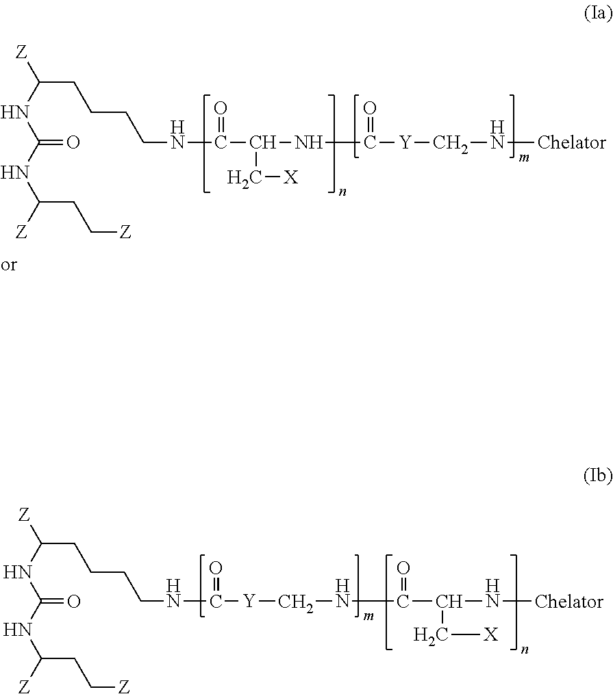
C00014
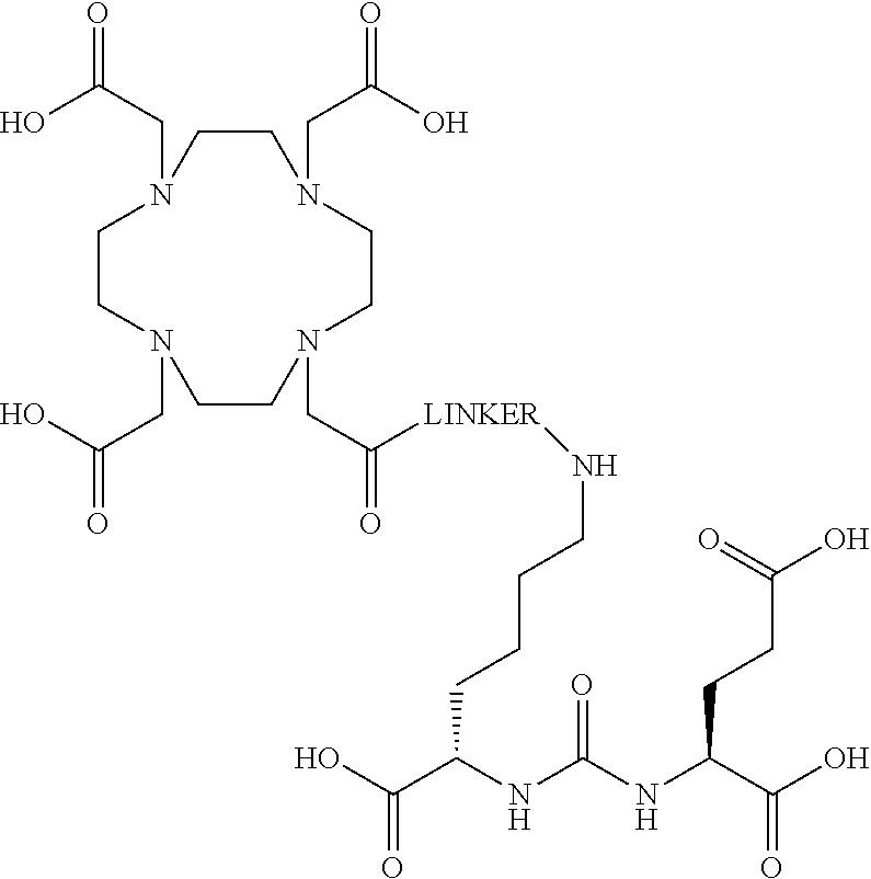
C00015
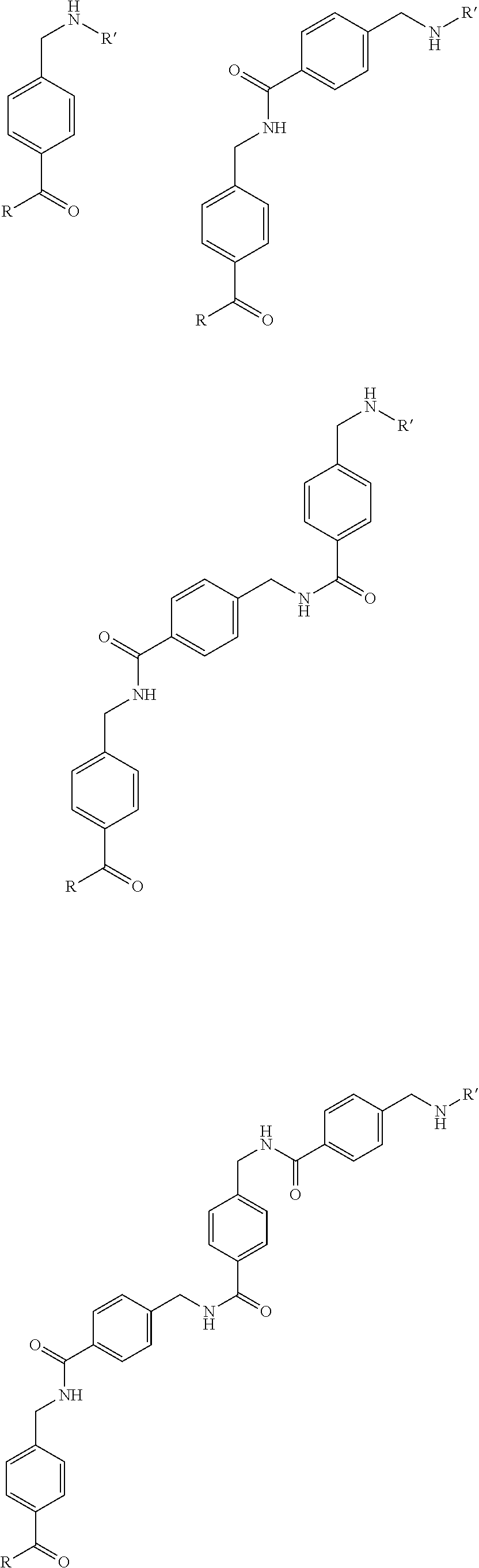
C00016
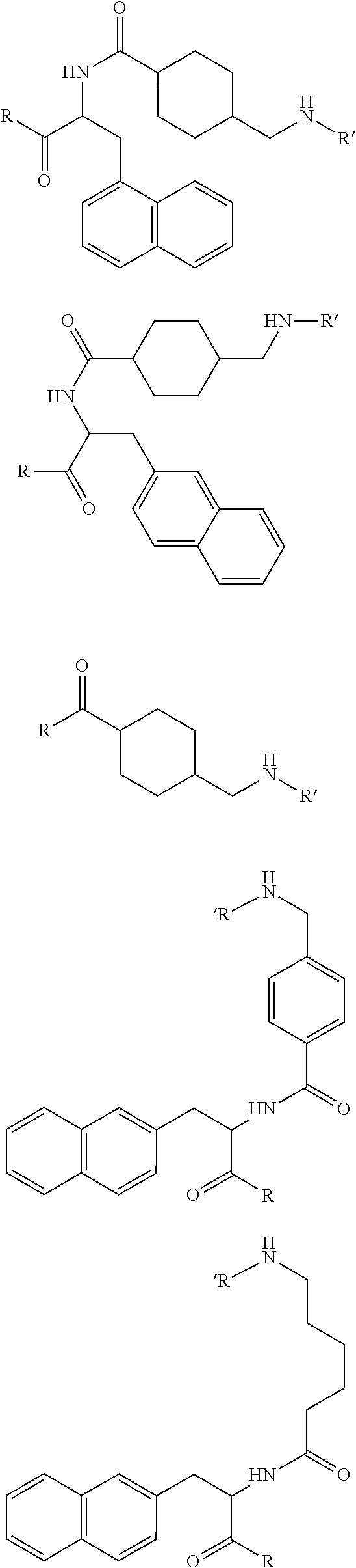
C00017
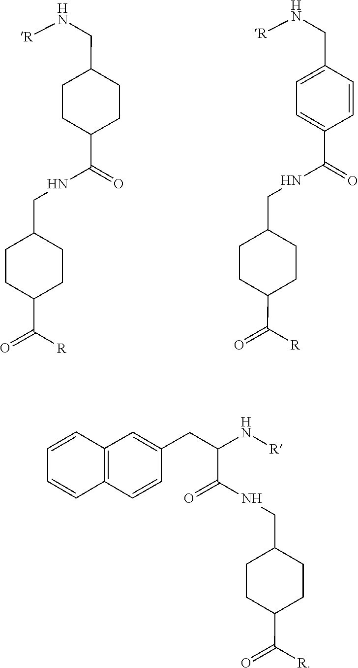
C00018
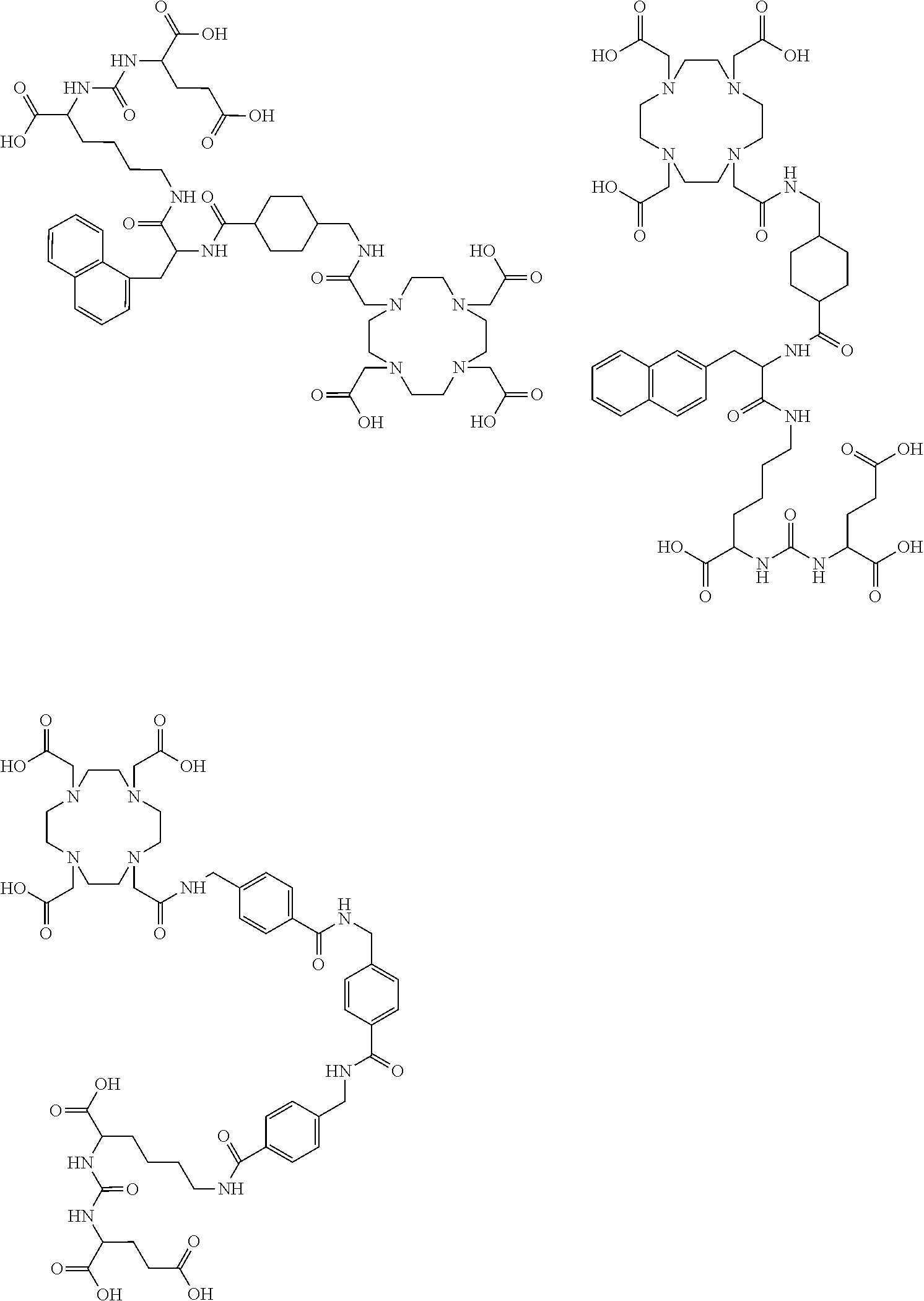
C00019

C00020

C00021
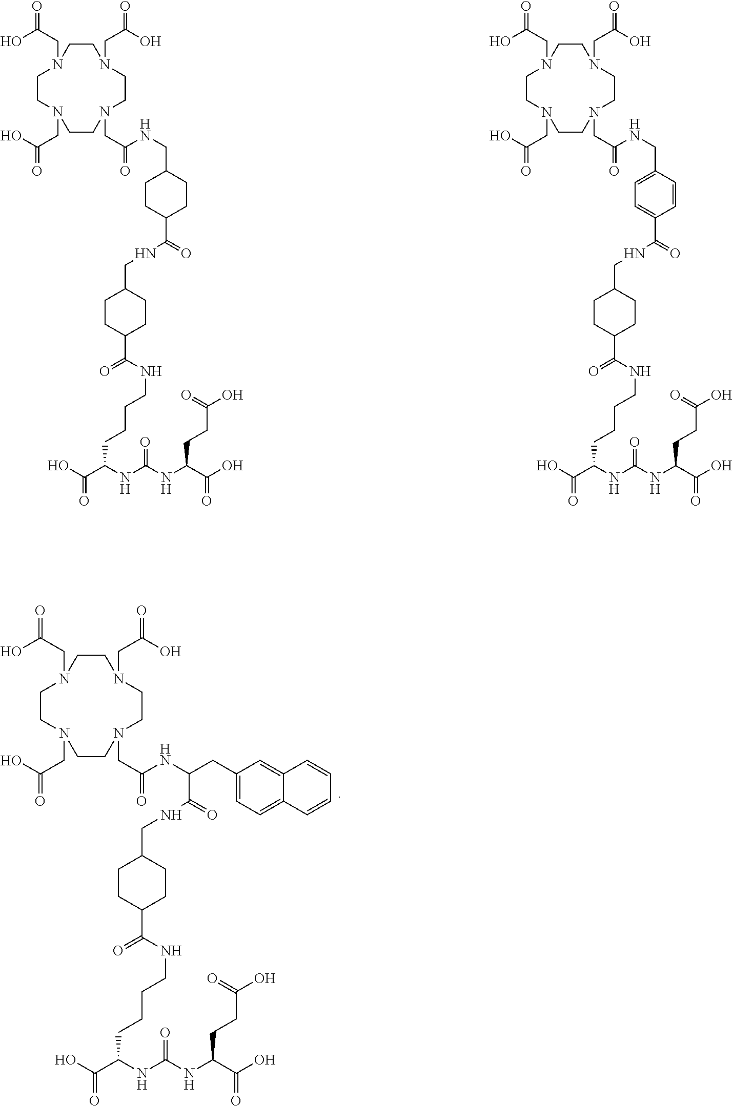
C00022

C00023
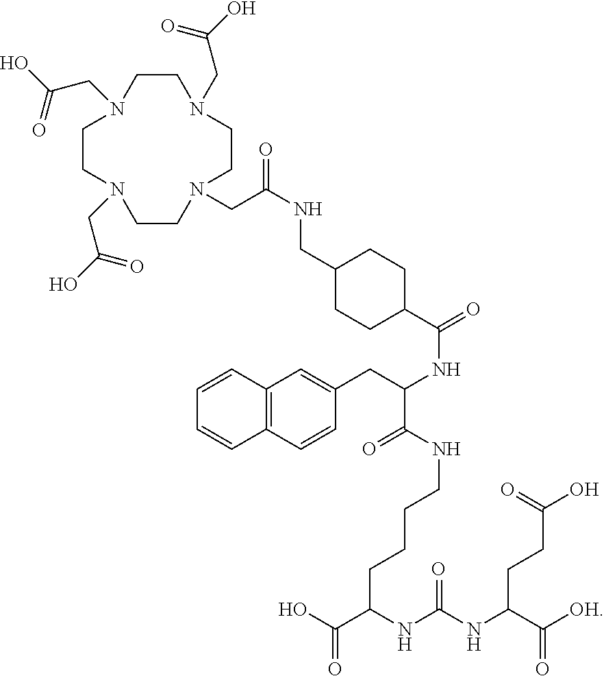
C00024

C00025
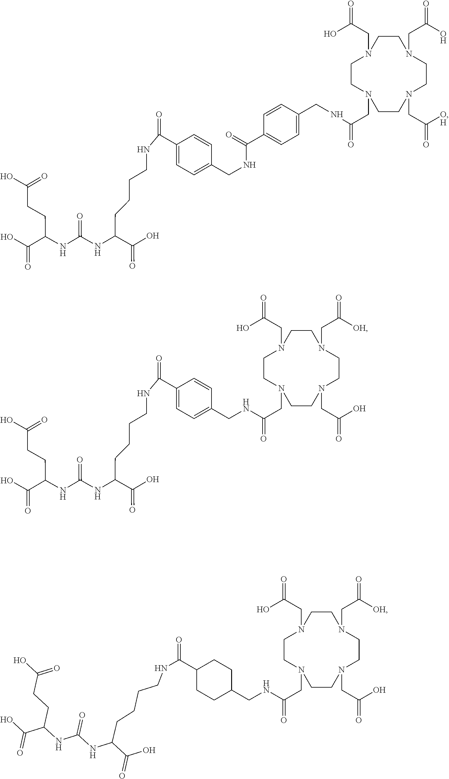
C00026

C00027

C00028

C00029

D00001
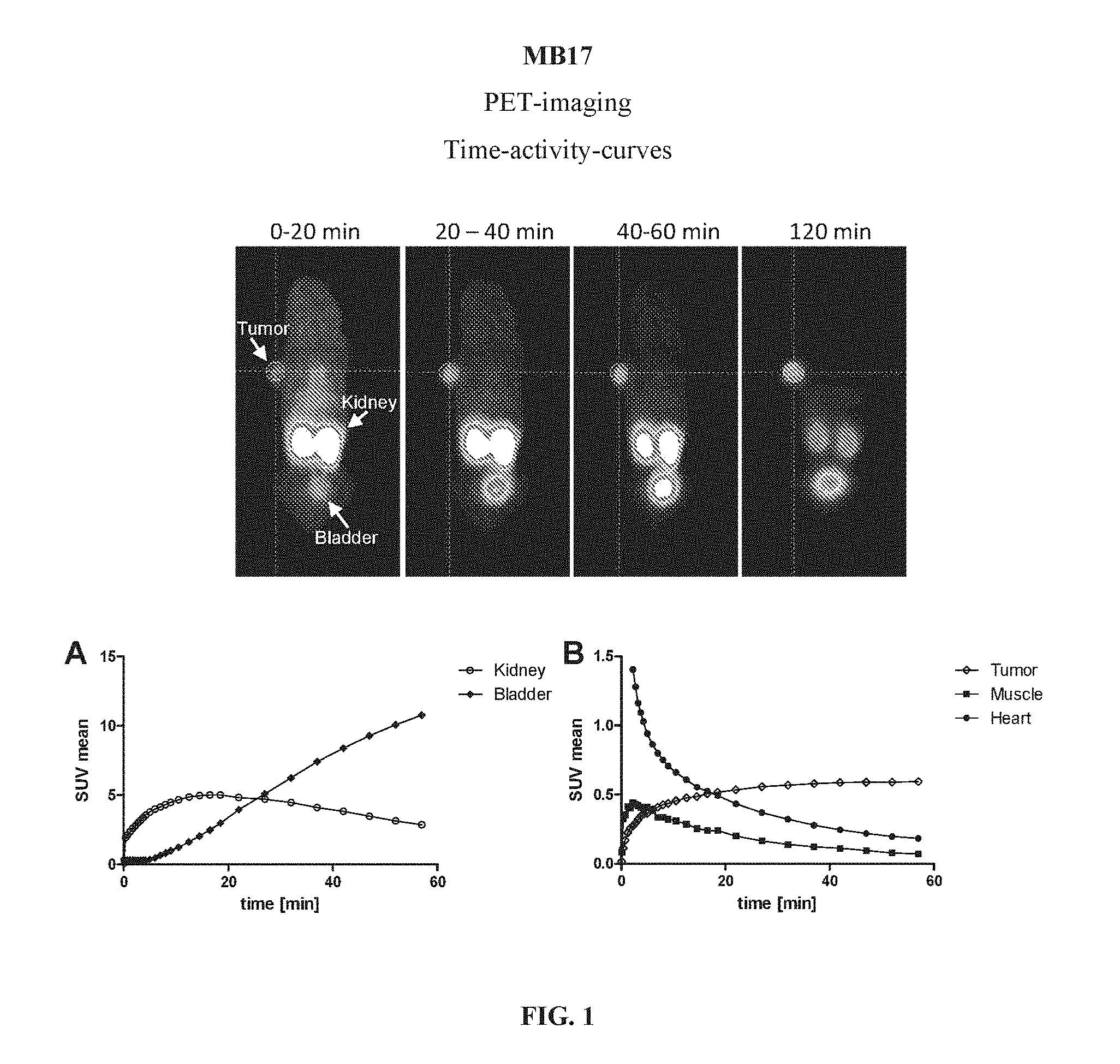
D00002

D00003
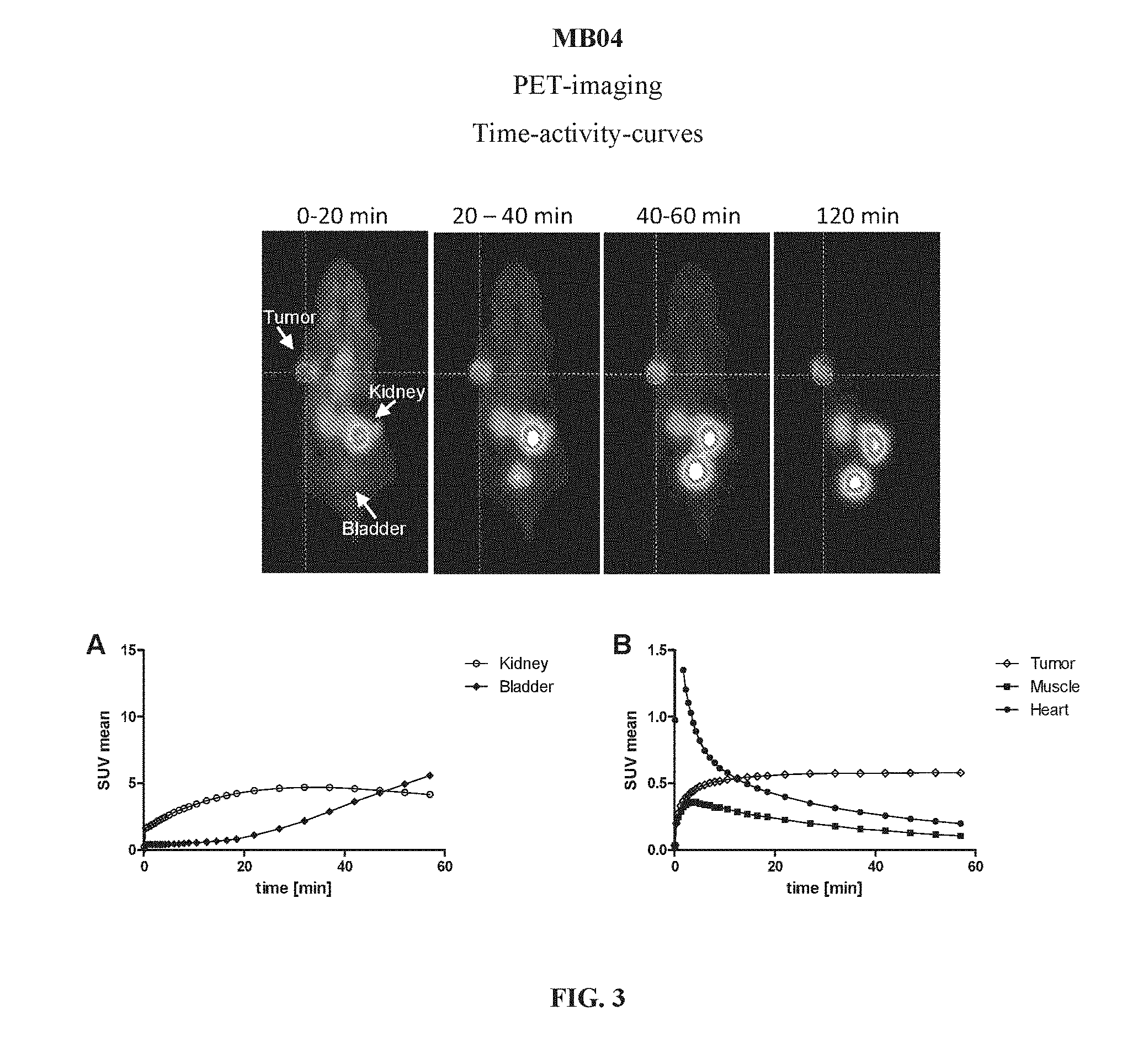
D00004

D00005
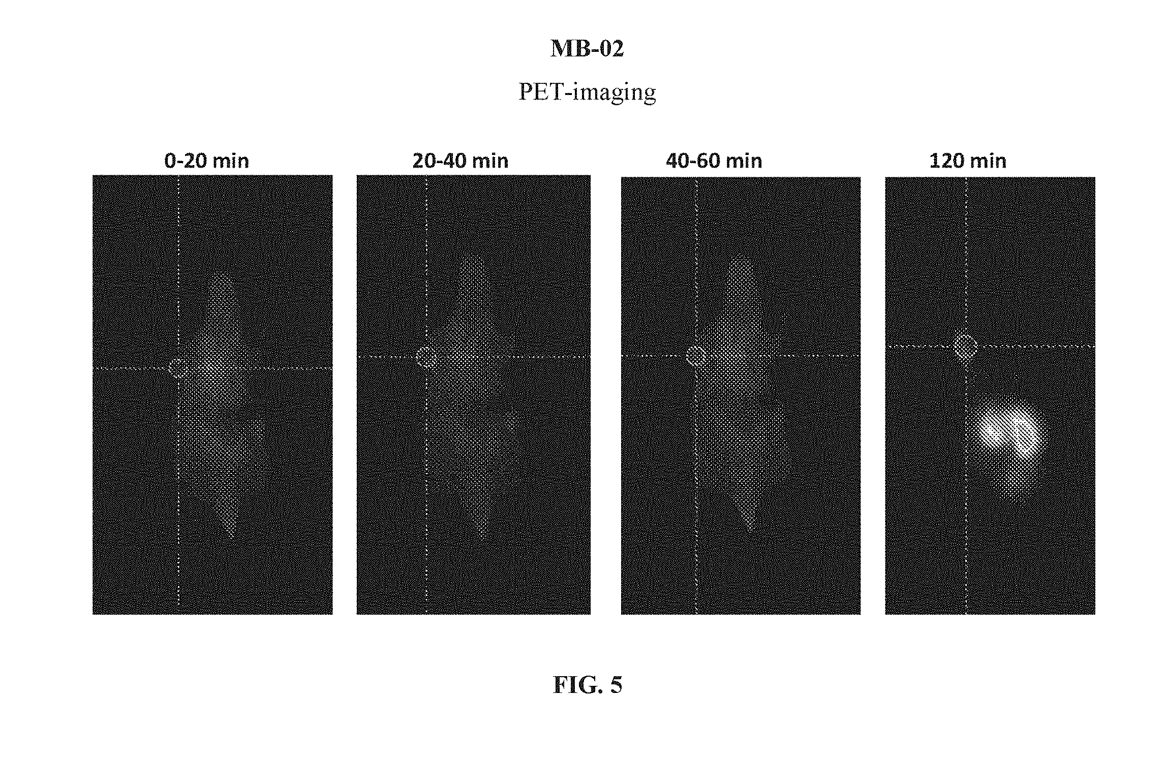
D00006

D00007
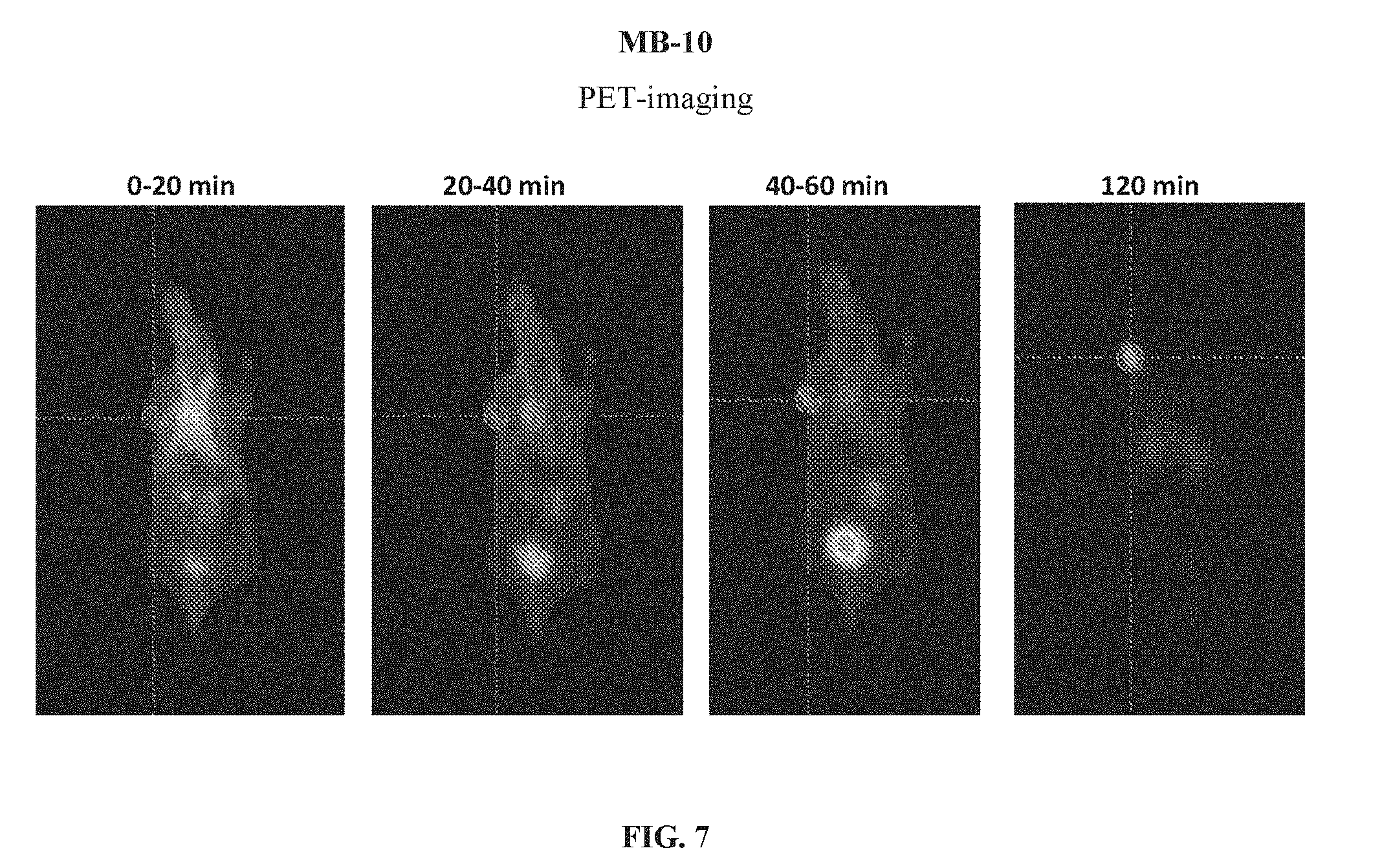
D00008

D00009
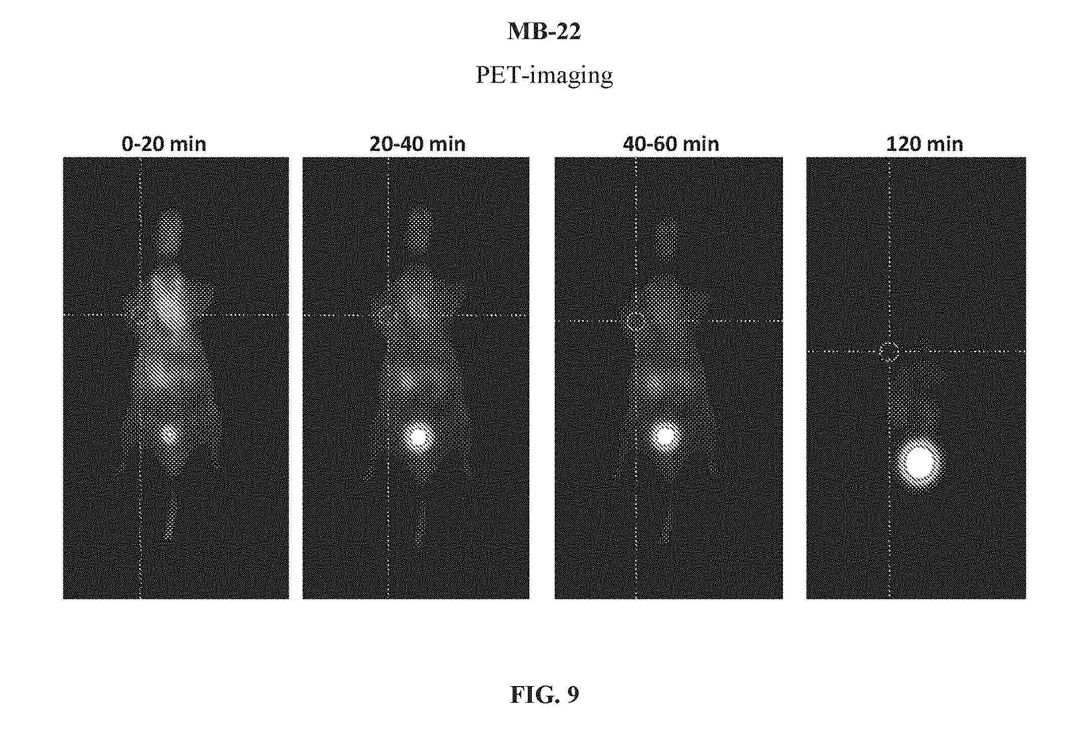
D00010

D00011

D00012

D00013

D00014
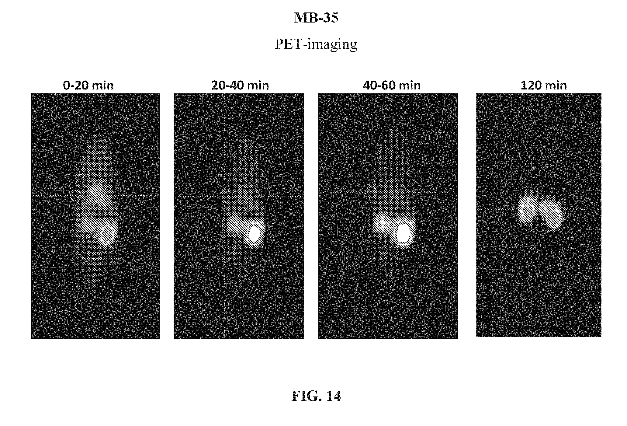
D00015
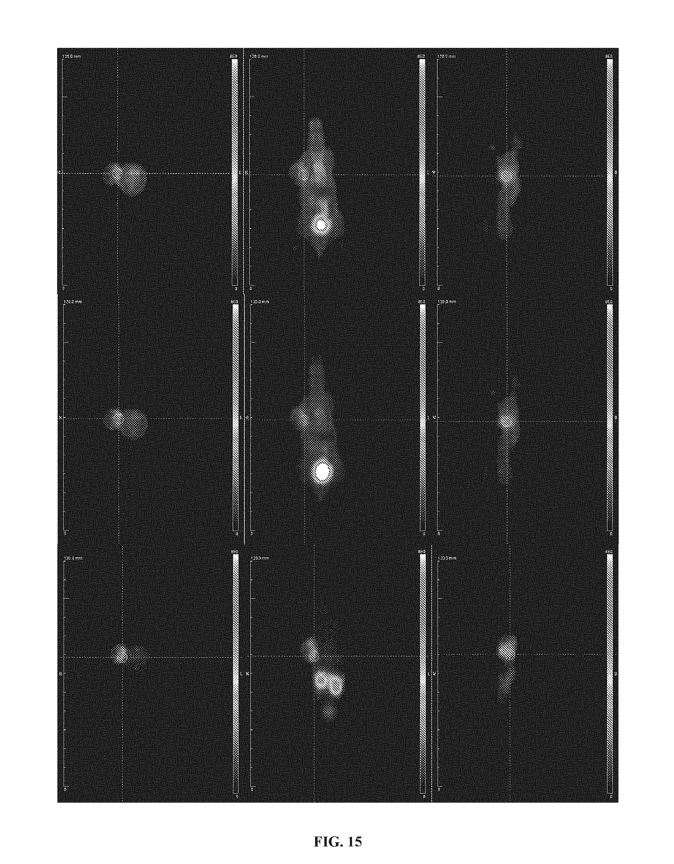
D00016
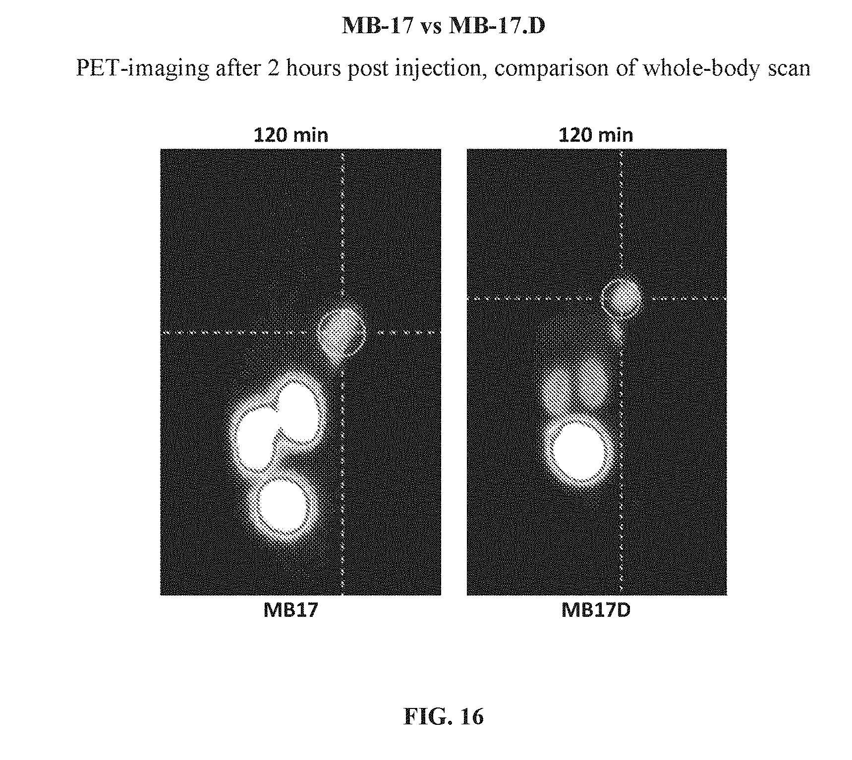
D00017
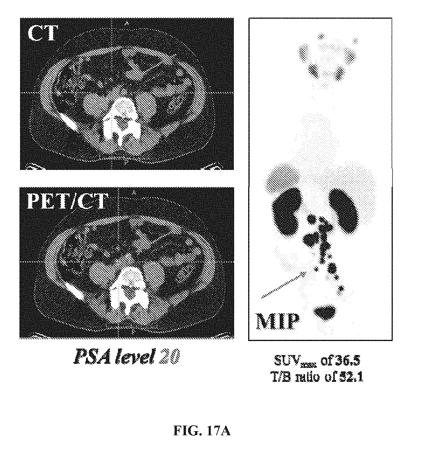
D00018
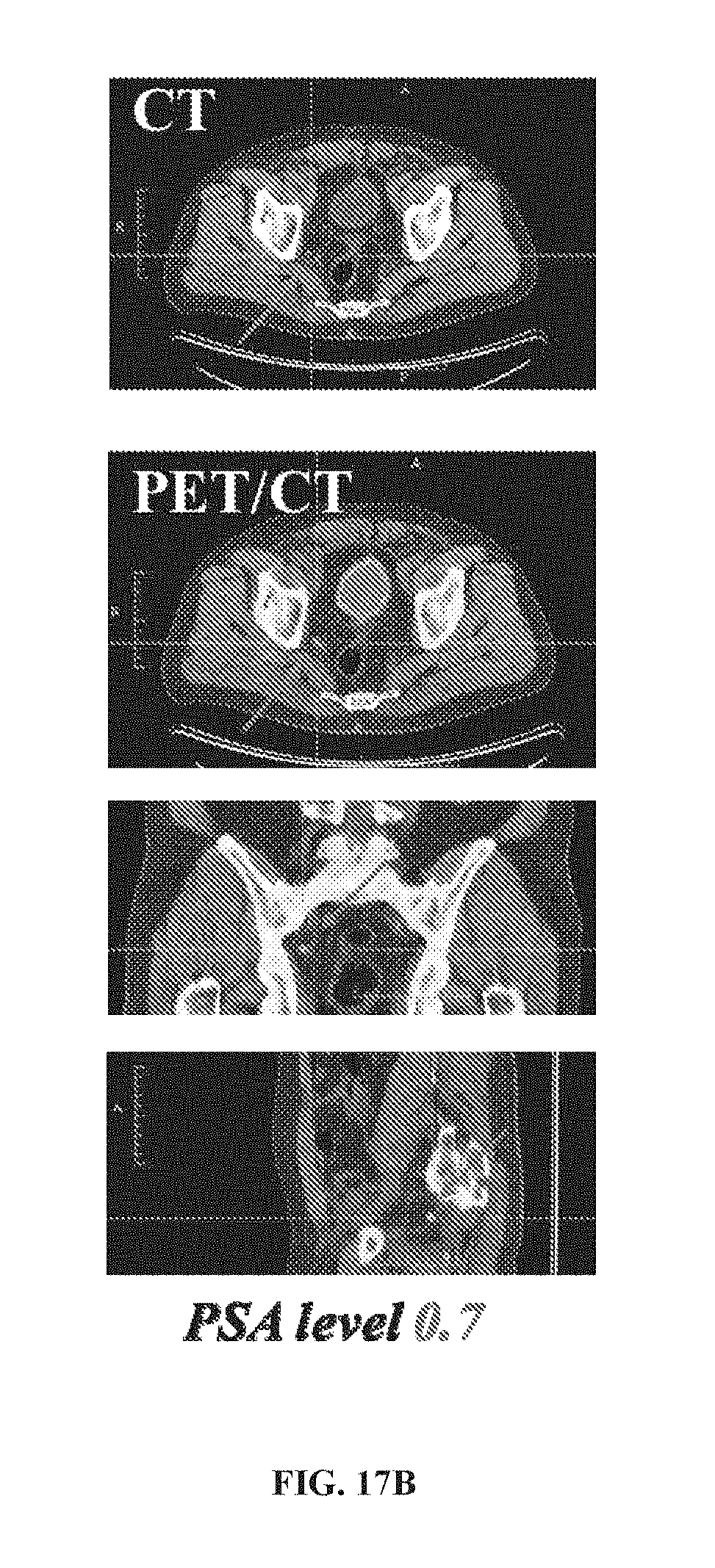
D00019

D00020

XML
uspto.report is an independent third-party trademark research tool that is not affiliated, endorsed, or sponsored by the United States Patent and Trademark Office (USPTO) or any other governmental organization. The information provided by uspto.report is based on publicly available data at the time of writing and is intended for informational purposes only.
While we strive to provide accurate and up-to-date information, we do not guarantee the accuracy, completeness, reliability, or suitability of the information displayed on this site. The use of this site is at your own risk. Any reliance you place on such information is therefore strictly at your own risk.
All official trademark data, including owner information, should be verified by visiting the official USPTO website at www.uspto.gov. This site is not intended to replace professional legal advice and should not be used as a substitute for consulting with a legal professional who is knowledgeable about trademark law.