In vitro production of red blood cells with proteins comprising sortase recognition motifs
Lodish , et al. Nov
U.S. patent number 10,471,099 [Application Number 14/890,241] was granted by the patent office on 2019-11-12 for in vitro production of red blood cells with proteins comprising sortase recognition motifs. This patent grant is currently assigned to Whitehead Institute for Biomedical Research. The grantee listed for this patent is Whitehead Institute for Biomedical Research. Invention is credited to Lenka Hoffman, Hsiang-Ying Lee, Harvey Lodish, Novalia Pishesha, Hidde L. Ploegh, Jiahai Shi.


View All Diagrams
| United States Patent | 10,471,099 |
| Lodish , et al. | November 12, 2019 |
In vitro production of red blood cells with proteins comprising sortase recognition motifs
Abstract
Methods for the in vitro production of enucleated red blood cells and the enucleated red blood cells thus prepared are provided. Such enucleated red blood cells may express a sortaggable surface protein, which allows for surface modification in the presence of a sortase. Also described herein are surface modified enucleated red blood cells, e.g., conjugated with an agent of interest such as a peptide, a detectable label, or a chemotherapeutic agent, and uses thereof in delivering the agent to a subject.
| Inventors: | Lodish; Harvey (Brookline, MA), Ploegh; Hidde L. (Brookline, MA), Lee; Hsiang-Ying (Cambridge, MA), Shi; Jiahai (Kowloon, HK), Hoffman; Lenka (Malden, MA), Pishesha; Novalia (Cambridge, MA) | ||||||||||
|---|---|---|---|---|---|---|---|---|---|---|---|
| Applicant: |
|
||||||||||
| Assignee: | Whitehead Institute for Biomedical
Research (Cambridge, MA) |
||||||||||
| Family ID: | 51867887 | ||||||||||
| Appl. No.: | 14/890,241 | ||||||||||
| Filed: | May 9, 2014 | ||||||||||
| PCT Filed: | May 09, 2014 | ||||||||||
| PCT No.: | PCT/US2014/037554 | ||||||||||
| 371(c)(1),(2),(4) Date: | November 10, 2015 | ||||||||||
| PCT Pub. No.: | WO2014/183071 | ||||||||||
| PCT Pub. Date: | November 13, 2014 |
Prior Publication Data
| Document Identifier | Publication Date | |
|---|---|---|
| US 20160082046 A1 | Mar 24, 2016 | |
Related U.S. Patent Documents
| Application Number | Filing Date | Patent Number | Issue Date | ||
|---|---|---|---|---|---|
| 61822071 | May 10, 2013 | ||||
| Current U.S. Class: | 1/1 |
| Current CPC Class: | A61P 43/00 (20180101); C07K 14/705 (20130101); A61K 35/18 (20130101); A01K 67/0275 (20130101); C12N 5/0641 (20130101); C07K 14/70582 (20130101); C07K 14/47 (20130101); C12N 2501/2303 (20130101); C12N 2501/26 (20130101); A01K 2227/105 (20130101); A01K 2267/03 (20130101); C12N 2501/2306 (20130101); C12N 2501/125 (20130101); C12N 2501/14 (20130101); C12N 2501/999 (20130101); C12N 2506/11 (20130101); C12N 2510/02 (20130101); A01K 2217/072 (20130101) |
| Current International Class: | A61K 39/00 (20060101); C07K 14/47 (20060101); C07K 14/705 (20060101); C07K 16/28 (20060101); A61K 35/18 (20150101); C12N 5/078 (20100101); A01K 67/027 (20060101); C07K 1/13 (20060101); C07K 16/10 (20060101) |
References Cited [Referenced By]
U.S. Patent Documents
| 5728369 | March 1998 | Griffiths et al. |
| 8211656 | July 2012 | Hyde et al. |
| 8940501 | January 2015 | Ploegh et al. |
| 9751945 | September 2017 | Ploegh et al. |
| 10081684 | September 2018 | Ploegh et al. |
| 2003/0022178 | January 2003 | Schneewind et al. |
| 2004/0213795 | October 2004 | Collins et al. |
| 2011/0195068 | August 2011 | Langermann et al. |
| 2011/0321183 | December 2011 | Ploegh et al. |
| 2012/0039906 | February 2012 | Olive |
| 2012/0114649 | May 2012 | Langermann et al. |
| 2013/0095098 | April 2013 | Tyson |
| 2013/0108651 | May 2013 | Carven et al. |
| 2013/0109843 | May 2013 | Carven et al. |
| 2013/0237580 | September 2013 | Sasikumar et al. |
| 2014/0030697 | January 2014 | Ploegh et al. |
| 2014/0057317 | February 2014 | Liu et al. |
| 2014/0249296 | September 2014 | Ploegh |
| 2015/0086576 | March 2015 | Ploegh et al. |
| 2016/0097773 | April 2016 | Pasqual et al. |
| 2016/0122707 | May 2016 | Ploegh et al. |
| 2016/0287734 | October 2016 | Rashidian et al. |
| 2018/0280440 | October 2018 | Lodish et al. |
| 2010115136 | May 2010 | JP | |||
| WO 2000/62804 | Oct 2000 | WO | |||
| WO 2005/051976 | Jun 2005 | WO | |||
| WO 2005/086654 | Sep 2005 | WO | |||
| WO 2010/087994 | Aug 2010 | WO | |||
| WO 2011/133704 | Oct 2011 | WO | |||
| WO 2012/142659 | Oct 2012 | WO | |||
| WO 2013/003555 | Jan 2013 | WO | |||
| WO 2014/183006 | Nov 2014 | WO | |||
| WO 2015/073746 | May 2015 | WO | |||
Other References
|
Sankaran et al., Cyclin D3 coordinates the cell cycle during differentiation to regulate erythrocyte size and No. Genes & Development 26:2075-2087. cited by examiner . Pishesha et al Engineered erythrocytes covalently linked to antigenic peptides can protect against autoimmune disease PNAS | Mar. 21, 2017 | vol. 114 | No. 12 | 3157-3162. cited by examiner . Fusion protein--Wikipedia pp. 1-7 downloaded Feb. 8, 2019. cited by examiner . Extended European Search Report, dated Nov. 30, 2012, in connection with EP 10736161.0. cited by applicant . Invitation to Pay Additional Fees, dated Nov. 8, 2010, in connection with PCT/US2010/000274. cited by applicant . International Search Report and Written Opinion, dated Dec. 22, 2010, in connection with PCT/US2010/000274. cited by applicant . International Preliminary Report on Patentability, dated Aug. 11, 2011, in connection with PCT/US2010/000274. cited by applicant . Extended European Search Report, dated Nov. 26, 2014, in connection with EP 12804570.5. cited by applicant . International Search Report and Written Opinion, dated Nov. 15, 2012, in connection with PCT/US2012/044584. cited by applicant . International Preliminary Report on Patentability, dated Jan. 16, 2014, in connection with PCT/US2012/044584. cited by applicant . Invitation to Pay Additional Fees, dated Sep. 5, 2014, in connection with PCT/U52014/037554. cited by applicant . International Search Report and Written Opinion, dated Nov. 6, 2014, in connection with PCT/US2014/037554. cited by applicant . International Preliminary Report on Patentability, dated Nov. 19, 2015, in connection with PCT/US2014/037554. cited by applicant . International Search Report and Written Opinion, dated Oct. 27, 2014, in connection with PCT/U52014/037545. cited by applicant . International Preliminary Report on Patentability, dated Nov. 19, 2015, in connection with PCT/U52014/037545. cited by applicant . Invitation to Pay Additional Fees, dated Mar. 20, 2015, in connection with PCT/US14/65574. cited by applicant . International Search Report and Written Opinion, dated Jun. 4, 2015, in connection with PCT/US14/65574. cited by applicant . Antos et al., Lipid modification of proteins through sortase-catalyzed transpeptidation. J Am Chem Soc. Dec. 3, 2008;130(48):16338-43. cited by applicant . Antos et al., Site-specific N- and C-terminal labeling of a single polypeptide using sortases of different specificity. J Am Chem Soc. Aug. 12, 2009;131(31):10800-1. doi: 10.1021/ja902681k. cited by applicant . Arnau et al., Current strategies for the use of affinity tags and tag removal for the purification of recombinant proteins. Protein Expr Purif. Jul. 2006;48(1):1-13. Epub Dec. 28, 2005. cited by applicant . Bader et al., Bioorganic synthesis of lipid-modified proteins for the study of signal transduction. Nature. Jan. 13, 2000;403(6766):223-6. doi: 10.1158/0008-5472.CAN-09-0547. Epub Jun. 9, 2009. cited by applicant . Barnett et al., Differential recognition of surface proteins in Streptococcus pyogenes by two sortase gene homologs. J Bacteriol. Apr. 2002;184(8):2181-91. cited by applicant . Baskin et al., Bioorthogonal click chemistry: Covalent labeling in living systems. QSAR Comb Sci. Dec. 2007;26(11-12):1211-9. cited by applicant . Biagiotti et al., Drug delivery by red blood cells. IUBMB Life. Aug. 2011;63(8):621-31. doi: 10.1002/iub.478. Epub Jul. 15, 2011. cited by applicant . Binder et al., `Click` Chemistry in Polymer and Materials Science. Macromol Rapid Commun. Jan. 5, 2007;28(1):15-54. cited by applicant . Boonyarattanakalin et al., Synthesis of an artificial cell surface receptor that enables oligohistidine affinity tags to func-tion as metal-dependent cell-penetrating peptides. J Am Chem Soc. Mar. 18, 2006;128(14):4917. cited by applicant . Chan et al., Covalent attachment of proteins to solid supports and surfaces via Sortase-mediated ligation. PLoS One. Nov. 14, 2007;2(11):e1164. 5 pages. cited by applicant . Chen et al., A general strategy for the evolution of bond-forming enzymes using yeast display. Proc Natl Acad Sci U S A. Jul. 12, 2011;108(28):11399-404. doi: 10.1073/pnas.1101046108. Epub Jun. 22, 2011. cited by applicant . Clow et al., Immobilization of proteins to biacore sensor chips using Staphylococcus aureus sortase A. Biotechnol Lett. Sep. 2008;30(9):1603-7. Epub Apr. 15, 2008. cited by applicant . Giarratana et al., Proof of principle for transfusion of in vitro-generated red blood cells. Blood. Nov. 10, 2011;118(19):5071-9. doi:10.1182/blood-2011-06-362038. Epub Sep. 1, 2011. cited by applicant . Godfrin et al., International seminar on the red blood cells as vehicles for drugs. Expert Opin Biol Ther. Jan. 2012;12(1):127-33. doi: 10.1517/14712598.2012.631909. Epub Oct. 25, 2011. cited by applicant . Guimaraes et al., Site-specific C-terminal and internal loop labeling of proteins using sortase-mediated reactions. Nat Protoc. Sep. 2013;8(9):1787-99. doi:10.1038/nprot.2013.101. Epub Aug. 29, 2013. cited by applicant . Heal et al., Site-specific N-terminal labelling of proteins in vitro and in vivo using N-myristoyl transferase and bioorthogonal ligation chemistry. Chem Commun (Camb). Jan. 28, 2008;(4):480-2. Epub Nov. 13, 2007. cited by applicant . Kruger et al., Analysis of the substrate specificity of the Staphylococcus aureus sortase transpeptidase SrtA. Biochemistry. Feb. 17, 2004;43(6):1541-51. cited by applicant . Levary et al., Protein-protein fusion catalyzed by sortase A. PLoS One. Apr. 6, 2011;6(4):e18342. doi: 10.1371/journal.pone.0018342. 6 pages. cited by applicant . Mao et al., Sortase-Mediated Protein Ligation: A New Method for Portein Engineering. J Am Chem Soc. Feb. 10, 2004;126:2670-1. cited by applicant . Mao, A self-cleavable sortase fusion for one-step purification of free recombinant proteins. Protein Expr Purif. Sep. 2004;37(1):253-63. cited by applicant . Maresso et al., Surface protein IsdC and Sortase B are required for heme-iron scavenging of Bacillus anthracis. J Bacteriol. Dec. 2006;188(23):8145-52. Epub Sep. 29, 2006. cited by applicant . Mariscotti et al., The Listeria monocytogenes sortase-B recognizes varied amino acids at position 2 of the sorting motif. J Biol Chem. Mar. 6, 2009;284(10):6140-6. Epub Jan. 7, 2009. cited by applicant . Marraffini et al., Sortase C-mediated anchoring of BasI to the cell wall envelope of Bacillus anthracis. J Bacteriol. Sep. 2007;189(17):6425-36. Epub Jun. 22, 2007. cited by applicant . Matsumoto et al., Site-specific tetrameric streptavidin-protein conjugation using sortase A. J Biotechnol. Mar. 10, 2011;152(1-2):37-42. doi: 10.1016/j.jbiotec.2011.01.008. Epub Jan. 22, 2011. cited by applicant . Mazmanian et al., An iron-regulated sortase anchors a class of surface protein during Staphylococcus aureus pathogenesis. Proc Natl Acad Sci U S A. Feb. 19, 2002;99(4):2293-8. Epub 2002 Feb. 5, 2002. cited by applicant . Mazmanian et al., Sortase-catalysed anchoring of surface proteins to the cell wall of Staphylococcus aureus. Mol Microbiol. Jun. 2001;40(5):1049-57. Review. cited by applicant . Murciano et al., Prophylactic fibrinolysis through selective dissolution of nascent clots by tPA-carrying erythrocytes. Nat Biotechnol. Aug. 2003;21(8):891-6. Epub Jul. 6, 2003. cited by applicant . Muzykantov, Drug delivery by red blood cells: vascular carriers designed by mother nature. Expert Opin Drug Deliv. Apr. 2010;7(4):403-27. doi:10.1517/17425241003610633. cited by applicant . Ning et al., Visualizing metabolically labeled glycoconjugates of living cells by copper-free and fast huisgen cycloadditions. Angew Chem Int Ed Engl. 2008;47(12):2253-5. cited by applicant . Pallen et al., An embarrassment of sortases--a richness of substrates? Trends Microbiol. Mar. 2001;9(3):97-101. cited by applicant . Parthasarathy et al., Sortase A as a novel molecular "stapler" for sequence-specific protein conjugation. Bioconjug Chem. Mar.-Apr. 2007;18(2):469-76. Epub Feb. 16, 2007. cited by applicant . Piotukh et al., D. Directed evolution of sortase A mutants with altered substrate selectivity profiles. J Am Chem Soc. Nov. 9, 2011;133(44):17536-9. doi: 10.1021/ja205630g. Epub Oct. 13, 2011. cited by applicant . Popp et al., Making and breaking peptide bonds: protein engineering using sortase. Angew Chem Int Ed Engl. May 23, 2011;50(22):5024-32. doi:10.1002/anie.201008267. Epub Apr. 27, 2011. cited by applicant . Popp et al., Site-specific protein labeling via sortase-mediated transpeptidation. Curr Protoc Protein Sci. Apr. 2009;Chapter 15:Unit 15.3. doi: 10.1002/0471140864.ps1503s56. cited by applicant . Popp et al., Sortagging: a versatile method for protein labeling. Nat Chem Biol. Nov. 2007;3(11):707-8. Epub Sep. 23, 2007. cited by applicant . Sletten et al., Bioorthogonal chemistry: fishing for selectivity in a sea of functionality. Angew Chem Int Ed Engl. 2009;48(38):6974-98. doi: 10.1002/anie.200900942. cited by applicant . Strijbis et al., Protein ligation in living cells using sortase. Traffic. Jun. 2012;13(6):780-9. doi: 10.1111/j.1600-0854.2012.01345.x. Epub Mar. 23, 2012. cited by applicant . Tanaka et al., Site-specific protein modification on living cells catalyzed by Sortase. Chembiochem. Mar. 25, 2008;9(5):802-7. cited by applicant . Theile et al., Site-specific N-terminal labeling of proteins using sortase-mediated reactions. Nat Protoc. Sep. 2013;8(9):1800-7. doi: 10.1038/nprot.2013.102. Epub Aug. 29, 2013. cited by applicant . Ton-That et al., Anchoring of surface proteins to the cell wall of Staphylococcus aureus. Sortase catalyzed in vitro transpeptidation reaction using LPXTG peptide and NH(2)-Gly(3) substrates. J Biol Chem. Mar. 31, 2000;275(13):9876-81. cited by applicant . Ton-That et al., Protein sorting to the cell wall envelope of Gram-positive bacteria. Biochim Biophys Acta. Nov. 11, 2004;1694(1-3):269-78. cited by applicant . Ton-That et al., Purification and characterization of sortase, the transpeptidase that cleaves surface proteins of Staphylococcus aureus at the LPXTG motif. Proc Natl Acad Sci U S A. Oct. 26, 1999;96(22):12424-9. cited by applicant . Witte et al., Preparation of unnatural N-to-N and C-to-C protein fusions. Proc Natl Acad Sci U S A. Jul. 24, 2012;109(30):11993-8. doi: 10.1073/pnas.1205427109. Epub Jul. 9, 2012. cited by applicant . Witte et al., Production of unnaturally linked chimeric proteins using a combination of sortase-catalyzed transpeptidation and click chemistry. Nat Protoc. Sep. 2013;8(9):1808-19. doi: 10.1038/nprot.2013.103. Epub Apr. 4, 2014. 16 pages. cited by applicant . Wu et al., Sortase A-catalyzed transpeptidation of glycosylphosphatidylinositol derivatives for chemoenzymatic synthesis of GPI-anchored proteins. J Am Chem Soc. Feb. 10, 2010;132(5):156771. doi: 10.1021/ja906611x. cited by applicant . Yamamoto et al., Expansion of the sortase-mediated labeling method for site-specific N-terminal labeling of cell surface proteins on living cells. Chem Commun (Camb). Mar. 7, 2009;(9):1022-4. doi: 10.1039/b818792d. Epub Jan. 7, 2009. cited by applicant . Yoo et al., Bio-inspired, bioengineered and biomimetic drug delivery carriers. Nat Rev Drug Discov. Jul. 1, 2011;10(7):521-35. doi: 10.1038/nrd3499. cited by applicant . Zaitsev et al., Sustained thromboprophylaxis mediated by an RBC-targeted pro-urokinase zymogen activated at the site of clot formation. Blood. Jun. 24, 2010;115(25):5241-8. doi: 10.1182/blood-2010-01-261610. Epub Apr. 21, 2010. cited by applicant . Zaslavskaia et al., Trophic conversion of an obligate photoautotrophic organism through metabolic engineering. Science. Jun. 15, 2001;292(5524):2073-5. cited by applicant . U.S. Appl. No. 14/890,296, filed Nov. 10, 2015, Ploegh et al. cited by applicant . U.S. Appl. No. 14/875,140, filed Nov. 5, 2015, Pasqual et al. cited by applicant . EP 10736161.0, Nov. 30, 2012, Extended European Search Report. cited by applicant . PCT/US2010/000274, Nov. 8, 2010, Invitation to Pay Additional Fees. cited by applicant . PCT/US2010/000274, Dec. 22, 2010, International Search Report and Written Opinion. cited by applicant . PCT/US2010/000274, Aug. 11, 2011, International Preliminary Report on Patentability. cited by applicant . EP 12804570.5, Nov. 26, 2014, Extended European Search Report. cited by applicant . PCT/US2012/044584, Nov. 15, 2012, International Search Report and Written Opinion. cited by applicant . PCT/US2012/044584, Jan. 16, 2014, International Preliminary Report on Patentability. cited by applicant . PCT/US2014/037554, Nov. 6, 2014, International Search Report and Written Opinion. cited by applicant . PCT/US2014/037554, Sep. 5, 2014, Invitation to Pay Additional Fees. cited by applicant . PCT/US2014/037554, Nov. 19, 2015, International Preliminary Report on Patentability. cited by applicant . PCT/US2014/037545, Oct. 27, 2014, International Search Report and Written Opinion. cited by applicant . PCT/US2014/037545, Nov. 19, 2015, International Preliminary Report on Patentability. cited by applicant . PCT/US14/65574, Mar. 20, 2015, Invitation to Pay Additional Fees. cited by applicant . PCT/US14/65574, Jun. 4, 2015, International Search Report and Written Opinion. cited by applicant . Extended European Search Report, dated Nov. 9, 2016, in connection with EP 14795167.7. cited by applicant . GenPept Accession No. YP 187332.1. Gill et al. Dec. 17, 2014. cited by applicant . Ahlgren et al., Targeting of HER2-expressing tumors with a site-specifically 99mTc-labeled recombinant affibody molecule, ZHER2:2395, with C-terminally engineered cysteine. J Nucl Med.May 2009;50(5):781-9. doi: 10.2967/jnumed.108.056929. Epub Apr. 16, 2009. cited by applicant . Chang et al., Development and characterization of 89Zr-labeled panitumumab for immuno-positron emission tomographic imaging of the epidermal growth factor receptor. Mol Imaging. Jan.-Feb. 2013;12(1):17-27. cited by applicant . De Meyer et al., Nanobody-based products as research and diagnostic tools. Trends Biotechnol. May 2014;32(5):263-70. doi:10.1016/j.tibtech.2014.03.001. Epub Apr. 1, 2014. cited by applicant . Delgado, et al. Stabilities of divalent and trivalent metal ion complexes of macrocyclic triazatriacetic acids. Inorg. Chem. 1999; 32, 3320-3326. cited by applicant . Dijkers et al., Biodistribution of 89Zr-trastuzumab and PET imaging of HER2-positive lesions in patients with metastatic breast cancer. Clin Pharmacol Ther. May 2010;87(5):586-92. doi:10.1038/clpt.2010.12. Epub Mar. 31, 2010. cited by applicant . Dorr et al., Reprogramming the specificity of sortase enzymes. Proc Natl Acad Sci U S A. Sep. 16, 2014;111(37):13343-8. doi:10.1073/pnas.1411179111. Epub Sep. 3, 2014. cited by applicant . Engfeldt et al., Chemical synthesis of triple-labelled three-helix bundle binding proteins for specific fluorescent detection of unlabelled protein. Chembiochem. Jun. 2005;6(6):1043-50. cited by applicant . Goldenberg et al., Novel radiolabeled antibody conjugates. Oncogene. May 28, 2007;26(25):3734-44. cited by applicant . Groheux et al., Correlation of high 18F-FDG uptake to clinical, pathological and biological prognostic factors in breast cancer. Eur J Nucl Med Mol Imaging. Mar. 2011;38(3):426-35. doi:10.1007/s00259-010-1640-9. Epub Nov. 6, 2010. cited by applicant . Hackenberger et al., Chemoselective ligation and modification strategies for peptides and proteins. Angew Chem Int Ed Engl. 2008;47(52):10030-74. doi: 10.1002/anie.200801313. cited by applicant . Hochuli et al., Genetic Approach to Facilitate Purification of Recombinant Proteins with a Novel Metal Chelate Adsorbent. Nature Biotechnology. 1988, 6, 1321-1325. cited by applicant . Holm et al., Electrophilic affibodies forming covalent bonds to protein targets. J Biol Chem. Nov. 20, 2009;284(47):32906-13. doi:10.1074/jbc.M109.034322. Epub Sep. 15, 2009. cited by applicant . Keliher et al., High-yielding, two-step 18F labeling strategy for 18F-PARP1 inhibitors. ChemMedChem. Mar. 7, 2011;6(3):424-7. doi: 10.1002/cmdc.201000426. Epub Jan. 4, 2011. cited by applicant . Knowles et al., Advances in immuno-positron emission tomography: antibodies for molecular imaging in oncology. J Clin Oncol. Nov. 1, 2012;30(31):3884-92. doi: 10.1200/JCO.2012.42.4887. Epub Sep. 17, 2012. cited by applicant . Langenhan et al., Recent Carbohydrate-Based Chemoselective Ligation Applications. Current Organic Synthesis. 2005; 2, 59-81. cited by applicant . Lu et al., Biologic properties and enucleation of red blood cells from human embryonic stem cells. Blood. Dec. 1, 2008;112(12):4475-84. doi:10.1182/blood-2008-05-157198. cited by applicant . Lundberg et al., Site-specifically conjugated anti-HER2 Affibody molecules as one-step reagents for target expression analyses on cells and xenograft samples. J Immunol Methods. Jan. 30, 2007;319(1-2):53-63. Epub Nov. 21, 2006. cited by applicant . Nair-Gill et al., Non-invasive imaging of adaptive immunity using positron emission tomography. Immunol Rev. Feb. 2008;221:214-28. doi: 10.1111/j.1600-065X.2008.00585.x. cited by applicant . Nayak et al., PET and MRI of metastatic peritoneal and pulmonary colorectal cancer in mice with human epidermal growth factor receptor 1-targeted 89Zr-labeled panitumumab. J Nucl Med. Jan. 2012;53(1):113-20. doi: 10.2967/jnumed.111.094169. cited by applicant . Orlova et al., Evaluation of [(111/114m)In]CHX-A''-DTPA-ZHER2:342, an affibody ligand coniugate for targeting of HER2-expressing malignant tumros. Q J Nucl Med Mol Imaging. 2007. cited by applicant . Orlova et al., Tumor imaging using a picomolar affinity HER2 binding affibody molecule. Cancer Res. Apr. 15, 2006;66(8):4339-48. cited by applicant . Poli et al., Radretumab radioimmunotherapy in patients with brain metastasis: a 1241-L19SIP dosimetric PET study. Cancer Immunol Res. Aug. 2013;1(2):134-43. doi: 10.1158/2326-6066.CIR-13-0007. Epub May 20, 2013. cited by applicant . Rashidian et al., A highly efficient catalyst for oxime ligation and hydrazone-oxime exchange suitable for bioconjugation. Bioconjug Chem. Mar. 20, 2013;24(3):333-42. doi: 10.1021/bc3004167.Epub Mar. 6, 2013. cited by applicant . Rashidian et al., Enzymatic labeling of proteins: techniques and approaches. Bioconjug Chem. Aug. 21, 2013;24(8):1277-94. cited by applicant . Salsano et al., "PET imaging using radiolabeled antibodies: future direction in tumor diagnosis and correlate applications." Research and Reports in Nuclear Medicine. 2013: 3; 9-17. cited by applicant . Shi et al., Engineered red blood cells as carriers for systemic delivery of a wide array of functional probes. Proc Natl Acad Sci U S A. Jul. 15, 2014;111(28):10131-6. doi: 10.1073/pnas.1409861111. cited by applicant . Siontorou, Nanobodies as novel agents for disease diagnosis and therapy. Int J Nanomedicine. 2013;8:4215-27. doi: 10.2147/IJN.S39428. Epub Jan. 11, 2013. cited by applicant . Spicer et al., Selective chemical protein modification. Nat Commun. Sep. 5, 2014;5:4740. doi: 10.1038/ncomms5740. cited by applicant . Swee et al., One-step enzymatic modification of the cell surface redirects cellular cytotoxicity and parasite tropism. ACS Chem Biol. Feb. 20, 2015;10(2):460-5. doi: 10.1021/cb500462t. cited by applicant . Ta et al., Enzymatic single-chain antibody tagging: a universal approach to targeted molecular imaging and cell homing in cardiovascular disease. Circ Res. Aug. 5, 2011;109(4):365-73. doi: 10.1161/CIRCRESAHA.111.249375. cited by applicant . Tanaka et al., PET (positron emission tomography) imaging of biomolecules using metal-DOTA complexes: a new collaborative challenge by chemists, biologists, and physicians for future diagnostics and exploration of in vivo dynamics. Org Biomol Chem. Mar. 7, 2008;6(5):815-28. doi: 10.1039/b718157b. Epub Feb. 1, 2008. cited by applicant . Tolmachev et al., Radionuclide therapy of HER2-positive microxenografts using a 177Lu-labeled HER2-specific Affibody molecule. Cancer Res. Mar. 15, 2007;67(6):2773-82. cited by applicant . Tran et al., (99m)Tc-maEEE-Z(HER2:342), an Affibody molecule-based tracer for the detection of HER2 expression in malignant tumors. Bioconjug Chem. Nov.-Dec. 2007;18(6):1956-64. Epub Oct. 19, 2007. cited by applicant . Truong et al., Copper-catalyzed, directing group-assisted fluorination of arene and heteroarene C-H bonds. J Am Chem Soc. Jun. 26, 2013;135(25):9342-5. doi: 10.1021/ja4047125. Epub Jun. 12, 2013. cited by applicant . Vaidyanathan et al., Synthesis of N-succinimidyl 4-[18F]fluorobenzoate, an agent for labeling proteins and peptides with 18F. Nat Protoc. 2006;1(4):1655-61. cited by applicant . Vosjan et al., Conjugation and radiolabeling of monoclonal antibodies with zirconium-89 for PET imaging using the bifunctional chelate p-isothiocyanatobenzyl-desferrioxamine. Nat Protoc. Apr. 2010;5(4):739-43. doi: 10.1038/nprot.2010.13. Epub Mar. 25, 2010. cited by applicant . Waldherr et al., Monitoring antiproliferative responses to kinase inhibitor therapy in mice with 3'-deoxy-3'-18F-fluorothymidine PET. J Nucl Med. Jan. 2005;46(1):114-20. cited by applicant . Wu et al., The use of sortase-mediated ligation for the immobilisation of bacterial adhesins onto fluorescence-labelled microspheres: a novel approach to analyse bacterial adhesion to host cells. Biotechnol Lett. Nov. 2010;32(11):1713-8. doi: 10.1007/s10529-010-0349-y. cited by applicant . Youssef et al., The use of 18F-FDG PET in the diagnosis of cardiac sarcoidosis: a systematic review and metaanalysis including the Ontario experience. J Nucl Med. Feb. 2012;53(2):241-8. doi:10.2967/jnumed.111.090662. Epub 2012 Jan. 6, 2012. cited by applicant . Zhang et al., Positron emission tomography imaging of CD105 expression with a 64Cu-labeled monoclonal antibody: NOTA is superior to DOTA. PLoS One. 2011;6(12):e28005. doi:10.1371/journal.pone.0028005. Epub Dec. 9, 2011. cited by applicant . Zong et al., Crystal structures of Staphylococcus aureus sortase A and its substrate complex. J Biol Chem. Jul. 23, 2004;279(30):31383-9. Epub Apr. 26, 2004. cited by applicant . U.S. Appl. No. 15/934,177, filed Mar. 23, 2018, Lodish et al. cited by applicant . U.S. Appl. No. 15/764,087, filed Mar. 28, 2018, Rashidian et al. cited by applicant . PCT/US2016/055074, Apr. 12, 2018, International Preliminary Report on Patentability. cited by applicant . PCT/US2016/055074, Dec. 1, 2016, Invitation to Pay Additional Fees. cited by applicant . PCT/US2016/055074, Feb. 6, 2017, International Search Report and Written Opinion. cited by applicant . Denk et al., Development of a (18) F-labeled tetrazine with favorable pharmacokinetics for bioorthogonal PET imaging. Angew Chem Int Ed Engl. Sep. 1, 2014;53(36):9655-9. doi:10.1002/anie.201404277. Epub Jul. 2, 2014. cited by applicant . Matsumura et al., Emerging principles for the recognition of peptide antigens by MHC class 1 molecules, Sci. 1992;257:927-934. cited by applicant . Tsukiji et al., Sortase-mediated ligation: A Gift from Gram-Positive Bacteria to Protein Engineering, ChemBioChem 2009;10:787-798. cited by applicant . Wriggers et al., Control of Protein Functional Dynamics by Peptide Linkers, Prat. Function. Dynam. 2005;80:736-746. cited by applicant . Xiao et al., Protein N-terminal processing: substrate specificity of Escherichia coli and human methionine aminopeptidases. Biochemistry. Jul. 6, 2010;49(26):5588-99. doi: 10.1021/bi1005464. cited by applicant . Extended European Search Report, dated Oct. 13, 2016, in connection with EP 14795120.6. cited by applicant . Lu et al. Biologic properties and enucleation of red blood cells from human embryonic stem cells. Blood. Dec. 1, 2008;112(12):4475-84. doi:10.1182/blood-2008-05-157198. Epub Aug. 19, 2008. cited by applicant . Park et al., Anchoring foreign substances on live cell surfaces using Sortase A specific binding peptide. Chem Commun (Camb). Oct. 25, 2013;49(83):9585-7. doi: 10.1039/c3cc44753g. cited by applicant . Shi et al. Engineered red blood cells as carriers for systemic delivery of a wide array of functional probes. Proc Natl Acad Sci U S A. Jul. 15, 2014;111(28):10131-6. doi: 10.1073/pnas.1409861111. Epub Jun. 30, 2014. cited by applicant. |
Primary Examiner: Leavitt; Maria G
Attorney, Agent or Firm: Wolf, Greenfield & Sacks, P.C.
Government Interests
GOVERNMENT SUPPORT
This invention was made with government support under Grant No. HR0011-12-2-0015, awarded by the Defense Advanced Research Projects Agency (DARPA). The Government has certain rights in this invention.
Parent Case Text
RELATED APPLICATIONS
This application is a national stage filing under 35 U.S.C. .sctn. 371 of international PCT application, PCT/US2014/037554, filed May 9, 2014, which claims priority under 35 U.S.C. .sctn. 119(e) to U.S. Provisional Application, U.S. Ser. No. 61/822,071, filed on May 10, 2013, the content of each of which is hereby incorporated by reference in its entirety.
Claims
What is claimed is:
1. A method for conjugating a first polypeptide of interest to the surface of a red blood cell, the method comprising: providing the red blood cell comprising a first sortaggable surface fusion protein, wherein the first sortaggable surface fusion protein comprises a first type I or type II red blood cell transmembrane protein and a first peptide; contacting the red blood cell with the first polypeptide of interest in the presence of a first sortase, wherein the first sortase is a sortase A; wherein when the N-terminus of the first sortaggable surface fusion protein is exposed to the extracellular space, the first peptide is either N-terminal oligoglycine or N-terminal oligoalanine fused to the first type I red blood cell transmembrane protein, and the first polypeptide of interest comprises a first sortase recognition motif; wherein when the C-terminus of the first sortaggable surface fusion protein is exposed to the extracellular space, the first peptide is the first sortase recognition motif which is fused to the C-terminus of the first type II red blood cell transmembrane protein and is exposed to the extracellular space, and the first polypeptide of interest comprises either N-terminal oligoglycine or N-terminal oligoalanine; wherein the first sortase recognition motif is the amino acid sequence LPXTG (SEQ ID NO: 1), LPXT, or LPTXA, in which X is any amino acid residue; and wherein the first sortase conjugates the first polypeptide of interest to the first sortaggable surface fusion protein, thereby conjugating the first polypeptide of interest to the surface of the red blood cell.
2. The method of claim 1, wherein the first sortase recognition motif is the amino acid sequence LPXTG (SEQ ID NO: 1), in which X is any amino acid residue.
3. The method of claim 1, wherein the first type I red blood cell transmembrane protein is glycophorin A (GPA).
4. The method of claim 1, wherein the N-terminus of the first sortaggable surface fusion protein is exposed to the extracellular space, and wherein the first peptide is N-terminal oligoglycine.
5. The method of claim 1, wherein the oligoglycine consists of 1-5 glycine residues.
6. The method of claim 1, wherein the first type II red blood cell transmembrane protein is Kell or CD71.
7. The method of claim 1, wherein the first polypeptide of interest is selected from the group consisting of protein drugs, vaccine antigens, fluorescent proteins, streptavidin, biotin, enzymes, and peptides capable of targeting a cell.
8. The method of claim 1, wherein the first polypeptide of interest is an antibody or a fragment thereof.
9. The method of claim 8, wherein the antibody is a single domain antibody.
10. The method of claim 1, wherein the red blood cell further comprises a second sortaggable surface fusion protein, wherein the second sortaggable surface fusion protein comprises a second type I or type II red blood cell transmembrane protein and a second peptide, and wherein the method further comprises: contacting the red blood cell with a second polypeptide of interest in the presence of a second sortase, wherein the second sortase is a sortase A; wherein when the N-terminus of the second sortaggable surface fusion protein is exposed to the extracellular space, the second peptide is either N-terminal oligoglycine or N-terminal oligoalanine fused to the second type I red blood cell transmembrane protein, and the second polypeptide of interest comprises a second sortase recognition motif; wherein when the C-terminus of the second sortaggable surface fusion protein is exposed to the extracellular space, the second peptide is the second sortase recognition motif which is fused to the C-terminus of the second type II red blood cell transmembrane protein and is exposed to the extracellular space, and the second polypeptide of interest comprises either N-terminal oligoglycine or N-terminal oligoalanine; wherein the second sortase recognition motif is the amino acid sequence LPXTG of SEQ ID NO: 1, LPXT, or LPTXA, in which X is any amino acid residue; wherein the first sortase recognition motif differs from the second sortase recognition motif; and wherein the second sortase conjugates the second polypeptide of interest to the second sortaggable surface fusion protein.
11. The method of claim 10, wherein the first peptide is different from the second peptide.
12. The method of claim 11, wherein the first sortase is a sortase A from Staphylococcus aureus, and wherein (a) when the C-terminus of the first sortaggable surface fusion protein is exposed to the extracellular space, the first sortase recognition motif is the amino acid sequence LPXTG which is fused to the C-terminus of the first type II red blood cell transmembrane protein, in which X is any amino acid residue (SEQ ID NO: 1), and the first polypeptide of interest comprises N-terminal oligoglycine; and (b) when the N-terminus of the first sortaggable surface fusion protein is exposed to the extracellular space, the first peptide is N-terminal oligoglycine which is fused to the first type I red blood cell transmembrane protein, and the first sortase recognition motif is the amino acid sequence LPXTG, in which X is any amino acid residue (SEQ ID NO: 1).
13. The method of claim 12, wherein the C-terminus of the first sortaggable surface fusion protein is exposed to the extracellular space, and wherein the first type II red blood cell transmembrane protein is Kell.
14. The method of claim 12, wherein the second sortase is a sortase A from Streptococcus pyogenes, and wherein (a) when the C-terminus of the second sortaggable surface fusion protein is exposed to the extracellular space, the second sortase recognition motif is the amino acid sequence LPXTA which is fused to the C-terminus of the second type II red blood cell transmembrane protein, in which X is any amino acid residue (SEQ ID NO: 2), and the second polypeptide of interest comprises N-terminal oligoalanine; and (b) when the N-terminus of the second sortaggable surface fusion protein is exposed to the extracellular space, the second peptide is N-terminal oligoalanine fused to the second type I red blood cell transmembrane protein, and the second sortase recognition motif is the amino acid sequence LPXTA, in which X is any amino acid (SEQ ID NO: 2).
15. The method of claim 14, wherein the N-terminus of the second sortaggable surface fusion protein is exposed to the extracellular space, and wherein the first type I red blood cell transmembrane protein is GPA.
16. The method of claim 10, wherein the first and second polypeptides of interest comprise or are each conjugated to a functional moiety, and the functional moieties are different.
17. The method of claim 1, wherein the first polypeptide of interest comprises a cytokine.
18. The method of claim 17, wherein the cytokine is an interferon, an interleukin, a colony stimulating factor, a leukemia inhibitory factor, or oncostatin M.
19. The method of claim 18, wherein the cytokine is an interleukin, and the interleukin is selected from the group consisting of IL-2, IL-3, IL-4, IL-5, IL-6, IL-7, IL-9, IL-10, IL-11, IL-12, IL-13, and IL-15.
20. The method of claim 18, wherein the cytokine is a colony stimulating factor, and the colony stimulating factor is selected from the group consisting of G-CSF, GM-CSF, and M-CSF.
21. The method of claim 1, wherein the first polypeptide of interest is an enzyme.
Description
BACKGROUND OF THE INVENTION
Cell surfaces can be modified in order to modulate surface function or to confer new functions to such surfaces. Surface functionalization may, for example, include an addition of a detectable label, a binding moiety, or a therapeutic agent to a surface protein, allowing for the detection or isolation of the surface-modified cells, for the generation of new cell-cell interactions that do not naturally occur, or for the conjugation of a therapeutic or diagnostic agent to the cell surface.
Cell surface modification can be achieved by genetic engineering or by chemical modifications of cell surface proteins. Both approaches are, however, limited in their capabilities, for example, in that many surface proteins do not tolerate insertions above a certain size without suffering impairments in their function or expression, and in that many chemical modifications require non-physiological reaction conditions.
SUMMARY OF THE INVENTION
The present disclosure is based on the development of an in vitro multi-phase culturing process for differentiating human CD34.sup.+ peripheral blood cells into enucleated red blood cells. This in vitro culturing process unexpectedly synchronized erythroid expansion, terminal differentiation, and enucleation of mobilized human CD34.sup.+ peripheral blood cells. The present disclosure is also based on the development of a sortagging system for surface modification of red blood cells, in which an agent of interest is conjugated to the surface of the enucleated red blood cells. The enucleated red blood cells can be produced by the in vitro multi-phase culturing system described herein. Such surface modified enucleated red blood cells can be used for diagnostic and therapeutic purposes.
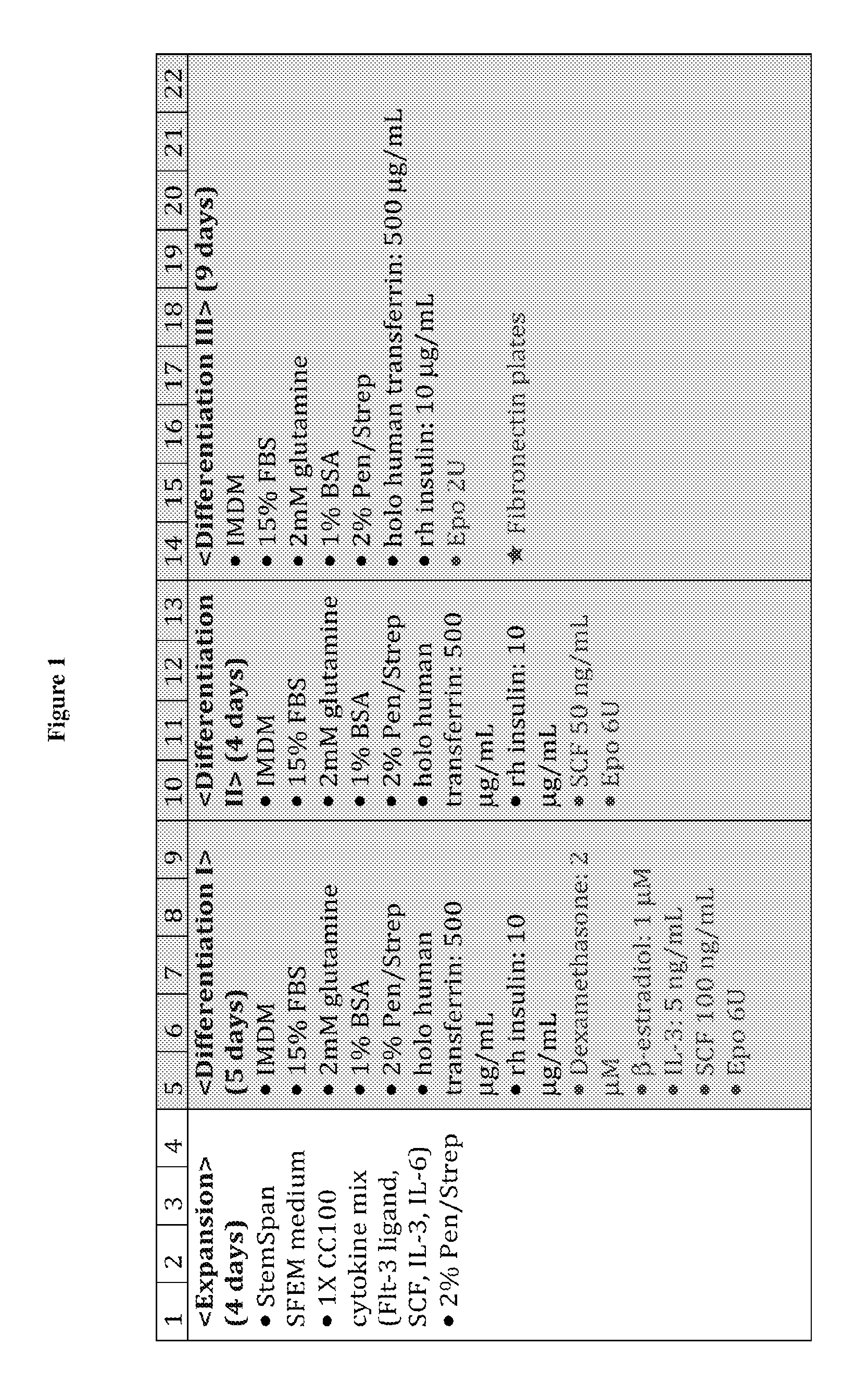
Accordingly, one aspect of the present disclosure features a method for producing human enucleated red blood cells, the method comprising: (i) providing a population of human CD34.sup.+ progenitor cells (e.g., human CD34.sup.+ peripheral blood cells); (ii) expanding the population of human CD34.sup.+ progenitor cells in a first medium for 0.about.6 days (e.g., 4 days), wherein the first medium comprises Flt-3 ligand, stem cell factor (SCF), interleukin 3 (IL-3), and interleukin 6 (IL-6); (iii) differentiating the expanded human CD34.sup.+ progenitor cells obtained from step (ii) in a second medium for 4.about.7 days (e.g., 5 days), wherein the second medium comprises dexamethasone, .beta.-estradiol, IL-3, SCF, and erythropoietin (EPO); (iv) differentiating the cells obtained from step (iii) in a third medium for 3.about.5 days (e.g., 4 days), wherein the third medium comprises SCF and EPO; (v) differentiating the cells obtained from step (iv) in a fourth medium for 4.about.12 days (e.g., 9 days) to produce human enucleated red blood cells, wherein the fourth medium comprises EPO; and (vi) collecting the human enucleated red blood cells obtained from step (v). In some embodiments, the total time period for steps (ii)-(v) of the method described above ranges from 11-25 days (e.g., 21 days). Step (v) may be performed in a culturing container coated with a component from the extracellular matrix, such as fibronectin.
In some embodiments, one or more of the second, third, and fourth media can further comprise holo human transferrin and insulin. When necessary, all of the second, third, and fourth media further comprise these two cytokines. In other embodiments, the second, third, and/or fourth media may be free of certain cytokines, for example, thrombopoietin (TPO), granulocyte macrophage colony-stimulating factor (GM-CSF), or both.
In one example, the second medium used in any of the in vitro production methods described herein can comprise 250.about.1500 .mu.g/ml (e.g., 500 .mu.g/ml) holo human transferrin, 5.about.20 .mu.g/ml insulin (e.g., 10 .mu.g/ml), 100 nM-5 .mu.M (e.g., 2 .mu.M) dexamethasone, 0.5.about.5 .mu.M (e.g., 1 .mu.M) .beta.-estradiol, 1.about.10 ng/ml (e.g., 5 ng) IL-3, 10.about.500 ng/ml (e.g., 100 ng/ml) SCF, and/or 2.about.10 U (e.g., 6 U) EPO. The second medium can be free of certain cytokines, for example, Flt-3, IL-6, or both.
In another example, the third medium used in any of the in vitro production methods described herein can comprise 250.about.1500 .mu.g/ml (e.g., 500 .mu.g/ml) holo human transferrin, 5.about.20 .mu.g/ml (e.g., 10 .mu.g/ml) insulin, 10.about.100 ng/ml (e.g., 50 ng/ml) SCF, and/or 2.about.10 U (e.g., 6 U) EPO. The third medium may be may be free of certain cytokines, for example, Flt-3, IL-6, dexamethasone, .beta.-estradiol, or any combination thereof.
In yet another example, the fourth medium used in any of the in vitro production methods described herein can comprise 250.about.1500 .mu.g/ml (e.g., 500 .mu.g/ml) holo human transferrin, 5.about.20 .mu.g/ml (e.g., 10 .mu.g/ml) insulin, and/or 0.5.about.3 U (e.g., 2 U) EPO. The fourth medium may be free of certain cytokines, for example, Flt-3, IL-6, dexamethasone, .beta.-estradiol, SCF, or any combination thereof.
In any of the in vitro production methods described herein, the human CD34.sup.+ progenitor cells can be any of the genetically engineered enucleated blood cells (which are not naturally occurring) as described herein, for example, a human CD34.sup.+ expressing a fusion protein comprising a red blood cell membrane protein and a peptide of interest. In some embodiments, the fusion protein comprises a type I red blood cell transmembrane protein (e.g., glycophorin A) fused to an acceptor peptide at the N-terminus of the type I red blood cell transmembrane protein. The acceptor peptide may include an oligoglycine moiety, e.g., a 1-5 glycine fragment, or an oligoalanine (e.g., a 1-5 alanine fragment) moiety. In other embodiments, the fusion protein comprises a type II red blood cell transmembrane protein (e.g., Kell or CD71) fused to a peptide comprising a sequence recognized by a sortase (e.g., a sortase A) at the C-terminus of the type II red blood cell transmembrane protein. The sequence recognizable by the sortase can be LPXTG (SEQ ID NO:1), in which X is any amino acid residue. In still other embodiments, the membrane protein is a type III red blood cell transmembrane protein, such as glucose transporter 1 (GLUT1).
In another aspect, the present disclosure provides a genetically engineered enucleated blood cell which expresses on the surface a first fusion protein comprising a first peptide of interest and a first red blood cell membrane protein. In some embodiments, the first red blood cell membrane protein is a type I red blood cell transmembrane protein (e.g., glycophorin A such as human glycophorin A), and the first peptide of interest is fused to the N-terminus of the type I red blood cell transmembrane protein. In other embodiments, the first red blood cell membrane protein is a type II transmembrane protein (e.g., Kell or CD71), and the first peptide of interest is fused to the C-terminus of the type II transmembrane protein. The first peptide of interest may comprise a sequence recognizable by a sortase, such as sortase A. In one example, the sequence recognizable by the sortase is LPXTG (SEQ ID NO: 1), in which X is any amino acid residue. In still other embodiments, the first red blood cell membrane protein is a type III red blood cell transmembrane protein, such as GLUT1.
In some embodiments, the first fusion protein further comprises a protein of interest (e.g., a cytoplasmic protein, which can be a diagnostic agent or a therapeutic agent). The protein of interest is fused to the terminus of the first red blood cell membrane protein that is exposed to a cytoplasmic space and the first peptide of interest is fused to the terminus of the first red blood cell membrane protein that is exposed to an extracellular or luminal space.
In some examples, the first red blood cell membrane protein can be a type I membrane protein (e.g., a GPA). The protein of interest is fused to the C-terminus of the type I membrane protein and the first peptide of interest is fused to the N-terminus of the type I membrane protein.
In other examples, the first red blood cell membrane protein is a type II membrane protein. The protein of interest is fused to the N-terminus of the type II membrane protein, and the first peptide of interest is fused to the C-terminus of the type I membrane protein.
In yet other embodiments, the genetically engineered enucleated blood cell as described herein can further express on the surface a second fusion protein comprising a second peptide of interest and a second red blood cell membrane protein.
In some examples, the first peptide of interest in the first fusion protein comprises a recognizable site or an acceptable peptide of a first sortase and the second peptide of interest in the second fusion protein comprises a recognizable site or an acceptable peptide of a second sortase. The first sortase and the second sortase use different recognizable sites and different acceptable peptides. In one example, the first peptide of interest comprises the motif of LPXTA (SEQ ID NO:2), in which X is any amino acid residue, or an oligoalanine; and the second peptide of interest comprises the motif LPXTG (SEQ ID NO:1), or an oligoglycine (e.g., consisting of 1-5 glycine residues). In another example, the first fusion protein comprises Kell, the C-terminus of which is fused to a first peptide of interest comprising the motif LPXTG (SEQ ID NO:1), and the second fusion protein comprises GPA, the C-terminus of which is fused to a second peptide of interest comprising an oligoalanine (e.g., consisting of 1-5 alanine residues).
In any of the genetically engineered enucleated blood cells described herein that express two fusion proteins on the surface, either one or both of the fusion proteins can be conjugated to two different functional moieties.
The above described enucleated blood cell may be prepared by any of the in vitro culturing methods described herein.
In some instances, the first peptide of interest, the second peptide of interest, or both, that are fused to the red blood cell membrane protein(s) described herein may comprise a protein drug (e.g., an antibody or an antigen-binding fragment thereof, which can be a single domain antibody), a vaccine antigen, a fluorescent protein, streptavidin, biotin, an enzyme, or a peptide capable of targeting a cell (e.g., a disease cell). In other instances, the peptide of interest is conjugated to a detectable label or a chemotherapeutic agent. For example, the peptide of interest may be conjugated to a lipid, a carbohydrate, a nucleic acid, a binding agent, a click-chemistry handle, a polymer, a peptide, a protein, a metal, a chelator, a radiolabel, or a small molecule.
In yet another aspect, the present disclosure provides methods for delivering an agent to a subject, the method comprising administering any of the enucleated blood cells described herein to the subject. In some examples, the enucleated blood cell being delivered is derived from the same subject the cell is being delivered to.
Also within the scope of the present disclosure are methods for conjugating any of the peptides of interest described herein to the surface of red blood cells. This method comprises: (i) providing a red blood cell expressing a fusion protein comprising a membrane protein and a first peptide, and (ii) contacting the red blood cell with a peptide of interest in the presence of a sortase (e.g., sortase A) under conditions suitable for the sortase to conjugate the peptide of interest to the first peptide in the fusion protein. Either the first peptide in the fusion protein or the peptide of interest comprises a sequence recognized by the sortase. In one example, the sequence recognizable by the sortase is LPXTG (SEQ ID NO:1), in which X is any amino acid residue.
In some embodiments, the membrane protein is a type I red blood cell transmembrane protein (e.g., glycophorin A) and the first peptide is an acceptor peptide (e.g., including an oligoglycine fragment such as a 1-5 glycine fragment) fused to the N-terminus of the type I red blood cell transmembrane protein, wherein the peptide of interest comprises the sequence recognized by the sortase. In other embodiments, the membrane protein is a type II red blood cell transmembrane protein (e.g., Kell or CD71) and the first peptide is fused to the C-terminus of the type II red blood cell transmembrane protein, wherein the first peptide comprises the sequence recognizable by the sortase. In still other embodiments, the membrane protein is a type III red blood cell transmembrane protein such as GLUT1.
In some embodiments, the first fusion protein in any of the methods described herein may further comprise a protein of interest (e.g., a cytoplasmic protein, which can be a diagnostic agent or a therapeutic agent), which is fused to the terminus of the first red blood cell membrane protein that is exposed to a cytoplasmic space. The first peptide of interest in the fusion protein is fused to the terminus of the first red blood cell membrane protein that is exposed to an extracellular or luminal space.
In some examples, the first red blood cell membrane protein is a type I membrane protein. The first protein of interest is fused to the C-terminus of the type I membrane protein, and the first peptide is fused to the N-terminus of the type I membrane protein. In other examples, the first red blood cell membrane protein is a type II membrane protein. The protein of interest is fused to the N-terminus of the type II membrane protein, and the first peptide is fused to the C-terminus of the type I membrane protein.
In yet other embodiments, any of the methods described herein can further comprise contacting the red blood cell in the presence of a second sortase under conditions suitable for the second sortase to conjugate a second peptide of interest to a second fusion protein expressed on the surface of the red blood cell. The second fusion protein comprises a second red blood cell membrane protein and a second peptide to which the second peptide of interest conjugates. In some examples, either the second peptide in the fusion protein or the second peptide of interest comprises a sequence recognizable by the second sortase. The sequence recognizable by the first sortase differ from the sequence recognizable by the second sortase.
In some examples, the first peptide in the first fusion protein comprises a sequence recognizable by the first sortase or an acceptable peptide of the first sortase; and/or the second peptide in the second fusion protein comprises a sequence recognizable by the second sortase or an acceptable peptide of the second sortase. The acceptable peptide of the first sortase is different from the acceptable peptide of the second sortase. The first sortase may be Sortase A from Staphylococcus arreus. One of the first peptide in the first fusion protein and the first peptide of interest comprises the motif LPXTG (SEQ ID NO:1), in which X is any amino acid residue, and the other comprises an acceptable peptide, which is an oligoglycin. The first fusion protein may comprise Kell, the C-terminus of which is fused to the first peptide which comprises the motif LPXTG (SEQ ID NO:1), and the first peptide of interest comprises an oligoglycine (e.g., consisting of 1-5 glycine residues).
In some examples, the second sortase is Sortase A from Streptococcus pyogenes. One of the second peptide in the second fusion protein and the second peptide of interest may comprise the motif LPXTA (SEQ ID NO:2), in which X is any amino acid residue, and the other may comprise an acceptable peptide, which can be an oligoalanine (e.g., consisting of 1-5 alanine residues). The second fusion protein may comprise GPA, the N-terminus of which is fused to the second peptide which comprises an oligoglycine. The second peptide of interest may comprise the motif LPXTG (SEQ ID NO:1), in which X is any amino acid residue.
In some embodiments, the first and second peptides of interest comprises or are conjugated to two different functional moieties. Alternatively or in addition, the first peptide of interest, the second peptide of interest, or both, may be a protein drug, a vaccine antigen, a fluorescent protein, streptavidin, biotin, an enzyme, or a peptide capable of targeting a cell. In one example, one of the first and second peptide of interest is a peptide capable of targeting a disease cell.
Further, the present disclosure provides (a) pharmaceutical compositions for diagnostic or therapeutic uses, the pharmaceutical composition comprising any of the enucleated red blood cells described herein, which carry any of the peptide of interest as described herein, and a pharmaceutically acceptable carrier, and (b) uses of the pharmaceutical compositions for manufacturing medicaments for diagnostic or therapeutic purposes.
The details of one or more embodiments of the invention are set forth in the description below. Other features or advantages of the present invention will be apparent from the following Drawings and Detailed Description of Certain Embodiments, and also from the Claims.
BRIEF DESCRIPTION OF THE DRAWINGS
FIG. 1 illustrates an exemplary in vitro culturing process for producing enucleated red blood cells from human CD34.sup.+ progenitor cells.
FIG. 2 includes photographs showing morphology of cells at expansion and different differentiation stages in the in vitro culturing process described herein.
FIG. 3 is a diagram showing the expression of cell surface markers glycophorin A (CD235) and c-kit in cells at expansion and different differentiation stages in the in vitro culturing process described herein, as determined by FACS analysis.
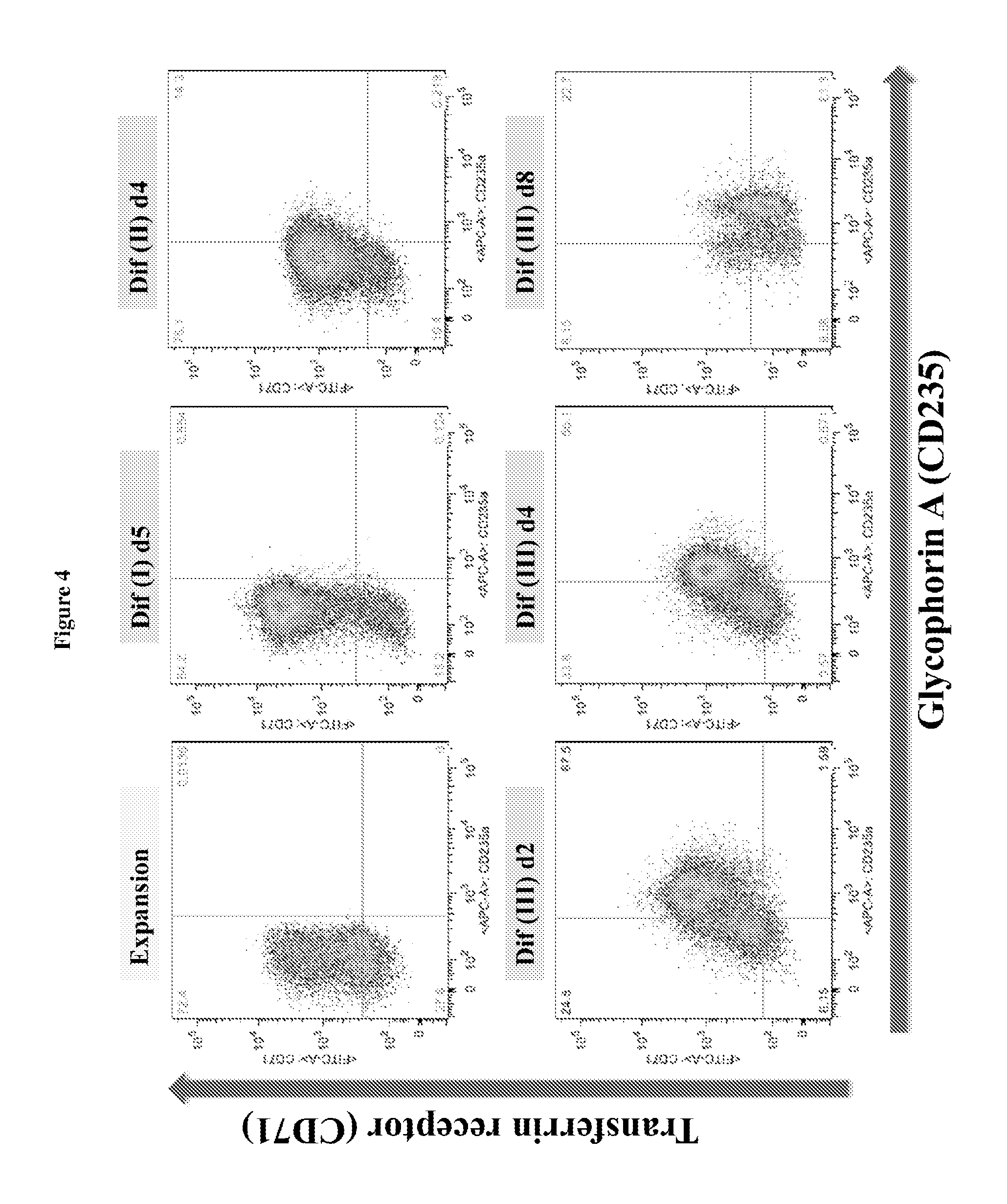
FIG. 4 is a diagram showing the expression of cell surface markers glycophorin A (CD235) and transferrin receptor (CD71) in cells at expansion and different differentiation stages in the in vitro culturing process described herein, as determined by FACS analysis.
FIG. 5 is a diagram showing the enucleation of cells at expansion and different differentiation stages in the in vitro culturing process described herein.
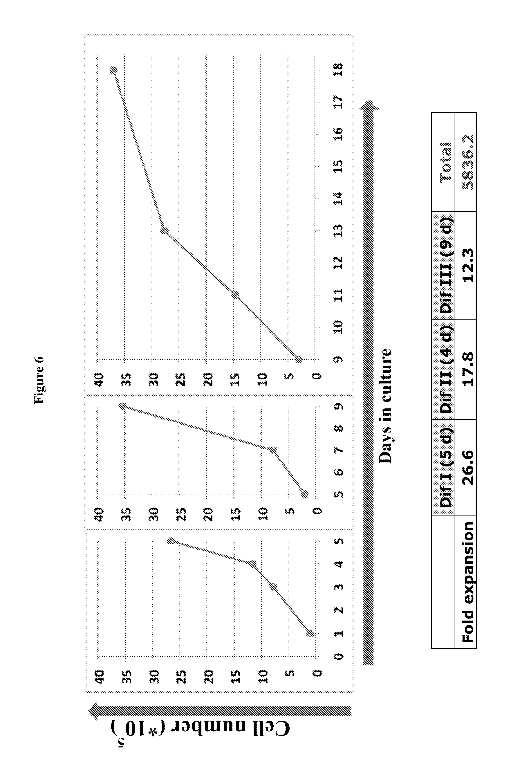
FIG. 6 includes charts showing cell proliferation at different differentiation stages.
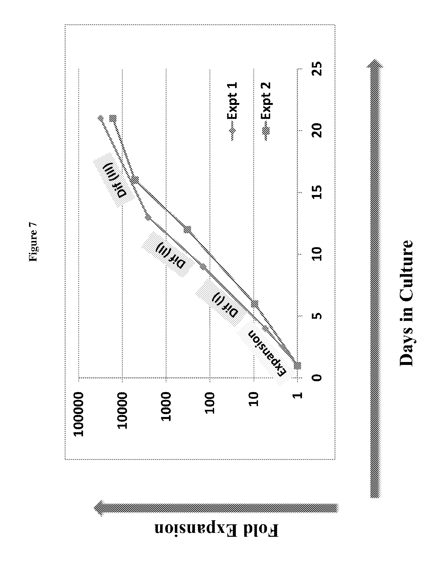
FIG. 7 is a chart showing cell proliferation in a window of around 20 days.
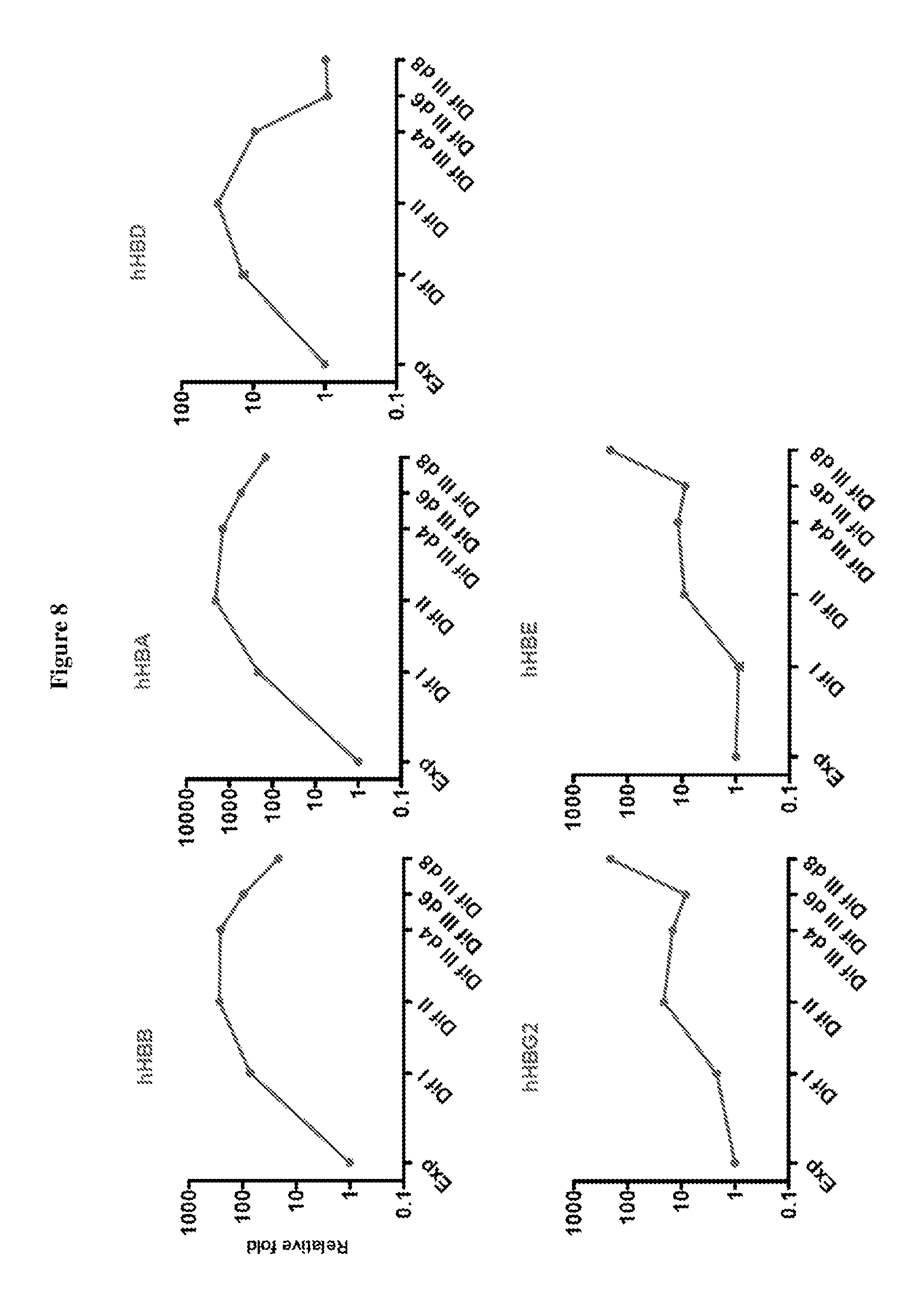
FIG. 8 include charts showing the expression of globin genes as indicated during hCD34.sup.+ cell differentiation.
FIG. 9 includes charts showing the expression of various genes as indicated during hCD34.sup.+ cell differentiation.
FIG. 10 is a diagram showing construction of EF1 and MSCV expression vectors for producing 5Gly (SEQ ID NO: 3)-myc-human glycophorin A (hGYPA or hGPA) fusion protein.
FIG. 11 is a diagram showing surface expression of sortaggable hGYPA on hCD34.sup.+ erythroid progenitors (at different differentiation stages) using EF1 expression vectors; 5Gly: (SEQ ID NO:3).
FIG. 12 is a diagram showing surface expression of sortaggable hGYPA on hCD34.sup.+ erythroid progenitors (at different differentiation stages) using MSCV expression vectors; 5Gly: (SEQ ID NO:3).
FIG. 13 is a photograph showing conjugation of biotin to hGYPA on hCD34.sup.+ erythroid progenitor cells by sortagging as determined by Western blotting; 5G: (SEQ ID NO:3).
FIG. 14 is a diagram showing conjugation of biotin to the surface of human CD34.sup.+ cells at the terminal differentiation stage via sortagging; 5Gly: (SEQ ID NO:3).
FIG. 15 is a diagram showing conjugation of biotin to the surface of human CD34.sup.+ cells at the terminal differentiation stage via sortagging; 5Gly: (SEQ ID NO:3).
FIG. 16 is a diagram showing the expression and sortase-mediated N-terminal labeling of glycophorin A on the surface of mature mouse red blood cells. (A) Schematic for glycophorin A N-terminal sortase labeling. GPA was extended at the N-terminus to include 3 glycine residues and a myc tag. Pre-incubation of sortase with a probe, which contains a C-terminal sortase recognition motif, LPETG (SEQ ID NO:44). leads to cleavage between T and G and formation of an acyl enzyme intermediate between Cys on sortase and Thr on the probe. Nucleophilic attack of the N-terminal glycine on GPA resolves the intermediate, thus ligating the probe to GPA. The probe can be a peptide, protein, lipid, carbohydrate, small molecule, etc. For conjugation of biotin to GPA, the probe is K(biotin)LPRTGG (SEQ ID NO:45); LPXTGG: (SEQ ID NO:46). (B) Evaluation of mature RBCs for the presence of 3G-myc-hGPA on the cell surface by staining either control blood or blood from mice that have undergone bone marrow transplantation to express 3G-myc-hGPA, with .alpha.-TER119 and .alpha.-myc tag antibodies and analyzing via flow cytometry. (C) Evaluation of mature RBCs for sortase-labeling by incubating control blood or 3G-myc-hGPA blood with sortase and the biotin containing probe, staining with .alpha.-TER119 and .alpha.-biotin antibodies, and analyzing via flow cytometry. (D) Evaluation of RBCs for sortase-labeling by immunoblotting. Control or 3G-myc-hGPA blood was incubated with biotin probe with or without sortase, and total cell protein was resolved by SDS-PAGE and immunoblotted for .alpha.-myc tag and .alpha.-biotin. The shift in molecular weight of GPA upon biotin conjugation in the .alpha.-myc tag immunoblot indicates almost complete modification. Biotin conjugated GPA is indicated in the .alpha.-biotin immunoblot with an arrow. (E) RBCs conjugated with biotin were sorted by flow cytometry. Immunofluorescence images show biotin (labeled with red) at the N-terminus of hGPA on mature RBCs (labeled with Ter119 antibody, purple). (F) HEK293T cells were transfected with 3A-myc-hGPA, hKell-LPETG (SEQ ID NO:44)-HA, or both. Cells were incubated with S. pyogenes sortase and a biotin probe followed by S. aureus sortase and TAMRA-containing probe incubation. Specific conjugation of biotin to GPA and of TAMRA to Kell is demonstrated by immunoblotting and fluorescence imaging.
FIG. 17 is a diagram showing the overexpression of modified human GPA and mouse GPA containing myctag at N-termini do not affect in vitro differentiation of mouse erythroid progenitor. (A) Flow cytometry analysis of the differentiation capacity of in vitro differentiated murine fetal-liver derived progenitor cells infected with retroviral constructs containing modified human (h) or mouse (m) GPA constructs extended at their N-terminus with myc-tag: myc-hGPA and myc-mGPA. Differentiation capacity in these cells is assessed based on the expressions of Ter119 and enucleation, i.e. nuclei expulsion, resulting in Ter119.sup.+ reticulocytes. The percentages of reticulocytes produced by erythroid progenitor cells infected with myc-hGPA or myc-mGPA (.about.20%) are comparable to cells infected with empty (control) vector, indicating that these constructs do not disturb erythroid terminal differentiation. Quantification of enucleation rate represents 3 independent experiments and graphed as mean values+/-standard deviation. (B) Evaluation of murine terminally differentiated erythroblasts, i.e. nucleated erythroblasts and reticulocytes, for the surface expression of myc-hGPA or myc-mGPA. More than 60% of these terminally differentiated cells expressed the desired modified GPA proteins as measured by flow cytometry using .alpha.-myc tag antibodies. Percentage of erythroblasts and reticulocytes with myc-tag on cell surface was determined from 3 independent experiments, graphed as mean value+/-standard deviation; ** indicates p<0.01. (C) Immunofluorescence images further confirm the surface expression of myc-tag (labelled with red) and enucleation capacity (blue nucleus staining) of the in vitro terminally differentiated erythroblasts.
FIG. 18 is a diagram showing the overexpression of engineered human GPA and mouse GPA containing myc-tag with multiple (3 or 5) glycines (SEQ ID NO:3) at their N-termini do not affect in vitro differentiation of mouse erythroid progenitors. Only cells expressing engineered human GPA with myc-tag and 3 glycines at the N-terminus can be efficiently biotin-labeled by sortagging. (A) Flow cytometry analysis on the differentiation capacity of in vitro differentiated murine fetal-liver derived progenitor cells infected with retroviral constructs containing modified human (h) or mouse (m) GPA constructs extended at their N-terminus with myc-tag and multiple (3 or 5) glycines (SEQ ID NO:3) (G): 5G (SEQ ID NO:3)-myc-hGPA, 5G (SEQ ID NO:3)-mycmGPA, and 3G-myc-hGPA. Differentiation capacity in these cells is assessed based on the expressions of Ter119 and enucleation, i.e. nuclei expulsion, resulting in Ter119.sup.+ reticulocytes. See upper panel. The percentages of reticulocytes produced by erythroid progenitor cells infected with 5G (SEQ ID NO:3)-myc-hGPA, 5G-myc-mGPA, and 3G-myc-hGPA (.about.17.5%) are comparable to cells infected with empty (control) vector, indicating that these constructs do not disturb erythroid terminal differentiation. Quantification of enucleation rate represents 3 independent experiments and graphed as mean values+/-standard deviation. See lower panel. (B) Evaluation of murine terminally differentiated erythroblasts, i.e., nucleated erythroblasts and reticulocytes, for the surface expression of 5G (SEQ ID NO:3)-myc-hGPA, 5G (SEQ ID NO:3)-mycmGPA, and 3G-myc-hGPA. More than 50% of these terminally differentiated cells expressed the desired modified GPA proteins as measured by flow cytometry using .alpha.-myc tag antibodies. See upper panel. Percentage of erythroblasts and reticulocytes with myc-tag on cell surface was determined from 3 independent experiments, graphed as mean value+/-standard deviation; ** indicates p<0.01. See lower panel. (C) HEK 293T cells were transfected to express 5G (SEQ ID NO:3)-myc-hGPA, 5G (SEQ ID NO:3)-myc-mGPA, 3G-myc-mGPA, and 3G-myc-hGPA. These cells were then incubated with a biotin probe with or without sortase, and total cell protein was immunoblotted for the myc-tag and biotin. Immunoblot stained for biotin shows successful biotin conjugation only to human GPA constructs 3G-myc-hGPA (strong band) and 5G (SEQ ID NO:3)-myc-hGPA (weak band) with a much higher efficiency for 3Gmyc (strong band) and 5G (SEQ ID NO:3)-myc-hGPA (weak band) with a much higher efficiency for 3Gmyc-hGPA. .alpha.-myc-tag immunoblotting further confirms biotin probe conjugation as indicated by a shift in hGPA molecular weight upon biotin conjugation.
FIG. 19 is a diagram showing the expression and sortase-mediated N-terminal labeling of glycophorin A on the surface of in vitro differentiated erythroblasts. (A) Flow cytometry analysis of in vitro differentiated erythroblasts for the presence of 3G-myc-hGPA and for sortase-labelled biotin-3G-myc-hGPA on the cell surface by staining with .alpha.-myc and .alpha.-biotin tag antibodies. See upper panel. Percentage of erythroblasts and reticulocytes with myc-tag on cell surface and percentage of myc-tag positive cells sortagged with a biotin probe was determined from 3 independent experiments, graphed as mean value+/-standard deviation; ** indicates p<0.01. See lower panel. (B) Evaluation of in vitro differentiated erythroblasts for sortase-labeling by incubating control or 3G-myc-hGPA erythroblasts with sortase and the biotin containing probe by means of immunoblotting with .alpha.-myc tag and .alpha.-biotin antibodies. The shift in molecular weight of hGPA upon biotin conjugation in the .alpha.-myc blot indicates complete modification of hGPA. Biotin-conjugated GPA is denoted by an arrow in the .alpha.-biotin immunoblot. (C) Immunofluorescence shows biotin (labelled with red) at the N-terminus of hGPA on the surface of differentiated erythroblasts. Nucleus was stained by Hoechst (blue). The arrow indicates reticulocytes, while the arrowheads indicate enucleating erythroblasts. The scale bar is 10 .mu.m.
FIG. 20 is a schematic illustration of engineered RBC production by mouse bone marrow/fetal liver cell transplantation. Isolated murine bone marrow cells or fetal liver cells were transduced with a retrovirus carrying hKell-LPETG (SEQ ID NO:44)-HA or 3G-myc-hGPA. Lethally irradiated mice were reconstituted with transduced bone marrow or fetal liver cells. After 1 month and onward, a significant percentage of mature RBCs are derived from transduced donor progenitors.
FIG. 21 is a diagram showing expression and sortase-mediated C-terminal labeling of Kell on the surface of mature mouse red blood cells. (A) Schematic for Kell C-terminal sortase labeling. Kell was extended at the C-terminus to include the sortase recognition motif LPETG (SEQ ID NO: 44) followed by an HA epitope tag. Incubation of sortase with cells containing Kell-LPETG (SEQ ID NO:44)-HA on the surface leads to peptide cleavage between T and G in the recognition motif and the formation of an acyl-enzyme intermediate between Cys on sortase and the sortase motif on Kell. Addition of a nucleophilic probe with N-terminal glycine residues (GGG-K(biotin); SEQ ID NO: 47; for biotin conjugation) resolves the intermediate, thus ligating the probe to the C-terminus of Kell. The probe can be a peptide, protein, lipid, carbohydrate, small molecule, etc; LPXTG (SEQ ID NO:1). (B) Evaluation of mature RBCs for the presence of Kell-LPETG (SEQ ID NO:44)-HA on the cell surface by staining either control blood or blood from mice that have undergone bone marrow transplantation to express Kell-LPETG (SEQ ID NO:44)-HA with .alpha.-HA tag antibody and analyzing via flow cytometry (gated on mature RBCs). (C) Evaluation of mature RBCs for sortase-labeling by incubating control blood or Kell-LPETG (SEQ ID NO:44)-HA blood with sortase and the biotin containing probe, staining with .alpha.-biotin antibody, and analyzing via flow cytometry (gated on mature RBCs). (D) Evaluation of RBCs for sortase-labeling by immune-blotting. Control or Kell-LPETG (SEQ ID NO:44)-HA blood was incubated with biotin probe with or without sortase. Total cell protein was resolved by SDS-PAGE and immunoblotted for .alpha.-HA tag and .alpha.-biotin. Loss of HA tag upon sortase labeling indicates complete modification. (E) Immunofluorescence images show biotin conjugated to the C-terminus of hKell on mature RBCs (labeled with Ter119 antibody).
FIG. 22 is a diagram showing expression and sortase-mediated C-terminal labeling of Kell on the surface of in vitro differentiated erythroblasts. (A) Flow cytometry of in vitro differentiated erythroblasts for the presence of hKell-LPETG (SEQ ID NO: 44)-HA and for sortase-labelled hKell-LPETG (SEQ ID NO: 44)-biotin on the cell surface by staining with .alpha.-HA and .alpha.-biotin tag antibodies. See upper panel. Percentage of erythroblasts and reticulocytes with HA-tag on cell surface and percentage of HA-tag positive cells sortagged with a biotin probe was determined from 3 independent experiments, graphed as mean value +/-standard deviation; ** indicates p<0.01. See lower panel. (B) Evaluation of in vitro differentiated erythroblasts for sortase-labeling by incubating control or hKell-LPETG (SEQ ID NO: 44)-HA erythroblasts with sortase and the biotin containing probe followed by immunoblotting for biotin and Kell. (C) Immunofluorescence shows biotin (labelled with red) at the C-terminus of hKell on the surface of differentiated erythroblasts. Nucleus was stained by Hoechst (blue). The arrow indicates reticulocytes, while the arrowhead indicates enucleating erythroblasts. The scale bar is 10 .mu.m.
FIG. 23 is a diagram showing sortase-mediated dual labeling at the N-terminus of hGPA and C-terminus of hKell on the surface of mature RBCs. Mature RBCs expressing hKell-LPETG (SEQ ID NO: 44)-HA and 3A-myc-hGPA were incubated with sortase A from S. pyogenes and K(biotin)LPETAA (SEQ ID NO: 45) probe for N-terminal GPA labeling (second panel), or with sortase A from S. aureus and GGGC (Alexa-647; SEQ ID NO: 48) probe for C-terminal Kell labeling (third panel), or both sequential label with both sortase enzymes and their corresponding probes (fourth panel). Flow cytometry analysis of mature RBCs for the presence of both hKell-LPET (SEQ ID NO: 49)-Alexa-647 and biotin-myc-hGPA indicate successful dual labeling.
FIG. 24 is a number of charts showing survival of engineered red blood cells in vivo. (A) Wild-type RBCs subjected to mock C-terminal sortagging reacting with or without sortase (n=3, each group) or left untreated (n=3) were reacted with CFSE and transfused into recipient mice via intravenous injection. Their in vivo survival was tracked via CFSE fluorescence and flow cytometry. (B) mKell-LPETG (SEQ ID NO: 44) RBCs were obtained from mice genetically engineered to have endogenous Kell extended at is C-terminus with an LPETG (SEQ ID NO: 44) sequence. Wild-type RBCs (n=6), mKell-LPETG (SEQ ID NO: 44) RBCs (n=6), and mKell-LPETG (SEQ ID NO: 44)--RBCs sortagged with biotin (n=6) were stained with CFSE, transfused into recipients, and their survival monitored via CFSE fluorescence or anti-biotin staining using flow cytometry. (C) CFSE-stained wild-type RBCs (n=6) and hKell-LPETG (SEQ ID NO: 44) RBCs isolated from transplanted mice (See Example 5) and sortase-labeled with biotin (n=2) were transfused into recipient mice and their survival in circulation was tracked via CFSE fluorescence (wild-type RBCs) or anti-biotin staining (hKell-LPETG (SEQ ID NO: 44)-biotin RBCs) using flow-cytometry. (D) Wild-type RBCs (n=6) and sortase-labeled biotin-3G-myc-hGPA RBCs (n=6) were CFSE stained, transfused into mice and tracked via CFSE fluorescence or anti-biotin staining using flow-cytometry. Internal control RBCs are cells that originate from 3G-myc-hGPA transplanted mice, were subjected to the sortagging reaction and CFSE-staining, but are wild-type. Error bars represent standard deviation; * denotes p<0.01.
FIG. 25 is a diagram showing conjugation of single domain antibody to engineered red blood cells and cell type-specific targeting. (A) Mouse mature red blood engineered to contain 3G-myc-GPA on the cell surface were incubated with Sortase with or without VHH7-LPETG (SEQ ID NO: 44), a single domain antibody with binding specificity for mouse MHC class II molecules and sortase motif complementary to 3G. An immunoblot against myc epitope tag on glycophorin A reveals fusion of VHH7 to GPA, as an increase in GPA molecular weight. (B) Either wild type mouse B cells (expressing MHC class II) or B cells derived from MHC class II knock-out mice were immobilized on magnetic streptavidin beads via biotinylated .alpha.-CD19 antibody, incubated with 3G-myc-GPA red blood cells or red cells that contained sortase-labeled VHH7-LPETG (SEQ ID NO: 44)-myc GPA on their surface, and washed. Immobilization of B cells in all experimental set-ups was tested by presence of CD19 in .alpha.-CD19 immunoblot and binding between B cells and red blood cells was evaluated by the presence of hemoglobin in the .alpha.-hemoglobin immunoblot. (C) Schematic for experiment shown in (B); LPETG: (SEQ ID NO: 44).
FIG. 26 is a diagram showing expression and sortase-mediated labeling of glycophorin A on the surface of human in vitro differentiated reticulocytes. (A) Human granulocyte--colony stimulating factor-mobilized peripheral blood CD34+ stem cells were virus transduced either with empty vector or a vector containing 3G-myc-GPA-GFP and differentiated for 18 days. Flow cytometry analysis of reticulocytes reveals the percentage of transduced cells (GFP.sup.+) and percentage of enucleated cells (lower panel with Hoechst staining). Reticulocytes, either control or 3G-myc-GPA-GFP (3G-myc-hGPA-GFP-IRES-GFP) transduced, were incubated with sortase and a biotin-containing probe, stained for biotin with .alpha.-biotin-PE antibody, analyzed via flow cytometry, and gated on enucleated cells. (B) 3G-myc-GPA-GFP transduced and in vitro differentiated reticulocytes were incubated with a biotin probe with or without sortase, and total cell protein immunoblotted for the myc tag and biotin. Both monomers as well as dimers (higher molecular weight) of 3G-myc-GPA-GFP are visible in both immunoblots. A shift in GPA-GFP molecular weight upon biotin conjugation in .alpha.-myc tag immunoblot indicates complete modification.
DETAILED DESCRIPTION CERTAIN EMBODIMENTS OF THE INVENTION
Red blood cells are the most numerous cell type in blood and account for a quarter of the total number of cells in the human body. RBCs possesses many unique characteristics that make them an attractive tool in therapeutics and diagnostics for various purposes, e.g., for in vivo delivery of natural or synthetic payloads. Yoo et al., 2011, Nature Reviews. Drug Delivery 10(7):521-535. For example, mature red blood cells do not have nuclei (i.e., enucleated) and thus there will be no risk of delivering remnants of foreign genes into a host. Thus, the possibility of tumorigenicity a key risk of stem cell-based therapies (Gruen et al., 2006 Stem cells 24(10):2162-2169) is thereby eliminated. Further, red blood cells have a long lifespan in vivo, e.g., about 120 days in the human blood stream, and about 50 days in mice, and presence throughout the macro- and micro-circulation. Modification of red cells with bioavailable therapeutics might thus lead to prolonged efficacy and coverage of all areas perfused by the circulation in vivo. Moreover, RBCs have large cell surface areas of about 140 .mu.m.sup.2 with a favorable surface to volume ratio. Also, red blood cells have good biocompability when used as carriers for delivery of therapeutic or diagnostic agents. Finally, old or damaged RBCs can be removed by cells of the reticuloendothelial system. Thus, any modification made to the DNA of RBC precursors is eliminated upon their enucleation and cannot lead to abnormal growth or tumorigenicity after their transfusion into a recipient.
Accordingly, it is of great interest to develop methods for producing enucleated red blood cells that carry an agent of interest, such as diagnostic or therapeutic agents. Such enucleated red blood cells can be used for, e.g., delivering the agent of interest into a subject.
Engineered RBCs have been generated using encapsulation (Biagiotti et al., 2011, IUBMB life 63(8):621-631; Godfrin et al., 2012, Expert Opinion on Biological Therapy 12(1):127-133; and Muzykantov, 2010, Expert Opinion on Drug Delivery 7(4):403-427), by non-covalent attachment of foreign peptides, or through installation of proteins by fusion to a monoclonal antibody specific for a RBC surface protein (Murciano, 2003, Nature Biotechnology 21(8):891-896; and Zaitsev et al., 2010, Blood 115(25):5241-5248). Modified RBCs face limitations if intended for application in vivo. Encapsulation allows entrapment of sizable quantities of material, but at the expense of disrupting plasma membrane integrity, with a concomitant reduction in circulatory half-life of the modified red blood cells. Osmosis driven entrapment limits the chemical nature of materials that can be successfully encapsulated, the site of release is difficult to control, and encapsulated enzymes are functional only at the final destination, compromising reusability at other sites. Murciano et al., 2003 and Zaitsev et al., 2010. Targeting of cargo to RBCs by fusion to an RBC-specific antibody, (e.g., antiglycophorin antibody), has its limitations because this mode of attachment to the RBC is non-covalent and readily dissociates, thus reducing circulatory half life and mass of cargo available for delivery. Murciano et al., 2003 and Zaitsev et al., 2010. Other developments that exploit RBCs for targeted delivery include nanoparticles enveloped by an RBC-mimicking membrane as well as RBC-shaped polymers. Yoo et al., 2011 Nature Reviews. Drug Discovery 10(7):521-535. The short in vivo survival rate of these RBC-inspired carriers 7 days maximum) may limit their therapeutic utility.
Another technical difficulty associated with engineering RBCs is lack of a suitable in vitro system for culturing and differentiation of RBCs. Human CD34.sup.+ cell culture systems have been disclosed in Miharada et al., Nat. Biotechnol., 24(10):1255-1256, 2006; Sankaran et al., Science, 322(5909):1839-1842, 2008, and Giarratana et al., Blood, 118(19):5071-5079, 2011. However, the mature enucleated red blood cells obtained from these cell culture systems did not show similar hemoglobin contents and/or cell sizes as compared to normal human reticulocytes or red blood cells, did not show synchronized expression of cell surface differentiation markers during the culture process. Also, the cell culture system taught in Sankaran et al. did not generate terminally-differentiated erythroid cells or enucleated red cells.
In light of the disadvantages associated with the existing technology, there is a need to develop new methodology for engineering RBCs, such that they can carry a wide variety of useful cargoes to specific locations in the body.
In the present studies, modified red blood cells were developed to serve as carriers for systemic delivery of a wide array of payloads. These RBCs contain modified proteins on their plasma membrane, which can be labeled in a sortase-catalyzed reaction under native conditions without inflicting damage on the target membrane or cell. Sortase accommodates a wide range of natural and synthetic payloads that allow modification of RBCs with substituents that cannot be encoded genetically. The present studies demonstrate the successful site-specific conjugation of biotin to in vitro differentiated mouse erythroblasts as well as to mature mouse RBCs. Unexpectedly, these modified red cells remain in the bloodstream for up to 28 days, which is far in excess of red blood cells engineered by methods known in the art. A single domain antibody attached enzymatically to RBCs enables them to bind specifically to target cells that express the antibody target. This study was extended to human red cells and demonstrate unexpected efficient sortase-mediated labeling of in vitro differentiated human reticulocytes.
The engineering and labeling processes described herein do not damage the cells or affect their survival in vivo. Most importantly, the engineered red blood cells can be labeled with a wide array of functional probes, including small molecules, peptides, and proteins and thus have the potential to be carriers of a variety of therapeutic substances into the bloodstream. Thus, the methods described herein offer advantages over the osmosis-driven encapsulation methods known in the art.
Sortase-mediated cell surface labeling has been exploited previously, for example to explore trafficking of flu glycoproteins. Popp et al., 2012 PLos Pathogens 8(3):e1002604; and Sanyal et al., 2013 Cell Host & Microbe 14(5):510-521. This methodology was modified and applied to primary cells as a platform for diagnostic or therapeutic purposes. A sortase A variant from S. aureus active at 0.degree. C. (Chen et al., 2011 PNAS 108(28):11399-11404) to reduce the risk of inflicting cellular damage in the course of labeling.
There are many other possible applications for the methods presented here, where the wide variety of possible payloads--ranging from proteins and peptides to synthetic compounds and fluorescent probes may serve as a guide. For example, such methods would enable the targeting of the modified red cells to a specific cell type. Further, the methods described herein can be combined with established protocols of small molecule encapsulation (Godfrin et al., 2012). In this scenario, engineered red cells loaded in the cytosol with a therapeutic agent and modified on the surface with a cell-type specific recognition module could be used to deliver payloads to a precise tissue/location in the body. It has been demonstrated herein attachment of two different functional probes to the surface of red blood cells, exploiting the subtly different recognition specificities of two distinct sortases. It should therefore be possible to attach both a therapeutic moiety as well as a targeting module to the red cell surface, to direct the engineered red cells to tumors or other diseased cells. Conjugation of an imaging probe, i.e., a radioisotope, together with such a targeting moiety could also be used for diagnostic purposes.
The in vitro generation of human RBCs and genetic engineering of their precursors as described herein may provide a robust platform for application of this surface engineering method, e.g., in conjunction with cytosolic modification, to clinical applications (Liu et al., 2010 Blood 115(10):2021-2027; Cong et al., 2013 Science 339(6121):819-826; Mali et al., Science 339(6121):823-826; Giarratana et al., 2011 Blood 118(19):5071-5079; Douay et al., 2009 Blood 105(1):85-94; and Griffiths et al., 2012 Blood 119(26):6296-6306.) Moreover, the established safety of blood transfusions inspires confidence that these engineered red blood cells will indeed find use in humans.
Accordingly, described herein is an in vitro multi-phase culturing process for producing enucleated red blood cells from mobilized CD34.sup.+ progenitor cells (e.g., human mobilized CD34.sup.+ peripheral blood cells). Such enucleated red blood cells can be genetically engineered such that they express proteins of interest, e.g., sortaggable surface proteins. Also described herein are methods for conjugating one or more agents of interest to the surface of the genetically engineered enucleated red blood cells via a sortase-mediated transpeptide reaction, as well as methods for producing cytoplasmically disposed protein of interest in mature RBCs such as human RBCs while retaining the ability to selectively target the RBCs to an intended target via sortagging of a modified membrane protein with a target moiety of interest.
I. In vitro Culturing Systems for Producing Enucleated Red Blood Cells
Described herein is an in vitro culturing process for producing mature enucleated red blood cells from CD34.sup.+ progenitor cells (e g from a human subject). This culturing process involves multiple differentiation stages (e.g., 2, 3, or more) and optionally an expansion stage prior to the differentiation phases. The total time period for the in vitro culturing process described herein can range from 11-25 days (e.g., 15-25 days, 15-20 days, or 18-21 days). In one example, the total time period is 21 days.
a. CD34.sup.+ Progenitor Cells
CD34 is a cell surface glycoprotein and functions as a cell-cell adhesion factor. Many human progenitor cells express this cell surface marker. Novershtern et al., Cell 144:296-309, 2011. A progenitor cell, like a stem cell, has a tendency to differentiate into a specific type of cell. Progenitor cells are usually more specific than stem cells and are often pushed to differentiate into the target cells. Any type of CD34+ progenitor cells that possess the tendency of differentiating into red blood cells can be used in the in vitro culturing process described herein. Such progenitor cells are well known in the art. See, e.g., Novershtern et al., Cell 144:296-309, 2011. In some examples, the in vitro culturing process described herein utilizes mobilized CD34+ peripheral blood cells as the progenitor cells for differentiation into enucleated red blood cells. CD34+ progenitor cells can also be derived from other sources (e.g., bone marrow).
Various techniques can be used to separate or isolate the CD34.sup.+ cell population from a suitable source such as peripheral blood cells. For example, antibodies such as monoclonal antibodies binding to CD34 can be used to enrich or isolate CD34.sup.+ cells. The anti-CD34 antibodies can be attached to a solid support such that cells expressing these surface markers are immobilized, thereby allowing for the separation of CD34.sup.+ cells from cells that do not express this surface marker. The separation techniques used should maximize the retention of viable cells to be collected. Such separation techniques can result in sub-populations of cells where up to 10%, usually not more than about 5%, preferably not more than about 1%, of the selected cells do not express CD34. The particular technique employed will depend upon the efficiency of separation, associated cytotoxicity, ease and speed of performance, and necessity for sophisticated equipment and/or technical skill.
An "isolated" or "purified" population of CD34.sup.+ cells for use in the in vitro culturing process described herein is substantially free of cells and materials with which it is associated in nature, in particular, free of cells that lack the desired phenotype, e.g., expressing CD34. Substantially free or substantially purified includes at least 50% CD34.sup.+ cells, preferably at least 70%, more preferably at least 80%, and even more preferably at least 90% CD34.sup.+ cells.
Procedures for separating the CD34.sup.+ population of cells can include, but are not limited to, physical separation, magnetic separation, antibody-coated magnetic beads, affinity chromatography, cytotoxic agents joined to a monoclonal antibody or used in conjunction with a monoclonal antibody, including, but not limited to, complement and cytotoxins, and "palming" with antibody attached to a solid matrix, e.g., plate, elutriation or any other convenient technique.
The use of physical separation techniques also include those based on differences in physical (density gradient centrifugation and counter-flow centrifugal elutriation), cell surface (lectin and antibody affinity), and vital staining properties (mitochondria-binding dye rho123 and DNA-binding dye Hoechst 33342). These procedures are well known to those of skill in this art.
Techniques providing accurate separation of CD34.sup.+ cells further include flow cytometry, which can have varying degrees of sophistication, e.g., a plurality of color channels, low angle and obtuse light scattering detecting channels, impedance channels. CD34.sup.+ cells also can be selected by flow cytometry based on light scatter characteristics, where the target cells are selected based on low side scatter and low to medium forward scatter profiles.
b. Expansion Stage
Optionally, the in vitro culturing process described herein includes an expansion stage, in which the CD34.sup.+ progenitor cells are allowed to proliferate. As used herein, expansion or proliferation includes any increase in cell number. Expansion includes, for example, an increase in the number of CD34+ cells over the number of CD34+ cells present in the cell population used to initiate the culture. Expansion can also include increased survival of existing CD34+ cells. The term survival refers to the ability of a cell to continue to remain alive or function.
A population of CD34.sup.+ progenitor cells (e.g., mobilized CD34.sup.+ peripheral blood cells) can be placed in a suitable container for expanding the CD34+ cells. For example, suitable containers for culturing the population of cells include flasks, tubes, or plates. In one embodiment, the flask can be T-flask such as a 12.5 cm.sup.2, or a 75 cm.sup.2 T-flask. The plate can be a 10 cm plate, a 3.5 cm plate, or a multi-welled plate such as a 12, 24, or 96 well plate. The wells can be flat, v-bottom, or u-bottom wells. The containers can be treated with any suitable treatment for tissue culture to promote cell adhesion or to inhibit cell adhesion to the surface of the container. Such containers are commercially available from Falcon, Corning and Costar. As used herein, "expansion container" also is intended to include any chamber or container for expanding cells whether or not free standing or incorporated into an expansion apparatus.
The cell density of the cultured population of CD34.sup.+ cells can be at least from about 1.times.10.sup.2 cells to about 1.times.10.sup.7 cells/mL. Preferably, the cell density can be from about 1.times.10.sup.5 to about 1.times.10.sup.6 cells/mL. The cells can be cultured at an oxygen concentration of from about 2 to 20%.
Various media can be used to expand the population of CD34.sup.+ progenitor cells, including, but not limited to, Dulbecco's MEM, IMDM, X-Vivo 15 (serum-depleted) and RPMI-1640. Such culture media can be serum free. In one embodiment, the medium is serum free StemSpan (Stem Cell Technologies), which can be supplemented with 10 .mu.g/ml heparin.
The culture medium for use in the expansion stage can contain one or more cytokines. As used herein, cytokines are factors that exert a variety of effects on cells, for example, growth or proliferation. Non-limiting examples of the cytokines that may be used in one or more stages of the in vitro culturing process (e.g., in the expansion stage or any of the differentiation stages described below) include interleukin-2 (IL-2), interleukin 3 (IL-3), interleukin 6 (IL-6) including soluble IL-6 receptor, interleukin 12 (IL12), G-CSF, granulocyte-macrophage colony stimulating factor (GM-CSF), interleukin 1 alpha (IL-1 .alpha.), interleukin 11 (IL-11), MIP-1.alpha., leukemia inhibitory factor (LIF), c-kit ligand, and flt3 ligand. In some examples, the in vitro culturing process described herein, or any stages thereof, can include culture conditions, in which one or more cytokine is specifically excluded from the culture medium. Cytokines are commercially available from several vendors such as, for example, Amgen (Thousand Oaks, Calif.), R & D Systems and Immunex (Seattle, Wash.). Cytokine can also include fibroblast growth factor (FGF) (e.g., FGF-1 or FGF-2), insulin-like growth factor (e.g., IGF-2, or IGF-1), thrombopoietin (TPO), and stem cell factor (SCF), or analogs and equivalents thereof. Equivalents thereof include molecules having similar biological activity to these factors (e.g., FGF, TPO, IGF, and SCF) in wild-type or purified form (e.g., recombinantly produced). Analogs include fragments retaining the desired activity and related molecules. For example, TPO is a ligand of the mpl receptor, thus molecules capable of binding the mpl receptor and initiating one or more biological actions associated with TPO binding to mpl are also within the scope of the invention. An example of a TPO mimetic is found in Cwirla et. al. (1997) Science 276:1696.
In one example, the expansion stage of the in vitro culturing process described herein can be performed as follows. A population of human mobilized CD34.sup.+ peripheral blood cells is placed in an expansion container at a cell density of 10.times.10.sup.4-10.times.10.sup.6 (e.g., 1.times.10.sup.5) cells/mL. The CD34.sup.+ cells are cultured in an expansion medium (e.g., StemSpan serum-free medium) supplemented with a cytokine mixture of Flt-3 ligand, SCF, IL-3, and IL-6 and 2% penicillin and streptomycin under suitable conditions (e.g., 37.degree. C.) for 1-6 days (e.g., 2-5 days, 3-4 days, or 4 days). The expanded CD34+ cells can be collected and subjected to further in vitro culturing under conditions allowing for differentiation toward mature enucleated red blood cells.
C. Multiple Differentiation Stages
The in vitro culturing process described herein involves multiple differentiation stages (2, 3, 4, or more), in which CD34+ progenitor cells differentiate into mature enucleated red blood cells. In each differentiation stage, CD34+ progenitor cells (either obtained from the expansion stage or collected from the original source) or cells obtained from the preceding differentiation stage can be cultured in a medium comprising one or more suitable cytokines (e.g., those described herein) under suitable conditions for a suitable period of time. Biological properties of the cells, such as cell size and expression of surface markers, may be monitored during the course or at the end of each differentiation stage to evaluate the status of erythropoiesis. Whenever necessary, cytokines can be timely supplied and/or withdrawn at each differentiation stage to achieve optimal erythroid differentiation and/or synchronizing the cell population in culture.
At each of the differentiation stages, CD34.sup.+ progenitor cells, either obtained from the expansion stage described herein or isolated from an original source (e.g., human peripheral blood), or cells obtained from the preceding differentiation stage can be cultured in a suitable medium, such as those described above, supplemented with one or more cytokines under suitable culturing conditions for a suitable period of time. In one example, the culture medium (e.g., IMDM) is supplemented with holo human transferrin and insulin at suitable concentrations. For example, the concentration of hobo human transferrin ranges from 250-1,500 .mu.g/ml (e.g., 250-1,000 .mu.g/ml; 300-800 .mu.g/ml, or 400-600 .mu.g/ml); and the concentration of insulin can range from 5-20 .mu.g/ml (e.g., 5-15 .mu.g/ml, 5-10 .mu.g/ml, or 10-20 .mu.g/ml). The culture medium may also be supplemented with other components commonly used in cell culture, e.g., fetal bovine serum, glutamine, bovine serum albumin, one or more antibiotics (e.g., penicillin and streptomycin), or any combination thereof.
In some embodiments, the in vitro culturing process described herein includes three differentiation stages, Differentiation stage I (`Dif. I), Differentiation stage II ("Dif. II"), and Differentiation stage III ("Dif. III").
In Dif. I, the cells may be cultured in the presence of a mixture of cytokines including a glucocorticoid (e.g., dexamethasone), .beta.-estradiol, IL-3, SCF, and EPO (including human EPO, EPO from other species, or EPO analogs such as Epoetin alfa, Epoetin beta, or Darbepoetin alfa) at suitable concentrations for a suitable period of time (e.g., 4-7 days, 4-6 days, 5-7 days, or 5-6 days). In some examples, the concentration of dexamethasone may range from 100 nM to 5 .mu.M (e.g., 100 nM to 2 .mu.M, 500 nM to 5 .mu.M, 1 .mu.M to 3 .mu.M, 1 .mu.M to 2 .mu.M, or 2 .mu.M to 5 .mu.M). The concentration of .beta.-estradiol may range from 0.5-5 .mu.M (e.g., 0.5-4 .mu.M, 1-5 .mu.M, 0.5-3 .mu.M, 0.5-2 .mu.M, 0.5-1 .mu.M, 1-2 .mu.M, 1-3 .mu.M, or 3-5 .mu.M). The concentration of IL-3 may range from 1-10 ng/ml (e.g., 1-8 ng/ml, 1-5 ng/ml, 3-6 ng/ml, 4-8 ng/ml, or 5-10 ng/ml). The concentration of SCF may range from 10-500 ng/ml (e.g., 10-300 ng/ml, 50-500 ng/ml, 50-200 ng/ml, 50-100 ng/ml, 100-200 ng/ml, or 100-400 ng/ml). Alternatively or in addition, the amount of EPO may range from 2-10 U (e.g., 2-8 U, 2-6 U, 5-10 U, or 6-10 U). As well known in the art, one EPO unit elicits the same erythropoiesis stimulating response in rodents (historically: fasted rats) as five micromoles of cobaltous chloride. See, e.g., Jelkmann, Nephrol Dial. Transplant, 2009. In some examples, the medium used in Dif. I contains holo human transferrin, insulin, dexamethasone, .beta.-estradiol, IL-3, SCF, and EPO. This medium may be substantially free of certain other cytokines, such as Flt-3 ligand or IL-6, or substantially free of other cytokines.
In Dif. II, the cells obtained from Dif. I may be cultured in the presence of a mixture of cytokines including SCF and EPO at suitable concentrations for a suitable period of time (e.g., 3-5 days, 3-4 days, or 4-5 days). In some examples, the concentration of SCF can range from 10-100 ng/ml (e.g., 10-80 ng/ml, 20-80 ng/ml, 20-50 ng/ml, 30-50 ng/ml, 40-50 ng/ml, 50-80 ng/ml, or 50-60 ng/ml). Alternatively or in addition, the amount of EPO can range from 2-10 U (e.g., 2-8 U, 2-6 U, 5-10 U, or 6-10 U). In some examples, the medium used in Dif. II contains holo human transferrin, insulin, SCF, and EPO. This medium may be substantially free of certain cytokines, such as Flt-3, IL-6, dexamethasone, .beta.-estradiol, IL-3, or any combination thereof, or may be substantially free of other cytokines.
In Dif. III, the cells obtained from Dif. II may be cultured in the presence of EPO at a suitable concentration for a suitable period of time (e.g., 4-12 days, 5-10 days, 8-12 days, or 8-10 days). In some examples, the amount of EPO may range from 0.5-3 U (e.g., 1-3 U, 0.5-2 U, 1-2 U, or 2-3 U). In some examples, the medium used in Dif. III contains holo human transferrin, insulin, and EPO. This medium may be substantially free of certain cytokines, for example, Flt-3, IL-6, dexamethasone, .beta.-estradiol, IL-3, SCF, or any combination thereof, or substantially free of other cytokines.
In some embodiments, the in vitro culturing process described herein may include one or any combination of the differentiation stages described herein, for example, Dif. I and Dif. III, Dif. II and Dif. III, or Dif. I and Dif. II.
Prior to the differentiation stages, the CD34.sup.+ progenitor cells may be genetically modified such that they express surface proteins of interest, for example, sortaggable surface proteins, which are discussed in detail below.
II. Preparation of Red Blood Cells Expressing Surface Proteins of Interest
Also described herein are methods of preparing genetically engineered red blood cells capable of expressing surface proteins of interest, such as sortaggable surface proteins, fluorescent proteins such as green fluorescent protein (GFP), or protein drugs (e.g., antibodies or antigen-binding fragments thereof).
(a) Genetic Modification of Progenitor Cells
Expression vectors for producing the surface protein of interest may be introduced into CD34.sup.+ progenitor cells, which can be isolated from an original source or obtained from the expansion stage described above via routine recombinant technology. In some instances, the expression vectors can be designed such that they can incorporate into the genome of cells by homologous or non-homologous recombination by methods known in the art. Methods for transferring expression vectors into CD34.sup.+ progenitor cells include, but are not limited to, viral mediated gene transfer, liposome mediated transfer, transformation, transfection and transduction, e.g., viral mediated gene transfer such as the use of vectors based on DNA viruses such as adenovirus, adeno-associated virus and herpes virus, as well as retroviral based vectors. Examples of modes of gene transfer include e.g., naked DNA, CaPO.sub.4 precipitation, DEAE dextran, electroporation, protoplast fusion, lipofection, cell microinjection, and viral vectors, adjuvant-assisted DNA, gene gun, catheters. In one example, a viral vector is used. To enhance delivery of non-viral vectors to a cell, the nucleic acid or protein can be conjugated to antibodies or binding fragments thereof which bind cell surface antigens, e.g., CD34. Liposomes that also include a targeting antibody or fragment thereof can be used in the methods described herein.
A "viral vector" as described herein refers to a recombinantly produced virus or viral particle that comprises a polynucleotide to be delivered into a host cell, either in vivo, ex vivo or in vitro. Examples of viral vectors include retroviral vectors such as lentiviral vectors, adenovirus vectors, adeno-associated virus vectors and the like. In aspects where gene transfer is mediated by a retroviral vector, a vector construct refers to the polynucleotide comprising the retroviral genome or part thereof, and a therapeutic gene.
A gene encoding the surface protein of interest can be inserted into a suitable vector (e.g., a retroviral vector) using methods well known in the art. Sambrook et al., Molecular Cloning, A Laboratory Mannual, 3.sup.rd Ed., Cold Spring Harbor Laboratory Press. For example, the gene and vector can be contacted, under suitable conditions, with a restriction enzyme to create complementary ends on each molecule that can pair with each other and be joined together with a ligase. Alternatively, synthetic nucleic acid linkers can be ligated to the termini of a gene. These synthetic linkers contain nucleic acid sequences that correspond to a particular restriction site in the vector. Additionally, the vector can contain, for example, some or all of the following: a selectable marker gene, such as the neomycin gene for selection of stable or transient transfectants in mammalian cells; enhancer/promoter sequences from the immediate early gene of human CMV for high levels of transcription; transcription termination and RNA processing signals from SV40 for mRNA stability; SV40 polyoma origins of replication and ColE1 for proper episomal replication; versatile multiple cloning sites; and T7 and SP6 RNA promoters for in vitro transcription of sense and antisense RNA. Suitable vectors and methods for producing vectors containing transgenes are well known and available in the art. Sambrook et al., Molecular Cloning, A Laboratory Mannual, 3.sup.rd Ed., Cold Spring Harbor Laboratory Press.
Modification of CD34.sup.+ progenitor cells can comprise the use of an expression cassette created for either constitutive or inducible expression of the introduced gene. Such an expression cassette can include regulatory elements such as a promoter, an initiation codon, a stop codon, and a polyadenylation signal. The elements are preferably operable in the progenitor cells or in cells that arise from the progenitor cells (e.g., enucleated red blood cells) after administration (e.g., infusion) into an individual. Moreover, the elements can be operably linked to the gene encoding the surface protein of interest such that the gene is operational (e.g., is expressed) in the progenitor cells or red blood cells derived therefrom.
A variety of promoters can be used for expression of the surface protein of interest. Promoters that can be used to express the protein are well known in the art. Promoters include cytomegalovirus (CMV) intermediate early promoter, a viral LTR such as the Rous sarcoma virus LTR, HIV-LTR, HTLV-1 LTR, the simian virus 40 (SV40) early promoter, E. coli lac UV5 promoter and the herpes simplex tk virus promoter.
Regulatable promoters can also be used. Such regulatable promoters include those using the lac repressor from E. coli as a transcription modulator to regulate transcription from lac operator-bearing mammalian cell promoters [Brown, M. et al., Cell, 49:603-612 (1987)], those using the tetracycline repressor (tetR) [Gossen, M., and Bujard, H., Proc. Natl. Acad. Sci. USA 89:5547-5551 (1992); Yao, F. et al., Human Gene Therapy, 9:1939-1950 (1998); Shockelt, P., et al., Proc. Natl. Acad. Sci. USA, 92:6522-6526 (1995)]. Other systems include FK506 dimer, VP16 or p65 using astradiol, RU486, diphenol murislerone or rapamycin. Inducible systems are available from Invitrogen, Clontech and Ariad.
Regulatable promoters that include a repressor with the operon can be used. In one embodiment, the lac repressor from E. coli can function as a transcriptional modulator to regulate transcription from lac operator-bearing mammalian cell promoters [M. Brown et al., Cell, 49:603-612 (1987)]; Gossen and Bujard (1992); [M. Gossen et al., Natl. Acad. Sci. USA, 89:5547-5551 (1992)] combined the tetracycline repressor (tetR) with the transcription activator (VP 16) to create a tetR-mammalian cell transcription activator fusion protein, tTa (tetR-VP 16), with the tetO-bearing minimal promoter derived from the human cytomegalovirus (hCMV) major immediate-early promoter to create a tetR-tet operator system to control gene expression in mammalian cells. In one embodiment, a tetracycline inducible switch is used. The tetracycline repressor (tetR) alone, rather than the tetR-mammalian cell transcription factor fusion derivatives can function as potent trans-modulator to regulate gene expression in mammalian cells when the tetracycline operator is properly positioned downstream for the TATA element of the CMVIE promoter [F. Yao et al., Human Gene Therapy, supra]. One particular advantage of this tetracycline inducible switch is that it does not require the use of a tetracycline repressor-mammalian cells transactivator or repressor fusion protein, which in some instances can be toxic to cells [M. Gossen et al., Natl. Acad. Sci. USA, 89:5547-5551 (1992); P. Shockett et al., Proc. Natl. Acad. Sci. USA, 92:6522-6526 (1995)], to achieve its regulatable effects.
The effectiveness of some inducible promoters can be increased over time. In such cases one can enhance the effectiveness of such systems by inserting multiple repressors in tandem, e.g., TetR linked to a TetR by an internal ribosome entry site (IRES). Alternatively, one can wait at least 3 days before screening for the desired function. While some silencing may occur, it can be minimized by using a suitable number of cells, preferably at least 1.times.10.sup.4, more preferably at least 1.times.10.sup.5, still more preferably at least 1.times.10.sup.6, and even more preferably at least 1.times.10.sup.7. One can enhance expression of desired proteins by known means to enhance the effectiveness of this system. For example, using the Woodchuck Hepatitis Virus Posttranscriptional Regulatory Element (WPRE). See Loeb, V. E., et al., Human Gene Therapy 10:2295-2305 (1999); Zufferey, R., et al., J. of Virol. 73:2886-2892 (1999); Donello, J. E., et al., J. of Virol. 72:5085-5092 (1998).
Examples of polyadenylation signals useful to practice the methods described herein include, but are not limited to, human collagen I polyadenylation signal, human collagen II polyadenylation signal, and SV40 polyadenylation signal.
The exogenous genetic material that includes the surface protein-encoding gene operably linked to the regulatory elements may remain present in the cell as a functioning cytoplasmic molecule, a functioning episomal molecule or it may integrate into the cell's chromosomal DNA. Exogenous genetic material may be introduced into cells where it remains as separate genetic material in the form of a plasmid. Alternatively, linear DNA, which can integrate into the chromosome, may be introduced into the cell. When introducing DNA into the cell, reagents, which promote DNA integration into chromosomes, may be added. DNA sequences, which are useful to promote integration, may also be included in the DNA molecule. Alternatively, RNA may be introduced into the cell.
Selectable markers can be used to monitor uptake of the desired transgene into the progenitor cells described herein. These marker genes can be under the control of any promoter or an inducible promoter. These are known in the art and include genes that change the sensitivity of a cell to a stimulus such as a nutrient, an antibiotic, etc. Genes include those for neo, puro, tk, multiple drug resistance (MDR), etc. Other genes express proteins that can readily be screened for such as green fluorescent protein (GFP), blue fluorescent protein (BFP), luciferase, and LacZ.
(b) Genetic Modification for Expressing Sortaggable Surface Proteins
In some embodiments, the CD34.sup.+ progenitor cells can be genetically modified using any of the methods described herein or known in the art such that they are capable of expressing a sortaggable surface protein, such as a fusion protein comprising a red blood cell membrane protein and a peptide (e.g., a peptide heterologous to the membrane protein). A sortaggable surface protein can be conjugated to another peptide via a sortase-mediated transpeptidation reaction. Strijbis et al., Traffic 13(6):780-789, 2012. Preferably the transduction of the CD34.sup.+ progenitor cells with a gene encoding a sortaggable surface protein is via a viral vector such as a retroviral vector (as described in for example, in WO 94/29438, WO 97/21824 and WO 97/21825).
A sortaggable surface protein can be a fusion protein comprising a membrane protein and at least one heterologous protein. In some embodiments, a membrane protein for use in the methods described herein, which may present on mature RBCs, inhibit neither erythroid differentiation nor be targeted for degradation during the extensive membrane remodeling that occurs during enucleation and at the later reticulocyte stage. Such membrane proteins are known in the art. See, e.g., Liu et al., 2010, Blood, 115(10):2021-2027. For example, red blood cell precursors express high levels of the transferrin receptor (Tfr), a type II membrane protein, but since it is no longer present on mature RBCs, Tfr may not be a suitable target for use in preparing a sortaggable surface protein. Any membrane proteins present in mature RBCs can be used for constructing the sortaggable surface protein as described herein. The membrane protein can be fused to a heterologous peptide at the terminus that is exposed to the extracellular or luminal space. In some examples, the terminus of the membrane protein that is exposed to cytoplasm may also be fused to a second heterologous protein of interest, which can be a cytoplasmic protein.
When the N-terminus of the membrane protein is exposed to the extracellular or luminal space (e.g., a Type I membrane protein), the heterologous protein may comprise an acceptor peptide to which another peptide can conjugate via a sortase-catalyzed transpeptidation reaction. Typically, the acceptor peptide is an oligoglycine or oligoalanine, such as a 1-5 glycine fragment or a 1-5 alanine fragment. In some examples, the oligoglycine consists of 3 or 5 glycine (SEQ ID NO: 3) residues. In other examples, the oligoalanine consists of 3 or 5 alanine residues (SEQ ID NO: 79).
In one example, the sortaggable surface protein is a fusion protein comprising a type I red blood cell transmembrane protein, such as glycophorin A, intercellular adhesion molecule 4 (ICAM-4), Lutheran glycoprotein (CD329), Basigin (CD147), and a peptide that comprises an acceptor peptide (e.g., a peptide that includes an oligoglycine moiety, such as a 1-5 glycine fragment). These membrane proteins can be human proteins. The acceptor peptide is fused to the N-terminus of the type I transmembrane protein. Type I transmembrane proteins are single-pass transmembrane proteins which have their N-termini exposed to the extracellular or luminal space.
In another example, the sortaggable surface protein is a fusion protein comprising a type II red blood cell transmembrane protein, e.g., Kell, or CD71, and a peptide that comprises a sequence recognizable by a sortase (e.g., sortase A). Type II transmembrane proteins are single-pass transmembrane proteins which have their C-termini exposed to the extracellular or luminal space.
Motifs recognizable by a sortase are well known in the art. One exemplary motif recognizable by a sortase, particularly sortase A, is LPXTG (SEQ ID NO: 1), in which X can be any amino acid residue (naturally-occurring or non-naturally occurring), e.g., any of the 20 standard amino acids found most commonly in proteins found in living organisms. In some examples, the recognition motif is LPXTG (SEQ ID NO: 1) or LPXT (SEQ ID NO: 50), in which X is D, E, A, N, Q, K, or R. In other examples, X is selected from K, E, N, Q, A in an LPXTG (SEQ ID NO: 1) or LPXT (SEQ ID NO: 50) motif, which are recognizable by a sortase A. In yet other examples, X is selected from K, S, E, L, A, N in an LPXTG (SEQ ID NO: 1) or LPXT (SEQ ID NO: 50) motif, which are recognizable by a class C sortase. Exemplary sortase recognition motifs include, but are not limited to, LPKTG (SEQ ID NO: 51), LPITG (SEQ ID NO: 52), LPDTA (SEQ ID NO: 53), SPKTG (SEQ ID NO: 54), LAETG (SEQ ID NO: 55), LAATG (SEQ ID NO: 56), LAHTG (SEQ ID NO: 57), LASTG (SEQ ID NO: 58), LPLTG (SEQ ID NO: 59), LSRTG (SEQ ID NO: 60), LPETG (SEQ ID NO: 44), VPDTG (SEQ ID NO: 61), IPQTG (SEQ ID NO: 62), YPRRG (SEQ ID NO: 63), LPMTG (SEQ ID NO: 64), LAFTG (SEQ ID NO: 65), LPQTS (SEQ ID NO: 66), LPXT (SEQ ID NO: 50), LAXT (SEQ ID NO: 67), LPXA (SEQ ID NO: 68), LGXT (SEQ ID NO: 69), IPXT (SEQ ID NO: 70), NPXT (SEQ ID NO: 71), NPQS (SEQ ID NO: 72), LPST (SEQ ID NO: 73), NSKT (SEQ ID NO: 74), NPQT (SEQ ID NO: 75), NAKT (SEQ ID NO: 76), LPIT (SEQ ID NO: 77), or LAET (SEQ ID NO: 78)
The sortaggable surface protein described herein can also be a fusion protein comprising a type III red cell transmembrane protein and a heterologous peptide. Type III membrane proteins are multi-pass structures, which usually have their N-termini exposed to the extracellular or luminal space. Examples of type III red cell transmembrane proteins include GLUT1, Aquaporin 1, and Band 3. In one example, a type III transmembrane protein can be fused with an acceptor peptide at the N-terminus of the transmembrane protein.
In some embodiments, a sortase recognition sequence as described above can further comprises one or more additional amino acids, e.g., at the N or C terminus. For example, one or more amino acids (e.g., up to 5 amino acids) having the identity of amino acids found immediately N-terminal to, or C-terminal to, a five (5) amino acid recognition sequence in a naturally occurring sortase substrate may be incorporated. Such additional amino acids may provide context that improves the recognition of the recognition motif.
In some embodiments, a sortase recognition motif can be masked. In contrast to an unmasked sortase recognition motif, which can be can be recognized by a sortase, a masked sortase recognition motif is a motif that is not recognized by a sortase but that can be readily modified ("unmasked") such that the resulting motif is recognized by the sortase. For example, in some embodiments at least one amino acid of a masked sortase recognition motif comprises a side chain comprising a moiety that inhibits, e.g., prevents, recognition of the sequence by a sortase of interest, e.g., SrtAaureus. Removal of the inhibiting moiety, in turn, allows recognition of the motif by the sortase. Masking may, for example, reduce recognition by at least 80%, 90%, 95%, or more (e.g., to undetectable levels) in certain embodiments. By way of example, in certain embodiments a threonine residue in a sortase recognition motif such as LPXTG (SEQ ID NO: 1) may be phosphorylated, thereby rendering it refractory to recognition and cleavage by SrtA. The masked recognition sequence can be unmasked by treatment with a phosphatase, thus allowing it to be used in a SrtA-catalyzed transamidation reaction.
When necessary, the genetically modified membrane protein of a CD34+ progenitor cell can further include additional suitable tags, which include, but are not limited to, amino acids, nucleic acids, polynucleotides, sugars, carbohydrates, polymers, lipids, fatty acids, and small molecules. Other suitable tags will be apparent to those of skill in the art and the invention is not limited in this aspect.
In some embodiments, such a tag comprises a sequence useful for purifying, expressing, solubilizing, and/or detecting a polypeptide. In some embodiments, a tag can serve multiple functions. In some embodiments, the tag is relatively small, e.g., ranging from a few amino acids up to about 100 amino acids long. In some embodiments, a tag is more than 100 amino acids long, e.g., up to about 500 amino acids long, or more. In some embodiments, a tag comprises an HA, TAP, Myc, 6.times.His, Flag, streptavidin, biotin, or GST tag, to name a few examples. In some embodiments, a tag comprises a solubility-enhancing tag (e.g., a SUMO tag, NUS A tag, SNUT tag, or a monomeric mutant of the Ocr protein of bacteriophage T7). See, e.g., Esposito D and Chatterjee D K. Curr Opin Biotechnol.; 17(4):353-8 (2006). In some embodiments, a tag is cleavable, so that it can be removed, e.g., by a protease. In some embodiments, this is achieved by including a protease cleavage site in the tag, e.g., adjacent or linked to a functional portion of the tag. Exemplary proteases include, e.g., thrombin, TEV protease, Factor Xa, PreScission protease, etc. In some embodiments, a "self-cleaving" tag is used. See, e.g., Wood et al., International PCT Application PCT/US2005/05763, filed on Feb. 24, 2005, and published as WO/2005/086654 on Sep. 22, 2005.
In some embodiments, the genetically modified red blood cells may express a fusion protein on the surface, the fusion protein comprising a red blood cell membrane protein as described herein, a first heterologous peptide fused to the terminus of the membrane protein that is exposed to the extracellular or luminal space, and a second heterologous protein fused to the terminus of the membrane protein that is exposed to the cytoplasmic space. The first heterologous peptide may comprise an acceptor peptide of a sortase (any acceptor peptides as described herein), if it is fused to the N-terminus of the membrane protein (e.g., a Type I membrane protein). Alternatively, the first heterologous peptide may comprise a sequence recognizable by a sortase (e.g., LPXTG (SEQ ID NO: 1) or LPXT (SEQ ID NO: 50)), if it is fused to the C-terminus of the membrane protein (e.g., a Type II membrane protein).
When the membrane protein is a Type I (e.g., GPA) or Type III membrane protein, the second heterologous protein is fused to the C-terminus of the membrane protein. The expression cassette for producing such a fusion protein may contain two copies of sequences encoding the second heterologous protein, one being fused in-frame with the membrane protein while the other being located 3' downstream to an IRES site located between the two copies. This design would allow for production of the second heterologous protein in free form and in fusion form with the membrane protein. See, e.g., Examples below. Alternatively, when the membrane protein is a Type II membrane protein (e.g., Kell), the second heterologous protein is fused to the N-terminus of the membrane protein. The expression cassette for producing such a fusion protein may contain two copies of the sequence encoding the second heterologous protein, which are separated by an IRES site, so as to produce the second heterologous protein in both free and fusion form.
The second heterologous protein can be a cytoplasmic protein. In some examples, the heterologous protein is a diagnostic protein, e.g., a protein such as a fluorescent protein (e.g., GFP) which can release a detectable signal under suitable conditions. In other examples, the heterologous protein is a therapeutic protein, e.g., a protein drug.
When necessary, a targeting agent may be conjugated to the first heterologous peptide as described above via a sortase reaction. Such a target agent (e.g., an antibody such as a single domain antibody) may specifically bind to a specific type of cells (e.g., disease cells such as cancer cells). The resultant genetically modified red blood cells are useful in delivering the cytoplasmically deposed protein of interest (the second heterologous protein) to intended target cells recognizable by the target agent.
Alternatively or in addition, the present disclosure provides genetically engineered red blood cells that express two sortaggable surface proteins, which can be conjugated to different functional moieties via reactions catalyzed by sortase enzymes having different substrate specificity. See below discussions. In one example, both of the two sortaggable surface proteins comprise a Type I membrane protein (e.g., GPA). In one sortaggable surface protein, the N-terminus of the Type I membrane protein is fused to an oligoglycine (e.g., G.sub.3 or G.sub.5 (SEQ ID NO: 3)); in the other sortaggable surface protein, the N-terminus of the Type I membrane protein is fused to an oligoalanine (e.g., A.sub.3 or A.sub.5 (SEQ ID NO: 79)). In another example, both of the two sortaggable surface proteins comprise a Type II membrane protein (e.g., Kell or CD71). In one sortaggable surface protein, the C-terminus of the Type II membrane protein is fused to the motif LPXTG (SEQ ID NO: 1); in the other sortaggable surface protein, the C-terminus of the Type II membrane protein is fused to the motif LPXTA (SEQ ID NO: 2). In yet another example, one of the sortaggable surface protein comprises a Type I membrane protein (e.g., GPA), the N-terminus of which is fused to an acceptor peptide, and the other sortaggable surface protein comprises a Type II membrane protein (e.g., Kell or CD71), the C-terminus of which is fused to a sequence recognizable by a sortase. When the acceptor peptide is an oligoglycine (e.g., G.sub.3 or G.sub.5 (SEQ ID NO: 3)), the sequence recognizable by a sortase may be the motif of LPXTA (SEQ ID NO: 2). When the acceptor peptide is an oligoalanine (e.g., A.sub.3 or A.sub.5 (SEQ ID NO: 79)), the sequence recognizable by a sortase may be the motif of LPXTG (SEQ ID NO: 1).
Any of the genetically modified CD34.sup.+ progenitor cells described herein can be cultured under suitable conditions allowing for differentiation into mature enucleated red blood cells, e.g., the in vitro culturing process described herein. The resultant enucleated red blood cells are capable of expressing the surface protein of interest, such as a sortaggable surface protein as described herein, which can be evaluated and confirmed by routine methodology (e.g., Western blotting or FACS analysis).
III. Conjugation of Agents to Cells Expression Sortaggable Surface Proteins Via Sortagging
Any of the genetically modified cells (e.g., CD34+ progenitor cells or mature enucleated red blood cells) expressing sortagaggle surface protein can be modified in the presence of a sortase to conjugate an agent of interest to the surface of the cells, a process known as sortagging. The term, "sortagging," as used herein, refers to the process of adding a tag, e.g., a moiety or molecule (e.g., a protein, polypeptide, detectable label, binding agent, or click chemistry handle, onto a target molecule, for example, a target protein on the surface of a red blood cell via a sortase-mediated transpeptidation reaction.
(a) Sortase
Sortase is a family of enzymes capable of carrying out a transpeptidation reaction conjugating the C-terminus of a protein to the N-terminus of another protein via transamidation. Sortases are also referred to as transamidases, and typically exhibit both a protease and a transpeptidation activity. Various sortases from prokaryotic organisms have been identified. For example, some sortases from Gram-positive bacteria cleave and translocate proteins to proteoglycan moieties in intact cell walls. Among the sortases that have been isolated from Staphylococcus aureus, are sortase A (Srt A) and sortase B (Srt B). Thus, in certain embodiments, a transamidase used in accordance with the conjugation methods described herein is sortase A, e.g., that from S. aureus, also referred to herein as SrtA.sub.aureus. In other embodiments, a transamidase is a sortase B, e.g., from S. aureus, also referred to herein as SrtB.sub.aureus.
Sortases have been classified into four classes, designated A, B, C, and D (i.e., sortase A, sortase B, sortase C, and sortase D, respectively) based on sequence alignment and phylogenetic analysis of 61 sortases from Gram-positive bacterial genomes (Dramsi et al., Res Microbiol.156(3):289-97, 2005; the entire contents of which are incorporated herein by reference). These classes correspond to the following subfamilies, into which sortases have also been classified by Comfort and Clubb (Comfort et al., Infect Immun., 72(5):2710-22, 2004; the entire contents of which are incorporated herein by reference): Class A (Subfamily 1), Class B (Subfamily 2), Class C (Subfamily 3), Class D (Subfamilies 4 and 5). The aforementioned references disclose numerous sortases and their recognition motifs. See also Pallen et al., TRENDS in Microbiology, 2001, 9(3), 97-101; the entire contents of which are incorporated herein by reference). Those skilled in the art will readily be able to assign a sortase to the correct class based on its sequence and/or other characteristics such as those described in Drami, et al., supra.
The term "sortase A" is used herein to refer to a class A sortase, usually named SrtA in any particular bacterial species, e.g., SrtA from S. aureus. Likewise "sortase B" is used herein to refer to a class B sortase, usually named SrtB in any particular bacterial species, e.g., SrtB from S. aureus. The present disclosure encompasses embodiments relating to any of the sortase classes known in the art (e.g., a sortase A from any bacterial species or strain, a sortase B from any bacterial species or strain, a class C sortase from any bacterial species or strain, and a class D sortase from any bacterial species or strain).
Amino acid sequences of Srt A and Srt B and the nucleotide sequences that encode them are known to those of skill in the art and are disclosed in a number of references cited herein, the entire contents of all of which are incorporated herein by reference. The amino acid sequences of S. aureus SrtA and SrtB are homologous, sharing, for example, 22% sequence identity and 37% sequence similarity. The amino acid sequence of a sortase-transamidase from Staphylococcus aureus also has substantial homology with sequences of enzymes from other Gram-positive bacteria, and such transamidases can be utilized in the ligation processes described herein. For example, for SrtA there is about a 31% sequence identity (and about 44% sequence similarity) with best alignment over the entire sequenced region of the S. pyogenes open reading frame. There is about a 28% sequence identity with best alignment over the entire sequenced region of the A. naeslundii open reading frame. It will be appreciated that different bacterial strains may exhibit differences in sequence of a particular polypeptide, and the sequences herein are exemplary.
In certain embodiments a transamidase bearing 18% or more sequence identity, 20% or more sequence identity, or 30% or more sequence identity with an S. pyogenes, A. naeslundii, S. mutans, E. faecalis or B. subtilis open reading frame encoding a sortase can be screened, and enzymes having transamidase activity comparable to Srt A or Srt B from S. aureas can be utilized (e.g., comparable activity sometimes is 10% of Srt A or Srt B activity or more).
In some embodiments, the conjugation methods described herein use a sortase A (SrtA). SrtA recognizes the motif LPXTX (SEQ ID NO: 80; wherein each occurrence of X represents independently any amino acid residue), with common recognition motifs being, e.g., LPKTG (SEQ ID NO: 81), LPATG (SEQ ID NO: 82), LPNTG (SEQ ID NO: 83). In some embodiments LPETG (SEQ ID NO: 44) is used as the sortase recognition motif. However, motifs falling outside this consensus may also be recognized. For example, in some embodiments the motif comprises an `A` rather than a `T` at position 4, e.g., LPXAG (SEQ ID NO: 84), e.g., LPNAG (SEQ ID NO: 85). In some embodiments the motif comprises an `A` rather than a `G` at position 5, e.g., LPXTA (SEQ ID NO: 2), e.g., LPNTA (SEQ ID NO: 86). In some embodiments the motif comprises a `G` rather than `P` at position 2, e.g., LGXTG (SEQ ID NO: 87), e.g., LGATG (SEQ ID NO: 88). In some embodiments the motif comprises an `I` rather than `L` at position 1, e.g., IPXTG (SEQ ID NO: 89), e.g., IPNTG (SEQ ID NO: 90) or IPETG (SEQ ID NO: 91). Additional suitable sortase recognition motifs will be apparent to those of skill in the art, and the invention is not limited in this respect. It will be appreciated that the terms "recognition motif" and "recognition sequence", with respect to sequences recognized by a transamidase or sortase, are used interchangeably. Such sortase recognition motifs can be used for constructing the sortaggable surface proteins described herein.
In some embodiments of the invention the sortase is a sortase B (SrtB), e.g., a sortase B of S. aureus, B. anthracis, or L. monocytogenes. Motifs recognized by sortases of the B class (SrtB) often fall within the consensus sequences NPXTX (SEQ ID NO: 92), e.g., NP[Q/K]-[T/s]-[N/G/s] (SEQ ID NO: 93), such as NPQTN (SEQ ID NO: 94) or NPKTG (SEQ ID NO: 95). For example, sortase B of S. aureus or B. anthracis cleaves the NPQTN (SEQ ID NO: 94) or NPKTG (SEQ ID NO: 95) motif of IsdC in the respective bacteria (see, e.g., Marraffini et al., Journal of Bacteriology, 189(17): 6425-6436, 2007). Other recognition motifs found in putative substrates of class B sortases are NSKTA (SEQ ID NO: 96), NPQTG (SEQ ID NO: 97), NAKTN (SEQ ID NO: 98), and NPQSS (SEQ ID NO: 99). For example, SrtB from L. monocytogenes recognizes certain motifs lacking P at position 2 and/or lacking Q or K at position 3, such as NAKTN (SEQ ID NO: 98) and NPQSS (SEQ ID NO: 99) (Mariscotti et al., J Biol Chem. 2009 Jan. 7). Such sortase recognition motifs can also be used for constructing the sortaggable surface proteins described herein.
Using sortases with distinct substrate specificity, it is possible to combine N-terminal and C-terminal labeling strategies (Antos et al., 2009, 1 Am. Chem. Soc., 131(31):10800-10801) to generate multi-labeled RBCs. For example, unlike Sortase A from Staphylococcus aureus, Sortase A derived from Streptococcus pyogenes recognizes LPXTA (SEQ ID NO: 2) motifs and accepts oligo-alanine probes as nucleophiles. Therefore, the sortase reactions of both enzymes can be performed as orthogonal reactions. Utilization of such sortase reactions with suitable sortase(s) is also within the scope of the present disclosure.
In some embodiments, the sortase is a sortase C (Srt C). Sortase C may utilize LPXTX (SEQ ID NO: 80) as a recognition motif, with each occurrence of X independently representing any amino acid residue. This recognition motif can be used for constructing the sortaggable surface proteins described herein.
In yet other embodiments, the sortase is a sortase D (Srt D). Sortases in this class are predicted to recognize motifs with a consensus sequence NA-[E/A/S/H]-TG (SEQ ID NO: 100; Comfort D, supra). Sortase D has been found, e.g., in Streptomyces spp., Corynebacterium spp., Tropheryma whipplei, Thermobifida fusca, and Bifidobacterium longhum. LPXTA (SEQ ID NO: 2) or LAXTG may serve as a recognition sequence for sortase D, e.g., of subfamilies 4 and 5, respectively subfamily-4 and subfamily-5 enzymes process the motifs LPXTA (SEQ ID NO: 2) and LAXTG (SEQ ID NO: 101), respectively. For example, B. anthracis Sortase C has been shown to specifically cleave the LPNTA (SEQ ID NO: 102) motif in B. anthracis BasI and BasH (see Marrafini, supra).
Additional sortases, including, but not limited to, sortases recognizing additional sortase recognition motifs are also suitable for use in some embodiments of this invention. For example, sortases described in Chen et al., Proc Natl Acad Sci USA. 2011 Jul. 12; 108(28):11399, the entire contents of which are incorporated herein; and a sortase that recognizes QVPTGV (SEQ ID NO: 103) motif as described in Barnett et al., Journal of Bacteriology, Vol. 184, No. 8, p. 2181-2191, 2002; the entire contents of which are incorporated herein by reference).
The use of sortases found in any gram-positive organism, such as those mentioned herein and/or in the references (including databases) cited herein is contemplated in the context of some embodiments of this invention. Also contemplated is the use of sortases found in gram negative bacteria, e.g., Colwellia psychrerythraea, Microbulbifer degradans, Bradyrhizobium japonicum, Shewanella oneidensis, and Shewanella putrefaciens. Such sortases recognize sequence motifs outside the LPXTX (SEQ ID NO: 80) consensus, for example, LP[Q/K]T[A/S]T (SEQ ID NO: 104). In keeping with the variation tolerated at position 3 in sortases from gram-positive organisms, a sequence motif LPXT[A/S] (SEQ ID NO: 105), e.g., LPXTA (SEQ ID NO: 2) or LPSTS (SEQ ID NO: 106) may be used.
(b) Agent for Conjugation to Cell Surface
Agents that can be conjugated to cell surfaces via sortagging (directly or indirectly) can be any molecule, entity, or moiety, including, but are not limited to a protein, an amino acid, a peptide, a polynucleotide, a carbohydrate, a detectable label, a binding agent, a tag, a metal atom, a contrast agent, a catalyst, a non-polypeptide polymer, a synthetic polymer, a recognition element, a lipid, a linker, or chemical compound, such as a small molecule. In some embodiments, the agent is a binding agent, for example, a ligand or a ligand-binding molecule such as streptavidin/biotin, and an antibody or an antibody fragment.
When the agent is a polypeptide, it can be conjugated directly to a cell expressing a sortaggable surface protein in the presence of a sortase. The terms "protein," "peptide" and "polypeptide" are used interchangeably herein, and refer to a polymer of amino acid residues linked together by peptide (amide) bonds. The terms refer to a protein, peptide, or polypeptide of any size, structure, or function. Typically, a protein, peptide, or polypeptide will be at least three amino acids long. A protein, peptide, or polypeptide may refer to an individual protein or a collection of proteins. One or more of the amino acids in a protein, peptide, or polypeptide may be modified, for example, by the addition of a chemical entity such as a carbohydrate group, a hydroxyl group, a phosphate group, a farnesyl group, an isofarnesyl group, a fatty acid group, a linker for conjugation, functionalization, or other modification, etc. A protein, peptide, or polypeptide may also be a single molecule or may be a multi-molecular complex. A protein, peptide, or polypeptide may be just a fragment of a naturally occurring protein or peptide. A protein, peptide, or polypeptide may be naturally occurring, recombinant, or synthetic, or any combination thereof.
A protein to be conjugated to cell surfaces by the conjugation methods described herein can be any protein having a desired bioactivity, e.g., diagnostic or therapeutic. In some examples, the protein is a protein drug (e.g., an antibody or a fragment thereof), a peptide capable of targeting a specific type of cells (e.g., disease cells such as cancer cells), or an immunogenic peptide capable of eliciting desired immune responses (e.g., B cell responses or T cell responses). Such a protein can also be a protein-based binding agent, e.g., streptavidin.
Without limiting the present disclosure in any way, this section discusses certain target proteins. In general, any protein or polypeptide can be modified to carry a click chemistry handle and/or conjugated to another molecule via click chemistry according to methods provided herein. In some embodiments the target protein comprises or consists of a polypeptide that is at least 80%, or at least 90%, e.g., at least 95%, 86%, 97%, 98%, 99%, 99.5%, or 100% identical to a naturally occurring protein or polypeptide. In some embodiments, the target protein has no more than 1, 2, 3, 4, 5, 6, 7, 8, 9, or 10 amino acid differences relative to a naturally occurring sequence. In some embodiments the naturally occurring protein is a mammalian protein, e.g., of human origin.
The term "antibody," as used herein, refers to a protein belonging to the immunoglobulin superfamily. The terms antibody and immunoglobulin are used interchangeably. With some exceptions, mammalian antibodies are typically made of basic structural units each with two large heavy chains and two small light chains. There are several different types of antibody heavy chains, and several different kinds of antibodies, which are grouped into different isotypes based on which heavy chain they possess. Five different antibody isotypes are known in mammals, IgG, IgA, IgE, IgD, and IgM, which perform different roles, and help direct the appropriate immune response for each different type of foreign object they encounter. In some embodiments, an antibody is an IgG antibody, e.g., an antibody of the IgG1, 2, 3, or 4 human subclass. Antibodies from mammalian species (e.g., human, mouse, rat, goat, pig, horse, cattle, camel) are within the scope of the term, as are antibodies from non-mammalian species (e.g., from birds, reptiles, amphibia) are also within the scope of the term, e.g., IgY antibodies.
Only part of an antibody is involved in the binding of the antigen, and antigen-binding antibody fragments, their preparation and use, are well known to those of skill in the art. As is well-known in the art, only a small portion of an antibody molecule, the paratope, is involved in the binding of the antibody to its epitope (see, in general, Clark, W. R. (1986) The Experimental Foundations of Modern Immunology Wiley & Sons, Inc., New York; Roitt, I. (1991) Essential Immunology, 7th Ed., Blackwell Scientific Publications, Oxford). Suitable antibodies and antibody fragments for use in the context of some embodiments of the present invention include, for example, human antibodies, humanized antibodies, domain antibodies, F(ab'), F(ab')2, Fab, Fv, Fc, and Fd fragments, antibodies in which the Fc and/or FR and/or CDR1 and/or CDR2 and/or light chain CDR3 regions have been replaced by homologous human or non-human sequences; antibodies in which the FR and/or CDR1 and/or CDR2 and/or light chain CDR3 regions have been replaced by homologous human or non-human sequences; antibodies in which the FR and/or CDR1 and/or CDR2 and/or light chain CDR3 regions have been replaced by homologous human or non-human sequences; and antibodies in which the FR and/or CDR1 and/or CDR2 regions have been replaced by homologous human or non-human sequences. In some embodiments, so-called single chain antibodies (e.g., ScFv), (single) domain antibodies, and other intracellular antibodies may be used in the context of the present invention. Domain antibodies (single domain antibodies), camelid and camelized antibodies and fragments thereof, for example, VHH domains, or nanobodies, such as those described in patents and published patent applications of Ablynx N V and Domantis are also encompassed in the term antibody. A single domain antibody may consist of a single monomeric variable antibody domain and is capable of specifically binding to an antigen. Further, chimeric antibodies, e.g., antibodies comprising two antigen-binding domains that bind to different antigens, are also suitable for use in the context of some embodiments of the present invention.
The term "antigen-binding antibody fragment," as used herein, refers to a fragment of an antibody that comprises the paratope, or a fragment of the antibody that binds to the antigen the antibody binds to, with similar specificity and affinity as the intact antibody. Antibodies, e.g., fully human monoclonal antibodies, may be identified using phage display (or other display methods such as yeast display, ribosome display, bacterial display). Display libraries, e.g., phage display libraries, are available (and/or can be generated by one of ordinary skill in the art) that can be screened to identify an antibody that binds to an antigen of interest, e.g., using panning. See, e.g., Sidhu, S. (ed.) Phage Display in Biotechnology and Drug Discovery (Drug Discovery Series; CRC Press; 1st ed., 2005; Aitken, R. (ed.) Antibody Phage Display: Methods and Protocols (Methods in Molecular Biology) Humana Press; 2nd ed., 2009.
Exemplary antibodies include, but are not limited to, Abciximab (glycoprotein IIb/IIIa; cardiovascular disease), Adalimumab (TNF-.alpha., various auto-immune disorders, e.g., rheumatoid arthritis), Alemtuzumab (CD52; chronic lymphocytic leukemia), Basiliximab (IL-2R.alpha. receptor (CD25); transplant rejection), Bevacizumab (vascular endothelial growth factor A; various cancers, e.g., colorectal cancer, non-small cell lung cancer, glioblastoma, kidney cancer; wet age-related macular degeneration), Catumaxomab, Cetuximab (EGF receptor, various cancers, e.g., colorectal cancer, head and neck cancer), Certolizumab (e.g., Certolizumab pegol) (TNF alpha; Crohn's disease, rheumatoid arthritis), Daclizumab (IL-2R.alpha. receptor (CD25); transplant rejection), Eculizumab (complement protein C5; paroxysmal nocturnal hemoglobinuria), Efalizumab (CD11a; psoriasis), Gemtuzumab (CD33; acute myelogenous leukemia (e.g., with calicheamicin)), Ibritumomab tiuxetan (CD20; Non-Hodgkin lymphoma (e.g., with yttrium-90 or indium-111)), Infliximab (TNF alpha; various autoimmune disorders, e.g., rheumatoid arthritis) Muromonab-CD3 (T Cell CD3 receptor; transplant rejection), Natalizumab (alpha-4 (.alpha.4) integrin; multiple sclerosis, Crohn's disease), Omalizumab (IgE; allergy-related asthma), Palivizumab (epitope of RSV F protein; Respiratory Syncytial Virus infection), Panitumumab (EGF receptor; cancer, e.g., colorectal cancer), Ranibizumab (vascular endothelial growth factor A; wet age-related macular degeneration) Rituximab (CD20; Non-Hodgkin lymphoma), Tositumomab (CD20; Non-Hodgkin lymphoma), Trastuzumab (ErbB2; breast cancer), and any antigen-binding fragment thereof.
The term "binding agent," as used herein refers to any molecule that binds another molecule with high affinity. In some embodiments, a binding agent binds its binding partner with high specificity. Examples for binding agents include, without limitation, antibodies, antibody fragments, receptors, ligands, aptamers, and adnectins.
In some embodiments, the protein of interest to be conjugated to red blood cells is a cytokine, e.g., a type I cytokine. In some embodiments of particular interest, the target protein is a four-helix bundle protein, e.g., a four-helix bundle cytokine. Exemplary four-helix bundle cytokines include, e.g., certain interferons (e.g., a type I interferon, e.g., IFN-.alpha.), interleukins (e.g., IL-2, IL-3, IL-4, IL-5, IL-6, IL-7, IL-12), and colony stimulating factors (e.g., G-CSF, GM-CSF, M-CSF). The IFN can be, e.g., interferon alpha 2a or interferon alpha 2b. See, e.g., Mott H R and Campbell I D. "Four-helix bundle growth factors and their receptors: protein-protein interactions." Curr Opin Struct Biol. 1995 February; 5(1):114-21; Chaiken I M, Williams W V. "Identifying structure-function relationships in four-helix bundle cytokines: towards de novo mimetics design." Trends Biotechnol. 1996 October; 14(10):369-75; Klaus W, et al., "The three-dimensional high resolution structure of human interferon alpha-2a determined by heteronuclear NMR spectroscopy in solution". J Mol Biol., 274(4):661-75, 1997, for further discussion of certain of these cytokines.
The protein of interest may also be a cytokine protein that has a similar structure to one or more of the afore-mentioned cytokines. For example, the cytokine can be an IL-6 class cytokine such as leukemia inhibitory factor (LIF) or oncostatin M. In some embodiments, the cytokine is one that in nature binds to a receptor that comprises a GP 130 signal transducing subunit. Other four-helix bundle proteins of interest include growth hormone (GH), prolactin (PRL), and placental lactogen. In some embodiments, the target protein is an erythropoiesis stimulating agent, e.g., (EPO), which is also a four-helix bundle cytokine. In some embodiments, an erythropoiesis stimulating agent is an EPO variant, e.g., darbepoetin alfa, also termed novel erythropoiesis stimulating protein (NESP), which is engineered to contain five N-linked carbohydrate chains (two more than recombinant HuEPO). In some embodiments, the protein comprises five helices. For example, the protein can be an interferon beta, e.g., interferon beta-1a or interferon beta-1b, which (as will be appreciated) is often classified as a four-helix bundle cytokine. In some embodiments, a target protein is IL-9, IL-10, IL-11, IL-13, or IL-15. See, e.g., Hunter, C A, Nature Reviews Immunology 5, 521-531, 2005, for discussion of certain cytokines. See also Paul, W E (ed.), Fundamental immunology, Lippincott Williams & Wilkins; 6th ed., 2008. Any protein described in the references cited herein, all of which are incorporated herein by reference, can be used as a target protein.
In addition, the protein of interest may be a protein that is approved by the US Food & Drug Administration (or an equivalent regulatory authority such as the European Medicines Evaluation Agency) for use in treating a disease or disorder in humans. Such proteins may or may not be one for which a PEGylated version has been tested in clinical trials and/or has been approved for marketing.
The protein of interest may also be a neurotrophic factor, i.e., a factor that promotes survival, development and/or function of neural lineage cells (which term as used herein includes neural progenitor cells, neurons, and glial cells, e.g., astrocytes, oligodendrocytes, microglia). For example, in some embodiments, the target protein is a factor that promotes neurite outgrowth. In some embodiments, the protein is ciliary neurotrophic factor (CNTF; a four-helix bundle protein) or an analog thereof such as Axokine, which is a modified version of human Ciliary neurotrophic factor with a 15 amino acid truncation of the C terminus and two amino acid substitutions, which is three to five times more potent than CNTF in in vitro and in vivo assays and has improved stability properties.
In another example, the protein of interest is a protein that forms homodimers or heterodimers, (or homo- or heterooligomers comprising more than two subunits, such as tetramers). In certain embodiments the homodimer, heterodimer, or oligomer structure is such that a terminus of a first subunit is in close proximity to a terminus of a second subunit. For example, an N-terminus of a first subunit is in close proximity to a C-terminus of a second subunit. In certain embodiments the homodimer, heterodimer, or oligomer structure is such that a terminus of a first subunit and a terminus of a second subunit are not involved in interaction with a receptor, so that the termini can be joined via a non-genetically encoded peptide element without significantly affecting biological activity. In some embodiments, termini of two subunits of a homodimer, heterodimer, or oligomer are conjugated via click chemistry using a method described herein, thereby producing a dimer (or oligomer) in which at least two subunits are covalently joined. For example, the neurotrophins nerve growth factor (NGF); brain-derived neurotrophic factor (BDNF); neurotrophin 3 (NT3); and neurotrophin 4 (NT4) are dimeric molecules which share approximately 50% sequence identity and exist in dimeric forms. See, e.g., Robinson R C, et al., "Structure of the brain-derived neurotrophic factor/neurotrophin 3 heterodimer.", Biochemistry. 34(13):4139-46, 1995; Robinson R C, et al., "The structures of the neurotrophin 4 homodimer and the brain-derived neurotrophic factor/neurotrophin 4 heterodimer reveal a common Trk-binding site." Protein Sci. 8(12):2589-97, 1999, and references therein. In some embodiments, the dimeric protein is a cytokine, e.g., an interleukin.
Alternatively, the protein of interest is an enzyme, e.g., an enzyme that is important in metabolism or other physiological processes. As is known in the art, deficiencies of enzymes or other proteins can lead to a variety of disease. Such diseases include diseases associated with defects in carbohydrate metabolism, amino acid metabolism, organic acid metabolism, porphyrin metabolism, purine or pyrimidine metabolism, lysosomal storage disorders, blood clotting, etc. Examples include Fabry disease, Gaucher disease, Pompe disease, adenosine deaminase deficiency, asparaginase deficiency, porphyria, hemophilia, and hereditary angioedema. In some embodiments, a protein is a clotting or coagulation factor, (e.g., factor VII, VIIa, VIII or IX). In other embodiments a protein is an enzyme that plays a role in carbohydrate metabolism, amino acid metabolism, organic acid metabolism, porphyrin metabolism, purine or pyrimidine metabolism, and/or lysosomal storage, wherein exogenous administration of the enzyme at least in part alleviates the disease.
Moreover, the protein of interest may be a receptor or receptor fragment (e.g., extracellular domain). In some embodiments the receptor is a TNF.alpha. receptor. In certain embodiments, the target protein comprises urate oxidase.
In some embodiments, the protein of interest is an antigenic protein, which may derive from a pathogen (e.g., a virus or bacterium). An antigenic protein may be naturally occurring or synthetic in various embodiments. It may be naturally produced by and/or comprises a polypeptide or peptide that is genetically encoded by a pathogen, an infected cell, or a neoplastic cell (e.g., a cancer cell). In some examples, the antigenic protein is an autoantigen ("self antigen"), that has the capacity to initiate or enhance an autoimmune response. In other examples, the antigenic protein is produced or genetically encoded by a virus, bacteria, fungus, or parasite which, in some embodiments, is a pathogenic agent. In some embodiments, an agent (e.g., virus, bacterium, fungus, parasite) infects and, in some embodiments, causes disease in, at least one mammalian or avian species, e.g., human, non-human primate, bovine, ovine, equine, caprine, and/or porcine species. In some embodiments, a pathogen is intracellular during at least part of its life cycle. In some embodiments, a pathogen is extracellular. It will be appreciated that an antigen that originates from a particular source may, in various embodiments, be isolated from such source, or produced using any appropriate means (e.g., recombinantly, synthetically, etc.), e.g., for purposes of using the antigen, e.g., to identify, generate, test, or use an antibody thereto). An antigen may be modified, e.g., by conjugation to another molecule or entity (e.g., an adjuvant), chemical or physical denaturation, etc. In some embodiments, an antigen is an envelope protein, capsid protein, secreted protein, structural protein, cell wall protein or polysaccharide, capsule protein or polysaccharide, or enzyme. In some embodiments an antigen is a toxin, e.g., a bacterial toxin.
Exemplary viruses include, e.g., Retroviridae (e.g., lentiviruses such as human immunodeficiency viruses, such as HIV-I); Caliciviridae (e.g. strains that cause gastroenteritis); Togaviridae (e.g., equine encephalitis viruses, rubella viruses); Flaviridae (e.g. dengue viruses, encephalitis viruses, yellow fever viruses, hepatitis C virus); Coronaviridae (e.g. coronaviruses); Rhabdoviridae (e.g. vesicular stomatitis viruses, rabies viruses); Filoviridae (e.g. Ebola viruses); Paramyxoviridae (e.g. parainfluenza viruses, mumps virus, measles virus, respiratory syncytial virus); Orthomyxoviridae (e.g. influenza viruses); Bunyaviridae (e.g. Hantaan viruses, bunga viruses, phleboviruses and Nairo viruses); Arenaviridae (hemorrhagic fever viruses); Reoviridae (erg., reoviruses, orbiviurses and rotaviruses); Birnaviridae; Hepadnaviridae (Hepatitis B virus); Parvoviridae (parvoviruses); Papovaviridae (papilloma viruses, polyoma viruses); Adenoviridae; Herpesviridae (herpes simplex virus (HSV) 1 and 2, varicella zoster virus, cytomegalovirus (CMV), EBV, KSV); Poxviridae (variola viruses, vaccinia viruses, pox viruses); and Picornaviridae (e.g. polio viruses, hepatitis A virus; enteroviruses, human coxsackie viruses, rhinoviruses, echoviruses).
Exemplary bacteria include, e.g., Helicobacter pylori, Borellia burgdorferi, Legionella pneumophilia, Mycobacteria (e.g., M. tuberculosis, M. avium, M. intracellulare, M. kansasii, M. gordonae), Staphylococcus aureus, Neisseria gonorrhoeae, Neisseria meningitidis, Listeria monocytogenes, Streptococcus pyogenes (Group A Streptococcus), Streptococcus agalactiae (Group B Streptococcus), Streptococcus (viridans group), Streptococcus faecalis, Streptococcus bovis, Streptococcus (anaerobic sps.), Streptococcus pneumoniae, Campylobacter sp., Enterococcus sp., Chlamydia sp., Haemophilus influenzae, Bacillus anthracia, Corynebacterium diphtheriae, Erysipelothrix rhusiopathiae, Clostridium perfringens, Clostridium tetani, Enterobacter aerogenes, Klebsiella pneumoniae, Pasteurella multocida, Bacteroides sp., Fusobacterium nucleatum, Streptobacillus moniliformis, Treponema pallidum, Treponema pertenue, Leptospira, Actinomyces israelii and Francisella tularensis.
Exemplary fungi include, e.g., Aspergillus, such as Aspergillus flavus, Aspergillus fumigatus, Aspergillus niger, Blastomyces, such as Blastomyces dermatitidis, Candida, such as Candida albicans, Candida glabrata, Candida guilliermondii, Candida krusei, Candida parapsilosis, Candida tropicalis, Coccidioides, such as Coccidioides immitis, Cryptococcus, such as Cryptococcus neoformans, Epidermophyton, Fusarium, Histoplasma, such as Histoplasma capsulatum, Malassezia, such as Malassezia furfur, Microsporum, Mucor, Paracoccidioides, such as Paracoccidioides brasiliensis, Penicillium, such as Penicillium marneffei, Pichia, such as Pichia anomala, Pichia guilliermondii, Pneumocystis, such as Pneumocystis carinii, Pseudallescheria, such as Pseudallescheria boydii, Rhizopus, such as Rhizopus oryzae, Rhodotorula, such as Rhodotorula rubra, Scedosporium, such as Scedosporium apiospermum, Schizophyllum, such as Schizophyllum commune, Sporothrix, such as Sporothrix schenckii, Trichophyton, such as Trichophyton mentagrophytes, Trichophyton rubrum, Trichophyton verrucosum, Trichophyton violaceutn, Trichosporon, such as Trichosporon asahii, Trichosporon cutaneum, Trichosporon inkin, and Trichosporon mucoides.
In some embodiments, an antigen is a tumor antigen (TA). In general, a tumor antigen can be any antigenic substance produced by tumor cells (e.g., tumorigenic cells or in some embodiments tumor stromal cells, e.g., tumor-associated cells such as cancer-associated fibroblasts). In many embodiments, a tumor antigen is a molecule (or portion thereof) that is differentially expressed by tumor cells as compared with non-tumor cells. Tumor antigens may include, e.g., proteins that are normally produced in very small quantities and are expressed in larger quantities by tumor cells, proteins that are normally produced only in certain stages of development, proteins whose structure (e.g., sequence or post-translational modification(s)) is modified due to mutation in tumor cells, or normal proteins that are (under normal conditions) sequestered from the immune system. Tumor antigens may be useful in, e.g., identifying or detecting tumor cells (e.g., for purposes of diagnosis and/or for purposes of monitoring subjects who have received treatment for a tumor, e.g., to test for recurrence) and/or for purposes of targeting various agents (e.g., therapeutic agents) to tumor cells. For example, in some embodiments, a chimeric antibody is provided, comprising an antibody of antibody fragment that binds a tumor antigen, and conjugated via click chemistry to a therapeutic agent, for example, a cytotoxic agent. In some embodiments, a TA is an expression product of a mutated gene, e.g., an oncogene or mutated tumor suppressor gene, an overexpressed or aberrantly expressed cellular protein, an antigen encoded by an oncogenic virus (e.g., HBV; HCV; herpesvirus family members such as EBV, KSV; papilloma virus, etc.), or an oncofetal antigen. Oncofetal antigens are normally produced in the early stages of embryonic development and largely or completely disappear by the time the immune system is fully developed. Examples are alphafetoprotein (AFP, found, e.g., in germ cell tumors and hepatocellular carcinoma) and carcinoembryonic antigen (CEA, found, e.g., in bowel cancers and occasionally lung or breast cancer). Tyrosinase is an example of a protein normally produced in very low quantities but whose production is greatly increased in certain tumor cells (e.g., melanoma cells). Other exemplary TAs include, e.g., CA-125 (found, e.g., in ovarian cancer); MUC-1 (found, e.g., in breast cancer); epithelial tumor antigen (found, e.g., in breast cancer); melanoma-associated antigen (MAGE; found, e.g., in malignant melanoma); prostatic acid phosphatase (PAP, found in prostate cancer). In some embodiments, a TA is at least in part exposed at the cell surface of tumor cells. In some embodiments, a tumor antigen comprises an abnormally modified polypeptide or lipid, e.g., an aberrantly modified cell surface glycolipid or glycoprotein. It will be appreciated that a TA may be expressed by a subset of tumors of a particular type and/or by a subset of cells in a tumor.
Table 1 below lists exemplary proteins of interest and their amino acid sequences:
TABLE-US-00001 TABLE 1 Sequences of Exemplary Proteins of Interest: Tissue plasminogen activator (1rtf) Chain A: TTCCGLRQY (SEQ ID NO: 5) Chain B: IKGGLFADIASHPWQAAIFAKHHRRGGERFLCGGILISSCWILS AAHCFQQQQQEEEEERRRRRFFFFFPPPPPPHHLTVILGRTYR VVPGEEEQKFEVEKYIVHKEFDDDTYDNDIALLQLKSSSSSD DDDDSSSSSSSSSSRRRRRCAQESSVVRTVCLPPADLQLPDWT ECELSGYGKHEALSPFYSERLKEAHVRLYPSSRCTTTSSSQQQ HLLNRTVTDNMLCAGDTTTRRRSSSNNNLHDACQGDSGGPL VCLNDGRMTLVGIISWGLGCGGQQKDVPGVYTKVTNYLDW IRDNMRP (SEQ ID NO: 4) Factor IX Chain A: VVGGEDAKPGQFPWQVVLNGKVDAFCGGSIVNEKWIVTAA HCVEETTGVKITVVAGEHNIEETEHTEQKRNVIRIIPHHNYNN NAAAAAAINKYNHDIALLELDEPLVLNSYVTPICIADKEYTTT NNNIIIFLKFGSGYVSGWGRVFHKGRSALVLQYLRVPLVDRA TCLRSTKFTIYNNMFCAGGFFHEGGGRRDSCQGDSGGPHVTE VEGTSFLTGIISWGEECAAMMKGKYGIYTKVSRYVNWIKEK TKLT (SEQ ID NO: 6) Chain B: MTCNIKNGRCEQFCKNSADNKVVCSCTEGYRLAENQKSCEP AVPFPCGRVSVSQTSK (SEQ ID NO: 7) Glucocerebrosidase EFARPCIPKSFGYSSVVCVCNATYCDSFDPPALGTFSRYESTR SGRRMELSMGPIQANHTGTGLLLTLQPEQKFQKVKGFGGAM TDAAALNILALSPPAQNLLLKSYFSEEGIGYNIIRVPMASCDFS IRTYTYADTPDDFQLHNFSLPEEDTKLKIPLIHRALQLAQRPV SLLASPWTSPTWLKTNGAVNGKGSLKGQPGDIYHQTWARYF VKFLDAYAEHKLQFWAVTAENEPSAGLLSGYPFQCLGFTPE HQRDFIARDLGPTLANSTHHNVRLLMLDDQRLLLPHWAKVV LTDPEAAKYVHGIAVHWYLDFLAPAKATLGETHRLFPNTML FASEACVGSKFWEQSVRLGSWDRGMQYSHSIITNLLYHVVG WTDWNLALNPEGGPNWVRNFVDSPIIVDITKDTFYKQPMFY HLGHFSKFIPEGSQRVGLVASQKNDLDAVALMHPDGSAVVV VLNRSSKDVPLTIKDPAVGFLETISPGYSIHTYLWHRQ (SEQ ID NO: 8) alpha galactosidase A LDNGLARTPTMGWLHWERFMCNLDCQEEPDSCISEKLFMEM AELMVSEGWKDAGYEYLCIDDCWMAPQRDSEGRLQADPQR FPHGIRQLANYVHSKGLKLGIYADVGNKTCAGFPGSFGYYDI DAQTFADWGVDLLKFDGCYCDSLENLADGYKHMSLALNRT GRSIVYSCEWPLYMWPFQKPNYTEIRQYCNHWRNFADIDDS WKSIKSILDWTSFNQERIVDVAGPGGWNDPDMLVIGNFGLS WNQQVTQMALWAIMAAPLFMSNDLRHISPQAKALLQDKDV IAINQDPLGKQGYQLRQGDNFEVWERPLSGLAWAVAMINRQ EIGGPRSYTIAVASLGKGVACNPACFITQLLPVKRKLGFYEWT SRLRSHINPTGTVLLQLENTM (SEQ ID NO: 9) arylsulfatase-A (iduronidase, .alpha.-L-) RPPNIVLIFADDLGYGDLGCYGHPSSTTPNLDQLAAGGLRFT DFYVPVSLPSRAALLTGRLPVRMGMYPGVLVPSSRGGLPLEE VTVAEVLAARGYLTGMAGKWHLGVGPEGAFLPPHQGFHRF LGIPYSHDQGPCQNLTCFPPATPCDGGCDQGLVPIPLLANLSV EAQPPWLPGLEARYMAFAHDLMADAQRQDRPFFLYYASHH THYPQFSGQSFAERSGRGPFGDSLMELDAAVGTLMTAIGDLG LLEETLVIFTADNGPETMRMSRGGCSGLLRCGKGTTYEGGVR EPALAFWPGHIAPGVTHELASSLDLLPTLAALAGAPLPNVTL DGFDLSPLLLGTGKSPRQSLFFYPSYPDEVRGVFAVRTGKYK AHFFTQGSAHSDTTADPACHASSSLTAHEPPLLYDLSKDPGE NYNLLGATPEVLQALKQLQLLKAQLDAAVTFGPSQVARGED PALQICCHPGCTPRPACCHCP (SEQ ID NO: 10) arylsulfatase B (N-acetylgalactos-amine- SRPPHLVFLLADDLGWNDVGFHGSRIRTPHLDALAAGGVLL 4-sulfatase) (1fsu) DNYYTQPLTPSRSQLLTGRYQIRTGLQHQIIWPCQPSCVPLDE KLLPQLLKEAGYTTHMVGKWHLGMYRKECLPTRRGFDTYF GYLLGSEDYYSHERCTLIDALNVTRCALDFRDGEEVATGYK NMYSTNIFTKRAIALITNHPPEKPLFLYLALQSVHEPLQVPEE YLKPYDFIQDKNRHHYAGMVSLMDEAVGNVTAALKSSGLW NNTVFIFSTDNGGQTLAGGNNWPLRGRKWSLWEGGVRGVG FVASPLLKQKGVKNRELIHISDWLPTLVKLARGHTNGTKPLD GFDVWKTISEGSPSPRIELLHNIDPNFVDSSPCSAFNTSVHAAI RHGNWKLLTGYPGCGYWFPPPSQYNVSEIPSSDPPTKTLWLF DIDRDPEERHDLSREYPHIVTKLLSRLQFYHKHSVPVYFPAQD PRCDPKATGVWGPWM (SEQ ID NO: 11) beta-hexosaminidase A (2gjx) LWPWPQNFQTSDQRYVLYPNNFQFQYDVSSAAQPGCSVLDE AFQRYRDLLFGTLEKNVLVVSVVTPGCNQLPTLESVENYTLT INDDQCLLLSETVWGALRGLETFSQLVWKSAEGTFFINKTEIE DFPRFPHRGLLLDTSRHYLPLSSILDTLDVMAYNKLNVFHWH LVDDPSFPYESFTFPELMRKGSYNPVTHIYTAQDVKEVIEYAR LRGIRVLAEFDTPGHTLSWGPGIPGLLTPCYSGSEPSGTFGPV NPSLNNTYEFMSTFFLEVSSVFPDFYLHLGGDEVDFTCWKSN PEIQDFMRKKGFGEDFKQLESFYIQTLLDIVSSYGKGYVVWQ EVFDNKVKIQPDTIIQVWREDIPVNYMKELELVTKAGFRALL SAPWYLNRISYGPDWKDFYVVEPLAFEGTPEQKALVIGGEAC MWGEYVDNTNLVPRLWPRAGAVAERLWSNKLTSDLTFAYE RLSHFRCELLRRGVQAQPLNVGFCEQEFEQ (SEQ ID NO: 12) Hexosaminidase A and B (2gjx) CHAIN A: LWPWPQNFQTSDQRYVLYPNNFQFQYDVSSAAQPGCSVLDE AFQRYRDLLFGTLEKNVLVVSVVTPGCNQLPTLESVENYTLT INDDQCLLLSETVWGALRGLETFSQLVWKSAEGTFFINKTEIE DFPRFPHRGLLLDTSRHYLPLSSILDTLDVMAYNKLNVFHWH LVDDPSFPYESFTFPELMRKGSYNPVTHIYTAQDVICEVIEYAR LRGIRVLAEFDTPGHTLSWGPGIPGLLTPCYSGSEPSGTFGPV NPSLNNTYEFMSTFFLEVSSVFPDFYLHLGGDEVDFTCWKSN PEIQDFMRKKGFGEDFKQLESFYIQTLLDIVSSYGKGYVVWQ EVFDNKVKIQPDTIIQVWREDIPVNYMKELELVTKAGFRALL SAPWYLNRISYGPDWKDFYVVEPLAFEGTPEQKALVIGGEAC MWGEYVDNTNLVPRLWPRAGAVAERLWSNKLTSDLTFAYE RLSHFRCELLRRGVQAQPLNVGFCEQEFEQ (SEQ ID NO: 13) Chain B: PALWPLPLSVKMTPNLLHLAPENFYISHSPNSTAGPSCTLLEE AFRRYHGYIFGTQVQQLLVSITLQSECDAFPNISSDESYTLLV KEPVAVLKANRVWGALRGLETFSQLVYQDSYGTFTINESTII DSPRFSHRGILIDTSRHYLPVKIILKTLDAMAFNKFNVLHWHI VDDQSFPYQSITFPELSNKGSYSLSHVYTPNDVRMVIEYARLR GIRVLPEFDTPGHTLSWGKGQKDLLTPCYSDSFGPINPTLNTT YSFLTTFFKEISEVFPDQFIHLGGDEVEFKCWESNPKIQDFMR QKGFGTDFKKLESFYIQKVLDIIATINKGSIVWQEVFDDKAKL APGTIVEVWKDSAYPEELSRVTASGFPVILSAPWYLDLISYGQ DWRKYYKVEPLDFGGTQKQKQLFIGGEACLWGEYVDATNL TPRLWPRASAVGERLWSSKDVRDMDDAYDRLTRHRCRMVE RGIAAQPLYAGYCN (SEQ ID NO: 14) Chain C: PALWPLPLSVKMTPNLLHLAPENFYISHSPNSTAGPSCTLLEE AFRRYHGYIFGTQVQQLLVSITLQSECDAFPNISSDESYTLLV KEPVAVLKANRVWGALRGLETFSQLVYQDSYGTFTINESTII DSPRFSHRGILIDTSRHYLPVKIILKTLDAMAFNKFNVLHWHI VDDQSFPYQSITFPELSNKGSYSLSHVYTPNDVRMVIEYARLR GIRVLPEFDTPGHTLSWGKGQKDLLTPCYSLDSFGPINPTLNT TYSFLTTFFKEISEVFPDQFIHLGGDEVEFKCWESNPKIQDFM RQKGFGTDFKKLESFYIQKVLDIIATINKGSIVWQEVFDDKAK LAPGTIVEVWKDSAYPEELSRVTASGFPVILSAPWYLDLISYG QDWRKYYKVEPLDFGGTQKQKQLFIGGEACLWGEYVDATN LTPRLWPRASAVGERLWSSKDVRDMDDAYDRLTRHRCRMV ERGIAAQPLYAGYCN (SEQ ID NO: 15) Chain D: LWPWPQNFQTSDQRYVLYPNNFQFQYDVSSAAQPGCSVLDE AFQRYRDLLFGTLEKNVLVVSVVTPGCNQLPTLESVENYTLT INDDQCLLLSETVWGALRGLETFSQLVWKSAEGTFFINKTEIE DFPRFPHRGLLLDTSRHYLPLSSILDTLDVMAYNKLNVFHWH LVDDPSFPYESFTFPELMRKGSYNPVTHIYTAQDVKEVIEYAR LRGIRVLAEFDTPGHTLSWGPGIPGLLTPCYSGSEPSGTFGPV NPSLNNTYEFMSTFFLEVSSVFPDFYLHLGGDEVDFTCWKSN PEIQDFMRKKGFGEDFKQLESFYIQTLLDIVSSYGKGYVVWQ EVFDNKVKIQPDTIIQVWREDIPVNYMKELELVTKAGFRALL SAPWYLNRISYGPDWKDFYVVEPLAFEGTPEQKALVIGGEAC MWGEYVDNTNLVPRLWPRAGAVAERLWSNKLTSDLTFAYE RLSHFRCELLRRGVQAQPLNVGFCEQEFEQ (SEQ ID NO: 16) phenylalanine hydroxylase (PAH) (1j8u) VPWFPRTIQELDRFANQILSYGAELDADHPGFKDPVYRARRK QFADIAYNYRHGQPIPRVEYMEEEKKTWGTVFKTLKSLYKT HACYEYNHIFPLLEKYCGFHEDNIPQLEDVSQFLQTCTGFRLR PVAGLLSSRDFLGGLAFRVFHCTQYIRHGSKPMYTPEPDICHE LLGHVPLFSDRSFAQFSQEIGLASLGAPDEYIEKLATIYWFTV EFGLCKQGDSIKAYGAGLLSSFGELQYCLSEKPKLLPLELEKT AIQNYTVTEFQPLYYVAESFNDAKEKVRNFAATIPRPFSVRY DPYTQRIEVL (SEQ ID NO: 17) Cathepsin A APDQDEIQRLPGLAKQPSFRQYSGYLKSSGSKHLHYWFVESQ KDPENSPVVLWLNGGPGCSSLDGLLTEHGPFLVQPDGVTLEY NPYSWNLIANVLYLESPAGVGFSYSDDKFYATNDTEVAQSNF EALQDFFRLFPEYKNNKLFLTGESYAGIYIPTLAVLVMQDPS MNLQGLAVGNGLSSYEQNDNSLVYFAYYHGLLGNRLWSSL QTHCCSQNKCNFYDNKDLECVTNLQEVARIVGNSGLNIYNL YAPCAGGVPSHFRYEKDTVVVQDLGNIFTRLPLKRMWHQAL LRSGDKVRMDPPCTNTTAASTYLNNPYVRKALNIPEQLPQW DMCNFLVNLQYRRLYRSMNSQYLKLLSSQKYQILLYNGDVD MACNFMGDEWFVDSLNQKMEVQRRPWLVKYGDSGEQIAGF VKEFSHIAFLTIKGAGHMVPTDKPLAAFTMFSRFLNKQPY (SEQ ID NO: 18) G-CSF LPQSFLLKCLEQVRKIQGDGAALQEKLCATYKLCHPEELVLL GHSLGIPWAPLLAGCLSQLHSGLFLYQGLLQALEGISPELGPT LDTLQLDVADFATTIWQQMEELGMMPAFASAFQRRAGGVL VASHLQSFLEVSYRVLRHLA (SEQ ID NO: 19) GM-CSF EHVNAIQEARRLLNLSRDTAAEMNETVEVISEMFDLQEPTCL QTRLELYKQGLRGSLTKLKGPLTMMASHYKQHCPPTPETSC ATQIITFESFKENLKDFLLVIP (SEQ ID NO: 20) Interferon alfa-2 CDLPQTHSLGSRRTLMLLAQMRKISLFSCLKDRHDFGFPQEE FGNQFQKAETIPVLHEMIQQIFNLFSTKDSSAAWDETLIDKFY TELYQQLNDLEACVIQGVGVTETPLMKEDSILAVRKYFQRIT LYLKEKKYSPCAWEVVRAEIMRSFSLSTNLQESLRSKE (SEQ ID NO: 21) Interferon beta-1 MSYNLLGFLQRSSNFQCQKLLWQLNGRLEYCLKDRMNFDIP EEIKQLQQFQKEDAALTIYEMLQNIFAIFRQDSSSTGWNETIV ENLLANVYHQINHLKTVLEEKLEKEDFTRGKLMSSLHLKRY YGRILHYLKAKEYSFICAWTIVRVEILRNFYFINRLTGYLRN (SEQ ID NO: 22) Interferon gamma-1b MQDPYVKEAENLKKYFNAGHSDVADNGTLFLGILKNWKEE SDRKIMQSQIVSFYFKLFKNFKDDQSIQKSVETIKEDMNVKFF NSNKKKRDDFEKLTNYSVTDLNVQRKAIDELIQVMAELGAN VSGEFVKEAENLKKYFNDNGTLFLGILKNWKEESDRKIMQS QIVSFYFKLFKNFKDDQSIQKSVETIKEDMNVKFFNSNKKKR DDFEKLTNYSVTDLNVQRKAIHELIQVMAELSPAA (SEQ ID NO: 23) IL-2 (1M47) STKKTQLQLEHLLLDLQMILNGINNYKNPKLTRMLTFKFYMP KKATELKHLQCLEEELKPLEEVLNLAQNFHLRPRDLISNINVI VLELKGFMCEYADETATIVEFLNRWITFCQSIISTLT (SEQ ID NO: 24) IL-1 (2nvh) APVRSLNCTLRDSQQKSLVMSGPYELKALHLQGQDMEQQV VFSMSFVQGEESNDKIPVALGLKEKNLYLSCVLKDDKPTLQL ESVDPKNYPKKKMEKRFVFNKIEINNKLEFESAQFPNWYISTS QAENMPVFLGGTKGGQDITDFTMQFVS (SEQ ID NO: 25) TNF-alpha (4tsv) DKPVAHVVANPQAEGQLQWSNRRANALLANGVELRDNQLV VPIEGLFLIYSQVLFKGQGCPSTHVLLTHTISRIAVSYQTKVNL LSAIKSPCQRETPEGAEAKPWYEPIYLGGVFQLEKGDRLSAEI NRPDYLDFAESGQVYFGIIAL (SEQ ID NO: 26) TNF-beta (lymphotoxin) (1tnr) KPAAHLIGDPSKQNSLLWRANTDRAFLQDGFSLSNNSLLVPT SGIYFVYSQVVFSGKAYSPKATSSPLYLAHEVQLFSSQYPFHV PLLSSQKMVYPGLQEPWLHSMYHGAAFQLTQGDQLSTHTDG IPHLVLSPSTVFFGAFAL (SEQ ID NO: 27) Erythropoietin APPRLICDSRVLERYLLEAKEAEKITTGCAEHCSLNEKITVPD TKVNFYAWKRMEVGQQAVEVWQGLALLSEAVLRGQALLV KSSQPWEPLQLHVDKAVSGLRSLTTLLRALGAQKEAISNSDA ASAAPLRTITADTFRKLFRVYSNFLRGKLKLYTGEACRTGDR (SEQ ID NO: 28) Insulin Chain A: GIVEQCCTSICSLYQLENYCN (SEQ ID NO: 29) Chain B: FVNQHLCGSHLVEALYLVCGERGFFYTPK (SEQ ID NO: 30) Growth hormone (GH) (Somatotropin) FPTIPLSRLADNAWLRADRLNQLAFDTYQEFEEAYIPKEQIHS (1huw) FWWNPQTSLCPSESIPTPSNKEETQQKSNLELLRISLLLIQSWL EPVQFLRSVFANSLVYGASDSNVYDLLKDLEEGIQTLMGRLE ALLKNYGLLYCFNKDMSKVSTYLRTVQCRSVEGSCGF (SEQ ID NO: 31) Follicle-stimulating hormone (FSH) CHHRICHCSNRVFLCQESKVTEIPSDLPRNAIELRFVLTKLRVI QKGAFSGFGDLEKIEISQNDVLEVIEADVFSNLPKLHEIRIEKA NNLLYINPEAFQNLPNLQYLLISNTGIKHLPDVHKIHSLQKVL LDIQDNINIHTIERNSFVGLSFESVILWLNKNGIQEIHNCAFNG TQLDELNLSDNNNLEELPNDVFHGASGPVILDISRTRIHSLPSY GLENLKKLRARSTYNLKKLPTLE (SEQ ID NO: 32) Leptin (1ax8) IQKVQDDTKTLIKTIVTRINDILDFIPGLHPILTLSKMDQTLAV YQQILTSMPSRNVIQISNDLENLRDLLHVLAFSKSCHLPEASG LETLDSLGGVLEASGYSTEVVALSRLQGSLQDMLWQLDLSP GC (SEQ ID NO: 33) Insulin-like growth factor (or PETLCGAELVDALQFVCGDRGFYFNKPTGYGSSSRRAPQTGI
somatomedin) (1wqj) VDECCFRSCDLRRLEMYCAP (SEQ ID NO: 34) Adiponectin (1c28) Chain A. MYRSAFSVGLETRVTVPNVPIRFTKIFYNQQNHYDGSTGKFY CNIPGLYYFSYHITVYMKDVKVSLFKKDKAVLFTYDQYQEN VDQASGSVLLHLEVGDQVWLQVYYADNVNDSTFTGFLLYH DT (SEQ ID NO: 35) Chain B. MYRSAFSVGLPNVPIRFTKIFYNQQNHYDGSTGKFYCNIPGL YYFSYHITVYMKDVKVSLFKKDKVLFTYDQYQEKVDQASGS VLLHLEVGDQVWLQVYDSTFTGFLLYHD (SEQ ID NO: 36) Chain C: MYRSAFSVGLETRVTVPIRFTKIFYNQQNHYDGSTGKFYCNIP GLYYFSYHITVDVKVSLFKKDKAVLFTQASGSVLLHLEVGD QVWLQNDSTFTGFLLYHD (SEQ ID NO: 37) Factor VIII (aka antihemophilic factor) Chain A: (2r7e) ATRRYYLGAVELSWDYMQSDLGELPVDARFPPRVPKSFPFN TSVVYKKTLFVEFTDHLFNIAKPRPPWMGLLGPTIQAEVYDT VVITLKNMASHPVSLHAVGVSYWKASEGAEYDDQTSQREKE DDKVFPGGSHTYVWQVLKENGPMASDPLCLTYSYLSHVDLV KDLNSGLIGALLVCREGSLAKEKTQTLHKFILLFAVFDEGKS WHSETKNAASARAWPKMHTVNGYVNRSLPGLIGCHRKSVY WHVIGMGTTPEVHSIFLEGHTFLVRNHRQASLEISPITFLTAQT LLMDLGQFLLFCHISSHQHDGMEAYVKVDSCPEEPQFDDDN SPSFIQIRSVAKKHPKTWVHYIAAEEEDWDYAPLVLAPDDRS YKSQYLNNGPQRIGRKYKKVRFMAYTDETFKTREAIQHESGI LGPLLYGEVGDTLLIIFKNQASRPYNIYPHGITDVRPLYSRRLP KGVKHLKDFPILPGEIFKYKWTVTVEDGPTKSDPRCLTRYYS SFVNMERDLASGLIGPLLICYKESVDQRGNQIMSDKRNVILFS VFDENRSWYLTENIQRFLPNPAGVQLEDPEFQASNIMHSING YVFDSLQLSVCLHEVAYWYILSIGAQTDFLSVFFSGYTFKHK MVYEDTLTLFPFSGETVFMSMENPGLWILGCHNSDFRNRGM TALLKVSSCDKNTGDYYEDSYED (SEQ ID NO: 38) Chain B: RSFQKKTRHYFIAAVERLWDYGMSSSPHVLRNRAQSGSVPQ FKKVVFQEFTDGSFTQPLYRGELNEHLGLLGPYIRAEVEDNI MVTFRNQASRPYSFYSSLISYEEDQRQGAEPRKNFVKPNETK TYFWKVQHHMAPTKDEFDCKAWAYSSDVDLEKDVHSGLIG PLLVCHTNTLNPAHGRQVTVQEFALFFTIFDETKSWYFTENM ERNCRAPCNIQMEDPTFKENYRFHAINGYIMDTLPGLVMAQ DQRIRWYLLSMGSNENIHSIHFSGHVFTVRKKEEYKMALYNL YPGVFETVEMLPSKAGIWRVECLIGEHLHAGMSTLFLVYSNK CQTPLGMASGHIRDFQITASGQYGQWAPKLARLHYSGSINA WSTKEPFSWIKVDLLAPMIIHGIKTQGARQKFSSLYISQFIIMY SLDGKKWQTYRGNSTGTLMVFFGNVDSSGIKHNIFNPPIIARY IRLHPTHYSIRSTLRMELMGCDLNSCSMPLGMESKAISDAQIT ASSYFTNMFATWSPSKARLHLQGRSNAWRPQVNNPKEWLQ VDFQKTMKVTGVTTQGVKSLLTSMYVKEFLISSSQDGHQWT LFFQNGKVKVFQGNQDSFTPVVNSLDPPLLTRYLRIHPQSWV HQIALRMEVLGCEAQDLY (SEQ ID NO: 39) Human serum albumin (1ao6) Chain A: SEVAHRFKDLGEENFKALVLIAFAQYLQQCPFEDHVKLVNEV TEFAKTCVADESAENCDKSLHTLFGDKLCTVATLRETYGEM ADCCAKQEPERNECFLQHKDDNPNLPRLVRPEVDVMCTAFH DNEETFLKKYLYEIARRHPYFYAPELLFFAKRYKAAFTECCQ AADKAACLLPKLDELRDEGKASSAKQRLKCASLQKFGERAF KAWAVARLSQRFPKAEFAEVSKLVTDLTKVHTECCHGDLLE CADDRADLAKYICENQDSISSKLKECCEKPLLEKSHCIAEVEN DEMPADLPSLAADFVESKDVCKNYAEAKDVFLGMFLYEYA RRHPDYSVVLLLRLAKTYETTLEKCCAAADPHECYAKVFDE FKPLVEEPQNLIKQNCELFEQLGEYKFQNALLVRYTKKVPQV STPTLVEVSRNLGKVGSKCCKHPEAKRMPCAEDYLSVVLNQ LCVLHEKTPVSDRVTKCCTESLVNRRPCFSALEVDETYVPKE FNAETFTFHADICTLSEKERQIKKQTALVELVKHKPKATKEQ LKAVMDDFAAFVEKCCKADDKETCFAEEGKKLVAASQAA (SEQ ID NO: 40) Chain B: SEVAHRFKDLGEENFKALVLIAFAQYLQQCPFEDHVKLVNEV TEFAKTCVADESAENCDKSLHTLFGDKLCTVATLRETYGEM ADCCAKQEPERNECFLQHKDDNPNLPRLVRPEVDVMCTAFH DNEETFLKKYLYEIARRHPYFYAPELLFFAKRYKAAFTECCQ AADKAACLLPKLDELRDEGKASSAKQRLKCASLQKFGERAF KAWAVARLSQRFPKAEFAEVSKLVTDLTKVHTECCHGDLLE CADDRADLAKYICENQDSISSKLKECCEKPLLEKSHCIAEVEN DEMPADLPSLAADFVESKDVCKNYAEAKDVFLGMFLYEYA RRHPDYSVVLLLRLAKTYETTLEKCCAAADPHECYAKVFDE FKPLVEEPQNLIKQNCELFEQLGEYKFQNALLVRYTKKVPQV STPTLVEVSRNLGKVGSKCCKHPEAKRMPCAEDYLSVVLNQ LCVLHEKTPVSDRVTKCCTESLVNRRPCFSALEVDETYVPKE FNAETFTFHADICTLSEKERQIKKQTALVELVKHKPKATKEQ LKAVMDDFAAFVEKCCKADDKETCFAEEGKKLVAASQAA (SEQ ID NO: 41) Hemoglobin (1bz0) Chain A: VLSPADKTNVKAAWGKVGAHAGEYGAEALERMFLSFPTTK TYFPHFDLSHGSAQVKGHGKKVADALTNAVAHVDDMPNAL SALSDLHAHKLRVDPVNFKLLSHCLLVTLAAHLPAEFTPAVH ASLDKFLASVSTVLTSKYR (SEQ ID NO: 42) Chain B: VHLTPEEKSAVTALWGKVNVDEVGGEALGRLLVVYPWTQR FFESFGDLSTPDAVMGNPKVKAHGKKVLGAFSDGLAHLDNL KGTFATLSELHCDKLHVDPENFRLLGNVLVCVLAHHFGKEFT PPVQAAYQKVVAGVANALAHKYH (SEQ ID NO: 43)
Any of the proteins of interest as described herein may be modified, which may comprise (i) one or more nucleophilic residues such as glycine at the N-terminus (e.g., between 1 and 10 residues) and, optionally, a cleavage recognition sequence, e.g., a protease cleavage recognition sequence that masks the nucleophilic residue(s); or (ii) a sortase recognition motif at or near the C-terminus. In some embodiments, the target protein comprises both (i) and (ii). Such modified proteins can be used in the methods of protein conjugation as described herein.
One of skill in the art will be aware that certain proteins, e.g., secreted eukaryotic (e.g., mammalian) proteins, often undergo intracellular processing (e.g., cleavage of a secretion signal prior to secretion and/or removal of other portion(s) that are not required for biological activity), to generate a mature form. Such mature, biologically active versions of target proteins are used in certain embodiments of the present disclosure.
Other proteins of interest may be found in, e.g., U.S. Ser. Nos. 10/773,530; 11/531,531; 11/707,014; 11/429,276; 11/365,008, all of which are incorporated by reference herein. The invention encompasses application of the inventive methods to any of the proteins described herein and any proteins known to those of skill in the art.
When the agent is not a protein, such an agent can be conjugated to a protein backbone, which can be ligated to a surface sortaggable protein in the presence of a sortase. Non-protein agents include, but are not limited to a lipid, a carbohydrate, a small molecule such as a click chemistry handle or a chemotherapeutic agent, a detectable label (e.g., an imaging agent), or a chemotherapeutic agent. Additional agents suitable for use in embodiments of the present disclosure will be apparent to the skilled artisan.
In some examples, the non-protein agent is antigenic such that it can elicit immune responses. Such antigenic agents may comprise, for example, a polysaccharide, a carbohydrate, a lipid, a nucleic acid, or combination thereof, and may derived from a pathogen, e.g., those described herein.
The term "conjugated" or "conjugation" refers to an association of two molecules, for example, two proteins or a protein and an agent, e.g., a small molecule, with one another in a way that they are linked by a direct or indirect covalent or non-covalent interaction. In certain embodiments, the association is covalent, and the entities are said to be "conjugated" to one another. In some embodiments, a protein is post-translationally conjugated to another molecule, for example, a second protein, a small molecule, a detectable label, a click chemistry handle, or a binding agent, by forming a covalent bond between the protein and the other molecule after the protein has been formed, and, in some embodiments, after the protein has been isolated. In some embodiments, two molecules are conjugated via a linker connecting both molecules. For example, in some embodiments where two proteins are conjugated to each other to form a protein fusion, the two proteins may be conjugated via a polypeptide linker, e.g., an amino acid sequence connecting the C-terminus of one protein to the N-terminus of the other protein. In some embodiments, two proteins are conjugated at their respective C-termini, generating a C--C conjugated chimeric protein. In some embodiments, two proteins are conjugated at their respective N-termini, generating an N-N conjugated chimeric protein. In some embodiments, conjugation of a protein to a peptide is achieved by transpeptidation using a sortase. See, e.g., Ploegh et al., International PCT Patent Application, PCT/US2010/000274, filed Feb. 1, 2010, published as WO/2010/087994 on Aug. 5, 2010, and Ploegh et al., International Patent Application PCT/US2011/033303, filed Apr. 20, 2011, published as WO/2011/133704 on Oct. 27, 2011, the entire contents of each of which are incorporated herein by reference, for exemplary sortases, proteins, recognition motifs, reagents, and methods for sortase-mediated transpeptidation.
Click chemistry is a chemical philosophy introduced by K. Barry Sharpless of The Scripps Research Institute, describing chemistry tailored to generate covalent bonds quickly and reliably by joining small units comprising reactive groups together (see H. C. Kolb, M. G. Finn and K. B. Sharpless (2001). Click Chemistry: Diverse Chemical Function from a Few Good Reactions. Angewandte Chemie International Edition 40 (11): 2004-2021. Click chemistry does not refer to a specific reaction, but to a concept including, but not limited to, reactions that mimic reactions found in nature. In some embodiments, click chemistry reactions are modular, wide in scope, give high chemical yields, generate inoffensive byproducts, are stereospecific, exhibit a large thermodynamic driving force >84 kJ/mol to favor a reaction with a single reaction product, and/or can be carried out under physiological conditions. In some embodiments, a click chemistry reaction exhibits high atom economy, can be carried out under simple reaction conditions, use readily available starting materials and reagents, uses no toxic solvents or use a solvent that is benign or easily removed (preferably water), and/or provides simple product isolation by non-chromatographic methods (crystallisation or distillation).
A click chemistry handle can be a reactant, or a reactive group, that can partake in a click chemistry reaction. For example, a strained alkyne, e.g., a cyclooctyne, is a click chemistry handle, since it can partake in a strain-promoted cycloaddition. In general, click chemistry reactions require at least two molecules comprising click chemistry handles that can react with each other. Such click chemistry handle pairs that are reactive with each other are sometimes referred to herein as partner click chemistry handles. For example, an azide is a partner click chemistry handle to a cyclooctyne or any other alkyne. Exemplary click chemistry handles suitable for use according to some aspects of this invention are described herein, for example, in Tables A and B. Other suitable click chemistry handles are known to those of skill in the art. For two molecules to be conjugated via click chemistry, the click chemistry handles of the molecules have to be reactive with each other, for example, in that the reactive moiety of one of the click chemistry handles can react with the reactive moiety of the second click chemistry handle to form a covalent bond. Such reactive pairs of click chemistry handles are well known to those of skill in the art and include, but are not limited to, those described in Table 2:
TABLE-US-00002 ##STR00001## 1,3-dipolar cycloaddition ##STR00002## Strain-promoted cycloaddition ##STR00003## Diels-Alder reaction ##STR00004## Thiol-ene reaction
Table 2 provides examples of click chemistry handles and reactions. R, R1, and R2 may represent any molecule comprising a sortase recognition motif. In some embodiments, each occurrence of R, R1, and R2 is independently RR-LPXT (SEQ ID NO: 50)-[X]y-, or -[X]y-LPXT (SEQ ID NO: 50)-RR, wherein each occurrence of X independently represents any amino acid residue, each occurrence of y is an integer between 0 and 10, inclusive, and each occurrence of RR independently represents a protein or an agent (e.g., a protein, peptide, a detectable label, a binding agent, a small molecule, etc.), and, optionally, a linker.
In some embodiments, click chemistry handles are used that can react to form covalent bonds in the absence of a metal catalyst. Such click chemistry handles are well known to those of skill in the art and include the click chemistry handles described in Becer, Hoogenboom, and Schubert, Click Chemistry beyond Metal-Catalyzed Cycloaddition, Angewandte Chemie International Edition (2009) 48: 4900-4908. See Table 3 below.
TABLE-US-00003 TABLE 3 Exemplary click chemistry handles and reactions. Reagent A Reagent B Mechanism Notes on reaction.sup.[a] Reference 0 azide alkyne Cu-catalyzed [3 + 2] azide-alkyne 2 h at 60.degree. C. in H.sub.2O [9] cycloaddition (CuAAC) 1 azide cyclooctyne strain-promoted [3 + 2] azide-alkyne 1 h at RT [6-8, 10, 11] cycloaddition (SPAAC) 2 azide activated alkyne [3 + 2] Huisgen cycloaddition 4 h at 50.degree. C. [12] 3 azide electron-deficient alkyne [3 + 2] cycloaddittion 12 h at RT in H.sub.2O [13] 4 azide aryne [3 + 2] cycloaddition 4 h at RT in THF with crown ether or [14, 15] 24 h at RT in CH.sub.3CN 5 tetrazine alkene Diels-Alder retro-[4 + 2] cycloaddition 40 min at 25.degree. C. (100% yield) [36-38] N.sub.2 is the only by-product 6 tetrazole alkene 1,3-dipolar cycloaddition (photoclick) few min UV irradiation and then overnight [39, 40] at 4.degree. C. 7 dithioester diene hetero-Diels-Alder cycloaddition 10 min at RT [43] 8 anthracene maleimide [4 + 2] Diels-Alder reaction 2 days at reflux in toluene [41] 9 thiol alkene radical addition (thio click) 30 min UV (quantitative conv.) or [19-23] 24 h UV irradiation (>96%) 10 thiol enone Michael addition 24 h at RT in CH.sub.3CN [27] 11 thiol maleimide Michael addition 1 h at 40.degree. C. in THF or [24-26] 16 h at RT in dioxane 12 thiol para-fluoro nucleophilic substitution overnight at RT in DMF or [32] 60 min at 40.degree. C. in DMF 13 amine para-fluoro nucleophilic substitution 20 min MW at 95.degree. C. in NMP as solvent [30] .sup.[a]RT = room temperature, DMF = N,N-dimethylformamide, NMP = N-methylpyrolidone, THF = tetrahydrofuran, CH.sub.3CN = acetonitrile.
A detectable label is a moiety that has at least one element, isotope, or functional group incorporated into the moiety which enables detection of the molecule, e.g., a protein or peptide, or other entity, to which the label is attached. Labels can be directly attached (i.e., via a bond) or can be attached by a linker (such as, for example, an optionally substituted alkylene; an optionally substituted alkenylene; an optionally substituted alkynylene; an optionally substituted heteroalkylene; an optionally substituted heteroalkenylene; an optionally substituted heteroalkynylene; an optionally substituted arylene; an optionally substituted heteroarylene; or an optionally substituted acylene, or any combination thereof, which can make up a linker). It will be appreciated that the label may be attached to or incorporated into a molecule, for example, a protein, polypeptide, or other entity, at any position. In general, a detectable label can fall into any one (or more) of five classes: a) a label which contains isotopic moieties, which may be radioactive or heavy isotopes, including, but not limited to, 2H, 3H, 13C, 14C, 15N, 18F, 31P, 32P, 35S, 67Ga, 76Br, 99mTc (Tc-99m), 111In, 123I, 125I, 131I, 153Gd, 169Yb, and 186Re; b) a label which contains an immune moiety, which may be antibodies or antigens, which may be bound to enzymes (e.g., such as horseradish peroxidase); c) a label which is a colored, luminescent, phosphorescent, or fluorescent moieties (e.g., such as the fluorescent label fluorescein-isothiocyanate (FITC); d) a label which has one or more photo affinity moieties; and e) a label which is a ligand for one or more known binding partners (e.g., biotin-streptavidin, FK506-FKBP). In certain embodiments, a label comprises a radioactive isotope, preferably an isotope which emits detectable particles, such as .beta. particles. In certain embodiments, the label comprises a fluorescent moiety. In certain embodiments, the label is the fluorescent label fluorescein-isothiocyanate (FITC). In certain embodiments, the label comprises a ligand moiety with one or more known binding partners. In certain embodiments, the label comprises biotin. In some embodiments, a label is a fluorescent polypeptide (e.g., GFP or a derivative thereof such as enhanced GFP (EGFP)) or a luciferase (e.g., a firefly, Renilla, or Gaussia luciferase). It will be appreciated that, in certain embodiments, a label may react with a suitable substrate (e.g., a luciferin) to generate a detectable signal. Non-limiting examples of fluorescent proteins include GFP and derivatives thereof, proteins comprising fluorophores that emit light of different colors such as red, yellow, and cyan fluorescent proteins. Exemplary fluorescent proteins include, e.g., Sirius, Azurite, EBFP2, TagBFP, mTurquoise, ECFP, Cerulean, TagCFP, mTFP1, mUkG1, mAG1, AcGFP1, TagGFP2, EGFP, mWasabi, EmGFP, TagYPF, EYFP, Topaz, SYFP2, Venus, Citrine, mKO, mKO2, mOrange, mOrange2, TagRFP, TagRFP-T, mStrawberry, mRuby, mCherry, mRaspberry, mKate2, mPlum, mNeptune, T-Sapphire, mAmetrine, mKeima. See, e.g., Chalfie, M. and Kain, S R (eds.) Green fluorescent protein: properties, applications, and protocols Methods of biochemical analysis, v. 47 Wiley-Interscience, Hoboken, N. J., 2006; and Chudakov, D M, et al., Physiol Rev. 90(3):1103-63, 2010, for discussion of GFP and numerous other fluorescent or luminescent proteins. In some embodiments, a label comprises a dark quencher, e.g., a substance that absorbs excitation energy from a fluorophore and dissipates the energy as heat.
"Small molecule" refer to molecules, whether naturally-occurring or artificially created (e.g., via chemical synthesis) that have a relatively low molecular weight. Typically, a small molecule is an organic compound (i.e., it contains carbon). A small molecule may contain multiple carbon-carbon bonds, stereocenters, and other functional groups (e.g., amines, hydroxyl, carbonyls, heterocyclic rings, etc.). In some embodiments, small molecules are monomeric and have a molecular weight of less than about 1500 g/mol. In certain embodiments, the molecular weight of the small molecule is less than about 1000 g/mol or less than about 500 g/mol. In certain embodiments, the small molecule is a drug, for example, a drug that has already been deemed safe and effective for use in humans or animals by the appropriate governmental agency or regulatory body.
Any of the non-protein agent described herein can be conjugated to a protein backbone via methods known to those skilled in the art.
(c) Sortase-Catalyzed Transpeptidation Reaction
Sortase-catalyzed transacylation reactions, and their use in transpeptidation (sometimes also referred to as transacylation) for protein engineering are well known to those of skill in the art (see, e.g., Ploegh et al., WO/2010/087994, and Ploegh et al., WO/2011/133704, the entire contents of which are incorporated herein by reference). In general, the transpeptidation reaction catalyzed by sortase results in the conjugation of a first protein containing a C-terminal sortase recognition motif, e.g., LPXTX (SEQ ID NO: 80; wherein each occurrence of X independently represents any amino acid residue), with a second protein comprising an N-terminal sortase acceptor peptide, e.g., one or more N-terminal glycine residues. In some embodiments, the sortase recognition motif is a sortase recognition motif described herein. In certain embodiments, the sortase recognition motif is LPXT (SEQ ID NO: 50) motif or LPXTG (SEQ ID NO: 1).
The sortase transacylation reaction provides means for efficiently linking an acyl donor with a nucleophilic acyl acceptor. This principle is widely applicable to many acyl donors and a multitude of different acyl acceptors. Previously, the sortase reaction was employed for ligating proteins and/or peptides to one another, ligating synthetic peptides to recombinant proteins, linking a reporting molecule to a protein or peptide, joining a nucleic acid to a protein or peptide, conjugating a protein or peptide to a solid support or polymer, and linking a protein or peptide to a label. Such products and processes save cost and time associated with ligation product synthesis and are useful for conveniently linking an acyl donor to an acyl acceptor. However, the modification and functionalization of proteins on the surface of viral particles via sortagging, as provided herein, has not been described previously.
Sortase-mediated transpeptidation reactions (also sometimes referred to as transacylation reactions) are catalyzed by the transamidase activity of sortase, which forms a peptide linkage (an amide linkage), between an acyl donor compound and a nucleophilic acyl acceptor containing an NH2-CH2-moiety. In some embodiments, the sortase employed to carry out a sortase-mediated transpeptidation reaction is sortase A (SrtA). However, it should be noted that any sortase, or transamidase, catalyzing a transacylation reaction can be used in some embodiments of this invention, as the invention is not limited to the use of sortase A.
Typically, an agent of interest (e.g., a protein or a non-protein agent conjugated to a protein backbone) can be ligated to the surface of a cell expressing sortaggable surface protein by contacting the protein agent or protein backbone to which an agent of interest is conjugated with the cell under suitable conditions allowing for occurrence of the transpeptidation reaction. In some examples, the agent of interest can be incubated with a sortase first under suitable conditions for a suitable period of time allowing for cleavage at the sortase recognition site. The mixture is then in contact with the cells under suitable conditions such that the agent of interest is conjugated to the surface sortaggable protein on the cells.
In some instances, the cell, such as a CD4+ progenitor cell or an enucleated red blood cell, expresses a fusion protein that comprises a sortase recognition motif at the C-terminus (e.g., a type II red cell transmembrane protein fused to a sortase recognition motif at the C-terminus). A protein of interest or a protein backbone conjugated with an agent of interest can be conjugated to the C-terminus of the fusion protein in the presence of a sortase. In other instances, the cell expresses a fusion protein comprising a membrane protein and an acceptor peptide (e.g., a glycine polymer) fused to the N-terminus of the membrane protein (e.g., a type I or type III transmembrane protein). A protein to be conjugated with the cell-surface fusion protein comprises a sortase recognition motif as described above at the C-terminal and can be conjugated to the N-terminus of the surface fusion protein.
The use of a specific sortase in a conjugation method as described herein would depend on the sortase recognition motif included either in the surface sortaggable protein on the cells or in the protein to be conjugated to the surface protein. This is well within the knowledge of those skilled in the art. See, e.g., descriptions above regarding recognition sites for various sortases.
In some embodiments, CD34+ or enucleated red blood cells derived therefrom can express a plurality of different surface sortaggable proteins (e.g., 2, 3, or more). In some embodiments, specific modification of one or more of the plurality of surface sortaggable proteins involves the use of different sortases, each specifically recognizing a different sortase recognition motif included in the one or more sortaggable proteins. For example, a first sortaggable protein can be modified with SrtA.sub.aureus, recognizing the C-terminal sortase recognition motif LPETGG (SEQ ID NO: 107) and the N-terminal sortase recognition motif (G).sub.n, and a second sortaggable can be modified with SrtA.sub.pyogenes, recognizing the C-terminal sortase recognition motif LPETAA (SEQ ID NO: 108) and the N-terminal sortase recognition motif (A).sub.n. The sortases in this example recognize their respective recognition motif but do not recognize the other sortase recognition motif to a significant extent, and, thus, "specifically" recognize their respective recognition motif. In some embodiments, a sortase binds a sortase recognition motif specifically if it binds the motif with an affinity that is at least 5-fold, 10-fold, 20-fold, 50-fold, 100-fold, 200-fold, 500-fold, 1000-fold, or more than 1000-fold higher than the affinity that the sortase binds a different motif. Such a pairing of orthogonal sortases and their respective recognition motifs, e.g., of the orthogonal sortase A enzymes SrtA.sub.aureus and SrtA.sub.pyogenes, can be used to site-specifically conjugate two different moieties onto two different surface sortaggable proteins.
In other embodiments, sortagging of a plurality of different proteins is achieved by sequentially contacting a cell comprising the different sortaggable surface proteins with a first sortase recognizing a sortase recognition motif of a first sortaggable protein and a suitable first protein of interest, and then with a second sortase recognizing a sortase recognition motif of a second sortaggable protein and a second protein of interest, and so forth. Alternatively, the cell may be contacted with a plurality of sortases in parallel, for example, with a first sortase recognizing a sortase recognition motif of a first sortaggable protein and a suitable first protein of interest, and with a second sortase recognizing a sortase recognition motif of a second target protein and a second protein of interest, and so forth.
For example, in some embodiments, a first sortaggable protein, e.g., a type I transmembrane protein fused to an oligoglycine, is modified using sortase A from Staphylococcus aureus (SrtA.sub.aureus), and a second sortaggable protein, e.g., a type II transmembrane protein fused to a suitable sortase recognition motif, is modified using sortase A from Streptococcus pyogenes (SrtA.sub.pyogenes). SrtA.sub.aureus recognizes the motif LPXTG (SEQ ID NO: 1), in which X is any amino acid residue, and uses oligoglycine as the acceptor peptide. Differently, SrtA.sub.pyogenes recognizes the motif LPXTA (SEQ ID NO: 2), in which X is any amino acid residue, and uses oligoalanine as the acceptor peptide.
Any sortases that recognize sufficiently different sortase recognition motifs with sufficient specificity are suitable for sortagging of a plurality of sortaggable proteins expressed on red blood cells. The respective sortase recognition motifs can be inserted into the sortaggable proteins or the proteins to be conjugated to the sortaggable proteins using recombinant technologies known to those of skill in the art. In some embodiments, suitable sortase recognition motifs may be present in a wild type membrane protein. The skilled artisan will understand that the choice of a suitable sortase for the conjugation of a given protein may depend on the sequence of the sortaggable protein, e.g., on whether or not the sortaggable protein comprises a sequence at its C-terminus or its N-terminus that can be recognized as a substrate by any known sortase. In some embodiments, use of a sortase that recognizes a naturally-occurring C-terminal or N-terminal recognition motif is preferred since further engineering of the target protein can be avoided.
IV. Uses of Red Blood Cells as a Carrier for in vivo Delivery of Agents of Interest
Any of the genetically modified CD34+ progenitor cells and enucleated red blood cells (including those obtained from the in vitro culturing process described herein), which can also be genetically modified as described, are within the scope of the present disclosure.
Enucleated red blood cells having a surface modification of an agent of interest as, described herein can be administered to a subject in need thereof (e.g., a human patient) for various purposes, e.g., treating a specific disease when the agent of interest is a therapeutic agent, detecting the presence of specific cell types when the agent of interest is capable of recognizing the target cells, and eliciting desired immune responses when the agent of interest is immunogenic. Any suitable delivery route can be used in the methods described herein, e.g., cell infusion.
In some examples, the red blood cells are delivered to the same subject from which the cells are originally obtained. For example, peripheral blood cells can be obtained from a subject such as human patient, expanded in vitro, genetically modified such that they express sortaggable surface proteins, differentiated into enucleated red blood cells, and conjugated with an agent of interest. The modified enucleated red blood cells can then be administered to the same subject.
In other examples, the CD34.sup.+ progenitor cells are obtained from a suitable subject and, after being differentiated and genetically modified as described herein, the resultant enucleated red blood cells are administered to a different subject, which preferably is immunocompatible with the donor (e.g., administration of the enucleated blood cells would not elicit undesirable immune responses).
The enucleated red blood cells can be administered to a subject who has or suspected of having a condition associated with red blood cell deficiency, for example, anemia, blood loss due to surgery or trauma. The enucleated red blood cells can also be administered to a subject in need of a treatment or diagnosis that can be achieved by the agent of interest conjugated on the surface of the enucleated red blood cells. For example, the enucleated red blood cells may be conjugated to an cancer antigen or an antigen derived from a pathogen. Such cells can be delivered to a subject in need (e.g., a human subject having or at risk for cancer or infection by the pathogen) for either preventive or therapeutic treatment.
In another example, the nucleated red blood cells can be conjugated to an enzyme effective in treating a disease or disorder associated with deficiency of the enzyme, e.g., Fabry disease, Gaucher disease, Pompe disease, adenosine deaminase deficiency, asparaginase deficiency, porphyria, hemophilia, and hereditary angioedema.
In other examples, the enucleated red blood cells carry a clotting or coagulation factor, (e.g., factor VII, VIIa, VIII or IX) and may be delivered to a human subject having or suspected of having conditions associated with abnormal blood clotting or coagulation.
V. Kits
Some aspects of the present disclosure provide kits useful for the genetic modification and surface modification of red blood cells via sortagging. Such a kit can comprise one or more expression vectors encoding one or more fusion protein each comprising a red blood cell membrane protein and a peptide as described herein. If a kit comprises multiple expression vectors as described, they can include sequences encoding different sortase recognition motifs. In some embodiments, the different sortase recognition motifs are recognized by orthogonal sortases, for example, one by SrtA.sub.aureus and another by SrtA.sub.pyogenes.
Alternatively or in addition, the kits described herein can comprise one or more of the medium components for use in the in vitro culturing process for producing enucleated red blood cells, e.g., one or more of the cytokines used therein, and one or more of the media used therein, and/or other components for cell culture.
Further, the kit can comprise one or more suitable sortases. Typically, the sortase comprised in the kit recognizes a sortase recognition motif encoded by a nucleic acid comprised in the kit. In some embodiments, the sortase is provided in a storage solution and under conditions preserving the structural integrity and/or the activity of the sortase. In some embodiments, where two or more orthogonal sortase recognition motifs are encoded by the nucleic acid(s) comprised in the kit, a plurality of sortases is provided, each recognizing a different sortase recognition motif encoded by the nucleic acid(s). In some embodiments, the kit comprises SrtA.sub.aureus and/or SrtA.sub.pyogenes.
In some embodiments, the kit further comprises an agent of interest, e.g., a protein of interest or a protein backbone conjugated with a non-protein agent. In some embodiments, the protein of interest or the protein backbone comprises a sortase recognition motif, which may be compatible with the peptide in the fusion protein encoded by a nucleic acid in the kit in that both motifs can partake in a sortase-mediated transpeptidation reaction catalyzed by the same sortase. For example, if the kit comprises a nucleic acid encoding a fusion membrane protein comprising a SrtA.sub.aureus N-terminal recognition sequence, the kit may also comprise SrtA.sub.aureus and a protein of interest or a protein backbone, which will comprise the C-terminal sortase recognition motif.
In some embodiments, the kit further comprises a buffer or reagent useful for carrying out a sortase-mediated transpeptidation reaction, for example, a buffer or reagent described in the Examples section.
Without further elaboration, it is believed that one skilled in the art can, based on the above description, utilize the present invention to its fullest extent. The following specific embodiments are, therefore, to be construed as merely illustrative, and not limitative of the remainder of the disclosure in any way whatsoever. All publications cited herein are incorporated by reference for the purposes or subject matter referenced herein.
EXAMPLES
Example 1
In Vitro Production of Human Mature Enucleated Red Blood Cells
Human peripheral blood G-CSF mobilized hematopoietic stem/progenitor cells enriched for CD34+ are purchased from Fred Hutchinson Cancer Research Center (FHCRC), Seattle. Cells are thawed according to the FHCRC protocol. The human CD.sup.34+ (hCD.sup.34+) blood cells were differentiated into mature enucleated red blood cells by a method composed of 4 phases in 21 days: (a) Expansion, (b) Differentiation I ("Dif I"), (c) Differentiation II ("Dif II"), and Differentiation III ("Dif III") illustrated in FIG. 1, and described below.
In the Expansion phase, the human CD.sup.14+ blood cells were cultured in Expansion medium (StemspanSFEM, CC 100 cytokine mix, which includes Flt-3 ligant, SCF, IL-3 and IL-6, and 2% penicillin-streptomycin) at 10.sup.5 cells/mL from Day 1.about.4. After expansion, cells are subsequently cultured in IMDM-based erythroid differentiation medium supplemented with different cytokines in Dif I, II and III. The medium base for all three differentiation phases comprises: IMDM, 15% FBS, 2 mM glutamine, 1% BSA, 500 .mu.g/mL holo human transferrin, 10 .mu.g/mL recombinant human insulin and 2% penicillin-streptomycin. Day 5.about.9, cells are cultured in Dif I medium, which contains erythroid medium base, 2 .mu.M Dexamethasone, 1 .mu.M b-estradiol, 5 ng/mL IL-3, 100 ng/mL SCF and 6 U Epo. Day 10.about.13, cells are grown in Dif II medium containing erythroid medium base, 50 ng/mL SCF and 6 U Epo. Day 14.about.21, cells were cultured in fibronectin-coated plates in Dif III medium, which is erythroid medium base supplemented with 2 U Epo. Cell numbers re-seeded in the beginning of Dif I, II and III were 10.sup.5, 2.times.10.sup.5, and 3.times.10.sup.5/mL, respectively.
The red blood cells at various time points during the above-noted 4-phrase process were subjected to Benzidine-Giemsa staining and the cell morphology was observed under a microscope. As shown in FIG. 2, decrease of cell sizes was observed upon erythroid maturation and enucleated red blood cells were observed at day 8 of Dif. III. The sizes of the enucleated red blood cells obtained from the 4-phrase method described were found to be similar to those of normal human reticulocytes or red blood cells. Table 4 below.
TABLE-US-00004 TABLE 4 Diameters and Areas of Red Cell Clones Cell# Diameter (.mu.m) Area (.mu.m.sup.2) 1 6.81 39.79 2 6.91 38.37 3 5.15 23.12 4 6.74 38.14 5 6.02 28.16 6 6.95 43.01 7 6.05 37.62 8 7 38.34 9 7.99 56.24 10 7.63 46.1 Mean 6.73 38.89 Cell Nos. 1-5: Enucleated red blood cells obtained from the differentiation process described above Cell Nos. 6-10: normal human reticulocytes or red blood cells
The hemoglobin contents of the red blood cells at Dif I, Dif II, and Dif III were determined by Drabkin's reagent. The results indicate that the enucleated blood cells obtained at the end of the differentiation process is around 30 pg/cell.
The expression levels of various red blood cell surface proteins, including CD235a (Glycophorin A), c-Kit, and CD71, were examined. FACS analysis indicated that CD235a expression was elevated during red erythropoiesis (red blood cell differentiation). The expression levels of CD235a versus those of c-kit or CD71 (transferrin) at different stages of the differentiation process are shown in FIGS. 4 and 5. Hoechst and glycophorin A staining indicated that around 40-50% of the red blood cells underwent enucleation at the end of the differentiation process. FIG. 5. Enucleation %=32.5/(37.8+32.5)=46.2%.
The cell numbers during the differentiation process were counted and the results indicate that the cell number doubled almost every day during Dif I to early Dif. III. FIGS. 6 and 7. The expression levels of globin genes, as well as other genes (hc-kit, hGATA2, hGATA1, hGYPA, hALAS2, and hSLCA1) during hCD.sup.34+ cell differentiation were also examined by FACS analysis. The results are shown in FIGS. 8 and 9.
In sum, the study described above indicates that a four-phase hCD.sup.34+ cell culture and differentiation process has been successfully developed. This process yielded about 17,000-32,000-fold cell expansion (14-15 doubling) after a 21-day culture and around 40-50% of the cells were found to undergo enucleation. The enucleated red blood cells obtained from this culturing process are similar to normal human reticulocytes or normal red blood cells in cell size, hemoglobin content (.about.30 pg/cell), and cell surface marker expression.
Example 2
Conjugation of Functional Probes to Engineered Red Blood Cells Via Sortase Reaction
RBCs lack a nucleus and at their mature stage cannot be genetically modified. Therefore erythroid precursors were genetically engineered to express sortase-modifiable proteins that are retained on the plasma membrane of mature RBCs. In this study, two sortase-modifiable membrane proteins were expressed in erythroid progenitors, Kell and Glycophorin A (GPA): (1) The blood group antigen Kell is a type II membrane protein with an extracellularly exposed C-terminus and was selected as a target for C-terminal labeling. (2) GPA, a type I membrane protein with its N-terminus extracellularly disposed, is the most abundant protein on the RBC surface and was chosen for N-terminal modification. Materials and Methods
(i) Plasmids
The coding sequences of human, mouse glycophorin A (GPA) and Kell were obtained from NCBI ref sequence NM_002099.6, NM_010369.3, and NM_000420.2, respectively. The Myc-tag coding sequence (GAGCAGAAACTCATCTCAGAAGAGGATCTG; SEQ ID NO: 109) was inserted between the signal peptide and the mature GPA for detection of cell surface expression. To attach the sortase tag at the N terminus of GPA, five glycine (SEQ ID NO: 3) or three glycine tag coding sequences (GGTGGCGGAGGTGGA; SEQ ID NO: 110 or GGTGGCGGA) were inserted immediately after the signal peptide. The full length of engineered human and mouse GPAs were synthesized by Genscript, and subsequently cloned into MSCV retroviral vectors. Human Kell was cloned into MSCV retroviral vector extended at its C-terminus with the coding sequence for Myc tag (GAACAAAAACTTATTTCTGAAGAAGATCTG; SEQ ID NO: 112), LPETGG (SEQ ID NO: 107) (CTGCCAGAAACTGGTGGA; SEQ ID NO: 113), followed by HA epitope tag (TACCCATACGACGTCCCAGACTACGCT; SEQ ID NO: 114).
(ii) Protein Expression and Purification
Both, sortase A from Staphylococcus aureus .DELTA.59 mutated for Kcat improvement (1) (P94R, D160N, D165A, K190E, K196T) and Ca.sup.2+ independent activity (Hirakawa et al., Biotechnol. Bioeng 109(12):2955-2961) (E105K, E108Q) in pET30 and sortase A from Streptococcus pyogenes were expressed and purified as described previously. Guimaraes et al., (2013) Nat Protoc 8(9):1787-1799. VHH7 C-terminally extended with LPETGGHHHHHH (SEQ ID NO: 115) in pHEN was expressed and purified as described previously (4). Design and synthesis of sortase probes are been described previously as well. Guimaraes et al., 2013; and Theile et al., (2013) Nat. Protoc. 8(9):1800-1807.
(iii) Sortase Labeling Reaction
For labeling of GPA N-terminus with biotin, 30 .mu.l of 500 .mu.M S. aureus sortase, 1 mM K(biotin)LPRTGG (SEQ ID NO: 116) peptide in 83 mM Tris-HCl pH 7.5, 250 mM NaCl buffer was pre-incubated on ice for 15 minutes and added to 70 .mu.l of .about.1.times.10.sup.6 cultured cells (reticulocytes or HEK293T cells) or up to 5.times.10.sup.7 red blood cells (10 .mu.l of whole blood) in DMEM (pre-washed with DMEM). For labeling GPA N-terminus with VHH7, 50 .mu.l of 100 .mu.M S. aureus sortase, 100 .mu.M VHH-LPETG-His6 (SEQ ID NO: 126) in 50 mM Tris-HCl pH 7.5, 150 mM NaCl buffer was pre-incubated on ice for 15 minutes and added to 50 .mu.l of up to 5.times.10.sup.7 red blood cells in DMEM. For labeling Kell C-terminus with biotin, 30 .mu.l of 500 .mu.M S. aureus sortase, 1.4 mM GGGK(biotin) (SEQ ID NO: 47) peptide in 83 mM Tris-HCl pH 7.5, 250 mM NaCl buffer was added to 70 .mu.l of .about.1.times.106 cultured cells (reticulocytes or HEK293T cells) or up to 5.times.107 red blood cells in DMEM (pre-washed with DMEM). All sortase and cell mixtures were incubated on ice for 30 minutes with occasional gentle mixing. They were spun at 2000 RPM for 2 minutes at 4.degree. C. to remove buffer/DMEM and washed 3 times with 1 ml of ice-cold PBS. For sequential dual labeling of GPA and Kell, 30 .mu.l of 333 .mu.M S. pyogenes sortase, 1.33 mM K(biotin)LPETAA (SEQ ID NO: 45) peptide in 83 mM TrisHCl, 250 mM NaCl, 16.7 mM CaCl2 buffer was pre-incubated at 37.degree. C. for 15 minutes, added to 70 .mu.l of 1.times.10.sup.6 HEK293T cells in DMEM (pre-washed with DMEM) and incubated at 37.degree. C. for 25 minutes with occasional gentle mixing. Cells were spun down at 2000 RPM for 2 minutes at 4.degree. C., washed 2 times with 1 ml of ice-cold PBS and finally re-suspended into 70 .mu.l of ice-cold DMEM. 30 .mu.l of 333 .mu.M S. aureus sortase, 1.67 mM GGGK(Alexa647; SEQ ID NO: 117) peptide in 83 mM Tris-HCl pH 7.5, 250 mM NaCl was added to the cells and incubated for 30 minutes on ice with occasional gentle mixing. Cells were washed 3 times with 1 ml of ice-cold PBS.
(iv) Western Blotting
When sortase-labeling whole blood, red blood were first lysed with 500 .mu.l of ammonium chloride solution (Stemcell Technologies) via a 5 minute incubation on ice and spun down at 14000 RPM for 10 minutes at 4.degree. C. Membranes were washed once with 500 .mu.l of PBS. Cells were solubilized in 25 mM Tris-HCl pH7.5, 0.5% NP40, 5 mM MgCl.sub.2, 150 mM NaCl supplemented with a Complete mini protease inhibitor cocktail tablet (Roche), vortexed, incubated on ice for 10 minutes, and spun at 14,000 RPM for 10 minutes at 4.degree. C. Supernatant was incubated with SDS sample buffer and boiled for 8 minutes. After SDS-PAGE, proteins were transferred onto PVDF membrane and immunoblotted for biotin (using streptavidin-HRP (GE Healthcare)), myc tag (Cell Signaling #2040), HA tag (Roche 3F10), hemoglobin, or CD19 (Cell Signaling #3574).
(v) Transfection
Virus production: Six million 293T cells were split and plated on 10 cm plates one day before transfection in antibiotic free Dulbecco's Modified Eagle Medium (DMEM) with added 15% Fetal Bovine Serum (FBS) and 2 mM L-Glutamine (Invitrogen). On day 0, 10 ug plasmids together with 5 ug packaging vector were added to the 293T 10-cm plate with Fugene 6 (Promega). Six-eight hours later, new DMEM with added 15% FBS, 2 mM L-Glutamine, and 1.times. Pen Strep (Invitrogen) replaced the old medium. On day 1, the supernatant containing fresh virus was collected, filtered through 0.45 .mu.m filter (Millipore), and then immediately used to infect the murine erythroid progenitors. GPA & Kell expression: HEK293T cells were plated on 10 cm dish and transfected with either 15 .mu.g of Gn-myc-h/mGPA constructs in retroviral vectors, or 7.5 .mu.g of hKell-LPETG (SEQ ID NO: 44)-HA and 7.5 .mu.g of 3G-myc-hGPA both in retroviral vectors (for sequential dual sortase labeling) using TransIT transfection reagent according to manufacturer's recommendations (Mirus). Cells were analyzed for protein expression and sortase-labeled 24 hours after cellular transfection.
(vi) Isolation of Erythroid Progenitors from Murine E14.5 Fetal Liver Cells
After etherization by carbon dioxide, Day 14.5 pregnant C57BL/6J mice were dissected, and the embryos were isolated. The entire fetal livers were carefully separated and placed in Phosphate Buffered Saline (PBS) with 2% Fetal Bovine Serum (FBS) and 100 uM EDTA. Single fetal liver cell suspensions were obtained after triturating and filtering through a 70 .mu.m filter (BD). Mature red blood cells among fetal liver cell suspension were lysed after incubation with Ammonium Chloride Solution (Stemcell) for ten minutes. Nucleated cells were collected by centrifugation at 1500 RPM for 5 minutes, and resuspended in PBS. Using BD Biotin Mouse Lineage Panel (559971) and BD Streptavidin Particles Plus DM (557812), we purified lineage negative fetal liver cells, which were enriched for erythroid progenitors (more than 90%).
(viii) Viral Infection and Culture of Murine Erythroid Progenitors
After purification, lineage negative fetal liver cells were prepared at a concentration of 10 million cells per ml. Ten microliters of the cells were plated in each well of a 24-well plate, together with 1 ml virus-containing supernatant with 5 ug/ml polybrene. The plate was spun at 2000 RPM for 90 min at 37 degree. Immediately following spin-infection, the virus-containing supernatant was aspirated and replaced with erythroid maintenance medium (StemSpan-SFEM (StemCell Technologies) supplemented with recombinant mouse stem cell factor (100 ng/ml SCF, R&D), recombinant mouse IGF-1 (40 ng/ml, R&D), dexamethasone (100 nM, Sigma), and erythropoietin (2 u/ml, Amgen)). The next morning, the infection rate was examined by flow cytometry by checking for the population of GFP positive cells. It was typically more than 95%. The cells were then cultured in Epo-only erythroid differentiation medium (Iscove modified Dulbecco's medium (IMDM) containing 15% FBS (Stemcell), 1% detoxified bovine serum albumin (BSA) (Stemcell), 500 .mu.g/mL holo-transferrin (Sigma-Aldrich), 0.5 U/mL Epo (Amgen), 10 .mu.g/mL recombinant human insulin (Sigma-Aldrich), 2 mM L-glutamine (Invitrogen), and 1.times. Pen Strep (Invitrogen)) for 48 hours.
(ix) Flow Cytometry
The desired cells, washed them once by PBS, and resuspended them at a density of 5-10 Mimi in PBS with 1 .mu.g/ml PI for FACS sorting or analysis. During enucleation analysis, the cells were first stained with Anti-Mouse TER-119 antibody (eBioscience, 14-5921) and Hoechst (Sigma) for 15 minutes at room temperature. For detecting the surface expression or labelling of Myc-tag, HA-tag or Biotin-labelling, the cells were stained with anti-Myc antibody (Cell signalling, 3739), anti-HA antibody (Thermo Fisher Scientific, NC9843881) or anti-biotin antibody (eBioscience, 12-9895-82), respectively, for 30 minutes at room temperature.
(x) Viral Infection of Bone Marrow Cells
Bone marrow in femur and tibias from C57BL/6J mice was isolated using 23G needle and cultured at density of 2.times.106 cells/ml for 18 hours in DMEM supplemented with 15% fetal calf serum, 2 mM L-Glutamine (Invitrogen), 1.times. Pen/Strep (Invitrogen), 20 ng/ml IL-3 (Peprotech), 50 ng/ml SCF (Peprotech), and 50 ng/ml IL-6 (Peprotech) in 6-well plates. Cells were infected by incubating 4.times.106 cells in 500 .mu.l of retrovirus-containing media, 500 .mu.l DMEM, and 5 .mu.g/ml polybrene and spinning the cells at 2500 RPM for 1.5 hours at room temperature and further incubating them in a CO.sub.2 incubator for 5 hours. Cells were returned to the above IL-3/SCF/IL-6 supplemented media and further incubated for 16 hours.
(xi) Irradiation Procedure and Mouse Fatal Liver Transplantation
B6.SJL-Ptprca Pep3b/BoyJ mice (The Jackson Lab) were subjected to total body irradiation with 1050 cGy in a Gammacell 40 irradiator chamber 1 day before transplantation. Mouse fetal liver cells were harvested and prepared as mentioned above. Following retroviral infection, cells were cultured in erythroid maintenance media for 18 hours. Alternatively, mouse bone marrow cells were harvested and retrovirus transduced as described above. Infected cells were then washed 2 times in sterile PBS and resuspended in sterile PBS at 2-5 million cells/mL. 100 .mu.l of these mouse stem and progenitor cells were then retro-orbitally injected into the lethally irradiated mice. Starting from four weeks, mature red cells were extracted from the irradiated mice for analysis and sortagging.
(xii) Cytospin Preparation and Immunofluorescence
After mature red blood cells and in vitro differentiated reticulocytes were sortagged with biotin, they were washed twice in cold 1.times.PBS. 50,000 sorted, biotinylated mature red blood cells or unsorted, in vitro differentiated reticulocytes were centrifuged onto Poly-L-Lysine coated slides for 5 minutes at 400 rpm (Cytospin 3, Thermo Shandon). Samples were air dried, fixed in 4% paraformaldehyde for 30 minutes at room temperature and blocked for 1 hour in blocking buffer (2% BSA+2% Donkey Serum in PBS). Cells were then incubated with primary antibodies: PE-conjugated anti-biotin (1:1000, eBioscience, 12-9895-82) and APC-conjugated anti-Ter119 (1:100, eBioscience, 14-5921) in blocking buffer overnight at 4.degree. C., followed with three washes in cold 1.times.PBS. Finally, cells were mounted with mounting media containing DAPI (Prolong Gold Antifade, Invitrogen) to visualize nuclei in all immunostaining experiments. Visualization was carried out using a Zeiss LSM 700 Laser Scanning Confocal Microscope.
(xiii) Transfusion and Survival of eRBC
After mature RBCs were sortagged, they were washed twice in 1.times.PBS and resuspended in RPMI medium. These sortagged RBCs were collected by centrifugation at 1500 rpm for 5 minutes and labeled with 5 .mu.M CFSE in Hank's Balanced Salt Solution (HBSS) for 8 minutes (Life Technologies). Equivolumes of 10% FBS in HBSS were then added to quench the reaction. RBCs were washed, counted, and resuspended in sterile HBSS for injection. 2.5 billion CFSElabeled RBCs (.+-.300 .mu.l mouse blood) were then injected intravenously into recipient CD.sup.45.1+ mice. Normal RBCs served as a control. Since only 20.about.50% of RBCs expressed engineered hGPA or hKell, the rest of the RBCs were normal and served as an internal control by monitoring the CFSE signal. One hour after transfusion, the first blood sample (20 .mu.l) were collected from retro orbital and labeled as Day 0. The subsequent blood samples with same amount were collected at days 1, 4, 7, and so on, until 28. The blood samples were stained with anti-biotin antibody conjugated with PE (ebioscience), so that eRBCs linked with biotin had strong PE signal during flow cytometry analysis. However, RBCs with hKell-biotin had very low red fluorescent intensity after staining with anti-biotin antibody conjugated with PE. It was difficult to detect it in the presence of strong green fluorescent signal from CFSE. The hKell-RBCs were stortagged with biotin, and transfused into mice without CFSE staining. The hKell-RBCs were monitored by their inherent GFP signal and weak PE signal from anti-biotin antibodies conjugated with PE. The control RBCs and hGPA-RBCs were transfused into a total of six mice in two separate times. The hKell-RBCs were transfused to a total of three mice, with one dying for an unknown reason.
Results
(I) Genetically Modified Type-I Membrane Protein Human Glycophorin a (GPA) was Expressed and Sortase-labeled on the RBC Surface
(a) Expression of Sortaggable Human Glycophorin (hGPA) on RBCs
The expression cassettes of fusion proteins 5Gly (SEQ ID NO: 3)-myc-hGYPA and 3Gly-myc-hGYPA were inserted into two lentiviral vectors EF1 and MSCV (System Biosciences, Inc.) as shown in FIG. 10. Both vectors carry GFP as a report gene. The resultant lentiviral vectors for expressing the fusion proteins were transduced into human CD34.sup.+ cells in the beginning of Dif I.
FIG. 11 shows the expression of the fusion proteins in the human CD34.sup.+ cells and red blood cells obtained at the end of Dif. I and Dif II as described in Example 1 above. The expression of 5Gly (SEQ ID NO: 3)-myc-hGYPA fusion protein on the surface of red blood cells transduced by the EF1 vectors was observed at least at the end of Dif. II.
Similar results were observed in red blood cells transduced with the MSCV expression vectors. As shown in FIG. 12, expression of MSCV-5Gly (SEQ ID NO: 3)-myc-hGYPA on the surface of red blood cells was observed at the end of Dif. III.
The lentiviral vectors for expressing the 5Gly (SEQ ID NO: 3)-myc-hGYPA and 3Gly-myc-hGYPA fusion proteins were also transduced into K562 cells and expression of both fusion proteins were observed on the surface of the K562 cells.
In sum, expression of sortaggable hGYPA were detected in 50.about.70% of the cells at the end of Dif I by flow cytometry and Western blotting. hGYPA expression remained constant and last until the end of Dif III.
(b) Conjugate an Agent of Interest to Red Blood Cell Surface in the Presence of a Sortase
To sortag hGYPA, 300 .mu.M sortase A of Staphylococcus aureus and 500 .mu.M biotin-LPRTGG (SEQ ID NO: 118) were pre-incubated in a sortase buffer at 37.degree. C. for 30 minutes. One million red blood cells expressing sortaggable hGYPA (5Gly (SEQ ID NO: 3)-myc-hGYPA or 3Gly-myc-hGYPA) were then incubated with the sortase-substrate mix at 37.degree. C. for 1 hour. The cells were washed with PBS for 3.about.4 times before detection by flow cytometry or Western blotting for biotin conjugation. Labeling efficiency is normally 80.about.100%.
As shown in FIG. 13, Western blotting results indicate expression of 5Gly (SEQ ID NO: 3)-myc-hGYPA on red blood cells and the conjugation of biotin to the hGYPA. FACS analysis indicated that biotin was conjugated to the surface of red blood cells at day 8 of Dif. III. FIGS. 14 and 15. This result shows that the surface of mature enucleated red blood cells can be modified via a sortase-catalyzed reaction.
Similarly, sortaggable hGYPA expressed on K562 cells was successfully modified by biotin in the presence of a sortase.
In another experiment, a PE-conjugated peptide comprising the LPXTG (SEQ ID NO: 1) motif was included with sortase A for 45 minutes at 37.degree. C. The mixture was then incubated with erythrocytes expressing 3Gly-GYPA or 5Gly (SEQ ID NO: 3)-GYPA sortaggable surface protein for 30 minutes at 37.degree. C. The cells were then subjected to FACS analysis. Results thus obtained indicate that the PE-conjugated peptide was conjugated to both 3Gly-GYPA and 5Gly (SEQ ID NO: 3)-GYPA on the surface of the erythrocytes.
(c) Genetically Modified hGPA was Expressed as Sortase-labeled on Murine RBCs
Extension of the N-terminus of GPA (GPA) with glycine residues was the minimal modification needed to render GPA a suitable sortase substrate. In the modified version, the N-terminal signal sequence was retained, followed by glycine residues and a myc epitope tag. Cleavage of the signal peptide would yield (Gly)n-myc tag-GPA. Incubation of cells carrying the modified GPA with sortase A from Staphylococcus aureus and an LPETG (SEQ ID NO: 44)-based probe leads to conjugation of this probe to the N-terminus of GPA (FIG. 16A).
Four retroviral constructs were prepared for GPA, encoding products extended at their N-terminus with either 3 or 5 N-terminal glycine residues (SEQ ID NO: 3), using either mouse or human GPA and in vitro erythroid differentiation system (Zhang et al., 203, Blood 102(12):3938-3946)
as with Kell below, to test GPA expression and modification by sortase. Upon retroviral transduction of progenitors none of the four GPA constructs affected the differentiation process, as in each case we obtained normal numbers of enucleated, Ter119 positive erythroblasts that expressed the modified GPA on their surface (FIG. 17 and FIG. 18, A,B).
These GPA constructs were confirmed as sortase-modifiable by expressing them in HEK293T cells, followed by sortagging with a biotin-containing probe (FIG. 18C). While high levels of expression of the mouse constructs were observed, no biotinylation of the encoded products was detected. In contrast, human 3G-myc-GPA (3G-myc-hGPA) was readily sortagged and was used for subsequent experiments. Expression of 3Gmyc-hGPA in erythroid progenitors yielded .about.36% of nucleated erythroblasts and .about.67% of enucleated reticulocytes that bear the modified GPA on the surface upon in vitro differentiation. Almost all of the modified GPA on both nucleated and enucleated cells were sortagged with a biotin-containing probe, as monitored by flow cytometry, immunoblotting, and immunofluorescence (FIG. 19).
As with Kell (see descriptions below), murine fetal liver lineage-negative cells were infected with retroviral vector expressing 3G-myc-hGPA and transplanted them into lethally irradiated mice (FIG. 20). After 4 weeks, these transplanted mice contained 20-50% of Ter119 positive, discoidshaped mature RBCs expressing the modified GPA (FIG. 16B; n=10), which can sortagged with a biotin-containing probe with the efficiency of 85%+/-5% as determined by flow cytometry (FIG. 16C). The observed reduction in gel mobility of 3Gmyc-hGPA upon sortagging with biotin can be used as a readout for reaction efficiency and indicates that most of GPA is modified by sortase (FIG. 16D). Immunofluorescence microscopy (FIG. 16E) confirmed that all of these modified red cells indeed have biotin conjugated to their surface. These results show that sortaggable GPA was retained on the surface of mature RBCs and was labeled efficiently in a sortase-catalyzed reaction.
(d) the Type-II Membrane Protein Kell was Expressed and Sortase-labeled on RBC Surface
HEK293T cells were transfected with expression vectors for producing surface fusion proteins hsCD71-LPETG (SEQ ID NO: 44)-HA and hsKell-LPETG (SEQ ID NO: 44)-HA via routine recombinant technology. Stable cell lines expressing the fusion proteins were established. The cells were incubated with 200 .mu.M sortase and 0.5 mM or 1 mM GGG-biotin and subjected to Western blotting analysis. The results indicate that biotin was successfully conjugated to the fusion proteins.
Extension of Kell C-terminus with the sortase recognition motif LPXTG (SEQ ID NO: 1) was the minimal modification required to render it sortase-modifiable. A retroviral construct encoding human Kell, C-terminally modified by extension with LPETG (SEQ ID NO: 44), was constructed as described herein, followed by a hemagglutinin (HA) epitope tag. A sortase reaction performed on thus modified Kell as described herein using a glycine-based probe leads to conjugation of the probe onto the Cterminus of Kell, with concomitant loss of the HA tag (FIG. 21A).
To test expression and modification of Kell by sortase, an in vitro erythroid differentiation system described previously was employed. Zhang et al., 2003. Culture of murine fetal liver-derived progenitors in vitro for .about.48 hours allows .about.4 terminal cell divisions and formation of hemoglobin-containing erythroblasts, about 30-50% of which undergo enucleation to yield reticulocytes. Expression of hKell-LPETG (SEQ ID NO: 44)-HA did not inhibit the in vitro differentiation process. Almost half of both nucleated erythroblasts and enucleated reticulocytes displayed modified Kell at the cell surface, and a large fraction of these could be sortagged with a biotin-containing probe as shown by flow cytometry, immunoblotting, and immunofluorescence (FIG. 22).
Reticulocytes obtained by in vitro differentiation must undergo not only expulsion of remaining organelles, they must also execute the membrane reorganizations that lead to the biconcave disk shape of mature RBCs. To ensure that this maturation step does not lead to loss of modified Kell, murine fetal liver lineage-negative cells were infected with retroviral vectors expressing engineered hKell-LPETG (SEQ ID NO: 44)-HA and transplanted into lethally irradiated mice (FIG. 20). Mature red blood cells were harvested from transplanted mice after 4 weeks and analyzed for the presence of sortase-modifiable Kell on their surface. It was routinely found that .about.20-50% of mature RBCs in these chimeric mice contained sortaggable human Kell on their surface (FIG. 21B) and that these cells could be covalently modified with biotin using sortase (FIG. 21C) with efficiency of 81%+/-28% as determined by flow cytometry. A high level of conjugation of the biotin probe onto the C-terminus of Kell upon sortagging was observed, as evidenced by complete loss of the HA tag on the immunoblot (FIG. 21D). Immunofluorescence microscopy (FIG. 21E) confirms the presence of biotin conjugated to the surface of red blood cells. These data demonstrates that Kell, extended at its C-terminus with the sortase recognition motif, does not inhibit erythroid differentiation, is retained on the plasma membrane of mature red cells, and can be labeled in a sortagging reaction.
(e) Dual-labeling of RBCs
Using sortases with distinct substrate specificity, it is possible to combine N-terminal and C-terminal labeling strategies (Antos et al., 2009, J. Ameri. Chem. Soc., 131(31):10800-10801) to generate multi-labeled RBCs. Unlike Sortase A from Staphylococcus aureus, Sortase A derived from Streptococcus pyogenes recognizes LPXTA (SEQ ID NO: 2) motifs and accepts oligo-alanine probes as nucleophiles. Therefore, the sortase reactions of both enzymes can be performed as orthogonal reactions.
HEK293T cells were infected with 2 constructs: AAA-myc-hGPA and hKell-LPETG (SEQ ID NO: 44)-HA. The cells were sequentially incubated with 2 different types of Sortase A in the presence of either biotin-LPETA (SEQ ID NO: 119; for GPA sortagging via S. pyogenes sortase) or GGG-TAMRA (for Kell sortagging via S. aureus sortase), giving rise to HEK293T cells containing both the biotinylated GPA and TAMRA-modified Kell (FIG. 16F). Moreover, mature RBCs with cell surface expression of both 3A-myc-hGPA and hKell-LPETG (SEQ ID NO: 44)-HA were generated. Despite the fact that expression levels of hKell and hGPA on individual RBCs were somewhat variable, hGPA was successfully sortagged with biotin, and hKell with Alexa-647 on the RBC surface (FIG. 23). Dual labeling can thus be used to attach two different functional moieties onto the surface of RBCs.
(f) Survival of Engineered RBCs in Circulation
To assess whether the process of sortagging RBCs, including incubation with sortase, centrifugation, and washing, affects their in vivo survival, wild-type RBCs and wild-type RBCs that had undergone a mock C-terminal sortagging reaction--with or
without sortase (no conjugation of a probe) were labeled with CFSE, a fluorescent dye that stably stains cytosol of live cells. The RBCs were then transfused into normal recipient mice; cell survival in circulation was assessed periodically using CFSE fluorescence. No difference was observed in in vivo survival of RBCs between the experimental groups (FIG. 24A), indicating that the sortagging procedure does not cause significant damage to the cells, which would lead to their premature removal.
Example 3
Conjugation of an Antibody to Engineered Red Blood Cells for Cell Type-Specific Targeting
One potential application of modified RBCs is to provide them with a targeting moiety that would enable their delivery (and attached or incorporated payload) to particular tissues or cell types. Such targeting may be accomplished by installation of proteins or other entities, such as ligands for specific receptors that can participate in specific recognition of the intended target.
To test conjugation of a functional protein onto the surface of RBCs, a sortaggable alpaca-derived single domain antibody was used. This antibody is specific for murine Class II MHC molecules, VHH7-LPETG (SEQ ID NO: 44), and was covalently attached it to red cells with 3G-myc-hGPA on their surface. As shown by the shift in gel mobility of the 3G-myc-hGPA, the sortagging reaction is stoichiometric (FIG. 25A).
A B cell-red blood cell binding assay was performed as follows. B cells were isolated from either wild-type C57BL/6 mice or mice knock-out for MHC class II using Dynabeads Mouse CD43 (Untouched B cells) (Life Technologies) according to manufacturer's recommendations. Cells from 1-1.5 spleen were incubated with 5 .mu.g of biotin-labeled anti-murine CD19 antibody (BD Pharmigen) for 30 minutes on ice and washed once with 500 .mu.l of ice-cold PBS. Cells were re-suspended in 500 .mu.l of PBS along with 70 .mu.l of pre-washed magnetic Dynabeads Myone streptavidin T1 (Life Technologies), incubated on a rotating platform for 1 hour at 4.degree. C., and washed with 500 .mu.l of ice-cold PBS. Resuspended cells (500 .mu.l of PBS) were further incubated with .about.2.times.107 red blood cells expressing 3G-myc-hGPA and sortase-labeled with VHH7 for 1 hour at 4.degree. C., washed 4 times with 1 ml of ice-cold PBS, and finally incubated with SDS sample buffer and boiled for subsequent SDS-PAGE analysis.
Incubation of these modified RBCs with Class II MHC-positive B cells immobilized on beads results in binding of the RBCs to B cells. Specific binding was inferred from the presence of hemoglobin, which co-purifies with B cells after washing. Neither incubation of wild-type B cells with unmodified red cells nor incubation of modified red cells with B cells derived from Class II MHC knock-out animals led to binding of the two cell types (FIG. 25B, C).
Example 4
Sortagging and Cytosolic Modification of Terminally Differentiated Human Red Blood Cells
To extend the utility of the method described here, whether erythroid progenitor modification, as well as subsequent sortase-mediated cell surface labeling is feasible also for human cells, was investigated.
Terminally differentiated human red blood cells were genetically engineered as follows. Plasmids used to engineer in vitro-differentiated human RBCs were created by cloning the GGGcontaining human GPA into HIV/MSCV lentiviral vector with the addition of enhanced GFP at its C-terminus to make 3G-myc-hGPA-EGFP. Lentivirus was produced by cotransfection of 293T cells with pVSV-G envelope plasmids and pDelta 8.9 packaging vectors. G-CSF (granulocyte--colony stimulating factor)-mobilized CD34+ peripheral blood stem cells were differentiated in vitro in 18 days into hemoglobin-containing reticulocytes using the method as described previously (Zaitsev et al., 2010, Blood 115(25):5241-5248). On day 3 of culture, differentiating cells were infected with by incubating 5.times.10.sup.5 cells in 2 mL of lentivirus containing media in the presence of polybrene, and spun at 2500 RPM for 1.5 hours at room temperature, followed by further incubation in a CO2 incubator overnight. The day after, the infected cells were washed twice and put into fresh differentiation media. At day 18, the enucleated reticulocytes were collected and subjected to sortase-labeling with a biotin-containing probe as described above. Analyses of the resulting engineered human reticulocytes were carried out by means of flow cytometry using the following antibodies: PE-conjugated anti-biotin (1:1000, eBioscience, 12-9895-82), Hoechst (Sigma), and APC-conjugated (1:100, eBioscience, 17-0087-42).
For the experiments described in this example, an in vitro human erythroid differentiation system as described previously. See, e.g., Hu et al., 2013. G-CSF (granulocyte--colony stimulating factor)-mobilized CD34+ peripheral blood stem cells were differentiated in vitro in 18 days into hemoglobin-containing reticulocytes, with an enucleation efficiency of .about.50%. A lentiviral vector that encodes a version of 3G-myc-hGPA fused to enhanced GFP at its C-terminus (3G-myc-GPA-GFP) was constructed. This provides an example of a cytoplasmically expressed protein domain designed to be retained in mature RBC via its genetic fusion to GPA. Both the empty (control) vector and the 3G-myc-hGPA-GFP vector encoded a GFP moiety 3' of the IRES sequence (3G-myc-hGPA-GFP-IRES-GFP). When cells are successfully transduced with 3G-myc-hGPA-GFP, which contains an additional enhanced GFP attached to the cytoplasmic domain of GPA, these cells will express a significantly higher level (.about.2.times.) of GFP signal, as indicated by flow cytometry (FIG. 26A, first panel). Since the molecular weights of GFP and hGPA are 32.7 kDa and 37 kDa respectively, the mobility of the 3G-myc-hGPA-GFP on SDS-PAGE also indicates that the modified GPA proteins are expressed in monomeric and dimeric forms (FIG. 26B). Viral transduction of human CD34+ cells with 3G-myc-hGPA-GFP did not affect their differentiation, as similar numbers of enucleated reticulocytes (50-60%) were formed as in cultures infected with an empty (control) vector (FIG. 26A, middle panel).
All of the enucleated reticulocytes that contain the modified GPA at the surface could be sortaselabeled with a biotin-containing probe, as monitored by flow cytometry (FIG. 26A). Furthermore, all of the 3G-myc-hGPA-GFP present on the reticulocyte surface was modified by biotin sortagging, as evidenced by the shift in gel mobility of the 3G-mychGPA-GFP (FIG. 26B). This experiment therefore establishes the ability to equip mature human RBCs with a cytoplasmically disposed protein of interest, while retaining the ability to selectively target the RBC to an intended target by sortagging of the modified GPA with a targeting moiety of interest.
Example 5
Creation of mKell-LPETG (SEQ ID NO: 44) Mice Using CRISPR/Cas9 Technology
An sgRNA sequence for CRTSPR/Cas9-dependent double strand break to stimulate Homologous direct repair (HDR) was designed on the last exon of mKel locus as in Table 5, and the oligo DNA as a template to introduce LPETG (SEQ ID NO: 44) into mKel C-terminus was designed as in Table 6.
TABLE-US-00005 TABLE 5 5' to 3' sgRNA sgRNA template sequence with PAM motif mKel CT- CTCTGCCCGCTGCAAGCTCTGG (SEQ ID NO: 120) targeting sgRNA *Underlined: PAM motif
TABLE-US-00006 TABLE 6 Template Oligo DNA for HDR 5' to 3' Oligo DNA sequence mKel-LPETG TGAGCAATACTCCAGATTTTGCCAAACATTTTCAT (SEQ ID NO: TGTCCACGTGGGACCCTTCTGAATCCCTCTGCCCG 44) CTGCAAGCTCGGAGGATCAGGAGGATCATTACCAG template Oligo AGACAGGAGGATGGTAAAACTTGGCTACCAAAGAG DNA ACTGATGTAAATGCATGGGCTGCTTGTGAGTCCAT CCTTGAAGTCAAAATAAATCT (SEQ ID NO: 121) Boldfaced/Underlined: LPETG (SEQ ID NO: 44)-encoding sequence
Potential off-target effects of the target sequence were explored using the NCBI Mus musculus Nucleotide BLAST. Cas9 mRNA was prepared as described in Wang et al., 2013. T7 promoter and the mKel-CT-targeting sgRNA coding sequence without PAM motif were added just in front of sgRNA generic tail (the part from hairpin region to terminal motif) by PCR amplification using pX330 vector as a template and the primers, mKel T7-sgRNA Fw and sgRNA generic Rv, as shown in Table 7 below.
TABLE-US-00007 TABLE 7 Primers 5' to 3' primer sequence mKel T7- TAATACGACTCACTATAGCTCTGCCCGCTGCAAGCTCgt sgRNA Fw tttagagctagaaatagcaag (SEQ ID NO: 122) sgRNA TAAGTTATGTAACGGGTAC (SEQ ID NO: 123) generic Rv mKel ATCTAACCCATCCCTATCACCCTATGG sequencing (SEQ ID NO: 124) Fw mKel ATGGAGATGTAGCTGATGAGCAGC sequencing (SEQ ID NO: 125) Rv Italic: T7 promoter Underlined: sgRNA target template Lower case: 5'-generic sequence of sgRNA tail
The T7-sgRNA template PCR product for in vitro transcription (IVT) was gel purified by column (Promega), and the purified PCR product was used as a template for IVT using MEGAshortscript T7 kit (Life Technologies). Both the Cas9 mRNA and the sgRNA were purified using MEGAclear kit (Life Technologies) and eluted in RNase-free water. Fertilized zygotes were collected from oviducts of superovulated females, and Cas9 mRNAs, sgRNA and template oligo DNA were injected into the cytoplasm at the pronuclear stage. The injected zygotes were transferred at the 2-cell stage into the oviduct of pseudopregnant females. To genotype the mice: mouse-tails were lysed at 55.degree. C. for 12 hours using tail lysis buffer and genomic DNA was purified from the lysates by isopropanol precipitation. Genomic DNA in the vicinity of the sgRNA target was amplified by PCR using primers mKel-sequencing Fw and mKel-sequencing Rv, as in table 3 [KOD Xtreme (VWR), Condition: 95.degree. C. for 2 min; 35.times.(98.degree. C. for 10 s, 50.degree. C. for 30 s, 68.degree. C. for 30 s); 68.degree. C. for 2 min; hold at 4.degree. C.], and then gel purified. The purified PCR products were sequenced using the same Fw primer.
The transgenic mice constructed using CRISPR/Cas-9 genome editing technology (Jinek et al., 2012, Science 337(6096):816-821; and Wang et al., 2013, Cell 153(4):910-918) were used to investigate RBC survival in vivo. Such a transgenic mouse contains LPETG (SEQ ID NO: 44) inserted at the C-terminus of the murine endogenous Kell gene (mKell-LPETG; SEQ ID NO: 44). These mice appear normal and reproduce normally. The mKell-LPETG (SEQ ID NO: 44) RBCs from these mice can be labeled with a probe in a sortase-mediated reaction as efficiently as hKell-LPETG (SEQ ID NO: 44)-HA RBCs.
CFSE staining was performed on control RBCs, mKell-LPETG (SEQ ID NO: 44) RBCs, and mKell-LPETG (SEQ ID NO: 44) RBCs sortagged with biotin, transfused them into wild-type recipient mice, and their survival in circulation was monitored. The modification of mKell with LPETG (SEQ ID NO: 44) at its C-terminus did not affect the survival of the modified RBCs in vivo (FIG. 24B). A slight, but significant difference was observed in survival of mKell-LPETG (SEQ ID NO: 44) RBCs sortagged with a biotin probe. hKell-LPETG (SEQ ID NO: 44) RBCs were also harvested from transplanted mice, sortagged with biotin, and transfused into recipients. Similarly, engineered biotin-labeled Kell-LPETG (SEQ ID NO: 44) RBCs last in circulation more than 28 days albeit with a slight lower survival compared to wild-type RBCs (FIG. 24C). These results show similar experiments comparing the in vivo survival of normal RBCs to sortase-labeled biotin-3G-myc-hGPA RBCs. The results (FIG. 24D) echoed the trend seen with Kell-LPETG (SEQ ID NO: 44) RBCs; there was a slightly but significantly lower survival rate for sortagged RBCs (FIG. 24D). Importantly, the half-life of the engineered RBCs in normal mice, including the ones conjugated with biotin, nevertheless is at least 28 days and thus drastically longer than other RBC-based carriers designed thus far.
Other Embodiments
All of the features disclosed in this specification may be combined in any combination. Each feature disclosed in this specification may be replaced by an alternative feature serving the same, equivalent, or similar purpose. Thus, unless expressly stated otherwise, each feature disclosed is only an example of a generic series of equivalent or similar features.
From the above description, one skilled in the art can easily ascertain the essential characteristics of the present invention, and without departing from the spirit and scope thereof, can make various changes and modifications of the invention to adapt it to various usages and conditions. Thus, other embodiments are also within the claims.
SEQUENCE LISTINGS
1
SEQUENCE LISTING <160> NUMBER OF SEQ ID NOS: 126 <210> SEQ ID NO 1 <211> LENGTH: 5 <212> TYPE: PRT <213> ORGANISM: Artificial Sequence <220> FEATURE: <223> OTHER INFORMATION: Synthetic Polypeptide <220> FEATURE: <221> NAME/KEY: misc_feature <222> LOCATION: (3)..(3) <223> OTHER INFORMATION: Xaa can be any naturally occurring amino acid <400> SEQUENCE: 1 Leu Pro Xaa Thr Gly 1 5 <210> SEQ ID NO 2 <211> LENGTH: 5 <212> TYPE: PRT <213> ORGANISM: Artificial Sequence <220> FEATURE: <223> OTHER INFORMATION: Synthetic Polypeptide <220> FEATURE: <221> NAME/KEY: misc_feature <222> LOCATION: (3)..(3) <223> OTHER INFORMATION: Xaa can be any naturally occurring amino acid <400> SEQUENCE: 2 Leu Pro Xaa Thr Ala 1 5 <210> SEQ ID NO 3 <211> LENGTH: 5 <212> TYPE: PRT <213> ORGANISM: Artificial Sequence <220> FEATURE: <223> OTHER INFORMATION: Synthetic Polypeptide <400> SEQUENCE: 3 Gly Gly Gly Gly Gly 1 5 <210> SEQ ID NO 4 <211> LENGTH: 303 <212> TYPE: PRT <213> ORGANISM: Homo sapiens <400> SEQUENCE: 4 Ile Lys Gly Gly Leu Phe Ala Asp Ile Ala Ser His Pro Trp Gln Ala 1 5 10 15 Ala Ile Phe Ala Lys His His Arg Arg Gly Gly Glu Arg Phe Leu Cys 20 25 30 Gly Gly Ile Leu Ile Ser Ser Cys Trp Ile Leu Ser Ala Ala His Cys 35 40 45 Phe Gln Gln Gln Gln Gln Glu Glu Glu Glu Glu Arg Arg Arg Arg Arg 50 55 60 Phe Phe Phe Phe Phe Pro Pro Pro Pro Pro Pro His His Leu Thr Val 65 70 75 80 Ile Leu Gly Arg Thr Tyr Arg Val Val Pro Gly Glu Glu Glu Gln Lys 85 90 95 Phe Glu Val Glu Lys Tyr Ile Val His Lys Glu Phe Asp Asp Asp Thr 100 105 110 Tyr Asp Asn Asp Ile Ala Leu Leu Gln Leu Lys Ser Ser Ser Ser Ser 115 120 125 Asp Asp Asp Asp Asp Ser Ser Ser Ser Ser Ser Ser Ser Ser Ser Arg 130 135 140 Arg Arg Arg Arg Cys Ala Gln Glu Ser Ser Val Val Arg Thr Val Cys 145 150 155 160 Leu Pro Pro Ala Asp Leu Gln Leu Pro Asp Trp Thr Glu Cys Glu Leu 165 170 175 Ser Gly Tyr Gly Lys His Glu Ala Leu Ser Pro Phe Tyr Ser Glu Arg 180 185 190 Leu Lys Glu Ala His Val Arg Leu Tyr Pro Ser Ser Arg Cys Thr Thr 195 200 205 Thr Ser Ser Ser Gln Gln Gln His Leu Leu Asn Arg Thr Val Thr Asp 210 215 220 Asn Met Leu Cys Ala Gly Asp Thr Thr Thr Arg Arg Arg Ser Ser Ser 225 230 235 240 Asn Asn Asn Leu His Asp Ala Cys Gln Gly Asp Ser Gly Gly Pro Leu 245 250 255 Val Cys Leu Asn Asp Gly Arg Met Thr Leu Val Gly Ile Ile Ser Trp 260 265 270 Gly Leu Gly Cys Gly Gly Gln Gln Lys Asp Val Pro Gly Val Tyr Thr 275 280 285 Lys Val Thr Asn Tyr Leu Asp Trp Ile Arg Asp Asn Met Arg Pro 290 295 300 <210> SEQ ID NO 5 <211> LENGTH: 9 <212> TYPE: PRT <213> ORGANISM: Homo sapiens <400> SEQUENCE: 5 Thr Thr Cys Cys Gly Leu Arg Gln Tyr 1 5 <210> SEQ ID NO 6 <211> LENGTH: 255 <212> TYPE: PRT <213> ORGANISM: Homo sapiens <400> SEQUENCE: 6 Val Val Gly Gly Glu Asp Ala Lys Pro Gly Gln Phe Pro Trp Gln Val 1 5 10 15 Val Leu Asn Gly Lys Val Asp Ala Phe Cys Gly Gly Ser Ile Val Asn 20 25 30 Glu Lys Trp Ile Val Thr Ala Ala His Cys Val Glu Glu Thr Thr Gly 35 40 45 Val Lys Ile Thr Val Val Ala Gly Glu His Asn Ile Glu Glu Thr Glu 50 55 60 His Thr Glu Gln Lys Arg Asn Val Ile Arg Ile Ile Pro His His Asn 65 70 75 80 Tyr Asn Asn Asn Ala Ala Ala Ala Ala Ala Ile Asn Lys Tyr Asn His 85 90 95 Asp Ile Ala Leu Leu Glu Leu Asp Glu Pro Leu Val Leu Asn Ser Tyr 100 105 110 Val Thr Pro Ile Cys Ile Ala Asp Lys Glu Tyr Thr Thr Thr Asn Asn 115 120 125 Asn Ile Ile Ile Phe Leu Lys Phe Gly Ser Gly Tyr Val Ser Gly Trp 130 135 140 Gly Arg Val Phe His Lys Gly Arg Ser Ala Leu Val Leu Gln Tyr Leu 145 150 155 160 Arg Val Pro Leu Val Asp Arg Ala Thr Cys Leu Arg Ser Thr Lys Phe 165 170 175 Thr Ile Tyr Asn Asn Met Phe Cys Ala Gly Gly Phe Phe His Glu Gly 180 185 190 Gly Gly Arg Arg Asp Ser Cys Gln Gly Asp Ser Gly Gly Pro His Val 195 200 205 Thr Glu Val Glu Gly Thr Ser Phe Leu Thr Gly Ile Ile Ser Trp Gly 210 215 220 Glu Glu Cys Ala Ala Met Met Lys Gly Lys Tyr Gly Ile Tyr Thr Lys 225 230 235 240 Val Ser Arg Tyr Val Asn Trp Ile Lys Glu Lys Thr Lys Leu Thr 245 250 255 <210> SEQ ID NO 7 <211> LENGTH: 57 <212> TYPE: PRT <213> ORGANISM: Homo sapiens <400> SEQUENCE: 7 Met Thr Cys Asn Ile Lys Asn Gly Arg Cys Glu Gln Phe Cys Lys Asn 1 5 10 15 Ser Ala Asp Asn Lys Val Val Cys Ser Cys Thr Glu Gly Tyr Arg Leu 20 25 30 Ala Glu Asn Gln Lys Ser Cys Glu Pro Ala Val Pro Phe Pro Cys Gly 35 40 45 Arg Val Ser Val Ser Gln Thr Ser Lys 50 55 <210> SEQ ID NO 8 <211> LENGTH: 496 <212> TYPE: PRT <213> ORGANISM: Homo sapiens <400> SEQUENCE: 8 Glu Phe Ala Arg Pro Cys Ile Pro Lys Ser Phe Gly Tyr Ser Ser Val 1 5 10 15 Val Cys Val Cys Asn Ala Thr Tyr Cys Asp Ser Phe Asp Pro Pro Ala 20 25 30 Leu Gly Thr Phe Ser Arg Tyr Glu Ser Thr Arg Ser Gly Arg Arg Met 35 40 45 Glu Leu Ser Met Gly Pro Ile Gln Ala Asn His Thr Gly Thr Gly Leu 50 55 60 Leu Leu Thr Leu Gln Pro Glu Gln Lys Phe Gln Lys Val Lys Gly Phe 65 70 75 80 Gly Gly Ala Met Thr Asp Ala Ala Ala Leu Asn Ile Leu Ala Leu Ser 85 90 95 Pro Pro Ala Gln Asn Leu Leu Leu Lys Ser Tyr Phe Ser Glu Glu Gly 100 105 110 Ile Gly Tyr Asn Ile Ile Arg Val Pro Met Ala Ser Cys Asp Phe Ser 115 120 125 Ile Arg Thr Tyr Thr Tyr Ala Asp Thr Pro Asp Asp Phe Gln Leu His 130 135 140 Asn Phe Ser Leu Pro Glu Glu Asp Thr Lys Leu Lys Ile Pro Leu Ile 145 150 155 160 His Arg Ala Leu Gln Leu Ala Gln Arg Pro Val Ser Leu Leu Ala Ser 165 170 175 Pro Trp Thr Ser Pro Thr Trp Leu Lys Thr Asn Gly Ala Val Asn Gly 180 185 190
Lys Gly Ser Leu Lys Gly Gln Pro Gly Asp Ile Tyr His Gln Thr Trp 195 200 205 Ala Arg Tyr Phe Val Lys Phe Leu Asp Ala Tyr Ala Glu His Lys Leu 210 215 220 Gln Phe Trp Ala Val Thr Ala Glu Asn Glu Pro Ser Ala Gly Leu Leu 225 230 235 240 Ser Gly Tyr Pro Phe Gln Cys Leu Gly Phe Thr Pro Glu His Gln Arg 245 250 255 Asp Phe Ile Ala Arg Asp Leu Gly Pro Thr Leu Ala Asn Ser Thr His 260 265 270 His Asn Val Arg Leu Leu Met Leu Asp Asp Gln Arg Leu Leu Leu Pro 275 280 285 His Trp Ala Lys Val Val Leu Thr Asp Pro Glu Ala Ala Lys Tyr Val 290 295 300 His Gly Ile Ala Val His Trp Tyr Leu Asp Phe Leu Ala Pro Ala Lys 305 310 315 320 Ala Thr Leu Gly Glu Thr His Arg Leu Phe Pro Asn Thr Met Leu Phe 325 330 335 Ala Ser Glu Ala Cys Val Gly Ser Lys Phe Trp Glu Gln Ser Val Arg 340 345 350 Leu Gly Ser Trp Asp Arg Gly Met Gln Tyr Ser His Ser Ile Ile Thr 355 360 365 Asn Leu Leu Tyr His Val Val Gly Trp Thr Asp Trp Asn Leu Ala Leu 370 375 380 Asn Pro Glu Gly Gly Pro Asn Trp Val Arg Asn Phe Val Asp Ser Pro 385 390 395 400 Ile Ile Val Asp Ile Thr Lys Asp Thr Phe Tyr Lys Gln Pro Met Phe 405 410 415 Tyr His Leu Gly His Phe Ser Lys Phe Ile Pro Glu Gly Ser Gln Arg 420 425 430 Val Gly Leu Val Ala Ser Gln Lys Asn Asp Leu Asp Ala Val Ala Leu 435 440 445 Met His Pro Asp Gly Ser Ala Val Val Val Val Leu Asn Arg Ser Ser 450 455 460 Lys Asp Val Pro Leu Thr Ile Lys Asp Pro Ala Val Gly Phe Leu Glu 465 470 475 480 Thr Ile Ser Pro Gly Tyr Ser Ile His Thr Tyr Leu Trp His Arg Gln 485 490 495 <210> SEQ ID NO 9 <211> LENGTH: 390 <212> TYPE: PRT <213> ORGANISM: Homo sapiens <400> SEQUENCE: 9 Leu Asp Asn Gly Leu Ala Arg Thr Pro Thr Met Gly Trp Leu His Trp 1 5 10 15 Glu Arg Phe Met Cys Asn Leu Asp Cys Gln Glu Glu Pro Asp Ser Cys 20 25 30 Ile Ser Glu Lys Leu Phe Met Glu Met Ala Glu Leu Met Val Ser Glu 35 40 45 Gly Trp Lys Asp Ala Gly Tyr Glu Tyr Leu Cys Ile Asp Asp Cys Trp 50 55 60 Met Ala Pro Gln Arg Asp Ser Glu Gly Arg Leu Gln Ala Asp Pro Gln 65 70 75 80 Arg Phe Pro His Gly Ile Arg Gln Leu Ala Asn Tyr Val His Ser Lys 85 90 95 Gly Leu Lys Leu Gly Ile Tyr Ala Asp Val Gly Asn Lys Thr Cys Ala 100 105 110 Gly Phe Pro Gly Ser Phe Gly Tyr Tyr Asp Ile Asp Ala Gln Thr Phe 115 120 125 Ala Asp Trp Gly Val Asp Leu Leu Lys Phe Asp Gly Cys Tyr Cys Asp 130 135 140 Ser Leu Glu Asn Leu Ala Asp Gly Tyr Lys His Met Ser Leu Ala Leu 145 150 155 160 Asn Arg Thr Gly Arg Ser Ile Val Tyr Ser Cys Glu Trp Pro Leu Tyr 165 170 175 Met Trp Pro Phe Gln Lys Pro Asn Tyr Thr Glu Ile Arg Gln Tyr Cys 180 185 190 Asn His Trp Arg Asn Phe Ala Asp Ile Asp Asp Ser Trp Lys Ser Ile 195 200 205 Lys Ser Ile Leu Asp Trp Thr Ser Phe Asn Gln Glu Arg Ile Val Asp 210 215 220 Val Ala Gly Pro Gly Gly Trp Asn Asp Pro Asp Met Leu Val Ile Gly 225 230 235 240 Asn Phe Gly Leu Ser Trp Asn Gln Gln Val Thr Gln Met Ala Leu Trp 245 250 255 Ala Ile Met Ala Ala Pro Leu Phe Met Ser Asn Asp Leu Arg His Ile 260 265 270 Ser Pro Gln Ala Lys Ala Leu Leu Gln Asp Lys Asp Val Ile Ala Ile 275 280 285 Asn Gln Asp Pro Leu Gly Lys Gln Gly Tyr Gln Leu Arg Gln Gly Asp 290 295 300 Asn Phe Glu Val Trp Glu Arg Pro Leu Ser Gly Leu Ala Trp Ala Val 305 310 315 320 Ala Met Ile Asn Arg Gln Glu Ile Gly Gly Pro Arg Ser Tyr Thr Ile 325 330 335 Ala Val Ala Ser Leu Gly Lys Gly Val Ala Cys Asn Pro Ala Cys Phe 340 345 350 Ile Thr Gln Leu Leu Pro Val Lys Arg Lys Leu Gly Phe Tyr Glu Trp 355 360 365 Thr Ser Arg Leu Arg Ser His Ile Asn Pro Thr Gly Thr Val Leu Leu 370 375 380 Gln Leu Glu Asn Thr Met 385 390 <210> SEQ ID NO 10 <211> LENGTH: 479 <212> TYPE: PRT <213> ORGANISM: Homo sapiens <400> SEQUENCE: 10 Arg Pro Pro Asn Ile Val Leu Ile Phe Ala Asp Asp Leu Gly Tyr Gly 1 5 10 15 Asp Leu Gly Cys Tyr Gly His Pro Ser Ser Thr Thr Pro Asn Leu Asp 20 25 30 Gln Leu Ala Ala Gly Gly Leu Arg Phe Thr Asp Phe Tyr Val Pro Val 35 40 45 Ser Leu Pro Ser Arg Ala Ala Leu Leu Thr Gly Arg Leu Pro Val Arg 50 55 60 Met Gly Met Tyr Pro Gly Val Leu Val Pro Ser Ser Arg Gly Gly Leu 65 70 75 80 Pro Leu Glu Glu Val Thr Val Ala Glu Val Leu Ala Ala Arg Gly Tyr 85 90 95 Leu Thr Gly Met Ala Gly Lys Trp His Leu Gly Val Gly Pro Glu Gly 100 105 110 Ala Phe Leu Pro Pro His Gln Gly Phe His Arg Phe Leu Gly Ile Pro 115 120 125 Tyr Ser His Asp Gln Gly Pro Cys Gln Asn Leu Thr Cys Phe Pro Pro 130 135 140 Ala Thr Pro Cys Asp Gly Gly Cys Asp Gln Gly Leu Val Pro Ile Pro 145 150 155 160 Leu Leu Ala Asn Leu Ser Val Glu Ala Gln Pro Pro Trp Leu Pro Gly 165 170 175 Leu Glu Ala Arg Tyr Met Ala Phe Ala His Asp Leu Met Ala Asp Ala 180 185 190 Gln Arg Gln Asp Arg Pro Phe Phe Leu Tyr Tyr Ala Ser His His Thr 195 200 205 His Tyr Pro Gln Phe Ser Gly Gln Ser Phe Ala Glu Arg Ser Gly Arg 210 215 220 Gly Pro Phe Gly Asp Ser Leu Met Glu Leu Asp Ala Ala Val Gly Thr 225 230 235 240 Leu Met Thr Ala Ile Gly Asp Leu Gly Leu Leu Glu Glu Thr Leu Val 245 250 255 Ile Phe Thr Ala Asp Asn Gly Pro Glu Thr Met Arg Met Ser Arg Gly 260 265 270 Gly Cys Ser Gly Leu Leu Arg Cys Gly Lys Gly Thr Thr Tyr Glu Gly 275 280 285 Gly Val Arg Glu Pro Ala Leu Ala Phe Trp Pro Gly His Ile Ala Pro 290 295 300 Gly Val Thr His Glu Leu Ala Ser Ser Leu Asp Leu Leu Pro Thr Leu 305 310 315 320 Ala Ala Leu Ala Gly Ala Pro Leu Pro Asn Val Thr Leu Asp Gly Phe 325 330 335 Asp Leu Ser Pro Leu Leu Leu Gly Thr Gly Lys Ser Pro Arg Gln Ser 340 345 350 Leu Phe Phe Tyr Pro Ser Tyr Pro Asp Glu Val Arg Gly Val Phe Ala 355 360 365 Val Arg Thr Gly Lys Tyr Lys Ala His Phe Phe Thr Gln Gly Ser Ala 370 375 380 His Ser Asp Thr Thr Ala Asp Pro Ala Cys His Ala Ser Ser Ser Leu 385 390 395 400 Thr Ala His Glu Pro Pro Leu Leu Tyr Asp Leu Ser Lys Asp Pro Gly 405 410 415 Glu Asn Tyr Asn Leu Leu Gly Ala Thr Pro Glu Val Leu Gln Ala Leu 420 425 430 Lys Gln Leu Gln Leu Leu Lys Ala Gln Leu Asp Ala Ala Val Thr Phe 435 440 445 Gly Pro Ser Gln Val Ala Arg Gly Glu Asp Pro Ala Leu Gln Ile Cys 450 455 460 Cys His Pro Gly Cys Thr Pro Arg Pro Ala Cys Cys His Cys Pro 465 470 475 <210> SEQ ID NO 11 <211> LENGTH: 474 <212> TYPE: PRT <213> ORGANISM: Homo sapiens <400> SEQUENCE: 11 Ser Arg Pro Pro His Leu Val Phe Leu Leu Ala Asp Asp Leu Gly Trp 1 5 10 15 Asn Asp Val Gly Phe His Gly Ser Arg Ile Arg Thr Pro His Leu Asp 20 25 30
Ala Leu Ala Ala Gly Gly Val Leu Leu Asp Asn Tyr Tyr Thr Gln Pro 35 40 45 Leu Thr Pro Ser Arg Ser Gln Leu Leu Thr Gly Arg Tyr Gln Ile Arg 50 55 60 Thr Gly Leu Gln His Gln Ile Ile Trp Pro Cys Gln Pro Ser Cys Val 65 70 75 80 Pro Leu Asp Glu Lys Leu Leu Pro Gln Leu Leu Lys Glu Ala Gly Tyr 85 90 95 Thr Thr His Met Val Gly Lys Trp His Leu Gly Met Tyr Arg Lys Glu 100 105 110 Cys Leu Pro Thr Arg Arg Gly Phe Asp Thr Tyr Phe Gly Tyr Leu Leu 115 120 125 Gly Ser Glu Asp Tyr Tyr Ser His Glu Arg Cys Thr Leu Ile Asp Ala 130 135 140 Leu Asn Val Thr Arg Cys Ala Leu Asp Phe Arg Asp Gly Glu Glu Val 145 150 155 160 Ala Thr Gly Tyr Lys Asn Met Tyr Ser Thr Asn Ile Phe Thr Lys Arg 165 170 175 Ala Ile Ala Leu Ile Thr Asn His Pro Pro Glu Lys Pro Leu Phe Leu 180 185 190 Tyr Leu Ala Leu Gln Ser Val His Glu Pro Leu Gln Val Pro Glu Glu 195 200 205 Tyr Leu Lys Pro Tyr Asp Phe Ile Gln Asp Lys Asn Arg His His Tyr 210 215 220 Ala Gly Met Val Ser Leu Met Asp Glu Ala Val Gly Asn Val Thr Ala 225 230 235 240 Ala Leu Lys Ser Ser Gly Leu Trp Asn Asn Thr Val Phe Ile Phe Ser 245 250 255 Thr Asp Asn Gly Gly Gln Thr Leu Ala Gly Gly Asn Asn Trp Pro Leu 260 265 270 Arg Gly Arg Lys Trp Ser Leu Trp Glu Gly Gly Val Arg Gly Val Gly 275 280 285 Phe Val Ala Ser Pro Leu Leu Lys Gln Lys Gly Val Lys Asn Arg Glu 290 295 300 Leu Ile His Ile Ser Asp Trp Leu Pro Thr Leu Val Lys Leu Ala Arg 305 310 315 320 Gly His Thr Asn Gly Thr Lys Pro Leu Asp Gly Phe Asp Val Trp Lys 325 330 335 Thr Ile Ser Glu Gly Ser Pro Ser Pro Arg Ile Glu Leu Leu His Asn 340 345 350 Ile Asp Pro Asn Phe Val Asp Ser Ser Pro Cys Ser Ala Phe Asn Thr 355 360 365 Ser Val His Ala Ala Ile Arg His Gly Asn Trp Lys Leu Leu Thr Gly 370 375 380 Tyr Pro Gly Cys Gly Tyr Trp Phe Pro Pro Pro Ser Gln Tyr Asn Val 385 390 395 400 Ser Glu Ile Pro Ser Ser Asp Pro Pro Thr Lys Thr Leu Trp Leu Phe 405 410 415 Asp Ile Asp Arg Asp Pro Glu Glu Arg His Asp Leu Ser Arg Glu Tyr 420 425 430 Pro His Ile Val Thr Lys Leu Leu Ser Arg Leu Gln Phe Tyr His Lys 435 440 445 His Ser Val Pro Val Tyr Phe Pro Ala Gln Asp Pro Arg Cys Asp Pro 450 455 460 Lys Ala Thr Gly Val Trp Gly Pro Trp Met 465 470 <210> SEQ ID NO 12 <211> LENGTH: 492 <212> TYPE: PRT <213> ORGANISM: Homo sapiens <400> SEQUENCE: 12 Leu Trp Pro Trp Pro Gln Asn Phe Gln Thr Ser Asp Gln Arg Tyr Val 1 5 10 15 Leu Tyr Pro Asn Asn Phe Gln Phe Gln Tyr Asp Val Ser Ser Ala Ala 20 25 30 Gln Pro Gly Cys Ser Val Leu Asp Glu Ala Phe Gln Arg Tyr Arg Asp 35 40 45 Leu Leu Phe Gly Thr Leu Glu Lys Asn Val Leu Val Val Ser Val Val 50 55 60 Thr Pro Gly Cys Asn Gln Leu Pro Thr Leu Glu Ser Val Glu Asn Tyr 65 70 75 80 Thr Leu Thr Ile Asn Asp Asp Gln Cys Leu Leu Leu Ser Glu Thr Val 85 90 95 Trp Gly Ala Leu Arg Gly Leu Glu Thr Phe Ser Gln Leu Val Trp Lys 100 105 110 Ser Ala Glu Gly Thr Phe Phe Ile Asn Lys Thr Glu Ile Glu Asp Phe 115 120 125 Pro Arg Phe Pro His Arg Gly Leu Leu Leu Asp Thr Ser Arg His Tyr 130 135 140 Leu Pro Leu Ser Ser Ile Leu Asp Thr Leu Asp Val Met Ala Tyr Asn 145 150 155 160 Lys Leu Asn Val Phe His Trp His Leu Val Asp Asp Pro Ser Phe Pro 165 170 175 Tyr Glu Ser Phe Thr Phe Pro Glu Leu Met Arg Lys Gly Ser Tyr Asn 180 185 190 Pro Val Thr His Ile Tyr Thr Ala Gln Asp Val Lys Glu Val Ile Glu 195 200 205 Tyr Ala Arg Leu Arg Gly Ile Arg Val Leu Ala Glu Phe Asp Thr Pro 210 215 220 Gly His Thr Leu Ser Trp Gly Pro Gly Ile Pro Gly Leu Leu Thr Pro 225 230 235 240 Cys Tyr Ser Gly Ser Glu Pro Ser Gly Thr Phe Gly Pro Val Asn Pro 245 250 255 Ser Leu Asn Asn Thr Tyr Glu Phe Met Ser Thr Phe Phe Leu Glu Val 260 265 270 Ser Ser Val Phe Pro Asp Phe Tyr Leu His Leu Gly Gly Asp Glu Val 275 280 285 Asp Phe Thr Cys Trp Lys Ser Asn Pro Glu Ile Gln Asp Phe Met Arg 290 295 300 Lys Lys Gly Phe Gly Glu Asp Phe Lys Gln Leu Glu Ser Phe Tyr Ile 305 310 315 320 Gln Thr Leu Leu Asp Ile Val Ser Ser Tyr Gly Lys Gly Tyr Val Val 325 330 335 Trp Gln Glu Val Phe Asp Asn Lys Val Lys Ile Gln Pro Asp Thr Ile 340 345 350 Ile Gln Val Trp Arg Glu Asp Ile Pro Val Asn Tyr Met Lys Glu Leu 355 360 365 Glu Leu Val Thr Lys Ala Gly Phe Arg Ala Leu Leu Ser Ala Pro Trp 370 375 380 Tyr Leu Asn Arg Ile Ser Tyr Gly Pro Asp Trp Lys Asp Phe Tyr Val 385 390 395 400 Val Glu Pro Leu Ala Phe Glu Gly Thr Pro Glu Gln Lys Ala Leu Val 405 410 415 Ile Gly Gly Glu Ala Cys Met Trp Gly Glu Tyr Val Asp Asn Thr Asn 420 425 430 Leu Val Pro Arg Leu Trp Pro Arg Ala Gly Ala Val Ala Glu Arg Leu 435 440 445 Trp Ser Asn Lys Leu Thr Ser Asp Leu Thr Phe Ala Tyr Glu Arg Leu 450 455 460 Ser His Phe Arg Cys Glu Leu Leu Arg Arg Gly Val Gln Ala Gln Pro 465 470 475 480 Leu Asn Val Gly Phe Cys Glu Gln Glu Phe Glu Gln 485 490 <210> SEQ ID NO 13 <211> LENGTH: 492 <212> TYPE: PRT <213> ORGANISM: Homo sapiens <400> SEQUENCE: 13 Leu Trp Pro Trp Pro Gln Asn Phe Gln Thr Ser Asp Gln Arg Tyr Val 1 5 10 15 Leu Tyr Pro Asn Asn Phe Gln Phe Gln Tyr Asp Val Ser Ser Ala Ala 20 25 30 Gln Pro Gly Cys Ser Val Leu Asp Glu Ala Phe Gln Arg Tyr Arg Asp 35 40 45 Leu Leu Phe Gly Thr Leu Glu Lys Asn Val Leu Val Val Ser Val Val 50 55 60 Thr Pro Gly Cys Asn Gln Leu Pro Thr Leu Glu Ser Val Glu Asn Tyr 65 70 75 80 Thr Leu Thr Ile Asn Asp Asp Gln Cys Leu Leu Leu Ser Glu Thr Val 85 90 95 Trp Gly Ala Leu Arg Gly Leu Glu Thr Phe Ser Gln Leu Val Trp Lys 100 105 110 Ser Ala Glu Gly Thr Phe Phe Ile Asn Lys Thr Glu Ile Glu Asp Phe 115 120 125 Pro Arg Phe Pro His Arg Gly Leu Leu Leu Asp Thr Ser Arg His Tyr 130 135 140 Leu Pro Leu Ser Ser Ile Leu Asp Thr Leu Asp Val Met Ala Tyr Asn 145 150 155 160 Lys Leu Asn Val Phe His Trp His Leu Val Asp Asp Pro Ser Phe Pro 165 170 175 Tyr Glu Ser Phe Thr Phe Pro Glu Leu Met Arg Lys Gly Ser Tyr Asn 180 185 190 Pro Val Thr His Ile Tyr Thr Ala Gln Asp Val Lys Glu Val Ile Glu 195 200 205 Tyr Ala Arg Leu Arg Gly Ile Arg Val Leu Ala Glu Phe Asp Thr Pro 210 215 220 Gly His Thr Leu Ser Trp Gly Pro Gly Ile Pro Gly Leu Leu Thr Pro 225 230 235 240 Cys Tyr Ser Gly Ser Glu Pro Ser Gly Thr Phe Gly Pro Val Asn Pro 245 250 255 Ser Leu Asn Asn Thr Tyr Glu Phe Met Ser Thr Phe Phe Leu Glu Val 260 265 270 Ser Ser Val Phe Pro Asp Phe Tyr Leu His Leu Gly Gly Asp Glu Val 275 280 285 Asp Phe Thr Cys Trp Lys Ser Asn Pro Glu Ile Gln Asp Phe Met Arg 290 295 300
Lys Lys Gly Phe Gly Glu Asp Phe Lys Gln Leu Glu Ser Phe Tyr Ile 305 310 315 320 Gln Thr Leu Leu Asp Ile Val Ser Ser Tyr Gly Lys Gly Tyr Val Val 325 330 335 Trp Gln Glu Val Phe Asp Asn Lys Val Lys Ile Gln Pro Asp Thr Ile 340 345 350 Ile Gln Val Trp Arg Glu Asp Ile Pro Val Asn Tyr Met Lys Glu Leu 355 360 365 Glu Leu Val Thr Lys Ala Gly Phe Arg Ala Leu Leu Ser Ala Pro Trp 370 375 380 Tyr Leu Asn Arg Ile Ser Tyr Gly Pro Asp Trp Lys Asp Phe Tyr Val 385 390 395 400 Val Glu Pro Leu Ala Phe Glu Gly Thr Pro Glu Gln Lys Ala Leu Val 405 410 415 Ile Gly Gly Glu Ala Cys Met Trp Gly Glu Tyr Val Asp Asn Thr Asn 420 425 430 Leu Val Pro Arg Leu Trp Pro Arg Ala Gly Ala Val Ala Glu Arg Leu 435 440 445 Trp Ser Asn Lys Leu Thr Ser Asp Leu Thr Phe Ala Tyr Glu Arg Leu 450 455 460 Ser His Phe Arg Cys Glu Leu Leu Arg Arg Gly Val Gln Ala Gln Pro 465 470 475 480 Leu Asn Val Gly Phe Cys Glu Gln Glu Phe Glu Gln 485 490 <210> SEQ ID NO 14 <211> LENGTH: 480 <212> TYPE: PRT <213> ORGANISM: Homo sapiens <400> SEQUENCE: 14 Pro Ala Leu Trp Pro Leu Pro Leu Ser Val Lys Met Thr Pro Asn Leu 1 5 10 15 Leu His Leu Ala Pro Glu Asn Phe Tyr Ile Ser His Ser Pro Asn Ser 20 25 30 Thr Ala Gly Pro Ser Cys Thr Leu Leu Glu Glu Ala Phe Arg Arg Tyr 35 40 45 His Gly Tyr Ile Phe Gly Thr Gln Val Gln Gln Leu Leu Val Ser Ile 50 55 60 Thr Leu Gln Ser Glu Cys Asp Ala Phe Pro Asn Ile Ser Ser Asp Glu 65 70 75 80 Ser Tyr Thr Leu Leu Val Lys Glu Pro Val Ala Val Leu Lys Ala Asn 85 90 95 Arg Val Trp Gly Ala Leu Arg Gly Leu Glu Thr Phe Ser Gln Leu Val 100 105 110 Tyr Gln Asp Ser Tyr Gly Thr Phe Thr Ile Asn Glu Ser Thr Ile Ile 115 120 125 Asp Ser Pro Arg Phe Ser His Arg Gly Ile Leu Ile Asp Thr Ser Arg 130 135 140 His Tyr Leu Pro Val Lys Ile Ile Leu Lys Thr Leu Asp Ala Met Ala 145 150 155 160 Phe Asn Lys Phe Asn Val Leu His Trp His Ile Val Asp Asp Gln Ser 165 170 175 Phe Pro Tyr Gln Ser Ile Thr Phe Pro Glu Leu Ser Asn Lys Gly Ser 180 185 190 Tyr Ser Leu Ser His Val Tyr Thr Pro Asn Asp Val Arg Met Val Ile 195 200 205 Glu Tyr Ala Arg Leu Arg Gly Ile Arg Val Leu Pro Glu Phe Asp Thr 210 215 220 Pro Gly His Thr Leu Ser Trp Gly Lys Gly Gln Lys Asp Leu Leu Thr 225 230 235 240 Pro Cys Tyr Ser Asp Ser Phe Gly Pro Ile Asn Pro Thr Leu Asn Thr 245 250 255 Thr Tyr Ser Phe Leu Thr Thr Phe Phe Lys Glu Ile Ser Glu Val Phe 260 265 270 Pro Asp Gln Phe Ile His Leu Gly Gly Asp Glu Val Glu Phe Lys Cys 275 280 285 Trp Glu Ser Asn Pro Lys Ile Gln Asp Phe Met Arg Gln Lys Gly Phe 290 295 300 Gly Thr Asp Phe Lys Lys Leu Glu Ser Phe Tyr Ile Gln Lys Val Leu 305 310 315 320 Asp Ile Ile Ala Thr Ile Asn Lys Gly Ser Ile Val Trp Gln Glu Val 325 330 335 Phe Asp Asp Lys Ala Lys Leu Ala Pro Gly Thr Ile Val Glu Val Trp 340 345 350 Lys Asp Ser Ala Tyr Pro Glu Glu Leu Ser Arg Val Thr Ala Ser Gly 355 360 365 Phe Pro Val Ile Leu Ser Ala Pro Trp Tyr Leu Asp Leu Ile Ser Tyr 370 375 380 Gly Gln Asp Trp Arg Lys Tyr Tyr Lys Val Glu Pro Leu Asp Phe Gly 385 390 395 400 Gly Thr Gln Lys Gln Lys Gln Leu Phe Ile Gly Gly Glu Ala Cys Leu 405 410 415 Trp Gly Glu Tyr Val Asp Ala Thr Asn Leu Thr Pro Arg Leu Trp Pro 420 425 430 Arg Ala Ser Ala Val Gly Glu Arg Leu Trp Ser Ser Lys Asp Val Arg 435 440 445 Asp Met Asp Asp Ala Tyr Asp Arg Leu Thr Arg His Arg Cys Arg Met 450 455 460 Val Glu Arg Gly Ile Ala Ala Gln Pro Leu Tyr Ala Gly Tyr Cys Asn 465 470 475 480 <210> SEQ ID NO 15 <211> LENGTH: 481 <212> TYPE: PRT <213> ORGANISM: Homo sapiens <400> SEQUENCE: 15 Pro Ala Leu Trp Pro Leu Pro Leu Ser Val Lys Met Thr Pro Asn Leu 1 5 10 15 Leu His Leu Ala Pro Glu Asn Phe Tyr Ile Ser His Ser Pro Asn Ser 20 25 30 Thr Ala Gly Pro Ser Cys Thr Leu Leu Glu Glu Ala Phe Arg Arg Tyr 35 40 45 His Gly Tyr Ile Phe Gly Thr Gln Val Gln Gln Leu Leu Val Ser Ile 50 55 60 Thr Leu Gln Ser Glu Cys Asp Ala Phe Pro Asn Ile Ser Ser Asp Glu 65 70 75 80 Ser Tyr Thr Leu Leu Val Lys Glu Pro Val Ala Val Leu Lys Ala Asn 85 90 95 Arg Val Trp Gly Ala Leu Arg Gly Leu Glu Thr Phe Ser Gln Leu Val 100 105 110 Tyr Gln Asp Ser Tyr Gly Thr Phe Thr Ile Asn Glu Ser Thr Ile Ile 115 120 125 Asp Ser Pro Arg Phe Ser His Arg Gly Ile Leu Ile Asp Thr Ser Arg 130 135 140 His Tyr Leu Pro Val Lys Ile Ile Leu Lys Thr Leu Asp Ala Met Ala 145 150 155 160 Phe Asn Lys Phe Asn Val Leu His Trp His Ile Val Asp Asp Gln Ser 165 170 175 Phe Pro Tyr Gln Ser Ile Thr Phe Pro Glu Leu Ser Asn Lys Gly Ser 180 185 190 Tyr Ser Leu Ser His Val Tyr Thr Pro Asn Asp Val Arg Met Val Ile 195 200 205 Glu Tyr Ala Arg Leu Arg Gly Ile Arg Val Leu Pro Glu Phe Asp Thr 210 215 220 Pro Gly His Thr Leu Ser Trp Gly Lys Gly Gln Lys Asp Leu Leu Thr 225 230 235 240 Pro Cys Tyr Ser Leu Asp Ser Phe Gly Pro Ile Asn Pro Thr Leu Asn 245 250 255 Thr Thr Tyr Ser Phe Leu Thr Thr Phe Phe Lys Glu Ile Ser Glu Val 260 265 270 Phe Pro Asp Gln Phe Ile His Leu Gly Gly Asp Glu Val Glu Phe Lys 275 280 285 Cys Trp Glu Ser Asn Pro Lys Ile Gln Asp Phe Met Arg Gln Lys Gly 290 295 300 Phe Gly Thr Asp Phe Lys Lys Leu Glu Ser Phe Tyr Ile Gln Lys Val 305 310 315 320 Leu Asp Ile Ile Ala Thr Ile Asn Lys Gly Ser Ile Val Trp Gln Glu 325 330 335 Val Phe Asp Asp Lys Ala Lys Leu Ala Pro Gly Thr Ile Val Glu Val 340 345 350 Trp Lys Asp Ser Ala Tyr Pro Glu Glu Leu Ser Arg Val Thr Ala Ser 355 360 365 Gly Phe Pro Val Ile Leu Ser Ala Pro Trp Tyr Leu Asp Leu Ile Ser 370 375 380 Tyr Gly Gln Asp Trp Arg Lys Tyr Tyr Lys Val Glu Pro Leu Asp Phe 385 390 395 400 Gly Gly Thr Gln Lys Gln Lys Gln Leu Phe Ile Gly Gly Glu Ala Cys 405 410 415 Leu Trp Gly Glu Tyr Val Asp Ala Thr Asn Leu Thr Pro Arg Leu Trp 420 425 430 Pro Arg Ala Ser Ala Val Gly Glu Arg Leu Trp Ser Ser Lys Asp Val 435 440 445 Arg Asp Met Asp Asp Ala Tyr Asp Arg Leu Thr Arg His Arg Cys Arg 450 455 460 Met Val Glu Arg Gly Ile Ala Ala Gln Pro Leu Tyr Ala Gly Tyr Cys 465 470 475 480 Asn <210> SEQ ID NO 16 <211> LENGTH: 492 <212> TYPE: PRT <213> ORGANISM: Homo sapiens <400> SEQUENCE: 16 Leu Trp Pro Trp Pro Gln Asn Phe Gln Thr Ser Asp Gln Arg Tyr Val 1 5 10 15 Leu Tyr Pro Asn Asn Phe Gln Phe Gln Tyr Asp Val Ser Ser Ala Ala 20 25 30 Gln Pro Gly Cys Ser Val Leu Asp Glu Ala Phe Gln Arg Tyr Arg Asp 35 40 45
Leu Leu Phe Gly Thr Leu Glu Lys Asn Val Leu Val Val Ser Val Val 50 55 60 Thr Pro Gly Cys Asn Gln Leu Pro Thr Leu Glu Ser Val Glu Asn Tyr 65 70 75 80 Thr Leu Thr Ile Asn Asp Asp Gln Cys Leu Leu Leu Ser Glu Thr Val 85 90 95 Trp Gly Ala Leu Arg Gly Leu Glu Thr Phe Ser Gln Leu Val Trp Lys 100 105 110 Ser Ala Glu Gly Thr Phe Phe Ile Asn Lys Thr Glu Ile Glu Asp Phe 115 120 125 Pro Arg Phe Pro His Arg Gly Leu Leu Leu Asp Thr Ser Arg His Tyr 130 135 140 Leu Pro Leu Ser Ser Ile Leu Asp Thr Leu Asp Val Met Ala Tyr Asn 145 150 155 160 Lys Leu Asn Val Phe His Trp His Leu Val Asp Asp Pro Ser Phe Pro 165 170 175 Tyr Glu Ser Phe Thr Phe Pro Glu Leu Met Arg Lys Gly Ser Tyr Asn 180 185 190 Pro Val Thr His Ile Tyr Thr Ala Gln Asp Val Lys Glu Val Ile Glu 195 200 205 Tyr Ala Arg Leu Arg Gly Ile Arg Val Leu Ala Glu Phe Asp Thr Pro 210 215 220 Gly His Thr Leu Ser Trp Gly Pro Gly Ile Pro Gly Leu Leu Thr Pro 225 230 235 240 Cys Tyr Ser Gly Ser Glu Pro Ser Gly Thr Phe Gly Pro Val Asn Pro 245 250 255 Ser Leu Asn Asn Thr Tyr Glu Phe Met Ser Thr Phe Phe Leu Glu Val 260 265 270 Ser Ser Val Phe Pro Asp Phe Tyr Leu His Leu Gly Gly Asp Glu Val 275 280 285 Asp Phe Thr Cys Trp Lys Ser Asn Pro Glu Ile Gln Asp Phe Met Arg 290 295 300 Lys Lys Gly Phe Gly Glu Asp Phe Lys Gln Leu Glu Ser Phe Tyr Ile 305 310 315 320 Gln Thr Leu Leu Asp Ile Val Ser Ser Tyr Gly Lys Gly Tyr Val Val 325 330 335 Trp Gln Glu Val Phe Asp Asn Lys Val Lys Ile Gln Pro Asp Thr Ile 340 345 350 Ile Gln Val Trp Arg Glu Asp Ile Pro Val Asn Tyr Met Lys Glu Leu 355 360 365 Glu Leu Val Thr Lys Ala Gly Phe Arg Ala Leu Leu Ser Ala Pro Trp 370 375 380 Tyr Leu Asn Arg Ile Ser Tyr Gly Pro Asp Trp Lys Asp Phe Tyr Val 385 390 395 400 Val Glu Pro Leu Ala Phe Glu Gly Thr Pro Glu Gln Lys Ala Leu Val 405 410 415 Ile Gly Gly Glu Ala Cys Met Trp Gly Glu Tyr Val Asp Asn Thr Asn 420 425 430 Leu Val Pro Arg Leu Trp Pro Arg Ala Gly Ala Val Ala Glu Arg Leu 435 440 445 Trp Ser Asn Lys Leu Thr Ser Asp Leu Thr Phe Ala Tyr Glu Arg Leu 450 455 460 Ser His Phe Arg Cys Glu Leu Leu Arg Arg Gly Val Gln Ala Gln Pro 465 470 475 480 Leu Asn Val Gly Phe Cys Glu Gln Glu Phe Glu Gln 485 490 <210> SEQ ID NO 17 <211> LENGTH: 307 <212> TYPE: PRT <213> ORGANISM: Homo sapiens <400> SEQUENCE: 17 Val Pro Trp Phe Pro Arg Thr Ile Gln Glu Leu Asp Arg Phe Ala Asn 1 5 10 15 Gln Ile Leu Ser Tyr Gly Ala Glu Leu Asp Ala Asp His Pro Gly Phe 20 25 30 Lys Asp Pro Val Tyr Arg Ala Arg Arg Lys Gln Phe Ala Asp Ile Ala 35 40 45 Tyr Asn Tyr Arg His Gly Gln Pro Ile Pro Arg Val Glu Tyr Met Glu 50 55 60 Glu Glu Lys Lys Thr Trp Gly Thr Val Phe Lys Thr Leu Lys Ser Leu 65 70 75 80 Tyr Lys Thr His Ala Cys Tyr Glu Tyr Asn His Ile Phe Pro Leu Leu 85 90 95 Glu Lys Tyr Cys Gly Phe His Glu Asp Asn Ile Pro Gln Leu Glu Asp 100 105 110 Val Ser Gln Phe Leu Gln Thr Cys Thr Gly Phe Arg Leu Arg Pro Val 115 120 125 Ala Gly Leu Leu Ser Ser Arg Asp Phe Leu Gly Gly Leu Ala Phe Arg 130 135 140 Val Phe His Cys Thr Gln Tyr Ile Arg His Gly Ser Lys Pro Met Tyr 145 150 155 160 Thr Pro Glu Pro Asp Ile Cys His Glu Leu Leu Gly His Val Pro Leu 165 170 175 Phe Ser Asp Arg Ser Phe Ala Gln Phe Ser Gln Glu Ile Gly Leu Ala 180 185 190 Ser Leu Gly Ala Pro Asp Glu Tyr Ile Glu Lys Leu Ala Thr Ile Tyr 195 200 205 Trp Phe Thr Val Glu Phe Gly Leu Cys Lys Gln Gly Asp Ser Ile Lys 210 215 220 Ala Tyr Gly Ala Gly Leu Leu Ser Ser Phe Gly Glu Leu Gln Tyr Cys 225 230 235 240 Leu Ser Glu Lys Pro Lys Leu Leu Pro Leu Glu Leu Glu Lys Thr Ala 245 250 255 Ile Gln Asn Tyr Thr Val Thr Glu Phe Gln Pro Leu Tyr Tyr Val Ala 260 265 270 Glu Ser Phe Asn Asp Ala Lys Glu Lys Val Arg Asn Phe Ala Ala Thr 275 280 285 Ile Pro Arg Pro Phe Ser Val Arg Tyr Asp Pro Tyr Thr Gln Arg Ile 290 295 300 Glu Val Leu 305 <210> SEQ ID NO 18 <211> LENGTH: 452 <212> TYPE: PRT <213> ORGANISM: Homo sapiens <400> SEQUENCE: 18 Ala Pro Asp Gln Asp Glu Ile Gln Arg Leu Pro Gly Leu Ala Lys Gln 1 5 10 15 Pro Ser Phe Arg Gln Tyr Ser Gly Tyr Leu Lys Ser Ser Gly Ser Lys 20 25 30 His Leu His Tyr Trp Phe Val Glu Ser Gln Lys Asp Pro Glu Asn Ser 35 40 45 Pro Val Val Leu Trp Leu Asn Gly Gly Pro Gly Cys Ser Ser Leu Asp 50 55 60 Gly Leu Leu Thr Glu His Gly Pro Phe Leu Val Gln Pro Asp Gly Val 65 70 75 80 Thr Leu Glu Tyr Asn Pro Tyr Ser Trp Asn Leu Ile Ala Asn Val Leu 85 90 95 Tyr Leu Glu Ser Pro Ala Gly Val Gly Phe Ser Tyr Ser Asp Asp Lys 100 105 110 Phe Tyr Ala Thr Asn Asp Thr Glu Val Ala Gln Ser Asn Phe Glu Ala 115 120 125 Leu Gln Asp Phe Phe Arg Leu Phe Pro Glu Tyr Lys Asn Asn Lys Leu 130 135 140 Phe Leu Thr Gly Glu Ser Tyr Ala Gly Ile Tyr Ile Pro Thr Leu Ala 145 150 155 160 Val Leu Val Met Gln Asp Pro Ser Met Asn Leu Gln Gly Leu Ala Val 165 170 175 Gly Asn Gly Leu Ser Ser Tyr Glu Gln Asn Asp Asn Ser Leu Val Tyr 180 185 190 Phe Ala Tyr Tyr His Gly Leu Leu Gly Asn Arg Leu Trp Ser Ser Leu 195 200 205 Gln Thr His Cys Cys Ser Gln Asn Lys Cys Asn Phe Tyr Asp Asn Lys 210 215 220 Asp Leu Glu Cys Val Thr Asn Leu Gln Glu Val Ala Arg Ile Val Gly 225 230 235 240 Asn Ser Gly Leu Asn Ile Tyr Asn Leu Tyr Ala Pro Cys Ala Gly Gly 245 250 255 Val Pro Ser His Phe Arg Tyr Glu Lys Asp Thr Val Val Val Gln Asp 260 265 270 Leu Gly Asn Ile Phe Thr Arg Leu Pro Leu Lys Arg Met Trp His Gln 275 280 285 Ala Leu Leu Arg Ser Gly Asp Lys Val Arg Met Asp Pro Pro Cys Thr 290 295 300 Asn Thr Thr Ala Ala Ser Thr Tyr Leu Asn Asn Pro Tyr Val Arg Lys 305 310 315 320 Ala Leu Asn Ile Pro Glu Gln Leu Pro Gln Trp Asp Met Cys Asn Phe 325 330 335 Leu Val Asn Leu Gln Tyr Arg Arg Leu Tyr Arg Ser Met Asn Ser Gln 340 345 350 Tyr Leu Lys Leu Leu Ser Ser Gln Lys Tyr Gln Ile Leu Leu Tyr Asn 355 360 365 Gly Asp Val Asp Met Ala Cys Asn Phe Met Gly Asp Glu Trp Phe Val 370 375 380 Asp Ser Leu Asn Gln Lys Met Glu Val Gln Arg Arg Pro Trp Leu Val 385 390 395 400 Lys Tyr Gly Asp Ser Gly Glu Gln Ile Ala Gly Phe Val Lys Glu Phe 405 410 415 Ser His Ile Ala Phe Leu Thr Ile Lys Gly Ala Gly His Met Val Pro 420 425 430 Thr Asp Lys Pro Leu Ala Ala Phe Thr Met Phe Ser Arg Phe Leu Asn 435 440 445 Lys Gln Pro Tyr 450 <210> SEQ ID NO 19 <211> LENGTH: 145 <212> TYPE: PRT
<213> ORGANISM: Homo sapiens <400> SEQUENCE: 19 Leu Pro Gln Ser Phe Leu Leu Lys Cys Leu Glu Gln Val Arg Lys Ile 1 5 10 15 Gln Gly Asp Gly Ala Ala Leu Gln Glu Lys Leu Cys Ala Thr Tyr Lys 20 25 30 Leu Cys His Pro Glu Glu Leu Val Leu Leu Gly His Ser Leu Gly Ile 35 40 45 Pro Trp Ala Pro Leu Leu Ala Gly Cys Leu Ser Gln Leu His Ser Gly 50 55 60 Leu Phe Leu Tyr Gln Gly Leu Leu Gln Ala Leu Glu Gly Ile Ser Pro 65 70 75 80 Glu Leu Gly Pro Thr Leu Asp Thr Leu Gln Leu Asp Val Ala Asp Phe 85 90 95 Ala Thr Thr Ile Trp Gln Gln Met Glu Glu Leu Gly Met Met Pro Ala 100 105 110 Phe Ala Ser Ala Phe Gln Arg Arg Ala Gly Gly Val Leu Val Ala Ser 115 120 125 His Leu Gln Ser Phe Leu Glu Val Ser Tyr Arg Val Leu Arg His Leu 130 135 140 Ala 145 <210> SEQ ID NO 20 <211> LENGTH: 105 <212> TYPE: PRT <213> ORGANISM: Homo sapiens <400> SEQUENCE: 20 Glu His Val Asn Ala Ile Gln Glu Ala Arg Arg Leu Leu Asn Leu Ser 1 5 10 15 Arg Asp Thr Ala Ala Glu Met Asn Glu Thr Val Glu Val Ile Ser Glu 20 25 30 Met Phe Asp Leu Gln Glu Pro Thr Cys Leu Gln Thr Arg Leu Glu Leu 35 40 45 Tyr Lys Gln Gly Leu Arg Gly Ser Leu Thr Lys Leu Lys Gly Pro Leu 50 55 60 Thr Met Met Ala Ser His Tyr Lys Gln His Cys Pro Pro Thr Pro Glu 65 70 75 80 Thr Ser Cys Ala Thr Gln Ile Ile Thr Phe Glu Ser Phe Lys Glu Asn 85 90 95 Leu Lys Asp Phe Leu Leu Val Ile Pro 100 105 <210> SEQ ID NO 21 <211> LENGTH: 165 <212> TYPE: PRT <213> ORGANISM: Homo sapiens <400> SEQUENCE: 21 Cys Asp Leu Pro Gln Thr His Ser Leu Gly Ser Arg Arg Thr Leu Met 1 5 10 15 Leu Leu Ala Gln Met Arg Lys Ile Ser Leu Phe Ser Cys Leu Lys Asp 20 25 30 Arg His Asp Phe Gly Phe Pro Gln Glu Glu Phe Gly Asn Gln Phe Gln 35 40 45 Lys Ala Glu Thr Ile Pro Val Leu His Glu Met Ile Gln Gln Ile Phe 50 55 60 Asn Leu Phe Ser Thr Lys Asp Ser Ser Ala Ala Trp Asp Glu Thr Leu 65 70 75 80 Leu Asp Lys Phe Tyr Thr Glu Leu Tyr Gln Gln Leu Asn Asp Leu Glu 85 90 95 Ala Cys Val Ile Gln Gly Val Gly Val Thr Glu Thr Pro Leu Met Lys 100 105 110 Glu Asp Ser Ile Leu Ala Val Arg Lys Tyr Phe Gln Arg Ile Thr Leu 115 120 125 Tyr Leu Lys Glu Lys Lys Tyr Ser Pro Cys Ala Trp Glu Val Val Arg 130 135 140 Ala Glu Ile Met Arg Ser Phe Ser Leu Ser Thr Asn Leu Gln Glu Ser 145 150 155 160 Leu Arg Ser Lys Glu 165 <210> SEQ ID NO 22 <211> LENGTH: 166 <212> TYPE: PRT <213> ORGANISM: Homo sapiens <400> SEQUENCE: 22 Met Ser Tyr Asn Leu Leu Gly Phe Leu Gln Arg Ser Ser Asn Phe Gln 1 5 10 15 Cys Gln Lys Leu Leu Trp Gln Leu Asn Gly Arg Leu Glu Tyr Cys Leu 20 25 30 Lys Asp Arg Met Asn Phe Asp Ile Pro Glu Glu Ile Lys Gln Leu Gln 35 40 45 Gln Phe Gln Lys Glu Asp Ala Ala Leu Thr Ile Tyr Glu Met Leu Gln 50 55 60 Asn Ile Phe Ala Ile Phe Arg Gln Asp Ser Ser Ser Thr Gly Trp Asn 65 70 75 80 Glu Thr Ile Val Glu Asn Leu Leu Ala Asn Val Tyr His Gln Ile Asn 85 90 95 His Leu Lys Thr Val Leu Glu Glu Lys Leu Glu Lys Glu Asp Phe Thr 100 105 110 Arg Gly Lys Leu Met Ser Ser Leu His Leu Lys Arg Tyr Tyr Gly Arg 115 120 125 Ile Leu His Tyr Leu Lys Ala Lys Glu Tyr Ser His Cys Ala Trp Thr 130 135 140 Ile Val Arg Val Glu Ile Leu Arg Asn Phe Tyr Phe Ile Asn Arg Leu 145 150 155 160 Thr Gly Tyr Leu Arg Asn 165 <210> SEQ ID NO 23 <211> LENGTH: 242 <212> TYPE: PRT <213> ORGANISM: Homo sapiens <400> SEQUENCE: 23 Met Gln Asp Pro Tyr Val Lys Glu Ala Glu Asn Leu Lys Lys Tyr Phe 1 5 10 15 Asn Ala Gly His Ser Asp Val Ala Asp Asn Gly Thr Leu Phe Leu Gly 20 25 30 Ile Leu Lys Asn Trp Lys Glu Glu Ser Asp Arg Lys Ile Met Gln Ser 35 40 45 Gln Ile Val Ser Phe Tyr Phe Lys Leu Phe Lys Asn Phe Lys Asp Asp 50 55 60 Gln Ser Ile Gln Lys Ser Val Glu Thr Ile Lys Glu Asp Met Asn Val 65 70 75 80 Lys Phe Phe Asn Ser Asn Lys Lys Lys Arg Asp Asp Phe Glu Lys Leu 85 90 95 Thr Asn Tyr Ser Val Thr Asp Leu Asn Val Gln Arg Lys Ala Ile Asp 100 105 110 Glu Leu Ile Gln Val Met Ala Glu Leu Gly Ala Asn Val Ser Gly Glu 115 120 125 Phe Val Lys Glu Ala Glu Asn Leu Lys Lys Tyr Phe Asn Asp Asn Gly 130 135 140 Thr Leu Phe Leu Gly Ile Leu Lys Asn Trp Lys Glu Glu Ser Asp Arg 145 150 155 160 Lys Ile Met Gln Ser Gln Ile Val Ser Phe Tyr Phe Lys Leu Phe Lys 165 170 175 Asn Phe Lys Asp Asp Gln Ser Ile Gln Lys Ser Val Glu Thr Ile Lys 180 185 190 Glu Asp Met Asn Val Lys Phe Phe Asn Ser Asn Lys Lys Lys Arg Asp 195 200 205 Asp Phe Glu Lys Leu Thr Asn Tyr Ser Val Thr Asp Leu Asn Val Gln 210 215 220 Arg Lys Ala Ile His Glu Leu Ile Gln Val Met Ala Glu Leu Ser Pro 225 230 235 240 Ala Ala <210> SEQ ID NO 24 <211> LENGTH: 122 <212> TYPE: PRT <213> ORGANISM: Homo sapiens <400> SEQUENCE: 24 Ser Thr Lys Lys Thr Gln Leu Gln Leu Glu His Leu Leu Leu Asp Leu 1 5 10 15 Gln Met Ile Leu Asn Gly Ile Asn Asn Tyr Lys Asn Pro Lys Leu Thr 20 25 30 Arg Met Leu Thr Phe Lys Phe Tyr Met Pro Lys Lys Ala Thr Glu Leu 35 40 45 Lys His Leu Gln Cys Leu Glu Glu Glu Leu Lys Pro Leu Glu Glu Val 50 55 60 Leu Asn Leu Ala Gln Asn Phe His Leu Arg Pro Arg Asp Leu Ile Ser 65 70 75 80 Asn Ile Asn Val Ile Val Leu Glu Leu Lys Gly Phe Met Cys Glu Tyr 85 90 95 Ala Asp Glu Thr Ala Thr Ile Val Glu Phe Leu Asn Arg Trp Ile Thr 100 105 110 Phe Cys Gln Ser Ile Ile Ser Thr Leu Thr 115 120 <210> SEQ ID NO 25 <211> LENGTH: 152 <212> TYPE: PRT <213> ORGANISM: Homo sapiens <400> SEQUENCE: 25 Ala Pro Val Arg Ser Leu Asn Cys Thr Leu Arg Asp Ser Gln Gln Lys 1 5 10 15 Ser Leu Val Met Ser Gly Pro Tyr Glu Leu Lys Ala Leu His Leu Gln 20 25 30 Gly Gln Asp Met Glu Gln Gln Val Val Phe Ser Met Ser Phe Val Gln 35 40 45 Gly Glu Glu Ser Asn Asp Lys Ile Pro Val Ala Leu Gly Leu Lys Glu 50 55 60
Lys Asn Leu Tyr Leu Ser Cys Val Leu Lys Asp Asp Lys Pro Thr Leu 65 70 75 80 Gln Leu Glu Ser Val Asp Pro Lys Asn Tyr Pro Lys Lys Lys Met Glu 85 90 95 Lys Arg Phe Val Phe Asn Lys Ile Glu Ile Asn Asn Lys Leu Glu Phe 100 105 110 Glu Ser Ala Gln Phe Pro Asn Trp Tyr Ile Ser Thr Ser Gln Ala Glu 115 120 125 Asn Met Pro Val Phe Leu Gly Gly Thr Lys Gly Gly Gln Asp Ile Thr 130 135 140 Asp Phe Thr Met Gln Phe Val Ser 145 150 <210> SEQ ID NO 26 <211> LENGTH: 148 <212> TYPE: PRT <213> ORGANISM: Homo sapiens <400> SEQUENCE: 26 Asp Lys Pro Val Ala His Val Val Ala Asn Pro Gln Ala Glu Gly Gln 1 5 10 15 Leu Gln Trp Ser Asn Arg Arg Ala Asn Ala Leu Leu Ala Asn Gly Val 20 25 30 Glu Leu Arg Asp Asn Gln Leu Val Val Pro Ile Glu Gly Leu Phe Leu 35 40 45 Ile Tyr Ser Gln Val Leu Phe Lys Gly Gln Gly Cys Pro Ser Thr His 50 55 60 Val Leu Leu Thr His Thr Ile Ser Arg Ile Ala Val Ser Tyr Gln Thr 65 70 75 80 Lys Val Asn Leu Leu Ser Ala Ile Lys Ser Pro Cys Gln Arg Glu Thr 85 90 95 Pro Glu Gly Ala Glu Ala Lys Pro Trp Tyr Glu Pro Ile Tyr Leu Gly 100 105 110 Gly Val Phe Gln Leu Glu Lys Gly Asp Arg Leu Ser Ala Glu Ile Asn 115 120 125 Arg Pro Asp Tyr Leu Asp Phe Ala Glu Ser Gly Gln Val Tyr Phe Gly 130 135 140 Ile Ile Ala Leu 145 <210> SEQ ID NO 27 <211> LENGTH: 144 <212> TYPE: PRT <213> ORGANISM: Homo sapiens <400> SEQUENCE: 27 Lys Pro Ala Ala His Leu Ile Gly Asp Pro Ser Lys Gln Asn Ser Leu 1 5 10 15 Leu Trp Arg Ala Asn Thr Asp Arg Ala Phe Leu Gln Asp Gly Phe Ser 20 25 30 Leu Ser Asn Asn Ser Leu Leu Val Pro Thr Ser Gly Ile Tyr Phe Val 35 40 45 Tyr Ser Gln Val Val Phe Ser Gly Lys Ala Tyr Ser Pro Lys Ala Thr 50 55 60 Ser Ser Pro Leu Tyr Leu Ala His Glu Val Gln Leu Phe Ser Ser Gln 65 70 75 80 Tyr Pro Phe His Val Pro Leu Leu Ser Ser Gln Lys Met Val Tyr Pro 85 90 95 Gly Leu Gln Glu Pro Trp Leu His Ser Met Tyr His Gly Ala Ala Phe 100 105 110 Gln Leu Thr Gln Gly Asp Gln Leu Ser Thr His Thr Asp Gly Ile Pro 115 120 125 His Leu Val Leu Ser Pro Ser Thr Val Phe Phe Gly Ala Phe Ala Leu 130 135 140 <210> SEQ ID NO 28 <211> LENGTH: 166 <212> TYPE: PRT <213> ORGANISM: Homo sapiens <400> SEQUENCE: 28 Ala Pro Pro Arg Leu Ile Cys Asp Ser Arg Val Leu Glu Arg Tyr Leu 1 5 10 15 Leu Glu Ala Lys Glu Ala Glu Lys Ile Thr Thr Gly Cys Ala Glu His 20 25 30 Cys Ser Leu Asn Glu Lys Ile Thr Val Pro Asp Thr Lys Val Asn Phe 35 40 45 Tyr Ala Trp Lys Arg Met Glu Val Gly Gln Gln Ala Val Glu Val Trp 50 55 60 Gln Gly Leu Ala Leu Leu Ser Glu Ala Val Leu Arg Gly Gln Ala Leu 65 70 75 80 Leu Val Lys Ser Ser Gln Pro Trp Glu Pro Leu Gln Leu His Val Asp 85 90 95 Lys Ala Val Ser Gly Leu Arg Ser Leu Thr Thr Leu Leu Arg Ala Leu 100 105 110 Gly Ala Gln Lys Glu Ala Ile Ser Asn Ser Asp Ala Ala Ser Ala Ala 115 120 125 Pro Leu Arg Thr Ile Thr Ala Asp Thr Phe Arg Lys Leu Phe Arg Val 130 135 140 Tyr Ser Asn Phe Leu Arg Gly Lys Leu Lys Leu Tyr Thr Gly Glu Ala 145 150 155 160 Cys Arg Thr Gly Asp Arg 165 <210> SEQ ID NO 29 <211> LENGTH: 21 <212> TYPE: PRT <213> ORGANISM: Homo sapiens <400> SEQUENCE: 29 Gly Ile Val Glu Gln Cys Cys Thr Ser Ile Cys Ser Leu Tyr Gln Leu 1 5 10 15 Glu Asn Tyr Cys Asn 20 <210> SEQ ID NO 30 <211> LENGTH: 29 <212> TYPE: PRT <213> ORGANISM: Homo sapiens <400> SEQUENCE: 30 Phe Val Asn Gln His Leu Cys Gly Ser His Leu Val Glu Ala Leu Tyr 1 5 10 15 Leu Val Cys Gly Glu Arg Gly Phe Phe Tyr Thr Pro Lys 20 25 <210> SEQ ID NO 31 <211> LENGTH: 166 <212> TYPE: PRT <213> ORGANISM: Homo sapiens <400> SEQUENCE: 31 Phe Pro Thr Ile Pro Leu Ser Arg Leu Ala Asp Asn Ala Trp Leu Arg 1 5 10 15 Ala Asp Arg Leu Asn Gln Leu Ala Phe Asp Thr Tyr Gln Glu Phe Glu 20 25 30 Glu Ala Tyr Ile Pro Lys Glu Gln Ile His Ser Phe Trp Trp Asn Pro 35 40 45 Gln Thr Ser Leu Cys Pro Ser Glu Ser Ile Pro Thr Pro Ser Asn Lys 50 55 60 Glu Glu Thr Gln Gln Lys Ser Asn Leu Glu Leu Leu Arg Ile Ser Leu 65 70 75 80 Leu Leu Ile Gln Ser Trp Leu Glu Pro Val Gln Phe Leu Arg Ser Val 85 90 95 Phe Ala Asn Ser Leu Val Tyr Gly Ala Ser Asp Ser Asn Val Tyr Asp 100 105 110 Leu Leu Lys Asp Leu Glu Glu Gly Ile Gln Thr Leu Met Gly Arg Leu 115 120 125 Glu Ala Leu Leu Lys Asn Tyr Gly Leu Leu Tyr Cys Phe Asn Lys Asp 130 135 140 Met Ser Lys Val Ser Thr Tyr Leu Arg Thr Val Gln Cys Arg Ser Val 145 150 155 160 Glu Gly Ser Cys Gly Phe 165 <210> SEQ ID NO 32 <211> LENGTH: 242 <212> TYPE: PRT <213> ORGANISM: Homo sapiens <400> SEQUENCE: 32 Cys His His Arg Ile Cys His Cys Ser Asn Arg Val Phe Leu Cys Gln 1 5 10 15 Glu Ser Lys Val Thr Glu Ile Pro Ser Asp Leu Pro Arg Asn Ala Ile 20 25 30 Glu Leu Arg Phe Val Leu Thr Lys Leu Arg Val Ile Gln Lys Gly Ala 35 40 45 Phe Ser Gly Phe Gly Asp Leu Glu Lys Ile Glu Ile Ser Gln Asn Asp 50 55 60 Val Leu Glu Val Ile Glu Ala Asp Val Phe Ser Asn Leu Pro Lys Leu 65 70 75 80 His Glu Ile Arg Ile Glu Lys Ala Asn Asn Leu Leu Tyr Ile Asn Pro 85 90 95 Glu Ala Phe Gln Asn Leu Pro Asn Leu Gln Tyr Leu Leu Ile Ser Asn 100 105 110 Thr Gly Ile Lys His Leu Pro Asp Val His Lys Ile His Ser Leu Gln 115 120 125 Lys Val Leu Leu Asp Ile Gln Asp Asn Ile Asn Ile His Thr Ile Glu 130 135 140 Arg Asn Ser Phe Val Gly Leu Ser Phe Glu Ser Val Ile Leu Trp Leu 145 150 155 160 Asn Lys Asn Gly Ile Gln Glu Ile His Asn Cys Ala Phe Asn Gly Thr 165 170 175 Gln Leu Asp Glu Leu Asn Leu Ser Asp Asn Asn Asn Leu Glu Glu Leu 180 185 190 Pro Asn Asp Val Phe His Gly Ala Ser Gly Pro Val Ile Leu Asp Ile 195 200 205 Ser Arg Thr Arg Ile His Ser Leu Pro Ser Tyr Gly Leu Glu Asn Leu 210 215 220
Lys Lys Leu Arg Ala Arg Ser Thr Tyr Asn Leu Lys Lys Leu Pro Thr 225 230 235 240 Leu Glu <210> SEQ ID NO 33 <211> LENGTH: 130 <212> TYPE: PRT <213> ORGANISM: Homo sapiens <400> SEQUENCE: 33 Ile Gln Lys Val Gln Asp Asp Thr Lys Thr Leu Ile Lys Thr Ile Val 1 5 10 15 Thr Arg Ile Asn Asp Ile Leu Asp Phe Ile Pro Gly Leu His Pro Ile 20 25 30 Leu Thr Leu Ser Lys Met Asp Gln Thr Leu Ala Val Tyr Gln Gln Ile 35 40 45 Leu Thr Ser Met Pro Ser Arg Asn Val Ile Gln Ile Ser Asn Asp Leu 50 55 60 Glu Asn Leu Arg Asp Leu Leu His Val Leu Ala Phe Ser Lys Ser Cys 65 70 75 80 His Leu Pro Glu Ala Ser Gly Leu Glu Thr Leu Asp Ser Leu Gly Gly 85 90 95 Val Leu Glu Ala Ser Gly Tyr Ser Thr Glu Val Val Ala Leu Ser Arg 100 105 110 Leu Gln Gly Ser Leu Gln Asp Met Leu Trp Gln Leu Asp Leu Ser Pro 115 120 125 Gly Cys 130 <210> SEQ ID NO 34 <211> LENGTH: 62 <212> TYPE: PRT <213> ORGANISM: Homo sapiens <400> SEQUENCE: 34 Pro Glu Thr Leu Cys Gly Ala Glu Leu Val Asp Ala Leu Gln Phe Val 1 5 10 15 Cys Gly Asp Arg Gly Phe Tyr Phe Asn Lys Pro Thr Gly Tyr Gly Ser 20 25 30 Ser Ser Arg Arg Ala Pro Gln Thr Gly Ile Val Asp Glu Cys Cys Phe 35 40 45 Arg Ser Cys Asp Leu Arg Arg Leu Glu Met Tyr Cys Ala Pro 50 55 60 <210> SEQ ID NO 35 <211> LENGTH: 125 <212> TYPE: PRT <213> ORGANISM: Homo sapiens <400> SEQUENCE: 35 Met Tyr Arg Ser Ala Phe Ser Val Gly Leu Glu Thr Arg Val Thr Val 1 5 10 15 Pro Asn Val Pro Ile Arg Phe Thr Lys Ile Phe Tyr Asn Gln Gln Asn 20 25 30 His Tyr Asp Gly Ser Thr Gly Lys Phe Tyr Cys Asn Ile Pro Gly Leu 35 40 45 Tyr Tyr Phe Ser Tyr His Ile Thr Val Tyr Met Lys Asp Val Lys Val 50 55 60 Ser Leu Phe Lys Lys Asp Lys Ala Val Leu Phe Thr Tyr Asp Gln Tyr 65 70 75 80 Gln Glu Asn Val Asp Gln Ala Ser Gly Ser Val Leu Leu His Leu Glu 85 90 95 Val Gly Asp Gln Val Trp Leu Gln Val Tyr Tyr Ala Asp Asn Val Asn 100 105 110 Asp Ser Thr Phe Thr Gly Phe Leu Leu Tyr His Asp Thr 115 120 125 <210> SEQ ID NO 36 <211> LENGTH: 111 <212> TYPE: PRT <213> ORGANISM: Homo sapiens <400> SEQUENCE: 36 Met Tyr Arg Ser Ala Phe Ser Val Gly Leu Pro Asn Val Pro Ile Arg 1 5 10 15 Phe Thr Lys Ile Phe Tyr Asn Gln Gln Asn His Tyr Asp Gly Ser Thr 20 25 30 Gly Lys Phe Tyr Cys Asn Ile Pro Gly Leu Tyr Tyr Phe Ser Tyr His 35 40 45 Ile Thr Val Tyr Met Lys Asp Val Lys Val Ser Leu Phe Lys Lys Asp 50 55 60 Lys Val Leu Phe Thr Tyr Asp Gln Tyr Gln Glu Lys Val Asp Gln Ala 65 70 75 80 Ser Gly Ser Val Leu Leu His Leu Glu Val Gly Asp Gln Val Trp Leu 85 90 95 Gln Val Tyr Asp Ser Thr Phe Thr Gly Phe Leu Leu Tyr His Asp 100 105 110 <210> SEQ ID NO 37 <211> LENGTH: 102 <212> TYPE: PRT <213> ORGANISM: Homo sapiens <400> SEQUENCE: 37 Met Tyr Arg Ser Ala Phe Ser Val Gly Leu Glu Thr Arg Val Thr Val 1 5 10 15 Pro Ile Arg Phe Thr Lys Ile Phe Tyr Asn Gln Gln Asn His Tyr Asp 20 25 30 Gly Ser Thr Gly Lys Phe Tyr Cys Asn Ile Pro Gly Leu Tyr Tyr Phe 35 40 45 Ser Tyr His Ile Thr Val Asp Val Lys Val Ser Leu Phe Lys Lys Asp 50 55 60 Lys Ala Val Leu Phe Thr Gln Ala Ser Gly Ser Val Leu Leu His Leu 65 70 75 80 Glu Val Gly Asp Gln Val Trp Leu Gln Asn Asp Ser Thr Phe Thr Gly 85 90 95 Phe Leu Leu Tyr His Asp 100 <210> SEQ ID NO 38 <211> LENGTH: 693 <212> TYPE: PRT <213> ORGANISM: Homo sapiens <400> SEQUENCE: 38 Ala Thr Arg Arg Tyr Tyr Leu Gly Ala Val Glu Leu Ser Trp Asp Tyr 1 5 10 15 Met Gln Ser Asp Leu Gly Glu Leu Pro Val Asp Ala Arg Phe Pro Pro 20 25 30 Arg Val Pro Lys Ser Phe Pro Phe Asn Thr Ser Val Val Tyr Lys Lys 35 40 45 Thr Leu Phe Val Glu Phe Thr Asp His Leu Phe Asn Ile Ala Lys Pro 50 55 60 Arg Pro Pro Trp Met Gly Leu Leu Gly Pro Thr Ile Gln Ala Glu Val 65 70 75 80 Tyr Asp Thr Val Val Ile Thr Leu Lys Asn Met Ala Ser His Pro Val 85 90 95 Ser Leu His Ala Val Gly Val Ser Tyr Trp Lys Ala Ser Glu Gly Ala 100 105 110 Glu Tyr Asp Asp Gln Thr Ser Gln Arg Glu Lys Glu Asp Asp Lys Val 115 120 125 Phe Pro Gly Gly Ser His Thr Tyr Val Trp Gln Val Leu Lys Glu Asn 130 135 140 Gly Pro Met Ala Ser Asp Pro Leu Cys Leu Thr Tyr Ser Tyr Leu Ser 145 150 155 160 His Val Asp Leu Val Lys Asp Leu Asn Ser Gly Leu Ile Gly Ala Leu 165 170 175 Leu Val Cys Arg Glu Gly Ser Leu Ala Lys Glu Lys Thr Gln Thr Leu 180 185 190 His Lys Phe Ile Leu Leu Phe Ala Val Phe Asp Glu Gly Lys Ser Trp 195 200 205 His Ser Glu Thr Lys Asn Ala Ala Ser Ala Arg Ala Trp Pro Lys Met 210 215 220 His Thr Val Asn Gly Tyr Val Asn Arg Ser Leu Pro Gly Leu Ile Gly 225 230 235 240 Cys His Arg Lys Ser Val Tyr Trp His Val Ile Gly Met Gly Thr Thr 245 250 255 Pro Glu Val His Ser Ile Phe Leu Glu Gly His Thr Phe Leu Val Arg 260 265 270 Asn His Arg Gln Ala Ser Leu Glu Ile Ser Pro Ile Thr Phe Leu Thr 275 280 285 Ala Gln Thr Leu Leu Met Asp Leu Gly Gln Phe Leu Leu Phe Cys His 290 295 300 Ile Ser Ser His Gln His Asp Gly Met Glu Ala Tyr Val Lys Val Asp 305 310 315 320 Ser Cys Pro Glu Glu Pro Gln Phe Asp Asp Asp Asn Ser Pro Ser Phe 325 330 335 Ile Gln Ile Arg Ser Val Ala Lys Lys His Pro Lys Thr Trp Val His 340 345 350 Tyr Ile Ala Ala Glu Glu Glu Asp Trp Asp Tyr Ala Pro Leu Val Leu 355 360 365 Ala Pro Asp Asp Arg Ser Tyr Lys Ser Gln Tyr Leu Asn Asn Gly Pro 370 375 380 Gln Arg Ile Gly Arg Lys Tyr Lys Lys Val Arg Phe Met Ala Tyr Thr 385 390 395 400 Asp Glu Thr Phe Lys Thr Arg Glu Ala Ile Gln His Glu Ser Gly Ile 405 410 415 Leu Gly Pro Leu Leu Tyr Gly Glu Val Gly Asp Thr Leu Leu Ile Ile 420 425 430 Phe Lys Asn Gln Ala Ser Arg Pro Tyr Asn Ile Tyr Pro His Gly Ile 435 440 445 Thr Asp Val Arg Pro Leu Tyr Ser Arg Arg Leu Pro Lys Gly Val Lys 450 455 460 His Leu Lys Asp Phe Pro Ile Leu Pro Gly Glu Ile Phe Lys Tyr Lys 465 470 475 480 Trp Thr Val Thr Val Glu Asp Gly Pro Thr Lys Ser Asp Pro Arg Cys 485 490 495
Leu Thr Arg Tyr Tyr Ser Ser Phe Val Asn Met Glu Arg Asp Leu Ala 500 505 510 Ser Gly Leu Ile Gly Pro Leu Leu Ile Cys Tyr Lys Glu Ser Val Asp 515 520 525 Gln Arg Gly Asn Gln Ile Met Ser Asp Lys Arg Asn Val Ile Leu Phe 530 535 540 Ser Val Phe Asp Glu Asn Arg Ser Trp Tyr Leu Thr Glu Asn Ile Gln 545 550 555 560 Arg Phe Leu Pro Asn Pro Ala Gly Val Gln Leu Glu Asp Pro Glu Phe 565 570 575 Gln Ala Ser Asn Ile Met His Ser Ile Asn Gly Tyr Val Phe Asp Ser 580 585 590 Leu Gln Leu Ser Val Cys Leu His Glu Val Ala Tyr Trp Tyr Ile Leu 595 600 605 Ser Ile Gly Ala Gln Thr Asp Phe Leu Ser Val Phe Phe Ser Gly Tyr 610 615 620 Thr Phe Lys His Lys Met Val Tyr Glu Asp Thr Leu Thr Leu Phe Pro 625 630 635 640 Phe Ser Gly Glu Thr Val Phe Met Ser Met Glu Asn Pro Gly Leu Trp 645 650 655 Ile Leu Gly Cys His Asn Ser Asp Phe Arg Asn Arg Gly Met Thr Ala 660 665 670 Leu Leu Lys Val Ser Ser Cys Asp Lys Asn Thr Gly Asp Tyr Tyr Glu 675 680 685 Asp Ser Tyr Glu Asp 690 <210> SEQ ID NO 39 <211> LENGTH: 644 <212> TYPE: PRT <213> ORGANISM: Homo sapiens <400> SEQUENCE: 39 Arg Ser Phe Gln Lys Lys Thr Arg His Tyr Phe Ile Ala Ala Val Glu 1 5 10 15 Arg Leu Trp Asp Tyr Gly Met Ser Ser Ser Pro His Val Leu Arg Asn 20 25 30 Arg Ala Gln Ser Gly Ser Val Pro Gln Phe Lys Lys Val Val Phe Gln 35 40 45 Glu Phe Thr Asp Gly Ser Phe Thr Gln Pro Leu Tyr Arg Gly Glu Leu 50 55 60 Asn Glu His Leu Gly Leu Leu Gly Pro Tyr Ile Arg Ala Glu Val Glu 65 70 75 80 Asp Asn Ile Met Val Thr Phe Arg Asn Gln Ala Ser Arg Pro Tyr Ser 85 90 95 Phe Tyr Ser Ser Leu Ile Ser Tyr Glu Glu Asp Gln Arg Gln Gly Ala 100 105 110 Glu Pro Arg Lys Asn Phe Val Lys Pro Asn Glu Thr Lys Thr Tyr Phe 115 120 125 Trp Lys Val Gln His His Met Ala Pro Thr Lys Asp Glu Phe Asp Cys 130 135 140 Lys Ala Trp Ala Tyr Ser Ser Asp Val Asp Leu Glu Lys Asp Val His 145 150 155 160 Ser Gly Leu Ile Gly Pro Leu Leu Val Cys His Thr Asn Thr Leu Asn 165 170 175 Pro Ala His Gly Arg Gln Val Thr Val Gln Glu Phe Ala Leu Phe Phe 180 185 190 Thr Ile Phe Asp Glu Thr Lys Ser Trp Tyr Phe Thr Glu Asn Met Glu 195 200 205 Arg Asn Cys Arg Ala Pro Cys Asn Ile Gln Met Glu Asp Pro Thr Phe 210 215 220 Lys Glu Asn Tyr Arg Phe His Ala Ile Asn Gly Tyr Ile Met Asp Thr 225 230 235 240 Leu Pro Gly Leu Val Met Ala Gln Asp Gln Arg Ile Arg Trp Tyr Leu 245 250 255 Leu Ser Met Gly Ser Asn Glu Asn Ile His Ser Ile His Phe Ser Gly 260 265 270 His Val Phe Thr Val Arg Lys Lys Glu Glu Tyr Lys Met Ala Leu Tyr 275 280 285 Asn Leu Tyr Pro Gly Val Phe Glu Thr Val Glu Met Leu Pro Ser Lys 290 295 300 Ala Gly Ile Trp Arg Val Glu Cys Leu Ile Gly Glu His Leu His Ala 305 310 315 320 Gly Met Ser Thr Leu Phe Leu Val Tyr Ser Asn Lys Cys Gln Thr Pro 325 330 335 Leu Gly Met Ala Ser Gly His Ile Arg Asp Phe Gln Ile Thr Ala Ser 340 345 350 Gly Gln Tyr Gly Gln Trp Ala Pro Lys Leu Ala Arg Leu His Tyr Ser 355 360 365 Gly Ser Ile Asn Ala Trp Ser Thr Lys Glu Pro Phe Ser Trp Ile Lys 370 375 380 Val Asp Leu Leu Ala Pro Met Ile Ile His Gly Ile Lys Thr Gln Gly 385 390 395 400 Ala Arg Gln Lys Phe Ser Ser Leu Tyr Ile Ser Gln Phe Ile Ile Met 405 410 415 Tyr Ser Leu Asp Gly Lys Lys Trp Gln Thr Tyr Arg Gly Asn Ser Thr 420 425 430 Gly Thr Leu Met Val Phe Phe Gly Asn Val Asp Ser Ser Gly Ile Lys 435 440 445 His Asn Ile Phe Asn Pro Pro Ile Ile Ala Arg Tyr Ile Arg Leu His 450 455 460 Pro Thr His Tyr Ser Ile Arg Ser Thr Leu Arg Met Glu Leu Met Gly 465 470 475 480 Cys Asp Leu Asn Ser Cys Ser Met Pro Leu Gly Met Glu Ser Lys Ala 485 490 495 Ile Ser Asp Ala Gln Ile Thr Ala Ser Ser Tyr Phe Thr Asn Met Phe 500 505 510 Ala Thr Trp Ser Pro Ser Lys Ala Arg Leu His Leu Gln Gly Arg Ser 515 520 525 Asn Ala Trp Arg Pro Gln Val Asn Asn Pro Lys Glu Trp Leu Gln Val 530 535 540 Asp Phe Gln Lys Thr Met Lys Val Thr Gly Val Thr Thr Gln Gly Val 545 550 555 560 Lys Ser Leu Leu Thr Ser Met Tyr Val Lys Glu Phe Leu Ile Ser Ser 565 570 575 Ser Gln Asp Gly His Gln Trp Thr Leu Phe Phe Gln Asn Gly Lys Val 580 585 590 Lys Val Phe Gln Gly Asn Gln Asp Ser Phe Thr Pro Val Val Asn Ser 595 600 605 Leu Asp Pro Pro Leu Leu Thr Arg Tyr Leu Arg Ile His Pro Gln Ser 610 615 620 Trp Val His Gln Ile Ala Leu Arg Met Glu Val Leu Gly Cys Glu Ala 625 630 635 640 Gln Asp Leu Tyr <210> SEQ ID NO 40 <211> LENGTH: 578 <212> TYPE: PRT <213> ORGANISM: Homo sapiens <400> SEQUENCE: 40 Ser Glu Val Ala His Arg Phe Lys Asp Leu Gly Glu Glu Asn Phe Lys 1 5 10 15 Ala Leu Val Leu Ile Ala Phe Ala Gln Tyr Leu Gln Gln Cys Pro Phe 20 25 30 Glu Asp His Val Lys Leu Val Asn Glu Val Thr Glu Phe Ala Lys Thr 35 40 45 Cys Val Ala Asp Glu Ser Ala Glu Asn Cys Asp Lys Ser Leu His Thr 50 55 60 Leu Phe Gly Asp Lys Leu Cys Thr Val Ala Thr Leu Arg Glu Thr Tyr 65 70 75 80 Gly Glu Met Ala Asp Cys Cys Ala Lys Gln Glu Pro Glu Arg Asn Glu 85 90 95 Cys Phe Leu Gln His Lys Asp Asp Asn Pro Asn Leu Pro Arg Leu Val 100 105 110 Arg Pro Glu Val Asp Val Met Cys Thr Ala Phe His Asp Asn Glu Glu 115 120 125 Thr Phe Leu Lys Lys Tyr Leu Tyr Glu Ile Ala Arg Arg His Pro Tyr 130 135 140 Phe Tyr Ala Pro Glu Leu Leu Phe Phe Ala Lys Arg Tyr Lys Ala Ala 145 150 155 160 Phe Thr Glu Cys Cys Gln Ala Ala Asp Lys Ala Ala Cys Leu Leu Pro 165 170 175 Lys Leu Asp Glu Leu Arg Asp Glu Gly Lys Ala Ser Ser Ala Lys Gln 180 185 190 Arg Leu Lys Cys Ala Ser Leu Gln Lys Phe Gly Glu Arg Ala Phe Lys 195 200 205 Ala Trp Ala Val Ala Arg Leu Ser Gln Arg Phe Pro Lys Ala Glu Phe 210 215 220 Ala Glu Val Ser Lys Leu Val Thr Asp Leu Thr Lys Val His Thr Glu 225 230 235 240 Cys Cys His Gly Asp Leu Leu Glu Cys Ala Asp Asp Arg Ala Asp Leu 245 250 255 Ala Lys Tyr Ile Cys Glu Asn Gln Asp Ser Ile Ser Ser Lys Leu Lys 260 265 270 Glu Cys Cys Glu Lys Pro Leu Leu Glu Lys Ser His Cys Ile Ala Glu 275 280 285 Val Glu Asn Asp Glu Met Pro Ala Asp Leu Pro Ser Leu Ala Ala Asp 290 295 300 Phe Val Glu Ser Lys Asp Val Cys Lys Asn Tyr Ala Glu Ala Lys Asp 305 310 315 320 Val Phe Leu Gly Met Phe Leu Tyr Glu Tyr Ala Arg Arg His Pro Asp 325 330 335 Tyr Ser Val Val Leu Leu Leu Arg Leu Ala Lys Thr Tyr Glu Thr Thr 340 345 350 Leu Glu Lys Cys Cys Ala Ala Ala Asp Pro His Glu Cys Tyr Ala Lys 355 360 365 Val Phe Asp Glu Phe Lys Pro Leu Val Glu Glu Pro Gln Asn Leu Ile 370 375 380 Lys Gln Asn Cys Glu Leu Phe Glu Gln Leu Gly Glu Tyr Lys Phe Gln
385 390 395 400 Asn Ala Leu Leu Val Arg Tyr Thr Lys Lys Val Pro Gln Val Ser Thr 405 410 415 Pro Thr Leu Val Glu Val Ser Arg Asn Leu Gly Lys Val Gly Ser Lys 420 425 430 Cys Cys Lys His Pro Glu Ala Lys Arg Met Pro Cys Ala Glu Asp Tyr 435 440 445 Leu Ser Val Val Leu Asn Gln Leu Cys Val Leu His Glu Lys Thr Pro 450 455 460 Val Ser Asp Arg Val Thr Lys Cys Cys Thr Glu Ser Leu Val Asn Arg 465 470 475 480 Arg Pro Cys Phe Ser Ala Leu Glu Val Asp Glu Thr Tyr Val Pro Lys 485 490 495 Glu Phe Asn Ala Glu Thr Phe Thr Phe His Ala Asp Ile Cys Thr Leu 500 505 510 Ser Glu Lys Glu Arg Gln Ile Lys Lys Gln Thr Ala Leu Val Glu Leu 515 520 525 Val Lys His Lys Pro Lys Ala Thr Lys Glu Gln Leu Lys Ala Val Met 530 535 540 Asp Asp Phe Ala Ala Phe Val Glu Lys Cys Cys Lys Ala Asp Asp Lys 545 550 555 560 Glu Thr Cys Phe Ala Glu Glu Gly Lys Lys Leu Val Ala Ala Ser Gln 565 570 575 Ala Ala <210> SEQ ID NO 41 <211> LENGTH: 578 <212> TYPE: PRT <213> ORGANISM: Homo sapiens <400> SEQUENCE: 41 Ser Glu Val Ala His Arg Phe Lys Asp Leu Gly Glu Glu Asn Phe Lys 1 5 10 15 Ala Leu Val Leu Ile Ala Phe Ala Gln Tyr Leu Gln Gln Cys Pro Phe 20 25 30 Glu Asp His Val Lys Leu Val Asn Glu Val Thr Glu Phe Ala Lys Thr 35 40 45 Cys Val Ala Asp Glu Ser Ala Glu Asn Cys Asp Lys Ser Leu His Thr 50 55 60 Leu Phe Gly Asp Lys Leu Cys Thr Val Ala Thr Leu Arg Glu Thr Tyr 65 70 75 80 Gly Glu Met Ala Asp Cys Cys Ala Lys Gln Glu Pro Glu Arg Asn Glu 85 90 95 Cys Phe Leu Gln His Lys Asp Asp Asn Pro Asn Leu Pro Arg Leu Val 100 105 110 Arg Pro Glu Val Asp Val Met Cys Thr Ala Phe His Asp Asn Glu Glu 115 120 125 Thr Phe Leu Lys Lys Tyr Leu Tyr Glu Ile Ala Arg Arg His Pro Tyr 130 135 140 Phe Tyr Ala Pro Glu Leu Leu Phe Phe Ala Lys Arg Tyr Lys Ala Ala 145 150 155 160 Phe Thr Glu Cys Cys Gln Ala Ala Asp Lys Ala Ala Cys Leu Leu Pro 165 170 175 Lys Leu Asp Glu Leu Arg Asp Glu Gly Lys Ala Ser Ser Ala Lys Gln 180 185 190 Arg Leu Lys Cys Ala Ser Leu Gln Lys Phe Gly Glu Arg Ala Phe Lys 195 200 205 Ala Trp Ala Val Ala Arg Leu Ser Gln Arg Phe Pro Lys Ala Glu Phe 210 215 220 Ala Glu Val Ser Lys Leu Val Thr Asp Leu Thr Lys Val His Thr Glu 225 230 235 240 Cys Cys His Gly Asp Leu Leu Glu Cys Ala Asp Asp Arg Ala Asp Leu 245 250 255 Ala Lys Tyr Ile Cys Glu Asn Gln Asp Ser Ile Ser Ser Lys Leu Lys 260 265 270 Glu Cys Cys Glu Lys Pro Leu Leu Glu Lys Ser His Cys Ile Ala Glu 275 280 285 Val Glu Asn Asp Glu Met Pro Ala Asp Leu Pro Ser Leu Ala Ala Asp 290 295 300 Phe Val Glu Ser Lys Asp Val Cys Lys Asn Tyr Ala Glu Ala Lys Asp 305 310 315 320 Val Phe Leu Gly Met Phe Leu Tyr Glu Tyr Ala Arg Arg His Pro Asp 325 330 335 Tyr Ser Val Val Leu Leu Leu Arg Leu Ala Lys Thr Tyr Glu Thr Thr 340 345 350 Leu Glu Lys Cys Cys Ala Ala Ala Asp Pro His Glu Cys Tyr Ala Lys 355 360 365 Val Phe Asp Glu Phe Lys Pro Leu Val Glu Glu Pro Gln Asn Leu Ile 370 375 380 Lys Gln Asn Cys Glu Leu Phe Glu Gln Leu Gly Glu Tyr Lys Phe Gln 385 390 395 400 Asn Ala Leu Leu Val Arg Tyr Thr Lys Lys Val Pro Gln Val Ser Thr 405 410 415 Pro Thr Leu Val Glu Val Ser Arg Asn Leu Gly Lys Val Gly Ser Lys 420 425 430 Cys Cys Lys His Pro Glu Ala Lys Arg Met Pro Cys Ala Glu Asp Tyr 435 440 445 Leu Ser Val Val Leu Asn Gln Leu Cys Val Leu His Glu Lys Thr Pro 450 455 460 Val Ser Asp Arg Val Thr Lys Cys Cys Thr Glu Ser Leu Val Asn Arg 465 470 475 480 Arg Pro Cys Phe Ser Ala Leu Glu Val Asp Glu Thr Tyr Val Pro Lys 485 490 495 Glu Phe Asn Ala Glu Thr Phe Thr Phe His Ala Asp Ile Cys Thr Leu 500 505 510 Ser Glu Lys Glu Arg Gln Ile Lys Lys Gln Thr Ala Leu Val Glu Leu 515 520 525 Val Lys His Lys Pro Lys Ala Thr Lys Glu Gln Leu Lys Ala Val Met 530 535 540 Asp Asp Phe Ala Ala Phe Val Glu Lys Cys Cys Lys Ala Asp Asp Lys 545 550 555 560 Glu Thr Cys Phe Ala Glu Glu Gly Lys Lys Leu Val Ala Ala Ser Gln 565 570 575 Ala Ala <210> SEQ ID NO 42 <211> LENGTH: 141 <212> TYPE: PRT <213> ORGANISM: Homo sapiens <400> SEQUENCE: 42 Val Leu Ser Pro Ala Asp Lys Thr Asn Val Lys Ala Ala Trp Gly Lys 1 5 10 15 Val Gly Ala His Ala Gly Glu Tyr Gly Ala Glu Ala Leu Glu Arg Met 20 25 30 Phe Leu Ser Phe Pro Thr Thr Lys Thr Tyr Phe Pro His Phe Asp Leu 35 40 45 Ser His Gly Ser Ala Gln Val Lys Gly His Gly Lys Lys Val Ala Asp 50 55 60 Ala Leu Thr Asn Ala Val Ala His Val Asp Asp Met Pro Asn Ala Leu 65 70 75 80 Ser Ala Leu Ser Asp Leu His Ala His Lys Leu Arg Val Asp Pro Val 85 90 95 Asn Phe Lys Leu Leu Ser His Cys Leu Leu Val Thr Leu Ala Ala His 100 105 110 Leu Pro Ala Glu Phe Thr Pro Ala Val His Ala Ser Leu Asp Lys Phe 115 120 125 Leu Ala Ser Val Ser Thr Val Leu Thr Ser Lys Tyr Arg 130 135 140 <210> SEQ ID NO 43 <211> LENGTH: 146 <212> TYPE: PRT <213> ORGANISM: Homo sapiens <400> SEQUENCE: 43 Val His Leu Thr Pro Glu Glu Lys Ser Ala Val Thr Ala Leu Trp Gly 1 5 10 15 Lys Val Asn Val Asp Glu Val Gly Gly Glu Ala Leu Gly Arg Leu Leu 20 25 30 Val Val Tyr Pro Trp Thr Gln Arg Phe Phe Glu Ser Phe Gly Asp Leu 35 40 45 Ser Thr Pro Asp Ala Val Met Gly Asn Pro Lys Val Lys Ala His Gly 50 55 60 Lys Lys Val Leu Gly Ala Phe Ser Asp Gly Leu Ala His Leu Asp Asn 65 70 75 80 Leu Lys Gly Thr Phe Ala Thr Leu Ser Glu Leu His Cys Asp Lys Leu 85 90 95 His Val Asp Pro Glu Asn Phe Arg Leu Leu Gly Asn Val Leu Val Cys 100 105 110 Val Leu Ala His His Phe Gly Lys Glu Phe Thr Pro Pro Val Gln Ala 115 120 125 Ala Tyr Gln Lys Val Val Ala Gly Val Ala Asn Ala Leu Ala His Lys 130 135 140 Tyr His 145 <210> SEQ ID NO 44 <211> LENGTH: 5 <212> TYPE: PRT <213> ORGANISM: Artificial Sequence <220> FEATURE: <223> OTHER INFORMATION: Synthetic Polypeptide <400> SEQUENCE: 44 Leu Pro Glu Thr Gly 1 5 <210> SEQ ID NO 45 <211> LENGTH: 7 <212> TYPE: PRT <213> ORGANISM: Artificial Sequence <220> FEATURE: <223> OTHER INFORMATION: Synthetic Polypeptide <220> FEATURE: <221> NAME/KEY: misc_feature <222> LOCATION: (1)..(1)
<223> OTHER INFORMATION: Biotinylated <400> SEQUENCE: 45 Lys Leu Pro Glu Thr Ala Ala 1 5 <210> SEQ ID NO 46 <211> LENGTH: 6 <212> TYPE: PRT <213> ORGANISM: Artificial Sequence <220> FEATURE: <223> OTHER INFORMATION: Synthetic Polypeptide <220> FEATURE: <221> NAME/KEY: misc_feature <222> LOCATION: (3)..(3) <223> OTHER INFORMATION: Xaa can be any naturally occurring amino acid <400> SEQUENCE: 46 Leu Pro Xaa Thr Gly Gly 1 5 <210> SEQ ID NO 47 <211> LENGTH: 4 <212> TYPE: PRT <213> ORGANISM: Artificial Sequence <220> FEATURE: <223> OTHER INFORMATION: Synthetic Polypeptide <220> FEATURE: <221> NAME/KEY: misc_feature <222> LOCATION: (4)..(4) <223> OTHER INFORMATION: Biotinylated <400> SEQUENCE: 47 Gly Gly Gly Lys 1 <210> SEQ ID NO 48 <211> LENGTH: 4 <212> TYPE: PRT <213> ORGANISM: Artificial Sequence <220> FEATURE: <223> OTHER INFORMATION: Synthetic Polypeptide <400> SEQUENCE: 48 Gly Gly Gly Cys 1 <210> SEQ ID NO 49 <211> LENGTH: 4 <212> TYPE: PRT <213> ORGANISM: Artificial Sequence <220> FEATURE: <223> OTHER INFORMATION: Synthetic Polypeptide <400> SEQUENCE: 49 Leu Pro Glu Thr 1 <210> SEQ ID NO 50 <211> LENGTH: 4 <212> TYPE: PRT <213> ORGANISM: Artificial Sequence <220> FEATURE: <223> OTHER INFORMATION: Synthetic Polypeptide <220> FEATURE: <221> NAME/KEY: misc_feature <222> LOCATION: (3)..(3) <223> OTHER INFORMATION: Xaa can be D, E, A, N, Q, K, L, S, or R <400> SEQUENCE: 50 Leu Pro Xaa Thr 1 <210> SEQ ID NO 51 <211> LENGTH: 5 <212> TYPE: PRT <213> ORGANISM: Artificial Sequence <220> FEATURE: <223> OTHER INFORMATION: Synthetic Polypeptide <400> SEQUENCE: 51 Leu Pro Lys Thr Gly 1 5 <210> SEQ ID NO 52 <211> LENGTH: 5 <212> TYPE: PRT <213> ORGANISM: Artificial Sequence <220> FEATURE: <223> OTHER INFORMATION: Synthetic Polypeptide <400> SEQUENCE: 52 Leu Pro Ile Thr Gly 1 5 <210> SEQ ID NO 53 <211> LENGTH: 5 <212> TYPE: PRT <213> ORGANISM: Artificial Sequence <220> FEATURE: <223> OTHER INFORMATION: Synthetic Polypeptide <400> SEQUENCE: 53 Leu Pro Asp Thr Ala 1 5 <210> SEQ ID NO 54 <211> LENGTH: 5 <212> TYPE: PRT <213> ORGANISM: Artificial Sequence <220> FEATURE: <223> OTHER INFORMATION: Synthetic Polypeptide <400> SEQUENCE: 54 Ser Pro Lys Thr Gly 1 5 <210> SEQ ID NO 55 <211> LENGTH: 5 <212> TYPE: PRT <213> ORGANISM: Artificial Sequence <220> FEATURE: <223> OTHER INFORMATION: Synthetic Polypeptide <400> SEQUENCE: 55 Leu Ala Glu Thr Gly 1 5 <210> SEQ ID NO 56 <211> LENGTH: 5 <212> TYPE: PRT <213> ORGANISM: Artificial Sequence <220> FEATURE: <223> OTHER INFORMATION: Synthetic Polypeptide <400> SEQUENCE: 56 Leu Ala Ala Thr Gly 1 5 <210> SEQ ID NO 57 <211> LENGTH: 5 <212> TYPE: PRT <213> ORGANISM: Artificial Sequence <220> FEATURE: <223> OTHER INFORMATION: Synthetic Polypeptide <400> SEQUENCE: 57 Leu Ala His Thr Gly 1 5 <210> SEQ ID NO 58 <211> LENGTH: 5 <212> TYPE: PRT <213> ORGANISM: Artificial Sequence <220> FEATURE: <223> OTHER INFORMATION: Synthetic Polypeptide <400> SEQUENCE: 58 Leu Ala Ser Thr Gly 1 5 <210> SEQ ID NO 59 <211> LENGTH: 5 <212> TYPE: PRT <213> ORGANISM: Artificial Sequence <220> FEATURE: <223> OTHER INFORMATION: Synthetic Polypeptide <400> SEQUENCE: 59 Leu Pro Leu Thr Gly 1 5 <210> SEQ ID NO 60 <211> LENGTH: 5 <212> TYPE: PRT <213> ORGANISM: Artificial Sequence <220> FEATURE: <223> OTHER INFORMATION: Synthetic Polypeptide <400> SEQUENCE: 60 Leu Ser Arg Thr Gly 1 5 <210> SEQ ID NO 61 <211> LENGTH: 5 <212> TYPE: PRT <213> ORGANISM: Artificial Sequence <220> FEATURE: <223> OTHER INFORMATION: Synthetic Polypeptide <400> SEQUENCE: 61 Val Pro Asp Thr Gly 1 5 <210> SEQ ID NO 62 <211> LENGTH: 5 <212> TYPE: PRT <213> ORGANISM: Artificial Sequence <220> FEATURE: <223> OTHER INFORMATION: Synthetic Polypeptide <400> SEQUENCE: 62 Ile Pro Gln Thr Gly 1 5 <210> SEQ ID NO 63 <211> LENGTH: 5 <212> TYPE: PRT <213> ORGANISM: Artificial Sequence <220> FEATURE: <223> OTHER INFORMATION: Synthetic Polypeptide <400> SEQUENCE: 63
Tyr Pro Arg Arg Gly 1 5 <210> SEQ ID NO 64 <211> LENGTH: 5 <212> TYPE: PRT <213> ORGANISM: Artificial Sequence <220> FEATURE: <223> OTHER INFORMATION: Synthetic Polypeptide <400> SEQUENCE: 64 Leu Pro Met Thr Gly 1 5 <210> SEQ ID NO 65 <211> LENGTH: 5 <212> TYPE: PRT <213> ORGANISM: Artificial Sequence <220> FEATURE: <223> OTHER INFORMATION: Synthetic Polypeptide <400> SEQUENCE: 65 Leu Ala Phe Thr Gly 1 5 <210> SEQ ID NO 66 <211> LENGTH: 5 <212> TYPE: PRT <213> ORGANISM: Artificial Sequence <220> FEATURE: <223> OTHER INFORMATION: Synthetic Polypeptide <400> SEQUENCE: 66 Leu Pro Gln Thr Ser 1 5 <210> SEQ ID NO 67 <211> LENGTH: 4 <212> TYPE: PRT <213> ORGANISM: Artificial Sequence <220> FEATURE: <223> OTHER INFORMATION: Synthetic Polypeptide <220> FEATURE: <221> NAME/KEY: misc_feature <222> LOCATION: (3)..(3) <223> OTHER INFORMATION: Xaa can be any naturally occurring amino acid <400> SEQUENCE: 67 Leu Ala Xaa Thr 1 <210> SEQ ID NO 68 <211> LENGTH: 4 <212> TYPE: PRT <213> ORGANISM: Artificial Sequence <220> FEATURE: <223> OTHER INFORMATION: Synthetic Polypeptide <220> FEATURE: <221> NAME/KEY: misc_feature <222> LOCATION: (3)..(3) <223> OTHER INFORMATION: Xaa can be any naturally occurring amino acid <400> SEQUENCE: 68 Leu Pro Xaa Ala 1 <210> SEQ ID NO 69 <211> LENGTH: 4 <212> TYPE: PRT <213> ORGANISM: Artificial Sequence <220> FEATURE: <223> OTHER INFORMATION: Synthetic Polypeptide <220> FEATURE: <221> NAME/KEY: misc_feature <222> LOCATION: (3)..(3) <223> OTHER INFORMATION: Xaa can be any naturally occurring amino acid <400> SEQUENCE: 69 Leu Gly Xaa Thr 1 <210> SEQ ID NO 70 <211> LENGTH: 4 <212> TYPE: PRT <213> ORGANISM: Artificial Sequence <220> FEATURE: <223> OTHER INFORMATION: Synthetic Polypeptide <220> FEATURE: <221> NAME/KEY: misc_feature <222> LOCATION: (3)..(3) <223> OTHER INFORMATION: Xaa can be any naturally occurring amino acid <400> SEQUENCE: 70 Ile Pro Xaa Thr 1 <210> SEQ ID NO 71 <211> LENGTH: 4 <212> TYPE: PRT <213> ORGANISM: Artificial Sequence <220> FEATURE: <223> OTHER INFORMATION: Synthetic Polypeptide <220> FEATURE: <221> NAME/KEY: misc_feature <222> LOCATION: (3)..(3) <223> OTHER INFORMATION: Xaa can be any naturally occurring amino acid <400> SEQUENCE: 71 Asn Pro Xaa Thr 1 <210> SEQ ID NO 72 <211> LENGTH: 4 <212> TYPE: PRT <213> ORGANISM: Artificial Sequence <220> FEATURE: <223> OTHER INFORMATION: Synthetic Polypeptide <400> SEQUENCE: 72 Asn Pro Gln Ser 1 <210> SEQ ID NO 73 <211> LENGTH: 4 <212> TYPE: PRT <213> ORGANISM: Artificial Sequence <220> FEATURE: <223> OTHER INFORMATION: Synthetic Polypeptide <400> SEQUENCE: 73 Leu Pro Ser Thr 1 <210> SEQ ID NO 74 <211> LENGTH: 4 <212> TYPE: PRT <213> ORGANISM: Artificial Sequence <220> FEATURE: <223> OTHER INFORMATION: Synthetic Polypeptide <400> SEQUENCE: 74 Asn Ser Lys Thr 1 <210> SEQ ID NO 75 <211> LENGTH: 4 <212> TYPE: PRT <213> ORGANISM: Artificial Sequence <220> FEATURE: <223> OTHER INFORMATION: Synthetic Polypeptide <400> SEQUENCE: 75 Asn Pro Gln Thr 1 <210> SEQ ID NO 76 <211> LENGTH: 4 <212> TYPE: PRT <213> ORGANISM: Artificial Sequence <220> FEATURE: <223> OTHER INFORMATION: Synthetic Polypeptide <400> SEQUENCE: 76 Asn Ala Lys Thr 1 <210> SEQ ID NO 77 <211> LENGTH: 4 <212> TYPE: PRT <213> ORGANISM: Artificial Sequence <220> FEATURE: <223> OTHER INFORMATION: Synthetic Polypeptide <400> SEQUENCE: 77 Leu Pro Ile Thr 1 <210> SEQ ID NO 78 <211> LENGTH: 4 <212> TYPE: PRT <213> ORGANISM: Artificial Sequence <220> FEATURE: <223> OTHER INFORMATION: Synthetic Polypeptide <400> SEQUENCE: 78 Leu Ala Glu Thr 1 <210> SEQ ID NO 79 <211> LENGTH: 5 <212> TYPE: PRT <213> ORGANISM: Artificial Sequence <220> FEATURE: <223> OTHER INFORMATION: Synthetic Polypeptide <400> SEQUENCE: 79 Ala Ala Ala Ala Ala 1 5 <210> SEQ ID NO 80 <211> LENGTH: 5 <212> TYPE: PRT <213> ORGANISM: Artificial Sequence <220> FEATURE: <223> OTHER INFORMATION: Synthetic Polypeptide <220> FEATURE: <221> NAME/KEY: misc_feature <222> LOCATION: (3)..(3) <223> OTHER INFORMATION: Xaa can be any naturally occurring amino acid <220> FEATURE: <221> NAME/KEY: misc_feature <222> LOCATION: (5)..(5)
<223> OTHER INFORMATION: Xaa can be any naturally occurring amino acid <400> SEQUENCE: 80 Leu Pro Xaa Thr Xaa 1 5 <210> SEQ ID NO 81 <211> LENGTH: 5 <212> TYPE: PRT <213> ORGANISM: Artificial Sequence <220> FEATURE: <223> OTHER INFORMATION: Synthetic Polypeptide <400> SEQUENCE: 81 Leu Pro Lys Thr Gly 1 5 <210> SEQ ID NO 82 <211> LENGTH: 5 <212> TYPE: PRT <213> ORGANISM: Artificial Sequence <220> FEATURE: <223> OTHER INFORMATION: Synthetic Polypeptide <400> SEQUENCE: 82 Leu Pro Ala Thr Gly 1 5 <210> SEQ ID NO 83 <211> LENGTH: 5 <212> TYPE: PRT <213> ORGANISM: Artificial Sequence <220> FEATURE: <223> OTHER INFORMATION: Synthetic Polypeptide <400> SEQUENCE: 83 Leu Pro Asn Thr Gly 1 5 <210> SEQ ID NO 84 <211> LENGTH: 5 <212> TYPE: PRT <213> ORGANISM: Artificial Sequence <220> FEATURE: <223> OTHER INFORMATION: Synthetic Polypeptide <220> FEATURE: <221> NAME/KEY: misc_feature <222> LOCATION: (3)..(3) <223> OTHER INFORMATION: Xaa can be any naturally occurring amino acid <400> SEQUENCE: 84 Leu Pro Xaa Ala Gly 1 5 <210> SEQ ID NO 85 <211> LENGTH: 5 <212> TYPE: PRT <213> ORGANISM: Artificial Sequence <220> FEATURE: <223> OTHER INFORMATION: Synthetic Polypeptide <400> SEQUENCE: 85 Leu Pro Asn Ala Gly 1 5 <210> SEQ ID NO 86 <211> LENGTH: 5 <212> TYPE: PRT <213> ORGANISM: Artificial Sequence <220> FEATURE: <223> OTHER INFORMATION: Synthetic Polypeptide <400> SEQUENCE: 86 Leu Pro Asn Thr Ala 1 5 <210> SEQ ID NO 87 <211> LENGTH: 5 <212> TYPE: PRT <213> ORGANISM: Artificial Sequence <220> FEATURE: <223> OTHER INFORMATION: Synthetic Polypeptide <220> FEATURE: <221> NAME/KEY: misc_feature <222> LOCATION: (3)..(3) <223> OTHER INFORMATION: Xaa can be any naturally occurring amino acid <400> SEQUENCE: 87 Leu Gly Xaa Thr Gly 1 5 <210> SEQ ID NO 88 <211> LENGTH: 5 <212> TYPE: PRT <213> ORGANISM: Artificial Sequence <220> FEATURE: <223> OTHER INFORMATION: Synthetic Polypeptide <400> SEQUENCE: 88 Leu Gly Ala Thr Gly 1 5 <210> SEQ ID NO 89 <211> LENGTH: 5 <212> TYPE: PRT <213> ORGANISM: Artificial Sequence <220> FEATURE: <223> OTHER INFORMATION: Synthetic Polypeptide <220> FEATURE: <221> NAME/KEY: misc_feature <222> LOCATION: (3)..(3) <223> OTHER INFORMATION: Xaa can be any naturally occurring amino acid <400> SEQUENCE: 89 Ile Pro Xaa Thr Gly 1 5 <210> SEQ ID NO 90 <211> LENGTH: 5 <212> TYPE: PRT <213> ORGANISM: Artificial Sequence <220> FEATURE: <223> OTHER INFORMATION: Synthetic Polypeptide <400> SEQUENCE: 90 Ile Pro Asn Thr Gly 1 5 <210> SEQ ID NO 91 <211> LENGTH: 5 <212> TYPE: PRT <213> ORGANISM: Artificial Sequence <220> FEATURE: <223> OTHER INFORMATION: Synthetic Polypeptide <400> SEQUENCE: 91 Ile Pro Glu Thr Gly 1 5 <210> SEQ ID NO 92 <211> LENGTH: 5 <212> TYPE: PRT <213> ORGANISM: Artificial Sequence <220> FEATURE: <223> OTHER INFORMATION: Synthetic Polypeptide <220> FEATURE: <221> NAME/KEY: misc_feature <222> LOCATION: (3)..(3) <223> OTHER INFORMATION: Xaa can be any naturally occurring amino acid <220> FEATURE: <221> NAME/KEY: misc_feature <222> LOCATION: (5)..(5) <223> OTHER INFORMATION: Xaa can be any naturally occurring amino acid <400> SEQUENCE: 92 Asn Pro Xaa Thr Xaa 1 5 <210> SEQ ID NO 93 <211> LENGTH: 5 <212> TYPE: PRT <213> ORGANISM: Artificial Sequence <220> FEATURE: <223> OTHER INFORMATION: Synthetic Polypeptide <220> FEATURE: <221> NAME/KEY: misc_feature <222> LOCATION: (3)..(3) <223> OTHER INFORMATION: Xaa can be Q or K <220> FEATURE: <221> NAME/KEY: misc_feature <222> LOCATION: (4)..(4) <223> OTHER INFORMATION: Xaa can be T or S <220> FEATURE: <221> NAME/KEY: misc_feature <222> LOCATION: (5)..(5) <223> OTHER INFORMATION: Xaa can be N, G, or S <400> SEQUENCE: 93 Asn Pro Xaa Xaa Xaa 1 5 <210> SEQ ID NO 94 <211> LENGTH: 5 <212> TYPE: PRT <213> ORGANISM: Artificial Sequence <220> FEATURE: <223> OTHER INFORMATION: Synthetic Polypeptide <400> SEQUENCE: 94 Asn Pro Gln Thr Asn 1 5 <210> SEQ ID NO 95 <211> LENGTH: 5 <212> TYPE: PRT <213> ORGANISM: Artificial Sequence <220> FEATURE: <223> OTHER INFORMATION: Synthetic Polypeptide <400> SEQUENCE: 95 Asn Pro Lys Thr Gly 1 5 <210> SEQ ID NO 96 <211> LENGTH: 5 <212> TYPE: PRT <213> ORGANISM: Artificial Sequence <220> FEATURE: <223> OTHER INFORMATION: Synthetic Polypeptide <400> SEQUENCE: 96 Asn Ser Lys Thr Ala
1 5 <210> SEQ ID NO 97 <211> LENGTH: 5 <212> TYPE: PRT <213> ORGANISM: Artificial Sequence <220> FEATURE: <223> OTHER INFORMATION: Synthetic Polypeptide <400> SEQUENCE: 97 Asn Pro Gln Thr Gly 1 5 <210> SEQ ID NO 98 <211> LENGTH: 5 <212> TYPE: PRT <213> ORGANISM: Artificial Sequence <220> FEATURE: <223> OTHER INFORMATION: Synthetic Polypeptide <400> SEQUENCE: 98 Asn Ala Lys Thr Asn 1 5 <210> SEQ ID NO 99 <211> LENGTH: 5 <212> TYPE: PRT <213> ORGANISM: Artificial Sequence <220> FEATURE: <223> OTHER INFORMATION: Synthetic Polypeptide <400> SEQUENCE: 99 Asn Pro Gln Ser Ser 1 5 <210> SEQ ID NO 100 <211> LENGTH: 5 <212> TYPE: PRT <213> ORGANISM: Artificial Sequence <220> FEATURE: <223> OTHER INFORMATION: Synthetic Polypeptide <220> FEATURE: <221> NAME/KEY: misc_feature <222> LOCATION: (3)..(3) <223> OTHER INFORMATION: Xaa can be E, A, S, or H <400> SEQUENCE: 100 Asn Ala Xaa Thr Gly 1 5 <210> SEQ ID NO 101 <211> LENGTH: 5 <212> TYPE: PRT <213> ORGANISM: Artificial Sequence <220> FEATURE: <223> OTHER INFORMATION: Synthetic Polypeptide <220> FEATURE: <221> NAME/KEY: misc_feature <222> LOCATION: (3)..(3) <223> OTHER INFORMATION: Xaa can be any naturally occurring amino acid <400> SEQUENCE: 101 Leu Ala Xaa Thr Gly 1 5 <210> SEQ ID NO 102 <211> LENGTH: 5 <212> TYPE: PRT <213> ORGANISM: Artificial Sequence <220> FEATURE: <223> OTHER INFORMATION: Synthetic Polypeptide <400> SEQUENCE: 102 Leu Pro Asn Thr Ala 1 5 <210> SEQ ID NO 103 <211> LENGTH: 6 <212> TYPE: PRT <213> ORGANISM: Artificial Sequence <220> FEATURE: <223> OTHER INFORMATION: Synthetic Polypeptide <400> SEQUENCE: 103 Gln Val Pro Thr Gly Val 1 5 <210> SEQ ID NO 104 <211> LENGTH: 6 <212> TYPE: PRT <213> ORGANISM: Artificial Sequence <220> FEATURE: <223> OTHER INFORMATION: Synthetic Polypeptide <220> FEATURE: <221> NAME/KEY: misc_feature <222> LOCATION: (3)..(3) <223> OTHER INFORMATION: Xaa can Q or K <220> FEATURE: <221> NAME/KEY: misc_feature <222> LOCATION: (5)..(5) <223> OTHER INFORMATION: Xaa can be A or S <400> SEQUENCE: 104 Leu Pro Xaa Thr Xaa Thr 1 5 <210> SEQ ID NO 105 <211> LENGTH: 5 <212> TYPE: PRT <213> ORGANISM: Artificial Sequence <220> FEATURE: <223> OTHER INFORMATION: Synthetic Polypeptide <220> FEATURE: <221> NAME/KEY: misc_feature <222> LOCATION: (3)..(3) <223> OTHER INFORMATION: Xaa can be any naturally occurring amino acid <220> FEATURE: <221> NAME/KEY: misc_feature <222> LOCATION: (5)..(5) <223> OTHER INFORMATION: Xaa can be A or S <400> SEQUENCE: 105 Leu Pro Xaa Thr Xaa 1 5 <210> SEQ ID NO 106 <211> LENGTH: 5 <212> TYPE: PRT <213> ORGANISM: Artificial Sequence <220> FEATURE: <223> OTHER INFORMATION: Synthetic Polypeptide <400> SEQUENCE: 106 Leu Pro Ser Thr Ser 1 5 <210> SEQ ID NO 107 <211> LENGTH: 6 <212> TYPE: PRT <213> ORGANISM: Artificial Sequence <220> FEATURE: <223> OTHER INFORMATION: Synthetic Polypeptide <400> SEQUENCE: 107 Leu Pro Glu Thr Gly Gly 1 5 <210> SEQ ID NO 108 <211> LENGTH: 6 <212> TYPE: PRT <213> ORGANISM: Artificial Sequence <220> FEATURE: <223> OTHER INFORMATION: Synthetic Polypeptide <400> SEQUENCE: 108 Leu Pro Glu Thr Ala Ala 1 5 <210> SEQ ID NO 109 <211> LENGTH: 30 <212> TYPE: DNA <213> ORGANISM: Artificial Sequence <220> FEATURE: <223> OTHER INFORMATION: Synthetic Oligonucleotide <400> SEQUENCE: 109 gagcagaaac tcatctcaga agaggatctg 30 <210> SEQ ID NO 110 <211> LENGTH: 15 <212> TYPE: DNA <213> ORGANISM: Artificial Sequence <220> FEATURE: <223> OTHER INFORMATION: Synthetic Oligonucleotide <400> SEQUENCE: 110 ggtggcggag gtgga 15 <210> SEQ ID NO 111 <400> SEQUENCE: 111 000 <210> SEQ ID NO 112 <211> LENGTH: 29 <212> TYPE: DNA <213> ORGANISM: Artificial Sequence <220> FEATURE: <223> OTHER INFORMATION: Synthetic Oligonucleotide <400> SEQUENCE: 112 gaacaaaaac ttatttctga agaagatct 29 <210> SEQ ID NO 113 <211> LENGTH: 18 <212> TYPE: DNA <213> ORGANISM: Artificial Sequence <220> FEATURE: <223> OTHER INFORMATION: Synthetic Oligonucleotide <400> SEQUENCE: 113 ctgccagaaa ctggtgga 18 <210> SEQ ID NO 114 <211> LENGTH: 27 <212> TYPE: DNA <213> ORGANISM: Artificial Sequence <220> FEATURE: <223> OTHER INFORMATION: Synthetic Oligonucleotide <400> SEQUENCE: 114 tacccatacg acgtcccaga ctacgct 27
<210> SEQ ID NO 115 <211> LENGTH: 12 <212> TYPE: PRT <213> ORGANISM: Artificial Sequence <220> FEATURE: <223> OTHER INFORMATION: Synthetic Polypeptide <400> SEQUENCE: 115 Leu Pro Glu Thr Gly Gly His His His His His His 1 5 10 <210> SEQ ID NO 116 <211> LENGTH: 7 <212> TYPE: PRT <213> ORGANISM: Artificial Sequence <220> FEATURE: <223> OTHER INFORMATION: Synthetic Polypeptide <220> FEATURE: <221> NAME/KEY: misc_feature <222> LOCATION: (1)..(1) <223> OTHER INFORMATION: Biotinylated <400> SEQUENCE: 116 Lys Leu Pro Arg Thr Gly Gly 1 5 <210> SEQ ID NO 117 <211> LENGTH: 4 <212> TYPE: PRT <213> ORGANISM: Artificial Sequence <220> FEATURE: <223> OTHER INFORMATION: Synthetic Polypeptide <400> SEQUENCE: 117 Gly Gly Gly Lys 1 <210> SEQ ID NO 118 <211> LENGTH: 6 <212> TYPE: PRT <213> ORGANISM: Artificial Sequence <220> FEATURE: <223> OTHER INFORMATION: Synthetic Polypeptide <400> SEQUENCE: 118 Leu Pro Arg Thr Gly Gly 1 5 <210> SEQ ID NO 119 <211> LENGTH: 5 <212> TYPE: PRT <213> ORGANISM: Artificial Sequence <220> FEATURE: <223> OTHER INFORMATION: Synthetic Polypeptide <400> SEQUENCE: 119 Leu Pro Glu Thr Ala 1 5 <210> SEQ ID NO 120 <211> LENGTH: 22 <212> TYPE: DNA <213> ORGANISM: Artificial Sequence <220> FEATURE: <223> OTHER INFORMATION: Synthetic Oligonucleotide <400> SEQUENCE: 120 ctctgcccgc tgcaagctct gg 22 <210> SEQ ID NO 121 <211> LENGTH: 196 <212> TYPE: DNA <213> ORGANISM: Artificial Sequence <220> FEATURE: <223> OTHER INFORMATION: Synthetic Oligonucleotide <400> SEQUENCE: 121 tgagcaatac tccagatttt gccaaacatt ttcattgtcc acgtgggacc cttctgaatc 60 cctctgcccg ctgcaagctc ggaggatcag gaggatcatt accagagaca ggaggatggt 120 aaaacttggc taccaaagag actgatgtaa atgcatgggc tgcttgtgag tccatccttg 180 aagtcaaaat aaatct 196 <210> SEQ ID NO 122 <211> LENGTH: 60 <212> TYPE: DNA <213> ORGANISM: Artificial Sequence <220> FEATURE: <223> OTHER INFORMATION: Synthetic Oligonucleotide <400> SEQUENCE: 122 taatacgact cactatagct ctgcccgctg caagctcgtt ttagagctag aaatagcaag 60 <210> SEQ ID NO 123 <211> LENGTH: 19 <212> TYPE: DNA <213> ORGANISM: Artificial Sequence <220> FEATURE: <223> OTHER INFORMATION: Synthetic Oligonucleotide <400> SEQUENCE: 123 taagttatgt aacgggtac 19 <210> SEQ ID NO 124 <211> LENGTH: 27 <212> TYPE: DNA <213> ORGANISM: Artificial Sequence <220> FEATURE: <223> OTHER INFORMATION: Synthetic Oligonucleotide <400> SEQUENCE: 124 atctaaccca tccctatcac cctatgg 27 <210> SEQ ID NO 125 <211> LENGTH: 24 <212> TYPE: DNA <213> ORGANISM: Artificial Sequence <220> FEATURE: <223> OTHER INFORMATION: Synthetic Oligonucleotide <400> SEQUENCE: 125 atggagatgt agctgatgag cagc 24 <210> SEQ ID NO 126 <211> LENGTH: 11 <212> TYPE: PRT <213> ORGANISM: Artificial Sequence <220> FEATURE: <223> OTHER INFORMATION: Synthetic Polypeptide <400> SEQUENCE: 126 Leu Pro Glu Thr Gly His His His His His His 1 5 10
* * * * *
C00001
C00002
C00003
C00004
D00001
D00002
D00003
D00004
D00005
D00006
D00007
D00008
D00009
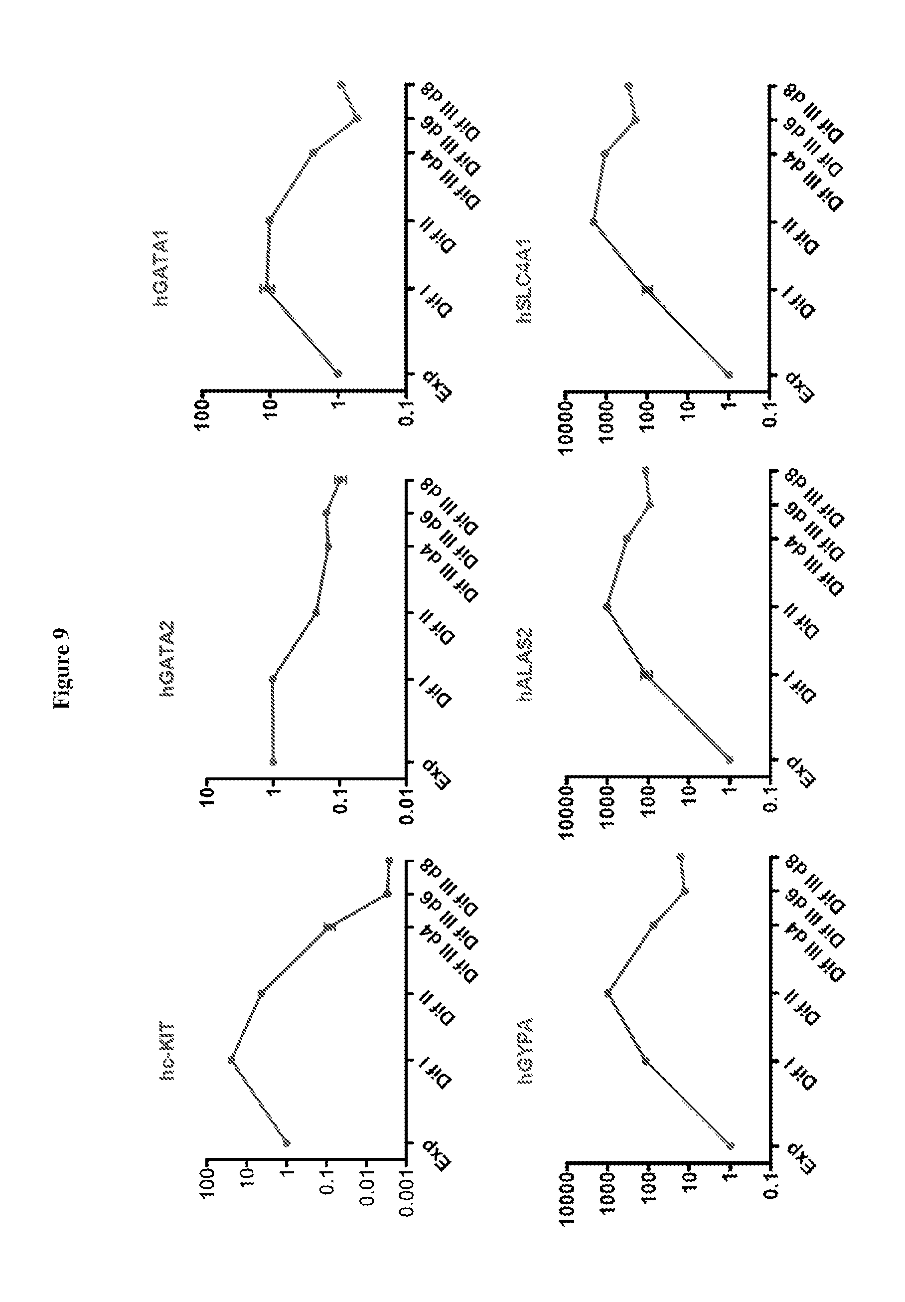
D00010
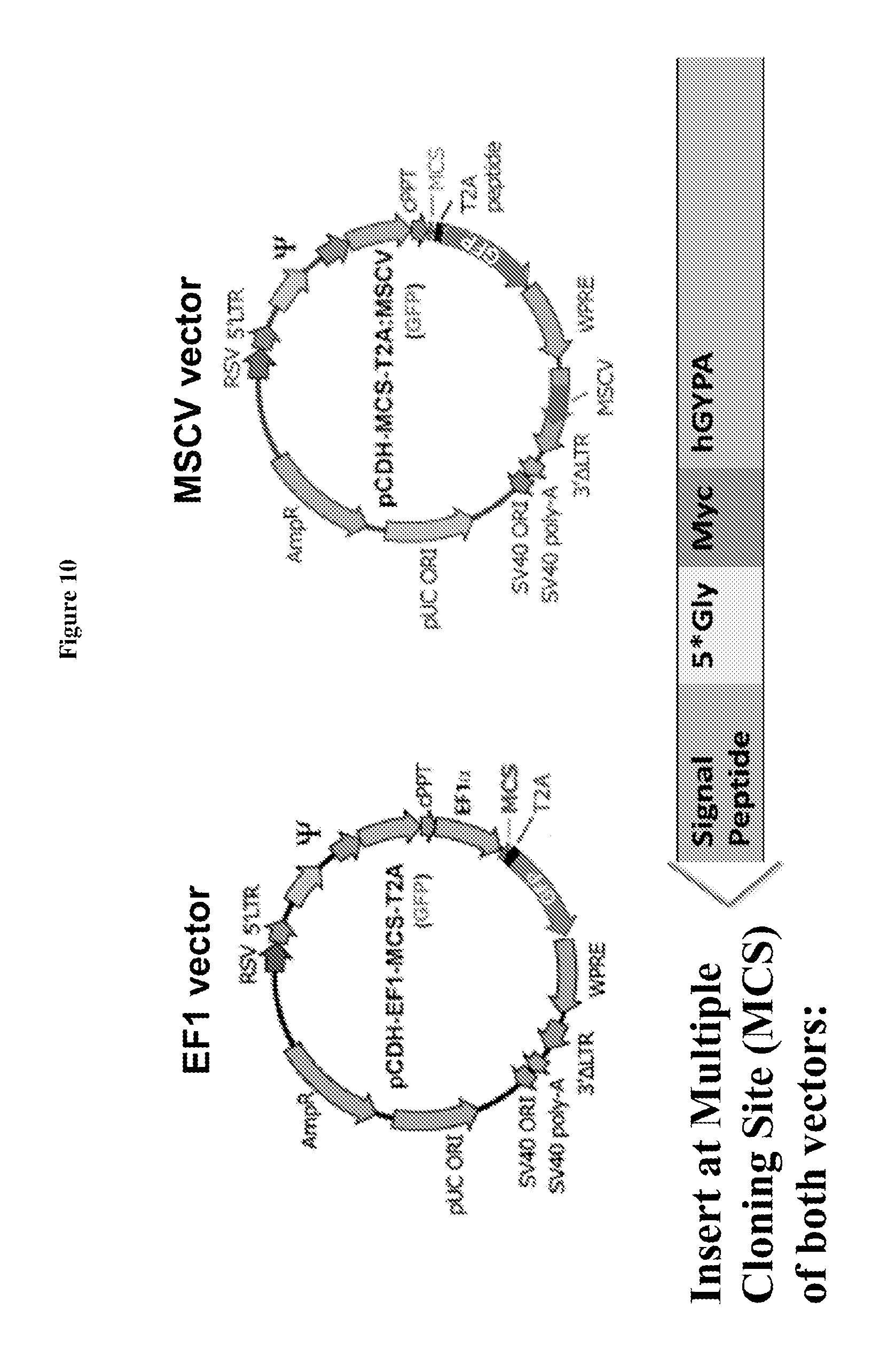
D00011

D00012
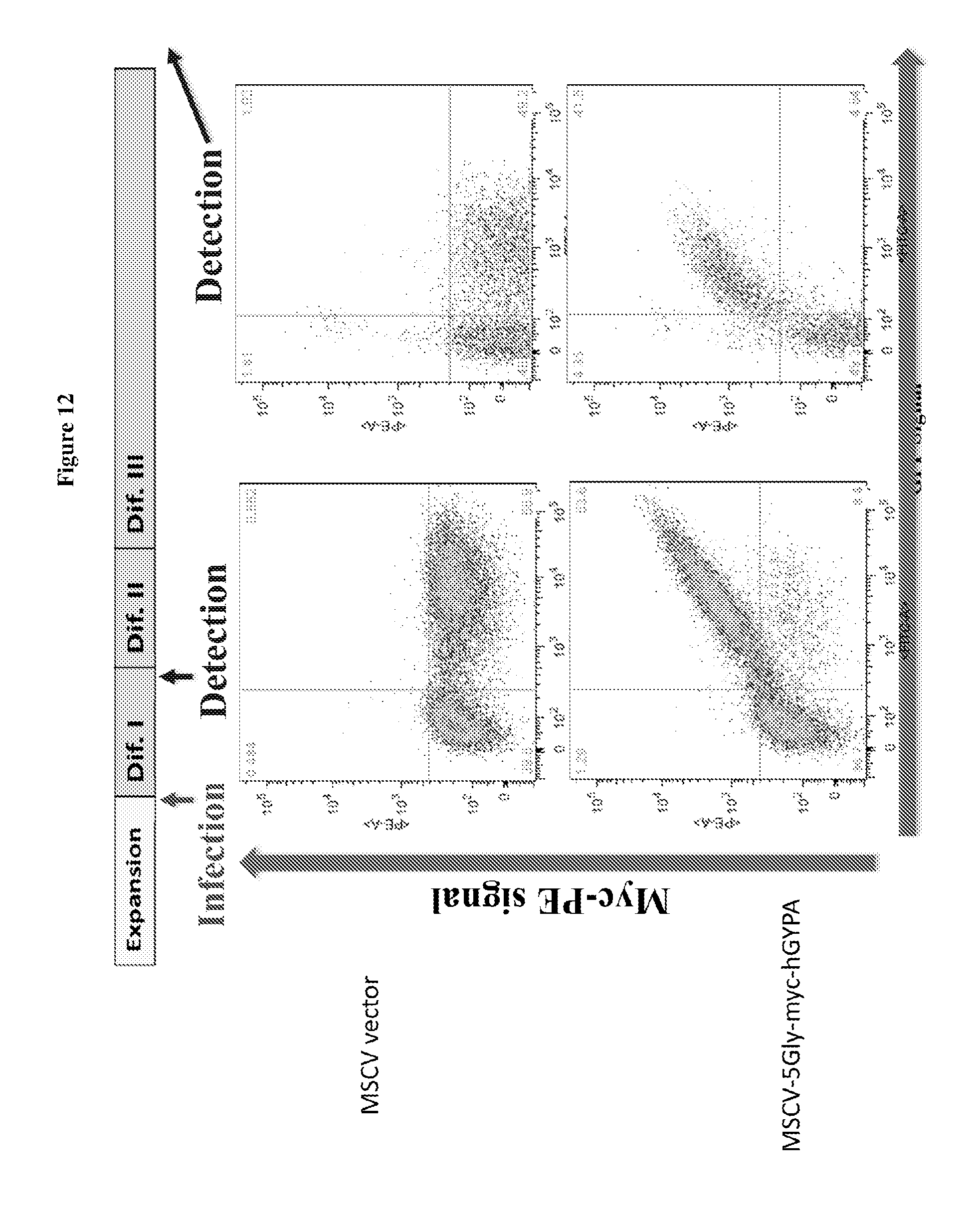
D00013
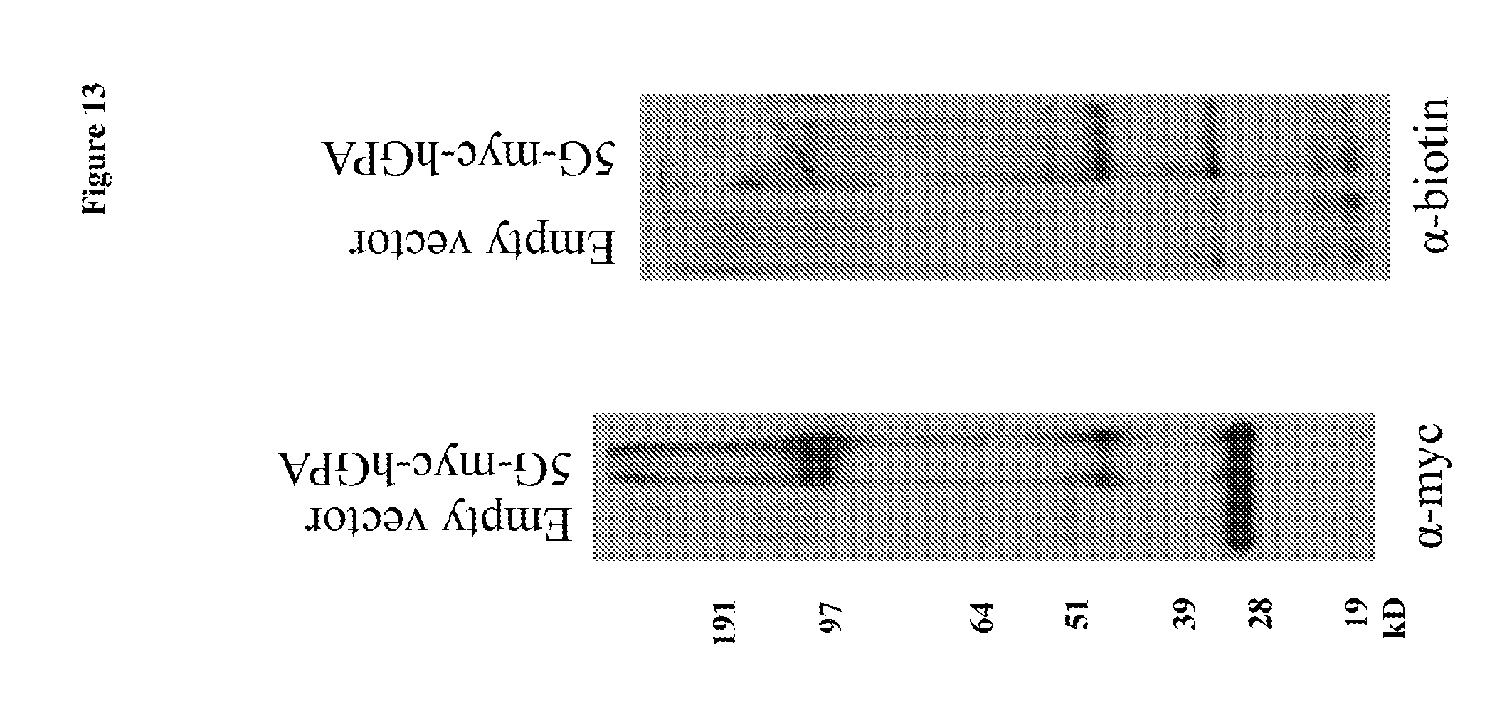
D00014

D00015
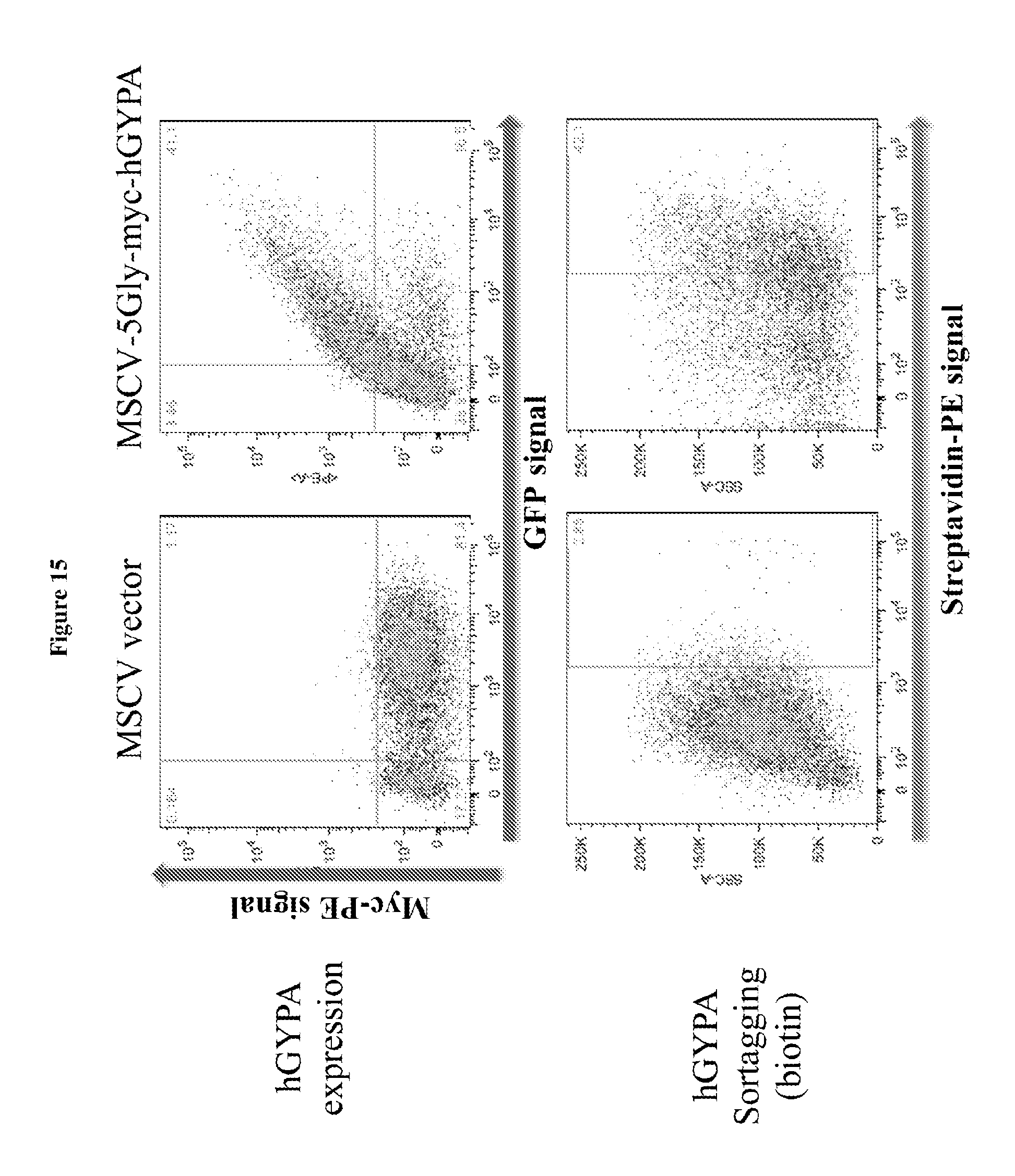
D00016
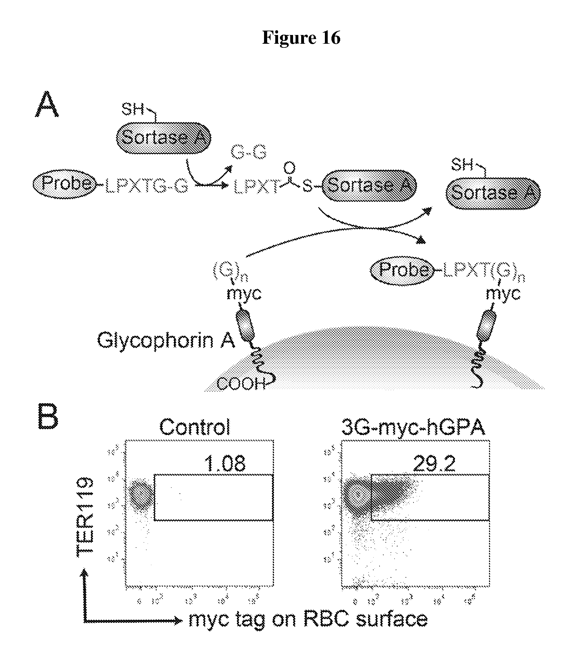
D00017
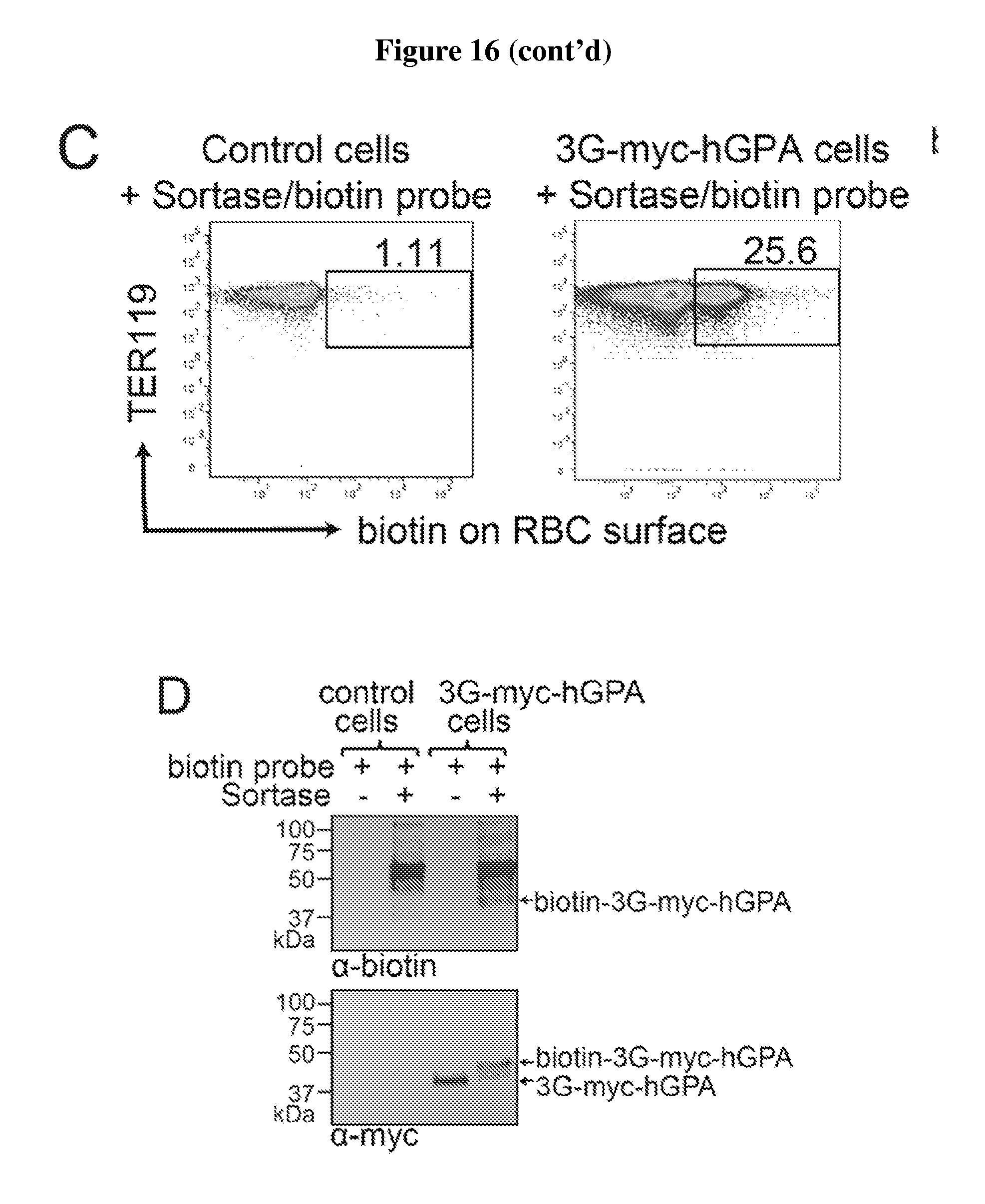
D00018
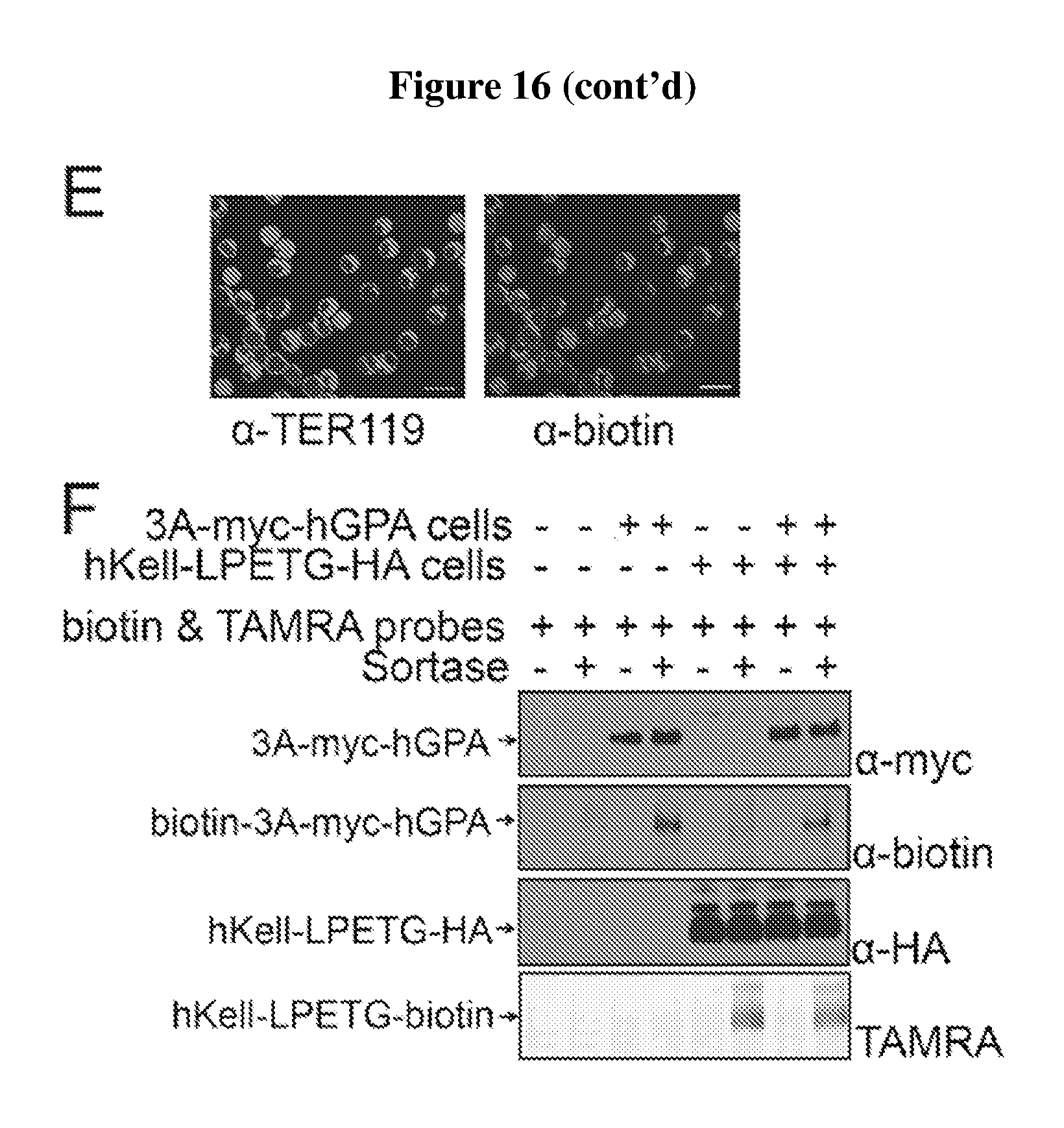
D00019

D00020
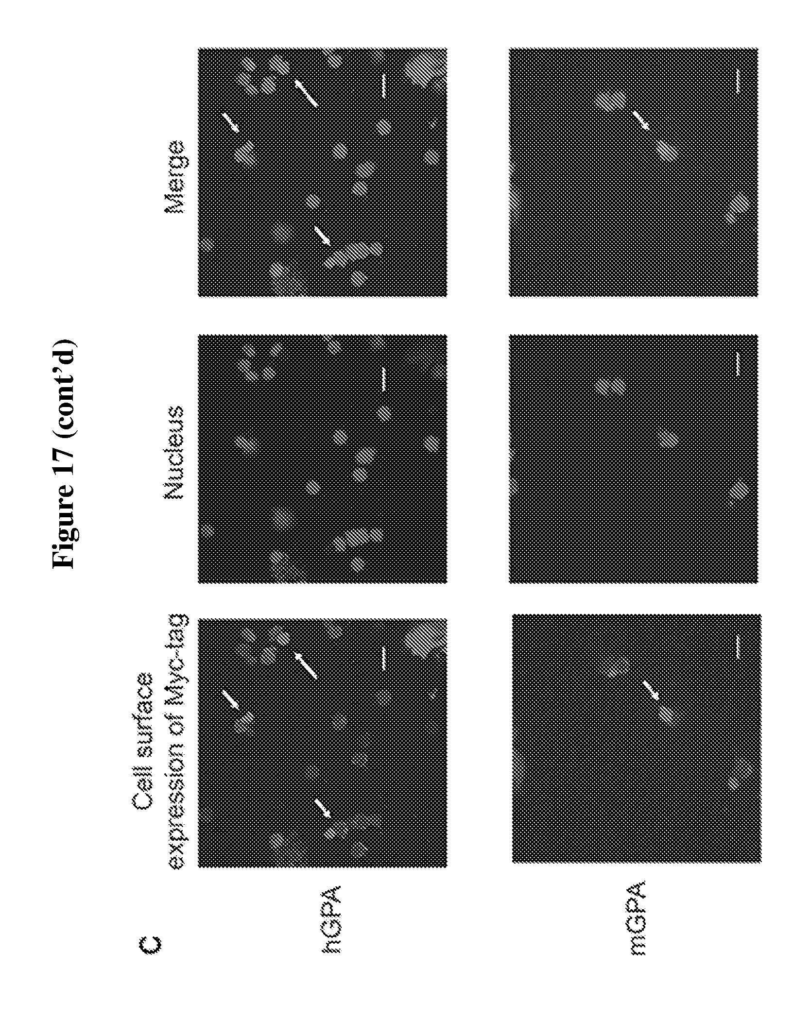
D00021
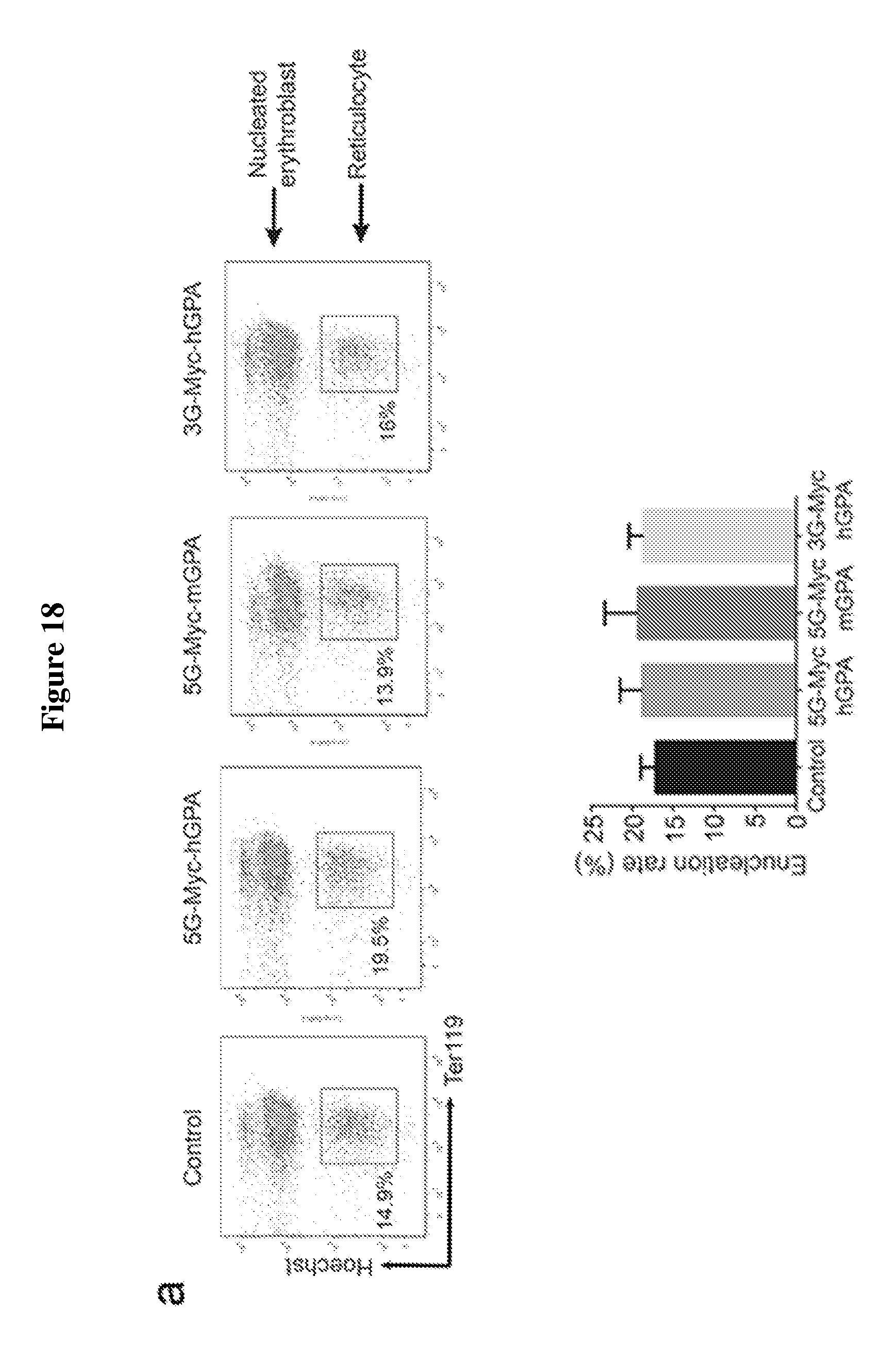
D00022
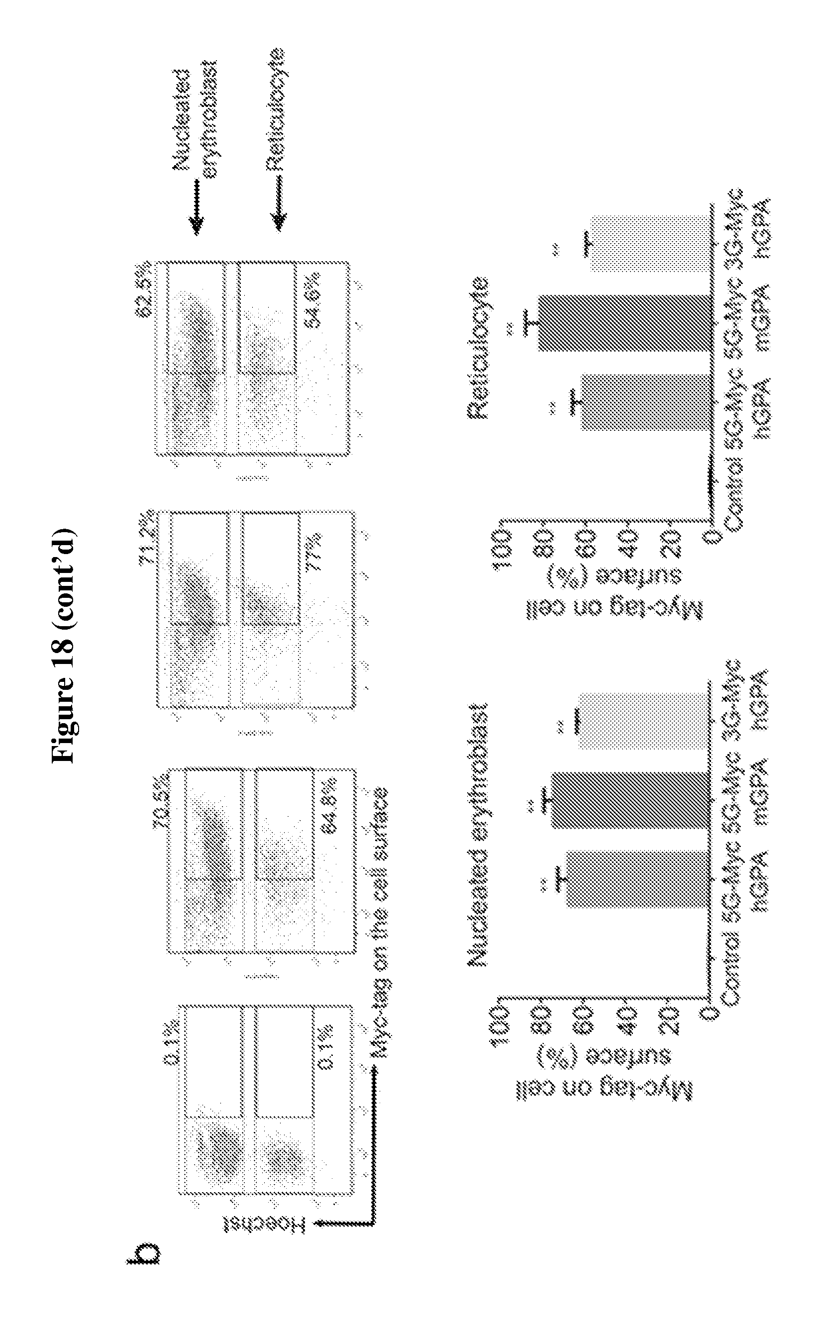
D00023
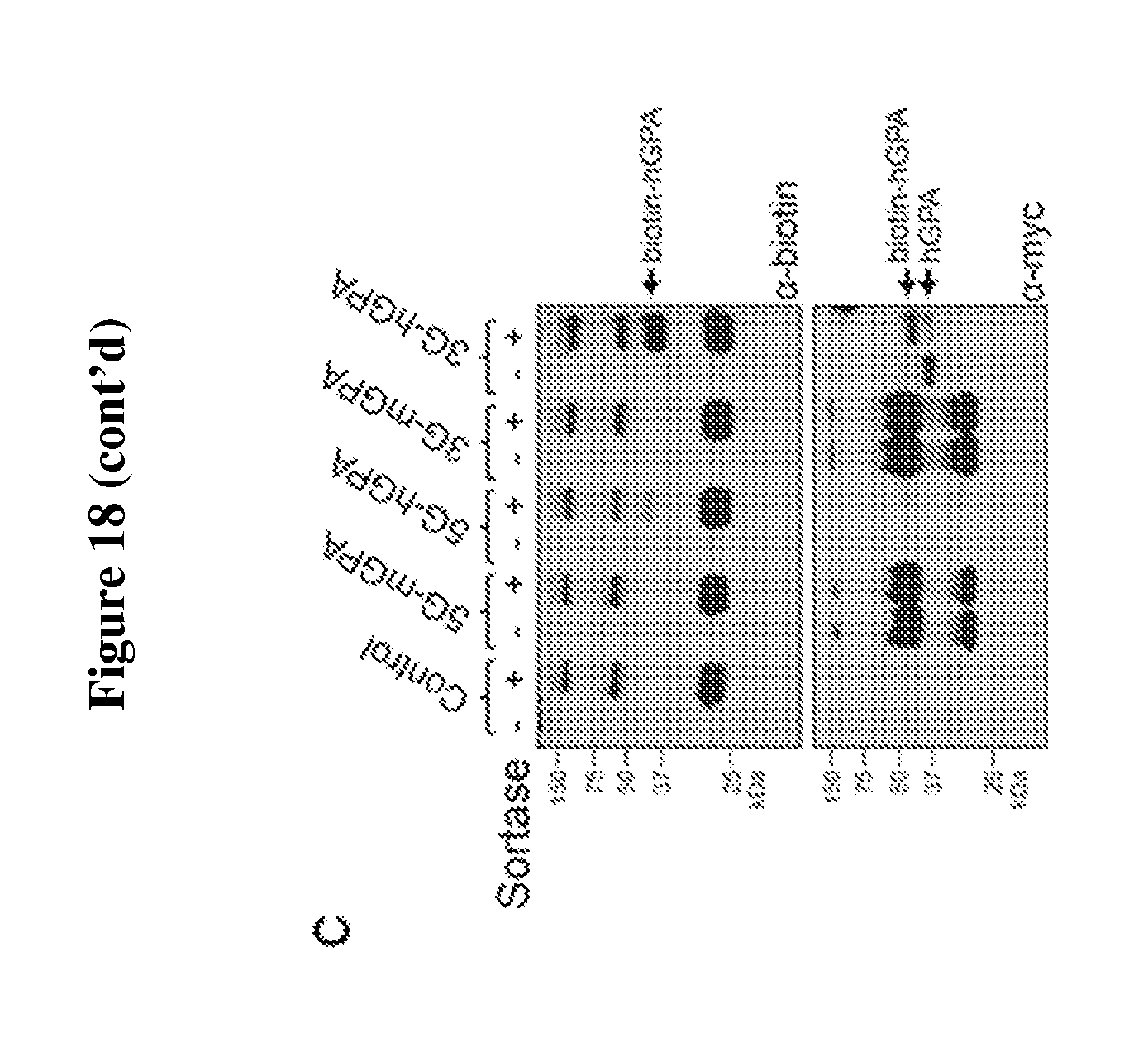
D00024
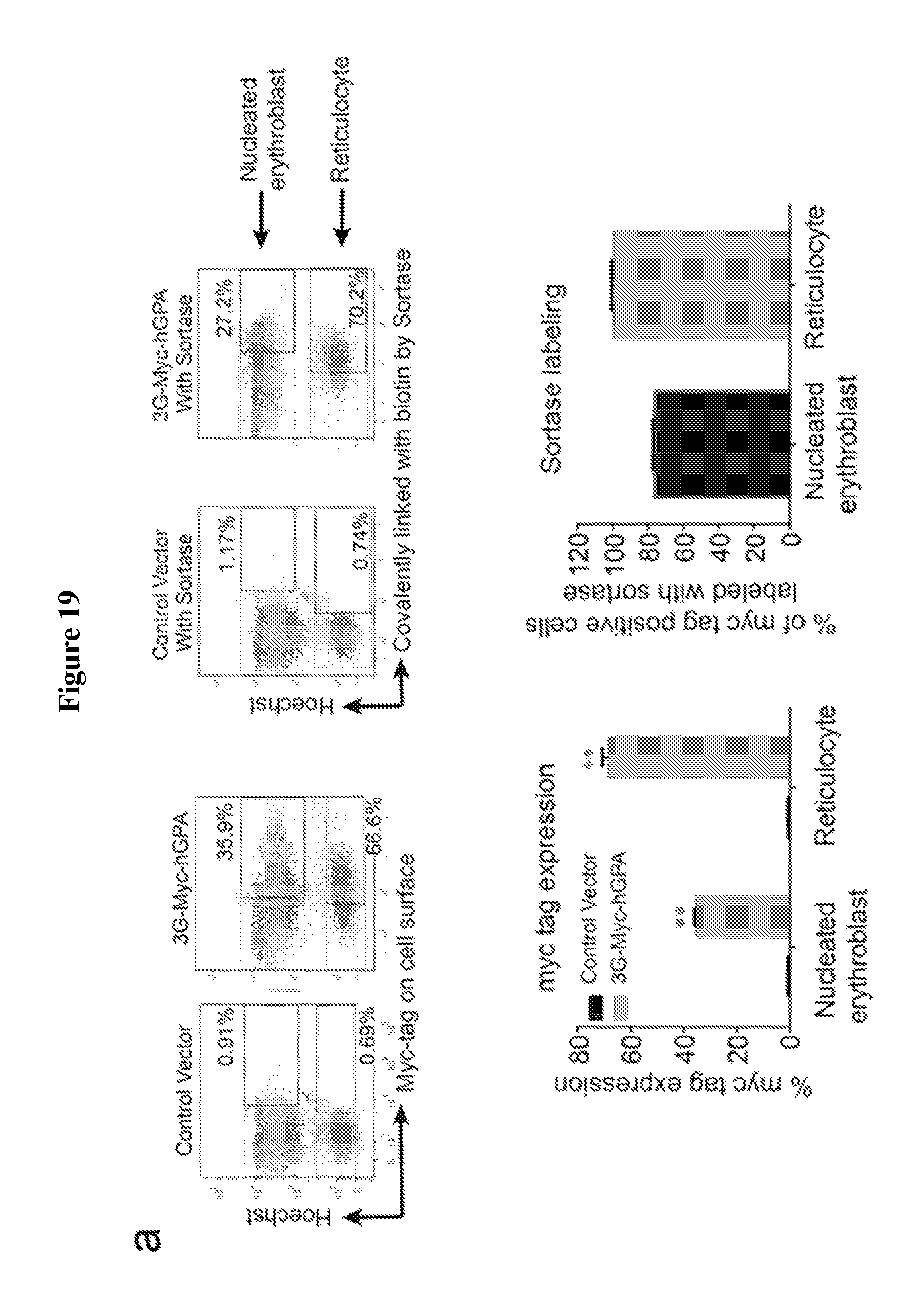
D00025
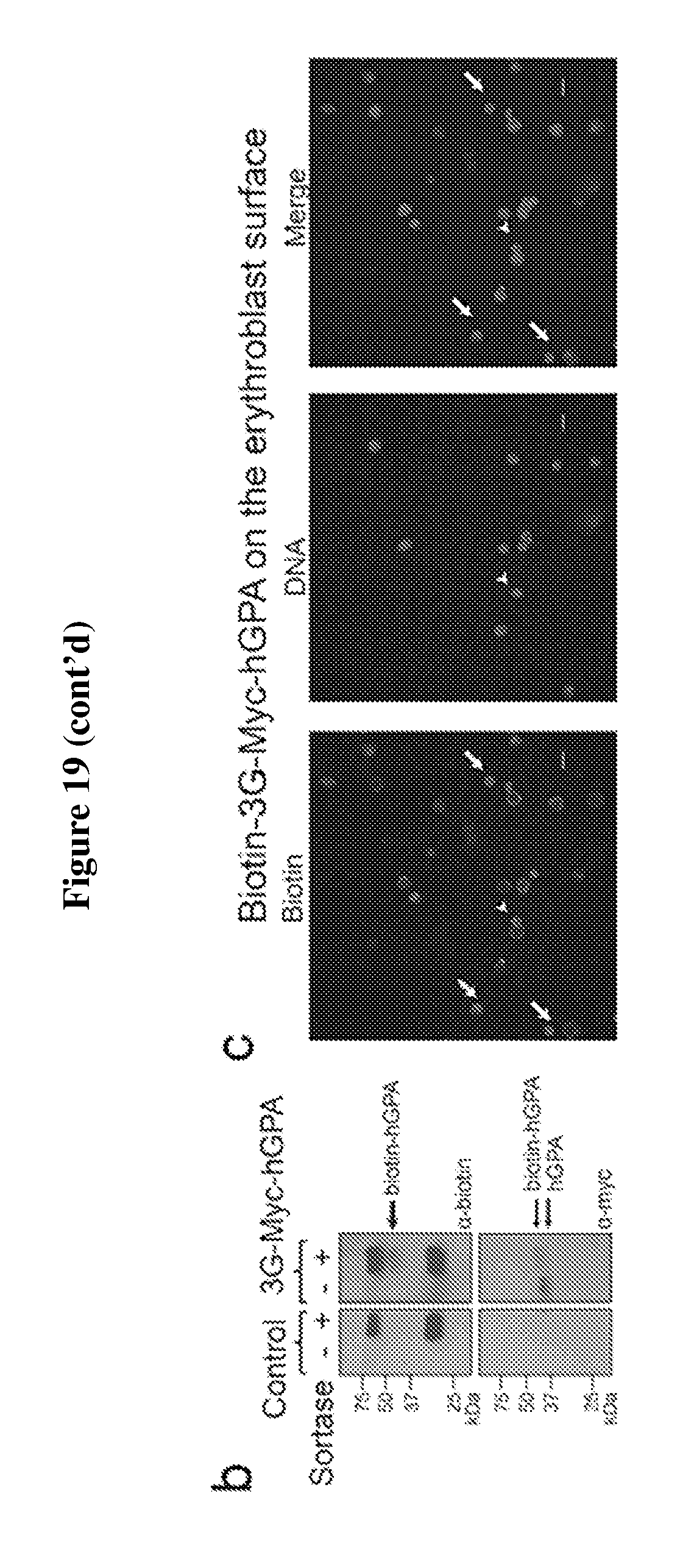
D00026

D00027
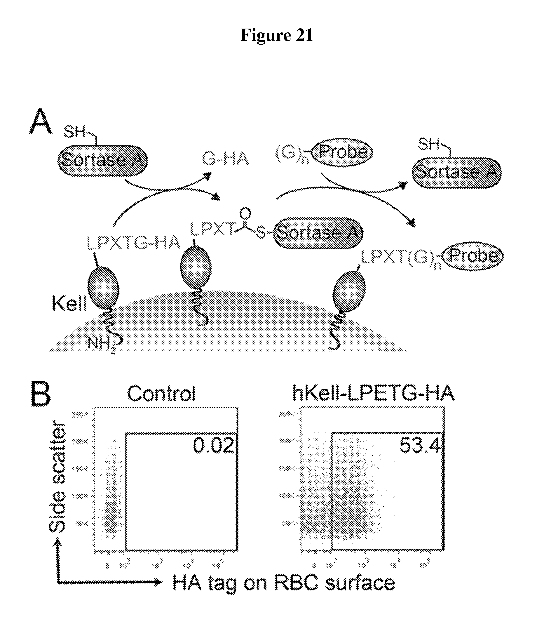
D00028
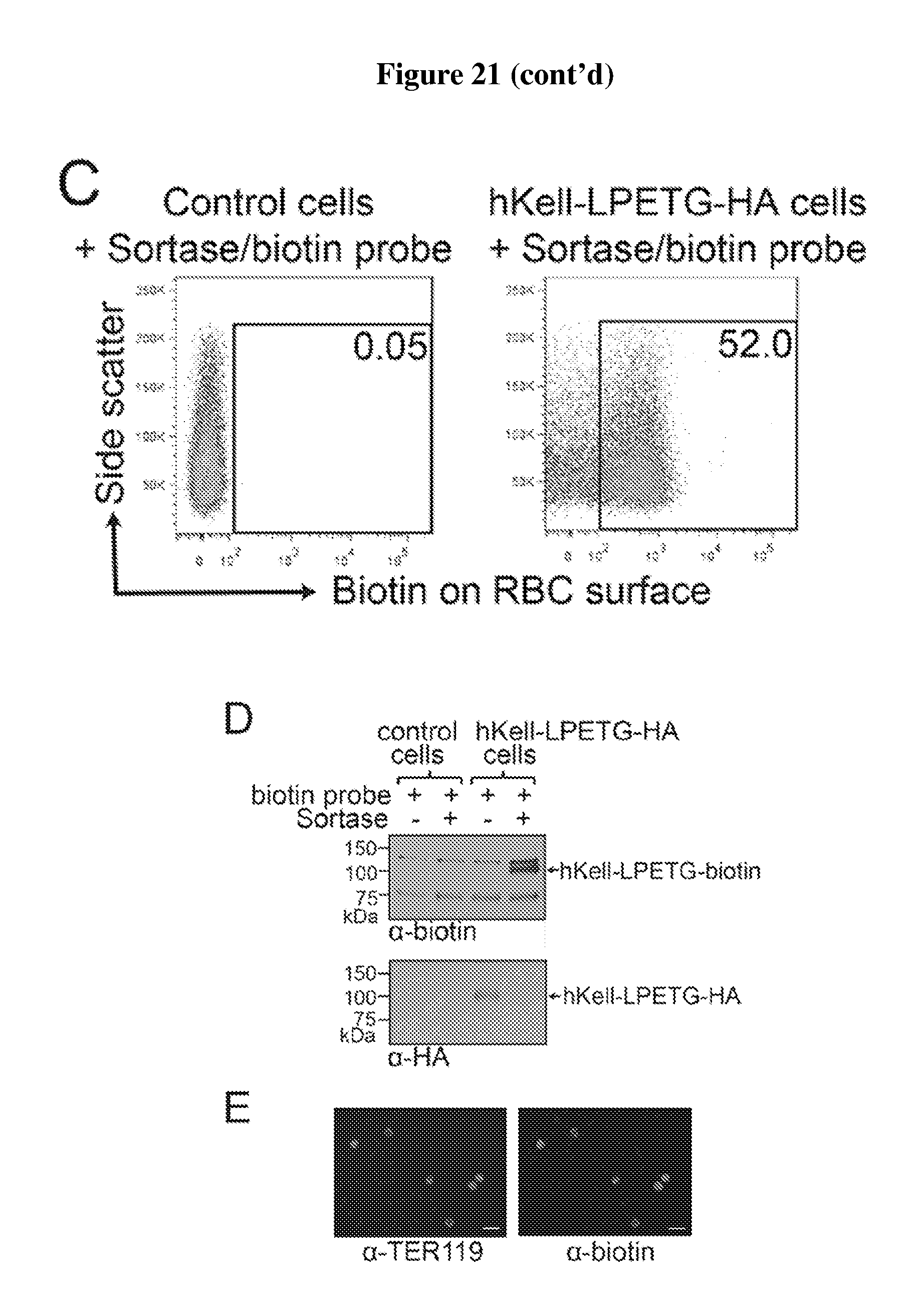
D00029
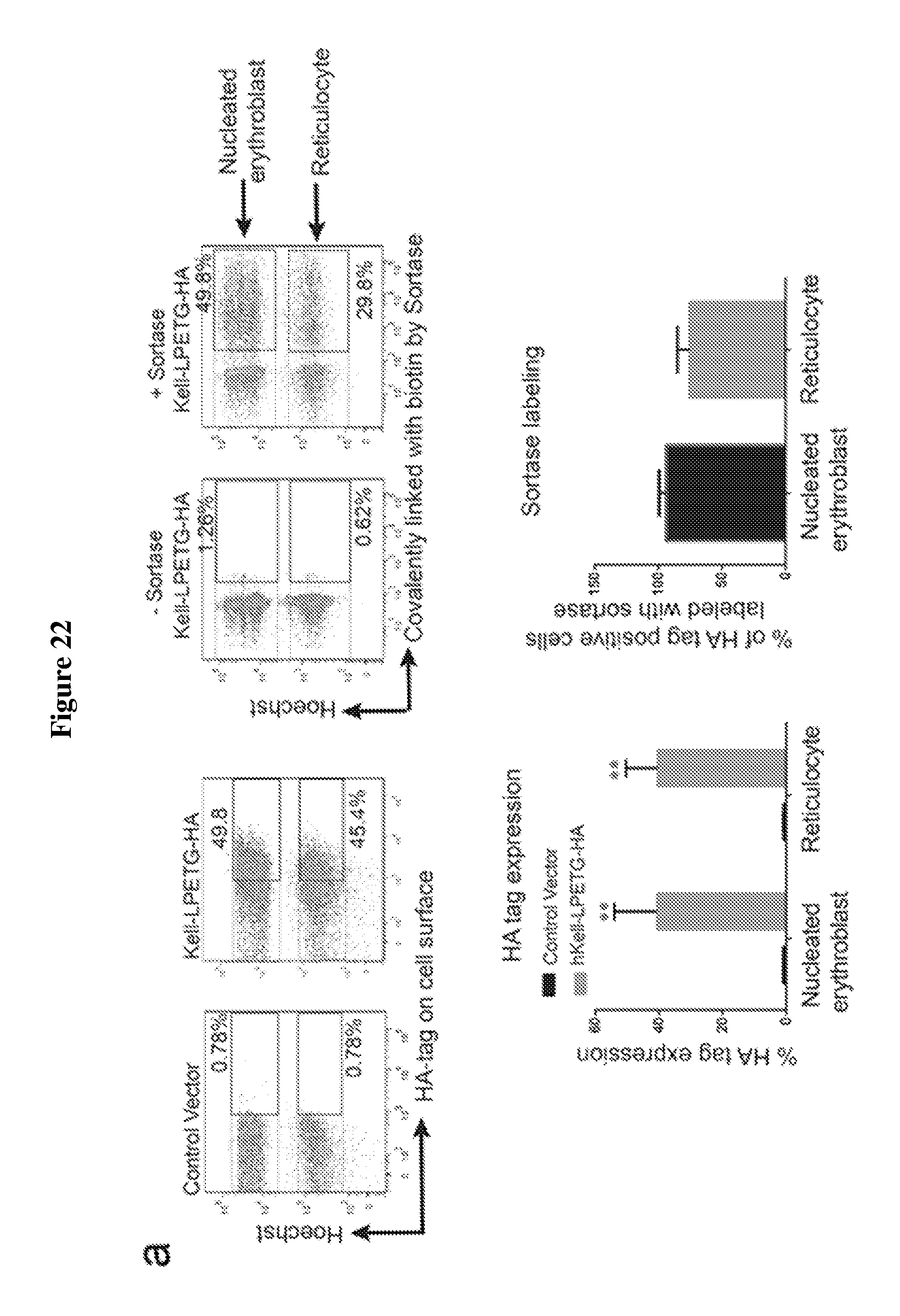
D00030
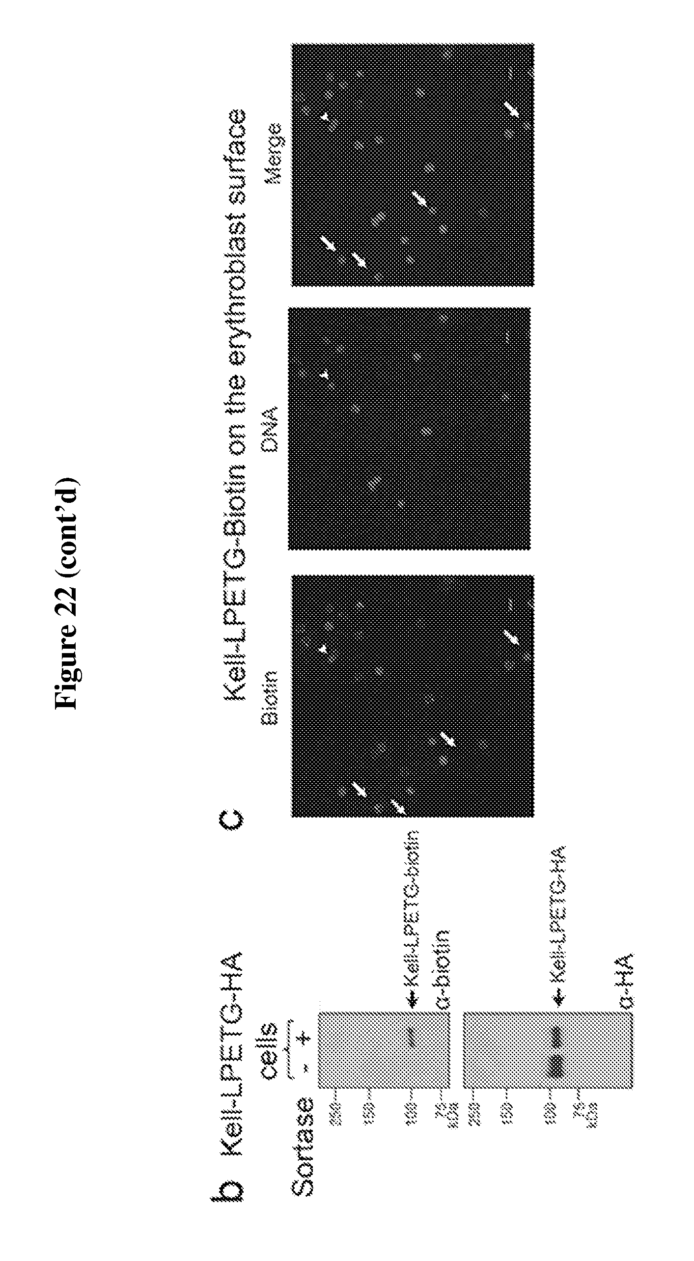
D00031
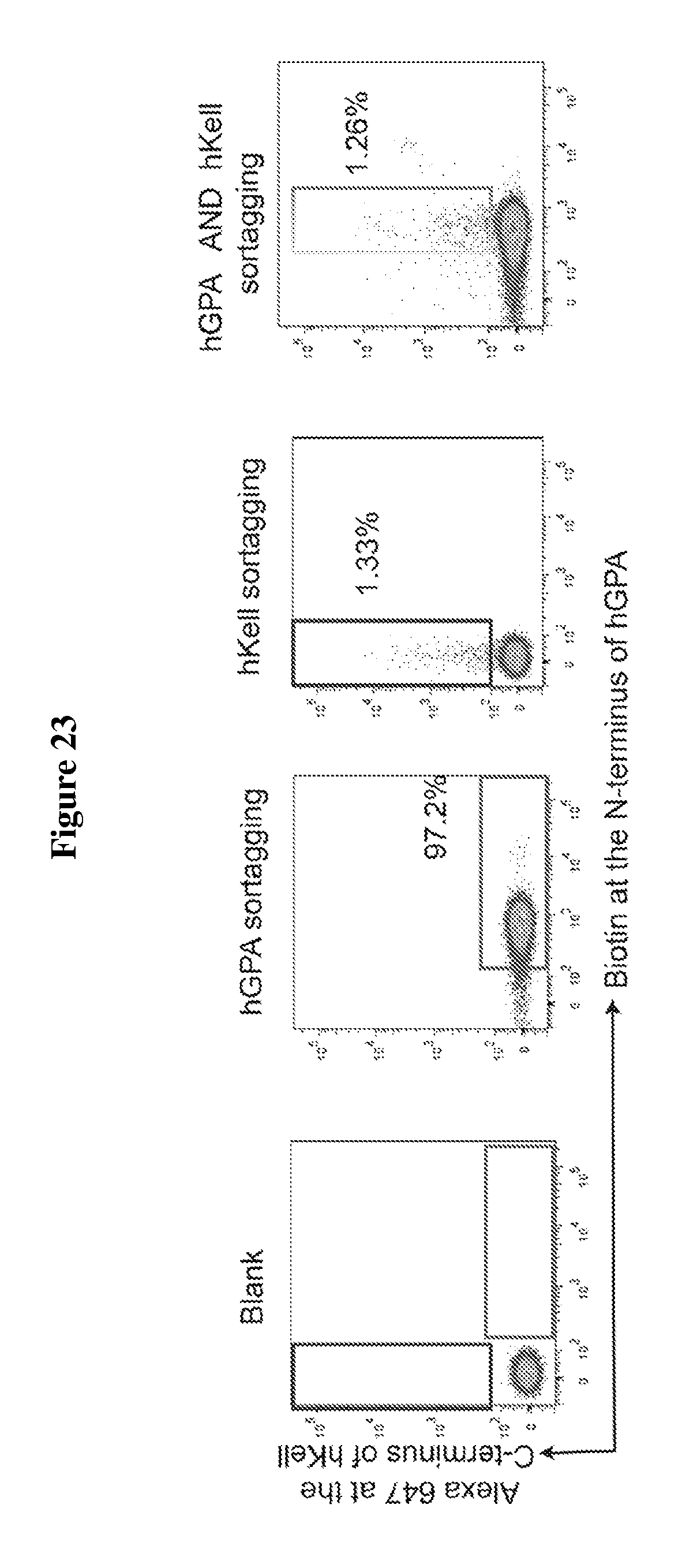
D00032
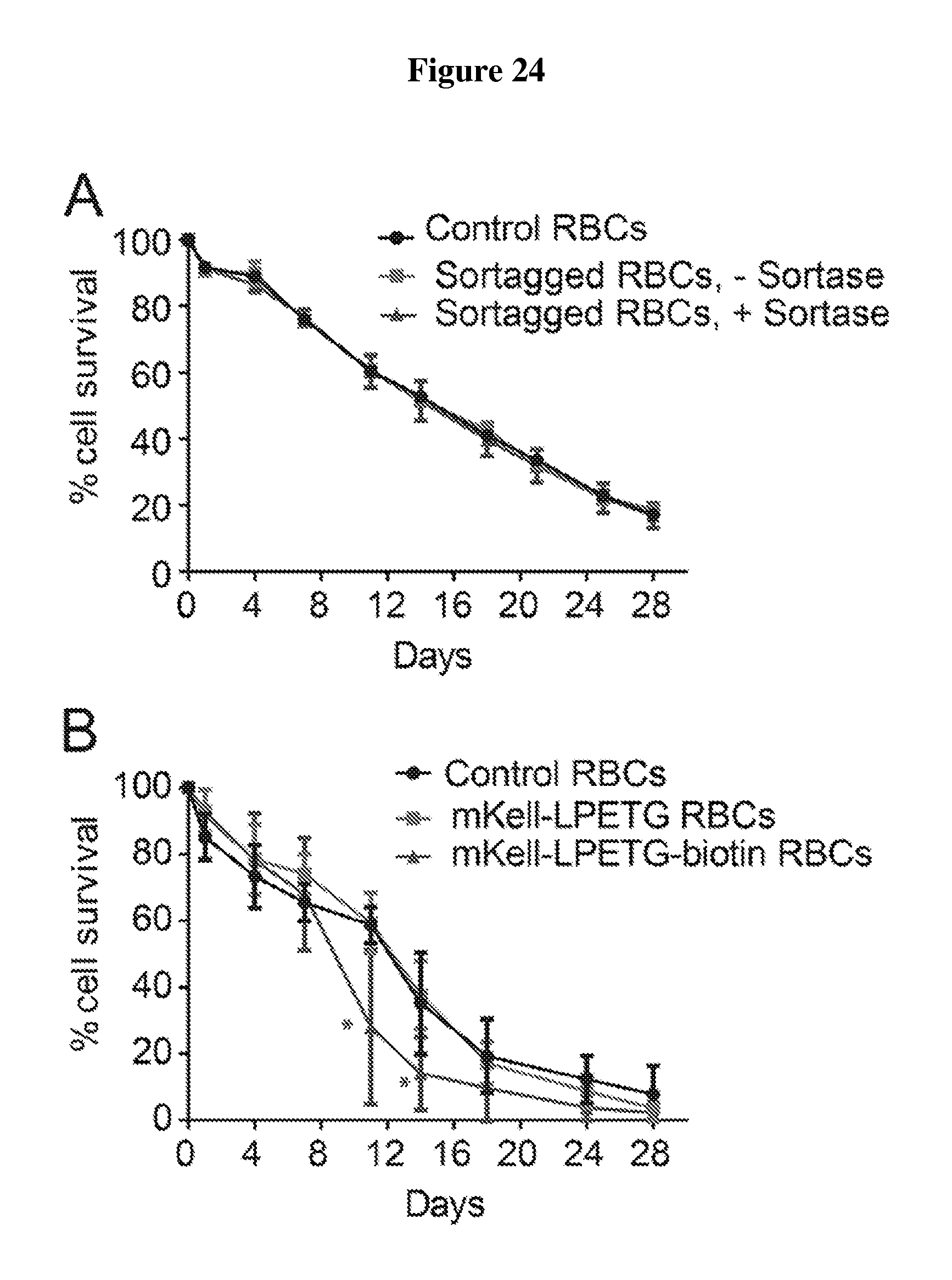
D00033
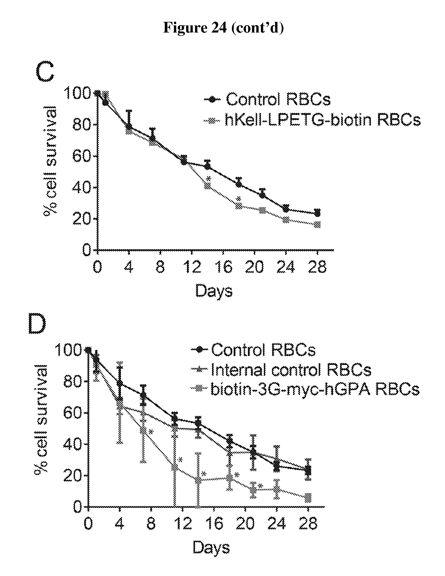
D00034
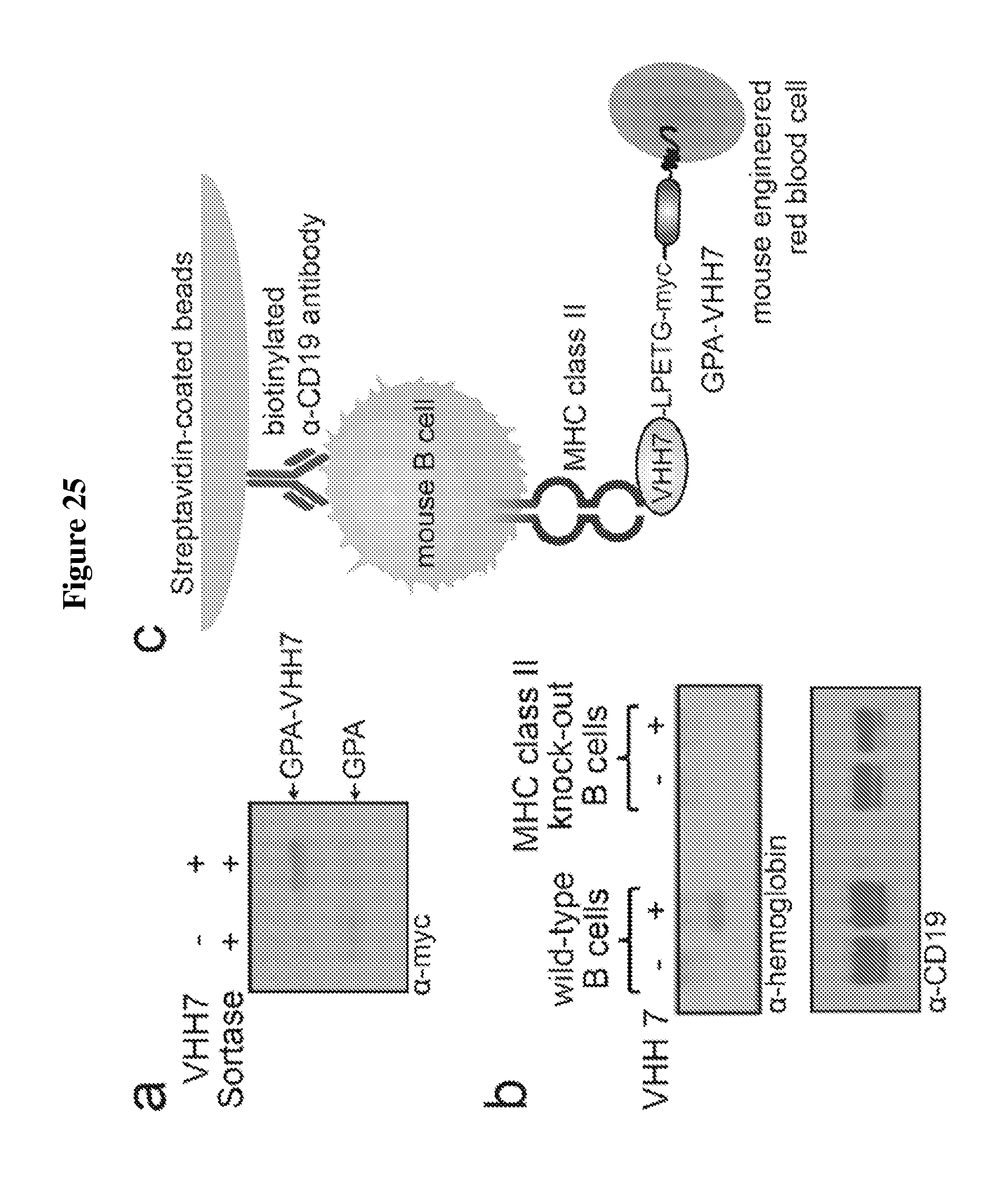
D00035
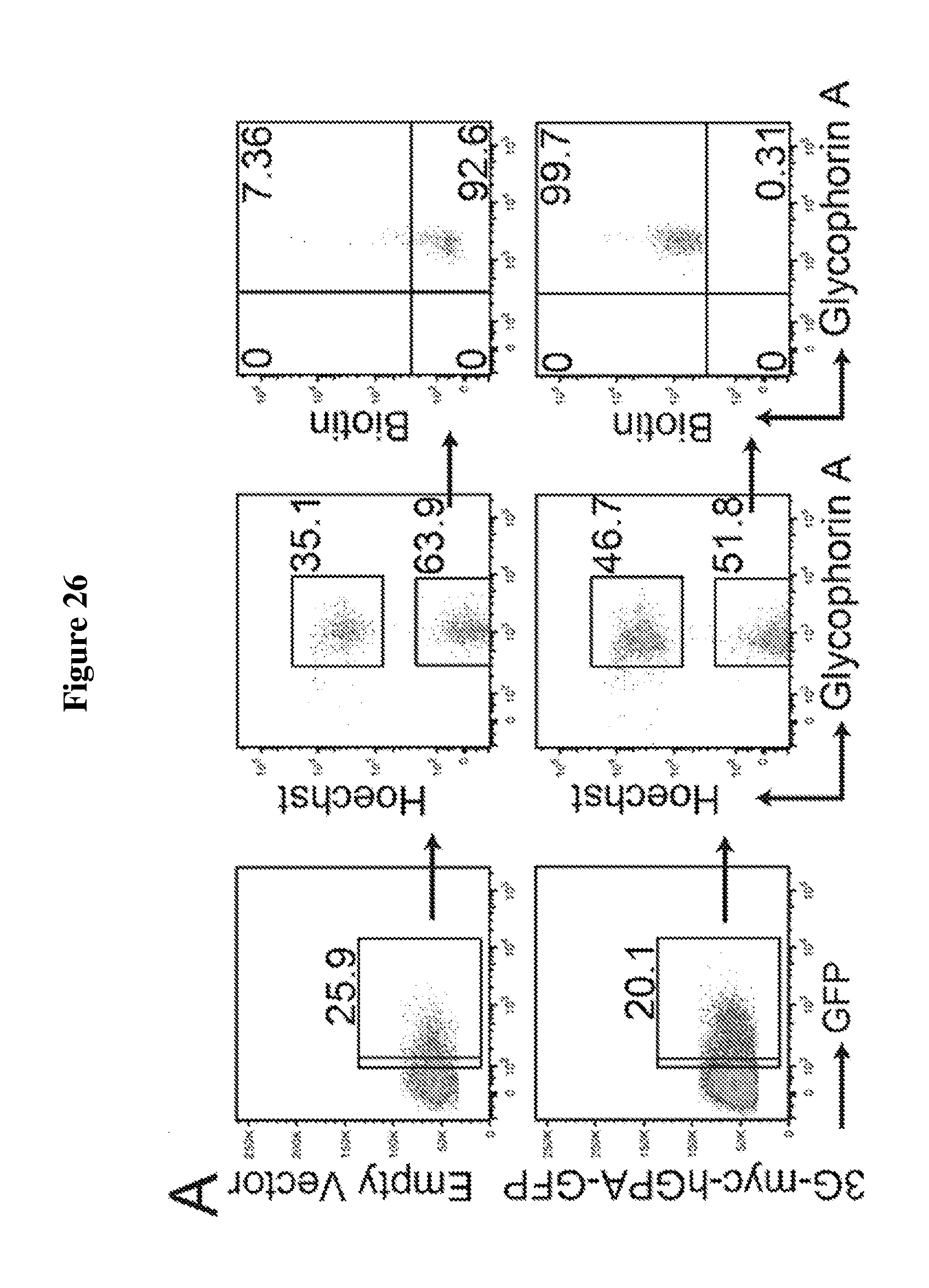
D00036

S00001
XML
uspto.report is an independent third-party trademark research tool that is not affiliated, endorsed, or sponsored by the United States Patent and Trademark Office (USPTO) or any other governmental organization. The information provided by uspto.report is based on publicly available data at the time of writing and is intended for informational purposes only.
While we strive to provide accurate and up-to-date information, we do not guarantee the accuracy, completeness, reliability, or suitability of the information displayed on this site. The use of this site is at your own risk. Any reliance you place on such information is therefore strictly at your own risk.
All official trademark data, including owner information, should be verified by visiting the official USPTO website at www.uspto.gov. This site is not intended to replace professional legal advice and should not be used as a substitute for consulting with a legal professional who is knowledgeable about trademark law.