Multimer of mutant protein A and methods of using same
Qian , et al. No
U.S. patent number 10,464,972 [Application Number 16/169,376] was granted by the patent office on 2019-11-05 for multimer of mutant protein a and methods of using same. This patent grant is currently assigned to Nanjingjinsirui Science & Technology Biology Corp.. The grantee listed for this patent is Nanjingjinsirui Science & Technology Biology Corp.. Invention is credited to Tao Bai, Rong Hua, Hong Qian.

| United States Patent | 10,464,972 |
| Qian , et al. | November 5, 2019 |
Multimer of mutant protein A and methods of using same
Abstract
A series of protein A mutants having high alkali resistance, and methods of using the protein A mutants are provided. The protein A mutants have a high binding affinity for regions of immunoglobulin proteins other than the complementarity determining regions. The protein A mutants can be coupled to a solid support for immunoglobulin isolation, or conjugated to a label for immunoglobulin detection. This series of protein A mutants have high chemical stability under alkaline conditions of pH 13-14, and can also be used as chromatography ligands for purification procedures that use alkaline solutions under harsh conditions, such as Clean-In-Place (CIP). Also provided are methods of immunoglobulin separation and purification, and alkali regeneration of affinity chromatography medium that uses protein A as a ligand.
| Inventors: | Qian; Hong (Nanjing, CN), Bai; Tao (Nanjing, CN), Hua; Rong (Nanjing, CN) | ||||||||||
|---|---|---|---|---|---|---|---|---|---|---|---|
| Applicant: |
|
||||||||||
| Assignee: | Nanjingjinsirui Science &
Technology Biology Corp. (Nanjing, Jiangsu, CN) |
||||||||||
| Family ID: | 51547091 | ||||||||||
| Appl. No.: | 16/169,376 | ||||||||||
| Filed: | October 24, 2018 |
Prior Publication Data
| Document Identifier | Publication Date | |
|---|---|---|
| US 20190048046 A1 | Feb 14, 2019 | |
Related U.S. Patent Documents
| Application Number | Filing Date | Patent Number | Issue Date | ||
|---|---|---|---|---|---|
| 14778395 | |||||
| PCT/CN2013/076445 | May 30, 2013 | ||||
Foreign Application Priority Data
| Mar 18, 2013 [CN] | 2013 1 0087284 | |||
| Current U.S. Class: | 1/1 |
| Current CPC Class: | C07K 16/00 (20130101); G01N 33/6854 (20130101); C07K 14/31 (20130101); C07K 1/22 (20130101); C07K 2319/21 (20130101); C07K 2319/705 (20130101) |
| Current International Class: | C12N 9/96 (20060101); C07K 1/22 (20060101); C07K 16/00 (20060101); G01N 33/68 (20060101); C07K 14/31 (20060101) |
References Cited [Referenced By]
U.S. Patent Documents
| 7709209 | May 2010 | Hober et al. |
| 8354510 | January 2013 | Hober et al. |
| 8357778 | January 2013 | Sato |
| 8772447 | July 2014 | Hall et al. |
| 2013/0096276 | April 2013 | Yoshida et al. |
| 2016/0237124 | August 2016 | Qian |
| 102516372 | Jun 2012 | CN | |||
| 102844432 | Dec 2012 | CN | |||
Other References
|
Gulich, S., et al. "Stability towards alkaline conditions can be engineered into a protein ligand", Journal of biotechnology, 80 (2000) 169-178. cited by applicant . Gulich, S. et al Protein engineering of an IgG-binding domain allows milder elution conditions during affinity chromatography. J. of Bio. 76 (2000) 233-244. cited by applicant . Hober, S. et al. "Protein A chromatography for antibody purification", Journal of Chromatography B, 848 (2007) 40-47. cited by applicant . Linhult, M. et al. "Affinity ligands for industrial protein purification," Protein and Peptide Letters, (2005), 12, 305-310. cited by applicant . Lund, L et al. "Exploring variation in binding of Protein A and Protein G to immunoglobulin type G by isothermal titration calorimetry," Journal of Molecular Recognition (2011), 24, 945-952. cited by applicant . Magdeldin, S. and Moser, A., "Affinity Chromatography: Principles and Applications", Intechopen, 2012. cited by applicant . Minakuchi, K. et al. "Remarkable alkaline stability of an engineered protein A as immunoglobulin affinity ligand: C domain having only one amino acid substitution" Protein Science, 22 (2013), 1230-1238. cited by applicant . Yu, F., et al. "Tailor-making a protein A-derived domain for efficient site-specific photocoupling to Fc of mouse IgG1" PLoS ONE, 8 (2013), e56597. cited by applicant . Heu, W., et al, "Protein binder for affinity purification of human immunoglobulin antibodies" Analytical Chemistry, 2014. cited by applicant . Yang, L., et al. "Effect of cleaning agents and additives on Protein A ligand degredation and chromatography performance", Journal of Chromatography A, (2015) 1385, 63-68. cited by applicant . Huang, B., et al. "Molecular mechanism of the affinity interactions between protein A and human immunoglobulin G1 revealed by molecular simulations", Journal of Physical Chemistry, (2011) 115, 4168-4176. cited by applicant . Huang B. et al. "Molecular mechanism of the effects of salt and pH on the affinity between protein A and human immunoglobulin G1 revealed by molecular simulations", Journal of Physical Chemistry, (2012) 116, 424-433. cited by applicant . Salvalaglio, M., "Molecular modeling of protein A affinity chromatography", Journal of Chromatography A, (2009) 1216, 8678-8686. cited by applicant . Linhult, M., et al. "Improving the tolerance of a protein A analogue to repeated alkaline exposures using a bypass mutagenesis approach", Proteins: Structure, Function, and bioinformatics, 55 (2004), 407-416. cited by applicant . Jiang, C., et al. "A mechanistic study of Protein A chromatography resin lifetime", Journal of Chromatography A, (2009) 1216, 5849-5855. cited by applicant . Xia, H., et al. "Molecular Modification of Protein A to Improve the Elution pH and Alaki Resistance in Affinity Chromatography", Applied Biochemistry and Biotechnology, 172 (2014), 4002-4012. cited by applicant . International Search Report dated Dec. 26, 2013 in corresponding PCT/CN2013/076445. cited by applicant . Hjerten, S., "The preparation of agarose spheres for chomatography of molecules and particles", Biochimica et Biophysica Acta, 79 (1964) 393-398. (Abstract Only). cited by applicant . Brown, N.L. et al., "A study of the interactions between an IgG-binding domain based on the B domain of staphylococcal protein A and rabbit IgG" Molecular Biotechnology, 10 (1998), 9-16. (Abstract Only). cited by applicant . Written Opinion dated Dec. 26, 2013 in PCT/CN2013/076445. cited by applicant. |
Primary Examiner: Robinson; Hope A
Attorney, Agent or Firm: Panitch Schwarze Belisario & Nadel LLP
Parent Case Text
CROSS-REFERENCE TO RELATED APPLICATION
This application is a divisional application of U.S. patent application Ser. No. 14/778,395, filed Sep. 18, 2015, now allowed, which is a Section 371 of International Application No. PCT/CN2013/076445, filed May 30, 2013, which was published in the Chinese language on Sep. 25, 2014 under International Publication No. WO 2014/146350 A1, the disclosures of which are herein incorporated by reference in their entirety.
Claims
The invention claimed is:
1. A multimer of a recombinantly expressed mutant protein A molecule comprising at least two recombinant expressed mutant protein A molecules for isolating, purifying, or detecting an immunoglobulin, the mutant protein A molecule comprising the amino acid sequence of SEQ ID NO:1, the amino acid sequence of SEQ ID NO:2, the amino acid sequence of residues 7 to 54 of SEQ ID NO:1, or the amino acid sequence of residues 7 to 54 of SEQ ID NO:2.
2. The multimer of claim 1, wherein the mutant protein A molecule is stable under alkaline conditions of pH 13-14.
3. The multimer of claim 1, wherein the at least two recombinantly expressed mutant protein A molecules comprise the same amino acid sequence.
4. The multimer of claim 3, wherein the multimer is a dimer, trimer, or tetramer comprising two, three, or four protein A mutants, respectively.
5. The multimer of claim 3, wherein the one or more protein A mutants are linked together via one or more linker units comprising 4 to 10 amino acids.
6. The multimer of claim 5, wherein the linker unit comprises the amino acid sequence ADGK (SEQ ID NO: 56).
7. The multimer of claim 3, further comprising six histidine residues fused to the N-terminal end of the multimer.
8. The multimer of claim 7, comprising the amino acid sequence of SEQ ID NO: 10 or SEQ ID NO: 12.
Description
REFERENCE TO SEQUENCE LISTING SUBMITTED ELECTRONICALLY
This application contains a sequence listing, which is submitted electronically via EFS-Web as an ASCII formatted sequence listing with a file name "688096-79U1 Sequence Listing", creation date of Nov. 9, 2015, and having a size of 17 kb. The sequence listing submitted via EFS-Web is part of the specification and is herein incorporated by reference in its entirety.
FIELD OF THE INVENTION
The invention belongs to the field of protein engineering, relates to a series of Staphylococcal protein A mutants with high alkali resistance and their application.
BACKGROUND OF THE INVENTION
Biotechnology is one of the fastest growing high-tech fields in the world. As one of the hot spots, antibody drug development continues to improve the health and the life quality of many patients and has achieved remarkable market performance in recent years. Although investments in research and development of new drugs have been increased continuously, the number of innovative drugs that reach the market has dramatically decreased, and many small molecule drugs face the threat of the patent cliff. Therefore, in order to seek new growing points, many pharmaceutical companies are entering the field of antibody drug development. Currently, antibody drugs are widely used in basic biomedical research, diagnosis and treatment of diseases such as cancers, organ transplant rejection and autoimmune diseases.
In recent years, as a large number of therapeutic antibody drugs have been invented and used in the medical field, the production process draws a lot of attention from people. In general, large-scaled economic production of antibody drugs and diagnostic reagents are produced by cell culture at the intracellular level or secreted into the culture medium. The cell culturing process requires a culture medium supplemented with sugars, amino acids, and certain growth factors.
Therefore antibody must be isolated from culture medium as well as other cellular components and purified to a certain level before it can be used as a human therapeutic agent. Currently the most widely used method for antibody purification is affinity chromatography which is simple, fast and highly selective. With those advantages, affinity chromatography significantly reduces the subsequent steps of purification to save time and cost with no sacrifice of purity. Cost control is very important in the modern industrial production processes. If chromatography medium could be used repeatedly, it would significantly reduce the production cost of an antibody. However, previously used chromatography medium may retain un-eluted proteins, protein aggregates, or residual substances that could be harmful, such as viruses or endotoxins. So it is very critical that the previously used media must be cleaned before reusing. The most effective way to clean chromatography medium is Clean-In-Place (CIP) using alkaline solutions. The method involves a treatment with a buffer or solution containing 0.5M Sodium Hydroxide (NaOH). Using this rigorous clean method, impurities can be effectively removed from the medium, however, it may also damage the chromatography medium itself. Therefore, Protein A molecules with high alkali resistance which bind immunoglobulin as described in the present invention can be used as effective ligands for the purification of antibodies. Importantly, the chromatography medium can be treated by CIP with alkali solution and regenerated for repetitive use.
BRIEF SUMMARY OF THE INVENTION
This invention aims to address the disadvantages of existing technology and provide a specific series of mutations to the Protein A molecule which enable it high alkali resistance.
Another objective of the present invention is to provide applications of this protein A molecule.
The objective of this invention can be achieved by the following technical solutions:
A series of Protein A mutants of which the amino acid sequences are defined by SEQ ID NO:1 or SEQ ID NO:2, or defined by the amino acid sequences within immunoglobulin binding region that have more than 99% homology to the sequences defined by SEQ ID NO:1 or SEQ ID NO:2. Here immunoglobulin binding region is defined from the 7th amino acid residue to the 54th. of SEQ ID NO: 1 or SEQ ID NO:2.
Nucleic Acids Encoding the Protein A.
The present invention relates to protein A which is essentially a series of immunoglobulin-binding molecules which can tightly bind with immunoglobulin at regions other than the complementarity determining region, and at the meantime can withstand an alkali condition of pH13-14. Such an immunoglobulin binding protein A is composed of three .alpha. helical regions, which fold into a three-helix bundle structure. Protein A binds to the Fc part of an immunoglobulin through the residues on the helix 1 and 2 surface. Also Protein A has lower affinity toward the Fab part of an immunoglobulin, through the residues on the surface of helix 2 and 3. The residues in the center of the three-helix bundle form an hydrophobic core, of which the hydrophobic interactions could enable the high thermal stability of protein A.
Different from other types of non-alkali resistant Protein A, when the alkali resistant protein A species are placed in an alkaline environment of pH13-14, even though the hydrogen bonds which help to maintain the protein tertiary structure disappeared and all three .alpha. helixes would be stretched, the strong hydrophobic interactions among three helixes would still enforce .alpha. helix 1, 2 3 to stay closely to each other and to ensure their relative positions do not change significantly. Once the pH are brought back to a neutral environment, such as pH7-8, hydrogen bonds among .alpha. helix 1, 2, 3 would be recovered, then the overall conformation or tertiary structure of the tri-helix bundle could be restored to the one before alkali treatment. While other non-alkali resistant proteins are not able to restore their tertiary structures due to the lacking of this kind of hydrophobic core to maintain their structure. After being placed in an alkaline environment, like pH13-14, the structure of non-alkali resistant proteins would be completely destroyed, so when they are put back into a neutral environment like pH 7-8, the overall conformation or tertiary structure of the protein could not be restored to the one before alkali treatment. With this alkali resistance characteristics, monomeric Protein A or multimeric protein A coupled to a solid phase support which can be used for the isolation and purification purpose of an immunoglobulin molecule, or they can be also used for detection of immunoglobulin molecules by conjugating a marker or label. If these alkali resistant protein A are used as ligands for chromatography medium, the chromatography medium can be cleaned and regenerated by Clean-In-Place (CIP) with alkaline solutions.
As described in the invention, the molecular weight of the protein A monomer is around 6KD, composed of 58 amino acids, which can be chemically synthesized or recombinantly expressed. If recombinantly expressed, cell strains must be constructed firstly to carry a gene that encodes the amino acid sequence of the protein. Therefore, the following steps of procedure are required: first, obtain the protein A gene encoding the amino acid sequence by gene synthesis; second, insert the protein A gene into an expression vector. Here tags for purification or labels for identification can be inserted into the expression vectors as needed. And then transform the vector into a suitable cell line for further expression. By doing this, protein A (including monomeric or polymeric) which have immunoglobulin molecule binding affinity could be obtained from culture medium or cells. It should be noticed that the expression of protein A needs to be tightly controlled. So selecting vectors for the expression of protein A is very important, and those vectors should have the following features: 1) an expression vector should have a promoter or transcription initiation site, 2) an expression vector should have the gene operons for expression switching, 3) an expression vector should have a ribosome binding site, and 4) an expression vector should have transcription and translation termination sites, all of which can improve stability of the transcription and translation products. The expression vectors recommended for protein A expression are pET (Novagen), pQE30 (Qiagen), PGS21a (genscript), pGAPZa A (Invitrogen), etc., in corresponding, the host cells which could be used for protein A expression are genetically engineered Escherichia coli (E. coli) or Pichia pastoris yeast strains. The host cells can express protein A on membranes or extracellularly.
According to different needs, those Protein A and/or their mutants could be isolated and purified using their physical and/or chemical properties. Usually those isolation and purification methods are protein precipitation (salting out), centrifugation, osmotic pressure shock, sonication, ultrafiltration, gel filtration, adsorption chromatography, ion exchange chromatography, affinity chromatography, high performance liquid chromatography or other liquid chromatography, dialysis etc., and the combination of these methods. Furthermore, target proteins fused with an affinity tag can be isolated and purified by affinity purification. Affinity tags described herein are, e.g., poly-histidine tag and FLAG.RTM. tag, which could be used for purification of protein A.
The binding affinity of protein A toward immunoglobulin can be measured by ELISA assay. For example, Protein A was immobilized on a solid phase carrier (here for example: a plate), free Protein A which was not immobilized on the carrier was washed out. And then an enzyme-labeled immunoglobulin was added and incubated for a certain time to allow efficient Protein A binding. By washing the solid phase carrier, free immunoglobulin which did not bind with immobilized protein A and other substances in liquid phase were removed from immunoglobulin-Protein A complexes which were immobilized on the solid phase carrier. Usually, the ratio of the amount of the enzyme-labeled immunoglobulin to the amount of immobilized Protein A in such a complex is 1:1. After adding chromogenic substrate, the substrate was catalyzed by the enzyme which bound to the immunoglobulin into colored product, and then the amount of immunoglobulin which bound to Protein A could be qualified and quantified by analyzing depth of coloration.
The alkali resistance and the chemical stability of these proteins can be easily confirmed by a person with technical expertise in this field. For example, proteins having been soaked in 0.5M NaOH for at least 60 hours are measured for immunoglobulin binding activity according to the ELISA assay described above. If the proteins having been treated by alkaline solutions can still maintain good binding activity, then these proteins are stable in an alkali environment. Another example is using an amino group or carboxyl group of the proteins to couple the proteins onto a solid phase carrier (here for example: SEPHAROSE.RTM. 4B) by diepoxide, epichlorohydrin, cyanogen bromide, N-hydroxysuccinimide and other coupling agents, which is packed into a chromatography column. The following steps (1 to 4) are performed as a cycle: 1) The column was loaded with excessive immunoglobulin molecules and then 2) washed with phosphate buffer at pH 8.0 thoroughly. 3) Immunoglobulin was eluted with glycine buffer at pH 3.0. 4) The elution was analyzed to determine the amount of total immunoglobulin that bound onto the column as binding capacity. The binding capacity was recorded after each cycle. In between each cycle, one step of Clean-In-Place (CIP) was done using alkali solution that was consisted of 0.5M NaOH and the column was regenerated and equilibrated with proper buffers. After 100 cycles, if the total binding capacity is not reduced, which means such proteins are very stable in the alkali environment. The proteins are very suitable for affinity purification process of immunoglobulins as affinity ligands.
Protein a Multimers which Contains Two or More Repetitive Units
The proteins discussed above are monomer. However, these proteins can be linked to form multimeric proteins, such as dimer, trimer, tetramer and others alike. Therefore, monomeric Protein A proteins described above are belong to the present invention and multimeric Protein A proteins which are formed by one kind protein of itself or the combinations of all other kinds Protein A proteins described above are also belong to the present invention. The multimeric protein A of the invention contain a linker unit which is composed of 4 to 10 optimized amino acids. This linker unit does not change the conformation or tertiary structure of Protein A, which makes multimeric Protein A sufficiently stable in the same alkaline environment as monomeric Protein A does. So the linker will not jeopardize the alkali resistance characteristics of the Protein A. In the present implementation of application, polymer is the dimer of Protein A, which is connected by a linker containing 4 amino acids (ADGK; SEQ ID NO: 56).
The present invention includes nucleic acid sequences encoding the above proteins and the sequences of the above Protein A multimers. The nucleic acid sequences were codon-optimized to avoid rare codons and the formation of secondary structures of mRNAs, the genes were synthesized using overlapping primers.
This invention includes such Protein A or Protein A multimer derivatives of which the N-terminal, C-terminal or side chain groups that have been chemically modified, such as acylation at the N-terminal amino group or esterification at the C-terminal carboxyl group, and those derivatives can tightly bind immunoglobulin. Modifications on a protein will play important roll on regulating protein's pI, stability, solubility, reactivity and biological activity. If providing a cysteine residue to the C-terminal of Protein A, then the protein could be coupled to a solid phase carrier with a thioether bond through this provided cysteine. This coupling method is easy to be applied with standard techniques and equipments.
Applications using Protein A, Protein A multimers or their derivatives as described above in isolation, purification or detection procedures of immunoglobulin.
Applications using genes of protein A, protein A multimers or their derivatives as described above in isolation, purification or detection procedures of immunoglobulin.
Commercial applications using the proteins described above include purification and detection of immunoglobulin. Applications for the immunoglobulin purification include affinity chromatographic separation methods, wherein at least one of the target molecules is separated from the matrix by binding to protein A or protein A multimers as described above. Specific steps involved are: 1) sample solution containing immunoglobulin is loaded through the protein A chromatography medium. In this step, components in the sample solution other than immunoglobulin will flow through unhindered, but immunoglobulin will be adsorbed onto the chromatographic medium. 2) Wash the medium with proper buffer (e.g., phosphate buffer) to remove non-specifically bound residual impurities. As discussed above, an alkaline solution could be used here if necessary. 3) Elute immunoglobulin from the medium with elution buffer. Usually immunoglobulin can be eluted by changing the elution pH, ionic strength of the buffer or by adding the competitive binding substances against protein A. The applications for detection of immunoglobulin include 1) Label or mark Protein A, Protein A mulitmers or their derivatives with an enzyme, chemiluminescent or isotope reagent, 2) Incubate labeled Protein A with the samples needed to be detected, 3) By analyzing protein A to detect immunoglobulin, which makes detection of immunoglobulin visualized.
Beneficial Effects (Benefits)
The present invention provides protein A, multimeric protein A and their derivatives which can be firmly bonded to regions of an immunoglobulin molecule other than the complementarity determining region, and also can be coupled to a solid phase carrier as ligands for isolation of immunoglobulin. This kind of protein A can maintain its chemical stability in an alkaline environment with pH 13-14, which enable the Protein A much more tolerant to the harsh condition that is used in Cleaning-In-Place (CIP) procedure. Therefore, the invention of protein A, its multimers and derivatives can be used in the purification and/or detection of immunoglobulin.
BRIEF DESCRIPTION OF THE SEVERAL VIEWS OF THE DRAWINGS
The foregoing summary, as well as the following detailed description of the invention, will be better understood when read in conjunction with the appended drawings. For the purpose of illustrating the invention, there are shown in the drawings embodiments which are presently preferred. It should be understood, however, that the invention is not limited to the precise arrangements and instrumentalities shown.
In the drawings:
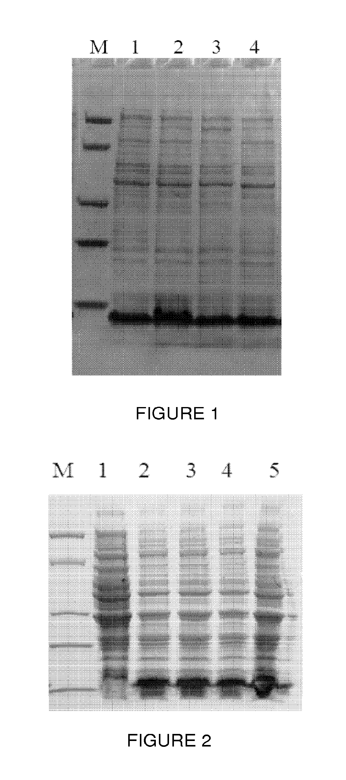
FIG. 1 shows expression of protein A in Escherichia coli detected by SDS-PAGE, wherein lane M is a Protein Marker (Genscript 94KD, 66 kD, 36KD, 25KD, 14KD), lanes 1 to 4 are expression of protein A in different Escherichia coli colonies;
FIG. 2 shows expression of protein A dimer in Escherichia coli detected by SDS-PAGE, wherein lane M is a Protein Marker (Genscript 94KD, 66 kD, 36KD, 25KD, 14KD), lanes 1 to 4 are expression of the protein A dimer in different Escherichia coli colonies;
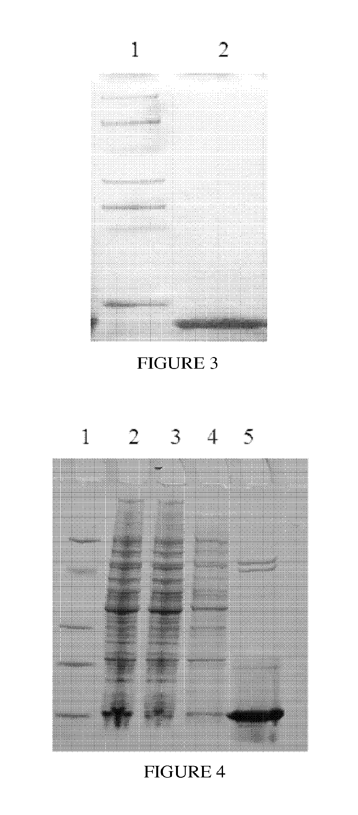
FIG. 3 is an image of SDS-PAGE; Protein A was purified by Ni column, wherein lane 1 is Protein Marker (Genscript 94KD, 66 kD, 36KD, 25KD, 14KD), lane 2 is protein A elution;
FIG. 4 is an image of SDS-PAGE; Protein A dimer was purified by Ni column, wherein lane 1 is Protein Marker (Genscript 94KD, 66 kD, 36KD, 25KD, 14KD), lane 2 is cell lysate supernatant, lane 3 is flow-through, lane 4 is protein A dimer eluted by the equilibration buffer, lane 5 is protein A dimer elution;
FIG. 5 is an image of SDS-PAGE; Protein A as an affinity ligand to purify immunoglobulin from human serum, wherein lane 1 is Protein Marker (Genscript 94KD, 66 kD, 36KD, 25KD, 14KD), lane 2 is human serum, lane 3 is purified human immunoglobulin using protein A as ligand for affinity chromatography;
FIG. 6 is an image of SDS-PAGE; Protein A dimer as an affinity ligand to purify immunoglobulin from human serum, where lane 1 is Protein Marker (Genscript Corporation) 220KD, 150KD, 100KD, 75KD, 50KD, 35 kD, 25 kD, 15KD), Lane 2 is human serum, lane 3 is proteins eluted by washing buffer (phosphate buffer), lane 4 is purified human immunoglobulin using protein A dimer as ligand for affinity chromatography; and
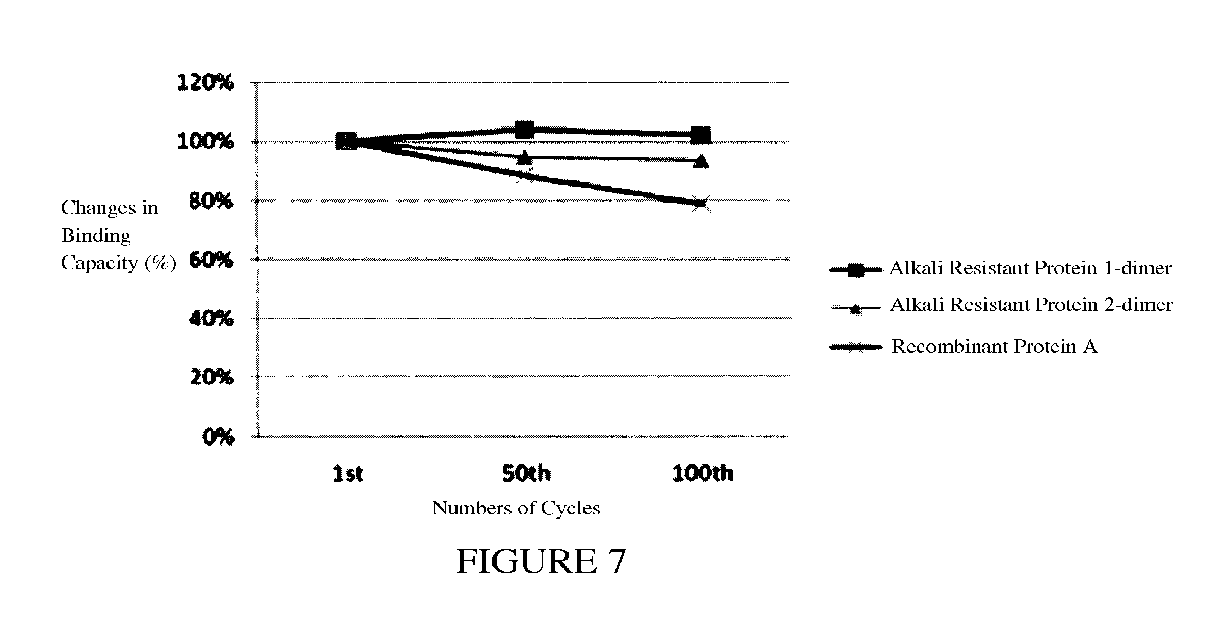
FIG. 7 shows alkali resistance tests for Protein A dimer as an affinity ligand.
DETAILED DESCRIPTION OF THE INVENTION
Example 1: Construction of Vector Containing a Gene which is Encoding Protein A Fused with 6 Histidine Residues at its N-Terminus
According to the codon preference of E. coli and to avoid secondary structure formation of mRNA, gene sequences of protein A fused with 6 histidine residues at its N-terminus were designed and optimized, which are shown here as SEQ ID NO:4 and SEQ ID NO:6 respectively; the corresponding amino acid sequences are shown as SEQ ID NO:3 and SEQ ID NO:5 respectively, of which the former is called protein A1 and the latter is called protein A2 in the following description. Using gene design software, multiple small gene fragments with certain length of overlapping sequences and similar annealing temperature were designed. All the small genes pieced together would cover the whole length of protein A1/A2 genes. The primer pairs were designed and synthesized according to those small gene sequences, of which the sequences were shown as here: Primers 4-1 to 4-8 (SEQ ID NOs: 16-23, respectively) are for the Protein A1 and Primers 6-1 to 6-8 (SEQ ID NOs: 24-31, respectively) are for the Protein A2. Two rounds of PCR reactions were done to synthesize the genes. For the 1st round PCR, all the primers 4-1 to 4-8 or primers 6-1 to 6-8 were mixed together as primers and templates, PBO polymerase (Genscript Corporation) was used for PCR reaction. All PCR reactions were done with 2720 thermal cycler (Applied Biosysytems) using the following method: the reaction cycles were 95.degree. C. for 20 s, 55.degree. C. for 20 s, 72.degree. C. for 20 s, after a total of 25 cycles, one extension step was taken at 72.degree. C. for 3 minutes, the PCR solutions were cooled down to 4.degree. C. and stored as the 1st PCR products. These 1st PCR products were used as templates for the 2nd round. Respectively, forwarding primers (primer 4-1/primer 6-1) and reversing primers (primer 4-8/primer 6-8) were used in the 2nd PCR reaction to amplify whole genes of Protein A1/A2 with the same PCR method as described above. The PCR products from the 2nd round PCR reactions were loading to an agarose gel containing 1% ethidium bromide and purified by electrophoresis. The DNA bands were visualized under UV light and cut out from the gel. The amplified DNA fragments were purified using Quick Gel Extraction Kit (Genscript Corporation) and the protocol provided by the manufacturer. The purified DNA fragments were sequenced using ABI PRISM.RTM. BIGDYE.RTM. Terminator Cycle Sequencing Ready Reaction Kit and 3730.times.196-capillary DNA analyzer from Applied Biosystems Inc.
TABLE-US-00001 TABLE 1 PCR Primers for synthesizing Protein A1 and Protein A2 genes. PCR Primers for synthesizing Protein A1 gene (SEQ ID NOs: 16-23) Primer 4-1 CCATGGGCTCACATCATCATCATCATCACGGCTCGGGTGCGGACGGTAA Primer 4-2 ACGCATTCTGCTGTTCTTTTTCAAATTTACCGTCCGCACCC Primer 4-3 AGAACAGCAGAATGCGTTCTACGAAATTCTGCATCTGCCGA Primer 4-4 CATTACGCTGTTCTTCGGTCAGGTTCGGCAGATGCAGAATT Primer 4-5 CCGAAGAACAGCGTAATGCATTTATCCAGTCTCTGAAAGATGATCCGAGC Primer 4-6 ACCCAGCACGTTCGTAGACTGGCTCGGATCATCTTTCAGA Primer 4-7 TACGAACGTGCTGGGTGAAGCGAAAAAACTGAACGATGCG Primer 4-8 CATATGTCATTTCGGGGCCTGCGCATCGTTCAGTTTTTTC PCR Primers for synthesizing Protein A2 gene (SEQ ID NOs: 24-31) Primer 6-1 CCATGGGCTCGCACCACCACCACCACCACGGCTCGGGCGCAGATGGCAAG Primer 6-2 ATGCGTTCTGTTGTTCTTTTTCAAACTTGCCATCTGCGCC Primer 6-3 AAAGAACAACAGAACGCATTCTACGAAATCCTGCATCTGCCGA Primer 6-4 TGCGTTACGCTGTTCTTCGGTCAGGTTCGGCAGATGCAGGA Primer 6-5 AGAACAGCGTAACGCATTCATCAAGTCTATCCGCGATGATCCG Primer 6-6 CCCAGCACGTTCGTAGACTGGCTCGGATCATCGCGGATAG Primer 6-7 CTACGAACGTGCTGGGCGAAGCGAAAAAACTGAATGATGC Primer 6-8 CATATGTCATTTCGGGGCCTGCGCATCATTCAGTTTTTTCGC
DNA fragments which the sequences had been verified were used as templates for protein A1/A2 subcloning. Primer 7 (SEQ ID NO:7) with primer 8 (SEQ ID NO:8) or primers 7 (SEQ ID NO:7) with primer 9 (SEQ ID NO:9) were used amplify cDNA fragments of the protein A1 or A2 respectively. The PCR products were purified by agarose gel electrophoresis. Using CLONEEZ.RTM. cloning kit (Genscript Corporation) and following the kit instruction, gene fragments encoding protein A1/A2 were subcloned into pET15b vectors. Vectors containing protein A1/A2 genes were verified by DNA sequencing and named by the following abbreviation: vector containing gene encoding protein A1 with the amino acid sequence shown as SEQ ID NO:3 was named PET15b-ProteinA1; vector containing gene encoding protein A2 with the amino acid sequence shown as SEQ ID NO:5 was named PET15b-ProteinA2.
Example 2 Construction of Vector Containing a Gene which is Encoding Protein A Dimer Fused with 6 Histidine Residues at its N-Terminus
According to the codon preference of E. coli and to avoid secondary structure formation of mRNA, gene sequences of protein A dimer fused with 6 histidine residues at its N-terminus were designed and optimized, which are shown here as SEQ ID NO:11 and SEQ ID NO:13 respectively; the corresponding amino acid sequences are shown as SEQ ID NO:10 and SEQ ID NO:12 respectively, of which the former is called protein AA1 and the latter is called protein AA2 for the following description. Using gene design software, multiple small gene fragments with certain length of overlapping sequences and similar annealing temperature were designed. All the small genes pieced together would cover the whole length of protein AA1/AA2 genes. The primer pairs were designed and synthesized according to those small gene sequences, of which the sequences were shown as here: Primers 11-1 to 11-12 (SEQ ID NOs: 32-43) are for the Protein AA1 and Primers 13-1 to 13-12 (SEQ ID NOs: 44-55) are for the Protein AA2. Two rounds of PCR reactions were done to synthesize the genes. For the 1st round PCR, all the primers of 11-1 to 11-12 or primers 13-1 to 13-12 were mixed together as primers and templates, PBO polymerase (Genscript Corporation) was used for PCR reaction. All PCR reactions were done with 2720 thermal cycler (Applied Biosysytems) using the following method: the reaction cycles were 95.degree. C. for 20 s, 55.degree. C. for 20 s, 72.degree. C. for 20 s, after a total of 25 cycles, one extension step was taken at 72.degree. C. for 3 minutes, the PCR solutions was cooled down to 4.degree. C. and stored as the 1st PCR products. These 1st PCR products were used as templates for the 2nd round. Respectively, forwarding primers (primer 11-1/primer 13-1) and reversing primers (primer 11-12/primer 13-12) were used in the 2nd PCR reaction to amplify whole genes of Protein AA1/AA2 with the same PCR method as described above. The PCR products from the 2nd round PCR reactions were loading to an agarose gel containing 1% ethidium bromide and purified by electrophoresis. The DNA bands were visualized under UV light and cut out from the gel. The amplified DNA fragments were purified using Quick Gel Extraction Kit (Genscript Corporation) and the protocol provided by the manufacture. The purified DNA fragments were sequenced using ABI PRISM.RTM. BIGDYE.RTM. Terminator Cycle Sequencing Ready Reaction Kit and 3730.times.196-capillary DNA analyzer from Applied Biosystems Inc.
TABLE-US-00002 TABLE 2 PCR Primers for synthesizing Protein AA1 dimer and Protein AA2 dimer PCR Primers for synthesizing Protein AA1 dimer gene (SEQ ID NOs: 32-43) Primer 11-1 CCATGGGCTCACATCATCATCATCATCACGGCTCGGGTGCGGACGGTAA Primer 11-2 ACGCATTCTGCTGTTCTTTTTCAAATTTACCGTCCGCACCC Primer 11-3 AGAACAGCAGAATGCGTTCTACGAAATTCTGCATCTGCCGAACCTGACC Primer 11-4 TCAGAGACTGGATAAATGCATTACGCTGTTCTTCGGTCAGGTTCGGCAG Primer 11-5 TGCATTTATCCAGTCTCTGAAAGATGATCCGAGCCAGTCTACGAACGTGC Primer 11-6 CCTGCGCATCGTTCAGTTTTTTCGCTTCACCCAGCACGTTCGTAGACTGG Primer 11-7 TGAACGATGCGCAGGCCCCGAAAGCGGATGGCAAATTCGAAAAAG Primer 11-8 GCAGAATTTCATAGAAGGCGTTCTGCTGTTCTTTTTCGAATTTGCCATCC Primer 11-9 CGCCTTCTATGAAATTCTGCACCTGCCGAATCTGACGGAAGAACAGCGCA Primer 11-10 GCTCGGATCGTCTTTCAGGCTCTGGATGAACGCATTGCGCTGTTCTTCCG Primer 11-11 TGAAAGACGATCCGAGCCAGTCCACGAATGTTCTGGGCGAAGCGAAAAA Primer 11-12 CATATGTCATTTCGGTGCTTGTGCGTCATTCAGTTTTTTCGCTTCGCCCA PCR Primers for synthesizing Protein AA2 dimer gene (SEQ ID NOs: 44-55) Primer 13-1 CCATGGGCTCGCACCACCACCACCACCACGGCTCGGGCGCAGATGGCAAG Primer 13-2 TCGTAGAATGCGTTCTGTTGTTCTTTTTCAAACTTGCCATCTGCGCC Primer 13-3 ACAGAACGCATTCTACGAAATCCTGCATCTGCCGAACCTGACCGA Primer 13-4 CGCGGATAGACTTGATGAATGCGTTACGCTGTTCTTCGGTCAGGTTCGGC Primer 13-5 TCATCAAGTCTATCCGCGATGATCCGAGCCAGTCTACGAACGTGCTGGG Primer 13-6 GGCCTGCGCATCATTCAGTTTTTTCGCTTCGCCCAGCACGTTCGTAG Primer 13-7 AATGATGCGCAGGCCCCGAAAGCGGATGGTAAATTTGAAAAAGAACAGCA Primer 13-8 AGGTGCAGAATTTCATAGAAGGCGTTCTGCTGTTCTTTTTCAAATTTACC Primer 13-9 CTTCTATGAAATTCTGCACCTGCCGAATCTGACGGAAGAACAGCGTAATG Primer 13-10 GCTCGGATCGTCACGAATGCTTTTAATGAACGCATTACGCTGTTCTTCCG Primer 13-11 CGTGACGATCCGAGCCAGAGCACGAATGTCCTGGGCGAAGCCAAAAA Primer 13-12 CATATGTCATTTCGGTGCTTGTGCGTCGTTCAGTTTTTTGGCTTCGCCCA
DNA fragments which the sequences had been verified were used as templates for protein AA1/AA2 subcloning. Primer 7 (SEQ ID NO:7) with primer 14 (SEQ ID NO:14) or primers 7 (SEQ ID NO:7) with primer 15 (SEQ ID NO:15) were used amplify cDNA fragments of the protein AA1 or AA2 respectively. The PCR products were purified by agarose gel electrophoresis. Using CLONEEZ.RTM. cloning kit (Genscript Corporation) and following the kit instruction, gene fragments encoding protein AA1/AA2 were subcloned into pET15b vectors. Vectors containing protein AA1/AA2 genes were verified by DNA sequencing and named by the following abbreviation: vector containing gene encoding protein AA1 with the amino acid sequence shown as SEQ ID NO:10 was named PET15b-ProteinAA1; vector containing gene encoding protein AA2 with the amino acid sequence shown as SEQ ID NO:12 was named PET15b-ProteinA2.
Example 3 Expression of Protein A with N-Terminal Fused Six Histidine Residues
The plasmid PET15b-ProteinA1 was transformed into competent cells of Escherichia coli strain BL21. Escherichia coli BL21 containing the plasmid PET15b-ProteinA1 was inoculated into the culture broth (1 g/L peptone, 5 g/L yeast extract, 5 g/L NaCl, and 100 mg/L Ampicillin) and cultured at 37.degree. C. When cells reached the logarithmic growth curve, 0.5 mM IPTG was added into the broth to induce protein expression for 4 hours, cell pellets were collected by centrifugation. A small amount of the cell was heated at high temperature (95.degree. C.) and the whole cell lysate was loaded onto 4.about.20% gradient SDS-PAGE gel for analysis. As shown in FIG. 1, a clear band was detected around 7.about.8KD which was protein A.
Example 4 Expression of Protein A Dimer with N-Terminal Fused Six Histidine Residues
The plasmid PET15b-ProteinAA1 was transformed into competent cells of Escherichia coli BL21. Escherichia coli BL21 containing the plasmid PET15b-Protein AA1 was inoculated into the culture broth (1 g/L peptone, 5 g/L yeast extract, 5 g/L NaCl, and 100 mg/L Ampicillin) and cultured at 37.degree. C., when cells reached their logarithmic growth curve 0.5 mM IPTG was added to induce protein expression for 4 hours. Cell pellets were collected by centrifugation. A small amount of bacteria was heated at high temperature (95.degree. C.) and the whole cell lysate was loaded onto 4.about.20% gradient SDS-PAGE gel for analysis. As shown in FIG. 2, a clear band was detected around 14.about.15KD which was protein A dimer.
Example 5 Purification of Protein A with the N-Terminal Fused Six Histidine Residues
The constant flow pump was rinsed with distilled water, and then the empty glass chromatographic column. About 200 ml Ni-IDA resin (Genscript Corporate) was packed into the column. Using the constant flow pump, the column was equilibrated with equilibration buffer (20 mM Tris 300 mM NaCl) for about 3 L (20CV) at the flow rate of 5 ml/min. 10 g cell pellets expressing protein A1 were suspended with 200 ml equilibration buffer (20 mMTris 300NaCl) and sonicated (Ningbo Xinzhi bio technology limited company JY98 IIIDH). Cell lysate was centrifuged and the supernatant was loaded onto the Ni-IDA column at the flow rate of 2 ml/min. Then the column was excessively washed with equilibration buffer at the flow rate of 5 ml/min to remove unbounded proteins and contaminants until the UV being stable. Then the target protein was eluted with elution buffer (20 mM Tris, 300 mMNaCl, 250 mM Iminazole) at the flow rate of 5 ml/min and collected. The eluted protein was loaded onto 4.about.20% gradient SDS-PAGE for analysis. As shown in FIG. 3, the purity of protein A after Ni-IDA column was above 90%.
Example 6 Purification of Protein A Dimer with the N-Terminal Fused Six Histidine Residues
The constant flow pump was rinsed with distilled water, and then the empty glass chromatographic column. About 200 ml Ni-IDA resin (Genscript Corporate) was packed into the column. Using the constant flow pump, the column was equilibrated with equilibration buffer (20 mM Tris 300 mMNaCl) for about 3 L (20CV) at the flow rate of 5 ml/min. 10 g cell pellets expressing protein A1 were suspended with 200 ml equilibration buffer (20 mMTris 300NaCl) and sonicated (Ningbo Xinzhi bio technology limited company JY98 IIIDH). Cell lysate was centrifuged and the supernatant was loaded onto the Ni-IDA column at the flow rate of 2 ml/min. Then the column was excessively washed with equilibration buffer at the flow rate of 5 ml/min to remove unbounded proteins and contaminants until the UV being stable. Then the target protein was eluted with elution buffer (20 mM Tris, 300 mMNaCl, 250 mM Imidazole) at the flow rate of 5 ml/min and collected. The eluted protein was loaded onto 4.about.20% gradient SDS-PAGE for analysis. As shown in FIG. 4, the purity of protein A after Ni-IDA column was above 90%.
7 Protein a or Protein A Dimer with the N-Terminal Fused Six Histidine Residues Used as an Ligand for Affinity Chromatographic Medium to Purify Immunoglobulin
Using the amino group, protein A or protein A dimer with the N-terminal fused six histidine residues were coupled onto the epoxy-based surface of agarose medium to make affinity chromatographic resin. Here 10 mg dimeric protein A or protein A was coupled to an epoxy-based surface of 1 ml SEPHAROSE.RTM. 4B (GE Healthcare) agarose beads to make Protein A affinity resin, of which 0.5 ml resin was used for immunoglobulin purification test. Firstly, the constant flow pump was cleaned with 20 ml double distilled water, then the empty chromatographic column was cleaned. 0.5 ml resin was packed into the column. Using constant flow pump the column was equilibrated with 10 ml phosphate buffer (containing 0.15M NaCl, 30 mM Na2HPO4, 10 mM NaH2PO4 pH 7) at the flow rate of 1 ml/min.
15 ml of human serum at the concentration of 5 mg/ml was used as testing sample and loaded to the packed column at the flow rate of 0.5-1 ml/min to the saturate the binding of immunoglobulin to the protein A ligands, and then washed the column with 20 ml (40CV) phosphate buffer (containing 0.15M of NaCl, 30 mM Na2HPO4, 10 mMNaH2PO4 pH 7.0) to remove unbound proteins and contaminants. Human immunoglobulin was finally eluted with 0.1M pH 3.0 glycine buffer and collected with UV detection. 20 uL of the eluted fraction was loaded onto 4-20% gradient SDS-PAGE gel for analysis. As shown in FIGS. 5 and 6, protein A or protein A dimer can isolate immunoglobulin with high purity from the serum.
Example 8 Alkali Resistance Tests of Protein a or Protein A Dimer with the N-Terminal Fused Six Histidine Residues as Ligands of Affinity Chromatographic Medium for Purifying Immunoglobulin
0.5 ml of SEPHAROSE.RTM. beads conjugated protein A dimer with six N-terminal histidine residues was used for Clean-In-Place (CIP) test using alkaline solutions. First, the procedure of Example 7 was performed, and the total amount of eluted immunoglobulin was calculated as column capacity. After elution, CIP was performed using 15 ml 0.5M NaOH solution at a flow rate of 1 ml/min to clean the resin thoroughly. And then followed with 10 ml of phosphate buffer (containing 0.15M of NaCl, 30 mMNa2HPO4, 10 mM NaH2PO4 adjusted to pH 7.0) at the same flow rate to wash out NaOH and re-equilibrate the column. Another procedure of Example 7 was performed to determine the binding capacity of the column, which could be used as binding ability of protein A ligand to immunoglobulin. As shown in FIG. 7, after 100 cycles of CIP with alkaline solution, the protein A dimer used as an affinity chromatographic ligand still remained good binding ability to immunoglobulin.
It will be appreciated by those skilled in the art that changes could be made to the embodiments described above without departing from the broad inventive concept thereof. It is understood, therefore, that this invention is not limited to the particular embodiments disclosed, but it is intended to cover modifications within the spirit and scope of the present invention as defined by the appended claims.
SEQUENCE LISTINGS
1
56158PRTArtificial SequenceProtein A1 mutant sequence 1Ala Asp Gly Lys Phe Glu Lys Glu Gln Gln Asn Ala Phe Tyr Glu Ile1 5 10 15Leu His Leu Pro Asn Leu Thr Glu Glu Gln Arg Asn Ala Phe Ile Gln 20 25 30Ser Leu Lys Asp Asp Pro Ser Gln Ser Thr Asn Val Leu Gly Glu Ala 35 40 45Lys Lys Leu Asn Asp Ala Gln Ala Pro Lys 50 55258PRTArtificial SequenceProtein A2 mutant sequence 2Ala Asp Gly Lys Phe Glu Lys Glu Gln Gln Asn Ala Phe Tyr Glu Ile1 5 10 15Leu His Leu Pro Asn Leu Thr Glu Glu Gln Arg Asn Ala Phe Ile Lys 20 25 30Ser Ile Arg Asp Asp Pro Ser Gln Ser Thr Asn Val Leu Gly Glu Ala 35 40 45Lys Lys Leu Asn Asp Ala Gln Ala Pro Lys 50 55370PRTArtificial Sequenceprotein A1 sequence with N-terminal 6X- histidine tag 3Met Gly Ser His His His His His His Gly Ser Gly Ala Asp Gly Lys1 5 10 15Phe Glu Lys Glu Gln Gln Asn Ala Phe Tyr Glu Ile Leu His Leu Pro 20 25 30Asn Leu Thr Glu Glu Gln Arg Asn Ala Phe Ile Gln Ser Leu Lys Asp 35 40 45Asp Pro Ser Gln Ser Thr Asn Val Leu Gly Glu Ala Lys Lys Leu Asn 50 55 60Asp Ala Gln Ala Pro Lys65 704221DNAArtificial Sequencegene encoding protein A1 sequence with N- terminal 6X-histidine tag 4ccatgggctc acatcatcat catcatcacg gctcgggtgc ggacggtaaa tttgaaaaag 60aacagcagaa tgcgttctac gaaattctgc atctgccgaa cctgaccgaa gaacagcgta 120atgcatttat ccagtctctg aaagatgatc cgagccagtc tacgaacgtg ctgggtgaag 180cgaaaaaact gaacgatgcg caggccccga aatgacatat g 221570PRTArtificial Sequenceprotein A2 sequence with N-terminal 6X- histidine tag 5Met Gly Ser His His His His His His Gly Ser Gly Ala Asp Gly Lys1 5 10 15Phe Glu Lys Glu Gln Gln Asn Ala Phe Tyr Glu Ile Leu His Leu Pro 20 25 30Asn Leu Thr Glu Glu Gln Arg Asn Ala Phe Ile Lys Ser Ile Arg Asp 35 40 45Asp Pro Ser Gln Ser Thr Asn Val Leu Gly Glu Ala Lys Lys Leu Asn 50 55 60Asp Ala Gln Ala Pro Lys65 706221DNAArtificial Sequencegene encoding protein A2 sequence with N- terminal 6X-histidine tag 6ccatgggctc gcaccaccac caccaccacg gctcgggcgc agatggcaag tttgaaaaag 60aacaacagaa cgcattctac gaaatcctgc atctgccgaa cctgaccgaa gaacagcgta 120acgcattcat caagtctatc cgcgatgatc cgagccagtc tacgaacgtg ctgggcgaag 180cgaaaaaact gaatgatgcg caggccccga aatgacatat g 221750DNAArtificial SequencePrimer 7 sequence 7aactttaaga aggagatata ccatgggctc acatcatcat catcatcacg 50850DNAArtificial Sequenceprimer 8 sequence 8ttagcagccg gatcctcgag catatgtcat ttcggggcct gcgcatcatt 50950DNAArtificial Sequenceprimer 9 sequence 9ttagcagccg gatcctcgag catatgtcat ttcggggcct gcgcatcgtt 5010128PRTArtificial Sequenceprotein AA1 dimer sequence with N-terminal 6X- histidine tag 10Met Gly Ser His His His His His His Gly Ser Gly Ala Asp Gly Lys1 5 10 15Phe Glu Lys Glu Gln Gln Asn Ala Phe Tyr Glu Ile Leu His Leu Pro 20 25 30Asn Leu Thr Glu Glu Gln Arg Asn Ala Phe Ile Gln Ser Leu Lys Asp 35 40 45Asp Pro Ser Gln Ser Thr Asn Val Leu Gly Glu Ala Lys Lys Leu Asn 50 55 60Asp Ala Gln Ala Pro Lys Ala Asp Gly Lys Phe Glu Lys Glu Gln Gln65 70 75 80Asn Ala Phe Tyr Glu Ile Leu His Leu Pro Asn Leu Thr Glu Glu Gln 85 90 95Arg Asn Ala Phe Ile Gln Ser Leu Lys Asp Asp Pro Ser Gln Ser Thr 100 105 110Asn Val Leu Gly Glu Ala Lys Lys Leu Asn Asp Ala Gln Ala Pro Lys 115 120 12511395DNAArtificial Sequencegene encoding protein AA1 dimer sequence with N-terminal 6X-histidine tag 11ccatgggctc acatcatcat catcatcacg gctcgggtgc ggacggtaaa tttgaaaaag 60aacagcagaa tgcgttctac gaaattctgc atctgccgaa cctgaccgaa gaacagcgta 120atgcatttat ccagtctctg aaagatgatc cgagccagtc tacgaacgtg ctgggtgaag 180cgaaaaaact gaacgatgcg caggccccga aagcggatgg caaattcgaa aaagaacagc 240agaacgcctt ctatgaaatt ctgcacctgc cgaatctgac ggaagaacag cgcaatgcgt 300tcatccagag cctgaaagac gatccgagcc agtccacgaa tgttctgggc gaagcgaaaa 360aactgaatga cgcacaagca ccgaaatgac atatg 39512128PRTArtificial Sequenceprotein AA2 dimer sequence with N-terminal 6X- histidine tag 12Met Gly Ser His His His His His His Gly Ser Gly Ala Asp Gly Lys1 5 10 15Phe Glu Lys Glu Gln Gln Asn Ala Phe Tyr Glu Ile Leu His Leu Pro 20 25 30Asn Leu Thr Glu Glu Gln Arg Asn Ala Phe Ile Lys Ser Ile Arg Asp 35 40 45Asp Pro Ser Gln Ser Thr Asn Val Leu Gly Glu Ala Lys Lys Leu Asn 50 55 60Asp Ala Gln Ala Pro Lys Ala Asp Gly Lys Phe Glu Lys Glu Gln Gln65 70 75 80Asn Ala Phe Tyr Glu Ile Leu His Leu Pro Asn Leu Thr Glu Glu Gln 85 90 95Arg Asn Ala Phe Ile Lys Ser Ile Arg Asp Asp Pro Ser Gln Ser Thr 100 105 110Asn Val Leu Gly Glu Ala Lys Lys Leu Asn Asp Ala Gln Ala Pro Lys 115 120 12513395DNAArtificial Sequencegene encoding protein AA2 dimer sequence with N-terminal 6X-histidine tag 13ccatgggctc gcaccaccac caccaccacg gctcgggcgc agatggcaag tttgaaaaag 60aacaacagaa cgcattctac gaaatcctgc atctgccgaa cctgaccgaa gaacagcgta 120acgcattcat caagtctatc cgcgatgatc cgagccagtc tacgaacgtg ctgggcgaag 180cgaaaaaact gaatgatgcg caggccccga aagcggatgg taaatttgaa aaagaacagc 240agaacgcctt ctatgaaatt ctgcacctgc cgaatctgac ggaagaacag cgtaatgcgt 300tcattaaaag cattcgtgac gatccgagcc agagcacgaa tgtcctgggc gaagccaaaa 360aactgaacga cgcacaagca ccgaaatgac atatg 3951450DNAArtificial Sequenceprimer 14 sequence 14ttagcagccg gatcctcgag catatgtcat ttcggtgctt gtgcgtcatt 501550DNAArtificial Sequenceprimer 15 sequence 15ttagcagccg gatcctcgag catatgtcat ttcggtgctt gtgcgtcgtt 501649DNAArtificial Sequenceprimer 4-1 sequence 16ccatgggctc acatcatcat catcatcacg gctcgggtgc ggacggtaa 491741DNAArtificial Sequenceprimer 4-2 sequence 17acgcattctg ctgttctttt tcaaatttac cgtccgcacc c 411841DNAArtificial Sequenceprimer 4-3 sequence 18agaacagcag aatgcgttct acgaaattct gcatctgccg a 411941DNAArtificial Sequenceprimer 4-4 sequence 19cattacgctg ttcttcggtc aggttcggca gatgcagaat t 412050DNAArtificial Sequenceprimer 4-5 sequence 20ccgaagaaca gcgtaatgca tttatccagt ctctgaaaga tgatccgagc 502140DNAArtificial Sequenceprimer 4-6 sequence 21acccagcacg ttcgtagact ggctcggatc atctttcaga 402240DNAArtificial Sequenceprimer 4-7 sequence 22tacgaacgtg ctgggtgaag cgaaaaaact gaacgatgcg 402340DNAArtificial Sequenceprimer 4-8 sequence 23catatgtcat ttcggggcct gcgcatcgtt cagttttttc 402450DNAArtificial Sequenceprimer 6-1 sequence 24ccatgggctc gcaccaccac caccaccacg gctcgggcgc agatggcaag 502540DNAArtificial Sequenceprimer 6-2 sequence 25atgcgttctg ttgttctttt tcaaacttgc catctgcgcc 402643DNAArtificial Sequenceprimer 6-3 sequence 26aaagaacaac agaacgcatt ctacgaaatc ctgcatctgc cga 432741DNAArtificial Sequenceprimer 6-4 sequence 27tgcgttacgc tgttcttcgg tcaggttcgg cagatgcagg a 412843DNAArtificial Sequenceprimer 6-5 sequence 28agaacagcgt aacgcattca tcaagtctat ccgcgatgat ccg 432940DNAArtificial Sequenceprimer 6-6 sequence 29cccagcacgt tcgtagactg gctcggatca tcgcggatag 403040DNAArtificial Sequenceprimer 6-7 sequence 30ctacgaacgt gctgggcgaa gcgaaaaaac tgaatgatgc 403142DNAArtificial Sequenceprimer 6-8 sequence 31catatgtcat ttcggggcct gcgcatcatt cagttttttc gc 423249DNAArtificial Sequenceprimer 11-1 sequence 32ccatgggctc acatcatcat catcatcacg gctcgggtgc ggacggtaa 493341DNAArtificial Sequenceprimer 11-2 sequence 33acgcattctg ctgttctttt tcaaatttac cgtccgcacc c 413449DNAArtificial Sequenceprimer 11-3 sequence 34agaacagcag aatgcgttct acgaaattct gcatctgccg aacctgacc 493549DNAArtificial Sequenceprimer 11-4 sequence 35tcagagactg gataaatgca ttacgctgtt cttcggtcag gttcggcag 493650DNAArtificial Sequenceprimer 11-5 sequence 36tgcatttatc cagtctctga aagatgatcc gagccagtct acgaacgtgc 503750DNAArtificial Sequenceprimer 11-6 sequence 37cctgcgcatc gttcagtttt ttcgcttcac ccagcacgtt cgtagactgg 503845DNAArtificial Sequenceprimer 11-7 sequence 38tgaacgatgc gcaggccccg aaagcggatg gcaaattcga aaaag 453950DNAArtificial Sequenceprimer 11-8 sequence 39gcagaatttc atagaaggcg ttctgctgtt ctttttcgaa tttgccatcc 504050DNAArtificial Sequenceprimer 11-9 sequence 40cgccttctat gaaattctgc acctgccgaa tctgacggaa gaacagcgca 504150DNAArtificial Sequenceprimer 11-10 sequence 41gctcggatcg tctttcaggc tctggatgaa cgcattgcgc tgttcttccg 504249DNAArtificial Sequenceprimer 11-11 sequence 42tgaaagacga tccgagccag tccacgaatg ttctgggcga agcgaaaaa 494350DNAArtificial Sequenceprimer 11-12 sequence 43catatgtcat ttcggtgctt gtgcgtcatt cagttttttc gcttcgccca 504450DNAArtificial Sequenceprimer 13-1 sequence 44ccatgggctc gcaccaccac caccaccacg gctcgggcgc agatggcaag 504547DNAArtificial Sequenceprimer 13-2 sequence 45tcgtagaatg cgttctgttg ttctttttca aacttgccat ctgcgcc 474645DNAArtificial Sequenceprimer 13-3 sequence 46acagaacgca ttctacgaaa tcctgcatct gccgaacctg accga 454750DNAArtificial Sequenceprimer 13-4 sequence 47cgcggataga cttgatgaat gcgttacgct gttcttcggt caggttcggc 504849DNAArtificial Sequenceprimer 13-5 sequence 48tcatcaagtc tatccgcgat gatccgagcc agtctacgaa cgtgctggg 494947DNAArtificial Sequenceprimer 13-6 sequence 49ggcctgcgca tcattcagtt ttttcgcttc gcccagcacg ttcgtag 475050DNAArtificial Sequenceprimer 13-7 sequence 50aatgatgcgc aggccccgaa agcggatggt aaatttgaaa aagaacagca 505150DNAArtificial Sequenceprimer 13-8 sequence 51aggtgcagaa tttcatagaa ggcgttctgc tgttcttttt caaatttacc 505250DNAArtificial Sequenceprimer 13-9 sequence 52cttctatgaa attctgcacc tgccgaatct gacggaagaa cagcgtaatg 505350DNAArtificial Sequenceprimer 13-10 sequence 53gctcggatcg tcacgaatgc ttttaatgaa cgcattacgc tgttcttccg 505447DNAArtificial Sequenceprimer 13-11 sequence 54cgtgacgatc cgagccagag cacgaatgtc ctgggcgaag ccaaaaa 475550DNAArtificial Sequenceprimer 13-12 sequence 55catatgtcat ttcggtgctt gtgcgtcgtt cagttttttg gcttcgccca 50564PRTArtificial Sequenceexemplary linker sequence for protein A multimer 56Ala Asp Gly Lys1
* * * * *
D00001
D00002
D00003
D00004
S00001
XML
uspto.report is an independent third-party trademark research tool that is not affiliated, endorsed, or sponsored by the United States Patent and Trademark Office (USPTO) or any other governmental organization. The information provided by uspto.report is based on publicly available data at the time of writing and is intended for informational purposes only.
While we strive to provide accurate and up-to-date information, we do not guarantee the accuracy, completeness, reliability, or suitability of the information displayed on this site. The use of this site is at your own risk. Any reliance you place on such information is therefore strictly at your own risk.
All official trademark data, including owner information, should be verified by visiting the official USPTO website at www.uspto.gov. This site is not intended to replace professional legal advice and should not be used as a substitute for consulting with a legal professional who is knowledgeable about trademark law.