Therapeutically useful molecules
Stauss , et al. Oc
U.S. patent number 10,450,360 [Application Number 15/234,382] was granted by the patent office on 2019-10-22 for therapeutically useful molecules. This patent grant is currently assigned to IMPERIAL INNOVATIONS LIMITED. The grantee listed for this patent is IMPERIAL INNOVATIONS LIMITED. Invention is credited to Liquan Gao, Hans Josef Stauss, Shao-An Xue.


View All Diagrams
| United States Patent | 10,450,360 |
| Stauss , et al. | October 22, 2019 |
Therapeutically useful molecules
Abstract
A T cell receptor molecule (TCR) containing an alpha chain portion and a beta chain portion wherein the alpha chain portion contains three complementarity determining regions (CDRs): CDR1.alpha.: SSYSPS CDR2.alpha.: YTSAATL CDR3.alpha.: VVSPFSGGGADGLT or comprising or consisting of SPFSGGGADGLT and the beta chain portion contains three complementarity determining regions (CDRs): CDR1.beta.: DFQATT CDR2.beta.: SNEGSKA CDR3.beta.: comprising SARDGGEG or comprising or consisting of RDGGEGSETQY, or wherein up to three amino acid residues in one or more CDRs are replaced by another amino acid.
| Inventors: | Stauss; Hans Josef (London, GB), Gao; Liquan (London, GB), Xue; Shao-An (London, GB) | ||||||||||
|---|---|---|---|---|---|---|---|---|---|---|---|
| Applicant: |
|
||||||||||
| Assignee: | IMPERIAL INNOVATIONS LIMITED
(London, GB) |
||||||||||
| Family ID: | 30129780 | ||||||||||
| Appl. No.: | 15/234,382 | ||||||||||
| Filed: | August 11, 2016 |
Prior Publication Data
| Document Identifier | Publication Date | |
|---|---|---|
| US 20160340404 A1 | Nov 24, 2016 | |
Related U.S. Patent Documents
| Application Number | Filing Date | Patent Number | Issue Date | ||
|---|---|---|---|---|---|
| 14469387 | Aug 26, 2014 | 9732141 | |||
| 13090845 | Apr 20, 2011 | ||||
| 10581773 | May 31, 2011 | 7951783 | |||
| PCT/GB2004/005100 | Dec 6, 2004 | ||||
Foreign Application Priority Data
| Dec 6, 2003 [GB] | 0328363.7 | |||
| Current U.S. Class: | 1/1 |
| Current CPC Class: | C12N 7/00 (20130101); C07K 14/7051 (20130101); A61P 35/00 (20180101); A61K 35/17 (20130101); A61P 35/02 (20180101); C12N 15/86 (20130101); C12N 5/0636 (20130101); C12N 2740/10043 (20130101) |
| Current International Class: | C07K 14/725 (20060101); C12N 7/00 (20060101); C12N 5/0783 (20100101); A61K 35/17 (20150101); C12N 15/86 (20060101) |
References Cited [Referenced By]
U.S. Patent Documents
| 5610031 | March 1997 | Burgeson et al. |
| 6028169 | February 2000 | Kreider et al. |
| 6558672 | May 2003 | Pastan et al. |
| 7871817 | January 2011 | Voss et al. |
| 7951783 | May 2011 | Stauss et al. |
| 2002/0064521 | May 2002 | Ellenhorn |
| 2015/0147302 | May 2015 | Stauss et al. |
| WO-2000/026249 | May 2000 | WO | |||
| 200155366 | Aug 2001 | WO | |||
| 2003020763 | Mar 2003 | WO | |||
| 2004033685 | Apr 2004 | WO | |||
| 2004074322 | Sep 2004 | WO | |||
| PCT/GB2004/005100 | Dec 2004 | WO | |||
| WO-2005056595 | Jun 2005 | WO | |||
| WO-2006000830 | Jan 2006 | WO | |||
Other References
|
Roszkowski et al. (J Innnnunol 2003; 170:2582-2589). (Year: 2003). cited by examiner . Garcia et al., Cell, vol. 122, 333-336, Aug. 12, 2005. (Year: 2005). cited by examiner . Parent U.S. Appl. No. 14/469,387, filed Aug. 26, 2014. cited by applicant . U.S. Appl. No. 13/090,845, filed Apr. 20, 2011. cited by applicant . U.S. Appl. No. 10/581,773, filed Feb. 12, 2007 (U.S. Pat. No. 7,951,783). cited by applicant . Bellantuono et al., Two distinct HLA-A0201-presented epitopes of the Wilms tumor antigen 1 can function as targets for leukemia-reactive CTL, Blood, 100:3835-37 (2002). cited by applicant . Boulter et al., Stable, soluble T-cell receptor molecules for crystallization and therapeutics, Protein Eng. 16:707-11 (2003). cited by applicant . Chervin et al., Engineering higher affinity T cell receptors using a T cell display system. J. Immunol. Methods, 339: 175-84 (2008). cited by applicant . Chung et al., Functional three-domain single-chain T-cell receptors, Proc. Natl. Acad. Sci. USA 91:12654-8 (1994). cited by applicant . Coleman et al., Effects of amino acid sequence changes on antibody-antigen interactions. Res. Immunol. 145: 33-6 (1994). cited by applicant . Dudley et al., Cancer regression and autoimmunity in patients after clonal repopulation with antitumor lymphocytes, Science, 298:850-4 (2002). cited by applicant . Engels et al., Retroviral vectors for high-level transgene expression in T lymphocytes, Human Gene Ther. 14:1155-68 (2003). cited by applicant . Eshhar et al., Specific activation and targeting of cytotoxic lymphocytes through chimeric single chains consisting of antibody-binding domains and the ? or ? subunits of the immunoglobulin and T-cell receptors, Proc. Natl. Acad. Sci. USA, 90:720-4 (1993). cited by applicant . Finer et al., kat: A high-efficiency retroviral transduction system for primary human T lymphocytes, Blood, 83:43-50 (1994). cited by applicant . Gao et al., Human cytotoxic T lymphocytes specific for Wilms' tumor antigen-1 inhibit engraftment of leukemia-initiating stem cells in non-obese diabetic-severe combined immunodeficient recipients, Transplantation, 75:1429-36 (2003). cited by applicant . Gao et al., Selective elimination of leukemic CD34+ progenitor cells by cytotoxic T lymphocytes specific for WT1, Blood, 95:2198-203 (2000). cited by applicant . Garcia et al., Structural basis of T cell recognition, Ann. Rev. Immunol. 17:369-97 (1999). cited by applicant . Goyarts et al., Point mutations in the beta chain CDR3 can alter the T cell receptor recognition pattern on an MHC class I/peptide complex over a broad interface area. Mol. Immunol. 35(10): 593-607 (1998). cited by applicant . Hwu et al., Lysis of ovarian cancer cells by human lymphocytes redirected with a chimeric gene composed by an antibody variable region and the Fc receptor ? chain, J. Exp. Med. 178:361-6 (1993). cited by applicant . Inoue et al., Aberrant overexpression of the Wilms tumor gene (WT1) in human leukemia, Blood 89:1405-12 (1997). cited by applicant . Inoue et al., Long-term follow-up of minimal residual disease in leukemia patients by monitoring WT1 (Wilms tumor gene) expression levels, Blood, 88:2267-78 (1996). cited by applicant . Inoue et al., Wilms' tumor gene (WT1) competes with differentiation-inducing signal in hematopoietic progenitor cells, Blood, 91:2969-76 (1998). cited by applicant . Janeway et al., Most thymocytes express receptors that cannot interact with self MHC and these cells die in the thymus. Immunobiology, Chapter 7: 262-63 (2001). cited by applicant . Kast et al., Eradication of adenovirus E1-induced tumors by E1A-specific cytotoxic T lymphocytes, Cell, 59:603-14 (1989). cited by applicant . Kawakami et al., Identification of a human melanoma antigen recognized by tumor-infiltrating lymphocytes associated with in vivo tumor rejection, Proc. Natl. Acad. Sci. USA 91:6458-6462 (1994). cited by applicant . Kessels et al., Changing T cell specifically by retroviral T cell receptor display, Proc. Natl. Acad. Sci. USA, 97:14578-83 (2000). cited by applicant . Kieke et al., Selection of functional T cell receptor mutants from a yeast surface-display library. Proc. Natl. Acad. Sci. USA, 96: 5651-6 (1999). cited by applicant . Lefranc et al., IMGT unique numbering for immunoglobulin and T cell receptor variable domains and Ig superfamily V-like domains, Dev. Comp. Immunol. 27:55-77 (2003). cited by applicant . Menssen et al., Detection by monoclonal antibodies of the Wilms' tumor (WT1) nuclear protein in patients with acute leukemia, Int. J. Cancer, 70:518-23 (1997). cited by applicant . Menssen et al., Presence of Wilms' tumor gene (WT1) transcripts and the WT1 nuclear protein in the majority of human acute leukemias, Leukemia, 9:1060-7 (1995). cited by applicant . Menssen et al., Wilms' tumor gene (WT1) expression in lung cancer, colon cancer and glioblastoma cell lines compared to freshly isolated tumor specimens, J. Cancer Res. Clin. Oncol. 126:226-32 (2000). cited by applicant . Miyoshi, High expression of Wilms' tumor suppressor gene predicts poor prognosis in breast cancer patients, Clin. Cancer Res. 8:1167-71 (2000). cited by applicant . Moritz et al., Cytotoxic T lymphocytes with a grafted recognition specifically for ERBB2-expressing tumor cells, Proc. Natl. Acad. Sci. USA, 91:4318-22 (1994). cited by applicant . Ogawa et al., Successful donor leukocyte transfusion at molecular relapse for a patient with acute myeloid leukemia who was treated with allogeneic bone marrow transplantation: Importance of the monitoring of minimal residual disease by WT1 assay, Bone Marrow Transplant, 21:525-7 (1998). cited by applicant . Oji et al., Expression of the Wilms' tumor gene WT1 in solid tumors and its involvement in tumor cell growth, Jpn J. Cancer Res. 90:194-204 (1999). cited by applicant . Oji et al., Overexpression of the Wilms' tumor gene WT1 in colorectal adenocarcinoma, Cancer Sci. 94:712-7 (2003). cited by applicant . Oji et al., Overexpression of the Wilms' tumor gene WT1 in head and neck squamous cell carcinoma, Cancer Sci. 94:523-9 (2003). cited by applicant . Oji et al., Overexpression of the Wilms' tumor gene WT1 in primary thyroid cancer, Cancer Sci. 94:606-11 (2003). cited by applicant . Roberts et al., Targeting of human immunodeficiency virus-infected cells by CD8+ T lymphocytes armed with universal T-Cell receptors, Blood, 84:2878-89 (1994). cited by applicant . Rodeck et al., Expression of the WT1 Wilms' tumor gene by normal and malignant human melanocytes, Int. J. Cancer, 59:78-82 (1994). cited by applicant . Rudikoff et al., Single amino acid substitution altering antigen-binding specificity. Proc. Natl. Acad. Sci. USA, 79: 1979-83 (1982). cited by applicant . Silberstein et al., Altered expression of the WT1 Wilms tumor suppressor gene in human breast cancer, Proc. Natl. Acad. Sci. USA, 94:8132-7 (1997). cited by applicant . Stanislawski et al., Circumventing tolerance to a human MDM2-derived tumor antigen by TCR gene transfer, Nat. Immunol. 2:962-70 (2001). cited by applicant . Tamaki et al., Increased expression of Wilms tumor gene (WT1) at relapse in acute leukemia, Blood, 88:4396-8 (1996). cited by applicant . Ueda et al., Overexpression of the Wilms' tumor gene WT1 in human bone and soft-tissue sarcomas, Cancer Sci. 94:271-6 (2003). cited by applicant . Vajdos et al., Comprehensive functional maps of the antigen-binding site of an anti-ErbB2 antibody obtained with shotgun scanning metagenesis. J. Mol. Biol. 320(2): 415-28 (2002). cited by applicant . Viel et al., Molecular mechanisms possibly affecting WT1 function in human ovarian tumors, Int. J. Cancer 57:515-21 (1994). cited by applicant . Xue et al., Elimination of human leukemia cells in NOD/SCID mice by WT1-TCR gene-transduced human T cells, Blood, 106 (prepublished online):3062-7 (2005). cited by applicant . Janeway et al., Immunobiology, 5th Ed. Garland Science, pp. 106-108, 117-118, and 262-263 (2001). cited by applicant . Chang et al.; "A General Method for Facilitating Heterodimeric Pairing Between Two Proteins: Application to Expression of Alpha and Beta T-cell Receptor Extracellular Segments"; Proc. Natl. Acad. Sci. 91; pp. 11408-11412 (1994). cited by applicant . Cooper et al.; "Transfer of Specificity for Human Immnodeficiency Virus Type 1 into Primary Human T Lymphocytes by Introduction of T-Cell Receptor Genes"; Journal of Virology; pp. 8207-8212; (2000). cited by applicant . Engel et al.; "High-Efficiency Expression and Solubilization of Functional T Cel Antigen Receptor Heterodimers"; Science; 256(5061); pp. 1318-1321; (1992). cited by applicant . Fernandez-Miguel et al; "Multivalent Structure of an Altha BT Cell Receptor"; Proc. Natl. Acad. Sci. 96; pp. 1547-1552; (1999). cited by applicant . Li et al.; "Structural Mutations in the Constant Region of the T-cell Antigen Receptor (TCR) Beta Chain and their Effect on TCRalpha and Beta Chain Interaction"; Immunology; 88; pp. 524-530; (1996). cited by applicant . Pecorari et al.; "Folding, Heterodimeric Association and Specific Peptide Recognition of a Murine Altha-Beta T-cell Receptor Expressed in Escherichia coli"; J. Mol. Biol. 285; pp. 1831-1843; (1999). cited by applicant . Reiter et al.; "Construction of a Functional Disulfide-Stabilized TCR Fv Indicates that Antibody and CR Fv Frameworks are Very Similar in Structure"; Immunity; 2; pp. 281-287; (1995). cited by applicant . Rubinstein et al.; "Transfer of TCR Genes into Mature T Cells is Accompanied by the Maintenance of Parental T Cell Avidity"; J Immunol; 170; pp. 1209-1217; (2003). cited by applicant . Willemsen et al.; "Grafting Primary Human T Lymphocytes with Cancer-specific Chimeric Single Chain and Two Chain TCR"; Gene Therapy; 7; pp. 1369-1377; (2000). cited by applicant. |
Primary Examiner: Skelding; Zachary S
Attorney, Agent or Firm: Cantor Colburn LLP
Parent Case Text
CROSS-REFERENCE TO RELATED APPLICATIONS
This application is a continuation of U.S. application Ser. No. 14/469,387 filed on Aug. 26, 2014, which is a continuation of U.S. application Ser. No. 13/090,845 filed on Apr. 20, 2011, now abandoned, which is a continuation of U.S. application Ser. No. 10/581,773 filed on Feb. 12, 2007, which is a .sctn. 371 of PCT/GB2004/0051000 filed on Dec. 6, 2004, now U.S. Pat. No. 7,951,783, which claims priority to Application GB0328363.7, filed on Dec. 6, 2003, which are incorporated herein by reference in their entirety.
Claims
The invention claimed is:
1. A T cell isolated from a patient comprising a T cell receptor (TCR), wherein said TCR comprises an alpha chain portion and a beta chain portion wherein the alpha chain comprises three complementarity determining regions (CDRs) TABLE-US-00010 (SEQ ID NO: 2) CDR1.alpha.: SSYSPS, (SEQ ID NO: 3) CDR2.alpha.: YTSAATL, and (SEQ ID NO: 4) CDR3.alpha.: VVSPFGGGADGLT or (SEQ ID NO: 5) SPFSGGGADGLT,
wherein the alpha chain portion comprises three CDRs: TABLE-US-00011 (SEQ ID NO: 2) CDR1.alpha.: SSYSPS; (SEQ ID NO: 3) CDR2.alpha.: YTSAATL; and (SEQ ID NO: 5) CDR3.alpha.: comprising of or consisting of SPFSGGGADGLT;
and wherein the beta chain portion comprises three CDRs: TABLE-US-00012 (SEQ ID NO: 6) CDR1.beta.: DFQATT; (SEQ ID NO: 7) CDR2.beta.: SNEGSKA; and (SEQ ID NO: 8) CDR3.beta.: comprising SARDGGEG or wherein the beta chain portion comprises three CDRs:
TABLE-US-00013 (SEQ ID NO: 6) CDR1.beta.: DFQATT; (SEQ ID NO: 7) CDR2.beta.: SNEGSKA; and (SEQ ID NO: 9) CDR3.beta.: comprising or consisting of RDGGEGSETQY; and
wherein said TCR is able to bind an HLA-A2/RMFPNAPYL (SEQ ID NO: 1) complex and is introduced into said T cell.
2. The T cell of claim 1, wherein said alpha and beta chains each comprise, from the N terminus to the C terminus, a leader sequence, a variable region, a constant region, a connecting sequence, a transmembrane region and a cytoplasmic tail region.
3. The T cell of claim 1, wherein the TCR is encoded on a retroviral vector.
Description
FIELD OF THE INVENTION
The present invention relates to therapeutically useful molecules, in particular to T cell receptors (TCRs) which may be introduced into a patient's own T cells in order to direct the T cells to kill cancer cells within the patient, particularly cancer cells which express the Wilms Tumour antigen-1 (WT1).
BACKGROUND
The listing or discussion of a prior-published document in this specification should not necessarily be taken as an acknowledgement that the document is part of the state of the art or is common general knowledge. All of the documents referred to in this specification are hereby incorporated by reference.
There is evidence that anti-tumour cytotoxic T lymphocytes (CTL) play an important role in vivo. Tumour reactive CTL have been shown to mediate tumour regression in animal models (Kast et al (1989) Cell 59, 603-614) and in man (Kawakami et al (1994) Proc. Natl. Acad. Sci. USA 91, 6458-6462; Dudley (2002) Science 298, 850-854). As with all types of anti-tumour therapy, a problem that needs to be overcome is that the therapy must destroy or inactivate the target tumour cells to a useful extent but that the therapy must not destroy or inactivate non-tumour cells to a deleterious extent. In other words, it is desirable if the therapy is selective for tumour cells to a beneficial extent.
Much of the current work on immunotherapy of cancer makes use of the fact that certain tumours express polypeptides which are not expressed in the equivalent non-tumour tissue, or makes use of the fact that the tumour expresses a mutant form of a polypeptide which is not expressed in the non-tumour tissue. However, it is not always possible to identify polypeptides in a tumour which fall into this category, and so other target polypeptides which can form the basis of an immunotherapeutic approach have been identified.
In adults, expression of WT1, an embryonic transcription factor, has been observed in renal podocytes, in the testis, in the ovary, in breast myoepithelial cells and in some CD34.sup.+ stem cells in the bone marrow. Aberrant expression was observed in breast cancer, ovarian cancer, melanoma, lung cancer, colon cancer, thyroid cancer, head and neck cancer, glioblastoma, sarcoma and leukaemia including CML and AML (see, for example, Menssen et al (1995) Leukaemia 9, 1060-1067; Inoue et al (1997) Blood 89, 1405-1412; Inoue et al (1996) Blood 88, 2267-2278; Inoue et al (1998) Blood 91, 2969-2976; Menssen et al (1997) Int. J. Cancer 70, 518-523; Menssen et al (1995) Leukemia 9, 1060-1067; Ogawa et al (1998) Transplant 21, 527-527; Rodeck et al (1994) Int. J. Cancer 59, 78-82; Silberstein et al (1997) Proc. Natl. Acad. Sci. USA 94, 8132-8137; Tamaki et al (1996) Blood 88, 4396-4398; Viel et al (1994) Int. J. Cancer 57, 515-521; Menssen (2000) J. Cancer Res. Clin. Oncol. 126, 226-232; Miyoshi (2002) Clin. Cancer Res. 8, 1167-1171; Oji (1999) Jpn J. Cancer Res. 90, 194-204; Oji (2003) Cancer Sci. 94, 523-529; Oji et al (2003) Cancer Sci. 94, 606-611; Oji et al (2003) Cancer Sci. 94, 712-717; and Ueda (2003) Cancer Sci. 94, 271-276.
As described in our patent application WO00/26249, using an unconventional approach employing allo-MHC-restricted CTL, we identified peptide epitopes in the WT1 polypeptide which may be presented by HLA-A2 class I molecules and displayed on the surface of tumour cells expressing these proteins endogenously. HLA-A2 negative responder individuals were used as a source of CTL specific for peptides presented by HLA-A2 class I molecule, and this approach allows identification of HLA-A2 presented peptides independent of possible tolerance of autologous CTL.
One of the peptide epitopes disclosed in WO00/26249 is RMFPNAPYL (which we have also termed pWT126), and we have previously described a CTL which is able to: kill HLA-A2-positive targets coated with the WT1-derived peptide pWT126 (Gao et al (2000) Blood 95, 2198-2203); kill fresh HLA-A2-positive leukaemia cells expressing WT1 (Gao et al (2000) Blood 95, 2198-2203); kill HLA-A2-positive leukemia CFU progenitor cells (Gao et al (2000) Blood 95, 2198-2203; Bellantuono et al (2002) Blood 100, 3835-3837); kill HLA-A2-positive leukaemia LTC-IC stem cells (Bellantuono et al (2002) Blood 100, 3835-3837); kill HLA-A2-positive NOD/SCID leukaemia initiating cells (Gao et al (2003) Transplantation 75, 1429-1436); and do not kill normal HLA-A2-positive NOD/SCID engrafting hematopoietic stem cells (Gao et al (2003) Transplantation 75, 1429-1436). However, none of these publications give molecular information concerning the TCR present in the CTL, and the particular CTL line mentioned in the publications has not been made available to the public in any way and so the structure of the TCR is unknown and could not be derived by the skilled person (since the CTL line was not publicly available).
SUMMARY
The present inventors have now cloned a TCR that is specific to RMFPNAPYL (SEQ ID NO: 1), a peptide of WT1 which is presented by HLA-A2 class I molecules, and have shown that introducing the TCR into either CD4-positive or CD8-positive T cells confers on the engineered T cells the ability to kill cancer cells which express WT1 endogenously. In addition, the inventors have defined the molecular structure of the TCR, identified the complementarity determining regions (CDRs), and describe how to make recombinant TCRs which are believed to retain the same specificity of the parent molecule.
The TCRs may usefully be introduced into a T cell derived from a patient (preferably an HLA-A2-positive patient) suffering from a malignancy (where the patient's tumour cells express WT1), and the engineered T cell introduced into the patient in order to combat the malignancy. In particular, it is proposed to take T cells from patients with breast cancer, colon cancer, lung cancer, other solid cancers or leukaemia, transduce them in vitro with a retroviral vector containing the TCR genes, and re-infuse the transduced T cells into the patients. The credibility of this approach is confirmed by the demonstration in the Examples that the WT1-specific TCR genes can be transferred into human T cells, that the genes give rise to TCR expression on the surface of recipient T cells, that the recipient T cells can kill HLA-A2-positive target cells coated with the pWT126 peptide and HLA-A2-positive tumour cells expressing WT1 endogenously.
BRIEF DESCRIPTION OF THE DRAWINGS
FIG. 1 shows the nucleotide coding sequence of the pWT126-specific TCR-alpha chain (V.alpha.-1.5).
FIG. 2 shows the protein sequence of the pWT126-specific TCR-alpha chain (V.alpha.-1.5). The position of the CDRs, FRs and constant region are marked. The leader sequence is shown in bold.
FIG. 3 shows the nucleotide coding sequence of the pWT126-specific TCR-beta chain (V.beta.-2.1).
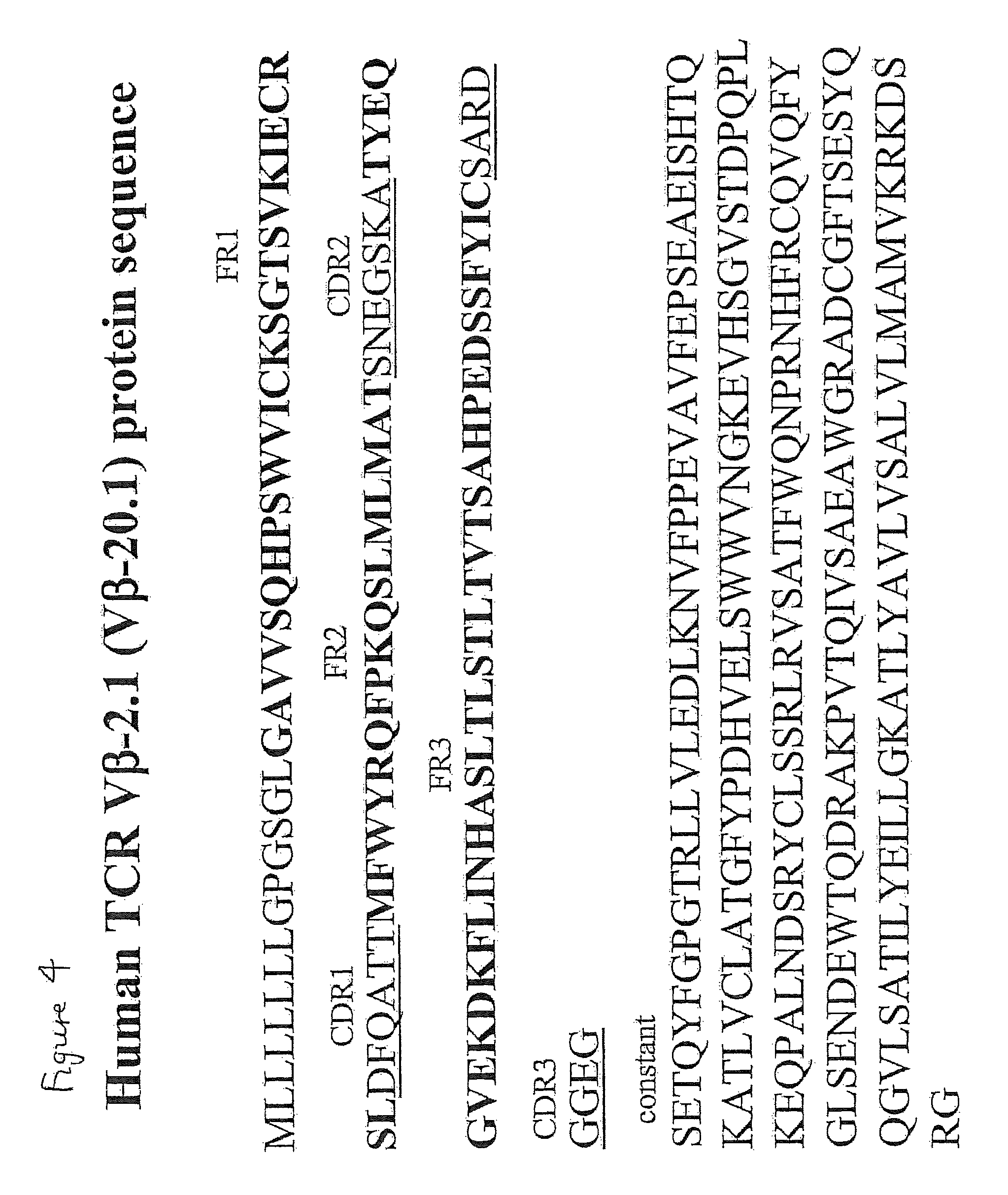
FIG. 4 shows the protein sequence of the pWT126-specific TCR-beta chain (V.beta.-2.1). The position of the CDRs, FRs and constant region are marked.
FIG. 5 shows the same protein sequence as in FIG. 2 but the start position of the constant region is indicated to be in a different place. The CDR sequence in this figure, starting after C, is based on IMGT nomenclature (primary sequence based). The Garcia nomenclature is based on structure and does not include the VV after the C (ie it starts SPF . . . ). Va8.2 means variable alpha 8.2 gene segment and J45 means joining 45 gene segment.
FIG. 6 shows the same protein sequence as in FIG. 4 except that CDR3.beta. is indicated as being longer and the start position of the constant region is indicated to be in a different place. The CDR sequence in this figure, starting after C, is based on IMGT nomenclature (primary sequence based). The Garcia nomenclature is based on structure and does not include the SA after the C (ie it starts RDGG . . . ). J2.5 refers to joining 2.5 gene segment.
FIG. 7 is a diagram showing retroviral vectors containing TCR genes. The TCR alpha and beta chains were inserted into the retroviral vector pMP71 (Engels et al (2003) Human Gene Ther. 14, 1155-1168 for gene transfer into human T cells. LTR is a long terminated repeat. PRE is posttranscriptional regulatory element.
FIG. 8 is a diagram showing retroviral TCR gene transfer into human T cells. Peripheral blood lymphocytes were activated with anti-CD3 antibodies, IL-2 and IL-7, followed 3 days later by transduction with retroviral vectors encoding the WT1-specific TCR. TCR expression was monitored at day 6 using antibodies specific for the TCR-V-beta 2.1 (present in the transferred TCR). Mock transduced T cells show the percentage of un-manipulated human T cells expressing V-beta 2.1. After transduction both CD8-positive and CD8-negative (i.e. CD4-pos) T cells have an increased percentage of V-beta 2.1 cells. V-beta 2.1 DNA and amino acid sequences are shown in FIGS. 3 and 4.
FIG. 9 shows that repeated stimulation of TCR-transduced T cells (as shown in FIG. 8) with T2 cells presenting the pWT126 peptide leads to an expansion of CD8-positive T cells expressing V-beta 2.1.
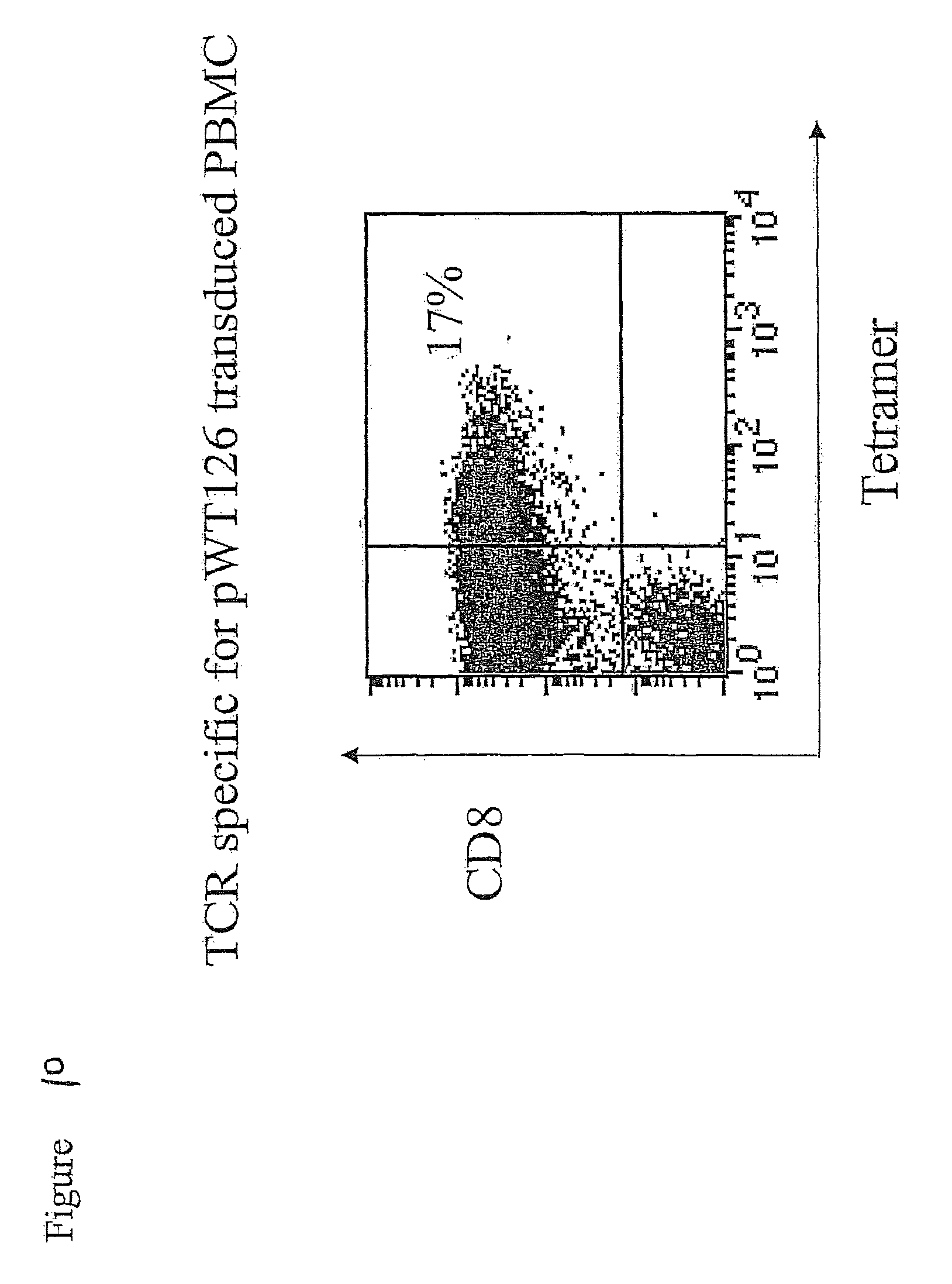
FIG. 10 shows that TCR-transduced T cells (as shown in FIGS. 8 and 9) stain with HLA-A2/pWT126 tetramers.
FIG. 11 shows that TCR-transduced T cells (as shown in FIGS. 8 and 9) kill the HLA-A2-positive T2 cells coated with the pWT126 peptide, but not T2 cells coated with the A2-binding pWT235 control peptide. The T cells also kill the HLA-A2-positive BV173 leukaemia cells expressing WT1 endogenously.
FIG. 12 shows that purified TCR-transduced CD8-positive T cells kill the HLA-A2-positive T2 cells coated with the pWT126 peptide, but not T2 cells coated with the A2-binding pWT235 control peptide. The CD8-positive T cells also kill the HLA-A2-positive BV173 leukaemia cells expressing WT1 endogenously.
FIG. 13 shows that a small percentage of purified CD4-positive TCR-transduced T cells stain with HLA-A2/pWT126 tetramers.
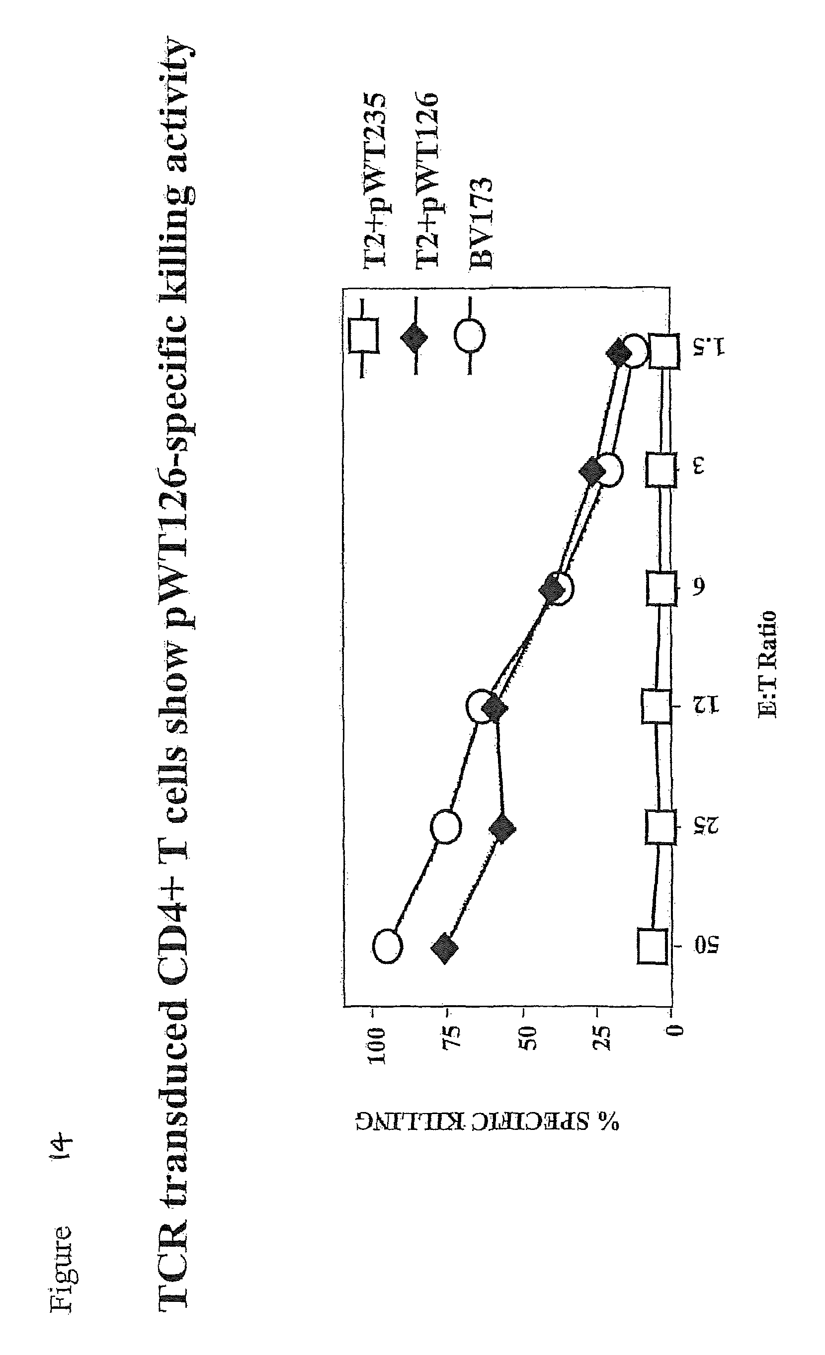
FIG. 14 shows that purified TCR-transduced CD4-positive T cells kill the HLA-A2-positive T2 cells coated with the pWT126 peptide, but not T2 cells coated with the A2-binding pWT235 control peptide. The CD4-positive T cells also kill the HLA-A2-positive BV173 leukaemia cells expressing WT1 endogenously.
FIG. 15 shows that purified TCR-transduced CD8-positive T cells produce IFN-.gamma. after stimulation with the HLA-A2-positive T2 cells coated with the pWT126 peptide, but not T2 cells coated with the A2-binding pWT235 control peptide. Also, the CD8-positive T cells produce IFN-.gamma. after stimulation with the HLA-A2-positive BV173 leukaemia cells expressing WT1 endogenously.
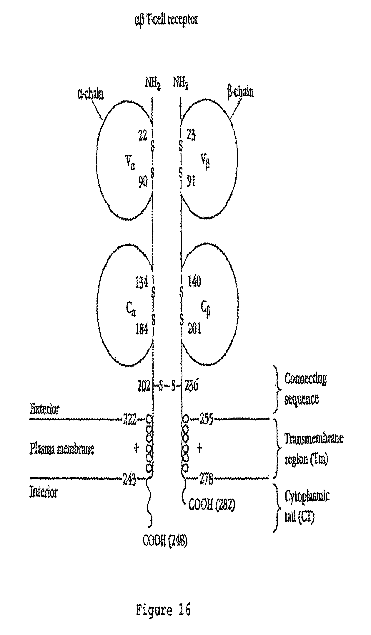
FIG. 16 is a schematic diagram showing the general structure of .alpha..beta. TCR molecules. The amino acid numbers mentioned do not necessarily correspond to those in FIGS. 2 and 4.
DETAILED DESCRIPTION
The general structure of T cell receptors (TCRs), their domain structure and the organisation of genes that encode them is well known, for example see Chapter 11 in Immunology, second edition (1994), by Janis Kuby, W H Freeman & Co, New York, USA, and Garcia et al (1999) Ann. Rev. Immunol. 17, 369-397. One common class of natural TCRs is the .alpha..beta. class in which the TCRs are made up of a separate alpha chain and a separate beta chain which form a heterodimer which is T cell membrane associated. Each alpha and beta chain is made up of regions which, in order from the N terminus to the C terminus are a leader sequence, a variable region, a constant region, a connecting sequence, a transmembrane region and a cytoplasmic tail region (see FIG. 14 for a graphical representation of .alpha..beta. TCR structure). The variable region of the alpha chain is called the V.alpha. region and the variable region of the beta chain is called the V.beta. region. Similarly, the constant region of the alpha chain is called the CI region and the constant region of the beta chain is called the C.beta. region. The job of the .alpha..beta. TCR is to recognise and bind to a peptide presented in a HLA molecule of a cell in the body. Generally speaking, the TCR cannot recognise and bind the peptide unless it is presented by a particular HLA molecule, and the TCR cannot recognise a HLA molecule unless it is presenting the specific peptide. T cells harboring a specific TCR will target cells which are presenting a specific peptide in a particular HLA molecule on a cell (ie a peptide-HLA complex), and this is the main principle of T cell-based immunity.
The peptide-HLA complex is recognised by the combined V regions of the alpha and beta chains of the TCR. In particular, it is the complementarity determining regions (CDRs) of the V regions which mediate recognition of the peptide-HLA complex. The V region of the alpha and beta chains of the natural TCR are made up of, in order in an N-terminal to C-terminal direction, FR1, CDR1, FR2, CDR2, FR3 and CDR3, where FR stands for "framework region" and CDR stands for "complementarity determining region". The FRs and CDRs of the alpha and beta chains are different.
From the predicted amino acid sequences of the alpha and beta chains of the TCR cloned as mentioned above, the inventors have determined the FRs and CDRs of the alpha and beta chains (see FIGS. 2 and 4). With the knowledge of the CDR sequences, it is possible to produce chimaeric TCRs in which the CDRs are grafted onto framework regions with which the CDRs are not naturally associated, and it is also possible to produce single chain TCR molecules, and in both cases the molecules retain substantially the same binding affinity for the peptide-HLA complex as the parent molecule, as is described in more detail below.
A first aspect of the invention provides a T cell receptor (TCR) molecule containing an alpha chain portion and a beta chain portion wherein the alpha chain portion contains three complementarity determining regions (CDRs):
TABLE-US-00001 (SEQ ID NO: 2) CDR1.alpha.: SSYSPS (SEQ ID NO: 3) CDR2.alpha.: YTSAATL (SEQ ID NO: 4) CDR3.alpha.: VVSPFSGGGADGLT or comprising or (SEQ ID NO: 5) consisting of SPFSGGGADGLT
and the beta chain portion contains three complementarity determining regions (CDRs):
TABLE-US-00002 (SEQ ID NO: 6) CDR1.beta.: DFQATT (SEQ ID NO: 7) CDR2.beta.: SNEGSKA (SEQ ID NO: 8) CDR3.beta.: comprising SARDGGEG, or comprising or (SEQ ID NO: 9) consisting of RDGGEGSETQY
or wherein up to three amino acid residues in one or more of the CDRs are replaced by another amino acid residue.
It should be noted that in some nomenclature systems the CDR3 of the .beta. chains may be defined to be longer than in the nomenclature system used in the Immunogenetics (IMGT) database described below. Also, in some nomenclature systems the CDR3 of the .alpha. chains may be defined to be shorter than in the IMGT system. Similarly, the constant portion may or may not include framework residues flanking the CDR3 region in the different nomenclature systems.
Thus, in one embodiment using the IMGT system CDR3.alpha. may have the amino acid sequence VVSPFSGGGADGLT (SEQ ID NO: 4) and the constant portion includes the framework amino acid sequence FGKGTHLIIQP (SEQ ID NO: 10) (see FIG. 5).
In another embodiment, using the Garcia nomenclature system (Garcia et al (1999) Ann. Rev. Immunol. 17, 369-397, incorporated herein by reference) CDR3.alpha. comprises or consists of the amino acid sequence SPFSGGGADGLT (SEQ ID NO: 5), the framework region immediately C-terminal to this has the amino acid sequence FGKGTHLIIQP (SEQ ID NO: 10) and the constant region begins with the amino acid sequence YIQNP (SEQ ID NO: 11) (see FIG. 5).
In one embodiment using the IMGT nomenclature system, CDR3.beta. may have the amino acid sequence SARDGGEG (SEQ ID NO: 8) and the constant region immediately C-terminal to this includes the framework amino acid sequence SETQY (SEQ ID NO: 12) (FIG. 4).
In another embodiment, using the Garcia nomenclature system as above, CDR3.beta. comprises or consists of the amino acid sequence RDGGEGSETQY (SEQ ID NO: 9) and the framework region immediately C-terminal to this has the amino acid sequence FGPGTRLLVL (SEQ ID NO: 13) and the immediately C-terminal constant region begins with the amino acid sequence EDLKN (SEQ ID NO: 14) (see FIG. 6).
It will be appreciated that the skilled person can readily design and synthesise TCRs according to the invention using either or any nomenclature systems provided that the framework region (ie region not replaced by the CDRs) is compatible with the CDRs as is well known in the art.
The standard IUPAC one letter amino acid code is used throughout the specification. For the avoidance of doubt, a reference to a "particular" or "given" CDR means any CDR with the amino acid sequence given above or wherein up to three amino acids have been replaced by another amino acid residue.
By "TCR molecule" we include any molecule which contains the given CDRs and also contains FRs suitably situated within the molecule so that the CDRs form a recognition site (combining site) which is able to bind to HLA-A2 presenting the peptide RMFPNAPYL (SEQ ID NO: 1) (ie a HLA-A2/RMFPNAPYL (SEQ ID NO: 1) complex).
It is particularly preferred if the TCR molecules contain the precise CDR amino acid sequences as given above and in FIGS. 2 and 4 and in FIGS. 5 and 6. Where a variant to this precise sequence is present, it preferably varies by one or two or three (preferably one or two) amino acids in one or two or three or four or five or all six CDRs. Typically, in these variants, the amino acids which are replaced are replaced with conservative amino acids. By conservative amino acids we include the groupings: G, A; S, A, T; F, Y, W; D, E; N, Q; and I, L, V.
A method for making and selecting TCR molecules which have CDRs which vary from the precise CDR sequences given in FIGS. 2 and 4 and in FIGS. 5 and 6 is given below.
The amino acid sequences, including V regions (and therefore FRs), of numerous TCR alpha chains and TCR beta chains are well known in the art, some of which are described in the IMGT (Immunogenetics) database at http.//imgt.cines.fr. See also Lefranc (2003) Dev. Comp. Immunol. 27, 55-77. The structural basis of T cell recognition is reviewed in Garcia et al (1999) Ann. Rev. Immunol. 17, 369-397, and the information contained therein may be used to design and synthesise CDR-grafted TCRs (and CDRs defined on the basis of this nomenclature are noted above). Preferably, the FRs into which the particular CDRs are grafted are FRs of human TCR alpha or beta chains. Conveniently, the alpha chain CDRs are grafted into alpha chain FRs, and beta chain CDRs are grafted to beta chain FRs. Typically, the three CDRs in the alpha chain and the three CDRs in the beta chain replace, in order, CDRs in other human alpha and beta chains, respectively. See Lefranc (2003) Dev. Comp. Immunol. 27, 55-77.
Typically, T cells expressing the TCR molecule recognise the HLA-A2 presenting peptide RMFPNAPYL (SEQ ID NO: 1) with substantially the same avidity as the TCR molecule which consists of the alpha and beta chains as described in FIGS. 2 and 4. This can be measured by retroviral-mediated transfer of the TCR into T cells followed by peptide titration experiments with the TCR-transduced T cells as outlined, for example, in Gao et al (2000) Blood 95, 2198-2203.
The TCR molecule preferably contains an alpha chain portion containing, in N-terminal to C-terminal order, FR1.alpha.-CDR1.alpha.-FR2.alpha.-CDR2.alpha.-FR3.alpha.-CDR3.alpha., and a beta chain portion containing, in N-terminal to C-terminal order, FR1.beta.-CDR1.beta.-FR2.beta.-CDR2.beta.-FR3.beta.-CDR3.beta. as shown in FIGS. 2 and 4, respectively and in FIGS. 5 and 6, respectively. Typically, the TCR molecule contains the V region of both the alpha chain and the beta chain of the TCR polypeptide chains shown in FIGS. 2 and 4, and in FIGS. 5 and 6.
In a preferred embodiment, the alpha chain portion and the beta chain portion are present on different polypeptide chains. Typically, the TCR molecule contains an alpha chain which contains the V region and the C region of the polypeptide chain shown in FIG. 2 (or FIG. 5), and also contains a beta chain which contains the V region and C region of the polypeptide chain shown in FIG. 4 (or FIG. 6). Preferably, the TCR molecule consists of a molecule containing the complete alpha chain shown in FIG. 2 and the complete beta chain shown in FIG. 4. Typically, however, the leader sequence is cleaved off the mature alpha chain and beta chain.
In a further embodiment, the alpha chain portion and the beta chain portion of the TCR molecule are present in the same polypeptide chain. Single chain TCR molecules are described in Chung et al (1994) Proc. Natl. Acad. Sci. USA 91, 12654-12658, and the principles described therein may readily be applied to the production of single chain TCR molecules which contain the specified CDRs. Typically, the single chain TCR molecules contain the V.alpha., V.beta. and C.beta. domains fused in the same polypeptide chain, and typically in that order (from N-terminus to C-terminus). For expression of a single chain TCR it is useful to provide a construct encoding the constant domain of the TCR alpha chain.
An additional strategy is described in Boulter et al (2003) Protein Eng. 16, 707-711 in which a new disulphide bond is introduced between a threonine in the constant region of the alpha chain and a serine in the constant region of the beta chain (by replacing these residues with a cysteine). The disulphide bond in the TCR connecting peptide may be removed or may remain in place.
The two-chain TCR molecules of the invention (eg ones which contain the alpha and beta chains whose amino acid sequence is given in FIGS. 2 and 4) or chimaeric TCRs which contain the specific CDRs as described above may be used to introduce to create antigen-specific CTL as described in more detail below (by using polynucleotides that encode the relevant chains). Similarly, the single chain TCRs may also be used for this purpose, and have the advantage that they do not pair with endogenous TCRs. Single chain TCRs may also be used as soluble constructs in a way similar to antibodies. In this case, the single chain constructs do not contain a transmembrane region (see Chung et al supra and Boulter et al supra).
A second aspect of the invention provides a polynucleotide encoding the alpha chain portion as defined in the first aspect of the invention. A third aspect of the invention provides a polynucleotide encoding the beta chain portion as defined in the first aspect of the invention. As discussed above, in a particularly preferred embodiment of the invention, the alpha chain portion and the beta chain portion are present on different polypeptide chains, and it is convenient (but not mandatory) that each polypeptide is encoded by a separate polynucleotide. Preferred polynucleotides encoding the alpha and beta chains are described in FIGS. 1 and 2, respectively. Alternatively, the two polypeptides may be encoded on the same polynucleotide, in which case the two coding regions may be linked by an (Internal Ribosome Entry Site) IRES sequence, and typically would have its own translational start and stop codons. Typically, such constructs contain two promoters, one for each TCR chain.
As discussed above, in an alternative embodiment the alpha chain portion and the beta chain portion are present in the same polypeptide, in which case a single polynucleotide may encode the single chain polypeptide.
In any event, the polynucleotide may be DNA or RNA, and it may or may not contain introns. Typically, the polynucleotide does not contain introns within the region that codes for the polypeptide of interest. It will be appreciated that different polynucleotides may encode the same polypeptide because of the degeneracy of the genetic code.
The invention also provides an expression vector that contains the polynucleotide of the invention. Such expression vectors, when present in a suitable host cell, allow for the expression of the polypeptide(s) of interest. Preferably, the expression vector is an expression vector capable of expressing a polypeptide in a mammalian cell. More preferably, the expression vector is one which is able to express a polypeptide in a T cell, such as a human CTL. Typically, the expression vectors contain a promoter which is active in particular cell types, and which may be controllable (eg inducible).
The vector is suitably a retroviral vector which is capable of transfection into a mammalian host cell such as a human T cell. Typically, the vector is a lentiviral vector.
A further aspect of the invention provides a host cell comprising a polynucleotide of the invention or a vector of the invention. The host cell may contain a polynucleotide or vector which encodes only the alpha chain portion or only the beta chain portion. However, if the host cell is to produce a TCR molecule of the invention, it contains one or more polynucleotides or vectors which encode both the alpha chain portion and the beta chain portion.
The host cell may be any cell such as a bacterial cell, yeast cell, insect cell, plant cell or mammalian cell, and methods of introducing polynucleotides into such cells are well known in the art. Typically, bacterial cells, such as Escherichia coli cells are used for general propagation and manipulation of the polynucleotides and vectors of the invention. Other host cells may be used to express the TCR molecules of the invention and, in particular, the cell may be a mammalian cell such as a human cell. As described below in relation to the therapeutic methods using the TCR molecules of the invention, it is particularly desirable if the host cell is a T cell such as (and preferably) a T cell derived from a patient to be treated, typically a patient with a WT1-expressing malignancy.
Typically, a retroviral vector (or, as the case may be vectors) encoding the TCR molecule of the invention is used based on its ability to infect mature human CD4.sup.+ or CD8.sup.+ T lymphocytes and to mediate gene expression: the retroviral vector system Kat is one preferred possibility (see Finer et al (1994) Blood 83, 43). High titre amphotrophic retrovirus are used to infect purified CD8.sup.+ T lymphocytes isolated from the peripheral blood of tumour patients following a protocol published by Roberts et al (1994) Blood 84, 2878-2889, incorporated herein by reference. Anti-CD3 antibodies are used to trigger proliferation T cells, which facilitates retroviral integration and stable expression of single chain TCRs. A combination of anti-CD3 and anti-CD8 antibodies may be more effective than anti-CD3 antibodies alone. Other suitable systems for introducing genes into CTL are described in Moritz et al (1994) Proc. Natl. Acad. Sci. USA 91, 4318-4322, incorporated herein by reference. Eshhar et al (1993) Proc. Natl. Acad. Sci. USA 90, 720-724 and Hwu et al (1993) J. Exp. Med. 178, 361-366 also describe the transfection of CTL. The commercially available Nuclofactor system, provided by AMAXA, Germany may be used to transfect T cells. Retroviral transduction of human CD8.sup.+ T cells is described in Stanislawski (2001) Nat. Immunol. 2, 962.
Methods of cloning and genetic manipulation are well known in the art and are described in detail in standard manuals such as Sambrook & Russell (2001) Molecular Cloning, a laboratory manual, Cold Spring Harbor Press, Cold Spring Harbor, N.Y., USA.
Patients suffering from a WT1-expressing malignancy may be treated by the introduction of the TCR molecule of the invention into their own T cells (or T cells from a donor), followed by the introduction of these engineered cells into the patient. Thus, a further aspect of the invention provides a method of combating a WT-1 expressing malignancy in a patient, the method comprising introducing into the patient a T cell, preferably derived from the patient, which is modified to express the TCR molecule of the invention. Typically, (1) T cells are obtained from the patient, (2) a polynucleotide or polynucleotides encoding and capable of expressing the TCR molecule of the invention are introduced into the T cells ex vivo and (3) the engineered T cells are introduced into the patient. It is particularly preferred if the T cells are the patient's T cells (ie autologous).
It is particularly preferred if the patient is HLA-A2 positive.
In other words, the specificity of the T cell, preferably autologous T cell, is changed by the introduction of the TCR molecule of the invention.
The T cells (for example of the patient) are typically isolated from peripheral blood mononuclear cells (PBMCs), and may be CD4.sup.+ and CD8.sup.+ cells. Typically, the cells are activated using an antibody (eg an anti-CD3 or anti-CD28 antibody) so that they become receptive to transfection, for example with one or more retroviral vectors encoding the TCR molecules of the invention. The number of cells isolated, transfected and returned to the patient may be determined by the physician.
Cells may be taken from a patient after a clinical response, cryopreserved, transfected and re-infused if the same patient relapses.
Whether or not a malignancy is one which expresses WT1 may be determined, for example using reverse transcriptase-polymerase chain reaction (RT-PCR) or using intracellular staining techniques for the WT1 protein (which may be anti-WT1 antibodies).
The patient is preferably a human patient although animals may be used in a research situation. It is particularly preferred that the patient is HLA-A2 positive. Whether or not a patient is HLA-A2 positive can be determined by methods well known in the art.
Typically, the patient is suffering from any one or more of leukaemia, breast cancer, colon cancer, lung cancer, ovarian cancer, melanoma, thyroid cancer, head and neck cancer, glioblastoma, and sarcoma.
A further aspect of the invention provides the use of a T cell, preferably a patient derived T cell, which is modified to express the TCR molecule of the invention in the manufacture of a medicament for combating a WT1-expressing malignancy in the patient.
As discussed above, TCR molecules in which one or more of the CDRs differ in sequence from the precise CDR sequences given in FIGS. 2 and 4 form part of the invention. Preferably, such TCR molecules are able to recognise the HLA-A2/RMFPNAPYL (SEQ ID NO: 1) complex more effectively than a TCR molecule with the precise CDR sequences. Thus, a further aspect of the invention provides a method of selecting a TCR molecule with improved binding to an HLA-A2/RMFPNAPYL (SEQ ID NO: 1) complex comprising (a) providing a TCR molecule containing an alpha chain portion and a beta chain portion wherein the alpha chain portion contains three complementarity determining regions (CDRs):
TABLE-US-00003 (SEQ ID NO: 2) CDR1.alpha.: SSYSPS (SEQ ID NO: 3) CDR2.alpha.: YTSAATL (SEQ ID NO: 4) CDR3.alpha.: VVSPFSGGGADGLT or comprising or (SEQ ID NO: 5) consisting of SPFSGGGADGLT
and the beta chain portion contains three complementarity determining regions (CDRs):
TABLE-US-00004 (SEQ ID NO: 6) CDR1.beta.: DFQATT (SEQ ID NO: 7) CDR2.beta.: SNEGSKA (SEQ ID NO: 8) CDR3.beta.: comprising SARDGGEG or comprising or (SEQ ID NO: 9) consisting of RDGGEGSETQY
wherein at least one amino acid residue in one or more of the CDRs as given is replaced with another amino acid residue, (b) determining whether the TCR molecule binds to an HLA-A2/RMFPNAPYL (SEQ ID NO: 1) complex with greater affinity than a TCR molecule without the replacement amino acid(s), and (c) selecting a molecule which binds with greater affinity. Preferably, the CDR3.beta. has the amino acid sequence given above in relation to the first aspect of the invention.
TCR molecules with altered CDRs can readily be made by protein engineering methods. For example, a TCR display library may be made in which the alpha chain and/or beta chain CDR regions are mutagenised and the TCR molecules displayed using retroviral transduction on the surface of a T cell lymphoma (see Kessels et al (2000) Proc. Natl. Acad. Sci. USA 97, 14578-14583), or on the surface of a yeast or a bacteriophage. A HLA-A2/RMFPNAPYL (SEQ ID NO: 1) complex may be used to select cells or bacteriophages which bind the complex with high affinity by virtue of the TCR molecule that they present. TCR molecules which have a higher binding affinity (lower K.sub.D) than a TCR molecule with the precise CDR sequences are selected for further study.
The invention will now be described in more detail by reference to the following figures and non-limiting examples.
TABLE-US-00005 Schedule of SEQ ID Nos. 1. RMFPNAPYL 2. SSYSPS 3. YTSAATL 4. VVSPFSGGGADGLT 5. SPFSGGGADGLT 6. DFQATT 7. SNEGSKA 8. SARDGGEG 9. RDGGEGSETQY 10. FGKGTHLIIQP 11. YIQNP 12. SETQY 13. FGPGTRLLVL 14. EDLKN 15. FIG. 1 nucleotide sequence 16. FIG. 2 (and FIG. 5) amino acid sequence 17. FIG. 3 nucleotide sequence 18. FIG. 4 (and FIG. 6) amino acid sequence
EXAMPLES
Example 1
Functionally Active T Cell Receptor (TCR) Specific for the WT-1-Derived Peptide pWT126 (RMFPNAPYL)
We have cloned a T cell receptor (TCR) that is specific for a peptide (pWT126; RMFPNAPYL (SEQ ID NO: 1) of the Wilms Tumour antigen-1 (WT1) presented by HLA-A2 class I molecules. The WT1 transcription factor is expressed in various human malignancies, including leukaemia, breast cancer, colon cancer, lung cancer, ovarian cancer and others. The CTL from which the TCR was cloned show killing activity against human cancer cells that express WT1, but not against normal human cells that express physiological levels of WT1.
The therapeutic goal is to equip patient T cells with this potent and specific killing activity by transfer of the genes encoding the TCR. For this, we have inserted the TCR genes into retroviral vectors and demonstrated that gene transduced human T cells show killing activity against WT1 expressing human cancer and leukemia cell lines. The specificity profile of this CTL line has been described in several research papers and can be summarized as: (1) Killing of HLA-A2-positive targets coated with the WT1-derived peptide pWT126 (Gao et al (2000) Blood 95, 2198-2203); (2) Killing of fresh HLA-A2-positive leukaemia cells expressing WT1 (Gao et al (2000) Blood 95, 2198-2203); (3) Killing of HLA-A2-positive leukemia CFU progenitor cells (Gao et al (2000) Blood 95, 2198-2203; Bellantuono et al (2002) 100, 3835-3837); (4) Killing of HLA-A2-positive leukaemia LTC-IC stem cells (Bellantuono et al (2002) Blood 100, 3835-3837); (5) Killing of HLA-A2-positive NOD/SCID leukaemia initiating cells (Gao et al (2003) Transplantation 75, 1429-1436); and (6) No killing of normal HLA-A2-positive NOD/SCID engrafting hematopoietic stem cells (Gao et al (2003) Transplantation 75, 1429-1436). We have now shown that human T cells transduced with the WT1-specific TCR display similar specificity as the CTL line from which the TCR was cloned.
The data described in detail in the legends to FIGS. 1 to 15 indicate that TCR gene transfer into human T cells is feasible and that it leads to the surface expression of the introduced TCR chains. The recipient T cells show killing activity against HLA-A2-positive targets coated with the pWT126 peptide. The TCR-transduced T cells also kill human tumour cells expressing WT1 endogenously. In addition, the transduced T cells produce IFN-g in an HLA-A2-restricted, peptide-specific fashion. Finally, the transferred TCR can function in CD4-positive helper T cells. These CD4-positive T cells show HLA-A2-restricted, antigen-specific killing activity and antigen-specific cytokine production (not shown). This indicates that TCR gene transfer can be used to confer HLA class I-restricted antigen-specific effector function to both CD8-positive and CD4-positive human T cells.
Example 2
Selection and Treatment of a Patient
Peripheral blood monocyte cells (PBMCs) are taken from an HLA-A2-positive patient who has a WT1-expressing malignancy. The PBMCs are activated with anti-CD3/CD28 antibodies added to the culture or on beads for 3 days and then transduced with TCR encoding retroviral particles as described in Example 1. At day 5 we can demonstrate that transduced CD4 and CD8 T cells express the introduced TCR. At day 6 we can demonstrate antigen-specific activity of the transduced T cells. At day 6 the transduced T cells are reinfused into the patient.
The following embodiments are provided:
1. A T cell receptor (TCR) molecule containing an alpha chain portion and a beta chain portion wherein the alpha chain portion contains three complementarity determining regions (CDRs):
TABLE-US-00006 (SEQ ID NO: 2) CDR1.alpha.: SSYSPS (SEQ ID NO: 3) CDR2.alpha.: YTSAATL (SEQ ID NO: 4) CDR3.alpha.: VVSPFSGGGADGLT or comprising or (SEQ ID NO: 5) consisting of SPFSGGGADGLT
and the beta chain portion contains three complementarity determining regions (CDRs):
TABLE-US-00007 (SEQ ID NO: 6) CDR1.beta.: DFQATT (SEQ ID NO: 7) CDR2.beta.: SNEGSKA (SEQ ID NO: 8) CDR3.beta.: comprising SARDGGEG or comprising or (SEQ ID NO: 9) consisting of RDGGEGSETQY,
or wherein up to three amino acid residues in one or more of the CDRs are replaced by another amino acid residue.
2. A TCR molecule according to Embodiment 1 wherein CDR3.alpha. has the amino acid sequence VVSPFSGGGADGLT (SEQ ID NO: 4).
3. A TCR molecule according to Embodiment 1 wherein the CDR3.alpha. has the amino acid sequence SPFSGGGADGLT (SEQ ID NO: 5).
4. A TCR molecule according to Embodiment 1 wherein the CDR3.beta. has the amino acid sequence SARDGGEG (SEQ ID NO: 8).
5. A TCR molecule according to Embodiment 1 wherein the CDR3.beta. has the amino acid sequence RDGGEGSETQY (SEQ ID NO: 9).
6. A TCR molecule according to any one of the preceding Embodiments wherein the alpha chain portion and the beta chain portion are present on different polypeptide chains.
7. A TCR molecule according to any one of Embodiments 1 to 5 wherein the alpha chain portion and the beta chain portion are present in the same polypeptide chain.
8. A TCR molecule according to any of Embodiments 1 to 7 wherein the CDRs are grafted to a human framework region.
9. A TCR molecule according to Embodiment 8 wherein the alpha chain portion has the amino acid sequence given in FIG. 2.
10. A TCR molecule according to Embodiment 8 or 9 wherein the beta chain portion has the amino acid sequence given in FIG. 4.
11. A TCR molecule according to any one of Embodiments 1 to 10 which is soluble.
12. A polynucleotide encoding the alpha chain portion as defined in Embodiment 1.
13. A polynucleotide encoding the beta chain portion as defined in Embodiment 1.
14. A polynucleotide encoding the single chain TCR molecule as defined in Embodiment 7.
15. An expression vector comprising a polynucleotide according to any of Embodiments 12 to 15.
16. An expression vector according to Embodiment 15 which is a retroviral vector.
17. A host cell comprising a polynucleotide according to any of Embodiments 12 to 14 or an expression vector according to Embodiment 15 or 16.
18. A host cell according to Embodiment 17 which is a T cell.
19. A host cell according to Embodiment 18 which is a T cell derived from a patient.
20. A method of combating a WT1-expressing malignancy in a patient, the method comprising introducing into the patient a T cell, preferably derived from the patient, which is modified to express the TCR molecule of any of Embodiments 1 to 11.
21. A method according to Embodiment 20 comprising (1) obtaining T cells from the patient, (2) introducing into the T cells a polynucleotide according to any of Embodiments 12 to 14 or an expression vector according to Embodiments 15 or 16 so that the T cell expresses the encoded TCR molecule and (3) introducing the cells from step (2) into the patient.
22. A method according to Embodiment 20 or 21 wherein the WT1-malignancy is any one or more of breast cancer, colon cancer, lung cancer, leukaemia, ovarian cancer, melanoma, head and neck cancer, thyroid cancer, glioblastoma and sarcoma.
23. Use of a T cell, preferably a patient derived T cell, modified to express the TCR molecule of any of Embodiments 1 to 11 in the manufacture of a medicament for combating a WT1-expressing malignancy in the patient.
24. Use according to Embodiment 23 wherein a polynucleotide according to any of Embodiments 12 to 14 or an expression vector according to Embodiments 15 or 16 has been introduced into the T cell, preferably patient derived T cell, so that the T cell expresses the encoded TCR molecule.
25. A method of selecting a TCR molecule with improved binding to an HLA-A2/RMFPNAPYL (SEQ ID NO: 1) complex comprising (a) providing a TCR molecule containing an alpha chain portion and a beta chain portion wherein the alpha chain portion contains three complementarity determining regions (CDRs):
TABLE-US-00008 (SEQ ID NO: 2) CDR1.alpha.: SSYSPS (SEQ ID NO: 3) CDR2.alpha.: YTSAATL (SEQ ID NO: 4) CDR3.alpha.: VVSPFSGGGADGLT or comprising or (SEQ ID NO: 5) consisting of SPFSGGGADGLT
and the beta chain portion contains three complementarity determining regions (CDRs):
TABLE-US-00009 (SEQ ID NO: 6) CDR1.beta.: DFQATT (SEQ ID NO: 7) CDR2.beta.: SNEGSKA (SEQ ID NO: 8) CDR3.beta.: comprising SARDGGEG or comprising or (SEQ ID NO: 9) consisting of RDGGEGSETQY,
wherein at least one amino acid residue in one or more of the CDRs as given is replaced with another amino acid residue, (b) determining whether the TCR molecule binds to an HLA-A2/RFMPNAPYL (SEQ ID NO: 1) complex with greater affinity than a TCR molecule without the replacement amino acid(s), and (c) selecting a molecule which binds with greater affinity.
26. A method according to Embodiment 25 wherein the CDR3s are as defined in any of Embodiments 2 to 9.
27. Any novel method of combating cancer as herein described.
SEQUENCE LISTINGS
1
1819PRTArtificial SequenceWT1 epitope - WT126 1Arg Met Phe Pro Asn Ala Pro Tyr Leu1 526PRTArtificial SequenceTCR CDR1 alpha 2Ser Ser Tyr Ser Pro Ser1 537PRTArtificial SequenceTCR CDR2 alpha 3Tyr Thr Ser Ala Ala Thr Leu1 5414PRTArtificial SequenceTCR CDR3 alpha 4Val Val Ser Pro Phe Ser Gly Gly Gly Ala Asp Gly Leu Thr1 5 10512PRTArtificial SequenceTCR CDR3 alpha 5Ser Pro Phe Ser Gly Gly Gly Ala Asp Gly Leu Thr1 5 1066PRTArtificial SequenceTCR CDR1 beta 6Asp Phe Gln Ala Thr Thr1 577PRTArtificial SequenceTCR CDR2 beta 7Ser Asn Glu Gly Ser Lys Ala1 588PRTArtificial SequenceTCR CDR3 beta 8Ser Ala Arg Asp Gly Gly Glu Gly1 5911PRTArtificial SequenceTCR CDR3 beta 9Arg Asp Gly Gly Glu Gly Ser Glu Thr Gln Tyr1 5 101011PRTArtificial SequenceTCR framework sequence 10Phe Gly Lys Gly Thr His Leu Ile Ile Gln Pro1 5 10115PRTArtificial Sequencestart of constant region sequence 11Tyr Ile Gln Asn Pro1 5125PRTArtificial SequenceTCR framework sequence 12Ser Glu Thr Gln Tyr1 51310PRTArtificial SequenceTCR framework sequence 13Phe Gly Pro Gly Thr Arg Leu Leu Val Leu1 5 10145PRTArtificial Sequencestart of constant region 14Glu Asp Leu Lys Asn1 515831DNAHomo sapiens 15atgctcctgc tgctcgtccc agtgctcgag gtgattttta ctctgggagg aaccagagcc 60cagtcggtga cccagcttga cagccacgtc tctgtctctg aaggaacccc ggtgctgctg 120aggtgcaact actcatcttc ttattcacca tctctcttct ggtatgtgca acaccccaac 180aaaggactcc agcttctcct gaagtacaca tcagcggcca ccctggttaa aggcatcaac 240ggttttgagg ctgaatttaa gaagagtgaa acctccttcc acctgacgaa accctcagcc 300catatgagcg acgcggctga gtacttctgt gttgtgagtc ctttttcagg aggaggtgct 360gacggactca cctttggcaa agggactcat ctaatcatcc agccctatat ccagaaccct 420gaccctgccg tgtaccagct gagagactct aaatccagtg acaagtctgt ctgcctattc 480accgattttg attctcaaac aaatgtgtca caaagtaagg attctgatgt gtatatcaca 540gacaaaactg tgctagacat gaggtctatg gacttcaaga gcaacagtgc tgtggcctgg 600agcaacaaat ctgactttgc atgtgcaaac gccttcaaca acagcattat tccagaagac 660accttcttcc ccagcccaga aagttcctgt gatgtcaagc tggtcgagaa aagctttgaa 720acagatacga acctaaactt tcaaaacctg tcagtgattg ggttccgaat cctcctcctg 780aaagtggccg ggtttaatct gctcatgacg ctgcggctgt ggtccagctg a 83116276PRTHomo sapiens 16Met Leu Leu Leu Leu Val Pro Val Leu Glu Val Ile Phe Thr Leu Gly1 5 10 15Gly Thr Arg Ala Gln Ser Val Thr Gln Leu Asp Ser His Val Ser Val 20 25 30Ser Glu Gly Thr Pro Val Leu Leu Arg Cys Asn Tyr Ser Ser Ser Tyr 35 40 45Ser Pro Ser Leu Phe Trp Tyr Val Gln His Pro Asn Lys Gly Leu Gln 50 55 60Leu Leu Leu Lys Tyr Thr Ser Ala Ala Thr Leu Val Lys Gly Ile Asn65 70 75 80Gly Phe Glu Ala Glu Phe Lys Lys Ser Glu Thr Ser Phe His Leu Thr 85 90 95Lys Pro Ser Ala His Met Ser Asp Ala Ala Glu Tyr Phe Cys Val Val 100 105 110Ser Pro Phe Ser Gly Gly Gly Ala Asp Gly Leu Thr Phe Gly Lys Gly 115 120 125Thr His Leu Ile Ile Gln Pro Tyr Ile Gln Asn Pro Asp Pro Ala Val 130 135 140Tyr Gln Leu Arg Asp Ser Lys Ser Ser Asp Lys Ser Val Cys Leu Phe145 150 155 160Thr Asp Phe Asp Ser Gln Thr Asn Val Ser Gln Ser Lys Asp Ser Asp 165 170 175Val Tyr Ile Thr Asp Lys Thr Val Leu Asp Met Arg Ser Met Asp Phe 180 185 190Lys Ser Asn Ser Ala Val Ala Trp Ser Asn Lys Ser Asp Phe Ala Cys 195 200 205Ala Asn Ala Phe Asn Asn Ser Ile Ile Pro Glu Asp Thr Phe Phe Pro 210 215 220Ser Pro Glu Ser Ser Cys Asp Val Lys Leu Val Glu Lys Ser Phe Glu225 230 235 240Thr Asp Thr Asn Leu Asn Phe Gln Asn Leu Ser Val Ile Gly Phe Arg 245 250 255Ile Leu Leu Leu Lys Val Ala Gly Phe Asn Leu Leu Met Thr Leu Arg 260 265 270Leu Trp Ser Ser 27517933DNAHomo sapiens 17atgctgctgc ttctgctgct tctggggcca ggctccgggc ttggtgctgt cgtctctcaa 60catccgagct gggttatctg taagagtgga acctctgtga agatcgagtg ccgttccctg 120gactttcagg ccacaactat gttttggtat cgtcagttcc cgaaacagag tctcatgctg 180atggcaactt ccaatgaggg ctccaaggcc acatacgagc aaggcgtcga gaaggacaag 240tttctcatca accatgcaag cctgaccttg tccactctga cagtgaccag tgcccatcct 300gaagacagca gcttctacat ctgcagtgct agagatgggg gggagggttc ggagacccag 360tacttcgggc caggcacgcg gctcctggtg ctcgaggacc tgaaaaacgt gttcccaccc 420gaggtcgctg tgtttgagcc atcagaagca gagatctccc acacccaaaa ggccacactg 480gtgtgcctgg ccacaggctt ctaccccgac cacgtggagc tgagctggtg ggtgaatggg 540aaggaggtgc acagtggggt cagcacagac ccgcagcccc tcaaggagca gcccgccctc 600aatgactcca gatactgcct gagcagccgc ctgagggtct cggccacctt ctggcagaac 660ccccgcaacc acttccgctg tcaagtccag ttctacgggc tctcggagaa tgacgagtgg 720acccaggata gggccaaacc tgtcacccag atcgtcagcg ccgaggcctg gggtagagca 780gactgtggct tcacctccga gtcttaccag caaggggtcc tgtctgccac catcctctat 840gagatcttgc tagggaaggc caccttgtat gccgtgctgg tcagtgccct cgtgctgatg 900gccatggtca agagaaagga ttccagaggc tag 93318310PRTHomo sapiens 18Met Leu Leu Leu Leu Leu Leu Leu Gly Pro Gly Ser Gly Leu Gly Ala1 5 10 15Val Val Ser Gln His Pro Ser Trp Val Ile Cys Lys Ser Gly Thr Ser 20 25 30Val Lys Ile Glu Cys Arg Ser Leu Asp Phe Gln Ala Thr Thr Met Phe 35 40 45Trp Tyr Arg Gln Phe Pro Lys Gln Ser Leu Met Leu Met Ala Thr Ser 50 55 60Asn Glu Gly Ser Lys Ala Thr Tyr Glu Gln Gly Val Glu Lys Asp Lys65 70 75 80Phe Leu Ile Asn His Ala Ser Leu Thr Leu Ser Thr Leu Thr Val Thr 85 90 95Ser Ala His Pro Glu Asp Ser Ser Phe Tyr Ile Cys Ser Ala Arg Asp 100 105 110Gly Gly Glu Gly Ser Glu Thr Gln Tyr Phe Gly Pro Gly Thr Arg Leu 115 120 125Leu Val Leu Glu Asp Leu Lys Asn Val Phe Pro Pro Glu Val Ala Val 130 135 140Phe Glu Pro Ser Glu Ala Glu Ile Ser His Thr Gln Lys Ala Thr Leu145 150 155 160Val Cys Leu Ala Thr Gly Phe Tyr Pro Asp His Val Glu Leu Ser Trp 165 170 175Trp Val Asn Gly Lys Glu Val His Ser Gly Val Ser Thr Asp Pro Gln 180 185 190Pro Leu Lys Glu Gln Pro Ala Leu Asn Asp Ser Arg Tyr Cys Leu Ser 195 200 205Ser Arg Leu Arg Val Ser Ala Thr Phe Trp Gln Asn Pro Arg Asn His 210 215 220Phe Arg Cys Gln Val Gln Phe Tyr Gly Leu Ser Glu Asn Asp Glu Trp225 230 235 240Thr Gln Asp Arg Ala Lys Pro Val Thr Gln Ile Val Ser Ala Glu Ala 245 250 255Trp Gly Arg Ala Asp Cys Gly Phe Thr Ser Glu Ser Tyr Gln Gln Gly 260 265 270Val Leu Ser Ala Thr Ile Leu Tyr Glu Ile Leu Leu Gly Lys Ala Thr 275 280 285Leu Tyr Ala Val Leu Val Ser Ala Leu Val Leu Met Ala Met Val Lys 290 295 300Arg Lys Asp Ser Arg Gly305 310
* * * * *
D00001
D00002
D00003
D00004
D00005
D00006
D00007
D00008
D00009
D00010
D00011
D00012
D00013

D00014
D00015

D00016
S00001
XML
uspto.report is an independent third-party trademark research tool that is not affiliated, endorsed, or sponsored by the United States Patent and Trademark Office (USPTO) or any other governmental organization. The information provided by uspto.report is based on publicly available data at the time of writing and is intended for informational purposes only.
While we strive to provide accurate and up-to-date information, we do not guarantee the accuracy, completeness, reliability, or suitability of the information displayed on this site. The use of this site is at your own risk. Any reliance you place on such information is therefore strictly at your own risk.
All official trademark data, including owner information, should be verified by visiting the official USPTO website at www.uspto.gov. This site is not intended to replace professional legal advice and should not be used as a substitute for consulting with a legal professional who is knowledgeable about trademark law.