Systems and methods for viewing medical images
Reicher , et al. O
U.S. patent number 10,437,444 [Application Number 15/799,657] was granted by the patent office on 2019-10-08 for systems and methods for viewing medical images. This patent grant is currently assigned to MERGE HEALTHCARE SOLTUIONS INC.. The grantee listed for this patent is D.R. Systems, Inc.. Invention is credited to Evan K. Fram, Murray A. Reicher.


| United States Patent | 10,437,444 |
| Reicher , et al. | October 8, 2019 |
Systems and methods for viewing medical images
Abstract
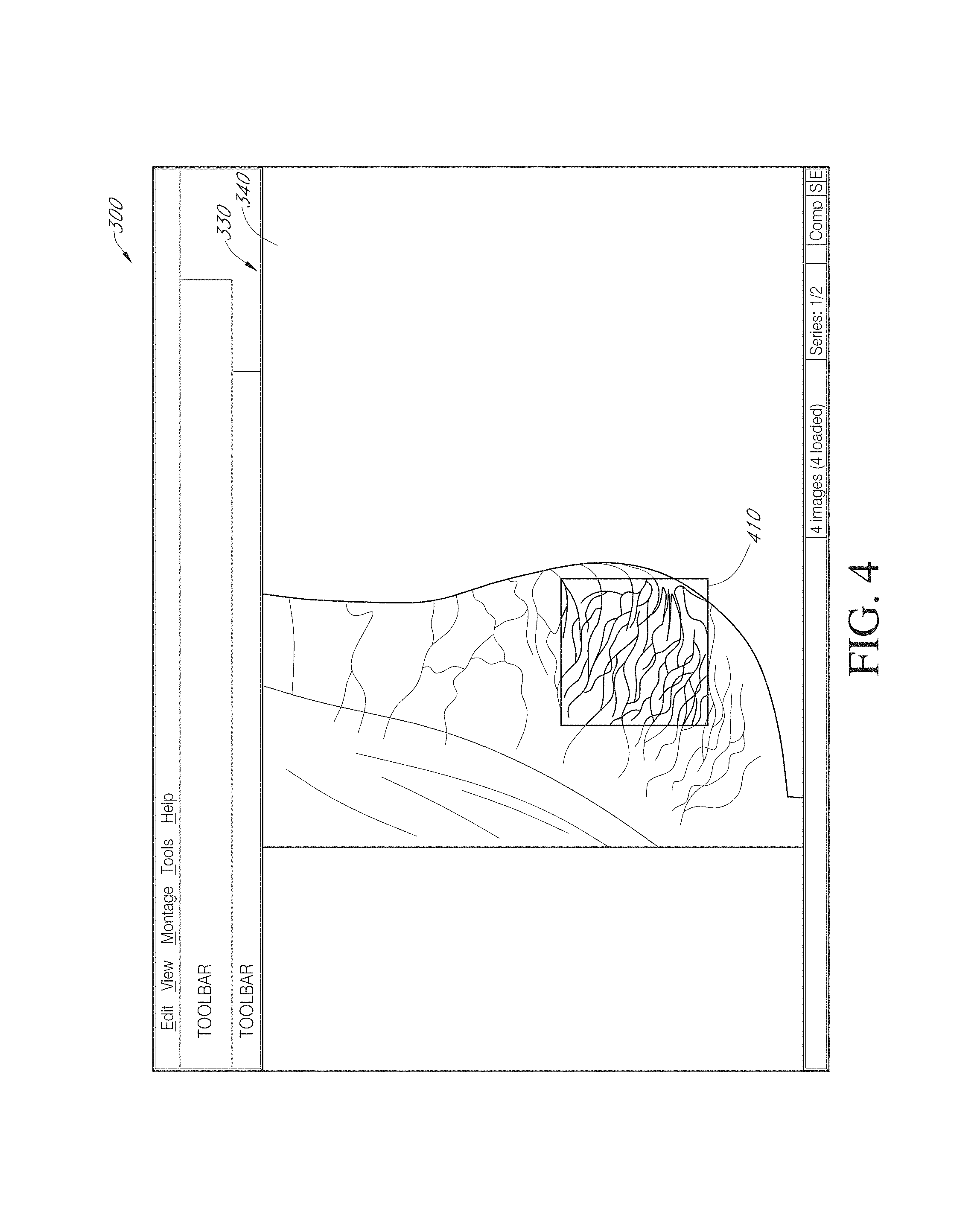
For certain medical images, it is important and/or required that a user view all of a medical image at full resolution so that minute, but important, indicia in the medical image are not missed. A computing systems monitor the portions of the medical image that are displayed on the display device, notates those portions that have been displayed at full resolution (or other user-defined display parameters), and provides the user with information indicating portions that have not been viewed at full resolution and/or provides information indicating for which images of a multiple image examination full pixel display has been accomplished. The process reduces the possibility of missing an abnormality in a medical image due to the viewer not viewing a portion of the image at full resolution or using other user-defined display parameters.
| Inventors: | Reicher; Murray A. (Rancho Santa Fe, CA), Fram; Evan K. (Paradise Valley, AZ) | ||||||||||
|---|---|---|---|---|---|---|---|---|---|---|---|
| Applicant: |
|
||||||||||
| Assignee: | MERGE HEALTHCARE SOLTUIONS INC.
(Hartland, WI) |
||||||||||
| Family ID: | 36261952 | ||||||||||
| Appl. No.: | 15/799,657 | ||||||||||
| Filed: | October 31, 2017 |
Prior Publication Data
| Document Identifier | Publication Date | |
|---|---|---|
| US 20180059918 A1 | Mar 1, 2018 | |
Related U.S. Patent Documents
| Application Number | Filing Date | Patent Number | Issue Date | ||
|---|---|---|---|---|---|
| 14540830 | Nov 13, 2014 | 9836202 | |||
| 13477853 | Dec 16, 2014 | 8913808 | |||
| 13228349 | Aug 14, 2012 | 8244014 | |||
| 12702976 | Sep 13, 2011 | 8019138 | |||
| 11179384 | Feb 9, 2010 | 7660488 | |||
| 60625690 | Nov 4, 2004 | ||||
| Current U.S. Class: | 1/1 |
| Current CPC Class: | G06F 3/04845 (20130101); G09G 5/02 (20130101); G16H 30/20 (20180101); G06F 3/167 (20130101); G09G 5/00 (20130101); G06F 19/321 (20130101) |
| Current International Class: | G06F 15/00 (20060101); G06F 3/0484 (20130101); G09G 5/00 (20060101); G09G 5/02 (20060101); G06F 3/16 (20060101) |
| Field of Search: | ;345/501 |
References Cited [Referenced By]
U.S. Patent Documents
| 4672683 | June 1987 | Matsueda |
| 5123056 | June 1992 | Wilson |
| 5172419 | December 1992 | Manian |
| 5179651 | January 1993 | Taaffe et al. |
| 5431161 | July 1995 | Ryals et al. |
| 5452416 | September 1995 | Hilton et al. |
| 5515375 | May 1996 | DeClerck |
| 5542003 | July 1996 | Wofford |
| 5734915 | March 1998 | Roewer |
| 5740267 | April 1998 | Echerer et al. |
| 5779634 | July 1998 | Ema et al. |
| 5807256 | September 1998 | Taguchi |
| 5835030 | November 1998 | Tsutsui et al. |
| 5852646 | December 1998 | Klotz et al. |
| 5857030 | January 1999 | Gaborski |
| 5867322 | February 1999 | Morton |
| 5926568 | July 1999 | Chaney et al. |
| 5954650 | September 1999 | Saito et al. |
| 5976088 | November 1999 | Urbano et al. |
| 5986662 | November 1999 | Argiro et al. |
| 5987345 | November 1999 | Engelmann et al. |
| 5995644 | November 1999 | Lai et al. |
| 6008813 | December 1999 | Lauer et al. |
| 6115486 | September 2000 | Cantoni |
| 6128002 | October 2000 | Leiper |
| 6130671 | October 2000 | Agiro |
| 6151581 | November 2000 | Kraftson et al. |
| 6175643 | January 2001 | Lai et al. |
| 6177937 | January 2001 | Stockham et al. |
| 6185320 | February 2001 | Bick et al. |
| 6211795 | April 2001 | Izuta |
| 6211884 | April 2001 | Knittel et al. |
| 6219059 | April 2001 | Argiro |
| 6219061 | April 2001 | Lauer et al. |
| 6243095 | June 2001 | Shile et al. |
| 6243098 | June 2001 | Lauer et al. |
| 6262740 | July 2001 | Lauer et al. |
| 6266733 | July 2001 | Knittel et al. |
| 6269379 | July 2001 | Hiyama et al. |
| 6297799 | October 2001 | Knittel et al. |
| 6304667 | October 2001 | Reitano |
| 6310620 | October 2001 | Lauer et al. |
| 6313841 | November 2001 | Ogata et al. |
| 6342885 | January 2002 | Knittel et al. |
| 6347329 | February 2002 | Evans |
| 6351547 | February 2002 | Johnson et al. |
| 6356265 | March 2002 | Knittel et al. |
| 6369816 | April 2002 | Knittel et al. |
| 6383135 | May 2002 | Chikovani et al. |
| 6388687 | May 2002 | Brackett et al. |
| 6404429 | June 2002 | Knittel |
| 6407737 | June 2002 | Zhao et al. |
| 6411296 | June 2002 | Knittel et al. |
| 6421057 | July 2002 | Lauer et al. |
| 6424346 | July 2002 | Correll et al. |
| 6424996 | July 2002 | Killcommons et al. |
| 6426749 | July 2002 | Knittel et al. |
| 6427022 | July 2002 | Craine et al. |
| 6438533 | August 2002 | Spackman et al. |
| 6463169 | October 2002 | Ino et al. |
| 6476810 | November 2002 | Simha et al. |
| 6512517 | January 2003 | Knittel et al. |
| 6532299 | March 2003 | Sachdeva et al. |
| 6532311 | March 2003 | Pritt |
| 6556695 | April 2003 | Packer et al. |
| 6556724 | April 2003 | Chang et al. |
| 6563950 | May 2003 | Wiskott et al. |
| 6574629 | June 2003 | Cooke, Jr. et al. |
| 6577753 | June 2003 | Ogawa |
| 6603494 | August 2003 | Banks et al. |
| 6606171 | August 2003 | Renk et al. |
| 6614447 | September 2003 | Bhatia et al. |
| 6618060 | September 2003 | Brackett |
| 6621918 | September 2003 | Hu et al. |
| 6630937 | October 2003 | Kallergi et al. |
| 6650766 | November 2003 | Rogers |
| 6654012 | November 2003 | Lauer et al. |
| 6678764 | January 2004 | Parvelescu et al. |
| 6680735 | January 2004 | Seiler et al. |
| 6683933 | January 2004 | Saito et al. |
| 6697067 | February 2004 | Callahan et al. |
| 6697506 | February 2004 | Oian et al. |
| 6734880 | May 2004 | Chang et al. |
| 6760755 | July 2004 | Brackett |
| 6775402 | August 2004 | Bacus et al. |
| 6778689 | August 2004 | Aksit et al. |
| 6785410 | August 2004 | Vining et al. |
| 6820093 | November 2004 | de la Huerga |
| 6820100 | November 2004 | Funahashi |
| 6826297 | November 2004 | Saito et al. |
| 6829377 | December 2004 | Milioto |
| 6864794 | March 2005 | Betz |
| 6886133 | April 2005 | Bailey et al. |
| 6891920 | May 2005 | Minyard et al. |
| 6894707 | May 2005 | Nemoto |
| 6909436 | June 2005 | Pianykh et al. |
| 6909795 | June 2005 | Tecotzky et al. |
| 6917696 | July 2005 | Soenksen |
| 6988075 | January 2006 | Hacker |
| 6996205 | February 2006 | Capolunghi et al. |
| 7016952 | March 2006 | Mullen et al. |
| 7022073 | April 2006 | Fan et al. |
| 7027633 | April 2006 | Foran et al. |
| 7031504 | April 2006 | Argiro et al. |
| 7031846 | April 2006 | Kaushikkar et al. |
| 7039723 | May 2006 | Hu et al. |
| 7043474 | May 2006 | Mojsilovic |
| 7050620 | May 2006 | Heckman |
| 7054473 | May 2006 | Roehrig et al. |
| 7058901 | June 2006 | Hafey et al. |
| 7092572 | August 2006 | Huang et al. |
| 7103205 | September 2006 | Wang et al. |
| 7106479 | September 2006 | Roy et al. |
| 7110616 | September 2006 | Ditt et al. |
| 7113186 | September 2006 | Kim et al. |
| 7123684 | October 2006 | Jing et al. |
| 7136064 | November 2006 | Zuiderveld |
| 7139416 | November 2006 | Vuylsteke |
| 7149334 | December 2006 | Dehmeshki |
| 7155043 | December 2006 | Daw |
| 7162623 | January 2007 | Yngvesson |
| 7170532 | January 2007 | Sako |
| 7174054 | February 2007 | Manber et al. |
| 7209149 | April 2007 | Jogo |
| 7209578 | April 2007 | Saito et al. |
| 7212661 | May 2007 | Samara et al. |
| 7218763 | May 2007 | Belykh et al. |
| 7224852 | May 2007 | Lipton et al. |
| 7236558 | June 2007 | Saito et al. |
| 7260249 | August 2007 | Smith |
| 7263710 | August 2007 | Hummel et al. |
| 7272610 | September 2007 | Torres |
| 7346199 | March 2008 | Pfaff |
| 7366992 | April 2008 | Thomas, III |
| 7379578 | May 2008 | Soussaline et al. |
| 7412111 | August 2008 | Battle et al. |
| 7450747 | November 2008 | Jabri et al. |
| 7492970 | February 2009 | Saito et al. |
| 7505782 | March 2009 | Chu |
| 7516417 | April 2009 | Amador et al. |
| 7525554 | April 2009 | Morita et al. |
| 7526114 | April 2009 | Xia et al. |
| 7526132 | April 2009 | Koenig |
| 7545965 | June 2009 | Suzuki et al. |
| 7574029 | August 2009 | Peterson et al. |
| 7583861 | September 2009 | Hanna et al. |
| 7590272 | September 2009 | Brejl et al. |
| 7599534 | October 2009 | Krishnan |
| 7613335 | November 2009 | McLennan et al. |
| 7634121 | December 2009 | Novatzky et al. |
| 7636413 | December 2009 | Toth |
| 7639879 | December 2009 | Goto et al. |
| 7656543 | February 2010 | Atkins |
| 7660481 | February 2010 | Schaap et al. |
| 7660488 | February 2010 | Reicher et al. |
| 7668352 | February 2010 | Tecotzky et al. |
| 7683909 | March 2010 | Takekoshi |
| 7698152 | April 2010 | Reid |
| 7716277 | May 2010 | Yamatake |
| 7787672 | August 2010 | Reicher et al. |
| 7834891 | November 2010 | Yarger et al. |
| 7835560 | November 2010 | Vining et al. |
| 7885440 | February 2011 | Fram et al. |
| 7885828 | February 2011 | Glaser-Seidnitzer et al. |
| 7899514 | March 2011 | Kirkland |
| 7920152 | April 2011 | Fram et al. |
| 7941462 | May 2011 | Akinyemi et al. |
| 7953614 | May 2011 | Reicher |
| 7970188 | June 2011 | Mahesh et al. |
| 7970625 | June 2011 | Reicher et al. |
| 7991210 | August 2011 | Peterson et al. |
| 7992100 | August 2011 | Lundstrom et al. |
| 7995821 | August 2011 | Nakamura |
| 8019138 | September 2011 | Reicher et al. |
| 8046044 | October 2011 | Stazzone et al. |
| 8050938 | November 2011 | Green, Jr. et al. |
| 8065166 | November 2011 | Maresh et al. |
| 8073225 | December 2011 | Hagen et al. |
| 8094901 | January 2012 | Reicher et al. |
| 8150708 | April 2012 | Kotula et al. |
| 8214756 | July 2012 | Salazar-Ferrer et al. |
| 8217966 | July 2012 | Fram et al. |
| 8244014 | August 2012 | Reicher et al. |
| 8249687 | August 2012 | Peterson et al. |
| 8262572 | September 2012 | Chono |
| 8292811 | October 2012 | Relkuntwar et al. |
| 8298147 | October 2012 | Huennekens et al. |
| 8370293 | February 2013 | Iwase et al. |
| 8379051 | February 2013 | Brown |
| 8380533 | February 2013 | Reicher et al. |
| 8391643 | March 2013 | Melbourne et al. |
| 8406491 | March 2013 | Gee et al. |
| 8457990 | June 2013 | Reicher et al. |
| 8554576 | October 2013 | Reicher et al. |
| 8560050 | October 2013 | Martin et al. |
| 8610746 | December 2013 | Fram et al. |
| 8626527 | January 2014 | Reicher et al. |
| 8693757 | April 2014 | Gundel |
| 8712120 | April 2014 | Reicher et al. |
| 8731259 | May 2014 | Reicher et al. |
| 8751268 | June 2014 | Reicher et al. |
| 8771189 | July 2014 | Ionasec et al. |
| 8797350 | August 2014 | Fram |
| 8879807 | November 2014 | Fram et al. |
| 8913808 | December 2014 | Reicher et al. |
| 8954884 | February 2015 | Barger |
| 8976190 | March 2015 | Westerhoff et al. |
| 9042617 | May 2015 | Reicher et al. |
| 9075899 | July 2015 | Reicher |
| 9092551 | July 2015 | Reicher |
| 9092727 | July 2015 | Reicher |
| 9324188 | April 2016 | Fram et al. |
| 9386084 | July 2016 | Reicher et al. |
| 9471210 | October 2016 | Fram et al. |
| 9495604 | November 2016 | Fram |
| 9501617 | November 2016 | Reicher et al. |
| 9501627 | November 2016 | Reicher et al. |
| 9501863 | November 2016 | Fram et al. |
| 9536106 | January 2017 | Fram |
| 9536324 | January 2017 | Fram |
| 9542082 | January 2017 | Reicher et al. |
| 9672477 | June 2017 | Reicher et al. |
| 9684762 | June 2017 | Reicher et al. |
| 9727938 | August 2017 | Reicher et al. |
| 9734576 | August 2017 | Fram et al. |
| 9754074 | September 2017 | Reicher et al. |
| 9836202 | December 2017 | Reicher et al. |
| 9892341 | February 2018 | Reicher et al. |
| 9934568 | April 2018 | Reicher et al. |
| 10096111 | October 2018 | Fram et al. |
| 10157686 | December 2018 | Reicher et al. |
| 2001/0016822 | August 2001 | Bessette |
| 2001/0042124 | November 2001 | Barron |
| 2002/0016718 | February 2002 | Rothschild et al. |
| 2002/0021828 | February 2002 | Papier et al. |
| 2002/0039084 | April 2002 | Yamaguchi |
| 2002/0044696 | April 2002 | Sirohey et al. |
| 2002/0070970 | June 2002 | Wood et al. |
| 2002/0073429 | June 2002 | Beane et al. |
| 2002/0081039 | June 2002 | Funahashi |
| 2002/0090118 | July 2002 | Olschewski |
| 2002/0090119 | July 2002 | Saito et al. |
| 2002/0090124 | July 2002 | Soubelet et al. |
| 2002/0091659 | July 2002 | Beaulieu et al. |
| 2002/0103673 | August 2002 | Atwood |
| 2002/0103827 | August 2002 | Sesek |
| 2002/0106119 | August 2002 | Foran et al. |
| 2002/0110285 | August 2002 | Wang et al. |
| 2002/0144697 | October 2002 | Betz |
| 2002/0145941 | October 2002 | Poland et al. |
| 2002/0164063 | November 2002 | Heckman |
| 2002/0172408 | November 2002 | Saito et al. |
| 2002/0172409 | November 2002 | Saito et al. |
| 2002/0180883 | December 2002 | Tomizawa et al. |
| 2002/0186820 | December 2002 | Saito et al. |
| 2002/0188637 | December 2002 | Bailey et al. |
| 2002/0190984 | December 2002 | Seiler et al. |
| 2003/0005464 | January 2003 | Gropper et al. |
| 2003/0013951 | January 2003 | Stefanescu |
| 2003/0016850 | January 2003 | Kaufman et al. |
| 2003/0028402 | February 2003 | Ulrich et al. |
| 2003/0034973 | February 2003 | Zuiderveld |
| 2003/0036087 | February 2003 | Kaushikkar et al. |
| 2003/0037054 | February 2003 | Dutta et al. |
| 2003/0053668 | March 2003 | Ditt et al. |
| 2003/0055896 | March 2003 | Hu et al. |
| 2003/0065613 | April 2003 | Smith |
| 2003/0071829 | April 2003 | Bodicker et al. |
| 2003/0101291 | May 2003 | Mussack et al. |
| 2003/0115083 | June 2003 | Masarie et al. |
| 2003/0120516 | June 2003 | Perednia |
| 2003/0123717 | July 2003 | Bacus et al. |
| 2003/0130973 | July 2003 | Sumner, II et al. |
| 2003/0140141 | July 2003 | Mullen et al. |
| 2003/0156745 | August 2003 | Saito et al. |
| 2003/0160095 | August 2003 | Segal |
| 2003/0164860 | September 2003 | Shen et al. |
| 2003/0184778 | October 2003 | Chiba |
| 2003/0185446 | October 2003 | Huang et al. |
| 2003/0187689 | October 2003 | Barnes et al. |
| 2003/0190062 | October 2003 | Noro et al. |
| 2003/0195416 | October 2003 | Toth |
| 2003/0204420 | October 2003 | Wilkes et al. |
| 2003/0215120 | November 2003 | Uppaluri et al. |
| 2003/0215122 | November 2003 | Tanaka |
| 2004/0008900 | January 2004 | Jabri et al. |
| 2004/0015703 | January 2004 | Madison et al. |
| 2004/0024303 | February 2004 | Banks et al. |
| 2004/0027359 | February 2004 | Aharon et al. |
| 2004/0061889 | April 2004 | Wood et al. |
| 2004/0068170 | April 2004 | Wang et al. |
| 2004/0086163 | May 2004 | Moriyama et al. |
| 2004/0088192 | May 2004 | Schmidt et al. |
| 2004/0101191 | May 2004 | Seul et al. |
| 2004/0105030 | June 2004 | Yamane |
| 2004/0105574 | June 2004 | Pfaff |
| 2004/0109032 | June 2004 | Kim et al. |
| 2004/0114714 | June 2004 | Minyard et al. |
| 2004/0122705 | June 2004 | Sabol et al. |
| 2004/0122787 | June 2004 | Avinash et al. |
| 2004/0141661 | July 2004 | Hanna et al. |
| 2004/0143582 | July 2004 | Vu |
| 2004/0151374 | August 2004 | Lipton et al. |
| 2004/0161139 | August 2004 | Samara et al. |
| 2004/0161164 | August 2004 | Dewaele |
| 2004/0165791 | August 2004 | Kaltanji |
| 2004/0170312 | September 2004 | Soenksen |
| 2004/0172306 | September 2004 | Wohl et al. |
| 2004/0174429 | September 2004 | Chu |
| 2004/0190780 | September 2004 | Shiibashi et al. |
| 2004/0197015 | October 2004 | Fan et al. |
| 2004/0202387 | October 2004 | Yngvesson |
| 2004/0243435 | December 2004 | Williams |
| 2004/0252871 | December 2004 | Tecotzky et al. |
| 2004/0254816 | December 2004 | Myers |
| 2004/0255252 | December 2004 | Rodriguez et al. |
| 2004/0264753 | December 2004 | Capolunghi et al. |
| 2005/0010531 | January 2005 | Kushalnagar et al. |
| 2005/0027569 | February 2005 | Gollogly et al. |
| 2005/0027570 | February 2005 | Maier et al. |
| 2005/0036668 | February 2005 | McLennan et al. |
| 2005/0043970 | February 2005 | Hsieh |
| 2005/0063575 | March 2005 | Ma et al. |
| 2005/0063612 | March 2005 | Manber et al. |
| 2005/0065424 | March 2005 | Shah et al. |
| 2005/0074150 | April 2005 | Bruss |
| 2005/0074157 | April 2005 | Thomas, III |
| 2005/0075544 | April 2005 | Shapiro et al. |
| 2005/0088534 | April 2005 | Shen et al. |
| 2005/0107689 | May 2005 | Sasano |
| 2005/0108058 | May 2005 | Weidner et al. |
| 2005/0110791 | May 2005 | Krishnamoorthy et al. |
| 2005/0111733 | May 2005 | Fors et al. |
| 2005/0113681 | May 2005 | DeFreitas et al. |
| 2005/0114178 | May 2005 | Krishnamurthy et al. |
| 2005/0114179 | May 2005 | Brackett et al. |
| 2005/0114283 | May 2005 | Pearson et al. |
| 2005/0143654 | June 2005 | Zuiderveld et al. |
| 2005/0171818 | August 2005 | McLaughlin |
| 2005/0184988 | August 2005 | Yanof et al. |
| 2005/0197860 | September 2005 | Joffe et al. |
| 2005/0238218 | October 2005 | Nakamura |
| 2005/0244041 | November 2005 | Tecotzky et al. |
| 2005/0251013 | November 2005 | Krishnan |
| 2005/0254729 | November 2005 | Saito et al. |
| 2005/0259118 | November 2005 | Mojaver |
| 2005/0273009 | December 2005 | Deischinger et al. |
| 2006/0008181 | January 2006 | Takekoshi |
| 2006/0031097 | February 2006 | Lipscher et al. |
| 2006/0050152 | March 2006 | Rai et al. |
| 2006/0058603 | March 2006 | Dave et al. |
| 2006/0061570 | March 2006 | Cheryauka et al. |
| 2006/0093198 | May 2006 | Fram et al. |
| 2006/0093199 | May 2006 | Fram et al. |
| 2006/0093207 | May 2006 | Reicher et al. |
| 2006/0095423 | May 2006 | Reicher et al. |
| 2006/0095426 | May 2006 | Takachio et al. |
| 2006/0106642 | May 2006 | Reicher et al. |
| 2006/0111937 | May 2006 | Yarger et al. |
| 2006/0111941 | May 2006 | Blom |
| 2006/0122482 | June 2006 | Mariotti et al. |
| 2006/0171574 | August 2006 | DelMonego et al. |
| 2006/0181548 | August 2006 | Hafey |
| 2006/0188134 | August 2006 | Quist |
| 2006/0230072 | October 2006 | Partovi et al. |
| 2006/0238546 | October 2006 | Handley et al. |
| 2006/0239573 | October 2006 | Novatzky et al. |
| 2006/0241979 | October 2006 | Sato et al. |
| 2006/0267976 | November 2006 | Saito et al. |
| 2006/0274145 | December 2006 | Reiner |
| 2006/0276708 | December 2006 | Peterson et al. |
| 2006/0277075 | December 2006 | Salwan |
| 2006/0282408 | December 2006 | Wisely et al. |
| 2007/0009078 | January 2007 | Saito et al. |
| 2007/0021977 | January 2007 | Elsholz |
| 2007/0050701 | March 2007 | El Emam et al. |
| 2007/0055550 | March 2007 | Courtney et al. |
| 2007/0064984 | March 2007 | Vassa et al. |
| 2007/0067124 | March 2007 | Kimpe et al. |
| 2007/0073556 | March 2007 | Lau et al. |
| 2007/0106535 | May 2007 | Matsunaga |
| 2007/0106633 | May 2007 | Reiner |
| 2007/0109299 | May 2007 | Peterson |
| 2007/0109402 | May 2007 | Niwa |
| 2007/0110294 | May 2007 | Schaap et al. |
| 2007/0116345 | May 2007 | Peterson et al. |
| 2007/0116346 | May 2007 | Peterson et al. |
| 2007/0122016 | May 2007 | Brejl et al. |
| 2007/0124541 | May 2007 | Lang et al. |
| 2007/0140536 | June 2007 | Sehnert |
| 2007/0159962 | July 2007 | Mathavu et al. |
| 2007/0162308 | July 2007 | Peters |
| 2007/0165917 | July 2007 | Cao et al. |
| 2007/0174079 | July 2007 | Kraus |
| 2007/0192138 | August 2007 | Saito et al. |
| 2007/0192140 | August 2007 | Gropper |
| 2007/0237380 | October 2007 | Iwase et al. |
| 2007/0239481 | October 2007 | DiSilvestro et al. |
| 2007/0245308 | October 2007 | Hill et al. |
| 2008/0016111 | January 2008 | Keen |
| 2008/0021877 | January 2008 | Saito et al. |
| 2008/0031507 | February 2008 | Uppaluri et al. |
| 2008/0059245 | March 2008 | Sakaida et al. |
| 2008/0097186 | April 2008 | Biglieri et al. |
| 2008/0100612 | May 2008 | Dastmalchi et al. |
| 2008/0103828 | May 2008 | Squilla et al. |
| 2008/0118120 | May 2008 | Wegenkittl et al. |
| 2008/0125846 | May 2008 | Battle et al. |
| 2008/0126982 | May 2008 | Sadikali et al. |
| 2008/0133526 | June 2008 | Haitani et al. |
| 2008/0136838 | June 2008 | Goede et al. |
| 2008/0275913 | November 2008 | van Arragon et al. |
| 2008/0279439 | November 2008 | Minyard et al. |
| 2008/0300484 | December 2008 | Wang et al. |
| 2009/0005668 | January 2009 | West et al. |
| 2009/0022375 | January 2009 | Fidrich |
| 2009/0028410 | January 2009 | Shimazaki |
| 2009/0164247 | January 2009 | Dobler et al. |
| 2009/0080719 | March 2009 | Watt |
| 2009/0091566 | April 2009 | Turney et al. |
| 2009/0094513 | April 2009 | Bay |
| 2009/0123052 | May 2009 | Ruth et al. |
| 2009/0129643 | May 2009 | Natanzon et al. |
| 2009/0132586 | May 2009 | Napora et al. |
| 2009/0150481 | June 2009 | Garcia et al. |
| 2009/0182577 | July 2009 | Squilla et al. |
| 2009/0198514 | August 2009 | Rhodes |
| 2009/0213034 | August 2009 | Wu et al. |
| 2009/0248442 | October 2009 | Pacheco et al. |
| 2009/0268986 | October 2009 | Holstein et al. |
| 2009/0326373 | December 2009 | Boese et al. |
| 2010/0053353 | March 2010 | Hunter et al. |
| 2010/0086182 | April 2010 | Luo et al. |
| 2010/0131887 | May 2010 | Salazar-Ferrer et al. |
| 2010/0138239 | June 2010 | Reicher et al. |
| 2010/0198608 | August 2010 | Kaboff et al. |
| 2010/0201714 | August 2010 | Reicher et al. |
| 2010/0211409 | August 2010 | Kotula et al. |
| 2010/0246981 | September 2010 | Hu et al. |
| 2010/0299157 | November 2010 | Fram et al. |
| 2011/0016430 | January 2011 | Fram |
| 2011/0019886 | January 2011 | Mizuno |
| 2011/0110572 | May 2011 | Guehring et al. |
| 2011/0267339 | November 2011 | Fram |
| 2011/0293162 | December 2011 | Pajeau |
| 2011/0316873 | December 2011 | Reicher et al. |
| 2012/0070048 | March 2012 | Van Den Brink |
| 2012/0130729 | May 2012 | Raizada et al. |
| 2012/0136794 | May 2012 | Kushalnagar et al. |
| 2012/0163684 | June 2012 | Natanzon et al. |
| 2012/0183191 | July 2012 | Nakamura |
| 2012/0194540 | August 2012 | Reicher et al. |
| 2012/0196258 | August 2012 | Geijsen et al. |
| 2012/0284657 | November 2012 | Hafey et al. |
| 2013/0076681 | March 2013 | Sirpal et al. |
| 2013/0083023 | April 2013 | Fram |
| 2013/0129198 | May 2013 | Sherman et al. |
| 2013/0129231 | May 2013 | Dale et al. |
| 2013/0159019 | June 2013 | Reicher |
| 2013/0169661 | July 2013 | Reicher et al. |
| 2013/0195329 | August 2013 | Canda et al. |
| 2013/0198682 | August 2013 | Matas et al. |
| 2013/0297331 | November 2013 | Zuehlsdorff et al. |
| 2014/0022194 | January 2014 | Ito |
| 2014/0096049 | April 2014 | Vonshak et al. |
| 2014/0119514 | May 2014 | Miyazawa |
| 2014/0142983 | May 2014 | Backhaus et al. |
| 2014/0378810 | December 2014 | Davis et al. |
| 2015/0046349 | February 2015 | Michael, Jr. et al. |
| 2015/0101066 | April 2015 | Fram |
| 2015/0160848 | June 2015 | Gkanatsios et al. |
| 2015/0363104 | December 2015 | Ichioka et al. |
| 2016/0034110 | February 2016 | Edwards |
| 2016/0270746 | September 2016 | Foos et al. |
| 2016/0335395 | November 2016 | Wu et al. |
| 2017/0038951 | February 2017 | Reicher et al. |
| 2017/0039321 | February 2017 | Reicher et al. |
| 2017/0039322 | February 2017 | Reicher et al. |
| 2017/0039350 | February 2017 | Reicher |
| 2017/0039705 | February 2017 | Fram et al. |
| 2017/0046014 | February 2017 | Fram |
| 2017/0046483 | February 2017 | Reicher et al. |
| 2017/0046485 | February 2017 | Reicher |
| 2017/0046495 | February 2017 | Fram |
| 2017/0046870 | February 2017 | Fram et al. |
| 2017/0053404 | February 2017 | Reicher et al. |
| 2017/0200064 | July 2017 | Reicher et al. |
| 2017/0200269 | July 2017 | Reicher et al. |
| 2017/0200270 | July 2017 | Reicher et al. |
| 2017/0206324 | July 2017 | Reicher et al. |
| 2017/0293720 | October 2017 | Reicher et al. |
| 2017/0301090 | October 2017 | Fram et al. |
| 2017/0308647 | October 2017 | Reicher et al. |
| 2018/0059918 | March 2018 | Reicher et al. |
| WO 2007/131157 | Nov 2007 | WO | |||
Other References
|
US 7,801,341 B2, 09/2010, Fram et al. (withdrawn) cited by applicant . US 8,208,705 B2, 06/2012, Reicher et al. (withdrawn) cited by applicant . U.S. Appl. No. 14/540,830, Systems and Methods for Viewing Medical Images, filed Nov. 13, 2014. cited by applicant . U.S. Appl. No. 15/631,313, Systems and Methods for Interleaving Series of Medical Images, filed Jun. 23, 2017. cited by applicant . U.S. Appl. No. 15/631,291, Systems and Methods for Retrieval of Medical Data, filed Jun. 23, 2017. cited by applicant . U.S. Appl. No. 15/292,006, Systems and Methods for Viewing Medical 3D Imaging Volumes, filed Oct. 12, 2016. cited by applicant . U.S. Appl. No. 15/346,530, Systems and Methods for Matching, Naming, and Displaying Medical Images, filed Nov. 8, 2016. cited by applicant . U.S. Appl. No. 15/646,756, Smart Placement Rules, filed Jul. 11, 2017. cited by applicant . U.S. Appl. No. 11/942,687, Smart Forms, filed Nov. 19, 2007. cited by applicant . U.S. Appl. No. 14/043,165, Automated Document Filings, filed Oct. 1, 2013. cited by applicant . U.S. Appl. No. 15/475,930, Exam Scheduling With Customer Configured Notifications, filed Mar. 31, 2017. cited by applicant . U.S. Appl. No. 15/292,014, System and Method of Providing Dynamic and Customizable Medical Examination for, filed Oct. 12, 2016. cited by applicant . U.S. Appl. No. 15/469,342, Rules-Based Rendering of Medical Images, filed Mar. 24, 2017. cited by applicant . U.S. Appl. No. 15/469,281, Rules-Based Processing and Presentation of Medical Images, filed Mar. 24, 2017. cited by applicant . U.S. Appl. No. 15/469,296, Computer-Aided Analysis and Rendering of Medical Images, filed Mar. 24, 2017. cited by applicant . U.S. Appl. No. 14/792,210, Dynamic Montage Reconstruction, filed Jul. 6, 2015. cited by applicant . U.S. Appl. No. 15/188,872, Intelligent Management of Computerized Advanced Processing, filed Jun. 21, 2016. cited by applicant . U.S. Appl. No. 15/188,819, Intelligent Management of Computerized Advanced Processing, filed Jun. 21, 2016. cited by applicant . U.S. Appl. No. 15/140,346, Database Systems and Interactive User Interfaces for Dynamic Interaction With, and Sorting of, Digital Medical Image Data, filed Apr. 27, 2016. cited by applicant . U.S. Appl. No. 15/140,363, Database Systems and Interactive User Interfaces for Dynamic Interation With, and Comparison of, Digital Medical Image Data, filed Apr. 27, 2016. cited by applicant . U.S. Appl. No. 15/140,351, Database Systems and Interactive User Interfaces for Dynamic Interaction With, and Review of, Digital Medical Image Data, filed Apr. 27, 2016. cited by applicant . U.S. Appl. No. 15/140,348, Database Systems and Interactive User Interfaces for Dynamic Interaction With, and Indications of, Digital Medical Image Data, filed Apr. 27, 2016. cited by applicant . Non-Final Office Action dated Aug. 28, 2007 in U.S. Appl. No. 11/179,384. cited by applicant . Final Office Action dated Jun. 26, 2008 in U.S. Appl. No. 11/179,384. cited by applicant . Non-Final Office Action dated Dec. 29, 2008 in U.S. Appl. No. 11/179,384. cited by applicant . Final Office Action dated Jul. 24, 2009, in U.S. Appl. No. 11/179,384. cited by applicant . Notice of Allowance dated Nov. 3, 2009, in U.S. Appl. No. 11/179,384. cited by applicant . Non-Final Office Action dated Aug. 18, 2010 in U.S. Appl. No. 12/702,976. cited by applicant . Interview Summary dated Dec. 1, 2010, in U.S. Appl. No. 12/702,976. cited by applicant . Final Office Action dated Feb. 17, 2011 in U.S. Appl. No. 12/702,976. cited by applicant . Interview Summary dated May 31, 2011 in U.S. Appl. No. 12/702,976. cited by applicant . Notice of Allowance dated Jul. 20, 2011, in U.S. Appl. No. 12/702,976. cited by applicant . Office Action dated Dec. 1, 2011, in U.S. Appl. No. 13/228,349. cited by applicant . Notice of Allowance dated Feb. 6, 2012, in U.S. Appl. No. 13/228,349. cited by applicant . Notice of Allowance dated Jul. 20, 2012, in U.S. Appl. No. 13/228,349. cited by applicant . Office Action dated Dec. 11, 2013, in U.S. Appl. No. 13/477,853. cited by applicant . Interview Summary dated Mar. 14, 2014, in U.S. Appl. No. 13/477,853. cited by applicant . Final Office Action dated Jun. 13, 2014, in U.S. Appl. No. 13/477,853. cited by applicant . Notice of Allowance dated Aug. 15, 2014, in U.S. Appl. No. 13/477,853. cited by applicant . Office Action dated Jan. 17, 2017, in U.S. Appl. No. 14/540,830. cited by applicant . Interview Summary dated Mar. 24, 2017, in U.S. Appl. No. 14/540,830. cited by applicant . Final Office Action dated May 15, 2017, in U.S. Appl. No. 14/540,830. cited by applicant . Interview Summary dated Jul. 28, 2017, in U.S. Appl. No. 14/540,830. cited by applicant . Notice of Allowance dated Aug. 15, 2017, in U.S. Appl. No. 14/540,830. cited by applicant . Non-Final Office Action dated Oct. 1, 2009, in U.S. Appl. No. 11/268,261. cited by applicant . Notice of Allowance dated Feb. 2, 2010, in U.S. Appl. No. 11/268,261. cited by applicant . Interview Summary dated Jan. 25, 2010, in U.S. Appl. No. 11/268,261. cited by applicant . Interview Summary dated May 14, 2010, in U.S. Appl. No. 11/268,261. cited by applicant . Notice of Allowance dated May 17, 2010, in U.S. Appl. No. 11/268,261. cited by applicant . Supplemental Notice of Allowance dated Aug. 6, 2010, in U.S. Appl. No. 11/268,261. cited by applicant . Notice of Allowance dated Oct. 8, 2010, in U.S. Appl. No. 11/268,261. cited by applicant . Notice of Allowance dated Dec. 3, 2010, in U.S. Appl. No. 11/268,261. cited by applicant . Notice of Allowance dated Jan. 6, 2011, in U.S. Appl. No. 11/268,261. cited by applicant . Office Action dated May 16, 2011, in U.S. Appl. No. 12/857,915. cited by applicant . Interview Summary dated Sep. 6, 2011, in U.S. Appl. No. 12/857,915. cited by applicant . Final Office Action dated Dec. 15, 2011, in U.S. Appl. No. 12/857,915. cited by applicant . Office Action dated Jun. 12, 2012, in U.S. Appl. No. 12/857,915. cited by applicant . Office Action dated Aug. 23, 2013, in U.S. Appl. No. 12/857,915. cited by applicant . Interview Summary dated Feb. 4, 2014, in U.S. Appl. No. 12/857,915. cited by applicant . Notice of Allowance dated Jul. 3, 2014, in U.S. Appl. No. 12/857,915. cited by applicant . Corrected Notice of Allowance dated Aug. 15, 2014, in U.S. Appl. No. 12/857,915. cited by applicant . Non-Final Office Action dated Jan. 20, 2016, in U.S. Appl. No. 14/502,055. cited by applicant . Interview Summary dated Apr. 14, 2016, in U.S. Appl. No. 14/502,055. cited by applicant . Notice of Allowance dated Jun. 2, 2016, in U.S. Appl. No. 14/502,055. cited by applicant . Corrected Notice of Allowance dated Jun. 27, 2016, in U.S. Appl. No. 14/502,055. cited by applicant . Notice of Corrected Allowability dated Jul. 14, 2016, in U.S. Appl. No. 14/502,055. cited by applicant . Notice of Corrected Allowability dated Sep. 19, 2016, in U.S. Appl. No. 14/502,055. cited by applicant . Office Action dated Dec. 12, 2016, in U.S. Appl. No. 15/254,627. cited by applicant . Notice of Allowance dated Apr. 3, 2017 in U.S. Appl. No. 15/254,627. cited by applicant . Notice of Allowance (corrected) dated Jul. 13, 2017 in U.S. Appl. No. 15/254,627. cited by applicant . Non-Final Office Action dated May 13, 2009, in U.S. Appl. No. 11/265,979. cited by applicant . Final Office Action dated Dec. 22, 2009 in U.S. Appl. No. 11/265,979. cited by applicant . Non-Final Office Action dated Jul. 8, 2010 in U.S. Appl. No. 11/265,979. cited by applicant . Interview Summary dated Mar. 4, 2010 in U.S. Appl. No. 11/265,979. cited by applicant . Interview Summary dated Nov. 16, 2010 in U.S. Appl. No. 11/265,979. cited by applicant . Final Office Action dated Dec. 23, 2010 in U.S. Appl. No. 11/265,979. cited by applicant . Interview Summary dated Mar. 17, 2011 in U.S. Appl. No. 11/265,979. cited by applicant . Notice of Allowance dated May 26, 2011 in U.S. Appl. No. 11/265,979. cited by applicant . Office Action dated Jun. 8, 2012 in U.S. Appl. No. 13/171,081. cited by applicant . Interview Summary dated Jul. 31, 2012 in U.S. Appl. No. 13/171,081. cited by applicant . Final Office Action dated Oct. 12, 2012 in U.S. Appl. No. 13/171,081. cited by applicant . Interview Summary dated Nov. 6, 2012 in U.S. Appl. No. 13/171,081. cited by applicant . Notice of Allowance, dated Sep. 4, 2013, in U.S. Appl. No. 13/171,081. cited by applicant . Office Action dated Mar. 3, 2015 in U.S. Appl. No. 14/095,123. cited by applicant . Interview Summary dated May 1, 2015 in U.S. Appl. No. 14/095,123. cited by applicant . Final Office Action dated Jul. 23, 2015 in U.S. Appl. No. 14/095,123. cited by applicant . Interview Summary dated Aug. 27, 2015 in U.S. Appl. No. 14/095,123. cited by applicant . Office Action dated Feb. 23, 2016 in U.S. Appl. No. 14/095,123. cited by applicant . Final Office Action dated Jul. 20, 2016 in U.S. Appl. No. 14/095,123. cited by applicant . Notice of Allowance dated Mar. 30, 2017 in U.S. Appl. No. 14/095,123. cited by applicant . Non-Final Office Action dated Aug. 24, 2009 in U.S. Appl. No. 11/268,262. cited by applicant . Non-Final Office Action dated Apr. 16, 2010 in U.S. Appl. No. 11/268,262. cited by applicant . Interview Summary dated Nov. 24, 2009 in U.S. Appl. No. 11/268,262. cited by applicant . Interview Summary dated May 12, 2010 in U.S. Appl. No. 11/268,262. cited by applicant . Final Office Action dated Oct. 28, 2010 in U.S. Appl. No. 11/268,262. cited by applicant . Interview Summary dated Dec. 1, 2010 in U.S. Appl. No. 11/268,262. cited by applicant . Notice of Allowance dated Dec. 1, 2010 in U.S. Appl. No. 11/268,262. cited by applicant . Notice of Allowance dated Feb. 25, 2011 in U.S. Appl. No. 11/268,262. cited by applicant . Non-Final Office Action dated Jan. 11, 2012 in U.S. Appl. No. 13/079,597. cited by applicant . Notice of Allowance dated Apr. 25, 2012, in U.S. Appl. No. 13/079,597. cited by applicant . Non-Final Office Action dated Apr. 4, 2013 in U.S. Appl. No. 13/535,758. cited by applicant . Notice of Allowance, dated Aug. 23, 2013 in U.S. Appl. No. 13/535,758. cited by applicant . Office Action dated Mar. 10, 2016 in U.S. Appl. No. 14/081,225. cited by applicant . Notice of Allowance dated Sep. 2, 2016 in U.S. Appl. No. 14/081,225. cited by applicant . Corrected Notice of Allowance dated Oct. 21, 2016 in U.S. Appl. No. 14/081,225. cited by applicant . Non-Final Office Action dated Jul. 27, 2009 in U.S. Appl. No. 11/265,978. cited by applicant . Notice of Allowance dated Nov. 19, 2009 in U.S. Appl. No. 11/265,978. cited by applicant . Notice of Allowance dated Apr. 19, 2010 in U.S. Appl. No. 11/265,978. cited by applicant . Supplemental Notice of Allowance dated May 3, 2010 in U.S. Appl. No. 11/265,978. cited by applicant . Supplemental Notice of Allowance dated Aug. 3, 2010 in U.S. Appl. No. 11/265,978. cited by applicant . Non-Final Office Action dated May 5, 2011 in U.S. Appl. No. 12/870,645. cited by applicant . Non-Final Office Action dated May 31, 2013, in U.S. Appl. No. 13/345,606. cited by applicant . Interview Summary dated Aug. 15, 2013, in U.S. Appl. No. 13/345,606. cited by applicant . Notice of Allowance, dated Jan. 9, 2014 in U.S. Appl. No. 13/345,606. cited by applicant . Non-Final Office Action dated Mar. 18, 2016 in U.S. Appl. No. 14/244,431. cited by applicant . Interview Summary dated Jun. 17, 2016 in U.S. Appl. No. 14/244,431. cited by applicant . Notice of Allowance dated Aug. 18, 2016 in U.S. Appl. No. 14/244,431. cited by applicant . Corrected Notice of Allowance dated Nov. 16, 2016 in U.S. Appl. No. 14/244,431. cited by applicant . Non-Final Office Action dated May 26, 2010 in U.S. Appl. No. 11/942,674. cited by applicant . Interview Summary dated Jul. 26, 2010 in U.S. Appl. No. 11/942,674. cited by applicant . Final Office Action dated Nov. 26, 2010 in U.S. Appl. No. 11/942,674. cited by applicant . Interview Summary dated Mar. 2, 2011 in U.S. Appl. No. 11/942,674. cited by applicant . Notice of Allowance, dated Apr. 1, 2011 in U.S. Appl. No. 11/942,674. cited by applicant . Non Final Office Action dated Nov. 10, 2011 in U.S. Appl. No. 13/118,085. cited by applicant . Interview Summary, dated Feb. 17, 2012, in U.S. Appl. No. 13/118,085. cited by applicant . Final Office Action, dated Apr. 13, 2012, in U.S. Appl. No. 13/118,085. cited by applicant . Notice of Allowance, dated Feb. 6, 2013, in U.S. Appl. No. 13/118,085. cited by applicant . Non Final Office Action dated Aug. 23, 2013 in U.S. Appl. No. 13/907,128. cited by applicant . Final Office Action dated Oct. 9, 2013 in U.S. Appl. No. 13/907,128. cited by applicant . Interview Summary dated Nov. 22, 2013 in U.S. Appl. No. 13/907,128. cited by applicant . Notice of Allowance dated Jan. 31, 2014 in U.S. Appl. No. 13/907,128. cited by applicant . Office Action, dated Dec. 29, 2014 in U.S. Appl. No. 14/298,806. cited by applicant . Interview Summary, dated Mar. 2, 2015 in U.S. Appl. No. 14/298,806. cited by applicant . Final Office Action, dated Jun. 17, 2015 in U.S. Appl. No. 14/298,806. cited by applicant . Office Action, dated Feb. 16, 2016 in U.S. Appl. No. 14/298,806. cited by applicant . Final Office Action, dated Jul. 21, 2016 in U.S. Appl. No. 14/298,806. cited by applicant . Notice of Allowance, dated Apr. 12, 2017 in U.S. Appl. No. 14/298,806. cited by applicant . Non Final Office Action dated Sep. 16, 2010 in U.S. Appl. No. 11/942,687. cited by applicant . Interview Summary dated Dec. 3, 2010 in U.S. Appl. No. 11/942,687. cited by applicant . Final Office Action, dated Apr. 5, 2011 in U.S. Appl. No. 11/942,687. cited by applicant . Office Action, dated Mar. 13, 2014 in U.S. Appl. No. 11/942,687. cited by applicant . Interview Summary, dated Jun. 17, 2014 in U.S. Appl. No. 11/942,687. cited by applicant . Office Action, dated Jul. 18, 2014 in U.S. Appl. No. 11/942,687. cited by applicant . Final Office Action, dated Jan. 5, 2015 in U.S. Appl. No. 11/942,687. cited by applicant . Interview Summary, dated Mar. 4, 2015 in U.S. Appl. No. 11/942,687. cited by applicant . PTAB Examiner's Answer, dated Feb. 25, 2016 in U.S. Appl. No. 11/942,687. cited by applicant . Non-Final Office Action dated Apr. 14, 2010 in U.S. Appl. No. 11/944,027. cited by applicant . Interview Summary dated May 13, 2010 in U.S. Appl. No. 11/944,027. cited by applicant . Final Office Action dated Dec. 23, 2010 in U.S. Appl. No. 11/944,027. cited by applicant . Interview Summary dated Mar. 31, 2011 in U.S. Appl. No. 11/944,027. cited by applicant . Office Action dated Apr. 19, 2012 in U.S. Appl. No. 11/944,027. cited by applicant . Interview Summary dated Jun. 28, 2012 in U.S. Appl. No. 11/944,027. cited by applicant . Final Office Action dated Oct. 22, 2012 in U.S. Appl. No. 11/944,027. cited by applicant . Notice of Allowance dated Jun. 5, 2013 in U.S. Appl. No. 11/944,027. cited by applicant . Office Action dated Oct. 14, 2014 in U.S. Appl. No. 14/043,165. cited by applicant . Final Office Action dated Apr. 1, 2015 in U.S. Appl. No. 14/043,165. cited by applicant . Office Action dated Oct. 2, 2015 in U.S. Appl. No. 14/043,165. cited by applicant . Interview Summary dated Dec. 21, 2015 in U.S. Appl. No. 14/043,165. cited by applicant . Final Office Action dated Feb. 17, 2016 in U.S. Appl. No. 14/043,165. cited by applicant . Appeal Brief dated Jul. 15, 2016 in U.S. Appl. No. 14/043,165. cited by applicant . Examiner's Answer dated Nov. 14, 2016, in U.S. Appl. No. 14/043,165. cited by applicant . Non-Final Office Action dated Sep. 29, 2010 in U.S. Appl. No. 11/944,000. cited by applicant . Final Office Action dated Apr. 20, 2011 in U.S. Appl. No. 11/944,000. cited by applicant . Interview Summary dated Jun. 7, 2011 in U.S. Appl. No. 11/944,000. cited by applicant . Appeal Brief dated Mar. 4, 2013 in U.S. Appl. No. 11/944,000. cited by applicant . Examiner's Answer dated Jun. 26, 2013 in U.S. Appl. No. 11/944,000. cited by applicant . Board Decision dated Mar. 23, 2016 in U.S. Appl. No. 11/944,000. cited by applicant . Office Action, dated Jul. 15, 2016 in U.S. Appl. No. 11/944,000. cited by applicant . Notice of Allowance, dated Jan. 30, 2017, in U.S. Appl. No. 11/944,000. cited by applicant . Office Action dated Feb. 3, 2012 in U.S. Appl. No. 12/622,404. cited by applicant . Interview Summary dated May 8, 2012 in U.S. Appl. No. 12/622,404. cited by applicant . Final Office Action dated Aug. 6, 2012 in U.S. Appl. No. 12/622,404. cited by applicant . Notice of Allowance dated Oct. 15, 2012 in U.S. Appl. No. 12/622,404. cited by applicant . Office Action dated Mar. 17, 2015 in U.S. Appl. No. 13/768,765. cited by applicant . Interview Summary dated Jun. 11, 2015 in U.S. Appl. No. 13/768,765. cited by applicant . Notice of Allowance dated Aug. 28, 2015 in U.S. Appl. No. 13/768,765. cited by applicant . Notice of Allowability dated Nov. 20, 2015 in U.S. Appl. No. 13/768,765. cited by applicant . Notice of Allowability dated Jul. 28, 2016 in U.S. Appl. No. 13/768,765. cited by applicant . Office Action dated Mar. 4, 2013 in U.S. Appl. No. 12/891,543. cited by applicant . Interview Summary dated Apr. 5, 2013 in U.S. Appl. No. 12/891,543. cited by applicant . Notice of Allowance dated Nov. 14, 2013 in U.S. Appl. No. 12/891,543. cited by applicant . Office Action dated Sep. 11, 2014 in U.S. Appl. No. 14/179,328. cited by applicant . Notice of Allowance dated Jan. 14, 2015 in U.S. Appl. No. 14/179,328. cited by applicant . Office Action dated Aug. 13, 2015 in U.S. Appl. No. 14/687,853. cited by applicant . Notice of Allowance dated Feb. 25, 2016 in U.S. Appl. No. 14/687,853. cited by applicant . Supplemental Notice of Allowance dated Jun. 2, 2016 in U.S. Appl. No. 14/687,853. cited by applicant . Notice of Allowance dated Aug. 11, 2016 in U.S. Appl. No. 15/163,600. cited by applicant . Supplemental Notice of Allowance dated Sep. 14, 2016 in U.S. Appl. No. 15/163,600. cited by applicant . Office Action, dated Jan. 12, 2017 in U.S. Appl. No. 15/292,023. cited by applicant . Notice of Allowance, dated Apr. 11, 2017 in U.S. Appl. No. 15/292,023. cited by applicant . Office Action dated Jun. 27, 2017 in U.S. Appl. No. 15/469,342. cited by applicant . Interview Summary dated Oct. 13, 2017 in U.S. Appl. No. 15/469,342. cited by applicant . Office Action dated Jun. 26, 2017 in U.S. Appl. No. 15/469,281. cited by applicant . Interview Summary dated Oct. 13, 2017 in U.S. Appl. No. 15/469,281. cited by applicant . Office Action dated Jun. 27, 2017 in U.S. Appl. No. 15/469,296. cited by applicant . Interview Summary dated Oct. 13, 2017 in U.S. Appl. No. 15/469,296. cited by applicant . Office Action dated Jun. 27, 2014 in U.S. Appl. No. 13/572,397. cited by applicant . Final Office Action dated Jan. 13, 2015 in U.S. Appl. No. 13/572,397. cited by applicant . Notice of Allowance dated Mar. 19, 2015, 2015 in U.S. Appl. No. 13/572,397. cited by applicant . Office Action dated Aug. 6, 2014 in U.S. Appl. No. 13/572,547. cited by applicant . Notice of Allowance, dated Mar. 3, 2015 in U.S. Appl. No. 13/572,547. cited by applicant . Corrected Notice of Allowance, dated Apr. 10, 2015 in U.S. Appl. No. 13/572,547. cited by applicant . Corrected Notice of Allowance, dated May 21, 2015 in U.S. Appl. No. 13/572,547. cited by applicant . Office Action dated Jul. 30, 2014 in U.S. Appl. No. 13/572,552. cited by applicant . Interview Summary dated Sep. 3, 2014 in U.S. Appl. No. 13/572,552. cited by applicant . Final Office Action dated Jan. 28, 2015 in U.S. Appl. No. 13/572,552. cited by applicant . Interview Summary dated Apr. 23, 2015 in U.S. Appl. No. 13/572,552. cited by applicant . Notice of Allowance, dated May 8, 2015 in U.S. Appl. No. 13/572,552. cited by applicant . Restriction Requirement, dated Jul. 28, 2015 in U.S. Appl. No. 14/139,068. cited by applicant . Office Action, dated Mar. 11, 2016 in U.S. Appl. No. 14/139,068. cited by applicant . Notice of Allowance, dated Sep. 21, 2016 in U.S. Appl. No. 14/139,068. cited by applicant . AGFA HealthCare, color brochure "IMPAX 6: Digital Image and Information Management," .COPYRGT. 2012 Agfa HealthCare N.V. Downloaded from http://www.agfahealthcare.com/global/en/he/library/libraryopen?ID=3288292- 5. Accessed on Feb. 9, 2015. cited by applicant . AGFA HealthCare, IMPAX 6.5 Datasheet (US)2012. .COPYRGT. 2012 Agfa HealthCare N.V. Downloaded from http://www.agfahealthcare.com/global/en/he/library/libraryopen?ID=3745980- 1. Accessed on Feb. 9, 2015. cited by applicant . AMD Technologies, Inc., Catella PACS 5.0 Viewer User Manual (112 pgs), .COPYRGT. 2010, AMD Technologies, Inc. (Doc. 340-3-503 Rev. 01). Downloaded from http://www.amdtechnologies.com/lit/cat5viewer.pdf. Accessed on Feb. 9, 2015. cited by applicant . ASPYRA's Imaging Solutions, 3 page color print out. Accessed at http://www.aspyra.com/imaging-solutions. Accessed on Feb. 9, 2015. cited by applicant . AVREO, interWorks--RIS/PACS package, 2 page color brochure, .COPYRGT. 2014, Avreo, Inc. (Document MR-5032 Rev. 4). Downloaded from http://www.avreo.com/ProductBrochures/MR-5032Rev.%204interWORKS%20RISPACS- Package.pdf. Accessed on Feb. 9, 2015. cited by applicant . BRIT Systems, BRIT PACS View Viewer, 2 page color brochure, (BPB-BPV-0001). Downloaded from http://www.brit.com/pdfs/britpacsview.pdf. Accessed on Feb. 9, 2015. cited by applicant . BRIT Systems, Roentgen Works--100% Browers-based VNA (Vendor Neutral Archive/PACS), .COPYRGT. 2010 BRIT Systems, 1 page color sheet. Accessed at http://www.roentgenworks.com/PACS. Accessed on Feb. 9, 2015. cited by applicant . BRIT Systems, Vision Multi-modality Viewer--with 3D, 2 page color brochure, (BPB-BVV-0001 REVC). Downloaded from http://www.brit.com/pdfs/BPB-BVV-0001REVC_BRIT_Vision_Viewer.pdf. Accessed on Feb. 9, 2015. cited by applicant . CANDELiS, ImageGrid.TM.: Image Management Appliance, 6 page color brochure. (AD-012 Rev. F Nov. 2012), .COPYRGT. 2012 Candelis, Inc. Downloaded from http://www.candelis.com/images/pdf/Candelis_ImageGrid_Appliance_20111121.- pdf. Accessed on Feb. 9, 2015. cited by applicant . Carestream, Cardiology PACS, 8 page color brochure. (CAT 866 6075 Jun. 2012). .COPYRGT. Carestream Health, Inc., 2012. Downloaded from http://www.carestream.com/cardioPACS_brochure M1-877.pdf. Accessed on Feb. 9, 2015. cited by applicant . Carestream, Vue PACS, 8 page color brochure. (CAT 300 1035 May 2014). .COPYRGT. Carestream Health, Inc., 2014. Downloaded from http://www.carestream.com/csPACS_brochure_M1-876.pdf. Accessed on Feb. 9, 2015. cited by applicant . Cerner, Radiology--Streamline image management, 2 page color brochure, (fl03_332_10_v3). Downloaded from http://www.cerner.com/uploadedFiles/Clinical_Imaging.pdf. Accessed on Feb. 9, 2015. cited by applicant . CoActiv, EXAM-PACS, 2 page color brochure, .COPYRGT. 2014 CoActiv, LLC. Downloaded from http://coactiv.com/wp-content/uploads/2013/08/EXAM-PACS-BROCHURE-final-we- b.pdf. Accessed on Feb. 9, 2015. cited by applicant . Crowley, Rebecca et al., Development of Visual Diagnostic Expertise in Pathology: an Information-processing Study, Jan. 2003, Journal of the American medical Informatics Association, vol. 10, No. 1, pp. 39-51. cited by applicant . DR Systems, Dominator.TM. Guide for Reading Physicians, Release 8.2, 546 pages, (TCP-000260-A), .COPYRGT. 1997-2009, DR Systems, Inc. Downloaded from https://resources.dominator.com/assets/004/6999.pdf. Document accessed Feb. 9, 2015. cited by applicant . DR Systems, DR Scheduler User Guide, Release 8.2, 410 pages, (TCP-000115-A), .COPYRGT. 1997-2009, DR Systems, Inc. Downloaded from https://resources.dominator.com/assets/003/6850.pdf. Document accessed Feb. 9, 2015. cited by applicant . Erickson, et al.: "Effect of Automated Image Registration on Radiologist Interpretation," Journal of Digital Imaging, vol. 20, No. 2 Jun. 2007; pp. 105-113. cited by applicant . Erickson, et al.: "Image Registration Improves Confidence and Accuracy of Image Interpretation," Special Issue-Imaging Informatics, Cancer Informatics 2007: 1 19-24. cited by applicant . Fujifilm Medical Systems, SYNAPSE.RTM. Product Data, Synapse Release Version 3.2.1, Foundation Technologies, 4 page color brochure, (XBUSSY084) Aug. 2008. Downloaded from http://www.fujifilmusa.com/shared/bin/foundation.pdf. Accessed on Feb. 9, 2015. cited by applicant . Fujifilm Medical Systems, SYNAPSE.RTM. Product Data, Synapse Release Version 3.2.1, Server Modules and Interfaces, 4 page color brochure, (XBUSSY085) Aug. 2008. Downloaded from http://www.fujifilmusa.com/shared/bin/server-interface.pdf. Accessed on Feb. 9, 2015. cited by applicant . Fujifilm Medical Systems, SYNAPSE.RTM. Product Data, Synapse Release Version 3.2.1, Workstation Software, 4 page color brochure, (XBUSSY082) Aug. 2008. Downloaded from http://www.fujifilmusa.com/shared/bin/workstation.pdf. Accessed on Feb. 9, 2015. cited by applicant . GE Healthcare, Centricity PACS, in 8 page printout. Accessed at http://www3.gehealthcare.com/en/products/categories/healthcare_it/medical- _imaging_informatics_-_ris-pacs-cvis/centricity_pacs. Accessed on Feb. 9, 2015. cited by applicant . Handylife.com--Overview of Handy Patients Enterprise, in 2 page printout. Accessed from http://www.handylife.com/en/software/overview.html. Accessed on Feb. 18, 2015. cited by applicant . Handylife.com--Features of Handy Patients Enterprise, in 4 page printout. Accessed from http://www.handylife.com/en/software/features.html. Accessed on Feb. 18, 2015. cited by applicant . Handylife.com--Screenshots of Handy Patients Enterprise, in 2 page printout. Accessed from http://www.handylife.com/en/software/screenshots.html. Accessed on Feb. 18, 2015. cited by applicant . iCRco, I See the Future, in 12 pages, color brochure, (BR080809AUS), .COPYRGT. 2009 iCRco.ClarityPACS. Downloaded from http://www.claritypacs.com/pdfs/ISeeFuture_26_Web.pdf. Accessed on Feb. 9, 2015. cited by applicant . Imageanalysis, dynamika, 2 page color brochure. Downloaded from http://www.imageanalysis.org.uk/what-we-do. Accessed on Feb. 9, 2015. cited by applicant . Imageanalysis, MRI Software, in 5 page printout. Accessed at http://www.imageanalysis.org.uk/mri-software. Accessed on Feb. 9, 2015. cited by applicant . IMSI, Integrated Modular Systems, Inc., Hosted / Cloud PACS in one page printout. Accessed at http://www.imsimed.com/#!products-services/ctnu. Accessed on Feb. 9, 2015. cited by applicant . Infinitt, PACS, RIS, Mammo PACS, Cardiology Suite and 3D/Advanced Visualization | Infinittna, 2 page printout. Accessed at http://www.infinittna.com/products/radiology/radiology-pacs. Accessed on Feb. 9, 2015. cited by applicant . Intelerad, IntelePACS, 2 page color brochure, .COPYRGT. 2014 Intelerad Medical Systems Incoprorated. Downloaded http://www.intelerad.com/wp-content/uploads/sites/2/2014/08/IntelePACS-br- ochure.pdf. Accessed on Feb. 9, 2015. cited by applicant . Intelerad, InteleViewer, 2 page color brochure, .COPYRGT. 2014 Intelerad Medical Systems Incoprorated. Downloaded from http://www.intelerad.com/wp-content/uploads/sites/2/2014/09/InteleViewer-- brochure.pdf. Accessed on Feb. 9, 2015. cited by applicant . Intuitive Imaging Informatics, ImageQube, 1 page in color. Downloaded from http://www.intuitiveimaging.com/2013/pdf/ImageQube%20one-sheet.pdf. Accessed on Feb. 9, 2015. cited by applicant . Kuhl, Helen: Comparison Chart/PACS, Customers Are Happy, But Looking for More, (color) Imaging Techology News, itnonline.com, May 2012, pp. 24-27. Downloaded from http://www.merge.com/MergeHealthcare/media/company/In%20The%20News/merge-- pacs-comparison.pdf. Accessed on Feb. 9, 2015. cited by applicant . LUMEDX CardioPACS 5.0 Web Viewer, Cardiopacs Module, 2 page color brochure, (506-10011 Rev A). Downloaded from http://cdn.medicexchange.com/images/whitepaper/cardiopacs_web_viewer.pdf?- 1295436926. Accessed on Feb. 9, 2015. cited by applicant . LUMEDX Cardiovascular Information System, CardioPACS, one page in color printout. Accessed at http://www.lumedx..com/pacs.aspx. Accessed on Feb. 9, 2015. cited by applicant . McKesson Enterprise Medical Imagining and PACS | McKesson, 1 page (color) printout. Accessed at http://www.mckesson.com/providers/health-systems/diagnostic-imaging/enter- prise-medical-imaging. Accessed on Feb. 9, 2015. cited by applicant . Medweb Radiology Workflow Solutions, Radiology Workflow Solutions, Complete Workflow & Flexible Turnkey Solutions, Web RIS/PACS with Advanced Viewer, 3 page color brochure, .COPYRGT. 2006-2014 Medweb. Downloaded from http://www.medweb.com/docs/rispacs_brochure_2014.pdf. Accessed on Feb. 9, 2015. cited by applicant . Mendelson, et al., "Informatics in Radiology--Image Exchange: IHE and the Evolution of Image Sharing," RadioGraphics, Nov.-Dec. 2008, vol. 28, No. 7. cited by applicant . Merge Radiology Solutions, Merge PACS, A real-time picture archiving communication system, (PAX-21990 rev 2.0), 2 page color brochure. Downloaded from http://www.merge.com/MergeHealthcare/media/documents/brochures/Merge_PACS- _web.pdf. Accessed on Feb. 9, 2015. cited by applicant . NOVARAD Enterprise Imaging Solutions, NOVAPACS, 2 page (color) printout. Accessed at http://ww1.novarad.net/novapacs. Accessed on Feb. 9, 2015. cited by applicant . PACSPLUS, PACSPLUS Server, 1 page (color) printout. Accessed at http://www.pacsplus.com/01_products/products_01.html. Accessed on Feb. 9, 2015. cited by applicant . PACSPLUS, PACSPLUS Workstation, 3 page (color) printout. Accessed at http://www.pacsplus.com/01_products/products_01.html. Accessed on Feb. 9, 2015. cited by applicant . Philips IntelliSpace PACS, in 2 color page printout. Accessed at https://www.healthcare.philips.com/main/products/healthcare_informatics/p- roducts/enterprise_imaging_informatics/isite_pacs. Accessed on Feb. 9, 2015. cited by applicant . Philips, IntelliSpace: Multi-modality tumor tracking application versus manual PACS methods, A time study for Response Evaluation Criteria in Solid Tumors (RECIST). 2012, Koninklijke Philips Electronics N.V., in four pages. cited by applicant . RamSoft, RIS PACS Teleradiology, PowerServer PACS, Lite PACS, XU PACS Compare RamSoft PACS Products, 2 color page printout. Accessed at http://www.ramsoft.com/products/powerserver-pacs-overview. Accessed on Feb. 9, 2015. cited by applicant . Rosset et al.: "OsiriX: An Open-Source Software for Navigating in Multidimensional DICOM Images," Journal of digital Imaging, Sep. 2004, pp. 205-216. cited by applicant . Sage Intergy PACS | Product Summary. Enhancing Your Workflow by Delivering Web-based Diagnostic Images When and Where You Need Them, in 2 color pages. (IRV-SS-INTPACS-PSS-031309). .COPYRGT. 2009 Sage Software Healcare, Inc. Downloaded from http://www.greenwayhealth.com/solutions/intergy/. Accessed on Feb. 9, 2015. cited by applicant . Sandberg, et al., "Automatic detection and notification of "wrong paitent-wrong location" errors in the operating room," Surgical Innovation, vol. 12, No. 3, Sep. 2005, pp. 253-260. cited by applicant . Schellingerhout, Dawid, MD, et al.: "Coregistration of Head CT Comparison Studies: Assessment of Clinical Utility," Acad Radiol 2003; 10:242-248. cited by applicant . ScImage, Cardiology PACS, in 8 color page printout. Accessed at http://www.scimage.com/solutions/clinical-solutions/cardiology. Accessed on Feb. 9, 2015. cited by applicant . Sectra RIS PACS, in 2 color page printout. Accessed at https://www.sectra.com/medical/diagnostic_imaging/solutions/ris-pacs/. Accessed on Feb. 9, 2015. cited by applicant . Siemens syngo.plaza, Features and Benefits, in 2 color page printout. Accessed at http://www.healthcare.siemens.com/medical-imaging-it/imaging-it-radiology- -image-management-pacs/syngoplaza/features. Accessed on Feb. 9, 2015. cited by applicant . Simms | RIS and PACS Medical Imaging Software, in 2 color page printout. http://www.mysimms.com/ris-pacs.php. Accessed on Feb. 9, 2015. cited by applicant . Sprawls, "Image Characteristics and Quality," Physical Principles of Medical Imaging, http://www.sprawls.org/resources pp. 1-14. cited by applicant . Stryker, Imaging--OfficePACS Power Digital Imaging, in one color page printout. Accessed from http://www.stryker.com/emea/Solutions/Imaging/OfficePACSPowerDigitalImagi- ng/index.htm. Accessed on Feb. 9, 2015. cited by applicant . Stryker, OfficePACS Power--Digital Imaging, 8 page color brochure, (MPP-022 Rev 4 BC/MP 300 Jan. 2007). .COPYRGT. 2007 Stryker. Downloaded from http://www.stryker.com/emea/Solutions/Imaging/OfficePACSPowerDigital- Imaging/ssLINK/emea/1557/022268. Accessed on Feb. 9, 2015. cited by applicant . TeraRecon iNtuition pamphlet in 20 pages, retrieved on Nov. 8, 2013, available at http://int.terarecon.com/wp-content/uploads/2013/11/brochure_english2013.- pdf. cited by applicant . TeraRecon iNtuition--Workflow. <www.terarecon.com/wordpress/our-solutions/intuition-workflow> Last accessed Nov. 8, 2013. 2 pages. cited by applicant . UltraRAD--ultra VISION, 1 page (color). Downloaded from http://www.ultraradcorp.com/pdf/UltraVISION.pdf. Accessed on Feb. 9, 2015. cited by applicant . VioStream for VitreaView, 2 color pages printout. Accessed at http://www.vitalimages.com/solutions/universal-viewing/viostream-for-vitr- eaview. Accessed on Feb. 9, 2015. cited by applicant . Visage Imaging Visage 7, 3 color page printout. Accessed at http://www.visageimaging.com/visage-7. Accessed on Feb. 9, 2015. cited by applicant . VIZTEK Radiology PACS Software Vixtek Opal-RAD, 4 color page printout. Accessed at http://viztek.net/products/opal-rad. Accessed on Feb. 9, 2015. cited by applicant . Voyager Imaging--Voyager PACS Radiologist Workstation, 2 page color brochure. Downloaded from http://www.intellirad.com.au/assets/Uploads/Voyager-PacsWorkstations.pdf?- . Accessed on Feb. 9, 2015. cited by applicant . Voyager Imaging--Voyager PACS, 3 page color brochure. Downloaded from http://www.intellirad.com.au/index.php/assets/Uploads/Voyager-Pacs3.pdf. Accessed on Feb. 9, 2015. cited by applicant . Ivetic, D., and Dragan, D., Medical Image on the Go!, 2009, J Med Syst, vol. 35, pp. 499-516. cited by applicant . Tahmoush, D. and Samet, H., A New Database for Medical Images and Information, 2007, Medical Imaging 2007; PACS and Imaging Informatics, vol. 6516. pp. 1-9. cited by applicant . Notice of Allowance from the U.S. Patent and Trademark Office for U.S. Appl. No. 15/631,313 dated Jan. 30, 2018 (10 pages). cited by applicant . Office Action from the U.S. Patent and Trademark Office for U.S. Appl. No. 15/346,530 dated Mar. 26, 2018 (40 pages). cited by applicant . Examiner-Initiated Interview Summary from the U.S. Patent and Trademark Office for U.S. Appl. No. 15/292,006 dated Nov. 29, 2018 (1 page). cited by applicant . Notice of Allowance from the U.S. Patent and Trademark Office for U.S. Appl. No. 15/945,448 dated Jan. 10, 2019 (9 pages). cited by applicant . Notice of Allowance from the U.S. Patent and Trademark Office for U.S. Appl. No. 15/631,313 dated May 25, 2018 (10 pages). cited by applicant . Office Action from the U.S. Patent and Trademark Office for U.S. Appl. No. 15/945,448 dated Jul. 16, 2018 (7 pages). cited by applicant . Corrected Notice of Allowability from the U.S. Patent and Trademark Office for U.S. Appl. No. 15/631,313 dated Jul. 20, 2018 (3 pages). cited by applicant . Interview Summary from the U.S. Patent and Trademark Office for U.S. Appl. No. 11/179,384 dated Sep. 24, 2008 (4 pages). cited by applicant . Interview Summary from the U.S. Patent and Trademark Office for U.S. Appl. No. 11/179,384 dated Feb. 18, 2009 (4 pages). cited by applicant . Notice of Allowance from the U.S. Patent and Trademark Office for U.S. Appl. No. 15/631,313 dated May 25, 2018 (14 pages). cited by applicant . Corrected Notice of Allowability from the U.S. Patent and Trademark Office for U.S. Appl. No. 15/631,313 dated Jul. 20, 2018 (7 pages). cited by applicant . Non-Final Office Action from the U.S. Patent and Trademark Office for U.S. Appl. No. 15/292,006 dated May 9, 2018 (17 pages). cited by applicant . Applicant-Initiated Interview Summary from the U.S. Patent and Trademark Office for U.S. Appl. No. 15/346,530 dated May 17, 2018 (3 pages). cited by applicant . Final Office Action from the U.S. Patent and Trademark Office for U.S. Appl. No. 15/346,530 dated Sep. 6, 2018 (14 pages). cited by applicant . Interview Summary from the U.S. Patent and Trademark Office for U.S. Appl. No. 11/942,687 dated Jun. 10, 2011 (3 pages). cited by applicant . Patent Board Decision from the U.S. Patent and Trademark Office for U.S. Appl. No. 11/942,687 dated Dec. 22, 2017 (13 pages). cited by applicant . Notice of Allowance from the U.S. Patent and Trademark Office for U.S. Appl. No. 14/043,165 dated Mar. 19, 2018 (11 pages). cited by applicant . Notice of Allowance from the U.S. Patent and Trademark Office for U.S. Appl. No. 14/043,165 dated Aug. 6, 2018 (11 pages). cited by applicant . Patent Board Decision from the U.S. Patent and Trademark Office for U.S. Appl. No. 14/043,165 dated Dec. 20, 2017 (11 pages). cited by applicant . Non-Final Office Action from the U.S. Patent and Trademark Office for U.S. Appl. No. 15/475,930 dated Jan. 10, 2018 (42 pages). cited by applicant . Final Office Action from the U.S. Patent and Trademark Office for U.S. Appl. No. 15/475,930 dated Jun. 1, 2018 (17 pages). cited by applicant . Non-Final Office Action from the U.S. Patent and Trademark Office for U.S. Appl. No. 15/475,930 dated Sep. 7, 2018 (16 pages). cited by applicant . Notice of Allowance from the U.S. Patent and Trademark Office for U.S. Appl. No. 15/469,342 dated Nov. 30, 2017 (11 pages). cited by applicant . Examiner-Initiated Interview Summary from the U.S. Patent and Trademark Office for Application No. 15/469,342 dated Nov. 30, 2017 (1 page). cited by applicant . Applicant-Initiated Interview Summary from the U.S. Patent and Trademark Office for Application No. 15/469,281 dated Jun. 26, 2018 (3 pages). cited by applicant . Final Office Action from the U.S. Patent and Trademark Office for U.S. Appl. No. 15/469,281 dated Jan. 11, 2018 (64 pages). cited by applicant . Non-Final Office Action from the U.S. Patent and Trademark Office for U.S. Appl. No. 15/469,281 dated Apr. 2, 2018 (59 pages). cited by applicant . Final Office Action from the U.S. Patent and Trademark Office for U.S. Appl. No. 15/469,281 dated Sep. 20, 2018 (58 pages). cited by applicant . Notice of Allowance from the U.S. Patent and Trademark Office for U.S. Appl. No. 15/469,296 dated Jan. 22, 2018 (11 pages). cited by applicant . Non-Final Office Action from the U.S. Patent and Trademark Office for U.S. Appl. No. 15/188,819 dated Jul. 3, 2018 (7 pages). cited by applicant . Non-Final Office Action from the U.S. Patent and Trademark Office for U.S. Appl. No. 15/140,351 dated Jul. 30, 2018 (25 pages). cited by applicant . Examiner-Initiated Interview Summary from the U.S. Patent and Trademark Office for U.S. Appl. No. 12/857,915 dated Jul. 3, 2014 (1 page). cited by applicant . Notice of Allowance from the U.S. Patent and Trademark Office for U.S. Appl. No. 11/265,979 dated May 13, 2011 (14 pages). cited by applicant . Examiner-Initiated Interview Summary from the U.S. Patent and Trademark Office for U.S. Appl. No. 13/171,081 dated Sep. 4, 2013 (1 page). cited by applicant . Notice of Allowance from the U.S. Patent and Trademark Office for U.S. Appl. No. 12/870,645 dated Sep. 13, 2011 (10 pages). cited by applicant . Non-Final Office Action from the U.S. Patent and Trademark Office for U.S. Appl. No. 11/944,000 dated Oct. 5, 2012 (11 pages). cited by applicant . Interview Summary from the U.S. Patent and Trademark Office for U.S. Appl. No. 11/944,000 dated Feb. 4, 2011 (3 pages). cited by applicant . Examiner-Initiated Interview Summary from the U.S. Patent and Trademark Office for U.S. Appl. No. 13/768,765 dated Aug. 28, 2015 (1 page). cited by applicant . Examiner-Initiated Interview Summary from the U.S. Patent and Trademark Office for U.S. Appl. No. 12/891,543 dated Nov. 14, 2013 (1 page). cited by applicant . Applicant-Initiated Interview Summary from the U.S. Patent and Trademark Office for U.S. Appl. No. 14/179,328 dated Dec. 11, 2014 (3 page). cited by applicant . Examiner-Initiated Interview Summary from the U.S. Patent and Trademark Office for U.S. Appl. No. 15/163,600 dated Sep. 14, 2016 (1 page). cited by applicant . Corrected Notice of Allowability from the U.S. Patent and Trademark Office for U.S. Appl. No. 13/572,397 dated Jun. 29, 2015 (2 pages). cited by applicant . Final Office Action from the U.S. Patent and Trademark Office for U.S. Appl. No. 15/292,006 dated Oct. 17, 2018 (18 pages). cited by applicant . Notice of Allowance from the U.S. Patent and Trademark Office for U.S. Appl. No. 15/292,006 dated Nov. 29, 2018 (12 pages). cited by applicant . Corrected Notice of Allowability from the U.S. Patent and Trademark Office for U.S. Appl. No. 15/292,006 dated Jan. 28, 2019 (3 pages). cited by applicant . Non-Final Office Action from the U.S. Patent and Trademark Office for U.S. Appl. No. 15/292,014 dated Jan. 24, 2019 (7 pages). cited by applicant . Non-Final Office Action from the U.S. Patent and Trademark Office for U.S. Appl. No. 15/188,872 dated Oct. 19, 2018 (12 pages). cited by applicant . Notice of Allowance from the U.S. Patent and Trademark Office for U.S. Appl. No. 15/188,819 dated Jan. 25, 2019 (7 pages). cited by applicant . Final Office Action from the U.S. Patent and Trademark Office for U.S. Appl. No. 15/140,351 dated Dec. 6, 2018 (21 pages). cited by applicant . Non-Final Office Action from the U.S. Patent and Trademark Office for U.S. Appl. No. 15/140,348 dated Nov. 19, 2018 (33 pages). cited by applicant . Corrected Notice of Allowability from the U.S. Patent and Trademark Office for U.S. Appl. No. 15/469,281 dated Mar. 4, 2019 (8 pages). cited by applicant . Notice of Allowance from the U.S. Patent and Trademark Office for U.S. Appl. No. 15/292,006 dated Apr. 10, 2019 (6 pages). cited by applicant . Final Office Action from the U.S. Patent and Trademark Office for U.S. Appl. No. 15/475,930 dated Apr. 1, 2019 (18 pages). cited by applicant . Notice of Allowance from the U.S. Patent and Trademark Office for U.S. Appl. No. 15/469,281 dated Apr. 29, 2019 (10 pages). cited by applicant . Non-Final Office Action from the U.S. Patent and Trademark Office for U.S. Appl. No. 15/140,346 dated May 28, 2019 (39 pages). cited by applicant . Non-Final Office Action from the U.S. Patent and Trademark Office for U.S. Appl. No. 15/140,363 dated Jun. 3, 2019 (33 pages). cited by applicant . Non-Final Office Action from the U.S. Patent and Trademark Office for U.S. Appl. No. 15/140,351 dated May 21, 2019 (14 pages). cited by applicant . Final Office Action from the U.S. Patent and Trademark Office for U.S. Appl. No. 15/188,872 dated May 8, 2019 (14 pages). cited by applicant . Notice of Allowance from the U.S. Patent and Trademark Office for U.S. Appl. No. 15/346,530 dated May 15, 2019 (8 pages). cited by applicant. |
Primary Examiner: Ge; Jin
Attorney, Agent or Firm: Michael Best & Friedrich LLP
Parent Case Text
CROSS-REFERENCE TO RELATED APPLICATIONS
This application is a continuation of U.S. patent application Ser. No. 14/540830, filed on Nov. 13, 2014 and entitled "SYSTEMS AND METHODS FOR VIEWING MEDICAL IMAGES," which is a continuation of U.S. patent application Ser. No. 13/477853, filed on May 22, 2012 and entitled "SYSTEMS AND METHODS FOR VIEWING MEDICAL IMAGES," now U.S. Pat. No. 8,913,808, which is a continuation of U.S. patent application Ser. No. 13/228349, filed on Sep. 8, 2011 and titled "SYSTEMS AND METHODS FOR VIEWING MEDICAL IMAGES," now U.S. Pat. No. 8,244,014, which is a continuation of U.S. patent application Ser. No. 12/702976, filed on Feb. 9, 2010 and titled "SYSTEMS AND METHODS FOR VIEWING MEDICAL IMAGES," now U.S. Pat. No. 8,019,138, which is a continuation of U.S. patent application Ser. No. 11/179384, filed on Jul. 11, 2005 and titled "SYSTEMS AND METHODS FOR VIEWING MEDICAL IMAGES," now U.S. Pat. No. 7,660,488, which claims priority under 35 U.S.C. .sctn. 119(e) to U.S. Provisional Application Ser. No. 60/625690, filed on Nov. 4, 2004, each of which is hereby expressly incorporated by reference in its entirety.
Claims
What is claimed is:
1. A computer-implemented method for medical image display and analysis, the method comprising: by one or more processors executing program instructions, determining user-defined display parameters for display of an image on a display device, wherein the user-defined display parameters are associated with a user of the computing system, and wherein the image comprises a medical image; selectively displaying, in response to inputs by the user, regions of the image on the display device, wherein the regions of the image are displayed according to the user-defined display parameters and at a first resolution; storing tracking information indicating portions of the image that have been displayed on the display device according to the user-defined display parameters and at the first resolution; displaying a reduced resolution version of the image on the display device, and in response to determining, by the one or more processors and based on at least the stored tracking information and the user-defined display parameters, that a relevant portion of the image has not been displayed, providing a visible and/or audible notification to a user, wherein: the reduced resolution version of the image is displayed at a second resolution that is lower than the first resolution, the reduced resolution version of the image includes visual indications, based on the tracking information, of the portions of the image that have been displayed on the display device according to the user-defined display parameters and at the first resolution, the selectively displayed regions of the image are displayed in a first viewing pane, the reduced resolution version of the image is displayed in a second viewing pane, and the first and second viewing panes are displayed on the display device simultaneously.
2. The method of claim 1, wherein the visible and/or audible notification indicates that at least the relevant portion of the image has not been displayed according to the user-defined display parameters and at the first resolution.
3. The method of claim 2, wherein the visible and/or audible warning notification further indicates at least a second portion of the image that has been displayed according to the user-defined display parameters and at the first resolution.
4. The method of claim 1 further comprising: receiving input from the user adding to and/or removing portions of the image determined to be relevant by the computing system.
5. The method of claim 1, wherein the reduced resolution version of the image that is displayed includes the entire reduced resolution version of the image.
6. The method of claim 1, wherein the user-defined display parameters are associated with an identity or role of a user, and wherein the user-defined display parameters indicate a percentage of pixels or portions of the image that must be displayed.
7. The method of claim 1, wherein the user-defined display parameters indicate one or more of an image window, a level, a brightness, a contrast, an opacity, or a color look-up table.
8. The method of claim 7, wherein the user-defined display parameters indicate more than one lung window or more than one bone window.
9. The method of claim 1, wherein the user-defined display parameters indicate one or more image processing functions.
10. The method of claim 9, wherein the one or more image processing functions include one or more of edge enhancement, automated image analysis, or computer-aided detection.
11. The method of claim 1, wherein the user-defined display parameters vary depending on one or more of a type of image, an area imaged, a clinical indication, a source of the image, a display device, and/or a user.
12. The method of claim 1, wherein the regions of the image and the reduced resolution version of the image are displayed on the display device simultaneously.
13. The method of claim 1, further comprising: automatically analyzing, by the one or more processors, the image to determine portions of the image that are irrelevant for display according to the user-defined display parameters and at the first resolution.
14. A system configured for medical image display and analysis, the system comprising: one or more hardware processors; and a non-transitory computer readable medium operatively coupled to the one or more hardware processors and storing executable instructions configured for execution by the one or more processors in order to: determine a characteristic associated with an image to display, wherein the image comprises a medical image; determine required display parameters for display of the image, wherein the required display parameters are associated with the determined characteristic, and wherein the required display parameters indicate a first resolution at which the image is to be displayed; store tracking information indicating portions of the image that have been displayed on a display device according to the required display parameters including at the first resolution; causing display of a reduced resolution version of the image on the display device, and in response to determining, based on at least the stored tracking information and the required display parameters, that a relevant portion of the image has not been displayed, provide a notification to a user of the system, wherein: the reduced resolution version of the image is displayed at a second resolution that is lower than the first resolution, the reduced resolution version of the image includes visual indications, based on the tracking information, of the portions of the image that have been displayed on the display device according to the required display parameters including at the first resolution, the portions of the image are displayed in a first viewing pane, the reduced resolution version of the image is displayed in a second viewing pane, and the first and second viewing panes are displayed on the display device simultaneously.
15. The system of claim 14, wherein the executable instructions are further configured for execution by the one or more processors in order to: determine the relevant portion of the image.
16. The system of claim 15, wherein the relevant portion of the image includes all of the image.
17. The system of claim 15, wherein the relevant portion excludes areas of the image that represent air.
18. The system of claim 14, wherein the required display parameters further indicate one or more image processing functions, and wherein the one or more image processing functions include one or more of edge enhancement, automated image analysis, or computer-aided detection.
Description
BACKGROUND OF THE INVENTION
Field of the Invention
This invention relates to management and viewing of medical images and, more particularly, to systems and methods of tracking which portions of medical images have been displayed using predetermined display parameters.
Description of the Related Art
Medical imaging is increasingly moving into the digital realm. This includes imaging techniques that were traditionally analog, such as mammography, x-ray imaging, angiography, endoscopy, and pathology, where information can now be acquired directly using digital sensors, or by digitizing information that was acquired in analog form. In addition, many imaging modalities are inherently digital, such as MRI, CT, nuclear medicine, and ultrasound. Increasingly these digital images are viewed, manipulated, and interpreted using computers and related computer equipment. Accordingly, there is a need for improved systems and methods of viewing and manipulating these digital images.
SUMMARY OF THE INVENTION
A pixel is the smallest changeable element of a digital image, where an image comprises a plurality of pixels. For example, a mammographic image may include an array of 4,000 horizontal by 6,000 vertical pixels. For all medical images, particularly for certain modalities such as mammography, for example, it is important that every image pixel is displayed on a display device and viewed by a viewer or reader, such as a doctor, nurse, or other medical staff member. For example, in the field of mammography, diagnosis of a cancer may only be detectable in a small number of pixels. Accordingly, if this small number of pixels is not viewed by the viewer or reader, a misdiagnosis may be given to the patient. If a mammography image comprises 16 million pixels (e.g., an image resolution of 4,000.times.4,000), all of the 16 million pixels cannot be simultaneously displayed on a display device with a resolution of 2,048.times.1,536 (a 3.2 Megapixel display). Thus, only about 1/5 of the 16 million pixels of the mammography image may be displayed simultaneously at full resolution on such a display device. In other words, if one elects to display the image at full resolution, the entire 4,000.times.4,000 pixel image cannot be simultaneously displayed on a 2,048.times.1,536 or smaller matrix monitor. If one elects to display the complete area of the image, the only alternative is to display the entire image at a reduced resolution, discarding a fraction of the pixels.
Because an entire medical image cannot be concurrently viewed at full resolution on a typical display device, software applications currently allow viewing of portions of medical images at full resolution on the display device. In some embodiments, a user may be required to adjust the portion of the medical image that is displayed at full resolution on the display device in an attempt to view all of the image pixels. For example, the viewer may select up to hundreds of portions of the image for sequential viewing at full resolution before the entire image has been viewed at full resolution. As those of skill in the art will appreciate, manually tracking which portions of an image have been viewed at full resolution is cumbersome and may not allow the viewer to accurately determine when all relevant portions of the image have been viewed at full resolution. Currently, there are no systems or methods for automatically tracking the portions of a medical image that have been displayed at full resolution, or for indicating those images for which all pixels have been presented on a display device at full resolution. Accordingly, portions of medical images may not be viewed at full resolution and important indicia in the medical image may be overlooked. Thus, systems and methods for tracking portions of a medical image that have been viewed at full resolution are desired. Furthermore, systems and methods for allowing a viewer of the medical image to visually distinguish those portions that have not been viewed at full resolution are desired.
In one embodiment, the invention comprises a method of viewing medical images on a display device coupled to a computing system, wherein the display device is configured to concurrently display N pixels of an image to a user. In one embodiment, the method comprises (a) receiving an image at the computing system, wherein the image comprises M pixels, wherein M is greater than N; (b) displaying on the display device a portion of the image comprising N pixels, wherein the image portion is displayed at full resolution; (c) determining whether each of the M pixels of the image has been displayed on the display device, and (d) in response to determining that not all of the M pixels of the image have been displayed on the display device, returning to step (b).
In another embodiment, the invention comprises a method of viewing a mammographic image in a viewing pane depicted on a display device, wherein the viewing pane is configured to display a predetermined number of pixels. In one embodiment, the method comprises displaying the mammographic image at a reduced resolution in the viewing pane, displaying a portion of the mammographic image at full resolution in the viewing pane, and displaying the mammography image at the reduced resolution in the viewing pane, wherein a portion of the reduced resolution image that corresponds with the portion of the mammographic image that was displayed at full resolution is visually distinguishable from the remaining portion of the reduced resolution image.
In another embodiment, the invention comprises a computing system for viewing a mammographic image. In one embodiment, the system comprises a display device depicting a viewing pane, means for displaying a portion of the mammographic image at full resolution in the viewing pane, and means for displaying the entire mammographic image at the reduced resolution in the viewing pane, wherein the portion of the mammographic image displayed at full resolution is visually distinguishable from the other portions of the reduced resolution mammographic image.
In another embodiment, the invention comprises a computing system for viewing a medical image. In one embodiment, the system comprises a display device having a predetermined number of pixels, an input interface configured to receive the medical image, an application module comprising software for initiating display of the medical image on the display device, and a processing unit configured to execute the software, wherein, in a first mode, the software initiates display of the entire medical image at a reduced resolution on the display device, in a second mode, the software initiates display of a portion of the medical image at full resolution on the display device, and, in a third mode, the software initiates display of the entire medical image at the reduced resolution on the display device, wherein a portion of the reduced resolution medical image corresponding to the portion of the medical image displayed at full resolution is visually distinguishable from the remaining portions of the reduced resolution medical image.
In another embodiment, the invention comprises a method of viewing medical images. In one embodiment, the method comprises selectively viewing portions of a high resolution image and verifying that the entire high resolution image has been viewed.
BRIEF DESCRIPTION OF THE DRAWINGS
FIG. 1 is a block diagram of an exemplary computing system in communication with a network and various networked devices.
FIG. 2 is a flowchart illustrating a method of tracking which portions of a medical image have been displayed at full resolution.
FIG. 3 is a mammographic image, wherein the entire medical image is displayed at a reduced resolution.
FIG. 4 is the mammographic image of FIG. 3, wherein a portion of the mammographic image is displayed at full resolution.
FIG. 5 is the mammographic image of FIG. 3, wherein portions of the image that have been displayed at full resolution are color inverted.
FIG. 6 is an exemplary mammographic image, wherein a portion of the image has been selected for display at full resolution and another portion of the image has already been displayed at full resolution.
FIG. 7 is a portion of a mammographic image that has been selected for display at full resolution displayed at full resolution.
DETAILED DESCRIPTION OF EMBODIMENTS OF THE INVENTION
Embodiments of the invention will now be described with reference to the accompanying figures, wherein like numerals refer to like elements throughout. The terminology used in the description presented herein is not intended to be interpreted in any limited or restrictive manner, simply because it is being utilized in conjunction with a detailed description of certain specific embodiments of the invention. Furthermore, embodiments of the invention may include several novel features, no single one of which is solely responsible for its desirable attributes or which is essential to practicing the inventions herein described.
FIG. 1 is a block diagram of an exemplary computing system 100 in communication with a network 160 and various network devices. The computing system 100 may be used to implement certain systems and methods described herein. The functionality provided for in the components and modules of computing system 100 may be combined into fewer components and modules or further separated into additional components and modules.
The computing system 100 includes, for example, a personal computer that is IBM, Macintosh, or Linux/Unix compatible. In one embodiment, the exemplary computing system 100 includes a central processing unit ("CPU") 105, which may include a conventional microprocessor, an application module 145 that comprises one or more various applications that may be executed by the CPU 105. The application module 145 may include, by way of example, components, such as software components, object-oriented software components, class components and task components, processes, functions, attributes, procedures, subroutines, segments of program code, drivers, firmware, microcode, circuitry, data, databases, data structures, tables, arrays, and variables.
The computing system 100 further includes a memory 130, such as random access memory ("RAM") for temporary storage of information and a read only memory ("ROM") for permanent storage of information, and a mass storage device 120, such as a hard drive, diskette, or optical media storage device. Typically, the modules of the computing system 100 are connected to the computer using a standards-based bus system. In different embodiments of the present invention, the standards based bus system could be Peripheral Component Interconnect (PCI), Microchannel, SCSI, Industrial Standard Architecture (ISA) and Extended ISA (EISA) architectures, for example.
The computing system 100 is generally controlled and coordinated by operating system software, such as the Windows 95, 98, NT, 2000, XP or other compatible operating systems. In Macintosh systems, the operating system may be any available operating system, such as MAC OS X. In other embodiments, the computing system 100 may be controlled by a proprietary operating system. Conventional operating systems control and schedule computer processes for execution, perform memory management, provide file system, networking, and I/O services, and provide a user interface, such as a graphical user interface ("GUI"), among other things.
The exemplary computing system 100 includes one or more of commonly available input/output (I/O) devices and interfaces 110, such as a keyboard, mouse, touchpad, and printer. In one embodiment, the I/O devices and interfaces 110 include one or more display devices, such as a monitor, that allows the visual presentation of data to a user. More particularly, display devices provide for the presentation of GUls, application software data, and multimedia presentations, for example. In one embodiment, a GUI includes one or more display panes in which medical images may be displayed. According to the systems and methods described below, medical images may be stored on the computing system 100 or another device that is local or remote, displayed on a display device, and manipulated by the application module 145. The computing system 100 may also include one or more multimedia devices 140, such as speakers, video cards, graphics accelerators, and microphones, for example.
In the embodiment of FIG. 1, the I/O devices and interfaces 110 provide a communication interface to various external devices. In the embodiment of FIG. 1, the computing system 100 is coupled to a network 160, such as a LAN, WAN, or the Internet, for example, via a communication link 115. The network 160 may be coupled to various computing devices and/or other electronic devices. In the exemplary embodiment of FIG. 1, the network 160 is coupled to imaging devices 170, an image server 180, and a medical facility 190. In addition to the devices that are illustrated in FIG. 1, the network 160 may communicate with other computing, imaging, and storage devices.
The imaging devices 170 may be any type of device that is capable of acquiring medical images, such as an MRI, x-ray, mammography, or CT scan systems. The image server 180 includes a data store 182 that is configured to store images and data associated with images. In one embodiment, the imaging devices 170 communicate with the image server via the network 160 and image information is transmitted to the image server 160 and stored in the data store 182. In one embodiment, the image data is stored in Digital Imaging and Communications in Medicine ("DICOM") format. The complete DICOM specifications may be found on the National Electrical Manufactures Association Website at <medical.nema.org>. Also, NEMA PS 3--Digital Imaging and Communications in Medicine, 2004 ed., Global Engineering Documents, Englewood Colo., 2004, provides an overview of the DICOM standard. Each of the above-cited references is hereby incorporated by reference in their entireties. In one embodiment, the data store 182 also stores the user-defined display parameters associated with one or more of the images stored on the data store 182. As discussed in further detail below, the user-defined display parameters may vary depending of the type of image, area imaged, clinical indication, source of image, display device, user, or other factors. Accordingly, any type of user-defined display parameter is expressly contemplated for use in conjunction with the systems and methods described herein.
The exemplary image server 160 is configured to store images from multiple sources and in multiple formats. For example, the image server 160 may be configured to receive medical images in the DICOM format from multiple sources, store these images in the data store 182, and selectively transmit medical images to requesting computing devices.
The medical facility 190 may be a hospital, clinic, doctor's office, or any other medical facility. The medical facility 190 may include one or more imaging devices and may share medical images with the image server 180 or other authorized computing devices. In one embodiment, multiple computing systems, such as the computing system 100 may be housed at a medical facility, such as medical facility 190.
Definition of Terms
Below is a definition of certain terms used herein.
"Medical image" is defined to include an image of an organism. It may include but is not limited to a radiograph, computed tomography (CT), magnetic resonance imaging (MRI), Ultrasound (US), mammogram, positron emission tomography scan (PET), nuclear scan (NM), pathology, endoscopy, ophthalmology, or many other types of medical images. While this description is directed to viewing and tracking of medical images, the methods and systems described herein may also be used in conjunction with non-medical images, such as, images of circuit boards, airplane wings, and satellite images, for example.
"Modality" is defined as a medical imaging device (a patient who undergoes an MRI is said to have been examined with the MRI modality).
"Patient" refers to an individual who undergoes a medical imaging examination.
"Viewing" is defined to include the process of visually observing one or more medical images associated with exams.
"Viewer" is defined as any person who views a medical image.
"Reading" is defined to include the process of visually observing one or more medical images for the purpose of creating a professional medical report, also called an interpretation. When reading is complete, an exam may be labeled "read," indicating that the medical professional has completed observation of the one or more medical images for purposes of creating a medical report.
"Reader" is defined to include one who is authorized to perform the reading process.
"User" is defined to include any person that is a viewer and/or a reader.
"Display parameters" are defined to include methods of display of an image or exam. For example, an image or exam may be displayed with a certain pixel window level or width (similar to brightness and contrast), in color, based on a certain color map, opacity map, or other display parameters.
"Full pixel display" is defined to include display on a monitor or other display system of every pixel of a medical image.
"Full Resolution" is defined to include the concurrent display of all pixels of a medical image portion on a display device.
"Reduced Resolution" is defined to include display of less than all of the pixels of a medical image portion on a display device.
"User-defined display parameter" refers to rules that a user can establish and store in a database that establish criteria for image display that is considered adequate. For example, a user-defined display parameter might store a rule that triggers certain warnings or displays if all pixels are not displayed or alternatively if at least half the pixels are not displayed, or alternatively, if some determined fraction of the pixels are not viewed with a certain display method (such as image window, level, brightness, contrast, opacity, color look-up table, or other parameters). User-defined display parameters may also refer to other image processing functions, such as edge enhancement and automated image analysis functions, e.g., computer-aided detection (CAD) techniques
FIG. 2 is a flowchart illustrating a method of tracking which pixels of a medical image have been displayed according to user-defined display parameters. In one embodiment, the method described with respect to FIG. 2 is performed by a computing system 100, a medical facility 190, or an image server 190, for example. For ease of description, the method will be discussed below with reference to a computing system 100 performing the method. Depending on the embodiment, certain of the blocks described below may be removed, others may be added, and the sequence of the blocks may be altered.
In one embodiment, the user-defined display parameters specify that an entire medical image must be viewed at full resolution before a reader may mark the image as read. However, the user-defined display parameters may have different requirements, such as requiring that at least a defined portion of the pixels are displayed at full resolution and/or a defined portion of the pixels are viewed with a certain display method, for example. In another embodiment, the user-defined display parameters may specify that the medical image is viewed at a resolution that is less than full resolution. In other embodiments, the user-defined display parameters may specify additional display settings that must be satisfied in order to allow the reader to mark the image as read. For example, the display parameters may be set to require that every n.sup.th pixel is displayed. Thus, various user-defined display parameters may be established on a user, modality, or facility basis, for example. In one embodiment, such as when viewing CT images, the display parameters specify that the CT images must be viewed using a specified series of display parameters, such as lung windows, bone windows, and/or other types of windows, for example. In this embodiment, if the user forgets to view the images separately using all the required display parameters, the CT images may be misinterpreted. For ease of description, the following description refers to user-defined display parameters specifying that every pixel of the image is displayed at full resolution before it may be marked as read. However, the methods described herein are not limited to these display parameters and application of these methods using other user-defined display parameters are expressly contemplated. Any reference to tracking pixels at full resolution should be interpreted to cover similar systems and methods for monitoring and/or tracking of any other user-defined display parameter or combination of display parameters.
In some embodiments, it may be important and/or required that a user view all of a medical image at full resolution. Thus, a user may be required to adjust the portion of the medical image that is displayed at full resolution on the display device in an attempt to view all of the image pixels. However, currently there are no systems or methods for automatically tracking the portions of a medical image that have already been displayed at full resolution, or for indicating those images for which all pixels have been presented on a display device at full resolution ("full pixel display"). Accordingly, there is a need for image viewing devices and computing systems that monitor the portions of the medical image that are displayed on the display device, notate those portions that have been displayed at full resolution (or other user-defined display parameters), and provide the user with information indicating portions that have not been viewed at full resolution and/or provide information indicating for which images of a multiple image examination full pixel display has been accomplished. These processes are referred to herein as "visual pixel tracking."
In one embodiment, visual pixel tracking applies to the display of an individual image. In another embodiment, visual pixel tracking applies to a volume rendering, wherein after 3-D slicing, the computing system 100 indicates which pixels in a volume have not been displayed at full resolution or meeting other user-defined display parameters. FIG. 2, described in detail below, is a flowchart illustrating an exemplary method of tracking which pixels of a medical image are displayed according to user-defined display parameters. When volume rendering is performed, the method of FIG. 2 may be applied to each slice of the imaging volume. The user interface may provide a real time status of viewing each of the slices at full resolution.
In one embodiment, the computing system 100 is configured to determine a portion of the medical image on which visual pixel tracking is to be applied. Many medical images comprise areas surrounding the area of interest that are not important for a user, such as a doctor, to view and mark as read. For example, a medical image of a breast typically includes areas, such as air around the breast, that are irrelevant to the analysis by the user. Accordingly, viewing of these irrelevant portions of the image according to the user-defined display parameters is not necessary. In one embodiment, the computing system 100 analyzes the medical image and determines those areas that are irrelevant to the user's analysis. These irrelevant areas are then excluded from the user-defined display parameters and a viewer may mark an image as read without viewing the irrelevant areas at full resolution, for example. In another embodiment, the user may define the irrelevant areas of an image prior to viewing portions of the image at full resolution. For example, the user may use the keyboard, mouse, or other input device, to select areas surrounding the area of interest that do not require viewing according to the user-defined display parameters. In yet another embodiment, the user may determine that the relevant portions of an image have been viewed according to the display parameters, without the need to pre-select portions that should be viewed according to the display parameters. By providing for automatic and/or manual selection of irrelevant portions of a medical image, the viewer is not required to display those irrelevant portions of the medical image according to the user-defined display parameters, such as full resolution.
In a block 210, one or more medical images are received from an image source. Although the process described below is directed to processing one image, it is possible to use the process in conjunction with multiple images. The image source may comprise one or more of the imaging devices 170, the image server 180, the medical facility 190, or any other device that is capable of transmitting medical images. The medical image may be received via the network 160, or by other means, such as transferred on a floppy disk or CD-ROM. For ease of description, in the description that follows the exemplary computing system 100 will be the device that receives and displays the medical image. However, other computing devices may perform the methods described herein. The received medical image comprises more pixels that the display device and, thus, the entire image may not be concurrently displayed at full resolution.
Continuing to a block 220, a portion of the medical image is selected for display on the display device. As discussed above, many medical images contain more pixels than are capable of being displayed concurrently on a display device. Accordingly, the user of the medical image may select a portion of the image to display at full resolution on the display device. Alternatively, the computing system 100 may automatically determine a portion of the image to display on the display device. For example, in one embodiment the computing system 100 may initially display a top, left portion of received medical image first and then proceed to display adjacent portions of the image in response to an input from the user.
Moving to a block 230, the portion of the image that was selected for display at full resolution is displayed on the display device. More particularly, all of the image pixels for the selected portion of the medical image are concurrently displayed on the display device. In one embodiment, depending on the resolution of the medical image and the resolution of the display device, about 1-25% of the image may be concurrently displayed at full resolution on the display device.
Continuing to a block 240, an indication of the portion of the image that is displayed at full resolution is recorded. For example, if 1/8 of the total pixels of an image are displayed at full resolution, an indication of these pixels is recorded, such as by storing pixel information in a memory 130 of the computing system 100. Alternatively, the information regarding displayed pixels may be stored on a central server, such as the image server 180, which may then be accessible to other medical facilities and imaging devices.
In a decision block 250, the computing system 100 determines if another portion of the image has been selected for display at full resolution. In one embodiment, the user is presented with a reduced resolution representation of the medical image and is allowed to select another portion of the image for display at full resolution. Selection of an image portion may be accomplished by pressing certain keys on a keyboard, such as the arrow keys, for example. In another embodiment, the user may change the selected portion for viewing by moving a mouse, or other input device. For example, a pan tool may be invoked by the user, allowing the user to adjust the portion of the image displayed at full resolution so that areas of the images that are above, below, or to the sides of the current displayed portion are displayed at full resolution. In another embodiment, the computing system 100 may be configured to periodically updated the display with a portion of the image that has not yet been displayed at full resolution, or update the display in response to an input from the user.
If in the decision block 250, the computing device 100 determines that instructions have been received to display another portion of the image on the display device, at a block 280 the requested portion of the image is selected for display at full resolution. In one embodiment, such as when a panning tool is used, the selected portion comprises much of the currently displayed portion of the image. In another embodiment, the selected portion comprises a portion of the image that is entirely different than the portion of the image that is currently displayed.
Moving to a block 290, one or more display characteristics of the selected portion of the image that is displayed at full resolution is altered. Thus, when the entire image is displayed on the display device at a reduced resolution, those portions of the image that have not been displayed at full resolution can be identified. These portions of the image may then be selected for display at full resolution.
In one embodiment, the adjustment of a display characteristic comprises changing a color of the image portion. In another embodiment, other indicators, such as a line surrounding those image portions already displayed at full resolution, may be used to discriminate between portions of the image that have been displayed at full resolution and portions that have not been displayed at full resolution. Accordingly, when the entire image is viewed at a reduced resolution, such as by displaying only every n.sup.th image pixel, where n is less than or equal to the ratio of image pixels to display pixels, areas of the image that have not been viewed at full resolution are distinguished from those that have been viewed at full resolution. Based on the distinguishing display characteristic, the user may select for display a portion of the image that has not yet been displayed at full resolution. In one embodiment, coloring of the viewed pixels may be toggled on and off by the user. In another embodiment, a text message, icon, or other indication, may be displayed at the bottom of the display, for example, indicating that the image has been viewed according to the user-defined display parameters. In yet another embodiment, the outside margins of the viewing pane may change color or the system could beep or provide some other audible feedback when the image has been displayed according to the user-defined display parameters.
Moving from block 290, the selected portion of the image is displayed at full resolution in block 230, and the method continues to block 240 and block 250. Accordingly, blocks 230, 240, 250, 280, and 290 may be repeated multiple times in the process of selecting and displaying portions of an image at full resolution.
Referring again to the decision block 250, if the computing device 100 determines that instructions to display another portion of the image at full resolution have not been received, the method continues to a decision block 260, wherein the computing device 100 determines whether all of the image has been displayed at full resolution. If it is determined that not all of the image has been displayed at full resolution, the method continues to a block 295, wherein an indication is provided to the user that not all of the image has been viewed at full resolution. If, however, in the decision block 260, the computing device 100 determines that the entire image has been displayed at full resolution, the method continues to a block 270, wherein an indication is provided to the user that the entire image has been displayed at full resolution.
As noted above, the flowchart of FIG. 2 illustrates an exemplary process of tracking pixels viewed by a user according to exemplary user-defined display parameters. In particular, the user-defined display parameters in the example of FIG. 2 specify that the entire image is viewed at full resolution. However, in other embodiments the user-defined display parameters may require that, for example, only a portion of the image is displayed at full resolution, or any other predetermined reduced resolution. For example, many images contain non-rectangular areas of interest. The portions outside of the areas of interest, such as a breast in a mammography image, may include air, other body portions, or imaging equipment, for example. Those of skill in the art will recognize that is not important to analyze every pixel of the air surrounding an area or interest. Accordingly, in one embodiment, the user or the software may select portions of the image that must be displayed according to the user-defined display parameters. In another embodiment, the display parameters may specify that the viewer determines when the image has been viewed according to the user-defined display parameters. In this embodiment, the system may track the viewed pixel of the image, present the viewer with a view of the image that distinguishes portions of the image that have not been viewed at full resolution, or according to any other user-defined display parameters, and the viewer can determine whether the image can be marked as read.
In one embodiment, the user can establish user-defined display parameters and store those parameters in a database. For example, the user may establish a rule linked to the individual user, user type, exam type, modality, system or other links that triggers the above described automatic warnings and/or visual pixel tracking if a user-defined fraction of the pixels are displayed. The user may, alternatively or additionally, establish other rules linked to the individual user, user type, exam type, modality, and/or system that trigger the above-described automatic warnings and/or visual pixel tracking if an image is not viewed using one or more specified display parameters or combination of display parameters. For example, the computing system 100 may be configured to automatically direct the user to any pixels or images that have not been displayed with specific display parameters.
In another embodiment, rules may be generated to automatically designate when the pixel tracking process should be turned on and off. For example, rules may designate that visual pixel tracking applies to only certain viewers or users. In one embodiment, one type of display parameters can apply to one modality and another set of display parameters can apply to another modality.
In one embodiment, the user is not able to notate an image as being read, or completely viewed, until the entire image has been displayed at full resolution. Accordingly, in the exemplary method of FIG. 2, if not all of the image has been displayed at full resolution, the method indicates that the entire image has not been viewed at full resolution in block 295, and the method returns to block 250, wherein another portion of the image may be selected for viewing.
In one embodiment, the computing system 100 automatically displays portions of the image that have not yet been displayed at full resolution, and/or that have not been displayed such that user-defined display parameters have been met. For example, a user interface may include visual indications as to which portions of an image include pixels that have not been displayed, or which pixels have not been displayed using a user-defined display parameter, such as a specified window or level setting. In one embodiment, the computing system 100 automatically displays a message indicating which one or more of several images has not displayed with full pixel display and/or meeting user-defined display parameter criteria. In another embodiment, the computing system 100 automatically directs the user to any image positions or images, that have not been displayed at full resolution and/or meeting user-defined display parameter criteria.
FIG. 3 is an exemplary graphical user interface (GUI) 300 including a menu bar 320, multiple toolbars 310, and an image viewing pane 330. In the example of FIG. 3, a mammographic image is displayed in the viewing pane 330, wherein the entire image is displayed at a reduced resolution. For example, if the mammography image comprises 16 million pixels (e.g., an image resolution of 4,000.times.4,000), all of the 16 million pixels cannot be simultaneously displayed on a display device with a resolution of 2,048.times.1,536 (a 3.2 Megapixel display). Thus, only about 1/5 of the 16 million pixels of the mammography image may be displayed simultaneously at full resolution on such a display device. Accordingly, in order to view the entire mammographic image on a display device, the number of pixels in the image is reduced by removing about 4 of every 5 pixels. Those of skill in the art will recognize that there are many systems and methods for reducing the resolution of a digital image. These systems and methods are contemplated for use in generating a reduced resolution image, such as the image displayed in the viewing pane 330 of FIG. 3.
As noted above, because many medical images, such as mammography images, for example, may include features or abnormalities that are only detectable by viewing only 1, or a few, pixels, a viewer of the image 340 may not be able to detect all features and abnormalities. Importantly, the viewer of a mammographic image displayed at less than full resolution, such as image 340, may not be able to detect an abnormality related to breast cancer. For example, the area 350 shown in FIG. 3 may include many times more pixel than are displayed in the area 350.
FIGS. 4 and 5 illustrate a method of sequentially viewing portions of a medical image at full resolution and related methods of tracking and indicating the portions of the image that have been viewed at full resolution. In the embodiment of FIGS. 4 and 5, the reduced resolution image remains in the viewing pane 330 while a portion of the image is also displayed at full resolution within the viewing pane 330.
FIG. 4 is the GUI 300 with the mammographic image 340 (FIG. 3) displayed in the viewing pane 330 at a reduced resolution, where a window 410 displays a portion of the image at full resolution. In one embodiment, the portion of the mammographic image that is displayed at full resolution in the window 410 is selected by the user, such as by allowing the user to move a cursor, or other icon, over portions of the mammographic image. In this embodiment, when the cursor is over an area of the mammographic image, the window 410 displays at full resolution the portion of the mammographic image currently covered by the cursor. Thus, in the example of FIG. 4, the window 410 display all of the pixels of a portion of the reduced resolution image 340.
In one embodiment, the portion of the image displayed at full resolution in the window 410 is updated as the user moves the cursor across the reduced resolution mammographic image 340. Thus, the user may determine the order in which portions of the reduced resolution image are viewed at full resolution and may control the speed at which portions of the image are viewed at full resolution.
FIG. 5 is the GUI 300 with the reduced resolution mammographic image 340 (FIG. 3) displayed in the viewing pane 330, wherein portions of the image that have been displayed at full resolution are distinguished from portions of the image that have not yet been displayed at full resolution. In the embodiment of FIG. 5, for example, the portions of the image that have already been viewed at full resolution, such as via the window 410 (FIG. 4) are color inverted from those portions that have not been viewed at full resolution. In FIG. 5, the non-inverted image portion 510 indicates that this portion has been displayed at full resolution. The inverted image portion 520, which substantially surrounds the non-inverted image portion 510, indicates that portion 520 has not been viewed at full resolution. Accordingly, if the viewer is required to view all of the image at full resolution, the portion 520 would need to be viewed at full resolution.
In another embodiment, the portions of the image that have been viewed at full resolution are distinguished from those portions that have not been viewed at full resolution in other manners. For example, in one embodiment a border may be displayed around those portions of the image 340 that have been displayed at full resolution. In another embodiment, the portion that has been viewed at full resolution is color-inverted. In another embodiment, the coloring of the portion that has been viewed at full resolution is adjusted, such as by adding a yellow tint to that portion.
FIGS. 6 and 7 illustrate another method of sequentially viewing portions of a reduced resolution medical image at full resolution and related methods of tracking and indicating the portions of the image that have been viewed at full resolution.
FIG. 6 is the GUI 300 with the reduced resolution mammographic image 640 displayed in the viewing pane 330, wherein a portion of the image has already been displayed at full resolution and another portion of the image is selected for display at full resolution. More particularly, the inverted portion 610 of the image (shaped generally as two overlapping rectangles), indicates that portion 610 has already been displayed at full resolution. As described above, other methods of indicating portions of an image that have been viewed at full resolution may be used also.
In one embodiment, a portion of the mammographic image 640 is selected for display at full resolution. The portion that is selected is sized so that the selected portion may be displayed at full resolution in the viewing pane 330. In FIG. 6, for example, the selection window 620 has approximately the same vertical to horizontal proportions as the viewing pane 330. In addition, the selection window 620 is sized so that a portion of the image covered by the selection window 620 substantially fills in the viewing pane 330 when displayed at full resolution.
In one embodiment, the viewer may move the selection window 620 to any portion of the viewing pane 330, such as by moving a mouse or pressing designated keys on a keyboard. Once the selection window 620 is over a portion of the image that the user would like to view at full resolution, the user selects the portion by pressing a button on the mouse or pressing a designated key on the keyboard, for example. In one embodiment, after selecting a portion for viewing at full resolution, the viewing pane 330 is updated to display the selected portion at full resolution in the viewing pane 330. After viewing the selected portion at full resolution for a predetermined period of time or until the user indicates, the viewing pane 330 is updated with the reduced resolution image 330, updated with an indication of the portion of the image that was viewed at full resolution. Alternatively, the viewing pane 330 may be sequentially filled with full resolution portions of the image without returning to the reduced resolution image.
FIG. 7 is the GUI 300 with a portion of a mammographic displayed at full resolution in the viewing pane 330. As noted above, many medical images, such as mammographic images, are taken at resolutions that are higher than resolutions of typical display and, thus, all pixel of these medical images may not concurrently be displayed. As illustrated in FIG. 7, a portion of a mammographic image is displayed at full resolution 710 in the viewing pane 330 of the GUI 300. The portion of the image displayed at full resolution in the viewing pane may be selected by the user (such as is described with reference to FIG. 6) or may be selected by the computing device according to predetermined display criteria.
In another embodiment, a viewing pane includes two panes, where a first pane, or selection pane, displays the image at a reduced resolution and a second pane, or display pane, displays at least a portion of the image at full resolution, or other user-defined display parameters. For example, a single display device could concurrently display an image such as the mammographic image 640 in the selection pane and a selected portion of the mammographic image 640 may be viewed in the display pane at full resolution, such as the full resolution image portion 710. In one embodiment, the selection pane may be sized to correspond to the size and shape that will accommodate a full resolution display pane. In one embodiment, the user may be provided with at least two methods of selecting portions of the reduced resolution image for viewing in the display pane. In particular, the user may move a selection window, such as the selection window 620 (FIG. 6), in the selection pane and the corresponding image area may be updated in the display pane. Alternatively, the user may use a pan function in the display pane and the position of the selection window in the selection pane is dynamically updated. In either case, areas that have been viewed at full resolution, or according to other user-defined display parameters, are dynamically adjusted so that they may be distinguished from the remaining portions. In an embodiment incorporating a selection and display pane, the relative sizes of the panes may be adjusted by the user or, alternatively, may be automatically adjusted by the software according to predetermined criteria.
In yet another embodiment, the selection pane may include multiple panes that each display a different image at a reduced resolution. For example, the multiple panes may display various images in a single image series. In one embodiment, the reduced resolution images are adjusted so that portions of the images that have been viewed at full resolution, or other user-defined display parameters, are visually distinguishable from the remaining portions. Accordingly, the display may provide an overview of the viewing status of multiple images.
The foregoing description details certain embodiments of the invention. It will be appreciated, however, that no matter how detailed the foregoing appears in text, the invention can be practiced in many ways. For example, the above-described pixel checking method may be performed on other types of images, in addition to medical images. For example, images of circuit boards, airplane wings, and satellite imagery may be analyzed using the described systems and methods for monitoring whether an image has been viewed according to predefined display parameters. As is also stated above, it should be noted that the use of particular terminology when describing certain features or aspects of the invention should not be taken to imply that the terminology is being re-defined herein to be restricted to including any specific characteristics of the features or aspects of the invention with which that terminology is associated. The scope of the invention should therefore be construed in accordance with the appended claims and any equivalents thereof.
* * * * *
References
-
agfahealthcare.com/global/en/he/library/libraryopen?ID=32882925
-
-
amdtechnologies.com/lit/cat5viewer.pdf
-
aspyra.com/imaging-solutions
-
avreo.com/ProductBrochures/MR-5032Rev.%204interWORKS%20RISPACSPackage.pdf
-
brit.com/pdfs/britpacsview.pdf
-
roentgenworks.com/PACS
-
-
candelis.com/images/pdf/Candelis_ImageGrid_Appliance_20111121.pdf
-
carestream.com/cardioPACS_brochureM1-877.pdf
-
-
cerner.com/uploadedFiles/Clinical_Imaging.pdf
-
coactiv.com/wp-content/uploads/2013/08/EXAM-PACS-BROCHURE-final-web.pdf
-
resources.dominator.com/assets/004/6999.pdf
-
-
fujifilmusa.com/shared/bin/foundation.pdf
-
-
-
www3.gehealthcare.com/en/products/categories/healthcare_it/medical_imaging_informatics_-_ris-pacs-cvis/centricity_pacs
-
handylife.com/en/software/overview.html
-
-
-
claritypacs.com/pdfs/ISeeFuture_26_Web.pdf
-
imageanalysis.org.uk/what-we-do
-
-
imsimed.com/#!products-services/ctnu
-
infinittna.com/products/radiology/radiology-pacs
-
intelerad.com/wp-content/uploads/sites/2/2014/08/IntelePACS-brochure.pdf
-
-
intuitiveimaging.com/2013/pdf/ImageQube%20one-sheet.pdf
-
merge.com/MergeHealthcare/media/company/In%20The%20News/merge-pacs-comparison.pdf
-
cdn.medicexchange.com/images/whitepaper/cardiopacs_web_viewer.pdf?1295436926
-
lumedx..com/pacs.aspx
-
mckesson.com/providers/health-systems/diagnostic-imaging/enterprise-medical-imaging
-
medweb.com/docs/rispacs_brochure_2014.pdf
-
-
ww1.novarad.net/novapacs
-
pacsplus.com/01_products/products_01.html
-
healthcare.philips.com/main/products/healthcare_informatics/products/enterprise_imaging_informatics/isite_pacs
-
ramsoft.com/products/powerserver-pacs-overview
-
greenwayhealth.com/solutions/intergy
-
scimage.com/solutions/clinical-solutions/cardiology
-
sectra.com/medical/diagnostic_imaging/solutions/ris-pacs
-
healthcare.siemens.com/medical-imaging-it/imaging-it-radiology-image-management-pacs/syngoplaza/features
-
mysimms.com/ris-pacs.php
-
sprawls.org/resourcespp
-
stryker.com/emea/Solutions/Imaging/OfficePACSPowerDigitalImaging/index.htm
-
-
int.terarecon.com/wp-content/uploads/2013/11/brochure_english2013.pdf
-
ultraradcorp.com/pdf/UltraVISION.pdf
-
vitalimages.com/solutions/universal-viewing/viostream-for-vitreaview
-
visageimaging.com/visage-7
-
viztek.net/products/opal-rad
-
intellirad.com.au/assets/Uploads/Voyager-PacsWorkstations.pdf
-
D00000
D00001
D00002
D00003
D00004
D00005
D00006
D00007
XML
uspto.report is an independent third-party trademark research tool that is not affiliated, endorsed, or sponsored by the United States Patent and Trademark Office (USPTO) or any other governmental organization. The information provided by uspto.report is based on publicly available data at the time of writing and is intended for informational purposes only.
While we strive to provide accurate and up-to-date information, we do not guarantee the accuracy, completeness, reliability, or suitability of the information displayed on this site. The use of this site is at your own risk. Any reliance you place on such information is therefore strictly at your own risk.
All official trademark data, including owner information, should be verified by visiting the official USPTO website at www.uspto.gov. This site is not intended to replace professional legal advice and should not be used as a substitute for consulting with a legal professional who is knowledgeable about trademark law.