Posterior capsulotomy using laser techniques
Mordaunt , et al. O
U.S. patent number 10,434,011 [Application Number 15/874,759] was granted by the patent office on 2019-10-08 for posterior capsulotomy using laser techniques. This patent grant is currently assigned to Bausch & Lomb Incorporated, Technolas Perfect Vision GMBH. The grantee listed for this patent is BAUSCH & LOMB INCORPORATED, TECHNOLAS PERFECT VISION GMBH. Invention is credited to Frieder Loesel, David Haydn Mordaunt, Gwillem Mosedale.
| United States Patent | 10,434,011 |
| Mordaunt , et al. | October 8, 2019 |
Posterior capsulotomy using laser techniques
Abstract
A system and method are provided for removing a natural lens and inserting an Intraocular Lens (IOL) into the lens capsule of an eye. Specifically, this is accomplished by inserting the IOL through an opening on the posterior capsule that is created using a focused laser beam. The system includes a laser unit, a detector for creating images of the interior of the eye, and a computer that controls the cooperative functions of the detector and the laser unit. Based on images of the posterior capsule provided by the detector, the computer is used to control movements of the focal point through tissue of the posterior capsule to perform Laser Induced Optical Breakdown (LIOB) on posterior capsule tissue. The result is a laser capsulotomy that creates an opening through the posterior capsule allowing the natural lens to be removed and the IOL to be implanted.
| Inventors: | Mordaunt; David Haydn (Los Gatos, CA), Loesel; Frieder (Mannheim, DE), Mosedale; Gwillem (Munich, DE) | ||||||||||
|---|---|---|---|---|---|---|---|---|---|---|---|
| Applicant: |
|
||||||||||
| Assignee: | Bausch & Lomb Incorporated
(Rochester, NY) Technolas Perfect Vision GMBH (Munich, DE) |
||||||||||
| Family ID: | 48045729 | ||||||||||
| Appl. No.: | 15/874,759 | ||||||||||
| Filed: | January 18, 2018 |
Prior Publication Data
| Document Identifier | Publication Date | |
|---|---|---|
| US 20180153742 A1 | Jun 7, 2018 | |
Related U.S. Patent Documents
| Application Number | Filing Date | Patent Number | Issue Date | ||
|---|---|---|---|---|---|
| 14412168 | 10076445 | ||||
| PCT/US2013/031394 | Mar 14, 2013 | ||||
| 61671312 | Jul 13, 2012 | ||||
| Current U.S. Class: | 1/1 |
| Current CPC Class: | A61F 2/1662 (20130101); A61F 9/0081 (20130101); A61F 9/00834 (20130101); A61F 9/00831 (20130101); A61F 9/0084 (20130101); A61F 2009/00889 (20130101); A61F 2009/0087 (20130101); A61F 2009/00851 (20130101) |
| Current International Class: | G06K 9/00 (20060101); A61F 9/008 (20060101); A61F 2/16 (20060101) |
References Cited [Referenced By]
U.S. Patent Documents
| 3589363 | June 1971 | Banko et al. |
| 3805787 | April 1974 | Banko |
| 3941122 | March 1976 | Jones |
| 3990452 | November 1976 | Murry et al. |
| 4428748 | January 1984 | Peyman et al. |
| 4531934 | July 1985 | Kossovsky et al. |
| RE31998 | October 1985 | Myers |
| 4634420 | January 1987 | Spinosa et al. |
| 4634441 | January 1987 | Clayman et al. |
| 4642114 | February 1987 | Rosa |
| 4648878 | March 1987 | Kelman |
| 4907586 | March 1990 | Bille et al. |
| 4955894 | September 1990 | Herman |
| 5217476 | June 1993 | Wishinsky |
| 5246435 | September 1993 | Bille et al. |
| 5273751 | December 1993 | Dubroff |
| 5702441 | December 1997 | Zhou |
| 5733276 | March 1998 | Belkin |
| 5941887 | August 1999 | Steen et al. |
| 6030416 | February 2000 | Huo et al. |
| 6066138 | May 2000 | Sheffer et al. |
| 6126629 | October 2000 | Perkins |
| 6299591 | October 2001 | Banko |
| 6322556 | November 2001 | Gwon et al. |
| 6325792 | December 2001 | Swinger et al. |
| 6800076 | October 2004 | Humayun |
| 6902558 | June 2005 | Laks |
| 6913603 | July 2005 | Knopp et al. |
| 7967775 | June 2011 | Hong |
| 8187168 | May 2012 | Wuchinich |
| 10076445 | September 2018 | Mordaunt |
| 2005/0234473 | October 2005 | Zacharias |
| 2006/0195076 | August 2006 | Blumenkranz et al. |
| 2007/0255196 | November 2007 | Wuchinich |
| 2009/0157063 | June 2009 | Ruiz et al. |
| 2010/0015562 | January 2010 | Babington |
| 2010/0076271 | March 2010 | Humayun |
| 2010/0082017 | April 2010 | Zickler et al. |
| 2011/0022036 | January 2011 | Frey et al. |
| 2011/0022037 | January 2011 | Bille et al. |
| 2011/0118609 | May 2011 | Goldshleger et al. |
| 2011/0184395 | July 2011 | Schuele et al. |
| 2011/0202044 | August 2011 | Goldshleger et al. |
| 2011/0202046 | August 2011 | Angeley et al. |
| 2012/0089134 | April 2012 | Horvath et al. |
| 2013/0235341 | September 2013 | Loesel et al. |
| 2017/0246033 | August 2017 | Bor et al. |
| 0697611 | Feb 1996 | EP | |||
Other References
|
PCT International Search Report, Application No. PCT/US2013/031394, dated Mar. 14, 2013. cited by applicant . Leitgeb et al., "Ultrasonic Vitrectomy--an Alternative Technique to Presently Used Mechanical Procedures," Graefes Archiv Ophthalmologie, Springer-Verlag, 1979. cited by applicant . Wuchinich, David, "Ultrasonic vitrectomy instrument," Ultrasonics Industry Association Symposium, 2009. cited by applicant. |
Primary Examiner: Strege; John B
Attorney, Agent or Firm: Smith; Michael L.
Parent Case Text
This application is a divisional of application Ser. No. 14/412,168, filed Dec. 30, 2014, which is currently pending. Application Ser. No. 14/412,168 is a 371 of International Application No. PCT/US2013/031394, filed Mar. 14, 2013, which is expired and which claims the benefit of U.S. Provisional Patent Application Ser. No. 61/671,312, filed Jul. 13, 2012, which is expired.
Claims
What is claimed is:
1. A method for performing ophthalmic surgery inside an eye which comprises the steps of: performing Laser Induced Optical Breakdown (LIOB) on tissue of a posterior capsule of the eye to create a section of separated tissue defining an opening into the posterior capsule of the eye; removing the section of separated tissue from the posterior capsule to establish the opening by: incising the sclera to establish an access port to a vitreous body of the eye; advancing a probe through the access port for engagement of the probe with the section of separated tissue; and withdrawing the separated section of tissue from the eye with the probe to establish the opening; extracting a lens of the eye through the opening; and implanting an optical device in a lens capsule through the opening to replace the extracted lens.
2. A method as recited in claim 1 wherein the performing step comprises the steps of: generating a pulsed laser beam; focusing the pulsed laser beam to a focal point; and directing the focal point of the pulsed laser beam onto tissue of the posterior capsule inside the eye.
3. A method as recited in claim 2 wherein the performing step further comprises the steps of: creating images of an inside of the eye; and using an image from the creating step to control movements of the focal point through tissue of the posterior capsule to create the opening.
4. A method as recited in claim 3 wherein the creating step is accomplished using imaging techniques selected from a group comprising Optical Coherence Tomography (OCT), Scheimpflug imaging, confocal imaging, two-photon imaging, laser (optical) range finding and acoustical imaging.
5. A method for performing ophthalmic surgery inside an eye which comprises the steps of: performing Laser Induced Optical Breakdown (LIOB) on tissue of a-posterior capsule of the eye to create a section of separated tissue defining an opening into the posterior capsule; removing the section of separated tissue from the posterior capsule to establish the opening; extracting a lens of the eye through the opening; implanting an intraocular lens (IOL) in a lens capsule through the opening to replace the extracted lens; and establishing LIOB marks on the lens capsule to assist in aligning the IOL in the lens capsule.
6. A method as recited in claim 5 further comprising the step of dimensioning the opening to receive a specific IOL into the lens capsule via the opening.
7. A method for performing ophthalmic laser surgery inside an eye which comprises the steps of: generating a pulsed laser beam; creating images of an inside of the eye; using images from the creating step to control movements of a focal point of the pulsed laser beam through tissue of a posterior capsule to perform Laser Induced Optical Breakdown (LIOB) on tissue of the posterior capsule to create an opening into the posterior capsule of the eye; extracting a lens of the eye from a lens capsule of the eye through the opening by: incising a sclera to establish an access port into a vitreous body of the eye; and advancing a probe through the access port to accomplish the extracting step; and implanting an optical device in the lens capsule through the opening to replace the extracted lens.
8. The method of claim 7 further comprising the step of preparing the lens of the eye for removal from the posterior capsule prior to the extracting step.
9. The method of claim 8 wherein the preparing step is accomplished by a procedure selected from the group consisting of hydrodissection, phacoemulsification and lens fragmentation.
10. A method for performing ophthalmic laser surgery inside an eye which comprises the steps of: generating a pulsed laser beam; creating images of an inside of the eye; using images from the creating step to control movements of a focal point of the pulsed laser beam through tissue of a posterior capsule to perform Laser Induced Optical Breakdown (LIOB) on tissue of the posterior capsule to create an opening into the posterior capsule of the eye; extracting a lens of the eye from a lens capsule of the eye through the opening by: incising a sclera to establish an access port into a vitreous body of the eye; and advancing a probe through the access port to accomplish the extracting step; implanting an intraocular lens (IOL) in the lens capsule through the opening to replace the extracted lens; and establishing LIOB marks on the lens capsule to assist in aligning the IOL in the lens capsule.
11. The method of claim 10 further comprising the step of dimensioning the opening to receive a specific IOL into the lens capsule via the opening.
Description
FIELD OF THE INVENTION
The present invention pertains generally to ocular laser surgery. More particularly, the present invention pertains to capsulotomy procedures that employ a femtosecond laser. The present invention is particularly, but not exclusively, useful for performing a posterior capsulotomy procedure to insert an Intraocular Lens (IOL) into the lens capsule of an eye.
BACKGROUND OF THE INVENTION
A common treatment for a cataract involves removal of the diseased lens from a patient's eye followed by replacement with an Intraocular Lens (IOL). Originally, the entire lens and capsule were removed and replaced. More modernly, the lens is removed from the capsule, in situ, and the new IOL is inserted into the capsule.
In a typical cataract procedure, access to the lens is obtained and the lens is fragmented and/or emulsified. For example, the lens can be emulsified using a traditional ultrasonic handpiece (a process known as "phacoemulsification"), or, more modernly, the lens can be fragmented using a femtosecond laser. Once fragmented or emulsified, the lens material can be removed from the capsule, for example, by aspirating the material using an aspiration needle. Once the lens material has been removed, an IOL can be inserted into the remaining portion of the lens capsule.
To perform the procedures described above, an opening in the lens capsule is required. Two possibilities for this opening include an anterior capsulotomy in which an opening is made on the anterior surface of the lens capsule and a posterior capsulotomy in which an opening is made on the posterior surface of the lens capsule. Typically, for an anterior capsulotomy, the surgeon gains access to the capsule and lens through incisions that are made on the cornea or limbus. However, these incisions can adversely affect the refractive properties of the eye, including the inducement of undesirable astigmatism.
Another drawback associated with a typical anterior capsulotomy procedure involves anatomical considerations. In more detail, access to the anterior capsule surface necessarily involves transit through the relatively small anterior chamber of the eye. Unfortunately, there are a number of surgical problems associated with passing tools, such as the phacoemulsification probe and aspiration needle, through the small anterior chamber of the eye. Moreover, the anterior capsulotomy procedure can disturb other fragile anatomical structures that are anterior to the crystalline lens.
Unlike the anterior capsulotomy, access for a posterior capsulotomy can be obtained using incisions through the sclera on the side of the eye. These incisions do not, in general, affect the refractive properties of the eye like the incisions described above that are made on the cornea or limbus. In addition, there is more operating room on the posterior side of the crystalline lens than the small anterior chamber of the eye. And, in many instances, additional room for tool manipulation can be made on the posterior side of the crystalline lens by performing a partial vitrectomy.
Another advantage of a posterior capsulotomy is that the optical barrier (e.g. the anterior surface of the capsule) is maintained intact during a surgical procedure. Lastly, the use of a posterior capsulotomy can provide flexibility for combining the capsulotomy procedure with other surgical procedures in the back of the eye.
In light of the above, it is an object of the present invention to provide a system and method for performing a posterior capsulotomy.
Another object of the present invention is to provide a system and method for performing a posterior capsulotomy procedure to accommodate the insertion of an Intraocular Lens (IOL) into the lens capsule of an eye.
Still another object of the present invention is to provide a system and method for performing a posterior capsulotomy procedure using laser techniques which is simple to implement and is relatively cost effective.
SUMMARY OF THE INVENTION
In accordance with the present invention, a system and method are provided for inserting an Intraocular Lens (IOL) into the lens capsule of an eye. Specifically, this is accomplished by inserting the IOL through an opening that is created through the posterior capsule of the eye. In overview, the system includes a detector for creating images of the interior of the eye, and it includes a laser unit for generating and focusing a laser beam to a focal point. The system further includes a computer that controls the cooperative functions of the detector and the laser unit.
For an operation of the present invention, the computer processes images that are provided by the detector. Specifically, these images are of tissue inside the eye and their processing by the computer is accomplished to establish an accurate location for the posterior capsule of the eye. As envisioned for the present invention, the detector is preferably an imaging unit that employs imaging techniques selected from a group comprising Optical Coherence Tomography (OCT), Scheimpflug imaging, confocal imaging, two-photon imaging, laser (optical) range finding and acoustical imaging.
In addition to receiving images from the detector, the computer also operates the laser unit to generate and focus a laser beam. In detail, the laser beam is preferably a pulsed laser beam wherein each pulse has a duration less than one millisecond. Furthermore, it is necessary that the energy at the focal point of the laser beam be capable of performing Laser Induced Optical Breakdown (LIOB) of tissue of the posterior capsule.
Based on images of the posterior capsule that have been provided by the detector (imaging unit), the computer is used to control movements of the focal point through tissue of the posterior capsule. As indicated above, this is done for the purpose of performing Laser Induced Optical Breakdown (LIOB) on tissue of the posterior capsule. As envisioned for the present invention, the condition of tissue of the posterior capsule is of interest only insofar as it may affect the laser operation. Stated differently, the present invention pertains regardless whether the posterior capsule is initially intact, or somehow torn. In either case, the intent here is to create an opening through the posterior capsule that can be used for inserting the IOL into the lens capsule. Importantly, in this process, the opening is dimensioned and customized to receive a specific IOL into the lens capsule via the opening.
In accordance with a methodology for the present invention, the crystalline lens of an eye is prepared for its removal from the lens capsule at an appropriate time in the particular procedure. As will be appreciated by the skilled artisan, preparation of the lens can be done in any of several different ways that are well known in the pertinent art. For example, techniques such as hydrodissection and phacoemulsification may be used for this purpose. Preferably, laser techniques such as disclosed in U.S. application Ser. No. 13/436,352, which was filed on Mar. 30, 2012 for an invention entitled "System and Method for Performing Lens Fragmentation," can be useful for this same purpose.
At an appropriate time in the procedure, Laser Induced Optical Breakdown (LIOB) is performed on tissue of the posterior capsule of the eye to create a section of separated tissue. In effect, this involves a laser capsulotomy that creates an opening through the posterior capsule, into the lens capsule of the eye. The section of separated tissue is then removed from the lens capsule to establish the opening. As envisioned for the present invention, this removal of the separated tissue section will be accomplished by first incising the sclera to establish an access port to the vitreous body of the eye. A probe can then be advanced through the access port for engagement of the probe with the section of separated tissue. After the probe has engaged with the separated section of tissue, the separated section of tissue can be removed from the eye by the probe to establish the opening.
Once an opening has been established through the posterior capsule of the eye, the crystalline lens of the eye can be extracted (removed) from the lens capsule of the eye. The extracted lens can then be removed from the eye through the access port that has been created through the sclera. Specifically, this may include removing the whole lens through the scleral incision using mechanical cutters and macelators such as scissors, lassos, guillotine cutters, cautery devices, aqua-jets, ultrasonic blades, a combination of a laser and another extraction device, or any other device or technique sufficient to remove the lens.
Once the crystalline lens has been extracted (removed), an IOL can be introduced through the sclera and inserted through the opening in the posterior capsule to replace the extracted lens. As an additional feature of the present invention, LIOB marks can be established on the lens capsule during the procedure to assist in aligning the IOL in the lens capsule. Such feature may be particularly useful for properly orienting a toric IOL that is to be used for the correction of astigmatism.
BRIEF DESCRIPTION OF THE DRAWINGS
The novel features of this invention, as well as the invention itself, both as to its structure and its operation, will be best understood from the accompanying drawings, taken in conjunction with the accompanying description, in which similar reference characters refer to similar parts, and in which:
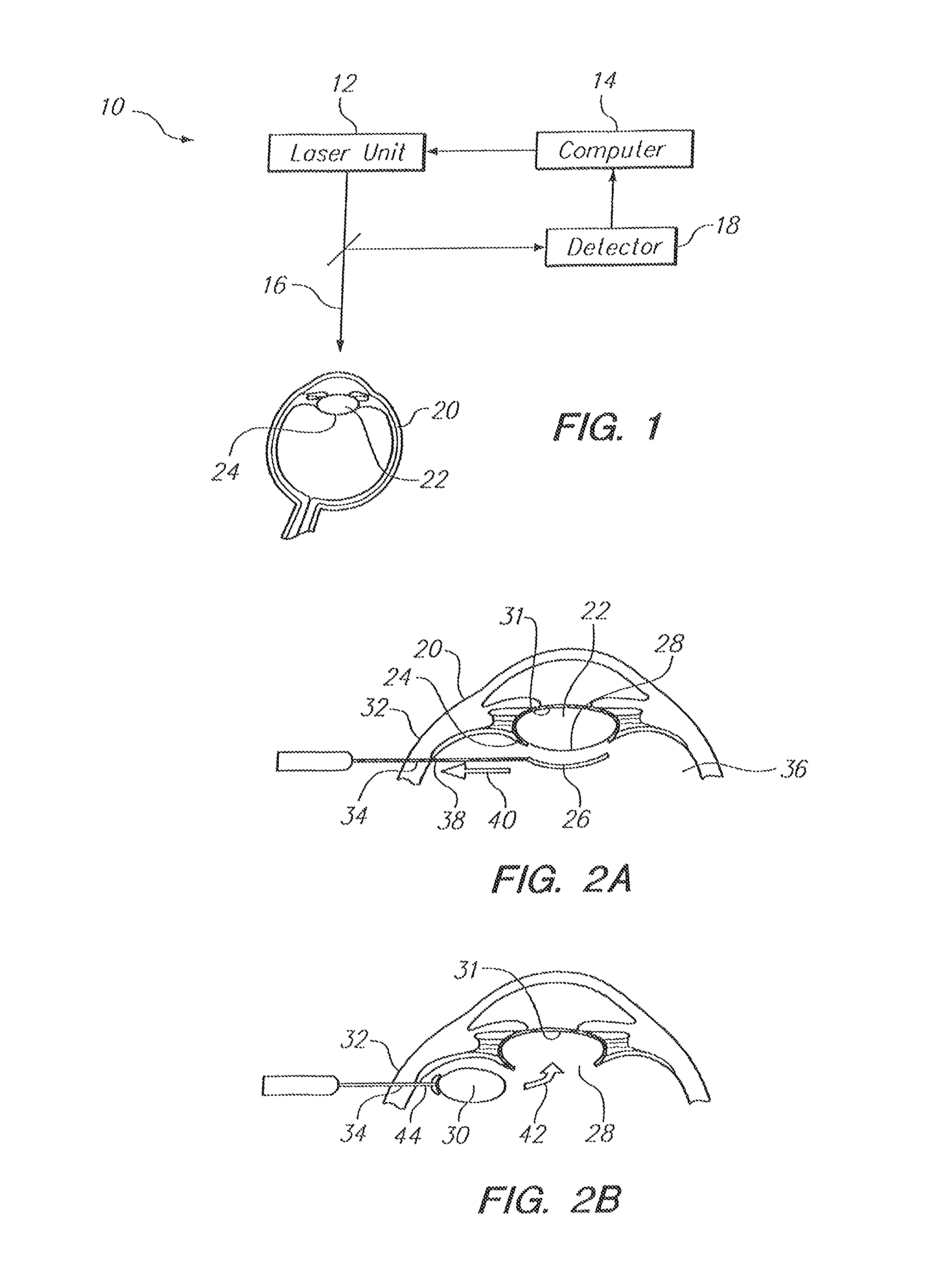
FIG. 1 is a schematic presentation of components for the system of the present invention;
FIG. 2A is a cross section view of the anterior portion of an eye showing the removal of tissue from the posterior capsule of the eye to create an opening into the lens capsule; and
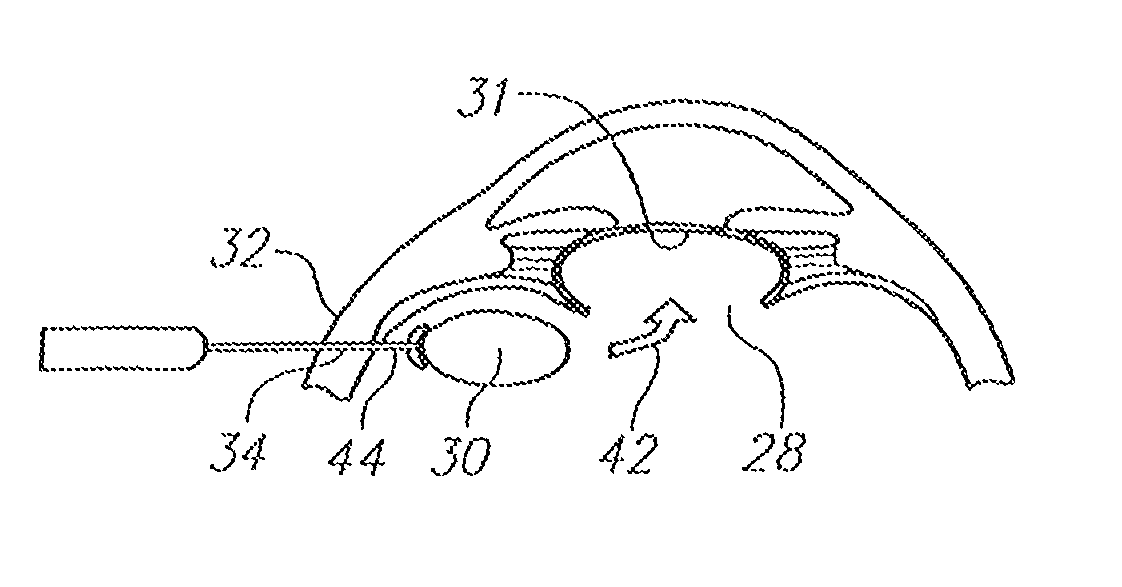
FIG. 2B is a cross section view of the anterior portion of the eye as seen in FIG. 2A, showing the insertion of an Intraocular Lens (IOL) through the opening into the lens capsule of the eye, after the crystalline lens of the eye has been removed from the lens capsule.
DESCRIPTION OF THE PREFERRED EMBODIMENTS
Referring initially to FIG. 1, a system in accordance with the present invention is shown and is generally designated 10. As shown, the system 10 includes a laser unit 12 and a computer 14. As shown, the computer 14 is connected with the laser unit 12 for the purposes of guiding and controlling the movement of a laser beam 16 that is generated and focused by the laser unit 12. Necessarily, the laser unit 12 is of a type that generates a pulsed laser beam 16 which is capable of performing Laser Induced Optical Breakdown (LIOB) on sub-surface anatomical tissue. Preferably, the laser beam 16 will have laser pulses with pulse durations in the femtosecond range.
FIG. 1 also shows that the system 10 can include a detector 18 for creating images of the interior of the eye 20. For example, the detector 18 can include an imaging unit that employs imaging techniques such as Optical Coherence Tomography (OCT), Scheimpflug imaging, confocal imaging, two-photon imaging, laser (optical) range finding and acoustical imaging. As shown, the computer 14 is connected to the detector 18 to receive and process image data from the detector 18 and control the operation of the detector 18. More specifically, images of tissue inside the eye 20 can be processed by the computer 14 to establish an accurate location for the crystalline lens 22 and posterior capsule 24 of the eye 20.
FIG. 2A illustrates a procedure for creating a section of separated tissue 26 and removing the tissue 26 from the posterior capsule 24 of the eye 20 to create an opening 28 into the posterior capsule 24. For this procedure, images of the posterior capsule 24 (FIG. 1) are provided by the detector 18 to the computer 14 which uses the image data to control movements of the focal point through tissue of the posterior capsule 24. As this is done, Laser Induced Optical Breakdown (LIOB) occurs on tissue of the posterior capsule 24 to create a section of separated tissue 26. Once the separated tissue is removed, an opening 28 is established through the posterior capsule 24 that can be used for removing the lens 22 and/or inserting an IOL 30 (FIG. 2B) into the lens capsule 31. For this process, the opening 28 can be dimensioned and customized to allow a specific IOL 30 (FIG. 2B) to pass through the opening 28 and be implanted into the lens capsule 31.
Continuing with FIG. 2A, it can be seen that the separated tissue 26 can be removed from the eye 20, by first incising the sclera 32 to establish an access port 34 to the vitreous body 36 of the eye 20. As shown, a probe 38 can then be advanced through the access port 34 for engagement of the probe 38 with the section of separated tissue 26. After the probe 38 has engaged with the separated section of tissue 26, the separated section of tissue 26 can be removed from the eye 20 by the probe 38 (in the direction of arrow 40) and through access port 34 to establish the opening 28. Once an opening 28 has been established through the posterior capsule 24 of the eye 20, as shown, the crystalline lens 22 can be extracted from the lens capsule 31 and removed from the eye 20 through the access port 34.
FIG. 2B shows that an IOL 30 can be introduced through the access port 34 in the sclera 32 and inserted through the opening 28 (in the direction of arrow 42 and into the lens capsule 31. As shown, probe 44 can be used to place the IOL 30 into the lens capsule 31. As an additional feature of the present invention, LIOB marks (not shown) can be established on the lens capsule 31 during the procedure to assist in aligning the IOL 30 in the lens capsule 31. For example, these LIOB marks may be used to properly orient a toric IOL (not shown) that is to be used for the correction of astigmatism.
While the particular Posterior Capsulotomy Using Laser Techniques as herein shown and disclosed in detail is fully capable of obtaining the objects and providing the advantages herein before stated, it is to be understood that it is merely illustrative of the presently preferred embodiments of the invention and that no limitations are intended to the details of construction or design herein shown other than as described in the appended claims.
* * * * *
D00000
D00001
XML
uspto.report is an independent third-party trademark research tool that is not affiliated, endorsed, or sponsored by the United States Patent and Trademark Office (USPTO) or any other governmental organization. The information provided by uspto.report is based on publicly available data at the time of writing and is intended for informational purposes only.
While we strive to provide accurate and up-to-date information, we do not guarantee the accuracy, completeness, reliability, or suitability of the information displayed on this site. The use of this site is at your own risk. Any reliance you place on such information is therefore strictly at your own risk.
All official trademark data, including owner information, should be verified by visiting the official USPTO website at www.uspto.gov. This site is not intended to replace professional legal advice and should not be used as a substitute for consulting with a legal professional who is knowledgeable about trademark law.