Stem-loop compositions and methods for inhibiting factor D
Erickson , et al. October 1, 2
U.S. patent number 10,428,330 [Application Number 15/990,547] was granted by the patent office on 2019-10-01 for stem-loop compositions and methods for inhibiting factor d. This patent grant is currently assigned to Albert Einstein College of Medicine, Vitrisa Therapeutics, Inc.. The grantee listed for this patent is Albert Einstein College of Medicine, Inc., Vitrisa Therapeutics, Inc.. Invention is credited to Arijit Bhowmick, Carl Erickson, Matthew Levy, Kevin G. McLure, Christopher P. Rusconi.


View All Diagrams
| United States Patent | 10,428,330 |
| Erickson , et al. | October 1, 2019 |
Stem-loop compositions and methods for inhibiting factor D
Abstract
The application discloses methods and compositions for the inhibition of the alternative complement pathway. The methods and compositions involve the use of aptamers for inhibiting complement Factor D. The application further provides anti-Factor D aptamers for the treatment of dry age-related macular degeneration, geographic atrophy, wet age-related macular degeneration or Stargardt disease. In some cases, stem-loop aptamers are provided for the inhibition of Factor D.
| Inventors: | Erickson; Carl (Corte Madera, CA), Rusconi; Christopher P. (Durham, NC), McLure; Kevin G. (Oakland, CA), Levy; Matthew (Cary, NC), Bhowmick; Arijit (Durham, NC) | ||||||||||
|---|---|---|---|---|---|---|---|---|---|---|---|
| Applicant: |
|
||||||||||
| Assignee: | Vitrisa Therapeutics, Inc. (San
Jose, CA) Albert Einstein College of Medicine (Bronx, NY) |
||||||||||
| Family ID: | 62908350 | ||||||||||
| Appl. No.: | 15/990,547 | ||||||||||
| Filed: | May 25, 2018 |
Prior Publication Data
| Document Identifier | Publication Date | |
|---|---|---|
| US 20190032056 A1 | Jan 31, 2019 | |
Related U.S. Patent Documents
| Application Number | Filing Date | Patent Number | Issue Date | ||
|---|---|---|---|---|---|
| PCT/US2018/014573 | Jan 19, 2018 | ||||
| 62536387 | Jul 24, 2017 | ||||
| 62448872 | Jan 20, 2017 | ||||
| Current U.S. Class: | 1/1 |
| Current CPC Class: | A61K 31/7088 (20130101); A61K 47/60 (20170801); A61K 31/7115 (20130101); C12N 15/115 (20130101); C12N 2310/16 (20130101); C12N 2310/317 (20130101); C12N 2310/531 (20130101); C12N 2310/322 (20130101); C12N 2310/321 (20130101); C12N 2310/321 (20130101); C12N 2310/3521 (20130101); C12N 2310/322 (20130101); C12N 2310/3533 (20130101) |
| Current International Class: | C12N 15/115 (20100101); A61K 31/7088 (20060101) |
References Cited [Referenced By]
U.S. Patent Documents
| 5270163 | December 1993 | Gold et al. |
| 5475096 | December 1995 | Gold et al. |
| 6140490 | October 2000 | Biesecker et al. |
| 6333034 | December 2001 | Gupta-Bansal et al. |
| 6653340 | November 2003 | Babu et al. |
| 6956107 | October 2005 | Fung et al. |
| 7112327 | September 2006 | Fung et al. |
| 7927592 | April 2011 | Fung et al. |
| 7943135 | May 2011 | Fung et al. |
| 7999082 | August 2011 | Holers et al. |
| 8007791 | August 2011 | Hass et al. |
| 8067002 | November 2011 | An et al. |
| 8124090 | February 2012 | Fung et al. |
| 8273352 | September 2012 | Huang et al. |
| 8435512 | May 2013 | Bansal et al. |
| 8492082 | July 2013 | De Franciscis et al. |
| 8580735 | November 2013 | Francois et al. |
| 8664362 | March 2014 | Bansal |
| 8703136 | April 2014 | Baas et al. |
| 8753625 | June 2014 | Fung et al. |
| 8858943 | October 2014 | Burbidge et al. |
| 8911733 | December 2014 | Holers et al. |
| 8921523 | December 2014 | Alard et al. |
| 8940299 | January 2015 | Medof et al. |
| 8981060 | March 2015 | Bansal |
| 9066925 | June 2015 | Tomlinson et al. |
| 9085555 | July 2015 | Altmann et al. |
| 9278108 | March 2016 | Takenaka et al. |
| 9803194 | October 2017 | May et al. |
| 9873727 | January 2018 | Sullenger et al. |
| 10174325 | January 2019 | Erickson et al. |
| 2004/0038869 | February 2004 | Finney et al. |
| 2007/0065433 | March 2007 | Mollnes et al. |
| 2007/0093443 | April 2007 | Madison et al. |
| 2007/0149616 | June 2007 | Clark et al. |
| 2007/0178068 | August 2007 | Reich et al. |
| 2007/0196367 | August 2007 | Dinu |
| 2008/0269318 | October 2008 | Romano |
| 2009/0092980 | April 2009 | Arenz et al. |
| 2009/0269356 | October 2009 | Epstein et al. |
| 2011/0044983 | February 2011 | Lambris et al. |
| 2011/0060027 | March 2011 | Benedict et al. |
| 2011/0160636 | June 2011 | Bansal |
| 2011/0165648 | July 2011 | Campagne et al. |
| 2011/0190221 | August 2011 | Francois et al. |
| 2012/0087905 | April 2012 | Lachmann |
| 2012/0107315 | May 2012 | Behrens et al. |
| 2012/0190578 | July 2012 | Seddon et al. |
| 2013/0035388 | February 2013 | McGeer et al. |
| 2014/0235701 | August 2014 | Jin |
| 2014/0371133 | December 2014 | Francois et al. |
| 2015/0044205 | February 2015 | Yaspan et al. |
| 2015/0104445 | April 2015 | Uknis et al. |
| 2015/0239837 | August 2015 | Wiles et al. |
| 2016/0061840 | March 2016 | Lee |
| 2017/0328909 | November 2017 | Bock et al. |
| 2018/0030446 | February 2018 | Benedict et al. |
| 2018/0051287 | February 2018 | Erickson et al. |
| 2019/0010499 | January 2019 | Erickson et al. |
| 5825673 | Dec 2015 | JP | |||
| WO-9119813 | Dec 1991 | WO | |||
| WO-9927133 | Jun 1999 | WO | |||
| WO-2007103549 | Sep 2007 | WO | |||
| WO-2012178083 | Dec 2012 | WO | |||
| WO-2015168468 | Nov 2015 | WO | |||
| WO-2017087919 | May 2017 | WO | |||
| WO-2017127761 | Jul 2017 | WO | |||
| WO-2018136827 | Jul 2018 | WO | |||
| WO-2018136831 | Jul 2018 | WO | |||
| WO-2019022986 | Jan 2019 | WO | |||
Other References
|
Altschul, et al. Basic local alignment search tool. J Mol Biol. Oct. 5, 1990;215(3):403-10. cited by applicant . Altschul, et al. Gapped BLAST and PSI-BLAST: a new generation of protein database search programs. Nucleic Acids Res. Sep. 1, 1997;25(17):3389-402. cited by applicant . Berchuck, et al. All-trans-retinal sensitizes human RPE cells to alternative complement pathway-induced cell death. Invest Ophthalmol Vis Sci. Apr. 12, 2013;54(4):2669-77. cited by applicant . Forneris, et al. Structures of C3b in complex with factors B and D give insight into complement convertase formation. Science. Dec. 24, 2010;330(6012):1816-20. doi: 10.1126/science.1195821. cited by applicant . Gold, et al. Aptamer-based multiplexed proteomic technology for biomarker discovery. PLoS One. Dec. 7, 2010;5(12):e15004. doi: 10.1371/journal.pone.0015004. cited by applicant . Harper et al. Reaction of serine proteases with substituted isocoumarins: discovery of 3,4-dichloroisocoumarin, a new general mechanism based serine protease inhibitor.Biochemistry 24(8):1831-1841 (Apr. 1985). cited by applicant . Hedstrom, Lizbeth. Serine protease mechanism and specificity. Chem Rev. Dec. 2002;102(12):4501-24. cited by applicant . Jing, et al. Structural basis of profactor D activation: from a highly flexible zymogen to a novel self-inhibited serine protease, complement factor D. EMBO J. Feb. 15, 1999;18(4):804-14. cited by applicant . Jing, et al. Structures of native and complexed complement factor D: implications of the atypical His57 conformation and self-inhibitory loop in the regulation of specific serine protease activity. J Mol Biol. Oct. 9, 1998;282(5):1061-81. cited by applicant . Kam, et al. Human complement proteins D, C2, and B. Active site mapping with peptide thioester substrates. J Biol Chem. Mar. 15, 1987;262(8):3444-51. cited by applicant . Karlin, et al. Applications and statistics for multiple high-scoring segments in molecular sequences. Proc Natl Acad Sci U S A. Jun. 15, 1993;90(12):5873-7. cited by applicant . Karlin, et al. Methods for assessing the statistical significance of molecular sequence features by using general scoring schemes. Proc Natl Acad Sci U S A. Mar. 1990;87(6):2264-8. cited by applicant . Katschke, et al. Inhibiting alternative pathway complement activation by targeting the factor D exosite. J Biol Chem. Apr. 13, 2012;287(16):12886-92. doi: 10.1074/jbc.M112.345082. Epub Feb. 23, 2012. cited by applicant . Katschke, et al. Structural and functional analysis of a C3b-specific antibody that selectively inhibits the alternative pathway of complement. J Biol Chem. Apr. 17, 2009;284(16):10473-9. cited by applicant . Lao et al. Selection of Aptamers Targeting the Sialic Acid Receptor of Hemagglutinin by Epitope-Specific SELEX. Chemical Communications 50(63):8719-8722 (2014). cited by applicant . Loyet, et al. Complement inhibition in cynomolgus monkeys by anti-factor d antigen-binding fragment for the treatment of an advanced form of dry age-related macular degeneration. J Pharmacol Exp Ther. Dec. 2014;351(3):527-37. cited by applicant . Loyet et al. Activation of the Alternative Complement Pathway in Vitreous is Controlled by Genetics in Age-Related Macular Degeneration. 53(10):6628-6637 (Sep. 2012). cited by applicant . Macugen.RTM.. Drugs at FDA. Revised Jul. 2011. URL:< https://www.accessdata.fda.gov/drugsatfda_docs/label/2011/021756s018lbl.p- df>. cited by applicant . McHarg, et al. Age-related macular degeneration and the role of the complement system. Mol Immunol. Sep. 2015 ;67(1):43-50. doi: 10.1016/j.molimm.2015.02.032. Epub Mar. 21, 2015. cited by applicant . Molday, et al. ATP-binding cassette transporter ABCA4: molecular properties and role in vision and macular degeneration. J Bioenerg Biomembr. Dec. 2007;39(5-6):507-17. cited by applicant . Narayana, et al. Structure of human factor D. A complement system protein at 2.0 A resolution. J Mol Biol. Jan. 14, 1994;235(2):695-708. cited by applicant . Ouellet, et al. Hi-Fi SELEX: A High-Fidelity Digital-PCR Based Therapeutic Aptamer Discovery Platform. Biotechnol Bioeng. Aug. 2015;112(8):1506-22. cited by applicant . Ouellet, et al. Hi-Fi SELEX: A High-Fidelity Digital-Pcr Based Therapeutic Aptamer Discovery Platform. Supporting Information. Biotechnol Bioeng. Aug. 2015;112(8):1506-22. cited by applicant . Pangburn, MK. Alternative pathway of complement. Methods Enzymol. 1988;162:639-53. cited by applicant . PCT/US2017/014458 International Search Report dated Apr. 19, 2017. cited by applicant . Preclinical and Phase 1A Clinical Evaluation of an Anti-VEGF Pegylated Aptamer (EYE001) for the Treatment of Exudative Age-Related Macular Degeneration. The Eyetech Study Group. Retina 22(2):143-152 (2002). cited by applicant . Shukla et al. Pegaptanib sodium for ocular vascular disease. Indian J Ophthalmol. 55(6):427-30 (Nov.-Dec. 2007). cited by applicant . PCT/US2018/014573 International Search Report dated May 7, 2018. cited by applicant . U.S. Appl. No. 15/693,361 Notice of Allowance dated Aug. 30, 2018. cited by applicant . U.S. Appl. No. 15/693,361 Office Action dated May 25, 2018. cited by applicant . PCT/US2017/014458 Written Opinion dated Apr. 19, 2017. cited by applicant . Volanakis, et al. Complement factor D, a novel serine protease. Protein Sci. Apr. 1996; 5(4): 553-564. doi: 10.1002/pro.5560050401. cited by applicant . Wiles et al. Preclinical Evaluation of Orally Bioavailable Small-Molecule Inhibitors of Complement Factor D as a Potential Treatment for Paroxysmal Nocturnal Hemoglobinuria. Achillion Pharmaceuticals Inc. Abstract ID: 4819. Presented at the 56th Annual Meeting of the American Society of Hematology, San Francisco, California, USA, Dec. 6-9, 2014. One page. cited by applicant . Wootton, et al. Statistics of local complexity in amino acid sequences and sequence databases. Computers & Chemistry. vol. 17, Issue 2, Jun. 1993, pp. 149-163. cited by applicant . Yang, et al. Buried Hydrogen Bond Interactions Contribute to the High Potency of Complement Factor D Inhibitors. ACS Med. Chem. Lett., 2016, 7 (12), pp. 1092-1096. DOI: 10.1021/acsmedchemlett.6b00299. cited by applicant . Co-pending U.S. Appl. No. 16/121,458, filed Sep. 4, 2018. cited by applicant . PCT Application PCT/US2018/042317 as filed Jul. 16, 2018. cited by applicant . PCT/US2018/042317 International Search Report and Written Opinion dated Oct. 29, 2018. cited by applicant. |
Primary Examiner: Vivlemore; Tracy
Attorney, Agent or Firm: Wilson Sonsini Goodrich & Rosati
Parent Case Text
CROSS-REFERENCE
This application is a continuation application of International Patent Application No. PCT/US2018/014573, filed Jan. 19, 2018, which claims the benefit of U.S. Provisional Application Nos. 62/448,872, filed Jan. 20, 2017, and 62/536,387, filed Jul. 24, 2017, which applications are incorporated herein by reference in their entireties.
Claims
What is claimed is:
1. An aptamer comprising a nucleic acid sequence that selectively binds to complement factor D (fD) and having a stem-loop secondary structure comprising, in a 5' to 3' direction, a first base-paired stem, a first loop, a second base-paired stem, a second loop, and a third loop, wherein said third loop comprises 6 or more nucleotides, non-nucleotidyl spacers, or a combination thereof, and wherein said third loop is adjacent to said first base-paired stem.
2. The aptamer of claim 1, wherein said first loop has fewer nucleotides than said second loop.
3. The aptamer of claim 1, wherein said first loop has from 1to 10 nucleotides, non-nucleotidyl spacers, or a combination thereof.
4. The aptamer of claim 1, wherein said second loop comprises at least 6 nucleotides, non-nucleotidyl spacers, or a combination thereof.
5. The aptamer of claim 1, wherein said third loop has from 6to 8 nucleotides, non-nucleotidyl spacers, or a combination thereof.
6. The aptamer of claim 1, wherein said first base-paired stem has from 2 to 10 base pairs.
7. The aptamer of claim 1, wherein said second base-paired stem has from 2 to 10 base pairs.
8. The aptamer of claim 1, wherein said third loop comprises at least 4 nucleotides and up to 2 non-nucleotidyl spacers.
9. The aptamer of claim 1 , wherein said third loop comprises at least 6 nucleotides.
10. The aptamer of claim 1, wherein: a) said first base-paired stem is adjacent to said first loop; b) said second base-paired stem is adjacent to said first loop, said second loop, and said third loop; or c) said first base-paired stem is adjacent to said first loop and said second base-paired stem is adjacent to said first loop, said second loop, and said third loop.
11. The aptamer of claim 1, wherein said aptamer is an RNA aptamer or a modified RNA aptamer.
12. The aptamer of claim 1, wherein said aptamer is a DNA aptamer or a modified DNA aptamer.
13. The aptamer of claim 1, wherein said aptamer comprises one or more modified nucleotides.
14. The aptamer of claim 13, wherein said one or more modified nucleotides comprises a 2'F-modified nucleotide, a 2'OMe-modified nucleotide, or a combination thereof.
15. The aptamer of claim 1, wherein said stem-loop structure has exactly two base-paired stems.
16. The aptamer of claim 1, wherein said aptamer is an RNA aptamer comprising nucleotides having ribose in a .beta.-D-ribofuranose configuration.
17. The aptamer of claim 1, wherein said aptamer blocks an active site of fD.
18. The aptamer of claim 1, wherein said aptamer inhibits a function associated with fD.
19. The aptamer of claim 1, wherein said nucleic acid sequence comprises from 30 to 90 nucleotides, non-nucleotidyl spacers, or a combination thereof.
20. The aptamer of claim 1, wherein: said aptamer is conjugated to a polyethylene glycol (PEG) molecule.
21. The aptamer of claim 1, wherein said second loop comprises more than 5 nucleotides, non-nucleotidyl spacers, or a combination thereof.
22. The aptamer of claim 1, wherein said first base-paired stem has no more than 5 base pairs.
23. The aptamer of claim 1, wherein said first loop comprises a nucleic acid sequence of 5'-DUG-3', where D is A, G, or U.
24. The aptamer of claim 1, wherein said second loop comprises a nucleic acid sequence of 5'-DWWVGCBHWG-3', where D is A, G, or U; W is A or U; V is A, C, or G; B is C, G, or U; and H is A, C, or U.
25. The aptamer of claim 1, wherein said second loop comprises a nucleic acid sequence having a U at nucleotide position 2, a U at nucleotide position 3, or both.
26. The aptamer of claim 1, wherein said third loop comprises a nucleic acid sequence comprising 5'-AAGUKN-3', where K is G or U; and N is A, G, C, or U.
27. The aptamer of claim 1, wherein said second base-paired stem comprises a terminal U-G base pair adjacent to said second loop.
28. The aptamer of claim 1, wherein said second base-paired stem comprises a terminal C-G base pair adjacent to said second loop.
Description
SEQUENCE LISTING
The instant application contains a Sequence Listing which has been submitted electronically in ASCII format and is hereby incorporated by reference in its entirety. Said ASCII copy, created on Apr. 6, 2018, is named 49644-716_601_SL.txt and is 100,140 bytes in size.
BACKGROUND OF THE INVENTION
Visual impairment is a national and global health concern that has a negative impact on physical and mental health. The number of people with visual impairment and blindness is increasing due to an overall aging population. Visual impairment and blindness can be caused by any one of a large number of eye diseases and disorders affecting people of all ages. In one example, age-related macular degeneration (AMD) is an eye disorder that is currently the leading cause of vision loss in people fifty years of age or older in industrialized countries. It is estimated that by 2020, the number of people with AMD could exceed 196 million and by 2040, that number is expected to rise to 288 million. AMD is a degenerative eye disease that progresses from early stages to advanced stages of the disease. Risk factors for the disease include aging, lifestyle factors such as smoking, and genetics. The clearest indicator of progression to AMD is the appearance of drusen, yellow-white deposits under the retina, and it is an important component of both forms of AMD: exudative ("wet") and non-exudative ("dry"). Wet AMD causes vision loss due to abnormal blood vessel growth in the choriocapillaris through Bruch's membrane. The most advanced form of dry AMD, known as geographic atrophy, is generally more gradual and occurs when light-sensitive cells in the macula atrophy, blurring and eliminating vision in the affected eye. While there are currently some promising treatments for wet AMD, no FDA-approved treatment exists for dry AMD or geographic atrophy.
A second example is childhood-onset Stargardt Disease ("STGD"), also known as Stargardt 1, a genetic, rare juvenile macular dystrophy generally associated with loss of central vision in the first two decades of life. STGD has a prevalence of approximately 1/20,000 affecting approximately 30,000 people in the US. STGD affects many ages, with the childhood-onset population at highest risk and most need. Patients with childhood-onset STGD tend to develop early severe visual acuity loss, significantly compromised retinal function, and rapid retinal pigment epithelial (RPE) cell atrophy with accompanying loss of retinal function. The median ages of onset and the median age at baseline examination are 8.5 (range, 3-16) and 12 years (range, 7-16), respectively. Patients with adult-onset disease are more likely to preserve visual acuity for a longer time and show slighter retinal dysfunction. STGD is an autosomal recessive genetic disease or complex heterozygous disease, caused by mutations in the ABCA4 gene. The ABCA4 gene encodes the photoreceptor protein ABCA4 Transporter, which is responsible for removal of bisretinoid fluorophores, which can include N-retinylidene-N-retinyethanolamine (A2E), all-trans-retinal and related photo-oxidation products of vitamin A aldehyde which together constitute lipofuscin from photoreceptor cells. Accumulation of all-trans-retinal in photoreceptor cells is believed to damage RPE cells via oxidative stress, and trigger or promote complement-mediated damage to RPE cells, leading to retinal atrophy. A related disease termed Stargardt-like macular dystrophy, also known as STGD3, is inherited in a dominant autosomal manner and is due to mutations in the ELOVL4 gene. ELOVL4 encodes the ELOVL4 protein, ELOVL fatty acid elongase 4. Mutations in ELOVL4 protein associated with STGD lead to mis-folding and accumulation of ELOVL4 protein aggregates in retinal cells, which impact retinal cell function, eventually leading to cell death and retinal atrophy. No treatments exist for STGD or Stargardt-like disease.
SUMMARY OF THE INVENTION
In one aspect, an aptamer is provided comprising a nucleic acid sequence that selectively binds to complement factor D (fD) and having a stem-loop secondary structure comprising, in a 5' to 3' direction, a first base-paired stem, a first loop, a second base-paired stem, a second loop, and a third loop, wherein the third loop comprises 6 or more nucleotides, non-nucleotidyl spacers, or a combination thereof, and wherein the first loop has fewer nucleotides than the second loop.
In another aspect, an aptamer is provided comprising a nucleic acid sequence that selectively binds to complement factor D (fD) and having a stem-loop secondary structure comprising, in a 5' to 3' direction, a first base-paired stem, a first loop, a second base-paired stem, a second loop, and a third loop, wherein the third loop comprises 6 or more nucleotides, non-nucleotidyl spacers, or a combination thereof, and wherein the second loop comprises more than 5 nucleotides, non-nucleotidyl spacers, or a combination thereof.
In another aspect, an aptamer is provided comprising a nucleic acid sequence that selectively binds to complement factor D (fD) and having a stem-loop secondary structure comprising, in a 5' to 3' direction, a first base-paired stem, a first loop, a second base-paired stem, a second loop, and a third loop, wherein the third loop comprises 6 or more nucleotides, non-nucleotidyl spacers, or a combination thereof, and wherein the third loop is adjacent to the first stem.
In yet another aspect, an aptamer is provided comprising a nucleic acid sequence that selectively binds to complement factor D (fD) and having a stem-loop secondary structure comprising, in a 5' to 3' direction, a first base-paired stem, a first loop, a second base-paired stem, a second loop, and a third loop, wherein the third loop comprises 6 or more nucleotides, non-nucleotidyl spacers, or a combination thereof, and wherein the first base-paired stem has no more than 5 base pairs.
In some cases, the third loop is connected to the first base-paired stem. In some cases, the first loop has from 1 to 10 nucleotides, non-nucleotidyl spacers, or a combination thereof. In some cases, the first loop has from 3 to 5 nucleotides, non-nucleotidyl spacers, or a combination thereof. In some cases, first loop comprises a nucleic acid sequence of 5'-DUG-3', where D is A, G, or U. In some cases, the second loop comprises at least 6 nucleotides, non-nucleotidyl spacers, or a combination thereof. In some cases, the second loop comprises at least 7 nucleotides, non-nucleotidyl spacers, or a combination thereof. In some cases, the second loop comprises 10 or 11 nucleotides, non-nucleotidyl spacers, or a combination thereof. In some cases, the second loop comprises a nucleic acid sequence of 5'-DWWVGCBHWG-3' (SEQ ID NO:319), where D is A, G, or U; W is A or U; V is A, C, or G; B is C, G, or U; and H is A, C, or U. In some cases, the second loop comprises a nucleic acid sequence having a U at nucleotide position 2, nucleotide position 3, or both. In some cases, the third loop has from 6 to 8 nucleotides, non-nucleotidyl spacers, or a combination thereof. In some cases, the third loop comprises a nucleic acid sequence comprising 5'-AAGUKN-3', where K is G or U; and N is A, G, C, or U. In some cases, the first base-paired stem has from 2 to 10 base pairs. In some cases, the first base-paired stem has from 3 to 8 base pairs. In some cases, the second base-paired stem has from 2 to 10 base pairs. In some cases, the second base-paired stem comprises 4 or 5 base pairs. In some cases, the second base-paired stem comprises a terminal U-G base pair adjacent to the second loop. In some cases, the second base-paired stem comprises a terminal C-G base pair adjacent to the second loop. In some cases, the nucleic acid sequence comprises nucleotides having ribose in a .beta.-D-ribofuranose configuration. In some cases, at least 50% of the nucleic acid sequence comprises nucleotides having ribose in a .beta.-D-ribofuranose configuration. In some cases, the third loop comprises at least 4 nucleotides and up to 2 non-nucleotidyl spacers. In some cases, the third loop comprises at least 6 nucleotides. In some cases, the non-nucleotidyl spacers comprise 3 carbons, 6 carbons, or 9 carbons. In some cases, the non-nucleotidyl spacers comprise an 18-atom spacer. In one example, the 18-atom spacer comprises hexaethylene glycol. In some cases, a) the first base-paired stem is adjacent to said first loop; b) the second base-paired stem is adjacent to the first loop, the second loop, and the third loop; or c) the first base-paired stem is adjacent to the first loop and the second base-paired stem is adjacent to the first loop, the second loop, and the third loop.
In another aspect, an aptamer is provided comprising a nucleic acid sequence that selectively binds to complement factor D (fD) and having a stem-loop secondary structure comprising a base-paired terminal stem; an asymmetric internal loop; an internal base-paired stem; and exactly one terminal loop, wherein the terminal loop comprises more than 4 nucleotides, non-nucleotidyl spacers, or a combination thereof, and wherein the asymmetric internal loop is adjacent to exactly 2 base-paired stems.
In another aspect, an aptamer is provided comprising a nucleic acid sequence that selectively binds to complement factor D (fD) and having a stem-loop secondary structure comprising, in a 5' to 3' direction, a first base-paired stem, a first loop, a second base-paired stem, a second loop, and a third loop, wherein the second loop comprises 7 or more nucleotides, non-nucleotidyl spacers, or a combination thereof, wherein the first base-paired stem has no more than 5 base pairs, and wherein the second base-paired stem comprises more than 2 base pairs.
In another aspect, an aptamer is provided comprising a nucleic acid sequence that selectively binds to complement factor D (fD) and having a stem-loop secondary structure comprising exactly one terminal base-paired stem; exactly one asymmetric internal loop comprising, from a 5' to 3' direction, a first loop and a second loop; exactly one internal base-paired stem; and exactly one terminal loop, wherein the first loop of the asymmetric internal loop has fewer nucleotides than the terminal loop.
In another aspect, an aptamer is provided comprising a nucleic acid sequence that selectively binds to complement factor D (fD) and having a stem-loop secondary structure comprising exactly one terminal base-paired stem; exactly one asymmetric internal loop; exactly one internal base-paired stem; and exactly one terminal loop, wherein the exactly one terminal loop comprises more than 4 nucleotides, non-nucleotidyl spacers, or a combination thereof. In some cases, the exactly one terminal loop comprises 10 or more nucleotides, non-nucleotidyl spacers, or a combination thereof.
In another aspect, an aptamer is provided comprising a nucleic acid sequence that selectively binds to complement factor D (fD) and having a stem-loop secondary structure comprising exactly one terminal base-paired stem; exactly one asymmetric internal loop comprising, from a 5' to 3' direction, a first loop and a second loop; exactly one internal base-paired stem; and exactly one terminal loop, wherein the second loop comprises 6 or more nucleotides, non-nucleotidyl spacers, or a combination thereof.
In another aspect, an aptamer is provided comprising a nucleic acid sequence that selectively binds to complement factor D (fD) and having a stem-loop secondary structure comprising exactly one terminal base-paired stem; exactly one asymmetric internal loop; exactly one internal base-paired stem; and exactly one terminal loop, wherein the exactly one terminal loop comprises 7 or more nucleotides, non-nucleotidyl spacers, or a combination thereof.
In some cases, the exactly one terminal base-paired stem comprises a tail at a 5' end, at a 3' end, or at both a 5' end and a 3' end, and the tail comprises at least one unpaired nucleotide.
In another aspect, an aptamer is provided comprising a nucleic acid sequence that selectively binds to complement factor D (fD) and having a stem-loop secondary structure comprising, in a 5' to 3' direction, a first base-paired stem, a first loop, a second base-paired stem, a second loop, and a third loop, wherein the second base-paired stem comprises a terminal U-G base pair adjacent to the second loop.
In yet another aspect, an aptamer is provided comprising a nucleic acid sequence that selectively binds to complement factor D (fD) and having a stem-loop secondary structure comprising, in a 5' to 3' direction, a first base-paired stem, a first loop, a second base-paired stem, a second loop, and a third loop, wherein the first loop comprises a nucleic acid sequence of 5'-DUG-3', where D is A, G, or U.
In yet another aspect, an aptamer is provided comprising a nucleic acid sequence that selectively binds to complement factor D (fD) and having a stem-loop secondary structure comprising, in a 5' to 3' direction, a first base-paired stem, a first loop, a second base-paired stem, a second loop, and a third loop, wherein the third loop comprises a nucleic acid sequence comprising 5'-AAGUKN-3', where K is G or U; and N is A, G, C, or U.
In yet another aspect, an aptamer is provided comprising a nucleic acid sequence that selectively binds to complement factor D (fD) and having a stem-loop secondary structure comprising, in a 5' to 3' direction, a first base-paired stem, a first loop, a second base-paired stem, a second loop, and a third loop, wherein the second loop comprises a nucleic acid sequence of 5'-DWWVGCBHWG-3' (SEQ ID NO:319), where D is A, G, or U; W is A or U; V is A, C, or G; B is C, G, or U; and H is A, C, or U.
In yet another aspect, an aptamer is provided comprising a nucleic acid sequence that selectively binds to complement factor D (fD) and having a stem-loop secondary structure comprising, in a 5' to 3' direction, a first base-paired stem, a first loop, a second base-paired stem, a second loop, and a third loop, wherein the second loop comprises a nucleic acid sequence having a U at nucleotide position 2, nucleotide position 3, or both.
In yet another aspect, an aptamer is provided comprising a nucleic acid sequence that selectively binds to complement factor D (fD) and having a stem-loop secondary structure comprising, in a 5' to 3' direction, a first base-paired stem, a first loop, a second base-paired stem, a second loop, and a third loop, wherein the second base-paired stem comprises a terminal C-G base pair adjacent to the second loop.
In some cases, any aptamer of the preceding is an RNA aptamer or a modified RNA aptamer. In other cases, any aptamer of the preceding is a DNA aptamer or a modified DNA aptamer. In some cases, any aptamer of the preceding comprises one or more modified nucleotides. In some instances, at least 50% of the nucleic acid sequence comprises the one or more modified nucleotides. In some instances, the one or more modified nucleotides comprises a 2'F-modified nucleotide, a 2'OMe-modified nucleotide, or a combination thereof. In some instances, the one or more modified nucleotides are selected from the group consisting of: 2'F-G, 2'OMe-G, 2'OMe-U, 2'OMe-A, 2'OMe-C, a 3' terminal inverted deoxythymidine, and any combination thereof. In some cases, an aptamer of any of the preceding comprises a nuclease-stabilized nucleic acid backbone. In some cases, the stem-loop structure of any aptamer of the preceding has exactly two base-paired stems. In some cases, any aptamer of the preceding is an RNA aptamer comprising nucleotides having ribose in a .beta.-D-ribofuranose configuration. In some cases, any aptamer of the preceding selectively binds to an active site of fD. In some cases, any aptamer of the preceding selectively binds to a substrate-binding exosite of fD. In some cases, any aptamer of the preceding selectively binds to both an active site of fD and a substrate-binding exosite of fD. In some cases, any aptamer of the preceding blocks an active site of fD. In some cases, any aptamer of the preceding blocks a substrate-binding exosite of fD. In some cases, any aptamer of the preceding blocks both an active site and a substrate-binding exosite of fD. In some cases, any aptamer of the preceding inhibits a function associated with fD. In some cases, any aptamer of the preceding prevents association of fD with pre-formed C3bB complex. In some cases, any aptamer of the preceding has no more than one nucleic acid strand. In other cases, any aptamer of the preceding comprises more than one nucleic acid strand. In some cases, the nucleic acid sequence of any aptamer of the preceding has from 30-90 nucleotides, non-nucleotidyl spacers, or a combination thereof. In some cases, any aptamer of the preceding selectively binds to an active site of fD with a K.sub.d of less than about 50 nM. In some cases, any aptamer of the preceding inhibits fD in an alternative complement dependent hemolysis assay with an IC.sub.50 of less than about 50 nM. In some cases, any aptamer of the preceding inhibits fD in a fD convertase assay with an IC.sub.50 of less than about 50 nM. In some cases, any aptamer of the preceding inhibits at least 85% of fD activity in an alternative complement dependent hemolysis assay. In some cases, any aptamer of the preceding inhibits at least 85% of fD activity in a fD convertase assay. In some cases, any aptamer of the preceding inhibits fD activity in an esterase activity assay. In some cases, any aptamer of the preceding binds to fD with a K.sub.d of less than about 50 nM and inhibits fD in an alternative complement dependent hemolysis assay with an IC.sub.90 of less than about 500 nM. In some cases, any aptamer of the preceding binds to fD with a K.sub.d of less than about 50 nM and inhibits fD in an alternative complement dependent hemolysis assay with an IC.sub.50 of less than about 100 nM. In some cases, any aptamer of the preceding is conjugated to a polyethylene glycol (PEG) molecule. In some cases, the PEG molecule has a molecular weight of 80 kDa or less. In some cases, any aptamer of the preceding does not contain a pseudoknot structure. In some cases, any aptamer of the preceding has less than 3 unpaired nucleotide residues at a 5' terminus, a 3' terminus, or both.
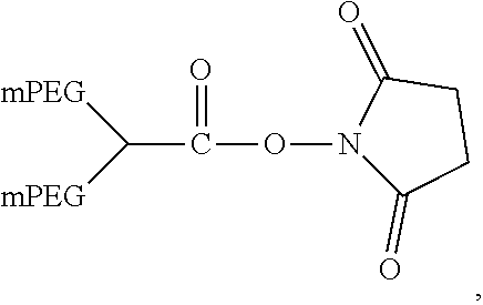
In another aspect, an aptamer is provided comprising a nucleic acid sequence comprising any one of SEQ ID NOs:13, 165, 166, 244, 253, 256, 262, 269, 284, 285, 294, 303, 306, and 312, or comprising at least 80% sequence identity to any one of SEQ ID NOs:13, 165, 166, 244, 253, 256, 262, 269, 284, 285, 294, 303, 306, and 312. In some cases, the nucleic acid sequence comprises one or more modified nucleotides. In some instances, at least 50% of said nucleic acid sequence comprises the one or more modified nucleotides. In some cases, the one or more modified nucleotides comprises a 2'F-modified nucleotide, a 2'OMe-modified nucleotide, or a combination thereof. In some cases, the one or more modified nucleotides are selected from the group consisting of: 2'F-G, 2'OMe-G, 2'OMe-U, 2'OMe-A, 2'OMe-C, a 3' terminal inverted deoxythymidine, and any combination thereof. In some cases, the aptamer is selected from the group consisting of: Aptamer 76 as described in Table 2, Aptamer 116 as described in Table 2, Aptamer 102 as described in Table 2, Aptamer 104 as described in Table 2, Aptamer 106 as described in Table 2, Aptamer 108 as described in Table 2, Aptamer 107 as described in Table 2, Aptamer 109 as described in Table 2, and Aptamer 99 as described in Table 2. In some cases, the aptamer is conjugated to a polyethylene glycol (PEG) molecule. In some cases, the PEG molecule has a molecular weight of 80 kDa or less. In some cases, the PEG molecule is conjugated to the aptamer using a pegylation reagent, wherein the pegylation reagent comprises 2,3-Bis(methylpolyoxyethylene-oxy)-1-{3-[(1,5-dioxo-5-succinimi- dyloxy, pentyl)amino]propyloxy} propane.
In another aspect, an aptamer is provided comprising a nucleic acid sequence that selectively blocks the active site of complement factor D (fD) and having a stem-loop secondary structure comprising at least one stem and at least one loop.
In another aspect, an aptamer is provided comprising a nucleic acid sequence that selectively binds to complement factor D (fD) and having a stem-loop secondary structure comprising at least one stem and at least one loop, wherein the aptamer comprises at least one modified nucleotide. In some cases, the aptamer comprises a nuclease-stabilized nucleic acid backbone.
In another aspect, an aptamer is provided comprising a nucleic acid sequence that selectively binds to complement factor D (fD) and having a stem-loop secondary structure comprising at least one stem and at least one loop, wherein the nucleic acid sequence does not include any one of SEQ ID NOs:228-235.
In yet another aspect, an aptamer is provided comprising a nucleic acid sequence that selectively blocks an active site of complement factor D (fD) and having a secondary structure having exactly three loops. In some cases, the secondary structure further has exactly two base-paired stems.
In some cases, an aptamer of any of the preceding has a nucleic acid sequence that does not include any one of SEQ ID NOs:1-3, 168-227.
In yet another aspect, an aptamer is provided comprising a nucleic acid sequence that selectively binds to complement factor D and having a stem-loop secondary structure comprising at least one stem and at least one loop, wherein said aptamer is an RNA aptamer or a modified RNA aptamer.
In some cases, an aptamer of any of the preceding further comprises up to two stems. In some cases, an aptamer of any of the preceding further comprises up to three loops. In some cases, an aptamer of any of the preceding is an RNA aptamer or a modified RNA aptamer. In some cases, an aptamer of any of the preceding is a DNA aptamer or a modified DNA aptamer. In some cases, an aptamer of any of the preceding selectively binds to an active site of fD. In some cases, an aptamer of the preceding has at least one loop, wherein each of the at least one loop has up to 25 nucleotides. In some cases, an aptamer of any of the preceding has no more than one nucleic acid strand. In some cases, an aptamer of any of the preceding has at least one stem, wherein no more than one of the at least one stem has more than 20 base pairs. In some cases, an aptamer of any of the preceding has a nucleic acid sequence comprising from 30-90 nucleotides.
In some cases, an aptamer of any of the preceding has a stem-loop secondary structure comprising, in a 5' to 3' direction, a first stem, a first loop, a second stem, a second loop, and a third loop. In some cases, the first loop comprises fewer nucleotides than the second loop. In some cases, the third loop is connected to the first stem. In some cases, the first loop has from 1 to 10 nucleotides. In some cases, the first loop has from 3 to 5 nucleotides. In some cases, the first loop comprises a nucleic acid sequence of 5'-DUG-3', where D is A, G, or U. In some cases, the second loop has from 2 to 15 nucleotides. In some cases, the second loop has at least 8 nucleotides. In some cases, the second loop has exactly 10 nucleotides. In some cases, the second loop has 10 or 11 nucleotides. In some cases, the second loop comprises a nucleic acid sequence of 5'-DWWVGCBHWG-3' (SEQ ID NO:319), where D is A, G, or U; W is A or U; V is A, C, or G; B is C, G, or U; and H is A, C, or U. In some cases, the second loop comprises a nucleic acid sequence having a U at nucleotide position 2, position 3, or both. In some cases, the third loop has from 2 to 10 nucleotides. In some cases, the third loop has at least 6 nucleotides. In some cases, the third loop has exactly 6 nucleotides. In some cases, the third loop has from 6 to 8 nucleotides. In some cases, the third loop has a nucleic acid sequence comprising 5'-AAGUKN-3', where K is G or U; and N is A, G, C, or U. In some cases, the first stem has from 2 to 10 base pairs. In some cases, the first stem has from 3 to 8 base pairs. In some cases, the second stem has from 2 to 10 base pairs. In some cases, the second stem has 4 or 5 base pairs. In some cases, the second stem comprises a terminal U-G base pair adjacent to the second loop. In some cases, the second stem comprises a terminal C-G base pair adjacent to the second loop.
In some cases, an aptamer of any of the preceding selectively binds to an active site of fD with a K.sub.d of less than about 50 nM. In some cases, an aptamer of any of the preceding inhibits fD in an alternative complement dependent hemolysis assay with an IC.sub.50 of less than about 50 nM. In some cases, an aptamer of any of the preceding inhibits fD in a fD convertase assay with an IC.sub.50 of less than about 50 nM. In some cases, an aptamer of any of the preceding inhibits at least 85% of fD activity in an alternative complement dependent hemolysis assay. In some cases, an aptamer of any of the preceding inhibits at least 85% of fD activity in a fD convertase assay. In some cases, an aptamer of any of the preceding inhibits fD activity in an esterase activity assay. In some cases, an aptamer of any of the preceding binds to fD with a IQ of less than about 50 nM and inhibits fD in an alternative complement dependent hemolysis assay with an IC.sub.90 of less than about 500 nM. In some cases, an aptamer of any of the preceding binds to fD with a K.sub.d of less than about 50 nM and inhibits fD in an alternative complement dependent hemolysis assay with an IC.sub.50 of less than about 100 nM. In some cases, an aptamer of any of the preceding has a nucleic acid sequence comprising at least one modified nucleotide. In some cases, an aptamer of any of the preceding is conjugated to a polyethylene glycol (PEG) molecule. In some cases, the PEG molecule has a molecular weight of 80 kDa or less.
In another aspect, an aptamer is provided having a nucleic acid sequence comprising any one of SEQ ID NOs:1-3, 10-167, 267-286, 317, and 318, or any nucleic acid sequence described in Table 2 or having at least 80% sequence identity to any one of SEQ ID NOs:1-3, 10-167, 267-286, 317, and 318, or any nucleic acid sequence described in Table 2.
In yet another aspect, an aptamer is provided comprising a nucleic acid sequence that selectively binds to complement factor D (fD) and having a stem-loop secondary structure comprising a terminal stem, an asymmetric internal loop, an internal stem, and a terminal loop.
In some cases, an aptamer of any of the preceding does not contain a pseudoknot structure. In some cases, an aptamer of any of the preceding has less than 3 unpaired nucleotide residues at a 5' terminus, a 3' terminus, or both.
In another aspect, an aptamer according to any of the preceding is provided for use in a method of therapy; for use in a method of treatment that benefits from modulating fD; for use in a method of treatment that benefits from inhibiting a function associated with fD; or for use in a method for the treatment of ocular diseases.
In another aspect, an aptamer according to any of the preceding is provided and a pharmaceutically acceptable carrier, excipient, or diluent. In some cases, a pharmaceutical composition or medicament is provided comprising a plurality of aptamers according to any of the preceding. In some cases, greater than 90% of the plurality of aptamers comprise nucleotides having ribose in a .beta.-D-ribofuranose configuration.
In yet another aspect, a method is provided for modulating complement factor D (fD) in a biological system, the method comprising: administering to the biological system, an aptamer according to any one of the preceding, thereby modulating fD in the biological system. In some cases, the modulating comprises inhibiting a function associated with fD. In some cases, the modulating comprises preventing association of fD with pre-formed C3bB complex. In some cases, the biological system is a subject. In some cases, the subject is a human.
INCORPORATION BY REFERENCE
All publications, patents, and patent applications mentioned in this specification are herein incorporated by reference in their entireties to the same extent as if each individual publication, patent, or patent application was specifically and individually indicated to be incorporated by reference.
BRIEF DESCRIPTION OF THE DRAWINGS
The novel features of the invention are set forth with particularity in the appended claims. A better understanding of the features and advantages of the present invention will be obtained by reference to the following detailed description that sets forth illustrative embodiments, in which the principles of the invention are utilized, and the accompanying drawings of which:
FIG. 1 depicts a non-limiting example of a consensus secondary structure of a family of stem-loop anti-fD aptamers according to an embodiment of the disclosure (SEQ ID NO:320). FIG. 1 further depicts a non-limiting illustration of numbering of stem and loop sequences according to an embodiment of the disclosure.
FIG. 2 depicts a non-limiting example of an asymmetric loop according to an embodiment of the disclosure (SEQ ID NO:321).
FIG. 3 depicts a non-limiting example of a role for the alternative complement pathway in the pathogenesis of geographic atrophy.
FIG. 4A and FIG. 4B depict modeling of the intravitreal (IVT) inhibition of Factor D by an anti-Factor D aptamer at various IVT concentrations over time.
FIG. 5A, FIG. 5B, FIG. 5C, and FIG. 5D depict non-limiting examples of small molecule inhibitors of fD.
FIG. 6 depicts the amino acid sequence of human complement Factor D, chymotrypsin numbering scheme, and fD numbering scheme.
FIG. 7A, FIG. 7B, and FIG. 7C depict a non-limiting example of an aptamer library sequence that may be utilized to generate anti-Factor D aptamers according to an embodiment of the disclosure (SEQ ID NOs: 322, 323, and 6, in order of appearance).
FIG. 8 depicts binding analysis of libraries enriched in anti-Factor D aptamers by flow cytometry according to an embodiment of the disclosure.
FIG. 9 depicts measurement of K.sub.d values of libraries enriched in anti-Factor D aptamers according to an embodiment of the disclosure.
FIG. 10 depicts direct binding analysis of anti-Factor D aptamers by flow cytometry according to an embodiment of the disclosure.
FIG. 11 depicts measurement of K.sub.d values of anti-Factor D aptamers according to an embodiment of the disclosure.
FIG. 12 depicts a competition assay according to an embodiment of the disclosure.
FIG. 13 depicts examples of data obtained from an alternative complement dependent hemolysis assay according to an embodiment of the disclosure.
FIG. 14 depicts examples of data obtained from a fD esterase activity assay according to an embodiment of the disclosure.
FIG. 15A depicts examples of data obtained from an alternative complement dependent hemolysis assay according to an embodiment of the disclosure. FIG. 15B depicts examples of data obtained from a fD esterase activity assay according to an embodiment of the disclosure.
FIG. 15C depicts examples of data obtained from a competition assay according to an embodiment of the disclosure.
FIG. 16 depicts examples of data obtained from selective substitution of 3-carbon spacers for each nucleotide of a fD aptamer according to an embodiment of the disclosure (SEQ ID NO:12).
FIG. 17 depicts examples of data obtained from an alternative complement dependent hemolysis assay according to an embodiment of the disclosure.
FIG. 18 depicts examples of data obtained from a competition binding assay according to an embodiment of the disclosure.
FIG. 19A and FIG. 19B depict examples of data obtained from an alternative complement dependent hemolysis assay according to an embodiment of the disclosure.
FIG. 20A, FIG. 20B, FIG. 20C, and FIG. 20D depict non-limiting examples of secondary structures of several active-site directed inhibitors of fD according to an embodiment of the disclosure (SEQ ID NOs: 162, and 165-167, in order of appearance).
FIG. 21 depicts examples of relative binding affinity of several active-site directed inhibitors of fD using a flow cytometry based competition binding assay according to an embodiment of the disclosure.
FIG. 22 depicts a non-limiting example of SPR complex assembly data according to an embodiment of the disclosure.
FIG. 23 depicts a non-limiting example of dose-dependent inhibition of C3bB:inactivated fD complex assembly with a fD aptamer according to an embodiment of the disclosure.
DETAILED DESCRIPTION OF THE INVENTION
The disclosure herein provides aptamer compositions that selectively bind to and inhibit a function associated with complement factor D (fD) and methods of using such aptamer compositions. Specifically, the aptamer compositions described herein have unique stem-loop secondary structures. In some cases, the aptamers of the disclosure have, in a 5' to 3' direction, a first base paired stem, a first loop, a second base paired stem, a second loop, and a third loop. The aptamers may also include one or more further elements (e.g., additional stem(s) or loop(s)). In some cases, such further elements are located before the first base paired stem and/or after the third loop. In some cases, such further elements are located interspersed between other elements of the aptamer (e.g., between the first loop and the second base paired stem, etc.). In other embodiments, each element is adjacent to each other. For example, the aptamers may have, in a 5' to 3' direction, a first base paired stem adjacent to a first loop, which is adjacent to a second base paired stem, which is adjacent to a second loop. A third loop may be present, and may, in some cases be adjacent to the first and/or second base paired stems. In some cases, the aptamers of the disclosure have a terminal base paired stem, an asymmetric internal loop, an internal base paired stem, and/or a terminal loop. Non-limiting examples of stem-loop aptamers that may be used to inhibit fD are described throughout.
The disclosure herein provides methods and compositions for the treatment of ocular diseases or disorders. In some cases, the methods and compositions include the use of an anti-fD stem-loop aptamer for, e.g., the treatment of ocular diseases or disorders. In some cases, the ocular disease is macular degeneration. In some cases, macular degeneration is age-related macular degeneration. In some cases, age-related macular degeneration is dry age-related macular degeneration. In some cases, dry age-related macular degeneration is advanced dry age-related macular degeneration (i.e., geographic atrophy). In some cases, the ocular disease is wet age-related macular degeneration. In some cases, the ocular disease is Stargardt disease. In some cases, the methods and compositions involve the inhibition of the alternative complement pathway. In some cases, the methods and compositions involve the inhibition of a function associated with Factor D (fD). In some cases, the methods and compositions involve the inhibition of a function associated with fD for the treatment of ocular diseases. In some cases, the methods and compositions involve the inhibition of a function associated with fD for the treatment of dry age-related macular degeneration or geographic atrophy. In some cases, the methods and compositions involve the inhibition of a function associated with fD for the treatment of wet age-related macular degeneration. In some cases, the methods and compositions involve the inhibition of a function associated with fD for the treatment of Stargardt disease.
In various aspects, the compositions may include oligonucleotides (e.g., aptamers) that selectively bind to and modulate an activity associated with fD. In some instances, the oligonucleotide compositions of the disclosure inhibit a function associated with fD. In some cases, the oligonucleotide compositions may bind directly to an active site of fD or to a region of fD that includes the active site, or the oligonucleotide compositions may bind to a region of fD such that the oligonucleotide occludes or blocks access to the active site. In some cases, the oligonucleotide compositions may bind directly to an exosite of fD or to a region of fD that includes the exosite, or the oligonucleotide compositions may bind to a region of fD such that the oligonucleotide occludes or blocks access of a substrate to the exosite. In some cases, the oligonucleotide compositions may bind to and/or block access to both the active site and the exosite of fD. In some cases, the oligonucleotide compositions may bind to the active site of fD and block access to the exosite of fD. In some cases, the oligonucleotide compositions may block access to the active site of fD and bind to the exosite of fD. In some cases, the oligonucleotides are aptamers, such as RNA aptamers, DNA aptamers, modified RNA aptamers, or modified DNA aptamers. In particular examples, the aptamers of the disclosure may have secondary structures. The secondary structures may include a stem-loop structure which may include one or more loops and one or more stems. Various examples of aptamers having stem-loop structures for modulating fD are described herein.
The practice of some embodiments disclosed herein employ, unless otherwise indicated, conventional techniques of immunology, biochemistry, chemistry, molecular biology, microbiology, cell biology, genomics and recombinant DNA, which are within the skill of the art. See for example Sambrook and Green, Molecular Cloning: A Laboratory Manual, 4th Edition (2012); the series Current Protocols in Molecular Biology (F. M. Ausubel, et al. eds.); the series Methods In Enzymology (Academic Press, Inc.), PCR 2: A Practical Approach (M. J. MacPherson, B. D. Hames and G. R. Taylor eds. (1995)), Harlow and Lane, eds. (1988) Antibodies, A Laboratory Manual, and Culture of Animal Cells: A Manual of Basic Technique and Specialized Applications, 6th Edition (R. I. Freshney, ed. (2010)).
In general, "sequence identity" refers to an exact nucleotide-to-nucleotide or amino acid-to-amino acid correspondence of two polynucleotides or polypeptide sequences, respectively. Typically, techniques for determining sequence identity include determining the nucleotide sequence of a polynucleotide and/or determining the amino acid sequence encoded thereby, and comparing these sequences to a second nucleotide or amino acid sequence. Two or more sequences (polynucleotide or amino acid) can be compared by determining their "percent identity." The percent identity of two sequences, whether nucleic acid or amino acid sequences, is the number of exact matches between two aligned sequences divided by the length of the longer sequences and multiplied by 100. Percent identity may also be determined, for example, by comparing sequence information using the advanced BLAST computer program, including version 2.2.9, available from the National Institutes of Health. The BLAST program is based on the alignment method of Karlin and Altschul, Proc. Natl. Acad. Sci. USA 87:2264-2268 (1990) and as discussed in Altschul, et al., J. Mol. Biol. 215:403-410 (1990); Karlin And Altschul, Proc. Natl. Acad. Sci. USA 90:5873-5877 (1993); and Altschul et al., Nucleic Acids Res. 25:3389-3402 (1997). Briefly, the BLAST program defines identity as the number of identical aligned symbols (generally nucleotides or amino acids), divided by the total number of symbols in the shorter of the two sequences. The program may be used to determine percent identity over the entire length of the proteins being compared. Default parameters are provided to optimize searches with short query sequences in, for example, with the blastp program. The program also allows use of an SEG filter to mask-off segments of the query sequences as determined by the SEG program of Wootton and Federhen, Computers and Chemistry 17:149-163 (1993). Ranges of desired degrees of sequence identity are approximately 80% to 100% and integer values therebetween. Typically, the percent identities between a disclosed sequence and a claimed sequence are at least 80%, at least 85%, at least 90%, at least 95%, or at least 98%.
The term "aptamer" as used herein refers to an oligonucleotide and/or nucleic acid analogues that can bind to a specific target molecule. Aptamers can include RNA, DNA, modified RNA, modified DNA, any nucleic acid analogue, and/or combinations thereof. Aptamers can be single-stranded oligonucleotides. In some cases, aptamers may comprise more than one nucleic acid strand (e.g., two or more nucleic acid strands). Without wishing to be bound by theory, aptamers are thought to bind to a three-dimensional structure of a target molecule. Aptamers may be monomeric (composed of a single unit) or multimeric (composed of multiple units). Multimeric aptamers can be homomeric (composed of multiple identical units) or heteromeric (composed of multiple non-identical units). Aptamers herein may be described by their primary structures, meaning the linear nucleotide sequence of the aptamer. Aptamer sequences herein are generally described from the 5' end to the 3' end, unless otherwise stated. Additionally or alternatively, aptamers herein may be described by their secondary structures which may refer to the combination of single-stranded regions and base-pairing interactions within the aptamer.
An aptamer may have a secondary structure having at least two complementary regions of the same nucleic acid strand that base-pair to form a double helix (referred to herein as a "stem"). Generally, these complementary regions are complementary when read in the opposite direction. The term "stem" as used herein may refer to either of the complementary nucleotide regions individually or may encompass a base-paired region containing both complementary regions, or a portion thereof. For example, the term "stem" may refer to the 5' side of the stem, that is, the stem sequence that is closer to the 5' end of the aptamer; additionally or alternatively, the term "stem" may refer to the 3' side of the stem, that is, the stem sequence that is closer to the 3' end of the aptamer. In some cases, the term "stem" may refer to the 5' side of the stem and the 3' side of the stem, collectively. The term "base-paired stem" is generally used herein to refer to both complementary stem regions collectively. A base-paired stem may be perfectly complementary meaning that 100% of its base pairs are Watson-Crick base pairs. A base-paired stem may also be "partially complementary." As used herein, the term "partially complementary stem" refers to a base-paired stem that is not entirely made up of Watson-Crick base pairs but does contain base pairs (either Watson-Crick base pairs or G-U/U-G wobble base pairs) at each terminus. In some cases, a partially complementary stem contains both Watson-Crick base-pairs and G-U/U-G wobble base pairs. In other cases, a partially complementary stem is exclusively made up of G-U/U-G wobble base pairs. A partially complementary stem may contain mis-matched base pairs and/or unpaired bases in the region between the base pairs at each terminus of the stem; but in such cases, the mis-matched base pairs and/or unpaired bases make up at most 50% of the positions between the base pairs at each terminus of the stem.
A stem as described herein may be referred to by the position, in a 5' to 3' direction on the aptamer, of the 5' side of the stem (i.e., the stem sequence closer to the 5' terminus of the aptamer), relative to the 5' side of additional stems present on the aptamer. For example, as depicted in FIG. 1, stem 1 (S1) may refer to the stem sequence that is closest to the 5' terminus of the aptamer, its complementary stem sequence, or both stem sequences collectively. Similarly, stem 2 (S2) may refer to the next stem sequence that is positioned 3' relative to S1, its complementary stem sequence, or both stem sequences collectively. In some cases, the aptamers of the disclosure have exactly two stems (e.g., S1 and S2). In other cases, the aptamers of the disclosure may have more than two stems (e.g., S1, S2, S3, etc.). Each additional stem may be referred to by its position, in a 5' to 3' direction, on the aptamer, as described above. For example, S3 may be positioned 3' relative to S2 on the aptamer, S4 may be positioned 3' relative to S3 on the aptamer, and so on. In some cases, the term "first stem" is used to refer to a stem in the aptamer, irrespective of its location. For example, a first stem may be S1, S2, S3, S4 or any other stem in the aptamer.
A stem may be adjacent to an unpaired region. An unpaired region may be present at a terminus of the aptamer or at an internal region of the aptamer.
As used herein, the term "loop" generally refers to an internal unpaired region of an aptamer. The term "loop" generally refers to any unpaired region of an aptamer that is flanked on both the 5' end and the 3' end by a stem region. In some cases, a loop sequence may be adjacent to a single base-paired stem, such that the loop and stem structure together resemble a hairpin. In such cases, generally the primary sequence of the aptamer contains a first stem sequence adjacent to the 5' end of the loop sequence and a second stem sequence adjacent to the 3' end of the loop sequence; and the first and second stem sequences are complementary to each other. In some cases, each terminus of a loop is adjacent to first and second stem sequences that are not complementary. In such cases, the primary sequence of the aptamer may contain an additional loop sequence that is bordered at one or both ends by stem sequences that are complementary to the first and/or second stem sequences. In cases where the two loops have different number of nucleotides, the two loops are referred to jointly herein as an "asymmetric loop" or "asymmetric loop pair," terms that are used herein interchangeably. In cases where the two loops have the same number of nucleotides, they are referred to jointly as a "symmetric loop" or "symmetric loop pair," terms that are used interchangeably herein. FIG. 2 depicts an example of an "asymmetric loop", composed of two loops that each contain different numbers of nucleotides and that border the same two stems. In this example, the first loop sequence has 3 nucleotides, and the second loop sequence has 6 nucleotides. An "asymmetric loop" is bordered by exactly two base-paired stems, as depicted in the example shown in FIG. 2. Similarly, a "symmetric loop" is bordered by exactly two base-paired stems.
A loop as described herein may be referred to by its position, in a 5' to 3' direction, on the aptamer. For example, as depicted in FIG. 1, loop 1 (L1) may refer to a loop sequence that is positioned most 5' on the aptamer. Similarly, loop 2 (L2) may refer to a loop sequence that is positioned 3' relative to L1, and loop 3 (L3) may refer to a loop sequence that is positioned 3' relative to L2. In some cases, the aptamers of the disclosure have exactly three loops (e.g., L1, L2, and L3). In other cases, the aptamers of the disclosure may have more than three loops (e.g., L1, L2, L3, L4, etc.). Each additional loop may be referred to by its position, in a 5' to 3' direction, on the aptamer, as described above. For example, L4 may be positioned 3' relative to L3 on the aptamer, L5 may be positioned 3' relative to L4 on the aptamer, and so on. In some cases, the term "first loop" is used to refer to a loop in the aptamer, irrespective of its location. For example, a first loop may be L1, L2, L3, L4 or any other loop in the aptamer.
The term "stem-loop" as used herein generally refers to the secondary structure of an aptamer of the disclosure having at least one stem and at least one loop. In some cases, a stem-loop secondary structure may include a terminal stem and a terminal loop. In some cases, a stem-loop secondary structure includes structures having two stems, which may include a terminal stem, an internal loop, an internal stem, and a terminal loop. A "terminal stem" as used herein generally refers to a stem that encompasses both the 5' and/or 3' terminus of the aptamer. In some cases, a "terminal stem" is bordered at one or both termini by a "tail" comprising one or more unpaired nucleotides. For example, a terminal stem present in the aptamer may be bordered by a tail of one or more unpaired nucleotides (or other structures) at its 5' end. Similarly, a terminal stem present in the aptamer may be bordered by a tail of one or more unpaired nucleotides (or other structures) at its 3' end. In some cases, a terminal stem present in the aptamer may be bordered by a tail of one or more unpaired nucleotides (or other structures) at both its 5' end and its 3' end. A terminal stem is generally adjacent to a loop; for example, the 5' side of a terminal stem (i.e., the terminal stem sequence closest to the 5' end of the molecule) may be bordered at its 3' terminus by the 5' terminus of a loop. Similarly, the 3' side of a terminal stem (i.e., the terminal stem sequence closest to the 3' end of the molecule) may be bordered at its 5' terminus by the 3' terminus of a loop. An "internal stem" as used herein generally refers to a stem that is bordered at both termini by a loop sequence. A "terminal loop" as used herein generally refers to a loop that is bordered by the same stem at both termini of the loop. For example, a terminal loop may be bordered at its 5' end by a stem sequence, and may be bordered at its 3' end by the complementary stem sequence. An "internal loop" as used herein generally refers to a loop that is bordered at both termini by different stems. For example, an internal loop may be bordered at its 5' end by a first stem sequence, and may be bordered at its 3' end by a second stem sequence that is not complementary to the first stem sequence. In some cases, a stem-loop secondary structure includes structures having more than two stems. Unless otherwise stated, when an aptamer includes more than one stem and/or more than one loop, the stems and loops are numbered consecutively in ascending order from the 5' end to the 3' end of the primary nucleotide sequence.
In some cases, an aptamer of the disclosure may have a terminal stem, an asymmetric internal loop, an internal stem, and a terminal loop, such as depicted in FIG. 1. In some cases, an aptamer of the disclosure may have exactly one terminal stem, exactly one asymmetric internal loop, exactly one internal stem, and exactly one terminal loop. In some cases, an aptamer of the disclosure may have, in a 5' to 3' direction, a first stem, a first loop, a second stem, a second loop, and a third loop. In some cases, an aptamer of the disclosure may have the general structure, in a 5' to 3' direction, S1-L1-S2-L2-S2-L3-S1 (FIG. 1).
The term "exosite" as used herein generally refers to a protein domain or region of a protein that is capable of binding to another protein. The exosite may also be referred to herein as a "secondary binding site", for example, a binding site that is remote from or separate from a primary binding site (e.g., an active site). In some cases, the primary and secondary binding sites may overlap. Binding of a molecule to an exosite may cause a physical change in the protein (e.g., a conformational change). In some cases, the activity of a protein may be dependent on occupation of the exosite. In some examples, the exosite may be distinct from an allosteric site. In some cases, the oligonucleotide compositions of the disclosure may bind to the exosite of fD or to part of the exosite of fD, or may bind to a region of fD that includes the exosite. In some cases, the oligonucleotide compositions of the disclosure may block or occlude the exosite such that the natural substrate of fD is prevented from accessing the exosite. In such cases, the oligonucleotide may block access to the exosite without directly binding the exosite (e.g., may bind to a region of fD other than the exosite in such a way that the exosite is sterically occluded).
The term "catalytic cleft" or "active site" as used herein refers to a domain of an enzyme in which a substrate molecule binds to and undergoes a chemical reaction. The active site may include amino acid residues that form temporary bonds with the substrate (e.g., a binding site) and amino acid residues that catalyze a reaction of that substrate (e.g., catalytic site). The active site may be a groove or pocket (e.g., a cleft) of the enzyme which can be located in a deep tunnel within the enzyme or between the interfaces of multimeric enzymes. In some cases, the oligonucleotide compositions of the disclosure may bind to the active site of fD or to part of the active site of fD, or may bind to a region of fD that includes the active site. In some cases, the oligonucleotide compositions of the disclosure may block or occlude the active site of fD such that the natural substrate of fD is prevented from accessing the active site. In such cases, the oligonucleotide may block access to the active site, without directly binding the active site (e.g., may bind to a region of fD other than the active site in such a way that the active site is sterically occluded). In some cases, the oligonucleotide compositions of the disclosure may include oligonucleotides that block or occlude the active site of fD, without directly binding the constituent amino acids comprising the active site of fD, such that the natural substrate of fD is prevented from accessing the active site.
In some cases, oligonucleotide compositions (e.g., aptamers) of the disclosure may block or occlude both the active site and the exosite. For example, oligonucleotide compositions (e.g., aptamers) of the disclosure may both block access to the active site and may block access to the substrate-binding exosite. In some cases, oligonucleotide compositions of the disclosure may bind to and/or block access to the active site of fD and prevent association of fD with pre-formed C3bB complex. In some cases, oligonucleotide compositions of the disclosure may bind to and/or block access to both the active site and the substrate-binding exosite of fD, and may prevent association of fD with pre-formed C3bB complex.
The term "epitope" as used herein refers to the part of an antigen (e.g., a substance that stimulates an immune system to generate an antibody against) that is specifically recognized by the antibody. In some cases, the antigen is a protein or peptide and the epitope is a specific region of the protein or peptide that is recognized and bound by an antibody. In some cases, the aptamers described herein bind to a region of fD that is an epitope for an anti-fD antibody or antibody fragment thereof, wherein the anti-fD antibody inhibits a function associated with fD. In some cases, the aptamer binding region of fD overlaps with at least 50%, at least 60%, at least 70%, at least 80%, at least 90%, at least 95%, or 100% of the epitope for an anti-fD antibody or the binding site of another fD-inhibiting molecule.
The terms "peptide" and "protein" are used interchangeably herein to refer to polymers of amino acids of any length. A polypeptide can be any protein, peptide, protein fragment or component thereof. A polypeptide can be a protein naturally occurring in nature or a protein that is ordinarily not found in nature. A polypeptide can consist largely of the standard twenty protein-building amino acids or it can be modified to incorporate non-standard amino acids. A polypeptide can be modified, typically by the host cell, by e.g., adding any number of biochemical functional groups, including phosphorylation, acetylation, acylation, formylation, alkylation, methylation, lipid addition (e.g. palmitoylation, myristoylation, prenylation, etc) and carbohydrate addition (e.g. N-linked and O-linked glycosylation, etc). Polypeptides can undergo structural changes in the host cell such as the formation of disulfide bridges or proteolytic cleavage. The peptides described herein may be therapeutic peptides utilized for e.g., the treatment of a disease.
The Complement System and the Alternative Complement Pathway
The complement system is a part of the innate immune system that enhances the ability of antibodies and phagocytic cells to clear pathogens from an organism. Although the system is not adaptable and does not change over the course of an individual's lifetime, it can be recruited and brought into action by the adaptive immune system.
The complement system consists of a number of small proteins found in the blood, in general synthesized by the liver, and normally circulating as inactive precursors (pro-proteins). When stimulated by one of several triggers, proteases in the system cleave specific proteins to release cytokines and initiate an amplifying cascade of further cleavages. The end result of this complement activation or complement fixation cascade is massive amplification of the response and activation of the cell-killing membrane attack complex. Over 30 proteins and protein fragments make up the complement system, including serum proteins, serosal proteins, and cell membrane receptors.
The alternative complement pathway is a rapid, antibody-independent route for complement system activation and amplification. The alternative pathway comprises several components: C3, Factor B (fB), and fD. Activation of the alternative pathway occurs when C3b, a proteolytic cleavage form of C3, is bound to an activating surface agent such as a bacterium. fB is then bound to C3b, and cleaved by fD to yield the C3 convertase C3bBb. Amplification of C3 convertase activity occurs as additional C3b is produced and deposited. The amplification response is further aided by the binding of the positive regulator protein properdin (Factor P), which stabilizes the active convertase against degradation, extending its half-life from 1-2 minutes to 18 minutes.
The C3 convertase further assembles into a C5 convertase (C3b3bBb). This complex subsequently cleaves complement component C5 into two components: the C5a polypeptide (9 kDa) and the C5b polypeptide (170 kDa). The C5a polypeptide binds to a 7 transmembrane G-protein coupled receptor, which was originally associated with leukocytes and is now known to be expressed on a variety of tissues including hepatocytes and neurons. The C5a molecule is the primary chemotactic component of the human complement system and can trigger a variety of biological responses including leukocyte chemotaxis, smooth muscle contraction, activation of intracellular signal transduction pathways, neutrophil-endothelial adhesion, cytokine and lipid mediator release and oxidant formation.
The alternative complement pathway is believed to play a role in the pathogenesis of a variety of ischemic, inflammatory and autoimmune diseases including age-related macular degeneration, geographic atrophy, Stargardt disease, systemic lupus erythematosus, rheumatoid arthritis, and asthma. Thus, components of the alternative complement pathway may be important targets for the treatment of these diseases.
Age-Related Macular Degeneration
Age-related macular degeneration ("AMD") is a chronic and progressive eye disease that is the leading cause of irreparable vision loss in the United States, Europe, and Japan. AMD is characterized by the progressive deterioration of the central portion of the retina referred to as the macula. The clearest indicator of progression to AMD is the appearance of drusen, yellow-white deposits under the retina, which are plaques of material that are derived from the metabolic waste products of retinal cells. The appearance of drusen is an important component of both forms of AMD: exudative ("wet") and non-exudative ("dry"). The presence of numerous, intermediate-to-large drusen is associated with the greatest risk of progression to late-stage disease, characterized by geographic atrophy and/or neovascularization. The majority of patients with wet AMD experience severe vision loss in the affected eye within months to two years after diagnosis of the disease, although vision loss can occur within hours or days. Dry AMD is more gradual and occurs when light-sensitive cells in the macula slowly atrophy, gradually blurring central vision in the affected eye. Vision loss is exacerbated by the formation and accumulation of drusen and sometimes the deterioration of the retina, although without abnormal blood vessel growth and bleeding. Geographic atrophy is a term used to refer to advanced dry AMD. Geographic atrophy is characterized by an "island" of atrophied photoreceptors cells. It is believed that the alternative complement pathway may play a role in the pathogenesis of AMD.
For example, FIG. 3 depicts a potential role for the alternative complement pathway in the pathogenesis of geographic atrophy. In this example, multiple factors may lead to activation of the alternative complement pathway, including the appearance of drusen in the eye, immune dysfunction, and genetic differences that predispose patients to activation of the complement pathway. As described above, amplification of C3 convertase activity may occur as additional C3b is produced and deposited. C3 convertase activity may lead to inflammation and opsonization. The C3 convertase may further assemble into a C5 convertase (C3b3bBb) which may lead to cell death through formation of the Membrane Attack Complex.
In some aspects, the oligonucleotide compositions of the disclosure may be used to treat AMD. In some cases, the oligonucleotide compositions of the disclosure may be used to treat wet AMD. In some cases, the oligonucleotide compositions of the disclosure may be used to treat geographic atrophy. In some cases, the oligonucleotide compositions of the disclosure may be used to stop, slow, or reverse the progression of wet AMD or geographic atrophy. In some cases, the oligonucleotide compositions of the disclosure may be used to treat symptoms associated with wet AMD or geographic atrophy.
Stargardt Disease
Stargardt Disease ("STGD") is a rare, genetic, macular dystrophy with an incidence of 1/20,000, affecting approximately 30,000 individuals in the United States. STGD is an autosomal recessive or complex heterozygous genetic disease caused by mutations in the ABCA4 gene. The ABCA4 gene encodes the photoreceptor protein ABCA4 Transporter, which is responsible for removal of bisretinoid fluorophores, which can include N-retinylidene-N-retinyethanolamine (A2E), all-trans-retinal and related photo-oxidation products of vitamin A aldehyde which together constitute lipofuscin from photoreceptor cells. Accumulation of all-trans-retinal in photoreceptor cells is believed to damage RPE cells via oxidative stress, and trigger or promote complement-mediated damage to RPE cells, leading to retinal atrophy. STGD is characterized by the progressive deterioration of the central portion of the retina referred to as the macula, generally beginning in the first two decades of life. The clearest indicator of progression of STGD is the appearance of drusen, yellow-white deposits under the retina, which are plaques of material that are derived from the metabolic waste products of retinal cells, including all-trans-retinal and other vitamin A-related metabolites. The onset of STGD is typically between the ages of 6-20 years, with early symptoms including difficulties in reading and adjusting to light. Patients with childhood-onset STGD tend to develop early severe visual acuity loss, significantly compromised retinal function, and rapid retinal pigment epithelial (RPE) cell atrophy with accompanying loss of retinal function. The median ages of onset and the median age at baseline examination are 8.5 (range, 3-16) and 12 years (range, 7-16), respectively. Patients with adult-onset disease are more likely to preserve visual acuity for a longer time and show slighter retinal dysfunction. Accumulation of all-trans-retinal in photoreceptor cells leads to inflammation, oxidative stress, deposition of auto-fluorescent lipofuscin pigments in the retinal pigment epithelium and retinal atrophy. Lipofuscin deposits (drusen), and oxidative products, trigger the alternative complement pathway into an inflammatory response leading to cell death. Data supporting the role of alternative complement in STGD include human cell models, genetic mouse models and the accumulation of complement factors in humans in drusen during disease progression. Therefore, inhibitors of complement, particularly complement factor D, are anticipated to stop or slow the progression of vision loss in individuals with STGD. A related disease termed Stargardt-like macular dystrophy, also known as STGD3, is inherited in a dominant autosomal manner and is due to mutations in the ELOVL4 gene. ELOVL4 encodes the ELOVL4 protein, ELOVL fatty acid elongase 4. Mutations in ELOVL4 protein associated with STGD lead to mis-folding and accumulation of ELOVL4 protein aggregates in retinal cells, which impact retinal cell function, eventually leading to cell death and retinal atrophy. Complement pathway activation is also thought to play a role in Stargardt-like disease, and therefore inhibitors of complement, particularly complement factor D, are anticipated to stop or slow the progression of vision loss in individuals with Stargardt-like disease.
In some aspects, the oligonucleotide compositions of the disclosure may be used to treat Stargardt and Stargardt-like disease. In some cases, the oligonucleotide compositions of the disclosure may be used to stop, slow, or reverse the progression of Stargardt and Stargardt-like disease. In some cases, the oligonucleotide compositions of the disclosure may be used to treat symptoms associated with Stargardt and Stargardt-like disease.
Aptamers
In some cases, the methods and compositions described herein use one or more aptamers for the treatment of an ocular disease. In some cases, the methods and compositions described herein utilize one or more aptamers for modulating an activity associated with fD. The term aptamer as used herein refers to oligonucleotide molecules that bind to a target (e.g., a protein) with high affinity and specificity through non-Watson-Crick base pairing interactions. Generally, the aptamers described herein are non-naturally occurring oligonucleotides (i.e., synthetically produced) that are isolated and used for the treatment of a disorder or a disease. Aptamers can bind to essentially any target molecule including, without limitation, proteins, oligonucleotides, carbohydrates, lipids, small molecules, and even bacterial cells. The aptamers described herein are oligonucleotides that bind to proteins of the alternative complement pathway. Whereas many naturally occurring oligonucleotides, such as mRNA, encode information in their linear base sequences, aptamers generally do not encode information in their linear base sequences. Further, aptamers can be distinguished from naturally occurring oligonucleotides in that binding of aptamers to target molecules is dependent upon secondary and tertiary structures of the aptamer.
Aptamers may be suitable as therapeutic agents and may be preferable to other therapeutic agents because: 1) aptamers may be fast and economical to produce because aptamers can be developed entirely by in vitro processes; 2) aptamers may have low toxicity and may lack an immunogenic response; 3) aptamers may have high specificity and affinity for their targets; 4) aptamers may have good solubility; 5) aptamers may have tunable pharmacokinetic properties; 6) aptamers may be amenable to site-specific conjugation of PEG and other carriers; and 7) aptamers may be stable at ambient temperatures.
Aptamers as described herein may include any number of modifications that can affect the function or affinity of the aptamer. For example, aptamers may be unmodified or they may contain modified nucleotides to improve stability, nuclease resistance or delivery characteristics. Examples of such modifications may include chemical substitutions at the sugar and/or phosphate and/or base positions, for example, at the 2' position of ribose, the 5 position of pyrimidines, and the 8 position of purines, various 2'-modified pyrimidines and modifications with 2'-amino (2'-NH.sub.2), 2'-fluoro (2'-F), and/or 2'-O-methyl (2'-OMe) substituents. In some cases, aptamers described herein comprise a 2'-OMe and/or a 2'F modification to increase in vivo stability. In some cases, the aptamers described herein contain modified nucleotides to improve the affinity and specificity of the aptamers for a specific epitope, exosite or active site. Examples of modified nucleotides include those modified with guanidine, indole, amine, phenol, hydroxymethyl, or boronic acid. In other cases, pyrimidine nucleotide triphosphate analogs or CE-phosphoramidites may be modified at the 5 position to generate, for example, 5-benzylaminocarbonyl-2'-deoxyuridine (BndU); 5-[N-(phenyl-3-propyl)carboxamide]-2'-deoxyuridine (PPdU); 5-(N-thiophenylmethylcarboxyamide)-2'-deoxyuridine (ThdU); 5-(N-4-fluorobenzylcarboxyamide)-2'-deoxyuridine (FBndU); 5-(N-(1-naphthylmethyl)carboxamide)-2'-deoxyuridine (NapdU); 5-(N-2-naphthylmethylcarboxyamide)-2'-deoxyuridine (2NapdU); 5-(N-1-naphthylethylcarboxyamide)-2'-deoxyuridine (NEdU); 5-(N-2-naphthylethylcarboxyamide)-2'-deoxyuridine (2NEdU); 5-(N-tryptaminocarboxyamide)-2'-deoxyuridine (TrpdU); 5-isobutylaminocarbonyl-2'-deoxyuridine (IbdU); 5-(N-tyrosylcarboxyamide)-2'-deoxyuridine (TyrdU); 5-(N-isobutylaminocarbonyl-2'-deoxyuridine (iBudU); 5-(N-benzylcarboxyamide)-2'-O-methyluridine, 5-(N-benzylcarboxyamide)-2'-fluorouridine, 5-(N-phenethylcarboxyamide)-2'-deoxyuridine (PEdU), 5-(N-3,4-methylenedioxybenzylcarboxyamide)-2'-deoxyuridine (MBndU), 5-(N-imidizolylethylcarboxyamide)-2'-deoxyuridine (ImdU), 5-(N-isobutylcarboxyamide)-2'-O-methyluridine, 5-(N-isobutylcarboxyamide)-2'-fluorouridine, 5-(N--R-threoninylcarboxyamide)-2'-deoxyuridine (ThrdU), 5-(N-tryptaminocarboxyamide)-2'-O-methyluridine, 5-(N-tryptaminocarboxyamide)-2'-fluorouridine, 5-(N-[1-(3-trimethylamonium)propyl]carboxyamide)-2'-deoxyuridine chloride, 5-(N-naphthylmethylcarboxyamide)-2'-O-methy luridine, 5-(N-naphthylmethylcarboxyamide)-2'-fluorouridine, 5-(N-[1-(2,3-dihydroxypropyl)]carboxyamide)-2'-deoxyuridine), 5-(N-2-naphthylmethylcarboxyamide)-2'-O-methyluridine, 5-(N-2-naphthylmethylcarboxyamide)-2'-fluorouridine, 5-(N-1-naphthylethylcarboxyamide)-2'-O-methyluridine, 5-(N-1-naphthylethylcarboxyamide)-2'-fluorouridine, 5-(N-2-naphthylethylcarboxyamide)-2'-O-methyluridine, 5-(N-2-naphthylethylcarboxyamide)-2'-fluorouridine, 5-(N-3-benzofuranylethylcarboxyamide)-2'-deoxyuridine (BFdU), 5-(N-3-benzofuranylethylcarboxyamide)-2'-O-methyluridine, 5-(N-3-benzofuranylethylcarboxyamide)-2'-fluorouridine, 5-(N-3-benzothiophenylethylcarboxyamide)-2'-deoxyuridine (BTdU), 5-(N-3-benzothiophenylethylcarboxyamide)-2'-O-methyluridine, 5-(N-3-benzothiophenylethylcarboxyamide)-2'-fluorouridine; 5-[N-(1-morpholino-2-ethyl)carboxamide]-2'-deoxyuridine (MOEdu); R-tetrahydrofiranylmethyl-2'-deoxyuridine (RTMdU); 3-methoxybenzyl-2'-deoxyuridine (3MBndU); 4-methoxybenzyl-2'-deoxyuridine (4MBndU); 3,4-dimethoxybenzyl-2'-deoxyuridine (3,4DMBndU); S-tetrahydrofuranylmethyl-2'-deoxyuridine (STMdU); 3,4-methylenedioxyphenyl-2-ethyl-2'-deoxyuridine (MPEdU); 4-pyridinylmethyl-2'-deoxyuridine (PyrdU); or 1-benzimidazol-2-ethyl-2'-deoxyuridine (BidU); 5-(amino-1-propenyl)-2'-deoxyuridine; 5-(indole-3-acetamido-1-propenyl)-2'-deoxyuridine; or 5-(4-pivaloylbenzamido-1-propenyl)-2'-deoxyuridine.
Modifications of the aptamers contemplated in this disclosure include, without limitation, those which provide other chemical groups that incorporate additional charge, polarizability, hydrophobicity, hydrogen bonding, electrostatic interaction, and functionality to the nucleic acid aptamer bases or to the nucleic acid aptamer as a whole. Modifications to generate oligonucleotide populations that are resistant to nucleases can also include one or more substitute internucleotide linkages, altered sugars, altered bases, or combinations thereof. Such modifications include, but are not limited to, 2'-position sugar modifications, 5-position pyrimidine modifications, 8-position purine modifications, modifications at exocyclic amines, substitution of 4-thiouridine, substitution of 5-bromo or 5-iodo-uracil; backbone modifications, phosphorothioate, phosphorodithioate, or alkyl phosphate modifications, methylations, and unusual base-pairing combinations such as the isobases isocytidine and isoguanosine. Modifications can also include 3' and 5' modifications such as capping, e.g., addition of a 3'-3'-dT cap to increase exonuclease resistance.
Aptamers of the disclosure may generally comprise nucleotides having ribose in the .beta.-D-ribofuranose configuration. In some cases, 100% of the nucleotides present in the aptamer have ribose in the .beta.-D-ribofuranose configuration. In some cases, at least 50% of the nucleotides present in the aptamer have ribose in the .beta.-D-ribofuranose configuration. In some cases, at least 10%, at least 20%, at least 30%, at least 40%, at least 50%, at least 60%, at least 70%, at least 80%, at least 90%, or 100% of the nucleotides present in the aptamer have ribose in the .beta.-D-ribofuranose configuration.
The length of the aptamer can be variable. In some cases, the length of the aptamer is less than 100 nucleotides. In some cases, the length of the aptamer is greater than 10 nucleotides. In some cases, the length of the aptamer is between 10 and 90 nucleotides. The aptamer can be, without limitation, about 10, about 15, about 20, about 25, about 30, about 35, about 40, about 45, about 50, about 55, about 60, about 65, about 70, about 75, about 80, about 85, or about 90 nucleotides in length.
In some instances, a polyethylene glycol (PEG) polymer chain is covalently bound to the aptamer, referred to herein as PEGylation. Without wishing to be bound by theory, PEGylation may increase the half-life and stability of the aptamer in physiological conditions. In some cases, the PEG polymer is covalently bound to the 5' end of the aptamer. In some cases, the PEG polymer is covalently bound to the 3' end of the aptamer. In some cases, the PEG polymer is covalently bound to specific site on a nucleobase within the aptamer, including the 5-position of a pyrimidine or 8-position of a purine. In some cases, the PEG polymer is covalently bound to an abasic site within the aptamer.
In some cases, an aptamer described herein may be conjugated to a PEG having the general formula, H--(O--CH.sub.2--CH.sub.2).sub.n--OH. In some cases, an aptamer described herein may be conjugated to a methoxy-PEG (mPEG) of the general formula, CH.sub.3O--(CH.sub.2--CH.sub.2--O).sub.n--H. In some cases, the aptamer is conjugated to a linear chain PEG or mPEG. The linear chain PEG or mPEG may have an average molecular weight of up to about 30 kD. Multiple linear chain PEGs or mPEGs can be linked to a common reactive group to form multi-arm or branched PEGs or mPEGs. For example, more than one PEG or mPEG can be linked together through an amino acid linker (e.g., lysine) or another linker, such as glycerine. In some cases, the aptamer is conjugated to a branched PEG or branched mPEG. Branched PEGs or mPEGs may be referred to by their total mass (e.g., two linked 20 kD mPEGs have a total molecular weight of 40 kD). Branched PEGs or mPEGs may have more than two arms. Multi-arm branched PEGs or mPEGs may be referred to by their total mass (e.g. four linked 10 kD mPEGs have a total molecular weight of 40 kD). In some cases, an aptamer of the present disclosure is conjugated to a PEG polymer having a total molecular weight from about 5 kD to about 200 kD, for example, about 5 about 10 kD, about 20 kD, about 30 kD, about 40 kD, about 50 kD, about 60 kD, about 70 kD, about 80 kD, about 90 kD, about 100 kD, about 110 kD, about 120 kD, about 130 kD, about 140 kD, about 150 kD, about 160 kD, about 170 kD, about 180 kD, about 190 kD, or about 200 kD. In one non-limiting example, the aptamer is conjugated to a PEG having a total molecular weight of about 40 kD.
In some cases, the reagent that may be used to generate PEGylated aptamers is a branched PEG N-Hydroxysuccinimide (mPEG-NHS) having the general formula:
##STR00001## with a 20 kD, 40 kD or 60 kD total molecular weight (e.g., where each mPEG is about 10 kD, 20 kD or about 30 kD). As described above, the branched PEGs can be linked through any appropriate reagent, such as an amino acid (e.g., lysine or glycine residues).
In one non-limiting example, the reagent used to generate PEGylated aptamers is [N.sup.2-(monomethoxy 20 K polyethylene glycol carbamoyl)-N.sup.6-(monomethoxy 20 K polyethylene glycol carbamoyl)]-lysine N-hydroxysuccinimide having the formula:
##STR00002##
In yet another non-limiting example, the reagent used to generate PEGylated aptamers has the formula:
##STR00003##
where X is N-hydroxysuccinimide and the PEG arms are of approximately equivalent molecular weight. Such PEG architecture may provide a compound with reduced viscosity compared to a similar aptamer conjugated to a two-armed or single-arm linear PEG.
In some examples, the reagent used to generate PEGylated aptamers has the formula:
##STR00004## where X is N-hydroxysuccinimide and the PEG arms are of different molecular weights, for example, a 40 kD PEG of this architecture may be composed of 2 arms of 5 kD and 4 arms of 7.5 kD. Such PEG architecture may provide a compound with reduced viscosity compared to a similar aptamer conjugated to a two-armed PEG or a single-arm linear PEG.
In some cases, the reagent that may be used to generate PEGylated aptamers is a non-branched mPEG-Succinimidyl Propionate (mPEG-SPA), having the general formula:
##STR00005## where mPEG is about 20 kD or about 30 kD. In one example, the reactive ester may be --O--CH.sub.2--CH.sub.2--CO.sub.2--NHS.
In some instances, the reagent that may be used to generate PEGylated aptamers may include a branched PEG linked through glycerol, such as the Sunbright.TM. series from NOF Corporation, Japan. Non-limiting examples of these reagents include:
##STR00006##
In another example, the reagents may include a non-branched mPEG Succinimidyl alpha-methylbutanoate (mPEG-SMB) having the general formula:
##STR00007## where mPEG is between 10 and 30 kD. In one example, the reactive ester may be --O--CH.sub.2--CH.sub.2--CH(CH.sub.3)--CO.sub.2--NHS.
In other instances, the PEG reagents may include nitrophenyl carbonate-linked PEGs, having the general formula:
##STR00008##
Compounds including nitrophenyl carbonate can be conjugated to primary amine containing linkers.
In some cases, the reagents used to generate PEGylated aptamers may include PEG with thiol-reactive groups that can be used with a thiol-modified linker. One non-limiting example may include reagents having the following general structure:
##STR00009## where mPEG is about 10 kD, about 20 kD or about 30 kD. Another non-limiting example may include reagents having the following general structure:
##STR00010## where each mPEG is about 10 kD, about 20 kD, or about 30 kD and the total molecular weight is about 20 kD, about 40 kD, or about 60 kD, respectively. Branched PEGs with thiol reactive groups that can be used with a thiol-modified linker, as described above, may include reagents in which the branched PEG has a total molecular weight of about 40 kD or about 60 kD (e.g., where each mPEG is about 20 kD or about 30 kD).
In some cases, the reagents used to generated PEGylated aptamers may include reagents having the following structure:
##STR00011## In some cases, the reaction is carried out between about pH 6 and about pH 10, or between about pH 7 and pH 9 or about pH 8.
In some cases, the reagents used to generate PEGylated aptamers may include reagents having the following structure:
##STR00012##
In some cases, the reagents used to generate PEGylated aptamers may include reagents having the following structure:
##STR00013##
In some cases, the aptamer is associated with a single PEG molecule. In other cases, the aptamer is associated with two or more PEG molecules.
In some cases, the aptamers described herein may be bound or conjugated to one or more molecules having desired biological properties. Any number of molecules can be bound or conjugated to aptamers, non-limiting examples including antibodies, peptides, proteins, carbohydrates, enzymes, polymers, drugs, small molecules, gold nanoparticles, radiolabels, fluorescent labels, dyes, haptens (e.g., biotin), other aptamers, or nucleic acids (e.g., siRNA). In some cases, aptamers may be conjugated to molecules that increase the stability, the solubility or the bioavailability of the aptamer. Non-limiting examples include polyethylene glycol (PEG) polymers, carbohydrates and fatty acids. In some cases, molecules that improve the transport or delivery of the aptamer may be used, such as cell penetration peptides. Non-limiting examples of cell penetration peptides can include peptides derived from Tat, penetratin, polyarginine peptide Arg.sub.8 sequence, Transportan, VP22 protein from Herpes Simplex Virus (HSV), antimicrobial peptides such as Buforin I and SynB, polyproline sweet arrow peptide molecules, Pep-1 and MPG. In some embodiments, the aptamer is conjugated to a lipophilic compound such as cholesterol, dialkyl glycerol, diacyl glycerol, or a non-immunogenic, high molecular weight compound or polymer such as polyethylene glycol (PEG) or other water-soluble pharmaceutically acceptable polymers including, but not limited to, polyaminoamines (PAMAM) and polysaccharides such as dextran, or polyoxazolines (POZ).
The molecule to be conjugated can be covalently bonded or can be associated through non-covalent interactions with the aptamer of interest. In one example, the molecule to be conjugated is covalently attached to the aptamer. The covalent attachment may occur at a variety of positions on the aptamer, for example, to the exocyclic amino group on the base, the 5-position of a pyrimidine nucleotide, the 8-position of a purine nucleotide, the hydroxyl group of the phosphate, or a hydroxyl group or other group at the 5' or 3' terminus. In one example, the covalent attachment is to the 5' or 3' hydroxyl group of the aptamer.
In some cases, the aptamer can be attached to another molecule directly or with the use of a spacer or linker. For example, a lipophilic compound or a non-immunogenic, high molecular weight compound can be attached to the aptamer using a linker or a spacer. Various linkers and attachment chemistries are known in the art. In a non-limiting example, 6-(trifluoroacetamido)hexanol (2-cyanoethyl-N,N-diisopropyl)phosphoramidite can be used to add a hexylamino linker to the 5' end of the synthesized aptamer. This linker, as with the other amino linkers provided herein, once the group protecting the amine has been removed, can be reacted with PEG-NHS esters to produce covalently linked PEG-aptamers. Other non-limiting examples of linker phosphoramidites may include: TFA-amino C4 CED phosphoramidite having the structure:
##STR00014## 5'-amino modifier C3 TFA having the structure:
##STR00015## MT amino modifier C6 CED phosphoramidite having the structure:
##STR00016## 5'-amino modifier 5 having the structure:
##STR00017## 5'-amino modifier C12 having the structure:
##STR00018## 5' thiol-modifier C6 having the structure:
##STR00019## 5' thiol-modifier C6 having the structure:
##STR00020## and 5' thiol-modifier C6 having the structure:
##STR00021##
The 5'-thiol modified linker may be used, for example, with PEG-maleimides, PEG-vinylsulfone, PEG-iodoacetamide and PEG-orthopyridyl-disulfide. In one example, the aptamer may be bonded to the 5'-thiol through a maleimide or vinyl sulfone functionality.
In some cases, the aptamer formulated according to the present disclosure may also be modified by encapsulation within a liposome. In other cases, the aptamer formulated according to the present disclosure may also be modified by encapsulation within a micelle. Liposomes and micelles may be comprised of any lipids, and in some cases the lipids may be phospholipids, including phosphatidylcholine.
In some cases, the aptamers described herein are designed to inhibit a function associated with an alternative complement pathway enzyme. In one example, an anti-fD aptamer is used to inhibit a function associated with fD (e.g., inhibit the catalytic activity of fD). In other cases, the aptamers described herein are designed to prevent an interaction or binding of two or more proteins of the alternative complement pathway. In one example, an aptamer binds to fD and prevents binding of the complex C3bBb to fD. In another example, an aptamer of the disclosure binds to fD and prevents binding of pre-formed C3bB complex. The aptamers described herein may bind to a region of fD that is recognized by an antibody or antibody fragment thereof that inhibits a function associated with fD. In some cases, the antibody or antibody fragment thereof that inhibits a function associated with fD has an amino acid sequence of heavy chain variable region of: EVQLVQSGPELKKPGASVKVSCKASGYTFTNYGMNWVRQA PGQGLEWMGWINTYTGETTYADDFKGRFVFSLDTSVSTAYLQISSLKAEDTAVYYCER GGVNNWGQGTLVTVSSASTKGPSVFPLAPSSKSTSGGTAALGCLVKDYFPEPVTVSWNS GALTSGVHTFPAVLQSSGLYSLSSVVTVPSSSLGTQTYICNVNHKPSNTKVDKKVEPKSC DKTHT (SEQ ID NO:7) and an amino acid sequence of light chain variable region of: DIQVTQSPSSLSASVGDRVTITCITSTDIDDDMNWYQQKPGKVPKLLISGGNTLRPGVPS RFSGSGSGTDFTLTISSLQPEDVATYYCLQSDSLPYTFGQGTKVEIKRTVAAPSVFIFPP SDEQLKSGTASVVCLLNNFYPREAKVQWKVDNALQSGNSQESVTEQDSKDSTYSLSST LTLSKADYEKHKVYACEVTHQGLSSPVTKSFNRGEC (SEQ ID NO:8).
FIG. 4 depicts modeling of the intravitreal (IVT) inhibition of Factor D by an anti-Factor D aptamer at various IVT concentrations. FIG. 4A and FIG. 4B demonstrate IVT inhibition of Factor D at various IVT concentrations of an anti-Factor D aptamer. Effective inhibition of IVT Factor D inhibition was modeled using a standard 2 compartment model, assuming reported IVT half-lives for Fabs (7 days, LUCENTIS.RTM.) and PEGylated aptamers (10 days, MACUGEN.RTM.) and 1:1 inhibition of Factor D by each therapy at the relevant IVT concentrations (IC.sub.50 data). As depicted in FIG. 4A, effective inhibition curves after IVT injection are shown for an anti-Factor D Fab (dashed line), an anti-Factor D aptamer VT-001 (solid line), and the intercept with the serum level of Factor D (dotted line) can be visualized as a surrogate for loss of clinically relevant Factor D inhibition. FIG. 4B depicts the predicted IVT drug concentration (nM) of a PEGylated aptamer (dotted line) and an anti-Factor D antibody (solid line) over the number of weeks post IVT injection.
The aptamers described herein may bind to a region of fD that is recognized by a small molecule inhibitor that inhibits a function associated with fD, non-limiting examples including dichloroisocoumarin or any one of the compounds depicted in FIG. 5A, FIG. 5B, FIG. 5C, and FIG. 5D. The aptamers described herein may bind to a region of fD that is recognized by a peptide inhibitor that inhibits a function associated with fD.
In some cases, an aptamer of the disclosure comprises one of the following sequences described in Table 1 or Table 2.
TABLE-US-00001 TABLE 1 fD Aptamer Sequences SEQ ID NO. Backbone Sequence 5' to 3' SEQ ID RNA GGGAGUGUGUACGAGGCAUUAGGCCG NO: 1 CCACCCAAACUGCAGUCCUCGUAAGUC UGCCUGGCGGCUUUGAUACUUGAUCG CCCUAGAAGC SEQ ID RNA GGGAGUGUGUACGAGGCAUUAGUCCG NO: 2 CCGAAGUCUUUUGGCUCGGUUUUUUC AAGGUCGGCGGCUUUGAUACUUGAUC GCCCUAGAAGC SEQ ID RNA GGGAGUGUGUACGAGGCAUUAGGCCG NO: 3 CCACCUCGUUUGAUUGCGGUUGUUCG GCCGCGGGCGGCUUUGAUACUUGAUC GCCCUAGAAGC SEQ ID RNA GGGAGUGUGUACGAGGCAUUAGGCCG NO: 10 CCUUGCCAGUAUUGGCUUAGGCUGGA AGUUUGGCGGCUUUGAUACUUGAUCG CCCUAGAAGC SEQ ID RNA AGGCCGCCUUGCCAGUAUUGGCUUAG NO: 11 GCUGGAAGUUUGGCGGCUU SEQ ID RNA CCGCCUUGCCAGUAUUGGCUUAGGCU NO: 12 GGAAGUUUGGCGG SEQ ID RNA CCGCCUUGCCAGUAUUGGCUUAGGCU NO: 13 GGAAGUUUGGCGG SEQ ID RNA CGCCUUGCAAGUAUUGGCUUUGGCUU NO: 14 GAAGUUUGGCGG SEQ ID RNA CCGCCUUGCAAGUAUUGGCUUAGGCU NO: 15 UGAAGUUUGGCGG SEQ ID RNA CUGCCUUGCGGGUAUUGGCGUUGGCC NO: 16 CGAAGUUUGGCUG SEQ ID RNA CAGUCUUGCGAGUUUGGCUAAGCUUC NO: 17 GAAGUUUGGCUG SEQ ID RNA CCGCCUUGCGAGUAAUGGCGUAGGCU NO: 18 CGAAGUUGGGCGG SEQ ID RNA CCGCCUUGCGAGUAUUGGCUUAGGCU NO: 19 CGAAGUUUGGCGG SEQ ID RNA CCGGCUUGCGAGUAUUGGCUUAGGCU NO: 20 CGAAGUUUGCCGG SEQ ID RNA CCGCCUUGCGAGUAUUGGCUUUGGCU NO: 21 CGAAGUUUGUCUG SEQ ID RNA CCGCCUUGCGAGUAUUGGCGUUGGCU NO: 22 CGAAGUUUUUCGG SEQ ID RNA UCACCUUGCCAUUAUUGGCUUAGGAU NO: 23 GGAAGUUUGGUGG SEQ ID RNA CCACCUUGCCAGUAUUGGCUUUGGCC NO: 24 GGAAGUUUGGUGG SEQ ID RNA CCACCUUGCCAGUAUUGGCUUAGGCU NO: 25 GGAAGUUUGGUGG SEQ ID RNA CCGCCUUGCCAGUAUUGGGUUUGGCU NO: 26 GGAAGUUUGGCGG SEQ ID RNA CCGCAUUGCCAGUAUUGGCUUCGGCU NO: 27 GGAAGUUUGCCGG SEQ ID RNA CCGCAUUGCCAGUAUAGGCUUAUGCU NO: 28 GGAAGUUUUGCGG SEQ ID RNA CCGCGUUGCCAGUAUAGGCUUAGGCU NO: 29 GGAAGUUUCGCGG SEQ ID RNA CGCAUUGCUAGUAUUGGCUUAGGCUA NO: 30 GAAGUGUUGCG SEQ ID RNA CUGCCUUGCCAGUAUUGGCUUAGGCU NO: 31 GGAAGUUUUUUUG SEQ ID RNA CCGGGUUGCCAGUAUUGGCUUAGGCU NO: 32 GGAAGUUUCGCGG SEQ ID RNA CCGACUUGCCAGUAUUGGCGUAGGCU NO: 33 GGAAGUUUGUCGG SEQ ID RNA CCGACUUGCCAGUAUUGGCGUAGGCU NO: 34 GGAAGUUUGCCGG SEQ ID RNA CCGACUUGCCAGUAUUGGCCUAGGCU NO: 35 GGAAGUUUGUCGG SEQ ID RNA UCGACUUGCCAGUAUUGGCUUAGGCU NO: 36 GGAAGUUUGUCGG SEQ ID RNA CGACUUGCCAGUAUUGGCUUAGUCUG NO: 37 GAAGUUUGUCG SEQ ID RNA CCGACUUGCCAGUGUUGGCUUUGGCU NO: 38 GGAAGUUUGUCGG SEQ ID RNA CCGACUUGCCAGUAUUGGCUUUGGCU NO: 39 GGAAGUUUGCCGG SEQ ID RNA CCGACUUGCCAGUAUUGGCUUAGGCU NO: 40 GGAAGUUUGCCGG SEQ ID RNA CGGCCUUGCCAGUAUUGGCUUUGGCU NO: 41 GGAAGUUUGGCCG SEQ ID RNA CGGCCUUGCAUGUAUUGGCUCAGGCA NO: 42 UGAAGUUUGUCCG SEQ ID RNA CGUGCUUGUCAGUAUUGGCGUUGGCU NO: 43 GAAAGUUUGCACG SEQ ID RNA CGUACUUGCCAGUAUUGGCGUAGGCU NO: 44 GGAAGUUUGUGCG SEQ ID RNA CGGGCUUGCCAGUAUUAGGGUAGGCU NO: 45 GGAAGUUUGGCCG SEQ ID RNA CGGGCUUGCCAGUAUUGGCUUAGGCU NO: 46 GGAAGUUUGUCCG SEQ ID RNA GAGACUCGCCAGUAUAGGCUAAGGCU NO: 47 GGAAGUAUGUCUG SEQ ID RNA CUGACUUGCCAGUAUUGGCUUAGGCU NO: 48 GGAAGUUUGUCGG SEQ ID RNA CUGACUUGCCAGUAUUGGCUUAGGCU NO: 49 GGAAGUUUGUCAG SEQ ID RNA CUGACUUGCCAGUAUUAGCUUAGGCU NO: 50 GGAAGUUUGGCGG SEQ ID RNA CCGGCUUGCCAGUAUUGGCUUAGGCU NO: 51 GGAAGUUUGCCGG SEQ ID RNA CCGCCUUGCCAGUAUUGGCUUAGGCU NO: 52 GGAAGUUUGUCGG SEQ ID RNA CCGGCUUGCCAGUAUUGGCUCAGGCU NO: 53 GGAAGUUUGCCGG SEQ ID RNA CCGGCUUGCCAGUAUAGGCUCAGGCU NO: 54 GGAAGUUUGCCGG SEQ ID RNA UCGGCUUGCCAGUAUUGGCUUAGGCU NO: 55 GGAAGUUUGCCGG SEQ ID RNA CCGGCUUGCCAGUAUUGGCUUUGGCU NO: 56 GGAAGUUUGCCGG SEQ ID RNA CCGGCUUGCCAGUAUUGGCGUAGGCU NO: 57 GGAAGUUUGCCGG SEQ ID RNA CCGACUUGCCAGUAUUCGCUUAGGCG NO: 58 GGAAGUUUGCCGG SEQ ID RNA CCGACUUGCCAGUAUUGGCUUAGGCU NO: 59 GGAAGUUUGUCGG SEQ ID RNA CGACUUGCCAGUAUUGGCUUAGGCUG NO: 60 GAAGUUUGUCG SEQ ID RNA CCGACUUGCCAGUAAUGGCUUAGGCU NO: 61 GGAAGUUUGUCGG SEQ ID RNA CCGACUUGCCAGUAUUGGCGAUGGCU NO: 62 GGAAGUUUGUCGG SEQ ID RNA CCGACUUGCCAGUAUUGGCUUUGGCU NO: 63 GGAAGUUUGUCGG SEQ ID RNA CCGCCUUGCCAGUAUUGGCUUAGGCU NO: 64 GGAAGUAUGGCGG SEQ ID RNA CCACCUUGCCAGUAUUGGCUUAGGCU NO: 65 GGAAGUGUGGUGG SEQ ID RNA ACACCUUGCGAGUAUUGGCUUAGGCU NO: 66 CGAAGUUUGGCGU SEQ ID RNA ACGCUUUGCCAGUAUUGGCUUAGGCU NO: 67 GGAAGUUUCGCGU SEQ ID RNA CCGCGUUGCCAGUAUUGGCUUUGGCU NO: 68 GGAAGUUUCGCGG SEQ ID RNA CCGCCUUGCCAGUAUUGGCUUAGGCU NO: 69 GGAAGUUUUGCGG SEQ ID RNA CCGCCUUGCCAGUAUUGGCUCAGGCU NO: 70 GGAAGUUUGGCGG SEQ ID RNA CCGCCUUGCCAGUAUUAGCGUAGGCU NO: 71 GGAAGUUUGGCGG SEQ ID RNA CCGCCUUGCAAGUAUUGGCGUUGGCU NO: 72 UGAAGUUUGGCGG SEQ ID RNA UCGCCUUGCAUGUAUUGGGUUUGGCA NO: 73 UGAAGUUUGGCGG SEQ ID RNA CCGCCUUGCAAGUAUAGGCUUAGGCU NO: 74 UAAGUUUGGGGG SEQ ID RNA CAGCCAUGCAAGUAUUGGCUUAGGCU NO: 75 UGAAGUUCGGCUG SEQ ID RNA CAGCCGUGCUAGUAUUGGCAUUGGCU NO: 76 AGAAGUUAGGCUG SEQ ID RNA CGGGCUUGCCAGUAUUGGCGUUGGCU NO: 77 GGAAGUUUGCCCG SEQ ID RNA UGGGCUUGCCAGUAUUGGCUUAGGCU NO: 78 GGAAGUUUGCCCG SEQ ID RNA GGGCUUGCCAGUAUUGGCUUAGGCUG NO: 79 GAAGUUUGUCC SEQ ID RNA GGGAGAUGGCGCUGAUCAGGCCGCCU NO: 80 UGCCAGUAUUGGCUUAGGCUGGAAGU UUGGCGGCUUUGAUACUUGAUCGCCC UAGAAGCA SEQ ID RNA GGGAGAUGGCGCUGAUCAGGUCGCCU NO: 81 UGCAAGUAUUGGCUUUGGCUUGAAGU UUGGCGGCCUUGAUACUUGAUCGCCC UAGAAGCA SEQ ID RNA GGGAGAUGGCGCUGAUCAGGCCGCCU NO: 82 UGCAAGUAUUGGCUUAGGCUUGAAGU UUGGCGGCUUUGAUACUUGAUCGCCC UAGAAGCA SEQ ID RNA GGGAGAUGGCGCUGAUCAGGCUGCCU NO: 83 UGCGGGUAUUGGCGUUGGCCCGAAGU
UUGGCUGCUUUGAUACUUGAUCGCCC UAGAAGCA SEQ ID RNA GGGAGAUGGCGCUGAUCAGGCAGUCU NO: 84 UGCGAGUUUGGCUAAGCUUCGAAGUU UGGCUGCUUUGAUACUUGAUCGCCCU AGAAGCA SEQ ID RNA GGGAGAUGGCGCUGAUCAGGCCGCCU NO: 85 UGCGAGUAAUGGCGUAGGCUCGAAGU UGGGCGGCUUUGAUACUUGAUCGCCC UAGAAGCA SEQ ID RNA GGGAGAUGGCGCUGAUCAGGCCGCCU NO: 86 UGCGAGUAUUGGCUUAGGCUCGAAGU UUGGCGGCUUUGAUACUUGAUCGCCC UAGAAGCA SEQ ID RNA GGGAGAUGGCGCUGAUCAGGCCGGCU NO: 87 UGCGAGUAUUGGCUUAGGCUCGAAGU UUGCCGGCUUUGAUACUUGAUCGCCC UAGAAGCA SEQ ID RNA GGGAGAUGGCGCUGAUCAGGCCGCCU NO: 88 UGCGAGUAUUGGCUUUGGCUCGAAGU UUGUCUGGUUGAUACUUGAUCGCCCU AGAAGCA SEQ ID RNA GGGAGAUGGCGCUGAUCAGGCCGCCU NO: 89 UGCGAGUAUUGGCGUUGGCUCGAAGU UUUUCGGCUUUGAUACUUGAUCGCCC UAGAAGCA SEQ ID RNA GGGAGAUGGCGCUGAUCAGGUCACCU NO: 90 UGCCAUUAUUGGCUUAGGAUGGAAGU UUGGUGGCUUGAUACUUGAUCGCCCU AGAAGCA SEQ ID RNA GGGAGAUGGCGCUGAUCAGGCCACCU NO: 91 UGCCAGUAUUGGCUUUGGCCGGAAGU UUGGUGGCUUUGAUACUUGAUCGCCC UAGAAGCA SEQ ID RNA GGGAGAUGGCGCUGAUCAGGCCACCU NO: 92 UGCCAGUAUUGGCUUAGGCUGGAAGU UUGGUGGCUUGUGAUACUUGAUCGCC CUAGAAGCA SEQ ID RNA GGGAGAUGGCGCUGAUCAGGCCGCCU NO: 93 UGCCAGUAUUGGGUUUGGCUGGAAGU UUGGCGGCUUUGAUACUUGAUCGCCC UAGAAGCA SEQ ID RNA GGGAGAUGGCGCUGAUCAGGCCGCAU NO: 94 UGCCAGUAUUGGCUUCGGCUGGAAGU UUGCCGGCUUUGAUACUUGAUCGCCC UAGAAGCA SEQ ID RNA GGGAGAUGGCGCUGAUCAGGCCGCAU NO: 95 UGCCAGUAUAGGCUUAUGCUGGAAGU UUUGCGGCUUUGAUACUUGAUCGCCC UAGAAGCA SEQ ID RNA GGGAGAUGGCGCUGAUCAGGCCGCGU NO: 96 UGCCAGUAUAGGCUUAGGCUGGAAGU UUCGCGGCUUUGAUACUUGAUCGCCC UAGAAGCA SEQ ID RNA GGGAGAUGGCGCUGAUCAGGACGCAU NO: 97 UGCUAGUAUUGGCUUAGGCUAGAAGU GUUGCGGCUUUGAUACUUGAUCGCCC UAGAAGCA SEQ ID RNA GGGAGAUGGCGCUGAUCAGGCUGCCU NO: 98 UGCCAGUAUUGGCUUAGGCUGGAAGU UUUGCGGCUUUGAUACUUGAUCGCCC UAGAAGCA SEQ ID RNA GGGAGAUGGCGCUGAUCAGGCCGGGU NO: 99 UGCCAGUAUUGGCUUAGGCUGGAAGU UUCGCGGUUUUGAUACUUGAUCGCCC UAGAAGCA SEQ ID RNA GGGAGAUGGCGCUGAUCAGGCCGACU NO: 100 UGCCAGUAUUGGCGUAGGCUGGAAGU UUGUCGGCUUUGAUACUUGAUCGCCC UAGAAGCA SEQ ID RNA GGGAGAUGGCGCUGAUCAGGCCGACU NO: 101 UGCCAGUAUUGGCGUAGGCUGGAAGU UUGCCGGCUUUGAUACUUGAUCGCCC UAGAAGCA SEQ ID RNA GGGAGAUGGCGCUGAUCAGGCCGACU NO: 102 UGCCAGUAUUGGCCUAGGCUGGAAGU UUGUCGGCUUUGAUACUUGAUCGCCC UAGAAGCA SEQ ID RNA GGGAGAUGGCGCUGAUCAAGUCGACU NO: 103 UGCCAGUAUUGGCUUAGGCUGGAAGU UUGUCGGCUUUGAUACUUGAUCGCCC UAGAAGCA SEQ ID RNA GGGAGAUGGCGCUGAUCAGGACGACU NO: 104 UGCCAGUAUUGGCUUAGUCUGGAAGU UUGUCGGCUUUGAUACUUGAUCGCCC UAGAAGCA SEQ ID RNA GGGAGAUGGCGCUGAUCAGGCCGACU NO: 105 UGCCAGUGUUGGCUUUGGCUGGAAGU UUGUCGGCUUUGAUACUUGAUCGCCC UAGAAGCA SEQ ID RNA GGGAGAUGGCGCUGAUCAGGCCGACU NO: 106 UGCCAGUAUUGGCUUUGGCUGGAAGU UUGCCGGCUUUGAUACUUGAUCGCCC UAGAAGCA SEQ ID RNA GGGAGAUGGCGCUGAUCAGGCCGACU NO: 107 UGCCAGUAUUGGCUUAGGCUGGAAGU UUGCCGGCUUUGAUACUUGAUCGCCC UAGAAGCA SEQ ID RNA GGGAGAUGGCGCUGAUCAGGCGGCCU NO: 108 UGCCAGUAUUGGCUUUGGCUGGAAGU UUGGCCGCUUUGAUACUUGAUCGCCC UAGAAGCA SEQ ID RNA GGGAGAUGGCGCUGAUCAGGCGGCCU NO: 109 UGCAUGUAUUGGCUCAGGCAUGAAGU UUGUCCGCUUUGAUACUUGAUCGCCC UAGAAGCA SEQ ID RNA GGGAGAUGGCGCUGAUCAGGCGUGCU NO: 110 UGUCAGUAUUGGCGUUGGCUGAAAGU UUGCACGCUUUGAUACUUGAUCGCCC UAGAAGCA SEQ ID RNA GGGAGAUGGCGCUGAUCAGGCGUACU NO: 111 UGCCAGUAUUGGCGUAGGCUGGAAGU UUGUGCGCUUUGAUACUUGAUCGCCC UAGAAGCA SEQ ID RNA GGGAGAUGGCGCUGAUCAGGCGGGCU NO: 112 UGCCAGUAUUAGGGUAGGCUGGAAGU UUGGCCGCUUUGAUACUUGAUCGCCC UAGAAGCA SEQ ID RNA GGGAGAUGGCGCUGAUCAGGCGGGCU NO: 113 UGCCAGUAUUGGCUUAGGCUGGAAGU UUGGCCGCUUUGAUACUUGAUCGCCC UAGAACA SEQ ID RNA GGGAGAUGGCGCUGAUCAGGGAGACU NO: 114 CGCCAGUAUAGGCUAAGGCUGGAAGU AUGUCUGCUUGAUACUUGAUCGCCCU AGAAGCA SEQ ID RNA GGGAGAUGGCGCUGAUCAGGCUGACU NO: 115 UGCCAGUAUUGGCUUAGGCUGGAAGU UUGUCGGCUUUGAUACUUGAUCGCCC UAGAAGCA SEQ ID RNA GGGAGAUGGCGCUGAUCAGGCUGACU NO: 116 UGCCAGUAUUGGCUUAGGCUGGAAGU UUGUCAGCUUUGAUACUUGAUCGCCC UAGAAGCA SEQ ID RNA GGGAGAUGGCGCUGAUCAGGCUGACU NO: 117 UGCCAGUAUUAGCUUAGGCUGGAAGU UUGGCGGCUUUGAUACUUGAUCGCCC UAGAAGCA SEQ ID RNA GGGAGAUGGCGCUGAUCAGGCCGGCU NO: 118 UGCCAGUAUUGGCUUAGGCUGGAAGU UUGCCGGCUUUGAUACUUGAUCGCCC UAGAAGCA SEQ ID RNA GGGAGAUGGCGCUGAUCAGGCCGCCU NO: 119 UGCCAGUAUUGGCUUAGGCUGGAAGU UUGUCGGCUUUGAUACUUGAUCGCCC UAGAAGCA SEQ ID RNA GGGAGAUGGCGCUGAUCAGGCCGGCU NO: 120 UGCCAGUAUUGGCUCAGGCUGGAAGU UUGCCGGCUUUGAUACUUGAUCGCCC UAGAAGCA SEQ ID RNA GGGAGAUGGCGCUGAUCAGGCCGGCU NO: 121 UGCCAGUAUAGGCUCAGGCUGGAAGU UUGCCGGCUUUGAUACUUGAUCGCCC UAGAAGCA SEQ ID RNA GGGAGAUGGCGCUGAUCAGGUCGGCU NO: 122 UGCCAGUAUUGGCUUAGGCUGGAAGU UUGCCGGCUUUGAUACUUGAUCGCCC UAGAAGCA SEQ ID RNA GGGAGAUGGCGCUGAUCAGGCCGGCU NO: 123 UGCCAGUAUUGGCUUUGGCUGGAAGU UUGCCGGCUUUGAUACUUGAUCGCCC UAGAAGCA SEQ ID RNA GGGAGAUGGCGCUGAUCAGGCCGGCU NO: 124 UGCCAGUAUUGGCGUAGGCUGGAAGU UUGCCGGCUUUGAUACUUGAUCGCCC UAGAAGCA SEQ ID RNA GGGAGAUGGCGCUGAUCAGGCCGACU NO: 125 UGCCAGUAUUCGCUUAGGCGGGAAGU UUGCCGGCUUUGAUACUUGAUCGCCC UAGAAGCA SEQ ID RNA GGGAGAUGGCGCUGAUCAGGCCGACU NO: 126 UGCCAGUAUUGGCUUAGGCUGGAAGU UUGUCGGCUUUGAUACUUGAUCGCCC UAGAAGCA SEQ ID RNA GGGAGAUGGCGCUGAUCAGGCCGACU NO: 127 UGCCAGUAUUGGCUUAGGCUGGAAGU UUGUCGCUUUGAUACUUGAUCGCCCU AGAAGCA SEQ ID RNA GGGAGAUGGCGCUGAUCAGGCCGACU NO: 128 UGCCAGUAAUGGCUUAGGCUGGAAGU UUGUCGGCUUUGAUACUUGAUCGCCC UAGAAGCA SEQ ID RNA GGGAGAUGGCGCUGAUCAGGCCGACU NO: 129 UGCCAGUAUUGGCGAUGGCUGGAAGU UUGUCGGCUUUGAUACUUGAUCGCCC UAGAAGCA SEQ ID RNA GGGAGAUGGCGCUGAUCAGGCCGACU NO: 130 UGCCAGUAUUGGCUUUGGCUGGAAGU UUGUCGGCUUUGAUACUUGAUCGCCC UAGAAGCA SEQ ID RNA GGGAGAUGGCGCUGAUCAGGCCGCCU NO: 131 UGCCAGUAUUGGCUUAGGCUGGAAGU AUGGCGGCUUUGAUACUUGAUCGCCC UAGAAGCA SEQ ID RNA GGAGAUGGCGCUGAUCAGGCCACCUU NO: 132 GCCAGUAUUGGCUUAGGCUGGAAGUG UGGUGGCUUUGAUACUUGAUCGCCCU AGAAGCA SEQ ID RNA GGGAGAUGGCGCUGAUCAGGACACCU NO: 133 UGCGAGUAUUGGCUUAGGCUCGAAGU UUGGCGUCUUUGAUACUUGAUCGCCC
UAGAAGCA SEQ ID RNA GGGAGAUGGCGCUGAUCAGGACGCUU NO: 134 UGCCAGUAUUGGCUUAGGCUGGAAGU UUCGCGUCUUUGAUACUUGAUCGCCC UAGAAGCA SEQ ID RNA GGGAGAUGGCGCUGAUCAGGCCGCGU NO: 135 UGCCAGUAUUGGCUUUGGCUGGAAGU UUCGCGGCUUUGAUACUUGAUCGCCC UAGAAGCA SEQ ID RNA GGGAGAUGGCGCUGAUCAGGCCGCCU NO: 136 UGCCAGUAUUGGCUUAGGCUGGAAGU UUUGCGGCUUUGAUACUUGAUCGCCC UAGAAGCA SEQ ID RNA GGGAGAUGGCGCUGAUCAGGCCGCCU NO: 137 UGCCAGUAUUGGCUCAGGCUGGAAGU UUGGCGGCUUUGAUACUUGAUCGCCC UAGAAGCA SEQ ID RNA GGGAGAUGGCGCUGAUCAGGCCGCCU NO: 138 UGCCAGUAUUAGCGUAGGCUGGAAGU UUGGCGGCUUUGAUACUUGAUCGCCC UAGAAGCA SEQ ID RNA GGGAGAUGGCGCUGAUCAGGCCGCCU NO: 139 UGCAAGUAUUGGCGUUGGCUUGAAGU UUGGCGGCUUUGAUACUUGAUCGCCC UAGAAGCA SEQ ID RNA GGGAGAUGGCGCUGAUCAGGUCGCCU NO: 140 UGCAUGUAUUGGGUUUGGCAUGAAGU UUGGCGGCUUUGAUACUUGAUCGCCC UAGAAGCA SEQ ID RNA GGGAGAUGGCGCUGAUCAGGCCGCCU NO: 141 UGCAAGUAUAGGCUUAGGCUUAAGUU UGGGGGCUUUGAUACUUGAUCGCCCU AGAAGCA SEQ ID RNA GGGAGAUGGCGCUGAUCAGGCAGCCA NO: 142 UGCAAGUAUUGGCUUAGGCUUGAAGU UCGGCUGCUUUGAUACUUGAUCGCCC UAGAAGCA SEQ ID RNA GGGAGAUGGCGCUGAUCAGGCAGCCG NO: 143 UGCUAGUAUUGGCAUUGGCUAGAAGU UAGGCUGCUUUGAUACUUGAUCGCCC UAGAAGCA SEQ ID RNA GGGAGAUGGCGCUGAUCAGGCGGGCU NO: 144 UGCCAGUAUUGGCGUUGGCUGGAAGU UUGCCCGCUUUGAUACUUGAUCGCCC UAGAAGCA SEQ ID RNA GGGAGAUGGCGCUGAUCAGGUGGGCU NO: 145 UGCCAGUAUUGGCUUAGGCUGGAAGU UUGCCCGCUUUGAUACUUGAUCGCCC UAGAAGCA SEQ ID RNA GGGAGAUGGCGCUGAUCAGGGGGCUU NO: 146 GCCAGUAUUGGCUUAGGCUGGAAGUU UGUCCGCUUUGAUACUUGAUCGCCCU AGAAGCA SEQ ID RNA CGCCUUGCCAGUAUUGGCUUAGGCUG NO: 147 GAAGUUUGGCG SEQ ID RNA GCCUUGCCAGUAUUGGCUUAGGCUGG NO: 148 AAGUUUGGC SEQ ID RNA CCGCCUUGCAGUAUUGGCUUAGGCUG NO: 149 AAGUUUGGCGG SEQ ID RNA CCGCCUUGCCAGAUUGGCUUAGCUGG NO: 150 AAGUUUGGCGG SEQ ID RNA CCGCCUUGAAGUAUUGGCUUAGGCUU NO: 151 AAGUUUGGCGG SEQ ID RNA CCGCCUUGCCAGUAUUGGGCUGGAAG NO: 152 UUUGGCGG SEQ ID RNA CCGCCUUGCCAGUAUUGGCGGCUGGA NO: 153 AGUUUGGCGG SEQ ID RNA CCGCCUUGCCAGUAUUG[I-18]GGCUGG NOs: 154 AAGUUUGGCGG and 317 SEQ ID RNA CCGCCUUGCCAGUAUUG[I-9]GGCUGG NOs: 155 AAGUUUGGCGG and 317 SEQ ID RNA CCGCCUUGCCAGUAUUGGC[I-9]G NOs: 156 GCUGGAAGUUUGGCGG and 317 SEQ ID RNA CCGCCUUGCCAGUAUUGGCUUAGGCU NO: 157 GGAAGUGGCGG SEQ ID RNA CCGCCUUGCCAGUAUUGGCUUAGGCU NO: 158 GGAAGUUGGCGG SEQ ID RNA CCGCCUUGCCAGUAUUGGCUUAGGCU NO: 159 GGAAGU[I-6]GGCGG SEQ ID RNA CAGCCAUGCAAGUAUUGGCUUAGGCU NO: 160 UGAAGUUCGGCUG SEQ ID RNA CAGCCGUGCUAGUAUUGGCAUUGGCU NO: 161 AGAAGUUAGGCUG SEQ ID RNA CCGCCUUGCGAGUAAUGGCGUAGGCU NO: 162 CGAAGUUGGGCGG SEQ ID RNA CCGACUUGCCAGUGUUGGCUUUGGCU NO: 163 GGAAGUUUGUCGG SEQ ID RNA CAGUCUUGCGAGUUUGGCUAAGCUUC NO: 164 GAAGUUUGGCUG SEQ ID RNA CUGCCUUGCGGGUAUUGGCGUUGGCC NO: 165 CGAAGUUUGGCUG SEQ ID RNA CCGACUUGCCAGUAUUGGCGAUGGCU NO: 166 GGAAGUUUGUCGG SEQ ID RNA CGCAUUGCUAGUAUUGGCUUAGGCUA NO: 167 GAAGUGUUGCG SEQ ID RNA CGACUUGCCAGUAUUGGCGAUGGCUG NO: 237 GAAGUUUGUCG SEQ ID RNA CCGACUUGCGGGUAUUGGCGAUGGCC NO: 238 CGAAGUUUGUCGG SEQ ID RNA CGACUUGCGGGUAUUGGCGAUGGCCC NO: 239 GAAGUUUGUCG SEQ ID RNA CUGCCUUGCCAGUAUUGGCGAUGGCU NO: 240 GGAAGUUUGGCUG SEQ ID RNA CUGCCUUGCGGGUAUUGGCGAUGGCC NO: 241 CGAAGUUUGGCUG SEQ ID RNA GCCUUGCCAGUAUUGGCGAUGGCUGG NO: 242 AAGUUUGGC SEQ ID RNA CCGACUUGCGGGUAUUGGCGUUGGCC NO: 243 CGAAGUUUGUCGG SEQ ID RNA CGCCUUGCCAGUAUUGGCGAUGGCUG NO: 244 GAAGUUUGGCG SEQ ID RNA CCGACUUGCCAGUAUAGGCUCAGGCU NO: 245 GGAAGUUUGUCGG SEQ ID RNA CCGACUUGCCAGUAUUAGCUUAGGCU NO: 246 GGAAGUUUGUCGG SEQ ID RNA CCGACUUGCCAGUAUUGGCCUAGGCU NO: 247 GGAAGUUUGUCGG SEQ ID RNA CCGACUUGCCAGUAUAGGCUUAUGCU NO: 248 GGAAGUUUGUCGG SEQ ID RNA CCGACUUGCCAGUAUUGGCGCUGGCU NO: 249 GGAAGUUUGUCGG SEQ ID RNA CCGACUUGCCAGUAUUGGCGCAGGCU NO: 250 GGAAGUUUGUCGG SEQ ID RNA CCGACUUGCCAGUAUUGGCGAAGGCU NO: 251 GGAAGUUUGUCGG SEQ ID RNA CCGACUUGCCAGUAUUGGCUCUGGCU NO: 252 GGAAGUUUGUCGG SEQ ID RNA CCGACUUGCCAGUAUUGGCUUAGGCU NO: 253 GGAAGUUUGUCGG SEQ ID RNA CCGUUGUGGGUAUUGGCUUAGGCCCA NO: 254 AAGUUUCGG SEQ ID RNA GCCUUGCGGGUAUUGGCGUUGGCCCG NO: 255 AAGUUUGGC SEQ ID RNA GCCUUGCGGGUAUUGGCGAUGGCCCG NO: 256 AAGUUUGGC SEQ ID RNA GCCUUGAGGGUAUUGGCGAUGGCCCU NO: 257 AAGUUUGGC SEQ ID RNA GCCCUUGUCCGUAUUGGCUUAGGCGG NO: 258 AAAGUUUGGGC SEQ ID RNA GCGGUUGCGGGUAUUGGCGAUGGCCC NO: 259 GAAGUUUCCGC SEQ ID RNA CCCUUGCCCGUAUUGGCUUAGGCGGG NO: 260 AAGUUUGGG SEQ ID RNA GGCCUUGCCCGUAUUGGCUUAGGCGG NO: 261 GAAGUUUGGCC SEQ ID RNA GGCCUUGCCCGUAUUGGCGAUGGCGG NO: 262 GAAGUUUGGCC SEQ ID RNA CGACUUGCCAGUAUUGGCGAUGGCUG NO: 263 GAAGUUUGUCG SEQ ID RNA CGCCUUGCCAGUAUUGGCUUAGGCUG NO: 264 GAAGUUUGGCG SEQ ID RNA CGACUUGCCAGUAUUGGCUUAGGCUG NO: 265 GAAGUUUGUCG SEQ ID RNA CCGCCUUGCCAGUAUUGGCGAUGGCU NO: 266 GGAAGUUUGGCGG
TABLE-US-00002 TABLE 2 fD Aptamer Sequences Compound SEQ ID NO. Name Backbone Sequence 5' to 3' SEQ ID C1 RNA GGGAGUGUGUACGAGGCAUUAGGCCG NO: 1 CCACCCAAACUGCAGUCCUCGUAAGUC with UGCCUGGCGGCUUUGAUACUUGAUCG modifications CCCUAGAAGC; where G is 2'F and A, C and U are 2'OMe modified RNA SEQ ID C2 RNA GGGAGUGUGUACGAGGCAUUAGUCCG NO: 2 with CCGAAGUCUUUUGGCUCGGUUUUUUC modifications AAGGUCGGCGGCUUUGAUACUUGAUC GCCCUAGAAGC; where G is 2'F and A, C and U are 2'OMe modified RNA SEQ ID C3 RNA GGGAGUGUGUACGAGGCAUUAGGCCG NO: 3 with CCACCUCGUUUGAUUGCGGUUGUUCG modifications GCCGCGGGCGGCUUUGAUACUUGAUC GCCCUAGAAGC; where G is 2'F and A, C and U are 2'OMe modified RNA SEQ ID 10FR14 RNA GGGAGUGUGUACGAGGCAUUAGGCCG NO: 10 with CCUUGCCAGUAUUGGCUUAGGCUGGA modifications AGUUUGGCGGCUUUGAUACUUGAUCG CCCUAGAAGC; where G is 2'F and A, C and U are 2'OMe modified RNA SEQ ID S31 RNA C6S- NO: 267 with AGGCCGCCUUGCCAGUAUUGGCUUAG modifications GCUGGAAGUUUGGCGGCUUidT; where G is 2'F and A, C and U are 2'OMe modified RNA, C6S represents a six-carbon disulfide linker, and idT represents a 3' inverted deoxythymidine residue. SEQ ID S32 RNA C6S- NO: 268 with CCGCCUUGCCAGUAUUGGCUUAGGCU modifications GGAAGUUUGGCGGidT; where G is 2'F and A, C and U are 2'OMe modified RNA, C6S represents a six-carbon disulfide linker, and idT represents a 3' inverted deoxythymidine residue. SEQ ID Aptamer 15 RNA C6NH.sub.2- NO: 269 with CCGCCUUGCCAGUAUUGGCUUAGGCU modifications GGAAGUUUGGCGGidT; where G is 2'F and A, C and U are 2'OMe modified RNA, C6NH.sub.2 represents a 6-carbon amino containing linker, and idT represents a 3'inverted deoxythymidine residue. SEQ ID Rd3-06 RNA CGCCUUGCAAGUAUUGGCUUUGGCUU NO: 14 with GAAGUUUGGCGG; modifications where G is 2'F and A, C and U are 2'OMe modified RNA. SEQ ID Rd4-28 RNA CCGCCUUGCAAGUAUUGGCUUAGGCU NO: 15 with UGAAGUUUGGCGG; modifications where G is 2'F and A, C and U are 2'OMe modified RNA. SEQ ID Rd3-11 RNA CUGCCUUGCGGGUAUUGGCGUUGGCC NO: 16 with CGAAGUUUGGCUG; modifications where G is 2'F and A, C and U are 2'OMe modified RNA. SEQ ID Rd3-10 RNA CAGUCUUGCGAGUUUGGCUAAGCUUC NO: 17 with GAAGUUUGGCUG; modifications where G is 2'F and A, C and U are 2'OMe modified RNA. SEQ ID Rd4-42 RNA CCGCCUUGCGAGUAAUGGCGUAGGCU NO: 18 with CGAAGUUGGGCGG; modifications where G is 2'F and A, C and U are 2'OMe modified RNA. SEQ ID Rd3-18 RNA CCGCCUUGCGAGUAUUGGCUUAGGCU NO: 19 with CGAAGUUUGGCGG; modifications where G is 2'F and A, C and U are 2'OMe modified RNA. SEQ ID Rd4-02 RNA CCGGCUUGCGAGUAUUGGCUUAGGCU NO: 20 with CGAAGUUUGCCGG; modifications where G is 2'F and A, C and U are 2'OMe modified RNA. SEQ ID Rd4-46 RNA CCGCCUUGCGAGUAUUGGCUUUGGCU NO: 21 with CGAAGUUUGUCUG; modifications where G is 2'F and A, C and U are 2'OMe modified RNA. SEQ ID Rd3-21 RNA CCGCCUUGCGAGUAUUGGCGUUGGCU NO: 22 with CGAAGUUUUUCGG; modifications where G is 2'F and A, C and U are 2'OMe modified RNA. SEQ ID Rd3-08 RNA UCACCUUGCCAUUAUUGGCUUAGGAU NO: 23 with GGAAGUUUGGUGG; modifications where G is 2'F and A, C and U are 2'OMe modified RNA. SEQ ID Rd3-22 RNA CCACCUUGCCAGUAUUGGCUUUGGCC NO: 24 with GGAAGUUUGGUGG; modifications where G is 2'F and A, C and U are 2'OMe modified RNA. SEQ ID Rd4-29 RNA CCACCUUGCCAGUAUUGGCUUAGGCU NO: 25 with GGAAGUUUGGUGG; modifications where G is 2'F and A, C and U are 2'OMe modified RNA. SEQ ID Rd3-35 RNA CCGCCUUGCCAGUAUUGGGUUUGGCU NO: 26 with GGAAGUUUGGCGG; modifications where G is 2'F and A, C and U are 2'OMe modified RNA. SEQ ID Rd3-24 RNA CCGCAUUGCCAGUAUUGGCUUCGGCU NO: 27 with GGAAGUUUGCCGG; modifications where G is 2'F and A, C and U are 2'OMe modified RNA. SEQ ID Rd3-01 RNA CCGCAUUGCCAGUAUAGGCUUAUGCU NO: 28 GGAAGUUUUGCGG; with where G is 2'F and A, C and U are 2'OMe modifications modified RNA. SEQ ID Rd4-07 RNA CCGCGUUGCCAGUAUAGGCUUAGGCU NO: 29 with GGAAGUUUCGCGG; modifications where G is 2'F and A, C and U are 2'OMe modified RNA. SEQ ID Rd3-47 RNA CGCAUUGCUAGUAUUGGCUUAGGCUA NO: 30 with GAAGUGUUGCG; modifications where G is 2'F and A, C and U are 2'OMe modified RNA. SEQ ID Rd3-31 RNA CUGCCUUGCCAGUAUUGGCUUAGGCU NO: 31 with GGAAGUUUUGCGG; modifications where G is 2'F and A, C and U are 2'OMe modified RNA. SEQ ID Rd4-18 RNA CCGGGUUGCCAGUAUUGGCUUAGGCU NO: 32 with GGAAGUUUCGCGG; modifications where G is 2'F and A, C and U are 2'OMe modified RNA. SEQ ID Rd4-03 RNA CCGACUUGCCAGUAUUGGCGUAGGCU NO: 33 with GGAAGUUUGUCGG; modifications where G is 2'F and A, C and U are 2'OMe modified RNA. SEQ ID Rd3-23 RNA CCGACUUGCCAGUAUUGGCGUAGGCU NO: 34 with GGAAGUUUGCCGG; modifications where G is 2'F and A, C and U are 2'OMe modified RNA. SEQ ID Rd4-24 RNA CCGACUUGCCAGUAUUGGCCUAGGCU NO: 35 with GGAAGUUUGUCGG; modifications where G is 2'F and A, C and U are 2'OMe modified RNA. SEQ ID Rd4-32 RNA UCGACUUGCCAGUAUUGGCUUAGGCU NO: 36 with GGAAGUUUGUCGG; modifications where G is 2'F and A, C and U are 2'OMe modified RNA. SEQ ID Rd3-39 RNA CGACUUGCCAGUAUUGGCUUAGUCUG NO: 37 with GAAGUUUGUCG; modifications where G is 2'F and A, C and U are 2'OMe modified RNA. SEQ ID Rd4-20 RNA CCGACUUGCCAGUGUUGGCUUUGGCU NO: 38 with GGAAGUUUGUCGG; modifications where G is 2'F and A, C and U are 2'OMe modified RNA. SEQ ID Rd4-38 RNA CCGACUUGCCAGUAUUGGCUUUGGCU NO: 39 with GGAAGUUUGCCGG; modifications where G is 2'F and A, C and U are 2'OMe modified RNA. SEQ ID Rd4-13 RNA CCGACUUGCCAGUAUUGGCUUAGGCU NO: 40 with GGAAGUUUGCCGG; modifications where G is 2'F and A, C and U are 2'OMe modified RNA. SEQ ID Rd4-37 RNA CGGCCUUGCCAGUAUUGGCUUUGGCU NO: 41 with GGAAGUUUGGCCG; modifications where G is 2'F and A, C and U are 2'OMe modified RNA. SEQ ID Rd3-03 RNA CGGCCUUGCAUGUAUUGGCUCAGGCA NO: 42 with UGAAGUUUGUCCG; modifications where G is 2'F and A, C and U are 2'OMe modified RNA. SEQ ID Rd4-25 RNA CGUGCUUGUCAGUAUUGGCGUUGGCU NO: 43 with GAAAGUUUGCACG; modifications where G is 2'F and A, C and U are 2'OMe modified RNA. SEQ ID Rd3-27 RNA CGUACUUGCCAGUAUUGGCGUAGGCU NO: 44 with GGAAGUUUGUGCG; modifications where G is 2'F and A, C and U are 2'OMe modified RNA. SEQ ID Rd4-47 RNA CGGGCUUGCCAGUAUUAGGGUAGGCU NO: 45 with GGAAGUUUGGCCG; modifications where G is 2'F and A, C and U are 2'OMe modified RNA. SEQ ID Rd4-12 RNA CGGGCUUGCCAGUAUUGGCUUAGGCU NO: 46 with GGAAGUUUGUCCG; modifications where G is 2'F and A, C and U are 2'OMe modified RNA. SEQ ID Rd3-33 RNA GAGACUCGCCAGUAUAGGCUAAGGCU NO: 47 with GGAAGUAUGUCUG; modifications where G is 2'F and A, C and U are 2'OMe modified RNA. SEQ ID Rd4-39 RNA CUGACUUGCCAGUAUUGGCUUAGGCU NO: 48 with GGAAGUUUGUCGG; modifications where G is 2'F and A, C and U are 2'OMe modified RNA. SEQ ID Rd4-10 RNA CUGACUUGCCAGUAUUGGCUUAGGCU NO: 49 with GGAAGUUUGUCAG; modifications where G is 2'F and A, C and U are 2'OMe modified RNA. SEQ ID Rd3-25 RNA CUGACUUGCCAGUAUUAGCUUAGGCU NO: 50 with GGAAGUUUGGCGG; modifications where G is 2'F and A, C and U are 2'OMe modified RNA. SEQ ID Rd4-16 RNA CCGGCUUGCCAGUAUUGGCUUAGGCU NO: 51 with GGAAGUUUGCCGG; modifications where G is 2'F and A, C and U are 2'OMe modified RNA. SEQ ID Rd4-21 RNA CCGCCUUGCCAGUAUUGGCUUAGGCU NO: 52 with GGAAGUUUGUCGG;
modifications where G is 2'F and A, C and U are 2'OMe modified RNA. SEQ ID Rd3-20 RNA CCGGCUUGCCAGUAUUGGCUCAGGCU NO: 53 with GGAAGUUUGCCGG; modifications where G is 2'F and A, C and U are 2'OMe modified RNA. SEQ ID Rd3-41 RNA CCGGCUUGCCAGUAUAGGCUCAGGCU NO: 54 with GGAAGUUUGCCGG; modifications where G is 2'F and A, C and U are 2'OMe modified RNA. SEQ ID Rd3-40 RNA UCGGCUUGCCAGUAUUGGCUUAGGCU NO: 55 with GGAAGUUUGCCGG; modifications where G is 2'F and A, C and U are 2'OMe modified RNA. SEQ ID Rd3-02 RNA CCGGCUUGCCAGUAUUGGCUUUGGCU NO: 56 with GGAAGUUUGCCGG; modifications where G is 2'F and A, C and U are 2'OMe modified RNA. SEQ ID Rd4-43 RNA CCGGCUUGCCAGUAUUGGCGUAGGCU NO: 57 with GGAAGUUUGCCGG; modifications where G is 2'F and A, C and U are 2'OMe modified RNA. SEQ ID Rd4-34 RNA CCGACUUGCCAGUAUUCGCUUAGGCG NO: 58 with GGAAGUUUGCCGG; modifications where G is 2'F and A, C and U are 2'OMe modified RNA. SEQ ID Rd4-04 RNA CCGACUUGCCAGUAUUGGCUUAGGCU NO: 59 with GGAAGUUUGUCGG; modifications where G is 2'F and A, C and U are 2'OMe modified RNA. SEQ ID Rd3-36 RNA CGACUUGCCAGUAUUGGCUUAGGCUG NO: 60 with GAAGUUUGUCG; modifications where G is 2'F and A, C and U are 2'OMe modified RNA. SEQ ID Rd4-06 RNA CCGACUUGCCAGUAAUGGCUUAGGCU NO: 61 with GGAAGUUUGUCGG; modifications where G is 2'F and A, C and U are 2'OMe modified RNA. SEQ ID Rd3-13 RNA CCGACUUGCCAGUAUUGGCGAUGGCU NO: 62 with GGAAGUUUGUCGG; modifications where G is 2'F and A, C and U are 2'OMe modified RNA. SEQ ID Rd4-15 RNA CCGACUUGCCAGUAUUGGCUUUGGCU NO: 63 with GGAAGUUUGUCGG; modifications where G is 2'F and A, C and U are 2'OMe modified RNA. SEQ ID Rd3-16 RNA CCGCCUUGCCAGUAUUGGCUUAGGCU NO: 64 with GGAAGUAUGGCGG; modifications where G is 2'F and A, C and U are 2'OMe modified RNA. SEQ ID Rd4-01 RNA CCACCUUGCCAGUAUUGGCUUAGGCU NO: 65 with GGAAGUGUGGUGG; modifications where G is 2'F and A, C and U are 2'OMe modified RNA. SEQ ID Rd3-26 RNA ACACCUUGCGAGUAUUGGCUUAGGCU NO: 66 with CGAAGUUUGGCGU; modifications where G is 2'F and A, C and U are 2'OMe modified RNA. SEQ ID Rd3-07 RNA ACGCUUUGCCAGUAUUGGCUUAGGCU NO: 67 with GGAAGUUUCGCGU; modifications where G is 2'F and A, C and U are 2'OMe modified RNA. SEQ ID Rd4-23 RNA CCGCGUUGCCAGUAUUGGCUUUGGCU NO: 68 with GGAAGUUUCGCGG; modifications where G is 2'F and A, C and U are 2'OMe modified RNA. SEQ ID Rd4-26 RNA CCGCCUUGCCAGUAUUGGCUUAGGCU NO: 69 with GGAAGUUUUGCGG; modifications where G is 2'F and A, C and U are 2'OMe modified RNA. SEQ ID Rd4-30 RNA CCGCCUUGCCAGUAUUGGCUCAGGCU NO: 70 with GGAAGUUUGGCGG; modifications where G is 2'F and A, C and U are 2'OMe modified RNA. SEQ ID Rd4-41 RNA CCGCCUUGCCAGUAUUAGCGUAGGCU NO: 71 with GGAAGUUUGGCGG; modifications where G is 2'F and A, C and U are 2'OMe modified RNA. SEQ ID Rd4-11 RNA CCGCCUUGCAAGUAUUGGCGUUGGCU NO: 72 with UGAAGUUUGGCGG; modifications where G is 2'F and A, C and U are 2'OMe modified RNA. SEQ ID Rd3-43 RNA UCGCCUUGCAUGUAUUGGGUUUGGCA NO: 73 with UGAAGUUUGGCGG; modifications where G is 2'F and A, C and U are 2'OMe modified RNA. SEQ ID Rd3-28 RNA CCGCCUUGCAAGUAUAGGCUUAGGCU NO: 74 with UAAGUUUGGGGG; modifications where G is 2'F and A, C and U are 2'OMe modified RNA. SEQ ID Rd3-34 RNA CAGCCAUGCAAGUAUUGGCUUAGGCU NO: 75 with UGAAGUUCGGCUG; modifications where G is 2'F and A, C and U are 2'OMe modified RNA. SEQ ID Rd4-19 RNA CAGCCGUGCUAGUAUUGGCAUUGGCU NO: 76 with AGAAGUUAGGCUG; modifications where G is 2'F and A, C and U are 2'OMe modified RNA. SEQ ID Rd3-44 RNA CGGGCUUGCCAGUAUUGGCGUUGGCU NO: 77 with GGAAGUUUGCCCG; modifications where G is 2'F and A, C and U are 2'OMe modified RNA. SEQ ID Rd3-14 RNA UGGGCUUGCCAGUAUUGGCUUAGGCU NO: 78 with GGAAGUUUGCCCG; modifications where G is 2'F and A, C and U are 2'OMe modified RNA. SEQ ID Rd3-15 RNA GGGCUUGCCAGUAUUGGCUUAGGCUG NO: 79 with GAAGUUUGUCC; modifications where G is 2'F and A, C and U are 2'OMe modified RNA. SEQ ID 10F/R-14 full RNA GGGAGAUGGCGCUGAUCAGGCCGCCU NO: 80 with length UGCCAGUAUUGGCUUAGGCUGGAAGU modifications UUGGCGGCUUUGAUACUUGAUCGCCC UAGAAGCA; where G is 2'F and A, C and U are 2'OMe modified RNA. SEQ ID Rd3-06 full RNA GGGAGAUGGCGCUGAUCAGGUCGCCU NO: 81 with length UGCAAGUAUUGGCUUUGGCUUGAAGU modifications UUGGCGGCCUUGAUACUUGAUCGCCC UAGAAGCA; where G is 2'F and A, C and U are 2'OMe modified RNA. SEQ ID Rd4-28 full RNA GGGAGAUGGCGCUGAUCAGGCCGCCU NO: 82 with length UGCAAGUAUUGGCUUAGGCUUGAAGU modifications UUGGCGGCUUUGAUACUUGAUCGCCC UAGAAGCA; where G is 2'F and A, C and U are 2'OMe modified RNA. SEQ ID Rd3-11 full RNA GGGAGAUGGCGCUGAUCAGGCUGCCU NO: 83 with length UGCGGGUAUUGGCGUUGGCCCGAAGU modifications UUGGCUGCUUUGAUACUUGAUCGCCC UAGAAGCA; where G is 2'F and A, C and U are 2'OMe modified RNA. SEQ ID Rd3-10 full RNA GGGAGAUGGCGCUGAUCAGGCAGUCU NO: 84 with length UGCGAGUUUGGCUAAGCUUCGAAGUU modifications UGGCUGCUUUGAUACUUGAUCGCCCU AGAAGCA; where G is 2'F and A, C and U are 2'OMe modified RNA. SEQ ID Rd4-42 full RNA GGGAGAUGGCGCUGAUCAGGCCGCCU NO: 85 with length UGCGAGUAAUGGCGUAGGCUCGAAGU modifications UGGGCGGCUUUGAUACUUGAUCGCCC UAGAAGCA; where G is 2'F and A, C and U are 2'OMe modified RNA. SEQ ID Rd3-18 full RNA GGGAGAUGGCGCUGAUCAGGCCGCCU NO: 86 with length UGCGAGUAUUGGCUUAGGCUCGAAGU modifications UUGGCGGCUUUGAUACUUGAUCGCCC UAGAAGCA; where G is 2'F and A, C and U are 2'OMe modified RNA. SEQ ID Rd4-02 full RNA GGGAGAUGGCGCUGAUCAGGCCGGCU NO: 87 with length UGCGAGUAUUGGCUUAGGCUCGAAGU modifications UUGCCGGCUUUGAUACUUGAUCGCCC UAGAAGCA; where G is 2'F and A, C and U are 2'OMe modified RNA. SEQ ID Rd4-46 full RNA GGGAGAUGGCGCUGAUCAGGCCGCCU NO: 88 with length UGCGAGUAUUGGCUUUGGCUCGAAGU modifications UUGUCUGGUUGAUACUUGAUCGCCCU AGAAGCA; where G is 2'F and A, C and U are 2'OMe modified RNA. SEQ ID Rd3-21 full RNA GGGAGAUGGCGCUGAUCAGGCCGCCU NO: 89 with length UGCGAGUAUUGGCGUUGGCUCGAAGU modifications UUUUCGGCUUUGAUACUUGAUCGCCC UAGAAGCA; where G is 2'F and A, C and U are 2'OMe modified RNA. SEQ ID Rd3-08 full RNA GGGAGAUGGCGCUGAUCAGGUCACCU NO: 90 with length UGCCAUUAUUGGCUUAGGAUGGAAGU modifications UUGGUGGCUUGAUACUUGAUCGCCCU AGAAGCA; where G is 2'F and A, C and U are 2'OMe modified RNA. SEQ ID Rd3-22 full RNA GGGAGAUGGCGCUGAUCAGGCCACCU NO: 91 with length UGCCAGUAUUGGCUUUGGCCGGAAGU modifications UUGGUGGCUUUGAUACUUGAUCGCCC UAGAAGCA; where G is 2'F and A, C and U are 2'OMe modified RNA. SEQ ID Rd4-29 full RNA GGGAGAUGGCGCUGAUCAGGCCACCU NO: 92 with length UGCCAGUAUUGGCUUAGGCUGGAAGU modifications UUGGUGGCUUGUGAUACUUGAUCGCC CUAGAAGCA; where G is 2'F and A, C and U are 2'OMe modified RNA. SEQ ID Rd3-35 full RNA GGGAGAUGGCGCUGAUCAGGCCGCCU NO: 93 with length UGCCAGUAUUGGGUUUGGCUGGAAGU modifications UUGGCGGCUUUGAUACUUGAUCGCCC UAGAAGCA; where G is 2'F and A, C and U are 2'OMe modified RNA. SEQ ID Rd3-24 full RNA GGGAGAUGGCGCUGAUCAGGCCGCAU NO: 94 with length UGCCAGUAUUGGCUUCGGCUGGAAGU modifications UUGCCGGCUUUGAUACUUGAUCGCCC UAGAAGCA; where G is 2'F and A, C and U are 2'OMe modified RNA. SEQ ID Rd3-01 full RNA GGGAGAUGGCGCUGAUCAGGCCGCAU NO: 95 with length UGCCAGUAUAGGCUUAUGCUGGAAGU modifications UUUGCGGCUUUGAUACUUGAUCGCCC UAGAAGCA; where G is 2'F and A, C and U are 2'OMe modified RNA. SEQ ID Rd4-07 full RNA GGGAGAUGGCGCUGAUCAGGCCGCGU
NO: 96 with length UGCCAGUAUAGGCUUAGGCUGGAAGU modifications UUCGCGGCUUUGAUACUUGAUCGCCC UAGAAGCA; where G is 2'F and A, C and U are 2'OMe modified RNA. SEQ ID Rd3-47 full RNA GGGAGAUGGCGCUGAUCAGGACGCAU NO: 97 with length UGCUAGUAUUGGCUUAGGCUAGAAGU modifications GUUGCGGCUUUGAUACUUGAUCGCCC UAGAAGCA; where G is 2'F and A, C and U are 2'OMe modified RNA. SEQ ID Rd3-31 full RNA GGGAGAUGGCGCUGAUCAGGCUGCCU NO: 98 with length UGCCAGUAUUGGCUUAGGCUGGAAGU modifications UUUGCGGCUUUGAUACUUGAUCGCCC UAGAAGCA; where G is 2'F and A, C and U are 2'OMe modified RNA. SEQ ID Rd4-18 full RNA GGGAGAUGGCGCUGAUCAGGCCGGGU NO: 99 with length UGCCAGUAUUGGCUUAGGCUGGAAGU modifications UUCGCGGUUUUGAUACUUGAUCGCCC UAGAAGCA; where G is 2'F and A, C and U are 2'OMe modified RNA. SEQ ID Rd4-03 full RNA GGGAGAUGGCGCUGAUCAGGCCGACU NO: 100 with length UGCCAGUAUUGGCGUAGGCUGGAAGU modifications UUGUCGGCUUUGAUACUUGAUCGCCC UAGAAGCA; where G is 2'F and A, C and U are 2'OMe modified RNA. SEQ ID Rd3-23 full RNA GGGAGAUGGCGCUGAUCAGGCCGACU NO: 101 with length UGCCAGUAUUGGCGUAGGCUGGAAGU modifications UUGCCGGCUUUGAUACUUGAUCGCCC UAGAAGCA; where G is 2'F and A, C and U are 2'OMe modified RNA. SEQ ID Rd4-24 full RNA GGGAGAUGGCGCUGAUCAGGCCGACU NO: 102 with length UGCCAGUAUUGGCCUAGGCUGGAAGU modifications UUGUCGGCUUUGAUACUUGAUCGCCC UAGAAGCA; where G is 2'F and A, C and U are 2'OMe modified RNA. SEQ ID Rd4-32 full RNA GGGAGAUGGCGCUGAUCAAGUCGACU NO: 103 with length UGCCAGUAUUGGCUUAGGCUGGAAGU modifications UUGUCGGCUUUGAUACUUGAUCGCCC UAGAAGCA; where G is 2'F and A, C and U are 2'OMe modified RNA. SEQ ID Rd3-39 full RNA GGGAGAUGGCGCUGAUCAGGACGACU NO: 104 with length UGCCAGUAUUGGCUUAGUCUGGAAGU modifications UUGUCGGCUUUGAUACUUGAUCGCCC UAGAAGCA; where G is 2'F and A, C and U are 2'OMe modified RNA. SEQ ID Rd4-20 full RNA GGGAGAUGGCGCUGAUCAGGCCGACU NO: 105 with length UGCCAGUGUUGGCUUUGGCUGGAAGU modifications UUGUCGGCUUUGAUACUUGAUCGCCC UAGAAGCA; where G is 2'F and A, C and U are 2'OMe modified RNA. SEQ ID Rd4-38 full RNA GGGAGAUGGCGCUGAUCAGGCCGACU NO: 106 with length UGCCAGUAUUGGCUUUGGCUGGAAGU modifications UUGCCGGCUUUGAUACUUGAUCGCCC UAGAAGCA; where G is 2'F and A, C and U are 2'OMe modified RNA. SEQ ID Rd4-13 full RNA GGGAGAUGGCGCUGAUCAGGCCGACU NO: 107 with length UGCCAGUAUUGGCUUAGGCUGGAAGU modifications UUGCCGGCUUUGAUACUUGAUCGCCC UAGAAGCA; where G is 2'F and A, C and U are 2'OMe modified RNA. SEQ ID Rd4-37 full RNA GGGAGAUGGCGCUGAUCAGGCGGCCU NO: 108 with length UGCCAGUAUUGGCUUUGGCUGGAAGU modifications UUGGCCGCUUUGAUACUUGAUCGCCC UAGAAGCA; where G is 2'F and A, C and U are 2'OMe modified RNA. SEQ ID Rd3-03 full RNA GGGAGAUGGCGCUGAUCAGGCGGCCU NO: 109 with length UGCAUGUAUUGGCUCAGGCAUGAAGU modifications UUGUCCGCUUUGAUACUUGAUCGCCC UAGAAGCA; where G is 2'F and A, C and U are 2'OMe modified RNA. SEQ ID Rd4-25 full RNA GGGAGAUGGCGCUGAUCAGGCGUGCU NO: 110 with length UGUCAGUAUUGGCGUUGGCUGAAAGU modifications UUGCACGCUUUGAUACUUGAUCGCCC UAGAAGCA; where G is 2'F and A, C and U are 2'OMe modified RNA. SEQ ID Rd3-27 full RNA GGGAGAUGGCGCUGAUCAGGCGUACU NO: 111 with length UGCCAGUAUUGGCGUAGGCUGGAAGU modifications UUGUGCGCUUUGAUACUUGAUCGCCC UAGAAGCA; where G is 2'F and A, C and U are 2'OMe modified RNA. SEQ ID Rd4-47 full RNA GGGAGAUGGCGCUGAUCAGGCGGGCU NO: 112 with length UGCCAGUAUUAGGGUAGGCUGGAAGU modifications UUGGCCGCUUUGAUACUUGAUCGCCC UAGAAGCA; where G is 2'F and A, C and U are 2'OMe modified RNA. SEQ ID Rd4-12 full RNA GGGAGAUGGCGCUGAUCAGGCGGGCU NO: 113 with length UGCCAGUAUUGGCUUAGGCUGGAAGU modifications UUGGCCGCUUUGAUACUUGAUCGCCC UAGAACA; where G is 2'F and A, C and U are 2'OMe modified RNA. SEQ ID Rd3-33 full RNA GGGAGAUGGCGCUGAUCAGGGAGACU NO: 114 with length CGCCAGUAUAGGCUAAGGCUGGAAGU modifications AUGUCUGCUUGAUACUUGAUCGCCCU AGAAGCA; where G is 2'F and A, C and U are 2'OMe modified RNA. SEQ ID Rd4-39 full RNA GGGAGAUGGCGCUGAUCAGGCUGACU NO: 115 with length UGCCAGUAUUGGCUUAGGCUGGAAGU modifications UUGUCGGCUUUGAUACUUGAUCGCCC UAGAAGCA; where G is 2'F and A, C and U are 2'OMe modified RNA. SEQ ID Rd4-10 full RNA GGGAGAUGGCGCUGAUCAGGCUGACU NO: 116 with length UGCCAGUAUUGGCUUAGGCUGGAAGU modifications UUGUCAGCUUUGAUACUUGAUCGCCC UAGAAGCA; where G is 2'F and A, C and U are 2'OMe modified RNA. SEQ ID Rd3-25 full RNA GGGAGAUGGCGCUGAUCAGGCUGACU NO: 117 with length UGCCAGUAUUAGCUUAGGCUGGAAGU modifications UUGGCGGCUUUGAUACUUGAUCGCCC UAGAAGCA; where G is 2'F and A, C and U are 2'OMe modified RNA. SEQ ID Rd4-16 full RNA GGGAGAUGGCGCUGAUCAGGCCGGCU NO: 118 with length UGCCAGUAUUGGCUUAGGCUGGAAGU modifications UUGCCGGCUUUGAUACUUGAUCGCCC UAGAAGCA; where G is 2'F and A, C and U are 2'OMe modified RNA. SEQ ID Rd4-21 full RNA GGGAGAUGGCGCUGAUCAGGCCGCCU NO: 119 with length UGCCAGUAUUGGCUUAGGCUGGAAGU modifications UUGUCGGCUUUGAUACUUGAUCGCCC UAGAAGCA; where G is 2'F and A, C and U are 2'OMe modified RNA. SEQ ID Rd3-20 full RNA GGGAGAUGGCGCUGAUCAGGCCGGCU NO: 120 with length UGCCAGUAUUGGCUCAGGCUGGAAGU modifications UUGCCGGCUUUGAUACUUGAUCGCCC UAGAAGCA; where G is 2'F and A, C and U are 2'OMe modified RNA. SEQ ID Rd3-41 full RNA GGGAGAUGGCGCUGAUCAGGCCGGCU NO: 121 with length UGCCAGUAUAGGCUCAGGCUGGAAGU modifications UUGCCGGCUUUGAUACUUGAUCGCCC UAGAAGCA; where G is 2'F and A, C and U are 2'OMe modified RNA. SEQ ID Rd3-40 full RNA GGGAGAUGGCGCUGAUCAGGUCGGCU NO: 122 with length UGCCAGUAUUGGCUUAGGCUGGAAGU modifications UUGCCGGCUUUGAUACUUGAUCGCCC UAGAAGCA; where G is 2'F and A, C and U are 2'OMe modified RNA. SEQ ID Rd3-02 full RNA GGGAGAUGGCGCUGAUCAGGCCGGCU NO: 123 with length UGCCAGUAUUGGCUUUGGCUGGAAGU modifications UUGCCGGCUUUGAUACUUGAUCGCCC UAGAAGCA; where G is 2'F and A, C and U are 2'OMe modified RNA. SEQ ID Rd4-43 full RNA GGGAGAUGGCGCUGAUCAGGCCGGCU NO: 124 with length UGCCAGUAUUGGCGUAGGCUGGAAGU modifications UUGCCGGCUUUGAUACUUGAUCGCCC UAGAAGCA; where G is 2'F and A, C and U are 2'OMe modified RNA. SEQ ID Rd4-34 full RNA GGGAGAUGGCGCUGAUCAGGCCGACU NO: 125 with length UGCCAGUAUUCGCUUAGGCGGGAAGU modifications UUGCCGGCUUUGAUACUUGAUCGCCC UAGAAGCA; where G is 2'F and A, C and U are 2'OMe modified RNA. SEQ ID Rd4-04 full RNA GGGAGAUGGCGCUGAUCAGGCCGACU NO: 126 with length UGCCAGUAUUGGCUUAGGCUGGAAGU modifications UUGUCGGCUUUGAUACUUGAUCGCCC UAGAAGCA; where G is 2'F and A, C and U are 2'OMe modified RNA. SEQ ID Rd3-36 full RNA GGGAGAUGGCGCUGAUCAGGCCGACU NO: 127 with length UGCCAGUAUUGGCUUAGGCUGGAAGU modifications UUGUCGCUUUGAUACUUGAUCGCCCU AGAAGCA; where G is 2'F and A, C and U are 2'OMe modified RNA. SEQ ID Rd4-06 full RNA GGGAGAUGGCGCUGAUCAGGCCGACU NO: 128 with length UGCCAGUAAUGGCUUAGGCUGGAAGU modifications UUGUCGGCUUUGAUACUUGAUCGCCC UAGAAGCA; where G is 2'F and A, C and U are 2'OMe modified RNA. SEQ ID Rd3-13 full RNA GGGAGAUGGCGCUGAUCAGGCCGACU NO: 129 with length UGCCAGUAUUGGCGAUGGCUGGAAGU modifications UUGUCGGCUUUGAUACUUGAUCGCCC UAGAAGCA; where G is 2'F and A, C and U are 2'OMe modified RNA. SEQ ID Rd4-15 full RNA GGGAGAUGGCGCUGAUCAGGCCGACU NO: 130 with length UGCCAGUAUUGGCUUUGGCUGGAAGU modifications UUGUCGGCUUUGAUACUUGAUCGCCC UAGAAGCA; where G is 2'F and A, C and U are 2'OMe modified RNA. SEQ ID Rd3-16 full RNA GGGAGAUGGCGCUGAUCAGGCCGCCU NO: 131 with length UGCCAGUAUUGGCUUAGGCUGGAAGU modifications AUGGCGGCUUUGAUACUUGAUCGCCC UAGAAGCA; where G is 2'F and A, C and U are 2'OMe modified RNA.
SEQ ID Rd4-01 full RNA GGAGAUGGCGCUGAUCAGGCCACCUU NO: 132 with length GCCAGUAUUGGCUUAGGCUGGAAGUG modifications UGGUGGCUUUGAUACUUGAUCGCCCU AGAAGCA; where G is 2'F and A, C and U are 2'OMe modified RNA. SEQ ID Rd3-26 full RNA GGGAGAUGGCGCUGAUCAGGACACCU NO: 133 with length UGCGAGUAUUGGCUUAGGCUCGAAGU modifications UUGGCGUCUUUGAUACUUGAUCGCCC UAGAAGCA; where G is 2'F and A, C and U are 2'OMe modified RNA. SEQ ID Rd3-07 full RNA GGGAGAUGGCGCUGAUCAGGACGCUU NO: 134 with length UGCCAGUAUUGGCUUAGGCUGGAAGU modifications UUCGCGUCUUUGAUACUUGAUCGCCC UAGAAGCA; where G is 2'F and A, C and U are 2'OMe modified RNA. SEQ ID Rd4-23 full RNA GGGAGAUGGCGCUGAUCAGGCCGCGU NO: 135 with length UGCCAGUAUUGGCUUUGGCUGGAAGU modifications UUCGCGGCUUUGAUACUUGAUCGCCC UAGAAGCA; where G is 2'F and A, C and U are 2'OMe modified RNA. SEQ ID Rd4-26 full RNA GGGAGAUGGCGCUGAUCAGGCCGCCU NO: 136 with length UGCCAGUAUUGGCUUAGGCUGGAAGU modifications UUUGCGGCUUUGAUACUUGAUCGCCC UAGAAGCA; where G is 2'F and A, C and U are 2'OMe modified RNA. SEQ ID Rd4-30 full RNA GGGAGAUGGCGCUGAUCAGGCCGCCU NO: 137 with length UGCCAGUAUUGGCUCAGGCUGGAAGU modifications UUGGCGGCUUUGAUACUUGAUCGCCC UAGAAGCA; where G is 2'F and A, C and U are 2'OMe modified RNA. SEQ ID Rd4-41 full RNA GGGAGAUGGCGCUGAUCAGGCCGCCU NO: 138 with length UGCCAGUAUUAGCGUAGGCUGGAAGU modifications UUGGCGGCUUUGAUACUUGAUCGCCC UAGAAGCA; where G is 2'F and A, C and U are 2'OMe modified RNA. SEQ ID Rd4-11 full RNA GGGAGAUGGCGCUGAUCAGGCCGCCU NO: 139 with length UGCAAGUAUUGGCGUUGGCUUGAAGU modifications UUGGCGGCUUUGAUACUUGAUCGCCC UAGAAGCA; where G is 2'F and A, C and U are 2'OMe modified RNA. SEQ ID Rd3-43 full RNA GGGAGAUGGCGCUGAUCAGGUCGCCU NO: 140 with length UGCAUGUAUUGGGUUUGGCAUGAAGU modifications UUGGCGGCUUUGAUACUUGAUCGCCC UAGAAGCA; where G is 2'F and A, C and U are 2'OMe modified RNA. SEQ ID Rd3-28 full RNA GGGAGAUGGCGCUGAUCAGGCCGCCU NO: 141 with length UGCAAGUAUAGGCUUAGGCUUAAGUU modifications UGGGGGCUUUGAUACUUGAUCGCCCU AGAAGCA; where G is 2'F and A, C and U are 2'OMe modified RNA. SEQ ID Rd3-34 full RNA GGGAGAUGGCGCUGAUCAGGCAGCCA NO: 142 with length UGCAAGUAUUGGCUUAGGCUUGAAGU modifications UCGGCUGCUUUGAUACUUGAUCGCCC UAGAAGCA; where G is 2'F and A, C and U are 2'OMe modified RNA. SEQ ID Rd4-19 full RNA GGGAGAUGGCGCUGAUCAGGCAGCCG NO: 143 with length UGCUAGUAUUGGCAUUGGCUAGAAGU modifications UAGGCUGCUUUGAUACUUGAUCGCCC UAGAAGCA; where G is 2'F and A, C and U are 2'OMe modified RNA. SEQ ID Rd3-44 full RNA GGGAGAUGGCGCUGAUCAGGCGGGCU NO: 144 with length UGCCAGUAUUGGCGUUGGCUGGAAGU modifications UUGCCCGCUUUGAUACUUGAUCGCCC UAGAAGCA; where G is 2'F and A, C and U are 2'OMe modified RNA. SEQ ID Rd3-14 full RNA GGGAGAUGGCGCUGAUCAGGUGGGCU NO: 145 with length UGCCAGUAUUGGCUUAGGCUGGAAGU modifications UUGCCCGCUUUGAUACUUGAUCGCCC UAGAAGCA; where G is 2'F and A, C and U are 2'OMe modified RNA. SEQ ID Rd3-15 full RNA GGGAGAUGGCGCUGAUCAGGGGGCUU NO: 146 with length GCCAGUAUUGGCUUAGGCUGGAAGUU modifications UGUCCGCUUUGAUACUUGAUCGCCCU AGAAGCA; where G is 2'F and A, C and U are 2'OMe modified RNA. SEQ ID Aptamer 16 RNA C6NH.sub.2- NO: 270 with CGCCUUGCCAGUAUUGGCUUAGGCUG modifications GAAGUUUGGCG-idT; where G is 2'F and A, C and U are 2'OMe modified RNA, C6NH.sub.2 represents a six-carbon amino containing linker, and idT represents a 3' inverted deoxythymidine residue. SEQ ID Aptamer 17 RNA C6NH.sub.2- NO: 271 with GCCUUGCCAGUAUUGGCUUAGGCUGG modifications AAGUUUGGC-idT; where G is 2'F and A, C and U are 2'OMe modified RNA, C6NH.sub.2 represents a six-carbon amino containing linker, and idT represents a 3' inverted deoxythymidine residue. SEQ ID Aptamer 18 RNA C6NH.sub.2- NO: 272 with CCGCCUUGCAGUAUUGGCUUAGGCUG modifications AAGUUUGGCGG-idT; where G is 2'F and A, C and U are 2'OMe modified RNA, C6NH.sub.2 represents a six-carbon amino containing linker, and idT represents a 3' inverted deoxythymidine residue. SEQ ID Aptamer 19 RNA C6NH.sub.2- NO: 273 with CCGCCUUGCCAGAUUGGCUUAGCUGG modifications AAGUUUGGCGG-idT; where G is 2'F and A, C and U are 2'OMe modified RNA, C6NH.sub.2 represents a six-carbon amino containing linker, and idT represents a 3' inverted deoxythymidine residue. SEQ ID Aptamer 20 RNA C6NH.sub.2- NO: 274 with CCGCCUUGAAGUAUUGGCUUAGGCUU modifications AAGUUUGGCGG-idT; where G is 2'F and A, C and U are 2'OMe modified RNA, C6NH.sub.2 represents a six-carbon amino containing linker, and idT represents a 3' inverted deoxythymidine residue. SEQ ID Aptamer 21 RNA C6NH.sub.2- NO: 275 with CCGCCUUGCCAGUAUUGGGCUGGAAG modifications UUUGGCGG-idT; where G is 2'F and A, C and U are 2'OMe modified RNA, C6NH.sub.2 represents a six-carbon amino containing linker, and idT represents a 3' inverted deoxythymidine residue. SEQ ID Aptamer 22 RNA C6NH.sub.2- NO: 276 with CCGCCUUGCCAGUAUUGGCGGCUGGA modifications AGUUUGGCGG-idT; where G is 2'F and A, C and U are 2'OMe modified RNA, C6NH.sub.2 represents a six-carbon amino containing linker, and idT represents a 3' inverted deoxythymidine residue. SEQ ID Aptamer 23 RNA C6NH.sub.2-CCGCCUUGCCAGUAUUG[I- NOs: 154 18]GGCUGGAAGUUUGGCGG-idT; and 318 with where G is 2'F and A, C and U are 2'OMe modifications modified RNA, [I-18] represents a hexaethylene glycol spacer, C6NH.sub.2 represents a six-carbon amino containing linker, and idT represents a 3' inverted deoxythymidine residue. SEQ ID Aptamer 24 RNA C6NH.sub.2-CCGCCUUGCCAGUAUUG[I- NOs: 155 9]GGCUGGAAGUUUGGCGG-idT; and 318 with where G is 2'F and A, C and U are 2'OMe modifications modified RNA, [I-9] represents a 9-carbon spacer, C6NH.sub.2 represents a six-carbon amino containing linker, and idT represents a 3' inverted deoxythymidine residue. SEQ ID Aptamer 25 RNA C6NH.sub.2-CCGCCUUGCCAGUAUUGGC[I- NOs: 156 9]GGCUGGAAGUUUGGCGG-idT; and 318 with where G is 2'F and A, C and U are 2'OMe modifications modified RNA, [I-9] represents a 9-carbon spacer, C6NH.sub.2 represents a six-carbon amino containing linker, and idT represents a 3' inverted deoxythymidine residue. SEQ ID Aptamer 26 RNA C6NH.sub.2- NO: 277 with CCGCCUUGCCAGUAUUGGCUUAGGCU modifications GGAAGUGGCGG-idT; where G is 2'F and A, C and U are 2'OMe modified RNA, C6NH.sub.2 represents a six-carbon amino containing linker, and idT represents a 3' inverted deoxythymidine residue. SEQ ID Aptamer 27 RNA C6NH.sub.2- NO: 278 with CCGCCUUGCCAGUAUUGGCUUAGGCU modifications GGAAGUUGGCGG-idT; where G is 2'F and A, C and U are 2'OMe modified RNA, C6NH.sub.2 represents a six-carbon amino containing linker, and idT represents a 3' inverted deoxythymidine residue. SEQ ID Aptamer 28 RNA C6NH.sub.2- NO: 159 with CCGCCUUGCCAGUAUUGGCUUAGGCU modifications GGAAGU[I-6]GGCGG-idT; where G is 2'F and A, C and U are 2'OMe modified RNA, [I-6] represents a 6-carbon spacer, C6NH.sub.2 represents a six-carbon amino containing linker, and idT represents a 3' inverted deoxythymidine residue. SEQ ID Aptamer 33 RNA C6NH.sub.2- NO: 279 with CAGCCAUGCAAGUAUUGGCUUAGGCU modifications UGAAGUUCGGCUG-idT; where G is 2'F and A, C and U are 2'OMe modified RNA, C6NH.sub.2 represents a six-carbon amino containing linker, and idT represents a 3' inverted deoxythymidine residue. SEQ ID Aptamer 34 RNA C6NH.sub.2- NO: 280 with CAGCCGUGCUAGUAUUGGCAUUGGCU modifications AGAAGUUAGGCUG-idT; where G is 2'F and A, C and U are 2'OMe modified RNA, C6NH.sub.2 represents a six-carbon amino containing linker, and idT represents a 3' inverted deoxythymidine residue. SEQ ID Aptamer 35 RNA C6NH.sub.2- NO: 281 with CCGCCUUGCGAGUAAUGGCGUAGGCU modifications CGAAGUUGGGCGG-idT; where G is 2'F and A, C and U are 2'OMe modified RNA, C6NH.sub.2 represents a six-carbon amino containing linker, and idT represents a 3' inverted deoxythymidine residue. SEQ ID Aptamer 36 RNA C6NH.sub.2- NO: 282 with CCGACUUGCCAGUGUUGGCUUUGGCU modifications GGAAGUUUGUCGG-idT; where G is 2'F and A, C and U are 2'OMe modified RNA, C6NH.sub.2 represents a six-carbon amino containing linker, and idT represents a 3' inverted deoxythymidine residue. SEQ ID Aptamer 37 RNA C6NH.sub.2- NO: 283 with CAGUCUUGCGAGUUUGGCUAAGCUUC modifications GAAGUUUGGCUG-idT; where G is 2'F and A, C and U are 2'OMe modified RNA, C6NH.sub.2 represents a six-carbon amino containing linker, and idT represents a 3' inverted deoxythymidine residue.
SEQ ID Aptamer 38 RNA C6NH.sub.2- NO: 284 with CUGCCUUGCGGGUAUUGGCGUUGGCC modifications CGAAGUUUGGCUG-idT; where G is 2'F and A, C and U are 2'OMe modified RNA, C6NH.sub.2 represents a six-carbon amino containing linker, and idT represents a 3' inverted deoxythymidine residue. SEQ ID Aptamer 39 RNA C6NH.sub.2- NO: 285 with CCGACUUGCCAGUAUUGGCGAUGGCU modifications GGAAGUUUGUCGG-idT; where G is 2'F and A, C and U are 2'OMe modified RNA, C6NH.sub.2 represents a six-carbon amino containing linker, and idT represents a 3' inverted deoxythymidine residue. SEQ ID Aptamer 40 RNA C6NH.sub.2- NO: 286 with CGCAUUGCUAGUAUUGGCUUAGGCUA modifications GAAGUGUUGCG-idT; where G is 2'F and A, C and U are 2'OMe modified RNA, C6NH.sub.2 represents a six-carbon amino containing linker, and idT represents a 3' inverted deoxythymidine residue. SEQ ID Aptamer 53 RNA C6NH.sub.2- NO: 287 with CGACUUGCCAGUAUUGGCGAUGGCUG modifications GAAGUUUGUCG-idT; where G is 2'F and A, C and U are 2'OMe modified RNA; C6NH.sub.2 represents a hexylamine linker; and idT represents a 3' inverted deoxythymidine residue. SEQ ID Aptamer 54 RNA C6NH.sub.2- NO: 288 with CCGACUUGCGGGUAUUGGCGAUGGCC modifications CGAAGUUUGUCGG-idT; where G is 2'F and A, C and U are 2'OMe modified RNA; C6NH.sub.2 represents a hexylamine linker; and idT represents a 3' inverted deoxythymidine residue. SEQ ID Aptamer 55 RNA C6NH.sub.2- NO: 289 with CGACUUGCGGGUAUUGGCGAUGGCCC modifications GAAGUUUGUCG-idT; where G is 2'F and A, C and U are 2'OMe modified RNA; C6NH.sub.2 represents a hexylamine linker; and idT represents a 3' inverted deoxythymidine residue. SEQ ID Aptamer 56 RNA C6NH.sub.2- NO: 290 with CUGCCUUGCCAGUAUUGGCGAUGGCU modifications GGAAGUITUGGCUG-idT; where G is 2'F and A, C and U are 2'OMe modified RNA; C6NH.sub.2 represents a hexylamine linker; and idT represents a 3' inverted deoxythymidine residue. SEQ ID Aptamer 57 RNA C6NH.sub.2- NO: 291 with CUGCCUUGCGGGUAUUGGCGAUGGCC modifications CGAAGUUUGGCUG-idT; where G is 2'F and A, C and U are 2'OMe modified RNA; C6NH.sub.2 represents a hexylamine linker; and idT represents a 3' inverted deoxythymidine residue. SEQ ID Aptamer 58 RNA C6NH.sub.2- NO: 292 with GCCUUGCCAGUAUUGGCGAUGGCUGG modifications AAGUUUGGC-idT; where G is 2'F and A, C and U are 2'OMe modified RNA; C6NH.sub.2 represents a hexylamine linker; and idT represents a 3' inverted deoxythymidine residue. SEQ ID Aptamer 59 RNA C6NH.sub.2- NO: 293 with CCGACUUGCGGGUAUUGGCGUUGGCC modifications CGAAGUUUGUCGG-idT; where G is 2'F and A, C and U are 2'OMe modified RNA; C6NH.sub.2 represents a hexylamine linker; and idT represents a 3' inverted deoxythymidine residue. SEQ ID Aptamer 60 RNA C6NH.sub.2- NO: 294 with CGCCUUGCCAGUAUUGGCGAUGGCUG modifications GAAGUUUGGCG-idT; where G is 2'F and A, C and U are 2'OMe modified RNA; C6NH.sub.2 represents a hexylamine linker; and idT represents a 3' inverted deoxythymidine residue. SEQ ID Aptamer 62 RNA C6NH.sub.2- NO: 295 with CCGACUUGCCAGUAUAGGCUCAGGCU modifications GGAAGUUUGUCGG-idT; where G is 2'F and A, C and U are 2'OMe modified RNA; C6NH.sub.2 represents a hexylamine linker; and idT represents a 3' inverted deoxythymidine residue. SEQ ID Aptamer 64 RNA C6NH.sub.2- NO: 296 with CCGACUUGCCAGUAUUAGCUUAGGCU modifications GGAAGUUUGUCGG-idT; where G is 2'F and A, C and U are 2'OMe modified RNA; C6NH.sub.2 represents a hexylamine linker; and idT represents a 3' inverted deoxythymidine residue. SEQ ID Aptamer 66 RNA C6NH.sub.2- NO: 297 with CCGACUUGCCAGUAUUGGCCUAGGCU modifications GGAAGUUUGUCGG-idT; where G is 2'F and A, C and U are 2'OMe modified RNA; C6NH.sub.2 represents a hexylamine linker; and idT represents a 3' inverted deoxythymidine residue. SEQ ID Aptamer 67 RNA C6NH.sub.2- NO: 298 with CCGACUUGCCAGUAUAGGCUUAUGCU modifications GGAAGUUUGUCGG-idT; where G is 2'F and A, C and U are 2'OMe modified RNA; C6NH.sub.2 represents a hexylamine linker; and idT represents a 3' inverted deoxythymidine residue. SEQ ID Aptamer 68 RNA C6NH.sub.2- NO: 299 with CCGACUUGCCAGUAUUGGCGCUGGCU modifications GGAAGUUUGUCGG-idT; where G is 2'F and A, C and U are 2'OMe modified RNA; C6NH.sub.2 represents a hexylamine linker; and idT represents a 3' inverted deoxythymidine residue. SEQ ID Aptamer 69 RNA C6NH.sub.2- NO: 300 with CCGACUUGCCAGUAUUGGCGCAGGCU modifications GGAAGUUUGUCGG-idT; where G is 2'F and A, C and U are 2'OMe modified RNA; C6NH.sub.2 represents a hexylamine linker; and idT represents a 3' inverted deoxythymidine residue. SEQ ID Aptamer 71 RNA C6NH.sub.2- NO: 301 with CCGACUUGCCAGUAUUGGCGAAGGCU modifications GGAAGUUUGUCGG-idT; where G is 2'F and A, C and U are 2'OMe modified RNA; C6NH.sub.2 represents a hexylamine linker; and idT represents a 3' inverted deoxythymidine residue. SEQ ID Aptamer 72 RNA C6NH.sub.2- NO: 302 with CCGACUUGCCAGUAUUGGCUCUGGCU modifications GGAAGUUUGUCGG-idT; where G is 2'F and A, C and U are 2'OMe modified RNA; C6NH.sub.2 represents a hexylamine linker; and idT represents a 3' inverted deoxythymidine residue. SEQ ID Aptamer 74 RNA C6NH.sub.2- NO: 269 with CCGCCUUGCCAGUAUUGGCUUAGGCU modifications GGAAGUUUGGCGG-idT; where G is 2'F, and G, A, C and U are 2'OMe modified RNA, C6NH.sub.2 represents a six-carbon amino containing linker, and idT represents a 3' inverted deoxythymidine residue. SEQ ID Aptamer 76 RNA C6NH.sub.2- NO: 285 with CCGACUUGCCAGUAUUGGCGAUGGCU modifications GGAAGUUUGUCGG-idT; where G is 2'F, and G, A, C and U are 2'OMe modified RNA, C6NH.sub.2 represents a six-carbon amino containing linker, and idT represents a 3' inverted deoxythymidine residue. SEQ ID Aptamer 88 RNA C6NH.sub.2- NO: 303 with CCGACUUGCCAGUAUUGGCUUAGGCU modifications GGAAGUUUGUCGG-idT; where G is 2'F and A, C and U are 2'OMe modified RNA; C6NH.sub.2 represents a hexylamine linker; and idT represents a 3' inverted deoxythymidine residue. SEQ ID Aptamer 90 RNA C6NH.sub.2- NO: 304 with CCGUUGUGGGUAUUGGCUUAGGCCCA modifications AAGUUUCGG-idT; where G is 2'F and A, C and U are 2'OMe modified RNA; C6NH.sub.2 represents a hexylamine linker; and idT represents a 3' inverted deoxythymidine residue. SEQ ID Aptamer 91 RNA C6NH.sub.2- NO: 305 with GCCUUGCGGGUAUUGGCGUUGGCCCG modifications AAGUUUGGC-idT; where G is 2'F and A, C and U are 2'OMe modified RNA; C6NH.sub.2 represents a hexylamine linker; and idT represents a 3' inverted deoxythymidine residue. SEQ ID Aptamer 92 RNA C6NH.sub.2- NO: 306 with GCCUUGCGGGUAUUGGCGAUGGCCCG modifications AAGUUUGGC-idT; where G is 2'F and A, C and U are 2'OMe modified RNA; C6NH.sub.2 represents a hexylamine linker; and idT represents a 3' inverted deoxythymidine residue. SEQ ID Aptamer 93 RNA C6NH.sub.2- NO: 307 with GCCUUGAGGGUAUUGGCGAUGGCCCU modifications AAGUUUGGC-idT; where G is 2'F and A, C and U are 2'OMe modified RNA; C6NH.sub.2 represents a hexylamine linker; and idT represents a 3' inverted deoxythymidine residue. SEQ ID Aptamer 94 RNA C6NH.sub.2- NO: 308 with GCCCUUGUCCGUAUUGGCUUAGGCGG modifications AAAGUUUGGGC-idT; where G is 2'F and A, C and U are 2'OMe modified RNA; C6NH.sub.2 represents a hexylamine linker; and idT represents a 3' inverted deoxythymidine residue. SEQ ID Aptamer 95 RNA C6NH.sub.2- NO: 309 with GCGGUUGCGGGUAUUGGCGAUGGCCC modifications GAAGUUUCCGC-idT; where G is 2'F and A, C and U are 2'OMe modified RNA; C6NH.sub.2 represents a hexylamine linker; and idT represents a 3' inverted deoxythymidine residue. SEQ ID Aptamer 96 RNA C6NH.sub.2- NO: 310 with CCCUUGCCCGUAUUGGCUUAGGCGGG modifications AAGUUUGGG-idT; where G is 2'F and A, C and U are 2'OMe modified RNA; C6NH.sub.2 represents a hexylamine linker; and idT represents a 3' inverted deoxythymidine residue. SEQ ID Aptamer 97 RNA C6NH.sub.2- NO: 311 with GGCCUUGCCCGUAUUGGCUUAGGCGG modifications GAAGUUUGGCC-idT; where G is 2'F and A, C and U are 2'OMe modified RNA; C6NH.sub.2 represents a hexylamine linker; and idT represents a 3' inverted deoxythymidine residue. SEQ ID Aptamer 98 RNA C6NH.sub.2- NO: 312 with GGCCUUGCCCGUAUUGGCGAUGGCGG modifications GAAGUUUGGCC-idT; where G is 2'F and A, C and U are 2'OMe modified RNA; C6NH.sub.2 represents a hexylamine linker; and idT represents a 3' inverted deoxythymidine residue. SEQ ID Aptamer 99 RNA C6NH.sub.2- NO: 294 with CGCCUUGCCAGUAUUGGCGAUGGCUG modifications GAAGUUUGGCG-idT;
where G is 2'F, and G, A, C and U are 2'OMe modified RNA, C6NH.sub.2 represents a six-carbon amino containing linker, and idT represents a 3' inverted deoxythymidine residue. SEQ ID Aptamer 102 RNA C6NH.sub.2- NO: 313 with CGACUUGCCAGUAUUGGCGAUGGCUG modifications GAAGUUUGUCG-idT; where G is 2'F, and G, A, C and U are 2'OMe modified RNA, C6NH.sub.2 represents a six-carbon amino containing linker, and idT represents a 3' inverted deoxythymidine residue. SEQ ID Aptamer 104 RNA C6NH.sub.2- NO: 314 with CGCCUUGCCAGUAUUGGCUUAGGCUG modifications GAAGUUUGGCG-idT; where G is 2'F, and G, A, C and U are 2'OMe modified RNA, C6NH.sub.2 represents a six-carbon amino containing linker, and idT represents a 3' inverted deoxythymidine residue. SEQ ID Aptamer 106 RNA C6NH.sub.2- NO: 303 with CCGACUUGCCAGUAUUGGCUUAGGCU modifications GGAAGUUUGUCGG-idT; where G is 2'F, and G, A, C and U are 2'OMe modified RNA, C6NH.sub.2 represents a six-carbon amino containing linker, and idT represents a 3' inverted deoxythymidine residue. SEQ ID Aptamer 107 RNA C6NH.sub.2- NO: 315 with CGACUUGCCAGUAUUGGCUUAGGCUG modifications GAAGUUUGUCG-idT; where G is 2'F, and G, A, C and U are 2'OMe modified RNA, C6NH.sub.2 represents a six-carbon amino containing linker, and idT represents a 3' inverted deoxythymidine residue. SEQ ID Aptamer 108 RNA C6NH.sub.2- NO: 306 with GCCUUGCGGGUAUUGGCGAUGGCCCG modifications AAGUUUGGC-idT; where G is 2'F, and G, A, C and U are 2'OMe modified RNA, C6NH.sub.2 represents a six-carbon amino containing linker, and idT represents a 3' inverted deoxythymidine residue. SEQ ID Aptamer 109 RNA C6NH.sub.2- NO: 312 with GGCCUUGCCCGUAUUGGCGAUGGCGG modifications GAAGUUUGGCC-idT; where G is 2'F, and G, A, C and U are 2'OMe modified RNA, C6NH.sub.2 represents a six-carbon amino containing linker, and idT represents a 3' inverted deoxythymidine residue. SEQ ID Aptamer 116 RNA C6NH.sub.2- NO: 316 with CCGCCUUGCCAGUAUUGGCGAUGGCU modifications GGAAGUUUGGCGG-idT; where G is 2'F, and G, A, C and U are 2'OMe modified RNA, C6NH.sub.2 represents a six-carbon amino containing linker, and idT represents a 3' inverted deoxythymidine residue. SEQ ID Aptamer RNA C6SH- NO: 269 with 1721 CCGCCUUGCCAGUAUUGGCUUAGGCU modifications GGAAGUHUGGCGG-idT; where G is 2'F, and G, A, C and U are 2'OMe modified RNA, C6SH represents a six-carbon thiol linker, and idT represents a 3' inverted deoxythymidine residue. SEQ ID Aptamer RNA C6SH- NO: 269 with 1722 CCGCCUUGCCAGUAUUGGCUUAGGCU modifications GGAAGUUUGGCGG-idT; where G is 2'F, and G, A, C and U are 2'OMe modified RNA, C6SH represents a six-carbon thiol linker, and idT represents a 3' inverted deoxythymidine residue. SEQ ID Aptamer RNA C6SH- NO: 269 with 1723 CCGCCUUGCCAGUAUUGGCUUAGGCU modifications GGAAGUUUGGCGG-idT; where G is 2'F, and G, A, C and U are 2'OMe modified RNA, C6SH represents a six-carbon thiol linker, and idT represents a 3' inverted deoxythymidine residue. SEQ ID Aptamer RNA C6SH- NO: 269 with 1724 CCGCCUUGCCAGUAUUGGCUUAGGCU modifications GGAAGUUUGGCGG-idT; where G is 2'F, and G, A, C and U are 2'OMe modified RNA, C6SH represents a six-carbon thiol linker, and idT represents a 3' inverted deoxythymidine residue.
In some cases, an aptamer of the disclosure has a nucleic acid sequence that comprises SEQ ID NOs:13 or 269. In some cases, an aptamer having a nucleic acid sequence comprising SEQ ID NOs:13 or 269 inhibits a function associated with fD. In some cases, an aptamer having a nucleic acid sequence comprising SEQ ID NOs:13 or 269 binds to and/or blocks access to the active site of fD. In some cases, an aptamer having a nucleic acid sequence comprising SEQ ID NOs:13 or 269 binds to and/or blocks access to the exosite of fD. In some cases, an aptamer having a nucleic acid sequence comprising SEQ ID NOs:13 or 269 prevents or reduces binding of pre-formed C3bB complex to fD.
In some aspects, an aptamer of the disclosure has a nucleic acid sequence comprising any one of SEQ ID NOs:13, 165, 166, 244, 253, 256, 262, 269, 284, 285, 294, 303, 306, and 312, or having at least 80% sequence identity to any one of SEQ ID NOs:13, 165, 166, 244, 253, 256, 262, 269, 284, 285, 294, 303, 306, and 312. In some cases, the nucleic acid sequence comprises one or more modified nucleotides. In some cases, at least 50% of said nucleic acid sequence comprises the one or more modified nucleotides. In some cases, the one or more modified nucleotides comprises a 2'F-modified nucleotide, a 2'OMe-modified nucleotide, or a combination thereof. In some cases, the one or more modified nucleotides are selected from the group consisting of: 2'F-G, 2'OMe-G, 2'OMe-U, 2'OMe-A, 2'OMe-C, an inverted deoxythymidine at the 3' terminus, and any combination thereof. In some cases, aptamer is selected from the group consisting of: Aptamer 76 as described in Table 2, Aptamer 116 as described in Table 2, Aptamer 102 as described in Table 2, Aptamer 104 as described in Table 2, Aptamer 106 as described in Table 2, Aptamer 108 as described in Table 2, Aptamer 107 as described in Table 2, Aptamer 109 as described in Table 2, and Aptamer 99 as described in Table 2. In some cases, the aptamer is conjugated to a polyethylene glycol (PEG) molecule. In some cases, the PEG molecule has a molecular weight of 80 kDa or less (e.g., 40 kDa). In some cases, the PEG molecule is conjugated to the aptamer using a pegylation reagent. In some cases, the pegylation reagent comprises 2,3-Bis(methylpolyoxyethylene-oxy)-1-{3-[(1,5-dioxo-5-succinimi- dyloxy, pentyl)amino]propyloxy} propane.
In some cases, an aptamer of the disclosure does not comprise any one of SEQ ID NOs:1-3, 168-235 as described in Table 3. In some cases, an aptamer of the disclosure does not comprise any one of SEQ ID NOs:168-235 as described in Table 3. In some cases, an aptamer of the disclosure does not comprise any one of SEQ ID NOs:228-235 as described in Table 3.
TABLE-US-00003 TABLE 3 fD Aptamer Sequences SEQ ID NO. Backbone Sequence 5' to 3' SEQ ID RNA GGGAGUGUGUACGAGGCAUUAGGCCG NO: 1 CCACCCAAACUGCAGUCCUCGUAAGUC UGCCUGGCGGCUUUGAUACUUGAUCG CCCUAGAAGC; where G is 2'F and A, C and U are 2'OMe modified RNA SEQ ID RNA GGGAGUGUGUACGAGGCAUUAGUCCG NO: 2 CCGAAGUCUUUUGGCUCGGUUUUUUC AAGGUCGGCGGCUUUGAUACUUGAUC GCCCUAGAAGC; where G is 2'F and A, C and U are 2'OMe modified RNA SEQ ID RNA GGGAGUGUGUACGAGGCAUUAGGCCG NO: 3 CCACCUCGUUUGAUUGCGGUUGUUCG GCCGCGGGCGGCUUUGAUACUUGAUC GCCCUAGAAGC; where G is 2'F and A, C and U are 2'OMe modified RNA SEQ ID DNA GTGACGACTGACATATCTGCTCCGAGG NO: 168 TTATTGGGGTTGGGGCCTGGGCGATTG GGGCCTCGTAGTTGAGTCTGAGTGCT SEQ ID DNA GTGACGACTGACATATCTGCGTTTGGG NO: 169 GTTGGGGCCTGGGAGTTTGGGGAGCAG AAAGGACGTAGTTGAGTCTGAGTGCT SEQ ID DNA GTGACGACTGACATATCTGCTGTGGGT NO: 170 GTTGTGGGGGTGGGTGGTGGGCCCTTC GCCATGCGTAGTTGAGTCTGAGTGCT SEQ ID DNA GTGACGACTGACATATCTGCGGCGGTT NO: 171 GGGGTCGAAGGGCGAGGGGTGGGAGG TCGCCGTAGTTGAGTCTGAGTGCT SEQ ID DNA GTGACGACTGACATATCTGCTATTTTGG NO: 172 GGCCTGGGTGTTGGGGATTGGGGACTA TGTGTCGTAGTTGAGTCTGAGTGCT SEQ ID DNA GTGACGACTGACATATCTGCTGTGGAT NO: 173 GGTGGGGGGTGGTGTGGGAGGGCTGGT CGGTCGCGTAGTTGAGTCTGAGTGCT SEQ ID DNA GTGACGACTGACATATCTGCCCTATAG NO: 174 GGGTGTGGGCGAGGGGTGGGTGGTAGG GCGGCTCGTAGTTGAGTCTGAGTGCT SEQ ID DNA GTGACGACTGACATATCTGCGGAGGTG NO: 175 GGTGGGTGGGTGCGTGCGAGGGCGGTG TAGGTCCGTAGTTGAGTCTGAGTGCT SEQ ID DNA GTGACGACTGACATATCTGCAAAAGTT NO: 176 AGATTGACATGGTATGCACCGTCTGAG GTTGGTCGTAGTTGAGTCTGAGTGCT SEQ ID DNA GTGACGACTGACATATCTGCACCACGC NO: 177 TAGGGGTGAGGGCGAGGGGTGGGTAGC GCGTGGCGTAGTTGAGTCTGAGTGCT SEQ ID DNA GTGACGACTGACATATCTGCTGTGGGT NO: 178 GTTGTGGGGGCGGGTGGTGGGTGCGTC GGTGGTCGTAGTTGAGTCTGAGTGCT SEQ ID DNA GTGACGACTGACATATCTGCTGCTTCCA NO: 179 GCGGTCATGATATGCACTGTCTGAAGC TCGGTCGTAGTTGAGTCTGAGTGCT SEQ ID DNA GTGACGACTGACATATCTGCTCTGTTAT NO: 180 GATATGCACCGTCTGAGGGTAGTCGCG GGGTGCGTAGTTGAGTCTGAGTGCT SEQ ID DNA GTGACGACTGACATATCTGCTGCTTGTT NO: 181 TAGTGGGTGGGTGGGTGGTGTGGTGGT GATGCGTAGTTGAGTCTGAGTGCT SEQ ID DNA GTGACGACTGACATATCTGCCTTGGGG NO: 182 TTGGGGCCTGGGTGTTTGGGGTGGCCT AGAAGTCGTAGTTGAGTCTGAGTGCT SEQ ID DNA GTGACGACTGACATATCTGCGCTAGGG NO: 183 GTGGGTTGGGGTTGGTGGTGTGCGTGT GGGTTGCGTAGTTGAGTCTGAGTGCT SEQ ID DNA GTGACGACTGACATATCTGCTGTTGAG NO: 184 GTTGGTGGGGGGTGGGCGGTGGGATGG TTGTGCCGTAGTTGAGTCTGAGTGCT SEQ ID DNA GTGACGACTGACATATCTGCTTGACAG NO: 185 TCTGCTTTGCAGGGGCCGAGAGCGCCA TTGCGTCGTAGTTGAGTCTGAGTGCT SEQ ID DNA GTGACGACTGACATATCTGCTGTGGTTG NO: 186 GTGGGGGGTGGAGGGTGGGAGGCCGTG TGTCCCGTAGTTGAGTCTGAGTGCT SEQ ID DNA GTGACGACTGACATATCTGCTGTGGTG NO: 187 GTGGGGGAGGGTGGTGGGGTGGCCGGC GCTCGTCGTAGTTGAGTCTGAGTGCT SEQ ID DNA GTGACGACTGACATATCTGCTGGGTTA NO: 188 CGTGGTTCGGGGCTAGGGGGGTGGGGT GTGTTTCGTAGTTGAGTCTGAGTGCT SEQ ID DNA GTGACGACTGACATATCTGCTGGTGGT NO: 189 GTGCGGTGGGTTCTTGGGTGGGATGGG TGGTACCGTAGTTGAGTCTGAGTGCT SEQ ID DNA GTGACGACTGACATATCTGCTATTAGAT NO: 190 CCTCGGTGGGTGGGTGGGTGTGTGGTG GTGTGCGTAGTTGAGTCTGAGTGCT SEQ ID DNA GTGACGACTGACATATCTGCGGGCGTC NO: 191 TGAGCGCATGGATGACCCACCGACAGA TTGCGGCGTAGTTGAGTCTGAGTGCT SEQ ID DNA GTGACGACTGACATATCTGCGCTTTGG NO: 192 GTGGGCTCGGTGTGCGGTGTGCGGGTG GGTTTGCGTAGTTGAGTCTGAGTGCT SEQ ID DNA GTGACGACTGACATATCTGCGTTTGGG NO: 193 GTTGGGGCCTGGGAGTTTGGGGAGCAG AAAGGGCGTAGTTGAGTCTGAGTGCT SEQ ID DNA GTGACGACTGACATATCTGCGGGTGGG NO: 194 TTGGGTTGGGTTTGGTGGTGGTGCCTGT TAGTTCGTAGTTGAGTCTGAGTGCT SEQ ID DNA GTGACGACTGACATATCTGCAGGTGGG NO: 195 TGGGTGGGTGTGTGTGCGGTGGTGTGA TTTGGCCGTAGTTGAGTCTGAGTGCT SEQ ID DNA GTGACGACTGACATATCTGCTGTGGTTG NO: 196 GTGGGGGGCGGCGGGTGGGGAGCCTGG TGTTCCGTAGTTGAGTCTGAGTGCT SEQ ID DNA GTGACGACTGACATATCTGCTCCCGTTT NO: 197 GAGGGCTTGTCGGACAGATTGCTGGCA CGTCACGTAGTTGAGTCTGAGTGCT SEQ ID DNA GTGACGACTGACATATCTGCTCTTGGTG NO: 198 GTGGTGGTGGGTTGGGATGGGTCTTGG GCTGCCGTAGTTGAGTCTGAGTGCT SEQ ID DNA GTGACGACTGACATATCTGCCTGTGAG NO: 199 GGGAGGGAGGGTGGGTTTGGCGGTGGC GCAGGCCGTAGTTGAGTCTGAGTGCT SEQ ID DNA GTGACGACTGACATATCTGCGTGGTGG NO: 200 TGCGTGGGTGGTGGGGGGGGGAGCTGG GTGCCCCGTAGTTGAGTCTGAGTGCT SEQ ID DNA GTGACGACTGACATATCTGCTGTGGGT NO: 201 GTTGTGGGGGTGGGTGGTGGGCCCTTC GCCGTGCGTAGTTGAGTCTGAGTGCT SEQ ID DNA GTGACGACTGACATATCTGCTTCCGGTA NO: 202 TGTGTGGGTGGGTGGGTGGTGTGGTGG TGTTGCGTAGTTGAGTCTGAGTGCT SEQ ID DNA GTGACGACTGACATATCTGCTCTCTTCT NO: 203 GTTGTGGGTGGGTGGGTGGTGTGGTGC GTGTGCGTAGTTGAGTCTGAGTGCT SEQ ID DNA GTGACGACTGACATATCTGCGGCTGGG NO: 204 TGGGTTGGGTTAGGGTGGTGTGCGGTG GGTTGCCGTAGTTGAGTCTGAGTGCT SEQ ID DNA GTGACGACTGACATATCTGCGTTTAGGT NO: 205 GGGCGGGTGGGTGTGCGGTGGGCGGTG TTGAACGTAGTTGAGTCTGAGTGCT SEQ ID DNA GTGACGACTGACATATCTGCGGTGATT NO: 206 GGGGTTGGGGCCTGGGCGTTTGGGGAC CGCATGCGTAGTTGAGTCTGAGTGCT SEQ ID DNA GTGACGACTGACATATCTGCGTTTGGG NO: 207 GTTGGGGCCTGGGAGTTTGGGGAGCAG AGAGGACGTAGTTGAGTCTGAGTGCT SEQ ID DNA GTGACGACTGACATATCTGCTAACTTGT NO: 208 TGGGGTTTGGGGCCTGGGTGTTGGGGT TGTTTCGTAGTTGAGTCTGAGTGCT SEQ ID DNA GTGACGACTGACATATCTGCTGGGGTT NO: 209 GGTGGGGGGAGGTGGGTGGGTTATGTG CGCTGGCGTAGTTGAGTCTGAGTGCT SEQ ID DNA GTGACGACTGACATATCTGCTGTGGGT NO: 210 GTTGTGGGGGTGGGTTGGTGGGCATTG CGTGTGCGTAGTTGAGTCTGAGTGCT SEQ ID DNA GTGACGACTGACATATCTGCGAGTGGG NO: 211 TTCGGTGGTGGTGTGTGGGAGGGTTGG GTACGTCGTAGTTGAGTCTGAGTGCT SEQ ID DNA GTGACGACTGACATATCTGCTGGACAT NO: 212 GATTGCACCGTATGAGGTTTAGTCGTTA ATGTGCGTAGTTGAGTCTGAGTGCT SEQ ID DNA GTGACGACTGACATATCTGCAGTGGGG NO: 213 CCTGGGCGTTGGGGTTTGGGGTGCCTC GTCAGTCGTAGTTGAGTCTGAGTGCT SEQ ID DNA GTGACGACTGACATATCTGCATGGATTT NO: 214 TCGGTGGGTGGGTGGGTTGGTGTGGTG GTGTGCGTAGTTGAGTCTGAGTGCT SEQ ID DNA GTGACGACTGACATATCTGCTGTGGTTG NO: 215 GTGGGGGGTGGGTGGTGGGAAGGTTCC GGTGCCGTAGTTGAGTCTGAGTGCT SEQ ID DNA GTGACGACTGACATATCTGCGGTTGGG NO: 216 GTTGGGGCCTGGGTGTTGGGGAGCAGG TAGCACCGTAGTTGAGTCTGAGTGCT SEQ ID DNA GTGACGACTGACATATCTGCGGCCTGG NO: 217 GAGGGTTCGGTGGTGGTGCGAGGGTGG GCAAGCCGTAGTTGAGTCTGAGTGCT SEQ ID DNA ACCTAGTTTGGCTTGCAXAAGTAACYA NO: 218 GCACGTGGGCTAG, where X = 5-(amino-1-propenyl)-2'- deoxyuridine; and Y = 5-(4- pivaloylbenzamido-1-propenyl)-2'- deoxyuridine. SEQ ID DNA ACGATCGCCCCYGTCTWTAAGAXCGAA NO: 219 TACTATGGGCTAG, where W = 5-(indole-3-acetamido-1-propenyl)- 2'-deoxyuridine; X = 5-(amino-1-propenyl)-2'- deoxyuridine; and Y = 5-(4- pivaloylbenzamido-1-propenyl)-2'- deoxyuridine. SEQ ID DNA ACCTAGAAAGGCTTAGTGAAGTAAWG NO: 220 ATCAGGGCGGGATC, where W = 5-(indole-3-acetamido-1-propenyl)- 2'-deoxyuridine. SEQ ID DNA ACCTAGTTCCCYGTCTAXYAGAXCCGA NO: 221 GXGTATGCCGATC, where X = 5-(amino-1-propenyl)-2'-
deoxyuridine; and Y = 5-(4- pivaloylbenzamido-1-propenyl)-2'- deoxyuridine. SEQ ID DNA ACCTAGGCAGTCTTGCCGAATTTACGA NO: 222 GXGGGGAGGGATC, where X = 5-(amino-1-propenyl)-2'- deoxyuridine. SEQ ID DNA ACGATCACTGCYCAGCWTYATTAACYA NO: 223 GCYTCGACCCTAG, where W = 5-(indole-3-acetamido-1-propenyl)- 2'-deoxyuridine; and Y = 5-(4- pivaloylbenzamido-1-propenyl)-2'- deoxyuridine. SEQ ID DNA ACGATCTTCCGCCAGCTGYATTXCGAA NO: 224 GXGCGTGAGGATC, where X = 5-(amino-1-propenyl)-2'- deoxyuridine: and Y = 5-(4- pivaloylbenzamido-1-propenyl)-2'- deoxyuridine SEQ ID DNA ACCTAGGCGGTCTTXCCGTCGTTACGTC NO: 225 CYCGGCCCCTAG, where X = 5-(amino-1-propenyl)-2'- deoxyuridine; and Y = 5-(4- pivaloylbenzamido-1-propenyl)-2'- deoxyuridine. SEQ ID DNA ACCTAGTTTGGCGTAGCGYATTAAWGG NO: 226 GXGCGGCAGCTAG, where W = 5-(indole-3-acetamido-1-propenyl)- 2'-deoxyuridine; X = 5-(amino-1-propenyl)-2'- deoxyuridine; and Y = 5-(4- pivaloylbenzamido-1-propenyl)-2'- deoxyuridine. SEQ ID DNA ACGATCGCTGACGTXCAXYAGTATGAG NO: 227 GCACGTGGGCTAG, where X = 5-(amino-1-propenyl)-2'- deoxyuridine; and Y = 5-(4- pivaloylbenzamido-1-propenyl)-2'- deoxyuridine. SEQ ID DNA ACGGAGAAAGAGAGAGTGTAATTGCTA NO: 228 GCATAACCGCTGC SEQ ID DNA GTAACCACGTTGCCAGACCGAGTCTAC NO: 229 CAGCGATCCTCAG SEQ ID DNA TATGCCCAAATCCCTCAAGTCGGCCAG NO: 230 GATACACCACCGT SEQ ID DNA AATCAAAAGGCTCACGCGCGGATTGGT NO: 231 CAACCTTACAACC SEQ ID DNA TCGGCCTTCCCAGACCACCGCAATCCCC NO: 232 AGGGAACAGGCA SEQ ID DNA CATCACACTGTCAACATACCCAGCCTG NO: 233 GGGAAAGACGAAC SEQ ID DNA AACCCGCATGCCGATCGATGTCGTGCC NO: 234 TCGCTCCACGCTC SEQ ID DNA ACCAGGCACCCGACGGACTAACTCATC NO: 235 ACTCAGGCGAGGG
In some cases, an aptamer of the disclosure may have at least 80%, 81%, 82%, 83%, 84%, 85%, 86%, 87%, 88%, 89%, 90%, 91%, 92%, 93%, 94%, 95%, 96%, 97%, 98%, or 99% sequence identity with any aptamer described herein. For example, an anti-fD aptamer of the disclosure may have at least 80%, 81%, 82%, 83%, 84%, 85%, 86%, 87%, 88%, 89%, 90%, 91%, 92%, 93%, 94%, 95%, 96%, 97%, 98%, or 99% sequence identity with any aptamer described in Table 1 or Table 2. In some cases, an anti-fD aptamer of the disclosure may have at least 80%, 81%, 82%, 83%, 84%, 85%, 86%, 87%, 88%, 89%, 90%, 91%, 92%, 93%, 94%, 95%, 96%, 97%, 98%, or 99% sequence identity with SEQ ID NOs:13 or 269. In some cases, an anti-fD aptamer of the disclosure may have at least 80%, 81%, 82%, 83%, 84%, 85%, 86%, 87%, 88%, 89%, 90%, 91%, 92%, 93%, 94%, 95%, 96%, 97%, 98%, or 99% sequence identity with SEQ ID NOs:165 or 284. In some cases, an anti-fD aptamer of the disclosure may have at least 80%, 81%, 82%, 83%, 84%, 85%, 86%, 87%, 88%, 89%, 90%, 91%, 92%, 93%, 94%, 95%, 96%, 97%, 98%, or 99% sequence identity with SEQ ID NOs:166 or 285. In some cases, an anti-fD aptamer of the disclosure may have at least 80%, 81%, 82%, 83%, 84%, 85%, 86%, 87%, 88%, 89%, 90%, 91%, 92%, 93%, 94%, 95%, 96%, 97%, 98%, or 99% sequence identity with SEQ ID NOs:244 or 294. In some cases, an anti-fD aptamer of the disclosure may have at least 80%, 81%, 82%, 83%, 84%, 85%, 86%, 87%, 88%, 89%, 90%, 91%, 92%, 93%, 94%, 95%, 96%, 97%, 98%, or 99% sequence identity with SEQ ID NOs:253 or 303. In some cases, an anti-fD aptamer of the disclosure may have at least 80%, 81%, 82%, 83%, 84%, 85%, 86%, 87%, 88%, 89%, 90%, 91%, 92%, 93%, 94%, 95%, 96%, 97%, 98%, or 99% sequence identity with SEQ ID NOs:256 or 306. In some cases, an anti-fD aptamer of the disclosure may have at least 80%, 81%, 82%, 83%, 84%, 85%, 86%, 87%, 88%, 89%, 90%, 91%, 92%, 93%, 94%, 95%, 96%, 97%, 98%, or 99% sequence identity with SEQ ID NOs:262 or 312.
In some cases, an anti-fD aptamer of the disclosure has at least 70% sequence identity with any one of SEQ ID NOs:13, 165, 166, 244, 253, 256, 262, 269, 284, 285, 294, 303, 306, and 312. In some cases, an anti-fD aptamer of the disclosure has at least 75% sequence identity with any one of SEQ ID NOs:13, 165, 166, 244, 253, 256, 262, 269, 284, 285, 294, 303, 306, and 312. In some cases, an anti-fD aptamer of the disclosure has at least 80% sequence identity with any one of SEQ ID NOs:13, 165, 166, 244, 253, 256, 262, 269, 284, 285, 294, 303, 306, and 312. In some cases, an anti-fD aptamer of the disclosure has at least 85% sequence identity with any one of SEQ ID NOs:13, 165, 166, 244, 253, 256, 262, 269, 284, 285, 294, 303, 306, and 312. In some cases, an anti-fD aptamer of the disclosure has at least 90% sequence identity with any one of SEQ ID NOs:13, 165, 166, 244, 253, 256, 262, 269, 284, 285, 294, 303, 306, and 312. In some cases, an anti-fD aptamer of the disclosure has at least 95% sequence identity with any one of SEQ ID NOs:13, 165, 166, 244, 253, 256, 262, 269, 284, 285, 294, 303, 306, and 312.
In some cases, an aptamer of the disclosure may have a primary nucleotide sequence that has at least 86%, 81%, 82%, 83%, 84%, 85%, 86%, 87%, 88%, 89%, 90%, 91%, 92%, 93%, 94%, 95%, 96%, 97%, 98%, or 99% sequence homology with any aptamer described herein. For example, an anti-fD aptamer of the disclosure may have a primary nucleotide sequence that has at least 80%, 81%, 82%, 83%, 84%, 85%, 86%, 87%, 88%, 89%, 90%, 91%, 92%, 93%, 94%, 95%, 96%, 97%, 98%, or 99% sequence homology with any aptamer described in Table 1 or Table 2. In some cases, an aptamer of the disclosure may have a primary nucleotide sequence that has at least 80%, 81%, 82%, 83%, 84%, 85%, 86%, 87%, 88%, 89%, 90%, 91%, 92%, 93%, 94%, 95%, 96%, 97%, 98%, or 99% sequence homology with SEQ ID NOs:13 or 269.
In some cases, an aptamer of the disclosure may have a primary nucleotide sequence that shares at least 10, at least 11, at least 12, at least 13, at least 14, at least 15, at least 16, at least 17; at least 18, at least 19, at least 20, at least 21, at least 22, at least 23, at least 24, at least 25, at least 26, at least 27, at least 28, at least 29, at least 30, at least 31, at least 32, at least 33, at least 34, at least 35, at least 36, at least 37, at least 38, at least 39, or at least 40 contiguous nucleotides with a nucleotide sequence described in Table 1 or Table 2. In some cases, an aptamer of the disclosure may have a primary nucleotide sequence that shares at least 10, at least 11, at least 12, at least 13, at least 14, at least 15, at least 16, at least 17, at least 18, at least 19, at least 20, at least 21, at least 22, at least 23, at least 24, at least 25, at least 26, at least 27, at least 28, at least 29, at least 30, at least 31, at least 32, at least 33, at least 34, at least 35, at least 36, at least 37, at least 38, or at least 39 contiguous nucleotides with SEQ ID NOs:13 or 269. In some cases, an aptamer of the disclosure may have a primary nucleotide sequence that shares at least 10, at least 11, at least 12, at least 13, at least 14, at least 15, at least 16, at least 17, at least 18, at least 19, at least 20, at least 21, at least 22, at least 23, at least 24, at least 25, at least 26, at least 27, at least 28, at least 29, at least 30, at least 31, at least 32, at least 33, at least 34, at least 35, at least 36, at least 37, at least 38, or at least 39 contiguous nucleotides with SEQ ID NOs:165 or 284. In some cases, an aptamer of the disclosure may have a primary nucleotide sequence that shares at least 10, at least 11, at least 12, at least 13, at least 14, at least 15, at least 16, at least 17, at least 18, at least 19, at least 20, at least 21, at least 22, at least 23, at least 24, at least 25, at least 26, at least 27, at least 28, at least 29, at least 30, at least 31, at least 32, at least 33, at least 34, at least 35, at least 36, at least 37, at least 38, or at least 39 contiguous nucleotides with SEQ ID NOs:166 or 285. In some cases, an aptamer of the disclosure may have a primary nucleotide sequence that shares at least 10, at least 11, at least 12, at least 13, at least 14, at least 15, at least 16, at least 17, at least 18, at least 19, at least 20, at least 21, at least 22, at least 23, at least 24, at least 25, at least 26, at least 27, at least 28, at least 29, at least 30, at least 31, at least 32, at least 33, at least 34, at least 35, at least 36, at least 37, at least 38, or at least 39 contiguous nucleotides with SEQ ID NOs:244 or 294. In some cases, an aptamer of the disclosure may have a primary nucleotide sequence that shares at least 10, at least 11, at least 12, at least 13, at least 14, at least 15, at least 16, at least 17, at least 18, at least 19, at least 20, at least 21, at least 22, at least 23, at least 24, at least 25, at least 26, at least 27, at least 28, at least 29, at least 30, at least 31, at least 32, at least 33, at least 34, at least 35, at least 36, at least 37, at least 38, or at least 39 contiguous nucleotides with SEQ ID NOs:253 or 303. In some cases, an aptamer of the disclosure may have a primary nucleotide sequence that shares at least 10, at least 11, at least 12, at least 13, at least 14, at least 15, at least 16, at least 17, at least 18, at least 19, at least 20, at least 21, at least 22, at least 23, at least 24, at least 25, at least 26, at least 27, at least 28, at least 29, at least 30, at least 31, at least 32, at least 33, at least 34, at least 35, at least 36, at least 37, at least 38, or at least 39 contiguous nucleotides with SEQ ID NOs:256 or 306. In some cases, an aptamer of the disclosure may have a primary nucleotide sequence that shares at least 10, at least 11, at least 12, at least 13, at least 14, at least 15, at least 16, at least 17, at least 18, at least 19, at least 20, at least 21, at least 22, at least 23, at least 24, at least 25, at least 26, at least 27, at least 28, at least 29, at least 30, at least 31, at least 32, at least 33, at least 34, at least 35, at least 36, at least 37, at least 38, or at least 39 contiguous nucleotides with SEQ ID NOs:262 or 312.
In such cases where specific nucleotide modifications have been recited, it should be understood that any number and type of nucleotide modifications may be substituted. For example, 2'OMeG may be substituted for 2'FG. Non-limiting examples of nucleotide modifications have been provided herein. In some instances, all of the nucleotides of an aptamer are modified. In some instances, at least 50%, 55%, 60%, 65%, 70%, 75%, 80%, 85%, 86%, 87%, 88%, 89%, 90%, 91%, 92%, 93%, 94%, 95%, 96%, 97%, 98%, 99%, or 100% of the nucleotides of an aptamer of the disclosure may be modified.
Aptamers of the disclosure may have a stem-loop secondary structure comprising at least one stem and at least one loop. FIG. 1 depicts an exemplary consensus nucleotide sequence of aptamers in relation to the structural elements of the aptamer. Although particular sequences and structural elements are described in FIG. 1, it should be understood that FIG. 1 is meant only as an example of a graphical representation of a family of anti-fD aptamers. Other structurally-related families of anti-fD aptamers are described herein. In some cases, aptamers of the disclosure may be structurally related to an aptamer of FIG. 1, but may tolerate some variation in size or nucleic acid sequence while still retaining anti-fD activity.
In various aspects, an aptamer of the disclosure may comprise a nucleic acid sequence that selectively blocks or occludes the active site of fD. The anti-fD aptamer may have a stem-loop secondary structure comprising at least one stem and at least one loop. In some cases, the anti-fD aptamer has a nucleic acid sequence comprising from 30 to 90 nucleotides. For example, the anti-fD aptamer may have a nucleic acid sequence comprising 30, 31, 32, 33, 34, 35, 36, 37, 38, 39, 40, 41, 42, 43, 44, 45, 46, 47, 48, 49, 50, 51, 52, 53, 54, 55, 56, 57, 58, 59, 60, 61, 62, 63, 64, 65, 66, 67, 68, 69, 70, 71, 72, 73, 74, 75, 76, 77, 78, 79, 80, 81, 82, 83, 84, 85, 86, 87, 88, 89, or 90 nucleotides.
In various aspects, an aptamer of the disclosure may comprise a nucleic acid sequence that selectively binds to fD. The anti-fD aptamer may have a stem-loop secondary structure comprising at least one stem and at least one loop. In some cases, the anti-fD aptamer comprises at least one modified nucleotide.
In various aspects, an aptamer of the disclosure may comprise a nucleic acid sequence that selectively binds to fD. The anti-fD aptamer may have a stem-loop secondary structure comprising at least one stem and at least one loop. In some cases, the anti-fD aptamer does not include a nucleic acid sequence of any one of SEQ ID NOs:228-235. In some instances, the anti-fD aptamer does not include a nucleic acid sequence of any one of SEQ ID NOs:1-3, 168-227. In some cases, the anti-fD aptamer does not include a nucleic acid sequence of any one of SEQ ID NOs:168-227.
In various aspects, an aptamer of the disclosure may comprise a nucleic acid sequence that selectively blocks an active site of fD. The anti-fD aptamer may have a secondary structure having exactly three loops. In some instances, the anti-fD aptamer may have exactly two stems.
In various aspects, an aptamer of the disclosure may comprise a nucleic acid sequence that selectively blocks an activity of fD. The anti-fD aptamer may have a secondary structure having less than four loops. For example, the anti-fD aptamer may have three loops, two loops, or one loop. In some cases, a secondary structure of the anti-fD aptamer as predicted by M-fold contains less than a total of 15 unpaired residues at either terminus. For example, a secondary structure of the anti-fD aptamer as predicted by M-fold contains 15, 14, 13, 12, 11, 10, 9, 8, 7, 6, 5, 4, 3, 2, 1, or 0 total unpaired residues at either terminus. In some cases, a secondary structure of the anti-fD aptamer as predicted by M-fold has less 4 loops.
In some cases, a secondary structure of the anti-fD aptamer as defined by comparative sequence analysis and multiple sequence alignment contains less than a total of 15 unpaired residues at the 5' terminus. For example, a secondary structure of the anti-fD aptamer as defined by comparative sequence analysis and multiple sequence alignment may contain 15, 14, 13, 12, 11, 10, 9, 8, 7, 6, 5, 4, 3, 2, 1 or 0 unpaired nucleotide residues at the 5' terminus. In some cases, a secondary structure of the anti-fD aptamer as defined by comparative sequence analysis and multiple sequence alignment contains less than 15 unpaired residues at the 3' terminus. For example, a secondary structure of the anti-fD aptamer as defined by comparative sequence analysis and multiple sequence alignment may contain 15, 14, 13, 12, 11, 10, 9, 8, 7, 6, 5, 4, 3, 2, 1, or 0 unpaired nucleotide residues at the 3' terminus. In some cases, a secondary structure of the anti-fD aptamer as defined by comparative sequence analysis and multiple sequence alignment contains less than 30 total unpaired nucleotide residues at the 5' and 3' termini. For example, a secondary structure of the anti-fD aptamer as defined by comparative sequence analysis and multiple sequence alignment may contain 30, 29, 28, 27, 26, 25, 24, 23, 22, 21, 20, 19, 18, 17, 16, 15, 14, 13, 12, 11, 10, 9, 8, 7, 6, 5, 4, 3, 2, 1, or 0 total unpaired nucleotide residues at the 5' and 3' termini. In some cases, a secondary structure of the anti-fD aptamer as defined by comparative sequence analysis and multiple sequence alignment has less 4 loops.
In various aspects, an aptamer of the disclosure may comprise a nucleic acid sequence that selectively binds to fD. The anti-fD aptamer may have a stem-loop secondary structure that comprises at least one stem and at least one loop. In some cases, the aptamer is an RNA aptamer or a modified RNA aptamer. In various aspects, an aptamer of the disclosure may be single-stranded, for example, may have no more than one nucleic acid strand. In other aspects, an aptamer of the disclosure may have more than one nucleic strands, for example, may have two or more nucleic strands.
In some cases, the anti-fD aptamer may have up to two stems, for example, the anti-fD aptamer may have one stem, or two stems. In some cases, each of the stems may have 1, 2, 3, 4, 5, 6, 7, 8, 9, 10, 11, 12, 13, 14, 15, 16, 17, 18, 19, 20 or greater than 20 base pairs. In some cases, each of the stems may have less than 20, less than 19, less than 18, less than 17, less than 16, less than 15, less than 14, less than 13, less than 12, less than 11, less than 10, less than 9, less than 8, less than 7, less than 6, less than 5, less than 4, less than 3, or less than 2 base pairs. In some instances, no more than one of the stems has more than 20 base pairs. In some cases, one or more of the stems may have one or more mismatched base pairs.
In some cases, the anti-fD aptamer may have up to three loops, for example, the anti-fD aptamer may have three loops, two loops, or one loop. In some cases, each of the loops may have up to 25 nucleotides. For example, each loop may have 1, 2, 3, 4, 5, 6, 7, 8, 9, 10, 11, 12, 13, 14, 15, 16, 17, 18, 19, 20, 21, 22, 23, 24 or 25 nucleotides. In some cases, at least one loop may have more than 1, more than 2, more than 3, more than 4, more than 5, more than 6, more than 7, more than 8, more than 9, or more than 10 nucleotides.
In particular aspects, aptamers of the disclosure may have a stem-loop secondary structure that includes, in a 5' to 3' direction, a first stem (S1), a first loop (L1), a second stem (S2), a second loop (L2), and a third loop (L3). As demonstrated in FIG. 1, the first loop (L1) may be connected to the 3' terminal end of the first stem (S1) and the 5' terminal end of the second stem (S2). The second stem (S2) may be connected to the 3' terminal end of the first loop (L1) and the 5' terminal end of the second loop (L2). The second loop (L2) may be connected to the 3' terminal end of the second stem (S2) and the 5' terminal end of the complementary region of the second stem (S2). The complementary region of the second stem (S2) may be connected to the 3' terminal end of the second loop (L2) and the 5' terminal end of the third loop (L3). The third loop (L3) may be connected to the 3' terminal end of the complementary region of the second stem (S2) and the 5' terminal end of the complementary region of the first stem (S1). In some cases, the first loop (L1) may comprise fewer nucleotides than the second loop (L2). In some cases, the third loop (L3) is connected to the first stem (S1) (e.g., connected to the 5' terminal end of the complementary region of the first stem (S1)).
In some aspects, the first loop (also referred to as L1) may have from 1 to 10 nucleotides. For example, the first loop (L1) may have 1, 2, 3, 4, 5, 6, 7, 8, 9, or 10 nucleotides. In particular aspects, the first loop (L1) may have from 3 to 5 nucleotides. For example, the first loop (L1) may have 3, 4 or 5 nucleotides. In some cases, the first loop (L1) has 3 or less nucleotides. For example, the first loop (L1) may have 1, 2, or 3 nucleotides. In some cases, the first loop (L1) has 3 or more nucleotides. For example, the first loop (L1) may have 3, 4, 5, 6, 7, 8, 9, or 10 nucleotides. In some cases, the first loop (L1) has greater than 4 nucleotides. In some cases, the first loop (L1) has greater than 5 nucleotides. In some cases, the first loop (L1) has greater than 6 nucleotides. In some cases, the first loop (L1) has greater than 7 nucleotides. In some cases, the first loop can contain one or more non-nucleotidyl spacers in place of nucleotides (e.g., a 3-carbon spacer, a 6-carbon spacer, a 9-carbon spacer, or an 18-atom spacer (such as a hexaethylene glycol spacer). In a non-limiting example, the first loop (L1) may comprise a nucleic acid sequence of 5'-DUG-3', where D is A, G, or U.
In some aspects, the second loop (L2) may have from 2 to 15 nucleotides. For example, the second loop (L2) may have 2, 3, 4, 5, 6, 7, 8, 9, 10, 11, 12, 13, 14, or 15 nucleotides. In a particular example, the second loop (L2) may have at least 6 nucleotides. In another particular example, the second loop (L2) may have at least 8 nucleotides. In another particular example, the second loop (L2) may have exactly 10 nucleotides. In yet another particular example, the second loop (L2) may have 10 or 11 nucleotides. In some cases, the second loop (L2) has 7 or more nucleotides. For example, the second loop (L2) may have 7, 8, 9, 10, 11, 12, 13, 14, or 15 nucleotides. In some cases, the second loop (L2) has 10 or more nucleotides. For example, the second loop (L2) may have 10, 11, 12, 13, 14, or 15 nucleotides. In some cases, the second loop (L2) has 10 or less nucleotides. For example, the second loop (L2) may have 10, 9, 8, 7, 6, 5, 4, 3, or 2 nucleotides. In some cases, the second loop (L2) has greater than 4 nucleotides. In some cases, the second loop (L2) has greater than 5 nucleotides. In some cases, the second loop (L2) has greater than 6 nucleotides. In some cases, the second loop (L2) has greater than 7 nucleotides. In some cases, the second loop can contain one or more non-nucleotidyl spacers in place of nucleotides (e.g., a 3-carbon spacer, a 6-carbon spacer, a 9-carbon spacer, or an 18-atom spacer (such as a hexaethylene glycol spacer). In a non-limiting example, the second loop (L2) may comprise a nucleic acid sequence of 5'-DWWVGCBHWG-3' (SEQ ID NO:319), where D is A, G, or U; W is A or U; V is A, C, or G; B is C, G, or U; and H is A, C, or U. In some cases, the second loop (L2) comprises a nucleic acid sequence having a U at nucleotide position 2 of the second loop (L2), at nucleotide position 3 of the second loop (L2), or both. In some cases, the first loop (L1) has fewer nucleotides than the second loop (L2).
In some aspects, the third loop (L3) may have from 2 to 10 nucleotides. For example, the third loop (L3) may have 2, 3, 4, 5, 6, 7, 8, 9, or 10 nucleotides. In a particular example, the third loop (L3) may have at least 6 nucleotides. In some cases, the third loop (L3) has exactly 6 nucleotides. In other cases, the third loop (L3) may have from 6 to 8 nucleotides, for example, 6, 7, or 8 nucleotides. In some cases, the third loop (L3) has 6 or more nucleotides, for example, 6, 7, 8, 9, or 10 nucleotides. In some cases, the third loop (L3) has 6 or less nucleotides, for example, 6, 5, 4, 3, or 2 nucleotides. In some cases, the third loop (L3) has greater than 4 nucleotides. In some cases, the third loop (L3) has greater than 5 nucleotides. In some cases, the third loop (L3) has greater than 6 nucleotides. In some cases, the third loop (L3) has greater than 7 nucleotides. In some cases, the third loop can contain one or more non-nucleotidyl spacers in place of nucleotides (e.g., a 3-carbon spacer, a 6-carbon spacer, a 9-carbon spacer, or an 18-atom spacer (such as a hexaethylene glycol spacer). In some examples, the third loop (L3) has a nucleic acid sequence comprising 5'-AAGUKN-3', where K is G or U; and N is A, G, C, or U. In some cases, the third loop (L3) is connected to the first stem (S1). In some cases, the third loop (L3) has at least 4 nucleotides and up to 2 non-nucleotidyl spacers.
In some aspects, the first stem (S1) may have from 2 to 10 base pairs. For example, the first stem (S1) may have 2, 3, 4, 5, 6, 7, 8, 9, or 10 base pairs. In a particular example, the first stem (S1) may have from 3 to 8 base pairs, for example, 3, 4, 5, 6, 7, or 8 base pairs. In some cases, the first stem (S1) has 10 or less base pairs, for example, 10, 9, 8, 7, 6, 5, 4, 3, or 2 nucleotides. In some cases, the first stem (S1) has 5 or less base pairs, for example, 5, 4, 3, or 2 base pairs. In some cases, the first stem (S1) may include one or more mismatched base pairs.
In some aspects, the second stem (S2) may have from 2 to 10 base pairs. For example, the second stem (S2) may have 2, 3, 4, 5, 6, 7, 8, 9, or 10 base pairs. In a particular example, the second stem (S2) may have 4 or 5 base pairs. In some cases, the second stem (S2) has 10 or less base pairs, for example, 10, 9, 8, 7, 6, 5, 4, 3, or 2 base pairs. In some cases, the second stem (S2) has 5 or less base pairs, for example, 5, 4, 3, or 2 base pairs. In some cases, the second stem (S2) may comprise a terminal U-G base pair, adjacent to the second loop (L2).
In various aspects, an aptamer of the disclosure is conjugated to a polyethylene glycol (PEG) molecule at the 5' end of the aptamer. Various non-limiting examples of PEG molecules suitable for use with an aptamer of the disclosure are provided throughout. In some cases, an aptamer of the disclosure is conjugated to a PEG molecule having a molecular weight of 80 kDa or less (e.g., 40 kDa).
In some cases, an aptamer of this disclosure may include: (1) a terminal stem (S1); (2) an asymmetric internal loop containing a 5' side (L1) and a 3' side (L3); (3) a second stem (S2); and (4) a terminal loop (L2). In some cases, an aptamer of this disclosure may contain any of the following: (1) a terminal stem (S1) having 3-8 base pairs; (2) an asymmetric internal loop, the 5' side of which (L1) ranging from 3-5 nucleotides; (3) a second stem (S2) having 4-5 base pairs; (4) a terminal loop (L2) that is 10 to 11 nucleotides in length; and (5) a 3' side of the asymmetric internal loop (L3) having 6-8 nucleotides.
In some cases, an aptamer of the disclosure comprises a nucleic acid sequence that selectively binds to complement factor D (fD) and has a stem-loop secondary structure comprising, in a 5' to 3' direction, a first base-paired stem, a first loop, a second base-paired stem, a second loop, and a third loop. In one aspect, the third loop may comprise 6 or more nucleotides, non-nucleotidyl spacers, or a combination thereof, and the first loop may have fewer nucleotides than said second loop. In another aspect, the third loop may comprise 6 or more nucleotides, non-nucleotidyl spacers, or a combination thereof, and the second loop may comprise more than 5 nucleotides, non-nucleotidyl spacers, or a combination thereof. In another aspect, the third loop may comprise 6 or more nucleotides, non-nucleotidyl spacers, or a combination thereof, and the third loop may be adjacent to the first stem. In yet another aspect, the third loop may comprise 6 or more nucleotides, non-nucleotidyl spacers, or a combination thereof, and the first base-paired stem may have no more than 5 base pairs. In yet another aspect, the second loop may comprise 7 or more nucleotides, non-nucleotidyl spacers, or a combination thereof, the first base-paired stem may have no more than 5 base pairs, and the second base-paired stem may comprise more than 2 base pairs. In another aspect, the second base-paired stem may comprise a terminal U-G base pair adjacent to the second loop. In another aspect, the first loop may comprise a nucleic acid sequence of 5'-DUG-3', where D is A, G, or U. In another aspect, the third loop may comprise a nucleic acid sequence comprising 5'-AAGUKN-3', where K is G or U; and N is A, G, C, or U. In yet another aspect, the second loop may comprise a nucleic acid sequence of 5'-DWWVGCBHWG-3' (SEQ ID NO:319), where D is A, G, or U; W is A or U; V is A, C, or G; B is C, G, or U; and H is A, C, or U. In another aspect, the second loop may comprise a nucleic acid sequence having a U at nucleotide position 2, nucleotide position 3, or both. In another aspect, the second base-paired stem may comprise a terminal C-G base pair adjacent to the second loop. In some cases, the first base-paired stem is adjacent to the first loop. In some cases, the second base-paired stem is adjacent to the first loop, the second loop, and the third loop. In some cases, the first base-paired stem is adjacent to the first loop and the second base-paired stem is adjacent to the first loop, the second loop, and the third loop.
In some cases, an aptamer of the disclosure comprises a nucleic acid sequence that selectively binds to complement factor D (fD) and has a stem-loop secondary structure comprising a base-paired terminal stem; an asymmetric internal loop; an internal base-paired stem; and exactly one terminal loop. In some aspects, the terminal loop may comprise more than 4 nucleotides, non-nucleotidyl spacers, or a combination thereof, and the asymmetric internal loop may be adjacent to exactly 2 base-paired stems.
In some cases, an aptamer of the disclosure comprises a nucleic acid sequence that selectively binds to complement factor D (ID) and has a stem-loop secondary structure comprising exactly one terminal base-paired stem; exactly one asymmetric internal loop comprising, from a 5' to 3' direction, a first loop and a second loop; exactly one internal base-paired stem; and exactly one terminal loop. In some aspects, the first loop of the asymmetric internal loop may have fewer nucleotides than the terminal loop. In another aspect, the exactly one terminal loop may comprise more than 4 nucleotides, non-nucleotidyl spacers, or a combination thereof. In another aspect, the second loop may comprise 6 or more nucleotides, non-nucleotidyl spacers, or a combination thereof. In yet another aspect, the exactly one terminal loop may comprise 7 or more nucleotides, non-nucleotidyl spacers, or a combination thereof.
Anti-fD Compositions
fD is a component of the alternative complement pathway and is believed to be involved in the pathogenesis of AMD and other ocular disorders. fD is unique among serine proteases in that it does not require cleavage of a zymogen for expression of proteolytic activity. Rather, fD requires a conformational change that is believed to be induced by the complex C3bB resulting in a reversible reorientation of the catalytic center and substrate binding site of fD. fD is primarily produced by adipocytes and is systemically available in serum at low levels. fD contains a self-inhibitory loop that prevents catalytic activity of fD. Binding of the C3bB complex to fD displaces the self-inhibitory loop and fD cleaves C3bB to form the C3 convertase C3bBb. The catalytic activity of fD only occurs in the context of complexed fB; fD does not cleave uncomplexed fB. The complex of fD, fB, and C3b forms an amplification loop of the alternative complement pathway of which fD is the rate-limited enzyme.
In some aspects, the methods and compositions described herein involve inhibition of fD, resulting in inhibition of the amplification step of the alternative complement pathway. The anti-fD compositions herein may involve the use of one or more anti-fD aptamers for the treatment of ocular diseases. In some cases, the ocular disease is macular degeneration. In some cases, macular degeneration is age-related macular degeneration. In some cases, age-related macular degeneration is dry age-related macular degeneration. In some cases, dry age-related macular degeneration is advanced dry age-related macular degeneration (i.e., geographic atrophy). In some cases, age-related macular degeneration is wet age-related macular degeneration. In some cases, macular degeneration is Stargardt disease or Stargardt-like disease.
Anti-fD Inhibitors
The anti-fD compositions disclosed herein may be designed to bind to specific regions of fD with high specificity and affinity. The compositions may bind to fD in such a way as to inhibit, either directly or indirectly, the catalytic activity of the enzyme. In some cases, the anti-fD aptamers can bind to the active site (e.g., the catalytic cleft) of fD and directly inhibit the catalytic activity of fD. In this example, the aptamer may be designed to target the active site (e.g., the catalytic cleft) of fD. In other cases, the anti-fD aptamers can bind to a region at or near the active site, such that binding of the aptamer occludes or blocks the substrate from accessing the active site. When the aptamer is bound to the active site of fD, it can prevent the substrate (e.g., C3bB) from accessing the active site. In some cases, the anti-fD aptamer can bind to an exosite of fD and indirectly inhibit the catalytic activity of fD by e.g., preventing the binding of C3bB. In some cases, the exosite may be remote from the catalytic site. In other cases, there may be some overlap with the catalytic site. In some cases the anti-fD aptamer can bind to the self-inhibitory loop of fD to prevent displacement of the self-inhibitory loop and thus, prevent activation of fD.
Amino acid residues of fD may be referenced according to the chymotrypsin numbering scheme and this numbering system is used throughout the disclosure to refer to specific amino acid residues of fD. Chymotrypsin numbering scheme for fD may be as depicted in FIG. 6 (SEQ ID NO:9)(chymotrypsin numbering displayed above amino acid sequence and fD numbering scheme below amino acid sequence).
Anti-fD aptamers as described herein can modulate or inhibit the activity of fD or a fD variant thereof. A fD variant as used herein encompasses variants that perform essentially the same function as fD. A fD variant includes essentially the same structure as fD and in some cases includes at least 80%, 85%, 86%, 87%, 88%, 89%, 90%, 91%, 92%, 93%, 94%, 95%, 96%, 97%, 98%, 99%, 99.5%, or 99.9% sequence identity to the amino acid sequence (shown above) of the fD protein.
In certain embodiments of the disclosure, methods are provided for the identification of fD aptamers that specifically bind to epitopes of fD. These methods may be utilized, for example, to determine the binding site and/or the mechanism of action of the aptamer.
In one instance, methods are provided for testing a fD aptamer in alternative complement dependent hemolysis of red blood cells. Human serum that is rendered deficient in the classical complement pathway by depleting C1q may be dependent on alternative complement activity to lyse rabbit red blood cells, an activity that may be dependent on fD (Katschke, W u, Ganesan, et al. (2012) Inhibiting alternative pathway complement activation by targeting the Factor D exosite. J. Biol. Chem. 287, 12886-12892). In some cases, the fD aptamers disclosed herein may inhibit alternative complement dependent hemolysis of red blood cells (see Example 2).
In another instance, methods are provided for testing a fD aptamer in fD esterase activity assays (see Example 3). Cleavage of a modified peptide substrate of fD, Z-lys-S-Bzl, may be monitored by the cleaved product reducing 5,5'-Dithiobis(2-nitrobenzoic acid). FD may have a lower catalytic rate than other complement proteases when using peptide thioester substrates, and one such substrate Z-lys-SBzl was found to be cleaved by fD and useful as a synthetic substrate (fD is called protein D in Kam, McRae et al. (1987) Human complement proteins D, C2, and B. J. Biol. Chem. 262, 3444-3451). In some cases, a molecule that binds fD may block catalytic activity by binding in the catalytic cleft to sterically prevent access of the peptide substrate to the catalytic residues of fD (Katschke, W u, Ganesan, et al. (2012) Inhibiting alternative pathway complement activation by targeting the Factor D exosite. J. Biol. Chem. 287, 12886-12892). In other cases, a molecule that binds fD may block catalytic activity by occluding access of the substrate to the active site. In yet other cases, a molecule that binds fD may block catalytic activity by an allosteric mechanism that induces structural changes in the enzyme. In yet other cases, a molecule that binds fD may bind to the fD exosite region to sterically inhibit binding of the physiologic substrate protein C3bB, but not of the synthetic modified peptide substrate Z-Lys-SBzl (Katschke, Wu, Ganesan, et al. (2012) Inhibiting alternative pathway complement activation by targeting the Factor D exosite. J. Biol. Chem. 287, 12886-12892). In some instances, where a molecule inhibits fD binding and proteolytic cleavage of fB but not Z-Lys-SBzl, the binding may be similar to how anti-factor D FAb antibody fragment binds to the exosite and induces a subtle conformational change that increases fD cleaving Z-Lys-S-Bzl (Katschke, Wu, Ganesan, et al. (2012) Inhibiting alternative pathway complement activation by targeting the Factor D exosite. J. Biol. Chem. 287, 12886-12892).
In another instance, methods are provided for testing a fD aptamer in a reconstituted biochemical fD activity assay which is composed of purified proteins fD, fB, and C3b (see Example 4). When fD binds to the complex of fB and C3b (C3bB), fB is cleaved by fD into fragments Ba and Bb (Katschke, Wu, Ganesan, et al. (2012) Inhibiting alternative pathway complement activation by targeting the Factor D exosite. J. Biol. Chem. 287, 12886-12892). The activity of fD can be monitored by the rate of fB cleavage and Ba fragment production using an ELISA that uses an antibody that specifically binds Ba (Quidel, A033), or by other means known in the art to measure Ba levels. In some cases, the concentrations of fB and C3b are equal so they form a 1:1 complex which can then bind fD and allow enzymatically active fD to cleave fB to fragments Ba and Bb. In some cases, the fB:C3b complex is present in 4-fold excess of fD. In other cases, the concentrations of fD and/or C3bB are varied in such a manner as to measure enzymatic constants, including, but not limited to k.sub.cat, K.sub.m and k.sub.cat/K.sub.m.
In yet another instance, methods are provided for the identification of fD binding to C3bB in complex (see Example 5). FD is the rate-limiting enzyme in the alternative complement pathway, and converts the proconvertases C3bB and C3b.sub.2B to form the active C3 convertase C3bBb or the active C5 convertase C3b.sub.2Bb (Katschke et al 2012). For surface plasmon resonance (SPR) to detect fD in a stable complex with fB, catalytically inactive fD (S195A) may be used so that it does not cleave the fB upon binding to the fB:C3b complex (Katschke, Wu, Ganesan, et al. (2012) Inhibiting alternative pathway complement activation by targeting the Factor D exosite. J. Biol. Chem. 287, 12886-12892). When C3b is amine-coupled to a CM5 chip, SPR may detect binding of fB as increased mass, and binding of fD to the C3b:fB complex as a further increase in mass. In one aspect, the fD binding compounds are aptamers that bind fD and prevent fD binding to fB:C3b as determined by a reduced mass detected by SPR.
In some cases, a cell model of Stargardt disease may be used to detect activity of anti-fD aptamers (see Example 6). Retinal pigment epithelial (RPE) cells may undergo cell death early during the progress of Stargardt disease, and evidence points toward the involvement of the alternative complement pathway (AP) in RPE cell death (Berchuck, Yang, et al (2013) All-trans-retinal (atRal) sensitizes human RPE cells to alternative complement pathway-induced cell death. Invest Ophthalmol Vis Sci 54, 2669-2677). ARPE-19 cells are a spontaneously arising RPE cell line derived from the normal eyes of a 19-year-old male. The ARPE-19 cell line, established using the cuboidal basal cell layer cultured in specific culture media, expresses the RPE-specific markers cellular retinaldehyde binding protein and RPE-65. Stargardt disease is a hereditary juvenile macular degeneration that occurs in patients with homozygous mutations in the ABCA4 genes, which encode a protein that is responsible for removal of bisretinoid fluorophores, which can include N-retinylidene-N-retinyethanolamine (A2E), all-trans-retinal and related photo-oxidation products of vitamin A aldehyde which together constitute lipofuscin from photoreceptor cells (Molday (2007) ATP-binding cassette transporter ABCA4: molecular properties and role in vision and macular degeneration. J. Bioenerg Biomembr 39, 507-517). An ABCA4 and RDH8 mouse model of Stargardt disease presents with retinal pathology caused by accumulated atRal, and ABCA4 mutations are present in 16% of AMD patients, suggesting that elevated atRal may contribute to Stargardt disease and AMD disease progression (Berchuck et al 2013). Mechanistically, atRal decreased expression of CD46 and CD59 on RPE cells in vitro, which increased susceptibility to cell lysis mediated by alternative complement in response to anti-RPE antibody binding to the RPE cell membranes (Berchuck et al 2013). In some cases, the disclosure provides for the identification of fD inhibitors that inhibit alternative complement-mediated lysis of human retinal pigmented epithelial cells.
The anti-fD aptamers as disclosed herein, in some cases, may bind to the region of fD that includes the active site cleft. Upon activation by binding to C3bB, fD exhibits serine protease activity towards fB. Activation of fD by substrate binding is a two-step process: first, fD binds to fB in the open C3bB configuration at the Von Willebrand factor type-A (VWA)-serine protease (SP) interface of fB, interacting mainly via its exosite residues within loops 145-149, 169-173, 185-188 and 220-224. Binding of fD to C3bB causes the self-inhibitory loop of fD to be displaced from the active site cleft. The global architecture of fD is comprised of two anti-parallel beta barrel domains, each composed of six or seven beta strands that have the same topology in both domains. The beta-strands are connected by 14 turns/loops and three short alpha helices. The active site cleft is located within the loop formed between the two beta barrels, and encompasses structural elements including helix 1, loop 7 and beta-strand 7, loop 11 and beta-strand 11, beta-strand 12, loop 13 and beta-strand 13 (Jing et. al. 1998). Aptamers which bind the active site cleft could recognize any portion of the alpha helices, loops and beta strands which comprise the portion of fD within which the active site cleft resides, and by binding to this region, may prevent access to the active site cleft. Such residues include the catalytic triad, His57, Asp102 and Ser195, the oxyanion hole including the backbone amine of residue 193 and Ser195, the residues linking the catalytic triad to the oxyanion hole via a salt bridge including residue 16, 194 and Ser195, the S1 pocket, including residues 189-192, 214-216, and 224-228, as well as other elements of the specificity pocket including those residues comprising the S2, S3, S4 and Sn pockets. In particular, such aptamers would prevent interaction of P2-Pn residues of fB with specificity pockets S2-Sn of fD. In some cases, the aptamers as described herein specifically bind to the active site cleft or a region comprising the active site cleft of fD. Aptamers that are said to bind to the active site cleft or a region comprising the active site cleft may include any aptamers that bind to one or more of the regions including the catalytic triad (His57, Asp102 and Ser195); the oxyanion hole including the backbone amine of residue 193 and Ser195; the residues linking the catalytic triad to the oxyanion hole via a salt bridge including residue 16, 194 and Ser195; the S1 pocket, including residues 189-192, 214-216, and 224-228; as well as other elements of the specificity pocket including those residues comprising the S2, S3, S4 and Sn pockets.
Such fD inhibitors may inhibit alternative complement dependent hemolysis of red blood cells, may inhibit esterase activity of fD against thioester substrates of fD such as Z-Lys-S-Bzl, and may inhibit fB cleavage in the C3bB complex by fD. In esterase assays, such inhibitors may reduce k.sub.cat and increase K.sub.m of fD, with the primary effect decreasing k.sub.cat and decreasing k.sub.cat/K.sub.m (Hedstrom). In complete biochemical assays, such inhibitors may decrease k.sub.cat and increase K.sub.m, with a primary effect decreasing k.sub.cat and decreasing k.sub.cat/K.sub.m. Such inhibitors may not prevent formation of the enzyme-substrate complex (fD-C3bB complex) as assessed in enzymatic assays or enzyme-substrate assembly assays, such as surface plasmon resonance (SPR) assays described in Forneris et. al. or Katschke et. al., or similar E-S assembly assays assessed by ELISA or similar assays. Alternatively, such inhibitors may additionally prevent formation of the enzyme-substrate complex (fD-C3bB complex) as assessed in enzymatic assays or enzyme-substrate assembly assays, such as surface plasmon resonance (SPR) assays described in Forneris et. al. or Katschke et. al., or similar E-S assembly assays assessed by ELISA or similar assays.
In some cases, an anti-fD aptamer as disclosed herein may bind to fD with a K.sub.d of less than about 50 nM and may inhibit at least 85% of fD activity in an alternative complement dependent hemolysis assay. In some cases, an anti-fD aptamer as disclosed herein may bind to fD with a K.sub.d of less than about 25 nM and may inhibit at least 85% of fD activity in an alternative complement dependent hemolysis assay. In some cases, an anti-fD aptamer as disclosed herein may bind to fD with a K.sub.d of less than about 10 nM and may inhibit at least 85% of fD activity in an alternative complement dependent hemolysis assay. In some cases, an anti-fD aptamer as disclosed herein may bind to fD with a K.sub.d of less than about 5 nM and may inhibit at least 85% of fD activity in an alternative complement dependent hemolysis assay. In some cases, an anti-fD aptamer as disclosed herein may bind to fD with a K.sub.d of less than about 1 nM and may inhibit at least 85% of fD activity in an alternative complement dependent hemolysis assay. In some cases, an anti-fD aptamer as disclosed herein may bind to fD with a K.sub.d of less than about 500 pM and may inhibit at least 85% of fD activity in an alternative complement dependent hemolysis assay. In some cases, an anti-fD aptamer as disclosed herein may bind to fD with a K.sub.d of less than about 50 pM and may inhibit at least 85% of fD activity in an alternative complement dependent hemolysis assay. In some cases, an anti-fD aptamer as disclosed herein may bind to fD with a K.sub.d of less than about 5 pM and may inhibit at least 85% of fD activity in an alternative complement dependent hemolysis assay.
In some cases, an anti-fD aptamer as disclosed herein may bind to fD with a K.sub.d of less than about 50 nM and may inhibit at least 85% of fD activity in a fD convertase assay. In some cases, an anti-fD aptamer as disclosed herein may bind to fD with a K.sub.d of less than about 25 nM and may inhibit at least 85% of fD activity in a fD convertase assay. In some cases, an anti-fD aptamer as disclosed herein may bind to fD with a K.sub.d of less than about 10 nM and may inhibit at least 85% of fD activity in a fD convertase assay. In some cases, an anti-fD aptamer as disclosed herein may bind to fD with a K.sub.d of less than about 5 nM and may inhibit at least 85% of fD activity in a fD convertase assay. In some cases, an anti-fD aptamer as disclosed herein may bind to fD with a K.sub.d of less than about 1 nM and may inhibit at least 85% of fD activity in a fD convertase assay. In some cases, an anti-fD aptamer as disclosed herein may bind to fD with a K.sub.d of less than about 500 pM and may inhibit at least 85% of fD activity in a fD convertase assay. In some cases, an anti-fD aptamer as disclosed herein may bind to fD with a K.sub.d of less than about 50 pM and may inhibit at least 85% of fD activity in a fD convertase assay. In some cases, an anti-fD aptamer as disclosed herein may bind to fD with a K.sub.d of less than about 5 pM and may inhibit at least 85% of fD activity in a fD convertase assay.
In some cases, an anti-fD aptamer as disclosed herein may bind to fD with a K.sub.d of less than about 50 nM and inhibit fD activity in an esterase activity assay. In some cases, an anti-fD aptamer as disclosed herein may bind to fD with a K.sub.d of less than about 25 nM and may inhibit fD activity in an esterase activity assay. In some cases, an anti-fD aptamer as disclosed herein may bind to fD with a K.sub.d of less than about 10 nM and may inhibit fD activity in an esterase activity assay. In some cases, an anti-fD aptamer as disclosed herein may bind to fD with a K.sub.d of less than about 5 nM and may inhibit fD activity in an esterase activity assay. In some cases, an anti-fD aptamer as disclosed herein may bind to fD with a K.sub.d of less than about 1 nM and may inhibit fD activity in an esterase activity assay. In some cases, an anti-fD aptamer as disclosed herein may bind to fD with a K.sub.d of less than about 500 pM and may inhibit fD activity in an esterase activity assay. In some cases, an anti-fD aptamer as disclosed herein may bind to fD with a K.sub.d of less than about 50 pM and may inhibit fD activity in an esterase activity assay. In some cases, an anti-fD aptamer as disclosed herein may bind to fD with a K.sub.d of less than about 5 pM and may inhibit fD activity in an esterase activity assay.
The anti-fD aptamers as disclosed herein, in some cases, may bind to the region of fD that includes the self-inhibitory loop (residues 212-218) and regions adjacent to the self-inhibitory loop, so as to stabilize the self-inhibited state of fD. Mature fD maintains a self-inhibited state through a set of conformations in the free fD state including the conformation of residues 212-218, which may be referred to as the self-inhibitory loop of fD. These residues may comprise portions of the polypeptide binding site as well as the S1 specificity pocket of fD. In the inactive state of fD, this loop is in an elevated conformation and forms specific bonds with key components of the catalytic triad and S1 specificity pocket, rendering fD inactive. In some cases, the anti-fD compounds of the disclosure are designed to target the self-inhibitory loop of fD to prevent the activation of fD. For example, the anti-fD compounds may bind to the self-inhibitory loop or to regions around the self-inhibitory loop to prevent displacement of the self-inhibitory loop from the active site cleft. In some cases, the anti-fD compounds may be designed to target residues 212-218 of fD. In cases where anti-fD aptamers bind to a region comprising one or more of amino acid residues 212-218 of fD, it may be said that such anti-fD aptamers bind to the self-inhibitory loop or a portion thereof of fD.
Such fD inhibitors may inhibit alternative complement dependent hemolysis of red blood cells, may inhibit esterase activity of fD against thioester substrates of fD such as Z-Lys-S-Bzl, and may inhibit fB cleavage in the C3bB complex by fD. In esterase assays, such inhibitors may reduce k.sub.cat and increase K.sub.m of fD, with the primary effect decreasing k.sub.cat and decreasing k.sub.cat/K.sub.m. In complete biochemical assays, such inhibitors may decrease k.sub.w and increase K.sub.m, with a primary effect decreasing k.sub.cat and decreasing k.sub.cat/K.sub.m. Such inhibitors may not prevent formation of the enzyme-substrate complex (fD-C3bB complex) as assessed in enzymatic assays or enzyme-substrate assembly assays, such as surface plasmon resonance (SPR) assays described in Forneris et. al. or Katschke et. al., or similar E-S assembly assays assessed by ELISA or similar assays. Alternatively, such inhibitors may also prevent formation of the enzyme-substrate complex (fD-C3bB complex) as assessed in enzymatic assays or enzyme-substrate assembly assays, such as surface plasmon resonance (SPR) assays described in Forneris et. al. or Katschke et. al., or similar E-S assembly assays assessed by ELISA or similar assays.
The anti-fD aptamers as disclosed herein, in some cases, may bind to the exosite of fD so as to prevent formation of the ES complex. Without wishing to be bound by theory, the high specificity of fD for fB may be due to protein-protein interactions between the exosites of fD and fB. The exosite of fD is approximately 25 .ANG. from the catalytic center and consists of 4 loops comprised by residues 145-149, 169-173, 185-188 and 220-224. In some cases, the anti-fD compounds of the disclosure may target the exosite of fD and prevent the interaction of fD with fB. Anti-fD compounds of this nature may target one or more of the 4 loops of the fD exosite, for example, the anti-fD compounds may be designed to target one or more of amino acid residues 145-149, 169-173, 185-188 and 220-224 of fD. In cases where an anti-fD aptamer binds to one or more of amino acid residues 145-149, 169-173, 185-188, and 220-224, it may be said that such aptamers bind to the exosite of fD.
Aptamer inhibitors that block binding of the C3bB substrate to fD may inhibit alternative complement dependent hemolysis of red blood cells. Such inhibitors may enhance the esterase activity of fD against thioester substrates of fD such as Z-Lys-S-Bzl, as observed for the anti-fD Fab's when bound to human fD (Katschke et. al.). Alternatively, aptamers which bind to the exosite of fD may not impact the esterase activity of fD, as for example, when the anti-fD Fab in Katschke et. al. binds fD from cynomolgus monkeys, it neither inhibits nor enhances fD esterase activity. Exosite binding aptamers would inhibit fB cleavage in the C3bB complex by fD. In esterase assays, such inhibitors may increase k.sub.cat and have no or minimal impact on K.sub.m of fD, with the primary effect increasing k.sub.cat and increasing k.sub.cat/K.sub.m, or such inhibitors would neither impact k.sub.cat or K.sub.m or k.sub.cat/K.sub.m. In complete biochemical assays, such inhibitors would primarily increase K.sub.m and decrease k.sub.cat/K.sub.m. Such inhibitors may prevent formation of the enzyme-substrate complex (fD-C3bB complex) as assessed in enzymatic assays or enzyme-substrate assembly assays, such as surface plasmon resonance (SPR) assays described in Forneris et. al. or Katschke et. al., or similar ES assembly assays assessed by ELISA or similar assays.
Catalytic turn-over of fD activation of fB requires dissociation of the ES complex if bound in a non-productive state or the EP (fD-C3bBb) complex upon fB cleavage. The anti-fD aptamers as disclosed herein, in some cases, may bind to fD in such a way as to prevent dissociation of fD from C3bB or C3bBb. As envisioned, such aptamers may bind near the exosite of fD and bind to fD in such a manner as to increase the affinity of fD for C3bB or C3bBb by decreasing the off-rate of this interaction. Such aptamers could be generated by selection against the fD-C3bB complex, by for example using a catalytically inactivated form of fD such as a mutant form in which Ser195 is mutated to Ala195 (Forneris et. al.), to provide a stable, non-reactive ES complex as a target for selection. Aptamers possessing such a mechanism of action would inhibit alternative complement dependent hemolysis of red blood cells. Such inhibitors may inhibit the esterase activity of fD against thioester substrates of fD such as Z-Lys-S-Bzl, or may not impact the esterase activity of fD. Such binding aptamers would inhibit the turn-over of fB cleavage in the C3bB complex by fD. In esterase assays, such inhibitors may decrease the k.sub.cat and have no or minimal impact on K.sub.m of fD, with the primary effect decreasing k.sub.cat and decreasing k.sub.cat/K.sub.m, or such inhibitors would neither impact k.sub.cat or K.sub.m or k.sub.cat/K.sub.m. In complete biochemical assays, such inhibitors would primarily decrease K.sub.cat and decrease k.sub.cat/K.sub.m. Such inhibitors would enhance formation of the enzyme-substrate complex (fD-C3bB complex) as assessed in enzymatic assays or enzyme-substrate assembly assays, such as surface plasmon resonance (SPR) assays described in Forneris et. al., and may increase the apparent affinity of fD for C3bB or C3bBb.
In some cases, an aptamer as described herein may bind the same epitope as an anti-fD antibody or antibody fragment thereof. In some cases, an aptamer as described herein may bind to the same epitope as an anti-fD therapeutic antibody. For example, the anti-fD aptamer may bind to the same or similar region of fD to that which an anti-fD therapeutic antibody such as an anti-fD Fab with an amino acid sequence of heavy chain variable region according to SEQ ID NO:7 and an amino acid sequence of light chain variable region according to SEQ ID NO:8; Mab 166-3 or LS-C135735 bind. For example, an anti-fD Fab with an amino acid sequence of heavy chain variable region according to SEQ ID NO:7 and light chain variable region according to SEQ ID NO:8 may bind residues 129-132, residues 164-178, Arg223 and Lys224, with the bulk of the interaction involving the loop encompassing amino acid 170 (the "170 loop"). In some cases, an aptamer that binds to the same or similar region of fD to that which an anti-fD Fab with an amino acid sequence of heavy chain variable region according to SEQ ID NO:7 and light chain variable region according to SEQ ID NO:8 binds (e.g., a region comprising one or more of amino acid residues 129-132, 164-178, Arg223 and Lys224) may be said to be binding to the exosite of fD.
In some cases, an anti-fD aptamer for the modulation of fD is provided. In some cases, an anti-fD aptamer for the inhibition of a function associated with fD is provided. In some cases, the anti-fD aptamer inhibits the catalytic activity of fD. In some cases, an anti-fD aptamer for the treatment of dry AMD or geographic atrophy is provided. In some cases, an anti-fD aptamer for the treatment of wet AMD is provided. In some cases, an anti-fD aptamer for the treatment of Stargardt disease is provided.
The dissociation constant (K.sub.d) can be used to describe the affinity of an aptamer for a target (or to describe how tightly the aptamer binds to the target) or to describe the affinity of an aptamer for a specific epitope of a target (e.g., exosite, catalytic cleft, etc.). The dissociation constant is defined as the molar concentration at which half of the binding sites of a target are occupied by the aptamer. Thus, the smaller the K.sub.d, the tighter the binding of the aptamer to its target. In some cases, an anti-fD aptamer has a dissociation constant (K.sub.d) for fD protein of less than 1 mM, less than 100 .mu.M, less than 10 .mu.M, less than 1 .mu.M, less than 100 nM, less than 10 nM, less than 1 nM, less than 500 pM, or less than 100 pM. In some cases, an anti-fD aptamer has a dissociation constant (K.sub.d) for fD protein of less than 50 nM. In some cases, an anti-fD aptamer has a dissociation constant (K.sub.d) for fD protein of less than 25 nM. In some cases, an anti-fD aptamer has a dissociation constant (K.sub.d) for fD protein of less than 10 nM. In some cases, an anti-fD aptamer has a dissociation constant (K.sub.d) for fD protein of less than 5 nM. In some cases, an anti-fD aptamer has a dissociation constant (K.sub.d) for fD protein of less than 500 pM. In some cases, an anti-fD aptamer has a dissociation constant (K.sub.d) for fD protein of less than 50 pM. In some cases, an anti-fD aptamer has a dissociation constant (K.sub.d) for fD protein of less than 5 pM. In some cases, the aptamer binds to the catalytic cleft, the active site, the exosite, and/or the self-inhibitory loop of fD with a K.sub.d of less than about 1 mM, 100 .mu.M, 10 .mu.M, 100 nM, 50 nM, 25 nM, 10 nM, 5 nM, 500 pM, 50 pM, or 5 pM. In some cases, the anti-fD aptamer binds to the catalytic cleft, the active site, and/or the self-inhibitory loop of fD with a K.sub.d from about 500 pM to about 1 nM. In some cases, the anti-fD aptamer binds to the catalytic cleft, the active site, and/or the self-inhibitory loop of fD with a K.sub.d from about 1 nM to about 10 nM. In some cases, the K.sub.d is determined by a flow cytometry assay as described herein.
The aptamers disclosed herein may bind to the catalytic cleft of fD with a K.sub.d of less than about 50 nM and have an IC.sub.50 of less than about 50 nM as measured by an alternative complement dependent hemolysis assay. The aptamers disclosed herein may bind to the catalytic cleft of fD with a K.sub.d of less than about 50 nM and have an IC.sub.50 of less than about 10 nM as measured by an alternative complement dependent hemolysis assay. The aptamers disclosed herein may bind to the catalytic cleft of fD with a K.sub.d of less than about 50 nM and have an IC.sub.50 of less than about 5 nM as measured by an alternative complement dependent hemolysis assay. The aptamers disclosed herein may bind to the catalytic cleft of fD with a K.sub.d of less than about 10 nM and have an IC.sub.50 of less than about 50 nM as measured by an alternative complement dependent hemolysis assay. The aptamers disclosed herein may bind to the catalytic cleft of fD with a K.sub.d of less than about 10 nM and have an IC.sub.50 of less than about 10 nM as measured by an alternative complement dependent hemolysis assay. The aptamers disclosed herein may bind to the catalytic cleft of fD with a K.sub.d of less than about 10 nM and have an IC.sub.50 of less than about 5 nM as measured by an alternative complement dependent hemolysis assay. The aptamers disclosed herein may bind to the catalytic cleft of fD with a K.sub.d of less than about 5 nM and have an IC.sub.50 of less than about 50 nM as measured by an alternative complement dependent hemolysis assay. The aptamers disclosed herein may bind to the catalytic cleft of fD with a K.sub.d of less than about 5 nM and have an IC.sub.50 of less than about 10 nM as measured by an alternative complement dependent hemolysis assay. The aptamers disclosed herein may bind to the catalytic cleft of fD with a K.sub.d of less than about 5 nM and have an IC.sub.50 of less than about 5 nM as measured by an alternative complement dependent hemolysis assay. The aptamers disclosed herein may bind to the catalytic cleft of fD with a K.sub.d of less than about 1 nM and have an IC.sub.50 of less than about 50 nM as measured by an alternative complement dependent hemolysis assay. The aptamers disclosed herein may bind to the catalytic cleft of fD with a K.sub.d of less than about 1 nM and have an IC.sub.50 of less than about 10 nM as measured by an alternative complement dependent hemolysis assay. The aptamers disclosed herein may bind to the catalytic cleft of fD with a K.sub.d of less than about 1 nM and have an IC.sub.50 of less than about 5 nM as measured by an alternative complement dependent hemolysis assay. The aptamers disclosed herein may bind to the catalytic cleft of fD with a K.sub.d of about 500 pM or greater and have an IC.sub.50 of less than about 50 nM as measured by an alternative complement dependent hemolysis assay. The aptamers disclosed herein may bind to the catalytic cleft of fD with a K.sub.d of about 500 pM or greater and have an IC.sub.50 of less than about 10 nM as measured by an alternative complement dependent hemolysis assay. The aptamers disclosed herein may bind to the catalytic cleft of fD with a K.sub.d of about 500 pM or greater and have an IC.sub.50 of less than about 5 nM as measured by an alternative complement dependent hemolysis assay.
The aptamers disclosed herein may bind to the active site of fD with a K.sub.d of less than about 50 nM and have an IC.sub.50 of less than about 50 nM as measured by an alternative complement dependent hemolysis assay. The aptamers disclosed herein may bind to the active site of fD with a K.sub.d of less than about 50 nM and have an IC.sub.50 of less than about 10 nM as measured by an alternative complement dependent hemolysis assay. The aptamers disclosed herein may bind to the active site of fD with a K.sub.d of less than about 50 nM and have an IC.sub.50 of less than about 5 nM as measured by an alternative complement dependent hemolysis assay. The aptamers disclosed herein may bind to the active site of fD with a K.sub.d of less than about 10 nM and have an IC.sub.50 of less than about 50 nM as measured by an alternative complement dependent hemolysis assay. The aptamers disclosed herein may bind to the active site of fD with a K.sub.d of less than about 10 nM and have an IC.sub.50 of less than about 10 nM as measured by an alternative complement dependent hemolysis assay. The aptamers disclosed herein may bind to the active site of fD with a K.sub.d of less than about 10 nM and have an IC.sub.50 of less than about 5 nM as measured by an alternative complement dependent hemolysis assay. The aptamers disclosed herein may bind to the active site of fD with a K.sub.d of less than about 5 nM and have an IC.sub.50 of less than about 50 nM as measured by an alternative complement dependent hemolysis assay. The aptamers disclosed herein may bind to the active site of fD with a K.sub.d of less than about 5 nM and have an IC.sub.50 of less than about 10 nM as measured by an alternative complement dependent hemolysis assay. The aptamers disclosed herein may bind to the active site of fD with a K.sub.d of less than about 5 nM and have an IC.sub.50 of less than about 5 nM as measured by an alternative complement dependent hemolysis assay. The aptamers disclosed herein may bind to the active site of fD with a K.sub.d of less than about 1 nM and have an IC.sub.50 of less than about 50 nM as measured by an alternative complement dependent hemolysis assay. The aptamers disclosed herein may bind to the active site of fD with a K.sub.d of less than about 1 nM and have an IC.sub.50 of less than about 10 nM as measured by an alternative complement dependent hemolysis assay. The aptamers disclosed herein may bind to the active site of fD with a K.sub.d of less than about 1 nM and have an IC.sub.50 of less than about 5 nM as measured by an alternative complement dependent hemolysis assay. The aptamers disclosed herein may bind to the active site of fD with a K.sub.d of about 500 pM or greater and have an IC.sub.50 of less than about 50 nM as measured by an alternative complement dependent hemolysis assay. The aptamers disclosed herein may bind to the active site of fD with a K.sub.d of about 500 pM or greater and have an IC.sub.50 of less than about 10 nM as measured by an alternative complement dependent hemolysis assay. The aptamers disclosed herein may bind to the active site of fD with a K.sub.d of about 500 pM or greater and have an IC.sub.50 of less than about 5 nM as measured by an alternative complement dependent hemolysis assay.
The aptamers disclosed herein may bind to a region of fD such that the aptamers block or occlude access to the active site of fD, thereby preventing a natural substrate of fD from accessing the active site. The aptamers disclosed herein may bind to such a region of fD with a K.sub.d of less than about 50 nM and have an IC.sub.50 of less than about 50 nM as measured by an alternative complement dependent hemolysis assay. The aptamers disclosed herein may bind to such a region of fD with a K.sub.d of less than about 50 nM and have an IC.sub.50 of less than about 10 nM as measured by an alternative complement dependent hemolysis assay. The aptamers disclosed herein may bind to such a region of fD with a K.sub.d of less than about 50 nM and have an IC.sub.50 of less than about 5 nM as measured by an alternative complement dependent hemolysis assay. The aptamers disclosed herein may bind to such a region of fD with a K.sub.d of less than about 10 nM and have an IC.sub.50 of less than about 50 nM as measured by an alternative complement dependent hemolysis assay. The aptamers disclosed herein may bind to such a region of fD with a K.sub.d of less than about 10 nM and have an IC.sub.50 of less than about 10 nM as measured by an alternative complement dependent hemolysis assay. The aptamers disclosed herein may bind to such a region of fD with a K.sub.d of less than about 10 nM and have an IC.sub.50 of less than about 5 nM as measured by an alternative complement dependent hemolysis assay. The aptamers disclosed herein may bind to such a region of fD with a K.sub.d of less than about 5 nM and have an IC.sub.50 of less than about 50 nM as measured by an alternative complement dependent hemolysis assay. The aptamers disclosed herein may bind to such a region of fD with a K.sub.d of less than about 5 nM and have an IC.sub.50 of less than about 10 nM as measured by an alternative complement dependent hemolysis assay. The aptamers disclosed herein may bind to such a region of fD with a K.sub.d of less than about 5 nM and have an IC.sub.50 of less than about 5 nM as measured by an alternative complement dependent hemolysis assay. The aptamers disclosed herein may bind to such a region of fD with a K.sub.d of less than about 1 nM and have an IC.sub.50 of less than about 50 nM as measured by an alternative complement dependent hemolysis assay. The aptamers disclosed herein may bind to such a region of fD with a K.sub.d of less than about 1 nM and have an IC.sub.50 of less than about 10 nM as measured by an alternative complement dependent hemolysis assay. The aptamers disclosed herein may bind to such a region of fD with a K.sub.d of less than about 1 nM and have an IC.sub.50 of less than about 5 nM as measured by an alternative complement dependent hemolysis assay. The aptamers disclosed herein may bind to such a region of fD with a K.sub.d of about 500 pM or greater and have an IC.sub.50 of less than about 50 nM as measured by an alternative complement dependent hemolysis assay. The aptamers disclosed herein may bind to such a region of fD with a K.sub.d of about 500 pM or greater and have an IC.sub.50 of less than about 10 nM as measured by an alternative complement dependent hemolysis assay. The aptamers disclosed herein may bind to such a region of fD with a K.sub.d of about 500 pM or greater and have an IC.sub.50 of less than about 5 nM as measured by an alternative complement dependent hemolysis assay.
The aptamers disclosed herein may bind to the exosite of fD with a K.sub.d of less than about 50 nM and have an IC.sub.50 of less than about 50 nM as measured by an alternative complement dependent hemolysis assay. The aptamers disclosed herein may bind to the exosite of fD with a K.sub.d of less than about 50 nM and have an IC.sub.50 of less than about 10 nM as measured by an alternative complement dependent hemolysis assay. The aptamers disclosed herein may bind to the exosite of fD with a K.sub.d of less than about 50 nM and have an IC.sub.50 of less than about 5 nM as measured by an alternative complement dependent hemolysis assay. The aptamers disclosed herein may bind to the exosite of fD with a K.sub.d of less than about 10 nM and have an IC.sub.50 of less than about 50 nM as measured by an alternative complement dependent hemolysis assay. The aptamers disclosed herein may bind to the exosite of fD with a K.sub.d of less than about 10 nM and have an IC.sub.50 of less than about 10 nM as measured by an alternative complement dependent hemolysis assay. The aptamers disclosed herein may bind to the exosite of fD with a K.sub.d of less than about 10 nM and have an IC.sub.50 of less than about 5 nM as measured by an alternative complement dependent hemolysis assay. The aptamers disclosed herein may bind to the exosite of fD with a K.sub.d of less than about 5 nM and have an IC.sub.50 of less than about 50 nM as measured by an alternative complement dependent hemolysis assay. The aptamers disclosed herein may bind to the exosite of fD with a K.sub.d of less than about 5 nM and have an IC.sub.50 of less than about 10 nM as measured by an alternative complement dependent hemolysis assay. The aptamers disclosed herein may bind to the exosite of fD with a K.sub.d of less than about 5 nM and have an IC.sub.50 of less than about 5 nM as measured by an alternative complement dependent hemolysis assay. The aptamers disclosed herein may bind to the exosite of fD with a K.sub.d of less than about 1 nM and have an IC.sub.50 of less than about 50 nM as measured by an alternative complement dependent hemolysis assay. The aptamers disclosed herein may bind to the exosite of fD with a K.sub.d of less than about 1 nM and have an IC.sub.50 of less than about 10 nM as measured by an alternative complement dependent hemolysis assay. The aptamers disclosed herein may bind to the exosite of fD with a K.sub.d of less than about 1 nM and have an IC.sub.50 of less than about 5 nM as measured by an alternative complement dependent hemolysis assay. The aptamers disclosed herein may bind to the exosite of fD with a K.sub.d of about 500 pM or greater and have an IC.sub.50 of less than about 50 nM as measured by an alternative complement dependent hemolysis assay. The aptamers disclosed herein may bind to the exosite of fD with a K.sub.d of about 500 pM or greater and have an IC.sub.50 of less than about 10 nM as measured by an alternative complement dependent hemolysis assay. The aptamers disclosed herein may bind to the exosite of fD with a K.sub.d of about 500 pM or greater and have an IC.sub.50 of less than about 5 nM as measured by an alternative complement dependent hemolysis assay.
The aptamers disclosed herein may bind to a region of fD such that the aptamers block or occlude access to the substrate-binding exosite of fD. The aptamers disclosed herein may bind to such a region of fD with a K.sub.d of less than about 50 nM and have an IC.sub.50 of less than about 50 nM as measured by an alternative complement dependent hemolysis assay. The aptamers disclosed herein may bind to such a region of fD with a K.sub.d of less than about 50 nM and have an IC.sub.50 of less than about 10 nM as measured by an alternative complement dependent hemolysis assay. The aptamers disclosed herein may bind to such a region of fD with a K.sub.d of less than about 50 nM and have an IC.sub.50 of less than about 5 nM as measured by an alternative complement dependent hemolysis assay. The aptamers disclosed herein may bind to such a region of fD with a K.sub.d of less than about 10 nM and have an IC.sub.50 of less than about 50 nM as measured by an alternative complement dependent hemolysis assay. The aptamers disclosed herein may bind to such a region of fD with a K.sub.d of less than about 10 nM and have an IC.sub.50 of less than about 10 nM as measured by an alternative complement dependent hemolysis assay. The aptamers disclosed herein may bind to such a region of fD with a K.sub.d of less than about 10 nM and have an IC.sub.50 of less than about 5 nM as measured by an alternative complement dependent hemolysis assay. The aptamers disclosed herein may bind to such a region of fD with a K.sub.d of less than about 5 nM and have an IC.sub.50 of less than about 50 nM as measured by an alternative complement dependent hemolysis assay. The aptamers disclosed herein may bind to such a region of fD with a K.sub.d of less than about 5 nM and have an IC.sub.50 of less than about 10 nM as measured by an alternative complement dependent hemolysis assay. The aptamers disclosed herein may bind to such a region of fD with a K.sub.d of less than about 5 nM and have an IC.sub.50 of less than about 5 nM as measured by an alternative complement dependent hemolysis assay. The aptamers disclosed herein may bind to such a region of fD with a K.sub.d of less than about 1 nM and have an IC.sub.50 of less than about 50 nM as measured by an alternative complement dependent hemolysis assay. The aptamers disclosed herein may bind to such a region of fD with a K.sub.d of less than about 1 nM and have an IC.sub.50 of less than about 10 nM as measured by an alternative complement dependent hemolysis assay. The aptamers disclosed herein may bind to such a region of fD with a K.sub.d of less than about 1 nM and have an IC.sub.50 of less than about 5 nM as measured by an alternative complement dependent hemolysis assay. The aptamers disclosed herein may bind to such a region of fD with a K.sub.d of about 500 pM or greater and have an IC.sub.50 of less than about 50 nM as measured by an alternative complement dependent hemolysis assay. The aptamers disclosed herein may bind to such a region of fD with a K.sub.d of about 500 pM or greater and have an IC.sub.50 of less than about 10 nM as measured by an alternative complement dependent hemolysis assay. The aptamers disclosed herein may bind to such a region of fD with a K.sub.d of about 500 pM or greater and have an IC.sub.50 of less than about 5 nM as measured by an alternative complement dependent hemolysis assay.
The aptamers disclosed herein may bind to a region of fD such that the aptamers block or occlude access to both the active site and the substrate-binding exosite of fD. The aptamers disclosed herein may bind to such a region of fD with a K.sub.d of less than about 50 nM and have an IC.sub.50 of less than about 50 nM as measured by an alternative complement dependent hemolysis assay. The aptamers disclosed herein may bind to such a region of fD with a K.sub.d of less than about 50 nM and have an IC.sub.50 of less than about 10 nM as measured by an alternative complement dependent hemolysis assay. The aptamers disclosed herein may bind to such a region of fD with a K.sub.d of less than about 50 nM and have an IC.sub.50 of less than about 5 nM as measured by an alternative complement dependent hemolysis assay. The aptamers disclosed herein may bind to such a region of fD with a K.sub.d of less than about 10 nM and have an IC.sub.50 of less than about 50 nM as measured by an alternative complement dependent hemolysis assay. The aptamers disclosed herein may bind to such a region of fD with a K.sub.d of less than about 10 nM and have an IC.sub.50 of less than about 10 nM as measured by an alternative complement dependent hemolysis assay. The aptamers disclosed herein may bind to such a region of fD with a K.sub.d of less than about 10 nM and have an IC.sub.50 of less than about 5 nM as measured by an alternative complement dependent hemolysis assay. The aptamers disclosed herein may bind to such a region of fD with a K.sub.d of less than about 5 nM and have an IC.sub.50 of less than about 50 nM as measured by an alternative complement dependent hemolysis assay. The aptamers disclosed herein may bind to such a region of fD with a K.sub.d of less than about 5 nM and have an IC.sub.50 of less than about 10 nM as measured by an alternative complement dependent hemolysis assay. The aptamers disclosed herein may bind to such a region of fD with a K.sub.d of less than about 5 nM and have an IC.sub.50 of less than about 5 nM as measured by an alternative complement dependent hemolysis assay. The aptamers disclosed herein may bind to such a region of fD with a K.sub.d of less than about 1 nM and have an IC.sub.50 of less than about 50 nM as measured by an alternative complement dependent hemolysis assay. The aptamers disclosed herein may bind to such a region of fD with a K.sub.d of less than about 1 nM and have an IC.sub.50 of less than about 10 nM as measured by an alternative complement dependent hemolysis assay. The aptamers disclosed herein may bind to such a region of fD with a K.sub.d of less than about 1 nM and have an IC.sub.50 of less than about 5 nM as measured by an alternative complement dependent hemolysis assay. The aptamers disclosed herein may bind to such a region of fD with a K.sub.d of about 500 pM or greater and have an IC.sub.50 of less than about 50 nM as measured by an alternative complement dependent hemolysis assay. The aptamers disclosed herein may bind to such a region of fD with a K.sub.d of about 500 pM or greater and have an IC.sub.50 of less than about 10 nM as measured by an alternative complement dependent hemolysis assay. The aptamers disclosed herein may bind to such a region of fD with a K.sub.d of about 500 pM or greater and have an IC.sub.50 of less than about 5 nM as measured by an alternative complement dependent hemolysis assay.
The aptamers disclosed herein may bind to the self-inhibitory loop of fD with a K.sub.d of less than about 50 nM and have an IC.sub.50 of less than about 50 nM as measured by an alternative complement dependent hemolysis assay. The aptamers disclosed herein may bind to the self-inhibitory loop of fD with a K.sub.d of less than about 50 nM and have an IC.sub.50 of less than about 10 nM as measured by an alternative complement dependent hemolysis assay. The aptamers disclosed herein may bind to the self-inhibitory loop of fD with a K.sub.d of less than about 50 nM and have an IC.sub.50 of less than about 5 nM as measured by an alternative complement dependent hemolysis assay. The aptamers disclosed herein may bind to the self-inhibitory loop of fD with a K.sub.d of less than about 10 nM and have an IC.sub.50 of less than about 50 nM as measured by an alternative complement dependent hemolysis assay. The aptamers disclosed herein may bind to the self-inhibitory loop of fD with a K.sub.d of less than about 10 nM and have an IC.sub.50 of less than about 10 nM as measured by an alternative complement dependent hemolysis assay. The aptamers disclosed herein may bind to the self-inhibitory loop of fD with a K.sub.d of less than about 10 nM and have an IC.sub.50 of less than about 5 nM as measured by an alternative complement dependent hemolysis assay. The aptamers disclosed herein may bind to the self-inhibitory loop of fD with a K.sub.d of less than about 5 nM and have an IC.sub.50 of less than about 50 nM as measured by an alternative complement dependent hemolysis assay. The aptamers disclosed herein may bind to the self-inhibitory loop of fD with a K.sub.d of less than about 5 nM and have an IC.sub.50 of less than about 10 nM as measured by an alternative complement dependent hemolysis assay. The aptamers disclosed herein may bind to the self-inhibitory loop of fD with a K.sub.d of less than about 5 nM and have an IC.sub.50 of less than about 5 nM as measured by an alternative complement dependent hemolysis assay. The aptamers disclosed herein may bind to the self-inhibitory loop of fD with a K.sub.d of less than about 1 nM and have an IC.sub.50 of less than about 50 nM as measured by an alternative complement dependent hemolysis assay. The aptamers disclosed herein may bind to the self-inhibitory loop of fD with a K.sub.d of less than about 1 nM and have an IC.sub.50 of less than about 10 nM as measured by an alternative complement dependent hemolysis assay. The aptamers disclosed herein may bind to the self-inhibitory loop of fD with a K.sub.d of less than about 1 nM and have an IC.sub.50 of less than about 5 nM as measured by an alternative complement dependent hemolysis assay. The aptamers disclosed herein may bind to the self-inhibitory loop of fD with a K.sub.d of about 500 pM or greater and have an IC.sub.50 of less than about 50 nM as measured by an alternative complement dependent hemolysis assay. The aptamers disclosed herein may bind to the self-inhibitory loop of fD with a K.sub.d of about 500 pM or greater and have an IC.sub.50 of less than about 10 nM as measured by an alternative complement dependent hemolysis assay. The aptamers disclosed herein may bind to the self-inhibitory loop of fD with a K.sub.d of about 500 pM or greater and have an IC.sub.50 of less than about 5 nM as measured by an alternative complement dependent hemolysis assay.
In some aspects, the aptamers disclosed herein have an improved half-life as compared to other therapeutics, including antibodies. In some cases, the aptamers have an improved half-life in a biological fluid or solution as compared to an antibody. In some cases, the aptamers have an improved half-life in vivo as compared to an antibody. In one example, the aptamers have an improved half-life when injected into the eye (intraocular half-life) as compared to an antibody. In some cases, the aptamers may have an improved intraocular half-life when injected into the eye of a human. In some cases, the aptamers may demonstrate improved stability over antibodies, under physiological conditions.
In some cases, the aptamers described herein have an intraocular half-life of at least 7 days in a human. In some cases, the aptamers described herein have an intraocular half-life of at least 8 days, at least 9 days, at least 10 days, at least 11 days, at least 12 days, at least 13 days, at least 14 days, at least 15 days, at least 20 days or greater in a human.
In some cases, the aptamers described herein have an intraocular half-life of at least 1 day in a non-human animal (e.g., rodent/rabbit/monkey). In some cases, the aptamers described herein have an intraocular half-life of at least 1 day, at least 2 days, at least 3 days, at least 4 days, at least 5 days, at least 6 days, at least 7 days, at least 8 days, at least 9 days, at least 10 days or greater in a non-human animal such as a rodent, rabbit or monkey.
In some aspects, the aptamers described herein may have a shorter half-life as compared to other therapeutics. For example, an unmodified or unconjugated aptamer may have a lower half-life as compared to a modified or conjugated aptamer, however, the low molecular weight of the unmodified or unconjugated forms may allow for orders of magnitude greater initial concentrations, thereby achieving greater duration/efficacy. In some examples, the aptamer may have an intraocular half-life of less than about 7 days in a human. In some examples, the aptamers described herein have an intraocular half-life of less than about 6 days, less than about 5 days or even less than about 4 days in a human.
The aptamers disclosed herein may demonstrate high specificity for fD versus other complement pathway components. Generally, the aptamer may be selected such that the aptamer has high affinity for fD, but with little to no affinity for other complement pathway components or serine proteases. In some cases, the aptamers bind to fD with a specificity of at least 5-fold, at least 10-fold, at least 15-fold, at least 20-fold, or greater than 20-fold greater than the aptamers bind to any of C3, CS, Factor B, Factor H or Factor I (or any of their related dimeric, trimeric, or multimeric complexes, units or subunits) at relative serum concentrations. For example, in some cases the aptamers bind to fD with a specificity of at least 50-fold greater than the aptamers bind to any of C3, C5, Factor B, Factor H or Factor I (or any of their related dimeric, trimeric, or multimeric complexes, units or subunits) at relative serum concentrations. For example, in some cases the aptamers bind to FD with a specificity of at least 100-fold greater than the aptamers bind to any of C3, C5, Factor B, Factor H or Factor I (or any of their related dimeric, trimeric, or multimeric complexes, units or subunits) at relative serum concentrations.
The activity of a therapeutic agent can be characterized by the half maximal inhibitory concentration (IC.sub.50). The IC.sub.50 is calculated as the concentration of therapeutic agent in nM at which half of the maximum inhibitory effect of the therapeutic agent is achieved. The IC.sub.50 is dependent upon the assay utilized to calculate the value. In some examples, the IC.sub.50 of an aptamer described herein is less than 100 nM, less than 50 nM, less than 25 nM, less than 10 nM, less than 5 nM, less than 1 nM, less than 0.5 nM, less than 0.1 nM or less than 0.01 nM as measured by an alternative complement dependent hemolysis assay (Pangburn, 1988, Methods in Enzymology; and Katschke, 2009, Journal of Biological Chemistry).
In some examples, the aptamers described herein increase the activity of fD as measured by a fD esterase activity assay as compared to a control, and inhibit activity of fD as measured by an alternative complement dependent hemolysis assay. In other examples, the aptamers described herein inhibit activity of fD as measured by a fD esterase activity assay as compared to a control, and inhibit activity of fD as measured by an alternative complement dependent hemolysis assay. In yet other cases, the aptamer does not inhibit activity of fD as measured by a fD esterase activity assay as compared to a control, and does inhibit activity of fD as measured by an alternative complement dependent hemolysis assay.
Aptamers generally have high stability at ambient temperatures for extended periods of time. The aptamers described herein demonstrate greater than 70%, greater than 75%, greater than 80%, greater than 85%, greater 90%, greater than 91%, greater than 92%, greater than 93%, greater than 94%, greater than 95%, greater than 96%, greater than 97%, greater than 98%, greater than 99%, greater than 99.5%, or greater than 99.9% activity in solution under physiological conditions at 30 days or later.
In some cases, a composition of the disclosure comprises anti-fD aptamers, wherein essentially 100% of the anti-fD aptamers comprise nucleotides having ribose in the .beta.-D-ribofuranose configuration. In other examples, a composition of the disclosure may comprise anti-fD aptamers, wherein at least 50%, at least 60%, at least 70%, at least 80%, at least 90%, or greater than 90% of the anti-fD aptamers have ribose in the .beta.-D-ribofuranose configuration.
Indications
In some aspects, the methods and compositions provided herein are used for the treatment of ocular diseases or disorders. Ocular diseases or disorders can include, without limitation, any complement-mediated ocular disorders such as inflammatory conjunctivitis, including allergic and giant papillary conjunctivitis, macular edema, uveitis, endophthalmitis, scleritis, corneal ulcers, dry eye syndrome, glaucoma, ischemic retinal disease, corneal transplant rejection, complications related to intraocular surgery such intraocular lens implantation and inflammation associated with cataract surgery, Behcet's disease, Stargardt disease, immune complex vasculitis, Fuch's disease, Vogt-Koyanagi-Harada disease, subretinal fibrosis, keratitis, vitreo-retinal inflammation, ocular parasitic infestation/migration, retinitis pigmentosa, cytomeglavirus retinitis and choroidal inflammation.
Other examples of ocular diseases or disorders that may be amendable to treatment by the methods and compositions provided herein may include, without limitation, ectropion, lagophthalmos, blepharochalasis, ptosis, xanthelasma of the eyelid, parasitic infestation of the eyelid, dermatitis of the eyelid, dacryoadenitis, epiphora, dysthyroid exophthalmos, conjunctivitis, scleritis, keratitis, corneal ulcer, corneal abrasion, snow blindness, arc eye, Thygeson's superficial punctate keratopathy, corneal neovascularization, Fuchs' dystrophy, keratoconus, keratoconjunctivitis sicca, iritis, uveitis, sympathetic ophthalmia, cataracts, chorioretinal inflammation, focal chorioretinal inflammation, focal chorioretinitis, focal choroiditis, focal retinitis, focal retinochoroiditis, disseminated chorioretinal inflammation, disseminated chorioretinitis, disseminated choroiditis, disseminated retinitis, disseminated retinochoroiditis, exudative retinopathy, posterior cyclitis, pars planitis, Harada's disease, chorioretinal scars, macula scars of posterior pole, solar retinopathy, choroidal degeneration, choroidal atrophy, choroidal sclerosis, angioid streaks, hereditary choroidal dystrophy, choroideremia, choroidal dystrophy (central arealor), gyrate atrophy (choroid), ornithinaemia, choroidal haemorrhage and rupture, choroidal haemorrhage (not otherwise specified), choroidal haemorrhage (expulsive), choroidal detachment, retinoschisis, retinal artery occlusion, retinal vein occlusion, hypertensive retinopathy, diabetic retinopathy, retinopathy, retinopathy of prematurity, macular degeneration, Bull's Eye maculopathy, epiretinal membrane, peripheral retinal degeneration, hereditary retinal dystrophy, retinitis pigmentosa, retinal haemorrhage, separation of retinal layers, central serous retinopathy, retinal detachment, macular edema, glaucoma--optic neuropathy, glaucoma suspect--ocular hypertension, primary open-angle glaucoma, primary angle-closure glaucoma, floaters, Leber's hereditary optic neuropathy, optic disc drusen, strabismus, ophthalmoparesis, progressive external ophthaloplegia, esotropia, exotropia, disorders of refraction and accommodation, hypermetropia, myopia, astigmastism, anisometropia, presbyopia, internal ophthalmoplegia, amblyopia, Leber's congenital amaurosis, scotoma, anopsia, color blindness, achromatopsia, maskun, nyctalopia, blindness, River blindness, micropthalmia, coloboma, red eye, Argyll Robertson pupil, keratomycosis, xerophthalmia, aniridia, sickle cell retinopathy, ocular neovascularization, retinal neovascularization, subretinal neovascularization; rubeosis iritis inflammatory diseases, chronic posterior and pan uveitis, neoplasms, retinoblastoma, pseudoglioma, neovascular glaucoma; neovascularization resulting following a combined vitrectomy-2 and lensectomy, vascular diseases, retinal ischemia, choroidal vascular insufficiency, choroidal thrombosis, neovascularization of the optic nerve, diabetic macular edema, cystoid macular edema, proliferative vitreoretinopathy, and neovascularization due to penetration of the eye or ocular injury.
In some aspects, the methods and compositions provided herein are suitable for the treatment of macular degeneration. In some cases, macular degeneration is age-related macular degeneration. In some cases, the methods and compositions can be utilized to treat neovascular or exudative ("wet") age-related macular degeneration. In other cases, the methods and compositions can be utilized to treat non-exudative ("dry") age-related macular degeneration. In some cases, advanced forms of dry age-related macular degeneration can be treated, including geographic atrophy. In some cases, the methods and compositions herein can be utilized to prevent age-related macular degeneration and associated diseases thereof. In other cases, the methods and compositions herein can be utilized to slow or halt the progression of age-related macular degeneration and associated diseases thereof.
In some aspects, the methods and compositions provided herein are suitable for the treatment of Stargardt disease. In some cases, the methods and compositions herein can be utilized to prevent age-related Stargardt disease. In other cases, the methods and compositions herein can be utilized to slow or halt the progression of Stargardt disease.
In some aspects, the methods and compositions provided herein are suitable for the treatment of diseases causing ocular symptoms. Examples of symptoms which may be amenable to treatment with the methods disclosed herein include: increased drusen volume, reduced reading speed, reduced color vision, retinal thickening, increase in central retinal volume and/or, macular sensitivity, loss of retinal cells, increase in area of retinal atrophy, reduced best corrected visual acuity such as measured by Snellen or ETDRS scales, Best Corrected Visual Acuity under low luminance conditions, impaired night vision, impaired light sensitivity, impaired dark adaptation, contrast sensitivity, and patient reported outcomes.
In some cases, the methods and compositions provided herein may alleviate or reduce a symptom of a disease. In some cases, treatment with an aptamer provided herein may result in a reduction in the severity of any of the symptoms described herein. In some cases, treatment with an aptamer described herein may slow, halt or reverse the progression of any of the symptoms described herein. In some cases, treatment with an aptamer described herein may prevent the development of any of the symptoms described herein. In some cases, treatment with an aptamer described herein may slow, halt or reverse the progression of a disease, as measured by the number and severity of symptoms experienced. Examples of symptoms and relevant endpoints where the aptamer may have a therapeutic effect include increased drusen volume, reduced reading speed, reduced color vision, retinal thickening, increase in central retinal volume and/or, macular sensitivity, loss of retinal cells, increase in area of retinal atrophy, reduced best corrected visual acuity such as measured by Snellen or ETDRS scales, Best Corrected Visual Acuity under low luminance conditions, impaired night vision, impaired light sensitivity, impaired dark adaptation, contrast sensitivity, and patient reported outcomes. In some instances, treatment with an aptamer described herein may have beneficial effects as measured by clinical endpoints including drusen volume, reading speed, retinal thickness as measured by Optical Coherence Tomography or other techniques, central retinal volume, number and density of retinal cells, area of retinal atrophy as measured by Fundus Photography or Fundus Autofluoresence or other techniques, best corrected visual acuity such as measured by Snellen or ETDRS scales, Best Corrected Visual Acuity under low luminance conditions, light sensitivity, dark adaptation, contrast sensitivity, and patient reported outcomes as measured by such tools as the National Eye Institute Visual Function Questionnaire and Health Related Quality of Life Questionnaires.
Subjects
The terms "subject" and "patient" are used interchangeably herein to refer to a vertebrate, preferably a mammal, more preferably a human. Mammals include, but are not limited to, murines, simians, humans, research animals, farm animals, sport animals, and pets. In some cases, the methods described herein may be used on tissues or cells derived from a subject and the progeny of such tissues or cells. For example, aptamers described herein may be used to effect some function (e.g., inhibit or block fD) in tissues or cells of a subject. The tissues or cells may be obtained from a subject in vivo. In some cases, the tissues or cells are cultured in vitro and contacted with a composition provided herein (e.g., an aptamer).
In some aspects, the methods and compositions provided herein are used to treat a subject in need thereof. In some cases, the subject suffers from an ocular disease or disorder. In some cases, the subject is a human. In some cases, the human is a patient at a hospital or a clinic. In some cases, the subject is a non-human animal, for example, a non-human primate, a livestock animal, a domestic pet, or a laboratory animal. For example, a non-human animal can be an ape (e.g., a chimpanzee, a baboon, a gorilla, or an orangutan), an old world monkey (e.g., a rhesus monkey), a new world monkey, a dog, a cat, a bison, a camel, a cow, a deer, a pig, a donkey, a horse, a mule, a lama, a sheep, a goat, a buffalo, a reindeer, a yak, a mouse, a rat, a rabbit, or any other non-human animal.
In cases where the subject is a human, the subject may be of any age. In some cases, the subject has an age-related ocular disease or disorder (e.g., age-related macular degeneration, Stargardt disease). In some cases, the subject is about 50 years or older. In some cases, the subject is about 55 years or older. In some cases, the subject is about 60 years or older. In some cases, the subject is about 65 years or older. In some cases, the subject is about 70 years or older. In some cases, the subject is about 75 years or older. In some cases, the subject is about 80 years or older. In some cases, the subject is about 85 years or older. In some cases, the subject is about 90 years or older. In some cases, the subject is about 95 years or older. In some cases, the subject is about 100 years or older. In some cases, the subject is about 50, 51, 52, 53, 54, 55, 56, 57, 58, 59, 60, 61, 62, 63, 64, 65, 66, 67, 68, 69, 70, 71, 72, 73, 74, 75, 76, 77, 78, 79, 80, 81, 82, 83, 84, 85, 86, 87, 88, 89, 90, 91, 92, 93, 94, 95, 96, 97, 98, 99, 100 or greater than 100 years old. In some cases, the subject is about 1, 2, 3, 4, 5, 6, 7, 8, 9, 10, 11, 12, 13, 14, 15, 16, 17, 18, 19, 20 or greater than 20 years old.
In cases where the subject is a human, the subject may have any genetic profile. In some cases, the subject may have mutations in complement Factor H (CFH), complement component 3 (C3), complement component 2 (C2), complement Factor B, complement Factor I (CFI), ABC4A, ELOVL4, or any combination thereof.
In some aspects, the methods and compositions provided herein are utilized to treat a subject suffering from ocular symptoms as described herein. In some aspects, the methods and compositions provided herein are utilized to treat a subject suffering from an ocular disease as provided herein. In some cases, the methods and compositions provided herein are utilized to treat a subject suffering from wet AMD. In some cases, the methods and compositions provided herein are utilized to treat a subject suffering from dry AMD or geographic atrophy. In some cases, the methods and compositions provided herein are utilized to treat a subject suffering from Stargardt disease.
In some aspects, the methods and compositions provided herein may be utilized to treat a subject with a highly active immune system. In some cases, the methods and compositions provided herein may be used to treat a subject with an autoimmune disease. In some cases, the methods and compositions provided herein may be used to treat a subject with an inflammatory disease. In some cases, the methods and compositions provided herein may be used to treat a subject undergoing an inflammatory reaction to a disease such as an infectious disease. For example, the aptamers described herein may be used to treat a subject with a fever. In some cases, the aptamers described herein may be used to treat a subject with an allergy. In some cases, the aptamers described herein may be used to treat a subject suffering from an allergic response. In some cases, the aptamers described herein may be particularly useful for treating a subject who has experienced an allergic reaction to an antibody treatment, and/or who has developed neutralizing antibodies against an antibody treatment.
Pharmaceutical Compositions or Medicaments
Disclosed herein are pharmaceutical compositions or medicaments, used interchangeably, for use in a method of therapy, or for use in a method of medical treatment. Such use may be for the treatment of ocular diseases. In some cases, the pharmaceutical compositions can be used to treat AMD. In some cases, the pharmaceutical compositions can be used to treat non-exudative (dry) AMD. In some cases, the pharmaceutical compositions can be used to treat geographic atrophy (advanced dry AMD). In some cases, the pharmaceutical compositions can be used to treat wet AMD. In some cases, the pharmaceutical compositions can be used to treat Stargardt disease. Pharmaceutical compositions described herein may include one or more aptamers for the treatment of dry AMD. Pharmaceutical compositions described herein may include one or more aptamers for the treatment of wet AMD. Pharmaceutical compositions described herein may include one or more aptamers for the treatment of Stargardt disease. In some cases, the one or more aptamers bind to and inhibit a component of the alternative complement pathway. In some cases, the one or more aptamers bind to one or more targets of fD as described herein. In some cases, the one or more aptamers inhibit fD as described herein. In some cases, the compositions include, e.g., an effective amount of the aptamer, alone or in combination, with one or more vehicles (e.g., pharmaceutically acceptable compositions or e.g., pharmaceutically acceptable carriers). In some cases, the compositions described herein are administered with one or more additional pharmaceutical treatments (e.g., co-administered, sequentially administered or co-formulated). In some examples, the compositions described herein are co-administered with one or more of an anti-vascular endothelial growth factor (VEGF) therapy, an anti-Factor P therapy, an anti-complement component 5 (C5) therapy, an anti-complement component 3 (C3) therapy, an anti-platelet-derived growth factor (PDGF) therapy, an anti-hypoxia-inducible factor 1-alpha (HIF1a) therapy, an anti-FAS therapy, an anti-integrin therapy or an anti-angiopoietin-2 (Ang2) therapy.
Formulations
Compositions as described herein may comprise a liquid formulation, a solid formulation or a combination thereof. Non-limiting examples of formulations may include a tablet, a capsule, a gel, a paste, a liquid solution and a cream. The compositions of the present disclosure may further comprise any number of excipients. Excipients may include any and all solvents, coatings, flavorings, colorings, lubricants, disintegrants, preservatives, sweeteners, binders, diluents, and vehicles (or carriers). Generally, the excipient is compatible with the therapeutic compositions of the present disclosure. The pharmaceutical composition may also contain minor amounts of non-toxic auxiliary substances such as wetting or emulsifying agents, pH buffering agents, and other substances such as, for example, sodium acetate, and triethanolamine oleate.
Dosage and Routes of Administration
Therapeutic doses of formulations of the disclosure can be administered to a subject in need thereof. In some cases, a formulation is administered to the eye of a subject to treat, for example, dry AMD, geographic atrophy, wet AMD or Stargardt disease. Administration to the eye can be a) topical; b) local ocular delivery; or c) systemic. A topical formulation can be applied directly to the eye (e.g., eye drops, contact lens loaded with the formulation) or to the eyelid (e.g., cream, lotion, gel). In some cases, topical administration can be to a site remote from the eye, for example, to the skin of an extremity. This form of administration may be suitable for targets that are not produced directly by the eye. In one non-limiting example, fD is thought to be produced primarily by adipose cells, and thus an anti-fD aptamer may be administered topically to a non-ocular region of the body. In some cases, a formulation of the disclosure is administered by local ocular delivery. Non-limiting examples of local ocular delivery include intravitreal (IVT), intracamarel, subconjunctival, subtenon, retrobulbar, posterior juxtascleral, and peribulbar. In some cases, a formulation of the disclosure is delivered by intravitreal administration (IVT). Local ocular delivery may generally involve injection of a liquid formulation. In other cases, a formulation of the disclosure is administered systemically. Systemic administration can involve oral administration. In some cases, systemic administration can be intravenous administration, subcutaneous administration, infusion, implantation, and the like.
Other formulations suitable for delivery of the pharmaceutical compositions described herein may include a sustained release gel or polymer formulations by surgical implantation of a biodegradable microsize polymer system, e.g., microdevice, microparticle, or sponge, or other slow release transscleral devices, implanted during the treatment of an ophthalmic disease, or by an ocular delivery device, e.g. polymer contact lens sustained delivery device. In some cases, the formulation is a polymer gel, a self assembling gel, a durable implant, an eluting implant, a biodegradable matrix or biodegradable polymers. In some cases, the formulation may be administered by iontophoresis using electric current to drive the composition from the surface to the posterior of the eye. In some cases, the formulation may be administered by a surgically implanted port with an intravitreal reservoir, an extra-vitreal reservoir or a combination thereof. Examples of implantable ocular devices can include, without limitation, the Durasert.TM. technology developed by Bausch & Lomb, the ODTx device developed by On Demand Therapeutics, the Port Delivery System developed by ForSight VISION4 and the Replenish MicroPump.TM. System developed by Replenish, Inc.
In some cases, nanotechnologies can be used to deliver the pharmaceutical compositions including nanospheres, nanoparticles, nanocapsules, liposomes, nanomicelles and dendrimers.
A composition of the disclosure can be administered once or more than once each day. In some cases, the composition is administered as a single dose (i.e., one-time use). In this example, the single dose may be curative. In other cases, the composition may be administered serially (e.g., taken every day without a break for the duration of the treatment regimen). In some cases, the treatment regime can be less than a week, a week, two weeks, three weeks, a month, or greater than a month. In some cases, the composition is administered over a period of at least 12 weeks. In other cases, the composition is administered for a day, at least two consecutive days, at least three consecutive days, at least four consecutive days, at least five consecutive days, at least six consecutive days, at least seven consecutive days, at least eight consecutive days, at least nine consecutive days, at least ten consecutive days, or at least greater than ten consecutive days. In some cases, a therapeutically effective amount can be administered one time per week, two times per week, three times per week, four times per week, five times per week, six times per week, seven times per week, eight times per week, nine times per week, 10 times per week, 11 times per week, 12 times per week, 13 times per week, 14 times per week, 15 times per week, 16 times per week, 17 times per week, 18 times per week, 19 times per week, 20 times per week, 25 times per week, 30 times per week, 35 times per week, 40 times per, week, or greater than 40 times per week. In some cases, a therapeutically effective amount can be administered one time per day, two times per day, three times per day, four times per day, five times per day, six times per day, seven times per day, eight times per day, nine times per day, 10 times per day, or greater than 10 times per day. In some cases, the composition is administered at least twice a day. In further cases, the composition is administered at least every hour, at least every two hours, at least every three hours, at least every four hours, at least every five hours, at least every six hours, at least every seven hours, at least every eight hours, at least every nine hours, at least every 10 hours, at least every 11 hours, at least every 12 hours, at least every 13 hours, at least every 14 hours, at least every 15 hours, at least every 16 hours, at least every 17 hours, at least every 18 hours, at least every 19 hours, at least every 20 hours, at least every 21 hours, at least every 22 hours, at least every 23 hours, or at least every day.
Aptamers as described herein may be particularly advantageous over antibodies as they may sustain therapeutic intravitreal concentrations of drug for longer periods of time, thus requiring less frequent administration. For example, an anti-fD Fab having an amino acid sequence of heavy chain variable region according to SEQ ID NO:7 and a light chain variable region according to SEQ ID NO:8, may show clinical efficacy for the treatment of geographic atrophy at 10 mg when dosed every 4 weeks (q4w) but not every 8 weeks (q8w). The aptamers described herein have a longer intraocular half-life, and/or sustain therapeutic intravitreal concentrations of drug for longer periods of time, than an anti-fD Fab with an amino acid sequence of heavy chain variable region according to SEQ ID NO:7 and light chain variable region according to SEQ ID NO:8 and other antibody therapies and thus, can be dosed less frequently. In some cases, the aptamers are dosed at least every 4 weeks (q4w), every 5 weeks (q5w), every 6 weeks (q6w), every 7 weeks (q7w), every 8 weeks (q8w), every 9 weeks (q9w), every 10 weeks (g10w), every 12 weeks (q12w) or greater than q12w.
In some aspects, a therapeutically effective amount of the aptamer is administered. A "therapeutically effective amount" or "therapeutically effective dose" are used interchangeably herein and refer to an amount of a therapeutic agent (e.g., an aptamer) that provokes a therapeutic or desired response in a subject. The therapeutically effective amount of the composition may be dependent on the route of administration. In the case of systemic administration, a therapeutically effective amount may be about 10 mg/kg to about 100 mg/kg. In some cases, a therapeutically effective amount may be about 10 .mu.g/kg to about 1000 .mu.g/kg for systemic administration. For intravitreal administration, a therapeutically effective amount can be about 0.01 mg to about 150 mg in about 25 .mu.l to about 100 .mu.l volume per eye.
Methods
Disclosed herein are methods for the treatment of ocular diseases. In some cases, the ocular disease is dry age-related macular degeneration or geographic atrophy. In some cases, the method involves administering a therapeutically effective amount of a composition to a subject to treat the disease. In some cases, the composition includes one or more aptamers as described herein. The aptamers may inhibit a function associated with fD as described herein. The methods can be performed at a hospital or a clinic, for example, the pharmaceutical compositions can be administered by a health-care professional. In other cases, the pharmaceutical compositions can be self-administered by the subject. Treatment may commence with the diagnosis of a subject with an ocular disease (e.g., AMD). In the event that further treatments are necessary, follow-up appointments may be scheduled for the administration of subsequence doses of the composition, for example, administration every 8 weeks.
Methods of Generating Aptamers
The SELEX.TM. Method
The aptamers described herein can be generated by any method suitable for generating aptamers. In some cases, the aptamers described herein are generated by a process known as Systematic Evolution of Ligands by Exponential Enrichment" ("SELEX.TM."). The SELEX.TM. process is described in, e.g., U.S. patent application Ser. No. 07/536,428, filed Jun. 11, 1990, now abandoned, U.S. Pat. No. 5,475,096 entitled "Nucleic Acid Ligands", and U.S. Pat. No. 5,270,163 (see also WO 91/19813) entitled "Nucleic Acid Ligands", each of which are herein incorporated by reference. By performing iterative cycles of selection and amplification, SELEX.TM. may be used to obtain aptamers with any desired level of target binding affinity.
The SELEX.TM. method relies as a starting point upon a large library or pool of single stranded oligonucleotides comprising randomized sequences. The oligonucleotides can be modified or unmodified DNA, RNA, or DNA/RNA hybrids. In some examples, the pool comprises 100% random or partially random oligonucleotides. In other examples, the pool comprises random or partially random oligonucleotides containing at least one fixed sequence and/or conserved sequence incorporated within randomized sequence. In other examples, the pool comprises random or partially random oligonucleotides containing at least one fixed sequence and/or conserved sequence at its 5' and/or 3' end which may comprise a sequence shared by all the molecules of the oligonucleotide pool. Fixed sequences are sequences common to oligonucleotides in the pool which are incorporated for a preselected purpose such as, CpG motifs, hybridization sites for PCR primers, promoter sequences for RNA polymerases (e.g., T3, T4, T7, and SP6), sequences to form stems to present the randomized region of the library within a defined terminal stem structure, restriction sites, or homopolymeric sequences, such as poly A or poly T tracts, catalytic cores, sites for selective binding to affinity columns, and other sequences to facilitate cloning and/or sequencing of an oligonucleotide of interest. Conserved sequences are sequences, other than the previously described fixed sequences, shared by a number of aptamers that bind to the same target.
The oligonucleotides of the pool can include a randomized sequence portion as well as fixed sequences necessary for efficient amplification. Typically the oligonucleotides of the starting pool contain fixed 5' and 3' terminal sequences which flank an internal region of 30-50 random nucleotides. The randomized nucleotides can be produced in a number of ways including chemical synthesis and size selection from randomly cleaved cellular nucleic acids. Sequence variation in test nucleic acids can also be introduced or increased by mutagenesis before or during the selection/amplification iterations.
The random sequence portion of the oligonucleotide can be of any length and can comprise ribonucleotides and/or deoxyribonucleotides and can include modified or non-natural nucleotides or nucleotide analogs. Typical syntheses carried out on automated DNA synthesis equipment yield 10.sup.14-10.sup.16 individual molecules, a number sufficient for most SELEX.TM.experiments. Sufficiently large regions of random sequence in the sequence design increases the likelihood that each synthesized molecule is likely to represent a unique sequence.
The starting library of oligonucleotides may be generated by automated chemical synthesis on a DNA synthesizer. To synthesize randomized sequences, mixtures of all four nucleotides are added at each nucleotide addition step during the synthesis process, allowing for random incorporation of nucleotides. As stated above, in some cases, random oligonucleotides comprise entirely random sequences; however, in other cases, random oligonucleotides can comprise stretches of nonrandom or partially random sequences. Partially random sequences can be created by adding the four nucleotides in different molar ratios at each addition step.
The starting library of oligonucleotides may be RNA, DNA, substituted RNA or DNA or combinations thereof. In those instances where an RNA library is to be used as the starting library it is typically generated by synthesizing a DNA library, optionally PCR amplifying, then transcribing the DNA library in vitro using T7 RNA polymerase or modified T7 RNA polymerases, and purifying the transcribed library. The nucleic acid library is then mixed with the target under conditions favorable for binding and subjected to step-wise iterations of binding, partitioning and amplification, using the same general selection scheme, to achieve virtually any desired criterion of binding affinity and selectivity. More specifically, starting with a mixture containing the starting pool of nucleic acids, the SELEX.TM. method includes steps of: (a) contacting the mixture with the target under conditions favorable for binding; (b) partitioning unbound nucleic acids from those nucleic acids which have bound specifically to target molecules; (c) dissociating the nucleic acid-target complexes; (d) amplifying the nucleic acids dissociated from the nucleic acid-target complexes to yield a ligand-enriched mixture of nucleic acids; and (e) reiterating the steps of binding, partitioning, dissociating and amplifying through as many cycles as desired to yield highly specific, high affinity nucleic acid ligands to the target molecule. In those instances where RNA aptamers are being selected, the SELEX' method further comprises the steps of: (i) reverse transcribing the nucleic acids dissociated from the nucleic acid-target complexes before amplification in step (d); and (ii) transcribing the amplified nucleic acids from step (d) before restarting the process.
Within a nucleic acid mixture containing a large number of possible sequences and structures, there is a wide range of binding affinities for a given target. Those which have the higher affinity (lower dissociation constants) for the target are most likely to bind to the target. After partitioning, dissociation and amplification, a second nucleic acid mixture is generated, enriched for the higher binding affinity candidates. Additional rounds of selection progressively favor the best ligands until the resulting nucleic acid mixture is predominantly composed of only one or a few sequences. These can then be cloned, sequenced and individually tested as ligands or aptamers for 1) target binding affinity; and 2) ability to effect target function.
Cycles of selection and amplification are repeated until a desired goal is achieved. In the most general case, selection/amplification is continued until no significant improvement in binding strength is achieved on repetition of the cycle. The method is typically used to sample approximately 10.sup.14 different nucleic acid species but may be used to sample as many as about 10.sup.18 different nucleic acid species. Generally, nucleic acid aptamer molecules are selected in a 5 to 20 cycle procedure.
In some cases, the aptamers of the disclosure are generated using the SELEX.TM. method as described above. In other cases, the aptamers of the disclosure are generated using any modification or variant of the SELEX.TM. method.
In some cases, the aptamers described herein have been generated using methodologies to select for specific sites related to activity or function of a target protein. In some cases, the aptamers described herein may be selected using methods that improve the chances of selecting an aptamer with a desired function or desired binding site. In some cases, the aptamers described herein are generated using methods that increase the chances of selecting an aptamer that binds to a region of fD that serves as an epitope for an anti-fD therapeutic antibody, which anti-fD therapeutic antibody inhibits a function associated with fD.
EXAMPLES
The following examples are given for the purpose of illustrating various embodiments of the invention and are not meant to limit the present invention in any fashion. The present examples, along with the methods described herein are presently representative of preferred embodiments, are exemplary, and are not intended as limitations on the scope of the invention. Changes therein and other uses which are encompassed within the spirit of the invention as defined by the scope of the claims will occur to those skilled in the art.
Example 1
Identification of Modified RNA Aptamers to fD
A. Selection of Anti-Factor D Aptamers
Anti-factor D (fD) aptamers were identified using an N30 library (N30S) comprised of a 30-nucleotide random region flanked by constant regions containing a built-in stem region as depicted in FIG. 7A. The sequence in italics represents the forward and reverse primer binding sites. The built-in stem region is underlined. FIG. 7B depicts a representation of the N30S library with the reverse oligo hybridized. For nuclease stability, the library was composed of 2'-fluoro-G (2'F GTP) and 2'-O-methyl (2'OMe) A/C/U. FIG. 7C depicts structures of modified nucleotides used to generate the N30S library for selection against target fD. For simplicity, the nucleosides, and not the nucleotide triphosphates are shown.
The library sequence (underlined sequences represent the built-in stem) and the sequence of oligos used to amplify the library are described in Table 4.
TABLE-US-00004 TABLE 4 Library sequence and sequence of oligos used to amplify the library SEQ ID NO. Sequence (5' to 3') SEQ ID NO: 4 Library sequence GGGAGTGTGTACGAGGCATTAGGCCGCC- (Total library length: N30- 89 bases) GGCGGCTTTGATACTTGATCGCCCTAGAAGC SEQ ID NO: 5 N30S.F TCTTAATACGACTCACTATAGGGAGTGTGTA CGAGGCATTA SEQ ID NO: 6 N30S.R GCTTCTAGGGCGATCAAGTATCA
The starting library was transcribed from a pool of .about.10.sup.14 double-stranded DNA (dsDNA) molecules. The dsDNA library was generated by primer extension using Klenow exo (-) DNA polymerase, the pool forward primer (N30S.F) and synthetic single-stranded DNA (ssDNA) molecule encoding the library. The dsDNA was subsequently converted to 100% backbone modified RNA via transcription using a mixture of 2'F GTP, 2'OMe ATP/CTP/UTP and a variant of T7 RNA polymerase in buffer optimized to facilitate efficient transcription. Following transcription, RNAs were treated with DNAse to remove the template dsDNA and purified.
The selection targeting fD was facilitated by the use of a His-tagged recombinant human complement Factor D protein and magnetic His capture beads. Briefly, beads (the amount varied with the amount of target protein coupled) were washed three times with immobilization buffer (50 mM sodium phosphate, pH 8.0, 300 mM NaCl, 0.01% Tween-20) and were resuspended in 50 .mu.L of immobilization buffer. His-tagged recombinant fD, in immobilization buffer, was then added to the beads and incubated at room temperature for 30 minutes. The amount of target protein varied with the rounds (Table 5). The beads were washed three times with binding buffer SB1T (40 mM HEPES, pH 7.5, 125 mM NaCl, 5 mM KCl, 1 mM MgCl.sub.2, 1 mM CaCl.sub.2, 0.05% Tween-20) to remove any unbound protein and then re-suspended in 50 .mu.L SB1T buffer containing 1 .mu.g/.mu.1 ssDNA and 0.1% BSA.
For the first round of selection, .about.3 nanomoles of the Round 0 RNA pool, .about.10.sup.14 sequences, was used. Prior to each round, the library was thermally equilibrated by heating at 80.degree. C. for 5 minutes and cooled at room temperature for 15 minutes in the presence of a 1.5-fold molar excess of reverse primer (N30S.R) to allow the library to refold and simultaneously block the 3' end of the pool. Following renaturation, the final volume of the reaction was adjusted to 50 .mu.L in SB1T supplemented with 1 .mu.g/ml ssDNA and 0.1% BSA.
For the first round, the library was added to the fD immobilized on beads and incubated at 37.degree. C. for 1 hour with intermittent mixing. After one hour, the beads were washed using 3.times.1 ml SB1T buffer to remove unbound aptamers. For round 0, each wash step was incubated for 5 minutes. After washing, fD-bound aptamers were eluted using 200 .mu.L elution buffer (2M Guanidine-HCl in SB buffer) two times (total volume 400 .mu.L). The eluted aptamers, in 400 .mu.L of elution buffer, were precipitated by adding 40 .mu.L 3M NaOAc, pH 5.2, 1 ml ethanol and 2 .mu.l glycogen and incubating at -80.degree. C. for 15 minutes. The recovered library was converted to DNA by reverse transcription using Super Script IV reverse transcriptase, and the ssDNA was subsequently amplified by PCR. The resulting dsDNA library was subsequently converted back into modified RNA via transcription as described above. DNased, purified RNA was used for subsequent rounds.
For subsequent rounds, the washing time and number of washes was varied as the selection progressed, the input RNA was kept fixed at 25 picomoles, and the protein input varied (Table 5). After the first round, a negative selection step was included in all the subsequent rounds. For the negative selection, the pool was prepared as described before and first incubated with non-labelled beads for 1 hour at 37.degree. C. in SB1T buffer. The beads were then spun down and the supernatant containing molecules that did not bind to the unlabeled beads was incubated with fD-labeled beads for an additional 1 hour at 37.degree. C.
B. Assessing the Progress of Selection
Flow cytometry was used to assess the progress of the selection. For these assays, RNA from each round was first hybridized with reverse complement oligonucleotide composed of 2'OMe RNA labeled with Dylight.RTM. 650 (Dy650-N30S.R.OMe). Briefly, the library was combined with 1.5-fold molar excess of Dy650-N30S.R.OMe, heated at 80.degree. C. for 6 minutes and allowed to cool at room temperature for 15 min. after which it was incubated with beads labelled with fD, in SB1T buffer containing 0.1% BSA and 1 .mu.g/.mu.ssDNA. Following incubation for 1 hour at 37.degree. C., the beads were washed 3 times with SB1T, re-suspended in SB1T buffer and analyzed by flow cytometry. As shown in FIG. 8, an improvement in fluorescent signal with the progressing rounds was seen as early as Round 3. After Round 6, there was little change in the binding signal through Round 8. "Beads" refers to the signal of fD-labelled beads in the absence of labeled RNA. The apparent affinity of rounds 6, 7, and 8 for fD was also measured using flow cytometry-based assays and revealed K.sub.ds in the range of 8-45 nM (FIG. 9, Table 7).
C. Selection, Purification and Characterization of Clones
The enriched aptamer populations recovered from rounds 6, 7, and 8 of the selection were sequenced to identify individual functional clones. The sequences were grouped in families based on sequence similarity. From an analysis of Rounds 6, 7 and 8, 7 individual clones were selected for testing. Individual bacterial colonies corresponding to these clones were picked and plasmid isolated using QIAGEN Mini Prep Kit. The sequences for each clone were PCR amplified using the F and R oligo of the library. Each full length clone was transcribed from the PCR product using the protocol described before. The clones were gel purified and used for further analysis.
A summary of the clones tested is shown in Table 6. For simplicity, the constant regions have been omitted from sequences C1 though C3.
D. Assaying Individual Clones for Binding
Individual clones were assayed by flow cytometry in a manner similar to that described above for individual rounds of selection. In the case of clones C1 through C3, fluorescent labeling of each aptamer was achieved via hybridization to Dy650-N30S.R.OMe as described above.
As an initial assay, the binding of each aptamer to fD was assessed using bead-immobilized fD when incubated at 100 nM for 1 hour at 37.degree. C. As shown in FIG. 10, clones C1-C3 displayed significant levels of binding to fD beads. No binding was observed when similar experiments were performed using beads bearing no target or a non-specific target, human growth factor.
E. Measurement of Apparent K.sub.d on Beads
Flow cytometry was used to measure the binding affinity of each individual aptamer to fD. Assays were again performed as described before but using serially diluted solutions of each aptamer. Following incubation for 1 hour at 37.degree. C., the beads were washed and fluorescence was measured using flow cytometry and a plot of median fluorescent intensity versus aptamer concentration (FIG. 11) was used to determine the apparent binding constant for each clone. Apparent K.sub.d values were obtained using the equation Y=Bmax*X/(KD+X). The apparent binding constant for each clone is also reported in Table 7. The apparent affinity of aptamers to fD ranged from approximately 8 to 20 nM.
F. Competition Assays with Rounds or Individual Clones
Competition binding assays were performed using a clone of an anti-fD Fab with an amino acid sequence of heavy chain variable region according to SEQ ID NO:7 and light chain variable region according to SEQ ID NO:8 (hereinafter, "AFD") to further assess binding. For the competition assays, beads labelled with fD were first incubated with 50 nM round or individual aptamer, in 50 .mu.l SB1T (with ssDNA and BSA), for 30 minutes at 37.degree. C. The beads were then washed with SB1T to remove unbound aptamers and incubated with or without 100 nM AFD for 30 minutes at 37.degree. C. Following incubation, the beads were washed three times with SB1T, and assayed by flow cytometry (FIG. 12). These assays revealed that binding of AFD reduced the aptamer signal by .about.75%-.about.90%, for both the Round 7 and 8 populations as well all selected aptamers. In cases where aptamers are sufficiently outcompeted by AFD as described above, such aptamers were presumed to be binding to the exosite or the self-inhibitory loop of fD.
TABLE-US-00005 TABLE 5 Selection details Target Input library protein Binding Washing Round pmoles/conc pmoles/conc buffer buffer washes #cycles NGS 0 1000 pm/40 .mu.M 40 pm/0.4 .mu.M SB1T SB1T 3 .times. 5 min 22 yes 1 25 pm/1 .mu.M 40 pm/0.4 .mu.M SB1T SB1T 3 .times. 5 min 22 yes 2 25 pm/1 .mu.M 40 pm/0.4 .mu.M SB1T SB1T 3 .times. 5 min 20 yes 3 25 pm/1 .mu.M 4 pm/0.04 .mu.M SB1T SB1T 3 .times. 5 min 18 yes 4 25 pm/1 .mu.M 8 pm/0.08 .mu.M SB1T SB1T 3 .times. 10 min 18 yes 5 25 pm/1 .mu.M 8 pm/0.08 .mu.M SB1T SB1T 3 .times. 10 min 16 yes 6 25 pm/1 .mu.M 4 pm/0.04 .mu.M HBSS SB1T 4 .times. 15 min 14 yes 7 25 pm/1 .mu.M 4 pm/0.04 .mu.M HBSS HBSS + SB1T 4 .times. 15 min 14 yes 8 25 pm/1 .mu.M 4 pm/0.04 .mu.M SB1T SB1T 4 .times. 15 min 12 yes
TABLE-US-00006 TABLE 6 Sequences of random region-derived sequences of select fD aptamers Compound SEQ ID NO. Name Sequence (5' to 3') SEQ ID NO: 1 C1 GGGAGUGUGUACGAGGCAUUAGGCCGCCA with CCCAAACUGCAGUCCUCGUAAGUCUGCCUG modifications GCGGCUUUGAUACUUGAUCGCCCUAGAAGC where G is 2'F and A, C and U are 2'OMe modified RNA SEQ ID NO: 2 C2 GGGAGUGUGUACGAGGCAUUAGUCCGCCG with AAGUCUUUUGGCUCGGUUUUUUCAAGGUC modifications GGCGGCUUUGAUACUUGAUCGCCCUAGAAGC where G is 2'F and A, C and U are 2'OMe modified RNA SEQ ID NO: 3 C3 GGGAGUGUGUACGAGGCAUUAGGCCGCCA with CCUCGUUUGAUUGCGGUUGUUCGGCCGCG modifications GGCGGCUUUGAUACUUGAUCGCCCUAGAAGC where G is 2'F and A, C and U are 2'OMe modified RNA
TABLE-US-00007 TABLE 7 Affinity constant of selected rounds and aptamers generated in selection to fD Round/Clone K.sub.d (nM) Rd 6 34.4 Rd 7 45.1 Rd 8 8.8 C1 12.2 C2 20.6 C3 8.5
Example 2
Identification of fD Inhibitors in Hemolysis Assays
In some cases, the disclosure provides for the identification of aptamers that inhibit a function associated with fD. In some cases, the identification of aptamers that that inhibit a function associated with fD may involve performing an alternative complement-dependent hemolysis assay. Human serum that is rendered deficient in the classical complement pathway by depleting C1q may be dependent on alternative complement activity to lyse rabbit red blood cells, an activity that may be dependent on fD. (Katschke, Wu, Ganesan, et al. (2012) Inhibiting alternative pathway complement activation by targeting the Factor D exosite. J. Biol. Chem. 287, 12886-12892).
Briefly, citrated rabbit blood was centrifuged at 500.times.g for 5 minutes at room temperature. The top plasma fraction was removed and the volume was replaced with 1.times. Veronal buffer containing 0.1% gelatin (prepared from 5.times. Veronal buffer, Lonza #12-624E and 2% gelatin solution, Sigma-Aldrich, G1393). The red blood cells were washed two more times. The washed rabbit red blood cells were diluted in 1.times. Veronal buffer to a concentration of 2.times.10.sup.9 cells/mL (RBCs).
In V-bottom 96-well plates the following reagents were added to a final volume of 250 .mu.L: appropriate volume of 1.times. Veronal buffer with 0.1% gelatin, 100 .mu.L aptamer, 30 .mu.L of C1q-depleted human serum and 20 .mu.l RBCs. This mixture was incubated for 25 minutes at room temperature, then the reaction was stopped by the addition of 5 .mu.L of 500 mM EDTA. The plate was centrifuged for 5 minutes at 500.times.g at room temperature, then 100 .mu.L of supernatant was removed and the extent of RBC lysis was determined by measuring absorbance at 405 nm. Controls for the assay were provided by complete RBC lysis with water in the absence of C1q-depleted serum, and by inhibition of lysis caused by C1q-depleted serum by 100 .mu.M small molecule fD inhibitor 3,4-dichloroisocoumarin.
C1-C3 identified in Example 1, a non-specific control oligo (C8), and one anti-fD Fab antibody fragment as described in Example 1 (AFD) were incubated with C1q-depleted human serum to allow binding to fD present in the serum, then assayed for the ability to inhibit fD-dependent lysis of rabbit red blood cells (FIG. 13). The endogenous concentration of fD was expected to be about 9.6 nM in 10% C1q-depleted human serum (Loyet, Good, Davancaze et al. (2014) Complement inhibition in cynomolgus monkeys by anti-factor D antigen-binding fragment for the treatment of an advanced form of dry age-related macular degeneration. J. Pharm. Exp. Ther. 351, 527-537), so compounds that bound fD with significantly better affinity, such as less than 1 nM, were expected to bind nearly stoichiometrically to the fD present in the assay. This appeared to be the case for AFD (FIG. 13; Table 8), which was reported to have a low pM affinity for fD (20 pM, Loyet et al. 2014). IC.sub.50 values for C1-C3, C8 and AFD are depicted in Table 8.
TABLE-US-00008 TABLE 8 IC.sub.50 values for C1-C3, C8 and AFD inhibiting alternative complement in human serum Aptamer AFD C1 C2 C3 C8 IC.sub.50 3.3 5.1 3.3 7.0 >160 (nM)
Example 3
Factor D Esterase Activity Assay
In some cases, a fD esterase activity assay may be used to test the activity of putative anti-fD aptamers. In some cases, inhibition of esterase activity may suggest that the anti-fD aptamer is binding to the catalytic cleft, the associated substrate binding specificity pockets, or sterically occluding access to the active site. In some cases, an enhancement of esterase activity may suggest that the anti-fD aptamer is binding to the exosite in a manner which causes allosteric activation, such as observed for an anti-fD Fab having an amino acid sequence of heavy chain variable region according to SEQ ID NO:7 and a light chain variable region according to SEQ ID NO:8. In yet other cases, no effect on esterase activity in combination with inhibition of hemolysis may suggest that the anti-fD aptamer is binding the exosite in manner that does not cause allosteric activation, or is binding to neither the exosite or catalytic cleft. Cleavage of a modified peptide substrate of fD, such as Z-lys-S-Bzl, may be monitored by measuring the amount of reduced 5,5'-Dithiobis(2-nitrobenzoic acid) (DTNB). FD may have a lower catalytic rate than other complement proteases when using peptide thioester substrates, and one such substrate Z-lys-SBzl was found to be cleaved by fD and useful as a synthetic substrate (fD is called protein D in Kam, McRae et al. (1987) Human complement proteins D, C2, and B. J. Biol. Chem. 262, 3444-3451).
In one aspect a molecule that binds fD could block catalytic activity by binding in the catalytic cleft to sterically prevent access of the peptide substrate to the catalytic residues of fD (Katschke, Wu, Ganesan, et al. (2012) Inhibiting alternative pathway complement activation by targeting the Factor D exosite. J. Biol. Chem. 287, 12886-12892). In another aspect a molecule that binds fD could block catalytic activity by an allosteric mechanism that induces structural changes in the enzyme. In a further aspect, a molecule that binds fD could bind to the fD exosite region to sterically inhibit binding of the physiologic substrate protein fB, but not of the synthetic modified peptide substrate Z-Lys-SBzl (Katschke, Wu, Ganesan, et al. (2012) Inhibiting alternative pathway complement activation by targeting the Factor D exosite. J. Biol. Chem. 287, 12886-12892).
In a further aspect where a molecule inhibits fD binding and proteolytic cleavage of fB but not Z-Lys-SBzl, the binding could be similar to how anti-factor D FAb antibody fragment binds to the exosite and induces a subtle conformational change that increases fD cleaving Z-Lys-S-Bzl (Katschke, Wu, Ganesan, et al. (2012) Inhibiting alternative pathway complement activation by targeting the Factor D exosite. J. Biol. Chem. 287, 12886-12892).
Briefly, in flat bottom 96-well plates, the following reagents were added to a final volume of 200 .mu.L: 1.times. Veronal buffer with 0.1% gelatin and 10 mM MgCl.sub.2; anti-fD antibody (AFD), aptamers (C1-C3, see Example 1) or a non-specific oligo control (C8); and a final concentration of fD at or within 5% of 10 nM, 20 nM, 40 nM, 80 nM, or 160 nM. After incubating for 10 min. at room temperature, Z-Lys-SBzl was added at or within 5% of 94 .mu.M, 188 .mu.M, 375 .mu.M, or 750 .mu.M and DTNB at or within 5% of 5 .mu.M, 20 .mu.M, or 40 .mu.M. In some cases, fD was added at 41.7 nM, Z-Lys-SBzl at 375 .mu.M, and DTNB at 20.0 .mu.M. The absorbance was immediately read in a plate reader at 405 nm for 1.5 hours with a read every 30 seconds and a 3 second plate shaking before each read.
Results of the assay are depicted in Table 9 and FIG. 14. Briefly, C3 was determined to be an active site inhibitor based on having inhibitory activity comparable to a known active site inhibitor of fD, dichloroisocoumarin (DIC). When run in this assay under these conditions, fD activity in this assay was reduced to 29.+-.15.8% (mean.+-.SD), which established that C3 was a potent fD inhibitor, operating via the catalytic or active site cleft. The data further established that C2 bound the exosite in a manner similar to that of AFD. The data also established that C1 either worked by a different mechanism of action than C2 and C3, or it functioned like C2 via the exosite, but did not affect fD in exactly the same way to cause allosteric activation of fD.
TABLE-US-00009 TABLE 9 Impact of C1, C2, C3, C8 and AFD on fD Esterase activity. Aptamer AFD C1 C2 C3 C8 Activity (%) 195 83 147 26 105
Example 4
Identification of fD Inhibitors in Reconstituted Enzymatic fD Assay
In some cases, the disclosure provides for the identification of fD inhibitors in a reconstituted biochemical fD activity assay which is composed of purified proteins fD, fB, and C3b. When fD binds to the complex of fB and C3b (C3bB), fB is cleaved by fD into fragments Ba and Bb (Katschke, Wu, Ganesan, et al. (2012) Inhibiting alternative pathway complement activation by targeting the Factor D exosite. J. Biol. Chem. 287, 12886-12892). The activity of fD can be monitored by the rate of fB cleavage and Ba fragment production using an ELISA that uses an antibody that specifically binds Ba (Quidel, A033).
The fB convertase assay mixture is 0.1% gelatin Veronal buffer and 10 mM MgCl.sub.2 with complement proteins fD at or within 5% of 7.5 nM, 15 nM, 30 nM, 60 nM, 120 nM, 240 nM (0.125 .mu.M), factor B (fB) at 125 nM, 250 nM, 500 nM, or 1 .mu.M and C3b at 125 nM, 250 nM, 500 nM, or 1 .mu.M and antibodies or aptamers.
In one example, the concentrations of fB and C3b are equal so they form a 1:1 complex which can then bind fD and allow enzymatically active fD to cleave fB to fragments Ba and Bb. In another example, the fB:C3b complex is present in 4-fold excess of fD. For example, final reaction concentrations of fD of 125 nM and 0.5 .mu.M aptamer (or a concentration range) are mixed for 15 minutes, then 0.5 .mu.M fB and 0.5 .mu.M of C3b are added to the FD/inhibitor mixture and incubated for 30 minutes at 37.degree. C., then 10 mM EDTA in 0.1% gelatin Veronal buffer is added to stop the reaction.
Example 5
Identification of Inhibitors of fD Binding to C3bB
In some aspects, the disclosure provides for the identification of inhibitors of fD binding to fB in complex with C3b. FD is the rate-limiting enzyme in the alternative complement pathway, and converts the proconvertases C3bB and C3b.sub.2B to form the active C3 convertase C3bBb or the active C5 convertase C3b.sub.2Bb (Katschke et al 2012). For surface plasmon resonance (SPR) to detect fD in a stable complex with fB, in some cases, catalytically inactive fD (S195A) can be used so that it does not cleave the fB upon binding to the fB:C3b complex (Katschke, Wu, Ganesan, et al. (2012) Inhibiting alternative pathway complement activation by targeting the Factor D exosite. J. Biol. Chem. 287, 12886-12892). In other cases wild type fD can be catalytically inactivated by the covalent inhibitor 3,4-dichloroisocoumarin (DIC) (Harper, Hemmi, Powers (1985) Reaction of serine proteases with substituted isocoumarins: discovery of 3,4-dichloroisocoumarin, a new general mechanism based serine protease inhibitor. Biochemistry 24, 1831-1841).
When C3b is amine-coupled to a CM5 chip, SPR detects binding of fB to C3b as increased mass, and binding of fD to the resultant C3b:fB complex as a further increase in mass. fB, 3,4-dichloroisocoumarin (DIC) inactivated S195A fD, and fD binding compounds in assay buffer (Veronal buffer, 1 mM NiCl.sub.2, and 0.05% surfactant P-20) are flowed over the SPR chip at a flow rate of 10, 20, 30, 40, 50, or 60 .mu.L/min, 90 .mu.L. fB is flowed over the immobilized C3b at 0.25, 0.5, 1, 2, or 4 .mu.M, then fB and fD are co-injected at 0.25, 0.5, 1, 2, or 4 .mu.M fB and DIC-inactivated fD at 2-fold dilutions concentration range of 7.8 nM to 8 .mu.M. In some cases, the flow rate is 30 .mu.L/min and the fB concentration is 1 .mu.M, and complexes formed are allowed to dissociate in assay buffer for 5 minutes.
In one example, fD binding compounds are co-injected with a mixture of fB and fD. For example, 1 .mu.M fB and 1 .mu.M 3,4-dichloroisocoumarin (DIC)-inactivated fD are co-injected with aptamers at a 2-fold dilution range of 1 .mu.M to 128 .mu.M. In one aspect, the fD binding compounds are aptamers that bind fD and prevent fD binding to fB:C3b as determined by a reduced mass detected by SPR.
Example 6
Inhibition of fD in Cell-Based Model Complement Pathology in Stargardt Disease
Retinal pigment epithelial (RPE) cells undergo cell death early during the progress of Stargardt disease, and evidence points toward the involvement of the alternative complement pathway (AP) in RPE cell death (Berchuck, Yang, et al (2013) All-trans-retinal (atRal) sensitizes human RPE cells to alternative complement pathway-induced cell death. Invest Ophthalmol Vis Sci 54, 2669-2677). ARPE-19 cells are a spontaneously arising RPE cell line derived from the normal eyes of a 19-year-old male. The ARPE-19 cell line, established using the cuboidal basal cell layer cultured in specific culture media, expresses the RPE-specific markers cellular retinaldehyde binding protein and RPE-65.
Stargardt disease is a hereditary juvenile macular degeneration that occurs in patients with homozygous mutations in the ABCA4 genes, which encode a protein that is responsible for removal of bisretinoid fluorophores, which can include N-retinylidene-N-retinyethanolamine (A2E), all-trans-retinal and related photo-oxidation products of vitamin A aldehyde which together constitute lipofuscin from photoreceptor cells (Molday (2007) ATP-binding cassette transporter ABCA4: molecular properties and role in vision and macular degeneration. J. Bioenerg Biomembr 39, 507-517). An ABCA4 and RDH8 mouse model of Stargardt disease presents with retinal pathology caused by accumulated atRal, and ABCA4 mutations are present in 16% of AMD patients, suggesting that elevated atRal may contribute to Stargardt disease and AMD disease progression (Berchuck et al 2013).
Mechanistically, atRal decreased expression of CD46 and CD59 on RPE cells in vitro, which increased susceptibility to cell lysis mediated by alternative complement in response to anti-RPE antibody binding to the RPE cell membranes (Berchuck et al 2013).
In some cases, the disclosure provides for the identification of fD inhibitors that inhibit alternative complement-mediated lysis of human retinal pigmented epithelial cells. Briefly, human RPE cells (ARPE-19 cells, ATCC, Manassas, Va., USA) are grown in 1:1 mixture (vol/vol) of Dulbecco's modified Eagle's and Ham's nutrient mixture F-12; (Invitrogen-Gibco, Carlsbad, Calif., USA), non-essential amino acids 10 mM, 0.37% sodium bicarbonate, 0.058% L-glutamine, 10% fetal bovine serum, and antibiotics (penicillin G 100 U/mL, streptomycin sulfate 0.1 mg/mL, gentamicin 10 .mu.g/mL, amphotericin-B 2.5 .mu.g/mL). Cells are incubated at 37.degree. C. in 5% CO2 and 95% relative humidity.
ARPE-19 cells are plated on six-well plates for determining cell viability in an in vitro model of Stargardt disease. 5.times.105 cells in 2 mL of culture media per well are plated and incubated in standard conditions for 24 hours. To sensitize cells to complement mediated lysis by atRal, ARPE-19 cells are treated with atRal for 90 minutes or 24 hours. To activate the fD-dependent alternative complement pathway, cells are incubated with 24% sheep anti-RPE antibody for 30 minutes and then treated with 6% C1q-depleted human serum. After 90 minutes at 37.degree. C., the supernatant is collected in a 96-well plate and replaced with fresh medium. LDH release is measured in the supernatant using a Cytotoxicity Detection Kit. The effect of fD-neutralizing aptamers is determined in the AP-induced cytotoxicity assay using defined doses (control--no drug, 1/2.times., 1.times., 2.times. and 10.times.) of all drugs.
Example 7
Treatment of Geographic Atrophy with Anti-fD Aptamer
In this example, a patient is diagnosed with geographic atrophy secondary to AMD. The patient is treated with a therapeutically effective dose of a PEGylated-anti-fD aptamer by intravitreal administration. The aptamer targets the exosite of fD and prevents binding and cleavage of the C3bB complex. The patient is treated once every 4 weeks or once every 8 weeks. After six months of treatment, one year of treatment, and every six months thereafter, the patient is assessed for stabilization of geographic atrophy. The patient shows significantly greater stabilization when compared to an untreated patient and comparable or greater stabilization when compared to a patient who has been treated with an anti-fD antibody fragment therapy once every 4 weeks.
Example 8
Isolation of Active-Site Directed Aptamer Inhibitors of fD
In order to preferentially skew enrichment for sequences that utilized the library's engineered stem (FIG. 7B), we continued the selection described above in Example 1, and performed the thermal equilibration step in the presence of a 1.5 molar excess of the reverse primer (N30S.R; SEQ ID NO:6) and a forward blocking primer, complementary to the 5' end of the library (N30S.F-block; AATGCCTCGTACACACTCCC; SEQ ID NO:236). Two rounds of selection using this strategy were conducted (denoted 9FR and 10FR), and conditions are indicated in Table 10.
TABLE-US-00010 TABLE 10 Selection details Target Input library protein Binding Washing Round pmoles/conc. pmoles/conc. buffer buffer washes #cycles NGS 9FR 25 pm/1 .mu.M 4 pm/0.04 .mu.M SB1T SB1T 4 .times. 15 min 20 no 10FR 25 pm/1 .mu.M 4 pm/0.04 .mu.M SB1T SB1T 4 .times. 20 min 18 yes
Analysis of the sequence data obtained from round 9FR and 10FR identified an additional unique sequence, 10FR-14 (Table 11). Subsequently, truncates of 10FR-14 based on formation of the engineered stem of the library as designed yielded S31, and a further truncate of S31 termed S32 (Table 11). These aptamers were synthesized chemically on an inverted dT CPG column bearing a 5' six-carbon disulfide containing linker. A sequence related to S32, Aptamer 15, containing a 5' six-carbon amino containing linker was also synthesized.
TABLE-US-00011 TABLE 11 Sequences of aptamers obtained from selection method Compound SEQ ID NO: Name Sequence (5' to 3') SEQ ID 10FR-14 GGGAGUGUGUACGAGGCAUUAGGCCGCCUUG NO: 10 with CCAGUAUUGGCUUAGGCUGGAAGUUUGGCGG modifications CUUUGAUACUUGAUCGCCCUAGAAGC; where G is 2'F and A, C and U are 2'OMe modified RNA. SEQ ID S31 C6S- NO: 267 with AGGCCGCCUUGCCAGUAUUGGCUUAGGCUGG modifications AAGUUUGGCGGCUU-idT; where G is 2'F and A, C and U are 2'OMe modified RNA; idT represents a 3' inverted deoxythymidine residue; C6S represents a 6-carbon disulfide containing linker. SEQ ID S32 C6S- NO: 268 with CCGCCUUGCCAGUAUUGGCUUAGGCUGGAAG modifications UUUGGCGG-idT; where G is 2'F and A, C and U are 2'OMe modified RNA; idT represents a 3' inverted deoxythymidine residue; C6S represents a 6-carbon disulfide containing linker. SEQ ID Aptamer 15 C6NH.sub.2- NO: 269 with CCGCCUUGCCAGUAUUGGCUUAGGCUGGAAG modifications UUUGGCGG-idT; where G is 2'F and A, C and U are 2'OMe modified RNA; idT represents a 3' inverted deoxythymidine residue; C6NH.sub.2 represents a 6-carbon amino containing linker.
Example 9
Characterization of the Mechanism of Action of Stem-Loop Aptamers
The inhibition profile of S31, S32 and Aptamer 15 was characterized in fD dependent assays, including the alternative complement pathway dependent hemolysis of red blood cells, the fD esterase assay, and the fully reconstituted biochemical fD convertase assay as detailed above in Examples 2-4, respectively. As shown in FIG. 15A, FIG. 15B, and FIG. 15C, these aptamers (as exemplified by S32 in these figures) are quite potent inhibitors of fD, with IC.sub.50's in the approximate 4-15 nM range in the hemolysis assay, and further, possess a mechanism of action that both directly inhibits the active site of fD and inhibits substrate binding to fD. This conclusion is based on (1) their potent inhibition of red-blood cell hemolysis mediated by the alternative complement pathway; (2) their ability to potently inhibit cleavage of the synthetic modified peptide substrate Z-Lys-SBzl; (3) their ability to block cleavage of C3bB to C3bBb+Ba in the convertase assay; (4) their ability to inhibit convertase activity even in the presence of excess substrate; and (5) their ability to inhibit substrate binding to fD in the SPR assay. For the substrate competition assay (see FIG. 15C), an approximately IC.sub.30 concentration of S32 (150 nM) was added to fD (62.5 nM) and the fD substrate complex C3b:fB 1:1 (1.times.=250 nM C3b and 250 nM fB; 2.times.=500 nM C3b and 500 nM fB; 4.times.=1 .mu.M C3b and 1 .mu.M C3b). The enzymatic reaction proceeded for 8 minutes, at which time the fB cleavage product fBa fragment was quantified by a specific ELISA. Inhibition of fD activity is expressed as the percentage difference in the amount of fBa produced by fD and the amount of fBa produced by fD in the presence of S32 at each concentration.
Example 10
Structure Activity Relationship of Aptamers
Visual analysis of the sequences presented in Table 11 suggested this family of aptamers formed a secondary structure comprising the engineered stem incorporated into the library, an asymmetric internal loop, a second stem, and a terminal loop. To better define the secondary structure of this class of aptamers, as well as to potentially identify fD aptamers with increased potency, a secondary selection was performed utilizing a partially randomized library consisting of 70% S32 parental sequence+10% of the other 3 nucleotides at each position within S32, flanked by 5' and 3' constant regions. Four rounds of selection against fD were conducted using this library, after which the library possessed a greater binding activity than S32 using the flow cytometry binding assay described in Example 1, above. Clones from rounds 3 and 4 of the secondary selection were sequenced, and the sequences obtained were manually incorporated into the multiple sequence alignment shown in Table 12. The alignment of these sequences provides strong covariation support for a secondary structure consisting of a terminal first stem (S1), a first loop forming the 5' side of an asymmetric internal loop (L1), a second stem (S2), a terminal second loop (L2), and a third loop forming the 3' side of an asymmetric internal loop (L3), which joins the structure to the terminal first stem (S1).
TABLE-US-00012 TABLE 12 Multiple sequence alignment of active-site directed aptamer inhibitors of fD S1 L1 S2 L2 S2 L3 S1 S32/015 CCGCC UUG CCAGU AUUGGCUUAG GCUGG AAGUUU GGCGG SEQ ID NO: 13 S31 AGGCCGCC UUG CCAGU AUUGGCUUAG GCUGG AAGUUU GGCGGCUU SEQ ID NO: 11 rd3-06 -CGCC UUG CAAGU AUUGGCUUUG GCUUG AAGUUU GGCGG SEQ ID NO: 14 rd4-28 CCGCC UUG CAAGU AUUGGCUUAG GCUUG AAGUUU GGCGG SEQ ID NO: 15 rd3-11 CUGCC UUG CGGGU AUUGGCGUUG GCCCG AAGUUU GGCUG SEQ ID NO: 16 rd3-10 CAGUC UUG CGAG- UUUGGCUAAGC -UUCG AAGUUU GGCUG SEQ ID NO: 17 rd4-42 CCGCC UUG CGAGU AAUGGCGUAG GCUCG AAGUUG GGCGG SEQ ID NO: 18 rd3-18 CCGCC UUG CGAGU AUUGGCUUAG GCUCG AAGUUU GGCGG SEQ ID NO: 19 rd4-02 CCGGC UUG CGAGU AUUGGCUUAG GCUCG AAGUUU GCCGG SEQ ID NO: 20 rd4-46 CCGCC UUG CGAGU AUUGGCUUUG GCUCG AAGUUU GUCUG SEQ ID NO: 21 rd3-21 CCG--CCUUG CGAGU AUUGGCGUUG GCUCG AAGUUUUU-CGG SEQ ID NO: 22 rd3-08 UCACC UUG CCAUU AUUGGCUUAG GAUGG AAGUUU GGUGG SEQ ID NO: 23 rd3-22 CCACC UUG CCAGU AUUGGCUUUG GCCGG AAGUUU GGUGG SEQ ID NO: 24 rd4-29 CCACC UUG CCAGU AUUGGCUUAG GCUGG AAGUUU GGUGG SEQ ID NO: 25 rd3-35 CCGCC UUG CCAGU AUUGGGUUUG GCUGG AAGUUU GGCGG SEQ ID NO: 26 rd3-24 CCG--CAUUG CCAGU AUUGGCUUCG GCUGG AAGUUUGC-CGG SEQ ID NO: 27 rd3-01 CCGCA UUG CCAGU AUAGGCUUAU GCUGG AAGUUU UGCGG SEQ ID NO: 28 rd4-07 CCGCG UUG CCAGU AUAGGCUUAG GCUGG AAGUUU CGCGG SEQ ID NO: 29 rd3-47 -CGCA UUG CUAGU AUUGGCUUAG GCUAG AAGUGU UGCG- SEQ ID NO: 30 rd3-31 CUGC- CUUG CCAGU AUUGGCUUAG GCUGG AAGUUUU -GCGG SEQ ID NO: 31 rd4-18 CCGGG UUG CCAGU AUUGGCUUAG GCUGG AAGUUU CGCGG SEQ ID NO: 32 rd4-03 CCGAC UUG CCAGU AUUGGCGUAG GCUGG AAGUUU GUCGG SEQ ID NO: 33 rd3-23 CCGAC UUG CCAGU AUUGGCGUAG GCUGG AAGUUU GCCGG SEQ ID NO: 34 rd4-24 CCGAC UUG CCAGU AUUGGCCUAG GCUGG AAGUUU GUCGG SEQ ID NO: 35 rd4-32 UCGAC UUG CCAGU AUUGGCUUAG GCUGG AAGUUU GUCGG SEQ ID NO: 36 rd3-39 -CGAC UUG CCAG-UAUUGGCUUAGU -CUGG AAGUUU GUCG- SEQ ID NO: 37 rd4-20 CCGAC UUG CCAGU GUUGGCUUUG GCUGG AAGUUU GUCGG SEQ ID NO: 38 rd4-38 CCGAC UUG CCAGU AUUGGCUUUG GCUGG AAGUUU GCCGG SEQ ID NO: 39 rd4-13 CCGAC UUG CCAGU AUUGGCUUAG GCUGG AAGUUU GCCGG SEQ ID NO: 40 rd4-37 CGGCC UUG CCAGU AUUGGCUUUG GCUGG AAGUUU GGCCG SEQ ID NO: 41 rd3-03 CGGCC UUG CAUGU AUUGGCUCAG GCAUG AAGUUU GUCCG SEQ ID NO: 42 rd4-25 CGUGC UUG UCAGU AUUGGCGUUG GCUGA AAGUUU GCACG SEQ ID NO: 43 rd3-27 CGUAC UUG CCAGU AUUGGCGUAG GCUGG AAGUUU GUGCG SEQ ID NO: 44 rd4-47 CGGGC UUG CCAGU AUUAGGGUAG GCUGG AAGUUU GGCCG SEQ ID NO: 45 rd4-12 CGGGC UUG CCAGU AUUGGCUUAG GCUGG AAGUUU GUCCG SEQ ID NO: 46 rd3-33 GAGAC UCG CCAGU AUAGGCUAAG GCUGG AAGUAU GUCUG SEQ ID NO: 47 rd4-39 CUGAC UUG CCAGU AUUGGCUUAG GCUGG AAGUUU GUCGG SEQ ID NO: 48 rd4-10 CUGAC UUG CCAGU AUUGGCUUAG GCUGG AAGUUU GUCAG SEQ ID NO: 49 rd3-25 CUGAC UUG CCAGU AUUAGCUUAG GCUGG AAGUUU GGCGG SEQ ID NO: 50 rd4-16 CCGGC UUG CCAGU AUUGGCUUAG GCUGG AAGUUU GCCGG SEQ ID NO: 51 rd4-21 CCGCC UUG CCAGU AUUGGCUUAG GCUGG AAGUUU GUCGG SEQ ID NO: 52 rd3-20 CCGGC UUG CCAGU AUUGGCUCAG GCUGG AAGUUU GCCGG SEQ ID NO: 53 rd3-41 CCGGC UUG CCAGU AUAGGCUCAG GCUGG AAGUUU GCCGG SEQ ID NO: 54 rd3-40 UCGGC UUG CCAGU AUUGGCUUAG GCUGG AAGUUU GCCGG SEQ ID NO: 55 rd3-02 CCGGC UUG CCAGU AUUGGCUUUG GCUGG AAGUUU GCCGG SEQ ID NO: 56 rd4-43 CCGGC UUG CCAGU AUUGGCGUAG GCUGG AAGUUU GCCGG SEQ ID NO: 57 rd4-34 CCGAC UUG CCAGU AUUCGCUUAG GCGGG AAGUUU GCCGG SEQ ID NO: 58 rd4-04 CCGAC UUG CCAGU AUUGGCUUAG GCUGG AAGUUU GUCGG SEQ ID NO: 59 rd3-36 -CGAC UUG CCAGU AUUGGCUUAG GCUGG AAGUUU GUCG- SEQ ID NO: 60 rd4-06 CCGAC UUG CCAGU AAUGGCUUAG GCUGG AAGUUU GUCGG SEQ ID NO: 61 rd3-13 CCGAC UUG CCAGU AUUGGCGAUG GCUGG AAGUUU GUCGG SEQ ID NO: 62 rd4-15 CCGAC UUG CCAGU AUUGGCUUUG GCUGG AAGUUU GUCGG SEQ ID NO: 63 rd3-16 CCGCC UUG CCAGU AUUGGCUUAG GCUGG AAGUAU GGCGG SEQ ID NO: 64 rd4-01 CCACC UUG CCAGU AUUGGCUUAG GCUGG AAGUGU GGUGG SEQ ID NO: 65 rd3-26 ACACC UUG CGAGU AUUGGCUUAG GCUCG AAGUUU GGCGU SEQ ID NO: 66 rd3-07 ACGC -UUUG CCAGU AUUGGCUUAG GCUGG AAGUUUC -GCGU SEQ ID NO: 67 rd4-23 CCGCG UUG CCAGU AUUGGCUUUG GCUGG AAGUUU CGCGG SEQ ID NO: 68 rd4-26 CCGC -CUUG CCAGU AUUGGCUUAG GCUGG AAGUUUU -GCGG SEQ ID NO: 69 rd4-30 CCGCC UUG CCAGU AUUGGCUCAG GCUGG AAGUUU GGCGG SEQ ID NO: 70 rd4-41 CCGCC UUG CCAGU AUUAGCGUAG GCUGG AAGUUU GGCGG SEQ ID NO: 71 rd4-11 CCGCC UUG CAAGU AUUGGCGUUG GCUUG AAGUUU GGCGG SEQ ID NO: 72 rd3-43 UCGCC UUG CAUGU AUUGGGUUUG GCAUG AAGUUU GGCGG SEQ ID NO: 73 rd3-28 CCGCC UUGC-AAGU AUAGGCUUAG GCUU- AAGUUU GGGGG SEQ ID NO: 74 rd3-34 CAGCC AUG CAAGU AUUGGCUUAG GCUUG AAGUUC GGCUG SEQ ID NO: 75 rd4-19 CAGCC GUG CUAGU AUUGGCAUUG GCUAG AAGUUA GGCUG SEQ ID NO: 76 rd3-44 CGGGC UUG CCAGU AUUGGCGUUG GCUGG AAGUUU GCCCG SEQ ID NO: 77 rd3-14 UGGGC UUG CCAGU AUUGGCUUAG GCUGG AAGUUU GCCCG SEQ ID NO: 78 rd3-15 -GGGC UUG CCAGU AUUGGCUUAG GCUGG AAGUUU GUCC- SEQ ID NO: 79
This multiple sequence alignment may define key features of these active site-directed inhibitors of fD. For example, the fD inhibitor may include: (1) a terminal stem (S1). The terminal stem may have 3-8 base pairs. The fD inhibitor may further include: (2) an asymmetric internal loop, the 5' side of which (L1) may range from 3-5 nucleotides. In some cases, the first loop may have a minimal consensus sequence of 5'DUG 3', where D is A, G, or U. The fD inhibitor may further include: (3) a second stem (S2). The second stem may have 4-5 base pairs. In some cases, the second stem is not highly conserved in sequence but may have a terminal U-G base pair adjacent to L2. In other cases, the second stem may have a terminal C-G base pair adjacent to L2. In yet other cases, the base pair at the terminus of the second stem adjacent to L2 may be any base pair. The fD inhibitor may further include: (4) a terminal loop (L2). In some cases, L2 may be 10 to 11 nucleotides in length. In some cases, L2 may have a minimal consensus sequence of 5' DWWVGCBHWG 3' (SEQ ID NO:319), where D is A, G, or U; W is A or U, V is A, C, or G, B is C, G or U and H is A, C or U. In some examples, L2 may have a U at nucleotide position 2 of L2, a U at nucleotide position 3 of L2, or both. The fD inhibitor may further include: (5) the 3' side of the asymmetric internal loop (L3). In some cases, L3 has 6-8 nucleotides. In some cases, L3 has a consensus sequence of 5' AAGUKN 3', where K is G or U and N is any nucleotide. The consensus secondary structure of this family of active-site directed inhibitors of fD is presented in FIG. 1.
The structure activity relationship for this family of active-site directed fD inhibitors was further probed by selective substitution of 3-carbon spacers (C3) for each nucleotide in Aptamer 15, beginning with the U residue at the first position of loop 1 (position 6 excluding the hexylamino linker) and proceeding to the U residue at the 3' end of loop 3 (position 34 excluding the hexylamino linker). These single C3 linker substituted molecules were assayed in the hemolysis assay to interrogate the importance of the sugar and base identity at each substituted position. As can be seen in FIG. 16, substitution at U6 and U7 with C3 led to a modest to moderate reduction in activity, whereas substitution of G8 with C3 resulted in large loss of fD inhibitory activity. This data is consistent with the sequence alignment presented above, in which the identity of the sequence within loop 1 is an important feature of the aptamer structure. Substitution of nucleotides within stem 2 with C3 linkers, which are not capable of forming base-pairing interactions, led to a complete to near complete loss of activity for each such aptamer, confirming the formation of stem 2 as a base-paired region within the aptamer structure. Substitution of positions within loop 2 with C3 linkers demonstrated the importance of the nucleotide residues at positions 14-17 (first 4 nucleotides of loop 2) and 23, whereas nucleotides at position 18-22 could be replaced with a C3 linker without loss of activity, indicating the presence of nucleotides at these positions is non-essential for fD inhibition. Substitution of nucleotides at 29-32 with C3 linkers led to a complete loss of activity, demonstrating the importance of nucleotides at these residues, whereas the C3 substitution at positions 33 and 34 was well tolerated.
Alignment of the sequences obtained from the secondary selection, combined with the results of the C3 scanning study, provide very strong evidence for the formation of the proposed secondary structure as well as key specific sequence elements required for fD inhibition. To further refine our understanding of the sequence activity relationship of this aptamer class for fD inhibition, a series of terminal sequence truncations, internal deletions and linker substitutions as shown in Table 13 were designed and tested for fD inhibitory activity in the alternative complement pathway dependent hemolysis assay.
TABLE-US-00013 TABLE 13 fD Aptamer Sequences Aptamer SEQ ID NO: Number Sequence (5' to 3') SEQ ID NO: 270 with Aptamer 16 C6NH.sub.2- modifications CGCCUUGCCAGUAUUGGCUUAGGCU GGAAGUUUGGCG-idT SEQ ID NO: 271 with Aptamer 17 C6NH.sub.2- modifications GCCUUGCCAGUAUUGGCUUAGGCUG GAAGUUUGGC-idT SEQ ID NO: 272 with Aptamer 18 C6NH.sub.2- modifications CCGCCUUGCAGUAUUGGCUUAGGCU GAAGUUUGGCGG-idT SEQ ID NO: 273 with Aptamer 19 C6NH.sub.2- modifications CCGCCUUGCCAGAUUGGCUUAGCUG GAAGUUUGGCGG-idT SEQ ID NO: 274 with Aptamer 20 C6NH.sub.2- modifications CCGCCUUGAAGUAUUGGCUUAGGCU UAAGUUUGGCGG-idT SEQ ID NO: 275 with Aptamer 21 C6NH.sub.2- modifications CCGCCUUGCCAGUAUUGGGCUGGAA GUUUGGCGG-idT SEQ ID NO: 276 with Aptamer 22 C6NH.sub.2- modifications CCGCCUUGCCAGUAUUGGCGGCUGG AAGUUUGGCGG-idT SEQ ID NOs: 154 and Aptamer 23 C6NH.sub.2-CCGCCUUGCCAGUAUUG[I- 318 with modifications 18]GGCUGGAAGUUUGGCGG-idT SEQ ID NOs: 155 and Aptamer 24 C6NH.sub.2-CCGCCUUGCCAGUAUUG[I- 318 with modifications 9]GGCUGGAAGUUUGGCGG-idT SEQ ID NOs: 156 and Aptamer 25 C6NH.sub.2-CCGCCUUGCCAGUAUUGGC[I- 318 with modifications 9]GGCUGGAAGUUUGGCGG-idT SEQ ID NO: 277 with Aptamer 26 C6NH.sub.2- modifications CCGCCUUGCCAGUAUUGGCUUAGGC UGGAAGUGGCGG-idT SEQ ID NO: 278 with Aptamer 27 C6NH.sub.2- modifications CCGCCUUGCCAGUAUUGGCUUAGGC UGGAAGUUGGCGG-idT SEQ ID NO: 159 with Aptamer 28 C6NH.sub.2- modifications CCGCCUUGCCAGUAUUGGCUUAGGC UGGAAGU[I-6]GGCGG-idT where G is 2'F and A, C and U are 2'OMe modified RNA; idT represents a 3' inverted deoxythymidine residue; [I-18] represents a hexaethylene glycol spacer; [I-9] represents a 9-carbon spacer; and [I-6] represents a 6-carbon spacer.
Aptamers 16-28 were evaluated for fD inhibitory activity at 100 and 10 nM using the alternative complement dependent hemolysis assay. The data presented in FIG. 17 demonstrate that stem 1 tolerates reduction in size to a 3 or 4 base pair stem. Further, loop 2 tolerates reduction in size to 5 or 7 nucleotides when non-nucleotidyl spacers are used to substitute for deleted residues, but does not tolerate a reduction in size to 5 or 7 nucleotides in the absence of additional spacer substitutions. Reduction of stem 2 from 5 to 4 base pairs does not appear to be tolerated, although the sequencing results from the secondary selection indicate a stem 2 of 4 base pairs is expected to be functional in certain sequence contexts. While loop 3 tolerates substitution of C3 linkers for U33 or U34, reduction of this loop to 4 or 5 nucleotides is not tolerated, nor is substitution of both U33 and U34 with a 6-carbon spacer.
In parallel to designing aptamers based on the C3 linker substitution and multiple sequence alignment data, variant sequences isolated in the secondary selection were chosen to probe the importance of primary and secondary structural features for inhibition of fD activity (Table 14).
TABLE-US-00014 TABLE 14 fD Aptamer Sequences Aptamer SEQ ID NO. Number Sequence (5' to 3') SEQ ID NO: 279 with Aptamer 33 C6NH.sub.2- modifications CAGCCAUGCAAGUAUUGGC UUAGGCUUGAAGUUCGGCU G-idT SEQ ID NO: 280 with Aptamer 34 C6NH.sub.2- modifications CAGCCGUGCUAGUAUUGGC AUUGGCUAGAAGUUAGGCU G-idT SEQ ID NO: 281 with Aptamer 35 C6NH.sub.2- modifications CCGCCUUGCGAGUAAUGGC GUAGGCUCGAAGUUGGGCG G-idT SEQ ID NO: 282 with Aptamer 36 C6NH.sub.2- modifications CCGACUUGCCAGUGUUGGC UUUGGCUGGAAGUUUGUCG G-idT SEQ ID NO: 283 with Aptamer 37 C6NH.sub.2- modifications CAGUCUUGCGAGUUUGGCU AAGCUUCGAAGUUUGGCU G-idT SEQ ID NO: 284 with Aptamer 38 C6NH.sub.2- modifications CUGCCUUGCGGGUAUUGGC GUUGGCCCGAAGUUUGGCU G-idT SEQ ID NO: 285 with Aptamer 39 C6NH.sub.2- modifications CCGACUUGCCAGUAUUGGC GAUGGCUGGAAGUUUGUCG G-idT SEQ ID NO: 286 with Aptamer 40 C6NH.sub.2- modifications CGCAUUGCUAGUAUUGGCU UAGGCUAGAAGUGUUGCG- idT where G is 2'F and A, C and U are 2'OMe modified RNA, and idT represents a 3' inverted deoxythymidine residue.
Aptamers 33-40 were evaluated for affinity to fD using the flow cytometric bead-immobilized fD binding assay (see Example 1) in competition format using S32 as the fluorescently labeled aptamer. Briefly, fluorescently labeled (Dylight.RTM. 650) S32 at 200 nM was combined with 0, 100, 200 or 400 nM unlabeled aptamer competitor and subsequently incubated with bead immobilized fD. Aptamers 33-40 were also evaluated for fD inhibitory activity at 100 nM and 10 nM using the alternative complement dependent hemolysis assay. The data in FIG. 18, FIG. 19A, and FIG. 19B demonstrate that the identity of the base pairs within stem 1 is not critical to fD inhibitory activity, as Aptamers 33, 34, 36, 38 and 40, each of which contains base pair substitutions within stem 1, retain fD inhibitory activity. Further, the data reinforces that the length of stem 1 can vary, as aptamers such as Aptamer 38, which has a 3 base pair stem 1 (note the U-U mispair within this stem yields a 3 base pair stem) retains potent fD inhibitory activity, as does Aptamer 40, which has a 4 base pair stem 1. The data in FIG. 18, FIG. 19A, and FIG. 19B further demonstrate that the identity of the first nucleotide of loop 1 is not critical to fD inhibitor activity, as Aptamer 33 (5'-AUG-3') and Aptamer 34 (5'-GUG-3') both retain fD inhibitory activity, consistent with the proposed consensus sequence for this loop. The data in FIG. 18, FIG. 19A, and FIG. 19B also demonstrate that the identity of base pairs within stem 2 is not critical to fD inhibitory activity. Aptamers 33, 34, 36, 38, and 40, each of which contains base pair substitutions within stem 2, retain fD inhibitory activity. The data in FIG. 18, FIG. 19A and FIG. 19B further confirm the tolerance for variability in the sequence of loop 2, consistent with the proposed consensus structure. Of the sequences tested, only Aptamer 37 appeared to be fully inactive, which could be due to the number of changes present in this molecule, including the length of stem 2, the sequence of stem 1, and the length and identity of residues in loop 2. The data in FIG. 18, FIG. 19A, and FIG. 19B also demonstrate that loop 3, particularly at the 5.sup.th and 6.sup.th position of this loop, can be variable, as Aptamer 33 (5'-AAGUUC-3'), Aptamer 34 (5'-AAGUUA-3') and Aptamer 35 (5'-AAGUUG-3') all retain fD inhibitory activity, as does Aptamer 40 (5.'-AAGUGU-3'), consistent with the proposed secondary structure. The IC.sub.50 of select aptamers was determined in the alternative complement dependent hemolysis assay, and is shown in Table 15. Several of these aptamers, in particular Aptamers 35, 38, 39, and 40 (see FIG. 20A, FIG. 20B, FIG. 20C, and FIG. 20D), appear to be more potent inhibitors of fD than Aptamer 15, indicating the set of nucleotide substitutions within these aptamers may improve their function. Aptamer 38 has about 80% sequence identity to Aptamer 15, and Aptamer 39 has about 77% sequence identity to Aptamer 15. To better assess this, a more thorough competition binding analysis was performed on two of these clones. For these assays, fluorescently labeled (Alexa Fluor.RTM. 647) Aptamer 15 at 200 nM was incubated with bead-immobilized fD in the presence of unlabeled Aptamers 15, 38 and 39. The data in FIG. 21 demonstrate that Aptamer 38 and Aptamer 39 possess between a 2.5 and 7.7 fold improvement in binding affinity when compared to Aptamer 15 in a competition binding assay. Together these data further support the assertion that the set of nucleotide substitutions within Aptamer 38 and Aptamer 39 may improve their function.
TABLE-US-00015 TABLE 15 IC.sub.50 values for various fD aptamers Aptamer Number IC.sub.50 (nM) Aptamer 33 11 Aptamer 34 6.9 Aptamer 35 5.4 Aptamer 36 8.2 Aptamer 38 5.5 Aptamer 39 3.1 Aptamer 40 5.8
Example 11
Refinement of Structure Activity Relationship of Aptamers
To further refine and interrogate the secondary structure determined in Example 10, two initial approaches were taken. First, structural motif swaps amongst the sequences identified in the secondary selection were designed to assess the robustness of the secondary structure. Second, libraries obtained in the secondary selection were subjected to deep sequencing to obtain a greater number of sequences to test and interrogate the robustness of the consensus sequences described in Example 10.
If the stem-loop secondary structure proposed for the active-site directed fD aptamers is accurate, aptamers composed of secondary structure elements such as stems or loops from one aptamer should generally be able to be substituted with equivalent sequences comprising the secondary structure elements from other aptamers of this family. Further, secondary structure elements of a given aptamer sequence should be able to be modified per the consensus and retain fD inhibitory activity. Therefore, new aptamers composed of secondary structure elements from various aptamers described in this application were designed and their activity assessed in the alternative complement dependent hemolysis assay described in Example 2. The following stem and loop swaps were constructed and tested, with sequences and activity data provided in Table 16: Aptamer 53 composed of Aptamer 39 with a 4 base pair Stem 1 (S1); Aptamer 54 composed of Aptamer 39 with Stem 2 (S2) of Aptamer 38; Aptamer 55 composed of Aptamer 39 with a 4 base pair Stem 1 (S1), and Stem 2 (S2) of Aptamer 38; Aptamer 56 composed of Aptamer 39 with Stem 1 (S1) of Aptamer 38; Aptamer 57 composed of Aptamer 38 with Loop 2 (L2) of Aptamer 39; Aptamer 58 composed of Aptamer 39 with Stem 1 (S1) from Aptamer 17; Aptamer 59 composed of Aptamer 38 with Stem 1 (S1) from Aptamer 39; and Aptamer 60 composed of Aptamer 39 with Stem 1 (S1) from Aptamer 16. As provided in Table 16, each of these aptamers, with the exception of Aptamer 58, possessed fD inhibitory activity, providing strong support for the secondary structure of this class of aptamers. As described in Example 12, for Aptamer 58, it is likely that the sequence of this 3 base pair stem is sub-optimal in the context of the Aptamer 39 Loop 1 (L1) sequence. Nonetheless, the inhibitory activity of Aptamers 53, 55, 56, and 60 support that Stem 1 (S1) can range from 3 to 5 base pairs.
The highest frequency sequence identified from deep sequencing of the secondary selection is Aptamer 88. As provided in Table 16, Aptamer 88 provides potent fD inhibitory activity supporting sequence variation within Stem 1 (S1).
Sequences obtained from the deep sequencing analysis of the secondary selection provided a number of variants to interrogate the Loop 2 (L2) consensus of 5' DWWVGCBHWG 3' (SEQ ID NO:319), where D is A, G, or U; W is A or U, V is A, C, or G, B is C, G or U and H is A, C or U. The following Loop 2 (L2) sequence variants identified via deep sequencing were constructed, and their activity was assessed in the alternative complement dependent hemolysis assay described in Example 2 (Nucleotide numbering refers to the nucleotide position within Loop 2 (L2) of the consensus secondary structure): Aptamer 64 where V at position 4 is present as A; Aptamer 66 where B at position 7 is present as C; Aptamer 67 where W at position 3 is present as A and position 10 is U and not present as G as in the consensus; Aptamer 68 where B at position 7 is present as G, H at position 8 is present as C, and W at position 9 is present as U; Aptamer 69 where B at position 7 is present as G, and H at position 8 is present as C; Aptamer 71 where B at position 7 is present as G, and H at position 8 is present as A; Aptamer 72 where position H at position 8 is present as C, and W at position 9 is present as U. As provided in Table 16, substitutions at Loop 2 (L2) positions 3, 4, 7, 8, 9 which fall within the consensus 5' DWWVGCBHWG 3' (SEQ ID NO:319) possess anti-fD activity, confirming the consensus primary sequence proposed for loop 2 (L2). Further, the lack of anti-fD inhibitory activity for Aptamer 67 provides additional support that position 10 does not tolerate nucleotides other than G.
TABLE-US-00016 TABLE 16 fD Aptamer Sequences Aptamer SEQ ID NO: Number Sequence (5' to 3') Activity SEQ ID Aptamer 53 C6NH.sub.2- ++++ NO: 287 with CGACUUGCCAGUAUUGGCGAUGGCUG modifications GAAGUUUGUCG-idT SEQ ID Aptamer 54 C6NH.sub.2- +++ NO: 288 with CCGACUUGCGGGUAUUGGCGAUGGCC modifications CGAAGUUUGUCGG-idT SEQ ID Aptamer 55 C6NH.sub.2- +++ NO: 289 with CGACUUGCGGGUAUUGGCGAUGGCCC modifications GAAGUUUGUCG-idT SEQ ID Aptamer 56 C6NH.sub.2- ++ NO: 290 with CUGCCUUGCCAGUAUUGGCGAUGGCU modifications GGAAGUUUGGCUG-idT SEQ ID Aptamer 57 C6NH.sub.2- +++ NO: 291 with CUGCCUUGCGGGUAUUGGCGAUGGCC modifications CGAAGUUUGGCUG-idT SEQ ID Aptamer 58 C6NH.sub.2- - NO: 292 with GCCUUGCCAGUAUUGGCGAUGGCUGG modifications AAGUUUGGC-idT SEQ ID Aptamer 59 C6NH.sub.2- ++++ NO: 293 with CCGACUUGCGGGUAUUGGCGUUGGCC modifications CGAAGUUUGUCGG-idT SEQ ID Aptamer 60 C6NH.sub.2- ++++ NO: 294 with CGCCUUGCCAGUAUUGGCGAUGGCUG modifications GAAGUUUGGCG-idT SEQ ID Aptamer 62 C6NH.sub.2- +++ NO: 295 with CCGACUUGCCAGUAUAGGCUCAGGCU modifications GGAAGUUUGUCGG-idT SEQ ID Aptamer 64 C6NH.sub.2- + NO: 296 with CCGACUUGCCAGUAUUAGCUUAGGCU modifications GGAAGUUUGUCGG-idT SEQ ID Aptamer 66 C6NH.sub.2- + NO: 297 with CCGACUUGCCAGUAUUGGCCUAGGCU modifications GGAAGUUUGUCGG-idT SEQ ID Aptamer 67 C6NH.sub.2- - NO: 298 with CCGACUUGCCAGUAUAGGCUUAUGCU modifications GGAAGUUUGUCGG-idT SEQ ID Aptamer 68 C6NH.sub.2- +++ NO: 299 with CCGACUUGCCAGUAUUGGCGCUGGCU modifications GGAAGUUUGUCGG-idT SEQ ID Aptamer 69 C6NH.sub.2- ++++ NO: 300 with CCGACUUGCCAGUAUUGGCGCAGGCU modifications GGAAGUUUGUCGG-idT SEQ ID Aptamer 71 C6NH.sub.2- ++++ NO: 301 with CCGACUUGCCAGUAUUGGCGAAGGCU modifications GGAAGUUUGUCGG-idT SEQ ID Aptamer 72 C6NH.sub.2- + NO: 302 with CCGACUUGCCAGUAUUGGCUCUGGCU modifications GGAAGUUUGUCGG-idT SEQ ID Aptamer 88 C6NH.sub.2- ++++ NO: 303 with CCGACUUGCCAGUAUUGGCUUAGGCU modifications GGAAGUUUGUCGG-idT where G is 2'F and A, C and U are 2'OMe modified RNA; C6NH.sub.2 represents a hexylamine linker; and idT represents a 3' inverted deoxythymidine residue. where ++++ = IC.sub.50 <10 nM; +++ = IC.sub.50 ~10 nM; ++ = IC.sub.50 between 10 and 100 nM; + = IC.sub.50 of ~100 nM; - = IC.sub.50 >100 nM
Example 12
Refinement of Structure Activity Relationship of Aptamers--Rational Stem Design
If the stem loop secondary structure proposed for the active-site directed fD aptamers is accurate, it is possible that the identity of the nucleotide in both stem S1 and stem S2 of the aptamer could be changed without significant loss of aptamer function.
To this end, series of aptamer variants were designed based on the loop L2 sequences for Aptamers 15, 39, and 38 in which the sequence of S1 and S2 were altered while maintaining pairing (Table 17). These included Aptamers 90, and 94-98. Additionally, a truncate of Aptamer 38 (Aptamer 91) was designed with 3 base pairs in stem S1. The stem elements (S1 and S2) from this molecule were also used to generate Aptamer 92, which contained L2 from Aptamer 39. As shown in Table 17, even though the sequence identity of stem S1 and S2 were significantly altered from that of the parental sequence, anti-fD activity was maintained. The ability to design new active sequences based upon the consensus secondary structure by altering sequence while maintaining pairing within stems S1 and S2 further supports the accuracy of the model for this genus of aptamer.
TABLE-US-00017 TABLE 17 fD Aptamer Sequences SEQ ID Aptamer Design based NO: Number on Sequence (5' to 3') Activity SEQ ID Aptamer C6NH.sub.2- ++++ NO: 269 with 15 CCGCCUUGCCAGUAU modifications UGGC GGCUGGAA GUUUGGCGG-idT SEQ ID Aptamer Aptamer 15 C6NH.sub.2- ++++ NO: 270 with 16 CGCCUUGCCAGUAUU modifications GGC GGCUGGAAG UUUGGCG-idT SEQ ID Aptamer Aptamer 15 C6NH.sub.2- ++ NO: 271 with 17 GCCUUGCCAGUAUUG modifications GC GGCUGGAAGU UUGGC-idT SEQ ID Aptamer C6NH.sub.2- ++++ NO: 285 with 39 CCGACUUGCCAGUAU modifications UGGC GGCUGGAA GUUUGUCGG-idT SEQ ID Aptamer Aptamer 39 C6NH.sub.2- +++ NO: 287 with 53 CGACUUGCCAGUAUU modifications GGC GGCUGGAAG UUUGUCG-idT SEQ ID Aptamer Aptamer 15 C6NH.sub.2- ++ NO: 308 with 94 GCCCUUGUCCGUAUU modifications GGC GGCGGAAAG UUUGGGC-idT SEQ ID Aptamer Aptamer 15 C6NH.sub.2- ++ NO: 311 with 97 GGCCUUGCCCGUAUU modifications GGC GGCGGGAAG UUUGGCC-idT SEQ ID Aptamer Aptamer 39 C6NH.sub.2- ++ NO: 309 with 95 GCGGUUGCGGGUAU modifications UGGC GGCCCGAA GUUUCCGC-idT SEQ ID Aptamer Aptamer 39 C6NH.sub.2- ++++ NO: 312 with 98 GGCCUUGCCCGUAUU modifications GGC GGCGGGAAG UUUGGCC-idT SEQ ID Aptamer Aptamer 15 C6NH.sub.2- - NO: 304 with 90 CCGUUGUGGGUAUUG modifications GC GGCCCAAAGU UUCGG-idT SEQ ID Aptamer Aptamer 15 C6NH.sub.2- ++ NO: 310 with 96 CCCUUGCCCGUAUUG modifications GC GGCGGGAAGU UUGGG-idT SEQ ID Aptamer Aptamer 39 C6NH.sub.2- + NO: 307 with 93 GCCUUGAGGGUAUUG modifications GC GGCCCUAAGU UUGGC-idT SEQ ID Aptamer Aptamer 39 C6NH.sub.2- +++ NO: 306 with 92 GCCUUGCGGGUAUUG modifications GC GGCCCGAAGU UUGGC-idT SEQ ID Aptamer Aptamer 38 C6NH.sub.2- ++ NO: 305 with 91 GCCUUGCGGGUAUUG modifications GC GGCCCGAAGU UUGGC-idT where G is 2'F and A, C and U are 2'OMe modified RNA; C6NH.sub.2 represents a hexylamine linker; and idT represents a 3' inverted deoxythymidine residue. Bolded, italicized nucleotides indicate differences in L2 between the sequences. Bolded, underlined nucleotides indicate differences between designed stems and parental molecule Activity Key: ++++ = IC.sub.50 <10 nM; +++ = IC.sub.50 ~10 nM; ++ = IC.sub.50 between 10 and 100 nM; + = IC.sub.50 of ~100 nM; - = IC.sub.50 >100 nM
Example 13
Optimization of fD Aptamers by 2'Ome Sugar Substitutions
2'OMe modifications are known to impart higher duplex stability and have greater coupling efficiency during synthesis compared to 2'F-containing nucleotides. The use of these nucleotides also avoids the potential loss of the 2'F group which can happen during deprotection and exposure to heat. To probe the effect of 2'F-G to 2'OMe-G substitution on target binding, variants of Aptamer 15 were synthesized where 2'F-G at certain positions was selectively substituted with 2'OMe-G (Table 18). 5 variants of Aptamer 15 were synthesized--Aptamer 74 with 2'OMe-G at positions 3, 39, 38, 36, and 35 in Stem 1 (S1); Aptamer 1721 with 2'OMe-G at positions 3, 39, 38, 36, and 35 in Stem 1 (S1) and at positions 12 and 27 in Stem 2 (S2); Aptamer 1722 with 2'OMe-G at positions 3, 39, 38, 36, and 35 in Stem 1 (S1) and at positions 12, 27, and 28 in Stem 2 (S2); Aptamer 1723 with 2'OMe-G at positions 3, 39, 38, 36, and 35 in Stem 1 (S1) and at positions 12, 24, 27, and 28 in Stem 2 (S2); Aptamer 1724 with 2'OMe-G at positions 3, 39, 38, 36, and 35 in Stem 1 (S1), at positions 12, 24, 27, and 28 in Stem 2 (S2), at position 8 in Loop 1 (L1), at positions 17, 18, and 23 in Loop 2 (L2) and position 31 in Loop 3 (L3). These variants were assayed to determine their binding affinity to fD using the bead-based direct binding assay described in Example 1. As shown in Table 18, 2'F-G to 2'OMe-G substitutions were well tolerated in Stem 1 (S1) which was evident from the higher affinity of Aptamer 74 as compared to the parent Aptamer 15 and other 2'OMe-G variants. This could be due to increased stability in the Stem 1 (S1) architecture resulting from the inclusion of 2'OMe-Gs, which may improve the overall structural stability of the aptamer. Interestingly, 2'OMe-G substitutions in Stem 2 (S2) (Aptamers 1721 to 1723) are tolerated, but do affect the affinity as determined by flow cytometry, suggesting a role for the 2'F-G residues within this stem. 2'OMe-G substitutions were not tolerated in Loop 1, 2 or 3 regions when all of the 2'F-G residues were modified (Aptamer 1724), clearly indicating that 2'F-G residues in the loop regions are either crucial in maintaining the loop structure required for binding fD or are making direct contacts with fD. Together these data clearly support that 2'OMe-G substitutions in Stem 1 (S1) of Aptamer 15 enhance its affinity to fD and improve its fD inhibitory function.
TABLE-US-00018 TABLE 18 fD Aptamer Sequences with 2'OMe-G Substitutions Aptamer SEQ ID NO number Sequence K.sub.d SEQ ID Aptamer 15 RNA C6NH.sub.2- 7-10 nM NO: 269 with CCGCCUUGCCAGUAUUGGCUU modifications AGGCUGGAAGUUUGGCGG-idT SEQ ID Aptamer 74 RNA C6NH.sub.2- 2.7 nM NO: 269 with CCGCCUUGCCAGUAUUGGCUU modifications AGGCUGGAAGUUUGGCGG-idT SEQ ID Aptamer 1721 RNA C6SH- 4.5 nM NO: 269 with CCGCCUUGCCAGUAUUGGCUU modifications AGGCUGGAAGUUUGGCGG-idT SEQ ID Aptamer 1722 RNA C6SH- 11.0 nM NO: 269 with CCGCCUUGCCAGUAUUGGCUU modifications AGGCUGGAAGUUUGGCGG-idT SEQ ID Aptamer 1723 RNA C6SH- 45 nM NO: 269 with CCGCCUUGCCAGUAUUGGCUU modifications AGGCUGGAAGUUUGGCGG-idT SEQ ID Aptamer 1724 RNA C6SH- <1000 nM NO: 269 with CCGCCUUGCCAGUAUUGGCUU modifications AGGCUGGAAGUUUGGCGG-idT where G is 2'F, and G, A, C and U are 2'OMe modified RNA, C6NH.sub.2 represents a six-carbon amino containing linker, C6SH represents a six-carbon thiol linker, and idT represents a 3' inverted deoxythymidine residue.
Due to the synthetic and stability advantages of 2'OMe-Gs over 2'F-Gs and that 2'OMe-G substitutions in Stem 1 (S1) improved affinity of Aptamer 74, similar substitutions were constructed in Stem 1 (S1) of other potent fD aptamers and their activity was assessed in the alternative complement dependent hemolysis assay. The following nine aptamers with 2'OMe-Gs in Stem 1 (S1) were synthesized: Aptamer 76 (2'OMe-Gs substituted in Stem 1 (S1) of Aptamer 39), Aptamer 116 (Aptamer comprising loop 2 (L2) of Aptamer 39 and Stem 1 (S1) of Aptamer 15 with 2'OMe-Gs in Stem 1 (S1)), Aptamer 102 (4 base pair 2'OMe-G Stem 1 (S1) variant of Aptamer 76), Aptamer 104 (4 base pair 2'OMe-G Stem 1 (S1) variant of Aptamer 74), Aptamer 106 (Aptamer 88 with 2'OMe-G Stem 1 (S1)), Aptamer 107 (4 base pair 2'OMe-G Stem 1 (S1) variant of Aptamer 88), Aptamer 108 (3 base pair 2'OMe-G Stem 1 (S1) variant of Aptamer 92), Aptamer 109 (4 base pair 2'OMe-G Stem 1 (S1) variant of Aptamer 98) and, Aptamer 99 (4 base pair 2'OMe-G Stem 1 (S1) variant of Aptamer 60). All of these aptamers demonstrated very high potency with IC.sub.50<10 nM in alternative complement dependent hemolysis assay as shown in Table 19. This data demonstrates that 2'OMe-G substitutions in Stem 1 (S1) may be preferred and improve the fD inhibitory activity and fD affinity of the stem-loop aptamers.
TABLE-US-00019 TABLE 19 fD Aptamer Sequences with 2'OMe-G Substitutions SEQ ID NO Aptamer number Sequence Activity SEQ ID Aptamer 74 RNA C6NH.sub.2- ++++ NO: 269 with CCGCCUUGCCAGUAUUGGCU modifications UAGGCUGGAAGUUUGGCGG- idT SEQ ID Aptamer 76 RNA C6NH.sub.2- ++++ NO: 285 with CCGACUUGCCAGUAUUGGC modifications GAUGGCUGGAAGUUUGUCG G-idT SEQ ID Aptamer 116 RNA C6NH.sub.2- ++++ NO: 316 with CCGCCUUGCCAGUAUUGGCG modifications AUGGCUGGAAGUUUGGCGG- idT SEQ ID Aptamer 102 RNA C6NH.sub.2- ++++ NO: 313 with CGACUUGCCAGUAUUGGCG modifications AUGGCUGGAAGUUUGUCG- idT SEQ ID Aptamer 104 RNA C6NH.sub.2- ++++ NO: 314 with CGCCUUGCCAGUAUUGGCU modifications UAGGCUGGAAGUUUGGCG- idT SEQ ID Aptamer 106 RNA C6NH.sub.2- ++++ NO: 303 with CCGACUUGCCAGUAUUGGC modifications UUAGGCUGGAAGUUUGUCG G-idT SEQ ID Aptamer 107 RNA C6NH.sub.2- ++++ NO: 315 with CGACUUGCCAGUAUUGGCU modifications UAGGCUGGAAGUUUGUCG- idT SEQ ID Aptamer 108 RNA C6NH.sub.2- ++++ NO: 306 with GCCUUGCGGGUAUUGGCGA modifications UGGCCCGAAGUUUGGC-idT SEQ ID Aptamer 109 RNA C6NH.sub.2- ++++ NO: 312 with GGCCUUGCCCGUAUUGGCG modifications AUGGCGGGAAGUUUGGCC- idT SEQ ID Aptamer 99 RNA C6NH.sub.2- ++++ NO: 294 with CGCCUUGCCAGUAUUGGCG modifications AUGGCUGGAAGUUUGGCG- idT where G is 2'F, and G, A, C and U are 2'OMe modified RNA, C6NH.sub.2 represents a six-carbon amino containing linker, C6SH represents a six-carbon thiol linker, and idT represents a 3' inverted deoxythymidine residue.
Example 14
Affinity Measures for fD Aptamers
The binding affinity of aptamers for fD was determined by surface plasmon resonance (SPR) on a Reichert4 SPR System, where either aptamer or fD was immobilized as ligand on a solid surface and the interactant analyte was flowed in solution over the immobilized ligand (Table 20). In brief, 14-30 .mu.g/mL human fD in 10 mM sodium acetate pH 5.2 or pH 5.7 was amine coupled to an EDC/NHS activated dextran chip surface, then blocked with 1 M ethanolamine pH 8.5, resulting in 835 RIU immobilized fD. 9 point, 2-fold aptamer dose response curves with 125 nM top concentration were then generated, and K.sub.D affinity values were calculated as the ratio of the dissociation and association rate constants (K.sub.D=K.sub.d/K.sub.a) and are presented in Table 20. Affinities were similar sub- to low-nM, ranging from 0.6 nM to 4.2 nM (Table 20).
TABLE-US-00020 TABLE 20 fD Aptamer Sequences with 2'OMe-G Substitutions SPR Affinity SEQ ID NO. Aptamer number Sequence (nM) SEQ ID NO: 269 with 74 RNA C6NH.sub.2- 3.7 modifications CCGCCUUGCCAGUAU UGGCUUAGGCUGGAA GUUUGGCGG-idT SEQ ID NO: 285 with 76 RNA C6NH.sub.2- 1.6 modifications CCGACUUGCCAGUAU UGGCGAUGGCUGGAA GUUUGUCGG-idT SEQ ID NO: 313 with 102 RNA C6NH.sub.2- 2.5 modifications CGACUUGCCAGUAUU GGCGAUGGCUGGAAG UUUGUCG-idT SEQ ID NO: 314 with 104 RNA C6NH.sub.2- 3.9 modifications CGCCUUGCCAGUAUU GGCUUAGGCUGGAAG UUUGGCG-idT SEQ ID NO: 303 with 106 RNA C6NH.sub.2- 1.7 modifications CCGACUUGCCAGUAU UGGCUUAGGCUGGAA GUUUGUCGG-idT SEQ ID NO: 315 with 107 RNA C6NH.sub.2- 4.2 modifications CGACUUGCCAGUAUU GGCUUAGGCUGGAAG UUUGUCG-idT SEQ ID NO: 306 with 108 RNA C6NH.sub.2- 2.7 modifications GCCUUGCGGGUAUUG GCGAUGGCCCGAAGU UUGGC-idT SEQ ID NO: 312 with 109 RNA C6NH.sub.2- 0.6-1.9 modifications GGCCUUGCCCGUAUU GGCGAUGGCGGGAAG UUUGGCC-idT SEQ ID NO: 294 with 99 RNA C6NH.sub.2- 3.1-3.6 modifications CGCCUUGCCAGUAUU GGCGAUGGCUGGAAG UUUGGCG-idT where G is 2'F, and G, A C and U are 2'OMe modified RNA, C6NH.sub.2 represents a six-carbon amino containing linker, C6SH represents a six-carbon thiol linker, and idT represents a 3' inverted deoxythymidine residue.
The binding affinity of aptamers for fD was determined in solution using a homogenous time-resolved fluorescence resonance energy transfer (TR-FRET) assay (Table 21). In brief, 2.5 nM recombinant human Factor D with a hexahistidine tag (SEQ ID NO:324) (His), 2.5 nM anti-His Europium, and varied concentrations of Dylight.RTM. 650-labeled aptamer was added to half-well black 96-well plates in TR-FRET buffer (50 mM MOPS, pH 7.4, 125 mM NaCl, 5 mM KCl, 0.1 mg/mL BSA, 50 .mu.M CHAPS, 1 mM CaCl.sub.2 and 1 mM MgCl.sub.2). After 1 hour incubation at 25.degree. C., binding activity was assessed using a BioTek.RTM. Cytation.TM. 5 plate reader. Affinity binding constants were calculated as the concentration at which 50% of aptamer is bound using a single site binding model, with values ranging from 0.66 nM to 2.5 nM (Table 21).
TABLE-US-00021 TABLE 21 fD Aptamer Sequences with 2'OMe-G Substitutions TR- FRET Aptamer Affinity SEQ ID NO. number Sequence (nM) SEQ ID NO: 269 74 RNA C6NH.sub.2- 2.5 with modifications CCGCCUUGCCAGUAUUGGCUU AGGCUGGAAGUUUGGCGG-idT SEQ ID NO: 285 76 RNA C6NH.sub.2- 0.8 with modifications CCGACUUGCCAGUAUUGGCGA UGGCUGGAAGUUUGUCGG-idT SEQ ID NO: 313 102 RNA C6NH.sub.2- 1.6 with modifications CGACUUGCCAGUAUUGGCGA UGGCUGGAAGUUUGUCG-idT SEQ ID NO: 303 106 RNA C6NH.sub.2- 0.66 with modifications CCGACUUGCCAGUAUUGGCUU AGGCUGGAAGUUUGUCGG-idT where G is 2'F, and G, A, C and U are 2'OMe modified RNA, C6NH.sub.2 represents a six-carbon amino containing linker, C6SH represents a six-carbon thiol linker, and idT represents a 3' inverted deoxythymidine residue.
Example 15
SPR Complex Assembly Data
In some cases, surface plasmon resonance (SPR) may be used to assemble fD in complex with the natural substrate fB when fB is in complex with C3b (C3bB), where C3b is immobilized to a solid surface, incubated with fB to allow the C3bB complex to form, and then further incubated with enzymatically inactivated fD (*fD) to allow *fD to bind to the C3bB substrate without proteolytically cleaving fB. This assay may be used to test whether putative anti-fD aptamers compete for binding to fD with C3bB. In some cases, an anti-fD aptamer may bind and inhibit fD enzyme activity by binding at or near the active site without interfering with C3bB substrate binding. In some cases, an anti-fD aptamer may bind fD and inhibit C3bB substrate binding. In some cases, an anti-fD aptamer may bind at or near the active site and inhibit enzyme activity, while also binding in such a manner as to inhibit C3bB substrate binding.
In one aspect, lack of inhibition of fD binding to C3bB may suggest that the anti-fD aptamer is binding to fD at the catalytic site without interfering, or competing, with C3bB binding to fD. In one aspect, inhibition of fD binding to C3bB may suggest that the anti-fD aptamer is binding to fD on a shared portion of the interface between fD and C3bB such that the aptamer interferes, or competes, with C3bB binding to fD in a substrate competitive mechanism. Given the relative proximity of the active site and the substrate binding exosite of fD, it is also possible that anti-fD aptamer is binding on a shared portion of the interface between fD and C3bB such that the aptamer interferes, or competes, with C3bB binding to fD in a substrate competitive mechanism, and also blocks access to the active site of fD.
Briefly, human fD was pre-incubated with 50 .mu.M of the covalent fD inhibitor, 3,4-dichloroisocoumarin (DIC) for 1 hour, which completely inactivated fD esterase activity (referred to hereinafter as *fD) so that it could form a complex with C3bB without enzymatically cleaving the fB substrate. 25 .mu.g/mL human C3b in 10 mM sodium acetate pH 4.0 was amine coupled to an EDC/NHS activated dextran chip surface for 0.5-10 minutes, then blocked with 1 M ethanolamine pH 8.5, resulting in 800-2,242 RIU immobilized, then *ID was injected as a negative control. 1 .mu.M fB was injected for 3 minutes to allow the C3bB complex to form, then 1 .mu.M fB preincubated with 1 .mu.M*fD plus up to 2 .mu.M aptamer was injected for 3 minutes to allow the C3bB:fD complex to form. Following each injection of fB/*fD/aptamer, the complexes were dissociated from the bound C3b with 2.times.60 seconds injections of 3M NaCl in 50 mM sodium acetate pH 5.0.
Results of the assay are depicted in FIG. 22, which has the results presented as non-ligand channel subtracted from the C3b-ligand channel. fB was bound to immobilized C3b to form C3bB, which was further bound by *fD (fD (DCI)) as indicated by an increase in signal to form the complex C3bB:*fD. In contrast, when the experiment was repeated in the presence of Aptamer 15, the immobilized C3b still bound fB to form C3bB, but addition of *fD did not further increase the signal, indicating that Aptamer 15 inhibited *fD binding to the C3bB substrate. When fB plus Aptamer 15 was added, there was also no increase in signal above fB alone, confirming Aptamer 15 specificity for *fD. Thus, under all conditions, fB bound to the immobilized C3b, which could then bind *fD, but the *fD binding to C3bB was inhibited by Aptamer 15, consistent with Aptamer 15 binding *fD and inhibiting *fD binding to substrate C3bB. Similar results as for Aptamer 15 were observed for the other aptamers P01 and P04 (described in Example 16) (FIG. 22). Throughout the sample runs, the immobilized C3b retained the ability to bind fB and *fD, as the final sample injection of fB and *fD had fB and *fD both binding to the immobilized C3b similarly to the beginning of the sample runs (FIG. 22).
Aptamer inhibition of C3bB:*fD complex assembly was further demonstrated by titrating 2-fold Aptamer P04 (PEGylated Aptamer 74, described in Example 16) from 4 .mu.M down to 31.3 nM (FIG. 23). A dose-dependent inhibition of C3bB:*fD complex assembly was observed throughout the dose-response curve, consistent with Aptamer P04 binding *fD and inhibiting *fD binding to substrate C3bB (FIG. 23). This data, in combination with the inhibition of fD esterase activity against small synthetic substrates (Example 9), demonstrate that this stem-loop class of anti-fD aptamer, represented by Aptamer 15, inhibits fD by at least two mechanisms of action. First, this class of aptamer possesses an active-site directed mechanism which blocks access to the active site and/or directly inhibits fD catalytic activity, and second, it prevents association of its biologic substrate, C3bB, with fD.
Example 16
Pegylation of Aptamers
Potent fD inhibiting Aptamers 15, 74, 76, 102 and 106 were conjugated to a 40 kDa branched PEG to evaluate their tolerance for pegylation. Briefly, a concentrated feed solution consisting of aptamer in DMSO, 16 to 25 mM borate and water was combined with a solution consisting of several equivalents 2,3-Bis(methylpolyoxyethylene-oxy)-1-{3-[(1,5-dioxo-5-succinimidyloxy, pentyl)amino]propyloxy} propane (for example SUNBRIGHT.RTM. GL2-400GS2) in acetonitrile, and incubated at approximately 35.degree. C. for approximately 1 hour with mixing to effect conjugation of the PEG to the amine moiety of the hexyl amine linker present on the 5' terminus of the aptamer. Following the pegylation reaction, each PEG-aptamer was purified by anion exchange chromatography to collect the pegylated aptamer and remove unreacted PEG and unreacted aptamer. Anion exchange purified PEG-aptamers were desalted by ultrafiltration into water prior to functional characterization. The pegylated versions of aptamers 15, 74, 76, 102 and 106 are termed aptamers P01, P04, P06, P07 and P08, respectively.
Example 17
In Vitro Characterization of Pegylated Aptamers
The potency of the PEG-aptamers P01, P04, P06, P07 and P08 compared to their non-pegylated counterparts was determined in the alternative complement-dependent hemolysis assay as described in Example 2. As shown in Table 22, this class of aptamer tolerates pegylation well, with each PEG-aptamer exhibiting a modest increase in potency compared to its non-pegylated counterpart as determined by their respective IC.sub.50 values in this assay.
TABLE-US-00022 TABLE 22 IC.sub.50 values for select PEG and non-PEG aptamers IC.sub.50 nM Aptamer (mean .+-. sd) 15 9.4 .+-. 4.4 P01 6.1 .+-. 1.5 74 7.0 .+-. 1.4 P04 3.2 .+-. 0.8 76 4.2 .+-. 0.8 P06 4.1 .+-. 0.9 102 6.3 .+-. 0.1 P07 4.4 .+-. 0.6 106 5.2 .+-. 0.4 P08 4.1 .+-. 0.5
Example 18
In Vivo Characterization of PEG-Aptamer P01
Aptamer P01 was selected as a representative pegylated form of an active site directed inhibitor of fD with a stem loop secondary structure to characterize the duration of action of this class of aptamer following intravitreal administration to rabbits. Sixteen New Zealand White rabbits, two rabbits per timepoint, were treated with 1.5 mg/eye of aptamer P01 administered by intravitreal injection. Vitreous and plasma samples were taken at 1, 8, 24, 48, 96, 168, 240 and 336 hours post aptamer P01 administration with individual samples being obtained from the left and right eye of each animal at each timepoint. Vitreous and plasma samples were also obtained from 2 placebo treated animals to serve as controls for sample analysis. The duration of action of aptamer P01 was determined by measuring the anti-fD activity retained in the vitreous over time following administration using the alternative complement-dependent hemolysis assay. Additionally, the terminal concentration of aptamer P01 was measured using a dual hybridization ELISA assay.
For ex vivo measurement of the retained anti-fD activity of aptamer P01 following intravitreal administration, a small volume of vitreous obtained from each eye was added to normal human serum and tested in the alternative complement-dependent hemolysis assay as described in Example 2. Absorbance readings obtained from treated samples in the hemolysis assay were normalized to those obtained when vitreous from control animals was tested in parallel in this assay to determine the percent of fD inhibition observed. As shown in Table 23, inhibition of fD activity by aptamer P01 was consistent from 1 to 336 hours post administration, with essentially complete fD inhibition observed at each time point. Therefore, aptamer P01 maintains complete inhibition of fD activity in rabbits for at least 14 days following a single intravitreal administration of 1.5 mg/eye.
TABLE-US-00023 TABLE 23 Percent inhibition of fD activity ex vivo following intravitreal administration of aptamer P01 Timepoint (hr post administration) 1 8 24 48 96 168 240 336 % fD Inhibition 96.4 96.4 95.9 96.2 96.3 96.6 96.4 96
To further characterize the duration of action of aptamer P01, the terminal concentration was determined by measuring the concentration of P01 in the vitreous 336 hours post intravitreal administration. The concentration of aptamer P01 in the vitreous at the 336 hour timepoint was approximately 4,600 nM. This concentration is approximately 760-fold greater than the IC.sub.50 of aptamer P01 in the alternative complement-dependent hemolysis assay and 270-fold greater than the concentration of complement fD in the human vitreous, which is approximately 17 nM (Loyet, DeForge, Katschke Jr., et al. (2012) Activation of the alternative complement pathway in vitreous is controlled by genetics in age-related macular degeneration. Invest. Ophthalmol: Vis. Sci. 53:6628-6637). The pegylated aptamer Macugen.RTM. which has been well-studied following intravitreal administration in animals and humans contains similar sugar modifications to aptamer P01 and is conjugated to similar 2-arm branched 40 kDa PEG, and provides a good analog from which to extrapolate the expected performance of aptamer P01 in humans. Assuming that aptamer P01 has a half-life in rabbits of at least 80 hours, similar to Macugen.RTM. (The EyeTech Study Group (2002) Preclinical and phase 1A clinical evaluation of an anti-VEGF pegylated aptamer (EYE001) for the treatment of exudative age-related macular degeneration. Retina 22(2): 143-152), and that complete inhibition of fD will be achieved when the aptamer concentration is two-fold or greater than the vitreous fD concentration, aptamer P01 administered at 1.5 mg/eye would be anticipated to provide complete inhibition of fD following intravitreal administration in rabbits for at least 30 days, the time at which the vitreous concentration of aptamer P01 is extrapolated to be, under these assumptions, approximately 40 nM. Macugen.RTM. has a half-life in humans following intravitreal administration of approximately 10 days ("MACUGEN.RTM., Drugs at FDA; https://www.accessdata.fda.gov/drugsatfda_docs/label/2011/021756s0181b1.p- df). If the in vivo performance of aptamer P01 performs like Macugen.RTM. and exhibits a similar approximately 3-fold increase in half-life in humans as compared to rabbits, one would anticipate a duration of action of approximately 90 days or greater for aptamer P01 following intravitreal administration of 1.5 mg/eye. Minimally, one would anticipate intravitreal administration of 1.5 mg/eye of P01 to provide a therapeutic effect for at least 60 days, and potentially up to 120 days.
While preferred embodiments of the present invention have been shown and described herein, it will be obvious to those skilled in the art that such embodiments are provided by way of example only. Numerous variations, changes, and substitutions will now occur to those skilled in the art without departing from the invention. It should be understood that various alternatives to the embodiments of the invention described herein may be employed in practicing the invention. It is intended that the following claims define the scope of the invention and that methods and structures within the scope of these claims and their equivalents be covered thereby.
SEQUENCE LISTINGS
1
324189RNAArtificial SequenceDescription of Artificial Sequence Synthetic oligonucleotide 1gggagugugu acgaggcauu aggccgccac ccaaacugca guccucguaa gucugccugg 60cggcuuugau acuugaucgc ccuagaagc 89289RNAArtificial SequenceDescription of Artificial Sequence Synthetic oligonucleotide 2gggagugugu acgaggcauu aguccgccga agucuuuugg cucgguuuuu ucaaggucgg 60cggcuuugau acuugaucgc ccuagaagc 89389RNAArtificial SequenceDescription of Artificial Sequence Synthetic oligonucleotide 3gggagugugu acgaggcauu aggccgccac cucguuugau ugcgguuguu cggccgcggg 60cggcuuugau acuugaucgc ccuagaagc 89489DNAArtificial SequenceDescription of Artificial Sequence Synthetic oligonucleotidemodified_base(29)..(58)a, c, t, g, unknown or other 4gggagtgtgt acgaggcatt aggccgccnn nnnnnnnnnn nnnnnnnnnn nnnnnnnngg 60cggctttgat acttgatcgc cctagaagc 89541DNAArtificial SequenceDescription of Artificial Sequence Synthetic primer 5tcttaatacg actcactata gggagtgtgt acgaggcatt a 41623DNAArtificial SequenceDescription of Artificial Sequence Synthetic primer 6gcttctaggg cgatcaagta tca 237222PRTArtificial SequenceDescription of Artificial Sequence Synthetic polypeptide 7Glu Val Gln Leu Val Gln Ser Gly Pro Glu Leu Lys Lys Pro Gly Ala1 5 10 15Ser Val Lys Val Ser Cys Lys Ala Ser Gly Tyr Thr Phe Thr Asn Tyr 20 25 30Gly Met Asn Trp Val Arg Gln Ala Pro Gly Gln Gly Leu Glu Trp Met 35 40 45Gly Trp Ile Asn Thr Tyr Thr Gly Glu Thr Thr Tyr Ala Asp Asp Phe 50 55 60Lys Gly Arg Phe Val Phe Ser Leu Asp Thr Ser Val Ser Thr Ala Tyr65 70 75 80Leu Gln Ile Ser Ser Leu Lys Ala Glu Asp Thr Ala Val Tyr Tyr Cys 85 90 95Glu Arg Gly Gly Val Asn Asn Trp Gly Gln Gly Thr Leu Val Thr Val 100 105 110Ser Ser Ala Ser Thr Lys Gly Pro Ser Val Phe Pro Leu Ala Pro Ser 115 120 125Ser Lys Ser Thr Ser Gly Gly Thr Ala Ala Leu Gly Cys Leu Val Lys 130 135 140Asp Tyr Phe Pro Glu Pro Val Thr Val Ser Trp Asn Ser Gly Ala Leu145 150 155 160Thr Ser Gly Val His Thr Phe Pro Ala Val Leu Gln Ser Ser Gly Leu 165 170 175Tyr Ser Leu Ser Ser Val Val Thr Val Pro Ser Ser Ser Leu Gly Thr 180 185 190Gln Thr Tyr Ile Cys Asn Val Asn His Lys Pro Ser Asn Thr Lys Val 195 200 205Asp Lys Lys Val Glu Pro Lys Ser Cys Asp Lys Thr His Thr 210 215 2208214PRTArtificial SequenceDescription of Artificial Sequence Synthetic polypeptide 8Asp Ile Gln Val Thr Gln Ser Pro Ser Ser Leu Ser Ala Ser Val Gly1 5 10 15Asp Arg Val Thr Ile Thr Cys Ile Thr Ser Thr Asp Ile Asp Asp Asp 20 25 30Met Asn Trp Tyr Gln Gln Lys Pro Gly Lys Val Pro Lys Leu Leu Ile 35 40 45Ser Gly Gly Asn Thr Leu Arg Pro Gly Val Pro Ser Arg Phe Ser Gly 50 55 60Ser Gly Ser Gly Thr Asp Phe Thr Leu Thr Ile Ser Ser Leu Gln Pro65 70 75 80Glu Asp Val Ala Thr Tyr Tyr Cys Leu Gln Ser Asp Ser Leu Pro Tyr 85 90 95Thr Phe Gly Gln Gly Thr Lys Val Glu Ile Lys Arg Thr Val Ala Ala 100 105 110Pro Ser Val Phe Ile Phe Pro Pro Ser Asp Glu Gln Leu Lys Ser Gly 115 120 125Thr Ala Ser Val Val Cys Leu Leu Asn Asn Phe Tyr Pro Arg Glu Ala 130 135 140Lys Val Gln Trp Lys Val Asp Asn Ala Leu Gln Ser Gly Asn Ser Gln145 150 155 160Glu Ser Val Thr Glu Gln Asp Ser Lys Asp Ser Thr Tyr Ser Leu Ser 165 170 175Ser Thr Leu Thr Leu Ser Lys Ala Asp Tyr Glu Lys His Lys Val Tyr 180 185 190Ala Cys Glu Val Thr His Gln Gly Leu Ser Ser Pro Val Thr Lys Ser 195 200 205Phe Asn Arg Gly Glu Cys 2109228PRTHomo sapiens 9Ile Leu Gly Gly Arg Glu Ala Glu Ala His Ala Arg Pro Tyr Met Ala1 5 10 15Ser Val Gln Leu Asn Gly Ala His Leu Cys Gly Gly Val Leu Val Ala 20 25 30Glu Gln Trp Val Leu Ser Ala Ala His Cys Leu Glu Asp Ala Ala Asp 35 40 45Gly Lys Val Gln Val Leu Leu Gly Ala His Ser Leu Ser Gln Pro Glu 50 55 60Pro Ser Lys Arg Leu Tyr Asp Val Leu Arg Ala Val Pro His Pro Asp65 70 75 80Ser Gln Pro Asp Thr Ile Asp His Asp Leu Leu Leu Leu Gln Leu Ser 85 90 95Glu Lys Ala Thr Leu Gly Pro Ala Val Arg Pro Leu Pro Trp Gln Arg 100 105 110Val Asp Arg Asp Val Ala Pro Gly Thr Leu Cys Asp Val Ala Gly Trp 115 120 125Gly Ile Val Asn His Ala Gly Arg Arg Pro Asp Ser Leu Gln His Val 130 135 140Leu Leu Pro Val Leu Asp Arg Ala Thr Cys Asn Arg Arg Thr His His145 150 155 160Asp Gly Ala Ile Thr Glu Arg Leu Met Cys Ala Glu Ser Asn Arg Arg 165 170 175Asp Ser Cys Lys Gly Asp Ser Gly Gly Pro Leu Val Cys Gly Gly Val 180 185 190Leu Glu Gly Val Val Thr Ser Gly Ser Arg Val Cys Gly Asn Arg Lys 195 200 205Lys Pro Gly Ile Tyr Thr Arg Val Ala Ser Tyr Ala Ala Trp Ile Asp 210 215 220Ser Val Leu Ala2251088RNAArtificial SequenceDescription of Artificial Sequence Synthetic oligonucleotide 10gggagugugu acgaggcauu aggccgccuu gccaguauug gcuuaggcug gaaguuuggc 60ggcuuugaua cuugaucgcc cuagaagc 881145RNAArtificial SequenceDescription of Artificial Sequence Synthetic oligonucleotide 11aggccgccuu gccaguauug gcuuaggcug gaaguuuggc ggcuu 451239RNAArtificial SequenceDescription of Artificial Sequence Synthetic oligonucleotide 12ccgccuugcc aguauuggcu uaggcuggaa guuuggcgg 391339RNAArtificial SequenceDescription of Artificial Sequence Synthetic oligonucleotide 13ccgccuugcc aguauuggcu uaggcuggaa guuuggcgg 391438RNAArtificial SequenceDescription of Artificial Sequence Synthetic oligonucleotide 14cgccuugcaa guauuggcuu uggcuugaag uuuggcgg 381539RNAArtificial SequenceDescription of Artificial Sequence Synthetic oligonucleotide 15ccgccuugca aguauuggcu uaggcuugaa guuuggcgg 391639RNAArtificial SequenceDescription of Artificial Sequence Synthetic oligonucleotide 16cugccuugcg gguauuggcg uuggcccgaa guuuggcug 391738RNAArtificial SequenceDescription of Artificial Sequence Synthetic oligonucleotide 17cagucuugcg aguuuggcua agcuucgaag uuuggcug 381839RNAArtificial SequenceDescription of Artificial Sequence Synthetic oligonucleotide 18ccgccuugcg aguaauggcg uaggcucgaa guugggcgg 391939RNAArtificial SequenceDescription of Artificial Sequence Synthetic oligonucleotide 19ccgccuugcg aguauuggcu uaggcucgaa guuuggcgg 392039RNAArtificial SequenceDescription of Artificial Sequence Synthetic oligonucleotide 20ccggcuugcg aguauuggcu uaggcucgaa guuugccgg 392139RNAArtificial SequenceDescription of Artificial Sequence Synthetic oligonucleotide 21ccgccuugcg aguauuggcu uuggcucgaa guuugucug 392239RNAArtificial SequenceDescription of Artificial Sequence Synthetic oligonucleotide 22ccgccuugcg aguauuggcg uuggcucgaa guuuuucgg 392339RNAArtificial SequenceDescription of Artificial Sequence Synthetic oligonucleotide 23ucaccuugcc auuauuggcu uaggauggaa guuuggugg 392439RNAArtificial SequenceDescription of Artificial Sequence Synthetic oligonucleotide 24ccaccuugcc aguauuggcu uuggccggaa guuuggugg 392539RNAArtificial SequenceDescription of Artificial Sequence Synthetic oligonucleotide 25ccaccuugcc aguauuggcu uaggcuggaa guuuggugg 392639RNAArtificial SequenceDescription of Artificial Sequence Synthetic oligonucleotide 26ccgccuugcc aguauugggu uuggcuggaa guuuggcgg 392739RNAArtificial SequenceDescription of Artificial Sequence Synthetic oligonucleotide 27ccgcauugcc aguauuggcu ucggcuggaa guuugccgg 392839RNAArtificial SequenceDescription of Artificial Sequence Synthetic oligonucleotide 28ccgcauugcc aguauaggcu uaugcuggaa guuuugcgg 392939RNAArtificial SequenceDescription of Artificial Sequence Synthetic oligonucleotide 29ccgcguugcc aguauaggcu uaggcuggaa guuucgcgg 393037RNAArtificial SequenceDescription of Artificial Sequence Synthetic oligonucleotide 30cgcauugcua guauuggcuu aggcuagaag uguugcg 373139RNAArtificial SequenceDescription of Artificial Sequence Synthetic oligonucleotide 31cugccuugcc aguauuggcu uaggcuggaa guuuugcgg 393239RNAArtificial SequenceDescription of Artificial Sequence Synthetic oligonucleotide 32ccggguugcc aguauuggcu uaggcuggaa guuucgcgg 393339RNAArtificial SequenceDescription of Artificial Sequence Synthetic oligonucleotide 33ccgacuugcc aguauuggcg uaggcuggaa guuugucgg 393439RNAArtificial SequenceDescription of Artificial Sequence Synthetic oligonucleotide 34ccgacuugcc aguauuggcg uaggcuggaa guuugccgg 393539RNAArtificial SequenceDescription of Artificial Sequence Synthetic oligonucleotide 35ccgacuugcc aguauuggcc uaggcuggaa guuugucgg 393639RNAArtificial SequenceDescription of Artificial Sequence Synthetic oligonucleotide 36ucgacuugcc aguauuggcu uaggcuggaa guuugucgg 393737RNAArtificial SequenceDescription of Artificial Sequence Synthetic oligonucleotide 37cgacuugcca guauuggcuu agucuggaag uuugucg 373839RNAArtificial SequenceDescription of Artificial Sequence Synthetic oligonucleotide 38ccgacuugcc aguguuggcu uuggcuggaa guuugucgg 393939RNAArtificial SequenceDescription of Artificial Sequence Synthetic oligonucleotide 39ccgacuugcc aguauuggcu uuggcuggaa guuugccgg 394039RNAArtificial SequenceDescription of Artificial Sequence Synthetic oligonucleotide 40ccgacuugcc aguauuggcu uaggcuggaa guuugccgg 394139RNAArtificial SequenceDescription of Artificial Sequence Synthetic oligonucleotide 41cggccuugcc aguauuggcu uuggcuggaa guuuggccg 394239RNAArtificial SequenceDescription of Artificial Sequence Synthetic oligonucleotide 42cggccuugca uguauuggcu caggcaugaa guuuguccg 394339RNAArtificial SequenceDescription of Artificial Sequence Synthetic oligonucleotide 43cgugcuuguc aguauuggcg uuggcugaaa guuugcacg 394439RNAArtificial SequenceDescription of Artificial Sequence Synthetic oligonucleotide 44cguacuugcc aguauuggcg uaggcuggaa guuugugcg 394539RNAArtificial SequenceDescription of Artificial Sequence Synthetic oligonucleotide 45cgggcuugcc aguauuaggg uaggcuggaa guuuggccg 394639RNAArtificial SequenceDescription of Artificial Sequence Synthetic oligonucleotide 46cgggcuugcc aguauuggcu uaggcuggaa guuuguccg 394739RNAArtificial SequenceDescription of Artificial Sequence Synthetic oligonucleotide 47gagacucgcc aguauaggcu aaggcuggaa guaugucug 394839RNAArtificial SequenceDescription of Artificial Sequence Synthetic oligonucleotide 48cugacuugcc aguauuggcu uaggcuggaa guuugucgg 394939RNAArtificial SequenceDescription of Artificial Sequence Synthetic oligonucleotide 49cugacuugcc aguauuggcu uaggcuggaa guuugucag 395039RNAArtificial SequenceDescription of Artificial Sequence Synthetic oligonucleotide 50cugacuugcc aguauuagcu uaggcuggaa guuuggcgg 395139RNAArtificial SequenceDescription of Artificial Sequence Synthetic oligonucleotide 51ccggcuugcc aguauuggcu uaggcuggaa guuugccgg 395239RNAArtificial SequenceDescription of Artificial Sequence Synthetic oligonucleotide 52ccgccuugcc aguauuggcu uaggcuggaa guuugucgg 395339RNAArtificial SequenceDescription of Artificial Sequence Synthetic oligonucleotide 53ccggcuugcc aguauuggcu caggcuggaa guuugccgg 395439RNAArtificial SequenceDescription of Artificial Sequence Synthetic oligonucleotide 54ccggcuugcc aguauaggcu caggcuggaa guuugccgg 395539RNAArtificial SequenceDescription of Artificial Sequence Synthetic oligonucleotide 55ucggcuugcc aguauuggcu uaggcuggaa guuugccgg 395639RNAArtificial SequenceDescription of Artificial Sequence Synthetic oligonucleotide 56ccggcuugcc aguauuggcu uuggcuggaa guuugccgg 395739RNAArtificial SequenceDescription of Artificial Sequence Synthetic oligonucleotide 57ccggcuugcc aguauuggcg uaggcuggaa guuugccgg 395839RNAArtificial SequenceDescription of Artificial Sequence Synthetic oligonucleotide 58ccgacuugcc aguauucgcu uaggcgggaa guuugccgg 395939RNAArtificial SequenceDescription of Artificial Sequence Synthetic oligonucleotide 59ccgacuugcc aguauuggcu uaggcuggaa guuugucgg 396037RNAArtificial SequenceDescription of Artificial Sequence Synthetic oligonucleotide 60cgacuugcca guauuggcuu aggcuggaag uuugucg 376139RNAArtificial SequenceDescription of Artificial Sequence Synthetic oligonucleotide 61ccgacuugcc aguaauggcu uaggcuggaa guuugucgg 396239RNAArtificial SequenceDescription of Artificial Sequence Synthetic oligonucleotide 62ccgacuugcc aguauuggcg auggcuggaa guuugucgg 396339RNAArtificial SequenceDescription of Artificial Sequence Synthetic oligonucleotide 63ccgacuugcc aguauuggcu uuggcuggaa guuugucgg 396439RNAArtificial SequenceDescription of Artificial Sequence Synthetic oligonucleotide 64ccgccuugcc aguauuggcu uaggcuggaa guauggcgg 396539RNAArtificial SequenceDescription of Artificial Sequence Synthetic oligonucleotide 65ccaccuugcc aguauuggcu uaggcuggaa guguggugg 396639RNAArtificial SequenceDescription of Artificial Sequence Synthetic oligonucleotide 66acaccuugcg aguauuggcu uaggcucgaa guuuggcgu 396739RNAArtificial SequenceDescription of Artificial Sequence Synthetic oligonucleotide 67acgcuuugcc aguauuggcu uaggcuggaa guuucgcgu 396839RNAArtificial SequenceDescription of Artificial Sequence Synthetic oligonucleotide 68ccgcguugcc aguauuggcu uuggcuggaa guuucgcgg 396939RNAArtificial SequenceDescription of Artificial Sequence Synthetic oligonucleotide 69ccgccuugcc aguauuggcu uaggcuggaa guuuugcgg 397039RNAArtificial SequenceDescription of Artificial Sequence Synthetic oligonucleotide 70ccgccuugcc aguauuggcu caggcuggaa guuuggcgg 397139RNAArtificial SequenceDescription of Artificial Sequence Synthetic oligonucleotide 71ccgccuugcc aguauuagcg uaggcuggaa guuuggcgg 397239RNAArtificial SequenceDescription of Artificial Sequence Synthetic oligonucleotide 72ccgccuugca aguauuggcg uuggcuugaa guuuggcgg 397339RNAArtificial SequenceDescription of Artificial Sequence Synthetic oligonucleotide 73ucgccuugca uguauugggu uuggcaugaa guuuggcgg 397438RNAArtificial SequenceDescription of Artificial Sequence Synthetic oligonucleotide 74ccgccuugca aguauaggcu uaggcuuaag uuuggggg 387539RNAArtificial SequenceDescription of Artificial Sequence Synthetic oligonucleotide 75cagccaugca aguauuggcu uaggcuugaa guucggcug 397639RNAArtificial SequenceDescription of Artificial Sequence Synthetic oligonucleotide 76cagccgugcu aguauuggca uuggcuagaa guuaggcug 397739RNAArtificial SequenceDescription of Artificial Sequence Synthetic oligonucleotide 77cgggcuugcc aguauuggcg uuggcuggaa guuugcccg 397839RNAArtificial SequenceDescription of Artificial Sequence Synthetic oligonucleotide 78ugggcuugcc aguauuggcu uaggcuggaa guuugcccg 397937RNAArtificial SequenceDescription of Artificial Sequence Synthetic oligonucleotide
79gggcuugcca guauuggcuu aggcuggaag uuugucc 378086RNAArtificial SequenceDescription of Artificial Sequence Synthetic oligonucleotide 80gggagauggc gcugaucagg ccgccuugcc aguauuggcu uaggcuggaa guuuggcggc 60uuugauacuu gaucgcccua gaagca 868186RNAArtificial SequenceDescription of Artificial Sequence Synthetic oligonucleotide 81gggagauggc gcugaucagg ucgccuugca aguauuggcu uuggcuugaa guuuggcggc 60cuugauacuu gaucgcccua gaagca 868286RNAArtificial SequenceDescription of Artificial Sequence Synthetic oligonucleotide 82gggagauggc gcugaucagg ccgccuugca aguauuggcu uaggcuugaa guuuggcggc 60uuugauacuu gaucgcccua gaagca 868386RNAArtificial SequenceDescription of Artificial Sequence Synthetic oligonucleotide 83gggagauggc gcugaucagg cugccuugcg gguauuggcg uuggcccgaa guuuggcugc 60uuugauacuu gaucgcccua gaagca 868485RNAArtificial SequenceDescription of Artificial Sequence Synthetic oligonucleotide 84gggagauggc gcugaucagg cagucuugcg aguuuggcua agcuucgaag uuuggcugcu 60uugauacuug aucgcccuag aagca 858586RNAArtificial SequenceDescription of Artificial Sequence Synthetic oligonucleotide 85gggagauggc gcugaucagg ccgccuugcg aguaauggcg uaggcucgaa guugggcggc 60uuugauacuu gaucgcccua gaagca 868686RNAArtificial SequenceDescription of Artificial Sequence Synthetic oligonucleotide 86gggagauggc gcugaucagg ccgccuugcg aguauuggcu uaggcucgaa guuuggcggc 60uuugauacuu gaucgcccua gaagca 868786RNAArtificial SequenceDescription of Artificial Sequence Synthetic oligonucleotide 87gggagauggc gcugaucagg ccggcuugcg aguauuggcu uaggcucgaa guuugccggc 60uuugauacuu gaucgcccua gaagca 868885RNAArtificial SequenceDescription of Artificial Sequence Synthetic oligonucleotide 88gggagauggc gcugaucagg ccgccuugcg aguauuggcu uuggcucgaa guuugucugg 60uugauacuug aucgcccuag aagca 858986RNAArtificial SequenceDescription of Artificial Sequence Synthetic oligonucleotide 89gggagauggc gcugaucagg ccgccuugcg aguauuggcg uuggcucgaa guuuuucggc 60uuugauacuu gaucgcccua gaagca 869085RNAArtificial SequenceDescription of Artificial Sequence Synthetic oligonucleotide 90gggagauggc gcugaucagg ucaccuugcc auuauuggcu uaggauggaa guuugguggc 60uugauacuug aucgcccuag aagca 859186RNAArtificial SequenceDescription of Artificial Sequence Synthetic oligonucleotide 91gggagauggc gcugaucagg ccaccuugcc aguauuggcu uuggccggaa guuugguggc 60uuugauacuu gaucgcccua gaagca 869287RNAArtificial SequenceDescription of Artificial Sequence Synthetic oligonucleotide 92gggagauggc gcugaucagg ccaccuugcc aguauuggcu uaggcuggaa guuugguggc 60uugugauacu ugaucgcccu agaagca 879386RNAArtificial SequenceDescription of Artificial Sequence Synthetic oligonucleotide 93gggagauggc gcugaucagg ccgccuugcc aguauugggu uuggcuggaa guuuggcggc 60uuugauacuu gaucgcccua gaagca 869486RNAArtificial SequenceDescription of Artificial Sequence Synthetic oligonucleotide 94gggagauggc gcugaucagg ccgcauugcc aguauuggcu ucggcuggaa guuugccggc 60uuugauacuu gaucgcccua gaagca 869586RNAArtificial SequenceDescription of Artificial Sequence Synthetic oligonucleotide 95gggagauggc gcugaucagg ccgcauugcc aguauaggcu uaugcuggaa guuuugcggc 60uuugauacuu gaucgcccua gaagca 869686RNAArtificial SequenceDescription of Artificial Sequence Synthetic oligonucleotide 96gggagauggc gcugaucagg ccgcguugcc aguauaggcu uaggcuggaa guuucgcggc 60uuugauacuu gaucgcccua gaagca 869786RNAArtificial SequenceDescription of Artificial Sequence Synthetic oligonucleotide 97gggagauggc gcugaucagg acgcauugcu aguauuggcu uaggcuagaa guguugcggc 60uuugauacuu gaucgcccua gaagca 869886RNAArtificial SequenceDescription of Artificial Sequence Synthetic oligonucleotide 98gggagauggc gcugaucagg cugccuugcc aguauuggcu uaggcuggaa guuuugcggc 60uuugauacuu gaucgcccua gaagca 869986RNAArtificial SequenceDescription of Artificial Sequence Synthetic oligonucleotide 99gggagauggc gcugaucagg ccggguugcc aguauuggcu uaggcuggaa guuucgcggu 60uuugauacuu gaucgcccua gaagca 8610086RNAArtificial SequenceDescription of Artificial Sequence Synthetic oligonucleotide 100gggagauggc gcugaucagg ccgacuugcc aguauuggcg uaggcuggaa guuugucggc 60uuugauacuu gaucgcccua gaagca 8610186RNAArtificial SequenceDescription of Artificial Sequence Synthetic oligonucleotide 101gggagauggc gcugaucagg ccgacuugcc aguauuggcg uaggcuggaa guuugccggc 60uuugauacuu gaucgcccua gaagca 8610286RNAArtificial SequenceDescription of Artificial Sequence Synthetic oligonucleotide 102gggagauggc gcugaucagg ccgacuugcc aguauuggcc uaggcuggaa guuugucggc 60uuugauacuu gaucgcccua gaagca 8610386RNAArtificial SequenceDescription of Artificial Sequence Synthetic oligonucleotide 103gggagauggc gcugaucaag ucgacuugcc aguauuggcu uaggcuggaa guuugucggc 60uuugauacuu gaucgcccua gaagca 8610486RNAArtificial SequenceDescription of Artificial Sequence Synthetic oligonucleotide 104gggagauggc gcugaucagg acgacuugcc aguauuggcu uagucuggaa guuugucggc 60uuugauacuu gaucgcccua gaagca 8610586RNAArtificial SequenceDescription of Artificial Sequence Synthetic oligonucleotide 105gggagauggc gcugaucagg ccgacuugcc aguguuggcu uuggcuggaa guuugucggc 60uuugauacuu gaucgcccua gaagca 8610686RNAArtificial SequenceDescription of Artificial Sequence Synthetic oligonucleotide 106gggagauggc gcugaucagg ccgacuugcc aguauuggcu uuggcuggaa guuugccggc 60uuugauacuu gaucgcccua gaagca 8610786RNAArtificial SequenceDescription of Artificial Sequence Synthetic oligonucleotide 107gggagauggc gcugaucagg ccgacuugcc aguauuggcu uaggcuggaa guuugccggc 60uuugauacuu gaucgcccua gaagca 8610886RNAArtificial SequenceDescription of Artificial Sequence Synthetic oligonucleotide 108gggagauggc gcugaucagg cggccuugcc aguauuggcu uuggcuggaa guuuggccgc 60uuugauacuu gaucgcccua gaagca 8610986RNAArtificial SequenceDescription of Artificial Sequence Synthetic oligonucleotide 109gggagauggc gcugaucagg cggccuugca uguauuggcu caggcaugaa guuuguccgc 60uuugauacuu gaucgcccua gaagca 8611086RNAArtificial SequenceDescription of Artificial Sequence Synthetic oligonucleotide 110gggagauggc gcugaucagg cgugcuuguc aguauuggcg uuggcugaaa guuugcacgc 60uuugauacuu gaucgcccua gaagca 8611186RNAArtificial SequenceDescription of Artificial Sequence Synthetic oligonucleotide 111gggagauggc gcugaucagg cguacuugcc aguauuggcg uaggcuggaa guuugugcgc 60uuugauacuu gaucgcccua gaagca 8611286RNAArtificial SequenceDescription of Artificial Sequence Synthetic oligonucleotide 112gggagauggc gcugaucagg cgggcuugcc aguauuaggg uaggcuggaa guuuggccgc 60uuugauacuu gaucgcccua gaagca 8611385RNAArtificial SequenceDescription of Artificial Sequence Synthetic oligonucleotide 113gggagauggc gcugaucagg cgggcuugcc aguauuggcu uaggcuggaa guuuggccgc 60uuugauacuu gaucgcccua gaaca 8511485RNAArtificial SequenceDescription of Artificial Sequence Synthetic oligonucleotide 114gggagauggc gcugaucagg gagacucgcc aguauaggcu aaggcuggaa guaugucugc 60uugauacuug aucgcccuag aagca 8511586RNAArtificial SequenceDescription of Artificial Sequence Synthetic oligonucleotide 115gggagauggc gcugaucagg cugacuugcc aguauuggcu uaggcuggaa guuugucggc 60uuugauacuu gaucgcccua gaagca 8611686RNAArtificial SequenceDescription of Artificial Sequence Synthetic oligonucleotide 116gggagauggc gcugaucagg cugacuugcc aguauuggcu uaggcuggaa guuugucagc 60uuugauacuu gaucgcccua gaagca 8611786RNAArtificial SequenceDescription of Artificial Sequence Synthetic oligonucleotide 117gggagauggc gcugaucagg cugacuugcc aguauuagcu uaggcuggaa guuuggcggc 60uuugauacuu gaucgcccua gaagca 8611886RNAArtificial SequenceDescription of Artificial Sequence Synthetic oligonucleotide 118gggagauggc gcugaucagg ccggcuugcc aguauuggcu uaggcuggaa guuugccggc 60uuugauacuu gaucgcccua gaagca 8611986RNAArtificial SequenceDescription of Artificial Sequence Synthetic oligonucleotide 119gggagauggc gcugaucagg ccgccuugcc aguauuggcu uaggcuggaa guuugucggc 60uuugauacuu gaucgcccua gaagca 8612086RNAArtificial SequenceDescription of Artificial Sequence Synthetic oligonucleotide 120gggagauggc gcugaucagg ccggcuugcc aguauuggcu caggcuggaa guuugccggc 60uuugauacuu gaucgcccua gaagca 8612186RNAArtificial SequenceDescription of Artificial Sequence Synthetic oligonucleotide 121gggagauggc gcugaucagg ccggcuugcc aguauaggcu caggcuggaa guuugccggc 60uuugauacuu gaucgcccua gaagca 8612286RNAArtificial SequenceDescription of Artificial Sequence Synthetic oligonucleotide 122gggagauggc gcugaucagg ucggcuugcc aguauuggcu uaggcuggaa guuugccggc 60uuugauacuu gaucgcccua gaagca 8612386RNAArtificial SequenceDescription of Artificial Sequence Synthetic oligonucleotide 123gggagauggc gcugaucagg ccggcuugcc aguauuggcu uuggcuggaa guuugccggc 60uuugauacuu gaucgcccua gaagca 8612486RNAArtificial SequenceDescription of Artificial Sequence Synthetic oligonucleotide 124gggagauggc gcugaucagg ccggcuugcc aguauuggcg uaggcuggaa guuugccggc 60uuugauacuu gaucgcccua gaagca 8612586RNAArtificial SequenceDescription of Artificial Sequence Synthetic oligonucleotide 125gggagauggc gcugaucagg ccgacuugcc aguauucgcu uaggcgggaa guuugccggc 60uuugauacuu gaucgcccua gaagca 8612686RNAArtificial SequenceDescription of Artificial Sequence Synthetic oligonucleotide 126gggagauggc gcugaucagg ccgacuugcc aguauuggcu uaggcuggaa guuugucggc 60uuugauacuu gaucgcccua gaagca 8612785RNAArtificial SequenceDescription of Artificial Sequence Synthetic oligonucleotide 127gggagauggc gcugaucagg ccgacuugcc aguauuggcu uaggcuggaa guuugucgcu 60uugauacuug aucgcccuag aagca 8512886RNAArtificial SequenceDescription of Artificial Sequence Synthetic oligonucleotide 128gggagauggc gcugaucagg ccgacuugcc aguaauggcu uaggcuggaa guuugucggc 60uuugauacuu gaucgcccua gaagca 8612986RNAArtificial SequenceDescription of Artificial Sequence Synthetic oligonucleotide 129gggagauggc gcugaucagg ccgacuugcc aguauuggcg auggcuggaa guuugucggc 60uuugauacuu gaucgcccua gaagca 8613086RNAArtificial SequenceDescription of Artificial Sequence Synthetic oligonucleotide 130gggagauggc gcugaucagg ccgacuugcc aguauuggcu uuggcuggaa guuugucggc 60uuugauacuu gaucgcccua gaagca 8613186RNAArtificial SequenceDescription of Artificial Sequence Synthetic oligonucleotide 131gggagauggc gcugaucagg ccgccuugcc aguauuggcu uaggcuggaa guauggcggc 60uuugauacuu gaucgcccua gaagca 8613285RNAArtificial SequenceDescription of Artificial Sequence Synthetic oligonucleotide 132ggagauggcg cugaucaggc caccuugcca guauuggcuu aggcuggaag ugugguggcu 60uugauacuug aucgcccuag aagca 8513386RNAArtificial SequenceDescription of Artificial Sequence Synthetic oligonucleotide 133gggagauggc gcugaucagg acaccuugcg aguauuggcu uaggcucgaa guuuggcguc 60uuugauacuu gaucgcccua gaagca 8613486RNAArtificial SequenceDescription of Artificial Sequence Synthetic oligonucleotide 134gggagauggc gcugaucagg acgcuuugcc aguauuggcu uaggcuggaa guuucgcguc 60uuugauacuu gaucgcccua gaagca 8613586RNAArtificial SequenceDescription of Artificial Sequence Synthetic oligonucleotide 135gggagauggc gcugaucagg ccgcguugcc aguauuggcu uuggcuggaa guuucgcggc 60uuugauacuu gaucgcccua gaagca 8613686RNAArtificial SequenceDescription of Artificial Sequence Synthetic oligonucleotide 136gggagauggc gcugaucagg ccgccuugcc aguauuggcu uaggcuggaa guuuugcggc 60uuugauacuu gaucgcccua gaagca 8613786RNAArtificial SequenceDescription of Artificial Sequence Synthetic oligonucleotide 137gggagauggc gcugaucagg ccgccuugcc aguauuggcu caggcuggaa guuuggcggc 60uuugauacuu gaucgcccua gaagca 8613886RNAArtificial SequenceDescription of Artificial Sequence Synthetic oligonucleotide 138gggagauggc gcugaucagg ccgccuugcc aguauuagcg uaggcuggaa guuuggcggc 60uuugauacuu gaucgcccua gaagca 8613986RNAArtificial SequenceDescription of Artificial Sequence Synthetic oligonucleotide 139gggagauggc gcugaucagg ccgccuugca aguauuggcg uuggcuugaa guuuggcggc 60uuugauacuu gaucgcccua gaagca 8614086RNAArtificial SequenceDescription of Artificial Sequence Synthetic oligonucleotide 140gggagauggc gcugaucagg ucgccuugca uguauugggu uuggcaugaa guuuggcggc 60uuugauacuu gaucgcccua gaagca 8614185RNAArtificial SequenceDescription of Artificial Sequence Synthetic oligonucleotide 141gggagauggc gcugaucagg ccgccuugca aguauaggcu uaggcuuaag uuugggggcu 60uugauacuug aucgcccuag aagca 8514286RNAArtificial SequenceDescription of Artificial Sequence Synthetic oligonucleotide 142gggagauggc gcugaucagg cagccaugca aguauuggcu uaggcuugaa guucggcugc 60uuugauacuu gaucgcccua gaagca 8614386RNAArtificial SequenceDescription of Artificial Sequence Synthetic oligonucleotide 143gggagauggc gcugaucagg cagccgugcu aguauuggca uuggcuagaa guuaggcugc 60uuugauacuu gaucgcccua gaagca 8614486RNAArtificial SequenceDescription of Artificial Sequence Synthetic oligonucleotide 144gggagauggc gcugaucagg cgggcuugcc aguauuggcg uuggcuggaa guuugcccgc 60uuugauacuu gaucgcccua gaagca 8614586RNAArtificial SequenceDescription of Artificial Sequence Synthetic oligonucleotide 145gggagauggc gcugaucagg ugggcuugcc aguauuggcu uaggcuggaa guuugcccgc 60uuugauacuu gaucgcccua gaagca 8614685RNAArtificial SequenceDescription of Artificial Sequence Synthetic oligonucleotide 146gggagauggc gcugaucagg gggcuugcca guauuggcuu aggcuggaag uuuguccgcu 60uugauacuug aucgcccuag aagca 8514737RNAArtificial SequenceDescription of Artificial Sequence Synthetic oligonucleotide 147cgccuugcca guauuggcuu aggcuggaag uuuggcg 3714835RNAArtificial SequenceDescription of Artificial Sequence Synthetic oligonucleotide 148gccuugccag uauuggcuua ggcuggaagu uuggc 3514937RNAArtificial SequenceDescription of Artificial Sequence Synthetic oligonucleotide 149ccgccuugca guauuggcuu aggcugaagu uuggcgg 3715037RNAArtificial SequenceDescription of Artificial Sequence Synthetic oligonucleotide 150ccgccuugcc agauuggcuu agcuggaagu uuggcgg 3715137RNAArtificial SequenceDescription of Artificial Sequence Synthetic oligonucleotide 151ccgccuugaa guauuggcuu aggcuuaagu uuggcgg 3715234RNAArtificial SequenceDescription of Artificial Sequence Synthetic oligonucleotide 152ccgccuugcc aguauugggc uggaaguuug gcgg 3415336RNAArtificial SequenceDescription of
Artificial Sequence Synthetic oligonucleotide 153ccgccuugcc aguauuggcg gcuggaaguu uggcgg 3615417RNAArtificial SequenceDescription of Artificial Sequence Synthetic oligonucleotide 154ccgccuugcc aguauug 1715517RNAArtificial SequenceDescription of Artificial Sequence Synthetic oligonucleotide 155ccgccuugcc aguauug 1715619RNAArtificial SequenceDescription of Artificial Sequence Synthetic oligonucleotide 156ccgccuugcc aguauuggc 1915737RNAArtificial SequenceDescription of Artificial Sequence Synthetic oligonucleotide 157ccgccuugcc aguauuggcu uaggcuggaa guggcgg 3715838RNAArtificial SequenceDescription of Artificial Sequence Synthetic oligonucleotide 158ccgccuugcc aguauuggcu uaggcuggaa guuggcgg 3815932RNAArtificial SequenceDescription of Artificial Sequence Synthetic oligonucleotide 159ccgccuugcc aguauuggcu uaggcuggaa gu 3216039RNAArtificial SequenceDescription of Artificial Sequence Synthetic oligonucleotide 160cagccaugca aguauuggcu uaggcuugaa guucggcug 3916139RNAArtificial SequenceDescription of Artificial Sequence Synthetic oligonucleotide 161cagccgugcu aguauuggca uuggcuagaa guuaggcug 3916239RNAArtificial SequenceDescription of Artificial Sequence Synthetic oligonucleotide 162ccgccuugcg aguaauggcg uaggcucgaa guugggcgg 3916339RNAArtificial SequenceDescription of Artificial Sequence Synthetic oligonucleotide 163ccgacuugcc aguguuggcu uuggcuggaa guuugucgg 3916438RNAArtificial SequenceDescription of Artificial Sequence Synthetic oligonucleotide 164cagucuugcg aguuuggcua agcuucgaag uuuggcug 3816539RNAArtificial SequenceDescription of Artificial Sequence Synthetic oligonucleotide 165cugccuugcg gguauuggcg uuggcccgaa guuuggcug 3916639RNAArtificial SequenceDescription of Artificial Sequence Synthetic oligonucleotide 166ccgacuugcc aguauuggcg auggcuggaa guuugucgg 3916737RNAArtificial SequenceDescription of Artificial Sequence Synthetic oligonucleotide 167cgcauugcua guauuggcuu aggcuagaag uguugcg 3716880DNAArtificial SequenceDescription of Artificial Sequence Synthetic oligonucleotide 168gtgacgactg acatatctgc tccgaggtta ttggggttgg ggcctgggcg attggggcct 60cgtagttgag tctgagtgct 8016980DNAArtificial SequenceDescription of Artificial Sequence Synthetic oligonucleotide 169gtgacgactg acatatctgc gtttggggtt ggggcctggg agtttgggga gcagaaagga 60cgtagttgag tctgagtgct 8017080DNAArtificial SequenceDescription of Artificial Sequence Synthetic oligonucleotide 170gtgacgactg acatatctgc tgtgggtgtt gtgggggtgg gtggtgggcc cttcgccatg 60cgtagttgag tctgagtgct 8017177DNAArtificial SequenceDescription of Artificial Sequence Synthetic oligonucleotide 171gtgacgactg acatatctgc ggcggttggg gtcgaagggc gaggggtggg aggtcgccgt 60agttgagtct gagtgct 7717280DNAArtificial SequenceDescription of Artificial Sequence Synthetic oligonucleotide 172gtgacgactg acatatctgc tattttgggg cctgggtgtt ggggattggg gactatgtgt 60cgtagttgag tctgagtgct 8017380DNAArtificial SequenceDescription of Artificial Sequence Synthetic oligonucleotide 173gtgacgactg acatatctgc tgtggatggt ggggggtggt gtgggagggc tggtcggtcg 60cgtagttgag tctgagtgct 8017480DNAArtificial SequenceDescription of Artificial Sequence Synthetic oligonucleotide 174gtgacgactg acatatctgc cctatagggg tgtgggcgag gggtgggtgg tagggcggct 60cgtagttgag tctgagtgct 8017580DNAArtificial SequenceDescription of Artificial Sequence Synthetic oligonucleotide 175gtgacgactg acatatctgc ggaggtgggt gggtgggtgc gtgcgagggc ggtgtaggtc 60cgtagttgag tctgagtgct 8017680DNAArtificial SequenceDescription of Artificial Sequence Synthetic oligonucleotide 176gtgacgactg acatatctgc aaaagttaga ttgacatggt atgcaccgtc tgaggttggt 60cgtagttgag tctgagtgct 8017780DNAArtificial SequenceDescription of Artificial Sequence Synthetic oligonucleotide 177gtgacgactg acatatctgc accacgctag gggtgagggc gaggggtggg tagcgcgtgg 60cgtagttgag tctgagtgct 8017880DNAArtificial SequenceDescription of Artificial Sequence Synthetic oligonucleotide 178gtgacgactg acatatctgc tgtgggtgtt gtgggggcgg gtggtgggtg cgtcggtggt 60cgtagttgag tctgagtgct 8017980DNAArtificial SequenceDescription of Artificial Sequence Synthetic oligonucleotide 179gtgacgactg acatatctgc tgcttccagc ggtcatgata tgcactgtct gaagctcggt 60cgtagttgag tctgagtgct 8018080DNAArtificial SequenceDescription of Artificial Sequence Synthetic oligonucleotide 180gtgacgactg acatatctgc tgtgttatga tatgcaccgt ctgagggtag tcgcggggtg 60cgtagttgag tctgagtgct 8018179DNAArtificial SequenceDescription of Artificial Sequence Synthetic oligonucleotide 181gtgacgactg acatatctgc tgcttgttta gtgggtgggt gggtggtgtg gtggtgatgc 60gtagttgagt ctgagtgct 7918280DNAArtificial SequenceDescription of Artificial Sequence Synthetic oligonucleotide 182gtgacgactg acatatctgc cttggggttg gggcctgggt gtttggggtg gcctagaagt 60cgtagttgag tctgagtgct 8018380DNAArtificial SequenceDescription of Artificial Sequence Synthetic oligonucleotide 183gtgacgactg acatatctgc gctaggggtg ggttggggtt ggtggtgtgc gtgtgggttg 60cgtagttgag tctgagtgct 8018480DNAArtificial SequenceDescription of Artificial Sequence Synthetic oligonucleotide 184gtgacgactg acatatctgc tgttgaggtt ggtggggggt gggcggtggg atggttgtgc 60cgtagttgag tctgagtgct 8018580DNAArtificial SequenceDescription of Artificial Sequence Synthetic oligonucleotide 185gtgacgactg acatatctgc ttgacagtct gctttgcagg ggccgagagc gccattgcgt 60cgtagttgag tctgagtgct 8018680DNAArtificial SequenceDescription of Artificial Sequence Synthetic oligonucleotide 186gtgacgactg acatatctgc tgtggttggt ggggggtgga gggtgggagg ccgtgtgtcc 60cgtagttgag tctgagtgct 8018780DNAArtificial SequenceDescription of Artificial Sequence Synthetic oligonucleotide 187gtgacgactg acatatctgc tgtggtggtg ggggagggtg gtggggtggc cggcgctcgt 60cgtagttgag tctgagtgct 8018880DNAArtificial SequenceDescription of Artificial Sequence Synthetic oligonucleotide 188gtgacgactg acatatctgc tgggttacgt ggttcggggc taggggggtg gggtgtgttt 60cgtagttgag tctgagtgct 8018980DNAArtificial SequenceDescription of Artificial Sequence Synthetic oligonucleotide 189gtgacgactg acatatctgc tggtggtgtg cggtgggttc ttgggtggga tgggtggtac 60cgtagttgag tctgagtgct 8019080DNAArtificial SequenceDescription of Artificial Sequence Synthetic oligonucleotide 190gtgacgactg acatatctgc tattagatcc tcggtgggtg ggtgggtgtg tggtggtgtg 60cgtagttgag tctgagtgct 8019180DNAArtificial SequenceDescription of Artificial Sequence Synthetic oligonucleotide 191gtgacgactg acatatctgc gggcgtctga gcgcatggat gacccaccga cagattgcgg 60cgtagttgag tctgagtgct 8019280DNAArtificial SequenceDescription of Artificial Sequence Synthetic oligonucleotide 192gtgacgactg acatatctgc gctttgggtg ggctcggtgt gcggtgtgcg ggtgggtttg 60cgtagttgag tctgagtgct 8019380DNAArtificial SequenceDescription of Artificial Sequence Synthetic oligonucleotide 193gtgacgactg acatatctgc gtttggggtt ggggcctggg agtttgggga gcagaaaggg 60cgtagttgag tctgagtgct 8019480DNAArtificial SequenceDescription of Artificial Sequence Synthetic oligonucleotide 194gtgacgactg acatatctgc gggtgggttg ggttgggttt ggtggtggtg cctgttagtt 60cgtagttgag tctgagtgct 8019580DNAArtificial SequenceDescription of Artificial Sequence Synthetic oligonucleotide 195gtgacgactg acatatctgc aggtgggtgg gtgggtgtgt gtgcggtggt gtgatttggc 60cgtagttgag tctgagtgct 8019680DNAArtificial SequenceDescription of Artificial Sequence Synthetic oligonucleotide 196gtgacgactg acatatctgc tgtggttggt ggggggcggc gggtggggag cctggtgttc 60cgtagttgag tctgagtgct 8019780DNAArtificial SequenceDescription of Artificial Sequence Synthetic oligonucleotide 197gtgacgactg acatatctgc tcccgtttga gggcttgtcg gacagattgc tggcacgtca 60cgtagttgag tctgagtgct 8019880DNAArtificial SequenceDescription of Artificial Sequence Synthetic oligonucleotide 198gtgacgactg acatatctgc tcttggtggt ggtggtgggt tgggatgggt cttgggctgc 60cgtagttgag tctgagtgct 8019980DNAArtificial SequenceDescription of Artificial Sequence Synthetic oligonucleotide 199gtgacgactg acatatctgc ctgtgagggg agggagggtg ggtttggcgg tggcgcaggc 60cgtagttgag tctgagtgct 8020080DNAArtificial SequenceDescription of Artificial Sequence Synthetic oligonucleotide 200gtgacgactg acatatctgc gtggtggtgc gtgggtggtg ggggggggag ctgggtgccc 60cgtagttgag tctgagtgct 8020180DNAArtificial SequenceDescription of Artificial Sequence Synthetic oligonucleotide 201gtgacgactg acatatctgc tgtgggtgtt gtgggggtgg gtggtgggcc cttcgccgtg 60cgtagttgag tctgagtgct 8020280DNAArtificial SequenceDescription of Artificial Sequence Synthetic oligonucleotide 202gtgacgactg acatatctgc ttccggtatg tgtgggtggg tgggtggtgt ggtggtgttg 60cgtagttgag tctgagtgct 8020380DNAArtificial SequenceDescription of Artificial Sequence Synthetic oligonucleotide 203gtgacgactg acatatctgc tctcttctgt tgtgggtggg tgggtggtgt ggtgcgtgtg 60cgtagttgag tctgagtgct 8020480DNAArtificial SequenceDescription of Artificial Sequence Synthetic oligonucleotide 204gtgacgactg acatatctgc ggctgggtgg gttgggttag ggtggtgtgc ggtgggttgc 60cgtagttgag tctgagtgct 8020580DNAArtificial SequenceDescription of Artificial Sequence Synthetic oligonucleotide 205gtgacgactg acatatctgc gtttaggtgg gcgggtgggt gtgcggtggg cggtgttgaa 60cgtagttgag tctgagtgct 8020680DNAArtificial SequenceDescription of Artificial Sequence Synthetic oligonucleotide 206gtgacgactg acatatctgc ggtgattggg gttggggcct gggcgtttgg ggaccgcatg 60cgtagttgag tctgagtgct 8020780DNAArtificial SequenceDescription of Artificial Sequence Synthetic oligonucleotide 207gtgacgactg acatatctgc gtttggggtt ggggcctggg agtttgggga gcagagagga 60cgtagttgag tctgagtgct 8020880DNAArtificial SequenceDescription of Artificial Sequence Synthetic oligonucleotide 208gtgacgactg acatatctgc taacttgttg gggtttgggg cctgggtgtt ggggttgttt 60cgtagttgag tctgagtgct 8020980DNAArtificial SequenceDescription of Artificial Sequence Synthetic oligonucleotide 209gtgacgactg acatatctgc tggggttggt ggggggaggt gggtgggtta tgtgcgctgg 60cgtagttgag tctgagtgct 8021080DNAArtificial SequenceDescription of Artificial Sequence Synthetic oligonucleotide 210gtgacgactg acatatctgc tgtgggtgtt gtgggggtgg gttggtgggc attgcgtgtg 60cgtagttgag tctgagtgct 8021180DNAArtificial SequenceDescription of Artificial Sequence Synthetic oligonucleotide 211gtgacgactg acatatctgc gagtgggttc ggtggtggtg tgtgggaggg ttgggtacgt 60cgtagttgag tctgagtgct 8021280DNAArtificial SequenceDescription of Artificial Sequence Synthetic oligonucleotide 212gtgacgactg acatatctgc tggacatgat tgcaccgtat gaggtttagt cgttaatgtg 60cgtagttgag tctgagtgct 8021380DNAArtificial SequenceDescription of Artificial Sequence Synthetic oligonucleotide 213gtgacgactg acatatctgc agtggggcct gggcgttggg gtttggggtg cctcgtcagt 60cgtagttgag tctgagtgct 8021480DNAArtificial SequenceDescription of Artificial Sequence Synthetic oligonucleotide 214gtgacgactg acatatctgc atggattttc ggtgggtggg tgggttggtg tggtggtgtg 60cgtagttgag tctgagtgct 8021580DNAArtificial SequenceDescription of Artificial Sequence Synthetic oligonucleotide 215gtgacgactg acatatctgc tgtggttggt ggggggtggg tggtgggaag gttccggtgc 60cgtagttgag tctgagtgct 8021680DNAArtificial SequenceDescription of Artificial Sequence Synthetic oligonucleotide 216gtgacgactg acatatctgc ggttggggtt ggggcctggg tgttggggag caggtagcac 60cgtagttgag tctgagtgct 8021780DNAArtificial SequenceDescription of Artificial Sequence Synthetic oligonucleotide 217gtgacgactg acatatctgc ggcctgggag ggttcggtgg tggtgcgagg gtgggcaagc 60cgtagttgag tctgagtgct 8021840DNAArtificial SequenceDescription of Artificial Sequence Synthetic oligonucleotidemodified_base(18)..(18)5-(amino-1-propenyl)-2'-deoxyuridin- emodified_base(26)..(26)5-(4-pivaloylbenzamido-1-propenyl)-2'- deoxyuridine 218acctagtttg gcttgcanaa gtaacnagca cgtgggctag 4021940DNAArtificial SequenceDescription of Artificial Sequence Synthetic oligonucleotidemodified_base(12)..(12)5-(4-pivaloylbenzamido-1-propenyl)-- 2'- deoxyuridinemodified_base(17)..(17)5-(indole-3-acetamido-1-propen- yl)-2'- deoxyuridinemodified_base(23)..(23)5-(amino-1-propenyl)-2'-deoxyuridine 219acgatcgccc cngtctntaa gancgaatac tatgggctag 4022040DNAArtificial SequenceDescription of Artificial Sequence Synthetic oligonucleotidemodified_base(25)..(25)5-(indole-3-acetamido-1-propen- yl)-2'- deoxyuridine 220acctagaaag gcttagtgaa gtaangatca gggcgggatc 4022140DNAArtificial SequenceDescription of Artificial Sequence Synthetic oligonucleotidemodified_base(12)..(12)5-(4-pivaloylbenzamido-1-propenyl)-- 2'- deoxyuridinemodified_base(18)..(18)5-(amino-1-propenyl)-2'-deoxyu- ridinemodified_base(19)..(19)5-(4-pivaloylbenzamido-1-propenyl)-2'- deoxyuridinemodified_base(23)..(23)5-(amino-1-propenyl)-2'-deoxyuridinemo- dified_base(29)..(29)5-(amino-1-propenyl)-2'-deoxyuridine 221acctagttcc cngtctanna ganccgagng tatgccgatc 4022240DNAArtificial SequenceDescription of Artificial Sequence Synthetic oligonucleotidemodified_base(29)..(29)5-(amino-1-propenyl)-2'-deoxyu- ridine 222acctaggcag tcttgccgaa tttacgagng gggagggatc 4022340DNAArtificial SequenceDescription of Artificial Sequence Synthetic oligonucleotidemodified_base(12)..(12)5-(4-pivaloylbenzamido-1-propenyl)-- 2'- deoxyuridinemodified_base(17)..(17)5-(indole-3-acetamido-1-propen- yl)-2'- deoxyuridinemodified_base(19)..(19)5-(4-pivaloylbenzamido-1-propenyl)-2'- deoxyuridinemodified_base(26)..(26)5-(4-pivaloylbenzamido-1-propenyl- )-2'- deoxyuridinemodified_base(30)..(30)5-(4-pivaloylbenzamido-1-pro- penyl)-2'- deoxyuridine 223acgatcactg cncagcntna ttaacnagcn tcgaccctag 4022440DNAArtificial SequenceDescription of Artificial Sequence Synthetic oligonucleotidemodified_base(19)..(19)5-(4-pivaloylbenzamido-1-propenyl)-- 2'- deoxyuridinemodified_base(23)..(23)5-(amino-1-propenyl)-2'-deoxyu- ridinemodified_base(29)..(29)5-(amino-1-propenyl)-2'-deoxyuridine
224acgatcttcc gccagctgna ttncgaagng cgtgaggatc 4022540DNAArtificial SequenceDescription of Artificial Sequence Synthetic oligonucleotidemodified_base(15)..(15)5-(amino-1-propenyl)-2'-deoxyu- ridinemodified_base(30)..(30)5-(4-pivaloylbenzamido-1-propenyl)-2'- deoxyuridine 225acctaggcgg tcttnccgtc gttacgtccn cggcccctag 4022640DNAArtificial SequenceDescription of Artificial Sequence Synthetic oligonucleotidemodified_base(19)..(19)5-(4-pivaloylbenzamido-1-propenyl)-- 2'- deoxyuridinemodified_base(25)..(25)5-(indole-3-acetamido-1-propen- yl)-2'- deoxyuridinemodified_base(29)..(29)5-(amino-1-propenyl)-2'-deoxyuridine 226acctagtttg gcgtagcgna ttaangggng cggcagctag 4022740DNAArtificial SequenceDescription of Artificial Sequence Synthetic oligonucleotidemodified_base(15)..(15)5-(amino-1-propenyl)-2'-deoxyu- ridinemodified_base(18)..(18)5-(amino-1-propenyl)-2'-deoxyuridinemodified_- base(19)..(19)5-(4-pivaloylbenzamido-1-propenyl)-2'- deoxyuridine 227acgatcgctg acgtncanna gtatgaggca cgtgggctag 4022840DNAArtificial SequenceDescription of Artificial Sequence Synthetic oligonucleotide 228acggagaaag agagagtgta attgctagca taaccgctgc 4022940DNAArtificial SequenceDescription of Artificial Sequence Synthetic oligonucleotide 229gtaaccacgt tgccagaccg agtctaccag cgatcctcag 4023040DNAArtificial SequenceDescription of Artificial Sequence Synthetic oligonucleotide 230tatgcccaaa tccctcaagt cggccaggat acaccaccgt 4023140DNAArtificial SequenceDescription of Artificial Sequence Synthetic oligonucleotide 231aatcaaaagg ctcacgcgcg gattggtcaa ccttacaacc 4023240DNAArtificial SequenceDescription of Artificial Sequence Synthetic oligonucleotide 232tcggccttcc cagaccaccg caatccccag ggaacaggca 4023340DNAArtificial SequenceDescription of Artificial Sequence Synthetic oligonucleotide 233catcacactg tcaacatacc cagcctgggg aaagacgaac 4023440DNAArtificial SequenceDescription of Artificial Sequence Synthetic oligonucleotide 234aacccgcatg ccgatcgatg tcgtgcctcg ctccacgctc 4023540DNAArtificial SequenceDescription of Artificial Sequence Synthetic oligonucleotide 235accaggcacc cgacggacta actcatcact caggcgaggg 4023620DNAArtificial SequenceDescription of Artificial Sequence Synthetic primer 236aatgcctcgt acacactccc 2023737RNAArtificial SequenceDescription of Artificial Sequence Synthetic oligonucleotide 237cgacuugcca guauuggcga uggcuggaag uuugucg 3723839RNAArtificial SequenceDescription of Artificial Sequence Synthetic oligonucleotide 238ccgacuugcg gguauuggcg auggcccgaa guuugucgg 3923937RNAArtificial SequenceDescription of Artificial Sequence Synthetic oligonucleotide 239cgacuugcgg guauuggcga uggcccgaag uuugucg 3724039RNAArtificial SequenceDescription of Artificial Sequence Synthetic oligonucleotide 240cugccuugcc aguauuggcg auggcuggaa guuuggcug 3924139RNAArtificial SequenceDescription of Artificial Sequence Synthetic oligonucleotide 241cugccuugcg gguauuggcg auggcccgaa guuuggcug 3924235RNAArtificial SequenceDescription of Artificial Sequence Synthetic oligonucleotide 242gccuugccag uauuggcgau ggcuggaagu uuggc 3524339RNAArtificial SequenceDescription of Artificial Sequence Synthetic oligonucleotide 243ccgacuugcg gguauuggcg uuggcccgaa guuugucgg 3924437RNAArtificial SequenceDescription of Artificial Sequence Synthetic oligonucleotide 244cgccuugcca guauuggcga uggcuggaag uuuggcg 3724539RNAArtificial SequenceDescription of Artificial Sequence Synthetic oligonucleotide 245ccgacuugcc aguauaggcu caggcuggaa guuugucgg 3924639RNAArtificial SequenceDescription of Artificial Sequence Synthetic oligonucleotide 246ccgacuugcc aguauuagcu uaggcuggaa guuugucgg 3924739RNAArtificial SequenceDescription of Artificial Sequence Synthetic oligonucleotide 247ccgacuugcc aguauuggcc uaggcuggaa guuugucgg 3924839RNAArtificial SequenceDescription of Artificial Sequence Synthetic oligonucleotide 248ccgacuugcc aguauaggcu uaugcuggaa guuugucgg 3924939RNAArtificial SequenceDescription of Artificial Sequence Synthetic oligonucleotide 249ccgacuugcc aguauuggcg cuggcuggaa guuugucgg 3925039RNAArtificial SequenceDescription of Artificial Sequence Synthetic oligonucleotide 250ccgacuugcc aguauuggcg caggcuggaa guuugucgg 3925139RNAArtificial SequenceDescription of Artificial Sequence Synthetic oligonucleotide 251ccgacuugcc aguauuggcg aaggcuggaa guuugucgg 3925239RNAArtificial SequenceDescription of Artificial Sequence Synthetic oligonucleotide 252ccgacuugcc aguauuggcu cuggcuggaa guuugucgg 3925339RNAArtificial SequenceDescription of Artificial Sequence Synthetic oligonucleotide 253ccgacuugcc aguauuggcu uaggcuggaa guuugucgg 3925435RNAArtificial SequenceDescription of Artificial Sequence Synthetic oligonucleotide 254ccguuguggg uauuggcuua ggcccaaagu uucgg 3525535RNAArtificial SequenceDescription of Artificial Sequence Synthetic oligonucleotide 255gccuugcggg uauuggcguu ggcccgaagu uuggc 3525635RNAArtificial SequenceDescription of Artificial Sequence Synthetic oligonucleotide 256gccuugcggg uauuggcgau ggcccgaagu uuggc 3525735RNAArtificial SequenceDescription of Artificial Sequence Synthetic oligonucleotide 257gccuugaggg uauuggcgau ggcccuaagu uuggc 3525837RNAArtificial SequenceDescription of Artificial Sequence Synthetic oligonucleotide 258gcccuugucc guauuggcuu aggcggaaag uuugggc 3725937RNAArtificial SequenceDescription of Artificial Sequence Synthetic oligonucleotide 259gcgguugcgg guauuggcga uggcccgaag uuuccgc 3726035RNAArtificial SequenceDescription of Artificial Sequence Synthetic oligonucleotide 260cccuugcccg uauuggcuua ggcgggaagu uuggg 3526137RNAArtificial SequenceDescription of Artificial Sequence Synthetic oligonucleotide 261ggccuugccc guauuggcuu aggcgggaag uuuggcc 3726237RNAArtificial SequenceDescription of Artificial Sequence Synthetic oligonucleotide 262ggccuugccc guauuggcga uggcgggaag uuuggcc 3726337RNAArtificial SequenceDescription of Artificial Sequence Synthetic oligonucleotide 263cgacuugcca guauuggcga uggcuggaag uuugucg 3726437RNAArtificial SequenceDescription of Artificial Sequence Synthetic oligonucleotide 264cgccuugcca guauuggcuu aggcuggaag uuuggcg 3726537RNAArtificial SequenceDescription of Artificial Sequence Synthetic oligonucleotide 265cgacuugcca guauuggcuu aggcuggaag uuugucg 3726639RNAArtificial SequenceDescription of Artificial Sequence Synthetic oligonucleotide 266ccgccuugcc aguauuggcg auggcuggaa guuuggcgg 3926746DNAArtificial SequenceDescription of Artificial Sequence Synthetic oligonucleotideDescription of Combined DNA/RNA Molecule Synthetic oligonucleotide 267aggccgccuu gccaguauug gcuuaggcug gaaguuuggc ggcuut 4626840DNAArtificial SequenceDescription of Artificial Sequence Synthetic oligonucleotideDescription of Combined DNA/RNA Molecule Synthetic oligonucleotide 268ccgccuugcc aguauuggcu uaggcuggaa guuuggcggt 4026940DNAArtificial SequenceDescription of Artificial Sequence Synthetic oligonucleotideDescription of Combined DNA/RNA Molecule Synthetic oligonucleotide 269ccgccuugcc aguauuggcu uaggcuggaa guuuggcggt 4027038DNAArtificial SequenceDescription of Artificial Sequence Synthetic oligonucleotideDescription of Combined DNA/RNA Molecule Synthetic oligonucleotide 270cgccuugcca guauuggcuu aggcuggaag uuuggcgt 3827136DNAArtificial SequenceDescription of Artificial Sequence Synthetic oligonucleotideDescription of Combined DNA/RNA Molecule Synthetic oligonucleotide 271gccuugccag uauuggcuua ggcuggaagu uuggct 3627238DNAArtificial SequenceDescription of Artificial Sequence Synthetic oligonucleotideDescription of Combined DNA/RNA Molecule Synthetic oligonucleotide 272ccgccuugca guauuggcuu aggcugaagu uuggcggt 3827338DNAArtificial SequenceDescription of Artificial Sequence Synthetic oligonucleotideDescription of Combined DNA/RNA Molecule Synthetic oligonucleotide 273ccgccuugcc agauuggcuu agcuggaagu uuggcggt 3827438DNAArtificial SequenceDescription of Artificial Sequence Synthetic oligonucleotideDescription of Combined DNA/RNA Molecule Synthetic oligonucleotide 274ccgccuugaa guauuggcuu aggcuuaagu uuggcggt 3827535DNAArtificial SequenceDescription of Artificial Sequence Synthetic oligonucleotideDescription of Combined DNA/RNA Molecule Synthetic oligonucleotide 275ccgccuugcc aguauugggc uggaaguuug gcggt 3527637DNAArtificial SequenceDescription of Artificial Sequence Synthetic oligonucleotideDescription of Combined DNA/RNA Molecule Synthetic oligonucleotide 276ccgccuugcc aguauuggcg gcuggaaguu uggcggt 3727738DNAArtificial SequenceDescription of Artificial Sequence Synthetic oligonucleotideDescription of Combined DNA/RNA Molecule Synthetic oligonucleotide 277ccgccuugcc aguauuggcu uaggcuggaa guggcggt 3827839DNAArtificial SequenceDescription of Artificial Sequence Synthetic oligonucleotideDescription of Combined DNA/RNA Molecule Synthetic oligonucleotide 278ccgccuugcc aguauuggcu uaggcuggaa guuggcggt 3927940DNAArtificial SequenceDescription of Artificial Sequence Synthetic oligonucleotideDescription of Combined DNA/RNA Molecule Synthetic oligonucleotide 279cagccaugca aguauuggcu uaggcuugaa guucggcugt 4028040DNAArtificial SequenceDescription of Artificial Sequence Synthetic oligonucleotideDescription of Combined DNA/RNA Molecule Synthetic oligonucleotide 280cagccgugcu aguauuggca uuggcuagaa guuaggcugt 4028140DNAArtificial SequenceDescription of Artificial Sequence Synthetic oligonucleotideDescription of Combined DNA/RNA Molecule Synthetic oligonucleotide 281ccgccuugcg aguaauggcg uaggcucgaa guugggcggt 4028240DNAArtificial SequenceDescription of Artificial Sequence Synthetic oligonucleotideDescription of Combined DNA/RNA Molecule Synthetic oligonucleotide 282ccgacuugcc aguguuggcu uuggcuggaa guuugucggt 4028339DNAArtificial SequenceDescription of Artificial Sequence Synthetic oligonucleotideDescription of Combined DNA/RNA Molecule Synthetic oligonucleotide 283cagucuugcg aguuuggcua agcuucgaag uuuggcugt 3928440DNAArtificial SequenceDescription of Artificial Sequence Synthetic oligonucleotideDescription of Combined DNA/RNA Molecule Synthetic oligonucleotide 284cugccuugcg gguauuggcg uuggcccgaa guuuggcugt 4028540DNAArtificial SequenceDescription of Artificial Sequence Synthetic oligonucleotideDescription of Combined DNA/RNA Molecule Synthetic oligonucleotide 285ccgacuugcc aguauuggcg auggcuggaa guuugucggt 4028638DNAArtificial SequenceDescription of Artificial Sequence Synthetic oligonucleotideDescription of Combined DNA/RNA Molecule Synthetic oligonucleotide 286cgcauugcua guauuggcuu aggcuagaag uguugcgt 3828738DNAArtificial SequenceDescription of Artificial Sequence Synthetic oligonucleotideDescription of Combined DNA/RNA Molecule Synthetic oligonucleotide 287cgacuugcca guauuggcga uggcuggaag uuugucgt 3828840DNAArtificial SequenceDescription of Artificial Sequence Synthetic oligonucleotideDescription of Combined DNA/RNA Molecule Synthetic oligonucleotide 288ccgacuugcg gguauuggcg auggcccgaa guuugucggt 4028938DNAArtificial SequenceDescription of Artificial Sequence Synthetic oligonucleotideDescription of Combined DNA/RNA Molecule Synthetic oligonucleotide 289cgacuugcgg guauuggcga uggcccgaag uuugucgt 3829040DNAArtificial SequenceDescription of Artificial Sequence Synthetic oligonucleotideDescription of Combined DNA/RNA Molecule Synthetic oligonucleotide 290cugccuugcc aguauuggcg auggcuggaa guuuggcugt 4029140DNAArtificial SequenceDescription of Artificial Sequence Synthetic oligonucleotideDescription of Combined DNA/RNA Molecule Synthetic oligonucleotide 291cugccuugcg gguauuggcg auggcccgaa guuuggcugt 4029236DNAArtificial SequenceDescription of Artificial Sequence Synthetic oligonucleotideDescription of Combined DNA/RNA Molecule Synthetic oligonucleotide 292gccuugccag uauuggcgau ggcuggaagu uuggct 3629340DNAArtificial SequenceDescription of Artificial Sequence Synthetic oligonucleotideDescription of Combined DNA/RNA Molecule Synthetic oligonucleotide 293ccgacuugcg gguauuggcg uuggcccgaa guuugucggt 4029438DNAArtificial SequenceDescription of Artificial Sequence Synthetic oligonucleotideDescription of Combined DNA/RNA Molecule Synthetic oligonucleotide 294cgccuugcca guauuggcga uggcuggaag uuuggcgt 3829540DNAArtificial SequenceDescription of Artificial Sequence Synthetic oligonucleotideDescription of Combined DNA/RNA Molecule Synthetic oligonucleotide 295ccgacuugcc aguauaggcu caggcuggaa guuugucggt 4029640DNAArtificial SequenceDescription of Artificial Sequence Synthetic oligonucleotideDescription of Combined DNA/RNA Molecule Synthetic oligonucleotide 296ccgacuugcc aguauuagcu uaggcuggaa guuugucggt 4029740DNAArtificial SequenceDescription of Artificial Sequence Synthetic oligonucleotideDescription of Combined DNA/RNA Molecule Synthetic oligonucleotide 297ccgacuugcc aguauuggcc uaggcuggaa guuugucggt 4029840DNAArtificial SequenceDescription of Artificial Sequence Synthetic oligonucleotideDescription of Combined DNA/RNA Molecule Synthetic oligonucleotide 298ccgacuugcc aguauaggcu uaugcuggaa guuugucggt 4029940DNAArtificial SequenceDescription of Artificial Sequence Synthetic oligonucleotideDescription of Combined DNA/RNA Molecule Synthetic oligonucleotide 299ccgacuugcc aguauuggcg cuggcuggaa guuugucggt 4030040DNAArtificial SequenceDescription of Artificial Sequence Synthetic oligonucleotideDescription of Combined DNA/RNA Molecule Synthetic oligonucleotide 300ccgacuugcc aguauuggcg caggcuggaa guuugucggt 4030140DNAArtificial SequenceDescription of Artificial Sequence Synthetic oligonucleotideDescription of Combined DNA/RNA Molecule Synthetic oligonucleotide 301ccgacuugcc aguauuggcg aaggcuggaa guuugucggt 4030240DNAArtificial SequenceDescription of Artificial Sequence Synthetic oligonucleotideDescription of Combined DNA/RNA Molecule Synthetic oligonucleotide 302ccgacuugcc aguauuggcu cuggcuggaa guuugucggt 4030340DNAArtificial SequenceDescription of Artificial Sequence Synthetic oligonucleotideDescription of Combined DNA/RNA Molecule Synthetic oligonucleotide 303ccgacuugcc aguauuggcu uaggcuggaa guuugucggt 4030436DNAArtificial SequenceDescription of Artificial Sequence Synthetic oligonucleotideDescription of Combined DNA/RNA Molecule Synthetic oligonucleotide 304ccguuguggg uauuggcuua ggcccaaagu uucggt 3630536DNAArtificial SequenceDescription of Artificial Sequence Synthetic oligonucleotideDescription of Combined DNA/RNA Molecule Synthetic oligonucleotide 305gccuugcggg uauuggcguu ggcccgaagu uuggct 3630636DNAArtificial SequenceDescription of Artificial
Sequence Synthetic oligonucleotideDescription of Combined DNA/RNA Molecule Synthetic oligonucleotide 306gccuugcggg uauuggcgau ggcccgaagu uuggct 3630736DNAArtificial SequenceDescription of Artificial Sequence Synthetic oligonucleotideDescription of Combined DNA/RNA Molecule Synthetic oligonucleotide 307gccuugaggg uauuggcgau ggcccuaagu uuggct 3630838DNAArtificial SequenceDescription of Artificial Sequence Synthetic oligonucleotideDescription of Combined DNA/RNA Molecule Synthetic oligonucleotide 308gcccuugucc guauuggcuu aggcggaaag uuugggct 3830938DNAArtificial SequenceDescription of Artificial Sequence Synthetic oligonucleotideDescription of Combined DNA/RNA Molecule Synthetic oligonucleotide 309gcgguugcgg guauuggcga uggcccgaag uuuccgct 3831036DNAArtificial SequenceDescription of Artificial Sequence Synthetic oligonucleotideDescription of Combined DNA/RNA Molecule Synthetic oligonucleotide 310cccuugcccg uauuggcuua ggcgggaagu uugggt 3631138DNAArtificial SequenceDescription of Artificial Sequence Synthetic oligonucleotideDescription of Combined DNA/RNA Molecule Synthetic oligonucleotide 311ggccuugccc guauuggcuu aggcgggaag uuuggcct 3831238DNAArtificial SequenceDescription of Artificial Sequence Synthetic oligonucleotideDescription of Combined DNA/RNA Molecule Synthetic oligonucleotide 312ggccuugccc guauuggcga uggcgggaag uuuggcct 3831338DNAArtificial SequenceDescription of Artificial Sequence Synthetic oligonucleotideDescription of Combined DNA/RNA Molecule Synthetic oligonucleotide 313cgacuugcca guauuggcga uggcuggaag uuugucgt 3831438DNAArtificial SequenceDescription of Artificial Sequence Synthetic oligonucleotideDescription of Combined DNA/RNA Molecule Synthetic oligonucleotide 314cgccuugcca guauuggcuu aggcuggaag uuuggcgt 3831538DNAArtificial SequenceDescription of Artificial Sequence Synthetic oligonucleotideDescription of Combined DNA/RNA Molecule Synthetic oligonucleotide 315cgacuugcca guauuggcuu aggcuggaag uuugucgt 3831640DNAArtificial SequenceDescription of Artificial Sequence Synthetic oligonucleotideDescription of Combined DNA/RNA Molecule Synthetic oligonucleotide 316ccgccuugcc aguauuggcg auggcuggaa guuuggcggt 4031717RNAArtificial SequenceDescription of Artificial Sequence Synthetic oligonucleotide 317ggcuggaagu uuggcgg 1731818DNAArtificial SequenceDescription of Artificial Sequence Synthetic oligonucleotideDescription of Combined DNA/RNA Molecule Synthetic oligonucleotide 318ggcuggaagu uuggcggt 1831910RNAArtificial SequenceDescription of Artificial Sequence Synthetic oligonucleotide 319hwwvgcbhwg 1032039RNAArtificial SequenceDescription of Artificial Sequence Synthetic oligonucleotidemodified_base(1)..(5)a, c, u, g, unknown or othermodified_base(9)..(12)a, c, u, g, unknown or othermodified_base(25)..(28)a, c, u, g, unknown or othermodified_base(34)..(39)a, c, u, g, unknown or other 320nnnnndugnn nnudwwvgcb hwggnnnnaa guknnnnnn 3932112RNAArtificial SequenceDescription of Artificial Sequence Synthetic oligonucleotidemodified_base(1)..(3)a, c, u, g, unknown or othermodified_base(9)..(12)a, c, u, g, unknown or other 321nnnaaguknn nn 1232289RNAArtificial SequenceDescription of Artificial Sequence Synthetic oligonucleotidemodified_base(29)..(58)a, c, u, g, unknown or other 322gggagugugu acgaggcauu aggccgccnn nnnnnnnnnn nnnnnnnnnn nnnnnnnngg 60cggcuuugau acuugaucgc ccuagaagc 8932389RNAArtificial SequenceDescription of Artificial Sequence Synthetic oligonucleotidemodified_base(29)..(58)a, c, u, g, unknown or other 323gggagugugu acgaggcauu aggccgccnn nnnnnnnnnn nnnnnnnnnn nnnnnnnngg 60cggcuuugau acuugaucgc ccuagaagc 893246PRTArtificial SequenceDescription of Artificial Sequence Synthetic 6xHis tag 324His His His His His His1 5
* * * * *
References
C00001
C00002
C00003
C00004
C00005
C00006
C00007
C00008
C00009
C00010
C00011
C00012
C00013
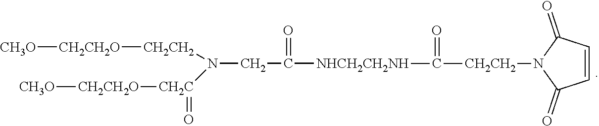
C00014
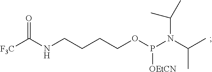
C00015

C00016
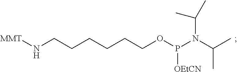
C00017
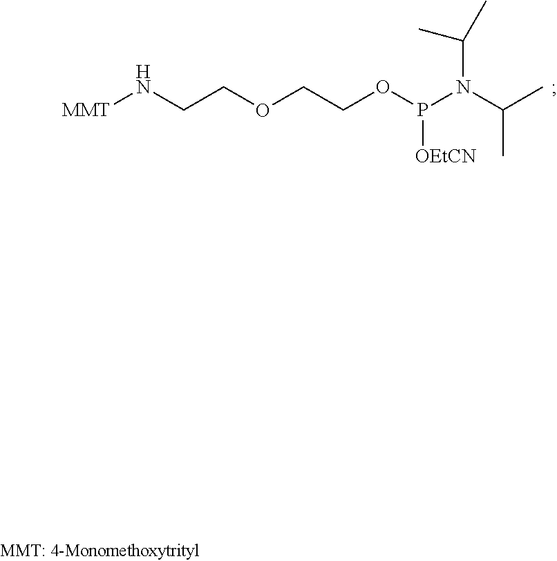
C00018
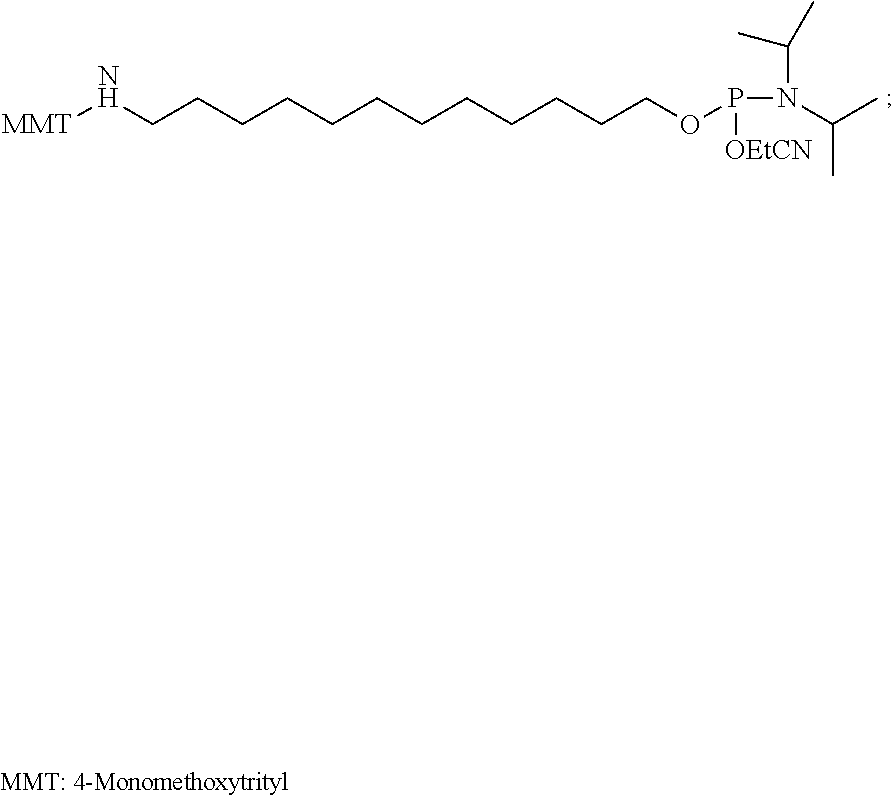
C00019
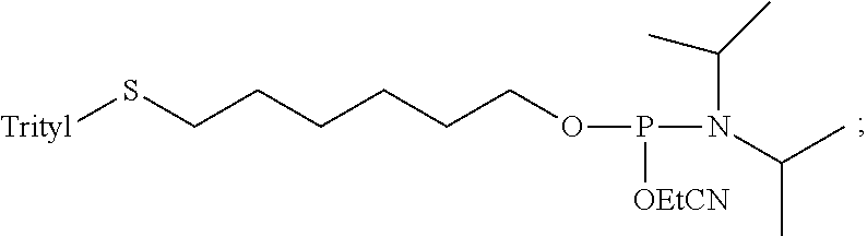
C00020
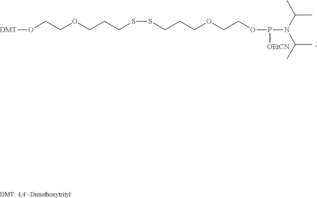
C00021

D00001

D00002
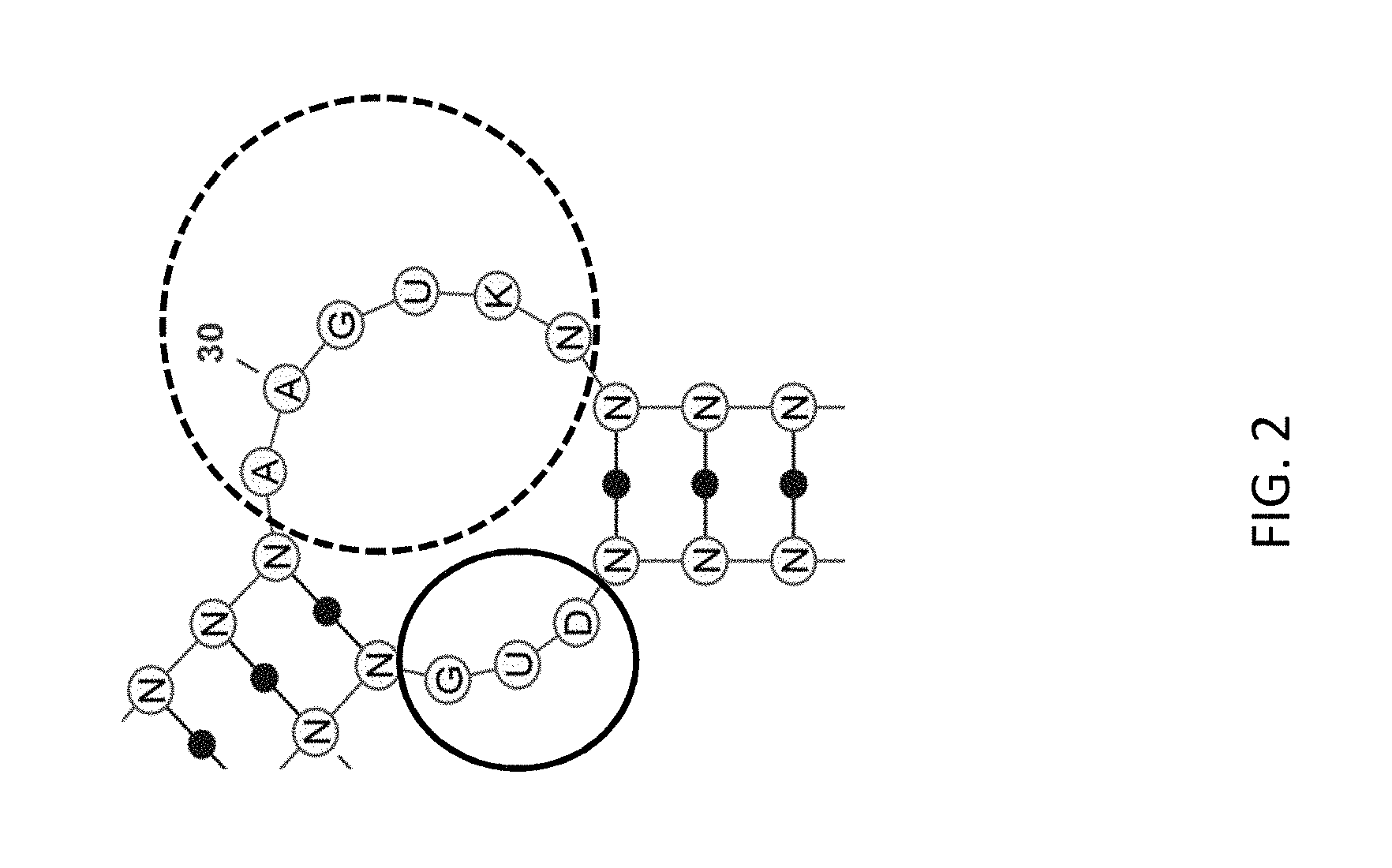
D00003
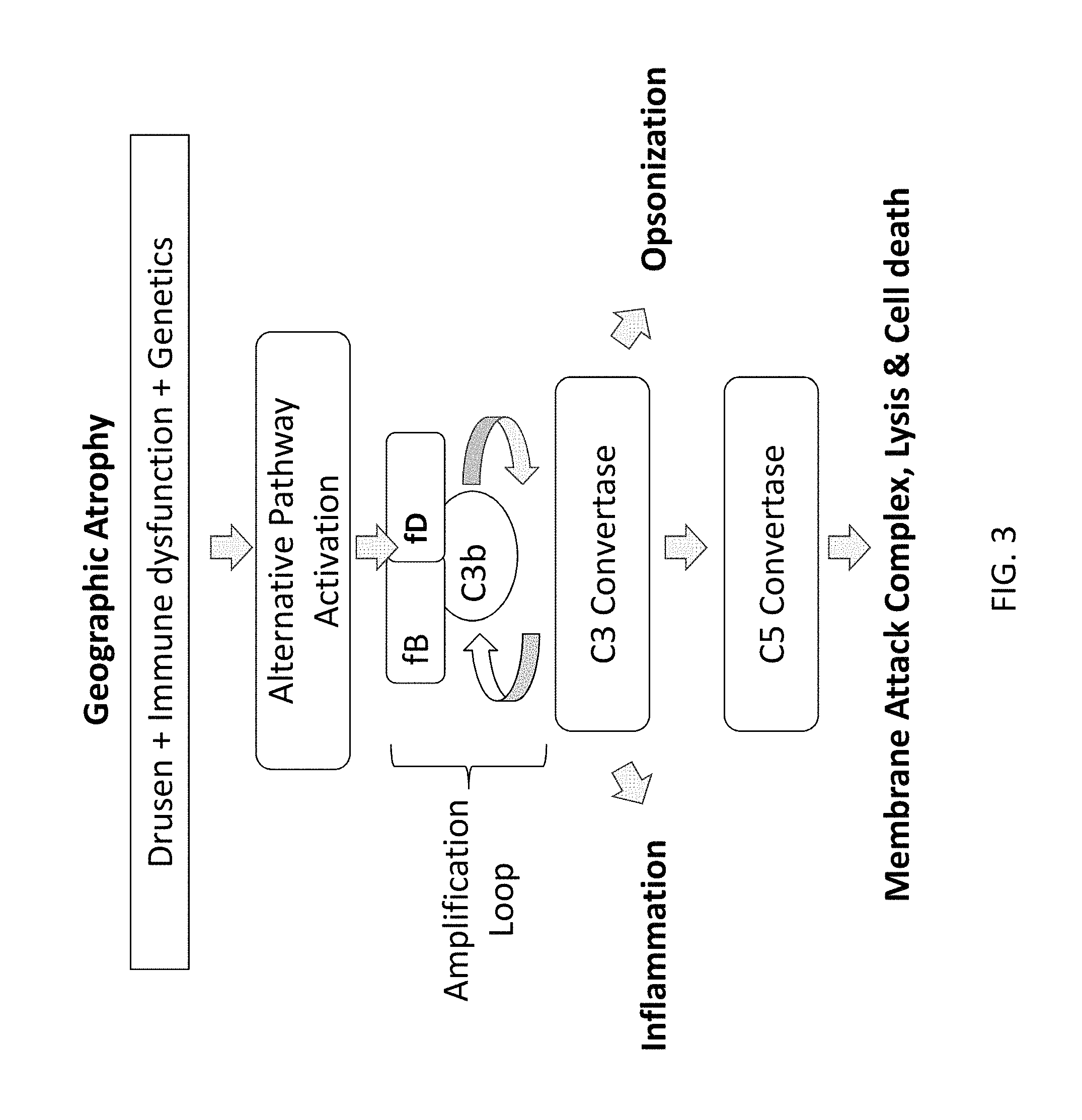
D00004
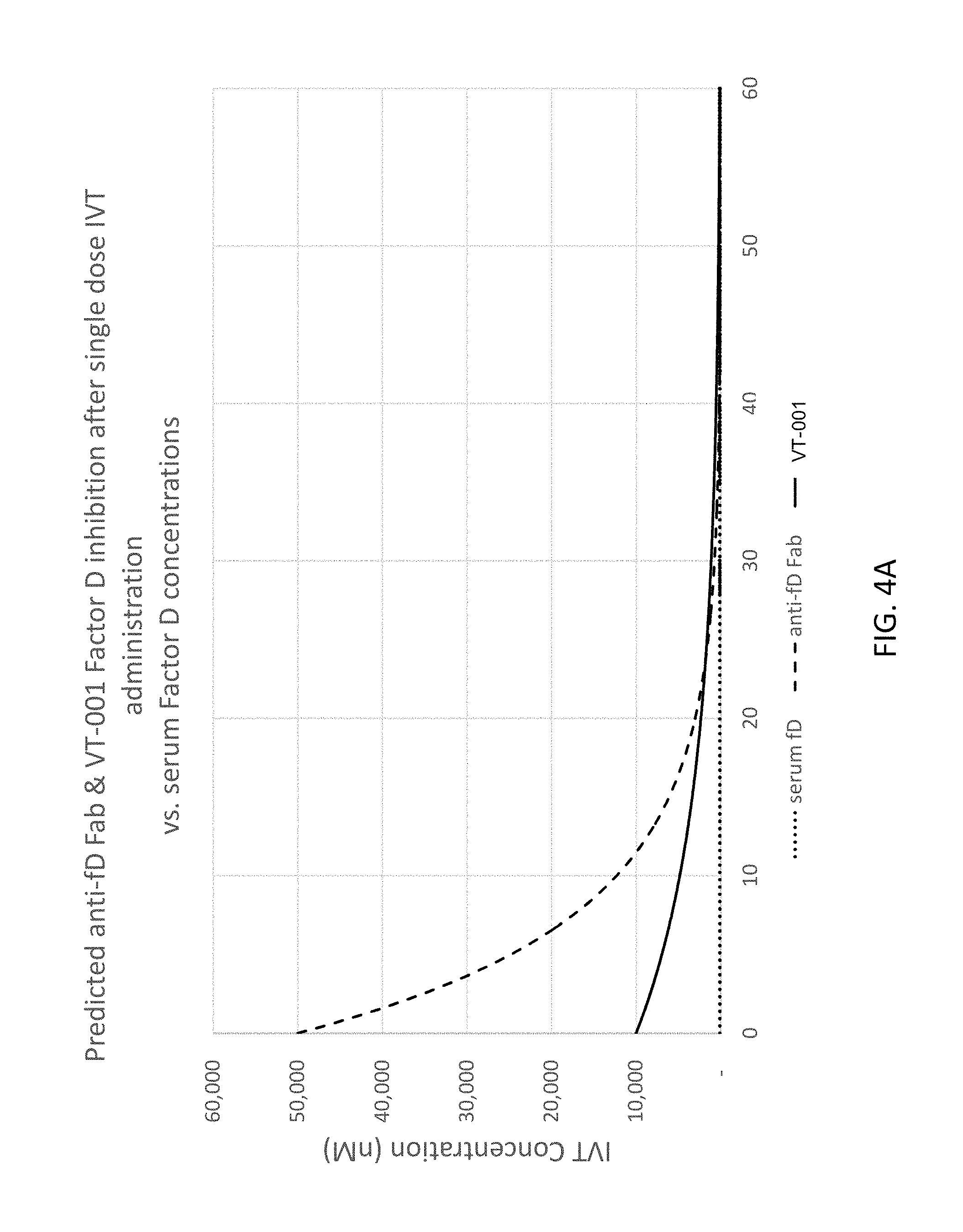
D00005

D00006
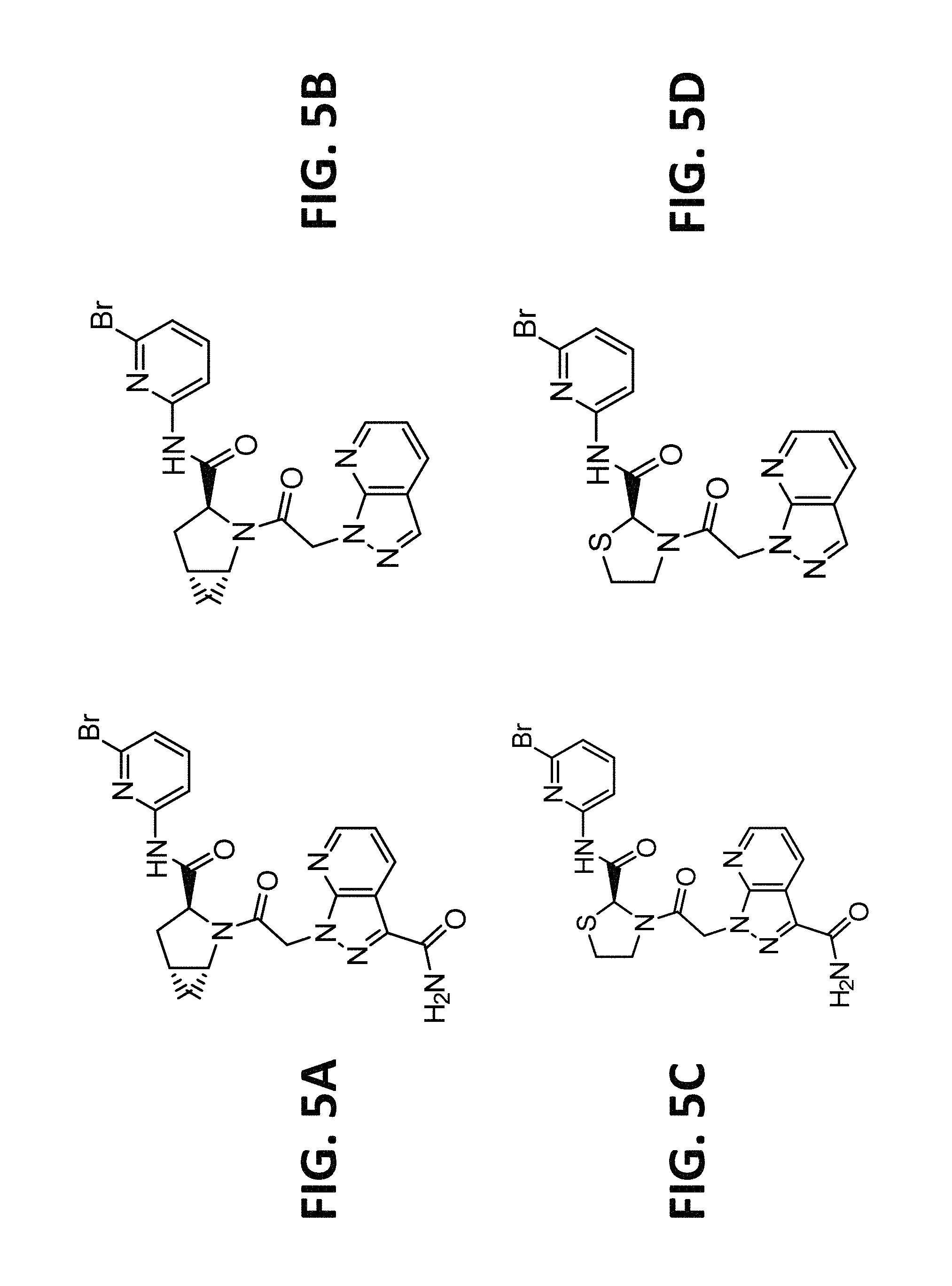
D00007
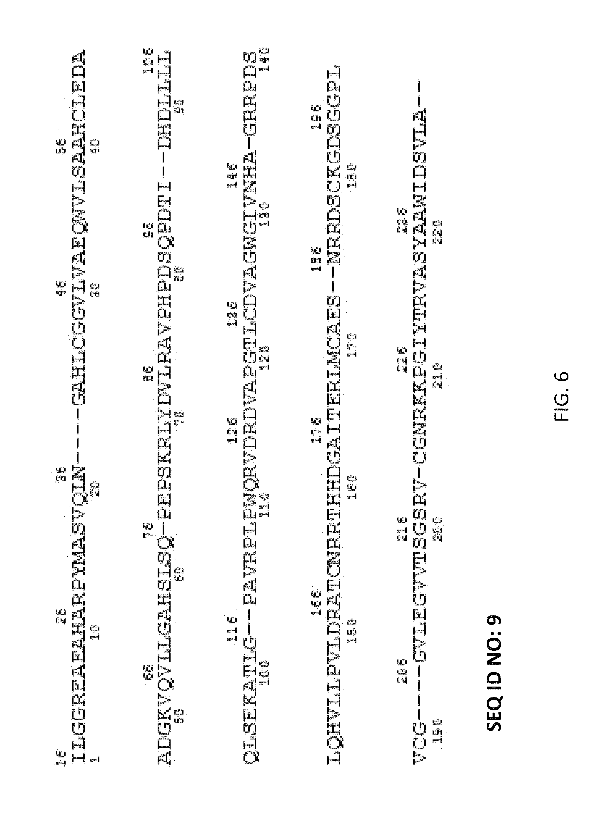
D00008
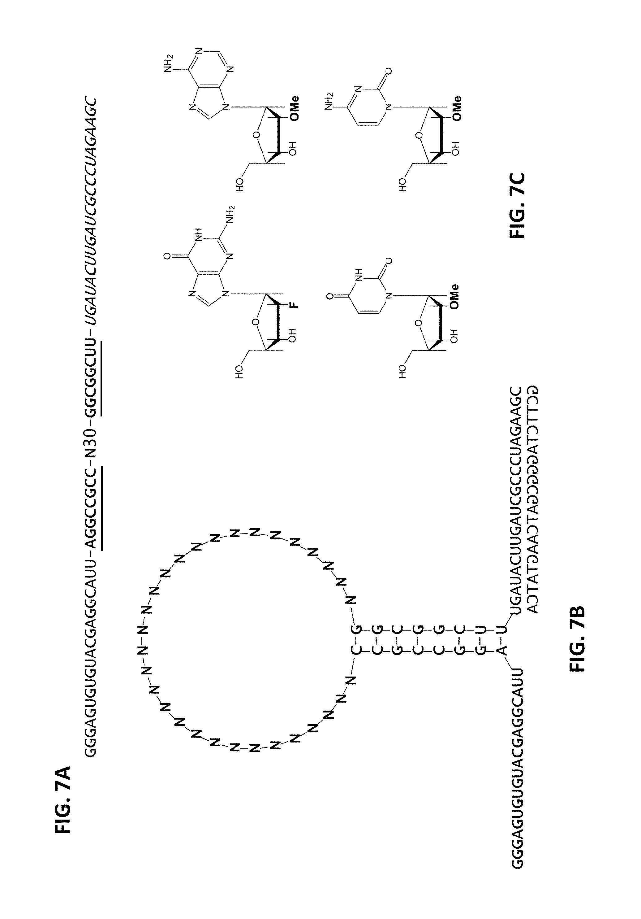
D00009

D00010
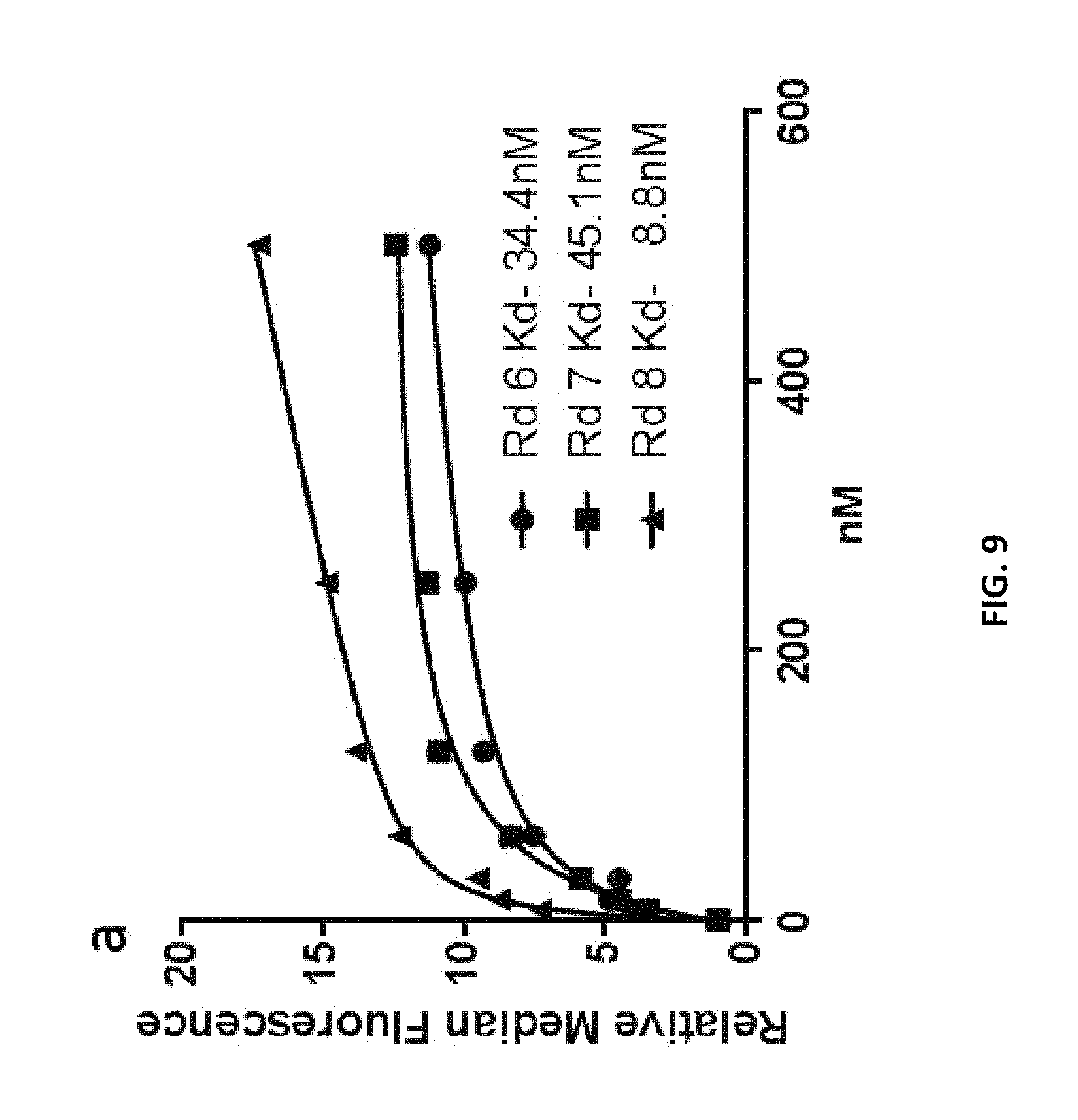
D00011

D00012
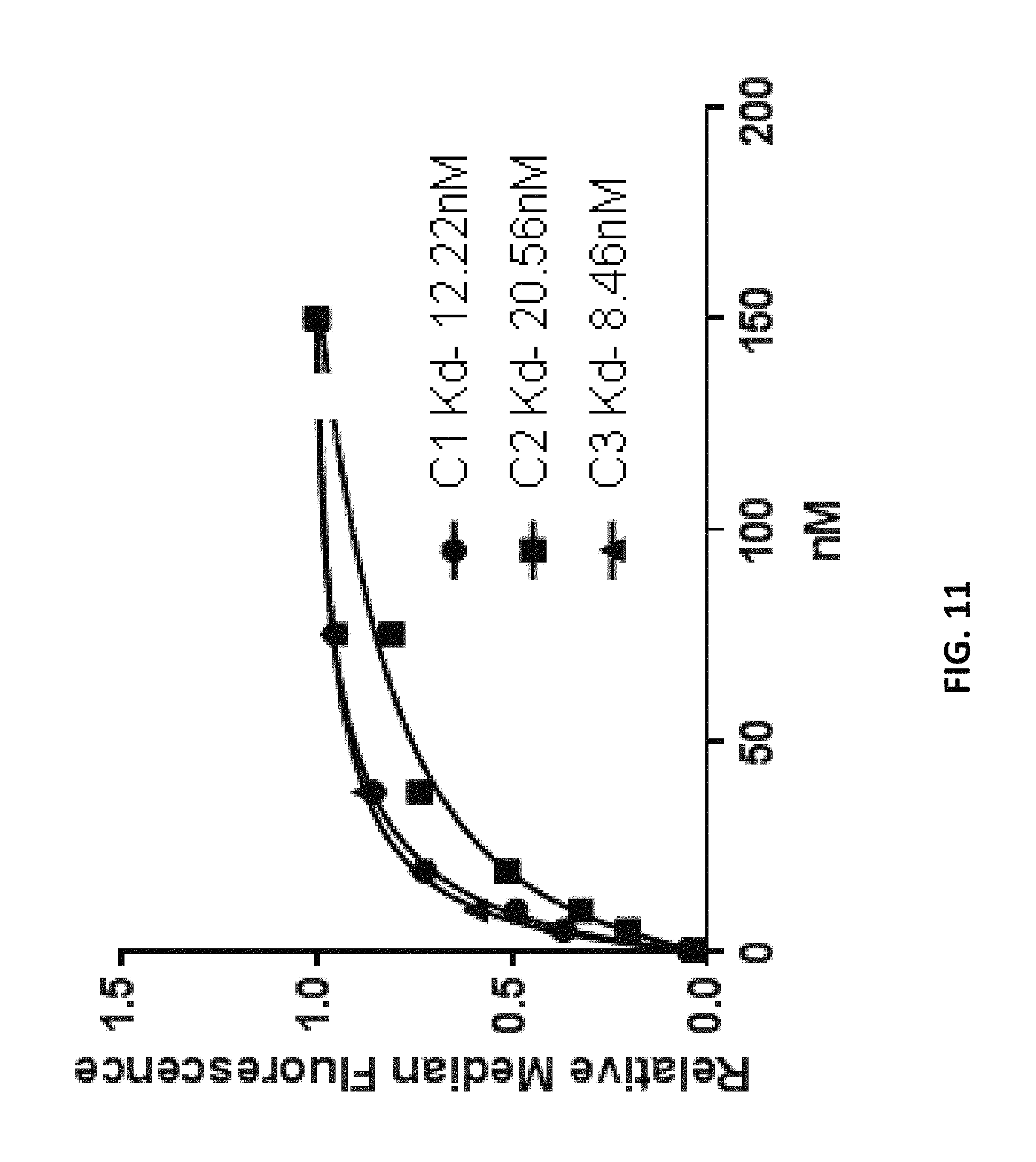
D00013
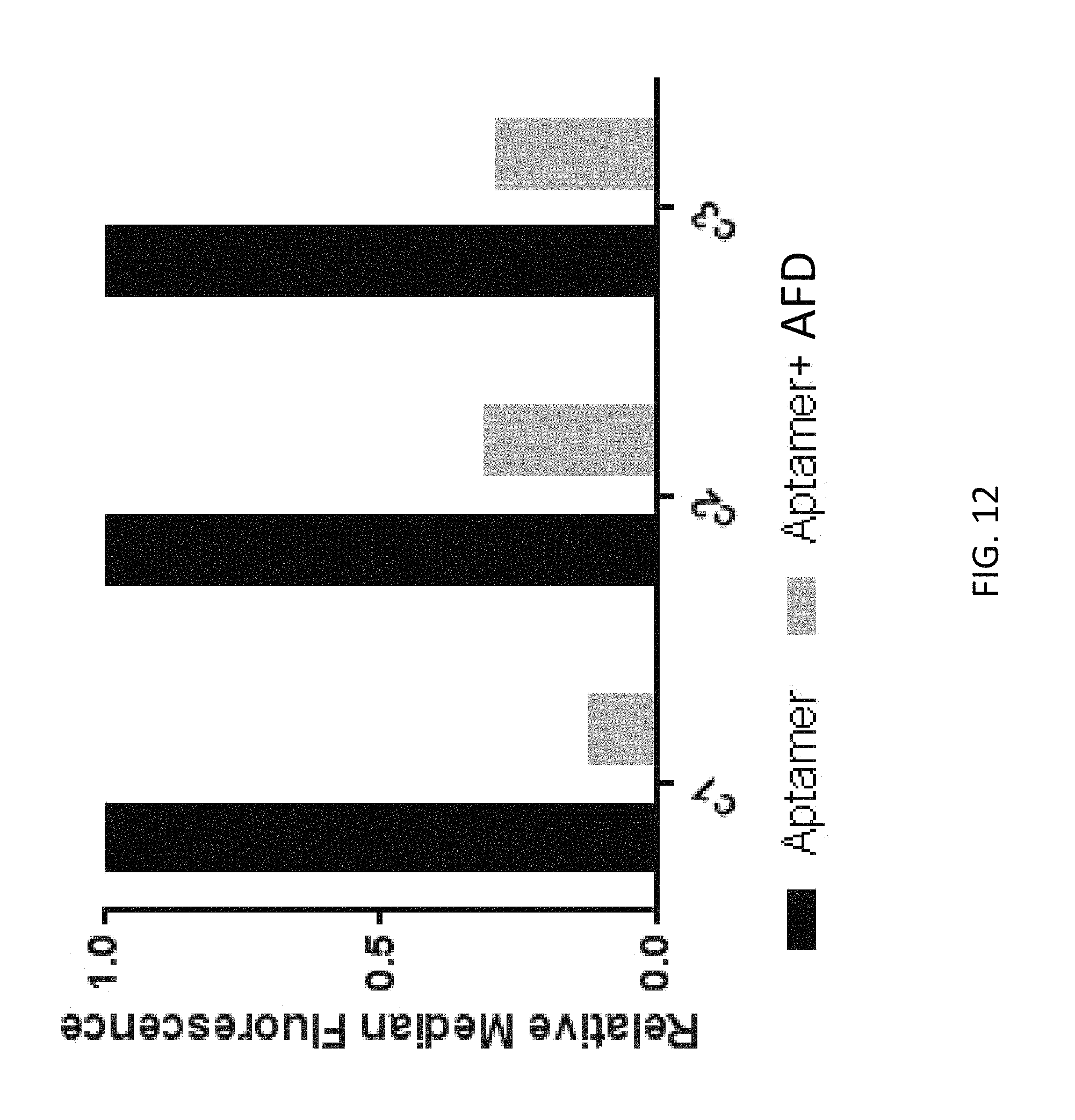
D00014
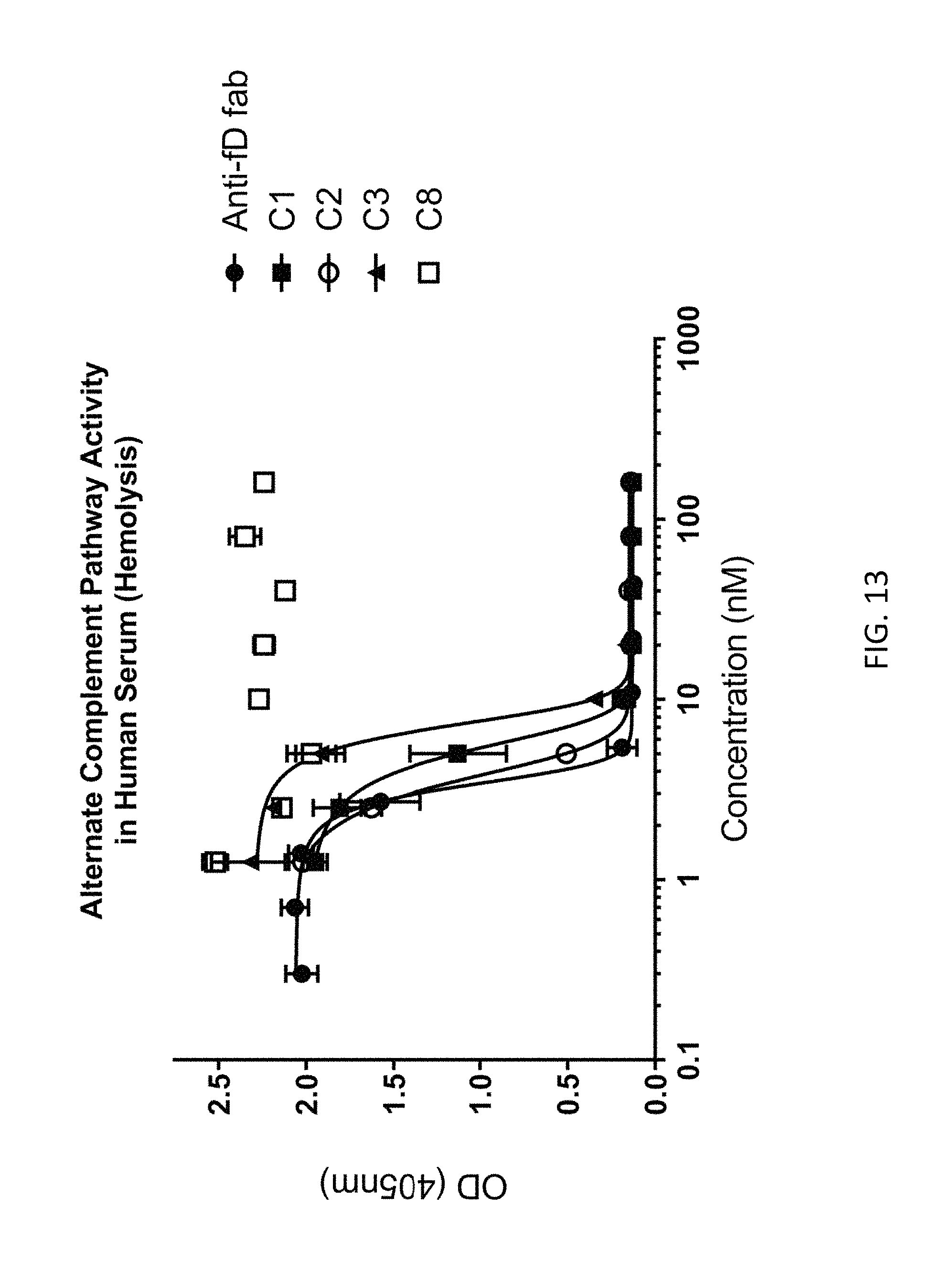
D00015
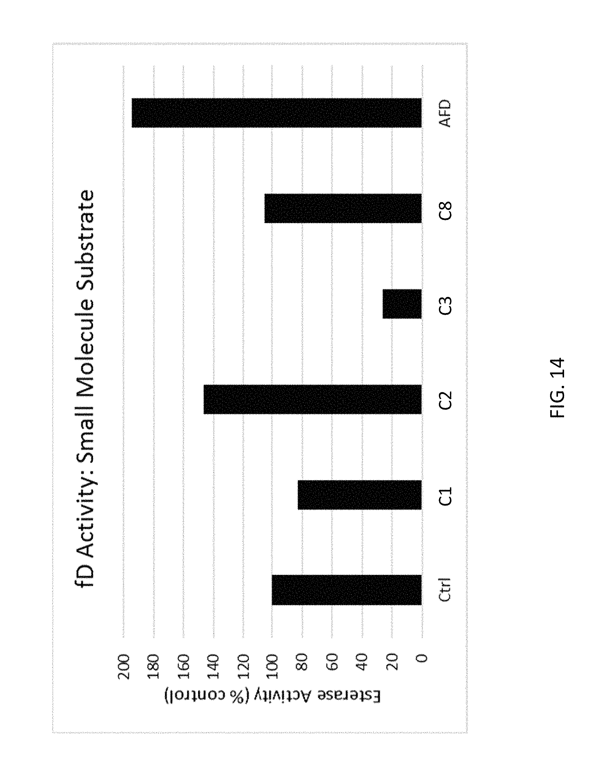
D00016

D00017
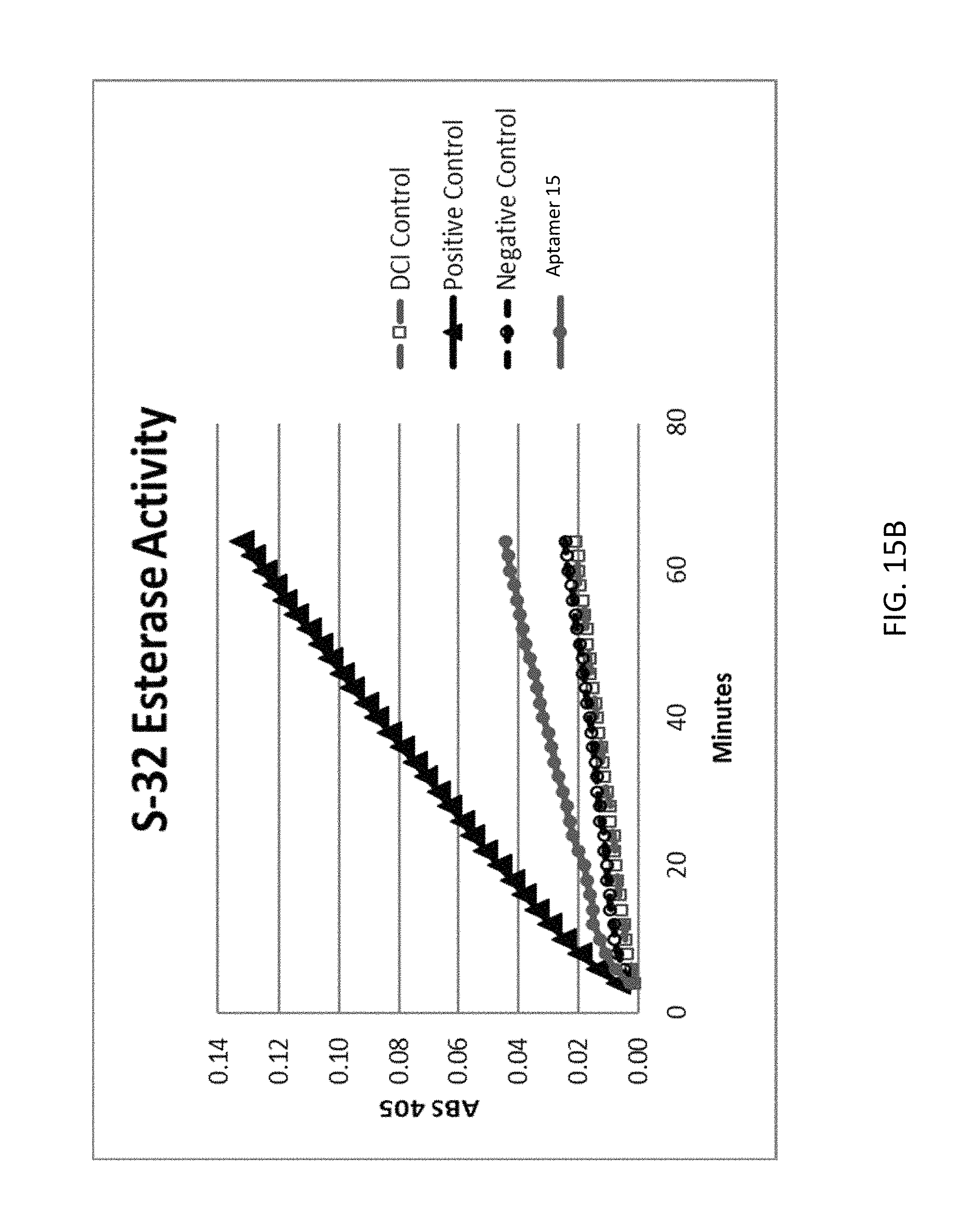
D00018
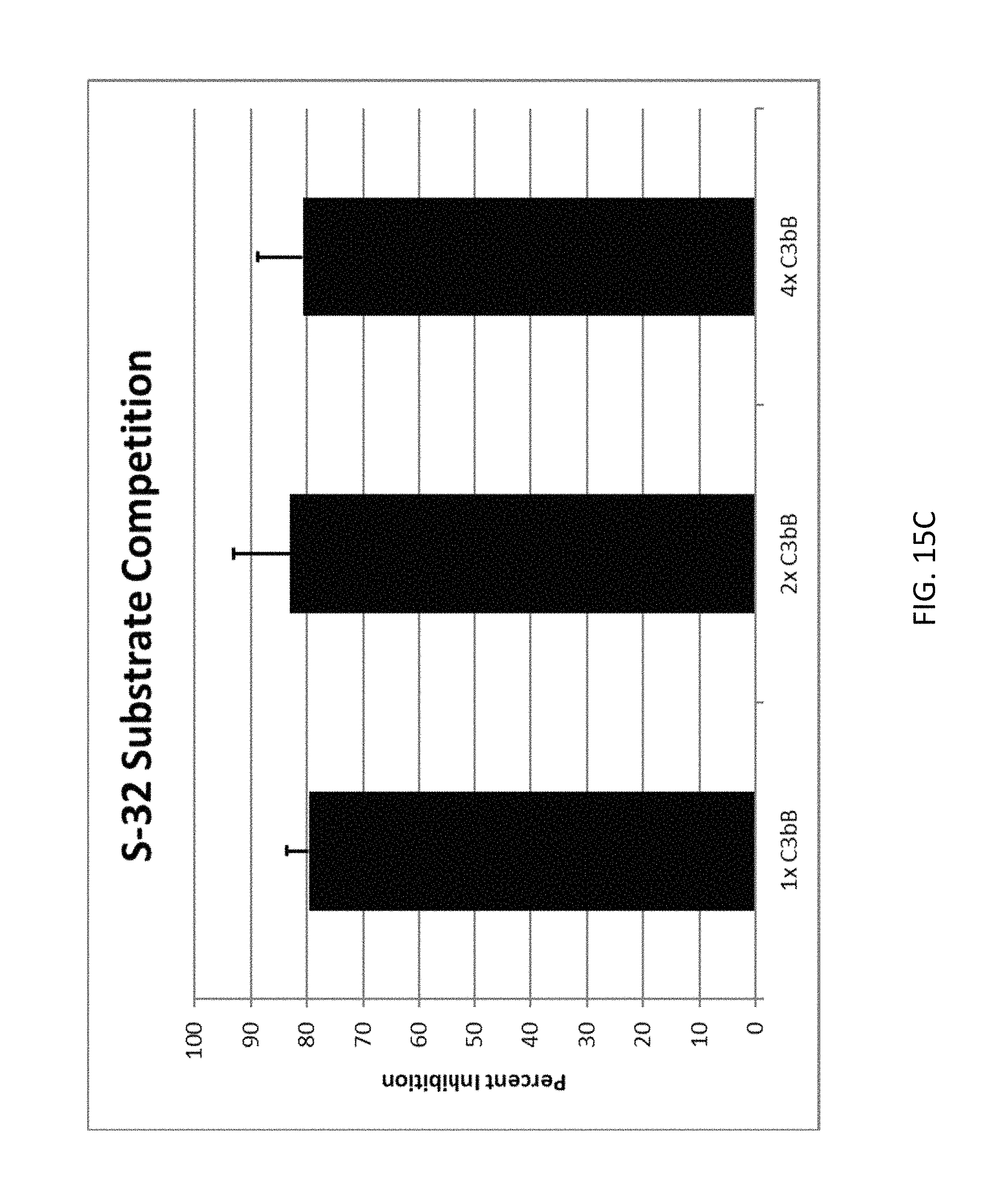
D00019
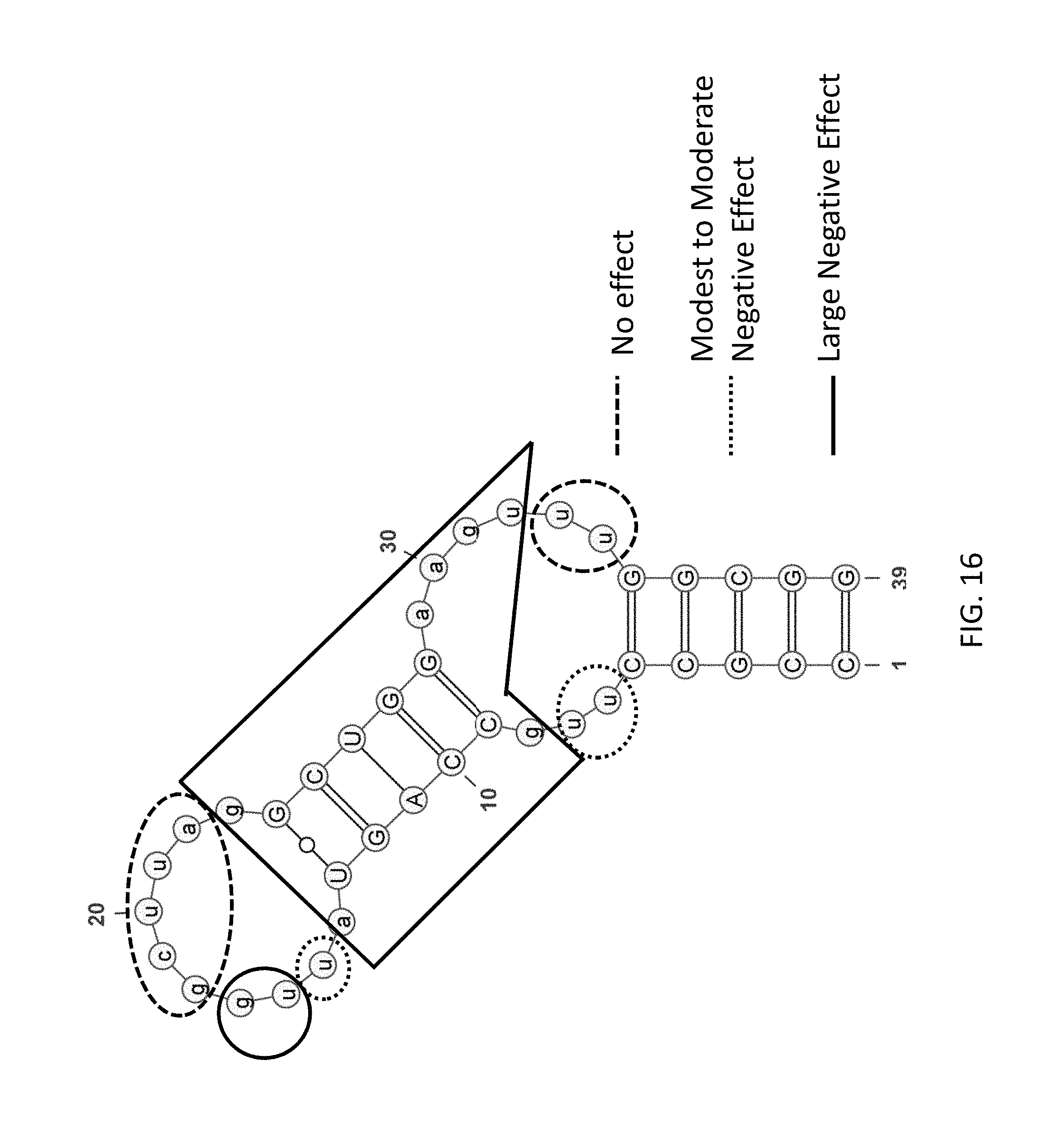
D00020
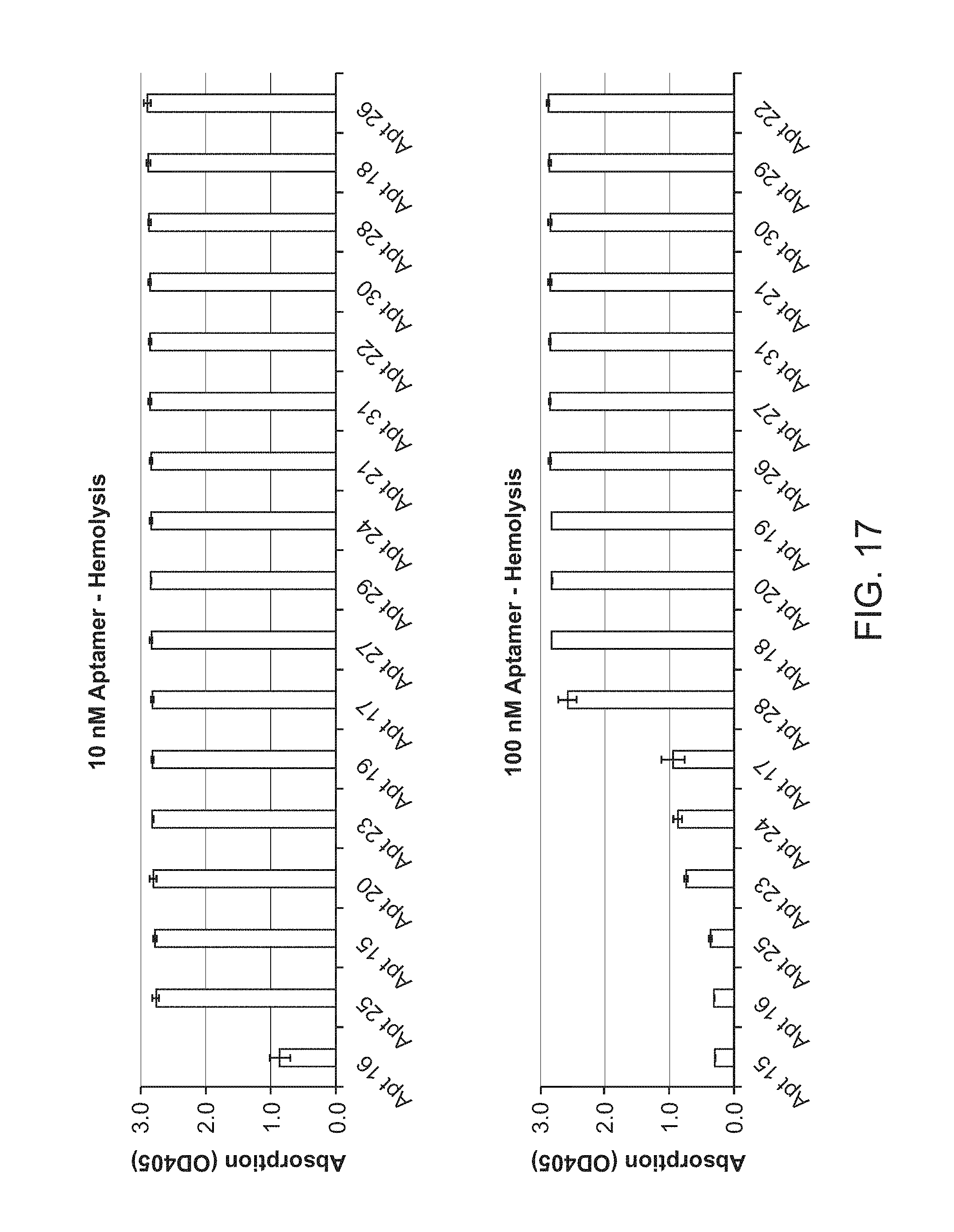
D00021
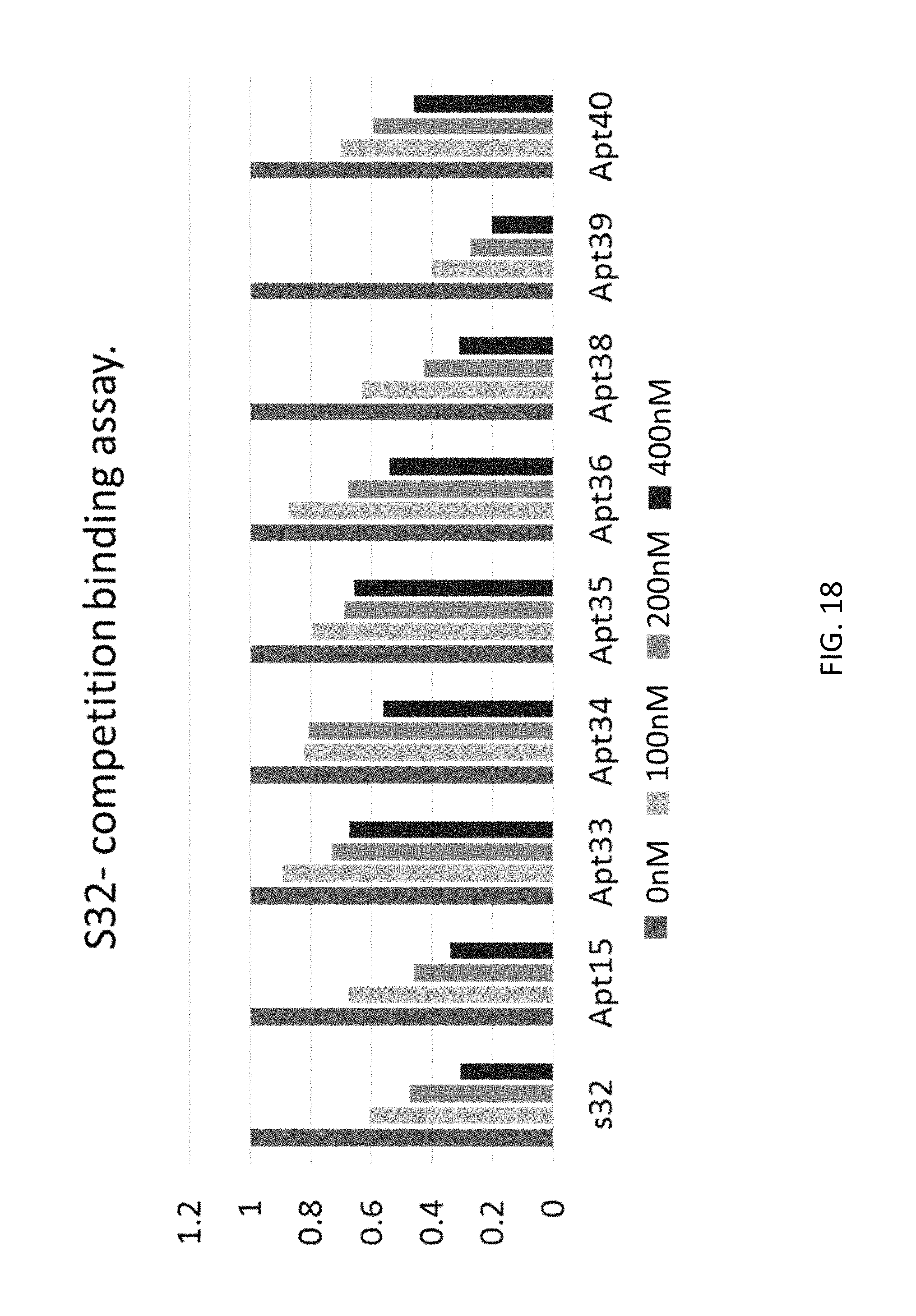
D00022

D00023

D00024

D00025
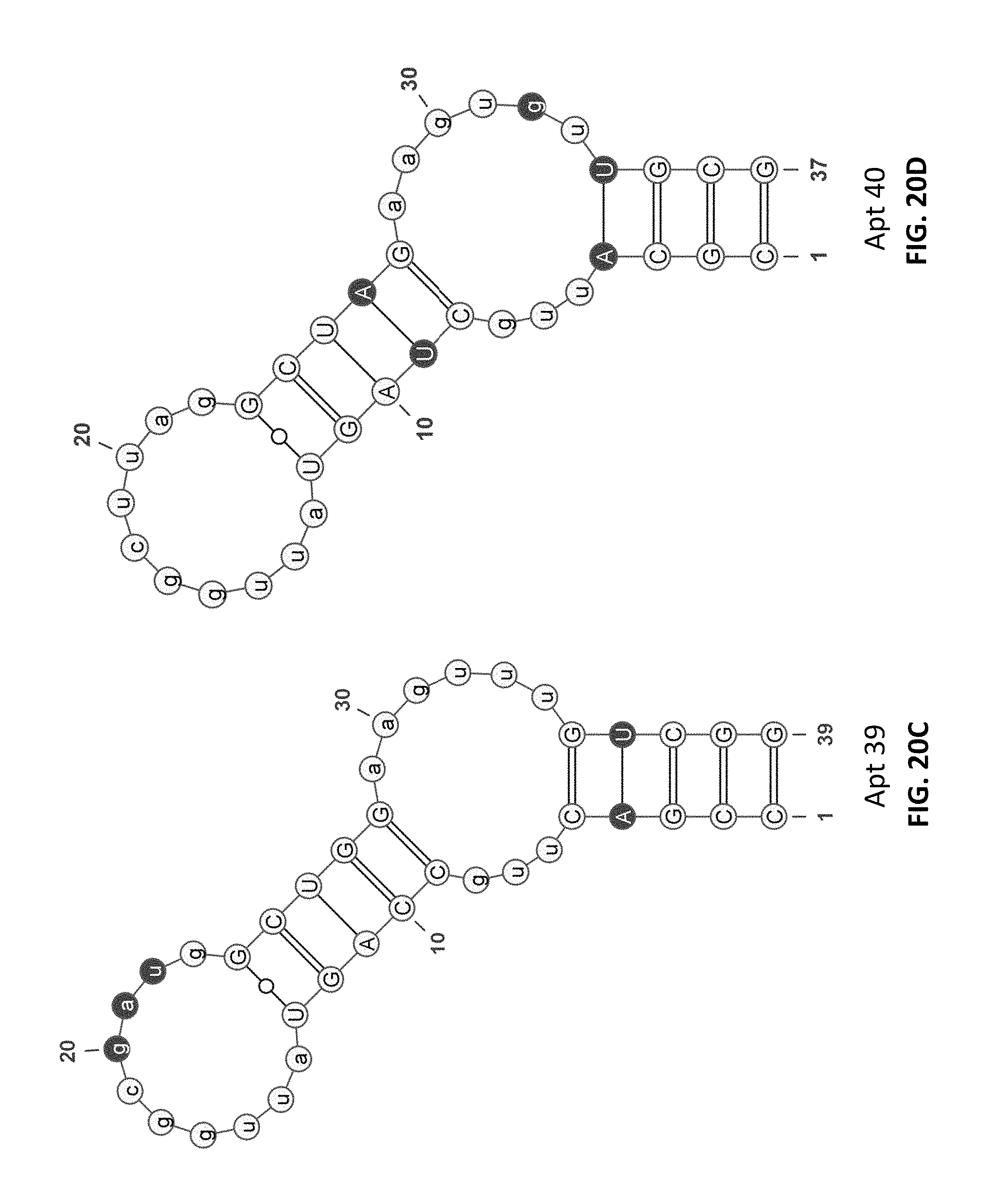
D00026

D00027
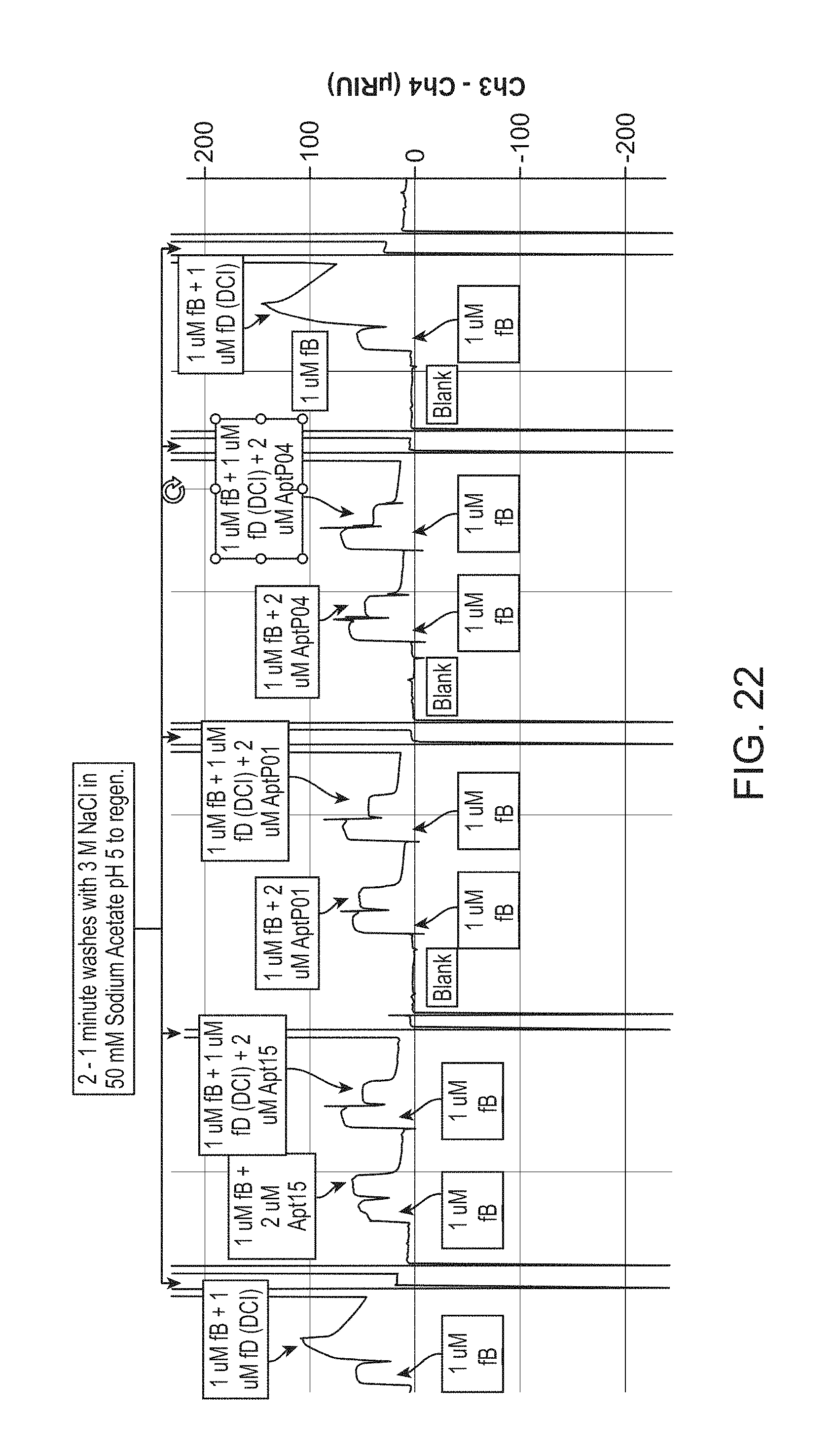
D00028

P00001

P00002

P00003

P00004

P00005

P00006

P00007

P00008

P00009

P00010

P00011

P00012

P00013

P00014

S00001
XML
uspto.report is an independent third-party trademark research tool that is not affiliated, endorsed, or sponsored by the United States Patent and Trademark Office (USPTO) or any other governmental organization. The information provided by uspto.report is based on publicly available data at the time of writing and is intended for informational purposes only.
While we strive to provide accurate and up-to-date information, we do not guarantee the accuracy, completeness, reliability, or suitability of the information displayed on this site. The use of this site is at your own risk. Any reliance you place on such information is therefore strictly at your own risk.
All official trademark data, including owner information, should be verified by visiting the official USPTO website at www.uspto.gov. This site is not intended to replace professional legal advice and should not be used as a substitute for consulting with a legal professional who is knowledgeable about trademark law.