Systems and methods for estimating blood flow characteristics from vessel geometry and physiology
Grady , et al. Sep
U.S. patent number 10,398,386 [Application Number 13/895,893] was granted by the patent office on 2019-09-03 for systems and methods for estimating blood flow characteristics from vessel geometry and physiology. This patent grant is currently assigned to HeartFlow, Inc.. The grantee listed for this patent is HeartFlow, Inc.. Invention is credited to Gilwoo Choi, Leo Grady, Michael Singer.


| United States Patent | 10,398,386 |
| Grady , et al. | September 3, 2019 |
Systems and methods for estimating blood flow characteristics from vessel geometry and physiology
Abstract
Systems and methods are disclosed for estimating patient-specific blood flow characteristics. One method includes acquiring, for each of a plurality of individuals, a geometric model and estimated blood flow characteristics of at least part of the individual's vascular system; executing a machine learning algorithm on the geometric model and estimated blood flow characteristics for each of the plurality of individuals; identifying, using the machine learning algorithm, features predictive of blood flow characteristics corresponding to a plurality of points in the geometric models; acquiring, for a patient, a geometric model of at least part of the patient's vascular system; and using the identified features to produce estimates of the patient's blood flow characteristic for each of a plurality of points in the patient's geometric model.
| Inventors: | Grady; Leo (Millbrae, CA), Choi; Gilwoo (Mountain View, CA), Singer; Michael (Belmont, CA) | ||||||||||
|---|---|---|---|---|---|---|---|---|---|---|---|
| Applicant: |
|
||||||||||
| Assignee: | HeartFlow, Inc. (Redwood City,
CA) |
||||||||||
| Family ID: | 50234017 | ||||||||||
| Appl. No.: | 13/895,893 | ||||||||||
| Filed: | May 16, 2013 |
Prior Publication Data
| Document Identifier | Publication Date | |
|---|---|---|
| US 20140073977 A1 | Mar 13, 2014 | |
Related U.S. Patent Documents
| Application Number | Filing Date | Patent Number | Issue Date | ||
|---|---|---|---|---|---|
| 61700213 | Sep 12, 2012 | ||||
| Current U.S. Class: | 1/1 |
| Current CPC Class: | A61B 5/14535 (20130101); A61B 5/021 (20130101); A61B 5/742 (20130101); A61B 5/7267 (20130101); A61B 5/02035 (20130101); G16H 50/50 (20180101); A61B 5/0022 (20130101); A61B 5/026 (20130101); G16H 50/20 (20180101); G16H 50/70 (20180101) |
| Current International Class: | A61B 5/00 (20060101); A61B 5/021 (20060101); A61B 5/026 (20060101); A61B 5/145 (20060101); G16H 50/50 (20180101); A61B 5/02 (20060101); G16H 50/70 (20180101); G16H 50/20 (20180101) |
References Cited [Referenced By]
U.S. Patent Documents
| 5533511 | July 1996 | Kaspari et al. |
| 6377832 | April 2002 | Bergman |
| 8315812 | November 2012 | Taylor |
| 8684921 | April 2014 | Osorio |
| 8821408 | September 2014 | Hu |
| 2003/0004652 | January 2003 | Brunner |
| 2003/0087244 | May 2003 | McCarthy |
| 2005/0059876 | March 2005 | Krishnan |
| 2008/0292049 | November 2008 | Camus |
| 2009/0080745 | March 2009 | Zheng et al. |
| 2009/0103794 | April 2009 | Sathyanarayana |
| 2011/0060576 | March 2011 | Sharma et al. |
| 2011/0071404 | March 2011 | Schmitt |
| 2012/0022843 | January 2012 | Ionasec |
| 2012/0041318 | February 2012 | Taylor |
| 2012/0053918 | March 2012 | Taylor |
| 2012/0059246 | March 2012 | Taylor |
| 2012/0072190 | March 2012 | Sharma et al. |
| 2012/0142632 | June 2012 | Campbell |
| 2012/0201446 | August 2012 | Yang |
| 2013/0132054 | May 2013 | Sharma |
| 2013/0246034 | September 2013 | Sharma |
| 2014/0100451 | April 2014 | Tolkowsky |
| 2015/0282765 | October 2015 | Goshen |
| 2016/0148371 | May 2016 | Itu |
| 2016/0148372 | May 2016 | Itu |
| 2007-526016 | Sep 2007 | JP | |||
| 2012-024582 | Feb 2012 | JP | |||
| 2012-159990 | Aug 2012 | JP | |||
| 2013-534154 | Sep 2013 | JP | |||
| 2015-527901 | Sep 2015 | JP | |||
| 2016028747 | Mar 2016 | JP | |||
| 2017512577 | May 2017 | JP | |||
| WO-2005001769 | Jan 2005 | WO | |||
| WO 2011/015822 | Feb 2011 | WO | |||
Other References
|
International Search Report issued in corresponding International Application No. PCT/US2013/057546, dated Nov. 18, 2013. cited by applicant . Bernhard, Stefan et al., "Transient integral boundary layer method to calculate the translesional pressure drop and the fractional flow reserve in myocardial bridges," Biomedical Engineering Online, Biomed Central Ltd., London, GB, vol. 5, No. 1, Jun. 21, 2006, p. 42. cited by applicant . International Search Report; PCT/US2013/057546; dated Nov. 18, 2013. cited by applicant . Patent Examination Report dated Mar. 18, 2015, in corresponding Australian Patent Application No. 2013315960, filed on Aug. 30, 2013 (3 pages). cited by applicant . Office Action dated Mar. 3, 2015, in corresponding Canadian Patent Application No. 2882543, filed on Aug. 30, 2013 (5 pages). cited by applicant . Office Action dated Jun. 25, 2015, in corresponding Canadian Patent Application No. 2882543, filed on Aug. 30, 2013 (6 pages). cited by applicant . Office Action dated Oct. 8, 2015, in corresponding Canadian Patent Application No. 2882543, filed on Aug. 30, 2013 (3 pages). cited by applicant . Examination Report pursuant to Article 94(3) EPC dated Nov. 14, 2014 in corresponding European Patent Application No. 13 756 800.2, filed on Aug. 30, 2013 (6 pages). cited by applicant . English-language translation of Notice of Reasons for Rejection dated Sep. 9, 2015, in corresponding Japanese Patent Application No. 2015-531136, filed on Aug. 30, 2013 (6 pages). cited by applicant . English-language translation of Notice of Preliminary Rejection dated Aug. 11, 2015, in corresponding Korean Patent Application No. 10-2015-7006740, filed on Aug. 30, 2013 (4 pages). cited by applicant . International Preliminary Report on Patentability dated Mar. 26, 2015, in corresponding PCT Application No. PCT/US2013/057546, filed on Aug. 30, 2013 (6 pages). cited by applicant . Ionasec, Razvan Ioan; Dissertation; "Patient-specific Modeling and Quantification of the Heart Valves from Multimodal Cardiac Images"; Siemens Corporate Research, Jun. 30, 2010. (Technische Universitat Munchen). cited by applicant . Krizhevesky et al. 2012, ImageNet Classification with Deep Convolution Neural Networks, Conference: Advances in Neural Information Processing Systems 25, p. 1-9. cited by applicant . Deng, Jan. 1, 2012, Three Classes of Deep Learning Architectures and Their Applications: A Tutorial Survey, APSIPA Transactions on Signal and Information Processing. cited by applicant . Filipovic N et al.; "Hemodynamic Flow Modeling Through an Abdominal Aorta Aneurysm Using Data Mining Tools", IEEE Transactions on Information Technology in Biomedicine, vol. 15, No. 2, Mar. 1, 2011, pp. 189-194. cited by applicant . Zhou et al., Shape Regression Machine, N. Karssemeijer and B. Lelieveldt (Eds.): IPMI 2007, LNCS 4584, pp. 13-25, 2007, Springer-Verlag Berlin Heidelberg, 200. cited by applicant . Japanese Office Action dated Dec. 5, 2018 in corresponding Japanese Application No. 2018-11463 and English Translation (7 pages). cited by applicant. |
Primary Examiner: Bloch; Michael R
Attorney, Agent or Firm: Bookoff McAndrews, PLLC
Parent Case Text
RELATED APPLICATION
This application claims priority to U.S. Provisional Application No. 61/700,213 filed Sep. 12, 2012, the entire disclosure of which is hereby incorporated by reference in its entirety.
Claims
What is claimed is:
1. A method for determining patient-specific blood flow characteristics, the method comprising: acquiring, for each of a plurality of individuals, a geometric model, one or more physiological parameters, and one or more measured or estimated blood flow characteristics at one or more points of at least part of the individual's vascular system; for a plurality of points in the geometric model for each of the plurality of individuals, creating a feature vector comprising a vascular cross-sectional area, a diseased length, and one or more boundary conditions of the geometric model at the one or more points; associating the feature vector for each of the plurality of individuals with one or more of the measured or estimated blood flow characteristics of the corresponding individual; modeling and training a support vector machine to predict blood flow characteristics at various points of the geometric model, using the associated feature vectors; acquiring, for a patient, a geometric model of at least part of the patient's vascular system and one or more measured or estimated physiological parameters; and using the modeled and trained support vector machine to determine a blood flow characteristic of the patient for at least one point in the patient's geometric model.
2. The method of claim 1, wherein each feature vector further includes one or more of: systolic and diastolic blood pressures, heart rate, blood properties, individual age, individual gender, individual height, individual weight, presence or absence of disease, lifestyle characteristics, characteristics of aortic geometry, and characteristics of coronary branch geometry.
3. The method of claim 1, wherein each feature vector further includes one or more of: an intensity feature set, a surface feature set, a volume feature set, a centerline feature set, and a simplified physics feature set.
4. The method of claim 1, further comprising: identifying, using the support vector machine, features or weights of features predictive of blood flow characteristics corresponding to a plurality of points in the geometric models for each of the plurality of individuals; and storing the identified features or weights of features to a storage device.
5. The method of claim 1, wherein using the modeled and trained support vector machine to determine the patient's blood flow characteristic comprises: for each of a plurality of points in the patient's geometric model, creating a feature vector of the patient's physiological parameters corresponding to feature vectors acquired for each of the plurality of individuals.
6. The method of claim 1, wherein the physiological parameters include one or more of a heart rate, a hematocrit level, a blood pressure, a blood viscosity, an individual's age, an individual's gender, an individual's weight, an individual lifestyle characteristic, and a mass of supplied tissue.
7. A system for estimating patient-specific blood flow characteristics, the system comprising: a data storage device storing instructions for determining patient-specific blood flow characteristics; and a processor configured to execute the instructions to perform a method including the steps of: acquiring, for each of a plurality of individuals, a geometric model, one or more physiological parameters, and one or more measured or estimated blood flow characteristics at one or more points of at least part of the individual's vascular system; for a plurality of points in the geometric model for each of the plurality of individuals, creating a feature vector comprising a vascular cross-sectional area, a diseased length, and one or more boundary conditions of the geometric model at the one or more points; associating the feature vector for each of the plurality of individuals with one or more of the measured or estimated blood flow characteristics of the corresponding individual; modeling and training a support vector machine to predict blood flow characteristics at various points of the geometric model, using the associated feature vectors; acquiring, for a patient, a geometric model of at least part of the patient's vascular system and one or more measured or estimated physiological parameters; and using the modeled and trained support vector machine to determine a blood flow characteristic of the patient for at least one point in the patient's geometric model.
8. The system of claim 7, wherein each feature vector further includes one or more of: systolic and diastolic blood pressures, heart rate, blood properties, individual age, individual gender, individual height, individual weight, presence or absence of disease, lifestyle characteristics, characteristics of aortic geometry, and characteristics of coronary branch geometry.
9. The system of claim 7, wherein each feature vector further includes one or more of: an intensity feature set, a surface feature set, a volume feature set, a centerline feature set, and a simplified physics feature set.
10. The system of claim 7, wherein the processor is further configured for: identifying, using the support vector machine, features or weights of features predictive of blood flow characteristics corresponding to a plurality of points in the geometric models for each of the plurality of individuals; and storing the identified features or weights of features to a storage device.
11. The system of claim 7, wherein using the modeled support vector machine to determine the patient's blood flow characteristic comprises: for each of a plurality of points in the patient's geometric model, creating a feature vector of the patient's physiological parameters corresponding to feature vectors acquired for each of the plurality of individuals.
12. The system of claim 7, wherein the physiological parameters include one or more of heart rate, hematocrit level, blood pressure, blood viscosity, individual age, individual gender, individual weight, individual lifestyle characteristic, and a mass of supplied tissue.
13. A non-transitory computer-readable medium storing instructions that, when executed by a computer, cause the computer to perform a method including: acquiring, for each of a plurality of individuals, a geometric model, one or more physiological parameters, and one or more measured or estimated blood flow characteristics at one or more points of at least part of the individual's vascular system; for a plurality of points in the geometric model for each of the plurality of individuals, creating a feature vector comprising a vascular cross-sectional area, a diseased length, and one or more boundary conditions of the geometric model at the one or more points; associating the feature vector for each of the plurality of individuals with one or more of the measured or estimated blood flow characteristics of the corresponding individual; modeling and training a support vector machine to predict blood flow characteristics at various points of the geometric model, using the associated feature vectors; acquiring, for a patient, a geometric model of at least part of the patient's vascular system and one or more measured or estimated physiological parameters; and using the modeled and trained support vector machine to determine a blood flow characteristic of the patient for at least one point in the patient's geometric model.
Description
FIELD OF THE INVENTION
Various embodiments of the present disclosure relate generally to medical imaging and related methods. More specifically, particular embodiments of the present disclosure relate to systems and methods for estimating patient-specific blood flow characteristics from vessel geometry and physiology.
BACKGROUND
A functional assessment of arterial capacity is important for treatment planning to address patient needs. Recent studies have demonstrated that hemodynamic characteristics, such as Fractional Flow Reserve (FFR), are important indicators to determine the optimal treatment for a patient with arterial disease. Conventional assessments of these hemodynamic characteristics use invasive catheterizations to directly measure blood flow characteristics, such as pressure and flow velocity. However, despite the important clinical information that is gathered, these invasive measurement techniques present severe risks to the patient and significant costs to the healthcare system.
To address the risks and costs associated with invasive measurement, a new generation of noninvasive tests have been developed to assess blood flow characteristics. These noninvasive tests use patient imaging (such as computed tomography (CT)) to determine a patient-specific geometric model of the blood vessels and this model is used computationally to simulate the blood flow using computational fluid dynamics (CFD) with appropriate physiological boundary conditions and parameters. Examples of inputs to these patient-specific boundary conditions include the patient's blood pressure, blood viscosity and the expected demand of blood from the supplied tissue (derived from scaling laws and a mass estimation of the supplied tissue from the patient imaging). Although these simulation-based estimations of blood flow characteristics have demonstrated a level of fidelity comparable to direct (invasive) measurements of the same quantity of interest, physical simulations demand a substantial computational burden that can make these virtual, noninvasive tests difficult to execute in a real-time clinical environment. Consequently, the present disclosure describes new approaches for performing rapid, noninvasive estimations of blood flow characteristics that are computationally inexpensive.
SUMMARY
Systems and methods are disclosed for deriving a patient-specific geometric model of a patient's blood vessels, and combining this geometry with the patient-specific physiological information and boundary conditions. Combined, these data may be used to estimate the patient's blood flow characteristics and predict clinically relevant quantities of interest (e.g., FFR). The presently disclosed systems and methods offer advantages over physics-based simulation of blood flow to compute the quantity of interest, such as by instead using machine learning to predict the results of a physics-based simulation. In one embodiment, disclosed systems and methods involve two phases: first, a training phase in which a machine learning system is trained to predict one or more blood flow characteristics; and second, a production phase in which the machine learning system is used to produce one or more blood flow characteristics and clinically relevant quantities of interest. In the case of predicting multiple blood flow characteristics, this machine learning system can be applied for each blood flow characteristic and quantity of interest.
According to one embodiment, a method is disclosed for estimating patient-specific blood flow characteristics. The method includes acquiring, for each of a plurality of individuals, a geometric model and estimated blood flow characteristics of at least part of the individual's vascular system; executing a machine learning algorithm on the geometric model and estimated blood flow characteristics for each of the plurality of individuals; identifying, using the machine learning algorithm, features predictive of blood flow characteristics corresponding to a plurality of points in the geometric models; acquiring, for a patient, a geometric model of at least part of the patient's vascular system; and using the identified features to produce estimates of the patient's blood flow characteristic for each of a plurality of points in the patient's geometric model.
According to another embodiment, a system is disclosed for estimating patient-specific blood flow characteristics. The system includes a data storage device storing instructions for estimating patient-specific blood flow characteristics; and a processor configured to execute the instructions to perform a method including the steps of: acquiring, for each of a plurality of individuals, a geometric model and estimated blood flow characteristics of at least part of the individual's vascular system; executing a machine learning algorithm on the geometric model and estimated blood flow characteristics for each of the plurality of individuals; identifying, using the machine learning algorithm, features predictive of blood flow characteristics corresponding to a plurality of points in the geometric models; acquiring, for a patient, a geometric model of at least part of the patient's vascular system; and using the identified features to produce estimates of the patient's blood flow characteristic for each of a plurality of points in the patient's geometric model.
Additional objects and advantages of the disclosed embodiments will be set forth in part in the description that follows, and in part will be apparent from the description, or may be learned by practice of the disclosed embodiments. The objects and advantages of the disclosed embodiments will be realized and attained by means of the elements and combinations particularly pointed out in the appended claims.
It is to be understood that both the foregoing general description and the following detailed description are exemplary and explanatory only and are not restrictive of the disclosed embodiments, as claimed.
BRIEF DESCRIPTION OF THE DRAWINGS
The accompanying drawings, which are incorporated in and constitute a part of this specification, illustrate various exemplary embodiments and together with the description, serve to explain the principles of the disclosed embodiments.
FIG. 1 is a block diagram of an exemplary system and network for estimating patient-specific blood flow characteristics from vessel geometry and physiological information, according to an exemplary embodiment of the present disclosure.
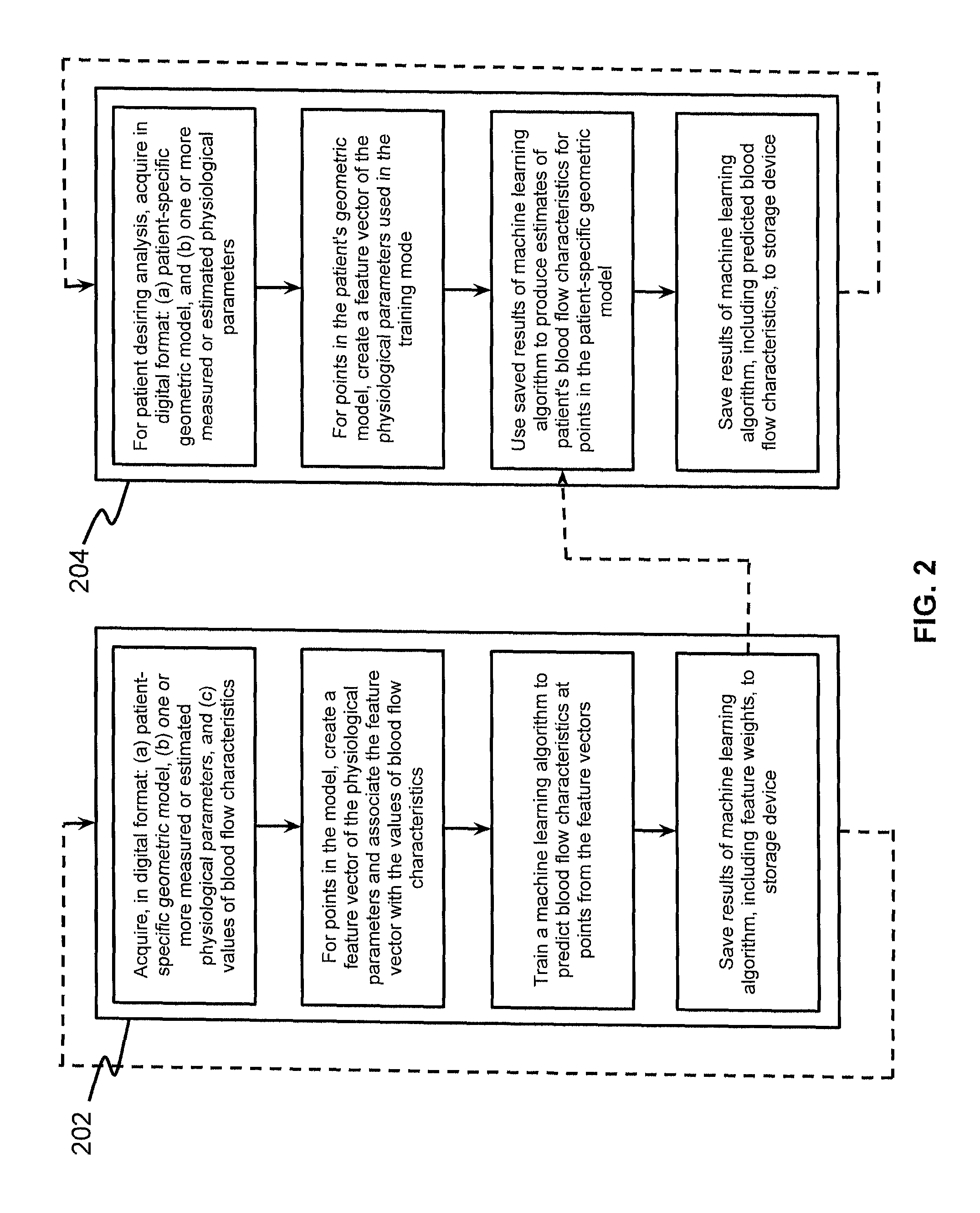
FIG. 2 is a block diagram of an exemplary method for estimating patient-specific blood flow characteristics from vessel geometry and physiological information, according to an exemplary embodiment of the present disclosure.
DESCRIPTION OF THE EMBODIMENTS
Reference will now be made in detail to the exemplary embodiments of the disclosure, examples of which are illustrated in the accompanying drawings. Wherever possible, the same reference numbers will be used throughout the drawings to refer to the same or like parts.
The present disclosure describes certain principles and embodiments for providing advantages over physics-based simulation of blood flow to compute patient-specific blood flow characteristics and clinically relevant quantities of interest. Namely, the presently disclosed systems and methods may incorporate machine learning techniques to predict the results of a physics-based simulation. For example, the present disclosure describes an exemplary, less processing-intensive technique, which may involve modeling the fractional flow reserve (FFR) as a function of a patient's vascular cross-sectional area, diseased length, and boundary conditions. The cross-sectional area may be calculated based on lumen segment and plaque segment, among other things. The diseased length may be calculated based on plaque segment and stenosis location, among other things. The boundary conditions may reflect patient-specific physiology, such as coronary flow (estimated from myocardial mass), outlet area, and hyperemic assumptions, to reflect that different patients have different geometry and physiologic responses.
In one embodiment, fractional flow reserve may be modeled as a function of a patient's boundary conditions (f(BCs)), and a function of a patient's vascular geometry (g(areaReductions)). Although the patient's geometry may be described as a function of "areaReductions," it should be appreciated that this term refers, not just to changes in patient's vascular cross-sectional area, but to any physical or geometric characteristics affecting a patient's blood flow. In one embodiment, FFR can be predicted by optimizing the functions "f" and "g" such that the difference between the estimated FFR (FFR.sub.CT.sub._.sub.ScalingLaw) and the measured FFR (mFFR) is minimized. In other words, machine learning techniques can be used to solve for the functions that cause the estimated FFR to approximate the measured FFR. In one embodiment, the measured FFR may be calculated by traditional catheterized methods or by modern, computational fluid dynamics (CFD) techniques. In one embodiment, one or more machine learning algorithms may be used to optimize the functions of boundary conditions and patient geometry for hundreds or even thousands of patients, such that estimates for FFR can reliably approximate measured FFR values. Thus, FFR values calculated by CFD techniques can be valuable for training the machine learning algorithms.
Referring now to the figures, FIG. 1 depicts a block diagram of an exemplary system and network for estimating patient-specific blood flow characteristics from vessel geometry and physiological information. Specifically, FIG. 1 depicts a plurality of physicians 102 and third party providers 104, any of whom may be connected to an electronic network 100, such as the Internet, through one or more computers, servers, and/or handheld mobile devices. Physicians 102 and/or third party providers 104 may create or otherwise obtain images of one or more patients' cardiac and/or vascular systems. The physicians 102 and/or third party providers 104 may also obtain any combination of patient-specific information, such as age, medical history, blood pressure, blood viscosity, etc. Physicians 102 and/or third party providers 104 may transmit the cardiac/vascular images and/or patient-specific information to server systems 106 over the electronic network 100. Server systems 106 may include storage devices for storing images and data received from physicians 102 and/or third party providers 104. Sever systems 106 may also include processing devices for processing images and data stored in the storage devices.
FIG. 2 is a block diagram of an exemplary method for estimating patient-specific blood flow characteristics from vessel geometry and physiological information, according to an exemplary embodiment of the present disclosure. The method of FIG. 2 may be performed by server systems 106, based on information received from physicians 102 and/or third party providers 104 over electronic network 100.
In one embodiment, the method of FIG. 2 may include a training method 202, for training one or more machine learning algorithms based on numerous patients' blood flow characteristic estimates, and a production method 204 for using the machine learning algorithm results to predict a particular patient's blood flow characteristics.
In one embodiment, training method 202 may be performed based on FFR estimates generating using CFD techniques for hundreds of patients. Training method 202 may involve acquiring, for each of a plurality of individuals, e.g., in digital format: (a) a patient-specific geometric model, (b) one or more measured or estimated physiological parameters, and (c) values of blood flow characteristics. Training method 202 may then involve, for one or more points in each patient's model, creating a feature vector of the patients' physiological parameters and associating the feature vector with the values of blood flow characteristics. For example, training method 202 may associate an estimated FFR with every point in a patient's geometric model. Training method 202 may then train a machine learning algorithm (e.g., using processing devices of server systems 106) to predict blood flow characteristics at each point of a geometric model, based on the feature vectors and blood flow characteristics. Training method 202 may then save the results of the machine learning algorithm, including feature weights, in a storage device of server systems 106. The stored feature weights may define the extent to which patient features or geometry are predictive of certain blood flow characteristics.
In one embodiment, the production method 204 may involve estimating FFR values for a particular patient, based on results of executing training method 202. In one embodiment, production method 204 may include acquiring, e.g. in digital format: (a) a patient-specific geometric model, and (b) one or more measured or estimated physiological parameters. For multiple points in the patient's geometric model, production method 204 may involve creating a feature vector of the physiological parameters used in the training mode. Production method 204 may then use saved results of the machine learning algorithm to produce estimates of the patient's blood flow characteristics for each point in the patient-specific geometric model. Finally, production method 204 may include saving the results of the machine learning algorithm, including predicted blood flow characteristics, to a storage device of server systems 106.
Described below are general and specific exemplary embodiments for implementing a training mode and a production mode of machine learning for predicting patient-specific blood flow characteristics, e.g. using server systems 106 based on images and data received from physicians 102 and/or third party providers 104 over electronic network 100.
General Embodiment
In a general embodiment, server systems 106 may perform a training mode based on images and data received from physicians 102 and/or third party providers 104 over electronic network 100. Specifically, for one or more patients, server systems 106 may acquire a digital representation (e.g., the memory or digital storage [e.g., hard drive, network drive] of a computational device such as a computer, laptop, DSP, server, etc.) of the following items: (a) a patient-specific model of the geometry for one or more of the patient's blood vessels; (b) a list of one or more measured or estimated physiological or phenotypic parameters of the patient; and/or (c) measurements, estimations or simulated values of all blood flow characteristic being targeted for prediction. In one embodiment, the patient-specific model of the geometry may be represented by a list of points in space (possibly with a list of neighbors for each point) in which the space can be mapped to spatial units between points (e.g., millimeters). In one embodiment, the list of one or more measured or estimated physiological or phenotypic parameters of the patient may include blood pressure, blood viscosity, patient age, patient gender, mass of the supplied tissue, etc. These patient-specific parameters may be global (e.g., blood pressure) or local (e.g., estimated density of the vessel wall at a particular location).
For every point in the patient-specific geometric model for which there is a measured, estimated or simulated value of the blood flow characteristic, server systems 106 may then create a feature vector for that point. The feature vector may be a numerical description of the patient-specific geometry at that point and estimates of physiological or phenotypic parameters of the patient. The feature vector may contain both global and local physiological or phenotypic parameters, where: for global parameters, all points have the same numerical value; and for local parameters, the value(s) may change at different points in the feature vector. Server systems 106 may then associate this feature vector with the measured, estimated or simulated value of the blood flow characteristic at this point.
Server systems 106 may then train a machine learning algorithm to predict the blood flow characteristics at the points from the feature vectors at the points. Examples of machine learning algorithms that can perform this task are support vector machines (SVMs), multi-layer perceptrons (MLPs), and multivariate regression (MVR) (e.g., weighted linear or logistic regression). Server systems 106 may then save the results of the machine learning algorithm (e.g., feature weights) to a digital representation (e.g., the memory or digital storage [e.g., hard drive, network drive] of a computational device such as a computer, laptop, DSP, server, etc.).
Also in a general embodiment, server systems 106 may perform a production mode based on images and data received from physicians 102 and/or third party providers 104 over electronic network 100. For a patient on whom a blood flow analysis is to be performed, server systems 106 may acquire a digital representation (e.g., the memory or digital storage [e.g., hard drive, network drive] of a computational device such as a computer, laptop, DSP, server, etc.) of (a) a patient-specific model of the geometry for one or more of the patient's blood vessels; and (b) a list of one or more estimates of physiological or phenotypic parameters of the patient. In one embodiment, the patient-specific model of the geometry for one or more of the patient's blood vessels may be represented as a list of points in space (possibly with a list of neighbors for each point) in which the space can be mapped to spatial units between points (e.g., millimeters). The list of one or more estimates of physiological or phenotypic parameters of the patient, may include blood pressure, blood viscosity, patient age, patient gender, the mass of the supplied tissue, etc. These parameters may be global (e.g., blood pressure) or local (e.g., estimated density of the vessel wall at a location). This list of parameters must be the same as the list used in the training mode.
For every point in the patient-specific geometric model, server systems 106 may create a feature vector that consists of a numerical description of the geometry and estimates of physiological or phenotypic parameters of the patient. Global physiological or phenotypic parameters can be used in the feature vector of all points and local physiological or phenotypic parameters can change in the feature vector of different points. These feature vectors may represent the same parameters used in the training mode. Server systems 106 may then use the saved results of the machine learning algorithm produced in the training mode (e.g., feature weights) to produce estimates of the blood flow characteristics at each point in the patient-specific geometric model. These estimates may be produced using the same machine learning algorithm technique used in the training mode (e.g., the SVM, MLP, MVR technique). Server systems 106 may also save the predicted blood flow characteristics for each point to a digital representation (e.g., the memory or digital storage [e.g., hard drive, network drive] of a computational device such as a computer, laptop, DSP, server, etc.).
Exemplary Embodiment
In one exemplary embodiment, server systems 106 may perform a training mode based on images and data received from physicians 102 and/or third party providers 104 over electronic network 100. Specifically, for one or more patients, server systems 106 may acquire a digital representation (e.g., the memory or digital storage [e.g., hard drive, network drive] of a computational device such as a computer, laptop, DSP, server, etc.) of (a) a patient-specific model of the geometry for the patient's ascending aorta and coronary artery tree; (b) a list of measured or estimated physiological or phenotypic parameters of the patient; and (c) measurements of the FFR when available.
In one embodiment, the patient-specific model of the geometry for the patient's ascending aorta and coronary artery tree may be represented as a list of points in space (possibly with a list of neighbors for each point) in which the space can be mapped to spatial units between points (e.g., millimeters). This model may be derived by performing a cardiac CT imaging study of the patient during the end diastole phase of the cardiac cycle. The resulting CT images may then be segmented manually or automatically to identify voxels belonging to the aorta and to the lumen of the coronary arteries. Once all relevant voxels are identified, the geometric model can be derived (e.g., using marching cubes).
In one embodiment, the list of measured or estimated physiological or phenotypic parameters of the patient may be obtained and may include: (i) systolic and diastolic blood pressures; (ii) heart rate; (iii) hematocrit level; (iv) patient age, gender, height, weight, general health status (presence or absence of diabetes, current medications); (v) lifestyle characteristics: smoker/non-smoker; and/or (vi) myocardial mass (may be derived by segmenting the myocardium obtained during the CT imaging study and then calculating the volume in the image; the mass is then computed using the computed volume and an estimated density (1.05 g/mL) of the myocardial mass.
In one embodiment, measurements of the FFR may be obtained when available. If the measured FFR value is not available at a given spatial location in the patient-specific geometric model, then a numerically computed value of the FFR at the point may be used. The numerically computed values may be obtained from a previous CFD simulation using the same geometric model and patient-specific boundary conditions derived from the physiological and phenotypic parameters listed above.
For every point in the patient-specific geometric model for which there is a measured, estimated or simulated value of the blood flow characteristics, server systems 106 may create a feature vector for that point that contains a numerical description of physiological or phenotypic parameters of the patient and a description of the local geometry. Specifically the feature vector may contain: (i) systolic and diastolic blood pressures; (ii) heart rate; (iii) blood properties including: plasma, red blood cells (erythrocytes), hematocrit, white blood cells (leukocytes) and platelets (thrombocytes), viscosity, yield stress; (iv) patient age, gender, height, weight, etc.; (v) diseases: presence or absence of diabetes, myocardial infarction, malignant and rheumatic conditions, peripheral vascular conditions, etc.; (vi) lifestyle characteristics: presence or absence of current medications/drugs, smoker/non-smoker; (vii) characteristics of the aortic geometry (Cross-sectional area of the aortic inlet and outlet, Surface area and volume of the aorta, Minimum, maximum, and average cross-sectional area, etc.); (viii) characteristics of the coronary branch geometry; and (ix) one or more feature sets.
In one embodiment, the characteristics of the coronary branch geometry may include: (i) volumes of the aorta upstream/downstream of the coronary branch point; (ii) cross-sectional area of the coronary/aorta bifurcation point, i.e., inlet to the coronary branch; (iii) total number of vessel bifurcations, and the number of upstream/downstream vessel bifurcations; (iv) average, minimum, and maximum upstream/downstream cross-sectional areas; (v) distances (along the vessel centerline) to the centerline point of minimum and maximum upstream/downstream cross-sectional areas; (vi) cross-sectional of and distance (along the vessel centerline) to the nearest upstream/downstream vessel bifurcation; (vii) cross-sectional area of and distance (along the vessel centerline) to the nearest coronary outlet and aortic inlet/outlet; (viii) cross-sectional areas and distances (along the vessel centerline) to the downstream coronary outlets with the smallest/largest cross-sectional areas; (ix) upstream/downstream volumes of the coronary vessels; and (x) upstream/downstream volume fractions of the coronary vessel with a cross-sectional area below a user-specified tolerance.
In one embodiment, a first feature set may define cross-sectional area features, such as a cross-sectional lumen area along the coronary centerline, a powered cross-sectional lumen area, a ratio of lumen cross-sectional area with respect to the main ostia (LM, RCA), a powered ratio of lumen cross-sectional area with respect to the main ostia, a degree of tapering in cross-sectional lumen area along the centerline, locations of stenotic lesions, lengths of stenotic lesions, location and number of lesions corresponding to 50%, 75%, 90% area reduction, distance from stenotic lesion to the main ostia, and/or irregularity (or circularity) of cross-sectional lumen boundary.
In one embodiment, the cross-sectional lumen area along the coronary centerline may be calculated by extracting a centerline from constructed geometry, smoothing the centerline if necessary, and computing cross-sectional area at each centerline point and map it to corresponding surface and volume mesh points. In one embodiment, the powered cross-sectional lumen area can be determined from various source of scaling laws. In one embodiment, the ratio of lumen cross-sectional area with respect to the main ostia (LM, RCA) can be calculated by measuring cross-sectional area at the LM ostium, normalizing cross-sectional area of the left coronary by LM ostium area, measuring cross-sectional area at the RCA ostium, and normalizing cross-sectional area of the right coronary by RCA ostium area. In one embodiment, the powered ratio of lumen cross-sectional area with respect to the main ostia can be determined from various source of scaling laws. In one embodiment, the degree of tapering in cross-sectional lumen area along the centerline can be calculated by sampling centerline points within a certain interval (e.g., twice the diameter of the vessel) and compute a slope of linearly-fitted cross-sectional area. In one embodiment, the location of stenotic lesions can be calculated by detecting minima of cross-sectional area curve, detecting locations where first derivative of area curve is zero and second derivative is positive, and computing distance (parametric arc length of centerline) from the main ostium. In one embodiment, the lengths of stenotic lesions can be calculated by computing the proximal and distal locations from the stenotic lesion, where cross-sectional area is recovered.
In one embodiment, another feature set may include intensity features that define, for example, intensity change along the centerline (slope of linearly-fitted intensity variation). In one embodiment, another feature set may include surface features that define, for example, 3D surface curvature of geometry (Gaussian, maximum, minimum, mean). In one embodiment, another feature set may include volume features that define, for example, a ratio of total coronary volume compared to myocardial volume. In one embodiment, another feature set may include centerline features that define, for example, curvature (bending) of coronary centerline, e.g., by computing Frenet curvature:
'.times.''' ##EQU00001## where p is coordinate of centerline
or by computing an inverse of the radius of circumscribed circle along the centerline points. Curvature (bending) of coronary centerline may also be calculated based on tortuosity (non-planarity) of coronary centerline, e.g., by computing Frenet torsion:
.tau..times.'.times.'''''.times.'' ##EQU00002## where p is coordinate of centerline
In one embodiment, another feature set may include a SYNTAX scoring feature, including, for example, an existence of aorta ostial lesion, detection of a lesion located at the origin of the coronary from the aorta; and/or dominance (left or right).
In one embodiment, another feature set may include a simplified physics feature, e.g., including a fractional flow reserve value derived from Hagen-Poisseille flow assumption (Resistance.about.Area.sup.-2). For example, in one embodiment, server systems 106 may compute the cross-sectional area of the origin (left main (LM) ostium or right coronary artery (RCA) ostium) of the coronary from the aorta (A.sub.0) with aortic pressure (P.sub.0); compute cross-sectional area of coronary vessel (A.sub.i) at each sampled interval (L.sub.i); determine the amount of coronary flow in each segment of vessel using resistance boundary condition under hyperemic assumption (Q.sub.i); estimate resistance at each sampled location (R.sub.i) based on:
##EQU00003## where:
Nominal value .alpha.=0.0023, .beta.=1.0, .gamma.=-2.0 (Hagen-Poisseille).
Server systems 106 may estimate pressure drop (.DELTA.P.sub.i) as .DELTA.P.sub.i=Q.sub.iR.sub.i and compute fractional flow reserve (FFR) at each minima as
##EQU00004## Locations of cross-sectional area minima or intervals smaller than vessel radius may be used for sampling locations. Server systems 106 may interpolate FFR along the centerline using FFR.sub.i, project FFR values to 3D surface mesh node, and vary .alpha..sub.i, .beta..sub.i, .gamma..sub.i and obtain new sets of FFR estimation as necessary for training, such as by using the feature sets defined above to perturb parameters where .alpha..sub.i, .beta..sub.i can be a function of the diseased length, degree of stenosis and tapering ratio to account for tapered vessel; and Q.sub.i can be determined by summing distributed flow of each outlet on the basis of the same scaling law as the resistance boundary condition. However, a new scaling law and hyperemic assumption can be adopted, and this feature vector may be associated with the measurement or simulated value of the FFR at that point. Server systems 106 may also train a linear state vector machine (SVM) to predict the blood flow characteristics at the points from the feature vectors at the points; and save the results of the SVM as a digital representation (e.g., the memory or digital storage [e.g., hard drive, network drive] of a computational device such as a computer, laptop, digital signal processor (DSP), server, etc.).
In an exemplary production mode, servers systems 106 may, for a target patient, acquire in digital representation (e.g., the memory or digital storage (e.g., hard drive, network drive) of a computational device such as a computer, laptop, DSP, server, etc.): (a) a patient-specific model of the geometry for the patient's ascending aorta and coronary artery tree; and (b) a list of physiological and phenotypic parameters of the patient obtained during training mode. In one embodiment, the patient-specific model of the geometry for the patient's ascending aorta and coronary artery tree may be represented as a list of points in space (possibly with a list of neighbors for each point) in which the space can be mapped to spatial units between points (e.g., millimeters). This model may be derived by performing a cardiac CT imaging of the patient in the end diastole phase of the cardiac cycle. This image then may be segmented manually or automatically to identify voxels belonging to the aorta and the lumen of the coronary arteries. Once the voxels are identified, the geometric model can be derived (e.g., using marching cubes). The process for generating the patient-specific model of the geometry may be the same as in the training mode. For every point in the patient-specific geometric model, the server systems 106 may create a feature vector for that point that consists of a numerical description of the geometry at that point and estimates of physiological or phenotypic parameters of the patient. These features may be the same as the quantities used in the training mode. The server systems 106 may then use the saved results of the machine learning algorithm produced in the training mode (e.g., feature weights) to produce estimates of the FFR at each point in the patient-specific geometric model. These estimates may be produced using the same linear SVM technique used in the training mode. The server systems 106 may save the predicted FFR values for each point to a digital representation (e.g., the memory or digital storage [e.g., hard drive, network drive] of a computational device such as a computer, laptop, DSP, server, etc.).
In one embodiment, the above factors (i) thru (viii) ("Systolic and diastolic blood pressures" thru "Characteristics of the coronary branch geometry") may be considered global features, which are applicable to all points within a given patient's geometric model. Also, items (ix) thru (xv) ("Feature Set I: Cross-sectional area feature" thru "Feature Set VII: Simplified Physics feature") may be considered features that are local to specific points within a given patient's geometric model. In addition, features (i) thru (vi) may be considered variables within the function of boundary conditions, f(BCs), while features (vii) thru (xv) may be considered variables within the function of geometry, g(areaReductions), on that page. It will be appreciated that any combination of those features, modified by any desired weighting scheme, may be incorporated into a machine learning algorithm executed according to the disclosed embodiments.
Other embodiments of the invention will be apparent to those skilled in the art from consideration of the specification and practice of the invention disclosed herein. It is intended that the specification and examples be considered as exemplary only, with a true scope and spirit of the invention being indicated by the following claims.
* * * * *
D00000
D00001
D00002
M00001
M00002
M00003
M00004
XML
uspto.report is an independent third-party trademark research tool that is not affiliated, endorsed, or sponsored by the United States Patent and Trademark Office (USPTO) or any other governmental organization. The information provided by uspto.report is based on publicly available data at the time of writing and is intended for informational purposes only.
While we strive to provide accurate and up-to-date information, we do not guarantee the accuracy, completeness, reliability, or suitability of the information displayed on this site. The use of this site is at your own risk. Any reliance you place on such information is therefore strictly at your own risk.
All official trademark data, including owner information, should be verified by visiting the official USPTO website at www.uspto.gov. This site is not intended to replace professional legal advice and should not be used as a substitute for consulting with a legal professional who is knowledgeable about trademark law.