Method for predicting efficacy of c-Met inhibitor
Jung , et al. A
U.S. patent number 10,393,748 [Application Number 14/591,787] was granted by the patent office on 2019-08-27 for method for predicting efficacy of c-met inhibitor. This patent grant is currently assigned to SAMSUNG ELECTRONICS CO., LTD.. The grantee listed for this patent is Samsung Electronics Co., Ltd.. Invention is credited to Soo Yeon Jung, Seon Hui Shim, Yun Jeong Song, Hyeon-seok Yoo.


View All Diagrams
| United States Patent | 10,393,748 |
| Jung , et al. | August 27, 2019 |
Method for predicting efficacy of c-Met inhibitor
Abstract
A biomarker for predicting and/or monitoring the efficacy of a c-Met inhibitor; a composition for predicting and/or monitoring the efficacy of a c-Met inhibitor, comprising a material detecting the biomarker; a composition for selecting a subject suitable for the application of a c-Met inhibitor, comprising a material detecting the biomarker; a method for predicting and/or monitoring the efficacy of a c-Met inhibitor using the biomarker; a method for selecting a subject suitable for the application of a c-Met inhibitor using the biomarker; and a method for preventing and/or treating cancer comprising administering a c-Met inhibitor to the selected subject.
| Inventors: | Jung; Soo Yeon (Seongnam-si, KR), Shim; Seon Hui (Daejeon, KR), Song; Yun Jeong (Seongnam-si, KR), Yoo; Hyeon-seok (Gangwon-do, KR) | ||||||||||
|---|---|---|---|---|---|---|---|---|---|---|---|
| Applicant: |
|
||||||||||
| Assignee: | SAMSUNG ELECTRONICS CO., LTD.
(Suwon-si, KR) |
||||||||||
| Family ID: | 52292780 | ||||||||||
| Appl. No.: | 14/591,787 | ||||||||||
| Filed: | January 7, 2015 |
Prior Publication Data
| Document Identifier | Publication Date | |
|---|---|---|
| US 20150192588 A1 | Jul 9, 2015 | |
Foreign Application Priority Data
| Jan 7, 2014 [KR] | 10-2014-0001799 | |||
| Jan 6, 2015 [KR] | 10-2015-0000956 | |||
| Current U.S. Class: | 1/1 |
| Current CPC Class: | G01N 33/57488 (20130101); C07K 16/40 (20130101); G01N 33/57419 (20130101); G01N 33/57423 (20130101); G01N 33/57446 (20130101); C12Q 1/6886 (20130101); A61K 2039/505 (20130101); G01N 2333/99 (20130101); C07K 2317/622 (20130101); G01N 2800/52 (20130101); C12Q 2600/106 (20130101); C07K 2317/565 (20130101); G01N 2333/5421 (20130101); C07K 2317/53 (20130101); C07K 2317/24 (20130101); C12Q 2600/158 (20130101); G01N 2333/495 (20130101); C07K 2317/76 (20130101) |
| Current International Class: | G01N 33/50 (20060101); G01N 33/574 (20060101); C07K 16/40 (20060101); C12Q 1/6886 (20180101); A61K 39/00 (20060101) |
References Cited [Referenced By]
U.S. Patent Documents
| 8129114 | March 2012 | Ford et al. |
| 8133686 | March 2012 | Teh et al. |
| 8563696 | October 2013 | Cheong et al. |
| 9394367 | July 2016 | Cheong et al. |
| 2006/0150260 | July 2006 | Levenson et al. |
| 2009/0004687 | January 2009 | Mansfield et al. |
| 2009/0075267 | March 2009 | Siena et al. |
| 2009/0150315 | June 2009 | Wirtz et al. |
| 2009/0305277 | December 2009 | Baker et al. |
| 2011/0104176 | May 2011 | Cheong |
| 2011/0151470 | June 2011 | Connors |
| 2011/0217309 | September 2011 | Buck et al. |
| 2011/0262436 | October 2011 | Bender et al. |
| 2012/0089541 | April 2012 | Patel et al. |
| 2012/0288862 | November 2012 | Xu et al. |
| 2012/0295258 | November 2012 | Hoon |
| 2013/0089557 | April 2013 | Cheong et al. |
| 2010-178651 | Aug 2010 | JP | |||
| 2011-0047698 | May 2011 | KR | |||
| 2011-0110247 | Oct 2011 | KR | |||
| 2012-0056939 | Jun 2012 | KR | |||
| 2010/077722 | Jul 2010 | WO | |||
Other References
|
Zhou et al. Sensitivity of selected human tumor models to PF-04217903, a novel selective c-Met kinase inhibitor. Mol Cancer Ther. Apr. 2012;11(4):1036-47. cited by examiner . Brand et al. KRAS mutant colorectal tumors. Small GTPases. Jan. 1, 2012; 3(1): 34-39. cited by examiner . Ma, Chaoyu et al., "Extracellular matrix protein bIG-H3/TGFBI promotes metastasis of colon cancer by enhancing cell extravasation," Genes & Development, 22:308-321 (2008). cited by applicant . European Search Report for EP Patent No. 15150360.4 dated May 28, 2015. cited by applicant . Scagliotti, Giorgio V. et al., "The emerging role of MET/HGF inhibitors in oncology," Cancer Treatment Reviews, vol. 39, No. 7, 793-801 (2013). cited by applicant . Torti, Davide et al., "A preclinical algorithm of soluble surrogate biomarkers that correlate with therapeutic inhibition of the MET oncogene in gastric tumors," Int. J. Cancer, 130, No. 6, 1357-1366 (2012). cited by applicant . Klotz Monika, et al., "Preclinical evaluation of biomarkers for response monitoring to the MET inhibitor BAY-853474," Biomarkers, vol. 17, No. 4, 325-335 (2012) (abstract only; abstr. No. XP 002739624). cited by applicant . European Patent Office Action for Application No. 15 150 360.4 dated Jul. 5. 2016 (4 pages). cited by applicant . European Search Report for Application No. 15 150 360.4 dated Sep. 15, 2015 (13 pages). cited by applicant . Torti, D., et al., "204 Identification and pre-clinical validation of surrogate soluble biomarkers correlating with therapeutic response to met inhibition," European Journal of Cancer Supplement, vol. 8, No. 5, pp. 53 (2010). cited by applicant . Japanese Office Action in 2015-001880 dated Feb. 6, 2018 (with English Translation). cited by applicant . Hurwitz et al., "The Clinical Benefit of Bevacizumab in Metastatic Colorectal Cancer Is Independent of K-ras Mutation Status: Analysis of a Phase III Study of Bevacizumab with Chemotherapy in Previously Untreated Metastatic Colorectal Cancer", The Oncologist, 14:22-28 (2009). cited by applicant . Ma et al., "Extracellular matrix protein ig-h3/TGFBI promotes metastasis of colon cancer by enhancing cell extravasation", Genes & Development, 22: 308-321 (2008). cited by applicant. |
Primary Examiner: Jiang; Dong
Attorney, Agent or Firm: Leydig, Voit & Mayer, Ltd.
Claims
What is claimed is:
1. A method for selecting a subject suitable for the application of a c-Met inhibitor in cancer treatment, comprising: measuring the level of IL-8 in a serum sample from a patient having cancer; determining the presence of K-RAS and B-RAF mutations in a cancer tissue sample from the patient having cancer; measuring the level of c-Met expression in a cancer tissue sample from the patient having cancer; selecting the patient as the subject suitable for the application of a c-Met inhibitor when the level of IL-8 in the serum sample is about 500 pg/ml or higher, no K-RAS or B-RAF mutations are found in the cancer tissue sample, and c-Met is expressed in the cancer tissue sample; and administering the c-Met inhibitor to the selected patient.
2. The method of claim 1, wherein measuring the level of IL-8 comprises contacting the serum sample with an antibody or antigen-binding fragment thereof that specifically binds to IL-8.
3. The method of claim 2, wherein the antibody or antigen-binding fragment thereof that specifically binds to IL-8 is at least one selected from the group consisting of scFv, (scFv)2, scFv-Fc, Fab, Fab', and F(ab')2.
4. The method of claim 1, wherein the c-Met inhibitor is at least one selected from the group consisting of an anti-c-Met antibody or an antigen-binding fragment thereof, crizotinib, cabozantinib, foretinib, PHA-665752, SU11274, SGX-523, PF-04217903, EMD 1214063, golvatinib, INCB28060, MK-2461, tivantinib, NVP-BVU972, AMG458, BMS 794833, BMS 777607, MGCD-265, AMG-208, BMS-754807, JNJ-38877605, pharmaceutically acceptable salts thereof, rilotumumab, and combinations thereof, wherein the anti-c-Met antibody is selected from the group consisting of (1) an antibody recognizing or binding to a polypeptide comprising 5 to 19 contiguous amino acid residues within SEQ ID NO: 71 and wherein the polypeptide comprises at least SEQ ID NO: 73, (2) onartuzumab, and (3) LY2875358.
5. The method of claim 4, wherein the c-Met inhibitor comprises an anti-c-Met antibody or an antigen-binding fragment thereof that comprises: (a) a CDR-H1 comprising SEQ ID NO: 1, 22, 23, or 24; (b) a CDR-H2 comprising SEQ ID NO: 2, 25, or 26; (c) a CDR-H3 comprising SEQ ID NO: 3, 27, 28, or 85; (d) a CDR-L1 comprising SEQ ID NO: 10, 29, 30, 31, 32, 33, or 106, (e) a CDR-L2 comprising SEQ ID NO: 11, 34, 35, or 36, and (f) a CDR-L3 comprising SEQ ID NO: 12, 13, 14, 15, 16, 37, 86, or 89.
6. The method of claim 4, wherein the c-Met inhibitor comprises an anti-c-Met antibody or an antigen-binding fragment thereof that comprises: (a) a CDR-H1 comprising SEQ ID NO: 1; (b) a CDR-H2 comprising SEQ ID NO: 2; (c) a CDR-H3 comprising SEQ ID NO: 3; (d) a CDR-L1 comprising SEQ ID NO: 10, (e) a CDR-L2 comprising SEQ ID NO: 11, and (f) a CDR-L3 comprising SEQ ID NO: 12, 13, 14, 15, or 16.
Description
CROSS-REFERENCE TO RELATED APPLICATIONS
This application claims the benefit of Korean Patent Application No. 10-2014-0001799 filed on Jan. 7, 2014, and Korean Patent Application No. 10-2015-0000956 filed on Jan. 6, 2015, the entire disclosures of which are hereby incorporated by reference.
INCORPORATION-BY-REFERENCE OF MATERIAL ELECTRONICALLY SUBMITTED
Incorporated by reference in its entirety herein is a computer-readable nucleotide/amino acid sequence listing submitted herewith and identified as follows: One 146,702 bytes ASCII (Text) file named "719114 ST25-Revised" created Feb. 1, 2016.
BACKGROUND OF THE INVENTION
1. Field
Provided are methods for predicting and/or monitoring the effect of a c-Met inhibitor by analyzing one or more biomarkers; a composition for predicting and/or monitoring the effect of a c-Met inhibitor, comprising a material detecting the biomarker; a composition for selecting a subject suitable for the application of a c-Met inhibitor, comprising a material detecting the biomarker; a method for predicting and/or monitoring the effect of a c-Met inhibitor using one or more biomarkers; a method for selecting a subject suitable for the application of a c-Met inhibitor using one or more biomarkers; and a method for preventing and/or treating cancer comprising administering a c-Met inhibitor to the selected subject.
2. Description of the Related Art
A biomarker generally refers to a measured characteristic (e.g., a naturally occurring protein) which may be used as an indicator of some change caused in an organism by an external factor (e.g., injury). Active studies have recently been made to apply biomarkers to the diagnosis of various diseases, such as cancer, stroke, dementia, etc., and the prediction or monitoring of therapeutic effects of some agents. Among biomarkers relevant to drug development are pharmacodynamic markers (PD markers) for indicating whether drugs are functionally effective in vivo, and predictive markers for indicating the most likely response to particular drugs before administration. The use of such markers is helpful in establishing the clinical strategy of drugs. For example, a predictive marker, designed to indicate sensitivity or resistance to drug action, may be applied to the selection of patients to allow for more effective drug therapy while the activity of a drug in individual patients can be monitored with a pharmacodynamic marker, which together can lead to the establishment of effective therapeutic strategies. Further, even in the absence of a predictive marker, a pharmacodynamic marker permits the early monitoring of responses to a drug, thus allowing one to identify drug-effective groups from drug-ineffective groups at an early stage. Consequentially, more effective and successful drug therapies can be developed. In addition, when applied to the monitoring of responses to a drug as a function of concentrations, a pharmacodynamic marker can be an index for calculating suitable doses of the drug.
To date, cancer is one of the leading causes of death. Although the development of medical techniques has brought about a remarkable progress in cancer therapy, the 5-year survival rate of all cancers has only improved by 10% over the past two decades. This is because cancer characteristics, such as rapid growth, metastasis, etc., make it difficult to diagnose and treat within a suitable time. The identification of suitable biomarkers that provide information regarding the efficacy of a cancer therapy can greatly impact the ability to provide the most suitable cancer therapies at the most optimal times. For example, patients with lung cancer may differ from each other in cancer classification, genotype, and protein secretion, and thus must be treated with different, proper therapeutics. For chemotherapy using a specific drug, a corresponding biomarker, if present, would reduce the number of erroneous trials and increase possibility of success. In this regard, it is very important to explore biomarkers for predicting or monitoring the effect of anti-cancer therapeutics. A proper biomarker, if successfully exploited, can make a great contribution to the utility and value of anti-cancer drugs and the success rate of treatment with them.
c-Met is a hepatocyte growth factor (HGF) receptor. Hepatocyte growth factor (HGF) acts as a multi-functional cytokine which binds to the extracellular domain of the c-Met receptor to regulate cell division, cell motility, and morphogenesis in various normal and tumor cells. The c-Met receptor is a membrane receptor that possesses tyrosine kinase activity. c-Met is a proto-oncogene, that encodes the representative receptor tyrosine kinase. Occasionally, it takes part in a variety of mechanisms responsible for the development of cancer such as oncogenesis, metastasis, migration, angiogenesis, and invasion of cancer cells, etc., irrespectively of the ligand HGF, and thus has attracted intensive attention as a target for anti-cancer therapy. Thus targeted therapies, such as antibodies against c-Met, have been developed.
A therapy with a developed c-Met targeting drug might be more effective treating cancer, with an elevated probability of success if there is a biomarker that is capable of predicting and monitoring the therapeutic effect of the drug to select patients suitable for the drug therapy and to monitor patient responses to the drug. Thus, the use of biomarkers could be applied to the establishment of effective therapeutic strategies.
BRIEF SUMMARY OF THE INVENTION
Provided is a method for predicting and/or monitoring the effect of a c-Met inhibitor, including measuring the existence (presence/absence), the level, and/or the mutation of at least one selected from the group consisting of IL-8, b-IG-H3, MIF, KRAS/BRAF, and nucleic acids encoding the proteins, in a biological sample.
Also provided is a method for selecting a subject suitable for the application of a c-Met inhibitor, including measuring the existence, the level, and/or the mutation of at least one selected from the group consisting of IL-8, b-IG-H3, MIF, KRAS/BRAF, and nucleic acids encoding the proteins, in a biological sample.
Another embodiment provides a method for inhibiting c-Met, and a method of preventing and/or treating a cancer, which methods including administering a c-Met inhibitor to the selected subject selected by the disclosed methods as being suitable for application of the c-Met inhibitor.
BRIEF DESCRIPTION OF THE DRAWINGS
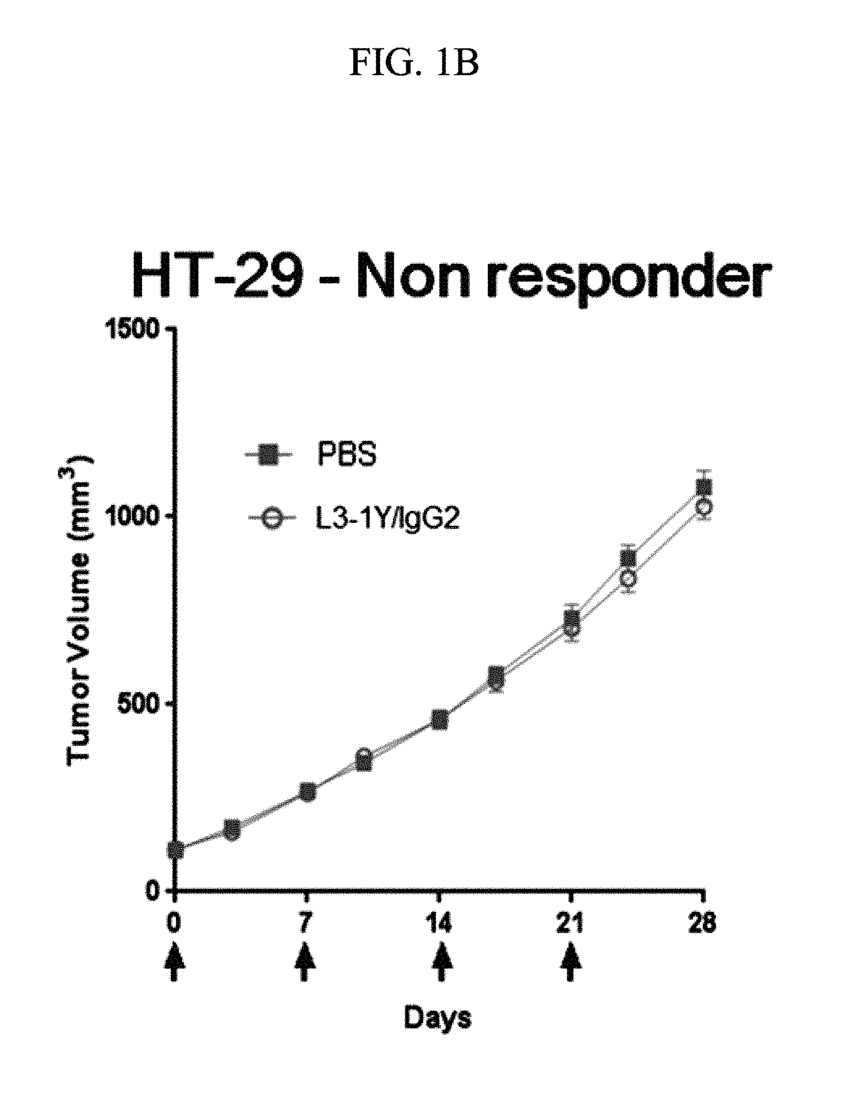
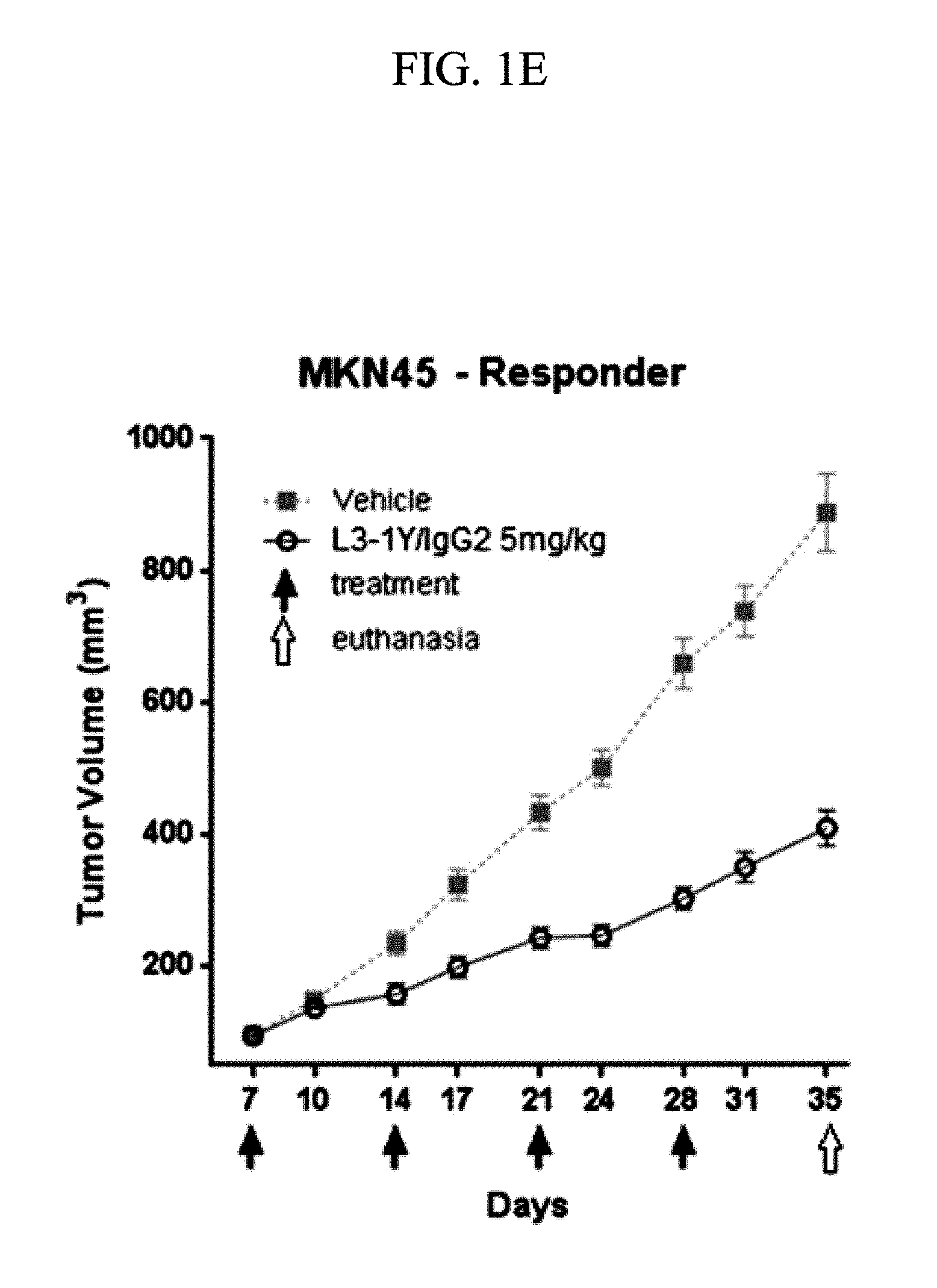
FIGS. 1A, 1B, 1C, 1D, and 1E are graphs showing the anticancer activity of an anti-c-Met antibody in terms of changes in tumor size with treatment with the anti-c-Met antibody in mouse models implanted with Lovo cells (FIG. 1A), HT29 cells (FIG. 1B), EBC-1 cells (FIG. 1C), Hs746T cells (FIG. 1D), and MKN45 cells (FIG. 1E).
FIG. 2 is a graph of IL-8 levels in cancer cell-implanted mouse models showing relationship between IL-8 level and responsiveness to an anti-c-Met antibody.
FIG. 3 contains graphs showing the serum level changes of IL-8 in response to the different doses of the anti-c-Met antibody in responder and non-responder groups of the human cancer cell-implanted mouse models.
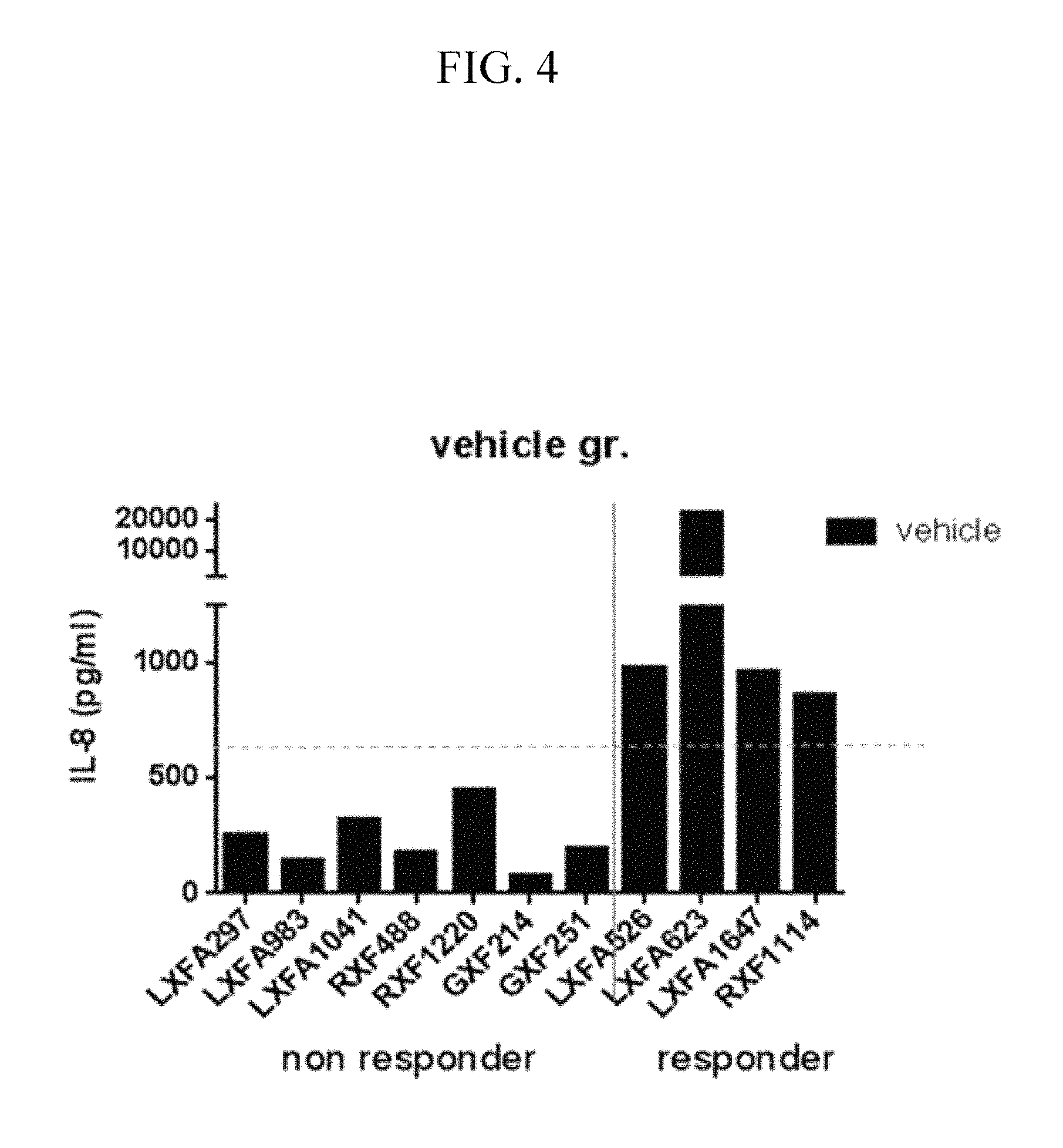
FIG. 4 is a graph showing IL-8 levels in serum samples from the control (vehicle (PBS) treated) of mouse models implanted with patient-derived tumor tissues.
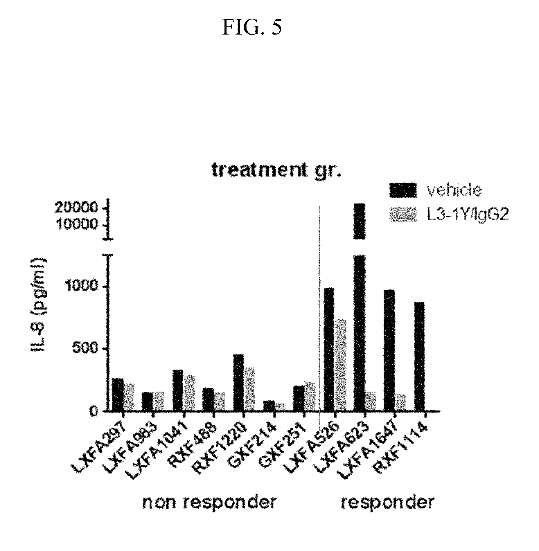
FIG. 5 is a graph of IL-8 levels in serum samples from anti-c-Met antibody-treated and untreated (control PBS treated) groups of mouse models implanted with patient-derived tumor tissues, showing relationship between the anticancer efficacy of the anti-c-Met antibody and the change of IL-8 level after treatment of anti-c-Met antibody.
FIG. 6 is a graph showing a change in tumor size after treatment of the anti-c-Met antibody in conventional cancer cell line (EBC-1)-implanted mouse models.
FIG. 7 is a graph showing a change in tumor size after treatment of the anti-c-Met antibody in conventional cancer cell line (Hs746T)-implanted mouse models.
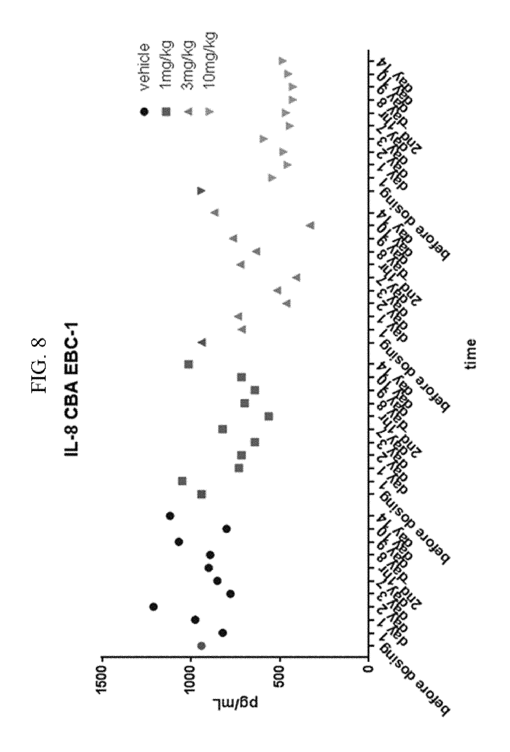
FIG. 8 is a graph showing a change in IL-8 level with time after treatment of the anti-c-Met antibody in conventional cancer cell line (EBC-1)-implanted mouse models.
FIG. 9 is a graph showing a change in IL-8 level with time after treatment of the anti-c-Met antibody in conventional cancer cell line (Hs746T)-implanted mouse models.
FIG. 10 is a graph showing a change in tumor size after treatment of the anti-c-Met antibody in U87MG-implanted mouse model.
FIG. 11 shows a change in protein expression pattern in U87MG cells, as measured by a chemiluminescence immunoassay using protein array kit.
FIG. 12 is a graph showing a change in bIG-H3 level after treatment of the anti-c-Met antibody in serum from mouse models implanted with various cancer cells.
FIG. 13 is a graph showing a change in bIG-H3 level after treatment of the anti-c-Met antibody in serum from mouse models implanted with patient-derived tumor tissues.
FIG. 14 is a graph showing a change in bIG-H3 level after treatment of the anti-c-Met antibody in cancer tissue lysates from mouse models implanted with patient-derived tumor tissues.
FIG. 15 shows a change in protein expression pattern in a patient-derived tumor tissue-implanted mouse model (LXFA623), as measured by a chemiluminescence immunoassay using protein array kit.
FIG. 16 is a graph showing a change in MIF level after treatment of the anti-c-Met antibody in serum samples from mouse models implanted with various cancer cell lines.
FIG. 17 is a graph showing a change in MIF level after treatment of the anti-c-Met antibody in serum samples from mouse models implanted with patient-derived tumor tissues.
FIG. 18 shows c-Met protein expression levels in patient-derived cancer cell-implanted mouse models, as measured by Western blotting.
FIG. 19 contains graphs illustrating the change of tumor sizes after treatment of the anti-c-Met antibody in the presence or absence of K-RAS and B-RAF mutations in patient-derived tumor tissue-implanted mouse models.
FIG. 20 contains graphs illustrating the responsiveness of anti-c-Met antibody responder cell lines to the treatment of other c-Met inhibitors; crizotinib and PHA665752.
FIG. 21 contains graphs illustrating the responsiveness of anti-c-Met antibody non-responder cell lines to the treatment of other c-Met inhibitors; crizotinib and PHA665752.
FIG. 22 contains graphs illustrating the relative levels of IL-8(%) in culture sup of the anti-c-Met antibody responder cell lines after treatment of various c-Met inhibitors, L3-1Y/IgG2, crizotinib and PHA665752.
FIG. 23 contains graphs illustrating the relative levels of IL-8(%) in culture sup of the anti-c-Met antibody non-responder cell lines after treatment of various c-Met inhibitors, L3-1Y/IgG2, crizotinib and PHA665752.
DETAILED DESCRIPTION OF THE INVENTION
Provided is the use of at least one of selected from the group consisting of IL-8, b-IG-H3, MIF, KRAS/BRAF, and nucleic acids encoding the proteins in predicting the effect of a c-Met inhibitor and/or monitoring the effect of administered c-Met inhibitors.
One embodiment provides a method for predicting and/or monitoring the effect of a c-Met inhibitor, comprising the analysis of at least one biomarker selected from the group consisting of IL-8, b-IG-H3, MIF, KRAS/BRAF, and nucleic acids encoding the proteins.
Another embodiment provides a method for selecting a subject to which a c-Met inhibitor is applicable (i.e., will exhibit efficacy), comprising the analysis of at least one biomarker selected from the group consisting of IL-8, b-IG-H3, MIF, KRAS/BRAF, and nucleic acids encoding the proteins.
Another embodiment provides a composition and a kit for predicting and/or monitoring the effect of a c-Met inhibitor, comprising a material interacting with at least one selected from the group consisting of IL-8, b-IG-H3, MIF, KRAS/BRAF, and nucleic acids encoding the proteins.
Another embodiment provides a composition and a kit for selecting a subject to which a c-Met inhibitor is applicable, comprising a material interacting with at least one selected from the group consisting of IL-8, b-IG-H3, MIF, KRAS/BRAF, and nucleic acids encoding the proteins.
Another embodiment provides a method for predicting and/or monitoring the efficacy of c-Met inhibitor or for providing information on the prediction and/or monitoring of the efficacy of a c-Met inhibitor, comprising measuring existence, level, and/or mutation of at least one selected from the group consisting of IL-8, b-IG-H3, MIF, KRAS/BRAF, and nucleic acids encoding the proteins, in a biological sample.
Another embodiment provides a method for selecting a subject suitable for the application of a c-Met inhibitor, comprising measuring for existence, level and/or mutation of at least one selected from the group consisting of IL-8, b-IG-H3, MIF, KRAS/BRAF, and nucleic acids encoding the proteins, in a biological sample obtained from a subject.
Another embodiment provides a method for preventing and/or treating cancer, comprising administering a c-Met inhibitor to a subject, wherein the subject has a high level of IL-8 or a nucleic acid encoding IL-8 compared to a reference sample in which a c-Met inhibitor is not effective, as defined below; shows a decreased level of IL-8, bIG-H3, MIF, and/or nucleic acids encoding them after administering a c-Met inhibitor (compared to that of before the administration); and/or has a mutation of KRAS, BRAF, and/or nucleic acids encoding them. For example, the subject may be selected by the above method for selecting a subject suitable for the application of a c-Met inhibitor. This preventing or treating method may further comprise selecting a subject suitable for the application of the c-Met inhibitor, prior to the administration.
Another embodiment provides a method for inhibiting c-Met, comprising administering a c-Met inhibitor to a subject, wherein the subject has a high level of IL-8 or a nucleic acid encoding IL-8 compared to a reference sample in which a c-Met inhibitor is not effective, as defined below; shows a decreased level of IL-8, bIG-H3, MIF, and/or nucleic acids encoding them after administering a c-Met inhibitor (compared to that of before the administration); and/or has a mutation of KRAS, BRAF, and/or nucleic acids encoding them. For example, the subject may be selected by the above method for selecting a subject suitable for the application of a c-Met inhibitor. This inhibiting method may further comprise selecting a subject suitable for the application of the c-Met inhibitor, prior to the administration.
As used herein, the term "efficacy" of a c-Met inhibitor refers to a c-Met inhibitory effect and/or the pharmaceutical activity of a c-Met inhibitor resulting in the prevention, amelioration, reduction or treatment of a c-Met-related disease, for instance, a cancer. As regards cancer the efficacy refers to an anticancer effect, for example, an ability to induce the reduction or death of cancer cells or tissues, and the suppression of cancer cell migration and/or invasion responsible for cancer metastasis. In other word, the efficacy may refer to responsiveness of a subject, who is administered with a c-Met inhibitor, to the c-Met inhibitor.
As used herein, the term "predicting an efficacy of a c-Met inhibitor" may refer to determining whether or not or how much the c-Met inhibitor exhibits its desired efficacy (e.g., an antitumor or anticancer efficacy) on a subject who is treated therewith.
As used herein, the wording "a subject suitable for the application of the c-Met inhibitor" may refer to "a subject suitable for administration, treatment with a c-Met inhibitor", "a subject with a disease (e.g., cancer) that is likely to respond to a c-Met inhibitor", or an individual that is likely to respond to a c-Met inhibitor.
The expression level of a certain protein may be determined by quantitatively analyzing the protein or the gene (nucleic acid) encoding it, such as DNA, cDNA or mRNA. As used herein, the term "gene" refers to any nucleic acid encoding the protein, whether genomic DNA, cDNA, or RNA (mRNA). The determination of KRAS and/or BRAF mutations may be achieved at a gene level or a protein level.
IL-8 (Interleukin 8) is a chemokine functioning as an important mediator of immune responses. IL-8, also known as neutrophil chemotactic factor, has two primary functions: it induces chemotaxis in target cells, primarily neutrophils but also other granulocytes, causing them to migrate toward the site of infection; and also phagocytosis once they have arrived. IL-8 is also known to be a potent promoter of angiogenesis contributing to the proliferation and metastasis of tumor cells. In one exemplary embodiment of the present invention, a response to a c-Met inhibitor varies depending on the expression level of IL-8, and a difference in the expression level of IL-8 between biological samples before and after treatment with an c-Met inhibitor is dependent on the response to the c-Met inhibitor (see Example 1). Briefly, a higher level of IL-8 or its gene in a biological sample indicates that a c-Met inhibitor, e.g., an anti-c-Met antibody, more efficiently exerts its function on the biological sample or a patient from which the biological sample is sourced. In addition, a biological sample in which a c-Met inhibitor, for example, an anti-c-Met antibody, functions well (drug response group), a decrease in the level of IL-8 or its gene after the application of the c-Met inhibitor, compared to before the application, is typically observed.
That is, an expression level of IL-8 can be used to predict the efficacy of an c-Met inhibitor and thus to select a subject suitable for treatment with the c-Met inhibitor, or a change in the expression level of IL-8 according to, or before and after treatment with a c-Met inhibitor allows for the evaluation (identification) or monitoring of the efficacy of the c-Met inhibitor, thereby being useful in determining whether the c-Met inhibitor should continue to be applied, or in establishing the therapeutic strategy of the c-Met inhibitor with regard to dose, dosing interval, and dosing number. Based on this finding, the use of IL-8 as a marker for indicating and/or monitoring the efficacy of a c-Met inhibitor is suggested.
The IL-8 may originate from vertebrates including mammals, such as rodents, e.g., mice, rat, etc., and primates, e.g., humans, monkeys, etc., birds, reptiles, amphibians, and fishes. For example, the IL-8 may be selected from the group consisting of NP_000575.1, AAH13615.1, AAA59158.1, NP_001028137.1, AAA80141.2, and AAA86711.1. An IL-8-encoding gene (cDNA or mRNA) may be selected from among NM_000584.3, BC013615.1 (from 41 to 340 positions), NC_000004, NG_029889, NC_018915.2, AC_000136, M28130.1, NM_001032965.1, S78555.1, and U19849.1.
One embodiment is directed to the analysis of a biomarker for the prediction and/or monitoring of the efficacy of a c-Met inhibitor comprising the analysis of (e.g., measuring the level of) at least one selected from the group consisting of IL-8 and a nucleic acid (e.g., gene) encoding IL-8.
Another embodiment is directed to the analysis of a biomarker for the selection of a subject suitable to the application of a c-Met inhibitor, comprising the analysis of (e.g., measuring the level of) at least one selected from the group consisting of IL-8 and a nucleic acid (e.g., gene) encoding IL-8.
A further is directed to a composition and a kit for the prediction and/or monitoring of the efficacy of a c-Met inhibitor, comprising a material capable of interacting with at least one selected from the group consisting of IL-8 and a nucleic acid (e.g., gene) encoding IL-8.
A further embodiment addresses a composition and a kit for the selection of a subject suitable to the application of a c-Met inhibitor, comprising a material interacting with at least one selected from the group consisting of IL-8 and a nucleic acid (e.g., gene) encoding IL-8.
Another embodiment is a method for predicting and/or monitoring the efficacy of a c-Met inhibitor or for offering information on the prediction and/or monitoring of the efficacy of a c-Met inhibitor, comprising measuring a biological sample for a level of at least one selected from the group consisting of IL-8 and a nucleic acid (e.g., gene) encoding IL-8.
Another embodiment provides a method for selecting a subject suitable to the application of a c-Met inhibitor or for offering information on the selection of a subject suitable to the application of a c-Met inhibitor, comprising measuring a biological sample for a level of at least one selected from the group consisting of IL-8 and a nucleic acid (e.g., gene) encoding IL-8.
Provided according to one embodiment is a method for predicting the efficacy of a c-Met inhibitor, for offering information on the prediction of the efficacy of a c-Met inhibitor, for selecting a subject suitable to the application of a c-Met inhibitor, or for offering information of the selection of a subject suitable to the application of a c-Met inhibitor, comprising measuring a level of at least one selected from the group consisting of IL-8 and a nucleic acid (e.g., gene) encoding IL-8 in a biological sample. In the method for predicting the efficacy of a c-Met inhibitor or for offering information on the efficacy of a c-Met inhibitor, when a biological sample is measured to have a high level of at least one selected from the group consisting of IL-8 and a nucleic acid (e.g., gene) encoding IL-8, the biological sample or a patient from which the biological sample is originated can be predicted to allow the c-Met inhibitor to effectively exert its activity or may be considered to be suitable for the application of the c-Met inhibitor. Hence, the method for predicting the efficacy of a c-Met inhibitor or for offering information on the efficacy of a c-Met inhibitor may further comprise determining that the c-Met inhibitor exhibits its efficacy in the biological sample or the patient when the biological sample is observed to have a high level of at least one selected from the group IL-8 or a nucleic acid (e.g., gene) encoding IL-8 after the measurement. In addition, the method for selecting a subject suitable to the application of a c-Met inhibitor or for offering information on the selection may further comprise determining the biological sample or the patient as a subject suitable for the application of the c-Met inhibitor when the biological sample is observed to have a high level of at least one selected from the group IL-8 or a nucleic acid (e.g., gene) encoding IL-8 after the measurement.
"A high level of at least one selected from the group consisting of IL-8 and a nucleic acid (e.g., gene) encoding IL-8" may refer to a large amount of IL-8 protein and/or a nucleic acid (e.g., gene) encoding IL-8 (DNA, cDNA or mRNA), compared to a biological sample (reference sample) from a patient in which a c-Met inhibitor to be applied does not exert therapeutic activity. For example, when the level of at least one selected from IL-8 and a nucleic acid (e.g., gene) encoding IL-8 in a test biological sample from a test patient is at least about 1.5-, about 2-, about 3-, about 4-, or about 5-fold, for example, about 1.5- to about 100-fold, about 2- to about 100-fold, about 3- to about 100-fold, about 4- to about 100-fold, or about 5- to about 100-fold greater in weight than that in a reference biological sample on which the c-Met inhibitor to be applied does not exert its efficacy (e.g., a c-Met inhibitory efficacy or an anticancer efficacy), the c-Met inhibitor is predicted to effectively exert its efficacy (e.g., a c-Met inhibitory efficacy or an anticancer efficacy) in the test biological sample or the test patient, or the test biological sample or the test patient can be considered as a subject suitable to the application of the c-Met inhibitor. The c-Met inhibitor may be, for example, an anti-c-Met antibody. The reference biological sample can be any sample useful for comparing the expression of the c-Met biomarker and/or c-Met inhibitor efficacy, such as a biological sample comprising cells, such as a mammalian cancer cell, in which the c-Met inhibitor does not have its efficacy (e.g., a c-Met inhibitory and/or anticancer efficacy). Examples of the reference biological sample on which the c-Met inhibitor does not exert its efficacy include, but are not limited to, the Lovo cell line (CCL-229, ATCC), and the HT-29 cell line (HTB-38, ATCC). Therefore, the method for predicting the efficacy of a c-Met inhibitor (or for offering information on the prediction of the efficacy of a c-Met inhibitor) or for selecting a subject suitable to the application of a c-Met inhibitor (or for offering information of the selection of a subject suitable to the application of a c-Met inhibitor) may further comprise a step of measuring a level of at least one selected from the group consisting of IL-8 and a nucleic acid (e.g., gene) encoding IL-8 in a reference biological sample. The measurement of the level of IL-8 and/or a nucleic acid (e.g., gene) encoding IL-8 (e.g., quantitative analysis) may be performed by the same method in both the test biological sample and the reference biological sample.
Measuring the biological sample for a level of at least one selected from among IL-8 and a nucleic acid (e.g., gene) encoding IL-8 may comprise i) treating (reacting or contacting) the biological sample with a material interacting with at least one selected from the group consisting of IL-8 and a nucleic acid (e.g., gene) encoding IL-8; and ii) quantitatively analyzing the reaction mixture to determine the level of at least one selected from IL-8 and a nucleic acid (e.g., gene) encoding IL-8. In an embodiment, prior to step i), a step of preparing a biological sample may be further performed, wherein the preparation step may comprise obtaining (isolating) a biological sample from a patient or obtaining a biological sample which has been isolated from a patient. In step i), the interacting material may be at least one selected from the group consisting of compounds (a small molecular chemical; including any general label such as a fluorescent, a dye, etc.), proteins (antibodies, aptamers, etc.), nucleic acids (DNA, RNA, etc.), and the like, which bind to IL-8 or IL-8 gene, and for example, the interacting material may be a compound (a small molecular chemical; e.g., general label such as a fluorescent, a dye, etc.), an antibody, or an aptamer, all binding (e.g., specifically binding) to IL-8, a polynucleotide (e.g., a primer, a probe, an aptamer) binding to a part or entirety of a nucleic acid (e.g., gene) encoding IL-8, or any combination thereof; and optionally, may be conjugated with a general label, such as a fluorescent, a secondary antibody, a bead (e.g., a magnetic bead or polystyrene bead), a dye, or any combination thereof. The step i) may be configured to form a complex by adding the interacting material to the biological sample. In step ii), the reaction mixture may be the complex resulting from interaction (binding) between at least one selected from the group consisting of IL-8 and a nucleic acid (e.g., gene) encoding IL-band the interacting material, which can be obtained in step i). Quantitatively analyzing may comprise quantifying the complex, the label conjugated to the complex, or the IL-8 or nucleic acid (e.g., gene) encoding IL-8 segregated from the complex after the isolation of the complex from the biological sample.
Provided in accordance with another embodiment is a method for monitoring (evaluating or identifying) the efficacy of a c-Met inhibitor or for offering information on the monitoring (evaluation or identification) of the efficacy of a c-Met inhibitor, comprising measuring a level of at least one selected from the group consisting of IL-8 and a nucleic acid (e.g., gene) encoding IL-8 in a biological sample. In the method for monitoring the efficacy of a c-Met inhibitor or for offering information on the monitoring of the efficacy of a c-Met inhibitor, levels of at least one selected from the group consisting of IL-8 and a nucleic acid (e.g., gene) encoding IL-8 in biological samples from a patient before and after treatment with the c-Met inhibitor (or in a c-Met inhibitor-untreated biological sample and a c-Met inhibitor-treated biological sample) may be measured and compared to each other. When the post-treatment level of at least one selected from the group consisting of IL-8 and a nucleic acid (e.g., gene) encoding IL-8 is lower (decreased) compared to the pre-treatment level, the c-Met inhibitor is determined to exert its efficacy in the patient from which the biological sample is sourced (obtained), or when the post-treatment level of at least one selected from the group consisting of IL-8 and a nucleic acid (e.g., gene) encoding IL-8 is the same with or higher (increased) compared to the pre-treatment level, it is determined that the c-Met inhibitor does not exert its efficacy or a resistance to the c-Met inhibitor occurs in the patient from which the biological sample is sourced (obtained). Therefore, the method for monitoring the efficacy of a c-Met inhibitor or for offering information on the monitoring of the efficacy of a c-Met inhibitor may further comprise 1) comparing the levels of at least one selected from the group consisting of IL-8 and a nucleic acid (e.g., gene) encoding IL-8 in a biological sample from the c-Met inhibitor-treated group with that in a biological sample from the untreated group, and/or 2) determining that the c-Met inhibitor exerts its efficacy in the biological sample-derived patient when the level of at least one selected from the group consisting of IL-8 and a nucleic acid (e.g., gene) encoding IL-8 in the biological sample of the c-Met inhibitor-treated group is reduced, compared to that of the untreated group (in this case, it may be determined to maintain (continue) the administration of a c-Met inhibitor to the subject), and/or 3) determining that the c-Met inhibitor does not exert its efficacy or a resistance to the c-Met inhibitor occurs in the biological sample-derived patient when the level of at least one selected from the group consisting of IL-8 and a nucleic acid (e.g., gene) encoding IL-8 in the biological sample of the c-Met inhibitor-treated group is maintained or increased (in this case, it may be determined to stop the administration of a c-Met inhibitor to the subject). The c-Met inhibitor may be an anti-c-Met antibody. The c-Met inhibitor-treated group and the untreated group mean the same biological sample after and before treatment with a c-Met inhibitor, respectively, or aliquots of the same biological sample which are treated with and are not treated with a c-Met inhibitor (for example, treated only with a vehicle), respectively. Unless stated otherwise, the terms "c-Met inhibitor-untreated group (or biological sample)" and "biological sample before treatment with a c-Met inhibitor" or "pre-treatment biological sample" are identical to each other while the terms "c-Met inhibitor-treated group (or biological sample)" and "biological sample after treatment with a c-Met inhibitor" or "post-treatment biological sample" share the same meaning.
The level of at least one selected from the group consisting of IL-8 and a nucleic acid (e.g., gene) encoding IL-8 may be measured by quantifying an IL-8 protein and/or a nucleic acid (e.g., gene) encoding IL-8 (DNA, cDNA or mRNA). For example, when the level of at least one selected from the group consisting of IL-8 and a nucleic acid (e.g., gene) encoding IL-8 in the c-Met inhibitor-treated group or in the biological sample after treatment with a c-Met inhibitor is about 0 to about 80 wt %, about 0 to about 70 wt %, about 0 to about 60 wt %, about 0 to about 50 wt %, about 0 to about 40 wt %, about 0 to about 30 wt %, about 0 to about 20 wt %, or about 0 to about 10 wt % compared to that (considering 100 wt %) of the c-Met inhibitor-untreated group or the pre-treatment biological sample, the c-Met inhibitor-treated group, or the post-treatment sample biological, is determined to decrease in the level of at least one selected from the group consisting of IL-8 and a nucleic acid (e.g., gene) encoding IL-8, compared to the c-Met inhibitor-untreated or the pre-treatment biological sample. When the level of IL-8 and a nucleic acid (e.g., gene) encoding IL-8 in the c-Met inhibitor-treated group or the post-treatment biological sample is about 0 wt % of that in the c-Met inhibitor-untreated group or the pre-treatment biological sample there is no detection (absence) of at least one selected from the IL-8 and a nucleic acid (e.g., gene) encoding IL-8 in the c-Met inhibitor-treated group or the post-treatment biological sample. Levels of IL-8 and/or a nucleic acid (e.g., gene) encoding IL-8 in the c-Met inhibitor-untreated group (or pre-treatment) and the c-Met inhibitor-treated group (or post-treatment) may be measured by conducting the same procedure on sample samples from the same patient.
Monitoring the efficacy of an c-Met inhibitor, IL-8 and/or a nucleic acid (e.g., gene) encoding IL-8 may be quantitatively analyzed within a relatively short period of time, for example, a week, 5 days, 3 days, 2 days or 1 day after administration of the c-Met inhibitor, and comparison between levels of the marker before and after administration thereof can evaluate the efficacy of c-Met inhibitor. Henceforth, the advantage of establishing a more effective therapeutic strategy can be achieved, since it is possible to evaluate the efficacy of the c-Met inhibitor in an early stage of therapy.
In one embodiment, the measuring of a level of at least one selected from the group consisting of IL-8 and a nucleic acid (e.g., gene) encoding IL-8 in a biological sample may comprise 1) determining a level of at least one selected from the group consisting of IL-8 and a nucleic acid (e.g., gene) encoding IL-8 in a biological sample from a patient prior to treatment with a c-Met inhibitor (or a c-Met inhibitor untreated biological sample which is a part of biological sample from a patient) and 2) determining a level of at least one selected from the group consisting of IL-8 and a nucleic acid (e.g., gene) encoding IL-8 in a biological sample from the patient after treatment with a c-Met inhibitor (or a c-Met inhibitor treated biological sample which is the other part of biological sample from a patient). Each of the steps 1) and 2) may comprise i) applying (adding) a material interacting with at least one selected from the group consisting of IL-8 and a nucleic acid (e.g., gene) encoding IL-8 to the biological sample, and ii) quantitatively analyzing the resulting reaction mixture to determine a level of at least one selected from the group consisting of IL-8 and a nucleic acid (e.g., gene) encoding IL-8. In an embodiment, prior to the step i), a step of preparing a biological sample may be further performed, wherein the preparation step may comprise obtaining (isolating) a biological sample from the patient or obtaining a biological sample which has been isolated from a patient. In step i), as will be further elucidated below, the interacting material may be at least one selected from the group consisting of compounds (a small molecular chemical; e.g., general label such as a fluorescent, a dye, etc.), proteins (antibodies, aptamers, etc.) nucleic acid (DNA, RNA, etc.), and the like, binding to IL-8 or IL-8 gene, and for example, the interacting material may be at least one selected from the group consisting of a compound, an antibody (e.g., antibodies for Intracellular staining (Human CXCL8/IL-8 Phycoerythrin MAb; #IC208P; R&D systems), etc.), an aptamer, all specifically binding to IL-8, and a polynucleotide (e.g., a primer (e.g., a primer set used in RT-PCR for IL-8 gene (ACCESSION NO: BC013615.1; Forward primer: 5'-ATG ACT TCC AAG CTG GCC GTG GCT-3' (SEQ ID NO: 113) and Reverse primer: 5'-TCT CAG CCC TCT TCA AAA ACT TCT-3' (SEQ ID NO: 114), etc.), a probe, an aptamer) binding to a part or entirety of a gene encoding IL-8, and optionally, may be conjugated with a label, such as a fluorescent or a dye. The step i) may be configured to form a complex by applying (adding) the interacting material to the biological sample. In step ii), the reaction mixture may be a complex resulting from interaction (binding) between at least one selected from the group consisting of IL-8 and a nucleic acid (e.g., gene) encoding IL-8 and the interacting material, which can be obtained in step i). The quantitatively analyzing step may comprise quantifying the complex, the marker conjugated to the complex, or the IL-8 or the gene segregated from the complex after the isolation of the complex from the biological sample. The quantitative analysis of IL-8 may be performed by any general quantifying means of proteins, such as immunochromatography, immunohistochemistry, immunohistochemical staining, enzyme linked immunosorbent assay (ELISA; e.g., Platinum ELISA Human IL-8/NAP-1 (#BMS204/3, eBioscience), etc.), radioimmunoassay (RIA), enzyme immunoassay (EIA), fluorescence immunoassay (FIA), luminescence immunoassay (LIA), Western blotting, microarray, surface plasmon resonance (SPR), flow cytometry assay (e.g., Cytometric Bead Array (CBA) assay (e.g., IL-8 flex set (#558277), human inflammatory cytokine kit (#551811); BD biosciences), Intracellular staining (e.g., using antibodies such as Human CXCL8/IL-8 Phycoerythrin MAb (#IC208P; R&D systems)), etc.), Luminex assay, and the like, and the quantitative analysis of IL-8 gene may be performed by any general quantifying means of genes (DNA or RNA), such as PCR (e.g., qPCR, RT-PCR, etc.), mRNA microarray, and the like, but not limited thereto.
The monitoring method may be useful in determining whether the c-Met inhibitor continues to be used, and/or suitable dosing conditions of the c-Met inhibitor (dose, dosing interval, number of doses, etc.). For example, when the level of at least one selected from the group consisting of IL-8 and a nucleic acid (e.g., gene) encoding IL-8 in the c-Met inhibitor-treated group or in the post-treatment biological sample is reduced, compared to the c-Met inhibitor-untreated group or the pre-treatment biological sample, for example, when the level of at least one selected from the group consisting of IL-8 and a nucleic acid (e.g., gene) encoding IL-8 in the c-Met inhibitor-treated group or in the post-treatment biological sample is about 0 to about 80 wt %, about 0 to about 70 wt %, about 0 to about 60 wt %, about 0 to about 50 wt %, about 0 to about 40 wt %, about 0 to about 30 wt %, about 0 to about 20 wt %, or about 0 to about 10 wt % of that of the c-Met inhibitor-untreated group or the pre-treatment biological sample, the use of c-Met inhibitor may be determined to be continued. In addition, when the level of at least one selected from the group consisting of IL-8 and a nucleic acid (e.g., gene) encoding IL-8 in the c-Met inhibitor-treated group or in the post-treatment biological sample is reduced, compared to the c-Met inhibitor-untreated group or the pre-treatment biological sample, for example, when the level of at least one selected from the group consisting of IL-8 and a nucleic acid (e.g., gene) encoding IL-8 in the c-Met inhibitor-treated group or in the post-treatment biological sample is about 0 to about 80 wt %, about 0 to about 70 wt %, about 0 to about 60 wt %, about 0 to about 50 wt %, about 0 to about 40 wt %, about 0 to about 30 wt %, about 0 to about 20 wt %, or about 0 to about 10 wt %, of that of the c-Met inhibitor-untreated group or the pre-treatment biological sample, the dosing condition of the c-Met inhibitor may be determined to be suitable. The dosing condition may be at least one selected from the group consisting of a dose, a dosing interval, and a number of doses.
Another embodiment provides a method for inhibiting c-Met, comprising administering a c-Met inhibitor to a subject who has a high level of IL-8 and/or a nucleic acid encoding IL-8 (wherein the high level is defined above) and/or shows a decreased level of IL-8 and/or a nucleic acid encoding IL-8 after administration of a c-Met inhibitor (compared to before administration of the c-Met inhibitor). The subject may be 1) selected by the above method for selecting a subject suitable for the application of a c-Met inhibitor or 2) determined by the above method of monitoring efficacy of a c-Met inhibitor as a subject wherein a c-Met inhibitor exerts its efficacy thereon.
Another embodiment provides a method for preventing and/or treating cancer, comprising administering a c-Met inhibitor to a subject who has a high level of IL-8 or a nucleic acid encoding IL-8 (wherein the high level is defined above) and/or shows a decreased level of IL-8 and/or a nucleic acid encoding IL-8 after administration of a c-Met inhibitor (compared to before administration of the c-Met inhibitor). The subject may be 1) selected by the above method for selecting a subject suitable for the application of a c-Met inhibitor or 2) determined by the above method of monitoring efficacy of a c-Met inhibitor as a subject wherein a c-Met inhibitor exert its efficacy thereon.
The method for inhibiting c-Met or the method for preventing and/treating cancer may further comprise selecting a subject to which the c-Met inhibitor is applicable, prior to the administering step. Details of the selection are as described above. The c-Met inhibitor may be an anti-c-Met antibody.
In an embodiment, the method for inhibiting c-Met or for preventing and/or treating cancer may comprise:
identifying a subject to which a c-Met inhibitor is applicable; and
administering a pharmaceutically effective amount of the c-Met inhibitor to the subject.
In another embodiment, the method for inhibiting c-Met or for preventing and/or treating cancer may comprise:
measuring a level of IL-8 and/or a nucleic acid (e.g., gene) encoding IL-8 in a biological sample to select a c-Met inhibitor-applicable subject; and
administering a pharmaceutically effective amount of the c-Met inhibitor to the subject.
With regard to dosing conditions of the c-Met inhibitor, such as doses, dosing intervals and/or number of doses, they may be determined in the method for monitoring a c-Met inhibitor for efficacy.
Some anti-cancer agents such as sunitinib, cis-platin, paclitaxel, etc. often cannot exert their anticancer effects in individuals with a high level of IL-8 because they provoke resistance. In contrast, c-Met inhibitors, for example, anti-c-Met antibodies, particularly, those described below, can exhibit high anticancer activity in individuals with a high level of IL-8, thereby allowing for the suggestion of more effective therapeutic strategies.
bIG-H3, also known as TGFBI (transforming growth factor beta-induced), is RGD-containing protein that binds to type I, II and IV collagens. The RGD motif is found in ECM (extracellular matrix). Serves as a ligand binding to a3b1 integrin, bIG-H3 is involved in cell adhesion, and migration. bIG-H3 is a soluble protein and plays a role in cancer metastasis and angiogenesis in the tumor microenvironment. No reports have been made on a relationship between c-Met and bIG-H3, so far.
MIF (Macrophage migration inhibitory factor) is an important regulator of innate immunity and is classified as an inflammatory cytokine.
In the Example section of the specification, a difference in the expression level of bIG-H3 and/or MIF between c-Met inhibitor-treated groups and untreated groups or between a post-treatment group and a pre-treatment group was observed to vary depending in response to the c-Met inhibitor (refer to Examples 2 and 3). In detail, when a c-Met inhibitor, e.g., an anti-c-Met antibody functions well (drug responsive group), the level of at least one selected from the group consisting of bIG-H3, MIF and nucleic acids encoding the proteins is lower after application of the c-Met inhibitor than before the application.
In other words, expression levels of at least one selected from the group consisting of bIG-H3, MIF, and nucleic acids encoding the proteins before or after the application of a c-Met inhibitor are measured to assay (identify) or monitor the effect of the c-Met inhibitor, so that the measurements can be effectively used to determine whether the c-Met inhibitor should continue to be applied or not, or to establish a therapeutic strategy with regard to, for example, doses, dosing intervals, dose numbers of the c-Met inhibitor. Based on this finding, bIG-H3 and/or MIF as appear to be viable pharmacodynamic markers for c-Met inhibitors.
The bIG-H3 may originate from vertebrates including mammals, such as rodents, e.g., mice, rat, etc., and primates, e.g., humans, monkeys, etc., birds, reptiles, amphibians, and fishes. For example, the bIG-H3 may be selected from the group consisting of, but not limited to, human bIG-H3 (NP_000349.1, AAC24944.1, AAC08449.1, AAH00097.1, etc.), mouse bIG-H3 (NP_033395.1, AAI29901.1, AAI29902.1, etc.), rat bIG-H3 (NP_446254.1, etc.), and zebrafish bIG-H3 (NP_878282.1, etc.). A bIG-H3 encoding gene (cDNA or mRNA) may be selected from the group consisting of but not limited to, NM_000358.2, NM_009369.4, NM_053802.1, and NM_182862.1.
MIF may originate from vertebrates including mammals, birds, reptiles, amphibians, and fishes. For example, the MIF may be selected from the group consisting of, but not limited to, human MIF (NP_002406.1, etc.), monkey MIF (e.g., NP_001028087.1, etc.), sheep MIF (NP_001072123.1, etc.), rodent MIF (e.g., mouse MIF (NP_034928.1, etc.), rat MIF (NP_112313.1, etc.), NP_001266756.1, etc.), cow MIF (NP_001028780.1, etc.), zebrafish MIF (NP_001036786.1, etc.), pig MIF (NP_001070681.1, etc.), frog MIF (NP_001083650.1, NP_001107147.1, etc.), and fish MIF (e.g., NP_001118053.1, NP_001135019.1, NP_001027889.1). An MIF-encoding gene (cDNA or mRNA) may be selected from among, but not limited to, NM_002415.1, NM_001032915.1, NM_001078655.1, NM_010798.2, NM_031051.1, NM_001279827.1, NM_001033608.1, NM_001043321.1, NM_001077213.2, NM_001090181.1, NM_001113675.1, NM_001124581.1, NM_001141547.1, and NM_001032717.1.
An embodiment provides a biomarker for evaluating the effect of a c-Met inhibitor, comprising at least one selected from the group consisting of bIG-H3, MIF, and nucleic acids encoding the proteins.
Another embodiment provides a composition and a kit for evaluating the effect of a c-Met inhibitor, comprising at least one selected from the group consisting of bIG-H3, MIF, and nucleic acids encoding the proteins.
Another embodiment provides a method for evaluating (or identifying or monitoring) the efficacy of a c-Met inhibitor or for offering information on the evaluation (or identifying or monitoring) of the efficacy of a c-Met inhibitor, comprising measuring a level of at least one selected from the group consisting of bIG-H3, MIF, and nucleic acids encoding the proteins in a biological sample.
In the method for monitoring the efficacy of a c-Met inhibitor or for offering information on the monitoring of the efficacy of a c-Met inhibitor, levels of at least one selected from the group consisting of bIG-H3, MIF and nucleic acids encoding the proteins in biological samples from a patient before and after treatment with the c-Met inhibitor (or in a c-Met inhibitor treated group biological sample and a c-Met inhibitor untreated biological sample) are measured and compared. When the post-treatment level of at least one selected from the group consisting of bIG-H3, MIF, and nucleic acids encoding the proteins is lower (decreased) compared to the pre-treatment level, the c-Met inhibitor is determined to exert its activity in the patient from which the biological sample is sourced (obtained), whereas when the post-treatment level of at least one selected from the group consisting of bIG-H3, MIF, and nucleic acids encoding the proteins is the same with or higher (increase) compared to the pre-treatment level, it is determined that the c-Met inhibitor does not exert its efficacy or a resistance to the c-Met inhibitor occurs in the patient from which the biological sample is sourced (obtained). Therefore, the method for monitoring the efficacy of a c-Met inhibitor or for offering information on the monitoring of the efficacy of a c-Met inhibitor may further comprise 1) comparing the level of at least one selected from the group consisting of bIG-H3, MIF, and nucleic acids encoding the proteins in a biological sample from the c-Met inhibitor-treated group with that in a biological sample from the untreated group, and/or 2) determining that the c-Met inhibitor exerts its efficacy in the biological sample-derived patient when the level of at least one selected from the group consisting of bIG-H3 or MIF and a nucleic acid (e.g., gene) encoding bIG-H3 or MIF in the biological sample of the c-Met inhibitor-treated group is reduced, compared to that of the untreated group (in this case, it may be determined to maintain (continue) the administration of a c-Met inhibitor to the subject), and/or 3) determining that the c-Met inhibitor does not exert its efficacy or a resistance to the c-Met inhibitor occurs in the biological sample-derived patient when the level of at least one selected from the group consisting of bIG-H3, MIF, and nucleic acids encoding the proteins in the biological sample of the c-Met inhibitor-treated group is maintained or increased (in this case, it may be determined to stop the administration of a c-Met inhibitor to the subject). The c-Met inhibitor may be an anti-c-Met antibody.
The level of at least one selected from the group consisting of bIG-H3, MIF and nucleic acids encoding the proteins may be measured by quantifying an bIG-H3 protein, an MIF protein and nucleic acids (DNA, cDNA or mRNA) encoding the proteins using the quantification techniques described, above. For example, when the level of at least one selected from the group consisting of bIG-H3, MIF and nucleic acids encoding the proteins in the c-Met inhibitor-treated group or in the biological sample after treatment with a c-Met inhibitor is about 0 to about 80 wt %, about 0 to about 70 wt %, about 0 to about 60 wt %, about 0 to about 50 wt %, about 0 to about 40 wt %, about 0 to about 30 wt %, about 0 to about 20 wt %, or about 0 to about 10 wt % of that of the c-Met inhibitor-untreated group or the pre-treatment biological sample," the c-Met inhibitor-treated group, or the post-treatment sample biological is determined to decrease in the level of at least one selected from the group consisting of bIG-H3, MIF and nucleic acids encoding the proteins, compared to the c-Met inhibitor-untreated or the pre-treatment biological sample. When the level of at least one selected from the group consisting of bIG-H3, MIF and nucleic acids encoding the proteins in the c-Met inhibitor-treated group or the post-treatment biological sample is about 0 wt % of that in the c-Met inhibitor-untreated group or the pre-treatment biological sample, there is no detection (absence) of at least one selected from the bIG-H3, MIF and nucleic acids encoding the proteins in the c-Met inhibitor-treated group or the post-treatment biological sample. Levels of bIG-H3, MIF and nucleic acids encoding the proteins in the c-Met inhibitor-untreated group (or pre-treatment) and the c-Met inhibitor-treated group (or post-treatment) may be measured by conducting the same procedure on sample samples from the same patient.
bIG-H3, MIF and nucleic acids encoding the proteins may be quantitatively analyzed within a relatively short period of time, for example, one month, two weeks, a week, 5 days, 3 days, 2 days or 1 day after administration of the c-Met inhibitor, and comparison between levels of the marker after and before administration thereof can evaluate the efficacy of c-Met inhibitor. Henceforth, the advantage of establishing a more effective therapeutic strategy can be achieved, since it makes it possible to evaluate the efficacy of the c-Met inhibitor in an early stage of therapy.
In one embodiment, the measuring of a level of at least one selected from the group consisting of bIG-H3, MIF and nucleic acids encoding the proteins in a biological sample may comprise 1) determining a level of at least one selected from the group consisting of bIG-H3, MIF and nucleic acids encoding the proteins in a biological sample from a patient prior to treatment with a c-Met inhibitor (or in a c-Met inhibitor untreated biological sample which is a part of biological sample from a patient) and 2) determining a level of at least one selected from the group consisting of bIG-H3, MIF and nucleic acids encoding the proteins in a biological sample from the patient after treatment with a c-Met inhibitor (or in a c-Met inhibitor treated biological sample which is a part of biological sample from a patient). Each of the steps 1) and 2) may comprise i) applying (adding) a material interacting with at least one selected from the group consisting of bIG-H3, MIF and nucleic acids encoding the proteins to the biological sample; and ii) quantitatively analyzing the resulting reaction mixture to determine a level of at least one selected from the group consisting of bIG-H3, MIF and nucleic acids encoding the proteins. In an embodiment, prior to the step i), a step of preparing a biological sample may be further performed, wherein the preparation step may comprise obtaining (isolating) a biological sample from the patient or obtained a biological sample which has been isolated from a patient. In step i), as will be further elucidated below, the interacting material may be at least one selected from the group consisting of compounds (a small molecular chemical; e.g., general label such as a fluorescent, a dye, etc.), proteins (antibodies, aptamers, etc.) nucleic acid (DNA, RNA, etc.), and the like, binding to bIG-H3, MIF, bIG-H3 gene or MIF gene, and for example, the interacting material may be at least one selected from the group consisting of a compound, an antibody, an aptamer, all specifically binding to bIG-H3 or MIF, and a polynucleotide (e.g., a primer, a probe, an aptamer) binding to a part or entirety of a gene encoding bIG-H3 or MIF, and optionally, may be conjugated with a label, such as a fluorescent, a secondary antibody, a bead (e.g., a magnetic or polystyrene bead), or a dye. The step i) may be configured to form a complex by applying (adding) the interacting material to the biological sample. In step ii), the reaction mixture may be a complex resulting from interaction (binding) between at least one selected from the group consisting of bIG-H3, MIF and nucleic acids encoding the proteins and the interacting material, which can be obtained in step i). The quantitative analysis step may comprise quantifying the complex, the label conjugated to the complex, or the bIG-H3 or the MIF, or the nucleic acid encoding same segregated from the complex after the isolation of the complex from the biological sample. The quantitative analysis of bIG-H3 or MIF may be performed by any general quantifying means of proteins, such as immunochromatography, immunohistochemistry, immunohistochemical staining, enzyme linked immunosorbent assay (ELISA), radioimmunoassay (RIA), enzyme immunoassay (EIA), fluorescence immunoassay (FIA), luminescence immunoassay (LIA), Western blotting, microchip, surface plasmon resonance (SPR), flow cytometry assay (e.g., Cytometric Bead Array (CBA) assay, human inflammatory cytokine kit; BD biosciences), Intracellular staining, Luminex assay, and the like, and the quantitative analysis of IL-8 gene may be performed by any general quantifying means of genes (DNA or RNA), such as PCR, (e.g., qPCR, RT-PCR, etc.), mRNA microarray, and the like, but not limited thereto.
The evaluating method may be useful in determining whether the c-Met inhibitor continues to be used, and/or suitable dosing conditions of the c-Met inhibitor (dose, dosing interval, and number of dosing). For example, when the level of at least one selected from the group consisting of bIG-H3, MIF and genes in the c-Met inhibitor-treated group or in the post-treatment biological sample is reduced, compared to the c-Met inhibitor-untreated group or the pre-treatment biological sample, e.g., when the level of at least one selected from the group consisting of bIG-H3, MIF and genes in the c-Met inhibitor-treated group or in the post-treatment biological sample is about 0 to about 80 wt %, about 0 to about 70 wt %, about 0 to about 60 wt %, about 0 to about 50 wt %, about 0 to about 40 wt %, about 0 to about 30 wt %, about 0 to about 20 wt %, or about 0 to about 10 wt % of that of the c-Met inhibitor-untreated group or the pre-treatment biological sample, the use of c-Met inhibitor may be determined to be continued. In addition, when the level of at least one selected from the group consisting of bIG-H3, MIF and genes in the c-Met inhibitor-treated group or in the post-treatment biological sample is reduced, compared to the c-Met inhibitor-untreated group or the pre-treatment biological sample, e.g., when the level of at least one selected from the group consisting of bIG-H3, MIF and genes in the c-Met inhibitor-treated group or in the post-treatment biological sample is about 0 to about 80 wt %, about 0 to about 70 wt %, about 0 to about 60 wt %, about 0 to about 50 wt %, about 0 to about 40 wt %, about 0 to about 30 wt %, about 0 to about 20 wt %, or about 0 to about 10 wt % of that of the c-Met inhibitor-untreated group or the pre-treatment biological sample, the dosing condition of the c-Met inhibitor may be determined to be suitable. The dosing condition may be at least one selected from the group consisting of a dose, a dosing interval, and a number of doses. The dosing condition may be determined using the techniques described, above.
Another embodiment provides a method for inhibiting c-Met, comprising administering a c-Met inhibitor to a subject in need thereof. Another embodiment provides a method for preventing and/or treating cancer, comprising administering a c-Met inhibitor to a subject in need thereof. In the method for inhibiting c-Met or for preventing and/or treating cancer, the subject may be one showing a decreased level of bIG-H3, MIF, and/or nucleic acids encoding them after administering the c-Met inhibitor (compared to before the administration). For example, the subject may be 1) selected by the above method for selecting a subject suitable for the application of a c-Met inhibitor or 2) determined by the above method of monitoring efficacy of a c-Met inhibitor as a subject wherein a c-Met inhibitor exert its efficacy thereon.
In the method for inhibiting c-Met or for preventing and/or treating cancer, the administration of the c-Met inhibitor may be conducted according to the suitable dosing conditions determined by the evaluating method, such as a dose, a dosing interval, and/or a number of doses.
Levels of IL-8, bIG-H3, MIF, and nucleic acids (e.g., genes) encoding them can be measured by any general protein or gene assay method using a material interacting with the proteins or the genes. The material interacting with any of the proteins or the genes may be a compound, an antibody, or an aptamer, all specifically binding to IL-8, bIG-H3, or MIF 8 proteins, or a polynucleotide (e.g., a primer, a probe, an aptamer) binding to a part or entirety of a gene encoding IL-8, bIG-H3, or MIF proteins. For example, the levels of IL-8, bIG-H3, or MIF protein may be measured by detecting enzymatic reactions, fluorescence, luminescence and/or radioactivity using a compound (a small molecular chemical; e.g., general label such as a fluorescent, a dye, etc.), an antibody and/or an aptamer, which binds (e.g., specifically binds) to IL-8, bIG-H3, or MIF proteins. In detail, the level of the proteins may be determined using an analysis technique selected from among, but not limited to, immunochromatography, immunohistochemistry (IHC), immunohistochemical staining, enzyme linked immunosorbent assay (ELISA), radioimmunoassay (RIA), enzyme immunoassay (ETA), fluorescence immunoassay (FIA), luminescence immunoassay (LIA), Western blotting, microarray, surface plasmon resonance (SPR), flow cytometry assay (e.g., Cytometric Bead Array (CBA) assay (BD biosciences), Intracellular staining, etc.), Luminex assay, and the like. In addition, the level of a gene encoding IL-8, bIG-H3 or MIF may be measured by a typical gene assay method using a primer, probe or aptamer hybridizable with the gene, for example, by polymerase chain reaction (PCR; e.g., qPCR, RT-PCR), FISH (fluorescent in situ hybridization), or microarray assay. In one embodiment, the primer is designed to detect a fragment of successive base pairs out of the gene encoding IL-8, bIG-H3 or MIF5 (full-length DNA, cDNA or mRNA), for example, a fragment of about 5 bp to about 2000 bp or 5 to 1000 bp, e.g., about 10 bp to about 1500 bp, about 10 bp to about 1000 bp, about 10 to about 500 bp, about 20 to about 200 bp, or about 50 to about 200 bp, and may be a pair of primers having nucleotide sequences which are respectively hybridizable with (e.g., complementary to) 3'- and 5'-terminal regions ranging in size from about 5 to about 100 bp, e.g., about 5 to about 50 bp, about 5 to about 30 bp, or about 10 to about 25 bp. The probe or the aptamer hybridizable with the gene may have a nucleotide sequence with a size of from about 5 bp to about 2000 bp, from about 5 bp to about 1500 bp, from about 5 bp to about 1000 bp, from about 5 bp to about 500 bp, from about 5 to about 100 bp, from about 5 to about 50 bp, from about 5 to about 30 bp, or from about 5 to about 25 bp, which is hybridizable with (or complementary to) a fragment of the IL-8, bIG-H3 or MIF-encoding gene (full-length DNA, cDNA or mRNA). As used herein, the term "hybridizable" means pertaining to complementarily binding to a specific region of the gene, with a sequence complementarity of 80% or higher, e.g., 90% or higher, 95% or higher, 98% or higher, 99% or higher, or 100% between the primer, probe or aptamer and the gene region.
The RAS-RAF pathway is a representative cell proliferation signaling pathway of which cancer cells take advantage in their growth. For this reason, the RAS-RAF pathway is a main target of anticancer drugs. Hence, a mutation of the RAS-RAF pathway may be an important factor that can affect the efficacy of a drug targeting the pathway. No reports have been made on a relationship between c-Met and RAS-RAF mutation, so far.
In one exemplary embodiment of the present invention, a response to a c-Met inhibitor varies depending on the mutation of RAS and/or RAF (see Example 4). In greater detail, when a biological sample is observed to have a RAS and/or RAF mutation, a c-Met inhibitor, e.g., an anti-c-Met antibody does not work in the biological sample or the patient from which the biological sample originates.
In other words, detection of the mutation of RAS and/or RAF may be effectively used to predict the efficacy of a c-Met inhibitor or to select a subject to which a c-Met inhibitor is applicable. Based on this finding, RAS-RAF is first suggested as a predictive maker for c-Met inhibitors.
RAS, a member of the GTPase superfamily, may be selected from the group consisting of KRAS, NRAS, and HRAS, all of which originate from vertebrates including mammals, such as rodents, e.g., mice, rat, etc., and primates, e.g., humans, monkeys, etc., birds, reptiles, amphibians, and fishes. For example, KRAS may be selected from the group consisting of, but not limited to, human KRAS (NP_004976.2, NP_203524.1, etc.), mouse KRAS (NP_067259.4, etc.), zebrafish KRAS (NP_001003744.1, etc.), frog KRAS (NP_001095209.1, etc.), cow KRAS (NP_001103471.1, etc.), chicken KRAS (NP_001243091.1, etc.), monkey KRAS (NP_001248441.1, etc.), NP_001028153.1, NP_113703.1, and NP_001008034.1. A KRAS-encoding gene (cDNA or mRNA) may be selected from among, but not limited to, NM_004985.4, NM_033360.3, NM_021284.6, NM_001003744.1, NM_001101739.1, NM_001110001.1, NM_001256162.1, NM_001261512.2, NM_001032981.2, NM_031515.3, and NM_001008033.1.
The mutation of KRAS connected with the prediction of the effect of c-Met inhibitors may be a substitution on the codon 12 GGT (corresponding to Gly at position 12 of the amino acid sequence NP_004976) of wild-type KRAS (accession number: NM_004985) and/or on the codon 13 GGC (corresponding to Gly at position 13 of the amino acid sequence NP_004976), as shown in Table 1, below.
TABLE-US-00001 TABLE 1 Examples of amino acid modifi- cation of NP_004976 Co- Base sub- Amino acid substi- don # stitution tution (in NP_004976) 12 GGT.fwdarw.GCT Gly.fwdarw.Ala (G12A) 12 GGT.fwdarw.GAT Gly.fwdarw.Asp (G12D) 12 GGT.fwdarw.CGT Gly.fwdarw.Arg (G12R) 12 GGT.fwdarw.TGT Gly.fwdarw.Cys (G12C) 12 GGT.fwdarw.AGT Gly.fwdarw.Ser (G12S) 12 GGT.fwdarw.GTT Gly.fwdarw.Val (G12V) 13 GGC.fwdarw.GAC Gly.fwdarw.Asp (G13D) 13 Gly.fwdarw.Asn (G13N) 13 Gly.fwdarw.Cys (G13C) 23 Leu.fwdarw.Arg (L23R) 24 Ile.fwdarw.Phe (I24F) 59 Ala.fwdarw.Thr (A59T) 61 Gln.fwdarw.Leu (Q61L) 61 Gln.fwdarw.His (Q61H) 61 Gln.fwdarw.Lys (Q61K) 146 Ala.fwdarw.Thr (A146T)
In addition, the mutation may be a substitution for at least one amino acid residue selected from the group consisting of Gly at position 13, Ile at position 24, Ala at position 59, Gln at position 61, and Ala at position 146 on the amino acid sequence of NP_004976, and/or a mutation in a KRAS coding nucleic acid inducing the amino acid mutation described above. For example, the mutation of KRAS may comprise at least one selected from the group consisting of G12A, G12D, G12R, G12C, G12S, G12V, A146T, A59T, L23R, G13N, G13D, I24F, Q61L, Q61H, G13C, and Q61K on the amino acid sequence of NP_004976, mutations in a KRAS coding nucleic acid inducing the amino acid mutation described above, or a combination thereof.
TABLE-US-00002 Amino acid sequence of NP_004976 (188 a.a.: SEQ ID NO: 109) MTEYKLVVVG AGGVGKSALT IQLIQNHFVD EYDPTIEDSY RKQVVIDGET CLLDILDTAG QEEYSAMRDQ YMRTGEGFLC VFAINNTKSF EDIHHYREQI KRVKDSEDVP MVLVGNKCDLPSRTVDTKQA QDLARSYGIP FIETSAKTRQ GVDDAFYTLV REIRKHKEKM SKDGKKKKKKSKTKCVIM
RAF may be selected from the group consisting of BRAF, c-RAF A-RAF, V-RAF, KSR1, and KSR2, all of which originate from vertebrates including mammals, such as rodents, e.g., mice, rats, etc., and primates, e.g., humans, monkeys, etc., birds, reptiles, amphibians, and fishes. For example, BRAF may be selected from the group consisting of, but not limited to, human BRAF (NP_004324.2, etc.), mouse BRAF (NP_647455.3, etc.), frog BRAF (NP_001083526.1, etc.), zebrafish BRAF (NP_991307.2, etc.), rat BRAF (NP_579817.1, etc.), and chicken KRAS (NP_990633.1, etc.). The BRAF-encoding gene (cDNA or mRNA) may be selected from the group consisting of but not limited to, NM_004333.4, NM_139294.5, NM_001090057.1, NM_205744.3, NM_133283.1, and NM_205302.1.
The mutation of BRAF connected with the prediction of the effect of c-Met inhibitors may be a substitution of at least one selected from the group consisting of the amino acids at positions 596 (G), 600 (V), 601 (K), 469 (G), 466 (G), 581 (N), 597 (L), 464 (G), and the like, mutations in a BRAF coding nucleic acid inducing the amino acid mutation described above, or a combination thereof. For example, the mutation of BRAF connected with the prediction of the effect of c-Met inhibitors may comprise at least one following mutation on the basis of wild-type BRAF (accession number NP_004324): G596R (base G at position 596 substituted by R), V600E, V600K, V600R, K601E, K601E, G469A, V600M, G469A, V600L, G466V, V600D, G469V, D594G, D594N, N581S, L597V, L597S, L597Q, K601N, G466V, G466E, G464V, G469E, etc.; mutations in a BRAF coding nucleic acid inducing the amino acid mutation described above; or a combination thereof.
TABLE-US-00003 Amino Acid Sequence of NP_004324 (766 a.a.: SEQ ID NO: 110) MAALSGGGGG GAEPGQALFN GDMEPEAGAG AGAAASSAAD PAIPEEVWNI KQMIKLTQEHIEALLDKFGG EHNPPSIYLE AYEEYTSKLD ALQQREQQLL ESLGNGTDFS VSSSASMDTVTSSSSSSLSV LPSSLSVFQN PTDVARSNPK SPQKPIVRVF LPNKQRTVVP ARCGVTVRDSLKKALMMRGL IPECCAVYRI QDGEKKPIGW DTDISWLTGE ELHVEVLENV PLTTHNFVRKTFFTLAFCDF CRKLLFQGFR CQTCGYKFHQ RCSTEVPLMC VNYDQLDLLF VSKFFEHHPIPQEEASLAET ALTSGSSPSA PASDSIGPQI LTSPSPSKSI PIPQPFRPAD EHRNQFGQRDRSSSAPNVH INTIEPVNID DLIRDQGFRG DGGSTTGLSA TPPASLPGSL TNVKALQKSPGPQRERKSSS SSEDRNRMKT LGRRDSSDDW EIPDGQITVG QRIGSGSFGT VYKGKWHGDVAVKMLNVTAP TPQQLQAFKN EVGVLRKTRH VNILLFMGYS TKPQLAIVTQ WCEGSSLYHHLHIIETKFEM IKLIDIARQT AQGMDYLHAK SIIHRDLKSN NIFLHEDLTV KIGDFGLATVKSRWSGSHQF EQLSGSILWM APEVIRMQDK NPYSFQSDVY AFGIVLYELM TGQLPYSNINNRDQIIFMVG RGYLSPDLSK VRSNCPKAMK RLMAECLKKK RDERPLFPQI LASIELLARSLPKIHRSASE PSLNRAGFQT EDFSLYACAS PKTPIQAGGY GAFPVH
An embodiment provides a composition and a kit for the prediction of the efficacy of a c-Met inhibitor, comprising a material detecting a mutation of at least one selected from the group consisting of KRAS protein, BRAF protein and nucleic acids encoding the proteins.
Another embodiment provides a composition and a kit for the selection of a subject suitable to the application of a c-Met inhibitor, comprising a material detecting a mutation of at least one selected from the group consisting of KRAS protein, BRAF protein and nucleic acids encoding the proteins.
Also, provided according to another embodiment is a method for predicting the efficacy of a c-Met inhibitor, for offering information on the prediction of the efficacy of a c-Met inhibitor, for selecting a subject suitable to the application of a c-Met inhibitor, or for offering information of the selection of a subject suitable to the application of a c-Met inhibitor, comprising detecting a mutation of at least one selected from the group consisting of KRAS protein, BRAF protein and nucleic acids encoding the proteins in a biological sample. In the method for predicting the efficacy of a c-Met inhibitor or for offering information on the efficacy of a c-Met inhibitor, when a mutation of at least one selected from the group consisting of KRAS protein, BRAF protein and nucleic acids encoding the proteins is not detected in a biological sample, the biological sample or a patient from which the biological sample is originated can be predicted to allow the c-Met inhibitor to effectively exert its activity or may be considered to be suitable for the application of the c-Met inhibitor. Hence, the method for predicting the efficacy of a c-Met inhibitor or for offering information on the efficacy of a c-Met inhibitor may further comprise estimating that when a mutation of at least one selected from the group consisting of KRAS protein, BRAF protein and nucleic acids encoding the proteins in a biological sample, the c-Met inhibitor would exert its efficacy in the biological sample or the patient. In addition, the method for selecting a subject suitable to the application of a c-Met inhibitor or for offering information on the selection may further comprise determining the biological sample or the patient as a subject suitable for the application of the c-Met inhibitor when a mutation of at least one selected from the group consisting of KRAS protein, BRAF protein and nucleic acids encoding the proteins is not detected biological sample.
Detecting a mutation of at least one selected from the group consisting of KRAS protein, BRAF protein and nucleic acids encoding the proteins in the biological sample may comprise i) treating the biological sample with a material detecting a mutation of at least one selected from the group consisting of KRAS protein, BRAF protein and nucleic acids encoding the proteins; and ii) analyzing the reaction mixture to determine the presence or absence of the mutation. In an embodiment, prior to the preparing step i), a step of preparing a biological sample may be further performed, wherein the preparation step may comprise obtaining (isolating) a biological sample from a patient or obtaining a biological sample which has been isolated from a patient. In step i), the detective material may be at least one selected from the group consisting of 1) a polynucleotide (e.g., a probe or aptamer) comprising a nucleotide sequence hybridizable with (e.g., complementary to) a mutation-containing fragment of a KRAS gene and/or BRAF gene (full-length DNA, cDNA or mRNA) ranging in size from about 5 bp to about 2000 bp, from about 5 bp to about 1500 bp, from about 5 bp to about 1000 bp, from about 5 bp to about 500 bp, from about 5 bp to about 100 bp, from about 5 bp to about 50 bp, from about 5 bp to about 30 bp, or from about 5 bp to about 25 bp, and a pair of primers having nucleotide sequences which are respectively hybridizable with (e.g., complementary to) 3'- and 5'-terminal regions ranging in size from about 5 bp to about 100 bp, from about 5 bp to about 50 bp, from about 5 bp to about 30 bp, or from about 5 bp to about 25 bp, out of the gene fragment with a size of about 5 bp to about 2000 bp or about 5 bp to about 1000 bp, e.g., from about 10 bp to about 1500 bp, from about 10 bp to about 1000 bp, from about 10 bp to about 500 bp, from about 20 to about 200 bp, or from about 50 to about 200 bp; 2) a compound (a small molecular chemical; e.g., general label such as a fluorescent, a dye, etc.), an antibody, or an aptamer, all binding (e.g., specifically binding) to the KRAS or BRAF protein having the amino acid mutation. Optionally, the detective material may be conjugated with a label, such as a fluorescent, a secondary antibody, a bead (e.g., a magnetic bead or a polystyrene bead), a dye or a combination thereof. As used herein, the term "hybridizable" means pertaining to complementarily binding to a specific region of the gene, with a sequence homology of 80% or higher, e.g., 90% or higher, 95% or higher, 98% or higher, 99% or higher, or 100% between the primer, probe or aptamer and the gene region. The step i) may be configured to form a complex by adding (applying) the detective material to the biological sample.
In step ii), the reaction mixture may be the complex resulting from hybridization between a gene selected from a KRAS gene and a BRAF gene (which is mutated) and the detective material or resulting from an interaction between a compound (a small molecular chemical; e.g., general label such as a fluorescent, a dye, etc.), an antibody, or an aptamer, and a KRAS or BRAF protein having the amino acid mutation (e.g., substitution), which can be obtained in step i). The analyzing may comprise detecting the presence of the complex or the label conjugated to the complex, or base or amino acid sequencing the KRAS, BRAF, KRAS gene and/or BRAF gene fragment segregated from the complex after the isolation of the complex from the biological sample.
Another embodiment provides a method for inhibiting c-Met, comprising administering a c-Met inhibitor to a subject who has a mutation of at least one of KRAS, BRAF, and nucleic acids encoding them. For example, the subject may be selected by the above method for selecting a subject suitable for the application of a c-Met inhibitor.
Another embodiment provides a method for preventing and/or treating cancer, comprising administering a c-Met inhibitor to a subject who has a mutation of at least one of KRAS, BRAF, and nucleic acids encoding them. For example, the subject may be selected by the above method for selecting a subject suitable for the application of a c-Met inhibitor.
The method for inhibiting c-Met or the method for preventing and/treating cancer may further comprise selecting a subject to which the c-Met inhibitor is applicable, prior to the administering step. Details of the selection are as described above. The c-Met inhibitor may be an anti-c-Met antibody.
In an embodiment, the method for inhibiting c-Met or for preventing and/or treating cancer may comprise:
identifying a subject to which a c-Met inhibitor is applicable; and
administering a pharmaceutically effective amount of the c-Met inhibitor to the subject.
In another embodiment, the method for inhibiting c-Met or for preventing and/or treating cancer may comprise:
detecting a mutation on KRAS, BRAF, or nucleic acid encoding them in a biological sample to select a c-Met inhibitor-applicable subject; and
administering a pharmaceutically effective amount of the c-Met inhibitor to the selected c-Met inhibitor-applicable subject. As used herein, the mutation of KRAS or BRAF protein may refer to the above described amino acid mutation (e.g., substitution), and the mutation of a nucleic acid encoding KRAS or BRAF protein may refer to a mutation (e.g., substitution) in the nucleic acid which induce the mutation of KRAS or BRAF protein as described above.
From the point of view of using a c-Met inhibitor such as an anti-c-Met antibody, a certain expression level of c-Met in cancer cells is prerequisite for the c-Met inhibitor therapy.
Accordingly, the composition and/or kit for predicting an efficacy of a c-Met inhibitor, selecting a subject for applying a c-Met inhibitor, or monitoring an efficacy of a c-Met inhibitor may further comprise a substance interacting with at least one selected from the group consisting of c-Met and c-Met coding gene, in addition to a substance interacting with at least one selected from the group consisting of IL-8, bIG-H3, MIF, KRAS/BRAF, and coding genes thereof. The interacting substance with c-Met and/or c-Met coding gene may be at least one selected from the group consisting of chemicals (small molecules; e.g., general label such as a fluorescent, a dye, etc.), proteins, peptides, nucleic acids (polynucleotides, oligonucleotides, etc.), and the like, which are specifically interact with (or bind to) c-Met and/or c-Met coding gene. For example, the substance interacting with c-Met and/or c-Met coding gene may be at least one selected from the group consisting of chemicals, antibodies, and aptamers, which specifically bind to c-Met, and nucleic acids (e.g., primers, probes, aptamers, etc.) which bind to an whole or a part (e.g., about 5 to about 100 bp, about 5 to about 50 bp, about 5 to about 30 bp, or about 5 to about 25 bp) of c-Met coding gene, which may be labeled with a general labeling material, such as a florescence, a secondary antibody, beads (e.g., a magnetic bead or polystyrene bead), a coloring material, and the like.
In addition, the method for predicting the efficacy of a c-Met inhibitor (or for offering information on the prediction of the efficacy of a c-Met inhibitor) or the method for selecting a subject suitable to the application of a c-Met inhibitor (or for offering information of the selection of a subject suitable to the application of a c-Met inhibitor) may further comprise measuring a level of c-Met protein or a gene thereof (e.g., full-length DNA, cDNA, mRNA) in the biological sample from the patient. The steps of measuring the level of c-Met and/or c-Met coding gene and measuring the level of at least one selected from the group consisting of IL-8, bIG-H3, MIF, KRAS/BRAF, and coding genes thereof may be performed simultaneously or sequentially in any order. Details of the measuring step are as described above. For example, a western blotting technique may be employed. In this regard, when a predetermined amount (e.g., about 10 .mu.g) of proteins obtained from a biological sample (e.g., cancer cells or tissues) is loaded on SDS PAGE gel, transferred onto the membrane, and reacted with an anti-c-Met antibody, and then, exposed on ECL reaction for a certain time (e.g., about 30 sec), the detection of a band may indicate that a prerequisite for the c-Met inhibitor therapy is established. In another embodiment, if the c-Met level, which is determined by immunohistochemistry (IHC) using a tissue section (e.g., (Formalin-fixed paraffin-embedded tissues (FFPE), etc.) of a tumor tissue, is +2 or more, the tumor tissue can be determined that a c-Met inhibitor can exhibit its therapeutic efficacy thereon. In another embodiment, when a biological sample is found to have a c-Met mRNA level of about 12,000 or higher, about 13,000 or higher, or about 14,000 or higher, as measured by Affymetrix array (Affymetrix GeneChip Human Genome U133 Plus 2.0 array; using the primer set "203510_at"), a prerequisite for the c-Met inhibitor therapy may be established. Cancer cells characterized by a high expression level of c-Met include cells from lung cancer, breast cancer, brain cancer, stomach cancer, liver cancer, and kidney cancer. However, any cancer cell, although derived from different kinds, may be a target of the c-Met inhibitor therapy if it expresses a high level of c-Met according to personal characteristics of patients.
In another embodiment, a biological sample used in the method for predicting the efficacy of a c-Met inhibitor (or for offering information on the prediction of the efficacy of a c-Met inhibitor) or the method for selecting a subject suitable to the application of a c-Met inhibitor (or for offering information of the selection of a subject suitable to the application of a c-Met inhibitor) may be a tissue, a cell or body fluid (blood, serum, urine, saliva, etc.) which shows a high expression level of c-Met, for example, a c-Met level of about 12,000 or higher, about 13,000 or higher, or about 14,000 or higher, as measured by Affymetrix array (Affymetrix GeneChip Human Genome U133 Plus 2.0 array; using the primer set "203510_at").
The method of predicting, monitoring, or selecting may further comprising administering a c-Met inhibitor to a subject, who is selected by the selecting method, or who is predicted or monitored that a c-Met inhibitor exerts its efficacy therein.
The method of predicting, monitoring, or selecting may further comprise optimizing the application of a c-Met inhibitor or a therapy with a c-Met inhibitor by 1) determining whether or not a c-Met inhibitor is administered to a subject according to the determination of the determining step of the predicting or selecting method (e.g., determining to administer a c-Met inhibitor to the subject when the level of IL-8 and/or a nucleic acid encoding IL-8 in a biological sample obtained (separated) from the subject is higher than that of a reference sample or when a mutation of at least one of KRAS, BRAF, and nucleic acids encoding the proteins is not present in the biological sample, or determining not to administer a c-Met inhibitor to the subject when the level of IL-8 and/or a nucleic acid encoding IL-8 in a biological sample obtained (separated) from the subject is equal to or lower than that of a reference sample or when a mutation of at least one of KRAS, BRAF, and nucleic acids encoding the proteins is present in the biological sample, or 2) determining whether the administration of a c-Met inhibitor to the subject is maintained (continued) or stopped according to the determination of the determining step of the monitoring method (e.g., determining to maintain the administration of c-Met inhibitor to the subject, when the level of IL-8, bIG-H3, MIF, and/or nucleic acids encoding the proteins in the biological sample after the administration of a c-Met inhibitor is decreased compared to before the administration, or determining to stop the administration of a c-Met inhibitor, when the level of IL-8, bIG-H3, MIF, and/or nucleic acids encoding the proteins in the biological sample after the administration of a c-Met inhibitor is equal to or increased compared to before the administration.
As used herein, the term "c-Met inhibitor-applicable subject" or "subject suitable for the application of the c-Met inhibitor" means a patient to which a c-Met inhibitor therapy is applicable suitably, and the term "subject" may be selected from the group consisting of mammals, such as rodents, e.g., mice, rats, etc., and primates, e.g., humans, monkeys, etc., e.g., a cancer patient. The biological sample may be a patient itself (mammals, such as primates, e.g., humans, monkeys, etc., or rodents, e.g., mice, rats, etc.) or a cell, a tissue or body fluid (e.g., blood, serum, urine, saliva, etc.) isolated from the patient or artificially an artificial culture thereof. The biological sample may be blood or a serum. The reference sample may be selected from the group consisting of mammal, mammalian cells or tissues (including tumor (e.g., cancer) cells or tissues), and the like, on which a c-Met inhibitor has no efficacy (e.g., a c-Met inhibitory efficacy or an anti-cancer efficacy).
As used herein, the term "c-Met inhibitor" may refer to any agent capable of inhibiting activity and/or expression of c-Met, or blocking c-Met signaling by inhibiting a ligand of c-Met. In an particular embodiment, the c-Met inhibitor may be at least one selected from the group consisting of an anti-c-Met antibody or an antigen-binding fragment thereof, crizotinib (PF-02341066; 3-((R)-1-(2,6-dichloro-3-fluorophenyl)ethoxy)-5-(1-(piperidin-4-yl)-1H-py- razol-4-yl)pyridin-2-amine), cabozantinib (XL-184; N-(4-(6,7-dimethoxyquinolin-4-yloxy)phenyl)-N-(4-fluorophenyl)cyclopropan- e-1,1-dicarboxamide), foretinib (N-(3-fluoro-4-(6-methoxy-7-(3-morpholinopropoxyl)quinolin-4-yloxy)phenyl- )-N-(4-fluorophenyl)cyclopropane-1,1-dicarboxamide), PHA-665752((R,Z)-5-(2,6-dichlorobenzylsulfonyl)-3-((3,5-dimethyl-4-(2-(py- rrolidin-1-ylmethyl)pyrrolidine-1-carbonyl)-1H-pyrrol-2-yl)methylene)indol- in-2-one), SU11274((Z)--N-(3-chlorophenyl)-3-((3,5-dimethyl-4-(1-methylpip- erazine-4-carbonyl)-1H-pyrrol-2-yl)methylene)-N-methyl-2-oxoindoline-5-sul- fonamide), SGX-523(6-(6-(1-methyl-1H-pyrazol-4-yl)-[1,2,4]triazolo[4,3-b]p- yridazin-3-ylthio)quinoline), PF-04217903(24443-(quinolin-6-ylmethyl)-3H-[1,2,3]triazolo[4,5-b]pyrazin-- 5-yl)-1H-pyrazol-1-yl)ethanol), EMD 1214063(Benzonitrile, 3-[1,6-Dihydro-1-[[3-[5-[(1-Methyl-4-Piperidinyl)Methoxy]-2-PyriMidinyl]P- henyl]Methyl]-6-Oxo-3-Pyridazinyl]), golvatinib (N-(2-fluoro-4-((2-(4-(4-methylpiperazin-1-yl)piperidine-1-carboxamido)py- ridin-4-yl)oxy)phenyl)-N-(4-fluorophenyl)cyclopropane-1,1-dicarboxamide), INCB28060(2-fluoro-N-methyl-4-(7-(quinolin-6-ylmethyl)imidazo[1,2-b][1,2,- 4]triazin-2-yl)benzamide), MK-2461(N-((2R)-1,4-Dioxan-2-ylmethyl)-N-methyl-N'-[3-(1-methyl-1H-pyrazo- l-4-yl)-5-oxo-5H-benzo[4,5]cyclohepta[1,2-b]pyridin-7-yl]sulfamide), tivantinib (ARQ 197; (3R,4R)-3-(5,6-Dihydro-4H-pyrrolo[3,2,1-ij]quinolin-1-yl)-4-(1H-indol-3-y- l)pyrrolidine-2,5-dione), NVP-BVU972(6-[[6-(1-Methyl-1H-pyrazol-4-yl)imidazo[1,2-b]pyridazin-3-yl]m- ethyl]quinoline), AMG458({1-(2-hydroxy-2-methylpropyl)-N-[5-(7-methoxyquinolin-4-yloxy)pyri- din-2-yl]-5-methyl-3-oxo-2-phenyl-2,3-dihydro-1H-pyrazole-4-carboxamide}), BMS 794833(N-(4-((2-amino-3-chloropyridin-4-yl)oxy)-3-fluorophenyl)-5-(4-- fluorophenyl)-4-oxo-1,4-dihydropyridine-3-carboxamide), BMS 777607(N-[4-[(2-Amino-3-chloropyridin-4-yl)oxy]-3-fluorophenyl]-4-ethoxy-- 1-(4-fluorophenyl)-2-oxo-1,2-dihydropyridine-3-carboxamide), MGCD-265(N-(3-Fluoro-4-(2-(1-methyl-1H-imidazol-4-yl)thieno[3,2-b]pyridin- -7-yloxy)phenylcarbamothioyl)-2-phenylacetamide), AMG-208(7-Methoxy-4-[(6-phenyl-1,2,4-triazolo[4,3-b]pyridazin-3-yl)methox- y]quinoline), BMS-754807((2S)-1-[4-[(5-Cyclopropyl-1H-pyrazol-3-yl)amino]pyrrolo[2,1-f]- [1,2,4]triazin-2-yl]-N-(6-fluoro-3-pyridinyl)-2-methyl-2-pyrrolidinecarbox- amide), JNJ-38877605(6-[Difluoro[6-(1-methyl-1H-pyrazol-4-yl)-1,2,4-triazo- lo[4,3-b]pyridazin-3-yl]methyl]quinoline), and pharmaceutically acceptable salts thereof; or any combination thereof.
The anti-c-Met antibody or an antigen-binding fragment thereof may be any antibody which specifically recognizes c-Met as an antigen and/or specifically binds to c-Met, or an antigen-binding fragment thereof. For example, the anti-c-Met antibody may be any antibody that acts on c-Met to induce intracellular internalization and degradation of c-Met, or antigen-binding fragment thereof. The anti-c-Met antibody may recognize any specific region of c-Met, e.g., a specific region in the SEMA domain, as an epitope.
Herein, unless stated otherwise, the term "anti-c-Met antibody" may be intended to cover not only an anti-c-Met antibody in a complete form (e.g., an IgG form) but also its antigen-binding fragment. The antigen-binding fragment thereof may be at least one selected from the group consisting of scFv, (scFv)2, scFv-Fc, Fab, Fab', and F(ab')2 of an anti-c-Met antibody.
"c-Met" or "c-Met protein" refers to a receptor tyrosine kinase (RTK) which binds hepatocyte growth factor (HGF). c-Met may be derived (obtained) from any species, particularly a mammal, for instance, primates such as human c-Met (e.g., GenBank Accession No. NP_000236.2), monkey c-Met (e.g., Macaca mulatta, GenBank Accession No. NP_001162100), or rodents such as mouse c-Met (e.g., GenBank Accession No. NP_032617.2), rat c-Met (e.g., GenBank Accession No. NP_113705.1), and the like. The c-Met protein may include a polypeptide encoded by the nucleotide sequence identified as GenBank Accession No. NM_000245.2, a polypeptide having the amino acid sequence identified as GenBank Accession No. NP_000236.2 or extracellular domains thereof. The receptor tyrosine kinase c-Met participates in various mechanisms, such as cancer incidence, metastasis, migration of cancer cells, invasion of cancer cells, angiogenesis, and the like.
c-Met, a receptor for hepatocyte growth factor (HGF), may be divided into three portions: extracellular, transmembrane, and intracellular. The extracellular portion is composed of an .alpha.-subunit and a .beta.-subunit which are linked to each other through a disulfide bond, and includes a SEMA domain responsible for binding HGF, a PSI domain (plexin-semaphorins-integrin identity/homology domain) and an IPT domain (immunoglobulin-like fold shared by plexins and transcriptional factors domain). The SEMA domain of c-Met protein may have the amino acid sequence of SEQ ID NO: 79, and is an extracellular domain that functions to bind HGF. A specific region of the SEMA domain, that is, a region having the amino acid sequence of SEQ ID NO: 71, which corresponds to a range from amino acid residues 106 to 124 of the amino acid sequence of the SEMA domain (SEQ ID NO: 79), is a loop region between the second and the third beta propellers within the epitopes of the SEMA domain. This region acts as an epitope for the anti-c-Met antibody.
The term "epitope," as used herein, refers to an antigenic determinant, a part of an antigen recognized by an antibody. In one embodiment, the epitope may be a region including about 5 or more contiguous (consecutive on primary, secondary (two-dimensional), or tertiary (three-dimensional) structure) amino acid residues within the SEMA domain (SEQ ID NO: 79) of c-Met protein, for instance, about 5 to about 19 contiguous amino acid residues within the amino acid sequence of SEQ ID NO: 71. For example, the epitope may be a polypeptide having about 5 to about 19 contiguous amino acids selected from among partial combinations of the amino acid sequence of SEQ ID NO: 71, wherein the polypeptide includes at least the amino sequence of SEQ ID NO: 73 (EEPSQ) which serves as an essential element for the epitope. For example, the epitope may be a polypeptide including, consisting essentially of, or consisting of the amino acid sequence of SEQ ID NO: 71, SEQ ID NO: 72, or SEQ ID NO: 73.
The epitope having the amino acid sequence of SEQ ID NO: 72 corresponds to the outermost part of the loop between the second and third beta propellers within the SEMA domain of a c-Met protein. The epitope having the amino acid sequence of SEQ ID NO: 73 is a site to which the antibody or antigen-binding fragment according to one embodiment most specifically binds.
Thus, the anti-c-Met antibody may specifically bind to an epitope which includes about 5 to about 19 contiguous amino acids selected from among partial combinations of the amino acid sequence of SEQ ID NO: 71, including SEQ ID NO: 73 as an essential element. For example, the anti-c-Met antibody may specifically bind to an epitope comprising the amino acid sequence of SEQ ID NO: 71, SEQ ID NO: 72, or SEQ ID NO: 73.
In one embodiment, the anti-c-Met antibody or an antigen-binding fragment thereof may comprise or consist essentially of:
(i) at least one heavy chain complementarity determining region (CDR) selected from the group consisting of (a) a CDR-H1 comprising the amino acid sequence of SEQ ID NO: 4; (b) a CDR-H2 comprising the amino acid sequence of SEQ ID NO: 5, the amino acid sequence of SEQ ID NO: 2, or an amino acid sequence comprising 8-19 consecutive amino acids within the amino acid sequence of SEQ ID NO: 2 comprising amino acid residues from the 3.sup.rd to 10.sup.th positions of the amino acid sequence of SEQ ID NO: 2; and (c) a CDR-H3 comprising the amino acid sequence of SEQ ID NO: 6, the amino acid sequence of SEQ ID NO: 85, or an amino acid sequence comprising 6-13 consecutive amino acids within the amino acid sequence of SEQ ID NO: 85 comprising amino acid residues from the 1.sup.st to 6.sup.th positions of the amino acid sequence of SEQ ID NO: 85, or a heavy chain variable region comprising the at least one heavy chain complementarity determining region;
(ii) at least one light chain complementarity determining region (CDR) selected from the group consisting of (a) a CDR-L1 comprising the amino acid sequence of SEQ ID NO: 7, (b) a CDR-L2 comprising the amino acid sequence of SEQ ID NO: 8, and (c) a CDR-L3 comprising the amino acid sequence of SEQ ID NO: 9, the amino acid sequence of SEQ ID NO: 15, the amino acid sequence of SEQ ID NO: 86, or an amino acid sequence comprising 9-17 consecutive amino acids within the amino acid sequence of SEQ ID NO: 89 comprising amino acid residues from the 1.sup.st to 9.sup.th positions of the amino acid sequence of SEQ ID NO: 89, or a light chain variable region including the at least one light chain complementarity determining region;
(iii) a combination of the at least one heavy chain complementarity determining region and at least one light chain complementarity determining region; or (iv) a combination of the heavy chain variable region and the light chain variable region.
Herein, the amino acid sequences of SEQ ID NOS: 4 to 9 are respectively represented by following Formulas I to VI, below:
Formula I: Xaa.sub.1-Xaa.sub.2-Tyr-Tyr-Met-Ser (SEQ ID NO: 4), wherein Xaa.sub.1 is absent or Pro or Ser, and Xaa.sub.2 is Glu or Asp,
Formula II: Arg-Asn-Xaa.sub.3-Xaa.sub.4-Asn-Gly-Xaa.sub.5-Thr (SEQ ID NO: 5), wherein Xaa.sub.3 is Asn or Lys, Xaa.sub.4 is Ala or Val, and Xaa.sub.5 is Asn or Thr,
Formula III: Asp-Asn-Trp-Leu-Xaa.sub.6-Tyr (SEQ ID NO: 6), wherein Xaa.sub.6 is Ser or Thr,
Formula IV: Lys-Ser-Ser-Xaa.sub.7-Ser-Leu-Leu-Ala-Xaa.sub.8-Gly-Asn-Xaa.sub.9-Xaa.sub- .10-Asn-Tyr-Leu-Ala (SEQ ID NO: 7), wherein Xaa.sub.7 is His, Arg, Gln, or Lys, Xaa.sub.8 is Ser or Trp, Xaa.sub.9 is His or Gln, and Xaa.sub.10 is Lys or Asn,
Formula V: Trp-Xaa.sub.11-Ser-Xaa.sub.12-Arg-Val-Xaa.sub.13 (SEQ ID NO: 8), wherein Xaa.sub.11 is Ala or Gly, Xaa.sub.12 is Thr or Lys, and Xaa.sub.13 is Ser or Pro, and
Formula VI: Xaa.sub.14-Gln-Ser-Tyr-Ser-Xaa.sub.15-Pro-Xaa.sub.16-Thr (SEQ ID NO: 9), wherein Xaa.sub.14 is Gly, Ala, or Gln, Xaa.sub.15 is Arg, His, Ser, Ala, Gly, or Lys, and Xaa.sub.16 is Leu, Tyr, Phe, or Met.
In one embodiment, the CDR-H1 may comprise an amino acid sequence selected from the group consisting of SEQ ID NO: 1, SEQ ID NO: 22, SEQ ID NO: 23, and SEQ ID NO: 24. The CDR-H2 may comprise an amino acid sequence selected from the group consisting of SEQ ID NO: 2, SEQ ID NO: 25, and SEQ ID NO: 26. The CDR-H3 may comprise an amino acid sequence selected from the group consisting of SEQ ID NO: 3, SEQ ID NO: 27, SEQ ID NO: 28, and SEQ ID NO: 85.
The CDR-L1 may comprise an amino acid sequence selected from the group consisting of SEQ ID NO: 10, SEQ ID NO: 29, SEQ ID NO: 30, SEQ ID NO: 31, SEQ ID NO: 32, SEQ ID NO: 33, and SEQ ID NO: 106. The CDR-L2 may comprise an amino acid sequence selected from the group consisting of SEQ ID NO: 11, SEQ ID NO: 34, SEQ ID NO: 35, and SEQ ID NO: 36. The CDR-L3 may comprise an amino acid sequence selected from the group consisting of SEQ ID NO: 12, SEQ ID NO: 13, SEQ ID NO: 14, SEQ ID NO: 15, SEQ ID NO: 16, SEQ ID NO: 37, SEQ ID NO: 86, and SEQ ID NO: 89.
In another embodiment, the anti-c-Met antibody or an antigen-binding fragment thereof may comprise or consisting essentially of:
a heavy variable region comprising or consisting essentially of a polypeptide (CDR-H1) comprising an amino acid sequence selected from the group consisting of SEQ ID NO: 1, SEQ ID NO: 22, SEQ ID NO: 23, and SEQ ID NO: 24, a polypeptide (CDR-H2) comprising an amino acid sequence selected from the group consisting of SEQ ID NO: 2, SEQ ID NO: 25, and SEQ ID NO: 26, and a polypeptide (CDR-H3) comprising an amino acid sequence selected from the group consisting of SEQ ID NO: 3, SEQ ID NO: 27, SEQ ID NO: 28, and SEQ ID NO: 85; and
a light variable region comprising or consisting essentially of a polypeptide (CDR-L1) comprising an amino acid sequence selected from the group consisting of SEQ ID NO: 10, SEQ ID NO: 29, SEQ ID NO: 30, SEQ ID NO: 31, SEQ ID NO: 32, SEQ ID NO: 33, and SEQ ID NO: 106, a polypeptide (CDR-L2) comprising an amino acid sequence selected from the group consisting of SEQ ID NO: 11, SEQ ID NO: 34, SEQ ID NO: 35, and SEQ ID NO: 36, and a polypeptide (CDR-L3) comprising an amino acid sequence selected from the group consisting of SEQ ID NO: 12, SEQ ID NO: 13, SEQ ID NO: 14, SEQ ID NO: 15, SEQ ID NO: 16, SEQ ID NO: 37, SEQ ID NO: 86, and SEQ ID NO: 89.
In an embodiment, the anti-c-Met antibody or an antigen-binding fragment thereof may comprise or consist essentially of a heavy variable region comprising the amino acid sequence of SEQ ID NO: 17, SEQ ID NO: 74, SEQ ID NO: 87, SEQ ID NO: 90, SEQ ID NO: 91, SEQ ID NO: 92, SEQ ID NO: 93, or SEQ ID NO: 94, and a light variable region comprising the amino acid sequence of SEQ ID NO: 18, SEQ ID NO: 19, SEQ ID NO: 20, SEQ ID NO: 21, SEQ ID NO: 75, SEQ ID NO: 88, SEQ ID NO: 95, SEQ ID NO: 96, SEQ ID NO: 97, SEQ ID NO: 98, SEQ ID NO: 99, or SEQ ID NO: 107.
In another embodiment, the anti-c-Met antibody or an antigen-binding fragment may be Onartuzumab (MetMab), LY2875358 (Heavy chain: SEQ ID NO: 111; Light chain: SEQ ID NO: 112), Rilotumumab (AMG102), or an antigen-binding fragment thereof, and the like.
Animal-derived antibodies produced by immunizing non-immune animals with a desired antigen generally invoke immunogenicity when injected into humans for the purpose of medical treatment, and thus chimeric antibodies have been developed to inhibit such immunogenicity. Chimeric antibodies are prepared by replacing constant regions of animal-derived antibodies that cause an anti-isotype response with constant regions of human antibodies by genetic engineering. Chimeric antibodies are considerably improved in an anti-isotype response compared to animal-derived antibodies, but animal-derived amino acids still have variable regions, so that chimeric antibodies have side effects with respect to a potential anti-idiotype response. Humanized antibodies have been developed to reduce such side effects. Humanized antibodies are produced by grafting complementarity determining regions (CDR) which serve an important role in antigen binding in variable regions of chimeric antibodies into a human antibody framework.
The most important thing in CDR grafting to produce humanized antibodies is choosing the optimized human antibodies for accepting CDRs of animal-derived antibodies. Antibody databases, analysis of a crystal structure, and technology for molecule modeling are used. However, even when the CDRs of animal-derived antibodies are grafted to the most optimized human antibody framework, amino acids positioned in a framework of the animal-derived CDRs affecting antigen binding are present. Therefore, in many cases, antigen binding affinity is not maintained, and thus application of additional antibody engineering technology for recovering the antigen binding affinity is necessary.
The anti c-Met antibodies may be mouse-derived antibodies, mouse-human chimeric antibodies, humanized antibodies, or human antibodies. The antibodies or antigen-binding fragments thereof may be isolated from a living body or non-naturally occurring. The antibodies or antigen-binding fragments thereof may be recombinant or synthetic. The antibody may be monoclonal.
An intact antibody includes two full-length light chains and two full-length heavy chains, in which each light chain is linked to a heavy chain by disulfide bonds. The antibody includes a heavy chain constant region and a light chain constant region. The heavy chain constant region is of a gamma (.gamma.), mu (.mu.), alpha (.alpha.), delta (.delta.), or epsilon ( ) type, which may be further categorized as gamma 1 (.gamma.1), gamma 2 (.gamma.2), gamma 3 (.gamma.3), gamma 4 (.gamma.4), alpha 1 (.alpha.1), or alpha 2 (.alpha.2). The light chain constant region is of either a kappa (.kappa.) or lambda (.lamda.) type.
As used herein, the term "heavy chain" refers to full-length heavy chain, and fragments thereof, including a variable region V.sub.H that includes amino acid sequences sufficient to provide specificity to antigens, and three constant regions, C.sub.H1, C.sub.H2, and C.sub.H3, and a hinge. The term "light chain" refers to a full-length light chain and fragments thereof, including a variable region V.sub.L that includes amino acid sequences sufficient to provide specificity to antigens, and a constant region C.sub.L.
The term "complementarity determining region (CDR)" refers to an amino acid sequence found in a hyper variable region of a heavy chain or a light chain of immunoglobulin. The heavy and light chains may respectively include three CDRs (CDRH1, CDRH2, and CDRH3; and CDRL1, CDRL2, and CDRL3). The CDRs may provide contact residues that play an important role in the binding of antibodies to antigens or epitopes. The terms "specifically binding" and "specifically recognized" are well known to one of ordinary skill in the art, and indicate that an antibody and an antigen specifically interact with each other to lead to an immunological activity.
The term "antigen-binding fragment" used herein refers to fragments of an intact immunoglobulin including portions of a polypeptide including antigen-binding regions having the ability to specifically bind to the antigen. In one embodiment, the antigen-binding fragment may be selected from the group consisting of scFv, (scFv).sub.2, Fab, Fab', and F(ab').sub.2, but not be limited thereto.
Among the antigen-binding fragments, Fab that includes light chain and heavy chain variable regions, a light chain constant region, and a first heavy chain constant region C.sub.H1, includes one antigen-binding site.
The Fab' fragment is different from the Fab fragment, in that Fab' includes a hinge region with at least one cysteine residue at the C-terminal of C.sub.H1.
The F(ab').sub.2 antibody is formed through disulfide bridging of the cysteine residues in the hinge region of the Fab' fragment. Fv is the smallest antibody fragment with only a heavy chain variable region and a light chain variable region. Recombination techniques of generating the Fv fragment are widely known in the art.
Two-chain Fv includes a heavy chain variable region and a light chain region which are linked by a non-covalent bond. Single-chain Fv generally includes a heavy chain variable region and a light chain variable region which are linked by a covalent bond via a peptide linker or linked at the C-terminals to have a dimer structure like the two-chain Fv. The peptide linker may be a polypeptide comprising about 1 to about 100 or about 2 to about 50 amino acids, wherein the amino acids may be selected from any amino acids without limitation.
The antigen-binding fragments may be attainable using protease (for example, the Fab fragment may be obtained by restricted cleavage of a whole antibody with papain, and the F(ab').sub.2 fragment may be obtained by cleavage with pepsin), or may be prepared by using a genetic recombination technique.
The term "hinge region," as used herein, refers to a region between CH1 and CH2 domains within the heavy chain of an antibody which functions to provide flexibility for the antigen-binding site.
When an animal antibody undergoes a chimerization process, the IgG1 hinge of animal origin may be replaced with a human IgG1 hinge or IgG2 hinge while the disulfide bridges between two heavy chains are reduced from three to two in number. In addition, an animal-derived IgG1 hinge is shorter than a human IgG1 hinge. Accordingly, the rigidity of the hinge is changed. Thus, a modification of the hinge region may bring about an improvement in the antigen binding efficiency of the humanized antibody. The modification of the hinge region through amino acid deletion, addition, or substitution is well-known to those skilled in the art.
In one embodiment, the anti-c-Met antibody or an antigen-binding fragment thereof may be modified by the deletion, insertion, addition, or substitution of at least one (e.g., two, three, four, five, six, seven, eight, nine, or ten) amino acid residue of the amino acid sequence of the hinge region so that it exhibits enhanced antigen-binding efficiency. For example, the antibody may include a hinge region including the amino acid sequence of SEQ ID NO: 100 (U7-HC6), 101 (U6-HC7), 102 (U3-HC9), 103 (U6-HC8), or 104 (U8-HC5), or a hinge region including the amino acid sequence of SEQ ID NO: 105 (non-modified human hinge). Preferably, the hinge region includes the amino acid sequence of SEQ ID NO: 100 or 101.
In one embodiment, the anti-c-Met antibody may be a monoclonal antibody. The monoclonal antibody may be produced by the hybridoma cell line deposited with the Korean Cell Line Research Foundation, an international depository authority located at Yungun-Dong, Jongno-Gu, Seoul, Korea, on Oct. 6, 2009, under Accession No. KCLRF-BP-00220, which binds specifically to the extracellular region of c-Met protein (refer to Korean Patent Publication No. 2011-0047698, the entire disclosure of which is incorporated herein by reference). The anti-c-Met antibody may include all the antibodies defined in Korean Patent Publication No. 2011-0047698.
In the anti-c-Met antibody, the rest portion of the light chain and the heavy chain portion other than the CDRs, the light chain variable region, and the heavy chain variable region, e.g., a light chain constant region and/or a heavy chain constant region, may be from any subtype of immunoglobulin (e.g., IgA, IgD, IgE, IgG (IgG1, IgG2, IgG3, IgG4), IgM, etc.).
By way of further example, the anti-c-Met antibody may comprise or consist essentially of:
(a) a heavy chain comprising an amino acid sequence selected from the group consisting of the amino acid sequence of SEQ ID NO: 62 (wherein the amino acid sequence from amino acid residues from the 1.sup.st to 17.sup.th positions is a signal peptide), the amino acid sequence from the 18.sup.th to 462.sup.nd positions of SEQ ID NO: 62, the amino acid sequence of SEQ ID NO: 64 (wherein the amino acid sequence from the 1.sup.st to 17.sup.th positions is a signal peptide), the amino acid sequence from the 18.sup.th to 461.sup.st positions of SEQ ID NO: 64, the amino acid sequence of SEQ ID NO: 66 (wherein the amino acid sequence from the 1.sup.st to 17.sup.th positions is a signal peptide), and the amino acid sequence from the 18.sup.th to 460.sup.th positions of SEQ ID NO: 66; and
(b) a light chain comprising an amino acid sequence selected from the group consisting of the amino acid sequence of SEQ ID NO: 68 (wherein the amino acid sequence from the 1.sup.st to 20.sup.th positions is a signal peptide), the amino acid sequence from the 21.sup.st to 240.sup.th positions of SEQ ID NO: 68, the amino acid sequence of SEQ ID NO: 70 (wherein the amino acid sequence from the 1.sup.st to 20.sup.th positions is a signal peptide), the amino acid sequence from the 21.sup.st to 240.sup.th positions of SEQ ID NO: 70, and the amino acid sequence of SEQ ID NO: 108.
For example, the anti-c-Met antibody may be selected from the group consisting of:
(i) an antibody comprising (a) a heavy chain comprising the amino acid sequence of SEQ ID NO: 62 or the amino acid sequence from the 18.sup.th to 462.sup.nd positions of SEQ ID NO: 62 and (b) a light chain comprising the amino acid sequence of SEQ ID NO: 68 or the amino acid sequence from the 21.sup.st to 240.sup.th positions of SEQ ID NO: 68;
(ii) an antibody comprising (a) a heavy chain comprising the amino acid sequence of SEQ ID NO: 64 or the amino acid sequence from the 18.sup.th to 461.sup.st positions of SEQ ID NO: 64 and (b) a light chain including the amino acid sequence of SEQ ID NO: 68 or the amino acid sequence from the 21.sup.st to 240.sup.th positions of SEQ ID NO: 68;
(iii) an antibody comprising (a) a heavy chain comprising the amino acid sequence of SEQ ID NO: 66 or the amino acid sequence from the 18.sup.th to 460.sup.th positions of SEQ ID NO: 66 and (b) a light chain comprising the amino acid sequence of SEQ ID NO: 68 or the amino acid sequence from the 21.sup.st to 240.sup.th positions of SEQ ID NO: 68;
(iv) an antibody comprising (a) a heavy chain comprising the amino acid sequence of SEQ ID NO: 62 or the amino acid sequence from the 18.sup.th to 462.sup.nd positions of SEQ ID NO: 62 and (b) a light chain including the amino acid sequence of SEQ ID NO: 70 or the amino acid sequence from the 21.sup.st to 240.sup.th positions of SEQ ID NO: 70;
(v) an antibody comprising a heavy chain comprising (a) the amino acid sequence of SEQ ID NO: 64 or the amino acid sequence from the 18.sup.th to 461.sup.st positions of SEQ ID NO: 64 and (b) a light chain comprising the amino acid sequence of SEQ ID NO: 70 or the amino acid sequence from the 21.sup.st to 240.sup.th positions of SEQ ID NO: 70;
(v) an antibody comprising (a) a heavy chain comprising the amino acid sequence of SEQ ID NO: 66 or the amino acid sequence from the 18.sup.th to 460.sup.th positions of SEQ ID NO: 66 and (b) a light chain comprising the amino acid sequence of SEQ ID NO: 70 or the amino acid sequence from the 21.sup.st to 240.sup.th positions of SEQ ID NO: 70;
(vi) an antibody comprising (a) a heavy chain comprising the amino acid sequence of SEQ ID NO: 62 or the amino acid sequence from the 18.sup.th to 462.sup.nd positions of SEQ ID NO: 62 and (b) a light chain comprising the amino acid sequence of SEQ ID NO: 108;
(vii) an antibody comprising (a) a heavy chain comprising the amino acid sequence of SEQ ID NO: 64 or the amino acid sequence from the 18.sup.th to 461.sup.st positions of SEQ ID NO: 64 and (b) a light chain comprising the amino acid sequence of SEQ ID NO: 108; and
(viii) an antibody comprising (a) a heavy chain comprising the amino acid sequence of SEQ ID NO: 66 or the amino acid sequence from the 18.sup.th to 460.sup.th positions of SEQ ID NO: 66 and (b) a light chain comprising the amino acid sequence of SEQ ID NO: 108.
The polypeptide comprising the amino acid sequence of SEQ ID NO: 70 is a light chain including human kappa (.kappa.) constant region, and the polypeptide comprising the amino acid sequence of SEQ ID NO: 68 is a polypeptide obtained by replacing histidine at position 62 (corresponding to position 36 of SEQ ID NO: 68 according to kabat numbering) of the polypeptide comprising the amino acid sequence of SEQ ID NO: 70 with tyrosine. The production yield of the antibodies may be increased by the replacement. The polypeptide comprising the amino acid sequence of SEQ ID NO: 108 is a polypeptide obtained by replacing serine at position 32 (position 27e according to kabat numbering in the amino acid sequence from amino acid residues 21 to 240 of SEQ ID NO: 68; positioned within CDR-L1) of SEQ ID NO: 108 with tryptophan. By such replacement, antibodies and antibody fragments including such sequences exhibit increased activities, such as c-Met biding affinity, c-Met degradation activity, Akt phosphorylation inhibition, and the like.
In another embodiment, the anti c-Met antibody may comprising a light chain complementarity determining region comprising the amino acid sequence of SEQ ID NO: 106, a light chain variable region comprising the amino acid sequence of SEQ ID NO: 107, or a light chain comprising the amino acid sequence of SEQ ID NO: 108.
The c-Met inhibitor may be applied (administered) together with a pharmaceutically acceptable carrier. The pharmaceutically acceptable carrier may be those commonly used for the formulation of the inhibitor (e.g., antibodies), which may be one or more selected from the group consisting of lactose, dextrose, sucrose, sorbitol, mannitol, starch, gum acacia, calcium phosphate, alginates, gelatin, calcium silicate, micro-crystalline cellulose, polyvinylpyrrolidone, cellulose, water, syrup, methyl cellulose, methylhydroxy benzoate, propylhydroxy benzoate, talc, magnesium stearate, and mineral oil, but are not limited thereto. The c-Met inhibitor may further comprise one or more selected from the group consisting of a lubricant, a wetting agent, a sweetener, a flavor enhancer, an emulsifying agent, a suspension agent, and preservative.
The c-Met inhibitor may be administered orally or parenterally. The parenteral administration may selected from the group consisting of intravenous injection, subcutaneous injection, muscular injection, intraperitoneal injection, endothelial administration, local administration, intranasal administration, intrapulmonary administration, and rectal administration. Since oral administration leads to digestion of proteins or peptides, the c-Met inhibitor for oral administration must be coated or formulated to prevent digestion in stomach. In addition, the c-Met inhibitor may be administered using an optional device that enables an active substance to be delivered to target cells.
The term "the pharmaceutically effective amount" as used herein may refer to an amount of which the active ingredient can exert pharmaceutically significant effects. The pharmaceutically effective amount of the c-Met inhibitor for a single dose may be prescribed in a variety of ways, depending on factors such as formulation methods, administration manners, age of patients, body weight of patients, gender of patients, pathologic conditions of patients, diets, administration time, administration interval, administration mute, excretion speed, and reaction sensitivity. For example, the pharmaceutically effective amount of the c-Met inhibitor for a single dose may be in ranges of 0.001 to 100 mg/kg, or 0.02 to 10 mg/kg, but not limited thereto. The pharmaceutically effective amount for the single dose may be formulated into a single formulation in a unit dosage form or formulated in suitably divided dosage forms, or it may be manufactured to be contained in a multiple dosage container.
The c-Met inhibitor may be used in the prevention and/or treatment of a cancer. The cancer may be associated with over-expression and/or abnormal activation of c-Met. The cancer may be a solid cancer or a blood cancer. The cancer may be, but not limited to, one or more selected from the group consisting of squamous cell carcinoma, small-cell lung cancer, non-small-cell lung cancer, adenocarcinoma of the lung, squamous cell carcinoma of the lung, peritoneal carcinoma, skin cancer, melanoma in the skin or eyeball, rectal cancer, cancer near the anus, esophagus cancer, small intestinal tumor, endocrine gland cancer, parathyroid cancer, adrenal cancer, soft-tissue sarcoma, urethral cancer, chronic or acute leukemia, lymphocytic lymphoma, hepatoma, gastric cancer, gastrointestinal cancer, pancreatic cancer, glioblastoma, cervical cancer, ovarian cancer, liver cancer, bladder cancer, hepatocellular adenoma, breast cancer, colon cancer, large intestine cancer, endometrial carcinoma or uterine carcinoma, salivary gland tumor, kidney cancer, prostate cancer, vulvar cancer, thyroid cancer, head or neck cancer, brain cancer, osteosarcoma, and the like. The cancer may be a primary cancer or a metastatic cancer. The cancer may be a c-Met inhibitor (e.g., an anti-c-Met antibody) resistant cancer.
The prevention and/or treatment of cancer may be achieved by at least one effect of the c-Met inhibitor selected from cancer cell death, inhibition of cancer cell proliferation, inhibition of migration, invasion, or metastasis of cancer, improvement of symptoms associated with cancer, etc.
Biomarkers capable of predicting and/or monitoring an efficacy of a c-Met inhibitor may lead to improvement of the therapeutic effect of the c-Met inhibitor, and making it possible to provide a patient-tailored treatment which is optimized for individual patient, thereby increasing the therapeutic efficacy.
EXAMPLES
Hereafter, the present invention will be described in detail by examples.
The following examples are intended merely to illustrate the invention and are not construed to restrict the invention.
Reference Example: Construction of Anti-c-Met Antibody
1.1. Production of "AbF46", a Mouse Antibody to c-Met
1.1.1. Immunization of Mice
To obtain immunized mice necessary for the development of a hybridoma cell line, each of five BALB/c mice (Japan SLC, Inc.), 4 to 6 weeks old, was intraperitoneally injected with a mixture of 100 .mu.g of human c-Met/Fc fusion protein (R&D Systems) and one volume of complete Freund's adjuvant. Two weeks after the injection, a second intraperitoneal injection was conducted on the same mice with a mixture of 50 .mu.g of human c-Met/Fc protein and one volume of incomplete Freund's adjuvant. One week after the second immunization, the immune response was finally boosted. Three days later, blood was taken from the tails of the mice and the sera were 1/1000 diluted in PBS and used to examine a titer of antibody to c-Met by ELISA. Mice found to have a sufficient antibody titer were selected for use in the cell fusion process.
1.1.2. Cell Fusion and Production of a Hybridoma
Three days before cell fusion, BALB/c mice (Japan SLC, Inc.) were immunized with an intraperitoneal injection of a mixture of 50 .mu.g of human c-Met/Fc fusion protein and one volume of PBS. The immunized mice were anesthetized before excising the spleen from the left half of the body. The spleen was meshed to separate splenocytes which were then suspended in a culture medium (DMEM, GIBCO, Invitrogen). The cell suspension was centrifuged to recover the cell layer. The splenocytes thus obtained (1.times.10.sup.8 cells) were mixed with myeloma cells (Sp2/0) (1.times.10.sup.8 cells), followed by spinning to yield a cell pellet. The cell pellet was slowly suspended, treated with 45% polyethylene glycol (PEG) (1 mL) in DMEM for 1 min at 37.degree. C., and supplemented with 1 mL of DMEM. To the cells was added 10 mL of DMEM over 10 min, after which incubation was conducted in a water bath at 37.degree. C. for 5 min. Then the cell volume was adjusted to 50 mL before centrifugation. The cell pellet thus formed was resuspended at a density of 1.about.2.times.10.sup.5 cells/mL in a selection medium (HAT medium). 0.1 mL of the cell suspension was allocated to each well of 96-well plates which were then incubated at 37.degree. C. in a CO.sub.2 incubator to establish a hybridoma cell population.
1.1.3. Selection of Hybridoma Cells Producing Monoclonal Antibodies to c-Met Protein
From the hybridoma cell population established in Reference Example 1.1.2, hybridoma cells which showed a specific response to c-Met protein were screened by ELISA using human c-Met/Fc fusion protein and human Fc protein as antigens.
Human c-Met/Fc fusion protein was seeded in an amount of 50 .mu.L (2 .mu.g/mL)/well to microtiter plates and allowed to adhere to the surface of each well. The antibody that remained unbound was removed by washing. For use in selecting the antibodies that do not bind c-Met but recognize Fc, human Fc protein was attached to the plate surface in the same manner.
The hybridoma cell culture obtained in Reference Example 1.1.2 was added in an amount of 50 .mu.L to each well of the plates and incubated for 1 hour. The cells remaining unreacted were washed out with a sufficient amount of Tris-buffered saline and Tween 20 (TBST). Goat anti-mouse IgG-horseradish peroxidase (HRP) was added to the plates and incubated for 1 hour at room temperature. The plates were washed with a sufficient amount of TBST, followed by reacting the peroxidase with a substrate (OPD). Absorbance at 450 nm was measured on an ELISA reader.
Hybridoma cell lines which secrete antibodies that specifically and strongly bind to human c-Met but not human Fc were selected repeatedly. From the hybridoma cell lines obtained by repeated selection, a single clone producing a monoclonal antibody was finally separated by limiting dilution. The single clone of the hybridoma cell line producing the monoclonal antibody was deposited with the Korean Cell Line Research Foundation, an international depository authority located at Yungun-Dong, Jongno-Gu, Seoul, Korea, on Oct. 6, 2009, with Accession No. KCLRF-BP-00220 according to the Budapest Treaty (refer to Korean Patent Laid-Open Publication No. 2011-0047698).
1.1.4. Production and Purification of a Monoclonal Antibody
The hybridoma cell line obtained in Reference Example 1.1.3 was cultured in a serum-free medium, and the monoclonal antibody (AbF46) was produced and purified from the cell culture.
First, the hybridoma cells cultured in 50 mL of a medium (DMEM) supplemented with 10% (v/v) FBS (fetal bovine serum) were centrifuged and the cell pellet was washed twice or more with 20 mL of PBS to remove the FBS therefrom. Then, the cells were resuspended in 50 mL of DMEM and incubated for 3 days at 37.degree. C. in a CO.sub.2 incubator.
After the cells were removed by centrifugation, the supernatant was stored at 4.degree. C. before use or immediately used for the separation and purification of the antibody. An AKTA system (GE Healthcare) equipped with an affinity column (Protein G agarose column; Pharmacia, USA) was used to purify the antibody from 50 to 300 mL of the supernatant, followed by concentration with a filter (Amicon). The antibody was stored in PBS before use in the following examples.
1.2. Construction of chAbF46, a Chimeric Antibody to c-Met
A mouse antibody is apt to elicit immunogenicity in humans. To solve this problem, chAbF46, a chimeric antibody, was constructed from the mouse antibody AbF46 produced in Reference Example 1.1.4 by replacing the constant region, but not the variable region responsible for antibody specificity, with an amino sequence of the human IgG1 antibody.
In this regard, a gene was designed to include the nucleotide sequence of "EcoRI-signal sequence-VH-NheI-CH-TGA-XhoI" (SEQ ID NO: 38) for a heavy chain and the nucleotide sequence of "EcoRI-signal sequence-VL-BsiWI-CL-TGA-XhoI" (SEQ ID NO: 39) for a light chain and synthesized. Then, a DNA fragment having the heavy chain nucleotide sequence (SEQ ID NO: 38) and a DNA fragment having the light chain nucleotide sequence (SEQ ID NO: 39) were digested with EcoRI (NEB, R0101S) and XhoI (NEB, R0146S) before cloning into a vector from the pOptiVEC.TM.-TOPO TA Cloning Kit enclosed in an OptiCHO.TM. Antibody Express Kit (Cat no. 12762-019, Invitrogen), and a vector from the pcDNA.TM. 3.3-TOPO TA Cloning Kit (Cat no. 8300-01), respectively.
Each of the constructed vectors was amplified using Qiagen Maxiprep kit (Cat no. 12662), and a transient expression was performed using Freestyle.TM. MAX 293 Expression System (Invitrogen). 293 F cells were used for the expression and cultured in FreeStyle.TM. 293 Expression Medium in a suspension culture manner. At one day before the transient expression, the cells were provided in the concentration of 5.times.10.sup.5 cells/mL. After 24 hours, when the cell number reached to 1.times.10.sup.6 cells/mL, the transient expression was performed. A transfection was performed by a liposomal reagent method using Freestyle.TM. MAX reagent (Invitrogen), wherein in a 15 mL tube, the DNA was provided in the mixture ratio of 1:1 (heavy chain DNA:light chain DNA) and mixed with 2 mL of OptiPro.TM. SFM (Invitrogen) (A). In another 15 mL tube, 100 .mu.L of Freestyle.TM. MAX reagent and 2 mL of OptiPro.TM. SFM were mixed (B), followed by mixing (A) and (B) and incubating for 15 minutes. The obtained mixture was slowly mixed with the cells provided one day before the transient expression. After completing the transfection, the cells were incubated in 130 rpm incubator for 5 days under the conditions of 37.degree. C., 80% humidity, and 8% CO.sub.2.
Afterwards, the cells were incubated in DMEM supplemented with 10% (v/v) FBS for 5 hours at 37.degree. C. under a 5% CO.sub.2 condition and then in FBS-free DMEM for 48 hours at 37.degree. C. under a 5% CO.sub.2 condition.
After centrifugation, the supernatant was applied to AKTA prime (GE Healthcare) to purify the antibody. In this regard, 100 mL of the supernatant was loaded at a flow rate of 5 mL/min to AKTA Prime equipped with a Protein A column (GE Healthcare, 17-0405-03), followed by elution with an IgG elution buffer (Thermo Scientific, 21004). The buffer was exchanged with PBS to purify a chimeric antibody AbF46 (hereinafter referred to as "chAbF46").
1.3. Construction of Humanized Antibody huAbF46 from Chimeric Antibody chAbF46
1.3.1. Heavy Chain Humanization
To design two domains H1-heavy and H3-heavy, human germline genes which share the highest identity/homology with the VH gene of the mouse antibody AbF46 purified in Reference Example 1.2 were analyzed. An Ig BLAST search (www.ncbi.nlm.nih.gov/igblast/) result revealed that VH3-71 has an identity/identity/homology of 83% at the amino acid level. CDR-H1, CDR-H2, and CDR-H3 of the mouse antibody AbF46 were defined according to Kabat numbering. A design was made to introduce the CDR of the mouse antibody AbF46 into the framework of VH3-71. Hereupon, back mutations to the amino acid sequence of the mouse AbF46 were conducted at positions 30 (S.fwdarw.T), 48 (V.fwdarw.L), 73 (D.fwdarw.N), and 78 (T.fwdarw.L). Then, H1 was further mutated at positions 83 (R.fwdarw.K) and 84 (A.fwdarw.T) to finally establish H1-heavy (SEQ ID NO: 40) and H3-heavy (SEQ ID NO: 41).
For use in designing H4-heavy, human antibody frameworks were analyzed by a BLAST search. The result revealed that the VH3 subtype, known to be most stable, is very similar in framework and sequence to the mouse antibody AbF46. CDR-H1, CDR-H2, and CDR-H3 of the mouse antibody AbF46 were defined according to Kabat numbering and introduced into the VH3 subtype to construct H4-heavy (SEQ ID NO: 42).
1.3.2. Light Chain Humanization
To design two domains H1-light (SEQ ID NO: 43) and H2-light (SEQ ID NO: 44), human germline genes which share the highest identity/homology with the VH gene of the mouse antibody AbF46 were analyzed. An Ig BLAST search result revealed that VK4-1 has an identity/homology of 75% at the amino acid level. CDR-L1, CDR-L2, and CDR-L3 of the mouse antibody AbF46 were defined according to Kabat numbering. A design was made to introduce the CDR of the mouse antibody AbF46 into the framework of VK4-1. Hereupon, back mutations to the amino acid sequence of the mouse AbF46 were conducted at positions 36 (Y.fwdarw.H), 46 (L.fwdarw.M), and 49 (Y.fwdarw.I). Only one back mutation was conducted at position 49 (Y.fwdarw.I) on H2-light.
To design H3-light (SEQ ID NO: 45), human germline genes which share the highest identity/homology with the VL gene of the mouse antibody AbF46 were analyzed by a BLAST search. As a result, VK2-40 was selected. VL and VK2-40 of the mouse antibody AbF46 were found to have a identity/homology of 61% at an amino acid level. CDR-L1, CDR-L2, and CDR-L3 of the mouse antibody were defined according to Kabat numbering and introduced into the framework of VK4-1. Back mutations were conducted at positions 36 (Y.fwdarw.H), 46 (L.fwdarw.M), and 49 (Y.fwdarw.I) on H3-light.
For use in designing H4-light (SEQ ID NO: 46), human antibody frameworks were analyzed. A BLAST search revealed that the Vk1 subtype, known to be the most stable, is very similar in framework and sequence to the mouse antibody AbF46. CDR-L1, CDR-L2, and CDR-L3 of the mouse antibody AbF46 were defined according to Kabat numbering and introduced into the Vk1 subtype. Hereupon, back mutations were conducted at positions 36 (Y.fwdarw.H), 46 (L.fwdarw.M), and 49 (Y.fwdarw.I) on H4-light.
Thereafter, DNA fragments having the heavy chain nucleotide sequences (H1-heavy: SEQ ID NO: 47, H3-heavy: SEQ ID NO: 48, H4-heavy: SEQ ID NO: 49) and DNA fragments having the light chain nucleotide sequences (H1-light: SEQ ID NO: 50, H2-light: SEQ ID NO: 51, H3-light: SEQ ID NO: 52, H4-light: SEQ ID NO: 53) were digested with EcoRI (NEB, R0101S) and XhoI (NEB, R0146S) before cloning into a vector from the pOptiVEC.TM.-TOPO TA Cloning Kit enclosed in an OptiCHO.TM. Antibody Express Kit (Cat no. 12762-019, Invitrogen) and a vector from the pcDNA.TM. 3.3-TOPO TA Cloning Kit (Cat no. 8300-01), respectively, so as to construct recombinant vectors for expressing a humanized antibody.
Each of the constructed vectors was amplified using Qiagen Maxiprep kit (Cat no. 12662), and a transient expression was performed using Freestyle.TM. MAX 293 Expression System (invitrogen). 293 F cells were used for the expression and cultured in FreeStyle.TM. 293 Expression Medium in a suspension culture manner. At one day before the transient expression, the cells were provided in the concentration of 5.times.10.sup.5 cells/ml, and after 24 hours, when the cell number reached to 1.times.10.sup.6 cells/mL, the transient expression was performed. A transfection was performed by a liposomal reagent method using Freestyle.TM. MAX reagent (Invitrogen), wherein in a 15 mL tube, the DNA was provided in the mixture ratio of 1:1 (heavy chain DNA:light chain DNA) and mixed with 2 mL of OptiPro.TM. SFM (invtrogen) (A). In another 15 mL tube, 100 .mu.L of Freestyle.TM. MAX reagent and 2 mL of OptiPro.TM. SFM were mixed (B), followed by mixing (A) and (B) and incubating for 15 minutes. The obtained mixture was slowly mixed with the cells provided one day before the transient expression. After completing the transfection, the cells were incubated in 130 rpm incubator for 5 days under the conditions of 37.degree. C., 80% humidity, and 8% CO.sub.2.
After centrifugation, the supernatant was applied to AKTA prime (GE Healthcare) to purify the antibody. In this regard, 100 mL of the supernatant was loaded at a flow rate of 5 mL/min to AKTA Prime equipped with a Protein A column (GE Healthcare, 17-0405-03), followed by elution with an IgG elution buffer (Thermo Scientific, 21004). The buffer was exchanged with PBS to purify a humanized antibody AbF46 (hereinafter referred to as "huAbF46"). The humanized antibody huAbF46 used in the following examples comprised a combination of H4-heavy (SEQ ID NO: 42) and H4-light (SEQ ID NO: 46).
1.4. Construction of scFV Library of huAbF46 Antibody
For use in constructing an scFv of the huAbF46 antibody from the heavy and light chain variable regions of the huAbF46 antibody, a gene was designed to have the structure of "VH-linker-VL" for each of the heavy and the light chain variable region, with the linker comprising the amino acid sequence "GLGGLGGGGSGGGGSGGSSGVGS" (SEQ ID NO: 54). A polynucleotide sequence (SEQ ID NO: 55) encoding the designed scFv of huAbF46 was synthesized in Bioneer and an expression vector for the polynucleotide had the nucleotide sequence of SEQ ID NO: 56.
After expression, the product was found to exhibit specificity to c-Met.
1.5. Construction of Library Genes for Affinity Maturation
1.5.1. Selection of Target CDRs and Synthesis of Primers
The affinity maturation of huAbF46 was achieved. First, six complementary determining regions (CDRs) were defined according to Kabat numbering. The CDRs are given in Table 2 below.
TABLE-US-00004 TABLE 2 CDR Amino Acid Sequence CDR- DYYMS H1 (SEQ ID NO: 1) CDR- FIRNKANGYTTEYSASVKG H2 (SEQ ID NO: 2) CDR- DNWFAY H3 (SEQ ID NO: 3) CDR- KSSQSLLASGNQNNYLA L1 (SEQ ID NO: 10) CDR- WASTRVS L2 (SEQ ID NO: 11) CDR- QQSYSAPLT L3 (SEQ ID NO: 12)
For use in the introduction of random sequences into the CDRs of the antibody, primers were designed as follows. Conventionally, N codons were utilized to introduce bases at the same ratio (25% A, 25% G, 25% C, 25% T) into desired sites of mutation. In this experiment, the introduction of random bases into the CDRs of huAbF46 was conducted in such a manner that, of the three nucleotides per codon in the wild-type polynucleotide encoding each CDR, the first and second nucleotides conserved over 85% of the entire sequence while the other three nucleotides were introduced at the same percentage (each 5%) and that the same possibility was imparted to the third nucleotide (33% G, 33% C, 33% T).
1.5.2. Construction of a Library of huAbF46 Antibodies and Affinity for c-Met
The construction of antibody gene libraries through the introduction of random sequences was carried out using the primers synthesized in the same manner as in Reference Example 1.5.1. Two PCR products were obtained using a polynucleotide covering the scFV of huAbF46 as a template, and were subjected to overlap extension PCR to give scFv library genes for huAbF46 antibodies in which only desired CDRs were mutated. Libraries targeting each of the six CDRs prepared from the scFV library genes were constructed.
The affinity for c-Met of each library was compared to that of the wildtype. Most libraries were lower in affinity for c-Met, compared to the wild-type. The affinity for c-Met was retained in some mutants.
1.6. Selection of Antibody with Improved Affinity from Libraries
After maturation of the affinity of the constructed libraries for c-Met, the nucleotide sequence of scFv from each clone was analyzed. The nucleotide sequences thus obtained are summarized in Table 3 and were converted into IgG forms. Four antibodies which were respectively produced from clones L3-1, L3-2, L3-3, and L3-5 were used in the subsequent experiments.
TABLE-US-00005 TABLE 3 Library Clone constructed CDR Sequence H11-4 CDR-H1 PEYYMS (SEQ ID NO: 22) YC151 CDR-H1 PDYYMS (SEQ ID NO: 23) YC193 CDR-H1 SDYYMS (SEQ ID NO: 24) YC244 CDR-H2 RNNANGNT (SEQ ID NO: 25) YC321 CDR-H2 RNKVNGYT (SEQ ID NO: 26) YC354 CDR-H3 DNWLSY (SEQ ID NO: 27) YC374 CDR-H3 DNWLTY (SEQ ID NO: 28) L1-1 CDR-L1 KSSHSLLASGNQNNYLA (SEQ ID NO: 29) L1-3 CDR-L1 KSSRSLLSSGNHKNYLA (SEQ ID NO: 30) L1-4 CDR-L1 KSSKSLLASGNQNNYLA (SEQ ID NO: 31) L1-12 CDR-L1 KSSRSLLASGNQNNYLA (SEQ ID NO: 32) L1-22 CDR-L1 KSSHSLLASGNQNNYLA (SEQ ID NO: 33) L2-9 CDR-L2 WASKRVS (SEQ ID NO: 34) L2-12 CDR-L2 WGSTRVS (SEQ ID NO: 35) L2-16 CDR-L2 WGSTRVP (SEQ ID NO: 36) L3-1 CDR-L3 QQSYSRPYT (SEQ ID NO: 13) L3-2 CDR-L3 GQSYSRPLT (SEQ ID NO: 14) L3-3 CDR-L3 AQSYSHPFS (SEQ ID NO: 15) L3-5 CDR-L3 QQSYSRPFT (SEQ ID NO: 16) L3-32 CDR-L3 QQSYSKPFT (SEQ ID NO: 37)
1.7. Conversion of Selected Antibodies into IgG
Respective polynucleotides encoding heavy chains of the four selected antibodies were designed to have the structure of "EcoRI-signal sequence-VH-NheI-CH-XhoI" (SEQ ID NO: 38). The heavy chains of huAbF46 antibodies were used as they were because their amino acids were not changed during affinity maturation. In the case of the hinge region, however, the U6-HC7 hinge (SEQ ID NO: 57) was employed instead of the hinge of human IgG1. Genes were also designed to have the structure of "EcoRI-signal sequence-VL-BsiWI-CL-XhoI" for the light chain. Polypeptides encoding light chain variable regions of the four antibodies which were selected after the affinity maturation were synthesized in Bioneer. Then, a DNA fragment having the heavy chain nucleotide sequence (SEQ ID NO: 38) and DNA fragments having the light chain nucleotide sequences (DNA fragment comprising L3-1-derived CDR-L3: SEQ ID NO: 58, DNA fragment comprising L3-2-derived CDR-L3: SEQ ID NO: 59, DNA fragment comprising L3-3-derived CDR-L3: SEQ ID NO: 60, and DNA fragment comprising L3-5-derived CDR-L3: SEQ ID NO: 61) were digested with EcoRI (NEB, R0101S) and XhoI (NEB, R0146S) before cloning into a vector from the pOptiVEC.TM.-TOPO TA Cloning Kit enclosed in an OptiCHO.TM. Antibody Express Kit (Cat no. 12762-019, Invitrogen) and a vector from the pcDNA.TM.3.3-TOPO TA Cloning Kit (Cat no. 8300-01), respectively, so as to construct recombinant vectors for expressing affinity-matured antibodies.
Each of the constructed vectors was amplified using Qiagen Maxiprep kit (Cat no. 12662), and a transient expression was performed using Freestyle.TM. MAX 293 Expression System (invitrogen). 293 F cells were used for the expression and cultured in FreeStyle.TM. 293 Expression Medium in a suspension culture manner. At one day before the transient expression, the cells were provided in the concentration of 5.times.10.sup.5 cells/mL. After 24 hours, when the cell number reached to 1.times.10.sup.6 cells/mL, the transient expression was performed. A transfection was performed by a liposomal reagent method using Freestyle.TM. MAX reagent (Invitrogen), wherein in a 15 mL tube, the DNA was provided in the mixture ratio of 1:1 (heavy chain DNA:light chain DNA) and mixed with 2 mL of OptiPro.TM. SFM (Invitrogen) (A). I In another 15 mL tube, 100 .mu.L of Freestyle.TM. MAX reagent and 2 mL of OptiPro.TM. SFM were mixed (B), followed by mixing (A) and (B) and incubating for 15 minutes. The obtained mixture was slowly mixed with the cells provided one day before the transient expression. After completing the transfection, the cells were incubated in 130 rpm incubator for 5 days under the conditions of 37.degree. C., 80% humidity, and 8% CO.sub.2.
After centrifugation, the supernatant was applied to AKTA prime (GE Healthcare) to purify the antibody. In this regard, 100 mL of the supernatant was loaded at a flow rate of 5 mL/min to AKTA Prime equipped with a Protein A column (GE Healthcare, 17-0405-03), followed by elution with an IgG elution buffer (Thermo Scientific, 21004). The buffer was exchanged with PBS to purify four affinity-matured antibodies (hereinafter referred to as "huAbF46-H4-A1 (L3-1 origin), huAbF46-H4-A2 (L3-2 origin), huAbF46-H4-A3 (L3-3 origin), and huAbF46-H4-A5 (L3-5 origin)," respectively).
1.8. Construction of Constant Region- and/or Hinge Region-Substituted huAbF46-H4-A1
Among the four antibodies selected in Reference Example 1.7, huAbF46-H4-A1 was found to be the highest in affinity for c-Met and the lowest in Akt phosphorylation and c-Met degradation degree. In the antibody, the hinge region, or the constant region and the hinge region, were substituted.
The antibody huAbF46-H4-A1 (U6-HC7) was composed of a heavy chain comprising the heavy chain variable region of huAbF46-H4-A1, U6-HC7 hinge, and the constant region of human IgG1 constant region, and a light chain comprising the light chain variable region of huAbF46-H4-A1 and human kappa constant region. The antibody huAbF46-H4-A1 (IgG2 hinge) was composed of a heavy chain comprising a heavy chain variable region, a human IgG2 hinge region, and a human IgG1 constant region, and a light chain comprising the light chain variable region of huAbF46-H4-A1 and a human kappa constant region. The antibody huAbF46-H4-A1 (IgG2 Fc) was composed of the heavy chain variable region of huAbF46-H4-A1, a human IgG2 hinge region, and a human IgG2 constant region, and a light chain comprising the light variable region of huAbF46-H4-A1 and a human kappa constant region. Hereupon, the histidine residue at position 36 on the human kappa constant region of the light chain was changed to tyrosine in all of the three antibodies to increase antibody production.
For use in constructing the three antibodies, a polynucleotide (SEQ ID NO: 63) encoding a polypeptide (SEQ ID NO: 62) composed of the heavy chain variable region of huAbF46-H4-A1, a U6-HC7 hinge region, and a human IgG1 constant region, a polynucleotide (SEQ ID NO: 65) encoding a polypeptide (SEQ ID NO: 64) composed of the heavy chain variable region of huAbF46-H4-A1, a human IgG2 hinge region, and a human IgG1 region, a polynucleotide (SEQ ID NO: 67) encoding a polypeptide (SEQ ID NO: 66) composed of the heavy chain variable region of huAbF46-H4-A1, a human IgG2 region, and a human IgG2 constant region, and a polynucleotide (SEQ ID NO: 69) encoding a polypeptide (SEQ ID NO: 68) composed of the light chain variable region of huAbF46-H4-A1, with a tyrosine residue instead of histidine at position 36, and a human kappa constant region were synthesized in Bioneer. Then, the DNA fragments having heavy chain nucleotide sequences were inserted into a vector from the pOptiVEC.TM.-TOPO TA Cloning Kit enclosed in an OptiCHO.TM. Antibody Express Kit (Cat no. 12762-019, Invitrogen) while DNA fragments having light chain nucleotide sequences were inserted into a vector from the pcDNA.TM.3.3-TOPO TA Cloning Kit (Cat no. 8300-01) so as to construct vectors for expressing the antibodies.
Each of the constructed vectors was amplified using Qiagen Maxiprep kit (Cat no. 12662), and a transient expression was performed using Freestyle.TM. MAX 293 Expression System (Invitrogen). 293 F cells were used for the expression and cultured in FreeStyle.TM. 293 Expression Medium in a suspension culture manner. At one day before the transient expression, the cells were provided in the concentration of 5.times.10.sup.5 cells/mL. After 24 hours, when the cell number reached to 1.times.10.sup.6 cells/mL, the transient expression was performed. A transfection was performed by a liposomal reagent method using Freestyle.TM. MAX reagent (Invitrogen), wherein in a 15 mL tube, the DNA was provided in the mixture ratio of 1:1 (heavy chain DNA:light chain DNA) and mixed with 2 mL of OptiPro.TM. SFM (Invitrogen) (A). In another 15 mL tube, 100 .mu.L of Freestyle.TM. MAX reagent and 2 mL of OptiPro.TM. SFM were mixed (B), followed by mixing (A) and (B) and incubating for 15 minutes. The obtained mixture was slowly mixed with the cells provided one day before the transient expression. After completing the transfection, the cells were incubated in 130 rpm incubator for 5 days under the conditions of 37.degree. C., 80% humidity, and 8% CO.sub.2.
After centrifugation, the supernatant was applied to AKTA prime (GE Healthcare) to purify the antibody. In this regard, 100 mL of the supernatant was loaded at a flow rate of 5 mL/min to AKTA Prime equipped with a Protein A column (GE Healthcare, 17-0405-03), followed by elution with IgG elution buffer (Thermo Scientific, 21004). The buffer was exchanged with PBS to finally purify three antibodies (huAbF46-H4-A1 (U6-HC7), huAbF46-H4-A1 (IgG2 hinge), and huAbF46-H4-A1 (IgG2 Fc)). Among the three antibodies, huAbF46-H4-A1 (U6-HC7) and huAbF46-H4-A1 (IgG2 Fc) were representatively selected for the following examples, and referred as anti-c-Met antibody L3-1Y and L3-1Y/IgG2, respectively.
Example 1: Prediction and Evaluation of Efficacy of Anti-c-Met Antibody Using IL-8
1.1. Assay for Predicting and Evaluating Efficacy of Anti-c-Met Antibody in Cancer Cell-Implanted Mouse Model Using IL-8
1.1.1. Construction of Cancer Cell-Implanted Mouse Model
For use in assays for predicting and evaluating the efficacy of an anti-c-Met antibody, xenograft mouse models were prepared by implanting various conventional cancer cell lines (Lovo, HT-29, EBC-1, MKN45, and Hs746T) to mice.
Briefly, the human colorectal cancer cell line HT29 (ATCC HTB-38) or Lovo (ATCC CCL-229) were injected s.c. at a dose of 5.times.10.sup.6 cells into BALB/C nude mice (4-5 week old, male; Shanghai SLAC Laboratory Animal Co. Ltd.) to construct xenograft mouse models. For modeling, 15 mice were allocated to each group. Also, the human stomach cancer cell lines Hs746T (ATCC HTB-135) and MKN45 (ATCC RCB1001), and the human non-small cell lung cancer cell line EBC-1 (ATCC JCRB0820) were used in the same manner to construct xenograft mouse models.
That is, mouse models implanted, respectively, with Lovo, HT-29, EBC-1, Hs746T, and MKN45 were constructed, and used in the following experiments.
1.1.2. Prediction of the Efficacy of Anti-c-Met Antibody in Cancer Cell-Implanted Mouse Model Using IL-8
After the anti-c-Met antibody L3-1Y/IgG2 prepared in Reference Example 1 was administered to Lovo-, HT-29-, EBC-1-, MKN45-, or Hs746T-implanted mouse models constructed in Example 1.1.1, tumor sizes were measured to identify groups which were responsive to L3-1Y/IgG2 (responder; effective anticancer activity) and were not responsive (non-responder; no anticancer activity).
Briefly, the anti-c-Met antibody was injected to each group of the mouse models constructed in Example 1.1.1 from 7-10 days after implantation. The injection was conducted at regular intervals of 7 days for 4 weeks via an intravenous route. Tumor volumes were measured and recorded twice a week. For a control, PBS was injected. The anti-c-Met antibody was injected at a dose of 0.2, 1, 5, or 10 mg/kg to HT29- or Lovo-implanted groups, and at a dose of 0.03, 0.1, 0.3, 1.0, 3.0, or 10.0 mg/kg to EBC-1-, MKN45-, or Hs746T-implanted groups. Results are depicted in FIGS. 1A to 1E (1a: Lovo, 1b: HT29, 1c: EBC-1, 1d: Hs746T, 1e: MKN45). FIGS. 1A to 1E show results of the control PBS group and representative anti-c-Met antibody groups (HT29, Lovo, MKN45, EBC-1 and Hs746T models, the first three were injected at a dose of 5 mg/kg while the remaining two injected at a dose of 3 mg/kg) only. Points of time of antibody injection or euthanasia are indicated by arrows. As can be seen in FIGS. 1A to 1E, the anticancer efficacy of L3-1Y/IgG2 was not detected in the Lovo- or the HT-29-implanted mouse models, but was apparent in EBC-1-, Hs746T- or MKN45-implanted mouse models. So, the Lovo- and the HT-29-implanted mouse models were determined to not be responsive to L3-1Y/IgG2 (non-responder group) while the EBC-1-, the Hs746T- and the MKN45-implanted mouse models were classified as responder groups.
An examination was made of a difference in serum IL-8 levels between the responder and the non-responder groups. Serum IL-8 levels of the Lovo-, the HT-29-, the EBC-1- and the Hs746T-implanted mouse models were compared to those of the control mice (injected with PBS only; at a dose of 100 .mu.L every 7 days for 4 weeks), and relationship between serum IL-8 level and responsiveness to L3-1Y/IgG2 was investigated.
Briefly, sera from the control of each model were analyzed using a BD Cytometric Bead Array kit. Whole blood was collected from the mouse models in a BD vacutainer SST.TM. tube. After collection of the whole blood, the tube was mixed by gently inverting for 5 times, and then, allowed to clot for 30 minutes at room temperature. After the incubation, the clot was removed by centrifuge the tube at 1500.times.g for 10 minutes in a refrigerated centrifuge. The serum sample is obtained by transfer the supernatant into a new tube. Experiments were performed with a human inflammatory cytokine kit. According to the manufacture's guideline, mouse serum was diluted 1/4 (v/v) in the assay diluent buffer and incubated for 1.5 hrs with biotinylated anti-human IL-8 antibody-coated beads (BD Cytometric Bead Array (CBA) Human inflammatory cytokines kit, #551811, BD biosciences). Thereafter, the sample was mixed with 1 mL of the wash buffer containing in the BD Cytometric Bead Array (CBA) Human inflammatory cytokines kit, and centrifuged. After removal of the supernatant, the residue was incubated for 1.5 hrs with the streptavidin-PE of the kit, mixed with 1 mL of the wash buffer. After centrifugation (200 g, 5 min), the supernatant containing unreacted materials was discarded. The precipitate was suspended in 200 .mu.L of wash buffer before FACS analysis (BD FACS CantoII flow cytometer, BD Bioscience). From the data thus obtained, serum IL-8 concentrations were calculated using the FCAP array program (BD Biosciences), and quantified by multiplication by the dilution factor.
The results are shown in FIG. 2. As can be seen in FIG. 2, the IL-8 level was measured about 500 pg/mL or higher in both the responder groups, but decreased to about 50-150 pg/ml in the non-responder groups. The serum IL-8 level of the responder groups was observed to be at least 3-fold higher than that of the non-responder. Accordingly, the cancer cells with high IL-8 levels are expected to allow L3-1Y/IgG2 to exert its anticancer activity more effectively.
1.1.3. Evaluation of the Efficacy of Anti-c-Met Antibody in Cancer Cell-Implanted Mouse Model Using IL-8
To examine the relationship between the change of IL-8 level and the responsiveness to an anti-c-Met antibody, the changes of serum IL-8 levels after treatment of the anti-c-Met antibody in cancer cell-implanted mouse models were monitored.
Briefly, among the serum samples obtained from the mouse xenograft models of Example 1.1.2, the vehicle-treated sample (PBS-treated, control) and the anti-c-Met antibody treated samples with high and low doses were prepared. To this end, serum samples were taken from HT29 and Lovo models (non-responder groups) injected at doses of 0.2 mg/kg and 5 mg/kg and from EBC-1 and Hs746T models (responder groups) injected at doses of 1 mg/kg and 3 mg/kg, and examined for the relationship between the anticancer efficacy of the anti-c-Met antibody and the change of IL-8 level after anti-c-Met antibody treatment. Serum IL-8 levels were analyzed with reference to Example 1.1.2.
Results are depicted in FIG. 3. As is understood from the data of FIG. 3, the non-responder groups showed no significant differences or an increase in the IL-8 level upon treatment with L3-1Y/IgG2 compared to the non-treated group ("vehicle-treated"; injected with vehicle only, PBS) whereas the responder groups showed a significant decrease in IL-8 level compared to the untreated group. These data imply that a patient can be monitored for responsiveness to L3-1Y/IgG2, that is, the efficacy of L3-1Y/IgG2 by measuring the expression level of IL-8 in the patient treated with L3-1Y/IgG2.
1.2. Assay for Predicting and Evaluating Efficacy of Anti-c-Met Antibody in Mouse Model Implanted with Patient-Derived Tumor Tissues by Measuring the Serum Level of IL-8.
1.2.1. Construction of Mouse Model Implanted with Patient-Derived Tumor Tissues.
For use in examining whether IL-8 can be used as a reliable biomarker in predicting or evaluating the efficacy of an anti-c-Met antibody when it is applied to patients, mouse models implanted with patient-derived tumor tissue suffering from cancer (lung cancer) were constructed.
This experiment was performed in the special company Oncotest (Oncotest GmbH, Freibrug Germany). Briefly, patient-derived lung cancer cell lines (non-small cell lung cancer (NSCLC)) LXFA297, 526, 623, 983, 1041 and 1647 were cultured and injected s.c. into NMRI nude mice (4-6 week old, female; Harlan) to construct xenograft mouse models. The construction was carried out according to the Oncotest SOP "Subcutaneous implantation" guideline. 10 mice were allocated to each group. After implantation, the mouse models were randomized to reduce measurement deviations of average tumor volumes between groups, and then injected with the drug. Experiments using patient-derived gastric cancer cell lines, GXF214 and GXF251, and patient-derived renal cancer cell lines, RXF488, RXF1114 and RXF1220 were performed referring to the above method.
The mouse models implanted with patient-derived tumor tissues were tested for responsiveness to an anti-c-Met antibody while L3-1Y/IgG2 was injected thereto.
Briefly, to monitor the anticancer efficacy of the antibody, the tumor volumes were regularly measured. Anticancer efficacy was calculated by dividing tumor volumes of each antibody-treated group (5 mg/kg, iv) by those of the control group (PBS injected) after completion of the injection (% inhibition=100-(tumor volume of test group/tumor volume of control*100)). The results are summarized in Tables 4 and 5.
A reduction of tumor volume by 30% or greater upon treatment with L3-1Y/IgG2 was considered statistically significant, which led to determining the antibody as being efficacious, and the group as a responder.
TABLE-US-00006 TABLE 4 NSCLC Model LXFA297 LXFA526 LXFA623 LXFA983 LXFA1041 LXFA1647 % inhibition -57.12 63.95 99.99 20.05 -35.5 90.61 Result no efficacy efficacy efficacy no efficacy no efficacy efficacy In Table 4, % inhibition = 100 - (tumor volume of test group/tumor volume of control *100)
TABLE-US-00007 TABLE 5 Gastric Renal Model GXF214 GXF251 RXF488 RXF1114 RXF1220 % inhibition 8.96 -20.77 24.82 92.56 6.79 Result no no no efficacy no efficacy efficacy efficacy efficacy
As is understood from data of Tables 4 and 5, the lung cancer cell-implanted mouse models LXFA526, LXFA623, and LXFA1647, and the kidney cancer cell-implanted mouse model RXF1114 were found to respond to L3-1Y/IgG2 (responder) while the other mouse models implanted with patient-derived tumor tissue were not responsive to the antibody (non-responder)
1.2.2. Prediction of the Efficacy of Anti-c-Met Antibody in Mouse Model Implanted with Patient-Derived Tumor Tissues Using Measurement of the Serum Level of IL-8
Differences in the basal level of serum IL-8 between the responders and the non-responders were examined. In this regard, serum samples were taken from the vehicle groups of mouse models constructed in Example 1.2.1 and the IL-8 levels in the serum samples were used to examine relationship with responsiveness to L3-1Y/IgG2.
Briefly, serum samples taken from the mouse models implanted with patient-derived tumor tissues were classified according to test group. IL-8 levels were measured in vehicle treated groups from the 6 samples of lung cancer models, 2 samples of stomach cancer models, 3 samples of kidney cancer models. Measurement of the IL-8 levels was carried out in the same manner as in Example 1.1.2.
The results are shown in FIG. 4. As is apparent from FIG. 4, the serum IL-8 levels were higher than 750 pg/mL in the responder groups but the levels were lower than about 500 pg/ml in the non-responder groups. The serum IL-8 level of the responder groups was observed to be at least 1.5-fold higher than that of the non-responder. Accordingly, the human cancer cells with high IL-8 levels are expected to allow L3-1Y/IgG2 to exert its anticancer activity more effectively.
1.2.3. Evaluation of the Efficacy of Anti-c-Met Antibody in Mouse Model Implanted with Patient-Derived Tumor Tissues Using Measurement of the Serum Level of IL-8
To examine the relationship between IL-8 level and responsiveness to an anti-c-Met antibody, changes of serum IL-8 levels after treatment of an anti-c-Met antibody in mouse models implanted with patient-derived tumor tissues were monitored.
Briefly, serum samples taken from the mouse models implanted with patient-derived tumor tissues were classified according to test group. IL-8 levels were measured in 6 samples of lung cancer models, 2 samples of stomach cancer models, 3 samples of kidney cancer models after injection of the anti-c-Met antibody (5 mg/kg, iv) and in samples of the control (PBS injected), and examined for relationship between the anticancer efficacy of the anti-c-Met antibody and the change of IL-8 level after the anti-c-Met antibody treatment. Measurement of the IL-8 levels was carried out in the same manner as in Example 1.1.2.
Results are depicted in FIG. 5. As is understood from the data of FIG. 5, there are no significant differences in serum IL-8 levels between vehicle and L3-1Y/IgG2 treatment samples of non-responder group whereas the responder group showed significant decrease in serum IL-8 levels after treatment of L3-1Y/IgG2. These data imply that a patient can be monitored for responsiveness to L3-1Y/IgG2, that is, the efficacy of L3-1Y/IgG2 by measuring the expression level of IL-8 in the patient treated with L3-1Y/IgG2.
1.3. Change in IL-8 Level with Time after Anti-c-Met Antibody Injection
1.3.1. Construction of Cancer Cell-Implanted Mouse Model and Assay for Anticancer Activity of Anti-c-Met Antibody in the Mouse Model
The role of IL-8 as a pharmcodynamic marker of anti-c-Met antibodies was confirmed in xengraft mouse models which were prepared by implanting cancer cell lines known to be responsive to the antibody.
As opposed to Example 1.1, this experiment was configured to examine the responsiveness in a condition under which tumor volumes were little changed. After implantation, tumors were increased to a size of 1000 mm.sup.3 and, then, the antibody was injected in various doses to the mice models. According time after injection, serum samples and tumor tissues were taken from three mice per group. In addition, tumor volumes were measured at regular intervals of time.
Briefly, xenograft mouse models implanted with the human stomach cell line Hs746T (Accession No. HTB-135) or human non-small cell lung cancer line EBC-1 (Accession No. JCRB0820) were employed. Each cancer cell line was injected s.c. at a dose of 5.times.10.sup.6 cells to 150 Balb/c nude mice (Shanghai SLAC Laboratory Animal Co. Ltd.). All of the experimental animals were male and 5-6 weeks old upon the implantation, and bred in an SPF condition. In order to increase the tumor to a predetermined size, the mice implanted with the cancer cells were treated with the drug (antibody) only 16-20 days after the implantation. When the tumor grew to a size of about 1000 mm.sup.3, the mice were randomly divided into groups of 30: PBS-treated group (Group 1) and anti-c-Met antibody-treated groups (Groups 2, 3, and 4). The treated groups were classified according to the dose of the antibody: 1 mg/kg (Group 2), 3 mg/kg (Group 3) and 10 mg/kg (Group 4). The antibody and the vehicle were administered twice at regular intervals of 7 days. Each sample was withdrawn 1 hr, 1 day (24 hrs), 2 days (48 hrs), 3 days (72 hrs), and 7 days (168 hrs) after the first injection. The same sampling time schedule was repeated after the second injection: 1 hr (169 hrs), 1 day (192 hrs), 2 days (216 hrs), 3 days (240 hrs), and 7 days (336 hrs). With regard to sampling, serum and tissues were taken from three mice which were sacrificed at each time point. Tumor volumes were also measured upon sampling to monitor the anticancer activity of the antibody (PharmaLegacy Laboratories Vivarium).
Anticancer activity data obtained above are depicted in FIG. 6 (EBC-1 implanted) and FIG. 7 (Hs746T implanted).
As is apparent from the data of FIGS. 6 and 7, the anti-c-Met antibody exhibited anticancer activity in mice bearing tumors of EBC-1 and Hs746T, as well, and the anticancer activity was dose-dependent.
1.3.2 Evaluation of the Efficacy of Anti-c-Met Antibody in Cancer Cell-Implanted Mouse Model Using Measurement of the Serum Level of IL-8
The following experiments were conducted with samples taken from the EBC-1- or Hs746T-implanted mouse models prepared in Example 1.3.1. To examine whether the anti-c-Met antibody-induced responsiveness of IL-8 is simply reflect the shrunken tumor size as a result from the efficacy of anti-cMet Ab, time course serum samples were analyzed from the EBC-1- or Hs746T-implanted mouse models to measure the short-term response to L3-1Y/IgG2.
Briefly, serum samples were taken in an amount of 100 .mu.L from each of 3 mice per group according to time after antibody dosing (see FIG. 8). The serum samples from the same group were pooled, and quantitatively analyzed for IL-8 level using CBA method.
The results are summarized in FIGS. 8 and 9 (wherein "1" between "before dosing" and "1 day" means "1 hour").
As can be seen in FIGS. 8 and 9, the serum IL-8 level was observed to decrease in a dose-dependent manner upon treatment with the anti-c-Met antibody, and was already lowered at 1 hr after treatment in the high-dose treatment group (10 mg/kg). At the same time, the tumor size was not significantly change in L3-1Y/IgG2 treated mice at this time point (1 hr after treatment). These data indicate that the change of serum IL-8 level shows a drug response in vivo xenograft models and it is not solely dependent on the tumor size. So, IL-8 can be used as an early marker indicative of in vivo responsiveness to an anti-c-Met antibody. Taken together, the data demonstrates that the responsiveness of IL-8 is not attributed simply to the change in tumor size and can be useful biomarker for c-Met targeted therapy.
1.4. Test of Efficacies of c-Met Inhibitors Other than Anti-c-Met Antibodies
It was verified whether or not the change in IL-8 level can be applicable to a monitoring of responsiveness to a c-Met targeting drug other than anti-c-Met antibodies, as a marker.
In particular, EBC-1 (human lung cancer cell line; Accession No. JCRB0820) and Hs746T (human stomach cancer cell line; Accession No. HTB-135), on which anti-c-Met antibody L3-1Y/IgG2 displays anticancer efficacies (anti-c-Met antibody responder groups), and Lovo (human colon cancer cell line; Accession No. CCL-229) and HT29 (human colon cancer cell line; Accession No. HTB-38), on which anti-c-Met antibody L3-1Y/IgG2 displays no anticancer efficacies (anti-c-Met antibody non-responder groups), were selected and cultured. The 4 cell lines were seeded on a 96 well plate at the same amount (1.times.10.sup.4 cells/well) and cultured for 24 hours (media: RPMI (Gibco, Invitrogen), culture conditions: 37.degree. C., 5% CO.sub.2). The cultured cells were treated with representative c-Met inhibitor, crizotinib (Selleck chemical) and PHA665752((R,Z)-5-(2,6-dichlorobenzylsulfonyl)-3-((3,5-dimethyl-4-(2-(pyr- rolidin-1-ylmethyl)pyrrolidine-1-carbonyl)-1H-pyrrol-2-yl)methylene)indoli- n-2-one), respectively. For comparison, human control IgG (chrompure human IgG (009-000-003, JACKSON IMMUNORESEARCH LABS INC.)) as a control and anti-c-Met antibody L3-1Y/IgG2 (Reference Example) were used respectively. Each of the inhibitors (or antibody) was treated at 10-fold serial diluted concentrations from 1000 nM to 0.01 nM. 72 hours after the treatment, the level of IL-8 in cell culture of each treatment group was assayed by Cytometric Bead Array (CBA) method using BD Cytometric Bead Array kit (see Example 1.1.2). In addition, in order to verify the anticancer efficacy of the c-Met inhibitors, the cell proliferation (%) was measured at 72 hours after c-Met inhibitor treatment through Cell-Titer Glo (CTG) assay.
The obtained results of cell proliferation (%) (i.e., results of CTG assay) at 72 hours were shown in FIG. 20 (anti-c-Met antibody responder groups) and FIG. 21 (anti-c-Met antibody non-responder groups). As shown in FIGS. 20 and 21, the profile of responsiveness to each c-Met inhibitor, crizotinib and PHA665752, is similar to that to anti-c-Met antibody L3-1Y/IgG2. That is, when crizotinib or PHA665752 was treated, the cell proliferation of L3-1Y/IgG2 responder groups, EBC-1 and Hs746T cell lines was significantly inhibited (decreased) at the inhibitor concentration of 10 nM or more (see FIG. 20), whereas the cell proliferation of L3-1Y/IgG2 non-responder groups, Lovo and HT29 cell lines displays no significant decrease (see FIG. 21).
In addition, at the concentration of 10 nM, from which the responsiveness to the inhibitors can be observed, the level of IL-8 in a cell culture of each treatment group was measured by Cytometric Bead Array (CBA) method using BD Cytometric Bead Array kit (see, Example 1.1.2), and the obtained results are shown in FIGS. 22 and 23 as relative ratio to the level of IL-8 in medium (100%). As shown in FIGS. 22 and 23, the level of IL-8 was significantly decreased in the responder groups, EBC-1 and Hs746T cell lines, which displayed significant decrease in cell proliferation, whereas any significant decrease in the level of IL-8 was not observed in the non-responder groups, Lovo and HT-29 cell lines.
These results indicate that the decrease in IL-8 level can also be useful as a maker for evaluation of the efficacy of not only anti-c-Met antibodies but also c-Met inhibitors other than anti-c-Met antibodies.
Example 2: Evaluation of Efficacy of Anti-c-Met Antibody Using bIG-H3
2.1. Selection of bIG-H3 as Pharmacodynamic Marker for Anti-c-Met Antibody
Proteins which showed a significant difference in the expression level between an anti-c-Met antibody-treated group and an untreated group responsive to the anti-c-Met antibody were selected as possible biomarkers for evaluating the efficacy of the anti-c-Met antibody, using an antibody array membrane capable of detecting 119 soluble receptors (a human soluble receptor array kit non-hematopoietic panel, cat# ARY012; R&D; alternatively, a human cytokine array kit (e.g., #ARY005; R&D) can be used).
In mouse models which were constructed with U87MG (Accession No. HTB-14) in the same manner as in Example 1.1.1, changes in tumor size with treatment with the anti-c-Met antibody L3-1Y/IgG2 were monitored. The results are shown in FIG. 10. As can be seen in FIG. 10, the anti-c-Met antibody L3-1Y/IgG2 exhibited anticancer activity in a dose-dependent manner in the U87MG-implanted mouse models.
Of the U87MG cells which were identified to respond to the anti-c-Met antibody, differences in protein expression level between anti-c-Met antibody-treated cells groups and untreated cells groups were investigated.
In this regard, serum samples taken from mouse models which were constructed with U87MG (Accession No. HTB-14) in the same manner as in Example 1.1.1 were incubated with the antibody array membrane (R&D, cat# ARY012) to examine responsiveness to the anti-c-Met antibody. In the membrane, 200 .mu.l of the mouse serum was incubated for overnight at 4.degree. C. with the buffer and the capture antibody, followed by developing the membrane in chemiluminescence immunoassay. The antibody L3-1Y/IgG2 was intravenously injected at a dose of 10 mg/ml every 7 days for 4 weeks from 7-10 days after implantation.
The results are depicted in FIG. 11. On the membrane, positive control dots appear at the corners and at middle regions while significant changes are detected as indicated by boxed dots. They were identified as bIG-H3. As compared to the control (PBS treated), L3-1Y/IgG2 significantly decreased the expression level of bIG-H3. Based on this finding, bIG-H3 is first suggested as a pharmacodynamic marker for anti-c-Met antibodies.
2.2. Evaluation of the Efficacy of Anti-c-Met Antibody in Cancer Cell-Implanted Mouse Model Using bIG-H3
2.2.1. Construction of Cancer Cell-Implanted Mouse Model
For use in assays for evaluating the efficacy of an anti-c-Met antibody, xenograft mouse models were prepared by implanting various cancer cell lines (anti-c-Met antibody non-responsive cell lines: Lovo (Accession No. CCL-229), HT-29 (Accession No. HTB-38), PC3 (Accession No. CRL-1435), BxPC3 (Accession No. CRL-1687), MDAMB231 (Accession No. HTB-26); anti-c-Met antibody responsive cell lines: Hs746T (Accession No. HTB-135), MKN45 (Accession No. RCB1001), EBC-1 (Accession No. JCRB0820), U87MG (Accession No. HTB-14), MHCC97H (Accession No. NB100-122)) to mice in the same manner as in Example 1.1.1.
Of the mouse models, Hs746T-, MKN45- and EBC-1-implanted mouse models were found to have responsiveness to the anti-c-Met antibody, as demonstrated in Example 1.1.2.
2.2.2. Evaluation of the Efficacy of Anti-c-Met Antibody in Cancer Cell-Implanted Mouse Model Using bIG-H3
To examine relationship between bIG-H3 level and responsiveness to anti-c-Met antibody, changes of serum bIG-H3 levels with treatment with an anti-c-Met antibody in cancer cell-implanted mouse models were monitored.
Briefly, serum samples taken from mouse models in a group were pooled, and quantitatively analyzed for bIG-H3 level using a bIG-H3 ELISA kit (R&D) in comparison with the control (injected with vehicle (PBS) only). More specifically, Whole blood was collected from the mouse models in a BD vacutainer SST.TM. tube. After collection of the whole blood, the tube was mixed by gently inverting for 5 times, and then, allowed to clot for 30 minutes at room temperature before centrifuging for 15 minutes at 1000.times.g to obtain serum. For individual ELISA assay, MIF (R&D, DMF00B) and IG-H3(R&D, DY2935) ELISA kit were used following manufacture's protocol. Briefly, 100 .mu.L Assay Diluent RD1-53 was added to each well. The serum sample was added in coated plate at the amount of 50 .mu.L per well, and then, incubated 2 hours at room temperature. After washing step, 200 ul of antibody conjugate was added to each well and incubated 2 hours and further incubated for 30 minutes after adding substrate solution. Wavelength at 450 nm was read using microplate reader.
Results are depicted in FIG. 12. As is understood from the data of FIG. 12, the non-responder groups showed no significant differences or an increase in the bIG-H3 expression level upon treatment with L3-1Y/IgG2, compared to the non-treated group (injected with vehicle only, PBS) whereas the responder group significantly decreased in bIG-H3 expression level, compared to the untreated group, or expressed little bIG-H3. These data imply that a patient can be monitored for responsiveness to L3-1Y/IgG2, that is, the efficacy of L3-1Y/IgG2 by measuring the expression level of bIG-H3 in the patient treated with L3-1Y/IgG2.
2.3. Evaluation of Efficacy of Anti-c-Met Antibody in Mouse Model Implanted with Patient-Derived Tumor Tissues Using bIG-H3
2.3.1. Construction of Mouse Model Implanted with Patient-Derived Tumor Tissue
With reference to Example 1.2.1, patient-derived lung cancer cell-implanted mouse models LXFA297, LXFA526, LXFA623, LXFA983, LXFA1041, and LXFA1647 were constructed. Of them, LXFA526, LXFA623, and LXFA1647 were observed to be responsive to the anti-c-Met antibody as shown in Table 4.
2.3.2. Evaluation of Efficacy of Anti-c-Met Antibody in Mouse Model Implanted with Patient-Derived Tumor Tissues Using bIG-H3
To examine the relationship between bIG-H3 level and responsiveness to an anti-c-Met antibody, changes of serum bIG-H3 levels with treatment with an anti-c-Met antibody in patient-derived tumor tissues-implanted mouse models were monitored.
Briefly, serum samples taken from mouse models in a group were pooled, and quantitatively analyzed for bIG-H3 level using a bIG-H3 ELISA kit (R&D) in comparison with the control (injected with vehicle (PBS) only), in the same manner as in Example 2.2.2 (refer to Example 1.1.2).
Results are depicted in FIG. 13. As is understood from the data of FIG. 13, the non-responder groups showed no significant differences or an increase in the bIG-H3 expression level upon treatment with L3-1Y/IgG2, compared to the non-treated group (injected with vehicle only, PBS) whereas the responder group significantly decreased in bIG-H3 expression level, compared to the untreated group, or expressed little bIG-H3. These data imply that a patient can be monitored for responsiveness to L3-1Y/IgG2, that is, the efficacy of L3-1Y/IgG2 by measuring the expression level of bIG-H3 in the patient treated with L3-1Y/IgG2.
Cancer tissues isolated from L3-1Y/IgG2-treated or untreated groups of the animal models were homogenized and lysed in 500 .mu.g of a lysis buffer (Roche). After centrifugation, the cell soup (cell lysate) was harvested and quantified. The cell soup was loaded in an amount corresponding to 1 ug of the total cell protein to plates of the bIG-H3 ELISA kit (R&D). The plates were previously coated with a capture antibody for bIG-H3. The HRP-conjugated detection antibody was applied to the plates before reading O.D 450.
The results are depicted in FIG. 14. As can be seen in FIG. 14, it was also observed from the cell soup that the content of bIG-H3 in 1 .mu.g of the total cell protein was remarkably reduced in the antibody-treated group of the responder, compared to the untreated group.
Example 3: Evaluation of Efficacy of Anti-c-Met Antibody Using MIF
3.1. Selection of MIF as Pharmacodynamic Marker for Anti-c-Met Antibody
A protein which showed a significant difference in the expression level between an anti-c-Met antibody-treated group and an untreated group of the LXFA623 mouse model responsive to the anti-c-Met antibody (refer to Example 1.2.1 and Table 4) was selected as a marker for evaluating the efficacy of the anti-c-Met antibody, using an antibody array membrane capable of detecting 46 cytokines (R&D ARY005).
Briefly, 200 .mu.l of each of serum samples taken from the antibody-treated group (L3-1Y/IgG2, 5 mg/kg) and the untreated group (PBS injected, control) of the LXFA623 mouse model were incubated overnight 4.degree. C. with the buffer provided in the kit, followed by developing dots in chemiluminescence immunoassay. This experiment was performed with antibody array membrane (R&D ARY005).
The results are depicted in FIG. 15. On the membrane of FIG. 15, L3-1Y/IgG2 remarkably reduced the level of MIF, compared to the control (PBS injected), indicating that MIF can be used as a pharmacodynamic mark for anti-c-Met antibodies.
3.2. Evaluation of the Efficacy of Anti-c-Met Antibody in Cancer Cell-Implanted Mouse Model Using MIF
In the same manner as in Example 2.2.1, xenograft mouse models were constructed by implanting various cancer cell lines (non-responder: Lovo (Accession No. CCL-229), HT-29 (Accession No. HTB-38), PC3 (Accession No. CRL-1435), BxPC3 (Accession No. CRL-1687), MDAMB231 (Accession No. HTB-26); responder: Hs746T (Accession No. HTB-135), MKN45 (Accession No. RCB1001), EBC-1 (Accession No. JCRB0820), U87MG (Accession No. HTB-14), MHCC97H (Accession No. NB100-122)).
To examine relationship between MIF level and responsiveness to anti-c-Met antibody, changes of MIF levels with treatment with an anti-c-Met antibody in cancer cell-implanted mouse models were monitored.
Briefly, serum samples taken from mouse models in each group, e.g., L3-1Y/IgG2-treated group or untreated group (PBS injected, control) of the xenograft animal models, were pooled, and quantitatively analyzed for MIF level using an MIF ELISA kit (R&D). The experiment was conducted according to the manufacturer's manual.
Results are depicted in FIG. 16. As is understood from the data of FIG. 16, the non-responder groups showed no significant differences or an increase in the MIF expression level upon treatment with L3-1Y/IgG2, compared to the untreated group (injected with vehicle only, PBS) whereas the responder group significantly decreased in MIF expression level, compared to the untreated group, or expressed little MIF. These data imply that a patient can be monitored for responsiveness to L3-1Y/IgG2, that is, the efficacy of L3-1Y/IgG2 by measuring the expression level of MIF in the patient treated with L3-1Y/IgG2.
3.3. Evaluation of Efficacy of Anti-c-Met Antibody in Mouse Model Implanted with Patient-Derived Tumor Tissues Using MIF
With reference to Example 1.2.1, patient-derived lung cancer cell-implanted mouse models LXFA297, LXFA526, LXFA623, LXFA983, LXFA1041, and LXFA1647 were constructed. Of them, LXFA526, LXFA623, and LXFA1647 were observed to be responsive to the anti-c-Met antibody as shown in Table 4.
To examine the relationship between MIF level and responsiveness to an anti-c-Met antibody, changes of serum MIF levels with treatment with an anti-c-Met antibody in patient-derived tumor tissues-implanted mouse models were monitored.
Briefly, MIF levels were using an MIF ELISA kit in the same manner as in Example 3.2
Results are depicted in FIG. 17. As is understood from the data of FIG. 17, the non-responder groups showed no significant differences or an increase in the MIF expression level upon treatment with L3-1Y/IgG2, compared to the non-treated group (injected with vehicle only) whereas the responder group significantly decreased in MIF expression level, compared to the untreated group, or expressed little MIF. These data imply that a patient can be monitored for responsiveness to L3-1Y/IgG2, that is, the efficacy of L3-1Y/IgG2 by measuring the expression level of MIF in the patient treated with L3-1Y/IgG2.
Example 4: Prediction of Efficacy of Anti-c-Met Antibody Using RAS-RAF Mutation
4.1. Assay for Predicting Efficacy of Anti-c-Met Antibody in Mouse Model Implanted with Patient-Derived Tumor Tissue Using RAS-RAF Mutation
4.1.1. Construction of Mouse Model Implanted with Patient-Derived Tumor Tissue
For use in predicting the efficacy of anti-c-Met antibodies, xenograft mouse models implanted with patient-derived tumor tissues were constructed.
Briefly female NMRI nude mice (Charles River or Harlan, 4-6 weeks old) were implanted s.c. with cancer tissues extracted from patients (LXFA: lung cancer, GXF: stomach cancer, RXF: kidney cancer, GXA: gastric cancer, LXFL: lung large cell carcinoma, OVXF: ovarian cancer, PAXF: pancreatic cancer, CXF: colon cancer). When the tumor grew to a volume of 80 to 200 mm.sup.3, the mice were randomly divided into groups of 10. L3-1Y/IgG2 was injected i.v. at a dose of 5 mg/kg while PBS was used for a control group. On Day 0, 7, 14, 21, 28, and 35 after randomization, the antibody was administered. When the tumor volume reached 2000 mm.sup.3, the mice were sacrificed and used in the following experiments.
4.1.2. Measurement of c-Met Expression Level in Patient-Derived Tumor Tissues-Implanted Mouse Model
c-Met expression levels in the patient-derived cancer cell-implanted mouse models were measured using a Western blotting method.
Briefly, tumor tissues isolated from the animal models were homogenized and lysed in 500 .mu.l of a lysis buffer (Roche). After centrifugation, the cell soup (cell lysate) was harvested and quantified. The cell soup was loaded in an amount corresponding to 10 .mu.g of the total cell protein (SDS-PAGE), followed by exposure to the membrane (Cell Signal; including the antibody)
The results are depicted in FIG. 18. In FIG. 18, the models which developed c-Met bands can be classified as being responsive to L3-1Y/IgG2. However, LXFA2201 did not respond to L3-1Y/IgG2 due to its RAF mutation although the band was detected.
4.1.3. Determination of RAS-RAF Mutation and Responsiveness to Anti-c-Met Antibody in Patient-Derived Tumor Tissues-Implanted Mouse Model
The patient-derived tumor tissues-implanted mouse models were examined for the presence of mutation of K-RAS and B-RAF, and for responsiveness to the anti-c-Met antibody.
DNA isolated from cancer cells was amplified by PCR using a Sequenom.TM. system including the primers to discriminate the wild-type from mutants. Mutants were detected mostly at G12V, G12C, Q61H, and G12C for K-RAS, and at G596R and D594A for B-RAF.
This experiment was carried out with reference to Example 1.2.1.
Results are summarized in FIG. 19 (efficacy of anti-c-Met antibody in LXFA 2201 and LXFA 2158 models; refer to Tables 4 and 5 for the other mouse models) and Table 6 (mutation of K-RAS and B-RAF in each mouse model). The level of c-Met in each mouse model was measured by Affymetrix array (Affymetrix GeneChip Human Genome U133 Plus 2.0 array; using the primer set "203510_at") according to the manufacturer's manual.
TABLE-US-00008 TABLE 6 tumor L3-1Y/IgG2 MET level K-RAS B-RAF model EFFICACY (by Affymetrix) mutation mutation LXFA 2201 NO 14,770 wild type Mutation G596R LXFA 2158 Efficacy 14,750 wild type wild type LXFA 1647 Efficacy 14,000 wild type wild type LXFA 526 Efficacy 14,000 wild type wild type LXFA 623 Efficacy 14,000 wild type wild type LXFA 1041 NO 12,390 Mutation wild type (G12V) LXFA 923 NO 12,250 Mutation wild type (G12C) LXFA 1848 NO 12,150 Mutation wild type (Q61H) LXFA 677 NO 12,060 wild type Mutation (D594A) LXFA 983 NO 12,000 Mutation wild type (G12C)
As can be seen in Table 6, when one or more mutations are present in K-RAS or B-RAF, the anti-c-Met antibody is not allowed to sufficiently its activity in spite of a high expression level of c-Met (e.g., LXFA 2201). This indicates that the presence or absence of mutations in K-RAS or B-RAF is a critical factor to predicting the efficacy of the anti-c-Met antibody.
4.2. Assay for Predicting Efficacy of Anti-c-Met Antibody in Cancer Cell Line Using RAS-RAF Mutation (In Vitro)
4.2.1. Preparation of Cancer Cell Line
NCI-H441 (HTB-114), NCI-H1993 (CRL-5909), NCI-H1373 (CRL-5866), A549 (CCL-185), HCC827 (CRL-2868), and EBC-1 (Accession No. JCRB0820) cells were seeded at a density of 1.times.10.sup.4 cells/well to plates and incubated with the antibody L3-1Y/IgG2 (1 .mu.g/ml) for 72 hrs. Cell growth was compared between antibody-treated and untreated groups.
4.2.2. Examination of Cancer Cell-Implanted Mouse Model for Presence of RAS-RAF Mutation and Responsiveness to Anti-c-Met Antibody
The cancer cell lines were examined for the presence of mutations in K-RAS and B-RAF and responsiveness to anti-c-Met antibody.
Briefly, DNA isolated from the cancer cells was amplified using a Sequenom.TM. system including the primers to discriminate the wild-type from mutants. Mutations were detected mostly at G12V, G12C, and G12S for K-RAS, and at G596R and D594A for B-RAF.
Results are summarized in Table 7.
TABLE-US-00009 TABLE 7 L3-1Y/IgG2 K-RAS B-RAF CELL LINE EFFICACY mutation mutation NCI-H441 X Mutation (G12V) wild type NCI-H1993 Efficacy wild type wild type NCI-H1373 X Mutation (G12C) N.A A549 X Mutation (G12S) N.A HCC827 Efficacy wild type wild type EBC1 Efficacy wild type wild type (Efficacy: 30% or higher inhibition relative to the control, N.A: not available)
As can be seen in Table 7, the anti-c-Met antibody can exert its anticancer activity in cancer models only when both K-RAS and B-RAF are wild-types.
Example 5: Prediction Accuracy of Each Marker
5.1. Test of In Vivo Efficacy of Antibody
Fourteen (14) mouse xenograft models, which were subjected to subcutaneously grafting of patient derived tumor tissues (Non-small cell lung cancer: NSCLC), were examined by Oncotest GmbH (Germany).
To each of the 14 mouse xenograft models, L3-1Y/IgG2 and empty vehicle (control) were administered, respectively, and the size of the tumor tissues was measured to determine the responsiveness to the antibody.
When the tumor size in the mouse xenograft models reaches 100 mm.sup.3 or more, the mouse xenograft models were administered (iv) with L3-1Y/IgG2 (PBS buffer in case of control) at the amount of 5 mg/kg once a week for 6 weeks in total, and the tumor size reaches 2000 mm.sup.3 or more, the experiment was stopped.
The tumor size (mm.sup.3) was calculated by the following formula: Tumor size (mm.sup.3)=(major axis*minor axis*minor axis)*1/2
The determination of responder (efficacy) group was conducted by ANOVA analysis where the hypothesis corresponding to p-value of 0.05 or less was excluded. That is, the hypothesis that the tumor size distribution is not changed in a drug (antibody)-administered group compared to non-administered group was subjected to ANOVA testing, and the group where p-value is more than 0.05 was determined as a non-responder (non-efficacy) group, and the group where p-value is 0.05 or less was determined as a responder (efficacy) group.
The obtained results for the responsiveness of the xenograft models to L3-1Y/IgG2 were summarized in Table 8:
TABLE-US-00010 TABLE 8 Responder (Efficacy) Non-Responder (Non-Efficacy) Group Group Samples LXFA1647, LXFA2158, LXFA1041, LXFA677, LXFA1848, LXFA526, LXFA623 LXFA2201, LXFA289, LXFA297, LXFA400, LXFA749, LXFA923, LXFA983
5.2. Test of Prediction Accuracy to the Efficacy of the Antibody
To the 14 mouse xenograft models were subjected to following 7 tests (see Examples 5.2.1 to 5.2.7). In each test, the prediction accuracy, prediction sensitivity, and prediction specificity were calculated as follows: Prediction Accuracy (%)=[(TP+TN)/14]*100 Prediction Sensitivity (%)=(TP/the number of antibody efficacy positive (responder) models)*100 Prediction Specificity (%)=(TN/the number of antibody efficacy negative (non-responder) models)*100
[TP (true positive): the number of models that are determined as being efficacy positive in both antibody efficacy test (Example 5.2) and each test (one of Examples 5.2.1 to 5.2.7); and
TN (true negative): the number of models that are determined as being efficacy negative in both antibody efficacy test (Example 5.2) and each test (one of Examples 5.2.1 to 5.2.7)]
The results of 7 tests are shown in Table 9 as below, wherein:
TP (true positive) refers to the case that is determined as being efficacy positive in both antibody efficacy test (Example 5.2) and each test (one of Examples 5.2.1 to 5.2.7);
TN (true negative) refers to the case that is determined as being efficacy negative in both antibody efficacy test (Example 5.2) and each test (one of Examples 5.2.1 to 5.2.7);
FP (false positive) refers to the case that is determined as being efficacy negative in antibody efficacy test (Example 5.2) but efficacy positive in each test (one of Examples 5.2.1 to 5.2.7); and
FN (false negative) refers to the case that is determined as being efficacy positive in antibody efficacy test (Example 5.2) but efficacy negative in each test (one of Examples 5.2.1 to 5.2.7).
5.2.1. c-Met IHC: Ventana
Comparison Example
The mouse tumor tissues that have been subject to the experiment of Example 5.1 were extracted, and a part of them was used in preparing a formalin fixed paraffin embedded (FFPE) block. The prepared FFPE block was used for measuring the level of surface expression c-MET by Ventana MET IHC (immunohistochemistry) (using sp44 (Ventana, Catalog Number 790-4430), which is an assay used for a clinical trial (phase III) of metmab (Genentech); see US2013089541 A1) according to a standard protocol of the manufacturer.
The case, where the IHC score according to the Ventana MET IHC assay is 2.about.3, was determined as a responder (efficacy or positive) group.
5.2.2. Efficacy Prediction Using a Modified c-Met IHC
A modified Ventana MET IHC assay was performed, wherein the assay was identical to the Ventana MET IHC described Example 5.2.1, except that the Ventana antibody sp44 was used by 5/6 dilution. The case, where the IHC score according the modified Ventana MET IHC assay is 2.about.3, was determined as a responder (efficacy or positive) group.
5.2.3. Efficacy Prediction Using IL-8
The level of IL-8 in serum of each mouse xenograft model was quantified referring to Examples 1.1.2. The case, where the level of serum IL-8 is 500 pg/ml or more, was determined as a responder (efficacy or positive) group, and the case, where the level of serum IL-8 is less than 500 pg/ml, was determined as a non-responder (non-efficacy or negative) group.
5.2.4. Combination I
Examples 5.2.2 and 5.2.3
The results from Example 5.2.2 (efficacy prediction by the modified Ventana MET IHC) and Example 5.2.3 (efficacy prediction using IL-8) were combined to predict the efficacy of anti-c-Met antibody L3-1Y/IgG2. In particular, the case, where the IHC score according to the modified Ventana MET IHC assay of Example 5.2.2 is 2.about.3 and the serum IL-8 level according to Example 5.2.3 is 500 pg/ml or more, was determined as a responder (efficacy or positive) group, and the other case was determined as a non-responder (non-efficacy or negative) group.
The results from Examples 5.2.1, 5.2.2, 5.2.3, and 5.2.4 are summarized in Table 9, as below:
TABLE-US-00011 TABLE 9 c-Met IHC Combined Serum IL-8 4. Combined 2. Modified 3. serum level of IL-8 (modified Ventana in vivo 1. Ventana SATT method (vehicle group) (pg/mL) 2+ AND IL-8 No Model Name efficacy (standard: 2+) (standard: 2+) (standard: 500 pg/mL or more) 500 pg/mL or more) 1 LXFA297 (--) 3 FP 1 TN* 250.96 TN* TN 2 LXFA526 moderate 3 TP 3 TP 980.36 TP TP 3 LXFA623 excellent 3 TP 3 TP 22316.88 TP TP 4 LXFA983 (--) 3 FP 1 TN* 147.4 TN* TN 5 LXFA1041 (--) 3 FP 1 TN* 317.6 TN* TN 6 LXFA1647 excellent 3 TP 3 TP 962.88 TP TP 1 LXFA1848 (--) 3 FP 1 TN* 668.88 FP TN 2 LXFA2158 excellent 3 TP 3 TP 1921.76 TP TP 3 LXFA2201 (--) 3 FP 3 FP 1100.56 FP FP 4 LXFA289 (--) 3 FP 2 FP 444.64 TN* TN** 5 LXFA400 (--) 2 FP 1 TN* 1203.32 FP TN 6 LXFA677 (--) 3 FP 2 FP 607.08 FP FP 7 LXFA749 (--) 3 FP 2 FP 0 TN* TN** 8 LXFA923 (--) 3 FP 2 FP 138.6 TN* TN** Prediction accuracy 28.57% 64.29% 71.43% 85.71% Prediction sensitivity 100% 100% 100% 100% Prediction specificity 0% 50% 60% 80% (*improved accuracy compared to "1"; and **improved accuracy compared to "2")
5.2.5. Efficacy Prediction According to RAS-RAF Mutation
The 14 mouse xenograft models were subject to the experiment according to Example 4.1.3, to examine the presence or absence of K-RAS and/or B-RAF mutation. The case, where both of K-RAS and B-RAF are wild-type, was determined as a responder (efficacy or positive) group, and the case, where at least one of K-RAS and B-RAF is mutated, was determined as a non-responder (non-efficacy or negative) group.
5.2.6. Combination II
Examples 5.2.3 and 5.2.5
The results from Example 5.2.3 (efficacy prediction using IL-8) and Example 5.2.5 (efficacy prediction according to K-RAS and B-RAF mutation) were combined to predict the efficacy of anti-c-Met antibody L3-1Y/IgG2. In particular, the case, where the serum IL-8 level according to Example 5.2.3 is 500 pg/ml or more and both of K-RAS and B-RAF are wild-type, was determined as a responder (efficacy or positive) group, and the other case was determined as a non-responder (non-efficacy or negative) group.
5.2.7. Combination III
Examples 5.2.2, 5.23, and 5.2.5
The results from Example 5.2.2 (efficacy prediction by the modified Ventana MET IHC), Example 5.2.3 (efficacy prediction using IL-8), and Example 5.2.5 (efficacy prediction according to K-RAS and B-RAF mutation) were combined to predict the efficacy of anti-c-Met antibody L3-1Y/IgG2. In particular, the case, where when the IHC score according to the modified Ventana MET IHC assay of Example 5.2.2 is 2.about.3, the serum IL-8 level according to Example 5.2.3 is 500 pg/ml or more, and both of K-RAS and B-RAF are wild-type, the mouse xenograft model was determined as a responder (efficacy or positive) group, and the other case was determined as a non-responder (non-efficacy or negative) group.
The results from Examples 5.2.5, 5.2.6, and 5.2.7 are summarized in Table 10, as below:
TABLE-US-00012 TABLE 10 SAIT IHC+IL8+ SAIT IHC+ Mutation Mutation 2 + 3 + 5. Modified KRAS-BRAF mutation 2 + 5 Modified SATT 2+ AND in vivo 5. SAIT 2+ AND mutation status No Model Name efficacy KRAS BRAF MUTATION mutation status AND serum IL-8 1 LXFA297 (--) W W FP TN TN 2 LXFA526 moderate W W TP TP TP 3 LXFA623 excellent W W TP TP TP 4 LXFA983 (--) M(G12C) W TN TN TN 5 LXFA1041 (--) M(G12V) W TN TN TN 6 LXFA1647 excellent W W TP TP TP 1 LXFA1848 (--) M(Q61H) W TN TN TN 2 LXFA2158 excellent W W TP TP TP 3 LXFA2201 (--) W M(G596R) TN TN TN 4 LXFA289 (--) W W FP FP TN 5 LXFA400 (--) W W FP TN TN 6 LXFA677 (--) W M(D594A) TN TN TN 7 LXFA749 (--) W W FP FP TN 8 LXFA923 (--) M(G12C) W TN TN TN Prediction accuracy = 71.43% 85.71%.sup. 100% Prediction sensitivity 100% 100% 100% Prediction specificity 60% 80% 100%
As shown in Tables 9 and 10, when the modified c-MET IHC assay, IL-8 marker, and KRAS-BRAF mutation marker are respectively used, the prediction accuracy is 64.29%, 71.43%, and 71.43%, respectively, which are all excellent compared to that of the control (Example 5.2.1; 28.57%). A combination of at least two of the modified c-MET IHC assay, IL-8 marker, and KRAS-BRAF mutation marker shows better prediction accuracy. In particular, the combination of all of the modified c-MET IHC assay, IL-8 marker, and KRAS-BRAF mutation marker shows excellent prediction accuracy (100%).
While one or more embodiments of the present invention have been described with reference to the figures, it will be understood by those of ordinary skill in the art that various changes in form and details may be made therein without departing from the spirit and scope of the present invention as defined by the following claims.
All references, including publications, patent applications, and patents, cited herein are hereby incorporated by reference to the same extent as if each reference were individually and specifically indicated to be incorporated by reference and were set forth in its entirety herein.
The use of the terms "a" and "an" and "the" and "at least one" and similar referents in the context of describing the invention (especially in the context of the following claims) are to be construed to cover both the singular and the plural, unless otherwise indicated herein or clearly contradicted by context. The use of the term "at least one" followed by a list of one or more items (for example, "at least one of A and B") is to be construed to mean one item selected from the listed items (A or B) or any combination of two or more of the listed items (A and B), unless otherwise indicated herein or clearly contradicted by context. The terms "comprising," "having," "including," and "containing" are to be construed as open-ended terms (i.e., meaning "including, but not limited to,") unless otherwise noted. Recitation of ranges of values herein are merely intended to serve as a shorthand method of referring individually to each separate value falling within the range, unless otherwise indicated herein, and each separate value is incorporated into the specification as if it were individually recited herein. All methods described herein can be performed in any suitable order unless otherwise indicated herein or otherwise clearly contradicted by context. The use of any and all examples, or exemplary language (e.g., "such as") provided herein, is intended merely to better illuminate the invention and does not pose a limitation on the scope of the invention unless otherwise claimed. No language in the specification should be construed as indicating any non-claimed element as essential to the practice of the invention.
Preferred embodiments of this invention are described herein, including the best mode known to the inventors for carrying out the invention. Variations of those preferred embodiments may become apparent to those of ordinary skill in the art upon reading the foregoing description. The inventors expect skilled artisans to employ such variations as appropriate, and the inventors intend for the invention to be practiced otherwise than as specifically described herein. Accordingly, this invention includes all modifications and equivalents of the subject matter recited in the claims appended hereto as permitted by applicable law. Moreover, any combination of the above-described elements in all possible variations thereof is encompassed by the invention unless otherwise indicated herein or otherwise clearly contradicted by context.
SEQUENCE LISTINGS
1
11515PRTArtificial SequenceSynthetic heavy chain CDR1 of AbF46 1Asp Tyr Tyr Met Ser 1 5219PRTArtificial SequenceSynthetic heavy chain CDR2 of AbF46 2Phe Ile Arg Asn Lys Ala Asn Gly Tyr Thr Thr Glu Tyr Ser Ala Ser 1 5 10 15Val Lys Gly36PRTArtificial SequenceSynthetic heavy chain CDR3 of AbF46 3Asp Asn Trp Phe Ala Tyr 1 546PRTArtificial SequenceSynthetic heavy chain CDR1 of c-Met antibodyMOD_RES(1)Xaa is Pro or Ser or absentMOD_RES(2)Xaa is Glu or Asp 4Xaa Xaa Tyr Tyr Met Ser 1 558PRTArtificial SequenceSynthetic heavy chain CDR2 of c-Met antibodyMOD_RES(3)Xaa is Asn or LysMOD_RES(4)Xaa is Ala or ValMOD_RES(7)Xaa is Asn or Thr 5Arg Asn Xaa Xaa Asn Gly Xaa Thr 1 566PRTArtificial SequenceSynthetic heavy chain CDR3 of c-Met antibodyMOD_RES(5)Xaa is Ser or Thr 6Asp Asn Trp Leu Xaa Tyr 1 5717PRTArtificial SequenceSynthetic light chain CDR1 of c-Met antibodyMOD_RES(4)Xaa is His, Arg, Gln or LysMOD_RES(9)Xaa is Ser or TrpMOD_RES(12)Xaa is His or GlnMOD_RES(13)Xaa is Lys or Asn 7Lys Ser Ser Xaa Ser Leu Leu Ala Xaa Gly Asn Xaa Xaa Asn Tyr Leu 1 5 10 15Ala87PRTArtificial SequenceSynthetic light chain CDR2 of c-Met antibodyMOD_RES(2)Xaa is Ala or GlyMOD_RES(4)Xaa is Thr or LysMOD_RES(7)Xaa is Ser or Pro 8Trp Xaa Ser Xaa Arg Val Xaa 1 599PRTArtificial SequenceSynthetic light chain CDR3 of c-Met antibodyMOD_RES(1)Xaa is Gly, Ala or GlnMOD_RES(6)Xaa is Arg, His, Ser, Ala, Gly or LysMOD_RES(8)Xaa is Leu, Tyr, Phe or Met 9Xaa Gln Ser Tyr Ser Xaa Pro Xaa Thr 1 51017PRTArtificial SequenceSynthetic light chain CDR1 of AbF46 10Lys Ser Ser Gln Ser Leu Leu Ala Ser Gly Asn Gln Asn Asn Tyr Leu 1 5 10 15Ala117PRTArtificial SequenceSynthetic light chain CDR2 of AbF46 11Trp Ala Ser Thr Arg Val Ser 1 5129PRTArtificial SequenceSynthetic light chain CDR3 of AbF46 12Gln Gln Ser Tyr Ser Ala Pro Leu Thr 1 5139PRTArtificial SequenceSynthetic CDR-L3 derived from L3-1 clone 13Gln Gln Ser Tyr Ser Arg Pro Tyr Thr 1 5149PRTArtificial SequenceSynthetic CDR-L3 derived from L3-2 clone 14Gly Gln Ser Tyr Ser Arg Pro Leu Thr 1 5159PRTArtificial SequenceSynthetic CDR-L3 derived from L3-3 clone 15Ala Gln Ser Tyr Ser His Pro Phe Ser 1 5169PRTArtificial SequenceSynthetic CDR-L3 derived from L3-5 clone 16Gln Gln Ser Tyr Ser Arg Pro Phe Thr 1 517117PRTArtificial SequenceSynthetic heavy chain variable region of anti c-Met humanized antibody(huAbF46-H4) 17Glu Val Gln Leu Val Glu Ser Gly Gly Gly Leu Val Gln Pro Gly Gly 1 5 10 15Ser Leu Arg Leu Ser Cys Ala Ala Ser Gly Phe Thr Phe Thr Asp Tyr 20 25 30Tyr Met Ser Trp Val Arg Gln Ala Pro Gly Lys Gly Leu Glu Trp Leu 35 40 45Gly Phe Ile Arg Asn Lys Ala Asn Gly Tyr Thr Thr Glu Tyr Ser Ala 50 55 60Ser Val Lys Gly Arg Phe Thr Ile Ser Arg Asp Asn Ser Lys Asn Thr 65 70 75 80Leu Tyr Leu Gln Met Asn Ser Leu Arg Ala Glu Asp Thr Ala Val Tyr 85 90 95Tyr Cys Ala Arg Asp Asn Trp Phe Ala Tyr Trp Gly Gln Gly Thr Leu 100 105 110Val Thr Val Ser Ser 11518114PRTArtificial SequenceSynthetic light chain variable region of anti c-Met humanized antibody(huAbF46-H4) 18Asp Ile Gln Met Thr Gln Ser Pro Ser Ser Leu Ser Ala Ser Val Gly 1 5 10 15Asp Arg Val Thr Ile Thr Cys Lys Ser Ser Gln Ser Leu Leu Ala Ser 20 25 30Gly Asn Gln Asn Asn Tyr Leu Ala Trp His Gln Gln Lys Pro Gly Lys 35 40 45Ala Pro Lys Met Leu Ile Ile Trp Ala Ser Thr Arg Val Ser Gly Val 50 55 60Pro Ser Arg Phe Ser Gly Ser Gly Ser Gly Thr Asp Phe Thr Leu Thr 65 70 75 80Ile Ser Ser Leu Gln Pro Glu Asp Phe Ala Thr Tyr Tyr Cys Gln Gln 85 90 95Ser Tyr Ser Arg Pro Tyr Thr Phe Gly Gln Gly Thr Lys Val Glu Ile 100 105 110Lys Arg19114PRTArtificial SequenceSynthetic light chain variable region of anti c-Met humanized antibody(huAbF46-H4) 19Asp Ile Gln Met Thr Gln Ser Pro Ser Ser Leu Ser Ala Ser Val Gly 1 5 10 15Asp Arg Val Thr Ile Thr Cys Lys Ser Ser Gln Ser Leu Leu Ala Ser 20 25 30Gly Asn Gln Asn Asn Tyr Leu Ala Trp His Gln Gln Lys Pro Gly Lys 35 40 45Ala Pro Lys Met Leu Ile Ile Trp Ala Ser Thr Arg Val Ser Gly Val 50 55 60Pro Ser Arg Phe Ser Gly Ser Gly Ser Gly Thr Asp Phe Thr Leu Thr 65 70 75 80Ile Ser Ser Leu Gln Pro Glu Asp Phe Ala Thr Tyr Tyr Cys Gly Gln 85 90 95Ser Tyr Ser Arg Pro Leu Thr Phe Gly Gln Gly Thr Lys Val Glu Ile 100 105 110Lys Arg20114PRTArtificial SequenceSynthetic light chain variable region of anti c-Met humanized antibody(huAbF46-H4) 20Asp Ile Gln Met Thr Gln Ser Pro Ser Ser Leu Ser Ala Ser Val Gly 1 5 10 15Asp Arg Val Thr Ile Thr Cys Lys Ser Ser Gln Ser Leu Leu Ala Ser 20 25 30Gly Asn Gln Asn Asn Tyr Leu Ala Trp His Gln Gln Lys Pro Gly Lys 35 40 45Ala Pro Lys Met Leu Ile Ile Trp Ala Ser Thr Arg Val Ser Gly Val 50 55 60Pro Ser Arg Phe Ser Gly Ser Gly Ser Gly Thr Asp Phe Thr Leu Thr 65 70 75 80Ile Ser Ser Leu Gln Pro Glu Asp Phe Ala Thr Tyr Tyr Cys Ala Gln 85 90 95Ser Tyr Ser His Pro Phe Ser Phe Gly Gln Gly Thr Lys Val Glu Ile 100 105 110Lys Arg21114PRTArtificial SequenceSynthetic light chain variable region of anti c-Met humanized antibody(huAbF46-H4) 21Asp Ile Gln Met Thr Gln Ser Pro Ser Ser Leu Ser Ala Ser Val Gly 1 5 10 15Asp Arg Val Thr Ile Thr Cys Lys Ser Ser Gln Ser Leu Leu Ala Ser 20 25 30Gly Asn Gln Asn Asn Tyr Leu Ala Trp His Gln Gln Lys Pro Gly Lys 35 40 45Ala Pro Lys Met Leu Ile Ile Trp Ala Ser Thr Arg Val Ser Gly Val 50 55 60Pro Ser Arg Phe Ser Gly Ser Gly Ser Gly Thr Asp Phe Thr Leu Thr 65 70 75 80Ile Ser Ser Leu Gln Pro Glu Asp Phe Ala Thr Tyr Tyr Cys Gln Gln 85 90 95Ser Tyr Ser Arg Pro Phe Thr Phe Gly Gln Gly Thr Lys Val Glu Ile 100 105 110Lys Arg226PRTArtificial SequenceSynthetic CDR-H1 derived from H11-4 clone 22Pro Glu Tyr Tyr Met Ser 1 5236PRTArtificial SequenceSynthetic CDR-H1 derived from YC151 clone 23Pro Asp Tyr Tyr Met Ser 1 5246PRTArtificial SequenceSynthetic CDR-H1 derived from YC193 clone 24Ser Asp Tyr Tyr Met Ser 1 5258PRTArtificial SequenceSynthetic CDR-H2 derived from YC244 clone 25Arg Asn Asn Ala Asn Gly Asn Thr 1 5268PRTArtificial SequenceSynthetic CDR-H2 derived from YC321 clone 26Arg Asn Lys Val Asn Gly Tyr Thr 1 5276PRTArtificial SequenceSynthetic CDR-H3 derived from YC354 clone 27Asp Asn Trp Leu Ser Tyr 1 5286PRTArtificial SequenceSynthetic CDR-H3 derived from YC374 clone 28Asp Asn Trp Leu Thr Tyr 1 52917PRTArtificial SequenceSynthetic CDR-L1 derived from L1-1 clone 29Lys Ser Ser His Ser Leu Leu Ala Ser Gly Asn Gln Asn Asn Tyr Leu 1 5 10 15Ala3017PRTArtificial SequenceSynthetic CDR-L1 derived from L1-3 clone 30Lys Ser Ser Arg Ser Leu Leu Ser Ser Gly Asn His Lys Asn Tyr Leu 1 5 10 15Ala3117PRTArtificial SequenceSynthetic CDR-L1 derived from L1-4 clone 31Lys Ser Ser Lys Ser Leu Leu Ala Ser Gly Asn Gln Asn Asn Tyr Leu 1 5 10 15Ala3217PRTArtificial SequenceSynthetic CDR-L1 derived from L1-12 clone 32Lys Ser Ser Arg Ser Leu Leu Ala Ser Gly Asn Gln Asn Asn Tyr Leu 1 5 10 15Ala3317PRTArtificial SequenceSynthetic CDR-L1 derived from L1-22 clone 33Lys Ser Ser His Ser Leu Leu Ala Ser Gly Asn Gln Asn Asn Tyr Leu 1 5 10 15Ala347PRTArtificial SequenceSynthetic CDR-L2 derived from L2-9 clone 34Trp Ala Ser Lys Arg Val Ser 1 5357PRTArtificial SequenceSynthetic CDR-L2 derived from L2-12 clone 35Trp Gly Ser Thr Arg Val Ser 1 5367PRTArtificial SequenceSynthetic CDR-L2 derived from L2-16 clone 36Trp Gly Ser Thr Arg Val Pro 1 5379PRTArtificial SequenceSynthetic CDR-L3 derived from L3-32 clone 37Gln Gln Ser Tyr Ser Lys Pro Phe Thr 1 5381416DNAArtificial SequenceSynthetic nucleotide sequence of heavy chain of chAbF46misc_feature(1)..(6)EcoRI restriction sitemisc_feature(7)..(66)signal sequencemisc_feature(67)..(417)VH - heavy chain variable regionmisc_feature(418)..(423)NdeI restriction sitemisc_feature(418)..(1407)CH - heavy chain constant regionmisc_feature(1408)..(1410)TGA - stop codonmisc_feature(1411)..(1416)XhoI restriction site 38gaattcgccg ccaccatgga atggagctgg gtttttctcg taacactttt aaatggtatc 60cagtgtgagg tgaagctggt ggagtctgga ggaggcttgg tacagcctgg gggttctctg 120agactctcct gtgcaacttc tgggttcacc ttcactgatt actacatgag ctgggtccgc 180cagcctccag gaaaggcact tgagtggttg ggttttatta gaaacaaagc taatggttac 240acaacagagt acagtgcatc tgtgaagggt cggttcacca tctccagaga taattcccaa 300agcatcctct atcttcaaat ggacaccctg agagctgagg acagtgccac ttattactgt 360gcaagagata actggtttgc ttactggggc caagggactc tggtcactgt ctctgcagct 420agcaccaagg gcccatcggt cttccccctg gcaccctcct ccaagagcac ctctgggggc 480acagcggccc tgggctgcct ggtcaaggac tacttccccg aaccggtgac ggtgtcgtgg 540aactcaggcg ccctgaccag cggcgtgcac accttcccgg ctgtcctaca gtcctcagga 600ctctactccc tcagcagcgt ggtgaccgtg ccctccagca gcttgggcac ccagacctac 660atctgcaacg tgaatcacaa gcccagcaac accaaggtgg acaagaaagt tgagcccaaa 720tcttgtgaca aaactcacac atgcccaccg tgcccagcac ctgaactcct ggggggaccg 780tcagtcttcc tcttcccccc aaaacccaag gacaccctca tgatctcccg gacccctgag 840gtcacatgcg tggtggtgga cgtgagccac gaagaccctg aggtcaagtt caactggtac 900gtggacggcg tggaggtgca taatgccaag acaaagccgc gggaggagca gtacaacagc 960acgtaccgtg tggtcagcgt cctcaccgtc ctgcaccagg actggctgaa tggcaaggag 1020tacaagtgca aggtctccaa caaagccctc ccagccccca tcgagaaaac catctccaaa 1080gccaaagggc agccccgaga accacaggtg tacaccctgc ccccatcccg ggaggagatg 1140accaagaacc aggtcagcct gacctgcctg gtcaaaggct tctatcccag cgacatcgcc 1200gtggagtggg agagcaatgg gcagccggag aacaactaca agaccacgcc tcccgtgctg 1260gactccgacg gctccttctt cctctacagc aagctcaccg tggacaagag caggtggcag 1320caggggaacg tcttctcatg ctccgtgatg catgaggctc tgcacaacca ctacacgcag 1380aagagcctct ccctgtctcc gggtaaatga ctcgag 141639759DNAArtificial SequenceSynthetic nucleotide sequence of light chain of chAbF46misc_difference(1)..(6)EcoRI restriction sitemisc_difference(7)..(90)signal sequencemisc_difference(91)..(432)VL - light chain variable regionmisc_difference(430)..(435)BsiWI restriction sitemisc_difference(433)..(750)CL - light chain constant regionmisc_difference(751)..(753)stop codonmisc_difference(754)..(759)XhoI restriction site 39gaattcacta gtgattaatt cgccgccacc atggattcac aggcccaggt cctcatgttg 60ctgctgctat cggtatctgg tacctgtgga gacattttga tgacccagtc tccatcctcc 120ctgactgtgt cagcaggaga gaaggtcact atgagctgca agtccagtca gagtctttta 180gctagtggca accaaaataa ctacttggcc tggcaccagc agaaaccagg acgatctcct 240aaaatgctga taatttgggc atccactagg gtatctggag tccctgatcg cttcataggc 300agtggatctg ggacggattt cactctgacc atcaacagtg tgcaggctga agatctggct 360gtttattact gtcagcagtc ctacagcgct ccgctcacgt tcggtgctgg gaccaagctg 420gagctgaaac gtacggtggc tgcaccatct gtcttcatct tcccgccatc tgatgagcag 480ttgaaatctg gaactgcctc tgttgtgtgc ctgctgaata acttctatcc cagagaggcc 540aaagtacagt ggaaggtgga taacgccctc caatcgggta actcccagga gagtgtcaca 600gagcaggaca gcaaggacag cacctacagc ctcagcagca ccctgacgct gagcaaagca 660gactacgaga aacacaaagt ctacgcctgc gaagtcaccc atcagggcct gagctcgccc 720gtcacaaaga gcttcaacag gggagagtgt tgactcgag 75940447PRTArtificial SequenceSynthetic amino acid sequence of H1-heavy 40Glu Val Gln Leu Val Glu Ser Gly Gly Gly Leu Val Gln Pro Gly Gly 1 5 10 15Ser Leu Arg Leu Ser Cys Ala Ala Ser Gly Phe Thr Phe Thr Asp Tyr 20 25 30Tyr Met Ser Trp Val Arg Gln Ala Pro Gly Lys Gly Leu Glu Trp Leu 35 40 45Gly Phe Ile Arg Asn Lys Ala Asn Gly Tyr Thr Thr Glu Tyr Ser Ala 50 55 60Ser Val Lys Gly Arg Phe Thr Ile Ser Arg Asp Asn Ser Lys Asn Ser 65 70 75 80Leu Tyr Leu Gln Met Asn Ser Leu Lys Thr Glu Asp Thr Ala Val Tyr 85 90 95Tyr Cys Ala Arg Asp Asn Trp Phe Ala Tyr Trp Gly Gln Gly Thr Leu 100 105 110Val Thr Val Ser Ser Ala Ser Thr Lys Gly Pro Ser Val Phe Pro Leu 115 120 125Ala Pro Ser Ser Lys Ser Thr Ser Gly Gly Thr Ala Ala Leu Gly Cys 130 135 140Leu Val Lys Asp Tyr Phe Pro Glu Pro Val Thr Val Ser Trp Asn Ser145 150 155 160Gly Ala Leu Thr Ser Gly Val His Thr Phe Pro Ala Val Leu Gln Ser 165 170 175Ser Gly Leu Tyr Ser Leu Ser Ser Val Val Thr Val Pro Ser Ser Ser 180 185 190Leu Gly Thr Gln Thr Tyr Ile Cys Asn Val Asn His Lys Pro Ser Asn 195 200 205Thr Lys Val Asp Lys Lys Val Glu Pro Lys Ser Cys Asp Lys Thr His 210 215 220Thr Cys Pro Pro Cys Pro Ala Pro Glu Leu Leu Gly Gly Pro Ser Val225 230 235 240Phe Leu Phe Pro Pro Lys Pro Lys Asp Thr Leu Met Ile Ser Arg Thr 245 250 255Pro Glu Val Thr Cys Val Val Val Asp Val Ser His Glu Asp Pro Glu 260 265 270Val Lys Phe Asn Trp Tyr Val Asp Gly Val Glu Val His Asn Ala Lys 275 280 285Thr Lys Pro Arg Glu Glu Gln Tyr Asn Ser Thr Tyr Arg Val Val Ser 290 295 300Val Leu Thr Val Leu His Gln Asp Trp Leu Asn Gly Lys Glu Tyr Lys305 310 315 320Cys Lys Val Ser Asn Lys Ala Leu Pro Ala Pro Ile Glu Lys Thr Ile 325 330 335Ser Lys Ala Lys Gly Gln Pro Arg Glu Pro Gln Val Tyr Thr Leu Pro 340 345 350Pro Ser Arg Glu Glu Met Thr Lys Asn Gln Val Ser Leu Thr Cys Leu 355 360 365Val Lys Gly Phe Tyr Pro Ser Asp Ile Ala Val Glu Trp Glu Ser Asn 370 375 380Gly Gln Pro Glu Asn Asn Tyr Lys Thr Thr Pro Pro Val Leu Asp Ser385 390 395 400Asp Gly Ser Phe Phe Leu Tyr Ser Lys Leu Thr Val Asp Lys Ser Arg 405 410 415Trp Gln Gln Gly Asn Val Phe Ser Cys Ser Val Met His Glu Ala Leu 420 425 430His Asn His Tyr Thr Gln Lys Ser Leu Ser Leu Ser Pro Gly Lys 435 440 44541447PRTArtificial SequenceSynthetic amino acid sequence of H3-heavy 41Glu Val Gln Leu Val Glu Ser Gly Gly Gly Leu Val Gln Pro Gly Gly 1 5 10 15Ser Leu Arg Leu Ser Cys Ala Ala Ser Gly Phe Thr Phe Thr Asp Tyr 20 25 30Tyr Met Ser Trp Val Arg Gln Ala Pro Gly Lys Gly Leu Glu Trp Leu 35 40 45Gly Phe Ile Arg Asn Lys Ala Asn Gly Tyr Thr Thr Glu Tyr Ser Ala 50 55 60Ser Val Lys Gly Arg Phe Thr Ile Ser Arg Asp Asn Ser Lys Asn Ser 65 70 75 80Leu Tyr Leu Gln Met Asn Ser Leu Arg Ala Glu Asp Thr Ala Val Tyr 85 90 95Tyr Cys Ala Arg Asp Asn Trp Phe Ala Tyr Trp Gly Gln Gly Thr Leu 100 105 110Val Thr Val Ser Ser Ala Ser Thr Lys Gly Pro Ser Val Phe Pro Leu 115 120 125Ala Pro Ser Ser Lys Ser Thr Ser Gly Gly Thr Ala Ala Leu Gly Cys 130 135 140Leu Val Lys Asp Tyr Phe Pro Glu Pro Val Thr Val Ser Trp Asn Ser145 150 155 160Gly Ala Leu Thr Ser Gly Val His Thr Phe Pro Ala Val Leu Gln Ser 165 170 175Ser Gly Leu Tyr Ser Leu Ser Ser Val Val Thr Val Pro Ser Ser Ser
180 185 190Leu Gly Thr Gln Thr Tyr Ile Cys Asn Val Asn His Lys Pro Ser Asn 195 200 205Thr Lys Val Asp Lys Lys Val Glu Pro Lys Ser Cys Asp Lys Thr His 210 215 220Thr Cys Pro Pro Cys Pro Ala Pro Glu Leu Leu Gly Gly Pro Ser Val225 230 235 240Phe Leu Phe Pro Pro Lys Pro Lys Asp Thr Leu Met Ile Ser Arg Thr 245 250 255Pro Glu Val Thr Cys Val Val Val Asp Val Ser His Glu Asp Pro Glu 260 265 270Val Lys Phe Asn Trp Tyr Val Asp Gly Val Glu Val His Asn Ala Lys 275 280 285Thr Lys Pro Arg Glu Glu Gln Tyr Asn Ser Thr Tyr Arg Val Val Ser 290 295 300Val Leu Thr Val Leu His Gln Asp Trp Leu Asn Gly Lys Glu Tyr Lys305 310 315 320Cys Lys Val Ser Asn Lys Ala Leu Pro Ala Pro Ile Glu Lys Thr Ile 325 330 335Ser Lys Ala Lys Gly Gln Pro Arg Glu Pro Gln Val Tyr Thr Leu Pro 340 345 350Pro Ser Arg Glu Glu Met Thr Lys Asn Gln Val Ser Leu Thr Cys Leu 355 360 365Val Lys Gly Phe Tyr Pro Ser Asp Ile Ala Val Glu Trp Glu Ser Asn 370 375 380Gly Gln Pro Glu Asn Asn Tyr Lys Thr Thr Pro Pro Val Leu Asp Ser385 390 395 400Asp Gly Ser Phe Phe Leu Tyr Ser Lys Leu Thr Val Asp Lys Ser Arg 405 410 415Trp Gln Gln Gly Asn Val Phe Ser Cys Ser Val Met His Glu Ala Leu 420 425 430His Asn His Tyr Thr Gln Lys Ser Leu Ser Leu Ser Pro Gly Lys 435 440 44542447PRTArtificial SequenceSynthetic amino acid sequence of H4-heavy 42Glu Val Gln Leu Val Glu Ser Gly Gly Gly Leu Val Gln Pro Gly Gly 1 5 10 15Ser Leu Arg Leu Ser Cys Ala Ala Ser Gly Phe Thr Phe Thr Asp Tyr 20 25 30Tyr Met Ser Trp Val Arg Gln Ala Pro Gly Lys Gly Leu Glu Trp Leu 35 40 45Gly Phe Ile Arg Asn Lys Ala Asn Gly Tyr Thr Thr Glu Tyr Ser Ala 50 55 60Ser Val Lys Gly Arg Phe Thr Ile Ser Arg Asp Asn Ser Lys Asn Thr 65 70 75 80Leu Tyr Leu Gln Met Asn Ser Leu Arg Ala Glu Asp Thr Ala Val Tyr 85 90 95Tyr Cys Ala Arg Asp Asn Trp Phe Ala Tyr Trp Gly Gln Gly Thr Leu 100 105 110Val Thr Val Ser Ser Ala Ser Thr Lys Gly Pro Ser Val Phe Pro Leu 115 120 125Ala Pro Ser Ser Lys Ser Thr Ser Gly Gly Thr Ala Ala Leu Gly Cys 130 135 140Leu Val Lys Asp Tyr Phe Pro Glu Pro Val Thr Val Ser Trp Asn Ser145 150 155 160Gly Ala Leu Thr Ser Gly Val His Thr Phe Pro Ala Val Leu Gln Ser 165 170 175Ser Gly Leu Tyr Ser Leu Ser Ser Val Val Thr Val Pro Ser Ser Ser 180 185 190Leu Gly Thr Gln Thr Tyr Ile Cys Asn Val Asn His Lys Pro Ser Asn 195 200 205Thr Lys Val Asp Lys Lys Val Glu Pro Lys Ser Cys Asp Lys Thr His 210 215 220Thr Cys Pro Pro Cys Pro Ala Pro Glu Leu Leu Gly Gly Pro Ser Val225 230 235 240Phe Leu Phe Pro Pro Lys Pro Lys Asp Thr Leu Met Ile Ser Arg Thr 245 250 255Pro Glu Val Thr Cys Val Val Val Asp Val Ser His Glu Asp Pro Glu 260 265 270Val Lys Phe Asn Trp Tyr Val Asp Gly Val Glu Val His Asn Ala Lys 275 280 285Thr Lys Pro Arg Glu Glu Gln Tyr Asn Ser Thr Tyr Arg Val Val Ser 290 295 300Val Leu Thr Val Leu His Gln Asp Trp Leu Asn Gly Lys Glu Tyr Lys305 310 315 320Cys Lys Val Ser Asn Lys Ala Leu Pro Ala Pro Ile Glu Lys Thr Ile 325 330 335Ser Lys Ala Lys Gly Gln Pro Arg Glu Pro Gln Val Tyr Thr Leu Pro 340 345 350Pro Ser Arg Glu Glu Met Thr Lys Asn Gln Val Ser Leu Thr Cys Leu 355 360 365Val Lys Gly Phe Tyr Pro Ser Asp Ile Ala Val Glu Trp Glu Ser Asn 370 375 380Gly Gln Pro Glu Asn Asn Tyr Lys Thr Thr Pro Pro Val Leu Asp Ser385 390 395 400Asp Gly Ser Phe Phe Leu Tyr Ser Lys Leu Thr Val Asp Lys Ser Arg 405 410 415Trp Gln Gln Gly Asn Val Phe Ser Cys Ser Val Met His Glu Ala Leu 420 425 430His Asn His Tyr Thr Gln Lys Ser Leu Ser Leu Ser Pro Gly Lys 435 440 44543220PRTArtificial SequenceSynthetic amino acid sequence of H1-light 43Asp Ile Val Met Thr Gln Ser Pro Asp Ser Leu Ala Val Ser Leu Gly 1 5 10 15Glu Arg Ala Thr Ile Asn Cys Lys Ser Ser Gln Ser Leu Leu Ala Ser 20 25 30Gly Asn Gln Asn Asn Tyr Leu Ala Trp His Gln Gln Lys Pro Gly Gln 35 40 45Pro Pro Lys Met Leu Ile Ile Trp Ala Ser Thr Arg Val Ser Gly Val 50 55 60Pro Asp Arg Phe Ser Gly Ser Gly Ser Gly Thr Asp Phe Thr Leu Thr 65 70 75 80Ile Ser Ser Leu Gln Ala Glu Asp Val Ala Val Tyr Tyr Cys Gln Gln 85 90 95Ser Tyr Ser Ala Pro Leu Thr Phe Gly Gly Gly Thr Lys Val Glu Ile 100 105 110Lys Arg Thr Val Ala Ala Pro Ser Val Phe Ile Phe Pro Pro Ser Asp 115 120 125Glu Gln Leu Lys Ser Gly Thr Ala Ser Val Val Cys Leu Leu Asn Asn 130 135 140Phe Tyr Pro Arg Glu Ala Lys Val Gln Trp Lys Val Asp Asn Ala Leu145 150 155 160Gln Ser Gly Asn Ser Gln Glu Ser Val Thr Glu Gln Asp Ser Lys Asp 165 170 175Ser Thr Tyr Ser Leu Ser Ser Thr Leu Thr Leu Ser Lys Ala Asp Tyr 180 185 190Glu Lys His Lys Val Tyr Ala Cys Glu Val Thr His Gln Gly Leu Ser 195 200 205Ser Pro Val Thr Lys Ser Phe Asn Arg Gly Glu Cys 210 215 22044220PRTArtificial SequenceSynthetic amino acid sequence of H2-light 44Asp Ile Val Met Thr Gln Thr Pro Leu Ser Leu Pro Val Thr Pro Gly 1 5 10 15Glu Pro Ala Ser Ile Ser Cys Lys Ser Ser Gln Ser Leu Leu Ala Ser 20 25 30Gly Asn Gln Asn Asn Tyr Leu Ala Trp His Leu Gln Lys Pro Gly Gln 35 40 45Ser Pro Gln Met Leu Ile Ile Trp Ala Ser Thr Arg Val Ser Gly Val 50 55 60Pro Asp Arg Phe Ser Gly Ser Gly Ser Gly Thr Asp Phe Thr Leu Lys 65 70 75 80Ile Ser Arg Val Glu Ala Glu Asp Val Gly Val Tyr Tyr Cys Gln Gln 85 90 95Ser Tyr Ser Ala Pro Leu Thr Phe Gly Gln Gly Thr Lys Leu Glu Leu 100 105 110Lys Arg Thr Val Ala Ala Pro Ser Val Phe Ile Phe Pro Pro Ser Asp 115 120 125Glu Gln Leu Lys Ser Gly Thr Ala Ser Val Val Cys Leu Leu Asn Asn 130 135 140Phe Tyr Pro Arg Glu Ala Lys Val Gln Trp Lys Val Asp Asn Ala Leu145 150 155 160Gln Ser Gly Asn Ser Gln Glu Ser Val Thr Glu Gln Asp Ser Lys Asp 165 170 175Ser Thr Tyr Ser Leu Ser Ser Thr Leu Thr Leu Ser Lys Ala Asp Tyr 180 185 190Glu Lys His Lys Val Tyr Ala Cys Glu Val Thr His Gln Gly Leu Ser 195 200 205Ser Pro Val Thr Lys Ser Phe Asn Arg Gly Glu Cys 210 215 22045220PRTArtificial SequenceSynthetic amino acid sequence of H3-light 45Asp Ile Val Met Thr Gln Ser Pro Asp Ser Leu Ala Val Ser Leu Gly 1 5 10 15Glu Arg Ala Thr Ile Asn Cys Lys Ser Ser Gln Ser Leu Leu Ala Ser 20 25 30Gly Asn Gln Asn Asn Tyr Leu Ala Trp Tyr Gln Gln Lys Pro Gly Gln 35 40 45Pro Pro Lys Leu Leu Ile Ile Trp Ala Ser Thr Arg Val Ser Gly Val 50 55 60Pro Asp Arg Phe Ser Gly Ser Gly Ser Gly Thr Asp Phe Thr Leu Thr 65 70 75 80Ile Ser Ser Leu Gln Ala Glu Asp Val Ala Val Tyr Tyr Cys Gln Gln 85 90 95Ser Tyr Ser Ala Pro Leu Thr Phe Gly Gly Gly Thr Lys Val Glu Ile 100 105 110Lys Arg Thr Val Ala Ala Pro Ser Val Phe Ile Phe Pro Pro Ser Asp 115 120 125Glu Gln Leu Lys Ser Gly Thr Ala Ser Val Val Cys Leu Leu Asn Asn 130 135 140Phe Tyr Pro Arg Glu Ala Lys Val Gln Trp Lys Val Asp Asn Ala Leu145 150 155 160Gln Ser Gly Asn Ser Gln Glu Ser Val Thr Glu Gln Asp Ser Lys Asp 165 170 175Ser Thr Tyr Ser Leu Ser Ser Thr Leu Thr Leu Ser Lys Ala Asp Tyr 180 185 190Glu Lys His Lys Val Tyr Ala Cys Glu Val Thr His Gln Gly Leu Ser 195 200 205Ser Pro Val Thr Lys Ser Phe Asn Arg Gly Glu Cys 210 215 22046219PRTArtificial SequenceSynthetic amino acid sequence of H4-light 46Asp Ile Gln Met Thr Gln Ser Pro Ser Ser Leu Ser Ala Ser Val Gly 1 5 10 15Asp Arg Val Thr Ile Thr Cys Lys Ser Ser Gln Ser Leu Leu Ala Ser 20 25 30Gly Asn Gln Asn Asn Tyr Leu Ala Trp His Gln Gln Lys Pro Gly Lys 35 40 45Ala Pro Lys Met Leu Ile Ile Trp Ala Ser Thr Arg Val Ser Gly Val 50 55 60Pro Ser Arg Phe Ser Gly Ser Gly Ser Gly Thr Asp Phe Thr Leu Thr 65 70 75 80Ile Ser Ser Leu Gln Pro Glu Asp Phe Ala Thr Tyr Tyr Cys Gln Gln 85 90 95Ser Tyr Ser Ala Pro Leu Thr Phe Gly Gln Gly Thr Lys Val Glu Ile 100 105 110Lys Arg Thr Val Ala Ala Pro Ser Val Phe Ile Phe Pro Pro Ser Asp 115 120 125Glu Gln Leu Lys Ser Gly Thr Ala Ser Val Val Cys Leu Leu Asn Asn 130 135 140Phe Tyr Pro Arg Glu Ala Lys Val Gln Trp Lys Val Asp Asn Ala Leu145 150 155 160Gln Ser Gly Asn Ser Gln Glu Ser Val Thr Glu Gln Asp Ser Lys Asp 165 170 175Ser Thr Tyr Ser Leu Ser Ser Thr Leu Thr Leu Ser Lys Ala Asp Tyr 180 185 190Glu Lys His Lys Val Tyr Ala Cys Glu Val Thr His Gln Gly Leu Ser 195 200 205Ser Pro Val Thr Lys Ser Phe Asn Arg Gly Glu 210 215471350DNAArtificial SequenceSynthetic nucleotide sequence of H1-heavy 47gaggtgcagc tggtggagtc tgggggaggc ttggtccagc ctggagggtc cctgagactc 60tcctgtgcag cctctggatt caccttcact gactactaca tgagctgggt ccgccaggct 120ccagggaagg ggctggagtg gttgggcttt attagaaaca aagctaacgg ttacaccaca 180gaatacagtg cgtctgtgaa aggcagattc accatctcaa gagataattc aaagaactca 240ctgtatctgc aaatgaacag cctgaaaacc gaggacacgg ccgtgtatta ctgtgctaga 300gataactggt ttgcttactg gggtcaagga accctggtca ccgtctcctc ggctagcacc 360aagggcccat cggtcttccc cctggcaccc tcctccaaga gcacctctgg gggcacagcg 420gccctgggct gcctggtcaa ggactacttc cccgaaccgg tgacggtgtc gtggaactca 480ggcgccctga ccagcggcgt gcacaccttc ccggctgtcc tacagtcctc aggactctac 540tccctcagca gcgtggtgac cgtgccctcc agcagcttgg gcacccagac ctacatctgc 600aacgtgaatc acaagcccag caacaccaag gtggacaaga aagttgagcc caaatcttgt 660gacaaaactc acacatgccc accgtgccca gcacctgaac tcctgggggg accgtcagtc 720ttcctcttcc ccccaaaacc caaggacacc ctcatgatct cccggacccc tgaggtcaca 780tgcgtggtgg tggacgtgag ccacgaagac cctgaggtca agttcaactg gtacgtggac 840ggcgtggagg tgcataatgc caagacaaag ccgcgggagg agcagtacaa cagcacgtac 900cgtgtggtca gcgtcctcac cgtcctgcac caggactggc tgaatggcaa ggagtacaag 960tgcaaggtct ccaacaaagc cctcccagcc cccatcgaga aaaccatctc caaagccaaa 1020gggcagcccc gagaaccaca ggtgtacacc ctgcccccat cccgggagga gatgaccaag 1080aaccaggtca gcctgacctg cctggtcaaa ggcttctatc ccagcgacat cgccgtggag 1140tgggagagca atgggcagcc ggagaacaac tacaagacca cgcctcccgt gctggactcc 1200gacggctcct tcttcctcta cagcaagctc accgtggaca agagcaggtg gcagcagggg 1260aacgtcttct catgctccgt gatgcatgag gctctgcaca accactacac gcagaagagc 1320ctctccctgt ctccgggtaa atgactcgag 1350481350DNAArtificial SequenceSynthetic nucleotide sequence of H3-heavy 48gaggtgcagc tggtggagtc tgggggaggc ttggtccagc ctggagggtc cctgagactc 60tcctgtgcag cctctggatt caccttcact gactactaca tgagctgggt ccgccaggct 120ccagggaagg ggctggagtg gttgggcttt attagaaaca aagctaacgg ttacaccaca 180gaatacagtg cgtctgtgaa aggcagattc accatctcaa gagataattc aaagaactca 240ctgtatctgc aaatgaacag cctgcgtgct gaggacacgg ccgtgtatta ctgtgctaga 300gataactggt ttgcttactg gggtcaagga accctggtca ccgtctcctc ggctagcacc 360aagggcccat cggtcttccc cctggcaccc tcctccaaga gcacctctgg gggcacagcg 420gccctgggct gcctggtcaa ggactacttc cccgaaccgg tgacggtgtc gtggaactca 480ggcgccctga ccagcggcgt gcacaccttc ccggctgtcc tacagtcctc aggactctac 540tccctcagca gcgtggtgac cgtgccctcc agcagcttgg gcacccagac ctacatctgc 600aacgtgaatc acaagcccag caacaccaag gtggacaaga aagttgagcc caaatcttgt 660gacaaaactc acacatgccc accgtgccca gcacctgaac tcctgggggg accgtcagtc 720ttcctcttcc ccccaaaacc caaggacacc ctcatgatct cccggacccc tgaggtcaca 780tgcgtggtgg tggacgtgag ccacgaagac cctgaggtca agttcaactg gtacgtggac 840ggcgtggagg tgcataatgc caagacaaag ccgcgggagg agcagtacaa cagcacgtac 900cgtgtggtca gcgtcctcac cgtcctgcac caggactggc tgaatggcaa ggagtacaag 960tgcaaggtct ccaacaaagc cctcccagcc cccatcgaga aaaccatctc caaagccaaa 1020gggcagcccc gagaaccaca ggtgtacacc ctgcccccat cccgggagga gatgaccaag 1080aaccaggtca gcctgacctg cctggtcaaa ggcttctatc ccagcgacat cgccgtggag 1140tgggagagca atgggcagcc ggagaacaac tacaagacca cgcctcccgt gctggactcc 1200gacggctcct tcttcctcta cagcaagctc accgtggaca agagcaggtg gcagcagggg 1260aacgtcttct catgctccgt gatgcatgag gctctgcaca accactacac gcagaagagc 1320ctctccctgt ctccgggtaa atgactcgag 1350491350DNAArtificial SequenceSynthetic nucleotide sequence of H4-heavy 49gaggttcagc tggtggagtc tggcggtggc ctggtgcagc cagggggctc actccgtttg 60tcctgtgcag cttctggctt caccttcact gattactaca tgagctgggt gcgtcaggcc 120ccgggtaagg gcctggaatg gttgggtttt attagaaaca aagctaatgg ttacacaaca 180gagtacagtg catctgtgaa gggtcgtttc actataagca gagataattc caaaaacaca 240ctgtacctgc agatgaacag cctgcgtgct gaggacactg ccgtctatta ttgtgctaga 300gataactggt ttgcttactg gggccaaggg actctggtca ccgtctcctc ggctagcacc 360aagggcccat cggtcttccc cctggcaccc tcctccaaga gcacctctgg gggcacagcg 420gccctgggct gcctggtcaa ggactacttc cccgaaccgg tgacggtgtc gtggaactca 480ggcgccctga ccagcggcgt gcacaccttc ccggctgtcc tacagtcctc aggactctac 540tccctcagca gcgtggtgac cgtgccctcc agcagcttgg gcacccagac ctacatctgc 600aacgtgaatc acaagcccag caacaccaag gtggacaaga aagttgagcc caaatcttgt 660gacaaaactc acacatgccc accgtgccca gcacctgaac tcctgggggg accgtcagtc 720ttcctcttcc ccccaaaacc caaggacacc ctcatgatct cccggacccc tgaggtcaca 780tgcgtggtgg tggacgtgag ccacgaagac cctgaggtca agttcaactg gtacgtggac 840ggcgtggagg tgcataatgc caagacaaag ccgcgggagg agcagtacaa cagcacgtac 900cgtgtggtca gcgtcctcac cgtcctgcac caggactggc tgaatggcaa ggagtacaag 960tgcaaggtct ccaacaaagc cctcccagcc cccatcgaga aaaccatctc caaagccaaa 1020gggcagcccc gagaaccaca ggtgtacacc ctgcccccat cccgggagga gatgaccaag 1080aaccaggtca gcctgacctg cctggtcaaa ggcttctatc ccagcgacat cgccgtggag 1140tgggagagca atgggcagcc ggagaacaac tacaagacca cgcctcccgt gctggactcc 1200gacggctcct tcttcctcta cagcaagctc accgtggaca agagcaggtg gcagcagggg 1260aacgtcttct catgctccgt gatgcatgag gctctgcaca accactacac gcagaagagc 1320ctctccctgt ctccgggtaa atgactcgag 135050669DNAArtificial SequenceSynthetic nucleotide sequence of H1-light 50gacatcgtga tgacccagtc tccagactcc ctggctgtgt ctctgggcga gagggccacc 60atcaactgca agtccagcca gagtctttta gctagcggca accaaaataa ctacttagct 120tggcaccagc agaaaccagg acagcctcct aagatgctca ttatttgggc atctacccgg 180gtatccgggg tccctgaccg attcagtggc agcgggtctg ggacagattt cactctcacc 240atcagcagcc tgcaggctga agatgtggca gtttattact gtcagcaatc ctatagtgct 300cctctcacgt tcggaggcgg taccaaggtg gagatcaaac gtacggtggc tgcaccatct 360gtcttcatct tcccgccatc tgatgagcag ttgaaatctg gaactgcctc tgttgtgtgc 420ctgctgaata acttctatcc cagagaggcc aaagtacagt ggaaggtgga taacgccctc 480caatcgggta actcccagga gagtgtcaca gagcaggaca gcaaggacag cacctacagc 540ctcagcagca ccctgacgct gagcaaagca gactacgaga aacacaaagt ctacgcctgc 600gaagtcaccc atcagggcct gagctcgccc gtcacaaaga gcttcaacag gggagagtgt 660tgactcgag
66951669DNAArtificial SequenceSynthetic nucleotide sequence of H2-light 51gatattgtga tgacccagac tccactctcc ctgcccgtca cccctggaga gccggcctcc 60atctcctgca agtccagtca gagtctttta gctagtggca accaaaataa ctacttggcc 120tggcacctgc agaagccagg gcagtctcca cagatgctga tcatttgggc atccactagg 180gtatctggag tcccagacag gttcagtggc agtgggtcag gcactgattt cacactgaaa 240atcagcaggg tggaggctga ggatgttgga gtttattact gccagcagtc ctacagcgct 300ccgctcacgt tcggacaggg taccaagctg gagctcaaac gtacggtggc tgcaccatct 360gtcttcatct tcccgccatc tgatgagcag ttgaaatctg gaactgcctc tgttgtgtgc 420ctgctgaata acttctatcc cagagaggcc aaagtacagt ggaaggtgga taacgccctc 480caatcgggta actcccagga gagtgtcaca gagcaggaca gcaaggacag cacctacagc 540ctcagcagca ccctgacgct gagcaaagca gactacgaga aacacaaagt ctacgcctgc 600gaagtcaccc atcagggcct gagctcgccc gtcacaaaga gcttcaacag gggagagtgt 660tgactcgag 66952669DNAArtificial SequenceSynthetic nucleotide sequence of H3-light 52gacatcgtga tgacccagtc tccagactcc ctggctgtgt ctctgggcga gagggccacc 60atcaactgca agtccagcca gagtctttta gctagcggca accaaaataa ctacttagct 120tggtaccagc agaaaccagg acagcctcct aagctgctca ttatttgggc atctacccgg 180gtatccgggg tccctgaccg attcagtggc agcgggtctg ggacagattt cactctcacc 240atcagcagcc tgcaggctga agatgtggca gtttattact gtcagcaatc ctatagtgct 300cctctcacgt tcggaggcgg taccaaggtg gagatcaaac gtacggtggc tgcaccatct 360gtcttcatct tcccgccatc tgatgagcag ttgaaatctg gaactgcctc tgttgtgtgc 420ctgctgaata acttctatcc cagagaggcc aaagtacagt ggaaggtgga taacgccctc 480caatcgggta actcccagga gagtgtcaca gagcaggaca gcaaggacag cacctacagc 540ctcagcagca ccctgacgct gagcaaagca gactacgaga aacacaaagt ctacgcctgc 600gaagtcaccc atcagggcct gagctcgccc gtcacaaaga gcttcaacag gggagagtgt 660tgactcgag 66953669DNAArtificial SequenceSynthetic nucleotide sequence of H4-light 53gatatccaga tgacccagtc cccgagctcc ctgtccgcct ctgtgggcga tagggtcacc 60atcacctgca agtccagtca gagtctttta gctagtggca accaaaataa ctacttggcc 120tggcaccaac agaaaccagg aaaagctccg aaaatgctga ttatttgggc atccactagg 180gtatctggag tcccttctcg cttctctgga tccgggtctg ggacggattt cactctgacc 240atcagcagtc tgcagccgga agacttcgca acttattact gtcagcagtc ctacagcgct 300ccgctcacgt tcggacaggg taccaaggtg gagatcaaac gtacggtggc tgcaccatct 360gtcttcatct tcccgccatc tgatgagcag ttgaaatctg gaactgcctc tgttgtgtgc 420ctgctgaata acttctatcc cagagaggcc aaagtacagt ggaaggtgga taacgccctc 480caatcgggta actcccagga gagtgtcaca gagcaggaca gcaaggacag cacctacagc 540ctcagcagca ccctgacgct gagcaaagca gactacgaga aacacaaagt ctacgcctgc 600gaagtcaccc atcagggcct gagctcgccc gtcacaaaga gcttcaacag gggagagtgt 660tgactcgag 6695423PRTArtificial SequenceSynthetic linker between VH and VL 54Gly Leu Gly Gly Leu Gly Gly Gly Gly Ser Gly Gly Gly Gly Ser Gly 1 5 10 15Gly Ser Ser Gly Val Gly Ser 20551088DNAArtificial SequenceSynthetic polynucleotide encoding scFv of huAbF46 antibody 55gctagcgttt tagcagaagt tcaattggtt gaatctggtg gtggtttggt tcaaccaggt 60ggttctttga gattgtcttg tgctgcttct ggttttactt tcaccgatta ttacatgtcc 120tgggttagac aagctccagg taaaggtttg gaatggttgg gtttcattag aaacaaggct 180aacggttaca ctaccgaata ttctgcttct gttaagggta gattcaccat ttctagagac 240aactctaaga acaccttgta cttgcaaatg aactccttga gagctgaaga tactgctgtt 300tattactgcg ctagagataa ttggtttgct tattggggtc aaggtacttt ggttactgtt 360tcttctggcc tcgggggcct cggaggagga ggtagtggcg gaggaggctc cggtggatcc 420agcggtgtgg gttccgatat tcaaatgacc caatctccat cttctttgtc tgcttcagtt 480ggtgatagag ttaccattac ttgtaagtcc tcccaatctt tgttggcttc tggtaatcag 540aacaattact tggcttggca tcaacaaaaa ccaggtaaag ctccaaagat gttgattatt 600tgggcttcta ccagagtttc tggtgttcca tctagatttt ctggttctgg ttccggtact 660gattttactt tgaccatttc atccttgcaa ccagaagatt tcgctactta ctactgtcaa 720caatcttact ctgctccatt gacttttggt caaggtacaa aggtcgaaat caagagagaa 780ttcggtaagc ctatccctaa ccctctcctc ggtctcgatt ctacgggtgg tggtggatct 840ggtggtggtg gttctggtgg tggtggttct caggaactga caactatatg cgagcaaatc 900ccctcaccaa ctttagaatc gacgccgtac tctttgtcaa cgactactat tttggccaac 960gggaaggcaa tgcaaggagt ttttgaatat tacaaatcag taacgtttgt cagtaattgc 1020ggttctcacc cctcaacaac tagcaaaggc agccccataa acacacagta tgttttttga 1080gtttaaac 1088565597DNAArtificial SequenceSynthetic expression vector including polynucleotide encoding scFv of huAbF46 antibodymisc_difference(573)..(578)NheI restriction sitemisc_difference(588)..(938)huAbF46 VHmisc_difference(939)..(1007)linkermisc_difference(1008)..(1349)huAbF46 VLmisc_difference(1350)..(1355)EcoRI restriction sitemisc_difference(1356)..(1397)V5 epitopemisc_difference(1398)..(1442)(G4S)3 linkermisc_difference(1443)..(1649)Aga2misc_difference(1650)..(1652)TGA(s- top codon)misc_difference(1653)..(1660)PmeI restriction site 56acggattaga agccgccgag cgggtgacag ccctccgaag gaagactctc ctccgtgcgt 60cctcgtcttc accggtcgcg ttcctgaaac gcagatgtgc ctcgcgccgc actgctccga 120acaataaaga ttctacaata ctagctttta tggttatgaa gaggaaaaat tggcagtaac 180ctggccccac aaaccttcaa atgaacgaat caaattaaca accataggat gataatgcga 240ttagtttttt agccttattt ctggggtaat taatcagcga agcgatgatt tttgatctat 300taacagatat ataaatgcaa aaactgcata accactttaa ctaatacttt caacattttc 360ggtttgtatt acttcttatt caaatgtaat aaaagtatca acaaaaaatt gttaatatac 420ctctatactt taacgtcaag gagaaaaaac cccggatcgg actactagca gctgtaatac 480gactcactat agggaatatt aagctaattc tacttcatac attttcaatt aagatgcagt 540tacttcgctg tttttcaata ttttctgtta ttgctagcgt tttagcagaa gttcaattgg 600ttgaatctgg tggtggtttg gttcaaccag gtggttcttt gagattgtct tgtgctgctt 660ctggttttac tttcaccgat tattacatgt cctgggttag acaagctcca ggtaaaggtt 720tggaatggtt gggtttcatt agaaacaagg ctaacggtta cactaccgaa tattctgctt 780ctgttaaggg tagattcacc atttctagag acaactctaa gaacaccttg tacttgcaaa 840tgaactcctt gagagctgaa gatactgctg tttattactg cgctagagat aattggtttg 900cttattgggg tcaaggtact ttggttactg tttcttctgg cctcgggggc ctcggaggag 960gaggtagtgg cggaggaggc tccggtggat ccagcggtgt gggttccgat attcaaatga 1020cccaatctcc atcttctttg tctgcttcag ttggtgatag agttaccatt acttgtaagt 1080cctcccaatc tttgttggct tctggtaatc agaacaatta cttggcttgg catcaacaaa 1140aaccaggtaa agctccaaag atgttgatta tttgggcttc taccagagtt tctggtgttc 1200catctagatt ttctggttct ggttccggta ctgattttac tttgaccatt tcatccttgc 1260aaccagaaga tttcgctact tactactgtc aacaatctta ctctgctcca ttgacttttg 1320gtcaaggtac aaaggtcgaa atcaagagag aattcggtaa gcctatccct aaccctctcc 1380tcggtctcga ttctacgggt ggtggtggat ctggtggtgg tggttctggt ggtggtggtt 1440ctcaggaact gacaactata tgcgagcaaa tcccctcacc aactttagaa tcgacgccgt 1500actctttgtc aacgactact attttggcca acgggaaggc aatgcaagga gtttttgaat 1560attacaaatc agtaacgttt gtcagtaatt gcggttctca cccctcaaca actagcaaag 1620gcagccccat aaacacacag tatgtttttt gagtttaaac ccgctgatct gataacaaca 1680gtgtagatgt aacaaaatcg actttgttcc cactgtactt ttagctcgta caaaatacaa 1740tatacttttc atttctccgt aaacaacatg ttttcccatg taatatcctt ttctattttt 1800cgttccgtta ccaactttac acatacttta tatagctatt cacttctata cactaaaaaa 1860ctaagacaat tttaattttg ctgcctgcca tatttcaatt tgttataaat tcctataatt 1920tatcctatta gtagctaaaa aaagatgaat gtgaatcgaa tcctaagaga attgggcaag 1980tgcacaaaca atacttaaat aaatactact cagtaataac ctatttctta gcatttttga 2040cgaaatttgc tattttgtta gagtctttta caccatttgt ctccacacct ccgcttacat 2100caacaccaat aacgccattt aatctaagcg catcaccaac attttctggc gtcagtccac 2160cagctaacat aaaatgtaag ctctcggggc tctcttgcct tccaacccag tcagaaatcg 2220agttccaatc caaaagttca cctgtcccac ctgcttctga atcaaacaag ggaataaacg 2280aatgaggttt ctgtgaagct gcactgagta gtatgttgca gtcttttgga aatacgagtc 2340ttttaataac tggcaaaccg aggaactctt ggtattcttg ccacgactca tctccgtgca 2400gttggacgat atcaatgccg taatcattga ccagagccaa aacatcctcc ttaggttgat 2460tacgaaacac gccaaccaag tatttcggag tgcctgaact atttttatat gcttttacaa 2520gacttgaaat tttccttgca ataaccgggt caattgttct ctttctattg ggcacacata 2580taatacccag caagtcagca tcggaatcta gagcacattc tgcggcctct gtgctctgca 2640agccgcaaac tttcaccaat ggaccagaac tacctgtgaa attaataaca gacatactcc 2700aagctgcctt tgtgtgctta atcacgtata ctcacgtgct caatagtcac caatgccctc 2760cctcttggcc ctctcctttt cttttttcga ccgaatttct tgaagacgaa agggcctcgt 2820gatacgccta tttttatagg ttaatgtcat gataataatg gtttcttagg acggatcgct 2880tgcctgtaac ttacacgcgc ctcgtatctt ttaatgatgg aataatttgg gaatttactc 2940tgtgtttatt tatttttatg ttttgtattt ggattttaga aagtaaataa agaaggtaga 3000agagttacgg aatgaagaaa aaaaaataaa caaaggttta aaaaatttca acaaaaagcg 3060tactttacat atatatttat tagacaagaa aagcagatta aatagatata cattcgatta 3120acgataagta aaatgtaaaa tcacaggatt ttcgtgtgtg gtcttctaca cagacaagat 3180gaaacaattc ggcattaata cctgagagca ggaagagcaa gataaaaggt agtatttgtt 3240ggcgatcccc ctagagtctt ttacatcttc ggaaaacaaa aactattttt tctttaattt 3300ctttttttac tttctatttt taatttatat atttatatta aaaaatttaa attataatta 3360tttttatagc acgtgatgaa aaggacccag gtggcacttt tcggggaaat gtgcgcggaa 3420cccctatttg tttatttttc taaatacatt caaatatgta tccgctcatg agacaataac 3480cctgataaat gcttcaataa tattgaaaaa ggaagagtat gagtattcaa catttccgtg 3540tcgcccttat tccctttttt gcggcatttt gccttcctgt ttttgctcac ccagaaacgc 3600tggtgaaagt aaaagatgct gaagatcagt tgggtgcacg agtgggttac atcgaactgg 3660atctcaacag cggtaagatc cttgagagtt ttcgccccga agaacgtttt ccaatgatga 3720gcacttttaa agttctgcta tgtggcgcgg tattatcccg tgttgacgcc gggcaagagc 3780aactcggtcg ccgcatacac tattctcaga atgacttggt tgagtactca ccagtcacag 3840aaaagcatct tacggatggc atgacagtaa gagaattatg cagtgctgcc ataaccatga 3900gtgataacac tgcggccaac ttacttctga caacgatcgg aggaccgaag gagctaaccg 3960cttttttgca caacatgggg gatcatgtaa ctcgccttga tcgttgggaa ccggagctga 4020atgaagccat accaaacgac gagcgtgaca ccacgatgcc tgtagcaatg gcaacaacgt 4080tgcgcaaact attaactggc gaactactta ctctagcttc ccggcaacaa ttaatagact 4140ggatggaggc ggataaagtt gcaggaccac ttctgcgctc ggcccttccg gctggctggt 4200ttattgctga taaatctgga gccggtgagc gtgggtctcg cggtatcatt gcagcactgg 4260ggccagatgg taagccctcc cgtatcgtag ttatctacac gacgggcagt caggcaacta 4320tggatgaacg aaatagacag atcgctgaga taggtgcctc actgattaag cattggtaac 4380tgtcagacca agtttactca tatatacttt agattgattt aaaacttcat ttttaattta 4440aaaggatcta ggtgaagatc ctttttgata atctcatgac caaaatccct taacgtgagt 4500tttcgttcca ctgagcgtca gaccccgtag aaaagatcaa aggatcttct tgagatcctt 4560tttttctgcg cgtaatctgc tgcttgcaaa caaaaaaacc accgctacca gcggtggttt 4620gtttgccgga tcaagagcta ccaactcttt ttccgaaggt aactggcttc agcagagcgc 4680agataccaaa tactgtcctt ctagtgtagc cgtagttagg ccaccacttc aagaactctg 4740tagcaccgcc tacatacctc gctctgctaa tcctgttacc agtggctgct gccagtggcg 4800ataagtcgtg tcttaccggg ttggactcaa gacgatagtt accggataag gcgcagcggt 4860cgggctgaac ggggggttcg tgcacacagc ccagcttgga gcgaacgacc tacaccgaac 4920tgagatacct acagcgtgag cattgagaaa gcgccacgct tcccgaaggg agaaaggcgg 4980acaggtatcc ggtaagcggc agggtcggaa caggagagcg cacgagggag cttccagggg 5040ggaacgcctg gtatctttat agtcctgtcg ggtttcgcca cctctgactt gagcgtcgat 5100ttttgtgatg ctcgtcaggg gggccgagcc tatggaaaaa cgccagcaac gcggcctttt 5160tacggttcct ggccttttgc tggccttttg ctcacatgtt ctttcctgcg ttatcccctg 5220attctgtgga taaccgtatt accgcctttg agtgagctga taccgctcgc cgcagccgaa 5280cgaccgagcg cagcgagtca gtgagcgagg aagcggaaga gcgcccaata cgcaaaccgc 5340ctctccccgc gcgttggccg attcattaat gcagctggca cgacaggttt cccgactgga 5400aagcgggcag tgagcgcaac gcaattaatg tgagttacct cactcattag gcaccccagg 5460ctttacactt tatgcttccg gctcctatgt tgtgtggaat tgtgagcgga taacaatttc 5520acacaggaaa cagctatgac catgattacg ccaagctcgg aattaaccct cactaaaggg 5580aacaaaagct ggctagt 55975713PRTArtificial SequenceSynthetic U6-HC7 hinge 57Glu Pro Lys Ser Cys Asp Cys His Cys Pro Pro Cys Pro 1 5 1058435DNAArtificial SequenceSynthetic polynucleotide encoding CDR-L3 derived from L3-1 clone 58gaattcacta gtgattaatt cgccgccacc atggattcac aggcccaggt cctcatgttg 60ctgctgctat cggtatctgg tacctgtgga gatatccaga tgacccagtc cccgagctcc 120ctgtccgcct ctgtgggcga tagggtcacc atcacctgca agtccagtca gagtctttta 180gctagtggca accaaaataa ctacttggcc tggcaccaac agaaaccagg aaaagctccg 240aaaatgctga ttatttgggc atccactagg gtatctggag tcccttctcg cttctctgga 300tccgggtctg ggacggattt cactctgacc atcagcagtc tgcagccgga agacttcgca 360acttattact gtcagcagtc ctacagccgc ccgtacacgt tcggacaggg taccaaggtg 420gagatcaaac gtacg 43559435DNAArtificial SequenceSynthetic polynucleotide encoding CDR-L3 derived from L3-2 clone 59gaattcacta gtgattaatt cgccgccacc atggattcac aggcccaggt cctcatgttg 60ctgctgctat cggtatctgg tacctgtgga gatatccaga tgacccagtc cccgagctcc 120ctgtccgcct ctgtgggcga tagggtcacc atcacctgca agtccagtca gagtctttta 180gctagtggca accaaaataa ctacttggcc tggcaccaac agaaaccagg aaaagctccg 240aaaatgctga ttatttgggc atccactagg gtatctggag tcccttctcg cttctctgga 300tccgggtctg ggacggattt cactctgacc atcagcagtc tgcagccgga agacttcgca 360acttattact gtgggcagtc ctacagccgt ccgctcacgt tcggacaggg taccaaggtg 420gagatcaaac gtacg 43560435DNAArtificial SequenceSynthetic polynucleotide encoding CDR-L3 derived from L3-3 clone 60gaattcacta gtgattaatt cgccgccacc atggattcac aggcccaggt cctcatgttg 60ctgctgctat cggtatctgg tacctgtgga gatatccaga tgacccagtc cccgagctcc 120ctgtccgcct ctgtgggcga tagggtcacc atcacctgca agtccagtca gagtctttta 180gctagtggca accaaaataa ctacttggcc tggcaccaac agaaaccagg aaaagctccg 240aaaatgctga ttatttgggc atccactagg gtatctggag tcccttctcg cttctctgga 300tccgggtctg ggacggattt cactctgacc atcagcagtc tgcagccgga agacttcgca 360acttattact gtgcacagtc ctacagccat ccgttctctt tcggacaggg taccaaggtg 420gagatcaaac gtacg 43561435DNAArtificial SequenceSynthetic polynucleotide encoding CDR-L3 derived from L3-5 clone 61gaattcacta gtgattaatt cgccgccacc atggattcac aggcccaggt cctcatgttg 60ctgctgctat cggtatctgg tacctgtgga gatatccaga tgacccagtc cccgagctcc 120ctgtccgcct ctgtgggcga tagggtcacc atcacctgca agtccagtca gagtctttta 180gctagtggca accaaaataa ctacttggcc tggcaccaac agaaaccagg aaaagctccg 240aaaatgctga ttatttgggc atccactagg gtatctggag tcccttctcg cttctctgga 300tccgggtctg ggacggattt cactctgacc atcagcagtc tgcagccgga agacttcgca 360acttattact gtcagcagtc ctacagccgc ccgtttacgt tcggacaggg taccaaggtg 420gagatcaaac gtacg 43562462PRTArtificial SequenceSynthetic polypeptide consisting of heavy chain of huAbF46-H4-A1, U6-HC7 hinge and constant region of human IgG1 62Met Glu Trp Ser Trp Val Phe Leu Val Thr Leu Leu Asn Gly Ile Gln 1 5 10 15Cys Glu Val Gln Leu Val Glu Ser Gly Gly Gly Leu Val Gln Pro Gly 20 25 30Gly Ser Leu Arg Leu Ser Cys Ala Ala Ser Gly Phe Thr Phe Thr Asp 35 40 45Tyr Tyr Met Ser Trp Val Arg Gln Ala Pro Gly Lys Gly Leu Glu Trp 50 55 60Leu Gly Phe Ile Arg Asn Lys Ala Asn Gly Tyr Thr Thr Glu Tyr Ser 65 70 75 80Ala Ser Val Lys Gly Arg Phe Thr Ile Ser Arg Asp Asn Ser Lys Asn 85 90 95Thr Leu Tyr Leu Gln Met Asn Ser Leu Arg Ala Glu Asp Thr Ala Val 100 105 110Tyr Tyr Cys Ala Arg Asp Asn Trp Phe Ala Tyr Trp Gly Gln Gly Thr 115 120 125Leu Val Thr Val Ser Ser Ala Ser Thr Lys Gly Pro Ser Val Phe Pro 130 135 140Leu Ala Pro Ser Ser Lys Ser Thr Ser Gly Gly Thr Ala Ala Leu Gly145 150 155 160Cys Leu Val Lys Asp Tyr Phe Pro Glu Pro Val Thr Val Ser Trp Asn 165 170 175Ser Gly Ala Leu Thr Ser Gly Val His Thr Phe Pro Ala Val Leu Gln 180 185 190Ser Ser Gly Leu Tyr Ser Leu Ser Ser Val Val Thr Val Pro Ser Ser 195 200 205Ser Leu Gly Thr Gln Thr Tyr Ile Cys Asn Val Asn His Lys Pro Ser 210 215 220Asn Thr Lys Val Asp Lys Lys Val Glu Pro Lys Ser Cys Asp Cys His225 230 235 240Cys Pro Pro Cys Pro Ala Pro Glu Leu Leu Gly Gly Pro Ser Val Phe 245 250 255Leu Phe Pro Pro Lys Pro Lys Asp Thr Leu Met Ile Ser Arg Thr Pro 260 265 270Glu Val Thr Cys Val Val Val Asp Val Ser His Glu Asp Pro Glu Val 275 280 285Lys Phe Asn Trp Tyr Val Asp Gly Val Glu Val His Asn Ala Lys Thr 290 295 300Lys Pro Arg Glu Glu Gln Tyr Asn Ser Thr Tyr Arg Val Val Ser Val305 310 315 320Leu Thr Val Leu His Gln Asp Trp Leu Asn Gly Lys Glu Tyr Lys Cys 325 330 335Lys Val Ser Asn Lys Ala Leu Pro Ala Pro Ile Glu Lys Thr Ile Ser 340 345 350Lys Ala Lys Gly Gln Pro Arg Glu Pro Gln Val Tyr Thr Leu Pro Pro 355 360 365Ser Arg Glu Glu Met Thr Lys Asn Gln Val Ser Leu Thr Cys Leu Val 370 375 380Lys Gly Phe Tyr Pro Ser Asp Ile Ala Val Glu Trp Glu Ser Asn Gly385 390 395 400Gln Pro Glu Asn Asn Tyr Lys Thr Thr Pro Pro Val Leu Asp Ser Asp 405 410 415Gly Ser Phe Phe Leu Tyr Ser Lys Leu Thr
Val Asp Lys Ser Arg Trp 420 425 430Gln Gln Gly Asn Val Phe Ser Cys Ser Val Met His Glu Ala Leu His 435 440 445Asn His Tyr Thr Gln Lys Ser Leu Ser Leu Ser Pro Gly Lys 450 455 460631410DNAArtificial SequenceSynthetic polynucleotide encoding polypeptide consisting of heavy chain of huAbF46-H4-A1, U6-HC7 hinge and constant region of human IgG1 63gaattcgccg ccaccatgga atggagctgg gtttttctcg taacactttt aaatggtatc 60cagtgtgagg ttcagctggt ggagtctggc ggtggcctgg tgcagccagg gggctcactc 120cgtttgtcct gtgcagcttc tggcttcacc ttcactgatt actacatgag ctgggtgcgt 180caggccccgg gtaagggcct ggaatggttg ggttttatta gaaacaaagc taatggttac 240acaacagagt acagtgcatc tgtgaagggt cgtttcacta taagcagaga taattccaaa 300aacacactgt acctgcagat gaacagcctg cgtgctgagg acactgccgt ctattattgt 360gctagagata actggtttgc ttactggggc caagggactc tggtcaccgt ctcctcggct 420agcaccaagg gcccatcggt cttccccctg gcaccctcct ccaagagcac ctctgggggc 480acagcggccc tgggctgcct ggtcaaggac tacttccccg aaccggtgac ggtgtcgtgg 540aactcaggcg ccctgaccag cggcgtgcac accttcccgg ctgtcctaca gtcctcagga 600ctctactccc tcagcagcgt ggtgaccgtg ccctccagca gcttgggcac ccagacctac 660atctgcaacg tgaatcacaa gcccagcaac accaaggtgg acaagaaagt tgagcccaaa 720agctgcgatt gccactgtcc tccatgtcca gcacctgaac tcctgggggg accgtcagtc 780ttcctcttcc ccccaaaacc caaggacacc ctcatgatct cccggacccc tgaggtcaca 840tgcgtggtgg tggacgtgag ccacgaagac cctgaggtca agttcaactg gtacgtggac 900ggcgtggagg tgcataatgc caagacaaag ccgcgggagg agcagtacaa cagcacgtac 960cgtgtggtca gcgtcctcac cgtcctgcac caggactggc tgaatggcaa ggagtacaag 1020tgcaaggtct ccaacaaagc cctcccagcc cccatcgaga aaaccatctc caaagccaaa 1080gggcagcccc gagaaccaca ggtgtacacc ctgcccccat cccgggagga gatgaccaag 1140aaccaggtca gcctgacctg cctggtcaaa ggcttctatc ccagcgacat cgccgtggag 1200tgggagagca atgggcagcc ggagaacaac tacaagacca cgcctcccgt gctggactcc 1260gacggctcct tcttcctcta cagcaagctc accgtggaca agagcaggtg gcagcagggg 1320aacgtcttct catgctccgt gatgcatgag gctctgcaca accactacac gcagaagagc 1380ctctccctgt ctccgggtaa atgactcgag 141064461PRTArtificial SequenceSynthetic polypeptide consisting of heavy chain of huAbF46-H4-A1, human IgG2 hinge and constant region of human IgG1 64Met Glu Trp Ser Trp Val Phe Leu Val Thr Leu Leu Asn Gly Ile Gln 1 5 10 15Cys Glu Val Gln Leu Val Glu Ser Gly Gly Gly Leu Val Gln Pro Gly 20 25 30Gly Ser Leu Arg Leu Ser Cys Ala Ala Ser Gly Phe Thr Phe Thr Asp 35 40 45Tyr Tyr Met Ser Trp Val Arg Gln Ala Pro Gly Lys Gly Leu Glu Trp 50 55 60Leu Gly Phe Ile Arg Asn Lys Ala Asn Gly Tyr Thr Thr Glu Tyr Ser 65 70 75 80Ala Ser Val Lys Gly Arg Phe Thr Ile Ser Arg Asp Asn Ser Lys Asn 85 90 95Thr Leu Tyr Leu Gln Met Asn Ser Leu Arg Ala Glu Asp Thr Ala Val 100 105 110Tyr Tyr Cys Ala Arg Asp Asn Trp Phe Ala Tyr Trp Gly Gln Gly Thr 115 120 125Leu Val Thr Val Ser Ser Ala Ser Thr Lys Gly Pro Ser Val Phe Pro 130 135 140Leu Ala Pro Ser Ser Lys Ser Thr Ser Gly Gly Thr Ala Ala Leu Gly145 150 155 160Cys Leu Val Lys Asp Tyr Phe Pro Glu Pro Val Thr Val Ser Trp Asn 165 170 175Ser Gly Ala Leu Thr Ser Gly Val His Thr Phe Pro Ala Val Leu Gln 180 185 190Ser Ser Gly Leu Tyr Ser Leu Ser Ser Val Val Thr Val Pro Ser Ser 195 200 205Ser Leu Gly Thr Gln Thr Tyr Ile Cys Asn Val Asn His Lys Pro Ser 210 215 220Asn Thr Lys Val Asp Lys Lys Val Glu Arg Lys Cys Cys Val Glu Cys225 230 235 240Pro Pro Cys Pro Ala Pro Glu Leu Leu Gly Gly Pro Ser Val Phe Leu 245 250 255Phe Pro Pro Lys Pro Lys Asp Thr Leu Met Ile Ser Arg Thr Pro Glu 260 265 270Val Thr Cys Val Val Val Asp Val Ser His Glu Asp Pro Glu Val Lys 275 280 285Phe Asn Trp Tyr Val Asp Gly Val Glu Val His Asn Ala Lys Thr Lys 290 295 300Pro Arg Glu Glu Gln Tyr Asn Ser Thr Tyr Arg Val Val Ser Val Leu305 310 315 320Thr Val Leu His Gln Asp Trp Leu Asn Gly Lys Glu Tyr Lys Cys Lys 325 330 335Val Ser Asn Lys Ala Leu Pro Ala Pro Ile Glu Lys Thr Ile Ser Lys 340 345 350Ala Lys Gly Gln Pro Arg Glu Pro Gln Val Tyr Thr Leu Pro Pro Ser 355 360 365Arg Glu Glu Met Thr Lys Asn Gln Val Ser Leu Thr Cys Leu Val Lys 370 375 380Gly Phe Tyr Pro Ser Asp Ile Ala Val Glu Trp Glu Ser Asn Gly Gln385 390 395 400Pro Glu Asn Asn Tyr Lys Thr Thr Pro Pro Val Leu Asp Ser Asp Gly 405 410 415Ser Phe Phe Leu Tyr Ser Lys Leu Thr Val Asp Lys Ser Arg Trp Gln 420 425 430Gln Gly Asn Val Phe Ser Cys Ser Val Met His Glu Ala Leu His Asn 435 440 445His Tyr Thr Gln Lys Ser Leu Ser Leu Ser Pro Gly Lys 450 455 460651407DNAArtificial SequenceSynthetic polynucleotide encoding polypeptide consisting of heavy chain of huAbF46-H4-A1, human IgG2 hinge and constant region of human IgG1 65gaattcgccg ccaccatgga atggagctgg gtttttctcg taacactttt aaatggtatc 60cagtgtgagg ttcagctggt ggagtctggc ggtggcctgg tgcagccagg gggctcactc 120cgtttgtcct gtgcagcttc tggcttcacc ttcactgatt actacatgag ctgggtgcgt 180caggccccgg gtaagggcct ggaatggttg ggttttatta gaaacaaagc taatggttac 240acaacagagt acagtgcatc tgtgaagggt cgtttcacta taagcagaga taattccaaa 300aacacactgt acctgcagat gaacagcctg cgtgctgagg acactgccgt ctattattgt 360gctagagata actggtttgc ttactggggc caagggactc tggtcaccgt ctcctcggct 420agcaccaagg gcccatcggt cttccccctg gcaccctcct ccaagagcac ctctgggggc 480acagcggccc tgggctgcct ggtcaaggac tacttccccg aaccggtgac ggtgtcgtgg 540aactcaggcg ccctgaccag cggcgtgcac accttcccgg ctgtcctaca gtcctcagga 600ctctactccc tcagcagcgt ggtgaccgtg ccctccagca gcttgggcac ccagacctac 660atctgcaacg tgaatcacaa gcccagcaac accaaggtgg acaagaaagt tgagaggaag 720tgctgtgtgg agtgcccccc ctgcccagca cctgaactcc tggggggacc gtcagtcttc 780ctcttccccc caaaacccaa ggacaccctc atgatctccc ggacccctga ggtcacatgc 840gtggtggtgg acgtgagcca cgaagaccct gaggtcaagt tcaactggta cgtggacggc 900gtggaggtgc ataatgccaa gacaaagccg cgggaggagc agtacaacag cacgtaccgt 960gtggtcagcg tcctcaccgt cctgcaccag gactggctga atggcaagga gtacaagtgc 1020aaggtctcca acaaagccct cccagccccc atcgagaaaa ccatctccaa agccaaaggg 1080cagccccgag aaccacaggt gtacaccctg cccccatccc gggaggagat gaccaagaac 1140caggtcagcc tgacctgcct ggtcaaaggc ttctatccca gcgacatcgc cgtggagtgg 1200gagagcaatg ggcagccgga gaacaactac aagaccacgc ctcccgtgct ggactccgac 1260ggctccttct tcctctacag caagctcacc gtggacaaga gcaggtggca gcaggggaac 1320gtcttctcat gctccgtgat gcatgaggct ctgcacaacc actacacgca gaagagcctc 1380tccctgtctc cgggtaaatg actcgag 140766460PRTArtificial SequenceSynthetic polypeptide consisting of heavy chain of huAbF46-H4-A1, human IgG2 hinge and constant region of human IgG2 66Met Glu Trp Ser Trp Val Phe Leu Val Thr Leu Leu Asn Gly Ile Gln 1 5 10 15Cys Glu Val Gln Leu Val Glu Ser Gly Gly Gly Leu Val Gln Pro Gly 20 25 30Gly Ser Leu Arg Leu Ser Cys Ala Ala Ser Gly Phe Thr Phe Thr Asp 35 40 45Tyr Tyr Met Ser Trp Val Arg Gln Ala Pro Gly Lys Gly Leu Glu Trp 50 55 60Leu Gly Phe Ile Arg Asn Lys Ala Asn Gly Tyr Thr Thr Glu Tyr Ser 65 70 75 80Ala Ser Val Lys Gly Arg Phe Thr Ile Ser Arg Asp Asn Ser Lys Asn 85 90 95Thr Leu Tyr Leu Gln Met Asn Ser Leu Arg Ala Glu Asp Thr Ala Val 100 105 110Tyr Tyr Cys Ala Arg Asp Asn Trp Phe Ala Tyr Trp Gly Gln Gly Thr 115 120 125Leu Val Thr Val Ser Ser Ala Ser Thr Lys Gly Pro Ser Val Phe Pro 130 135 140Leu Ala Pro Cys Ser Arg Ser Thr Ser Glu Ser Thr Ala Ala Leu Gly145 150 155 160Cys Leu Val Lys Asp Tyr Phe Pro Glu Pro Val Thr Val Ser Trp Asn 165 170 175Ser Gly Ala Leu Thr Ser Gly Val His Thr Phe Pro Ala Val Leu Gln 180 185 190Ser Ser Gly Leu Tyr Ser Leu Ser Ser Val Val Thr Val Pro Ser Ser 195 200 205Asn Phe Gly Thr Gln Thr Tyr Thr Cys Asn Val Asp His Lys Pro Ser 210 215 220Asn Thr Lys Val Asp Lys Thr Val Glu Arg Lys Cys Cys Val Glu Cys225 230 235 240Pro Pro Cys Pro Ala Pro Pro Val Ala Gly Pro Ser Val Phe Leu Phe 245 250 255Pro Pro Lys Pro Lys Asp Thr Leu Met Ile Ser Arg Thr Pro Glu Val 260 265 270Thr Cys Val Val Val Asp Val Ser His Glu Asp Pro Glu Val Gln Phe 275 280 285Asn Trp Tyr Val Asp Gly Val Glu Val His Asn Ala Lys Thr Lys Pro 290 295 300Arg Glu Glu Gln Phe Asn Ser Thr Phe Arg Val Val Ser Val Leu Thr305 310 315 320Val Val His Gln Asp Trp Leu Asn Gly Lys Glu Tyr Lys Cys Lys Val 325 330 335Ser Asn Lys Gly Leu Pro Ala Pro Ile Glu Lys Thr Ile Ser Lys Thr 340 345 350Lys Gly Gln Pro Arg Glu Pro Gln Val Tyr Thr Leu Pro Pro Ser Arg 355 360 365Glu Glu Met Thr Lys Asn Gln Val Ser Leu Thr Cys Leu Val Lys Gly 370 375 380Phe Tyr Pro Ser Asp Ile Ala Val Glu Trp Glu Ser Asn Gly Gln Pro385 390 395 400Glu Asn Asn Tyr Lys Thr Thr Pro Pro Met Leu Asp Ser Asp Gly Ser 405 410 415Phe Phe Leu Tyr Ser Lys Leu Thr Val Asp Lys Ser Arg Trp Gln Gln 420 425 430Gly Asn Val Phe Ser Cys Ser Val Met His Glu Ala Leu His Asn His 435 440 445Tyr Thr Gln Lys Ser Leu Ser Leu Ser Pro Gly Lys 450 455 460671404DNAArtificial SequenceSynthetic polynucleotide encoding polypeptide consisting of heavy chain of huAbF46-H4-A1, human IgG2 hinge and constant region of human IgG2 67gaattcgccg ccaccatgga atggagctgg gtttttctcg taacactttt aaatggtatc 60cagtgtgagg ttcagctggt ggagtctggc ggtggcctgg tgcagccagg gggctcactc 120cgtttgtcct gtgcagcttc tggcttcacc ttcactgatt actacatgag ctgggtgcgt 180caggccccgg gtaagggcct ggaatggttg ggttttatta gaaacaaagc taatggttac 240acaacagagt acagtgcatc tgtgaagggt cgtttcacta taagcagaga taattccaaa 300aacacactgt acctgcagat gaacagcctg cgtgctgagg acactgccgt ctattattgt 360gctagagata actggtttgc ttactggggc caagggactc tggtcaccgt ctcctcggct 420agcaccaagg gcccatcggt cttccccctg gcgccctgct ccaggagcac ctccgagagc 480acagcggccc tgggctgcct ggtcaaggac tacttccccg aaccggtgac ggtgtcgtgg 540aactcaggcg ctctgaccag cggcgtgcac accttcccag ctgtcctaca gtcctcagga 600ctctactccc tcagcagcgt ggtgaccgtg ccctccagca acttcggcac ccagacctac 660acctgcaacg tagatcacaa gcccagcaac accaaggtgg acaagacagt tgagcgcaaa 720tgttgtgtcg agtgcccacc gtgcccagca ccacctgtgg caggaccgtc agtcttcctc 780ttccccccaa aacccaagga caccctcatg atctcccgga cccctgaggt cacgtgcgtg 840gtggtggacg tgagccacga agaccccgag gtccagttca actggtacgt ggacggcgtg 900gaggtgcata atgccaagac aaagccacgg gaggagcagt tcaacagcac gttccgtgtg 960gtcagcgtcc tcaccgttgt gcaccaggac tggctgaacg gcaaggagta caagtgcaag 1020gtctccaaca aaggcctccc agcccccatc gagaaaacca tctccaaaac caaagggcag 1080ccccgagaac cacaggtgta caccctgccc ccatcccggg aggagatgac caagaaccag 1140gtcagcctga cctgcctggt caaaggcttc taccccagcg acatcgccgt ggagtgggag 1200agcaatgggc agccggagaa caactacaag accacgcctc ccatgctgga ctccgacggc 1260tccttcttcc tctacagcaa gctcaccgtg gacaagagca ggtggcagca ggggaacgtc 1320ttctcatgct ccgtgatgca tgaggctctg cacaaccact acacgcagaa gagcctctcc 1380ctgtctccgg gtaaatgact cgag 140468240PRTArtificial SequenceSynthetic polypeptide consisting of light chain of huAbF46-H4-A1(H36Y) and human kappa constant region 68Met Asp Ser Gln Ala Gln Val Leu Met Leu Leu Leu Leu Ser Val Ser 1 5 10 15Gly Thr Cys Gly Asp Ile Gln Met Thr Gln Ser Pro Ser Ser Leu Ser 20 25 30Ala Ser Val Gly Asp Arg Val Thr Ile Thr Cys Lys Ser Ser Gln Ser 35 40 45Leu Leu Ala Ser Gly Asn Gln Asn Asn Tyr Leu Ala Trp Tyr Gln Gln 50 55 60Lys Pro Gly Lys Ala Pro Lys Met Leu Ile Ile Trp Ala Ser Thr Arg 65 70 75 80Val Ser Gly Val Pro Ser Arg Phe Ser Gly Ser Gly Ser Gly Thr Asp 85 90 95Phe Thr Leu Thr Ile Ser Ser Leu Gln Pro Glu Asp Phe Ala Thr Tyr 100 105 110Tyr Cys Gln Gln Ser Tyr Ser Arg Pro Tyr Thr Phe Gly Gln Gly Thr 115 120 125Lys Val Glu Ile Lys Arg Thr Val Ala Ala Pro Ser Val Phe Ile Phe 130 135 140Pro Pro Ser Asp Glu Gln Leu Lys Ser Gly Thr Ala Ser Val Val Cys145 150 155 160Leu Leu Asn Asn Phe Tyr Pro Arg Glu Ala Lys Val Gln Trp Lys Val 165 170 175Asp Asn Ala Leu Gln Ser Gly Asn Ser Gln Glu Ser Val Thr Glu Gln 180 185 190Asp Ser Lys Asp Ser Thr Tyr Ser Leu Ser Ser Thr Leu Thr Leu Ser 195 200 205Lys Ala Asp Tyr Glu Lys His Lys Val Tyr Ala Cys Glu Val Thr His 210 215 220Gln Gly Leu Ser Ser Pro Val Thr Lys Ser Phe Asn Arg Gly Glu Cys225 230 235 24069758DNAArtificial SequenceSynthetic polynucleotide encoding polypeptide consisting of light chain of huAbF46-H4-A1(H36Y) and human kappa constant region 69aattcactag tgattaattc gccgccacca tggattcaca ggcccaggtc ctcatgttgc 60tgctgctatc ggtatctggt acctgtggag atatccagat gacccagtcc ccgagctccc 120tgtccgcctc tgtgggcgat agggtcacca tcacctgcaa gtccagtcag agtcttttag 180ctagtggcaa ccaaaataac tacttggcct ggtaccaaca gaaaccagga aaagctccga 240aaatgctgat tatttgggca tccactaggg tatctggagt cccttctcgc ttctctggat 300ccgggtctgg gacggatttc actctgacca tcagcagtct gcagccggaa gacttcgcaa 360cttattactg tcagcagtcc tacagccgcc cgtacacgtt cggacagggt accaaggtgg 420agatcaaacg tacggtggct gcaccatctg tcttcatctt cccgccatct gatgagcagt 480tgaaatctgg aactgcctct gttgtgtgcc tgctgaataa cttctatccc agagaggcca 540aagtacagtg gaaggtggat aacgccctcc aatcgggtaa ctcccaggag agtgtcacag 600agcaggacag caaggacagc acctacagcc tcagcagcac cctgacgctg agcaaagcag 660actacgagaa acacaaagtc tacgcctgcg aagtcaccca tcagggcctg agctcgcccg 720tcacaaagag cttcaacagg ggagagtgtt gactcgag 75870240PRTArtificial SequenceSynthetic polypeptide consisting of light chain of huAbF46-H4-A1 and human kappa constant region 70Met Asp Ser Gln Ala Gln Val Leu Met Leu Leu Leu Leu Ser Val Ser 1 5 10 15Gly Thr Cys Gly Asp Ile Gln Met Thr Gln Ser Pro Ser Ser Leu Ser 20 25 30Ala Ser Val Gly Asp Arg Val Thr Ile Thr Cys Lys Ser Ser Gln Ser 35 40 45Leu Leu Ala Ser Gly Asn Gln Asn Asn Tyr Leu Ala Trp His Gln Gln 50 55 60Lys Pro Gly Lys Ala Pro Lys Met Leu Ile Ile Trp Ala Ser Thr Arg 65 70 75 80Val Ser Gly Val Pro Ser Arg Phe Ser Gly Ser Gly Ser Gly Thr Asp 85 90 95Phe Thr Leu Thr Ile Ser Ser Leu Gln Pro Glu Asp Phe Ala Thr Tyr 100 105 110Tyr Cys Gln Gln Ser Tyr Ser Arg Pro Tyr Thr Phe Gly Gln Gly Thr 115 120 125Lys Val Glu Ile Lys Arg Thr Val Ala Ala Pro Ser Val Phe Ile Phe 130 135 140Pro Pro Ser Asp Glu Gln Leu Lys Ser Gly Thr Ala Ser Val Val Cys145 150 155 160Leu Leu Asn Asn Phe Tyr Pro Arg Glu Ala Lys Val Gln Trp Lys Val 165 170 175Asp Asn Ala Leu Gln Ser Gly Asn Ser Gln Glu Ser Val Thr Glu Gln 180 185 190Asp Ser Lys Asp Ser Thr Tyr Ser Leu Ser Ser Thr Leu Thr Leu Ser 195 200 205Lys Ala Asp Tyr Glu Lys His Lys Val Tyr Ala Cys Glu Val Thr His 210 215 220Gln Gly Leu Ser Ser Pro Val Thr Lys Ser Phe Asn Arg Gly Glu Cys225 230 235 2407119PRTArtificial SequenceSynthetic epitope in SEMA domain of c-Met
71Phe Ser Pro Gln Ile Glu Glu Pro Ser Gln Cys Pro Asp Cys Val Val 1 5 10 15Ser Ala Leu7210PRTArtificial SequenceSynthetic epitope in SEMA domain of c-Met 72Pro Gln Ile Glu Glu Pro Ser Gln Cys Pro 1 5 10735PRTArtificial SequenceSynthetic epitope in SEMA domain of c-Met 73Glu Glu Pro Ser Gln 1 574117PRTArtificial SequenceSynthetic heavy chain variable region of anti-c-Met antibody (AbF46 or huAbF46-H1) 74Glu Val Gln Leu Val Glu Ser Gly Gly Gly Leu Val Gln Pro Gly Gly 1 5 10 15Ser Leu Arg Leu Ser Cys Ala Ala Ser Gly Phe Thr Phe Thr Asp Tyr 20 25 30Tyr Met Ser Trp Val Arg Gln Ala Pro Gly Lys Gly Leu Glu Trp Leu 35 40 45Gly Phe Ile Arg Asn Lys Ala Asn Gly Tyr Thr Thr Glu Tyr Ser Ala 50 55 60Ser Val Lys Gly Arg Phe Thr Ile Ser Arg Asp Asn Ser Lys Asn Ser 65 70 75 80Leu Tyr Leu Gln Met Asn Ser Leu Lys Thr Glu Asp Thr Ala Val Tyr 85 90 95Tyr Cys Ala Arg Asp Asn Trp Phe Ala Tyr Trp Gly Gln Gly Thr Leu 100 105 110Val Thr Val Ser Ser 11575114PRTArtificial SequenceSynthetic light chain variable region of anti-c-Met antibody (AbF46 or huAbF46-H1) 75Asp Ile Val Met Thr Gln Ser Pro Asp Ser Leu Ala Val Ser Leu Gly 1 5 10 15Glu Arg Ala Thr Ile Asn Cys Lys Ser Ser Gln Ser Leu Leu Ala Ser 20 25 30Gly Asn Gln Asn Asn Tyr Leu Ala Trp His Gln Gln Lys Pro Gly Gln 35 40 45Pro Pro Lys Met Leu Ile Ile Trp Ala Ser Thr Arg Val Ser Gly Val 50 55 60Pro Asp Arg Phe Ser Gly Ser Gly Ser Gly Thr Asp Phe Thr Leu Thr 65 70 75 80Ile Ser Ser Leu Gln Ala Glu Asp Val Ala Val Tyr Tyr Cys Gln Gln 85 90 95Ser Tyr Ser Ala Pro Leu Thr Phe Gly Gly Gly Thr Lys Val Glu Ile 100 105 110Lys Arg761416DNAArtificial SequenceSynthetic nucleotide sequence of heavy chain of anti-c-Met antibody (AbF46 or huAbF46-H1)misc_feature(1)..(6)EcoRI restriction sitemisc_feature(7)..(66)signal sequencemisc_feature(67)..(417)VH - heavy chain variable regionmisc_feature(418)..(423)NdeI restriction sitemisc_feature(418)..(1407)CH - heavy chain constant regionmisc_feature(1408)..(1410)TGA - stop codonmisc_feature(1411)..(1416)XhoI restriction site 76gaattcgccg ccaccatgga atggagctgg gtttttctcg taacactttt aaatggtatc 60cagtgtgagg tgaagctggt ggagtctgga ggaggcttgg tacagcctgg gggttctctg 120agactctcct gtgcaacttc tgggttcacc ttcactgatt actacatgag ctgggtccgc 180cagcctccag gaaaggcact tgagtggttg ggttttatta gaaacaaagc taatggttac 240acaacagagt acagtgcatc tgtgaagggt cggttcacca tctccagaga taattcccaa 300agcatcctct atcttcaaat ggacaccctg agagctgagg acagtgccac ttattactgt 360gcaagagata actggtttgc ttactggggc caagggactc tggtcactgt ctctgcagct 420agcaccaagg gcccatcggt cttccccctg gcaccctcct ccaagagcac ctctgggggc 480acagcggccc tgggctgcct ggtcaaggac tacttccccg aaccggtgac ggtgtcgtgg 540aactcaggcg ccctgaccag cggcgtgcac accttcccgg ctgtcctaca gtcctcagga 600ctctactccc tcagcagcgt ggtgaccgtg ccctccagca gcttgggcac ccagacctac 660atctgcaacg tgaatcacaa gcccagcaac accaaggtgg acaagaaagt tgagcccaaa 720tcttgtgaca aaactcacac atgcccaccg tgcccagcac ctgaactcct ggggggaccg 780tcagtcttcc tcttcccccc aaaacccaag gacaccctca tgatctcccg gacccctgag 840gtcacatgcg tggtggtgga cgtgagccac gaagaccctg aggtcaagtt caactggtac 900gtggacggcg tggaggtgca taatgccaag acaaagccgc gggaggagca gtacaacagc 960acgtaccgtg tggtcagcgt cctcaccgtc ctgcaccagg actggctgaa tggcaaggag 1020tacaagtgca aggtctccaa caaagccctc ccagccccca tcgagaaaac catctccaaa 1080gccaaagggc agccccgaga accacaggtg tacaccctgc ccccatcccg ggaggagatg 1140accaagaacc aggtcagcct gacctgcctg gtcaaaggct tctatcccag cgacatcgcc 1200gtggagtggg agagcaatgg gcagccggag aacaactaca agaccacgcc tcccgtgctg 1260gactccgacg gctccttctt cctctacagc aagctcaccg tggacaagag caggtggcag 1320caggggaacg tcttctcatg ctccgtgatg catgaggctc tgcacaacca ctacacgcag 1380aagagcctct ccctgtctcc gggtaaatga ctcgag 141677759DNAArtificial SequenceSynthetic nucleotide sequence of light chain of anti-c-Met antibody (AbF46 or huAbF46-H1)misc_difference(1)..(6)EcoRI restriction sitemisc_difference(7)..(90)signal sequencemisc_difference(91)..(432)VL - light chain variable regionmisc_difference(430)..(435)BsiWI restriction sitemisc_difference(433)..(750)CL - light chain constant regionmisc_difference(751)..(753)stop codonmisc_difference(754)..(759)XhoI restriction site 77gaattcacta gtgattaatt cgccgccacc atggattcac aggcccaggt cctcatgttg 60ctgctgctat cggtatctgg tacctgtgga gacattttga tgacccagtc tccatcctcc 120ctgactgtgt cagcaggaga gaaggtcact atgagctgca agtccagtca gagtctttta 180gctagtggca accaaaataa ctacttggcc tggcaccagc agaaaccagg acgatctcct 240aaaatgctga taatttgggc atccactagg gtatctggag tccctgatcg cttcataggc 300agtggatctg ggacggattt cactctgacc atcaacagtg tgcaggctga agatctggct 360gtttattact gtcagcagtc ctacagcgct ccgctcacgt tcggtgctgg gaccaagctg 420gagctgaaac gtacggtggc tgcaccatct gtcttcatct tcccgccatc tgatgagcag 480ttgaaatctg gaactgcctc tgttgtgtgc ctgctgaata acttctatcc cagagaggcc 540aaagtacagt ggaaggtgga taacgccctc caatcgggta actcccagga gagtgtcaca 600gagcaggaca gcaaggacag cacctacagc ctcagcagca ccctgacgct gagcaaagca 660gactacgaga aacacaaagt ctacgcctgc gaagtcaccc atcagggcct gagctcgccc 720gtcacaaaga gcttcaacag gggagagtgt tgactcgag 759784170DNAArtificial SequenceSynthetic polynucleotide encoding c-Met protein 78atgaaggccc ccgctgtgct tgcacctggc atcctcgtgc tcctgtttac cttggtgcag 60aggagcaatg gggagtgtaa agaggcacta gcaaagtccg agatgaatgt gaatatgaag 120tatcagcttc ccaacttcac cgcggaaaca cccatccaga atgtcattct acatgagcat 180cacattttcc ttggtgccac taactacatt tatgttttaa atgaggaaga ccttcagaag 240gttgctgagt acaagactgg gcctgtgctg gaacacccag attgtttccc atgtcaggac 300tgcagcagca aagccaattt atcaggaggt gtttggaaag ataacatcaa catggctcta 360gttgtcgaca cctactatga tgatcaactc attagctgtg gcagcgtcaa cagagggacc 420tgccagcgac atgtctttcc ccacaatcat actgctgaca tacagtcgga ggttcactgc 480atattctccc cacagataga agagcccagc cagtgtcctg actgtgtggt gagcgccctg 540ggagccaaag tcctttcatc tgtaaaggac cggttcatca acttctttgt aggcaatacc 600ataaattctt cttatttccc agatcatcca ttgcattcga tatcagtgag aaggctaaag 660gaaacgaaag atggttttat gtttttgacg gaccagtcct acattgatgt tttacctgag 720ttcagagatt cttaccccat taagtatgtc catgcctttg aaagcaacaa ttttatttac 780ttcttgacgg tccaaaggga aactctagat gctcagactt ttcacacaag aataatcagg 840ttctgttcca taaactctgg attgcattcc tacatggaaa tgcctctgga gtgtattctc 900acagaaaaga gaaaaaagag atccacaaag aaggaagtgt ttaatatact tcaggctgcg 960tatgtcagca agcctggggc ccagcttgct agacaaatag gagccagcct gaatgatgac 1020attcttttcg gggtgttcgc acaaagcaag ccagattctg ccgaaccaat ggatcgatct 1080gccatgtgtg cattccctat caaatatgtc aacgacttct tcaacaagat cgtcaacaaa 1140aacaatgtga gatgtctcca gcatttttac ggacccaatc atgagcactg ctttaatagg 1200acacttctga gaaattcatc aggctgtgaa gcgcgccgtg atgaatatcg aacagagttt 1260accacagctt tgcagcgcgt tgacttattc atgggtcaat tcagcgaagt cctcttaaca 1320tctatatcca ccttcattaa aggagacctc accatagcta atcttgggac atcagagggt 1380cgcttcatgc aggttgtggt ttctcgatca ggaccatcaa cccctcatgt gaattttctc 1440ctggactccc atccagtgtc tccagaagtg attgtggagc atacattaaa ccaaaatggc 1500tacacactgg ttatcactgg gaagaagatc acgaagatcc cattgaatgg cttgggctgc 1560agacatttcc agtcctgcag tcaatgcctc tctgccccac cctttgttca gtgtggctgg 1620tgccacgaca aatgtgtgcg atcggaggaa tgcctgagcg ggacatggac tcaacagatc 1680tgtctgcctg caatctacaa ggttttccca aatagtgcac cccttgaagg agggacaagg 1740ctgaccatat gtggctggga ctttggattt cggaggaata ataaatttga tttaaagaaa 1800actagagttc tccttggaaa tgagagctgc accttgactt taagtgagag cacgatgaat 1860acattgaaat gcacagttgg tcctgccatg aataagcatt tcaatatgtc cataattatt 1920tcaaatggcc acgggacaac acaatacagt acattctcct atgtggatcc tgtaataaca 1980agtatttcgc cgaaatacgg tcctatggct ggtggcactt tacttacttt aactggaaat 2040tacctaaaca gtgggaattc tagacacatt tcaattggtg gaaaaacatg tactttaaaa 2100agtgtgtcaa acagtattct tgaatgttat accccagccc aaaccatttc aactgagttt 2160gctgttaaat tgaaaattga cttagccaac cgagagacaa gcatcttcag ttaccgtgaa 2220gatcccattg tctatgaaat tcatccaacc aaatctttta ttagtggtgg gagcacaata 2280acaggtgttg ggaaaaacct gaattcagtt agtgtcccga gaatggtcat aaatgtgcat 2340gaagcaggaa ggaactttac agtggcatgt caacatcgct ctaattcaga gataatctgt 2400tgtaccactc cttccctgca acagctgaat ctgcaactcc ccctgaaaac caaagccttt 2460ttcatgttag atgggatcct ttccaaatac tttgatctca tttatgtaca taatcctgtg 2520tttaagcctt ttgaaaagcc agtgatgatc tcaatgggca atgaaaatgt actggaaatt 2580aagggaaatg atattgaccc tgaagcagtt aaaggtgaag tgttaaaagt tggaaataag 2640agctgtgaga atatacactt acattctgaa gccgttttat gcacggtccc caatgacctg 2700ctgaaattga acagcgagct aaatatagag tggaagcaag caatttcttc aaccgtcctt 2760ggaaaagtaa tagttcaacc agatcagaat ttcacaggat tgattgctgg tgttgtctca 2820atatcaacag cactgttatt actacttggg tttttcctgt ggctgaaaaa gagaaagcaa 2880attaaagatc tgggcagtga attagttcgc tacgatgcaa gagtacacac tcctcatttg 2940gataggcttg taagtgcccg aagtgtaagc ccaactacag aaatggtttc aaatgaatct 3000gtagactacc gagctacttt tccagaagat cagtttccta attcatctca gaacggttca 3060tgccgacaag tgcagtatcc tctgacagac atgtccccca tcctaactag tggggactct 3120gatatatcca gtccattact gcaaaatact gtccacattg acctcagtgc tctaaatcca 3180gagctggtcc aggcagtgca gcatgtagtg attgggccca gtagcctgat tgtgcatttc 3240aatgaagtca taggaagagg gcattttggt tgtgtatatc atgggacttt gttggacaat 3300gatggcaaga aaattcactg tgctgtgaaa tccttgaaca gaatcactga cataggagaa 3360gtttcccaat ttctgaccga gggaatcatc atgaaagatt ttagtcatcc caatgtcctc 3420tcgctcctgg gaatctgcct gcgaagtgaa gggtctccgc tggtggtcct accatacatg 3480aaacatggag atcttcgaaa tttcattcga aatgagactc ataatccaac tgtaaaagat 3540cttattggct ttggtcttca agtagccaaa ggcatgaaat atcttgcaag caaaaagttt 3600gtccacagag acttggctgc aagaaactgt atgctggatg aaaaattcac agtcaaggtt 3660gctgattttg gtcttgccag agacatgtat gataaagaat actatagtgt acacaacaaa 3720acaggtgcaa agctgccagt gaagtggatg gctttggaaa gtctgcaaac tcaaaagttt 3780accaccaagt cagatgtgtg gtcctttggc gtgctcctct gggagctgat gacaagagga 3840gccccacctt atcctgacgt aaacaccttt gatataactg tttacttgtt gcaagggaga 3900agactcctac aacccgaata ctgcccagac cccttatatg aagtaatgct aaaatgctgg 3960caccctaaag ccgaaatgcg cccatccttt tctgaactgg tgtcccggat atcagcgatc 4020ttctctactt tcattgggga gcactatgtc catgtgaacg ctacttatgt gaacgtaaaa 4080tgtgtcgctc cgtatccttc tctgttgtca tcagaagata acgctgatga tgaggtggac 4140acacgaccag cctccttctg ggagacatca 417079444PRTArtificial SequenceSynthetic SEMA domain of c-Met 79Leu His Glu His His Ile Phe Leu Gly Ala Thr Asn Tyr Ile Tyr Val 1 5 10 15Leu Asn Glu Glu Asp Leu Gln Lys Val Ala Glu Tyr Lys Thr Gly Pro 20 25 30Val Leu Glu His Pro Asp Cys Phe Pro Cys Gln Asp Cys Ser Ser Lys 35 40 45Ala Asn Leu Ser Gly Gly Val Trp Lys Asp Asn Ile Asn Met Ala Leu 50 55 60Val Val Asp Thr Tyr Tyr Asp Asp Gln Leu Ile Ser Cys Gly Ser Val 65 70 75 80Asn Arg Gly Thr Cys Gln Arg His Val Phe Pro His Asn His Thr Ala 85 90 95Asp Ile Gln Ser Glu Val His Cys Ile Phe Ser Pro Gln Ile Glu Glu 100 105 110Pro Ser Gln Cys Pro Asp Cys Val Val Ser Ala Leu Gly Ala Lys Val 115 120 125Leu Ser Ser Val Lys Asp Arg Phe Ile Asn Phe Phe Val Gly Asn Thr 130 135 140Ile Asn Ser Ser Tyr Phe Pro Asp His Pro Leu His Ser Ile Ser Val145 150 155 160Arg Arg Leu Lys Glu Thr Lys Asp Gly Phe Met Phe Leu Thr Asp Gln 165 170 175Ser Tyr Ile Asp Val Leu Pro Glu Phe Arg Asp Ser Tyr Pro Ile Lys 180 185 190Tyr Val His Ala Phe Glu Ser Asn Asn Phe Ile Tyr Phe Leu Thr Val 195 200 205Gln Arg Glu Thr Leu Asp Ala Gln Thr Phe His Thr Arg Ile Ile Arg 210 215 220Phe Cys Ser Ile Asn Ser Gly Leu His Ser Tyr Met Glu Met Pro Leu225 230 235 240Glu Cys Ile Leu Thr Glu Lys Arg Lys Lys Arg Ser Thr Lys Lys Glu 245 250 255Val Phe Asn Ile Leu Gln Ala Ala Tyr Val Ser Lys Pro Gly Ala Gln 260 265 270Leu Ala Arg Gln Ile Gly Ala Ser Leu Asn Asp Asp Ile Leu Phe Gly 275 280 285Val Phe Ala Gln Ser Lys Pro Asp Ser Ala Glu Pro Met Asp Arg Ser 290 295 300Ala Met Cys Ala Phe Pro Ile Lys Tyr Val Asn Asp Phe Phe Asn Lys305 310 315 320Ile Val Asn Lys Asn Asn Val Arg Cys Leu Gln His Phe Tyr Gly Pro 325 330 335Asn His Glu His Cys Phe Asn Arg Thr Leu Leu Arg Asn Ser Ser Gly 340 345 350Cys Glu Ala Arg Arg Asp Glu Tyr Arg Thr Glu Phe Thr Thr Ala Leu 355 360 365Gln Arg Val Asp Leu Phe Met Gly Gln Phe Ser Glu Val Leu Leu Thr 370 375 380Ser Ile Ser Thr Phe Ile Lys Gly Asp Leu Thr Ile Ala Asn Leu Gly385 390 395 400Thr Ser Glu Gly Arg Phe Met Gln Val Val Val Ser Arg Ser Gly Pro 405 410 415Ser Thr Pro His Val Asn Phe Leu Leu Asp Ser His Pro Val Ser Pro 420 425 430Glu Val Ile Val Glu His Thr Leu Asn Gln Asn Gly 435 44080451PRTArtificial SequenceSynthetic PSI-IPT domain of c-Met 80Tyr Thr Leu Val Ile Thr Gly Lys Lys Ile Thr Lys Ile Pro Leu Asn 1 5 10 15Gly Leu Gly Cys Arg His Phe Gln Ser Cys Ser Gln Cys Leu Ser Ala 20 25 30Pro Pro Phe Val Gln Cys Gly Trp Cys His Asp Lys Cys Val Arg Ser 35 40 45Glu Glu Cys Leu Ser Gly Thr Trp Thr Gln Gln Ile Cys Leu Pro Ala 50 55 60Ile Tyr Lys Val Phe Pro Asn Ser Ala Pro Leu Glu Gly Gly Thr Arg 65 70 75 80Leu Thr Ile Cys Gly Trp Asp Phe Gly Phe Arg Arg Asn Asn Lys Phe 85 90 95Asp Leu Lys Lys Thr Arg Val Leu Leu Gly Asn Glu Ser Cys Thr Leu 100 105 110Thr Leu Ser Glu Ser Thr Met Asn Thr Leu Lys Cys Thr Val Gly Pro 115 120 125Ala Met Asn Lys His Phe Asn Met Ser Ile Ile Ile Ser Asn Gly His 130 135 140Gly Thr Thr Gln Tyr Ser Thr Phe Ser Tyr Val Asp Pro Val Ile Thr145 150 155 160Ser Ile Ser Pro Lys Tyr Gly Pro Met Ala Gly Gly Thr Leu Leu Thr 165 170 175Leu Thr Gly Asn Tyr Leu Asn Ser Gly Asn Ser Arg His Ile Ser Ile 180 185 190Gly Gly Lys Thr Cys Thr Leu Lys Ser Val Ser Asn Ser Ile Leu Glu 195 200 205Cys Tyr Thr Pro Ala Gln Thr Ile Ser Thr Glu Phe Ala Val Lys Leu 210 215 220Lys Ile Asp Leu Ala Asn Arg Glu Thr Ser Ile Phe Ser Tyr Arg Glu225 230 235 240Asp Pro Ile Val Tyr Glu Ile His Pro Thr Lys Ser Phe Ile Ser Thr 245 250 255Trp Trp Lys Glu Pro Leu Asn Ile Val Ser Phe Leu Phe Cys Phe Ala 260 265 270Ser Gly Gly Ser Thr Ile Thr Gly Val Gly Lys Asn Leu Asn Ser Val 275 280 285Ser Val Pro Arg Met Val Ile Asn Val His Glu Ala Gly Arg Asn Phe 290 295 300Thr Val Ala Cys Gln His Arg Ser Asn Ser Glu Ile Ile Cys Cys Thr305 310 315 320Thr Pro Ser Leu Gln Gln Leu Asn Leu Gln Leu Pro Leu Lys Thr Lys 325 330 335Ala Phe Phe Met Leu Asp Gly Ile Leu Ser Lys Tyr Phe Asp Leu Ile 340 345 350Tyr Val His Asn Pro Val Phe Lys Pro Phe Glu Lys Pro Val Met Ile 355 360 365Ser Met Gly Asn Glu Asn Val Leu Glu Ile Lys Gly Asn Asp Ile Asp 370 375 380Pro Glu Ala Val Lys Gly Glu Val Leu Lys Val Gly Asn Lys Ser Cys385 390 395 400Glu Asn Ile His Leu His Ser Glu Ala Val Leu Cys Thr Val Pro Asn 405 410 415Asp Leu Leu Lys Leu Asn Ser Glu Leu Asn Ile Glu Trp Lys Gln Ala 420 425 430Ile Ser Ser Thr Val Leu Gly Lys Val Ile Val Gln Pro Asp Gln Asn 435 440 445Phe Thr Gly 45081313PRTArtificial SequenceSynthetic TyrKc domain of c-Met 81Val His Phe Asn Glu Val Ile Gly Arg Gly His Phe Gly Cys Val Tyr 1 5 10
15His Gly Thr Leu Leu Asp Asn Asp Gly Lys Lys Ile His Cys Ala Val 20 25 30Lys Ser Leu Asn Arg Ile Thr Asp Ile Gly Glu Val Ser Gln Phe Leu 35 40 45Thr Glu Gly Ile Ile Met Lys Asp Phe Ser His Pro Asn Val Leu Ser 50 55 60Leu Leu Gly Ile Cys Leu Arg Ser Glu Gly Ser Pro Leu Val Val Leu 65 70 75 80Pro Tyr Met Lys His Gly Asp Leu Arg Asn Phe Ile Arg Asn Glu Thr 85 90 95His Asn Pro Thr Val Lys Asp Leu Ile Gly Phe Gly Leu Gln Val Ala 100 105 110Lys Gly Met Lys Tyr Leu Ala Ser Lys Lys Phe Val His Arg Asp Leu 115 120 125Ala Ala Arg Asn Cys Met Leu Asp Glu Lys Phe Thr Val Lys Val Ala 130 135 140Asp Phe Gly Leu Ala Arg Asp Met Tyr Asp Lys Glu Tyr Tyr Ser Val145 150 155 160His Asn Lys Thr Gly Ala Lys Leu Pro Val Lys Trp Met Ala Leu Glu 165 170 175Ser Leu Gln Thr Gln Lys Phe Thr Thr Lys Ser Asp Val Trp Ser Phe 180 185 190Gly Val Leu Leu Trp Glu Leu Met Thr Arg Gly Ala Pro Pro Tyr Pro 195 200 205Asp Val Asn Thr Phe Asp Ile Thr Val Tyr Leu Leu Gln Gly Arg Arg 210 215 220Leu Leu Gln Pro Glu Tyr Cys Pro Asp Pro Leu Tyr Glu Val Met Leu225 230 235 240Lys Cys Trp His Pro Lys Ala Glu Met Arg Pro Ser Phe Ser Glu Leu 245 250 255Val Ser Arg Ile Ser Ala Ile Phe Ser Thr Phe Ile Gly Glu His Tyr 260 265 270Val His Val Asn Ala Thr Tyr Val Asn Val Lys Cys Val Ala Pro Tyr 275 280 285Pro Ser Leu Leu Ser Ser Glu Asp Asn Ala Asp Asp Glu Val Asp Thr 290 295 300Arg Pro Ala Ser Phe Trp Glu Thr Ser305 310821332DNAArtificial SequenceSynthetic polynucleotide encoding SEMA domain of c-Met 82ctacatgagc atcacatttt ccttggtgcc actaactaca tttatgtttt aaatgaggaa 60gaccttcaga aggttgctga gtacaagact gggcctgtgc tggaacaccc agattgtttc 120ccatgtcagg actgcagcag caaagccaat ttatcaggag gtgtttggaa agataacatc 180aacatggctc tagttgtcga cacctactat gatgatcaac tcattagctg tggcagcgtc 240aacagaggga cctgccagcg acatgtcttt ccccacaatc atactgctga catacagtcg 300gaggttcact gcatattctc cccacagata gaagagccca gccagtgtcc tgactgtgtg 360gtgagcgccc tgggagccaa agtcctttca tctgtaaagg accggttcat caacttcttt 420gtaggcaata ccataaattc ttcttatttc ccagatcatc cattgcattc gatatcagtg 480agaaggctaa aggaaacgaa agatggtttt atgtttttga cggaccagtc ctacattgat 540gttttacctg agttcagaga ttcttacccc attaagtatg tccatgcctt tgaaagcaac 600aattttattt acttcttgac ggtccaaagg gaaactctag atgctcagac ttttcacaca 660agaataatca ggttctgttc cataaactct ggattgcatt cctacatgga aatgcctctg 720gagtgtattc tcacagaaaa gagaaaaaag agatccacaa agaaggaagt gtttaatata 780cttcaggctg cgtatgtcag caagcctggg gcccagcttg ctagacaaat aggagccagc 840ctgaatgatg acattctttt cggggtgttc gcacaaagca agccagattc tgccgaacca 900atggatcgat ctgccatgtg tgcattccct atcaaatatg tcaacgactt cttcaacaag 960atcgtcaaca aaaacaatgt gagatgtctc cagcattttt acggacccaa tcatgagcac 1020tgctttaata ggacacttct gagaaattca tcaggctgtg aagcgcgccg tgatgaatat 1080cgaacagagt ttaccacagc tttgcagcgc gttgacttat tcatgggtca attcagcgaa 1140gtcctcttaa catctatatc caccttcatt aaaggagacc tcaccatagc taatcttggg 1200acatcagagg gtcgcttcat gcaggttgtg gtttctcgat caggaccatc aacccctcat 1260gtgaattttc tcctggactc ccatccagtg tctccagaag tgattgtgga gcatacatta 1320aaccaaaatg gc 1332831299DNAArtificial SequenceSynthetic polynucleotide encoding PSI-IPT domain of c-Met 83tacacactgg ttatcactgg gaagaagatc acgaagatcc cattgaatgg cttgggctgc 60agacatttcc agtcctgcag tcaatgcctc tctgccccac cctttgttca gtgtggctgg 120tgccacgaca aatgtgtgcg atcggaggaa tgcctgagcg ggacatggac tcaacagatc 180tgtctgcctg caatctacaa ggttttccca aatagtgcac cccttgaagg agggacaagg 240ctgaccatat gtggctggga ctttggattt cggaggaata ataaatttga tttaaagaaa 300actagagttc tccttggaaa tgagagctgc accttgactt taagtgagag cacgatgaat 360acattgaaat gcacagttgg tcctgccatg aataagcatt tcaatatgtc cataattatt 420tcaaatggcc acgggacaac acaatacagt acattctcct atgtggatcc tgtaataaca 480agtatttcgc cgaaatacgg tcctatggct ggtggcactt tacttacttt aactggaaat 540tacctaaaca gtgggaattc tagacacatt tcaattggtg gaaaaacatg tactttaaaa 600agtgtgtcaa acagtattct tgaatgttat accccagccc aaaccatttc aactgagttt 660gctgttaaat tgaaaattga cttagccaac cgagagacaa gcatcttcag ttaccgtgaa 720gatcccattg tctatgaaat tcatccaacc aaatctttta ttagtggtgg gagcacaata 780acaggtgttg ggaaaaacct gaattcagtt agtgtcccga gaatggtcat aaatgtgcat 840gaagcaggaa ggaactttac agtggcatgt caacatcgct ctaattcaga gataatctgt 900tgtaccactc cttccctgca acagctgaat ctgcaactcc ccctgaaaac caaagccttt 960ttcatgttag atgggatcct ttccaaatac tttgatctca tttatgtaca taatcctgtg 1020tttaagcctt ttgaaaagcc agtgatgatc tcaatgggca atgaaaatgt actggaaatt 1080aagggaaatg atattgaccc tgaagcagtt aaaggtgaag tgttaaaagt tggaaataag 1140agctgtgaga atatacactt acattctgaa gccgttttat gcacggtccc caatgacctg 1200ctgaaattga acagcgagct aaatatagag tggaagcaag caatttcttc aaccgtcctt 1260ggaaaagtaa tagttcaacc agatcagaat ttcacagga 129984939DNAArtificial SequenceSynthetic polynucleotide encoding TyrKc domain of c-Met 84gtgcatttca atgaagtcat aggaagaggg cattttggtt gtgtatatca tgggactttg 60ttggacaatg atggcaagaa aattcactgt gctgtgaaat ccttgaacag aatcactgac 120ataggagaag tttcccaatt tctgaccgag ggaatcatca tgaaagattt tagtcatccc 180aatgtcctct cgctcctggg aatctgcctg cgaagtgaag ggtctccgct ggtggtccta 240ccatacatga aacatggaga tcttcgaaat ttcattcgaa atgagactca taatccaact 300gtaaaagatc ttattggctt tggtcttcaa gtagccaaag gcatgaaata tcttgcaagc 360aaaaagtttg tccacagaga cttggctgca agaaactgta tgctggatga aaaattcaca 420gtcaaggttg ctgattttgg tcttgccaga gacatgtatg ataaagaata ctatagtgta 480cacaacaaaa caggtgcaaa gctgccagtg aagtggatgg ctttggaaag tctgcaaact 540caaaagttta ccaccaagtc agatgtgtgg tcctttggcg tgctcctctg ggagctgatg 600acaagaggag ccccacctta tcctgacgta aacacctttg atataactgt ttacttgttg 660caagggagaa gactcctaca acccgaatac tgcccagacc ccttatatga agtaatgcta 720aaatgctggc accctaaagc cgaaatgcgc ccatcctttt ctgaactggt gtcccggata 780tcagcgatct tctctacttt cattggggag cactatgtcc atgtgaacgc tacttatgtg 840aacgtaaaat gtgtcgctcc gtatccttct ctgttgtcat cagaagataa cgctgatgat 900gaggtggaca cacgaccagc ctccttctgg gagacatca 9398513PRTArtificial SequenceSynthetic heavy chain CDR3 of anti-c-Met antibody 85Asp Asn Trp Phe Ala Tyr Trp Gly Gln Gly Thr Leu Val 1 5 108610PRTArtificial SequenceSynthetic light chain CDR3 of anti-c-Met antibody 86Leu Thr Phe Gly Ala Gly Thr Lys Leu Glu 1 5 1087117PRTArtificial SequenceSynthetic heavy chain variable region of monoclonal antibody AbF46 87Glu Val Lys Leu Val Glu Ser Gly Gly Gly Leu Val Gln Pro Gly Gly 1 5 10 15Ser Leu Arg Leu Ser Cys Ala Thr Ser Gly Phe Thr Phe Thr Asp Tyr 20 25 30Tyr Met Ser Trp Val Arg Gln Pro Pro Gly Lys Ala Leu Glu Trp Leu 35 40 45Gly Phe Ile Arg Asn Lys Ala Asn Gly Tyr Thr Thr Glu Tyr Ser Ala 50 55 60Ser Val Lys Gly Arg Phe Thr Ile Ser Arg Asp Asn Ser Gln Ser Ile 65 70 75 80Leu Tyr Leu Gln Met Asp Thr Leu Arg Ala Glu Asp Ser Ala Thr Tyr 85 90 95Tyr Cys Ala Arg Asp Asn Trp Phe Ala Tyr Trp Gly Gln Gly Thr Leu 100 105 110Val Thr Val Ser Ala 11588114PRTArtificial SequenceSynthetic light chain variable region of anti-c-Met antibody 88Asp Ile Leu Met Thr Gln Ser Pro Ser Ser Leu Thr Val Ser Ala Gly 1 5 10 15Glu Lys Val Thr Met Ser Cys Lys Ser Ser Gln Ser Leu Leu Ala Ser 20 25 30Gly Asn Gln Asn Asn Tyr Leu Ala Trp His Gln Gln Lys Pro Gly Arg 35 40 45Ser Pro Lys Met Leu Ile Ile Trp Ala Ser Thr Arg Val Ser Gly Val 50 55 60Pro Asp Arg Phe Ile Gly Ser Gly Ser Gly Thr Asp Phe Thr Leu Thr 65 70 75 80Ile Asn Ser Val Gln Ala Glu Asp Leu Ala Val Tyr Tyr Cys Gln Gln 85 90 95Ser Tyr Ser Ala Pro Leu Thr Phe Gly Ala Gly Thr Lys Leu Glu Leu 100 105 110Lys Arg8917PRTArtificial SequenceSynthetic light chain CDR3 of anti-c-Met antibody 89Gln Gln Ser Tyr Ser Ala Pro Leu Thr Phe Gly Ala Gly Thr Lys Leu 1 5 10 15Glu90117PRTArtificial SequenceSynthetic heavy chain variable region of AT-VH1 90Glu Val Lys Leu Val Glu Ser Gly Gly Gly Leu Val Gln Pro Gly Gly 1 5 10 15Ser Leu Arg Leu Ser Cys Ala Thr Ser Gly Phe Thr Phe Thr Asp Tyr 20 25 30Tyr Met Ser Trp Val Arg Gln Pro Pro Gly Lys Gly Leu Glu Trp Leu 35 40 45Gly Phe Ile Arg Asn Lys Ala Asn Gly Tyr Thr Thr Glu Tyr Ser Ala 50 55 60Ser Val Lys Gly Arg Phe Thr Ile Ser Arg Asp Asn Ser Lys Ser Thr 65 70 75 80Leu Tyr Leu Gln Met Asn Ser Leu Arg Ala Glu Asp Ser Ala Thr Tyr 85 90 95Tyr Cys Ala Arg Asp Asn Trp Phe Ala Tyr Trp Gly Gln Gly Thr Leu 100 105 110Val Thr Val Ser Ser 11591117PRTArtificial SequenceSynthetic heavy chain variable region of AT-VH2 91Glu Val Lys Leu Val Glu Ser Gly Gly Gly Leu Val Gln Pro Gly Gly 1 5 10 15Ser Leu Arg Leu Ser Cys Ala Thr Ser Gly Phe Thr Phe Thr Asp Tyr 20 25 30Tyr Met Ser Trp Val Arg Gln Pro Pro Gly Lys Gly Leu Glu Trp Leu 35 40 45Gly Phe Ile Arg Asn Lys Ala Asn Gly Tyr Thr Thr Glu Tyr Ser Ala 50 55 60Ser Val Lys Gly Arg Phe Thr Ile Ser Arg Asp Asn Ser Lys Ser Thr 65 70 75 80Leu Tyr Leu Gln Met Asn Ser Leu Arg Ala Glu Asp Thr Ala Thr Tyr 85 90 95Tyr Cys Ala Arg Asp Asn Trp Phe Ala Tyr Trp Gly Gln Gly Thr Leu 100 105 110Val Thr Val Ser Ser 11592117PRTArtificial SequenceSynthetic heavy chain variable region of AT-VH3 92Glu Val Gln Leu Val Glu Ser Gly Gly Gly Leu Val Gln Pro Gly Gly 1 5 10 15Ser Leu Arg Leu Ser Cys Ala Thr Ser Gly Phe Thr Phe Thr Asp Tyr 20 25 30Tyr Met Ser Trp Val Arg Gln Pro Pro Gly Lys Gly Leu Glu Trp Leu 35 40 45Gly Phe Ile Arg Asn Lys Ala Asn Gly Tyr Thr Thr Glu Tyr Ser Ala 50 55 60Ser Val Lys Gly Arg Phe Thr Ile Ser Arg Asp Asn Ser Lys Ser Thr 65 70 75 80Leu Tyr Leu Gln Met Asn Ser Leu Arg Ala Glu Asp Thr Ala Thr Tyr 85 90 95Tyr Cys Ala Arg Asp Asn Trp Phe Ala Tyr Trp Gly Gln Gly Thr Leu 100 105 110Val Thr Val Ser Ser 11593117PRTArtificial SequenceSynthetic heavy chain variable region of AT-VH4 93Glu Val Gln Leu Val Glu Ser Gly Gly Gly Leu Val Gln Pro Gly Gly 1 5 10 15Ser Leu Arg Leu Ser Cys Ala Thr Ser Gly Phe Thr Phe Thr Asp Tyr 20 25 30Tyr Met Ser Trp Val Arg Gln Pro Pro Gly Lys Gly Leu Glu Trp Leu 35 40 45Gly Phe Ile Arg Asn Lys Ala Asn Gly Tyr Thr Thr Glu Tyr Ser Ala 50 55 60Ser Val Lys Gly Arg Phe Thr Ile Ser Arg Asp Asn Ser Lys Asn Thr 65 70 75 80Leu Tyr Leu Gln Met Asn Ser Leu Arg Ala Glu Asp Thr Ala Thr Tyr 85 90 95Tyr Cys Ala Arg Asp Asn Trp Phe Ala Tyr Trp Gly Gln Gly Thr Leu 100 105 110Val Thr Val Ser Ser 11594117PRTArtificial SequenceSynthetic heavy chain variable region of AT-VH5 94Glu Val Gln Leu Val Glu Ser Gly Gly Gly Leu Val Gln Pro Gly Gly 1 5 10 15Ser Leu Arg Leu Ser Cys Ala Thr Ser Gly Phe Thr Phe Thr Asp Tyr 20 25 30Tyr Met Ser Trp Val Arg Gln Pro Pro Gly Lys Gly Leu Glu Trp Leu 35 40 45Gly Phe Ile Arg Asn Lys Ala Asn Gly Tyr Thr Thr Glu Tyr Ser Ala 50 55 60Ser Val Lys Gly Arg Phe Thr Ile Ser Arg Asp Asn Ser Lys Asn Thr 65 70 75 80Leu Tyr Leu Gln Met Asn Ser Leu Arg Ala Glu Asp Thr Ala Val Tyr 85 90 95Tyr Cys Ala Arg Asp Asn Trp Phe Ala Tyr Trp Gly Gln Gly Thr Leu 100 105 110Val Thr Val Ser Ser 11595114PRTArtificial SequenceSynthetic light chain variable region of anti c-Met humanized antibody(huAbF46-H4) 95Asp Ile Gln Met Thr Gln Ser Pro Ser Ser Leu Ser Ala Ser Val Gly 1 5 10 15Asp Arg Val Thr Ile Thr Cys Lys Ser Ser Gln Ser Leu Leu Ala Ser 20 25 30Gly Asn Gln Asn Asn Tyr Leu Ala Trp His Gln Gln Lys Pro Gly Lys 35 40 45Ala Pro Lys Met Leu Ile Ile Trp Ala Ser Thr Arg Val Ser Gly Val 50 55 60Pro Ser Arg Phe Ser Gly Ser Gly Ser Gly Thr Asp Phe Thr Leu Thr 65 70 75 80Ile Ser Ser Leu Gln Pro Glu Asp Phe Ala Thr Tyr Tyr Cys Gln Gln 85 90 95Ser Tyr Ser Ala Pro Leu Thr Phe Gly Gln Gly Thr Lys Val Glu Ile 100 105 110Lys Arg96113PRTArtificial SequenceSynthetic light chain variable region of AT-Vk1 96Asp Ile Leu Met Thr Gln Ser Pro Ser Ser Leu Thr Ala Ser Val Gly 1 5 10 15Asp Arg Val Thr Met Thr Cys Lys Ser Ser Gln Ser Leu Leu Ala Ser 20 25 30Gly Asn Gln Asn Asn Tyr Leu Ala Trp His Gln Gln Lys Pro Gly Lys 35 40 45Ala Pro Lys Met Leu Ile Ile Trp Ala Ser Thr Arg Val Ser Gly Val 50 55 60Pro Asp Arg Phe Ile Gly Ser Gly Ser Gly Thr Asp Phe Thr Leu Thr 65 70 75 80Ile Ser Ser Leu Gln Ala Glu Asp Val Ala Val Tyr Tyr Cys Gln Gln 85 90 95Ser Tyr Ser Ala Pro Leu Thr Phe Gly Gln Gly Thr Lys Leu Glu Ile 100 105 110Lys97113PRTArtificial SequenceSynthetic light chain variable region of AT-Vk2 97Asp Ile Leu Met Thr Gln Ser Pro Ser Ser Leu Ser Ala Ser Val Gly 1 5 10 15Asp Arg Val Thr Ile Thr Cys Lys Ser Ser Gln Ser Leu Leu Ala Ser 20 25 30Gly Asn Gln Asn Asn Tyr Leu Ala Trp His Gln Gln Lys Pro Gly Lys 35 40 45Ala Pro Lys Met Leu Ile Ile Trp Ala Ser Thr Arg Val Ser Gly Val 50 55 60Pro Asp Arg Phe Ile Gly Ser Gly Ser Gly Thr Asp Phe Thr Leu Thr 65 70 75 80Ile Ser Ser Leu Gln Ala Glu Asp Val Ala Val Tyr Tyr Cys Gln Gln 85 90 95Ser Tyr Ser Ala Pro Leu Thr Phe Gly Gln Gly Thr Lys Leu Glu Ile 100 105 110Lys98113PRTArtificial SequenceSynthetic light chain variable region of AT-Vk3 98Asp Ile Gln Met Thr Gln Ser Pro Ser Ser Leu Ser Ala Ser Val Gly 1 5 10 15Asp Arg Val Thr Ile Thr Cys Lys Ser Ser Gln Ser Leu Leu Ala Ser 20 25 30Gly Asn Gln Asn Asn Tyr Leu Ala Trp His Gln Gln Lys Pro Gly Lys 35 40 45Ala Pro Lys Met Leu Ile Ile Trp Ala Ser Thr Arg Val Ser Gly Val 50 55 60Pro Asp Arg Phe Ile Gly Ser Gly Ser Gly Thr Asp Phe Thr Leu Thr 65 70 75 80Ile Ser Ser Leu Gln Ala Glu Asp Val Ala Val Tyr Tyr Cys Gln Gln 85 90 95Ser Tyr Ser Ala Pro Leu Thr Phe Gly Gln Gly Thr Lys Leu Glu Ile 100 105 110Lys99113PRTArtificial SequenceSynthetic light chain variable region of AT-Vk4 99Asp Ile Gln Met Thr Gln Ser Pro Ser Ser Leu Ser Ala Ser Val Gly 1 5 10 15Asp Arg Val Thr Ile Thr Cys Lys Ser Ser Gln Ser Leu Leu Ala Ser 20 25 30Gly Asn Gln Asn Asn Tyr Leu Ala Trp His Gln Gln Lys Pro Gly Lys 35 40 45Ala Pro Lys Met Leu Ile Ile
Trp Ala Ser Thr Arg Val Ser Gly Val 50 55 60Pro Asp Arg Phe Ser Gly Ser Gly Ser Gly Thr Asp Phe Thr Leu Thr 65 70 75 80Ile Ser Ser Leu Gln Ala Glu Asp Val Ala Val Tyr Tyr Cys Gln Gln 85 90 95Ser Tyr Ser Ala Pro Leu Thr Phe Gly Gln Gly Thr Lys Leu Glu Ile 100 105 110Lys10013PRTArtificial SequenceSynthetic modified hinge region(U7-HC6) 100Glu Pro Ser Cys Asp Lys His Cys Cys Pro Pro Cys Pro 1 5 1010113PRTArtificial SequenceSynthetic modified hinge region(U6-HC7) 101Glu Pro Lys Ser Cys Asp Cys His Cys Pro Pro Cys Pro 1 5 1010212PRTArtificial SequenceSynthetic modified hinge region(U3-HC9) 102Glu Arg Lys Cys Cys Val Glu Cys Pro Pro Cys Pro 1 5 1010314PRTArtificial SequenceSynthetic modified hinge region(U6-HC8) 103Glu Pro Arg Asp Cys Gly Cys Lys Pro Cys Pro Pro Cys Pro 1 5 1010413PRTArtificial SequenceSynthetic modified hinge region(U8-HC5) 104Glu Lys Cys Asp Lys Thr His Thr Cys Pro Pro Cys Pro 1 5 1010515PRTArtificial SequenceSynthetic human hinge region 105Glu Pro Lys Ser Cys Asp Lys Thr His Thr Cys Pro Pro Cys Pro 1 5 10 1510617PRTArtificial SequenceSynthetic CDR-L1 of antibody L3-11Y 106Lys Ser Ser Gln Ser Leu Leu Ala Trp Gly Asn Gln Asn Asn Tyr Leu 1 5 10 15Ala107114PRTArtificial SequenceSynthetic amino acid sequence of light chain variable region of antibody L3-11Y 107Asp Ile Gln Met Thr Gln Ser Pro Ser Ser Leu Ser Ala Ser Val Gly 1 5 10 15Asp Arg Val Thr Ile Thr Cys Lys Ser Ser Gln Ser Leu Leu Ala Trp 20 25 30Gly Asn Gln Asn Asn Tyr Leu Ala Trp Tyr Gln Gln Lys Pro Gly Lys 35 40 45Ala Pro Lys Met Leu Ile Ile Trp Ala Ser Thr Arg Val Ser Gly Val 50 55 60Pro Ser Arg Phe Ser Gly Ser Gly Ser Gly Thr Asp Phe Thr Leu Thr 65 70 75 80Ile Ser Ser Leu Gln Pro Glu Asp Phe Ala Thr Tyr Tyr Cys Gln Gln 85 90 95Ser Tyr Ser Arg Pro Tyr Thr Phe Gly Gln Gly Thr Lys Val Glu Ile 100 105 110Lys Arg108220PRTArtificial SequenceSynthetic amino acid sequence of light chain of antibody L3-11Y 108Asp Ile Gln Met Thr Gln Ser Pro Ser Ser Leu Ser Ala Ser Val Gly 1 5 10 15Asp Arg Val Thr Ile Thr Cys Lys Ser Ser Gln Ser Leu Leu Ala Trp 20 25 30Gly Asn Gln Asn Asn Tyr Leu Ala Trp Tyr Gln Gln Lys Pro Gly Lys 35 40 45Ala Pro Lys Met Leu Ile Ile Trp Ala Ser Thr Arg Val Ser Gly Val 50 55 60Pro Ser Arg Phe Ser Gly Ser Gly Ser Gly Thr Asp Phe Thr Leu Thr 65 70 75 80Ile Ser Ser Leu Gln Pro Glu Asp Phe Ala Thr Tyr Tyr Cys Gln Gln 85 90 95Ser Tyr Ser Arg Pro Tyr Thr Phe Gly Gln Gly Thr Lys Val Glu Ile 100 105 110Lys Arg Thr Val Ala Ala Pro Ser Val Phe Ile Phe Pro Pro Ser Asp 115 120 125Glu Gln Leu Lys Ser Gly Thr Ala Ser Val Val Cys Leu Leu Asn Asn 130 135 140Phe Tyr Pro Arg Glu Ala Lys Val Gln Trp Lys Val Asp Asn Ala Leu145 150 155 160Gln Ser Gly Asn Ser Gln Glu Ser Val Thr Glu Gln Asp Ser Lys Asp 165 170 175Ser Thr Tyr Ser Leu Ser Ser Thr Leu Thr Leu Ser Lys Ala Asp Tyr 180 185 190Glu Lys His Lys Val Tyr Ala Cys Glu Val Thr His Gln Gly Leu Ser 195 200 205Ser Pro Val Thr Lys Ser Phe Asn Arg Gly Glu Cys 210 215 220109188PRTArtificial SequenceSynthetic wild type KRAS (accession number NP_004976) 109Met Thr Glu Tyr Lys Leu Val Val Val Gly Ala Gly Gly Val Gly Lys 1 5 10 15Ser Ala Leu Thr Ile Gln Leu Ile Gln Asn His Phe Val Asp Glu Tyr 20 25 30Asp Pro Thr Ile Glu Asp Ser Tyr Arg Lys Gln Val Val Ile Asp Gly 35 40 45Glu Thr Cys Leu Leu Asp Ile Leu Asp Thr Ala Gly Gln Glu Glu Tyr 50 55 60Ser Ala Met Arg Asp Gln Tyr Met Arg Thr Gly Glu Gly Phe Leu Cys 65 70 75 80Val Phe Ala Ile Asn Asn Thr Lys Ser Phe Glu Asp Ile His His Tyr 85 90 95Arg Glu Gln Ile Lys Arg Val Lys Asp Ser Glu Asp Val Pro Met Val 100 105 110Leu Val Gly Asn Lys Cys Asp Leu Pro Ser Arg Thr Val Asp Thr Lys 115 120 125Gln Ala Gln Asp Leu Ala Arg Ser Tyr Gly Ile Pro Phe Ile Glu Thr 130 135 140Ser Ala Lys Thr Arg Gln Gly Val Asp Asp Ala Phe Tyr Thr Leu Val145 150 155 160Arg Glu Ile Arg Lys His Lys Glu Lys Met Ser Lys Asp Gly Lys Lys 165 170 175Lys Lys Lys Lys Ser Lys Thr Lys Cys Val Ile Met 180 185110766PRTArtificial SequenceSynthetic wild type BRAF (accession number NP_004324) 110Met Ala Ala Leu Ser Gly Gly Gly Gly Gly Gly Ala Glu Pro Gly Gln 1 5 10 15Ala Leu Phe Asn Gly Asp Met Glu Pro Glu Ala Gly Ala Gly Ala Gly 20 25 30Ala Ala Ala Ser Ser Ala Ala Asp Pro Ala Ile Pro Glu Glu Val Trp 35 40 45Asn Ile Lys Gln Met Ile Lys Leu Thr Gln Glu His Ile Glu Ala Leu 50 55 60Leu Asp Lys Phe Gly Gly Glu His Asn Pro Pro Ser Ile Tyr Leu Glu 65 70 75 80Ala Tyr Glu Glu Tyr Thr Ser Lys Leu Asp Ala Leu Gln Gln Arg Glu 85 90 95Gln Gln Leu Leu Glu Ser Leu Gly Asn Gly Thr Asp Phe Ser Val Ser 100 105 110Ser Ser Ala Ser Met Asp Thr Val Thr Ser Ser Ser Ser Ser Ser Leu 115 120 125Ser Val Leu Pro Ser Ser Leu Ser Val Phe Gln Asn Pro Thr Asp Val 130 135 140Ala Arg Ser Asn Pro Lys Ser Pro Gln Lys Pro Ile Val Arg Val Phe145 150 155 160Leu Pro Asn Lys Gln Arg Thr Val Val Pro Ala Arg Cys Gly Val Thr 165 170 175Val Arg Asp Ser Leu Lys Lys Ala Leu Met Met Arg Gly Leu Ile Pro 180 185 190Glu Cys Cys Ala Val Tyr Arg Ile Gln Asp Gly Glu Lys Lys Pro Ile 195 200 205Gly Trp Asp Thr Asp Ile Ser Trp Leu Thr Gly Glu Glu Leu His Val 210 215 220Glu Val Leu Glu Asn Val Pro Leu Thr Thr His Asn Phe Val Arg Lys225 230 235 240Thr Phe Phe Thr Leu Ala Phe Cys Asp Phe Cys Arg Lys Leu Leu Phe 245 250 255Gln Gly Phe Arg Cys Gln Thr Cys Gly Tyr Lys Phe His Gln Arg Cys 260 265 270Ser Thr Glu Val Pro Leu Met Cys Val Asn Tyr Asp Gln Leu Asp Leu 275 280 285Leu Phe Val Ser Lys Phe Phe Glu His His Pro Ile Pro Gln Glu Glu 290 295 300Ala Ser Leu Ala Glu Thr Ala Leu Thr Ser Gly Ser Ser Pro Ser Ala305 310 315 320Pro Ala Ser Asp Ser Ile Gly Pro Gln Ile Leu Thr Ser Pro Ser Pro 325 330 335Ser Lys Ser Ile Pro Ile Pro Gln Pro Phe Arg Pro Ala Asp Glu Asp 340 345 350His Arg Asn Gln Phe Gly Gln Arg Asp Arg Ser Ser Ser Ala Pro Asn 355 360 365Val His Ile Asn Thr Ile Glu Pro Val Asn Ile Asp Asp Leu Ile Arg 370 375 380Asp Gln Gly Phe Arg Gly Asp Gly Gly Ser Thr Thr Gly Leu Ser Ala385 390 395 400Thr Pro Pro Ala Ser Leu Pro Gly Ser Leu Thr Asn Val Lys Ala Leu 405 410 415Gln Lys Ser Pro Gly Pro Gln Arg Glu Arg Lys Ser Ser Ser Ser Ser 420 425 430Glu Asp Arg Asn Arg Met Lys Thr Leu Gly Arg Arg Asp Ser Ser Asp 435 440 445Asp Trp Glu Ile Pro Asp Gly Gln Ile Thr Val Gly Gln Arg Ile Gly 450 455 460Ser Gly Ser Phe Gly Thr Val Tyr Lys Gly Lys Trp His Gly Asp Val465 470 475 480Ala Val Lys Met Leu Asn Val Thr Ala Pro Thr Pro Gln Gln Leu Gln 485 490 495Ala Phe Lys Asn Glu Val Gly Val Leu Arg Lys Thr Arg His Val Asn 500 505 510Ile Leu Leu Phe Met Gly Tyr Ser Thr Lys Pro Gln Leu Ala Ile Val 515 520 525Thr Gln Trp Cys Glu Gly Ser Ser Leu Tyr His His Leu His Ile Ile 530 535 540Glu Thr Lys Phe Glu Met Ile Lys Leu Ile Asp Ile Ala Arg Gln Thr545 550 555 560Ala Gln Gly Met Asp Tyr Leu His Ala Lys Ser Ile Ile His Arg Asp 565 570 575Leu Lys Ser Asn Asn Ile Phe Leu His Glu Asp Leu Thr Val Lys Ile 580 585 590Gly Asp Phe Gly Leu Ala Thr Val Lys Ser Arg Trp Ser Gly Ser His 595 600 605Gln Phe Glu Gln Leu Ser Gly Ser Ile Leu Trp Met Ala Pro Glu Val 610 615 620Ile Arg Met Gln Asp Lys Asn Pro Tyr Ser Phe Gln Ser Asp Val Tyr625 630 635 640Ala Phe Gly Ile Val Leu Tyr Glu Leu Met Thr Gly Gln Leu Pro Tyr 645 650 655Ser Asn Ile Asn Asn Arg Asp Gln Ile Ile Phe Met Val Gly Arg Gly 660 665 670Tyr Leu Ser Pro Asp Leu Ser Lys Val Arg Ser Asn Cys Pro Lys Ala 675 680 685Met Lys Arg Leu Met Ala Glu Cys Leu Lys Lys Lys Arg Asp Glu Arg 690 695 700Pro Leu Phe Pro Gln Ile Leu Ala Ser Ile Glu Leu Leu Ala Arg Ser705 710 715 720Leu Pro Lys Ile His Arg Ser Ala Ser Glu Pro Ser Leu Asn Arg Ala 725 730 735Gly Phe Gln Thr Glu Asp Phe Ser Leu Tyr Ala Cys Ala Ser Pro Lys 740 745 750Thr Pro Ile Gln Ala Gly Gly Tyr Gly Ala Phe Pro Val His 755 760 765111442PRTArtificial SequenceSynthetic Heavy chain of anti-c-Met antibody LY2875358 111Gln Val Gln Leu Val Gln Ser Gly Ala Glu Val Lys Lys Pro Gly Ala 1 5 10 15Ser Val Lys Val Ser Cys Lys Ala Ser Gly Tyr Thr Phe Thr Asp Tyr 20 25 30Tyr Met His Trp Val Arg Gln Ala Pro Gly Gln Gly Leu Glu Trp Met 35 40 45Gly Arg Val Asn Pro Asn Arg Arg Gly Thr Thr Tyr Asn Gln Lys Phe 50 55 60Glu Gly Arg Val Thr Met Thr Thr Asp Thr Ser Thr Ser Thr Ala Tyr 65 70 75 80Met Glu Leu Arg Ser Leu Arg Ser Asp Asp Thr Ala Val Tyr Tyr Cys 85 90 95Ala Arg Ala Asn Trp Leu Asp Tyr Trp Gly Gln Gly Thr Thr Val Thr 100 105 110Val Ser Ser Ala Ser Thr Lys Gly Pro Ser Val Phe Pro Leu Ala Pro 115 120 125Cys Ser Arg Ser Thr Ser Glu Ser Thr Ala Ala Leu Gly Cys Leu Val 130 135 140Lys Asp Tyr Phe Pro Glu Pro Val Thr Val Ser Trp Asn Ser Gly Ala145 150 155 160Leu Thr Ser Gly Val His Thr Phe Pro Ala Val Leu Gln Ser Ser Gly 165 170 175Leu Tyr Ser Leu Ser Ser Val Val Thr Val Pro Ser Ser Ser Leu Gly 180 185 190Thr Lys Thr Tyr Thr Cys Asn Val Asp His Lys Pro Ser Asn Thr Lys 195 200 205Val Asp Lys Arg Val Glu Ser Lys Tyr Gly Pro Pro Cys Pro Pro Cys 210 215 220Pro Ala Pro Glu Ala Ala Gly Gly Pro Ser Val Phe Leu Phe Pro Pro225 230 235 240Lys Pro Lys Asp Thr Leu Met Ile Ser Arg Thr Pro Glu Val Thr Cys 245 250 255Val Val Val Asp Val Ser Gln Glu Asp Pro Glu Val Gln Phe Asn Trp 260 265 270Tyr Val Asp Gly Val Glu Val His Asn Ala Lys Thr Lys Pro Arg Glu 275 280 285Glu Gln Phe Asn Ser Thr Tyr Arg Val Val Ser Val Leu Thr Val Leu 290 295 300His Gln Asp Trp Leu Asn Gly Lys Glu Tyr Lys Cys Lys Val Ser Asn305 310 315 320Lys Gly Leu Pro Ser Ser Ile Glu Lys Thr Ile Ser Lys Ala Lys Gly 325 330 335Gln Pro Arg Glu Pro Gln Val Tyr Thr Leu Pro Pro Ser Gln Glu Glu 340 345 350Met Thr Lys Asn Gln Val Ser Leu Thr Cys Leu Val Lys Gly Phe Tyr 355 360 365Pro Ser Asp Ile Ala Val Glu Trp Glu Ser Asn Gly Gln Pro Glu Asn 370 375 380Asn Tyr Lys Thr Thr Pro Pro Val Leu Asp Ser Asp Gly Ser Phe Phe385 390 395 400Leu Tyr Ser Arg Leu Thr Val Asp Lys Ser Arg Trp Gln Glu Gly Asn 405 410 415Val Phe Ser Cys Ser Val Met His Glu Ala Leu His Asn His Tyr Thr 420 425 430Gln Lys Ser Leu Ser Leu Ser Leu Gly Lys 435 440112215PRTArtificial SequenceSynthetic Light chain of anti-c-Met antibody LY2875358 112Asp Ile Gln Met Thr Gln Ser Pro Ser Ser Leu Ser Ala Ser Val Gly 1 5 10 15Asp Arg Val Thr Ile Thr Cys Ser Val Ser Ser Ser Val Ser Ser Ile 20 25 30Tyr Leu His Trp Tyr Gln Gln Lys Pro Gly Lys Ala Pro Lys Leu Leu 35 40 45Ile Tyr Ser Thr Ser Asn Leu Ala Ser Gly Val Pro Ser Arg Phe Ser 50 55 60Gly Ser Gly Ser Gly Thr Asp Phe Thr Leu Thr Ile Ser Ser Leu Gln 65 70 75 80Pro Glu Asp Phe Ala Thr Tyr Tyr Cys Gln Val Tyr Ser Gly Tyr Pro 85 90 95Leu Thr Phe Gly Gly Gly Thr Lys Val Glu Ile Lys Arg Thr Val Ala 100 105 110Ala Pro Ser Val Phe Ile Phe Pro Pro Ser Asp Glu Gln Leu Lys Ser 115 120 125Gly Thr Ala Ser Val Val Cys Leu Leu Asn Asn Phe Tyr Pro Arg Glu 130 135 140Ala Lys Val Gln Trp Lys Val Asp Asn Ala Leu Gln Ser Gly Asn Ser145 150 155 160Gln Glu Ser Val Thr Glu Gln Asp Ser Lys Asp Ser Thr Tyr Ser Leu 165 170 175Ser Ser Thr Leu Thr Leu Ser Lys Ala Asp Tyr Glu Lys His Lys Val 180 185 190Tyr Ala Cys Glu Val Thr His Gln Gly Leu Ser Ser Pro Val Thr Lys 195 200 205Ser Phe Asn Arg Gly Asp Cys 210 21511324DNAArtificial SequenceSynthetic Forward primer for IL-8 gene (ACCESSION NO BC013615.1) 113atgacttcca agctggccgt ggct 2411424DNAArtificial SequenceSynthetic Reverse primer for IL-8 gene (ACCESSION NO BC013615.1) 114tctcagccct cttcaaaaac ttct 24115114PRTArtificial SequenceSynthetic (light chain variable region of anti c-Met antibody) 115Asp Ile Gln Met Thr Gln Ser Pro Ser Ser Leu Ser Ala Ser Val Gly 1 5 10 15Asp Arg Val Thr Ile Thr Cys Lys Ser Ser Gln Ser Leu Leu Ala Ser 20 25 30Gly Asn Gln Asn Asn Tyr Leu Ala Trp Tyr Gln Gln Lys Pro Gly Lys 35 40 45Ala Pro Lys Met Leu Ile Ile Trp Ala Ser Thr Arg Val Ser Gly Val 50 55 60Pro Ser Arg Phe Ser Gly Ser Gly Ser Gly Thr Asp Phe Thr Leu Thr 65 70 75 80Ile Ser Ser Leu Gln Pro Glu Asp Phe Ala Thr Tyr Tyr Cys Gln Gln 85 90 95Ser Tyr Ser Arg Pro Tyr Thr Phe Gly Gln Gly Thr Lys Val Glu Ile 100 105 110Lys Arg
* * * * *
D00001
D00002
D00003
D00004
D00005
D00006
D00007
D00008
D00009
D00010
D00011
D00012
D00013

D00014
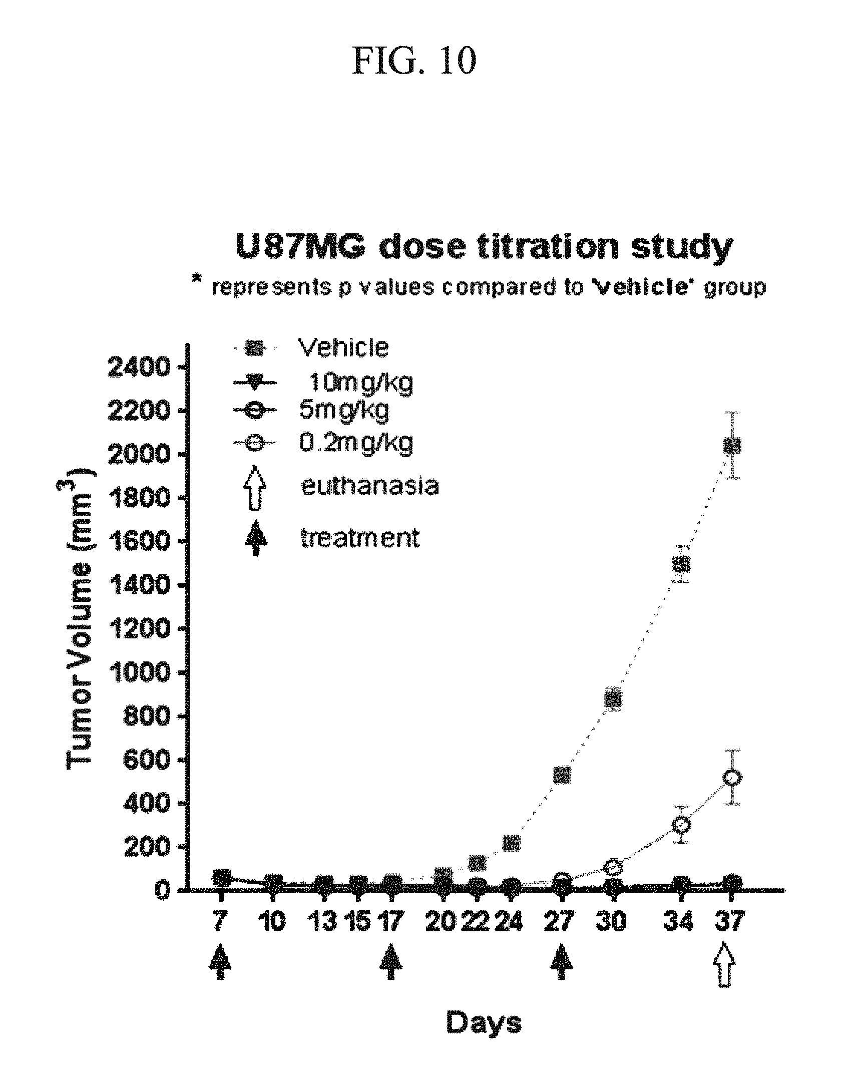
D00015

D00016
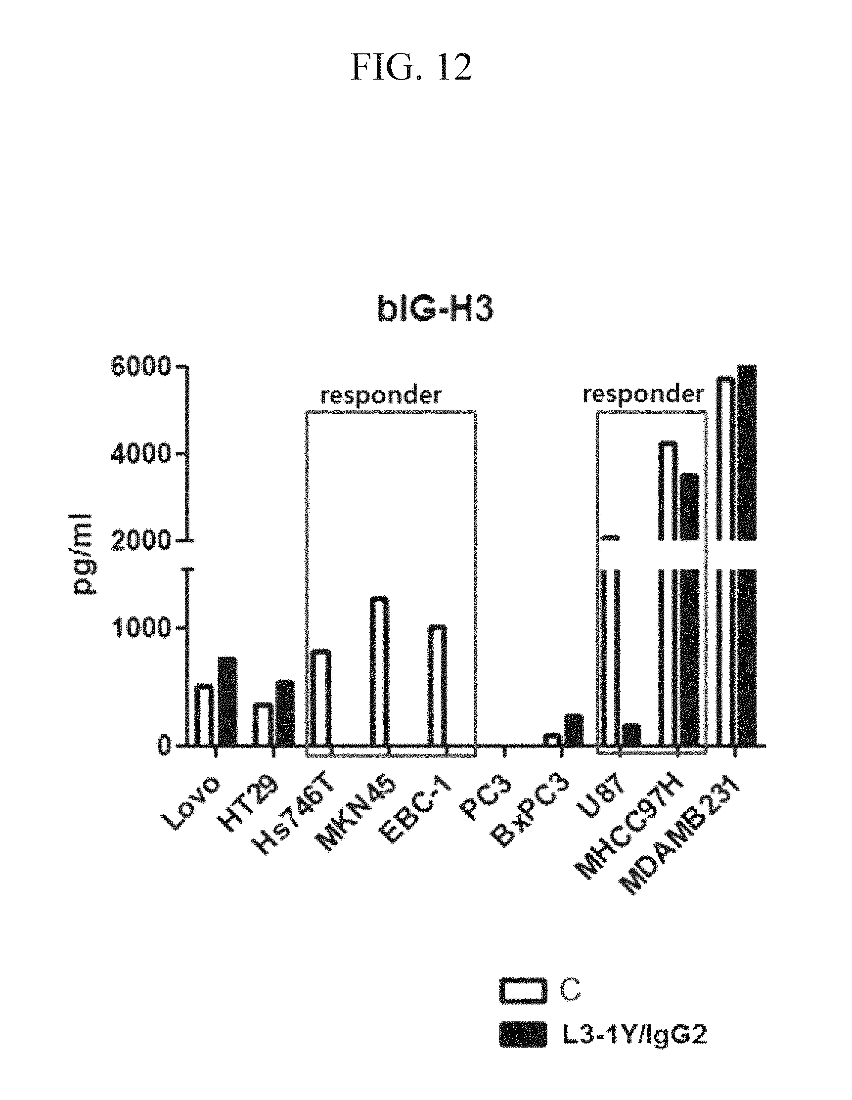
D00017
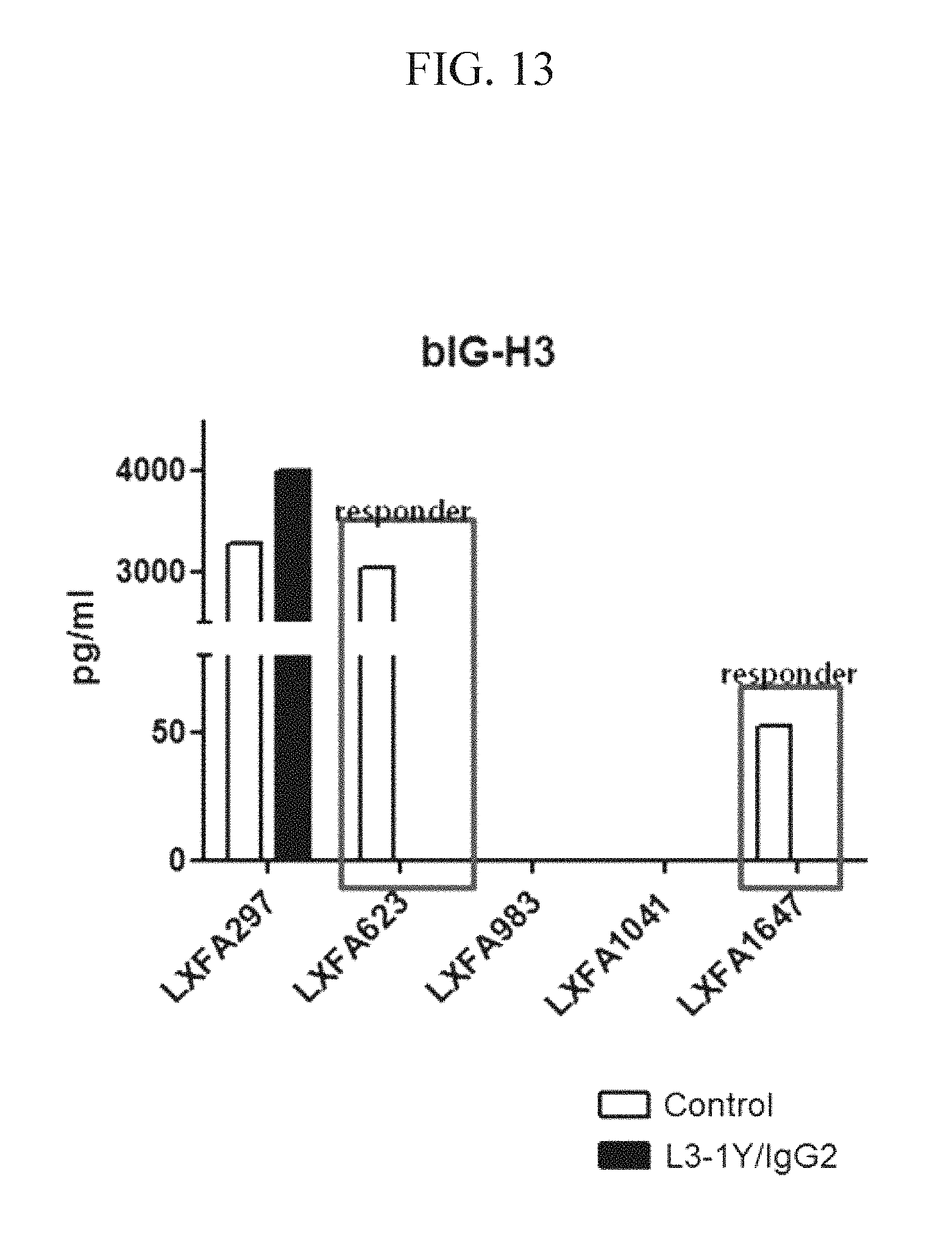
D00018
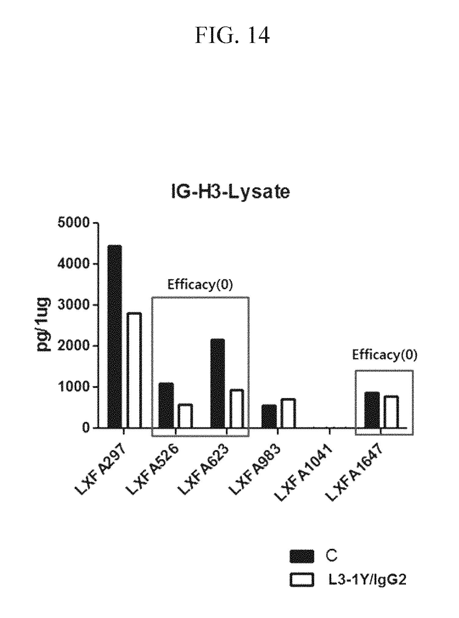
D00019

D00020

D00021

D00022

D00023

D00024
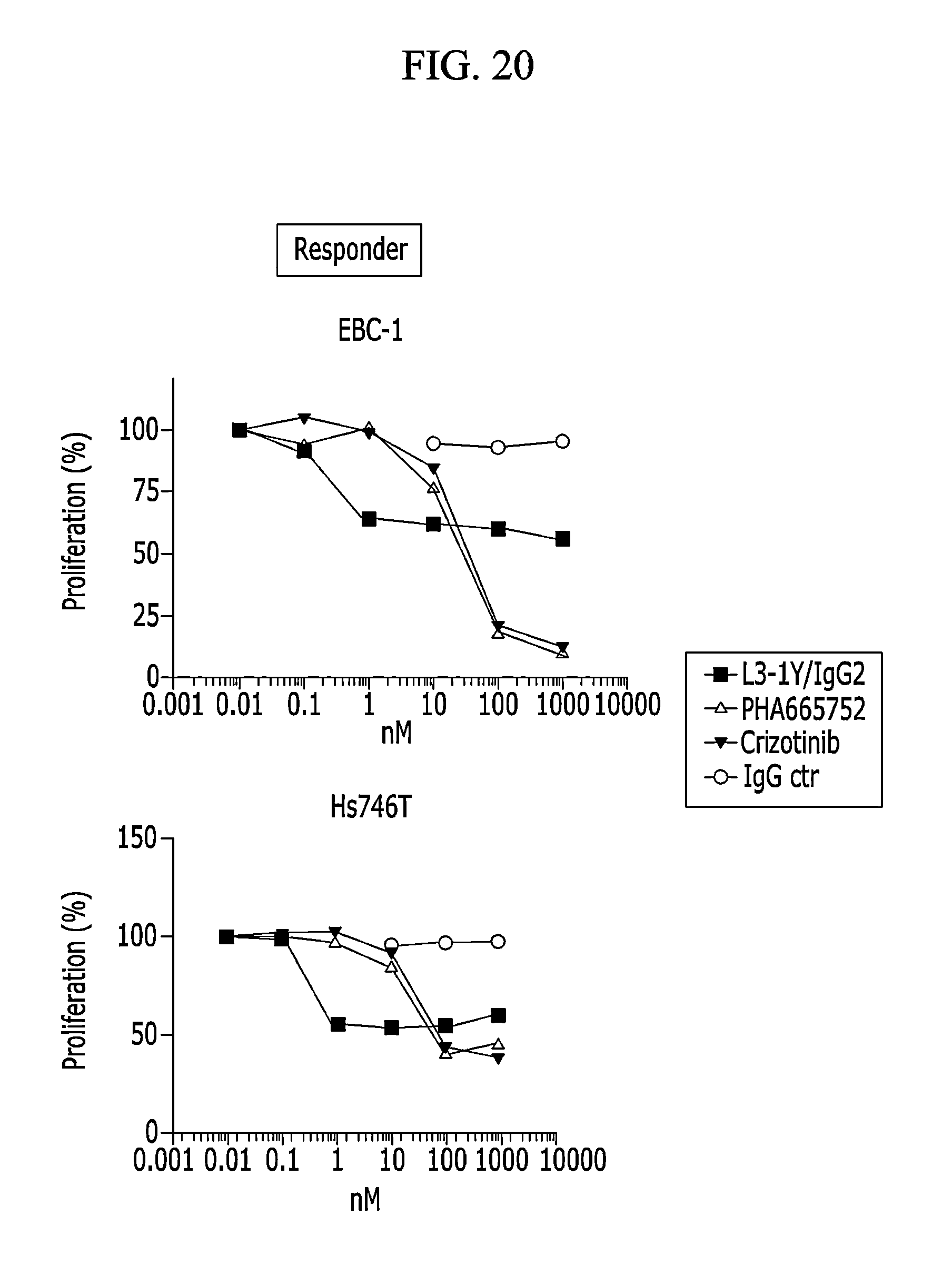
D00025
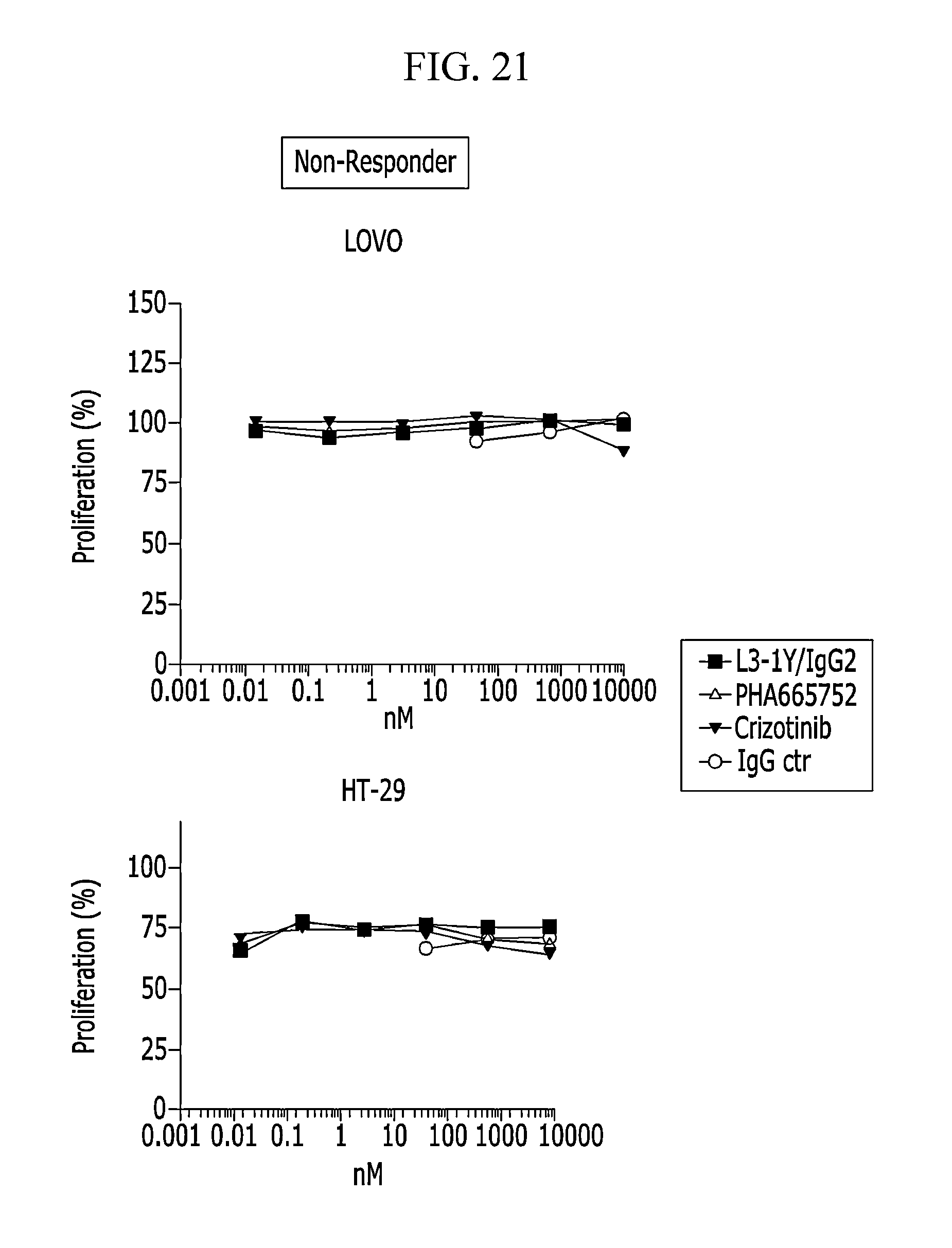
D00026
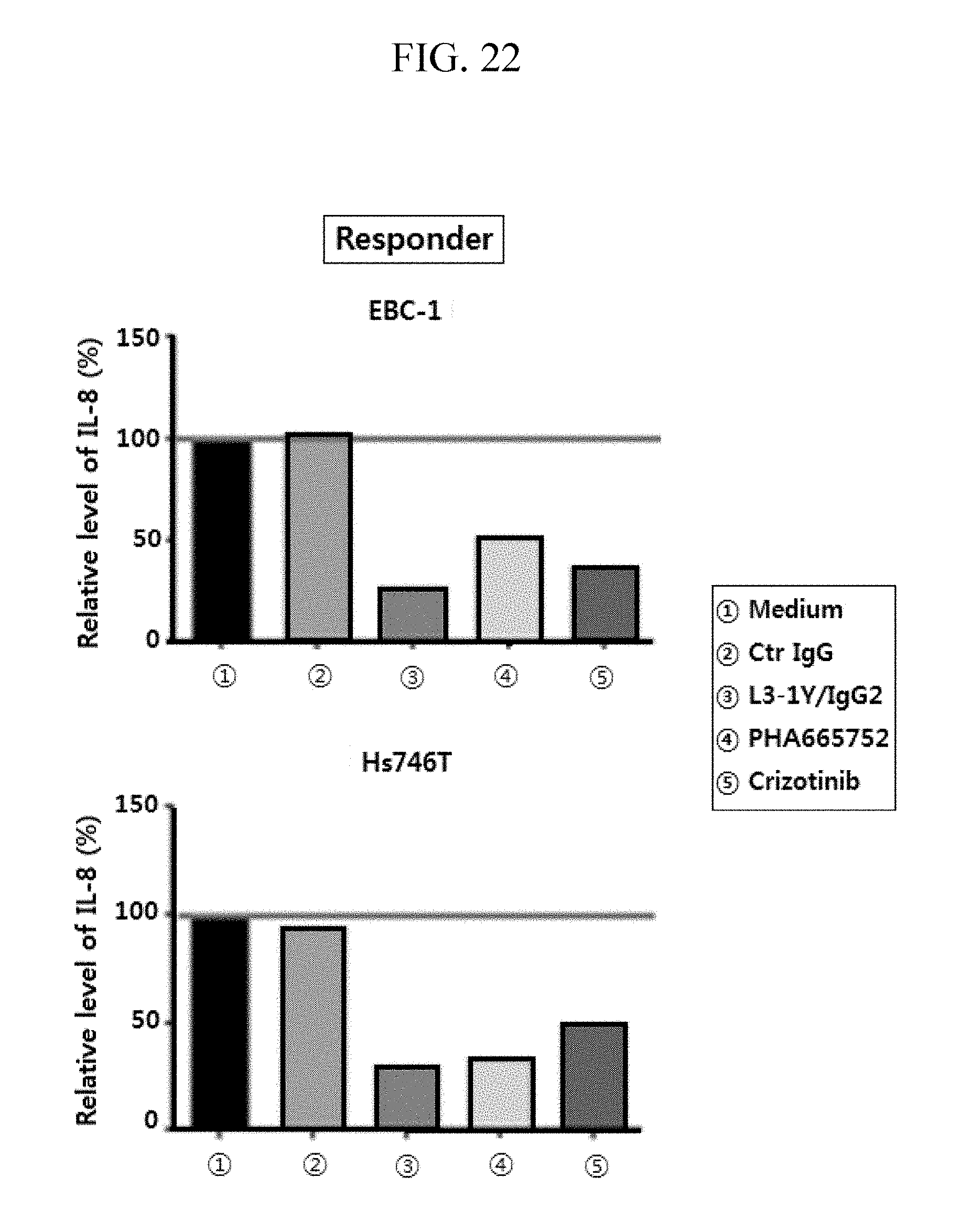
D00027

S00001
XML
uspto.report is an independent third-party trademark research tool that is not affiliated, endorsed, or sponsored by the United States Patent and Trademark Office (USPTO) or any other governmental organization. The information provided by uspto.report is based on publicly available data at the time of writing and is intended for informational purposes only.
While we strive to provide accurate and up-to-date information, we do not guarantee the accuracy, completeness, reliability, or suitability of the information displayed on this site. The use of this site is at your own risk. Any reliance you place on such information is therefore strictly at your own risk.
All official trademark data, including owner information, should be verified by visiting the official USPTO website at www.uspto.gov. This site is not intended to replace professional legal advice and should not be used as a substitute for consulting with a legal professional who is knowledgeable about trademark law.