Methods of making nucleic acid molecules encoding modified chimeric polypeptides with improved pharmacokinetic properties
Papadopoulos , et al. A
U.S. patent number 10,392,430 [Application Number 15/624,845] was granted by the patent office on 2019-08-27 for methods of making nucleic acid molecules encoding modified chimeric polypeptides with improved pharmacokinetic properties. This patent grant is currently assigned to REGENERON PHARMACEUTICALS, INC.. The grantee listed for this patent is Regeneron Pharmaceuticals, Inc.. Invention is credited to Samuel Davis, Nicholas J. Papadopoulos, George D. Yancopoulos.


View All Diagrams
| United States Patent | 10,392,430 |
| Papadopoulos , et al. | August 27, 2019 |
Methods of making nucleic acid molecules encoding modified chimeric polypeptides with improved pharmacokinetic properties
Abstract
The present invention provides VEGF antagonists with improved pharmacokinetic properties. According to certain embodiments, a fusion polypeptide capable of antagonizing VEGF activity is provided comprising a modified extracellular ligand binding domain of a VEGF receptor fused to a multimerizing component.
| Inventors: | Papadopoulos; Nicholas J. (LaGrangeville, NY), Davis; Samuel (New York, NY), Yancopoulos; George D. (Yorktown Heights, NY) | ||||||||||
|---|---|---|---|---|---|---|---|---|---|---|---|
| Applicant: |
|
||||||||||
| Assignee: | REGENERON PHARMACEUTICALS, INC.
(Tarrytown, NY) |
||||||||||
| Family ID: | 30447765 | ||||||||||
| Appl. No.: | 15/624,845 | ||||||||||
| Filed: | June 16, 2017 |
Prior Publication Data
| Document Identifier | Publication Date | |
|---|---|---|
| US 20180051066 A1 | Feb 22, 2018 | |
Related U.S. Patent Documents
| Application Number | Filing Date | Patent Number | Issue Date | ||
|---|---|---|---|---|---|
| 14822929 | Aug 11, 2015 | 9708386 | |||
| 14109999 | Sep 22, 2015 | 9139644 | |||
| 13439889 | Feb 11, 2014 | 8647842 | |||
| 12885185 | Sep 17, 2010 | ||||
| 12715128 | Dec 27, 2011 | 8084234 | |||
| 12102681 | Apr 27, 2010 | 7704500 | |||
| 11016097 | May 20, 2008 | 7374757 | |||
| 10009852 | Jul 4, 2006 | 7070959 | |||
| PCT/US00/14142 | May 23, 2000 | ||||
| 60138133 | Jun 8, 1999 | ||||
| Current U.S. Class: | 1/1 |
| Current CPC Class: | C12N 15/10 (20130101); C07K 19/00 (20130101); C12N 15/09 (20130101); C07K 14/71 (20130101); A61P 43/00 (20180101); C07K 16/22 (20130101); C07H 21/04 (20130101); C07K 2319/30 (20130101); A61K 38/00 (20130101); C07K 2319/00 (20130101) |
| Current International Class: | C07H 21/04 (20060101); C12N 15/09 (20060101); C12N 15/10 (20060101); C07K 14/71 (20060101); C07K 19/00 (20060101); C07K 16/22 (20060101); A61K 38/00 (20060101) |
References Cited [Referenced By]
U.S. Patent Documents
| 5712380 | January 1998 | Kendall et al. |
| 6011003 | January 2000 | Charnonk-Jones et al. |
| 6100071 | August 2000 | Davis-Smyth et al. |
| 6833349 | December 2004 | Xia et al. |
| 6987294 | January 2006 | Sasada et al. |
| 7070959 | July 2006 | Papadopoulos et al. |
| 7087411 | August 2006 | Daly et al. |
| 7279159 | October 2007 | Daly et al. |
| 7303746 | December 2007 | Wiegand et al. |
| 7303747 | December 2007 | Wiegand et al. |
| 7306799 | December 2007 | Wiegand et al. |
| 7374757 | May 2008 | Papadopoulos et al. |
| 7374758 | May 2008 | Papadopoulos et al. |
| 7396664 | July 2008 | Daly et al. |
| 7399612 | July 2008 | Daly et al. |
| 7521049 | April 2009 | Wiegand et al. |
| 7524499 | April 2009 | Papadopoulos et al. |
| 7635474 | December 2009 | Daly et al. |
| 7704500 | April 2010 | Papadopoulos et al. |
| 2004/0265309 | December 2004 | Kendall et al. |
| 2005/0175610 | August 2005 | Wiegand et al. |
| 2005/0260203 | November 2005 | Wiegand et al. |
| 2005/0281822 | December 2005 | Cedarbaum et al. |
| 2005/0281831 | December 2005 | Davis-Smyth et al. |
| 2006/0210566 | September 2006 | Holash et al. |
| 2007/0037748 | February 2007 | Stahl et al. |
| 2009/0081217 | March 2009 | Papadopoulos et al. |
| 2009/0155899 | June 2009 | Papadopoulos et al. |
| 2009/0234103 | September 2009 | Davis-Smyth et al. |
| 2010/0087632 | April 2010 | Daly et al. |
| 2010/0221782 | September 2010 | Papadopoulos et al. |
| WO 94/21679 | Sep 1994 | WO | |||
| WO 97/44453 | Nov 1997 | WO | |||
| WO 98/13071 | Apr 1998 | WO | |||
| WO 99/03996 | Jan 1999 | WO | |||
| WO 00/75319 | Dec 2000 | WO | |||
Other References
|
Chung et al. (2008), Treatment of malignant ascites, Current Treatment Options in Oncology 9:215-231. cited by applicant . Wells et al. (1990), Additivity of mutational effects in proteins, Biochemistry 29 (37):8509-8517. cited by applicant . Ngo et al. (1994), Computational complexity, protein structure prediction, and the Levinthal paradox. In Merz and Le Grand (Eds.) Birkhauser; Boston, pp. 491-495. cited by applicant . Herley et al, (1999), Characterization of the VEGF binding site on the Flt-1 receptor, Biochem Biophys Res Commun 262:731-738. cited by applicant . Terman et al. (1991), Identification of a new endothelial cell growth factor receptor tyrosine kinase, Oncogene 6:1677-1683. cited by applicant . Hileman et al, (1998),Glycosaminoglycan-protein interactions; definitions of consensus sites in glycosaminoglycan binding proteins, BioEssays 20:156-167. cited by applicant . Devries et al. (1992), The fms-like tyrosine kinase, a receptor for vascular endothelial growth factor, Science 225:989-991. cited by applicant . Sharifi et al. (1998), Improving monoclonal antibody pharmacokinetics via chemical modification, Quart. J. Nucl. Med. 42:242-249. cited by applicant . Jensen-Pippo at al. (1996), Enteral bioavailability of human granulocyte colony stimulating factor conjugated with poly(ethylene glycol), Pharm. Res. 13(1):102-107. cited by applicant . Tanaka et al. (1997), Characterization of the extracellular domain in vascular endothelial growth factor receptor-1 (Flt-1 tyrosine kinase). Jpn. J. Cancer Res. 88:867-876. cited by applicant . Yang et al. (1995), The use of polyethylene glycol-modified interleukin-2 (PEG-IL-2) in the treatment of patients with metastatic renal cell carcinoma . . . Cancer 76:687-694. cited by applicant . Davis-Smyth et al. (1996), The second immunoglobulin-like domain of the VEGF tyrosine kinase receptor Flt-1 determines ligand binding and may initiate . . . EMBO J. 15:4919-4927. cited by applicant . Terman et al, (1992), Identification of the KDR tyrosine kinase as a receptor for vascular endothelial cell growth factor, Biochem Biophys Res Commun 187:1579-1586. cited by applicant . Tsutsumi et al, (1997), PEGylation of interleukin-6 effectively increases its thrombopoietic potency, Thrombosis and Haemostasis 77(1):168-173. cited by applicant . Dunca and Spreafico (1994), Polymer conjugates, Drug Delivery Systems 27(4):290-306. cited by applicant . Kendall et al. (1993), Inhibition of vascular endothelial cell growth factor activity by an endogenously encoded soluble receptor, Proc. Natl. Acad. Sci. USA 90:10705-10709. cited by applicant . Kendall et al. (1996), Identification of a natural soluble form of the vascular endothelial growth factor receptor, FLT-1, and its . . . Biochem Biophys Res Commun 226:324-328. cited by applicant . Autiero et al. (2003), Role of PIGF in the infra- and intermolecular cross talk between the VEGF receptors Flt1 and Flk1, Nature Medicine 9:936-943. cited by applicant . Declaration of Dr. Sarah Hymowitz submitted to the European Patent Office on Oct. 7, 2009, by Genentech, Inc. during prosecution of European Patent Appl. No. 05023819.5. cited by applicant . Mahdadevan et al. (1995), Structural role of extracellular domain 1 of alpha-platelet-derived growth factor (PDGF) receptor for PDGF-AA and . . . J. Biol. Chem. 270:27595-27600. cited by applicant . Tanaka et al., (1995), Characterization of the ligand binding domain of FLT-1, . . . The 8th Annual Meeting of Japanese Molecular Biology, Nov. 21, 1995, Abstract 2P-227. cited by applicant . Keyt et al. (1996), Identification of vascular endothelial growth factor determinants for binding KDR and FLT-1 receptors; generation of . . . J. Biol Chem. 271:5638-5646. cited by applicant . Heidaran et al. (1990). Chimeric alpha- and beta-platelet derived growth factor (PDGF) receptors define three immunoglobulin-like domains . . . J. Biol. Chem. 265:18741-18744. cited by applicant . Park et al. (1994), Placenta growth factor; potentiation of vascular endothelial growth factor bioactivity, in vitro and in vivo, and high . . . J. Biol. Chem. 269:25646-25654. cited by applicant . Shibuya et al. (1990), Nucleotide sequence and expression of a novel human receptor-type tyrosine kinase gene (flt) closely related to the fms family, Oncogene 5:519-524. cited by applicant . Heidaran et al, (1995), Beta-PDGF-IgG chimera demonstrates that human beta-PDGFR Ig-like domains 1 to 3 are sufficient for high affinity PDGF BB . . . FASEB Journal 9:140-145. cited by applicant . Yu et al. (1994), Structural coincidence of alpha-PDGFR epitopes binding to platelet-derived growth factor-AA and a potent neutralizing . . . J. Biol. Chem. 269:10668-10674. cited by applicant . Yu et al. (1995), Differential requirement of a motif within the carboxyl-terminal domain of alpha-platelet-derived growth factor . . . J. Biol. Chem. 270:7033-7036. cited by applicant . USPTO Office Action dated Mar. 25, 2005, in U.S. Appl. No. 10/009,852. cited by applicant . USPTO Office Action dated Jul. 16, 2007, in U.S. Appl. No. 11/089,803. cited by applicant . USPTO Office Action dated Oct. 17, 2007, in U.S. Appl. No. 11/016,503. cited by applicant . Applicant Amendment and Remarks filed on May 16, 2005, in U.S. Appl. No. 10/009,852. cited by applicant . USPTO Office Action dated Sep. 7, 2005, in U.S. Appl. No. 10/009,852. cited by applicant . Applicant Amendment and Remarks filed on Sep. 20, 2005, in U.S. Appl. No, 10/009,852. cited by applicant . USPTO Notice of Allowance dated Nov. 30, 2009, in U.S. Appl. No. 12/102,681. cited by applicant . Preliminary Amendment filed on Oct. 6, 2008, in U.S. Appl. No. 12/104,894. cited by applicant . Claims filed at the European Patent Office in EP Appl. No. 05023819.5 on Oct. 9, 2009, on behalf of Applicant Genentech, Inc. cited by applicant . Communication pursuant to Article 94(3) EPC issued by the European Patent Office on Apr. 1, 2009, in EP Appl. No. 05023819.5. cited by applicant . Summons to attend Oral Proceedings issued by the European Patent Office on Nov. 17, 2010, in EP Appl. No. 05023819.5. cited by applicant. |
Primary Examiner: Saoud; Christine J
Assistant Examiner: Lockard; Jon M
Attorney, Agent or Firm: Cottingham; Frank R.
Parent Case Text
CROSS-REFERENCE TO RELATED APPLICATIONS
This application is a continuation of U.S. application Ser. No. 14/822,929, filed on Aug. 11, 2015, which is a continuation of U.S. application Ser. No. 14/109,999, filed on Dec. 18, 2013, now U.S. Pat. No. 9,139,644, which is a continuation of U.S. application Ser. No. 13/439,889, filed on Apr. 5, 2012, now U.S. Pat. No. 8,647,842, which is a continuation of U.S. application Ser. No. 12/885,185, filed on Sep. 17, 2010, which is a continuation of U.S. application Ser. No. 12/715,128, filed on Mar. 1, 2010, now U.S. Pat. No. 8,084,234, which is a continuation of U.S. application Ser. No. 12/102,681 filed on Apr. 14, 2008, now U.S. Pat. No. 7,704,500, which is a divisional of U.S. application Ser. No. 11/016,097, filed on Dec. 17, 2004, now U.S. Pat. No. 7,374,757, which is a divisional of U.S. application Ser. No. 10/009,852, having a 371(c) date of Dec. 6, 2001, now U.S. Pat. No. 7,070,959, which is a national stage application of International Application No. PCT/US00/14142, filed on May 23, 2000, which claims the benefit of U.S. Provisional Appl. No. 60/138,133, filed on Jun. 8, 1999. The disclosures of these publications are hereby incorporated by reference into this application in their entireties.
Claims
What is claimed is:
1. A method for making a nucleic acid molecule encoding a chimeric VEGF-binding fusion protein with improved pharmacokinetic properties, the method comprising: modifying a nucleic acid molecule encoding immunoglobulin ("Ig") domains 1, 2 and 3 of Flt1 fused to an Fc domain ("Flt1(1-3)-Fc"), wherein said modifying comprises: (a) deleting the nucleotide sequence encoding the first Ig domain of Flt1; and (b) replacing the nucleotide sequence encoding the third Ig domain of Flt1 with the nucleotide sequence encoding the third Ig domain of Flk1 or Flt4; thereby making a nucleic acid molecule encoding a chimeric VEGF-binding fusion protein with reduced binding to extracellular matrix (ECM) and improved pharmacokinetics (PK) as compared to Flt1(1-3)-Fc.
2. The method of claim 1, wherein the nucleic acid molecule encoding a chimeric VEGF-binding fusion protein comprises: (i) the nucleotide sequence of SEQ ID NO:11 encoding Flt1D2.FlkD3.Fc.DELTA.C1(a); (ii) the nucleotide sequence of SEQ ID NO:13 encoding Flt1D2.VEGFR3D3.Fc.DELTA.C1(a); or (iii) the nucleic acid sequence of SEQ ID NO:15 encoding VEGFR1R2-Fc.DELTA.C1(a).
Description
FIELD OF THE INVENTION
The field of this invention is modified polypeptides with improved pharmacokinetics. Specifically, the field of this invention relates to Flt1 receptor polypeptides that have been modified in such a way as to improve their pharmacokinetic profile. The field of this invention also relates to methods of making and using the modified polypeptides including but not limited to using the modified polypeptides to decrease or inhibit plasma leakage and/or vascular permeability in a mammal.
BACKGROUND
The ability of polypeptide ligands to bind to cells and thereby elicit a phenotypic response such as cell growth, survival, cell product secretion, or differentiation is often mediated through transmembrane receptors on the cells. The extracellular domain of such receptors (i.e. that portion of the receptor that is displayed on the surface of the cell) is generally the most distinctive portion of the molecule, as it provides the protein with its ligand binding characteristic. Binding of a ligand to the extracellular domain generally results in signal transduction which transmits a biological signal to intracellular targets. Often, this signal transduction acts via a catalytic intracellular domain. The particular array of sequence motifs of this catalytic intracellular domain determines its access to potential kinase substrates (Mohammadi, et al., 1990, Mol. Cell. Biol. 11:5068-5078; Fantl, et al., 1992, Cell 69:413-413). Examples of receptors that transduce signals via catalytic intracellular domains include the receptor tyrosine kinases (RTKs) such as the Trk family of receptors which are generally limited to cells of the nervous system, the cytokine family of receptors including the tripartate CNTF receptor complex (Stahl & Yancopoulos, 1994, J. Neurobio. 25:1454-1466) which is also generally limited to the cells of the nervous system, G-protein coupled receptors such as the .beta..sub.2-adrenergic receptor found on, for instance, cardiac muscle cells, and the multimeric IgE high affinity receptor Fc.epsilon.RI which is localized, for the most part, on mast cells and basophils (Sutton & Gould, 1993, Nature 366:421-428).
All receptors identified so far appear to undergo dimerization, multimerization, or some related conformational change following ligand binding (Schlessinger, J., 1988, Trend Biochem. Sci. 13:443-447; Ullrich & Schlessinger, 1990, Cell 61:203-212; Schlessinger & Ullrich, 1992, Neuron 9:383-391) and molecular interactions between dimerizing intracellular domains lead to activation of catalytic function. In some instances, such as platelet-derived growth factor (PDGF), the ligand is a dimer that binds two receptor molecules (Hart, et al., 1988, Science, 240:1529-1531; Heldin, 1989, J. Biol. Chem. 264:8905-8912) while, for example, in the case of epidermal growth factor (EGF), the ligand is a monomer (Weber, et al., 1984, J. Biol. Chem. 259:14631-14636). In the case of the Fc.epsilon.RI receptor, the ligand, IgE, exists bound to Fc.epsilon.RI in a monomeric fashion and only becomes activated when antigen binds to the IgE/Fc.epsilon.RI complex and cross-links adjacent IgE molecules (Sutton & Gould, 1993, Nature 366:421-428).
Often, the tissue distribution of a particular receptor within higher organisms provides insight into the biological function of the receptor. The RTKs for some growth and differentiation factors, such as fibroblast growth factor (FGF), are widely expressed and therefore appear to play some general role in tissue growth and maintenance. Members of the Trk RTK family (Glass & Yancopoulos, 1993, Trends in Cell Biol. 3:262-268) of receptors are more generally limited to cells of the nervous system, and the Nerve Growth Factor family consisting of nerve growth factor (NGF), brain-derived neurotrophic factor (BDNF), neurotrophin-3 (NT-3) and neurotrophin-4/5 (NT-4/5), which bind the Trk RTK family receptors, promote the differentiation of diverse groups of neurons in the brain and periphery (Lindsay, R. M, 1993, in Neurotrophic Factors, S. E. Loughlin & J. H. Fallon, eds., pp. 257-284, San Diego, Calif., Academic Press). Fc.epsilon.RI is localized to a very limited number of types of cells such as mast cells and basophils. Mast cells derive from bone marrow pluripotent hematopoietic stem cell lineage, but complete their maturation in the tissue following migration from the blood stream (See Janeway & Travers, 1996, in Immunobiology, 2d. Edition, M. Robertson & E. Lawrence, eds., pp. 1:3-1:4, Current Biology Ltd., London, UK, Publisher) and are involved in the allergic response.
Many studies have demonstrated that the extracellular domain of a receptor provides the specific ligand binding characteristic. Furthermore, the cellular environment in which a receptor is expressed may influence the biological response exhibited upon binding of a ligand to the receptor. For example, when a neuronal cell expressing a Trk receptor is exposed to a neurotrophin which binds to that receptor, neuronal survival and differentiation results. When the same receptor is expressed by a fibroblast, exposure to the neurotrophin results in proliferation of the fibroblast (Glass, et al., 1991, Cell 66:405-413).
A class of cell-derived dimeric mitogens with selectivity for vascular endothelial cells has been identified and designated vascular endothelial cell growth factor (VEGF). VEGF has been purified from conditioned growth media of rat glioma cells (Conn et al., 1990, Proc. Natl. Acad. Sci. U.S.A., 87. pp 2628-2632); and conditioned growth media of bovine pituitary follicle stellate cells (Ferrara and Henzel, 1989, Biochem. Biophys. Res. Comm., 161, pp. 851-858; Gozpadorowicz et al., 1989, Proc. Natl. Acad. Sci. U.S.A., 86, pp. 7311-7315 and conditioned growth medium from human U937 cells (Connolly, D. T. et al. 1989, Science, 246, pp. 1309-1312). VEGF is a dimer with an apparent molecular mass of about 46 kDa with each subunit having an apparent molecular mass of about 23 kDa. VEGF has some structural similarities to platelet derived growth factor (PDGF), which is a mitogen for connective tissue cells but not mitogenic for vascular endothelial cells from large vessels.
The membrane-bound tyrosine kinase receptor, known as Flt, was shown to be a VEGF receptor (DeVries, C. et al., 1992, Science, 255, pp. 989-991). The Flt receptor specifically binds VEGF which induces mitogenesis. Another form of the VEGF receptor, designated KDR, is also known to bind VEGF and induce mitogenesis. The partial cDNA sequence and nearly full length protein sequence of KDR is known as well (Terman, B. I. et al., 1991 Oncogene 6, pp. 1677-1683; Terman, B. I. et al., 1992 Biochem. Biophys. Res. Comm. 187, pp. 1579-1586).
Persistent angiogenesis may cause or exacerbate certain diseases such as psoriasis, rheumatoid arthritis, hemangiomas, angiofibromas, diabetic retinopathy and neovascular glaucoma. An inhibitor of VEGF activity would be useful as a treatment for such diseases and other VEGF-induced pathological angiogenesis and vascular permeability conditions, such as tumor vascularization. The present invention relates to a VEGF inhibitor that is based on the VEGF receptor Flt1.
Plasma leakage, a key component of inflammation, occurs in a distinct subset of microvessels. In particular, in most organs plasma leakage occurs specifically in the venules. Unlike arterioles and capillaries, venules become leaky in response to numerous inflammatory mediators including histamine, bradykinin, and serotonin. One characteristic of inflammation is the plasma leakage that results from intercellular gaps that form in the endothelium of venules. Most experimental models of inflammation indicate that these intercellular gaps occur between the endothelial cells of postcapillary and collecting venules (Baluk, P., et al., Am. J. Pathol., 1998, 152:1463-76). It has been shown that certain lectins may be used to reveal features of focal sites of plasma leakage, endothelial gaps, and finger-like processes at endothelial cell borders in inflamed venules (Thurston, G., et al., Am. J. Physiol., 1996, 271: H2547-62). In particular, plant lectins have been used to visualize morphological changes at endothelial cell borders in inflamed venules of, for example, the rat trachea. Lectins, such as conconavalin A and ricin, that bind focally to inflamed venules reveal regions of the subendothelial vessel wall exposed by gaps that correspond to sites of plasma leakage (Thurston, G., et al., Am J Physiol., 1996, 271: H2547-62).
The properties of the microvessels are dynamic. Chronic inflammatory diseases, for example, are associated with microvascular remodeling, including angiogenesis and microvessel enlargement. Microvessels can also remodel by acquiring abnormal phenotypic properties. In a murine model of chronic airway inflammation, airway capillaries acquire properties of venules, including widened vessel diameter, increased immunoreactivity for von Willebrand factor, and increased immunoreactivity for P-selectin. In addition, these remodeled vessels leak in response to inflammatory mediators, whereas vessels in the same position in the airways of normal mice do not.
Certain substances have been shown to decrease or inhibit vascular permeability and/or plasma leakage. For example, mystixins are synthetic polypeptides that have been reported to inhibit plasma leakage without blocking endothelial gap formation (Baluk, P., et al., J. Pharmacol. Exp. Ther., 1998, 284: 693-9). Also, the beta 2-adrenergic receptor agonist formoterol reduces microvascular leakage by inhibiting endothelial gap formation (Baluk, P. and McDonald, D. M., Am. J. Physiol., 1994, 266:L461-8).
The angiopoietins and members of the vascular endothelial growth factor (VEGF) family are the only growth factors thought to be largely specific for vascular endothelial cells. Targeted gene inactivation studies in mice have shown that VEGF is necessary for the early stages of vascular development and that Ang-1 is required for later stages of vascular remodeling.
U.S. Pat. No. 6,011,003, issued Jan. 4, 2000, in the name of Metris Therapeutics Limited, discloses an altered, soluble form of FLT polypeptide being capable of binding to VEGF and thereby exerting an inhibitory effect thereon, the polypeptide comprising five or fewer complete immunoglobulin domains.
U.S. Pat. No. 5,712,380, issued Jan. 27, 1998 and assigned to Merck & Co., discloses vascular endothelial cell growth factor (VEGF) inhibitors that are naturally occurring or recombinantly engineered soluble forms with or without a C-terminal transmembrane region of the receptor for VEGF.
Also assigned to Merck & Co. is PCT Publication No. WO 98/13071, published Apr. 2, 1998, which discloses gene therapy methodology for inhibition of primary tumor growth and metastasis by gene transfer of a nucleotide sequence encoding a soluble receptor protein which binds to VEGF.
PCT Publication No. WO 97/44453, published Nov. 27, 1997, in the name of Genentech, Inc., discloses novel chimeric VEGF receptor proteins comprising amino acid sequences derived from the vascular endothelial growth factor (VEGF) receptors Flt1 and KDR, including the murine homologue to the human KDR receptor FLK1, wherein said chimeric VEGF receptor proteins bind to VEGF and antagonize the endothelial cell proliferative and angiogenic activity thereof.
PCT Publication No. WO 97/13787, published Apr. 17, 1997, in the name of Toa Gosei Co., LTD., discloses a low molecular weight VEGF inhibitor usable in the treatment of diseases accompanied by neovascularization such as solid tumors. A polypeptide containing the first immunoglobulin-like domain and the second immunoglobulin-like domain in the extracellular region of a VEGF receptor FLT but not containing the sixth immunoglobulin-like domain and the seventh immunoglobulin-like domain thereof shows a VEGF inhibitory activity.
Sharifi, J. et al., 1998, The Quarterly Jour. of Nucl. Med. 42:242-249, disclose that because monoclonal antibodies (MAbs) are basic, positively charged proteins, and mammalian cells are negatively charged, the electrostatic interactions between the two can create higher levels of background binding resulting in low tumor to normal organ ratios. To overcome this effect, the investigators attempted to improve MAb clearance by using various methods such as secondary agents as well as chemical and charge modifications of the MAb itself.
Jensen-Pippo, et al., 1996, Pharmaceutical Research 13:102-107, disclose that pegylation of a therapeutic protein, recombinant human granulocyte colony stimulating factor (PEG-G-CSF), results in an increase in stability and in retention of in vivo bioactivity when administered by the intraduodenal route.
Tsutsumi, et al., 1997, Thromb Haemost. 77:168-73, disclose experiments wherein the in vivo thrombopoietic activity of polyethylene glycol-modified interleukin-6 (MPEG-IL-6), in which 54% of the 14 lysine amino groups of IL-6 were coupled with PEG, was compared to that of native IL-6.
Yang, et al., 1995, Cancer 76:687-94, disclose that conjugation of polyethylene glycol to recombinant human interleukin-2 (IL-2) results in a compound, polyethylene glycol-modified IL-2 (PEG-IL-2) that retains the in vitro and in vivo activity of IL-2, but exhibits a markedly prolonged circulating half-life.
R. Duncan and F. Spreafico, Clin. Pharmacokinet., 27: 290-306, 296 (1994) review efforts to improve the plasma half-life of asparaginase by conjugating polyethylene glycol.
PCT International Publication No. WO 99/03996 published Jan. 28, 1999 in the name of Regeneron Pharmaceuticals, Inc. and The Regents of The University of California describes modified human noggin polypeptides having deletions of regions of basic amino acids. The modified human noggin polypeptides are described as retaining biological activity while having reduced affinity for heparin and superior pharmacokinetics in animal sera as compared to the unmodified human noggin.
SUMMARY OF THE INVENTION
The present invention is directed to VEGF antagonists with improved pharmacokinetic properties. A preferred embodiment is an isolated nucleic acid molecule encoding a fusion polypeptide capable of binding a VEGF polypeptide comprising (a) a nucleotide sequence encoding a VEGF receptor component operatively linked to (b) a nucleotide sequence encoding a multimerizing component, wherein the VEGF receptor component is the only VEGF receptor component of the fusion polypeptide and wherein the nucleotide sequence of (a) consists essentially of a nucleotide sequence encoding the amino acid sequence of Ig domain 2 of the extracellular domain of a first VEGF receptor and a nucleotide sequence encoding the amino acid sequence of Ig domain 3 of the extracellular domain of a second VEGF receptor.
In a further embodiment, the isolated nucleic acid of the first VEGF receptor is Flt1.
In a further embodiment, the isolated nucleic acid of the second VEGF receptor is Flk1.
In yet another embodiment, the isolated nucleic acid of the second VEGF receptor is Flt4.
In another preferred embodiment, the nucleotide sequence encoding Ig domain 2 of the extracellular domain of the first VEGF receptor is upstream of the nucleotide sequence encoding Ig domain 3 of the extracellular domain of the second VEGF receptor.
In still another preferred embodiment, the nucleotide sequence encoding Ig domain 2 of the extracellular domain of the first VEGF receptor is downstream of the nucleotide sequence encoding Ig domain 3 of the extracellular domain of the second VEGF receptor.
In a preferred embodiment of the invention, the multimerizing component comprises an immunoglobulin domain. In another embodiment, the immunoglobulin domain is selected from the group consisting of the Fc domain of IgG or the heavy chain of IgG.
Preferred embodiments include an isolated nucleic acid molecule comprising a nucleotide sequence encoding a modified Flt1 receptor fusion polypeptide, wherein the coding region of the nucleic acid molecule consists of a nucleotide sequence selected from the group consisting of (a) the nucleotide sequence set forth in FIG. 13A-D (SEQ ID NO:3); (b) the nucleotide sequence set forth in FIG. 14A-C (SEQ ID NO:5); (c) the nucleotide sequence set forth in FIG. 15A-C (SEQ ID NO:7); (d) the nucleotide sequence set forth in FIG. 16A-D (SEQ ID NO:9); (e) the nucleotide sequence set forth in FIGS. 21A-C (SEQ ID NO:11); (f) the nucleotide sequence set forth in FIG. 22A-C (SEQ ID NO:13); (g) the nucleotide sequence set forth in FIG. 24A-C; and (SEQ ID NO:15); and (h) a nucleotide sequence which, as a result of the degeneracy of the genetic code, differs from the nucleotide sequence of (a), (b), (c), (d), (e), (f), or (g) and which encodes a fusion polypeptide molecule having the biological activity of the modified Flt1 receptor fusion polypeptide.
In a further embodiment of the invention, a fusion polypeptide is encoded by the isolated nucleic acid molecules described above.
A preferred embodiment is a composition capable of binding a VEGF molecule to form a nonfunctional complex comprising a multimer of the fusion polypeptide.
Also preferred is a composition wherein the multimer is a dimer.
In yet another embodiment, the composition is in a carrier.
Another embodiment is a vector which comprises the nucleic acid molecules described above, including an expression vector comprising a the nucleic acid molecules described wherein the nucleic acid molecule is operatively linked to an expression control sequence.
Other included embodiments are a host-vector system for the production of a fusion polypeptide which comprises the expression vector, in a suitable host cell; the host-vector system wherein the suitable host cell is a bacterial cell, yeast cell, insect cell, or mammalian cell; the host-vector system wherein the suitable host cell is E. coli; the host-vector system wherein the suitable host cell is a COS cell; the host-vector system wherein the suitable host cell is a CHO cell.
Another embodiment of the invention is a method of producing a fusion polypeptide which comprises growing cells of the host-vector system under conditions permitting production of the fusion polypeptide and recovering the fusion polypeptide so produced.
Additional embodiments include a fusion polypeptide encoded by the nucleic acid sequence set forth in FIG. 10A-D (SEQ ID NO:1) or FIG. 24A-C (SEQ ID NO:15), which has been modified by acetylation or pegylation wherein the acetylation is accomplished with at least about a 100 fold molar excess of acetylation reagent or wherein acetylation is accomplished with a molar excess of acetylation reagent ranging from at least about a 10 fold molar excess to about a 100 fold molar excess or wherein the pegylation is 10K or 20K PEG.
A preferred embodiment includes a method of decreasing or inhibiting plasma leakage in a mammal comprising administering to the mammal the fusion polypeptide described above, including embodiments wherein the mammal is a human, the fusion polypeptide is acetylated or the fusion polypeptide is pegylated.
A preferred embodiment of the invention is a method of blocking blood vessel growth in a human comprising administering an effective amount of the fusion polypeptide described above.
Also preferred is a method of inhibiting VEGF receptor ligand activity in a mammal comprising administering to the mammal an effective amount of the fusion polypeptide described above. Preferred embodiments of these methods are wherein the mammal is a human.
Further embodiments of the methods of the invention include attenuation or prevention of tumor growth in a human; attenuation or prevention of edema in a human, especially wherein the edema is brain edema; attenuation or prevention of ascites formation in a human, especially wherein the ascites is ovarian cancer-associated ascites.
Preferred embodiments of the invention include a fusion polypeptide capable of binding a VEGF polypeptide comprising (a) a VEGF receptor component operatively linked to (b) a multimerizing component, wherein the VEGF receptor component is the only VEGF receptor component in the fusion polypeptide and consists essentially of the amino acid sequence of Ig domain 2 of the extracellular domain of a first VEGF receptor and the amino acid sequence of Ig domain 3 of the extracellular domain of a second VEGF receptor. In a further embodiment of the fusion polypeptide, the first VEGF receptor is Flt1. In yet a further embodiment of the fusion polypeptide, the second VEGF receptor is Flk1. Still another embodiment of the fusion polypeptide is one in which the second VEGF receptor is Flt4.
Preferred embodiments include a fusion polypeptide wherein amino acid sequence of Ig domain 2 of the extracellular domain of the first VEGF receptor is upstream of the amino acid sequence of Ig domain 3 of the extracellular domain of the second VEGF receptor and a fusion polypeptide wherein the amino acid sequence of Ig domain 2 of the extracellular domain of the first VEGF receptor is downstream of the amino acid sequence of Ig domain 3 of the extracellular domain of the second VEGF receptor.
In yet another embodiment, the fusion polypeptide multimerizing component comprises an immunoglobulin domain including an embodiment wherein the immunoglobulin domain is selected from the group consisting of the Fc domain of IgG or the heavy chain of IgG.
Preferred embodiments include a fusion polypeptide comprising an amino acid sequence of a modified Flt1 receptor, wherein the amino acid sequence selected from the group consisting of (a) the amino acid sequence set forth in FIG. 13A-D (SEQ ID NO:4); (b) the amino acid sequence set forth in FIG. 14A-C (SEQ ID NO:6); (c) the amino acid sequence set forth in FIG. 15A-C (SEQ ID NO:8); (d) the amino acid sequence set forth in FIG. 16A-D (SEQ ID NO:10); (e) the amino acid sequence set forth in FIGS. 21A-C (SEQ ID NO:12); (f) the amino acid sequence set forth in FIG. 22A-C (SEQ ID NO:14); and (g) the amino acid sequence set forth in FIG. 24A-C (SEQ ID NO:16).
BRIEF DESCRIPTION OF THE FIGURES
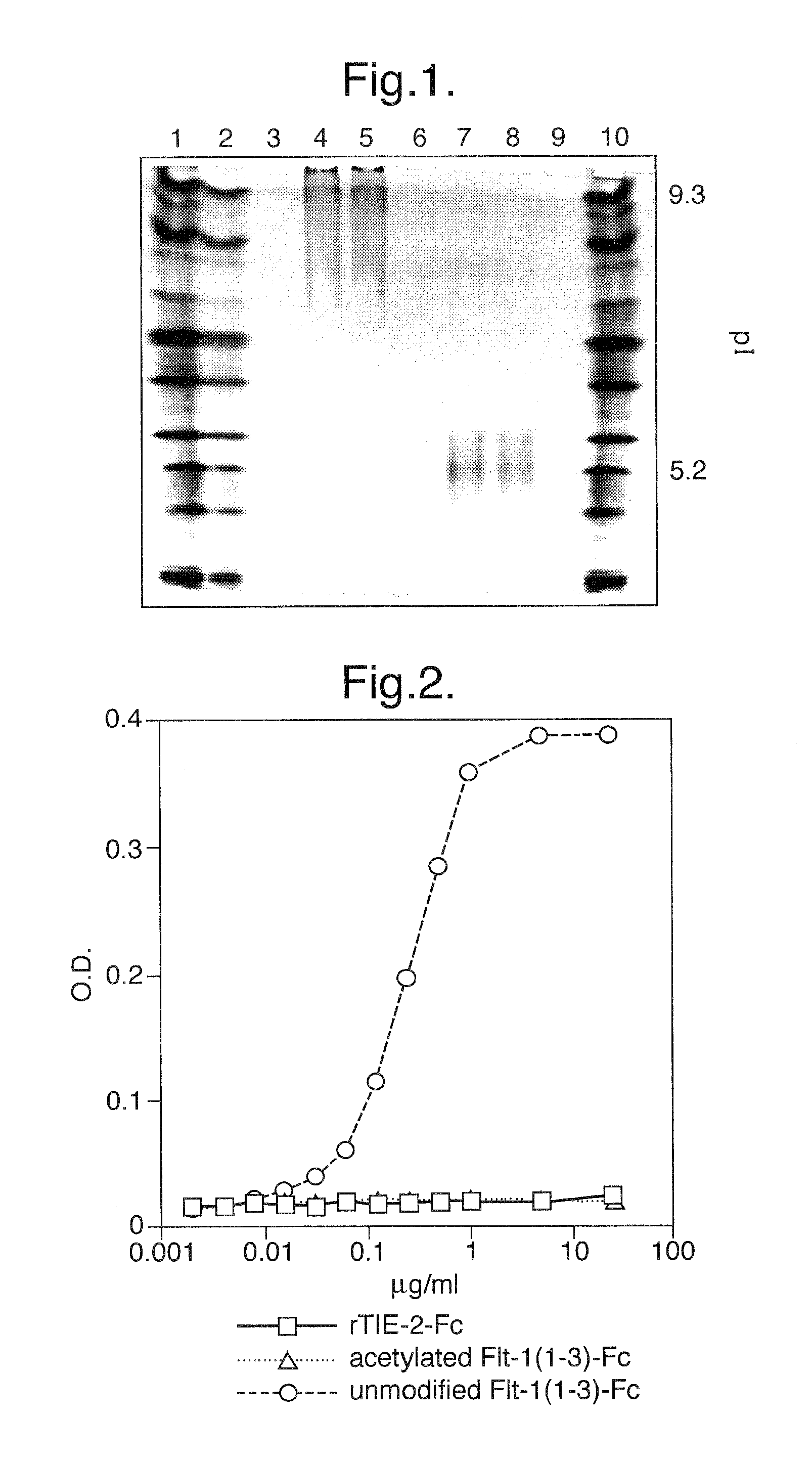
FIG. 1. IEF gel analysis of unmodified and acetylated Flt1(1-3)-Fc proteins. Unmodified Flt1(1-3)-Fc protein is unable to enter the gel due to its >9.3 pl, whereas acetylated Flt1(1-3)-Fc is able to enter the gel and equilibrate at pl 5.2.
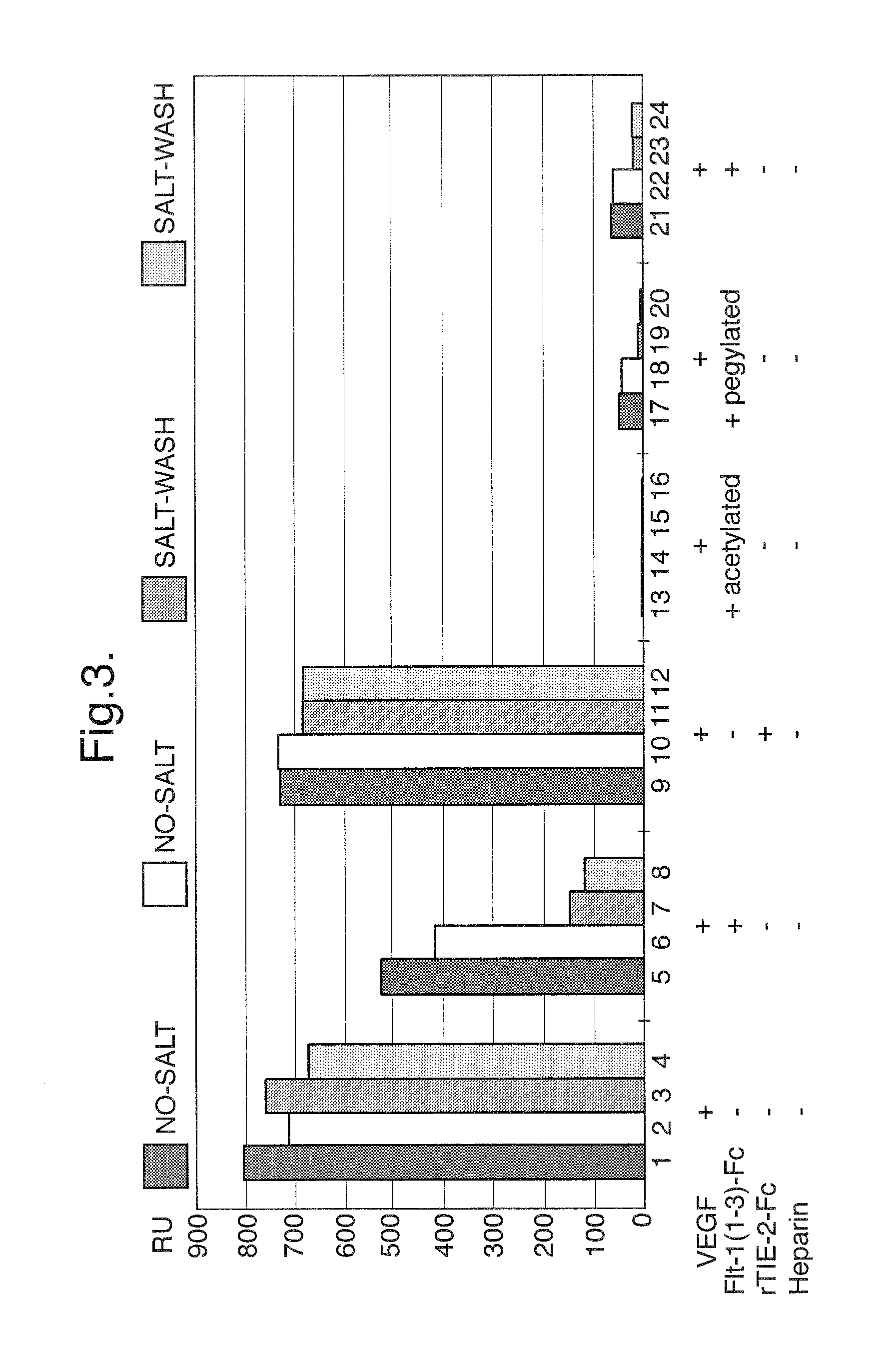
FIG. 2. Binding of unmodified Flt1(1-3)-Fc and acetylated Flt1(1-3)-Fc proteins to MATRIGEL.RTM. coated plates. Unmodified Flt1(1-3)-Fc proteins binds extensive to extracellular matrix components in MATRIGEL.RTM., whereas acetylated Flt1(1-3)-Fc does not bind.
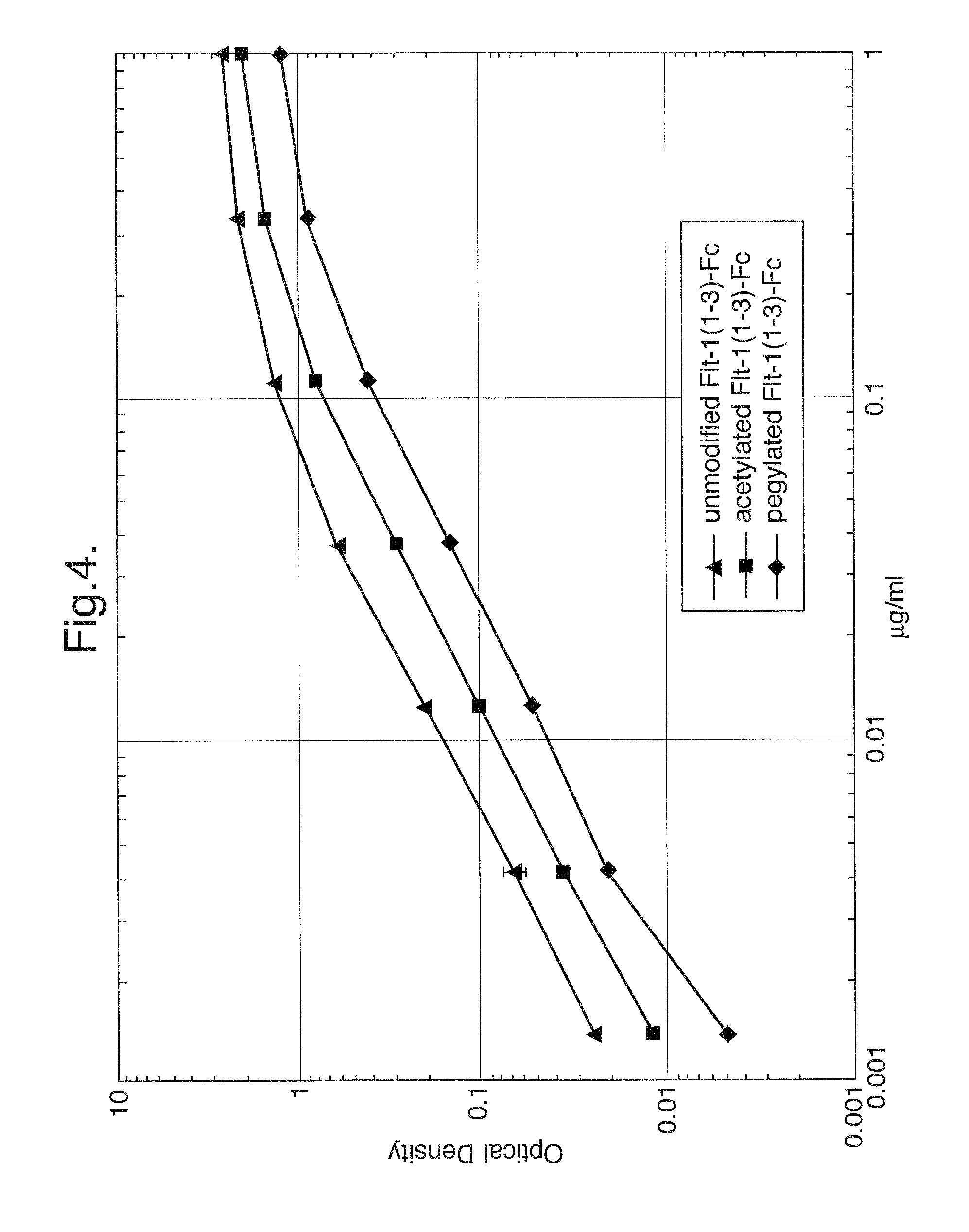
FIG. 3. Binding of unmodified Flt1(1-3)-Fc, acetylated Flt1(1-3)-Fc, and pegylated Flt1(1-3)-Fc in a BIACORE.TM.-based assay
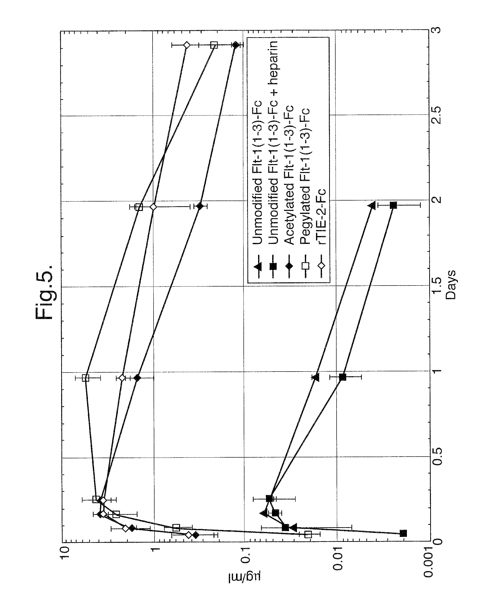
FIG. 4. Binding of unmodified Flt1(1-3)-Fc, acetylated Flt1(1-3)-Fc, and pegylated Flt1(1-3)-Fc to VEGF in an ELISA-based assay. Both pegylated and acetylated Flt1(1-3)-Fc proteins bind to VEGF with affinities approaching that of unmodified Flt1(1-3)-Fc.
FIG. 5. Pharmacokinetic profiles of unmodified Flt1(1-3)-Fc, acetylated Flt1(1-3)-Fc, and pegylated Flt1(1-3)-Fc.
FIG. 6A-B. IEF gel analysis of unmodified and step-acetylated Flt1(1-3)-Fc proteins. Unmodified Flt1(1-3)-Fc protein is unable to enter the gel due to its >9.3 pl, whereas most of the step-acetylated Flt1(1-3)-Fc samples (30-100 fold excess samples) were able to migrate into the gel and equilibrate at pls ranging between 4.55-8.43, depending on the degree of acetylation.
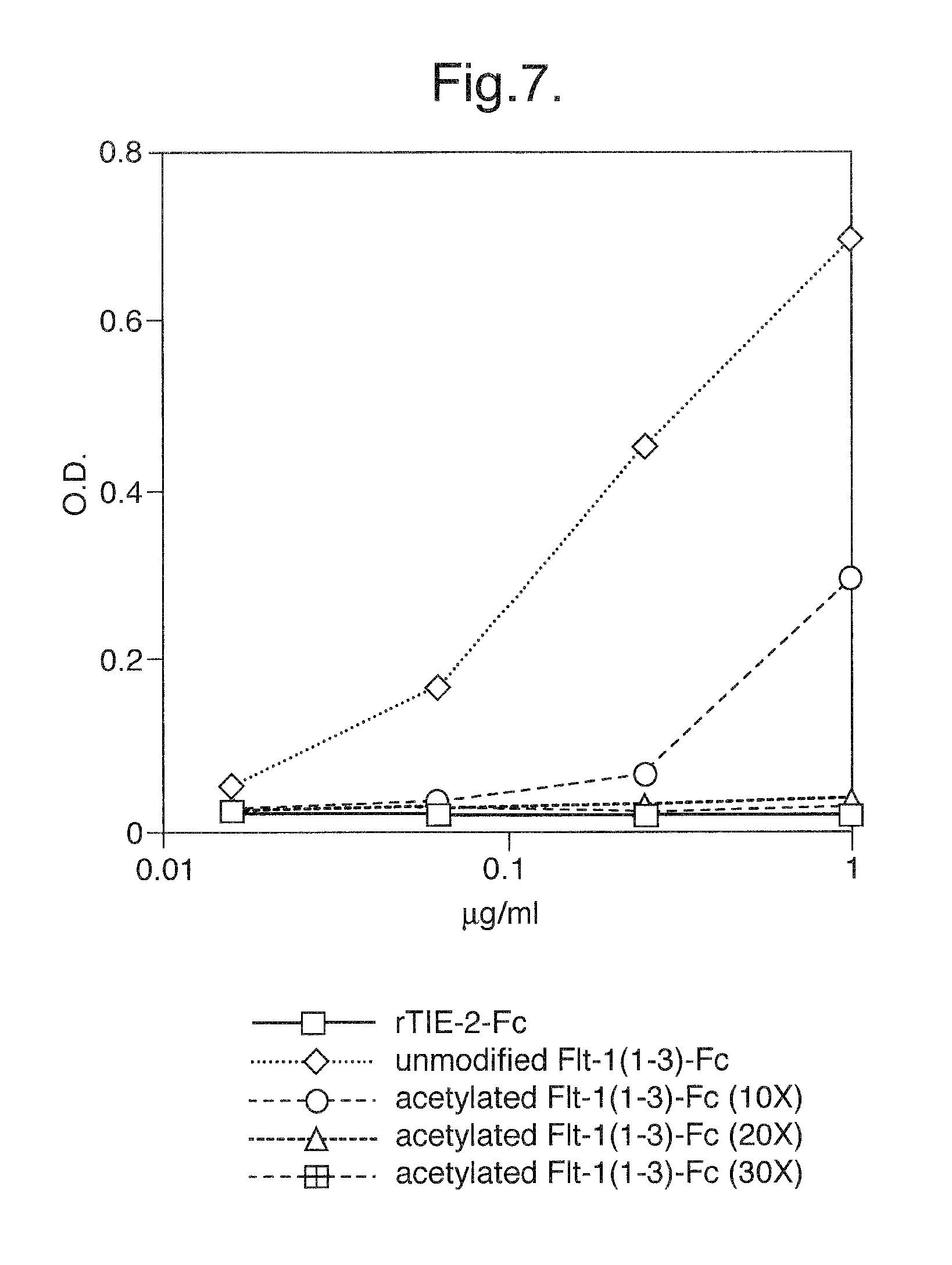
FIG. 7. Binding of unmodified Flt1(1-3)-Fc and step-acetylated Flt1(1-3)-Fc proteins to MATRIGEL.RTM. coated plates.
FIG. 8. Binding of unmodified Flt1(1-3)-Fc and step-acetylated Flt1(1-3)-Fc in a BIACORE.TM.-based assay.
FIG. 9. Pharmacokinetic profiles of unmodified Flt1(1-3)-Fc and step-acetylated Flt1(1-3)-Fc.
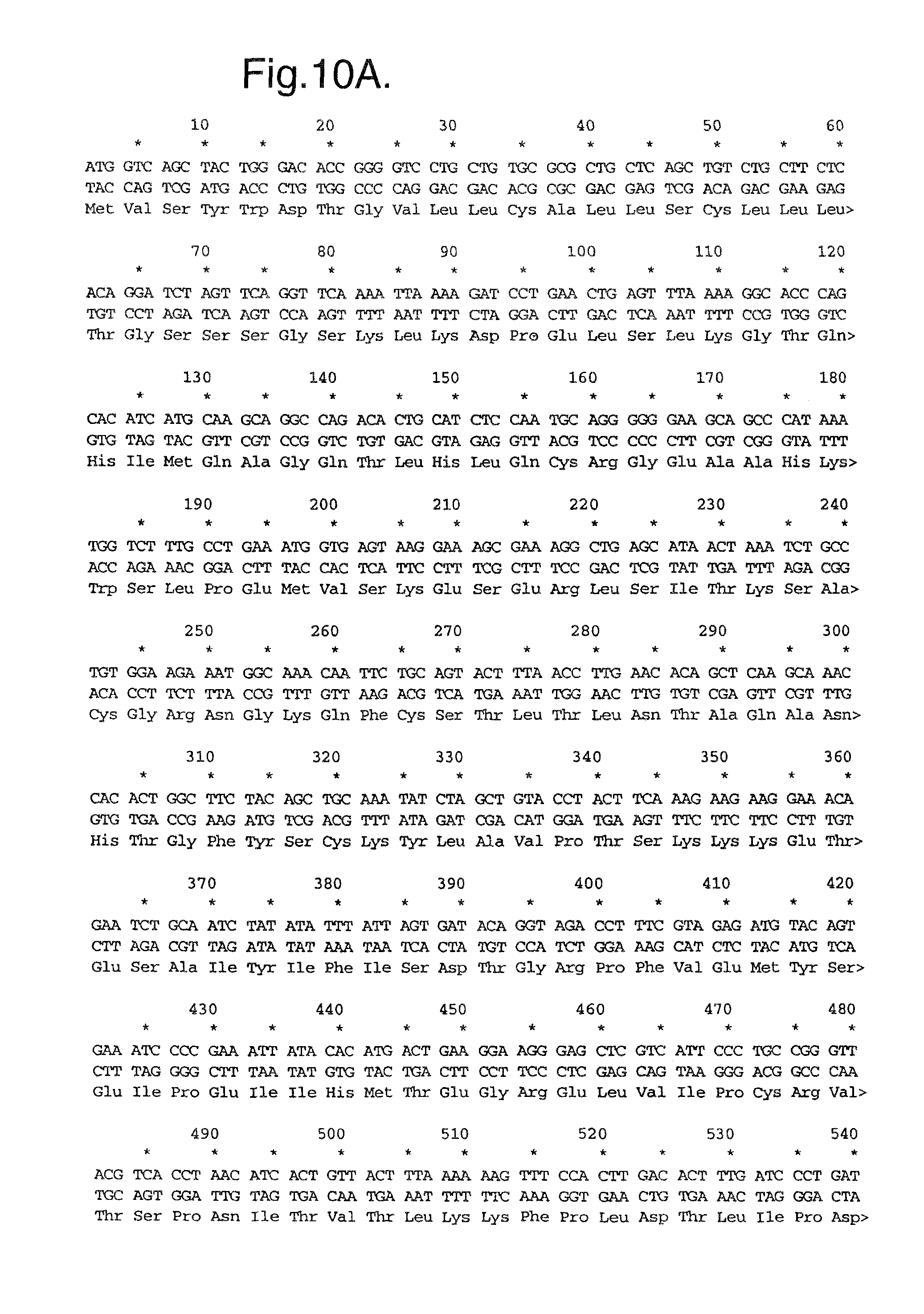
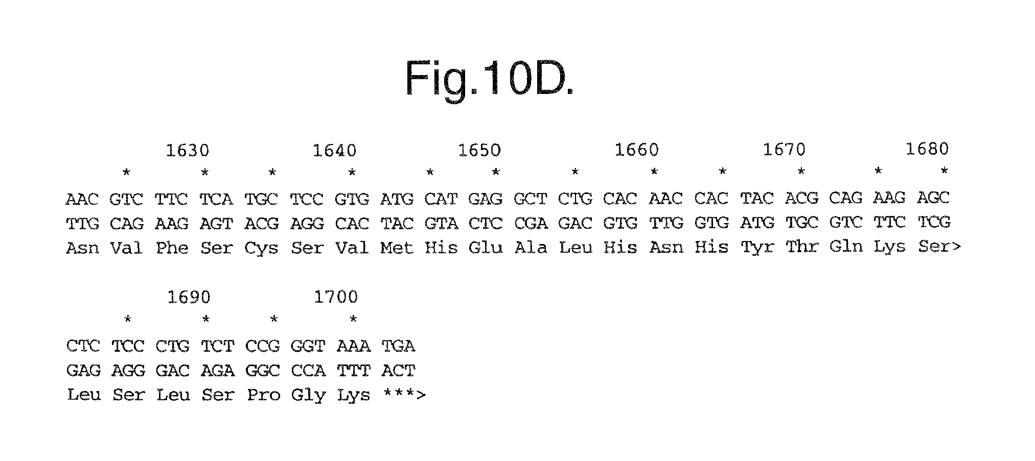
FIG. 10A-D. Nucleic acid (SEQ ID NO:1) and deduced amino acid sequence (SEQ ID NO:2) of Flt1(1-3)-Fc.
FIG. 11. Schematic diagram of the structure of Flt1.
FIG. 12A-B. Hydrophilicity analysis of the amino acid sequences of Ig domain 2 and Ig domain 3 of Flt1.
FIG. 13A-D. Nucleic acid (SEQ ID NO:3) and deduced amino acid sequence (SEQ ID NO:4) of Mut1: Flt1(1-3.sub..DELTA.B)-Fc.
FIG. 14A-C. Nucleic acid (SEQ ID NO:5) and deduced amino acid sequence (SEQ ID NO:6) of Mut2: Flt1(2-3.sub..DELTA.B)-Fc.
FIG. 15A-C. Nucleic acid (SEQ ID NO:7) and deduced amino acid sequence (SEQ ID NO:8) of Mut3: Flt1(2-3)-Fc.
FIG. 16A-D. Nucleic acid (SEQ ID NO:9) and deduced amino acid sequence (SEQ ID NO:10) of Mut4: Flt1(1-3.sub.R.fwdarw.N)-Fc.
FIG. 17. Binding of unmodified Flt1(1-3)-Fc, basic region deletion mutant Flt1(1-3)-Fc, and Flt1(1-3).sub.R.fwdarw.N mutant proteins in a BIACORE.TM.-based assay.
FIG. 18. Binding of unmodified Flt1(1-3)-Fc, Mut1: Flt1(1-3.sub..DELTA.B)-Fc, Mut2: Flt1(2-3.sub..DELTA.B)-Fc, and Flt1(2-3) mutant proteins to MATRIGEL.RTM. coated plates.
FIG. 19. Binding of unmodified Flt1(1-3)-Fc, Mut1: Flt1(1-3.sub..DELTA.B)-Fc, Mut2: Flt1(2-3.sub..DELTA.B)-Fc, and Flt1(2-3) mutant proteins in an ELISA-based assay.
FIG. 20. Pharmacokinetic profiles of unmodified Flt1(1-3)-Fc, Mut1: Flt1(1-3.sub..DELTA.B)-Fc, Mut2: Flt1(2-3.sub..DELTA.B)-Fc, and Flt1(2-3) mutant proteins
FIGS. 21A-C. Nucleotide (SEQ ID NO:11) and deduced amino acid sequence (SEQ ID NO:12) of the modified Flt1 receptor termed Flt1D2.Flk1D3.Fc.DELTA.C1(a).
FIGS. 22A-C. Nucleotide (SEQ ID NO:13) and deduced amino acid sequence (SEQ ID NO:14) of the modified Flt1 receptor termed Flt1D2.VEGFR3D3.Fc.DELTA.C1(a).
FIG. 23. Extracellular Matrix (ECM) Assay. The results of this assay demonstrate that the Flt1D2.Flk1D3.Fc.DELTA.C1(a) and Flt1D2.VEGFR3D3.Fc.DELTA.C1(a) proteins are considerably less sticky to the ECM as compared to the Flt1(1-3)-Fc protein.
FIGS. 24A-C. Nucleotide (SEQ ID NO:15) and deduced amino acid sequence (SEQ ID NO:16) of the modified Flt1 receptor termed VEGFR1R2-Fc.DELTA.C1(a).
FIGS. 25A-C. Phosphorylation assay. At a 1.5 molar excess of either Flt1(1-3)-Fc, Flt1(1-3)-Fc (A40) or transient Flt1D2Flk1D3.Fc.DELTA.C1(a) there is complete blockage of receptor stimulation by these three modified Flt1 receptors as compared to control media challenge. In contrast, transient Flt1D2VEGFR3D3.Fc.DELTA.C1(a) does not show significant blockage at this molar excess, as compared with VEGF positive control challenge. Similar results are seen in FIG. 25B, where the modified Flt receptors are in a 3-fold molar excess to VEGF165 ligand. In FIG. 25C, where the modified Flt1 receptors are in a 6-fold molar excess to VEGF165 ligand, transient Flt1D2VEGFR3D3.Fc.DELTA.C1(a) can now be shown to be partially blocking VEGF165-induced stimulation of cell-surface receptors.
FIGS. 26A-B. Phosphorylation assay. Detection by Western blot of tyrosine phosphorylated VEGFR2(Flk1) by VEGF165 ligand stimulation shows that cell-surface receptors are not phosphorylated by challenge samples which have VEGF165 preincubated with 1 and 2 fold molar excess (FIG. 26A) or 3 and 4 fold molar excess (FIG. 26B) of either transient Flt1D2Flk1D3.Fc.DELTA.C1(a), stable Flt1D2Flk1D3.Fc.DELTA.C1(a), or transient VEGFR1R2-Fc.DELTA.C1(a). At all modified Flt1 receptor concentrations tested there is complete binding of VEGF165 ligand during the preincubation, resulting in no detectable stimulation of cell-surface receptors by unbound VEGF165 as compared to control media challenge.
FIG. 27. MG/R2 Cell proliferation assay. The following modified Flt receptors Flt1(1-3)-Fc, Flt1D2.Flk1D3.Fc.DELTA.C1(a) and Flt1D2.VEGFR3D3.Fc.DELTA.C1(a), plus an irrelevant receptor termed Tie2-Fc as a negative control, were titrated from 40 nM to 20 pM and incubated on the cells for 1 hr at 37.degree. C. Human recombinant VEGF165 in defined media was then added to all the wells at a concentration of 1.56 nM. The negative control receptor Tie2-Fc does not block VEGF165-induced cell proliferation at any concentration whereas Flt1D2.Flk1D3.Fc.DELTA.C1(a) blocks 1.56 nM VEGF165 with a half maximal dose of 0.8 nM. Flt1(1-3)-Fc and Flt1D2.VEGFR3D3.Fc.DELTA.C1(a) are less effective in blocking VEGF165 in this assay with a half maximal dose of .about.2 nM. VEGF165 alone gives a reading of 1.2 absorbance units and the background is 0.38 absorbance units.
FIG. 28. BIACORE.TM. analysis of Binding Stoichiometry. Binding stoichiometry was calculated as a molar ratio of bound VEGF165 to the immobilized Flt1D2Flk1D3.Fc.DELTA.C1(a) or VEGFR1R2-Fc.DELTA.C1(a), using the conversion factor of 1000 RU equivalent to 1 ng/ml. The results indicated binding stoichiometry of one VEGF165 dimeric molecule per one Flt1D2Flk1D3.Fc.DELTA.C1(a) or VEGFR1R2-Fc.DELTA.C1(a) molecule.
FIGS. 29-30. Size Exclusion Chromatography Stoichiometry. Flt1D2Flk1D3.Fc.DELTA.C1(a) or VEGFR1R2-Fc.DELTA.C1(a) at a concentration of 1 nM (estimated to be 1000 times higher than the KD of the Flt1D2Flk1D3.Fc.DELTA.C1(a) or VEGFR1R2-Fc.DELTA.C1(a)/VEGF165 interaction) were mixed with varied concentrations of VEGF165. After incubation, concentrations of the free Flt1D2Flk1D3.Fc.DELTA.C1(a) in solution were measured. The data shows that the addition of 1 nM VEGF165 into the Flt1D2Flk1D3.Fc.DELTA.C1(a) solution completely blocks Flt1D2Flk1D3.Fc.DELTA.C1(a) binding to the VEGF165 surface. This result suggested the binding stoichiometry of one VEGF165 molecule per one Flt1D2Flk1D3.Fc.DELTA.C1(a) molecule.
FIG. 31. Size Exclusion Chromatography (SEC) under native conditions. Peak #1 represents the Flt1D2Flk1D3.Fc.DELTA.C1(a)/VEGF165 complex and peak #2 represents unbound VEGF165. Fractions eluted between 1.1 and 1.2 ml were combined and guanidinium hydrochloride (GuHCl) was added to a final concentration 4.5 M to dissociate the complex.
FIG. 32. Size Exclusion Chromatography (SEC) under dissociative conditions. To separate the components of the receptor-ligand complex and to determine their molar ratio, 50 .mu.l of dissociated complex was loaded onto a SUPEROSE.TM. 12 PC 3.2/30 equilibrated in 6 M GuHCl and eluted. Peak #1 represents Flt1D2Flk1D3.Fc.DELTA.C1(a) and peak #2 represents VEGF165.
FIGS. 33-35. Size Exclusion Chromatography (SEC) with On-Line Light Scattering. Size exclusion chromatography column with a MiniDawn on-line light scattering detector (Wyatt Technology, Santa Barbara, Calif.) and refractive index (RI) detectors (Shimadzu, Kyoto, Japan) was used to determine the molecular weight (MW) of the receptor-ligand complex. As shown in FIG. 33, the elution profile shows two peaks. Peak #1 represents the receptor-ligand complex and peak #2 represents the unbound VEGF165. MW was calculated from LS and RI signals. The same procedure was used to determine MW of the individual components of the receptor-ligand complex. The results of these determinations are as follows: MW of the Flt1D2Flk1D3.Fc.DELTA.C1(a)/VEGF165 complex at the peak position is 157 300 (FIG. 33), the MW of VEGF165 at the peak position is 44 390 (FIG. 34) and the MW of R1R2 at the peak is 113 300 (FIG. 35).
FIG. 36. Peptide mapping and glycosylation analysis. The disulfide structures and glycosylation sites in Flt1D2.Flk1D3.Fc.DELTA.C1(a) (SEQ ID NO:12) were determined by a peptide mapping method. There are a total of ten cysteines in Flt1D2.Flk1D3.Fc.DELTA.C1(a); six of them belong to the Fc region. Cys27 is disulfide bonded to Cys76. Cys121 is disulfide bonded to Cys182. The first two cysteines in the Fc region (Cys211 and Cys214) form an intermolecular disulfide bond with the same two cysteines in another Fc chain. However, it can not be determined whether disulfide bonding is occurring between same cysteines (Cys211 to Cys211, for example) or between Cys211 and Cys214. Cys216 is disulfide bonded to Cys306. Cys352 is disulfide bonded to Cys410. There are five possible N-linked glycosylation sites in Flt1D2.Flk1D3.Fc.DELTA.C1(a) (SEQ ID NO:12) and are found to be glycosylated to varying degrees. Complete glycosylation is observed at Asn33, Asn193, and Asn282. Partial glycosylation is observed on Asn65 and Asn120. Sites of glycosylation are highlighted by underline in the figure.
FIG. 37. Pharmacokinetics of Flt1(1-3)-Fc (A40), Flt1D2.Flk1D3.Fc.DELTA.C1(a) and VEGFR1R2-Fc.DELTA.C1(a). Balb/c mice were injected subcutaneously with 4 mg/kg of Flt1(1-3)-Fc (A40), CHO transiently expressed Flt1D2.Flk1D3.Fc.DELTA.C1(a), CHO stably expressed Flt1D2.Flk1D3.Fc.DELTA.C1(a), and CHO transiently expressed VEGFR1R2-Fc.DELTA.C1(a). The mice were tail bled at 1, 2, 4, 6, 24 hrs, 2 days, 3 days and 6 days after injection. The sera were assayed in an ELISA designed to detect Flt1(1-3)-Fc (A40), Flt1D2.Flk1D3.Fc.DELTA.C1(a) or VEGFR1R2-Fc.DELTA.C1(a). The T.sub.max for Flt1(1-3)-Fc (A40) was at 6 hrs while the T.sub.max for the transient and stable Flt1D2.Flk1D3.Fc.DELTA.C1(a) and the transient VEGFR1R2-Fc.DELTA.C1(a) was 24 hrs. The C.sub.max for Flt1(1-3)-Fc (A40) was 8 .mu.g/ml. For both transients (Flt1D2.Flk1D3.Fc.DELTA.C1(a) and VEGFR1R2-Fc.DELTA.C1(a)) the C.sub.max was 18 .mu.g/ml and the C.sub.max for the stable VEGFR1R2-Fc.DELTA.C1(a) was 30 .mu.g/ml.
FIG. 38. Pharmacokinetics of Flt1(1-3)-Fc (A40), Flt1D2.Flk1D3.Fc.DELTA.C1(a) and Flt1D2.VEGFR3D3.Fc.DELTA.C1(a). Balb/c mice were injected subcutaneously with 4 mg/kg of Flt1(1-3)-Fc (A40), CHO transiently expressed Flt1D2.Flk1D3.Fc.DELTA.C1(a) and CHO transiently expressed Flt1D2.VEGFR3D3.Fc.DELTA.C1(a). The mice were tail bled at 1, 2, 5, 6, 7, 8, 12, 15 and 20 days after injection. The sera were assayed in an ELISA designed to detect Flt1(1-3)-Fc, Flt1D2.Flk1D3.Fc.DELTA.C1(a) and Flt1D2.VEGFR3D3.Fc.DELTA.C1(a). Flt1(1-3)-Fc (A40) could no longer be detected in the serum after day 5 whereas Flt1D2.Flk1D3.Fc.DELTA.C1(a) and Flt1D2.VEGFR3D3.Fc.DELTA.C1(a) were detectable for 15 days or more.
FIG. 39. The ability of Flt1D2.Flk1D3.Fc.DELTA.C1(a) to inhibit HT-1080 fibrosarcoma tumor growth In Vivo. Every other day or 2 times per week treatment of SCID mice with Flt1D2.Flk1D3.Fc.DELTA.C1(a) at 25 mg/Kg significantly decreases the growth of subcutaneous HT-1080 fibrosarcoma tumors.
FIG. 40. The ability of Flt1D2.Flk1D3.Fc.DELTA.C1(a) to inhibit C6 glioma tumor growth in vivo. Every other day or 2 times a week treatment of SCID mice with Flt1D2.Flk1D3.Fc.DELTA.C1(a) significantly decreases the growth of subcutaneous C6 glioma tumors at doses as low as 2.5 mg/Kg.
FIG. 41. VEGF-induced uterine hyperpermeability.
FIGS. 42A-B. Assessment of corpus luteum angiogenesis using progesterone as a readout.
DETAILED DESCRIPTION OF THE INVENTION
It has been a long standing problem in the art to produce a receptor based VEGF antagonist that has a pharmacokinetic profile that is appropriate for consideration of the antagonist as a therapeutic candidate. Applicants describe herein, for the first time, a chimeric polypeptide molecule, capable of antagonizing VEGF activity, that exhibits improved pharmacokinetic properties as compared to other known receptor-based VEGF antagonists. The chimeric polypeptide molecules described herein thus provide for the first time appropriate molecules for use in therapies in which antagonism of VEGF is a desired result.
The present invention provides for novel chimeric polypeptide molecules formed by fusing a modified extracellular ligand binding domain of the Flt1 receptor to the Fc region of IgG.
The extracellular ligand binding domain is defined as the portion of a receptor that, in its native conformation in the cell membrane, is oriented extracellularly where it can contact with its cognate ligand. The extracellular ligand binding domain does not include the hydrophobic amino acids associated with the receptor's transmembrane domain or any amino acids associated with the receptor's intracellular domain. Generally, the intracellular or cytoplasmic domain of a receptor is usually composed of positively charged or polar amino acids (i.e. lysine, arginine, histidine, glutamic acid, aspartic acid). The preceding 15-30, predominantly hydrophobic or apolar amino acids (i.e. leucine, valine, isoleucine, and phenylalanine) comprise the transmembrane domain. The extracellular domain comprises the amino acids that precede the hydrophobic transmembrane stretch of amino acids. Usually the transmembrane domain is flanked by positively charged or polar amino acids such as lysine or arginine. von Heijne has published detailed rules that are commonly referred to by skilled artisans when determining which amino acids of a given receptor belong to the extracellular, transmembrane, or intracellular domains (See von Heijne, 1995, BioEssays 17:25-30). Alternatively, websites on the Internet have become available to provide protein chemists with information about making predictions about protein domains.
The present invention provides for the construction of nucleic acid molecules encoding chimeric polypeptide molecules that are inserted into a vector that is able to express the chimeric polypeptide molecules when introduced into an appropriate host cell. Appropriate host cells include, but are not limited to, bacterial cells, yeast cells, insect cells, and mammalian cells. Any of the methods known to one skilled in the art for the insertion of DNA fragments into a vector may be used to construct expression vectors encoding the chimeric polypeptide molecules under control of transcriptional/translational control signals. These methods may include in vitro recombinant DNA and synthetic techniques and in vivo recombinations (genetic recombination) (See Sambrook, et al., Molecular Cloning, A Laboratory Manual, Cold Spring Harbor Laboratory; Current Protocols in Molecular Biology, Eds. Ausubel, et al., Greene Publ. Assoc., Wiley-Interscience, NY).
Expression of nucleic acid molecules encoding the chimeric polypeptide molecules may be regulated by a second nucleic acid sequence so that the chimeric polypeptide molecule is expressed in a host transformed with the recombinant DNA molecule. For example, expression of the chimeric polypeptide molecules described herein may be controlled by any promoter/enhancer element known in the art. Promoters which may be used to control expression of the chimeric polypeptide molecules include, but are not limited to, the long terminal repeat as described in Squinto et al., (1991, Cell 65:1-20); the SV40 early promoter region, the CMV promoter, the M-MuLV 5' terminal repeat the promoter contained in the 3' long terminal repeat of Rous sarcoma virus, the herpes thymidine kinase promoter, the regulatory sequences of the metallothionine gene; prokaryotic expression vectors such as the .beta.-lactamase promoter, or the tac promoter; promoter elements from yeast or other fungi such as the Gal 4 promoter, the ADH (alcohol dehydrogenase) promoter, PGK (phosphoglycerol kinase) promoter, alkaline phosphatase promoter, and the following animal transcriptional control regions, which exhibit tissue specificity and have been utilized in transgenic animals: elastase I gene control region which is active in pancreatic acinar cells; insulin gene control region which is active in pancreatic beta cells, immunoglobulin gene control region which is active in lymphoid cells, mouse mammary tumor virus control region which is active in testicular, breast, lymphoid and mast cells, albumin gene control region which is active in liver, alpha-fetoprotein gene control region which is active in liver; alpha 1-antitrypsin gene control region which is active in the liver, beta-globin gene control region which is active in myeloid cells; myelin basic protein gene control region which is active in oligodendrocyte cells in the brain; myosin light chain-2 gene control region which is active in skeletal muscle, and gonadotropic releasing hormone gene control region which is active in the hypothalamus.
Thus, according to the invention, expression vectors capable of being replicated in a bacterial or eukaryotic host comprising chimeric polypeptide molecule-encoding nucleic acid as described herein, are used to transfect the host and thereby direct expression of such nucleic acids to produce the chimeric polypeptide molecules, which may then be recovered in a biologically active form. As used herein, a biologically active form includes a form capable of binding to VEGF.
Expression vectors containing the chimeric nucleic acid molecules described herein can be identified by three general approaches: (a) DNA-DNA hybridization, (b) presence or absence of "marker" gene functions, and (c) expression of inserted sequences. In the first approach, the presence of a foreign gene inserted in an expression vector can be detected by DNA-DNA hybridization using probes comprising sequences that are homologous to the inserted chimeric polypeptide molecule sequences. In the second approach, the recombinant vector/host system can be identified and selected based upon the presence or absence of certain "marker" gene functions (e.g., thymidine kinase activity, resistance to antibiotics, transformation phenotype, occlusion body formation in baculovirus, etc.) caused by the insertion of foreign genes in the vector. For example, if the chimeric polypeptide molecule DNA sequence is inserted within the marker gene sequence of the vector, recombinants containing the insert can be identified by the absence of the marker gene function. In the third approach, recombinant expression vectors can be identified by assaying the foreign gene product expressed by the recombinant. Such assays can be based, for example, on the physical or functional properties of the chimeric polypeptide molecules.
Cells of the present invention may transiently or, preferably, constitutively and permanently express the chimeric polypeptide molecules.
The chimeric polypeptide molecules may be purified by any technique which allows for the subsequent formation of a stable, biologically active chimeric polypeptide molecule.
In one embodiment of the invention, the nucleotide sequence encoding the first component is upstream of the nucleotide sequence encoding the second component. In another embodiment of the invention, the nucleotide sequence encoding the first component is downstream of the nucleotide sequence encoding the second component. Further embodiments of the invention may be prepared in which the order of the first, second and third fusion polypeptide components are rearranged. For example, if the nucleotide sequence encoding the first component is designated 1, the nucleotide sequence encoding the second component is designated 2, and the nucleotide sequence of the third component is designated 3, then the order of the components in the isolated nucleic acid of the invention as read from 5' to 3' may be any of the following six combinations: 1, 2, 3; 1, 3, 2; 2, 1, 3; 2, 3, 1; 3, 1, 2; or 3, 2, 1.
The present invention also has diagnostic and therapeutic utilities. In particular embodiments of the invention, methods of detecting aberrancies in the function or expression of the chimeric polypeptide molecules described herein may be used in the diagnosis of disorders. In other embodiments, manipulation of the chimeric polypeptide molecules or agonists or antagonists which bind the chimeric polypeptide molecules may be used in the treatment of diseases. In further embodiments, the chimeric polypeptide molecule is utilized as an agent to block the binding of a binding agent to its target.
By way of example, but not limitation, the method of the invention may be useful in treating clinical conditions that are characterized by vascular permeability, edema or inflammation such as brain edema associated with injury, stroke or tumor; edema associated with inflammatory disorders such as psoriasis or arthritis, including rheumatoid arthritis; asthma; generalized edema associated with burns; ascites and pleural effusion associated with tumors, inflammation or trauma; chronic airway inflammation; capillary leak syndrome; sepsis; kidney disease associated with increased leakage of protein; and eye disorders such as age related macular degeneration and diabetic retinopathy.
An amino acid sequence analysis of Flt1(1-3)-Fc revealed the presence of an unusually high number (46) of the basic amino acid residue lysine. An IEF analysis of Flt1(1-3)-Fc showed that this protein has pl greater than 9.3, confirming the prediction that the protein is very basic. It was hypothesized that the basic nature of Flt1(1-3)-Fc protein was causing it to bind to extracellular matrix components and that this interaction might be the cause of the extremely short detectable circulating serum half-life exhibited by Flt1(1-3)-Fc when injected into mice. In order to test this hypothesis, Flt1(1-3)-Fc protein was acetylated at the lysine residues to reduce the basic charge. Acetylated Flt1(1-3)-Fc was then tested in the assays described infra.
The following examples are offered by way of illustration and not by way of limitation.
EXAMPLES
Example 1: Expression of Flt1(1-3)-Fc Protein in CHO K1 Cells
Using standard molecular biology techniques (see e.g., Molecular Cloning, A Laboratory Manual (Sambrook, et al., Cold Spring Harbor Laboratory), Current Protocols in Molecular Biology (Eds. Ausubel, et al., Greene Publ. Assoc., Wiley-Interscience, NY), the gene encoding Flt1(1-3)-Fc was inserted into the expression vector pEE14.1 (Lonza Biologics, plc) at a multiple cloning site downstream of the CMV promoter. CHO K1 cells were transfected with the pEE14.1/Flt1(1-3)-Fc DNA construct using lipofectamine (Gaithersburg, Md.). The transfected CHO K1 cells were grown in glutamine-free DMEM (JRH, Kansas City, Mo.) containing 25 .mu.M methionine sulfoximine (MSX) from Sigma Inc., St. Louis, Mo., and high recombinant protein expressors were obtained by screening the CHO K1 cell supernatants from over 100 hand-picked colony isolates using a standard immunoassay which captures and detects human Fc. The selected hand-picked clone was amplified in the presence of 100 .mu.M MSX followed by a second round of screening of the amplified clones. The highest producing clone had a specific productivity of recombinant Flt1(1-3)-Fc protein of 55 pg/cell/day.
The selected clone was expanded in 225 cm.sup.2 T-flasks (Corning, Acton, Mass.) and then into 8.5 L roller bottles (Corning, Acton, Mass.) using the cell culture media described supra. Cells were removed from the roller bottles by standard trypsinization and put into 3.5 L of suspension medium. The suspension medium is comprised of glutamine-free ISCHO medium (Irvine Scientific, Santa Ana, Calif.) containing 5% fetal bovine serum (FBS from HYCLONE.TM. Labs, Logan, Utah), 100 .mu.M MSX and GS supplement (JRH Scientific, Kansas City, Mo.) in a 5 L Celligen bioreactor (New Brunswick Scientific, New Brunswick, N.J.) at a density of 0.3.times.10.sup.6 cells/mL. After the cells reached a density of 3.6.times.10.sup.6/mL and were adapted to suspension they were transferred to a 60 L bioreactor (ABEC, Allentown, Pa.) at a density of 0.5.times.10.sup.6 cells/mL in 20 L of ISCHO medium with 5% fetal bovine serum. After two days an additional 20 L of ISCHO+5% fetal bovine serum was added to the bioreactor. The cells were allowed to grow for an additional two days reaching a final density of 3.1.times.10.sup.6 cells/mL, and a final Flt1(1-3)-Fc concentration at harvest was 95 mg/L. At harvest the cells were removed by tangential flow filtration using 0.45 .mu.m Prostak Filters (Millipore, Inc., Bedford, Mass.).
Example 2: Purification of Flt1(1-3)-Fc Protein Obtained from CHO K1 Cells
Flt1(1-3)-Fc protein was initially purified by affinity chromatography. A Protein A column was used to bind, with high specificity, the Fc portion of the molecule. This affinity-purified protein was then concentrated and passed over a SEC column. The protein was then eluted into the formulation buffer. The following describes these procedures in detail.
Materials and Methods. All chemicals were obtained from J.T. Baker, Phillipsburg, N.J. with the exception of PBS, which was obtained as a 10.times. concentrate from Life Technologies, Gaithersburg, Md. Protein A Fast Flow and SUPERDEX.TM. 200 preparation grade resins were obtained from Pharmacia, Piscataway, N.J. Equipment and membranes for protein concentration were obtained from Millipore, Bedford, Mass.
Approximately 40 L of 0.45 .mu.m-filtered CHO conditioned media containing Flt1(1-3)-Fc protein was applied to a 290 mL Protein A Fast Flow column (10 cm diameter) that had been equilibrated with PBS. The column was washed with PBS containing 350 mM NaCl and 0.02% CHAPS and the bound protein was eluted with 20 mM Citric Acid containing 10 mM Na.sub.2HPO.sub.4. The single peak in the elution was collected and its pH was raised to neutrality with 1 M NaOH. The eluate fractions was concentrated to approximately 9 mg/mL using 10K regenerated cellulose membranes by both tangential flow filtration and by stirred cell concentration. To remove aggregates and other contaminants, the concentrated protein was applied to a column packed with SUPERDEX.TM. 200 preparation grade resin (10 cm.times.55 cm) and run in PBS containing 5% glycerol. The main peak fractions were pooled, sterile filtered, aliquoted and stored at -80.degree. C.
Example 3: Acetylation of Flt1(1-3)-Fc Protein
Two milligrams of Flt1(1-3)-Fc protein were acetylated as described in the instruction manual provided with the sulfo-NHS-acetate modification kit (Pierce Chemical Co., Rockford, Ill.).
Example 4: Characterization of Acetylated Flt1(1-3)-Fc Protein
IEF Analysis:
Flt1(1-3)-Fc and acetylated Flt1(1-3)-Fc were analyzed by standard IEF analysis. As shown in FIG. 1, Flt1(1-3)-Fc protein is not able to migrate into the gel and therefore must have a pl greater than 9.3, the highest pl in the standard. However, acetylated Flt1(1-3)-Fc is able to migrate into the gel and equilibrate at a pl of approximately 5.2. This result demonstrates that acetylation reduces the net positive charge of the protein and therefore its pl considerably.
Binding to Extracellular Matrix Components.
To test for binding to extracellular matrix components, Flt1(1-3)-Fc and acetylated Flt1(1-3)-Fc where tested in an assay designed to mimic the interaction with extracellular matrix components. In this assay, 96-well tissue culture plates are coated with MATRIGEL.RTM. (Biocoat MATRIGEL.RTM. matrix thin layer 96 well plate, Catalog #40607, Becton Dickinson Labware, Bedford, Mass.). The plates are incubated with varying concentrations of either Flt1(1-3)-Fc, acetylated Flt1(1-3)-Fc, or rTie2-Fc (an irrelevant control) protein are added to the wells. The plates are incubated for 1-2 hours at either room temperature or 37.degree. C. degrees and then detection of bound proteins is accomplished by adding a secondary alkaline phosphatase-conjugated anti-human Fc antibody to the wells. Finally, alkaline phosphatase substrate is added to the wells and optical density is measured. FIG. 2 shows the results of this assay. Like the irrelevant control protein rTie2-Fc, acetylated Flt1(1-3)-Fc does not exhibit any binding to the MATRIGEL.RTM. coated plate, whereas the non-acetylated Flt1(1-3)-Fc protein exhibits significant binding. This result indicates that acetylation of basic amino acid residues is an effective way to interfere with the charge interactions that exist between positively charged proteins and the negatively charged extracellular matrix components they are exposed to in vivo.
Example 5: Pegylation of Flt1(1-3)-Fc Protein
Although pegylation (polyethylene glycol--PEG) of proteins has been shown to increase their in vivo potency by enhancing stability and bioavailability while minimizing immunogenicity (see references cited supra), it is counter-intuitive that pegylating molecules that are too large to be filtered by the kidney glomeruli would improve their pharmacokinetic properties. Without being bound by theory, Applicants postulated that pegylation of the Flt1(1-3)-Fc molecules could improve the pharmacokinetic properties, possibly not by altering the positive charge or by decreasing the pl of Flt1(1-3)-Fc, but rather by physically shielding the positive charges from interacting with the extracellular matrix. Applicants decided to attempt to improve the pharmacokinetic properties of Flt1(1-3)-Fc molecules by attaching strands of 20K PEGs as described infra.
Materials and Methods.
Purified Flt1(1-3)-Fc derived from CHO cells (see supra) was used in the following pegylation experiments. Functionalized PEGs were obtained from Shearwater Polymers, Huntsville, Ala.; Bicine from Sigma, St Louis, Mo.; SUPEROSE.TM. 6 column from Pharmacia, Piscataway, N.J.; PBS as a 10.times. concentrate from Life Technologies, Gaithersburg, Md.; Glycerol from J.T. Baker, Phillipsburg, N.J.; and Bis-Tris precast gels from Novex, Calif.
20K PEG strands functionalized with amine-specific terminal moieties were used in small-scale reaction studies that were set-up to evaluate different reaction conditions in which the PEG:protein stoichiometry was varied. Based on these reactions and the analyses of samples on standard SDS-PAGE, Flt1(1-3)-Fc at a concentration of 1.5 mg/mL was reacted at pH 8.1 with 20K SPA-PEG (PEG succinimidyl propionate) molecules at a PEG-to-Flt1(1-3)-Fc monomer molar ratio of 1:6. The reaction was allowed to proceed at 8.degree. C. overnight. For initial purification, the reaction products were applied to a 10 mm.times.30 cm SUPEROSE.TM. 6 column equilibrated with PBS containing 5% Glycerol. The column appeared to separate pegylated Flt1(1-3)-Fc molecules based on the extent of pegylation. Fractions corresponding to what appeared to be primarily mono-pegylated and di-pegylated dimeric Flt1(1-3)-Fc, as judged by banding patterns on reducing and non-reducing SDS-PAGE gels were pooled. The protein concentration was determined by measuring absorbance at 280 nm. The pegylated Flt1(1-3)-Fc protein was sterile filtered, aliquoted and stored at -40.degree. C.
Example 6: Binding of Unmodified, Acetylated, and Pegylated Flt1(1-3)-Fc in a BIACORE.TM.-Based Assay
Unmodified, acetylated, and pegylated Flt1(1-3)-Fc proteins were tested in a BIACORE.TM.-based assay to evaluate their ability to bind to the Flt1 ligand, VEGF. In this assay, unmodified Flt1(1-3)-Fc protein was immobilized on the surface of a BIACORE.TM. chip and a sample containing 0.2 .mu.g/ml VEGF and either unmodified Flt1(1-3)-Fc, acetylated Flt1(1-3)-Fc or pegylated Flt1(1-3)-Fc (each at 25 .mu.g/ml) was passed over the Flt1(1-3)-Fc-coated chip. To minimize the effects of non-specific binding, the bound samples were washed with a 0.5 M NaCl wash. In one sample, unmodified Flt1(1-3)-Fc was mixed with heparin. Heparin is a negatively charged molecule and the Flt1(1-3)-Fc protein is a positively charged molecule, so when the two molecules are mixed together, they should interact through their respective charges. This essentially neutralizes Flt1(1-3)-Fc's inherent positive charge making the molecule behave as if it has been chemically or genetically modified so as to reduce its charge and its tendency to bind via charge interactions. As shown in FIG. 3, acetylated (columns 13-16), pegylated (columns 17-20), and heparin-treated Flt1(1-3)-Fc (columns 21-24) are each able to completely compete with the BIACORE.TM. chip-bound Flt1(1-3)-Fc for VEGF binding as compared to control (columns 1-4) and irrelevant protein (columns 5-8). Unmodified Flt1(1-3)-Fc (columns 5-6) appeared to only partially compete with BIACORE.TM. chip-bound Flt1(1-3)-Fc for VEGF binding. However, washing the bound samples with 0.5 M NaCl (columns 7-8) resulted in a binding profile similar to the modified forms of Flt1(1-3)-Fc, indicating that the unmodified protein was exhibiting non-specific binding to the chip that could be eliminated by the salt wash.
Example 7: Binding of Unmodified, Acetylated, and Pegylated Flt1(1-3)-Fc in an ELISA-Based Assay
Unmodified, acetylated, and pegylated Flt1(1-3)-Fc proteins were tested in a standard ELISA-based assay to evaluate their ability to bind the Flt1 receptor ligand VEGF. As shown in FIG. 4, both pegylated and acetylated Flt1(1-3)-Fc proteins are capable of binding to VEGF, demonstrating that modifying the protein either by pegylation or acetylation does not destroy its ability to bind its ligand.
Example 8: Pharmacokinetic Analysis of Unmodified Flt1(1-3)-Fc, Acetylated Flt1(1-3)-Fc, and Pegylated Flt1(1-3)-Fc
In vivo experiments were designed to assess the pharmacokinetic profiles of unmodified Flt1(1-3)-Fc, acetylated Flt1(1-3)-Fc, and pegylated Flt1(1-3)-Fc protein. Balb/c mice (23-28 g; 3 mice/group) were injected subcutaneously with 4 mg/kg of unmodified, acetylated, or pegylated Flt1(1-3)-Fc. The mice were tail bled at 1, 2, 4, 6, 24 hours, 2 days, and 3 days after injection of protein. The sera were assayed in a standard ELISA-based assay designed to detect Flt1(1-3)-Fc protein. Briefly, the assay involves coating an ELISA plate with VEGF, binding the unmodified, acetylated, or pegylated Flt1(1-3)-Fc-containing sera, and reporting with an anti-Fc antibody linked to alkaline phosphatase. As shown in FIG. 5, the T.sub.max for all of the Flt1(1-3)-Fc proteins was between the 6 hour and 24 hour time points. The C.sub.max for the different proteins was as follows: Unmodified: 0.06 .mu./ml-0.15 .mu.g/ml; acetylated: 1.5 .mu.g/ml-4.0 .mu.g/ml; and pegylated: approximately 5 .mu.g/ml.
Example 9: Step-Acetylation of Flt1(1-3)-Fc
To determine what minimal amount of acetylation is necessary to eliminate binding to extracellular matrix components, an experiment was designed that acetylated the Flt1(1-3)-Fc protein in a step-wise fashion by using increasing amounts of molar excess of acetylation reagent in the acetylation reaction mixture. The range of molar excess was as follows: 0, 10, 20, 30, 40, 50, 60, 70, 80, 90, and 100 moles of acetylation reagent per 1 mole of Flt1(1-3)-Fc monomer. The reactions were performed as detailed in the instruction manual provided with the sulfo-NHS-Acetate modification kit (Pierce Chemical Co., Rockford, Ill.).
Example 10: Characterization of Step-Acetylated Flt1(1-3)-Fc
IEF Analysis
Unmodified Flt1(1-3)-Fc and step-acetylated Flt1(1-3)-Fc proteins were analyzed by standard IEF analysis. As shown in FIG. 6A-B, unmodified Flt1(1-3)-Fc protein was not able to migrate into the gel due to its extremely high pl (greater than 9.3). However, most of the step-acetylated Flt1(1-3)-Fc samples (30-100 fold molar excess samples) were able to migrate into the gel and equilibrate at pls ranging between 4.55-8.43, depending on the degree of acetylation of the protein. This result demonstrates that acetylation can change the positive charge of the protein in a dose-dependent manner and that reduction of the pl can be controlled by controlling the degree of acetylation.
Binding of Step-Acetylated Flt1(1-3)-Fc to Extracellular Matrix Components.
To test for binding to extracellular matrix components, Flt1(1-3)-Fc and step-acetylated Flt1(1-3)-Fc where tested in the above-described assay designed to mimic the interaction with extracellular matrix components. Varying concentrations of either unmodified Flt1(1-3)-Fc, step-acetylated Flt1(1-3)-Fc (10, 20, and 30 fold molar excess samples), or rTie2-Fc (an irrelevant control) protein were added to the wells. The plates were incubated for 1-2 hours at room temperature or 37.degree. C. and then detection of bound proteins was accomplished by adding a secondary alkaline phosphatase-conjugated anti-human Fc antibody to the wells. Alkaline phosphatase substrate was subsequently added to the wells and optical density measured. FIG. 7 shows the results of this assay. Like the irrelevant control protein rTie2-Fc, step-acetylated Flt1(1-3)-Fc (20 and 30 fold molar excess samples) did not exhibit any significant binding to the MATRIGEL.RTM. coated plate, whereas the non-acetylated Flt1(1-3)-Fc protein exhibited significant binding. The binding is saturable, indicating that the Flt1(1-3)-Fc protein may be binding to specific sites, rather than a more general charge-mediated interaction that might not be saturable. The 10 fold molar excess sample showed reduced binding, but the degree of acetylation was not enough to completely block binding to extracellular matrix components. The 20 fold molar excess and higher samples displayed no detectable binding, despite the fact that by IEF analysis (FIG. 6A-B) the lower molar excess samples still had a large net positive charge. This result demonstrates that it is not necessary to completely acetylate all available basic amino acids in order to eliminate binding to extracellular matrix components.
Binding of Step-Acetylated Flt1(1-3)-Fc in a BIACORE.TM.-Based Assay.
Unmodified and step-acetylated Flt1(1-3)-Fc proteins where tested in a BIACORE.TM.-based assay to evaluate their ability to bind to the Flt1 ligand, VEGF. In this assay, unmodified Flt1(1-3)-Fc protein (0.5, 1.0, or 5.0 .mu.g/ml) was immobilized on the surface of a BIACORE.TM. chip and a solution containing 0.2 .mu.g/ml VEGF and either unmodified Flt1(1-3)-Fc (at either 0.5, 1.0, or 5.0 .mu.g/ml) or 10 different step-acetylated Flt1(1-3)-Fc samples (at 0.5, 1.0, or 5.0 .mu.g/ml each) were passed over the Flt1(1-3)-Fc-coated chip. As shown in FIG. 8, at a sub-stoichiometric ratio (0.5 .mu.g/ml of either unmodified Flt1(1-3) or step-acetylated Flt1(1-3)-Fc vs. 0.2 .mu.g/ml VEGF), there is not enough Flt1(1-3)-Fc (either unmodified or step-acetylated) in the solution to completely bind the VEGF. At 1.0 .mu.g/ml, which approximates a 1:1 stoichiometric ratio, both unmodified and step-acetylated Flt1(1-3)-Fc are better able to compete for VEGF binding, but there is still insufficient Flt1(1-3)-Fc protein (either unmodified or step-acetylated) to completely bind the available VEGF. However, at 5.0 .mu.g/ml, which is several times greater than a 1:1 stoichiometric ratio, both the Flt1(1-3)-Fc and the step-acetylated Flt1(1-3)-Fc proteins are able to bind the VEGF, regardless of the degree of acetylation. This clearly demonstrates that acetylation does not alter Flt1(1-3)-Fc's ability to bind VEGF.
Pharmacokinetic Analysis of Step-Acetylated Flt1(1-3)-Fc.
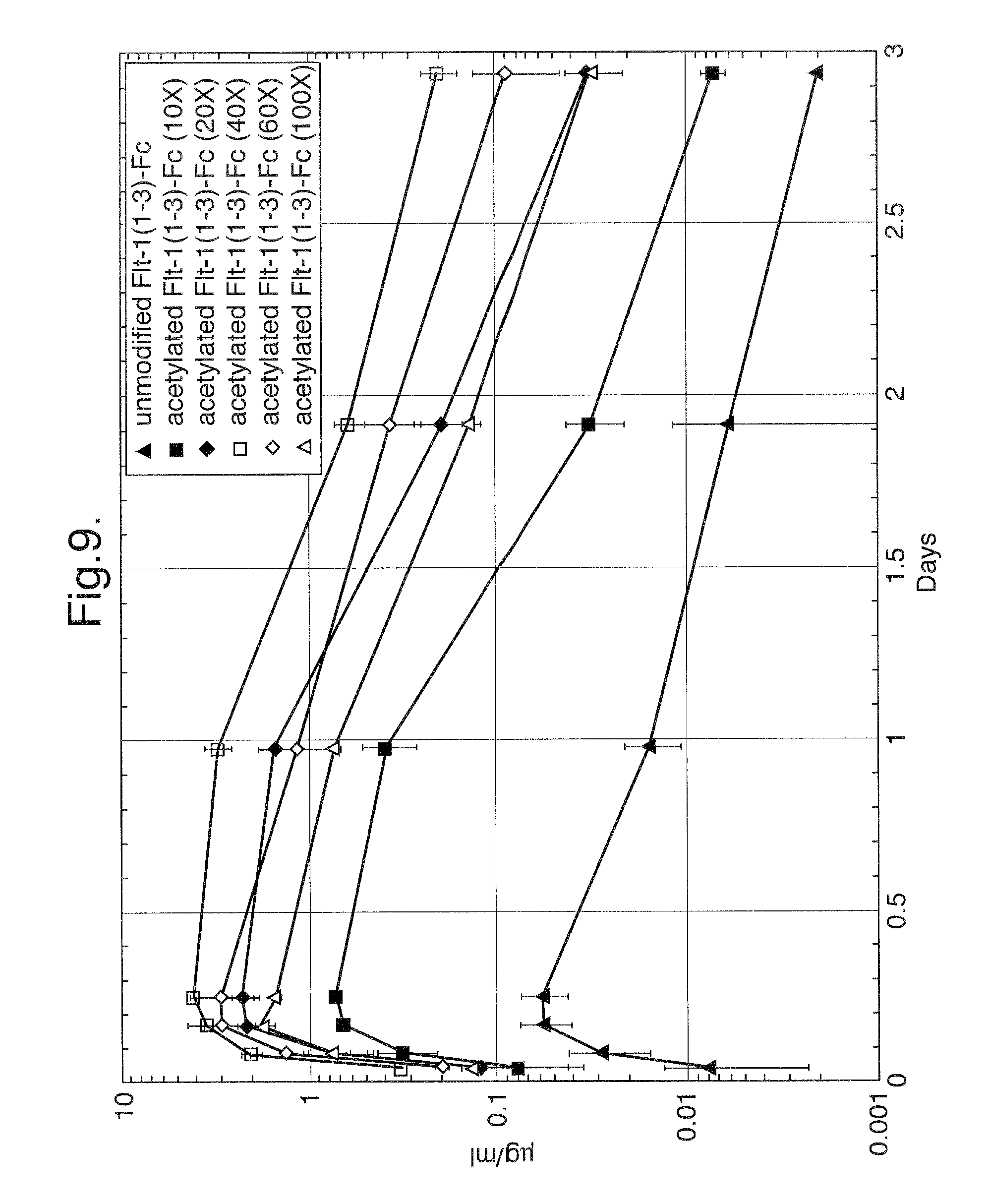
In vivo experiments were designed to assess the pharmacokinetic profiles of unmodified Flt1(1-3)-Fc and step-acetylated Flt1(1-3)-Fc protein. Balb/c mice (23-28 g) were injected subcutaneously with 4 mg/kg of unmodified or 10, 20, 40, 60 and 100 fold molar excess samples of step-acetylated Flt1(1-3)-Fc (3 mice for unmodified, 10, 20 and 40 fold molar excess samples and 2 mice for 60 and 100 fold molar excess samples). The mice were tail bled at 1, 2, 4, 6, 24 hours, 2 days and 3 days after injection. The sera were assayed in an ELISA-based assay designed to detect Flt1(1-3)-Fc (described supra). FIG. 9 details the results of this study. The T.sub.max for all of the Flt1(1-3)-Fc proteins tested was at the 6 hour time point but the C.sub.max was as follows: Unmodified Flt1(1-3)-Fc: 0.06 .mu.g/ml; 10 fold molar excess sample: -0.7 .mu.g/ml, 20 fold molar excess sample -2 .mu.g/ml, 40 fold molar excess sample -4 .mu.g/ml, 60 fold molar excess sample -2 .mu.g/ml, 100 fold molar excess sample -1 .mu.g/ml. This results demonstrates that acetylation or pegylation of Flt1(1-3)-Fc significantly improves its pharmacokinetic profile.
Example 11: Construction of Flt1(1-3)-Fc Basic Region Deletion Mutant Designated Mut1: Flt1(1-3.sub..DELTA.B)-Fc
Based on the observation that acetylated Flt1(1-3)-Fc, which has a pl below 6, has much better pharmacokinetics than the highly positive unmodified Flt1(1-3)-Fc (pl >9.3), it was asked whether the difference in pharmacokinetics could be attributed to the net charge of the protein, which made it stick to negatively charged extracellular matrix components, or whether there were perhaps specific locations on the surface of the Flt1(1-3)-Fc protein that constituted specific binding sites for extracellular matrix components. For example, many proteins are known to have heparin binding sites, often consisting of a cluster of basic residues. Sometimes these residues are found in a cluster on the primary sequence of the protein; some of the literature has identified "consensus sequences" for such heparin binding sites (see for example Hileman, et al., 1998, Bioessays 20(2):156-67). In other cases, the known crystal structure of a protein reveals a cluster of positively charged residues on the surface of a protein, but the residues come from different regions of the primary sequence and are only brought together when the protein folds into its tertiary structure. Thus it is difficult to deduce whether an isolated amino acid residue forms part of a cluster of basic residues on the surface of the protein. However, if there is a cluster of positively charged amino acid residues in the primary sequence, it is not unreasonable to surmise that the residues are spatially close to one another and might therefore be part of an extracellular matrix component binding site. Flt1 receptor has been studied extensively and various domains have been described (see for example Tanaka et al., 1997, Jpn. J. Cancer Res. 88:867-876). Referring to the nucleic acid and amino acid sequence set forth in FIG. 10A-D of this application, one can identify the signal sequence for secretion which is located at the beginning of the sequence and extends to the glycine coded for by nucleotides 76-78. The mature protein begins with Ser-Lys-Leu-Lys, starting at nucleotide 79 of the nucleic acid sequence. Flt1 Ig domain 1 extends from nucleotide 79 to 393, ending with the amino acids Ser-Asp-Thr. Flt1 Ig domain 2 extends from nucleotide 394 to 687 (encoding Gly-Arg-Pro to Asn-Thr-Ile), and Flt1 Ig domain 3 extends from nucleotides 688 to 996 (encoding Ile-Asp-Val to Asp-Lys-Ala). There is a bridging amino acid sequence, Gly-Pro-Gly, encoded by nucleotides 997-1005, followed by the nucleotide sequence encoding human Fc (nucleotides 1006-1701 or amino acids Glu-Pro-Lys to Pro-Gly-Lys-stop).
A more detailed analysis of the Flt1 amino acid sequence reveals that there is a cluster, namely, amino acid residues 272-281 (KNKRASVRR) of FIG. 10A-D, in which 6 out of 10 amino acid residues are basic. This sequence is located in Flt1 Ig domain 3 of the receptor (see FIG. 11), which is not itself essential for binding of VEGF ligand, but which confers a higher affinity binding to ligand. An alignment of the sequence of Ig domain 3 with that of Ig domain 2 reveals that in this region, there is very poor alignment between the two Ig domains, and that there are about 10 additional amino acids in Ig domain 3. An analysis of the hydrophilicity profiles (MACVECTOR.TM. computer software) of these two domains clearly indicates the presence of a hydrophilic region in the protein (FIG. 12A-B). These observations raised the possibility that the actual three dimensional conformation of Flt1 Ig domain 3 allowed for some type of protrusion that is not in Flt1 Ig domain 2. To test this hypothesis, the 10 additional amino acids were deleted and the resulting protein was tested to see whether the deletion would affect the pharmacokinetics favorably without seriously compromising the affinity of the receptor for VEGF. This DNA construct, which was constructed using standard molecular biology techniques in the mammalian expression vector pMT21 (Genetics Institute, Inc., Cambridge, Mass.), is referred to as Mut1: Flt1(1-3.sub..DELTA.B)-Fc. The Mut1: Flt1(1-3.sub..DELTA.B)-Fc construct was derived from Flt1(1-3)-Fc by deletion of nucleotides 814-843 (set forth in FIG. 10A-D), which deletes the highly basic 10-amino acid residue sequence Lys-Asn-Lys-Arg-Ala-Ser-Val-Arg-Arg-Arg from Flt1 Ig domain 3.
The final DNA construct was sequence-verified using an ABI 373A DNA sequencer and Taq Dideoxy Terminator Cycle Sequencing Kit (Applied Biosystems, Inc., Foster City, Calif.). The sequence of Mut1: Flt1(1-3.sub..DELTA.B)-Fc is set forth in FIG. 13A-D.
Example 12: Construction of Flt1(1-3)-Fc Basic Region Deletion Mutant Designated Mut2: Flt1(2-3.sub..DELTA.B)-Fc
A second deletion mutant construct, designated Mut2: Flt1(2-3.sub..DELTA.B)-Fc, was derived from the Mut1: Flt1(1-3.sub..DELTA.B)-Fc construct by deletion of Flt1 Ig domain 1 encoded by nucleotides 79-393 (see FIG. 10A-D); for convenience, nucleotides 73-78 (TCA GGT) were changed to TCC GGA. This introduced a restriction site (BspE1) without altering the associated amino acid sequence, Ser-Gly. This DNA construct, which was constructed using standard molecular biology techniques in the mammalian expression vector pMT21, was also sequence-verified using an ABI 373A DNA sequencer and Taq Dideoxy Terminator Cycle Sequencing Kit. The sequence of Mut2: Flt1(2-3.sub..DELTA.B)-Fc is set forth in FIG. 14A-C.
Example 13: Construction of Flt1(1-3)-Fc Deletion Mutant Designated Mut3: Flt1(2-3)-Fc
A Third Deletion Mutate Construct, Designated Mut3:
Flt1(2-3)-Fc, was constructed the same way as the Mut2: Flt1(2-3.sub..DELTA.B)-Fc construct, except that Flt1 Ig domain 3 was left intact (the basic region amino acids were not deleted). The construct was constructed using standard molecular biology techniques and the final construct was sequence-verified as described supra. The sequence of Mut3: Flt1(2-3)-Fc is set forth in FIG. 15A-C.
Example 14: Construction of Flt(1-3)-Fc Basic Region N-glycosylation Mutant Designated Mut4: Flt1(1-3.sub.R.fwdarw.N)-Fc
A final construct was made in which a N-glycosylation site was introduced into the middle of the basic region of Flt1 Ig domain 3. This construct was designated Mut4: Flt1(1-3.sub.R.fwdarw.N)-Fc and was made by changing nucleotides 824-825 from GA to AC, consequently changing the coded Arg residue (AGA) into an Asn residue (AAC) (see FIG. 10A-D). The resulting amino acid sequence is therefore changed from Arg-Ala-Ser to Asn-Ala-Ser, which matches the canonical signal (Asn-Xxx-Ser/Thr) for the addition of a N-glycosylation site at the Asn residue. The sequence of Mut4: Flt1(1-3.sub.R.fwdarw.N)-Fc is set forth in FIG. 16A-D.
Example 15: Characterization of Acetylated Flt1(1-3)-Fc, Mut1: Flt1(1-3.sub..DELTA.B)-Fc, and Mut4: Flt1(1-3.sub.R.fwdarw.N)-Fc Mutants
Binding to Extracellular Matrix Components.
To determine whether the three modified proteins were more or less likely to have improved pharmacokinetic properties, MATRIGEL.RTM. coated 96-well dishes (as described supra) were incubated with varying concentrations of the mutant proteins and detected with anti-human Fc/alkaline-phosphatase conjugated antibodies. As shown in FIG. 18, this experiment showed that while the unmodified Flt1(1-3)-Fc protein could bind avidly to these wells, the Mut3: Flt1(2-3)-Fc protein bound somewhat more weakly, the Mut1: Flt1(1-3.sub..DELTA.B)-Fc protein bound more weakly still, and the Mut2: Flt1(2-3.sub..DELTA.B)-Fc protein showed the best profile, binding more weakly than any of the other mutant proteins. The Mut4: Flt1(1-3.sub.R.fwdarw.N)-Fc glycosylation mutant protein showed only marginal benefit on the MATRIGEL.RTM. assay. These results confirm the hypothesis that a linear sequence of positive amino acids can be deleted from the primary sequence resulting in a decrease in charge interaction with extracellular matrix components.
Binding of Mut1: Flt1(1-3.sub..DELTA.B)-Fc and Mut4: Flt1(1-3.sub.R.fwdarw.N)-Fc in a BIACORE.TM.-based assay. Unmodified and acetylated Flt1(1-3)-Fc and genetically modified Mut1: Flt1(1-3.sub..DELTA.B)-Fc and Mut4: Flt1(1-3.sub.R.fwdarw.N)-Fc proteins where tested in a BIACORE.TM.-based assay to evaluate their ability to bind to the Flt1 ligand, VEGF. In this assay, unmodified Flt1(1-3)-Fc protein (0.25, 0.5, or 1.0 .mu.g/ml) was immobilized on the surface of a BIACORE.TM. chip and a solution containing 0.1 .mu.g/ml VEGF and either purified or COS cell supernatant containing unmodified Flt1(1-3)-Fc (at approximately (0.25, 0.5, or 1.0 .mu.g/ml), purified acetylated Flt1(1-3)-Fc (at (0.25, 0.5, or 1.0 .mu.g/ml), COS cell supernatant containing Mut1: Flt1(1-3.sub..DELTA.B)-Fc (at approximately (0.25, 0.5, or 1.0 .mu.g/ml), or COS cell supernatant containing Mut4: Flt1(1-3.sub.R.fwdarw.N)-Fc (at approximately (0.25, 0.5, or 1.0 .mu.g/ml) were passed over the Flt1(1-3)-Fc-coated chip. As shown in FIG. 17, at the sub-stoichiometric ratio (0.25 .mu.g/ml Flt1(1-3)-Fc of unmodified, acetylated or genetically modified samples vs. 0.1 .mu.g/ml VEGF), there is insufficient Flt1(1-3)-Fc protein to block binding of VEGF to the Flt1(1-3)-Fc immobilized on the BIACORE.TM. chip. At 0.5 .mu.g/ml of unmodified, acetylated or genetically modified Flt1(1-3)-Fc proteins, the stoichiometric ratio approximates 1:1 and there is an increased ability to block VEGF binding to the BIACORE.TM. chip. At 1.0 .mu.g/ml of unmodified, acetylated or genetically modified Flt1(1-3)-Fc proteins, which is approximately a 10:1 stoichiometric ratio, the Flt1(1-3)-Fc proteins are able to block binding of VEGF to the BIACORE.TM. chip, but they are not equivalent. Unmodified, acetylated, and Mut1: Flt1(1-3.sub..DELTA.B)-Fc are essentially equal in their ability to block VEGF binding, whereas Mut4: Flt1(1-3.sub.R.fwdarw.N)-Fc is somewhat less efficient at blocking binding. These results confirm the hypothesis that it is possible to reduce the non-specific binding of a positively charged molecule by genetically removing a linear sequence of predominantly negatively charged amino acids.
Binding of Mut1: Flt1(1-3.sub..DELTA.B)-Fc, Mut2: Flt1(2-3.sub..DELTA.B)-Fc, Mut3: Flt1(2-3)-Fc, and in an ELISA-based assay. To determine whether the three mutant proteins could bind the Flt1 ligand VEGF, binding experiments were done in which 96-well plates coated with VEGF were incubated with varying concentrations of the respective mutant protein, and after washing, the amount bound was detected by incubating with an alkaline phosphatase conjugated anti-human Fc antibody and quantitated colorimetrically by the addition of an appropriate alkaline phosphatase substrate. As shown in FIG. 19, this experiment showed that all the mutant proteins could bind VEGF similarly, at the concentrations tested.
Example 16: Pharmacokinetic Analysis of Acetylated Flt1(1-3)-Fc, Mut1: Flt1(1-3.sub..DELTA.B)-Fc, and Unmodified Flt1(1-3)-Fc
In vivo experiments were designed to assess the pharmacokinetic profiles of unmodified Flt1(1-3)-Fc, Mut1: Flt1(1-3.sub..DELTA.B)-Fc, and 40 fold molar excess acetylated Flt1(1-3)-Fc protein. Balb/c mice (25-30 g) were injected subcutaneously with 4 mg/kg of unmodified Flt1(1-3)-Fc, 40 fold molar excess acetylated Flt1(1-3)-Fc, and Mut1: Flt1(1-3.sub..DELTA.B)-Fc proteins (4 mice each). These mice were tail bled at 1, 2, 4, 6, 24 hours, 2 days, 3 days, and 5 days after injection. The sera were assayed in an ELISA designed to detect Flt1(1-3)-Fc protein which involves coating an ELISA plate with VEGF, binding the Flt1(1-3)-Fc and reporting with an anti-Fc antibody linked to alkaline phosphatase. As shown in FIG. 20, the C.sub.max for these reagents was as follows: Unmodified Flt1(1-3)-Fc--0.15 .mu.g/ml; 40 fold molar excess acetylated Flt1(1-3)-Fc--1.5 .mu.g/ml; and Mut1: Flt1(1-3.sub..DELTA.B)-Fc--0.7 .mu.g/ml.
Example 17: Modified Flt1 Receptor Vector Construction
The rationale for constructing modified versions of the Flt1 receptor (also known as VEGFR1) was based on the observation that the protein sequence of Flt1 was highly basic, and was therefore likely to stick to extracellular matrix (ECM). The highly basic nature of Flt1 probably explains why unmodified Flt1(1-3)-Fc (described supra) has poor pharmacokinetics that make it difficult to use as a therapeutic agent. As described supra, the chemically modified form of 40 fold molar excess acetylated Flt1(1-3)-Fc, hereinafter termed A40, exhibited a greatly improved pharmacokinetic (PK) profile over the non-acetylated Flt1(1-3)-Fc. Therefore, attempts were made to engineer DNA molecules that could be used to recombinantly express modified forms of a Flt1 receptor molecule that would possess the improved PK profile exhibited by A40 and still maintain the ability to bind tightly to VEGF.
It is known in the literature that the first Ig domain of Flt1 (which has a net charge of +5 at neutral pH) is not essential for tight binding to VEGF, so this domain was deleted. The third Ig domain (having a net charge of +11) is not essential for binding, but confers higher affinity for VEGF than the second Ig domain, so instead of deleting it entirely, it was replaced with the equivalent domains of the Flt1 receptor relatives Flk1 (also known as VEGFR2) and Flt4 (also known as VEGFR3). These chimeric molecules (denoted R1R2 (Flt1.D2.Flk1D3.Fc.DELTA.C1(a) and VEGFR1R2-Fc.DELTA.C1 (a)) and R1R3 (Flt1D2.VEGFR3D3-Fc.DELTA.C1(a) and VEGFR1R3-Fc.DELTA.C1 (a)) respectively, wherein R1 and Flt1D2=Ig domain 2 of Flt1 (VEGFR1); R2 and Flk1D3=Ig domain 3 of Flk1 (VEGFR2); and R3 and VEGFR3D3=Ig domain 3 of Flt4 (VEGFR3)) were much less sticky to ECM, as judged by an in vitro ECM binding assay as described infra, had greatly improved PK as described infra. In addition, these molecules were able to bind VEGF tightly as described infra and block phosphorylation of the native Flk1 receptor expressed in endothelial cells as described infra.
Construction of the expression plasmid pFlt1D2.Flk1D3.Fc.DELTA.C1(a). Expression plasmids pMT21.Flt1(1-3).Fc (6519 bp) and pMT21.Flk-1(1-3).Fc (5230 bp) are plasmids that encode ampicillin resistance and Fc-tagged versions of Ig domains 1-3 of human Flt1 and human Flk1, respectively. These plasmids were used to construct a DNA fragment consisting of a fusion of Ig domain 2 of Flt1 with Ig domain 3 of Flk1, using PCR amplification of the respective Ig domains followed by further rounds of PCR to achieve fusion of the two domains into a single fragment. For Ig domain 2 of Flt1, the 5' and 3' amplification primers were as follows: 5': bsp/flt1D2 (5'-GACTAGCAGTCCGGAGGTAGACCTTTCGTAGAGATG-3') (SEQ ID NO:18)3': Flt1D2-Flk1D3.as (5'-CGGACTCAGAACCACATCTATGATTGTATTGGT-3') (SEQ ID NO:19)
The 5' amplification primer encodes a BspE1 restriction enzyme site upstream of Ig domain 2 of Flt1, defined by the amino acid sequence GRPFVEM (SEQ ID NO:20) (corresponding to amino acids 27-33 of FIGS. 21A-C). The 3' primer encodes the reverse complement of the 3' end of Flt1 Ig domain 2 fused directly to the 5' beginning of Flk1 Ig domain 3, with the fusion point defined as TIID (SEQ ID NO:37) of Flt1 (corresponding to amino acids 123-126 of FIGS. 21A-21C) and continuing into VVLS (SEQ ID NO:38) (corresponding to 127-130 of FIGS. 21A-21C) of Flk1.
For Ig domain 3 of Flk1, the 5' and 3' amplification primers were as follows: 5': Flt1D2-Flk1D3.s (5'-ACAATCATAGATGTGGTTCTGAGTCCGTCTCATGG-3') (SEQ ID NO:21)-3': Flk1D3/apa/srf.as (5'-GATAATGCCCGGGCCCTTTTCATGGACCCTGACAAATG-3') (SEQ ID NO:22).
The 5' amplification primer encodes the end of Flt1 Ig domain 2 fused directly to the beginning of Flk1 Ig domain 3, as described above. The 3' amplification primer encodes the end of Flk1 Ig domain 3, defined by the amino acids VRVHEK (SEQ ID NO:23) (corresponding to amino acids 223-228 of FIGS. 21A-C), followed by a bridging sequence that includes a recognition sequence for the restriction enzyme Srf1, and encodes the amino acids GPG. The bridging sequence corresponds to amino acids 229-231 of FIGS. 21A-C.
After a round of PCR amplification to produce the individual domains, the products were combined in a tube and subjected to a further round of PCR with the primers bsp/flt1D2 and Flk1D3/apa/srf.as (described supra) to produce the fusion product. This PCR product was subsequently digested with the restriction enzymes BspEI and SmaI and the resulting 614 bp fragment was subcloned into the BspEI to SrfI restriction sites of the vector pMT21/.DELTA.B2.Fc, to create the plasmid pMT21/Flt1D2.Flk1D3.Fc. The nucleotide sequence of the Flt1D2-Flk1D3 gene fusion insert was verified by standard sequence analysis. This plasmid was then digested with the restriction enzymes EcoRI and SrfI and the resulting 702 bp fragment was transferred into the EcoRI to SrfI restriction sites of the plasmid pFlt1(1-3)B2-Fc.DELTA.C1(a) to produce the plasmid pFlt1D2.Flk1D3.Fc.DELTA.C1(a). The complete DNA and deduced amino acid sequences of the Flt1D2.Flk1D3.Fc.DELTA.C1(a) chimeric molecule is set forth in FIGS. 21A-C.
Construction of the Expression Plasmid pFlt1D2VEGFR3D3Fc.DELTA.C1(a).
The expression plasmid pMT21.Flt1(1-3).Fc (6519 bp) encodes ampicillin resistance and an Fc-tagged version of Ig domains 1-3 of human Flt1 receptor. This plasmid was used to produce a DNA fragment containing Ig domain 2 of Flt1 by PCR. RNA from the cell line HEL921.7 was used to produce Ig domain 3 of Flk1, using standard RT-PCR methodology. A further round of PCR amplification was used to achieve fusion of the two Ig domains into a single fused fragment. For Ig domain 2 of Flt1, the 5' and 3' amplification primers were as follows: 5': bsp/flt1D2 (5'-GACTAGCAGTCCGGAGGTAGACC-TTTCGTAGAGATG-3') (SEQ ID NO:24)-3': Flt1D2.VEGFR3D3.as (TTCCTGGGCAACAGCTGGAT ATCTATGATTGTATTGGT) (SEQ ID NO:25).
The 5' amplification primer encodes a BspE1 restriction site upstream of Ig domain 2 of Flt1, defined by the amino acid sequence GRPFVEM (SEQ ID NO:20) (corresponding to amino acids 27-33 of FIG. 22A-C). The 3' amplification primer encodes the reverse complement of the end of Flt1 Ig domain 2 fused directly to the beginning of VEGFR3 Ig domain 3, with the fusion point defined as TIID (SEQ ID NO:37) of Flt1 (corresponding to amino acids 123-126 of FIG. 22A-C) and continuing into IQLL (SEQ ID NO:26) of VEGFR3 (corresponding to amino acids 127-130 of FIG. 22A-C).
For Ig domain 3 of VEGFR3, the 5' and 3' primers used for RT-PCR were as follows:
TABLE-US-00001 5': R3D3.s (SEQ ID NO: 27) (ATCCAGCTGTTGCCCAGGAAGTCGCTGGAGCTGCTGGTA) 3': R3D3.as (SEQ ID NO: 28) (ATTTTCATGCACAATGACCTCGGTGCTCTCCCGAAATCG).
Both the 5' and 3' amplification primers match the sequence of VEGFR3. The 296 bp amplification product of this RT-PCR reaction was isolated by standard techniques and subjected to a second round of PCR to add suitable sequences to allow for fusion of the Flt1D2 with the Flk1D3 domains and fusion of the Flk1D3 and Fc domains via a GPG bridge (see below). The amplification primers were as follows:
TABLE-US-00002 5': Flt1D2.VEGFR3D3.s (SEQ ID NO: 29) (TCATAGATATCCAGCTGTTGCCCAGGAAGT-CGCTGGAG) 3': VEGFR3D3/srf.as (SEQ ID NO: 30) (GATAATGCCCGGGCCATTTTCATGCACA-ATGACCTCGGT).
The 5' amplification primer encodes the 3' end of Flt1 Ig domain 2 fused directly to the beginning (5' end) of VEGFR3 Ig domain 3, as described above. The 3' amplification primer encodes the 3' end of VEGFR3 Ig domain 3, defined by the amino acids VIVHEN (SEQ ID NO:31) (corresponding to amino acids 221-226 of FIG. 22A-C), followed by a bridging sequence that includes a recognition sequence for Srf1, and encodes the amino acids GPG. The bridging sequence corresponds to amino acids 227-229 of FIG. 22A-C.
After one round (for Flt1 Ig domain 2) or two rounds (for Flt4 Ig domain 3) of PCR to produce the individual Ig domains, the PCR products were combined in a tube and subjected to a further round of PCR amplification with the amplification primers bsp/flt1D2 and VEGFR3D3/srf.as described supra, to produce the fusion product. This PCR product was subsequently digested with the restriction enzymes BspEI and SmaI and the resulting 625 bp fragment was subcloned into the BspEI to SrfI restriction sites of the vector pMT21/Flt1.DELTA.B2.Fc (described supra), to create the plasmid pMT21/Flt1D2.VEGFR3D3.Fc. The sequence of the Flt1D2-VEGFR3D3 gene fusion insert was verified by standard sequence analysis. This plasmid was then digested with the restriction enzymes EcoRI and SrfI and the resulting 693 bp fragment was subcloned into the EcoRI to SrfI restriction sites of the plasmid pFlt1(1-3).DELTA.B2-Fc.DELTA.C1(a) to produce the plasmid designated pFlt1D2.VEGFR3D3.Fc.DELTA.C1(a). The complete DNA deduced amino acid sequence of the Flt1D2.VEGFR3D3.Fc.DELTA.C1(a) chimeric molecule is set forth in FIG. 22A-C.
Example 18: Extracellular Matrix Binding (ECM) Binding Assay
ECM-coated plates (Becton Dickinson catalog #35-4607) were rehydrated with warm DME supplemented with glutamine (2 mM), 100 U penicillin, 100 U streptomycin, and 10% BCS for at least 1 hr before adding samples. The plates were then incubated for 1 hr at room temperature with varying concentrations of Flt1D2.Flk1D3.Fc.DELTA.C1(a) and Flt1D2.VEGFR3D3.Fc.DELTA.C1(a) starting at 10 nM with subsequent 2-fold dilutions in PBS plus 10% BCS. The plates were then washed 3 times with PBS plus 0.1% Triton-X and incubated with alkaline phosphatase-conjugated anti-human Fc antibody (Promega, 1:4000 in PBS plus 10% BCS) for 1 hr at room temperature. The plates were then washed 4 times with PBS 0.1% Triton-X and alkaline phosphatase buffer/pNPP solution (Sigma) was added for color development. Plates were read at I=405-570 nm. The results of this experiment are shown in FIG. 23 and demonstrate that the Flt1D2.Flk1D3.Fc.DELTA.C1(a) and Flt1D2.VEGFR3D3.Fc.DELTA.C1(a) proteins are considerably less sticky to the ECM as compared to the Flt1(1-3)-Fc protein.
Example 19: Transient Expression of pFlt1D2.Flk1D3.Fc.DELTA.C1(a) in CHO-K1 (E1A) Cells
A large scale (2 L) culture of E. coli DH10B cells carrying the pFlt1D2.Flk1D3.Fc.DELTA.C1(a) plasmid described supra in Example 17 was grown overnight in Terrific Broth (TB) plus 100 .mu.g/ml ampicillin. The next day, the plasmid DNA was extracted using a QIAgen ENDOFREE.TM. Megaprep kit following the manufacturer's protocol. The concentration of the purified plasmid DNA was determined by standard techniques using a UV spectrophotometer and fluorometer. The plasmid DNA was verified by standard restriction enzyme digestion of aliquots using the restriction enzymes EcoRI plus NotI and AseI. All restriction enzyme digest fragments corresponded to the predicted sizes when analyzed on a 1% agarose gel.
Forty 15 cm petri plates were seeded with CHO-K1/E1A cells at a density of 4.times.10.sup.6 cells/plate. Plating media was Gibco Ham's F-12 supplemented with 10% HYCLONE.TM. Fetal Bovine Serum (FBS), 100 U penicillin/100 U streptomycin and glutamine (2 mM). The following day each plate of cells was transfected with 6 .mu.g of the pFlt1D2.Flk1D3.Fc.DELTA.C1(a) plasmid DNA using Gibco Optimem and Gibco Lipofectamine in 12 ml volume, following the manufacturer's protocol. Four hours after adding the transfection mix to the cells, 12 ml/plate of Optimem supplemented with 10% FBS was added. Plates were incubated at 37.degree. C. in a 5% CO.sub.2 incubator overnight. The following day the media was removed from each plate and 25 ml expression media (Gibco CHO-S-SFM II supplemented with glutamine (2 mM) and 1 mM sodium butyrate) was added. The plates were incubated at 37.degree. C. for 3 days. After 3 days of incubation, the media was aspirated from each plate and centrifuged at 400 rpm in a swinging bucket rotor to pellet cells. The supernatant was decanted into sterile 1 L bottles and purification of the expressed protein was performed as described infra.
Example 20: Construction pVEGFR1R2-Fc.DELTA.C1(a) Expression Vector
The pVEGFR1R2.Fc.DELTA.C1(a) expression plasmid was constructed by insertion of DNA encoding amino acids SDT (corresponding to amino acids 27-29 of FIG. 24A-C) between Flt1d2-Flk1d3-Fc.DELTA.C1(a) amino acids 26 and 27 of FIGS. 21A-C (GG) and removal of DNA encoding amino acids GPG corresponding to amino acids 229-231. The SDT amino acid sequence is native to the Flt1 receptor and was added back in to decrease the likelihood of heterogeneous N-terminal processing. The GPG (bridging sequence) was removed so that the Flt1 and Flk1 Ig domains were fused directly to one another. The complete DNA and deduced amino acid sequences of the pVEGFR1R2.Fc.DELTA.C1(a) chimeric molecule is set forth in FIG. 24A-C.
Example 21: Cell Culture Process Used to Produce Modified Flt1 Receptors
Cell Culture Process Used to Produce Flt1D2.Flk1D3.Fc.DELTA.C1(a).
The process for production of Flt1D2.Flk1D3.Fc.DELTA.C1(a) protein using the expression plasmid pFlt1D2.Flk1D3.Fc.DELTA.C1(a) described supra in Example 1 involves suspension culture of recombinant Chinese hamster ovary (CHO K1/E1A) cells which constitutively express the protein product. The cells are grown in bioreactors and the protein product is isolated and purified by affinity and size exclusion chromatography. The process is provided in greater detail below.
Cell Expansion.
Two confluent T-225 cm.sup.2 flasks containing the Flt1D2.Flk1D3.Fc.DELTA.C1(a) expressing cell line were expanded by passaging cells into eight T-225 cm.sup.2 flasks in medium (GMEM +10% serum, GIBCO) and incubated at 37.degree. C. and 5% CO.sub.2. When the flasks approached confluence (approximately 3 to 4 days) the cells were detached using trypsin. Fresh medium was added to protect the cells from further exposure to the trypsin. The cells were centrifuged and resuspended in fresh medium then transferred to eight 850 cm.sup.2 roller bottles and incubated at 37.degree. C. and 5% CO.sub.2 until confluent.
Suspension Culture in Bioreactors.
Cells grown in roller bottles were trypsinized to detach them from the surface and washed with suspension culture medium. The cells are aseptically transferred to a 5 L bioreactor (New Brunswick Celligen Plus) where the cells are grown in 3.5 L of suspension culture. The suspension culture medium was a glutamine-free low glucose modification of IS-CHO (Irvine Scientific) to which 5% fetal bovine serum (HYCLONE.TM.), GS supplement (Life Technologies) and 25 .mu.M methionine sulfoximine (Sigma) was added. The pH was controlled at 7.2 by addition of carbon dioxide to the inlet gas or by addition of a liquid solution of sodium carbonate to the bioreactor. Dissolved oxygen level was maintained at 30% of saturation by addition of oxygen or nitrogen to the inlet gas and temperature controlled at 37.degree. C. When a density of 4.times.10.sup.6 cells/mL was reached the cells were transferred to a 40 L bioreactor containing the same medium and setpoints for controlling the bioreactor. The temperature setpoint was reduced to 34.degree. C. to slow cell growth and increase the relative rate of protein expression.
Cell Culture Process Used to Produce Flt1D2.VEGFR3D3.Fc.DELTA.C1(a).
The same methodologies as described supra for Flt1D2.Flk1D3.Fc.DELTA.C1(a) were used to produce Flt1 D2.VEGFR3D3.Fc.DELTA.C1 (a).
Example 22: Harvest and Purification of Modified Flt1 Receptors
Harvest and Purification of Flt1D2.Flk1D3.Fc.DELTA.C1(a).
The product protein was aseptically harvested from the bioreactor while retaining cells using Millipore Prostak tangential-flow filtration modules and a low-shear mechanical pump (Fristam). Fresh medium was added to the bioreactor to replace that removed during the harvest filtration. Approximately 40 L of harvest filtrate was then loaded onto a 400 mL column containing Protein A SEPHAROSE.TM. resin (Amersham Pharmacia). After loading the resin was washed with buffer containing 10 mM sodium phosphate, 500 mM sodium chloride, pH 7.2 to remove any unbound contaminating proteins. Flt1D2.Flk1D3.Fc.DELTA.C1(a) protein was eluted with a pH 3.0 citrate buffer. The eluted protein was neutralized by addition of Tris base and frozen at -20.degree. C.
Several frozen lots of Flt1D2.Flk1D3.Fc.DELTA.C1(a) protein from the Protein A step above were thawed, pooled and concentrated using a Millipore 30 kD nominal molecular weight cutoff (NMWCO) tangential flow filtration membrane. The protein was transferred to a stirred cell concentrator (Millipore) and further concentrated to 30 mg/mL using a 30 kD NMWCO membrane. The concentrated protein was loaded onto a size exclusion column packed with SUPERDEX.TM. 200 resin (Amersham Pharmacia) that was equilibrated with phosphate buffered saline plus 5% glycerol. The same buffer was used to run the column. The fractions corresponding to Flt1D2.Flk1D3.Fc.DELTA.C1(a) dimer were pooled, sterile filtered through a 0.22 micron filter, aliquoted and frozen.
Harvest and Purification of Flt1D2.VEGFR3D3.Fc.DELTA.C1(a).
The same methodologies as described supra for Flt1D2.Flk1D3.Fc.DELTA.C1(a) were used to harvest and purify Flt1D2.VEGFR3D3.Fc.DELTA.C1(a).
Example 23: Phosphorylation Assay for Transiently Expressed VEGFR2
Primary human umbilical vein endothelial cells (HUVECs), passage 4-6, were starved for 2 hrs in serum-free DME high glucose media. Samples containing 40 ng/ml (1 nM) human VEGF165, which is a ligand for the VEGF receptors Flt1, Flk1 and Flt4(VEGFR3) were prepared and were preincubated for 1 hr at room temperature with varying amounts of the modified Flt1 receptors Flt1(1-3)-Fc, Flt1(1-3)-Fc (A40), Flt1D2Flk1D3.Fc.DELTA.C1(a) and Flt1D2VEGFR3D3.Fc.DELTA.C1(a) in serum-free DME-high glucose media containing 0.1% BSA. Cells were challenged for 5 minutes with the samples prepared above +/-VEGF165, followed by whole cell lysis using complete lysis buffer. Cell lysates were immunoprecipitated with an antibody directed against the C-terminus of VEGFR2 receptor. The immunoprecipitated lysates were loaded onto 4-12% SDS-PAGE Novex gel and then transferred to PVDF membrane using standard transfer methodologies. Detection of phosphorylated VEGFR2 was done by immunoblotting with the anti-phospho Tyrosine mAb called 4G10 (UBI) and developed using ECL-reagent (Amersham). FIGS. 25A-C and 26A-B show the results of this experiment. FIG. 25A-C reveals that detection by Western blot of tyrosine phosphorylated VEGFR2(Flk1) by VEGF165 ligand stimulation shows that cell-surface receptors are phosphorylated to varying levels depending on which modified Flt1 receptor is used during the preincubations with VEGF. As is seen in FIG. 25A, at a 1.5 molar excess of either Flt1(1-3)-Fc, Flt1(1-3)-Fc (A40) or transient Flt1D2Flk1D3.Fc.DELTA.C1(a) there is complete blockage of receptor stimulation by these three modified Flt1 receptors as compared to control media challenge. In contrast, transient Flt1D2VEGFR3D3.Fc.DELTA.C1(a) does not show significant blockage at this molar excess, as compared with VEGF positive control challenge. Similar results are seen in FIG. 25B, where the modified Flt receptors are in a 3-fold molar excess to VEGF165 ligand. In FIG. 25C, where the modified Flt1 receptors are in a 6-fold molar excess to VEGF165 ligand, transient Flt1D2VEGFR3D3.Fc.DELTA.C1(a) can now be shown to be partially blocking VEGF165-induced stimulation of cell-surface receptors.
In FIG. 26A-26B, detection by Western blot of tyrosine phosphorylated VEGFR2(Flk1) by VEGF165 ligand stimulation shows that cell-surface receptors are not phosphorylated by challenge samples which have VEGF165 preincubated with 1 and 2 fold molar excess (FIG. 26A) or 3 and 4 fold molar excess (FIG. 26B) of either transient Flt1D2Flk1D3.Fc.DELTA.C1(a), stable Flt1D2Flk1D3.Fc.DELTA.C1(a), or transient VEGFR1R2-Fc.DELTA.C1(a). At all modified Flt1 receptor concentrations tested there is complete binding of VEGF165 ligand during the preincubation, resulting in no detectable stimulation of cell-surface receptors by unbound VEGF165 as compared to control media challenge.
Example 24: Cell Proliferation Bioassay
The test cell population is MG87 cells that have been stably transfected with a expression plasmid that contains a DNA insert encoding the VEGFR2(Flk1) extracellular domain fused to the TrkB intracellular kinase domain, thus producing a chimeric molecule. The reason the TrkB intracellular kinase domain was used rather than the native VEGFR2(Flk1) intracellular kinase domain is that the intracellular kinase domain of VEGFR2(Flk1) does not cause a strong proliferative response when stimulated by VEGF165 in these cells. It is known that MG87 cells containing full length TrkB receptor give a robust proliferative response when stimulated with BDNF, so the TrkB intracellular kinase domain was engineered to replace the intracellular kinase domain of VEGFR2(Flk1) to take advantage of this proliferative response capability.
Five thousand cells/well were plated in a 96 well plate and allowed to settle for 2 hrs at 37.degree. C. The following modified Flt receptors Flt1(1-3)-Fc, Flt1D2.Flk1D3.Fc.DELTA.C1(a) and Flt1D2.VEGFR3D3.Fc.DELTA.C1(a), plus an irrelevant receptor termed Tie2-Fc as a negative control, were titrated from 40 nM to 20 pM and incubated on the cells for 1 hr at 37.degree. C. Human recombinant VEGF165 in defined media was then added to all the wells at a concentration of 1.56 nM. The plates were incubated for 72 hrs at 37.degree. C. and then MTS (Owen's reagent, Promega) added and the plates were incubated for an additional for 4 hrs. Finally, the plates were read on a spectrophotometer at 450/570 nm. The results of this experiment are shown in FIG. 27. The control receptor Tie2-Fc does not block VEGF165-induced cell proliferation at any concentration whereas Flt1D2.Flk1D3.Fc.DELTA.C1(a) blocks 1.56 nM VEGF165 with a half maximal dose of 0.8 nM. Flt1(1-3)-Fc and Flt1D2.VEGFR3D3.Fc.DELTA.C1(a) are less effective in blocking VEGF165 in this assay with a half maximal dose of .about.2 nM. VEGF165 alone gives a reading of 1.2 absorbance units and the background is 0.38 absorbance units.
Example 25: Binding Stoichiometry of Modified Flt Receptors to VEGF165
BIACORE.TM. Analysis.
The stoichiometry of Flt1D2Flk1D3.Fc.DELTA.C1(a) and VEGFR1R2-Fc.DELTA.C1(a) interaction with human VEGF165 was determined by measuring either the level of VEGF saturation binding to the Flt1D2Flk1D3.Fc.DELTA.C1(a) or VEGFR1R2-Fc.DELTA.C1(a) surfaces or measuring concentration of VEGF165 needed to completely prevent binding of Flt1D2Flk1D3.Fc.DELTA.C1(a) or VEGFR1R2-Fc.DELTA.C1(a) to VEGF BIACORE.TM. chip surface.
Modified Flt receptors Flt1D2Flk1D3.Fc.DELTA.C1(a) and VEGFR1R2-Fc.DELTA.C1(a), were captured with an anti-Fc specific antibody that was first immobilized on a BIACORE.TM. chip using amine-coupling chemistry. A blank antibody surface was used as a negative control. VEGF165 was injected at a concentration of 1 nM, 10 nM, and 50 nM over the Flt1D2Flk1D3.Fc.DELTA.C1(a) and VEGFR1R2-Fc.DELTA.C1(a) surfaces at 10 .mu.l/min for one hour. A real-time binding signal was recorded and saturation binding was achieved at the end of each injection. Binding stoichiometry was calculated as a molar ratio of bound VEGF165 to the immobilized Flt1D2Flk1D3.Fc.DELTA.C1(a) or VEGFR1R2-Fc.DELTA.C1(a), using the conversion factor of 1000 RU equivalent to 1 ng/ml. The results indicated binding stoichiometry of one VEGF165 dimeric molecule per one Flt1D2Flk1D3.Fc.DELTA.C1(a) or VEGFR1R2-Fc.DELTA.C1(a) molecule (FIG. 28).
In solution, Flt1D2Flk1D3.Fc.DELTA.C1(a) or VEGFR1R2-Fc.DELTA.C1(a) at a concentration of 1 nM (estimated to be 1000 times higher than the KD of the Flt1D2Flk1D3.Fc.DELTA.C1(a) or VEGFR1R2-Fc.DELTA.C1(a)/VEGF165 interaction) were mixed with varied concentrations of VEGF165. After one hour incubation, concentrations of the free Flt1D2Flk1D3.Fc.DELTA.C1(a) in solution were measured as a binding signal to an amine-coupled VEGF165 surface. A calibration curve was used to convert the Flt1D2Flk1D3.Fc.DELTA.C1(a) BIACORE.TM. binding signal to its molar concentration. The data showed that the addition of 1 nM VEGF165 into the Flt1D2Flk1D3.Fc.DELTA.C1(a) solution completely blocked Flt1D2Flk1D3.Fc.DELTA.C1(a) binding to the VEGF165 surface. This result suggested the binding stoichiometry of one VEGF165 molecule per one Flt1D2Flk1D3.Fc.DELTA.C1(a) molecule (FIG. 29 and FIG. 30). When the concentration of Flt1D2Flk1D3.Fc.DELTA.C1(a) was plotted as a function of added concentration of VEGF165, the slope of the linear portion was -1.06 for Flt1D2Flk1D3.Fc.DELTA.C1(a) and -1.07 for VEGFR1R2-Fc.DELTA.C1(a). The magnitude of the slope, very close to negative one, was indicative that one molecule of VEGF165 bound to one molecule of either Flt1D2Flk1D3.Fc.DELTA.C1(a) or VEGFR1R2-Fc.DELTA.C1(a).
Size Exclusion Chromatography.
Flt1D2Flk1D3.Fc.DELTA.C1(a) was mixed with a 3-fold excess of VEGF165 and the receptor-ligand complex was purified using a Pharmacia SUPEROSE.TM. 6 size exclusion chromatography column. The receptor-ligand complex was then incubated in a buffer containing 6 M guanidine hydrochloride in order to dissociate it into its component proteins. Flt1D2Flk1D3.Fc.DELTA.C1(a) was separated from VEGF165 using SUPEROSE.TM. 6 size exclusion chromatography column run in 6 M guanidium chloride. In order to determine complex stoichiometry, several injections of Flt1D2Flk1D3.Fc.DELTA.C1(a) and VEGF165 were made and peak height or peak integrated intensity was plotted as a function of the concentration of injected protein. The calibration was done under condition identical to one used in separating components of Flt1D2Flk1D3.Fc.DELTA.C1(a)/VEGF complex. Quantification of the Flt1D2 Flk1D3. Fc.DELTA.C1(a)/VEGF complex composition was based on the calibration curves. FIG. 28 shows the ratio of VEGF165 to Flt1D2Flk1D3.Fc.DELTA.C1(a) in a complex to be 1:1.
Example 26: Determination of the Binding Stoichiometry of Flt1D2Flk1D3.Fc.DELTA.C1(a)/VEGF165 Complex by Size Exclusion Chromatography
Flt1D2Flk1D3.Fc.DELTA.C1(a)/VEGF165 Complex Preparation.
VEGF165 (concentration=3.61 mg/ml) was mixed with CHO cell transiently expressed Flt1D2.Flk1D3.Fc.DELTA.C1(a) (concentration=0.9 mg/ml) in molar ratio of 3:1 (VEGF165:Flt1D2.Flk1D3.Fc.DELTA.C1(a)) and incubated overnight at 4.degree. C.
Size Exclusion Chromatography (SEC) Under Native Conditions.
To separate the complex from excess of unbound VEGF165, 50 .mu.l of the complex was loaded on a Pharmacia SUPEROSE.TM. 12 PC 3.2/30 which was equilibrated in PBS buffer. The sample was eluted with the same buffer at flow rate 40 .mu.l/min. at room temperature. The results of this SEC are shown in FIG. 31. Peak #1 represents the complex and peak #2 represents unbound VEGF165. Fractions eluted between 1.1 and 1.2 ml were combined and guanidinium hydrochloride (GuHCl) was added to a final concentration 4.5 M to dissociate the complex.
Size Exclusion Chromatography (SEC) Under Dissociative Conditions.
To separate the components of the receptor-ligand complex and to determine their molar ratio, 50 .mu.l of dissociated complex as described supra was loaded onto a SUPEROSE.TM. 12 PC 3.2/30 equilibrated in 6 M GuHCl and eluted with the same solution at a flow rate 40 .mu.l/min at room temperature. The results of this SEC are shown in FIG. 32. Peak #1 represents Flt1D2Flk1D3.Fc.DELTA.C1(a) and peak #2 represents VEGF165.
Calculation of Flt1D2Flk1D3.Fc.DELTA.C1(a):VEGF165 Complex Stoichiometry.
The stoichiometry of the receptor-ligand complex was determined from the peak area or the peak height of the components. Concentrations of VEGF165 and Flt1D2Flk1D3.Fc.DELTA.C1(a) corresponding to the peak height or peak area, respectively, were obtained from the standard curves for VEGF165 and Flt1D2Flk1D3.Fc.DELTA.C1(a). To obtain a standard curve, four different concentrations (0.04 mg/ml-0.3 mg/ml) of either component were injected onto a Pharmacia SUPEROSE.TM. 12 PC 3.2/30 column equilibrated in 6 M guanidinium chloride and eluted with the same solution at flow rate 40 .mu.l/min at room temperature. The standard curve was obtained by plotting peak area or peak height vs protein concentration. The molar ratio of VEGF165:Flt1D2Flk1D3.Fc.DELTA.C1(a) determined from the peak area of the components was 1.16. The molar ratio of VEGF165:Flt1D2Flk1D3.Fc.DELTA.C1(a) determined from the peak height of the components was 1.10.
Example 27: Determination of the Stoichiometry of the Flt1D2Flk1D3.Fc.DELTA.C1(a)/VEGF165 Complex by Size Exclusion Chromatography with On-Line Light Scattering
Complex preparation.
VEGF165 was mixed with CHO transiently expressed Flt1D2.Flk1D3.Fc.DELTA.C1(a) protein in molar ratio of 3:1 (VEGF165:Flt1D2Flk1D3.Fc.DELTA.C1(a)) and incubated overnight at 4.degree. C.
Size Exclusion Chromatography (SEC) with On-Line Light Scattering.
Size exclusion chromatography column with a MiniDawn on-line light scattering detector (Wyatt Technology, Santa Barbara, Calif.) and refractive index (RI) detectors (Shimadzu, Kyoto, Japan) was used to determine the molecular weight (MW) of the receptor-ligand complex. Samples were injected onto a SUPEROSE.TM. 12 HR 10/30 column (Pharmacia) equilibrated in PBS buffer and eluted with the same buffer at flow rate 0.5 ml/min. at room temperature. As shown in FIG. 33, the elution profile shows two peaks. Peak #1 represents the receptor-ligand complex and peak #2 represents the unbound VEGF165. MW was calculated from LS and RI signals. The same procedure was used to determine MW of the individual components of the receptor-ligand complex. The results of these determinations are as follows: MW of the Flt1D2Flk1D3.Fc.DELTA.C1(a)/VEGF165 complex at the peak position is 157 300 (FIG. 33), the MW of VEGF165 at the peak position is 44 390 (FIG. 34) and the MW of R1R2 at the peak is 113 300 (FIG. 35).
These data indicated that the stoichiometry of the Flt1D2Flk1D3.Fc.DELTA.C1(a)/VEGF complex is 1:1 as its corresponds to the sum of molecular weights for Flt1D2Flk1D3.Fc.DELTA.C1(a) and VEGF165. Importantly, this method conclusively proved that the Flt1D2Flk1D3.Fc.DELTA.C1(a)/VEGF165 complex was indeed composed of only one molecule of VEGF165 ligand and only one molecule of the Flt1D2Flk1D3.Fc.DELTA.C1(a).
Example 28: Peptide Mapping of Flt1D2.Flk1D3.Fc.DELTA.C1(a)
The disulfide structures and glycosylation sites in Flt1D2.Flk1D3.Fc.DELTA.C1(a) were determined by a peptide mapping method. In this method, the protein was first cleaved with trypsin. Tryptic fragments were analyzed and identified by HPLC coupled with mass spectrometry, in addition to an N-terminal sequencing technique. Reduction of the tryptic digest was employed to help identify disulfide-bond-containing fragments. Treatment of the tryptic digest with PNGase F (Glyko, Novato, Calif.) was employed to help identify fragments with N-linked glycosylation sites. The results are summarized in the accompanying FIG. 36.
There are a total of ten cysteines in Flt1D2.Flk1D3.Fc.DELTA.C1(a); six of them belong to the Fc region. Cys27 has been confirmed to be disulfide bonded to Cys76. Cys121 is confirmed to be disulfide bonded to Cys182. The first two cysteines in the Fc region (Cys211 and Cys214) form an intermolecular disulfide bond with the same two cysteines in another Fc chain. However, because these two cysteines can not be separated enzymatically from each other, it can not be determined whether disulfide bonding is occurring between same cysteines (Cys211 to Cys211, for example) or between Cys211 and Cys214. Cys216 is confirmed to be disulfide bonded to Cys306. Cys352 is confirmed to be disulfide bonded to Cys410.
There are five possible N-linked glycosylation sites in Flt1D2.Flk1D3.Fc.DELTA.C1(a). All five of them are found to be glycosylated to varying degrees. Complete glycosylation was observed at Asn33 (amino acid sequence NIT), Asn193 (amino acid sequence NST), and Asn282 (amino acid sequence NST). In addition, partial glycosylation is observed on Asn65 and Asn120. Sites of glycosylation are highlighted by underline in the FIG. 36.
Example 29: Pharmacokinetic Analysis of Modified Flt Receptors
Pharmacokinetic analysis of Flt1(1-3)-Fc (A40), Flt1D2.Flk1D3.Fc.DELTA.C1(a) and VEGFR1R2-Fc.DELTA.C1(a). Balb/c mice (25-30 g) were injected subcutaneously with 4 mg/kg of Flt1(1-3)-Fc (A40), CHO transiently expressed Flt1D2.Flk1D3.Fc.DELTA.C1(a), CHO stably expressed Flt1D2.Flk1D3.Fc.DELTA.C1(a), and CHO transiently expressed VEGFR1R2-Fc.DELTA.C1(a). The mice were tail bled at 1, 2, 4, 6, 24 hrs, 2 days, 3 days and 6 days after injection. The sera were assayed in an ELISA designed to detect Flt1(1-3)-Fc (A40), Flt1D2.Flk1D3.Fc.DELTA.C1(a) or VEGFR1R2-Fc.DELTA.C1(a). The ELISA involves coating an ELISA plate with VEGF165, binding the detect Flt1(1-3)-Fc (A40), Flt1D2.Flk1D3.Fc.DELTA.C1(a) or VEGFR1R2-Fc.DELTA.C1(a) and reporting with an anti-Fc antibody linked to horse radish peroxidase. The results of this experiments are shown in FIG. 37. The T.sub.max for Flt1(1-3)-Fc (A40) was at 6 hrs while the T.sub.max for the transient and stable Flt1D2.Flk1D3.Fc.DELTA.C1(a) and the transient VEGFR1R2-Fc.DELTA.C1(a) was 24 hrs. The C.sub.max for Flt1(1-3)-Fc (A40) was 8 .mu.g/ml. For both transients (Flt1D2.Flk1D3.Fc.DELTA.C1(a) and VEGFR1R2-Fc.DELTA.C1(a)) the C.sub.max was 18 .mu.g/ml and the C.sub.max for the stable VEGFR1R2-Fc.DELTA.C1(a) was 30 .mu.g/ml.
Pharmacokinetic analysis of Flt1(1-3)-Fc (A40), Flt1D2.Flk1D3.Fc.DELTA.C1(a) and Flt1D2.VEGFR3D3.Fc.DELTA.C1(a). Balb/c mice (25-30 g) were injected subcutaneously with 4 mg/kg of Flt1(1-3)-Fc (A40), CHO transiently expressed Flt1D2.Flk1D3.Fc.DELTA.C1(a) and CHO transiently expressed Flt1D2.VEGFR3D3.Fc.DELTA.C1(a). The mice were tail bled at 1, 2, 5, 6, 7, 8, 12, 15 and 20 days after injection. The sera were assayed in an ELISA designed to detect Flt1(1-3)-Fc, Flt1D2.Flk1D3.Fc.DELTA.C1(a) and Flt1D2.VEGFR3D3.Fc.DELTA.C1(a). The ELISA involves coating an ELISA plate with VEGF 165, binding the Flt1(1-3)-Fc, Flt1D2.Flk1D3.Fc.DELTA.C1(a) or Flt1D2.VEGFR3D3.Fc.DELTA.C1(a) and reporting with an anti-Fc antibody linked to horse radish peroxidase. Flt1(1-3)-Fc (A40) could no longer be detected in the serum after day 5 whereas, Flt1D2.Flk1D3.Fc.DELTA.C1(a) and Flt1D2.VEGFR3D3.Fc.DELTA.C1(a) were detectable for 15 days or more. The results of this experiment are shown in FIG. 38.
Example 30: Ability of Flt1D2.Flk1D3.Fc.DELTA.C1(a) to Inhibit Tumor Growth In Vivo
To evaluate the ability of Flt1D2.Flk1D3.Fc.DELTA.C1(a) to inhibit tumor growth in vivo a model in which tumor cell suspensions are implanted subcutaneously on the right flank of male severe combined immunodeficiency (SCID) mice was employed. Two cell lines, the human HT-1080 fibrosarcoma cell line (ATCC accession no. CCL-121) and the rat C6 glioma cell line (ATCC accession no. CCL-107), each of which exhibit distinctly different morphologies and growth characteristics, were used in the assay. The first dose of Flt1D2.Flk1D3.Fc.DELTA.C1(a) (at 25 mg/Kg or as indicated in FIGS. 39 and 40) was given on the day of tumor implantation. Animals subsequently received subcutaneous injections of Flt1(1-3)-Fc (A40), Flt1D2.Flk1D3.Fc.DELTA.C1(a) or vehicle either every other day (EOD) or two times per week (2.times./wk) for a period of 2 weeks. After 2 weeks, animals were perfused with fixative, tumors were removed and samples were blinded. Tumor volume was determined by measuring the length and width of visible subcutaneous tumors. Both of Flt1(1-3)-Fc (A40) and Flt1D2.Flk1D3.Fc.DELTA.C1(a) significantly reduced the growth of tumors formed by HT-1080 and C6 cells. The results of these experiments are shown in FIG. 39 and FIG. 40.
Example 31: Effect of VEGF165 and Modified Flt Receptors in Female Reproductive System
The stereotypic pattern of vascular remodeling which occur in the uterus and ovary over the course of the reproductive cycle has been well characterized, making these tissues particularly well suited to the study of mechanisms which regulate angiogenesis, vascular remodeling and vascular regression. Indeed, in situ hybridization studies in the reproductive tissues provided the first clear evidence that VEGF acts as a mediator of physiological angiogenesis in mature rodents, as well as humans and non-human primates. As cyclic angiogenesis and vascular remodeling are prominent features of the normal ovary and uterus, it is not surprising that abnormal blood vessel growth and/or vascular dysfunction have been found to characterize many pathological conditions which affect these organs. Furthermore, these pathogenic vascular abnormalities are thought to be caused or perpetuated by the dysregulated expression of one or more angiogenic or anti-angiogenic factors, most prominently VEGF.
For example, abnormal angiogenesis is characteristic of polycystic ovary disease, endometriosis and endometrial carcinoma, and in each case VEGF is over expressed in the affected tissue. Overexpression of VEGF is also thought to play a pathogenic role in the establishment of systemic vascular hyperpermeability in ovarian hyperstimulation syndrome. In addition, VEGF has been implicated as the permeability factor responsible for the production of ascites associated with ovarian carcinoma and other. Agents which effectively neutralize the biological actions of VEGF can reasonably be anticipated to be of therapeutic benefit in the above and related conditions.
Angiogenesis and vascular remodeling are also hallmarks of blastocyst implantation and placental development. VEGF is strongly expressed both in the maternal decidua and in embryonic trophoblasts, where it is thought to first stimulate expansion and hyperpermeability of the uterine vasculature during the peri-implantation period and subsequently mediate formation of both the maternal and embryonic components of the placental vasculature. VEGF is also required for luteal angiogenesis and associated progesterone secretion necessary to prepare the uterus for implantation. Thus, agents which inhibit the biological actions of VEGF may prove to be useful as contraceptive agents (by preventing implantation), or as an abortifacients in the early stages of gestation. The latter application might find particular use as a non-surgical intervention for the termination of ectopic pregnancies.
While the expression of VEGF receptors is largely confined to the vascular endothelium in normal reproductive tissues, Flt1 is also expressed by trophoblasts in the placenta in both humans and animals where it has been proposed to play a role in trophoblast invasion. Interestingly, both Flt1 and KDR (Flk1) are expressed by choriocarcinoma cell line BeWo, and VEGF has been shown to promote DNA synthesis and tyrosine phosphorylation of MAP kinase in these cells. Furthermore, primary and metastatic ovarian carcinomas not only to express high levels of VEGF, but--in addition to the vascular endothelium--the tumor cells themselves express KDR and/or Flt1. These findings suggest that VEGF may not only be critically involved in the generation and maintenance of tumor vasculature, but that at least in some tumors of reproductive origin VEGF may subserve an autocrine role, directly supporting the survival and proliferation of the tumor cells. Thus agents which block the actions of VEGF may have particularly beneficial applications to the treatment of tumors of reproductive origin.
Assessment of VEGF-Induced Uterine Hyperpermeability.
Pregnant mare's serum gonadotrophin (PMSG) was injected subcutaneously (5 IU) to induce ovulation in prepubertal female rats. This results in a surge of estradiol after 2 days which in turn causes an induction of VEGF in the uterus. It is reported that this induction results in hyperpermeability of the uterus and an increase in uterine wet weight 6 hrs. later and, therefore, could potentially be blocked by the modified Flt receptors Flt1(1-3)-Fc (A40), Flt1D2.Flk1D3.Fc.DELTA.C1(a) and Flt1D2.VEGFR3D3.Fc.DELTA.C1(a). In this in vivo model, the normal weight of the rat uterus is about 50 mg and this can be induced to 300-350 mg by PMSG. Desiccation of the tissue reveals that this is all water weight. Subcutaneous injection of Flt1(1-3)-Fc (A40), Flt1D2.Flk1D3.Fc.DELTA.C1(a) and Flt1D2.VEGFR3D3.Fc.DELTA.C1(a) at 25 mg/kg at 1 hr after PMSG injection results in about a 50% inhibition of the increase in uterine wet weight. Increasing the dose of modified Flt receptor does not further reduce the increase in wet weight suggesting that there is a VEGF-independent component to this model. The results of this experiment are shown in FIG. 41.
Assessment of Corpus Luteum Angiogenesis Using Progesterone as a Readout.
Pregnant mare's serum gonadotrophin (PMSG) is injected subcutaneously (5 IU) to induce ovulation in prepubertal female rats. This results in a fully functioning corpus luteum containing a dense network of blood vessels after 4 days that allows for the secretion of progesterone into the blood stream in order to prepare the uterus for implantation. The induction of angiogenesis in the corpus luteum requires VEGF; therefore, blocking VEGF would result in a lack of new blood vessels and thus a lack of progesterone secreted into the blood stream. In this in vivo model, resting levels of progesterone are about 5 ng/ml and this can be induced to a level of 25-40 ng/ml after PMSG. Subcutaneous injection of Flt1(1-3)-Fc (A40) or Flt1D2.Flk1D3.Fc.DELTA.C1(a) at 25 mg/kg or 5 mg/kg at 1 hr after PMSG injection results in a complete inhibition of the progesterone induction on day 4. The results of this experiment are shown in FIG. 42A-B.
Example 33: Pharmacokinetic Analysis of Flt1(1-3)-Fc (A40) and Pegylated Flt1(1-3)-Fc
Flt1(1-3)-Fc was PEGylated with either 10 kD PEG or 20 kD PEG and tested in balb/c mice for their pharmacokinetic profile. Both PEGylated forms of Flt1(1-3)-Fc were found to have much better PK profiles than Flt1(1-3)-Fc (A40), with the T.sub.max occurring at 24 hrs for the PEGylated molecules as opposed to 6 hrs for Flt1(1-3)-Fc (A40).
Example 34: VEGF165 ELISA to Test Affinity of Modified Flt1 Receptor Variants
Ten pM of VEGF165 was incubated overnight at room temperature with modified Flt1 receptor variants ranging from 160 pM to 0.1 pM. The modified Flt1 receptor variants used in this experiment were Flt1(1-3)-Fc, Flt1(1-3)-Fc (A40), transiently expressed Flt1D2Flk1D3.Fc.DELTA.C1(a), transiently expressed Flt1D2VEFGFR3D3-Fc.DELTA.C1(a), Flt1-(1-3.sub.NAS)-Fc, Flt1(1-3.sub.R.fwdarw.C)-Fc and Tie2-Fc. Flt1(1-3.sub.NAS)-Fc is a modified version of Flt1(1-3)-Fc in which the highly basic amino acid sequence KNKRASVRRR (SEQ ID NO:32) is replaced by NASVNGSR (SEQ ID NO:33), resulting in the incorporation of two new glycosylation sites and a net reduction of five positive charges, both with the purpose of reducing the unfavorable effects of this sequence on PK. Flt1(1-3.sub.R.fwdarw.C)-Fc is a modification in which a single arginine (R) residue within the same basic amino acid sequence is changed to a cysteine (C) (KNKRASVRRR (SEQ ID NO:36).fwdarw.KNKCASVRRR (SEQ ID NO:34)) to allow for pegylation at that residue, which could then shield the basic region from exerting its unfavorable effects on PK. After incubation the solution was transferred to a plate containing a capture antibody for VEGF165 (R&D). The amount of free VEGF165 was then determined using an antibody to report free VEGF165. This showed that the modified Flt1 receptor variant with the highest affinity for VEGF165 (determined as the lowest amount of free VEGF165) was Flt1D2Flk1D3.Fc.DELTA.C1(a), followed by Flt1(1-3)-Fc and Flt1(1-3)-Fc (A40) and then by Flt1(1-3.sub.R.fwdarw.C)-Fc, Flt1(1-3.sub.NAS)-Fc and Flt1D2VEFGFR3D3-Fc.DELTA.C1(a). Tie2Fc has no affinity for VEGF165.
SEQUENCE LISTINGS
1
3811704DNAHomo sapiensCDS(1)...(1701) 1atggtcagct actgggacac cggggtcctg ctgtgcgcgc tgctcagctg tctgcttctc 60acaggatcta gttcaggttc aaaattaaaa gatcctgaac tgagtttaaa aggcacccag 120cacatcatgc aagcaggcca gacactgcat ctccaatgca ggggggaagc agcccataaa 180tggtctttgc ctgaaatggt gagtaaggaa agcgaaaggc tgagcataac taaatctgcc 240tgtggaagaa atggcaaaca attctgcagt actttaacct tgaacacagc tcaagcaaac 300cacactggct tctacagctg caaatatcta gctgtaccta cttcaaagaa gaaggaaaca 360gaatctgcaa tctatatatt tattagtgat acaggtagac ctttcgtaga gatgtacagt 420gaaatccccg aaattataca catgactgaa ggaagggagc tcgtcattcc ctgccgggtt 480acgtcaccta acatcactgt tactttaaaa aagtttccac ttgacacttt gatccctgat 540ggaaaacgca taatctggga cagtagaaag ggcttcatca tatcaaatgc aacgtacaaa 600gaaatagggc ttctgacctg tgaagcaaca gtcaatgggc atttgtataa gacaaactat 660ctcacacatc gacaaaccaa tacaatcata gatgtccaaa taagcacacc acgcccagtc 720aaattactta gaggccatac tcttgtcctc aattgtactg ctaccactcc cttgaacacg 780agagttcaaa tgacctggag ttaccctgat gaaaaaaata agagagcttc cgtaaggcga 840cgaattgacc aaagcaattc ccatgccaac atattctaca gtgttcttac tattgacaaa 900atgcagaaca aagacaaagg actttatact tgtcgtgtaa ggagtggacc atcattcaaa 960tctgttaaca cctcagtgca tatatatgat aaagcaggcc cgggcgagcc caaatcttgt 1020gacaaaactc acacatgccc accgtgccca gcacctgaac tcctgggggg accgtcagtc 1080ttcctcttcc ccccaaaacc caaggacacc ctcatgatct cccggacccc tgaggtcaca 1140tgcgtggtgg tggacgtgag ccacgaagac cctgaggtca agttcaactg gtacgtggac 1200ggcgtggagg tgcataatgc caagacaaag ccgcgggagg agcagtacaa cagcacgtac 1260cgtgtggtca gcgtcctcac cgtcctgcac caggactggc tgaatggcaa ggagtacaag 1320tgcaaggtct ccaacaaagc cctcccagcc cccatcgaga aaaccatctc caaagccaaa 1380gggcagcccc gagaaccaca ggtgtacacc ctgcccccat cccgggatga gctgaccaag 1440aaccaggtca gcctgacctg cctggtcaaa ggcttctatc ccagcgacat cgccgtggag 1500tgggagagca atgggcagcc ggagaacaac tacaagacca cgcctcccgt gctggactcc 1560gacggctcct tcttcctcta cagcaagctc accgtggaca agagcaggtg gcagcagggg 1620aacgtcttct catgctccgt gatgcatgag gctctgcaca accactacac gcagaagagc 1680ctctccctgt ctccgggtaa atga 17042567PRTHomo sapiens 2Met Val Ser Tyr Trp Asp Thr Gly Val Leu Leu Cys Ala Leu Leu Ser1 5 10 15Cys Leu Leu Leu Thr Gly Ser Ser Ser Gly Ser Lys Leu Lys Asp Pro 20 25 30Glu Leu Ser Leu Lys Gly Thr Gln His Ile Met Gln Ala Gly Gln Thr 35 40 45Leu His Leu Gln Cys Arg Gly Glu Ala Ala His Lys Trp Ser Leu Pro 50 55 60Glu Met Val Ser Lys Glu Ser Glu Arg Leu Ser Ile Thr Lys Ser Ala65 70 75 80Cys Gly Arg Asn Gly Lys Gln Phe Cys Ser Thr Leu Thr Leu Asn Thr 85 90 95Ala Gln Ala Asn His Thr Gly Phe Tyr Ser Cys Lys Tyr Leu Ala Val 100 105 110Pro Thr Ser Lys Lys Lys Glu Thr Glu Ser Ala Ile Tyr Ile Phe Ile 115 120 125Ser Asp Thr Gly Arg Pro Phe Val Glu Met Tyr Ser Glu Ile Pro Glu 130 135 140Ile Ile His Met Thr Glu Gly Arg Glu Leu Val Ile Pro Cys Arg Val145 150 155 160Thr Ser Pro Asn Ile Thr Val Thr Leu Lys Lys Phe Pro Leu Asp Thr 165 170 175Leu Ile Pro Asp Gly Lys Arg Ile Ile Trp Asp Ser Arg Lys Gly Phe 180 185 190Ile Ile Ser Asn Ala Thr Tyr Lys Glu Ile Gly Leu Leu Thr Cys Glu 195 200 205Ala Thr Val Asn Gly His Leu Tyr Lys Thr Asn Tyr Leu Thr His Arg 210 215 220Gln Thr Asn Thr Ile Ile Asp Val Gln Ile Ser Thr Pro Arg Pro Val225 230 235 240Lys Leu Leu Arg Gly His Thr Leu Val Leu Asn Cys Thr Ala Thr Thr 245 250 255Pro Leu Asn Thr Arg Val Gln Met Thr Trp Ser Tyr Pro Asp Glu Lys 260 265 270Asn Lys Arg Ala Ser Val Arg Arg Arg Ile Asp Gln Ser Asn Ser His 275 280 285Ala Asn Ile Phe Tyr Ser Val Leu Thr Ile Asp Lys Met Gln Asn Lys 290 295 300Asp Lys Gly Leu Tyr Thr Cys Arg Val Arg Ser Gly Pro Ser Phe Lys305 310 315 320Ser Val Asn Thr Ser Val His Ile Tyr Asp Lys Ala Gly Pro Gly Glu 325 330 335Pro Lys Ser Cys Asp Lys Thr His Thr Cys Pro Pro Cys Pro Ala Pro 340 345 350Glu Leu Leu Gly Gly Pro Ser Val Phe Leu Phe Pro Pro Lys Pro Lys 355 360 365Asp Thr Leu Met Ile Ser Arg Thr Pro Glu Val Thr Cys Val Val Val 370 375 380Asp Val Ser His Glu Asp Pro Glu Val Lys Phe Asn Trp Tyr Val Asp385 390 395 400Gly Val Glu Val His Asn Ala Lys Thr Lys Pro Arg Glu Glu Gln Tyr 405 410 415Asn Ser Thr Tyr Arg Val Val Ser Val Leu Thr Val Leu His Gln Asp 420 425 430Trp Leu Asn Gly Lys Glu Tyr Lys Cys Lys Val Ser Asn Lys Ala Leu 435 440 445Pro Ala Pro Ile Glu Lys Thr Ile Ser Lys Ala Lys Gly Gln Pro Arg 450 455 460Glu Pro Gln Val Tyr Thr Leu Pro Pro Ser Arg Asp Glu Leu Thr Lys465 470 475 480Asn Gln Val Ser Leu Thr Cys Leu Val Lys Gly Phe Tyr Pro Ser Asp 485 490 495Ile Ala Val Glu Trp Glu Ser Asn Gly Gln Pro Glu Asn Asn Tyr Lys 500 505 510Thr Thr Pro Pro Val Leu Asp Ser Asp Gly Ser Phe Phe Leu Tyr Ser 515 520 525Lys Leu Thr Val Asp Lys Ser Arg Trp Gln Gln Gly Asn Val Phe Ser 530 535 540Cys Ser Val Met His Glu Ala Leu His Asn His Tyr Thr Gln Lys Ser545 550 555 560Leu Ser Leu Ser Pro Gly Lys 56531674DNAHomo sapiensCDS(1)...(1671) 3atggtcagct actgggacac cggggtcctg ctgtgcgcgc tgctcagctg tctgcttctc 60acaggatcta gttcaggttc aaaattaaaa gatcctgaac tgagtttaaa aggcacccag 120cacatcatgc aagcaggcca gacactgcat ctccaatgca ggggggaagc agcccataaa 180tggtctttgc ctgaaatggt gagtaaggaa agcgaaaggc tgagcataac taaatctgcc 240tgtggaagaa atggcaaaca attctgcagt actttaacct tgaacacagc tcaagcaaac 300cacactggct tctacagctg caaatatcta gctgtaccta cttcaaagaa gaaggaaaca 360gaatctgcaa tctatatatt tattagtgat acaggtagac ctttcgtaga gatgtacagt 420gaaatccccg aaattataca catgactgaa ggaagggagc tcgtcattcc ctgccgggtt 480acgtcaccta acatcactgt tactttaaaa aagtttccac ttgacacttt gatccctgat 540ggaaaacgca taatctggga cagtagaaag ggcttcatca tatcaaatgc aacgtacaaa 600gaaatagggc ttctgacctg tgaagcaaca gtcaatgggc atttgtataa gacaaactat 660ctcacacatc gacaaaccaa tacaatcata gatgtccaaa taagcacacc acgcccagtc 720aaattactta gaggccatac tcttgtcctc aattgtactg ctaccactcc cttgaacacg 780agagttcaaa tgacctggag ttaccctgat gaaattgacc aaagcaattc ccatgccaac 840atattctaca gtgttcttac tattgacaaa atgcagaaca aagacaaagg actttatact 900tgtcgtgtaa ggagtggacc atcattcaaa tctgttaaca cctcagtgca tatatatgat 960aaagcaggcc cgggcgagcc caaatcttgt gacaaaactc acacatgccc accgtgccca 1020gcacctgaac tcctgggggg accgtcagtc ttcctcttcc ccccaaaacc caaggacacc 1080ctcatgatct cccggacccc tgaggtcaca tgcgtggtgg tggacgtgag ccacgaagac 1140cctgaggtca agttcaactg gtacgtggac ggcgtggagg tgcataatgc caagacaaag 1200ccgcgggagg agcagtacaa cagcacgtac cgtgtggtca gcgtcctcac cgtcctgcac 1260caggactggc tgaatggcaa ggagtacaag tgcaaggtct ccaacaaagc cctcccagcc 1320cccatcgaga aaaccatctc caaagccaaa gggcagcccc gagaaccaca ggtgtacacc 1380ctgcccccat cccgggatga gctgaccaag aaccaggtca gcctgacctg cctggtcaaa 1440ggcttctatc ccagcgacat cgccgtggag tgggagagca atgggcagcc ggagaacaac 1500tacaagacca cgcctcccgt gctggactcc gacggctcct tcttcctcta cagcaagctc 1560accgtggaca agagcaggtg gcagcagggg aacgtcttct catgctccgt gatgcatgag 1620gctctgcaca accactacac gcagaagagc ctctccctgt ctccgggtaa atga 16744557PRTHomo sapiens 4Met Val Ser Tyr Trp Asp Thr Gly Val Leu Leu Cys Ala Leu Leu Ser1 5 10 15Cys Leu Leu Leu Thr Gly Ser Ser Ser Gly Ser Lys Leu Lys Asp Pro 20 25 30Glu Leu Ser Leu Lys Gly Thr Gln His Ile Met Gln Ala Gly Gln Thr 35 40 45Leu His Leu Gln Cys Arg Gly Glu Ala Ala His Lys Trp Ser Leu Pro 50 55 60Glu Met Val Ser Lys Glu Ser Glu Arg Leu Ser Ile Thr Lys Ser Ala65 70 75 80Cys Gly Arg Asn Gly Lys Gln Phe Cys Ser Thr Leu Thr Leu Asn Thr 85 90 95Ala Gln Ala Asn His Thr Gly Phe Tyr Ser Cys Lys Tyr Leu Ala Val 100 105 110Pro Thr Ser Lys Lys Lys Glu Thr Glu Ser Ala Ile Tyr Ile Phe Ile 115 120 125Ser Asp Thr Gly Arg Pro Phe Val Glu Met Tyr Ser Glu Ile Pro Glu 130 135 140Ile Ile His Met Thr Glu Gly Arg Glu Leu Val Ile Pro Cys Arg Val145 150 155 160Thr Ser Pro Asn Ile Thr Val Thr Leu Lys Lys Phe Pro Leu Asp Thr 165 170 175Leu Ile Pro Asp Gly Lys Arg Ile Ile Trp Asp Ser Arg Lys Gly Phe 180 185 190Ile Ile Ser Asn Ala Thr Tyr Lys Glu Ile Gly Leu Leu Thr Cys Glu 195 200 205Ala Thr Val Asn Gly His Leu Tyr Lys Thr Asn Tyr Leu Thr His Arg 210 215 220Gln Thr Asn Thr Ile Ile Asp Val Gln Ile Ser Thr Pro Arg Pro Val225 230 235 240Lys Leu Leu Arg Gly His Thr Leu Val Leu Asn Cys Thr Ala Thr Thr 245 250 255Pro Leu Asn Thr Arg Val Gln Met Thr Trp Ser Tyr Pro Asp Glu Ile 260 265 270Asp Gln Ser Asn Ser His Ala Asn Ile Phe Tyr Ser Val Leu Thr Ile 275 280 285Asp Lys Met Gln Asn Lys Asp Lys Gly Leu Tyr Thr Cys Arg Val Arg 290 295 300Ser Gly Pro Ser Phe Lys Ser Val Asn Thr Ser Val His Ile Tyr Asp305 310 315 320Lys Ala Gly Pro Gly Glu Pro Lys Ser Cys Asp Lys Thr His Thr Cys 325 330 335Pro Pro Cys Pro Ala Pro Glu Leu Leu Gly Gly Pro Ser Val Phe Leu 340 345 350Phe Pro Pro Lys Pro Lys Asp Thr Leu Met Ile Ser Arg Thr Pro Glu 355 360 365Val Thr Cys Val Val Val Asp Val Ser His Glu Asp Pro Glu Val Lys 370 375 380Phe Asn Trp Tyr Val Asp Gly Val Glu Val His Asn Ala Lys Thr Lys385 390 395 400Pro Arg Glu Glu Gln Tyr Asn Ser Thr Tyr Arg Val Val Ser Val Leu 405 410 415Thr Val Leu His Gln Asp Trp Leu Asn Gly Lys Glu Tyr Lys Cys Lys 420 425 430Val Ser Asn Lys Ala Leu Pro Ala Pro Ile Glu Lys Thr Ile Ser Lys 435 440 445Ala Lys Gly Gln Pro Arg Glu Pro Gln Val Tyr Thr Leu Pro Pro Ser 450 455 460Arg Asp Glu Leu Thr Lys Asn Gln Val Ser Leu Thr Cys Leu Val Lys465 470 475 480Gly Phe Tyr Pro Ser Asp Ile Ala Val Glu Trp Glu Ser Asn Gly Gln 485 490 495Pro Glu Asn Asn Tyr Lys Thr Thr Pro Pro Val Leu Asp Ser Asp Gly 500 505 510Ser Phe Phe Leu Tyr Ser Lys Leu Thr Val Asp Lys Ser Arg Trp Gln 515 520 525Gln Gly Asn Val Phe Ser Cys Ser Val Met His Glu Ala Leu His Asn 530 535 540His Tyr Thr Gln Lys Ser Leu Ser Leu Ser Pro Gly Lys545 550 55551359DNAHomo sapiensCDS(1)...(1356) 5atggtcagct actgggacac cggggtcctg ctgtgcgcgc tgctcagctg tctgcttctc 60acaggatcta gttccggagg tagacctttc gtagagatgt acagtgaaat ccccgaaatt 120atacacatga ctgaaggaag ggagctcgtc attccctgcc gggttacgtc acctaacatc 180actgttactt taaaaaagtt tccacttgac actttgatcc ctgatggaaa acgcataatc 240tgggacagta gaaagggctt catcatatca aatgcaacgt acaaagaaat agggcttctg 300acctgtgaag caacagtcaa tgggcatttg tataagacaa actatctcac acatcgacaa 360accaatacaa tcatagatgt ccaaataagc acaccacgcc cagtcaaatt acttagaggc 420catactcttg tcctcaattg tactgctacc actcccttga acacgagagt tcaaatgacc 480tggagttacc ctgatgaaat tgaccaaagc aattcccatg ccaacatatt ctacagtgtt 540cttactattg acaaaatgca gaacaaagac aaaggacttt atacttgtcg tgtaaggagt 600ggaccatcat tcaaatctgt taacacctca gtgcatatat atgataaagc aggcccgggc 660gagcccaaat cttgtgacaa aactcacaca tgcccaccgt gcccagcacc tgaactcctg 720gggggaccgt cagtcttcct cttcccccca aaacccaagg acaccctcat gatctcccgg 780acccctgagg tcacatgcgt ggtggtggac gtgagccacg aagaccctga ggtcaagttc 840aactggtacg tggacggcgt ggaggtgcat aatgccaaga caaagccgcg ggaggagcag 900tacaacagca cgtaccgtgt ggtcagcgtc ctcaccgtcc tgcaccagga ctggctgaat 960ggcaaggagt acaagtgcaa ggtctccaac aaagccctcc cagcccccat cgagaaaacc 1020atctccaaag ccaaagggca gccccgagaa ccacaggtgt acaccctgcc cccatcccgg 1080gatgagctga ccaagaacca ggtcagcctg acctgcctgg tcaaaggctt ctatcccagc 1140gacatcgccg tggagtggga gagcaatggg cagccggaga acaactacaa gaccacgcct 1200cccgtgctgg actccgacgg ctccttcttc ctctacagca agctcaccgt ggacaagagc 1260aggtggcagc aggggaacgt cttctcatgc tccgtgatgc atgaggctct gcacaaccac 1320tacacgcaga agagcctctc cctgtctccg ggtaaatga 13596452PRTHomo sapiens 6Met Val Ser Tyr Trp Asp Thr Gly Val Leu Leu Cys Ala Leu Leu Ser1 5 10 15Cys Leu Leu Leu Thr Gly Ser Ser Ser Gly Gly Arg Pro Phe Val Glu 20 25 30Met Tyr Ser Glu Ile Pro Glu Ile Ile His Met Thr Glu Gly Arg Glu 35 40 45Leu Val Ile Pro Cys Arg Val Thr Ser Pro Asn Ile Thr Val Thr Leu 50 55 60Lys Lys Phe Pro Leu Asp Thr Leu Ile Pro Asp Gly Lys Arg Ile Ile65 70 75 80Trp Asp Ser Arg Lys Gly Phe Ile Ile Ser Asn Ala Thr Tyr Lys Glu 85 90 95Ile Gly Leu Leu Thr Cys Glu Ala Thr Val Asn Gly His Leu Tyr Lys 100 105 110Thr Asn Tyr Leu Thr His Arg Gln Thr Asn Thr Ile Ile Asp Val Gln 115 120 125Ile Ser Thr Pro Arg Pro Val Lys Leu Leu Arg Gly His Thr Leu Val 130 135 140Leu Asn Cys Thr Ala Thr Thr Pro Leu Asn Thr Arg Val Gln Met Thr145 150 155 160Trp Ser Tyr Pro Asp Glu Ile Asp Gln Ser Asn Ser His Ala Asn Ile 165 170 175Phe Tyr Ser Val Leu Thr Ile Asp Lys Met Gln Asn Lys Asp Lys Gly 180 185 190Leu Tyr Thr Cys Arg Val Arg Ser Gly Pro Ser Phe Lys Ser Val Asn 195 200 205Thr Ser Val His Ile Tyr Asp Lys Ala Gly Pro Gly Glu Pro Lys Ser 210 215 220Cys Asp Lys Thr His Thr Cys Pro Pro Cys Pro Ala Pro Glu Leu Leu225 230 235 240Gly Gly Pro Ser Val Phe Leu Phe Pro Pro Lys Pro Lys Asp Thr Leu 245 250 255Met Ile Ser Arg Thr Pro Glu Val Thr Cys Val Val Val Asp Val Ser 260 265 270His Glu Asp Pro Glu Val Lys Phe Asn Trp Tyr Val Asp Gly Val Glu 275 280 285Val His Asn Ala Lys Thr Lys Pro Arg Glu Glu Gln Tyr Asn Ser Thr 290 295 300Tyr Arg Val Val Ser Val Leu Thr Val Leu His Gln Asp Trp Leu Asn305 310 315 320Gly Lys Glu Tyr Lys Cys Lys Val Ser Asn Lys Ala Leu Pro Ala Pro 325 330 335Ile Glu Lys Thr Ile Ser Lys Ala Lys Gly Gln Pro Arg Glu Pro Gln 340 345 350Val Tyr Thr Leu Pro Pro Ser Arg Asp Glu Leu Thr Lys Asn Gln Val 355 360 365Ser Leu Thr Cys Leu Val Lys Gly Phe Tyr Pro Ser Asp Ile Ala Val 370 375 380Glu Trp Glu Ser Asn Gly Gln Pro Glu Asn Asn Tyr Lys Thr Thr Pro385 390 395 400Pro Val Leu Asp Ser Asp Gly Ser Phe Phe Leu Tyr Ser Lys Leu Thr 405 410 415Val Asp Lys Ser Arg Trp Gln Gln Gly Asn Val Phe Ser Cys Ser Val 420 425 430Met His Glu Ala Leu His Asn His Tyr Thr Gln Lys Ser Leu Ser Leu 435 440 445Ser Pro Gly Lys 45071389DNAHomo sapiensCDS(1)...(1386) 7atggtcagct actgggacac cggggtcctg ctgtgcgcgc tgctcagctg tctgcttctc 60acaggatcta gttccggagg tagacctttc gtagagatgt acagtgaaat ccccgaaatt 120atacacatga ctgaaggaag ggagctcgtc attccctgcc gggttacgtc acctaacatc 180actgttactt taaaaaagtt tccacttgac actttgatcc ctgatggaaa acgcataatc 240tgggacagta gaaagggctt catcatatca aatgcaacgt acaaagaaat agggcttctg 300acctgtgaag caacagtcaa tgggcatttg tataagacaa actatctcac acatcgacaa 360accaatacaa tcatagatgt ccaaataagc acaccacgcc cagtcaaatt acttagaggc 420catactcttg tcctcaattg tactgctacc actcccttga acacgagagt tcaaatgacc 480tggagttacc ctgatgaaaa aaataagaga gcttccgtaa ggcgacgaat tgaccaaagc 540aattcccatg
ccaacatatt ctacagtgtt cttactattg acaaaatgca gaacaaagac 600aaaggacttt atacttgtcg tgtaaggagt ggaccatcat tcaaatctgt taacacctca 660gtgcatatat atgataaagc aggcccgggc gagcccaaat cttgtgacaa aactcacaca 720tgcccaccgt gcccagcacc tgaactcctg gggggaccgt cagtcttcct cttcccccca 780aaacccaagg acaccctcat gatctcccgg acccctgagg tcacatgcgt ggtggtggac 840gtgagccacg aagaccctga ggtcaagttc aactggtacg tggacggcgt ggaggtgcat 900aatgccaaga caaagccgcg ggaggagcag tacaacagca cgtaccgtgt ggtcagcgtc 960ctcaccgtcc tgcaccagga ctggctgaat ggcaaggagt acaagtgcaa ggtctccaac 1020aaagccctcc cagcccccat cgagaaaacc atctccaaag ccaaagggca gccccgagaa 1080ccacaggtgt acaccctgcc cccatcccgg gatgagctga ccaagaacca ggtcagcctg 1140acctgcctgg tcaaaggctt ctatcccagc gacatcgccg tggagtggga gagcaatggg 1200cagccggaga acaactacaa gaccacgcct cccgtgctgg actccgacgg ctccttcttc 1260ctctacagca agctcaccgt ggacaagagc aggtggcagc aggggaacgt cttctcatgc 1320tccgtgatgc atgaggctct gcacaaccac tacacgcaga agagcctctc cctgtctccg 1380ggtaaatga 13898462PRTHomo sapiens 8Met Val Ser Tyr Trp Asp Thr Gly Val Leu Leu Cys Ala Leu Leu Ser1 5 10 15Cys Leu Leu Leu Thr Gly Ser Ser Ser Gly Gly Arg Pro Phe Val Glu 20 25 30Met Tyr Ser Glu Ile Pro Glu Ile Ile His Met Thr Glu Gly Arg Glu 35 40 45Leu Val Ile Pro Cys Arg Val Thr Ser Pro Asn Ile Thr Val Thr Leu 50 55 60Lys Lys Phe Pro Leu Asp Thr Leu Ile Pro Asp Gly Lys Arg Ile Ile65 70 75 80Trp Asp Ser Arg Lys Gly Phe Ile Ile Ser Asn Ala Thr Tyr Lys Glu 85 90 95Ile Gly Leu Leu Thr Cys Glu Ala Thr Val Asn Gly His Leu Tyr Lys 100 105 110Thr Asn Tyr Leu Thr His Arg Gln Thr Asn Thr Ile Ile Asp Val Gln 115 120 125Ile Ser Thr Pro Arg Pro Val Lys Leu Leu Arg Gly His Thr Leu Val 130 135 140Leu Asn Cys Thr Ala Thr Thr Pro Leu Asn Thr Arg Val Gln Met Thr145 150 155 160Trp Ser Tyr Pro Asp Glu Lys Asn Lys Arg Ala Ser Val Arg Arg Arg 165 170 175Ile Asp Gln Ser Asn Ser His Ala Asn Ile Phe Tyr Ser Val Leu Thr 180 185 190Ile Asp Lys Met Gln Asn Lys Asp Lys Gly Leu Tyr Thr Cys Arg Val 195 200 205Arg Ser Gly Pro Ser Phe Lys Ser Val Asn Thr Ser Val His Ile Tyr 210 215 220Asp Lys Ala Gly Pro Gly Glu Pro Lys Ser Cys Asp Lys Thr His Thr225 230 235 240Cys Pro Pro Cys Pro Ala Pro Glu Leu Leu Gly Gly Pro Ser Val Phe 245 250 255Leu Phe Pro Pro Lys Pro Lys Asp Thr Leu Met Ile Ser Arg Thr Pro 260 265 270Glu Val Thr Cys Val Val Val Asp Val Ser His Glu Asp Pro Glu Val 275 280 285Lys Phe Asn Trp Tyr Val Asp Gly Val Glu Val His Asn Ala Lys Thr 290 295 300Lys Pro Arg Glu Glu Gln Tyr Asn Ser Thr Tyr Arg Val Val Ser Val305 310 315 320Leu Thr Val Leu His Gln Asp Trp Leu Asn Gly Lys Glu Tyr Lys Cys 325 330 335Lys Val Ser Asn Lys Ala Leu Pro Ala Pro Ile Glu Lys Thr Ile Ser 340 345 350Lys Ala Lys Gly Gln Pro Arg Glu Pro Gln Val Tyr Thr Leu Pro Pro 355 360 365Ser Arg Asp Glu Leu Thr Lys Asn Gln Val Ser Leu Thr Cys Leu Val 370 375 380Lys Gly Phe Tyr Pro Ser Asp Ile Ala Val Glu Trp Glu Ser Asn Gly385 390 395 400Gln Pro Glu Asn Asn Tyr Lys Thr Thr Pro Pro Val Leu Asp Ser Asp 405 410 415Gly Ser Phe Phe Leu Tyr Ser Lys Leu Thr Val Asp Lys Ser Arg Trp 420 425 430Gln Gln Gly Asn Val Phe Ser Cys Ser Val Met His Glu Ala Leu His 435 440 445Asn His Tyr Thr Gln Lys Ser Leu Ser Leu Ser Pro Gly Lys 450 455 46091704DNAHomo sapiensCDS(1)...(1701) 9atggtcagct actgggacac cggggtcctg ctgtgcgcgc tgctcagctg tctgcttctc 60acaggatcta gttcaggttc aaaattaaaa gatcctgaac tgagtttaaa aggcacccag 120cacatcatgc aagcaggcca gacactgcat ctccaatgca ggggggaagc agcccataaa 180tggtctttgc ctgaaatggt gagtaaggaa agcgaaaggc tgagcataac taaatctgcc 240tgtggaagaa atggcaaaca attctgcagt actttaacct tgaacacagc tcaagcaaac 300cacactggct tctacagctg caaatatcta gctgtaccta cttcaaagaa gaaggaaaca 360gaatctgcaa tctatatatt tattagtgat acaggtagac ctttcgtaga gatgtacagt 420gaaatccccg aaattataca catgactgaa ggaagggagc tcgtcattcc ctgccgggtt 480acgtcaccta acatcactgt tactttaaaa aagtttccac ttgacacttt gatccctgat 540ggaaaacgca taatctggga cagtagaaag ggcttcatca tatcaaatgc aacgtacaaa 600gaaatagggc ttctgacctg tgaagcaaca gtcaatgggc atttgtataa gacaaactat 660ctcacacatc gacaaaccaa tacaatcata gatgtccaaa taagcacacc acgcccagtc 720aaattactta gaggccatac tcttgtcctc aattgtactg ctaccactcc cttgaacacg 780agagttcaaa tgacctggag ttaccctgat gaaaaaaata agaacgcttc cgtaaggcga 840cgaattgacc aaagcaattc ccatgccaac atattctaca gtgttcttac tattgacaaa 900atgcagaaca aagacaaagg actttatact tgtcgtgtaa ggagtggacc atcattcaaa 960tctgttaaca cctcagtgca tatatatgat aaagcaggcc cgggcgagcc caaatcttgt 1020gacaaaactc acacatgccc accgtgccca gcacctgaac tcctgggggg accgtcagtc 1080ttcctcttcc ccccaaaacc caaggacacc ctcatgatct cccggacccc tgaggtcaca 1140tgcgtggtgg tggacgtgag ccacgaagac cctgaggtca agttcaactg gtacgtggac 1200ggcgtggagg tgcataatgc caagacaaag ccgcgggagg agcagtacaa cagcacgtac 1260cgtgtggtca gcgtcctcac cgtcctgcac caggactggc tgaatggcaa ggagtacaag 1320tgcaaggtct ccaacaaagc cctcccagcc cccatcgaga aaaccatctc caaagccaaa 1380gggcagcccc gagaaccaca ggtgtacacc ctgcccccat cccgggatga gctgaccaag 1440aaccaggtca gcctgacctg cctggtcaaa ggcttctatc ccagcgacat cgccgtggag 1500tgggagagca atgggcagcc ggagaacaac tacaagacca cgcctcccgt gctggactcc 1560gacggctcct tcttcctcta cagcaagctc accgtggaca agagcaggtg gcagcagggg 1620aacgtcttct catgctccgt gatgcatgag gctctgcaca accactacac gcagaagagc 1680ctctccctgt ctccgggtaa atga 170410567PRTHomo sapiens 10Met Val Ser Tyr Trp Asp Thr Gly Val Leu Leu Cys Ala Leu Leu Ser1 5 10 15Cys Leu Leu Leu Thr Gly Ser Ser Ser Gly Ser Lys Leu Lys Asp Pro 20 25 30Glu Leu Ser Leu Lys Gly Thr Gln His Ile Met Gln Ala Gly Gln Thr 35 40 45Leu His Leu Gln Cys Arg Gly Glu Ala Ala His Lys Trp Ser Leu Pro 50 55 60Glu Met Val Ser Lys Glu Ser Glu Arg Leu Ser Ile Thr Lys Ser Ala65 70 75 80Cys Gly Arg Asn Gly Lys Gln Phe Cys Ser Thr Leu Thr Leu Asn Thr 85 90 95Ala Gln Ala Asn His Thr Gly Phe Tyr Ser Cys Lys Tyr Leu Ala Val 100 105 110Pro Thr Ser Lys Lys Lys Glu Thr Glu Ser Ala Ile Tyr Ile Phe Ile 115 120 125Ser Asp Thr Gly Arg Pro Phe Val Glu Met Tyr Ser Glu Ile Pro Glu 130 135 140Ile Ile His Met Thr Glu Gly Arg Glu Leu Val Ile Pro Cys Arg Val145 150 155 160Thr Ser Pro Asn Ile Thr Val Thr Leu Lys Lys Phe Pro Leu Asp Thr 165 170 175Leu Ile Pro Asp Gly Lys Arg Ile Ile Trp Asp Ser Arg Lys Gly Phe 180 185 190Ile Ile Ser Asn Ala Thr Tyr Lys Glu Ile Gly Leu Leu Thr Cys Glu 195 200 205Ala Thr Val Asn Gly His Leu Tyr Lys Thr Asn Tyr Leu Thr His Arg 210 215 220Gln Thr Asn Thr Ile Ile Asp Val Gln Ile Ser Thr Pro Arg Pro Val225 230 235 240Lys Leu Leu Arg Gly His Thr Leu Val Leu Asn Cys Thr Ala Thr Thr 245 250 255Pro Leu Asn Thr Arg Val Gln Met Thr Trp Ser Tyr Pro Asp Glu Lys 260 265 270Asn Lys Asn Ala Ser Val Arg Arg Arg Ile Asp Gln Ser Asn Ser His 275 280 285Ala Asn Ile Phe Tyr Ser Val Leu Thr Ile Asp Lys Met Gln Asn Lys 290 295 300Asp Lys Gly Leu Tyr Thr Cys Arg Val Arg Ser Gly Pro Ser Phe Lys305 310 315 320Ser Val Asn Thr Ser Val His Ile Tyr Asp Lys Ala Gly Pro Gly Glu 325 330 335Pro Lys Ser Cys Asp Lys Thr His Thr Cys Pro Pro Cys Pro Ala Pro 340 345 350Glu Leu Leu Gly Gly Pro Ser Val Phe Leu Phe Pro Pro Lys Pro Lys 355 360 365Asp Thr Leu Met Ile Ser Arg Thr Pro Glu Val Thr Cys Val Val Val 370 375 380Asp Val Ser His Glu Asp Pro Glu Val Lys Phe Asn Trp Tyr Val Asp385 390 395 400Gly Val Glu Val His Asn Ala Lys Thr Lys Pro Arg Glu Glu Gln Tyr 405 410 415Asn Ser Thr Tyr Arg Val Val Ser Val Leu Thr Val Leu His Gln Asp 420 425 430Trp Leu Asn Gly Lys Glu Tyr Lys Cys Lys Val Ser Asn Lys Ala Leu 435 440 445Pro Ala Pro Ile Glu Lys Thr Ile Ser Lys Ala Lys Gly Gln Pro Arg 450 455 460Glu Pro Gln Val Tyr Thr Leu Pro Pro Ser Arg Asp Glu Leu Thr Lys465 470 475 480Asn Gln Val Ser Leu Thr Cys Leu Val Lys Gly Phe Tyr Pro Ser Asp 485 490 495Ile Ala Val Glu Trp Glu Ser Asn Gly Gln Pro Glu Asn Asn Tyr Lys 500 505 510Thr Thr Pro Pro Val Leu Asp Ser Asp Gly Ser Phe Phe Leu Tyr Ser 515 520 525Lys Leu Thr Val Asp Lys Ser Arg Trp Gln Gln Gly Asn Val Phe Ser 530 535 540Cys Ser Val Met His Glu Ala Leu His Asn His Tyr Thr Gln Lys Ser545 550 555 560Leu Ser Leu Ser Pro Gly Lys 565111453DNAHomo sapiensCDS(69)...(1442) 11aagcttgggc tgcaggtcga tcgactctag aggatcgatc cccgggcgag ctcgaattcg 60caaccaccat ggtcagctac tgggacaccg gggtcctgct gtgcgcgctg ctcagctgtc 120tgcttctcac aggatctagt tccggaggta gacctttcgt agagatgtac agtgaaatcc 180ccgaaattat acacatgact gaaggaaggg agctcgtcat tccctgccgg gttacgtcac 240ctaacatcac tgttacttta aaaaagtttc cacttgacac tttgatccct gatggaaaac 300gcataatctg ggacagtaga aagggcttca tcatatcaaa tgcaacgtac aaagaaatag 360ggcttctgac ctgtgaagca acagtcaatg ggcatttgta taagacaaac tatctcacac 420atcgacaaac caatacaatc atagatgtgg ttctgagtcc gtctcatgga attgaactat 480ctgttggaga aaagcttgtc ttaaattgta cagcaagaac tgaactaaat gtggggattg 540acttcaactg ggaataccct tcttcgaagc atcagcataa gaaacttgta aaccgagacc 600taaaaaccca gtctgggagt gagatgaaga aatttttgag caccttaact atagatggtg 660taacccggag tgaccaagga ttgtacacct gtgcagcatc cagtgggctg atgaccaaga 720agaacagcac atttgtcagg gtccatgaaa agggcccggg cgacaaaact cacacatgcc 780caccgtgccc agcacctgaa ctcctggggg gaccgtcagt cttcctcttc cccccaaaac 840ccaaggacac cctcatgatc tcccggaccc ctgaggtcac atgcgtggtg gtggacgtga 900gccacgaaga ccctgaggtc aagttcaact ggtacgtgga cggcgtggag gtgcataatg 960ccaagacaaa gccgcgggag gagcagtaca acagcacgta ccgtgtggtc agcgtcctca 1020ccgtcctgca ccaggactgg ctgaatggca aggagtacaa gtgcaaggtc tccaacaaag 1080ccctcccagc ccccatcgag aaaaccatct ccaaagccaa agggcagccc cgagaaccac 1140aggtgtacac cctgccccca tcccgggatg agctgaccaa gaaccaggtc agcctgacct 1200gcctggtcaa aggcttctat cccagcgaca tcgccgtgga gtgggagagc aatgggcagc 1260cggagaacaa ctacaagacc acgcctcccg tgctggactc cgacggctcc ttcttcctct 1320atagcaagct caccgtggac aagagcaggt ggcagcaggg gaacgtcttc tcatgctccg 1380tgatgcatga ggctctgcac aaccactaca cgcagaagag cctctccctg tctccgggta 1440aatgagcggc cgc 145312458PRTHomo sapiens 12Met Val Ser Tyr Trp Asp Thr Gly Val Leu Leu Cys Ala Leu Leu Ser1 5 10 15Cys Leu Leu Leu Thr Gly Ser Ser Ser Gly Gly Arg Pro Phe Val Glu 20 25 30Met Tyr Ser Glu Ile Pro Glu Ile Ile His Met Thr Glu Gly Arg Glu 35 40 45Leu Val Ile Pro Cys Arg Val Thr Ser Pro Asn Ile Thr Val Thr Leu 50 55 60Lys Lys Phe Pro Leu Asp Thr Leu Ile Pro Asp Gly Lys Arg Ile Ile65 70 75 80Trp Asp Ser Arg Lys Gly Phe Ile Ile Ser Asn Ala Thr Tyr Lys Glu 85 90 95Ile Gly Leu Leu Thr Cys Glu Ala Thr Val Asn Gly His Leu Tyr Lys 100 105 110Thr Asn Tyr Leu Thr His Arg Gln Thr Asn Thr Ile Ile Asp Val Val 115 120 125Leu Ser Pro Ser His Gly Ile Glu Leu Ser Val Gly Glu Lys Leu Val 130 135 140Leu Asn Cys Thr Ala Arg Thr Glu Leu Asn Val Gly Ile Asp Phe Asn145 150 155 160Trp Glu Tyr Pro Ser Ser Lys His Gln His Lys Lys Leu Val Asn Arg 165 170 175Asp Leu Lys Thr Gln Ser Gly Ser Glu Met Lys Lys Phe Leu Ser Thr 180 185 190Leu Thr Ile Asp Gly Val Thr Arg Ser Asp Gln Gly Leu Tyr Thr Cys 195 200 205Ala Ala Ser Ser Gly Leu Met Thr Lys Lys Asn Ser Thr Phe Val Arg 210 215 220Val His Glu Lys Gly Pro Gly Asp Lys Thr His Thr Cys Pro Pro Cys225 230 235 240Pro Ala Pro Glu Leu Leu Gly Gly Pro Ser Val Phe Leu Phe Pro Pro 245 250 255Lys Pro Lys Asp Thr Leu Met Ile Ser Arg Thr Pro Glu Val Thr Cys 260 265 270Val Val Val Asp Val Ser His Glu Asp Pro Glu Val Lys Phe Asn Trp 275 280 285Tyr Val Asp Gly Val Glu Val His Asn Ala Lys Thr Lys Pro Arg Glu 290 295 300Glu Gln Tyr Asn Ser Thr Tyr Arg Val Val Ser Val Leu Thr Val Leu305 310 315 320His Gln Asp Trp Leu Asn Gly Lys Glu Tyr Lys Cys Lys Val Ser Asn 325 330 335Lys Ala Leu Pro Ala Pro Ile Glu Lys Thr Ile Ser Lys Ala Lys Gly 340 345 350Gln Pro Arg Glu Pro Gln Val Tyr Thr Leu Pro Pro Ser Arg Asp Glu 355 360 365Leu Thr Lys Asn Gln Val Ser Leu Thr Cys Leu Val Lys Gly Phe Tyr 370 375 380Pro Ser Asp Ile Ala Val Glu Trp Glu Ser Asn Gly Gln Pro Glu Asn385 390 395 400Asn Tyr Lys Thr Thr Pro Pro Val Leu Asp Ser Asp Gly Ser Phe Phe 405 410 415Leu Tyr Ser Lys Leu Thr Val Asp Lys Ser Arg Trp Gln Gln Gly Asn 420 425 430Val Phe Ser Cys Ser Val Met His Glu Ala Leu His Asn His Tyr Thr 435 440 445Gln Lys Ser Leu Ser Leu Ser Pro Gly Lys 450 455131444DNAHomo sapiensCDS(69)...(1433) 13aagcttgggc tgcaggtcga tcgactctag aggatcgatc cccgggcgag ctcgaattcg 60caaccaccat ggtcagctac tgggacaccg gggtcctgct gtgcgcgctg ctcagctgtc 120tgcttctcac aggatctagt tccggaggta gacctttcgt agagatgtac agtgaaatcc 180ccgaaattat acacatgact gaaggaaggg agctcgtcat tccctgccgg gttacgtcac 240ctaacatcac tgttacttta aaaaagtttc cacttgacac tttgatccct gatggaaaac 300gcataatctg ggacagtaga aagggcttca tcatatcaaa tgcaacgtac aaagaaatag 360ggcttctgac ctgtgaagca acagtcaatg ggcatttgta taagacaaac tatctcacac 420atcgacaaac caatacaatc atagatatcc agctgttgcc caggaagtcg ctggagctgc 480tggtagggga gaagctggtc ctcaactgca ccgtgtgggc tgagtttaac tcaggtgtca 540cctttgactg ggactaccca gggaagcagg cagagcgggg taagtgggtg cccgagcgac 600gctcccaaca gacccacaca gaactctcca gcatcctgac catccacaac gtcagccagc 660acgacctggg ctcgtatgtg tgcaaggcca acaacggcat ccagcgattt cgggagagca 720ccgaggtcat tgtgcatgaa aatggcccgg gcgacaaaac tcacacatgc ccaccgtgcc 780cagcacctga actcctgggg ggaccgtcag tcttcctctt ccccccaaaa cccaaggaca 840ccctcatgat ctcccggacc cctgaggtca catgcgtggt ggtggacgtg agccacgaag 900accctgaggt caagttcaac tggtacgtgg acggcgtgga ggtgcataat gccaagacaa 960agccgcggga ggagcagtac aacagcacgt accgtgtggt cagcgtcctc accgtcctgc 1020accaggactg gctgaatggc aaggagtaca agtgcaaggt ctccaacaaa gccctcccag 1080cccccatcga gaaaaccatc tccaaagcca aagggcagcc ccgagaacca caggtgtaca 1140ccctgccccc atcccgggat gagctgacca agaaccaggt cagcctgacc tgcctggtca 1200aaggcttcta tcccagcgac atcgccgtgg agtgggagag caatgggcag ccggagaaca 1260actacaagac cacgcctccc gtgctggact ccgacggctc cttcttcctc tatagcaagc 1320tcaccgtgga caagagcagg tggcagcagg ggaacgtctt ctcatgctcc gtgatgcatg 1380aggctctgca caaccactac acgcagaaga gcctctccct gtctccgggt aaatgagcgg 1440ccgc 144414455PRTHomo sapiens 14Met Val Ser Tyr Trp Asp Thr Gly Val Leu Leu Cys Ala Leu Leu Ser1 5 10 15Cys Leu Leu Leu Thr Gly Ser Ser Ser Gly Gly Arg Pro Phe Val Glu 20 25 30Met Tyr Ser Glu Ile Pro Glu Ile Ile His
Met Thr Glu Gly Arg Glu 35 40 45Leu Val Ile Pro Cys Arg Val Thr Ser Pro Asn Ile Thr Val Thr Leu 50 55 60Lys Lys Phe Pro Leu Asp Thr Leu Ile Pro Asp Gly Lys Arg Ile Ile65 70 75 80Trp Asp Ser Arg Lys Gly Phe Ile Ile Ser Asn Ala Thr Tyr Lys Glu 85 90 95Ile Gly Leu Leu Thr Cys Glu Ala Thr Val Asn Gly His Leu Tyr Lys 100 105 110Thr Asn Tyr Leu Thr His Arg Gln Thr Asn Thr Ile Ile Asp Ile Gln 115 120 125Leu Leu Pro Arg Lys Ser Leu Glu Leu Leu Val Gly Glu Lys Leu Val 130 135 140Leu Asn Cys Thr Val Trp Ala Glu Phe Asn Ser Gly Val Thr Phe Asp145 150 155 160Trp Asp Tyr Pro Gly Lys Gln Ala Glu Arg Gly Lys Trp Val Pro Glu 165 170 175Arg Arg Ser Gln Gln Thr His Thr Glu Leu Ser Ser Ile Leu Thr Ile 180 185 190His Asn Val Ser Gln His Asp Leu Gly Ser Tyr Val Cys Lys Ala Asn 195 200 205Asn Gly Ile Gln Arg Phe Arg Glu Ser Thr Glu Val Ile Val His Glu 210 215 220Asn Gly Pro Gly Asp Lys Thr His Thr Cys Pro Pro Cys Pro Ala Pro225 230 235 240Glu Leu Leu Gly Gly Pro Ser Val Phe Leu Phe Pro Pro Lys Pro Lys 245 250 255Asp Thr Leu Met Ile Ser Arg Thr Pro Glu Val Thr Cys Val Val Val 260 265 270Asp Val Ser His Glu Asp Pro Glu Val Lys Phe Asn Trp Tyr Val Asp 275 280 285Gly Val Glu Val His Asn Ala Lys Thr Lys Pro Arg Glu Glu Gln Tyr 290 295 300Asn Ser Thr Tyr Arg Val Val Ser Val Leu Thr Val Leu His Gln Asp305 310 315 320Trp Leu Asn Gly Lys Glu Tyr Lys Cys Lys Val Ser Asn Lys Ala Leu 325 330 335Pro Ala Pro Ile Glu Lys Thr Ile Ser Lys Ala Lys Gly Gln Pro Arg 340 345 350Glu Pro Gln Val Tyr Thr Leu Pro Pro Ser Arg Asp Glu Leu Thr Lys 355 360 365Asn Gln Val Ser Leu Thr Cys Leu Val Lys Gly Phe Tyr Pro Ser Asp 370 375 380Ile Ala Val Glu Trp Glu Ser Asn Gly Gln Pro Glu Asn Asn Tyr Lys385 390 395 400Thr Thr Pro Pro Val Leu Asp Ser Asp Gly Ser Phe Phe Leu Tyr Ser 405 410 415Lys Leu Thr Val Asp Lys Ser Arg Trp Gln Gln Gly Asn Val Phe Ser 420 425 430Cys Ser Val Met His Glu Ala Leu His Asn His Tyr Thr Gln Lys Ser 435 440 445Leu Ser Leu Ser Pro Gly Lys 450 455151377DNAHomo sapiensCDS(1)...(1374) 15atggtcagct actgggacac cggggtcctg ctgtgcgcgc tgctcagctg tctgcttctc 60acaggatcta gttccggaag tgataccggt agacctttcg tagagatgta cagtgaaatc 120cccgaaatta tacacatgac tgaaggaagg gagctcgtca ttccctgccg ggttacgtca 180cctaacatca ctgttacttt aaaaaagttt ccacttgaca ctttgatccc tgatggaaaa 240cgcataatct gggacagtag aaagggcttc atcatatcaa atgcaacgta caaagaaata 300gggcttctga cctgtgaagc aacagtcaat gggcatttgt ataagacaaa ctatctcaca 360catcgacaaa ccaatacaat catagatgtg gttctgagtc cgtctcatgg aattgaacta 420tctgttggag aaaagcttgt cttaaattgt acagcaagaa ctgaactaaa tgtggggatt 480gacttcaact gggaataccc ttcttcgaag catcagcata agaaacttgt aaaccgagac 540ctaaaaaccc agtctgggag tgagatgaag aaatttttga gcaccttaac tatagatggt 600gtaacccgga gtgaccaagg attgtacacc tgtgcagcat ccagtgggct gatgaccaag 660aagaacagca catttgtcag ggtccatgaa aaggacaaaa ctcacacatg cccaccgtgc 720ccagcacctg aactcctggg gggaccgtca gtcttcctct tccccccaaa acccaaggac 780accctcatga tctcccggac ccctgaggtc acatgcgtgg tggtggacgt gagccacgaa 840gaccctgagg tcaagttcaa ctggtacgtg gacggcgtgg aggtgcataa tgccaagaca 900aagccgcggg aggagcagta caacagcacg taccgtgtgg tcagcgtcct caccgtcctg 960caccaggact ggctgaatgg caaggagtac aagtgcaagg tctccaacaa agccctccca 1020gcccccatcg agaaaaccat ctccaaagcc aaagggcagc cccgagaacc acaggtgtac 1080accctgcccc catcccggga tgagctgacc aagaaccagg tcagcctgac ctgcctggtc 1140aaaggcttct atcccagcga catcgccgtg gagtgggaga gcaatgggca gccggagaac 1200aactacaaga ccacgcctcc cgtgctggac tccgacggct ccttcttcct ctacagcaag 1260ctcaccgtgg acaagagcag gtggcagcag gggaacgtct tctcatgctc cgtgatgcat 1320gaggctctgc acaaccacta cacgcagaag agcctctccc tgtctccggg taaatga 137716458PRTHomo sapiens 16Met Val Ser Tyr Trp Asp Thr Gly Val Leu Leu Cys Ala Leu Leu Ser1 5 10 15Cys Leu Leu Leu Thr Gly Ser Ser Ser Gly Ser Asp Thr Gly Arg Pro 20 25 30Phe Val Glu Met Tyr Ser Glu Ile Pro Glu Ile Ile His Met Thr Glu 35 40 45Gly Arg Glu Leu Val Ile Pro Cys Arg Val Thr Ser Pro Asn Ile Thr 50 55 60Val Thr Leu Lys Lys Phe Pro Leu Asp Thr Leu Ile Pro Asp Gly Lys65 70 75 80Arg Ile Ile Trp Asp Ser Arg Lys Gly Phe Ile Ile Ser Asn Ala Thr 85 90 95Tyr Lys Glu Ile Gly Leu Leu Thr Cys Glu Ala Thr Val Asn Gly His 100 105 110Leu Tyr Lys Thr Asn Tyr Leu Thr His Arg Gln Thr Asn Thr Ile Ile 115 120 125Asp Val Val Leu Ser Pro Ser His Gly Ile Glu Leu Ser Val Gly Glu 130 135 140Lys Leu Val Leu Asn Cys Thr Ala Arg Thr Glu Leu Asn Val Gly Ile145 150 155 160Asp Phe Asn Trp Glu Tyr Pro Ser Ser Lys His Gln His Lys Lys Leu 165 170 175Val Asn Arg Asp Leu Lys Thr Gln Ser Gly Ser Glu Met Lys Lys Phe 180 185 190Leu Ser Thr Leu Thr Ile Asp Gly Val Thr Arg Ser Asp Gln Gly Leu 195 200 205Tyr Thr Cys Ala Ala Ser Ser Gly Leu Met Thr Lys Lys Asn Ser Thr 210 215 220Phe Val Arg Val His Glu Lys Asp Lys Thr His Thr Cys Pro Pro Cys225 230 235 240Pro Ala Pro Glu Leu Leu Gly Gly Pro Ser Val Phe Leu Phe Pro Pro 245 250 255Lys Pro Lys Asp Thr Leu Met Ile Ser Arg Thr Pro Glu Val Thr Cys 260 265 270Val Val Val Asp Val Ser His Glu Asp Pro Glu Val Lys Phe Asn Trp 275 280 285Tyr Val Asp Gly Val Glu Val His Asn Ala Lys Thr Lys Pro Arg Glu 290 295 300Glu Gln Tyr Asn Ser Thr Tyr Arg Val Val Ser Val Leu Thr Val Leu305 310 315 320His Gln Asp Trp Leu Asn Gly Lys Glu Tyr Lys Cys Lys Val Ser Asn 325 330 335Lys Ala Leu Pro Ala Pro Ile Glu Lys Thr Ile Ser Lys Ala Lys Gly 340 345 350Gln Pro Arg Glu Pro Gln Val Tyr Thr Leu Pro Pro Ser Arg Asp Glu 355 360 365Leu Thr Lys Asn Gln Val Ser Leu Thr Cys Leu Val Lys Gly Phe Tyr 370 375 380Pro Ser Asp Ile Ala Val Glu Trp Glu Ser Asn Gly Gln Pro Glu Asn385 390 395 400Asn Tyr Lys Thr Thr Pro Pro Val Leu Asp Ser Asp Gly Ser Phe Phe 405 410 415Leu Tyr Ser Lys Leu Thr Val Asp Lys Ser Arg Trp Gln Gln Gly Asn 420 425 430Val Phe Ser Cys Ser Val Met His Glu Ala Leu His Asn His Tyr Thr 435 440 445Gln Lys Ser Leu Ser Leu Ser Pro Gly Lys 450 45517430PRTHomo sapiens 17Gly Arg Pro Phe Val Glu Met Tyr Ser Glu Ile Pro Glu Ile Ile His1 5 10 15Met Thr Glu Gly Arg Glu Leu Val Ile Pro Cys Arg Val Thr Ser Pro 20 25 30Asn Ile Thr Val Thr Leu Lys Lys Phe Pro Leu Asp Thr Leu Ile Pro 35 40 45Asp Gly Lys Arg Ile Ile Trp Asp Ser Arg Lys Gly Phe Ile Ile Ser 50 55 60Asn Ala Thr Tyr Lys Glu Ile Gly Leu Leu Thr Cys Glu Ala Thr Val65 70 75 80Asn Gly His Leu Tyr Lys Thr Asn Tyr Leu Thr His Arg Gln Thr Asn 85 90 95Thr Ile Ile Asp Val Val Leu Ser Pro Ser His Gly Ile Glu Leu Ser 100 105 110Val Gly Glu Lys Leu Val Leu Asn Cys Thr Ala Arg Thr Glu Leu Asn 115 120 125Val Gly Ile Asp Phe Asn Trp Glu Tyr Pro Ser Ser Lys His Gln His 130 135 140Lys Lys Leu Val Asn Arg Asp Leu Lys Thr Gln Ser Gly Ser Glu Met145 150 155 160Lys Lys Phe Leu Ser Thr Leu Thr Ile Asp Gly Val Thr Arg Ser Asp 165 170 175Gln Gly Leu Tyr Thr Cys Ala Ala Ser Ser Gly Leu Met Thr Lys Lys 180 185 190Asn Ser Thr Phe Val Arg Val His Glu Lys Gly Pro Gly Asp Lys Thr 195 200 205His Thr Cys Pro Pro Cys Pro Ala Pro Glu Leu Leu Gly Gly Pro Ser 210 215 220Val Phe Leu Phe Pro Pro Lys Pro Lys Asp Thr Leu Met Ile Ser Arg225 230 235 240Thr Pro Glu Val Thr Cys Val Val Val Asp Val Ser His Glu Asp Pro 245 250 255Glu Val Lys Phe Asn Trp Tyr Val Asp Gly Val Glu Val His Asn Ala 260 265 270Lys Thr Lys Pro Arg Glu Glu Gln Tyr Asn Ser Thr Tyr Arg Val Val 275 280 285Ser Val Leu Thr Val Leu His Gln Asp Trp Leu Asn Gly Lys Glu Tyr 290 295 300Lys Cys Lys Val Ser Asn Lys Ala Leu Pro Ala Pro Ile Glu Lys Thr305 310 315 320Ile Ser Lys Ala Lys Gly Gln Pro Arg Glu Pro Gln Val Tyr Thr Leu 325 330 335Pro Pro Ser Arg Asp Glu Leu Thr Lys Asn Gln Val Ser Leu Thr Cys 340 345 350Leu Val Lys Gly Phe Tyr Pro Ser Asp Ile Ala Val Glu Trp Glu Ser 355 360 365Asn Gly Gln Pro Glu Asn Asn Tyr Lys Thr Thr Pro Pro Val Leu Asp 370 375 380Ser Asp Gly Ser Phe Phe Leu Tyr Ser Lys Leu Thr Val Asp Lys Ser385 390 395 400Arg Trp Gln Gln Gly Asn Val Phe Ser Cys Ser Val Met His Glu Ala 405 410 415Leu His Asn His Tyr Thr Gln Lys Ser Leu Ser Pro Gly Lys 420 425 4301836DNAArtificial SequenceSynthetic 18gactagcagt ccggaggtag acctttcgta gagatg 361933DNAArtificial SequenceSynthetic 19cggactcaga accacatcta tgattgtatt ggt 33207PRTHomo sapiens 20Gly Arg Pro Phe Val Glu Met1 52135DNAArtificial SequenceSynthetic 21acaatcatag atgtggttct gagtccgtct catgg 352238DNAArtificial SequenceSynthetic 22gataatgccc gggccctttt catggaccct gacaaatg 38236PRTHomo sapiens 23Val Arg Val His Glu Lys1 52436DNAArtificial SequenceSynthetic 24gactagcagt ccggaggtag acctttcgta gagatg 362538DNAArtificial SequenceSynthetic 25ttcctgggca acagctggat atctatgatt gtattggt 38264PRTHomo sapiens 26Ile Gln Leu Leu12739DNAArtificial SequenceSynthetic 27atccagctgt tgcccaggaa gtcgctggag ctgctggta 392839DNAArtificial SequenceSynthetic 28attttcatgc acaatgacct cggtgctctc ccgaaatcg 392938DNAArtificial SequenceSynthetic 29tcatagatat ccagctgttg cccaggaagt cgctggag 383039DNAArtificial SequenceSynthetic 30gataatgccc gggccatttt catgcacaat gacctcggt 39316PRTHomo sapiens 31Val Ile Val His Glu Asn1 53210PRTArtificial SequenceSynthetic 32Lys Asn Lys Arg Ala Ser Val Arg Arg Arg1 5 10338PRTArtificial SequenceSynthetic 33Asn Ala Ser Val Asn Gly Ser Arg1 53410PRTArtificial SequenceSynthetic 34Lys Asn Lys Cys Ala Ser Val Arg Arg Arg1 5 10354PRTHomo sapiens 35Ser Lys Leu Lys1369PRTHomo sapiens 36Lys Asn Lys Arg Ala Ser Val Arg Arg1 5374PRTHomo sapiens 37Thr Ile Ile Asp1384PRTHomo sapiens 38Val Val Leu Ser1
* * * * *
D00001
D00002
D00003
D00004
D00005
D00006
D00007
D00008
D00009
D00010
D00011
D00012
D00013
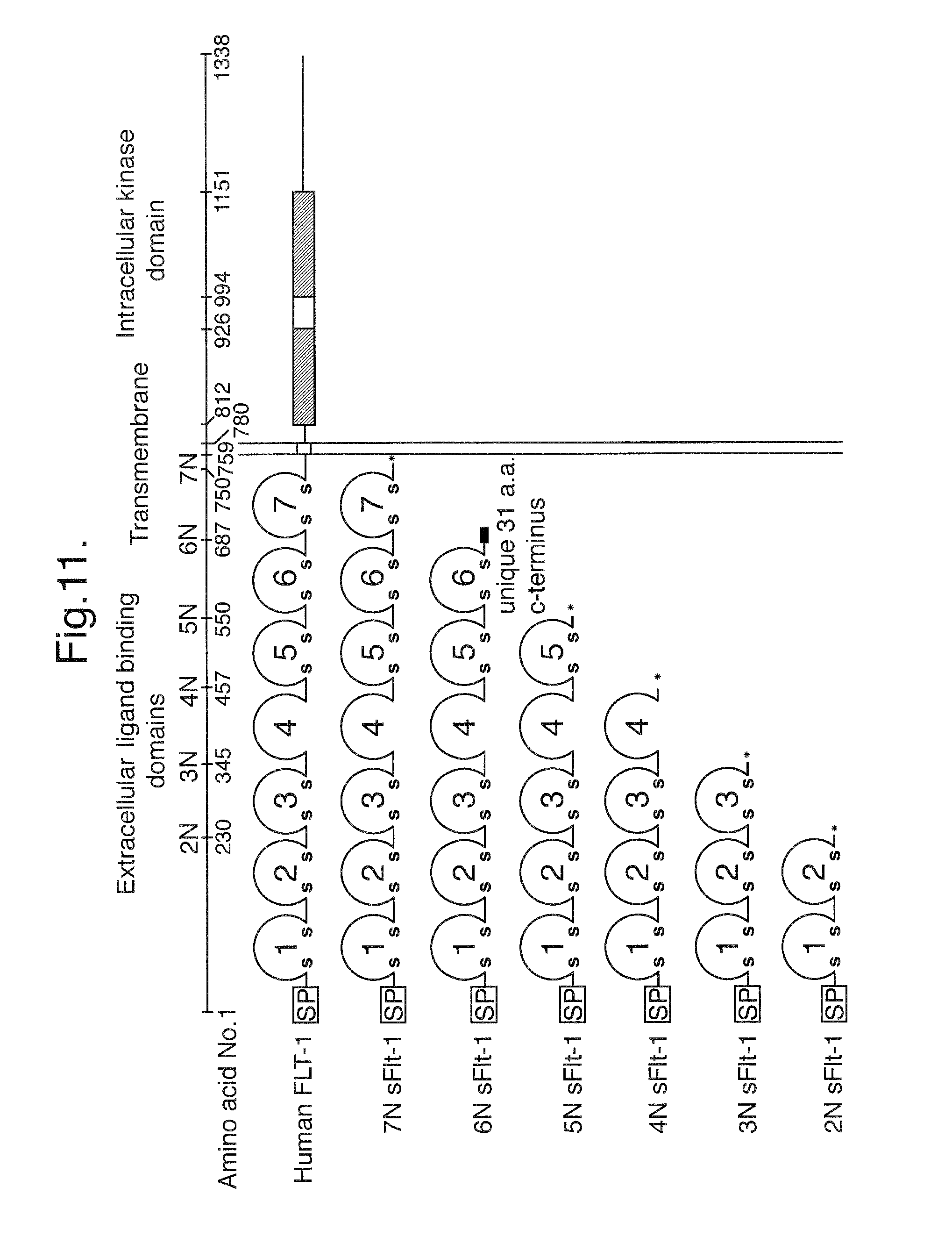
D00014
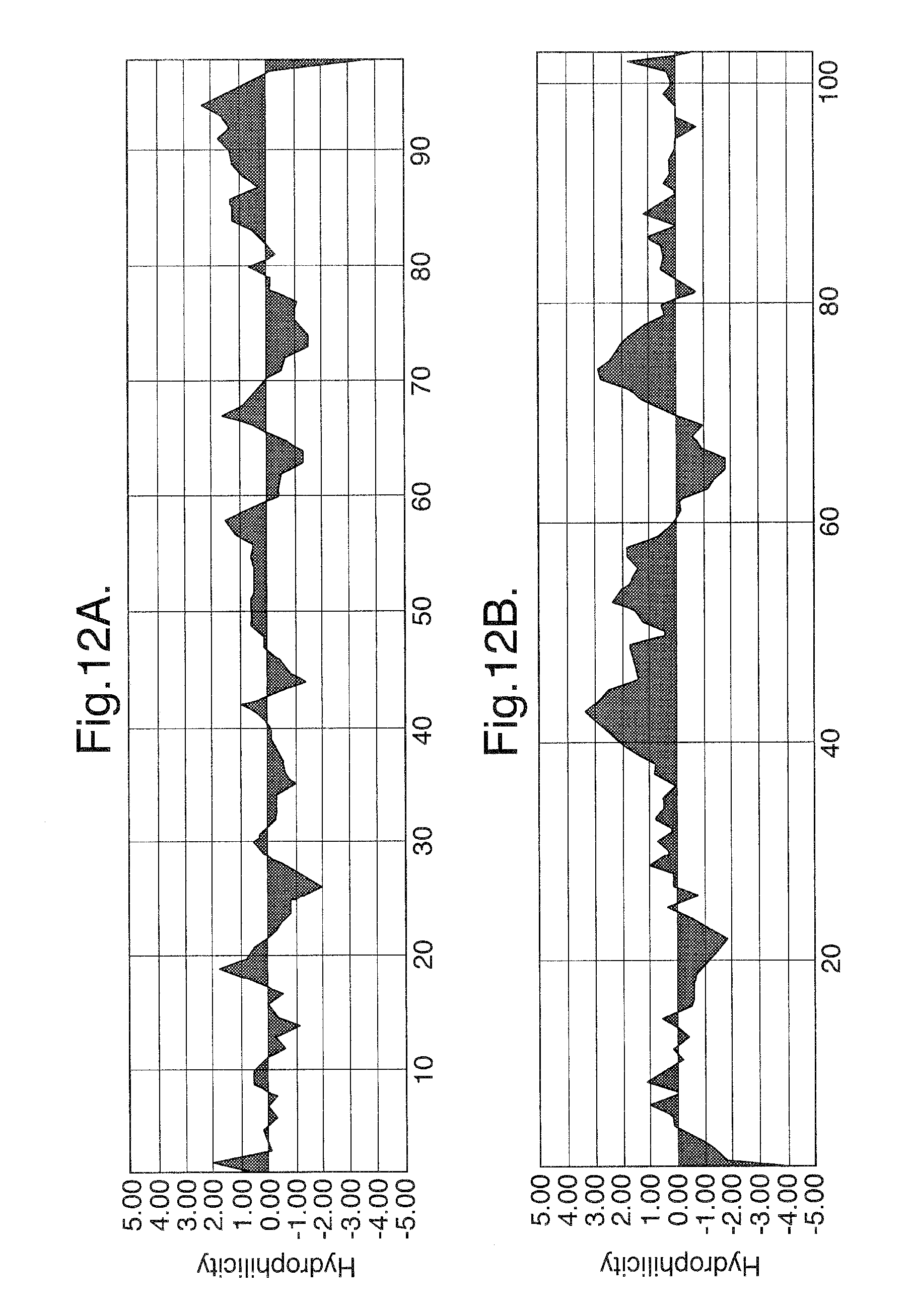
D00015
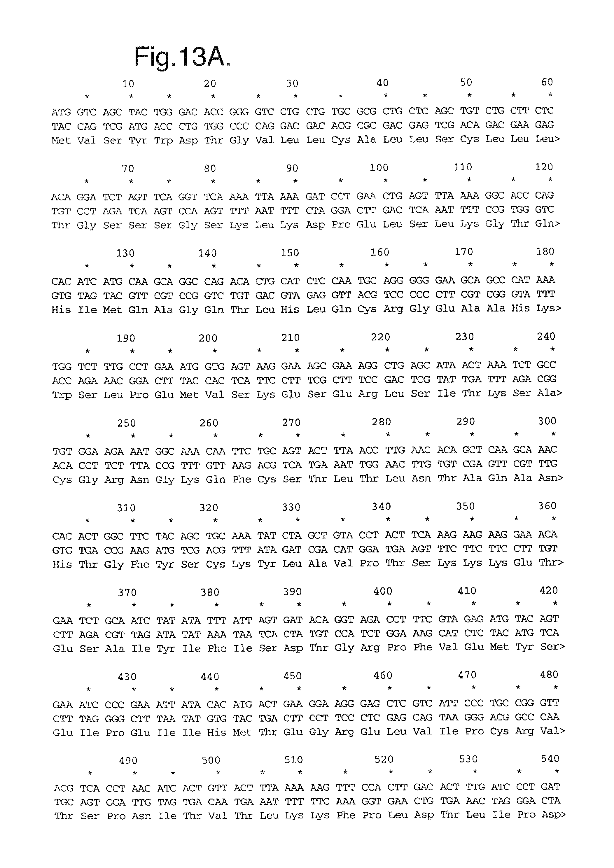
D00016
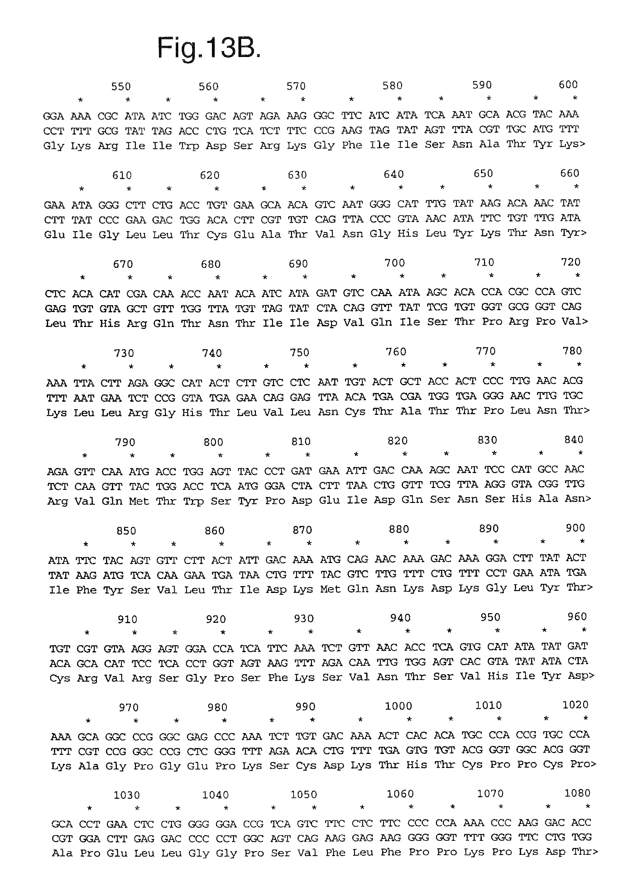
D00017
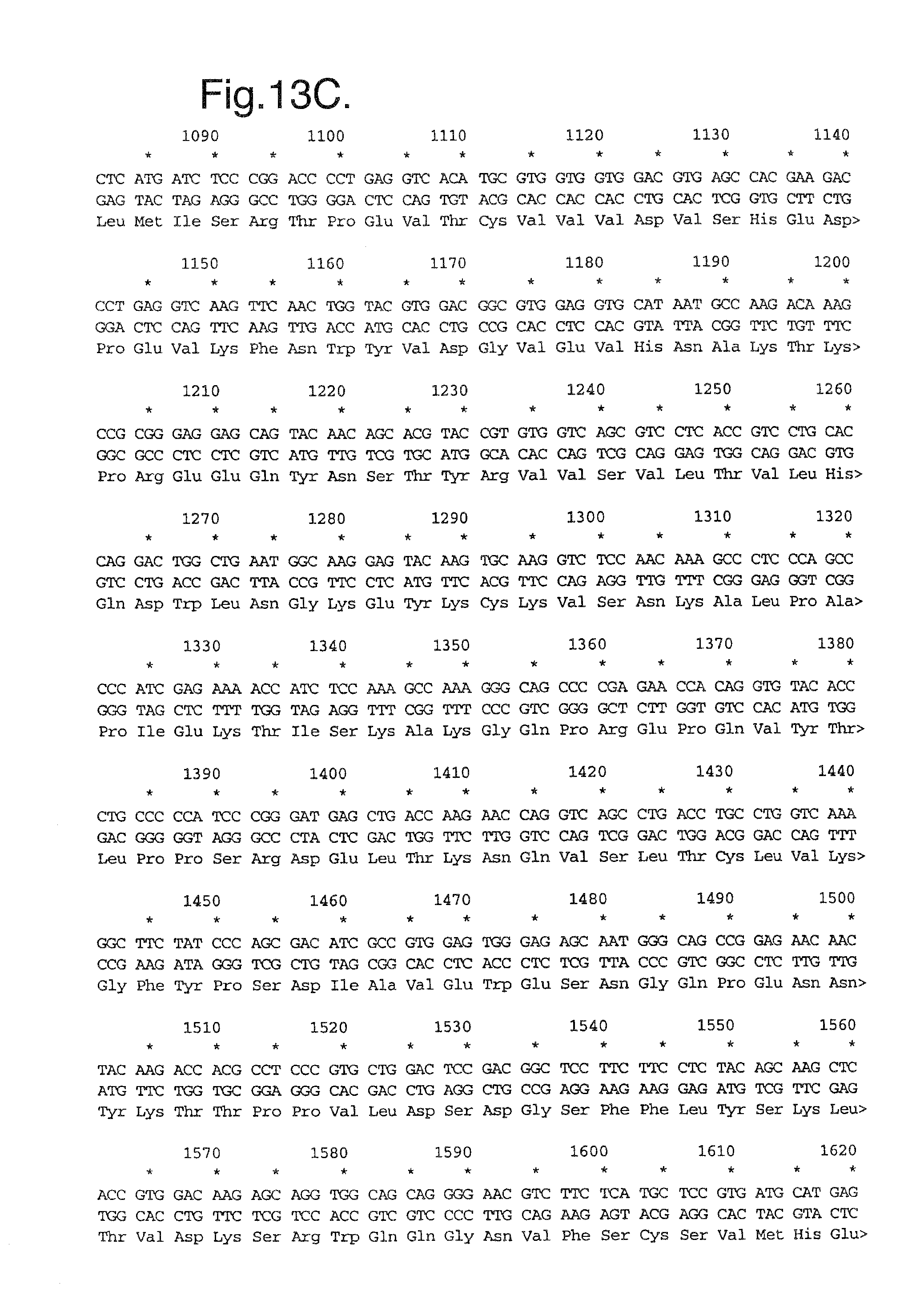
D00018
D00019
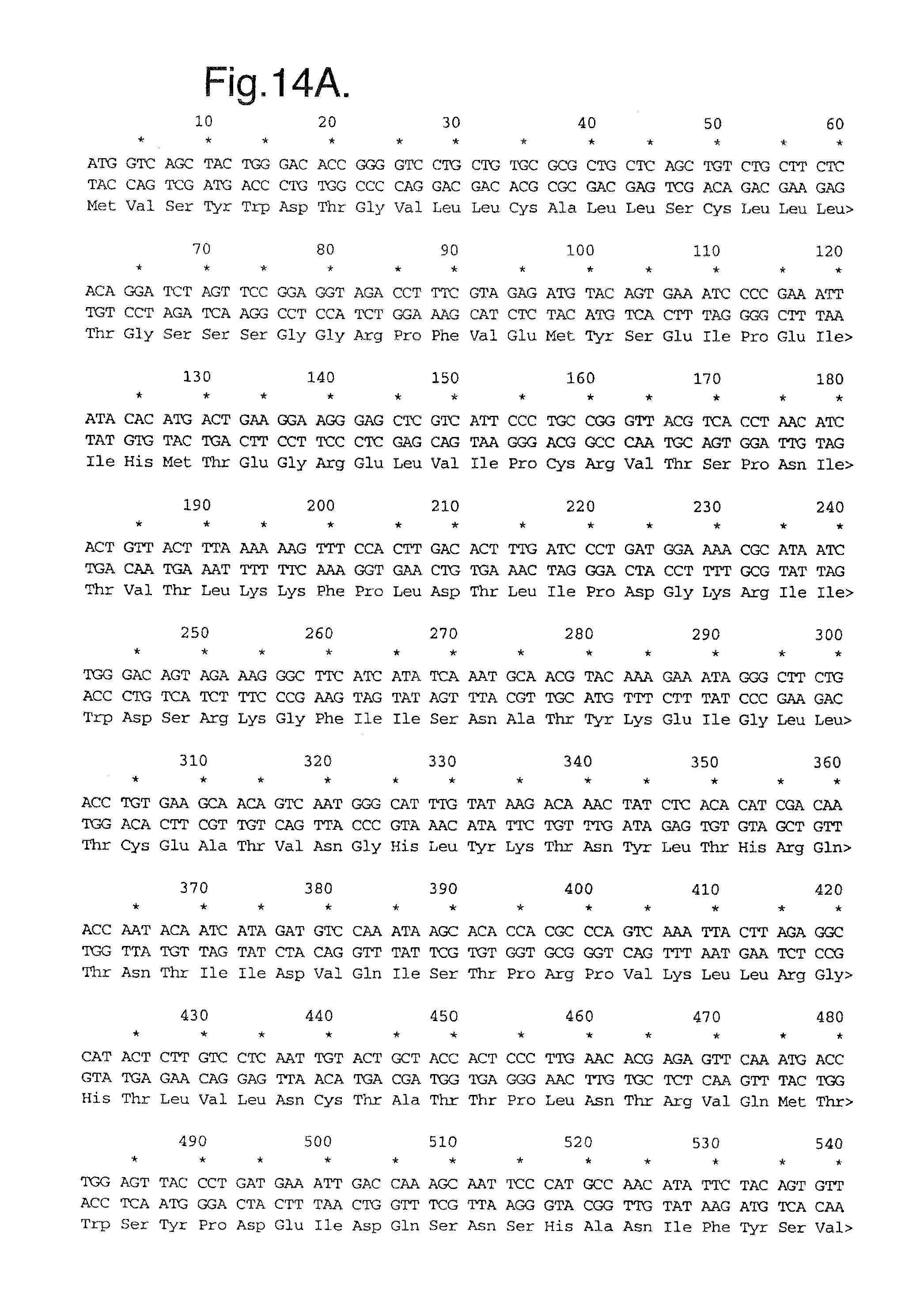
D00020
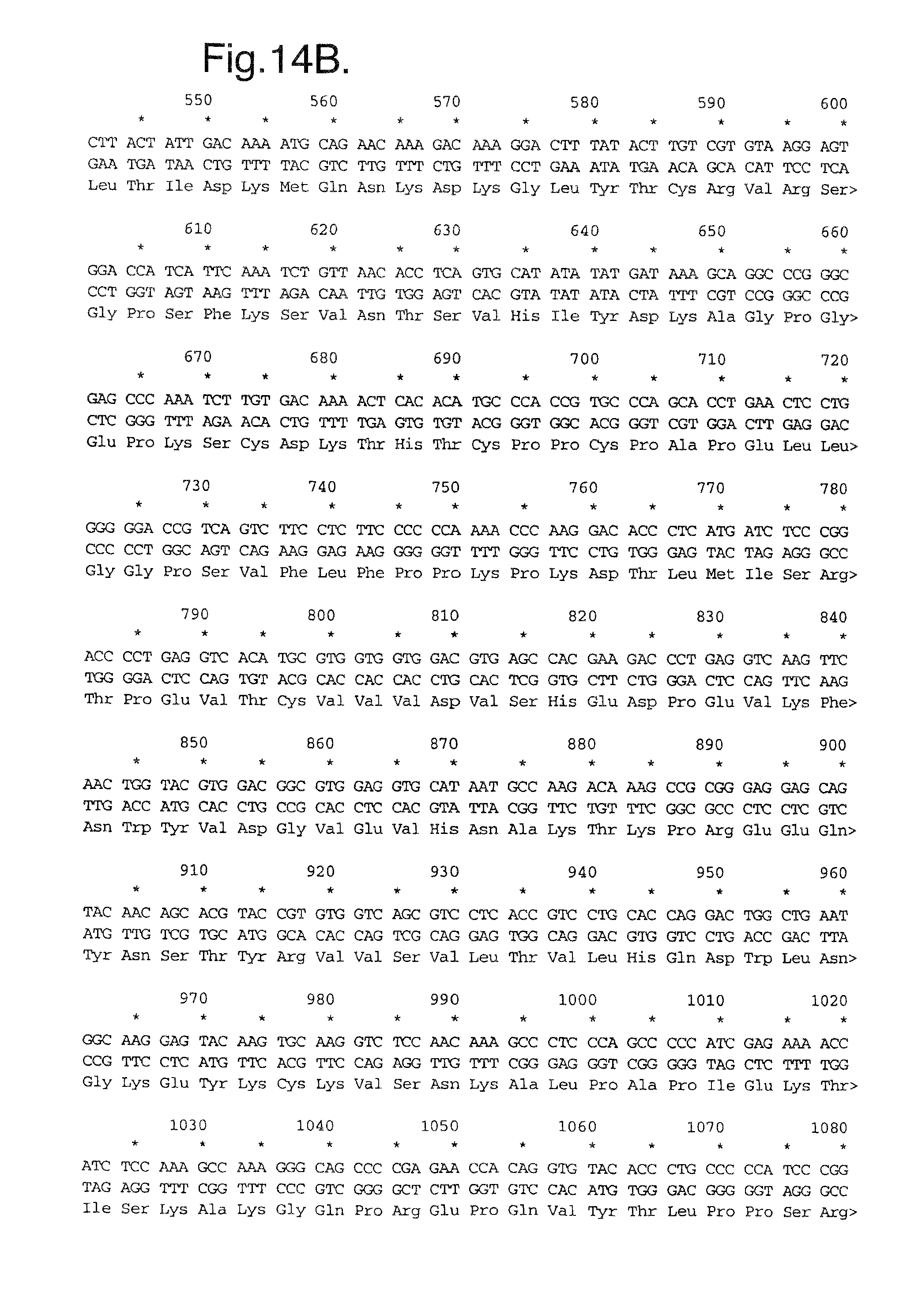
D00021
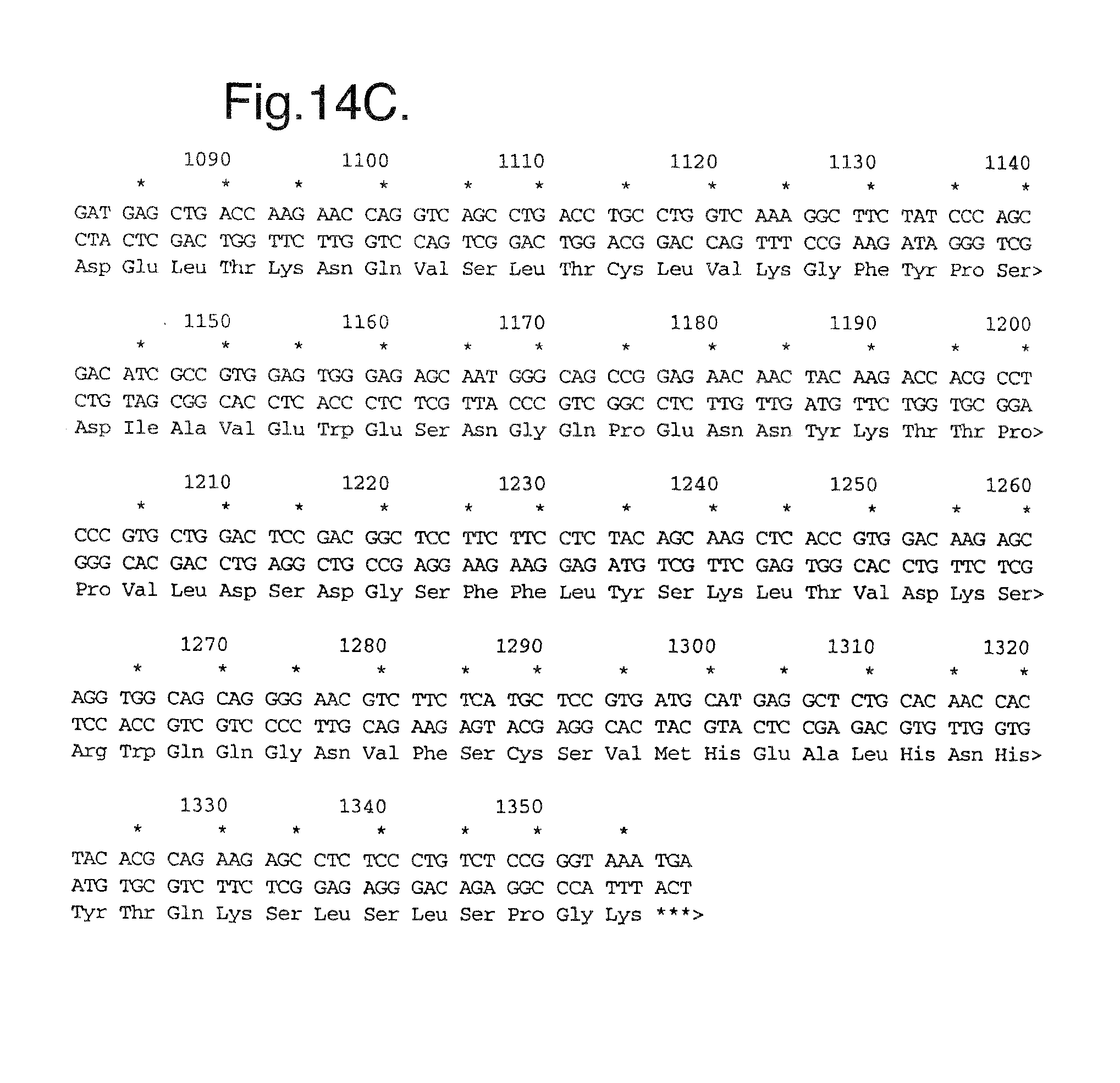
D00022

D00023
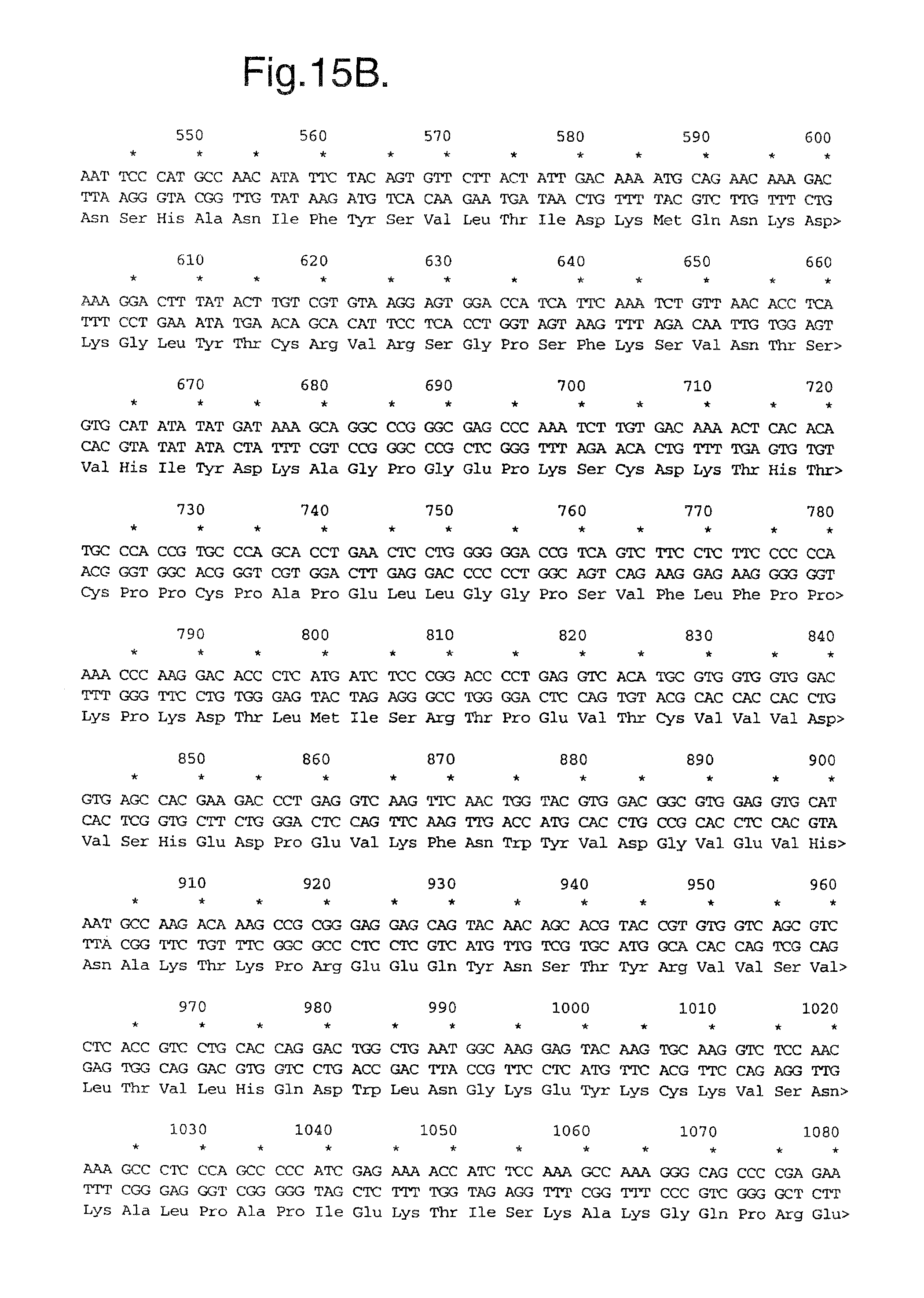
D00024
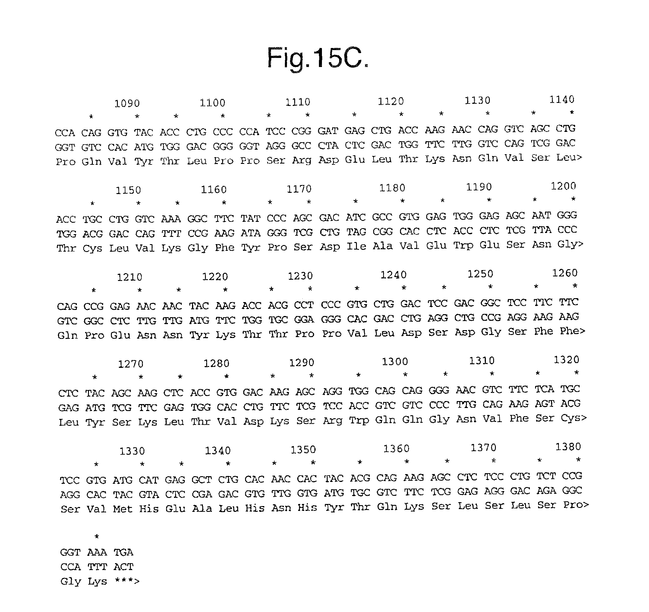
D00025
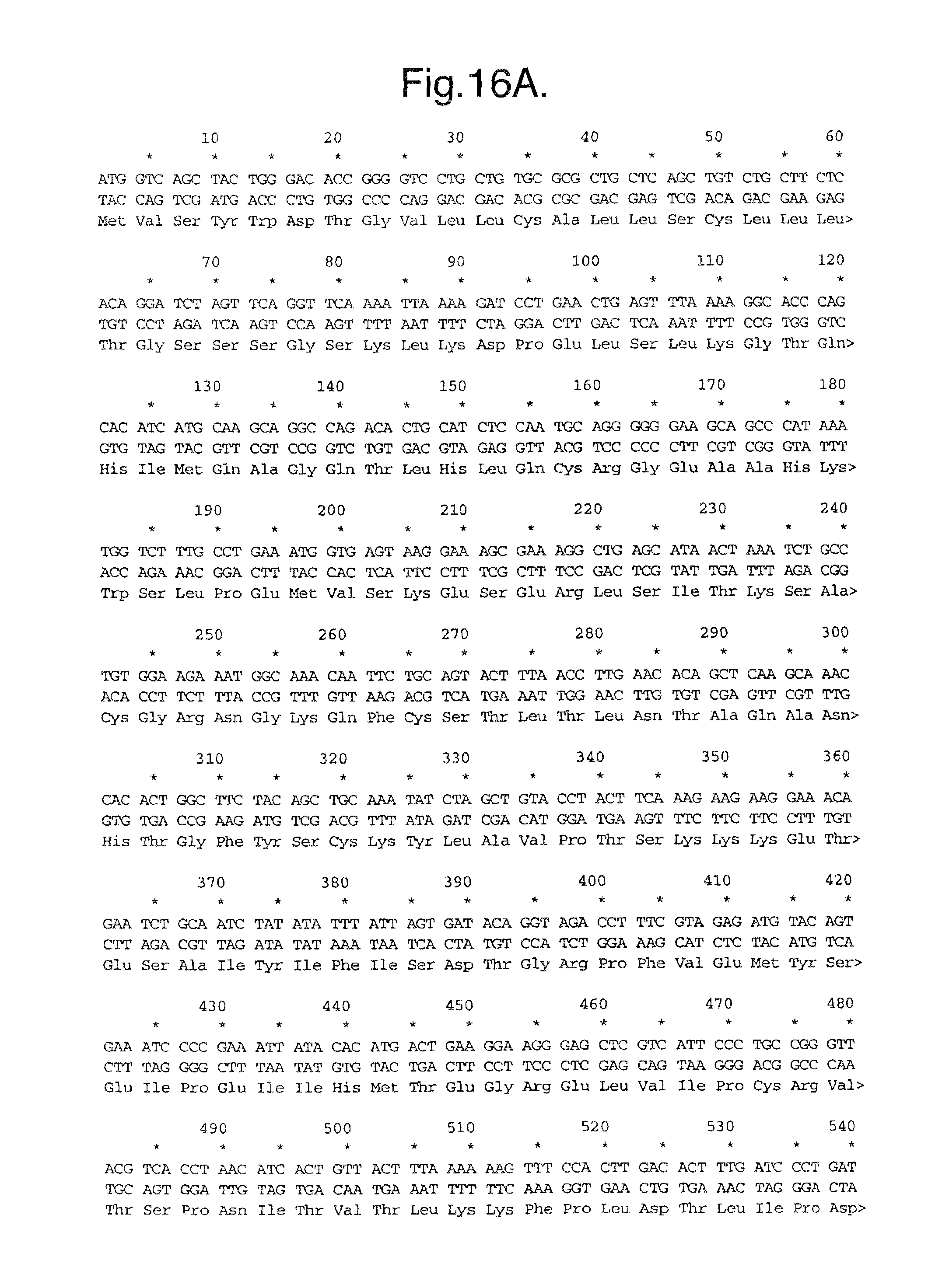
D00026
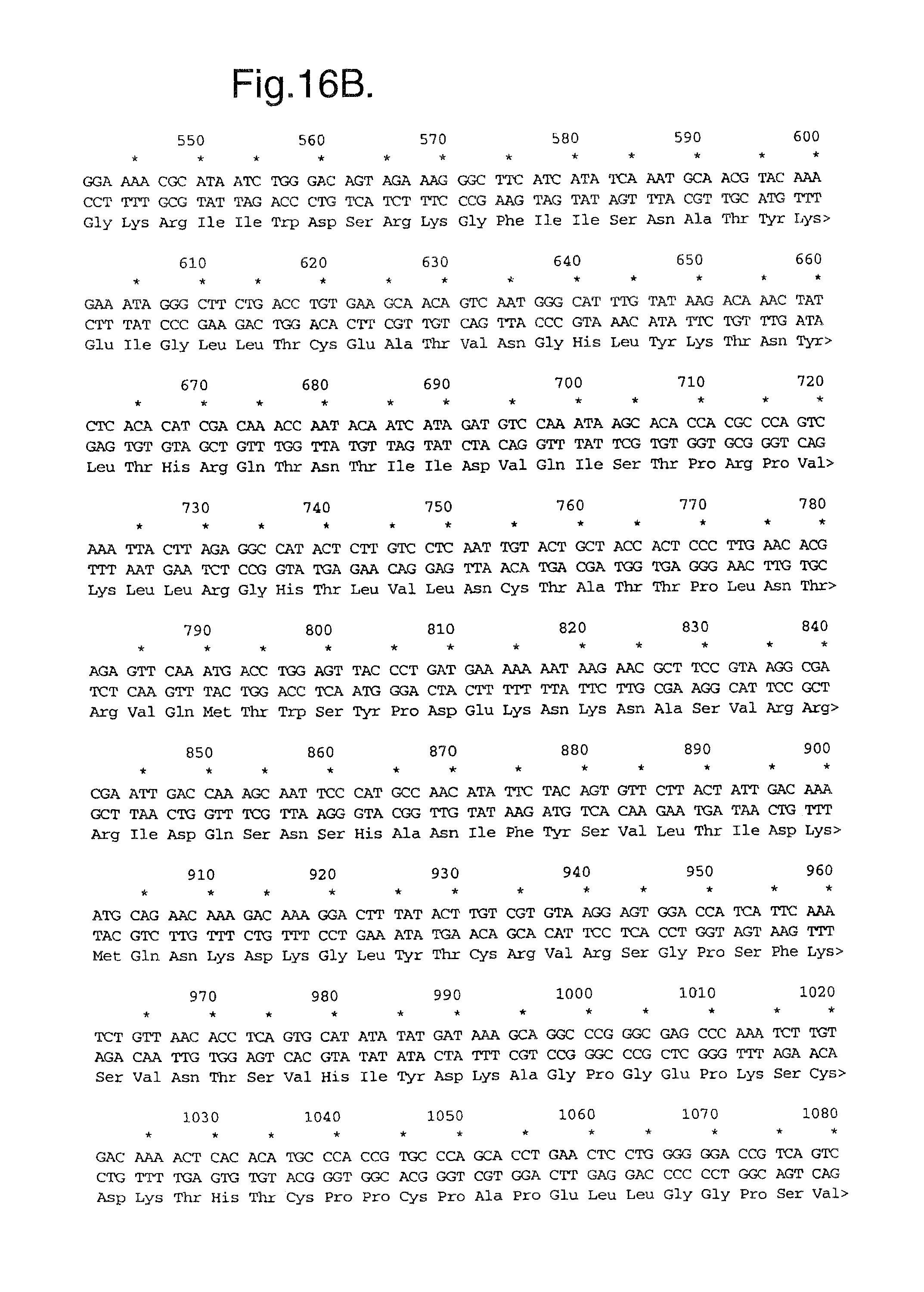
D00027
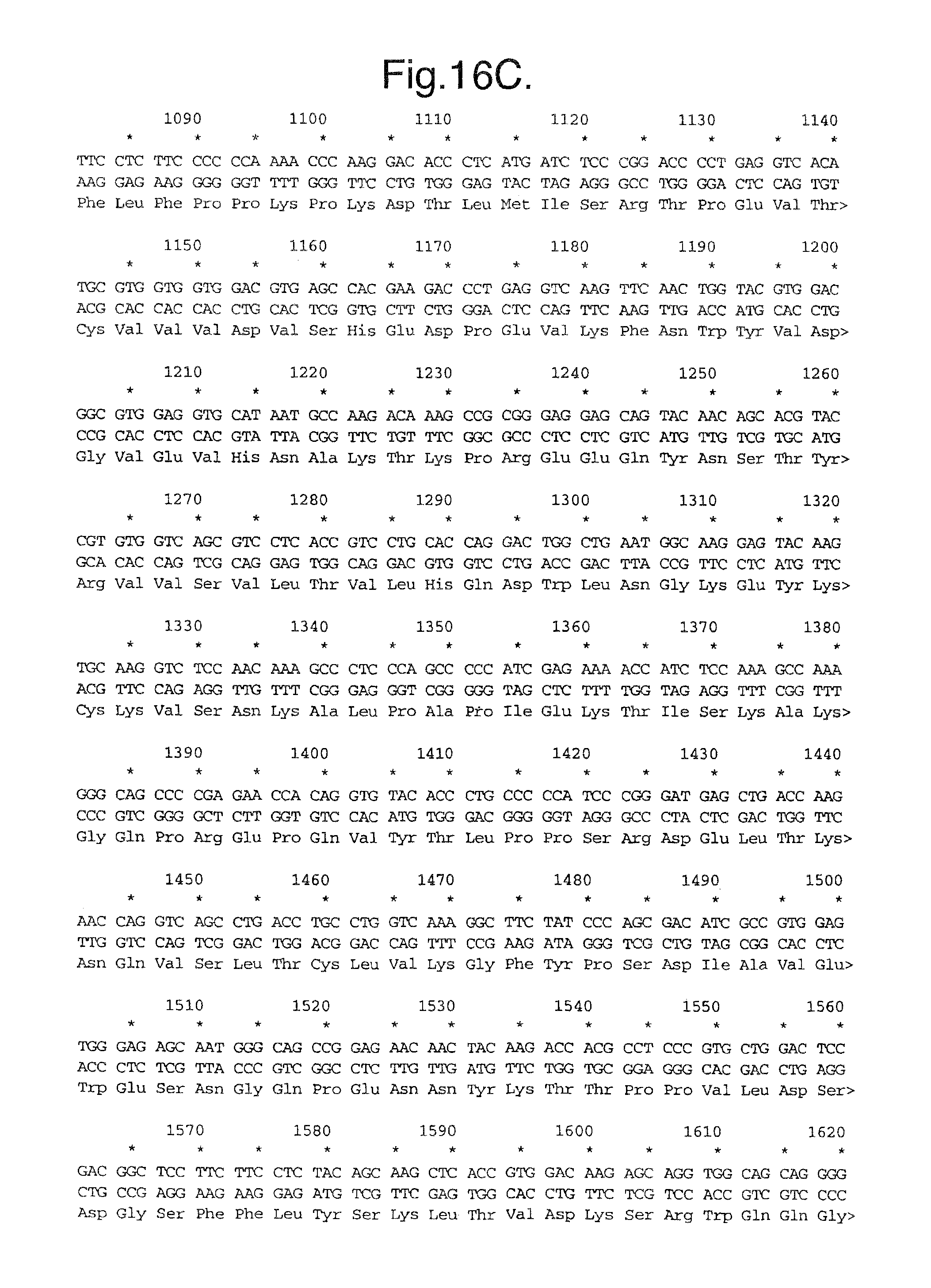
D00028
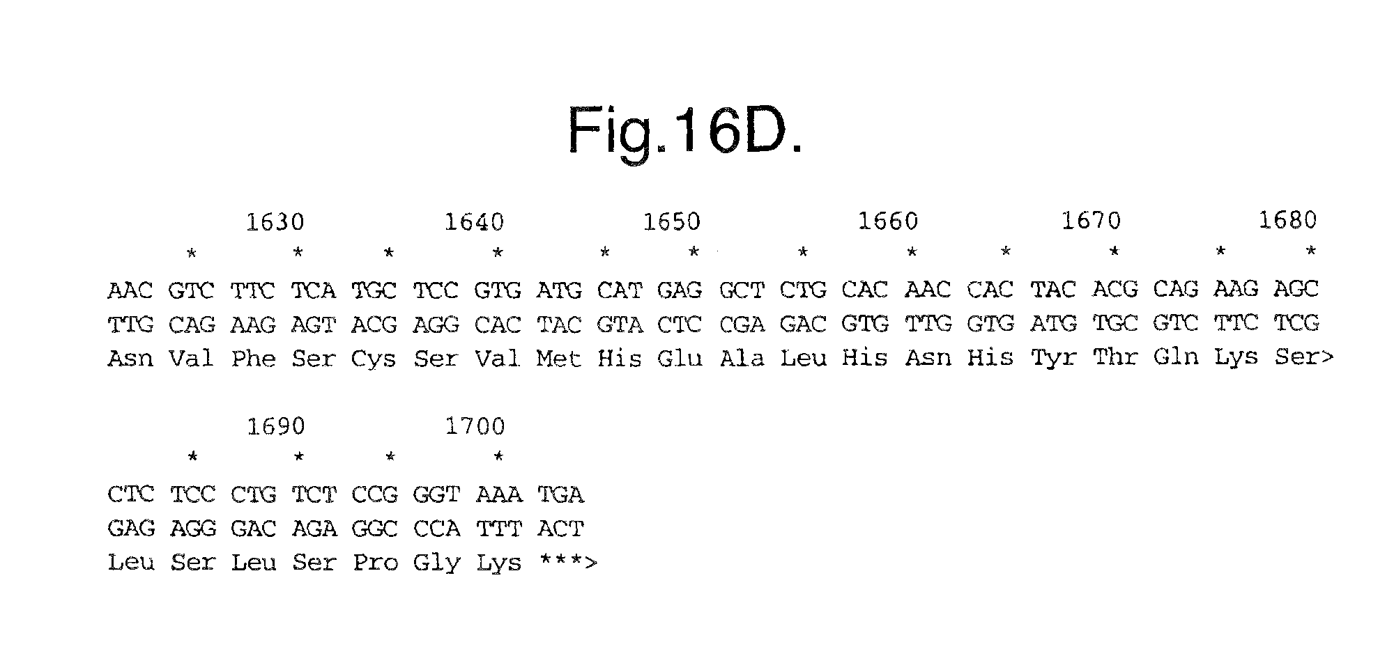
D00029

D00030

D00031
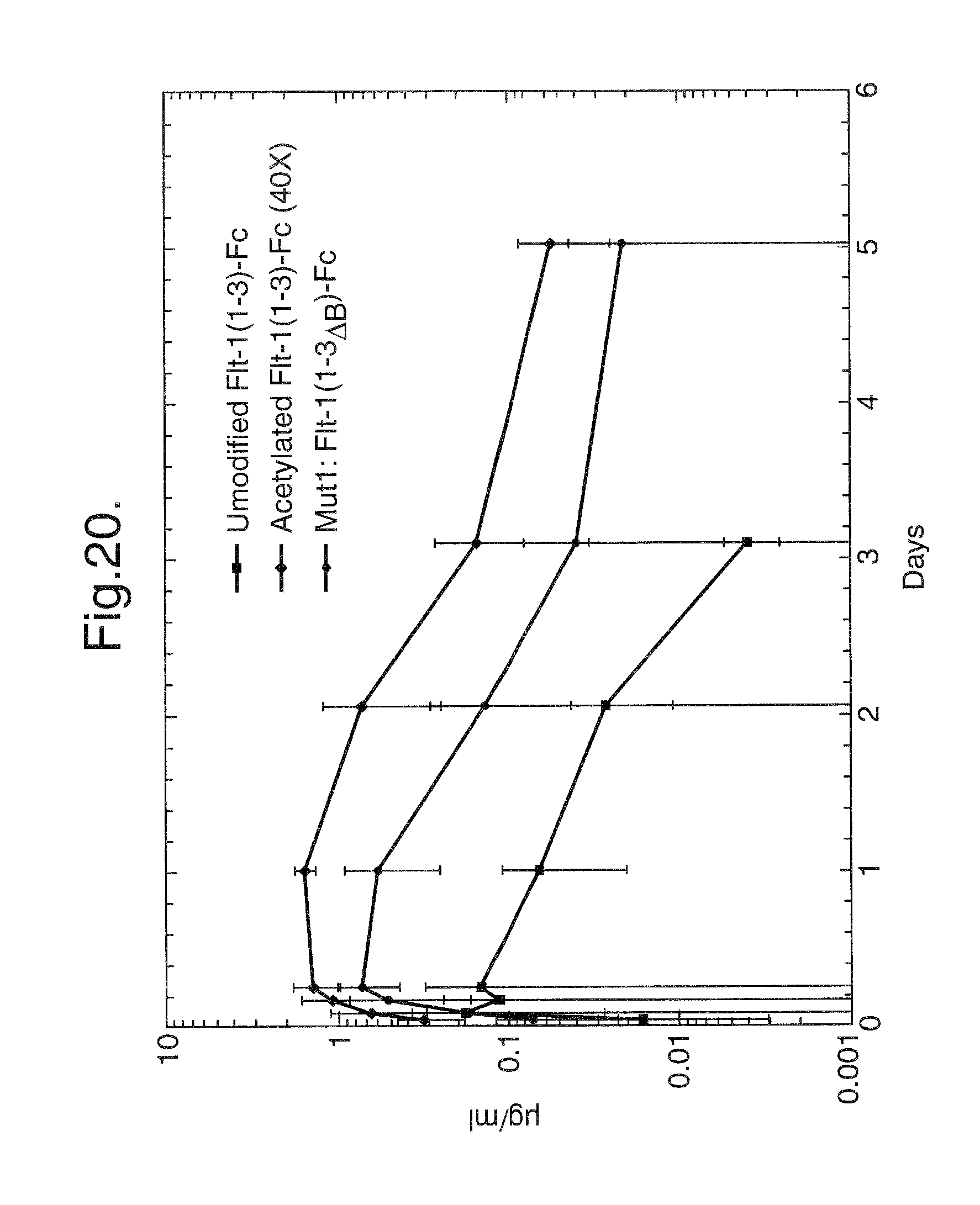
D00032
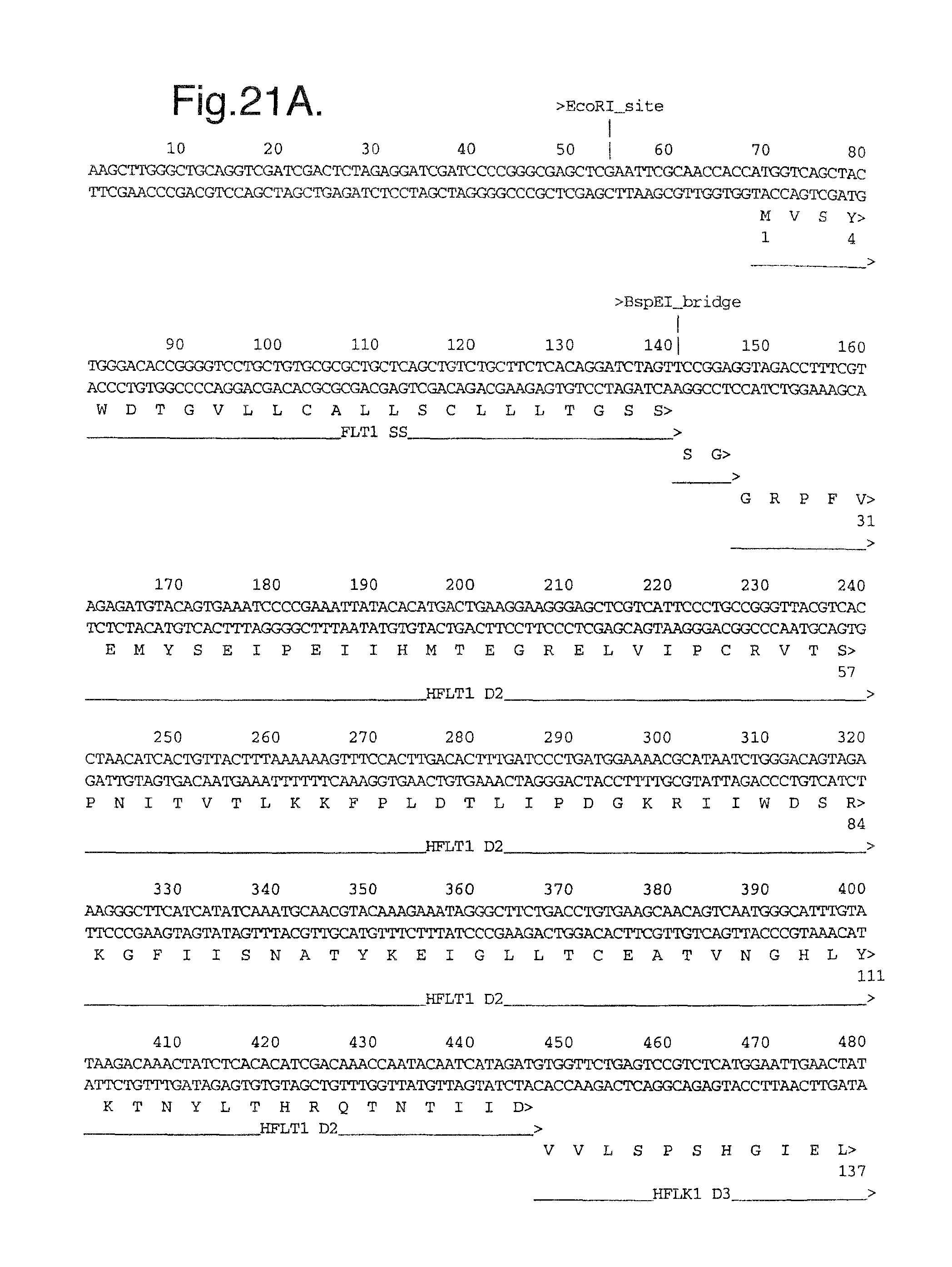
D00033
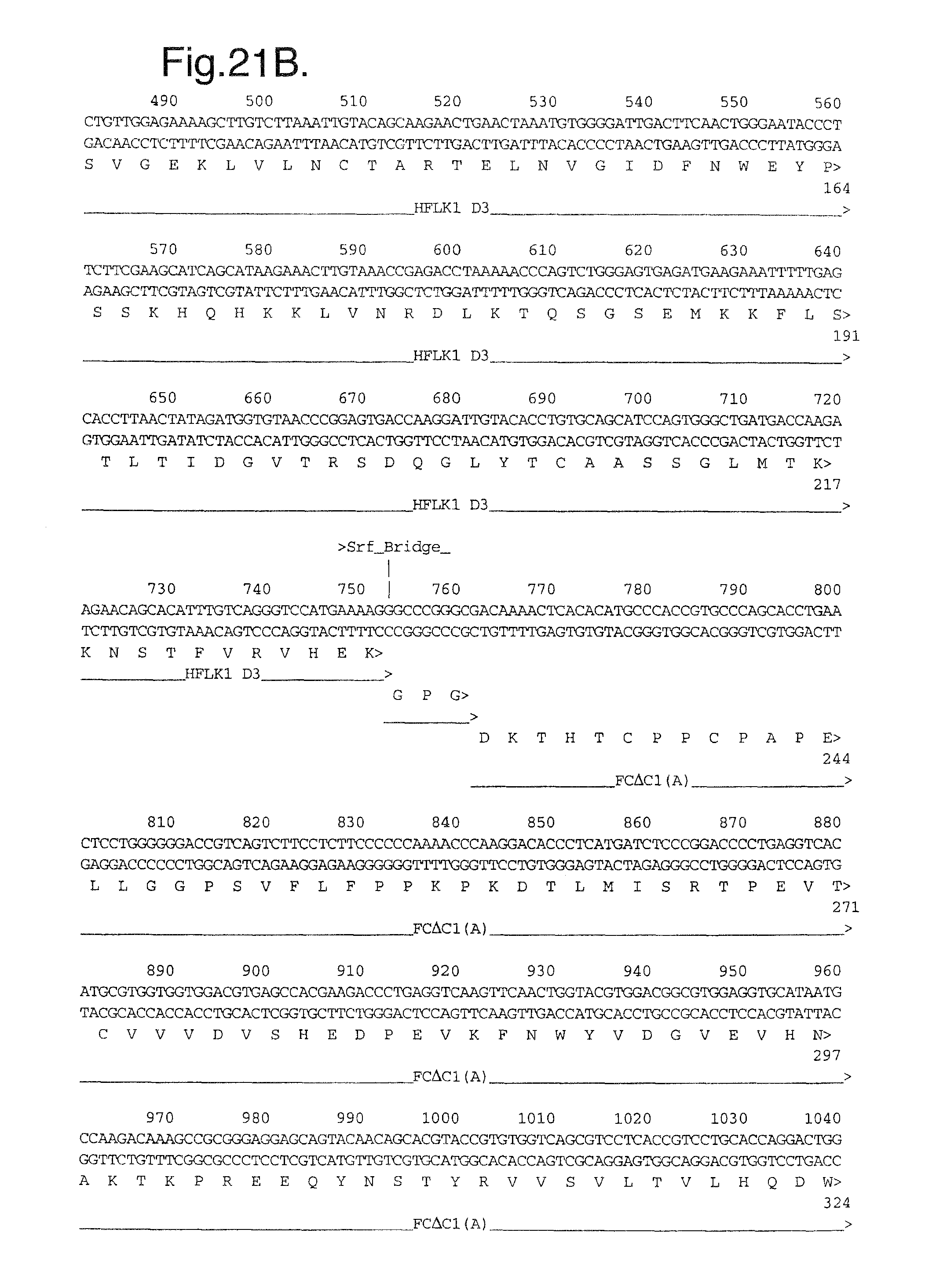
D00034
D00035
D00036

D00037
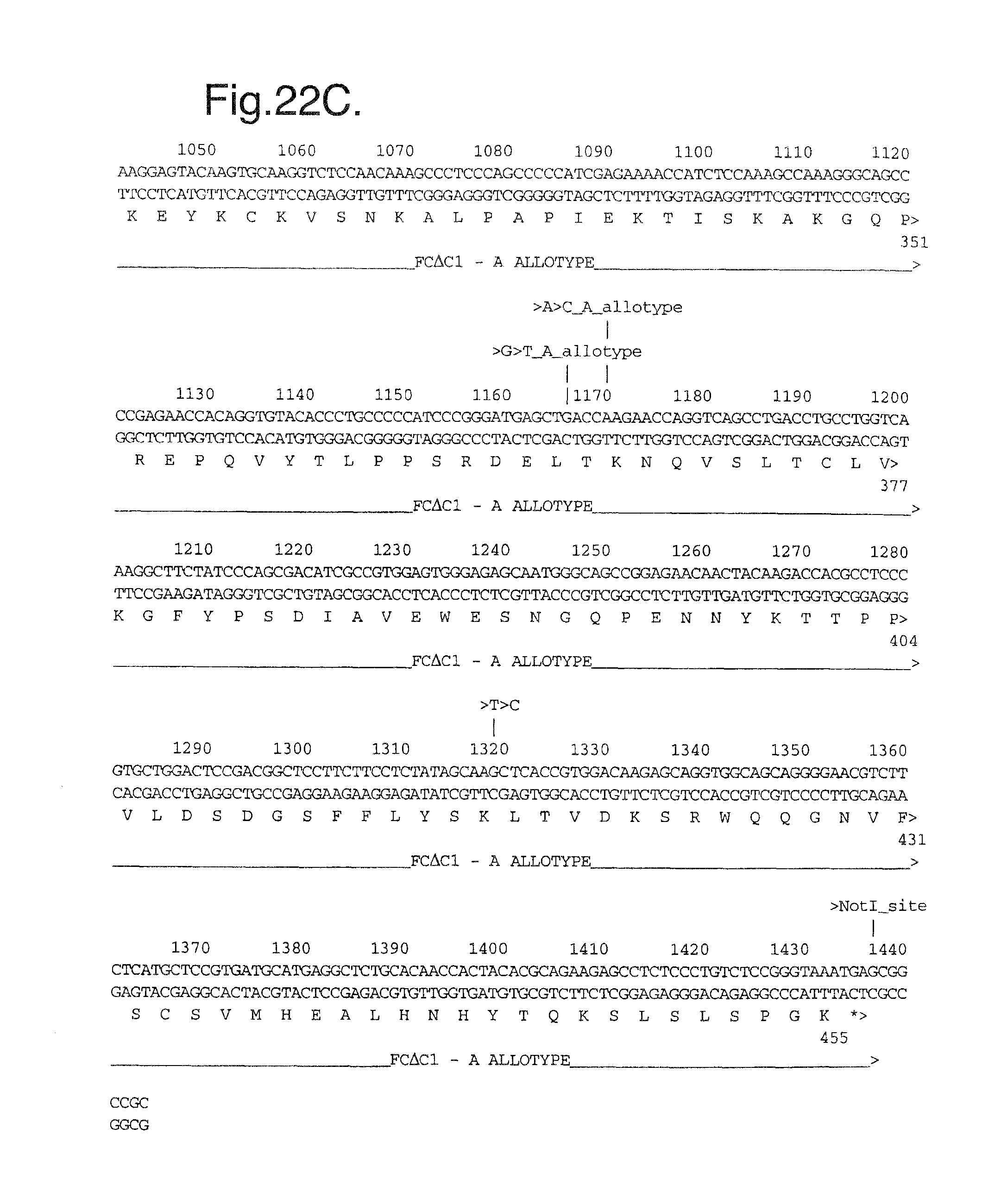
D00038
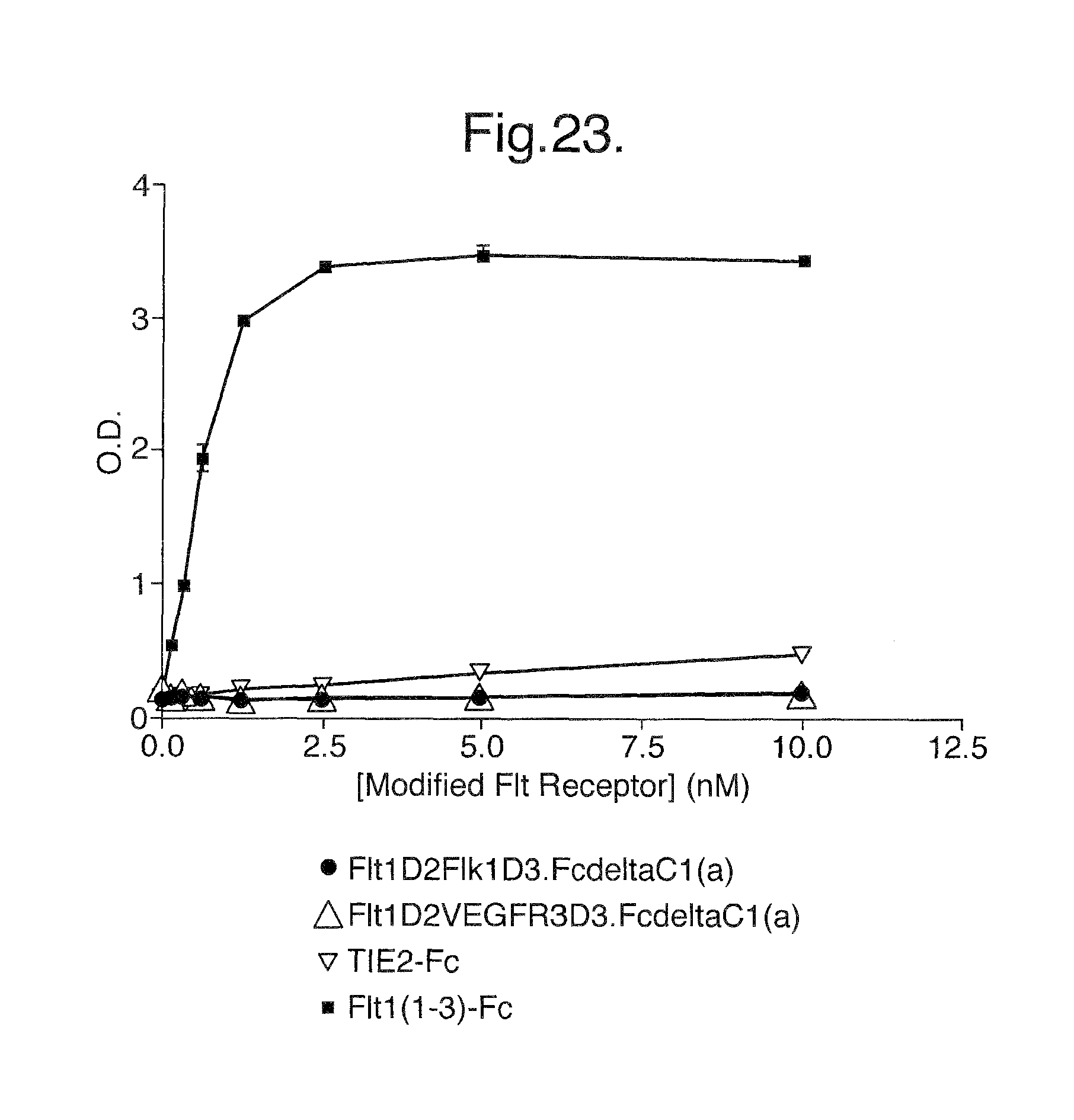
D00039
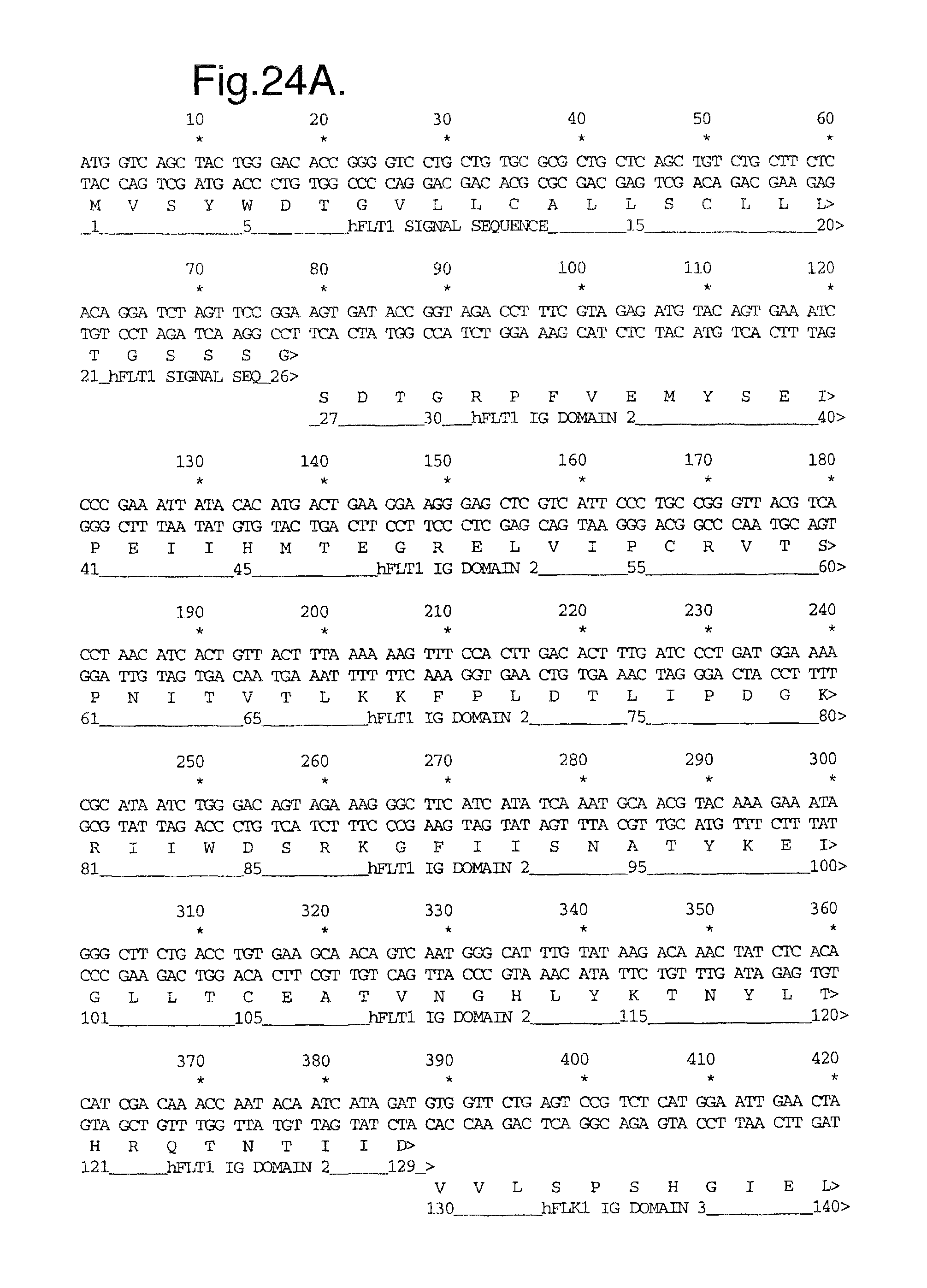
D00040
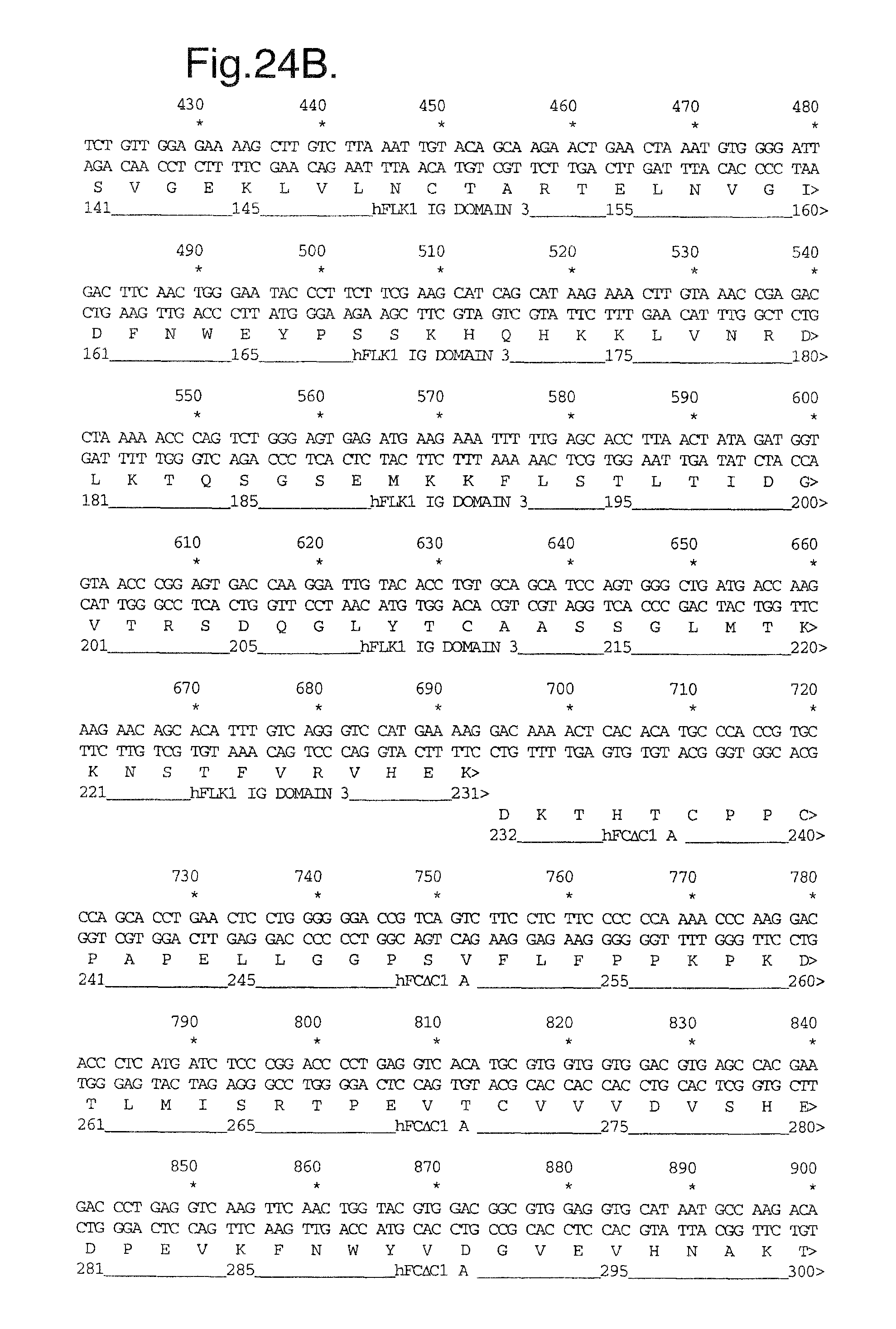
D00041
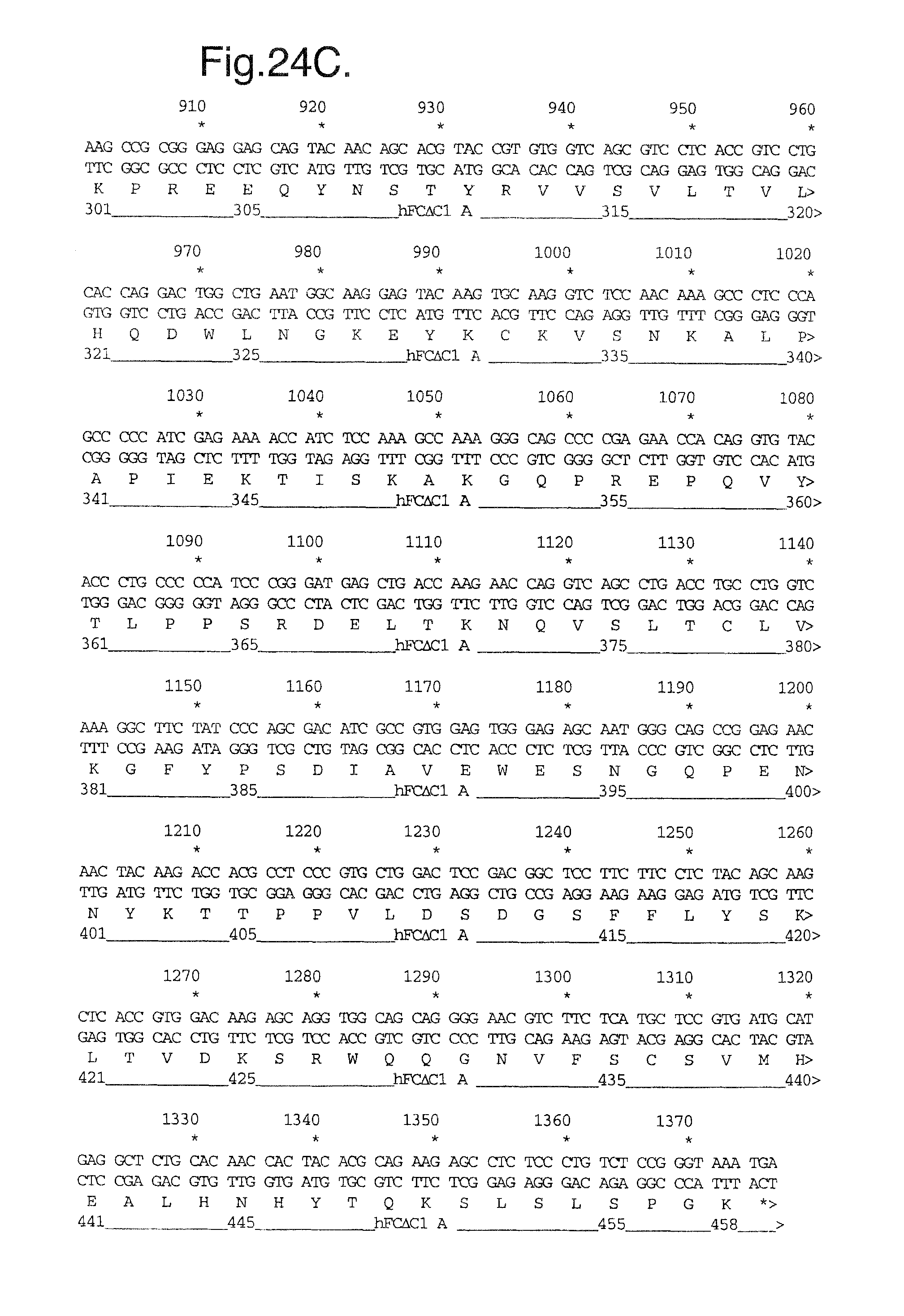
D00042
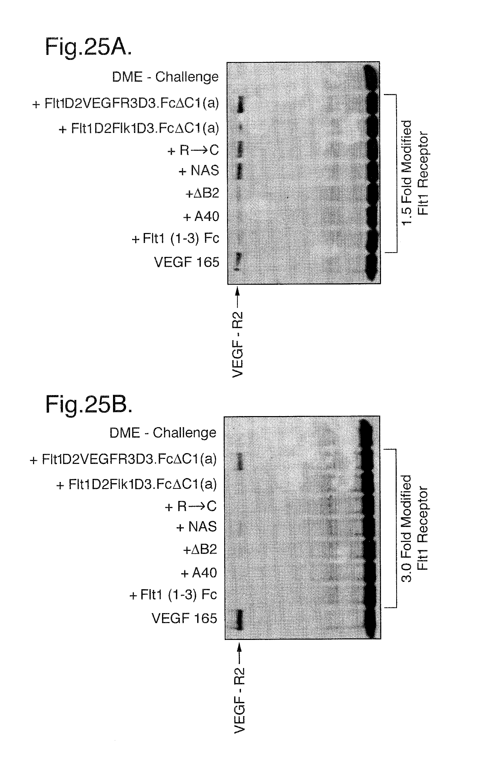
D00043
D00044
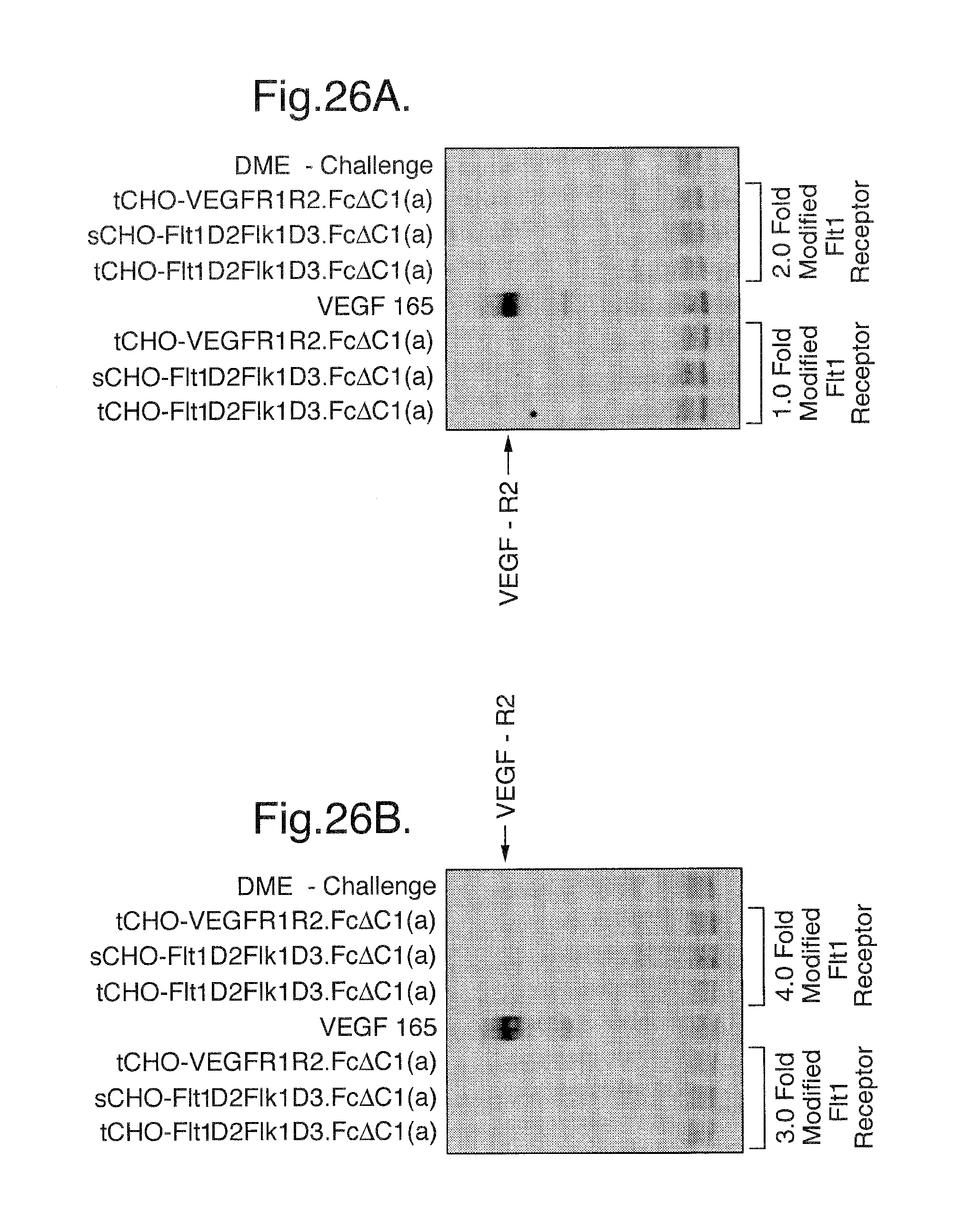
D00045

D00046

D00047

D00048
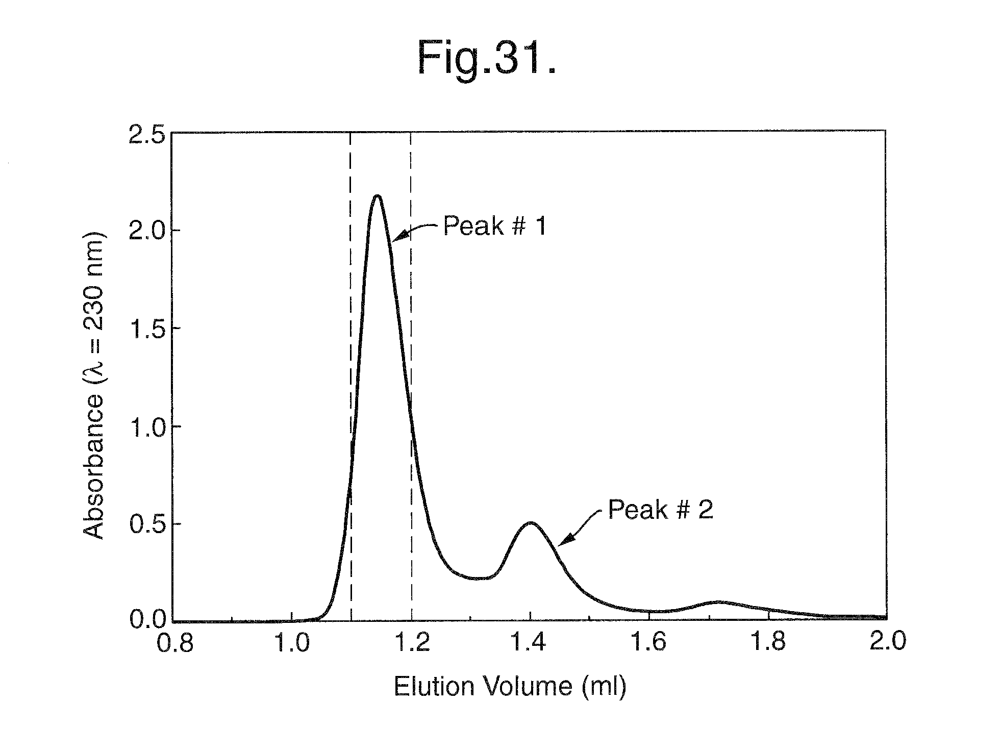
D00049
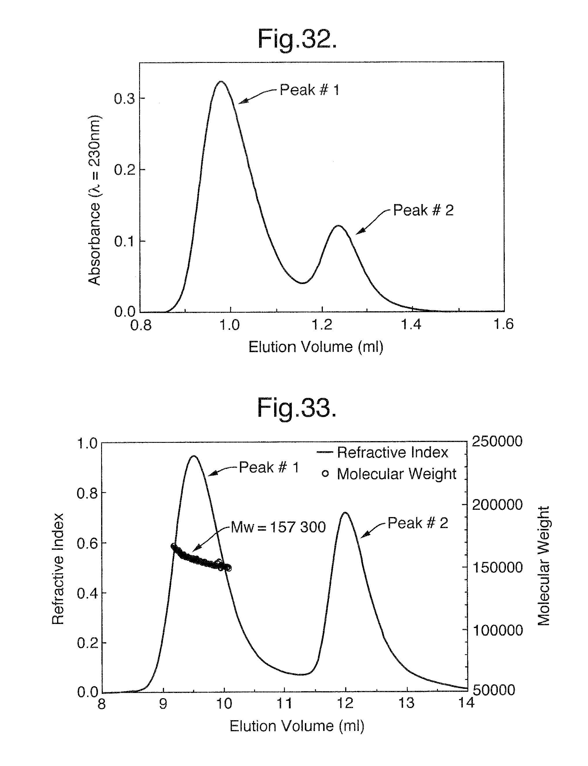
D00050
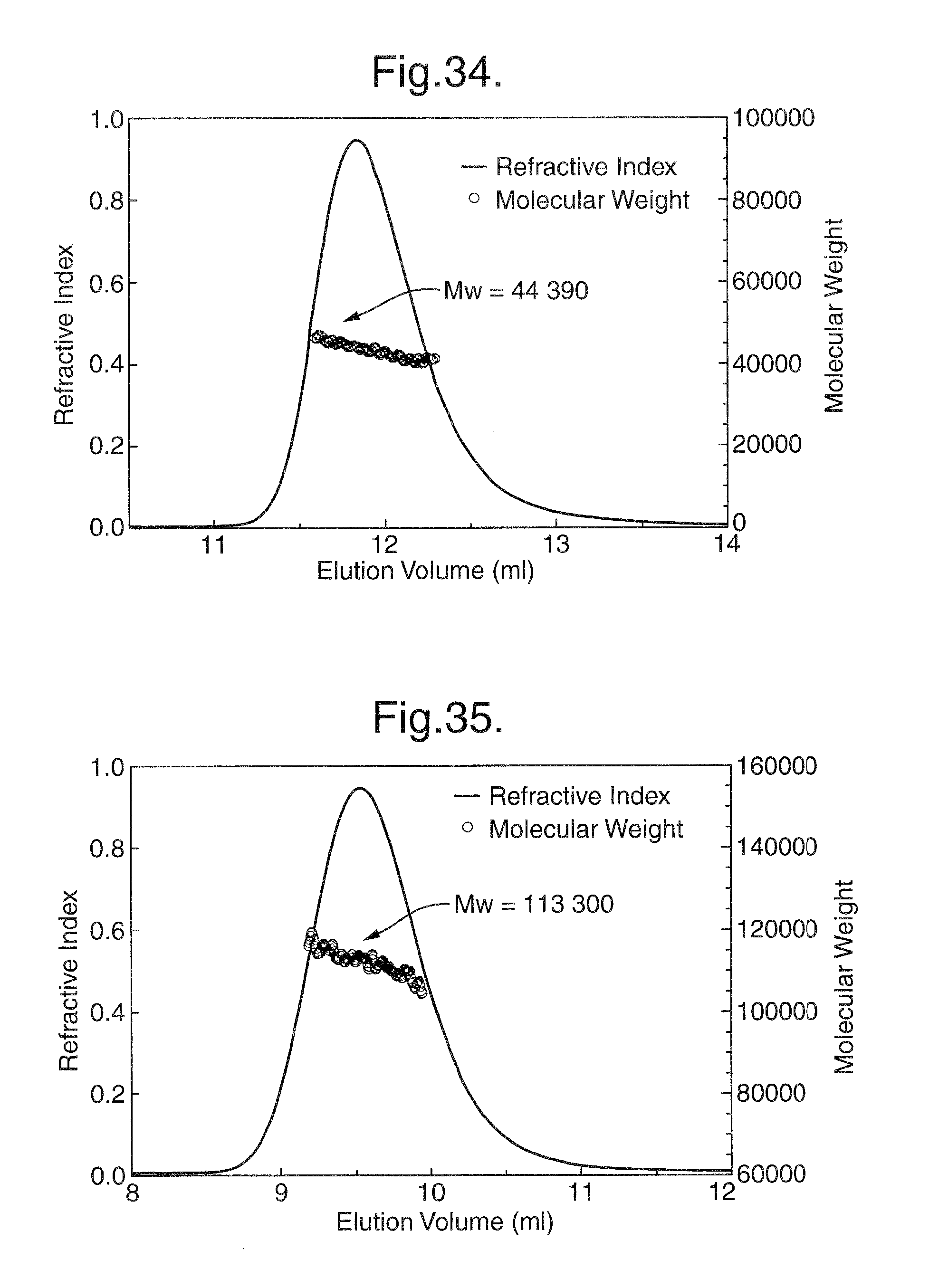
D00051
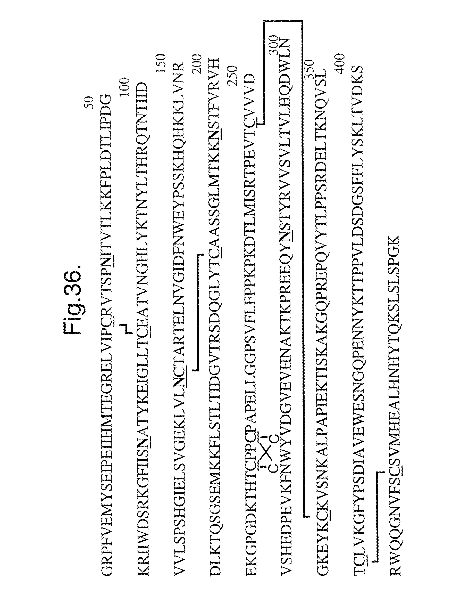
D00052
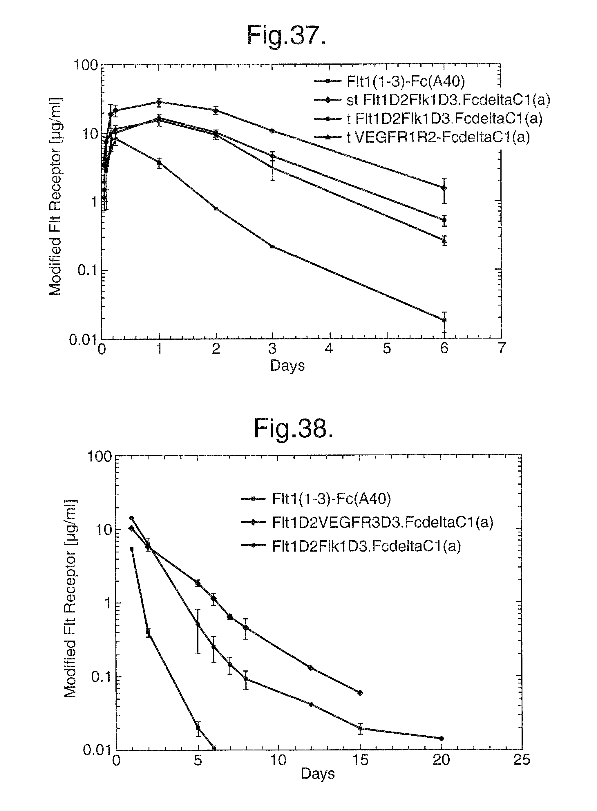
D00053

D00054

D00055

S00001
XML
uspto.report is an independent third-party trademark research tool that is not affiliated, endorsed, or sponsored by the United States Patent and Trademark Office (USPTO) or any other governmental organization. The information provided by uspto.report is based on publicly available data at the time of writing and is intended for informational purposes only.
While we strive to provide accurate and up-to-date information, we do not guarantee the accuracy, completeness, reliability, or suitability of the information displayed on this site. The use of this site is at your own risk. Any reliance you place on such information is therefore strictly at your own risk.
All official trademark data, including owner information, should be verified by visiting the official USPTO website at www.uspto.gov. This site is not intended to replace professional legal advice and should not be used as a substitute for consulting with a legal professional who is knowledgeable about trademark law.