Mucosal vaccine composition
Fukasaka , et al. A
U.S. patent number 10,391,167 [Application Number 14/917,077] was granted by the patent office on 2019-08-27 for mucosal vaccine composition. This patent grant is currently assigned to NITTO DENKO CORPORATION. The grantee listed for this patent is NITTO DENKO CORPORATION. Invention is credited to Daisuke Asari, Masahiro Fukasaka, Mitsuhiko Hori, Eiji Kiyotoh, Kyohei Matsushita, Arimichi Okazaki, Katsuyuki Okubo.


View All Diagrams
| United States Patent | 10,391,167 |
| Fukasaka , et al. | August 27, 2019 |
Mucosal vaccine composition
Abstract
The present disclosure relates to a method for inducing humoral immunity in a human or animal, that includes administering to the human or animal a mucosal vaccine composition to a mucous membrane selected from the group consisting of a human or animal intraoral mucous membrane, ocular mucous membrane, ear mucous membrane, genital mucous membrane, pharyngeal mucous membrane, respiratory tract mucous membrane, bronchial mucous membrane, pulmonary mucous membrane, gastric mucous membrane, enteric mucous membrane, and rectal mucous membrane. The mucosal vaccine composition includes at least one antigen derived from a pathogen; and as an adjuvant, a lipopolysaccharide derived from at least one gram-negative bacterium selected from the group consisting of Pantoea, Acetobacter, Zymomonas, and Xanthomonas, or a salt of the lipopolysaccharide, wherein the mass ratio between a total mass of the adjuvant and a total mass of the antigen is 0.002 to 500. The mucosal vaccine composition is administered in an amount effective to induce humoral immunity in the human or animal.
| Inventors: | Fukasaka; Masahiro (Osaka, JP), Hori; Mitsuhiko (Osaka, JP), Okubo; Katsuyuki (Osaka, JP), Asari; Daisuke (Osaka, JP), Okazaki; Arimichi (Osaka, JP), Kiyotoh; Eiji (Osaka, JP), Matsushita; Kyohei (Osaka, JP) | ||||||||||
|---|---|---|---|---|---|---|---|---|---|---|---|
| Applicant: |
|
||||||||||
| Assignee: | NITTO DENKO CORPORATION (Osaka,
JP) |
||||||||||
| Family ID: | 52778766 | ||||||||||
| Appl. No.: | 14/917,077 | ||||||||||
| Filed: | October 2, 2014 | ||||||||||
| PCT Filed: | October 02, 2014 | ||||||||||
| PCT No.: | PCT/JP2014/076347 | ||||||||||
| 371(c)(1),(2),(4) Date: | March 07, 2016 | ||||||||||
| PCT Pub. No.: | WO2015/050179 | ||||||||||
| PCT Pub. Date: | April 09, 2015 |
Prior Publication Data
| Document Identifier | Publication Date | |
|---|---|---|
| US 20160213773 A1 | Jul 28, 2016 | |
Foreign Application Priority Data
| Oct 3, 2013 [JP] | 2013-208664 | |||
| Current U.S. Class: | 1/1 |
| Current CPC Class: | A61K 39/12 (20130101); A61K 39/00 (20130101); C12N 7/00 (20130101); A61K 39/092 (20130101); A61K 39/145 (20130101); A61K 39/39 (20130101); A61K 39/0005 (20130101); A61P 31/16 (20180101); A61P 31/00 (20180101); A61P 35/00 (20180101); A61K 39/13 (20130101); A61P 37/04 (20180101); A61K 39/15 (20130101); A61K 2039/542 (20130101); A61K 2039/544 (20130101); A61K 2039/52 (20130101); C12N 2710/20034 (20130101); A61K 2039/525 (20130101); A61K 2039/55572 (20130101); A61K 2039/543 (20130101); A61K 2039/541 (20130101); C12N 2760/16034 (20130101); C12N 2720/12334 (20130101); C12N 2770/32634 (20130101) |
| Current International Class: | A61K 39/00 (20060101); A61K 39/39 (20060101); A61K 39/15 (20060101); C12N 7/00 (20060101); A61K 39/145 (20060101); A61K 39/13 (20060101); A61K 39/12 (20060101); A61K 39/09 (20060101) |
References Cited [Referenced By]
U.S. Patent Documents
| 6010719 | January 2000 | Remon |
| 9498527 | November 2016 | Fukasaka |
| 9962439 | May 2018 | Matsushita |
| 10071155 | September 2018 | Kiyotoh |
| 10076491 | September 2018 | Asari |
| 10206957 | February 2019 | Myles |
| 10206985 | February 2019 | Asari |
| 2005/0152919 | July 2005 | Ward |
| 2008/0279926 | November 2008 | Vandepapeliere |
| 2010/0312045 | December 2010 | Ramlov et al. |
| 2013/0089570 | April 2013 | Ouaked et al. |
| 2013/0266612 | October 2013 | Fukasaka |
| 2014/0193460 | July 2014 | Spector et al. |
| 2014/0220058 | August 2014 | Maeda |
| 2014/0220059 | August 2014 | Asari et al. |
| 2014/0220063 | August 2014 | Asari et al. |
| 2014/0220079 | August 2014 | Asari et al. |
| 2014/0234377 | August 2014 | Okazaki |
| 2016/0193327 | July 2016 | Fukasaka |
| 2016/0206728 | July 2016 | Kiyotoh |
| 2016/0213773 | July 2016 | Fukasaka |
| 2016/0228540 | August 2016 | Kiyotoh |
| 2016/0287687 | October 2016 | Mahr et al. |
| 2016/0287697 | October 2016 | Matsushita et al. |
| 2017/0028054 | February 2017 | Fukasaka |
| 2647387 | Oct 2013 | EP | |||
| H08-245702 | Sep 1996 | JP | |||
| 4043533 | Feb 2008 | JP | |||
| 2009-242367 | Oct 2009 | JP | |||
| 2012-82156 | Apr 2012 | JP | |||
| 2013-527218 | Jun 2013 | JP | |||
| 96/23002 | Aug 1996 | WO | |||
| 2005/027964 | Mar 2005 | WO | |||
| 2007/068907 | Jun 2007 | WO | |||
| 2009/065415 | May 2009 | WO | |||
| 2013/006569 | Jan 2013 | WO | |||
Other References
|
International Search Report issued with respect to application No. PCT/JP2014/076347, dated Dec. 16, 2014. cited by applicant . International Preliminary Report on Patentability issued with respect to application No. PCT/JP2014/076347, dated Apr. 5, 2016. cited by applicant . ODawei He et al., "Modulation of Mucosal Immune Response by Bacterial Lipopolysaccharide in Nasal Vaccination Models", Journal of Dental Health, 2012, vol. 62, No. 2, pp. 263. cited by applicant . Mulyatno A. Sapta et al., "Mucosal Adjuvanticity of bacterial lipopolysaccharide in compared with cholera toxin", Proceedings of the Japanese Society for Immunology, 2012, vol. 41., pp. 89. cited by applicant . European Search Report issued with respect to Application No. 14851195.9, dated Mar. 23, 2017. cited by applicant . Mizuno et al. (Google machine translation of JP H08-245702, 1996; IDS filed Apr. 21, 2016). cited by applicant . European Search Report issued with respect to Application No. 14850476.4, dated Mar. 22, 2017. cited by applicant . International Search Report issued with respect to Application No. PCT/JP2014/076349, dated Dec. 16, 2014. cited by applicant . International Preliminary Report on Patentability issued with respect to Application No. PCT/JP2014/076349, dated Apr. 5, 2016. cited by applicant . Office Action issued with respect to European Application No. 14850476.4, dated Dec. 21, 2017. cited by applicant . Arenas et al., "The Role of Bacterial Lipopolysaccharides as Immune Modulator in Vaccine and Drug Development", Endocrine, Metabolic & Immune Disorders--Drug Targets, 2012, 12(3):221-235. cited by applicant . Office Action issued with respect to U.S. Appl. No. 14/917,084, dated Oct. 3, 2016. cited by applicant . Office Action issued with respect to U.S. Appl. No. 14/917,084, dated May 10, 2017. cited by applicant . Office Action issued with respect to U.S. Appl. No. 14/917,084, dated Dec. 28, 2017. cited by applicant . Gwinn et al., A comparison of non-toxin vaccine adjuvants for their ability to enhance the immunogenicity of nasally-administered anthrax recombinant protective antigen, Vaccine 2013, 31(11):1480-1489. cited by applicant . Hirano et al., "Kinetics of mouse antibody and lymphocyte responses during intranasal vaccination with a lipooligosaccharide-based conjugate vaccine", Immunology Letters 2006, 107:131-139. cited by applicant . Tseng et al., "Effect of lipopolysaccharide on intranasal administration of liposomal Newcastle disease virus vaccine to SPF chickens", Veterinary Immunology and Immunopathology 2009, 131:285-289. cited by applicant . Hebijima et al., Abstract EP-6: "Effect of immune-potentiator from Pantoea aggromelans 1 (IP-PA1) on reduction of Salmonella vaccine reactivity in steroid-administered stress model fown", Abstracts of Annual Meeting of the Japanese Society of Veterinary Science 2009, 147:256. cited by applicant . Office Action issued in JP 2014-204022, dated Jun. 12, 2018, with English translation. cited by applicant . Office Action issued in JP 2014-204023, dated Jun. 12, 2018, with English translation. cited by applicant . Taniguchi et al., "Identification and Characterization of Lipopolysaccharide in Acetic Acid Bacteria", Anticancer Research 2006, 26: 3997-4002. cited by applicant . Kaminski et al., "Mucosal Adjuvanticity of a Shigella Invasin Complex with DNA-Based Vaccines", Clinical and Vaccine Immunology 2009, pp. 574-586. cited by applicant . Wang et al., "Mechanism of the Effect of LPSp on the Production of Anti-HBs in Mice", Xi Bao Yu Fen Zi Mian Yi Xue Za Zhi 2007. cited by applicant . Office Action issued in RU 2016109368, dated Jul. 12, 2018, with English translation. cited by applicant . Office Action issued in RU 2016109150, dated Jul. 12, 2018, with English translation. cited by applicant . Office Action issued in TW 103134468, dated Jul. 4, 2018, with English translation. cited by applicant . Hebishima et al., "Oral Administration of Immunopotentiator from Pantoea agglomerans 1 (IP-PA1) Improves the survival of B16 Melanoma-Inoculated Model Mice", Experimental Animals, vol. 60 No. 2, 2011, pp. 101-109, XP055425351. cited by applicant . Yuki et al., New Generation of Mucosal Adjuvants For the Induction of Protective Immunity, Reviews in Medical Viro, vol. 13, No. 15, 2003, pp. 293-310, XP009031909. cited by applicant . Extended European Search Report from Application No. 14851195.9 dated Apr. 17, 2018. cited by applicant . Office Action issued in EP 14 851 195.9, dated Jan. 25, 2019. cited by applicant . Office Action issued in EP 14850476.4, dated Oct. 23, 2018. cited by applicant . Office Action issued in TW 103134466, dated Jul. 25, 2018, with English translation. cited by applicant . Office Action for CN App. No. 201480050379.5 dated Mar. 13, 2019 (w/ translation). cited by applicant . Office Action for CN App. No. 201480050378.0 dated Mar. 13, 2019 (w/ translation). cited by applicant . Wang et al., "Primary research on LPSp as adjuvant to HBsAg," Guangxi Prey. Med. 10(5):257-259 (2004) (w/ translation). cited by applicant . Mulyatno et al., "Modulation of Mucosal Immune Response by Bacterial Lipopolysaccharide in Nasal Vaccination Models," Journal of Dental Health 62(2):263 (2012). cited by applicant. |
Primary Examiner: Minnifield; Nita M.
Attorney, Agent or Firm: Greenblum & Bernstein, P.L.C.
Claims
The invention claimed is:
1. A method for inducing humoral immunity in a human or animal, comprising: administering to the human or animal a mucosal vaccine composition to a mucous membrane selected from the group consisting of a human or animal intraoral mucous membrane, ocular mucous membrane, ear mucous membrane, genital mucous membrane, pharyngeal mucous membrane, respiratory tract mucous membrane, bronchial mucous membrane, pulmonary mucous membrane, gastric mucous membrane, enteric mucous membrane, and rectal mucous membrane, the mucosal vaccine composition comprising: at least one antigen derived from a pathogen; and as an adjuvant, a lipopolysaccharide derived from at least one gram-negative bacterium selected from the group consisting of Pantoea, Acetobacter, Zymomonas, and Xanthomonas, or a salt of the lipopolysaccharide, wherein: a mass ratio between a total mass of the adjuvant and a total mass of the antigen is 0.002 to 500; and the mucosal vaccine composition is administered in an amount effective to induce humoral immunity in the human or animal.
2. The method according to claim 1, wherein the mucosal vaccine composition is a liquid preparation, a nebular, a semisolid preparation, or a solid preparation, wherein the semi-solid preparation and the solid preparation dissolve by a body fluid and/or body temperature.
3. The method according to claim 2, wherein the mucosal vaccine composition is a solid preparation that dissolves by a body fluid and/or body temperature.
4. The method according to claim 1, wherein the lipopolysaccharide is derived from Pantoea agglomerans.
5. The method according to claim 1, wherein the mass ratio between the total mass of the adjuvant and the total mass of the antigen is 0.01 to 100.
Description
TECHNICAL FIELD
The present invention relates to a mucosal vaccine composition that is useful as a prophylactic or therapeutic agent for infectious diseases or cancers, and can be administered to an intraoral mucous membrane, ocular mucous membrane, ear mucous membrane, genital mucous membrane, pharyngeal mucous membrane, respiratory tract mucous membrane, bronchial mucous membrane, pulmonary mucous membrane, gastric mucous membrane, enteric mucous membrane, or rectal mucous membrane. In particular, the present invention relates to a mucosal vaccine composition capable of safely and effectively inducing the systemic immune response and mucosal immune response by being administered to the surface of a mucous membrane together with an antigen, using a specific lipopolysaccharide as an adjuvant.
BACKGROUND ART
As the dosage form of vaccine preparations, most of the commercial products that are currently available are injections. An injectable vaccine induces the blood (systemic) immune response (production of an IgG antibody), but does not induce the immune response (production of an IgA antibody) in mucous membranes, and hence has a problem of difficulty in preventing the infection itself with a pathogen via the mucosal pathway although proliferation of the pathogen after infection can be prevented.
In light of this, recently, vaccination from mucous membranes attracts attention, and among others, development of a mucosal administration (transnasal administration) type vaccine using an influenza virus as an antigen is in the limelight.
A mucosal administration type vaccine is capable of inducing not only the systemic immunity (production of an IgG antibody) but also the mucosal immunity (production of an IgA antibody). The IgA antibody is featured by not distinguishing the type of the pathogen of the objective disease so strictly, and being capable of responding to change in the prevailing type of the pathogen that changes every year, and hence it is considered as being effective for preventing a pandemic.
The transnasal administration type vaccine is in the limelight partly because administration of an antigen to a nasal mucous membrane is not affected by gastric acid and protease, while administration of an antigen to a mucous membrane of a digestive tract is likely to be affected by these, and the affection is difficult to be avoided. Further, on a nasal mucous membrane, there is an antigen recognizing tissue called NALT, and this is also a reason why the transnasal administration type vaccine is effective on the immune response.
However, administration of an antigen to the nasal mucous membrane has a high possibility of a severe side effect such as acute encephalopathy although it is highly effective. Also it has the problems that transnasal administration itself is cumbersome and difficult for the aged, infants and so on, and stable effects cannot be obtained due to physical factors such as a running nose.
On the other hand, the attempt to orally administer an antigen, and following swallowing, to induce the systemic immunity and the mucosal immunity in the mucous membrane of the digestive tract (small intestine) or the like has often been made. The problem in such an attempt lies in how digestion of the antigen by gastric acid and digestion of the antigen by protease are prevented. For solving such a problem, a technique of incorporating a large quantity of an antacid for neutralizing the gastric acid, or a technique for protecting an antigen by a coating technique such as a microsphere have been developed.
However, a technique that has been actually successful is based on live attenuated poliovirus vaccines or live attenuated rotavirus vaccines that are originally highly stable in the gastric acid.
As an example of inducing the mucosal immunity and the systemic immunity in the intraoral mucosal route, the following reports have been made.
Patent Literature 1 proposes an immunogenic composition containing one or more antigens and a Toll-like receptor (TLR) agonist in an oral (for example, sublingual administration) composition, and discloses an influenza antigen as an antigen, and a TLR4 agonist as an adjuvant.
However, the TLR4 agonist in the immunogenic composition proposed in Patent Literature 1 has weak effect in terms of the immune induction, and hence, an adjuvant that is capable of inducing stronger immunity and is safe has been demanded.
Also, Patent Literature 2 proposes a lipopolysaccharide (LPS) derived from Pantoea bacteria, and describes that the LPS is safer than conventional LPSs, and the immune reaction is enhanced when it is administered together with an antigen.
Patent Literature 2, however, lacks distinct reference and illustration regarding the use for acquired immunity, and also lacks reference to the optimum ratio of adjuvant/antigen. Further, Patent Literature 2 lacks distinct reference regarding use of an LPS derived from Pantoea bacteria as a mucosal vaccine.
Also, Patent Literature 3 proposes a vaccine containing a combination of Poly(I:C) and zymosan as an inactivated antigen of a pathogen, and an immunostimulant (adjuvant), and describes an example of using a lipopolysaccharide (LPS) derived from Pantoea agglomerans as an adjuvant, and an influenza virus as a pathogen.
In the example of the vaccine containing a lipopolysaccharide (LPS) derived from Pantoea agglomerans described in Patent Literature 3, the vaccine is administered to a nasal mucous membrane, and there is no teaching about administration to a specific mucous membrane such as an intraoral mucous membrane. Generally, it is the common knowledge in the art that the effective adjuvant differs depending on the administration site. Therefore, it is unclear whether a lipopolysaccharide (LPS) derived from Pantoea agglomerans is effective in an intraoral mucous membrane, ocular mucous membrane, ear mucous membrane, genital mucous membrane, pharyngeal mucous membrane, respiratory tract mucous membrane, bronchial mucous membrane, pulmonary mucous membrane, gastric mucous membrane, enteric mucous membrane, or rectal mucous membrane from the example of the vaccine containing a lipopolysaccharide (LPS) derived from Pantoea agglomerans described in Patent Literature 3.
CITATION LIST
Patent Literatures
Patent Literature 1: JP 2013-527218 T Patent Literature 2: JP 4043533 B1 Patent Literature 3: JP 2009-242367 A
SUMMARY OF INVENTION
Technical Problems
In view of the aforementioned situation, it is an object of the present invention to provide a vaccine composition capable of being administered to an intraoral mucous membrane, ocular mucous membrane, ear mucous membrane, genital mucous membrane, pharyngeal mucous membrane, respiratory tract mucous membrane, bronchial mucous membrane, pulmonary mucous membrane, gastric mucous membrane, enteric mucous membrane, or rectal mucous membrane, that is safe, useful as a prophylactic or therapeutic agent for infectious diseases and cancers, and is capable of effectively inducing the systemic immune response and mucosal immune response.
Solution to Problems
The present inventors made various investigations for solving the aforementioned problems, and found that it is possible to induce the systemic immune response and mucosal immune response safely and effectively by administering, as an adjuvant, a lipopolysaccharide derived from at least one gram-negative bacterium selected from the group consisting of Serratia, Leclercia, Rahnella, Acidicaldus, Acidiphilium, Acidisphaera, Acidocella, Acidomonas, Asaia, Belnapia, Craurococcus, Gluconacetobacter, Gluconobacter, Kozakia, Leahibacter, Muricoccus, Neoasaia, Oleomonas, Paracraurococcus, Rhodopila, Roseococcus, Rubritepida, Saccharibacter, Stella, Swaminathania, Teichococcus, Zavarzinia, Pseudomonas, Achromobacter, Bacillus, Methanoculleus, Methanosarcina, Clostridium, Micrococcus, Flavobacterium, Pantoea, Acetobacter, Zymomonas, Xanthomonas, and Enterobacter, or a salt thereof, together with an antigen, to an intraoral mucous membrane, ocular mucous membrane, ear mucous membrane, genital mucous membrane, pharyngeal mucous membrane, respiratory tract mucous membrane, bronchial mucous membrane, pulmonary mucous membrane, gastric mucous membrane, enteric mucous membrane, or rectal mucous membrane, in administration to the intraoral mucous membrane, ocular mucous membrane, ear mucous membrane, genital mucous membrane, pharyngeal mucous membrane, respiratory tract mucous membrane, bronchial mucous membrane, pulmonary mucous membrane, gastric mucous membrane, enteric mucous membrane, or rectal mucous membrane, and setting the mass ratio of the adjuvant to the antigen in a specific range. These findings have now led to completion of the present invention.
That is, the present invention is a mucosal vaccine composition to be administered to at least one mucous membrane selected from the group consisting of a human or animal intraoral mucous membrane, ocular mucous membrane, ear mucous membrane, genital mucous membrane, pharyngeal mucous membrane, respiratory tract mucous membrane, bronchial mucous membrane, pulmonary mucous membrane, gastric mucous membrane, enteric mucous membrane, and rectal mucous membrane, the mucosal vaccine composition containing at least one antigen, and, as an adjuvant, a lipopolysaccharide derived from at least one gram-negative bacterium selected from the group consisting of Serratia, Leclercia, Rahnella, Acidicaldus, Acidiphilium, Acidisphaera, Acidocella, Acidomonas, Asaia, Belnapia, Craurococcus, Gluconacetobacter, Gluconobacter, Kozakia, Leahibacter, Muricoccus, Neoasaia, Oleomonas, Paracraurococcus, Rhodopila, Roseococcus, Rubritepida, Saccharibacter, Stella, Swaminathania, Teichococcus, Zavarzinia, Pseudomonas, Achromobacter, Bacillus, Methanoculleus, Methanosarcina, Clostridium, Micrococcus, Flavobacterium, Pantoea, Acetobacter, Zymomonas, Xanthomonas, and Enterobacter, or a salt thereof, wherein a mass ratio between the adjuvant and the antigen (total mass of the adjuvant/total mass of the antigen) is 0.002 to 500.
The mucosal vaccine composition of the present invention is a liquid preparation, a nebular, a semisolid preparation, or a solid preparation. Preferably, the semi-solid preparation and the solid preparation dissolve by a body fluid and/or body temperature.
Preferably, the mucosal vaccine composition of the present invention is a solid preparation that dissolves by a body fluid and/or body temperature.
Preferably, the mucosal vaccine composition of the present invention is used for inducing the humoral immunity.
In the mucosal vaccine composition of the present invention, the antigen is preferably an antigen derived from an infectious disease or a cancer antigen.
The semi-solid preparation is preferably a gel, an ointment, a cream, a transvaginal preparation, a suppository or a syrup.
The solid preparation is preferably a film preparation, a disintegrating tablet or a rapid soluble tablet.
The antigen is preferably at least one selected from the group consisting of an antigen derived from an influenza virus, an antigen derived from human papillomavirus, and an antigen derived from pneumococcus.
The antigen is preferably an antigen derived from an influenza virus.
Hereinafter, the present invention will be specifically described.
The mucosal vaccine composition of the present invention contains at least one antigen and an adjuvant.
In the mucosal vaccine composition of the present invention, the mass ratio between the adjuvant and the antigen (total mass of adjuvant/total mass of antigen) is 0.002 to 500. If the mass ratio is less than 0.002, the immunity of sufficient strength is not induced, whereas if it is more than 500, a safety problem arises. A preferred lower limit of the mass ratio between the adjuvant and the antigen is 0.01, and a preferred upper limit thereof is 100. By selecting the mass ratio between the adjuvant and the antigen within this range, it is possible to induce the immunity of sufficient strength while ensuring the safety.
The "mass of the antigen" used herein refers to the mass of the antigen protein contained in the antigen in the vaccine unless otherwise specified. Therefore, when the antigen is a substance derived from an organism such as a virus, the wording means the mass of the whole protein contained in the antigen.
As the antigen used in the present invention, an antigen derived from an infectious disease or a cancer antigen is preferred.
In an antigen derived from an infectious disease, it is required to preliminarily form an antibody by administering a vaccine for the purpose of preventing the disease, so that it is desired to use the present invention. The mucosal vaccine composition of the present invention is suited for activating the humoral immunity.
The antigen derived from an infectious disease is not particularly limited as long as it is an infectious pathogen or an antigen derived from an infectious pathogen.
Non-limiting examples of the diseases developed by an infectious pathogen include viral diseases such as diseases developed by infection with a virus such as an adenovirus, a herpesvirus (e.g., HSV-I, HSV-II, CMV, or VZV), a poxvirus (e.g., smallpox or vaccinia, or an orthopoxvirus such as molluscum contagiosum), a picornavirus (e.g., a rhinovirus or an enterovirus), an orthomyxovirus (e.g., an influenza virus), a paramyxovirus (e.g., a parainfluenza virus, a mumps virus, a measles virus, or a respiratory syncytial virus (RSV)), a coronavirus (e.g., SARS), a papovavirus (for example, a human papilloma virus that causes genital wart, bladder wart vulgaris, or plantar wart), a hepadnavirus (e.g., a hepatitis B virus), a flavivirus (e.g., a hepatitis C virus or a dengue virus), or a retrovirus (e.g., a lentivirus such as HIV), bacterial diseases such as diseases developed by infection with a bacterium such as Escherichia, Enterobacter, Salmonella, staphylococcus, dysentery bacilli, Listeria, Aerobacter, helicobacter, Klebsiella, Proteus, Pseudomonas, streptococcus, Chlamydia, mycoplasma, pneumococcus, Neisseria, Clostridium, bacillus, Corynebacterium, mycobacterium, Campylobacter, Vibrio, Serratia, Providencia, Chromobacterium, Brucella, Yersinia, Haemophilus, or Bordetella, fungous diseases including, but not limited to, Chlamydia, candidiasis, aspergillosis, histoplasmosis, and cryptococcal meningitis, malaria, Pneumocystis carinii pneumonia, leishmaniasis, cryptosporidiosis, toxoplasmosis, and Trypanosoma infection.
In the present invention, the antigen derived from an infectious disease is preferably at least one selected from the group consisting of an antigen derived from an influenza virus, an antigen derived from human papillomavirus, and an antigen derived from pneumococcus, with an antigen derived from an influenza virus being more preferred.
Here, the influenza virus is an RNA envelope virus belonging to Orthomyxoviridae, and having a particle size of about 100 nm in diameter, and is classified into types A, Band C based on the antigenicity of the internal protein. The influenza virus is composed of a core of ribonucleic acid (RNA) associated with an internal nucleocapsid surrounded by a virus envelope having a lipid bilayer structure or nucleic protein, and an external glycoprotein. The inner layer of the virus envelope is mainly formed of matrix protein, and the outer layer is mostly formed of a lipid substance derived from the host. RNA of the influenza virus has a multipartite structure. Influenza that is pandemic all over the world is caused by an influenza A type virus, and the influenza A type virus has two envelope glycoproteins: hemagglutinin (HA) and neuraminidase (NA), and is classified into 16 subtypes for HA and 9 subtypes for NA depending on the antigenicity.
In the present invention, as the antigen derived from an infectious disease, antigens derived from influenza A type and B type viruses are preferably used. The subtype of the influenza A type and B type viruses is not particularly limited, and may be a subtype that is already isolated, or a subtype that will be isolated in future.
In the present invention, the antigen derived from an influenza virus is not particularly limited as long as it is at least part of various components constituting the influenza virus, and may be a subvirion obtained by digesting a purified viral particle with an organic solvent/surfactant or another reagent so that the lipid envelope is solubilized, or a viral subunit such as HA and NA, or may be a viral whole particle. From the view point of immunogenicity, HA or a viral whole particle is preferred. The viral whole particle is preferably inactivated with formalin or the like.
The method for preparing the aforementioned influenza viral antigen is not particularly limited, and any known method can be used without restriction. One exemplary method includes: infecting a hen egg with a viral strain that is isolated from an animal or a patient infected with influenza, culturing the hen egg by an ordinary method, and preparing an antigen from the purified undiluted viral culture. Also an antigen derived from a virus prepared in cultured cells by genetic engineering may be used.
In the mucosal vaccine composition of the present invention, the antigen is required to be contained in an effective amount. For example, the antigen is preferably contained in an amount of 0.01 to 10000 .mu.g per a single dose in the mucosal vaccine composition of the present invention. If the amount is less than 0.01 .mu.g, the function as a prophylactic or therapeutic agent for infectious diseases or cancers can be insufficient, and if it is more than 10000 .mu.g, a problem regarding the safety can arise. A more preferred lower limit of the antigen content is 0.1 .mu.g, and a more preferred upper limit thereof is 5000 .mu.g.
The mucosal vaccine composition of the present invention contains an adjuvant.
As the adjuvant, a toll-like receptor 4 (TLR4) agonist can be recited. In the present invention, as the toll-like receptor 4 (TLR4) agonist, a specific lipopolysaccharide, or a derivative or a salt thereof is used.
The term "lipopolysaccharide" used herein refers to a lipopolysaccharide itself, or may be a derivative of a lipopolysaccharide as far as it has the property of the lipopolysaccharide. The salt used herein may be a salt of any organic acid or inorganic acid, and is preferably a pharmaceutically acceptable salt.
Here, a lipopolysaccharide (hereinafter, also referred to as an LPS) will be described.
An LPS is a composite compound composed of a lipid and a saccharide existing in the outer membrane surrounding peptide glycan of cell walls of gram-negative bacteria such as Escherichia coli, Salmonella typhimurium, and Bordetella pertussis, and is known as an active component of 0 antigen and endotoxin [J. M. Ghuysen and R. Hakenbeck ed., "New Comprehensive Biochemistry", Vol. 27, Bacterial Cell Wall, p. 18, Elsevier, 1994].
The basic structure of an LPS consists of three components: lipid A having a specific lipid, an oligosaccharide covalently bonded thereto, which is called an R core, and an O-specific polysaccharide ("Nikkei Biotechnology Up-to-date Glossary", p. 431, Nikkei Macgraw-hill, 1985).
The structure of the O-specific polysaccharide is the most diverse in the components, specific for the bacterial species, and shows the activity as a so-called 0 antigen. Generally, it is characterized by a structure in which oligosaccharides made up of several kinds of monosaccharides are repeated, however, the one composed of identical monosaccharides, or the one not having a repetitive structure is also known.
The mucosal vaccine composition of the present invention contains a lipopolysaccharide derived from a specific gram-negative bacterium or a salt thereof, as an adjuvant. These are contained in many foods and herbal medicines, and hence assured to be safe to the living body, and extracts derived from these bacteria or modified substances thereof can also be used as they are.
Examples of bacteria from which a lipopolysaccharide for use in the adjuvant is derived include Serratia (species closely related to Pantoea/bread, meat, milk, one species of indigenous bacteria), Leclercia (species closely related to Pantoea/foods in general (soil bacteria)), Rahnella (species closely related to Pantoea/one species of indigenous bacteria), Acidicaldus (acetic bacteria/fermented food production), Acidiphilium (acetic bacteria/fermented food production), Acidisphaera (acetic bacteria/fermented food production), Acidocella (acetic bacteria/fermented food production), Acidomonas (acetic bacteria/fermented food production), Asaia (acetic bacteria/fermented food production), Belnapia (acetic bacteria/fermented food production), Craurococcus (acetic bacteria/fermented food production), Gluconacetobacter (acetic bacteria/fermented food production), Gluconobacter (acetic bacteria/fermented food production), Kozakia (acetic bacteria/fermented food production), Leahibacter (acetic bacteria/fermented food production), Muricoccus (acetic bacteria/fermented food production), Neoasaia (acetic bacteria/fermented food production), Oleomonas (acetic bacteria/fermented food production), Paracraurococcus (acetic bacteria/fermented food production), Rhodopila (acetic bacteria/fermented food production), Roseococcus (acetic bacteria/fermented food production), Rubritepida (acetic bacteria/fermented food production), Saccharibacter (acetic bacteria/fermented food production), Stella (acetic bacteria/fermented food production), Swaminathania (acetic bacteria/fermented food production), Teichococcus (acetic bacteria/fermented food production), Zavarzinia (acetic bacteria/fermented food production), Pseudomonas (Pseudomonas bacteria/beef, egg, meat, fish, vegetable), Achromobacter (Achromobacter bacteria/fish, meat), Bacillus (Bacillus bacteria/rice, vegetable), Methanoculleus (methane-producing bacteria/methane-producing bacterium parasitizing on animal intestines), Methanosarcina (methane-producing bacteria/methane-producing bacterium parasitizing on animal intestines), Clostridium (Clostridium bacteria/meat, milk, vegetable, canned food), Micrococcus (Actinomycetes/meat, fish), Flavobacterium (Bacteroides bacteria/putrefactive bacterium of food), Pantoea, Acetobacter, Zymomonas, Xanthomonas, and Enterobacter. These are assured to be safe to the living body because these are contained in many foods, or used in the course of producing foods.
Among these, at least one selected from the group consisting of Serratia, Leclercia, Rahnella, Acidicaldus, Acidiphilium, Acidisphaera, Acidocella, Acidomonas, Asaia, Belnapia, Craurococcus, Gluconacetobacter, Gluconobacter, Kozakia, Leahibacter, Muricoccus, Neoasaia, Oleomonas, Paracraurococcus, Rhodopila, Roseococcus, Rubritepida, Saccharibacter, Stella, Swaminathania, Teichococcus, Zavarzinia, Pantoea, Acetobacter, Zymomonas, Xanthomonas, and Enterobacter is preferred.
More preferably, the gram-negative bacterium is at least one selected from the group consisting of Pantoea, Acetobacter, Zymomonas, Xanthomonas, and Enterobacter. In particular, a lipopolysaccharide derived from Pantoea is currently used as a health food, and is particularly effective when it is orally administered. Extracts derived from these bacteria or modified substances thereof can also be used as they are.
When a lipopolysaccharide derived from the gram-negative bacterium or a salt thereof is used, it is generally necessary to take the safety of the living body into account, and a modified substance may be used to detoxify the same.
As the toll-like receptor 4 (TLR4) agonist, a derivative of the aforementioned specific lipopolysaccharide, for example, lipid A from which a polysaccharide moiety is removed or monophosphoryl lipid A, 3-deacylated MPL and so on are recited, or the agonist may be a salt.
The lipid A from which a polysaccharide moiety of a lipopolysaccharide is removed can be an isolate derived from the specific gram-negative bacterium, or can be a synthetic product having the same structure as the isolate derived from the gram-negative bacterium.
As the modified substance of the lipid A, dephosphorylated monophosphoryl lipid (MPL) or a salt thereof is preferably used. The monophosphoryl lipid used herein may be monophosphoryl lipid itself, and a derivative thereof as far as the property is possessed. In particular, 3-deacylated monophosphoryl lipid (3D-MPL) that has already been proven as an adjuvant in medical use, or synthetic glucopyranosyl lipid that is not deacylated, proposed in US Patent Application No. 2010/0310602 is preferred from the view point of safety in a living body.
Also as the monophosphoryl lipid, the one derived from Salmonella typhimurium having safety and precedent use is preferably used.
In the present invention, an LPS derived from Pantoea agglomerans is further preferably used. Among others, the LPS derived from Pantoea agglomerans is preferably an LPS derived from Pantoea agglomerans having a molecular weight determined by the SDS-PAGE method using protein markers of 5000.+-.3000, preferably 5000.+-.2000. The molecular weight used herein is measured by the position of the stained band by the SDS-PAGE method using protein markers, and the details will be described later.
The LPS derived from Pantoea agglomerans that is also preferably used in the present invention is a lipopolysaccharide wherein the O-antigen moiety is formed of a repeating structure of rhamnose and glucose.
The LPS derived from Pantoea agglomerans can be produced by culturing Pantoea agglomerans by an ordinary method, collecting the bacterial cells from the culture medium, and purifying the collected bacterial cells according to a known method.
The molecular weight of the LPS derived from Pantoea agglomerans can be measured in the following manner.
That is, for an LPS derived from Pantoea agglomerans prepared as a blend, or for an LPS derived from Pantoea agglomerans extracted and purified from a vaccine composition by an appropriate method, the molecular weight can be determined in the following manner. An LPS derived from Pantoea agglomerans is dissolved in distilled water to prepare a 1 mg/mL solution, equivalent amounts of the solution and Sample buffer solution 2ME+(available from WAKO) are mixed, and the mixture is dipped in a boiling water bath for 5 minutes, and then immediately dipped in ice water and rapidly cooled.
A slab gel electrophoresis tank (available from Marisol) is filled with a running buffer (available from ATTO), 20% polyacrylamide gel is fixed in the electrophoresis tank, each 10 .mu.L of sample is put into a sample groove, and running is continued for at least one hour at a voltage of 100 V until the pigment is eluted from the gel. After end of the running, silver staining is conducted with a silver staining kit 161-0443 (available from Bio-Rad) at room temperature, and the behavior is checked.
Also, in the mucosal vaccine composition of the present invention, as the adjuvant, those described above and a different conventionally known adjuvant may be used in combination as long as a specific lipopolysaccharide derived from a gram-negative bacterium or a salt thereof is contained.
The mucosal vaccine composition of the present invention can be prepared by adding other ingredients (e.g., phosphate buffer solution) as needed to the aforementioned antigen and adjuvant, and stirring and mixing them by a known method, and further heating, cooling, or drying without heating as needed by a known method.
Also, it is possible to prepare a liquid preparation, a semisolid preparation, a solid preparation, or a nebular by using the mucosal vaccine composition of the present invention. Besides the aforementioned materials, an excipient, a binder, a flavor, a corrigent, a sweetener, a coloring agent, an antiseptic, an antioxidant, a stabilizer, a surfactant and the like may be appropriately used as desired.
These materials are not particularly limited, and those conventionally known can be used.
The mucosal vaccine composition of the present invention is preferably a liquid preparation, a nebular, a semisolid preparation, or a solid preparation. As will be described later, when the mucosal vaccine composition of the present invention is a liquid preparation, a nebular, a semisolid preparation or a solid preparation, it can be suitably administered to the surface of a human or animal mucous membrane.
Since the mucosal vaccine composition of the present invention is administered to the surface of a human or animal mucous membrane, the semi-solid preparation and the solid preparation preferably dissolve by a body fluid and/or body temperature.
Further, since low water content is preferred during storage from the viewpoint of ensuring the safety of the antigen, the mucosal vaccine composition of the present invention is preferably a solid preparation that is in a dry state during storage, and dissolves by a body fluid and/or body temperature after administration to the surface of a mucous membrane. The term "low water content" in this context means that the water content in the total weight of the mucosal vaccine composition of the present invention is preferably less than or equal to 20% by weight, more preferably less than or equal to 10% by weight. The "water content" in this context is determined according to the first method in Loss on Drying Test, General Tests, The Japanese Pharmacopoeia, Sixteenth Edition. That is, it is determined by the rate of loss in weight when a test piece of the mucosal vaccine composition of the present invention is heated for 3 hours at 105.degree. C. For realizing such characteristics of the solid preparation, it is preferred to select a material that dissolves by a body fluid and/or body temperature as a material for the mucosal vaccine composition of the present invention. As such a material, for example, it is preferred to select an LPS derived from Pantoea agglomerans having high water solubility as the adjuvant, and it is preferred to select a polymer having the physical property of dissolving by a body fluid and/or body temperature as the excipient. By employing such a solid preparation, the preparation can be administered to a mucous membrane such as an intraoral mucous membrane, a vaginal mucous membrane, and a rectal mucous membrane easily without necessity of a special device.
Here, examples of the solid preparation include tablets, coated tablets, powders, granules, fine granules, disintegrating tablets, patches, gels, and films, and the solid preparation is not particularly limited as long as it is solid and administered to the surface of a mucous membrane.
The mucosal vaccine composition of the present invention is administered to at least one mucous membrane selected from the group consisting of a human or animal (mammal, avian or the like) intraoral mucous membrane, ocular mucous membrane, ear mucous membrane, genital mucous membrane, pharyngeal mucous membrane, respiratory tract mucous membrane, bronchial mucous membrane, pulmonary mucous membrane, gastric mucous membrane, enteric mucous membrane, and rectal mucous membrane, and is preferably administered to an intraoral mucous membrane. While an intraoral mucous membrane, ocular mucous membrane, ear mucous membrane, genital mucous membrane, pharyngeal mucous membrane, respiratory tract mucous membrane, bronchial mucous membrane, pulmonary mucous membrane, gastric mucous membrane, enteric mucous membrane, or rectal mucous membrane has been generally considered as being difficult to activate the immunity, the mucosal vaccine composition of the present invention, which contains the aforementioned specific adjuvant together with at least one antigen, can effectively induce the systemic immune response and mucosal immune response even when it is administered to the intraoral mucous membrane, ocular mucous membrane, ear mucous membrane, genital mucous membrane, pharyngeal mucous membrane, respiratory tract mucous membrane, bronchial mucous membrane, pulmonary mucous membrane, gastric mucous membrane, enteric mucous membrane, or rectal mucous membrane.
Also, by selecting an intraoral mucous membrane, a vaginal mucous membrane, or a rectal mucous membrane as an administration route, influence of gastric acid or influence of protease is avoided unlike the case where an antigen is administered to the mucous membrane of the digestive tract, and also the possibility of a severe side effect such as acute encephalopathy is avoided unlike the case where an antigen is administered transnasally. Therefore, easy administration to the aged, infants and so on is achieved, and stable effects are not interfered by physical factors such as a running nose.
The administration method of the mucosal vaccine composition of the present invention is as described above. The dose thereof is determined in consideration of the animal species, and age, sex, body weight and the like of the subject. For example, when HA is used as an antigen, usually 0.1 .mu.g to 50 .mu.g can be administered once or two or more times. Preferably, it is administered two or more times, and in this case, it is preferably administered at intervals of one to four weeks.
Advantageous Effects of Invention
Since the mucosal vaccine composition of the present invention contains the aforementioned specific adjuvant together with at least one antigen, it can induce the humoral immunity, for example, the systemic immune response and mucosal immune response safely and effectively by being administered to the intraoral mucous membrane, ocular mucous membrane, ear mucous membrane, genital mucous membrane, pharyngeal mucous membrane, respiratory tract mucous membrane, bronchial mucous membrane, pulmonary mucous membrane, gastric mucous membrane, enteric mucous membrane, or rectal mucous membrane.
BRIEF DESCRIPTION OF DRAWINGS
FIG. 1 is a graph showing results of influenza HA (type B)-specific IgA titers in a mouse nasal cavity washing liquid in Examples 1 to 10, and Comparative Examples 1 to 5.
FIG. 2 is a graph showing results of influenza HA (type B)-specific IgG titers in a mouse serum in Examples 1 to 10, and Comparative Examples 1 to 5.
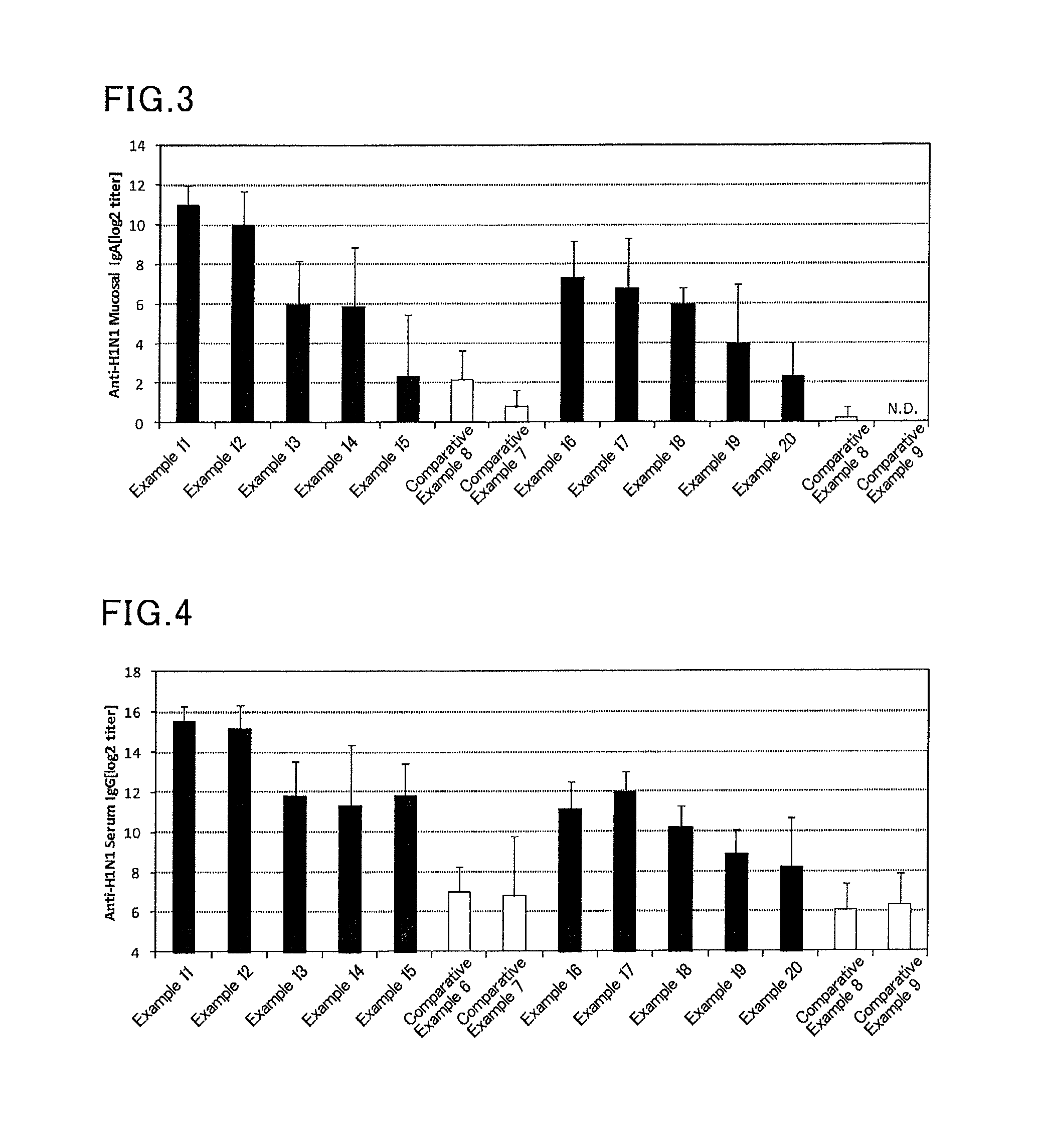
FIG. 3 is a graph showing results of influenza HA (H1N1)-specific IgA titers in a mouse nasal cavity washing liquid in Examples 11 to 20, and Comparative Examples 6 to 9.
FIG. 4 is a graph showing results of influenza HA (H1N1)-specific IgG titers in a mouse serum in Examples 11 to 20, and Comparative Examples 6 to 9.
FIG. 5 is a graph showing results of pneumococcal-specific IgA titers in a mouse nasal cavity washing liquid in Examples 21 to 24, and Comparative Examples 10 to 12.
FIG. 6 is a graph showing results of pneumococcal-specific IgG titers in a mouse serum in Examples 21 to 24, and Comparative Examples 10 to 12.
FIG. 7 is a graph showing results of HPV16-specific IgA titers in a mouse nasal cavity washing liquid in Examples 25 to 28, and Comparative Examples 13 to 15.
FIG. 8 is a graph showing results of HPV16-specific IgG titers in a mouse serum in Examples 25 to 28, and Comparative Examples 13 to 15.
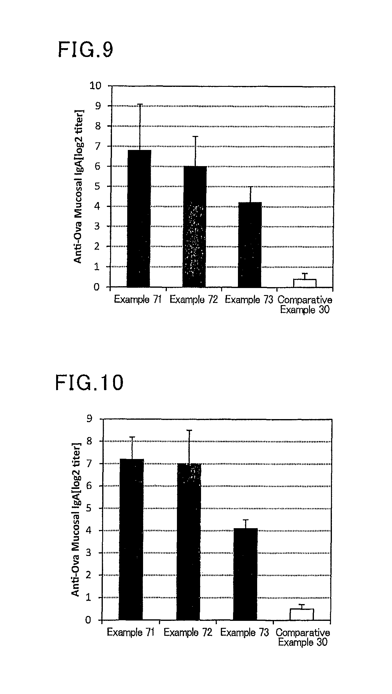
FIG. 9 is a graph showing results of OVA-specific IgA titers in a mouse nasal cavity washing liquid in Examples 71 to 73, and Comparative Example 30.
FIG. 10 is a graph showing results of OVA-specific IgA titers in mouse saliva in Examples 71 to 73, and Comparative Example 30.
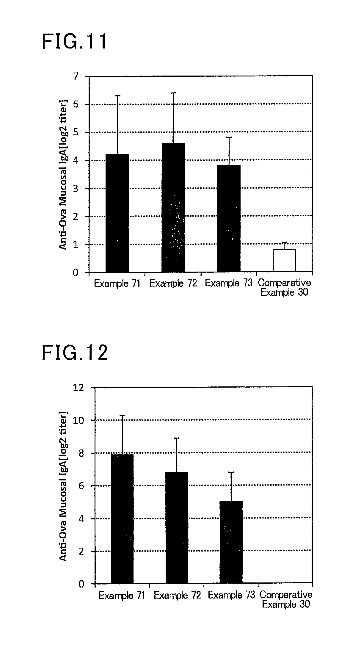
FIG. 11 is a graph showing results of OVA-specific IgA titers in a mouse alveolus washing liquid in Examples 71 to 73, and Comparative Example 30.
FIG. 12 is a graph showing results of an OVA-specific IgA titer in a mouse vaginal washing liquid in Examples 71 to 73, and Comparative Example 30.
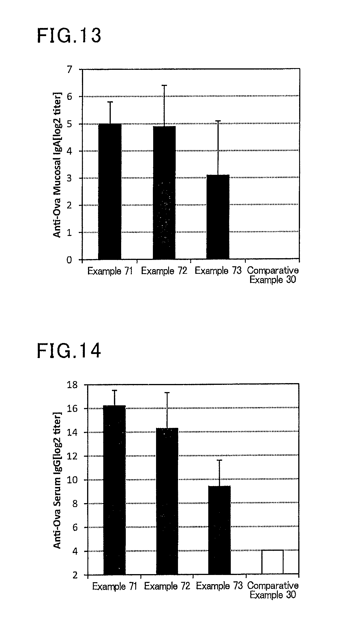
FIG. 13 is a graph showing results of OVA-specific IgA titers in a mouse fecal extract in Examples 71 to 73, and Comparative Example 30.
FIG. 14 is a graph showing results of OVA-specific IgG titers in a mouse serum in Examples 71 to 73, and Comparative Example 30.
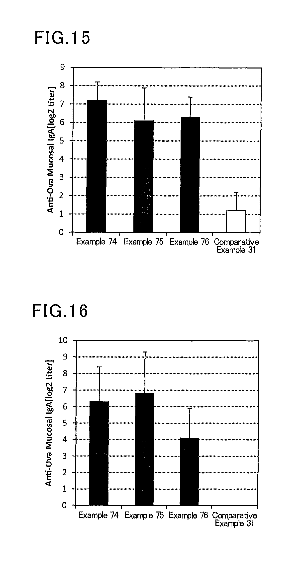
FIG. 15 is a graph showing results of OVA-specific IgA titers in a mouse nasal cavity washing liquid in Examples 74 to 76, and Comparative Example 31.
FIG. 16 is a graph showing results of OVA-specific IgA titers in mouse saliva in Examples 74 to 76, and Comparative Example 31.
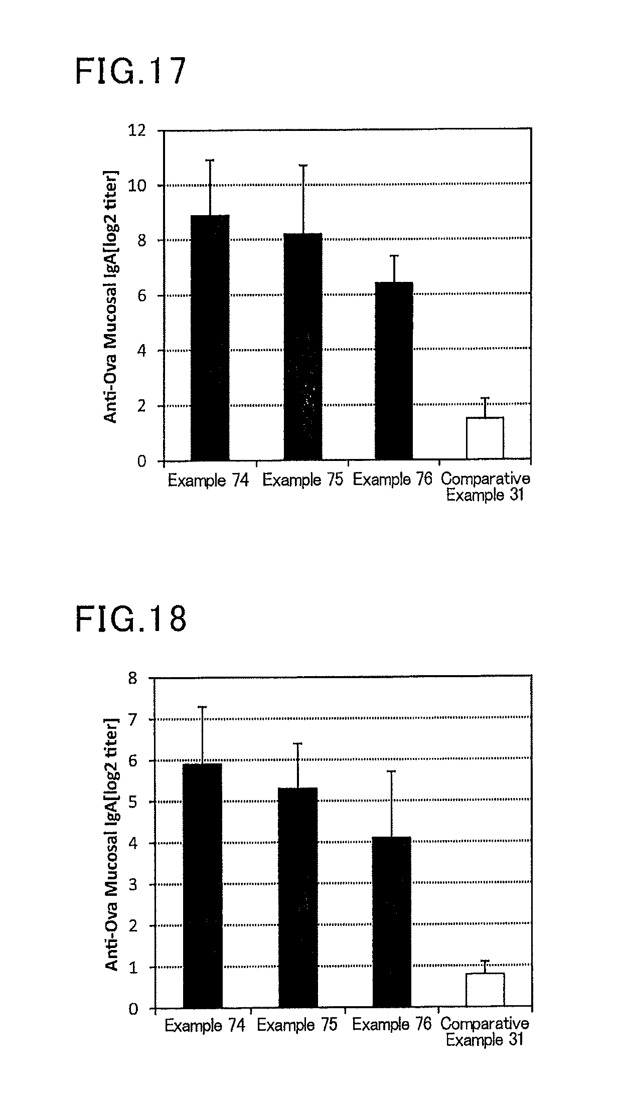
FIG. 17 is a graph showing results of OVA-specific IgA titers in a mouse alveolus washing liquid in Examples 74 to 76, and Comparative Example 31.
FIG. 18 is a graph showing results of OVA-specific IgA titers in a mouse vaginal washing liquid in Examples 74 to 76, and Comparative Example 31.
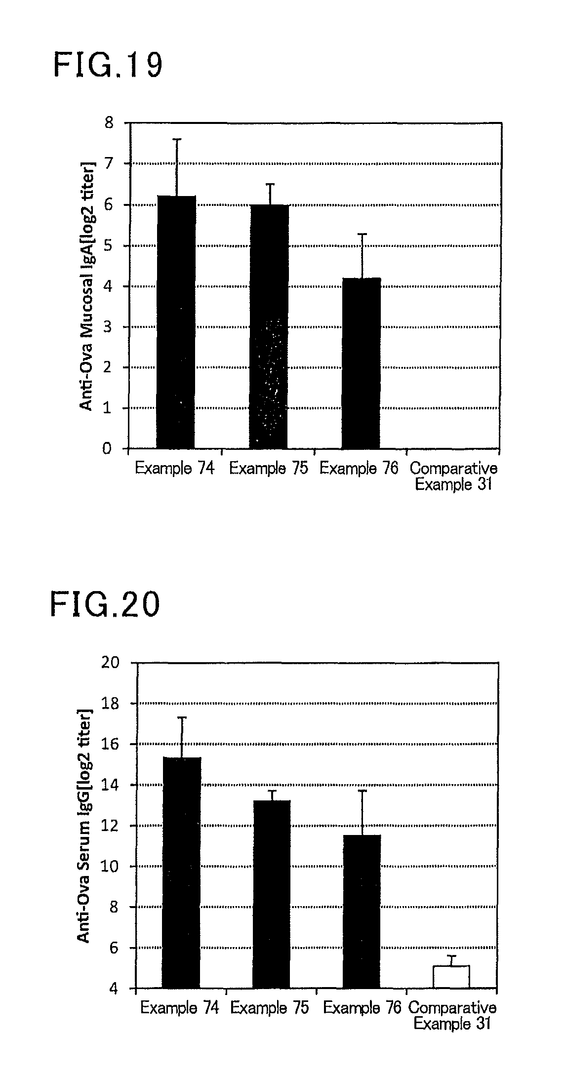
FIG. 19 is a graph showing results of OVA-specific IgA titers in a mouse fecal extract in Examples 74 to 76, and Comparative Example 31.
FIG. 20 is a graph showing results of OVA-specific IgG titers in a mouse serum in Examples 74 to 76, and Comparative Example 31.
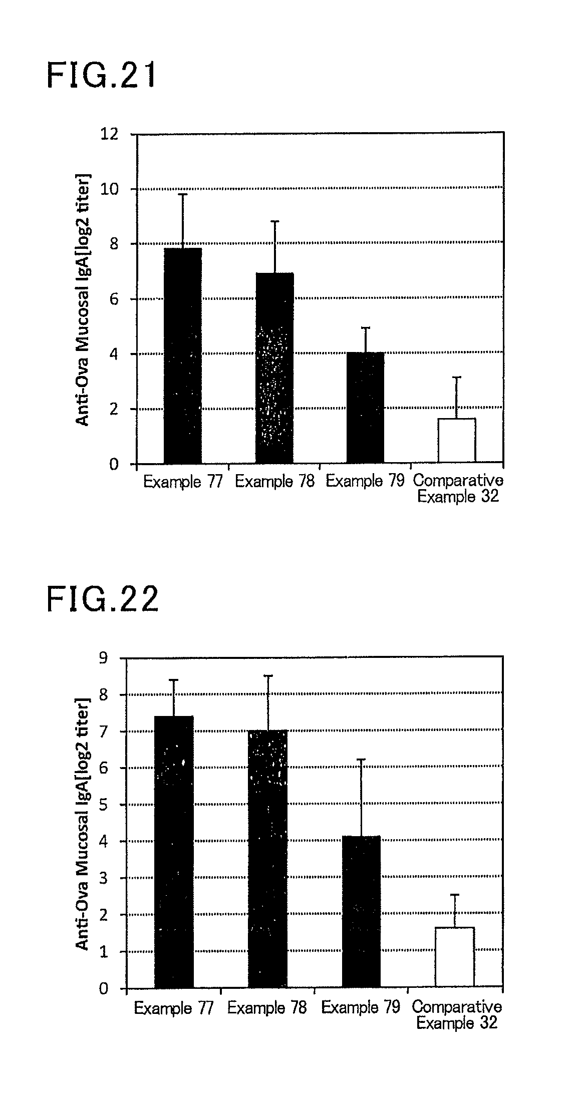
FIG. 21 is a graph showing results of OVA-specific IgA titers in a mouse vaginal washing liquid in Examples 77 to 79, and Comparative Example 32.
FIG. 22 is a graph showing results of OVA-specific IgA titers in a mouse fecal extract in Examples 77 to 79, and Comparative Example 32.
FIG. 23 is a graph showing results of OVA-specific IgG titers in a mouse serum in Examples 77 to 79, and Comparative Example 32.
FIG. 24 is a graph showing results of OVA-specific IgA titers in a mouse vaginal washing liquid in Examples 80 to 82, and Comparative Example 33.
FIG. 25 is a graph showing results of an OVA-specific IgA titer in a mouse fecal extract in Examples 80 to 82, and Comparative Example 33.
FIG. 26 is a graph showing results of OVA-specific IgG titers in a mouse serum in Examples 80 to 82, and Comparative Example 33.
DESCRIPTION OF EMBODIMENTS
The present invention will be described in more detail with reference to the following examples, but is not limited to these examples.
Each of the following administration groups was prepared for ten animals.
As an appropriate dose of a vaccine antigen to mice, two patterns of 1.0 .mu.g and 0.1 .mu.g were examined in the case of an influenza vaccine. If the dose is more than 1.0 .mu.g, an antibody may be produced even in the absence of an adjuvant. On the other hand, if the dose is less than 0.1 .mu.g, an antibody may not be produced even in the presence of an adjuvant because the amount of the vaccine antigen is too small.
Examples 1 to 10, Comparative Examples 1 to 5
An influenza vaccine antigen-containing solution (B/Wisconsin/1/2010, produced by The Research Foundation for Microbial Diseases of Osaka University) (445 .mu.g/mL), and a solution of a lipopolysaccharide derived from Pantoea agglomerans (available from Institute of applied technology for innate immunity) (50 mg/mL) were prepared to give doses in each group of table 1, and then a phosphate buffer (available from Nacalai Tesque) was added to prepare 300 .mu.L of a vaccine composition. For example, in Example 1, after adding 22.5 .mu.L of the influenza vaccine antigen-containing solution, and 20 .mu.L of the solution of a lipopolysaccharide derived from Pantoea agglomerans, a phosphate buffer was added to make the total amount 300 .mu.L. For other examples and comparative examples, vaccine compositions were prepared to have contents corresponding to the doses by appropriate dilution, and in Comparative Example 5, only a phosphate buffer (available from Nacalai Tesque) was administered to mice without adding a vaccine antigen or an adjuvant.
Six mice (female BALB/C mice aged 8 weeks, Japan SLC, Inc.) were anesthetized, and 30 .mu.L of the prepared vaccine composition was sublingually administered to each mouse. After one week from the administration, the mice were anesthetized again, and 30 .mu.L of the prepared vaccine composition was sublingually administered to each mouse. After one week from the second administration, a serum and a nasal cavity washing liquid of each mouse were collected, and an influenza HA (type B)-specific IgG titer in a serum and an influenza HA (type B)-specific IgA titer in a nasal cavity washing liquid were determined by the ELISA method.
In the group in which 1000 .mu.g of the adjuvant was administered (Comparative Example 1), impairment in the lie of hair, and weight loss of mice were observed after 24 hours from the first administration, and the mice were euthanized. Therefore, the subsequent measurement of the antibody titer was not conducted. An adjuvant is a substance that activates immunity, and it is apparent that the immunity can be obtained more easily as the amount added increases. However, administering an excessive amount is problematic in terms of safety, and administration of 1000 .mu.g in mice was not conducted after Comparative Example 1.
Specific determination methods will be described later.
TABLE-US-00001 TABLE 1 Adjuvant (LPS derived Vaccine antigen from Pantoea agglomerans) Amount Amount Ratio Administration No. Species [.mu.g/mouse/dose] [.mu.g/mouse/dose] (adjuvant/antigen) route- Comaprative B/Wisconsin/1/2010 1 1000 1000 Sublingual Example 1 Example 1 B/Wisconsin/1/2010 1 100 100 Sublingual Example 2 B/Wisconsin/1/2010 1 10 10 Sublingual Example 3 B/Wisconsin/1/2010 1 1 1 Sublingual Example 4 B/Wisconsin/1/2010 1 0.1 0.1 Sublingual Example 5 B/Wisconsin/1/2010 1 0.01 0.01 Sublingual Comaprative B/Wisconsin/1/2010 1 0.001 0.001 Sublingual Example 2 Comaprative B/Wisconsin/1/2010 1 0 0 Sublingual Example 3 Example 6 B/Wisconsin/1/2010 0.1 10 100 Sublineual Example 7 B/Wisconsin/1/2010 0.1 1 10 Sublingual Example 8 B/Wisconsin/1/2010 0.1 0.1 1 Sublingual Example 9 B/Wisconsin/1/2010 0.1 0.01 0.1 Sublingual Example 10 B/Wisconsin/1/2010 0.1 0.001 0.01 Sublingual Comparative B/Wisconsin/1/2010 0.1 0 0 Sublingual Example 4 Comparative -- -- -- -- Sublingual Example 5
Examples 11 to 20, Comparative Examples 6 to 9
Vaccine compositions corresponding to Table 2 were prepared in the procedure based on that in Examples 1 to 10 and Comparative Examples 1 to 5 except that the influenza vaccine antigen-containing solution was changed from B/Wisconsin/1/2010 to A/California/07/2009 (H1N1, produced by The Research Foundation for Microbial Diseases of Osaka University) (801 .mu.g/mL). For example, in Example 11, after adding 12.5 .mu.L of an influenza vaccine antigen-containing solution and 20 .mu.L of a solution of a lipopolysaccharide derived from Pantoea agglomerans, a phosphate buffer was added to make the total amount 300 .mu.L.
Six mice (female BALB/C mice aged 8 weeks, Japan SLC, Inc.) were anesthetized, and 30 .mu.L of the prepared vaccine composition was sublingually administered to each mouse. After one week from the administration, the mice were anesthetized again, and 30 .mu.L of the prepared vaccine composition was sublingually administered to each mouse. After one week from the second administration, a serum and a nasal cavity washing liquid of each mouse were collected, and an influenza HA (H1N1)-specific IgG titer in a serum and an influenza HA (H1N1)-specific IgA titer in a nasal cavity washing liquid were determined by the ELISA method. Specific determination methods will be described later.
TABLE-US-00002 TABLE 2 Adjuvant (LPS derived Vaccine antigen from Pantoea agglomerans) Amount Amount Ratio Administration No. Species [.mu.g/mouse/dose] [.mu.g/mouse/dose] (adjuvant/antigen) route- Example 11 A/California/07/2009(H1N1) 1 100 100 Sublingual Example 12 A/California/07/2009(H1N1) 1 10 10 Sublingual Example 13 A/California/07/2009(H1N1) 1 1 1 Sublingual Example 14 A/California/07/2009(H1N1) 1 0.1 0.1 Sublingual Example 15 A/California/07/2009(H1N1) 1 0.01 0.01 Sublingual Comparative A/California/07/2009(H1N1) 1 0.001 0.001 Sublingual Example 6 Comparative A/California/07/2009(H1N1) 1 0 0 Sublingual Example 7 Example 16 A/California/07/2009(H1N1) 0.1 10 100 Sublingual Example 17 A/California/07/2009(H1N1) 0.1 1 10 Sublingual Example 18 A/California/07/2009(H1N1) 0.1 0.1 1 Sublingual Example 19 A/California/07/2009(H1N1) 0.1 0.01 0.1 Sublingual Example 20 A/California/07/2009(H1N1) 0.1 0.001 0.01 Sublingual Comparative A/California/07/2009(H1N1) 0.1 0 0 Sublingual Example 8 Comparative -- -- -- -- Sublingual Example 9
Examples 21 to 24, Comparative Examples 10 to 12
Vaccine compositions corresponding to Table 3 were prepared in the procedure based on that in Examples 1 to 10 and Comparative Examples 1 to 5 except that the vaccine antigen was changed from influenza to a pneumococcal capsular polysaccharide-containing solution (PNEUMOVAX NP, available from MSD K.K.) (1150 .mu.g/mL). For example, in Example 21, after adding 8.7 .mu.L of a pneumococcal capsular polysaccharide-containing solution and 2 .mu.L of a solution of a lipopolysaccharide derived from Pantoea agglomerans, a phosphate buffer was added to make the total amount 300 .mu.L.
Six mice (female BALB/C mice aged 8 weeks, Japan SLC, Inc.) were anesthetized, and 30 .mu.L of the prepared vaccine composition was sublingually administered to each mouse. After one week from the administration, the mice were anesthetized again, and 30 .mu.L of the prepared vaccine composition was sublingually administered to each mouse. After one week from the second administration, a serum and a nasal cavity washing liquid of each mouse were collected, and a pneumococcal-specific IgG titer in a serum and a pneumococcal-specific IgA titer in a nasal cavity washing liquid were determined by the ELISA method. Specific determination methods will be described later.
TABLE-US-00003 TABLE 3 Adjuvant (LPS derived Vaccine antigen from Pantoea agglomerans) Amount Amount Ratio Administration No. Species [.mu.g/mouse/dose] [.mu.g/mouse/dose] (adjuvant/antigen) route- Example 21 Pneumococcal capsular 1 10 10 Sublingual polysaccharide Pneumovax NP Example 22 Pneumococcal capsular 1 1 1 Sublingual polysaccharide Pneumovax NP Example 23 Pneumococcal capsular 1 0.1 0.1 Sublingual polysaccharide Pneumovax NP Example 24 Pneumococcal capsular 1 0.01 0.01 Sublingual polysaccharide Pneumovax NP Comparative Pneumococcal capsular 1 0.001 0.001 Sublingual Example 10 polysaccharide Pneumovax NP Comparative Pneumococcal capsular 1 0 0 Sublingual Example 11 polysaccharide Pneumovax NP Comparative -- -- -- -- Sublingual Example 12
Examples 25 to 28, Comparative Examples 13 to 15
Vaccine compositions corresponding to Table 4 were prepared in the procedure based on that in Examples 1 to 10 and Comparative Examples 1 to 5 except that the vaccine antigen was changed from influenza to an HPV16 recombinant protein-containing solution (HPV16, available from PROSPEC) (820 .mu.g/mL). For example, in Example 25, after adding 12.2 .mu.L of an HPV16 recombinant protein-containing solution and 2 .mu.L of a solution of a lipopolysaccharide derived from Pantoea agglomerans, a phosphate buffer was added to make the total amount 300 .mu.L.
Six mice (female BALB/C mice aged 8 weeks, Japan SLC, Inc.) were anesthetized, and 30 .mu.L of the prepared vaccine composition was sublingually administered to each mouse. After one week from the administration, the mice were anesthetized again, and 30 .mu.L of the prepared vaccine composition was sublingually administered to each mouse. After one week from the second administration, a serum and a nasal cavity washing liquid of each mouse were collected, and an HPV16 recombinant protein-specific IgG titer in a serum and an HPV16 recombinant protein-specific IgA titer in a nasal cavity washing liquid were determined by the ELISA method. Specific determination methods will be described later.
TABLE-US-00004 TABLE 4 Adjuvant (LPS derived Vaccine antigen from Pantoea agglomerans) Amount Amount Ratio Administration No. Species [.mu.g/mouse/dose] [.mu.g/mouse/dose] (adjuvant/antigen) route- Example 25 HPV16 recombinant protein 1 10 10 Sublingual Example 26 HPV16 recombinant protein 1 1 1 Sublingual Example 27 HPV16 recombinant protein 1 0.1 0.1 Sublingual Example 28 HPV16 recombinant protein 1 0.01 0.01 Sublingual Comparative HPV16 recombinant protein 1 0.001 0.001 Sublingual Exampl 13 Comparative HPV16 recombinant protein 1 0 0 Sublingual Exampl 14 Comparative -- -- -- -- Sublingual Exampl 15
Examples 29 to 31, Comparative Example 16
To 200 .mu.L of an attenuated live rotavirus-containing solution (ROTATEQ mixture for internal use, available from MSD K.K.), 50 .mu.L (2 mg/mL) in Example 29, 5 .mu.L in Example 30, or 0.5 .mu.L in Example 31 of a solution of a lipopolysaccharide derived from Pantoea agglomerans (available from Nacalai Tesque), or 5 .mu.L of a glucopyranosyl lipid (MPLAs, available from InvivoGen) solution (2 mg/mL) in Comparative Example 16 was added, and a phosphate buffer (available from Nacalai Tesque) was added to prepare 300 .mu.L of a vaccine composition.
Six mice (female BALB/C mice aged 8 weeks, Japan SLC, Inc.) are anesthetized, and 30 .mu.L of the prepared vaccine composition is sublingually administered to each mouse. After one week from the administration, the mice are anesthetized again, and 30 .mu.L of the prepared vaccine composition is sublingually administered to each mouse. After one week from the second administration, a serum and a nasal cavity washing liquid of each mouse are collected, and an antigen-specific IgG titer in a serum and an antigen-specific IgA titer in a nasal cavity washing liquid are determined by the ELISA method.
Examples 32 to 70, Comparative Examples 17 to 29
In Examples 32 to 34, and Comparative Example 17, an inactivated poliovirus-containing solution (IMOVAX POLIO subcutaneous, available from Sanofi K.K.) was used, in Examples 35 to 37, and Comparative Example 18, an inactivated hepatitis A virus-containing solution (AIMMUGEN, available from KAKETSUKEN) was used, in Examples 38 to 40, and Comparative Example 19, an inactivated Japanese encephalitis virus-containing solution (ENCEVAC for subcutaneous injection, available from KAKETSUKEN) was used, in Examples 41 to 43, and Comparative Example 20, an attenuated live mumps virus-containing solution (mumps live vaccine, available from KITASATO DAIICHISANKYO VACCINE CO., LTD.) was used, in Examples 44 to 46, and Comparative Example 21, an attenuated live measles virus-containing solution (measles live vaccine, available from KITASATO DAIICHISANKYO VACCINE CO., LTD.) was used, in Examples 47 to 49, and Comparative Example 22, an attenuated live rubella virus-containing solution (dry attenuated live rubella vaccine, available from KITASATO DAIICHISANKYO VACCINE CO., LTD.) was used, in Examples 50 to 52, and Comparative Example 23, a tetanus toxoid conjugate Haemophilus influenzae type b polysaccharide-containing solution (ACTHIB, available from Sanofi K.K.) was used, in Examples 53 to 55, and Comparative Example 24, a recombinant HBs antigen protein-containing solution (BIMMUGEN, available from KAKETSUKEN) was used, in Examples 56 to 58, and Comparative Example 25, an attenuated live yellow fever virus-containing solution (yellow fever vaccine, available from Sanofi K.K.) was used, in Examples 59 to 61, and Comparative Example 26, a tetanus toxoid-containing solution (tetanus toxoid, available from DENKA SEIKEN CO., LTD.) was used, in Examples 62 to 64, and Comparative Example 27, an attenuated live chickenpox virus-containing solution (dry attenuated live chickenpox vaccine, available from The Research Foundation for Microbial Diseases of Osaka University) was used, in Examples 65 to 67, and Comparative Example 28, a live BCG-containing solution (dry BCG vaccine, available from Japan BCG Laboratory) was used, and in Examples 68 to 70, and Comparative Example 29, an inactivated rabies virus-containing solution (tissue-cultured inactivated rabies vaccine, available from KAKETSUKEN) was used.
A vaccine composition was prepared in the same manner as in Table 5 and Examples 29 to 31, and Comparative Example 16. Also immunological experiments are conducted in the same manner as in Examples 29 to 31, and Comparative Example 16.
TABLE-US-00005 TABLE 5 Vaccine antigen Adjuvant Amount Amount Administration No. Species [/mouse/dose] Substance name Ligand [.mu.g/mouse/dose] route Note Example 29 Live attenuated rotavirus Vaccine 20 .mu.L equivalent LPS derived from TLR4 10 Sublingual Liquid (RIX4414 strain) Pantoea agglomerans Example 30 Live attenuated rotavirus Vaccine 20 .mu.L equivalent LPS derived from TLR4 1 Sublingual Liquid (RIX4414 strain) Pantoea agglomerans Example 31 Live attenuated rotavirus Vaccine 20 .mu.L equivalent LPS derived from TLR4 0.1 Sublingual Liquid (RIX4414 strain) Pantoea agglomerans Example 32 Inactivated poliovirus Vaccine 20 .mu.L equivalent LPS derived from TLR4 10 Sublingual Liquid (type 1, type 2, type 3) Pantoea agglomerans Example 33 Inactivated poliovirus Vaccine 20 .mu.L equivalent LPS derived from TLR4 1 Sublingual Liquid (type 1, type 2, type 3) Pantoea agglomerans Example 34 Inactivated poliovirus Vaccine 20 .mu.L equivalent LPS derived from TLR4 0.1 Sublingual Liquid (type 1, type 2, type 3) Pantoea agglomerans Example 35 Inactivated hepatitis Vaccine 20 .mu.L equivalent LPS derived from TLR4 10 Sublingual Liquid A virus Pantoea agglomerans Example 36 Inactivated hepatitis Vaccine 20 .mu.L equivalent LPS derived from TLR4 1 Sublingual Liquid A virus Pantoea agglomerans Example 37 Inactivated hepatitis Vaccine 20 .mu.L equivalent LPS derived from TLR4 0.1 Sublingual Liquid A virus Pantoea agglomerans Example 38 Inactivated Japanese Vaccine 20 .mu.L equivalent LPS derived from TLR4 10 Sublingual Liquid encephalitis virus Pantoea agglomerans Example 39 Inactivated Japanese Vaccine 20 .mu.L equivalent LPS derived from TLR4 1 Sublingual Liquid encephalitis virus Pantoea agglomerans Example 40 Inactivated Japanese Vaccine 20 .mu.L equivalent LPS derived from TLR4 0.1 Sublingual Liquid encephalitis virus Pantoea agglomerans Example 41 Live attenuated mumps Vaccine 20 .mu.L equivalent LPS derived from TLR4 10 Sublingual Liquid virus Pantoea agglomerans Example 42 Live attenuated mumps Vaccine 20 .mu.L equivalent LPS derived from TLR4 1 Sublingual Liquid virus Pantoea agglomerans Example 43 Live attenuated mumps Vaccine 20 .mu.L equivalent LPS derived from TLR4 0.1 Sublingual Liquid virus Pantoea agglomerans Example 44 Live attenuated Vaccine 20 .mu.L equivalent LPS derived from TLR4 10 Sublingual Liquid measles virus Pantoea agglomerans Example 45 Live attenuated Vaccine 20 .mu.L equivalent LPS derived from TLR4 1 Sublingual Liquid measles virus Pantoea agglomerans Example 46 Live attenuated Vaccine 20 .mu.L equivalent LPS derived from TLR4 0.1 Sublingual Liquid measles virus Pantoea agglomerans Example 47 Live attenuated Vaccine 20 .mu.L equivalent LPS derived from TLR4 10 Sublingual Liquid rubella virus Pantoea agglomerans Example 48 Live attenuated Vaccine 20 .mu.L equivalent LPS derived from TLR4 1 Sublingual Liquid rubella virus Pantoea agglomerans Example 49 Live attenuated Vaccine 20 .mu.L equivalent LPS derived from TLR4 0.1 Sublingual Liquid rubella virus Pantoea agglomerans Example 50 Tetanus toxoid-conjugated Vaccine 20 .mu.L equivalent LPS derived from TLR4 10 Sublingual Liquid Haemophilus influenzae type Pantoea agglomerans b polysaccharide Example 51 Tetanus toxoid-conjugated Vaccine 20 .mu.L equivalent LPS derived from TLR4 1 Sublingual Liquid Haemophilus influenzae type Pantoea agglomerans b polysaccharide Example 52 Tetanus toxoid-conjugated Vaccine 20 .mu.L equivalent LPS derived from TLR4 0.1 Sublingual Liquid Haemophilus influenzae type Pantoea agglomerans b polysaccharide Example 53 Recombinant HBs antigen Vaccine 20 .mu.L equivalent LPS derived from TLR4 10 Sublingual Liquid protein Pantoea agglomerans Example 54 Recombinant HBs antigen Vaccine 20 .mu.L equivalent LPS derived from TLR4 1 Sublingual Liquid protein Pantoea agglomerans Example 55 Recombinant HBs antigen Vaccine 20 .mu.L equivalent LPS derived from TLR4 0.1 Sublingual Liquid protein Pantoea agglomerans Example 56 Live attenuated yellow Vaccine 20 .mu.L equivalent LPS derived from TLR4 10 Sublingual Liquid fever virus Pantoea agglomerans Example 57 Live attenuated yellow Vaccine 20 .mu.L equivalent LPS derived from TLR4 1 Sublingual Liquid fever virus Pantoea agglomerans Example 58 Live attenuated yellow Vaccine 20 .mu.L equivalent LPS derived from TLR4 0.1 Sublingual Liquid fever virus Pantoea agglomerans Example 59 Tetanus toxoid Vaccine 20 .mu.L equivalent LPS derived from TLR4 10 Sublingual Liquid Pantoea agglomerans Example 60 Tetanus toxoid Vaccine 20 .mu.L equivalent LPS derived from TLR4 1 Sublingual Liquid Pantoea agglomerans Example 61 Tetanus toxoid Vaccine 20 .mu.L equivalent LPS derived from TLR4 0.1 Sublingual Liquid Pantoea agglomerans Example 62 Live attenuated varicella- Vaccine 20 .mu.L equivalent LPS derived from TLR4 10 Sublingual Liquid zoster virus Pantoea agglomerans Example 63 Live attenuated varicella- Vaccine 20 .mu.L equivalent LPS derived from TLR4 1 Sublingual Liquid zoster virus Pantoea agglomerans Example 64 Live attenuated varicella- Vaccine 20 .mu.L equivalent LPS derived from TLR4 0.1 Sublingual Liquid zoster virus Pantoea agglomerans Example 65 Live BCG Vaccine 20 .mu.L equivalent LPS derived from TLR4 10 Sublingual Liquid Pantoea agglomerans Example 66 Live BCG Vaccine 20 .mu.L equivalent LPS derived from TLR4 1 Sublingual Liquid Pantoea agglomerans Example 67 Live BCG Vaccine 20 .mu.L equivalent LPS derived from TLR4 0.1 Sublingual Liquid Pantoea agglomerans Example 68 Inactivated rabies virus Vaccine 20 .mu.L equivalent LPS derived from TLR4 10 Sublingual Liquid Pantoea agglomerans Example 69 Inactivated rabies virus Vaccine 20 .mu.L equivalent LPS derived from TLR4 1 Sublingual Liquid Pantoea agglomerans Example 70 Inactivated rabies virus Vaccine 20 .mu.L equivalent LPS derived from TLR4 0.1 Sublingual Liquid Pantoea agglomerans Comparative Live attenuated rotavirus Vaccine 20 .mu.L equivalent Glucopyranosyl lipid TLR4 1 Sublingual Liquid Example 16 (RIX44I4 strain) Comparative Inactivated poliovirus Vaccine 20 .mu.L equivalent Glucopyranosyl lipid TLR4 1 Sublingual Liquid Example 17 (type 1, type 2, type 3) Comparative Inactivated hepatitis Vaccine 20 .mu.L equivalent Glucopyranosyl lipid TLR4 1 Sublingual Liquid Example 18 A virus Comparative Inactivated Japanese Vaccine 20 .mu.L equivalent Glucopyranosyl lipid TLR4 1 Sublingual Liquid Example 19 encephalitis virus Comparative Live attenuated mumps Vaccine 20 .mu.L equivalent Glucopyranosyl lipid TLR4 1 Sublingual Liquid Example 20 virus Comparative Live attenuated measles Vaccine 20 .mu.L equivalent Glucopyranosyl lipid TLR4 1 Sublingual Liquid Example 21 virus Comparative Liva attenuated rubella Vaccine 20 .mu.L equivalent Glucopyranosyl lipid TLR4 1 Sublingual Liquid Example 22 virus Comparative Tetanus toxoid-conjugated Vaccine 20 .mu.L equivalent Glucopyranosyl lipid TLR4 1 Sublingual Liquid Example 23 Haemophilus influenzae type b polysaccharide Comparative Recombinant HBs antigen Vaccine 20 .mu.L equivalent Glucopyranosyl lipid TLR4 1 Sublingual Liquid Example 24 protein Comparative Live attenuated yellow Vaccine 20 .mu.L equivalent Glucopyranosyl lipid TLR4 1 Sublingual Liquid Example 25 fever virus Comparative Tetanus toxoid Vaccine 20 .mu.L equivalent Glucopyranosyl lipid TLR4 1 Sublingual Liquid Example 26 Comparative Live attenuated varicella- Vaccine 20 .mu.L equivalent Glucopyranosyl lipid TLR4 1 Sublingual Liquid Example 27 zoster virus Comparative Live BCG Vaccine 20 .mu.L equivalent Glucopyranosyl lipid TLR4 1 Sublingual Liquid Example 28 Comparative Inactivated rabies virus Vaccine 20 .mu.L equivalent Glucopyranosyl lipid TLR4 1 Sublingual Liquid Example 29
Examples 71 to 73, Comparative Example 30
Vaccine compositions corresponding to Table 6 were prepared in the procedure based on that in Examples 1 to 10 and Comparative Examples 1 to 5 except that the vaccine antigen was changed from influenza to ovalbumin (OVA) (Sigma-Aldrich Japan). For example, in Example 71, after adding 100 .mu.L (1000 .mu.g/mL) of ovalbumin (OVA) and 5 .mu.L (2 mg/mL) of a solution of a lipopolysaccharide derived from Pantoea agglomerans (available from Nacalai Tesque), a phosphate buffer (available from Nacalai Tesque) was added to prepare 300 .mu.L of a mucosal vaccine composition.
Six mice (female BALB/C mice aged 8 weeks, Japan SLC, Inc.) were anesthetized, and 30 .mu.L of the prepared vaccine composition was sublingually administered to each mouse. After one week from the administration, the mice were anesthetized again, and sublingual administration was conducted to each mouse in the same manner. After one week from the second administration, a serum and mucosal samples of each mouse were collected, and an ovalbumin-specific IgG titer in a serum, and ovalbumin-specific IgA titers in a nasal cavity washing liquid, saliva, an alveolus washing liquid, a vaginal washing liquid, and a fecal extract were determined by the ELISA method. Specific determination methods will be described later.
TABLE-US-00006 TABLE 6 Vaccine antigen Adjuvant Amount Amount Administration No. Species [.mu.g/mouse/dose] Substance name Ligand [.mu.g/mouse/dose] route Example 71 Ovalbumin 10 LPS derived from Pantoea agglomerans TLR4 1 Sublingual Example 72 Ovalbumin 1 LPS derived from Pantoea agglomerans TLR4 1 Sublingual Example 73 Ovalbumin 0.1 LPS derived from Pantoea agglomerans TLR4 1 Sublingual Comparative Ovalbumin 0.001 LPS derived from Pantoea agglomerans TLR4 1 Sublingual Example 30
Examples 74 to 76, Comparative Example 31
Vaccine compositions corresponding to Table 7 were prepared in the procedure based on that in Examples 1 to 10 and Comparative Examples 1 to 5 except that the vaccine antigen was changed from influenza to ovalbumin (OVA) (Sigma-Aldrich Japan). For example, in Example 74, a phosphate buffer (available from Nacalai Tesque) was added to 100 .mu.L (1000 .mu.g/mL) of ovalbumin (OVA) and 5 .mu.L (2 mg/mL) of a solution of a lipopolysaccharide derived from Pantoea agglomerans (available from Nacalai Tesque), to prepare 500 .mu.L of a mucosal vaccine composition.
Six mice (female BALB/C mice aged 8 weeks, Japan SLC, Inc.) were anesthetized, and 50 .mu.L of the prepared vaccine composition was spray-administered to the bronchial tube of each mouse using a liquid sprayer (available from Penn-Century, Inc.). After one week from the administration, the mice were anesthetized again, and pulmonary administration was conducted to each mouse in the same manner. After one week from the second administration, a serum and mucosal samples of each mouse were collected, and an ovalbumin-specific IgG titer in a serum, and ovalbumin-specific IgA titers in a nasal cavity washing liquid, saliva, an alveolus washing liquid, a vaginal washing liquid, and a fecal extract were determined by the ELISA method. Specific determination methods will be described later.
TABLE-US-00007 TABLE 7 Vaccine antigen Adjuvant Amount Amount Administration No. Species [.mu.g/mouse/dose] Substance name Ligand [.mu.g/mouse/dose] route Example 74 Ovalbumin 10 LPS derived from Pantoea agglomerans TLR4 1 Pulmonary Example 75 Ovalbumin 1 LPS derived from Pantoea agglomerans TLR4 1 Pulmonary Example 76 Ovalbumin 0.1 LPS derived from Pantoea agglomerans TLR4 1 Pulmonary Comparative Ovalbumin 0.001 LPS derived from Pantoea agglomerans TLR4 1 Pulmonary Example 31
Examples 77 to 79, Comparative Example 32
Vaccine compositions corresponding to Table 8 were prepared in the procedure based on that in Examples 1 to 10 and Comparative Examples 1 to 5 except that the vaccine antigen was changed from influenza to ovalbumin (OVA) (Sigma-Aldrich Japan). For example, in Example 77, a phosphate buffer (available from Nacalai Tesque) was added to 100 .mu.L (1000 .mu.g/mL) of ovalbumin (OVA) and 5 .mu.L (2 mg/mL) of a solution of a lipopolysaccharide derived from Pantoea agglomerans (available from Nacalai Tesque), to prepare 200 .mu.L of a mucosal vaccine composition.
Six mice (female BALB/C mice aged 8 weeks, Japan SLC, Inc.) were anesthetized, and 20 .mu.L of the prepared vaccine composition was administered to the vagina of each mouse with the use of a pipette. After one week from the administration, the mice were anesthetized again, and vaginal administration was conducted to each mouse in the same manner. After one week from the second administration, a serum and mucosal samples of each mouse were collected, and an ovalbumin-specific IgG titer in a serum, and ovalbumin-specific IgA titers in a vaginal washing liquid and a fecal extract were determined by the ELISA method. Specific determination methods will be described later.
TABLE-US-00008 TABLE 8 Vaccine antigen Adjuvant Amount Amount Administration No. Species [.mu.g/mouse/dose] Substance name Ligand [.mu.g/mouse/dose] route Example 77 Ovalbumin 10 LPS derived from Pantoea agglomerans TLR4 1 Transvaginal Example 78 Ovalbumin 1 LPS derived from Pantoea agglomerans TLR4 1 Transvaginal Example 79 Ovalbumin 0.1 LPS derived from Pantoea agglomerans TLR4 1 Transvaginal Comparative Ovalbumin 0.001 LPS derived from Pantoea agglomerans TLR4 1 Transvaginal Example 32
Examples 80 to 82, Comparative Example 33
Vaccine compositions corresponding to Table 9 were prepared in the procedure based on that in Examples 1 to 10 and Comparative Examples 1 to 5 except that the vaccine antigen was changed from influenza to ovalbumin (OVA) (Sigma-Aldrich Japan). For example, in Example 80, a phosphate buffer (available from Nacalai Tesque) was added to 100 .mu.L (1000 .mu.g/mL) of ovalbumin (OVA) and 5 .mu.L (2 mg/mL) of a solution of a lipopolysaccharide derived from Pantoea agglomerans (available from Nacalai Tesque), to prepare 500 .mu.L of a mucosal vaccine composition.
Six mice (female BALB/C mice aged 8 weeks, Japan SLC, Inc.) were anesthetized, and 50 .mu.L of the prepared vaccine composition was administered to the rectum of each mouse with the use of a 1 mL syringe and a sonde for mouse (Fuchigami Kikai). After one week from the administration, the mice were anesthetized again, and rectal administration was conducted to each mouse in the same manner. After one week from the second administration, a serum and mucosal samples of each mouse were collected, and an ovalbumin-specific IgG titer in a serum, and ovalbumin-specific IgA titers in a vaginal washing liquid and a fecal extract were determined by the ELISA method. Specific determination methods will be described later.
TABLE-US-00009 TABLE 9 Vaccine antigen Adjuvant Amount Amount Administration No. Species [.mu.g/mouse/dose] Substance name Ligand [.mu.g/mouse/dose] route Example 80 Ovalbumin 10 LPS derived from Pantoea agglomerans TLR4 1 Rectal Example 81 Ovalbumin 1 LPS derived from Pantoea agglomerans TLR4 1 Rectal Example 82 Ovalbumin 0.1 LPS derived from Pantoea agglomerans TLR4 1 Rectal Comparative Ovalbumin 0.001 LPS derived from Pantoea agglomerans TLR4 1 Rectal Example 33
(Mouse Immunological Experiments)
For female BALB/c mice aged 8 weeks, administration was conducted twice at an interval of one week. After one week from the last administration, blood and a nasal cavity washing liquid of each mouse were collected. The blood was centrifuged at 3000 G for 10 minutes at 4.degree. C., and 300 .mu.L of a phosphate buffer (available from Nacalai Tesque) was added to 20 .mu.L of the supernatant to prepare a serum sample. Mucous membrane samples were collected in the following manner. Regarding a nasal cavity washing liquid, a cut was made in a lower part of the respiratory tract of a BALB/c mouse, 200 .mu.L of a phosphate buffer (available from Nacalai Tesque) was poured into the respiratory tract, and a sample came into the nasal cavity was collected as a nasal cavity washing liquid sample. Regarding saliva, 500 .mu.L of 12 .mu.g/mL carbamylcholine chloride was administered to the abdominal cavity of a mouse to promote production of saliva, and then 20 .mu.L of saliva was collected. Regarding an alveolus washing liquid, a cut was made in a lower part of the respiratory tract of a BALB/c mouse, 500 .mu.L of a phosphate buffer (available from Nacalai Tesque) was poured into the lung, and the phosphate buffer came into the lung was collected as an alveolus washing liquid sample. Regarding a vaginal washing liquid, 150 .mu.L of a phosphate buffer (available from Nacalai Tesque) was poured into the vagina of a BALB/c mouse, and a sample after pipetting 10 times was collected as a vaginal washing liquid sample. Regarding a fecal extract, 100 .mu.L of a phosphate buffer (available from Nacalai Tesque) per 10 mg of collected faces was added, and the mixture was vortexed for 10 minutes. Thereafter, centrifugation at 3000 G was conducted for 10 minutes at 4.degree. C., and the supernatant was collected as a fecal extract sample.
By measuring an immunogen-specific IgG titer in a mouse serum, the systemic immune response was evaluated. Also, by measuring an immunogen-specific IgA titer in a mouse mucous membrane sample, the mucosal immune response was evaluated. The respective evaluation methods will be described below.
The respective evaluation results are shown in FIGS. 1 to 26.
(Method for Measuring Antigen-Specific IgG Titer in Mouse Serum (ELISA Method))
In a 96-well plate for ELISA, each 100 .mu.L of each antigen (for example, a B/Wisconsin/1/2010(B) influenza HA antigen solution in measurement of a B/Wisconsin/1/2010(B)-specific IgG antibody titer) diluted with a carbonate buffer (2.5 .mu.g/mL) was added, and the plate was left still overnight.
Wells were washed with a preliminarily prepared TWEEN 20-containing PBS (hereinafter, referred to as a washing liquid) three times, and after adding each 200 .mu.L of a blocking solution prepared by diluting a blocking agent (Block Ace BLOCK ACE, available from DS Pharma Biomedical Co., Ltd.) in purified water into 4 g/400 mL, the plate was left still for 2 hours at room temperature. Then, wells were washed with the washing liquid three times.
Using a solution prepared by diluting a blocking agent (BLOCK ACE, available from DS Pharma Biomedical Co., Ltd.) with a phosphate buffer (available from Nacalai Tesque) into 0.4 g/100 mL (hereinafter, referred to as a reagent diluent), the aforementioned serum sample was diluted 15 times by doubling serial dilution, each 50 .mu.L of the solutions were added, and the plate was left still for 2 hours at room temperature.
Then, the wells were washed three times with a washing liquid, and each 100 .mu.L of an HRP-labeled anti-mouse IgG antibody (Goat-anti-mouse IgG Fc HRP, available from BETHYL) diluted 10000 times with the reagent diluent was added, and the plate was left still for 1 hour at room temperature.
Then, the wells were washed three times with a washing liquid, and each 100 .mu.L of a TMB solution (ELISA POD TMB kit, available from Nacalai Tesque) was added. Then, each 100 .mu.L of a 1 M sulfuric acid solution was added, and absorbance at 450 nm of the 96-well plate was measured by a micro plate reader (168-11135CAM, available from Bio-Rad). Based on the absorbance in the serial dilution, the maximum dilution fold at which the absorbance was not less than 0.1 was determined as an IgG titer in a mouse serum, and the value was determined as a value of Log 2.
(Method for Measuring Antigen-Specific IgA Titer in Mouse Mucous Membrane Sample (ELISA Method))
The method is basically the same as the method for measuring antigen-specific IgG titer, and the same operations were conducted except that the measurement sample was mucous membrane samples, and an HRP-labeled anti-mouse IgA antibody (Goat-anti-mouse IgA a HRP, available from BETHYL) was used in place of the HRP-labeled anti-mouse IgG antibody.
As shown in FIGS. 1 to 26, according to examples, antigen-specific IgG and IgA were produced at high levels. On the other hand, in comparative examples, the production amounts of both antigen-specific IgG and IgA were small. Also, as can be seen in FIGS. 9 to 26, it was confirmed that by administering an antigen and a lipopolysaccharide to a mucous membrane, immunity is induced not only on the mucosal surface but also on a remote mucosal surface (for example, when an antigen and a lipopolysaccharide were administered sublingually, production of antigen-specific IgA was observed on the intestinal tract and the vaginal surface). That is, it was found that a lipopolysaccharide or a salt thereof functions as an adjuvant that is effective on any mucosal surface, and is capable of sufficiently inducing antigen-specific IgA all over the body.
These results revealed that a lipopolysaccharide derived from a specific gram-negative bacterium or a salt thereof as an adjuvant contained within a predetermined range with respect to the antigen is effective for mucosal immune induction on the surface of a mucous membrane.
INDUSTRIAL APPLICABILITY
Since the mucosal vaccine composition of the present invention contains the aforementioned specific adjuvant together with at least one antigen in contents within predetermined ranges, it can induce the systemic immune response and mucosal immune response safely and effectively even when it is administered to an intraoral mucous membrane, ocular mucous membrane, ear mucous membrane, genital mucous membrane, pharyngeal mucous membrane, respiratory tract mucous membrane, bronchial mucous membrane, pulmonary mucous membrane, gastric mucous membrane, enteric mucous membrane, or rectal mucous membrane.
* * * * *
D00001
D00002
D00003
D00004
D00005
D00006
D00007
D00008
D00009
D00010
D00011
D00012
D00013

XML
uspto.report is an independent third-party trademark research tool that is not affiliated, endorsed, or sponsored by the United States Patent and Trademark Office (USPTO) or any other governmental organization. The information provided by uspto.report is based on publicly available data at the time of writing and is intended for informational purposes only.
While we strive to provide accurate and up-to-date information, we do not guarantee the accuracy, completeness, reliability, or suitability of the information displayed on this site. The use of this site is at your own risk. Any reliance you place on such information is therefore strictly at your own risk.
All official trademark data, including owner information, should be verified by visiting the official USPTO website at www.uspto.gov. This site is not intended to replace professional legal advice and should not be used as a substitute for consulting with a legal professional who is knowledgeable about trademark law.