Techniques for use with nerve tissue
Gross , et al. A
U.S. patent number 10,383,685 [Application Number 15/147,081] was granted by the patent office on 2019-08-20 for techniques for use with nerve tissue. This patent grant is currently assigned to PYTHAGORAS MEDICAL LTD.. The grantee listed for this patent is RAINBOW MEDICAL LTD.. Invention is credited to Yossi Gross, Yehuda Zadok.


| United States Patent | 10,383,685 |
| Gross , et al. | August 20, 2019 |
Techniques for use with nerve tissue
Abstract
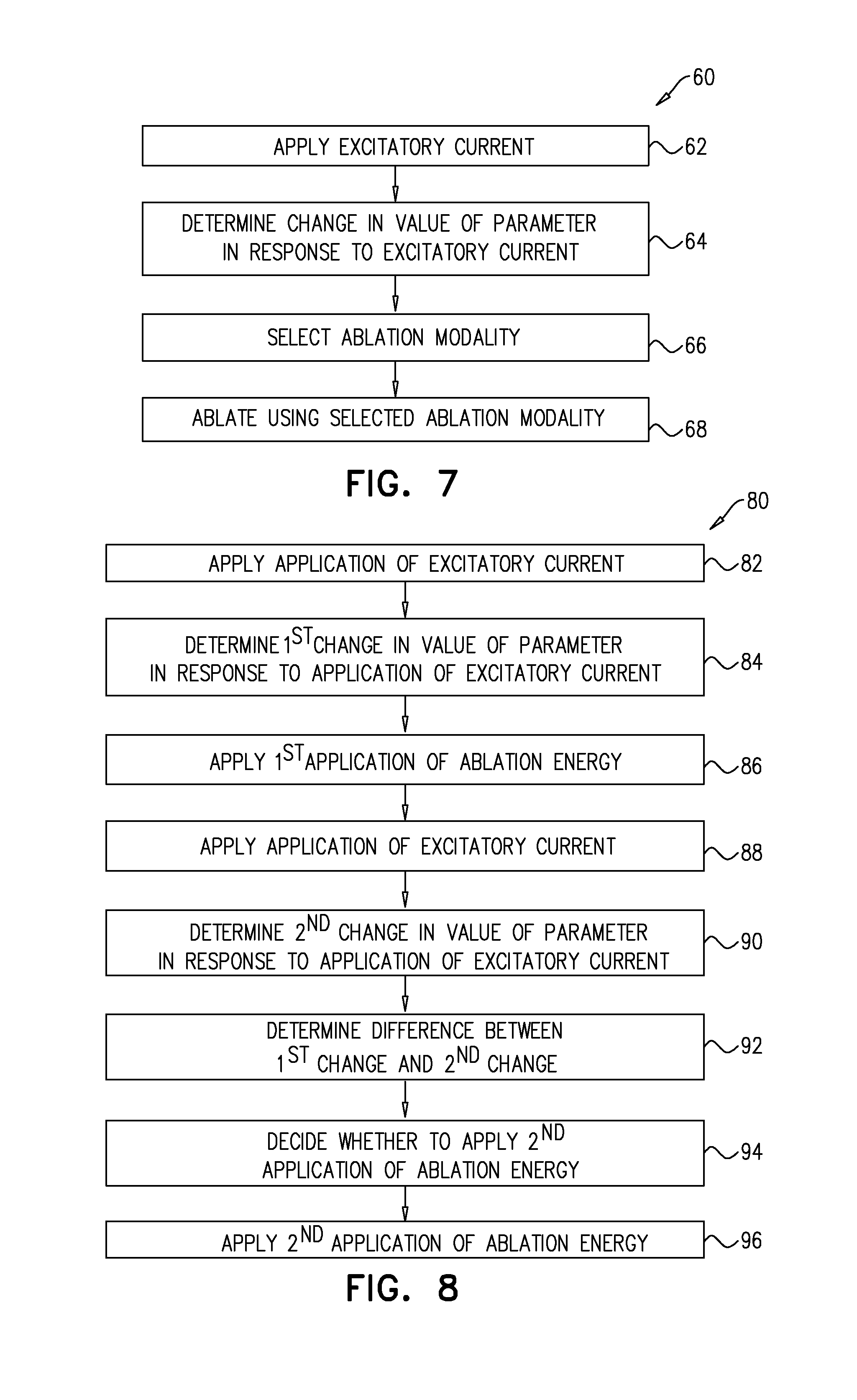
A method is provided, comprising: (1) applying an excitatory current to renal nerve fibers of a subject; (2) determining a change in a parameter of the subject in response to the excitatory current, the parameter being a fastest rate of increase in arterial pressure during a systolic upstroke of an arterial pressure wave of the subject; (3) in response to the change, deciding whether to ablate the renal nerve fibers; and (4) in response to the deciding, applying ablation energy to the renal nerve fibers. Other embodiments are also described.
| Inventors: | Gross; Yossi (Moshav Mazor, IL), Zadok; Yehuda (Holon, IL) | ||||||||||
|---|---|---|---|---|---|---|---|---|---|---|---|
| Applicant: |
|
||||||||||
| Assignee: | PYTHAGORAS MEDICAL LTD.
(Herzliya, IL) |
||||||||||
| Family ID: | 57222105 | ||||||||||
| Appl. No.: | 15/147,081 | ||||||||||
| Filed: | May 5, 2016 |
Prior Publication Data
| Document Identifier | Publication Date | |
|---|---|---|
| US 20160324572 A1 | Nov 10, 2016 | |
Related U.S. Patent Documents
| Application Number | Filing Date | Patent Number | Issue Date | ||
|---|---|---|---|---|---|
| 62158139 | May 7, 2015 | ||||
| Current U.S. Class: | 1/1 |
| Current CPC Class: | A61B 18/1206 (20130101); A61B 18/1492 (20130101); A61B 5/0215 (20130101); A61B 2505/05 (20130101); A61B 2018/00642 (20130101); A61B 5/024 (20130101); A61B 2018/00577 (20130101); A61B 2018/00434 (20130101); A61B 5/02007 (20130101); A61B 2090/064 (20160201); A61B 5/6852 (20130101); A61B 2018/00214 (20130101); A61B 5/4848 (20130101); A61B 2018/00511 (20130101); A61B 2018/00702 (20130101); A61B 2018/00404 (20130101); A61B 2562/0247 (20130101); A61B 2018/00773 (20130101) |
| Current International Class: | A61B 18/12 (20060101); A61B 18/14 (20060101); A61B 5/0215 (20060101); A61B 18/00 (20060101); A61B 5/00 (20060101); A61B 5/02 (20060101); A61B 5/024 (20060101); A61B 90/00 (20160101) |
References Cited [Referenced By]
U.S. Patent Documents
| 4106488 | August 1978 | Gordon |
| 4569836 | February 1986 | Gordon |
| 4619247 | October 1986 | Inoue |
| 5215086 | June 1993 | Terry, Jr. et al. |
| 5251634 | October 1993 | Weinberg |
| 5697377 | December 1997 | Wittkampf |
| 5735280 | April 1998 | Sherman et al. |
| 5776063 | July 1998 | Dittrich et al. |
| 5807285 | September 1998 | Vaitekunas |
| 5817022 | October 1998 | Vesely |
| 5827216 | October 1998 | Igo et al. |
| 6050943 | April 2000 | Slayton |
| 6064902 | May 2000 | Haissaguerre et al. |
| 6117101 | September 2000 | Diederich |
| 6128523 | October 2000 | Bechtold |
| 6161048 | December 2000 | Sluijter |
| 6219577 | April 2001 | Brown |
| 6233477 | May 2001 | Chia |
| 6241727 | June 2001 | Tu |
| 6246899 | June 2001 | Chia |
| 6361500 | March 2002 | Masters |
| 6405732 | June 2002 | Edwards |
| 6425877 | July 2002 | Edwards |
| 6440077 | August 2002 | Jung |
| 6485431 | November 2002 | Campbell |
| 6522926 | February 2003 | Kieval |
| 6526318 | February 2003 | Ansarinia |
| 6605084 | August 2003 | Acker et al. |
| 6635054 | October 2003 | Fjield et al. |
| 6641579 | November 2003 | Bernardi |
| 6659950 | December 2003 | Taheri |
| 6671556 | December 2003 | Osorio et al. |
| 6684105 | January 2004 | Cohen et al. |
| 6685639 | February 2004 | Wang |
| 6692490 | February 2004 | Edwards |
| 6701931 | March 2004 | Sliwa et al. |
| 6736835 | May 2004 | Pellegrino |
| 6740040 | May 2004 | Mandrusov |
| 6805129 | October 2004 | Pless et al. |
| 6845267 | January 2005 | Harrison |
| 7001336 | February 2006 | Mandrusov |
| 7022105 | April 2006 | Edwards |
| 7037306 | May 2006 | Podany et al. |
| 7149574 | December 2006 | Yun |
| 7162303 | January 2007 | Levin |
| 7226440 | June 2007 | Gelfand et al. |
| 7311701 | December 2007 | Gifford et al. |
| 7326201 | February 2008 | Fjield et al. |
| 7430449 | September 2008 | Aldrich |
| 7499747 | March 2009 | Kieval |
| 7510536 | March 2009 | Foley |
| 7553284 | June 2009 | Vaitekunas |
| 7565191 | July 2009 | Burbank et al. |
| 7617005 | November 2009 | Demarais |
| 7647115 | January 2010 | Levin et al. |
| 7653438 | January 2010 | Deem |
| 7662099 | February 2010 | Podany et al. |
| 7684865 | March 2010 | Aldrich |
| 7706882 | April 2010 | Francischelli |
| 7717948 | May 2010 | Demarais |
| 7787946 | August 2010 | Stahmann et al. |
| 7840271 | November 2010 | Kieval |
| 7853333 | December 2010 | Demarais |
| 7854733 | December 2010 | Govari |
| 7901359 | March 2011 | Mandrusov |
| 7974696 | July 2011 | DiLorenzo |
| 8197409 | June 2012 | Foley |
| 8391970 | March 2013 | Tracey et al. |
| 8585601 | November 2013 | Sverdlik et al. |
| 8696581 | April 2014 | Sverdlik et al. |
| 8702619 | April 2014 | Wang |
| 9014821 | April 2015 | Wang |
| 9028417 | May 2015 | Sverdlik et al. |
| 9381063 | July 2016 | Gang et al. |
| 9408549 | August 2016 | Brockway et al. |
| 9439598 | September 2016 | Shimada et al. |
| 9566456 | February 2017 | Sverdlik et al. |
| 9999463 | June 2018 | Puryear et al. |
| 2001/0003798 | June 2001 | McGovern |
| 2001/0007940 | July 2001 | Tu |
| 2002/0091427 | July 2002 | Rappaport |
| 2002/0147446 | October 2002 | Ein-Gal |
| 2002/0173688 | November 2002 | Chen |
| 2002/0193787 | December 2002 | Qin et al. |
| 2003/0018256 | January 2003 | Sasaki |
| 2003/0045909 | March 2003 | Gross et al. |
| 2003/0055421 | March 2003 | West |
| 2003/0069590 | April 2003 | Rabiner |
| 2003/0013968 | June 2003 | Fjield |
| 2004/0034339 | February 2004 | Stoller |
| 2004/0038857 | February 2004 | Tracey |
| 2004/0097788 | May 2004 | Mourlas |
| 2004/0122494 | June 2004 | Eggers et al. |
| 2004/0162507 | August 2004 | Govari et al. |
| 2004/0162550 | August 2004 | Govari et al. |
| 2004/0193021 | September 2004 | Savage |
| 2005/0020921 | January 2005 | Glassell |
| 2005/0080469 | April 2005 | Larson et al. |
| 2005/0165298 | July 2005 | Larson |
| 2005/0192638 | September 2005 | Gelfand |
| 2005/0203410 | September 2005 | Jenkins |
| 2005/0222554 | October 2005 | Wallace et al. |
| 2005/0251125 | November 2005 | Pless |
| 2005/0288651 | December 2005 | Van Tassel et al. |
| 2005/0288730 | December 2005 | Deem et al. |
| 2006/0009753 | January 2006 | Fjield et al. |
| 2006/0041277 | February 2006 | Deem |
| 2006/0058711 | March 2006 | Harhen et al. |
| 2006/0100514 | May 2006 | Lopath |
| 2006/0184048 | August 2006 | Saadat |
| 2006/0206150 | September 2006 | Demarais |
| 2006/0212076 | September 2006 | Demarais |
| 2006/0212078 | September 2006 | Demarais |
| 2006/0241523 | October 2006 | Sinelnikov et al. |
| 2006/0265014 | November 2006 | Demarais |
| 2006/0265015 | November 2006 | Demarais |
| 2006/0271111 | November 2006 | Demarais |
| 2006/0276852 | December 2006 | Demarais |
| 2006/0287648 | December 2006 | Schwartz |
| 2007/0004984 | January 2007 | Crum et al. |
| 2007/0021803 | January 2007 | Deem |
| 2007/0038259 | February 2007 | Kieval |
| 2007/0060972 | March 2007 | Kieval |
| 2007/0093420 | April 2007 | Yeomans |
| 2007/0112327 | May 2007 | Lee |
| 2007/0129760 | June 2007 | Demarais |
| 2007/0129761 | June 2007 | Demarais |
| 2007/0133849 | June 2007 | Young et al. |
| 2007/0135875 | June 2007 | Demarais |
| 2007/0142879 | June 2007 | Greenberg |
| 2007/0162085 | June 2007 | DiLorenzo |
| 2007/0167984 | June 2007 | Kieval |
| 2007/0167913 | July 2007 | Elkins et al. |
| 2007/0173899 | July 2007 | Levin et al. |
| 2007/0191906 | August 2007 | Caparso |
| 2007/0203549 | August 2007 | Demarais |
| 2007/0239077 | October 2007 | Azhari et al. |
| 2007/0265610 | November 2007 | Thapliyal et al. |
| 2007/0265687 | November 2007 | Deem |
| 2007/0282407 | December 2007 | Demarais |
| 2008/0004614 | January 2008 | Burdette |
| 2008/0015445 | January 2008 | Saadat |
| 2008/0033415 | February 2008 | Rieker et al. |
| 2008/0039746 | February 2008 | Francischelli |
| 2008/0058682 | March 2008 | Azhari et al. |
| 2008/0058702 | March 2008 | Arndt |
| 2008/0071173 | March 2008 | Aldrich |
| 2008/0091109 | April 2008 | Abraham |
| 2008/0108984 | May 2008 | Burdette |
| 2008/0125819 | May 2008 | Ben-David et al. |
| 2008/0140180 | June 2008 | Dolan et al. |
| 2008/0172104 | July 2008 | Kieval |
| 2008/0183248 | July 2008 | Rezai |
| 2008/0215111 | September 2008 | Kieval |
| 2008/0255449 | October 2008 | Sinelnikov |
| 2008/0255642 | October 2008 | Zarins |
| 2008/0281379 | November 2008 | Wesselink |
| 2008/0288017 | November 2008 | Kieval |
| 2008/0288031 | November 2008 | Kieval |
| 2008/0306570 | December 2008 | Rezai |
| 2008/0312521 | December 2008 | Solomon |
| 2008/0319513 | December 2008 | Pu |
| 2009/0024195 | January 2009 | Rezai et al. |
| 2009/0048514 | February 2009 | Azhari |
| 2009/0062790 | March 2009 | Malchano |
| 2009/0062873 | March 2009 | Wu et al. |
| 2009/0076409 | March 2009 | Wu |
| 2009/0112133 | April 2009 | Deisseroth |
| 2009/0118780 | May 2009 | DiLorenzo |
| 2009/0137900 | May 2009 | Bonner et al. |
| 2009/0155336 | June 2009 | Rezai |
| 2009/0187230 | June 2009 | DiLorenzo |
| 2009/0192506 | July 2009 | Vaska et al. |
| 2009/0234407 | September 2009 | Hastings et al. |
| 2009/0247912 | October 2009 | Warnking |
| 2009/0270741 | October 2009 | Vanney et al. |
| 2009/0275827 | November 2009 | Aiken et al. |
| 2009/0287274 | November 2009 | Ridder |
| 2009/0326511 | December 2009 | Shivkumar |
| 2010/0004704 | January 2010 | Mazgalev |
| 2010/0010567 | January 2010 | Deem |
| 2010/0036292 | February 2010 | Darlington et al. |
| 2010/0042170 | February 2010 | Caparso |
| 2010/0105993 | April 2010 | Hassan |
| 2010/0113928 | May 2010 | Thapliyal |
| 2010/0130836 | May 2010 | Malchano |
| 2010/0137860 | June 2010 | Demarais |
| 2010/0137949 | June 2010 | Mazgalev |
| 2010/0137952 | June 2010 | Demarais et al. |
| 2010/0145428 | June 2010 | Cameron |
| 2010/0168624 | July 2010 | Sliwa |
| 2010/0168731 | July 2010 | Wu et al. |
| 2010/0168739 | July 2010 | Wu et al. |
| 2010/0174282 | July 2010 | Demarais |
| 2010/0191112 | July 2010 | Demarais |
| 2010/0204741 | August 2010 | Tweden |
| 2010/0217162 | August 2010 | Francischelli |
| 2010/0217369 | August 2010 | Gross |
| 2010/0222851 | September 2010 | Deem |
| 2010/0222854 | September 2010 | Demarais |
| 2010/0234728 | September 2010 | Foley |
| 2010/0256436 | October 2010 | Partsch |
| 2010/0268297 | October 2010 | Neisz |
| 2010/0305392 | December 2010 | Gross et al. |
| 2010/0312094 | December 2010 | Guttman et al. |
| 2011/0009734 | January 2011 | Foley |
| 2011/0015548 | January 2011 | Aldrich |
| 2011/0022133 | January 2011 | Bradford |
| 2011/0040171 | February 2011 | Foley |
| 2011/0040214 | February 2011 | Foley |
| 2011/0060324 | March 2011 | Wu |
| 2011/0092781 | April 2011 | Gertner |
| 2011/0092880 | April 2011 | Gertner |
| 2011/0112394 | May 2011 | Mishelevich |
| 2011/0112400 | May 2011 | Emery |
| 2011/0118598 | May 2011 | Gertner |
| 2011/0118600 | May 2011 | Gertner |
| 2011/0118725 | May 2011 | Mayse |
| 2011/0137149 | June 2011 | Gertner |
| 2011/0137298 | June 2011 | Chen |
| 2011/0172527 | June 2011 | Gertner |
| 2011/0172528 | June 2011 | Gertner |
| 2011/0172529 | June 2011 | Gertner |
| 2011/0178570 | June 2011 | Demarais |
| 2011/0184337 | June 2011 | Evans |
| 2011/0178541 | July 2011 | Azhari |
| 2011/0184322 | July 2011 | Brawer |
| 2011/0196248 | August 2011 | Grunwald |
| 2011/0207758 | August 2011 | Sobotka et al. |
| 2011/0208173 | August 2011 | Sobotka et al. |
| 2011/0208175 | August 2011 | Sobotka et al. |
| 2011/0251524 | October 2011 | Azhari |
| 2011/0257523 | October 2011 | Hastings et al. |
| 2011/0257564 | October 2011 | Demarais et al. |
| 2011/0257641 | October 2011 | Hastings et al. |
| 2011/0264075 | October 2011 | Leung et al. |
| 2011/0264086 | October 2011 | Ingle |
| 2011/0264116 | October 2011 | Kocur et al. |
| 2011/0282203 | November 2011 | Tsoref |
| 2011/0282249 | November 2011 | Tsoref |
| 2011/0306851 | December 2011 | Wang |
| 2012/0029500 | February 2012 | Jenson |
| 2012/0029505 | February 2012 | Jenson |
| 2012/0029509 | February 2012 | Smith et al. |
| 2012/0029510 | February 2012 | Haverkost |
| 2012/0029511 | February 2012 | Smith et al. |
| 2012/0029512 | February 2012 | Willard et al. |
| 2012/0029513 | February 2012 | Smith et al. |
| 2012/0089047 | April 2012 | Ryba et al. |
| 2012/0095371 | April 2012 | Sverdlik et al. |
| 2012/0101413 | April 2012 | Beetel et al. |
| 2012/0101538 | April 2012 | Ballakur et al. |
| 2012/0116382 | May 2012 | Ku et al. |
| 2012/0130363 | May 2012 | Kim |
| 2012/0150049 | June 2012 | Zielinski et al. |
| 2012/0157992 | June 2012 | Smith et al. |
| 2012/0172680 | July 2012 | Gelfand et al. |
| 2012/0191079 | July 2012 | Moll et al. |
| 2012/0197198 | August 2012 | Demarais |
| 2012/0197243 | August 2012 | Sherman et al. |
| 2012/0265198 | October 2012 | Crow et al. |
| 2012/0265227 | October 2012 | Sverdlik et al. |
| 2012/0290024 | November 2012 | Zhang et al. |
| 2012/0296240 | November 2012 | Azhari |
| 2012/0296329 | November 2012 | Ng |
| 2013/0012866 | January 2013 | Deem |
| 2013/0013024 | January 2013 | Levin |
| 2013/0085489 | April 2013 | Fain et al. |
| 2013/0103028 | April 2013 | Tsoref |
| 2013/0131743 | May 2013 | Yamasaki et al. |
| 2013/0165926 | June 2013 | Mathur |
| 2013/0204242 | August 2013 | Sverdlik et al. |
| 2013/0211396 | August 2013 | Sverdlik et al. |
| 2013/0211437 | August 2013 | Sverdlik et al. |
| 2013/0218029 | August 2013 | Cholette et al. |
| 2013/0218054 | August 2013 | Sverdlik et al. |
| 2013/0218068 | August 2013 | Sverdlik et al. |
| 2013/0231655 | September 2013 | Budzelaar et al. |
| 2013/0274614 | October 2013 | Shimada |
| 2013/0274735 | October 2013 | Hastings et al. |
| 2013/0289369 | October 2013 | Margolis |
| 2013/0303876 | November 2013 | Gelfand et al. |
| 2013/0310674 | November 2013 | Deno et al. |
| 2013/0310823 | November 2013 | Gelfand et al. |
| 2013/0322724 | December 2013 | Florent et al. |
| 2013/0324987 | December 2013 | Leung et al. |
| 2013/0324989 | December 2013 | Leung et al. |
| 2013/0331813 | December 2013 | Barbut et al. |
| 2014/0005706 | January 2014 | Gelfand et al. |
| 2014/0012133 | January 2014 | Sverdlik et al. |
| 2014/0018788 | January 2014 | Engelman et al. |
| 2014/0088561 | March 2014 | Levin et al. |
| 2014/0128865 | May 2014 | Gross |
| 2014/0194866 | July 2014 | Wang |
| 2014/0213873 | July 2014 | Wang |
| 2014/0221805 | August 2014 | Wang |
| 2014/0243809 | August 2014 | Gelfand et al. |
| 2014/0257263 | September 2014 | Azamian et al. |
| 2014/0276036 | September 2014 | Collins et al. |
| 2014/0276063 | September 2014 | Park et al. |
| 2014/0276742 | September 2014 | Nabutovsky et al. |
| 2014/0288551 | September 2014 | Bharmi et al. |
| 2015/0011843 | January 2015 | Toth et al. |
| 2015/0045649 | February 2015 | O'Dea et al. |
| 2015/0073400 | March 2015 | Sverdlik et al. |
| 2015/0148601 | May 2015 | Weiner et al. |
| 2015/0164401 | June 2015 | Toth et al. |
| 2015/0173673 | June 2015 | Toth et al. |
| 2015/0216590 | August 2015 | Wang et al. |
| 2015/0224326 | August 2015 | Toth et al. |
| 2015/0245867 | September 2015 | Gross |
| 2015/0257779 | September 2015 | Sinelnikov et al. |
| 2015/0289929 | October 2015 | Toth et al. |
| 2015/0297113 | October 2015 | Kassab et al. |
| 2015/0297139 | October 2015 | Toth |
| 2016/0000499 | January 2016 | Lennox et al. |
| 2016/0106498 | April 2016 | Highsmith et al. |
| 2016/0113699 | April 2016 | Sverdlik et al. |
| 2016/0128767 | May 2016 | Azamian et al. |
| 2016/0324572 | November 2016 | Gross et al. |
| 2016/0338773 | November 2016 | Shimada et al. |
| 2017/0007157 | January 2017 | Gross et al. |
| 2017/0007158 | January 2017 | Gross et al. |
| 2017/0027460 | February 2017 | Shimada et al. |
| 2017/0035310 | February 2017 | Shimada et al. |
| 2017/0056104 | March 2017 | Asirvatham |
| 2017/0172651 | June 2017 | Gross et al. |
| 2018/0221087 | August 2018 | Puryear et al. |
| 2018/0280082 | October 2018 | Puryear et al. |
| 2900160 | Aug 2014 | CA | |||
| 2956945 | Feb 2016 | CA | |||
| 102551878 | Jul 2012 | CN | |||
| 203089369 | Jul 2013 | CN | |||
| 2460486 | Jun 2012 | EP | |||
| 1999/40957 | Aug 1999 | WO | |||
| 03/097162 | Nov 2003 | WO | |||
| 2006/072928 | Jul 2006 | WO | |||
| 07/134258 | Nov 2007 | WO | |||
| 2008/003058 | Jan 2008 | WO | |||
| 2009/073208 | Jun 2009 | WO | |||
| 2010/067360 | Jun 2010 | WO | |||
| 2011/024159 | Mar 2011 | WO | |||
| 2011/141918 | Nov 2011 | WO | |||
| 2012/100211 | Jul 2012 | WO | |||
| 2012/120495 | Sep 2012 | WO | |||
| 2012/122157 | Sep 2012 | WO | |||
| 2013/030738 | Mar 2013 | WO | |||
| 2013/030743 | Mar 2013 | WO | |||
| 2013/049601 | Apr 2013 | WO | |||
| 2013/111136 | Aug 2013 | WO | |||
| 2013/121424 | Aug 2013 | WO | |||
| 2013/157009 | Oct 2013 | WO | |||
| 2014/029355 | Feb 2014 | WO | |||
| 2014/068577 | May 2014 | WO | |||
| 2014/071223 | May 2014 | WO | |||
| 2014/123512 | Aug 2014 | WO | |||
| 2014/160832 | Oct 2014 | WO | |||
| 2014/175853 | Oct 2014 | WO | |||
| 2015/057696 | Apr 2015 | WO | |||
| 2015/138225 | Sep 2015 | WO | |||
| 2015/170281 | Nov 2015 | WO | |||
| 2015/175948 | Nov 2015 | WO | |||
Other References
|
Buch E et al., "Intra-pericardial balloon retraction of the left atrium: A novel method to prevent esophageal injury during catheter ablation," Heart Rhythm 2008;5:1473-1475. cited by applicant . Cassak D, "Endosense: Facing technology and financing challenges in AF," In-Vivo: The Business & Medicine Report, 36-44, Mar. 2010. cited by applicant . Di Biase L et al., "Prevention of phrenic nerve injury during epicardial ablation: Comparison of methods for separating the phrenic nerve from the epicardial surface," Heart Rhythm 2009;6:957-961. cited by applicant . Matsuo S et al., "Novel technique to prevent left phrenic nerve injury during epicardial catheter ablation," Circulation 2008;117;e471. cited by applicant . Nakahara S et al., "Intrapericardial balloon placement for prevention of collateral injury during catheter ablation of the left atrium in a porcine model," Heart Rhythm 2010;7:81-87. cited by applicant . Shen J et al., "The surgical treatment of atrial fibrillation Heart Rhythm," vol. 6, No. 8S, August Supplement 2009. cited by applicant . Sacher F et al., "Phrenic Nerve Injury After Catheter Ablation of Atrial Fibrillation," Indian Pacing Electrophysiol J. Jan.- Mar. 2007; 7(1): 1-6. cited by applicant . A Restriction Requirement dated Feb. 25, 2013, which issued during the prosecution of U.S. Appl. No. 12/780,240. cited by applicant . Tanaka S et al., "Development of a new vascular endoscopic system for observing inner wall of aorta using intermittent saline jet" World Congress on Medical Physics and Biomedical Engineering, Sep. 7-12, 2009, Munich, Germany. cited by applicant . Tearney GJ et al., "Three-Dimensional coronary artery microscopy by intracoronary optical frequency domain imaging" JACC Cardiovasc Imaging. Nov. 2008; 1(6): 752-761. cited by applicant . An Office Action dated Aug. 21, 2015, which issued during the prosecution of U.S. Appl. No. 13/771,853. cited by applicant . William E. Cohn, et al., "Contrast pericardiography facilitates intrapericardial navigation under fluoroscopy", Ann Thorac Surg 2010; 90: 1537-40. Accepted for publication Jun. 7, 2010. cited by applicant . Srijoy Mahapatra, et al., "Pressure frequency characteristics of the pericardial space and thorax during subxiphoid access for epicardial ventricular tachycardia ablation", Heart Rhythm 2010; 7:604-609. cited by applicant . Schuessler RB et al., "Animal studies of epicardial atrial ablation," Heart Rhythm, vol. 6, No. 12S, S41-S45 December Supplement 2009. cited by applicant . An International Search Report and a Written Opinion both dated Oct. 26, 2011, which issued during the prosecution of Applicant's PCT/IL11/00382. cited by applicant . An International Search Report and a Written Opinion both dated Sep. 17, 2012, which issued during the prosecution of Applicant's PCT/IL2012/000100. cited by applicant . An International Preliminary Report on Patentability dated Nov. 20, 2012, which issued during the prosecution of Applicant's PCT/IL11/00382. cited by applicant . An International Search Report dated Jul. 31, 2008, which issued during the prosecution of Applicant's PCT/US07/68818. cited by applicant . An Office Action dated Dec. 20, 2012, which issued during the prosecution of U.S. Appl. No. 11/653,115. cited by applicant . An Office Action dated Feb. 19, 2013, which issued during the prosecution of U.S. Appl. No. 13/010,555. cited by applicant . Fajardo et al., Effects of Hyperthermia in a Maligant Tumor, Cancer 45:613-623 (1980). cited by applicant . Short et al., Physical Hyperthermia and Cancer Therapy, Proceedings of the IEEE 68:133-142 (1980) p. 136, col. 2, para 6. cited by applicant . U.S. Appl. No. 60/370,190, filed Apr. 8, 2002. cited by applicant . U.S. Appl. No. 60/307,124, filed Jul. 23, 2001. cited by applicant . An Office Action dated May 17, 2013, which issued during the prosecution of U.S. Appl. No. 12/780,240. cited by applicant . An Invitation to pay additional fees dated Jun. 7, 2013, which issued during the prosecution of Applicant's PCT/IL2013/050134. cited by applicant . An International Search Report and a Written Opinion both dated Aug. 12, 2013 which issued during the prosecution of Applicant's PCT/IL2013/050134. cited by applicant . An International Search Report and a Written Opinion both dated Feb. 18, 2011 which issued during the prosecution of Applicant's PCT/IL2010/000683. cited by applicant . An International Preliminary Report of patentability dated Feb. 28, 2012 which issued during the prosecution of Applicant's PCT/IL2010/000683. cited by applicant . F. Mahfoud et al., Catherter-Based renal denervation increases insulin sensitivity and improves glucose metabolism. European Heart Journal 2010. cited by applicant . F. Mahfoud et al., Effects of Renal Sympathetic Denervation on Glucose Metabolism in Patients with Resistant Hypertension: A Pilot Study. Circulation 2011: 123 1940-1946. cited by applicant . Tai et al., Analysis of Nerve Conduction Including by Direct Current, J Comput Neuro. Published Online on 2009. cited by applicant . Ariav et al., Electrical Stimulation Induced Relaxation of Isolated Pig Aortas, Scientific Sessions 2011. American Heart Association.Abstract. cited by applicant . Stella et al., Cardiovascular Effects of Efferent renal nerve stimulation, Clin and Exper. Theory and Practice, 97-111, 1987. cited by applicant . Mortimer and Bhadra., Peripheral Nerve and Muscle Stimulation, Chapter 4.2, 1-48, 2004. cited by applicant . Stella et al., Effects of afferent renal nerve stimulation on renal hemodynamic and excretory functions, American Journal of physiology, 576-583, 1984. cited by applicant . Renal Sympathetic denervation in patients with treatment resistant hypertension, (1-7) Published online Nov. 2010. cited by applicant . Zhang et al., Mechanism of Nerve conduction Block induced by High-Frequency Biphasic Electrical Currents, IEEE Biomedical Engineering vol. 53 No. 12, 2006. cited by applicant . Bhadra et al., Reduction of the Onset Response in High-Frequency Nerve Block with Amplitude Ramps from Non-Zero Amplitudes, 650-653, 2009 IEEE. cited by applicant . Tai et al., Stimulation of Nerve Block by High-Frequency Sinusoidal Electrical Current Based on the Hodgkin-Huxley Model, IEEE Neural Systems and Rehabilitation engineering, vol. 13 No. 3, 2005. cited by applicant . Tsui, Electrical Nerve Stimulation, Springer Atlas of Ultrasound, pp. 9-18, 2008. cited by applicant . Bartus et al., Denervation (ablation) of Nerve Terminalis in renal arteries: early results of interventional treatment of arterial hypertension in Poland, Kardiologia Polska 2013, 71, 2: 152-158. cited by applicant . Krum et al., Catherter-Based Renal sympathetic denervation for resistant hypertension: A multicentre safety and proof-of-principle cohort study, Lancet 2009. cited by applicant . Chinushi M. et al., Blood pressure and autonomic responses to electrical stimulation of the renal arterial nerve before and after ablation of the renal artery, Pubmed, Hyper tension, Feb. 2013 61;(2) 450-6. cited by applicant . Wojakowski and Tendera, Renal sympathetic nerve in pathopysiology of resistant hypertension, European Society of Cardiology, downloaded on Jun. 2013. cited by applicant . Chinushi et al., Hemodynamic Responses and Histological Effects of Radiofrequency catheter Ablation to renal artery Sympathetic nerve. Abstract, downloaded on Jun. 2013. cited by applicant . Berjano, Biomedical Engineering Online Theoretical modeling for Radiofrequency Ablation: state-of-the-art and challenges for the future, published Apr. 2006. cited by applicant . Young and Henneman, Reversible block of nerve Conduction by Ultrasound, Archive of Neurology vol. 4, 1961. cited by applicant . Ballantine et al., Focal Destruction of nervous tissue by focused ultrasound : Biophysical factors influencing its Application, Medical Acoustics Research Group, 1956. cited by applicant . Colucci et al., Focused Ultrasound effects on nerve action potential in vitro, Department of Radiology, Harvard Medical Scholl, Ultrasound Med Biolog. 2009, 35(10); 1773-174. cited by applicant . Damianou, MRI Monitoring of the effects of tissue interfaces in the penetration of high intensity focused ultrasound in kidney in vivo, Ultrasound in Med & Bilo., vol. 30 No. 9, 2004. cited by applicant . Daum et al., In vivo Demonstration of noninvasive thermal surgery of the liver and kidney using an ultrasonic phase array, Ultrasound in Med & Bilo., vol. 25 No. 7, 1087-1098, 1999. cited by applicant . Foley et al., Image guided HIFU Neurolysis of peripheral nerve to treat Spasticity and Pain, Ultrasound in Med & Bilo., vol. 30 No. 9, 1199-1207, 2004. cited by applicant . Foley et al., Image guided High-Intensity focused Ultrasound for Condition block of peripheral nerves, Biomed Engineering, vol. 35 No. 1, 2007. cited by applicant . Zhang and Solomon, Nerve Ablation by high Intensity focused Ultrasound (HIFU) in swine model: Investigating HIFU as a non invasive Nerve block tool, WCIO 2011. Abstract. cited by applicant . Hynynen et al., Noninvasive arterial occlusion using MRI-Guided focused Ultrasound, Ultrasound in Med & Bilo., vol. 22 No. 8, 1071-1077, 1996. cited by applicant . Iwamoto et al., focused Ultrasound for Tactile Felling display, ICAT 2001. cited by applicant . Lele, Effects of Ultrasonic radiation on peripheral Nerve, with Observation on local Hearting, Experimental Neurology 8, 47-83, 1963. cited by applicant . Miharn et al., Temporally-Specific modification of Myelinated Axon excitability in vitro following a single ultrasound pulse,Ultrasound in Med & Bilo., 1990. cited by applicant . Rubin et al., Acute effects of Ultrasound on skeletal muscle oxygen tension , blood flow and capillary density, Ultrasound in Med & Bilo., vol. 16 No. 3, 271*277, 1990. cited by applicant . Renal sympathetic nerve ablation for Uncontrolled Hypertension, The New England journal of medicine, 932-934, 2009. cited by applicant . Wu et al., Preliminary Experience using high Intensity focused Ultrasound for the treatment of patient with advanced stage renal malignancy. The Journal of Urology, vol. 170, 2237-2240, 2003. cited by applicant . Young and Henneman, Functional Effects of focused Ultrasound on Mammalian nerves, Science New Series, vol. 134, No. 3489, 1961, 1521-1522. cited by applicant . Mizelle et al., Role of Renal nerve in Compensatory adaptation to chronic reduction in sodium intake, American Physiological Society, 1987. cited by applicant . Gibson, The Present Status of Renal Sympathectomy, California and Western Medicine, vol. 45, No. 1, 1936. cited by applicant . Kassab et al., Renal Denervation Attenuates the Sodium Retention and Hypertension Associated With Obesity, Hypertension, 1997. Abstract. cited by applicant . Winternitz et al., Role of the Renal Sympathetic Nerves in the Development and Maintenance of Hypertension in the Spontaneously Hypertensive Rat, J. Clin Invest 66(5), 1980. Abstract. cited by applicant . Augustyniak et al., Sympathetic overactivity as a cause of hypertension in chronic renal failure, Hypertension vol. 20, Issue 1, 2002. Abstract. cited by applicant . Brief introduction to bioimpedance (from www.ucl.ac.uk-medphys-research-eit). cited by applicant . Fletcher, Effect of episodic hypoxia on sympathetic activity and blood pressure, Respyration Pysiology, vol. 119, issue 2-3, 2000. Abstract. cited by applicant . Fletcher et al., Blood pressure response to chronic episodic hypoxia: the renin-angiotensin system, Journal of Applied physiology, 2001. cited by applicant . Illis, Spinal Cord Synapses in the Cat: The Reaction of the Boutons Termineaux at the Motoneurone Surface to Experimental Denervation, Brain a Journal of Neurology, vol. 87 issue 3, 1963, First page only. cited by applicant . Kopelman et al., Upper dorsal thoracoscopic sympathectomy for palmar hyperhidrosis. The use of harmonic scalpel versus diathermy. Ann Chir Gynaecol. 2001;90(3):203-5. Abstract. cited by applicant . Hashmonai et al., Thoracoscopic sympathectomy for palmar hyperhidrosis, Surgical Endoscopy May 2001, vol. 15, Issue 5, pp. 435-441. cited by applicant . Yoshimoto et al., Relationship between renal sympathetic nerve activity and renal blood flow during natural behavior in rats, American Journal of Physiology vol. 286, 2004. cited by applicant . DiBona. Dynamic Analysis of patterns of renal sympathetic nerve activity: Implications of renal functions, Exp Physiol. 90.2 pp. 159-161, 2004. cited by applicant . Valente et al., Laparoscopic renal denervation for intractable ADPKD-related pain, Nephrology Dialysis Transplantation vol. 6 issue 1, 2000. cited by applicant . An International Search Report and a Written Opinion both dated Aug. 11, 2015 which issued during of the prosecution of Applicant's PCT/IB2015/053350. cited by applicant . An International Preliminary Report on Patentability dated Nov. 8, 2016, which issued during the prosecution of Applicant's PCT/IB2015/053350. cited by applicant . An Advisory Action dated Aug. 8, 2016, which issued during the prosecution of U.S. Appl. No. 13/771,853. cited by applicant . European Search Report dated Jun. 7, 2016, which issued during the prosecution of Applicant's European App No. 13850508.6. cited by applicant . Schwarz et al;(2015) Autonomix presentation at TCT--Guidewire-Based Autonomic Neural Sensing From the Artery Lumen. cited by applicant . An International Search Report and a Written Opinion both dated Apr. 17, 2014 which issued during the prosecution of Applicant's PCT/IL2013/050903. cited by applicant . Luscher TF, Mahfoud F. Renal nerve ablation after symplicity htn-3: Confused at the higher level? Eur Heart J. 2014;35:1706-1711. cited by applicant . Lu (2015) Selective Proximal Renal Denervation Guided by Autonomic Responses Evoked via High-Frequency Stimulation in a Preclinical Canine Model. cited by applicant . Straub et al., `A bacteria-induced switch of sympathetic effector mechanisms augments local inhibition of TNF-a and IL-6 secretion in the spleen` Jul. 2000 The FASEB Journal vol. 14 No. 10 1380-1388. cited by applicant . Gestel et al., `Autonomic dysfunction in patients with chronic obstructive pulmonary disease (COPD)` J Thorac Dis 2010; 2:215-222. cited by applicant . Hering et al., `Renal Denervation in Moderate to Severe CKD` J Am Soc Nephrol. [Jul. 2012]; 23(7): 1250-1257. cited by applicant . Jonson et al, `Afferent electrical stimulation of mesenteric nerves inhibits duodenal HCO3 secretion via a spinal reflex activation of the splanchnic nerves in the rat` [1988] Acta Physiologica Scandinavica, 133: 545-550. doi: 10.1111/j.1748-1716.1988.tb08439.x. cited by applicant . Jonson et al., `Splanchnic nerve stimulation inhibits duodenal HC03--secretion in the rat` Am J Physiol. [Dec. 1988];255 (6 Pt 1):G709-12. cited by applicant . Schwan, H.P. and Kay, C.F., 1956. Specific resistance of body tissues.Circulation Research, 4(6), pp. 664-670. cited by applicant . Kees et al., `Via beta-adrenoceptors, stimulation of extrasplenic sympathetic nerve fibers inhibits lipopolysaccharideinduced TNF secretion in perfused rat spleen` J Neuroimmunol. Dec. 2003;145(1-2):77-85. cited by applicant . pcta.org, `New (Dec. 6, 2013) Medtronic Multi-Electrode Renal Denervation Device Gets CE Mark and Australian Approval` http://www.ptca.org/news/2013/1206_MEDTRONIC_SYMPLICITY.html. cited by applicant . BusinessWire, `St. Jude Medical Receives European Approval for New Renal Denervation System That Reduces Total Ablation Time by More Than 80 Percent` (Aug. 29, 2013) 2013 European Society of Cardiology. cited by applicant . mananatomy.com, `Duodenum` http://www.mananatomy.com/digestive-system/duodenum. cited by applicant . Rosas-Ballina et al., `Splenic nerve is required for cholinergic anti-inflammatory pathway control of TNF in endotoxemia` Aug. 5, 2008, vol. 105, No. 31 www.pnas.org/cgi/doi10.1073/pnas.0803237105. cited by applicant . Krum, H., et al. "Device-based antihypertensive therapy: therapeutic modulation of the autonomic nervous system." Circulation 123.2 (2011): 209. cited by applicant . Kilgore, Kevin L., et al. "Combined direct current and high frequency nerve block for elimination of the onset response." Engineering in Medicine and Biology Society, 2009. EMBC 2009. Annual International Conference of the IEEE. IEEE, 2009. cited by applicant . Bohm (2014) Symplicity HTN-3 trial_ what is it and what does it mean? cited by applicant . Ruilope (2014) Was there real denervation in the Symplicity HTN-3 trial. cited by applicant . Esler (2010) Renal sympathetic denervation in patients with treatment-resistant hypertension (The Symplicity HTN-2 Trial). cited by applicant . Renal Catheterization--SymplicityTM Renal Denervation System--downloaded from medtronicrdn.com Jun. 26, 2013. cited by applicant . An Office Action dated Mar. 11, 2016, which issued during the prosecution of U.S. Appl. No. 13/771,853. cited by applicant . Persu A, Jin Y, Fadl Elmula FE, Jacobs L, Renkin J, Kjeldsen S. Renal denervation after symplicity htn-3: An update.Curr Hypertens Rep. 2014;16:460. cited by applicant . Renal denervation and symplicity htn-3: "Dubium sapientiae initium" (doubt is the beginning of wisdom). Circ Res. 2014;115:211-214. cited by applicant . Patel HC, Hayward C, Di Mario C. Symplicity htn 3: The death knell for renal denervation in hypertension? Glob Cardiol Sci Pract. 2014;2014:94-98. cited by applicant . An Office Action dated Jan. 8, 2015, which issued during the prosecution of U.S. Appl. No. 13/771,853. cited by applicant . An International Preliminary Report on Patentability dated May 5, 2015, which issued during the prosecution of Applicant's PCT/IL2013/050903. cited by applicant . Changfeng (2009) Analysis of nerve conduction block induced by direct current. cited by applicant . Tsui (2008) Chapter 2 of Atlas of ultrasound and nerve stimulation guided regional anesthesia. cited by applicant . Changfeng (2005) Simulation of nerve block by high frequency sunusoidal electrical current. cited by applicant . Warchol-Celinska E, Januszewicz A, Prejbisz A, Kadziela J. Renal denervation after the symplicity htn-3 trial. Postepy Kardiol Interwencyjnej. 2014;10:75-77. cited by applicant . Calhoun DA, Jones D, Textor S, Goff DC, Murphy TP, Toto RD, White A, Cushman WC, White W, Sica D, Ferdinand K, Giles TD, Falkner B, Carey RM. Resistant hypertension: Diagnosis, evaluation, and treatment: A scientific statement from the American Heart Association professional education committee of the council for high blood pressure research Circulation. 2008. cited by applicant . Schlaich MP, Sobotka PA, Krum H, Whitbourn R, Walton A, Esler MD. Renal denervation as a therapeutic approach for hypertension: Novel implications for an old concept. Hypertension. 2009;54:1195-1201. cited by applicant . Esler MD, Bohm M, Sieved H, Rump CL, Schmieder RE, Krum H, Mahfoud F, Schlaich MP. Catheter-based renal denervation for treatment of patients with treatment-resistant hypertension: 36 month results from the Symplicity htn-2 randomized clinical trial. Eur Heart J. 2014;35:1752-1759. cited by applicant . "Blood pressure response to renal nerve stimulation in patients undergoing renal denervation: a feasibility study" , Gal et al., Journal of Human Hypertension (2014), 1-4, Macmillan Publishers Limited. cited by applicant . Sarafidis PA, Bakris GL. Resistant hypertension: An overview of evaluation and treatment. J Am Coll Cardiol. 2008;52:1749-1757. cited by applicant . Mahfoud F, Cremers B, Janker J, Link B, Vonend O, Ukena C, Linz D, Schmieder R, Rump LC, Kindermann I, Sobotka PA, Krum H, Scheller B, Schlaich M, Laufs U, Bohm M. Renal hemodynamics and renal function after catheter-based renal sympathetic denervation in patients with resistant hypertension. Hypertension. 2012;60:419-424. cited by applicant . Kjeldsen SE, Fadl Elmula FE, Persu A, Jin Y, Staessen JA. Renal sympathetic denervation in the aftermath of symplicity htn-Blood Press. 2014;23:256-261. cited by applicant . Kandzari DE, Bhatt DL, Sobotka PA, O'Neill WW, Esler M, Flack JM, Katzen BT, Leon MB, Massaro JM, Negoita M, Oparil S, Rocha-Singh K, Straley C, Townsend RR, Bakris G. Catheter-based renal denervation for resistant hypertension: Rationale and design of the symplicity htn-3 trial. Clin Cardiol. 2012;35:528-535. cited by applicant . U.S. Appl. No. 62/158,139, filed May 7, 2015. cited by applicant . Krum H, Schlaich MP, Sobotka PA, Bohm M, Mahfoud F, Rocha-Singh K, Katholi R, Esler MD. Percutaneous renal denervation in patients with treatment-resistant hypertension: Final 3-year report of the symplicity htn-1 study. Lancet. 2014;383:622-629. cited by applicant . Esler M. Illusions of truths in the symplicity htn-3 trial: Generic design strengths but neuroscience failings. J Am Soc Hypertens. 2014;8:593-598. cited by applicant . Schmieder RE. Hypertension: How should data from symplicity htn-3 be interpreted? Nat Rev Cardiol. 2014;11:375-376. cited by applicant . Pathak A, Ewen S, Fajadet J, Honton B, Mahfoud F, Marco J, Schlaich M, Schmieder R, Tsioufis K, Ukena C, Zeller T. From symplicity htn-3 to the renal denervation global registry: Where do we stand and where should we go? Eurointervention. 2014;10:21-23. cited by applicant . Pokushalov, Evgeny, et al. "A randomized comparison of pulmonary vein isolation with versus without concomitant renal artery denervation in patients with refractory symptomatic atrial fibrillation and resistant hypertension. "Journal of the American College of Cardiology 60.13 (2012): 1163-1170. cited by applicant . Ruilope, L.M. and Arribas, F., 2014. Resistant Hypertension and Renal Denervation. Considerations on the Results of the Symplicity HTN-3 Trial.Revista Espanola de Cardiologia, 67(11), pp. 881-882. cited by applicant . An Office Action dated Nov. 30, 2017, which issued during the prosecution of U.S. Appl. No. 14/794,737. cited by applicant . An Office Action dated Dec. 15, 2017, which issued during the prosecution of U.S. Appl. No. 14/795,529. cited by applicant . An International Search Report and a Written Opinion both dated Dec. 14, 2017, which issued during the prosecution of Applicant's PCT/IL2017/050967. cited by applicant . An International Search Report and a Written Opinion both dated Nov. 10, 2017, which issued during the prosecution of Applicant's PCT/IL2017/050533. cited by applicant . An International Search Report and a Written Opinion both dated May 24, 2017, which issued during the prosecution of Applicant's PCT/IL2017/050029. cited by applicant . An Invitation to pay additional fees dated Mar. 28, 2017, which issued during the prosecution of Applicant's PCT/IL2017/050029. cited by applicant . Notice of Allowance together with the English translation dated May 4, 2017 which issued during the prosecution of Chinese Patent Application No. 2013800692612. cited by applicant . An Office Action dated Jun. 15, 2017, which issued during the prosecution of U.S. Appl. No. 14/440,431. cited by applicant . An Invitation to pay additional fees dated Sep. 11, 2017, which issued during the prosecution of Applicant's PCT/IL2017/050533. cited by applicant . An Office Action dated Apr. 6, 2017, which issued during the prosecution of U.S. Appl. No. 13/771,853. cited by applicant . European Search Report dated May 9, 2017, which issued during the prosecution of Applicant's European App No. 16203956.4. cited by applicant . An English translation of an Office Action dated Nov. 18, 2016, which issued during the prosecution of Chinese Patent Application No. 201380069261.2. cited by applicant . An Office Action dated Sep. 6, 2018, which issued during the prosecution of U.S. Appl. No. 15/001,615. cited by applicant . An Office Action dated May 15, 2018, which issued during the prosecution of U.S. Appl. No. 14/972,756. cited by applicant . An International Search Report and a Written Opinion both dated Jun. 11, 2018, which issued during the prosecution of Applicant's PCT/IL2018/050231. cited by applicant . An Office Action dated Feb. 21, 2019, which issued during the prosecution of U.S. Appl. No. 15/330,790. cited by applicant. |
Primary Examiner: Della; Jaymi E
Assistant Examiner: Collins; Sean W
Attorney, Agent or Firm: Sughrue Mion, PLLC
Parent Case Text
CROSS-REFERENCES TO RELATED APPLICATIONS
The present application claims the benefit of U.S. Provisional Application 62/158,139 to Gross et al., filed May 7, 2015, and entitled "Techniques for use with nerve tissue," which is incorporated herein by reference.
Claims
The invention claimed is:
1. A method, comprising: applying an excitatory current to renal nerve fibers of a subject; determining a change in a parameter of the subject in response to the excitatory current, the parameter being a fastest rate of increase in arterial pressure during a systolic upstroke of an arterial pressure wave of the subject; in response to the change, deciding whether to ablate or to not ablate the renal nerve fibers; and in response to a decision to ablate, applying ablation energy to the renal nerve fibers.
2. The method according to claim 1, wherein: the renal nerve fibers comprise renal nerve fibers proximate to a first location of a renal artery wall and renal nerve fibers proximate to a second location of the renal artery wall; applying the excitatory current comprises applying the excitatory current to the renal nerve fibers proximate to the first location of the renal artery wall, deciding whether to ablate or to not ablate the renal nerve fibers comprises deciding whether to ablate or to not ablate the renal nerve fibers proximate to the first location of the renal artery wall, and the method further comprises: applying the excitatory current to the renal nerve fibers proximate to the second location of the renal artery wall; determining a second change in the parameter in response to the excitatory current applied to the renal nerve fibers proximate to the second location of the renal artery wall; and in response to the second change, making a second decision to ablate or to not ablate the renal nerve fibers proximate to the second location of the renal artery wall.
3. A method, comprising: measuring a first value of a parameter of a subject while the subject is at rest, the parameter being a fastest rate of increase in arterial pressure during a systolic upstroke of an arterial pressure wave of the subject; applying an excitatory current to renal nerve fibers of the subject; while applying the excitatory current, measuring a second value of the parameter; in response to a difference between the first value and the second value, deciding whether to ablate or to not ablate the renal nerve fibers; and in response to a decision to ablate, applying ablation energy to the renal nerve fibers.
4. A method, comprising: applying an excitatory current to renal nerve fibers of a subject; determining a change in a parameter of the subject in response to the excitatory current, the parameter being a fastest rate of increase in arterial pressure during a systolic upstroke of an arterial pressure wave of the subject; in response to the change, selecting an ablation modality from a plurality of ablation modalities; and in response to the selecting, ablating the renal nerve fibers using the selected ablation modality.
5. A method, comprising: applying a first application of excitatory current to renal nerve fibers of a subject; determining a first change in a parameter of the subject in response to the first application of excitatory current, the parameter being a fastest rate of increase in arterial pressure during a systolic upstroke of an arterial pressure wave of the subject; subsequently, applying a first application of ablation energy to the renal nerve fibers; subsequently, applying a second application of excitatory current to the renal nerve fibers of the subject; determining a second change in the parameter in response to the second application of excitatory current; determining a difference between the first change and the second change; deciding whether to apply or to not apply a second application of ablation energy to the renal nerve fibers; and in response to a decision to apply the second application of ablation energy, applying the second application of ablation energy to the renal nerve fibers.
6. A method, comprising: transluminally advancing a distal portion of a longitudinal member of a device into a renal artery of a subject; operating the device to drive an electrode disposed on the distal portion of the longitudinal member to apply an excitatory current to nerve tissue of the renal artery; receiving (i) a first value, the first value being indicative of a parameter of the subject before a start of the application of the excitatory current, and (ii) a second value, the second value being indicative of the parameter of the subject after the start of the application of the excitatory current, the parameter being a fastest rate of increase in arterial pressure during a systolic upstroke of an arterial pressure wave of the subject; determining if a difference between the first value and the second value is smaller than a threshold difference; and in response to the determining: only if the determined difference is smaller than the threshold difference, withdrawing the longitudinal member from the subject without having applied ablation energy to the renal artery.
7. Apparatus comprising: a transluminal electrode catheter, comprising: a flexible shaft, dimensioned for advancement of a distal portion of the shaft into a renal artery of a subject; a plurality of electrodes disposed at the distal portion of the shaft; an arterial blood pressure sensor; and a control unit, electrically coupled to the catheter, and configured to: use the arterial blood pressure sensor to measure a first value of a parameter of the subject while the subject is at rest, the parameter being a fastest rate of increase in arterial pressure during a systolic upstroke of an arterial pressure wave of the subject, drive at least one of the electrodes to apply an excitatory current to renal nerve fibers of the subject, while applying the excitatory current, use the arterial blood pressure sensor to measure a second value of the parameter, determine if a difference between the first value and the second value is greater than a threshold difference, and in response to the determining: only if the determined difference is greater than the threshold difference, enable an ablation function of the control unit and drive at least one of the electrodes to apply ablation energy to the renal nerve fibers.
Description
BACKGROUND
Hypertension is a prevalent condition in the general population, particularly in older individuals. Sympathetic nervous pathways, such as those involving the renal nerve, are known to play a role in regulating blood pressure. Ablation of renal nerve tissue from the renal artery is a known technique for treating hypertension.
SUMMARY OF THE INVENTION
Techniques are described for using blood pressure parameters, derived from arterial blood pressure waveforms, to facilitate (i) selecting a subject for renal nerve ablation, (ii) defining more specifically a condition (e.g., a pathology) of the subject, (iii) locating target ablation sites within a renal artery of the subject, (iv) deciding on a modality of ablation energy to be used, and/or (v) monitoring the progress and/or success of an ablation procedure.
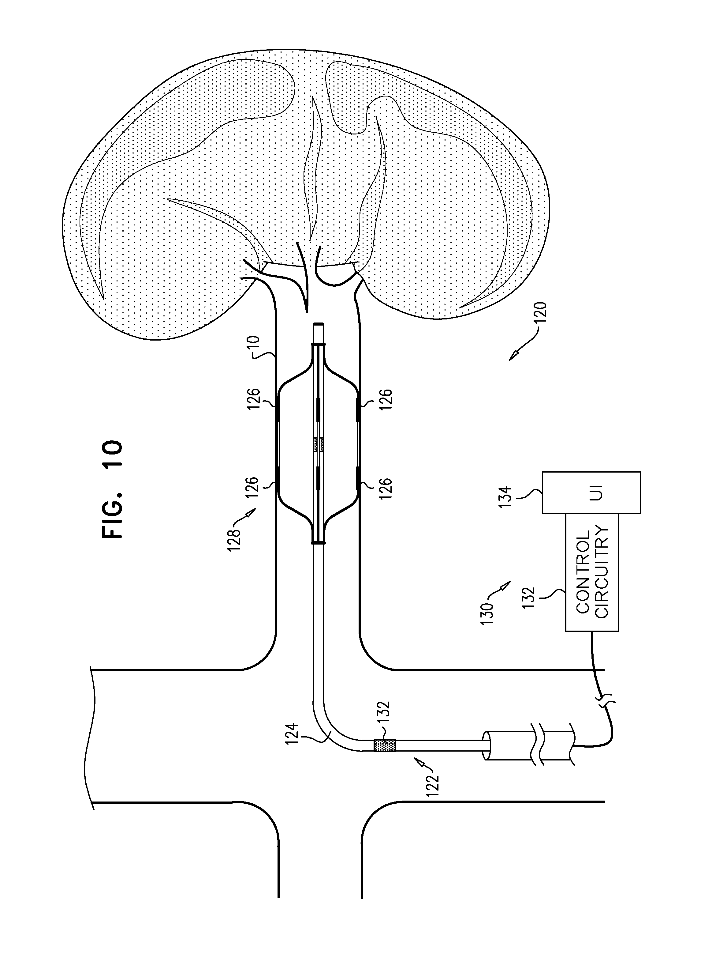
For some applications the techniques described are performed generally manually, e.g., by a physician. For some applications the techniques described are at least partly automated, e.g., using a device comprising a control unit, a transluminally-advanceable longitudinal member such as a catheter, and one or more electrodes at a distal portion of the longitudinal member.
There is therefore provided, in accordance with an application of the present invention, a method, including:
applying an excitatory current to renal nerve fibers of a subject;
determining a change in a parameter of the subject in response to the excitatory current, the parameter being a fastest rate of increase in arterial pressure during a systolic upstroke of an arterial pressure wave of the subject;
in response to the change, deciding whether to ablate the renal nerve fibers; and
in response to the deciding, applying ablation energy to the renal nerve fibers.
In an application:
applying the excitatory current includes applying the excitatory current to renal nerve fibers proximate to a first location of a renal artery wall,
deciding whether to ablate the renal nerve fibers includes deciding whether to ablate the renal nerve fibers proximate to the first location, and
the method further includes: applying the excitatory current to renal nerve fibers proximate to a second location of a renal artery wall; determining a second change in the parameter in response to the excitatory current applied to the renal nerve fibers proximate to the second location; and in response to the second change, deciding whether to ablate the renal nerve fibers proximate to the second location.
There is further provided, in accordance with an application of the present invention, a method, including:
measuring a first value of a parameter of a subject while the subject is at rest, the parameter being a fastest rate of increase in arterial pressure during a systolic upstroke of an arterial pressure wave of the subject;
applying an excitatory current to renal nerve fibers of a subject;
while applying the excitatory current, measuring a second value of the parameter;
in response to a difference between the first value and the second value, deciding whether to ablate the renal nerve fibers; and
in response to the deciding, applying ablation energy to the renal nerve fibers.
There is further provided, in accordance with an application of the present invention, a method, including:
applying an excitatory current to renal nerve fibers of a subject;
determining a change in a parameter of the subject in response to the excitatory current, the parameter being a fastest rate of increase in arterial pressure during a systolic upstroke of an arterial pressure wave of the subject;
in response to the change, selecting an ablation modality from a plurality of ablation modalities; and
in response to the selecting, ablating the renal nerve fibers using the selected ablation modality.
There is further provided, in accordance with an application of the present invention, a method, including:
applying a first application of excitatory current to renal nerve fibers of a subject;
determining a first change in a parameter of the subject in response to the first application of excitatory current, the parameter being a fastest rate of increase in arterial pressure during a systolic upstroke of an arterial pressure wave of the subject;
subsequently, applying a first application of ablating energy to the renal nerve fibers;
subsequently, applying a second application of excitatory current to renal nerve fibers of a subject;
determining a second change in the parameter in response to the second application of excitatory current;
determining a difference between the first change and the second change;
deciding whether to apply a second application of ablation energy to the renal nerve fibers; and
in response to the deciding, applying the second application of ablation energy to the renal nerve fibers.
There is further provided, in accordance with an application of the present invention, a method, including:
transluminally advancing a distal portion of a longitudinal member of a device into a renal artery of a subject;
operating the device to drive an electrode disposed on the distal portion of the longitudinal member to apply an excitatory current to nerve tissue of the renal artery;
receiving (i) a first value, the first value being indicative of a parameter of the subject before a start of the application of the current, and (ii) a second value, the second value being indicative of the parameter of the subject after a start of the application of the current, the parameter being a fastest rate of increase in arterial pressure during a systolic upstroke of an arterial pressure wave of the subject;
determining if a difference between the first value and the second value is smaller than a threshold difference; and
in response to the determining: only if the determined difference is smaller than the threshold difference, withdrawing the longitudinal member from the subject without having applied ablation energy to the renal artery.
There is further provided, in accordance with an application of the present invention, apparatus including:
a transluminal electrode catheter, including: a flexible shaft, dimensioned for advancement of a distal portion of the shaft into a renal artery of a subject; a plurality of electrodes disposed at the distal portion of the shaft; and an arterial blood pressure sensor; and
a control unit, electrically coupled to the catheter, and configured to: use the sensor to measure a first value of a parameter of the subject while the subject is at rest, the parameter being a fastest rate of increase in arterial pressure during a systolic upstroke of an arterial pressure wave of the subject, drive at least one of the electrodes to apply an excitatory current to renal nerve fibers of the subject, while applying the excitatory current, use the blood pressure sensor to measure a second value of the parameter, determine if a difference between the first value and the second value is greater than a threshold difference, and in response to the determining: only if the determined difference is greater than the threshold difference, enable an ablation function of the control unit.
There is further provided, in accordance with an application of the present invention, a method, including:
applying an excitatory current to renal nerve fibers of a subject;
determining a change in a parameter of the subject in response to the excitatory current, the parameter of the subject being a skewness of an arterial pressure wave of the subject;
in response to the change, deciding whether to ablate the renal nerve fibers; and
in response to the deciding, applying ablation energy to the renal nerve fibers.
In an application:
applying the excitatory current includes applying the excitatory current to renal nerve fibers proximate to a first location of a renal artery wall,
deciding whether to ablate the renal nerve fibers includes deciding whether to ablate the renal nerve fibers proximate to the first location, and
the method further includes: applying the excitatory current to renal nerve fibers proximate to a second location of a renal artery wall; determining a second change in the parameter in response to the excitatory current applied to the renal nerve fibers proximate to the second location; and in response to the second change, deciding whether to ablate the renal nerve fibers proximate to the second location.
There is further provided, in accordance with an application of the present invention, a method, including:
measuring a first value of a parameter of a subject while the subject is at rest, the parameter being skewness of an arterial pressure wave of the subject;
applying an excitatory current to renal nerve fibers of a subject;
while applying the excitatory current, measuring a second value of the parameter;
in response to a difference between the first value and the second value, deciding whether to ablate the renal nerve fibers; and
in response to the deciding, applying ablation energy to the renal nerve fibers.
There is further provided, in accordance with an application of the present invention, a method, including:
applying an excitatory current to renal nerve fibers of a subject;
determining a change in a parameter of the subject in response to the excitatory current, the parameter of the subject being skewness of an arterial pressure wave of the subject;
in response to the change, selecting an ablation modality from a plurality of ablation modalities; and
in response to the selecting, ablating the renal nerve fibers using the selected ablation modality.
There is further provided, in accordance with an application of the present invention, a method, including:
applying a first application of excitatory current to renal nerve fibers of a subject;
determining a first change in a parameter of the subject in response to the first application of excitatory current, the parameter of the subject being skewness of an arterial pressure wave of the subject;
subsequently, applying a first application of ablating energy to the renal nerve fibers;
subsequently, applying a second application of excitatory current to renal nerve fibers of a subject;
determining a second change in the parameter in response to the second application of excitatory current;
determining a difference between the first change and the second change;
deciding whether to apply a second application of ablation energy to the renal nerve fibers; and
in response to the deciding, applying the second application of ablation energy to the renal nerve fibers.
There is further provided, in accordance with an application of the present invention, a method, including:
transluminally advancing a distal portion of a longitudinal member of a device into a renal artery of a subject;
operating the device to drive an electrode disposed on the distal portion of the longitudinal member to apply an excitatory current to nerve tissue of the renal artery;
receiving (i) a first value, the first value being indicative of a parameter of the subject before a start of the application of the current, and (ii) a second value, the second value being indicative of the parameter of the subject after a start of the application of the current, the parameter being skewness of an arterial pressure wave of the subject;
determining if a difference between the first value and the second value is smaller than a threshold difference; and
in response to the determining: only if the determined difference is smaller than the threshold difference, withdrawing the longitudinal member from the subject without having applied ablation energy to the renal artery.
There is further provided, in accordance with an application of the present invention, apparatus including:
a transluminal electrode catheter, including: a flexible shaft, dimensioned for advancement of a distal portion of the shaft into a renal artery of a subject; a plurality of electrodes disposed at the distal portion of the shaft; and an arterial blood pressure sensor; and
a control unit, electrically coupled to the catheter, and configured to: use the sensor to measure a first value of a parameter of the subject while the subject is at rest, the parameter being skewness of an arterial pressure wave of the subject, drive at least one of the electrodes to apply an excitatory current to renal nerve fibers of the subject, while applying the excitatory current, use the blood pressure sensor to measure a second value of the parameter, determine if a difference between the first value and the second value is greater than a threshold difference, and in response to the determining: only if the determined difference is greater than the threshold difference, enable an ablation function of the control unit.
There is further provided, in accordance with an application of the present invention, a method, including:
applying an excitatory current to renal nerve fibers of a subject;
determining a change in a parameter of the subject in response to the excitatory current, the parameter of the subject being a heart rate of the subject;
in response to the change, deciding whether to ablate the renal nerve fibers; and
in response to the deciding, applying ablation energy to the renal nerve fibers.
In an application:
applying the excitatory current includes applying the excitatory current to renal nerve fibers proximate to a first location of a renal artery wall,
deciding whether to ablate the renal nerve fibers includes deciding whether to ablate the renal nerve fibers proximate to the first location, and
the method further includes: applying the excitatory current to renal nerve fibers proximate to a second location of a renal artery wall; determining a second change in the parameter in response to the excitatory current applied to the renal nerve fibers proximate to the second location; and in response to the second change, deciding whether to ablate the renal nerve fibers proximate to the second location.
There is further provided, in accordance with an application of the present invention, a method, including:
measuring a first value of a parameter of a subject while the subject is at rest, the parameter being a heart rate of the subject;
applying an excitatory current to renal nerve fibers of a subject;
while applying the excitatory current, measuring a second value of the parameter;
in response to a difference between the first value and the second value, deciding whether to ablate the renal nerve fibers; and
in response to the deciding, applying ablation energy to the renal nerve fibers.
There is further provided, in accordance with an application of the present invention, a method, including:
applying an excitatory current to renal nerve fibers of a subject;
determining a change in a parameter of the subject in response to the excitatory current, the parameter of the subject being a heart rate of the subject;
in response to the change, selecting an ablation modality from a plurality of ablation modalities; and
in response to the selecting, ablating the renal nerve fibers using the selected ablation modality.
There is further provided, in accordance with an application of the present invention, a method, including:
applying a first application of excitatory current to renal nerve fibers of a subject;
determining a first change in a parameter of the subject in response to the first application of excitatory current, the parameter of the subject being a heart rate of the subject;
subsequently, applying a first application of ablating energy to the renal nerve fibers;
subsequently, applying a second application of excitatory current to renal nerve fibers of a subject;
determining a second change in the parameter in response to the second application of excitatory current;
determining a difference between the first change and the second change;
deciding whether to apply a second application of ablation energy to the renal nerve fibers; and
in response to the deciding, applying the second application of ablation energy to the renal nerve fibers.
There is further provided, in accordance with an application of the present invention, a method, including:
transluminally advancing a distal portion of a longitudinal member of a device into a renal artery of a subject;
operating the device to drive an electrode disposed on the distal portion of the longitudinal member to apply an excitatory current to nerve tissue of the renal artery;
receiving (i) a first value, the first value being indicative of a parameter of the subject before a start of the application of the current, and (ii) a second value, the second value being indicative of the parameter of the subject after a start of the application of the current, the parameter being a heart rate of the subject;
determining if a difference between the first value and the second value is smaller than a threshold difference; and
in response to the determining: only if the determined difference is smaller than the threshold difference, withdrawing the longitudinal member from the subject without having applied ablation energy to the renal artery.
There is further provided, in accordance with an application of the present invention, apparatus including:
a transluminal electrode catheter, including: a flexible shaft, dimensioned for advancement of a distal portion of the shaft into a renal artery of a subject; a plurality of electrodes disposed at the distal portion of the shaft; and an arterial blood pressure sensor; and
a control unit, electrically coupled to the catheter, and configured to: use the sensor to measure a first value of a parameter of the subject while the subject is at rest, the parameter being a heart rate of the subject, drive at least one of the electrodes to apply an excitatory current to renal nerve fibers of the subject, while applying the excitatory current, use the blood pressure sensor to measure a second value of the parameter, determine if a difference between the first value and the second value is greater than a threshold difference, and in response to the determining: only if the determined difference is greater than the threshold difference, enable an ablation function of the control unit.
There is further provided, in accordance with an application of the present invention, a method, including:
applying an excitatory current to renal nerve fibers of a subject;
determining a change in a parameter of the subject in response to the excitatory current, the parameter of the subject being an augmentation index of an arterial pressure wave of the subject;
in response to the change, deciding whether to ablate the renal nerve fibers; and
in response to the deciding, applying ablation energy to the renal nerve fibers.
In an application:
applying the excitatory current includes applying the excitatory current to renal nerve fibers proximate to a first location of a renal artery wall,
deciding whether to ablate the renal nerve fibers includes deciding whether to ablate the renal nerve fibers proximate to the first location, and
the method further includes: applying the excitatory current to renal nerve fibers proximate to a second location of a renal artery wall; determining a second change in the parameter in response to the excitatory current applied to the renal nerve fibers proximate to the second location; and in response to the second change, deciding whether to ablate the renal nerve fibers proximate to the second location.
There is further provided, in accordance with an application of the present invention, a method, including:
measuring a first value of a parameter of a subject while the subject is at rest, the parameter being an augmentation index of an arterial pressure wave of the subject;
applying an excitatory current to renal nerve fibers of a subject;
while applying the excitatory current, measuring a second value of the parameter;
in response to a difference between the first value and the second value, deciding whether to ablate the renal nerve fibers; and
in response to the deciding, applying ablation energy to the renal nerve fibers.
There is further provided, in accordance with an application of the present invention, a method, including:
applying an excitatory current to renal nerve fibers of a subject;
determining a change in a parameter of the subject in response to the excitatory current, the parameter of the subject being an augmentation index of an arterial pressure wave of the subject;
in response to the change, selecting an ablation modality from a plurality of ablation modalities; and
in response to the selecting, ablating the renal nerve fibers using the selected ablation modality.
There is further provided, in accordance with an application of the present invention, a method, including:
applying a first application of excitatory current to renal nerve fibers of a subject;
determining a first change in a parameter of the subject in response to the first application of excitatory current, the parameter of the subject being an augmentation index of an arterial pressure wave of the subject;
subsequently, applying a first application of ablating energy to the renal nerve fibers;
subsequently, applying a second application of excitatory current to renal nerve fibers of a subject;
determining a second change in the parameter in response to the second application of excitatory current;
determining a difference between the first change and the second change;
deciding whether to apply a second application of ablation energy to the renal nerve fibers; and
in response to the deciding, applying the second application of ablation energy to the renal nerve fibers.
There is further provided, in accordance with an application of the present invention, a method, including:
transluminally advancing a distal portion of a longitudinal member of a device into a renal artery of a subject;
operating the device to drive an electrode disposed on the distal portion of the longitudinal member to apply an excitatory current to nerve tissue of the renal artery;
receiving (i) a first value, the first value being indicative of a parameter of the subject before a start of the application of the current, and (ii) a second value, the second value being indicative of the parameter of the subject after a start of the application of the current, the parameter being an augmentation index of an arterial pressure wave of the subject;
determining if a difference between the first value and the second value is smaller than a threshold difference; and
in response to the determining: only if the determined difference is smaller than the threshold difference, withdrawing the longitudinal member from the subject without having applied ablation energy to the renal artery.
There is further provided, in accordance with an application of the present invention, apparatus including:
a transluminal electrode catheter, including: a flexible shaft, dimensioned for advancement of a distal portion of the shaft into a renal artery of a subject; a plurality of electrodes disposed at the distal portion of the shaft; and an arterial blood pressure sensor; and
a control unit, electrically coupled to the catheter, and configured to: use the sensor to measure a first value of a parameter of the subject while the subject is at rest, the parameter being an augmentation index of an arterial pressure wave of the subject, drive at least one of the electrodes to apply an excitatory current to renal nerve fibers of the subject, while applying the excitatory current, use the blood pressure sensor to measure a second value of the parameter, determine if a difference between the first value and the second value is greater than a threshold difference, and in response to the determining: only if the determined difference is greater than the threshold difference, enable an ablation function of the control unit.
There is further provided, in accordance with an application of the present invention, a method, including:
applying an excitatory current to renal nerve fibers of a subject;
determining a change in a parameter of the subject in response to the excitatory current, the parameter of the subject being a diastolic decay time constant of an arterial pressure wave of the subject;
in response to the change, deciding whether to ablate the renal nerve fibers; and
in response to the deciding, applying ablation energy to the renal nerve fibers.
In an application:
applying the excitatory current includes applying the excitatory current to renal nerve fibers proximate to a first location of a renal artery wall,
deciding whether to ablate the renal nerve fibers includes deciding whether to ablate the renal nerve fibers proximate to the first location, and
the method further includes: applying the excitatory current to renal nerve fibers proximate to a second location of a renal artery wall; determining a second change in the parameter in response to the excitatory current applied to the renal nerve fibers proximate to the second location; and in response to the second change, deciding whether to ablate the renal nerve fibers proximate to the second location.
There is further provided, in accordance with an application of the present invention, a method, including:
measuring a first value of a parameter of a subject while the subject is at rest, the parameter being a diastolic decay time constant of an arterial pressure wave of the subject;
applying an excitatory current to renal nerve fibers of a subject;
while applying the excitatory current, measuring a second value of the parameter;
in response to a difference between the first value and the second value, deciding whether to ablate the renal nerve fibers; and
in response to the deciding, applying ablation energy to the renal nerve fibers.
There is further provided, in accordance with an application of the present invention, a method, including:
applying an excitatory current to renal nerve fibers of a subject;
determining a change in a parameter of the subject in response to the excitatory current, the parameter of the subject being a diastolic decay time constant of an arterial pressure wave of the subject;
in response to the change, selecting an ablation modality from a plurality of ablation modalities; and
in response to the selecting, ablating the renal nerve fibers using the selected ablation modality.
There is further provided, in accordance with an application of the present invention, a method, including:
applying a first application of excitatory current to renal nerve fibers of a subject;
determining a first change in a parameter of the subject in response to the first application of excitatory current, the parameter of the subject being a diastolic decay time constant of an arterial pressure wave of the subject;
subsequently, applying a first application of ablating energy to the renal nerve fibers;
subsequently, applying a second application of excitatory current to renal nerve fibers of a subject;
determining a second change in the parameter in response to the second application of excitatory current;
determining a difference between the first change and the second change;
deciding whether to apply a second application of ablation energy to the renal nerve fibers; and
in response to the deciding, applying the second application of ablation energy to the renal nerve fibers.
There is further provided, in accordance with an application of the present invention, a method, including:
transluminally advancing a distal portion of a longitudinal member of a device into a renal artery of a subject;
operating the device to drive an electrode disposed on the distal portion of the longitudinal member to apply an excitatory current to nerve tissue of the renal artery;
receiving (i) a first value, the first value being indicative of a parameter of the subject before a start of the application of the current, and (ii) a second value, the second value being indicative of the parameter of the subject after a start of the application of the current, the parameter being a diastolic decay time constant of an arterial pressure wave of the subject;
determining if a difference between the first value and the second value is smaller than a threshold difference; and
in response to the determining: only if the determined difference is smaller than the threshold difference, withdrawing the longitudinal member from the subject without having applied ablation energy to the renal artery.
There is further provided, in accordance with an application of the present invention, apparatus including:
a transluminal electrode catheter, including: a flexible shaft, dimensioned for advancement of a distal portion of the shaft into a renal artery of a subject; a plurality of electrodes disposed at the distal portion of the shaft; and an arterial blood pressure sensor; and
a control unit, electrically coupled to the catheter, and configured to: use the sensor to measure a first value of a parameter of the subject while the subject is at rest, the parameter being a diastolic decay time constant of an arterial pressure wave of the subject, drive at least one of the electrodes to apply an excitatory current to renal nerve fibers of the subject, while applying the excitatory current, use the blood pressure sensor to measure a second value of the parameter, determine if a difference between the first value and the second value is greater than a threshold difference, and in response to the determining: only if the determined difference is greater than the threshold difference, enable an ablation function of the control unit.
There is further provided, in accordance with an application of the present invention, a method, including:
transluminally advancing an electrode into a renal artery of a subject;
while the subject is at rest, measuring a first value of a first parameter and a first value of a second parameter, the first parameter and the second parameter selected from the group consisting of: (i) a fastest rate of increase in arterial pressure during a systolic upstroke of an arterial pressure wave of the subject, (ii) a skewness of the arterial pressure wave of the subject, (iii) a heart rate of the subject, (iv) an augmentation index of the arterial pressure wave of the subject, and (v) a diastolic decay constant of the arterial pressure wave of the subject;
applying an excitatory current to renal nerve fibers of a subject;
while applying the excitatory current, measuring a second value of the first parameter and a second value of the second parameter;
determining (i) a first difference between the first value of the first parameter and the second value of the first parameter, and (ii) a second difference between the first value of the second parameter and the second value of the second parameter; and
in response to the first difference and the second difference, diagnosing a pathology of the subject.
In an application, the method further includes:
while the subject is at rest, measuring a first value of a third parameter selected from the group;
while applying the excitatory current, measuring a second value of the third parameter; and
determining a third difference between the first value of the third parameter and the second value of the third parameter, and diagnosing the pathology in response includes diagnosing the pathology in response to the first difference, the second difference, and the third difference.
In an application, the method further includes:
while the subject is at rest, measuring a first value of a fourth parameter selected from the group;
while applying the excitatory current, measuring a second value of the fourth parameter; and
determining a fourth difference between the first value of the fourth parameter and the second value of the fourth parameter, and diagnosing the pathology in response includes diagnosing the pathology in response to the first difference, the second difference, the third difference, and the fourth difference.
In an application, the method further includes:
while the subject is at rest, measuring a first value of a fifth parameter selected from the group;
while applying the excitatory current, measuring a second value of the fifth parameter; and
determining a fifth difference between the first value of the fifth parameter and the second value of the fifth parameter, and diagnosing the pathology in response includes diagnosing the pathology in response to the first difference, the second difference, the third difference, the fourth difference, and the fifth difference.
The present invention will be more fully understood from the following detailed description of applications thereof, taken together with the drawings, in which:
BRIEF DESCRIPTION OF THE DRAWINGS
FIGS. 1A, 1B, and 1C show a table of evaluated parameters, which spans across FIGS. 1A, 1B, and 1C, in accordance with some applications of the invention;
FIGS. 2, 3, and 4 are schematic illustrations showing arterial pressure waves, with annotations explaining some of the parameters of FIGS. 1A, 1B, and 1C, in accordance with some applications of the invention;
FIGS. 5, 6, 7, 8, and 9 are flow charts showing at least some steps in respective techniques for use with nerve tissue, in accordance with some applications of the invention; and
FIG. 10 is a schematic illustration of a system for ablating and/or stimulating nerve tissue of a blood vessel of a subject, in accordance with some applications of the present invention.
DETAILED DESCRIPTION OF EMBODIMENTS
Reference is made to FIGS. 1A-C, 2, 3, and 4. FIGS. 1A-C show a table of evaluated parameters, which spans across FIGS. 1A-C, in accordance with some applications of the invention. FIGS. 2, 3, and 4 are schematic illustrations showing arterial pressure waves, with annotations explaining some of the parameters of FIGS. 1A-C, in accordance with some applications of the invention.
Experiments were performed in a porcine model. Arterial blood pressure was monitored before, during, and after excitation of (i.e., induction of action potentials in) a renal nerve. Various parameters derived from the resulting trace were evaluated, in order to determine which respond clearly (e.g., a quantitatively large change) and reliably (e.g., qualitatively similarities in changes between animals, such as changing in the same direction in each animal) to renal nerve excitation.
The evaluated parameters were ranked according to the results of the experiments; the top ten parameters appear in the table that spans FIGS. 1A-C.
dP/dT_max is the maximal derivative (dP/dT) of the pressure wave during systolic upstroke (i.e., the fastest rate of increase in arterial pressure during the systolic upstroke). (In the interpretations column, beta_1, alpha_2, and alpha_1 refer to subtypes of adrenergic receptor.) dP/dT_max is illustrated in FIG. 2. FIG. 2 also shows (i) the parameter TdP/dT_max, which is the time to reach dP/dT_max, and (ii) the parameter AP at dP/dT_max, which is the arterial pressure at dP/dT_max.
Skewness is the third moment of the blood pressure wave (i.e., the degree to which the wave is asymmetric with respect to its mid-point). Skewness is illustrated in FIG. 4. ("RAS" refers to the renin-angiotensin system.)
Heart rate (HR) is the number of detected beats per minute.
Augmentation Index (AIx) is the ratio between irregular pressure waveform amplitude to the total pulse pressure, and is a measure of wave reflection and arterial stiffness. AIx is illustrated in FIG. 3.
Diastolic Decay Time Constant (or Diastolic Decay Constant) (D_tau) is exponential pressure decay time (RC) during the diastolic period. D_tau is illustrated in FIG. 2.
Diastolic Average Slope (Dslope) is the average of the pressure derivative during the diastolic phase.
Systolic Pressure (SP) is the maximal pressure point of the entire pressure wave. SP is illustrated in FIG. 2 as Systolic Arterial Pressure (SAP).
Systolic Average Slope (Sslope) is the average of the pressure derivative during the systolic phase.
Pulse Pressure (PP) is the difference between SP and diastolic pressure (minimal pressure during the entire pressure wave). PP is illustrated in FIG. 2.
Systolic Duration Ratio (SDR) is the duration of systole as a proportion of the entire pressure wave. SDR is illustrated in FIG. 2.
More details regarding the parameters are included in the table of FIGS. 1A-C.
Reference is now made to FIGS. 5-9, which are flow charts showing at least some steps in respective techniques for use with nerve tissue, in accordance with some applications of the invention. Typically, these techniques are performed transluminally, e.g., by advancing a catheter into the renal artery and applying excitatory current, and optionally ablation energy, to renal nerve tissue via the wall of the renal artery. For example, these techniques may be performed using a system 120 (described with reference to FIG. 10), and/or in combination with apparatus and/or techniques described in one or more of the following references, which are incorporated herein by reference: PCT application publication WO 2014/068577 to Gross et al., filed Nov. 3, 2013, and entitled "Controlled Tissue Ablation," PCT application publication WO 2015/170281 to Gross et al., filed May 7, 2015, and entitled "Controlled Tissue Ablation Techniques," U.S. patent application Ser. No. 14/794,737 to Gross et al., filed Jul. 8, 2015, and entitled "Electrical Signal-Based Electrode-Tissue Contact Detection," which published as US 2017/0007157, U.S. patent application Ser. No. 14/972,756 to Gross et al., filed Dec. 17, 2015, and entitled "Transluminal Electrode Catheters," which published as US 2017/0172651, U.S. patent application Ser. No. 15/001,615 to Gross et al., filed Jan. 20, 2016, and entitled "Catheter Guidance and Procedure Planning," which published as 2017/0202614.
As shown in FIG. 5, a method 20 is described, the method comprising: (i) applying an excitatory current to renal nerve fibers of a subject (box 24); (ii) determining a change in a parameter of the subject in response to the excitatory current (box 26), the parameter of the subject being a parameter that appears in the table of FIGS. 1A-C (and typically a parameter that appears in the top five in the table); (iii) in response to the change, deciding whether to ablate the renal nerve fibers (box 28); and (iv) in response to the deciding, applying ablation energy to the renal nerve fibers (box 30). Typically, the change in the parameter is determined by comparing a value of the parameter measured while the subject is at rest (e.g., before applying the excitatory current) (box 22) to a value measured during the application of the current (box 24).
For example, method 20, or a similar method, may be used to determine whether an individual subject is a suitable candidate for renal nerve ablation. Therefore, for some applications, a method comprises: transluminally advancing a distal portion of a longitudinal member of a device into a renal artery of a subject; operating the device to drive an electrode disposed on the distal portion of the longitudinal member to apply an excitatory current to nerve tissue of the renal artery; receiving (i) a first value, the first value being indicative of a parameter of the subject (e.g., a parameter described herein) before a start of the application of the current, and (ii) a second value, the second value being indicative of the parameter of the subject after a start of the application of the current; determining if a difference between the first value and the second value is smaller than a threshold difference; and in response to the determining: only if the determined difference is smaller than the threshold difference, withdrawing the longitudinal member from the subject without having applied ablation energy to the renal artery.
For some applications, instead of withdrawing the longitudinal member from the subject, the longitudinal member is moved to another site within the subject and the process is repeated. For some applications, following several iterations of the process, if the determined difference at more than a threshold proportion of the sites (e.g., more than 50 percent, e.g., more than 60 percent, e.g., more than 70 percent, e.g., more than 80 percent, e.g., more than 90 percent) is smaller than the threshold difference, the longitudinal member is withdrawn from the subject without having applied ablation energy to the renal artery.
For some applications, method 20 is used at various locations within the renal artery, mutatis mutandis, e.g., to locate target renal nerve tissue (and optionally ablate it). Therefore, as shown in FIG. 6, a method 40 is described, the method being the same as method 20, wherein: (a) applying the excitatory current (box 24 of FIG. 5) comprises applying the excitatory current to renal nerve fibers proximate to a first location of a renal artery wall (box 42), (b) deciding whether to ablate the renal nerve fibers (box 28 of FIG. 5) comprises deciding whether to ablate the renal nerve fibers proximate to the first location (box 50), and (c) the method further comprises: (i) applying the excitatory current to renal nerve fibers proximate to a second location of a renal artery wall (box 46); (ii) determining a second change in the parameter in response to the excitatory current applied to the renal nerve fibers proximate to the second location (box 48); and (iii) in response to the second change, deciding whether to ablate the renal nerve fibers proximate to the second location (box 50). In response to deciding whether to ablate the renal nerve fibers proximate to the first location or the second location, ablation energy is applied to the renal nerve fibers (box 52). Box 44 of FIG. 6 corresponds to box 26 of FIG. 5 in the same way that boxes 42 and 50 of FIG. 6 correspond to boxes 24 and 28 of FIG. 5, respectively.
For some applications, a modality of ablation is selected in response to the change in value of the parameter. For example, the modality may be selected from radiofrequency (RF), ultrasound, heating, cooling, laser, chemical, or any other ablation modality known in the art. Therefore, as shown in FIG. 7, a method 60 is described, the method comprising (i) applying an excitatory current to renal nerve fibers of a subject (box 62); (ii) determining a change in a parameter of the subject in response to the excitatory current (box 64), the parameter of the subject being a parameter that appears in the table of FIGS. 1A-C (and typically a parameter that appears in the top five in the table); (iii) in response to the change, selecting an ablation modality from a plurality of ablation modalities (66); and (iv) in response to the selecting, ablating the renal nerve fibers using the selected ablation modality (box 68).
For some applications, the parameters in the table of FIGS. 1A-C are used to monitor the progress of an ablation process. For example, ablation energy may be applied to the nerve tissue iteratively, and measurement of the parameter after each application may be used to determine whether the nerve tissue has become sufficiently ablated (e.g., sufficiently unable to propagate action potentials therealong). Therefore, as shown in FIG. 8, a method is described, the method comprising: (i) applying an application of excitatory current to renal nerve fibers of a subject (box 82); (ii) determining a first change in a parameter of the subject in response to the application of excitatory current (box 84), the parameter of the subject being a parameter that appears in the table of FIGS. 1A-C (and typically a parameter that appears in the top five in the table); (iii) subsequently, applying a first application of ablating energy to the renal nerve fibers (box 86); (iv) subsequently, applying another application of excitatory current to renal nerve fibers of a subject (box 88); (v) determining a second change in the parameter in response to the second application of excitatory current (box 90); (vi) determining a difference between the first change and the second change (box 92); (vii) deciding whether to apply a second application of ablation energy to the renal nerve fibers (box 94); and (viii) in response to the deciding, applying the second application of ablation energy to the renal nerve fibers (box 96).
Because, as described hereinabove and in the table of FIGS. 1A-C, the parameters relate to different physiological aspects (e.g., properties), it is hypothesized that determining how the parameters respond to excitation of renal nerve tissue may facilitate more detailed understanding and/or definition of the particular condition of the subject (e.g., enabling a more specific diagnosis of the condition). It is further hypothesized that taking into account two or more of the parameters will be further facilitative. As shown in FIG. 9, a method 100 is described, the method comprising: (i) transluminally advancing an electrode into a renal artery of a subject; (ii) while the subject is at rest, measuring a first value of a first parameter (box 102) and a first value of a second parameter (box 104), the first parameter and the second parameter selected from the parameters that appear in the table of FIGS. 1A-C (and typically those that appear in the top five in the table); (iii) applying an excitatory current to renal nerve fibers of a subject, and while applying the excitatory current, measuring a second value of the first parameter and a second value of the second parameter (box 106); (iv) determining (a) a first difference between the first value of the first parameter and the second value of the first parameter, and (b) a second difference between the first value of the second parameter and the second value of the second parameter (box 108); and (v) in response to the first difference and the second difference, diagnosing a pathology of the subject (110).
For some applications, differences between first and second values of only two parameters from the table are used for diagnosing the pathology (e.g., the first and second parameters in the above paragraph). For some applications more than two parameters (e.g., at least three parameters, e.g., at least four parameters, such as at least five parameters) from the table (and typically from the top five in the table) are used.
Reference is again made to FIGS. 1A-8. Although methods 20, 40, 60, and 80 are generally described with reference to "a parameter," it is to be understood that the scope of the invention includes the use of more than one of the parameters in the table of FIGS. 1A-C (typically in the top five parameters of the table). Furthermore, methods 20, 40, 60, 80, and 100 may be further facilitated by taking into account other parameters of the subject, including other blood pressure parameters, and non-blood pressure parameters.
Reference is again made to FIGS. 1A-9. Described hereinabove are parameter values measured before application of the excitatory current, and parameter values measured during application of the excitatory current. It is to be noted that for some applications of the invention, the timing of these two measurements may differ from a strict definition of these timings, in that, for example, a "before" value of the parameter may be measured soon after the beginning of application of the excitatory current, before the action potentials initiated by the excitatory current have begun to affect the parameter. Similarly, a "during" value of the parameter may be measured soon after the end of application of the excitatory current, before the parameter has begun to return toward the "before" value.
Reference is again made to FIGS. 1A-9. The excitatory current described hereinabove is to be understood as a current that initiates action potentials in the nerve tissue. Typically, the excitatory current has a frequency of greater than 1 Hz and/or less than 100 Hz, such as between 1 and 100 Hz, e.g., between 10 and 100 Hz. For some applications, the ablation energy described hereinabove is RF energy, e.g., an RF current having a frequency of above 5 kHz and/or below 1 GHz, such as between 5 kHz and 1 GHz (e.g., 10 kHz-10 MHz, e.g., 50 kHz-1 MHz, e.g., 300 kHz-1 MHz, e.g., 300 kHz-500 kHz).
Reference is again made to FIGS. 5-9. For some applications, the techniques described hereinabove are performed generally manually by a physician (alone, or with assistance). For example, a commercially-available arterial blood pressure sensor may be used to monitor arterial blood pressure; the output may be separately analyzed (e.g., manually, or using a computer) to measure the values of the parameter(s); and the change in the value of the parameter(s) may be separately determined (e.g., manually, or using a computer).
Reference is now made to FIG. 10, which is a schematic illustration of a system 120 for ablating and/or stimulating nerve tissue of a blood vessel of a subject, such as a renal artery 10, in accordance with some applications of the present invention. For some applications, the techniques described hereinabove are performed generally automatically by a device such as system 120, comprising a transluminal electrode catheter 122 that comprises a transluminally-advanceable longitudinal member (e.g., a flexible shaft) 124, one or more electrodes 126 at a distal portion 128 of the longitudinal member, and a (typically extracorporeal) control unit 130 that comprises control circuitry 132, and is electrically coupled to the catheter. Control unit 130 is typically a programmed digital computing device comprising a central processing unit (CPU), random access memory (RAM), non-volatile secondary storage, such as a hard drive or CD ROM drive, network interfaces, and/or peripheral devices. Program code, including software programs, and/or data are loaded into the RAM for execution and processing by the CPU and results are generated for display, output, transmittal, or storage, as is known in the art. Such program code and/or data, when provided to the control unit, produce a machine or special-purpose computer, configured to perform the tasks described herein. Typically, control unit 130 is connected to an arterial blood pressure sensor 132 (which may be disposed on longitudinal member 124), and is configured to receive signals from the sensor.
Typically, control unit 30 comprises a user interface 134, which is configured to receive user input (e.g., activation of the system) and/or to output measurements and/or status.
The control unit automatically performs one or more of the following steps: (1) deriving the value of the parameter (e.g., from raw pressure data from the pressure sensor), (2) driving the electrodes to apply the excitatory current, (3) determining a change in the parameter value in response to the excitatory current, and (4) providing an output via an interface. For some applications, the output indicates the change in the parameter value in response to the excitatory current (e.g., as an absolute value, as a relative value, or as an indication of whether the change exceeds a threshold change), and based on the output the physician decides whether to ablate.
For some applications, the control unit drives the electrodes to apply the ablation energy, either automatically in response to the determined change in the parameter value, or when activated by the physician. That is, for some applications, the same device is configured and used to apply the excitatory current and to apply the ablation energy (and typically to measure the arterial blood pressure). For some applications, the device is not configured or used to apply both the excitatory current and the ablation energy. For example, a separate ablation device may be used, either during the same general procedure (e.g., without the subject leaving the treatment room), or at another time.
Therefore, for some applications, apparatus comprises: a transluminal electrode catheter 122, comprising: a flexible shaft 124, dimensioned for advancement of a distal portion 128 of the shaft into a renal artery 10 of a subject; a plurality of electrodes 126 disposed at the distal portion of the shaft; and an arterial blood pressure sensor 132; and a control unit 130, electrically coupled to the catheter.
For some applications, control unit 130 is configured to: use sensor 124 to measure a first value of a parameter of the subject while the subject is at rest (the parameter being a parameter described herein), drive at least one of the electrodes 126 to apply an excitatory current to renal nerve fibers of the subject, while applying the excitatory current, use the blood pressure sensor to measure a second value of the parameter, determine if a difference between the first value and the second value is greater than a threshold difference, and in response to the determining: only if the determined difference is greater than the threshold difference, enable an ablation function of the control unit.
The ablation function of control unit 130 can be activated only while enabled. Upon its activation, the ablation function of the control unit drives at least one of the electrodes to apply ablation energy to the renal nerve fibers (through the wall of renal artery 10). For some applications, control unit 130 activates the ablation function automatically upon the enabling of the ablation function. For some applications, control unit enables the ablation function, but activation of the ablation function requires subsequent user input (e.g., via interface 134).
For some applications, the devices described hereinabove are similar to those described in PCT application publication WO 2014/068577 to Gross et al., filed Nov. 3, 2013, and entitled "Controlled Tissue Ablation," and/or a PCT application to Yossi GROSS et al., filed on even date herewith, and entitled "Controlled Tissue Ablation Techniques," both of which are incorporated herein by reference.
It will be understood that, although the terms "first," "second," etc. may be used in the present application (including the specification and the claims) to describe various elements and/or directions, these terms should not be limiting. These terms are only used to distinguish one element and/or direction from another. Thus, a "first" element described herein could also be termed a "second" element without departing from the teachings of the present disclosure.
It will be appreciated by persons skilled in the art that the present invention is not limited to what has been particularly shown and described hereinabove. Rather, the scope of the present invention includes both combinations and subcombinations of the various features described hereinabove, as well as variations and modifications thereof that are not in the prior art, which would occur to persons skilled in the art upon reading the foregoing description.
* * * * *
References
D00000
D00001
D00002
D00003
D00004
D00005
D00006
D00007
D00008
D00009
D00010
XML
uspto.report is an independent third-party trademark research tool that is not affiliated, endorsed, or sponsored by the United States Patent and Trademark Office (USPTO) or any other governmental organization. The information provided by uspto.report is based on publicly available data at the time of writing and is intended for informational purposes only.
While we strive to provide accurate and up-to-date information, we do not guarantee the accuracy, completeness, reliability, or suitability of the information displayed on this site. The use of this site is at your own risk. Any reliance you place on such information is therefore strictly at your own risk.
All official trademark data, including owner information, should be verified by visiting the official USPTO website at www.uspto.gov. This site is not intended to replace professional legal advice and should not be used as a substitute for consulting with a legal professional who is knowledgeable about trademark law.