System and methods for detecting ribosome inhibition
Melancon , et al. July 30, 2
U.S. patent number 10,365,269 [Application Number 15/577,809] was granted by the patent office on 2019-07-30 for system and methods for detecting ribosome inhibition. The grantee listed for this patent is Shijie Huang, Charles Melancon, STC.UNM. Invention is credited to Shijie Huang, Charles Melancon.


View All Diagrams
| United States Patent | 10,365,269 |
| Melancon , et al. | July 30, 2019 |
| **Please see images for: ( Certificate of Correction ) ** |
System and methods for detecting ribosome inhibition
Abstract
This disclosure describes a cell genetically modified to detect ribosome inhibition in the cell and methods involving such a cell. Generally, the genetically-modified cell includes an aminoglycoside-sensitive orthogonal 16S rRNA (O-16S) coding region bearing a mutated anti-Shine-Dalgarno (O-ASD) sequence, a repressor/operator system, and a polynucleotide encoding a detectable reporter under transcriptional control of the repressor/operator system. The repressor/operator system includes a coding region that encodes a transcriptional regulator and having an orthogonal SD (O-SD) sequence complementary to the 16S rRNA O-ASD sequence. The operator sequence, which is repressable by the transcriptional regulator, is operably linked to the polynucleotide encoding a detectable reporter.
| Inventors: | Melancon; Charles (Albuquerque, NM), Huang; Shijie (Albuquerque, NM) | ||||||||||
|---|---|---|---|---|---|---|---|---|---|---|---|
| Applicant: |
|
||||||||||
| Family ID: | 57441936 | ||||||||||
| Appl. No.: | 15/577,809 | ||||||||||
| Filed: | June 2, 2016 | ||||||||||
| PCT Filed: | June 02, 2016 | ||||||||||
| PCT No.: | PCT/US2016/035451 | ||||||||||
| 371(c)(1),(2),(4) Date: | November 29, 2017 | ||||||||||
| PCT Pub. No.: | WO2016/196749 | ||||||||||
| PCT Pub. Date: | December 08, 2016 |
Prior Publication Data
| Document Identifier | Publication Date | |
|---|---|---|
| US 20180156783 A1 | Jun 7, 2018 | |
Related U.S. Patent Documents
| Application Number | Filing Date | Patent Number | Issue Date | ||
|---|---|---|---|---|---|
| 62169637 | Jun 2, 2015 | ||||
| Current U.S. Class: | 1/1 |
| Current CPC Class: | G01N 33/5076 (20130101); G01N 33/533 (20130101); C12Q 1/6897 (20130101) |
| Current International Class: | G01N 33/50 (20060101); C12Q 1/68 (20180101); G01N 33/53 (20060101); C12Q 1/6897 (20180101); G01N 33/533 (20060101) |
References Cited [Referenced By]
U.S. Patent Documents
| 2007/0117107 | May 2007 | Cunningham |
| 2017/0073381 | March 2017 | Jewett |
Other References
|
Beck et al. "A Mulitfunctional Gene (tetR) Controls Tn10-Encoded Tetracycline Resistance," J. of Bacteriology, May 1982; 50(2):633-642. cited by applicant . Brosius et al. "Construction and Fine Mapping of Recombinant Plasmids Containing the rrnB Ribosomal RNA Operon of E. coli," Plasmid, Feb. 1981; 6:112-118. cited by applicant . Chen et al. "Characterization of 582 Natural and Synthetic Terminators and Quantification of Their Design Constraints," Nature Methods, Jul. 2013; 10(7):659-664. cited by applicant . Crameri et al. "Improved Green Fluorescent Protein by Molecular Evolution Using DNA Shuffling," Nature Biotechnology, Mar. 1996; 14:315-318. cited by applicant . Huang et al. "Direct Detection of Ribosome Inhibition by Aminoglycoside Antibiotics in Living Bacteria Using an Orthogonal Ribosome-Controlled Fluorescent Reporter," Wiley-VCH. cited by applicant . Inouye et al. "Up-Promoter Mutations in the 1pp Gene of Escherichia coli," Nucleic Acid Research, Apr. 1985; 13(9):3101-3109. cited by applicant . Lutz et al. "Independent and Tight Regulation of Transcriptional Units in Escherichia coli via the LacR/O, the TetR/O and AraC/I1-I2 Regulatory Elements," Nucleic Acids Res., Jan. 1997; 25(6):1203-1210. cited by applicant . Melancon et al. "One Plasmid Selection System for the Rapid Evolution of Aminoacyl-tRNA Synthetases," Bioorg. Med. Chem. Lett., 2009; 19:3845-3847. cited by applicant . Mutalik et al. "Quantitative Estimation of Activity and Quality for Collections of Functional Genetic Elements," Nat. Methods, Apr. 2013; 10(4):347-353. cited by applicant . Mutalik et al. "Precise and Reliable Gene Expression via Standard Transcription and Translation Initiation Elements," Nat. Methods, Apr. 2013; 10(4): 354-360. cited by applicant . Quan et al. "Circular Polymerase Extension Cloning or High-Throughput Cloning of Complex and Combinatorial DNA Libraries," Nat. Protocols, Feb. 2011; 6(2):242-251. cited by applicant . Recht et al. "Basis for Prokaryotic Specificity of Action of Aminoglycoside Antibiotics," EMBO J., 1999; 18(11):3133-3136. cited by applicant . Sambrook et al. "Molecular Cloning: A Laboratory Manual," 3.sup.rd Edition 2001; Cold Spring Harbor Laboratory Press, Cold Spring Harbor, NY. cited by applicant . Sucheck et al. "Design of Bifunctional Antibiotics that Target Bacterial rRNA and Inhibit Resistance-Causing Enzymes," J. Am. Chem. Soc., 2000; 122:5230-5231. cited by applicant . Young et al. "An Enhanced System for Unnatural Amino Acid Mutagenesis in E. coli," J. Mol. Biol., 2010; 395: 361-374. cited by applicant . Zaporojects et al. "Products Transcribed from Rearranged rrn Genes of Escherichia coli Can Assemble to Form Functional Ribosomes," Bacteriology, 2003; 185(23): 6921-6927. cited by applicant . Abdi et al. "Contribution of 16S rRNA Nucleotides Forming the 30S Subunit A and P Sites to Translation in Escherichia coli," RNA J., 2005; 11:1624-1632. cited by applicant . Chubiz, L. and Rao, C., "Computational design of orthogonal ribosomes," Nucleic Acids Res, 2008; 36(12):4038-4046. cited by applicant . Hui, A. and de Boer, H. "Specialized ribosome system: Preferential translation of a single mRNA species by a subpopulation of mutated ribosomes in Escherichia coli," Proc Natl Acad Sci USA, 1987; 84:4762-4766. cited by applicant . Lee et al. "Genetic Analysis of the Shine-Dalgarno Interaction: Selection of Alternative Functional mRNA-rRNA Combinations", RNA J., 1996; 2:1270-1285. cited by applicant . Rackham, O. and Chin, J, "A network of orthogonal ribosome.cndot.mRNA pairs," Nature Chemical Biol, 2005; 1(3):159-166. cited by applicant. |
Primary Examiner: Treptow; Nancy A
Attorney, Agent or Firm: Mueting, Raasch & Gebhardt, P.A.
Government Interests
GOVERNMENT FUNDING
This invention was made with government support under GM103451 awarded by the national Institutes of Health. The government has certain rights in the invention.
Parent Case Text
CROSS-REFERENCE TO RELATED APPLICATION
This application is the .sctn. 371 U.S. National Stage of International Application No. PCT/US2016/035451, filed Jun. 2, 2016, which claims priority to U.S. Provisional Application No. 62/169,637, filed Jun. 2, 2015, each of which is incorporated herein by reference in its entirety.
Claims
What is claimed is:
1. A method of measuring ribosome inhibition, the method comprising: providing a genetically-modified cell comprising: an aminoglycoside-sensitive orthogonal 16S rRNA (O-16S) coding region bearing a mutated anti-Shine-Dalgarno (O-ASD) sequence; a repressor/operator pair that comprises: a coding region that encodes a transcriptional regulator and having an orthogonal SD (O-SD) sequence complementary to the 16S rRNA O-ASD sequence; and an operator sequence repressable by the transcriptional regulator; and a polynucleotide encoding a detectable reporter operably linked to the operator sequence repressable by the transcriptional regulator; contacting the cell with a test compound; and detecting a signal produced by the reporter, wherein intensity of the signal indicates a dose dependent degree of ribosome inhibition.
2. The method of claim 1, wherein the transcriptional regulator comprises TetR.
3. The method of claim 2, wherein the operator sequence repressable by TetR comprises PLtetO-1.
4. The method of claim 1, wherein the detectable reporter comprises a fluorescent polypeptide.
Description
SUMMARY
This disclosure describes, in one aspect, a cell genetically modified to detect ribosome inhibition in the cell. Generally, the genetically-modified cell includes an aminoglycoside-sensitive orthogonal 16S rRNA (O-16S) coding region bearing a mutated anti-Shine-Dalgarno (O-ASD) sequence, a repressor/operator system, and a polynucleotide encoding a detectable reporter under transcriptional control of the repressor/operator system. The repressor/operator system includes a coding region that encodes a transcriptional regulator and having an orthogonal SD (O-SD) sequence complementary to the 16S rRNA O-ASD sequence. The operator sequence, which is repressable by the transcriptional regulator, is operably linked to the polynucleotide encoding a detectable reporter.
In some cases, the repressor/operator system can include TetR/PLtetO-1.
In some cases, the detectable signal can include a fluorescent polypeptide such as green fluorescent protein or a variant thereof.
In another aspect, this disclosure describes a method of measuring ribosome inhibition. Generally, the method includes providing any embodiment of the genetically-modified cell summarized above, contacting the cell with a test compound, and detecting a signal produced by the reporter, wherein intensity of the signal indicates a dose dependent degree of ribosome inhibition.
The above summary of the present invention is not intended to describe each disclosed embodiment or every implementation of the present invention. The description that follows more particularly exemplifies illustrative embodiments. In several places throughout the application, guidance is provided through lists of examples, which examples can be used in various combinations. In each instance, the recited list serves only as a representative group and should not be interpreted as an exclusive list.
BRIEF DESCRIPTION OF THE FIGURES
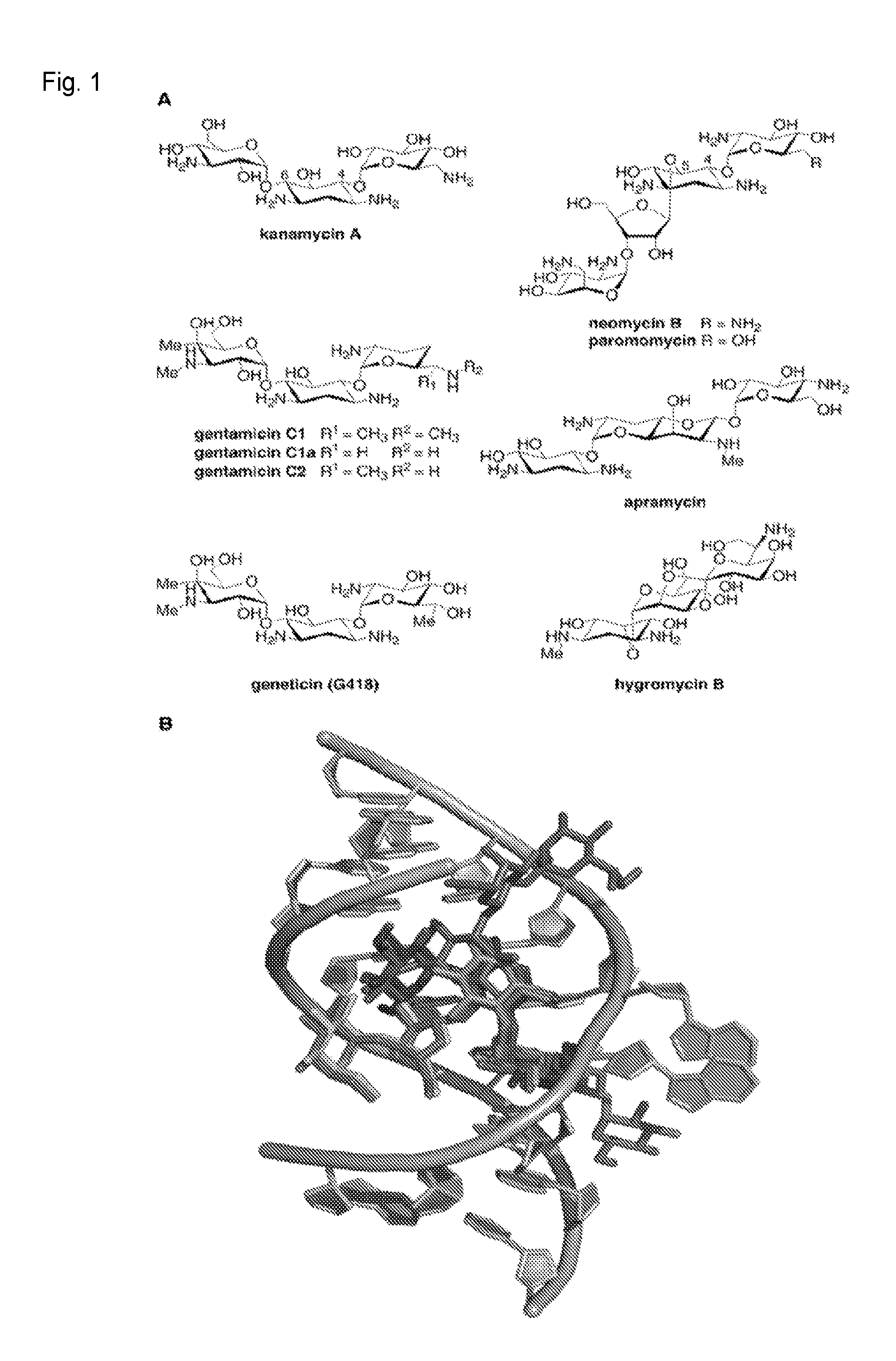
FIG. 1. (A) Structures of aminoglycosides used in this study. (B) Overlayed X-ray crystal structures of the decoding site (A-site) of the E. coli 16S ribosomal RNA (rRNA) with these aminoglycosides bound. Residues 1403-1411, 1489-1498 of 16S rRNA are shown in grey. Aminoglycosides are color-coded as follows; and the source PDB file for each is given in parentheses: kanamycin A: maroon (2ESI.sup.[10b]), gentamicin: purple (2QB9.sup.[8b]), geneticin (G418): pink (1MWL.sup.[10a]), neomycin B: dark green (2QAL.sup.[8b]), paromomycin: light green (2Z4K.sup.[8b]), apramycin: orange (.sup.4AQY[8e]) hygromycin: blue (3DF1.sup.[8c]).
FIG. 2. (A) Schematic showing the functionality of the orthogonal ribosome-controlled fluorescent reporter in the absence (left panel) and presence (right panel) of aminoglycoside. The O-16S rRNA is shown in black, O-SD/O-ASD pair is shown in red, tetR mRNA and TetR protein are shown in cyan, PLtetO-1 is shown in blue, the gfp-uv gene and GFP protein are shown in green, and aminoglycoside is shown as a red hexagon. (B) Cell pellet fluorescence and fluorescence quantification of E. coli DH5.alpha. cells transformed with (from left to right) plasmids pGBSH3, pGBSH18, pSH3-KF (see Examples for details).
FIG. 3. Cell pellet fluorescence of initial detector strain E. coli SH391 (boxed in red), eleven additional strains with tetR and O-16S promoter strengths combinatorially altered (promoter strengths are show on the left: +- very weak, ++- weak, +++- medium, ++++- strong), and E. coli SH399, the strain with the best detection performance (boxed in green), in response to increasing concentrations of kanamycin. Fluorescence quantification of E. coli SH391 and E. coli SH399 at each of the ten kanamycin concentrations tested is shown in the bar graphs to the right.
FIG. 4. Cell pellet fluorescence of (A) E. coli SH399 and (B) E. coli SH431 in response to increasing concentrations of each of seven aminoglycosides. Kan: kanamycin A, Apr: apramycin, Neo: neomycin B, Paro: paromomycin, Gen: gentamicin, Ami: amikacin, Nea: neamine, Rib: ribostamycin, Sis: sisomicin, Tob: tobramycin G418: geneticin (G418), Hyg: hygromycin.
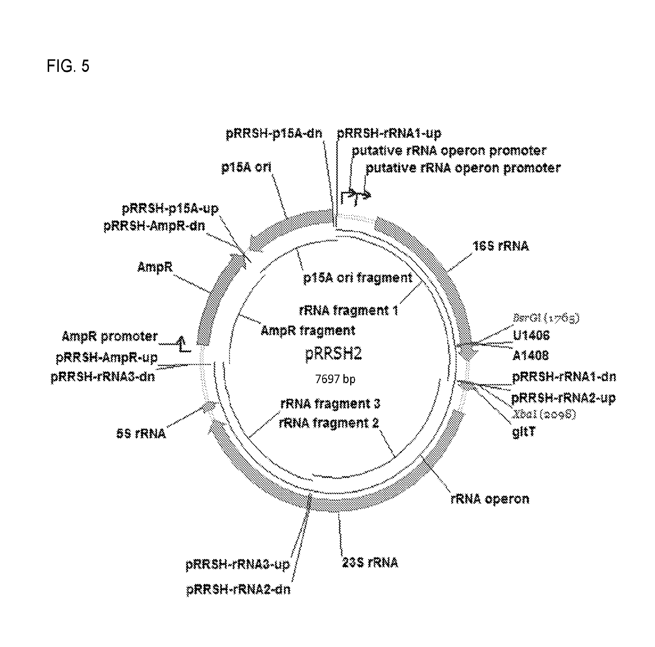
FIG. 5. Plasmid map for pRRSH2.
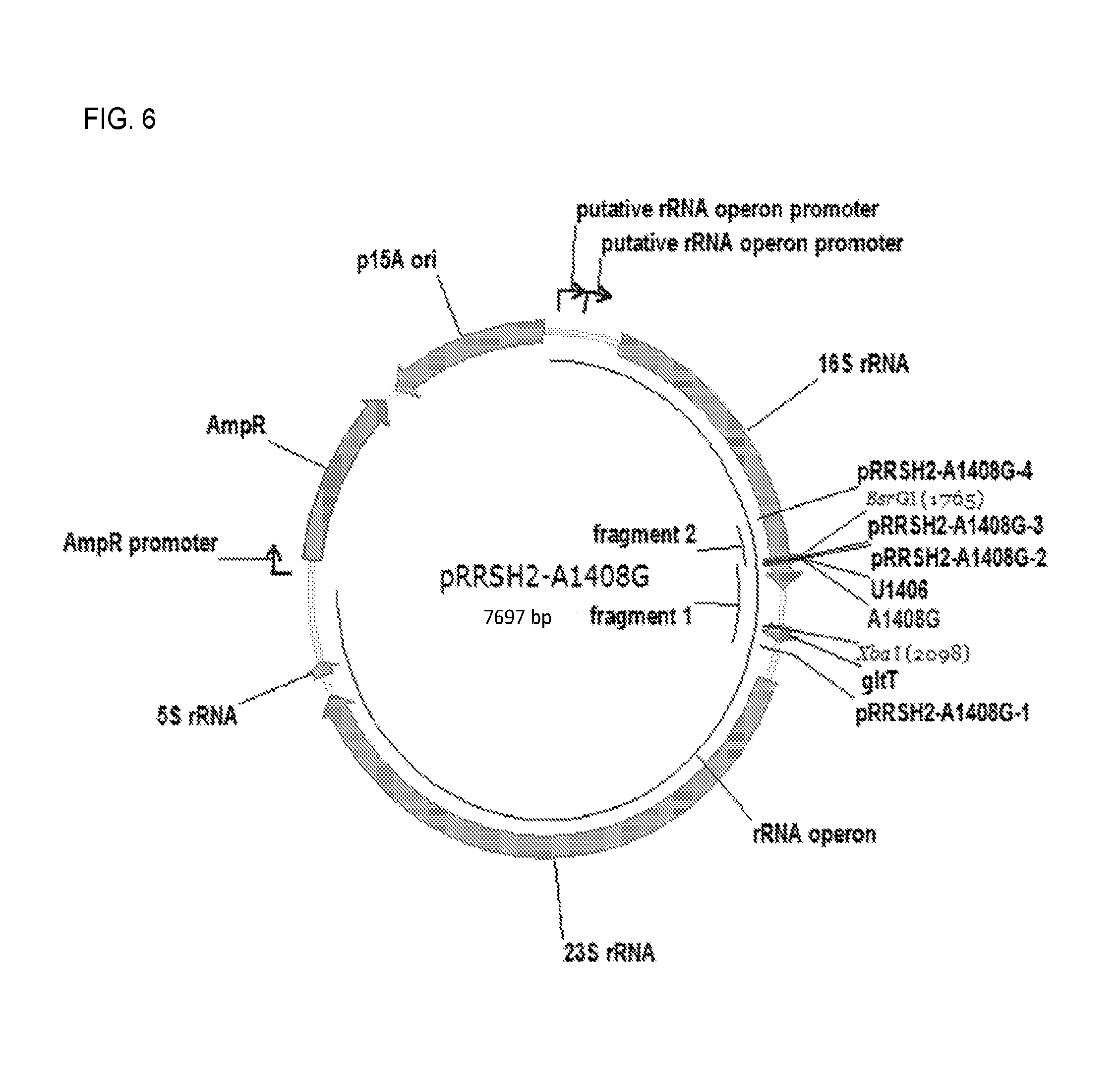
FIG. 6. Plasmid map fop pRRSH2-A1408G.
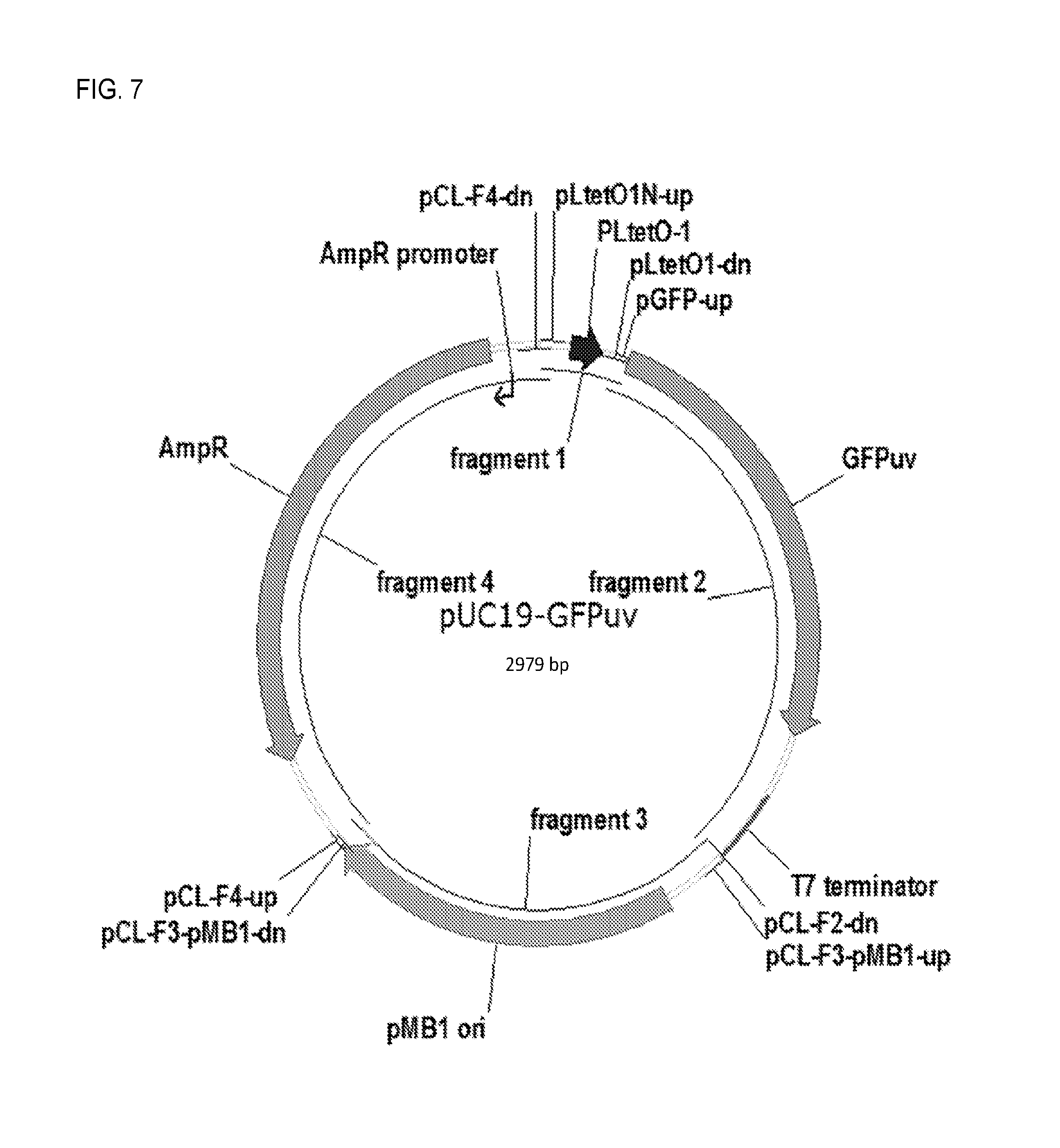
FIG. 7. Plasmid map for pUC19-GFPuv.
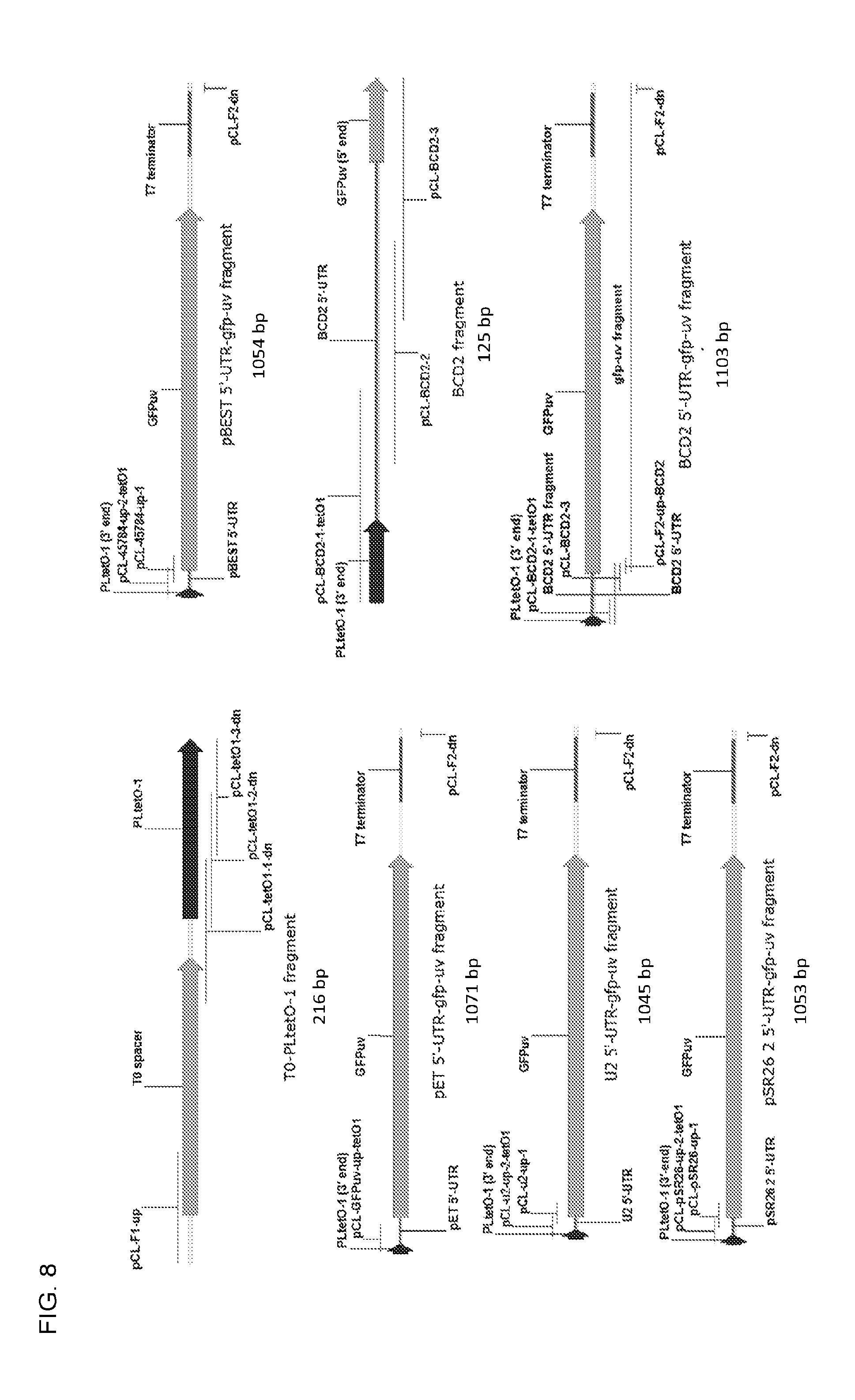
FIG. 8. Maps of T0-PLtetO-1 fragment, pET 5'-UTR-gfp-uv fragment, U2 5'-UTR-gfp-us fragment, pSR26 2 5'-UTR-gfp-uv fragment, pBEST 5'-UTR-gfp-uv fragment, BCD2 fragment, and BCD2 5'-UTR-gfp-uv fragment.
FIG. 9. Plasmid map for pGBSH1-BCD2.
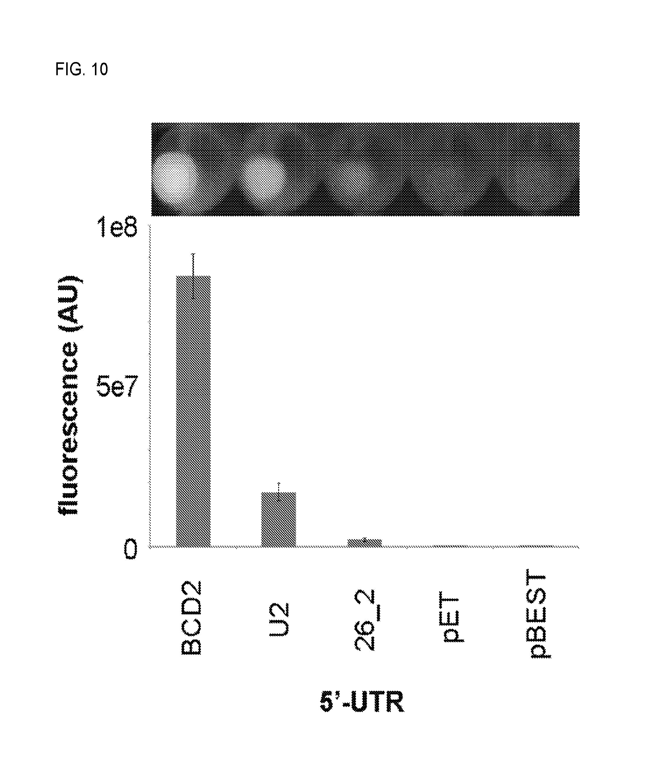
FIG. 10. Cell pellet fluorescence and fluorescence quantification of pGBSH1 variants in which the gfp-uv 5'-UTR has been altered.
FIG. 11. Plasmid map for pGBSH3.
FIG. 12. Map of O-SD-tetR fragment.
FIG. 13. Plasmid map for pGBSH18.
FIG. 14. Map of Plpp-O-16S-terminator fragment.
FIG. 15. Plasmid map for pSH3-KF.
FIG. 16. Cell pellet fluorescence and fluorescence quantification of E. coli DH5.alpha. cells containing pSH3-KF grown in the presence of a range of anhydrotetracycline concentrations.
FIG. 17. Plasmid map of pSH4-KF.
FIG. 18. Plasmid map of pSH6-KF.
FIG. 19. Fluorescence quantification of E. coli SH386 cells containing pSH3-KF-pSH14-KF grown in the presence of a range of kanamycin concentrations.
FIG. 20. OD.sub.600 and fluorescence quantification of E. coli SH399 (pSH6-KF, pRRSH2-A1408G), SH431 (pSH6-KF, pRRSH2-U1406A), and SH434 (pSH6-KF, pRRSH2) cells grown in a range of concentrations (0-500 .mu.M) of each of the seven aminoglycosides examined. The scale of x and y axes is consistent on each bar chart. Undetermined data points are marked with red boxes.
FIG. 21. OD.sub.600 readings of SH386 (A1408G, blue bars) and SH430 (wild-type, red bars) grown in a range of kanamycin concentrations.
FIG. 22. Analysis of the correlations between: (top table and graph) IC.sub.50 values previously determined through in vitro translation assays (main text, ref. 28) (column 1, x axis) and IC.sub.50 values determined from E. coli SH399-derived fluorescence data (column 2, y axis); (middle table and graph) LD.sub.50 values determined from growth inhibition assays of E. coli SH434 (column 1, x axis) and IC.sub.50 values determined from E. coli SH399-derived fluorescence data for the subset of six compounds for which there was a statistically significant correlation (column 2, y axis); and (bottom table and graph) LD.sub.50 values determined from growth inhibition assays of E. coli SH434 (column 1, x axis) and IC.sub.50 values determined from E. coli SH399-derived fluorescence data for the full set of 9 compounds (column 2, y axis). The three compounds that produced outlying data are labeled in the graph. Neamine was excluded from the analysis due to its very weak ribosome inhibition and growth inhibition activities.
FIG. 23. Fluorescence quantification (top row of graphs) and OD.sub.600 quantification (bottom row of graphs) of E. coli SH431 (pSH6-KF, pRRSH2-U1406A) cells grown in a range of concentrations (0-500 .mu.M) of G418, hygromycin, kanamycin, and gentamicins. The scale of x and y axes is consistent for each row of bar charts.
FIG. 24. Analysis of the correlations between: (top table and graph) LD.sub.50 values determined from growth inhibition assays of E. coli SH434 (column 1, x axis) and IC.sub.50 values determined from E. coli SH431-derived fluorescence data for the subset of three compounds for which there was a statistically significant correlation (column 2, y axis); and (bottom table and graph) LD.sub.50 values determined from growth inhibition assays of E. coli SH434 (column 1, x axis) and IC.sub.50 values determined from E. coli SH431-derived fluorescence data for the full set of four compounds (column 2, y axis). The compound that produced outlying data is labeled in the graph.
DETAILED DESCRIPTION OF ILLUSTRATIVE EMBODIMENTS
The ribosome is a complex, highly conserved biomolecular machine essential for the biosynthesis of cellular proteins and peptides. The essentiality and ancient origin of the ribosome have made it one of the most frequent targets of antibacterial natural products. Aminoglycosides (FIG. 1A) can impair ribosome function by affecting the efficiency of intersubunit rotation, translocation, and/or ribosome recycling. Aminoglycoside also can impair ribosome function by inducing translational miscoding through specific interactions with the decoding site (A-site) of the 16S ribosomal RNA (rRNA) in helix 44 (h44) of the small (30S) ribosomal subunit (FIG. 1B), and in the case of some aminoglycosides, with helix 69 (H69) of the large (50S) ribosomal subunit.
Affinities of aminoglycosides for rRNA hairpins mimicking the decoding site have been assessed using mass spectrometry, surface plasmon resonance (SPR), and competition assays. However, the affinities measured by these methods do not correlate linearly with in vivo antibacterial potencies. The dose-dependent effects of aminoglycosides on translation, on the bulk kinetics of translation, and/or on the kinetics of its individual steps have also been examined in vitro using cell-free translation systems, more recently combined with single molecule techniques such as smFRET. These studies show that although most aminoglycosides exert their effects in large part by binding to the decoding site of h44, individual compounds differ markedly with respect their potencies and in their effects on the kinetics of the individual steps of translation.
In contrast to the breadth of techniques available for in vitro studies, in vivo analyses of the effects of aminoglycosides on translation have been limited to measuring minimum inhibitory concentrations (MICs) of the compounds on wild-type bacterial strains and on those carrying ribosomal resistance mutations. These limitations are the consequence of the essentiality of the ribosome for cell survival. However, the limitations imposed by ribosome essentiality can be circumvented in vivo through the use of orthogonal ribosomes (O-ribosomes)--specialized mutant ribosomes that, by virtue of a mutated 16S rRNA anti-Shine-Dalgarno (ASD) sequence, are unable to translate native mRNA, yet retain the ability to translate mRNA carrying a complementary mutant Shine-Dalgarno (SD) sequence. O-ribosomes, in a properly engineered cellular context, can be used to directly detect and quantify ribosome inhibition by aminoglycosides, providing a means of rapidly assessing the activity, specificity, and/or potency of these compounds in living bacterial cells.
This disclosure describes engineered E. coli strains harboring an O-ribosome-controlled fluorescent reporter that are able to detect ribosome inhibition by a variety of aminoglycosides in a highly sensitive, dose-dependent manner with essentially no background. The dose response patterns observed for different aminoglycosides are consistent with current knowledge regarding their potencies and mechanisms of action; and in some cases provide additional insights into the characteristics of specific aminoglycosides. Thus, the O-ribosome reporter system provides a powerful new tool for easily and rapidly assessing the target sites and relative potencies of aminoglycosides and for investigating their mechanisms of action in living bacterial cells.
The exemplary aminoglycoside responsive strains described herein are designed to harbor an engineered plasmid-borne reporter system that includes three elements (FIG. 2A): (1) a constitutively expressed aminoglycoside-sensitive orthogonal 16S rRNA (O-16S) coding region bearing a mutated anti-Shine-Dalgarno (O-ASD) sequence (Lee et al., 1996, RNA 2:1270-1285), (2) a repressor/operator system that includes the tetR coding region encoding a constitutively expressed tetracycline-responsive repressor protein TetR (Beck et al., 1982, J. Bacteriol. 150:633-642) with orthogonal SD (O-SD) sequence complementary to the 16S rRNA O-ASD sequence (Lee et al., 1996, RNA 2:1270-1285) and the TetR-repressed promoter PLtetO-1, and (3) a polynucleotide encoding a detectable reporter--e.g., a gfp-uv polynucleotide encoding the green fluorescent protein variant GFPuv (Crameri et al., 1996, Nat. Biotech. 14:315-319)--under transcriptional control of PLtetO-1 (Lutz et al., 1997, Nucleic Acids Res. 25:1203-1210). In the absence of aminoglycoside (FIG. 2A, left panel), cells bearing these three elements produce O-ribosome-derived TetR that represses transcription of gfp-uv, resulting in no fluorescence. In the presence of aminoglycoside (FIG. 2A, right panel), however, the O-ribosome is inhibited, resulting in a reduced level of TetR, de-repression of gfp-uv transcription, and production of GFPuv. The system is designed to be highly sensitive by substantially amplifying the aminoglycoside input signal through transcription and translation of gfp-uv, resulting in production of multiple GFPuv proteins per aminoglycoside molecule.
To protect the E. coli host itself from inhibition by aminoglycosides, the strain's native rRNA was made aminoglycoside resistant using a previously-developed host, E. coli SQ380, in which all seven chromosomal copies of the rRNA operon were deleted and replaced by a single rRNA operon on plasmid prrnC-sacB bearing the counterselectable marker sacB. The A1408G 16S rRNA mutation, which confers resistance to many aminoglycosides, was introduced into rRNA operon-expressing plasmid pRRSH2. The resulting plasmid, pRRSH2-A1408G, was used to replace prrnC-sacB in SQ380. The resulting strain SH386 possessed a high-level of resistance to kanamycin A, up to 500 .mu.M, the highest concentration tested (FIG. 21).
While described herein and illustrated (FIG. 2) in the context of an exemplary embodiment in which the repressor/operator pair includes the TetR/PLtetO-1 system, the constructs and methods described herein can involve any suitable repressor/operator pair. Exemplary alternative repressor/operator pairs include, for example, LacI/Plac, AraC/Para, or any other repressor operator pair that functions analogously to TetR/PLtetO-1.
Similarly, while described herein and illustrated in the context of an exemplary embodiment in which the polynucleotide encoding a detectable reporter encodes a green fluorescent protein variant, the polynucleotide, the constructs and methods described herein can involve any suitable detectable reporter whose expression can be regulated by the repressor/operator pair. Suitable polynucleotides can encode, for example, other fluorescent proteins or peptides, enzymes, or other reported polypeptide.
Similarly, while described herein and illustrated in FIG. 2 in the context of an exemplary embodiment in which an orthogonal ribosome derived from the E. coli 16S ribosomal RNA sequence is employed, the constructs and methods described herein can involve any suitable 16S ribosomal RNA sequence.
Similarly, while described herein and illustrated in FIG. 2 in the context of an exemplary embodiment in which the mutant E. coli strain SQ380 host strain is employed, the constructs and methods described herein can involve any suitable host strain.
To protect the E. coli host itself from inhibition by aminoglycosides, the strain's native rRNA was made aminoglycoside resistant using a previously-developed host, E. coli SQ380, in which all seven chromosomal copies of the rRNA operon were deleted and replaced by a single rRNA operon on plasmid prrnC-sacB bearing the counterselectable marker sacB. The A1408G 16S rRNA mutation, which confers resistance to many aminoglycosides (Recht et al., 1999, EMBO J. 18:3133-3136), was introduced into rRNA operon-expressing plasmid pRRSH2. The resulting plasmid, pRRSH2-A1408G, was used to replace prrnC-sacB in SQ380. The resulting strain SH386 possessed a high-level of resistance to kanamycin (up to 500 .mu.M, the highest concentration tested).
To demonstrate that the reporter system functions properly, plasmids harboring sets of the system's three functional elements were sequentially constructed and assayed individually by expression in E. coli DH5.alpha. and fluorescence quantification (FIG. 2B). First, performance of the element bearing gfp-uv under control of PLtetO-1 with the optimized 5'-untranslated region BCD2 (Mutilak et al., 2013, Nat. Methods 10:354-360) (see FIG. 10 and Examples section 2.3.4 for optimization) was examined. In the absence of tetR, PLtetO-1 behaves as a strong constitutive promoter and results in high GFP expression (FIG. 2B). Next, the O-SD-controlled tetR cassette was inserted into the gfp-uv-expressing construct. The resulting two element system retained robust fluorescence (.about.60% of that of the gfp-uv-expressing construct, FIG. 2B). Finally, the O-16S cassette was inserted into the gfp-uv/tetR-expressing construct to give final reporter construct pSH3-KF. Addition of the O-16S cassette completely abolished fluorescence (FIG. 2B). Fluorescence could be completely recovered, however, by adding a saturating concentration of anhydrotetracycline (ATC), which binds to TetR causing its dissociation from PLtetO-1 (FIG. 2B, FIG. 16). Together, these results indicate that the reporter system as a whole, as well as its individual elements, function correctly in E. coli DH5.alpha.. The .about.40% loss of fluorescence upon insertion of the tetR cassette may be due, at least in part, to a polar effect on gfp-uv expression by tetR--not a lack of O-SD orthogonality--since addition of saturating ATC to the pSH3-KF-expressing strain resulted in a fluorescence level nearly identical to (106.6%.+-.1.2%) that observed in the gfp-uv/tetR-expressing strain. Thus, tetR is not translated to a significant extent without co-expression of the O-16S gene.
In an initial O-ribosome inhibition assay using kanamycin, E. coli SH391 (SH386 transformed with pSH3-KF) showed a modest concentration-dependent increase in fluorescence upon addition of kanamycin (maximum 1.6-fold induction, FIG. 3), but displayed strong background fluorescence in the absence of kanamycin. The promoter strengths of the tetR and O-16S elements, both of which can affect the amount of TetR, were combinatorially altered using synthetic constitutive promoters with characterized strengths (see Examples section 2.4.2). Eleven new pSH3-KF derivatives with altered tetR and O-16S promoter strengths were generated and their performance examined in SH386 (FIG. 3, FIG. 19). One variant strain, SH399, with reporter plasmid pSH6-KF carrying strong promoter BBa_J23100 controlling O-16S expression, had essentially no background and displayed a robust dose-dependent increase in fluorescence (FIG. 3). Cells expressing the twelve pSH3-KF variants showed a range of kanamycin-dependent phenotypes that correlate with promoter strength--from high background and low sensitivity with weak promoter combinations to low background and high sensitivity with strong promoter combinations (FIG. 3). Thus, a combinatorial approach was used to identify the promoter pair that gave optimal detection of ribosome inhibition by kanamycin while eliminating background signal.
The ability of SH399 to detect ribosome inhibition by other aminoglycosides was tested next. A panel of six additional structurally diverse aminoglycosides, including other 4,6-disubstituted 2-deoxystreptamines (2-DOS) gentamicins and G418, 4,5-disubstituted 2-DOS paromomycin and neomycin, and atypical 2-DOS apramycin and hygromycin (FIG. 1A) were chosen. In addition to kanamycin, SH399 was able to detect O-ribosome inhibition by four of these compounds: apramycin, neomycin, paromomycin, and gentamicins (FIG. 4A). The two compounds that failed to give signal, G418 and hygromycin, also caused significant growth inhibition of SH399 (FIG. 20), indicating that the A1408G 16S rRNA mutation does not confer sufficient resistance to these compounds to allow survival of the detector strain. For those compounds that were detectable, the A1408G mutation confers resistance well above the detection threshold (FIG. 20). Moreover, dose-dependent fluorescence response patterns were different for each compound, with gentamicin, neomycin, and paromomycin giving robust fluorescence responses at low concentrations, and kanamycin and apramycin showing more gradual dose-dependent fluorescence increases. These dose-response patterns correlate well with previously determined potencies (IC.sub.50 values) of these compounds measured through inhibition of translation in vitro (Sucheck et al., 2000, J. Am. Chem. Soc. 122:5230-5231), demonstrating that the O-ribosome reporter system can be used to assess relative potencies of various aminoglycosides.
There was a strong correlation (R.sup.2=0.97, FIG. 22) between the two datasets, suggesting that IC.sub.50 values determined using the O-ribosome reporter assay are comparably accurate to those determined using in vitro translation assays. Next, to test whether the fluorescence dose-response patterns also correlate with inhibition of E. coli growth, dose-dependent growth inhibition (represented as LD.sub.50 values) of the parent aminoglycoside-sensitive E. coli strain SH434 was compared to IC.sub.50 values calculated from fluorescence data obtained from aminoglycoside-treated SH399. While data obtained by the two methods correlated for a subset of the compounds (apramycin, gentamicins, amikacin, ribostamycin, sisomicin, and tobramycin; R.sup.2=0.97, FIG. 22), there was a lack of correlation between the two datasets for kanamycin A, neomycin B, and paromomycin, and therefore between the two datasets as a whole (R.sup.2=0.40, FIG. 22). While the reason for the incomplete correlation between inhibition of E. coli growth and fluorescence-derived IC.sub.50 values is unclear, these inconsistencies may be the result of differences between the pleiotropic effects of differentially inhibiting the ribosome--whose activity is required for synthesis of the entire E. coli proteome--on cell viability and/or the effects of differentially inhibiting the O-ribosome, which are restricted to the TetR-GFP output system. Taken together, these results are consistent with the ability of the O-ribosome reporter system to compare the potencies of aminoglycosides as ribosome inhibitors. Of the ten compounds examined, sisomicin was found to have the strongest ribosome inhibition activity, followed in order of decreasing activity by gentamicins, neomycin B, paromomycin, tobramycin, amikacin, ribostamycin, apramycin, kanamycin A, and neamine (FIG. 20 and FIG. 22). These results provide a complete comparative assessment of the ribosome inhibiting potencies of these ten compounds.
Also, at drug concentrations beyond those that give the peak response, fluorescence decreases significantly with neomycin, paromomycin, and gentamicin, but not with kanamycin or apramycin. These decreases in fluorescence were accompanied by growth inhibition in the cases of paromomycin and gentamicin. This result is consistent with the previous observation that neomycin, paromomycin, and gentamicins bind to and inhibit the ribosome at a second, lower affinity site in helix 69 of the large ribosomal subunit. In the O-ribosome reporter described herein, binding to this site on the pRRSH2-A1408G-derived ribosome can affect translation of both GFPuv and endogenous proteins, leading to both a decrease in fluorescence and loss of cell viability. Thus, the O-ribosome reporter system is able to distinguish aminoglycosides that bind exclusively to the h44 site from those that bind to both h44 and H69 sites.
To further explore the capabilities of the system and attempt to develop a strain that can detect O-ribosome inhibition by hygromycin and G418, the U1406A mutation was introduced into pRRSH2. Mutations at position 1406 confer an aminoglycoside resistance spectrum distinct from that of A1408G, including resistance to G418 (U1406A) and hygromycin (U1406C). Strain SH431, carrying this mutation and reporter plasmid pSH6-KF, was tested for its ability to detect O-ribosome inhibition by the same twelve aminoglycosides. SH431 was able to detect O-ribosome inhibition by both G418 and hygromycin as well as by gentamicins and kanamycin (FIG. 4B and FIG. 23). As in the case of treatment of SH399 with some aminoglycosides, a lack of signal by SH431 in the presence of apramycin, neomycin, and paromomycin was observed, and is the result of the inability of the U1406A mutation to confer resistance to these compounds (FIG. 20). Dose-dependent decreases in response at higher concentrations of gentamicins and G418 (FIG. 4B and FIG. 20) were also observed, consistent with binding of gentamicins to the second, low affinity site in H69. The decrease in response with G418 suggests that it, being a close congener of gentamicin, also inhibits the ribosome by binding to the H69 site. Dose response patterns observed for SH431 treated with the four compounds for which fluorescence could be observed indicate that, among these, gentamicins are the most potent ribosome inhibitors, followed by G418 and hygromycin B, with kanamycin A being the least potent (FIG. 23 and FIG. 24). IC.sub.50 values calculated from the O-ribosome-based fluorescence assay correlated with LD.sub.50 values of the parent aminoglycoside sensitive E. coli strain SH434 for three of the compounds (gentamicins, G418, hygromycin B; R.sup.2=0.997, FIG. 24), but not for kanamycin A, as was the case with SH399.
While described herein and illustrated in FIG. 4 in the context of exemplary embodiments in which the ribosomal RNA mutations conferring drug resistance, A1408G and U1406A, were employed, the constructs and methods described herein can involve any suitable ribosomal RNA mutation that confers resistance to a ribosome inhibiting drug.
In summary, this disclosure describes an engineered E. coli strain that can directly detect ribosome inhibition by a variety of structurally distinct aminoglycosides with high sensitivity and essentially no background. The dose-response patterns observed for the aminoglycosides tested correlate well with their reported potencies and mechanisms of action, demonstrating that the system can be used to determine relative potencies of aminoglycosides and to make inferences about their mechanisms of action based on similarity of response patterns to those of well-studied aminoglycosides. The selectivity of each strain for detecting specific aminoglycosides--even those with high structural similarity such and gentamicins and G418--can be controlled by employing 16S rRNA mutations that confer distinct resistance profiles.
The O-ribosome reporter strategy described herein can be used to assess in vivo potencies of synthetic aminoglycoside analogs in high-throughput, to detect and quantify aminoglycosides in natural product extracts, and/or to detect activity of aminoglycoside biosynthetic enzymes. The O-ribosome reporter system can be extended to detect ribosome inhibition by other classes of 16S rRNA-targeting inhibitors such as, for example, tetracyclines, tuberactinomycins, and/or pactamycins by employing 16S rRNA resistance mutations specific to each compound class. Mutation of the O-ribosome A-site in the strains described here to mimic the A-site of other bacteria (e.g. Mycobacteria or other pathogens) or human mitochondria may allow estimation of aminoglycoside potency against these bacteria, or toxicity to human cells, respectively.
In the preceding description and following claims, the term "and/or" means one or all of the listed elements or a combination of any two or more of the listed elements; the terms "comprises," "comprising," and variations thereof are to be construed as open ended--i.e., additional elements or steps are optional and may or may not be present; unless otherwise specified, "a," "an," "the," and "at least one" are used interchangeably and mean one or more than one; and the recitations of numerical ranges by endpoints include all numbers subsumed within that range (e.g., 1 to 5 includes 1, 1.5, 2, 2.75, 3, 3.80, 4, 5, etc.).
In the preceding description, particular embodiments may be described in isolation for clarity. Unless otherwise expressly specified that the features of a particular embodiment are incompatible with the features of another embodiment, certain embodiments can include a combination of compatible features described herein in connection with one or more embodiments.
For any method disclosed herein that includes discrete steps, the steps may be conducted in any feasible order. And, as appropriate, any combination of two or more steps may be conducted simultaneously.
The present invention is illustrated by the following examples. It is to be understood that the particular examples, materials, amounts, and procedures are to be interpreted broadly in accordance with the scope and spirit of the invention as set forth herein.
EXAMPLES
1. Materials and Methods
1.1. General.
All general molecular biological and biochemical reagents, including Luria-Bertani (LB) media (Miller), were purchased from VWR (Atlanta, Ga.) and were used without further purification. Water used for media was obtained from a Barnstead/Thermolyne HN Ultrapure water purification system. Gentamicins sulfate, paromomycin sulfate, geneticin (G418) sulfate, neomycin sulfate, and hygromycin B were purchased from Santa Cruz Biotechnology (Dallas, Tex.). Kanamycin sulfate was purchased from Genlantis (San Diego, Calif.). Apramycin sulfate was purchased from Research Products International (Mount Prospect, Ill.). Restriction enzymes, Phusion DNA polymerase, T4 DNA ligase and calf intestinal alkaline phosphatase were purchased from New England Biolabs (Ipswich, Mass.). DNA purification and concentration was performed using the DNA Clean & Concentrator Kit; and agarose gel DNA extraction was performed using the Gel DNA Recovery Kit, both from Zymo Research (Irvine, Calif.). Plasmid extractions were performed using the QIAprep Spin Miniprep Kit from Qiagen (Valencia, Calif.). Oligonucleotides were obtained from Integrated DNA Technologies (Coralville, Iowa). DNA sequencing was performed by Genewiz (South Plainfield, N.J.). PCR reactions were carried out using a Bio-Rad S1000 thermal cycler. Cell density and fluorescence measurements were taken using a Molecular Devices SpectraMax M2 Multi-Mode Microplate Reader. Plasmid and DNA sequence design and management was conducted using Vector NTI 10 (Life Technologies). Chemically competent E. coli cells were prepared using the rubidium chloride method..sup.1 Standard molecular biological methods, protocols, reagents, and materials.sup.1 were used for PCR, restriction enzyme digestion, ligation, transformation, selection of transformants, agarose gel electrophoresis, gel extraction, and plasmid isolation unless otherwise specified.
1.2. Bacterial Strains.
E. coli DH5.alpha. and E. coli TOP10 were used for routine DNA cloning and manipulation. E. coli SQ380 (E. coli MG1655/.DELTA.rrnGADEHBC/prrnC-sacB.sup.2/ptRNA67.sup.2), in which all seven genomic rRNA operons have been deleted and replaced with a single plasmid-borne rRNA operon expressed from the sucrose counterselectable plasmid prrnC-sacB, was used as the starting point for construction of strains capable of detecting ribosome inhibition by aminoglycoside antibiotics.
1.3. Bacterial Culture.
Routine liquid culture of E. coli DH5.alpha. and E. coli TOP10 for cloning purposes was carried out in 2-5 mL of Luria-Bertani broth in sterile 15 mL conical tubes at 37.degree. C., 250 rpm overnight (12-16 h). Selection of E. coli DH5.alpha. and E. coli TOP10 transformants was carried out on Luria-Bertani agar plates containing the appropriate antibiotic(s) at 37.degree. C. overnight (12-16 h). All cell growth and fluorescence assays were performed in sterile Cellstar 96-well deep well culture plates sealed with breathable sealing film, with one mL of LB media per well and with appropriate concentrations of the necessary antibiotics (100 .mu.g/mL ampicillin, 35 .mu.g/mL chloramphenicol, 50 .mu.g/mL kanamycin, 100 .mu.g/mL spectinomycin), anhydrotetracycline (1-100 ng/mL) and aminoglycoside (2.5-500 .mu.M).
1.4. General PCR Conditions.
Concentrations of template, primers, polymerase, dNTPs, and buffer recommended by NEB for Phusion DNA polymerase were used unless otherwise specified. Four types of PCR protocols were used to construct all fragments and all final constructs not obtained by ligation: Protocol 1: PCR amplification of a single fragment with two primers, Protocol 2: templateless (primer only) assembly with three primers, Protocol 3: two fragment overlap extension PCR, and Protocol 4: COE-PCR (see Section 2.2.1 for an explanation of this method). General PCR programs for each protocol are given below.
Protocol 1
TABLE-US-00001 98.degree. C. 30 seconds 98.degree. C. 10 seconds Tm-prim*-5.degree. C. 30 seconds 72.degree. C. 30 sec/kb Repeat two times 98.degree. C. 10 seconds Tm-ext#-5.degree. C. 30 seconds 72.degree. C. 30 sec/kb Repeat 26 times 72.degree. C. 10 minutes 4.degree. C. storage
Protocol 2 Same as Protocol 1, but with no template and with 0.1 .mu.M inside primer and 0.5 .mu.M of each outside primer. Protocol 3
TABLE-US-00002 98.degree. C. 30 seconds 98.degree. C. 10 seconds Tm-OE{circumflex over ( )}-5.degree. C. 30 seconds 72.degree. C. 30 sec/kb Repeat 1-9 times (primerless) Add 0.5 .mu.M of two outside primers 98.degree. C. 10 seconds Tm-ext#-5.degree. C. 30 seconds 72.degree. C. 30 sec/kb Repeat 29 times 72.degree. C. 10 minutes 4.degree. C. storage
Protocol 4 Using 10 nM of each fragment. Fragment junctions designed to have Tm of 55.degree. C.+/-5.degree. C.
TABLE-US-00003 98.degree. C. 30 seconds 98.degree. C. 10 seconds 48.degree. C.-50.degree. C. 30 seconds 72.degree. C. 15 sec/kb (of final plasmid size) Repeat 29-34 times 72.degree. C. 10 minutes 4.degree. C. storage *Tm-prim = Tm of the portion of the primer that primes to the template #Tm-ext = Tm of the entire primer {circumflex over ( )}Tm-OE = Tm of the junction between fragments
1.5. Enforced Replacement by Sucrose Counterselection
To replace plasmid prrnC-sacB (Kan.sup.R, Suc.sup.S), which is essential in E. coli SQ380 because it carries the only cellular copy of the ribosomal RNA (rRNA) operon, with pRRSH2 (Amp.sup.R), pRRSH2-A1408G, or pRRSH2-U1406A, sucrose counterselection was used against the sacB (sucrose sensitivity gene)-containing plasmid prrnC-sacB. E. coli SQ380 competent cells were grown in LB with kanamycin and spectinomycin (essential tRNA-bearing plasmid ptRNA67 has a spectinomycin resistance marker) and transformed with pRRSH2, pRRSH2-A1408G, or pRRSH2-U1406A. Transformants were selected on LB agar with ampicillin and spectinomycin. One colony was picked and grown in LB liquid with ampicillin and spectinomycin overnight, and plated on LB agar with ampicillin, spectinomycin, and 5% (w/v) sucrose. Surviving colonies are resistant to both ampicillin and sucrose, and have therefore gained pRRSH2 and lost prrnC-sacB. Elimination of prrnC-sacB was verified by plasmid isolation and digestion of the resulting plasmid mixture with PvuI, which has three recognition sites in prrnC-sacB but only a single site in pRRSH2 and ptRNA67, and therefore gives a distinctive digestion pattern if prrnC-sacB is present. This, rather than replica plate screening of surviving colonies for kanamycin sensitivity, was done because pRRSH2-A1408G and pRRSH2-U1406A confer kanamycin resistance. The resulting strains--SH430 containing pRRSH2, SH386 containing pRRSH2-A1408G, and SH424 containing pRRSH2-U1406A--were used for transformation with plasmids carrying the O-ribosome-based aminoglycoside detection systems (pSH3-KF through pSH14-KF).
1.6. Cell Density and Fluorescence Assays.
All cell density and fluorescence measurements were taken in triplicate. 96-well culture plates (1 mL LB per well) with appropriate concentrations of necessary antibiotics and aminoglycoside were inoculated 1:100 from a saturated overnight liquid culture and allowed to grow for 48 hours at 37.degree. C., 200 rpm shaking. For cell density assays, 40 .mu.L of sample was taken from each well, diluted 5-fold, and OD.sub.600 was measured by microplate reader. The OD.sub.600 of the original culture was calculated by multiplying the reading by the dilution factor (Melancon et al., 2009, Bioorg. Med. Chem. Lett. 19:3845-3847). For cell pellet fluorescence imaging, cells were pelleted by centrifugation (4,000.times.g, 15 minutes, 4.degree. C.) and the supernatant was decanted completely. The underside of the plate was illuminated at 365 nm using an ultraviolet handheld lamp and photgraphed with an 8 megapixel digital camera. For fluorescence quantification, cell pellets were resuspended in 1 mL of 1/4.times.Ringer's solution (30.75 mM NaCl, 1.2 mM, KCl, 1.5 mM CaCl.sub.2, pH 7.3-7.4), 200 .mu.L of cells from each well were transferred to black 96-well plates, and GFP fluorescence was measured (excitation=395 nm, bandwidth=9 nm; emission=509 nm, bandwidth=15 nm). Fluorescence intensities were calculated as fluorescence/OD.sub.600 of the sample minus fluorescence/OD.sub.600 of a sample of a non-GFP-expressing E. coli strain parental to the strain being analyzed in order to correct for both cell density and E. coli auto-fluorescence.
2. Vector Construction and Functional Assays
2.1. General Notes.
All vectors used in this study were designed to avoid any antibiotic resistance markers that encode aminoglycoside modifying enzymes (e.g. kanamycin, apramycin, streptomycin resistance markers) or tetracycline because they would interfere with aminoglycoside detection, or the TetR repressor system, respectively. The sequences of all genetic parts amplified from plasmids whose sequences are not publicly available and detailed information on all primers used are provided in each section.
2.2. rRNA-Expressing Plasmids
2.2.1. Construction of pRRSH2 (FIG. 5).
Plasmid pKK3535 (11.9 kb; Brosius et al., 1981, Plasmid 6:112-118), which contains the constitutively expressed rrnB ribosomal rRNA operon, pMB1 origin of replication, and ampicillin resistance marker, as well as 4.2 kb of non-essential DNA sequence, was used as the starting point for construction of a simplified, refactored rrnB-expressing plasmid pRRSH2 (7.7 kb), which also bears the ampicillin resistance marker, but contains the p15A origin of replication. To construct pRRSH2, concatamerizing overlap extension PCR was used (COE-PCR), a de novo plasmid assembly method that is similar to the CPEC method (Quan et al., 2011, Nat. Protocols 6:242-251). In COE-PCR, a circular plasmid is obtained by one pot PCR assembly of linear fragments with short (15-25 bp) overlapping ends followed by transformation of competent E. coli with the PCR assembly mixture. The 5.8 kb rrnB operon was amplified as three fragments from pKK3535 using primer pairs pRRSH-rRNA1-up/pRRSH-rRNA1-dn, pRRSH-rRNA2-up/pRRSH-rRNA2-dn, and pRRSH-rRNA3-up/pRRSH-rRNA3-dn. The fragment containing the promoter and coding region of the ampicillin resistance marker was amplified from pKK3535 using primers pRRSH-AmpR-up and pRRSH-AmpR-dn. The fragment containing the p15A origin of replication was amplified from pRepCM3 (Melancon et al., 2009, Bioorg. Med. Chem. Lett. 19:3845-3847) using primers pRRSH-p15A-up and pRRSH-p15A-dn. The resulting five DNA fragments were assembled by COE-PCR and the reaction mixture was concentrated using the Zymo Clean and Concentrator Kit, and introduced into competent E. coli DH5.alpha. cells. The final pRRSH2 construct was verified by restriction mapping and sequencing. Primer information is provided in Table 1. The priming region of each primer is underlined.
TABLE-US-00004 TABLE 1 amplicon primer name sequence (5'.fwdarw.3') size (bp) template pRRSH-rRNA1-up TTTGGTTGAATGTTGCGCGGTC Seq ID No. 1 2116 pKK3535 pRRSH-rRNA1-dn CGGTGTCCTGGGCCTCTAGAC Seq ID No. 2 pRRSH-rRNA2-up TCTAGAGGCCCAGGACACCGCCCTTTCACGGCGGTAACAG Seq ID No. 3 2022 pKK3535 pRRSH-rRNA2-dn CTGGTATCTTCGACTGATTTCAGCTCCATCCGCGAGGGACC Seq ID No. 4 pRRSH-rRNA3-up GCTGAAATCAGTCGAAGATACCAGCTGGC Seq ID No. 5 1677 pKK3535 pRRSH-rRNA3-dn AGCTGCTTTCCTGATGCAAAAACG Seq ID No. 6 pRRSH-AmpR-up CGTTTTTGCATCAGGAAAGCAGCTGATATCAGACGTCAGGTGGCACTTTTC Seq ID No. 7 1077 pKK3535 pRRSH-AmpR-dn CATATGATCAATCTAAAGTATATATGAGTAAACTTGGTCTGACAG Seq ID No. 8 pRRSH-p15A-up CCAAGTTTACTCATATATACTTTAGATTGATCATATGCTTCGGATCCCTCGAGA 934 pRepCM3 GATC Seq ID No. 9 pRRSH-p15A-dn CCGCGCAACATTCAACCAAAATTACATGTGCGTCAGACCC Seq ID No. 10
The p15A origin of replication fragment sequence is provided below. Primer binding sites are underlined, and the p15A origin region is shown in bold.
TABLE-US-00005 Seq ID No. 11 ATTACATGTG CGTCAGACCC CTTAATAAGA TGATCTTCTT GAGATCGTTT TGGTCTGCGC GTAATCTCTT GCTCTGAAAA CGAAAAAACC GCCTTGCAGG GCGGTTTTTC GAAGGTTCTC TGAGCTACCA ACTCTTTGAA CCGAGGTAAC TGGCTTGGAG GAGCGCAGTC ACCAAAACTT GTCCTTTCAG TTTAGCCTTA ACCGGCGCAT GACTTCAAGA CTAACTCCTC TAAATCAATT ACCAGTGGCT GCTGCCAGTG GTGCTTTTGC ATGTCTTTCC GGGTTGGACT CAAGACGATA GTTACCGGAT AAGGCGCAGC GGTCGGACTG AACGGGGGGT TCGTGCATAC AGTCCAGCTT GGAGCGAACT GCCTACCCGG AACTGAGTGT CAGGCGTGGA ATTAGACAAA CGCGGCCATA ACAGCGGAAT GACACCGGTA AACCGAAAGG CAGGAACAGG AGAGCGCACG AGGGAGCCGC CAGGGGGAAA CGCCTGGTAT CTTTATAGTC CTGTCGGGTT TCGCCACCAC TGATTTGAGC GTCAGATTTC GTGATGCTTG TCAGGGGGGC GGAGCCTATG GAAAAACGGC TTTGCCGCGG CCCTCTCACT TCCCTGTTAA GTATCTTCCT GGCATCTTCC AGGAAATCTC CGCCCCGTTC GTAAGCCATT TCCGCTCGCC GCAGTCGAAC GACCGAGCGT AGCGAGTCAG TGAGCGAGGA AGCGGAATAT ATCCTGTATC ACATATTCTG CTGACGCACC GGTGCAGCCT TTTTTCTCCT GCCACATGAA GCACTTCACT GACACCCTCA TCAGTGCCAA CATAGTAAGC CAGTATACAC TCCGCTAGCC CATGGAGATC TCTCGAGGGA TCCGAAG
2.2.2. Construction of pRRSH2-A1408G (FIG. 6) and pRRSH2-U1406A.
Both plasmids were constructed from pRRSH2. A 684 bp region of pRRSH2 containing the 16S rRNA A1408 and U1406 sites was amplified in two fragments with the mutation site at the junction of the fragments. In each case, the two fragments were joined by overlap extension PCR, the resulting PCR product digested with BsrGI and XbaI, and cloned into pRRSH2 digested with the same enzymes. For pRRSH2-A1408G, fragment 1 was amplified using primers pRRSH2-A1408G-1 and pRRSH2-A1408G-2, and fragment 2 was amplified using primers pRRSH2-A1408G-3 and pRRSH2-A1408G-4. For pRRSH2-U1406A, fragment 1 was amplified using primers pRRSH2-A1408G-1 and pRRSH2-U1406A-2, and fragment 2 was amplified using primers pRRSH2-U1406A-3 and pRRSH2-A1408G-4. Introduction of the mutation into each plasmid was verified by sequencing the cloned region of the plasmid containing it. The vector map of pRRSH2-A1408G is given in FIG. 6 as an example. Primer information is provided in Table 2. The priming region of each primer is underlined. The A1408G and U1406A mutation sites are show in bold and double underlined in the primers that contain them.
TABLE-US-00006 TABLE 2 amplicon primer name sequence (5'.fwdarw.3') size (bp) template pRRSH2-A1408G-1 TCTCAAACATCACCCGAAGATGAG Seq ID No. 12 457 pRRSH2 pRRSH2-A1408G-2 CCCGTCGCACCATGGGAGTG Seq ID No. 13 pRRSH2-A1408G-3 CCATGGTGCGACGGGCGGTGTG Seq ID No. 14 242 pRRSH2 pRRSH2-A1408G-4 GAGGAAGGTGGGGATGACGTC Seq ID No. 15 pRRSH2-U1406A-2 CCCGACACACCATGGGAGTG (used with pRRSH2-A1408G-1) 457 pRRSH2 Seq ID No. 16 pRRSH2-U1406A-3 ACTCCCATGGTGTGTCGGGCGGTG (used with pRRSH2- 246 A1408G-4) Seq ID NO. 17
2.2.3. Functional Verification of pRRSH2-A1408G and pRRSH2 in E. coli SQ380.
The ability of pRRSH2-A1408G to confer aminoglycoside resistance was confirmed through a cell viability assay. E. coli SQ380 was transformed with pRRSH2-A1408G; and prrnC-sacB was removed by sucrose counterselection, resulting in E. coli SH386. As a control, E. coli SQ380 was also transformed with pRRSH2; and prrnC-sacB was removed by sucrose counterselection, resulting in E. coli SH430. The growth inhibition of these two strains by various kanamycin concentrations was determined by inoculation of each strain (1:100 dilution of a saturated culture) into ten 1 mL wells of a 96-well culture plate containing LB broth with specific concentrations of kanamycin added, growth for 24 hours at 37.degree. C., 200 rpm shaking, and measurement of the OD.sub.600. The results demonstrated that E. coli SH430, which has no 16S rRNA aminoglycoside resistance mutation, experiences significant growth inhibition at 10 .mu.M kanamycin, and cannot survive at concentrations above 10 .mu.M kanamycin; whereas E. coli SH386, which has the A1408G mutation, shows no growth inhibition at any kanamycin concentration tested, indicating that the mutation confers robust resistance to kanamycin at concentrations as high as 500 .mu.M.
2.3 Sequential Construction of the Reporter Plasmid
2.3.1. General Notes.
The final reporter plasmid, pSH6-KF, was constructed in six steps:
1) construction of pUC19-GFPuv, which contains the gfp-uv gene under control of the PLtetO-1 promoter (see section 2.3.2).
2) optimization of the gfp-uv 5'-untranslated (5'-UTR) region through construction of a five plasmid series pGBSH1-BCD2, pGBSH1-U2, pGBSH1-26.2, pGBSH1-pET, and pGBSH1-pBEST (see section 2.3.3).
3) replacement of the ampicillin resistance marker with a chloramphenicol resistance marker in pGBSH1-BCD2, the plasmid with the highest gfp-uv expression level from Step 2, to give pGBSH3 (see section 2.3.5).
4) insertion of the cassette containing tetR with orthogonal Shine-Dalgarno (O-SD) sequence under control of medium strength promoter BBa_J23016 into pGBSH3 to give pGBSH18 (see section 2.3.6).
5) insertion of the cassette containing the orthogonal 16S rRNA (O-16S) under control of the constitutive lpp promoter to give reporter plasmid pSH3-KF (see section 2.3.7).
6) optimization of the tetR and O-16S promoter strengths for use in E. coli SH386 through construction of an eleven plasmid series pSH4-KF through pSH14-KF (see section 2.4.2).
2.3.2. Construction and Testing of pUC19-GFPuv (FIG. 7).
This plasmid, which contains the gfp-uv gene under control of the PLtetO-1 promoter/operator (Beck et al., 1982, J. Bacteriol. 150:633-642), pMB1 origin of replication, and ampicillin resistance marker was constructed from four fragments by COE-PCR. The PLtetO-1 promoter/operator was amplified from pSR26_2 (J. Tabor, unpublished) using primers pLTetO1N-up and pLtetO1-dn. The gfp-uv gene was amplified from plasmid pET101-GFP (Young et al., 2010, J. Mol. Biol. 395:361-374) using primers pGFP-up and pCL-F2-dn. The pMB1 origin of replication was amplified from pUC19 using primers pCL-F3-pMB1-up and pCL-F3-pMB1-dn. The ampicillin resistance marker was amplified from pUC19 using primers pCL-F4-up and pCL-F4-dn. All primers are provided in Table 3.
The resulting four fragments were assembled by COE-PCR. The reaction mixture was concentrated using the Zymo Clean and Concentrator Kit, and introduced into competent E. coli DH5.alpha. cells. The final construct was verified by restriction mapping and sequencing. The priming region of each primer is underlined.
E. coli DH5.alpha. transformed with pUC19-GFPuv displayed no fluorescence as determined by plate reader fluorescence assay (see cell density and fluorescence assays section, above, for experimental details). A series of five pUC19-GFPuv derivatives were constructed in which the 5'-UTR was varied (FIG. 8).
TABLE-US-00007 TABLE 3 amplicon primer name sequence (5'.fwdarw.3') size (bp) template pLtetO1N-up ACAAACTAGTGCGACCCTGCGTATCACGAGGCCCTTTCGTC Seq ID No. 18 159 pSR26_2 pLtetO1-dn CATGGTGAAGGGCTCCTGAATTCCTTCATTAATGGTCAGTGCGTCCTGCTGATG Seq ID No. 19 pGFP-up GAAGGAATTCAGGAGCCCTTCACCATG Seq ID No. 20 1022 pET101- pCL-F2-dn CCGGGCCTCTTGCGGGATATC Seq ID No. 21 GFP pCL-F3-pMB1-up ATATCCCGCAAGAGGCCCGGGCGGTAATAAGCTTACGGTTATCCACAGAATCAGG 758 pUC19 Seq ID No. 22 pCL-F3-pMB1-dn AGACCCCGTCTAGATAGAAAAGATCAAAGGATCTTCTTGAG Seq ID No. 23 pCL-F4-up CTTTGATCTTTTCTATCTAGACGGGGTCTGACGCTCAGTG Seq ID No. 24 1137 pUC19 pCL-F4-dn GCAGGGTCGCACTAGTTTGTTTATTTTTCTAAATACATTCAAATATGTATCCGCTC Seq ID No. 25
The gfp-uv fragment sequence is provided below. Primer binding sites are underlined, the gfp-uv coding region is shown in bold with start and stop codons underlined, and the T7 terminator sequence is double underlined.
TABLE-US-00008 Seq ID No. 26 GAAGGAATTC AGGAGCCCTT CACCATGAGT AAAGGAGAAG AACTTTTCAC TGGAGTTGTC CCAATTCTTG TTGAATTAGA TGGTGATGTT AATGGGCACA AATTTTCTGT CAGTGGAGAG GGTGAAGGTG ATGCAACATA CGGAAAACTT ACCCTTAAAT TTATTTGCAC TACTGGAAAA CTACCTGTTC CATGGCCAAC ACTTGTCACT ACTTTCTCTT ATGGTGTTCA ATGCTTTTCC CGTTATCCGG ATCACATGAA ACGGCATGAC TTTTTCAAGA GTGCCATGCC CGAAGGTTAT GTACAGGAAC GCACTATATC TTTCAAAGAT GACGGGAACT ACAAGACGCG TGCTGAAGTC AAGTTTGAAG GTGATACCCT TGTTAATCGT ATCGAGTTAA AAGGTATTGA TTTTAAAGAA GATGGAAACA TTCTCGGACA CAAACTCGAA TACAACTATA ACTCACACAA TGTATACATC ACGGCAGACA AACAAAAGAA TGGAATCAAA GCTAACTTCA AAATTCGCCA CAACATTGAA GATGGATCCG TTCAACTAGC AGACCATTAT CAACAAAATA CTCCAATTGG CGATGGCCCT GTCCTTTTAC CAGACAACCA TTACCTGTCG ACACAATCTG CCCTTTCGAA AGATCCCAAC GAAAAGCGTG ACCACATGGT CCTTCTTGAG TTTGTAACTG CTGCTGGGAT TACACATGGC ATGGATGAGC TCTACAAACT CGAGCACCAC CACCACCACC ACTGAAAGGG CGAGCTCAAT TCGAAGCTTG AAGGTAAGCC TATCCCTAAC CCTCTCCTCG GTCTCGATTC TACGCGTACC GGTCATCATC ACCATCACCA TTGAGTTTGA TCCGGCTGCT AACAAAGCCC GAAAGGAAGC TGAGTTGGCT GCTGCCACCG CTGAGCAATA ACTAGCATAA CCCCTTGGGG CCTCTAAACG GGTCTTGAGG GGTTTTTTGC TGAAAGGAGG AACTATATCC GGATATCCCG CAAGAGGCCC GG
2.3.3. Construction of the pGBSH1 Plasmid Series (FIG. 8 and FIG. 9)
The pGBSH1 plasmid series was constructed by replacement of the gfp-uv 5'-UTR on pUC19-GFPuv with 5 different 5'-UTRs. The 5'-UTRs included three 5'-UTRs that were reported to be strong [BCD2 (Mutilak et al., 2013, Nat. Methods 10:354-360), U2 (Mutilak et al., 2013, Nat. Methods 10:347-353), pBEST (cat. no. #45784, Addgene, Cambridge, Mass.)] and two representing the intact 5'-UTRs associated with the GFPuv gene in pET101-GFP (pET) and the PLtetO-1 promoter in pSR26_2 (26.2). Additionally, a T0 spacer sequence was added between the ampicillin resistance gene and the PLtetO-1 portion to attempt to minimize any polar effects on GFP expression. The T0 spacer was appended to the 5' end of the PLtetO-1 fragment. Construction of these plasmids was accomplished by five COE-PCR reactions, each employing four fragments, three of which (pMB1 origin, ampicillin resistance marker, T0 spacer-PLtetO-1) were identical in all five reactions, and one (the 5'-UTR-gfp-uv fragment) of which was variable. The fragments containing the pMB1 origin and ampicillin resistance marker were identical to those used in construction of pUC19-GFPuv.
The T0 spacer-PLtetO-1 fragment was constructed by three sequential PCR reactions in which the product of the previous reaction was used as the template for the next reaction. The T0 spacer was amplified from plasmid pSR26_2 using primers pCL-F1-up and pCL-tetO1-dn-1. The resulting PCR product was used as the template for a second round of PCR using primers pCL-F1-up and pCL-tetO1-dn-2. The resulting PCR product was used as the template for a third round of PCR using primers pCL-F1-up and pCL-tetO1-dn-3 to generate the final fragment.
The five 5'-UTR-gfp-uv fragments (FIG. 8) were constructed as follows: The pET 5'-UTR-gfp-uv fragment was constructed in a single PCR reaction by amplification from plasmid pET101-GFP using primers pCL-GFPuv-up-tetO1 and pCL-F2-dn. The U2 5'-UTR-gfp-uv, 26.2 5'-UTR-gfp-uv, and pBEST 5'-UTR-gfp-uv fragments were each constructed by two sequential PCR reactions in which the product of the first reaction (the gfp-uv-containing fragment, which was amplified from plasmid pET101-GFP), was used as the template for the second reaction. The U2 5'-UTR-gfp-uv fragment was constructed by amplification using primers pCL-u2-up-1 and pCL-F2-dn; and the resulting PCR product used as the template for a second round of PCR using primers pCL-u2-up-2-tetO1 and pCL-F2-dn. The 26.2 5'-UTR-gfp-uv fragment was constructed by amplification using primers pCL-pSR26-up-1 and pCL-F2-dn; and the resulting PCR product used as the template for a second round of PCR using primers pCL-pSR26-up-2-tetO1 and pCL-F2-dn. The pBEST 5'-UTR-gfp-uv fragment was constructed by amplification using primers pCL-45784-up-1 and pCL-F2-dn; and the resulting PCR product used as the template for a second round of PCR using primers pCL-45784-up-2-tetO1 and pCL-F2-dn. The BCD2 5'-UTR was constructed as a stand-alone fragment by templateless assembly using three primers (pCL-BCD2-1-tetO1, pCL-BCD2-2, and pCL-BCD2-3). A gfp-uv-containing fragment was amplified from pET101-GFP using primers pCL-F2-up-BCD2 and pCL-F2-dn; and the BCD2 5'-UTR and gfp-uv-containing fragments were joined by overlap extension PCR and amplified using outside primers pCL-BCD2-1-tetO1 and pCL-F2-dn to generate the final BCD2 5'-UTR-gfp-uv fragment.
Each of the five 5'-UTR-gfp-uv fragment variants was then assembled with the T0 spacer-PLtetO-1, pMB1 origin, and ampicillin resistance marker fragments in a COE-PCR reaction. Each reaction mixture was concentrated using the Zymo Clean and Concentrator Kit and introduced into competent E. coli DH5.alpha. cells. Each final construct was verified by restriction mapping and sequencing. Primer information is provided in Table 4. The priming region of each primer is underlined. The vector map of pGBSH1-BCD2 is given as an example in FIG. 9.
The 5'-UTRs examined are provided in Table 5.
TABLE-US-00009 TABLE 4 amplicon primer name sequence (5'.fwdarw.3') size (bp) template pCL-F1-up ACAAACTAGTGCGACCCTGCTGCTTGGATTCTCACCAATAAAAAAC Seq ID 167 pSR26_2 NO. 27 pCL-tetO1-dn-1 TGTCAATCTCTATCACTGATAGGGATTTGATATCGAGCTCGCTTGGACTCCTGTTG ATAG Seq ID NO. 28 pCL-tetO1-dn-2 GCTCAGTATCTCTATCACTGATAGGGATGTCAATCTCTATCACTGATAGGGATTTG 194 PCR pdt. Seq ID No. 29 pCL-tetO1-dn-3 GGTCAGTGCGTCCTGCTGATGTGCTCAGTATCTCTATCACTGATAGGG Seq ID 216 PCR pdt. NO. 30 pCL-GFPuv-up- ATCAGCAGGACGCACTGACCCCTCTAGAAATAATTTTGTTTAACTTTAAGAAGGAA 107- 1 pET101- tetO1 TTC Seq ID No. 31 pCL-F2-dn CCGGGCCTCTTGCGGGATATC (same is used to construct pUC19-GFPuv) GFP Seq ID NO. 32 pCL-u2-up-1 AAAGAGGAGAAAGGTACCATGAGTAAAGGAGAAGAACTTTTCACTGG 1016 pET101- (used with pCL-F2-dn) Seq ID NO. 33 GFP pCL-u2-up-2-tetO1 ATCAGCAGGACGCACTGACCGAATTCATTAAAGAGGAGAAAGGTACCATGAG 104- 5 PCR pdt. (used with pCL-F2-dn) Seq ID NO. 34 pCL-pSR26-up-1 AGCAAAGCCCAATTTTAAACAAATGAGTAAAGGAGAAGAACTTTTCACTGG 1020 pE- T101- (used with pCL-F2-dn) Seq ID NO. 35 GFP pCL-pSR26-up-2- ATCAGCAGGACGCACTGACCGCATAAAGGACTTAGCAAAGCCCAATTTTAAAC 1053- PCR pdt. tetO1 (used with pCL-F2-dn) Seq ID NO. 36 pCL-45784-up-1 ATTTTGTTTAACTTTAAGAAGGATCCATGAGTAAAGGAGAAGAACTTTTCACTGG 102- 4 pET101- (used with pCL-F2-dn) Seq ID NO. 37 GFP pCL-45784-up-2- ATCAGCAGGACGCACTGACCGCTAGCAATAATTTTGTTTAACTTTAAGAAGGATCC 1- 054 PCR pdt. tetO1 ATG (used with pCL-F2-dn) Seq ID No. 38 pCL-BCD2-1-tetO1 ATCAGCAGGACGCACTGACCGGGCCCAAGTTCACTTAAAAAGGAGATCAAC Seq ID NO. 39 pCL-BCD2-2 GATTAAGATGTTTCAGTACGAAAATTGCTTTCATTGTTGATCTCCTTTTTAAG 125 primer Seq ID No. 40 assembly pCL-BCD2-3 AGTTCTTCTCCTTTACTCATTAGAAAACCTCCTTAGCATGATTAAGATGTTTCAGT AC Seq ID No. 41 pCL-F2-up-BCD2 ATGAGTAAAGGAGAAGAACTTTTCACTGGAG (used with pCL-F2-dn) 998 pET101- Seq ID NO. 42 GFP
The T0 spacer fragment sequence is provided below. Primer binding sites are underlined, and the T0 spacer region is shown in italics.
TABLE-US-00010 Seq ID No. 43 TGCTTGGATT CTCACCAATA AAAAACGCCC GGCGGCAACC GAGCGTTCTG AACAAATCCA GATGGAGTTC TGAGGTCATT ACTGGATCTA TCAACAGGAG TCCAAGCGAG CTCGATATCA AAT
TABLE-US-00011 TABLE 5 5'-UTR Sequence length (bp) pET CCTCTAGAAATAATTTTGTTTAACTTTAAGAAGGAATTCAGGAGCCCTTCACC Seq ID No. 44 53 U2 GAATTCATTAAAGAGGAGAAAGGTACC Seq ID No. 45 27 26_2 GCATAAAGGACTTAGCAAAGCCCAATTTTAAACAA Seq ID No. 46 35 pBEST GCTAGCAATAATTTTGTTTAACTTTAAGAAGGATCC Seq ID No.47 36 BCD2 GGGCCCAAGTTCACTTAAAAAGGAGATCAACAATGAAAGCAATTTTCGTACTGAAACATCTTAAT 85 CATGCTAAGGAGGTTTTCTA Seq ID No. 48
2.3.4. pGBSH1 Series Functional Assay.
E. coli DH5.alpha. transformed with pGBSH1-BCD2, pGBSH1-U2, pGBSH1-26.2, pGBSH1-pET, and pGBSH1-pBEST displayed a range of fluorescences (FIG. 10) as determined by plate reader fluorescence assays (see cell density and fluorescence assays section, above, for experimental details), with pGBSH1-BCD2 resulting in the highest fluorescence. Thus, pGBSH1-BCD2 was selected for further development.
2.3.5. Construction and Testing of pGBSH3 (FIG. 11)
After identification of plasmid pGBSH1-BCD2 as the variant resulting in the highest fluorescence, the ampicillin resistance marker in pGBSH1-BCD2 was replaced with a chloramphenicol resistance marker so that the resulting plasmid, pGBSH3, could be co-transformed with pRRSH2-A1408G. A four fragment COE-PCR reaction was employed to construct pGBSH3. Three of the fragments (BCD2 5'-UTR-gfp-uv, pMB1 origin, and T0-PLtetO-1) were identical to those used to construct pGBSH1-BCD2. The fragment containing the chloramphenicol resistance marker was amplified from plasmid pBKCM7b (Charles E. Melancon III, unpublished) using primers cat-pBKCM7b-up and cat-pBKCM7b-dn. After COE-PCR, concentration using a Zymo Clean and Concentrator Kit, and transformation, the final construct was verified by restriction mapping and sequencing. Primer information is provided in Table 6. The priming region of each primer is underlined. Retention of robust fluorescence by E. coli DH5.alpha. cells transformed with pGBSH3 was verified by plate reader fluorescence assays (FIG. 2B) (see cell density and fluorescence assays section, above, for experimental details).
TABLE-US-00012 TABLE 6 amplicon primer name sequence (5'.fwdarw.3') size (bp) template cat-pBKCM7b-up CTTTTCTATCTAGACGGGGTCTTTTGATAGAAAATCATAAAAGGATTTGC 1069 pBK- CM7b Seq ID No. 49 cat-pBKCM7b-dn GCAGGGTCGCACTAGTTTGTGGATCCAACTGCATTCAGAATAAATAAATC Seq ID No. 50
The chloramphenicol resistance marker fragment sequence is provided below. Primer binding sites are underlined, the promoter sequence is double underlined, and the chloramphenicol acetyltransferase coding region is shown in bold with start and stop codons underlined.
TABLE-US-00013 Seq ID No. 51 GGATCCAACT GCATTCAGAA TAAATAAATC CTGGTGTCCC TGTTGATACC GGGAAGCCCT GGGCCAACTT TTGGCGAAAA TGAGACGTTG ATCGGCACGT AAGAGGTTCC AACTTTCACC ATAATGAAAT AAGATCACTA CCGGGCGTAT TTTTTGAGTT GTCGAGATTT TCAGGAGCTA AGGAAGCTAA AATGGAGAAA AAAATCACTG GATATACCAC CGTTGATATA TCCCAATGGC ATCGTAAAGA ACATTTTGAG GCATTTCAGT CAGTTGCTCA ATGTACCTAT AACCAGACCG TTCAGCTGGA TATTACGGCC TTTTTAAAGA CCGTAAAGAA AAATAAGCAC AAGTTTTATC CGGCCTTTAT TCACATTCTT GCCCGCCTGA TGAATGCTCA TCCGGAATTA CGTATGGCAA TGAAAGACGG TGAGCTGGTG ATATGGGATA GTGTTCACCC TTGTTACACC GTTTTCCATG AGCAAACTGA AACGTTTTCA TCGCTCTGGA GTGAATACCA CGACGATTTC CGGCAGTTTC TACACATATA TTCGCAAGAT GTGGCGTGTT ACGGTGAAAA CCTGGCCTAT TTCCCTAAAG GGTTTATTGA GAATATGTTT TTCGTCTCAG CCAATCCCTG GGTGAGTTTC ACCAGTTTTG ATTTAAACGT GGCCAATATG GACAACTTCT TCGCCCCCGT TTTCACCATG GGCAAATATT ATACGCAAGG CGACAAGGTG CTGATGCCGC TGGCGATTCA GGTTCATCAT GCCGTTTGTG ATGGCTTCCA TGTCGGCAGA ATGCTTAATG AATTACAACA GTACTGCGAT GAGTGGCAGG GCGGGGCGTA AGGCGCGCCA TTTAAATGAA GTTCCTATTC CGAAGTTCCT ATTCCTAGGG ATTAAAAAGG CAACTTTATG CCCATGCAAC AGAAACTATA AAAAATACAG AGAATGAAAA GAAACAGATA GATTTTTTAG TTCTTTAGGC CCGTAGTCTG CAAATCCTTT TATGATTTTC TATCAAA
2.3.6. Construction and Testing of pGBSH18 (FIG. 13)
A cassette containing the tetR gene with orthogonal Shine-Dalgarno (O-SD) sequence ATCCC (Lee et al., 1996 RNA 2:1270-1285; Abdi et al., 2007, RNA 11: 1624-1632) under control of medium strength promoter BBa_J23106 (J. Christopher Anderson, unpublished) and containing the T1 terminator was inserted into pGBSH3 to generate pGBSH18. A five fragment COE-PCR reaction was employed to construct pGBSH18. Three of the fragments (the BCD2 5'-UTR-gfp-uv, chloramphenicol resistance marker, and T0-PLtetO-1) were identical to those used to construct pGBSH1-BCD2 and pGBSH3. The pMB1 origin was amplified from pGBSH3 using primers F3-up-tetRassem and pCL-F3-pMB1-dn.
The fragment containing O-SD-tetR (FIG. 12) was constructed by three sequential PCR reactions in which the product of the previous reaction was used as the template for the next reaction. The tetR gene with T1 terminator was amplified from pSR26_2 using primers ptetR-1-KF-up and tetRassem1-dn-XmnI. The resulting PCR product was used as the template for a second round of PCR using primers ptetR-2-KF-up and tetRassem1-dn-XmnI. The resulting PCR product was used as the template for a third round of PCR using primers tetRassem1-up-XmnI and tetRassem1-dn-XmnI to generate the final fragment. After COE-PCR, concentration using a Zymo Clean and Concentrator Kit, and transformation, the final construct was verified by restriction mapping and sequencing. Primer information is provided in Table 7. The priming region of each primer is underlined. The O-SD sequence and ATG start codon are shown in bold and are double underlined in the primers that contain them.
TABLE-US-00014 TABLE 7 amplicon primer name sequence (5'.fwdarw.3') size (bp) template F3-up-tetRassem GCGGTAATAAGCTTACGGTTATCCAC Seq ID NO. 52 738 pGBSH3 pCL-F3-pMB1-dn AGACCCCGTCTAGATAGAAAAGATCAAAGGATCTTCTTGAG (same as used to construct pUC19-GFPuv) Seq ID No. 53 ptetR-1-KF-up ACAATCGATACATCCCCCGCAAATGATGTCTCGTTTAGATAAAAGTAA 823 pSR26_2- AG Seq ID No. 54 tetRassem1-dn-Xmn1 TGTGGATAACCGTAAGCTTATTACCGCTTTGAGTGAGCTGATACCGC Seq ID No. 55 ptetR-2-KF-up CTAGCTCAGTCCTAGGTATAGTGCTAGCCCAGCCAGAGAAACAATCGA 863 PCR pdt. TACATCCCCC Seq ID NO. 56 tetRassem1-up-Xmn1 ATATCCCGCAAGAGGCCCGGTTTACGGCTAGCTCAGTCCTAG Seq ID 890 PCR pdt. NO. 57
The sequence of the tetR-T1 terminator fragment is provided below. Primer binding sites are underlined, the tetR coding sequence is shown in bold, and the T1 terminator region is double underlined.
TABLE-US-00015 Seq ID No. 58 ATGATGTCTC GTTTAGATAA AAGTAAAGTG ATTAACAGCG CATTAGAGCT GCTTAATGAG GTCGGAATCG AAGGTTTAAC AACCCGTAAA CTCGCCCAGA AGCTAGGTGT AGAGCAGCCT ACATTGTATT GGCATGTAAA AAATAAGCGG GCTTTGCTCG ACGCCTTAGC CATTGAGATG TTAGATAGGC ACCATACTCA CTTTTGCCCT TTAGAAGGGG AAAGCTGGCA AGATTTTTTA CGTAATAACG CTAAAAGTTT TAGATGTGCT TTACTAAGTC ATCGCGATGG AGCAAAAGTA CATTTAGGTA CACGGCCTAC AGAAAAACAG TATGAAACTC TCGAAAATCA ATTAGCCTTT TTATGCCAAC AAGGTTTTTC ACTAGAGAAT GCATTATATG CACTCAGCGC AGTGGGGCAT TTTACTTTAG GTTGCGTATT GGAAGATCAA GAGCATCAAG TCGCTAAAGA AGAAAGGGAA ACACCTACTA CTGATAGTAT GCCGCCATTA TTACGACAAG CTATCGAATT ATTTGATCAC CAAGGTGCAG AGCCAGCCTT CTTATTCGGC CTTGAATTGA TCATATGCGG ATTAGAAAAA CAACTTAAAT GTGAAAGTGG GTCTTAAGGC ATCAAATAAA ACGAAAGGCT CAGTCGAAAG ACTGGGCCTT TCGTTTTATC TGTTGTTTGT CGGTGAACGC TCTCCTGAGT AGGACAAATC CGCCGCCCTA GACCTAGGCG TTCGGCTGCG GCGAGCGGTA TCAGCTCACT CAAAG
The sequence of the pMB1 origin in pGBSH18 is provided below with spontaneous mutations shown underlined and in bold. G at position 107 was mutated to C, and G at position 457 was mutated to A.
TABLE-US-00016 Seq ID No. 59 CGCGTTGCTG GCGTTTTTCC ATAGGCTCCG CCCCCCTGAC GAGCATCACA AAAATCGACG CTCAAGTCAG AGGTGGCGAA ACCCGACAGG ACTATAAAGA TACCAGCCGT TTCCCCCTGG AAGCTCCCTC GTGCGCTCTC CTGTTCCGAC CCTGCCGCTT ACCGGATACC TGTCCGCCTT TCTCCCTTCG GGAAGCGTGG CGCTTTCTCA TAGCTCACGC TGTAGGTATC TCAGTTCGGT GTAGGTCGTT CGCTCCAAGC TGGGCTGTGT GCACGAACCC CCCGTTCAGC CCGACCGCTG CGCCTTATCC GGTAACTATC GTCTTGAGTC CAACCCGGTA AGACACGACT TATCGCCACT GGCAGCAGCC ACTGGTAACA GGATTAGCAG AGCGAGGTAT GTAGGCGGTG CTACAGAGTT CTTGAAGTGG TGGCCTAACT ACGGCTACAC TAGAAGAACA GTATTTGGTA TCTGCGCTCT GCTGAAGCCA GTTACCTTCG GAAAAAGAGT TGGTAGCTCT TGATCCGGCA AACAAACCAC CGCTGGTAGC GGTGGTTTTT TTGTTTGCAA GCAGCAGATT ACGCGCAGAA AAAAAGGATC TCAAGAAGAT CCTTT
Retention of robust fluorescence by E. coli cells DH5.alpha. transformed with pGBSH18 was verified as by plate reader fluorescence assays (FIG. 2B) (see cell density and fluorescence assays section, above, for experimental details). During the sequencing process, two spontaneous mutations in the pMB1 origin (see below for locations) were discovered. It is unclear whether these mutations have any effect on plasmid copy number, but it is clear from fluorescence assays that they do not interfere with replication in E. coli DH5.alpha. or gfp-uv expression.
2.3.7. Construction and Testing of pSH3-KF (FIG. 15) in E. coli DH5.alpha..
A cassette containing the orthogonal 16S rRNA (O-16S) with orthogonal anti-Shine-Dalgarno (O-ASD) sequence TGGGG (Lee et al., 1996 RNA 2:1270-1285; Abdi et al., 2007, RNA 11:1624-1632) was inserted into pGBSH18 to generate pSH3-KF, which contains all the components of the orthogonal ribosome-based fluorescent reporter. A five fragment COE-PCR reaction was employed to construct pSH3-KF.
Two of the fragments (the O-SD-tetR and pMB1 origin) were identical to those used to construct pGBSH18. The chloramphenicol resistance marker fragment was amplified from pBGSH18 using primers cat-pBKCM7b-up and pCAT-OKF-dn. A fragment containing T0-PLtetO-1 and BCD2 5'-UTR-gfp-uv was also amplified from pGBSH18 using primers pGFP-OKF-up and pCL-F2-dn.
The fragment containing the orthogonal 16S rRNA (O-16S) under control of the reportedly strong lpp promoter (Plpp; Inouye et al., 1985, Nucleic Acids Res. 1985, 13, 13:3101-3110) was constructed by amplifying the Plpp-16S rRNA cassette from plasmid pTrcSS1d-rrsBb (Shinichiro Shoji, unpublished) using upstream primer pO16S-up and mutagenic downstream primer pO16S-KF-dn which was used to install the orthogonal anti-Shine-Dalgarno (O-ASD) sequence. The strong terminator BBa_B0015 (Chen et al., 2013, Nat. Methods 10:659-664) was amplified from plasmid pSR26_2 using primers pB15-up and pB15-dn and appended to the 3' end of the Plpp-O-16S fragment by overlap extension PCR to attempt to minimize any polar effects on other genes in the construct. After COE-PCR, concentration using a Zymo Clean and Concentrator Kit, and transformation, the final construct was verified by restriction mapping and sequencing. Primer information is provided in Table 8. The priming region of each primer is underlined. The lpp promoter and O-ASD sequences are shown in bold in the primers that contain them.
TABLE-US-00017 TABLE 8 ampli- con primer size name sequence (5'-3') (bp) template pGFP-OKF- GAATTCGTGGCCCTGCATGCA 1319 pG6SH18 up CAAACTAGTGCGACCCTGCTGC Seq ID No. 60 pCL-F2-dn CCGGGCCTCTTGCGGGATATC (same is used to construct pUC19-GFPuv) Seq ID NO. 61 cat- CTTTTCTATCTAGACGGGGTCT 1091 pG6SH18 pBKCM7b- TTTGATAGAAAATCATAAAAGG up ATTTGC (same as used to construct pG6SH3) Seq ID NO. 62 pCAT-OKF- GTACCCGTGGATCCTCTAGAGG dn ATCCAACTGCATTCAGAATAAA TAAATC Seq ID NO. 63 pO16S-up GCATGCAGGGCCACGAATTCTC 1824 pTrcSS1d- AACATAAAAAACTTTGTGTAAT rrsBb ACTTGTAACGCTAGATCCGGTA GCGATCGAAAGCGAAGCGGCAC Seq ID No. 64 pO16S- CTGCAGTATCAGACAATCTGTG KF-dn TGAGCACTACAAAGTACGCTTC TTTAAGGTACCCCATGATCCA ACCG Seq ID No. 65 pB15-up CAGATTGTCTGATACTGCAGGC 188 pSR26_2 ATGATAATAATCTAGACCAGG Seq ID No. 66 pB15-dn TCTAGAGGATCCACGGGTACC Seq ID No. 67
The sequence of the lpp promoter--016S fragment is provided below. Primer binding sites are underlined, the lpp promoter is shown in italics and underlined, the O-16S rRNA coding region is shown in old, and the O-ASD sequence is double underlined.
TABLE-US-00018 Seq ID No. 68 ATTCTCAACA TAAAAAACTT TGTGTAATAC TTGTAACGCT AGATCCGGTA GCGATCGAAA GCGAAGCGGC ACTGCTCTTT AACAATTTAT CAGACAATCT GTGTGGGCAC TCGAAGATAC GGATTCTTAA CGTCGCAAGA CGAAAAATGA ATACCAAGTC TCAAGAGTGA ACACGTAATT CATTACGAAG TTTAATTCTT TGAGCGTCAA ACTTTTAAAT TGAAGAGTTT GATCATGGCT CAGATTGAAC GCTGGCGGCA GGCCTAACAC ATGCAAGTCG AACGGTAACA GGAAGAAGCT TGCTTCTTTG CTGACGAGTG GCGGACGGGT GAGTAATGTC TGGGAAACTG CCTGATGGAG GGGGATAACT ACTGGAAACG GTAGCTAATA CCGCATAACG TCGCAAGACC AAAGAGGGGG ACCTTCGGGC CTCTTGCCAT CGGATGTGCC CAGATGGGAT TAGCTAGTAG GTGGGGTAAC GGCTCACCTA GGCGACGATC CCTAGCTGGT CTGAGAGGAT GACCAGCCAC ACTGGAACTG AGACACGGTC CAGACTCCTA CGGGAGGCAG CAGTGGGGAA TATTGCACAA TGGGCGCAAG CCTGATGCAG CCATGCCGCG TGTATGAAGA AGGCCTTCGG GTTGTAAAGT ACTTTCAGCG GGGAGGAAGG GAGTAAAGTT AATACCTTTG CTCATTGACG TTACCCGCAG AAGAAGCACC GGCTAACTCC GTGCCAGCAG CCGCGGTAAT ACGGAGGGTG CAAGCGTTAA TCGGAATTAC TGGGCGTAAA GCGCACGCAG GCGGTTTGTT AAGTCAGATG TGAAATCCCC GGGCTCAACC TGGGAACTGC ATCTGATACT GGCAAGCTTG AGTCTCGTAG AGGGGGGTAG AATTCCAGGT GTAGCGGTGA AATGCGTAGA GATCTGGAGG AATACCGGTG GCGAAGGCGG CCCCCTGGAC GAAGACTGAC GCTCAGGTGC GAAAGCGTGG GGAGCAAACA GGATTAGATA CCCTGGTAGT CCACGCCGTA AACGATGTCG ACTTGGAGGT TGTGCCCTTG AGGCGTGGCT TCCGGAGCTA ACGCGTTAAG TCGACCGCCT GGGGAGTACG GCCGCAAGGT TAAAACTCAA ATGAATTGAC GGGGGCCCGC ACAAGCGGTG GAGCATGTGG TTTAATTCGA TGCAACGCGA AGAACCTTAC CTGGTCTTGA CATCCACGGA AGTTTTCAGA GATGAGAATG TGCCTTCGGG AACCGTGAGA CAGGTGCTGC ATGGCTGTCG TCAGCTCGTG TTGTGAAATG TTGGGTTAAG TCCCGCAACG AGCGCAACCC TTATCCTTTG TTGCCAGCGG TCCGGCCGGG AACTCAAAGG AGACTGCCAG TGATAAACTG GAGGAAGGTG GGGATGACGT CAAGTCATCA TGGCCCTTAC GACCAGGGCT ACACACGTGC TACAATGGCG CATACAAAGA GAAGCGACCT CGCGAGAGCA AGCGGACCTC ATAAAGTGCG TCGTAGTCCG GATTGGAGTC TGCAACTCGA CTCCATGAAG TCGGAATCGC TAGTAATCGT GGATCAGAAT GCCACGGTGA ATACGTTCCC GGGCCTTGTA CACACCGCCC GTCACACCAT GGGAGTGGGT TGCAAAAGAA GTAGGTAGCT TAACCTTCGG GAGGGCGCTT ACCACTTTGT GATTCATGAC TGGGGTGAAG TCGTAACAAG GTAACCGTAG GGGAACCTGC GGTTGGATCA TGGGGTACCT TAAAGAAGCG TACTTTGTAG TGCTCACACA GATTGTCTGA TA
A nearly complete lack of fluorescence by E. coli DH5.alpha. cells transformed with pSH3-KF was observed (FIG. 2B) as determined by plate reader fluorescence assays (see cell density and fluorescence assays section, above, for experimental details). However, fluorescence of E. coli DH5.alpha. cells transformed with pSH3-KF could be recovered in a dose-dependent manner by addition of various concentrations of anhydrotetracycline (ATC), which binds to TetR and causes its dissociation from PLtetO-1 thereby relieving repression of transcription (FIG. 16).
2.4. Ribosome Inhibition Assays Using pSH Series Plasmids in E. coli SH386 and SH424.
2.4.1. Ribosome Inhibition Assay of Kanamycin in E. coli SH391.
The ability of E. coli SH386 cells transformed with pSH3-KF (referred to as E. coli SH391) to detect ribosome inhibition by kanamycin was tested. E. coli SH391 cells were grown in the presence of various concentrations of kanamycin ranging from 0-500 .mu.M and analyzed by fluorescence assay (see Section 1.6 for experimental details). E. coli SH391 displayed strong fluorescence in the absence of kanamycin and only a modest (.about.50%) increase in fluorescence when kanamycin was added (FIG. 3). This result led to construction and testing of plasmids pSH4-KF-pSH14-KF in E. coli SH386.
2.4.2. Construction and Testing of pSH4-KF-pSH14-KF in E. coli SH386.
To attempt to overcome the low sensitivity to kanamycin and high background fluorescence imparted by pSH3-KF in E. coli SH386 (FIG. 3), a series of eleven pSH3-KF variants were constructed in which the strengths of the two promoters controlling expression of tetR and O-16S rRNA were combinatorially altered using synthetic constitutive promoters with characterized strengths (J. Christopher Anderson, unpublished,). In addition to the original medium strength synthetic promoter BBa_J23106 controlling tetR expression, strong promoter BBa_J23100 and weak promoter BBa_J23115 were selected for use with tetR. In addition to the original lpp promoter controlling O-16S rRNA expression, strong promoter BBa_J23100, medium strength promoter BBa_J23108, and weak strength promoter BBa_J23114 were selected for use with O-16S rRNA. Promoter information is provided in Table 9.
TABLE-US-00019 TABLE 9 Name Sequence Strength BBa_J23100 TTGACGGCTAGCTCAGTCCTAGGTAC Strong (1.0) AGTGCTAGC Seq ID No. 69 BBa_J23106 TTTACGGCTAGCTCAGTCCTAGGTAT Medium (0.47) AGTGCTAGC Seq ID No. 70 BBa_J23115 TTTATAGCTAGCTCAGCCCTTGGTAC Weak (0.15) AATGCTAGC Seq ID No. 71 BBa_J23108 CTGACAGCTAGCTCAGTCCTAGGTAT Medium (0.51) AATGCTAGC Seq ID No. 72 BBa_J23114 TTTATGGCTAGCTCAGTCCTAGGTAC Weak (0.10) AATGCTAGC Seq ID No. 73 IPP TTCTCAACATAAAAAACTTTGTGTAA TACT Seq ID No. 74
First, plasmids pSH4-KF (FIG. 17) and pSH5-KF were constructed, in which the BBa_J23106 promoter controlling expression of tetR was replaced with strong promoter BBa_J23100 and a weak promoter BBa_J23115, respectively. Promoter replacement was accomplished by overlap extension PCR of two fragments amplified from pSH3-KF whose junction encompassed each promoter to be inserted, digestion of both the resulting PCR product and pSH3-KF with unique restriction sites MfeI and NdeI, and ligation of the PCR product into the vector. For construction of pSH4-KF, primer pSH4-KF-1 and mutagenic primer pSH4-KF-2 were used to amplify fragment 1; and mutagenic primer pSH4-KF-3 and primer pSH4-KF-4 were used to amplify fragment 2. For construction of pSH5-KF, primer pSH4-KF-1 and mutagenic primer pSH5-KF-2 were used to amplify fragment 1; and mutagenic primer pSH5-KF-3 and primer pSH4-KF-4 were used to amplify fragment 2. Plasmids pSH4-KF and pSH5-KF were verified by sequencing the cloned region. Primer information is provided in Table 10. The priming region of each primer is underlined.
To construct the remaining nine plasmid variants bearing strong (pSH6-KF, pSH9-KF, pSH12-KF), medium (pSH7-KF, pSH10-KF, pSH13-KF), and weak (pSH8-KF, pSH11-KF, pSH14-KF) strength promoters controlling expression of O-16S, a similar overlap extension PCR strategy was used. Two fragments amplified from pSH3-KF whose junction encompassed each promoter to be inserted were joined by overlap extension PCR. The PCR product bearing strong promoter BBa_J23100 was constructed using primer pSH6-KF-1 and mutagenic primer pSH6-KF-2 to amplify fragment 1; and mutagenic primer pSH6-KF-3 and primer pSH6-KF-4 to amplify fragment 2. The PCR product bearing medium promoter BBa_J23108 was constructed using primer pSH6-KF-1 and mutagenic primer pSH7-KF-2 to amplify fragment 1; and mutagenic primer pSH7-KF-3 and primer pSH6-KF-4 to amplify fragment 2. The PCR product bearing weak promoter BBa_J23114 was constructed using primer pSH6-KF-1 and mutagenic primer pSH8-KF-2 to amplify fragment 1; and mutagenic primer pSH8-KF-3 and primer pSH6-KF-4 to amplify fragment 2. Each of the resulting three PCR products was digested with unique restriction enzymes PstI and SpeI and ligated into each pSH3-KF, pSH4-KF, and pSH5-KF digested with the same enzymes to generate the nine final constructs pSH6-KF through pSH14-KF. All nine plasmids were verified by sequencing the cloned region. Primer information is provided in Table 10. The priming region of each primer is underlined. Regions of the mutagenic primers containing promoter regions are show in bold.
TABLE-US-00020 TABLE 10 ampli- con primer size name sequence (5'-3') (bp) template pSH4-KF-1 ACGGGAACTACAAGACGCGTGCTG 725 pSH3-KF Seq ID No. 75 pSH4-KF-2 GCTAGCACTGTACCTAGGACTGAG CTAGCCGTCAACCGGGCCTCTTGC GGG Seq ID No. 76 pSH4-KF-3 CAGTCCTAGGTACAGTGCTAGCCC 769 pSH3-KF AGCCAGAG Seq ID No. 77 pSH4-KF-4 CCTACTCAGGAGAGCGTTCACCG Seq ID No. 78 pSH5-KF-2 GCTAGCATTGTACCTAGGACTGAG 725 pSH3-KF CTAGCTATAAACCGGGCCTCTTGC GGG (used with pSH4-KF-1) Seq ID No. 79 pSH5-KF-3 CAGTCCTAGGTACAATGCTAGCCC 769 pSH3-KF AGCCAGAG (used with pSH4- KF-4) Seq ID No. 80 pSH6-KF-1 GTGACTCTAGTAGAGAGCGTTCAC 1926 pSH3-KF CGAC Seq ID No. 81 pSH6-KF-2 TTGACGGCTAGCTCAGTCCTAGGT ACAGTGCTAGCTACTTGTAACGCT AGATCCGG Seq ID No. 82 pSH6-KF-3 AGGACTGAGCTAGCCGTCAATCGT 375 pSH3-KF GGCCCTGCATGCAC Seq ID No. 83 pSH6-KF-4 GGGACAACTCCAGTGAAAAGTTCT TCTCC Seq ID No. 84 pSH7-KF-2 CTGACAGCTAGCTCAGTCCTAGGT 1926 pSH3-KF ATAATGCTAGCTACTTGTAACGCT AGATCCGG (used with pSH6- KF-1) Seq ID No. 85 pSH7-KF-3 AGGACTGAGCTAGCCATAAATCGT 375 pSH3-KF GGCCCTGCATGCAC (used with pSH6-KF-4) Seq ID No. 86 pSH8-KF-2 TTTATGGCTAGCTCAGTCCTAGGT 1926 pSH3-KF ACAATGCTAGCTACTTGTAACGCT AGATCCGG (used with pSH6-KF-1) Seq ID No. 87 pSH8-KF-3 AGGACTGAGCTAGCCATAAATCGT 375 pSH3-KF GGCCCTGCATGCAC (used with pSH6-KF-4) Seq ID No. 88
Plasmid maps of pSH4-KF (FIG. 17) and pSH6-KF (FIG. 18) are shown as examples. Summaries of the names, sequences, and strengths (as measured by J. Christopher Anderson, unpublished) of the promoters used and of the tetR and O-16S promoters found in each plasmid are summarized in Table 9 and Table 11.
TABLE-US-00021 TABLE 11 tetR O-16S plasmid promoter strength promoter strength pSH3-KF BBa_J23106 Medium Ipp ND (0.47) pSH4-KF BBa_J23100 Strong (1.0) Ipp ND pSH5-KF BBa_J23115 Weak (0.15) Ipp ND pSH6-KF BBa_J23106 Medium BBa_J23100 Strong (1.0) (0.47) pSH7-KF BBa_J23106 Medium BBa_J23108 Medium (0.47) (0.51) pSH8-KF BBa_J23106 Medium BBa_J23114 Weak (0.10) (0.47) pSH9-KF BBa_J23100 Strong (1.0) BBa_J23100 Strong (1.0) pSH10-KF BBa_J23100 Strong (1.0) BBa_J23108 Medium (0.51) pSH11-KF BBa_J23100 Strong (1.0) BBa_J23114 Weak (0.10) pSH12-KF BBa_J23115 Weak (0.15) BBa_J23100 Strong (1.0) pSH13-KF BBa_J23115 Weak (0.15) BBa_J23108 Medium (0.51) pSH14-KF BBa_J23115 Weak (0.15) BBa_J23114 Weak (0.10)
E. coli SH386 cells transformed with pSH4-KF-pSH14-KF displayed a range of kanamycin concentration-dependent fluorescent phenotypes as determined by plate reader fluorescence assays (see cell density and fluorescence assays section, above, for experimental details). E. coli SH386 cells transformed with pSH6-KF (referred to as E. coli SH391) displayed the most favorable properties: essentially no background fluorescence in the absence of kanamycin, and a robust dose-dependent increase in fluorescence in response to kanamycin (FIG. 3). Thus, E. coli SH391 was selected for subsequent experiments. Interestingly, the results are consistent with the lpp promoter being the weakest of the six promoters tested. The full fluorescence quantification data are shown in FIG. 19.
2.4.3. Ribosome Inhibition Assays of Aminoglycosides in E. coli SH399 and SH431.
E. coli SH399, which harbors detection plasmid pSH6-KF, was used to conduct ribosome inhibition assays using a range of concentrations of seven structurally diverse aminoglycosides--kanamycin, apramycin, neomycin, paromomycin, gentamicin, geneticin (G418), and hygromycin--over a range of nineteen drug concentrations. Dose-dependent fluorescence responses by E. coli SH399 was observed when treated with five of these compounds as determined by plate reader fluorescence assays (see section 1.6 for experimental details). (FIG. 4A and FIG. 20).
To extend the approach to a system with a different aminoglycoside resistance mutation, prrnC-sacB was replaced with pRRSH2-U1406A (see Section 2.2.2 for vector construction) in E. coli SQ380 (E. coli SH424), which was then transformed with pSH6-KF, resulting in the detection strain E. coli SH431. E. coli SH431 was used to conduct ribosome inhibition assays identical to those performed with E. coli SH399. Dose-dependent fluorescence responses by E. coli SH431 were observed when treated with four of these compounds as determined by plate reader fluorescence assays (see section 1.6 for experimental details), including two to which E. coli SH399 was not resistant and therefore could not produce a response. (FIG. 4B and FIG. 20).
As a control to check for innate resistance on the part of E. coli SQ380 to any of the aminoglycosides tested, E. coli SH434 was constructed, which contains pRRSH2 (wild-type 16S rRNA, see Section 2.2.2 for vector construction and Section 2.2.3 for functional testing), and examined its growth in the presence of a range of concentrations of each aminoglycoside. E. coli SH434 did not display innate resistance to any of the compounds except moderately to hygromycin. However, this did not prevent detection of ribosome inhibition by hygromycin in SH431.
The complete disclosure of all patents, patent applications, and publications, and electronically available material (including, for instance, nucleotide sequence submissions in, e.g., GenBank and RefSeq, and amino acid sequence submissions in, e.g., SwissProt, PIR, PRF, PDB, and translations from annotated coding regions in GenBank and RefSeq) cited herein are incorporated by reference in their entirety. In the event that any inconsistency exists between the disclosure of the present application and the disclosure(s) of any document incorporated herein by reference, the disclosure of the present application shall govern. The foregoing detailed description and examples have been given for clarity of understanding only. No unnecessary limitations are to be understood therefrom. The invention is not limited to the exact details shown and described, for variations obvious to one skilled in the art will be included within the invention defined by the claims.
Unless otherwise indicated, all numbers expressing quantities of components, molecular weights, and so forth used in the specification and claims are to be understood as being modified in all instances by the term "about." Accordingly, unless otherwise indicated to the contrary, the numerical parameters set forth in the specification and claims are approximations that may vary depending upon the desired properties sought to be obtained by the present invention. At the very least, and not as an attempt to limit the doctrine of equivalents to the scope of the claims, each numerical parameter should at least be construed in light of the number of reported significant digits and by applying ordinary rounding techniques.
Notwithstanding that the numerical ranges and parameters setting forth the broad scope of the invention are approximations, the numerical values set forth in the specific examples are reported as precisely as possible. All numerical values, however, inherently contain a range necessarily resulting from the standard deviation found in their respective testing measurements.
All headings are for the convenience of the reader and should not be used to limit the meaning of the text that follows the heading, unless so specified.
SEQUENCE LISTINGS
1
88122DNAartificialPrimer 1tttggttgaa tgttgcgcgg tc 22221DNAartificialPrimer 2cggtgtcctg ggcctctaga c 21340DNAartificialPrimer 3tctagaggcc caggacaccg ccctttcacg gcggtaacag 40441DNAartificialPrimer 4ctggtatctt cgactgattt cagctccatc cgcgagggac c 41529DNAartificialPrimer 5gctgaaatca gtcgaagata ccagctggc 29624DNAartificialPrimer 6agctgctttc ctgatgcaaa aacg 24751DNAartificialPrimer 7cgtttttgca tcaggaaagc agctgatatc agacgtcagg tggcactttt c 51845DNAartificialPrimer 8catatgatca atctaaagta tatatgagta aacttggtct gacag 45958DNAartificialPrimer 9ccaagtttac tcatatatac tttagattga tcatatgctt cggatccctc gagagatc 581040DNAartificialPrimer 10ccgcgcaaca ttcaaccaaa attacatgtg cgtcagaccc 4011877DNAartificialp15A origin of replication fragment 11attacatgtg cgtcagaccc cttaataaga tgatcttctt gagatcgttt tggtctgcgc 60gtaatctctt gctctgaaaa cgaaaaaacc gccttgcagg gcggtttttc gaaggttctc 120tgagctacca actctttgaa ccgaggtaac tggcttggag gagcgcagtc accaaaactt 180gtcctttcag tttagcctta accggcgcat gacttcaaga ctaactcctc taaatcaatt 240accagtggct gctgccagtg gtgcttttgc atgtctttcc gggttggact caagacgata 300gttaccggat aaggcgcagc ggtcggactg aacggggggt tcgtgcatac agtccagctt 360ggagcgaact gcctacccgg aactgagtgt caggcgtgga attagacaaa cgcggccata 420acagcggaat gacaccggta aaccgaaagg caggaacagg agagcgcacg agggagccgc 480cagggggaaa cgcctggtat ctttatagtc ctgtcgggtt tcgccaccac tgatttgagc 540gtcagatttc gtgatgcttg tcaggggggc ggagcctatg gaaaaacggc tttgccgcgg 600ccctctcact tccctgttaa gtatcttcct ggcatcttcc aggaaatctc cgccccgttc 660gtaagccatt tccgctcgcc gcagtcgaac gaccgagcgt agcgagtcag tgagcgagga 720agcggaatat atcctgtatc acatattctg ctgacgcacc ggtgcagcct tttttctcct 780gccacatgaa gcacttcact gacaccctca tcagtgccaa catagtaagc cagtatacac 840tccgctagcc catggagatc tctcgaggga tccgaag 8771224DNAartificialPrimer 12tctcaaacat cacccgaaga tgag 241320DNAartificialPrimer 13cccgtcgcac catgggagtg 201422DNAartificialPrimer 14ccatggtgcg acgggcggtg tg 221521DNAartificialPrimer 15gaggaaggtg gggatgacgt c 211620DNAartificialPrimer 16cccgacacac catgggagtg 201724DNAartificialPrimer 17actcccatgg tgtgtcgggc ggtg 241841DNAartificialPrimer 18acaaactagt gcgaccctgc gtatcacgag gccctttcgt c 411954DNAartificialPrimer 19catggtgaag ggctcctgaa ttccttcatt aatggtcagt gcgtcctgct gatg 542027DNAartificialPrimer 20gaaggaattc aggagccctt caccatg 272121DNAartificialPrimer 21ccgggcctct tgcgggatat c 212255DNAartificialPrimer 22atatcccgca agaggcccgg gcggtaataa gcttacggtt atccacagaa tcagg 552341DNAartificialPrimer 23agaccccgtc tagatagaaa agatcaaagg atcttcttga g 412440DNAartificialPrimer 24ctttgatctt ttctatctag acggggtctg acgctcagtg 402556DNAartificialPrimer 25gcagggtcgc actagtttgt ttatttttct aaatacattc aaatatgtat ccgctc 56261022DNAartificialgfp-uv fragment 26gaaggaattc aggagccctt caccatgagt aaaggagaag aacttttcac tggagttgtc 60ccaattcttg ttgaattaga tggtgatgtt aatgggcaca aattttctgt cagtggagag 120ggtgaaggtg atgcaacata cggaaaactt acccttaaat ttatttgcac tactggaaaa 180ctacctgttc catggccaac acttgtcact actttctctt atggtgttca atgcttttcc 240cgttatccgg atcacatgaa acggcatgac tttttcaaga gtgccatgcc cgaaggttat 300gtacaggaac gcactatatc tttcaaagat gacgggaact acaagacgcg tgctgaagtc 360aagtttgaag gtgataccct tgttaatcgt atcgagttaa aaggtattga ttttaaagaa 420gatggaaaca ttctcggaca caaactcgaa tacaactata actcacacaa tgtatacatc 480acggcagaca aacaaaagaa tggaatcaaa gctaacttca aaattcgcca caacattgaa 540gatggatccg ttcaactagc agaccattat caacaaaata ctccaattgg cgatggccct 600gtccttttac cagacaacca ttacctgtcg acacaatctg ccctttcgaa agatcccaac 660gaaaagcgtg accacatggt ccttcttgag tttgtaactg ctgctgggat tacacatggc 720atggatgagc tctacaaact cgagcaccac caccaccacc actgaaaggg cgagctcaat 780tcgaagcttg aaggtaagcc tatccctaac cctctcctcg gtctcgattc tacgcgtacc 840ggtcatcatc accatcacca ttgagtttga tccggctgct aacaaagccc gaaaggaagc 900tgagttggct gctgccaccg ctgagcaata actagcataa ccccttgggg cctctaaacg 960ggtcttgagg ggttttttgc tgaaaggagg aactatatcc ggatatcccg caagaggccc 1020gg 10222746DNAartificialPrimer 27acaaactagt gcgaccctgc tgcttggatt ctcaccaata aaaaac 462860DNAartificialPrimer 28tgtcaatctc tatcactgat agggatttga tatcgagctc gcttggactc ctgttgatag 602956DNAartificialPrimer 29gctcagtatc tctatcactg atagggatgt caatctctat cactgatagg gatttg 563048DNAartificialPrimer 30ggtcagtgcg tcctgctgat gtgctcagta tctctatcac tgataggg 483159DNAartificialPrimer 31atcagcagga cgcactgacc cctctagaaa taattttgtt taactttaag aaggaattc 593221DNAartificialPrimer 32ccgggcctct tgcgggatat c 213347DNAartificialPrimer 33aaagaggaga aaggtaccat gagtaaagga gaagaacttt tcactgg 473452DNAartificialPrimer 34atcagcagga cgcactgacc gaattcatta aagaggagaa aggtaccatg ag 523551DNAartificialPrimer 35agcaaagccc aattttaaac aaatgagtaa aggagaagaa cttttcactg g 513653DNAartificialPrimer 36atcagcagga cgcactgacc gcataaagga cttagcaaag cccaatttta aac 533755DNAartificialPrimer 37attttgttta actttaagaa ggatccatga gtaaaggaga agaacttttc actgg 553859DNAartificialPrimer 38atcagcagga cgcactgacc gctagcaata attttgttta actttaagaa ggatccatg 593951DNAartificialPrimer 39atcagcagga cgcactgacc gggcccaagt tcacttaaaa aggagatcaa c 514053DNAartificialPrimer 40gattaagatg tttcagtacg aaaattgctt tcattgttga tctccttttt aag 534158DNAartificialPrimer 41agttcttctc ctttactcat tagaaaacct ccttagcatg attaagatgt ttcagtac 584231DNAartificialPrimer 42atgagtaaag gagaagaact tttcactgga g 3143123DNAartificialT0 spacer fragment 43tgcttggatt ctcaccaata aaaaacgccc ggcggcaacc gagcgttctg aacaaatcca 60gatggagttc tgaggtcatt actggatcta tcaacaggag tccaagcgag ctcgatatca 120aat 1234453DNAartificialpET 5'-UTR 44cctctagaaa taattttgtt taactttaag aaggaattca ggagcccttc acc 534527DNAartificialU2 5'-UTR 45gaattcatta aagaggagaa aggtacc 274635DNAartificial26_2 5'-UTR 46gcataaagga cttagcaaag cccaatttta aacaa 354736DNAartificialpBEST 5'-UTR 47gctagcaata attttgttta actttaagaa ggatcc 364885DNAartificialBCD2 5'-UTR 48gggcccaagt tcacttaaaa aggagatcaa caatgaaagc aattttcgta ctgaaacatc 60ttaatcatgc taaggaggtt ttcta 854950DNAartificialPrimer 49cttttctatc tagacggggt cttttgatag aaaatcataa aaggatttgc 505050DNAartificialPrimer 50gcagggtcgc actagtttgt ggatccaact gcattcagaa taaataaatc 50511027DNAartificialchloramphenicol resistance marker fragment 51ggatccaact gcattcagaa taaataaatc ctggtgtccc tgttgatacc gggaagccct 60gggccaactt ttggcgaaaa tgagacgttg atcggcacgt aagaggttcc aactttcacc 120ataatgaaat aagatcacta ccgggcgtat tttttgagtt gtcgagattt tcaggagcta 180aggaagctaa aatggagaaa aaaatcactg gatataccac cgttgatata tcccaatggc 240atcgtaaaga acattttgag gcatttcagt cagttgctca atgtacctat aaccagaccg 300ttcagctgga tattacggcc tttttaaaga ccgtaaagaa aaataagcac aagttttatc 360cggcctttat tcacattctt gcccgcctga tgaatgctca tccggaatta cgtatggcaa 420tgaaagacgg tgagctggtg atatgggata gtgttcaccc ttgttacacc gttttccatg 480agcaaactga aacgttttca tcgctctgga gtgaatacca cgacgatttc cggcagtttc 540tacacatata ttcgcaagat gtggcgtgtt acggtgaaaa cctggcctat ttccctaaag 600ggtttattga gaatatgttt ttcgtctcag ccaatccctg ggtgagtttc accagttttg 660atttaaacgt ggccaatatg gacaacttct tcgcccccgt tttcaccatg ggcaaatatt 720atacgcaagg cgacaaggtg ctgatgccgc tggcgattca ggttcatcat gccgtttgtg 780atggcttcca tgtcggcaga atgcttaatg aattacaaca gtactgcgat gagtggcagg 840gcggggcgta aggcgcgcca tttaaatgaa gttcctattc cgaagttcct attcctaggg 900attaaaaagg caactttatg cccatgcaac agaaactata aaaaatacag agaatgaaaa 960gaaacagata gattttttag ttctttaggc ccgtagtctg caaatccttt tatgattttc 1020tatcaaa 10275226DNAartificialPrimer 52gcggtaataa gcttacggtt atccac 265341DNAartificialPrimer 53agaccccgtc tagatagaaa agatcaaagg atcttcttga g 415450DNAartificialPrimer 54acaatcgata catcccccgc aaatgatgtc tcgtttagat aaaagtaaag 505547DNAartificialPrimer 55tgtggataac cgtaagctta ttaccgcttt gagtgagctg ataccgc 475658DNAartificialPrimer 56ctagctcagt cctaggtata gtgctagccc agccagagaa acaatcgata catccccc 585742DNAartificialPrimer 57atatcccgca agaggcccgg tttacggcta gctcagtcct ag 4258775DNAartificialtetR-T1 terminator fragment 58atgatgtctc gtttagataa aagtaaagtg attaacagcg cattagagct gcttaatgag 60gtcggaatcg aaggtttaac aacccgtaaa ctcgcccaga agctaggtgt agagcagcct 120acattgtatt ggcatgtaaa aaataagcgg gctttgctcg acgccttagc cattgagatg 180ttagataggc accatactca cttttgccct ttagaagggg aaagctggca agatttttta 240cgtaataacg ctaaaagttt tagatgtgct ttactaagtc atcgcgatgg agcaaaagta 300catttaggta cacggcctac agaaaaacag tatgaaactc tcgaaaatca attagccttt 360ttatgccaac aaggtttttc actagagaat gcattatatg cactcagcgc agtggggcat 420tttactttag gttgcgtatt ggaagatcaa gagcatcaag tcgctaaaga agaaagggaa 480acacctacta ctgatagtat gccgccatta ttacgacaag ctatcgaatt atttgatcac 540caaggtgcag agccagcctt cttattcggc cttgaattga tcatatgcgg attagaaaaa 600caacttaaat gtgaaagtgg gtcttaaggc atcaaataaa acgaaaggct cagtcgaaag 660actgggcctt tcgttttatc tgttgtttgt cggtgaacgc tctcctgagt aggacaaatc 720cgccgcccta gacctaggcg ttcggctgcg gcgagcggta tcagctcact caaag 77559615DNAartificialpMB1 origin 59cgcgttgctg gcgtttttcc ataggctccg cccccctgac gagcatcaca aaaatcgacg 60ctcaagtcag aggtggcgaa acccgacagg actataaaga taccagccgt ttccccctgg 120aagctccctc gtgcgctctc ctgttccgac cctgccgctt accggatacc tgtccgcctt 180tctcccttcg ggaagcgtgg cgctttctca tagctcacgc tgtaggtatc tcagttcggt 240gtaggtcgtt cgctccaagc tgggctgtgt gcacgaaccc cccgttcagc ccgaccgctg 300cgccttatcc ggtaactatc gtcttgagtc caacccggta agacacgact tatcgccact 360ggcagcagcc actggtaaca ggattagcag agcgaggtat gtaggcggtg ctacagagtt 420cttgaagtgg tggcctaact acggctacac tagaagaaca gtatttggta tctgcgctct 480gctgaagcca gttaccttcg gaaaaagagt tggtagctct tgatccggca aacaaaccac 540cgctggtagc ggtggttttt ttgtttgcaa gcagcagatt acgcgcagaa aaaaaggatc 600tcaagaagat ccttt 6156043DNAartificialPrimer 60gaattcgtgg ccctgcatgc acaaactagt gcgaccctgc tgc 436121DNAartificialPrimer 61ccgggcctct tgcgggatat c 216250DNAartificialPrimer 62cttttctatc tagacggggt cttttgatag aaaatcataa aaggatttgc 506350DNAartificialPrimer 63gtacccgtgg atcctctaga ggatccaact gcattcagaa taaataaatc 506488DNAartificialPrimer 64gcatgcaggg ccacgaattc tcaacataaa aaactttgtg taatacttgt aacgctagat 60ccggtagcga tcgaaagcga agcggcac 886569DNAartificialPrimer 65ctgcagtatc agacaatctg tgtgagcact acaaagtacg cttctttaag gtaccccatg 60atccaaccg 696643DNAartificialPrimer 66cagattgtct gatactgcag gcatgataat aatctagacc agg 436721DNAartificialPrimer 67tctagaggat ccacgggtac c 21681802DNAartificiallpp promoter - O16S fragment 68attctcaaca taaaaaactt tgtgtaatac ttgtaacgct agatccggta gcgatcgaaa 60gcgaagcggc actgctcttt aacaatttat cagacaatct gtgtgggcac tcgaagatac 120ggattcttaa cgtcgcaaga cgaaaaatga ataccaagtc tcaagagtga acacgtaatt 180cattacgaag tttaattctt tgagcgtcaa acttttaaat tgaagagttt gatcatggct 240cagattgaac gctggcggca ggcctaacac atgcaagtcg aacggtaaca ggaagaagct 300tgcttctttg ctgacgagtg gcggacgggt gagtaatgtc tgggaaactg cctgatggag 360ggggataact actggaaacg gtagctaata ccgcataacg tcgcaagacc aaagaggggg 420accttcgggc ctcttgccat cggatgtgcc cagatgggat tagctagtag gtggggtaac 480ggctcaccta ggcgacgatc cctagctggt ctgagaggat gaccagccac actggaactg 540agacacggtc cagactccta cgggaggcag cagtggggaa tattgcacaa tgggcgcaag 600cctgatgcag ccatgccgcg tgtatgaaga aggccttcgg gttgtaaagt actttcagcg 660gggaggaagg gagtaaagtt aatacctttg ctcattgacg ttacccgcag aagaagcacc 720ggctaactcc gtgccagcag ccgcggtaat acggagggtg caagcgttaa tcggaattac 780tgggcgtaaa gcgcacgcag gcggtttgtt aagtcagatg tgaaatcccc gggctcaacc 840tgggaactgc atctgatact ggcaagcttg agtctcgtag aggggggtag aattccaggt 900gtagcggtga aatgcgtaga gatctggagg aataccggtg gcgaaggcgg ccccctggac 960gaagactgac gctcaggtgc gaaagcgtgg ggagcaaaca ggattagata ccctggtagt 1020ccacgccgta aacgatgtcg acttggaggt tgtgcccttg aggcgtggct tccggagcta 1080acgcgttaag tcgaccgcct ggggagtacg gccgcaaggt taaaactcaa atgaattgac 1140gggggcccgc acaagcggtg gagcatgtgg tttaattcga tgcaacgcga agaaccttac 1200ctggtcttga catccacgga agttttcaga gatgagaatg tgccttcggg aaccgtgaga 1260caggtgctgc atggctgtcg tcagctcgtg ttgtgaaatg ttgggttaag tcccgcaacg 1320agcgcaaccc ttatcctttg ttgccagcgg tccggccggg aactcaaagg agactgccag 1380tgataaactg gaggaaggtg gggatgacgt caagtcatca tggcccttac gaccagggct 1440acacacgtgc tacaatggcg catacaaaga gaagcgacct cgcgagagca agcggacctc 1500ataaagtgcg tcgtagtccg gattggagtc tgcaactcga ctccatgaag tcggaatcgc 1560tagtaatcgt ggatcagaat gccacggtga atacgttccc gggccttgta cacaccgccc 1620gtcacaccat gggagtgggt tgcaaaagaa gtaggtagct taaccttcgg gagggcgctt 1680accactttgt gattcatgac tggggtgaag tcgtaacaag gtaaccgtag gggaacctgc 1740ggttggatca tggggtacct taaagaagcg tactttgtag tgctcacaca gattgtctga 1800ta 18026935DNAartificialpromoter 69ttgacggcta gctcagtcct aggtacagtg ctagc 357035DNAartificialpromoter 70tttacggcta gctcagtcct aggtatagtg ctagc 357135DNAartificialpromoter 71tttatagcta gctcagccct tggtacaatg ctagc 357235DNAartificialpromoter 72ctgacagcta gctcagtcct aggtataatg ctagc 357335DNAartificialpromoter 73tttatggcta gctcagtcct aggtacaatg ctagc 357430DNAartificialpromoter 74ttctcaacat aaaaaacttt gtgtaatact 307524DNAartificialprimer 75acgggaacta caagacgcgt gctg 247651DNAartificialprimer 76gctagcactg tacctaggac tgagctagcc gtcaaccggg cctcttgcgg g 517732DNAartificialprimer 77cagtcctagg tacagtgcta gcccagccag ag 327823DNAartificialprimer 78cctactcagg agagcgttca ccg 237951DNAartificialprimer 79gctagcattg tacctaggac tgagctagct ataaaccggg cctcttgcgg g 518032DNAartificialprimer 80cagtcctagg tacaatgcta gcccagccag ag 328128DNAartificialprimer 81gtgactctag tagagagcgt tcaccgac 288256DNAartificialprimer 82ttgacggcta gctcagtcct aggtacagtg ctagctactt gtaacgctag atccgg 568338DNAartificialprimer 83aggactgagc tagccgtcaa tcgtggccct gcatgcac 388429DNAartificialprimer 84gggacaactc cagtgaaaag ttcttctcc 298556DNAartificialprimer 85ctgacagcta gctcagtcct aggtataatg ctagctactt gtaacgctag atccgg 568638DNAartificialprimer 86aggactgagc tagccataaa tcgtggccct gcatgcac 388756DNAartificialprimer 87tttatggcta gctcagtcct aggtacaatg ctagctactt gtaacgctag atccgg 568838DNAartificialprimer 88aggactgagc tagccataaa tcgtggccct gcatgcac 38
* * * * *
D00001
D00002
D00003
D00004
D00005
D00006
D00007
D00008
D00009
D00010
D00011
D00012
D00013

D00014

D00015

D00016
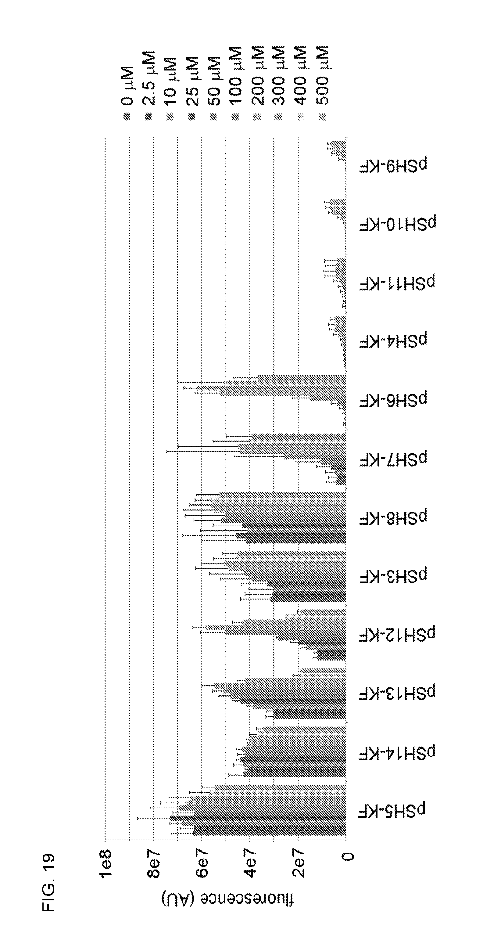
D00017

D00018
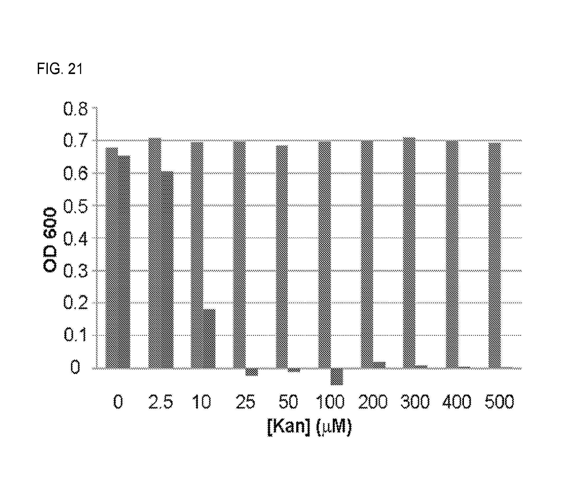
D00019
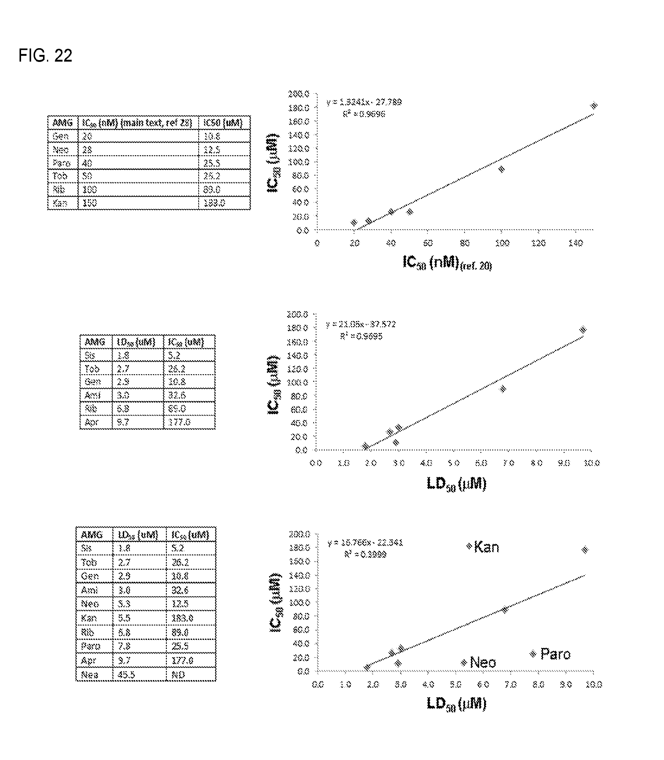
D00020

D00021

S00001
XML
uspto.report is an independent third-party trademark research tool that is not affiliated, endorsed, or sponsored by the United States Patent and Trademark Office (USPTO) or any other governmental organization. The information provided by uspto.report is based on publicly available data at the time of writing and is intended for informational purposes only.
While we strive to provide accurate and up-to-date information, we do not guarantee the accuracy, completeness, reliability, or suitability of the information displayed on this site. The use of this site is at your own risk. Any reliance you place on such information is therefore strictly at your own risk.
All official trademark data, including owner information, should be verified by visiting the official USPTO website at www.uspto.gov. This site is not intended to replace professional legal advice and should not be used as a substitute for consulting with a legal professional who is knowledgeable about trademark law.