System and method for delivering energy to tissue
Thapliyal , et al.
U.S. patent number 10,363,057 [Application Number 12/505,335] was granted by the patent office on 2019-07-30 for system and method for delivering energy to tissue. This patent grant is currently assigned to VytronUS, Inc.. The grantee listed for this patent is James W. Arenson, David A. Gallup, Hira V. Thapliyal. Invention is credited to James W. Arenson, David A. Gallup, Hira V. Thapliyal.


| United States Patent | 10,363,057 |
| Thapliyal , et al. | July 30, 2019 |
System and method for delivering energy to tissue
Abstract
An ablation system for treating atrial fibrillation in a patient including an elongate shaft having proximal and distal ends, a lumen therebetween and a housing adjacent the distal end of the elongate shaft. An energy source is coupled to the housing and is adapted to deliver energy to a target tissue so as to create a zone of ablation in the target tissue that blocks abnormal electrical activity thereby reducing or eliminating the atrial fibrillation in the patient. A sensor is adjacent the energy source and adapted to detect relative position of the energy source to the target tissue or characteristics of the target tissue. The system also has a reflecting element operably coupled with the energy source and adapted to redirect energy emitted from the energy source in a desired direction or pattern.
| Inventors: | Thapliyal; Hira V. (Los Altos, CA), Gallup; David A. (Alameda, CA), Arenson; James W. (Woodside, CA) | ||||||||||
|---|---|---|---|---|---|---|---|---|---|---|---|
| Applicant: |
|
||||||||||
| Assignee: | VytronUS, Inc. (Sunnyvale,
CA) |
||||||||||
| Family ID: | 41530927 | ||||||||||
| Appl. No.: | 12/505,335 | ||||||||||
| Filed: | July 17, 2009 |
Prior Publication Data
| Document Identifier | Publication Date | |
|---|---|---|
| US 20100016762 A1 | Jan 21, 2010 | |
Related U.S. Patent Documents
| Application Number | Filing Date | Patent Number | Issue Date | ||
|---|---|---|---|---|---|
| 61082064 | Jul 18, 2008 | ||||
| Current U.S. Class: | 1/1 |
| Current CPC Class: | A61B 17/320068 (20130101); A61B 18/148 (20130101); A61B 17/2202 (20130101); A61B 2018/00023 (20130101); A61B 2018/00011 (20130101); A61N 7/022 (20130101); A61B 18/18 (20130101); A61B 2018/1807 (20130101); A61B 2017/320069 (20170801); A61B 2018/0262 (20130101); A61B 2017/22051 (20130101); A61B 2017/00106 (20130101); A61B 2018/00577 (20130101) |
| Current International Class: | A61B 17/32 (20060101); A61N 7/02 (20060101); A61B 17/00 (20060101); A61B 17/22 (20060101); A61B 18/14 (20060101); A61B 18/02 (20060101); A61B 18/00 (20060101); A61B 18/18 (20060101) |
References Cited [Referenced By]
U.S. Patent Documents
| 4641649 | February 1987 | Walinsky et al. |
| 4708127 | November 1987 | Abdelghani |
| 4757820 | July 1988 | Itoh |
| 4858613 | August 1989 | Francis et al. |
| 5010886 | April 1991 | Passafaro et al. |
| 5024234 | June 1991 | Leary et al. |
| 5029588 | July 1991 | Yock et al. |
| 5246438 | September 1993 | Langberg |
| 5295484 | March 1994 | Marcus et al. |
| 5314466 | May 1994 | Stern et al. |
| 5405346 | April 1995 | Grundy et al. |
| 5471988 | December 1995 | Fujio et al. |
| 5647367 | July 1997 | Lum et al. |
| 5718241 | February 1998 | Ben-Haim et al. |
| 5735811 | April 1998 | Brisken |
| 6012457 | January 2000 | Lesh |
| 6024740 | February 2000 | Lesh et al. |
| 6050943 | April 2000 | Slayton et al. |
| 6052576 | April 2000 | Lambourg |
| 6064902 | May 2000 | Haissaguerre et al. |
| 6117101 | September 2000 | Diederich et al. |
| 6161543 | December 2000 | Cox et al. |
| 6164283 | December 2000 | Lesh |
| 6237605 | May 2001 | Vaska et al. |
| 6245064 | June 2001 | Lesh et al. |
| 6245095 | June 2001 | Dobak, III et al. |
| 6251129 | June 2001 | Dobak, III et al. |
| 6251130 | June 2001 | Dobak, III et al. |
| 6254599 | July 2001 | Lesh et al. |
| 6261312 | July 2001 | Dobak, III et al. |
| 6277116 | August 2001 | Utely et al. |
| 6305378 | October 2001 | Lesh |
| 6311090 | October 2001 | Knowlton |
| 6311692 | November 2001 | Vaska et al. |
| 6314962 | November 2001 | Vaska et al. |
| 6314963 | November 2001 | Vaska et al. |
| 6379378 | April 2002 | Werneth et al. |
| 6383151 | May 2002 | Diederich et al. |
| 6387089 | May 2002 | Kreindel et al. |
| 6416511 | July 2002 | Lesh et al. |
| 6468296 | October 2002 | Dobak, III et al. |
| 6474340 | November 2002 | Vaska et al. |
| 6475231 | November 2002 | Dobak, III et al. |
| 6478811 | November 2002 | Dobak, III et al. |
| 6478812 | November 2002 | Dobak, III et al. |
| 6484727 | November 2002 | Vaska et al. |
| 6491039 | December 2002 | Dobak, III |
| 6491716 | December 2002 | Dobak, III et al. |
| 6500121 | December 2002 | Slayton et al. |
| 6500174 | December 2002 | Maguire et al. |
| 6502576 | January 2003 | Lesh |
| 6514244 | February 2003 | Pope et al. |
| 6514249 | February 2003 | Maguire et al. |
| 6517536 | February 2003 | Hooven et al. |
| 6529756 | March 2003 | Phan et al. |
| 6533804 | March 2003 | Dobak, III et al. |
| 6540771 | April 2003 | Dobak, III et al. |
| 6542781 | April 2003 | Koblish et al. |
| 6546935 | April 2003 | Hooven |
| 6547788 | April 2003 | Maguire et al. |
| 6551349 | April 2003 | Lasheras et al. |
| 6576001 | June 2003 | Werneth et al. |
| 6585752 | July 2003 | Dobak, III et al. |
| 6592576 | July 2003 | Andrews et al. |
| 6595989 | July 2003 | Schaer |
| 6599288 | July 2003 | Maguire et al. |
| 6602276 | August 2003 | Dobak, III et al. |
| 6605084 | August 2003 | Acker et al. |
| 6607502 | August 2003 | Maguire et al. |
| 6607527 | August 2003 | Ruiz et al. |
| 6613046 | September 2003 | Jenkins et al. |
| 6635054 | October 2003 | Fjield et al. |
| 6645144 | November 2003 | Wen et al. |
| 6645199 | November 2003 | Jenkins et al. |
| 6645202 | November 2003 | Pless et al. |
| 6648908 | November 2003 | Dobak, III et al. |
| 6652515 | November 2003 | Maguire et al. |
| 6652517 | November 2003 | Hall et al. |
| 6666614 | December 2003 | Fechter et al. |
| 6666858 | December 2003 | Lafontaine |
| 6669655 | December 2003 | Acker et al. |
| 6669687 | December 2003 | Saadat |
| 6676688 | January 2004 | Dobak, III et al. |
| 6676689 | January 2004 | Dobak, III et al. |
| 6676690 | January 2004 | Werneth |
| 6685732 | February 2004 | Kramer |
| 6689128 | February 2004 | Sliwa, Jr. et al. |
| 6692488 | February 2004 | Dobak, III et al. |
| 6695873 | February 2004 | Dobak, III et al. |
| 6701931 | March 2004 | Sliwa, Jr. et al. |
| 6702842 | March 2004 | Dobak, III et al. |
| 6711444 | March 2004 | Koblish |
| 6719755 | April 2004 | Sliwa, Jr. et al. |
| 6745080 | June 2004 | Koblish |
| 6752805 | June 2004 | Maguire et al. |
| 6758847 | July 2004 | Maguire |
| 6763722 | July 2004 | Fjield et al. |
| 6780183 | August 2004 | Jimenez, Jr. et al. |
| 6786218 | September 2004 | Dobak, III |
| 6805128 | October 2004 | Pless et al. |
| 6805129 | October 2004 | Pless et al. |
| 6814733 | November 2004 | Schwartz et al. |
| 6840936 | January 2005 | Sliwa, Jr. et al. |
| 6858026 | February 2005 | Sliwa, Jr. et al. |
| 6869431 | March 2005 | Maguire et al. |
| 6872205 | March 2005 | Lesh et al. |
| 6889694 | May 2005 | Hooven |
| 6893438 | May 2005 | Hall et al. |
| 6896673 | May 2005 | Hooven |
| 6899710 | May 2005 | Hooven |
| 6899711 | May 2005 | Stewart et al. |
| 6904303 | June 2005 | Phan et al. |
| 6905494 | June 2005 | Yon et al. |
| 6905498 | June 2005 | Hooven |
| 6905509 | June 2005 | Dobak, III et al. |
| 6908464 | June 2005 | Jenkins et al. |
| 6920883 | July 2005 | Bessette et al. |
| 6923806 | August 2005 | Hooven et al. |
| 6923808 | August 2005 | Taimisto |
| 6929639 | August 2005 | Lafontaine |
| 6932811 | August 2005 | Hooven et al. |
| 6949095 | September 2005 | Vaska et al. |
| 6949097 | September 2005 | Stewart et al. |
| 6953460 | October 2005 | Maguire et al. |
| 6954977 | October 2005 | Maguire et al. |
| 6955173 | October 2005 | Lesh |
| 6964660 | November 2005 | Maguire et al. |
| 6966908 | November 2005 | Maguire et al. |
| 6971394 | December 2005 | Sliwa, Jr. et al. |
| 6974454 | December 2005 | Hooven |
| 6984233 | January 2006 | Hooven |
| 6997925 | February 2006 | Maguire et al. |
| 7001378 | February 2006 | Yon et al. |
| 7001415 | February 2006 | Hooven |
| 7044135 | May 2006 | Lesh |
| 7063682 | June 2006 | Whayne et al. |
| 7142905 | November 2006 | Slayton et al. |
| 7275450 | October 2007 | Hirai et al. |
| 7285116 | October 2007 | de la Rama et al. |
| 7306593 | December 2007 | Keidar et al. |
| 7393325 | July 2008 | Barthe et al. |
| 9033885 | May 2015 | Thapliyal et al. |
| 9220924 | December 2015 | Thapliyal et al. |
| 2001/0025185 | September 2001 | Laufer et al. |
| 2002/0045895 | April 2002 | Sliwa et al. |
| 2002/0065512 | May 2002 | Fjield et al. |
| 2002/0087151 | July 2002 | Mody et al. |
| 2002/0128636 | September 2002 | Chin et al. |
| 2003/0036729 | February 2003 | Jang |
| 2003/0050630 | March 2003 | Mody et al. |
| 2003/0050631 | March 2003 | Mody et al. |
| 2003/0060815 | March 2003 | Lalonde et al. |
| 2003/0125726 | July 2003 | Maguire et al. |
| 2003/0153907 | August 2003 | Suorsa |
| 2003/0163128 | August 2003 | Patil et al. |
| 2003/0176816 | September 2003 | Maguire et al. |
| 2004/0015106 | January 2004 | Coleman |
| 2004/0082948 | April 2004 | Stewart et al. |
| 2004/0176757 | September 2004 | Sinelnikov |
| 2005/0043726 | February 2005 | McHale et al. |
| 2005/0049582 | March 2005 | DeBenedictis et al. |
| 2005/0070961 | March 2005 | Shin et al. |
| 2005/0131468 | June 2005 | Echt et al. |
| 2005/0165388 | July 2005 | Bhola |
| 2005/0256518 | November 2005 | Rama et al. |
| 2006/0122508 | June 2006 | Slayton et al. |
| 2006/0155269 | July 2006 | Warnking |
| 2006/0287650 | December 2006 | Cao et al. |
| 2007/0015998 | January 2007 | Yock et al. |
| 2007/0016072 | January 2007 | Grunwald et al. |
| 2007/0027445 | February 2007 | Gifford et al. |
| 2007/0083168 | April 2007 | Whiting et al. |
| 2007/0265609 | November 2007 | Thapliyal et al. |
| 2007/0265610 | November 2007 | Thapliyal et al. |
| 2007/0299496 | December 2007 | Podmore et al. |
| 2008/0039746 | February 2008 | Hissong et al. |
| 2008/0077200 | March 2008 | Bendett et al. |
| 2008/0161785 | July 2008 | Crowe et al. |
| 10037660 | Feb 2002 | DE | |||
| 2540348 | Jan 2013 | EP | |||
| 2307098 | Mar 2015 | EP | |||
| 2005137916 | Jun 2005 | JP | |||
| 2007152094 | Jun 2007 | JP | |||
| WO 99/02096 | Jan 1999 | WO | |||
| WO-03053259 | Jul 2003 | WO | |||
| WO 2004/110258 | Dec 2004 | WO | |||
| WO 2004/110258 | Aug 2005 | WO | |||
| WO 2005/102199 | Nov 2005 | WO | |||
| WO 2005/117734 | Dec 2005 | WO | |||
| WO 2006/044662 | Apr 2006 | WO | |||
| WO 2006/044662 | Jul 2006 | WO | |||
| WO-2010009472 | Jan 2010 | WO | |||
| WO 2010/120883 | Oct 2010 | WO | |||
| WO 2010/120883 | Mar 2011 | WO | |||
Other References
|
Svilainis, L., and G. Motiej nas. "Power amplifier for ultrasonic transducer excitation." Ultragarsas "Ultrasound" 58.1 (2016): 30-36. cited by examiner . Duty cycle. (n.d.) Mosby's Medical Dictionary, 8th edition. (2009). Retrieved Mar. 16, 2017 from http://medical-dictionary.thefreedictionary.com/duty+cycle. cited by examiner . "A new treatment for atrial fibrillation?" Medical Device & Diagnostic Industry, Feb. 2006, p. 30; retrieved from the Internet: <<http://www.devicelink.com/mddi/archive/06/02/013.html>>, 2 pages total. cited by applicant . Bushberg et al., The Essential Physics of Medical Imaging, 2nd edition, Lippincott Williams & Wilkins 2002, p. 491. cited by applicant . Cox et al. "Current status of the Maze procedure for the treatment of atrial fibrillation," Semin Thorac Cardiovasc Surg. Jan. 2000;12(1):15-9. cited by applicant . Cox et al., "Electrophysiologic basis, surgical development, and clinical results of the maze procedure for atrial flutter and atrial fibrillation," Adv Card Surg. 1995;6:1-67. cited by applicant . Cox et al., "Modification of the maze procedure for atrial flutter and atrial fibrillation. II, Surgical technique of the maze III procedure," J Thorac Cardiovasc Surg. Aug. 1995;110(2):485-95. cited by applicant . Cox et al., "The development of the Maze procedure for the treatment of atrial fibrillation," Semin Thorac Cardiovasc Surg. Jan. 2000;12(1):2-14. cited by applicant . Gentry et al., "Integrated Catheter for 3-D Intracardiac Echocardiography and Ultrasound Ablation," IEEE Transactions on Ultrasonics, Ferroelectrics, and Frequency Control, vol. 51, No. 7, pp. 799-807. cited by applicant . Gill, "How to perform pulmonary vein isolation," Europace, 2004; 6 (2): 83-91; retrieved from the Internet: <<http://europace.oxfordjournals.org/cgi/reprint/6/2/83>>. cited by applicant . Gillinov et al., Atrial fibrillation: current surgical options and their assessment,: Annals of Thoracic Surgery 2002; 74:2210-7; retrieved from the Internet: <<http://ats.ctsnetjournals.org/cgi/reprint/74/6/2210>>. cited by applicant . Haissaguerre et al., "Spontaneous Initiation of Atrial Fibrillation by Ectopic Beats Originating in the Pulmonary Veins," New England J Med., Sep. 3, 1998; 339(10):659-666; retrieved from the Internet: <<http://content.nejm.org/cgi/reprint/339/10/659.pdf>>. cited by applicant . Levinson, "Endocardial Microwave Ablation: A New Surgical Approach for Atrial Fibrillation"; The Heart Surgery Forum, 2006. cited by applicant . Maessen et al., "Beating heart surgical treatment of atrial fibrillation with microwave ablation," Ann Thorac Surg 2002;74:S1307-S1311; retrieved from the Internet: <<http://ats.ctsnetjournals.org/cgi/reprint/74/4/S1307>>. cited by applicant . Nathan et al., "The junction between the left atrium and the pulmonary veins, An anatomic study of human hearts," Circulation 1966; 34:412-422; retrieved from the Internet: <<http://circ.ahajournals.org/cgi/reprint/34/3/412>>. cited by applicant . Sueda et al., "Efficacy of a simple left atrial procedure for chronic atrial fibrillation in mitral valve operations," Ann Thorac Surg 1997; 63:1070-1075. cited by applicant . Sueda et al., "Simple left atrial procedure for chronic atrial fibrillation associated with mitral valve disease," Ann Thorac Surg 1996; 62: 1796-1800. cited by applicant . Ter Haar, "Acoustic Surgery", Physics Today, 2001; 54(12):29-34. cited by applicant . U.S. Appl. No. 12/480,256, filed Jun. 8, 2009; first named inventor: Hira V. Thapliyal. cited by applicant . U.S. Appl. No. 12/483,174, filed Jun. 11, 2009; first named inventor: Hira V. Thapliyal. cited by applicant . U.S. Appl. No. 12/482,640, filed Jun. 11, 2009; first named inventor: Hira V. Thapliyal. cited by applicant . International Search Report and Written Opinion of PCT Application No. PCT/US2009/051164, dated Nov. 17, 2009, 8 pages total. cited by applicant . U.S. Appl. No. 13/630,674, filed Sep. 28, 2012, Thapliyal et al. cited by applicant . European search report and opinion dated Sep. 17, 2012 for EP Application No. 09798856.2. cited by applicant . European search report dated Oct. 30, 2012 for EP Application No. 12186735.2. cited by applicant . Office action dated Sep. 4, 2013 for U.S. Appl. No. 13/630,674. cited by applicant . Office action dated Feb. 28, 2014 for U.S. Appl. No. 13/630,674. cited by applicant . Office action dated Jul. 24, 2014 for U.S. Appl. No. 13/630,674. cited by applicant . Office action dated Dec. 17, 2014 for U.S. Appl. No. 13/630,674. cited by applicant . Office action dated Jul. 23, 2015 for U.S. Appl. No. 13/630,674. cited by applicant . Office action dated Jun. 6, 2016 for U.S. Appl. No. 13/630,674. cited by applicant . Office Action dated Jun. 26, 2017 for U.S. Appl. No. 13/630,674. cited by applicant . Ehrenstein, D. New technique maps the body electric. Science 276. No. 5313 (1997): 681-681. cited by applicant . "Office action dated Jan. 25, 2018 for U.S. Appl. No. 13/630,674." cited by applicant . U.S. Appl. No. 13/630,674 Office Action dated Sep. 9, 2018. cited by applicant. |
Primary Examiner: Pehlke; Carolyn A
Attorney, Agent or Firm: Wilson Sonsini Goodrich & Rosati
Parent Case Text
CROSS-REFERENCES TO RELATED APPLICATIONS
This application is a non-provisional of, and claims the benefit of U.S. Provisional Patent Application No. 61/082,064, filed Jul. 18, 2008, the entire contents of which are incorporated herein by reference.
Claims
What is claimed is:
1. An ablation system for treating atrial fibrillation in a patient, said system comprising: an elongate shaft having a proximal end, a distal end, and a lumen therebetween; a distal tip assembly comprising a housing and an energy source, the housing adjacent the distal end of the elongate shaft and comprising an open window and an exterior surface, and the energy source comprising an ultrasound transducer disposed within the housing, wherein the ultrasound transducer comprises a single ultrasound transducer element; and wherein the ultrasound transducer is configured to operate in two modes, wherein the first mode comprises delivering, from a first face of the ultrasound transducer, a collimated beam of ultrasound energy comprising ablation energy to a target tissue, so as to create a zone of ablation in the target tissue that blocks abnormal electrical activity thereby reducing or eliminating the atrial fibrillation in the patient and wherein the second mode comprises emitting and receiving, to and from the first face of the ultrasound transducer, ultrasound energy comprising sensing energy to and from the target tissue; a processor configured to determine a gap distance between a surface of the target tissue and the energy source based on the sensing energy; and a reflecting element operably coupled with the energy source and adapted to redirect the ablation energy emitted from the ultrasound transducer through the open window of the housing in a direction or pattern, wherein the energy source delivers the ablation energy to the target tissue without contacting the zone of ablation with the elongate shaft or any structure disposed thereon, and wherein the distal tip assembly and the energy source disposed therein are configured to move across the target tissue without contacting the target tissue while the energy source delivers the ablation energy.
2. The system of claim 1, wherein the housing is configured to rotate about its longitudinal axis while the ablation energy or the sensing energy is delivered, the ablation or sensing energy redirected by the reflecting element and through the open window in a circular or elliptical pattern.
3. The system of claim 1, wherein the energy source is recessed from a distal end of the housing such that the energy source does not contact the target tissue in operation.
4. The system of claim 1, wherein the collimated beam of energy has a frequency in the range of 5 to 20 MHz.
5. The system of claim 1, further comprising a generator electrically coupled with the ultrasound transducer, the generator providing an excitation voltage of 5 to 300 volts peak-to-peak to the ultrasound transducer.
6. The system of claim 5, wherein the generator provides the excitation voltage with a duty cycle ranging from 0% to 100%.
7. The system of claim 1, wherein the first face comprises a flat face.
8. The system of claim 1, wherein the first face comprises a concave or a convex face, the first face adapted to act as a lens for focusing the ablation or sensing energy delivered by the energy source.
9. The system of claim 1, wherein the processor is adapted to determine characteristics of the target tissue based on the sensing energy.
10. The system of claim 9, wherein the target tissue characteristics comprise thickness of the target tissue or thickness of the ablation zone.
11. The system of claim 1, wherein the reflecting element is non expandable.
12. The system of claim 1, wherein the reflecting element comprises an angled outer surface adapted to redirect the ablation or sensing energy through the open window.
13. The system of claim 12, wherein the angle of the outer surface ranges from 30 to 60 degrees relative to a longitudinal axis of the housing.
14. The system of claim 12, wherein the angled outer surface comprises a flat surface.
15. The system of claim 1, wherein the reflecting element comprises a curved outer surface adapted to redirect the ablation or sensing energy through the open window.
16. The system of claim 1, wherein the reflecting element redirects the ablation or sensing energy through the open window in a sidewall of the housing.
17. The system of claim 1, wherein the reflecting element comprises a gas filled region adapted to reflect the ablation or sensing energy.
18. The system of claim 1, wherein the reflecting element is movable relative to the energy source so that the ablation or sensing energy exits the open window of the housing at varying angles.
19. The system of claim 1, wherein the reflecting element is rotatable relative to the energy source thereby allowing the ablation or sensing energy to be reflected outward away from the housing in a circular pattern.
20. The system of claim 1, wherein the reflecting element is adapted to redirect the ablation or sensing energy from the energy source through the open window to form a ring shaped beam of energy.
21. The system of claim 20, wherein the reflecting element comprises a bowl-shaped reflector centered around a longitudinal axis of the housing.
22. The system of claim 1, wherein the reflecting element comprises a liquid-gas interface.
23. The system of claim 1, wherein the reflecting element comprises two reflecting portions each having a different shape or angle relative to the energy source so that the ablation or sensing energy is redirected through the open window in two or more directions or patterns.
24. The system of claim 23, wherein the ablation or sensing energy is redirected through the open window in a first pattern comprising a collimated beam and the ablation or sensing energy is redirected in a second pattern comprising a focused beam.
25. The system of claim 1, wherein the processor is adapted to control the ablation energy provided by the energy source based on the sensing energy.
26. The system of claim 1, wherein the target tissue comprises left atrial tissue.
27. The system of claim 1, wherein the target tissue comprises a pulmonary vein or tissue adjacent thereto.
28. The system of claim 1, wherein the zone of ablation comprises a linear ablation path.
29. The system of claim 1, wherein the zone of ablation comprises an arcuate ablation path.
30. The system of claim 1, wherein the zone of ablation comprises a transmural ablation zone.
31. The system of claim 1, further comprising a lens adjacent the energy source, the lens adapted to adjust beam pattern of the ablation or sensing energy emitted from the energy source.
32. The ablation system of claim 1, further comprising a fluid that flows past the energy source to prevent blood from coagulating thereon.
33. The system of claim 32, wherein the fluid flows at a rate of 1 ml/minute.
34. The system of claim 33, wherein the fluid flow may be increased or decreased.
35. The system of claim 32, further comprising modifying a flow of the fluid based on the sensing energy.
36. The system of claim 1, wherein the processor is configured to modify the collimated beam of ultrasound energy based on the sensing energy to thereby facilitate continuous ablation of the target tissue.
37. A method for treating atrial fibrillation by ablating a target tissue in a patient, said method comprising: providing an ablation system comprising a processor and an elongate shaft having a distal tip assembly, the distal tip assembly comprising an energy source, a reflecting element, and a housing comprising an open window and an exterior surface, wherein the energy source comprises an ultrasound transducer disposed within the housing, wherein the ultrasound transducer comprises a single ultrasound transducer element wherein the ultrasound transducer is configured to operate in two modes, wherein the first mode comprises delivering, from a first face, a collimated beam of ultrasound energy comprising ablation energy to a target tissue, and wherein the second mode comprises emitting and receiving, to and from the first face, ultrasound energy comprising sensing energy to and from the target tissue; advancing the distal tip assembly adjacent the target tissue; delivering the ablation energy from the ultrasound transducer to the target tissue in the first mode by reflecting the ablation energy emitted from the ultrasound transducer off of the reflecting element and through the open window, so as to redirect the ablation energy in a direction or pattern; moving the distal tip assembly and the energy source disposed therein across the target tissue without contacting the target tissue while the energy source delivers the ablation energy; determining a gap distance between the energy source and a surface of the target tissue with the processor based on the sensing energy while the housing does not contact the target tissue; and creating a partial or complete zone of ablation in the tissue thereby blocking abnormal electrical activity and reducing or eliminating the atrial fibrillation, wherein the energy source delivers the ablation energy to the target tissue without contacting the zone of ablation with the elongate shaft or any structure disposed thereon.
38. The method of claim 37, wherein the step of advancing comprises passing the distal tip assembly through an atrial septal wall.
39. The method of claim 37, wherein the collimated beam of energy comprises a frequency in the range of 5 to 20 MHz.
40. The method of claim 37, wherein the step of delivering the ablation energy comprises providing an excitation voltage to the ultrasound transducer, the excitation voltage ranging from 5 to 300 volts peak-to-peak.
41. The method of claim 37, wherein the step of reflecting the ablation energy comprises redirecting the ablation energy so that it exits through the open window in a sidewall of the housing.
42. The method of claim 37, wherein the reflecting element is non-expandable.
43. The method of claim 37, wherein the reflecting element comprises an expandable member and the step of reflecting the ablation energy comprises expanding the expandable member.
44. The method of claim 43, wherein the expandable member comprises a balloon.
45. The method of claim 37, wherein the step of reflecting the ablation energy comprises moving the reflecting element relative to the housing.
46. The method of claim 45, wherein the moving comprises rotating the reflecting element.
47. The method of claim 37, wherein the reflecting element comprises a first reflecting portion and a second reflecting portion, wherein the ablation energy reflected off the first portion is redirected in a first direction through the open window, and wherein the ablation energy reflected off the second portion is redirected in a second direction different than the first direction.
48. The method of claim 37, wherein the zone of ablation comprises a linear zone of ablation.
49. The method of claim 37, wherein the zone of ablation comprises an arcuate zone of ablation.
50. The method of claim 37, wherein the step of creating the zone of ablation comprises encircling the zone of ablation around a pulmonary vein or left atrial tissue.
51. The method of claim 37, wherein the zone of ablation comprises a transmural lesion.
52. The method of claim 37, further comprising cooling the energy source with a cooling fluid.
53. The method of claim 37, wherein the method further comprises controlling the ablation energy delivery based on the sensing energy.
54. The method of claim 37, further comprising sensing characteristics of the tissue with the ultrasound transducer in the second mode.
55. The method of claim 54, wherein the characteristics comprise thickness of the tissue or ablation zone thickness.
56. The method of claim 37, further comprising switching modes between the first mode and the second mode.
57. The method of claim 37, further comprising flowing a fluid past the energy source to prevent blood from coagulating thereon.
58. The method of claim 57, wherein the fluid flows at a rate of 1 ml/minute.
59. The method of claim 58, wherein the fluid flow may be increased or decreased.
60. The method of claim 57, further comprising controlling a flow of the fluid based on the sensing energy to cool the target tissue.
61. The method of claim 37, wherein the method further comprises: modifying the collimated beam of ultrasound energy with the processor based on the sensing energy, thereby continuously creating the partial or complete zone of ablation while moving the distal tip assembly.
62. An ablation system for treating atrial fibrillation in a patient, said system comprising: an elongate shaft having a proximal end, a distal end, and a lumen therebetween; a distal tip assembly comprising a housing and an energy source, the housing adjacent the distal end of the elongate shaft and the energy source comprising an ultrasound transducer disposed within the housing, wherein the ultrasound transducer comprises a single ultrasound transducer element; and wherein the ultrasound transducer is configured to operate in two modes, wherein the first mode comprises delivering, from a first face of the ultrasound transducer, a collimated beam of ultrasound energy comprising ablation energy to a target tissue, so as to create a zone of ablation in the target tissue that blocks abnormal electrical activity thereby reducing or eliminating the atrial fibrillation in the patient and wherein the second mode comprises emitting and receiving, to and from the first face of the ultrasound transducer, ultrasound energy comprising sensing energy to and from the target tissue; and wherein the distal tip assembly and the energy source disposed therein are configured to move across the target tissue without contacting the target tissue while the energy source delivers the ablation energy; a processor configured to determine a gap distance between a surface of the target tissue and the energy source based on the sensing energy; and a reflecting element operably coupled with the energy source and adapted to redirect the ablation energy emitted from energy source in a direction or pattern, wherein the energy source delivers the ablation energy through blood to the target tissue without contacting the target tissue with the elongate shaft or any structure disposed thereon.
63. A method for treating atrial fibrillation by ablating a target tissue in a patient, said method comprising: providing an ablation system operably coupled to a processor and comprising an elongate shaft having a distal tip assembly, the distal tip assembly comprising an energy source disposed therein and a reflecting element wherein the energy source comprises a single ultrasound transducer element is configured to operate in two modes, wherein the first mode comprises delivering, from a first face, ablation energy to a target tissue and wherein the second mode comprises emitting and receiving, to and from the first face, sensing energy to and from the target tissue; advancing the distal tip assembly adjacent the target tissue; delivering the ablation energy from the energy source to the target tissue in the first mode, by reflecting the ablation energy emitted from the energy source off of the reflecting element so as to redirect the ablation energy in a direction or pattern; moving the distal tip assembly and the energy source disposed therein across the target tissue without contacting the target tissue while the energy source delivers the ablation energy; determining a gap distance between the energy source and a surface of the target tissue with the processor based on the sensing energy while the distal tip assembly does not contact the target tissue; and creating a partial or complete zone of ablation in the target tissue thereby blocking abnormal electrical activity and reducing or eliminating the atrial fibrillation, wherein the energy source delivers the ablation energy through blood to the target tissue without contacting the target tissue with the elongate shaft or any structure disposed thereon.
Description
BACKGROUND OF THE INVENTION
Field of the Invention
The present invention relates generally to medical devices, systems and methods, and more specifically to improved devices, systems and methods for creating an ablation zone in tissue. The device may be used to treat atrial fibrillation.
The condition of atrial fibrillation (AF) is characterized by the abnormal (usually very rapid) beating of the left atrium of the heart which is out of synch with the normal synchronous movement ("normal sinus rhythm") of the heart muscle. In normal sinus rhythm, the electrical impulses originate in the sino-atrial node ("SA node") which resides in the right atrium. The abnormal beating of the atrial heart muscle is known as fibrillation and is caused by electrical impulses originating instead in the pulmonary veins ("PV") [Haissaguerre, M. et al., Spontaneous Initiation of Atrial Fibrillation by Ectopic Beats Originating in the Pulmonary Veins, New England J Med., Vol. 339:659-666].
There are pharmacological treatments for this condition with varying degrees of success. In addition, there are surgical interventions aimed at removing the aberrant electrical pathways from the PV to the left atrium ("LA") such as the Cox-Maze III Procedure [J. L. Cox et al., The development of the Maze procedure for the treatment of atrial fibrillation, Seminars in Thoracic & Cardiovascular Surgery, 2000; 12: 2-14; J. L. Cox et al., Electrophysiologic basis, surgical development, and clinical results of the maze procedure for atrial flutter and atrial fibrillation, Advances in Cardiac Surgery, 1995; 6: 1-67; and J. L. Cox et al., Modification of the maze procedure for atrial flutter and atrial fibrillation. II, Surgical technique of the maze III procedure, Journal of Thoracic & Cardiovascular Surgery, 1995; 2110:485-95]. This procedure is shown to be 99% effective [J. L. Cox, N. Ad, T. Palazzo, et al. Current status of the Maze procedure for the treatment of atrial fibrillation, Seminars in Thoracic & Cardiovascular Surgery, 2000; 12: 15-19] but requires special surgical skills and is time consuming.
There has been considerable effort to copy the Cox-Maze procedure for a less invasive percutaneous catheter-based approach. Less invasive treatments have been developed which involve use of some form of energy to ablate (or kill) the tissue surrounding the aberrant focal point where the abnormal signals originate in the PV. The most common methodology is the use of radio-frequency ("RF") electrical energy to heat the muscle tissue and thereby ablate it. The aberrant electrical impulses are then prevented from traveling from the PV to the atrium (achieving conduction block within the heart tissue) and thus avoiding the fibrillation of the atrial muscle. Other energy sources, such as microwave, laser, and ultrasound have been utilized to achieve the conduction block. In addition, techniques such as cryoablation, administration of ethanol, and the like have also been used.
There has been considerable effort in developing catheter based systems for the treatment of AF using radiofrequency (RF) energy. One such method is described in U.S. Pat. No. 6,064,902 to Haissaguerre et al. In this approach, a catheter is made of distal and proximal electrodes at the tip. The catheter can be bent in a J shape and positioned inside a pulmonary vein. The tissue of the inner wall of the pulmonary vein (PV) is ablated in an attempt to kill the source of the aberrant heart activity. Other RF based catheters are described in U.S. Pat. No. 6,814,733 to Schwartz et al., U.S. Pat. No. 6,996,908 to Maguire et al., U.S. Pat. No. 6,955,173 to Lesh, and U.S. Pat. No. 6,949,097 to Stewart et al.
Another source used in ablation is microwave energy. One such device is described by Dr. Mark Levinson [(Endocardial Microwave Ablation: A New Surgical Approach for Atrial Fibrillation; The Heart Surgery Forum, 2006] and Maessen et al. [Beating heart surgical treatment of atrial fibrillation with microwave ablation. Ann Thorac Surg 74: 1160-8, 2002]. This intraoperative device consists of a probe with a malleable antenna which has the ability to ablate the atrial tissue. Other microwave based catheters are described in U.S. Pat. No. 4,641,649 to Walinsky; U.S. Pat. No. 5,246,438 to Langberg; U.S. Pat. No. 5,405,346 to Grundy et al.; and U.S. Pat. No. 5,314,466 to Stem et al.
Another catheter based method utilizes the cryogenic technique where the tissue of the atrium is frozen below a temperature of -60 degrees C. This results in killing of the tissue in the vicinity of the PV thereby eliminating the pathway for the aberrant signals causing the AF [A. M. Gillinov, E. H. Blackstone and P. M. McCarthy, Atrial fibrillation: current surgical options and their assessment, Annals of Thoracic Surgery 2002; 74:2210-7]. Cryo-based techniques have been a part of the partial Maze procedures [Sueda T., Nagata H., Orihashi K. et al., Efficacy of a simple left atrial procedure for chronic atrial fibrillation in mitral valve operations, Ann Thorac Surg 1997; 63:1070-1075; and Sueda T., Nagata H., Shikata H. et al.; Simple left atrial procedure for chronic atrial fibrillation associated with mitral valve disease, Ann Thorac Surg 1996; 62: 1796-1800]. More recently, Dr. Cox and his group [Nathan H., Eliakim M., The junction between the left atrium and the pulmonary veins, An anatomic study of human hearts, Circulation 1966; 34:412-422, and Cox J. L., Schuessler R. B., Boineau J. P., The development of the Maze procedure for the treatment of atrial fibrillation, Semin Thorac Cardiovasc Surg 2000; 12:2-14] have used cryoprobes (cryo-Maze) to duplicate the essentials of the Cox-Maze III procedure. Other cryo-based devices are described in U.S. Pat. Nos. 6,929,639 and 6,666,858 to Lafintaine and U.S. Pat. No. 6,161,543 to Cox et al.
More recent approaches for the AF treatment involve the use of ultrasound energy. The target tissue of the region surrounding the pulmonary vein is heated with ultrasound energy emitted by one or more ultrasound transducers. One such approach is described by Lesh et al. in U.S. Pat. No. 6,502,576. Here the catheter distal tip portion is equipped with a balloon which contains an ultrasound element. The balloon serves as an anchoring means to secure the tip of the catheter in the pulmonary vein. The balloon portion of the catheter is positioned in the selected pulmonary vein and the balloon is inflated with a fluid which is transparent to ultrasound energy. The transducer emits the ultrasound energy which travels to the target tissue in or near the pulmonary vein and ablates it. The intended therapy is to destroy the electrical conduction path around a pulmonary vein and thereby restore the normal sinus rhythm. The therapy involves the creation of a multiplicity of lesions around individual pulmonary veins as required. The inventors describe various configurations for the energy emitter and the anchoring mechanisms.
Yet another catheter device using ultrasound energy is described by Gentry et al. [Integrated Catheter for 3-D Intracardiac Echocardiography and Ultrasound Ablation, IEEE Transactions on Ultrasonics, Ferroelectrics, and Frequency Control, Vol. 51, No. 7, pp 799-807]. Here the catheter tip is made of an array of ultrasound elements in a grid pattern for the purpose of creating a three dimensional image of the target tissue. An ablating ultrasound transducer is provided which is in the shape of a ring which encircles the imaging grid. The ablating transducer emits a ring of ultrasound energy at 10 MHz frequency. In a separate publication [Medical Device Link, Medical Device and Diagnostic Industry, February 2006], in the description of the device, the authors assert that the pulmonary veins can be imaged.
While these devices and methods are promising, improved devices and methods for creating a heated zone of tissue, such as an ablation zone are needed. Furthermore, it would also be desirable if such devices could create single or multiple ablation zones to block abnormal electrical activity in the heart in order to lessen or prevent atrial fibrillation. It would also be desirable if such devices could be used in the presence of blood or other body tissues without coagulating or clogging up the ultrasound transducer. Such devices and methods should be easy to use, minimally invasive, cost effective and simple to manufacture.
Description of Background Art
Other devices based on ultrasound energy to create circumferential lesions are described in U.S. Pat. Nos. 6,997,925; 6,966,908; 6,964,660; 6,954,977; 6,953,460; 6,652,515; 6,547,788; and 6,514,249 to Maguire et al.; U.S. Pat. Nos. 6,955,173; 6,052,576; 6,305,378; 6,164,283; and 6,012,457 to Lesh; U.S. Pat. Nos. 6,872,205; 6,416,511; 6,254,599; 6,245,064; and 6,024,740; to Lesh et al.; U.S. Pat. Nos. 6,383,151; 6,117,101; and WO 99/02096 to Diederich et al.; U.S. Pat. No. 6,635,054 to Fjield et al.; U.S. Pat. No. 6,780,183 to Jimenez et al.; U.S. Pat. No. 6,605,084 to Acker et al.; U.S. Pat. No. 5,295,484 to Marcus et al.; and WO 2005/117734 to Wong et al.
BRIEF SUMMARY OF THE INVENTION
The present invention relates generally to medical devices and methods, and more specifically to medical devices and methods used to deliver energy to tissue as a treatment for atrial fibrillation and other medical conditions.
In a first aspect of the present invention, an ablation system for treating atrial fibrillation in a patient comprises an elongate shaft having a proximal end, a distal end, and lumens therebetween. A housing is adjacent the distal end of the elongate shaft and an energy source is coupled to the housing. The energy source is adapted to deliver energy to a target tissue so as to create a zone of ablation in the target tissue that blocks abnormal electrical activity thereby reducing or eliminating the atrial fibrillation in the patient. The system also has a reflecting element operably coupled with the energy source and adapted to redirect energy emitted from the energy source in a desired direction or pattern.
The housing may be rotatable about its longitudinal axis and the energy may be redirected by the reflecting element in a generally circular pattern. The energy source may be recessed from a distal end of the housing such that the energy source does not contact the target tissue in operation.
The energy source may comprise an ultrasound transducer that is adapted to emit a beam of ultrasound energy. The beam may have a frequency in the range of 5 to 20 MHz and a generator may be electrically coupled with the ultrasound transducer. The generator may provide an excitation voltage of 5 to 300 volts peak-to-peak to the ultrasound transducer. The excitation voltage may have a duty cycle ranging from 0% to 100%, and may have a repetition frequency of about 40 KHz. The energy source may be adapted to deliver one of radiofrequency energy, microwaves, photonic energy, thermal energy, and cryogenic energy. The energy source may comprise a flat face, a concave face or a convex face. The face may be adapted to act as a lens for focusing the energy delivered by the energy source.
The system may comprise a sensor adjacent the energy source and that is adapted to detect relative position of the energy source to the target tissue or characteristics of the target tissue. The sensor may be adapted to detect a gap between a surface of the target tissue and the energy source. The sensor may be adapted to determine characteristics of the target tissue such as tissue thickness or ablation zone depth. The sensor may comprise an ultrasound transducer. The energy source may comprise the same ultrasound transducer as the sensor. Other sensors may comprise an infrared sensor or strain gage.
The reflecting element may be non-expandable. It may redirect the energy in a collimated beam through a portion of the housing or it may redirect the energy in a focused beam that converges toward a focal point or a focal ring, or it may alter the focus of the beam to provide a more uniformly collimated beam. The reflecting element may comprise an angled outer surface that is adapted to redirect the energy. The angle of the outer surface may range from 30 to 60 degrees relative to a longitudinal axis of the housing. The angled face may comprise a flat surface. The reflecting element may comprise a curved outer surface that redirects the energy. The reflecting element may redirect the energy through a sidewall of the housing. The reflecting element may be movable relative to the energy source so that the energy exits the housing at varying angles or so that the energy is reflected outward away from the housing in a circular pattern. The reflecting element may be adapted to redirect the energy from the energy source to form a ring shaped beam of energy. The reflecting element may comprise a liquid-gas interface or a bowl-shaped reflector that is centered around a longitudinal axis of the housing. The liquid-gas interface may comprise a plurality of expandable reflectors positioned adjacent one another. The reflecting element may comprise two reflecting portions each having a different shape or angle relative to the energy source so that the energy is redirected in two or more directions or patterns. The energy may be redirected in a first pattern comprising a collimated beam and the energy may be redirected in a second pattern comprising a focused beam.
The system may further comprise a processor that is adapted to control the energy provided by the energy source based on information received from the sensor. The system may have a lens adjacent the energy source and that is adapted to adjust beam pattern of the energy emitted from the energy source.
The target tissue may comprise left atrial tissue, a pulmonary vein or tissue adjacent thereto. The zone of ablation may comprise a linear ablation path or an arcuate ablation path. The zone of ablation may comprise a transmural ablation zone.
In another aspect of the present invention, a method for treating atrial fibrillation by ablating tissue in a patient comprises providing an ablation system comprising an elongate shaft having a distal tip assembly. The distal tip assembly comprises an energy source and a reflecting element. The distal tip assembly is advanced adjacent the tissue and energy is delivered from the energy source to the tissue. Energy from the energy source is reflected off of the reflecting element so as to redirect the energy emitted from the energy source in a desired direction or pattern. A partial or complete zone of ablation is created in the tissue, thereby blocking abnormal electrical activity and reducing or eliminating the atrial fibrillation.
The step of advancing the distal tip assembly may comprise passing the distal tip through an atrial septal wall. The energy source may comprise an ultrasound transducer and the step of delivering the energy may comprise delivering a beam of ultrasound. The beam may comprise a frequency in the range of 5 to 20 MHz. Delivering the energy may comprise providing an excitation voltage ranging from 5 to 300 volts peak-to-peak to the ultrasound transducer. Delivering energy may comprise delivering one of radiofrequency energy, microwaves, photonic energy, thermal energy and cryogenic energy.
The step of reflecting the energy may comprise redirecting the energy in a collimated beam of energy or focusing the energy so that it converges toward a focal point or a focal ring. Reflecting the energy may comprise redirecting the energy so that it exits a sidewall of the housing. The reflecting element may be non-expandable or it may comprise an expandable member such as a balloon or collapsible nested reflectors (e.g. similar to a collapsible parabolic dish used in satellite communications), and the step of reflecting the energy may comprise expanding the expandable member. Reflecting the energy may comprise moving the reflecting element relative to the housing such as by rotating it. The reflecting element may comprise a first reflecting portion and a second reflecting portion. The energy reflected off the first portion may be redirected in a first direction, and the energy reflected off the second portion may be redirected in a second direction different than the first direction.
The zone of ablation may comprise a linear or arcuate zone of ablation. The step of creating the zone of ablation may comprise encircling the zone of ablation around a pulmonary vein or left atrial tissue. The zone of ablation may comprise a transmural lesion.
The method may further comprise cooling the energy source with a cooling fluid. The system may further comprise a sensor that is adapted to sense relative position of the energy source to the target tissue or characteristics of the target tissue. The ablation system may further comprise a processor, and the method may further comprise controlling energy delivery based on information from the sensor.
The method may comprise sensing a gap distance between the energy source and a surface of the tissue with the sensor. The method may also include sensing characteristics of the tissue such as tissue thickness or depth of the ablation zone, with the sensor. The sensor may comprise an ultrasound sensor and the energy source may also comprise the same ultrasound transducer. The method may include switching modes between delivering energy with the ultrasound transducer and sensing tissue characteristics with the ultrasound transducer sensor.
These and other embodiments are described in further detail in the following description related to the appended drawing figures.
BRIEF DESCRIPTION OF THE DRAWINGS
FIGS. 1 and 2 are drawings of the system of the preferred embodiments of the invention;
FIGS. 3A-3B illustrate the reflecting surface of the system and energy beam of the preferred embodiments of the invention; and
FIG. 4-5 are drawings of the energy beam and the zone of ablation of the preferred embodiment of the invention.
DETAILED DESCRIPTION OF THE INVENTION
The following description of preferred embodiments of the invention is not intended to limit the invention to these embodiments, but rather to enable any person skilled in the art to make and use this invention.
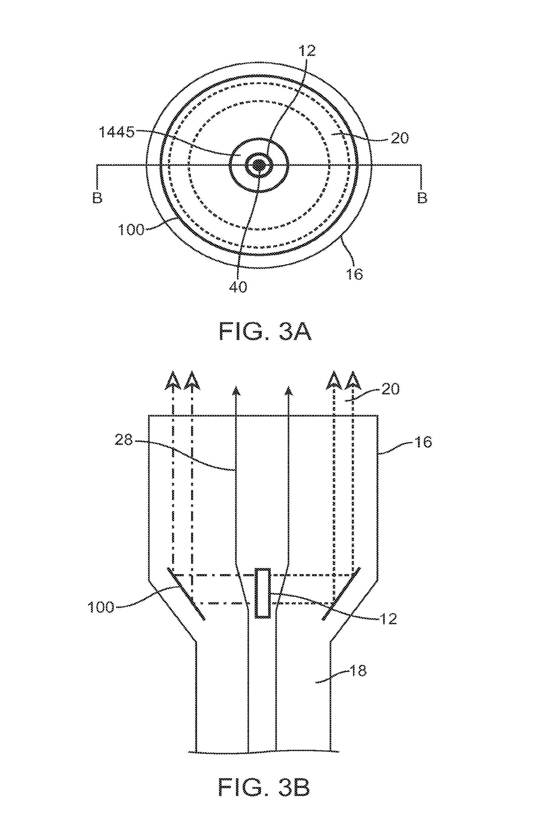
As shown in FIG. 1, the energy delivery system 10 of the preferred embodiments includes an energy source 12 that functions to provide a source of ablation energy; a reflecting surface 100 that functions to redirect the ablation energy from the energy source 12; a sensor; and a processor (not shown), coupled to the sensor and to the energy source 12, which may controls the energy source 12 based on the information from the sensor. The energy delivery system 10 is preferably designed for delivering energy to tissue, more specifically, for delivering ablation energy to tissue, such as heart tissue, to create a conduction block--isolation and/or block of conduction pathways of abnormal electrical activity, which typically originate from the pulmonary veins in the left atrium--for treatment of atrial fibrillation in a patient. The system 10, however, may be alternatively used with any suitable tissue in any suitable environment and for any suitable reason.
The Energy Source.
As shown in FIG. 1, the energy source 12 of the preferred embodiments functions to provide a source of ablation energy. The ablation energy is preferably in the form of an energy beam 20 emitted from the energy source 12. The energy source 12 is preferably an ultrasound transducer that emits an ultrasound beam, but may alternatively be any suitable energy source that functions to provide a source of ablation energy. Some suitable sources of ablation energy may include radio frequency (RF) energy, microwaves, photonic energy, and thermal energy. The therapy could alternatively be achieved using cooled fluids (e.g., cryogenic fluid). The energy delivery system 10 preferably includes a single energy source 12, but may alternatively include any suitable number of energy sources 12. For example, the system 10 may include multiple energy sources configured in a ring, such that in combination they emit an annular shaped energy beam 20.
The energy source 12 is preferably an ultrasound transducer that is preferably made of a piezoelectric material such as PZT (lead zirconate titanate) or PVDF (polyvinylidine difluoride), or any other suitable ultrasound beam emitting material. The transducer may further include coating layers such as a thin layer of a metal. Some suitable transducer coating metals may include gold, stainless steel, nickel-cadmium, silver, plastic, metal-filled graphite, a metal alloy, and any other suitable material that functions to increase the efficiency of coupling of the energy beam 20 into the surrounding fluid 28 or performs any other suitable functions. The transducer is preferably a cylindrical transducer, as shown in FIGS. 3A and 3B, such that it preferably emits energy beam 20 from the outer face of the cylinder (e.g. radially out from the face of the energy source). The energy beam 20 is preferably emitted radially 360 degrees around the energy source 12, but may alternatively be emitted from any suitable portions) of the energy source. The transducer may alternatively be a generally flat transducer, such as a disc, as shown in FIGS. 1 and 2. The disc transducer preferably emits energy beam 20 from at least one of the faces of the disc. The faces of the disc that emit the energy beam 20 are preferably flat, but may alternatively be either concave or convex to achieve an effect of a lens. The disc transducer preferably has a circular geometry, but may alternatively be elliptical, polygonal, doughnut shaped, or any other suitable shape.
As shown in FIG. 1, the energy source 12 of the preferred embodiments is preferably coupled to at least one electrical attachment 14. The electrical attachment 14 of the preferred embodiments functions to energize the energy source 12 such that it emits an energy beam 20. The energy delivery system 10 preferably includes two electrical attachments 14 and 14', but may alternatively include any suitable number of electrical attachments to energize the energy source 12. The energy delivery system 10 of the preferred embodiments also includes an electrical generator (not shown) that functions to provide power to the energy source 12 via the electrical attachment(s) 14. When energized by the generator the energy source 12 emits an energy beam 20. The generator provides the appropriate frequency and voltage to the energy source 12 to create the desired energy beam 20. In the case of an ultrasound energy source 12, the ultrasound frequency is preferably in the range of 1 to 25 MHz and more preferably in the range of 5 to 20 MHz. The energy of the energy beam 20 is determined by the excitation voltage applied to the energy source 12. The voltage is preferably in the range of 5 to 300 volts peak-to-peak. In addition, a variable duty cycle is preferably used to control the average power delivered to the energy source 12. The duty cycle preferably ranges from 0% to 100%, with a repetition frequency of approximately 40 kHz, which is preferably faster than the time constant of thermal conduction in the tissue.
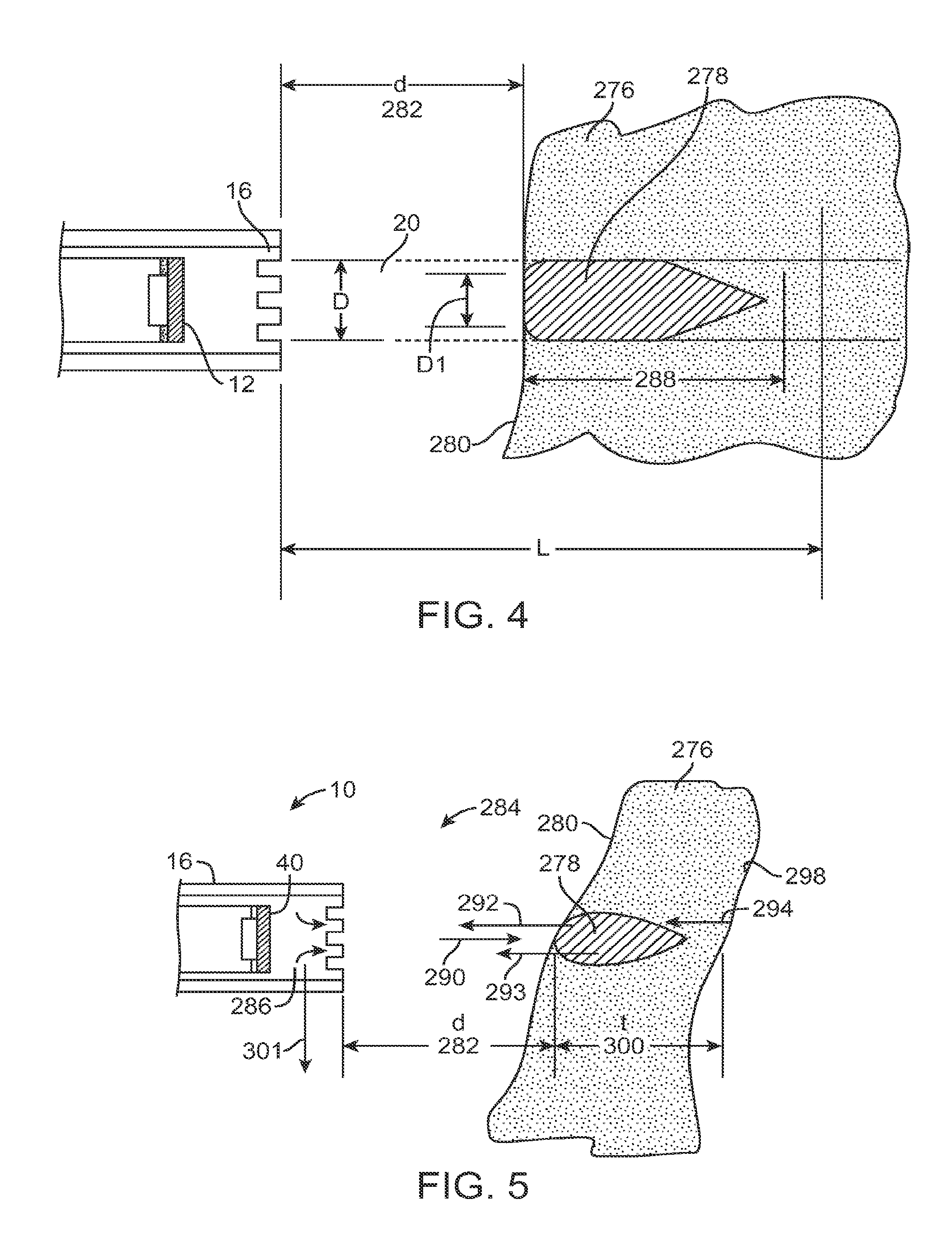
When energized with an electrical pulse or pulse train by the electrical attachment 14 and/or 14', the energy source 12 emits an energy beam 20 (such as a sound wave). The properties of the energy beam 20 are determined by the characteristics of the energy source 12, the matching layer, the backing (described below), and the electrical pulse from electrical attachment 14. These elements determine the frequency, bandwidth, and amplitude of the energy beam 20 (such as a sound wave) propagated into the tissue. As shown in FIG. 4, the energy source 12 emits energy beam 20 such that it interacts with tissue 276 and forms a lesion (zone of ablation 278). The energy beam 20 is preferably an ultrasound beam. The tissue 276 is preferably presented to the energy beam 20 within the collimated length L. The front surface 280 of the tissue 276 is at a distance d (282) away from the face of a housing 16. As the energy beam 20 travels through the tissue 276, its energy is absorbed by the tissue 276 and converted to thermal energy. This thermal energy heats the tissue to temperatures higher than the surrounding tissue resulting in a heated zone 278. In the zone 278 where the tissue is heated, the tissue cells are preferably rendered dead due to heat. The temperatures of the tissue are preferably above the temperature where cell death occurs in the heated zone 278 and therefore, the tissue is said to be ablated. Hence, the zone 278 is preferably referenced as the ablation zone or lesion.
The shape of the lesion or ablation zone 278 formed by the energy beam 20 depends on the characteristics of suitable combination factors such as the energy beam 20, the energy source 12 (including the material, the geometry, the portions of the energy source 12 that are energized and/or not energized, etc.), the matching layer, the backing, the electrical pulse from electrical attachment 14 (including the frequency, the voltage, the duty cycle, the length of the pulse, etc.), and the characteristics of target tissue that the beam 20 contacts and the length of contact or dwell time. These characteristics can be changed based on the information detected by the sensor (as described below), thereby changing the physical characteristics of the lesion.
The housing 16 also functions to provide a barrier between the face of the energy source 12 and blood or tissue. When fluid flow is incorporated, the fluid may flow past the energy source thereby preventing blood from coagulating thereon. In preferred embodiments, the coolant flows past the energy source at approximately 1 ml/minute, but may be increased or decreased as desired. Additionally, since the energy source is disposed in the housing, the energy source will not directly contact tissue, thereby also preventing coagulation on the energy source.
Additional details on the energy source, energy source configurations, the housing and adjacent components are disclosed in U.S. patent application Ser. No. 12/480,256 and Ser. No. 12/482,640, the entire contents of which are incorporated herein by reference.
The Reflecting Surface.
As shown in FIG. 1, the reflector 100 of the preferred embodiments functions to redirect the energy beam 20 from the energy source 12. The reflecting surface 100 preferably redirects the energy beam 20 from the energy source 12 out of housing 16 and preferably towards the target tissue. The reflecting surface 100 preferably redirects the energy beam 20 such that it is a collimated beam exiting the housing 16 (as shown in FIGS. 2 and 3), the reflecting surface 22 may alternatively redirect the energy beam 20 such that it is a focused beam that preferably converges towards a substantially single focal point or towards focal point ring. The reflecting surface is preferably one of several variations. In a first variation, as shown in FIG. 1, the reflecting surface 100 is an angled reflector device. The reflector device is preferably a cylindrical reflector with a face of the reflector at an angle to the longitudinal axis of the housing 16. The energy source 12 is preferably positioned towards the distal end of the housing 16 with the front face pointing towards the reflector device, which is preferably positioned along the same axis (the longitudinal axis of the housing 16) as the energy source 12. The energy beam 20 from the energy source 12 is preferably redirected from the reflecting surface such that it exits the housing 16 through a side portion of the housing. The reflector device is preferably made from a material that reflects the energy beam 20, such as metal, but may alternatively be a gas filled reflector device such as a gas filled balloon. The angled face of the reflector is preferably flat, but may alternatively be a non-planar face, such as a curved, convex or concave surface. The angle of the reflector preferably ranges between substantially 0 degrees, more preferably substantially 30-60 degrees, and most preferably substantially 45 degrees. The reflector device is preferably set at a fixed angle with respect to the energy source 12, but may alternatively be movable, such as rotated or pivoted, to alter the angle that the energy beam 20 will exit the housing 16. Referring to FIG. 1, the reflector device is preferably secured to the housing 16 by means of a distal adhesive band 1418, but may alternatively be coupled to the housing 16 with any other suitable chemical and/or mechanical connection such as adhesive, welding, pins and/or screws. The adhesive band 1418 preferably includes a passageway 1445 for the flow of a cooling fluid (as described below).
In the first variation of the reflecting surface 100, the energy beam 20 exiting from the housing 16 is preferably directed along an ablation path such that it propagates into tissue. As the energy beam 20 propagates into the tissue along the ablation path, it preferably provides a partial or complete zone of ablation along the ablation path. The zone of ablation along the ablation path preferably has any suitable geometry to provide therapy, such as providing a conduction block for treatment of atrial fibrillation in a patient. The zone of ablation along the ablation path may alternatively provide any other suitable therapy for a patient. A linear ablation path is preferably created by moving the system 10, and the energy source 12 within it, in an X, Y, and/or Z direction. A generally circular or elliptical ablation path is preferably created by rotating the energy source 12 about an axis. In a first version, the reflecting surface 100 is preferably rotated within the housing 16 and about the longitudinal axis of the housing 16, such that as the energy source 12 is energized and emitting the energy beam 20, the beam will be reflected out of the housing in 360 degrees. The energy beam 20 that is redirected by the reflecting surface 100 preferably exits a side portion of the housing though a window located around the circumference of the distal tip assembly 16. The window is preferably made of a material that is transparent to ultrasound waves such as a poly 4-methyl, 1-pentene (PMP) material or may alternatively be an open window. In a second version, the entire system 10 will rotate, rotating the energy beam 20 that exits from at least one single portion of the housing 16. The system 10 is preferably rotated about the longitudinal axis of the housing 16, but may alternatively be rotated about an axis off set from the longitudinal axis of the housing 16. In this version, the energy beam 20 preferably sweeps a generally circular path.
In a second variation, as shown in FIG. 2, the reflecting surface 100 is also an angled reflector device. The reflector device is preferably a substantially flat reflecting device, with an inside face of the reflector at an angle to the front face of the energy source 12. The energy source 12 is preferably positioned adjacent to a side wall of the housing 16 with the front face the energy source 12 pointing towards the reflector device. The energy beam 20 from the energy source 12 is preferably redirected from the reflecting surface such that it exits the housing 16 through an end portion of the housing. The energy beam 20 that is redirected by the reflecting surface 100 preferably exits the end portion of the housing though a window. The window is preferably an open window, but may alternatively be made of a material that is transparent to ultrasound waves such as a poly 4-methyl, 1-pentene (PMP) material. The reflector device is preferably made from a material that reflects the energy beam 20, such as metal, but may alternatively be a gas filled reflector device such as a gas filled balloon. The angled face of the reflector is preferably flat, but may alternatively be a nonplanar face, such as a curved, convex or concave surface. The angle of the reflector preferably ranges between substantially 0-90 degrees, more preferably substantially 30-60 degrees, and most preferably substantially 45 degrees. The reflector device is preferably set at a fixed angle with respect to the energy source 12, but may alternatively be movable, such as rotated or pivoted, to alter the angle that the energy beam 20 will exit the housing 16. The reflecting surface 22 preferably includes a passageway 1445 for the flow 28 of a cooling fluid (as described below).
In the second variation of the reflecting surface 100, the energy beam 20 exiting from the housing 16 is preferably directed along an ablation path such that it propagates into tissue. As the energy beam 20 propagates into the tissue along the ablation path, it preferably provides a partial or complete zone of ablation along the ablation path. A linear ablation path is preferably created by moving the system 10, and the energy source 12 within it, in an X, Y, and/or Z direction. Alternatively, a generally circular or elliptical ablation path is preferably created by rotating the housing 16 about an axis. In a first version, the housing 16 is preferably rotated about its longitudinal axis. Because the energy beam 20 is redirected by the reflecting surface 100, as shown in FIG. 2, the energy beam 20 exits the housing at a distance from the longitudinal axis of the housing. Therefore, as the housing 16 is moved in a circular or elliptical path, the energy beam 20 will contact the tissue, creating a corresponding generally circular or elliptical ablation path.
In a third variation, as shown in FIGS. 3A and 3B, the reflecting surface 100 is also an angled, bowl-shaped reflector device centered around the longitudinal axis of the housing 16. The inside surface of the reflector device is preferably a substantially linear surface (in cross section, as shown in FIG. 3B) at an angle to the front face of the energy source 12. The angled face of the reflector is preferably flat, but may alternatively be a non-planar face, such as a curved, convex or concave surface, or combinations thereof. The energy source 12 is preferably a cylindrical energy source 12, positioned along the longitudinal axis of the housing 16. The energy beam 20 from the energy source 12 preferably exits the energy source radially and is preferably redirected from the reflecting surface such that it exits the housing 16 as a ring shaped energy beam (as shown in FIG. 3A) through an end portion of the housing. The energy beam 20 that is redirected by the reflecting surface 100 preferably exits the end portion of the housing though a window. The window is preferably an open window, but may alternatively be made of a material that is transparent to ultrasound waves such as a poly 4-methyl, 1-pentene (PMP) material. The reflector device is preferably made from a material that reflects the energy beam 20, such as metal, but may alternatively be a gas filled reflector device such as a gas filled balloon. The angle of the reflector preferably ranges between substantially 0-90 degrees, more preferably substantially 30-60 degrees, and most preferably substantially 45 degrees. The reflector device is preferably set at a fixed angle with respect to the energy source 12, but may alternatively be movable, such as rotated or pivoted, to alter the angle that the energy beam 20 will exit the housing 16.
In the third variation of the reflector 100, the energy beam 20 exiting from the housing 16 is preferably ring-shaped, as shown in FIG. 3A, and therefore preferably creates a ring shaped ablation path when it interacts with tissue and preferably provides a partial or complete zone of ablation along the ablation path. A linear ablation path is alternatively created by the energy source 12 emitting energy beam 20 from only a partial radial portion of the energy source and/or by moving the system 10, and the energy source 12 within it, in an X, Y, and/or Z direction. Alternatively, the energy source of the third variation reflecting surface 100 may be a flat energy source (rather than a cylindrical one) with the front face towards a portion of the reflecting surface. To create an ablation path, the energy source 12 is preferably rotated about the longitudinal axis of the housing such that the energy beam 20 will be redirected by various portions of the reflecting surface, creating a circular ablation path.
The Sensor.
As shown in FIG. 5, the energy delivery system 10 of the preferred embodiments also includes a sensor that functions to detect the gap (e.g., the distance of the tissue surface from the energy source 12), the thickness of the tissue targeted for ablation, and the characteristics of the ablated tissue. The sensor is preferably an ultrasound transducer, but may alternatively be any suitable sensor, such as a strain gage, feeler gage, or IR sensor, to detect information with respect to the gap, the thickness of the tissue targeted for ablation, the characteristics of the ablated tissue, the location of elements of the system 10, and/or any other suitable parameter or characteristic.
The sensor is preferably the same transducer as the transducer of the energy source 12 operating in a different mode (such as A-mode, defined below), but may alternatively be a separate ultrasound transducer or an additional sensor 40' as shown in FIG. 3A coupled to a top portion of the cylindrical energy source 12. The system 10 may include multiple sensors such as a first sensor to detect information with respect to the target tissue, and a second sensor to detect information with respect to the location of the elements of the system 10. By detecting information on the gap, the thickness of the tissue targeted for ablation, the characteristics of the ablated tissue, and/or the locations of the elements of the system 10, the sensor preferably functions to guide the therapy provided by the ablation of the tissue.
In the variations of the system 10 wherein the sensor is the same transducer as the transducer of the energy source 12 operating in a different mode (such as A-mode), the sensor preferably utilizes a pulse of ultrasound of short duration, which is generally not sufficient for heating of the tissue. This is a simple ultrasound imaging technique, referred to in the art as A Mode, or Amplitude Mode imaging. As shown in FIG. 5, sensor 40 preferably sends a pulse 290 of ultrasound towards the tissue 276. A portion of the beam is reflected and backscattered as 292 from the front surface 280 of the tissue 276. This reflected beam 292 is detected by the sensor 40 a short time later and converted to an electrical signal, which is sent to the electrical receiver (not shown). The reflected beam 292 is delayed by the amount of time it takes for the sound to travel from the sensor 40 to the front boundary 280 of the tissue 276 and back to the sensor 40. This travel time represents a delay in receiving the electrical signal from the sensor 40. Based on the speed of sound in the intervening media (fluid 286 and blood 284), the gap distance d (282) is determined. As the sound beam travels further into the tissue 276, a portion 293 of it is scattered from the lesion 278 being formed and travels towards the sensor 40. Again, the sensor 40 converts this sound energy into electrical signals and a processor (described below) converts this information into characteristics of the lesion formation such as thickness, etc. As the sound beam travels still further into the tissue 276, a portion 294 of it is reflected from the back surface 298 and travels towards the transducer. Again, the sensor 40 converts this sound energy into electrical signals and the processor converts this information into the thickness t (300) of the tissue 276 at the point of the incidence of the ultrasound pulse 290. As the catheter housing 16 is traversed in a manner 301 across the tissue 276, the sensor 40 detects the gap distance d (282), lesion characteristics, and the tissue thickness t (300). The sensor preferably detects these parameters continuously, but may alternatively detect them periodically or in any other suitable fashion. This information is used in delivering continuous ablation of the tissue 276 during therapy as discussed below.
The Processor.
The energy delivery system 10 of the preferred embodiments also includes a processor, coupled to the sensor 40 and to the electrical attachment 14, that controls the electrical pulse delivered to the electrical attachment 14 and may modify the electrical pulse delivered based on the information from the sensor 40. The processor is preferably a conventional processor or logic machine that can execute computer programs including a microprocessor or integrated circuit, but may alternatively be any suitable device to perform the desired functions.
The processor preferably receives information from the sensor such as information related to the gap distance, the thickness of the tissue targeted for ablation, the characteristics of the ablated tissue, and any other suitable parameter or characteristic. Based on this information, the processor converts this information into a gap distance, a thickness of the tissue targeted for ablation, a characteristic of the ablated tissue, and any other suitable parameter or characteristic and/or controls the energy beam 20 emitted from the energy source 12 by modifying the electrical pulse sent to the energy source 12 via the electrical attachment 14 such as the frequency, the voltage, the duty cycle, the length of the pulse, and/or any other suitable parameter. The processor preferably also controls the energy beam 20 by controlling which portions of the energy source 12 are energized and/or at which frequency, voltage, duty cycle, etc. different portions of the energy source 12 are energized. Additionally, the processor may further be coupled to a fluid flow controller. The processor preferably controls the fluid flow controller to increase or decrease fluid flow based on the sensor detecting characteristics of the ablated tissue, of the unablated or target tissue, and/or any other suitable condition.
By controlling the energy beam 20 (and/or the cooling of the targeted tissue), the shape of the ablation zone 278 is controlled. For example, the depth 288 of the ablation zone is preferably controlled such that a transmural (through the thickness of the tissue) lesion is achieved. Additionally, the processor preferably functions to minimize the possibility of creating a lesion beyond the targeted tissue, for example, beyond the outer atrial wall. If the sensor detects the lesion extending beyond the outer wall of the atrium or that the depth of the lesion has reached or exceeded a preset depth, the processor preferably turns off the generator and/or ceases to send electrical pulses to the electrical attachment(s) 14. Additionally, if the sensor detects, for example, that the system 10 is not centered with respect to the pulmonary vein PV by detecting the distance of the target tissue with respect to the energy source and/or intended ablation path, the processor may either turn off the generator and/or cease to send electrical pulses to the electrical attachment(s) 14, may alter the pulses sent to the electrical attachment, and/or may alter the operator or motor drive unit to reposition the system with respect to the target tissue.
Additional Elements.
As shown in FIG. 1, the energy delivery system 10 of the preferred embodiments may also include an elongate member 18, coupled to the energy source 12. The elongate member 18 is preferably a catheter made of a flexible multi-lumen tube, but may alternatively be a cannula, tube or any other suitable elongate structure having one or more lumens. The elongate member 18 of the preferred embodiments functions to accommodate pull wires, fluids, gases, energy delivery structures, electrical connections, therapy catheters, navigation catheters, pacing catheters, and/or any other suitable device or element. As shown in FIG. 1, the elongate member 18 preferably includes a housing 16 positioned at a distal portion of the elongate member 18 that functions to enclose the energy source 12 and the reflection surface 100. The elongate member 18 further functions to move and position the energy source 12 and/or the housing 16 within a patient, such that the emitted energy beam 20 contacts the target tissue at an appropriate angle and the energy source 12 and/or the housing 16 is moved along an ablation path such that the energy source 12 provides a partial or complete zone of ablation along the ablation path.
The energy delivery system 10 of the preferred embodiments may also include a lens or mirror, operably coupled to the energy source 12, that functions to provide additional flexibility in adjusting the beam pattern of the energy beam 20. The lens is preferably a standard acoustic lens, but may alternatively be any suitable lens to adjust the energy beam 20 in any suitable fashion. The lens may be used to focus or defocus the energy beam. For example, an acoustic lens could create a beam that is more uniformly collimated, such that the minimum beam width D.sub.1 approaches the diameter D of the energy source 12. This will provide a more uniform energy density in the ablation window, and therefore more uniform lesions as the tissue depth varies within the window. A lens could also be used to move the position of the minimum beam width D.sub.1, for those applications that may need either shallower or deeper lesion. This lens could be fabricated from plastic or other material with the appropriate acoustic properties, and bonded to the face of energy source 12. Alternatively, the energy source 12 itself may have a geometry such that it functions as a lens, or the matching layer or coating of the energy source 12 may function as a lens.
Although omitted for conciseness, the preferred embodiments include every combination and permutation of the various energy sources 12, electrical attachments 14, energy beams 20, sensors 40, and processors.
As a person skilled in the art will recognize from the previous detailed description and from the figures and claim, modifications and changes can be made to the preferred embodiments of the invention without departing from the scope of this invention defined in the following claims.
* * * * *
References
-
medical-dictionary.thefreedictionary.com/duty+cycle
-
devicelink.com/mddi/archive/06/02/013.html
-
europace.oxfordjournals.org/cgi/reprint/6/2/83
-
ats.ctsnetjournals.org/cgi/reprint/74/6/2210
-
content.nejm.org/cgi/reprint/339/10/659.pdf
-
-
circ.ahajournals.org/cgi/reprint/34/3/412
D00000
D00001
D00002
D00003
D00004
XML
uspto.report is an independent third-party trademark research tool that is not affiliated, endorsed, or sponsored by the United States Patent and Trademark Office (USPTO) or any other governmental organization. The information provided by uspto.report is based on publicly available data at the time of writing and is intended for informational purposes only.
While we strive to provide accurate and up-to-date information, we do not guarantee the accuracy, completeness, reliability, or suitability of the information displayed on this site. The use of this site is at your own risk. Any reliance you place on such information is therefore strictly at your own risk.
All official trademark data, including owner information, should be verified by visiting the official USPTO website at www.uspto.gov. This site is not intended to replace professional legal advice and should not be used as a substitute for consulting with a legal professional who is knowledgeable about trademark law.