Optogenetic probes for measuring membrane potential
Cohen , et al. July 16, 2
U.S. patent number 10,352,945 [Application Number 15/783,886] was granted by the patent office on 2019-07-16 for optogenetic probes for measuring membrane potential. This patent grant is currently assigned to President and Fellows of Harvard College. The grantee listed for this patent is President and Fellows of Harvard College. Invention is credited to Adam Ezra Cohen, Adam D. Douglass, Joel Kralj.


View All Diagrams
| United States Patent | 10,352,945 |
| Cohen , et al. | July 16, 2019 |
Optogenetic probes for measuring membrane potential
Abstract
The invention provides methods, cells and constructs for optical measurement of membrane potential. These methods can be used in cells that are not accessible to presently available methods using electrodes. The methods can be directed to, for example, high-throughput drug screening assays to determine agents that can affect membrane potential of a target cell.
| Inventors: | Cohen; Adam Ezra (Cambridge, MA), Kralj; Joel (Louisville, CO), Douglass; Adam D. (Salt Lake City, UT) | ||||||||||
|---|---|---|---|---|---|---|---|---|---|---|---|
| Applicant: |
|
||||||||||
| Assignee: | President and Fellows of Harvard
College (Cambridge, MA) |
||||||||||
| Family ID: | 44515070 | ||||||||||
| Appl. No.: | 15/783,886 | ||||||||||
| Filed: | October 13, 2017 |
Prior Publication Data
| Document Identifier | Publication Date | |
|---|---|---|
| US 20180031572 A1 | Feb 1, 2018 | |
Related U.S. Patent Documents
| Application Number | Filing Date | Patent Number | Issue Date | ||
|---|---|---|---|---|---|
| 14739908 | Jun 15, 2015 | 9791455 | |||
| 13818432 | 9057734 | ||||
| PCT/US2011/048793 | Aug 23, 2011 | ||||
| 61412972 | Nov 12, 2010 | ||||
| 61376049 | Aug 23, 2010 | ||||
| Current U.S. Class: | 1/1 |
| Current CPC Class: | C07K 14/00 (20130101); G01N 21/6486 (20130101); G01N 33/6872 (20130101); C12N 7/00 (20130101); C12N 15/625 (20130101); C07K 14/43595 (20130101); C12N 15/86 (20130101); C12N 15/62 (20130101); C12N 15/85 (20130101); G01N 33/566 (20130101); C07K 14/195 (20130101); C07K 2319/60 (20130101); C12N 2740/15043 (20130101); C12N 2750/14143 (20130101); C12N 2810/00 (20130101); G01N 2333/726 (20130101) |
| Current International Class: | H01M 8/16 (20060101); G01N 33/566 (20060101); C07K 14/195 (20060101); G01N 33/68 (20060101); C12N 15/62 (20060101); G01N 21/64 (20060101); C12N 15/86 (20060101); C12N 7/00 (20060101); C07K 14/435 (20060101); C12N 15/85 (20060101); C07K 14/00 (20060101) |
References Cited [Referenced By]
U.S. Patent Documents
| 5290699 | March 1994 | Oesterhelt et al. |
| 5661035 | August 1997 | Tsien et al. |
| 5925523 | July 1999 | Dove et al. |
| 6107066 | August 2000 | Tsien et al. |
| 6200759 | March 2001 | Dove et al. |
| 6243197 | June 2001 | Schalz |
| 6885492 | April 2005 | DeSimone et al. |
| 6898004 | May 2005 | Shimizu et al. |
| 6972892 | December 2005 | DeSimone et al. |
| 6991910 | January 2006 | Adorante et al. |
| 7459333 | December 2008 | Richards et al. |
| 7560709 | July 2009 | Kimura et al. |
| 7736897 | June 2010 | Tao et al. |
| 7964853 | June 2011 | Araya |
| 8202699 | June 2012 | Hegemann et al. |
| 8273722 | September 2012 | Ladine et al. |
| 8401609 | March 2013 | Deisseroth et al. |
| 8532398 | September 2013 | Filkins et al. |
| 8562658 | October 2013 | Shoham et al. |
| 8580937 | November 2013 | Spudich et al. |
| 8603790 | December 2013 | Deisseroth et al. |
| 8617876 | December 2013 | Farrar et al. |
| 8647870 | February 2014 | Hegemann et al. |
| 8716447 | May 2014 | Deisseroth et al. |
| 9057734 | June 2015 | Cohen et al. |
| 9207237 | December 2015 | Cohen et al. |
| 9518103 | December 2016 | Cohen et al. |
| 9702874 | July 2017 | Cohen et al. |
| 9791455 | October 2017 | Cohen et al. |
| 10077463 | September 2018 | Cohen et al. |
| 10161937 | December 2018 | Cohen et al. |
| 2002/0021490 | February 2002 | Kasahara et al. |
| 2005/0202398 | September 2005 | Hegemann et al. |
| 2007/0087959 | April 2007 | Sfeir et al. |
| 2009/0142852 | June 2009 | Friedrich et al. |
| 2009/0229669 | September 2009 | Birge et al. |
| 2009/0268511 | October 2009 | Birge et al. |
| 2010/0120043 | May 2010 | Sood et al. |
| 2011/0165681 | July 2011 | Boyden et al. |
| 2011/0200568 | August 2011 | Ikeda et al. |
| 2013/0170026 | July 2013 | Cohen et al. |
| 2013/0224756 | August 2013 | Cohen et al. |
| 2014/0093907 | April 2014 | Miller et al. |
| 2014/0120557 | May 2014 | Xie et al. |
| 2014/0135382 | May 2014 | Spudich et al. |
| 2014/0295413 | October 2014 | Cohen et al. |
| 2015/0004637 | January 2015 | Cohen et al. |
| 2015/0285820 | October 2015 | Cohen et al. |
| 2015/0369740 | December 2015 | Cohen et al. |
| 2016/0069876 | March 2016 | Cohen et al. |
| 2016/0208308 | July 2016 | Cohen et al. |
| 2017/0313757 | November 2017 | Cohen et al. |
| 2018/0031553 | February 2018 | Cohen et al. |
| 2 023 127 | Feb 2009 | EP | |||
| 2 112 510 | Oct 2009 | EP | |||
| 2009-018772 | Jan 2009 | JP | |||
| 2009-065848 | Apr 2009 | JP | |||
| 2010-538603 | Dec 2010 | JP | |||
| 2012-014066 | Jan 2012 | JP | |||
| WO 2001/59446 | Aug 2001 | WO | |||
| WO 2001/83701 | Nov 2001 | WO | |||
| WO 2004/063326 | Jul 2004 | WO | |||
| WO 2007/019398 | Feb 2007 | WO | |||
| WO 2007/131180 | Nov 2007 | WO | |||
| WO 2007/139201 | Dec 2007 | WO | |||
| WO 2008/149055 | Dec 2008 | WO | |||
| WO 2010/027446 | Mar 2010 | WO | |||
| WO 2010/056970 | May 2010 | WO | |||
| WO 2012/027358 | Mar 2012 | WO | |||
Other References
|
Invitation to Pay Additional Fees for PCT/US2012/066303, dated Mar. 21, 2013. cited by applicant . International Search Report and Written Opinion for PCT/US2012/066303, dated May 28, 2013. cited by applicant . International Preliminary Report on Patentability for PCT/US2012/066303, dated Jun. 5, 2014. cited by applicant . International Search Report and Written Opinion for PCT/US2011/048793, dated Dec. 13, 2011. cited by applicant . International Preliminary Report on Patentability for PCT/US2011/048793, dated Mar. 7, 2013. cited by applicant . Invitation to Pay Additional Fees for PCT/US2015/036181, dated Oct. 27, 2015. cited by applicant . International Search Report and Written Opinion for PCT/US2015/036181, dated Jan. 11, 2016. cited by applicant . International Preliminary Report on Patentability for PCT/US2015/036181, dated Dec. 29, 2016. cited by applicant . Invitation to Pay Additional Fees for PCT/US2016/013384, dated Mar. 30, 2016. cited by applicant . International Search Report and Written Opinion for PCT/US2016/013384, dated Jun. 6, 2016. cited by applicant . International Preliminary Report on Patentability for PCT/US2016/013384, dated Jul. 27, 2017. cited by applicant . GenBank submission, Accession No. AAA72184.1. Apr. 27, 1993. Last accessed Dec. 1, 2015. cited by applicant . GenBank Submission; NIH/NCBI, Accession No. AAY82897. Ewers et al., Jun. 1, 2006. 1 page. cited by applicant . GenBank Submission; NIH/NCBI, Accession No. NC_010364.1. Pfeiffer et al., Jun. 10, 2013. 1 page. cited by applicant . GenBank Submission; NIH/NCBI, Accession No. P29563. Uegaki et al., Oct. 29, 2014. 3 pages. cited by applicant . GenBank Submission; NIH/NCBI, Accession No. P69051. Sugiyama et al., Oct. 29, 2014. 3 pages. cited by applicant . GenBank Submission; NIH/NCBI, Accession No. P96787. Ihara et al., Oct. 29, 2014. 3 pages. cited by applicant . GenBank Submission; NIH/NCBI, Accession No. Z35086.1. Seidel et al., Sep. 9, 2004. 2 pages. cited by applicant . GenBank Submission; NIH/NCBI, Accession No. AAG01180. Idnurm et al., Mar. 21, 2001. 1 page. cited by applicant . GenBank Submission; NIH/NCBI, Accession No. AAG42454. Wang et al., Dec. 26, 2000. 1 page. cited by applicant . GenBank Submission; NIH/NCBI, Accession No. AF349981. Beja et al., May 11, 2004. 1 page. cited by applicant . GenBank Submission; NIH/NCBI, Accession No. AF349983. Beja et al., May 11, 2004. 1 page. cited by applicant . GenBank Submission; NIH/NCBI, Accession No. BAA06678. Tateno et al., Feb. 7, 1999. 1 page. cited by applicant . GenBank Submission; NIH/NCBI, Accession No. GU045593.1. Chow et al., Jan. 6, 2010. 1 page. cited by applicant . GenBank Submission; NIH/NCBI, Accession No. HM367071. Han et al. , Apr. 13, 2011. 1 page. cited by applicant . GenBank Submission; NIH/NCBI, Accession No. M11720.1. Dunn et al., Apr. 26, 1993. 1 page. cited by applicant . Akemann et al., Imaging neural circuit dynamics with a voltage-sensitive fluorescent protein. J Neurophysiol. Oct. 2012;108(8):2323-37. doi: 10.1152/jn.00452.2012. Epub Jul. 18, 2012. cited by applicant . Akemann et al., Two-photon voltage imaging using a genetically encoded voltage indicator. Sci Rep. 2013;3:2231. doi: 10.1038/srep02231. cited by applicant . Ataka et al., A genetically targetable fluorescent probe of channel gating with rapid kinetics. Biophys J. Jan. 2002;82(1 Pt 1):509-16. cited by applicant . Atasoy et al., A FLEX switch targets Channelrhodopsin-2 to multiple cell types for imaging and long-range circuit mapping. J Neurosci. Jul. 9, 2008;28(28):7025-30. doi: 10.1523/JNEUROSCI.1954-08.2008. cited by applicant . Baker et al., Genetically encoded fluorescent sensors of membrane potential. Brain Cell Biol. Aug. 2008;36(1-4):53-67. cited by applicant . Baker et al., Three fluorescent protein voltage sensors exhibit low plasma membrane expression in mammalian cells. J Neurosci Methods. Mar. 30, 2007;161(1):32-8. cited by applicant . Barondeau et al., Mechanism and energetics of green fluorescent protein chromophore synthesis revealed by trapped intermediate structures. Proc Natl Acad Sci U S A. Oct. 14, 2003;100(21):12111-6. Epub Oct. 1, 2003. cited by applicant . Bean, The action potential in mammalian central neurons. Nat Rev Neurosci. Jun. 2007;8(6):451-65. cited by applicant . Beja et al., Proteorhodopsin phototrophy in the ocean. Nature. Jun. 14, 2001;411(6839):786-9. cited by applicant . Beja et al., Bacterial rhodopsin: evidence for a new type of phototrophy in the sea. Science. Sep. 15, 2000;289(5486):1902-6. cited by applicant . Bergo et al., Conformational changes detected in a sensory rhodopsin II-transducer complex. J Biol Chem. Sep. 19, 2003;278(38):36556-62. cited by applicant . Bernstein et al., Optogenetics and thermogenetics: technologies for controlling the activity of targeted cells within intact neural circuits. Curr Opin Neurobiol. Feb. 2012;22(1):61-71. doi: 10.1016/j.conb.2011.10.023. Epub Nov. 24, 2011. cited by applicant . Boyden et al., Millisecond-timescale, genetically targeted optical control of neural activity. Nat Neurosci. Sep. 2005;8(9):1263-8. Epub Aug. 14, 2005. cited by applicant . Brack et al., Picosecond time-resolved absorption and fluorescence dynamics in the artificial bacteriorhodopsin pigment BR6.11. Biophys J. Aug. 1993;65(2):964-72. cited by applicant . Canepari et al., Combining calcium imaging with other optical applications. Cold Spring Harbor Protocols. 2013. pbd. Top066167. cited by applicant . Cans et al., Positioning Lipid Membrane Domains in Giant Vesicles by Micro-organization of Aqueous Cytoplasm Mimic. J. Am. Chem. Soc., 2008;130(23):7400-7406. cited by applicant . Cao et al., Genetically targeted optical electrophysiology in intact neural circuits. Cell. Aug. 15, 2013;154(4):904-13. doi: 10.1016/j.cell.2013.07.027. Epub Aug. 8, 2013. cited by applicant . Cardin et al., Targeted optogenetic stimulation and recording of neurons in vivo using cell-type-specific expression of Channelrhodopsin-2. Nat Protoc. Feb. 2010;5(2):247-54. doi: 10.1038/nprot.2009.228. Epub Jan. 21, 2010. cited by applicant . Carlson et al., Circular permutated red fluorescent proteins and calcium ion indicators based on mCherry. Protein Eng Des Sel. Dec. 2013;26(12):763-72. doi: 10.1093/protein/gzt052. Epub Oct. 22, 2013. cited by applicant . Chanda et al., A hybrid approach to measuring electrical activity in genetically specified neurons. Nat Neurosci. Nov. 2005;8(11):1619-26. Epub Oct. 2, 2005. cited by applicant . Chen et al., Paired-pulse depression of unitary quantal amplitude at single hippocampal synapses. Proc Natl Acad Sci U S A. Jan. 27, 2004;101(4):1063-8. Epub Jan. 13, 2004. cited by applicant . Chen et al., Ultrasensitive fluorescent proteins for imaging neuronal activity. Nature. Jul. 18, 2013;499(7458):295-300. doi: 10.1038/nature12354. cited by applicant . Chien et al., Photostick: a method for selective isolation of target cells from culture. Chem Sci. Mar. 2015;6(3):1701-1705. cited by applicant . Chow et al., High-performance genetically targetable optical neural silencing by light-driven proton pumps. Nature. Jan. 7, 2010;463(7277):98-102. cited by applicant . Chung et al., Diagnostic potential of laser-induced autofluorescence emission in brain tissue. J Korean Med Sci. Apr. 1997;12(2):135-42. cited by applicant . Depry et al., Multiplexed visualization of dynamic signaling networks using genetically encoded fluorescent protein-based biosensors. Pflugers Arch. Mar. 2013;465(3):373-81. doi: 10.1007/s00424-012-1175-y. Epub Nov. 9, 2012. cited by applicant . Derossi et al., Cell internalization of the third helix of the Antennapedia homeodomain is receptor-independent. J Biol Chem. Jul. 26, 1996;271(30):18188-93. cited by applicant . Diester et al., An optogenetic toolbox designed for primates. Nat Neurosci. Mar. 2011;14(3):387-97. doi: 10.1038/nn.2749. Epub Jan. 30, 2011. cited by applicant . Dioumaev et al., Photocycle of Exiguobacterium sibiricum rhodopsin characterized by low-temperature trapping in the IR and time-resolved studies in the visible. J Phys Chem B. Jun. 20, 2013;117(24):7235-53. doi: 10.1021/jp402430w. Epub Jun. 10, 2013. cited by applicant . Dioumaev et al., Proton transfers in the photochemical reaction cycle of proteorhodopsin. Biochemistry.Apr. 30, 2002;41(17):5348-58. cited by applicant . Dioumeav et al., Proton transport by proteorhodopsin requires that the retinal Schiff base counterion Asp-97 be anionic. Biochemistry. Jun. 3, 2003;42(21):6582-7. cited by applicant . Dooley et al., Imaging dynamic redox changes in mammalian cells with green fluorescent protein indicators. J Biol Chem. May 21, 2004;279(21):22284-93. Epub Feb. 25, 2004. cited by applicant . Enami et al., Crystal structures of archaerhodopsin-1 and -2: Common structural motif in archaeal light-driven proton pumps. J Mol Biol. May 5, 2006;358(3):675-85. cited by applicant . Flock et al., Optical properties of Intralipid: a phantom medium for light propagation studies. Lasers Surg Med. 1992;12(5):510-9. cited by applicant . Friedrich et al., Proteorhodopsin is a light-driven proton pump with variable vectoriality. J Mol Biol. Aug. 30, 2002;321(5):821-38. cited by applicant . Fromherz et al., ANNINE-6p1us, a voltage-sensitive dye with good solubility, strong membrane binding and high sensitivity. Eur Biophys J. Apr. 2008;37(4):509-14. cited by applicant . Furuta et al., Brominated 7-hydroxycoumarin-4-ylmethyls: photolabile protecting groups with biologically useful cross-sections for two photon photolysis. Proc Natl Acad Sci U S A. Feb. 16, 1999;96(4):1193-200. cited by applicant . Gabriel et al., Direct observation in the millisecond time range of fluorescent molecule asymmetrical interaction with the electropermeabilized cell membrane. Biophys J. Nov. 1997;73(5):2630-7. cited by applicant . Giovannoni et al., Proteorhodopsin in the ubiquitous marine bacterium SAR11. Nature. Nov. 3, 2005;438(7064):82-5. cited by applicant . Gong et al., Imaging neural spiking in brain tissue using FRET-opsin protein voltage sensors. Nat Commun. Apr. 22, 2014;5:3674. doi: 10.1038/ncomms4674. cited by applicant . Gradinaru et al., Molecular and cellular approaches for diversifying and extending optogenetics. Cell. Apr. 2, 2010;141(1):154-65. cited by applicant . Hoffmann et al., Photoactive mitochondria: in vivo transfer of a light-driven proton pump into the inner mitochondrial membrane of Schizosaccharomyces pombe. Proc Natl Acad Sci U S A. Sep. 27, 1994;91(20):9367-71. cited by applicant . Huggins et al., Optimal experimental design for sampling voltage on dendritic trees in the low-SNR regime. J Comput Neurosci. Apr. 2012;32(2):347-66. doi: 10.1007/s10827-011-0357-5. Epub Aug. 23, 2011. cited by applicant . Huys et al., Efficient estimation of detailed single-neuron models. J Neurophysiol. Aug. 2006;96(2):872-90. Epub Apr. 19, 2006. cited by applicant . Ichas et al., Mitochondria are excitable organelles capable of generating and conveying electrical and calcium signals. Cell. Jun. 27, 1997;89(7):1145-53. cited by applicant . Ihara et al., Evolution of the archaeal rhodopsins: evolution rate changes by gene duplication and functional differentiation. J Mol Biol. Jan. 8, 1999;285(1):163-74. cited by applicant . Ingenhoven et al., Fluorescent labelled analogues of neuropeptide Y for the characterization of cells expressing NPY receptor subtypes. J Recept Signal Transduct Res. Jan.-May 1997;17(1-3):407-18. cited by applicant . Jin et al., Single action potentials and subthreshold electrical events imaged in neurons with a fluorescent protein voltage probe. Neuron. Sep. 6, 2012;75(5):779-85. doi: 10.1016/j.neuron.2012.06.040. cited by applicant . Johnson et al., Localization of mitochondria in living cells with rhodamine 123. Proc Natl Acad Sci U S A. Feb. 1980;77(2):990-4. cited by applicant . Kirkton et al., Engineering biosynthetic excitable tissues from unexcitable cells for electrophysiological and cell therapy studies. Nat Commun. 2011;2:300. doi: 10.1038/ncomms1302. cited by applicant . Klapoetke et al., Independent optical excitation of distinct neural populations. Nat Methods. Mar. 2014;11(3):338-46. doi: 10.1038/nmeth.2836. Epub Feb. 9, 2014. cited by applicant . Kleinlogel et al., A gene-fusion strategy for stoichiometric and co-localized expression of light-gated membrane proteins. Nat Methods. Nov. 6, 2011;8(12):1083-8. doi: 10.1038/nmeth.1766. cited by applicant . Knopfel et al., Toward the second generation of optogenetic tools. J Neurosci. Nov. 10, 2010;30(45):14998-5004. cited by applicant . Kochendoerfer et al., How color visual pigments are tuned. Trends Biochem Sci. Aug. 1999;24(8):300-5. cited by applicant . Kolodner et al., Electric-field-induced Schiff-base deprotonation in D85N mutant bacteriorhodopsin. Proc Natl Acad Sci U S A. Oct. 15, 1996;93(21):11618-21. cited by applicant . Kralj et al., Electrical spiking in Escherichia coli probed with a fluorescent voltage-indicating protein. Science. Jul. 15, 2011;333(6040):345-8. cited by applicant . Kralj et al., Optical recording of action potentials in mammalian neurons using a microbial rhodopsin. Nat Methods. Nov. 27, 2012;9(1):90-5. doi: 10.1038/nmeth.1782. cited by applicant . Kramer et al., New photochemical tools for controlling neuronal activity. Curr Opin Neurobiol. Oct. 2009;19(5):544-52. doi: 10.1016/j.conb.2009.09.004. Epub Oct. 12, 2009. cited by applicant . Krauthamer et al., Action potential-induced fluorescence changes resolved with an optical fiber carrying excitation light. J Fluoresc. Dec. 1991;1(4):207-13. cited by applicant . Krylova et al., A versatile, bar-coded nuclear marker/reporter for live cell fluorescent and multiplexed high content imaging. PLoS One. May 14, 2013;8(5):e63286. doi: 10.1371/journal.pone.0063286. Print 2013. cited by applicant . Kuner et al., A genetically encoded ratiometric indicator for chloride: capturing chloride transients in cultured hippocampal neurons. Neuron. Sep. 2000;27(3):447-59. cited by applicant . Lam et al., Improving FRET dynamic range with bright green and red fluorescent proteins. Nat Methods. Oct. 2012;9(10):1005-12. doi: 10.1038/nmeth.2171. Epub Sep. 9, 2012. cited by applicant . Lanyi, Bacteriorhodopsin. Annu Rev Physiol. 2004;66:665-88. cited by applicant . Lanyi, Proton translocation mechanism and energetics in the light-driven pump bacteriorhodopsin. Biochim Biophys Acta. Dec. 7, 1993;1183(2):241-61. cited by applicant . Lenz et al., First steps of retinal photoisomerization in proteorhodopsin. Biophys J. Jul. 1, 2006;91(1):255-62. cited by applicant . Liang et al., Patterned Photostimulation with Digital Micromirror Devices to Investigate Dendritic Integration Across Branch Points. J Vis Exp. 2011;49:e2003. Video Article. cited by applicant . Liem et al., The patch clamp technique. Neurosurgery. Feb. 1995;36(2):382-92. cited by applicant . Lin et al., Brain tumor demarcation using optical spectroscopy; an in vitro study. J Biomed Opt. Apr. 2000;5(2):214-20. cited by applicant . Lin et al., Characterization of engineered channelrhodopsin variants with improved properties and kinetics. Biophys J. Mar. 4, 2009;96(5):1803-14. doi: 10.1016/j.bpj.2008.11.034. cited by applicant . Lundby et al., Engineering of a genetically encodable fluorescent voltage sensor exploiting fast Ci-VSP voltage-sensing movements. PLoS One. Jun. 25, 2008;3(6):e2514. doi: 10.1371/journal.pone.0002514. cited by applicant . Ma et al., Role of ER export signals in controlling surface potassium channel numbers. Science. Jan. 12, 2001;291(5502):316-9. cited by applicant . MacKinnon et al., Target Identification by Diazirine Photo-Cross-linking and Click Chemistry. Curr Protoc Chem Biol. Dec. 2009;1:55-73. cited by applicant . MacLaurin et al., Mechanism of voltage-sensitive fluorescence in a microbial rhodopsin. Proc Natl Acad Sci U S A. Apr. 9, 2013;110(15):5939-44. doi: 10.1073/pnas.1215595110. Epub Mar. 25, 2013. cited by applicant . Man et al., Diversification and spectral tuning in marine proteorhodopsins. EMBO J. Apr. 15, 2003;22(8):1725-31. cited by applicant . Martinac et al., Ion channels in microbes. Physiol Rev. Oct. 2008;88(4):1449-90. cited by applicant . Maruyama et al., Detecting cells using non-negative matrix factorization on calcium imaging data. Neural Netw. Jul. 2014;55:11-9. doi: 10.1016/j.neunet.2014.03.007. Epub Mar. 24, 2014. cited by applicant . Marvin et al., An optimized fluorescent probe for visualizing glutamate neurotransmission. Nat Methods. Feb. 2013;10(2):162-70. doi: 10.1038/nmeth.2333. Epub Jan. 13, 2013. cited by applicant . Mattis et al., Principles for applying optogenetic tools derived from direct comparative analysis of microbial opsins. Nat Methods. Dec. 18, 2011;9(2):159-72. doi: 10.1038/nmeth.1808. cited by applicant . Melkonian et al., A light and electron microscopic study of Scherffelia dubia, a new member of the scaly green flagellates (Prasinophyceae). Nord J Bot. 1986;6(2):235-256. cited by applicant . Miller et al., Optically monitoring voltage in neurons by photo-induced electron transfer through molecular wires. Proc Natl Acad Sci U S A. Feb. 7, 2012;109(6):2114-9. doi: 10.1073/pnas.1120694109. Epub Jan. 24, 2012. cited by applicant . Mogi et al., Aspartic acid substitutions affect proton translocation by bacteriorhodopsin. Proc Natl Acad Sci U S A. Jun. 1988;85(12):4148-52. cited by applicant . Molokanova et al., Bright future of optical assays for ion channel drug discovery. Drug Discov Today. Jan. 2008;13(1-2):14-22. cited by applicant . Muga et al., Membrane interaction and conformational properties of the putative fusion peptide of PH-30, a protein active in sperm-egg fusion. Biochemistry. Apr. 19, 1994;33(15):4444-8. cited by applicant . Mukamel et al., Automated analysis of cellular signals from large-scale calcium imaging data. Neuron. Sep. 24, 2009;63(6):747-60. doi: 10.1016/j.neuron.2009.08.009. cited by applicant . Murata et al., Phosphoinositide phosphatase activity coupled to an intrinsic voltage sensor. Nature. Jun. 30, 2005;435(7046):1239-43. Epub May 18, 2005. cited by applicant . Mutoh et al., Genetically engineered fluorescent voltage reporters. ACS Chem Neurosci. Aug. 15, 2012;3(8):585-92. doi: 10.1021/cn300041b. Epub Jun. 6, 2012. cited by applicant . Mutoh et al., Spectrally-resolved response properties of the three most advanced FRET based fluorescent protein voltage probes. PLoS One. 2009;4(2):e4555. cited by applicant . Nagel et al., Light activation of channelrhodopsin-2 in excitable cells of Caenorhabditis elegans triggers rapid behavioral responses. Curr Biol. Dec. 20, 2005;15(24):2279-84. cited by applicant . Neutze et al., Bacteriorhodopsin: a high-resolution structural view of vectorial proton transport. Biochim Biophys Acta. Oct. 11, 2002;1565(2):144-67. cited by applicant . Oldach et al., Genetically encoded fluorescent biosensors for live-cell visualization of protein phosphorylation. Chem Biol. Feb. 20, 2014;21(2):186-97. doi: 10.1016/j.chembiol.2013.12.012. Epub Jan. 30, 2014. cited by applicant . Park et al., Screening fluorescent voltage indicators with spontaneously spiking HEK cells. PLoS One. Dec. 31, 2013;8(12):e85221. doi: 10.1371/journal.pone.0085221. eCollection 2013. cited by applicant . Peron et al., From cudgel to scalpel: toward precise neural control with optogenetics. Nat Methods. Jan. 2011;8(1):30-4. doi: 10.1038/nmeth.f.325. Epub Dec. 20, 2010. cited by applicant . Perron et al., Second and third generation voltage-sensitive fluorescent proteins for monitoring membrane potential. Front Mol Neurosci. Jun. 22, 2009;2:5. doi: 10.3389/neuro.02.005.2009. eCollection 2009. cited by applicant . Popovic et al., The spatio-temporal characteristics of action potential initiation in layer 5 pyramidal neurons: a voltage imaging study. J Physiol. Sep. 1, 2011;589(Pt 17):4167-87. doi: 10.1113/jphysiol.2011.209015. Epub Jun. 13, 2011. cited by applicant . Przybylo et al., Fluorescence techniques for determination of the membrane potentials in high throughput screening. J Fluoresc. Nov. 2010;20(6):1139-57. doi: 10.1007/s10895-010-0665-6. cited by applicant . Pucihar et al., Measuring the induced membrane voltage with Di-8-ANEPPS. J Vis Exp. Nov. 19, 2009;(33). pii: 1659. doi: 10.3791/1659. Video Article. cited by applicant . Rousso et al., pKa of the protonated Schiff base and aspartic 85 in the bacteriorhodopsin binding site is controlled by a specific geometry between the two residues. Biochemistry. Sep. 19, 1995;34(37):12059-65. cited by applicant . Sakai et al., Design and characterization of a DNA-encoded, voltage-sensitive fluorescent protein. Eur J Neurosci. Jun. 2001;13(12):2314-8. cited by applicant . San Martin et al., Imaging mitochondrial flux in single cells with a FRET sensor for pyruvate.PLoS One. Jan. 21, 2014;9(1):e85780. doi: 10.1371/journal.pone.0085780. eCollection 2014. cited by applicant . Scanziani et al., Electrophysiology in the age of light. Nature. Oct. 15, 2009;461(7266):930-9. doi: 10.1038/nature08540. cited by applicant . Schoenenberger et al., Optimizing the spatial resolution of Channelrhodopsin-2 activation. Brain Cell Biol. Aug. 2008;36(1-4):119-27. doi: 10.1007/s11068-008-9025-8. Epub Jul. 25, 2008. cited by applicant . Shaner et al., A guide to choosing fluorescent proteins. Nat Methods. Dec. 2005;2(12):905-9. cited by applicant . Sheves et al., Controlling the pKa of the bacteriorhodopsin Schiff base by use of artificial retinal analogues. Proc Natl Acad Sci. U S A. May 1986;83(10):3262-6. cited by applicant . Siegel et al., A genetically encoded optical probe of membrane voltage. Neuron. Oct. 1997;19(4):735-41. cited by applicant . Sineshchekov et al., Light-induced intramolecular charge movements in microbial rhodopsins in intact E. coli cells. Photochem Photobiol Sci. Jun. 2004;3(6):548-54. Epub Mar. 18, 2004. cited by applicant . Sjulson et al., Rational optimization and imaging in vivo of a genetically encoded optical voltage reporter. J Neurosci. May 21, 2008;28(21):5582-93. cited by applicant . Son et al., Conversion of mouse and human fibroblasts into functional spinal motor neurons. Cell Stem Cell. Sep. 2, 2011;9(3):205-18. doi: 10.1016/j.stem.2011.07.014. cited by applicant . Soppa et al., Bacteriorhodopsin mutants of Halobacterium sp. GRB. II. Characterization of mutants. J Biol Chem. Aug. 5, 1989;264(22):13049-56. cited by applicant . St-Pierre et al., High-fidelity optical reporting of neuronal electrical activity with an ultrafast fluorescent voltage sensor. Nat Neurosci. Jun. 2014;17(6):884-9. doi: 10.1038/nn.3709. Epub Apr. 22, 2014. cited by applicant . Stuart et al., Active propagation of somatic action potentials into neocortical pyramidal cell dendrites. Nature. Jan. 6, 1994;367(6458):69-72. cited by applicant . Subramaniam et al., Aspartic acid 85 in bacteriorhodopsin functions both as proton acceptor and negative counterion to the Schiff base. J Biol Chem. Dec. 25, 1992;267(36):25730-3. cited by applicant . Subramaniam et al., Protonation state of Asp (Glu)-85 regulates the purple-to-blue transition in bacteriorhodopsin mutants Arg-82----Ala and Asp-85----Glu: the blue form is inactive in proton translocation. Proc Natl Acad Sci U S A. Feb. 1990;87(3):1013-7. cited by applicant . Takahashi et al., Light-addressed single-neuron stimulation in dissociated neuronal cultures with sparse expression of ChR2. Biosystems. Feb. 2012;107(2):106-12. doi: 10.1016/j.biosystems.2011.10.002. Epub Oct. 14, 2011. cited by applicant . Tantama et al., Imaging energy status in live cells with a fluorescent biosensor of the intracellular ATP-to-ADP ratio. Nat Commun. 2013;4:2550. doi: 10.1038/ncomms3550. cited by applicant . Tateno et al., The novel ion pump rhodopsins from Haloarcula form a family independent from both the bacteriorhodopsin and archaerhodopsin families/tribes. Arch Biochem Biophys. Nov. 15, 1994;315(1):127-32. cited by applicant . Thevenin et al., A novel photoactivatable cross-linker for the functionally-directed region-specific fluorescent labeling of proteins. Eur J Biochem. Jun. 1, 1992;206(2):471-7. cited by applicant . Tsuda et al., Probing the function of neuronal populations: combining micromirror-based optogenetic photostimulation with voltage-sensitive dye imaging. Neurosci Res. Jan. 2013;75(1):76-81. doi: 10.1016/j.neures.2012.11.006. Epub Dec. 17, 2012. cited by applicant . Venkatachalam et al., Flash memory: photochemical imprinting of neuronal action potentials onto a microbial rhodopsin. J Am Chem Soc. Feb. 12, 2014;136(6):2529-37. doi: 10.1021/ja411338t. Epub Jan. 27, 2014. cited by applicant . Verburg et al., Mitochondrial membrane potential in axons increases with local nerve growth factor or semaphorin signaling. J Neurosci. Aug. 13, 2008;28(33):8306-15. cited by applicant . Vogt et al., Combining membrane potential imaging with L-glutamate or GABA photorelease. PLoS One. 2011;6(10):e24911. doi: 10.1371/journal.pone.0024911. Epub Oct. 11, 2011. cited by applicant . Wachter., The family of GFP-like proteins: structure, function, photophysics and biosensor applications. Introduction and perspective. Photochem Photobiol. Mar.-Apr. 2006;82(2):339-44. cited by applicant . Wang et al., Laser-evoked synaptic transmission in cultured hippocampal neurons expressing channelrhodopsin-2 delivered by adeno-associated virus. J Neurosci Methods. Oct. 15, 2009;183(2):165-75. doi: 10.1016/j.jneumeth.2009.06.024. Epub Jun. 26, 2009. cited by applicant . Wardill et al., A neuron-based screening platform for optimizing genetically-encoded calcium indicators. PLoS One. Oct. 14, 2013;8(10):e77728. doi: 10.1371/journal.pone.0077728. eCollection 2013. cited by applicant . Waschuk et al., Leptosphaeria rhodopsin: bacteriorhodopsin-like proton pump from a eukaryote. Proc Natl Acad Sci U S A. May 10, 2005;102(19):6879-83. Epub Apr. 28, 2005. cited by applicant . White, Membrane fusion. Science. Nov. 6, 1992;258(5084):917-24. cited by applicant . White, Viral and cellular membrane fusion proteins. Annu Rev Physiol. 1990;52:675-97. cited by applicant . Williams et al., Computational optogenetics: empirically-derived voltage- and light-sensitive channelrhodopsin-2 model. PLoS Comput Biol. 2013;9(9):e1003220. doi: 10.1371/journal.pcbi.1003220. Epub Sep. 12, 2013. cited by applicant . Wu et al., Improved orange and red Ca.sup.2+ indicators and photophysical considerations for optogenetic applications. ACS Chem Neurosci. Jun. 19, 2013;4(6):963-72. doi: 10.1021/cn400012b. Epub Mar. 19, 2013. cited by applicant . Yan et al., Synthesis and characterization of a photocleavable cross-linker and its application on tunable surface modification and protein photodelivery. Bioconjug Chem. Sep.-Oct. 2004;15(5):1030-6. cited by applicant . Yan et al., Palette of fluorinated voltage-sensitive hemicyanine dyes. Proc Natl Acad Sci U S A. Dec. 11, 2012;109(50):20443-8. doi: 10.1073/pnas.1214850109. Epub Nov. 20, 2012. cited by applicant . Yizhar et al., Optogenetics in neural systems. Neuron. Jul. 14, 2011;71(1):9-34. doi: 10.1016/j.neuron.2011.06.004. cited by applicant . Zhao et al., An expanded palette of genetically encoded Ca.sup.2+ indicators. Science. Sep. 30, 2011;333(6051):1888-91. doi: 10.1126/science.1208592. Epub Sep. 8, 2011. cited by applicant . Zhao et al., Molecular evolution by staggered extension process (StEP) in vitro recombination. Nat Biotechnol. Mar. 1998;16(3):258-61. cited by applicant . U.S. Appl. No. 14/359,387, filed May 20, 2014, Cohen et al. cited by applicant . U.S. Appl. No. 13/818,432, filed May 13, 2013, Cohen et al. cited by applicant . U.S. Appl. No. 14/739,908, filed Jun. 15, 2015, Cohen et al. cited by applicant . U.S. Appl. No. 14/742,648, filed Jun. 17, 2015, Cohen et al. cited by applicant . U.S. Appl. No. 15/362,594, filed Nov. 28, 2016, Cohen et al. cited by applicant . U.S. Appl. No. 14/303,178, filed Jun. 12, 2014, Cohen et al. cited by applicant . U.S. Appl. No. 14/942,992, filed Nov. 16, 2015, Cohen et al. cited by applicant . U.S. Appl. No. 15/645,426, filed Jul. 10, 2017, Cohen et al. cited by applicant . U.S. Appl. No. 14/995,716, filed Jan. 14, 2016, Cohen et al. cited by applicant . PCT/US2012/066303, Mar. 21, 2013, Invitation to Pay Additional Fees. cited by applicant . PCT/US2012/066303, May 28, 2013, International Search Report and Written Opinion. cited by applicant . PCT/US2012/066303, Jun. 5, 2014, International Preliminary Report on Patentability. cited by applicant . PCT/US2011/048793, Dec. 13, 2011, International Search Report and Written Opinion. cited by applicant . PCT/US2011/048793, Mar. 7, 2013, International Preliminary Report on Patentability. cited by applicant . PCT/US2015/036181, Oct. 27, 2015, Invitation to Pay Additional Fees. cited by applicant . PCT/US2015/036181, Jan. 11, 2016, International Search Report and Written Opinion. cited by applicant . PCT/US2015/036181, Dec. 29, 2016, International Preliminary Report on Patentability. cited by applicant . PCT/US2016/013384, Mar. 30, 2016, Invitation to Pay Additional Fees. cited by applicant . PCT/US2016/013384, Jun. 6, 2016, International Search Report and Written Opinion. cited by applicant . PCT/US2016/013384, Jul. 27, 2017, International Preliminary Report on Patentability. cited by applicant . Extended European Search Report for EP 15809987.9, dated Nov. 8, 2017. cited by applicant . Anderson et al., Simultaneous fluorescence-activated cell sorter analysis of two distinct transcriptional elements within a single cell using engineered green fluorescent proteins. Proc Natl Acad Sci U S A. Aug. 6, 1996;93(16):8508-11. cited by applicant . Brunner et al., New photolabeling and crosslinking methods. Annu Rev Biochem. 1993;62:483-514. cited by applicant . El Muslemany et al., Photoactivated bioconjugation between ortho-azidophenols and anilines: a facile approach to biomolecular photopatterning. J Am Chem Soc. Sep. 10, 2014;136(36):12600-6. doi: 10.1021/ja503056x. Epub Aug. 29, 2014. cited by applicant . Emmert-Buck et al., Laser capture microdissection. Science. Nov. 8, 1996;274(5289):998-1001. cited by applicant . Espina et al., Laser-capture microdissection. Nat Protoc. 2006;1(2):586-603. cited by applicant . Fors et al., Fabrication of unique chemical patterns and concentration gradients with visible light. J Am Chem Soc. Sep. 25, 2013;135(38):14106-9. doi: 10.1021/ja408467b. Epub Sep. 11, 2013. cited by applicant . Henriksen, Quantitative imaging cytometry: instrumentation of choice for automated cellular and tissue analysis. Nature Methods 2010;7. doi:10.1038/nmeth.f.302. cited by applicant . Hochbaum et al., All-optical electrophysiology in mammalian neurons using engineered microbial rhodopsins. Nat Methods. Aug. 2014;11(8):825-33. doi: 10.1038/nmeth.3000. Epub Jun. 22, 2014. cited by applicant . Hou et al., Temporal dynamics of microbial rhodopsin fluorescence reports absolute membrane voltage. Biophys J. Feb. 4, 2014;106(3):639-48. doi: 10.1016/j.bpj.2013.11.4493. cited by applicant . Hribar et al., Light-assisted direct-write of 3D functional biomaterials. Lab Chip. Jan. 21, 2014;14(2):268-75. doi: 10.1039/c31c50634g. Epub Nov. 20, 2013. cited by applicant . Kamegaya et al., Evaluation of photochemical tissue bonding for closure of skin incisions and excisions. Lasers Surg Med. Oct. 2005;37(4):264-70. cited by applicant . Kloxin et al., Synthesis of photodegradable hydrogels as dynamically tunable cell culture platforms. Nat Protoc. Dec. 2010;5(12):1867-87. doi: 10.1038/nprot.2010.139. Epub Nov. 4, 2010. cited by applicant . Kluger et al., Chemical cross-linking and protein-protein interactions--a review with illustrative protocols. Bioorg Chem. Dec. 2004;32(6):451-72. cited by applicant . Lu et al., Single cell deposition and patterning with a robotic system. PLoS One. Oct. 21, 2010;5(10):e13542. doi: 10.1371/journal.pone.0013542. cited by applicant . Matsuda et al., Development of surface photochemical modification method for micropatterning of cultured cells. J Biomed Mater Res. Jun. 1995;29(6):749-56. cited by applicant . Moffat et al., A lentiviral RNAi library for human and mouse genes applied to an arrayed viral high-content screen. Cell. Mar. 24, 2006;124(6):1283-98. cited by applicant . Onoe et al., Cellular microfabrication: observing intercellular interactions using lithographically-defined DNA capture sequences. Langmuir. May 29, 2012;28(21):8120-6. doi: 10.1021/la204863s. Epub May 16, 2012. cited by applicant . Ozaki et al., A quantitative image cytometry technique for time series or population analyses of signaling networks. PLoS One. Apr. 1, 2010;5(4):e9955. doi: 10.1371/journal.pone.0009955. cited by applicant . Root et al., Genome-scale loss-of-function screening with a lentiviral RNAi library. Nat Methods. Sep. 2006;3(9):715-9. cited by applicant . Shin et al., Photodegradable hydrogels for capture, detection, and release of live cells. Angew Chem Int Ed Engl. Jul. 28, 2014;53(31):8221-4. doi: 10.1002/anie.201404323. Epub Jun. 16, 2014. cited by applicant . Soman et al., Digital microfabrication of user-defined 3D microstructures in cell-laden hydrogels. Biotechnol Bioeng. Nov. 2013;110(11):3038-47. doi: 10.1002/bit.24957. Epub Jun. 3, 2013. cited by applicant . Tamura et al., Optical cell separation from three-dimensional environment in photodegradable hydrogels for pure culture techniques. Sci Rep. 2014; 4: 4793. Published online May 7, 2014. doi: 10.1038/srep04793. cited by applicant . Venkatachalam et al., Flash Memory: Photochemical Imprinting of Neuronal Action Potentials onto a Microbial Rhodopsin. J. Am. Chem. Soc., 2014;136(6):2529-37. DOI: 10.1021/ja411338t. cited by applicant . Yamahira et al., Collagen Surfaces Modified with Photo-Cleavable Polyethylene Glycol-Lipid Support Versatile Single-Cell Arrays of Both Non-adherent and Adherent Cells. Macromol. Biosci., Dec. 2014;14:1670-6. doi:10.1002/mabi.201400312. cited by applicant . Yang et al., A public genome-scale lentiviral expression library of human ORFs. Nat Methods. Jun. 26, 2011;8(8):659-61. doi: 10.1038/nmeth.1638. cited by applicant. |
Primary Examiner: Navarro; Albert M
Attorney, Agent or Firm: Wolf, Greenfield & Sacks, P.C.
Parent Case Text
CROSS-REFERENCE TO RELATED APPLICATIONS
The present application claims priority under 35 U.S.C. .sctn. 120 to and is a divisional of U.S. Application, U.S. Ser. No. 14/739,908, filed Jun. 15, 2015, which claims priority under 35 U.S.C. .sctn. 120 to and is a continuation of U.S. Application, U.S. Ser. No. 13/818,432, filed May 13, 2013, which is a national stage filing under 35 U.S.C. .sctn. 371 of international PCT application, PCT/US2011/048793, filed Aug. 23, 2011, which claims benefit under 35 U.S.C. .sctn. 119(e) of U.S. Provisional Applications, U.S. Ser. No. 61/412,972, filed Nov. 12, 2010, and U.S. Ser. No. 61/376,049, filed Aug. 23, 2010. U.S. Provisional Applications, U.S. Ser. No. 61/412,972 filed Nov. 12, 2010, and U.S. Ser. No. 61/376,049, filed Aug. 23, 2010 are herein incorporated into this application by reference in their entirety.
Claims
We claim:
1. A system for measuring membrane potential in a cell comprising: a cell expressing a nucleic acid encoding a microbial rhodopsin protein fused to an additional fluorescent protein, wherein the cell is excited with light of at least one wavelength; and a microscope for detecting at least one optical signal from the cell, wherein the level of fluorescence emitted by the cell compared to a reference is indicative of the membrane potential of the cell.
2. The system of claim 1 further comprising a computer for analyzing the level of fluorescence emitted by the cell compared to a reference.
3. The system of claim 1, wherein the microbial rhodopsin protein is a modified microbial rhodopsin protein.
4. The system of claim 1, wherein the fluorescence emission of the additional fluorescent protein overlaps with the absorption of the microbial rhodopsin protein.
5. The system of claim 1, wherein the carboxylic amino acid on the third transmembrane helix of the microbial rhodopsin protein comprises a proton acceptor proximal to a Schiff base.
6. The system of claim 1, wherein the microbial rhodopsin protein is a member of the proteorhodopsin family of proteins or a member of the archaerhodopsin family of proteins.
7. The system of claim 1, wherein the modified microbial rhodopsin protein is: a mutation to a carboxylic amino acid on a third transmembrane helix of the microbial rhodopsin protein; and has reduced ion pumping activity compared to natural microbial rhodopsin protein from which it is derived.
8. The system of claim 1, wherein the microbial rhodopsin protein is Proteorhodopsin Optical Proton Sensor, or a genetically encoded voltage indicator.
9. The system of claim 1, wherein the at least one wavelength is a wavelength between .delta.=594-645 nm.
10. The system of claim 1, wherein the cell is selected from prokaryotic cells.
11. The system of claim 1, wherein the cell is selected from eukaryotic cells.
12. The system of claim 1, wherein the cell is selected from mammalian cells.
13. The system of claim 1, wherein the cell is selected from the group consisting of stem cells, pluripotent cells, progenitor cells, and induced pluripotent cells.
14. The system of claim 1, wherein the cell is selected from the group consisting of neurons and cardiomyocytes.
15. The system of claim 1, wherein the additional fluorescent protein is a protein that is capable of indicating ion concentration in the cell.
16. The system of claim 15, wherein the additional fluorescent protein that is capable of indicating ion concentration is a calcium indicator.
17. The system of claim 15, wherein the additional fluorescent protein that is capable of indicating ion concentration is a pH indicator.
18. The system of claim 4, wherein the additional fluorescent protein is capable of undergoing nonradiative fluorescence resonance energy transfer to the microbial rhodopsin protein, with a rate of energy transfer dependent on membrane potential.
19. The system of claim 1, wherein the additional fluorescent protein is selected from the group consisting of a green fluorescent protein, mOrange2, and eGFP.
20. The system of claim 1, wherein the microbial rhodopsin protein is a modified microbial rhodopsin protein selected from the group consisting of Ar1-D97N, Ar2-D96N, Ar3-D95N, and Ar4-D98N.
21. The system of claim 1, wherein the additional fluorescent protein is Venus, EYFP, or TagRFP.
Description
FIELD OF THE INVENTION
The field of the invention relates to methods, constructs, and compositions for optically measuring electrical potential across a phospholipid bilayer.
BACKGROUND
Membrane-enclosed biological structures can support a voltage difference between the inside and the outside of the membrane. This voltage, also called membrane potential, serves a variety of biological functions, including carrying information (e.g., in neurons), acting as an intermediate in production of ATP (e.g., in bacteria and mitochondria), powering the flagellar motor (e.g., in bacteria), and controlling transport of nutrients, toxins, and signaling molecules across the cell membrane (in bacteria and eukaryotic cells).
In spite of its fundamental biological role, membrane potential is very difficult to measure. Electrophysiology involves positioning electrodes on both sides of the membrane to record voltage directly. Electrophysiological experiments are slow to set up, can only be performed on one or a few cells at a time, cannot access deeply buried tissues (e.g., in vivo), do not work for cells that are too small (e.g. bacteria) or are enclosed in a hard cell wall (e.g. yeast), or are motile (e.g., sperm) cannot be applied to long-term measurements, and usually damage or kill the cell under study.
Accordingly, novel methods for measuring membrane potential are needed.
SUMMARY OF THE INVENTION
Described herein are methods of harnessing microbial rhodopsins as optical sensors to detect voltage across phospholipid layers. We have discovered thatour novel system allows us to optically measure membrane potential of a cell or membrane bound cellular compartment, such as intracellular organelles and artificial cells or other lipid membrane bound structures.
The methods comprise expressing a microbial rhodopsin in the cell or cellular organelle, exposing the cell to a light source and detecting the emitted fluorescence from the microbial rhodopsin, wherein the intensity of the emitted fluorescence reflects membrane potential. The method allows measurement of membrane potential without the use of electrodes. The method further allows monitoring membrane potential changes in response to external stimulus or stimuli. This is important not only for research but also, for example, if one wants to screen candidate agents for their capacity to affect membrane potential, e.g., in drug screens.
Also provided are cells expressing microbial rhodopsins and modified microbial rhodopsins as well as nucleic acids constructs encoding modified archaerhodopsins useful in measuring membrane potential and changes thereof in eukaryotic cells. In some embodiments, the optical sensors described herein have their endogenous ion pump activity reduced or inhibited partially or substantially completely compared to the native microbial rhodopsin protein. This permits the optical sensors to sense voltage but not to participate in altering voltage through establishing ionic gradients. The detection of the voltage and its changes can be visualized and measured using optical systems.
The present invention is based, at least in part, on the discovery that microbial rhodopsin proteins, such as archaerhodopsins or proteorhodopsins and modified versions thereof having reduced ion pumping activity (compared to the natural microbial rhodopsin protein from which it is derived) can be used as optical sensors to sense voltage across membranes in a cell. That is, the microbial rhodopsin and the modified microbial rhodopsin proteins can be used to measure membrane potential of a cell and changes in the membrane potential. The constructs and methods can also be used for in vivo imaging of organs and organisms, such as a zebrafish, that could not be studied due to electrode size constraints. This is important not only in research but also for screening novel candidate agents for their capacity to affect membrane potential in cells.
We have developed Proteorhodopsin Optical Proton Sensor (PROPS), which function primarily in bacterial cells, and a family of archaerhodopsin-based fluorescent voltage-indicating proteins (VIPs) that also function in mammalian cells, including neurons and human stem cell-derived cardiomyocytes. The VIPs are based on voltage indicators derived from Archaerhodopsin 3 (Arch) and its homologues. These proteins indicate electrical dynamics with sub-millisecond temporal resolution and sub-micron spatial resolution. Using VIPs, we demonstrated non-contact, high-throughput, and high-content methods for measuring by using optical detection of electrical dynamics in mammalian cells and tissues.
The optical sensors described herein are not constrained by the need for electrodes and permit electrophysiological studies to be performed in e.g., subcellular compartments (e.g., mitochondria) or in small cells (e.g., bacteria). The optical sensors described herein can be used in drug screening, research settings, and for in vitro and in vivo imaging of voltage changes in both eukaryotic and prokaryotic cells.
We describe voltage indicator proteins and constructs expressing such proteins. The constructs have optionally a cell-type specific promoter that turns on when the cells are differentiated, e.g., to neuronal cells, such as neurons, or to cardiac cells, such as cardiomyocytes, purkinje cells, or sinusoidal cells. The constructs may further include targeting signals such as mitochondrial targeting signals to direct the voltage indicator protein to a desired membrane location. We provide cells and cell lines transiently and stably expressing these proteins, including human stem cells, such as induced pluripotent cells (iPSC) or embryonal stem cells (ESC), neural progenitor cells and neural cells, and cardiac progenitor cells and cardiac myocytes. We also describe methods for screening drugs using the described voltage indicator proteins.
The cells, whether they be prokaryotic or eukaryotic cells, used in the methods of the invention are typically engineered to express the microbial rhodopsin or the modified microbial rhodopsin as they do not naturally express the microbial rhodopsin protein that is used in the methods of the invention.
Accordingly, in one embodiment, the invention provides a method for measuring membrane potential in a cell expressing a nucleic acid encoding a microbial rhodopsin protein, the method comprising the steps of (a) exciting, in vitro, ex vivo or in vivo, at least one cell comprising a nucleic acid encoding a microbial rhodopsin protein with light of at least one wave length; and (b) detecting, in vitro, ex vivo or in vivo, at least one optical signal from the at least one cell, wherein the level of fluorescence emitted by the at least one cell compared to a reference is indicative of the membrane potential of the cell.
In some aspects of any embodiment or aspect of the invention, the microbial rhodopsin protein is a modified microbial rhodopsin protein with reduced ion pumping activity compared to a natural microbial rhodopsin protein from which it is derived.
In some aspects of any embodiment or aspect of the invention, the microbial rhodopsin protein is a member of a proteorhodopsin family of proteins.
In some aspects of any embodiment or aspect of the invention, the microbial rhodopsin protein is a member of an archaerhodopsin family of proteins.
In some aspects of any embodiment or aspect of the invention, the at least one wave length is a wave length between .lamda.=594-645. Range of wave length between .lamda.=630-645 nm can also be used.
In some aspects of any embodiment or aspect of the invention, the cell is a prokaryotic cell. The prokaryotic cell can be Gram negative or Gram positive. The prokaryotic cell can be pathogenic or non-pathogenic.
In some aspects of any embodiment or aspect of the invention, the cell is a eukaryotic cell.
In some aspects of any embodiment or aspect of the invention, the eukaryotic cell is a mammalian cell.
In some aspects of any embodiment or aspect of the invention, the eukaryotic cell is a stem cell or a pluripotent or a progenitor cell.
In some aspects of any embodiment or aspect of the invention, the eukaryotic cell is an induced pluripotent cell.
In some aspects of any embodiment or aspect of the invention, the eukaryotic cell is a neural cell.
In some aspects of any embodiment or aspect of the invention, the eukaryotic cell is a cardiomyocyte.
The cells can be cultured in vitro, ex vivo or the cells can be part of an organ or organism. Exemplary cells include bacteria, yeast, a plant cell, and a cell from vertebrate and non-vertebrate animal. In some embodiments, the eukaryotic cells are human cells. In some embodiments, the eukaryotic cells are non-human cells. In some embodiments, the cells do not naturally express the microbial proteorhodopsin used in the methods.
In some aspects of any embodiment or aspect of the invention, the method further comprises a step of transfecting, in vitro, ex vivo or in vivo, the at least one cell with a vector comprising the nucleic acid encoding the microbial rhodopsin protein. The cells can be transfected transiently or stably.
In some aspects of any embodiment or aspect of the invention, the nucleic acid encoding the microbial rhodopsin protein is operably linked to a cell-type specific promoter.
In some aspects of any embodiment or aspect of the invention, the nucleic acid encoding the microbial rhodopsin protein is operably linked to a membrane-targeting sequence.
In some aspects of any embodiment or aspect of the invention, the membrane-targeting sequence is a plasma membrane targeting sequence.
In some aspects of any embodiment or aspect of the invention, the membrane-targeting sequence is a subcellular compartment-targeting sequence.
In some aspects of any embodiment or aspect of the invention, the subcellular compartment is selected from a mitochondrial membrane, an endoplasmic reticulum, a sarcoplastic reticulum, a synaptic vesicle, an endosome and a phagosome.
In some aspects of any embodiment or aspect of the invention, the microbial rhodopsin gene is operably linked to a nucleic acid encoding an additional fluorescent protein or a chromophore.
In some aspects of any embodiment or aspect of the invention, the at least one additional fluorescent protein is a protein capable for indicating ion concentration in the cell.
In some aspects of any embodiment or aspect of the invention, the at least one additional fluorescent protein capable for indicating ion concentration is a calcium indicator.
In some aspects of any embodiment or aspect of the invention, the fluorescent protein capable for indicating ion concentration is a pH indicator.
In some aspects of any embodiment or aspect of the invention, the fluorescent protein is capable of undergoing nonradiative fluorescence resonance energy transfer to the microbial rhodopsin, with a rate of energy transfer dependent on the membrane potential.
In some aspects of any embodiment or aspect of the invention, brightness of the fluorescent protein is insensitive to membrane potential and local chemical environment, and thereby serves as a reference against which to compare the fluorescence of the microbial rhodopsin
In some aspects of any embodiment or aspect of the invention, the method further comprises steps of exciting, in vitro, ex vivo or in vivo, the at least one cell with light of at least a first and a second wavelength; and detecting, in vitro, ex vivo, or in vivo, the at least first and the second optical signal resulting from the excitation with the at least the first and the second wavelength, which is different from the at least first wave length, from the at least one cell.
In some aspects of any embodiment or aspect of the invention, the at least second wave length is between .lamda.=447-594 nm.
In some aspects of any embodiment or aspect of the invention, the method further comprises a step of calculating the ratio of the fluorescence emission from the microbial rhodopsin to the fluorescence emission of the fluorescent protein to obtain a measurement of membrane potential independent of variations in expression level.
In some aspects of any embodiment or aspect of the invention, the method further comprises the step of exposing, in vitro, ex vivo, or in vivo, the at least one cell to a stimulus capable of or suspected to be capable of changing membrane potential.
In some aspects of any embodiment or aspect of the invention, the stimulus a candidate agent. In some embodiments, at least one candidate agent is administered. In some embodiments a combination of at least two candidate agents are administered simultaneously or in series.
In some aspects of any embodiment or aspect of the invention, the stimulus is a change to the composition of the cell culture medium.
In some aspects of any embodiment or aspect of the invention, the stimulus is an electrical current.
In some aspects of any embodiment or aspect of the invention, the method further comprises the step of measuring, in vitro, ex vivo or in vivo, the at least one optical signal at least at a first and at least at a second time point.
In some aspects of any embodiment or aspect of the invention, the at least first time point is before exposing the at least one cell to a stimulus and the at least second time point is after exposing the at least one cell to the stimulus.
In some aspects of any embodiment or aspect of the invention, the method further comprises a step of measuring the ratio of fluorescence between the optical signals from the exposure to the at least first wave length and the at least second wave length.
In some aspects of any embodiment or aspect of the invention, the method comprises use of a plurality of cells. For example in a high-throughput assay format. For example, in such embodiments, a plurality of cells expressing the microbial rhodopsin proteins can be exposed to a number of candidate agents, such as drug candidates, and screened for the candidate agents' ability to affect the membrane potential of the cell.
In some aspects of any embodiment or aspect of the invention, the eukaryotic cell is a human cell. In some embodiments, the eukaryotic cell is a non-human cell.
In another embodiment, the invention provides an isolated and purified nucleic acid encoding a modified member of an archaerhodopsin family of proteins with reduced ion pumping activity compared to a natural member of an archaerhodopsin family of proteins from which it is derived.
In some aspects of any embodiment or aspect of the invention, the modified member of an archaerhodopsin family of proteins with reduced ion pumping activity compared to a natural member of an archaerhodopsin family of proteins from which it is derived comprises a mutated proton acceptor proximal to the Schiff Base.
In some aspects of any embodiment or aspect of the invention, the isolated and purified nucleic acid is operably linked to a nucleic acid encoding a membrane-targeting sequence.
In some aspects of any embodiment or aspect of the invention, the membrane-targeting sequence is a subcellular membrane-targeting sequence.
In some aspects of any embodiment or aspect of the invention, the subcellular membrane is a mitochondrial membrane, an endoplasmic reticulum, a sarcoplastic reticulum, a synaptic vesicle, an endosome or a phagosome.
In some aspects of any embodiment or aspect of the invention, the isolated and purified nucleic acid is operably linked to a cell-type specific promoter.
In some aspects of any embodiment or aspect of the invention, the isolated and purified nucleic acid is operably linked to at least one nucleic acid encoding an additional fluorescent protein or a chromophore.
In some aspects of any embodiment or aspect of the invention, the additional fluorescent protein is green fluorescent protein or a homolog thereof.
In some aspects of any embodiment or aspect of the invention, the isolated and purified nucleic acid is operably linked to a nucleic acid encoding a fluorescent protein capable of undergoing fluorescence resonance energy transfer to the microbial rhodopsin, with the rate of energy transfer dependent on the membrane potential.
In some aspects of any embodiment or aspect of the invention, the isolated and purified nucleic acid further comprises a vector.
In some aspects of any embodiment or aspect of the invention, the vector is a viral vector, such as a lentiviral vector or an adeno-associated virus (AAV) vector.
In another embodiment, the invention provides a kit comprising the isolated and purified nucleic acid as described above. The nucleic acid may be provided in a buffer solution or in a dried, such as lyophilized form in a suitable container. In some embodiments the kit further comprises buffers and solutions for performing the methods or the assays of the invention. Based on the description provided in the specification, a skilled artisan will be able to pick and choose the appropriate reagents for such a kit. The kit may comprise one or more transfection agents, one or more buffers, one or more cell culture media, and one or more containers, such as cell culture plates or arrays, to perform the methods and assays of the invention. Instruction manuals comprising instructions for performing the assay may also be included with the kits.
In another embodiment, the invention provides an isolated cell comprising a nucleic acid encoding a microbial rhodopsin protein. The cell is typically engineered to express the microbial rhodopsin protein.
In some aspects of any embodiment or aspect of the invention, the microbial rhodopsin protein is a modified microbial rhodopsin protein with reduced ion pumping activity compared to a natural microbial rhodopsin protein from which it is derived.
In some aspects of any embodiment or aspect of the invention, the modified microbial rhodopsin protein comprises a mutated proton acceptor proximal to the Schiff Base.
In some aspects of any embodiment or aspect of the invention, the microbial rhodopsin is a member of a proteorhodopsin family.
In some aspects of any embodiment or aspect of the invention, the microbial rhodopsin is a member of a archaerhodopsin family.
In some aspects of any embodiment or aspect of the invention, the cell is a eukaryotic cell.
In some aspects of any embodiment or aspect of the invention, the cell is a prokaryotic cell. In some embodiments, the prokaryotic cell is a Gram positive cell. In some embodiments, the prokaryotic cell is a Gram negative cell. In some embodiments, the prokaryotic cell is a pathogenic cell.
In some aspects of any embodiment or aspect of the invention, the modified microbial rhodopsin gene is operably linked to a promoter.
In some aspects of any embodiment or aspect of the invention, the promoter is a cell-type specific promoter.
In some aspects of any embodiment or aspect of the invention, the nucleic acid encoding the modified microbial rhodopsin protein is operably linked to a membrane-targeting nucleic acid.
In some aspects of any embodiment or aspect of the invention, the nucleic acid encoding the modified microbial rhodopsin protein is operably linked to a nucleic acid encoding at least one additional fluorescent protein or a chromophore.
In some aspects of any embodiment or aspect of the invention, at least one additional fluorescent protein is a green fluorescent protein or a homolog thereof.
In some aspects of any embodiment or aspect of the invention, the at least one additional fluorescent protein is a fluorescent protein capable of undergoing fluorescence resonance energy transfer to the microbial rhodopsin, with a rate of energy transfer dependent on the membrane potential.
In some aspects of any embodiment or aspect of the invention, the at least one additional fluorescent protein is a fluorescent protein whose brightness is insensitive to membrane potential and local chemical environment
In some aspects of any embodiment or aspect of the invention, the cell further comprises a nucleic acid encoding a fluorescent protein capable of indicating ion concentration in the cell.
In some aspects of any embodiment or aspect of the invention, the cell is a stem cell, a pluripotent cell, or an induced pluripotent cell, or differentiated or undifferentiated progeny thereof.
In some aspects of any embodiment or aspect of the invention, the differentiated cell is a neuron.
In some aspects of any embodiment or aspect of the invention, the differentiated cell is a cardiomyocyte.
In one embodiment, the invention provides a kit comprising a cell or a plurality of cells of as described above in a suitable cell culture medium and a container. The kit may comprise frozen cells in a suitable medium. Other reagents, such as one or more buffers, one or more cell culture media, and one or more containers may be included in the kit. The kit may also include instructions for the methods of using the cells in the methods as described herein.
In another embodiment, the invention provides a method of making an engineered cell for optical measurement of membrane potential comprising the steps of transfecting a cell with a nucleic acid encoding a microbial rhodopsin protein. The transfection may be a transient transfection or a stable transfection.
In some embodiments, the cell is a prokaryotic cell. The prokaryotic cell may be Gram positive or Gram negative. In some aspects, the prokaryotic cell is a pathogenic bacterium. The cell can also be a stem cell, a pluripotent cell, a differentiated cell or an immortalized cell or a cell line. The cell can be an isolated cell or a part of an organ or an organism, such as a zebrafish or a non-human embryo or a human embryo.
In one aspect of this embodiment, and any aspect of this embodiment, the microbial rhodopsin protein is a modified microbial rhodopsin protein.
In one aspect of this embodiment, and any aspect of this embodiment, the nucleic acid encoding the microbial rhodopsin protein is operably linked to the differentiated cell type-specific promoter.
In one aspect of this embodiment, and any aspect of this embodiment, the nucleic acid encoding the microbial rhodopsin protein is operably linked to at least one additional gene encoding a fluorescent protein or a chromophore.
In one aspect of this embodiment, the at least one additional gene encoding a fluorescent protein is a green fluorescent protein or a homolog thereof.
In one aspect of this embodiment, and any aspect of this embodiment, the nucleic acid encoding the microbial rhodopsin protein is operably linked to a fluorescent protein capable of indicating ion concentration in the cell.
Exemplary nucleic acids and nucleic acid constructs are provided throughout this specification and their nucleic acid sequences are provided in the accompanying Sequence Listing. Typically, the modified microbial rhodopsins are modified to at least reduce the ion pumping activity of such proteins by mutating the proton acceptor proximal to the Schiff Base. However, the invention is not intended to be limited to these examples, as similar optical measurement is possible for any number of the existing microbial rhodopsin proteins. Any such protein may be used in the methods of the invention and any such protein may also be modified to reduce its ion pumping activity.
BRIEF DESCRIPTION OF THE FIGURES
FIG. 1 shows a mechanism of voltage sensitivity in the D97N mutant of Green Proteorhodopsin. Left: Green Proteorhodopsin (a) spans a lipid bilayer membrane (b). Right: Close-up showing a chromophore, retinal (c), covalently linked to the protein backbone via a Schiff Base (d). Aspartic acid 97 in the wild-type structure has been mutated to asparagine (e) to decrease the pKa of the Schiff Base from the wild-type value of >12 to the value 9.8 and to eliminate the proton-pumping photocycle. A change in the voltage drop across the membrane changes the local electrochemical potential for a proton (f) to reside on the Schiff Base, and thereby changes the acid-base equilibrium. The absorption spectrum and fluorescence of the retinal depend on the state of protonation of the Schiff Base: the protonated form is fluorescent, the deprotonated form is not. The voltage across the membrane is determined by measuring the fluorescence.
FIGS. 2A-2D show that GPR D97N is a transmembrane protein that shows highly photostable and environmentally sensitive fluorescence. E. Coli cells expressing GPR D97N were excited at a wavelength of 633 nm and imaged via fluorescence emission of GPR D97N between 660-760 nm. The protein is localized in the cell periphery as expected for a transmembrane protein; FIG. 2A shows visible absorption spectra of GPR D97N in whole E. coli at pH 7 and pH 11; FIG. 2B shows fluorescence emission spectra of purified GPR D97N protein solubulized in octyl-glucoside at pH 7 and pH 11; FIG. 2C shows photobleaching curves of GPR D97N and the organic dye Alexa 647 (Molecular Probes) under identical illumination conditions. GPR D97N is more photostable than any other known fluorescent protein under comparable illumination intensities. FIG. 2D shows pH titration of the Schiff base as monitored by visible absorption, in the wild-type and D97N mutants of GPR. The pKa of the D97N mutant is 9.8 and the pKa of the wild-type protein is >12.
FIG. 3 shows fluorescence brightness of GPR D97N as a function of pH in vivo and in vitro. In both cases the fluorescence decreases at high pH due to the deprotonation of the Schiff Base. The pKa in E. coli is measured in cells whose membrane has been made permeable to protons via addition of Carbonyl cyanide 3-chlorophenylhydrazone (CCCP). This treatment is necessary because the proton binds to the Schiff Base from the cytoplasmic side and in the absence of CCCP the cells maintain a nearly constant cytoplasmic pH in the presence of swings in the external pH. The pKa in the cells and in the purified protein differ due to the local environmental effects in the cell.
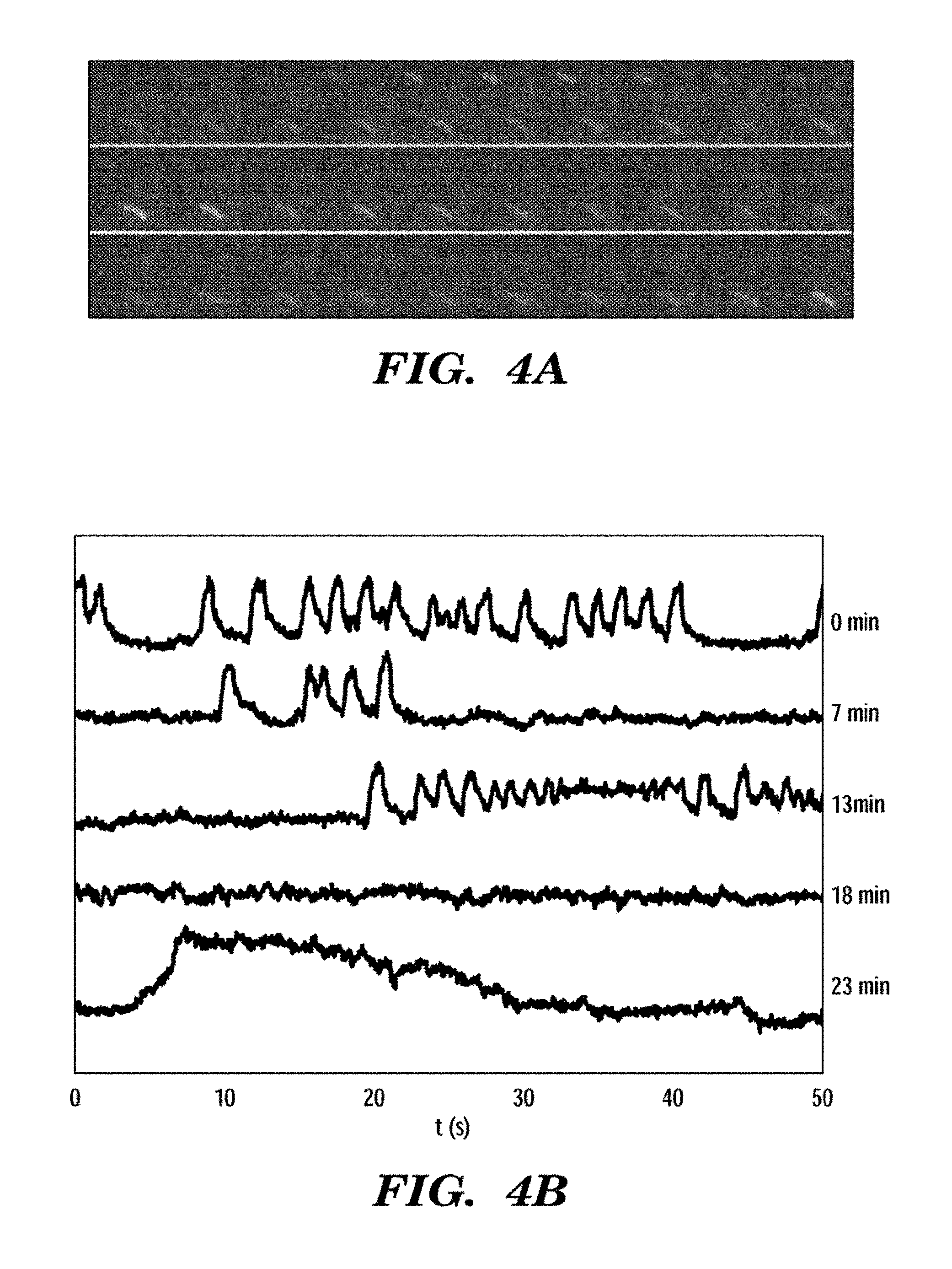
FIGS. 4A and 4B show fluorescence blinking in E. coli expressing GPR D97N. FIG. 4A shows a film strip of three E. coli at pH 7.5. Each exposure is 100 ms. The brightness of individual cells varies with time. FIG. 4B shows a blinking pattern from a single cell at pH 7.5. Each trace is a 50 second (s) record of the intensity. The time after the start of the experiment is indicated on the right. Cells continued to blink throughout the 1 hour experiment. The same cell shows fast blinks (0 and 7 minutes), slow blinks (28 minutes), and `ringing` behavior (18 minutes).
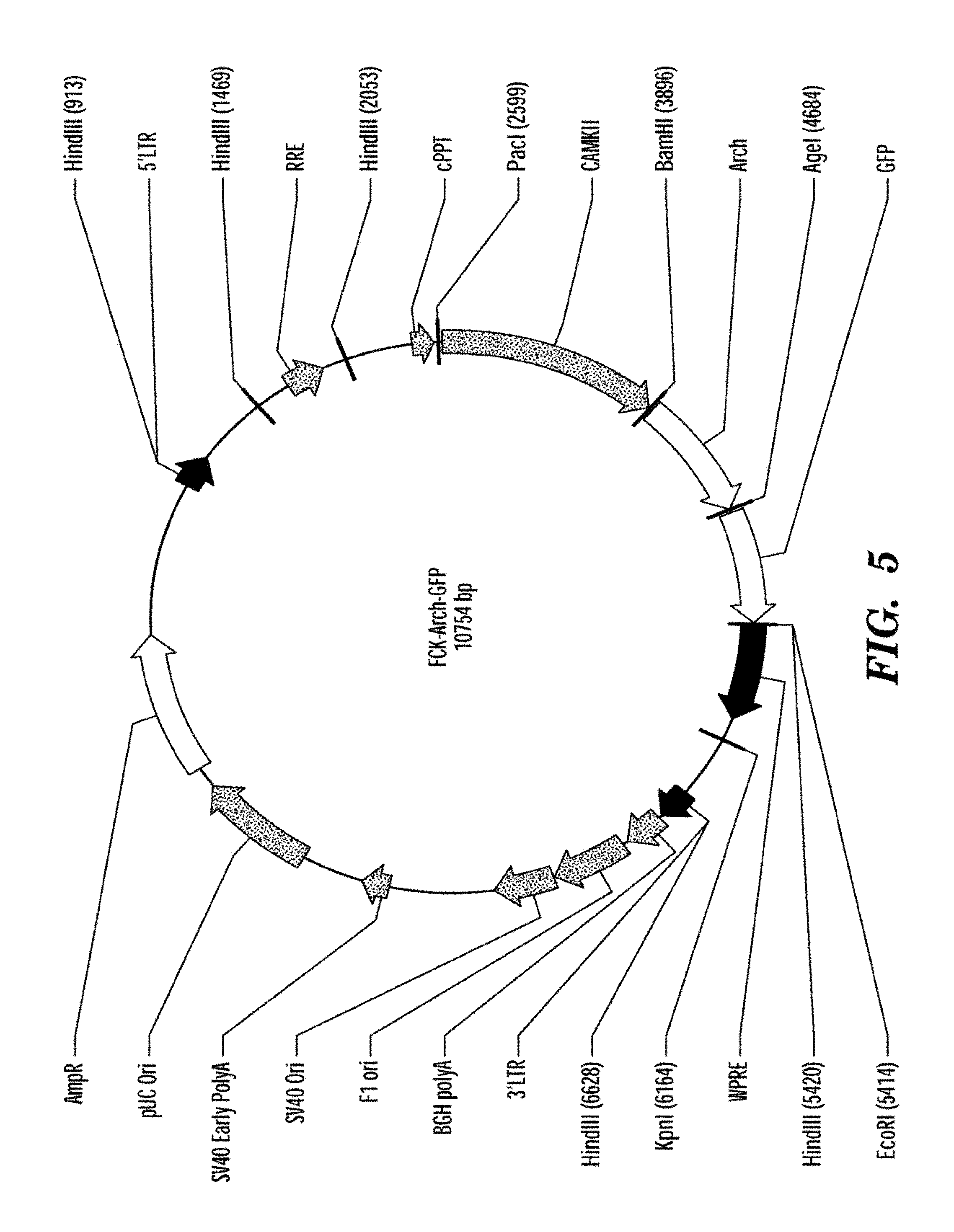
FIG. 5 shows a map of the plasmid containing Archaerhodopsin 3 (Arch3, also referred to in some instances herein as Ar-3) as described in the syntheticbiology web site (world wide web at syntheticneurobiology.org/protocols/protocoldetail/36/10).
FIGS. 6A-6E show that Arch is a fluorescent voltage indicator. FIG. 6A shows a model of Arch as a voltage sensor. pH and membrane potential can both alter the protonation of the Schiff base. The crystal structure shown is bacteriorhodopsin; the structure of Arch has not been solved.
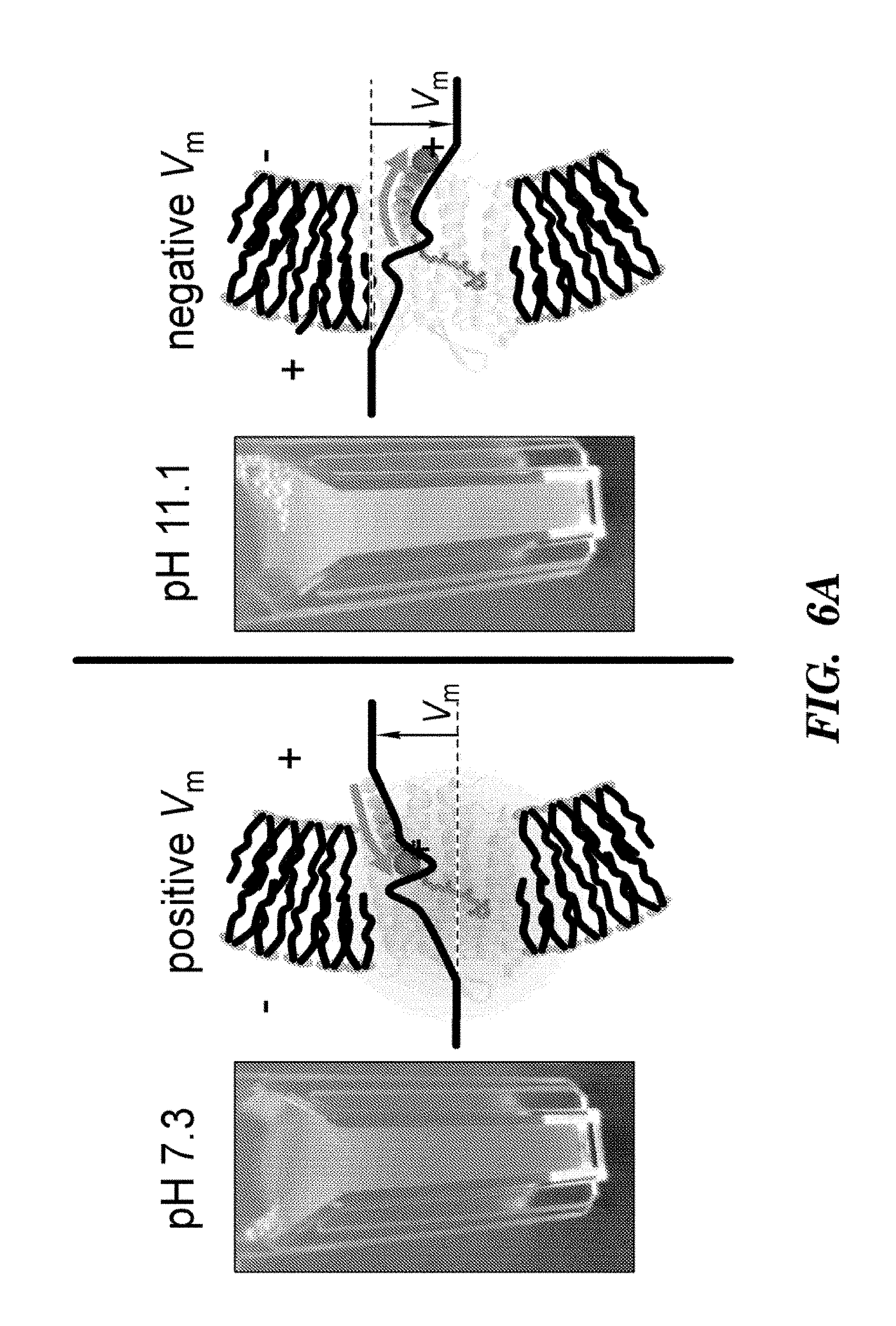
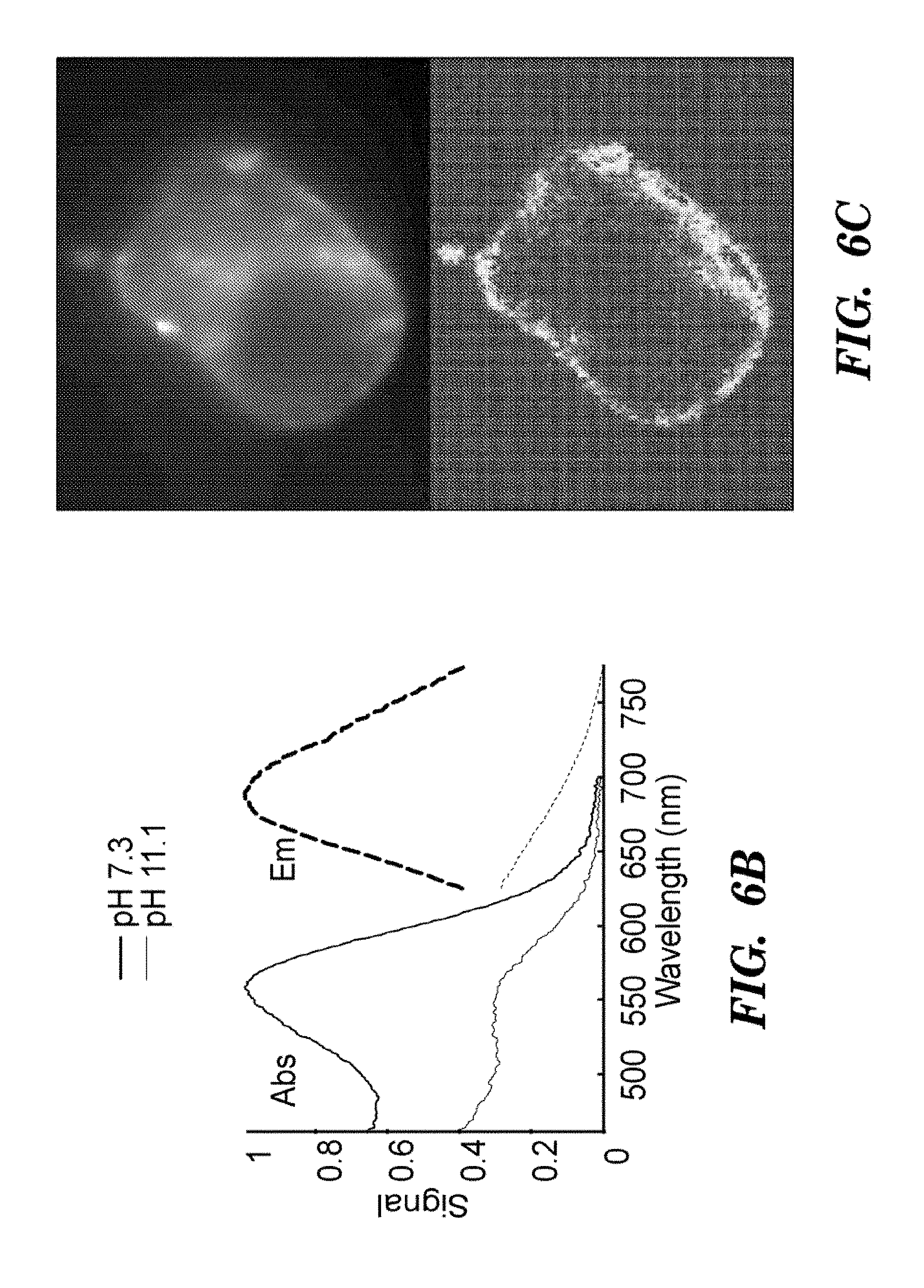
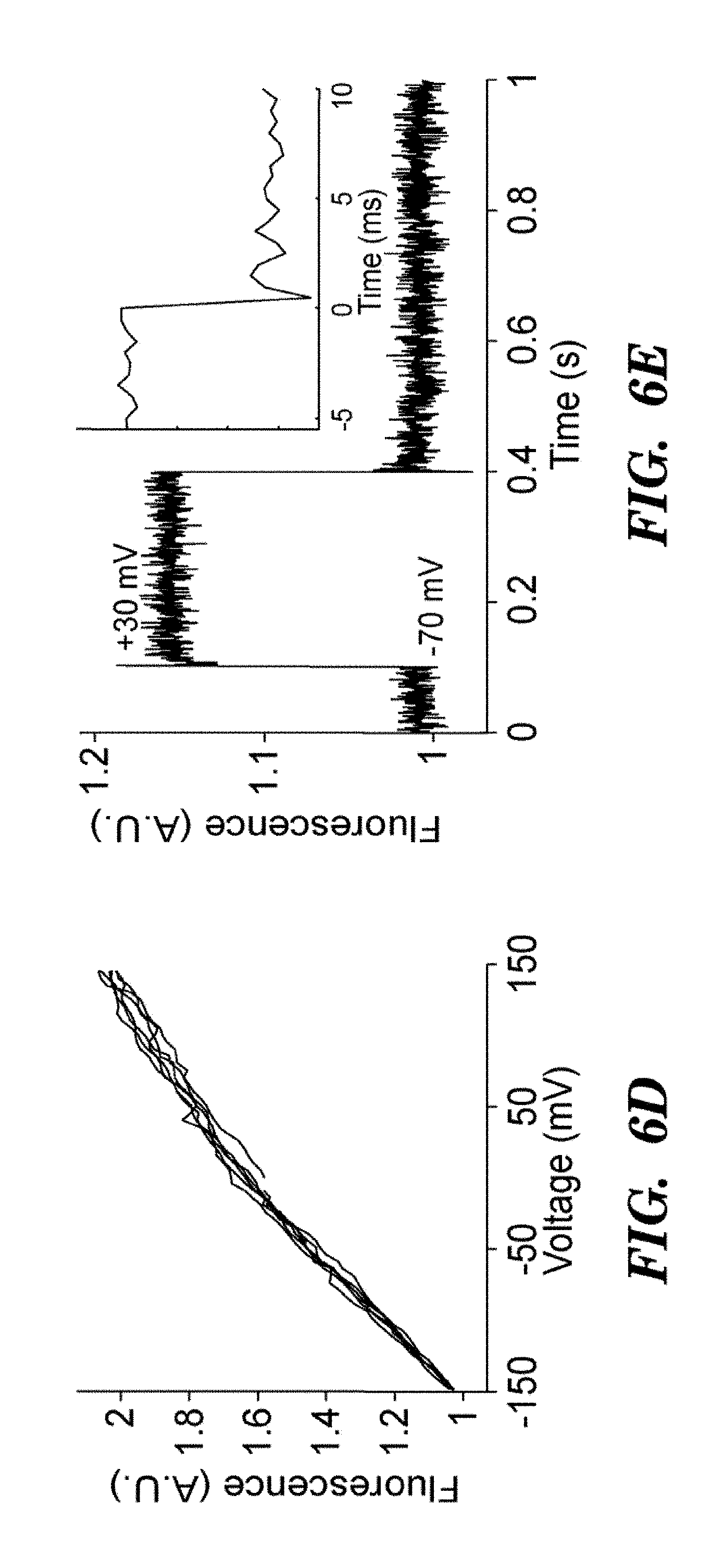
FIG. 6B shows absorption (solid line) and fluorescence emission (Em, see, dashed line) spectra of purified Arch at neutral and high pH. FIG. 6C top shows a HEK cell expressing Arch, visualized via Arch fluorescence. FIG. 6C bottom shows a pixel-weight matrix regions of voltage-dependent fluorescence. Scale bar 10 .mu.m. FIG. 6D shows fluorescence of Arch as a function of membrane potential. The fluorescence was divided by its value at -150 mV. FIG. 6E shows dynamic response of Arch to steps in membrane potential between -70 mV and +30 mV. The overshoots on the rising and falling edges were an artifact of electronic compensation circuitry. Data were an average of 20 cycles. Inset shows that step response occurred in less than the 0.5 ms resolution of the imaging system.
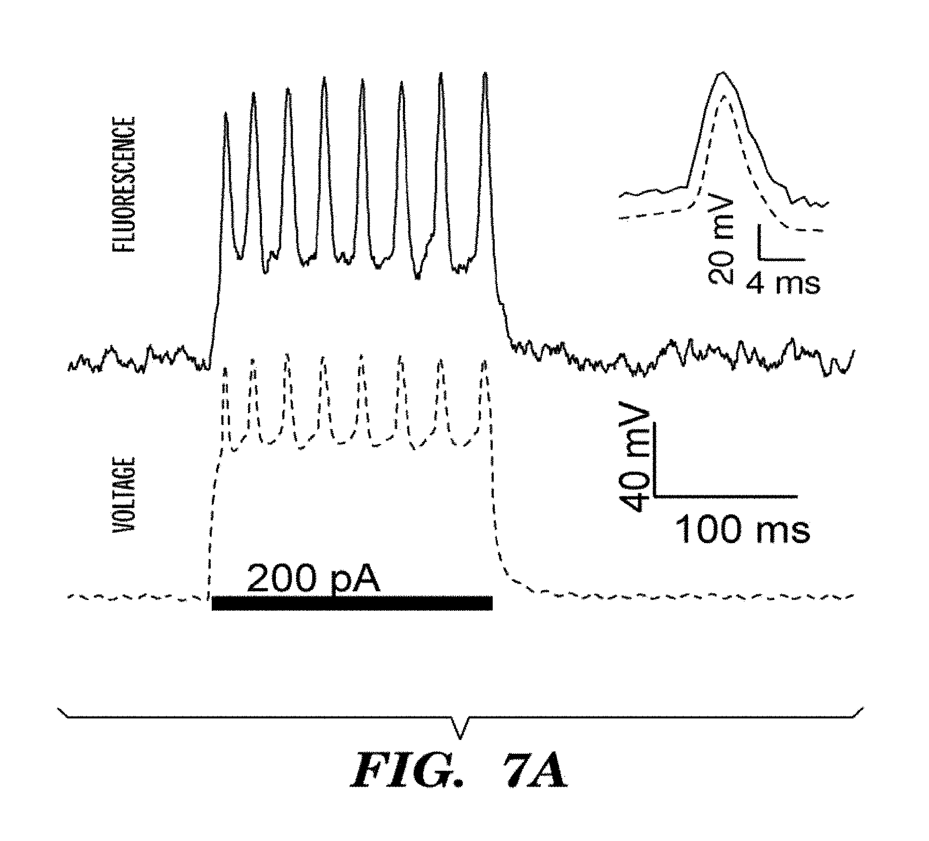
FIGS. 7A-7D show optical recording of action potentials with Arch3 WT. Cultured rat hippocampal neuron expressing Arch-GFP were imaged via fluorescence of GFP. Arch fluorescence was shown in cyan and regions of voltage-dependent fluorescence were shown in red. FIG. 7A shows whole-cell membrane potential determined via direct voltage recording (bottom, dotted line) and weighted Arch3 fluorescence (top, solid line) during a single-trial recording of a train of action potentials. Inset shows an averaged spike response for 269 events in a single cell, showing voltage (dotted line) and fluorescence (solid line). FIG. 7B shows recording of multiple spike-trains from a single cell. Current injections (shown in black dotted line) of 200 pA were applied to a neuron expressing Arch WT. Action potentials (shown in grey line) were readily detected via fluorescence over multiple rounds of current injection. FIG. 7C shows sub-cellular localization of an action potential. We took an image of a field of neuronal processes expressing Arch and created a weight matrix indicating pixels, shown as images in the Figure, whose fluorescence co-varied with the recorded potential in red, overlaid on the time-average Arch fluorescence which was shown in cyan. We detected sub-cellular regions within the electrically active cell. The figure shows a timecourse of an action potential determined via fluorescence (F) on the top graph (averaged over n=100 spikes) corresponding to each of the regions indicated. Also shown is the electrical recording of the action potential (V, bottom graph). FIG. 7D shows heterogeneous dynamics of an action potential within a single neuron, computed from an average of n=33 spikes. The region indicated by the arrow in the pixel map (see also graph) lags behind the rest of the cell by .about.1 ms (black arrows). Scale bar 5 .mu.m.
FIGS. 8A-8D demonstrate that Arch D95N shows voltage-dependent fluorescence but no photocurrent. FIG. 8A shows photocurrents in Arch 3 WT and Arch 3 D95N mutant, expressed in HEK cells clamped at V=0. Cells were illuminated with pulses of light at .lamda.=640 nm, 1800 W/cm.sup.2. FIG. 8B shows that Arch D95N fluorescence increased 3-fold between -150 mV and +150 mV, with nearly linear sensitivity from -120 to +120 mV. Inset shows a map of voltage sensitivity. Scale bar 5 inn. FIG. 8C shows that the step response comprised a component faster than 500 .mu.s (20% of the response) and a component with a time constant of 41 ms. FIG. 8D shows that Arch D95N provided highly accurate estimates of membrane potential, clearly resolving voltage steps of 10 mV, with a noise in the voltage estimated from fluorescence of 260 .mu.V/(Hz).sup.1/2 over timescales<12 s.
FIGS. 9A-9C show optical recording of action potentials with ArchD95N. FIG. 9A shows electrically recorded membrane potential of a neuron expressing Arch WT, subjected to pulses of current injection and laser illumination (I=1800 W/cm2, .lamda.=640 nm). Illumination generated sufficient photocurrent to suppress action potentials when the cell was near threshold. Grey bars indicate laser illumination. FIG. 9B is same as FIG. 9A in a neuron expressing Arch D95N, showing no effect of illumination on spiking or resting potential. We showed a neuron expressing Arch D95N, showing Arch D95N fluorescence (shows in cyan in the experiment), and regions of voltage-dependent fluorescence (shown in red in the experiment). FIG. 9C shows whole-cell membrane potential determined via electrical recording (bottom, voltage line) and weighted ArchD95N fluorescence (top, fluorescence line) during a single-trial recording of a train of action potentials.
FIG. 10 shows existing genetically encoded fluorescent voltage indicators classified according to their sensitivity and speed--the two key parameters that determine the performance of an indicator. VSFPs, FLARE and SPARC represent indicators based on fusions of GFP homologues to membrane proteins. The exemplary proteins we have developed are the Proteorhodopsin Optical Proton Sensor (PROPS), Arch3 WT, and Arch3 D95N, shown on the upper right. PROPS functions in bacteria, while Arch3 WT and Arch3 D95N function in mammalian cells. Note the logarithmic axes. Microbial rhodopsin-based voltage indicators are much faster and far more sensitive than other indicators.
FIGS. 11A-11D show optical recordings of action potentials in a single HL-1 mouse cardiomyocyte expressing Arch3 D95N-eGFP. Action potentials were recorded for up to 1000 s, with no signs of phototoxicity. This experiment is the first quantitative measurement of cardiac action potentials with a genetically encoded voltage indicator. We showed an overlay showing fluorescence of Arch D95N and GFP in a Arch D95N-GFP fusion. FIG. 11A shows a comparison of the action potential determined from patch clamp recording (dashed line) and fluorescence (solid line).
FIGS. 11B-11D show optical recordings of the action potentials in a single HL-1 cell over increasingly long intervals. Data in 11D have been corrected for photobleaching.
FIG. 12 shows optical recordings of action potentials in human induced pluripotent stem cell (iPS)-derived cardiomyocytes, expressing Arch3 D95N-eGFP. Human induced pluripotent stem cells (hiPSC) were provided by Cellular Dynamics Inc. Cells were plated into MatTek dishes coated in 0.1% gelatin at a density of 20, 50, or 75 thousand cells per square centimeter. These conditions showed cells that were sparse and did not beat spontaneously (20K), a confluent monolayer that did beat spontaneously (50K), and a dense monolayer (75K). iPS cells were plated and maintained for 48 hours in plating medium, and thereafter fed every 48 hours with maintenance medium (both from Cellular Dynamics). iPS cells were transfected using Mirus LT-1 according to the manufacturer directions. To a tube containing 20 uL of OPTI-MEM.RTM., we added 200 ng of DNA and 1.2 uL of the LT-1 transfection reagent. The DNA mixture was incubated at room temperature for 20 minutes. Fresh maintenance medium was added to the iPS cells during the incubation and the DNA mixture was added dropwise over the plate. Cells were imaged 48 to 96 hours after transfection. We observed synchronized beating of adjoining cells, indicating that VIPs can probe intercellular conduction. We recorded for more than 10 minutes continuously, with little phototoxicity. Within the population of cells we observed cells with action potentials matching those of ventricular, atrial, and nodal cells, as expected for this population. Addition of drugs led to changes in the action potential waveform that matched changes reported by conventional patch clamp. Cell 1 fluorescence information is indicated as a solid line and Cell 2 fluorescence as a dashed line on the photos fluorescence vs. time.
FIG. 13 illustrates constructs of fused VIPs with GFP-homologue proteins to develop a series of improved voltage indicators. The length of each bar indicates the length of the protein sequence or linker region. The color indicates the color of the fluorescence of the corresponding protein. All constructs were constructed with both Arch WT and Arch D95N backbones. The sequences for the constructs are provided in the Sequence Listing as follows:
TABLE-US-00001 pADD247/248 SEQ ID NO: 48 pADD286/287 SEQ ID NO: 53 pADD292/293 SEQ ID NO: 50 pADD294 (D95N only) SEQ ID NO: 54 pADD297/300 SEQ ID NO: 51 pADD259/298 SEQ ID NOS: 55 and 52, respectively Pfck:Arch3(WT/D95N)-EGFP SEQ ID NOS: 56-57, respectively pADD269/270 SEQ ID NO: 49 Pcmv:Arch3(WT/D95N)-GCaMP SEQ ID NOS: 58-59, respectively
FIGS. 14A-14B illustrate mechanism of ssFRET. FIG. 14A shows that when the Schiff Base (SB) on the retinal is protonated, the absorption spectrum of the retinal (solid) overlaps with the emission spectrum of the GFP (dotted), and the fluorescence of the GFP is quenched. However, the retinal itself is fluorescent in this state. FIG. 14B shows that when the SB is deprotonated, the GFP fluorescence (solid line) becomes de-quenched and the retinal fluorescence vanishes.
FIG. 15 shows distance dependence of ssFRET signal. As the distance between the mOrange2 and the Arch chromophores decreased, the magnitude of the ssFRET signal increased.
FIG. 16 shows a step response of Arch 3 fluorescence and mOrange fluorescence in pADD294. Here the mOrange2 signal has been inverted to facilitate comparison with the Arch 3 signal. The similarity of the timecourses is consistent with modulation of the mOrange2 fluorescence via ssFRET. The voltage step is from -70 mV to +30 mV.
FIGS. 17A-17C show pH-dependent spectra of Arch 3 WT and D95N. FIG. 17A shows Arch WT absorption at neutral (bold line) and high (this line) pH. At neutral pH, Arch absorbed maximally at 558 nm. Fluorescence emission (dashed line) was recorded on 2 .mu.M protein solubilized in 1% DM, with .lamda.exc=532 nm. FIG. 17B shows Arch D95N spectra under the same conditions as in FIG. 17A. The absorption maximum was at 585 nm. FIG. 17C shows absorption spectra that were recorded on purified protein between pH 6-11. Singular Value Decomposition of absorption spectra between 400-750 nm was used to calculate the fraction of the SB in the protonated state as a function of pH. The result was fit to a Hill function to determine the pKa of the SB.
FIG. 18 shows frequency response of Arch 3 WT. A chirped sine wave with amplitude 50 mV and frequency from 1 Hz-1 kHz was applied to a HEK cell expressing Arch 3 WT (wild type). Membrane potential {circumflex over (V)}.sub.FL was determined from fluorescence and the Fourier transform of {circumflex over (V)}.sub.FL was calculated. The uptick at 1 kHz is an artifact of electronic compensation circuitry. Inset: power spectrum of noise in {circumflex over (V)}.sub.FL, under voltage clamp at constant V=0 mV shows a shot-noise limited noise floor of 470 .mu.V/(Hz)1/2 at frequencies above 10 Hz. The noise figures reported here are specific to our imaging system and serve primarily as an indicator of the possible sensitivity of Arch 3.
FIG. 19 shows sensitivity of Arch 3 WT to voltage steps of 10 mV. Whole-cell membrane potential determined via direct voltage recording, V, (bolded black line, showing step-like line on the graph) and weighted Arch 3 fluorescence, {circumflex over (V)}.sub.1, (solid narrower line showing serrations on the graph).
FIG. 20 shows that arch reports action potentials without exogenous retinal. We made an image of 14 day in vitro (DIV) hippocampal neuron imaged via Arch 3 fluorescence with no exogenous retinal. Electrical (bolded solid black line) and fluorescence (non-bolded line, showing serrated line in the graph) records of membrane potential from the neuron during a current pulse. Action potentials are clearly resolved.
FIG. 21 shows a frequency response of Arch D95N, measured in the same manner as for Arch 3 WT (FIG. 18).
DETAILED DESCRIPTION OF THE INVENTION
The present invention is based, at least in part, on the discovery that microbial rhodopsin proteins or modified microbial rhodopsin proteins that have reduced ion pumping activity, compared to the natural microbial rhodopsin protein from which they are derived, can be used as an optically detectable sensor to sense voltage across membranous structures, such as in cells and sub-cellular organelles when they are present on the cell membrane. That is, the microbial rhodopsin proteins and the modified microbial rhodopsin proteins can be used to measure changes in membrane potential of a cell, including prokaryotic and eukaryotic cells. The optical sensors described herein are not constrained by the need for electrodes and permit electrophysiological studies to be performed in e.g., subcellular compartments (e.g., mitochondria) or in small cells (e.g., bacteria). The optical sensors described herein can be used in methods for drug screening, in research settings, and in in vivo imaging systems.
Microbial Rhodopsins: Design of Optical Voltage Sensors
Microbial rhodopsins are a large class of proteins characterized by seven transmembrane domains and a retinilydene chromophore bound in the protein core to a lysine via a Schiff base (Beja, O., et al. Nature 411, 786-789 (2001)). Over 5,000 microbial rhodopsins are known, and these proteins are found in all kingdoms of life. Microbial rhodopsins serve a variety of functions for their hosts: some are light-driven proton pumps (bacteriorhodopsin, proteorhodopsins), others are light-driven ion channels (channelrhodopsins), chloride pumps (halorhodopsins), or serve in a purely photosensory capacity (sensory rhodopsins).
The retinilydene chromophore imbues microbial rhodopsins with unusual optical properties. The linear and nonlinear responses of the retinal are highly sensitive to interactions with the protein host: small changes in the electrostatic environment can lead to large changes in absorption spectrum. These electro-optical couplings provide the basis for voltage sensitivity in microbial rhodopsins.
Some of the optical sensors described herein are natural proteins without modifications and are used in cells that do not normally express the microbial rhodopsin transfected to the cell, such as eukaryotic cells. For example, as shown in the examples, the wild type Arch3 can be used in neural cells to specifically detect membrane potential and changes thereto.
Some of the microbial rhodopsins are derived from a microbial rhodopsin protein by modification of the protein to reduce or inhibit light-induced ion pumping of the rhodopsin protein. Such modifications permit the modified microbial rhodopsin proteins to sense voltage without altering the membrane potential of the cell with its native ion pumping activity and thus altering the voltage of the system. Other mutations impart other advantageous properties to microbial rhodopsin voltage sensors, including increased fluorescence brightness, improved photostability, tuning of the sensitivity and dynamic range of the voltage response, increased response speed, and tuning of the absorption and emission spectra.
Mutations that eliminate pumping in microbial rhodopsins in the present invention generally comprise mutations to the Schiff base counterion; a carboxylic amino acid (Asp or Glu) conserved on the third transmembrane helix (helix C) of the rhodopsin proteins. The amino acid sequence is RYX(DE) where X is a non-conserved amino acid. Mutations to the carboxylic residue directly affect the proton conduction pathway, eliminating proton pumping. Most typically the mutation is to Asn or Gln, although other mutations are possible, and based on the description provided herein, one skilled in the art can make different mutants which also result in reduced or absent ion pumping by the microbial rhodopsin protein. In one embodiment, the modified microbial rhodopsin proteins of the invention and the methods of the invention comprises the Asp to Asn or Gln mutation, or Glu to Asn or Gln mutation. In some embodiments, the protein consist essentially of Asp to Asn or Gln mutation, or Glu to Asn or Gln mutation. In some embodiments, the protein consist of Asp to Asn or Gln mutation, or Glu to Asn or Gln mutation.
Provided herein are illustrative exemplary optical voltage sensors and directions for making and using such sensors. Other sensors that work in a similar manner as optical sensors can be prepared and used based on the description and the examples provided herein.
Table 1a includes exemplary microbial rhodopsins useful according to the present invention. For example, mutations that eliminate pumping in microbial rhodopsins in the present invention generally comprise mutations to the Schiff base counterion; a carboxylic amino acid (Asp or Glu) conserved on the third transmembrane helix (helix C) of the rhodopsin proteins. Table 1a refers to the amino acid position in the sequence provided as the exemplary Genbank number. However, the position may be numbered slightly differently based on the variations in the available amino acid sequences. Based on the description of the motif described herein, a skilled artisan will easily be able to make similar mutations into other microbial rhodopsin genes to achieve the same functional feature, i.e. reduction in the pumping activity of the microbial rhodopsin in question.
TABLE-US-00002 TABLE 1a Exemplary microbial rhodopsins useful according to the present invention. Microbial Rhodopsin Abbreviation Genbank number Amino acid mutation Green-absorbing GPR AF349983; wild-type, D99N (SEQ ID NO: 76) in the proteorhodopsin: a (Nucleotide and protein specification, this mutation is light-driven proton disclosed as SEQ ID also referred to as D97N pump found in NOS 74-75, marine bacteria respectively) Blue-absorbing BPR AF349981; wild-type; D99N (SEQ ID NO: 79) proteorhodopsin: a (Nucleotide and protein light-driven proton disclosed as SEQ ID pump found in NOS 77-78, marine bacteria. respectively) Natronomonas NpSRII Z35086.1; In one D75N (SEQ ID NO: 82) pharaonis sensory embodiment only the rhodopsin II: a light- sensory domain, given activated signaling by nucleotides 2112-2831 protein found in the of sequence halophilic bacterium Z35086, is used N. pharaonis. In the (Nucleotide and wild the sensory "sensory domain" domain is paired with protein disclosed as a transducer domain SEQ ID NOS 80-81, respectively) Bacteriorhodopsin: a BR NC_010364.1, D98N light-driven proton nucleotides 1082241 to (SEQ ID NO: 85) pump found in 1083029, or GenBank Halobacterium sequence M11720.1; salinarum ("M11720.1" nucleotide and protein disclosed as SEQ ID NOS 83-84, respectively Archaerhodopsin Arch 3 (or Ar Chow B. Y. et al., D95N (SEQ ID NO: 87) Arch 3: a light-driven 3) Nature 463: 98-102; proton pump found ("Arch 3" wild-type in Halobacterium protein disclosed as sodomense SEQ ID NO: 86)
The following Table 1b includes exemplary additional rhodopsins that can be mutated as indicated in the methods of the invention:
TABLE-US-00003 TABLE 1b Nucleic acid Amino Acid Microbial Rhodopsin Abbreviation Genbank number mutation mutation Fungal Opsin Related Mac AAG01180 (SEQ G415 to A D139N (SEQ ID Protein ID NO: 60) NO: 61) Cruxrhodopsin Crux BAA06678 (SEQ G247 to A D83N (SEQ ID ID NO: 62) NO: 63) Algal Bacteriorhodopsin Ace AAY82897 (SEQ G265 to A D89N (SEQ ID ID NO: 64) NO: 65) Archaerhodopsin 1 Ar1 P69051 (SEQ ID G289 to A D97N (SEQ ID NO: 66) NO: 67) Archaerhodopsin 2 Ar2 P29563 (SEQ ID G286 to A D96N (SEQ ID NO: 68) NO: 69) Archaerhodopsin 3 Ar3 P96787 (SEQ ID G283 to A D95N (SEQ ID NO: 70) NO: 71) Archaerhodopsin 4 Ar4 AAG42454 (SEQ G292 to A D98N (SEQ ID ID NO: 72) NO: 73)
Voltage Indicating Proteins (VIP)
We have developed a family of fluorescent voltage-indicating proteins (VIPs) based on Achaerhodopsins that function in mammalian cells, including neurons and human stem cell-derived cardiomyocytes. These proteins indicate electrical dynamics with sub-millisecond temporal resolution and sub-micron spatial resolution. We have demonstrated non-contact, high-throughput, and high-content studies of electrical dynamics in mammalian cells and tissues using optical measurement of membrane potential. These VIPs are broadly useful, particularly in eukaryotic, such as mammalian, including human cells.
We have developed VIPs based on Archaerhodopsin 3 (Arch 3) and its homologues. Arch 3 is Archaerhodopsin from H. sodomense and it is known as a genetically-encoded reagent for high-performance yellow/green-light neural silencing. Gene sequence at GenBank: GU045593.1 (synthetic construct Arch 3 gene, complete cds. Submitted Sep. 28, 2009). We have shown that these proteins localize to the plasma membrane in eukaryotic cells and show voltage-dependent fluorescence.
We have also shown further improved membrane localization, with comparable voltage sensitivity, in ArchT, gene sequence at GenBank: HM367071.1 (synthetic construct ArchT gene, complete cds. Submitted May 27, 2010). ArchT is Archaerhodopsin from Halorubrum sp. TP009: genetically-encoded reagent for high-performance yellow/green-light neural silencing, 3.5.times. more light sensitive than Arch 3.
Table 1c summarizes exemplary sequences that can be used to create viral constructs that express the voltage indicators based on Archaerhodopsin.
TABLE-US-00004 TABLE 1c Exemplary sequences that can be used to generate virus constructs with Arch 3 and ArchT Virus backbone Lentivirus SEQ ID NO: 24 Promoter CamKII (neuron specific) SEQ ID NO: 25 CAG enhancer (pan cellular) SEQ ID NO: 26 CMV (pan cellular) SEQ ID NO: 27 Ubiquitin (pan cellular) SEQ ID NO: 28 Voltage-sensing Arch D95N SEQ ID NO: 29 domain ArchT D95N SEQ ID NO: 30
FIG. 10 shows existing genetically encoded fluorescent voltage indicators classified according to their sensitivity and speed--the two key parameters that determine the performance of an indicator. The proteins we developed are the Proteorhodopsin Optical Proton Sensor (PROPS), Arch 3 WT, and Arch 3 D95N, shown on the upper right. PROPS only functions in bacteria, while Arch 3 WT and Arch 3 D95N function in mammalian cells. We showed that microbial rhodopsin-based voltage indicators are faster and far more sensitive than other indicators.
Table 2 shows exemplary approximate characteristics of fluorescent voltage indicating proteins and contains representative members of all families of fluorescent indicators. Although the list on Table 2 is not comprehensive, one skilled in the art can readily see the characteristics of the types of proteins useful in the present invention.
TABLE-US-00005 TABLE 2 Representative members of all families of fluorescent indicators. Approx Approx .DELTA.F/F per response Molecule 100 mV time Comments VSFP 2.3, Knopfel, T. 9.5% 78 ms Ratiometric (.DELTA.R/R) et al. J. Neurosci. 30, 14998-15004 (2010) VSFP 2.4 Knopfel, T. et 8.9% 72 ms Ratiometric (.DELTA.R/R) al. J. Neurosci. 30, 14998-15004 (2010) VSFP 3.1, Lundby, A., 3% 1-20 ms Protein et al., PLoS One 3, 2514 (2008) Mermaid, Perron, A. et 9.2% 76 Ratiometric (.DELTA.R/R) al. Front Mol Neurosci. 2, 1-8 (2009) SPARC, Ataka, K. & 0.5% 0.8 ms Protein Pieribone, V. A. Biophys. J. 82, 509-516 (2002) Flash, Siegel, M. S. & 5.1% 2.8-85 ms Protein Isacoff, E. Y. Neuron 19, 735-741 (1997) PROPS, described 150% 5 ms Protein herein; SEQ ID NO: Arch 3 WT, described 66% <0.5 ms Protein herein Arch D95N 100% 41 ms Protein
FIG. 9 shows an optical recording of action potentials in a single rat hippocampal neuron. The data represents a single trial, in which spiking was induced by injection of a current pulse. The fluorescence shows clear bursts accompanying individual action potentials. This experiment is the first robust measurement of action potentials in a single mammalian neuron using a genetically encoded voltage indicator.
FIG. 11 shows optical recordings of action potentials in a single HL-1 mouse cardiomyocyte expressing Arch 3 D95N-eGFP. Action potentials were recorded for up to 1000 s, with no signs of phototoxicity. This experiment is the first quantitative measurement of cardiac action potentials with a genetically encoded voltage indicator.
FIG. 12 shows optical recordings of action potentials in human induced pluripotent stem cell (iPS)-derived cardiomyocytes, expressing Arch 3 D95N-eGFP. Human induced pluripotent stem cells (hiPSC) were provided by Cellular Dynamics Inc. Cells were plated into MatTek dishes coated in 0.1% gelatin at a density of 20, 50, or 75 thousand cells per square centimeter. These conditions showed cells that were sparse and did not beat spontaneously (20K), a confluent monolayer that did beat spontaneously (50K), and a dense monolayer (75K). iPS cells were plated and maintained for 48 hours in plating medium, and thereafter fed every 48 hours with maintenance medium (both from Cellular Dynamics). iPS cells were transfected using Mirus LT-1 according to the manufacturer directions. To a tube containing 20 uL of optimem, we added 200 ng of DNA and 1.2 uL of the LT-1 transfection reagent. The DNA mixture was incubated at room temperature for 20 minutes. Fresh maintenance medium was added to the iPS cells during the incubation and the DNA mixture was added dropwise over the plate. Cells were imaged 48 to 96 hours after transfection.
We observed synchronized beating of adjoining cells, indicating that VIPs can probe intercellular conduction. We recorded for more than 10 minutes continuously, with little phototoxicity. Within the population of cells we observed cells with action potentials matching those of ventricular, atrial, and nodal cells, as expected for this population. Addition of drugs led to changes in the action potential waveform that matched changes reported by conventional patch clamp.
Generation of Fusions Between Microbial Rhodopsins and GFP Homologues with Additional or Improved Properties
We fused VIPs with GFP-homologue proteins to develop a series of improved voltage indicators. FIG. 16 illustrates these exemplary constructs and FIG. 16 legend provides the sequences for these constructs. The new capabilities of these newly developed sensors include, for example, a spectral shift FRET (ssFRET) for enhanced brightness and 2-photon imaging, ratiometric voltage imaging, and multimodal sensors for simultaneous measurement of voltage and concentration.
Spectral Shift FRET (ssFRET) for Enhanced Brightness and 2-photon Imaging
A key limitation of the first generation of VIPs was that the endogenous fluorescence of the retinal was dim. Imaging required a specialized system comprising an intense red laser, a high numerical aperture objective, and an electron-multiplying CCD (EMCCD) camera. Ideally one would like an indicator bright enough to image on a conventional wide-field or confocal fluorescence microscope, or a 2-photon confocal microscope for in vivo applications.
ssFRET provides a path to brighter VIPs as shown in FIG. 10. A GFP-homologue (generically referred to as GFP) is fused to the microbial rhodopsin (see, e.g., FIG. 13). Voltage-dependent changes in the absorption spectrum of the retinal lead to voltage-dependent rates of nonradiative fluorescence resonance energy transfer (FRET) between the GFP and the retinal. Retinal in its absorbing, fluorescent state quenches the GFP, while retinal in the non-absorbing, non-fluorescent state does not quench the GFP. Thus one obtains anti-correlated fluorescence emission of the GFP and the retinal.
Thus in one embodiment, the invention provides a fusion protein comprising a GFP that is fused to a microbial rhodopsin or a modified microbial rhodopsin, such as a proteorhdopsin or archaerhodopsin. Such fusion proteins can be used in any and all of the methods of the present invention.
To maximize the degree of ssFRET between the GFP and the retinal we selected a GFP-homologue, mOrange2, whose emission overlaps maximally with the absorption of Arch 3 in its protonated state. The rate of FRET falls off very quickly with increasing distance between chromophores, so we constructed a series of truncated constructs in which the linker and non-essential elements of the Arch 3 and mOrange2 were removed. FIG. 15 shows that as the distance between the mOrange2 and the Arch 3 decreased, the ssFRET signal increased. This same strategy can be applied to generate ssFRET signals from other microbial rhodopsins and GFP homologues.
We have shown that the time response of fluorescence from mOrange2 to a step in Vm matches the time response of fluorescence from Arch D95N. This observation is consistent with ssFRET. Similar results can be seen for FIGS. 9A and 11 for fusions to Arch 3 WT.
Ratiometric Voltage Imaging
A key challenge in application of VIPs is to extract accurate values of the membrane potential, without systematic artifacts from photobleaching, variation in illumination intensity, cell movement, or variations in protein expression level. In cells that are accessible to patch clamp, one can calibrate the fluorescence as a function of membrane potential by varying the membrane potential under external control. However, a benefit of VIPs is that they function in systems that are inaccessible to patch clamp. In these cases direct calibration is not possible.
The Arch 3 (WT or D95N) fusion with eGFP enables ratiometric determination of membrane potential. Similar ratiometric determinations may be made using other rhodopsins such as those described in this application using the identical concept. The eGFP fluorescence is independent of membrane potential, The ratio of Arch 3 fluorescence to eGFP fluorescence provides a measure of membrane potential that is independent of variations in expression level, illumination, or movement. This construct does not undergo ssFRET due to the long linker between the eGFP and the Arch 3, and because the emission of eGFP has little spectral overlap with the absorption of Arch 3.
Multimodal Sensors for Simultaneous Measurement of Voltage and Concentration
Membrane potential is only one of several mechanisms of signaling within cells. One often wishes to correlate changes in membrane potential with changes in concentration of other species, such as Ca++, H+ (i.e. pH), Na+, ATP, cAMP. We constructed fusions of Arch with pHluorin (a fluorescent pH indicator) and GCaMP3 (a fluorescent Ca++ indicator). One can also use fusions with other protein-based fluorescent indicators to enable other forms of multimodal imaging using the concept as taught herein. Concentration of ions such as sodium, potassium, chloride, and calcium can be simultaneously measured when the nucleic acid encoding the microbial rhodopsin is operably linked to or fused with an additional fluorescent ion sensitive indicator.
Additional Fluorescent Proteins
The term "additional fluorescent molecule" refers to fluorescent proteins other then microbial rhodopsins. Such molecules may include, e.g., green fluorescent proteins and their homologs.
Fluorescent proteins that are not microbial rhodopsins are well known and commonly used, and examples can be found, e.g., in a review The Family of GFP-Like Proteins: Structure, Function, Photophysics and Biosensor Applications. Introduction and Perspective, by Rebekka M. Wachter (Photochemistry and Photobiology Volume 82, Issue 2, pages 339-344, March 2006). Also, a review by Nathan C Shaner, Paul A Steinbach, & Roger Y Tsien, entitled A guide to choosing fluorescent proteins (Nature Methods--2, 905-909 (2005)) provides examples of additional useful fluorescent proteins.
Targeting VIPs to Intracellular Organelles
We have shown targeting VIPs to intracellular organelles, including mitochondria, the endoplasmic reticulum, the sarcoplasmic reticulum, synaptic vesicles, and phagosomes. Accordingly, in one embodiment, the invention provides constructs, such as expression constructs, such as viral constructs comprising a microbial rhodopsin operably linked to a sequence targeting the protein to an intracellular organelle, including a mitochondrium, an endoplasmic reticulum, a sarcoplasmic reticulum, a synaptic vesicle, and a phagosome.
The invention further provides cells expressing the constructs, and further methods of measuring membrane potential changes in the cells expressing such constructs as well as methods of screening for agents that affect the membrane potential of one or more of the intracellular membranes.
The Key Advantages of Voltage Indicating Proteins (VIPs)
The newly developed VIPs show high sensitivity. In mammalian cells VIPs show about 3-fold increase in fluorescence between -150 mV and +150 mV. The response is linear over most of this range. We can measure membrane voltage with a precision of <1 mV in a 1 s interval.
The newly developed VIPs show high speed. Arch 3 WT shows 90% of its step response in <0.5 ms. A neuronal action potential lasts 1 ms, so this speed meets the benchmark for imaging electrical activity of neurons. However, Arch 3 WT retains the photoinduced proton-pumping, so illumination slightly hyperpolarizes the cell.
The modified microbial rhodopsin, Arch 3 D95N, has a 40 ms response time and lacks photoinduced proton pumping. Although the slower response time of this construct hampers detection of membrane potential and changes thereto in neurons, the Arch 3 D95N is fast enough to indicate membrane potential and action potentials in other types of cells, for example, in cardiomyocytes and does not perturb membrane potential in the cells wherein it is used.
The newly developed VIPs also show high photostability. VIPs are comparable to GFP in the number of fluorescence photons produced prior to photobleaching. We routinely watch VIPs in mammalian cells for many minutes, without signs of photobleaching or phototoxicity. VIPs have no homology to GFP, nor to any other known fluorescent protein.
The newly developed VIPs also show far red spectrum. VIPs are excited with a 633 nm laser, and the emission is in the near infrared, peaked at 710 nm. The emission is farther to the red than any existing fluorescent protein. These wavelengths coincide with low cellular autofluorescence and good transmission through tissue. This feature makes these proteins particularly useful in optical measurements of action potential as the spectrum facilitates imaging with high signal-to-noise, as well as multi-spectral imaging in combination with other fluorescent probes.
The newly developed VIPs further show high targetability. We have imaged VIPs in primary neuronal cultures, cardiomyocytes (HL-1 and human iPSC-derived), HEK cells, and Gram positive and Gram negative bacteria. We targeted a VIP to the endoplasmic reticulum, and to mitochondria. The ocnstructs are useful also for in vivo imaging in C. elegans, zebrafish, and mice.
With the microbial rhodopsin constructs of the invention further comprising a cell type- and/or a time-specific promotors, one can image membrane potential in any optically accessible cell type or organelle in a living organism.
In one embodiment, the design of a voltage sensor comprises, consists of, or consists essentially of selecting at least three elements: a promoter, a microbial rhodopsin voltage sensor, one or more targeting motifs, and an optional accessory fluorescent protein. Some non-limiting examples for each of these elements are listed in Tables 1a and 1b, and Table 3 below. In one embodiment, at least one element from each column is selected to create an optical voltage sensor with the desired properties. In some embodiments, methods and compositions for voltage sensing as described herein involves selecting: 1) A microbial rhodopsin protein, 2) one or more mutations to imbue the protein with sensitivity to voltage or to other quantities of interest and to eliminate light-driven charge pumping, 3) codon usage appropriate to the host species, 4) a promoter and targeting sequences to express the protein in cell types of interest and to target the protein to the sub-cellular structure of interest, 5) an optional fusion with a conventional fluorescent protein to provide ratiometric imaging, 6) a chromophore to insert into the microbial rhodopsin, and 7) an optical imaging scheme.
TABLE-US-00006 TABLE 3 Exemplary optical sensor combinations Accessory fluorescent Promoter Voltage sensor Targeting motif protein CMV (SEQ ID hGPR (D97N)(SEQ SS(.beta.2nAChR)(SEQ Venus (SEQ ID NO: 43) NO: 31) ID NO: 35) ID NO: 39) 14x UAS-E1b hGPR (D97N, .+-.E108Q, SS(PPL) EYFP (SEQ ID NO: 44) (SEQ ID .+-.E142Q, .+-.L217D) (SEQ (SEQ ID NO: 40) NO: 32) ID NO: 36) HuC (SEQ ID hBPR (D99N) (SEQ ID ER export motif TagRFP (SEQ ID NO: 45) NO: 33) NO: 37) (SEQ ID NO: 41) ara (SEQ ID hNpSRII (D75N) (SEQ TS from Kir2.1 NO: 34) ID NO: 38) (SEQ ID NO: 42) lac MS
In one embodiment, the optical sensor gene is encoded by a delivery vector. Such vectors include but are not limited to: plasmids (e.g. pBADTOPO, pCI-Neo, pcDNA3.0), cosmids, and viruses (such as a lentivirus, an adeno-associated virus, or a baculovirus).
In one embodiment, the green-absorbing proteorhodopsin (GPR) is used as the starting molecule. This molecule is selected for its relatively red-shifted absorption spectrum and its ease of expression in heterologous hosts such as E. coli. In another embodiment, the blue-absorbing proteorhodopsin (BPR) is used as an optical sensor of voltage. It is contemplated herein that a significant number of the microbial rhodopsins found in the wild can be engineered as described herein to serve as optical voltage sensors.
Microbial rhodopsins are sensitive to quantities other than voltage. Mutants of GPR and BPR, as described herein, are also sensitive to intracellular pH. It is also contemplated that mutants of halorhodopsin may be sensitive to local chloride concentration.
In one embodiment, the voltage sensor is selected from a microbial rhodopsin protein (wild-type or mutant) that provides a voltage-induced shift in its absorption or fluorescence. The starting sequences from which these constructs can be engineered include, but are not limited to, sequences listed in Tables 1a-1b, that list the rhodopsin and an exemplary mutation that can be made to the gene to enhance the performance of the protein product.
Mutations to minimize the light-induced charge-pumping capacity. The retinal chromophore is linked to a lysine by a Schiff base. A conserved aspartic acid serves as the proton acceptor adjacent to the Schiff base. Mutating this aspartic acid to asparagine suppresses proton pumping. Thus, in some embodiments, the mutations are selected from the group consisting of: D97N (green-absorbing proteorhodopsin), D99N (blue-absorbing proteorhodopsin), D75N (sensory rhodopsin II), and D85N (bacteriorhodopsin). In other embodiments, residues that can be mutated to inhibit pumping include (using bacteriorhodopsin numbering) D96, Y199, and R82, and their homologues in other microbial rhodopsins. In another embodiment, residue D95 can be mutated in archaerhodopsin to inhibit proton pumping (e.g., D95N).
Mutations are introduced to shift the absorption and emission spectra into a desirable range. Residues near the binding pocket can be mutated singly or in combination to tune the spectra to a desired absorption and emission wavelength. In bacteriorhodopsin these residues include, but are not limited to, L92, W86, W182, D212, 1119, and M145. Homologous residues may be mutated in other microbial rhodopsins. Thus, in some embodiments, the mutation to modify the microbial rhodopsin protein is performed at a residue selected from the group consisting of L92, W86, W182, D212, 1119, M145.
Mutations are introduced to shift the dynamic range of voltage sensitivity into a desired band. Such mutations function by shifting the distribution of charge in the vicinity of the Schiff base, and thereby changing the voltage needed to add or remove a proton from this group. Voltage-shifting mutations in green-absorbing proteorhodopsin include, but are not limited to, E108Q, E142Q, L217D, either singly or in combination using green-absorbing proteorhodopsin locations as an example, or a homologous residue in another rhodopsin. In one embodiment, a D95N mutation is introduced into archaerhodopsin 3 to adjust the pKa of the Schiff base towards a neutral pH.
Optionally mutations are introduced to enhance the brightness and photostability of the fluorescence. Residues which when mutated may restrict the binding pocket to increase fluorescence include (using bacteriorhodopsin numbering), but are not limited to, Y199, Y57, P49, V213, and V48.
Codon Usage
A large number of mammalian genes, including, for example, murine and human genes, have been successfully expressed in various host cells, including bacterial, yeast, insect, plant and mammalian host cells. Nevertheless, despite the burgeoning knowledge of expression systems and recombinant DNA technology, significant obstacles remain when one attempts to express a foreign or synthetic gene in a selected host cell. For example, translation of a synthetic gene, even when coupled with a strong promoter, often proceeds much more slowly than would be expected. The same is frequently true of exogenous genes that are foreign to the host cell. This lower than expected translation efficiency is often due to the protein coding regions of the gene having a codon usage pattern that does not resemble those of highly expressed genes in the host cell. It is known in this regard that codon utilization is highly biased and varies considerably in different organisms and that biases in codon usage can alter peptide elongation rates. It is also known that codon usage patterns are related to the relative abundance of tRNA isoacceptors, and that genes encoding proteins of high versus low abundance show differences in their codon preferences.
Codon-optimization techniques have been developed for improving the translational kinetics of translationally inefficient protein coding regions. These techniques are based on the replacement of codons that are rarely or infrequently used in the host cell with those that are host-preferred. Codon frequencies can be derived from literature sources for the highly expressed genes of many organisms (see, for example, Nakamura et al., 1996, Nucleic Acids Res 24: 214-215). These frequencies are generally expressed on an `organism-wide average basis` as the percentage of occasions that a synonymous codon is used to encode a corresponding amino acid across a collection of protein-encoding genes of that organism, which are preferably highly expressed. In one embodiment, the codons of a microbial rhodopsin protein are optimized for expression in a eukaryotic cell. In one embodiment, the eukaryotic cell is a human cell.
It is preferable but not necessary to replace all the codons of the microbial polynucleotide with synonymous codons having higher translational efficiencies in eukaryotic (e.g., human) cells than the first codons. Increased expression can be accomplished even with partial replacement. Typically, the replacement step affects at least about 5%, 10%, 15%, 20%, 25%, 30%, more preferably at least about 35%, 40%, 50%, 60%, 70% or more of the first codons of the parent polynucleotide. Suitably, the number of, and difference in translational efficiency between, the first codons and the synonymous codons are selected such that the protein of interest is produced from the synthetic polynucleotide in the eukaryotic cell at a level which is at least about 110%, suitably at least about 150%, preferably at least about 200%, more preferably at least about 250%, even more preferably at least about 300%, even more preferably at least about 350%, even more preferably at least about 400%, even more preferably at least about 450%, even more preferably at least about 500%, and still even more preferably at least about 1000%, of the level at which the protein is produced from the parent polynucleotide in the eukaryotic cell.
Generally, if a parent polynucleotide has a choice of low and intermediate translationally efficient codons, it is preferable in the first instance to replace some, or more preferably all, of the low translationally efficient codons with synonymous codons having intermediate, or preferably high, translational efficiencies. Typically, replacement of low with intermediate or high translationally efficient codons results in a substantial increase in production of the polypeptide from the synthetic polynucleotide so constructed. However, it is also preferable to replace some, or preferably all, of the intermediate translationally efficient codons with high translationally efficient codons for optimized production of the polypeptide.
Replacement of one codon for another can be achieved using standard methods known in the art. For example codon modification of a parent polynucleotide can be effected using several known mutagenesis techniques including, for example, oligonucleotide-directed mutagenesis, mutagenesis with degenerate oligonucleotides, and region-specific mutagenesis. Exemplary in vitro mutagenesis techniques are described for example in U.S. Pat. Nos. 4,184,917, 4,321,365 and 4,351,901 or in the relevant sections of Ausubel, et al. (CURRENT PROTOCOLS IN MOLECULAR BIOLOGY, John Wiley & Sons, Inc. 1997) and of Sambrook, et al., (MOLECULAR CLONING. A LABORATORY MANUAL, Cold Spring Harbor Press, 1989). Instead of in vitro mutagenesis, the synthetic polynucleotide can be synthesized de novo using readily available machinery as described, for example, in U.S. Pat. No. 4,293,652. However, it should be noted that the present invention is not dependent on, and not directed to, any one particular technique for constructing the synthetic polynucleotide.
The genes for microbial rhodopsins (e.g., GPR) express well in E. coli, but less well in eukaryotic hosts. In one embodiment, to enable expression in eukaryotes a version of the gene with codon usage appropriate to eukaryotic (e.g., human) cells is designed and synthesized. This procedure can be implemented for any gene using publicly available software, such as e.g., the Gene Designer 2.0 package (available on the world wide web at dna20.com/genedesigner2/). Some of the "humanized" genes are referred to herein by placing the letter "h" in front of the name, e.g. hGPR. The Arch 3 rhodopsins and mutants thereof described herein and in the examples are all optimized for human codon usage.
Applications for VIPs in Screens for Drugs that Target the Following Tissues or Processes
The constructs disclosed in the present application can be used in methods for drug screening, e.g., for drugs targeting the nervous system. In a culture of cells expressing specific ion channels, one can screen for agonists or antagonists without the labor of applying patch clamp to cells one at a time. In neuronal cultures one can probe the effects of drugs on action potential initiation, propagation, and synaptic transmission. Application in human iPSC-derived neurons will enable studies on genetically determined neurological diseases, as well as studies on the response to environmental stresses (e.g. anoxia).
Similarly, the optical voltage sensing using the constructs provided herein provides a new and much improved methods to screen for drugs that modulate the cardiac action potential and its intercellular propagation. These screens will be useful both for determining safety of candidate drugs and to identify new cardiac drug leads. Identifying drugs that interact with the hERG channel is a particularly promising direction because inhibition of hERG is associated with ventricular fibrillation in patients with long QT syndrome. Application in human iPSC-derived cardiomyocytes will enable studies on genetically determined cardiac conditions, as well as studies on the response to environmental stresses (e.g. anoxia).
Additionally, the constructs of the present invention can be used in methods to study of development and wound healing. The role of electrical signaling in normal and abnormal development, as well as tissue repair, is poorly understood. VIPs enable studies of voltage dynamics over long times in developing or healing tissues, organs, and organisms, and lead to drugs that modulate these dynamics.
In yet another embodiment, the invention provides methods to screen for drugs that affect membrane potential of mitochondria. Mitochondria play an essential role in ageing, cancer, and neurodegenerative diseases. Currently there is no good probe for mitochondrial membrane potential. VIPs provide such a probe, enabling searches for drugs that modulate mitochondrial activity.
The invention further provides methods to screen for drugs that modulate the electrophysiology of a wide range of medically, industrially, and environmentally significant microorganisms.
Prior to our discovery of VIPs, no measurement of membrane potential had been made in any intact prokaryote. We discovered that bacteria have complex electrical dynamics. VIPs enable screens for drugs that modulate the electrophysiology of a wide range of medically, industrially, and environmentally significant microorganisms. For instance, we found that electrical activity is correlated with efflux pumping in E. coli.
Changes in membrane potential are also associated with activation of macrophages. However, this process is poorly understood due to the difficulty in applying patch clamp to motile cells. VIPs enable studies of the electrophysiology of macrophages and other motile cells, including sperm cells for fertility studies. Thus the VIPs of the invention can be used in methods to screen for drugs or agents that affect, for example, immunity and immune diseases, as well as fertility.
The examples describe expression of VIPs in rat hippocampal neurons, mouse HL-1 cardiomyocytes, and human iPS-derived cardiomyocytes. In all cell types, single action potentials (APs) were readily observed. We tested the effects of drugs on the AP waveform.
For example, in one embodiment, the invention provides a method wherein the cell expressing a microbial rhodopsin is further exposed to a stimulus capable of or suspected to be capable of changing membrane potential.
Stimuli that can be used include candidate agents, such as drug candidates, small organic and inorganic molecules, larger organic molecules and libraries of molecules and any combinations thereof. One can also use a combination of a known drug, such as an antibiotic with a candidate agent to screen for agents that may increase the effectiveness of the one or more of the existing drugs, such as antibiotics.
The methods of the invention are also useful for vitro toxicity screening and drug development. For example, using the methods described herein one can make a human cardiomyocyte from induced pluripotent cells that stably express a modified archaerhodopsin wherein the proton pumping activity is substantially reduced or abolished. Such cells are particularly useful for in vitro toxicity screening in drug development.
PROPS: An Exemplary Optogenetic Voltage Sensor Derived from GPR
GPR has seven spectroscopically distinguishable states that it passes through in its photocycle. In principle the transition between any pair of states is sensitive to membrane potential. In one embodiment, the acid-base equilibrium of the Schiff base was chosen as the wavelength-shifting transition, hence the name of the sensor: Proteorhodopsin Optical Proton Sensor (PROPS). Characterization of the properties of PROPS and its uses are described herein in the Examples section. A brief discussion of PROPS is provided herein below.
The absorption spectrum of wild-type GPR is known to depend sensitively on the state of protonation of the Schiff base. When protonated, the absorption maximum is at 545 nm, and when deprotonated the maximum is at 412 nm. When GPR absorbs a photon, the retinal undergoes a 13-trans to cis isomerization, which causes a proton to hop from the Schiff base to nearby Asp97, leading to a shift from absorption at 545 nm to 412 nm. The PROPS design described herein seeks to recapitulate this shift in response to a change in membrane potential.
Two aspects of wild-type GPR can be changed for it to serve as an optimal voltage sensor. First, the pKa of the Schiff base can be be shifted from its wild-type value of .about.12 to a value close to the ambient pH. When pKa .about.pH, the state of protonation becomes maximally sensitive to the membrane potential. Second, the endogenous charge-pumping capability can be be eliminated, because optimally, a voltage probe should not perturb the quantity under study. However, in some situations, a wild type microbial rhodopsin can be used, such as Arch 3 WT, which functions in neurons to measure membrane potential as shown in our examples.
In one embodiment, a single point mutation induces both changes in GPR. Mutating Asp97 to Asn eliminates a negative charge near the Schiff base, and destabilizes the proton on the Schiff base. The pKa shifts from .about.12 to 9.8. In wild-type GPR, Asp97 also serves as the proton acceptor in the first step of the photocycle, so removing this amino acid eliminates proton pumping. This mutant of GPR is referred to herein as PROPS.
Similarly, in an analogous voltage sensor derived from BPR, the homologous mutation Asp99 to Asn lowers the pKa of the Schiff base and eliminates the proton-pumping photocycle. Thus, in one embodiment the optical sensor is derived from BPR in which the amino acid residue Asp99 is mutated to Asn.
In GPR, additional mutations shift the pKa closer to the physiological value of 7.4. In particular, mutations Glu108 to Gln and Glu142 to Gln individually or in combination lead to decreases in the pKa and to further increases in the sensitivity to voltage. Many mutations other than those discussed herein may lead to additional changes in the pKa and improvements in the optical properties of PROPS and are contemplated herein.
Expression Vectors and Targeting Sequences
Optical sensors can be expressed in a cell using an expression vector. The term "vector" refers to a carrier DNA molecule into which a nucleic acid sequence can be inserted for introduction into a host cell. An "expression vector" is a specialized vector that contains the necessary regulatory regions needed for expression of a gene of interest in a host cell. In some embodiments the gene of interest is operably linked to another sequence in the vector. In some embodiments, it is preferred that the viral vectors are replication defective, which can be achieved for example by removing all viral nucleic acids that encode for replication. A replication defective viral vector will still retain its infective properties and enters the cells in a similar manner as a replicating vector, however once admitted to the cell a replication defective viral vector does not reproduce or multiply. The term "operably linked" means that the regulatory sequences necessary for expression of the coding sequence are placed in the DNA molecule in the appropriate positions relative to the coding sequence so as to effect expression of the coding sequence. This same definition is sometimes applied to the arrangement of coding sequences and transcription control elements (e.g. promoters, enhancers, and termination elements) in an expression vector.
Many viral vectors or virus-associated vectors are known in the art. Such vectors can be used as carriers of a nucleic acid construct into the cell. Constructs may be integrated and packaged into non-replicating, defective viral genomes like Adenovirus, Adeno-associated virus (AAV), or Herpes simplex virus (HSV) or others, including retroviral and lentiviral vectors, for infection or transduction into cells. The vector may or may not be incorporated into the cell's genome. The constructs may include viral sequences for transfection, if desired. Alternatively, the construct may be incorporated into vectors capable of episomal replication, e.g EPV and EBV vectors. The inserted material of the vectors described herein may be operatively linked to an expression control sequence when the expression control sequence controls and regulates the transcription and translation of that polynucleotide sequence. The term "operatively linked" includes having an appropriate start signal (e.g., ATG) in front of the polynucleotide sequence to be expressed, and maintaining the correct reading frame to permit expression of the polynucleotide sequence under the control of the expression control sequence, and production of the desired polypeptide encoded by the polynucleotide sequence. In some examples, transcription of an inserted material is under the control of a promoter sequence (or other transcriptional regulatory sequence) which controls the expression of the recombinant gene in a cell-type in which expression is intended. It will also be understood that the inserted material can be under the control of transcriptional regulatory sequences which are the same or which are different from those sequences which control transcription of the naturally-occurring form of a protein. In some instances the promoter sequence is recognized by the synthetic machinery of the cell, or introduced synthetic machinery, required for initiating transcription of a specific gene.
An "inducible promoter" is a promoter that is capable of directly or indirectly activating transcription of one or more DNA sequences or genes in response to a "regulatory agent" (e.g., doxycycline), or a "stimulus" (e.g., heat). In the absence of a "regulatory agent" or "stimulus", the DNA sequences or genes will not be substantially transcribed. The term "not substantially transcribed" or "not substantially expressed" means that the level of transcription is at least 100-fold lower than the level of transcription observed in the presence of an appropriate stimulus or regulatory agent; preferably at least 200-fold, 300-fold, 400-fold, 500-fold or more. As used herein, the terms "stimulus" and/or "regulatory agent" refers to a chemical agent, such as a metabolite, a small molecule, or a physiological stress directly imposed upon the organism such as cold, heat, toxins, or through the action of a pathogen or disease agent. A recombinant cell containing an inducible promoter may be exposed to a regulatory agent or stimulus by externally applying the agent or stimulus to the cell or organism by exposure to the appropriate environmental condition or the operative pathogen. Inducible promoters initiate transcription only in the presence of a regulatory agent or stimulus. Examples of inducible promoters include the tetracycline response element and promoters derived from the .beta.-interferon gene, heat shock gene, metallothionein gene or any obtainable from steroid hormone-responsive genes. Inducible promoters which may be used in performing the methods of the present invention include those regulated by hormones and hormone analogs such as progesterone, ecdysone and glucocorticoids as well as promoters which are regulated by tetracycline, heat shock, heavy metal ions, interferon, and lactose operon activating compounds. For review of these systems see Gingrich and Roder, 1998, Annu Rev Neurosci 21, 377-405. Tissue specific expression has been well characterized in the field of gene expression and tissue specific and inducible promoters are well known in the art. These promoters are used to regulate the expression of the foreign gene after it has been introduced into the target cell.
The promoter sequence may be a "cell-type specific promoter" or a "tissue-specific promoter" which means a nucleic acid sequence that serves as a promoter, i.e., regulates expression of a selected nucleic acid sequence operably linked to the promoter, and which affects expression of the selected nucleic acid sequence in specific cells or tissues where membrane potential is desired to be measured. In some embodiments, the cell-type specific promoter is a leaky cell-type specific promoter. The term "leaky" promoter refers to a promoter which regulates expression of a selected nucleic acid primarily in one cell type, but cause expression in other cells as well. For expression of an exogenous gene specifically in neuronal cells, a neuron-specific enolase promoter can be used (see Forss-Petter et al., 1990, Neuron 5: 187-197). For expression of an exogenous gene in dopaminergic neurons, a tyrosine hydroxylase promoter can be used. For expression in pituitary cells, a pituitary-specific promoter such as POMC may be used (Hammer et al., 1990, Mol. Endocrinol. 4:1689-97). Examples of muscle specific promoters include, for example .alpha.-myosin heavy chain promoter, and the MCK promoter. Other cell specific promoters active in mammalian cells are also contemplated herein. Such promoters provide a convenient means for controlling expression of the exogenous gene in a cell of a cell culture or within a mammal.
In some embodiments, the expression vector is a lentiviral vector. Lentiviral vectors useful for the methods and compositions described herein can comprise a eukaryotic promoter. The promoter can be any inducible promoter, including synthetic promoters, that can function as a promoter in a eukaryotic cell. For example, the eukaryotic promoter can be, but is not limited to, ecdysone inducible promoters, E1a inducible promoters, tetracycline inducible promoters etc., as are well known in the art. In addition, the lentiviral vectors used herein can further comprise a selectable marker, which can comprise a promoter and a coding sequence for a selectable trait. Nucleotide sequences encoding selectable markers are well known in the art, and include those that encode gene products conferring resistance to antibiotics or anti-metabolites, or that supply an auxotrophic requirement. Examples of such sequences include, but are not limited to, those that encode thymidine kinase activity, or resistance to methotrexate, ampicillin, kanamycin, chloramphenicol, puromycinor zeocin, among many others.
In some embodiments the viral vector is an adeno-associated virus (AAV) vector. AAV can infect both dividing and non-dividing cells and may incorporate its genome into that of the host cell.
The type of vector one selects will also depend on whether the expression is intended to be stable or transient.
The invention also provides cells that are genetically engineered to express the microbial rhodopsin, such as VIPs or PROPS. The cell may be engineered to express the VIP or PROPS transiently or stably.
The invention provides methods of making both transiently expressing cells and cells and cell lines that express the microbial rhodopsins stably.
Transient expression. One of ordinary skill in the art is well equipped to engineer cells that are transiently transfected to express the VIPs or PROPS as described herein. Transduction and transformation methods for transient expression of nucleic acids are well known to one skilled in the art.
Transient transfection can be carried out, e.g., using calcium phosphate, by electroporation, or by mixing a cationic lipid with the material to produce liposomes, cationic polymers or highly branched organic compounds. All these are in routine use in genetic engineering.
Stable expression of VIP or PROP in an eukaryotic cell. One of ordinary skill in the art is well equipped to engineer cells that stably express the VIPs or PROPS as described herein. These methods are also in routine use in genetic engineering. Exemplary protocols can be found, e.g., in Essential Stem Cell Methods, edited by Lanza and Klimanskaya, published in 2008, Academic Press. For example, one can generate a virus that integrates into the genome and comprises a selectable marker, and infect the cells with the virus and screen for cells that express the marker, which cells are the ones that have incorporated the virus into their genome. For example, one can generate a VSV-g psuedotyped lenti virus with a puromycin selectable marker in HEK cells according to established procedures. Generally, one can use a stem cell specific promoter to encode a GFP if FACS sorting is necessary. The hiPS cultures are cultivated on embryonic fibroblast (EF) feeder layers or on Matrigel in fibroblast growth factor supplemented EF conditioned medium. The cells are dissociated by trypsinization to a single cell suspension The cells can be plated, e.g., 1.times.10.sup.5 cells on a tissue culture 6-well plate pretreated with, e.g., Matrigel. To maintain the cells in an undifferentiated state, one can use, e.g., EF conditioned medium. About 6 hours after plating, one can add virus supernatant to adhered cells (use 5.times.10.sup.6 IU virus per 1.times.10.sup.5 cells). Add 6 .mu.g/mL protamine sulfate to enhance virus infection. Cells are cultured with the virus for 24 hours; washed, typically with PBS, and fresh media is added with a selection marker, such as 1 .mu.g/mL puromycin. The medium is replaced about every 2 days with additional puromycin. Cells surviving after 1 week are re-plated, e.g., using the hanging drop method to form EBs with stable incorporation of gene.
In some embodiments, it is advantageous to express an optical voltage sensor (e.g., Arch 3 D94N) in only a single cell-type within an organism, and further, if desired, to direct the sensor to a particular subcellular structure within the cell. Upstream promoters control when and where the gene is expressed. Constructs are made that optimize expression in all eukaryotic cells. In one embodiment, the optical voltage sensor is under the control of a neuron-specific promoter.
The promoter sequence can be selected to restrict expression of the protein to a specific class of cells and environmental conditions. Common promoter sequences include, but are not limited to, CMV (cytomegalovirus promoter; a universal promoter for mammalian cells), 14.times. UAS-E1b (in combination with the transactivator Gal4, this promoter allows combinatorial control of transgene expression in a wide array of eukaryotes. Tissue-specific expression can be achieved by placing Gal4 under an appropriate promoter, and then using Gal4 to drive the UAS-controlled transgene), HuC (drives pan-neuronal expression in zebrafish and other teleosts), ara (allows regulation of expression with arabinose in bacteria) and lac (allows regulation of expression with IPTG in bacteria).
In some embodiments, the optical voltage sensor further comprises a localization or targeting sequence to direct or sort the sensor to a particular face of a biological membrane or subcellular organelle. Preferred localization sequences provide for highly specific localization of the protein, with minimal accumulation in other subcellular compartments. Example localization sequences that direct proteins to specific subcellular structures are shown below in Table 4.
TABLE-US-00007 TABLE 4 Exemplary protein localization sequences Subcellular compartment Sequence SEQ ID NO. Nuclear (import signal) PPKKKRKV 1 Endoplasmic reticulum MSFVSLLLVGILFWATGAENLTKCEVFN 2 (import signal) Endoplasmic reticulum KDEL 3 (retention signal) Peroxisome (import SKL 4 signal) Peroxisome (import (R/K)-(L/V/I)-XXXXX-(H/Q)-(where 5 signal) (L/A/F) X can be any amino acid). Mitochondrial inner MLSLRNSIRFFKPATRTLCSSRYLL 6 membrane Mitochondrial outer MLRTSSLFTRRVQPSLFRNILRLQST 7 membrane plasma membrane MGCIKSKRKDNLNDDGVDMKT 8 (cytosolic face) plasma membrane KKKKKKKSKTKCVIM 9 (cytosolic face) mitochondrial targeting MAVRQALGRGLQLGRALLLR 10 sequence: human FTGKPGRAYGLGRPGPAAGC PINK1 VRGERPGWAAGPGAEPRRVG LGLPNRLRFFRQSVAGL mitochondrial targeting MAAPRAGRGAGWSLRAWRAL 11 sequence: human serine GGIRWGRRPRL protease HTRA2 mitochondrial targeting MFADRWLFST NHKDIGTLY 12 sequence: human cytochrome oxidase 1 mitochondrial targeting MAHAAQVGLQ DATSPIMEEL ITFHDH 13 sequence: human cytochrome oxidase 2 mitochondrial targeting MSTAALITL VRSGGNQVRRR 14 sequence: human VLLSSRLLQ protein phospatase 1K mitochondrial targeting MLSVRVAAAVVRALPRRAGL 15 sequence: human ATP VSRNALGSSFIAARNFHASNTHL synthase alpha mitochondrial targeting MWTLGRRAVAGLLASPSPAQ 16 sequence: human AQTLTRVPRPAELAPLCGRRG frataxin
Other examples of localization signals are described in, e.g., "Protein Targeting", chapter 35 of Stryer, L., Biochemistry (4th ed.). W. H. Freeman, 1995 and Chapter 12 (pages 551-598) of Molecular Biology of the Cell, Alberts et al. third edition, (1994) Garland Publishing Inc. In some embodiments, more than one discrete localization motif is used to provide for correct sorting by the cellular machinery. For example, correct sorting of proteins to the extracellular face of the plasma membrane can be achieved using an N-terminal signal sequence and a C-terminal GPI anchor or transmembrane domain.
Typically, localization sequences can be located almost anywhere in the amino acid sequence of the protein. In some cases the localization sequence can be split into two blocks separated from each other by a variable number of amino acids. The creation of such constructs via standard recombinant DNA approaches is well known in the art, as for example described in Maniatis, et al., Molecular Cloning A Laboratory Manual, Cold Spring Harbor Laboratory, N.Y, 1989).
Targeting to the plasma membrane: In some embodiments, constructs are designed to include signaling sequences to optimize localization of the protein to the plasma membrane. These can include e.g., a C-terminal signaling sequence from the .quadrature.2 nicotinic acetylcholine receptor (MRGTPLLLVVSLFSLLQD (SEQ ID NO: 17), indicated with the one-letter amino acid code), and/or an endoplasmic reticulum export motif from Kir2.1, (FCYENEV) (SEQ ID NO: 18).
Additional improvements in plasma localization can be obtained by adding Golgi export sequences (e.g. from Kir2.1: RSRFVKKDGHCNVQFINV (SEQ ID NO: 19)) and membrane localization sequences (e.g. from Kir2.1: KSRITSEGEYIPLDQIDINV (SEQ ID NO: 20)) (Gradinaru, V. et al. Cell (2010)). In some embodiments, the targeting sequence is selected to regulate intracellular transport of the protein to the desired subcellular structure. In one embodiment the protein is targeted to the plasma membrane of a eukaryotic cell. In this case the targeting sequence can be designed following the strategy outlined in e.g., Gradinaru et al., "Molecular and Cellular Approaches for Diversifying and Extending Optogenetics," Cell 141, 154-165 (2010). The term "signal sequence" refers to N-terminal domains that target proteins into a subcellular locale e.g., the endoplasmic reticulum (ER), and thus are on their way to the plasma membrane. Signal sequences used in optogenetic voltage sensors can be derived from the proteins .beta.2-n-acetylcholine receptor (SS B2nAChR) and PPL. In addition, there is an endogenous signaling sequence on microbial rhodopsin proteins that can be harnessed for appropriate subcellular targeting. A trafficking signal (TS) can optionally be inserted into the genome C-terminal to the microbial rhodopsin and N-terminal to the accessory fluorescent protein. In one embodiment, the trafficking signal is derived from the Kir2.1 protein as specified in Gradinaru et al. In another embodiment, an ER export motif is inserted at the C-terminus of the accessory fluorescent protein.
Targeting mitochondria: For measuring mitochondrial membrane potential or for studying mitochondria, one may wish to localize PROPS to the mitochondrial inner membrane or mitochondrial outer membrane, in which case appropriate signaling sequences can be added to the rhodopsin protein.
Optogenetic voltage sensors can be targeted to the inner mitochondrial membrane, following a procedure such as that described in e.g., A. Hoffmann, V. Hildebrandt, J. Heberle, and G. MIldt, "Photoactive mitochondria: in vivo transfer of a light-driven proton pump into the inner mitochondrial membrane of Schizosaccharomyces pombe," Proc. Natl. Acad. Sci. USA 91, PNAS 9367-9371 (1994).
Cells
Cells that are useful according to the invention include eukaryotic and prokaryotic cells. Eukaryotic cells include cells of non-mammalian invertebrates, such as yeast, plants, and nematodes, as well as non-mammalian vertebrates, such as fish and birds. The cells also include mammalian cells, including human cells. The cells also include immortalized cell lines such as HEK, HeLa, CHO, 3T3, which may be particularly useful in applications of the methods for drug screens. The cells also include stem cells, pluripotent cells, progenotir cells, and induced pluripotent cells. Differentiated cells including cells differentiated from the stem cells, pluripotent cells and progenotir cells are included as well.
In some embodiments, the cells are cultured in vitro or ex vivo. In some embodiments, the cells are part of an organ or an organism.
In some embodiment, the cell is an "artificial cell" or a "synthetic cell" created by bioengineering (see, e.g., Creation of a Bacterial Cell Controlled by a Chemically Synthesized Genome, Daniel G. Gibson et al., Science 2 Jul. 2010: Vol. 329 no. 5987 pp. 52-56; Cans, Ann-Sofie, Andes-Koback, Meghan, and Keating, Christine D. Positioning Lipid Membrane Domains in Giant Vesicles by Micro-organization of Aqueous Cytoplasm Mimic. J. Am. Chem. Soc., 2008).
The methods can also be applied to any other membrane-bound structure, which may not necessarily be classified as a cell. Such membrane bound structures can be made to carry the microbial rhodopsin proteins of the invention by, e.g., fusing the membranes with cell membrane fragments that carry the microbial rhodopsin proteins of the invention.
Cells include also zebrafish cardiomyocytes; immune cells (primary murine and human cultures and iPS-derived lines for all, in addition to the specific lines noted below), including B cells (e.g., human Raji cell line, and the DT40 chicken cell line), T cells (e.g., human Jurkat cell line), Macrophages, Dendritic cells, and Neutrophils (e.g., HL-60 line). Additionally, one can use glial cells: astrocytes and oligodendrocytes; pancreatic beta cells; hepatocytes; non-cardiac muscle cells; endocrine cells such as parafollicular and chromaffin; and yeast cells. Cells further include neuronal cells, such as neurons.
The cell can also be a Gram positive or a Gram negative bacteria, as well as pathogenic bacteria of either Gram type. The pathogenic cells are useful for applications of the method to, e.g., screening of novel antibiotics that affect membrane potential to assist in destruction of the bacterial cell or that affect membrane potential to assist destruction of the bacterial cell in combination with the membrane potential affecting agent; or in the search for compounds that suppress efflux of antibiotics.
The membrane potential of essentially any cell, or any phospholipid bilayer enclosed structure, can be measured using the methods and compositions described herein. Examples of the cells that can be assayed are a primary cell e.g., a primary hepatocyte, a primary neuronal cell, a primary myoblast, a primary mesenchymal stem cell, primary progenitor cell, or it may be a cell of an established cell line. It is not necessary that the cell be capable of undergoing cell division; a terminally differentiated cell can be used in the methods described herein. In this context, the cell can be of any cell type including, but not limited to, epithelial, endothelial, neuronal, adipose, cardiac, skeletal muscle, fibroblast, immune cells, hepatic, splenic, lung, circulating blood cells, reproductive cells, gastrointestinal, renal, bone marrow, and pancreatic cells. The cell can be a cell line, a stem cell, or a primary cell isolated from any tissue including, but not limited to brain, liver, lung, gut, stomach, fat, muscle, testes, uterus, ovary, skin, spleen, endocrine organ and bone, etc. Where the cell is maintained under in vitro conditions, conventional tissue culture conditions and methods can be used, and are known to those of skill in the art. Isolation and culture methods for various cells are well within the knowledge of one skilled in the art. The cell can be a prokaryotic cell, a eukaryotic cell, a mammalian cell or a human cell. In one embodiment, the cell is a neuron or other cell of the brain. In some embodiment, the cell is a cardiomyocyte. In some embodiments, the cell is cardiomyocyte that has been differentiated from an induced pluripotent cell.
Reference Value
The invention provides method for measuring membrane potential in a cell expressing a nucleic acid encoding a microbial rhodopsin protein, the method comprising the steps of (a) exciting at least one cell comprising a nucleic acid encoding a microbial rhodopsin protein with light of at least one wave length; and (b) detecting at least one optical signal from the at least one cell, wherein the level of fluorescence emitted by the at least one cell compared to a reference is indicative of the membrane potential of the cell.
The term "reference" as used herein refers to a baseline value of any kind that one skilled in the art can use in the methods. In some embodiments, the reference is a cell that has not been exposed to a stimulus capable of or suspected to be capable of changing membrane potential. In one embodiment, the reference is the same cell transfected with the microbial rhodopsin but observed at a different time point. In another embodiment, the reference is the fluorescence of a homologue of Green Fluorescent Protein (GFP) operably fused to the microbial rhodopsin.
Detecting Fluorescence from a Modified Microbial Rhodopsin
In the methods of the invention, the cells are excited with a light source so that the emitted fluorescence can be detected. The wavelength of the excitation light depends on the fluorescent molecule. For example, the archerhodopsin constructs in the examples are all excitable using light with wavelengths varying between .lamda.=594 nm and .lamda.=645 nm. Alternatively, the range may be between .lamda.=630-645 nm. For example a commonly used Helium Neon laser emits at .lamda.=632.8 nm and can be used in excitation of the fluorescent emission of these molecules.
In some embodiments a second light is used. For example, if the cell expresses a reference fluorescent molecule or a fluorescent molecule that is used to detect another feature of the cell, such a pH or Calcium concentration. In such case, the second wavelength differs from the first wavelength. Examples of useful wavelengths include wavelengths in the range of .lamda.=447-594 nm, for example, .lamda.=473 nm, .lamda.=488 nm, .lamda.=514 nm, .lamda.=532 nm, and .lamda.=561 nm.
The hardware and software needed to take maximal advantage of VIPs depends on the type of assay, and can be easily optimized and selected by a skilled artisan based on the information provided herein. Existing instrumentation can be easily used or adapted for the detection of VIPs and PROPs. The factors that determine the type of instrumentation include, precision and accuracy, speed, depth penetration, multiplexing and throughput.
Precision and accuracy: In determining the detection system, one should evaluate whether one needs absolute or relative measurement of voltage. Absolute measurements of membrane potential are typically done ratiometrically, with either two excitation wavelengths or two emission wavelengths. Relative voltage changes in a single cell can be performed with single-band excitation and detection.
Speed: To measure action potentials in a neuron one needs sub-millisecond temporal resolution. This requires either high-speed CCDs, or high-speed confocal microscopes which can scan custom trajectories. Slower dynamics and quasi steady state voltages can be measured with conventional cameras. These measurements can be used, for example, in methods and assays that are directed to screening of agents in cardiac cells, such as cardiomyocytes.
Depth penetration: Imaging in deep tissue may require confocal microscopy or lateral sheet illumination microscopy. Alternatively, deep imaging may require the development of nonlinear microscopies, including two-photon fluorescence or second harmonic generation. Conventional epifluorescence imaging works well for cells in culture, and total internal reflection fluorescence (TIRF) provides particularly high signal-to-noise ratios in images of adherent cells.
Multiplexing with other optical imaging and control: One can combine imaging of VIPs with other structural and functional imaging, of e.g. pH, calcium, or ATP. One may also combine imaging of VIPs with optogenetic control of membrane potential using e.g. channelrhodopsin, halorhodopsin, and archaerhodopsin. If optical measurement and control are combined in a feedback loop, one can perform all-optical patch clamp to probe the dynamic electrical response of any membrane.
Throughput: One can also integrate robotics and custom software for screening large libraries or large numbers of conditions which are typically encountered in high throughput drug screening methods.
Spectroscopic Readouts of Voltage-Induced Shifts in Microbial Rhodopsins
The spectroscopic states of microbial rhodopsins are typically classified by their absorption spectrum. However, in some cases there is insufficient protein in a single cell to detect spectral shifts via absorbance alone. Any of the following several optical imaging techniques can be used to probe other state-dependent spectroscopic properties.
a) Fluorescence
It was found that many microbial rhodopsin proteins and their mutants produce measurable fluorescence. For example, PROPS fluorescence is excited by light with a wavelength between wavelength of 500 and 650 nm, and emission is peaked at 710 nm. The rate of photobleaching of PROPS decreases at longer excitation wavelengths, so one preferable excitation wavelength is in the red portion of the spectrum, near 633 nm. These wavelengths are further to the red than the excitation and emission wavelengths of any other fluorescent protein, a highly desirable property for in vivo imaging. Furthermore, the fluorescence of PROPS shows negligible photobleaching, in stark contrast to all other known fluorophores. When excited at 633 nm, PROPS and GFP emit a comparable numbers of photons prior to photobleaching. Thus microbial rhodopsins constitute a new class of highly photostable, membrane-bound fluorescent markers.
It was further found that the fluorescence of PROPS is exquisitely sensitive to the state of protonation of the Schiff base in that only the protonated form fluoresces. Thus voltage-induced changes in protonation lead to changes in fluorescence.'
In some embodiments, the fluorescence of PROPS is detected using e.g., a fluorescent microscope, a fluorescent plate reader, FACS sorting of fluorescent cells, etc.
b) Spectral Shift Fluorescence Resonance Energy Transfer (FRET)
FRET is a useful tool to quantify molecular dynamics in biophysics and biochemistry, such as protein-protein interactions, protein-DNA interactions, and protein conformational changes. For monitoring the complex formation between two molecules (e.g., retinal and microbial rhodopsin), one of them is labeled with a donor and the other with an acceptor, and these fluorophore-labeled molecules are mixed. When they are dissociated, the donor emission is detected upon the donor excitation. On the other hand, when the donor and acceptor are in proximity (1-10 nm) due to the interaction of the two molecules, the acceptor emission is predominantly observed because of the intermolecular FRET from the donor to the acceptor.
A fluorescent molecule appended to a microbial rhodopsin can transfer its excitation energy to the retinal, but only if the absorption spectrum of the retinal overlaps with the emission spectrum of the fluorophore. Changes in the absorption spectrum of the retinal lead to changes in the fluorescence brightness of the fluorophore. To perform spectral shift FRET, a fluorescent protein is fused with the microbial rhodopsin voltage sensor, and the fluorescence of the protein is monitored. This approach has the advantage over direct fluorescence that the emission of fluorescent proteins is far brighter than that of retinal, but the disadvantage of being an indirect readout, with smaller fractional changes in fluorescence.
In some embodiments, voltage-induced changes in the absorption spectrum of microbial rhodopsins are detected using spectral shift FRET
c) Rhodopsin Optical Lock-in Imaging (ROLI)
The absorption spectrum of many of the states of retinal is temporarily changed by a brief pulse of light. In ROLI, periodic pulses of a "pump" beam are delivered to the sample. A second "probe" beam measures the absorbance of the sample at a wavelength at which the pump beam induces a large change in absorbance. Thus the pump beam imprints a periodic modulation on the transmitted intensity of the probe beam. These periodic intensity changes are detected by a lock-in imaging system. In contrast to conventional absorption imaging, ROLI provides retinal-specific contrast. Modulation of the pump at a high frequency allows detection of very small changes in absorbance.
In some embodiments, the fluorescence of PROPS is detected using rhodopsin optical lock-in imaging.
d) Raman
Raman spectroscopy is a technique that can detect vibrational, rotational, and other low-frequency modes in a system. The technique relies on inelastic scattering of monochromatic light (e.g., a visible laser, a near infrared laser or a near ultraviolet laser). The monochromatic light interacts with molecular vibrations, phonons or other excitations in the system, resulting in an energy shift of the laser photons. The shift in energy provides information about the phonon modes in the system.
Retinal in microbial rhodopsin molecules is known to have a strong resonant Raman signal. This signal is dependent on the electrostatic environment around the chromophore, and therefore is sensitive to voltage.
In some embodiments, voltage-induced changes in the Raman spectrum of microbial rhodopsins are detected using Raman microscopy.
e) Second Harmonic Generation (SHG)
Second harmonic generation, also known in the art as "frequency doubling" is a nonlinear optical process, in which photons interacting with a nonlinear material are effectively "combined" to form new photons with twice the energy, and therefore twice the frequency and half the wavelength of the initial photons.
SHG signals have been observed from oriented films of bacteriorhodopsin in cell membranes. SHG is an effective probe of the electrostatic environment around the retinal in optical voltage sensors. Furthermore, SHG imaging involves excitation with infrared light which penetrates deep into tissue. Thus SHG imaging can be used for three-dimensional optical voltage sensing using the optical sensors described herein.
In some embodiments, voltage-induced changes in the second harmonic spectrum of microbial rhodopsins are detected using SHG imaging.
Fusion Protein with a Moiety that Produces an Optical Signal
Although microbial rhodopsin proteins are themselves fluorescent in response to changes in voltage, in some applications it may desired or necessary to enhance the level of fluorescence or provide another optical signal (e.g., a colorimetric signal) to permit detection of voltage changes. Further, a moiety that produces an optical signal can be attached to the microbial rhodopsin to monitor the subcellular localization of the rhodopsin protein. Thus, in some embodiments, the modified microbial rhodopsin proteins further comprise a moiety that produces an optical signal, thereby enhancing the optical signal measured from the modified microbial rhodopsin protein or permitting localization studies to be performed for the rhodopsin protein.
For example, a gene for a fluorescent protein of the GFP family or a homolog thereof can optionally be appended or as referred to in the claims "operably linked" to the nucleic acid encoding the microbial rhodopsin. In one embodiment, the identity of the fluorescent protein, its linker to the voltage-sensing complex, and the location of this linker in the overall protein sequence are selected for one of two functions: either to serve as an indicator of the level and distribution of gene expression products; or to serve as an alternative readout of voltage, independent of the endogenous fluorescence of the retinal.
For example, when the fluorescent protein serves as an indicator of protein localization, it enables quantitative optical voltage measurements that are not confounded by cell-to-cell variation in expression levels. The fluorescence of the fluorescent protein and the microbial rhodopsin can be measured simultaneously and the ratio of these two signals provides a concentration-independent measure of membrane potential.
In one embodiment, the microbial rhodopsin protein is a PROPS fusion protein comprising a fluorescent protein. For example, an N-terminal fusion of PROPS with the fluorescent protein Venus. This protein provides a stable reference indicating localization of PROPS within the cell and permitting ratiometric imaging of Venus and PROPS fluorescence. Ratiometric imaging permits quantitative measurements of membrane potential because this technique is insensitive to the total quantity of protein within the cell. Other fluorescent proteins may be used in lieu of Venus with similar effects. In some embodiments, the fluorescent polypeptide is selected from the group consisting of GFP, YFP, EGFP, EYFP, EBFB, DsRed, RFP and fluorescent variants thereof.
In one embodiment, the microbial rhodopsin is an archaerhodopsin fused with or operably linked to an additional fluorescent protein, such as a GFP or a homolog thereof.
Chromophore
In the wild, microbial rhodopsins contain a bound molecule of retinal which serves as the optically active element. These proteins will also bind and fold around many other chromophores with similar structure, and possibly preferable optical properties. Analogues of retinal with locked rings cannot undergo trans-cis isomerization, and therefore have higher fluorescence quantum yields (Brack, T. et al. Biophys. J. 65, 964-972 (1993)). Analogues of retinal with electron-withdrawing substituents have a Schiff base with a lower pKa than natural retinal and therefore may be more sensitive to voltage (Sheves, M., et al. Proc. Nat. Acad. Sci. U.S.A 83, 3262-3266 (1986); Rousso, I., et al. Biochemistry 34, 12059-12065 (1995)). Covalent modifications to the retinal molecule may lead to optical voltage sensors with significantly improved optical properties and sensitivity to voltage.
Advantages of the Methods and Compositions Described Herein
The key figures of merit for an optical voltage sensor are its response speed and its sensitivity (fractional change in fluorescence per 100 mV change in membrane potential). FIG. 10 compares these attributes for previous protein-based fluorescent voltage indicators and those contemplated herein. Additional important attributes include the ability to target the indicator to a particular cell type or sub-cellular structure, photostability, and low phototoxicity.
Previous protein-based efforts focused on fusing one or more fluorescent proteins to transmembrane voltage sensing domains. A change in voltage induces a conformational change in the voltage sensing domain, which moves the fluorescent proteins, and changes their fluorescence. The reliance on conformational motion of multiple large protein domains makes these approaches unavoidably slow. Furthermore, the conformational shifts of most voltage sensing domains are small, leading to small changes in fluorescence.
The most sensitive indicators from the VSFP 2.x family have a change in fluorescence of .DELTA.F/F=10% per 100 mV. VSFP 2.x proteins respond in approximately 100 milliseconds, far too slow to detect a 1 ms action potential in a neuron (Perron, A. et al. Front Mol Neurosci. 2 (2009); Mutoh, H. et al. PLoS One 4, e4555 (2009)). The SPARC family of voltage sensors has a 1 ms response time, but shows a fluorescence change of <1% per 100 mV (Baker, B. J. et al. J. Neurosci. Methods 161, 32-38 (2007); Ataka, K. & Pieribone, V. A. Biophys. J. 82, 509-516 (2002)). Prior to the present study described herein, two decades of research on fluorescent voltage sensors had not yet yielded a protein that could signal individual neuronal action potentials in vivo.
Scientists have also developed small organic dyes that show voltage-sensitive fluorescence. These lipophilic molecules incorporate into the cell membrane where voltage leads to shifts in conformation or electronic energy levels and thereby to changes in optical properties. These molecules respond quickly (less than 1 ms, typically), and have sensitivities as large as 34% per 100 mV, but cannot be targeted, are often difficult to deliver, and are highly toxic (Krauthamer, V., et al. J. Fluoresc. 1, 207-213 (1991); Fromherz, P., et al. Eur. Biophys. J. 37, 509-514 (2008); Sjulson, L. & Miesenbock, G. J. Neurosci. 28, 5582 (2008)), see e.g. U.S. Pat. Nos. 7,867,282, 6,107,066, and 5,661,035). None of these optical voltage sensors employs a microbial rhodopsin protein that is configured to run "backwards" to convert changes in membrane potential into changes in an optically detectable signal.
The approach to optical voltage sensing described herein is different from previous efforts. As described herein a protein is used that has a strong electro-optical coupling in the wild. Microbial rhodopsins in the wild serve to transduce sunlight into a membrane potential. The optical voltage sensors described herein use this function in reverse, transducing a membrane potential into a readily detectable optical signal. As FIG. 12 shows, the exemplary microbial rhodopsin voltage sensors (PROPS, Arch 3 WT, Arch 3 D95N) exceed other protein-based indicators on the key figures of merit.
Membrane Fusion Mediated Delivery of an Optical Sensor
Membrane fusion reactions are common in eukaryotic cells. Membranes are fused intracellularly in processes including endocytosis, organelle formation, inter-organelle traffic, and constitutive and regulated exocytosis. Intercellularly, membrane fusion occurs during sperm-egg fusion and myoblast fusion.
Membrane fusion has been induced artificially by the use of liposomes, in which the cell membrane is fused with the liposomal membrane, and by various chemicals or lipids, which induce cell-cell fusion to produce heterokaryons. Naturally occurring proteins shown to induce fusion of biological membranes are mainly fusion proteins of enveloped viruses. Thus, in some embodiments, the optical sensor is administered using a liposome comprising a fusogenic protein.
It is generally believed that membrane fusion under physiological conditions is protein-mediated. This has led to the development of liposomes that contain fusion-promoting proteins (proteoliposomes), with decreased cytotoxicity (see, for example, Cheng, Hum. Gene Ther. 7:275-282 (1996); Hara et al., Gene 159:167-174 (1995); and Findeis et al., Trends Biotechnol., 11:202-205 (1993)).
The only proteins conclusively shown to induce intercellular fusion of biological membranes are those of enveloped viruses and two proteins from nonenveloped viruses. All enveloped viruses encode proteins responsible for fusion of the viral envelope with the cell membrane. These viral fusion proteins are essential for infection of susceptible cells. The mechanism of action of fusion proteins from enveloped viruses have served as a paradigm for protein-mediated membrane fusion (see, for example, White, Ann. Rev. Physiol., 52:675-697 (1990); and White, Science, 258:917-924 (1992)).
Most enveloped virus fusion proteins are relatively large, multimeric, type I membrane proteins, as typified by the influenza virus HA protein, a low pH-activated fusion protein, and the Sendai virus F protein, which functions at neutral pH. These are structural proteins of the virus with the majority of the fusion protein oriented on the external surface of the virion to facilitate interactions between the virus particle and the cell membrane.
According to the mechanism of action of fusion proteins from enveloped viruses, fusion of the viral envelope with the cell membrane is mediated by an amphipathic alpha-helical region, referred to as a fusion peptide motif, that is present in the viral fusion protein. This type of fusion peptide motif is typically 17 to 28 residues long, hydrophobic (average hydrophobicity of about 0.6.+-.0.1), and contains a high content of glycine and alanine, typically 36%.+-.7% (White, Annu. Rev. Physiol., 52:675-697 (1990).
All of the enveloped virus fusion proteins are believed to function via extensive conformational changes that, by supplying the energy to overcome the thermodynamic barrier, promote membrane fusion. These conformational changes are frequently mediated by heptad repeat regions that form coiled coil structures (see Skehel and Wiley, Cell, 95:871-874 (1998)). Recognition of the importance of fusion peptide motifs in triggering membrane fusion has resulted in the use of small peptides containing fusion peptide motifs to enhance liposome-cell fusion (see, for example, Muga et al., Biochemistry 33:4444-4448 (1994)).
Enveloped virus fusion proteins also trigger cell-cell fusion, resulting in the formation of polykaryons (syncytia). Synthesis of the viral fusion protein inside the infected cell results in transport of the fusion protein through the endoplasmic reticulum and Golgi transport system to the cell membrane, an essential step in the assembly and budding of infectious progeny virus particles from the infected cell (Petterson, Curr. Top. Micro. Immunol., 170:67-106 (1991)). The synthesis, transport, and folding of the fusion protein is facilitated by a variety of components, including signal peptides to target the protein to the intracellular transport pathway, glycosylation signals for N-linked carbohydrate addition to the protein, and a transmembrane domain to anchor the protein in the cell membrane. These proteins have been used in reconstituted proteoliposomes (`virosomes`) for enhanced, protein-mediated liposome-cell fusion in both cell culture and in vivo (see, for example, Ramani et al., FEBS Lett., 404:164-168 (1997); Scheule et al., Am. J. Respir. Cell Mol. Biol., 13:330-343 (1995); and Grimaldi, Res. Virol., 146:289-293 (1995)).
Thus, in some embodiments of the methods and compositions described herein, a micelle, liposome or other artificial membrane comprising the modified microbial rhodopsin and a fusion protein is administered to a cell or a subject to mediate delivery of the optical sensor protein to the cell by membrane fusion. In a preferred embodiment, the composition further comprises a targeting sequence to target the delivery system to a particular cell-type. If desired, the exogenous lipid of an artificial membrane composition can further comprise a targeting moiety (e.g., ligand) that binds to mammalian cells to facilitate entry. For example, the composition can include as a ligand an asialoglycoprotein that binds to mammalian lectins (e.g., the hepatic asialoglycoprotein receptor), facilitating entry into mammalian cells. Single chain antibodies, which can target particular cell surface markers, are also contemplated herein for use as targeting moieties. Targeting moieties can include, for example, a drug, a receptor, an antibody, an antibody fragment, an aptamer, a peptide, a vitamin, a carbohydrate, a protein, an adhesion molecule, a glycoprotein, a sugar residue or a glycosaminoglycan, a therapeutic agent, a drug, or a combination of these. A skilled artisan can readily design various targeting moieties for modifying an artificial membrane based on the intended target cell to be assessed using an optical sensor as described herein.
For methods using membrane fusion mediated delivery, it is contemplated that the optical sensor to be used is expressed and produced in a heterologous expression system. Different expression vectors comprising a nucleic acid that encodes an optical sensor or derivative as described herein for the expression of the optical sensor can be made for use with a variety of cell types or species. The expression vector should have the necessary 5' upstream and 3' downstream regulatory elements such as promoter sequences, ribosome recognition and binding TATA box, and 3' UTR AAUAAA transcription termination sequence for efficient gene transcription and translation in the desired cell. In some embodiments, the optical sensors are made in a heterologous protein expression system and then purified for production of lipid-mediated delivery agents for fusion with a desired cell type. In such embodiments, the expression vector can have additional sequences such as 6.times.-histidine (SEQ ID NO: 21), V5, thioredoxin, glutathione-S-transferase, c-Myc, VSV-G, HSV, FLAG, maltose binding peptide, metal-binding peptide, HA and "secretion" signals (e.g., Honeybee melittin Pho, BiP), which are incorporated into the expressed recombinant optical sensor for ease of purification. In addition, there can be enzyme digestion sites incorporated after these sequences to facilitate enzymatic removal of additional sequence after they are not needed. These additional sequences are useful for the detection of optical sensor expression, for protein purification by affinity chromatography, enhanced solubility of the recombinant protein in the host cytoplasm, for better protein expression especially for small peptides and/or for secreting the expressed recombinant protein out into the culture media, into the periplasm of the prokaryote bacteria, or to the spheroplast of yeast cells. The expression of recombinant optical sensors can be constitutive in the host cells or it can be induced, e.g., with copper sulfate, sugars such as galactose, methanol, methylamine, thiamine, tetracycline, infection with baculovirus, and (isopropyl-beta-D-thiogalactopyranoside) IPTG, a stable synthetic analog of lactose, depending on the host and vector system chosen.
Examples of other expression vectors and host cells are the pET vectors (Novagen), pGEX vectors (Amersham Pharmacia), and pMAL vectors (New England labs. Inc.) for protein expression in E. coli host cells such as BL21, BL21(DE3) and AD494(DE3) pLysS, Rosetta (DE3), and Origami(DE3) (Novagen); the strong CMV promoter-based pcDNA3.1 (Invitrogen) and pClneo vectors (Promega) for expression in mammalian cell lines such as CHO, COS, HEK-293, Jurkat, and MCF-7; replication incompetent adenoviral vector vectors pAdeno X, pAd5F35, pLP-Adeno-X-CMV (Clontech), pAd/CMV/V5-DEST, pAd-DEST vector (Invitrogen) for adenovirus-mediated gene transfer and expression in mammalian cells; pLNCX2, pLXSN, and pLAPSN retrovirus vectors for use with the Retro-X.TM. system from Clontech for retroviral-mediated gene transfer and expression in mammalian cells; pLenti4/V5-DEST.TM., pLenti6/V5-DEST.TM., and pLenti6.2/V5-GW/lacZ (Invitrogen) for lentivirus-mediated gene transfer and expression in mammalian cells; adenovirus-associated virus expression vectors such as pAAV-MCS, pAAV-IRES-hrGFP, and pAAV-RC vector (Stratagene) for adeno-associated virus-mediated gene transfer and expression in mammalian cells; BACpak6 baculovirus (Clontech) and pFastBac.TM. HT (Invitrogen) for the expression in Spodopera frugiperda 9 (Sf9) and Sf11 insect cell lines; pMT/BiP/V5-His (Invitrogen) for the expression in Drosophila Schneider S2 cells; Pichia expression vectors pPICZ.alpha., pPICZ, pFLD.alpha. and pFLD (Invitrogen) for expression in Pichia pastoris and vectors pMET.alpha. and pMET for expression in P. methanolica; pYES2/GS and pYD1 (Invitrogen) vectors for expression in yeast Saccharomyces cerevisiae. Recent advances in the large scale expression heterologous proteins in Chlamydomonas reinhardtii are described by Griesbeck C. et. al. 2006 Mol. Biotechnol. 34:213-33 and Fuhrmann M. 2004, Methods Mol Med. 94:191-5. Foreign heterologous coding sequences are inserted into the genome of the nucleus, chloroplast and mitochondria by homologous recombination. The chloroplast expression vector p64 carrying the versatile chloroplast selectable marker aminoglycoside adenyl transferase (aadA), which confers resistance to spectinomycin or streptomycin, can be used to express foreign protein in the chloroplast. The biolistic gene gun method can be used to introduce the vector in the algae. Upon its entry into chloroplasts, the foreign DNA is released from the gene gun particles and integrates into the chloroplast genome through homologous recombination.
Cell-free expression systems are also contemplated. Cell-free expression systems offer several advantages over traditional cell-based expression methods, including the easy modification of reaction conditions to favor protein folding, decreased sensitivity to product toxicity and suitability for high-throughput strategies such as rapid expression screening or large amount protein production because of reduced reaction volumes and process time. The cell-free expression system can use plasmid or linear DNA. Moreover, improvements in translation efficiency have resulted in yields that exceed a milligram of protein per milliliter of reaction mix. An example of a cell-free translation system capable of producing proteins in high yield is described by Spirin A S. et. al., Science 242:1162 (1988). The method uses a continuous flow design of the feeding buffer which contains amino acids, adenosine triphosphate (ATP), and guanosine triphosphate (GTP) throughout the reaction mixture and a continuous removal of the translated polypeptide product. The system uses E. coli lysate to provide the cell-free continuous feeding buffer. This continuous flow system is compatible with both prokaryotic and eukaryotic expression vectors. As an example, large scale cell-free production of the integral membrane protein EmrE multidrug transporter is described by Chang G. el. al., Science 310:1950-3 (2005). Other commercially available cell-free expression systems include the Expressway.TM. Cell-Free Expression Systems (Invitrogen) which utilize an E. coli-based in-vitro system for efficient, coupled transcription and translation reactions to produce up to milligram quantities of active recombinant protein in a tube reaction format; the Rapid Translation System (RTS) (Roche Applied Science) which also uses an E. coli-based in-vitro system; and the TNT Coupled Reticulocyte Lysate Systems (Promega) which uses a rabbit reticulocyte-based in-vitro system.
Applications of Optical Voltage Sensors
Provided herein are areas in which an improved optical voltage indicator can be applied both in commercial and scientific endeavors.
Drug Screens
A recent article reported that "Among the 100 top-selling drugs, 15 are ion-channel modulators with a total market value of more than $15 billion." (Molokanova, E. & Savchenko, A. Drug Discov. Today 13, 14-22 (2008)). However, searches for new ion-channel modulators are limited by the absence of good indicators of membrane potential (Przybylo, M., et al. J. Fluoresc., 1-19 (2010)). In some embodiments, the optical sensors described herein are used to measure or monitor membrane potential changes in response to a candidate ion channel modulator. Such screening methods can be performed in a high throughput manner by simultaneously screening multiple candidate ion channel modulators in cells.
Stem Cells
Many genetically determined diseases of the nervous system and heart lack good animal models. In some embodiments, the optical sensors described herein are expressed in stem cells, either induced pluripotent or stem cells isolated from cord blood or amniotic fluid, or embryonic stem cells derived from humans or fetuses known to carry or be affected with a genetic defect. In some embodiments, the embryonal stem cells are of non-human origin. Alternatively the optical sensors are expressed in progeny of the stem cells, either progenitor cells or differentiated cell types, such as cardiac or neuronal cells. Expression of voltage indicators in these cell types provides information on the electrophysiology of these cells and the response of membrane potential to candidate agents or to changes in ambient conditions (e.g. anoxia). Additionally, expression of voltage indicators in stem cells enables studies of the differentiation and development of stem cells into electrically active cell types and tissues.
The genetic defect may be a single gene alteration, or a deletion, insertion, duplication or a rearrangement or one or more nucleic acids, including large chromosomal alterations.
Stem cells may be isolated and manipulated according to methods known to one skilled in the art. Patents describing methods of making and using, e.g., primate embryonic stem cells are described in, e.g., U.S. Pat. Nos. 7,582,479; 6,887,706; 6,613,568; 6,280,718; 6,200,806; and 5,843,780. Additionally, for example, human cord blood derived unrestricted somatic stem cells are described in U.S. Pat. No. 7,560,280 and progenitor cells from wharton's jelly of human umbilical cord in U.S. Pat. No. 7,547,546.
Induced pluripotent stem cells may be produced by methods described, for example, in U.S. Patent Application Publication No. 20110200568, European Patent Application Publication No. 01970446, and U.S. Paten Application Publication No. US52008/0233610. Additional methods for making and using induced pluripotent stem cells are also described in application U.S. Ser. No. 10/032,191, titled "Methods for cloning mammals using reprogrammed donor chromatin or donor cells," and Ser. No. 10/910,156, "Methods for altering cell fate." These patent applications relate to technology to alter the state of a cell, such as a human skin cell, by exposing the cell's DNA to the cytoplasm of another reprogramming cell with differing properties. Detailed description of the reprogramming factors used in making induced pluripotent stem cells, including expression of genes OCT4, SOX2, NANOG, cMYC, LIN28 can also be found, for example, in PCT/US2006/030632.
Methods for differentiating stem cells or pluripotent cells into differentiated cells are also well known to one skilled in the art.
Brain Imaging
The human brain functions by sending electrical impulses along its .about.10.sup.11 neurons. These patterns of firing are the origin of every human thought and action. Yet there is currently no good way to observe large-scale patterns of electrical activity in an intact brain (Baker, B. J. et al. J. Neurosci. Methods 161, 32-38 (2007); Baker, B. J. et al. Brain Cell Biology 36, 53-67 (2008)).
An improved optical voltage sensor can lead to unprecedented insights in neuroscience. The device can allow mapping of brain activity in patients and/or cells of patients with psychiatric and neurological diseases, and in victims of traumatic injuries or animal models modeling such diseases and injuries.
Optical imaging of neuronal activity can also form the basis for improved brain-machine interfaces for people with disabilities. For imaging in the brain, the optical sensor is administered by direct injection into the site to be analyzed (with or without accompanying electroporation) or the optical sensor is delivered using a viral vector. Alternatively the optical sensor may be administered through the formation of a transgenic organism, or through application of the Cre-Lox recombination system.
Microbiology
Bacteria are host to dozens of ion channels of unknown function (Martinac, B., et al. Physiol. Rev. 88, 1449 (2008)). Most bacteria are too small for direct electrophysiological measurements, so their electrical properties are almost entirely unknown.
Upon expressing PROPS in E. coli, it was found that E. coli undergo a previously unknown electrical spiking behavior. The data described herein in the Examples section is the first report of spontaneous electrical spiking in any bacterium. This result establishes the usefulness of voltage sensors in microbes.
Furthermore, we found that electrical spiking in E. coli is coupled to efflux of a cationic membrane permeable dye. It is thus plausible that electrical spiking is correlated to efflux of other cationic compounds, including antibiotics. Optical voltage indicators may prove useful in screens for inhibitors of antibiotic efflux.
Optical voltage sensors will unlock the electrophysiology of the millions of species of microorganisms which have proven too small to probe via conventional electrophysiology. This information will be useful for understanding the physiology of bacteria with medical, industrial, and ecological applications.
Mitochondria and Metabolic Diseases
Mitochondria are membrane-bound organelles which act as the ATP factories in eukaryotic cells. A membrane voltage powers the mitochondrial ATP synthase. Dysfunction of mitochondria has been implicated in a variety of neurodegenerative diseases, diabetes, cancer, cardiovascular disease, and aging. Thus there is tremendous interest in measuring mitochondrial membrane potential in vivo, although currently available techniques are severely limited (Verburg, J. & Hollenbeck, P. J. J. Neurosci. 28, 8306 (2008); Ichas, F., et al. Cell 89, 1145-1154 (1997); Johnson, L. V., et al. Proc. Natl. Acad. Sci. U.S.A 77, 990 (1980)).
The exemplary optical voltage sensor described herein (PROPS) can be tagged with peptide sequences that direct it to the mitochondrial inner membrane (Hoffmann, A., et al. Proc. Nat. Acad. Sci. U.S.A 91, 9367 (1994)) or the mitochondrial outer membrane, where it serves as an optical indicator of mitochondrial membrane potential.
Imaging in Human Cells and Vertebrate Models (e.g., Rat, Mouse, Zebrafish)
As described in Example 2, we also expressed Arch 3 in HEK293T cells. Fluorescence of Arch 3 in HEK 293T cells was readily imaged in an inverted fluorescence microscope with red illumination (.lamda.=640 nm, I=540 W/cm2), a high numerical aperture objective, a Cy5 filter set, and an EMCCD camera. The cells exhibited fluorescence predominantly localized to the plasma membrane (FIG. 6C). Cells not expressing Arch 3 were not fluorescent. Cells showed 17% photobleaching over a continuous 10-minute exposure, and retained normal morphology during this interval.
The fluorescence of HEK cells expressing Arch 3 was highly sensitive to membrane potential, as determined via whole-cell voltage clamp. We developed an algorithm to combine pixel intensities in a weighted sum such that the output, was a nearly optimal estimate of membrane potential V determined by conventional electrophysiology. FIG. 6C shows an example of a pixel-weight matrix, indicating that the voltage-sensitive protein was localized to the cell membrane; intracellular Arch 3 contributed fluorescence but no voltage-dependent signal. The fluorescence increased by a factor of 2 between -150 mV and +150 mV, with a nearly linear response throughout this range (FIG. 6D). The response of fluorescence to a step in membrane potential occurred within the 500 .mu.s time resolution of our imaging system on both the rising and falling edge (FIG. 6E). Application of a sinusoidally varying membrane potential led to sinusoidally varying fluorescence; at f=1 kHz, the fluorescence oscillations retained 55% of their low-frequency amplitude (FIG. 18). Arch 3 reported voltage steps as small as 10 mV, with an accuracy of 625 .mu.V/(Hz).sup.1/2 over timescales <12 s (FIG. 19).
We tested Arch 3 as a voltage indicator in cultured rat hippocampal neurons, using viral delivery. Neurons expressing Arch 3 showed voltage-dependent changes in fluorescence localized to the cell membrane. Under whole cell current clamp, cells exhibited spiking upon injection of current pulses of 200 pA. Individual spikes were accompanied by clearly identifiable increases of fluorescence (FIG. 7A). At a 2 kHz image acquisition rate, the signal-to-noise ratio in the fluorescence (spike amplitude:baseline noise) was 10.5. A spike-finding algorithm correctly identified 99.6% of the spikes (based on comparison to simultaneously recorded membrane potential), with a false-positive rate of 0.7% (n=269 spikes) (FIG. 7B). Single cells were observed for up to 4 minutes of cumulative exposure, with no detectable change in resting potential or spike frequency.
We imaged the dynamics of action potentials with sub-cellular resolution by averaging multiple temporally registered movies of single spikes (FIG. 7C). In and near the soma, the optically determined waveform of the action potential was uniform and matched the electrically recorded waveform. However in very thin processes the peak of the action potential lagged by up to 1 ms (FIG. 7D). These observations are consistent with multiple-patch recordings on single neurons (Stuart, G. J. & Sakmann, B Active propagation of somatic action potentials into neocortical pyramidal cell dendrites. Nature 367, 69-72 (1994)); but such recordings are technically demanding and only probe the variation in membrane potential at a small number of points. We show that Arch 3 may be used to map intracellular dynamics of action potentials. Similarly, other archaerhodopsins can be expected to work in a eukaryotic cell as membrane potential indicators.
Thus, using a model system of Arch 3, we showed that the membrane potential of a mammalian cell can be detected using archaerhodopsins and modified archaerhodopsins.
Gene Delivery Methods
The nucleic acids encoding the microbial rhodopsin proteins of the invention are introduced to the cell or organ or organism of interest uding routine gene delivery methods. are administered to a subject for the purpose of imaging membrane potential changes in cells of a subject. In one embodiment, the optical sensors are introduced to the cell via expression vectors.
The various gene delivery methods currently being applied to stem cell engineering include viral and non viral vectors, as well as biological or chemical methods of transfection. The methods can yield either stable or transient gene expression in the system used.
Viral Gene Delivery Systems
Because of their high efficiency of transfection, genetically modified viruses have been widely applied for the delivery of genes into stem cells.
DNA Virus Vectors
(i) Adenovirus
Adenoviruses are double stranded, nonenveloped and icosahedral viruses containing a 36 kb viral genome (Kojaoghlanian et al., 2003). Their genes are divided into early (E1A, E1B, E2, E3, E4), delayed (IX, IVa2) and major late (L1, L2, L3, L4, L5) genes depending on whether their expression occurs before or after DNA replication. More than 51 human adenovirus serotypes have been described which can infect and replicate in a wide range of organs. The viruses are classified into the following subgroups: A--induces tumor with high frequency and short latency, B--are weakly oncogenic, and C--are non-oncogenic (Cao et al., 2004; Kojaoghlanian et al., 2003).
These viruses have been used to generate a series of vectors for gene transfer cellular engineering. The initial generation of adenovirus vectors were produced by deleting the E1 gene (required for viral replication) generating a vector with a 4 kb cloning capacity. An additional deletion of E3 (responsible for host immune response) allowed an 8 kb cloning capacity (Bett et al., 1994; Danthinne and Imperiale, 2000; Danthinne and Werth, 2000). The second generation of vectors was produced by deleting the E2 region (required for viral replication) and/or the E4 region (participating in inhibition of host cell apoptosis) in conjunction with E1 or E3 deletions. The resultant vectors have a cloning capacity of 10-13 kb (Armentano et al., 1995). The third "gutted" generation of vectors was produced by deletion of the entire viral sequence with the exception of the inverted terminal repeats (ITRs) and the cis acting packaging signals. These vectors have a cloning capacity of 25 kb (Kochanek et al., 2001) and have retained their high transfection efficiency both in quiescent and dividing cells.
Importantly, the adenovirus vectors do not normally integrate into the genome of the host cell, but they have shown efficacy for transient gene delivery into adult stem cells. These vectors have a series of advantages and disadvantages. An important advantage is that they can be amplified at high titers and can infect a wide range of cells (Benihoud et al., 1999; Kanerva and Hemminki, 2005). The vectors are generally easy to handle due to their stability in various storing conditions. Adenovirus type 5 (Ad5) has been successfully used in delivering genes in human and mouse stem cells (Smith-Arica et al., 2003). The lack of adenovirus integration into host cell genetic material can in many instances be seen as a disadvantage, as its use allows only transient expression of the therapeutic gene.
The following provides examples to show that a skilled artisan can readily transducer cells with contructs expressing microbial rhodopsins of the present invention to eukaryotic, such as mammalian cells. For example in a study evaluating the capacity of mesenchymal stem cells to undergo chondrogenesis when TGF-beta1 and bone morphogencic protein-2 (BMP-2) were delivered by adenoviral-mediated expression, the chondrogenesis was found to closely correlated with the level and duration of the transiently expressed proteins. Transgene expression in all aggregates was highly transient, showing a marked decrease after 7 days. Chondrogenesis was inhibited in aggregates modified to express >100 ng/ml TGF-beta1 or BMP-2; however, this was partly due to the inhibitory effect of exposure to high adenoviral loads (Mol. Ther. 2005 August; 12 (2):219-28. Gene-induced chondrogenesis of primary mesenchymal stem cells in vitro. Palmer G D, Steinert A, Pascher A, Gouze E, Gouze J N, Betz O, Johnstone B, Evans C H, Ghivizzani S C). In a second model using rat adipose derived stem cells transduced with adenovirus carrying the recombinant human bone morphogenic protein-7 (BMP-7) gene showed promising results for an autologous source of stem cells for BMP gene therapy. However, activity assessed by measuring alkaline phosphatase in vitro was transient and peaked on day 8. Thus the results were similar to those found in the chondrogenesis model (Cytotherapy. 2005; 7 (3):273-81).
Thus for experiments that do not require stable gene expression adenovirus vectors is a good option.
Adenovirus vectors based on Ad type 5 have been shown to efficiently and transiently introduce an exogenous gene via the primary receptor, coxsackievirus, and adenovirus receptor (CAR). However, some kinds of stem cells, such as MSC and hematopoietic stem cells, cannot be efficiently transduced with conventional adenovirus vectors based on Ad serotype 5 (Ad5), because of the lack of CAR expression. To overcome this problem, fiber-modified adenovirus vectors and an adenovirus vector based on another serotype of adenovirus have been developed. (Mol. Pharm. 2006 March-April; 3 (2):95-103. Adenovirus vector-mediated gene transfer into stem cells. Kawabata K, Sakurai F, Koizumi N, Hayakawa T, Mizuguchi H. Laboratory of Gene Transfer and Regulation, National Institute of Biomedical Innovation, Osaka 567-0085, Japan).
Such modifications can be readily applied to the use of the microbial rhodopsin constructs described herein, particularly in the applications relating to stem cells.
(ii) Adeno-Associated Virus
Adeno-Associated viruses (AAV) are ubiquitous, noncytopathic, replication-incompetent members of ssDNA animal virus of parvoviridae family (G. Gao et al., New recombinant serotypes of AAV vectors. Curr Gene Ther. 2005 June; 5 (3):285-97). AAV is a small icosahedral virus with a 4.7 kb genome. These viruses have a characteristic termini consisting of palindromic repeats that fold into a hairpin. They replicate with the help of helper virus, which are usually one of the many serotypes of adenovirus. In the absence of helper virus they integrate into the human genome at a specific locus (AAVS1) on chromosome 19 and persist in latent form until helper virus infection occurs (Atchison et al., 1965, 1966). AAV can transduce cell types from different species including mouse, rat and monkey. Among the serotypes, AAV2 is the most studied and widely applied as a gene delivery vector. Its genome encodes two large opening reading frames (ORFs) rep and cap. The rep gene encodes four proteins Rep 78, Rep 68, Rep 52 and Rep 40 which play important roles in various stages of the viral life cycle (e.g. DNA replication, transcriptional control, site specific integration, accumulation of single stranded genome used for viral packaging). The cap gene encodes three viral capsid proteins VP1, VP2, VP3 (Becerra et al., 1988; Buning et al., 2003). The genomic 3' end serves as the primer for the second strand synthesis and has terminal resolution sites (TRS) which serve as the integration sequence for the virus as the sequence is identical to the sequence on chromosome 19 (Young and Samulski, 2001; Young et al., 2000).
These viruses are similar to adenoviruses in that they are able to infect a wide range of dividing and non-dividing cells. Unlike adenovirus, they have the ability to integrate into the host genome at a specific site in the human genome. Unfortunately, due to their rather bulky genome, the AAV vectors have a limited capacity for the transfer of foreign gene inserts (Wu and Ataai, 2000).
RNA Virus Vectors
(i) Retroviruses
Retroviral genomes consist of two identical copies of single stranded positive sense RNAs, 7-10 kb in length coding for three genes; gag, pol and env, flanked by long terminal repeats (LTR) (Yu and Schaffer, 2005). The gag gene encodes the core protein capsid containing matrix and nucleocapsid elements that are cleavage products of the gag precursor protein. The pol gene codes for the viral protease, reverse transcriptase and integrase enzymes derived from gag-pol precursor gene. The env gene encodes the envelop glycoprotein which mediates viral entry. An important feature of the retroviral genome is the presence of LTRs at each end of the genome. These sequences facilitate the initiation of viral DNA synthesis, moderate integration of the proviral DNA into the host genome, and act as promoters in regulation of viral gene transcription. Retroviruses are subdivided into three general groups: the oncoretroviruses (Maloney Murine Leukenmia Virus, MoMLV), the lentiviruses (HIV), and the spumaviruses (foamy virus) (Trowbridge et al., 2002).
Retroviral based vectors are the most commonly used integrating vectors for gene therapy. These vectors generally have a cloning capacity of approximately 8 kb and are generated by a complete deletion of the viral sequence with the exception of the LTRs and the cis acting packaging signals.
The retroviral vectors integrate at random sites in the genome. The problems associated with this include potential insertional mutagenesis, and potential oncogenic activity driven from the LTR. The U3 region of the LTR harbors promoter and enhancer elements, hence this region when deleted from the vector leads to a self-inactivating vector where LTR driven transcription is prevented. An internal promoter can then be used to drive expression of the transgene.
The initial studies of stem cell gene transfer in mice raised the hope that gene transfer into humans would be equally as efficient (O'Connor and Crystal, 2006). Gene transfer using available retroviral vector systems to transfect multi-lineage long-term repopulating stem cells is still significantly more efficient in the mouse.
(ii) Lentivirus
Lentiviruses are members of Retroviridae family of viruses (M. Scherr et al., Gene transfer into hematopoietic stem cells using lentiviral vectors. Curr Gene Ther. 2002 February; 2 (1):45-55). They have a more complex genome and replication cycle as compared to the oncoretroviruses (Beyer et al., 2002). They differ from simpler retroviruses in that they possess additional regulatory genes and elements, such as the taat gene, which mediates the transactivation of viral transcription (Sodroski et al., 1996) and rev, which mediates nuclear export of unspliced viral RNA (Cochrane et al., 1990; Emerman and Temin, 1986).
Lentivirus vectors are derived from the human immunodeficiency virus (HIV-1) by removing the genes necessary for viral replication rendering the virus inert. Although they are devoid of replication genes, the vector can still efficiently integrate into the host genome allowing stable expression of the transgene. These vectors have the additional advantage of a low cytotoxicity and an ability to infect diverse cell types. Lentiviral vectors have also been developed from Simian, Equine and Feline origin but the vectors derived from Human Immunodeficiency Virus (HIV) are the most common (Young et al., 2006).
Lentivirus vectors are generated by deletion of the entire viral sequence with the exception of the LTRs and cis acting packaging signals. The resultant vectors have a cloning capacity of about 8 kb. One distinguishing feature of these vectors from retroviral vectors is their ability to transduce dividing and non-dividing cells as well as terminally differentiated cells (Kosaka et al., 2004). The lentiviral delivery system is capable of high infection rates in human mesenchymal and embryonic stem cells. In a study by Clements et al., the lentiviral backbone was modified to express mono- and bi-cistronic transgenes and was also used to deliver short hairpin ribonucleic acid for specific silencing of gene expression in human stem cells. (Tissue Eng. 2006 July; 12 (7):1741-51. Lentiviral manipulation of gene expression in human adult and embryonic stem cells. Clements M O, Godfrey A, Crossley J, Wilson S J, Takeuchi Y, Boshoff C).
Table below summarizes some of the qualities of the viral vectors.
TABLE-US-00008 Insert Vector Vector capacity (kb) Tropism genome form Expression Efficiency Enveloped Retrovirus 8 Dividing cells Integrated Stable High only Lentivirus 8 Dividing and Integrated Stable High non-dividing Non-enveloped Adeno- <5 Dividing and Episomal and Stable High associated virus non-dividing integrated Adenovirus 2-24 Dividing and Episomal Transient High non-dividing
Non-Viral Gene Delivery Systems (i) Methods for the Facilitated Integration of Genes
In addition to the viral based vectors discussed above, other vector systems that lack viral sequence can be used. The alternative strategies include conventional plasmid transfer and the application of targeted gene integration through the use of integrase or transposase technologies. These represent important new approaches for vector integration and have the advantage of being both efficient, and often site specific in their integration. Currently three recombinase systems are available for genetic engineering: cre recombinase from phage P1 (Lakso et al., 1992; Orban et al., 1992), FLP (flippase) from yeast 2 micron plasmid (Dymecki, 1996; Rodriguez et al., 2000), and an integrase isolated from streptomyses phage I C31 (Ginsburg and Calos, 2005). Each of these recombinases recognize specific target integration sites. Cre and FLP recombinase catalyze integration at a 34 bp palindromic sequence called lox P (locus for crossover) and FRT (FLP recombinase target) respectively. Phage integrase catalyzes site-specific, unidirectional recombination between two short att recognition sites in mammalian genomes. Recombination results in integration when the att sites are present on two different DNA molecules and deletion or inversion when the att sites are on the same molecule. It has been found to function in tissue culture cells (in vitro) as well as in mice (in vivo).
The Sleeping Beauty (SB) transposon is comprised of two inverted terminal repeats of 340 base pairs each (Izsvak et al., 2000). This system directs the precise transfer of specific constructs from a donor plasmid into a mammalian chromosome. The excision and integration of the transposon from a plasmid vector into a chromosomal site is mediated by the SB transposase, which can be delivered to cells as either in a cis or trans manner (Kaminski et al., 2002). A gene in a chromosomally integrated transposon can be expressed over the lifetime of a cell. SB transposons integrate randomly at TA-dinucleotide base pairs although the flanking sequences can influence integration.
Physical Methods to Introduce Vectors into Cells
(i) Electroporation
Electroporation relies on the use of brief, high voltage electric pulses which create transient pores in the membrane by overcoming its capacitance. One advantage of this method is that it can be utilized for both stable and transient gene expression in most cell types. The technology relies on the relatively weak nature of the hydrophobic and hydrophilic interactions in the phospholipid membrane and its ability to recover its original state after the disturbance. Once the membrane is permeabilized, polar molecules can be delivered into the cell with high efficiency. Large charged molecules like DNA and RNA move into the cell through a process driven by their electrophoretic gradient. The amplitude of the pulse governs the total area that would be permeabilized on the cell surface and the duration of the pulse determines the extent of permeabilization (Gabriel and Teissie, 1997). The permeabilized state of the cell depends on the strength of the pulses. Strong pulses can lead to irreversible permeabilization, irreparable damage to the cell and ultimately cell death. For this reason electroporation is probably the harshest of gene delivery methods and it generally requires greater quantities of DNA and cells. The effectiveness of this method depends on many crucial factors like the size of the cell, replication and temperature during the application of pulse (Rols and Teissie, 1990).
The most advantageous feature of this technique is that DNA can be transferred directly into the nucleus increasing its likelihood of being integrated into the host genome. Even cells difficult to transfect can be stably transfected using this method (Aluigi et al., 2005; Zernecke et al., 2003). Modification of the transfection procedure used during electroporation has led to the development of an efficient gene transfer method called nucleofection. The Nucleofector.TM. technology, is a non-viral electroporation-based gene transfer technique that has been proven to be an efficient tool for transfecting hard-to-transfect cell lines and primary cells including MSC (Michela Aluigi, Stem Cells Vol. 24, No. 2, February 2006, pp. 454-461).
Biomolecule-Based Methods
(i) Protein Transduction Domains (PTD)
PTD are short peptides that are transported into the cell without the use of the endocytotic pathway or protein channels. The mechanism involved in their entry is not well understood, but it can occur even at low temperature (Derossi et al. 1996). The two most commonly used naturally occurring PTDs are the trans-activating activator of transcription domain (TAT) of human immunodeficiency virus and the homeodomain of Antennapedia transcription factor. In addition to these naturally occurring PTDs, there are a number of artificial peptides that have the ability to spontaneously cross the cell membrane (Joliot and Prochiantz, 2004). These peptides can be covalently linked to the pseudo-peptide backbone of PNA (peptide nucleic acids) to help deliver them into the cell.
(ii) Liposomes
Liposomes are synthetic vesicles that resemble the cell membrane. When lipid molecules are agitated with water they spontaneously form spherical double membrane compartments surrounding an aqueous center forming liposomes. They can fuse with cells and allow the transfer of "packaged" material into the cell. Liposomes have been successfully used to deliver genes, drugs, reporter proteins and other biomolecules into cells (Felnerova et al., 2004). The advantage of liposomes is that they are made of natural biomolecules (lipids) and are nonimmunogenic.
Diverse hydrophilic molecules can be incorporated into them during formation. For example, when lipids with positively charged head group are mixed with recombinant DNA they can form lipoplexes in which the negatively charged DNA is complexed with the positive head groups of lipid molecules. These complexes can then enter the cell through the endocytotic pathway and deliver the DNA into lysosomal compartments. The DNA molecules can escape this compartment with the help of dioleoylethanolamine (DOPE) and are transported into the nucleus where they can be transcribed (Tranchant et al., 2004).
Despite their simplicity, liposomes suffer from low efficiency of transfection because they are rapidly cleared by the reticuloendothelial system due to adsorption of plasma proteins. Many methods of stabilizing liposomes have been used including modification of the liposomal surface with oligosaccharides, thereby sterically stabilizing the liposomes (Xu et al., 2002).
(iii) Immunoliposomes
Immunoliposomes are liposomes with specific antibodies inserted into their membranes. The antibodies bind selectively to specific surface molecules on the target cell to facilitate uptake. The surface molecules targeted by the antibodies are those that are preferably internalized by the cells so that upon binding, the whole complex is taken up. This approach increases the efficiency of transfection by enhancing the intracellular release of liposomal components. These antibodies can be inserted in the liposomal surface through various lipid anchors or attached at the terminus of polyethylene glycol grafted onto the liposomal surface. In addition to providing specificity to gene delivery, the antibodies can also provide a protective covering to the liposomes that helps to limit their degradation after uptake by endogenous RNAses or proteinases (Bendas, 2001). To further prevent degradation of liposomes and their contents in the lysosomal compartment, pH sensitive immunoliposomes can be employed (Torchilin, 2006). These liposomes enhance the release of liposomal content into the cytosol by fusing with the endosomal membrane within the organelle as they become destabilized and prone to fusion at acidic pH.
In general non-viral gene delivery systems have not been as widely applied as a means of gene delivery into stem cells as viral gene delivery systems. However, promising results were demonstrated in a study looking at the transfection viability, proliferation and differentiation of adult neural stem/progenitor cells into the three neural lineages neurons. Non-viral, non-liposomal gene delivery systems (ExGen500 and FuGene6) had a transfection efficiency of between 16% (ExGen500) and 11% (FuGene6) of cells. FuGene6-treated cells did not differ from untransfected cells in their viability or rate of proliferation, whereas these characteristics were significantly reduced following ExGen500 transfection. Importantly, neither agent affected the pattern of differentiation following transfection. Both agents could be used to genetically label cells, and track their differentiation into the three neural lineages, after grafting onto ex vivo organotypic hippocampal slice cultures (J Gene Med. 2006 January; 8 (1):72-81. Efficient non-viral transfection of adult neural stem/progenitor cells, without affecting viability, proliferation or differentiation. Tinsley R B, Faijerson J, Eriksson P S).
(iv) Polymer-Based Methods
The protonated epsilon-amino groups of poly L-lysine (PLL) interact with the negatively charged DNA molecules to form complexes that can be used for gene delivery. These complexes can be rather unstable and showed a tendency to aggregate (Kwoh et al., 1999). The conjugation of polyethylene glycol (PEG) was found to lead to an increased stability of the complexes (Lee et al., 2005, Harada-Shiba et al., 2002). To confer a degree of tissue-specificity, targeting molecules such as tissue-specific antibodies have also been employed (Trubetskoy et al., 1992, Suh et al., 2001).
An additional gene carrier that has been used for transfecting cells is polyethylenimine (PEI) which also forms complexes with DNA. Due to the presence of amines with different pKa values, it has the ability to escape the endosomal compartment (Boussif et al., 1995). PEG grafted onto PEI complexes was found to reduce the cytotoxicity and aggregation of these complexes. This can also be used in combination with conjugated antibodies to confer tissue-specificity (Mishra et al., 2004, Shi et al., 2003, Chiu et al., 2004, Merdan et al., 2003).
Targeted Gene Delivery--Site-specific Recombinations
In certain embodiments, a non-human, transgenic animal comprising a targeting vector that further comprises recombination sites (e.g., Lox sites, FRT sites) can be crossed with a non-human, transgenic animal comprising a recombinase (e.g., Cre recombinase, FLP recombinase) under control of a particular promoter. It has been shown that these site-specific recombination systems, although of microbial origin for the majority, function in higher eukaryotes, such as plants, insects and mice. Among the site-specific recombination systems commonly used, there may be mentioned the Cre/Lox and FLP/FRT systems. The strategy normally used consists of inserting the loxP (or FRT) sites into the chromosomes of ES cells by homologous recombination, or by conventional transgenesis, and then of delivering Cre (or FLP) for the latter to catalyze the recombination reaction. The recombination between the two loxP (or FRT) sites may be obtained in ES cells or in fertilized eggs by transient expression of Cre or using a Cre transgenic mouse. Such a strategy of somatic mutagenesis allows a spatial control of the recombination because the expression of the recombinase is controlled by a promoter specific for a given tissue or for a given cell.
A detailed description of the FRT system can be found, e.g., in U.S. Pat. No. 7,736,897.
The P1 bacteriophage uses Cre-lox recombination to circularize and facilitate replication of its genomic DNA when reproducing. Since being discovered, the bacteriophage's recombination strategy has been developed as a technology for genome manipulation. Because the cre gene and loxP sites are not native to the mouse genome, they are introduced by transgenic technology into the mouse genomes (Nagy A. 2000. Cre recombinase: the universal reagent for genome tailoring. Genesis 26:99-109). The orientation and location of the loxP sites determine whether Cre recombination induces a deletion, inversion, or chromosomal translocation (Nagy A. 2000. Cre recombinase: the universal reagent for genome tailoring. Genesis 26:99-109). The cre/lox system has been successfully applied in mammalian cell cultures, yeasts, plants, mice, and other organisms (Araki K, Imaizumi T, Okuyama K, Oike Y, Yamamura K. 1997. Efficiency of recombination by Cre transient expression in embryonic stem cells: comparison of various promoters. J Biochem (Tokyo) 122:977-82). Much of the success of Cre-lox is due to its simplicity. It requires only two components: (a) Cre recombinase: an enzyme that catalyzes recombination between two loxP sites; and (b) LoxP sites: a specific 34-base pair bp) sequences consisting of an 8-bp core sequence, where recombination takes place, and two flanking 13-bp inverted repeats.
Cell-mediated Delivery
In one embodiment, the optical sensors of the present invention are delivered using e.g., a cell expressing the optical sensor. A variety of means for administering cells to subjects are known to those of skill in the art. Such methods can include systemic injection, for example i.v. injection or implantation of cells into a target site in a subject. Cells may be inserted into a delivery device which facilitates introduction by injection or implantation into the subjects. Such delivery devices may include tubes, e.g., catheters, for injecting cells and fluids into the body of a recipient subject. In one preferred embodiment, the tubes additionally have a needle, e.g., a syringe, through which the cells of the invention can be introduced into the subject at a desired location. The cells may be prepared for delivery in a variety of different forms. For example, the cells may be suspended in a solution or gel or embedded in a support matrix when contained in such a delivery device. Cells may be mixed with a pharmaceutically acceptable carrier or diluent in which the cells of the invention remain viable. Pharmaceutically acceptable carriers and diluents include saline, aqueous buffer solutions, solvents and/or dispersion media. The use of such carriers and diluents is well known in the art. The solution is preferably sterile and fluid. Preferably, the solution is stable under the conditions of manufacture and storage and preserved against the contaminating action of microorganisms such as bacteria and fungi through the use of, for example, parabens, chlorobutanol, phenol, ascorbic acid, thimerosal, and the like. Solutions of the invention may be prepared by incorporating cells as described herein in a pharmaceutically acceptable carrier or diluent and, as required, other ingredients enumerated above, followed by filtered sterilization. It is preferred that the mode of cell administration is relatively non-invasive, for example by intravenous injection, pulmonary delivery through inhalation, oral delivery, buccal, rectal, vaginal, topical, or intranasal administration.
However, the route of cell administration will depend on the tissue to be treated and may include implantation or direct injection. Methods for cell delivery are known to those of skill in the art and can be extrapolated by one skilled in the art of medicine for use with the methods and compositions described herein. Direct injection techniques for cell administration can also be used to stimulate transmigration through the entire vasculature, or to the vasculature of a particular organ, such as for example liver, or kidney or any other organ. This includes non-specific targeting of the vasculature. One can target any organ by selecting a specific injection site, such as e.g., a liver portal vein. Alternatively, the injection can be performed systemically into any vein in the body. This method is useful for enhancing stem cell numbers in aging patients. In addition, the cells can function to populate vacant stem cell niches or create new stem cells to replenish the organ, thus improving organ function. For example, cells may take up pericyte locations within the vasculature. Delivery of cells may also be used to target sites of active angiogenesis. If so desired, a mammal or subject can be pre-treated with an agent, for example an agent is administered to enhance cell targeting to a tissue (e.g., a homing factor) and can be placed at that site to encourage cells to target the desired tissue. For example, direct injection of homing factors into a tissue can be performed prior to systemic delivery of ligand-targeted cells.
Method of using stem cells, such as neural stem cells to deliver agents through systemic administration and via intracranial administration to home in on a tumor or to an injured parts of brain have been described (see, e.g., U.S. Pat. Nos. 7,655,224; and 7,393,526). Accordingly, one can also modify such cells to express the desired voltage sensor for delivery into the organs, such as the brain.
As used in this specification and the appended claims, the singular forms "a," "an," and "the" include plural references unless the context clearly dictates otherwise. Thus for example, references to "the method" includes one or more methods, and/or steps of the type described herein and/or which will become apparent to those persons skilled in the art upon reading this disclosure and so forth.
It is understood that the foregoing detailed description and the following examples are illustrative only and are not to be taken as limitations upon the scope of the invention. Various changes and modifications to the disclosed embodiments, which will be apparent to those skilled in the art, may be made without departing from the spirit and scope of the present invention. Further, all patents, patent applications, and publications identified are expressly incorporated herein by reference for the purpose of describing and disclosing, for example, the methodologies described in such publications that might be used in connection with the present invention. These publications are provided solely for their disclosure prior to the filing date of the present application. Nothing in this regard should be construed as an admission that the inventors are not entitled to antedate such disclosure by virtue of prior invention or for any other reason. All statements as to the date or representation as to the contents of these documents are based on the information available to the applicants and do not constitute any admission as to the correctness of the dates or contents of these documents.
Some Definitions
As used herein the term "optical sensor" refers to a microbial rhodopsin protein employed to yield an optical signal indicative of the voltage drop across the membrane in which it is embedded
As used herein the phrase "reduced ion pumping activity" means a decrease in the endogenous ion pumping activity of a modified microbial rhodopsin protein of at least 10% compared to the endogenous pumping activity of the natural microbial rhodopsin protein from which the modified rhodopsin is derived. The ions most commonly pumped by microbial rhodopsins are H.sup.+ and Cl.sup.-. In some embodiments, the ion pumping activity of a modified rhodopsin protein is at least 20% lower, at least 30%, at least 40%, at least 50%, at least 60%, at least 70%, at least 80%, at least 90%, at least 95%, or at least 99% lower than the endogenous ion pumping activity of the corresponding native microbial rhodopsin protein.
In some embodiments, the modified microbial rhodopsin has no detectable ion pumping activity.
As used herein, the term "endogenous ion pumping activity" refers to the movement of ions through a native microbial rhodopsin protein that occurs in response to light stimuli.
As used herein, the term "native microbial rhodopsin protein" or "natural microbial rhodopsin protein" refers to a rhodopsin protein prepared or isolated from a microbial (e.g., bacterial, archaeal, or eukaryotic) source. Such natural microbial rhodopsin proteins, when isolated, retain characteristics (e.g., pKa, ion pumping activity etc) that are substantially similar to the microbial rhodopsin protein in its native environment (e.g., in a microbial cell). Some non-limiting examples of microbial rhodopsin proteins useful with the methods described herein include green-absorbing proteorhodopsin (GPR; GenBank accession number AF349983), blue-absorbing proteorhodopsin (BPR, GenBank accession number AF349981), Natromonas pharaonis sensory rhodopsin II (NpSRII; GenBank accession number Z35086.1), and bacteriorhodopsin (BR; the protein encoded by GenBank sequence NC_010364.1, nucleotides 1082241-1083029, wherein 1082241 is designated as 1 herein, GenBank accession number M11720.1, or as described by e.g., Beja O, et al., (2000). Science 289 (5486): 1902-1904), and archaerhodopsin (see e.g., Chow B. Y. et al., Nature 463:98-102 (2010) and the Examples in this application).
As used herein, the term "modified microbial rhodopsin protein" refers to a native microbial rhodopsin protein comprising at least one mutation. Mutations can be in the nucleic acid sequence (e.g., genomic or mRNA sequence), or alternatively can comprise an amino acid substitution. Such amino acid substitutions can be conserved mutations or non-conserved mutations. As well-known in the art, a "conservative substitution" of an amino acid or a "conservative substitution variant" of a polypeptide refers to an amino acid substitution which maintains: 1) the structure of the backbone of the polypeptide (e.g. a beta sheet or alpha-helical structure); 2) the charge or hydrophobicity of the amino acid; or 3) the bulkiness of the side chain. More specifically, the well-known terminologies "hydrophilic residues" relate to serine or threonine. "Hydrophobic residues" refer to leucine, isoleucine, phenylalanine, valine or alanine. "Positively charged residues" relate to lysine, arginine or histidine. "Negatively charged residues" refer to aspartic acid or glutamic acid. Residues having "bulky side chains" refer to phenylalanine, tryptophan or tyrosine. To avoid doubt as to nomenclature, the term "D97N" or similar terms specifying other specific amino acid substitutions means that the Asp (D) at position 97 of the protein sequence is substituted with Asn (N). A "conservative substitution variant" of D97N would substitute a conservative amino acid variant of Asn (N) that is not D.
The terminology "conservative amino acid substitutions" is well known in the art, which relates to substitution of a particular amino acid by one having a similar characteristic (e.g., similar charge or hydrophobicity, similar bulkiness). Examples include aspartic acid for glutamic acid, or isoleucine for leucine. A list of exemplary conservative amino acid substitutions is given in the Table 5 below. A conservative substitution mutant or variant will 1) have only conservative amino acid substitutions relative to the parent sequence, 2) will have at least 90% sequence identity with respect to the parent sequence, preferably at least 95% identity, 96% identity, 97% identity, 98% identity or 99%; and 3) will retain voltage sensing activity as that term is defined herein.
TABLE-US-00009 TABLE 5 Conservative Amino Acid Substitutions For Amino Acid Code Replace With Alanine A D-ala, Gly, Aib, .beta.-Ala, Acp, L-Cys, D-Cys Arginine R D-Arg, Lys, D-Lys, homo-Arg, D-homo-Arg, Met, Ile, D- Met, D-Ile, Orn, D-Orn Asparagine N D-Asn, Asp, D-Asp, Glu, D-Glu, Gln, D-Gln Aspartic Acid D D-Asp, D-Asn, Asn, Glu, D-Glu, Gln, D-Gln Cysteine C D-Cys, S-Me-Cys, Met, D-Met, Thr, D-Thr Glutamine Q D-Gln, Asn, D-Asn, Glu, D-Glu, Asp, D-Asp Glutamic Acid E D-Glu, D-Asp, Asp, Asn, D-Asn, Gln, D-Gln Glycine G Ala, D-Ala, Pro, D-Pro, Aib, .beta.-Ala, Acp Isoleucine I D-Ile, Val, D-Val, AdaA, AdaG, Leu, D-Leu, Met, D-Met Leucine L D-Leu, Val, D-Val, AdaA, AdaG, Leu, D-Leu, Met, D-Met Lysine K D-Lys, Arg, D-Arg, homo-Arg, D-homo-Arg, Met, D-Met, Ile, D-Ile, Orn, D-Orn Methionine M D-Met, S-Me-Cys, Ile, D-Ile, Leu, D-Leu, Val, D-Val Phenylalanine F D-Phe, Tyr, D-Thr, L-Dopa, His, D-His, Trp, D-Trp, Trans- 3,4 or 5-phenylproline, AdaA, AdaG, cis-3,4 or 5- phenylproline, Bpa, D-Bpa Proline P D-Pro, L-I-thioazolidine-4-carboxylic acid, D-or-L-1- oxazolidine-4-carboxylic acid (Kauer, U.S. Pat. No. (4,511,390) Serine S D-Ser, Thr, D-Thr, allo-Thr, Met, D-Met, Met (O), D-Met (O), L-Cys, D-Cys Threonine T D-Thr, Ser, D-Ser, allo-Thr, Met, D-Met, Met (O), D-Met (O), Val, D-Val Tyrosine Y D-Tyr, Phe, D-Phe, L-Dopa, His, D-His Valine V D-Val, Leu, D-Leu, Ile, D-Ile, Met, D-Met, AdaA, AdaG
A non-conservative mutation is any other amino acid substitution other than the conservative substitutions noted in the above table.
Methods of making conservative amino acid substitutions are also well known to one skilled in the art and include but are not limited to site-specific mutagenesis using oligonucleotide primers and polymerase chain reactions. Optical sensor variants can be expressed and assayed for voltage sensing activity, pKa, and fluorescence detection by methods known in the art and/or described herein to verify that the desired activities of the optical sensor are retained or augmented by the amino acid substitutions. It is contemplated that conservative amino acid substitution variants of the optical sensors described herein can have enhanced activity or superior characteristics for sensing voltage relative to the parent optical sensor. Certain silent or neutral missense mutations can also be made in the nucleic acid encoding an optical sensor by a mutation that does not change the encoded amino acid sequence of the encoded optical sensor. These types of mutations are useful to optimize codon usage which improve recombinant protein expression and production in the desired cell type. Specific site-directed mutagenesis of a nucleic acid encoding an optical sensor in a vector can be used to create specific amino acid mutations and substitutions. Site-directed mutagenesis can be carried out using, e. g. the QUICKCHANGE.RTM. site-directed mutagenesis kit from STRATAGENE.RTM. according to manufacture's instructions, or by any method known in the art.
As used herein, the term "membrane potential" refers to a calculated difference in voltage between the interior and exterior of a cell. In one embodiment membrane potential, .DELTA.V, is determined by the equation .DELTA.V=V.sub.interior-V.sub.exterior. For example, if the outside voltage is 100 mV, and the inside voltage is 30 mV, then the difference is -70 mV. Under resting conditions, the membrane potential is predominantly determined by the ion having the greatest conductance across the membrane. In many cells, the membrane potential is determined by potassium, which yields a resting membrane potential of approximately -70 mV. Thus by convention, a cell under resting conditions has a negative membrane potential. In some cells when a membrane potential is reached that is equal to or greater than a threshold potential, an action potential is triggered and the cell undergoes depolarization (i.e., a large increase in the membrane potential). Often, when a cell undergoes depolarization, the membrane potential reverses and reaches positive values (e.g., 55 mV). During resolution of the membrane potential following depolarization towards the resting membrane potential, a cell can "hyperpolarize." The term "hyperpolarize" refers to membrane potentials that are more negative than the resting membrane potential, while the term "depolarize" refers to membrane potentials that are less negative (or even positive) compared to the resting membrane potential. Membrane potential changes can arise by movement of ions through ion channels or ion pumps embedded in the membrane. Membrane potential can be measured across any cellular membrane that comprises ion channels or ion pumps that can maintain an ionic gradient across the membrane (e.g., plasma membrane, mitochondrial inner and outer membranes etc.).
As used herein, the term "change in the membrane potential" refers to an increase (or decrease) in .DELTA.V of at least 1 mV that is either spontaneous or in response to e.g., environmental or chemical stimuli (e.g., cell-to-cell communication, ion channel modulation, contact with a candidate agent etc.) compared to the resting membrane potential measured under control conditions (e.g., absence of an agent, impaired cellular communication, etc.). In some embodiments, the membrane potential .DELTA.V is increased by at least 10 mV, at least 15 mV, at least 20 mV, at least 25 mV, at least 30 mV, at least 35 mV, at least 40 mV, at least 45 mV, at least 50 mV, at least 55 mV, at least 60 mV, at least 65 mV, at least 70 mV, at least 75 mV, at least 80 mV, at least 85 mV, at least 90 mV, at least 95 mV, at least 100 mV, at least 105 mV, at least 110 mV, at least 115 mV, at least 120 mV, at least 125 mV, at least 130 mV, at least 135 mV, at least 140 mV, at least 145 mV, at least 150 mV, at least 155 mV, at least 160 mV, at least 165 V, at least 170 mV, at least 180 mV, at least 190 mV, at least 200 mV or more compared to the membrane potential of a similar cell under control conditions. In other embodiments, the membrane potential is decreased by at least 3 mV, at least 5 mV, at least 10 mV, at least 15 mV, at least 20 mV, at least 25 mV, at least 30 mV, at least 35 mV, at least 40 mV, at least 45 mV, at least 50 mV, at least 55 mV, at least 60 mV, at least 65 mV, at least 70 mV, at least 75 mV, at least 80 mV, at least 85 mV, at least 90 mV, at least 95 mV, at least 100 mV, at least 105 mV, at least 110 mV, at least 115 mV, at least 120 mV, at least 125 mV, at least 130 mV, at least 135 mV, at least 140 mV, at least 145 mV, at least 150 mV or more compared to the membrane potential of a similar cell under control conditions.
As used herein, the phrase "localizes to a membrane of the cell" refers to the preferential localization of the modified microbial rhodopsin protein to the membrane of a cell and can be achieved by e.g., modifying the microbial rhodopsin to comprise a signal sequence that directs the rhodopsin protein to a membrane of the cell (e.g., the plasma membrane, the mitochondrial outer membrane, the mitochondrial inner membrane etc.). In some embodiments, at least 40% of the modified microbial rhodopsin protein in the cell is localized to the desired cellular membrane compartment (e.g., plasma membrane, mitochondrial membrane etc); in other embodiments, at least 50%, at least 60%, at least 70%, at least 75%, at least 80%, at least 85%, at least 90%, at least 95%, at least 99% of the modified microbial rhodopsin protein is localized to the desired cellular membrane compartment. Similarly, the phrase "localized to a subcellular compartment" refers to the preferential localization of the microbial rhodopsin protein to a particular subcellular compartment (e.g., mitochondria, endoplasmic reticulum, peroxisome etc.). In some embodiments, at least 40% of the modified microbial rhodopsin protein in the cell is localized to the desired subcellular compartment; in other embodiments, at least 50%, at least 60%, at least 70%, at least 75%, at least 80%, at least 85%, at least 90%, at least 95%, or at least 99% of the modified microbial rhodopsin protein is localized to the desired subcellular compartment.
In some embodiment, about 100% is localized to the desired cellular membrane or compartment.
As used herein, the term "introducing to a cell" refers to any method for introducing either an expression vector encoding an optical sensor or a recombinant optical sensor protein described herein into a host cell. Some non-limiting examples of introducing an expression vector into a cell include, for example, calcium phosphate transfection, electroporation, lipofection, or a method using a gene gun or the like. In one embodiment, a recombinant optical sensor protein is introduced to a cell by membrane fusion using a lipid mediated delivery system, such as micelles, liposomes, etc.
As used herein, the phrase "a moiety that produces an optical signal" refers to a molecule (e.g., retinal), or moiety of a molecule, capable of producing a detectable signal such as e.g., fluorescence, chemiluminescence, a colorimetric signal etc. In one embodiment, the modified microbial rhodopsin comprises a fusion molecule with a moiety that produces an optical signal.
As used herein, the phrases "change in the level of fluorescence" or "a change in the level of the optical signal" refer to an increase or decrease in the level of fluorescence from the modified microbial rhodopsin protein or an increase or decrease in the level of the optical signal induced by a change in voltage or membrane potential. In some embodiments, the level of fluorescence or level of optical signal in a cell is increased by at least at least 2%, at least 5%, at least 10%, 20%, at least 30%, at least 40%, at least 50%, at least 60%, at least 70%, at least 80%, at least 90%, at least 95%, at least 99%, at least 1-fold, at least 2-fold, at least 5-fold, at least 10-fold, at least 50-fold, at least 100-fold, at least 500-fold, at least 600-fold, at least 700-fold, at least 800-fold, at least 900-fold, at least 1000-fold, at least 2000-fold, at least 5000-fold, at least 10000-fold or more compared to the same cell or a similar cell under control conditions. Alternatively, the level of fluorescence or level of optical signal in a cell is decreased by at least by at least 20%, at least 30%, at least 40%, at least 50%, at least 60%, at least 70%, at least 80%, at least 90%, at least 95%, at least 99%, or even 100% (i.e., no detectable signal) compared to the same cell or a similar cell under control culture conditions.
As used herein, the phrase "modulates ion channel activity" refers to an increase or decrease in one or more properties of an ion channel that manifests as a change in the membrane potential of a cell. These properties include, e.g., open- or closed-state conductivity, threshold voltage, kinetics and/or ligand affinity. In some embodiments, the one or more properties of interest of an ion channel of a cell as measured by e.g., a change in membrane potential of the cell. In some embodiments, the activity of an ion channel is increased by at least 20%, at least 30%, at least 40%, at least 50%, at least 60%, at least 70%, at least 80%, at least 90%, at least 95%, at least 99%, at least 1-fold, at least 2-fold, at least 5-fold, at least 10-fold, at least 20-fold, at least 50-fold, at least 100-fold, at least 1000-fold or more in the presence of an agent compared to the activity of the ion channel in the absence of the agent. In other embodiments, the parameter of interest of an ion channel is decreased by at least 20%, at least 30%, at least 40%, at least 50%, at least 60%, at least 70%, at least 80%, at least 90%, at least 95%, or at least 99% in the presence of an agent compared to the activity of the ion channel in the absence of the agent. In some embodiments, the parameter of an ion channel is absent in the presence of an agent compared to the activity of the ion channel in the absence of the agent.
As used herein, the term "inhibitor of antibiotic efflux" refers to an agent that decreases the level of antibiotic efflux from a bacterial cell by at least 20% compared to the level of antibiotic efflux in the absence of the agent. In some embodiments, antibiotic efflux is decreased by at least 30%, at least 40%, at least 50%, at least 60%, at least 70%, at least 80%, at least 90%, at least 95%, at least 99%, or even 100% (i.e., absent) in the presence of an agent compared to the level of antibiotic efflux in the absence of the agent.
As used herein, the term "targeting sequence" refers to a moiety or sequence that homes to or preferentially associates or binds to a particular tissue, cell type, receptor, organelle, or other area of interest. The addition of a targeting sequence to an optical sensor composition will enhance the delivery of the composition to a desired cell type or subcellular location. The addition to, or expression of, a targeting sequence with the optical sensor in a cell enhances the localization of the optical sensor to a desired location within an animal or subject.
As used herein, the phrase "homologous mutation in another microbial rhodopsin that corresponds to the amino acid mutation in bacteriorhodopsin" refers to mutation of a residue in a desired microbial rhodopsin that is expected to have a similar effect to a substantially similar mutation in bacteriorhodopsin. One of skill in the art can easily locate a homologous residue in their desired microbial rhodopsin by performing an alignment of conserved regions of the desired microbial rhodopsin with a bacteriorhodopsin sequence using a computer program such as ClustalW.
Examples of homologous mutations include the mutations made in the Examples set forth in this application.
As used herein the term "comprising" or "comprises" is used in reference to compositions, methods, and respective component(s) thereof, that are essential to the invention, yet open to the inclusion of unspecified elements, whether essential or not.
As used herein the term "consisting essentially of" refers to those elements required for a given embodiment. The term permits the presence of elements that do not materially affect the basic and novel or functional characteristic(s) of that embodiment of the invention.
The term "consisting of" refers to compositions, methods, and respective components thereof as described herein, which are exclusive of any element not recited in that description of the embodiment.
Provided herein are optical sensors comprising a microbial rhodopsin or a modified microbial rhodopsin protein with a reduced ion pumping activity compared to a natural microbial rhodopsin protein from which it is derived. In one embodiment, the composition comprises a vector encoding or an engineered cell comprising the microbial rhodopsin protein or the modified microbial rhodopsin protein with a reduced ion pumping activity compared to a natural microbial rhodopsin protein from which it is derived. In some embodiments, the microbial rhodopsin is directed to a membrane, such as plasma membrane or mitochondrial membrane.
Accordingly, the invention provides a method for measuring membrane potential in a cell expressing a nucleic acid encoding a microbial rhodopsin protein, the method comprising the steps of: (a) exciting, in vitro, ex vivo or in vivo, at least one cell comprising a nucleic acid encoding a microbial rhodopsin protein with light of at least one wave length; and (b) detecting, in vitro, at least one optical signal from the at least one cell, wherein the level of fluorescence emitted by the at least one cell compared to a reference is indicative of the membrane potential of the cell.
In some or any embodiment or aspect of the invention, the he microbial rhodopsin protein is a modified microbial rhodopsin protein with reduced ion pumping activity compared to a natural microbial rhodopsin protein from which it is derived.
In some or any embodiment or aspect of the invention, the modified microbial rhodopsin comprises a mutated proton acceptor proximal to the Schiff Base.
In some or any embodiment or aspect of the invention, the microbial rhodopsin protein is a member of the proteorhodopsin family of proteins.
In some or any embodiment or aspect of the invention the microbial rhodopsin protein is a member of the archaerhodopsin family of proteins.
In some or any embodiment or aspect of the invention the at least one wave length is a wave length between .lamda.=594-645 nm.
In some or any embodiment or aspect of the invention the cell is a prokaryotic cell.
In some or any embodiment or aspect of the invention the cell is a eukaryotic cell.
In some or any embodiment or aspect of the invention the eukaryotic cell is a mammalian cell.
In some or any embodiment or aspect of the invention the eukaryotic cell is a stem cell or a pluripotent or a progenitor cell.
In some or any embodiment or aspect of the invention the eukaryotic cell is an induced pluripotent cell.
In some or any embodiment or aspect of the invention the eukaryotic cell is a neuron.
In some or any embodiment or aspect of the invention the eukaryotic cell is a cardiomyocyte.
In some or any embodiment or aspect of the invention the method further comprises a step of transfecting, in vitro, ex vivo or in vivo, the at least one cell with a vector comprising the nucleic acid encoding the microbial rhodopsin protein.
In some or any embodiment or aspect of the invention, the nucleic acid encoding the microbial rhodopsin protein is operably linked to a cell-type specific promoter.
In some or any embodiment or aspect of the invention, the nucleic acid encoding the microbial rhodopsin protein is operably linked to a membrane-targeting nucleic acid sequence.
In some or any embodiment or aspect of the invention, the membrane-targeting nucleic acid is a plasma membrane targeting nucleic acid sequence.
In some or any embodiment or aspect of the invention, the membrane-targeting nucleic acid sequence is a subcellular compartment-targeting nucleic acid sequence.
In some or any embodiment or aspect of the invention, the subcellular compartment is selected from a mitochondrial membrane, an endoplasmic reticulum, a sarcoplastic reticulum, a synaptic vesicle, an endosome and a phagosome.
In some or any embodiment or aspect of the invention, the nucleic acid encoding a microbial rhodopsin protein is operably linked to a nucleic acid encoding at least one additional fluorescent protein or a chromophore.
In some or any embodiment or aspect of the invention, the at least one additional fluorescent protein is a fluorescent protein capable for indicating the ion concentration in the cell.
In some or any embodiment or aspect of the invention, the fluorescent protein capable for indicating ion concentration is a calcium indicator.
In some or any embodiment or aspect of the invention, the fluorescent protein capable for indicating ion concentration is a pH indicator.
In some or any embodiment or aspect of the invention, the at least one additional fluorescent protein is capable of undergoing nonradiative fluorescence resonance energy transfer to the microbial rhodopsin, with a rate of energy transfer dependent on the membrane potential.
In some or any embodiment or aspect of the invention, the at least one additional fluorescent protein is a green fluorescent protein or a homolog thereof.
In some or any embodiment or aspect of the invention, brightness of the fluorescent protein is insensitive to membrane potential and local chemical environment.
In some or any embodiment or aspect of the invention, the method further comprises steps of exciting, in vitro, ex vivo or in vivo, the at least one cell with light of at least a first and a second wavelength; and detecting, in vitro, ex vivo or in vivo, the at least first and the second optical signal resulting from the excitation with the at least the first and the second wavelength from the at least one cell.
In some or any embodiment or aspect of the invention, the at least second wave length is between .lamda.=447-594 nm.
In some or any embodiment or aspect of the invention, the method further comprises a step of calculating the ratio of the fluorescence emission from the microbial rhodopsin to the fluorescence emission of the at least one additional fluorescent protein to obtain a measurement of membrane potential independent of variations in expression level.
In some or any embodiment or aspect of the invention, the method further comprises the step of exposing, in vitro, ex vivo or in vivo, the at least one cell to a stimulus capable of, or suspected to be capable of changing membrane potential.
In some or any embodiment or aspect of the invention, the stimulus a candidate agent.
In some or any embodiment or aspect of the invention, the stimulus is a change to the composition of the cell culture medium.
In some or any embodiment or aspect of the invention, the stimulus is an electrical current.
In some or any embodiment or aspect of the invention, further comprising the step of measuring, in vitro, ex vivo or in vivo the at least one optical signal at a first and at least at a second time point.
In some or any embodiment or aspect of the invention, the first time point is before exposing the at least one cell to a stimulus and the at least second time point is after exposing the at least one cell to the stimulus.
In some or any embodiment or aspect of the invention, wherein the method comprises a plurality of cells, for example in a highthroughput assay.
In another embodiment, the invention provides an isolated and purified nucleic acid encoding a modified member of the archaerhodopsin family of proteins with reduced ion pumping activity compared to a natural member of the archaerhodopsin family of proteins from which it is derived.
In some or any embodiment or aspect of the invention, the modified member of an archaerhodopsin family of proteins comprises a mutated proton acceptor proximal to the Schiff Base.
In some or any embodiment or aspect of the invention, the modified member of the archaerhodopsin family of proteins with reduced ion pumping activity compared to a natural member of the archaerhodopsin family of proteins from which it is derived is operably linked to a nucleic acid sequence encoding a membrane-targeting nucleic acid sequence.
In some or any embodiment or aspect of the invention, the membrane-targeting nucleic acid sequence is a plasma membrane targeting nucleic acid sequence.
In some or any embodiment or aspect of the invention, the membrane-targeting nucleic acid sequence is a subcellular membrane-targeting nucleic acid sequence.
In some or any embodiment or aspect of the invention, the subcellular membrane is a mitochondrial membrane, an endoplasmic reticulum, a sarcoplastic reticulum, a synaptic vesicle, an endosome or a phagosome.
In some or any embodiment or aspect of the invention, the modified microbial rhodopsin of the archaerhodopsin family is operably linked to a cell-type specific promoter.
In some or any embodiment or aspect of the invention, the modified microbial rhodopsin of the archaerhodopsin family is operably linked to a nucleic acid encoding at least one additional fluorescent protein or a chromophore.
In some or any embodiment or aspect of the invention, wherein the at least one additional fluorescent protein is a green fluorescent protein or a homolog thereof.
In some or any embodiment or aspect of the invention, the at least one additional fluorescent protein is a fluorescent protein capable for indicating ion concentration in the cell.
In some or any embodiment or aspect of the invention, the fluorescent protein capable for indicating ion concentration is a calcium indicator.
In some or any embodiment or aspect of the invention, the fluorescent protein capable for indicating ion concentration is a pH indicator.
In some or any embodiment or aspect of the invention, the at least one additional fluorescent protein is capable of undergoing nonradiative fluorescence resonance energy transfer to the microbial rhodopsin, with a rate of energy transfer dependent on the membrane potential.
In some or any embodiment or aspect of the invention, the isolated and purified nucleic further comprises a vector.
In some or any embodiment or aspect of the invention, the vector is a viral vector, such as a lentiviral vector or an adeno-associated viral vector.
In another embodiment, the invention provides a kit comprising the isolated and purified nucleic acid encoding the modified microbial proteins of archaerhodopsin family as disclosed herein in a suitable container. The nucleic acids may be provided in dry form or in a suitable buffer.
Another embodiment of the invention provides an isolated cell comprising a nucleic acid encoding a microbial rhodopsin protein, wherein the microbial rhodopsin protein is a modified microbial rhodopsin protein with reduced ion pumping activity compared to a natural microbial rhodopsin protein from which it is derived.
In some or any embodiment or aspect of the invention, the modified microbial rhodopsin protein comprises a mutated proton acceptor proximal to the Schiff Base.
In some or any embodiment or aspect of the invention, the microbial rhodopsin is a member of the proteorhodopsin family.
In some or any embodiment or aspect of the invention, the microbial rhodopsin is a member of the archaerhodopsin family.
In some or any embodiment or aspect of the invention, the cell is a eukaryotic cell.
In some or any embodiment or aspect of the invention, the cell is a prokaryotic cell.
In some or any embodiment or aspect of the invention, the modified microbial rhodopsin gene is operably linked to a promoter.
In some or any embodiment or aspect of the invention, the promoter is a cell-type specific promoter.
In some or any embodiment or aspect of the invention, the nucleic acid encoding the modified microbial rhodopsin protein is operably linked to a membrane-targeting nucleic acid sequence.
In some or any embodiment or aspect of the invention, the membrane-targeting nucleic acid is a plasma membrane targeting nucleic acid sequence.
In some or any embodiment or aspect of the invention, the membrane-targeting nucleic acid is a subcellular compartment-targeting nucleic acid sequence.
In some or any embodiment or aspect of the invention, the subcellular compartment is selected from a mitochondrial membrane, an endoplasmic reticulum, a sarcoplastic reticulum, a synaptic vesicle, an endosome and a phagosome.
In some or any embodiment or aspect of the invention, the nucleic acid encoding the modified microbial rhodopsin protein is operably linked to a nucleic acid encoding at least one additional fluorescent protein or chromophore.
In some or any embodiment or aspect of the invention, the at least one additional fluorescent protein is a green fluorescent protein or a homolog thereof.
In some or any embodiment or aspect of the invention, the at least one additional fluorescent protein is capable of undergoing nonradiative fluorescence resonance energy transfer to the microbial rhodopsin, with a rate of energy transfer dependent on the membrane potential.
In some or any embodiment or aspect of the invention, the least one additional fluorescent protein is a fluorescent protein whose brightness is insensitive to membrane potential and local chemical environment.
In some or any embodiment or aspect of the invention, the at least one additional fluorescent protein is capable of indicating the ion concentration in the cell.
In some or any embodiment or aspect of the invention, the fluorescent protein capable for indicating ion concentration is a calcium indicator.
In some or any embodiment or aspect of the invention, the fluorescent protein capable for indicating ion concentration is a pH indicator.
In some or any embodiment or aspect of the invention, the cell is a stem cell, a pluripotent cell, or an induced pluripotent cell, or differentiated or undifferentiated progeny thereof.
In some or any embodiment or aspect of the invention, the cell is a differentiated cell.
In some or any embodiment or aspect of the invention, the differentiated cell is a neuron.
In some or any embodiment or aspect of the invention, the differentiated cell is a cardiomyocyte.
The invention further provides, in one embodiment, an isolated cell comprising the isolated and purified nucleic acid of any of the isolated and purified nucleic acids or nucleic acid constructs comprising the modified microbial rhodopsin of archaerhodopsin family as described above.
In one embodiment, the invention provides a kit comprising a plurality of cells as described herein in a suitable cell culture medium and a container.
In yet another embodiment, the invention provides a method of making an engineered cell for optical measurement of membrane potential comprising the steps of transducing a cell with a nucleic acid encoding a microbial rhodopsin protein.
In some or any embodiment or aspect of the invention, the microbial rhodopsin protein is a modified microbial rhodopsin protein with reduced ion pumping activity compared to a natural microbial rhodopsin protein.
In some or any embodiment or aspect of the invention, the modified microbial rhodopsin comprises a mutated proton acceptor proximal to the Schiff Base.
In some or any embodiment or aspect of the invention, the nucleic acid encoding a microbial rhodopsin protein is operably linked to a membrane targeting nucleic acid sequence.
In some or any embodiment or aspect of the invention, the membrane targeting nucleic acid sequence is a plasma membrane targeting nucleic acid sequence.
In some or any embodiment or aspect of the invention, the membrane targeting nucleic acid sequence is a subcellular membrane targeting nucleic acid sequence.
In some or any embodiment or aspect of the invention, the subcellular membrane is a mitochondrial membrane, an endoplasmic reticulum, a sarcoplastic reticulum, a synaptic vesicle, an endosome or a phagosome.
In some or any embodiment or aspect of the invention, the nucleic acid encoding the microbial rhodopsin protein is operably linked to a cell-type specific promoter.
In some or any embodiment or aspect of the invention, the nucleic acid encoding the microbial rhodopsin protein is operably linked to an additional nucleic acid encoding at least one additional fluorescent protein or a chromophore.
In some or any embodiment or aspect of the invention, the at least one additional fluorescent protein is a green fluorescent protein or a homolog thereof.
In some or any embodiment or aspect of the invention, the at least one additional fluorescent protein is capable of indicating ion concentration in the cell.
In some or any embodiment or aspect of the invention, the at least one additional fluorescent protein is capable of indicating ion concentration in the cell is a calcium indicator.
In some or any embodiment or aspect of the invention, the at least one additional fluorescent protein is capable of indicating ion concentration in the cell is a pH indicator.
In some or any embodiment or aspect of the invention, the cell is a differentiated cell.
In some or any embodiment or aspect of the invention, the differentiated cell is a neuron.
In some or any embodiment or aspect of the invention, the differentiated cell is a cardiomyocyte.
In some or any embodiment or aspect of the invention, the cell is a pluripotent cell, a stem cell or an induced pluripotent stem cell.
In some or any embodiment or aspect of the invention, the method further comprises a step of differentiating the pluripotent cell, the stem cell or the induced pluripotent stem cell into a differentiated cell.
The method of any one of the claims 80-97, wherein the transducing is performed using a transient transfection.
In some or any embodiment or aspect of the invention, the transducing is performed using a stable transfection.
In some or any embodiment or aspect of the invention, the cell is transduced with the isolated and purified nucleic acid as described herein.
In some embodiments, the method is performed to a non-human cells.
In some embodiments and aspects, the invention provides a cell that has been genetically engineered to express a microbial rhodopsin that is not naturally present in the cell, and methods of using such cells.
Also provided herein are methods for making an optical sensor comprising: (a) modifying a microbial rhodopsin protein to reduce the ion pumping activity compared to a natural microbial rhodopsin protein from which it is derived, and (b) introducing the modified microbial rhodopsin protein into a cell, thereby producing a genetically encoded optical sensor. In one embodiment, the step of introducing comprises introducing a gene for the modified microbial rhodopsin protein into the cell.
In another aspect, the invention provides for a method for measuring a change in a membrane potential of a cell, the method comprising: measuring an optical signal in a cell, wherein the cell comprises a natural microbial rhodopsin or a modified microbial rhodopsin protein with reduced ion pumping activity compared to a natural microbial rhodopsin protein from which it is derived, wherein the microbial rhodopsin or the modified microbial rhodopsin protein localizes to a membrane of the cell and, wherein a change in the level of fluorescence of the rhodopsin or the modified microbial rhodopsin is indicative of a change in the membrane potential of the cell.
Also described herein is a method of screening for an agent that modulates membrane potential, either directly or through an effect on ion channel activity, the method comprising: (a) contacting a cell with a candidate agent, wherein the cell comprises a microbial rhodopsin or a modified microbial rhodopsin protein with reduced ion pumping activity compared to a natural microbial rhodopsin protein from which it is derived, wherein the microbial rhodopsin or the modified microbial rhodopsin protein localizes to a membrane of the cell, and (b) measuring the optical signal, wherein a change in the level of fluorescence of the rhodopsin or the modified microbial rhodopsin in the presence of the candidate agent is indicative of a change in the membrane potential across the membrane of the cell, thereby indicating that the candidate agent modulates membrane potential directly or through an effect on ion channel activity.
In addition, provided herein is a method of screening for a modulator of antibiotic efflux in a bacterial cell, the method comprising: (a) contacting a bacterial cell with a candidate agent, wherein the cell comprises a microbial rhodopsin or a modified microbial rhodopsin protein having reduced ion pumping activity compared to a natural microbial rhodopsin protein from which it is derived, wherein the microbial rhodopsin or the modified microbial rhodopsin protein localizes to a membrane of the cell, and (b) measuring the time-dependent fluctuations in the level of fluorescence of the microbial rhodopsin or the modified microbial rhodopsin, wherein a change in the fluctuations in the presence of the candidate agent is indicative of a change in fluctuations in the membrane potential across the membrane of the cell, thereby indicating that the candidate agent is a modulator of antibiotic efflux in the bacterial cell. In one embodiment, the candidate agent is an inhibitor of antibiotic efflux. In an alternate embodiment, the candidate agent enhances antibiotic efflux.
Also described herein are nucleic acid constructs for expressing a microbial rhodopsin and a modified microbial rhodopsin protein with a reduced ion pumping activity compared to the natural microbial rhodopsin protein from which it is derived comprising at least one nucleic acid sequence encoding the modified microbial rhodopsin protein that is modified for codon usage appropriate for the eukaryotic cell.
In another embodiment, described herein are vectors comprising at least one nucleic acid encoding a voltage indicator protein operably linked to a promoter, e.g., a tissue-specific promoter, such as a neuron-specific promoter or a cardiac tissue, such as cardiomyocyte-specific promoter.
In some embodiments, the vector also comprises a membrane-targeting signal sequence to membranes such as the plasma membrane, mitochondria, the endoplasmic reticulum, the sarcoplasmic reticulum, synaptic vesicles, and phagosomes. In some embodiments, the vector comprises at least two nucleic acids each encoding a different voltage indicator protein. In some embodiments, the vector further comprises a nucleic acids encoding, e.g., a pH indicator (e.g., pHluorin) or a calcium indicator (e.g., GCaMP3). Use of such a combination enables simultaneous monitoring of voltage and ion concentration (pH or Ca.sup.2+, respectively).
In another embodiment, the invention provides a cell expressing, either stably or transiently, at least one nucleic acid construct or vector encoding a microbial rhodopsin or a modified microbial rhodopsin described in the specification.
In another aspect, methods are provided for measuring a change in the membrane potential of at least one cell in a cellular circuit, the method comprising: measuring the level of fluorescence of the microbial rhodopsin or the modified microbial rhodopsin in at least one cell in a cellular circuit, wherein the at least one cell in a cellular circuit comprises a microbial rhodopsin or a modified microbial rhodopsin protein having reduced ion pumping activity compared to a natural microbial rhodopsin protein from which it is derived, wherein the microbial rhodopsin or the modified microbial rhodopsin protein localizes to a membrane of the cell, and wherein a change in the level of fluorescence is indicative of a change in the membrane potential across a membrane of the at least one cell in said cellular circuit. In some and in all aspects of the invention, the microbial rhodopsin targets the plasma membrane of the cell.
Another aspect provided herein is a high-throughput system for optically measuring membrane potential, the system comprising: (a) a plurality of engineered cells comprising on their membrane at least one modified microbial rhodopsin protein having reduced ion pumping activity compared to a natural microbial rhodopsin protein from which it is derived, and, (b) means for detecting fluorescence of the modified microbial rhodopsin protein.
In one embodiment of the cells or methods described above, the cell is a eukaryotic cell or a prokaryotic cell.
In one embodiment, the eukaryotic cell is a mammalian cell.
In another embodiment of the cell is a stem cell or a pluripotent cell, including an induced pluripotent cell or a stem cell.
In some embodiments, the cell is a progenitor cell, such as a neural progenitor or a cardiac progenitor cell.
In some embodiments, the cell is a differentiated cell, such as a neural cell or a cardiomyocyte.
In another embodiment of the cells or methods described above, the prokaryotic cell is a bacterial cell.
In one embodiment, the step of introducing comprises introducing a gene for the microbial rhodopsin or the modified rhodopsin protein into the cell.
In one and all aspects of the embodiments described herein, the cell is a eukaryotic cell. In one and all embodiments described herein, the cell does not naturally express a microbial rhodopsin.
In another embodiment of the cells or methods described above, the introducing is performed using a viral vector.
In another embodiment of the cells or methods described above, the introducing is performed using electroporation.
In other embodiments of the cells or methods described herein, the introducing is performed using lipofection, CaPO.sub.4, lipid-mediated delivery (e.g., liposome, micelle etc.), transfection or transformation.
In another embodiment of the cells or methods described above, the introducing is performed in vivo into at least one cell in a subject.
In another embodiment of the cells or methods described above, the cell is genetically engineered to encode the modified microbial rhodopsin protein.
In another embodiment of the cells or methods described above, the cell is a stem cell.
In another embodiment of the cells or methods described above, the stem cell selected from the group consisting of an induced pluripotent cell, an embryonic stem cell, an adult stem cell, and a neuronal stem cell.
In another embodiment of the cells or methods described above, the modified microbial rhodopsin protein further comprises a moiety that produces an optical signal.
In another embodiment of the cells or methods described above, the modified microbial rhodopsin protein comprises an amino acid mutation compared to the natural microbial rhodopsin protein from which it is derived.
In some embodiments, the microbial rhodopsin is a proteorhodopsin.
In some embodiments, the microbial rhodopsin is Archaerhodopsin. In some embodiments, the Archaerhodopsin is Archaerhodopsin 3 (Arch3) or any of its homologues.
In another embodiment of the cells or methods described above, the modified microbial rhodopsin protein comprising a mutation is green-absorbing proteorhodopsin.
In another embodiment of the cells or methods described above, the mutation is selected from the group consisting of: D97N, E108Q, E142Q, and L217D.
In another embodiment of the cells or methods described above, the modified microbial rhodopsin protein comprising a mutation is blue-absorbing proteorhodopsin.
In another embodiment of the cells or methods described above, the mutation is D85N in bacteriorhodopsin or D99N in blue-absorbing proteorhodopsin.
In another embodiment of the cells or methods described above, the amino acid mutation is at residue V48, P49, Y57, L92, W86, 1119, M145, W182, Y199, D212, and V213 in bacteriorhodopsin (e.g., a protein encoded by the nucleic acid sequence GenBank NC_010364.1, sequences 1082241-1083029, wherein the nucleotides 1082241 is designated herein as nucleic acid residue 1 or GenBank accession sequence M11720.1) or the homologous mutations in another microbial rhodopsin.
In one embodiment, the microbial rhodopsin is archaerhodopsin, such as Arh-3 (see e.g., Chow, B. Y. et al., Nature 463:98-102 (2010), which is herein incorporated by reference in its entirety). In another embodiment, the archaerhodopsin Arh-3 comprises a D95N amino acid mutation or a mutation in a homologous location of another archaerhodopsin. Other achaerhodopsins include, but are not limited to archaerhodopsin-1 and -2 found in Halorubrum sp. (see, e.g., Enami et al. J Mol Biol. 2006 May 5; 358(3):675-85. Epub 2006 Mar. 3)
In another embodiment of the cells or methods described above, the modified microbial rhodopsin protein is localized to a subcellular compartment.
In another embodiment of the cells or methods described above, the modified microbial rhodopsin protein is localized to the plasma membrane, the mitochondrial inner membrane, or the mitochondrial outer membrane.
In another embodiment of the cells or methods described above, the modified microbial rhodopsin protein further comprises a targeting sequence.
In another embodiment of the cells or methods described above, the modified microbial rhodopsin protein comprises a Golgi export sequence, a membrane localization sequence, and/or an endoplasmic reticulum export sequence.
In another embodiment of the cells or methods described above, the membrane localization sequence comprises a C-terminal signaling sequence from the .beta.2 nicotinic acetylcholine receptor.
In another embodiment of the cells or methods described above, the modified microbial rhodopsin protein further comprises a fluorescent moiety or a chromophore.
In another embodiment of the cells or methods described above, the fluorescence or optical signal is detected by fluorescence, spectral shift fluorescence resonance energy transfer (FRET), rhodopsin optical lock-in imaging (ROLI), Raman, or second harmonic generation (SHG).
In another embodiment of the cells or methods described above, the eukaryotic cell is a neuronal cell.
In another embodiment of the cells or methods described above, the modified microbial rhodopsin protein is expressed in the cell.
In another embodiment of the cells or methods described above, the modified microbial rhodopsin protein is expressed from a nucleic acid sequence encoding the modified microbial rhodopsin protein having a modified codon usage such that the codon usage is appropriate for the eukaryotic cell but the amino acid sequence remains substantially similar to the modified microbial rhodopsin protein.
In another embodiment of the cells or methods described above, the bacterial cell is an antibiotic insensitive strain of bacteria.
In another embodiment of the cells or methods described above, the rhodopsin protein further comprises a moiety that produces an optical signal.
In another embodiment of the cells or methods described above, the moiety that produces an optical signal comprises a fluorescent moiety or a chromophore.
In another embodiment of the cells or methods described above, the construct further comprises a eukaryotic promoter.
In another embodiment of the cells or methods described above, the eukaryotic promoter is an inducible promoter, or a tissue-specific promoter.
In another embodiment of the cells or methods described above, the cellular circuit comprises at least two neuronal cells.
In another embodiment of the cells or methods described above, the ion pumping activity of the modified microbial rhodopsin protein is inhibited.
In another embodiment of the cells or methods described above, the modified microbial rhodopsin has no measurable ion pumping activity.
In another embodiment of the cells or methods described above, the candidate agent inhibits, increases, or reduces the ion channel activity.
EXAMPLES
Voltage is used by biological systems to convey information on both cellular and organismal scales. Traditional voltage probes rely on electrodes to make physical contact with the cell and can be used in limited numbers. Described herein is a new class of fluorescent proteins based on microbial rhodopsins, whose fluorescence is exquisitely sensitive to the membrane potential of a cell. The probes, have a far red fluorescent excitiation, a near infrared emission, a millisecond response time, and are extremely photostable.
Example 1
Proteorhodopsin as a Fluorescent Sensor of Protons
Proteorhodopsins are a large family of photoactive transmembrane proteins recently discovered in marine bacteria. Green-absorbing proteorhodopsin (GPR) is a light-driven proton pump that converts solar energy into a proton-motive force used by its host to power cellular machinery. The directional motion of a proton through GPR is accompanied by a series of dramatic color shifts in the protein. The approach of the study was to determine whether GPR could be run backward: could a transmembrane potential drive a proton through the protein and thereby alter its color? Described herein is a protein-based colorimetric indicator of membrane potential using such an approach.
A covalently bound retinylidene is the visible chromophore of GPR. A Schiff base (SB) links the retinal to the .epsilon.-amino group of lysine 231. Early in the wild-type photocycle, a proton leaves the SB, causing a shift in the peak absorption from .lamda..sub.max=535 nm to .lamda..sub.max=421 nm. An increase in pH also induces this color shift and indicates that the SB has a pK.sub.a>12. It was reasoned that a change in membrane potential would alter the electrochemical potential of the proton on the SB, and thereby affect the pK.sub.a. The Nernst equation indicates that a .DELTA.V of 59 mV at the SB corresponds to a .DELTA.pK.sub.a of 1 pH unit. If the pK.sub.a became lower than the ambient pH, the SB loses its proton and a color change would ensue. Electrochromic responses were shown to occur in bulk in the closely related protein bacteriorhodopsin when a point mutation was made to the SB counterion Asp85.
A mutation of the GPR counterion, Asp97Asn, was constructed. The mutant has been reported to not pump protons in response to light, and has a SB with pK.sub.a=9.9 which is much closer to physiological pH. The purified protein undergoes a visible color change between pH 8.4 and 10.4. Although the pK.sub.a was still >2 pH units above ambient, the protein was tested as a voltage sensor. This engineered protein is referred to herein as a Proteorhodopsin Optical Proton Sensor (PROPS).
Optical absorption is insufficiently sensitive to detect color changes in the small amount of PROPS in a single cell. Therefore, a different spectroscopic readout was sought. Surprisingly, PROPS expressed in E. coli showed clearly visible fluorescence (.lamda..sub.exc=633 nm, .lamda..sub.em=700-750 nm) localized to the membrane. Furthermore the fluorescence of purified PROPS vanished at high pH, indicating that only the protonated SB is fluorescent with a 633 nm excitation. E. coli-PROPS/+retinal, or +PROPS/-retinal exhibited 100-fold lower fluorescence than cells +PROPS/+retinal (SOM) confirming the emission is arising from the functional protein.
PROPS has several photophysical properties that are advantageous for imaging. The red excitation and near infrared emission fall in spectral bands of little background autofluorescence. At pH 7.4, purified PROPS has fluorescence quantum yield (QY)=1.0.times.10.sup.-3, while WT GPR has QY=1.3.times.10.sup.3. The low QY of PROPS is partially offset by its remarkable photostability. At a laser intensity of 350 mW/cm.sup.2, PROPS photobleaches to 50% of its initial intensity in >30 minutes, while under the same measurement conditions the organic fluorophore Alexa 647 photobleaches to 50% in 24 s. PROPS constitutes a new class of far-red fluorescent protein with no homology to GFP.
PROPS is Sensitive to the Local Concentration of Protons
To test the sensitivity of PROPS to protons, the protein was simultaneously imaged with the pH sensitive dye, BCECF, inside intact E. coli treated with CCCP to equalize internal and external pH. A pH titration of the fluorescence of both fluorophores shows that PROPS is indeed sensitive to protons with the fluorescence decreasing at increasing pH.
To test the response of PROPS to voltage, E. coli membranes were rendered permeable to K.sup.+ by treatment with EDTA and valinomycin and subjected to shocks of KCl using a homebuilt flow chamber. Intensity differences were calculated as a function of the imposed membrane voltage. PROPS shows a clear decrease in fluorescence upon a KCl up-shock consistent with the model that higher membrane voltage lowers the fluorescence from the protonated state. Using these measurements, a .DELTA.F/F per 100 mV of 500% was calculated. These experiments confirm that PROPS is sensitive to a linear combination of the internal pH and voltage.
E. coli Expressing PROPS have Periodic Flashes of Fluorescence
PROPS was expressed in E. coli, the cells were immobilized on a glass coverslip, and imaged in an inverted epifluorescence microscope while gently flowing minimal medium at pH 7 over the cells. Many cells exhibited cell-wide flashes in fluorescence in which the fluorescence increased by a factor of up to 8. The flashes occurred simultaneously (to within the 10 ms imaging resolution) and homogeneously over the extent of a cell. Flashes were uncorrelated between neighboring cells.
Within a nominally homogeneous population of cells in a single microscope field of view, a variety of temporal dynamics were observed. The most common flashes had a rise time of 350 ms, a duration of 1 s and a return to baseline over 1000 ms. Many cells flashed periodically, with a typical frequency of 0.2 Hz. Some cells occasionally had "slow flashes", lasting from 20 s to 40 s. The fast and slow flashes reached the same maximum intensity. A third motif was a "ringing pattern" in which the fluorescence oscillations became smaller in amplitude and higher in frequency until the cell settled at an intermediate intensity. Some cells were quiescent for many minutes, flashed once, and then returned to darkness; others had periods of quiescence punctuated by brief bursts of flashing. Some cells were permanently bright, and some were permanently dark. Four strains of E. coli showed blinks with two different plasmids encoding PROPS (arabinose or IPTG induction). Flashing occurred in cells immobilized via poly-L-lysine, cells left to settle on an uncoated coverslip, and cells immobilized on an agarose pad. Flashing was observed on two independent microscopic imaging systems.
It was sought to determine whether flashing was induced by the expression of PROPS and/or by the laser used in imaging. The same field of cells was monitored under increasing laser power over 2 orders of magnitude. The blinking was unchanged until a threshold intensity of 100 W/cm.sup.2 at which point the blinking rose dramatically. The cause of the increased blinking is unclear. The sharp increase in blinking allows two regimes of PROPS (i) monitoring endogenous activity at low powers (<50 W/cm.sup.2) and (ii) enhancing activity at high powers (>100 W/cm.sup.2). To increase the fraction of cells undergoing blinking, the rest of the studies were conducted in the high power regime.
It was also observed that strain JY29 showed some "sub-threshold" flashes with rise and fall times as brief as 4 ms. This observation is significant because it establishes that PROPS responds to voltage fluctuations on a timescale comparable to the duration of a neuronal action potential (1 ms). At present it is not clear whether the observed 4 ms timescale is set by the response speed of PROPS or by the intrinsic voltage dynamics in E. coli.
Flashes are Due to Swings in Voltage, not pH
PROPS are sensitive to both voltage and pH, but the transient blinks in fluorescence could be a function of the internal pH (pH.sub.i), the external pH (pH.sub.o), and the membrane potential (.DELTA.V). The study sought to determine this unknown response function, and furthermore to determine whether flashing was caused by changes in pH.sub.i, .DELTA.V. or both. Independent measures of these quantities were deemed necessary.
To measure pH.sub.i, cells were incubated with EDTA and a fluorescent indicator of pH, BCECF-AM. This dye is taken up by the cells and converted to a fluorescent membrane-impermeable form. The fluorescence of the BCECF and of the PROPS was simultaneously measured. The BCECF fluorescence was largely constant as pH.sub.o was varied from 6.5-9, consistent with earlier findings that E. coli maintain homeostasis of pH.sub.i.about.7.8 over this range of pH.sub.o. The PROPS baseline (non-blinking) fluorescence was also largely constant over this range of pH.sub.o, indicating that the SB in PROPS is not exposed to the extracellular medium. The ionophore carbonyl cyanide m-chlorophenyl hydrazone (CCCP) was then added, which renders the cell membrane permeable to protons so that changes in pH.sub.o induce corresponding changes in pH.sub.i. Both the BCECF and the PROPS fluorescence changed much more in response to changed pH.sub.o, indicating that PROPS is sensitive to pH.sub.i. CCCP treated cells with BCECF responded to step changes in pH.sub.o fast enough that if there were a change in pH, during a flash, a change in BCECF fluorescence would be detectable.
A fresh sample of blinking cells was prepared with EDTA and BCECF and two-color movies were recorded showing simultaneous fluorescence of BCECF and PROPS (data not shown). These cells continued to flash in the PROPS channel, but the BCECF fluorescence remained constant to within measurement error. This indicates that there is little or no change in pH, during a blink.
To correlate flashing with .DELTA.V, PROPS fluorescence and flagellar rotation of E. coli strain JY29 were simultaneously observed. These cells have a sticky flagellum which binds to a glass coverslip, causing the cell body to rotate at an angular velocity proportional to the cellular PMF. Strain JY29 lacks CheY and thus its flagellar motor does not reverse direction. Flashes were associated with slowing or pauses of the rotation indicating that blinks occur as a lowering of the cellular PMF. Since pH.sub.o and pH.sub.i remain constant, the membrane potential must be reduced to account for the change in PMF.
Blinks are Sensitive to pH, Benzoate, and [Na.sup.+] and [K.sup.+].
Flashing cells were subjected to chemical perturbations in an effort to deduce the mechanism of the voltage changes. In unstirred medium in a sealed chamber, the flashing ceased over .about.15 minutes; but re-started upon addition of freshly oxygenated medium. Removal of oxygen from the medium using Oxyrase also reversibly eliminated flashing. From these experiments it was concluded that flashing requires aerobic respiration. Under continuous gentle flow of aerated minimal medium, cells flashed continuously for >1 hr. Cells stored in minimal media for several days at 4.degree. C. started flashing when warmed to room temperature.
Flashing was highly sensitive to pH.sub.o, occurring only between pH 7 and 9. Flashing frequencies increased and on-time decreased at lower pH. Flashing was more likely to be periodic at lower pH. Extremes of pH (<6 or >9.5) irreversibly eliminated flashing, causing cells to enter the bright state in an abrupt transition. Addition of CCCP (20 .mu.g/mL) rapidly and irreversibly eliminated the blinking, causing all cells to become bright.
Ionic shocks were tested to determine the ions conducted during the voltage swing. Cells in buffer containing just Na.sup.+, K.sup.+, Cl.sup.-, and PO.sub.4 were able to blink for >30 minutes at pH 7.5. Removal of just Na.sup.+ or K.sup.+ did not immediately stop blinking, but removal of both ions caused dramatic fluorescence changes. Likewise, a 2 M KCl shock induced dramatic effects, but a 2 M NaCl shock did not. The evidence does not point to specific ions, but rather a channel that is capable of transporting multiple types of ions. Further tests on the mechanisms of blinking and efflux are being conducted with genetic knockouts.
The development of PROPS represents an important step in molecular probes. With the advent of the optogenetic tools for controlling neural activity based on channel rhodopsin II and halorhodopsin, microbial rhodopsins have gained interest as tools for neuroscience. PROPS adds an important tool in neuroscience through its ability to probe electrical activity with light. The far red shifted excitation and emission spectra of PROPS can combine easily with current efforts of channel rhodopsin to provide fully optical control and read-out of membrane potentials.
Using the unique capabilities of PROPS spontaneous electrical activity in respirating E. coli was discovered. The discrete nature of the spikes is reminiscent of localization of the Crz1 protein to the nucleus of yeast during Ca.sup.++ adaption, although the localization on yeast occurs on a much slower timescale (minutes rather than seconds.) The biological significance of electrical spiking in E. coli is at present unclear.
PROPS can also be used to determine (i) the molecular components and mechanisms behind bacterial blinking, (ii) the biological role that each of the three types of blinks serve, (iii) other cellular behaviors, and (iv) blinking patterns of other bacterial strains. In one embodiment, GPR is engineered to have a higher fluorescent quantum yield. GPR, its mutants, and other microbial rhodopsins are a promising family of fluorescent indicators of membrane potential. In one embodiment, PROPS and its variants may be used to monitor changes in membrane potential in other types of electrically active cells, such as neurons.
Example 2
Arch 3 can be Run Backward and Thereby Provide a Voltage Sensor
Archaerhodopsin 3 (Arch 3) from Halorubrum sodomense is a light-driven outward proton pump, capturing solar energy for its host (Ihara, K. et al. Evolution of the archaeal rhodopsins: evolution rate changes by gene duplication and functional differentiation. J. Mol. Biol. 285, 163-174 (1999)). Recently Arch 3 was expressed in mammalian neurons, wherein it enabled optical silencing of neural activity, and was shown to be minimally perturbative to endogenous function in the dark (Chow, B. Y. et al. High-performance genetically targetable optical neural silencing by light-driven proton pumps. Nature 463, 98-102 (2010)). We have now demonstrated that Arch 3 can be run backward: that a membrane potential can alter the optical properties of the protein, and thereby provide a voltage sensor that function through a mechanism similar to PROPS.
At neutral pH Arch 3 was pink, but at high pH the protein turned yellow (FIG. 8A), with a pKa for the transition of 10.1. Based on homology to other microbial rhodopsins (Lanyi, J. K. Bacteriorhodopsin. Annu. Rev. Physiol. 66, 665-688 (2004); Friedrich, T. et al. Proteorhodopsin is a light-driven proton pump with variable vectoriality. J. Mol. Biol. 321, 821-838 (2002)), we attributed the pH-induced color change to deprotonation of the Schiff Base (SB) which links the retinal chromophore to the protein core (Dioumaev, A. K. et al. Proton transfers in the photochemical reaction cycle of proteorhodopsin. Biochemistry 41, 5348-5358 (2002)). We reasoned that a change in membrane potential might change the local electrochemical potential of the proton at the SB, tipping the acid-base equilibrium and inducing a similar color shift (FIG. 6A). This mechanism of voltage-induced color shift has previously been reported in dried films of bacteriorhodopsin (Kolodner, P., Lukashev, E. P., Ching, Y. & Rousseau, D. L. Electric-field-induced Schiff-base deprotonation in D85N mutant bacteriorhodopsin. Proc. Nat. Acad. Sci. U.S.A 93, 11618-11621 (1996)), and formed the basis of voltage sensitivity in PROPS (Kralj, J. M., Hochbaum, D. R., Douglass, A. D. & Cohen, A. E. Electrical spiking in Escherichia coli probed with a fluorescent voltage indicating protein. Science 333, 345-348 (2011)).
Changes in optical absorption would be challenging to detect in a single cell, due to the small quantity of protein available. However, most microbial rhodopsins are weakly fluorescent (Lenz, M. O. et al. First steps of retinal photoisomerization in proteorhodopsin. Biophys. J. 91, 255-262 (2006)), so we characterized purified Arch 3 as a prospective fluorescent indicator (Table 6). At neutral pH, Arch emitted far red fluorescence (.lamda.em=687 nm), while at high pH Arch was not fluorescent (FIG. 6B, FIG. 17). The fluorescence quantum yield of Arch was low (9.times.10-4) but the photostability was comparable to members of the GFP family (Shaner, N. C., Steinbach, P. A. & Tsien, R. Y. A guide to choosing fluorescent proteins. Nat. Meth. 2, 905 (2005)), yielding approximately 25% as many photons prior to photobleaching as eGFP. The broad absorption peak enabled excitation at .lamda.=640 nm, a wavelength where few other cellular components absorb, and the far red emission occurred in a spectral region of little background autofluorescence.
Fluorescence of Arch 3 in HEK 293 cells was readily imaged in an inverted fluorescence microscope with red illumination (.lamda.=640 nm, I=540 W/cm.sup.2), a high numerical aperture objective, a Cy5 filter set, and an EMCCD camera. The cells exhibited fluorescence predominantly localized to the plasma membrane (FIG. 6C). Cells not expressing Arch were not fluorescent. Cells showed 17% photobleaching over a continuous 10-minute exposure, and retained normal morphology during this interval.
The fluorescence of HEK cells expressing Arch was highly sensitive to membrane potential, as determined via whole-cell voltage clamp. We developed an algorithm to combine pixel intensities in a weighted sum such that the output, was a nearly optimal estimate of membrane potential V determined by conventional electrophysiology. FIG. 6C shows an example of a pixel-weight matrix, indicating that the voltage-sensitive protein was localized to the cell membrane; intracellular Arch contributed fluorescence but no voltage-dependent signal. The fluorescence increased by a factor of 2 between -150 mV and +150 mV, with a nearly linear response throughout this range (FIG. 6D). The response of fluorescence to a step in membrane potential occurred within the 500 .mu.s time resolution of our imaging system on both the rising and falling edge (FIG. 6E). Application of a sinusoidally varying membrane potential led to sinusoidally varying fluorescence; at f=1 kHz, the fluorescence oscillations retained 55% of their low-frequency amplitude (FIG. 18). Arch reported voltage steps as small as 10 mV, with an accuracy of 625 .mu.V/(Hz).sup.1/2 over timescales <12 s (FIG. 19). Over longer timescales laser power fluctuations and cell motion degraded the accuracy.
We tested Arch 3 as a voltage indicator in cultured rat hippocampal neurons, using viral delivery. Neurons expressing Arch showed voltage-dependent changes in fluorescence localized to the cell membrane. Under whole cell current clamp, cells exhibited spiking upon injection of current pulses of 200 pA. Individual spikes were accompanied by clearly identifiable increases of fluorescence (FIG. 7A). At a 2 kHz image acquisition rate, the signal-to-noise ratio in the fluorescence (spike amplitude:baseline noise) was 10.5. A spike-finding algorithm correctly identified 99.6% of the spikes (based on comparison to simultaneously recorded membrane potential), with a false-positive rate of 0.7% (n=269 spikes) (FIG. 7B). Single cells were observed for up to 4 minutes of cumulative exposure, with no detectable change in resting potential or spike frequency.
We imaged the dynamics of action potentials with sub-cellular resolution by averaging multiple temporally registered movies of single spikes (FIG. 7C). In and near the soma, the optically determined waveform of the action potential was uniform and matched the electrically recorded waveform. However in very thin processes the peak of the action potential lagged by up to 1 ms (FIG. 7D). These observations are consistent with multiple-patch recordings on single neurons (Stuart, G. J. & Sakmann, B Active propagation of somatic action potentials into neocortical pyramidal cell dendrites. Nature 367, 69-72 (1994)); but such recordings are technically demanding and only probe the variation in membrane potential at a small number of points. The present results suggest that Arch may be used to map intracellular dynamics of action potentials.
In the absence of added retinal, neurons expressing Arch showed clearly identifiable fluorescence flashes accompanying individual spikes (FIG. 20), indicating that neurons contained sufficient endogenous retinal to populate some of the protein. Experiments with Arch and other microbial rhodopsins in vivo have shown that endogenous retinal is sufficient for optogenetic control of neural activity 25. Thus Arch may function as a voltage indicator in vivo without exogenous retinal.
Illumination at 640 nm was far from the peak of the Arch absorption spectrum (.lamda.=558 nm), but the imaging laser nonetheless induced photocurrents of 10-20 pA in HEK cells expressing Arch 3 (FIG. 8A). We sought to develop a mutant which did not perturb the membrane potential, yet which maintained voltage sensitivity. The mutation D85N in bacteriorhodopsin eliminated proton pumping 26, so we introduced the homologous mutation, D95N, into Arch 3. This mutation eliminated the photocurrent (FIG. 8A) and shifted several other photophysical properties of importance to voltage sensing (Table 6, FIG. 8, FIG. 21). Movies of the fluorescence response to changes in membrane potential were also taken and showed the result visually. Arch D95N was more sensitive and brighter than Arch 3 WT, but had a slower response (FIG. 8A-8C).
Under illumination conditions typically used for imaging neural activity (I=1800 W/cm2 in total internal reflection (TIR) mode), the light-induced outward photocurrent was typically 10 pA in neurons expressing Arch 3 WT. Under current-clamp conditions this photocurrent shifted the resting potential of the neurons by up to -20 mV. For neurons near their activation threshold, this photocurrent could suppress firing (FIG. 9A), so we explored the non-pumping variant D95N as a voltage indicator in neurons. Illumination of Arch D95N did not perturb membrane potential in neurons (FIG. 9B).
Arch 3 D95N reported neuronal action potentials on a single-trial basis (FIG. 9C). The response to a depolarizing current pulse was dominated by the slow component of the step response; yet the fast component of the response was sufficient to indicate action potentials.
Protein Purification
A lentiviral backbone plasmid encoding Arch 3-EGFP (FCK:Arch 3-EGFP) was a generous gift from Dr. Edward Boyden (MIT). The gene for the rhodopsin was cloned into pet28b vector using the restriction sites EcoRI and NcoI. The D95N mutation was created using the QuikChangell kit (Agilent) using the forward primer (5'-TTATGCCAGGTACGCCAACTGGCTGTTTACCAC) (SEQ ID NO: 22) and the reverse primer (5'-GTGGTAAACAGCCAGTTGGCGTACCTGGCATAA) (SEQ ID NO: 23).
Arch and its D95N mutant were expressed and purified from E. coli, following Bergo, V., et al. (Conformational changes detected in a sensory rhodopsin II-transducer complex. J. Biol. Chem. 278, 36556-36562 (2003)). Briefly, E. coli (strain BL21, pet28b plasmid) was grown in 1 L of LB with 100 .mu.g/mL kanamycin, to an O.D 600 of 0.4 at 37.degree. C. All-trans retinal (5 .mu.M) and inducer (IPTG 0.5 mM) were added and cells were grown for an additional 3.5 hours in the dark. Cells were harvested by centrifugation and resuspended in 50 mM Tris, 2 mM MgCl.sub.2 at pH 7.3 and lysed with a tip sonicator for 5 minutes. The lysate was centrifuged and the pellet was resuspended in PBS supplemented with 1.5% dodecyl maltoside (DM). The mixture was homogenized with a glass/teflon Potter Elvehjem homogenizer and centrifuged again. The supernatant was concentrated and washed using a centricon 30k MWCO filter to a final volume of 3 mL.
Spectroscopic Characterization of Arch WT and D95N
The absorption spectra of purified Arch 3 WT and D95N were determined using an Ocean Optics USB4000 spectrometer with a DT-MINI-2-GS light source. Absorption spectra, for Arch 3 WT and D95N, were measured as a function of pH between pH 6 and 11. To determine the fluorescence emission spectra, proteins in a quartz cuvette were illuminated with the uncollimated beam of a 100 mW, 532 nm laser (Dragon Lasers, 532GLM100) or a 25 mW, 633 nm HeNe laser (Spectra-Physics). Scattered laser light was blocked with a 532 nm Raman notch filter (Omega Optical, XR03) or a 710/100 emission filter (Chroma), and fluorescence was collected perpendicular to the illumination with a 1000 micron fiber, which passed the light to an Ocean Optics QE65000 spectrometer. Spectra were integrated for 2 seconds.
The fluorescence quantum yields of Arch 3 WT and D95N were determined by comparing the integrated emission intensity to emission of a sample of the dye ALEXA FLUOR.RTM. 647 Conjugate. Briefly, the concentrations of micromolar solutions of dye and protein were determined using a visible absorption spectrum. We used the extinction coefficients of 270,000 M.sup.-1 cm.sup.-1 for ALEXA FLUOR.RTM. 647 and 63,000 M.sup.-1 cm.sup.-1 for Arch 3 WT and D95N, assuming that these microbial rhodopsins have the same extinction coefficient as bacteriorhodopsin. The dye solution was then diluted 1:1000 to yield a solution with comparable fluorescence emission to the Arch 3. The fluorescence emission spectra of dye and protein samples were measured with 633 nm excitation. The quantum yield was then determined by the formula
##EQU00001##
where Fl is the integrated fluorescence from 660 to 760 nm, E is the extinction coefficient at 633 nm and c is the concentration.
Relative Photostability of Arch 3 and eGFP
To perform a direct comparison of photostability of Arch 3 and eGFP we studied the photobleaching of the Arch 3-eGFP fusion. This strategy guaranteed a 1:1 stoichiometry of the two fluorophores, simplifying the analysis. The experiments were performed on permeabilized cells, in the microscope, with video recording as the cells photobleached. We first recorded a movie of photobleaching of Arch 3 under 640 nm illumination; then on the same field of view we recorded photobleaching of eGFP under 488 nm illumination, with illumination intensity adjusted to yield approximately the same initial count rate as for Arch 3. Fluorescence background levels were obtained from nearby protein-free regions of each movie and were subtracted from the intensity of the protein-containing regions. The area under each photobleaching timetrace was calculated, yielding an estimate of the total number of detected photons from each fluorophore. The eGFP emission (.lamda..sub.max=509 nm) and the Arch emission (.lamda..sub.max=687 nm) were collected through different emission filters, so the raw counts were corrected for the transmission spectra of the filters and the wavelength-dependent quantum yield of the EMCCD camera. The result was that the relative number of photons emitted prior to photobleaching for eGFP:Arch 3 WT was 3.9:1, and for eGFP:Arch D95N this ratio was 10:1.
HEK Cell Culture
HEK-293 cells were grown at 37.degree. C., 5% CO.sub.2, in DMEM supplemented with 10% FBS and penicillin-streptomycin. Plasmids were transfected using LIPOFECTAMINE.TM. and PLUS reagent (Invitrogen) following the manufacturer's instructions, and assayed between 48-72 hours later. The day before recording, cells were re-plated onto glass-bottom dishes (MatTek) at a density of .about.5000 cells/cm.sup.2.
The concentration of endogenous retinal in the HEK cells was not known, so the cells were supplemented with retinal by diluting stock retinal solutions (40 mM, DMSO) in growth medium to a final concentration of 5 .mu.M, and then placing the cells back in the incubator for 1-3 hours. All imaging and electrophysiology were performed in Tyrode's buffer (containing, in mM: 125 NaCl, 2 KCl, 3 CaCl.sub.2, 1 MgCl.sub.2, 10 HEPES, 30 glucose pH 7.3, and adjusted to 305-310 mOsm with sucrose). Only HEK cells having reversal potentials between -10 and -40 mV were included in the analysis.
Microscopy
Simultaneous fluorescence and whole-cell patch clamp recordings were acquired on a home-built, inverted epifluorescence microscope at room temperature. A diode laser operating at 100 mW, 640 nm (CrystaLaser, DL638-100-0) provided wide-field illumination at an intensity typically between 500-2000 W/cm.sup.2. To minimize background signal from out-of-focus debris, illumination was often performed in through-the-objective total internal reflection fluorescence (TIRF) mode. Fluorescence emission was collected using a 60.times., 1.45 NA oil immersion objective (Olympus), and separated from scattered excitation using a 660-760 nm bandpass emission filter (Chroma). For fast imaging of dynamic fluorescence responses and action potentials, images were acquired on an Andor iXon.sup.+ 860 camera operating at up to 2,000 frames/s (using a small region of interest and pixel binning) Slower images with higher spatial resolution were acquired on an Andor iXon.sup.+ 897 EMCCD. Custom software written in LabView (National Instruments) was used to synchronize illumination, collection of images, recording of membrane potential and cell current, and application of electrical stimuli to the cell.
Electrophysiology
Filamented glass micropipettes (WPI) were pulled to a tip resistance of 3-10 M.OMEGA., fire polished, and filled with internal solution (containing, in mM: 125 Potassium gluconate, 8 NaCl, 0.6 MgCl.sub.2, 0.1 CaCl.sub.2, 1 EGTA, 10 HEPES, 4 Mg-ATP, 0.4 Na-GTP, pH 7.3; adjusted to 295 mOsm with sucrose). The micropipettes were positioned with a Burleigh PCS 5000 micromanipulator. Whole-cell, voltage clamp recordings were acquired using an AxoPatch 200B amplifier (Molecular Devices), filtered at 2 kHz with the internal Bessel filter, and digitized with a National Instruments PCIE-6323 acquisition board at 10 kHz. Ambient 60 Hz noise was removed using a HumBug Noise Eliminator (AutoMate Scientific). For experiments requiring rapid modulation of transmembrane potential, series resistance and whole-cell capacitance were predicted to 95% and corrected to .about.50%. Electrical stimuli were generated using the PCIE-6323 acquisition board and sent to the AXOPATCH.TM., which then applied these signals in either constant current or constant voltage mode.
Measurements of photocurrents were performed on HEK cells held in voltage clamp at 0 mV while being exposed to brief (200 ms) pulses of illumination at 640 nm at an intensity of 1800 W/cm.sup.2.
All experiments were performed at room temperature.
Estimates of Membrane Potentials from Fluorescence Images
A common practice in characterizing fluorescent voltage indicators is to report a value of .DELTA.F/F per 100 mV of membrane potential. We feel that this parameter is of limited use, for several reasons. First, the value of .DELTA.F/F is highly sensitive to the method of background subtraction, particularly for indicators in which F approaches zero at some voltage. Second, .DELTA.F/F contains no information about signal-to-noise ratio: one indicator may have small values of F and of .DELTA.F and another may have large values, but the ratio .DELTA.F/F might be the same. Third, the ratio .DELTA.F/F contains no information about the temporal stability of the fluorescence. Fluctuations may arise due to intracellular transport, photobleaching, or other photophysics.
We therefore sought a measure of the performance of a voltage indicator which reported the information content of the fluorescence signal. We sought an algorithm to infer membrane potential from a series of fluorescence images. We used the accuracy with which the estimated membrane potential matched the true membrane potential (as reported by patch clamp recording) as a measure of indicator performance.
The estimated membrane potential, {circumflex over (V)}.sub.FL (t), was determined from the fluorescence in two steps. First we trained a model relating membrane potential to fluorescence at each pixel. We used the highly simplified model that the fluorescence signal, S.sub.i(t), at pixel i and time t, is given by: S.sub.i(t)=a.sub.i+b.sub.iV(t)+.epsilon..sub.i(t), [S1]
where a.sub.i and b.sub.i are position-dependent but time-independent constants, the membrane potential V(t) is time-dependent but position independent, and .epsilon..sub.i(t) is spatially and temporally uncorrelated Gaussian white noise with pixel-dependent variance: .epsilon..sub.i(t.sub.1).epsilon..sub.j(t.sub.2)=.sigma..sub.i.sup.2.delt- a..sub.i,j.delta.(t.sub.1-t.sub.2),
where indicates an average over time.
This model neglects nonlinearity in the fluorescence response to voltage, finite response time of the protein to a change in voltage, photobleaching, cell-motion or stage drift, and the fact that if .epsilon..sub.i(t) is dominated by shot-noise then its variance should be proportional to S.sub.i(t), and its distribution should be Poisson, not Gaussian. Despite these simplifications, the model of Eq. S1 provided good estimates of membrane potential when calibrated from the same dataset to which it was applied.
The pixel-specific parameters in Eq. 1 are determined by a least-squares procedure, as follows. We define the deviations from the mean fluorescence and mean voltage by .delta.S.sub.i(t)=S.sub.i(t)-S.sub.i(t) .delta.V(t)=V(t)-V(t).
Then the estimate for the slope {circumflex over (b)}.sub.i is:
.delta..times..times..times..delta..times..times..delta..times..times. ##EQU00002## and the offset is: a.sub.i=S.sub.i-{circumflex over (b)}.sub.iV.
A pixel-by-pixel estimate of the voltage is formed from:
.function..function. ##EQU00003##
The accuracy of this estimate is measured by .zeta..sub.i.sup.2=({circumflex over (V)}.sub.i(t)-V(t).sup.2.
A maximum likelihood weight matrix is defined by:
.ident. .times. ##EQU00004##
This weight matrix favors pixels whose fluorescence is an accurate estimator of voltage in the training set.
To estimate the membrane potential, the pixel-by-pixel estimates are combined according to:
.function..times..times..function. ##EQU00005##
Within the approximations underlying Eq. S1, Eq. S3 is the maximum likelihood estimate of V(t).
Ramp and Step-response of Arch 3 WT and D95N
To measure fluorescence as a function of membrane potential, a triangle wave was applied, with amplitude from -150 mV to +150 mV and period 12 s, with video recording at 100 ms per frame. A pixel weight matrix was calculated according to Eq. S2 and applied to the movie images to generate a fluorescence number for each frame. These fluorescence values were divided by their minimum value (at V=-150 mV). The result is plotted as a function of V in FIGS. 7 and 8. This procedure preferentially weighted data from pixels at the cell membrane, but did not entail any background subtraction. Comparable results were obtained by manually selecting pixels corresponding to a region of plasma membrane, and plotting their intensity as a function of V, without background subtraction. Background subtraction from the raw fluorescence would have yielded considerably larger values of .DELTA.F/F.
The step response was measured in a similar manner, except that test waveforms consisted of a series of voltage pulses, from -70 mV to +30 mV with duration 300 ms and period 1 s. Cells were subjected to 20 repetitions of the waveform, and the fluorescence response was averaged over all iterations.
Frequency-dependent Response Functions of Arch 3 WT and D95N
Test waveforms consisted of a concatenated series of sine waves, each of duration 2 s, amplitude 100 mV, zero mean, and frequencies uniformly spaced on a logarithmic scale between 1 Hz and 1 kHz (31 frequencies total). The waveforms were discretized at 10 kHz and applied to the cell, while fluorescence movies were acquired at a frame rate of 2 kHz.
The model parameters for extracting {circumflex over (V)}.sub.FL (t) were calculated from the fluorescence response to low frequency voltages. These parameters were then used to calculate an estimated voltage at all frequencies.
The applied voltage was downsampled to 2 kHz to mimic the response of a voltage indicator with instantaneous response. For each applied frequency, the Fourier transform of {circumflex over (V)}.sub.FL (t) was calculated and divided by the Fourier transform of the downsampled V(t). The amplitude of this ratio determined the response sensitivity. It was crucial to properly compensate pipette resistance and cell membrane capacitance to obtain accurate response spectra. Control experiments on cells expressing membrane-bound GFP showed no voltage-dependent fluorescence.
The power spectrum of {circumflex over (V)}.sub.FL (t) under constant V=0 was also measured to enable calculations of signal-to-noise ratio for any applied V(t).
Molecular Biology and Virus Production
Plasmids encoding Arch-EGFP (FCK:Arch-EGFP) were either used directly for experiments in HEK cells, or first used to produce VSVg-pseudotyped virus according to published methods (Chow, B. Y. et al. High-performance genetically targetable optical neural silencing by light-driven proton pumps. Nature 463, 98-102 (2010)). For pseudotyping, HEK-293 cells were co-transfected with pDelta 8.74, VSVg, and either of the Arch backbone plasmids using LIPOFECTAMINE and PLUS reagent (Invitrogen). Viral supernatants were collected 48 hours later and filtered using a 0.45 inn membrane. The virus medium was used to infect neurons without further concentration.
The D95N mutation was introduced using the QUICKCHANGE.RTM. kit (Stratagene), according to the manufacturer's instructions using the same primers as the E. coli plasmid.
Neuronal Cell Culture
E18 rat hippocampi were purchased from BrainBits and mechanically dissociated in the presence of 1 mg/mL papain (Worthington) before plating at 5,000-30,000 cells per dish on poly-L-lysine and Matrigel-coated (BD Biosciences) glass-bottom dishes. Cells were incubated in N+ medium (100 mL Neurobasal medium, 2 mL B27 supplement, 0.5 mM glutamine, 25 .mu.M glutamate, penicillin-streptomycin) for 3 hours. An additional 300 .mu.L virus medium was added to the cells and incubated overnight, then brought to a final volume of 2 mL N+ medium. After two days, cells were fed with 1.5 mL N+ medium. Cells were fed with 1 mL N+ medium without glutamate at 4 DIV, and fed 1 mL every 3-4 days after. Cells were allowed to grow until 10-14 DIV at which point they were used for experiments.
Whole-cell current clamp recordings were obtained from mature neurons under the same conditions used for HEK cells recordings. Series resistance and pipette capacitance were corrected. Only neurons having resting potentials between -50 and -70 mV were used in the analysis.
Spike Sorting
A spike identification algorithm was developed that could be applied either to electrically recorded V(t) or to optically determined {circumflex over (V)}(t). The input trace was convolved with a reference spike of duration 10 ms. Sections of the convolved waveform that crossed a user-defined threshold were identified as putative spikes. Multiple spikes that fell within 10 ms (a consequence of noise-induced glitches near threshold) were clustered and identified as one.
Table 6 shows Optical and electrical response of Arch 3 WT and Arch D95N. .sup.(1) Excitation at .lamda.=532 nm. .sup.(2) Absorption spectra calibrated assuming the same peak extinction coefficient as Bacteriorhodopsin, 63,000 M.sup.-1 cm.sup.-1 (Ref. .sup.32). .sup.(3) Determined via comparison to Alexa 647 with excitation at .lamda.=633 nm. .sup.(4) Measured in a 1:1 fusion with eGFP. .sup.(5) Determined via singular value decomposition on absorption spectra. .sup.(6) Determined from step-response. Arch D95N has a minor component of its response (20%) that is fast (<0.5 ms). .sup.(7) {circumflex over (V)}.sub.FL is the membrane potential estimated from fluorescence. Noise determined at frequencies f.gtoreq.0.1 Hz in HEK cells.
TABLE-US-00010 TABLE 6 .lamda..sub.max .lamda..sub.max Photostability abs em.sup.(1) .epsilon..sub.633.sup.(2) relative to pK.sub.a .tau..sub.response.sup.(6) Noise in {circumflex over (V)}.sub.FL.sup.(7) Photo- (nm) (nm) (M.sup.-1cm.sup.-1) QY.sup.(3) eGFP.sup.(4) of SB.sup.(5) (ms) (.mu.V/Hz.sup.1/2) current Arch 558 687 6,300 9 .times. 10.sup.-4 0.25 10.1 <0.5 625 yes WT Arch 585 687 37,500 4 .times. 10.sup.-4 0.1 8.9 41 260 no D95N
The references cited throughout the specification and examples are hereby incorporated by reference in their entirety.
SEQUENCE LISTINGS
1
8718PRTUnknownDescription of Unknown Nuclear import signal peptide 1Pro Pro Lys Lys Lys Arg Lys Val1 5228PRTUnknownDescription of Unknown Endoplasmic reticulum import signal peptide 2Met Ser Phe Val Ser Leu Leu Leu Val Gly Ile Leu Phe Trp Ala Thr1 5 10 15Gly Ala Glu Asn Leu Thr Lys Cys Glu Val Phe Asn 20 2534PRTUnknownDescription of Unknown Endoplasmic reticulum retention signal peptide 3Lys Asp Glu Leu143PRTUnknownDescription of Unknown Peroxisome import signal peptide 4Ser Lys Leu159PRTUnknownDescription of Unknown Peroxisome import signal peptideMOD_RES(1)..(1)Arg or LysMOD_RES(2)..(2)Leu, Val or IleMOD_RES(3)..(7)Any amino acidMOD_RES(8)..(8)His or GlnMOD_RES(9)..(9)Leu, Ala or Phe 5Xaa Xaa Xaa Xaa Xaa Xaa Xaa Xaa Xaa1 5625PRTUnknownDescription of Unknown Mitochondrial inner membrane peptide 6Met Leu Ser Leu Arg Asn Ser Ile Arg Phe Phe Lys Pro Ala Thr Arg1 5 10 15Thr Leu Cys Ser Ser Arg Tyr Leu Leu 20 25726PRTUnknownDescription of Unknown Mitochondrial outer membrane peptide 7Met Leu Arg Thr Ser Ser Leu Phe Thr Arg Arg Val Gln Pro Ser Leu1 5 10 15Phe Arg Asn Ile Leu Arg Leu Gln Ser Thr 20 25821PRTUnknownDescription of Unknown Plasma membrane cytosolic face peptide 8Met Gly Cys Ile Lys Ser Lys Arg Lys Asp Asn Leu Asn Asp Asp Gly1 5 10 15Val Asp Met Lys Thr 20915PRTUnknownDescription of Unknown Plasma membrane cytosolic face peptide 9Lys Lys Lys Lys Lys Lys Lys Ser Lys Thr Lys Cys Val Ile Met1 5 10 151077PRTHomo sapiens 10Met Ala Val Arg Gln Ala Leu Gly Arg Gly Leu Gln Leu Gly Arg Ala1 5 10 15Leu Leu Leu Arg Phe Thr Gly Lys Pro Gly Arg Ala Tyr Gly Leu Gly 20 25 30Arg Pro Gly Pro Ala Ala Gly Cys Val Arg Gly Glu Arg Pro Gly Trp 35 40 45Ala Ala Gly Pro Gly Ala Glu Pro Arg Arg Val Gly Leu Gly Leu Pro 50 55 60Asn Arg Leu Arg Phe Phe Arg Gln Ser Val Ala Gly Leu65 70 751131PRTHomo sapiens 11Met Ala Ala Pro Arg Ala Gly Arg Gly Ala Gly Trp Ser Leu Arg Ala1 5 10 15Trp Arg Ala Leu Gly Gly Ile Arg Trp Gly Arg Arg Pro Arg Leu 20 25 301219PRTHomo sapiens 12Met Phe Ala Asp Arg Trp Leu Phe Ser Thr Asn His Lys Asp Ile Gly1 5 10 15Thr Leu Tyr1326PRTHomo sapiens 13Met Ala His Ala Ala Gln Val Gly Leu Gln Asp Ala Thr Ser Pro Ile1 5 10 15Met Glu Glu Leu Ile Thr Phe His Asp His 20 251429PRTHomo sapiens 14Met Ser Thr Ala Ala Leu Ile Thr Leu Val Arg Ser Gly Gly Asn Gln1 5 10 15Val Arg Arg Arg Val Leu Leu Ser Ser Arg Leu Leu Gln 20 251543PRTHomo sapiens 15Met Leu Ser Val Arg Val Ala Ala Ala Val Val Arg Ala Leu Pro Arg1 5 10 15Arg Ala Gly Leu Val Ser Arg Asn Ala Leu Gly Ser Ser Phe Ile Ala 20 25 30Ala Arg Asn Phe His Ala Ser Asn Thr His Leu 35 401641PRTHomo sapiens 16Met Trp Thr Leu Gly Arg Arg Ala Val Ala Gly Leu Leu Ala Ser Pro1 5 10 15Ser Pro Ala Gln Ala Gln Thr Leu Thr Arg Val Pro Arg Pro Ala Glu 20 25 30Leu Ala Pro Leu Cys Gly Arg Arg Gly 35 401718PRTUnknownDescription of Unknown Beta2 nicotinic acetylcholine receptor peptide 17Met Arg Gly Thr Pro Leu Leu Leu Val Val Ser Leu Phe Ser Leu Leu1 5 10 15Gln Asp187PRTUnknownDescription of Unknown Endoplasmic reticulum export motif peptide 18Phe Cys Tyr Glu Asn Glu Val1 51918PRTUnknownDescription of Unknown Golgi export peptide 19Arg Ser Arg Phe Val Lys Lys Asp Gly His Cys Asn Val Gln Phe Ile1 5 10 15Asn Val2020PRTUnknownDescription of Unknown Membrane localization peptide 20Lys Ser Arg Ile Thr Ser Glu Gly Glu Tyr Ile Pro Leu Asp Gln Ile1 5 10 15Asp Ile Asn Val 20216PRTArtificial SequenceDescription of Artificial Sequence Synthetic 6xHis tag 21His His His His His His1 52233DNAArtificial SequenceDescription of Artificial Sequence Synthetic primer 22ttatgccagg tacgccaact ggctgtttac cac 332333DNAArtificial SequenceDescription of Artificial Sequence Synthetic primer 23gtggtaaaca gccagttggc gtacctggca taa 33247933DNALentivirus sp. 24gaattcgata tcaagcttat cgataatcaa cctctggatt acaaaatttg tgaaagattg 60actggtattc ttaactatgt tgctcctttt acgctatgtg gatacgctgc tttaatgcct 120ttgtatcatg ctattgcttc ccgtatggct ttcattttct cctccttgta taaatcctgg 180ttgctgtctc tttatgagga gttgtggccc gttgtcaggc aacgtggcgt ggtgtgcact 240gtgtttgctg acgcaacccc cactggttgg ggcattgcca ccacctgtca gctcctttcc 300gggactttcg ctttccccct ccctattgcc acggcggaac tcatcgccgc ctgccttgcc 360cgctgctgga caggggctcg gctgttgggc actgacaatt ccgtggtgtt gtcggggaaa 420tcatcgtcct ttccttggct gctcgcctgt gttgccacct ggattctgcg cgggacgtcc 480ttctgctacg tcccttcggc cctcaatcca gcggaccttc cttcccgcgg cctgctgccg 540gctctgcggc ctcttccgcg tcttcgcctt cgccctcaga cgagtcggat ctccctttgg 600gccgcctccc cgcatcgata ccgtcgacct cgagacctag aaaaacatgg agcaatcaca 660agtagcaata cagcagctac caatgctgat tgtgcctggc tagaagcaca agaggaggag 720gaggtgggtt ttccagtcac acctcaggta cctttaagac caatgactta caaggcagct 780gtagatctta gccacttttt aaaagaaaag gggggactgg aagggctaat tcactcccaa 840cgaagacaag atatccttga tctgtggatc taccacacac aaggctactt ccctgattgg 900cagaactaca caccagggcc agggatcaga tatccactga cctttggatg gtgctacaag 960ctagtaccag ttgagcaaga gaaggtagaa gaagccaatg aaggagagaa cacccgcttg 1020ttacaccctg tgagcctgca tgggatggat gacccggaga gagaagtatt agagtggagg 1080tttgacagcc gcctagcatt tcatcacatg gcccgagagc tgcatccgga ctgtactggg 1140tctctctggt tagaccagat ctgagcctgg gagctctctg gctaactagg gaacccactg 1200cttaagcctc aataaagctt gccttgagtg cttcaagtag tgtgtgcccg tctgttgtgt 1260gactctggta actagagatc cctcagaccc ttttagtcag tgtggaaaat ctctagcagg 1320gcccgtttaa acccgctgat cagcctcgac tgtgccttct agttgccagc catctgttgt 1380ttgcccctcc cccgtgcctt ccttgaccct ggaaggtgcc actcccactg tcctttccta 1440ataaaatgag gaaattgcat cgcattgtct gagtaggtgt cattctattc tggggggtgg 1500ggtggggcag gacagcaagg gggaggattg ggaagacaat agcaggcatg ctggggatgc 1560ggtgggctct atggcttctg aggcggaaag aaccagctgg ggctctaggg ggtatcccca 1620cgcgccctgt agcggcgcat taagcgcggc gggtgtggtg gttacgcgca gcgtgaccgc 1680tacacttgcc agcgccctag cgcccgctcc tttcgctttc ttcccttcct ttctcgccac 1740gttcgccggc tttccccgtc aagctctaaa tcgggggctc cctttagggt tccgatttag 1800tgctttacgg cacctcgacc ccaaaaaact tgattagggt gatggttcac gtagtgggcc 1860atcgccctga tagacggttt ttcgcccttt gacgttggag tccacgttct ttaatagtgg 1920actcttgttc caaactggaa caacactcaa ccctatctcg gtctattctt ttgatttata 1980agggattttg ccgatttcgg cctattggtt aaaaaatgag ctgatttaac aaaaatttaa 2040cgcgaattaa ttctgtggaa tgtgtgtcag ttagggtgtg gaaagtcccc aggctcccca 2100gcaggcagaa gtatgcaaag catgcatctc aattagtcag caaccaggtg tggaaagtcc 2160ccaggctccc cagcaggcag aagtatgcaa agcatgcatc tcaattagtc agcaaccata 2220gtcccgcccc taactccgcc catcccgccc ctaactccgc ccagttccgc ccattctccg 2280ccccatggct gactaatttt ttttatttat gcagaggccg aggccgcctc tgcctctgag 2340ctattccaga agtagtgagg aggctttttt ggaggcctag gcttttgcaa aaagctcccg 2400ggagcttgta tatccatttt cggatctgat cagcacgtgt tgacaattaa tcatcggcat 2460agtatatcgg catagtataa tacgacaagg tgaggaacta aaccatggcc aagttgacca 2520gtgccgttcc ggtgctcacc gcgcgcgacg tcgccggagc ggtcgagttc tggaccgacc 2580ggctcgggtt ctcccgggac ttcgtggagg acgacttcgc cggtgtggtc cgggacgacg 2640tgaccctgtt catcagcgcg gtccaggacc aggtggtgcc ggacaacacc ctggcctggg 2700tgtgggtgcg cggcctggac gagctgtacg ccgagtggtc ggaggtcgtg tccacgaact 2760tccgggacgc ctccgggccg gccatgaccg agatcggcga gcagccgtgg gggcgggagt 2820tcgccctgcg cgacccggcc ggcaactgcg tgcacttcgt ggccgaggag caggactgac 2880acgtgctacg agatttcgat tccaccgccg ccttctatga aaggttgggc ttcggaatcg 2940ttttccggga cgccggctgg atgatcctcc agcgcgggga tctcatgctg gagttcttcg 3000cccaccccaa cttgtttatt gcagcttata atggttacaa ataaagcaat agcatcacaa 3060atttcacaaa taaagcattt ttttcactgc attctagttg tggtttgtcc aaactcatca 3120atgtatctta tcatgtctgt ataccgtcga cctctagcta gagcttggcg taatcatggt 3180catagctgtt tcctgtgtga aattgttatc cgctcacaat tccacacaac atacgagccg 3240gaagcataaa gtgtaaagcc tggggtgcct aatgagtgag ctaactcaca ttaattgcgt 3300tgcgctcact gcccgctttc cagtcgggaa acctgtcgtg ccagctgcat taatgaatcg 3360gccaacgcgc ggggagaggc ggtttgcgta ttgggcgctc ttccgcttcc tcgctcactg 3420actcgctgcg ctcggtcgtt cggctgcggc gagcggtatc agctcactca aaggcggtaa 3480tacggttatc cacagaatca ggggataacg caggaaagaa catgtgagca aaaggccagc 3540aaaaggccag gaaccgtaaa aaggccgcgt tgctggcgtt tttccatagg ctccgccccc 3600ctgacgagca tcacaaaaat cgacgctcaa gtcagaggtg gcgaaacccg acaggactat 3660aaagatacca ggcgtttccc cctggaagct ccctcgtgcg ctctcctgtt ccgaccctgc 3720cgcttaccgg atacctgtcc gcctttctcc cttcgggaag cgtggcgctt tctcatagct 3780cacgctgtag gtatctcagt tcggtgtagg tcgttcgctc caagctgggc tgtgtgcacg 3840aaccccccgt tcagcccgac cgctgcgcct tatccggtaa ctatcgtctt gagtccaacc 3900cggtaagaca cgacttatcg ccactggcag cagccactgg taacaggatt agcagagcga 3960ggtatgtagg cggtgctaca gagttcttga agtggtggcc taactacggc tacactagaa 4020gaacagtatt tggtatctgc gctctgctga agccagttac cttcggaaaa agagttggta 4080gctcttgatc cggcaaacaa accaccgctg gtagcggtgg tttttttgtt tgcaagcagc 4140agattacgcg cagaaaaaaa ggatctcaag aagatccttt gatcttttct acggggtctg 4200acgctcagtg gaacgaaaac tcacgttaag ggattttggt catgagatta tcaaaaagga 4260tcttcaccta gatcctttta aattaaaaat gaagttttaa atcaatctaa agtatatatg 4320agtaaacttg gtctgacagt taccaatgct taatcagtga ggcacctatc tcagcgatct 4380gtctatttcg ttcatccata gttgcctgac tccccgtcgt gtagataact acgatacggg 4440agggcttacc atctggcccc agtgctgcaa tgataccgcg agacccacgc tcaccggctc 4500cagatttatc agcaataaac cagccagccg gaagggccga gcgcagaagt ggtcctgcaa 4560ctttatccgc ctccatccag tctattaatt gttgccggga agctagagta agtagttcgc 4620cagttaatag tttgcgcaac gttgttgcca ttgctacagg catcgtggtg tcacgctcgt 4680cgtttggtat ggcttcattc agctccggtt cccaacgatc aaggcgagtt acatgatccc 4740ccatgttgtg caaaaaagcg gttagctcct tcggtcctcc gatcgttgtc agaagtaagt 4800tggccgcagt gttatcactc atggttatgg cagcactgca taattctctt actgtcatgc 4860catccgtaag atgcttttct gtgactggtg agtactcaac caagtcattc tgagaatagt 4920gtatgcggcg accgagttgc tcttgcccgg cgtcaatacg ggataatacc gcgccacata 4980gcagaacttt aaaagtgctc atcattggaa aacgttcttc ggggcgaaaa ctctcaagga 5040tcttaccgct gttgagatcc agttcgatgt aacccactcg tgcacccaac tgatcttcag 5100catcttttac tttcaccagc gtttctgggt gagcaaaaac aggaaggcaa aatgccgcaa 5160aaaagggaat aagggcgaca cggaaatgtt gaatactcat actcttcctt tttcaatatt 5220attgaagcat ttatcagggt tattgtctca tgagcggata catatttgaa tgtatttaga 5280aaaataaaca aataggggtt ccgcgcacat ttccccgaaa agtgccacct gactcgacgg 5340atcgggagat ctcccgatcc cctatggtgc actctcagta caatctgctc tgatgccgca 5400tagttaagcc agtatctgct ccctgcttgt gtgttggagg tcgctgagta gtgcgcgagc 5460aaaatttaag ctacaacaag gcaaggcttg accgacaatt gcatgaagaa tctgcttagg 5520gttaggcgtt ttgcgctgct tcgcgatgta cgggccagat atacgcgttg acattgatta 5580ttgactagtt attaatagta atcaattacg gggtcattag ttcatagccc atatatggag 5640ttccgcgtta cataacttac ggtaaatggc ccgcctggct gaccgcccaa cgacccccgc 5700ccattgacgt caataatgac gtatgttccc atagtaacgc caatagggac tttccattga 5760cgtcaatggg tggagtattt acggtaaact gcccacttgg cagtacatca agtgtatcat 5820atgccaagta cgccccctat tgacgtcaat gacggtaaat ggcccgcctg gcattatgcc 5880cagtacatga ccttatggga ctttcctact tggcagtaca tctacgtatt agtcatcgct 5940attaccatgg tgatgcggtt ttggcagtac atcaatgggc gtggatagcg gtttgactca 6000cggggatttc caagtctcca ccccattgac gtcaatggga gtttgttttg gcaccaaaat 6060caacgggact ttccaaaatg tcgtaacaac tccgccccat tgacgcaaat gggcggtagg 6120cgtgtacggt gggaggtcta tataagcagc gcgttttgcc tgtactgggt ctctctggtt 6180agaccagatc tgagcctggg agctctctgg ctaactaggg aacccactgc ttaagcctca 6240ataaagcttg ccttgagtgc ttcaagtagt gtgtgcccgt ctgttgtgtg actctggtaa 6300ctagagatcc ctcagaccct tttagtcagt gtggaaaatc tctagcagtg gcgcccgaac 6360agggacttga aagcgaaagg gaaaccagag gagctctctc gacgcaggac tcggcttgct 6420gaagcgcgca cggcaagagg cgaggggcgg cgactggtga gtacgccaaa aattttgact 6480agcggaggct agaaggagag agatgggtgc gagagcgtca gtattaagcg ggggagaatt 6540agatcgcgat gggaaaaaat tcggttaagg ccagggggaa agaaaaaata taaattaaaa 6600catatagtat gggcaagcag ggagctagaa cgattcgcag ttaatcctgg cctgttagaa 6660acatcagaag gctgtagaca aatactggga cagctacaac catcccttca gacaggatca 6720gaagaactta gatcattata taatacagta gcaaccctct attgtgtgca tcaaaggata 6780gagataaaag acaccaagga agctttagac aagatagagg aagagcaaaa caaaagtaag 6840accaccgcac agcaagcggc cgctgatctt cagacctgga ggaggagata tgagggacaa 6900ttggagaagt gaattatata aatataaagt agtaaaaatt gaaccattag gagtagcacc 6960caccaaggca aagagaagag tggtgcagag agaaaaaaga gcagtgggaa taggagcttt 7020gttccttggg ttcttgggag cagcaggaag cactatgggc gcagcgtcaa tgacgctgac 7080ggtacaggcc agacaattat tgtctggtat agtgcagcag cagaacaatt tgctgagggc 7140tattgaggcg caacagcatc tgttgcaact cacagtctgg ggcatcaagc agctccaggc 7200aagaatcctg gctgtggaaa gatacctaaa ggatcaacag ctcctgggga tttggggttg 7260ctctggaaaa ctcatttgca ccactgctgt gccttggaat gctagttgga gtaataaatc 7320tctggaacag atttggaatc acacgacctg gatggagtgg gacagagaaa ttaacaatta 7380cacaagctta atacactcct taattgaaga atcgcaaaac cagcaagaaa agaatgaaca 7440agaattattg gaattagata aatgggcaag tttgtggaat tggtttaaca taacaaattg 7500gctgtggtat ataaaattat tcataatgat agtaggaggc ttggtaggtt taagaatagt 7560ttttgctgta ctttctatag tgaatagagt taggcaggga tattcaccat tatcgtttca 7620gacccacctc ccaaccccga ggggacccga caggcccgaa ggaatagaag aagaaggtgg 7680agagagagac agagacagat ccattcgatt agtgaacgga tcggcactgc gtgcgccaat 7740tctgcagaca aatggcagta ttcatccaca attttaaaag aaaagggggg attggggggt 7800acagtgcagg ggaaagaata gtagacataa tagcaacaga catacaaact aaagaattac 7860aaaaacaaat tacaaaaatt caaaattttc gggtttatta cagggacagc agagatccag 7920tttggttaat taa 7933251293DNAArtificial SequenceDescription of Artificial Sequence Synthetic polynucleotide 25cattatggcc ttaggtcact tcatctccat ggggttcttc ttctgatttt ctagaaaatg 60agatgggggt gcagagagct tcctcagtga cctgcccagg gtcacatcag aaatgtcaga 120gctagaactt gaactcagat tactaatctt aaattccatg ccttgggggc atgcaagtac 180gatatacaga aggagtgaac tcattagggc agatgaccaa tgagtttagg aaagaagagt 240ccagggcagg gtacatctac accacccgcc cagccctggg tgagtccagc cacgttcacc 300tcattatagt tgcctctctc cagtcctacc ttgacgggaa gcacaagcag aaactgggac 360aggagcccca ggagaccaaa tcttcatggt ccctctggga ggatgggtgg ggagagctgt 420ggcagaggcc tcaggagggg ccctgctgct cagtggtgac agataggggt gagaaagcag 480acagagtcat tccgtcagca ttctgggtct gtttggtact tcttctcacg ctaaggtggc 540ggtgtgatat gcacaatggc taaaaagcag ggagagctgg aaagaaacaa ggacagagac 600agaggccaag tcaaccagac caattcccag aggaagcaaa gaaaccatta cagagactac 660aagggggaag ggaaggagag atgaattagc ttcccctgta aaccttagaa cccagctgtt 720gccagggcaa cggggcaata cctgtctctt cagaggagat gaagttgcca gggtaactac 780atcctgtctt tctcaaggac catcccagaa tgtggcaccc actagccgtt accatagcaa 840ctgcctcttt gccccactta atcccatccc gtctgttaaa agggccctat agttggaggt 900gggggaggta ggaagagcga tgatcacttg tggactaagt ttgttcacat ccccttctcc 960aaccccctca gtacatcacc ctgggagaac aaggtccact tgcttctggg cccacacagt 1020cctgcagtat tgtgtatata aggccagggc aacggaggag caggttttga agtgaaaggc 1080aggcaggtgt tggggaggca gttaccgggg caacgggaac agggcgtttc ggaggtggtt 1140gccatgggga cctggatgct gacgaaggct cgcgaggctg tgagcagcca cagtgccctg 1200ctcagaagcc ccaagctcgt caatcaagct ggttctccat ttgcactcag gagcacgggc 1260aggcgagtgg cccctagttc tgggggcagc ggg 129326288DNAArtificial SequenceDescription of Artificial Sequence Synthetic polynucleotide 26acataactta cggtaaatgg cccgcctggc tgaccgccca acgacccccg cccattgacg 60tcaataatga cgtatgttcc catagtaacg ccaataggga ctttccattg acgtcaatgg 120gtggactatt tacggtaaac tgcccacttg gcagtacatc aagtgtatca tatgccaagt 180acgcccccta ttgacgtcaa tgacggtaaa tggcccgcct ggcattatgc ccagtacatg 240accttatggg actttcctac ttggcagtac atctacgtat tagtcatc 28827527DNAArtificial SequenceDescription of Artificial Sequence Synthetic polynucleotide 27cgttacataa cttacggtaa atggcccgcc tggctgaccg cccaacgacc cccgcccatt 60gacgtcaata atgacgtatg ttcccatagt aacgccaata gggactttcc attgacgtca 120atgggtggag tatttacggt aaactgccca cttggcagta catcaagtgt atcatatgcc 180aagtacgccc cctattgacg tcaatgacgg taaatggccc gcctggcatt atgcccagta 240catgacctta tgggactttc ctacttggca gtacatctac gtattagtca tcgctattac 300catggtgatg cggttttggc agtacatcaa tgggcgtgga tagcggtttg actcacgggg 360atttccaagt ctccacccca ttgacgtcaa tgggagtttg ttttggcacc aaaatcaacg 420ggactttcca aaatgtcgta acaactccgc cccattgacg caaatgggcg gtaggcgtgt 480acggtgggag gtctatataa gcagagctcg tttagtgaac cgtcaga 527281181DNAArtificial SequenceDescription of Artificial Sequence Synthetic polynucleotide 28gggtgcagcg gcctccgcgc cgggttttgg cgcctcccgc gggcgccccc ctcctcacgg 60cgagcgctgc cacgtcagac gaagggcgca gcgagcgtcc tgatccttcc gcccggacgc 120tcaggacagc ggcccgctgc tcataagact cggccttaga accccagtat cagcagaagg 180acattttagg acgggacttg ggtgactcta gggcactggt tttctttcca gagagcggaa
240caggcgagga aaagtagtcc cttctcggcg attctgcgga gggatctccg tggggcggtg 300aacgccgatg attatataag gacgcgccgg gtgtggcaca gctagttccg tcgcagccgg 360gatttgggtc gcggttcttg tttgtggatc gctgtgatcg tcacttggtg agtagcgggc 420tgctgggctg gccggggctt tcgtggccgc cgggccgctc ggtgggacgg aagcgtgtgg 480agagaccgcc aagggctgta gtctgggtcc gcgagcaagg ttgccctgaa ctgggggttg 540gggggagcgc agcaaaatgg cggctgttcc cgagtcttga atggaagacg cttgtgaggc 600gggctgtgag gtcgttgaaa caaggtgggg ggcatggtgg gcggcaagaa cccaaggtct 660tgaggccttc gctaatgcgg gaaagctctt attcgggtga gatgggctgg ggcaccatct 720ggggaccctg acgtgaagtt tgtcactgac tggagaactc ggtttgtcgt ctgttgcggg 780ggcggcagtt atggcggtgc cgttgggcag tgcacccgta cctttgggag cgcgcgccct 840cgtcgtgtcg tgacgtcacc cgttctgttg gcttataatg cagggtgggg ccacctgccg 900gtaggtgtgc ggtaggcttt tctccgtcgc aggacgcagg gttcgggcct agggtaggct 960ctcctgaatc gacaggcgcc ggacctctgg tgaggggagg gataagtgag gcgtcagttt 1020ctttggtcgg ttttatgtac ctatcttctt aagtagctga agctccggtt ttgaactatg 1080cgctcggggt tggcgagtgt gttttgtgaa gttttttagg caccttttga aatgtaatca 1140tttgggtcaa tatgtaattt tcagtgttag actagtaaag g 118129776DNAArtificial SequenceDescription of Artificial Sequence Synthetic polynucleotide 29atggacccca tcgctctgca ggctggttac gacctgctgg gtgacggcag acctgaaact 60ctgtggctgg gcatcggcac tctgctgatg ctgattggaa ccttctactt tctggtccgc 120ggatggggag tcaccgataa ggatgcccgg gaatattacg ctgtgactat cctggtgccc 180ggaatcgcat ccgccgcata tctgtctatg ttctttggta tcgggcttac tgaggtgacc 240gtcgggggcg aaatgttgga tatctattat gccaggtacg ccaactggct gtttaccacc 300ccacttctgc tgctggatct ggcccttctc gctaaggtgg atcgggtgac catcggcacc 360ctggtgggtg tggacgccct gatgatcgtc actggcctca tcggagcctt gagccacacg 420gccatagcca gatacagttg gtggttgttc tctacaattt gcatgatagt ggtgctctat 480tttctggcta catccctgcg atctgctgca aaggagcggg gccccgaggt ggcatctacc 540tttaacaccc tgacagctct ggtcttggtg ctgtggaccg cttaccctat cctgtggatc 600ataggcactg agggcgctgg cgtggtgggc ctgggcatcg aaactctgct gtttatggtg 660ttggacgtga ctgccaaggt cggctttggc tttatcctgt tgagatcccg ggctattctg 720ggcgacaccg aggcaccaga acccagtgcc ggtgccgatg tcagtgccgc cgaccg 77630744DNAArtificial SequenceDescription of Artificial Sequence Synthetic polynucleotide 30atggacccca tcgctctgca ggcaggatac gacctgctgg gcgacggaag gccagagacc 60ctgtggctgg gaatcggaac cctgctgatg ctgatcggca ccttctactt catcgtgaag 120ggctggggcg tgaccgacaa ggaggccagg gagtactaca gcatcacaat cctggtgccc 180ggcatcgcca gcgccgccta cctgagcatg ttcttcggca tcggcctgac cgaggtgacc 240gtggccggcg aggtgctgga catctactac gccagatacg ccaactggct gttcaccacc 300cctttgcttc tgctcgacct ggccctgctg gctaaggtgg acagggtgag catcggaacc 360ctggtgggag tggacgccct gatgatcgtg accggcctga tcggcgccct gagccacacc 420ccactggcta ggtacagctg gtggctgttc agcaccatct gcatgatcgt ggtgctgtac 480ttcctggcta ccagcctgag ggctgctgct aaggagaggg gaccagaggt ggctagcacc 540ttcaacaccc tgaccgccct ggtgctggtg ctgtggaccg cctaccccat cctgtggatc 600atcggaaccg agggagctgg agtggtggga ctgggaatcg agaccctgct gttcatggtg 660ctggacgtga ccgccaaagt gggcttcggc ttcatcctgc tgaggagcag ggccatcctg 720ggcgacaccg aggcccccga gccc 74431655DNAArtificial SequenceDescription of Artificial Sequence Synthetic polynucleotide 31cgatgtacgg gccagatata cgcgttgaca ttgattattg actagttatt aatagtaatc 60aattacgggg tcattagttc atagcccata tatggagttc cgcgttacat aacttacggt 120aaatggcccg cctggctgac cgcccaacga cccccgccca ttgacgtcaa taatgacgta 180tgttcccata gtaacgccaa tagggacttt ccattgacgt caatgggtgg actatttacg 240gtaaactgcc cacttggcag tacatcaagt gtatcatatg ccaagtacgc cccctattga 300cgtcaatgac ggtaaatggc ccgcctggca ttatgcccag tacatgacct tatgggactt 360tcctacttgg cagtacatct acgtattagt catcgctatt accatggtga tgcggttttg 420gcagtacatc aatgggcgtg gatagcggtt tgactcacgg ggatttccaa gtctccaccc 480cattgacgtc aatgggagtt tgttttggca ccaaaatcaa cgggactttc caaaatgtcg 540taacaactcc gccccattga cgcaaatggg cggtaggcgt gtacggtggg aggtctatat 600aagcagagct ctctggctaa ctagagaacc cactgcttac tggcttatcg aaatt 65532419DNAArtificial SequenceDescription of Artificial Sequence Synthetic polynucleotide 32cggagtactg tcctccgggc tggcggagta ctgtcctccg gcaaggtcgg agtactgtcc 60tccgacacta gaggtcggag tactgtcctc cgacgcaagg cggagtactg tcctccgggc 120tgcggagtac tgtcctccgg caaggtcgga gtactgtcct ccgacactag aggtcggagt 180actgtcctcc gacgcaaggt cggagtactg tcctccgaca ctagaggtcg gagtactgtc 240ctccgacgca aggtcggagt actgtcctcc gacactagag gtcggagtac tgtcctccga 300cgcaaggcgg agtactgtcc tccgggctgg cggagtactg tcctccggca agggtcgact 360ctagagggta tataatggat cccatcgcgt ctcagcctca ctttgagctc ctccacacg 419338703DNAArtificial SequenceDescription of Artificial Sequence Synthetic polynucleotide 33ctattcctaa agaccttggg tgaccaaaat cttattttaa taaataaaac tgtttattaa 60aacttttttg tttcaaagaa ccatatgtat agtgaaattt ataaaaatat caatttttaa 120aaagctggtg tactcattta tgttatgaac tctaaaacca tatactgact gcaagtgatg 180atgtatagag tgatgtttac gagtaaacat atttagttgt atacatccta ctgagcacat 240tttgatgtat gaaataacat tacaagcttt atccaaatta agccatttta aaacactgcc 300aattgaaaat acaaatcctg gaaaaaatcg tctttagcgc agtcatttga gccatcctaa 360tccgttacct cagaccataa taagaaggga taacactagc tgtagcaatg gaacacatct 420gtttcacaca atcatatctc ctgcgccggt gctaagcaga ttcagcgtga tcataacatg 480ctttccactc ataaatgtaa atttacaatt tgcacatgta aaacagacac ttttgagata 540ttggataaaa aaacaagagt atattgctta gtttcatcca ccagtcatcc ccacagcgtt 600tggaaggcca taaaaagtgt ctaaaatcaa tgatcattga aagagcacaa gagagactct 660tacgctgtaa tgccactggg gacaaaagtg acagtctctt aatgggctct tctggagggg 720ctcctgaaca ttaaaaatta tcagcgaaat taccgaaaga gcttcaagca actggcatgc 780ttgatcctct gcgtcggggc ggtgaatagg tgcttcagat gccctcttac ccacgggctg 840gattcagctg ccccgctacc agcggagacc ccctaatgag cctctgcaat taagtttatt 900catgttaagt gtgaacgggg tgcgtgcgga actgtgggca gctaacagac ctgggttctt 960tgtgccacaa gtgctgcctt tattcggctc acaaagcaga aaacaacacc cgcacctata 1020atggcgccct cggctgggtc taagaaacgt ggcgagttga cagagcagag tgggcggggt 1080taagacagac tgacagcggg acccatctcc atcctcttat taacgcttaa cgagtgcctt 1140cctcatgcaa tattcatcgc cactaatatc atccaagctc tgagctgagc tggccactta 1200tgtaaggcaa ttatgtaaaa tatcagacag ggcccacact cagaatctga ctggggtaga 1260gacgcgggac gagaaccgag agcaagaact gaaagtgaaa gtgaccacta aagggaggag 1320aggacagagg ggcaggatgt gtcaagatta ccagagaaca cttggccaga aatgcgcaac 1380cattggagct ctccggatta cccaaaggtt aacgagtttg aacgcctctg cccactcgcc 1440catctctgat ggtttcccaa gaactcctca agcaaaatat atataattgt gtgtattatg 1500cacagacacg agaaaatgct gtttttctga tctgcattac agcacatttg cccgccaacg 1560acaataccac ccactcggta cctcgctgac tcctgatgcc tgatacctgc gcggtgactg 1620tctacaatct gcataatcaa gagaagttgt gttgaagacg agcgccacac aaccgtttcc 1680acaaggtcac ccaaggccgg tgcagatgta ggtgaggtct ccataaacag actgaaataa 1740acacatcctc cgctgggaac aacaaccccc tcacgcctca tgcatttcca taagcctaca 1800tgcatctctt ccaacttatg gagactcgca cctaccaaca tccgcacaac aaagatatac 1860agagcgcgct ccctcaggtc aaggccctgt gggggtctgt gcagaaatag gtcatttgtc 1920acacatcaag tcctggggca ggagatgcat tatagatgag accaaacagc ctgtctcggt 1980gagctctacc cactccctga gactagaaat gggggaaggg agcttgagat aacaaccgct 2040gcaatcactg tgtcgatgtt taatatcagc accaaccggg aacaataagg agatgggtgc 2100attcatgttc acatcttacc agtcaagtat catcgaaccg gcttgataac cacacctcgt 2160gtaatagctg agcagatagt tgtcatttta aagcgttggc ctttgtcgat tatgtaatgc 2220gcacattcaa cacatggtaa tatagaaacg gttatgtcga ggttgttttg tccagagatg 2280accttcacac agttacagcc gctctgcatc cacacaaatg gaggacttaa tcgtggactg 2340cattcttaga aatgatctac aaagacaaat aatgtgaaat caagaaagga caaaatttaa 2400gtaaggggat gagggagaga gagaacgagg ggcaaggaga aagcatggct cctgtctttt 2460tctgcaccca tctgttcgga gtgcaggtgg agctctattc actcagctct gcatgtgtgt 2520ttgggggggg caggaagaaa gggagggcaa aaggaagagt ggagagatgg tgggggctgg 2580agggatgggg ggttctcggt gatctctcct gaaggggata atgggagagc agcgctttgc 2640aatggctgcc atgtagtacc ctccctgcac aattagccaa tcagcagcaa gctctgccag 2700ccagaaggac acataaaaga agaacattgc agcagaggca cagaaggagc ctgcgaggag 2760ctgggaaata cacacacaac agcagaacca caacaccctc ccctggacac accctactgg 2820ggatcactgc ttttcttttt ttctgaacca tcgcccacgc cacacggaga gaaatctctc 2880tctcatcatc atcctgaaga aaaccccctt atcctcattt tcacactgct gaggaaaacc 2940tacaatcgca cgggctgaga tttcctggcg aagactgtcc tttttccttt ttcttttttt 3000ttcttttcct ttggaaactg acatttgcat ttctccattc caagccacgg cgtaataata 3060tctgcaatcc agcctgaaga cctgcaaatc gaaggaccta gatcactatc atctttgtac 3120gtcaagaatg gttactgtac gtataacctt tctttctttt gctctgacca atatgaaaca 3180ctaaaatcga ttcgagcagc ctctcagcat caattacagt gcgtgaaaaa cattcaaata 3240gaggcagcaa atattacatg tgaaaataca ggctggctaa atccaagcta atttagaaat 3300gtggtcaaaa cgcatactgg cacgtctaat cgcggattca gtaaacaaga ttaacgatta 3360gcccagtgta taagtcatat gatacaggca tgcgcgagag catcgctaca cccgagctgg 3420cttcattttc ggaggaaaat caaaacattg ctttctcctg ccgtgcgaac cattcgtcat 3480aaaccgtaat acgcaacata catttattac tacatccgtt aattagcgat aattagccgt 3540tattaacaaa gagcgctgag gaattcctct caaatagcgg aggtgcggcg gaggcagagg 3600ggcgtaaaag ggcacatgcg tgcctggctc aaaaaaggat gctccagact gaaactcaga 3660gacaaaacac gatgcctcgg aggctgagag cccatcgaga ggaaaggcaa agaaaagggt 3720cgagtcactc agagggggag gggagatatt gtgcatccat tggtttgaat tgaagcaggc 3780agaataaaaa tccttgcgat tatgcttcgt gagagatcgc gagagaggaa aaggctaaat 3840gccacgtatg caaatggata aatgtcatct atttcttcgc gagtagacat tttgtggaca 3900acgagagtgt aaataagcga tatgagggga atacaaaggc cttgagggaa agcgttttgg 3960gcgtgtcttg tcgaaaagaa gacagcgatg gcacgcgctc agaaccacca tcttcacgta 4020gataatccgc gtgaaaacaa taacaaaact gctcatttta acacgagcag gtgtttgaaa 4080ccacccaaaa aagattgtct aaaatattaa gattaattaa tgacgtttaa atgtccaggc 4140ctgatataaa tgaagcgtgt agtaagagta atcgcctttg tttcttttca gagatacgtt 4200ttcagagatt cagggaatta tatcttcaat aaaataatgt ttgatttcat tattttaaag 4260ctttttaaga agtaaaaata cacattattc atttacattt tattttattt aaaggcctat 4320ggtcttgaaa ataataagtg gaatatatag gctatatatt cgtctaagga cagccggtgt 4380tattttatag tgacctcaga caaaagccga gacaaaacga ggcgcttgcc tttcatttta 4440atccttaata gacggtgtga tgaatgaatg aatgaatgaa taaatgaatg aatgacgggc 4500ccttcggcat gttgttccgc agtcctccgt gagcacgagt ctctaccgca gtgccaagca 4560cataaaaccg ttgagtcaga taaccttctc caggcgcgta tacacaattc aaggacaagc 4620acatcacaac tatacacaac acaacacaac cgcctaaacg cacgagctca tcaatcgcaa 4680tttaaaggca tgtcagtcaa agaaaaaggc ggcgttattt aaaaccaatg ttcaacaata 4740tttgtatgct gactagcccg ggcgcgcacg ctataagatc acaaggcctt gttttttccc 4800tttattcaaa atatcattaa ttattacagg tcataaaaca aaacaatgca aattacaatg 4860atttaaagac tattgattta acaataatca gttaattata gaataatatt ataatatttt 4920acatacaaat gaaagatgac attatttgat gacattgtaa ttaaaatata tgcaatatat 4980ttttacgttt ttattaatta ggctattact attattataa ttatcattat ttttattatt 5040attattatta ttaaatagtc ttgcgaaata agagtgaagg cagttgcaat gttcattgtt 5100cccggagagt cccgcagcct ctggcctcaa cacaaagcgc tactgtttac attaatattc 5160atagtgctgc catggtctct gtagaggagg aacgggtggc tcttttgttc gctagtgtca 5220atacgtttat tcagcaaaaa gcagctcgga tttgtgaatg cccaacaagc acacgcgcac 5280tccaatcaaa ggacgagagg ataacgattt taaacgattc gcaaacgtgt ttatttgtaa 5340agacgacatc aagagcttaa tgtccacttg aaaagaaata aatatgcccg cggcgccaag 5400gatattttga atgtagtcgt tcatggtgag ccactcccga attcagcact ctggagagcg 5460accaaagagg aatagcggaa tagcaactac ttgagccatt tctctccgtt tgatggcgtt 5520catgattaaa aatataatca cgtgattttt tatacatata taaatatata cataaagtaa 5580cgctttcctt ttgagaatac ataaatattt attcgcgtga taaatcaatg atcttatatt 5640tatttgcacg accgaaatta gccaaattca ccaattaaaa aaaaaataag ccaacaaaaa 5700agaagggttt atccgttcag ttttgacttg tgtcctgttg ttttacagct gccactgtga 5760tcccttaagc tgcagtgaga gtgtggctaa tgccttttgt ttaagataat cctgtaatct 5820gttaccgaaa cggcctattg acaagccggc attcacattt cagtcaggag gcccgtccag 5880acatgtacat ttatgaatta cgaatgataa aattaagatc tgcattaggc gtcagaaatg 5940tcacgaacac ccattcattt cccacagcca ataatggatc atgctgggaa gcgctatcct 6000cgcagtctca aaattaatgt tgacatgttg cgtagagcta cagattagat cagccatatg 6060ctttatgtgt ttacctcagc ggatcttcag caagatgtgt ttattttaag aaaatggctt 6120cctcgctgcc tgcacagggg cattaaggaa gtgacgtgag cgtcctcaca gtcaaattac 6180tttatctcat gctgctttca ggcctctgct tattttatta ttattatttt aaattagggg 6240gaatcacggt ggcgttgtgg gtagggtagc gcgatcacct tacagcaaga aggtcgctgg 6300ttcgagctct ggctgggtca gttggcattt ctgtgtggag tttaaattga attgaaattg 6360aataaactaa attggcccta gtgtatgtgt gtgaatgtaa gtgtgcatgg gtgtttccca 6420gtgttgggtt tgactggaag agcatccgct tcgtaaaaca tatgctggat aagttggcgg 6480ttcatttcac tgtggtgacc gctgattaat aaagggacta agctgaaaat gaaatgaatt 6540aatacatttt aaatgagctt ttgctgacat gtttatgatt acaaatagtg aaattgtatg 6600ttaatttacg ataagtgcat tgttctaaat tcatttttat gtagcagctg gtttttcttc 6660catttgtgtc attcaacaga acatttttct aaaaaagtga acttaaattt tctcctccct 6720ctgacaaggc ttgatggtaa tttcagccga ctctaattac agactcacta aaaggaatcg 6780gtgcagtcat ttatctttat cagcgcgctg gtgggaaggt aataggtttt gcttatgagt 6840tgtttgcgtg cgagtctggg tccttgtgta ggcgatgctg atgcactgca ttaaatattg 6900agggagaagt cgctcaaaaa tagcaaagta caggctgggg caggaacgtc aaagcccgct 6960cagtggccac gtttcttgtt tcgtaagtcc tatttttaat agcgataatt caggctcagt 7020agaaggaact ccagcactat aagcagtctc tcgcctgtct cggcgagtta tgtattttgg 7080gatgaagtgc tcttccacct ctacctctgc tgtgctggcg ccagtatctg ggctgtaaga 7140tagtgtacgt gtcagtctgt gtggccgaag agcctgtgtg ttgtgtaact cactacattc 7200gtcgctgtgg atggctgact tgccatgggg aggtttactt cagttcacga atcacattcg 7260ccacgatctt gtcactttgt gtcaagactc gctctctgtg tgtcaacagc tctgctgttt 7320gaggttgaaa ttctgaactt gagatatatc ccaggatata ttagggctac tgccaacgga 7380aaaagcaaat ggtgccatgg ccatttgatt gatcaaacaa acaccctttt gcttgggtga 7440cagtgaaatg tcagatccat gtttgtactg ctttaataac ttgcctctct gagctttctc 7500cttgtcagtg tcaatttgcc tgagatataa attccggttg ccccgaaagc ttattttatt 7560aaaaaaaagc taacatgcac tcatacaacg tgcgcacata catgtgaggc ttgctaaaag 7620tcaagaagca agccaatagc aagagtcgtc agactgctgt tacctgggca acctggttag 7680aggtaaggct caaggcaaga ataaatcatt ccactgaaga gacacagcgt tacagcgaga 7740tgagagcaag aggcgatagt gaagggagaa caaaagaaca gcctacccac actagtgcag 7800atctacctgt ttgagctact gattgttcag tttgattaga tatcagacaa ttctgctatc 7860tccaattgca ttgctgcatg aattcctaga aatcatttta atcaccaaaa actagtaggg 7920acacttgtgg cgtatatgat tctgcctgtt gattgtgggg atccaacaga aagatcatga 7980atgcattgtt aattaagtcg gtgagacacc ctgttccacc ctaggggccg ttctggggaa 8040agagtgcttt cagtcagcta aagatgacta atatgtgaac atatattact acacatgcca 8100atttgtacat tctggatagt taccagccct gggaaaacat cgaaaaaaat aaatcaacat 8160agataaaatt ggcaaatccc ttctggtacg atgcagagtg cttgctctga gaacttgtgc 8220attgtgacag aaagcgaccg ttaaaaactg aaagtagcat ctacaacctg catacatctg 8280ttctttgaaa atcccctgta ggccaaatta agcatcacgc tgtgctccac agcaacacgg 8340atgaaccgaa aacccagaca caaacgcaca cacttttcac ataaacaagg tttagttatg 8400ctagctatgc tttttcgtcc tccctttgtc atcagcggcg agtgacgtaa cacagtttga 8460ccctggacag cgtaaagggg gggttgggaa tgagtgaaag gactgtgaca tctgcagggc 8520caggggctga tggagagagc tgaagtgagg gaggtcagag gaggaggaag ggtgggatat 8580tgtcctcctc aggccctatc cctctcagac gcattgattt ttaacctctc agcgagaatg 8640ccaagcatta cacccctcaa attatggttc ctcacccaac ctgttatatt ttccacctgc 8700agc 87033428DNAArtificial SequenceDescription of Artificial Sequence Synthetic oligonucleotide 34ctgacgcttt ttatcgcaac tctctact 2835750DNAArtificial SequenceDescription of Artificial Sequence Synthetic polynucleotide 35atgaaactgc tgctgatctt gggaagcgta attgccttgc cgacattcgc agctggggga 60ggggatctgg acgccagcga ttacacgggt gtcagttttt ggctcgtgac tgcggcgctc 120ctggcatcga ccgtgttttt cttcgtggaa agagacaggg tgtcggcgaa gtggaaaacc 180tcgttgactg tctccgggtt ggtgacaggc atcgcgtttt ggcactacat gtatatgcga 240ggggtctgga ttgagacagg tgattcaccg acggtgtttc gttacatcaa ttggttgctc 300actgtaccac tcctcatttg cgagttttac cttattcttg cggcagccac gaatgtggcg 360ggttcgttgt tcaaaaagct ccttgttggc tcgttggtta tgctggtatt tggctatatg 420ggggaagccg gtatcatggc tgcgtggcct gcgtttatca ttggatgcct ggcttgggtc 480tatatgatct atgagttgtg ggccggagaa ggaaaatccg cgtgtaatac ggcctcgccc 540gcagtgcagt ccgcctataa cacgatgatg tatatcatca tctttggatg ggcaatctat 600cccgtcggat actttaccgg gtacctcatg ggtgacggtg gatctgccct caatcttaat 660ctcatctaca accttgcaga cttcgtcaac aagattcttt tcgggctgat tatctggaac 720gtcgcagtaa aagaatcctc aaatgcgtga 75036750DNAArtificial SequenceDescription of Artificial Sequence Synthetic polynucleotide 36atgaaactgc tgctgatctt gggaagcgta attgccttgc cgacattcgc agctggggga 60ggggatctgg acgccagcga ttacacgggt gtcagttttt ggctcgtgac tgcggcgctc 120ctggcatcga ccgtgttttt cttcgtggaa agagacaggg tgtcggcgaa gtggaaaacc 180tcgttgactg tctccgggtt ggtgacaggc atcgcgtttt ggcactacat gtatatgcga 240ggggtctgga ttgagacagg tgattcaccg acggtgtttc gttacatcaa ttggttgctc 300actgtaccac tcctcatttg ccagttttac cttattcttg cggcagccac gaatgtggcg 360ggttcgttgt tcaaaaagct ccttgttggc tcgttggtta tgctggtatt tggctatatg 420gggcaagccg gtatcatggc tgcgtggcct gcgtttatca ttggatgcct ggcttgggtc 480tatatgatct atgagttgtg ggccggagaa ggaaaatccg cgtgtaatac ggcctcgccc 540gcagtgcagt ccgcctataa cacgatgatg tatatcatca tctttggatg ggcaatctat 600cccgtcggat actttaccgg gtacctcatg ggtgacggtg gatctgccga caatcttaat 660ctcatctaca accttgcaga cttcgtcaac aagattcttt tcgggctgat tatctggaac 720gtcgcagtaa aagaatcctc aaatgcgtga 75037753DNAArtificial SequenceDescription of Artificial Sequence Synthetic polynucleotide 37atgggtaagc ttctcctgat tctaggaagt gctattgctt tgccatcttt tgcagcggcc 60gggggagatc tcgacataag cgacacagtc ggggtgagtt tttggcttgt caccgcaggg 120atgttggcag ctacggtctt ttttttcgtg gagcgggatc aggtgtccgc aaagtggaag 180acttcactga ccgtttctgg attgataacg gggatcgcct tttggcatta tctctacatg 240cgaggagtgt ggattgatac cggggatact cccacggtat ttcgatacat caattggcta
300ctgacagttc ccctgcaggt tgtagagttc tatctgatac tggctgcatg cacaagcgtc 360gcggcctctc tcttcaagaa actgctggct ggttcattag ttatgttggg ggctgggttc 420gccggagagg cggggctggc cccagtgctg ccagcgttta ttatcggtat ggcaggatgg 480ctctatatga tttacgagtt gtatatgggg gaagggaagg ccgctgtgag cacagccagc 540ccagctgtga attccgcgta caatgccatg atgatgatta ttgttgttgg gtgggccatc 600tatccagccg ggtatgcagc agggtatctg atgggcggag aaggagtgta tgcatctaac 660ttgaacctga tctacaatct cgccgacttc gttaacaaga tcctattcgg tttgatcatt 720tggaacgttg ccgtgaaaga atcaagtaat gca 75338717DNAArtificial SequenceDescription of Artificial Sequence Synthetic polynucleotide 38atggtgggcc tcaccaccct tttctggctg ggcgctattg ggatgttggt tgggaccctg 60gcttttgcat gggccggcag ggacgccggg tcaggtgaga ggcggtacta cgtgacgctc 120gtgggaatta gcggtatcgc cgccgtagca tatgtcgtaa tggcccttgg ggttggctgg 180gtccccgtgg ccgagcggac cgtttttgca cctcgataca ttaattggat tctcactact 240ccacttatcg tttacttcct tgggctgctg gctggcctgg acagccgcga atttggaata 300gttataactt tgaatacagt ggtgatgttg gcgggcttcg ctggggccat ggtgccaggc 360atcgagcgat atgctctttt cggtatgggc gcagtagctt tcctaggact cgtttattac 420cttgtggggc ctatgacaga gagcgctagc cagaggtcta gcggaatcaa gagcctttat 480gtgagactgc ggaatttgac cgtgattctg tgggccattt atccctttat ttggttgtta 540gggccccccg gcgtcgcctt actgactccc acagtcgacg tggcgctgat cgtctatctg 600gacctggtca ctaaagtggg gtttggcttc attgctctgg acgccgctgc cactttgaga 660gctgaacacg gagaatcact cgcaggagtg gataccgacg ctcccgccgt ggcagat 7173954DNAArtificial SequenceDescription of Artificial Sequence Synthetic oligonucleotide 39atgaggggta cgcccctgct cctcgtcgtc tctctgttct ctctgcttca ggac 5440108DNAArtificial SequenceDescription of Artificial Sequence Synthetic polynucleotide 40atgatggaca gcaaaggttc gtcgcagaaa gggtcccgcc tgctcctgct gctggtggtg 60tcaaatctac tcttgtgcca gggtgtggtc tccacccccg tcgggatc 1084121DNAArtificial SequenceDescription of Artificial Sequence Synthetic oligonucleotide 41ttctgctacg agaacgaggt g 214260DNAArtificial SequenceDescription of Artificial Sequence Synthetic oligonucleotide 42aagagcagga tcaccagcga gggcgagtac atccccctgg accagatcga catcaacgtg 6043720DNAArtificial SequenceDescription of Artificial Sequence Synthetic polynucleotide 43atggtgagca agggcgagga gctgttcacc ggggtggtgc ccatcctggt cgagctggac 60ggcgacgtaa acggccacaa gttcagcgtg tccggcgagg gcgagggcga tgccacctac 120ggcaagctga ccctgaagct gatctgcacc accggcaagc tgcccgtgcc ctggcccacc 180ctcgtgacca ccctgggcta cggcctgcag tgcttcgccc gctaccccga ccacatgaag 240cagcacgact tcttcaagtc cgccatgccc gaaggctacg tccaggagcg caccatcttc 300ttcaaggacg acggcaacta caagacccgc gccgaggtga agttcgaggg cgacaccctg 360gtgaaccgca tcgagctgaa gggcatcgac ttcaaggagg acggcaacat cctggggcac 420aagctggagt acaactacaa cagccacaac gtctatatca ccgccgacaa gcagaagaac 480ggcatcaagg ccaacttcaa gatccgccac aacatcgagg acggcggcgt gcagctcgcc 540gaccactacc agcagaacac ccccatcggc gacggccccg tgctgctgcc cgacaaccac 600tacctgagct accagtccgc cctgagcaaa gaccccaacg agaagcgcga tcacatggtc 660ctgctggagt tcgtgaccgc cgccgggatc actctcggca tggacgagct gtacaagtga 72044720DNAArtificial SequenceDescription of Artificial Sequence Synthetic polynucleotide 44atggtgagca agggcgagga gctgttcacc ggggtggtgc ccatcctggt cgagctggac 60ggcgacgtaa acggccacaa gttcagcgtg tccggcgagg gcgagggcga tgccacctac 120ggcaagctga ccctgaagtt catctgcacc accggcaagc tgcccgtgcc ctggcccacc 180ctcgtgacca ccttcggcta cggcctgcag tgcttcgccc gctaccccga ccacatgaag 240cagcacgact tcttcaagtc cgccatgccc gaaggctacg tccaggagcg caccatcttc 300ttcaaggacg acggcaacta caagacccgc gccgaggtga agttcgaggg cgacaccctg 360gtgaaccgca tcgagctgaa gggcatcgac ttcaaggagg acggcaacat cctggggcac 420aagctggagt acaactacaa cagccacaac gtctatatca tggccgacaa gcagaagaac 480ggcatcaagg tgaacttcaa gatccgccac aacatcgagg acggcagcgt gcagctcgcc 540gaccactacc agcagaacac ccccatcggc gacggccccg tgctgctgcc cgacaaccac 600tacctgagct accagtccgc cctgagcaaa gaccccaacg agaagcgcga tcacatggtc 660ctgctggagt tcgtgaccgc cgccgggatc actctcggca tggacgagct gtacaagtaa 72045714DNAArtificial SequenceDescription of Artificial Sequence Synthetic polynucleotide 45atggtgtcta agggcgaaga gctgattaag gagaacatgc acatgaagct gtacatggag 60ggcaccgtga acaaccacca cttcaagtgc acatccgagg gcgaaggcaa gccctacgag 120ggcacccaga ccatgagaat caaggtggtc gagggcggcc ctctcccctt cgccttcgac 180atcctggcta ccagcttcat gtacggcagc agaaccttca tcaaccacac ccagggcatc 240cccgacttct ttaagcagtc cttccctgag ggcttcacat gggagagagt caccacatac 300gaagacgggg gcgtgctgac cgctacccag gacaccagcc tccaggacgg ctgcctcatc 360tacaacgtca agatcagagg ggtgaacttc ccatccaacg gccctgtgat gcagaagaaa 420acactcggct gggaggccaa caccgagatg ctgtaccccg ctgacggcgg cctggaaggc 480agaaccgaca tggccctgaa gctcgtgggc gggggccacc tgatctgcaa cttcaagacc 540acatacagat ccaagaaacc cgctaagaac ctcaagatgc ccggcgtcta ctatgtggac 600cacagactgg aaagaatcaa ggaggccgac aaagagacct acgtcgagca gcacgaggtg 660gctgtggcca gatactgcga cctccctagc aaactggggc acaaacttaa ttaa 714465492DNAArtificial SequenceDescription of Artificial Sequence Synthetic polynucleotide 46aagaaaccaa ttgtccatat tgcatcagac attgccgtca ctgcgtcttt tactggctct 60tctcgctaac caaaccggta accccgctta ttaaaagcat tctgtaacaa agcgggacca 120aagccatgac aaaaacgcgt aacaaaagtg tctataatca cggcagaaaa gtccacattg 180attatttgca cggcgtcaca ctttgctatg ccatagcatt tttatccata agattagcgg 240atcctacctg acgcttttta tcgcaactct ctactgtttc tccatacccg tttttttggg 300ctagaaataa ttttgtttaa ctttaagaag gagatataca tacccatggg taaattatta 360ctgatattag gtagtgttat tgcacttcct acatttgctg caggtggtgg tgaccttgat 420gctagtgatt acactggtgt ttctttttgg ttagttactg ctgctttatt agcatctact 480gtatttttct ttgttgaaag agatagagtt tctgcaaaat ggaaaacatc attaactgta 540tctggtcttg ttactggtat tgctttctgg cattacatgt acatgagagg ggtatggatt 600gaaactggtg attcgccaac tgtatttaga tacattaatt ggttactaac agttcctcta 660ttaatatgtg aattctactt aattcttgct gctgcaacta atgttgctgg atcattattt 720aagaaattac tagttggttc tcttgttatg cttgtgtttg gttacatggg tgaagcagga 780atcatggctg catggcctgc attcattatt gggtgtttag cttgggtata catgatttat 840gaattatggg ctggagaagg aaaatctgca tgtaatactg caagtcctgc tgtgcaatca 900gcttacaaca caatgatgta tattatcatc tttggttggg cgatttatcc tgtaggttat 960ttcacaggtt acctgatggg tgacggtgga tcagctctta acttaaacct tatctataac 1020cttgctgact ttgttaacaa gattctattt ggtttaatta tatggaatgt tgctgttaaa 1080gaatcttcta atgctcacca tcaccatcac catagcggca tggtgagcaa gggcgaggag 1140ctgttcaccg gggtggtgcc catcctggtc gagctggacg gcgacgtaaa cggccacaag 1200ttcagcgtgt ccggcgaggg cgagggcgat gccacctacg gcaagctgac cctgaagctg 1260atctgcacca ccggcaagct gcccgtgccc tggcccaccc tcgtgaccac cctgggctac 1320ggcctgcagt gcttcgcccg ctaccccgac cacatgaagc agcacgactt cttcaagtcc 1380gccatgcccg aaggctacgt ccaggagcgc accatcttct tcaaggacga cggcaactac 1440aagacccgcg ccgaggtgaa gttcgagggc gacaccctgg tgaaccgcat cgagctgaag 1500ggcatcgact tcaaggagga cggcaacatc ctggggcaca agctggagta caactacaac 1560agccacaacg tctatatcac cgccgacaag cagaagaacg gcatcaaggc caacttcaag 1620atccgccaca acatcgagga cggcggcgtg cagctcgccg accactacca gcagaacacc 1680cccatcggcg acggccccgt gctgctgccc gacaaccact acctgagcta ccagtccgcc 1740ctgagcaaag accccaacga gaagcgcgat cacatggtcc tgctggagtt cgtgaccgcc 1800gccgggatca ctctcggcat ggacgagctg tacaagtgag tttaaacggt ctccagcttg 1860gctgttttgg cggatgagag aagattttca gcctgataca gattaaatca gaacgcagaa 1920gcggtctgat aaaacagaat ttgcctggcg gcagtagcgc ggtggtccca cctgacccca 1980tgccgaactc agaagtgaaa cgccgtagcg ccgatggtag tgtggggtct ccccatgcga 2040gagtagggaa ctgccaggca tcaaataaaa cgaaaggctc agtcgaaaga ctgggccttt 2100cgttttatct gttgtttgtc ggtgaacgct ctcctgagta ggacaaatcc gccgggagcg 2160gatttgaacg ttgcgaagca acggcccgga gggtggcggg caggacgccc gccataaact 2220gccaggcatc aaattaagca gaaggccatc ctgacggatg gcctttttgc gtttctacaa 2280actctttttg tttatttttc taaatacatt caaatatgta tccgctcatg agacaataac 2340cctgataaat gcttcaataa tattgaaaaa ggaagagtat gagtattcaa catttccgtg 2400tcgcccttat tccctttttt gcggcatttt gccttcctgt ttttgctcac ccagaaacgc 2460tggtgaaagt aaaagatgct gaagatcagt tgggtgcacg agtgggttac atcgaactgg 2520atctcaacag cggtaagatc cttgagagtt ttcgccccga agaacgtttt ccaatgatga 2580gcacttttaa agttctgcta tgtggcgcgg tattatcccg tgttgacgcc gggcaagagc 2640aactcggtcg ccgcatacac tattctcaga atgacttggt tgagtactca ccagtcacag 2700aaaagcatct tacggatggc atgacagtaa gagaattatg cagtgctgcc ataaccatga 2760gtgataacac tgcggccaac ttacttctga caacgatcgg aggaccgaag gagctaaccg 2820cttttttgca caacatgggg gatcatgtaa ctcgccttga tcgttgggaa ccggagctga 2880atgaagccat accaaacgac gagcgtgaca ccacgatgcc tgtagcaatg gcaacaacgt 2940tgcgcaaact attaactggc gaactactta ctctagcttc ccggcaacaa ttaatagact 3000ggatggaggc ggataaagtt gcaggaccac ttctgcgctc ggcccttccg gctggctggt 3060ttattgctga taaatctgga gccggtgagc gtgggtctcg cggtatcatt gcagcactgg 3120ggccagatgg taagccctcc cgtatcgtag ttatctacac gacggggagt caggcaacta 3180tggatgaacg aaatagacag atcgctgaga taggtgcctc actgattaag cattggtaac 3240tgtcagacca agtttactca tatatacttt agattgattt aaaacttcat ttttaattta 3300aaaggatcta ggtgaagatc ctttttgata atctcatgac caaaatccct taacgtgagt 3360tttcgttcca ctgagcgtca gaccccgtag aaaagatcaa aggatcttct tgagatcctt 3420tttttctgcg cgtaatctgc tgcttgcaaa caaaaaaacc accgctacca gcggtggttt 3480gtttgccgga tcaagagcta ccaactcttt ttccgaaggt aactggcttc agcagagcgc 3540agataccaaa tactgtcctt ctagtgtagc cgtagttagg ccaccacttc aagaactctg 3600tagcaccgcc tacatacctc gctctgctaa tcctgttacc agtggctgct gccagtggcg 3660ataagtcgtg tcttaccggg ttggactcaa gacgatagtt accggataag gcgcagcggt 3720cgggctgaac ggggggttcg tgcacacagc ccagcttgga gcgaacgacc tacaccgaac 3780tgagatacct acagcgtgag ctatgagaaa gcgccacgct tcccgaaggg agaaaggcgg 3840acaggtatcc ggtaagcggc agggtcggaa caggagagcg cacgagggag cttccagggg 3900gaaacgcctg gtatctttat agtcctgtcg ggtttcgcca cctctgactt gagcgtcgat 3960ttttgtgatg ctcgtcaggg gggcggagcc tatggaaaaa cgccagcaac gcggcctttt 4020tacggttcct ggccttttgc tggccttttg ctcacatgtt ctttcctgcg ttatcccctg 4080attctgtgga taaccgtatt accgcctttg agtgagctga taccgctcgc cgcagccgaa 4140cgaccgagcg cagcgagtca gtgagcgagg aagcggaaga gcgcctgatg cggtattttc 4200tccttacgca tctgtgcggt atttcacacc gcatatggtg cactctcagt acaatctgct 4260ctgatgccgc atagttaagc cagtatacac tccgctatcg ctacgtgact gggtcatggc 4320tgcgccccga cacccgccaa cacccgctga cgcgccctga cgggcttgtc tgctcccggc 4380atccgcttac agacaagctg tgaccgtctc cgggagctgc atgtgtcaga ggttttcacc 4440gtcatcaccg aaacgcgcga ggcagcagat caattcgcgc gcgaaggcga agcggcatgc 4500ataatgtgcc tgtcaaatgg acgaagcagg gattctgcaa accctatgct actccgtcaa 4560gccgtcaatt gtctgattcg ttaccaatta tgacaacttg acggctacat cattcacttt 4620ttcttcacaa ccggcacgga actcgctcgg gctggccccg gtgcattttt taaatacccg 4680cgagaaatag agttgatcgt caaaaccaac attgcgaccg acggtggcga taggcatccg 4740ggtggtgctc aaaagcagct tcgcctggct gatacgttgg tcctcgcgcc agcttaagac 4800gctaatccct aactgctggc ggaaaagatg tgacagacgc gacggcgaca agcaaacatg 4860ctgtgcgacg ctggcgatat caaaattgct gtctgccagg tgatcgctga tgtactgaca 4920agcctcgcgt acccgattat ccatcggtgg atggagcgac tcgttaatcg cttccatgcg 4980ccgcagtaac aattgctcaa gcagatttat cgccagcagc tccgaatagc gcccttcccc 5040ttgcccggcg ttaatgattt gcccaaacag gtcgctgaaa tgcggctggt gcgcttcatc 5100cgggcgaaag aaccccgtat tggcaaatat tgacggccag ttaagccatt catgccagta 5160ggcgcgcgga cgaaagtaaa cccactggtg ataccattcg cgagcctccg gatgacgacc 5220gtagtgatga atctctcctg gcgggaacag caaaatatca cccggtcggc aaacaaattc 5280tcgtccctga tttttcacca ccccctgacc gcgaatggtg agattgagaa tataaccttt 5340cattcccagc ggtcggtcga taaaaaaatc gagataaccg ttggcctcaa tcggcgttaa 5400acccgccacc agatgggcat taaacgagta tcccggcagc aggggatcat tttgcgcttc 5460agccatactt ttcatactcc cgccattcag ag 54924713764DNAArtificial SequenceDescription of Artificial Sequence Synthetic polynucleotide 47gtacaaaaaa gcaggctcta ttcctaaaga ccttgggtga ccaaaatctt attttaataa 60ataaaactgt ttattaaaac ttttttgttt caaagaacca tatgtatagt gaaatttata 120aaaatatcaa tttttaaaaa gctggtgtac tcatttatgt tatgaactct aaaaccatat 180actgactgca agtgatgatg tatagagtga tgtttacgag taaacatatt tagttgtata 240catcctactg agcacatttt gatgtatgaa ataacattac aagctttatc caaattaagc 300cattttaaaa cactgccaat tgaaaataca aatcctggaa aaaatcgtct ttagcgcagt 360catttgagcc atcctaatcc gttacctcag accataataa gaagggataa cactagctgt 420agcaatggaa cacatctgtt tcacacaatc atatctcctg cgccggtgct aagcagattc 480agcgtgatca taacatgctt tccactcata aatgtaaatt tacaatttgc acatgtaaaa 540cagacacttt tgagatattg gataaaaaaa caagagtata ttgcttagtt tcatccacca 600gtcatcccca cagcgtttgg aaggccataa aaagtgtcta aaatcaatga tcattgaaag 660agcacaagag agactcttac gctgtaatgc cactggggac aaaagtgaca gtctcttaat 720gggctcttct ggaggggctc ctgaacatta aaaattatca gcgaaattac cgaaagagct 780tcaagcaact ggcatgcttg atcctctgcg tcggggcggt gaataggtgc ttcagatgcc 840ctcttaccca cgggctggat tcagctgccc cgctaccagc ggagaccccc taatgagcct 900ctgcaattaa gtttattcat gttaagtgtg aacggggtgc gtgcggaact gtgggcagct 960aacagacctg ggttctttgt gccacaagtg ctgcctttat tcggctcaca aagcagaaaa 1020caacacccgc acctataatg gcgccctcgg ctgggtctaa gaaacgtggc gagttgacag 1080agcagagtgg gcggggttaa gacagactga cagcgggacc catctccatc ctcttattaa 1140cgcttaacga gtgccttcct catgcaatat tcatcgccac taatatcatc caagctctga 1200gctgagctgg ccacttatgt aaggcaatta tgtaaaatat cagacagggc ccacactcag 1260aatctgactg gggtagagac gcgggacgag aaccgagagc aagaactgaa agtgaaagtg 1320accactaaag ggaggagagg acagaggggc aggatgtgtc aagattacca gagaacactt 1380ggccagaaat gcgcaaccat tggagctctc cggattaccc aaaggttaac gagtttgaac 1440gcctctgccc actcgcccat ctctgatggt ttcccaagaa ctcctcaagc aaaatatata 1500taattgtgtg tattatgcac agacacgaga aaatgctgtt tttctgatct gcattacagc 1560acatttgccc gccaacgaca ataccaccca ctcggtacct cgctgactcc tgatgcctga 1620tacctgcgcg gtgactgtct acaatctgca taatcaagag aagttgtgtt gaagacgagc 1680gccacacaac cgtttccaca aggtcaccca aggccggtgc agatgtaggt gaggtctcca 1740taaacagact gaaataaaca catcctccgc tgggaacaac aaccccctca cgcctcatgc 1800atttccataa gcctacatgc atctcttcca acttatggag actcgcacct accaacatcc 1860gcacaacaaa gatatacaga gcgcgctccc tcaggtcaag gccctgtggg ggtctgtgca 1920gaaataggtc atttgtcaca catcaagtcc tggggcagga gatgcattat agatgagacc 1980aaacagcctg tctcggtgag ctctacccac tccctgagac tagaaatggg ggaagggagc 2040ttgagataac aaccgctgca atcactgtgt cgatgtttaa tatcagcacc aaccgggaac 2100aataaggaga tgggtgcatt catgttcaca tcttaccagt caagtatcat cgaaccggct 2160tgataaccac acctcgtgta atagctgagc agatagttgt cattttaaag cgttggcctt 2220tgtcgattat gtaatgcgca cattcaacac atggtaatat agaaacggtt atgtcgaggt 2280tgttttgtcc agagatgacc ttcacacagt tacagccgct ctgcatccac acaaatggag 2340gacttaatcg tggactgcat tcttagaaat gatctacaaa gacaaataat gtgaaatcaa 2400gaaaggacaa aatttaagta aggggatgag ggagagagag aacgaggggc aaggagaaag 2460catggctcct gtctttttct gcacccatct gttcggagtg caggtggagc tctattcact 2520cagctctgca tgtgtgtttg gggggggcag gaagaaaggg agggcaaaag gaagagtgga 2580gagatggtgg gggctggagg gatggggggt tctcggtgat ctctcctgaa ggggataatg 2640ggagagcagc gctttgcaat ggctgccatg tagtaccctc cctgcacaat tagccaatca 2700gcagcaagct ctgccagcca gaaggacaca taaaagaaga acattgcagc agaggcacag 2760aaggagcctg cgaggagctg ggaaatacac acacaacagc agaaccacaa caccctcccc 2820tggacacacc ctactgggga tcactgcttt tctttttttc tgaaccatcg cccacgccac 2880acggagagaa atctctctct catcatcatc ctgaagaaaa cccccttatc ctcattttca 2940cactgctgag gaaaacctac aatcgcacgg gctgagattt cctggcgaag actgtccttt 3000ttcctttttc tttttttttc ttttcctttg gaaactgaca tttgcatttc tccattccaa 3060gccacggcgt aataatatct gcaatccagc ctgaagacct gcaaatcgaa ggacctagat 3120cactatcatc tttgtacgtc aagaatggtt actgtacgta taacctttct ttcttttgct 3180ctgaccaata tgaaacacta aaatcgattc gagcagcctc tcagcatcaa ttacagtgcg 3240tgaaaaacat tcaaatagag gcagcaaata ttacatgtga aaatacaggc tggctaaatc 3300caagctaatt tagaaatgtg gtcaaaacgc atactggcac gtctaatcgc ggattcagta 3360aacaagatta acgattagcc cagtgtataa gtcatatgat acaggcatgc gcgagagcat 3420cgctacaccc gagctggctt cattttcgga ggaaaatcaa aacattgctt tctcctgccg 3480tgcgaaccat tcgtcataaa ccgtaatacg caacatacat ttattactac atccgttaat 3540tagcgataat tagccgttat taacaaagag cgctgaggaa ttcctctcaa atagcggagg 3600tgcggcggag gcagaggggc gtaaaagggc acatgcgtgc ctggctcaaa aaaggatgct 3660ccagactgaa actcagagac aaaacacgat gcctcggagg ctgagagccc atcgagagga 3720aaggcaaaga aaagggtcga gtcactcaga gggggagggg agatattgtg catccattgg 3780tttgaattga agcaggcaga ataaaaatcc ttgcgattat gcttcgtgag agatcgcgag 3840agaggaaaag gctaaatgcc acgtatgcaa atggataaat gtcatctatt tcttcgcgag 3900tagacatttt gtggacaacg agagtgtaaa taagcgatat gaggggaata caaaggcctt 3960gagggaaagc gttttgggcg tgtcttgtcg aaaagaagac agcgatggca cgcgctcaga 4020accaccatct tcacgtagat aatccgcgtg aaaacaataa caaaactgct cattttaaca 4080cgagcaggtg tttgaaacca cccaaaaaag attgtctaaa atattaagat taattaatga 4140cgtttaaatg tccaggcctg atataaatga agcgtgtagt aagagtaatc gcctttgttt 4200cttttcagag atacgttttc agagattcag ggaattatat cttcaataaa ataatgtttg 4260atttcattat tttaaagctt tttaagaagt aaaaatacac attattcatt tacattttat 4320tttatttaaa ggcctatggt cttgaaaata ataagtggaa tatataggct atatattcgt 4380ctaaggacag ccggtgttat tttatagtga cctcagacaa aagccgagac aaaacgaggc 4440gcttgccttt cattttaatc cttaatagac ggtgtgatga atgaatgaat gaatgaataa 4500atgaatgaat gacgggccct tcggcatgtt gttccgcagt cctccgtgag cacgagtctc 4560taccgcagtg ccaagcacat aaaaccgttg agtcagataa ccttctccag gcgcgtatac 4620acaattcaag gacaagcaca tcacaactat acacaacaca acacaaccgc ctaaacgcac 4680gagctcatca atcgcaattt aaaggcatgt cagtcaaaga aaaaggcggc gttatttaaa 4740accaatgttc aacaatattt gtatgctgac tagcccgggc gcgcacgcta taagatcaca 4800aggccttgtt ttttcccttt attcaaaata tcattaatta ttacaggtca taaaacaaaa 4860caatgcaaat tacaatgatt taaagactat tgatttaaca ataatcagtt aattatagaa 4920taatattata atattttaca tacaaatgaa agatgacatt atttgatgac attgtaatta 4980aaatatatgc aatatatttt tacgttttta
ttaattaggc tattactatt attataatta 5040tcattatttt tattattatt attattatta aatagtcttg cgaaataaga gtgaaggcag 5100ttgcaatgtt cattgttccc ggagagtccc gcagcctctg gcctcaacac aaagcgctac 5160tgtttacatt aatattcata gtgctgccat ggtctctgta gaggaggaac gggtggctct 5220tttgttcgct agtgtcaata cgtttattca gcaaaaagca gctcggattt gtgaatgccc 5280aacaagcaca cgcgcactcc aatcaaagga cgagaggata acgattttaa acgattcgca 5340aacgtgttta tttgtaaaga cgacatcaag agcttaatgt ccacttgaaa agaaataaat 5400atgcccgcgg cgccaaggat attttgaatg tagtcgttca tggtgagcca ctcccgaatt 5460cagcactctg gagagcgacc aaagaggaat agcggaatag caactacttg agccatttct 5520ctccgtttga tggcgttcat gattaaaaat ataatcacgt gattttttat acatatataa 5580atatatacat aaagtaacgc tttccttttg agaatacata aatatttatt cgcgtgataa 5640atcaatgatc ttatatttat ttgcacgacc gaaattagcc aaattcacca attaaaaaaa 5700aaataagcca acaaaaaaga agggtttatc cgttcagttt tgacttgtgt cctgttgttt 5760tacagctgcc actgtgatcc cttaagctgc agtgagagtg tggctaatgc cttttgttta 5820agataatcct gtaatctgtt accgaaacgg cctattgaca agccggcatt cacatttcag 5880tcaggaggcc cgtccagaca tgtacattta tgaattacga atgataaaat taagatctgc 5940attaggcgtc agaaatgtca cgaacaccca ttcatttccc acagccaata atggatcatg 6000ctgggaagcg ctatcctcgc agtctcaaaa ttaatgttga catgttgcgt agagctacag 6060attagatcag ccatatgctt tatgtgttta cctcagcgga tcttcagcaa gatgtgttta 6120ttttaagaaa atggcttcct cgctgcctgc acaggggcat taaggaagtg acgtgagcgt 6180cctcacagtc aaattacttt atctcatgct gctttcaggc ctctgcttat tttattatta 6240ttattttaaa ttagggggaa tcacggtggc gttgtgggta gggtagcgcg atcaccttac 6300agcaagaagg tcgctggttc gagctctggc tgggtcagtt ggcatttctg tgtggagttt 6360aaattgaatt gaaattgaat aaactaaatt ggccctagtg tatgtgtgtg aatgtaagtg 6420tgcatgggtg tttcccagtg ttgggtttga ctggaagagc atccgcttcg taaaacatat 6480gctggataag ttggcggttc atttcactgt ggtgaccgct gattaataaa gggactaagc 6540tgaaaatgaa atgaattaat acattttaaa tgagcttttg ctgacatgtt tatgattaca 6600aatagtgaaa ttgtatgtta atttacgata agtgcattgt tctaaattca tttttatgta 6660gcagctggtt tttcttccat ttgtgtcatt caacagaaca tttttctaaa aaagtgaact 6720taaattttct cctccctctg acaaggcttg atggtaattt cagccgactc taattacaga 6780ctcactaaaa ggaatcggtg cagtcattta tctttatcag cgcgctggtg ggaaggtaat 6840aggttttgct tatgagttgt ttgcgtgcga gtctgggtcc ttgtgtaggc gatgctgatg 6900cactgcatta aatattgagg gagaagtcgc tcaaaaatag caaagtacag gctggggcag 6960gaacgtcaaa gcccgctcag tggccacgtt tcttgtttcg taagtcctat ttttaatagc 7020gataattcag gctcagtaga aggaactcca gcactataag cagtctctcg cctgtctcgg 7080cgagttatgt attttgggat gaagtgctct tccacctcta cctctgctgt gctggcgcca 7140gtatctgggc tgtaagatag tgtacgtgtc agtctgtgtg gccgaagagc ctgtgtgttg 7200tgtaactcac tacattcgtc gctgtggatg gctgacttgc catggggagg tttacttcag 7260ttcacgaatc acattcgcca cgatcttgtc actttgtgtc aagactcgct ctctgtgtgt 7320caacagctct gctgtttgag gttgaaattc tgaacttgag atatatccca ggatatatta 7380gggctactgc caacggaaaa agcaaatggt gccatggcca tttgattgat caaacaaaca 7440cccttttgct tgggtgacag tgaaatgtca gatccatgtt tgtactgctt taataacttg 7500cctctctgag ctttctcctt gtcagtgtca atttgcctga gatataaatt ccggttgccc 7560cgaaagctta ttttattaaa aaaaagctaa catgcactca tacaacgtgc gcacatacat 7620gtgaggcttg ctaaaagtca agaagcaagc caatagcaag agtcgtcaga ctgctgttac 7680ctgggcaacc tggttagagg taaggctcaa ggcaagaata aatcattcca ctgaagagac 7740acagcgttac agcgagatga gagcaagagg cgatagtgaa gggagaacaa aagaacagcc 7800tacccacact agtgcagatc tacctgtttg agctactgat tgttcagttt gattagatat 7860cagacaattc tgctatctcc aattgcattg ctgcatgaat tcctagaaat cattttaatc 7920accaaaaact agtagggaca cttgtggcgt atatgattct gcctgttgat tgtggggatc 7980caacagaaag atcatgaatg cattgttaat taagtcggtg agacaccctg ttccacccta 8040ggggccgttc tggggaaaga gtgctttcag tcagctaaag atgactaata tgtgaacata 8100tattactaca catgccaatt tgtacattct ggatagttac cagccctggg aaaacatcga 8160aaaaaataaa tcaacataga taaaattggc aaatcccttc tggtacgatg cagagtgctt 8220gctctgagaa cttgtgcatt gtgacagaaa gcgaccgtta aaaactgaaa gtagcatcta 8280caacctgcat acatctgttc tttgaaaatc ccctgtaggc caaattaagc atcacgctgt 8340gctccacagc aacacggatg aaccgaaaac ccagacacaa acgcacacac ttttcacata 8400aacaaggttt agttatgcta gctatgcttt ttcgtcctcc ctttgtcatc agcggcgagt 8460gacgtaacac agtttgaccc tggacagcgt aaaggggggg ttgggaatga gtgaaaggac 8520tgtgacatct gcagggccag gggctgatgg agagagctga agtgagggag gtcagaggag 8580gaggaagggt gggatattgt cctcctcagg ccctatccct ctcagacgca ttgattttta 8640acctctcagc gagaatgcca agcattacac ccctcaaatt atggttcctc acccaacctg 8700ttatattttc cacctgcagc acccagcttt cttgtacaaa gtggtcacta gtcgccacca 8760tgaggggtac gcccctgctc ctcgtcgtct ctctgttctc tctgcttcag gacgctgggg 8820gaggggatct ggacgccagc gattacacgg gtgtcagttt ttggctcgtg actgcggcgc 8880tcctggcatc gaccgtgttt ttcttcgtgg aaagagacag ggtgtcggcg aagtggaaaa 8940cctcgttgac tgtctccggg ttggtgacag gcatcgcgtt ttggcactac atgtatatgc 9000gaggggtctg gattgagaca ggtgattcac cgacggtgtt tcgttacatc aattggttgc 9060tcactgtacc actcctcatt tgcgagtttt accttattct tgcggcagcc acgaatgtgg 9120cgggttcgtt gttcaaaaag ctccttgttg gctcgttggt tatgctggta tttggctata 9180tgggggaagc cggtatcatg gctgcgtggc ctgcgtttat cattggatgc ctggcttggg 9240tctatatgat ctatgagttg tgggccggag aaggaaaatc cgcgtgtaat acggcctcgc 9300ccgcagtgca gtccgcctat aacacgatga tgtatatcat catctttgga tgggcaatct 9360atcccgtcgg atactttacc gggtacctca tgggtgacgg tggatctgcc ctcaatctta 9420atctcatcta caaccttgca gacttcgtca acaagattct tttcgggctg attatctgga 9480acgtcgcagt aaaagaatcc tcaaatgcga gcggcatggt gagcaagggc gaggagctgt 9540tcaccggggt ggtgcccatc ctggtcgagc tggacggcga cgtaaacggc cacaagttca 9600gcgtgtccgg cgagggcgag ggcgatgcca cctacggcaa gctgaccctg aagctgatct 9660gcaccaccgg caagctgccc gtgccctggc ccaccctcgt gaccaccctg ggctacggcc 9720tgcagtgctt cgcccgctac cccgaccaca tgaagcagca cgacttcttc aagtccgcca 9780tgcccgaagg ctacgtccag gagcgcacca tcttcttcaa ggacgacggc aactacaaga 9840cccgcgccga ggtgaagttc gagggcgaca ccctggtgaa ccgcatcgag ctgaagggca 9900tcgacttcaa ggaggacggc aacatcctgg ggcacaagct ggagtacaac tacaacagcc 9960acaacgtcta tatcaccgcc gacaagcaga agaacggcat caaggccaac ttcaagatcc 10020gccacaacat cgaggacggc ggcgtgcagc tcgccgacca ctaccagcag aacaccccca 10080tcggcgacgg ccccgtgctg ctgcccgaca accactacct gagctaccag tccgccctga 10140gcaaagaccc caacgagaag cgcgatcaca tggtcctgct ggagttcgtg accgccgccg 10200ggatcactct cggcatggac gagctgtaca agttctgcta cgagaacgag gtgtaaccgc 10260ggtggagctc gaattaattc atcgatgatg atccagacat gataagatac attgatgagt 10320ttggacaaac cacaactaga atgcagtgaa aaaaatgctt tatttgtgaa atttgtgatg 10380ctattgcttt atttgtaacc attataagct gcaataaaca agttaacaac aacaattgca 10440ttcattttat gtttcaggtt cagggggagg tgtgggaggt tttttaaagc aagtaaaacc 10500tctacaaatg tggtatggct gattatgatc ctctagatca gatctaatct gatcattcag 10560gcgcgccaac acgtgaatac cacgcgtgat ctgcgaagat acggccacgg gtgctcttga 10620tcctgtggct gattttggac tgtgctgctc gcagctgctg atgaatcaca tacttcctcc 10680attttcttcc actgattgac tgttataatt tccctaattt ccaggtcaag gtgctgtgca 10740ttgtggtaat agatgtgaca tgacgtcact tccaaaggac caatgaacat gtctgaccaa 10800tttcatataa tgtgaaaacg attttcatag gcagaataaa taacatttaa attaaactgg 10860gcatcagcgc aattcaattg gtttggtaat agcaagggaa aatagaatga agtgatctcc 10920aaaaaataag tactttttga ctgtaaataa aattgtaagg agtaaaaagt actttttttt 10980ctaaaaaaat gtaattaagt aaaagtaaaa gtattgattt ttaattgtac tcaagtaaag 11040taaaaatccc caaaaataat acttaagtac agtaatcaag taaaattact caagtacttt 11100acacctctgg ttcttgaggt acctcttccg cttcctcgct cactgactcg ctgcgctcgg 11160tcgttcggct gcggcgagcg gtatcagctc actcaaaggc ggtaatacgg ttatccacag 11220aatcagggga taacgcagga aagaacatgt gagcaaaagg ccagcaaaag gccaggaacc 11280gtaaaaaggc cgcgttgctg gcgtttttcc ataggctccg cccccctgac gagcatcaca 11340aaaatcgacg ctcaagtcag aggtggcgaa acccgacagg actataaaga taccaggcgt 11400ttccccctgg aagctccctc gtgcgctctc ctgttccgac cctgccgctt accggatacc 11460tgtccgcctt tctcccttcg ggaagcgtgg cgctttctca tagctcacgc tgtaggtatc 11520tcagttcggt gtaggtcgtt cgctccaagc tgggctgtgt gcacgaaccc cccgttcagc 11580ccgaccgctg cgccttatcc ggtaactatc gtcttgagtc caacccggta agacacgact 11640tatcgccact ggcagcagcc actggtaaca ggattagcag agcgaggtat gtaggcggtg 11700ctacagagtt cttgaagtgg tggcctaact acggctacac tagaaggaca gtatttggta 11760tctgcgctct gctgaagcca gttaccttcg gaaaaagagt tggtagctct tgatccggca 11820aacaaaccac cgctggtagc ggtggttttt ttgtttgcaa gcagcagatt acgcgcagaa 11880aaaaaggatc tcaagaagat cctttgatct tttctacggg gtctgacgct cagtggaacg 11940aaaactcacg ttaagggatt ttggtcatga gattatcaaa aaggatcttc acctagatcc 12000ttttaaatta aaaatgaagt tttaaatcaa tctaaagtat atatgagtaa acttggtctg 12060acagttacca atgcttaatc agtgaggcac ctatctcagc gatctgtcta tttcgttcat 12120ccatagttgc ctgactcccc gtcgtgtaga taactacgat acgggagggc ttaccatctg 12180gccccagtgc tgcaatgata ccgcgagacc cacgctcacc ggctccagat ttatcagcaa 12240taaaccagcc agccggaagg gccgagcgca gaagtggtcc tgcaacttta tccgcctcca 12300tccagtctat taattgttgc cgggaagcta gagtaagtag ttcgccagtt aatagtttgc 12360gcaacgttgt tgccattgct acaggcatcg tggtgtcacg ctcgtcgttt ggtatggctt 12420cattcagctc cggttcccaa cgatcaaggc gagttacatg atcccccatg ttgtgcaaaa 12480aagcggttag ctccttcggt cctccgatcg ttgtcagaag taagttggcc gcagtgttat 12540cactcatggt tatggcagca ctgcataatt ctcttactgt catgccatcc gtaagatgct 12600tttctgtgac tggtgagtac tcaaccaagt cattctgaga atagtgtatg cggcgaccga 12660gttgctcttg cccggcgtca atacgggata ataccgcgcc acatagcaga actttaaaag 12720tgctcatcat tggaaaacgt tcttcggggc gaaaactctc aaggatctta ccgctgttga 12780gatccagttc gatgtaaccc actcgtgcac ccaactgatc ttcagcatct tttactttca 12840ccagcgtttc tgggtgagca aaaacaggaa ggcaaaatgc cgcaaaaaag ggaataaggg 12900cgacacggaa atgttgaata ctcatactct tcctttttca atattattga agcatttatc 12960agggttattg tctcatgagc ggatacatat ttgaatgtat ttagaaaaat aaacaaatag 13020gggttccgcg cacatttccc cgaggtacct gtccaggagt tcttgacaga ggtgtaaaaa 13080gtactcaaaa attttactca agtgaaagta caagtactta gggaaaattt tactcaatta 13140aaagtaaaag tatctggcta gaatcttact tgagtaaaag taaaaaagta ctccattaaa 13200attgtacttg agtattaagg aagtaaaagt aaaagcaaga aagaaaacta gagattcttg 13260tttaagcttt taatctcaaa aaacattaaa tgaaatgcat acaaggtttt atcctgcttt 13320agaactgttt gtatttaatt atcaaactat aagacagaca atctaatgcc agtacacgct 13380actcaaagtt gtaaaacctc agatttaact tcagtagaag ctgattctca aaattgttag 13440tgtcaagcct agctcttttg gggctgaaaa gcaatcctgc agtgctgaaa agcctctcac 13500aggcagccga tgcgggaaga ggtgtattag tcttgataga gaggctgcaa atagcaggaa 13560acgtgagcag agactccctg gtgtctgaaa cacaggccag atgggccatt cgtacgctta 13620atttcgaata tcgatatcaa gggcccatct cgagtatccg gacacctagg tagcatgcat 13680ggcgccttgt cgactagcta gcatcccggg aagaattcct cgacggatcc accggtgtgg 13740aattctgcag atatcaacaa gttt 13764481497DNAArtificial SequenceDescription of Artificial Sequence Synthetic polynucleotide 48atggacccca tcgctctgca ggctggttac gacctgctgg gtgacggcag acctgaaact 60ctgtggctgg gcatcggcac tctgctgatg ctgattggaa ccttctactt tctggtccgc 120ggatggggag tcaccgataa ggatgcccgg gaatattacg ctgtgactat cctggtgccc 180ggaatcgcat ccgccgcata tctgtctatg ttctttggta tcgggcttac tgaggtgacc 240gtcgggggcg aaatgttgga tatctattat gccaggtacg ccgactggct gtttaccacc 300ccacttctgc tgctggatct ggcccttctc gctaaggtgg atcgggtgac catcggcacc 360ctggtgggtg tggacgccct gatgatcgtc actggcctca tcggagcctt gagccacacg 420gccatagcca gatacagttg gtggttgttc tctacaattt gcatgatagt ggtgctctat 480tttctggcta catccctgcg atctgctgca aaggagcggg gccccgaggt ggcatctacc 540tttaacaccc tgacagctct ggtcttggtg ctgtggaccg cttaccctat cctgtggatc 600ataggcactg agggcgctgg cgtggtgggc ctgggcatcg aaactctgct gtttatggtg 660ttggacgtga ctgccaaggt cggctttggc tttatcctgt tgagatcccg ggctattctg 720ggcgacaccg aggcaccaga acccagtgcc ggtgccgatg tcagtgccgc cgaccgaccg 780gtagtagcag tgagcaaggg cgaggagaat aacatggcca tcatcaagga gttcatgcgc 840ttcaaggtgc gcatggaggg ctccgtgaac ggccacgagt tcgagatcga gggcgagggc 900gagggccgcc cctacgaggg ctttcagacc gctaagctga aggtgaccaa gggtggcccc 960ctgcccttcg cctgggacat cctgtcccct catttcacct acggctccaa ggcctacgtg 1020aagcaccccg ccgacatccc cgactacttc aagctgtcct tccccgaggg cttcaagtgg 1080gagcgcgtga tgaactacga ggacggcggc gtggtgaccg tgacccagga ctcctccctg 1140caggacggcg agttcatcta caaggtgaag ctgcgcggca ccaacttccc ctccgacggc 1200cccgtgatgc agaagaagac catgggctgg gaggcctcct ccgagcggat gtaccccgag 1260gacggtgccc tgaagggcaa gatcaagatg aggctgaagc tgaaggacgg cggccactac 1320acctccgagg tcaagaccac ctacaaggcc aagaagcccg tgcagctgcc cggcgcctac 1380atcgtcgaca tcaagttgga catcacctcc cacaacgagg actacaccat cgtggaacag 1440tacgaacgcg ccgagggccg ccactccacc ggcggcatgg acgagctgta caagtga 1497491494DNAArtificial SequenceDescription of Artificial Sequence Synthetic polynucleotide 49atggacccca tcgctctgca ggctggttac gacctgctgg gtgacggcag acctgaaact 60ctgtggctgg gcatcggcac tctgctgatg ctgattggaa ccttctactt tctggtccgc 120ggatggggag tcaccgataa ggatgcccgg gaatattacg ctgtgactat cctggtgccc 180ggaatcgcat ccgccgcata tctgtctatg ttctttggta tcgggcttac tgaggtgacc 240gtcgggggcg aaatgttgga tatctattat gccaggtacg ccgactggct gtttaccacc 300ccacttctgc tgctggatct ggcccttctc gctaaggtgg atcgggtgac catcggcacc 360ctggtgggtg tggacgccct gatgatcgtc actggcctca tcggagcctt gagccacacg 420gccatagcca gatacagttg gtggttgttc tctacaattt gcatgatagt ggtgctctat 480tttctggcta catccctgcg atctgctgca aaggagcggg gccccgaggt ggcatctacc 540tttaacaccc tgacagctct ggtcttggtg ctgtggaccg cttaccctat cctgtggatc 600ataggcactg agggcgctgg cgtggtgggc ctgggcatcg aaactctgct gtttatggtg 660ttggacgtga ctgccaaggt cggctttggc tttatcctgt tgagatcccg ggctattctg 720ggcgacaccg aggcaccaga acccagtgcc ggtgccgatg tcagtgccgc cgactcaggc 780agtaaaggag aagaactttt cactggagtt gtcccaattc ttgttgaatt agatggtgat 840gttaatgggc acaaattttc tgtcagtgga gagggtgaag gtgatgcaac atacggaaaa 900cttaccctta aatttatttg cactactgga aaactacctg ttccttggcc aacacttgtc 960actactttaa cttatggtgt tcaatgcttt tcaagatacc cagatcatat gaaacggcat 1020gactttttca agagtgccat gcccgaaggt tatgtacagg aaagaactat atttttcaaa 1080gatgacggga actacaagac acgtgctgaa gtcaagtttg aaggtgatac ccttgttaat 1140agaatcgagt taaaaggtat tgattttaaa gaagatggaa acattcttgg acacaaattg 1200gaatacaact ataacgatca ccaggtgtac atcatggcag acaaacaaaa gaatggaatc 1260aaagctaact tcaaaattag acacaacatt gaagatggag gcgttcaact agcagaccat 1320tatcaacaaa atactccaat tggcgatggg cccgtccttt taccagacaa ccattacctg 1380ttcacaactt ctactctttc gaaagatccc aacgaaaaga gagaccacat ggtccttctt 1440gagtttgtaa cagctgctgg gattacacat ggcatggatg aactatacaa ataa 1494501434DNAArtificial SequenceDescription of Artificial Sequence Synthetic polynucleotide 50atggacccca tcgctctgca ggctggttac gacctgctgg gtgacggcag acctgaaact 60ctgtggctgg gcatcggcac tctgctgatg ctgattggaa ccttctactt tctggtccgc 120ggatggggag tcaccgataa ggatgcccgg gaatattacg ctgtgactat cctggtgccc 180ggaatcgcat ccgccgcata tctgtctatg ttctttggta tcgggcttac tgaggtgacc 240gtcgggggcg aaatgttgga tatctattat gccaggtacg ccgactggct gtttaccacc 300ccacttctgc tgctggatct ggcccttctc gctaaggtgg atcgggtgac catcggcacc 360ctggtgggtg tggacgccct gatgatcgtc actggcctca tcggagcctt gagccacacg 420gccatagcca gatacagttg gtggttgttc tctacaattt gcatgatagt ggtgctctat 480tttctggcta catccctgcg atctgctgca aaggagcggg gccccgaggt ggcatctacc 540tttaacaccc tgacagctct ggtcttggtg ctgtggaccg cttaccctat cctgtggatc 600ataggcactg agggcgctgg cgtggtgggc ctgggcatcg aaactctgct gtttatggtg 660ttggacgtga ctgccaaggt cggctttggc tttatcctgt tgagatcccg ggctattctg 720tcaggcgtga gcaagggcga ggagaataac atggccatca tcaaggagtt catgcgcttc 780aaggtgcgca tggagggctc cgtgaacggc cacgagttcg agatcgaggg cgagggcgag 840ggccgcccct acgagggctt tcagaccgct aagctgaagg tgaccaaggg tggccccctg 900cccttcgcct gggacatcct gtcccctcat ttcacctacg gctccaaggc ctacgtgaag 960caccccgccg acatccccga ctacttcaag ctgtccttcc ccgagggctt caagtgggag 1020cgcgtgatga actacgagga cggcggcgtg gtgaccgtga cccaggactc ctccctgcag 1080gacggcgagt tcatctacaa ggtgaagctg cgcggcacca acttcccctc cgacggcccc 1140gtgatgcaga agaagaccat gggctgggag gcctcctccg agcggatgta ccccgaggac 1200ggtgccctga agggcaagat caagatgagg ctgaagctga aggacggcgg ccactacacc 1260tccgaggtca agaccaccta caaggccaag aagcccgtgc agctgcccgg cgcctacatc 1320gtcgacatca agttggacat cacctcccac aacgaggact acaccatcgt ggaacagtac 1380gaacgcgccg agggccgcca ctccaccggc ggcatggacg agctgtacaa gtga 1434511410DNAArtificial SequenceDescription of Artificial Sequence Synthetic polynucleotide 51atggacccca tcgctctgca ggctggttac gacctgctgg gtgacggcag acctgaaact 60ctgtggctgg gcatcggcac tctgctgatg ctgattggaa ccttctactt tctggtccgc 120ggatggggag tcaccgataa ggatgcccgg gaatattacg ctgtgactat cctggtgccc 180ggaatcgcat ccgccgcata tctgtctatg ttctttggta tcgggcttac tgaggtgacc 240gtcgggggcg aaatgttgga tatctattat gccaggtacg ccgactggct gtttaccacc 300ccacttctgc tgctggatct ggcccttctc gctaaggtgg atcgggtgac catcggcacc 360ctggtgggtg tggacgccct gatgatcgtc actggcctca tcggagcctt gagccacacg 420gccatagcca gatacagttg gtggttgttc tctacaattt gcatgatagt ggtgctctat 480tttctggcta catccctgcg atctgctgca aaggagcggg gccccgaggt ggcatctacc 540tttaacaccc tgacagctct ggtcttggtg ctgtggaccg cttaccctat cctgtggatc 600ataggcactg agggcgctgg cgtggtgggc ctgggcatcg aaactctgct gtttatggtg 660ttggacgtga ctgccaaggt cggctttggc tttatcctgt tgagatcccg ggctattctg 720tcaggcatgg ccatcatcaa ggagttcatg cgcttcaagg tgcgcatgga gggctccgtg 780aacggccacg agttcgagat cgagggcgag ggcgagggcc gcccctacga gggctttcag 840accgctaagc tgaaggtgac caagggtggc cccctgccct tcgcctggga catcctgtcc 900cctcatttca cctacggctc caaggcctac gtgaagcacc ccgccgacat ccccgactac 960ttcaagctgt ccttccccga gggcttcaag tgggagcgcg tgatgaacta cgaggacggc 1020ggcgtggtga ccgtgaccca ggactcctcc ctgcaggacg gcgagttcat ctacaaggtg 1080aagctgcgcg gcaccaactt cccctccgac ggccccgtga tgcagaagaa gaccatgggc 1140tgggaggcct cctccgagcg gatgtacccc gaggacggtg ccctgaaggg caagatcaag 1200atgaggctga agctgaagga cggcggccac tacacctccg aggtcaagac cacctacaag 1260gccaagaagc ccgtgcagct gcccggcgcc tacatcgtcg acatcaagtt ggacatcacc 1320tcccacaacg aggactacac catcgtggaa cagtacgaac gcgccgaggg ccgccactcc 1380accggcggca tggacgagct gtacaagtga 1410521446DNAArtificial
SequenceDescription of Artificial Sequence Synthetic polynucleotide 52atggacccca tcgctctgca ggctggttac gacctgctgg gtgacggcag acctgaaact 60ctgtggctgg gcatcggcac tctgctgatg ctgattggaa ccttctactt tctggtccgc 120ggatggggag tcaccgataa ggatgcccgg gaatattacg ctgtgactat cctggtgccc 180ggaatcgcat ccgccgcata tctgtctatg ttctttggta tcgggcttac tgaggtgacc 240gtcgggggcg aaatgttgga tatctattat gccaggtacg ccgactggct gtttaccacc 300ccacttctgc tgctggatct ggcccttctc gctaaggtgg atcgggtgac catcggcacc 360ctggtgggtg tggacgccct gatgatcgtc actggcctca tcggagcctt gagccacacg 420gccatagcca gatacagttg gtggttgttc tctacaattt gcatgatagt ggtgctctat 480tttctggcta catccctgcg atctgctgca aaggagcggg gccccgaggt ggcatctacc 540tttaacaccc tgacagctct ggtcttggtg ctgtggaccg cttaccctat cctgtggatc 600ataggcactg agggcgctgg cgtggtgggc ctgggcatcg aaactctgct gtttatggtg 660ttggacgtga ctgccaaggt cggctttggc tttatcctgt tgagatcccg ggctattctg 720tcaggcatgg tgagcaaggg cgaggagctg ttcaccgggg tggtgcccat cctggtcgag 780ctggacggcg acgtaaacgg ccacaagttc agcgtgtccg gcgagggcga gggcgatgcc 840acctacggca agctgaccct gaagctgatc tgcaccaccg gcaagctgcc cgtgccctgg 900cccaccctcg tgaccaccct gggctacggc ctgcagtgct tcgcccgcta ccccgaccac 960atgaagcagc acgacttctt caagtccgcc atgcccgaag gctacgtcca ggagcgcacc 1020atcttcttca aggacgacgg caactacaag acccgcgccg aggtgaagtt cgagggcgac 1080accctggtga accgcatcga gctgaagggc atcgacttca aggaggacgg caacatcctg 1140gggcacaagc tggagtacaa ctacaacagc cacaacgtct atatcaccgc cgacaagcag 1200aagaacggca tcaaggccaa cttcaagatc cgccacaaca tcgaggacgg cggcgtgcag 1260ctcgccgacc actaccagca gaacaccccc atcggcgacg gccccgtgct gctgcccgac 1320aaccactacc tgagctacca gtccgccctg agcaaagacc ccaacgagaa gcgcgatcac 1380atggtcctgc tggagttcgt gaccgccgcc gggatcactc tcggcatgga cgagctgtac 1440aagtaa 1446531488DNAArtificial SequenceDescription of Artificial Sequence Synthetic polynucleotide 53atggacccca tcgctctgca ggctggttac gacctgctgg gtgacggcag acctgaaact 60ctgtggctgg gcatcggcac tctgctgatg ctgattggaa ccttctactt tctggtccgc 120ggatggggag tcaccgataa ggatgcccgg gaatattacg ctgtgactat cctggtgccc 180ggaatcgcat ccgccgcata tctgtctatg ttctttggta tcgggcttac tgaggtgacc 240gtcgggggcg aaatgttgga tatctattat gccaggtacg ccgactggct gtttaccacc 300ccacttctgc tgctggatct ggcccttctc gctaaggtgg atcgggtgac catcggcacc 360ctggtgggtg tggacgccct gatgatcgtc actggcctca tcggagcctt gagccacacg 420gccatagcca gatacagttg gtggttgttc tctacaattt gcatgatagt ggtgctctat 480tttctggcta catccctgcg atctgctgca aaggagcggg gccccgaggt ggcatctacc 540tttaacaccc tgacagctct ggtcttggtg ctgtggaccg cttaccctat cctgtggatc 600ataggcactg agggcgctgg cgtggtgggc ctgggcatcg aaactctgct gtttatggtg 660ttggacgtga ctgccaaggt cggctttggc tttatcctgt tgagatcccg ggctattctg 720ggcgacaccg aggcaccaga acccagtgcc ggtgccgatg tcagtgccgc cgactcaggc 780gtgagcaagg gcgaggagaa taacatggcc atcatcaagg agttcatgcg cttcaaggtg 840cgcatggagg gctccgtgaa cggccacgag ttcgagatcg agggcgaggg cgagggccgc 900ccctacgagg gctttcagac cgctaagctg aaggtgacca agggtggccc cctgcccttc 960gcctgggaca tcctgtcccc tcatttcacc tacggctcca aggcctacgt gaagcacccc 1020gccgacatcc ccgactactt caagctgtcc ttccccgagg gcttcaagtg ggagcgcgtg 1080atgaactacg aggacggcgg cgtggtgacc gtgacccagg actcctccct gcaggacggc 1140gagttcatct acaaggtgaa gctgcgcggc accaacttcc cctccgacgg ccccgtgatg 1200cagaagaaga ccatgggctg ggaggcctcc tccgagcgga tgtaccccga ggacggtgcc 1260ctgaagggca agatcaagat gaggctgaag ctgaaggacg gcggccacta cacctccgag 1320gtcaagacca cctacaaggc caagaagccc gtgcagctgc ccggcgccta catcgtcgac 1380atcaagttgg acatcacctc ccacaacgag gactacacca tcgtggaaca gtacgaacgc 1440gccgagggcc gccactccac cggcggcatg gacgagctgt acaagtga 1488541422DNAArtificial SequenceDescription of Artificial Sequence Synthetic polynucleotide 54atggacccca tcgctctgca ggctggttac gacctgctgg gtgacggcag acctgaaact 60ctgtggctgg gcatcggcac tctgctgatg ctgattggaa ccttctactt tctggtccgc 120ggatggggag tcaccgataa ggatgcccgg gaatattacg ctgtgactat cctggtgccc 180ggaatcgcat ccgccgcata tctgtctatg ttctttggta tcgggcttac tgaggtgacc 240gtcgggggcg aaatgttgga tatctattat gccaggtacg ccaactggct gtttaccacc 300ccacttctgc tgctggatct ggcccttctc gctaaggtgg atcgggtgac catcggcacc 360ctggtgggtg tggacgccct gatgatcgtc actggcctca tcggagcctt gagccacacg 420gccatagcca gatacagttg gtggttgttc tctacaattt gcatgatagt ggtgctctat 480tttctggcta catccctgcg atctgctgca aaggagcggg gccccgaggt ggcatctacc 540tttaacaccc tgacagctct ggtcttggtg ctgtggaccg cttaccctat cctgtggatc 600ataggcactg agggcgctgg cgtggtgggc ctgggcatcg aaactctgct gtttatggtg 660ttggacgtga ctgccaaggt cggctttggc tttatcctgt tgagatcccg ggctattctg 720ggcgacaccg agtcaggcat ggccatcatc aaggagttca tgcgcttcaa ggtgcgcatg 780gagggctccg tgaacggcca cgagttcgag atcgagggcg agggcgaggg ccgcccctac 840gagggctttc agaccgctaa gctgaaggtg accaagggtg gccccctgcc cttcgcctgg 900gacatcctgt cccctcattt cacctacggc tccaaggcct acgtgaagca ccccgccgac 960atccccgact acttcaagct gtccttcccc gagggcttca agtgggagcg cgtgatgaac 1020tacgaggacg gcggcgtggt gaccgtgacc caggactcct ccctgcagga cggcgagttc 1080atctacaagg tgaagctgcg cggcaccaac ttcccctccg acggccccgt gatgcagaag 1140aagaccatgg gctgggaggc ctcctccgag cggatgtacc ccgaggacgg tgccctgaag 1200ggcaagatca agatgaggct gaagctgaag gacggcggcc actacacctc cgaggtcaag 1260accacctaca aggccaagaa gcccgtgcag ctgcccggcg cctacatcgt cgacatcaag 1320ttggacatca cctcccacaa cgaggactac accatcgtgg aacagtacga acgcgccgag 1380ggccgccact ccaccggcgg catggacgag ctgtacaagt ga 1422551500DNAArtificial SequenceDescription of Artificial Sequence Synthetic polynucleotide 55atggacccca tcgctctgca ggctggttac gacctgctgg gtgacggcag acctgaaact 60ctgtggctgg gcatcggcac tctgctgatg ctgattggaa ccttctactt tctggtccgc 120ggatggggag tcaccgataa ggatgcccgg gaatattacg ctgtgactat cctggtgccc 180ggaatcgcat ccgccgcata tctgtctatg ttctttggta tcgggcttac tgaggtgacc 240gtcgggggcg aaatgttgga tatctattat gccaggtacg ccaactggct gtttaccacc 300ccacttctgc tgctggatct ggcccttctc gctaaggtgg atcgggtgac catcggcacc 360ctggtgggtg tggacgccct gatgatcgtc actggcctca tcggagcctt gagccacacg 420gccatagcca gatacagttg gtggttgttc tctacaattt gcatgatagt ggtgctctat 480tttctggcta catccctgcg atctgctgca aaggagcggg gccccgaggt ggcatctacc 540tttaacaccc tgacagctct ggtcttggtg ctgtggaccg cttaccctat cctgtggatc 600ataggcactg agggcgctgg cgtggtgggc ctgggcatcg aaactctgct gtttatggtg 660ttggacgtga ctgccaaggt cggctttggc tttatcctgt tgagatcccg ggctattctg 720ggcgacaccg aggcaccaga acccagtgcc ggtgccgatg tcagtgccgc cgacagcggc 780atggtgagca agggcgagga gctgttcacc ggggtggtgc ccatcctggt cgagctggac 840ggcgacgtaa acggccacaa gttcagcgtg tccggcgagg gcgagggcga tgccacctac 900ggcaagctga ccctgaagct gatctgcacc accggcaagc tgcccgtgcc ctggcccacc 960ctcgtgacca ccctgggcta cggcctgcag tgcttcgccc gctaccccga ccacatgaag 1020cagcacgact tcttcaagtc cgccatgccc gaaggctacg tccaggagcg caccatcttc 1080ttcaaggacg acggcaacta caagacccgc gccgaggtga agttcgaggg cgacaccctg 1140gtgaaccgca tcgagctgaa gggcatcgac ttcaaggagg acggcaacat cctggggcac 1200aagctggagt acaactacaa cagccacaac gtctatatca ccgccgacaa gcagaagaac 1260ggcatcaagg ccaacttcaa gatccgccac aacatcgagg acggcggcgt gcagctcgcc 1320gaccactacc agcagaacac ccccatcggc gacggccccg tgctgctgcc cgacaaccac 1380tacctgagct accagtccgc cctgagcaaa gaccccaacg agaagcgcga tcacatggtc 1440ctgctggagt tcgtgaccgc cgccgggatc actctcggca tggacgagct gtacaagtga 15005610745DNAArtificial SequenceDescription of Artificial Sequence Synthetic polynucleotide 56gtcgacggat cgggagatct cccgatcccc tatggtgcac tctcagtaca atctgctctg 60atgccgcata gttaagccag tatctgctcc ctgcttgtgt gttggaggtc gctgagtagt 120gcgcgagcaa aatttaagct acaacaaggc aaggcttgac cgacaattgc atgaagaatc 180tgcttagggt taggcgtttt gcgctgcttc gcgatgtacg ggccagatat acgcgttgac 240attgattatt gactagttat taatagtaat caattacggg gtcattagtt catagcccat 300atatggagtt ccgcgttaca taacttacgg taaatggccc gcctggctga ccgcccaacg 360acccccgccc attgacgtca ataatgacgt atgttcccat agtaacgcca atagggactt 420tccattgacg tcaatgggtg gagtatttac ggtaaactgc ccacttggca gtacatcaag 480tgtatcatat gccaagtacg ccccctattg acgtcaatga cggtaaatgg cccgcctggc 540attatgccca gtacatgacc ttatgggact ttcctacttg gcagtacatc tacgtattag 600tcatcgctat taccatggtg atgcggtttt ggcagtacat caatgggcgt ggatagcggt 660ttgactcacg gggatttcca agtctccacc ccattgacgt caatgggagt ttgttttggc 720accaaaatca acgggacttt ccaaaatgtc gtaacaactc cgccccattg acgcaaatgg 780gcggtaggcg tgtacggtgg gaggtctata taagcagcgc gttttgcctg tactgggtct 840ctctggttag accagatctg agcctgggag ctctctggct aactagggaa cccactgctt 900aagcctcaat aaagcttgcc ttgagtgctt caagtagtgt gtgcccgtct gttgtgtgac 960tctggtaact agagatccct cagacccttt tagtcagtgt ggaaaatctc tagcagtggc 1020gcccgaacag ggacttgaaa gcgaaaggga aaccagagga gctctctcga cgcaggactc 1080ggcttgctga agcgcgcacg gcaagaggcg aggggcggcg actggtgagt acgccaaaaa 1140ttttgactag cggaggctag aaggagagag atgggtgcga gagcgtcagt attaagcggg 1200ggagaattag atcgcgatgg gaaaaaattc ggttaaggcc agggggaaag aaaaaatata 1260aattaaaaca tatagtatgg gcaagcaggg agctagaacg attcgcagtt aatcctggcc 1320tgttagaaac atcagaaggc tgtagacaaa tactgggaca gctacaacca tcccttcaga 1380caggatcaga agaacttaga tcattatata atacagtagc aaccctctat tgtgtgcatc 1440aaaggataga gataaaagac accaaggaag ctttagacaa gatagaggaa gagcaaaaca 1500aaagtaagac caccgcacag caagcggccg ctgatcttca gacctggagg aggagatatg 1560agggacaatt ggagaagtga attatataaa tataaagtag taaaaattga accattagga 1620gtagcaccca ccaaggcaaa gagaagagtg gtgcagagag aaaaaagagc agtgggaata 1680ggagctttgt tccttgggtt cttgggagca gcaggaagca ctatgggcgc agcgtcaatg 1740acgctgacgg tacaggccag acaattattg tctggtatag tgcagcagca gaacaatttg 1800ctgagggcta ttgaggcgca acagcatctg ttgcaactca cagtctgggg catcaagcag 1860ctccaggcaa gaatcctggc tgtggaaaga tacctaaagg atcaacagct cctggggatt 1920tggggttgct ctggaaaact catttgcacc actgctgtgc cttggaatgc tagttggagt 1980aataaatctc tggaacagat ttggaatcac acgacctgga tggagtggga cagagaaatt 2040aacaattaca caagcttaat acactcctta attgaagaat cgcaaaacca gcaagaaaag 2100aatgaacaag aattattgga attagataaa tgggcaagtt tgtggaattg gtttaacata 2160acaaattggc tgtggtatat aaaattattc ataatgatag taggaggctt ggtaggttta 2220agaatagttt ttgctgtact ttctatagtg aatagagtta ggcagggata ttcaccatta 2280tcgtttcaga cccacctccc aaccccgagg ggacccgaca ggcccgaagg aatagaagaa 2340gaaggtggag agagagacag agacagatcc attcgattag tgaacggatc ggcactgcgt 2400gcgccaattc tgcagacaaa tggcagtatt catccacaat tttaaaagaa aaggggggat 2460tggggggtac agtgcagggg aaagaatagt agacataata gcaacagaca tacaaactaa 2520agaattacaa aaacaaatta caaaaattca aaattttcgg gtttattaca gggacagcag 2580agatccagtt tggttaatta acattatggc cttaggtcac ttcatctcca tggggttctt 2640cttctgattt tctagaaaat gagatggggg tgcagagagc ttcctcagtg acctgcccag 2700ggtcacatca gaaatgtcag agctagaact tgaactcaga ttactaatct taaattccat 2760gccttggggg catgcaagta cgatatacag aaggagtgaa ctcattaggg cagatgacca 2820atgagtttag gaaagaagag tccagggcag ggtacatcta caccacccgc ccagccctgg 2880gtgagtccag ccacgttcac ctcattatag ttgcctctct ccagtcctac cttgacggga 2940agcacaagca gaaactggga caggagcccc aggagaccaa atcttcatgg tccctctggg 3000aggatgggtg gggagagctg tggcagaggc ctcaggaggg gccctgctgc tcagtggtga 3060cagatagggg tgagaaagca gacagagtca ttccgtcagc attctgggtc tgtttggtac 3120ttcttctcac gctaaggtgg cggtgtgata tgcacaatgg ctaaaaagca gggagagctg 3180gaaagaaaca aggacagaga cagaggccaa gtcaaccaga ccaattccca gaggaagcaa 3240agaaaccatt acagagacta caagggggaa gggaaggaga gatgaattag cttcccctgt 3300aaaccttaga acccagctgt tgccagggca acggggcaat acctgtctct tcagaggaga 3360tgaagttgcc agggtaacta catcctgtct ttctcaagga ccatcccaga atgtggcacc 3420cactagccgt taccatagca actgcctctt tgccccactt aatcccatcc cgtctgttaa 3480aagggcccta tagttggagg tgggggaggt aggaagagcg atgatcactt gtggactaag 3540tttgttcaca tccccttctc caaccccctc agtacatcac cctgggagaa caaggtccac 3600ttgcttctgg gcccacacag tcctgcagta ttgtgtatat aaggccaggg caacggagga 3660gcaggttttg aagtgaaagg caggcaggtg ttggggaggc agttaccggg gcaacgggaa 3720cagggcgttt cggaggtggt tgccatgggg acctggatgc tgacgaaggc tcgcgaggct 3780gtgagcagcc acagtgccct gctcagaagc cccaagctcg tcaatcaagc tggttctcca 3840tttgcactca ggagcacggg caggcgagtg gcccctagtt ctgggggcag cgggggatcc 3900gccaccatgg accccatcgc tctgcaggct ggttacgacc tgctgggtga cggcagacct 3960gaaactctgt ggctgggcat cggcactctg ctgatgctga ttggaacctt ctactttctg 4020gtccgcggat ggggagtcac cgataaggat gcccgggaat attacgctgt gactatcctg 4080gtgcccggaa tcgcatccgc cgcatatctg tctatgttct ttggtatcgg gcttactgag 4140gtgaccgtcg ggggcgaaat gttggatatc tattatgcca ggtacgccga ctggctgttt 4200accaccccac ttctgctgct ggatctggcc cttctcgcta aggtggatcg ggtgaccatc 4260ggcaccctgg tgggtgtgga cgccctgatg atcgtcactg gcctcatcgg agccttgagc 4320cacacggcca tagccagata cagttggtgg ttgttctcta caatttgcat gatagtggtg 4380ctctattttc tggctacatc cctgcgatct gctgcaaagg agcggggccc cgaggtggca 4440tctaccttta acaccctgac agctctggtc ttggtgctgt ggaccgctta ccctatcctg 4500tggatcatag gcactgaggg cgctggcgtg gtgggcctgg gcatcgaaac tctgctgttt 4560atggtgttgg acgtgactgc caaggtcggc tttggcttta tcctgttgag atcccgggct 4620attctgggcg acaccgaggc accagaaccc agtgccggtg ccgatgtcag tgccgccgac 4680cgaccggtag tagcagtgag caagggcgag gagctgttca ccggggtggt gcccatcctg 4740gtcgagctgg acggcgacgt aaacggccac aagttcagcg tgtccggcga gggcgagggc 4800gatgccacct acggcaagct gaccctgaag ttcatctgca ccaccggcaa gctgcccgtg 4860ccctggccca ccctcgtgac caccctgacc tacggcgtgc agtgcttcag ccgctacccc 4920gaccacatga agcagcacga cttcttcaag tccgccatgc ccgaaggcta cgtccaggag 4980cgcaccatct tcttcaagga cgacggcaac tacaagaccc gcgccgaggt gaagttcgag 5040ggcgacaccc tggtgaaccg catcgagctg aagggcatcg acttcaagga ggacggcaac 5100atcctggggc acaagctgga gtacaactac aacagccaca acgtctatat catggccgac 5160aagcagaaga acggcatcaa ggtgaacttc aagatccgcc acaacatcga ggacggcagc 5220gtgcagctcg ccgaccacta ccagcagaac acccccatcg gcgacggccc cgtgctgctg 5280cccgacaacc actacctgag cacccagtcc gccctgagca aagaccccaa cgagaagcgc 5340gatcacatgg tcctgctgga gttcgtgacc gccgccggga tcactctcgg catggacgag 5400ctgtacaagt aagaattcga tatcaagctt atcgataatc aacctctgga ttacaaaatt 5460tgtgaaagat tgactggtat tcttaactat gttgctcctt ttacgctatg tggatacgct 5520gctttaatgc ctttgtatca tgctattgct tcccgtatgg ctttcatttt ctcctccttg 5580tataaatcct ggttgctgtc tctttatgag gagttgtggc ccgttgtcag gcaacgtggc 5640gtggtgtgca ctgtgtttgc tgacgcaacc cccactggtt ggggcattgc caccacctgt 5700cagctccttt ccgggacttt cgctttcccc ctccctattg ccacggcgga actcatcgcc 5760gcctgccttg cccgctgctg gacaggggct cggctgttgg gcactgacaa ttccgtggtg 5820ttgtcgggga aatcatcgtc ctttccttgg ctgctcgcct gtgttgccac ctggattctg 5880cgcgggacgt ccttctgcta cgtcccttcg gccctcaatc cagcggacct tccttcccgc 5940ggcctgctgc cggctctgcg gcctcttccg cgtcttcgcc ttcgccctca gacgagtcgg 6000atctcccttt gggccgcctc cccgcatcga taccgtcgac ctcgagacct agaaaaacat 6060ggagcaatca caagtagcaa tacagcagct accaatgctg attgtgcctg gctagaagca 6120caagaggagg aggaggtggg ttttccagtc acacctcagg tacctttaag accaatgact 6180tacaaggcag ctgtagatct tagccacttt ttaaaagaaa aggggggact ggaagggcta 6240attcactccc aacgaagaca agatatcctt gatctgtgga tctaccacac acaaggctac 6300ttccctgatt ggcagaacta cacaccaggg ccagggatca gatatccact gacctttgga 6360tggtgctaca agctagtacc agttgagcaa gagaaggtag aagaagccaa tgaaggagag 6420aacacccgct tgttacaccc tgtgagcctg catgggatgg atgacccgga gagagaagta 6480ttagagtgga ggtttgacag ccgcctagca tttcatcaca tggcccgaga gctgcatccg 6540gactgtactg ggtctctctg gttagaccag atctgagcct gggagctctc tggctaacta 6600gggaacccac tgcttaagcc tcaataaagc ttgccttgag tgcttcaagt agtgtgtgcc 6660cgtctgttgt gtgactctgg taactagaga tccctcagac ccttttagtc agtgtggaaa 6720atctctagca gggcccgttt aaacccgctg atcagcctcg actgtgcctt ctagttgcca 6780gccatctgtt gtttgcccct cccccgtgcc ttccttgacc ctggaaggtg ccactcccac 6840tgtcctttcc taataaaatg aggaaattgc atcgcattgt ctgagtaggt gtcattctat 6900tctggggggt ggggtggggc aggacagcaa gggggaggat tgggaagaca atagcaggca 6960tgctggggat gcggtgggct ctatggcttc tgaggcggaa agaaccagct ggggctctag 7020ggggtatccc cacgcgccct gtagcggcgc attaagcgcg gcgggtgtgg tggttacgcg 7080cagcgtgacc gctacacttg ccagcgccct agcgcccgct cctttcgctt tcttcccttc 7140ctttctcgcc acgttcgccg gctttccccg tcaagctcta aatcgggggc tccctttagg 7200gttccgattt agtgctttac ggcacctcga ccccaaaaaa cttgattagg gtgatggttc 7260acgtagtggg ccatcgccct gatagacggt ttttcgccct ttgacgttgg agtccacgtt 7320ctttaatagt ggactcttgt tccaaactgg aacaacactc aaccctatct cggtctattc 7380ttttgattta taagggattt tgccgatttc ggcctattgg ttaaaaaatg agctgattta 7440acaaaaattt aacgcgaatt aattctgtgg aatgtgtgtc agttagggtg tggaaagtcc 7500ccaggctccc cagcaggcag aagtatgcaa agcatgcatc tcaattagtc agcaaccagg 7560tgtggaaagt ccccaggctc cccagcaggc agaagtatgc aaagcatgca tctcaattag 7620tcagcaacca tagtcccgcc cctaactccg cccatcccgc ccctaactcc gcccagttcc 7680gcccattctc cgccccatgg ctgactaatt ttttttattt atgcagaggc cgaggccgcc 7740tctgcctctg agctattcca gaagtagtga ggaggctttt ttggaggcct aggcttttgc 7800aaaaagctcc cgggagcttg tatatccatt ttcggatctg atcagcacgt gttgacaatt 7860aatcatcggc atagtatatc ggcatagtat aatacgacaa ggtgaggaac taaaccatgg 7920ccaagttgac cagtgccgtt ccggtgctca ccgcgcgcga cgtcgccgga gcggtcgagt 7980tctggaccga ccggctcggg ttctcccggg acttcgtgga ggacgacttc gccggtgtgg 8040tccgggacga cgtgaccctg ttcatcagcg cggtccagga ccaggtggtg ccggacaaca 8100ccctggcctg ggtgtgggtg cgcggcctgg acgagctgta cgccgagtgg tcggaggtcg 8160tgtccacgaa cttccgggac gcctccgggc cggccatgac cgagatcggc gagcagccgt 8220gggggcggga gttcgccctg cgcgacccgg ccggcaactg cgtgcacttc gtggccgagg 8280agcaggactg acacgtgcta cgagatttcg attccaccgc cgccttctat gaaaggttgg 8340gcttcggaat cgttttccgg gacgccggct ggatgatcct ccagcgcggg gatctcatgc 8400tggagttctt cgcccacccc aacttgttta ttgcagctta taatggttac aaataaagca 8460atagcatcac aaatttcaca aataaagcat ttttttcact gcattctagt tgtggtttgt 8520ccaaactcat caatgtatct tatcatgtct gtataccgtc gacctctagc tagagcttgg 8580cgtaatcatg gtcatagctg tttcctgtgt gaaattgtta tccgctcaca attccacaca 8640acatacgagc cggaagcata aagtgtaaag cctggggtgc ctaatgagtg agctaactca
8700cattaattgc gttgcgctca ctgcccgctt tccagtcggg aaacctgtcg tgccagctgc 8760attaatgaat cggccaacgc gcggggagag gcggtttgcg tattgggcgc tcttccgctt 8820cctcgctcac tgactcgctg cgctcggtcg ttcggctgcg gcgagcggta tcagctcact 8880caaaggcggt aatacggtta tccacagaat caggggataa cgcaggaaag aacatgtgag 8940caaaaggcca gcaaaaggcc aggaaccgta aaaaggccgc gttgctggcg tttttccata 9000ggctccgccc ccctgacgag catcacaaaa atcgacgctc aagtcagagg tggcgaaacc 9060cgacaggact ataaagatac caggcgtttc cccctggaag ctccctcgtg cgctctcctg 9120ttccgaccct gccgcttacc ggatacctgt ccgcctttct cccttcggga agcgtggcgc 9180tttctcatag ctcacgctgt aggtatctca gttcggtgta ggtcgttcgc tccaagctgg 9240gctgtgtgca cgaacccccc gttcagcccg accgctgcgc cttatccggt aactatcgtc 9300ttgagtccaa cccggtaaga cacgacttat cgccactggc agcagccact ggtaacagga 9360ttagcagagc gaggtatgta ggcggtgcta cagagttctt gaagtggtgg cctaactacg 9420gctacactag aagaacagta tttggtatct gcgctctgct gaagccagtt accttcggaa 9480aaagagttgg tagctcttga tccggcaaac aaaccaccgc tggtagcggt ggtttttttg 9540tttgcaagca gcagattacg cgcagaaaaa aaggatctca agaagatcct ttgatctttt 9600ctacggggtc tgacgctcag tggaacgaaa actcacgtta agggattttg gtcatgagat 9660tatcaaaaag gatcttcacc tagatccttt taaattaaaa atgaagtttt aaatcaatct 9720aaagtatata tgagtaaact tggtctgaca gttaccaatg cttaatcagt gaggcaccta 9780tctcagcgat ctgtctattt cgttcatcca tagttgcctg actccccgtc gtgtagataa 9840ctacgatacg ggagggctta ccatctggcc ccagtgctgc aatgataccg cgagacccac 9900gctcaccggc tccagattta tcagcaataa accagccagc cggaagggcc gagcgcagaa 9960gtggtcctgc aactttatcc gcctccatcc agtctattaa ttgttgccgg gaagctagag 10020taagtagttc gccagttaat agtttgcgca acgttgttgc cattgctaca ggcatcgtgg 10080tgtcacgctc gtcgtttggt atggcttcat tcagctccgg ttcccaacga tcaaggcgag 10140ttacatgatc ccccatgttg tgcaaaaaag cggttagctc cttcggtcct ccgatcgttg 10200tcagaagtaa gttggccgca gtgttatcac tcatggttat ggcagcactg cataattctc 10260ttactgtcat gccatccgta agatgctttt ctgtgactgg tgagtactca accaagtcat 10320tctgagaata gtgtatgcgg cgaccgagtt gctcttgccc ggcgtcaata cgggataata 10380ccgcgccaca tagcagaact ttaaaagtgc tcatcattgg aaaacgttct tcggggcgaa 10440aactctcaag gatcttaccg ctgttgagat ccagttcgat gtaacccact cgtgcaccca 10500actgatcttc agcatctttt actttcacca gcgtttctgg gtgagcaaaa acaggaaggc 10560aaaatgccgc aaaaaaggga ataagggcga cacggaaatg ttgaatactc atactcttcc 10620tttttcaata ttattgaagc atttatcagg gttattgtct catgagcgga tacatatttg 10680aatgtattta gaaaaataaa caaatagggg ttccgcgcac atttccccga aaagtgccac 10740ctgac 107455710745DNAArtificial SequenceDescription of Artificial Sequence Synthetic polynucleotide 57gtcgacggat cgggagatct cccgatcccc tatggtgcac tctcagtaca atctgctctg 60atgccgcata gttaagccag tatctgctcc ctgcttgtgt gttggaggtc gctgagtagt 120gcgcgagcaa aatttaagct acaacaaggc aaggcttgac cgacaattgc atgaagaatc 180tgcttagggt taggcgtttt gcgctgcttc gcgatgtacg ggccagatat acgcgttgac 240attgattatt gactagttat taatagtaat caattacggg gtcattagtt catagcccat 300atatggagtt ccgcgttaca taacttacgg taaatggccc gcctggctga ccgcccaacg 360acccccgccc attgacgtca ataatgacgt atgttcccat agtaacgcca atagggactt 420tccattgacg tcaatgggtg gagtatttac ggtaaactgc ccacttggca gtacatcaag 480tgtatcatat gccaagtacg ccccctattg acgtcaatga cggtaaatgg cccgcctggc 540attatgccca gtacatgacc ttatgggact ttcctacttg gcagtacatc tacgtattag 600tcatcgctat taccatggtg atgcggtttt ggcagtacat caatgggcgt ggatagcggt 660ttgactcacg gggatttcca agtctccacc ccattgacgt caatgggagt ttgttttggc 720accaaaatca acgggacttt ccaaaatgtc gtaacaactc cgccccattg acgcaaatgg 780gcggtaggcg tgtacggtgg gaggtctata taagcagcgc gttttgcctg tactgggtct 840ctctggttag accagatctg agcctgggag ctctctggct aactagggaa cccactgctt 900aagcctcaat aaagcttgcc ttgagtgctt caagtagtgt gtgcccgtct gttgtgtgac 960tctggtaact agagatccct cagacccttt tagtcagtgt ggaaaatctc tagcagtggc 1020gcccgaacag ggacttgaaa gcgaaaggga aaccagagga gctctctcga cgcaggactc 1080ggcttgctga agcgcgcacg gcaagaggcg aggggcggcg actggtgagt acgccaaaaa 1140ttttgactag cggaggctag aaggagagag atgggtgcga gagcgtcagt attaagcggg 1200ggagaattag atcgcgatgg gaaaaaattc ggttaaggcc agggggaaag aaaaaatata 1260aattaaaaca tatagtatgg gcaagcaggg agctagaacg attcgcagtt aatcctggcc 1320tgttagaaac atcagaaggc tgtagacaaa tactgggaca gctacaacca tcccttcaga 1380caggatcaga agaacttaga tcattatata atacagtagc aaccctctat tgtgtgcatc 1440aaaggataga gataaaagac accaaggaag ctttagacaa gatagaggaa gagcaaaaca 1500aaagtaagac caccgcacag caagcggccg ctgatcttca gacctggagg aggagatatg 1560agggacaatt ggagaagtga attatataaa tataaagtag taaaaattga accattagga 1620gtagcaccca ccaaggcaaa gagaagagtg gtgcagagag aaaaaagagc agtgggaata 1680ggagctttgt tccttgggtt cttgggagca gcaggaagca ctatgggcgc agcgtcaatg 1740acgctgacgg tacaggccag acaattattg tctggtatag tgcagcagca gaacaatttg 1800ctgagggcta ttgaggcgca acagcatctg ttgcaactca cagtctgggg catcaagcag 1860ctccaggcaa gaatcctggc tgtggaaaga tacctaaagg atcaacagct cctggggatt 1920tggggttgct ctggaaaact catttgcacc actgctgtgc cttggaatgc tagttggagt 1980aataaatctc tggaacagat ttggaatcac acgacctgga tggagtggga cagagaaatt 2040aacaattaca caagcttaat acactcctta attgaagaat cgcaaaacca gcaagaaaag 2100aatgaacaag aattattgga attagataaa tgggcaagtt tgtggaattg gtttaacata 2160acaaattggc tgtggtatat aaaattattc ataatgatag taggaggctt ggtaggttta 2220agaatagttt ttgctgtact ttctatagtg aatagagtta ggcagggata ttcaccatta 2280tcgtttcaga cccacctccc aaccccgagg ggacccgaca ggcccgaagg aatagaagaa 2340gaaggtggag agagagacag agacagatcc attcgattag tgaacggatc ggcactgcgt 2400gcgccaattc tgcagacaaa tggcagtatt catccacaat tttaaaagaa aaggggggat 2460tggggggtac agtgcagggg aaagaatagt agacataata gcaacagaca tacaaactaa 2520agaattacaa aaacaaatta caaaaattca aaattttcgg gtttattaca gggacagcag 2580agatccagtt tggttaatta acattatggc cttaggtcac ttcatctcca tggggttctt 2640cttctgattt tctagaaaat gagatggggg tgcagagagc ttcctcagtg acctgcccag 2700ggtcacatca gaaatgtcag agctagaact tgaactcaga ttactaatct taaattccat 2760gccttggggg catgcaagta cgatatacag aaggagtgaa ctcattaggg cagatgacca 2820atgagtttag gaaagaagag tccagggcag ggtacatcta caccacccgc ccagccctgg 2880gtgagtccag ccacgttcac ctcattatag ttgcctctct ccagtcctac cttgacggga 2940agcacaagca gaaactggga caggagcccc aggagaccaa atcttcatgg tccctctggg 3000aggatgggtg gggagagctg tggcagaggc ctcaggaggg gccctgctgc tcagtggtga 3060cagatagggg tgagaaagca gacagagtca ttccgtcagc attctgggtc tgtttggtac 3120ttcttctcac gctaaggtgg cggtgtgata tgcacaatgg ctaaaaagca gggagagctg 3180gaaagaaaca aggacagaga cagaggccaa gtcaaccaga ccaattccca gaggaagcaa 3240agaaaccatt acagagacta caagggggaa gggaaggaga gatgaattag cttcccctgt 3300aaaccttaga acccagctgt tgccagggca acggggcaat acctgtctct tcagaggaga 3360tgaagttgcc agggtaacta catcctgtct ttctcaagga ccatcccaga atgtggcacc 3420cactagccgt taccatagca actgcctctt tgccccactt aatcccatcc cgtctgttaa 3480aagggcccta tagttggagg tgggggaggt aggaagagcg atgatcactt gtggactaag 3540tttgttcaca tccccttctc caaccccctc agtacatcac cctgggagaa caaggtccac 3600ttgcttctgg gcccacacag tcctgcagta ttgtgtatat aaggccaggg caacggagga 3660gcaggttttg aagtgaaagg caggcaggtg ttggggaggc agttaccggg gcaacgggaa 3720cagggcgttt cggaggtggt tgccatgggg acctggatgc tgacgaaggc tcgcgaggct 3780gtgagcagcc acagtgccct gctcagaagc cccaagctcg tcaatcaagc tggttctcca 3840tttgcactca ggagcacggg caggcgagtg gcccctagtt ctgggggcag cgggggatcc 3900gccaccatgg accccatcgc tctgcaggct ggttacgacc tgctgggtga cggcagacct 3960gaaactctgt ggctgggcat cggcactctg ctgatgctga ttggaacctt ctactttctg 4020gtccgcggat ggggagtcac cgataaggat gcccgggaat attacgctgt gactatcctg 4080gtgcccggaa tcgcatccgc cgcatatctg tctatgttct ttggtatcgg gcttactgag 4140gtgaccgtcg ggggcgaaat gttggatatc tattatgcca ggtacgccaa ctggctgttt 4200accaccccac ttctgctgct ggatctggcc cttctcgcta aggtggatcg ggtgaccatc 4260ggcaccctgg tgggtgtgga cgccctgatg atcgtcactg gcctcatcgg agccttgagc 4320cacacggcca tagccagata cagttggtgg ttgttctcta caatttgcat gatagtggtg 4380ctctattttc tggctacatc cctgcgatct gctgcaaagg agcggggccc cgaggtggca 4440tctaccttta acaccctgac agctctggtc ttggtgctgt ggaccgctta ccctatcctg 4500tggatcatag gcactgaggg cgctggcgtg gtgggcctgg gcatcgaaac tctgctgttt 4560atggtgttgg acgtgactgc caaggtcggc tttggcttta tcctgttgag atcccgggct 4620attctgggcg acaccgaggc accagaaccc agtgccggtg ccgatgtcag tgccgccgac 4680cgaccggtag tagcagtgag caagggcgag gagctgttca ccggggtggt gcccatcctg 4740gtcgagctgg acggcgacgt aaacggccac aagttcagcg tgtccggcga gggcgagggc 4800gatgccacct acggcaagct gaccctgaag ttcatctgca ccaccggcaa gctgcccgtg 4860ccctggccca ccctcgtgac caccctgacc tacggcgtgc agtgcttcag ccgctacccc 4920gaccacatga agcagcacga cttcttcaag tccgccatgc ccgaaggcta cgtccaggag 4980cgcaccatct tcttcaagga cgacggcaac tacaagaccc gcgccgaggt gaagttcgag 5040ggcgacaccc tggtgaaccg catcgagctg aagggcatcg acttcaagga ggacggcaac 5100atcctggggc acaagctgga gtacaactac aacagccaca acgtctatat catggccgac 5160aagcagaaga acggcatcaa ggtgaacttc aagatccgcc acaacatcga ggacggcagc 5220gtgcagctcg ccgaccacta ccagcagaac acccccatcg gcgacggccc cgtgctgctg 5280cccgacaacc actacctgag cacccagtcc gccctgagca aagaccccaa cgagaagcgc 5340gatcacatgg tcctgctgga gttcgtgacc gccgccggga tcactctcgg catggacgag 5400ctgtacaagt aagaattcga tatcaagctt atcgataatc aacctctgga ttacaaaatt 5460tgtgaaagat tgactggtat tcttaactat gttgctcctt ttacgctatg tggatacgct 5520gctttaatgc ctttgtatca tgctattgct tcccgtatgg ctttcatttt ctcctccttg 5580tataaatcct ggttgctgtc tctttatgag gagttgtggc ccgttgtcag gcaacgtggc 5640gtggtgtgca ctgtgtttgc tgacgcaacc cccactggtt ggggcattgc caccacctgt 5700cagctccttt ccgggacttt cgctttcccc ctccctattg ccacggcgga actcatcgcc 5760gcctgccttg cccgctgctg gacaggggct cggctgttgg gcactgacaa ttccgtggtg 5820ttgtcgggga aatcatcgtc ctttccttgg ctgctcgcct gtgttgccac ctggattctg 5880cgcgggacgt ccttctgcta cgtcccttcg gccctcaatc cagcggacct tccttcccgc 5940ggcctgctgc cggctctgcg gcctcttccg cgtcttcgcc ttcgccctca gacgagtcgg 6000atctcccttt gggccgcctc cccgcatcga taccgtcgac ctcgagacct agaaaaacat 6060ggagcaatca caagtagcaa tacagcagct accaatgctg attgtgcctg gctagaagca 6120caagaggagg aggaggtggg ttttccagtc acacctcagg tacctttaag accaatgact 6180tacaaggcag ctgtagatct tagccacttt ttaaaagaaa aggggggact ggaagggcta 6240attcactccc aacgaagaca agatatcctt gatctgtgga tctaccacac acaaggctac 6300ttccctgatt ggcagaacta cacaccaggg ccagggatca gatatccact gacctttgga 6360tggtgctaca agctagtacc agttgagcaa gagaaggtag aagaagccaa tgaaggagag 6420aacacccgct tgttacaccc tgtgagcctg catgggatgg atgacccgga gagagaagta 6480ttagagtgga ggtttgacag ccgcctagca tttcatcaca tggcccgaga gctgcatccg 6540gactgtactg ggtctctctg gttagaccag atctgagcct gggagctctc tggctaacta 6600gggaacccac tgcttaagcc tcaataaagc ttgccttgag tgcttcaagt agtgtgtgcc 6660cgtctgttgt gtgactctgg taactagaga tccctcagac ccttttagtc agtgtggaaa 6720atctctagca gggcccgttt aaacccgctg atcagcctcg actgtgcctt ctagttgcca 6780gccatctgtt gtttgcccct cccccgtgcc ttccttgacc ctggaaggtg ccactcccac 6840tgtcctttcc taataaaatg aggaaattgc atcgcattgt ctgagtaggt gtcattctat 6900tctggggggt ggggtggggc aggacagcaa gggggaggat tgggaagaca atagcaggca 6960tgctggggat gcggtgggct ctatggcttc tgaggcggaa agaaccagct ggggctctag 7020ggggtatccc cacgcgccct gtagcggcgc attaagcgcg gcgggtgtgg tggttacgcg 7080cagcgtgacc gctacacttg ccagcgccct agcgcccgct cctttcgctt tcttcccttc 7140ctttctcgcc acgttcgccg gctttccccg tcaagctcta aatcgggggc tccctttagg 7200gttccgattt agtgctttac ggcacctcga ccccaaaaaa cttgattagg gtgatggttc 7260acgtagtggg ccatcgccct gatagacggt ttttcgccct ttgacgttgg agtccacgtt 7320ctttaatagt ggactcttgt tccaaactgg aacaacactc aaccctatct cggtctattc 7380ttttgattta taagggattt tgccgatttc ggcctattgg ttaaaaaatg agctgattta 7440acaaaaattt aacgcgaatt aattctgtgg aatgtgtgtc agttagggtg tggaaagtcc 7500ccaggctccc cagcaggcag aagtatgcaa agcatgcatc tcaattagtc agcaaccagg 7560tgtggaaagt ccccaggctc cccagcaggc agaagtatgc aaagcatgca tctcaattag 7620tcagcaacca tagtcccgcc cctaactccg cccatcccgc ccctaactcc gcccagttcc 7680gcccattctc cgccccatgg ctgactaatt ttttttattt atgcagaggc cgaggccgcc 7740tctgcctctg agctattcca gaagtagtga ggaggctttt ttggaggcct aggcttttgc 7800aaaaagctcc cgggagcttg tatatccatt ttcggatctg atcagcacgt gttgacaatt 7860aatcatcggc atagtatatc ggcatagtat aatacgacaa ggtgaggaac taaaccatgg 7920ccaagttgac cagtgccgtt ccggtgctca ccgcgcgcga cgtcgccgga gcggtcgagt 7980tctggaccga ccggctcggg ttctcccggg acttcgtgga ggacgacttc gccggtgtgg 8040tccgggacga cgtgaccctg ttcatcagcg cggtccagga ccaggtggtg ccggacaaca 8100ccctggcctg ggtgtgggtg cgcggcctgg acgagctgta cgccgagtgg tcggaggtcg 8160tgtccacgaa cttccgggac gcctccgggc cggccatgac cgagatcggc gagcagccgt 8220gggggcggga gttcgccctg cgcgacccgg ccggcaactg cgtgcacttc gtggccgagg 8280agcaggactg acacgtgcta cgagatttcg attccaccgc cgccttctat gaaaggttgg 8340gcttcggaat cgttttccgg gacgccggct ggatgatcct ccagcgcggg gatctcatgc 8400tggagttctt cgcccacccc aacttgttta ttgcagctta taatggttac aaataaagca 8460atagcatcac aaatttcaca aataaagcat ttttttcact gcattctagt tgtggtttgt 8520ccaaactcat caatgtatct tatcatgtct gtataccgtc gacctctagc tagagcttgg 8580cgtaatcatg gtcatagctg tttcctgtgt gaaattgtta tccgctcaca attccacaca 8640acatacgagc cggaagcata aagtgtaaag cctggggtgc ctaatgagtg agctaactca 8700cattaattgc gttgcgctca ctgcccgctt tccagtcggg aaacctgtcg tgccagctgc 8760attaatgaat cggccaacgc gcggggagag gcggtttgcg tattgggcgc tcttccgctt 8820cctcgctcac tgactcgctg cgctcggtcg ttcggctgcg gcgagcggta tcagctcact 8880caaaggcggt aatacggtta tccacagaat caggggataa cgcaggaaag aacatgtgag 8940caaaaggcca gcaaaaggcc aggaaccgta aaaaggccgc gttgctggcg tttttccata 9000ggctccgccc ccctgacgag catcacaaaa atcgacgctc aagtcagagg tggcgaaacc 9060cgacaggact ataaagatac caggcgtttc cccctggaag ctccctcgtg cgctctcctg 9120ttccgaccct gccgcttacc ggatacctgt ccgcctttct cccttcggga agcgtggcgc 9180tttctcatag ctcacgctgt aggtatctca gttcggtgta ggtcgttcgc tccaagctgg 9240gctgtgtgca cgaacccccc gttcagcccg accgctgcgc cttatccggt aactatcgtc 9300ttgagtccaa cccggtaaga cacgacttat cgccactggc agcagccact ggtaacagga 9360ttagcagagc gaggtatgta ggcggtgcta cagagttctt gaagtggtgg cctaactacg 9420gctacactag aagaacagta tttggtatct gcgctctgct gaagccagtt accttcggaa 9480aaagagttgg tagctcttga tccggcaaac aaaccaccgc tggtagcggt ggtttttttg 9540tttgcaagca gcagattacg cgcagaaaaa aaggatctca agaagatcct ttgatctttt 9600ctacggggtc tgacgctcag tggaacgaaa actcacgtta agggattttg gtcatgagat 9660tatcaaaaag gatcttcacc tagatccttt taaattaaaa atgaagtttt aaatcaatct 9720aaagtatata tgagtaaact tggtctgaca gttaccaatg cttaatcagt gaggcaccta 9780tctcagcgat ctgtctattt cgttcatcca tagttgcctg actccccgtc gtgtagataa 9840ctacgatacg ggagggctta ccatctggcc ccagtgctgc aatgataccg cgagacccac 9900gctcaccggc tccagattta tcagcaataa accagccagc cggaagggcc gagcgcagaa 9960gtggtcctgc aactttatcc gcctccatcc agtctattaa ttgttgccgg gaagctagag 10020taagtagttc gccagttaat agtttgcgca acgttgttgc cattgctaca ggcatcgtgg 10080tgtcacgctc gtcgtttggt atggcttcat tcagctccgg ttcccaacga tcaaggcgag 10140ttacatgatc ccccatgttg tgcaaaaaag cggttagctc cttcggtcct ccgatcgttg 10200tcagaagtaa gttggccgca gtgttatcac tcatggttat ggcagcactg cataattctc 10260ttactgtcat gccatccgta agatgctttt ctgtgactgg tgagtactca accaagtcat 10320tctgagaata gtgtatgcgg cgaccgagtt gctcttgccc ggcgtcaata cgggataata 10380ccgcgccaca tagcagaact ttaaaagtgc tcatcattgg aaaacgttct tcggggcgaa 10440aactctcaag gatcttaccg ctgttgagat ccagttcgat gtaacccact cgtgcaccca 10500actgatcttc agcatctttt actttcacca gcgtttctgg gtgagcaaaa acaggaaggc 10560aaaatgccgc aaaaaaggga ataagggcga cacggaaatg ttgaatactc atactcttcc 10620tttttcaata ttattgaagc atttatcagg gttattgtct catgagcgga tacatatttg 10680aatgtattta gaaaaataaa caaatagggg ttccgcgcac atttccccga aaagtgccac 10740ctgac 10745589812DNAArtificial SequenceDescription of Artificial Sequence Synthetic polynucleotide 58caggtggcac ttttcgggga aatgtgcgcg gaacccctat ttgtttattt ttctaaatac 60attcaaatat gtatccgctc atgagacaat aaccctgata aatgcttcaa taatattgaa 120aaaggaagag tatgagtatt caacatttcc gtgtcgccct tattcccttt tttgcggcat 180tttgccttcc tgtttttgct cacccagaaa cgctggtgaa agtaaaagat gctgaagatc 240agttgggtgc acgagtgggt tacatcgaac tggatctcaa cagcggtaag atccttgaga 300gttttcgccc cgaagaacgt tttccaatga tgagcacttt taaagttctg ctatgtggcg 360cggtattatc ccgtattgac gccgggcaag agcaactcgg tcgccgcata cactattctc 420agaatgactt ggttgagtac tcaccagtca cagaaaagca tcttacggat ggcatgacag 480taagagaatt atgcagtgct gccataacca tgagtgataa cactgcggcc aacttacttc 540tgacaacgat cggaggaccg aaggagctaa ccgctttttt gcacaacatg ggggatcatg 600taactcgcct tgatcgttgg gaaccggagc tgaatgaagc cataccaaac gacgagcgtg 660acaccacgat gcctgtagca atggcaacaa cgttgcgcaa actattaact ggcgaactac 720ttactctagc ttcccggcaa caattaatag actggatgga ggcggataaa gttgcaggac 780cacttctgcg ctcggccctt ccggctggct ggtttattgc tgataaatct ggagccggtg 840agcgtgggtc tcgcggtatc attgcagcac tggggccaga tggtaagccc tcccgtatcg 900tagttatcta cacgacgggg agtcaggcaa ctatggatga acgaaataga cagatcgctg 960agataggtgc ctcactgatt aagcattggt aactgtcaga ccaagtttac tcatatatac 1020tttagattga tttaaaactt catttttaat ttaaaaggat ctaggtgaag atcctttttg 1080ataatctcat gaccaaaatc ccttaacgtg agttttcgtt ccactgagcg tcagaccccg 1140tagaaaagat caaaggatct tcttgagatc ctttttttct gcgcgtaatc tgctgcttgc 1200aaacaaaaaa accaccgcta ccagcggtgg tttgtttgcc ggatcaagag ctaccaactc 1260tttttccgaa ggtaactggc ttcagcagag cgcagatacc aaatactgtc cttctagtgt 1320agccgtagtt aggccaccac ttcaagaact ctgtagcacc gcctacatac ctcgctctgc 1380taatcctgtt accagtggct gctgccagtg gcgataagtc gtgtcttacc gggttggact 1440caagacgata gttaccggat aaggcgcagc ggtcgggctg aacggggggt tcgtgcacac 1500agcccagctt ggagcgaacg acctacaccg aactgagata cctacagcgt gagctatgag 1560aaagcgccac gcttcccgaa gggagaaagg cggacaggta tccggtaagc ggcagggtcg 1620gaacaggaga gcgcacgagg gagcttccag ggggaaacgc ctggtatctt tatagtcctg 1680tcgggtttcg ccacctctga cttgagcgtc gatttttgtg atgctcgtca ggggggcgga 1740gcctatggaa aaacgccagc aacgcggcct ttttacggtt cctggccttt tgctggcctt 1800ttgctcacat gttctttcct gcgttatccc ctgattctgt ggataaccgt attaccgcct 1860ttgagtgagc tgataccgct cgccgcagcc gaacgaccga gcgcagcgag tcagtgagcg 1920aggaagcgga agagcgccca atacgcaaac cgcctctccc cgcgcgttgg ccgattcatt
1980aatgcagctg gcacgacagg tttcccgact ggaaagcggg cagtgagcgc aacgcaatta 2040atgtgagtta gctcactcat taggcacccc aggctttaca ctttatgctt ccggctcgta 2100tgttgtgtgg aattgtgagc ggataacaat ttcacacagg aaacagctat gaccatgatt 2160acgccaagcg cgcaattaac cctcactaaa gggaacaaaa gctggagctg caagcttaat 2220gtagtcttat gcaatactct tgtagtcttg caacatggta acgatgagtt agcaacatgc 2280cttacaagga gagaaaaagc accgtgcatg ccgattggtg gaagtaaggt ggtacgatcg 2340tgccttatta ggaaggcaac agacgggtct gacatggatt ggacgaacca ctgaattgcc 2400gcattgcaga gatattgtat ttaagtgcct agctcgatac aataaacggg tctctctggt 2460tagaccagat ctgagcctgg gagctctctg gctaactagg gaacccactg cttaagcctc 2520aataaagctt gccttgagtg cttcaagtag tgtgtgcccg tctgttgtgt gactctggta 2580actagagatc cctcagaccc ttttagtcag tgtggaaaat ctctagcagt ggcgcccgaa 2640cagggacctg aaagcgaaag ggaaaccaga gctctctcga cgcaggactc ggcttgctga 2700agcgcgcacg gcaagaggcg aggggcggcg actggtgagt acgccaaaaa ttttgactag 2760cggaggctag aaggagagag atgggtgcga gagcgtcagt attaagcggg ggagaattag 2820atcgcgatgg gaaaaaattc ggttaaggcc agggggaaag aaaaaatata aattaaaaca 2880tatagtatgg gcaagcaggg agctagaacg attcgcagtt aatcctggcc tgttagaaac 2940atcagaaggc tgtagacaaa tactgggaca gctacaacca tcccttcaga caggatcaga 3000agaacttaga tcattatata atacagtagc aaccctctat tgtgtgcatc aaaggataga 3060gataaaagac accaaggaag ctttagacaa gatagaggaa gagcaaaaca aaagtaagac 3120caccgcacag caagcggccg ctgatcttca gacctggagg aggagatatg agggacaatt 3180ggagaagtga attatataaa tataaagtag taaaaattga accattagga gtagcaccca 3240ccaaggcaaa gagaagagtg gtgcagagag aaaaaagagc agtgggaata ggagctttgt 3300tccttgggtt cttgggagca gcaggaagca ctatgggcgc agcctcaatg acgctgacgg 3360tacaggccag acaattattg tctggtatag tgcagcagca gaacaatttg ctgagggcta 3420ttgaggcgca acagcatctg ttgcaactca cagtctgggg catcaagcag ctccaggcaa 3480gaatcctggc tgtggaaaga tacctaaagg atcaacagct cctggggatt tggggttgct 3540ctggaaaact catttgcacc actgctgtgc cttggaatgc tagttggagt aataaatctc 3600tggaacagat tggaatcaca cgacctggat ggagtgggac agagaaatta acaattacac 3660aagcttaata cactccttaa ttgaagaatc gcaaaaccag caagaaaaga atgaacaaga 3720attattggaa ttagataaat gggcaagttt gtggaattgg tttaacataa caaattggct 3780gtggtatata aaattattca taatgatagt aggaggcttg gtaggtttaa gaatagtttt 3840tgctgtactt tctatagtga atagagttag gcagggatat tcaccattat cgtttcagac 3900ccacctccca accccgaggg gacccgacag gcccgaagga atagaagaag aaggtggaga 3960gagagacaga gacagatcca ttcgattagt gaacggatct cgacggttaa cttttaaaag 4020aaaagggggg attggggggt acagtgcagg ggaaagaata gtagacataa tagcaacaga 4080catacaaact aaagaattac aaaaacaaat tacaaaaatt caaaatttta tcgataagct 4140tgggagttcc gcgttacata acttacggta aatggcccgc ctggctgacc gcccaacgac 4200ccccgcccat tgacgtcaat aatgacgtat gttcccatag taacgccaat agggactttc 4260cattgacgtc aatgggtgga gtatttacgg taaactgccc acttggcagt acatcaagtg 4320tatcatatgc caagtacgcc ccctattgac gtcaatgacg gtaaatggcc cgcctggcat 4380tatgcccagt acatgacctt atgggacttt cctacttggc agtacatcta cgtattagtc 4440atcgctatta ccatggtgat gcggttttgg cagtacatca atgggcgtgg atagcggttt 4500gactcacggg gatttccaag tctccacccc attgacgtca atgggagttt gttttggcac 4560caaaatcaac gggactttcc aaaatgtcgt aacaactccg ccccattgac gcaaatgggc 4620ggtaggcgtg tacggtggga ggtctatata agcagagctc gtttagtgaa ccgtcagatc 4680gcctggagac gccatccacg ctgttttgac ctccatagaa gacaccgact ctagagatgg 4740accccatcgc tctgcaggct ggttacgacc tgctgggtga cggcagacct gaaactctgt 4800ggctgggcat cggcactctg ctgatgctga ttggaacctt ctactttctg gtccgcggat 4860ggggagtcac cgataaggat gcccgggaat attacgctgt gactatcctg gtgcccggaa 4920tcgcatccgc cgcatatctg tctatgttct ttggtatcgg gcttactgag gtgaccgtcg 4980ggggcgaaat gttggatatc tattatgcca ggtacgccaa ctggctgttt accaccccac 5040ttctgctgct ggatctggcc cttctcgcta aggtggatcg ggtgaccatc ggcaccctgg 5100tgggtgtgga cgccctgatg atcgtcactg gcctcatcgg agccttgagc cacacggcca 5160tagccagata cagttggtgg ttgttctcta caatttgcat gatagtggtg ctctattttc 5220tggctacatc cctgcgatct gctgcaaagg agcggggccc cgaggtggca tctaccttta 5280acaccctgac agctctggtc ttggtgctgt ggaccgctta ccctatcctg tggatcatag 5340gcactgaggg cgctggcgtg gtgggcctgg gcatcgaaac tctgctgttt atggtgttgg 5400acgtgactgc caaggtcggc tttggcttta tcctgttgag atcccgggct attctgggcg 5460acaccgaggc accagaaccc agtgccggtg ccgatgtcag tgccgccgac cgaaccggta 5520acgtctatat caaggccgac aagcagaaga acggcatcaa ggcgaacttc aagatccgcc 5580acaacatcga ggacggcggc gtgcagctcg cctaccacta ccagcagaac acccccatcg 5640gcgacggccc cgtgctgctg cccgacaacc actacctgag cgtgcagtcc aaactttcga 5700aagaccccaa cgagaagcgc gatcacatgg tcctgctgga gttcgtgacc gccgccggga 5760tcactctcgg catggacgag ctgtacaagg gcggtaccgg agggagcatg gtgagcaagg 5820gcgaggagct gttcaccggg gtggtgccca tcctggtcga gctggacggc gacgtaaacg 5880gccacaagtt cagcgtgtcc ggcgagggcg agggcgatgc cacctacggc aagctgaccc 5940tgaagttcat ctgcaccacc ggcaagctgc ccgtgccctg gcccaccctc gtgaccaccc 6000tgacctacgg cgtgcagtgc ttcagccgct accccgacca catgaagcag cacgacttct 6060tcaagtccgc catgcccgaa ggctacatcc aggagcgcac catcttcttc aaggacgacg 6120gcaactacaa gacccgcgcc gaggtgaagt tcgagggcga caccctggtg aaccgcatcg 6180agctgaaggg catcgacttc aaggaggacg gcaacatcct ggggcacaag ctggagtaca 6240acacgcgtga ccaactgact gaagagcaga tcgcagaatt taaagaggct ttctccctat 6300ttgacaagga cggggatggg acaataacaa ccaaggagct ggggacggtg atgcggtctc 6360tggggcagaa ccccacagaa gcagagctgc aggacatgat caatgaagta gatgccgacg 6420gtgacggcac aatcgacttc cctgagttcc tgacaatgat ggcaagaaaa atgaaagaca 6480cagacagtga agaagaaatt agagaagcgt tccgtgtgtt tgataaggat ggcaatggct 6540acatcagtgc agcagagctt cgccacgtga tgacaaacct tggagagaag ttaacagatg 6600aagaggttga tgaaatgatc agggaagcag acatcgatgg ggatggtcag gtaaactacg 6660aagagtttgt acaaatgatg acagcgaagt aacaatcaac ctctggatta caaaatttgt 6720gaaagattga ctggtattct taactatgtt gctcctttta cgctatgtgg atacgctgct 6780ttaatgcctt tgtatcatgc tattgcttcc cgtatggctt tcattttctc ctccttgtat 6840aaatcctggt tgctgtctct ttatgaggag ttgtggcccg ttgtcaggca acgtggcgtg 6900gtgtgcactg tgtttgctga cgcaaccccc actggttggg gcattgccac cacctgtcag 6960ctcctttccg ggactttcgc tttccccctc cctattgcca cggcggaact catcgccgcc 7020tgccttgccc gctgctggac aggggctcgg ctgttgggca ctgacaattc cgtggtgttg 7080tcggggaagc tgacgtcctt tccatggctg ctcgcctgtg ttgccacctg gattctgcgc 7140gggacgtcct tctgctacgt cccttcggcc ctcaatccag cggaccttcc ttcccgcggc 7200ctgctgccgg ctctgcggcc tcttccgcgt cttcgccttc gccctcagac gagtcggatc 7260tccctttggg ccgcctcccc gcctggaatt ctaccgggta ggggaggcgc ttttcccaag 7320gcagtctgga gcatgcgctt tagcagcccc gctgggcact tggcgctaca caagtggcct 7380ctggcctcgc acacattcca catccaccgg taggcgccaa ccggctccgt tctttggtgg 7440ccccttcgcg ccaccttcta ctcctcccct agtcaggaag ttcccccccg ccccgcagct 7500cgcgtcgtgc aggacgtgac aaatggaagt agcacgtctc actagtctcg tgcagatgga 7560cagcaccgct gagcaatgga agcgggtagg cctttggggc agcggccaat agcagctttg 7620ctccttcgct ttctgggctc agaggctggg aaggggtggg tccgggggcg ggctcagggg 7680cgggctcagg ggcggggcgg gcgcccgaag gtcctccgga ggcccggcat tctgcacgct 7740tcaaaagcgc acgtctgccg cgctgttctc ctcttcctca tctccgggcc tttcgacctg 7800cagcccaagc ttaccatgac cgagtacaag cccacggtgc gcctcgccac ccgcgacgac 7860gtccccaggg ccgtacgcac cctcgccgcc gcgttcgccg actaccccgc cacgcgccac 7920accgtcgatc cggaccgcca catcgagcgg gtcaccgagc tgcaagaact cttcctcacg 7980cgcgtcgggc tcgacatcgg caaggtgtgg gtcgcggacg acggcgccgc ggtggcggtc 8040tggaccacgc cggagagcgt cgaagcgggg gcggtgttcg ccgagatcgg cccgcgcatg 8100gccgagttga gcggttcccg gctggccgcg cagcaacaga tggaaggcct cctggcgccg 8160caccggccca aggagcccgc gtggttcctg gccaccgtcg gcgtctcgcc cgaccaccag 8220ggcaagggtc tgggcagcgc cgtcgtgctc cccggagtgg aggcggccga gcgcgccggg 8280gtgcccgcct tcctggagac ctccgcgccc cgcaacctcc ccttctacga gcggctcggc 8340ttcaccgtca ccgccgacgt cgaggtgccc gaaggaccgc gcacctggtg catgacccgc 8400aagcccggtg cctgacgccc gccccacgac ccgcagcgcc cgaccgaaag gagcgcacga 8460ccccatgcat ctcgagggcc cggtaccttt aagaccaatg acttacaagg cagctgtaga 8520tcttagccac tttttaaaag aaaagggggg actggaaggg ctagctcact cccaacgaag 8580acaagatctg ctttttgctt gtactgggtc tctctggtta gaccagatct gagcctggga 8640gctctctggc tgcctaggga acccactgct taagcctcaa taaagcttgc cttgagtgct 8700tcaagtagtg tgtgcccgtc tgttgtgtga ctctggtaac tagagatccc tcagaccctt 8760ttagtcagtg tggaaaatct ctagcagtag tagttcatgt catcttatta ttcagtattt 8820ataacttgca aagaaatgaa tatcagagag tgagaggaac ttgtttattg cagcttataa 8880tggttacaaa taaagcaata gcatcacaaa tttcacaaat aaagcatttt tttcactgca 8940ttctagttgt ggtttgtcca aactcatcaa tgtatcttat catgtctggc tctagctatc 9000ccgcccctaa ctccgcccag ttccgcccat tctccgcccc atggctgact aatttttttt 9060atttatgcag aggccgaggc cgcctcggcc tctgagctat tccagaagta gtgaggaggc 9120ttttttggag gcctaggctt ttgcgtcgag acgtacccaa ttcgccctat agtgagtcgt 9180attacgcgcg ctcactggcc gtcgttttac aacgtcgtga ctgggaaaac cctggcgtta 9240cccaacttaa tcgccttgca gcacatcccc ctttcgccag ctggcgtaat agcgaagagg 9300cccgcaccga tcgcccttcc caacagttgc gcagcctgaa tggcgaatgg cgcgacgcgc 9360cctgtagcgg cgcattaagc gcggcgggtg tggtggttac gcgcagcgtg accgctacac 9420ttgccagcgc cctagcgccc gctcctttcg ctttcttccc ttcctttctc gccacgttcg 9480ccggctttcc ccgtcaagct ctaaatcggg ggctcccttt agggttccga tttagtgctt 9540tacggcacct cgaccccaaa aaacttgatt agggtgatgg ttcacgtagt gggccatcgc 9600cctgatagac ggtttttcgc cctttgacgt tggagtccac gttctttaat agtggactct 9660tgttccaaac tggaacaaca ctcaacccta tctcggtcta ttcttttgat ttataaggga 9720ttttgccgat ttcggcctat tggttaaaaa atgagctgat ttaacaaaaa tttaacgcga 9780attttaacaa aatattaacg tttacaattt cc 9812599811DNAArtificial SequenceDescription of Artificial Sequence Synthetic polynucleotide 59caggtggcac ttttcgggga aatgtgcgcg gaacccctat ttgtttattt ttctaaatac 60attcaaatat gtatccgctc atgagacaat aaccctgata aatgcttcaa taatattgaa 120aaaggaagag tatgagtatt caacatttcc gtgtcgccct tattcccttt tttgcggcat 180tttgccttcc tgtttttgct cacccagaaa cgctggtgaa agtaaaagat gctgaagatc 240agttgggtgc acgagtgggt tacatcgaac tggatctcaa cagcggtaag atccttgaga 300gttttcgccc cgaagaacgt tttccaatga tgagcacttt taaagttctg ctatgtggcg 360cggtattatc ccgtattgac gccgggcaag agcaactcgg tcgccgcata cactattctc 420agaatgactt ggttgagtac tcaccagtca cagaaaagca tcttacggat ggcatgacag 480taagagaatt atgcagtgct gccataacca tgagtgataa cactgcggcc aacttacttc 540tgacaacgat cggaggaccg aaggagctaa ccgctttttt gcacaacatg ggggatcatg 600taactcgcct tgatcgttgg gaaccggagc tgaatgaagc cataccaaac gacgagcgtg 660acaccacgat gcctgtagca atggcaacaa cgttgcgcaa actattaact ggcgaactac 720ttactctagc ttcccggcaa caattaatag actggatgga ggcggataaa gttgcaggac 780cacttctgcg ctcggccctt ccggctggct ggtttattgc tgataaatct ggagccggtg 840agcgtgggtc tcgcggtatc attgcagcac tggggccaga tggtaagccc tcccgtatcg 900tagttatcta cacgacgggg agtcaggcaa ctatggatga acgaaataga cagatcgctg 960agataggtgc ctcactgatt aagcattggt aactgtcaga ccaagtttac tcatatatac 1020tttagattga tttaaaactt catttttaat ttaaaaggat ctaggtgaag atcctttttg 1080ataatctcat gaccaaaatc ccttaacgtg agttttcgtt ccactgagcg tcagaccccg 1140tagaaaagat caaaggatct tcttgagatc ctttttttct gcgcgtaatc tgctgcttgc 1200aaacaaaaaa accaccgcta ccagcggtgg tttgtttgcc ggatcaagag ctaccaactc 1260tttttccgaa ggtaactggc ttcagcagag cgcagatacc aaatactgtc cttctagtgt 1320agccgtagtt aggccaccac ttcaagaact ctgtagcacc gcctacatac ctcgctctgc 1380taatcctgtt accagtggct gctgccagtg gcgataagtc gtgtcttacc gggttggact 1440caagacgata gttaccggat aaggcgcagc ggtcgggctg aacggggggt tcgtgcacac 1500agcccagctt ggagcgaacg acctacaccg aactgagata cctacagcgt gagctatgag 1560aaagcgccac gcttcccgaa gggagaaagg cggacaggta tccggtaagc ggcagggtcg 1620gaacaggaga gcgcacgagg gagcttccag ggggaaacgc ctggtatctt tatagtcctg 1680tcgggtttcg ccacctctga cttgagcgtc gatttttgtg atgctcgtca ggggggcgga 1740gcctatggaa aaacgccagc aacgcggcct ttttacggtt cctggccttt tgctggcctt 1800ttgctcacat gttctttcct gcgttatccc ctgattctgt ggataaccgt attaccgcct 1860ttgagtgagc tgataccgct cgccgcagcc gaacgaccga gcgcagcgag tcagtgagcg 1920aggaagcgga agagcgccca atacgcaaac cgcctctccc cgcgcgttgg ccgattcatt 1980aatgcagctg gcacgacagg tttcccgact ggaaagcggg cagtgagcgc aacgcaatta 2040atgtgagtta gctcactcat taggcacccc aggctttaca ctttatgctt ccggctcgta 2100tgttgtgtgg aattgtgagc ggataacaat ttcacacagg aaacagctat gaccatgatt 2160acgccaagcg cgcaattaac cctcactaaa gggaacaaaa gctggagctg caagcttaat 2220gtagtcttat gcaatactct tgtagtcttg caacatggta acgatgagtt agcaacatgc 2280cttacaagga gagaaaaagc accgtgcatg ccgattggtg gaagtaaggt ggtacgatcg 2340tgccttatta ggaaggcaac agacgggtct gacatggatt ggacgaacca ctgaattgcc 2400gcattgcaga gatattgtat ttaagtgcct agctcgatac aataaacggg tctctctggt 2460tagaccagat ctgagcctgg gagctctctg gctaactagg gaacccactg cttaagcctc 2520aataaagctt gccttgagtg cttcaagtag tgtgtgcccg tctgttgtgt gactctggta 2580actagagatc cctcagaccc ttttagtcag tgtggaaaat ctctagcagt ggcgcccgaa 2640cagggacctg aaagcgaaag ggaaaccaga gctctctcga cgcaggactc ggcttgctga 2700agcgcgcacg gcaagaggcg aggggcggcg actggtgagt acgccaaaaa ttttgactag 2760cggaggctag aaggagagag atgggtgcga gagcgtcagt attaagcggg ggagaattag 2820atcgcgatgg gaaaaaattc ggttaaggcc agggggaaag aaaaaatata aattaaaaca 2880tatagtatgg gcaagcaggg agctagaacg attcgcagtt aatcctggcc tgttagaaac 2940atcagaaggc tgtagacaaa tactgggaca gctacaacca tcccttcaga caggatcaga 3000agaacttaga tcattatata atacagtagc aaccctctat tgtgtgcatc aaaggataga 3060gataaaagac accaaggaag ctttagacaa gatagaggaa gagcaaaaca aaagtaagac 3120caccgcacag caagcggccg ctgatcttca gacctggagg aggagatatg agggacaatt 3180ggagaagtga attatataaa tataaagtag taaaaattga accattagga gtagcaccca 3240ccaaggcaaa gagaagagtg gtgcagagag aaaaaagagc agtgggaata ggagctttgt 3300tccttgggtt cttgggagca gcaggaagca ctatgggcgc agcctcaatg acgctgacgg 3360tacaggccag acaattattg tctggtatag tgcagcagca gaacaatttg ctgagggcta 3420ttgaggcgca acagcatctg ttgcaactca cagtctgggg catcaagcag ctccaggcaa 3480gaatcctggc tgtggaaaga tacctaaagg atcaacagct cctggggatt tggggttgct 3540ctggaaaact catttgcacc actgctgtgc cttggaatgc tagttggagt aataaatctc 3600tggaacagat tggaatcaca cgacctggat ggagtgggac agagaaatta acaattacac 3660aagcttaata cactccttaa ttgaagaatc gcaaaaccag caagaaaaga atgaacaaga 3720attattggaa ttagataaat gggcaagttt gtggaattgg tttaacataa caaattggct 3780gtggtatata aaattattca taatgatagt aggaggcttg gtaggtttaa gaatagtttt 3840tgctgtactt tctatagtga atagagttag gcagggatat tcaccattat cgtttcagac 3900ccacctccca accccgaggg gacccgacag gcccgaagga atagaagaag aaggtggaga 3960gagagacaga gacagatcca ttcgattagt gaacggatct cgacggttaa cttttaaaag 4020aaaagggggg attggggggt acagtgcagg ggaaagaata gtagacataa tagcaacaga 4080catacaaact aaagaattac aaaaacaaat tacaaaaatt caaaatttta tcgataagct 4140tgggagttcc gcgttacata acttacggta aatggcccgc ctggctgacc gcccaacgac 4200ccccgcccat tgacgtcaat aatgacgtat gttcccatag taacgccaat agggactttc 4260cattgacgtc aatgggtgga gtatttacgg taaactgccc acttggcagt acatcaagtg 4320tatcatatgc caagtacgcc ccctattgac gtcaatgacg gtaaatggcc cgcctggcat 4380tatgcccagt acatgacctt atgggacttt cctacttggc agtacatcta cgtattagtc 4440atcgctatta ccatggtgat gcggttttgg cagtacatca atgggcgtgg atagcggttt 4500gactcacggg gatttccaag tctccacccc attgacgtca atgggagttt gttttggcac 4560caaaatcaac gggactttcc aaaatgtcgt aacaactccg ccccattgac gcaaatgggc 4620ggtaggcgtg tacggtggga ggtctatata agcagagctc gtttagtgaa ccgtcagatc 4680gcctggagac gccatccacg ctgttttgac ctccatagaa gacaccgact ctagagatgg 4740accccatcgc tctgcaggct ggttacgacc tgctgggtga cggcagacct gaaactctgt 4800ggctgggcat cggcactctg ctgatgctga ttggaacctt ctactttctg gtccgcggat 4860ggggagtcac cgataaggat gcccgggaat attacgctgt gactatcctg gtgcccggaa 4920tcgcatccgc cgcatatctg tctatgttct ttggtatcgg gcttactgag gtgaccgtcg 4980ggggcgaaat gttggatatc tattatgcca ggtacgccaa ctggctgttt accaccccac 5040ttctgctgct ggatctggcc cttctcgcta aggtggatcg ggtgaccatc ggcaccctgg 5100tgggtgtgga cgccctgatg atcgtcactg gcctcatcgg agccttgagc cacacggcca 5160tagccagata cagttggtgg ttgttctcta caatttgcat gatagtggtg ctctattttc 5220tggctacatc cctgcgatct gctgcaaagg agcggggccc cgaggtggca tctaccttta 5280acaccctgac agctctggtc ttggtgctgt ggaccgctta ccctatcctg tggatcatag 5340gcactgaggg cgctggcgtg gtgggcctgg gcatcgaaac tctgctgttt atggtgttgg 5400acgtgactgc caaggtcggc tttggcttta tcctgttgag atcccgggct attctgggcg 5460acaccgaggc accagaaccc agtgccggtg ccgatgtcag tgccgccgac cgaccggtaa 5520cgtctatatc aaggccgaca agcagaagaa cggcatcaag gcgaacttca agatccgcca 5580caacatcgag gacggcggcg tgcagctcgc ctaccactac cagcagaaca cccccatcgg 5640cgacggcccc gtgctgctgc ccgacaacca ctacctgagc gtgcagtcca aactttcgaa 5700agaccccaac gagaagcgcg atcacatggt cctgctggag ttcgtgaccg ccgccgggat 5760cactctcggc atggacgagc tgtacaaggg cggtaccgga gggagcatgg tgagcaaggg 5820cgaggagctg ttcaccgggg tggtgcccat cctggtcgag ctggacggcg acgtaaacgg 5880ccacaagttc agcgtgtccg gcgagggcga gggcgatgcc acctacggca agctgaccct 5940gaagttcatc tgcaccaccg gcaagctgcc cgtgccctgg cccaccctcg tgaccaccct 6000gacctacggc gtgcagtgct tcagccgcta ccccgaccac atgaagcagc acgacttctt 6060caagtccgcc atgcccgaag gctacatcca ggagcgcacc atcttcttca aggacgacgg 6120caactacaag acccgcgccg aggtgaagtt cgagggcgac accctggtga accgcatcga 6180gctgaagggc atcgacttca aggaggacgg caacatcctg gggcacaagc tggagtacaa 6240cacgcgtgac caactgactg aagagcagat cgcagaattt aaagaggctt tctccctatt 6300tgacaaggac ggggatggga caataacaac caaggagctg gggacggtga tgcggtctct 6360ggggcagaac cccacagaag cagagctgca ggacatgatc aatgaagtag atgccgacgg 6420tgacggcaca atcgacttcc ctgagttcct gacaatgatg gcaagaaaaa tgaaagacac 6480agacagtgaa gaagaaatta gagaagcgtt ccgtgtgttt gataaggatg gcaatggcta 6540catcagtgca gcagagcttc gccacgtgat gacaaacctt ggagagaagt taacagatga 6600agaggttgat gaaatgatca gggaagcaga catcgatggg gatggtcagg taaactacga 6660agagtttgta caaatgatga cagcgaagta acaatcaacc tctggattac aaaatttgtg 6720aaagattgac tggtattctt aactatgttg ctccttttac gctatgtgga tacgctgctt 6780taatgccttt gtatcatgct attgcttccc gtatggcttt cattttctcc tccttgtata 6840aatcctggtt gctgtctctt tatgaggagt tgtggcccgt tgtcaggcaa cgtggcgtgg 6900tgtgcactgt gtttgctgac gcaaccccca ctggttgggg cattgccacc acctgtcagc 6960tcctttccgg gactttcgct ttccccctcc ctattgccac ggcggaactc atcgccgcct 7020gccttgcccg ctgctggaca ggggctcggc tgttgggcac tgacaattcc gtggtgttgt 7080cggggaagct gacgtccttt ccatggctgc
tcgcctgtgt tgccacctgg attctgcgcg 7140ggacgtcctt ctgctacgtc ccttcggccc tcaatccagc ggaccttcct tcccgcggcc 7200tgctgccggc tctgcggcct cttccgcgtc ttcgccttcg ccctcagacg agtcggatct 7260ccctttgggc cgcctccccg cctggaattc taccgggtag gggaggcgct tttcccaagg 7320cagtctggag catgcgcttt agcagccccg ctgggcactt ggcgctacac aagtggcctc 7380tggcctcgca cacattccac atccaccggt aggcgccaac cggctccgtt ctttggtggc 7440cccttcgcgc caccttctac tcctccccta gtcaggaagt tcccccccgc cccgcagctc 7500gcgtcgtgca ggacgtgaca aatggaagta gcacgtctca ctagtctcgt gcagatggac 7560agcaccgctg agcaatggaa gcgggtaggc ctttggggca gcggccaata gcagctttgc 7620tccttcgctt tctgggctca gaggctggga aggggtgggt ccgggggcgg gctcaggggc 7680gggctcaggg gcggggcggg cgcccgaagg tcctccggag gcccggcatt ctgcacgctt 7740caaaagcgca cgtctgccgc gctgttctcc tcttcctcat ctccgggcct ttcgacctgc 7800agcccaagct taccatgacc gagtacaagc ccacggtgcg cctcgccacc cgcgacgacg 7860tccccagggc cgtacgcacc ctcgccgccg cgttcgccga ctaccccgcc acgcgccaca 7920ccgtcgatcc ggaccgccac atcgagcggg tcaccgagct gcaagaactc ttcctcacgc 7980gcgtcgggct cgacatcggc aaggtgtggg tcgcggacga cggcgccgcg gtggcggtct 8040ggaccacgcc ggagagcgtc gaagcggggg cggtgttcgc cgagatcggc ccgcgcatgg 8100ccgagttgag cggttcccgg ctggccgcgc agcaacagat ggaaggcctc ctggcgccgc 8160accggcccaa ggagcccgcg tggttcctgg ccaccgtcgg cgtctcgccc gaccaccagg 8220gcaagggtct gggcagcgcc gtcgtgctcc ccggagtgga ggcggccgag cgcgccgggg 8280tgcccgcctt cctggagacc tccgcgcccc gcaacctccc cttctacgag cggctcggct 8340tcaccgtcac cgccgacgtc gaggtgcccg aaggaccgcg cacctggtgc atgacccgca 8400agcccggtgc ctgacgcccg ccccacgacc cgcagcgccc gaccgaaagg agcgcacgac 8460cccatgcatc tcgagggccc ggtaccttta agaccaatga cttacaaggc agctgtagat 8520cttagccact ttttaaaaga aaagggggga ctggaagggc tagctcactc ccaacgaaga 8580caagatctgc tttttgcttg tactgggtct ctctggttag accagatctg agcctgggag 8640ctctctggct gcctagggaa cccactgctt aagcctcaat aaagcttgcc ttgagtgctt 8700caagtagtgt gtgcccgtct gttgtgtgac tctggtaact agagatccct cagacccttt 8760tagtcagtgt ggaaaatctc tagcagtagt agttcatgtc atcttattat tcagtattta 8820taacttgcaa agaaatgaat atcagagagt gagaggaact tgtttattgc agcttataat 8880ggttacaaat aaagcaatag catcacaaat ttcacaaata aagcattttt ttcactgcat 8940tctagttgtg gtttgtccaa actcatcaat gtatcttatc atgtctggct ctagctatcc 9000cgcccctaac tccgcccagt tccgcccatt ctccgcccca tggctgacta atttttttta 9060tttatgcaga ggccgaggcc gcctcggcct ctgagctatt ccagaagtag tgaggaggct 9120tttttggagg cctaggcttt tgcgtcgaga cgtacccaat tcgccctata gtgagtcgta 9180ttacgcgcgc tcactggccg tcgttttaca acgtcgtgac tgggaaaacc ctggcgttac 9240ccaacttaat cgccttgcag cacatccccc tttcgccagc tggcgtaata gcgaagaggc 9300ccgcaccgat cgcccttccc aacagttgcg cagcctgaat ggcgaatggc gcgacgcgcc 9360ctgtagcggc gcattaagcg cggcgggtgt ggtggttacg cgcagcgtga ccgctacact 9420tgccagcgcc ctagcgcccg ctcctttcgc tttcttccct tcctttctcg ccacgttcgc 9480cggctttccc cgtcaagctc taaatcgggg gctcccttta gggttccgat ttagtgcttt 9540acggcacctc gaccccaaaa aacttgatta gggtgatggt tcacgtagtg ggccatcgcc 9600ctgatagacg gtttttcgcc ctttgacgtt ggagtccacg ttctttaata gtggactctt 9660gttccaaact ggaacaacac tcaaccctat ctcggtctat tcttttgatt tataagggat 9720tttgccgatt tcggcctatt ggttaaaaaa tgagctgatt taacaaaaat ttaacgcgaa 9780ttttaacaaa atattaacgt ttacaatttc c 981160313PRTLeptosphaeria maculans 60Met Ile Val Asp Gln Phe Glu Glu Val Leu Met Lys Thr Ser Gln Leu1 5 10 15Phe Pro Leu Pro Thr Ala Thr Gln Ser Ala Gln Pro Thr His Val Ala 20 25 30Pro Val Pro Thr Val Leu Pro Asp Thr Pro Ile Tyr Glu Thr Val Gly 35 40 45Asp Ser Gly Ser Lys Thr Leu Trp Val Val Phe Val Leu Met Leu Ile 50 55 60Ala Ser Ala Ala Phe Thr Ala Leu Ser Trp Lys Ile Pro Val Asn Arg65 70 75 80Arg Leu Tyr His Val Ile Thr Thr Ile Ile Thr Leu Thr Ala Ala Leu 85 90 95Ser Tyr Phe Ala Met Ala Thr Gly His Gly Val Ala Leu Asn Lys Ile 100 105 110Val Ile Arg Thr Gln His Asp His Val Pro Asp Thr Tyr Glu Thr Val 115 120 125Tyr Arg Gln Val Tyr Tyr Ala Arg Tyr Ile Asp Trp Ala Ile Thr Thr 130 135 140Pro Leu Leu Leu Leu Asp Leu Gly Leu Leu Ala Gly Met Ser Gly Ala145 150 155 160His Ile Phe Met Ala Ile Val Ala Asp Leu Ile Met Val Leu Thr Gly 165 170 175Leu Phe Ala Ala Phe Gly Ser Glu Gly Thr Pro Gln Lys Trp Gly Trp 180 185 190Tyr Thr Ile Ala Cys Ile Ala Tyr Ile Phe Val Val Trp His Leu Val 195 200 205Leu Asn Gly Gly Ala Asn Ala Arg Val Lys Gly Glu Lys Leu Arg Ser 210 215 220Phe Phe Val Ala Ile Gly Ala Tyr Thr Leu Ile Leu Trp Thr Ala Tyr225 230 235 240Pro Ile Val Trp Gly Leu Ala Asp Gly Ala Arg Lys Ile Gly Val Asp 245 250 255Gly Glu Ile Ile Ala Tyr Ala Val Leu Asp Val Leu Ala Lys Gly Val 260 265 270Phe Gly Ala Trp Leu Leu Val Thr His Ala Asn Leu Arg Glu Ser Asp 275 280 285Val Glu Leu Asn Gly Phe Trp Ala Asn Gly Leu Asn Arg Glu Gly Ala 290 295 300Ile Arg Ile Gly Glu Asp Asp Gly Ala305 31061313PRTArtificial SequenceDescription of Artificial Sequence Synthetic polypeptide 61Met Ile Val Asp Gln Phe Glu Glu Val Leu Met Lys Thr Ser Gln Leu1 5 10 15Phe Pro Leu Pro Thr Ala Thr Gln Ser Ala Gln Pro Thr His Val Ala 20 25 30Pro Val Pro Thr Val Leu Pro Asp Thr Pro Ile Tyr Glu Thr Val Gly 35 40 45Asp Ser Gly Ser Lys Thr Leu Trp Val Val Phe Val Leu Met Leu Ile 50 55 60Ala Ser Ala Ala Phe Thr Ala Leu Ser Trp Lys Ile Pro Val Asn Arg65 70 75 80Arg Leu Tyr His Val Ile Thr Thr Ile Ile Thr Leu Thr Ala Ala Leu 85 90 95Ser Tyr Phe Ala Met Ala Thr Gly His Gly Val Ala Leu Asn Lys Ile 100 105 110Val Ile Arg Thr Gln His Asp His Val Pro Asp Thr Tyr Glu Thr Val 115 120 125Tyr Arg Gln Val Tyr Tyr Ala Arg Tyr Ile Asn Trp Ala Ile Thr Thr 130 135 140Pro Leu Leu Leu Leu Asp Leu Gly Leu Leu Ala Gly Met Ser Gly Ala145 150 155 160His Ile Phe Met Ala Ile Val Ala Asp Leu Ile Met Val Leu Thr Gly 165 170 175Leu Phe Ala Ala Phe Gly Ser Glu Gly Thr Pro Gln Lys Trp Gly Trp 180 185 190Tyr Thr Ile Ala Cys Ile Ala Tyr Ile Phe Val Val Trp His Leu Val 195 200 205Leu Asn Gly Gly Ala Asn Ala Arg Val Lys Gly Glu Lys Leu Arg Ser 210 215 220Phe Phe Val Ala Ile Gly Ala Tyr Thr Leu Ile Leu Trp Thr Ala Tyr225 230 235 240Pro Ile Val Trp Gly Leu Ala Asp Gly Ala Arg Lys Ile Gly Val Asp 245 250 255Gly Glu Ile Ile Ala Tyr Ala Val Leu Asp Val Leu Ala Lys Gly Val 260 265 270Phe Gly Ala Trp Leu Leu Val Thr His Ala Asn Leu Arg Glu Ser Asp 275 280 285Val Glu Leu Asn Gly Phe Trp Ala Asn Gly Leu Asn Arg Glu Gly Ala 290 295 300Ile Arg Ile Gly Glu Asp Asp Gly Ala305 31062250PRTHaloarcula argentinensis 62Met Pro Glu Pro Gly Ser Glu Ala Ile Trp Leu Trp Leu Gly Thr Ala1 5 10 15Gly Met Phe Leu Gly Met Leu Tyr Phe Ile Ala Arg Gly Trp Gly Glu 20 25 30Thr Asp Ser Arg Arg Gln Lys Phe Tyr Ile Ala Thr Ile Leu Ile Thr 35 40 45Ala Ile Ala Phe Val Asn Tyr Leu Ala Met Ala Leu Gly Phe Gly Leu 50 55 60Thr Ile Val Glu Phe Ala Gly Glu Glu His Pro Ile Tyr Trp Ala Arg65 70 75 80Tyr Ser Asp Trp Leu Phe Thr Thr Pro Leu Leu Leu Tyr Asp Leu Gly 85 90 95Leu Leu Ala Gly Ala Asp Arg Asn Thr Ile Thr Ser Leu Val Ser Leu 100 105 110Asp Val Leu Met Ile Gly Thr Gly Leu Val Ala Thr Leu Ser Pro Gly 115 120 125Ser Gly Val Leu Ser Ala Gly Ala Glu Arg Leu Val Trp Trp Gly Ile 130 135 140Ser Thr Ala Phe Leu Leu Val Leu Leu Tyr Phe Leu Phe Ser Ser Leu145 150 155 160Ser Gly Arg Val Ala Asp Leu Pro Ser Asp Thr Arg Ser Thr Phe Lys 165 170 175Thr Leu Arg Asn Leu Val Thr Val Val Trp Leu Val Tyr Pro Val Trp 180 185 190Trp Leu Ile Gly Thr Glu Gly Ile Gly Leu Val Gly Ile Gly Ile Glu 195 200 205Thr Ala Gly Phe Met Val Ile Asp Leu Thr Ala Lys Val Gly Phe Gly 210 215 220Ile Ile Leu Leu Arg Ser His Gly Val Leu Asp Gly Ala Ala Glu Thr225 230 235 240Thr Gly Thr Gly Ala Thr Pro Ala Asp Asp 245 25063250PRTArtificial SequenceDescription of Artificial Sequence Synthetic polypeptide 63Met Pro Glu Pro Gly Ser Glu Ala Ile Trp Leu Trp Leu Gly Thr Ala1 5 10 15Gly Met Phe Leu Gly Met Leu Tyr Phe Ile Ala Arg Gly Trp Gly Glu 20 25 30Thr Asp Ser Arg Arg Gln Lys Phe Tyr Ile Ala Thr Ile Leu Ile Thr 35 40 45Ala Ile Ala Phe Val Asn Tyr Leu Ala Met Ala Leu Gly Phe Gly Leu 50 55 60Thr Ile Val Glu Phe Ala Gly Glu Glu His Pro Ile Tyr Trp Ala Arg65 70 75 80Tyr Ser Asn Trp Leu Phe Thr Thr Pro Leu Leu Leu Tyr Asp Leu Gly 85 90 95Leu Leu Ala Gly Ala Asp Arg Asn Thr Ile Thr Ser Leu Val Ser Leu 100 105 110Asp Val Leu Met Ile Gly Thr Gly Leu Val Ala Thr Leu Ser Pro Gly 115 120 125Ser Gly Val Leu Ser Ala Gly Ala Glu Arg Leu Val Trp Trp Gly Ile 130 135 140Ser Thr Ala Phe Leu Leu Val Leu Leu Tyr Phe Leu Phe Ser Ser Leu145 150 155 160Ser Gly Arg Val Ala Asp Leu Pro Ser Asp Thr Arg Ser Thr Phe Lys 165 170 175Thr Leu Arg Asn Leu Val Thr Val Val Trp Leu Val Tyr Pro Val Trp 180 185 190Trp Leu Ile Gly Thr Glu Gly Ile Gly Leu Val Gly Ile Gly Ile Glu 195 200 205Thr Ala Gly Phe Met Val Ile Asp Leu Thr Ala Lys Val Gly Phe Gly 210 215 220Ile Ile Leu Leu Arg Ser His Gly Val Leu Asp Gly Ala Ala Glu Thr225 230 235 240Thr Gly Thr Gly Ala Thr Pro Ala Asp Asp 245 25064279PRTAcetabularia acetabulum 64Met Ser Asn Pro Asn Pro Phe Gln Thr Thr Leu Gly Thr Asp Ala Gln1 5 10 15Trp Val Val Phe Ala Val Met Ala Leu Ala Ala Ile Val Phe Ser Ile 20 25 30Ala Val Gln Phe Arg Pro Leu Pro Leu Arg Leu Thr Tyr Tyr Val Asn 35 40 45Ile Ala Ile Cys Thr Ile Ala Ala Thr Ala Tyr Tyr Ala Met Ala Val 50 55 60Asn Gly Gly Asp Asn Lys Pro Thr Ala Gly Thr Gly Ala Asp Glu Arg65 70 75 80Gln Val Ile Tyr Ala Arg Tyr Ile Asp Trp Val Phe Thr Thr Pro Leu 85 90 95Leu Leu Leu Asp Leu Val Leu Leu Thr Asn Met Pro Ala Thr Met Ile 100 105 110Ala Trp Ile Met Gly Ala Asp Ile Ala Met Ile Ala Phe Gly Ile Ile 115 120 125Gly Ala Phe Thr Val Gly Ser Tyr Lys Trp Phe Tyr Phe Val Val Gly 130 135 140Cys Ile Met Leu Ala Val Leu Ala Trp Gly Met Ile Asn Pro Ile Phe145 150 155 160Lys Glu Glu Leu Gln Lys His Lys Glu Tyr Thr Gly Ala Tyr Thr Thr 165 170 175Leu Leu Ile Tyr Leu Ile Val Leu Trp Val Ile Tyr Pro Ile Val Trp 180 185 190Gly Leu Gly Ala Gly Gly His Ile Ile Gly Val Asp Val Glu Ile Ile 195 200 205Ala Met Gly Val Leu Asp Leu Leu Ala Lys Pro Leu Tyr Ala Ile Gly 210 215 220Val Leu Ile Thr Val Glu Val Val Tyr Gly Lys Val Gly Gln Gly Gly225 230 235 240Ser Leu Ala Phe Asp Cys Leu Lys Ile Leu Lys Trp Trp Lys Leu Leu 245 250 255Trp Phe Gln Leu Val Ser Ile His Phe Ser Leu Cys Val Cys Arg Val 260 265 270Thr Ser Tyr Phe Leu Leu Asn 27565279PRTArtificial SequenceDescription of Artificial Sequence Synthetic polypeptide 65Met Ser Asn Pro Asn Pro Phe Gln Thr Thr Leu Gly Thr Asp Ala Gln1 5 10 15Trp Val Val Phe Ala Val Met Ala Leu Ala Ala Ile Val Phe Ser Ile 20 25 30Ala Val Gln Phe Arg Pro Leu Pro Leu Arg Leu Thr Tyr Tyr Val Asn 35 40 45Ile Ala Ile Cys Thr Ile Ala Ala Thr Ala Tyr Tyr Ala Met Ala Val 50 55 60Asn Gly Gly Asp Asn Lys Pro Thr Ala Gly Thr Gly Ala Asp Glu Arg65 70 75 80Gln Val Ile Tyr Ala Arg Tyr Ile Asn Trp Val Phe Thr Thr Pro Leu 85 90 95Leu Leu Leu Asp Leu Val Leu Leu Thr Asn Met Pro Ala Thr Met Ile 100 105 110Ala Trp Ile Met Gly Ala Asp Ile Ala Met Ile Ala Phe Gly Ile Ile 115 120 125Gly Ala Phe Thr Val Gly Ser Tyr Lys Trp Phe Tyr Phe Val Val Gly 130 135 140Cys Ile Met Leu Ala Val Leu Ala Trp Gly Met Ile Asn Pro Ile Phe145 150 155 160Lys Glu Glu Leu Gln Lys His Lys Glu Tyr Thr Gly Ala Tyr Thr Thr 165 170 175Leu Leu Ile Tyr Leu Ile Val Leu Trp Val Ile Tyr Pro Ile Val Trp 180 185 190Gly Leu Gly Ala Gly Gly His Ile Ile Gly Val Asp Val Glu Ile Ile 195 200 205Ala Met Gly Val Leu Asp Leu Leu Ala Lys Pro Leu Tyr Ala Ile Gly 210 215 220Val Leu Ile Thr Val Glu Val Val Tyr Gly Lys Val Gly Gln Gly Gly225 230 235 240Ser Leu Ala Phe Asp Cys Leu Lys Ile Leu Lys Trp Trp Lys Leu Leu 245 250 255Trp Phe Gln Leu Val Ser Ile His Phe Ser Leu Cys Val Cys Arg Val 260 265 270Thr Ser Tyr Phe Leu Leu Asn 27566260PRTHalobacterium sp. 66Met Asp Pro Ile Ala Leu Thr Ala Ala Val Gly Ala Asp Leu Leu Gly1 5 10 15Asp Gly Arg Pro Glu Thr Leu Trp Leu Gly Ile Gly Thr Leu Leu Met 20 25 30Leu Ile Gly Thr Phe Tyr Phe Ile Val Lys Gly Trp Gly Val Thr Asp 35 40 45Lys Glu Ala Arg Glu Tyr Tyr Ser Ile Thr Ile Leu Val Pro Gly Ile 50 55 60Ala Ser Ala Ala Tyr Leu Ser Met Phe Phe Gly Ile Gly Leu Thr Glu65 70 75 80Val Gln Val Gly Ser Glu Met Leu Asp Ile Tyr Tyr Ala Arg Tyr Ala 85 90 95Asp Trp Leu Phe Thr Thr Pro Leu Leu Leu Leu Asp Leu Ala Leu Leu 100 105 110Ala Lys Val Asp Arg Val Ser Ile Gly Thr Leu Val Gly Val Asp Ala 115 120 125Leu Met Ile Val Thr Gly Leu Val Gly Ala Leu Ser His Thr Pro Leu 130 135 140Ala Arg Tyr Thr Trp Trp Leu Phe Ser Thr Ile Cys Met Ile Val Val145 150 155 160Leu Tyr Phe Leu Ala Thr Ser Leu Arg Ala Ala Ala Lys Glu Arg Gly 165 170 175Pro Glu Val Ala Ser Thr Phe Asn Thr Leu Thr Ala Leu Val Leu Val 180 185 190Leu Trp Thr Ala Tyr Pro Ile Leu Trp Ile Ile Gly Thr Glu Gly Ala 195 200 205Gly Val Val Gly Leu Gly Ile Glu Thr Leu Leu Phe Met Val Leu Asp 210 215 220Val Thr Ala Lys Val Gly Phe Gly Phe Ile Leu Leu Arg Ser Arg Ala225 230 235 240Ile Leu Gly Asp Thr Glu Ala Pro Glu Pro Ser Ala Gly Ala Glu Ala 245 250 255Ser Ala Ala Asp 26067260PRTArtificial SequenceDescription of Artificial Sequence Synthetic polypeptide 67Met Asp Pro Ile Ala Leu Thr Ala Ala Val Gly Ala Asp Leu Leu Gly1 5 10 15Asp Gly Arg Pro Glu Thr Leu Trp Leu Gly Ile Gly Thr Leu Leu
Met 20 25 30Leu Ile Gly Thr Phe Tyr Phe Ile Val Lys Gly Trp Gly Val Thr Asp 35 40 45Lys Glu Ala Arg Glu Tyr Tyr Ser Ile Thr Ile Leu Val Pro Gly Ile 50 55 60Ala Ser Ala Ala Tyr Leu Ser Met Phe Phe Gly Ile Gly Leu Thr Glu65 70 75 80Val Gln Val Gly Ser Glu Met Leu Asp Ile Tyr Tyr Ala Arg Tyr Ala 85 90 95Asn Trp Leu Phe Thr Thr Pro Leu Leu Leu Leu Asp Leu Ala Leu Leu 100 105 110Ala Lys Val Asp Arg Val Ser Ile Gly Thr Leu Val Gly Val Asp Ala 115 120 125Leu Met Ile Val Thr Gly Leu Val Gly Ala Leu Ser His Thr Pro Leu 130 135 140Ala Arg Tyr Thr Trp Trp Leu Phe Ser Thr Ile Cys Met Ile Val Val145 150 155 160Leu Tyr Phe Leu Ala Thr Ser Leu Arg Ala Ala Ala Lys Glu Arg Gly 165 170 175Pro Glu Val Ala Ser Thr Phe Asn Thr Leu Thr Ala Leu Val Leu Val 180 185 190Leu Trp Thr Ala Tyr Pro Ile Leu Trp Ile Ile Gly Thr Glu Gly Ala 195 200 205Gly Val Val Gly Leu Gly Ile Glu Thr Leu Leu Phe Met Val Leu Asp 210 215 220Val Thr Ala Lys Val Gly Phe Gly Phe Ile Leu Leu Arg Ser Arg Ala225 230 235 240Ile Leu Gly Asp Thr Glu Ala Pro Glu Pro Ser Ala Gly Ala Glu Ala 245 250 255Ser Ala Ala Asp 26068259PRTHalobacterium sp. 68Met Asp Pro Ile Ala Leu Gln Ala Gly Phe Asp Leu Leu Asn Asp Gly1 5 10 15Arg Pro Glu Thr Leu Trp Leu Gly Ile Gly Thr Leu Leu Met Leu Ile 20 25 30Gly Thr Phe Tyr Phe Ile Ala Arg Gly Trp Gly Val Thr Asp Lys Glu 35 40 45Ala Arg Glu Tyr Tyr Ala Ile Thr Ile Leu Val Pro Gly Ile Ala Ser 50 55 60Ala Ala Tyr Leu Ala Met Phe Phe Gly Ile Gly Val Thr Glu Val Glu65 70 75 80Leu Ala Ser Gly Thr Val Leu Asp Ile Tyr Tyr Ala Arg Tyr Ala Asp 85 90 95Trp Leu Phe Thr Thr Pro Leu Leu Leu Leu Asp Leu Ala Leu Leu Ala 100 105 110Lys Val Asp Arg Val Thr Ile Gly Thr Leu Ile Gly Val Asp Ala Leu 115 120 125Met Ile Val Thr Gly Leu Ile Gly Ala Leu Ser Lys Thr Pro Leu Ala 130 135 140Arg Tyr Thr Trp Trp Leu Phe Ser Thr Ile Ala Phe Leu Phe Val Leu145 150 155 160Tyr Tyr Leu Leu Thr Ser Leu Arg Ser Ala Ala Ala Lys Arg Ser Glu 165 170 175Glu Val Arg Ser Thr Phe Asn Thr Leu Thr Ala Leu Val Ala Val Leu 180 185 190Trp Thr Ala Tyr Pro Ile Leu Trp Ile Val Gly Thr Glu Gly Ala Gly 195 200 205Val Val Gly Leu Gly Ile Glu Thr Leu Ala Phe Met Val Leu Asp Val 210 215 220Thr Ala Lys Val Gly Phe Gly Phe Val Leu Leu Arg Ser Arg Ala Ile225 230 235 240Leu Gly Glu Thr Glu Ala Pro Glu Pro Ser Ala Gly Ala Asp Ala Ser 245 250 255Ala Ala Asp69259PRTArtificial SequenceDescription of Artificial Sequence Synthetic polypeptide 69Met Asp Pro Ile Ala Leu Gln Ala Gly Phe Asp Leu Leu Asn Asp Gly1 5 10 15Arg Pro Glu Thr Leu Trp Leu Gly Ile Gly Thr Leu Leu Met Leu Ile 20 25 30Gly Thr Phe Tyr Phe Ile Ala Arg Gly Trp Gly Val Thr Asp Lys Glu 35 40 45Ala Arg Glu Tyr Tyr Ala Ile Thr Ile Leu Val Pro Gly Ile Ala Ser 50 55 60Ala Ala Tyr Leu Ala Met Phe Phe Gly Ile Gly Val Thr Glu Val Glu65 70 75 80Leu Ala Ser Gly Thr Val Leu Asp Ile Tyr Tyr Ala Arg Tyr Ala Asn 85 90 95Trp Leu Phe Thr Thr Pro Leu Leu Leu Leu Asp Leu Ala Leu Leu Ala 100 105 110Lys Val Asp Arg Val Thr Ile Gly Thr Leu Ile Gly Val Asp Ala Leu 115 120 125Met Ile Val Thr Gly Leu Ile Gly Ala Leu Ser Lys Thr Pro Leu Ala 130 135 140Arg Tyr Thr Trp Trp Leu Phe Ser Thr Ile Ala Phe Leu Phe Val Leu145 150 155 160Tyr Tyr Leu Leu Thr Ser Leu Arg Ser Ala Ala Ala Lys Arg Ser Glu 165 170 175Glu Val Arg Ser Thr Phe Asn Thr Leu Thr Ala Leu Val Ala Val Leu 180 185 190Trp Thr Ala Tyr Pro Ile Leu Trp Ile Val Gly Thr Glu Gly Ala Gly 195 200 205Val Val Gly Leu Gly Ile Glu Thr Leu Ala Phe Met Val Leu Asp Val 210 215 220Thr Ala Lys Val Gly Phe Gly Phe Val Leu Leu Arg Ser Arg Ala Ile225 230 235 240Leu Gly Glu Thr Glu Ala Pro Glu Pro Ser Ala Gly Ala Asp Ala Ser 245 250 255Ala Ala Asp70258PRTHalorubrum sodomense 70Met Asp Pro Ile Ala Leu Gln Ala Gly Tyr Asp Leu Leu Gly Asp Gly1 5 10 15Arg Pro Glu Thr Leu Trp Leu Gly Ile Gly Thr Leu Leu Met Leu Ile 20 25 30Gly Thr Phe Tyr Phe Leu Val Arg Gly Trp Gly Val Thr Asp Lys Asp 35 40 45Ala Arg Glu Tyr Tyr Ala Val Thr Ile Leu Val Pro Gly Ile Ala Ser 50 55 60Ala Ala Tyr Leu Ser Met Phe Phe Gly Ile Gly Leu Thr Glu Val Thr65 70 75 80Val Gly Gly Glu Met Leu Asp Ile Tyr Tyr Ala Arg Tyr Ala Asp Trp 85 90 95Leu Phe Thr Thr Pro Leu Leu Leu Leu Asp Leu Ala Leu Leu Ala Lys 100 105 110Val Asp Arg Val Thr Ile Gly Thr Leu Val Gly Val Asp Ala Leu Met 115 120 125Ile Val Thr Gly Leu Ile Gly Ala Leu Ser His Thr Ala Ile Ala Arg 130 135 140Tyr Ser Trp Trp Leu Phe Ser Thr Ile Cys Met Ile Val Val Leu Tyr145 150 155 160Phe Leu Ala Thr Ser Leu Arg Ser Ala Ala Lys Glu Arg Gly Pro Glu 165 170 175Val Ala Ser Thr Phe Asn Thr Leu Thr Ala Leu Val Leu Val Leu Trp 180 185 190Thr Ala Tyr Pro Ile Leu Trp Ile Ile Gly Thr Glu Gly Ala Gly Val 195 200 205Val Gly Leu Gly Ile Glu Thr Leu Leu Phe Met Val Leu Asp Val Thr 210 215 220Ala Lys Val Gly Phe Gly Phe Ile Leu Leu Arg Ser Arg Ala Ile Leu225 230 235 240Gly Asp Thr Glu Ala Pro Glu Pro Ser Ala Gly Ala Asp Val Ser Ala 245 250 255Ala Asp71258PRTArtificial SequenceDescription of Artificial Sequence Synthetic polypeptide 71Met Asp Pro Ile Ala Leu Gln Ala Gly Tyr Asp Leu Leu Gly Asp Gly1 5 10 15Arg Pro Glu Thr Leu Trp Leu Gly Ile Gly Thr Leu Leu Met Leu Ile 20 25 30Gly Thr Phe Tyr Phe Leu Val Arg Gly Trp Gly Val Thr Asp Lys Asp 35 40 45Ala Arg Glu Tyr Tyr Ala Val Thr Ile Leu Val Pro Gly Ile Ala Ser 50 55 60Ala Ala Tyr Leu Ser Met Phe Phe Gly Ile Gly Leu Thr Glu Val Thr65 70 75 80Val Gly Gly Glu Met Leu Asp Ile Tyr Tyr Ala Arg Tyr Ala Asn Trp 85 90 95Leu Phe Thr Thr Pro Leu Leu Leu Leu Asp Leu Ala Leu Leu Ala Lys 100 105 110Val Asp Arg Val Thr Ile Gly Thr Leu Val Gly Val Asp Ala Leu Met 115 120 125Ile Val Thr Gly Leu Ile Gly Ala Leu Ser His Thr Ala Ile Ala Arg 130 135 140Tyr Ser Trp Trp Leu Phe Ser Thr Ile Cys Met Ile Val Val Leu Tyr145 150 155 160Phe Leu Ala Thr Ser Leu Arg Ser Ala Ala Lys Glu Arg Gly Pro Glu 165 170 175Val Ala Ser Thr Phe Asn Thr Leu Thr Ala Leu Val Leu Val Leu Trp 180 185 190Thr Ala Tyr Pro Ile Leu Trp Ile Ile Gly Thr Glu Gly Ala Gly Val 195 200 205Val Gly Leu Gly Ile Glu Thr Leu Leu Phe Met Val Leu Asp Val Thr 210 215 220Ala Lys Val Gly Phe Gly Phe Ile Leu Leu Arg Ser Arg Ala Ile Leu225 230 235 240Gly Asp Thr Glu Ala Pro Glu Pro Ser Ala Gly Ala Asp Val Ser Ala 245 250 255Ala Asp72255PRTHalobacterium salinarum 72Met Gly Met Asp Pro Ile Ala Leu Gln Ala Gly Phe Asp Leu Leu Gly1 5 10 15Asp Gly Arg Pro Glu Thr Leu Trp Leu Gly Ile Gly Thr Leu Leu Met 20 25 30Ile Ile Gly Thr Phe Tyr Phe Ile Ala Gln Gly Trp Gly Val Thr Asp 35 40 45Lys Glu Ala Arg Glu Tyr Tyr Ala Ile Thr Ile Leu Val Pro Gly Ile 50 55 60Ala Ser Ala Ala Tyr Leu Ala Met Phe Phe Gly Ile Gly Val Thr Glu65 70 75 80Val Glu Leu Ala Ser Gly Ala Val Leu Asp Ile Tyr Tyr Ala Arg Tyr 85 90 95Ala Asp Trp Leu Phe Thr Thr Pro Leu Leu Leu Leu Asp Leu Ala Leu 100 105 110Leu Ala Lys Val Asp Arg Val Ser Ile Gly Thr Leu Ile Gly Val Asp 115 120 125Ala Leu Met Ile Val Thr Gly Leu Ile Gly Ala Leu Ser Lys Thr Pro 130 135 140Leu Ala Arg Tyr Thr Trp Trp Leu Phe Ser Thr Ile Ala Phe Leu Phe145 150 155 160Val Leu Tyr Tyr Leu Leu Thr Ser Leu Arg Ser Ala Ala Ala Gln Arg 165 170 175Ser Glu Glu Val Gln Ser Thr Phe Asn Thr Leu Thr Ala Leu Val Ala 180 185 190Val Leu Trp Thr Ala Tyr Pro Ile Leu Trp Ile Val Gly Thr Glu Gly 195 200 205Ala Gly Val Val Gly Leu Gly Val Glu Thr Leu Ala Phe Met Val Leu 210 215 220Asp Val Thr Ala Lys Val Gly Phe Gly Phe Ala Leu Leu Arg Ser Arg225 230 235 240Ala Ile Leu Gly Glu Thr Glu Ala Pro Glu Pro Ser Ala Gly Thr 245 250 25573255PRTArtificial SequenceDescription of Artificial Sequence Synthetic polypeptide 73Met Gly Met Asp Pro Ile Ala Leu Gln Ala Gly Phe Asp Leu Leu Gly1 5 10 15Asp Gly Arg Pro Glu Thr Leu Trp Leu Gly Ile Gly Thr Leu Leu Met 20 25 30Ile Ile Gly Thr Phe Tyr Phe Ile Ala Gln Gly Trp Gly Val Thr Asp 35 40 45Lys Glu Ala Arg Glu Tyr Tyr Ala Ile Thr Ile Leu Val Pro Gly Ile 50 55 60Ala Ser Ala Ala Tyr Leu Ala Met Phe Phe Gly Ile Gly Val Thr Glu65 70 75 80Val Glu Leu Ala Ser Gly Ala Val Leu Asp Ile Tyr Tyr Ala Arg Tyr 85 90 95Ala Asn Trp Leu Phe Thr Thr Pro Leu Leu Leu Leu Asp Leu Ala Leu 100 105 110Leu Ala Lys Val Asp Arg Val Ser Ile Gly Thr Leu Ile Gly Val Asp 115 120 125Ala Leu Met Ile Val Thr Gly Leu Ile Gly Ala Leu Ser Lys Thr Pro 130 135 140Leu Ala Arg Tyr Thr Trp Trp Leu Phe Ser Thr Ile Ala Phe Leu Phe145 150 155 160Val Leu Tyr Tyr Leu Leu Thr Ser Leu Arg Ser Ala Ala Ala Gln Arg 165 170 175Ser Glu Glu Val Gln Ser Thr Phe Asn Thr Leu Thr Ala Leu Val Ala 180 185 190Val Leu Trp Thr Ala Tyr Pro Ile Leu Trp Ile Val Gly Thr Glu Gly 195 200 205Ala Gly Val Val Gly Leu Gly Val Glu Thr Leu Ala Phe Met Val Leu 210 215 220Asp Val Thr Ala Lys Val Gly Phe Gly Phe Ala Leu Leu Arg Ser Arg225 230 235 240Ala Ile Leu Gly Glu Thr Glu Ala Pro Glu Pro Ser Ala Gly Thr 245 250 25574753DNAUnknownDescription of Unknown Green-absorbing proteorhodopsin polynucleotide from an uncultured marine bacterium 74accatgggta aattattact gatattaggt agtgttattg cacttcctac atttgctgca 60ggtggtggtg accttgatgc tagtgattac actggtgttt ctttttggtt agttactgct 120gctctattag catctactgt atttttcttt gttgaaagag atagagtttc tgcaaaatgg 180aaaacatcat taactgtatc tggtcttgtt actggtattg ctttctggca ttacatgtac 240atgagagggg tatggattga gactggtgat tcgccaactg tatttagata cattgattgg 300ttactaacag ttcctctatt gatatgtgaa ttctacttaa ttcttgctgc tgcaacaaat 360gttgctgctg gcctgtttaa gaaattattg gttggttctc ttgttatgct tgtgtttggt 420tacatgggtg aggcaggaat tatgaacgct tggcctgcat tcattattgg gtgtttagct 480tgggtataca tgatttatga actatatgct ggagaaggaa aatctgcatg taatactgca 540agtccttcgg ttcaatcagc ttacaacaca atgatggcta tcatagtctt cggttgggca 600atttatcctg taggttattt cacaggttac ctaatgggtg acggtggatc agctcttaac 660ttaaacctta tttataacct tgctgacttt gttaacaaga ttctatttgg tttaattata 720tggaatgttg ctgttaaaga atcttctaat gct 75375251PRTUnknownDescription of Unknown Green-absorbing proteorhodopsin polypeptide from an uncultured marine bacterium 75Thr Met Gly Lys Leu Leu Leu Ile Leu Gly Ser Val Ile Ala Leu Pro1 5 10 15Thr Phe Ala Ala Gly Gly Gly Asp Leu Asp Ala Ser Asp Tyr Thr Gly 20 25 30Val Ser Phe Trp Leu Val Thr Ala Ala Leu Leu Ala Ser Thr Val Phe 35 40 45Phe Phe Val Glu Arg Asp Arg Val Ser Ala Lys Trp Lys Thr Ser Leu 50 55 60Thr Val Ser Gly Leu Val Thr Gly Ile Ala Phe Trp His Tyr Met Tyr65 70 75 80Met Arg Gly Val Trp Ile Glu Thr Gly Asp Ser Pro Thr Val Phe Arg 85 90 95Tyr Ile Asp Trp Leu Leu Thr Val Pro Leu Leu Ile Cys Glu Phe Tyr 100 105 110Leu Ile Leu Ala Ala Ala Thr Asn Val Ala Ala Gly Leu Phe Lys Lys 115 120 125Leu Leu Val Gly Ser Leu Val Met Leu Val Phe Gly Tyr Met Gly Glu 130 135 140Ala Gly Ile Met Asn Ala Trp Pro Ala Phe Ile Ile Gly Cys Leu Ala145 150 155 160Trp Val Tyr Met Ile Tyr Glu Leu Tyr Ala Gly Glu Gly Lys Ser Ala 165 170 175Cys Asn Thr Ala Ser Pro Ser Val Gln Ser Ala Tyr Asn Thr Met Met 180 185 190Ala Ile Ile Val Phe Gly Trp Ala Ile Tyr Pro Val Gly Tyr Phe Thr 195 200 205Gly Tyr Leu Met Gly Asp Gly Gly Ser Ala Leu Asn Leu Asn Leu Ile 210 215 220Tyr Asn Leu Ala Asp Phe Val Asn Lys Ile Leu Phe Gly Leu Ile Ile225 230 235 240Trp Asn Val Ala Val Lys Glu Ser Ser Asn Ala 245 25076251PRTArtificial SequenceDescription of Artificial Sequence Synthetic polypeptide 76Thr Met Gly Lys Leu Leu Leu Ile Leu Gly Ser Val Ile Ala Leu Pro1 5 10 15Thr Phe Ala Ala Gly Gly Gly Asp Leu Asp Ala Ser Asp Tyr Thr Gly 20 25 30Val Ser Phe Trp Leu Val Thr Ala Ala Leu Leu Ala Ser Thr Val Phe 35 40 45Phe Phe Val Glu Arg Asp Arg Val Ser Ala Lys Trp Lys Thr Ser Leu 50 55 60Thr Val Ser Gly Leu Val Thr Gly Ile Ala Phe Trp His Tyr Met Tyr65 70 75 80Met Arg Gly Val Trp Ile Glu Thr Gly Asp Ser Pro Thr Val Phe Arg 85 90 95Tyr Ile Asn Trp Leu Leu Thr Val Pro Leu Leu Ile Cys Glu Phe Tyr 100 105 110Leu Ile Leu Ala Ala Ala Thr Asn Val Ala Ala Gly Leu Phe Lys Lys 115 120 125Leu Leu Val Gly Ser Leu Val Met Leu Val Phe Gly Tyr Met Gly Glu 130 135 140Ala Gly Ile Met Asn Ala Trp Pro Ala Phe Ile Ile Gly Cys Leu Ala145 150 155 160Trp Val Tyr Met Ile Tyr Glu Leu Tyr Ala Gly Glu Gly Lys Ser Ala 165 170 175Cys Asn Thr Ala Ser Pro Ser Val Gln Ser Ala Tyr Asn Thr Met Met 180 185 190Ala Ile Ile Val Phe Gly Trp Ala Ile Tyr Pro Val Gly Tyr Phe Thr 195 200 205Gly Tyr Leu Met Gly Asp Gly Gly Ser
Ala Leu Asn Leu Asn Leu Ile 210 215 220Tyr Asn Leu Ala Asp Phe Val Asn Lys Ile Leu Phe Gly Leu Ile Ile225 230 235 240Trp Asn Val Ala Val Lys Glu Ser Ser Asn Ala 245 25077756DNAUnknownDescription of Unknown Blue-absorbing proteorhodopsin polynucleotide from an uncultured marine bacterium 77accatgggta aattattact gatattaggt agtgctattg cacttccatc atttgctgct 60gctggtggcg atctagatat aagtgatact gttggtgttt cattctggct ggttacagct 120ggtatgttag cggcaactgt gttctttttt gtagaaagag accaagtcag cgctaagtgg 180aaaacttcac ttactgtatc tggtttaatt actggtatag ctttttggca ttatctctat 240atgagaggtg tttggataga cactggtgat accccaacag tattcagata tattgattgg 300ttattaactg ttccattaca agtggttgag ttctatctaa ttcttgctgc ttgtacaagt 360gttgctgctt cattatttaa gaagcttcta gctggttcat tagtaatgtt aggtgctgga 420tttgcaggcg aagctggatt agctcctgta ttacctgctt tcattattgg tatggctgga 480tggttataca tgatttatga gctatatatg ggtgaaggta aggctgctgt aagtactgca 540agtcctgctg ttaactctgc atacaacgca atgatgatga ttattgttgt tggatgggca 600atttatcctg ctggatatgc tgctggttac ctaatgggtg gcgaaggtgt atacgcttca 660aacttaaacc ttatatataa ccttgctgac tttgttaaca agattctatt tggtttgatc 720atttggaatg ttgctgttaa agaatcttct aatgct 75678252PRTUnknownDescription of Unknown Blue-absorbing proteorhodopsin polypeptide from an uncultured marine bacterium 78Thr Met Gly Lys Leu Leu Leu Ile Leu Gly Ser Ala Ile Ala Leu Pro1 5 10 15Ser Phe Ala Ala Ala Gly Gly Asp Leu Asp Ile Ser Asp Thr Val Gly 20 25 30Val Ser Phe Trp Leu Val Thr Ala Gly Met Leu Ala Ala Thr Val Phe 35 40 45Phe Phe Val Glu Arg Asp Gln Val Ser Ala Lys Trp Lys Thr Ser Leu 50 55 60Thr Val Ser Gly Leu Ile Thr Gly Ile Ala Phe Trp His Tyr Leu Tyr65 70 75 80Met Arg Gly Val Trp Ile Asp Thr Gly Asp Thr Pro Thr Val Phe Arg 85 90 95Tyr Ile Asp Trp Leu Leu Thr Val Pro Leu Gln Val Val Glu Phe Tyr 100 105 110Leu Ile Leu Ala Ala Cys Thr Ser Val Ala Ala Ser Leu Phe Lys Lys 115 120 125Leu Leu Ala Gly Ser Leu Val Met Leu Gly Ala Gly Phe Ala Gly Glu 130 135 140Ala Gly Leu Ala Pro Val Leu Pro Ala Phe Ile Ile Gly Met Ala Gly145 150 155 160Trp Leu Tyr Met Ile Tyr Glu Leu Tyr Met Gly Glu Gly Lys Ala Ala 165 170 175Val Ser Thr Ala Ser Pro Ala Val Asn Ser Ala Tyr Asn Ala Met Met 180 185 190Met Ile Ile Val Val Gly Trp Ala Ile Tyr Pro Ala Gly Tyr Ala Ala 195 200 205Gly Tyr Leu Met Gly Gly Glu Gly Val Tyr Ala Ser Asn Leu Asn Leu 210 215 220Ile Tyr Asn Leu Ala Asp Phe Val Asn Lys Ile Leu Phe Gly Leu Ile225 230 235 240Ile Trp Asn Val Ala Val Lys Glu Ser Ser Asn Ala 245 25079252PRTArtificial SequenceDescription of Artificial Sequence Synthetic polypeptide 79Thr Met Gly Lys Leu Leu Leu Ile Leu Gly Ser Ala Ile Ala Leu Pro1 5 10 15Ser Phe Ala Ala Ala Gly Gly Asp Leu Asp Ile Ser Asp Thr Val Gly 20 25 30Val Ser Phe Trp Leu Val Thr Ala Gly Met Leu Ala Ala Thr Val Phe 35 40 45Phe Phe Val Glu Arg Asp Gln Val Ser Ala Lys Trp Lys Thr Ser Leu 50 55 60Thr Val Ser Gly Leu Ile Thr Gly Ile Ala Phe Trp His Tyr Leu Tyr65 70 75 80Met Arg Gly Val Trp Ile Asp Thr Gly Asp Thr Pro Thr Val Phe Arg 85 90 95Tyr Ile Asn Trp Leu Leu Thr Val Pro Leu Gln Val Val Glu Phe Tyr 100 105 110Leu Ile Leu Ala Ala Cys Thr Ser Val Ala Ala Ser Leu Phe Lys Lys 115 120 125Leu Leu Ala Gly Ser Leu Val Met Leu Gly Ala Gly Phe Ala Gly Glu 130 135 140Ala Gly Leu Ala Pro Val Leu Pro Ala Phe Ile Ile Gly Met Ala Gly145 150 155 160Trp Leu Tyr Met Ile Tyr Glu Leu Tyr Met Gly Glu Gly Lys Ala Ala 165 170 175Val Ser Thr Ala Ser Pro Ala Val Asn Ser Ala Tyr Asn Ala Met Met 180 185 190Met Ile Ile Val Val Gly Trp Ala Ile Tyr Pro Ala Gly Tyr Ala Ala 195 200 205Gly Tyr Leu Met Gly Gly Glu Gly Val Tyr Ala Ser Asn Leu Asn Leu 210 215 220Ile Tyr Asn Leu Ala Asp Phe Val Asn Lys Ile Leu Phe Gly Leu Ile225 230 235 240Ile Trp Asn Val Ala Val Lys Glu Ser Ser Asn Ala 245 250803058DNANatronomonas pharaonis 80gtcgacgagt acgccggttc cctgccactt gcgggcatct gtctcggcca gcaggtcatc 60gccaacgccc tcggcggcga gaccgaaaag atggagttcg gccaccgcgg cgttaaccaa 120ccggtcatgg acctccggac cgaaaaggtc gtcatgacga cccagaacca cggctacacc 180gtctccgaac cgggcgagct tgatgtcacg caggtcaacg tcaacgacga gacgcccgaa 240ggtcgaaagc gacgaactcg atgtcatcac ccgccagtac caccccgaag ccaaccccgg 300tccccacgac accctcgggt tcttcgacga cgttctcgga atggtcgagg agccggcggc 360aacccagtag cgccacggga tatctgcttg gtaccttttc acaagaaaaa gagcttatta 420gcgctttcta cctatagatt gcggtcgttt cgctccggcg attcggtttc gggtttttat 480gtgcagtcgc gtcaataaca ccctatgtcg ctgaacgtat cacggctcct tctccccagc 540cgtgtccggc acagttatac ggggaagatg ggtgccgttt tcatcttcgt cggcgcgttg 600acggtgcttt ttggtgccat cgcgtacggt gaggtaaccg ccgccgccgc gaccggtgat 660gccgcagccg tacaggaggc ggcagtatcg gccattctcg ggctcatcat cctgctcggg 720atcaacctcg gactcgttgc tgccacgctg ggcggtgaca ccgccgcctc gctttcaacg 780ctggccgcga aggcctcgcg gatgggcgac ggcgacctcg atgtcgagct tgagacccgt 840cgcgaggacg aaatcggcga cctctatgcg gccttcgacg agatgcgcca atcggtgcgg 900acatcgctgg aggacgccaa gaacgctcgc gaggacgcag agcaggcaca aaagcgggca 960gaggagatca acacggaact acaggccgaa gccgagcgct tcggcgaggt gatggaccgc 1020tgtgccgacg gcgactttac ccagcggctc gacgccgaaa cggacaacga agcaatgcag 1080tccatcgagg ggtcatttaa cgagatgatg gacggcatcg aggcgcttgt cgggcgcatc 1140gagcgcttcg ccgacgcggt ctccgaggac gcagaggccg tccgtgcgaa cgccgaatcg 1200gtcatggagg ccagcgagga cgtaaaccgc gccgtacaga acatctctga tgcagccggc 1260gaccagaccg aaaccgtcca gcagatcgca ctggagatgg acgacgtctc ggcgacgacc 1320gaagaggtcg ccgccagcgc cgacgacatc gccaagacgg ctcggcaggc cgccgaaacg 1380ggcgaagccg ggcgggagac cgccgagacg gccatcaccg agatgaacga ggtcgagtcg 1440aggaccgaac aggcagtcgc gtcgatggaa gagctcaacg aagacgtccg cgaaatcggc 1500gaggtatccg agatgattgc ggatatcgcc gagcagacga acatcctcgc gctgaacgcc 1560tctatcgagg cagcacgggc ggacggcaac agcgagggct tcgcggtcgt cgccgacgag 1620gtcaaggcgc tcgccgagga gacgaaggcg gcgaccgagg aaatcgacga cctcatcggg 1680accgtccagg acagaacaca gacaacggtc gacgacatcc gcgagacaag cgaccaagtt 1740tcggagggcg tcgagacggt cgaagatacc gtcgacgctc tcgaacgtat tgtcgacagc 1800gtcgagcgga ccaacgacgg gattcaagag atcaaccagt cgacagacgc acaggctgac 1860gccgcacaga aggcaacaac gatggtcgaa gatatggctg cgacatccga acagactgca 1920agcgacgccg agacggccgc ggaaacgacg gagacacagg ccgagtctgt caaagaggtc 1980ttcgacctca tcgatggtct ttccgagcag gccgactcac tcagcgaaac gctcagtcgg 2040accgacaccg aagaggcgtc agcggccgac cttgatgacc agccgacgct cgcggcgggg 2100gatgattaac gatggtggga cttacgaccc tcttttggct cggcgcaatc ggcatgctcg 2160tcggcacgct cgcgttcgcg tgggccggcc gtgacgccgg aagcggcgag cgacggtact 2220acgtgacgct tgtcggcatc agtggtatcg cagcagtcgc ctacgtcgtc atggcgctgg 2280gcgtcggctg ggttcccgtg gccgaacgga ctgtttttgc cccccggtac attgactgga 2340ttctcacaac cccgctcatc gtctacttcc tcgggctgct tgcggggctt gatagtcggg 2400agttcggcat cgtcatcacg ctcaacaccg tggtcatgct cgccggcttc gccggggcga 2460tggtgcccgg tatcgagcgc tacgcgctgt tcggcatggg ggcggtcgca ttcctcggac 2520tggtctacta cctcgtcggg ccgatgaccg aaagtgccag ccagcggtcc tccggaatca 2580agtcgctgta cgtccgcctc cgaaacctga cggtcatcct ctgggcgatt tatccgttca 2640tctggctgct tggaccgccg ggcgtggcgc tgctgacacc gactgtcgac gtggcgctta 2700tcgtctacct tgacctcgtc acgaaggtcg gattcggctt catcgcactc gatgctgcgg 2760cgacacttcg ggccgaacac ggcgaatcgc tcgctggcgt cgatactgac gcgcctgcgg 2820tcgccgacta aaaagcgcct gtttttccgc gaacgtcccg tcgttattct acgtagcggt 2880actgccgttc gcccattcgc ccccagccgt cgaagacgaa ctccgagtca ggggccgtga 2940actcctcgac ggccgtctcc tcgtcgtagt tgtgggcgtc gtggacgtcc tcgtactcct 3000cgaaatcgag gtcgtagcgc gattcgagct gctcgtcgat gttcagcgca tcgagctc 305881239PRTNatronomonas pharaonis 81Met Val Gly Leu Thr Thr Leu Phe Trp Leu Gly Ala Ile Gly Met Leu1 5 10 15Val Gly Thr Leu Ala Phe Ala Trp Ala Gly Arg Asp Ala Gly Ser Gly 20 25 30Glu Arg Arg Tyr Tyr Val Thr Leu Val Gly Ile Ser Gly Ile Ala Ala 35 40 45Val Ala Tyr Val Val Met Ala Leu Gly Val Gly Trp Val Pro Val Ala 50 55 60Glu Arg Thr Val Phe Ala Pro Arg Tyr Ile Asp Trp Ile Leu Thr Thr65 70 75 80Pro Leu Ile Val Tyr Phe Leu Gly Leu Leu Ala Gly Leu Asp Ser Arg 85 90 95Glu Phe Gly Ile Val Ile Thr Leu Asn Thr Val Val Met Leu Ala Gly 100 105 110Phe Ala Gly Ala Met Val Pro Gly Ile Glu Arg Tyr Ala Leu Phe Gly 115 120 125Met Gly Ala Val Ala Phe Leu Gly Leu Val Tyr Tyr Leu Val Gly Pro 130 135 140Met Thr Glu Ser Ala Ser Gln Arg Ser Ser Gly Ile Lys Ser Leu Tyr145 150 155 160Val Arg Leu Arg Asn Leu Thr Val Ile Leu Trp Ala Ile Tyr Pro Phe 165 170 175Ile Trp Leu Leu Gly Pro Pro Gly Val Ala Leu Leu Thr Pro Thr Val 180 185 190Asp Val Ala Leu Ile Val Tyr Leu Asp Leu Val Thr Lys Val Gly Phe 195 200 205Gly Phe Ile Ala Leu Asp Ala Ala Ala Thr Leu Arg Ala Glu His Gly 210 215 220Glu Ser Leu Ala Gly Val Asp Thr Asp Ala Pro Ala Val Ala Asp225 230 23582239PRTArtificial SequenceDescription of Artificial Sequence Synthetic polypeptide 82Met Val Gly Leu Thr Thr Leu Phe Trp Leu Gly Ala Ile Gly Met Leu1 5 10 15Val Gly Thr Leu Ala Phe Ala Trp Ala Gly Arg Asp Ala Gly Ser Gly 20 25 30Glu Arg Arg Tyr Tyr Val Thr Leu Val Gly Ile Ser Gly Ile Ala Ala 35 40 45Val Ala Tyr Val Val Met Ala Leu Gly Val Gly Trp Val Pro Val Ala 50 55 60Glu Arg Thr Val Phe Ala Pro Arg Tyr Ile Asn Trp Ile Leu Thr Thr65 70 75 80Pro Leu Ile Val Tyr Phe Leu Gly Leu Leu Ala Gly Leu Asp Ser Arg 85 90 95Glu Phe Gly Ile Val Ile Thr Leu Asn Thr Val Val Met Leu Ala Gly 100 105 110Phe Ala Gly Ala Met Val Pro Gly Ile Glu Arg Tyr Ala Leu Phe Gly 115 120 125Met Gly Ala Val Ala Phe Leu Gly Leu Val Tyr Tyr Leu Val Gly Pro 130 135 140Met Thr Glu Ser Ala Ser Gln Arg Ser Ser Gly Ile Lys Ser Leu Tyr145 150 155 160Val Arg Leu Arg Asn Leu Thr Val Ile Leu Trp Ala Ile Tyr Pro Phe 165 170 175Ile Trp Leu Leu Gly Pro Pro Gly Val Ala Leu Leu Thr Pro Thr Val 180 185 190Asp Val Ala Leu Ile Val Tyr Leu Asp Leu Val Thr Lys Val Gly Phe 195 200 205Gly Phe Ile Ala Leu Asp Ala Ala Ala Thr Leu Arg Ala Glu His Gly 210 215 220Glu Ser Leu Ala Gly Val Asp Thr Asp Ala Pro Ala Val Ala Asp225 230 235831229DNAHalobacterium salinarum 83gggtgcaacc gtgaagtccg ccacgaccgc gtcacgacag gagccgacca gcgacaccca 60gaaggtgcga acggttgagt gccgcaacga tcacgagttt ttcgtgcgct tcgagtggta 120acacgcgtgc acgcatcgac ttcaccgcgg gtgtttcgac gccagccggc cgttgaacca 180gcaggcagcg ggcatttaca gccgctgtgg cccaaatggt ggggtgcgct attttggtat 240ggtttggaat ccgcgtgtcg gctccgtgtc tgacggttca tcggttctaa attccgtcac 300gagcgtacca tactgattgg gtcgtagagt tacacacata tcctcgttag gtactgttgc 360atgttggagt tattgccaac agcagtggag ggggtatcgc aggcccagat caccggacgt 420ccggagtgga tctggctagc gctcggtacg gcgctaatgg gactcgggac gctctatttc 480ctcgtgaaag ggatgggcgt ctcggaccca gatgcaaaga aattctacgc catcacgacg 540ctcgtcccag ccatcgcgtt cacgatgtac ctctcgatgc tgctggggta tggcctcaca 600atggtaccgt tcggtgggga gcagaacccc atctactggg cgcggtacgc tgactggctg 660ttcaccacgc cgctgttgtt gttagacctc gcgttgctcg ttgacgcgga tcagggaacg 720atccttgcgc tcgtcggtgc cgacggcatc atgatcggga ccggcctggt cggcgcactg 780acgaaggtct actcgtaccg cttcgtgtgg tgggcgatca gcaccgcagc gatgctgtac 840atcctgtacg tgctgttctt cgggttcacc tcgaaggccg aaagcatgcg ccccgaggtc 900gcatccacgt tcaaagtact gcgtaacgtt accgttgtgt tgtggtccgc gtatcccgtc 960gtgtggctga tcggcagcga aggtgcggga atcgtgccgc tgaacatcga gacgctgctg 1020ttcatggtgc ttgacgtgag cgcgaaggtc ggcttcgggc tcatcctcct gcgcagtcgt 1080gcgatcttcg gcgaagccga agcgccggag ccgtccgccg gcgacggcgc ggccgcgacc 1140agcgactgat cgcacacgca ggacagcccc acaaccggcg cggctgtgtt caacgacaca 1200cgatgagtcc cccactcggt cttgtactc 122984262PRTHalobacterium salinarum 84Met Leu Glu Leu Leu Pro Thr Ala Val Glu Gly Val Ser Gln Ala Gln1 5 10 15Ile Thr Gly Arg Pro Glu Trp Ile Trp Leu Ala Leu Gly Thr Ala Leu 20 25 30Met Gly Leu Gly Thr Leu Tyr Phe Leu Val Lys Gly Met Gly Val Ser 35 40 45Asp Pro Asp Ala Lys Lys Phe Tyr Ala Ile Thr Thr Leu Val Pro Ala 50 55 60Ile Ala Phe Thr Met Tyr Leu Ser Met Leu Leu Gly Tyr Gly Leu Thr65 70 75 80Met Val Pro Phe Gly Gly Glu Gln Asn Pro Ile Tyr Trp Ala Arg Tyr 85 90 95Ala Asp Trp Leu Phe Thr Thr Pro Leu Leu Leu Leu Asp Leu Ala Leu 100 105 110Leu Val Asp Ala Asp Gln Gly Thr Ile Leu Ala Leu Val Gly Ala Asp 115 120 125Gly Ile Met Ile Gly Thr Gly Leu Val Gly Ala Leu Thr Lys Val Tyr 130 135 140Ser Tyr Arg Phe Val Trp Trp Ala Ile Ser Thr Ala Ala Met Leu Tyr145 150 155 160Ile Leu Tyr Val Leu Phe Phe Gly Phe Thr Ser Lys Ala Glu Ser Met 165 170 175Arg Pro Glu Val Ala Ser Thr Phe Lys Val Leu Arg Asn Val Thr Val 180 185 190Val Leu Trp Ser Ala Tyr Pro Val Val Trp Leu Ile Gly Ser Glu Gly 195 200 205Ala Gly Ile Val Pro Leu Asn Ile Glu Thr Leu Leu Phe Met Val Leu 210 215 220Asp Val Ser Ala Lys Val Gly Phe Gly Leu Ile Leu Leu Arg Ser Arg225 230 235 240Ala Ile Phe Gly Glu Ala Glu Ala Pro Glu Pro Ser Ala Gly Asp Gly 245 250 255Ala Ala Ala Thr Ser Asp 26085262PRTArtificial SequenceDescription of Artificial Sequence Synthetic polypeptide 85Met Leu Glu Leu Leu Pro Thr Ala Val Glu Gly Val Ser Gln Ala Gln1 5 10 15Ile Thr Gly Arg Pro Glu Trp Ile Trp Leu Ala Leu Gly Thr Ala Leu 20 25 30Met Gly Leu Gly Thr Leu Tyr Phe Leu Val Lys Gly Met Gly Val Ser 35 40 45Asp Pro Asp Ala Lys Lys Phe Tyr Ala Ile Thr Thr Leu Val Pro Ala 50 55 60Ile Ala Phe Thr Met Tyr Leu Ser Met Leu Leu Gly Tyr Gly Leu Thr65 70 75 80Met Val Pro Phe Gly Gly Glu Gln Asn Pro Ile Tyr Trp Ala Arg Tyr 85 90 95Ala Asn Trp Leu Phe Thr Thr Pro Leu Leu Leu Leu Asp Leu Ala Leu 100 105 110Leu Val Asp Ala Asp Gln Gly Thr Ile Leu Ala Leu Val Gly Ala Asp 115 120 125Gly Ile Met Ile Gly Thr Gly Leu Val Gly Ala Leu Thr Lys Val Tyr 130 135 140Ser Tyr Arg Phe Val Trp Trp Ala Ile Ser Thr Ala Ala Met Leu Tyr145 150 155 160Ile Leu Tyr Val Leu Phe Phe Gly Phe Thr Ser Lys Ala Glu Ser Met 165 170 175Arg Pro Glu Val Ala Ser Thr Phe Lys Val Leu Arg Asn Val Thr Val 180 185 190Val Leu Trp Ser Ala Tyr Pro Val Val Trp Leu Ile Gly Ser Glu Gly 195 200 205Ala Gly Ile Val Pro Leu Asn Ile Glu Thr Leu Leu Phe Met Val Leu 210 215 220Asp Val Ser Ala Lys Val Gly Phe Gly Leu Ile Leu Leu Arg Ser Arg225 230 235 240Ala Ile Phe Gly Glu Ala Glu Ala Pro Glu Pro Ser Ala Gly Asp Gly 245 250 255Ala Ala Ala Thr Ser Asp 26086258PRTHalobacterium salinarum 86Met Asp Pro
Ile Ala Leu Gln Ala Gly Tyr Asp Leu Leu Gly Asp Gly1 5 10 15Arg Pro Glu Thr Leu Trp Leu Gly Ile Gly Thr Leu Leu Met Leu Ile 20 25 30Gly Thr Phe Tyr Phe Leu Val Arg Gly Trp Gly Val Thr Asp Lys Asp 35 40 45Ala Arg Glu Tyr Tyr Ala Val Thr Ile Leu Val Pro Gly Ile Ala Ser 50 55 60Ala Ala Tyr Leu Ser Met Phe Phe Gly Ile Gly Leu Thr Glu Val Thr65 70 75 80Val Gly Gly Glu Met Leu Asp Ile Tyr Tyr Ala Arg Tyr Ala Asp Trp 85 90 95Leu Phe Thr Thr Pro Leu Leu Leu Leu Asp Leu Ala Leu Leu Ala Lys 100 105 110Val Asp Arg Val Thr Ile Gly Thr Leu Val Gly Val Asp Ala Leu Met 115 120 125Ile Val Thr Gly Leu Ile Gly Ala Leu Ser His Thr Ala Ile Ala Arg 130 135 140Tyr Ser Trp Trp Leu Phe Ser Thr Ile Cys Met Ile Val Val Leu Tyr145 150 155 160Phe Leu Ala Thr Ser Leu Arg Ser Ala Ala Lys Glu Arg Gly Pro Glu 165 170 175Val Ala Ser Thr Phe Asn Thr Leu Thr Ala Leu Val Leu Val Leu Trp 180 185 190Thr Ala Tyr Pro Ile Leu Trp Ile Ile Gly Thr Glu Gly Ala Gly Val 195 200 205Val Gly Leu Gly Ile Glu Thr Leu Leu Phe Met Val Leu Asp Val Thr 210 215 220Ala Lys Val Gly Phe Gly Phe Ile Leu Leu Arg Ser Arg Ala Ile Leu225 230 235 240Gly Asp Thr Glu Ala Pro Glu Pro Ser Ala Gly Ala Asp Val Ser Ala 245 250 255Ala Asp87258PRTArtificial SequenceDescription of Artificial Sequence Synthetic polypeptide 87Met Asp Pro Ile Ala Leu Gln Ala Gly Tyr Asp Leu Leu Gly Asp Gly1 5 10 15Arg Pro Glu Thr Leu Trp Leu Gly Ile Gly Thr Leu Leu Met Leu Ile 20 25 30Gly Thr Phe Tyr Phe Leu Val Arg Gly Trp Gly Val Thr Asp Lys Asp 35 40 45Ala Arg Glu Tyr Tyr Ala Val Thr Ile Leu Val Pro Gly Ile Ala Ser 50 55 60Ala Ala Tyr Leu Ser Met Phe Phe Gly Ile Gly Leu Thr Glu Val Thr65 70 75 80Val Gly Gly Glu Met Leu Asp Ile Tyr Tyr Ala Arg Tyr Ala Asn Trp 85 90 95Leu Phe Thr Thr Pro Leu Leu Leu Leu Asp Leu Ala Leu Leu Ala Lys 100 105 110Val Asp Arg Val Thr Ile Gly Thr Leu Val Gly Val Asp Ala Leu Met 115 120 125Ile Val Thr Gly Leu Ile Gly Ala Leu Ser His Thr Ala Ile Ala Arg 130 135 140Tyr Ser Trp Trp Leu Phe Ser Thr Ile Cys Met Ile Val Val Leu Tyr145 150 155 160Phe Leu Ala Thr Ser Leu Arg Ser Ala Ala Lys Glu Arg Gly Pro Glu 165 170 175Val Ala Ser Thr Phe Asn Thr Leu Thr Ala Leu Val Leu Val Leu Trp 180 185 190Thr Ala Tyr Pro Ile Leu Trp Ile Ile Gly Thr Glu Gly Ala Gly Val 195 200 205Val Gly Leu Gly Ile Glu Thr Leu Leu Phe Met Val Leu Asp Val Thr 210 215 220Ala Lys Val Gly Phe Gly Phe Ile Leu Leu Arg Ser Arg Ala Ile Leu225 230 235 240Gly Asp Thr Glu Ala Pro Glu Pro Ser Ala Gly Ala Asp Val Ser Ala 245 250 255Ala Asp
* * * * *
D00001
D00002
D00003
D00004
D00005
D00006
D00007
D00008
D00009
D00010
D00011
D00012
D00013

D00014
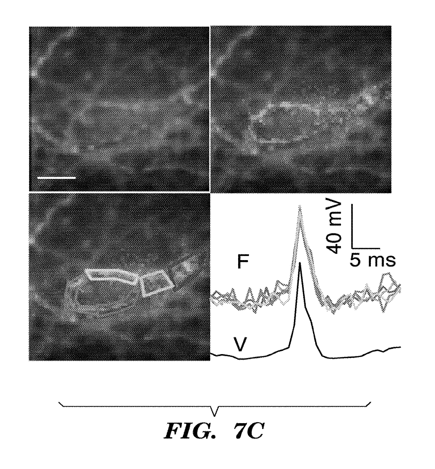
D00015

D00016

D00017

D00018
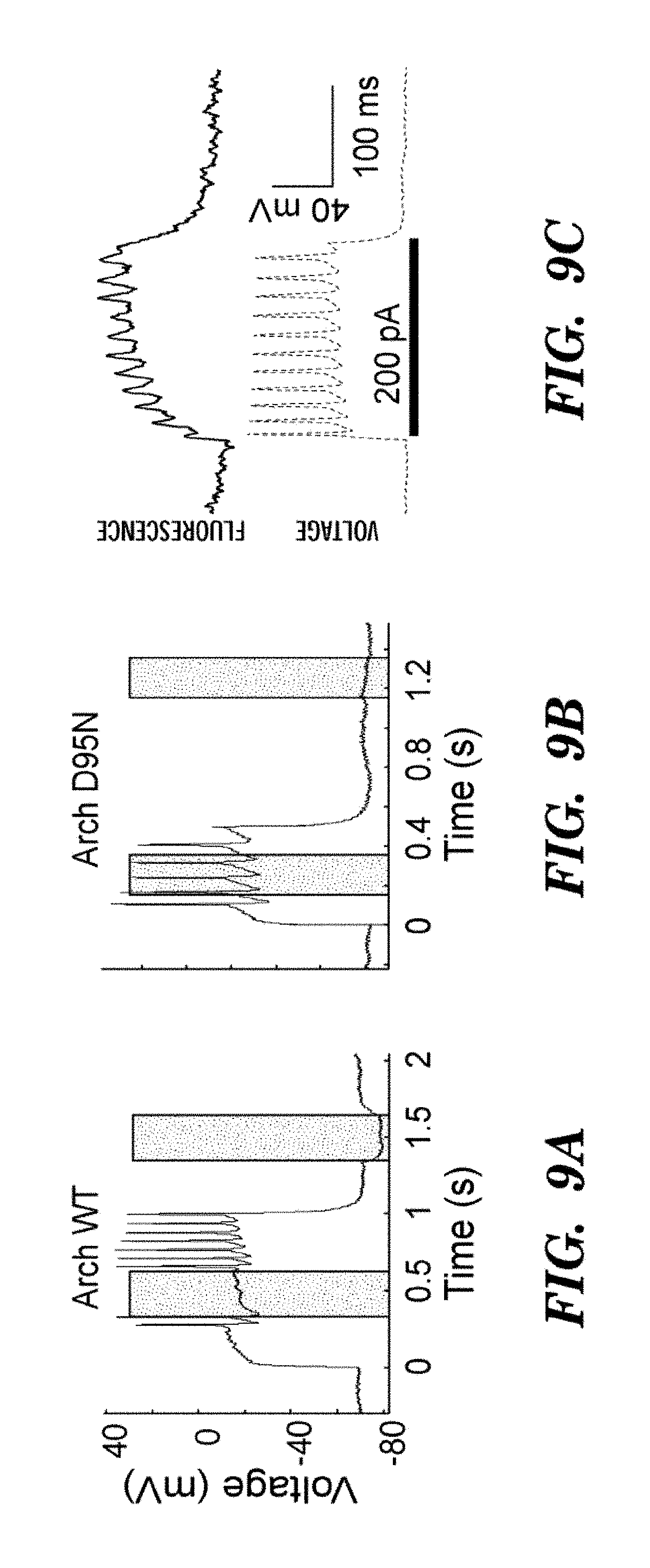
D00019

D00020
D00021

D00022
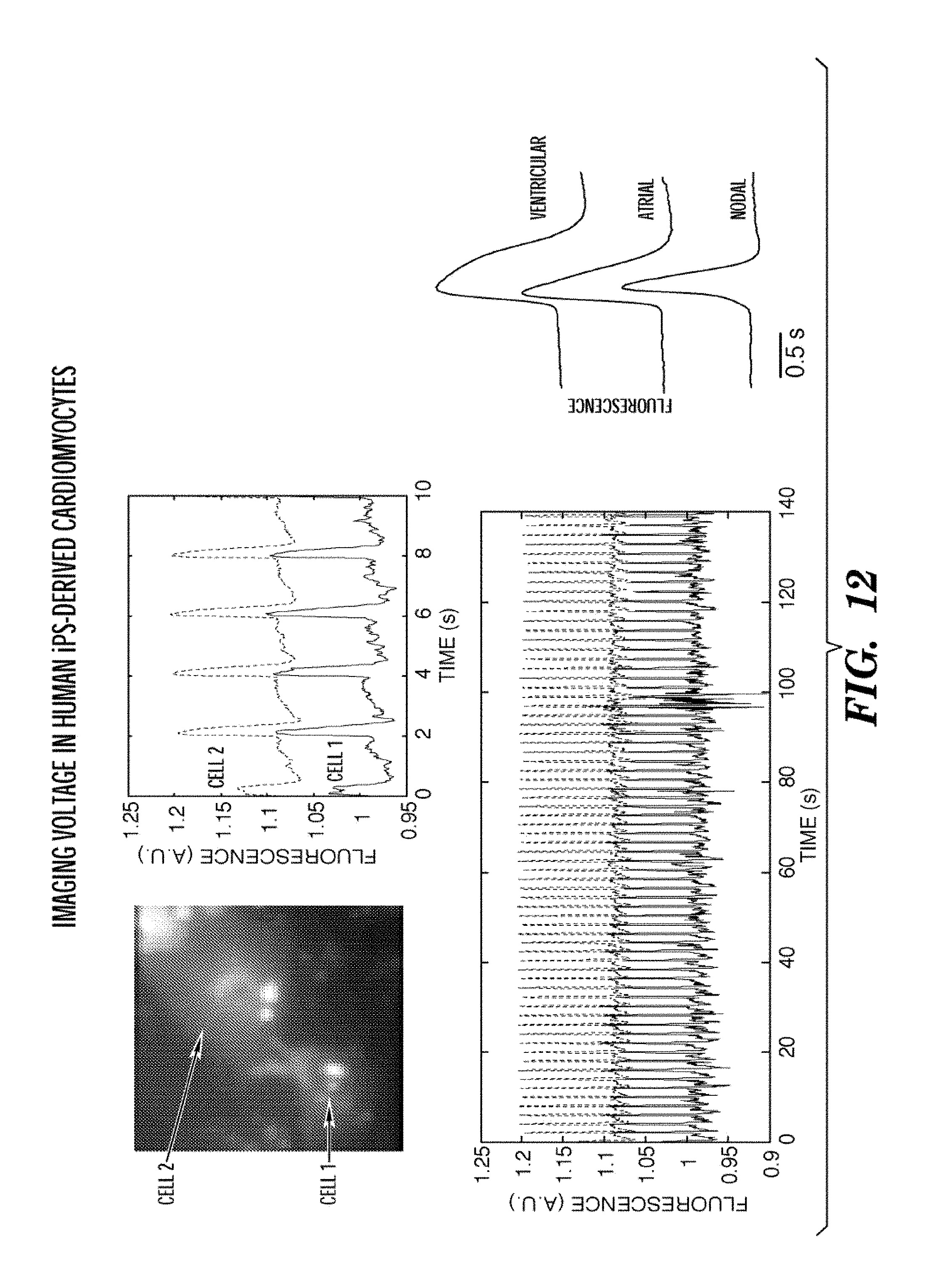
D00023

D00024
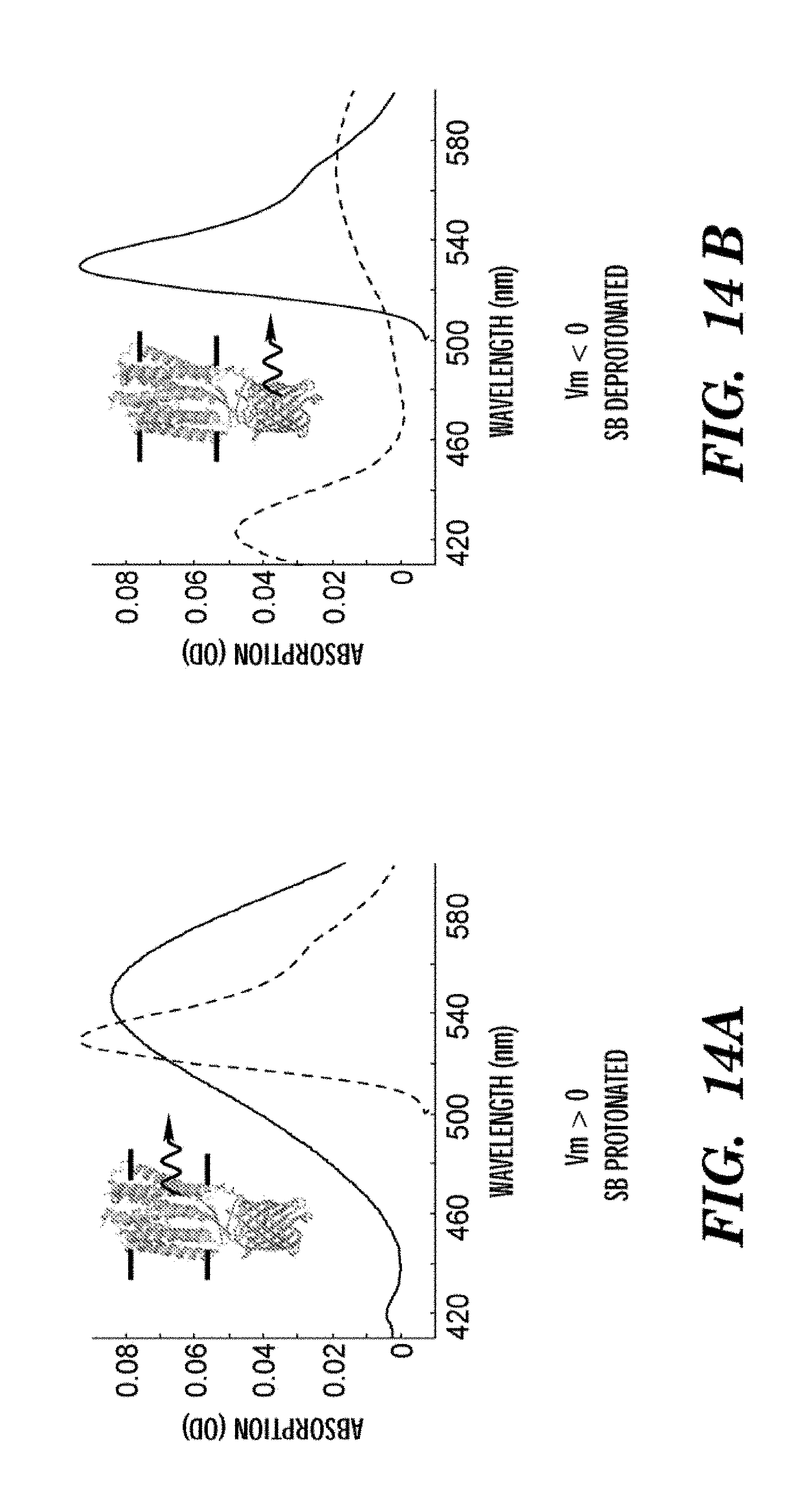
D00025
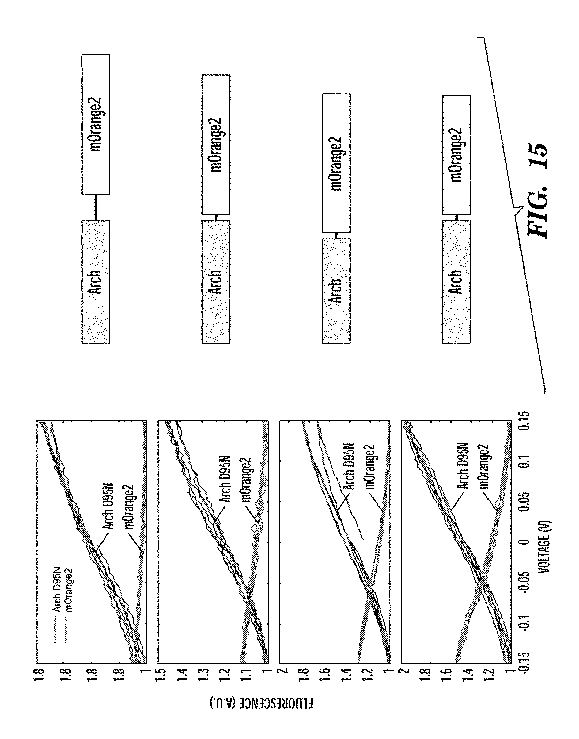
D00026

D00027
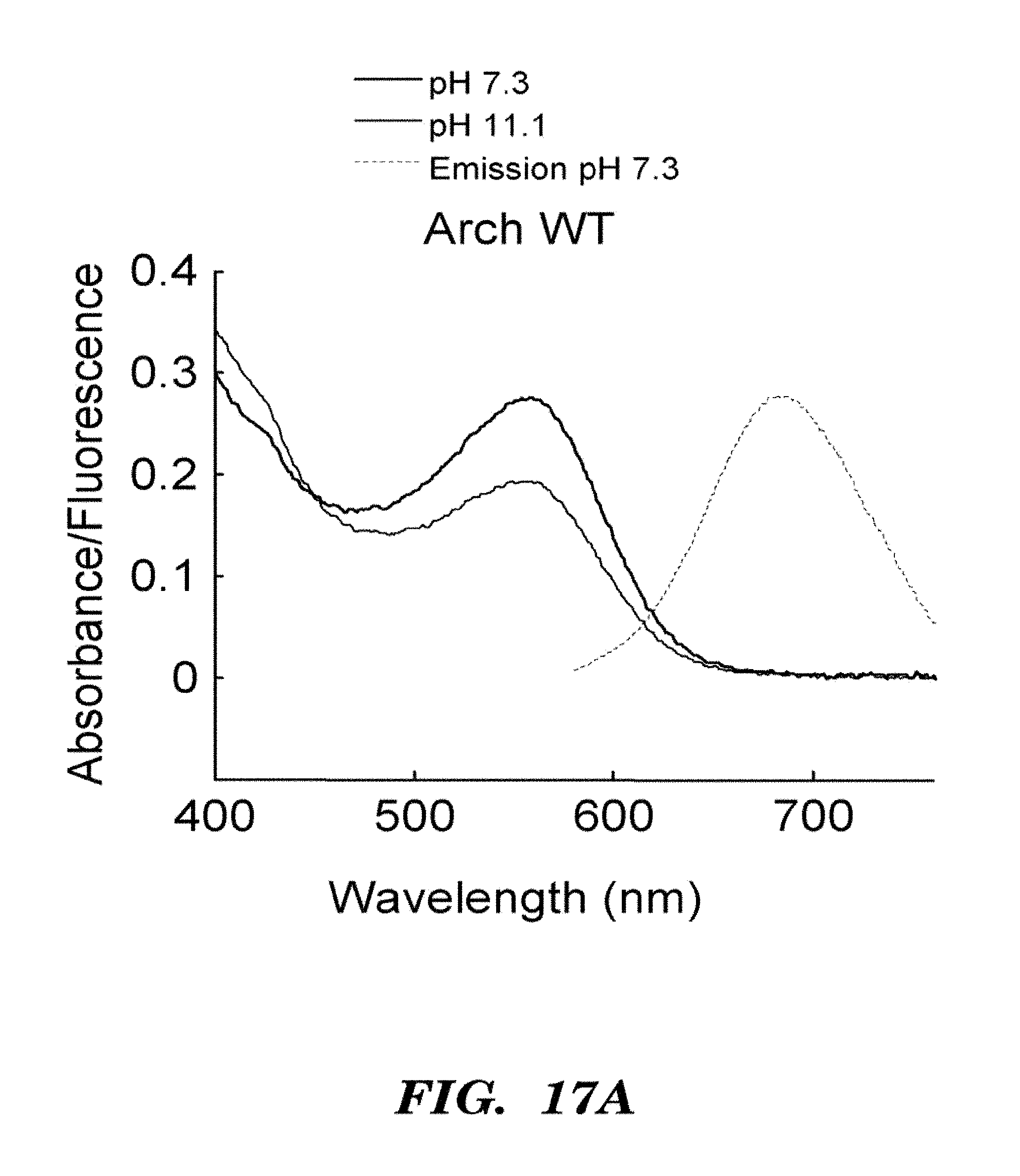
D00028
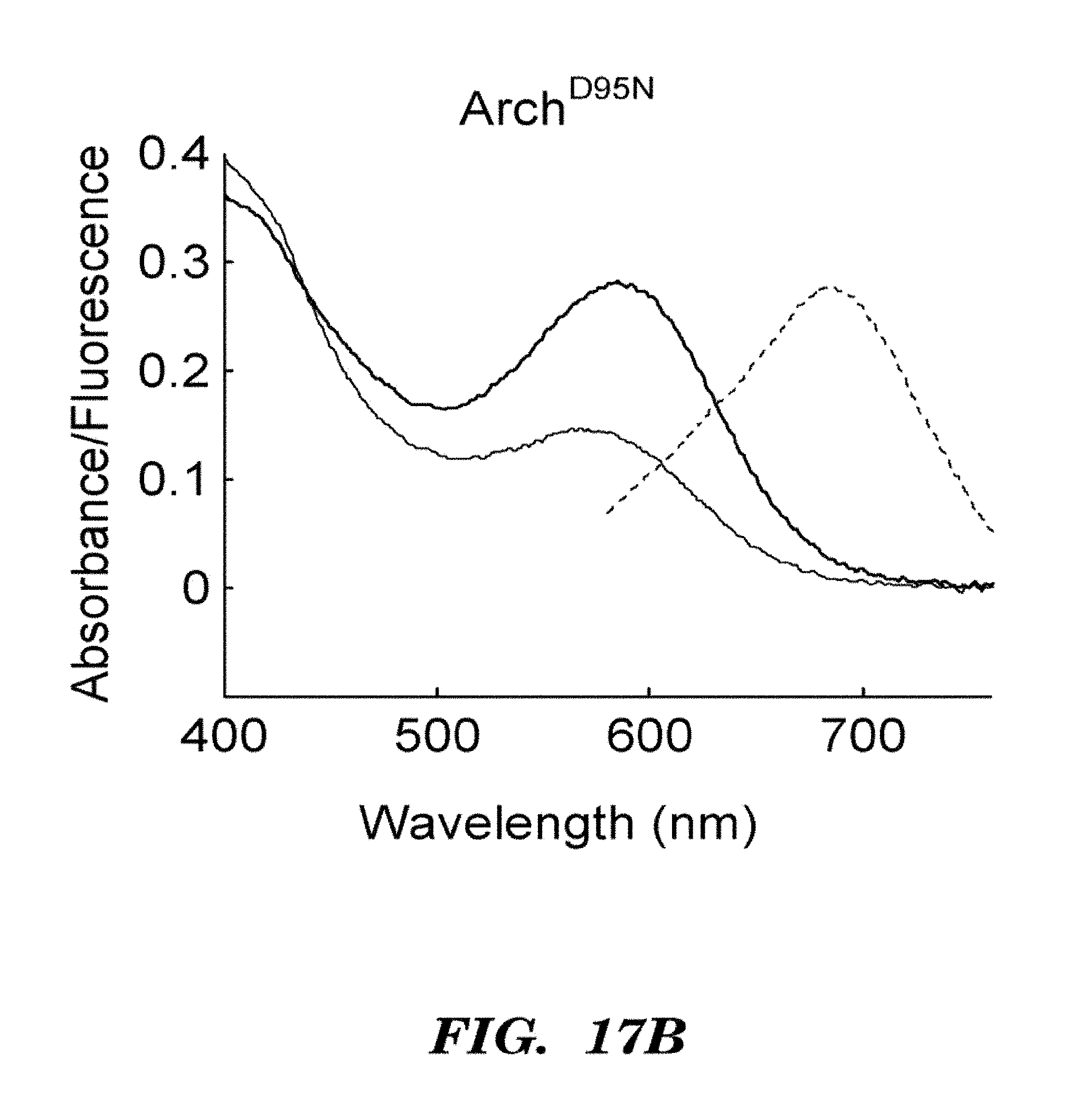
D00029
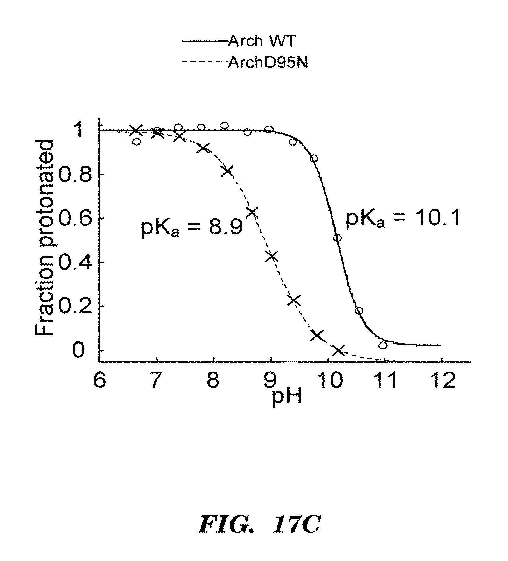
D00030
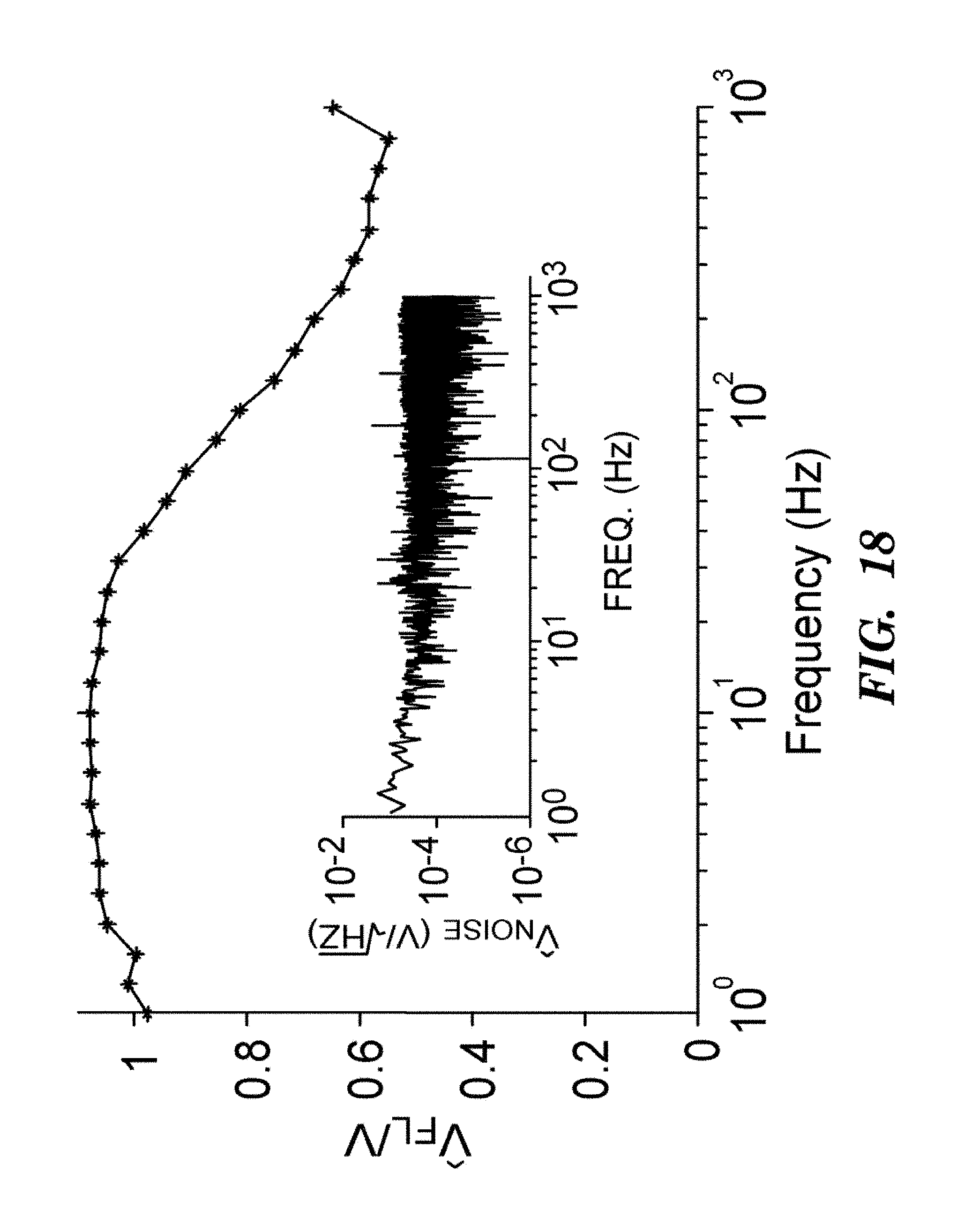
D00031
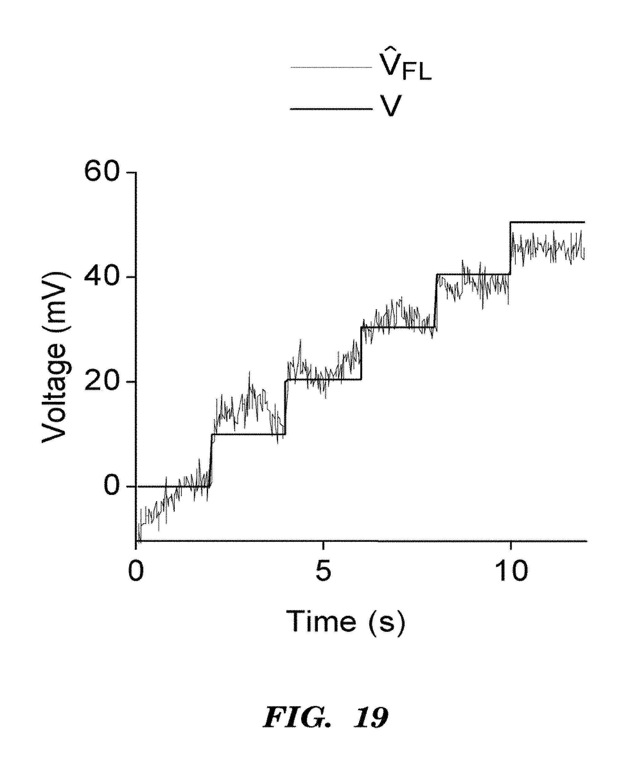
D00032

D00033
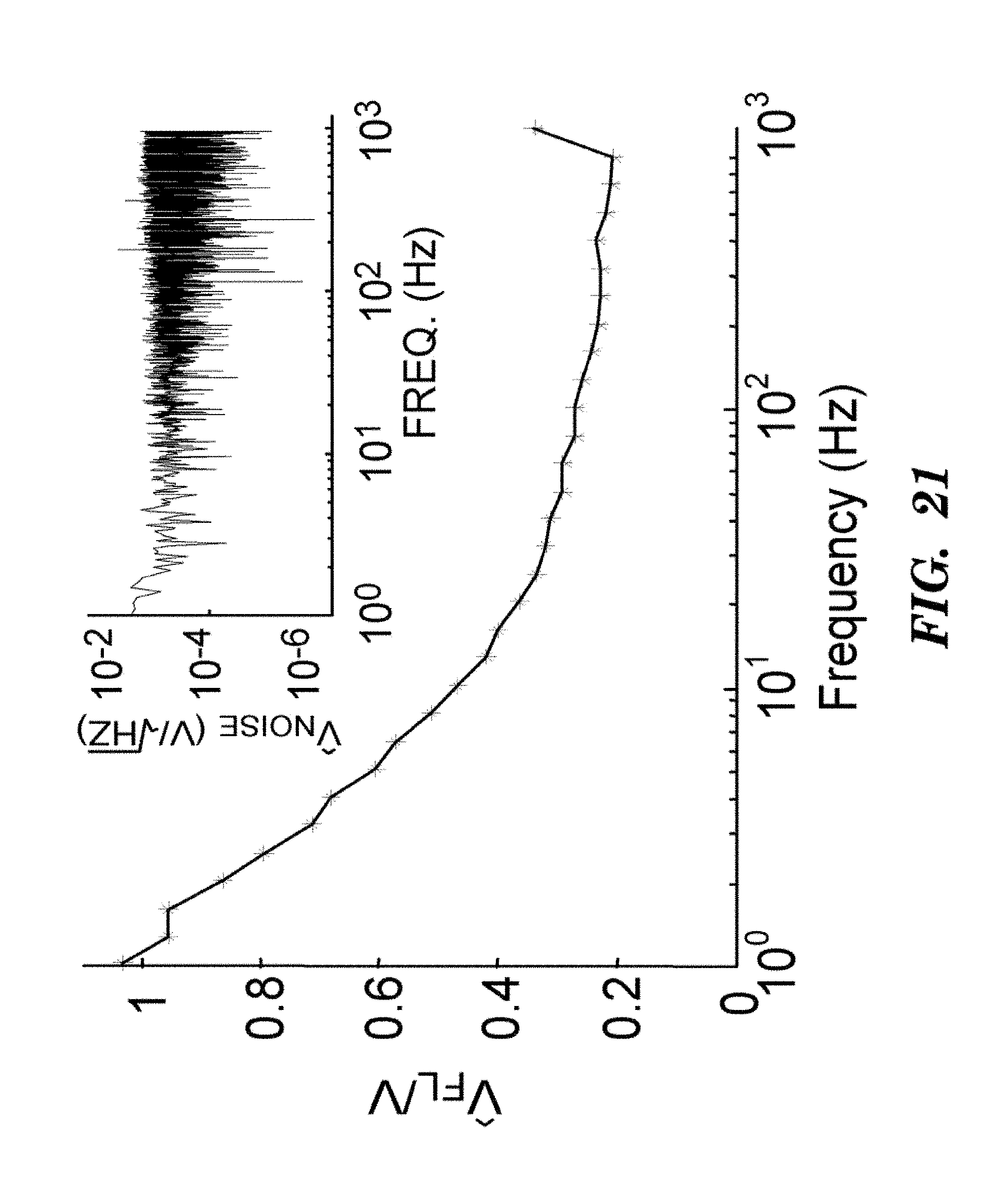
M00001

M00002

M00003

M00004

M00005

P00001

P00002

S00001
XML
uspto.report is an independent third-party trademark research tool that is not affiliated, endorsed, or sponsored by the United States Patent and Trademark Office (USPTO) or any other governmental organization. The information provided by uspto.report is based on publicly available data at the time of writing and is intended for informational purposes only.
While we strive to provide accurate and up-to-date information, we do not guarantee the accuracy, completeness, reliability, or suitability of the information displayed on this site. The use of this site is at your own risk. Any reliance you place on such information is therefore strictly at your own risk.
All official trademark data, including owner information, should be verified by visiting the official USPTO website at www.uspto.gov. This site is not intended to replace professional legal advice and should not be used as a substitute for consulting with a legal professional who is knowledgeable about trademark law.